Products and Methods for Treatment of Familial Amyotrophic Lateral Sclerosis
Kaspar; Brian K. ; et al.
U.S. patent application number 16/041381 was filed with the patent office on 2019-05-16 for products and methods for treatment of familial amyotrophic lateral sclerosis. The applicant listed for this patent is LUDWIG INSTITUTE FOR CANCER RESEARCH, RESEARCH INSTITUTE AT NATIONWIDE CHILDREN'S HOSPITAL. Invention is credited to Don W. Cleveland, Kevin Foust, Brian K. Kaspar.
| Application Number | 20190144868 16/041381 |
| Document ID | / |
| Family ID | 51494538 |
| Filed Date | 2019-05-16 |


View All Diagrams
| United States Patent Application | 20190144868 |
| Kind Code | A1 |
| Kaspar; Brian K. ; et al. | May 16, 2019 |
Products and Methods for Treatment of Familial Amyotrophic Lateral Sclerosis
Abstract
The present invention relates to RNA-based methods for inhibiting the expression of the superoxide dismutase 1 (SOD-1) gene. Recombinant adeno-associated viruses of the invention deliver DNAs encoding RNAs that knock down the expression of SOD-1. The methods have application in the treatment of amyotrophic lateral sclerosis.
| Inventors: | Kaspar; Brian K.; (Columbus, OH) ; Foust; Kevin; (Columbus, OH) ; Cleveland; Don W.; (La Jolla, CA) | ||||||||||
| Applicant: |
|
||||||||||
|---|---|---|---|---|---|---|---|---|---|---|---|
| Family ID: | 51494538 | ||||||||||
| Appl. No.: | 16/041381 | ||||||||||
| Filed: | July 20, 2018 |
Related U.S. Patent Documents
| Application Number | Filing Date | Patent Number | ||
|---|---|---|---|---|
| 14914861 | Feb 26, 2016 | |||
| PCT/US2014/052753 | Aug 26, 2014 | |||
| 16041381 | ||||
| 61870585 | Aug 27, 2013 | |||
| Current U.S. Class: | 424/93.2 |
| Current CPC Class: | C12N 15/86 20130101; C12N 2750/14143 20130101; C12N 9/0089 20130101; A61P 25/28 20180101; C12N 2310/531 20130101; C12N 2750/14043 20130101; C12N 7/00 20130101; A61P 25/02 20180101; A61K 9/0019 20130101; C12N 2320/32 20130101; C12N 2310/14 20130101; C12Y 115/01001 20130101; C12N 15/1137 20130101; A61K 31/7105 20130101 |
| International Class: | C12N 15/113 20060101 C12N015/113; C12N 15/86 20060101 C12N015/86; A61K 31/7105 20060101 A61K031/7105; C12N 7/00 20060101 C12N007/00; A61K 9/00 20060101 A61K009/00; C12N 9/02 20060101 C12N009/02 |
Goverment Interests
STATEMENT OF GOVERNMENT INTEREST
[0002] This invention was made with government support under U.S. National Institutes of Health R21-NS067238, NS027036, ROI NS064492 and RC2 NS69476-01. The Government has certain rights in the invention.
Claims
1. A recombinant adeno-associated virus comprising a superoxide dismutase 1 (SOD1) shRNA-encoding DNA comprising a sequence selected from the group consisting of: TABLE-US-00006 (SEQ ID NO: I) GCATCATCAATTTCGAGCAGAAGGAA, (SEQ ID NO: 2) GAAGCATTAAAGGACTGACTGAA, (SEQ ID NO: 3) CTGACTGAAGGCCTGCATGGATT, (SEQ ID NO: 4) CATGGATTCCATGTTCATGA, (SEQ ID NO: 5) GCATGGATTCCATGTTCATGA, (SEQ ID NO: 6) GGTCTGGCCTATAAAGTAGTC, (SEQ ID NO: 7) GGGCATCATCAATTTCGAGCA, (SEQ ID NO: 8) GCATCATCAATTTCGAGCAGA, (SEQ ID NO: 9) GCCTGCATGGATTCCATGTTC, (SEQ ID NO: 10) GGAGGTCTGGCCTATAAAGTA, (SEQ ID NO: 11) GATTCCATGTTCATGAGTTTG, (SEQ ID NO: 12) GGAGATAATACAGCAGGCTGT, (SEQ ID NO: 13) GCTTTAAAGTACCTGTAGTGA, (SEQ ID NO: 14) GCATTAAAGGACTGACTGAAG, (SEQ ID NO: 15) TCATCAATTTCGAGCAGAA, (SEQ ID NO: 16) TCGAGCAGAAGGAAAGTAA, (SEQ ID NO: 17) GCCTGCATGGATTCCATGT, (SEQ ID NO: 18) TCACTCTCAGGAGACCATT, and (SEQ ID NO: 19) GCTTTAAAGTACCTGTAGT,
wherein the recombinant adeno-associated virus genome lacks rep and cap genes.
2. A composition comprising the recombinant adeno-associated virus of claim 1 and a pharmaceutically acceptable carrier.
3. A method of inhibiting expression of mutant SOD1 in a cell comprising contacting the cell with the recombinant adeno-associated virus of claim 1.
4. A method of delivering a SOD1 shRNA-encoding DNA to a subject in need thereof, comprising administering to the subject a recombinant adeno-associated virus comprising a SODI shRNA-encoding DNA comprising a sequence selected from the group consisting of: TABLE-US-00007 (SEQ ID NO: I) GCATCATCAATTTCGAGCAGAAGGAA, (SEQ ID NO: 2) GAAGCATTAAAGGACTGACTGAA, (SEQ ID NO: 3) CTGACTGAAGGCCTGCATGGATT, (SEQ ID NO: 4) CATGGATTCCATGTTCATGA, (SEQ ID NO: 5) GCATGGATTCCATGTTCATGA, (SEQ ID NO: 6) GGTCTGGCCTATAAAGTAGTC, (SEQ ID NO: 7) GGGCATCATCAATTTCGAGCA, (SEQ ID NO: 8) GCATCATCAATTTCGAGCAGA, (SEQ ID NO: 9) GCCTGCATGGATTCCATGTTC, (SEQ ID NO: 10) GGAGGTCTGGCCTATAAAGTA, (SEQ ID NO: 11) GATTCCATGTTCATGAGTTTG, (SEQ ID NO: 12) GGAGATAATACAGCAGGCTGT, (SEQ ID NO: 13) GCTTTAAAGTACCTGTAGTGA, (SEQ ID NO: 14) GCATTAAAGGACTGACTGAAG, (SEQ ID NO: 15) TCATCAATTTCGAGCAGAA, (SEQ ID NO: 16) TCGAGCAGAAGGAAAGTAA, (SEQ ID NO: 17) GCCTGCATGGATTCCATGT, (SEQ ID NO: 18) TCACTCTCAGGAGACCATT, and (SEQ ID NO: 19) GCTTTAAAGTACCTGTAGT;
wherein the recombinant adeno-associated virus genome lacks rep and cap genes.
5-7. (canceled)
8. A method of treating amyotrophic lateral sclerosis (ALS) comprising administering to a subject in need thereof an effective dose of a composition comprising a recombinant adeno-associated virus comprising a SOD1 shRNA-encoding DNA comprising a sequence selected from the group consisting of: TABLE-US-00008 (SEQ ID NO: I) GCATCATCAATTTCGAGCAGAAGGAA, (SEQ ID NO: 2) GAAGCATTAAAGGACTGACTGAA, (SEQ ID NO: 3) CTGACTGAAGGCCTGCATGGATT, (SEQ ID NO: 4) CATGGATTCCATGTTCATGA, (SEQ ID NO: 5) GCATGGATTCCATGTTCATGA, (SEQ ID NO: 6) GGTCTGGCCTATAAAGTAGTC, (SEQ ID NO: 7) GGGCATCATCAATTTCGAGCA, (SEQ ID NO: 8) GCATCATCAATTTCGAGCAGA, (SEQ ID NO: 9) GCCTGCATGGATTCCATGTTC, (SEQ ID NO: 10) GGAGGTCTGGCCTATAAAGTA, (SEQ ID NO: 11) GATTCCATGTTCATGAGTTTG, (SEQ ID NO: 12) GGAGATAATACAGCAGGCTGT, (SEQ ID NO: 13) GCTTTAAAGTACCTGTAGTGA, (SEQ ID NO: 14) GCATTAAAGGACTGACTGAAG, (SEQ ID NO: 15) TCATCAATTTCGAGCAGAA, (SEQ ID NO: 16) TCGAGCAGAAGGAAAGTAA, (SEQ ID NO: 17) GCCTGCATGGATTCCATGT, (SEQ ID NO: 18) TCACTCTCAGGAGACCATT, and (SEQ ID NO: 19) GCTTTAAAGTACCTGTAGT;
wherein the recombinant adeno-associated virus genome lacks rep and cap genes.
9-11. (canceled)
12. The recombinant adeno-associated virus of claim 1, further comprising an H1 promoter operably linked to the SOD1 shRNA-encoding DNA.
13. The recombinant adeno-associated virus of claim 1, wherein the recombinant adeno-associated virus is an rAAV2, rAAV9 or rAAVrh74 virus.
14. The recombinant adeno-associated virus of claim 1, further comprising a stuffer sequence.
15. The recombinant adeno-associated virus of claim 1, wherein the SOD1 shRNA-encoding DNA comprises SEQ ID NO: 4.
16. The recombinant adeno-associated virus of claim 1, wherein the SOD1 shRNA-encoding DNA comprises, from 5' to 3', a) nucleotides 104-123 of SEQ ID NO: 21; b) a stem loop; and c) nucleotides 133-152 of SEQ ID NO: 21.
17. The method of claim 4, wherein the recombinant adeno-associated virus is administered by parenteral, intravenous or intrathecal administration.
18. The method of claim 17, wherein the intrathecal administration is introcerebroventricular, by the cisterna magna, or by lumbar puncture.
19. The method of claim 4, wherein the SOD1 shRNA-encoding DNA comprises SEQ ID NO: 4.
20. The method of claim 4, wherein the SOD1 shRNA-encoding DNA comprises, from 5' to 3', a) nucleotides 104-123 of SEQ ID NO: 21; b) a stem loop; and c) nucleotides 133-152 of SEQ ID NO: 21.
21. The method of claim 4, further comprising delivering a contrast agent to the subject.
22. the method of claim 21, wherein the contrast agent is iobitridol, iohexol, iomeprol, iopamidol, iopentol, iopromide, ioversol or ioxilan.
23. The method of claim 8, wherein the recombinant adeno-associated virus is administered by parenteral, intravenous or intrathecal administration.
24. The method of claim 23, wherein the intrathecal administration is introcerebroventricular, by the cisterna magna, or by lumbar puncture.
25. The method of claim 8, further comprising delivering a contrast agent to the subject.
26. the method of claim 25, wherein the contrast agent is iobitridol, iohexol, iomeprol, iopamidol, iopentol, iopromide, ioversol or ioxilan.
27. The method of claim 8, wherein the SOD1 shRNA-encoding DNA comprises SEQ ID NO: 4.
28. The method of claim 8, wherein the SOD1 shRNA-encoding DNA comprises, from 5' to 3', a) nucleotides 104-123 of SEQ ID NO: 21; b) a stem loop; and c) nucleotides 133-152 of SEQ ID NO: 21.
Description
INCORPORATION BY REFERENCE OF MATERIAL SUBMITTED ELECTRONICALLY
[0001] Incorporated by reference in its entirety is a computer-readable nucleotide/amino acid sequence listing submitted concurrently herewith and identified as follows: 14,350 byte ACII (Text) file named "47886PCT_SeqListing.txt," created on Aug. 26, 2014.
FIELD OF THE INVENTION
[0003] The present invention relates to RNA-based methods for inhibiting the expression of the superoxide dismutase 1 (SOD-1) gene. Recombinant adeno-associated viruses of the invention deliver DNAs encoding RNAs that knock down the expression of SOD-1. The methods have application in the treatment of amyotrophic lateral sclerosis (ALS).
BACKGROUND
[0004] ALS is an adult-onset, rapidly progressive and fatal neurodegenerative disease, characterized by selective degeneration of both upper and lower motor neurons. First characterized by Charcot in 1869, ALS is responsible for one in every 2000 deaths, affecting nearly 5 out of 100,000 individuals. ALS occurs when specific nerve cells in the brain and spinal cord that control voluntary movement degenerate. Within two to five years after clinical onset, the loss of these motor neurons leads to progressive atrophy of skeletal muscles, which results in loss of muscular function resulting in paralysis, speech deficits, and death due to respiratory failure.
[0005] Most ALS cases have no clear genetic linkage and are referred to as sporadic, but in 10% of instances disease is familial with dominant inheritance. Twenty percent of familial cases are caused by mutations in the enzyme superoxide dismutase 1 (SOD1), with over 140 distinct mutations identified to date.sup.1,2. Many efforts to identify how mutations alter the function of SOD1 have produced a consensus view that SOD1 mutants acquire one or more toxicities, whose nature still remains controversial.sup.3, but there is clear evidence that a proportion of mutant SOD1 is misfolded and subsequently aggregates.sup.4,5. SOD1 aggregates are, in fact, one of the histological hallmarks of SOD1-related ALS cases.sup.4.
[0006] In the past 20 years, multiple animal models expressing mutant forms of human SOD1 have been generated. These models recapitulate the hallmarks of ALS, developing age-dependent motor axon degeneration and accompanying muscle denervation, glial inflammation and subsequent motor neuron loss. Selective gene excision experiments have determined that mutant SOD1 expression within motor neurons themselves contributes to disease onset and early disease progression.sup.6, as does mutant synthesis in NG2.sup.+ cells.sup.7 that are precursors to oligodendrocytes. However, mutant SOD1 protein expression in microglia and astrocytes significantly drives rapid disease progression.sup.6,8, findings which have lead to the conclusion that ALS pathophysiology is non-cell autonomous.sup.3.
[0007] Further, astrocytes have been found to be toxic to motor neurons in multiple in vitro models where mutant forms of human SOD1 were overexpressed.sup.9-11. A recent study derived astrocytes from post-mortem spinal cords of ALS patients with or without SOD1 mutations. In all cases, astrocytes from sporadic ALS patients were as toxic to motor neurons as astrocytes carrying genetic mutations in SOD1.sup.12. Even more strikingly, reduction of SOD1 in astrocytes derived from both sporadic and familial ALS patients decreased astrocyte-derived toxicity that is selective for motor, but not GABA, neurons. This remarkable finding, along with reports that misfolded SOD1 inclusions are found in the spinal cords of familial as well as some sporadic ALS patients.sup.13,14,15, has provided strong evidence for a pathogenic role of wild-type SOD1 in sporadic ALS.
[0008] Despite the insights that SOD1 mutant-expressing animal models have provided for understanding mechanisms involved in motor neuron degeneration, their utility for the development of therapeutic approaches has been questioned.sup.16, as no drug with a reported survival benefit in mutant SOD1.sup.G93A mice has been effective in clinical trials with sporadic ALS patients. In all but one case the drugs taken to human trial had been reported only to extend mutant SOD1 mouse survival when applied presymptomatically, and even then to provide a survival benefit solely by delaying disease onset with no benefit in slowing disease progression. The one exception to this was riluzole, which like the human situation, modestly extended survival of mutant SOD1.sup.G93A mice and did so by slowing disease progression.sup.17. Recognizing that success at human trial will require slowing of disease progression, the SOD1 mutant mice have perfectly predicted the success of riluzole and the failure of efficacy of each other drug attempted in human trial. What has been missing are additional therapies that affect disease progression in these mice.
[0009] Thus, riluzole is the only drug currently approved by the FDA as a therapy for ALS, providing a modest survival benefit.sup.21. For the 20% of familial cases caused by mutation in SOD1, attempts at improving therapy by reducing synthesis of SOD1 have been the focus of multiple therapeutic development approaches. Antisense oligonucleotides and viral delivered RNA interference (RNAi) were tested in rat.sup.22 and mouse models.sup.23-25 that develop fatal paralysis from overexpressing human SOD1.sup.G93A. Antisense oligonucleotides infused at disease onset produced SOD1 reduction and a modest slowing of disease progression.sup.22. Direct CSF infusion of antisense oligonucleotides has been tested clinically.sup.26, leading to encouraging results in terms of tolerability and safety, but without significant reduction in SOD1 levels at the low dosages utilized. In each of the prior viral studies.sup.23-25, SOD1 knockdown was achieved before disease onset by direct injection into the nervous system or taking advantage of axonal retrograde transport when a virus was injected intramusculary.sup.23,24. These studies led to varying degrees of success in extending survival or improving motor performance, depending on the time of treatment as well as level of SOD1 knockdown achieved in the spinal cord. Although these studies provided important proof of principle, the approaches were far from being readily translated into clinical strategies. Indeed, there have been controversial reports surrounding these initial viral mediated SOD1 suppression studies.sup.23,24,27-29.
[0010] Adeno-associated virus (AAV) vectors have been used in a number of recent clinical trials for treatment of neurological disorders [Kaplitt et al., Lancet 369: 2097-2105 (2007); Marks et al., Lancet Neurol 7: 400-408 (2008); Worgall et al., Hum Gene Ther (2008)].
[0011] AAV is a replication-deficient parvovirus, the single-stranded DNA genome of which is about 4.7 kb in length including 145 nucleotide inverted terminal repeat (ITRs). The nucleotide sequence of the AAV serotype 2 (AAV2) genome is presented in Srivastava et al., J Virol, 45: 555-564 (1983) as corrected by Ruffing et al., J Gen Virol, 75: 3385-3392 (1994). Cis-acting sequences directing viral DNA replication (rep), encapsidation/packaging and host cell chromosome integration are contained within the ITRs. Three AAV promoters (named p5, p19, and p40 for their relative map locations) drive the expression of the two AAV internal open reading frames encoding rep and cap genes. The two rep promoters (p5 and p19), coupled with the differential splicing of the single AAV intron (at nucleotides 2107 and 2227), result in the production of four rep proteins (rep 78, rep 68, rep 52, and rep 40) from the rep gene. Rep proteins possess multiple enzymatic properties that are ultimately responsible for replicating the viral genome. The cap gene is expressed from the p40 promoter and it encodes the three capsid proteins VP1, VP2, and VP3. Alternative splicing and non-consensus translational start sites are responsible for the production of the three related capsid proteins. A single consensus polyadenylation site is located at map position 95 of the AAV genome. The life cycle and genetics of AAV are reviewed in Muzyczka, Current Topics in Microbiology and Immunology, 158: 97-129 (1992).
[0012] AAV possesses unique features that make it attractive as a vector for delivering foreign DNA to cells, for example, in gene therapy. AAV infection of cells in culture is noncytopathic, and natural infection of humans and other animals is silent and asymptomatic. Moreover, AAV infects many mammalian cells allowing the possibility of targeting many different tissues in vivo. Moreover, AAV transduces slowly dividing and non-dividing cells, and can persist essentially for the lifetime of those cells as a transcriptionally active nuclear episome (extrachromosomal element). The AAV proviral genome is infectious as cloned DNA in plasmids which makes construction of recombinant genomes feasible. Furthermore, because the signals directing AAV replication, genome encapsidation and integration are contained within the ITRs of the AAV genome, some or all of the internal approximately 4.3 kb of the genome (encoding replication and structural capsid proteins, rep-cap) may be replaced with foreign DNA such as a gene cassette containing a promoter, a DNA of interest and a polyadenylation signal. The rep and cap proteins may be provided in trans. Another significant feature of AAV is that it is an extremely stable and hearty virus. It easily withstands the conditions used to inactivate adenovirus (56.degree. to 65.degree. C. for several hours), making cold preservation of AAV less critical. AAV may even be lyophilized. Finally, AAV-infected cells are not resistant to superinfection.
[0013] Multiple serotypes of AAV exist and offer varied tissue tropism. Known serotypes include, for example, AAV1, AAV2, AAV3, AAV4, AAV5, AAV6, AAV7, AAV8, AAV9, AAV10, AAV11 and AAVrh74. Advances in the delivery of AAV6 and AAV8 have made possible the transduction by these serotypes of skeletal and cardiac muscle following simple systemic intravenous or intraperitoneal injections. See Pacak et al., Circ. Res., 99(4): 3-9 (1006) and Wang et al., Nature Biotech., 23(3): 321-8 (2005). The use of AAV to target cell types within the central nervous system has involved surgical intraparenchymal injection. See, Kaplitt et al., supra; Marks et al., supra and Worgall et al., supra. Regarding the use of AAV to target cell types within the nervous system, see International Publication No. WO 2010/071832. International Publication Nos. WO 2009/043936 and WO 2009/013290 state they relate to delivering genes to the central nervous system. International Publication No. WO 2011/133890 states it relates to recombinant adeno-associated viruses useful for targeting transgenes to central nervous system tissue.
[0014] There thus remains a need in the art for methods and materials for treatment of ALS.
SUMMARY
[0015] The present invention provides products and methods useful for reducing mutant SOD1 protein levels in subjects in need thereof. The invention provides AAV-mediated delivery of RNAs including, but not limited to short hairpin RNAs, to reduce synthesis of ALS-causing human SOD1 mutants in subjects in need thereof. Recombinant AAV (rAAV) contemplated by the invention include, but are not limited to, rAAV9, rAAV2 and rAAVrh74. Delivery routes contemplated by the invention include, but are not limited to, systemic delivery and intrathecal delivery. Use of the methods and products of the invention is indicated, for example, in treating ALS.
DETAILED DESCRIPTION
[0016] In one aspect, the invention provides rAAV genomes comprising one or more AAV ITRs flanking a polynucleotide encoding one or more RNAs (including, but not limited to, small hairpin RNAs, antisense RNAs and/or microRNAs) that target mutant SOD1 polynucleotides. The examples describe the use of exemplary rAAV encoding small hairpin RNAs (shRNAs). In the rAAV genomes, the shRNA-encoding polynucleotide is operatively linked to transcriptional control DNA, specifically promoter DNA that is functional in target cells. Commercial providers such as Ambion Inc. (Austin, Tex.), Darmacon Inc. (Lafayette, Colo.), InvivoGen (San Diego, Calif.), and Molecular Research Laboratories, LLC (Herndon, Va.) generate custom inhibitory RNA molecules. In addition, commercially kits are available to produce custom siRNA molecules, such as SILENCER.TM. siRNA Construction Kit (Ambion Inc., Austin, Tex.) or psiRNA System (InvivoGen, San Diego, Calif.). In some embodiments, the rAAV genome comprises a DNA encoding a SOD1 shRNA such as:
TABLE-US-00001 (SEQ ID NO: 1) GCATCATCAATTTCGAGCAGAAGGAA, (SEQ ID NO: 2) GAAGCATTAAAGGACTGACTGAA, (SEQ ID NO: 3) CTGACTGAAGGCCTGCATGGATT, (SEQ ID NO: 4) CATGGATTCCATGTTCATGA ("shRNA 130" or "SOD1 shRNA" herein), (SEQ ID NO: 5) GCATGGATTCCATGTTCATGA, (SEQ ID NO: 6) GGTCTGGCCTATAAAGTAGTC, (SEQ ID NO: 7) GGGCATCATCAATTTCGAGCA, (SEQ ID NO: 8) GCATCATCAATTTCGAGCAGA, (SEQ ID NO: 9) GCCTGCATGGATTCCATGTTC, (SEQ ID NO: 10) GGAGGTCTGGCCTATAAAGTA, (SEQ ID NO: 11) GATTCCATGTTCATGAGTTTG, (SEQ ID NO: 12) GGAGATAATACAGCAGGCTGT, (SEQ ID NO: 13) GCTTTAAAGTACCTGTAGTGA, (SEQ ID NO: 14) GCATTAAAGGACTGACTGAAG, (SEQ ID NO: 1) GCATCATCAATTTCGAGCAGAAGGAA, (SEQ ID NO: 2) GAAGCATTAAAGGACTGACTGAA, (SEQ ID NO: 3) CTGACTGAAGGCCTGCATGGATT, (SEQ ID NO: 4) CATGGATTCCATGTTCATGA, (SEQ ID NO: 5) GCATGGATTCCATGTTCATGA, (SEQ ID NO: 6) GGTCTGGCCTATAAAGTAGTC, (SEQ ID NO: 7) GGGCATCATCAATTTCGAGCA, (SEQ ID NO: 8) GCATCATCAATTTCGAGCAGA, (SEQ ID NO: 9) GCCTGCATGGATTCCATGTTC, (SEQ ID NO: 10) GGAGGTCTGGCCTATAAAGTA, (SEQ ID NO: 11) GATTCCATGTTCATGAGTTTG, (SEQ ID NO: 12) GGAGATAATACAGCAGGCTGT, (SEQ ID NO: 13) GCTTTAAAGTACCTGTAGTGA, (SEQ ID NO: 14) GCATTAAAGGACTGACTGAAG, (SEQ ID NO: 15) TCATCAATTTCGAGCAGAA, (SEQ ID NO: 16) TCGAGCAGAAGGAAAGTAA, (SEQ ID NO: 17) GCCTGCATGGATTCCATGT, (SEQ ID NO: 18) TCACTCTCAGGAGACCATT, or (SEQ ID NO: 19) GCTTTAAAGTACCTGTAGT.
[0017] The rAAV genomes of the invention lack AAV rep and cap DNA. AAV DNA in the rAAV genomes (e.g., ITRs) may be from any AAV serotype for which a recombinant virus can be derived including, but not limited to, AAV serotypes AAV-1, AAV-2, AAV-3, AAV-4, AAV-5, AAV-6, AAV-7, AAV-8, AAV-9, AAV-10 and AAV-11. The nucleotide sequences of the genomes of the AAV serotypes are known in the art. For example, the complete genome of AAV-1 is provided in GenBank Accession No. NC_002077; the complete genome of AAV-2 is provided in GenBank Accession No. NC 001401 and Srivastava et al., J. Virol., 45: 555-564 {1983); the complete genome of AAV-3 is provided in GenBank Accession No. NC_1829; the complete genome of AAV-4 is provided in GenBank Accession No. NC_001829; the AAV-5 genome is provided in GenBank Accession No. AF085716; the complete genome of AAV-6 is provided in GenBank Accession No. NC_00 1862; at least portions of AAV-7 and AAV-8 genomes are provided in GenBank Accession Nos. AX753246 and AX753249, respectively; the AAV-9 genome is provided in Gao et al., J. Virol., 78: 6381-6388 (2004); the AAV-10 genome is provided in Mol. Ther., 13(1): 67-76 (2006); and the AAV-11 genome is provided in Virology, 330(2): 375-383 (2004). The AAVrh74 genome is provided in International Publication No. WO 2013/078316.
[0018] In another aspect, the invention provides DNA plasmids comprising rAAV genomes of the invention. The DNA plasmids are transferred to cells permissible for infection with a helper virus of AAV (e.g., adenovirus, E1-deleted adenovirus or herpesvirus) for assembly of the rAAV genome into infectious viral particles. Techniques to produce rAAV particles, in which an AAV genome to be packaged, rep and cap genes, and helper virus functions are provided to a cell are standard in the art. Production of rAAV requires that the following components are present within a single cell (denoted herein as a packaging cell): a rAAV genome, AAV rep and cap genes separate from (i.e., not in) the rAAV genome, and helper virus functions. The AAV rep and cap genes may be from any AAV serotype for which recombinant virus can be derived and may be from a different AAV serotype than the rAAV genome ITRs, including, but not limited to, AAV serotypes AAV-1, AAV-2, AAV-3, AAV-4, AAV-5, AAV-6, AAV-7, AAV-8, AAV-9, AAV-10 and AAV-11. Production of pseudotyped rAAV is disclosed in, for example, WO 01/83692 which is incorporated by reference herein in its entirety. In various embodiments, AAV capsid proteins may be modified to enhance delivery of the recombinant vector. Modifications to capsid proteins are generally known in the art. See, for example, US 20050053922 and US 20090202490, the disclosures of which are incorporated by reference herein in their entirety.
[0019] A method of generating a packaging cell is to create a cell line that stably expresses all the necessary components for AAV particle production. For example, a plasmid (or multiple plasmids) comprising a rAAV genome lacking AAV rep and cap genes, AAV rep and cap genes separate from the rAAV genome, and a selectable marker, such as a neomycin resistance gene, are integrated into the genome of a cell. AAV genomes have been introduced into bacterial plasmids by procedures such as GC tailing (Samulski et al., 1982, Proc. Natl. Acad. S6. USA, 79:2077-2081), addition of synthetic linkers containing restriction endonuclease cleavage sites (Laughlin et al., 1983, Gene, 23:65-73) or by direct, blunt-end ligation (Senapathy & Carter, 1984, J. Biol. Chem., 259:4661-4666). The packaging cell line is then infected with a helper virus such as adenovirus. The advantages of this method are that the cells are selectable and are suitable for large-scale production of rAAV. Other examples of suitable methods employ adenovirus or baculovirus rather than plasmids to introduce rAAV genomes and/or rep and cap genes into packaging cells.
[0020] General principles of rAAV production are reviewed in, for example, Carter, 1992, Current Opinions in Biotechnology, 1533-539; and Muzyczka, 1992, Curr. Topics in Microbial. and Immunol., 158:97-129). Various approaches are described in Ratschin et al., Mol. Cell. Biol. 4:2072 (1984); Hermonat et al., Proc. Natl. Acad. Sci. USA, 81:6466 (1984); Tratschin et al., Mol. Cell. Biol. 5:3251 (1985); McLaughlin et al., J. Virol., 62:1963 (1988); and Lebkowski et al., 1988 Mol. Cell. Biol., 7:349 (1988). Samulski et al. (1989, J. Virol., 63:3822-3828); U.S. Pat. No. 5,173,414; WO 95/13365 and corresponding U.S. Pat. No. 5,658,776; WO 95/13392; WO 96/17947; PCT/US98/18600; WO 97/09441 (PCT/US96/14423); WO 97/08298 (PCT/US96/13872); WO 97/21825 (PCT/US96/20777); WO 97/06243 (PCT/FR96/01064); WO 99/11764; Perrin et al. (1995) Vaccine 13:1244-1250; Paul et al. (1993) Human Gene Therapy 4:609-615; Clark et al. (1996) Gene Therapy 3:1124-1132; U.S. Pat. Nos. 5,786,211; 5,871,982; and 6,258,595. Single-stranded rAAV are specifically contemplated. The foregoing documents are hereby incorporated by reference in their entirety herein, with particular emphasis on those sections of the documents relating to rAAV production.
[0021] The invention thus provides packaging cells that produce infectious rAAV. In one embodiment packaging cells may be stably transformed cancer cells such as HeLa cells, 293 cells and PerC.6 cells (a cognate 293 line). In another embodiment, packaging cells are cells that are not transformed cancer cells such as low passage 293 cells (human fetal kidney cells transformed with E1 of adenovirus), MRC-5 cells (human fetal fibroblasts), WI-38 cells (human fetal fibroblasts), Vero cells (monkey kidney cells) and FRhL-2 cells (rhesus fetal lung cells).
[0022] In still another aspect, the invention provides rAAV (i.e., infectious encapsidated rAAV particles) comprising a rAAV genome of the invention. In some embodiments, the rAAV genome is a self-complementary genome. The genomes of the rAAV lack AAV rep and cap DNA, that is, there is no AAV rep or cap DNA between the ITRs of the genomes. Embodiments include, but are not limited to, the exemplary rAAV including a genome encoding the SOD1 shRNA named "AAV-SOD1-shRNA." A sequence including the AAV-SOD1-shRNA genome is set out below as an inverted sequence from a plasmid used in production.
TABLE-US-00002 FEATURES Location/Qualifiers misc_feature 662..767 /gene="mutated ITR" /SECDrawAs="Region" /SECStyleId=1 CDS complement(901..965) /gene="SOD shRNA" /SECDrawAs="Gene" /SECStyleId=1 misc_feature complement(966..1064) /gene="H1" /SECDrawAs="Region" /SECStyleId=1 misc_feature 1224..1503 /gene="CMV enhancer" /SECDrawAs="Region" /SECStyleId=1 misc_feature 1510..1779 /gene="B-Actin promoter" /product="Chicken" /SECDrawAs="Region" /SECStyleId=1 misc_feature 1845..1875 /gene="SV40_late_19s_int" /SECDrawAs="Region" /SECStyleId=1 misc_feature 1845..1941 /gene="modSV40_late_16s_int" /SECDrawAs="Region" /SECStyleId=1 CDS 2015..2734 /gene="GFP" /SECDrawAs="Gene" /SECStyleId=1 misc_feature 2783..2929 /gene="BGHpA" /SECDrawAs="Region" /SECStyleId=1 misc_feature 3009..3149 /gene="ITR" /SECDrawAs="Region" /SECStyleId=1 misc_feature 3983..4843 /gene="amp r" /SECDrawAs="Region" /SECStyleId=1 misc_feature 4997..5618 /gene="pBR322 ori" /SECDrawAs="Region" /SECStyleId=1
TABLE-US-00003 (SEQ ID NO: 20) 1 gcccaatacg caaaccgcct ctccccgcgc gttggccgat tcattaatgc agctgattct 61 aacgaggaaa gcacgttata cgtgctcgtc aaagcaacca tagtacgcgc cctgtagcgg 121 cgcattaagc gcggcgggtg tggtggttac gcgcagcgtg accgctacac ttgccagcgc 181 cctagcgccc gctcctttcg ctttcttccc ttcctttctc gccacgttcg ccggctttcc 241 ccgtcaagct ctaaatcggg ggctcccttt agggttccga tttagtgctt tacggcacct 301 cgaccccaaa aaacttgatt agggtgatgg ttcacgtagt gggccatcgc cctgatagac 361 ggtttttcgc cctttgacgt tggagtccac gttctttaat agtggactct tgttccaaac 421 tggaacaaca ctcaacccta tctcggtcta ttcttttgat ttataaggga ttttgccgat 481 ttcggcctat tggttaaaaa atgagctgat ttaacaaaaa tttaacgcga attttaacaa 541 aatattaacg cttacaattt aaatatttgc ttatacaatc ttcctgtttt tggggctttt 601 ctgattatca accggggtac atatgattga catgctagtt ttacgattac cgttcatcgc 661 cctgcgcgct cgctcgctca ctgaggccgc ccgggcaaag cccgggcgtc gggcgacctt 721 tggtcgcccg gcctcagtga gcgagcgagc gcgcagagag ggagtggaat tcacgcgtgg 781 atctgaattc aattcacgcg tggtacctac actttatgct tccggctcgt atgttgtgtg 841 gaattgtgag cggataacaa tttcacacag gaaacagcta tgaccatgat tacgccaagc 901 tttccaaaaa agcatggatt ccatgttcat gatctcttga atcatgaaca tggaatccat 961 ggatccgagt ggtctcatac agaacttata agattcccaa atccaaagac atttcacgtt 1021 tatggtgatt tcccagaaca catagcgaca tgcaaatatg aattcactgg ccgtcgtttt 1081 acaacgtcgt gactgggaaa accctggcgt tacccaactt aatcgccttg cagcacatcc 1141 ccctttcgcc agctggcgta atagcgaaga ggcccgcacc gatcgccctt cccaacagtt 1201 gcgcagcctg tggtacctct ggtcgttaca taacttacgg taaatggccc gcctggctga 1261 ccgcccaacg acccccgccc attgacgtca ataatgacgt atgttcccat agtaacgcca 1321 atagggactt tccattgacg tcaatgggtg gagtatttac ggtaaactgc ccacttggca 1381 gtacatcaag tgtatcatat gccaagtacg ccccctattg acgtcaatga cggtaaatgg 1441 cccgcctggc attatgccca gtacatgacc ttatgggact ttcctacttg gcagtacatc 1501 tactcgaggc cacgttctgc ttcactctcc ccatctcccc cccctcccca cccccaattt 1561 tgtatttatt tattttttaa ttattttgtg cagcgatggg ggcggggggg gggggggggc 1621 gcgcgccagg cggggcgggg cggggcgagg ggcggggcgg ggcgaggcgg agaggtgcgg 1681 cggcagccaa tcagagcggc gcgctccgaa agtttccttt tatggcgagg cggcggcggc 1741 ggcggcccta taaaaagcga agcgcgcggc gggcgggagc gggatcagcc accgcggtgg 1801 cggcctagag tcgacgagga actgaaaaac cagaaagtta actggtaagt ttagtctttt 1861 tgtcttttat ttcaggtccc ggatccggtg gtggtgcaaa tcaaagaact gctcctcagt 1921 ggatgttgcc tttacttcta ggcctgtacg gaagtgttac ttctgctcta aaagctgcgg 1981 aattgtaccc gcggccgatc caccggtcgc caccatggtg agcaagggcg aggagctgtt 2041 caccggggtg gtgcccatcc tggtcgagct ggacggcgac gtaaacggcc acaagttcag 2101 cgtgtccggc gagggcgagg gcgatgccac ctacggcaag ctgaccctga agttcatctg 2161 caccaccggc aagctgcccg tgccctggcc caccctcgtg accaccctga cctacggcgt 2221 gcagtgcttc agccgctacc ccgaccacat gaagcagcac gacttcttca agtccgccat 2281 gcccgaaggc tacgtccagg agcgcaccat cttcttcaag gacgacggca actacaagac 2341 ccgcgccgag gtgaagttcg agggcgacac cctggtgaac cgcatcgagc tgaagggcat 2401 cgacttcaag gaggacggca acatcctggg gcacaagctg gagtacaact acaacagcca 2461 caacgtctat atcatggccg acaagcagaa gaacggcatc aaggtgaact tcaagatccg 2521 ccacaacatc gaggacggca gcgtgcagct cgccgaccac taccagcaga acacccccat 2581 cggcgacggc cccgtgctgc tgcccgacaa ccactacctg agcacccagt ccgccctgag 2641 caaagacccc aacgagaagc gcgatcacat ggtcctgctg gagttcgtga ccgccgccgg 2701 gatcactctc ggcatggacg agctgtacaa gtaaagcggc catcaagctt atcgataccg 2761 tcgactagag ctcgctgatc agcctcgact gtgccttcta gttgccagcc atctgttgtt 2821 tgcccctccc ccgtgccttc cttgaccctg gaaggtgcca ctcccactgt cctttcctaa 2881 taaaatgagg aaattgcatc gcattgtctg agtaggtgtc attctattct ggggggtggg 2941 gtggggcagg acagcaaggg ggaggattgg gaagacaata gcaggcatgc tggggagaga 3001 tcgatctgag gaacccctag tgatggagtt ggccactccc tctctgcgcg ctcgctcgct 3061 cactgaggcc gggcgaccaa aggtcgcccg acgcccgggc tttgcccggg cggcctcagt 3121 gagcgagcga gcgcgcagag agggagtggc cccccccccc ccccccccgg cgattctctt 3181 gtttgctcca gactctcagg caatgacctg atagcctttg tagagacctc tcaaaaatag 3241 ctaccctctc cggcatgaat ttatcagcta gaacggttga atatcatatt gatggtgatt 3301 tgactgtctc cggcctttct cacccgtttg aatctttacc tacacattac tcaggcattg 3361 catttaaaat atatgagggt tctaaaaatt tttatccttg cgttgaaata aaggcttctc 3421 ccgcaaaagt attacagggt cataatgttt ttggtacaac cgatttagct ttatgctctg 3481 aggctttatt gcttaatttt gctaattctt tgccttgcct gtatgattta ttggatgttg 3541 gaatcgcctg atgcggtatt ttctccttac gcatctgtgc ggtatttcac accgcatatg 3601 gtgcactctc agtacaatct gctctgatgc cgcatagtta agccagcccc gacacccgcc 3661 aacacccgct gacgcgccct gacgggcttg tctgctcccg gcatccgctt acagacaagc 3721 tgtgaccgtc tccgggagct gcatgtgtca gaggttttca ccgtcatcac cgaaacgcgc 3781 gagacgaaag ggcctcgtga tacgcctatt tttataggtt aatgtcatga taataatggt 3841 ttcttagacg tcaggtggca cttttcgggg aaatgtgcgc ggaaccccta tttgtttatt 3901 tttctaaata cattcaaata tgtatccgct catgagacaa taaccctgat aaatgcttca 3961 ataatattga aaaaggaaga gtatgagtat tcaacatttc cgtgtcgccc ttattccctt 4021 ttttgcggca ttttgccttc ctgtttttgc tcacccagaa acgctggtga aagtaaaaga 4081 tgctgaagat cagttgggtg cacgagtggg ttacatcgaa ctggatctca acagcggtaa 4141 gatccttgag agttttcgcc ccgaagaacg ttttccaatg atgagcactt ttaaagttct 4201 gctatgtggc gcggtattat cccgtattga cgccgggcaa gagcaactcg gtcgccgcat 4261 acactattct cagaatgact tggttgagta ctcaccagtc acagaaaagc atcttacgga 4321 tggcatgaca gtaagagaat tatgcagtgc tgccataacc atgagtgata acactgcggc 4381 caacttactt ctgacaacga tcggaggacc gaaggagcta accgcttttt tgcacaacat 4441 gggggatcat gtaactcgcc ttgatcgttg ggaaccggag ctgaatgaag ccataccaaa 4501 cgacgagcgt gacaccacga tgcctgtagc aatggcaaca acgttgcgca aactattaac 4561 tggcgaacta cttactctag cttcccggca acaattaata gactggatgg aggcggataa 4621 agttgcagga ccacttctgc gctcggccct tccggctggc tggtttattg ctgataaatc 4681 tggagccggt gagcgtgggt ctcgcggtat cattgcagca ctggggccag atggtaagcc 4741 ctcccgtatc gtagttatct acacgacggg gagtcaggca actatggatg aacgaaatag 4801 acagatcgct gagataggtg cctcactgat taagcattgg taactgtcag accaagttta 4861 ctcatatata ctttagattg atttaaaact tcatttttaa tttaaaagga tctaggtgaa 4921 gatccttttt gataatctca tgaccaaaat cccttaacgt gagttttcgt tccactgagc 4981 gtcagacccc gtagaaaaga tcaaaggatc ttcttgagat cctttttttc tgcgcgtaat 5041 ctgctgcttg caaacaaaaa aaccaccgct accagcggtg gtttgtttgc cggatcaaga 5101 gctaccaact ctttttccga aggtaactgg cttcagcaga gcgcagatac caaatactgt 5161 ccttctagtg tagccgtagt taggccacca cttcaagaac tctgtagcac cgcctacata 5221 cctcgctctg ctaatcctgt taccagtggc tgctgccagt ggcgataagt cgtgtcttac 5281 cgggttggac tcaagacgat agttaccgga taaggcgcag cggtcgggct gaacgggggg 5341 ttcgtgcaca cagcccagct tggagcgaac gacctacacc gaactgagat acctacagcg 5401 tgagctatga gaaagcgcca cgcttcccga agggagaaag gcggacaggt atccggtaag 5461 cggcagggtc ggaacaggag agcgcacgag ggagcttcca gggggaaacg cctggtatct 5521 ttatagtcct gtcgggtttc gccacctctg acttgagcgt cgatttttgt gatgctcgtc 5581 aggggggcgg agcctatgga aaaacgccag caacgcggcc tttttacggt tcctggcctt 5641 ttgctggcct tttgctcaca tgttctttcc tgcgttatcc cctgattctg tggataaccg 5701 tattaccgcc tttgagtgag ctgataccgc tcgccgcagc cgaacgaccg agcgcagcga 5761 gtcagtgagc gaggaagcgg aagagc
The SOD shRNA nucleotides 901-965 comprise the entire hairpin sequence including the sense and antisense arms, stem loop and termination sequence. The sequence in a forward orientation (with target sequences against SOD1 underlined) is:
TABLE-US-00004 (SEQ ID NO: 21) 5'AATTCATATTTGCATGTCGCTATGTGTTCTGGGAAATCACCATAAACG TGAAATGTCTTTGGATTTGGGAATCTTATAAGTTCTGTATGAGACCACTC GGATCCATGGATTCCATGTTCATGATTCAAGAGATCATGAACATGGAATC CATGCTTTTTTGGAAA 3'
[0023] The rAAV of the invention may be purified by methods standard in the art such as by column chromatography or cesium chloride gradients. Methods for purifying rAAV vectors from helper virus are known in the art and include methods disclosed in, for example, Clark et al., Hum. Gene Ther., 10(6): 1031-1039 (1999); Schenpp and Clark, Methods Mol. Med., 69: 427-443 (2002); U.S. Pat. No. 6,566,118 and WO 98/09657.
[0024] In another aspect, the invention contemplates compositions comprising rAAV of the present invention. Compositions of the invention comprise rAAV in a pharmaceutically acceptable carrier. The compositions may also comprise other ingredients such as diluents and adjuvants. Acceptable carriers, diluents and adjuvants are nontoxic to recipients and are preferably inert at the dosages and concentrations employed, and include buffers such as phosphate, citrate, or other organic acids; antioxidants such as ascorbic acid; low molecular weight polypeptides; proteins, such as serum albumin, gelatin, or immunoglobulins; hydrophilic polymers such as polyvinylpyrrolidone; amino acids such as glycine, glutamine, asparagine, arginine or lysine; monosaccharides, disaccharides, and other carbohydrates including glucose, mannose, or dextrins; chelating agents such as EDTA; sugar alcohols such as mannitol or sorbitol; salt-forming counterions such as sodium; and/or nonionic surfactants such as Tween, pluronics or polyethylene glycol (PEG).
[0025] Titers of rAAV to be administered in methods of the invention will vary depending, for example, on the particular rAAV, the mode of administration, the treatment goal, the individual, and the cell type(s) being targeted, and may be determined by methods standard in the art. Titers of rAAV may range from about about 1.times.10.sup.2, about 1.times.10.sup.3, about 1.times.10.sup.4, about 1.times.10.sup.5, about 1.times.10.sup.6, about 1.times.10.sup.7, about 1.times.10.sup.8, about 1.times.10.sup.9, about 1.times.10.sup.10, about 1.times.10.sup.11, about 1.times.10.sup.12, about 1.times.10.sup.13 to about 1.times.10.sup.14 or more DNase resistant particles (DRP) per ml. Dosages may also be expressed in units of viral genomes (vg). Dosages may also vary based on the timing of the administration to a human. These dosages of rAAV may range from about 1.times.10.sup.4, about 1.times.10.sup.5, about 1.times.10.sup.6, about 1.times.10.sup.7, about 1.times.10.sup.8, about 1.times.10.sup.9, about 1.times.10.sup.10, about 1.times.10.sup.11, about 1.times.10.sup.12, about 1.times.10.sup.13, about 1.times.10.sup.14, about 1.times.10.sup.15, about 1.times.10.sup.16 or more viral genomes per kilogram body weight in an adult. For a neonate, the dosages of rAAV may range from about about 1.times.10.sup.4, about 3.times.10.sup.4, about 1.times.10.sup.5, about 3.times.10.sup.5, about 1.times.10.sup.6, about 3.times.10.sup.6, about 1.times.10.sup.7, about 3.times.10.sup.7, about 1.times.10.sup.8, about 3.times.10.sup.8, about 1.times.10.sup.9, about 3.times.10.sup.9, about 1.times.10.sup.10, about 3.times.10.sup.10, about 1.times.10.sup.11, about 3.times.10.sup.11, about 1.times.10.sup.12, about 3.times.10.sup.12, about 1.times.10.sup.13, about 3.times.10.sup.13, about 1.times.10.sup.14, about 3.times.10.sup.14, about 1.times.10.sup.15, about 3.times.10.sup.15, about 1.times.10.sup.16, about 3.times.10.sup.16 or more viral genomes per kilogram body weight.
[0026] In another aspect, the invention contemplates compositions comprising rAAV of the present invention. Compositions of the invention comprise rAAV in a pharmaceutically acceptable carrier. The compositions may also comprise other ingredients such as diluents and adjuvants. Acceptable carriers, diluents and adjuvants are nontoxic to recipients and are preferably inert at the dosages and concentrations employed, and include buffers such as phosphate, citrate, or other organic acids; antioxidants such as ascorbic acid; low molecular weight polypeptides; proteins, such as serum albumin, gelatin, or immunoglobulins; hydrophilic polymers such as polyvinylpyrrolidone; amino acids such as glycine, glutamine, asparagine, arginine or lysine; monosaccharides, disaccharides, and other carbohydrates including glucose, mannose, or dextrins; chelating agents such as EDTA; sugar alcohols such as mannitol or sorbitol; salt-forming counterions such as sodium; and/or nonionic surfactants such as Tween, pluronics or polyethylene glycol (PEG).
[0027] In still another aspect, the invention provides methods of transducing a target cell with a rAAV of the invention, in vivo or in vitro. The in vivo methods comprise the step of administering an effective dose, or effective multiple doses, of a composition comprising a rAAV of the invention to a subject, a subject (including a human being), in need thereof. If the dose is administered prior to onset/development of a disorder/disease, the administration is prophylactic. If the dose is administered after the onset/development of a disorder/disease, the administration is therapeutic. In embodiments of the invention, an effective dose is a dose that alleviates (eliminates or reduces) at least one symptom associated with the disorder/disease state being treated, that slows or prevents progression to a disorder/disease state, that slows or prevents progression of a disorder/disease state, that diminishes the extent of disease, that results in remission (partial or total) of disease, and/or that prolongs survival. An example of a disease contemplated for treatment with methods of the invention is ALS. "Treatment" according to the invention thus alleviates (eliminates or reduces) at least one symptom associated with the disorder/disease state being treated (for example, weight loss is eliminated or reduced by at least 10%, 11%, 12%, 13%, 14%, 15%, 16%, 17%, 18%, 19%, 20%, 25%, 30%, 35%, 40%, 45%, 50%, 55%, 60%, 65%, 70%, 75%, 80%, 85%, 90%, 95%, 100% or greater), that slows or prevents progression to (onset/development) of a disorder/disease state, that slows or prevents progression of a disorder/disease state, that diminishes the extent of disease, that results in remission (partial or total) of disease, and/or that prolongs survival. In some embodiments, survival is prolonged by at least 12%, 13%, 14%, 15%, 16%, 17%, 18%, 19%, 20%, 25%, 30%, 35%, 40%, 45%, 50%, 55%, 60%, 65%, 70%, 75%, 80%, 85%, 90%, 95%, 100% or greater.
[0028] Combination therapies are also contemplated by the invention. Combination as used herein includes both simultaneous treatment or sequential treatments. Combinations of methods of the invention with standard medical treatments (e.g., riluzole) are specifically contemplated, as are combinations with novel therapies.
[0029] Administration of an effective dose of the compositions may be by routes standard in the art including, but not limited to, systemic intramuscular, parenteral, intravenous, oral, buccal, nasal, pulmonary, intracranial, intrathecal, intraosseous, intraocular, rectal, or vaginal. Route(s) of administration and serotype(s) of AAV components of the rAAV (in particular, the AAV ITRs and capsid protein) of the invention may be chosen and/or matched by those skilled in the art taking into account the infection and/or disease state being treated and the target cells/tissue(s) that are to express the SOD1 shRNAs. In some embodiments, the route of administration is systemic. In some, embodiments the route of administration is intrathecal. In some, embodiments the route of administration is introcerebroventricular. In some, embodiments the route of administration is cisterna magna. In some, embodiments the route of administration is by lumbar puncture.
[0030] Transduction of cells with rAAV of the invention results in sustained expression of SOD1 shRNAs. In another aspect, the present invention thus provides methods of administering/delivering rAAV which express SOD1 shRNA to a subject, preferably a human being. The term "transduction" is used to refer to the administration/delivery of SOD1 shRNAs to a recipient cell either in vivo or in vitro, via a replication-deficient rAAV of the invention resulting in expression of a SOD1 shRNA by the recipient cell.
[0031] Thus, the invention provides methods of administering an effective dose (or doses, administered essentially simultaneously or doses given at intervals) of rAAV that encode SOD1 shRNAs to a subject in need thereof.
[0032] In one aspect, the invention provides methods of delivering a polynucleotide encoding an shRNA of the invention across the BBB comprising systemically administering a rAAV with a genome including the polynucleotide to a subject. In some embodiments, the rAAV genome is a self complementary genome. In other embodiments, the rAAV genome is a single-stranded genome. In some embodiments, the rAAV is a rAAV9. In some embodiments, the rAAV is a rAAV2. In some embodiments, the rAAV is a rAAVrh74.
[0033] In some embodiments, the methods systemically deliver polynucleotides across the BBB to the central and/or peripheral nervous system. Accordingly, a method is provided of delivering a polynucleotide to the central nervous system comprising systemically administering a rAAV with a self-complementary genome including the genome to a subject. In some embodiments, the polynucleotide is delivered to brain. In some embodiments, the polynucleotide is delivered to the spinal cord. Also provided is a method of delivering a polynucleotide to the peripheral nervous system comprising systemically administering a rAAV with a self-complementary genome including the polynucleotide to a subject is provided. In some embodiments, the polynucleotide is delivered to a lower motor neuron. In some embodiments, the rAAV genome is a self complementary genome. In other embodiments, the rAAV genome is a single-stranded genome. In some embodiments, the rAAV is a rAAV9. In some embodiments, the rAAV is a rAAV2. In some embodiments, the rAAV is a rAAVrh74.
[0034] In another aspect, the invention provides methods of delivering a polynucleotide to the central nervous system of a subject in need thereof comprising intrathecal delivery of rAAV with a genome including the polynucleotide. In some embodiments, the rAAV genome is a self complementary genome. In other embodiments, the rAAV genome is a single-stranded genome. In some embodiments, the rAAV is a rAAV9. In some embodiments, the rAAV is a rAAV2. In some embodiments, the rAAV is a rAAVrh74. In some embodiments, a non-ionic, low-osmolar contrast agent is also delivered to the subject, for example, iobitridol, iohexol, iomeprol, iopamidol, iopentol, iopromide, ioversol or ioxilan.
[0035] Embodiments of the invention employ rAAV to deliver polynucleotides to nerve, glial cells and endothelial cells. In some embodiments, the nerve cell is a lower motor neuron and/or an upper motor neuron. In some embodiments, the glial cell is a microglial cell, an oligodendrocyte and/or an astrocyte. In other aspects the rAAV is used to deliver a polynucleotide to a Schwann cell.
BRIEF DESCRIPTION OF THE DRAWINGS
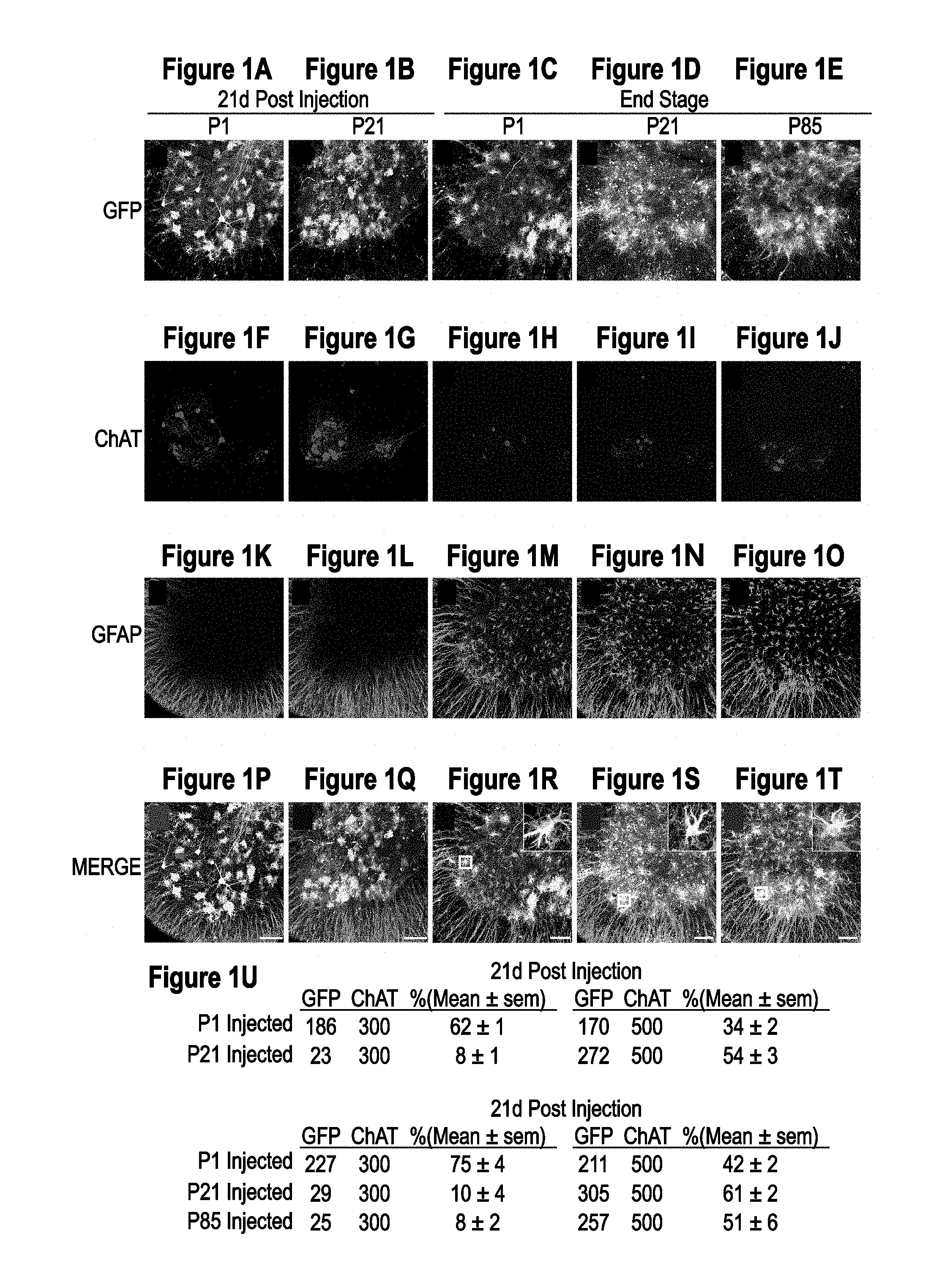
[0036] FIGS. 1A-1U. AAV9 transduction pattern and persistence in SOD1.sup.G93A mice. SOD1.sup.G93A mice were injected intravenously with AAV9-CB-GFP at P1, P21 and euthanized 21 days post injection (n=3 per time point). Spinal cords were examined for GFP, ChAT (motor neuron marker) and GFAP (astrocyte marker) expression. Temporal vein injection of AAV9-CB-GFP at P1 resulted in efficient transduction of motor neurons and glia in SOD1.sup.G93A mice (FIGS. 1A, 1F, 1K, and 1P). Tail vein injection at P21 (FIG. 1B, FIG. 1G, FIG. 1L, FIG. 1Q) predominantly targeted astrocytes with few GFP positive motor neurons. To test the persistence of transduced cells, AAV9-CB-GFP was intravenously injected at P1 and P21 in SOD1.sup.G93A animals that were sacrificed at end stage (.about.P130). Immunofluorescence analysis of lumbar ventral horn (FIG. 1C, FIG. 1D, FIG. 1H, FIG. 1I, FIG. 1M, FIG. 1N, FIG. 1R, FIG. 1S) demonstrated that GFP expression was maintained in astrocytes throughout the disease course. To determine whether SOD1 mediated inflammation and damage would affect AAV9 transduction, we intravenously injected SOD1.sup.G93A mice at P85 and harvested their spinal cords at endstage. There was no difference observed in the transduction pattern of SOD1.sup.G93A mice treated at P21 or P85. Insets in (FIG. 1R, FIG. 1S, FIG. 1T) show co-localization between GFP and GFAP signal. (FIG. 1U) Quantification of transduced cells in ALS spinal cords (for each group tissues were analyzed from 3 animals). GFP and ChAT columns show numbers of cells counted. Bars=100 .mu.m. AAV, adeno-associated virus; P1, postnatal day 1; P21, postnatal day 21; P85, postnatal day 85; GFP, green fluorescent protein; ChAT, choline acetyltransferase; GFAP, glial fibrillary acidic protein.
[0037] FIGS. 2A-2E. shRNA constructs show efficient reduction of human SOD1 protein in vitro and in vivo. (FIG. 2A) Sequence alignments between human and mouse SOD1 for the regions targeted by the 4 different shRNA constructs tested. (FIG. 2B) shRNA sequences were cloned into an H1 expression construct and transiently transfected into 293 cells. Lysates were collected 72 hours post transfection and analyzed by western blot. (FIG. 2C) Quantification of in vitro suppression of human SOD1 from three separate transient transfections showed >50% reduction in SOD1. (FIG. 2D) shRNA 130 was packaged into AAV9 and injected into SOD1.sup.G93A mice at either P1 or P21. Spinal cords (n=3 per time point) were harvested three weeks post injection and analyzed by western blot for human SOD1 protein levels. (FIG. 2E) Quantification of in vivo suppression of human SOD1 within the spinal cord of ALS mice. P1 and P21 injected spinal cords showed 60% and 45% reductions in mutant SOD1 protein, respectively. hSOD1, human superoxide dismutase 1; mSOD1, mouse superoxide dismutase 1; GAPDH, glyceraldehyde 3 phosphate dehydrogenase.
[0038] FIGS. 3A-3H. Intravenous delivery of AAV9-SOD1-shRNA improves survival and motor performance in SOD1.sup.G93A mice. SOD1.sup.G93A mice received a single intravenous injection of AAV9-SOD1-shRNA at P1 (n=6, green), P21 (n=9, red) or P85 (n=5, blue). Treated mice were monitored up to end stage and compared with non-injected control SOD1.sup.G93A mice (n=15, gray). (FIG. 3A, FIG. 3C) AAV9-SOD1-shRNA injection into P1 SOD1.sup.G93A mice significantly delayed median disease onset 39.5 days compared to control animals (uninjected, 103d; P1, 142.5d; p<0.05). Injection in P21 (red) or P85 (blue) ALS animals had no effect on disease onset (P21, 110d; P85, 105d). However, AAV9-SOD1-shRNA administered at P1, P21 or P85 all significantly extended median survival (FIG. 3B, FIG. 3E) (uninjected, 132d; P1, 183.5d P21, 171d; P85, 162d; all comparisons to control p<0.001). The P21 group had a significant extension in median disease duration (FIG. 3D) indicating a slowing of disease (uninjected, 29.5d; P1, 41d; P21, 49d; P85, 40d; Wilcoxon Signed Rank Test p=0.06, 0.01 and 0.12, respectively). (FIG. 3F, FIG. 3G, FIG. 3H) P1 and P21 treated animals maintained their weights, had better hind limb grip strength and rotarod performance when compared to age-matched controls, indicating treated animals retained muscle tone and motor function during their prolonged survival. Lines between bars in (FIG. 3C, FIG. 3D, FIG. 3E) indicate statistically significant differences. * p<0.05. P1, postnatal day 1; P21, postnatal day 21; P85, postnatal day 85.
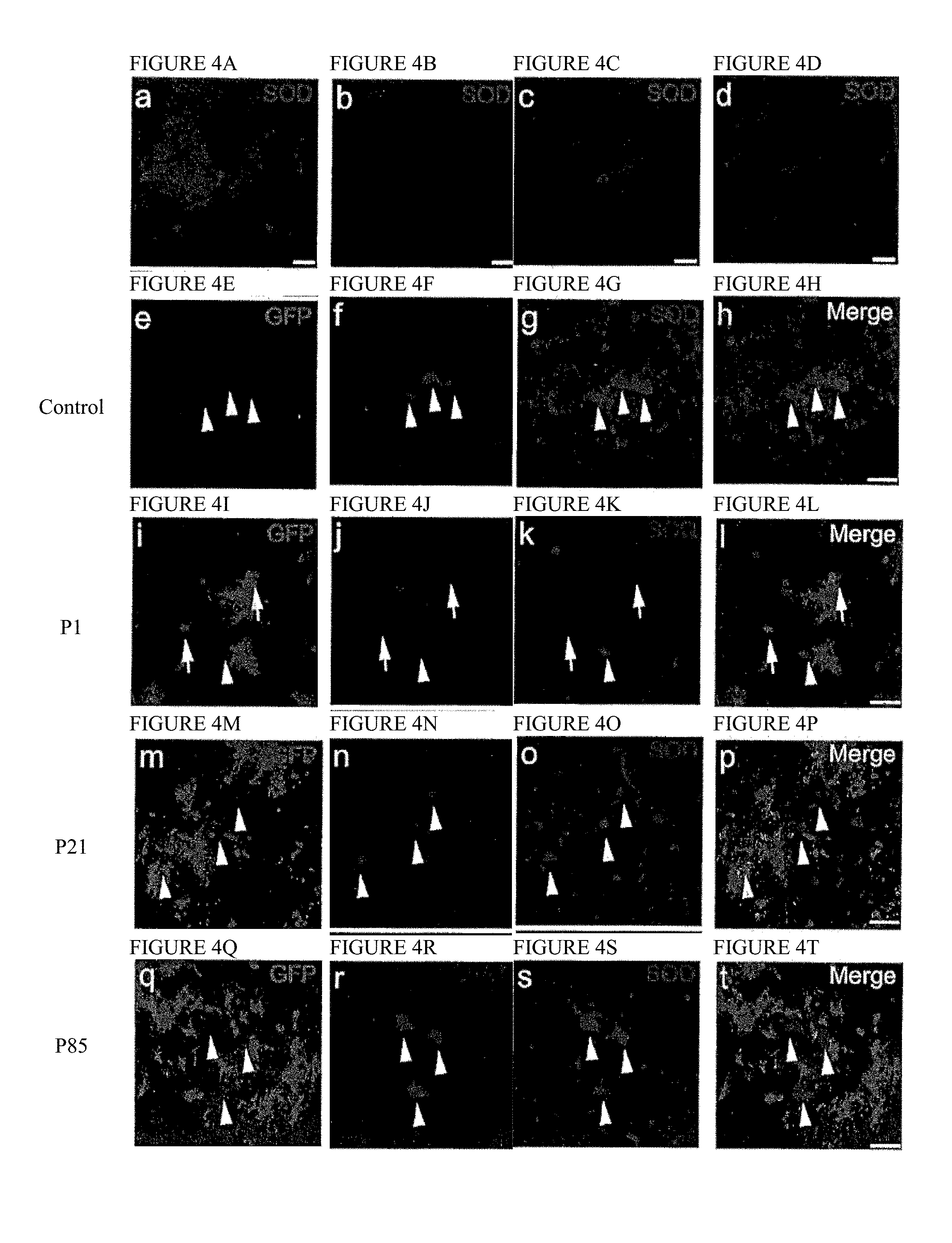
[0039] FIGS. 4A-4T. Intravenous injection of AAV9-SOD1-shRNA reduces mutant protein in spinal cords of SOD1.sup.G93A mice. (FIG. 4A, FIG. 4B, FIG. 4C, FIG. 4D) Images of lumbar spinal cord sections from uninjected (FIG. 4A), P1 injected (FIG. 4B), P21 injected (FIG. 4C) and P85 injected (FIG. 4D) mice were captured with identical microscope settings to qualitatively show SOD1 levels at end stage. SOD1 levels inversely correlate with survival. (FIG. 4E, FIG. 4F, FIG. 4G, FIG. 4H, FIG. 4I, FIG. 4J, FIG. 4K, FIG. 4L, FIG. 4M, FIG. 4N, FIG. 4O, FIG. 4P, FIG. 4Q, FIG. 4R, FIG. 4S, FIG. 4T) Co-labeling for GFP, ChAT and SOD1 shows that AAV9 transduced motor neurons had reduced SOD1 expression (arrows) while cells that lacked GFP maintained high levels of mutant protein (arrowheads). As described in FIG. 1U, higher MN transduction and corresponding SOD1 reduction was observed in P1 injected mice (FIG. 4I, FIG. 4J, FIG. 4K, FIG. 4L) as compared to P21 injected (FIG. 4M, FIG. 4N, FIG. 4O, FIG. 4P) and P85 injected (FIG. 4Q, FIG. 4R, FIG. 4S, FIG. 4T) mice. Bar=100 .mu.m. P1, postnatal day 1; P21, postnatal day 21; P85, postnatal day 85; SOD1, superoxide dismutase 1; GFP, green fluorescent protein; ChAT, choline acetyltransferase.
[0040] FIGS. 5A-5G. AAV9-SOD1-shRNA improves survival and motor performance in SOD1.sup.G37R mice treated after disease onset. (FIG. 5A) There was no difference in median disease onset between AAV9-SOD1-shRNA and control treated mice. (average age at treatment=215d versus median onset of 194d control and 197d treated; Log Rank Test p=0.46). (FIG. 5B, FIG. 5F) Median survival of AAV9-SOD1-shRNA treated SOD1.sup.G37R mice (n=25) was significantly extended versus control mice (n=21). (control, n=21, 392d; SOD1 shRNA, n=25, 478.5d; Log Rank Test p<0.0001) (FIG. 5C, FIG. 5D, FIG. 5E). The early phase of disease was significantly slowed by 73 days in treated mice as compared to control mice (control, 89d; SOD1 shRNA, 162d; p<0.0001 Wilcoxon Signed Rank Test) while the late phase of disease showed a non-significant slowing (control, 63d; SOD1 shRNA, 81d; p=0.14 Wilcoxon Signed Rank Test). Together this amounted to a 66 day increase in median disease duration (control, 173d; SOD1 shRNA, 239d; p<0.0001 Wilcoxon Signed Rank Test). (FIG. 5G) A trend to improved hind limb grip strength appeared in AAV9-SOD1-shRNA treated mice compared to control mice.
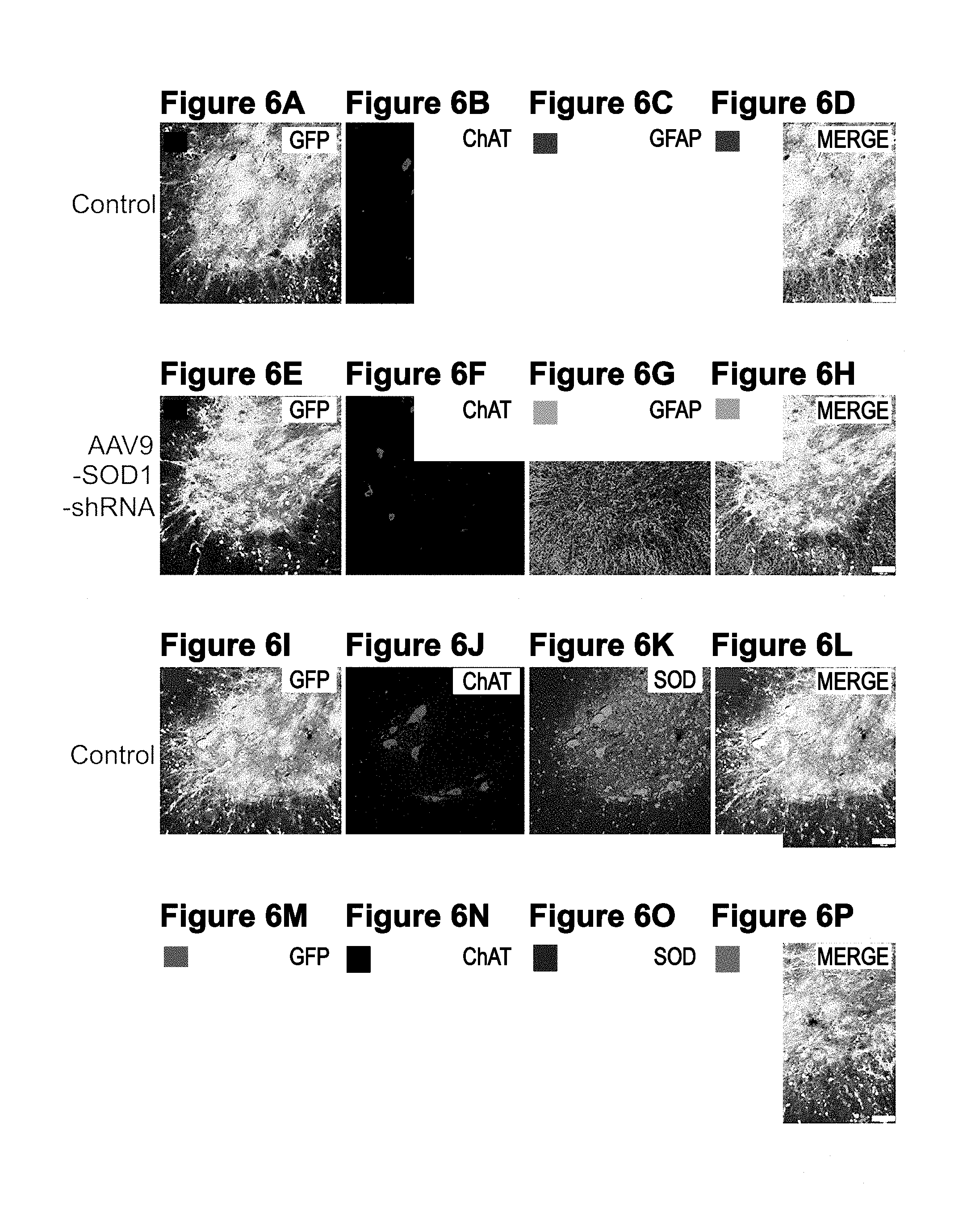
[0041] FIGS. 6A-6P. Intravenous injection of AAV9 in adult SOD1.sup.G37R mice targets astrocytes and motor neurons within the spinal cord. (FIG. 6A, FIG. 6B, FIG. 6C, FIG. 6D, FIG. 6E, FIG. 6F, FIG. 6G, FIG. 6H) Immunofluorescence analysis revealed neuronal as well as glial transduction in both AAV9-CB-GFP (FIG. 6A, FIG. 6B, FIG. 6C, FIG. 6D) and AAV9-SOD1-shRNA treated (FIG. 6E, FIG. 6F, FIG. 6G, FIG. 6H) mice. (FIG. 6I, FIG. 6J, FIG. 6K, FIG. 6L, FIG. 6M, FIG. 6N, FIG. 6O, FIG. 6P) Human SOD1 levels appeared reduced in AAV9-SOD1-shRNA treated mice (FIG. 6O) compared with AAV9-GFP treated mice (FIG. 6K). Bar=100 GFP, green fluorescent protein; ChAT, choline acetyltransferase; GFAP, glial fibrillary acidic protein; SOD1, superoxide dismutase 1.
[0042] FIGS. 7A-7H. Intrathecal infusion of AAV9-SOD1-shRNA in non-human primates leads to efficient reduction in SOD1 levels. (FIG. 7A) A myelogram shortly after intrathecal infusion of AAV9-SOD1-shRNA mixed with contrast shows proper delivery into the subarachnoid space of a cynomolgus macaque. Arrows show diffusion of the contrast agent along the entire spinal cord. (FIG. 7B) Lumbar spinal cord sections from treated monkeys (n=3) were harvested two weeks post injection and stained for GFP using DAB staining. Sections had widespread GFP expression throughout the grey and white matter. (FIG. 7C, FIG. 7D, FIG. 7E) Immunofluorescence analysis of the lumbar spinal cord sections showed robust GFP (FIG. 7C) expression within ChAT (FIG. 7D) positive cells indicating motor neuron transduction (FIG. 7E, merge). (FIG. 7F) Western blot analysis of the lumbar spinal cords showed significant reduction in SOD1 levels in AAV9-SOD1-shRNA injected animals as compared to controls. (FIG. 7G) In vivo quantification of SOD1 knockdown in monkey lumbar spinal cord homogenate (n=3) showed an 87% reduction in animals that received AAV9-SOD1-shRNA compared to uninjected controls. (FIG. 7H) Laser capture microdissection was used to collect motor neurons or surrounding neuropil from injected and control lumbar monkey sections. Collected cells were analyzed for SOD1 levels by qRT-PCR. Motor neurons collected from AAV9-SOD1-shRNA animals (n=3) had a 95.+-.3% reduction in SOD1 RNA. Non-neurons had a 66.+-.9% reduction in SOD1 RNA in AAV9-SOD1-shRNA treated animals. Scale Bars: b=100 .mu.m; e=50 .mu.m. SOD1: Superoxide dismutase 1.
[0043] FIGS. 8A-8E. Lumbar intrathecal infusion of AAV9-SOD1-shRNA leads to efficient transduction of motor neurons and non-neuronal cells in the cervical, thoracic and lumbar cord resulting in reduction of SOD1. (FIG. 8A, FIG. 8B, FIG. 8C) Immunofluorescence analysis of the three segments of the spinal cord; cervical (FIG. 8A), thoracic (FIG. 8B) and lumbar (FIG. 8C), showed robust GFP (green) expression within Chat (red) positive cells indicating motor neuron transduction. (FIG. 8D) GFP+/Chat+ cell counts show a caudal to rostral gradient of motor neuron transduction ranging from 85% of transduced cells in the lumbar region to more than 50% in the cervical region. (FIG. 8E) SOD1 mRNA levels in cervical, thoracic and lumbar cord section homogenates analyzed by qRT-PCR show significant reduction in SOD1 transcript, consistently with motor neuron transduction. SOD1 levels were normalized to .beta.-actin and AAV9-SOD1-shRNA injected animals were compared to an AAV9-CB-GFP injected control. Scale bars: (FIG. 8A, FIG. 8B, FIG. 8C)=50 .mu.m; Error bars: (FIG. 8D, FIG. 8E)=SD.
[0044] FIGS. 9A-B. Design of a clinical SOD1 shRNA construct. (FIG. 9A) Original AAV SOD1 shRNA construct contains shRNA sequence against human SOD1 under H1 promoter followed by the expression cassette for GFP which includes CMV enhancer, CBA promoter, modified SV40 intron, and GFP transgene sequence followed by bGH PolyA terminator. SOD1 shRNA expression cassette and GFP expression cassette are flanked by AAV2 ITRs which ensures the packaging of the complete flanked sequence in AAV9 capsid. (FIG. 9B) In clinical SOD1 shRNA construct, the GFP expression cassette is replaced by a stuffer element that contains tandem, noncoding sequences from FDA approved DNA vaccines. ITR: inverted terminal repeats; shRNA, small hairpin RNA; SOD1, superoxide dismutase 1; CMV, cytomegalo virus enhancer; CBA, Chicken .beta.-actin promoter; GFP, green fluorescent protein; bGH pA, bovine growth hormone poly A terminator.
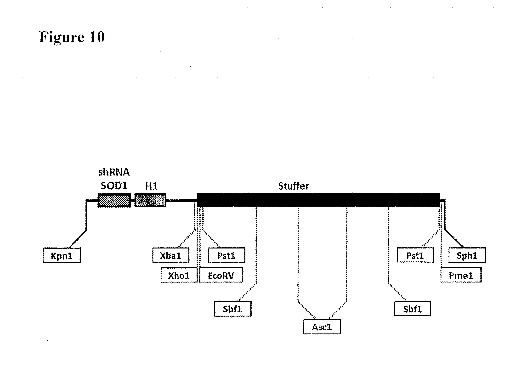
[0045] FIG. 10. Schematic of clinical SOD1 shRNA construct. Different restriction sites are placed in the clinical SOD1 shRNA construct that allow the cloning of multiple shRNA expression cassettes while maintaining the total distance between the two ITRs.
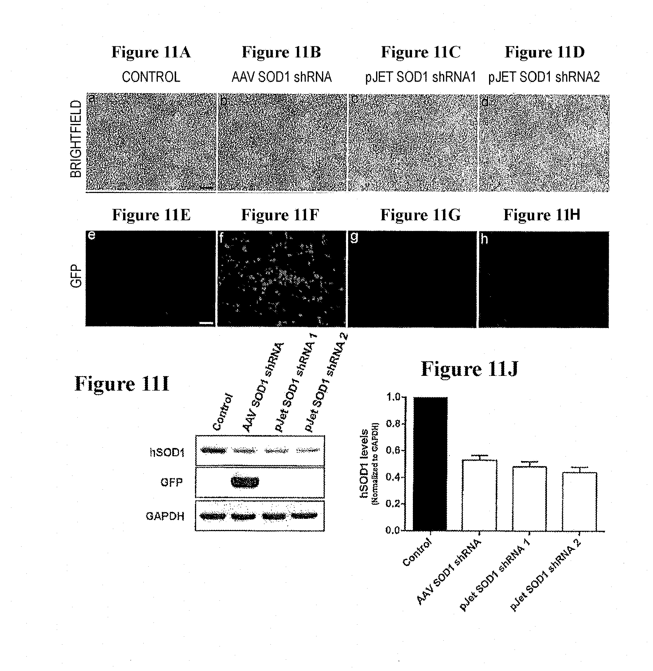
[0046] FIGS. 11A-11J. In vitro transfection of clinical SOD1 shRNA construct efficiently reduces human SOD1 protein in HEK293 cells. Representative microscopic fields showing bright-field images of non-transfected control (FIG. 11A), AAV SOD1 shRNA transfected (FIG. 11B) and shuttle vector pJet SOD1 shRNA transfected (FIG. 11C, FIG. 11D) HEK 293 cells, 72 hrs post transfection. Corresponding fluorescence images reveal the lack of GFP fluorescence from pJet SOD1 shRNA transfected HEK 293 cells (FIG. 11G, FIG. 11H) as compared to AAV SOD1 shRNA transfected cells (FIG. 11F). (FIG. 11I) Western blot analysis of the cell lysates confirms the efficient knockdown of human SOD1 protein in pJet SOD1 shRNA transfected cells as compared to the non-transfected control cells. Immunoblot analysis also confirms removal of GFP transgene from pJet SOD1 shRNA construct. (FIG. 11J) Quantification of the in vitro downregulation of SOD1 by pJet SOD1 shRNA. pJet SOD1 shRNA reduces the protein levels of human SOD1 by almost 50% in HEK293 cells as compared to control. This reduction is similar to that achieved with AAV SOD1 shRNA construct.
[0047] FIG. 12. Schematic of cloning strategy for clinical AAV SOD1 shRNA vector. Clinical SOD1 shRNA construct was cloned into AAV CB MCS vector using Kpn1/SPh1 sites. Kpn1/SPh1 double digest of AAV CB MCS plasmid results in the release of the complete transgene expression cassette from this vector which is further replaced with clinical SOD1 shRNA construct carrying SOD1 shRNA expression cassette and stuffer sequence.
[0048] FIGS. 13A-13H. Clinical AAV SOD1 shRNA efficiently reduces human SOD1 levels in vitro. HEK293 cells were transfected with clinical AAV SOD1 shRNA plasmid by Calcium phosphate method. Representative microscopic fields showing brightfield images of non-transfected control, AAV SOD1 shRNA and Clinical AAV SOD1 shRNA transfected cells respectively, 72 hrs post-transfection (FIG. 13A, FIG. 13B, FIG. 13C). Successful removal of GFP from clinical AAV SOD1 shRNA was confirmed by lack of GFP expression in Clinical AAV SOD1 shRNA transfected cells (FIG. 13F, FIG. 13G. (FIG. 13G) Western blot analysis of cell lysates, harvested 72 hrs post-transfection confirmed efficient downregulation of SOD1 in clinical AAV SOD1 shRNA transfected cells as compared to control. AAV SOD1 shRNA was used as a positive control. (FIG. 13H) Quantification of the in vitro knockdown of SOD1 by clinical AAV SOD1 shRNA.
[0049] Figures S1A-S1F. AAV9-shRNA-SOD1 administration is well tolerated in WT mice. Female and male WT animals were injected with AAV9-SOD1-shRNA at P1 or P21 and monitored up to 6 months of age. (Figure S1A, Figure S1B) Both male and female treated mice showed steady increase in body mass as compared to control animals. (Figure S1C, Figure S1D) Rotarod performance and (Figure S1E, Figure S1F) hind limb grip strength were not affected by P1 or P21 treatment in both groups as compared to respective controls. n=5 per group. WT, wild type; P1, postnatal day 1; P21, postnatal day 21.
[0050] Figures S2A-S2W. Hematology and Serum Chemistry of AAV9-SOD1-shRNA treated WT animals. (Figure S2A, Figure S2B, Figure S2C, Figure S2D, Figure S2E, Figure S2F, Figure S2G, Figure S2H, Figure S21, Figure S2J, Figure S2K, Figure S2L, Figure S2M) Blood was collected from P1 (green) or P21 (red) treated and control (gray) WT animals at 150 days of age for hematology studies. No significant differences were observed between treated and control animals. (Figure S2N, Figure S20, Figure S2P, Figure S2Q, Figure S2R, Figure S2S, Figure S2T, Figure S2U, Figure S2V, Figure S2W) Serum samples collected at 180 days of age from the same mice showed no significant differences in serum chemistry profile. Mean.+-.SEM. n=5 per group. P1, postnatal day 1; P21, postnatal day 21.
[0051] Figures S3A-S3H. AAV9-SOD1-shRNA treatment in SOD1.sup.G93A mice reduces astrogliosis. End stage sections from control and AAV9-SOD1-shRNA treated animals were harvested and stained for GFAP, an astrocyte activation marker. P1 (Figure S3B) and P85 (Figure S3D) injected mice showed reduced levels of astrogliosis as compared to control (Figure S3A) mice while P21 (Figure S3C) injected mice showed the maximum reduction. This was consistent with the percent astrocyte transduction achieved in these mice (FIG. 1U). However, no effect was observed on microglia reactivity (Figure S3E, Figure S3F, Figure S3G, Figure S3H). Bar=100 .mu.m. P1, postnatal day 1; P21, postnatal day 21; P85, postnatal day 85.
[0052] Figures S4A-S4B. Intravenous injection of AAV9-SOD1-shRNA efficiently reduces levels of mutant SOD1 protein in spinal cords of SOD1.sup.G37R mice. (Figure S4A) Following disease onset, AAV9-CB-GFP or AAV9-SOD1-shRNA was injected in SOD1.sup.G37R mice and spinal cords were harvested at end stage and analyzed by western blot for human SOD1 protein levels. (Figure S4B) Quantification of a) shows suppression of human SOD1 within the spinal cord of SOD1.sup.G37R mice (n=4 per group). hSOD1, human superoxide dismutase 1; GAPDH, glyceraldehyde 3 phosphate dehydrogenase.
[0053] Figures S5A-S5B shRNA 130 efficiently reduces the levels of monkey SOD1 in vitro. (Figure S5A) Sequence alignment of the region targeted by SOD1 shRNA 130 and a single mismatch with the monkey sequence. Monkey sequence corresponds to SOD1 sequence from Rhesus monkey (NM 001032804.1), Cynomolgus monkey (sequenced in-house) and African green monkey. (Figure S5B) The shRNA 130 expression cassette was cloned into lentiviral vector and used to infect Cos-7 cells. Lysates were analyzed 72 hours post infection by qRT PCR for SOD1. shRNA 130 reduced SOD1 transcript levels by 75% in Cos-7 cells.
EXAMPLES
[0054] The present invention is illustrated by the following examples. While the present invention has been described in terms of various embodiments and examples, it is understood that variations and improvements will occur to those skilled in the art. Therefore, only such limitations as appear in the claims should be placed on the invention.
Example 1
AAV9 Transduction Pattern and Persistence in SOD1.sup.G93A Mice
[0055] We first evaluated the efficiency of AAV9 transduction in the SOD1.sup.G93A mouse model that develops fatal paralytic disease. High copy SOD1.sup.G93A mice were obtained from Jackson Laboratories (Bar Harbor, Me.) and bred within the Kaspar lab. Animals were genotyped before the treatment to obtain SOD1.sup.G93A expressing mice and their wild type littermates. Only female mice were included in the SOD1.sup.G93A experiments. Animals were injected intravenously at postnatal day 1 or day 21 (to be referred to as P1 and P21, respectively) with self-complementary AAV9 expressing GFP from the CMV enhancer/beta-actin (CB) promoter (AAV9-CB-GFP) (n=3 per group). Three weeks post-injection, animals were sacrificed, and spinal cords examined for GFP expression (FIG. 1A, FIG. 1B, FIG. 1C, FIG. 1D, FIG. 1E, FIG. 1F, FIG. 1G, FIG. 1H, FIG. 1I, FIG. 1J, FIG. 1K, FIG. 1L, FIG. 1M, FIG. 1N, FIG. 1O, FIG. 1P, FIG. 1Q, FIG. 1R, FIG. 1S, FIG. 1T, FIG. 1U).
[0056] All procedures with animals described herein were performed in accordance with the NIH Guidelines and approved by the Research Institute at Nationwide Children's Hospital (Columbus, Ohio), University of California (San Diego, Calif.) or Mannheimer Foundation (Homestead, Fla.) Institutional Animal Care and Use Committees.
[0057] Transduction efficiency was high in SOD1.sup.G93A astrocytes with GFP expressed in 34.+-.2% and 54.+-.3%, respectively, of P1 and P21 injected spinal grey matter astrocytes (defined by immunoreactivity for GFAP). This efficiency was similar to our previous report of 64.+-.1% in P21 injected wild type animals.sup.18. Motor neurons were a prominent cell type transduced at all levels of the spinal cords of P1 injected SOD1.sup.G93A animals (62.+-.1%), compared with significantly lower targeting to motor neurons in P21 injected animals (8.+-.1%).
[0058] Although we have previously reported that transduced astrocytes in wild type spinal cords persist with continued GFP accumulation for at least 7 weeks post injection.sup.18, longevity of mutant SOD1 astrocytes (and their continued synthesis of genes encoded by the AAV9 episome) during active ALS-like disease was untested. Therefore, SOD1.sup.G93A mice were injected at P1 and P21 with AAV9-CB-GFP and followed to end-stage (.about.P130, n=3 per group) (FIG. 1C, FIG. 1D, FIG. 1H, FIG. 1I, FIG. 1M, FIG. 1N, FIG. 1R, FIG. 1S). Immunofluorescent examination of the end-stage SOD1.sup.G93A spinal cords from animals injected at P1 and P21 showed a comparable number of GFP-expressing astrocytes as were found 21 days after AAV9 injection (P1: 42.+-.2%, P21: 61.+-.2%). These data are consistent with survival of transduced astrocytes for the duration of disease (.about.110 days post injection at P21) in SOD1.sup.G93A mice and that AAV9-encoded gene expression is maintained.
[0059] Further, recognizing that SOD1 mutant mediated damage, including astrocytic and microglial activation and early changes in the blood brain barrier develop during disease in mice in SOD1 mutant mice.sup.20, we tested if this damage affected AAV9 transduction. SOD1.sup.G93A mice were injected at P85 with AAV9-CB-GFP and sacrificed at endstage (n=3) (FIG. 1E, FIG. 1J, FIG. 1O, FIG. 1T). Analysis of the spinal cords revealed that the transduction pattern seen in P85 animals was similar to P21 treated animals with astrocytes as the predominant cell type transduced at all levels (51.+-.6% GFP+/GFAP+ cells in lumbar grey matter).
Example 2
Development of an shRNA Sequence Specific for Human SOD1
[0060] To specifically target the human SOD1 mRNA, four shRNA constructs targeting human SOD1 were generated and obtained from the Life Technologies design tool. The constructs that had a minimum of four base mismatches compared to the mouse mRNA sequence (FIG. 2A). The base numbers for the human sequences shown correspond to record number CCDS33536.1 in the NCBI CCDS database. These constructs were cloned in pSilencer 3.1 (Genscript) under the human H1 promoter and tested in vitro. shRNA 130 along with H1 promoter was further cloned into an AAV vector along with a reporter GFP under Chicken Beta-Actin promoter to identify the transduced cells. Human 293 cells were transfected with each cassette. The HEK-293 cells were maintained in IMDM medium containing 10% FBS, 1% L-glutamine and 1% penicillin/streptomycin. Upon reaching .about.60% confluence, cells were transfected with pSilencer 3.1 containing the shRNAs being tested. Protein lysates were prepared 72 hours post transfection and analyzed for SOD1 levels by western blot. All four sequences reduced SOD1 protein levels by >50% (FIG. 2B, FIG. 2C).
[0061] shRNA130 was selected for further experiments because it produced the most consistent knockdown across three separate transfection experiments. It was cloned into a self-complementary AAV9 vector that also contained a GFP gene whose expression would identify transduced cells (referred to as AAV9-SOD1-shRNA). Self-complementary AAV9-SOD1-shRNA was produced by transient transfection procedures using a double-stranded AAV2-ITR-based CB-GFP vector, with a plasmid encoding Rep2Cap9 sequence as previously described along with an adenoviral helper plasmid pHelper (Stratagene, Santa Clara, Calif.) in 293 cells.sup.18.
[0062] To confirm that the shRNA could suppress accumulation of human SOD1, SOD1.sup.G93A mice (n=3) were injected intravenously with AAV9-SOD1-shRNA at either P1 or P21. For neonatal mouse injections, postnatal day 1-2 SOD1.sup.G93A pups were utilized. Total volume of 50 .mu.l containing 5.times.10.sup.11 DNAse resistant viral particles of AAV9-SOD1-shRNA (Virapur LLC, San Diego, Calif.) was injected via temporal vein as previously described.sup.18. A correct injection was verified by noting blanching of the vein. After the injection, pups were returned to their cage. Animals were euthanized three weeks post injection and the spinal cords were harvested and analyzed by immunoblotting for both human (mutant) and murine (wild-type) SOD1 protein. P1 and P21 injected spinal cords showed 60% and 45% reductions in mutant SOD1 protein, respectively (FIG. 2D, FIG. 2E). Murine SOD1 levels remained unchanged in response to human SOD1 knockdown.
Example 3
AAV9-SOD1-shRNA is Safe and Well Tolerated in Wild Type Mice
[0063] To determine whether high dose AAV9-SOD1-shRNA would be safe, normal mice of both sexes were intravenously injected at P1 or P21 (P1=5 males, 5 females at 5.times.10.sup.11 vg; P21=5 males, 5 females at 2.times.10.sup.12 vector genomes (vg)) and then monitored up to 6 months of age. Both P1 and P21 injected mice showed a steady increase in body mass similar to untreated mice (Figures S1A-S1F). Weekly behavioral tests observed no significant differences between injected and control groups in motor skills (measured by rotarod) as well as in hind limb grip strength. At 150 and 180 days of age, blood samples were collected. Complete and differential blood counts of both treated and untreated groups showed similar blood chemistry parameters (Figures S2A-S2W). Serum samples from both groups showed no significant differences in the levels of alkaline phosphatase, creatinine, blood urea nitrogen, potassium, sodium and chloride. Finally, all the animals were sacrificed at the age of 180 days. Histopathological analyses by a pathologist blinded to treatment group revealed no significant alterations in the AAV9-SOD1-shRNA treated animals compared to uninjected controls (data not shown). We conclude that both administration of AAV9 and sustained shRNA expression were apparently safe and well tolerated.
Example 4
Extended Survival of SOD1.sup.G93A Mice from AAV9 Mediated Reduction in Mutant SOD1 Even when Initiated Mid-Disease
[0064] To test the efficacy of AAV9-mediated SOD1 reduction, we treated cohorts of SOD1.sup.G93A mice with a single intravenous injection of AAV9-SOD1-shRNA before (P1, 5.times.10.sup.11 vg, n=6 and P21, 2.times.10.sup.12 vg, n=9) or after (P85, 3.times.10.sup.12 vg, n=5) onset, recognizing that many astrocytes, but few motor neurons, would be transduced at the two later time points. For adult tail vein injections, animals were placed in a restraint that positioned the mouse tail in a lighted, heated groove. The tail was swabbed with alcohol then injected intravenously with AAV9-SOD1-shRNA.
[0065] Onset of disease (measured by weight loss from denervation-induced muscle atrophy) was significantly delayed by a median of 39.5 days (FIG. 3A, FIG. 3C; uninjected, 103d; P1, 142.5d; p<0.05, Wilcoxon Signed Rank Test) in the P1 injected cohort, but was not affected by either of the later injections (P21, 110d; P85, 105d). P1 and P21 treated animals maintained their weights, had better rotarod performance and hind limb grip strength when compared to age-matched controls, indicating treated animals maintained muscle tone and motor function during their prolonged survival (FIG. 3F, FIG. 3G, FIG. 3H). Survival was significantly extended by AAV9 injection at all three ages, yielding survival times 30-51.5 days beyond that of uninjected SOD1.sup.G93A mice (uninjected, 132d; P1, 183.5d; P21, 171d; P85, 162d; Log-Rank Test p=<0.0001, 0.0003 and 0.001, respectively) (FIG. 3B, FIG. 3E). Defining disease duration as the time from onset to end-stage revealed that the P21 treatment group had significantly increased duration, indicative of slowed disease progression, compared to uninjected controls (uninjected, 29.5d; P21, 49d; Wilcoxon Signed Rank Test p=0.01), with trends toward slowed progression in animals injected at the other two ages (P1, 41d; P85, 40d; p=0.06 and 0.12, respectively) (FIG. 3D). The lower percentage of targeted non-neuronal cells at P1 versus those targeted at P21 (FIG. 1U) suggests that a minimum number of non-neuronal cells must be targeted to slow disease progression in the fast progressive SOD1.sup.G93A model (FIG. 1U).
Example 5
Reduction of Mutant SOD1 in AAV9 Infected Cells in Treated SOD1.sup.G93A Mice
[0066] Indirect immunofluorescence with an antibody that recognizes human, but not mouse SOD1, was used to determine accumulated mutant SOD1 levels in end-stage spinal cords of treated and control mice. Human SOD1 levels in end-stage spinal cord sections inversely correlated with increased survival (FIG. 4A, FIG. 4B, FIG. 4C, FIG. 4D). At end-stage, P1 (FIG. 4B), P21 (FIG. 4C) and P85 (FIG. 4D) AAV9-SOD1-shRNA injected animals had lower levels of mutant SOD1 when compared with uninjected SOD1.sup.G93A animals (FIG. 4A). SOD1 expression within transduced motor neurons (identified by GFP and ChAT expressing cells) was reduced compared to surrounding neurons that had not been transduced to express viral encoded GFP (FIG. 4H, FIG. 4L, FIG. 4P, FIG. 4T; arrows versus arrowheads). Moreover, immunofluorescence imaging of end-stage spinal cords revealed corresponding reduction in astrogliosis, but no difference in microgliosis in AAV9-SOD1-shRNA treated animals versus controls (Figures S3A-S3H).
Example 6
Therapeutic Slowing of Disease Progression with Peripheral Injection of AAV9 after Onset
[0067] To determine if AAV9-mediated mutant SOD1 reduction would slow disease progression, a cohort of SOD1.sup.G37R mice.sup.6 were injected intravenously with AAV9-SOD1-shRNA after disease onset (average age at treatment=215d versus median onset of 197d in treated animals; Log Rank Test p=0.46; FIG. 5A). loxSOD1.sup.G37R ALS mice, carrying a human mutant SOD1.sup.G37R transgene flanked by lox p sites under its endogenous promoter, were maintained in as previously described.sup.37. A combination of AAV9-CB-GFP (n=9) and uninjected (n=12) littermates were used as controls.
[0068] Post hoc analysis showed no differences between GFP and uninjected animals, therefore the groups were compiled as "control" in FIGS. 5A-5G. Animals were evaluated weekly for body weight and hind limb grip strength and monitored until end-stage. AAV9-SOD1-shRNA treatment after disease onset significantly extended median survival by 86.5 days over control animals (control, n=21, 392d; SOD1 shRNA, n=25, 478.5d; Log Rank Test p<0.0001). Early disease duration, defined by the time from peak weight to 10% weight loss, was significantly slowed (control, 89d; SOD1 shRNA treated mice, 162 days; Wilcoxon Signed Rank Test p<0.01; FIG. 5C). A continuing trend toward slowing of later disease (10% weight loss to end stage) was also seen (control, 63d; SOD1 shRNA treated mice, 81d; Wilcoxon Signed Rank Test p=0.1389; FIG. 5D). Overall disease duration following AAV9-SOD1-shRNA therapy rose to 239d after disease onset versus 173d in control mice (Wilcoxon Signed Rank Test p<0.0001; FIG. 5E). Consistent with the slowed progression, AAV9 therapy maintained grip strength relative to control SOD1 mutant animals (FIG. 5G). The 86.5 day extension in survival surpassed the 62 day extension seen in transgenic studies that used astrocyte-specific Cre expression to inactivate the mutant SOD1.sup.G37R transgene.sup.8, presumably reflecting efficient AAV9 transduction of astrocytes after peripheral delivery and the possible transduction of other cell types (especially microglia.sup.6) whose synthesis of mutant SOD1 accelerates disease progression.
[0069] Histological examination of end-stage SOD1.sup.G37R treated animals revealed similar levels of intraspinal cell transduction in animals treated with AAV9-SOD1-shRNA or AAV9-GFP (FIGS. 6A-6P). GFP expression was predominantly observed within motor neurons and astrocytes of both groups, and SOD1 expression was detectably decreased only in animals that received AAV9-SOD1-shRNA (FIG. 6K, FIG. 6O). Immunoblotting of whole spinal cord extracts from end stage SOD1.sup.G37R mice revealed an 80% reduction in hSOD1 protein levels in AAV9-SOD1-shRNA treated animals compared to controls (Figures S4A-S4B).
Example 7
AAV9 Mediated Suppression of SOD1 in Non-Human Primates
[0070] To test whether SOD1 levels could be efficiently lowered using AAV9 in the non-human primate spinal cord, AAV9 was injected intrathecally via lumbar puncture. This method was chosen over systemic delivery to decrease the amount of virus required and to minimize any effects from reduction of SOD1 in peripheral tissues. One year old cynomolgus macaques (Macaca fascicularis) with average body weight of 2 kg were used for this study at the Mannheimer Foundation. Regular monitoring of overall health and body weight was performed prior and after the injections to assess the welfare of the animals.
[0071] Sequencing of cDNA copied from mRNA isolated from African Green Monkey (COS cells) and the Cynomolgus macaque verified that the 130 shRNA had a single base mismatch to either sequence (Figures S5A-S5B). The 130 shRNA expression cassette was inserted into a lentiviral vector which was then used to transduce COS cells. Cos-7 cells were maintained in DMEM with 10% FBS and 1% penicillin/streptomycin. Cells were infected with a lentiviral vector expressing SOD1 shRNA 130 under the H1 promoter and RFP under CMV promoter. RNA was extracted from infected and non-infected cells 72 hours post infection using an RNAeasy Kit (Qiagen). cDNA was prepared using RT.sup.2 First strand synthesis kit (SABiosciences). SOD1 transcript levels were analyzed by qRT-PCR which revealed that the monkey SOD1 mRNA was reduced by .about.75% in 130 shRNA transduced cells compared to mock transduced control cells (Figures S5A-S5B).
[0072] The AAV9-SOD1-shRNA virus (1.times.10.sup.13 vg/kg) was infused along with contrast agent via lumbar puncture into the subarachnoid space of three male cynomolgus macaques and one control subject was injected with AAV9-CB-GFP (1.times.10.sup.13 vg/kg) (FIG. 7A). Each intrathecal injection was performed by lumbar puncture into the subarachnoid space of the lumbar thecal sac. AAV9 was resuspended with omnipaque (iohexol), an iodinated compound routinely used in the clinical setting. Iohexol is used to validate successful subarachnoid space cannulation and was administered at a dose of 100 mg/Kg. The subject was placed in the lateral decubitus position and the posterior midline injection site at .about.L4/5 level identified (below the conus of the spinal cord). Under sterile conditions, a spinal needle with stylet was inserted and subarachnoid cannulation was confirmed with the flow of clear CSF from the needle. In order to decrease the pressure in the subarachnoid space, 0.8 ml of CSF was drained, immediately followed by injection with a mixture containing 0.7 mL iohexol (300 mg/ml formulation) mixed with 2.1 mL of virus (2.8 ml total).
[0073] No side effects from the treatments were identified. Two weeks post injection, the spinal cords were harvested for analysis of GFP expression and SOD1 RNA levels. GFP expression was seen broadly in neuronal and astrocytic cells throughout the grey and white matter of the lumbar spinal cord, the area closest to the site of injection (FIG. 7B, FIG. 7C, FIG. 7D, FIG. 7E). Immunoblotting of extracts of lumbar spinal cord revealed 87% reduction in monkey SOD1 protein levels (FIG. 7F, FIG. 7G). Laser capture microdissection was then used to isolate total RNA from motor neurons as well as from glia in the nearby neuropil. Analysis by quantitative RT-PCR using primers specific for monkey SOD1 (and normalized to actin) confirmed a 95.+-.3% knockdown in the motor neuron pool and a 66.+-.9% knockdown in the neuropil pool when compared to samples from a control animal (FIG. 7H).
[0074] Next we examined the level of cell transduction throughout the spinal cord including cervical, thoracic and lumbar segments. GFP was found to be expressed broadly within all sections analyzed (FIGS. 8A-8C). Motor neuron counts revealed a caudal to rostral gradient in cell transduction, with the cervical region showing more than 50% of GFP/Chat+ motor neurons, increasing to 65% in the thoracic region and reaching a remarkable 80% in the lumbar region (FIG. 8D). In order to determine the overall level of SOD1 knockdown achieved with this transduction pattern, qRT-PCR for SOD1 was performed on whole section homogenates from cervical, thoracic and lumbar cord segments. The results confirmed robust SOD1 reduction at all three spinal cord levels, ranging from a 60% decrease in the cervical segment, a 70% decrease in the thoracic region and an 88% decrease in the lumbar region (FIG. 8E), consistent with the proportion of cells transduced in each region.
DISCUSSION
[0075] The examples above show that intravenous administration of AAV9-SOD1-shRNA is safe and well tolerated in wild type mice, with the absence of adverse effects after long-term assessment. This approach have achieved one of the longest extensions in survival ever reported in the rapidly progressive SOD1.sup.G93A mouse model of ALS (increasing survival by 39% when treatment is initiated at birth). Even more encouraging, markedly slowed disease progression is seen even when AAV9 therapy to reduce mutant SOD1 synthesis is applied after disease onset in SOD1.sup.G37R mice, thereby significantly extending survival. Thus, the vascular delivery paradigm in mice represents a proof of concept that mutant SOD1 knockdown after disease onset can be beneficial in both rapid and more slowly progressive models of ALS at clinically relevant points in disease. Together, these data show that robust targeting and suppression of SOD1 levels via AAV9-mediated delivery of shRNA is effective in slowing disease progression in mouse models of ALS, critically even when treatment is initiated after onset.
[0076] Multiple recent studies have brought forward the hypothesis that wild-type SOD1 may contribute through misfolding to the pathogenic mechanism(s) that underlie sporadic ALS through a pathway similar to that triggered by mutant SOD1.sup.14,30-32 Included in this body of evidence is our own demonstration that astrocytes produced from sporadic ALS patients are toxic to co-cultured motor neurons and that toxicity is alleviated by siRNA-mediated reduction in wild type SOD1.sup.30. This evidence creates the potential that a proportion of sporadic ALS patients could also benefit from an AAV9-mediated SOD1 reduction approach that we have demonstrated to be effective in slowing disease progression in mice that develop fatal, ALS-like disease from expressing ALS-causing mutations in SOD1.
[0077] Finally, for translation of an AAV9-mediated suppression of SOD1 synthesis to the human setting, we have determined that infusion directly into the CSF at the lumbar level in a non-human primate produce substantial SOD1 reduction by targeting both motor neurons and non-neuronal cells. This outcome provides strong support for extending these efforts to an adult human by direct injection into CSF, as previously proposed.sup.33,34, so as to 1) limit the cost of viral production, 2) reduce the possibility that chronic suppression of SOD1 in the periphery may have deleterious consequences, and 3) reduce viral exposure to the peripheral immune system.sup.33. These data strongly indicate AAV9-SOD1-shRNA as a treatment for ALS.
Techniques/Methods Used in Examples 1-7
[0078] Perfusion and Tissue Processing. Control and treated SOD1.sup.G93A mice were sacrificed at either 21 days post injection or at endstage for immunohistochemical analysis. Animals were anesthetized with xylazene/ketamine cocktail, transcardially perfused with 0.9% saline, followed by 4% paraformaldehyde. Spinal cords were harvested, cut into blocks of tissue 5-6 mm in length, and then cut into 40 .mu.m thick transverse sections on a vibratome (Leica, Bannockburn, Ill.). Serial sections were kept in a 96-well plate that contained 4% paraformaldehyde and were stored at 4.degree. C. End stage loxSOD1.sup.G37R mice were anesthetized using isoflurane and perfused with 4% paraformaldehyde. Spinal cord segments, including cervical, thoracic and lumbar segments were dissected. Following cryoprotection with 20% sucrose/4% paraformaldehyde overnight, spinal cords were frozen in isopentane at -65.degree. C., and serial 30 .mu.m coronal sections were collected free floating using sliding microtome.
[0079] For safety studies, P1, P21 treated and control wild type mice were sacrificed at 180 days of age. Animals were anesthetized using xylazene/ketamine cocktail and perfused with 0.9% saline. Different tissues were removed and stored in 10% buffered formalin. These tissues were further processed, blocked and mounted for hematoxilin & eosin staining by the Nationwide Children's Hospital Morphology Core.
[0080] Cynomolgus monkeys injected with virus were euthanized 2 weeks post injection. Animals were anesthetized with sodium pentobarbital at the dose of 80-100 mg/kg intravenously and perfused with saline solution. Brain and spinal cord dissection were performed immediately and tissues were processed either for nucleic acid isolation (snap frozen) or post-fixed in 4% paraformaldehyde and subsequently cryoprotected with 30% sucrose and frozen in isopentane at -65.degree. C. 12 .mu.m coronal sections were collected from lumbar cord using a cryostat for free floating immunostaining.
[0081] Immunohistochemistry.
[0082] Mouse spinal cords were stained as floating sections. Tissues were washed three-times for 10 minutes each in TBS, then blocked in a solution containing 10% donkey serum, 1% Triton X-100 and 1% penicillin/streptomycin for two hours at room temperature. All the antibodies were diluted with the blocking solution. Primary antibodies used were as follows: rabbit anti-GFP (1:400, Invitrogen, Carlsbad, Calif.), rabbit anti-SOD1 (1:200, Cell signaling, Danvers, Mass.), goat anti-ChAT (1:50 Millipore, Billerica, Mass.), mouse anti-GFAP (1:200, Millipore, Billerica, Mass.), chicken anti GFAP (1:400, Abcam, Cambridge, Mass.), and rabbit anti-Iba1 (1:400, Wako, Richmond Va.). Tissues were incubated in primary antibody at 4.degree. C. for 48-72 hours then washed three times with TBS. After washing, tissues were incubated for 2 hours at room temperature in the appropriate FITC-, Cy3-, or Cy5-conjugated secondary antibodies (1:200 Jackson Immunoresearch, Westgrove, Pa.) and DAPI (1:1000, Invitrogen, Carlsbad, Calif.). Tissues were then washed three times with TBS, mounted onto slides then coverslipped with PVA-DABCO. All images were captured on a Zeiss-laser-scanning confocal microscope.
[0083] For DAB staining, monkey spinal cord sections were washed three times in TBS, blocked for 2 h at RT in 10% donkey serum and 1% Triton X-100. Sections were then incubated overnight at 4.degree. C. with rabbit anti-GFP primary antibody (1:1000 Invitrogen, Carlsbad, Calif.) diluted in blocking buffer. The following day, tissues were washed with TBS 3 times, incubated with biotinylated secondary antibody anti-rabbit (1:200 Jackson Immunoresearch, Westgrove, Pa.) in blocking buffer for 30 min at RT, washed 3 times in TBS and incubated for 30 min at RT with ABC (Vector, Burlingame, Calif.). Sections were then washed for 3 times in TBS and incubated for 2 min with DAB solution at RT and washed with distilled water. These were then mounted onto slides and covered with coverslips in mounting medium. All images were captured with the Zeiss Axioscope.
[0084] Motor Neuron and Astrocyte Quantification.
[0085] For MN quantification, serial 40 .mu.m thick lumbar spinal cord sections, each separated by 480 .mu.m, were labeled as described for GFP and ChAT expression. Stained sections were serially mounted on slides from rostral to caudal, then coverslipped. Sections were evaluated using confocal microscopy (Zeiss) with a 40.times. objective and simultaneous FITC and Cy3 filters. The total number of ChAT positive cells found in the ventral horns with defined soma was tallied by careful examination through the entire z-extent of the section. GFP labeled cells were quantified in the same manner, while checking for co-localization with ChAT. For astrocyte quantification, as with MNs, serial sections were stained for GFP, GFAP and then mounted. Using confocal microscopy with a 63.times. objective and simultaneous FITC and Cy5 filters, random fields in the ventral horns of lumbar spinal cord sections from tail vein injected animals were selected. The total numbers of GFP and GFAP positive cells were counted from a minimum of at least 24-fields per animal while focusing through the entire z extent of the section. Spinal cord sections of 3 animals per group were examined for MN and astrocyte quantification.
[0086] Immunoblot Analysis.
[0087] Spinal cords were harvested from P1, P21 injected and control SOD1.sup.G93A mice 21 days post injection and from treated and control monkeys 2 weeks post injection of AAV9-SOD1-shRNA. Spinal cords were homogenized and protein lysates were prepared using T-Per (Pierce) with protease inhibitor cocktail. Samples were resolved on SDS-PAGE according to manufacturer's instructions. Primary antibodies used were rabbit anti-SOD1 (1:750, Cell signaling, Danvers, Mass.) mouse anti-SOD1 (1:750, Millipore, Billerica, Mass.), rabbit anti-SOD1 (1:1000, Abcam, Cambridge, Mass.), rabbit anti-Actin (1:1000, Abcam, Cambridge, Mass.) and mouse anti-GAPDH (1:1000, Millipore, Billerica, Mass.). Secondary antibodies used were anti-rabbit HRP (1:10000-1:50000) and anti-mouse HRP (1:10000). Densitometric analysis was performed using Image J software.
[0088] Laser Capture Microdissection.
[0089] 12 .quadrature.m lumbar spinal cord frozen sections were collected onto PEN membrane slides (Zeiss, Munich, Germany) and stained with 1% Cresyl violet (Sigma, St. Louis, Mo.) in methanol. Sections were air dried and stored at -80.degree. C. After thawing, motor neurons were collected within 30 min from staining using the laser capture microdissector PALM Robo3 Zeiss) using the following settings: Cut energy: 48, LPC energy: 20, Cut focus: 80/81, LPC focus: 1, Position speed: 100, Cut speed: 50. About 500 MNs were collected per animal. Non-neuronal cells from the ventral horn were collected from the same sections after collecting the motor neurons.
[0090] qRT-PCR.
[0091] RNA from laser captured cells or whole spinal cord sections from the cervical, thoracic and lumbar segments was isolated using the RNaqueous Micro Kit (Ambion, Grand Island, N.Y.) according to manufacturer's instructions. RNA was then reverse-transcribed into cDNA using the RT.sup.2 HT First Strand Kit (SABiosciences, Valencia, Calif.). 12.5 ng RNA were used in each Q-PCR reaction using SyBR Green (Invitrogen, Carlsbad, Calif.) to establish the relative quantity of endogenous monkey SOD1 transcript in animals who had received the AAV9-SOD1-shRNA compared to animals who had received only AAV9-GFP. Each sample was run in triplicate and relative concentration calculated using the ddCt values normalized to endogenous actin transcript.
[0092] Behavior and Survival Analysis.
[0093] Treated and control SOD1.sup.G93A mice were monitored for changes in body mass twice a week. loxSOD1.sup.G37R mice were weighed on a weekly basis. Motor coordination was recorded using a rotarod instrument (Columbus Instruments, Columbus, Ohio). Each weekly session consisted of three trials on the accelerating rotarod beginning at 5 rpm/min. The time each mouse remained on the rod was registered. Both SOD1.sup.G93A and loxSOD1.sup.G37R mice were subjected to weekly assessment of hindlimb grip strength using a grip strength meter (Columbus Instruments, Columbus, Ohio). Each weekly session consisted of 3 (SOD1.sup.G93A mice) or 5 (loxSOD1.sup.G37R mice) tests per animal. Survival analysis was performed using Kaplan-Meier survival analysis. End stage was defined as an artificial death point when animals could no longer "right" themselves within 30 sec after being placed on its back. Onset and disease progression were determined from retrospective analysis of the data. Disease onset is defined as the age at which the animal reached its peak weight. Disease duration is defined as the time period between disease onset and end stage. Early disease duration is the period between peak weight and loss of 10% of body weight while late disease duration is defined as the period between 10% loss of body weight until disease end stage. Due to shorter life span of SOD1.sup.G93A animals, we did not assess the distinction between the early and late progression.
[0094] For toxicity analysis following injection at P1 or P21, treated and control WT mice were subjected to behavioral analysis starting at .about.30 days of age and monitored up to 6 months. Body mass was recorded weekly while rotarod performance and hindlimb grip strength were recorded biweekly.
[0095] Hematology and Serum Studies.
[0096] Blood samples were collected in (K2) EDTA microtainer tubes (BD) from treated and control WT mice at 150 days of age by mandibular vein puncture. The same animals were bled at 180 days of age and blood was collected in serum separator microtainer tubes. The blood was allowed to clot for an hour and was then centrifuged at 10,000 rpm for 5 minutes. The clear upper phase (serum) was collected and frozen at -80.degree. C. Hematological and serum analysis were conducted by Ani Lytics Inc, Gaithersburg, Md.
[0097] Statistical Analysis.
[0098] All statistical tests were performed using the GraphPad Prism (San Diego, Calif.) software package. Kaplan Meier survival analyses were analyzed by the Log Rank Test. Comparisons of median disease durations and survival times were analyzed by the Wilcoxon Signed Rank Test.
Example 8
Development of a Clinical SOD1 shRNA Construct
[0099] The AAV SOD1 shRNA vector described in Example 2 carries shRNA against human SOD1 sequence under the H1 promoter (FIG. 9A). The same vector also contains a GFP expression cassette which expresses GFP under a CBA promoter. The other regulatory elements present in this cassette include CMV enhancer, SV40 intron and bGH PolyA terminator sequence. We show herein that AAV9 SOD1 shRNA administration results in efficient SOD1 downregulation along with robust expression of GFP in vitro as well as in vivo. No significant alterations were observed after the long term assessment of wild-type mice administered with AAV9 SOD1 shRNA. These results suggested that there are no evident off-target effects due to the long-term expression of SOD1 shRNA as well as overexpression of GFP. Although we did not find GFP toxicity in our mice, several reports have shown the adverse effects of GFP overexpression in vitro and in vivo. Therefore, to eliminate the possibility of GFP toxicity altogether, the SOD1 shRNA construct of Example 2 was modified by replacing the GFP expression cassette with a non-coding stuffer sequence while maintaining the size of the total DNA construct flanked by the ITRs (FIG. 9B). This is important as the distance between the two ITR sequences greatly affects the packaging capacity of the flanked construct into AAV9 capsids[321-324].
[0100] To date, none of the FDA approved stuffer sequences are readily available. There are, however, several plasmid backbones that are approved by FDA for the human administration. Small DNA fragments were picked from these plasmids which do not correspond to any essential DNA sequences necessary for selection and replication of the plasmid or the elements of the transcriptional units. The plasmid backbones are listed in Table 1. The DNA elements from different plasmids were arranged in tandem to generate a complete, 1607 bp stuffer sequence (SEQ ID NO: 22). Finally, a DNA construct containing the SOD1 shRNA expression cassette, followed by the stuffer sequence was synthesized from Genscript.
TABLE-US-00005 TABLE 1 Plasmid ClinicalTrials.gov Backbone Condition Intervention Phase Identifier pVax1 Early Stage Non- Recombinant DNA- 1 NCT00062907 Small Cell Lung pVAX/L523S Cancer pCDNA3 Chronic Hepatitis B DNA vaccine 1, 2 NCT00536627 pCMVS2.S pUCMV3 Stage III Ovarian pUMVC3-hIGFBP-2 1 NCT01322802 Epithelial Cancer multi-epitope plasmid Stage III Ovarian DNA vaccine Germ Cell Tumor Stage IV Ovarian Epithelial Cancer Stage IV Ovarian Germ Cell Tumor pBK- Prostate Cancer NY-ESO-1 plasmid 1 NCT00199849 CMV Bladder Cancer DNA Cancer Vaccine Non-Small Cell Lung Cancer Esophageal Cancer Sarcoma pGA2 HIV Infections pGA2/JS2 Plasmid DNA 1 NCT00043511 Vaccine
[0101] Clinical SOD1 shRNA construct has shRNA against human SOD1 under H1 promoter which is followed by the non-coding stuffer sequence. This construct is designed in such a way that multiple shRNA expression cassettes can be added to the final vector by simultaneous removal of the stuffer sequence. Restriction endonuclease sites have been added to the stuffer sequence so that a part of the stuffer can be removed when another shRNA expression cassette is added (FIG. 10). This simultaneous removal and addition of DNA sequences would help maintaining the optimal size of the whole construct between the ITRs (.about.2.0 kb) to achieve efficient packaging.
[0102] Clinical SOD1 shRNA construct from Genscript was cloned into pJet1.2 shuttle vector via EcoRV. This parental clone was screened using various restriction endonucleases designed within the construct to confirm the correct clone. Kpn1/Sph1 double digestion of pJet SOD1 shRNA confirmed the presence of the complete construct (2023 bp) while Xba1 digestion confirmed the presence of SOD1 shRNA expression cassette (414 bp) and the stuffer element, along with pJet backbone (.about.3000 bp). EcoRV/Pme1 double digestion also revealed the presence of stuffer element.
Example 9
Clinical SOD1 shRNA Efficiently Reduces Human SOD1 Protein Levels In Vitro
[0103] To determine the efficacy of the de novo synthesized SOD1 shRNA construct to downregulate SOD1 levels, HEK293 cells were transfected with pJet SOD1 shRNA plasmid using Calcium Phosphate method. AAV SOD1 shRNA plasmid was used as a positive control. Immunofluorescence analysis of HEK293 cells, 72 hrs post transfection revealed the lack of native GFP fluorescence from pJet SOD1 shRNA transfected cells as compared to AAV9 SOD1 shRNA transfected cells. Immunoblot analysis of cell lysates from these cells further confirmed the successful replacement of GFP from pJet SOD1 shRNA plasmid. Importantly, pJet SOD1 shRNA resulted in efficient downregulation of SOD1 protein levels (>50%), similar to AAV SOD1 shRNA plasmid. See FIGS. 11A-11J.
Example 10
[0104] Generation of clinical AAV SOD1 shRNA
[0105] Clinical SOD1 shRNA construct was further cloned into an AAV.CB.MCS vector using Kpn1/Sph1 sites to generate clinical AAV SOD1 shRNA plasmid (FIG. 12). AAV.CB.MCS was generated from AAV.CB.GFP plasmid obtained from merion Scientific by replacing GFP with a multiple cloning site (MCS). Cloning of clinical SOD1 shRNA construct at Kpn1/Sph1 sites puts it between the two AAV2 ITRs which facilitates the packaging of the construct in AAV9 viral capsids. See FIG. 12.
[0106] Clinical AAV SOD1 shRNA plasmid was screened with restriction endonucleases to confirm the presence of SOD1 shRNA expression cassette (Xba1 digest), stuffer sequence (EcoRV/Pme1 double digest) and also intact ITR sequences (Sma1 digest).
Example 11
Clinical AAV SOD1 shRNA Efficiently Reduces Human SOD1 Protein Levels In Vitro
[0107] Clinical AAV SOD1 shRNA plasmid was transfected in HEK293 cells to determine its knockdown efficiency. Similar to the pJet SOD1 shRNA plasmid, clinical AAV SOD1 shRNA transfected cells were devoid of any GFP expression as evident by immunofluorescence (FIG. 13A, FIG. 13B, FIG. 13C, FIG. 13D, FIG. 13E, FIG. 13F) and immunoblot assay (FIG. 13G). More importantly, clinical AAV SOD1 shRNA efficiently reduced human SOD1 protein levels in HEK293 cells by more than 50% (FIG. 13G, FIG. 13H). Altogether, these results confirmed the successful generation of clinical AAV SOD1 shRNA vector with functional SOD1 shRNA expression cassette and complete removal of the transgene expression cassette.
DOCUMENTS REFERENCED
[0108] 1. Da Cruz, S. & Cleveland, D. W. Understanding the role of TDP-43 and FUS/TLS in ALS and beyond. Curr Opin Neurobiol 21, 904-919 (2011). [0109] 2. Rosen, D. R. et al. Mutations in Cu/Zn superoxide dismutase gene are associated with familial amyotrophic lateral sclerosis. Nature 362, 59-62 (1993). [0110] 3. Ilieva, H., Polymenidou, M. & Cleveland, D. W. Non-cell autonomous toxicity in neurodegenerative disorders: ALS and beyond. The Journal of cell biology 187, 761-772 (2009). [0111] 4. Chattopadhyay, M. & Valentine, J. S. Aggregation of copper-zinc superoxide dismutase in familial and sporadic ALS. Antioxidants & redox signaling 11, 1603-1614 (2009). [0112] 5. Prudencio, M., Hart, P. J., Borchelt, D. R. & Andersen, P. M. Variation in aggregation propensities among ALS-associated variants of SOD1: correlation to human disease. Human molecular genetics 18, 3217-3226 (2009). [0113] 6. Boillee, S. et al. Onset and progression in inherited ALS determined by motor neurons and microglia. Science 312, 1389-1392 (2006). [0114] 7. Kang, S. H. et al. Degeneration and impaired regeneration of gray matter oligodendrocytes in amyotrophic lateral sclerosis. Nature neuroscience 16, 571-579 (2013). [0115] 8. Yamanaka, K. et al. Astrocytes as determinants of disease progression in inherited amyotrophic lateral sclerosis. Nature neuroscience 11, 251-253 (2008). [0116] 9. Di Giorgio, F. P., Boulting, G. L., Bobrowicz, S. & Eggan, K. C. Human embryonic stem cell-derived motor neurons are sensitive to the toxic effect of glial cells carrying an ALS-causing mutation. Cell Stem Cell 3, 637-648 (2008). [0117] 10. Di Giorgio, F. P., Carrasco, M. A., Siao, M. C., Maniatis, T. & Eggan, K. Non-cell autonomous effect of glia on motor neurons in an embryonic stem cell-based ALS model. Nature neuroscience 10, 608-614 (2007). [0118] 11. Marchetto, M. C. et al. Non-cell-autonomous effect of human SOD1 G37R astrocytes on motor neurons derived from human embryonic stem cells. Cell Stem Cell 3, 649-657 (2008). [0119] 12. Haidet-Phillips, A. M. et al. Astrocytes from familial and sporadic ALS patients are toxic to motor neurons. Nat Biotechnol 29, 824-828 (2011). [0120] 13. Bosco, D. A. et al. Wild-type and mutant SOD1 share an aberrant conformation and a common pathogenic pathway in ALS. Nature neuroscience 13, 1396-1403 (2010). [0121] 14. Pokrishevsky, E. et al. Aberrant localization of FUS and TDP43 is associated with misfolding of SOD1 in amyotrophic lateral sclerosis. PloS one 7, e35050 (2012). [0122] 15. Forsberg, K. et al. Novel antibodies reveal inclusions containing non-native SOD1 in sporadic ALS patients. PLoS One 5, e11552 (2010). [0123] 16. Aggarwal, S. & Cudkowicz, M. ALS drug development: reflections from the past and a way forward. Neurotherapeutics: the journal of the American Society for Experimental Neuro Therapeutics 5, 516-527 (2008). [0124] 17. Gurney, M. E. et al. Benefit of vitamin E, riluzole, and gabapentin in a transgenic model of familial amyotrophic lateral sclerosis. Ann Neurol 39, 147-157 (1996). [0125] 18. Foust, K. D. et al. Intravascular AAV9 preferentially targets neonatal neurons and adult astrocytes. Nature biotechnology 27, 59-65 (2009). [0126] 19. Duque, S. et al. Intravenous administration of self-complementary AAV9 enables transgene delivery to adult motor neurons. Mol Ther 17, 1187-1196 (2009). [0127] 20. Zhong, Z. et al. ALS-causing SOD1 mutants generate vascular changes prior to motor neuron degeneration. Nature neuroscience 11, 420-422 (2008). [0128] 21. Miller, R. G., Mitchell, J. D. & Moore, D. H. Riluzole for amyotrophic lateral sclerosis (ALS)/motor neuron disease (MND). Cochrane Database Syst Rev 3, CD001447 (2012). [0129] 22. Smith, R. A. et al. Antisense oligonucleotide therapy for neurodegenerative disease. The Journal of clinical investigation 116, 2290-2296 (2006). [0130] 23. Raoul, C. et al. Lentiviral-mediated silencing of SOD1 through RNA interference retards disease onset and progression in a mouse model of ALS. Nat Med 11, 423-428 (2005). [0131] 24. Ralph, G. S. et al. Silencing mutant SOD1 using RNAi protects against neurodegeneration and extends survival in an ALS model. Nat Med 11, 429-433 (2005). [0132] 25. Miller, T. M. et al. Virus-delivered small RNA silencing sustains strength in amyotrophic lateral sclerosis. Annals of neurology 57, 773-776 (2005). [0133] 26. Miller, T. M. et al. An antisense oligonucleotide against SOD1 delivered intrathecally for patients with SOD1 familial amyotrophic lateral sclerosis: a phase 1, randomised, first-in-man study. Lancet neurology 12, 435-442 (2013). [0134] 27. Towne, C., Raoul, C., Schneider, B. L. & Aebischer, P. Systemic AAV6 delivery mediating RNA interference against SOD1: neuromuscular transduction does not alter disease progression in fALS mice. Mol Ther 16, 1018-1025 (2008). [0135] 28. Towne, C., Setola, V., Schneider, B. L. & Aebischer, P. Neuroprotection by gene therapy targeting mutant SOD1 in individual pools of motor neurons does not translate into therapeutic benefit in fALS mice. Mol Ther 19, 274-283 (2011). [0136] 29. Mandel, R. J., Lowenstein, P. R. & Byrne, B. J. AAV6-mediated gene silencing fALS short. Mol Ther 19, 231-233 (2011). [0137] 30. Synofzik, M. et al. Mutant superoxide dismutase-1 indistinguishable from wild-type causes ALS. Human molecular genetics 21, 3568-3574 (2012). [0138] 31. Guareschi, S. et al. An over-oxidized form of superoxide dismutase found in sporadic amyotrophic lateral sclerosis with bulbar onset shares a toxic mechanism with mutant SOD1. Proc Natl Acad Sci USA 109, 5074-5079 (2012). [0139] 32. Haidet-Phillips, A. M. et al. Astrocytes from familial and sporadic ALS patients are toxic to motor neurons. Nat Biotechnol 29, 824-828 (2011). [0140] 33. Bevan, A. K. et al. Systemic gene delivery in large species for targeting spinal cord, brain, and peripheral tissues for pediatric disorders. Mol Ther 19, 1971-1980 (2011). [0141] 34. Gray, S. J. et al. Preclinical differences of intravascular AAV9 delivery to neurons and glia: a comparative study of adult mice and nonhuman primates. Mol Ther 19, 1058-1069 (2011). [0142] 35. Lioy, D. T. et al. A role for glia in the progression of Rett's syndrome. Nature 475, 497-500 (2011). [0143] 36. Miranda, C. J. et al. Aging brain microenvironment decreases hippocampal neurogenesis through Wnt-mediated survivin signaling. Aging Cell 11, 542-552 (2012). [0144] 37. Yamanaka, K. et al. Mutant SOD1 in cell types other than motor neurons and oligodendrocytes accelerates onset of disease in ALS mice. Proc Natl Acad Sci USA 105, 7594-7599 (2008).
[0145] All documents referred to in this application, including priority documents, are hereby incorporated by reference in their entirety with particular attention to the content for which they are referred.
Sequence CWU 1
1
22126DNAArtificial SequenceSynthetic Polynucleotide 1gcatcatcaa
tttcgagcag aaggaa 26223DNAArtificial SequenceSynthetic
Polynucleotide 2gaagcattaa aggactgact gaa 23323DNAArtificial
SequenceSynthetic Polynucleotide 3ctgactgaag gcctgcatgg att
23420DNAArtificial SequenceSynthetic Polynucleotide 4catggattcc
atgttcatga 20521DNAArtificial SequenceSynthetic Polynucleotide
5gcatggattc catgttcatg a 21621DNAArtificial SequenceSynthetic
Polynucleotide 6ggtctggcct ataaagtagt c 21721DNAArtificial
SequenceSynthetic Polynucleotide 7gggcatcatc aatttcgagc a
21821DNAArtificial SequenceSynthetic Polynucleotide 8gcatcatcaa
tttcgagcag a 21921DNAArtificial SequenceSynthetic Polynucleotide
9gcctgcatgg attccatgtt c 211021DNAArtificial SequenceSynthetic
Polynucleotide 10ggaggtctgg cctataaagt a 211121DNAArtificial
SequenceSynthetic Polynucleotide 11gattccatgt tcatgagttt g
211221DNAArtificial SequenceSynthetic Polynucleotide 12ggagataata
cagcaggctg t 211321DNAArtificial SequenceSynthetic Polynucleotide
13gctttaaagt acctgtagtg a 211421DNAArtificial SequenceSynthetic
Polynucleotide 14gcattaaagg actgactgaa g 211519DNAArtificial
SequenceSynthetic Polynucleotide 15tcatcaattt cgagcagaa
191619DNAArtificial SequenceSynthetic Polynucleotide 16tcgagcagaa
ggaaagtaa 191719DNAArtificial SequenceSynthetic Polynucleotide
17gcctgcatgg attccatgt 191819DNAArtificial SequenceSynthetic
Polynucleotide 18tcactctcag gagaccatt 191919DNAArtificial
SequenceSynthetic Polynucleotide 19gctttaaagt acctgtagt
19205786DNAArtificial SequenceSynthetic Polynucleotide 20gcccaatacg
caaaccgcct ctccccgcgc gttggccgat tcattaatgc agctgattct 60aacgaggaaa
gcacgttata cgtgctcgtc aaagcaacca tagtacgcgc cctgtagcgg
120cgcattaagc gcggcgggtg tggtggttac gcgcagcgtg accgctacac
ttgccagcgc 180cctagcgccc gctcctttcg ctttcttccc ttcctttctc
gccacgttcg ccggctttcc 240ccgtcaagct ctaaatcggg ggctcccttt
agggttccga tttagtgctt tacggcacct 300cgaccccaaa aaacttgatt
agggtgatgg ttcacgtagt gggccatcgc cctgatagac 360ggtttttcgc
cctttgacgt tggagtccac gttctttaat agtggactct tgttccaaac
420tggaacaaca ctcaacccta tctcggtcta ttcttttgat ttataaggga
ttttgccgat 480ttcggcctat tggttaaaaa atgagctgat ttaacaaaaa
tttaacgcga attttaacaa 540aatattaacg cttacaattt aaatatttgc
ttatacaatc ttcctgtttt tggggctttt 600ctgattatca accggggtac
atatgattga catgctagtt ttacgattac cgttcatcgc 660cctgcgcgct
cgctcgctca ctgaggccgc ccgggcaaag cccgggcgtc gggcgacctt
720tggtcgcccg gcctcagtga gcgagcgagc gcgcagagag ggagtggaat
tcacgcgtgg 780atctgaattc aattcacgcg tggtacctac actttatgct
tccggctcgt atgttgtgtg 840gaattgtgag cggataacaa tttcacacag
gaaacagcta tgaccatgat tacgccaagc 900tttccaaaaa agcatggatt
ccatgttcat gatctcttga atcatgaaca tggaatccat 960ggatccgagt
ggtctcatac agaacttata agattcccaa atccaaagac atttcacgtt
1020tatggtgatt tcccagaaca catagcgaca tgcaaatatg aattcactgg
ccgtcgtttt 1080acaacgtcgt gactgggaaa accctggcgt tacccaactt
aatcgccttg cagcacatcc 1140ccctttcgcc agctggcgta atagcgaaga
ggcccgcacc gatcgccctt cccaacagtt 1200gcgcagcctg tggtacctct
ggtcgttaca taacttacgg taaatggccc gcctggctga 1260ccgcccaacg
acccccgccc attgacgtca ataatgacgt atgttcccat agtaacgcca
1320atagggactt tccattgacg tcaatgggtg gagtatttac ggtaaactgc
ccacttggca 1380gtacatcaag tgtatcatat gccaagtacg ccccctattg
acgtcaatga cggtaaatgg 1440cccgcctggc attatgccca gtacatgacc
ttatgggact ttcctacttg gcagtacatc 1500tactcgaggc cacgttctgc
ttcactctcc ccatctcccc cccctcccca cccccaattt 1560tgtatttatt
tattttttaa ttattttgtg cagcgatggg ggcggggggg gggggggggc
1620gcgcgccagg cggggcgggg cggggcgagg ggcggggcgg ggcgaggcgg
agaggtgcgg 1680cggcagccaa tcagagcggc gcgctccgaa agtttccttt
tatggcgagg cggcggcggc 1740ggcggcccta taaaaagcga agcgcgcggc
gggcgggagc gggatcagcc accgcggtgg 1800cggcctagag tcgacgagga
actgaaaaac cagaaagtta actggtaagt ttagtctttt 1860tgtcttttat
ttcaggtccc ggatccggtg gtggtgcaaa tcaaagaact gctcctcagt
1920ggatgttgcc tttacttcta ggcctgtacg gaagtgttac ttctgctcta
aaagctgcgg 1980aattgtaccc gcggccgatc caccggtcgc caccatggtg
agcaagggcg aggagctgtt 2040caccggggtg gtgcccatcc tggtcgagct
ggacggcgac gtaaacggcc acaagttcag 2100cgtgtccggc gagggcgagg
gcgatgccac ctacggcaag ctgaccctga agttcatctg 2160caccaccggc
aagctgcccg tgccctggcc caccctcgtg accaccctga cctacggcgt
2220gcagtgcttc agccgctacc ccgaccacat gaagcagcac gacttcttca
agtccgccat 2280gcccgaaggc tacgtccagg agcgcaccat cttcttcaag
gacgacggca actacaagac 2340ccgcgccgag gtgaagttcg agggcgacac
cctggtgaac cgcatcgagc tgaagggcat 2400cgacttcaag gaggacggca
acatcctggg gcacaagctg gagtacaact acaacagcca 2460caacgtctat
atcatggccg acaagcagaa gaacggcatc aaggtgaact tcaagatccg
2520ccacaacatc gaggacggca gcgtgcagct cgccgaccac taccagcaga
acacccccat 2580cggcgacggc cccgtgctgc tgcccgacaa ccactacctg
agcacccagt ccgccctgag 2640caaagacccc aacgagaagc gcgatcacat
ggtcctgctg gagttcgtga ccgccgccgg 2700gatcactctc ggcatggacg
agctgtacaa gtaaagcggc catcaagctt atcgataccg 2760tcgactagag
ctcgctgatc agcctcgact gtgccttcta gttgccagcc atctgttgtt
2820tgcccctccc ccgtgccttc cttgaccctg gaaggtgcca ctcccactgt
cctttcctaa 2880taaaatgagg aaattgcatc gcattgtctg agtaggtgtc
attctattct ggggggtggg 2940gtggggcagg acagcaaggg ggaggattgg
gaagacaata gcaggcatgc tggggagaga 3000tcgatctgag gaacccctag
tgatggagtt ggccactccc tctctgcgcg ctcgctcgct 3060cactgaggcc
gggcgaccaa aggtcgcccg acgcccgggc tttgcccggg cggcctcagt
3120gagcgagcga gcgcgcagag agggagtggc cccccccccc ccccccccgg
cgattctctt 3180gtttgctcca gactctcagg caatgacctg atagcctttg
tagagacctc tcaaaaatag 3240ctaccctctc cggcatgaat ttatcagcta
gaacggttga atatcatatt gatggtgatt 3300tgactgtctc cggcctttct
cacccgtttg aatctttacc tacacattac tcaggcattg 3360catttaaaat
atatgagggt tctaaaaatt tttatccttg cgttgaaata aaggcttctc
3420ccgcaaaagt attacagggt cataatgttt ttggtacaac cgatttagct
ttatgctctg 3480aggctttatt gcttaatttt gctaattctt tgccttgcct
gtatgattta ttggatgttg 3540gaatcgcctg atgcggtatt ttctccttac
gcatctgtgc ggtatttcac accgcatatg 3600gtgcactctc agtacaatct
gctctgatgc cgcatagtta agccagcccc gacacccgcc 3660aacacccgct
gacgcgccct gacgggcttg tctgctcccg gcatccgctt acagacaagc
3720tgtgaccgtc tccgggagct gcatgtgtca gaggttttca ccgtcatcac
cgaaacgcgc 3780gagacgaaag ggcctcgtga tacgcctatt tttataggtt
aatgtcatga taataatggt 3840ttcttagacg tcaggtggca cttttcgggg
aaatgtgcgc ggaaccccta tttgtttatt 3900tttctaaata cattcaaata
tgtatccgct catgagacaa taaccctgat aaatgcttca 3960ataatattga
aaaaggaaga gtatgagtat tcaacatttc cgtgtcgccc ttattccctt
4020ttttgcggca ttttgccttc ctgtttttgc tcacccagaa acgctggtga
aagtaaaaga 4080tgctgaagat cagttgggtg cacgagtggg ttacatcgaa
ctggatctca acagcggtaa 4140gatccttgag agttttcgcc ccgaagaacg
ttttccaatg atgagcactt ttaaagttct 4200gctatgtggc gcggtattat
cccgtattga cgccgggcaa gagcaactcg gtcgccgcat 4260acactattct
cagaatgact tggttgagta ctcaccagtc acagaaaagc atcttacgga
4320tggcatgaca gtaagagaat tatgcagtgc tgccataacc atgagtgata
acactgcggc 4380caacttactt ctgacaacga tcggaggacc gaaggagcta
accgcttttt tgcacaacat 4440gggggatcat gtaactcgcc ttgatcgttg
ggaaccggag ctgaatgaag ccataccaaa 4500cgacgagcgt gacaccacga
tgcctgtagc aatggcaaca acgttgcgca aactattaac 4560tggcgaacta
cttactctag cttcccggca acaattaata gactggatgg aggcggataa
4620agttgcagga ccacttctgc gctcggccct tccggctggc tggtttattg
ctgataaatc 4680tggagccggt gagcgtgggt ctcgcggtat cattgcagca
ctggggccag atggtaagcc 4740ctcccgtatc gtagttatct acacgacggg
gagtcaggca actatggatg aacgaaatag 4800acagatcgct gagataggtg
cctcactgat taagcattgg taactgtcag accaagttta 4860ctcatatata
ctttagattg atttaaaact tcatttttaa tttaaaagga tctaggtgaa
4920gatccttttt gataatctca tgaccaaaat cccttaacgt gagttttcgt
tccactgagc 4980gtcagacccc gtagaaaaga tcaaaggatc ttcttgagat
cctttttttc tgcgcgtaat 5040ctgctgcttg caaacaaaaa aaccaccgct
accagcggtg gtttgtttgc cggatcaaga 5100gctaccaact ctttttccga
aggtaactgg cttcagcaga gcgcagatac caaatactgt 5160ccttctagtg
tagccgtagt taggccacca cttcaagaac tctgtagcac cgcctacata
5220cctcgctctg ctaatcctgt taccagtggc tgctgccagt ggcgataagt
cgtgtcttac 5280cgggttggac tcaagacgat agttaccgga taaggcgcag
cggtcgggct gaacgggggg 5340ttcgtgcaca cagcccagct tggagcgaac
gacctacacc gaactgagat acctacagcg 5400tgagctatga gaaagcgcca
cgcttcccga agggagaaag gcggacaggt atccggtaag 5460cggcagggtc
ggaacaggag agcgcacgag ggagcttcca gggggaaacg cctggtatct
5520ttatagtcct gtcgggtttc gccacctctg acttgagcgt cgatttttgt
gatgctcgtc 5580aggggggcgg agcctatgga aaaacgccag caacgcggcc
tttttacggt tcctggcctt 5640ttgctggcct tttgctcaca tgttctttcc
tgcgttatcc cctgattctg tggataaccg 5700tattaccgcc tttgagtgag
ctgataccgc tcgccgcagc cgaacgaccg agcgcagcga 5760gtcagtgagc
gaggaagcgg aagagc 578621164DNAArtificial SequenceSynthetic
Polynucleotide 21aattcatatt tgcatgtcgc tatgtgttct gggaaatcac
cataaacgtg aaatgtcttt 60ggatttggga atcttataag ttctgtatga gaccactcgg
atccatggat tccatgttca 120tgattcaaga gatcatgaac atggaatcca
tgcttttttg gaaa 164221607DNAArtificial SequenceSynthetic
Polynucleotide 22tctagaggct cgagaagata tcaactgcag cttctactgg
gcggttttat ggacagcaag 60cgaaccggaa ttgccagctg gggcgccctc tggtaaggtt
gggaagccct gcaaagtaaa 120ctggatggct ttctcgccgc caaggatctg
atggcgcagg ggatcaagct ctgatcaaga 180gacaggatga ggatcgtttc
gcgttcttga ctcttcgcga tgtacgggcc agatatacgc 240gttgacattg
attattgact agttattaat agtaatcaat tacggggtca ttagttcata
300gcccatatat ggagttccgc ctgcagggac gtcgacggat cgggagatct
cccgatcccc 360tatctgctcc ctgcttgtgt gttggaggtc gctgagtagt
gcgcgagcaa aatttaagct 420acaacaaggc aaggcttgac cgacaattgc
atgaagaatc tgcttagggt taggcgtttt 480gcgctgcttc gcggcgcgcc
ttttaaggca gttattggtg cccttaaacg cctggtgcta 540cgcctgaata
agtgataata agcggatgaa tggcagaaat tcgccggatc tttgtgaagg
600aaccttactt ctgtggtgtg acataattgg acaaactacc tacagagatt
taaagctcta 660atgtaagcag acagttttat tgttcatgat gatatatttt
tatcttgtgc aatgtaacat 720cagagatttt gagacacaac gtggctttcc
cccccccccc ctagggtggg cgaagaactc 780cagcatgaga tccccgcgct
ggaggatcat ccagccggcg tcccggaaaa cgattccgaa 840gcccaacctt
tcatagaagg cggcggtgga atcgaaatct cgtgatggca ggttgggcgt
900cgcttggtcg gtcatttcga accccagagt cccgctcagg gcgcgccggg
ggggggggcg 960ctgaggtctg cctcgtgaag aaggtgttgc tgactcatac
caggcctgaa tcgccccatc 1020atccagccag aaagtgaggg agccacggtt
gatgagagct ttgttgtagg tggaccagtc 1080ctgcaggagc ataaagtgta
aagcctgggg tgcctaatga gtgagctaac tcacattaat 1140tgcgttgcgc
tcactgcccg ctttccagtc gggaaacctg tcgtgcccgc ccagtctagc
1200tatcgccatg taagcccact gcaagctacc tgctttctct ttgcgcttgc
gttttccctt 1260gtccagatag cccagtagct gacattcatc cggggtcagc
accgtttctg cggactggct 1320ttctacgtgt ctggttcgag gcgggatcag
ccaccgcggt ggcggcctag agtcgacgag 1380gaactgaaaa accagaaagt
taactggcct gtacggaagt gttacttctg ctctaaaagc 1440tgcggaattg
tacccgcggc cgatccaccg gtcgccacca gcggccatca agcacgttat
1500cgataccgtc gactagagct cgctgatcag tggggggtgg ggtggggcag
gacagcaagg 1560gggaggattg ggaagacaat agcagctgca gaagtttaaa cgcatgc
1607
D00001
D00002
D00003
D00004
D00005
D00006
D00007
D00008
D00009
D00010
D00011
D00012
D00013
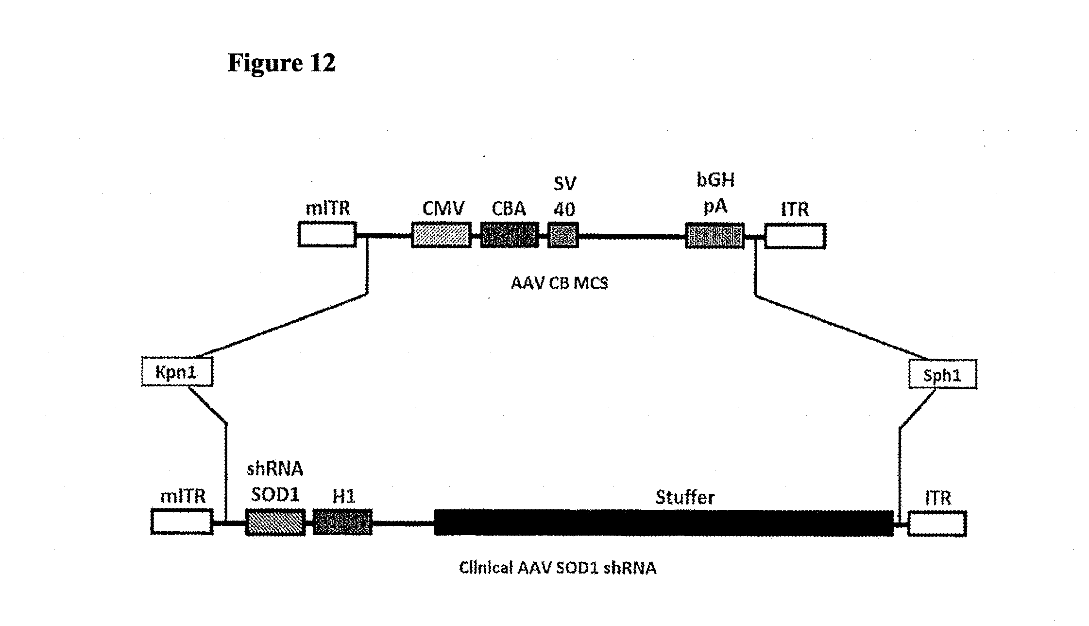
D00014
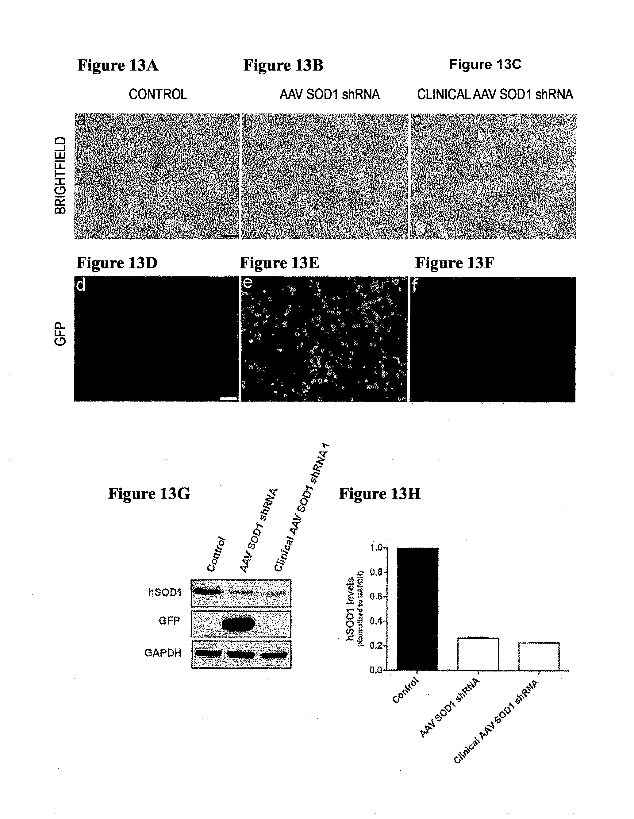
D00015
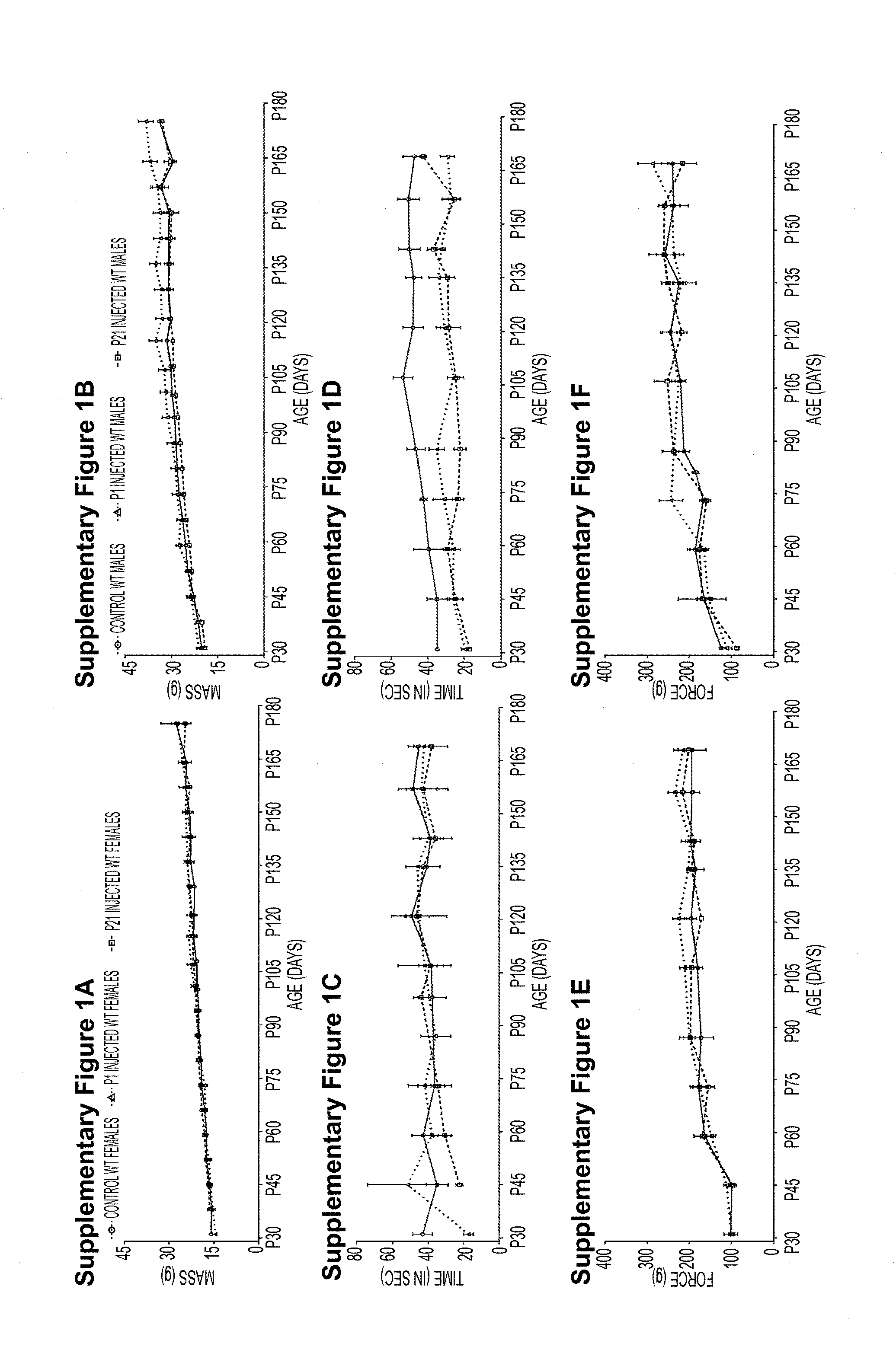
D00016

D00017

D00018
D00019

D00020
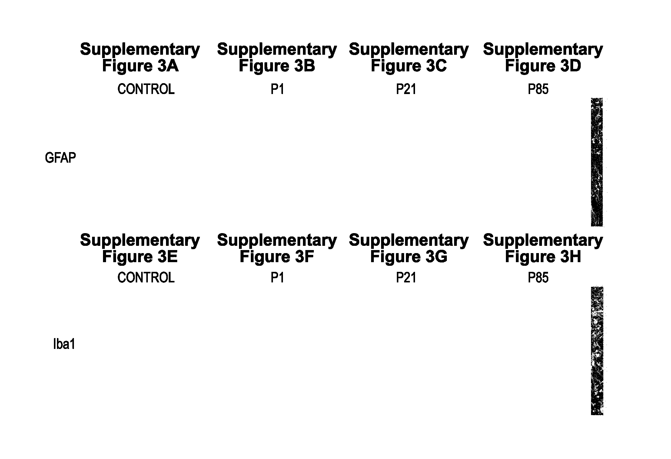
D00021
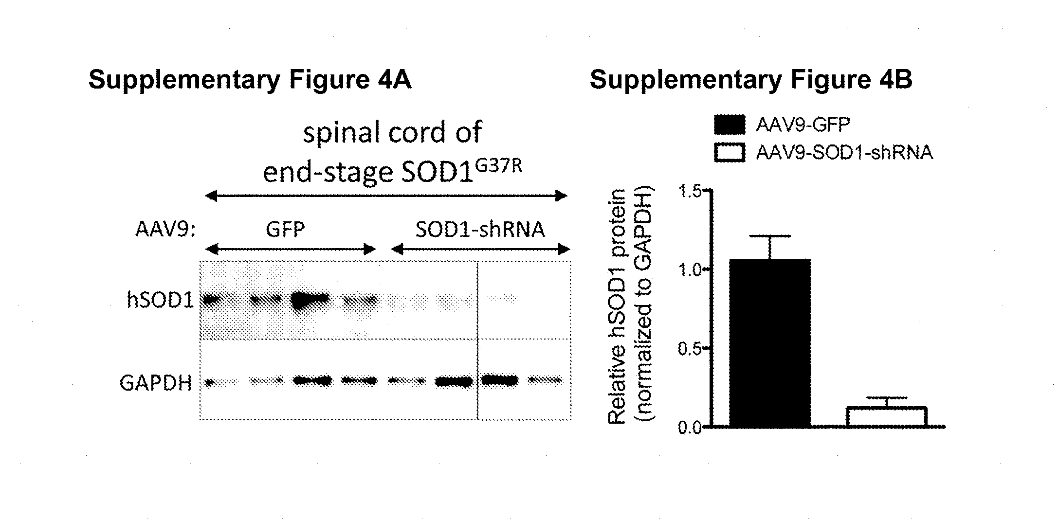
D00022
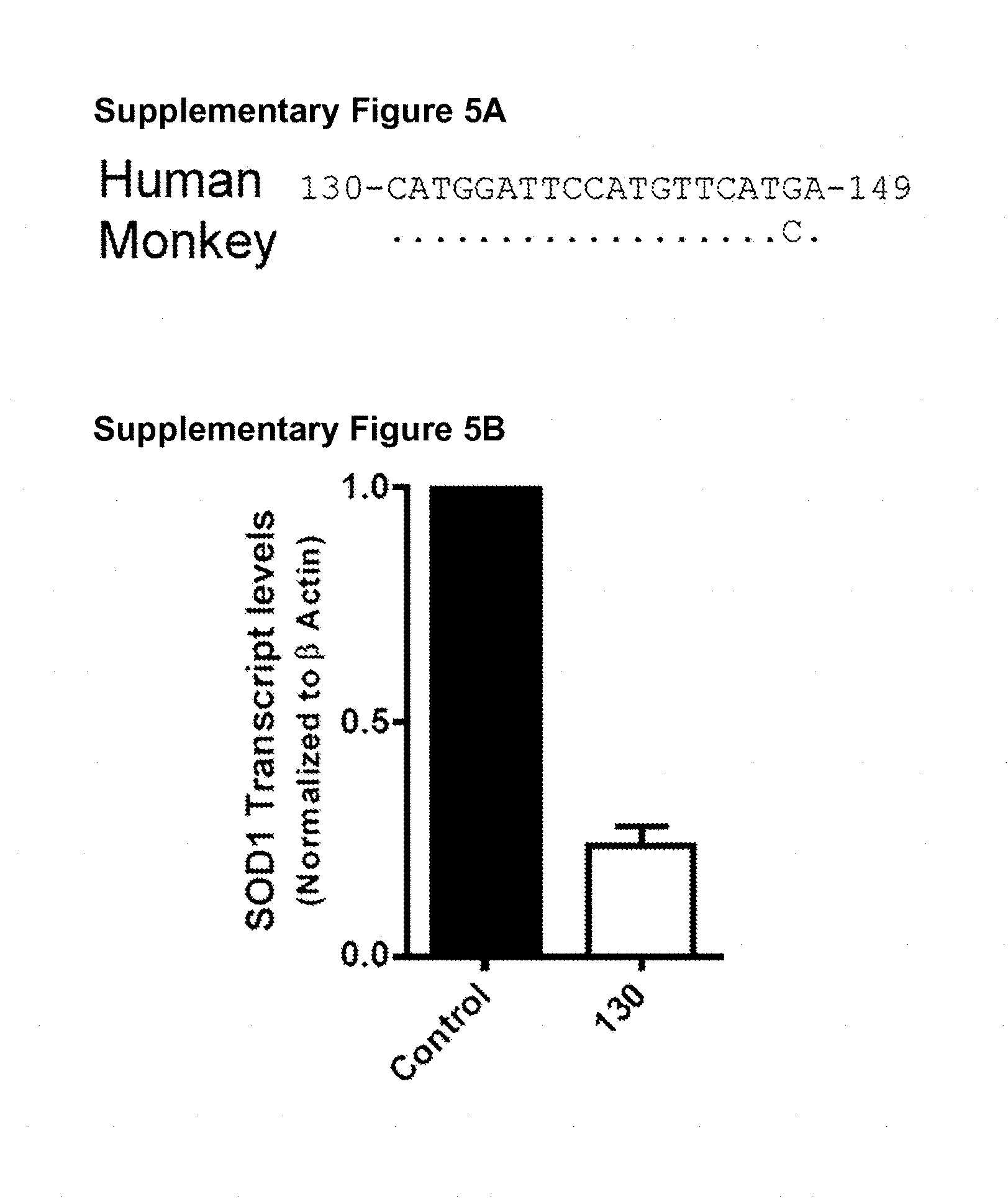
S00001
XML
uspto.report is an independent third-party trademark research tool that is not affiliated, endorsed, or sponsored by the United States Patent and Trademark Office (USPTO) or any other governmental organization. The information provided by uspto.report is based on publicly available data at the time of writing and is intended for informational purposes only.
While we strive to provide accurate and up-to-date information, we do not guarantee the accuracy, completeness, reliability, or suitability of the information displayed on this site. The use of this site is at your own risk. Any reliance you place on such information is therefore strictly at your own risk.
All official trademark data, including owner information, should be verified by visiting the official USPTO website at www.uspto.gov. This site is not intended to replace professional legal advice and should not be used as a substitute for consulting with a legal professional who is knowledgeable about trademark law.