Integrated Cells
HEDHAMMAR; My ; et al.
U.S. patent application number 16/077179 was filed with the patent office on 2019-05-16 for integrated cells. This patent application is currently assigned to SPIBER TECHNOLOGIES AB. The applicant listed for this patent is SPIBER TECHNOLOGIES AB. Invention is credited to My HEDHAMMAR, Ulrika JOHANSSON, Mona WIDHE.
| Application Number | 20190144819 16/077179 |
| Document ID | / |
| Family ID | 58018109 |
| Filed Date | 2019-05-16 |


View All Diagrams
| United States Patent Application | 20190144819 |
| Kind Code | A1 |
| HEDHAMMAR; My ; et al. | May 16, 2019 |
INTEGRATED CELLS
Abstract
A cell scaffold material is manufactured by providing an aqueous solution of a silk protein capable of assembling into a water-insoluble macrostructure. The silk protein is mixed with eukaryotic cells, and the silk protein is assembled into a water-insoluble macrostructure in the presence of the cells, thereby forming a scaffold material for cultivating the cells. The cells can be grown integrated with the scaffold material under conditions suitable for cell culture.
| Inventors: | HEDHAMMAR; My; (Stockholm, SE) ; WIDHE; Mona; (Uppsala, SE) ; JOHANSSON; Ulrika; (Uppsala, SE) | ||||||||||
| Applicant: |
|
||||||||||
|---|---|---|---|---|---|---|---|---|---|---|---|
| Assignee: | SPIBER TECHNOLOGIES AB Stockholm SE |
||||||||||
| Family ID: | 58018109 | ||||||||||
| Appl. No.: | 16/077179 | ||||||||||
| Filed: | February 10, 2017 | ||||||||||
| PCT Filed: | February 10, 2017 | ||||||||||
| PCT NO: | PCT/EP2017/053084 | ||||||||||
| 371 Date: | August 10, 2018 |
| Current U.S. Class: | 435/395 |
| Current CPC Class: | C12N 2533/90 20130101; C07K 14/43586 20130101; C12N 2533/50 20130101; C07K 14/435 20130101; C12N 5/0068 20130101 |
| International Class: | C12N 5/00 20060101 C12N005/00; C07K 14/435 20060101 C07K014/435 |
Foreign Application Data
| Date | Code | Application Number |
|---|---|---|
| Feb 12, 2016 | EP | 16155494.4 |
| Oct 18, 2016 | EP | 16194431.9 |
Claims
1. A method for the cultivation of eukaryotic cells, comprising the steps: (a) providing an aqueous solution of a silk protein capable of assembling into a water-insoluble macrostructure, wherein the silk protein optionally contains a cell-binding motif; (b) preparing an aqueous mixture of a sample of the eukaryotic cells with the silk protein, wherein the silk protein remains dissolved in the aqueous mixture; (c) allowing the silk protein to assemble into a water-insoluble macrostructure in the presence of the eukaryotic cells, thereby forming a scaffold material for cultivating the eukaryotic cells; and (d) maintaining the eukaryotic cells within the scaffold material under conditions suitable for cell culture.
2. The method according to claim 1, wherein the macrostructure is brought into a shape selected from fiber, foam, film, fiber mesh, capsules and nets.
3. The method according to claim 1, wherein the eukaryotic cells are selected from mammalian cells; and stem cells; or a combination of at least two different mammalian cell types.
4. The method according to claim 1, wherein the silk protein is a fibroin.
5. The method according to claim 1, wherein the silk protein is a spider silk protein.
6. The method according to claim 5, wherein the spider silk protein is comprising, or consisting of, the protein moieties REP and CT, wherein REP is a repetitive fragment of from 70 to 300 amino acid residues, selected from the group consisting of L(AG).sub.nL, L(AG).sub.nAL, L(GA).sub.nL, and L(GA).sub.nGL, wherein n is an integer from 2 to 10; each individual A segment is an amino acid sequence of from 8 to 18 amino acid residues, wherein from 0 to 3 of the amino acid residues are not Ala, and the remaining amino acid residues are Ala; each individual G segment is an amino acid sequence of from 12 to 30 amino acid residues, wherein at least 40% of the amino acid residues are Gly; and each individual L segment is a linker amino acid sequence of from 0 to 30 amino acid residues; and CT is a fragment of from 70 to 120 amino acid residues, having at least 70% identity to SEQ ID NO: 3 or SEQ ID NO: 68; and wherein the optional cell-binding motif is arranged either terminally in the spider silk protein, or between the moieties, or within any of the moieties.
7. The method according to claim 1, wherein the silk protein contains a cell-binding motif selected from RGD, IKVAV (SEQ ID NO: 10), YIGSR (SEQ ID NO: 11), EPDIM (SEQ ID NO: 12), NKDIL (SEQ ID NO: 13), GRKRK (SEQ ID NO: 14), KYGAASIKVAVSADR (SEQ ID NO: 15), NGEPRGDTYRAY (SEQ ID NO: 16), PQVTRGDVFTM (SEQ ID NO: 17), AVTGRGDSPASS (SEQ ID NO: 18), TGRGDSPA (SEQ ID NO: 19), CTGRGDSPAC (SEQ ID NO: 20) and FN.sub.cc (SEQ ID NO: 9); wherein FN.sub.cc is C.sup.1X.sup.1X.sup.2RGDX.sup.3X.sup.4X.sup.5C.sup.2; wherein each of X.sup.1, X.sup.2, X.sup.3, X.sup.4 and X.sup.5 are independently selected from natural amino acid residues other than cysteine; and C.sup.1 and C.sup.2 are connected via a disulphide bond.
8. A process for manufacturing a cell culture product comprising (i) a scaffold material for cultivating eukaryotic cells; and (ii) eukaryotic cells, which are growing integrated with the scaffold material, comprising the steps: (a) providing an aqueous solution of a silk protein capable of assembling into a water-insoluble macrostructure, wherein the silk protein optionally contains a cell-binding motif; (b) preparing an aqueous mixture of a sample of the eukaryotic cells with the silk protein, wherein the silk protein remains dissolved in the aqueous mixture; and (c) allowing the silk protein to assemble into a water-insoluble macrostructure in the presence of the eukaryotic cells, thereby forming the scaffold material for cultivating the eukaryotic cells.
9. The process for manufacturing a cell culture product according to claim 8, wherein the macrostructure is brought into a shape selected from fiber, foam, film, fiber mesh, capsules and nets; and/or wherein the eukaryotic cells are selected from mammalian cells; and stem cells; or a combination of at least two different mammalian cell types; and/or wherein the silk protein is a fibroin or a spider silk protein.
10. A cell culture product comprising (i) a scaffold material for cultivating eukaryotic cells, which is a water-insoluble macrostructure of a silk protein capable of assembling into a water-insoluble macrostructure, wherein the silk protein optionally contains a cell-binding motif; and (ii) eukaryotic cells, which are growing integrated with the scaffold material.
11. A cell culture product comprising (i) a scaffold material for cultivating eukaryotic cells, which is a water-insoluble macrostructure of a silk protein capable of assembling into a water-insoluble macrostructure, wherein the silk protein optionally contains a cell-binding motif; and (ii) eukaryotic cells, which are growing integrated with the scaffold material, wherein said cell culture product is obtainable or obtained by the process according to claim 8.
12-13. (canceled)
14. The method according to claim 1, wherein the aqueous mixture of step (b) further contains cell-binding proteins or polypeptides.
15. The cell culture product according to claim 10, wherein the macrostructure is brought into a shape selected from fiber, foam, film, fiber mesh, capsules and nets; and/or wherein the eukaryotic cells are selected from mammalian cells; and stem cells; or a combination of at least two different mammalian cell types; and/or wherein the silk protein is a fibroin or a spider silk protein.
16. The method according to claim 2, wherein the macrostructure is brought into a shape selected fiber or foam.
17. The method according to claim 3, wherein the mammalian cells are selected from primary cells and cell lines, and the stem cells are mesenchymal stem cells.
18. The method according to claim 17, wherein the mammalian cells are endothelical cells, fibroblasts, keratinocytes, skeletal muscle satellite cells, skeletal muscle myoblasts, Schwann cells, pancreatic .beta.-cells, pancreatic islet cells, hepatocytes and glioma-forming cells.
19. The method according to claim 4, wherein the fibroin is a silkworm fibroin.
20. The method according to claim 6, wherein the optional cell-binding motif is arranged terminally in the spider silk protein.
21. The method according to claim 7, wherein the cell-binding motif is selected from FN.sub.cc, GRKRK, IKVAV, RGD and CTGRGDSPAC.
22. The method according to claim 7, wherein the cell-binding motif is selected from FN.sub.cc and CTGRGDSPAC.
Description
TECHNICAL FIELD OF THE INVENTION
[0001] The present invention relates to the fields of eukaryotic cell culture and tissue engineering, and provides methods and a cell scaffold material for culture of eukaryotic cells, wherein a polymer of a silk protein, such as a fibroin or a spider silk protein, is used as a cell scaffold material.
BACKGROUND TO THE INVENTION
[0002] The fundamental concept of tissue engineering is to combine different components, such as living cells, biomaterial and bioactive factors, to form engineered tissue constructs. Traditional tissue engineering strategies typically employ a "top-down" approach, in which cells are seeded on a polymeric scaffold. The material must then contain large pores with high interconnectivity to allow subsequent cell infiltration. In order to allow a high porosity without collapse, the material has to have thick and/or stiff walls, which leads to poor cell compatibility and low flexibility when the cells are about to expand.
[0003] As alternative, the "bottom-up" tissue engineering approach has been initiated lately. A bottom-up approach relies on the assembly of a matrix from smaller components or modules together with the cells. For example, this can be achieved by 3D printing of hydrogels containing cells. However, one major drawback of hydrogels is the lack of mechanical strength, which restricts their use to soft tissue engineering. The processes used for formulation of stronger synthetic matrices are typically dependent on harsh conditions such as melting or organic solvents, and hence not compatible with cell viability. Moreover, synthetic material typically gets much stiffer than what is suitable to match mammalian tissue. The natural extracellular matrix (ECM) that surrounds mammalian cells in tissue consists of fibers (e.g. collagen and elastin) composed of modified proteins that are demanding to produce synthetically, and in vitro mimicry of their mechanical properties has so far not been accomplished. Also other organisms use protein fibers as support; the strongest being silk threads spun by spiders. Apart from outstanding strength, spider silk has very attractive properties such as elasticity and biocompatibility.
[0004] Spiders have up to seven different glands which produce a variety of silk types with different mechanical properties and functions. Dragline silk, produced by the major ampullate gland, is the toughest fiber, and on a weight basis it outperforms man-made materials, such as tensile steel. The properties of dragline silk are attractive in development of new materials for medical or technical purposes, e.g. as scaffolds for cell culture.
[0005] Dragline silk consists of two main polypeptides, mostly referred to as major ampullate spidroin (MaSp) 1 and 2, but e.g. as ADF-3 and ADF-4 in Araneus diadematus. These proteins have molecular masses in the range of 200-720 kDa. The genes coding for dragline proteins of Latrodectus hesperus are the only ones that have been completely characterized, and the MaSp1 and MaSp2 genes encode 3129 and 3779 amino acids, respectively (Ayoub NA et al. PLoS ONE 2(6): e514, 2007). The properties of dragline silk polypeptides are discussed in Huemmerich, D. et al. Curr. Biol. 14, 2070-2074 (2004).
[0006] Spider dragline silk proteins, or MaSps, have a tripartite composition; a non-repetitive N-terminal domain, a central repetitive region comprised of many iterated poly-Ala/Gly segments, and a non-repetitive C-terminal domain. It is generally believed that the repetitive region forms intermolecular contacts in the silk fibers, while the precise functions of the terminal domains are less clear. It is also believed that in association with fiber formation, the repetitive region undergoes a structural conversion from random coil and .alpha.-helical conformation to .beta.-sheet structure. The C-terminal region of spidroins is generally conserved between spider species and silk types. The N-terminal domain of spider silks is the most conserved region (Rising, A. et al. Biomacromolecules 7, 3120-3124 (2006)).
[0007] WO 07/078239 and Stark, M. et al., Biomacromolecules 8, 1695-1701, (2007) disclose a miniature spider silk protein consisting of a repetitive fragment with a high content of Ala and Gly and a C-terminal fragment of a protein, as well as soluble fusion proteins comprising the spider silk protein. The spider silk protein is spontaneously transformed into a coherent and water insoluble macrostructure, e.g. an ordered polymer such as a fiber, upon subjection to an interface such as air:water. The miniature spider silk protein unit is sufficient and necessary for the fiber formation. Cells from an immortalized cell line is added onto the pre-formed, macroscopic spider silk fiber and allowed to grow.
[0008] Hedhammar, M. et al., Biochemistry 47, 3407-3417, (2008) study the thermal, pH and salt effects on the structure and aggregation and/or polymerisation of recombinant N- and C-terminal spidroin domains and a repetitive spidroin domain containing four poly-Ala and -Gly rich co-blocks.
[0009] WO 2011/129756 discloses methods and a cell scaffold material based on a miniature spider silk protein for eukaryotic cell culture. The protein may contain various short (3-5 amino acid residues) cell-binding peptides. Various cell types are added onto the pre-formed cell scaffold material.
[0010] WO 2012/055854 discloses manufacture of a cell scaffold material comprising a recombinant protein which is a fusion protein between a spider silk proteins and a longer (>30 amino acid residues), non-spidroin polypeptide or protein with desirable binding properties. Cells are added onto the pre-formed cell scaffold material and cultivated.
[0011] WO 2015/036619 and Widhe, M. et al., Biomaterials 74:256-266 (2016) disclose further miniature spider silk proteins with useful cell-binding peptides. Again, various cell types are added onto the pre-formed cell scaffold material.
[0012] Johansson et al., PLOS ONE 10(6): e0130169 (2015) discloses formulation of a spider silk protein into various physical formats. Subsequently, pancreatic mouse islets were placed on top of the spider silk matrices and allowed to adhere.
[0013] Despite these advances in the field, there is still a need for new cell scaffolds in the field. In particular, there is a need in the field for a mechanically robust, three-dimensional scaffold for cultivation of integrated eukaryotic cells and use in tissue engineering.
SUMMARY OF THE INVENTION
[0014] It is an object of the present invention to provide a cell scaffold with improved cell compatibility and flexibility when the cells are about to expand.
[0015] It is also an object of the present invention to provide a cell scaffold which achieves a more tissue-like spreading of cultivated cells.
[0016] It is an object of the present invention to provide a cell scaffold with high seeding efficiency, yielding quickly and viably adhered cells.
[0017] It is a further object of the present invention to provide a cell scaffold with sufficient mechanical strength and suitable stiffness for mammalian tissue engineering.
[0018] It is also an object of the present invention to provide a process for providing a cell scaffold under conditions which are compatible with cell viability.
[0019] It is yet another object of the present invention to provide a cell scaffold wherein cells are integrated throughout the cell scaffold material.
[0020] It is also an object of the present invention to provide a method which allows for co-cultures of several cell types within the cell scaffolds.
[0021] For these and other objects that will be evident from the following disclosure, the present invention provides according to a first aspect a method for the cultivation of eukaryotic cells, comprising the steps: [0022] (a) providing an aqueous solution of a silk protein capable of assembling into a water-insoluble macrostructure, wherein the silk protein optionally contains a cell-binding motif; [0023] (b) preparing an aqueous mixture of a sample of the eukaryotic cells with the silk protein, wherein the silk protein remains dissolved in the aqueous mixture; [0024] (c) allowing the silk protein to assemble into a water-insoluble macrostructure in the presence of the eukaryotic cells, thereby forming a scaffold material for cultivating the eukaryotic cells; and [0025] (d) maintaining the eukaryotic cells within the scaffold material under conditions suitable for cell culture.
[0026] In a preferred variant of the method for the cultivation of eukaryotic cells, the silk protein is a spider silk protein.
[0027] The invention is based on the inventive insight that dispersed eukaryotic cells can be added to the silk protein solution before assembly of the silk proteins into a water-insoluble macrostructure, and thereby be integrated throughout the silk-like material during the mild self-assembly process. This is in contrast to the prior art cell cultivation methods, where cells have been added onto pre-formed silk macrostructures.
[0028] Advantageously, formulation of macrostructures with integrated cells provides a high seeding efficiency, yielding quickly and viably adhered cells.
[0029] Compared to cultivation in hydrogels, cells attain a more tissue-like spreading when integrated into silk scaffolds employing the methods according to the invention.
[0030] As demonstrated herein, it is not critical which specific spider silk protein is utilized in the present invention. The silk protein is preferably a fibroin, such as a silkworm fibroin, or a spider silk protein.
[0031] The present invention provides according to a second aspect a process for manufacturing a cell culture product comprising (i) a scaffold material for cultivating eukaryotic cells; and (ii) eukaryotic cells, which are growing integrated with the scaffold material, comprising the steps: [0032] (a) providing an aqueous solution of a silk protein capable of assembling into a water-insoluble macrostructure, wherein the silk protein optionally contains a cell-binding motif; [0033] (b) preparing an aqueous mixture of a sample of the eukaryotic cells with the silk protein, wherein the silk protein remains dissolved in the aqueous mixture; and [0034] (c) allowing the silk protein to assemble into a water-insoluble macrostructure in the presence of the eukaryotic cells, thereby forming the scaffold material for cultivating the eukaryotic cells.
[0035] In a preferred variant of the process for manufacturing a cell culture product, the silk protein is a spider silk protein.
[0036] According to a third aspect, the present invention provides a cell culture product comprising (i) a scaffold material for cultivating eukaryotic cells, which is a water-insoluble macrostructure of a silk protein capable of assembling into a water-insoluble macrostructure, wherein the silk protein optionally contains a cell-binding motif; and (ii) eukaryotic cells, which are growing integrated with the scaffold material.
[0037] In a preferred variant of the cell culture product, the silk protein is a spider silk protein.
[0038] In preferred embodiments, the cell culture product is obtainable or obtained by the manufacturing process according to the invention.
[0039] The present invention provides according to a fourth aspect a novel use of a silk protein capable of assembling into a water-insoluble macrostructure in the formation of a scaffold material for cultivating eukaryotic cells in the presence of said cells; wherein the scaffold material is a water-insoluble macrostructure of the silk protein; and wherein the silk protein optionally contains a cell-binding motif.
[0040] In a preferred variant of the use, the silk protein is a spider silk protein.
[0041] In some preferred embodiments of these and other aspects of the invention, the macrostructure is brought into a shape selected from fiber, foam, film, fiber mesh, capsules and nets, preferably fiber or foam.
[0042] In certain preferred embodiments of these and other aspects of the invention, the eukaryotic cells are selected from mammalian cells, preferably selected from primary cells and cell lines, such as endothelical cells, fibroblasts, keratinocytes, skeletal muscle satellite cells, skeletal muscle myoblasts, smooth muscle cells, umbilical vein endothelial cells, Schwann cells, pancreatic .beta.-cells, pancreatic islet cells, hepatocytes and glioma-forming cells; and stem cells, such as mesenchymal stem cells; or a combination of at least two different mammalian cell types.
[0043] In certain preferred embodiments of the present invention, the silk protein is a fibroin, such as a silkworm fibroin.
[0044] In some preferred embodiments of the present invention, the silk protein is a spider silk protein. In some preferred embodiments of these and other aspects of the invention, the spider silk protein is comprising, or consisting of, the protein moieties REP and CT, wherein REP is a repetitive fragment of from 70 to 300 amino acid residues, selected from the group consisting of L(AG).sub.nL, L(AG).sub.nAL, L(GA).sub.nL, and L(GA).sub.nGL, wherein n is an integer from 2 to 10; each individual A segment is an amino acid sequence of from 8 to 18 amino acid residues, wherein from 0 to 3 of the amino acid residues are not Ala, and the remaining amino acid residues are Ala; each individual G segment is an amino acid sequence of from 12 to 30 amino acid residues, wherein at least 40% of the amino acid residues are Gly; and each individual L segment is a linker amino acid sequence of from 0 to 30 amino acid residues; and CT is a fragment of from 70 to 120 amino acid residues, having at least 70% identity to SEQ ID NO: 3 or SEQ ID NO: 68; and wherein the optional cell-binding motif is arranged either terminally in the spider silk protein, or between the moieties, or within any of the moieties, preferably terminally in the spider silk protein.
[0045] In certain preferred embodiments of these and other aspects of the invention, the silk protein contains a cell-binding motif, such as a cell-binding motif selected from RGD, IKVAV (SEQ ID NO: 10), YIGSR (SEQ ID NO: 11), EPDIM (SEQ ID NO: 12), NKDIL (SEQ ID NO: 13), GRKRK (SEQ ID NO: 14), KYGAASIKVAVSADR (SEQ ID NO: 15), NGEPRGDTYRAY (SEQ ID NO: 16), PQVTRGDVFTM (SEQ ID NO: 17), AVTGRGDSPASS (SEQ ID NO: 18), TGRGDSPA (SEQ ID NO: 19), CTGRGDSPAC (SEQ ID NO: 20) and FN.sub.cc (SEQ ID NO: 9); and preferably from FN.sub.cc, GRKRK, IKVAV, RGD and CTGRGDSPAC, more preferably FN.sub.cc and CTGRGDSPAC; wherein FN.sub.cc is C.sup.1X.sup.1X.sup.2RGDX.sup.3X.sup.4X.sup.5C.sup.2; wherein each of X.sup.1, X.sup.2, X.sup.3, X.sup.4 and X.sup.5 are independently selected from natural amino acid residues other than cysteine; and C.sup.1 and C.sup.2 are connected via a disulphide bond.
BRIEF DESCRIPTION OF THE DRAWINGS
[0046] FIG. 1 shows a sequence alignment of spidroin C-terminal domains.
[0047] FIG. 2 shows spider silk constructs with cell-binding motifs derived from fibronectin.
[0048] FIG. 3 shows formulation of silk scaffolds with integrated cells.
[0049] FIG. 4 shows metabolic activity of cells within silk scaffolds.
[0050] FIG. 5 shows viability of cells within silk scaffolds.
[0051] FIG. 6 shows spreading of cells within silk scaffolds.
[0052] FIG. 7 shows distribution of cells within silk scaffolds.
[0053] FIG. 8 shows mechanical properties of silk fibers with cells.
[0054] FIG. 9 shows immunofluorescence staining of collagen type I in fibroblasts grown on silk scaffolds.
[0055] FIG. 10 shows immunofluorescence staining of myotube formation in Hsk cells grown on silk fibers.
[0056] FIG. 11 shows presence of several cell types co-cultured within silk scaffolds.
[0057] FIG. 12 shows that islet-like clusters are functional within silk scaffolds.
[0058] FIG. 13 shows in vivo imaging of silk scaffolds with cells.
[0059] FIG. 14 shows cell distribution within silk fibers.
[0060] FIG. 15 shows cell distribution within silk foam.
[0061] FIG. 16 shows growth curves of proliferating cells within silk foams.
[0062] FIG. 17 shows staining of live cells integrated within silk foams.
[0063] FIG. 18 shows growth curves of proliferating cells within silk fibers.
[0064] FIG. 19 shows staining of live cells integrated within silk fibers.
[0065] FIG. 20 shows growth curves of proliferating cells within silk films.
[0066] FIG. 21 shows images of live cells integrated within silk films and foams.
[0067] FIG. 22 shows micrographs of cells integrated within silk films and their crystal violet absorption.
[0068] FIG. 23 shows stem cells differentiated into the adipogenic and osteogenic linages, respectively.
[0069] FIG. 24 shows relative gene expression of neuronal progenitor markers in differentiated stem cells.
LIST OF APPENDED SEQUENCES
[0070] SEQ ID NO: [0071] 1 RepCT (4RepCT, WT) (DNA) [0072] 2 RepCT (4RepCT, WT) [0073] 3 CT [0074] 4 consensus CT sequence [0075] 5 repetitive sequence from Euprosthenops australis MaSp1 [0076] 6 consensus G segment sequence 1 [0077] 7 consensus G segment sequence 2 [0078] 8 consensus G segment sequence 3 [0079] 9 FN.sub.cc [0080] 10 IKVAV [0081] 11 YIGSR [0082] 12 EPDIM [0083] 13 NKDIL [0084] 14 GRKRK [0085] 15 KYGAASIKVAVSADR [0086] 16 NGEPRGDTYRAY [0087] 17 PQVTRGDVFTM [0088] 18 AVTGRGDSPASS [0089] 19 TGRGDSPA [0090] 20 CTGRGDSPAC [0091] 21 GPNSRGDAGAAS [0092] 22 VTGRGDSPAS [0093] 23 STGRGDSPAS [0094] 24 RGD-4RepCT, Widhe et al. (2013) (DNA)* [0095] 25 RGD-4RepCT, Widhe et al. (2013)* [0096] 26 FN.sub.cc-4RepCT (DNA) [0097] 27 FN.sub.cc-4RepCT [0098] 28 2RepRGD2RepCT (2R) [0099] 29 3RepRGD1RepCT (3R) [0100] 30 GRKRK-4RepCT [0101] 31 IKVAV-4RepCT [0102] 32 Linker peptide 1 [0103] 33 Linker peptide 2 [0104] 34 Linker peptide 3 [0105] 35 Linker peptide 4
[0106] SEQ ID NO: [0107] 36 CT Euprosthenops sp MaSp1 [0108] 37 CT Euprosthenops australis MaSp1 [0109] 38 CT Argiope trifasciata MaSp1 [0110] 39 CT Cyrtophora moluccensis Sp1 [0111] 40 CT Latrodectus geometricus MaSp1 [0112] 41 CT Latrodectus hesperus MaSp1 [0113] 42 CT Macrothele holsti Sp1 [0114] 43 CT Nephila clavipes MaSp1 [0115] 44 CT Nephila pilipes MaSp1 [0116] 45 CT Nephila madagascariensis MaSp1 [0117] 46 CT Nephila senegalensis MaSp1 [0118] 47 CT Octonoba varians Sp1 [0119] 48 CT Psechrus sinensis Sp1 [0120] 49 CT Tetragnatha kauaiensis MaSp1 [0121] 50 CT Tetragnatha versicolor MaSp1 [0122] 51 CT Araneus bicentenarius Sp2 [0123] 52 CT Argiope amoena MaSp2 [0124] 53 CT Argiope aurantia MaSp2 [0125] 54 CT Argiope trifasciata MaSp2 [0126] 55 CT Gasteracantha mammosa MaSp2 [0127] 56 CT Latrodectus geometricus MaSp2 [0128] 57 CT Latrodectus hesperus MaSp2 [0129] 58 CT Nephila clavipes MaSp2 [0130] 59 CT Nephila madagascariensis MaSp2 [0131] 60 CT Nephila senegalensis MaSp2 [0132] 61 CT Dolomedes tenebrosus Fb1 [0133] 62 CT Dolomedes tenebrosus Fb2 [0134] 63 CT Araneus diadematus ADF-1 [0135] 64 CT Araneus diadematus ADF-2 [0136] 65 CT Araneus diadematus ADF-3 [0137] 66 CT Araneus diadematus ADF-4 [0138] 67 STGRGDSPAV (FN1011) [0139] 68 CT Aranaeus ventricosus MiSp [0140] 69 FN.sub.cc-RepCT.sub.MiSp
[0141] * Widhe M et al., Biomaterials 34(33): 8223-8234 (2013)
DETAILED DESCRIPTION OF THE INVENTION
[0142] Tissues are built up of cells integrated in a composite material, called the extracellular matrix (ECM). The ECM provides physical 3D support and also specific sites for cell anchorage. We have developed a recombinant silk protein functionalized with a motif from the ECM protein fibronectin (FN), which enhance the cell supportive capacity of FN-silk formed thereof. A mild self-assembly process can be used to accomplish various formats of spider silk scaffolds, including foam, fiber and film. The mild self-assembly process is surprisingly also useful to accomplish various formats of fibroin silk, including foam, fiber and film.
[0143] Acute injuries and trauma where tissue loss and failure are large causes repair process problems due to loss of guiding extracellular matrix. The healing process is not sufficient and can be life-threatening in case of life support organs such as the liver. A liver has a unique ability to self-renewal and if the liver has the chance and time it can regenerate. The recombinant spider silk could give the support to liver failures by providing a supporting scaffold for the patients' own liver cells that have survived. This could give the liver cells a chance to regenerate and repair and become a personalized liver transplant.
[0144] The co-formulation of silk combined with cells from a specific tissue (normal or cancer) could also develop a 3D in vitro platform for disease modeling, drug discovery and toxicology. Cancer treatment is aiming for personal medicine due to the complexity of the cancer disease. A biomimetic 3D culture of co-formulated cancer and recombinant spider silk is one example where it could be possible to screen the cancer progress and develop cancer specific treatment--a personalized method to target and demolish cancer.
[0145] The present invention is based on the insight that dispersed mammalian cells can be added to a silk protein solution before assembly thereof into water-insoluble ordered polymers or macrostructures, and thereby be integrated throughout the silk-like material. A collection of various mammalian cell types (from mouse and human) have been successfully been integrated into various silk formats, including fiber, foam and film. The silk protein is a fibroin or a spider silk protein. The proliferative capacity of the cells was maintained through more than two weeks within the spider silk scaffolds, with some variability of when confluence was reached depending on the cell type. The viability was high (>80%) for all cell types investigated, with confirmed viability in the innermost part of the materials. The observed cell infiltration is highly advantageous for the formation of engineered tissue constructs.
[0146] It is demonstrated herein that formulation of macrostructures, preferably films and foams, with integrated cells provides a high seeding efficiency, yielding quickly and viably adhered cells. Elongated cells with filamentous actin and defined focal adhesion points confirm proper cell attachment within the scaffolds. Cryosectioning was used to further confirm presence of cells within the deepest parts of the materials. Tensile testing of cell-containing spider silk fibers was performed under physiological-like conditions, to investigate the mechanical properties. In vivo imaging of cell-containing spider silk scaffolds transplanted into the anterior eye chamber confirms maintenance of cells for 4 weeks in vivo.
[0147] Compared to cultivation in hydrogels, cells attain a more tissue-like spreading when integrated into silk scaffolds employing the methods according to the invention.
[0148] Most native tissue types consist of several cell types organized together in a complex three-dimensional arrangement with extracellular matrix surrounding the cells and keeping them together. In order to replicate this in engineered tissue constructs it is therefore of importance to achieve co-cultures within the scaffolds. With the herein described method for formulation of cell containing silk scaffolds it is practically very easy to combine several cell types.
[0149] According to a first aspect, there is provided a method for the cultivation of eukaryotic cells. The method is preferably carried out in vitro.
[0150] The method is comprising the steps:
[0151] (a) providing an aqueous solution of a silk protein capable of assembling into a water-insoluble macrostructure, wherein the silk protein optionally contains a cell-binding motif;
[0152] (b) preparing an aqueous mixture of a sample of the eukaryotic cells with the silk protein, wherein the silk protein remains dissolved in the aqueous mixture;
[0153] (c) allowing the silk protein to assemble into a water-insoluble macrostructure in the presence of the eukaryotic cells, thereby forming a scaffold material for cultivating the eukaryotic cells; and
[0154] (d) maintaining the eukaryotic cells within the scaffold material under conditions suitable for cell culture.
[0155] It is preferred that the eukaryotic cells are mammalian cells, and preferably human cells, including primary cells, cell lines and stem cells. Useful examples of primary cells and cell lines include endothelical cells, fibroblasts, keratinocytes, skeletal muscle satellite cells, skeletal muscle myoblasts, smooth muscle cells, umbilical vein endothelial cells, Schwann cells, pancreatic .beta.-cells, pancreatic islet cells, hepatocytes and glioma-forming cells. The stem cells are preferably human pluripotent stem cells (hPSCs), such as embryonic stem cells (ESC) and induced pluripotent cells (iPS). Useful examples of stem cells include mesenchymal stem cells. The cells may also preferably be a combination of at least two different mammalian cell types, such as those set out above.
[0156] In the first step, an aqueous solution of a silk protein capable of assembling into a water-insoluble macrostructure is provided. The composition of the aqueous solution is not critical, but it is generally preferred to use a mild aqueous buffer, e.g. a phosphate buffer with a low or intermediate ion strength and a pH in the range of 6-8. The aqueous solution preferably contains no organic solvents, such as hexafluoroisopropanol, DMSO, and the like.
[0157] In certain preferred embodiments of the present invention, the silk protein is a fibroin. Fibroin is present in silk created by spiders, moths, such as silkworms, and other insects. Preferred fibroins are derived from the genus Bombyx, and preferably from the silkworm of Bombyx mori.
[0158] In certain preferred embodiments of the present invention, the silk protein is a spider silk protein. The terms "spidroins" and "spider silk proteins" are used interchangeably throughout the description and encompass all known spider silk proteins, including major ampullate spider silk proteins which typically are abbreviated "MaSp", or "ADF" in the case of Araneus diadematus. These major ampullate spider silk proteins are generally of two types, 1 and 2. These terms furthermore include non-natural proteins with a high degree of identity and/or similarity to the known spider silk proteins.
[0159] The silk protein optionally contains a cell-binding motif (CBM). The optional cell-binding motif is arranged either terminally in the silk protein or within the silk protein, preferably N-terminally or C-terminally in the silk protein.
[0160] Upon assembly into a macrostructure, the silk protein provides an internal solid support activity for the cells. For avoidance of doubt, the term "macrostructure" refers to a coherent form of the silk protein, typically an ordered polymer, such as a fiber, foam or film, and not to unordered aggregates or precipitates of the same protein. When the silk protein further contains a cell-binding motif, the resulting macrostructure harbors both a desired selective cell-binding activity in the cell-binding motif and an internal solid support activity in the silk protein fragment. The binding activity of the silk protein is maintained when it is structurally rearranged to form polymeric, solid structures. These macrostructures also provide a high and predictable density of the cell-binding motif. The way biomaterials functionalized with e.g. RGD stimulate different cell responses is not only affected by the type of RGD motif used, but also the resulting surface concentrations of ligands. Since the rather small silk proteins used in the present study self-assemble into multilayers where each molecule carries an RGD motif, a dense surface presentation is expected. However, if a sparser surface concentration is desired, any possible surface density can be achieved simply by mixing silk proteins with and without the cyclic RGD cell-binding motif disclosed herein at different ratios, thereby directing the cellular response of interest.
[0161] The cell-binding motif may for example comprise an amino acid sequence selected from the group consisting of RGD, IKVAV (SEQ ID NO: 10), YIGSR (SEQ ID NO: 11), EPDIM (SEQ ID NO: 12) and NKDIL (SEQ ID NO: 13). RGD, IKVAV and YIGSR are general cell-binding motifs, whereas EPDIM and NKDIL are known as keratinocyte-specific motifs that may be particularly useful in the context of cultivation of keratinocytes. Other useful cell-binding motifs include GRKRK from tropoelastin (SEQ ID NO: 14), KYGAASIKVAVSADR (laminin derived, SEQ ID NO: 15), NGEPRGDTYRAY (from bone sialoprotein, SEQ ID NO: 16), PQVTRGDVFTM (from vitronectin, SEQ ID NO: 17), AVTGRGDSPASS (from fibronectin, SEQ ID NO: 18), TGRGDSPA (SEQ ID NO: 19) and FN.sub.cc, such as CTGRGDSPAC (SEQ ID NO: 20).
[0162] Certain relevant silk constructs with cell binding motifs are illustrated in FIG. 2. FIG. 2a schematically shows the spider silk protein 4RepCT with different RGD motifs genetically introduced to its N-terminus. "RGD" in FIG. 1 a denotes the RGD containing peptide (SEQ ID NO 21) used in Widhe M et al., Biomaterials 34(33): 8223-8234 (2013). "FN.sub.vs" denotes the RGD-containing decapeptide from fibronectin (SEQ ID NO: 22). "FN.sub.cc" in FIG. 1 a denotes the same peptide with V and S exchanged to C (SEQ ID NO: 20). "FN.sub.ss" denotes the same peptide with V and S exchanged to S (SEQ ID NO: 23). FIG. 1b shows the structure of the 9th and 10th domain of fibronectin, displaying the turn loop containing the RGD motif. FIG. 1c shows a structure model of the RGD loop taken from fibronectin, with the residues V and S mutated to C (adapted from 1FNF.pdb).
[0163] In its most general form, FN.sub.cc is C.sup.1X.sup.1X.sup.2RGDX.sup.3X.sup.4X.sup.5C.sup.2 (SEQ ID NO: 9); wherein each of X.sup.1, X.sup.2, X.sup.3, X.sup.4 and X.sup.5 are independently selected from natural amino acid residues other than cysteine; and C.sup.1 and C.sup.2 are connected via a disulphide bond. FN.sub.cc is a modified cell-binding motif that imitates the .alpha.5.beta.1-specific RGD loop motif of fibronectin by positioning cysteines in precise positions adjacent to the RGD sequence to allow formation of a disulphide-bridge to constrain the chain into a similar type of turn loop. This cyclic RGD cell-binding motif increases the cell adhesion efficacy to a matrix made of a protein containing the cell-binding motif, such as a recombinantly produced spider silk protein. The term "cyclic" as used herein refers to a peptide wherein two amino acid residues are covalently bonded via their side chains, more specifically through a disulfide bond between two cysteine residues. The cyclic RGD cell-binding motif FN.sub.cc promotes both proliferation of and migration by primary cells. Human primary cells cultured on a cell scaffold material containing the cyclic RGD cell-binding motif show increased attachment, spreading, stress fiber formation and focal adhesions compared to the same material containing a linear RGD peptide.
[0164] In preferred embodiments of FN.sub.cc, each of X.sup.1, X.sup.2, X.sup.3, X.sup.4 and X.sup.5 are independently selected from the group of amino acid residues consisting of: G, A, V, S, T, D, E, M, P, N and Q. In other preferred embodiments of FN.sub.cc, each of X.sup.1 and X.sup.3 are independently selected from the group of amino acid residues consisting of: G, S, T, M, N and Q; and each of X.sup.2, X.sup.4 and X.sup.5 are independently selected from the group of amino acid residues consisting of: G, A, V, S, T, P, N and Q. In certain preferred embodiments of FN.sub.cc, X.sup.1 is selected from the group of amino acid residues consisting of: G, S, T, N and Q; X.sup.3 is selected from the group of amino acid residues consisting of: S, T and Q; and each of X.sup.2, X.sup.4 and X.sup.5 are independently selected from the group of amino acid residues consisting of: G, A, V, S, T, P and N. In some preferred embodiments of FN.sub.cc, X.sup.1 is S or T; X.sup.2 is G, A or V; preferably G or A; more preferably G; X.sup.3 is S or T; preferably S; X.sup.4 is G, A, V or P; preferably G or P; more preferably P; and X.sup.5 is G, A or V; preferably G or A; more preferably A.
[0165] In certain preferred embodiments of FN.sub.cc, the cell-binding motif is comprising the amino acid sequence CTGRGDSPAC (SEQ ID NO: 20). Further preferred cyclic RGD cell-binding motifs according to the invention display at least 60%, such as at least 70%, such as at least 80%, such as at least 90% identity to CTGRGDSPAC (SEQ ID NO: 20), with the proviso that position 1 and 10 are always C; position 4 is always R; position 5 is always G; position 6 is always D; and positions 2-3 and 7-9 are never cysteine. It is understood that the non-identical positions among positions 2-3 and 7-9 can be freely selected as set out above.
[0166] A preferred group of cell-binding motifs are FN.sub.cc, GRKRK, IKVAV, and RGD, and in particular FN.sub.cc, such as CTGRGDSPAC.
[0167] The spider silk protein is preferably comprising, or consisting of, the protein moieties REP and CT. A preferred spider silk protein has the structure REP-CT. Another preferred spider silk protein has the structure REP-CT. The optional cell-binding motif is arranged either terminally in the spider silk protein, or between the moieties, or within any of the moieties, preferably N-terminally or C-terminally in the spider silk protein.
[0168] REP is a repetitive fragment of from 70 to 300 amino acid residues, selected from the group consisting of L(AG).sub.nL, L(AG).sub.nAL, L(GA).sub.nL, and L(GA).sub.nGL, wherein [0169] n is an integer from 2 to 10; [0170] each individual A segment is an amino acid sequence of from 8 to 18 amino acid residues, wherein from 0 to 3 of the amino acid residues are not Ala, and the remaining amino acid residues are Ala; [0171] each individual G segment is an amino acid sequence of from 12 to 30 amino acid residues, wherein at least 40% of the amino acid residues are Gly; and [0172] each individual L segment is a linker amino acid sequence of from 0 to 30 amino acid residues; and
[0173] CT is a fragment of from 70 to 120 amino acid residues, having at least 70% identity to SEQ ID NO: 3 or SEQ ID NO: 68.
[0174] The spider silk protein according to the invention is preferably a recombinant protein, i.e. a protein that is made by expression from a recombinant nucleic acid, i.e. DNA or RNA that is created artificially by combining two or more nucleic acid sequences that would not normally occur together (genetic engineering). The spider silk proteins according to the invention are preferably recombinant proteins, and they are therefore not identical to naturally occurring proteins. In particular, wildtype spidroins are preferably not spider silk proteins according to the invention, because they are not expressed from a recombinant nucleic acid as set out above. The combined nucleic acid sequences encode different proteins, partial proteins or polypeptides with certain functional properties. The resulting recombinant protein is a single protein with functional properties derived from each of the original proteins, partial proteins or polypeptides.
[0175] The spider silk protein typically consists of from 140 to 2000 amino acid residues, such as from 140 to 1000 amino acid residues, such as from 140 to 600 amino acid residues, preferably from 140 to 500 amino acid residues, such as from 140 to 400 amino acid residues. The small size is advantageous because longer proteins containing spider silk protein fragments may form amorphous aggregates, which require use of harsh solvents for solubilisation and polymerisation.
[0176] The spider silk protein may contain one or more linker peptides, or L segments. The linker peptide(s) may be arranged between any moieties of the spider silk protein, e.g. between the REP and CT moieties, at either terminal end of the spider silk protein or between the spidroin fragment and the cell-binding motif. The linker(s) may provide a spacer between the functional units of the spider silk protein, but may also constitute a handle for identification and purification of the spider silk protein, e.g. a His and/or a Trx tag. If the spider silk protein contains two or more linker peptides for identification and purification of the spider silk protein, it is preferred that they are separated by a spacer sequence, e.g. His.sub.6-spacer-His.sub.6-. The linker may also constitute a signal peptide, such as a signal recognition particle, which directs the spider silk protein to the membrane and/or causes secretion of the spider silk protein from the host cell into the surrounding medium. The spider silk protein may also include a cleavage site in its amino acid sequence, which allows for cleavage and removal of the linker(s) and/or other relevant moieties. Various cleavage sites are known to the person skilled in the art, e.g. cleavage sites for chemical agents, such as CNBr after Met residues and hydroxylamine between Asn-Gly residues, cleavage sites for proteases, such as thrombin or protease 3C, and self-splicing sequences, such as intein self-splicing sequences.
[0177] The spidroin fragment and the cell-binding motif are linked directly or indirectly to one another. A direct linkage implies a direct covalent binding between the moieties without intervening sequences, such as linkers. An indirect linkage also implies that the moieties are linked by covalent bonds, but that there are intervening sequences, such as linkers and/or one or more further moieties, e.g. 1-2 NT moieties.
[0178] The cell-binding motif may be arranged internally or at either end of the spider silk protein, i.e. C-terminally arranged or N-terminally arranged. It is preferred that the cell-binding motif is arranged at the N-terminal end of the spider silk protein. If the spider silk protein contains one or more linker peptide(s) for identification and purification of the spider silk protein, e.g. a His or Trx tag(s), it is preferred that it is arranged at the N-terminal end of the spider silk protein.
[0179] A preferred spider silk protein has the form of an N-terminally arranged cell-bonding motif, coupled by a linker peptide of 0-30 amino acid residues, such as 0-10 amino acid residues, to a REP moiety. Optionally, the spider silk protein has an N-terminal or C-terminal linker peptide, which may contain a purification tag, such as a His tag, and a cleavage site.
[0180] The protein moiety REP is fragment with a repetitive character, alternating between alanine-rich stretches and glycine-rich stretches. The REP fragment generally contains more than 70, such as more than 140, and less than 300, preferably less than 240, such as less than 200, amino acid residues, and can itself be divided into several L (linker) segments, A (alanine-rich) segments and G (glycine-rich) segments, as will be explained in more detail below. Typically, said linker segments, which are optional, are located at the REP fragment terminals, while the remaining segments are in turn alanine-rich and glycine-rich. Thus, the REP fragment can generally have either of the following structures, wherein n is an integer:
[0181] L(AG).sub.nL, such as LA.sub.1G.sub.1A.sub.2G.sub.2A.sub.3G.sub.3A.sub.4G.sub.4A.sub.5G.sub.5L;
[0182] L(AG).sub.nAL, such as LA.sub.1G.sub.1A.sub.2G.sub.2A.sub.3G.sub.3A.sub.4G.sub.4A.sub.5G.sub.5A.- sub.6L;
[0183] L(GA).sub.nL, such as LG.sub.1A.sub.1G.sub.2A.sub.2G.sub.3A.sub.3G.sub.4A.sub.4G.sub.5A.sub.5L; or
[0184] L(GA).sub.nGL, such as LG.sub.IA.sub.IG.sub.2A.sub.2G.sub.3A.sub.3G.sub.4A.sub.4G.sub.5A.sub.5G.- sub.6L.
[0185] It follows that it is not critical whether an alanine-rich or a glycine-rich segment is adjacent to the N-terminal or C-terminal linker segments. It is preferred that n is an integer from 2 to 10, preferably from 2 to 8, also preferably from 4 to 8, more preferred from 4 to 6, i.e. n=4, n=5 or n=6.
[0186] In some embodiments, the alanine content of the REP fragment is above 20%, preferably above 25%, more preferably above 30%, and below 50%, preferably below 40%, more preferably below 35%. It is contemplated that a higher alanine content provides a stiffer and/or stronger and/or less extendible fiber.
[0187] In certain embodiments, the REP fragment is void of proline residues, i.e. there are no Pro residues in the REP fragment.
[0188] Turning now to the segments that constitute the REP fragment, it is emphasized that each segment is individual, i.e. any two A segments, any two G segments or any two L segments of a specific REP fragment may be identical or may not be identical. Thus, it is not a general feature of the spidroin that each type of segment is identical within a specific REP fragment. Rather, the following disclosure provides the skilled person with guidelines how to design individual segments and gather them into a REP fragment, which is a part of a functional spider silk protein useful in a cell scaffold material.
[0189] Each individual A segment is an amino acid sequence having from 8 to 18 amino acid residues. It is preferred that each individual A segment contains from 13 to 15 amino acid residues. It is also possible that a majority, or more than two, of the A segments contain from 13 to 15 amino acid residues, and that a minority, such as one or two, of the A segments contain from 8 to 18 amino acid residues, such as 8-12 or 16-18 amino acid residues. A vast majority of these amino acid residues are alanine residues. More specifically, from 0 to 3 of the amino acid residues are not alanine residues, and the remaining amino acid residues are alanine residues. Thus, all amino acid residues in each individual A segment are alanine residues, with no exception or with the exception of one, two or three amino acid residues, which can be any amino acid. It is preferred that the alanine-replacing amino acid(s) is (are) natural amino acids, preferably individually selected from the group of serine, glutamic acid, cysteine and glycine, more preferably serine. Of course, it is possible that one or more of the A segments are all-alanine segments, while the remaining A segments contain 1-3 non-alanine residues, such as serine, glutamic acid, cysteine or glycine.
[0190] In an embodiment, each A segment contains 13-15 amino acid residues, including 10-15 alanine residues and 0-3 non-alanine residues as described above. In a more preferred embodiment, each A segment contains 13-15 amino acid residues, including 12-15 alanine residues and 0-1 non-alanine residues as described above.
[0191] It is preferred that each individual A segment has at least 80%, preferably at least 90%, more preferably 95%, most preferably 100% identity to an amino acid sequence selected from the group of amino acid residues 7-19, 43-56, 71-83, 107-120, 135-147, 171-183, 198-211, 235-248, 266-279, 294-306, 330-342, 357-370, 394-406, 421-434, 458-470, 489-502, 517-529, 553-566, 581-594, 618-630, 648-661, 676-688, 712-725, 740-752, 776-789, 804-816, 840-853, 868-880, 904-917, 932-945, 969-981, 999-1013, 1028-1042 and 1060-1073 of SEQ ID NO: 5. Each sequence of this group corresponds to a segment of the naturally occurring sequence of Euprosthenops australis MaSp1 protein, which is deduced from cloning of the corresponding cDNA, see WO2007/078239. Alternatively, each individual A segment has at least 80%, preferably at least 90%, more preferably 95%, most preferably 100% identity to an amino acid sequence selected from the group of amino acid residues 25-36, 55-69, 84-98, 116-129 and 149-158 of SEQ ID NO: 2. Each sequence of this group corresponds to a segment of expressed, non-natural spider silk proteins, which proteins have the capacity to form silk fibers under appropriate conditions. Thus, in certain embodiments of the spidroin, each individual A segment is identical to an amino acid sequence selected from the above-mentioned amino acid segments. Without wishing to be bound by any particular theory, it is envisaged that A segments according to the invention form helical structures or beta sheets.
[0192] Furthermore, it has been concluded from experimental data that each individual G segment is an amino acid sequence of from 12 to 30 amino acid residues. It is preferred that each individual G segment consists of from 14 to 23 amino acid residues. At least 40% of the amino acid residues of each G segment are glycine residues. Typically, the glycine content of each individual G segment is in the range of 40-60%.
[0193] It is preferred that each individual G segment has at least 80%, preferably at least 90%, more preferably 95%, most preferably 100% identity to an amino acid sequence selected from the group of amino acid residues 20-42, 57-70, 84-106, 121-134, 148-170, 184-197, 212-234, 249-265, 280-293, 307-329, 343-356, 371-393, 407-420, 435-457, 471-488, 503-516, 530-552, 567-580, 595-617, 631-647, 662-675, 689-711, 726-739, 753-775, 790-803, 817-839, 854-867, 881-903, 918-931, 946-968, 982-998, 1014-1027, 1043-1059 and 1074-1092 of SEQ ID NO: 5. Each sequence of this group corresponds to a segment of the naturally occurring sequence of Euprosthenops australis MaSp1 protein, which is deduced from cloning of the corresponding cDNA, see WO2007/078239. Alternatively, each individual G segment has at least 80%, preferably at least 90%, more preferably 95%, most preferably 100% identity to an amino acid sequence selected from the group of amino acid residues 1-24, 37-54, 70-83, 99-115 and 130-148 of SEQ ID NO: 2. Each sequence of this group corresponds to a segment of expressed, non-natural spider silk proteins, which proteins have the capacity to form silk fibers under appropriate conditions. Thus, in certain embodiments of the spidroin in the cell scaffold material, each individual G segment is identical to an amino acid sequence selected from the above-mentioned amino acid segments.
[0194] In certain embodiments, the first two amino acid residues of each G segment are not -Gln-Gln-.
[0195] There are three subtypes of the G segment. This classification is based upon careful analysis of the Euprosthenops australis MaSp1 protein sequence (see WO2007/078239), and the information has been employed and verified in the construction of novel, non-natural spider silk proteins.
[0196] The first subtype of the G segment is represented by the amino acid one letter consensus sequence GQG(G/S)QGG(Q/Y)GG (L/Q)GQGGYGQGA GSS (SEQ ID NO: 6). This first, and generally the longest, G segment subtype typically contains 23 amino acid residues, but may contain as little as 17 amino acid residues, and lacks charged residues or contain one charged residue. Thus, it is preferred that this first G segment subtype contains 17-23 amino acid residues, but it is contemplated that it may contain as few as 12 or as many as 30 amino acid residues. Without wishing to be bound by any particular theory, it is envisaged that this subtype forms coil structures or 3.sub.1-helix structures. Representative G segments of this first subtype are amino acid residues 20-42, 84-106, 148-170, 212-234, 307-329, 371-393, 435-457, 530-552, 595-617, 689-711, 753-775, 817-839, 881-903, 946-968, 1043-1059 and 1074-1092 of SEQ ID NO: 5. In certain embodiments, the first two amino acid residues of each G segment of this first subtype according to the invention are not -Gln-Gln-.
[0197] The second subtype of the G segment is represented by the amino acid one letter consensus sequence GQGGQGQG(G/R)Y GQG(A/S)G(S/G)S (SEQ ID NO: 7). This second, generally mid-sized, G segment subtype typically contains 17 amino acid residues and lacks charged residues or contain one charged residue. It is preferred that this second G segment subtype contains 14-20 amino acid residues, but it is contemplated that it may contain as few as 12 or as many as 30 amino acid residues. Without wishing to be bound by any particular theory, it is envisaged that this subtype forms coil structures. Representative G segments of this second subtype are amino acid residues 249-265, 471-488, 631-647 and 982-998 of SEQ ID NO: 5.
[0198] The third subtype of the G segment is represented by the amino acid one letter consensus sequence G(R/Q)GQG(G/R)YGQG (A/S/V)GGN (SEQ ID NO: 8). This third G segment subtype typically contains 14 amino acid residues, and is generally the shortest of the G segment subtypes. It is preferred that this third G segment subtype contains 12-17 amino acid residues, but it is contemplated that it may contain as many as 23 amino acid residues. Without wishing to be bound by any particular theory, it is envisaged that this subtype forms turn structures. Representative G segments of this third subtype are amino acid residues 57-70, 121-134, 184-197, 280-293, 343-356, 407-420, 503-516, 567-580, 662-675, 726-739, 790-803, 854-867, 918-931, 1014-1027 of SEQ ID NO: 5.
[0199] Thus, in preferred embodiments of the spidroin in the cell scaffold material, each individual G segment has at least 80%, preferably 90%, more preferably 95%, identity to an amino acid sequence selected from SEQ ID NO: 6, SEQ ID NO: 7 and SEQ ID NO: 8.
[0200] In an embodiment of the alternating sequence of A and G segments of the REP fragment, every second G segment is of the first subtype, while the remaining G segments are of the third subtype, e.g. . . . A.sub.1G.sub.shortA.sub.2G.sub.longA.sub.3G.sub.shortA.sub.4G.sub.longA.s- ub.5G.sub.short . . . In another embodiment of the REP fragment, one G segment of the second subtype interrupts the G segment regularity via an insertion, e.g. . . . A.sub.1G.sub.shortA.sub.2G.sub.longA.sub.3G.sub.midA.sub.4G.sub.shortA.su- b.5G.sub.long . . .
[0201] Each individual L segment represents an optional linker amino acid sequence, which may contain from 0 to 30 amino acid residues, such as from 0 to 20 amino acid residues. While this segment is optional and not critical for the function of the spider silk protein, its presence still allows for fully functional spider silk proteins and polymers thereof which form fibers, films, foams and other structures. There are also linker amino acid sequences present in the repetitive part (SEQ ID NO: 5) of the deduced amino acid sequence of the MaSp1 protein from Euprosthenops australis. In particular, the amino acid sequence of a linker segment may resemble any of the described A or G segments, but usually not sufficiently to meet their criteria as defined herein.
[0202] As shown in WO2007/078239, a linker segment arranged at the C-terminal part of the REP fragment can be represented by the amino acid one letter consensus sequences ASASAAASAA STVANSVS (SEQ ID NO: 32) and ASAASAAA (SEQ ID NO: 33), which are rich in alanine. In fact, the second sequence can be considered to be an A segment according to the definition herein, whereas the first sequence has a high degree of similarity to A segments according to this definition. Another example of a linker segment has the one letter amino acid sequence GSAMGQGS (SEQ ID NO: 34), which is rich in glycine and has a high degree of similarity to G segments according to the definition herein. Another example of a linker segment is SASAG (SEQ ID NO: 35).
[0203] Representative L segments are amino acid residues 1-6 and 1093-1110 of SEQ ID NO: 5; and amino acid residues 159-165 of SEQ ID NO: 2, but the skilled person will readily recognize that there are many suitable alternative amino acid sequences for these segments. In one embodiment of the REP fragment, one of the L segments contains 0 amino acids, i.e. one of the L segments is void. In another embodiment of the REP fragment, both L segments contain 0 amino acids, i.e. both L segments are void. Thus, these embodiments of the REP fragments according to the invention may be schematically represented as follows: (AG).sub.nL, (AG).sub.nAL, (GA).sub.nL, (GA).sub.nGL; L(AG).sub.n, L(AG).sub.nA, L(GA).sub.n, L(GA).sub.nG; and (AG).sub.n, (AG).sub.nA, (GA).sub.n, (GA).sub.nG. Any of these REP fragments are suitable for use with any CT fragment as defined below.
[0204] The CT fragment of the spidroin in the cell scaffold material has a high degree of similarity to the C-terminal amino acid sequence of spider silk proteins. As shown in WO2007/078239, this amino acid sequence is well conserved among various species and spider silk proteins, including MaSp1, MaSp2 and MiSp (minor ampullate spidroin). A consensus sequence of the C-terminal regions of MaSp1 and MaSp2 is provided as SEQ ID NO: 4. In FIG. 1, the MaSp proteins (SEQ ID NO: 36-66) presented in Table 1 are aligned, denoted with GenBank accession entries where applicable:
TABLE-US-00001 TABLE 1 Spidroin CT fragments Species and spidroin Entry Euprosthenops sp MaSp1 (Pouchkina-Stantcheva*) Cthyb_Esp Euprosthenops australis MaSp1 (SEQ ID NO: 3) CTnat_Eau Argiope trifasciata MaSp1 AF350266_At1 Cyrtophora moluccensis Sp1 AY666062_Cm1 Latrodectus geometricus MaSp1 AF350273_Lg1 Latrodectus hesperus MaSp1 AY953074_Lh1 Macrothele holsti Sp1 AY666068_Mh1 Nephila clavipes MaSp1 U20329_Nc1 Nephila pilipes MaSp1 AY666076_Np1 Nephila madagascariensis MaSp1 AF350277_Nm1 Nephila senegalensis MaSp1 AF350279_Ns1 Octonoba varians Sp1 AY666057_Ov1 Psechrus sinensis Sp1 AY666064_Ps1 Tetragnatha kauaiensis MaSp1 AF350285_Tk1 Tetragnatha versicolor MaSp1 AF350286_Tv1 Araneus bicentenarius Sp2 ABU20328_Ab2 Argiope amoena MaSp2 AY365016_Aam2 Argiope aurantia MaSp2 AF350263_Aau2 Argiope trifasciata MaSp2 AF350267_At2 Gasteracantha mammosa MaSp2 AF350272_Gm2 Latrodectus geometricus MaSp2 AF350275_Lg2 Latrodectus hesperus MaSp2 AY953075_Lh2 Nephila clavipes MaSp2 AY654293_Nc2 Nephila madagascariensis MaSp2 AF350278_Nm2 Nephila senegalensis MaSp2 AF350280_Ns2 Dolomedes tenebrosus Fb1 AF350269_DtFb1 Dolomedes tenebrosus Fb2 AF350270_DtFb2 Araneus diadematus ADF-1 U47853_ADF1 Araneus diadematus ADF-2 U47854_ADF2 Araneus diadematus ADF-3 U47855_ADF3 Araneus diadematus ADF-4 U47856_ADF4 *Comparative Biochemistry and Physiology, Part B 138: 371-376 (2004)
[0205] It is not critical which specific CT fragment is present in the spider silk protein in the cell scaffold material. Thus, the CT fragment can be selected from any of the amino acid sequences shown in FIG. 1 and Table 1 or sequences with a high degree of similarity, such as the MiSp CT fragment SEQ ID NO: 68 from Araneus ventricosus (Genbank entry AFV 31615). A wide variety of C-terminal sequences can be used in the spider silk protein.
[0206] The sequence of the CT fragment has at least 50% identity, preferably at least 60%, more preferably at least 65% identity, or even at least 70% identity, to the consensus amino acid sequence SEQ ID NO: 4, which is based on the amino acid sequences of FIG. 1.
[0207] A representative CT fragment is the Euprosthenops australis sequence SEQ ID NO: 3 or amino acid residues 180-277 of SEQ ID NO: 27. Another representative CT fragment is the MiSp sequence SEQ ID NO: 68. Thus, in one embodiment, the CT fragment has at least 70%, such as at least 80%, such as at least 85%, preferably at least 90%, such as at least 95%, identity to SEQ ID NO: 3, amino acid residues 180-277 of SEQ ID NO: 27, or any individual amino acid sequence of FIG. 1 and Table 1, or SEQ ID NO: 68. For example, the CT fragment may be identical to SEQ ID NO: 3, amino acid residues 180-277 of SEQ ID NO: 27, or any individual amino acid sequence of FIG. 1 and Table 1, or SEQ ID NO: 68.
[0208] The CT fragment typically consists of from 70 to 120 amino acid residues. It is preferred that the CT fragment contains at least 70, or more than 80, preferably more than 90, amino acid residues. It is also preferred that the CT fragment contains at most 120, or less than 110 amino acid residues. A typical CT fragment contains approximately 100 amino acid residues.
[0209] The term "% identity", as used herein, is calculated as follows. The query sequence is aligned to the target sequence using the CLUSTAL W algorithm (Thompson et al, Nucleic Acids Research, 22:4673-4680 (1994)). A comparison is made over the window corresponding to the shortest of the aligned sequences. The amino acid residues at each position are compared, and the percentage of positions in the query sequence that have identical correspondences in the target sequence is reported as % identity.
[0210] The term "% similarity", as used herein, is calculated as described above for "% identity", with the exception that the hydrophobic residues Ala, Val, Phe, Pro, Leu, Ile, Trp, Met and Cys are similar; the basic residues Lys, Arg and His are similar; the acidic residues Glu and Asp are similar; and the hydrophilic, uncharged residues Gln, Asn, Ser, Thr and Tyr are similar. The remaining natural amino acid Gly is not similar to any other amino acid in this context.
[0211] Throughout this description, alternative embodiments according to the invention fulfill, instead of the specified percentage of identity, the corresponding percentage of similarity. Other alternative embodiments fulfill the specified percentage of identity as well as another, higher percentage of similarity, selected from the group of preferred percentages of identity for each sequence. For example, a sequence may be 70% similar to another sequence; or it may be 70% identical to another sequence; or it may be 70% identical and 90% similar to another sequence.
[0212] In a preferred spider silk protein according to the invention, the REP-CT fragment has at least 70%, such as at least 80%, such as at least 85%, preferably at least 90%, such as at least 95%, identity to SEQ ID NO: 2 or to amino acid residues 18-277 of SEQ ID NO: 27 or to amino acid residues 18-272 of SEQ ID NO: 69.
[0213] In one preferred spider silk protein according to the invention, the protein has at least 70%, such as at least 80%, such as at least 85%, preferably at least 90%, such as at least 95%, identity to SEQ ID NO: 25, 27 or 69. In a particularly preferred embodiment, the spider silk protein according to the invention is SEQ ID NO: 25, 27 or 69.
[0214] The cell scaffold material according to the invention preferably comprises a protein or peptide according to the invention displaying the cyclic RGD cell-binding motif. The cyclic RGD cell-binding motif may be exposed from short synthetic peptides or longer synthetic or recombinant proteins, which may in turn be attached to or associated with a matrix or support.
[0215] The cell scaffold material preferably comprises a protein polymer, which protein polymer in turn is containing the silk protein according to the invention as a repeating structural unit, i.e. the protein polymer contains or consists of a polymer of the silk protein according to the invention. This implies that the protein polymer contains or consists of an ordered plurality of silk proteins according to the invention, typically well above 100 silk protein units, e.g. 1000 silk protein units or more. In a preferred embodiment, the cell scaffold material according to the invention consists of the protein polymer.
[0216] The magnitude of silk protein units in the polymer implies that the protein polymer obtains a significant size. In a preferred embodiment, the protein polymer has a size of at least 0.01 .mu.m in at least two dimensions. Thus, the term "protein polymer" as used herein relates to silk protein polymers having a thickness of at least 0.01 .mu.m, such as at least 0.1 .mu.m, preferably macroscopic polymers that are visible to the human eye, i.e. having a thickness of at least 1 .mu.m, such as up 10 .mu.m. The term "protein polymer" does not encompass unstructured aggregates or precipitates. While monomers/dimers of the spider silk protein are water soluble, it is understood that the protein polymers according to the invention are solid structures, i.e. not soluble in water. The protein polymers are comprising monomers of the silk proteins according to the invention as a repeating structural unit.
[0217] The protein polymer according to the invention is typically provided in a physical form selected from the group consisting of fiber, film, coating, foam, net, fiber-mesh, sphere and capsule. According to one embodiment, it is preferable that the protein polymer according to the invention is a fiber, film or fiber-mesh. According to certain embodiments, it is preferable that the protein polymer has a three-dimensional form, such as a foam or a fiber-mesh. One preferred embodiment involves thin (typically 0.01-0.1 .mu.m thickness) coatings made of the protein polymer, which are useful for coating of stents and other medical devices. The term "foam" is comprising a porous foam with channels connecting the bubbles of the foam, sometimes to the extent that it can even be regarded as a three-dimensional net or mesh of fibers.
[0218] In a preferred embodiment, the protein polymer is in a physical form of a free-standing matrix, such as a free-standing film. This is highly useful as it allows for transfer of a cell sheet where needed, e.g. in an in vivo situation where cells need to be transferred as a cell sheet to e.g. a wound area.
[0219] The fiber, film or fiber-mesh typically has a thickness of at least 0.1 .mu.m, preferably at least 1 .mu.m. It is preferred that the fiber, film or fiber-mesh has a thickness in the range of 1-400 .mu.m, preferably 60-120 .mu.m. It is preferred that fibers have a length in the range of 0.5-300 cm, preferably 1-100 cm. Other preferred ranges are 0.5-30 cm and 1-20 cm. The fiber has the capacity to remain intact during physical manipulation, i.e. can be used for spinning, weaving, twisting, crocheting and similar procedures. The film is advantageous in that it is coherent and adheres to solid structures, e.g. the plastics in microtiter plates. This property of the film facilitates washing and regeneration procedures and is very useful for separation purposes.
[0220] The spider silk protein according to the invention harbors an internal solid support activity in the REP-CT moieties, and optionally also a desired cell-binding activity in the cell-binding motif, and these activities are employed in the cell scaffold material. The cell scaffold material provides a high and predictable density of the selective interaction activity towards an organic target. Losses of valuable protein moieties with selective interaction activity are minimized, since all expressed protein moieties are associated with the cell scaffold material.
[0221] The polymers which are formed from the silk proteins according to the invention are solid structures and are useful for their physical properties, especially the useful combination of high strength, elasticity and light weight. A particularly useful feature is that the REP-CT moieties of the spider silk protein are biochemically robust and suitable for regeneration, e.g. with acid, base or chaotropic agents, and suitable for heat sterilization, e.g. autoclaving at 120.degree. C. for 20 min. The polymers are also useful for their ability to support cell adherence and growth.
[0222] The properties derived from the REP-CT moieties are attractive in development of new materials for medical or technical purposes. In particular, the cell scaffold materials according to the invention are useful as scaffolds for cell immobilization, cell culture, cell differentiation, tissue engineering and guided cell regeneration. They are also useful in preparative and analytical separation procedures, such as chromatography, cell capture, selection and culture, active filters, and diagnostics. The cell scaffold materials according to the invention are also useful as in medical devices, such as implants and stents, e.g. as coatings.
[0223] In a preferred embodiment, the cell scaffold material comprises a protein polymer, which is consisting of a silk protein according to the invention as a repeating structural unit. And in a further preferred embodiment, the cell scaffold material is a protein polymer, which is consisting of a silk protein according to the invention as a repeating structural unit. The silk protein is a fibroin or a spider silk protein.
[0224] In the second step, an aqueous mixture of a sample of the eukaryotic cells with the silk protein is prepared. This can preferably be achieved by mixing the aqueous solution from the previous step with a liquid cell suspension or by dispersing a cell pellet. The liquid component of the aqueous mixture should be suitable for the respective eukaryotic cell in terms of buffering capacity, ion strength and pH. Suitable media for cell culture and cell handling are well-known in the art e.g. DMEM, Ham's Nutrient Mixtures, Minimal Essential Medium Eagle, and RPMI.
[0225] It is preferred that the eukaryotic cells are mammalian cells, and preferably human cells, including primary cells, cell lines and stem cells. Useful examples of primary cells and cell lines include endothelical cells, fibroblasts, keratinocytes, skeletal muscle satellite cells, skeletal muscle myoblasts, Schwann cells, pancreatic .beta.-cells, pancreatic islet cells, hepatocytes and glioma-forming cells. The stem cells are preferably human pluripotent stem cells (hPSCs), such as embryonic stem cells (ESC) and induced pluripotent cells (iPS). Useful examples of stem cells include mesenchymal stem cells. The cells may also preferably be a combination of at least two different mammalian cell types, such as those set out above.
[0226] In the second step, it is critical that silk protein remains dissolved in the aqueous mixture. By the term "dissolved" means that the cells are added to the silk protein before the silk assembly process has been developed, when the silk proteins predominantly form bonds with the surrounding water molecules. When the silk assembly process has been developed, irreversible formation of ordered polymers with predominantly intra- and intermolecular bonds between the silk proteins occurs. It is understood that the polymerization is a continuous process, but according to the present invention, the cells should be added to the dissolved silk protein as early as possible in view of the desired final format of the final macrostructure. It is preferred that the cells are added when at least some, and preferably most of or even substantially all of the silk proteins remain dissolved. Thus for instance, if the desired format is a foam, the cells should be added before foaming or to the wet foam when it is newly made by introduction of air into the liquid, and not when the foam has polymerized into a silk macrostructure.
[0227] Optionally, the aqueous mixture may contain further components which are desirable to integrate in the macrostructure. For instance, the aqueous mixture may contain cell-binding proteins and polypeptides, such as laminins.
[0228] In the third step, the silk protein is allowed to assemble into a water-insoluble macrostructure in the presence of the eukaryotic cells. Proteins structures according to the invention are assembled spontaneously from the silk proteins according to the invention under suitable conditions, and the assembly into polymers is promoted by the presence of shearing forces and/or an interface between two different phases e.g. between a solid and a liquid phase, between air and a liquid phase or at a hydrophobic/hydrophilic interface, e.g. a mineral oil-water interface. The presence of the resulting interface stimulates polymerization at the interface or in the region surrounding the interface, which region extends into the liquid medium, such that said polymerizing initiates at said interface or in said interface region. Various protein structures can be produced by adapting the conditions during the assembly. For instance, if the assembly is allowed to occur in a container that is gently wagged from side to side, a fiber is formed at the air-water interface. If the mixture is allowed to stand still, a film is formed at the air-water interface. If the mixture is evaporated, a film is formed at the bottom of the container. If oil is added on top of the aqueous mixture, a film is formed at the oil-water interface, either if allowed to stand still or if wagged. If the mixture is foamed, e.g. by bubbling of air or whipping, the foam is stable and solidifies with time. The new macrostructure may be allowed to form in any suitable cell culture well. Optionally, the culture well surface is pre-coated with a silk macrostructure or with other substances, e.g. gelatin.
[0229] The assembly into water-insoluble macrostructure results in formation of a scaffold material for cultivating the eukaryotic cells. Thus, the very cells to be cultured are present already during assembly of the scaffold material and become integrated within the cell material. Thereby, the cells become surrounded by and embedded in the spider silk macrostructure. This has advantageous effect in terms of viability, proliferative capacity, cell spreading and attachment in the subsequent cell culture. Furthermore, the co-presence of the cells in the assembly of the macrostructure achieves formation of cavities and pores in the scaffold material which would otherwise not have existed.
[0230] In the fourth step, the eukaryotic cells are maintained within the scaffold material under conditions suitable for cell culture, which are well known to the skilled person and exemplified herein. This advantageously allows for the cells to grow integrated with the scaffold material. This means that the cells are not just growing attached to the very surface of the scaffold material, but also within cavities and pores in the scaffold material which have been formed due to air bubbles and the co-presence of the cells in the assembly of the macrostructure.
[0231] According to a second aspect, the present invention provides a process for manufacturing a cell culture product comprising (i) a scaffold material for cultivating eukaryotic cells; and (ii) eukaryotic cells, which are growing integrated with the scaffold material. The method is preferably carried out in vitro. The method is comprising the steps: [0232] (a) providing an aqueous solution of a silk protein capable of assembling into a water-insoluble macrostructure, wherein the silk protein optionally contains a cell-binding motif; [0233] (b) preparing an aqueous mixture of a sample of the eukaryotic cells with the silk protein, wherein the silk protein remains dissolved in the aqueous mixture; and [0234] (c) allowing the silk protein to assemble into a water-insoluble macrostructure in the presence of the eukaryotic cells, thereby forming the scaffold material for cultivating the eukaryotic cells.
[0235] Preferred embodiments and variants of the manufacturing process are evident from the above disclosure of the method for the cultivation of eukaryotic cells which is including corresponding steps.
[0236] According to a third aspect, the present invention provides a cell culture product comprising (i) a scaffold material for cultivating eukaryotic cells, which is a water-insoluble macrostructure of a silk protein capable of assembling into a water-insoluble macrostructure, wherein the silk protein optionally contains a cell-binding motif; and (ii) eukaryotic cells, which are growing integrated with the scaffold material.
[0237] This means that the cells are not just growing attached to the very surface of the scaffold material, but also within cavities and pores in the scaffold material which have been formed e.g. due to the co-presence of the cells in the assembly of the macrostructure.
[0238] Preferred embodiments and variants of the cell culture product are evident from the above disclosure of the method for the cultivation of eukaryotic cells which is including corresponding features.
[0239] In a preferred embodiment, the cell culture product according to the invention is obtainable or obtained by the manufacturing process according to the invention. The co-presence of the cells in the assembly of the macrostructure achieves formation of cavities and pores in the scaffold material which would otherwise not have existed.
[0240] According to a fourth and final aspect, the present invention provides a novel use of a silk protein capable of assembling into a water-insoluble macrostructure in the formation of a scaffold material for cultivating eukaryotic cells in the presence of said cells; wherein the scaffold material is a water-insoluble macrostructure of the silk protein; and wherein the silk protein optionally contains a cell-binding motif. The use is preferably carried out in vitro.
[0241] Preferred embodiments and variants of the use are evident from the above disclosure of the method for the cultivation of eukaryotic cells which is including corresponding features.
[0242] In summary, novel methods for formulation of cell-containing silk scaffolds have been developed, where the cells are added to the silk protein before the silk assembly process has been developed. The following Examples demonstrate how the cells are affected by incorporation into the silk scaffolds, in terms of viability, proliferative capacity, cell spreading and attachment. To survey the generality of the method, a broad repertoire of mammalian cells, ranging from stable cell lines to primary cells, of both mouse and human origin (Table 2) has been analyzed. Maintenance of specific cell functions for certain cell types, such as production of extracellular matrix components, differentiation and responsiveness to glucose stimulation, has also been confirmed.
TABLE-US-00002 TABLE 2 Tested mammalian cells Silk Cell Cell Cell format Motif type* viability** Proliferation*** integration Fiber:air FN.sub.CC HSkMC +++ +++ Yes, cryosections and confocal Fiber:oil FN.sub.CC HSkMC +++ +++ Yes, cryosections Fiber:air FN.sub.CC HSkMC:HDMEC +++ ++ Yes, Cryosections Fiber:air FN.sub.CC HDMEC +++ +++ Yes, cryosections Fiber:oil GRKRK HSkMC + ++ Yes, cryosections Fiber:oil GRKRK HSkMC:HDMEC + ++ Yes, cryosections Fiber:air FN.sub.CC HSMM +++ ++ Yes, cryosections Fiber:air FN.sub.CC HSMM:HDMEC +++ ++ Yes, cryosections Fiber:air FN.sub.CC HDFn:HDMEC +++ +++ Yes, cryosections Fiber:air FN.sub.CC HDFn +++ Yes, cryosections Fiber:oil FN.sub.CC HDFn Yes, cryosections Foam FN.sub.CC HDFn ++ Fiber:oil FN.sub.CC HaCaT Yes, cryosections Fiber:air FN.sub.CC HaCaT Yes, cryosections Foam FN.sub.CC HaCaT +++ ++ Foam FN.sub.CC mEC +++ ++ Yes, cryosections Fiber:air FN.sub.CC mEC +++ ++ Foam FN.sub.CC mMSC +++ ++ Yes, cryosections, confokal Fiber:air FN.sub.CC mMSC +++ ++ Yes, cryosections Fiber:oil FN.sub.CC mMSC ++ ++ Yes, cryosections Foam FN.sub.CC hMSC +++ ++ Fiber:air FN.sub.CC hMSC +++ ++ Foam FN.sub.CC Schwann +++ ++ cells Foam IKVAV Schwann +++ ++ cells Foam FN.sub.CC:IKVAV Schwann +++ ++ cells Fiber:air FN.sub.CC Schwann +++ ++ cells Fiber:air IKVAV Schwann +++ ++ cells Fiber:air FN.sub.CC:IKVAV Schwann +++ ++ cells Foam FN.sub.CC Min6m9 ++ ++ Yes, cryosections Foam 2R Min6m9 + ++ Foam FN:2R Min6m9 + ++ Foam FN:2R MIP +++ ++ Foam FN Human + islets Foam FN:2R Human + islet cells Foam FN hESC ++ ++ Film FN hESC ++ ++ Foam FN hIPS ++ ++ Film FN hIPS ++ ++ Film FN SMC ++ ++ Film FN HUVEC ++ ++ Foam FN HUVEC ++ ++ *HSkMC = Human skeletal muscle satellite cells; HDMEC = Human Dermal Microvascular Endothelial cells; HSMM = human skeletal muscle myoblasts; HDFn = Human dermal fibroblasts; HaCaT = human keratinocyte cell line; mEC = mouse Endothelial cells; mMSC = mouse Mesenchymal stem cells; hMSC = human Mesenchymal stem cells; Min6m9 = Pancreatic .beta.-cell line; MIP = Islets from MIP-GFP mice; hESC = human embryonal stem cells; hIPS = human induced pluripotent stem cells; SMC = smooth muscle cells; HUVEC = Human umbilical vein endothelial cells **+ = 50-70%; ++ =70-90%; +++ = >90% ***+ = cells increase day 1-7; ++ = cells increase day 1-14; +++ = cells increase day 1-21
EXAMPLES
Example 1
Materials and Methods
Recombinant Spider Silk Protein Preparation
[0243] Production of recombinant silk proteins in in E. coli and the following purification were done essentially as described in Hedhammar M et al., Biochemistry 47(11):3407-3417 (2008) and Hedhammar M et al., Biomacromolecules 11: 953-959 (2010).
[0244] Briefly, Escherichia coli BL21(DE3) cells (Merck Biosciences) with the expression vector for the target protein were grown at 30.degree. C. in Luria-Bertani medium containing kanamycin to an OD.sub.600 of 0.8-1 and then induced with isopropyl .beta.-D-thiogalactopyranoside and further incubated for at least 2 h. Thereafter, cells were harvested and resuspended in 20 mM Tris-HCl (pH 8.0) supplemented with lysozyme and DNase I. After complete lysis, the supernatants from centrifugation at 15,000 g were loaded onto a column packed with Ni Sepharose (GE Healthcare, Uppsala, Sweden). The column was washed extensively before elution of bound proteins with 300 mM imidazole. Fractions containing the target proteins were pooled and dialyzed against 20 mM Tris-HCl (pH 8.0). The target protein was released from the tags by proteolytic cleavage. To remove the released HisTrxHis tag, the cleavage mixture was loaded onto a second Ni Sepharose column and the flowthrough was collected. The protein content was determined from the absorbance at 280 nm.
[0245] The protein solutions obtained were purified from lipopolysaccharides (Ips) as described in Hedhammar et al., Biomacromolecules 11:953-959 (2010). The protein solutions were sterile filtered (0.22 .mu.m) before being used to prepare scaffolds (film, foam, coatings or fibers).
[0246] The recombinant spider silk proteins were successfully expressed in E coli and purified with similar yield and purity as the original 4RepCT.
[0247] The partial spider silk protein 4RepCT (SEQ ID NO: 2) was used as base for all proteins used. A functionalized version of 4RepCT with the modified cell binding motif from fibronectin, denoted FNcc-4RepCT in the experimental section (SEQ ID NO: 27), was used for most of the experiments. Other versions, 2RepRGD2RepCT ("2R", SEQ ID NO: 28) and 3RepRGD1RepCT ("3R", SEQ ID NO: 29), with the RGD peptide inserted within the repetitive part, was used for some of the experiments with endocrine cells and other cells. Another version GRKRK-4RepCT (SEQ ID NO: 30), with the GRKRK peptide inserted at the N-terminus, was used for some of the experiments with muscle satellite cells. Another version, IKVAV-4RepCT (SEQ ID NO: 31) with the IKVAV peptide inserted at the N-terminus, was used for some of the experiments with Schwann cells.
Cell Culture
Mesenchymal Stem Cells (MSC)
[0248] Mouse Mesenchymal stem cells (mMSC, Gibco) at a passage of 8-14 were cultured in DMEM F12 HAM supplemented with 10% Fetal Bovine Serum (Mesenchymal Stem Cell Qualified, USDA Approved Regions, Gibco).
[0249] Human Mesenchymal stem cells (hMSC) at a passage of 8 (Gibco) from bone marrow were grown in culture flasks coated with CELLstart (Gibco) in complete StemPro MSC serum free medium CTS (Gibco) containing 2 mM Glutamax.
Endothelial Cells (EC)
[0250] Mouse Endothelial cells (Cell Biologics) were cultured at a passage of 7-9 in complete endothelial cell media MV (PromoCell GmbH, Germany).
[0251] Human Dermal Microvascular Endothelial cells (HDMEC) (PromoCell GmbH, Germany) isolated from dermis from adult donors were cultured culture flasks coated with gelatin (Sigma Aldrich) in complete endothelial cell media MV (PromoCell GmbH, Germany).
Fibroblasts (HDFn)
[0252] Human dermal fibroblasts, HDF (ECACC, Salisbury, UK) were used in passage 8-11. Culture medium, DMEM F12 ham supplemented with 5% FBS (Sigma), was changed every 2nd-3rd day.
Keratinocytes (HaCaT)
[0253] HaCaT (human keratinocyte cell line, spontaneously transformed), were cultured in DMEM F12 ham supplemented with 5% FBS (Sigma). Medium was changed every 2nd-3rd day.
Human Skeletal Muscle Satellite Cells (Hsk)
[0254] Human skeletal muscle satellite cells, HskMSC (ScienCell Research Laboratories, Carlsbad, Calif.) and human skeletal muscle myoblasts (HSMM, Lonza, Belgium) were used from passage 2-6. Skeletal muscle culture medium, SkMCM (ScienCell Research Laboratories), with skeletal muscle cell growth supplement, SkMCGS (ScienCell Research Laboratories) or SkGM-2 BulletKit (for HSMM, Lonza) and 5% FBS (ScienCell Research Laboratories or Lonza, respectively) was changed every second day.
Schwann Cells
[0255] Schwann cells (3H Biomedical, Uppsala, Sweden) at a passage of 2-6 were cultured in Schwann cell medium (SCM, 3H Biomedical) supplemented with 5% FBS and Schwann cell growth supplement (SCGS, 3H Biomedical) and penicillin/streptomycin solution (3H Biomedical).
Endocrine Cells
[0256] Pancreatic .beta.-cell line MIN6m9 at passage 27-35 were cultured in DMEM (Gibco) supplemented with .beta.-mercaptoethanol (50 .mu.M), penicillin (100 U mL.sup.-1), streptomycin (100 ug mL.sup.-1), 10% heat-inactivated FBS and glucose (11 mM).
[0257] Islets from MIP-GFP mice, all inbred in the animal core facility at Karolinska Institutet, were isolated from pancreas by injecting 1.2 mg/ml collagenase into the bile duct. The pancreas was thoroughly taken out and put into a flask containing collagenase with same concentration as above. The flask was then put into a 37.degree. C. water bath for 15 min. There after the islets were washed and handpicked under a stereomicroscope. To disperse the islets into cells, the islets were first washed two times in PBS without Ca.sup.2+ and Mg.sup.2+ and incubated in Accutase (Gibco) for 5 min at 37.degree. C. The cells were counted and cultured in RPMI 1640 medium (Gibco) supplemented with L-glutamine (2 mM), penicillin (100 U mL.sup.-1), streptomycin (100 ug mL.sup.-1) and 10% heat-inactivated fetal bovine serum (FBS).
[0258] Human islets were obtained from the unavoidable excess of islets generated within the Nordic Network for Clinical Islet Transplantation. Only organ donors who explicitly had agreed to donate for scientific purposes were included. Informed written consent to donate organs for medical and research purposes was obtained from donors, or relatives of donors, by the National Board of Health and Welfare (Socialstyrelsen), Sweden. Experimental procedures were done according to the approved ethical permit from the Ethical Committee for Human Research (permit number 2011/14667-32). The human cells were cultured in CMRL-1066 (ICN Biomedicals) supplemented with HEPES (10 mM), L-glutamine (2 mM), Gentamycin (50 mg ml.sup.-1), Fungizone (0.25 mg ml.sup.-1, Gibco), Ciprofloxacin (20 mg ml.sup.-1, Bayer Healthcare AG), nicotinamide (10 mM), and 10% heat inactivated FBS.
Hepatocytes
[0259] Rodent Hepatocytes (liver cells) are isolated by enzymatic (1,2 mg/ml Collagenase P in pH 7.4 HBSS buffer supplemented with 25 mM Hepes, 0,25% w/v BSA) collagenase treatment of the liver, digested by a continuous mechanic shaking in 37.degree. C. for 20 minutes, separated and cultured in RPMI-1640 medium supplemented with 10% FBS (Invitrogen).
Glioma Forming Cell Line
[0260] Glioma forming cell line GL261 is cultured in 10% FBS containing DMEM (Invitrogen) with medium change every 2-3rd day.
Co-Cultures
[0261] Hsk cells in co-culture with EC were cultured in SkMCM culture media. Endocrine cells in co-culture with MSC and EC cultured in RPMI 1640 medium (Gibco), StemPro MSC serum free medium CTS (Gibco) containing 2 mM Glutamax and endothelial cell media MV (PromoCell GmbH, Germany) at a ratio of 50:25:25.
Formulation of Silk Scaffolds with Integrated Cells
Fiber Formation
[0262] Silk protein (0.5-3 mg) was mixed with 0.5-2 million cells in respective culture media in a total volume of 2-4 ml. The fiber formation together with cells was performed in RT under gentle wagging for 1-3 hours. The formed fibers were then washed in 1.times.PBS and thereafter transferred into non-tissue treated 12 or 24-well plates and further kept in culture by adding fresh media (0.5mL in 24-well plate or 1mL in 12-well plate).
[0263] For fiber formation against oil, 3-4 ml of either FC40 (3M), HFE7100 or HFE7500 (Novec) oils were used.
[0264] For pre-made fibers, 70 000 cells were added to each fiber piece (corresponding to a quarter of what is obtained in each tube), and incubated in a 96 well for 1 h before transfer to a 24 well with 1 ml fresh media.
Foam Formation
[0265] The silk foam scaffolds were made with 20-40 .mu.l of silk protein (3 mg/mL) that was placed in the middle of a hydrophobic culture well. Air was pipetted into the 20 ul protein drop for 30 times. Cell suspensions (0.5-2 million cells/ml) were prepared in respective culture media containing 25 mM Hepes but without serum and added dropwise (10-20 .mu.l) either before or after introduction of air bubbles. The cell containing foam plates were incubated for 30-60 minutes in the cell incubator before the appropriate cell culture medium was added.
[0266] Film Formation
[0267] Silk protein (3 mg/mL) was centrifuged after thawing to remove aggregates. 5 or 10 .mu.L of protein solution was added to a hydrophobic culture well (Sarstedt suspension cells), to create a drop of liquid on the surface of the well bottom. Thereafter, an equal volume of cell suspension (HDFn or HaCaT, 0.5 milj/mL, 1 milj/mL or 2 milj/mL) was added to the drop of protein. The cell-containing films were incubated 30-60 min in the cell incubator followed by 30 min (5+5 .mu.L films) or 60 min (10+10 .mu.L films) in the LAF bench without lid, before 1 mL of culture medium was added. Culture was conducted for 2 or 3 days before Live/Dead assay (Life Technologies) was performed.
3D Foam Formation with Hepatocytes and Glioma-Forming Cells
[0268] Recombinant spider silk protein is used to prepare a foam of 20 .mu.l of the protein (3 mg/mL), placed in the middle of a well in a 24 well plate. Air is pipetted into the 20 .mu.l protein drop. A cell suspension (1 million cells/ml) is prepared in DMEM containing 25 mM Hepes without serum (Invitrogen). A final amount of 20 000 cells (20 .mu.l) from the prepared cell suspension is carefully put on top of the foam with small drops. The cell-containing foams are incubated for 1 h in the cell incubator before more RPMI-1640 medium supplemented with 10% FBS is added (500 .mu.l, Invitrogen).
Analysis of Cells within Silk Scaffolds
Proliferation
[0269] Alamar Blue (Invitrogen, Stockholm, Sweden) was used to investigate viability and proliferation of incorporated cells in the fibers and foam over a period of up to 21 days. Alamar Blue was diluted 1/10 in the appropriate cell culture medium and added to each well containing fibers or foam and incubated for 2 hours in the cell incubator. After incubation, the supernatants were transferred to new 96-well plate (Corning) and OD was measured at 595 nm using a multimode plate reader (ClarioStar, LabVision). OD was plotted as fluorescent intensity per well. The culture was then, after Alamar Blue incubation and removal, continued with fresh complete medium.
[0270] BrdU (Invitrogen) was added to a final concentration of 10 .mu.M at day 3, 10 and 14 of culture of the cell-containing silk scaffolds and incubated for 20 h with BrdU before wash, fixation and cryosectioning. DNA denaturation was performed in 1 N HCl in ice for 10 min, 2 N HCl at RT for 10 min followed by 20 min at 37.degree. C. Neutralization was done immediately in 0.1 M Borate buffer pH 8.5 for 10 min at RT. Samples were washed 3.times.5 min in PBS (pH 7.4) with 0.1% Triton X-100, and blocked 15 min in PBS/1% BSA. Staining was done with BrdU-Mouse Monoclonal Antibody (Clone MoBU-1), Alexa-488 conjugated (Molecular Probes B35130) at 4 pg/mL in PBS/1%BSA for 1 h at RT (or overnight at +4.degree. C.). Counterstain was done with DAPI. Slides were mounted in Fluorescence mounting medium (Dako). Micrographs were taken at 10.times. and 20.times. in Nikon inverted fluorescence microscope.
Viability
[0271] Live/Dead cell viability assay (Molecular Probes/Invitrogen, Stockholm, Sweden) was performed on the cell-containing silk scaffolds at selected endpoint, after 7-21 days of culture. The silk scaffolds were washed in PBS before a mixture of Calcein (1/2000) and EthD-1 (1/500) in PBS was added to the wells and incubated for 30 minutes in RT. Staining was then analyzed for live (green) and dead (red) cells in a fluorescent inverted microscope (Eclipse, Nikon, Sweden). Images were taken at 10.times. magnification at selected planes of the scaffolds. Using the software NIS-elements 3 equal areas per image was calculated for % viability (amount of green cells/total amount of cells .times.100).
Cell Spreading and Attachment
[0272] After gentle washing, cell-containing silk scaffolds were fixed with 4% paraformaldehyde, permeabilized with 0.1% Triton X-100 in PBS, and blocked with 1% bovine serum albumin (BSA, AppliChem) in PBS. Primary antibody mouse anti human vinculin (Sigma V9131) was used at a concentration of 9.5 .mu.g/ml in 1% BSA. Secondary antibody was AlexaFlour488 goat anti mouse IgG (H+L), cross adsorbed (Invitrogen), used at 1:500. Phalloidin-AlexaFluor594 (Life Technologies) was used at 1:40 to detect filamentous actin. DAPI was used for nuclear staining. Slides were mounted in fluorescence mounting medium (Dako, Copenhagen). The stained cells were analyzed using an inverted microscope (Nikon Eclipse Ti) at 4.times. and 10.times. magnification.
Cell Distribution and Morphology
[0273] At the endpoint, the cell-containing silk scaffolds were fixed in 4% paraformaldehyde for 15-30 minutes washed, incubated in 20% sucrose until embedded in Tissue-Tek (Sakura, Japan), cryo-preserved and sectioned in a cryostat to 12-25 .mu.m thick consecutive sections. Selected sections were morphological evaluated after following a standard Heamatoxylin and Eosin (HE) staining for frozen tissue.
Differentiation
[0274] Selected sections of cell-containing silk scaffolds were permeabilized in 0.5% Triton .times.100 for 5 minutes, blocked with 5% normal goat serum in PBS for 30 minutes at RT and stained for Desmin (Anti-Des, Prestige Antibodies, Atlas Antibodies, Sigma Aldrich, 1:200). Next the fiber sections were probed with a secondary antibody raised in goat against rabbit coupled to Alexa488 (Molecular Probes, 1:1000).
Collagen Production
[0275] Selected sections were blocked with 1% BSA in PBS before staining with mouse anti collagen type I (clone COL-1, SigmaAldrich) at 3.5 mg/mL in 1% BSA, followed by AlexaFluor488 goat anti mouse IgG antibody (Invitrogen). DAPI was used for nuclear staining. Slides were mounted in Fluorescence mounting medium (Dako). Micrographs were taken at 10.times. in Nikon inverted fluorescence microscope.
Insulin Production and Secretion
[0276] Silk foam scaffolds with clusters of endocrine cells were washed in PBS and fixed in 1% paraformaldehyde and thereafter permeabilized in PBS containing 0.3% Triton .times.100 for 15 min. Blocking was done with 6% fetal calf serum (FCS) in PBS containing 0.1% Tween for 1 h at room temperature (RT). The samples were then incubated with antibodies against insulin (guinea-pig anti-insulin, 1:1000, Dako), rabbit anti-human CD44 (1:100) and/or mouse anti-human CD31 (1:100, BD Pharmingen) overnight at 4.degree. C. The next day the samples were probed with a secondary antibody raised in goat against guinea-pig coupled to Alexa488 and rabbit and mouse coupled to Alexa 594 (Molecular Probes, 1:1000).
[0277] Endocrine cell clusters from MIP-GFP transgenic mice were cultured together with hMSC and HDMEC for 7 days in 24-well plates in foam consisting of a mixture of 2R and FN protein. The foam was gently put on top of a 0.5 ml column packed with Bio-Gel P4 polyacrylamide beads (Bio-Rad). Dynamics of insulin release were studied by perifusing the clusters at 37.degree. C. with the Hepes buffer with 3 mM glucose as a basal condition and 11 mM as a stimulatory glucose concentration for insulin release, followed by 25 mM KCl. The flow rate was 40 ml/min, and 2 min fractions were collected and analyzed for insulin with an insulin assay HTRF kit (Cisbio).
Mechanical Analysis of Cellular Silk Scaffolds
[0278] Stress versus strain (% length extension) of cell-containing fibers was measured under physiological-like conditions (37C, 1.times.PBS) with a custom built Zwick/Roell Material testing machine using a ramp force of 0.2 N/min. The fiber ends were mounted with specimen grippers. Fibers with macroscopic defects or that were obviously maltreated during mechanical testing were excluded. A circular initial cross section of the fibers was used for the calculation of stress.
Transplantation and in Vivo Imaging of Cellular Silk Constructs
[0279] Transplantations were done using essentially the same procedure as previously described in Speier S, et al. Nat Protoc. 3:1278-1286 (2008). Cell-containing silk matrices were dissected into smaller pieces (.about.50 um) and put into sterile culture media before aspiration into a 27 gauge eye cannula (prepared by adapting a blunt ended patch clamp glass capillary) connected to a 1 mL Hamilton syringe (Hamilton) via 0.4-mm polyethylene tubing (Portex). B6 Albino A++ (C57BL/6NTac-Atm1.1Arte Tyrtm1Arte, Taconic, Cologne, Germany) purchased from Jackson Laboratory (Bar Harbor, Me., USA) were used as recipients after anesthesia with 2% isoflurane (vol/vol). When the cannula had been stably inserted into the anterior chamber, the transplants were slowly injected in the smallest volume possible of sterile saline solution into the anterior chamber, where they settled on the iris. Analgesia was obtained after surgical procedures with buprenorphine (0.05-0.1 mg/kg s.c.).
[0280] In vivo imaging of scaffolds in the anterior chamber of the eye of the transplanted animals was performed essentially as previously reported in Speier S, et al. Nat Protoc. 3:1278-1286 (2008). Briefly, mice were anesthetized with 2% isoflurane air mixture and placed on a heating pad, and the head was restrained with a head holder. The eyelid was carefully pulled back and the eye gently supported, Viscotears (Novartis) was used as an immersion liquid between the eye and the objective. Scanning speed and laser intensities were adjusted to avoid cellular damage to the mouse eye.
Results
[0281] Formulation of Silk Scaffolds with Integrated Cells
[0282] FIG. 3 shows a schematic description of formulation of silk scaffolds with integrated cells.
Fiber Formation
[0283] FIG. 3A shows a schematic description of formulation of cell-containing silk fibers. The silk protein is mixed with cells suspended in media (I). During gentle wagging for 1-3 hours incubation the silk protein assemble at the air-liquid interface into a fibrous mat with incorporated cells (II). The cell-containing silk fibers are then easily retrieved and placed in a culture well (III).
[0284] Gentle wagging of silk protein solution mixed with cells in culture media in a tube resulted in formation of visible fibers within 20 minutes, thus within the same time frame as for silk protein alone. The fiber formation was allowed to continue for 1-3 hours before transferring the fiber bundles to cell culture wells with fresh media. To the naked eye the cell-containing fibers look very similar to ordinary silk fiber bundles at day 1 (FIG. 3A), but continue to grow in thickness during the culture period. For some cell types, such as fibroblasts and skeletal muscle satellite cells, the smaller fiber bundles were typically curled up after a few days in culture. This could be avoided by mounting them as elongated fiber bundles between two fixed points using an insert in the well.
[0285] Due to sedimentation, a substantial fraction of the cells was found at the bottom of the tube during fiber formation. In order to avoid this cell loss, an inverted set up was developed, with an oil phase of higher density underneath the silk/cell solution. Using this approach the cell containing fibers were formed at the buffer:oil interface were the cells were trapped, instead of at the air:buffer interface. A higher cell density within the fibers was obtained using this method, although at the cost of more irregular morphology.
Foam Formation
[0286] FIG. 3B shows a schematic description of formulation of cell-containing silk foam. The silk protein solution with cells in media is transformed into wet foam (I) by gently introduction of air. After 30-60 minutes pre-incubation, additional culture media is added to cover the foam (II). The cellular silk foam can then be cultured in the well (III). Scalebar=1 mm.
[0287] Gently introduction of air bubbles into a mixture of silk protein solution and cells in culture media gave rise to an expanding foam structure, to the naked eye similar to what is accomplished with only silk protein (FIG. 3B). When fresh cell culture media is added after a pre-incubation period of 30-60 minutes the foam holds together as a coherent three-dimensional structure. Throughout the culture period the foam gets increasingly white and less transparent.
Film Formation
[0288] FIG. 3C is a schematic description of formulation of cell-containing silk film. The silk protein solution is placed as a drop into a culture well, where after cells suspended in media are directly added drop wise (I). After 30-60 minutes of pre-incubation, additional culture media is added to cover the film (II). The cell-containing silk film can then be cultured in the well and subjected to L/D staining (III). Left: 4.times. magnification of HDFn (20 000cells/film) after 2 days, and right: 4.times. magnification of HaCaT (10 000 cells/film) after 3 days.
[0289] By addition of cells in culture media into a defined drop of silk protein solution the cells stay together as a coherent film if pre-incubated for 30-60 minutes before fresh culture media is added. Depending on the amount of cells added, the cell-containing film gets confluent within 1-3 days of culture.
Cells Maintain Proliferative Capacity within Silk Scaffolds
[0290] Measurements of cell proliferation (using Alamar blue cell viability assay) confirmed a growth profile of proliferating cells within both foam, fibers and films. FIG. 4 shows metabolic activity of cells within silk scaffolds. FIG. 4A shows representative growth profiles of individual silk fiber bundles containing different cell types (mMSC, mEC, HDFn, Hsk) measured using the Alamar blue viability assay. FIG. 4B shows representative growth profiles of individual silk foams containing different cell types (mMSC, mEC, HaCaT, MIN6m9) measured using the Alamar blue viability assay.
[0291] The amplitude of the signal varied between samples of fiber bundles, probably reflecting an uneven distribution of captured cells. This could partly be avoided using a higher cell density and quick handling before initiated fiber formation. For the foam and film format the growth profiles were more reproducible between samples, probably due to the fact that here all added cells are directly captured within the scaffolds.
[0292] The slope of the growth curves was affected by both the cell density and cell type used. Typically, a slower initial phase could be observed, followed by a steeper curve. Samples which reached a high plateau after two weeks typically contained confluent cell layers, as could be confirmed with cellular stainings (see below).
[0293] To examine if cells incorporated within the silk scaffolds (and not just cells on the surface) are dividing and proliferating, we also performed BrdU analysis. By adding BrdU to the medium the last 20 h before fixation, cells that undergo cell division will incorporate BrdU molecules in their genome during DNA-synthesis. These BrdU molecules can then be detected by immunofluorescence. In this way, proliferating cells present deep within the silk fibers could be demonstrated at all time points examined (d4, d11 and d15). The ratio of dividing cells was higher at the earlier time points, and decrease during culture period (d4 80%, d11 50%, d15 25%) which is normal for in vitro culture where the cells get confluent.
The Majority of the Cells are Viable within the Silk Scaffolds
[0294] The viability of the cells within the silk scaffolds were analyzed with microscopy using a two-color fluorescence viability assay which simultaneously stains live (green) and dead (red) cells.
[0295] FIG. 5 shows viability of cells within silk scaffolds: [0296] A) Live staining of various cell types within cellular silk fibers (10.times.). [0297] B) Live staining of various cell types within cellular silk foam (10.times.). [0298] C) Viability of cells within fibers. [0299] D) Viability of cells within foam.
[0300] Although the scaffolds to the naked eye looked like ordinary silk materials, although a bit thicker, it became evident under the fluorescence microscope that the samples contained a substantial amount of cells, with the major fraction alive (FIG. 5). The viability within all fibers was above 80% (FIG. 5C) and well above 90% for all foam scaffolds (FIG. 5D). For film, the viability was very depending on the amount of cells added, with a viability of 80-90% if cells were added above the number of cells possible to fit in a confluent layer (data not shown).
The Cells Spread Out and Attach via Focal Adhesions within Silk Scaffolds
[0301] The ability of the cells to stretch and spread out within the silk scaffolds was evaluated by staining for stress fibers (via actin filament). FIG. 6 shows spreading of cells within silk scaffolds. FIG. 6A shows f-actin staining of HDFn cells within fibers (left) and Dapi (round spots represents nuclei) and f-actin staining of mMSC in foam (right) (10.times.). FIG. 6B shows f-actin and Vinculin (bright spots) staining of HDFn (left) and HDMEC (right) in fibers.
[0302] In the fiber format the cells were found along the fiber bundle, with mostly elongated cell shapes (FIG. 6A, left). In the foam format the cells are typically found stretched out and rambling between the silk structures (FIG. 6A, right). Well-organized actin stress fibers can be seen in the majority of the cells within both fibers and foam.
[0303] Cell attachment via formation of focal adhesions was analyzed after staining for F-actin in combination with vinculin, which is one of the major components of the focal adhesion complex, often situated close to the cell membrane. Co-staining of F-actin and vinculin is thus a sign of integrin-involved, well established binding of cells to the scaffold. Within the cell containing fibers it was possible to distinguish focal adhesion points as bright spot at the edges of elongated cells (FIG. 6B). Within the foam scaffolds the cells were distributed randomly in three dimensions, which complicated distinction of focal adhesion points, although clear signal from vinculin staining could be found (data not shown).
Cells are Distributed throughout the Silk Scaffolds
[0304] In order to confirm that the cells are well distributed within the silk scaffolds we performed cryosectioning and H/E staining to locate cells. The fibers were sectioned both longitudinal and cross the fiber axis (FIG. 7A). Cells could be seen throughout the fiber, although some areas were more populated than others. In accordance with results from the viability assays, the foam scaffolds were more densely populated with cells throughout the material (FIG. 7B).
[0305] FIG. 7 shows distribution of cells within silk scaffolds. FIG. 7A shows H/E staining of longitudinal (left) and cross (right) cryosections of silk fibers with HDFn cells. Dark spots represent nuclei. FIG. 7B shows H/E staings of cryosections of cellular silk foams with HaCaT (left) and mMSC (right). Dark spots represent nuclei.
Silk Scaffolds with Cells are Mechanically Stable
[0306] The cell-containing silk scaffolds were stable enough for handling throughout the culture period and analysis procedures, resembling of ordinary silk scaffolds in terms of flexibility under humid conditions. In order to relate the mechanical properties in comparison to native tissue, the cell containing fibers were subjected to tensile testing in pre-warmed physiological buffer (FIG. 8). After an initial elastic phase, the deformation zone was reached and the fibers were extended to approximately twice its initial length.
[0307] FIG. 8 shows mechanical properties of silk fibers with cells by stress strain curves of two representative silk fibers with fibroblasts (HDFn) cultured for two weeks.
Fibroblasts Produce Collagen within Silk Scaffolds
[0308] As a first step to confirm that the cells maintain their main functions during culture within the silk scaffolds, it was investigated if fibroblasts produced collagen type I when growing within the different scaffold types. By staining intracellular collagen type I, it was evident that a majority of the cells produced collagen although within fiber or foam.
[0309] FIG. 9 shows immunofluorescence staining of collagen type I. Silk scaffolds with fibroblasts cultured for two weeks before stained with collagen type I specific antibodies for the detection of native helical collagen type I. The specific antibody detects both intracellular and extracellular collagen. Round spots represents Dapi staining of nuclei.
Cells within Silk Scaffolds can be Differentiated
[0310] In order to confirm that cells within the silk scaffolds are accessible for differentiation, fibers with human skeletal muscle satellite cells were transferred to DMEM culture media to promote differentiation. Staining of Desmin was applied to visualize myotube formation (FIG. 10).
[0311] FIG. 10 shows immunofluorescence staining of myotube formation. Fibers with Hsk cells cultured for two weeks and thereafter kept in diffentiation media for another two weeks, before staining with Desmin. Round spots represent Dapi staining of nuclei.
Several Cell Types can be Co-Cultures within Silk Scaffolds
[0312] Most native tissue types consist of several cell types organized together in a complex three-dimensional arrangement with extracellular matrix surrounding the cells and keeping them together. In order to replicate this in engineered tissue constructs it is therefore of importance to achieve co-cultures within the scaffolds. With the herein described method for formulation of cell containing silk scaffolds it is practically very easy to combine several cell types, as long as they can be cultured in a similar media.
[0313] We have herein demonstrated an example of co-culture in silk fiber using human skeletal muscle satellite cells and endothelial cells (FIG. 11A). The endothelial cells were found distributed with some local clusters within the fibers, possibly representing an early state of vessel formation.
[0314] As example of co-culture within silk foam, we have combined endocrine cells with supportive mesenchymal stem cells and endothelial cells (FIG. 11B).
[0315] FIG. 11 shows presence of several cell types co-cultured within silk scaffolds. FIG. 11A shows a section of a silk fiber subjected to co-culture and immunostained for EC (upper) and Hsk cells (lower). FIG. 11B shows silk foam subjected to co-culture and immunostained for MIP (upper) and MSC (lower).
Endocrine Cells within Silk Scaffolds Maintain Functional
[0316] The endocrine cell islets found within the pancreas, often called islets of Langerhans, is a typical example of cells which require the right cellular neighbors as well as a physical three-dimensional support in order to stay functional.
[0317] FIG. 12 shows that islet-like clusters are functional within silk scaffolds. FIG. 12A shows insulin staining of endocrine cells and a cluster thereof within a silk foam. A solution of dispersed endocrine cells, retrieved by cell dissociation of isolated islets, has a tendency to cluster into islet-like shapes if cultured within the silk foam. Staining for insulin confirm that the single cells as well as clusters maintain their ability to produce insulin within the silk foam (FIG. 12A).
[0318] To further elucidate if the islet-like clusters formed within the silk foam were functional, i.e. produced insulin only upon stimulation, the amount of insulin was measured after stimulation with physiological concentrations of glucose. FIG. 12B shows a representative curve of dynamic insulin release after perifusion of islet-like clusters within silk foam. The insulin values are normalized for dsDNA, and the insulin values in the chart are presented as % of basal level. In order to imitate a physiological stimulation as far as possible, the clusters were dynamically stimulated with increasing glucose levels. Silk foam containing islet-like clusters were put into a column that was dynamically perifused by pumping through buffers with different concentrations of glucose. Thereby, an increase in insulin release after stimulation with high concentration (11 mM) of glucose could be measured, which was reversed when the glucose concentration was brought back to basal levels (3 mM) (FIG. 12B). Moreover, the clusters within the silk foam also responded to subsequent KCl stimulation.
In Vivo Imaging of Silk Scaffolds with Cells
[0319] Next, it was investigated how cell-containing silk scaffolds would persist in vivo. Cells were first cultured within fibers and foam respectively, and were after 1 week transplanted into the anterior chamber of the eye of a mouse. The open window offered by the eye was utilized for evaluation of the silk scaffold using a camera (FIG. 13, left) and the cells (in vivo traced) therein using confocal microscopy (FIG. 13). The macroscopic appearance of the silk scaffolds was similar for all four weeks in vivo, while the distribution and amount of cells slowly changed, probably due to cell migration as well as degradation.
[0320] FIG. 13 shows in vivo imaging of silk scaffolds with cells. To the left is shown a picture of an eye with cell-containing (mMSC) fibers (in white) transplanted into the anterior eye chamber. To the right are representative confocal micrographs of in vivo traced cells (mMSC) within a silk fiber after 1, 2 and 4 weeks in vivo.
Integration of Cells Depend on when they are Added to the Silk Protein
[0321] Alternative formulation protocols were investigated to determine how the cells are distributed within the silk scaffolds depending on at which stage they are added during the formulation process.
[0322] Fiber formation occurs at a hydrophilic/hydrophobic interface within a tube put on incubation during gentle rocking. In order to maintain sterile conditions, the tube has to be closed during incubation, why there are only two options for cell addition: either to the silk protein solution before fiber formation has initiated, or on top of the formed fibers after they have been retrained and put in a culture well. Since the fibers form as a bundle, there are some cell penetrations possible also when cells are added after fiber formation (FIG. 14, right column). However, if the cells are added to the silk protein solution before fiber formation, a more even distribution of cells within the fibers is obtained (FIG. 14, left column).
[0323] FIG. 14 shows cell distribution within silk fibers. H/E staining of cryosections of silk fibers with HDFn (upper row) and EC (lower row) added before (left column) or after (right column) fiber formation. Dark spots represent cell nuclei.
[0324] Foam formation is achieved by gently introduction of air bubbles into a silk protein solution. The silk scaffold slowly solidifies at the interface in each air bubble. If the cells (in media) are added directly to the silk protein solution before introducing air bubbles, they get evenly distributed throughout the silk foam. If the cells are added dropwise after formation of the foam, the cells in media slowly spread through the foam structure as long as the foam is still wet; with more evenly distribution the earlier the cells are added. If the cells are added to dry foam, the foam structure partly collapses, resulting in a thinner and more net-like structure of the silk.
[0325] Foam scaffolds with cells added at different time points were stained for f-actin (to visualize cells) and imaged using an inverted fluorescence microscope. Distinct and different cells could be seen in several z-plans of all analyzed foam scaffolds were the cells had been added before drying (0-90 min) (Table 3). For foam scaffolds that were allowed to dry before adding the cells it was only possible to distinguish one z-plan with cells.
TABLE-US-00003 TABLE 3 Analysis of silk foam scaffolds with addition of cells at different time points Time point for Number of z-plan Number of cells Time point Number of z-plan Number of cells addition of with different in layer (H/E for addition with different in layer (H/E endothelial cells (fluorescence staining of of fibro- cells (fluorescence staining of cells (min) microscope) cryosection) blasts (min) microscope) cryosection) 0 3 10-15 0 3 6-8 10 3 10 10 3 n.a. 60 3 5 60 2 3-5 90 4 n.a. 90 3 3-5 240 (dry) 1 n.a. 240 (dry) 1 1-2
[0326] The foam scaffolds were further investigated by cryosectioning (from the side) and stained with H/E. For all analyzed foam scaffolds were the cells had been added before drying (0-90 min) the scaffold had a poofy appearance, with several cells in layers (FIG. 15, left column). Foam scaffolds that were allowed to dry before addition of cells, most cells were located as a thin and compact line, with one or maximum two cells layers (FIG. 15, right column).
[0327] FIG. 15 shows cell distribution within silk foam. H/E staining of cryosections of silk foam with HDFn (upper row) and EC (lower row) added to the silk protein solution at time 0 (left column) or after drying for 240 minutes (right column).
Example 2
Integration of Cells into Foam of Minispidroin with an Alternative C-Terminal Domain
[0328] Production and purification of the silk protein FN.sub.cc-RepCT.sub.MiSp (SEQ NO: 69) was done as described in Example 1. CT.sub.MiSp (SEQ ID NO: 68) is a minor ampullate spider silk protein derived from Aranaeus ventricosus.
[0329] Primary endothelial cells from capillaries of human origin (HUVEC, PromoCell) were cultured in Endothelial cell growth medium MV2 (PromoCell) containing fetal bovine serum (FBS, 5%). The cells were used at passage 6.
[0330] Silk foam scaffolds were made with 20-40 .mu.l of the silk protein (3 mg/mL) that was placed in the middle of a hydrophobic culture well. Air was pipetted into the 20 .mu.l protein drop 30 times. Cell suspensions (0.5-2 million cells/ml) were prepared in respective culture media containing 25 mM Hepes but without serum and added dropwise (10-20 .mu.l) directly after introduction of air bubbles. The cell containing foam plates were incubated for 30-60 minutes in the cell incubator before the appropriate cell culture medium was added.
[0331] Alamar Blue (Invitrogen, Stockholm, Sweden) was used to investigate viability and proliferation of incorporated cells. Alamar Blue was diluted 1/10 in the appropriate cell culture medium and added to each well containing foam and incubated for 2 hours in the cell incubator. After incubation, the supernatants were transferred to new 96-well plate (Corning) and OD was measured at 595 nm using a multimode plate reader (ClarioStar, LabVision). OD was plotted as fluorescent intensity per well. The culture was then, after Alamar Blue incubation and removal, continued with fresh complete medium.
[0332] Live/Dead cell viability assay (Molecular Probes/Invitrogen, Stockholm, Sweden) was performed on the cell-containing silk foam after 8 days of culture. The silk scaffolds were washed in PBS before a mixture of Calcein (1/2000) and EthD-1 (1/500) in PBS was added to the wells and incubated for 30 minutes in RT. Staining was then analyzed for live (green) and/or dead (red) cells in a fluorescent inverted microscope (Eclipse, Nikon, Sweden). Images were taken at 4.times. magnification at selected planes of the scaffolds.
[0333] FIG. 16 shows a growth curve (n=3, SEM) of proliferating cells (20 000 HUVEC/well) within foam of FN.sub.cc-RepCT.sub.MiSp (SEQ ID NO: 69; filled diamonds) and the corresponding FN.sub.cc-RepCT.sub.MaSp (SEQ ID NO: 27; open squares), confirming similar proliferation.
[0334] FIG. 17 shows live cell staining at the end of the culture (Day 8) and confirms the presence of viable cells integrated within the foams (4.times. magnification) of both FN.sub.cc-RepCT.sub.MiSp (left panel) and FN.sub.cc-RepCT.sub.MaSp (right panel).
Example 3
Integration of Cells within Matrices of Silk Fibroin from the Silk Worm Bombyx mori
[0335] Pieces of silk cocoons from B. mori were degummed in boiling 0.02 M sodium carbonate, washed properly with distilled water, and dried overnight at room temperature. Degummed and dried silk were then dissolved in 9.3 M LiBr and dialyzed against milli-Q water using dialysis membrane (MWCO 12 kDa) for 3 days with successive water change.
[0336] For fiber formation, fibroin protein (0.5-10 mg) was mixed with 0.5-2 million cells in respective culture media in a total volume of 4 ml. The fiber formation together with cells was performed in RT under gentle wagging for 1-24 hours. The formed fibers were then washed in 1.times.PBS and thereafter transferred into non-treated 24-well plates and further kept in culture by adding fresh media.
[0337] For foam formation, 20-40 .mu.l of fibroin protein (3 mg/mL) was placed in the middle of a hydrophobic culture well. Air was pipetted into the 20 .mu.l protein drop 30 times. Cell suspensions (0.5-2 million cells/ml) were prepared in respective culture media containing 25 mM Hepes but without serum and added dropwise (10-20 .mu.l) either before or after introduction of air bubbles. The plates were incubated for 30-60 minutes in the cell incubator before the appropriate cell culture medium was added.
[0338] For film formation, 5 or 10 .mu.L of fibroin protein solution (3 mg/mL) was added to a hydrophobic culture well (Sarstedt suspension cells), to create a drop of liquid on the surface of the well bottom. Thereafter, an equal volume of cell suspension was added to the drop of protein. The cell-containing films were incubated 30-60 min in the cell incubator followed by 30 min in the LAF bench without lid, before 1 mL of culture medium was added.
[0339] Cells were treated and cultured as described under Example 1. The Alamar blue and Live/dead viability assays were performed as described under Example 2.
[0340] FIG. 18 shows the growth curve of proliferating cells (hDF) within a fiber of B. mori silk fibroin (open triangles, dotted line), compared to corresponding fiber of FN.sub.cc-RepCT (SEQ ID NO: 27; filled diamonds, solid line). FIG. 19 shows live staining of fibroblasts (HDFn, ECACC, P7; scale bar 250 .mu.m) integrated within fibers of B. mori silk fibroin and further confirms presence of viable cells at day 15.
[0341] The presence of viable HUVECs within a foam of B. mori silk fibroin was determined after 19 days of culture (data not shown).
[0342] FIG. 20 shows growth curves (n=6, SEM; (A): 10 000 HUVEC/well; (B): 3 000 HUVEC/well) of proliferating cells (HUVEC) within a film of B. mori silk fibroin ("BM", solid diamonds), compared to a corresponding film of FN.sub.cc-RepCT (SEQ ID NO: 27; "FN", open squares). Live staining further confirms the presence of viable cells, at day 8 within both film types (data not shown).
Example 4
Formulation of Silk Scaffolds with Integrated Human Pluripotent Stem Cells (hPSCs)
Foam Formation
[0343] A 20 .mu.l droplet of FN.sub.cc-RepCT (SEQ ID NO: 27; 3 mg/ml) and laminin 521 (BioLamina, to a final concentration of 10 .mu.g/ml) was placed at the center of a hydrophobic culture well. Air was pipetted into the droplet by quickly pipetting up and down 20 strokes with a pipette set at 40 .mu.l, to create a dense wet foam. 50 000 hPSCs typically at 10 000 cell/.mu.l concentration in Essential 8.TM. medium (Life Technologies) was immediately introduced into the foam by another 10 strokes to disperse the cells throughout the 3D structure. The cell-containing foam was then stabilized in a cell incubator at 37.degree. C. for 20 min before the addition of 1 ml Essential 8.TM. containing ROCK inhibitor Y27632, 10 .mu.M, suitable for a 24-well plate. The next day fresh culture medium was added without ROCK inhibitor, and medium was changed daily.
Film Formation
[0344] Films were made by adding 10-20 .mu.l of FN.sub.cc-RepCT (SEQ ID NO: 27; 3 mg/ml) and laminin at the center of a hydrophobic well. The silk solution was formed into the desired shape and size using the pipette tip and typically 30 000 to 50 000 hPSCs (at least 10 000 cell/.mu.l concentration) were added by gently dropping the solution into the center of the silk protein, letting the cells float out and immerse into the protein mix. The films were then stabilized in a cell incubator at 37.degree. C. at 20-40 min depending on the size of the film before the addition of 0.5 ml (suitable for a 24-well plate) Essential 8.TM. medium containing ROCK inhibitor, 10 .mu.m. The next day fresh culture medium was added without ROCK inhibitor and medium was changed daily. PSCs integrated in silk discs can easily be monitored by bright field microscopy and the time point for initiation of differentiation is decided when cells reach the confluence for the protocol of choice.
Immunostaining for PSCs Included in Foam and Film
[0345] Immunocytochemistry was performed at selected time points after integration of cells in the silk. The silk scaffolds were washed once in PBS before the addition of 4% paraformaldehyde for 15 min. Permeabilization was carried out for 15 min in PBS with 0.1% Triton X-100 before blocking with 10% donkey serum (Jackson ImmunoResearch). Primary antibodies were incubated over night at 4.degree. C. in PBS with 0.1% Tween-20 (PBS-T) and 5% serum. Secondary antibodies were incubated for 1 h at RT in PBS-T and 5% serum. Nuclei were counterstained using DAPI (Sigma) and incubated for 30 min. Samples were washed three times with PBS-T between each incubation.
[0346] Primary antibodies used: polyclonal goat anti-Nanog, 1:50 dilution (R&D), polyclonal rabbit anti-laminin, 1:200 dilution (Abcam).
[0347] Secondary antibodies used: donkey anti-rabbit 688 (Abcam) and donkey anti-goat 488 (Jackson ImmunoReasech) both at 1:1000 dilution.
[0348] Samples were imaged using a Leica DMI6000 B microscope and the image software ImageJ.
[0349] FIG. 21 shows cultivation of PSCs integrated into silk foam and film: [0350] (A) Example micrographs of foam and film of FN.sub.cc-RepCT (SEQ ID NO: 27) together with laminin 521 (LN521) at 24 h after inclusion of 50 000 human iPS C5. Cell distribution was visualized by nuclear DAPI stain (blue). Scale bars represent 1000 .mu.m. [0351] (B) Human embryonic cells, HS980 proliferated well and remained Nanog positive 72h after integration into foam (upper panel) and film (lower panel) of FN.sub.cc-RepCT (SEQ ID NO: 27) silk as revealed by ICC. BF is brightfield. The laminin-coated silk was visualized by an anti-laminin antibody (Abcam) in green and pluripotency by anti-nanog (R&D) in red. Nuclei were counterstained with DAPI (blue). Scale bars represent 200 .mu.m. [0352] (C) Representative images of proliferating iPS C5 cells in FN.sub.cc-RepCT (SEQ ID NO: 27) foam and film at 72 h after inclusion visualized with bright field microscopy.
[0353] Conclusions: Human pluripotent stem cells (hPSCs) such as embryonic stem cells (ESC) and induced pluripotent cells (iPS) survive and proliferate well after integration into foams and films of silk protein.
Example 5
Integration of Cells into Silk Films as Efficient Seeding Method
[0354] Two different cell types were tested: smooth muscle cells (human coronary artery, Gibco) and Human Umbilical Vein Endothelial Cells (Promocell). Cells suspended in respective culture media were mixed 1:1 with FN.sub.cc-RepCT (SEQ ID NO: 27; 3 mg/ml) and then seeded as a drop in culture wells (either uncoated, or pre-coated with gelatin or silks made of RepCT ("WT", SEQ ID NO: 2) or FN.sub.cc-RepCT ("FN", SEQ ID NO: 27).
[0355] The amount of cells adhered within 30 min was analyzed after three subsequent washes before fixation and staining with Crystal violet. FIG. 22, upper row shows absorbance from the crystal violet stained cells after dissolved from being adhered to the culture well. Significantly more cells adhered to uncoated wells if seeded within a silk film. FIG. 22, lower row shows micrographs of the stained cells. The morphology of the adhered cells confirms proper attachment and spreading.
[0356] It is concluded that formulation of films with integrated cells provides a high seeding efficiency, yielding quickly and viably adhered cells.
Example 6
Differentiation of Stem Cells Integrated into Silk Scaffolds
[0357] Fibers and foam were prepared from FN.sub.cc-RepCT (SEQ ID NO: 27) with integrated human mesenchymal stem cells (hMSC) as set out in Example 1.
(A) Adipogenic or Osteogenic Differentiation
[0358] The macrostructures with integrated hSMC cells were subjected to either adipogenic or osteogenic differentiation medium (PromoCell) after 7 days of culture. Media was changed every third day until day 14. The samples were then subjected to fixation and staining with the lipid marker Red Oil O (Sigma Aldrich) for fat, and the osteogenic marker Alizarin Red S (Sigma Aldrich) for bone, all according to standard protocols.
[0359] FIG. 23, upper row shows hMSCs differentiated into the adipogenic linage contains fat lipids, visualized by Red Oil staining of foams (left) and fibers (right). (N=2, n=4). Scale bars=100 .mu.m. Insets show photos of foams (differentiated (left) and undifferentiated (right), scale bars=6.6 mm), and fibers (unstained (left), and Red oil stained (right), scale bars 1 mm). FIG. 23, lower row shows hMSCs differentiated into the osteogenic linage and probed with osteogenic marker for calcium content (Alizarin Red S (red)) in foam (upper left, scale bar=100 .mu.m) and fiber (upper right, scale bar=200 .mu.m). (N=2, n=4). Insets show photos of foams (differentiated (left) and undifferentiated (right), scale bars=6.6 mm), and fibers (unstained (left), and Alizarin Red S stained (right), scale bars=1 mm).
[0360] Lipid droplets were found throughout those silk foam and fibers with incorporated cells that had been treated with adipocyte induction media (FIG. 23, upper row). Calcium was found deposited throughout scaffolds treated with osteoblast induction media, with accumulation also in the innermost part of the fibers (FIG. 23, lower row).
(B) Neuronal Differentiation
[0361] The macrostructures with integrated hSMC cells were cultured for 3 days and then subjected to dual-SMAD inhibition (Noggin and SB431542) for 7 days. This protocol yields neural progenitor cells. Thereafter, the medium was replaced to neuronal progenitor differentiation media, and the culture was continued for 14 days, followed by RT-qPCR analysis of the neuronal differentiation markers .beta.III tub, MAP2 and GAD1.
[0362] FIG. 24 shows relative gene expression analyzed by RT-qPCR of the neuronal progenitor markers .beta.III tub, MAP2 and GAD1 at day 0 and day 21. All data represent the mean .+-.SD for five independent cultivations (n=5).
[0363] It is concluded that human mesenchymal stem cells within the silk scaffolds are accessible for differentiation. Successful differentiation could be confirmed after fixation and staining with a lipid marker for fat, and an osteogenic marker for bone. Successful differentiation could also be confirmed after RT-qPCR analysis of neuronal differentiation markers.
Example 7
Cell Spreading Following Integration into Silk Scaffolds
[0364] In order to investigate the effect a fibrillar silk network have on cell spreading, macrostructures incorporating cells are prepared from FN.sub.cc-RepCT (SEQ ID NO: 27) as set out in Example 1.
[0365] For comparison, the same cell type is seeded within a hydrogel of alginate with covalently coupled RGD motifs (NovaMatrix). The RGD alginate is prepared as 2% mixture in cell culture media together with cells, and submersion into CaCl.sub.2 (100 mM) is used to trigger gelation.
[0366] Confocal reflection microscopy is used to collect high resolution 3D images of the native hydrated state of silk and hydrogel scaffolds with integrated cells.
[0367] The adhesion and spreading of cells integrated within the silk and hydrogel scaffolds is evaluated using laser scanning confocal microscopy. An inverted system equipped with fluorescence and phase contrast is used to allow visualization of both cells and material.
[0368] Immunohistochemistry is used to detect the important components (e.g. integrins, paxillin, vinculin, f-actin) of the various stages of adhesion (focal complexes, focal adhesions, fibrillar adhesion, 3D adhesions) at selected time points.
Sequence CWU 1
1
691789DNAEuprosthenops australis 1ggtccgaatt caggtcaagg aggatatggt
ggactaggtc aaggagggta tggacaaggt 60gcaggaagtt ctgcagccgc tgccgccgcc
gcagcagccg ccgcagcagg tggacaaggt 120ggacaaggtc aaggaggata
tggacaaggt tcaggaggtt ctgcagccgc cgccgccgcc 180gcagcagcag
cagcagctgc agcagctgga cgaggtcaag gaggatatgg ccaaggttct
240ggaggtaatg ctgctgccgc agccgctgcc gccgccgccg ccgctgcagc
agccggacag 300ggaggtcaag gtggatatgg tagacaaagc caaggtgctg
gttccgctgc tgctgctgct 360gctgctgctg ccgctgctgc tgctgcagga
tctggacaag gtggatacgg tggacaaggt 420caaggaggtt atggtcagag
tagtgcttct gcttcagctg ctgcgtcagc tgctagtact 480gtagctaatt
cggtgagtcg cctctcatcg ccttccgcag tatctcgagt ttcttcagca
540gtttctagct tggtttcaaa tggtcaagtg aatatggcag cgttacctaa
tatcatttcc 600aacatttctt cttctgtcag tgcatctgct cctggtgctt
ctggatgtga ggtcatagtg 660caagctctac tcgaagtcat cactgctctt
gttcaaatcg ttagttcttc tagtgttgga 720tatattaatc catctgctgt
gaaccaaatt actaatgttg ttgctaatgc catggctcaa 780gtaatgggc
7892263PRTEuprosthenops australisDOMAIN(1)..(158)REP
fragmentDOMAIN(159)..(165)Spacer fragmentDOMAIN(166)..(263)CT
fragment 2Gly Pro Asn Ser Gly Gln Gly Gly Tyr Gly Gly Leu Gly Gln
Gly Gly1 5 10 15Tyr Gly Gln Gly Ala Gly Ser Ser Ala Ala Ala Ala Ala
Ala Ala Ala 20 25 30Ala Ala Ala Ala Gly Gly Gln Gly Gly Gln Gly Gln
Gly Gly Tyr Gly 35 40 45Gln Gly Ser Gly Gly Ser Ala Ala Ala Ala Ala
Ala Ala Ala Ala Ala 50 55 60Ala Ala Ala Ala Ala Gly Arg Gly Gln Gly
Gly Tyr Gly Gln Gly Ser65 70 75 80Gly Gly Asn Ala Ala Ala Ala Ala
Ala Ala Ala Ala Ala Ala Ala Ala 85 90 95Ala Ala Gly Gln Gly Gly Gln
Gly Gly Tyr Gly Arg Gln Ser Gln Gly 100 105 110Ala Gly Ser Ala Ala
Ala Ala Ala Ala Ala Ala Ala Ala Ala Ala Ala 115 120 125Ala Gly Ser
Gly Gln Gly Gly Tyr Gly Gly Gln Gly Gln Gly Gly Tyr 130 135 140Gly
Gln Ser Ser Ala Ser Ala Ser Ala Ala Ala Ser Ala Ala Ser Thr145 150
155 160Val Ala Asn Ser Val Ser Arg Leu Ser Ser Pro Ser Ala Val Ser
Arg 165 170 175Val Ser Ser Ala Val Ser Ser Leu Val Ser Asn Gly Gln
Val Asn Met 180 185 190Ala Ala Leu Pro Asn Ile Ile Ser Asn Ile Ser
Ser Ser Val Ser Ala 195 200 205Ser Ala Pro Gly Ala Ser Gly Cys Glu
Val Ile Val Gln Ala Leu Leu 210 215 220Glu Val Ile Thr Ala Leu Val
Gln Ile Val Ser Ser Ser Ser Val Gly225 230 235 240Tyr Ile Asn Pro
Ser Ala Val Asn Gln Ile Thr Asn Val Val Ala Asn 245 250 255Ala Met
Ala Gln Val Met Gly 260398PRTEuprosthenops australis 3Ser Arg Leu
Ser Ser Pro Ser Ala Val Ser Arg Val Ser Ser Ala Val1 5 10 15Ser Ser
Leu Val Ser Asn Gly Gln Val Asn Met Ala Ala Leu Pro Asn 20 25 30Ile
Ile Ser Asn Ile Ser Ser Ser Val Ser Ala Ser Ala Pro Gly Ala 35 40
45Ser Gly Cys Glu Val Ile Val Gln Ala Leu Leu Glu Val Ile Thr Ala
50 55 60Leu Val Gln Ile Val Ser Ser Ser Ser Val Gly Tyr Ile Asn Pro
Ser65 70 75 80Ala Val Asn Gln Ile Thr Asn Val Val Ala Asn Ala Met
Ala Gln Val 85 90 95Met Gly4100PRTArtificial SequenceConsensus
sequence derived from known MaSp1 and MaSp2
proteinsMISC_FEATURE(1)..(71)Sequence length present in known
species variantsVARIANT(7)..(7)Glu 4Ser Arg Leu Ser Ser Pro Gln Ala
Ser Ser Arg Val Ser Ser Ala Val1 5 10 15Ser Asn Leu Val Ser Ser Gly
Pro Thr Asn Ser Ala Ala Leu Ser Asn 20 25 30Thr Ile Ser Asn Val Val
Ser Gln Ile Ser Ala Ser Asn Pro Gly Leu 35 40 45Ser Gly Cys Asp Val
Leu Val Gln Ala Leu Leu Glu Val Val Ser Ala 50 55 60Leu Val His Ile
Leu Gly Ser Ser Ser Ile Gly Gln Val Asn Tyr Gly65 70 75 80Ser Ala
Gly Gln Ala Thr Gln Ile Val Gly Gln Ser Val Ala Gln Ala 85 90 95Leu
Gly Glu Phe 10051110PRTEuprosthenops
australisREPEAT(7)..(19)REPEAT(20)..(42)REPEAT(43)..(56)REPEAT(57)..(70)R-
EPEAT(71)..(83)REPEAT(84)..(106)REPEAT(107)..(120)REPEAT(121)..(134)REPEAT-
(135)..(147)REPEAT(148)..(170)REPEAT(171)..(183)REPEAT(184)..(197)REPEAT(1-
98)..(211)REPEAT(212)..(234)REPEAT(235)..(248)REPEAT(249)..(265)REPEAT(266-
)..(279)REPEAT(280)..(293)REPEAT(294)..(306)REPEAT(307)..(329)REPEAT(330).-
.(342)REPEAT(343)..(356)REPEAT(357)..(370)REPEAT(371)..(393)REPEAT(394)..(-
406)REPEAT(407)..(420)REPEAT(421)..(434)REPEAT(435)..(457)REPEAT(458)..(47-
0)REPEAT(471)..(488)REPEAT(489)..(502)REPEAT(503)..(516)REPEAT(517)..(529)-
REPEAT(530)..(552)REPEAT(553)..(566)REPEAT(567)..(580)REPEAT(581)..(594)RE-
PEAT(595)..(617)REPEAT(618)..(630)REPEAT(631)..(647)REPEAT(648)..(661)REPE-
AT(662)..(675)REPEAT(676)..(688)REPEAT(689)..(711)REPEAT(712)..(725)REPEAT-
(726)..(739)REPEAT(740)..(752)REPEAT(753)..(775)REPEAT(776)..(789)REPEAT(7-
90)..(803)REPEAT(804)..(816)REPEAT(817)..(839)REPEAT(840)..(853)REPEAT(854-
)..(867)REPEAT(868)..(880)REPEAT(881)..(903)REPEAT(904)..(917)REPEAT(918).-
.(931)REPEAT(932)..(945)REPEAT(946)..(968)REPEAT(969)..(981)REPEAT(982)..(-
998)REPEAT(999)..(1013)REPEAT(1014)..(1027)REPEAT(1028)..(1042)REPEAT(1043-
)..(1059)REPEAT(1060)..(1073)REPEAT(1074)..(1092) 5Gln Gly Ala Gly
Gly Asn Ala Ala Ala Ala Ala Ala Ala Ala Ala Ala1 5 10 15Ala Ala Ala
Gly Gln Gly Gly Gln Gly Gly Tyr Gly Gly Leu Gly Gln 20 25 30Gly Gly
Tyr Gly Gln Gly Ala Gly Ser Ser Ala Ala Ala Ala Ala Ala 35 40 45Ala
Ala Ala Ala Ala Ala Ala Ala Gly Arg Gly Gln Gly Gly Tyr Gly 50 55
60Gln Gly Ser Gly Gly Asn Ala Ala Ala Ala Ala Ala Ala Ala Ala Ala65
70 75 80Ala Ala Ser Gly Gln Gly Gly Gln Gly Gly Gln Gly Gly Gln Gly
Gln 85 90 95Gly Gly Tyr Gly Gln Gly Ala Gly Ser Ser Ala Ala Ala Ala
Ala Ala 100 105 110Ala Ala Ala Ala Ala Ala Ala Ala Gly Gln Gly Gln
Gly Arg Tyr Gly 115 120 125Gln Gly Ala Gly Gly Asn Ala Ala Ala Ala
Ala Ala Ala Ala Ala Ala 130 135 140Ala Ala Ala Gly Gln Gly Gly Gln
Gly Gly Gln Gly Gly Leu Gly Gln145 150 155 160Gly Gly Tyr Gly Gln
Gly Ala Gly Ser Ser Ala Ala Ala Ala Ala Ala 165 170 175Ser Ala Ala
Ala Ala Ala Ala Gly Arg Gly Gln Gly Gly Tyr Gly Gln 180 185 190Gly
Ala Gly Gly Asn Ala Ala Ala Ala Ala Ala Ala Ala Ala Ala Ala 195 200
205Ala Ala Ala Gly Gln Gly Gly Gln Gly Gly Tyr Gly Gly Leu Gly Gln
210 215 220Gly Gly Tyr Gly Gln Gly Ala Gly Ser Ser Ala Ala Ala Ala
Ala Ala225 230 235 240Ala Ala Ala Ala Ala Ala Ala Gly Gly Gln Gly
Gly Gln Gly Gln Gly 245 250 255Arg Tyr Gly Gln Gly Ala Gly Ser Ser
Ala Ala Ala Ala Ala Ala Ala 260 265 270Ala Ala Ala Ala Ala Ala Ala
Gly Gln Gly Gln Gly Gly Tyr Gly Gln 275 280 285Gly Ala Gly Gly Asn
Ala Ala Ala Ala Ala Ala Ala Ala Ala Ala Ala 290 295 300Ala Ala Gly
Gln Gly Gly Gln Gly Gly Gln Gly Gly Leu Gly Gln Gly305 310 315
320Gly Tyr Gly Gln Gly Ala Gly Ser Ser Ala Ala Ala Ala Ala Ala Ala
325 330 335Ala Ala Ala Ala Ala Ala Gly Arg Gly Gln Gly Gly Tyr Gly
Gln Gly 340 345 350Ala Gly Gly Asn Ala Ala Ala Ala Ala Ala Ala Ala
Ala Glu Ala Ala 355 360 365Ala Ala Gly Gln Gly Gly Gln Gly Gly Tyr
Gly Gly Leu Gly Gln Gly 370 375 380Gly Tyr Gly Gln Gly Ala Gly Ser
Ser Ala Ala Ala Ala Ala Ala Ala385 390 395 400Ala Ala Ala Ala Ala
Ala Gly Arg Gly Gln Gly Gly Tyr Gly Gln Gly 405 410 415Ala Gly Gly
Asn Ala Ala Ala Ala Ala Ala Ala Ala Ala Ala Ala Ala 420 425 430Ala
Ala Gly Gln Gly Gly Gln Gly Gly Tyr Gly Gly Leu Gly Gln Gly 435 440
445Gly Tyr Gly Gln Gly Ala Gly Ser Ser Ala Ala Ala Ala Ala Ala Ala
450 455 460Ala Ala Ala Ala Ala Ala Gly Gly Gln Gly Gly Gln Gly Gln
Gly Arg465 470 475 480Tyr Gly Gln Gly Ala Gly Ser Ser Ala Ala Ala
Ala Ala Ala Ala Ala 485 490 495Ala Ala Ala Ala Ala Ala Gly Arg Gly
Gln Gly Gly Tyr Gly Gln Gly 500 505 510Ser Gly Gly Asn Ala Ala Ala
Ala Ala Ala Ala Ala Ala Ala Ala Ala 515 520 525Ser Gly Gln Gly Ser
Gln Gly Gly Gln Gly Gly Gln Gly Gln Gly Gly 530 535 540Tyr Gly Gln
Gly Ala Gly Ser Ser Ala Ala Ala Ala Ala Ala Ala Ala545 550 555
560Ala Ala Ala Ala Ala Ser Gly Arg Gly Gln Gly Gly Tyr Gly Gln Gly
565 570 575Ala Gly Gly Asn Ala Ala Ala Ala Ala Ala Ala Ala Ala Ala
Ala Ala 580 585 590Ala Ala Gly Gln Gly Gly Gln Gly Gly Tyr Gly Gly
Leu Gly Gln Gly 595 600 605Gly Tyr Gly Gln Gly Ala Gly Ser Ser Ala
Ala Ala Ala Ala Ala Ala 610 615 620Ala Ala Ala Ala Ala Gly Gly Gln
Gly Gly Gln Gly Gln Gly Gly Tyr625 630 635 640Gly Gln Gly Ala Gly
Ser Ser Ala Ala Ala Ala Ala Ala Ala Ala Ala 645 650 655Ala Ala Ala
Ala Ala Gly Arg Gly Gln Gly Gly Tyr Gly Gln Gly Ser 660 665 670Gly
Gly Asn Ala Ala Ala Ala Ala Ala Ala Ala Ala Ala Ala Ala Ser 675 680
685Gly Gln Gly Gly Gln Gly Gly Gln Gly Gly Gln Gly Gln Gly Gly Tyr
690 695 700Gly Gln Gly Ala Gly Ser Ser Ala Ala Ala Ala Ala Ala Ala
Ala Ala705 710 715 720Ala Ala Ala Ala Ala Gly Gln Gly Gln Gly Gly
Tyr Gly Gln Gly Ala 725 730 735Gly Gly Asn Ala Ala Ala Ala Ala Ala
Ala Ala Ala Ala Ala Ala Ala 740 745 750Gly Gln Gly Gly Gln Gly Gly
Gln Gly Gly Leu Gly Gln Gly Gly Tyr 755 760 765Gly Gln Gly Ala Gly
Ser Ser Ala Ala Ala Ala Ala Ala Ala Ala Ala 770 775 780Ala Ala Ala
Ala Ala Gly Arg Gly Gln Gly Gly Tyr Gly Gln Gly Val785 790 795
800Gly Gly Asn Ala Ala Ala Ala Ala Ala Ala Ala Ala Ala Ala Ala Ala
805 810 815Gly Gln Gly Gly Gln Gly Gly Gln Gly Gly Leu Gly Gln Gly
Gly Tyr 820 825 830Gly Gln Gly Ala Gly Ser Ser Ala Ala Ala Ala Ala
Ala Ala Ala Ala 835 840 845Ala Ala Ala Ala Ala Gly Arg Gly Gln Gly
Gly Tyr Gly Gln Gly Ser 850 855 860Gly Gly Asn Ala Ala Ala Ala Ala
Ala Ala Ala Ala Ala Ala Ala Ser865 870 875 880Gly Gln Gly Ser Gln
Gly Gly Gln Gly Gly Gln Gly Gln Gly Gly Tyr 885 890 895Gly Gln Gly
Ala Gly Ser Ser Ala Ala Ala Ala Ala Ala Ala Ala Ala 900 905 910Ala
Ala Ala Ala Ser Gly Arg Gly Gln Gly Gly Tyr Gly Gln Gly Ala 915 920
925Gly Gly Asn Ala Ala Ala Ala Ala Ala Ala Ala Ala Ala Ala Ala Ala
930 935 940Ala Gly Gln Gly Gly Gln Gly Gly Tyr Gly Gly Leu Gly Gln
Gly Gly945 950 955 960Tyr Gly Gln Gly Ala Gly Ser Ser Ala Ala Ala
Ala Ala Ala Ala Ala 965 970 975Ala Ala Ala Ala Gly Gly Gln Gly Gly
Gln Gly Gln Gly Gly Tyr Gly 980 985 990Gln Gly Ser Gly Gly Ser Ala
Ala Ala Ala Ala Ala Ala Ala Ala Ala 995 1000 1005Ala Ala Ala Ala
Ala Gly Arg Gly Gln Gly Gly Tyr Gly Gln Gly 1010 1015 1020Ser Gly
Gly Asn Ala Ala Ala Ala Ala Ala Ala Ala Ala Ala Ala 1025 1030
1035Ala Ala Ala Ala Gly Gln Gly Gly Gln Gly Gly Tyr Gly Arg Gln
1040 1045 1050Ser Gln Gly Ala Gly Ser Ala Ala Ala Ala Ala Ala Ala
Ala Ala 1055 1060 1065Ala Ala Ala Ala Ala Gly Ser Gly Gln Gly Gly
Tyr Gly Gly Gln 1070 1075 1080Gly Gln Gly Gly Tyr Gly Gln Ser Ser
Ala Ser Ala Ser Ala Ala 1085 1090 1095Ala Ser Ala Ala Ser Thr Val
Ala Asn Ser Val Ser 1100 1105 1110623PRTArtificial
SequenceConsensus sequence derived from internal repeats of
Euprosthenops australis
MaSp1VARIANT(4)..(4)SerVARIANT(8)..(8)TyrVARIANT(11)..(11)Gln 6Gly
Gln Gly Gly Gln Gly Gly Gln Gly Gly Leu Gly Gln Gly Gly Tyr1 5 10
15Gly Gln Gly Ala Gly Ser Ser 20717PRTArtificial SequenceConsensus
sequence derived from internal repeats of Euprosthenops australis
MaSp1VARIANT(9)..(9)ArgVARIANT(14)..(14)SerVARIANT(16)..(16)Gly
7Gly Gln Gly Gly Gln Gly Gln Gly Gly Tyr Gly Gln Gly Ala Gly Ser1 5
10 15Ser814PRTArtificial SequenceConsensus sequence derived from
internal repeats of Euprosthenops australis
MaSp1VARIANT(2)..(2)GlnVARIANT(6)..(6)ArgVARIANT(11)..(11)SerVARIANT(11).-
.(11)Val 8Gly Arg Gly Gln Gly Gly Tyr Gly Gln Gly Ala Gly Gly Asn1
5 10910PRTArtificial Sequencecell-binding peptidemisc_featureX =
any amino acid other than CysDISULFID(1)..(10) 9Cys Xaa Xaa Arg Gly
Asp Xaa Xaa Xaa Cys1 5 10105PRTHomo sapiens 10Ile Lys Val Ala Val1
5115PRTHomo sapiens 11Tyr Ile Gly Ser Arg1 5125PRTHomo sapiens
12Glu Pro Asp Ile Met1 5135PRTHomo sapiens 13Asn Lys Asp Ile Leu1
5145PRTHomo sapiens 14Gly Arg Lys Arg Lys1 51515PRTHomo sapiens
15Lys Tyr Gly Ala Ala Ser Ile Lys Val Ala Val Ser Ala Asp Arg1 5 10
151612PRTHomo sapiens 16Asn Gly Glu Pro Arg Gly Asp Thr Tyr Arg Ala
Tyr1 5 101711PRTHomo sapiens 17Pro Gln Val Thr Arg Gly Asp Val Phe
Thr Met1 5 101812PRTHomo sapiens 18Ala Val Thr Gly Arg Gly Asp Ser
Pro Ala Ser Ser1 5 10198PRTHomo sapiens 19Thr Gly Arg Gly Asp Ser
Pro Ala1 52010PRTArtificial SequenceModified from Homo
sapiensDISULFID(1)..(10) 20Cys Thr Gly Arg Gly Asp Ser Pro Ala Cys1
5 102112PRTArtificial SequenceSynthetic peptide (see Widhe et al.,
2013) 21Gly Pro Asn Ser Arg Gly Asp Ala Gly Ala Ala Ser1 5
102210PRTHomo sapiens 22Val Thr Gly Arg Gly Asp Ser Pro Ala Ser1 5
102310PRTArtificial Sequencemodified from Homo sapiens 23Ser Thr
Gly Arg Gly Asp Ser Pro Ala Ser1 5 1024813DNAArtificial
SequenceFusion protein 24ggtccgaatt cacgcggcga tgcaggagcg
gctagcggtc aaggaggata tggtggacta 60ggtcaaggag ggtatggaca aggtgcagga
agttctgcag ccgctgccgc cgccgcagca 120gccgccgcag caggtggaca
aggtggacaa ggtcaaggag gatatggaca aggttcagga 180ggttctgcag
ccgccgccgc cgccgcagca gcagcagcag ctgcagcagc tggacgaggt
240caaggaggat atggccaagg ttctggaggt aatgctgctg ccgcagccgc
tgccgccgcc 300gccgccgctg cagcagccgg acagggaggt caaggtggat
atggtagaca aagccaaggt 360gctggttccg ctgctgctgc tgctgctgct
gctgccgctg ctgctgctgc aggatctgga 420caaggtggat acggtggaca
aggtcaagga ggttatggtc agagtagtgc ttctgcttca 480gctgctgcgt
cagctgctag tactgtagct aattcggtga gtcgcctctc atcgccttcc
540gcagtatctc gagtttcttc agcagtttct agcttggttt caaatggtca
agtgaatatg 600gcagcgttac ctaatatcat ttccaacatt tcttcttctg
tcagtgcatc tgctcctggt 660gcttctggat gtgaggtcat agtgcaagct
ctactcgaag tcatcactgc tcttgttcaa 720atcgttagtt cttctagtgt
tggatatatt aatccatctg ctgtgaacca aattactaat 780gttgttgcta
atgccatggc tcaagtaatg ggc 81325271PRTArtificial SequenceFusion
protein 25Gly Pro Asn Ser Arg Gly Asp Ala Gly Ala Ala Ser Gly Gln
Gly Gly1 5 10 15Tyr Gly Gly Leu Gly Gln Gly Gly Tyr Gly Gln Gly Ala
Gly Ser Ser
20 25 30Ala Ala Ala Ala Ala Ala Ala Ala Ala Ala Ala Ala Gly Gly Gln
Gly 35 40 45Gly Gln Gly Gln Gly Gly Tyr Gly Gln Gly Ser Gly Gly Ser
Ala Ala 50 55 60Ala Ala Ala Ala Ala Ala Ala Ala Ala Ala Ala Ala Ala
Gly Arg Gly65 70 75 80Gln Gly Gly Tyr Gly Gln Gly Ser Gly Gly Asn
Ala Ala Ala Ala Ala 85 90 95Ala Ala Ala Ala Ala Ala Ala Ala Ala Ala
Gly Gln Gly Gly Gln Gly 100 105 110Gly Tyr Gly Arg Gln Ser Gln Gly
Ala Gly Ser Ala Ala Ala Ala Ala 115 120 125Ala Ala Ala Ala Ala Ala
Ala Ala Ala Gly Ser Gly Gln Gly Gly Tyr 130 135 140Gly Gly Gln Gly
Gln Gly Gly Tyr Gly Gln Ser Ser Ala Ser Ala Ser145 150 155 160Ala
Ala Ala Ser Ala Ala Ser Thr Val Ala Asn Ser Val Ser Arg Leu 165 170
175Ser Ser Pro Ser Ala Val Ser Arg Val Ser Ser Ala Val Ser Ser Leu
180 185 190Val Ser Asn Gly Gln Val Asn Met Ala Ala Leu Pro Asn Ile
Ile Ser 195 200 205Asn Ile Ser Ser Ser Val Ser Ala Ser Ala Pro Gly
Ala Ser Gly Cys 210 215 220Glu Val Ile Val Gln Ala Leu Leu Glu Val
Ile Thr Ala Leu Val Gln225 230 235 240Ile Val Ser Ser Ser Ser Val
Gly Tyr Ile Asn Pro Ser Ala Val Asn 245 250 255Gln Ile Thr Asn Val
Val Ala Asn Ala Met Ala Gln Val Met Gly 260 265
27026831DNAArtificial SequenceFusion protein 26ggtccgaatt
catgcacagg tcgtggtgat tctccggcgt gcggatccgc tagcggtcaa 60ggaggatatg
gtggactagg tcaaggaggg tatggacaag gtgcaggaag ttctgcagcc
120gctgccgccg ccgcagcagc cgccgcagca ggtggacaag gtggacaagg
tcaaggagga 180tatggacaag gttcaggagg ttctgcagcc gccgccgccg
ccgcagcagc agcagcagct 240gcagcagctg gacgaggtca aggaggatat
ggccaaggtt ctggaggtaa tgctgctgcc 300gcagccgctg ccgccgccgc
cgccgctgca gcagccggac agggaggtca aggtggatat 360ggtagacaaa
gccaaggtgc tggttccgct gctgctgctg ctgctgctgc tgccgctgct
420gctgctgcag gatctggaca aggtggatac ggtggacaag gtcaaggagg
ttatggtcag 480agtagtgctt ctgcttcagc tgctgcgtca gctgctagta
ctgtagctaa ttcggtgagt 540cgcctctcat cgccttccgc agtatctcga
gtttcttcag cagtttctag cttggtttca 600aatggtcaag tgaatatggc
agcgttacct aatatcattt ccaacatttc ttcttctgtc 660agtgcatctg
ctcctggtgc ttctggatgt gaggtcatag tgcaagctct actcgaagtc
720atcactgctc ttgttcaaat cgttagttct tctagtgttg gatatattaa
tccatctgct 780gtgaaccaaa ttactaatgt tgttgctaat gccatggctc
aagtaatggg c 83127277PRTArtificial SequenceFusion
proteinDISULFID(5)..(14) 27Gly Pro Asn Ser Cys Thr Gly Arg Gly Asp
Ser Pro Ala Cys Gly Ser1 5 10 15Ala Ser Gly Gln Gly Gly Tyr Gly Gly
Leu Gly Gln Gly Gly Tyr Gly 20 25 30Gln Gly Ala Gly Ser Ser Ala Ala
Ala Ala Ala Ala Ala Ala Ala Ala 35 40 45Ala Ala Gly Gly Gln Gly Gly
Gln Gly Gln Gly Gly Tyr Gly Gln Gly 50 55 60Ser Gly Gly Ser Ala Ala
Ala Ala Ala Ala Ala Ala Ala Ala Ala Ala65 70 75 80Ala Ala Ala Gly
Arg Gly Gln Gly Gly Tyr Gly Gln Gly Ser Gly Gly 85 90 95Asn Ala Ala
Ala Ala Ala Ala Ala Ala Ala Ala Ala Ala Ala Ala Ala 100 105 110Gly
Gln Gly Gly Gln Gly Gly Tyr Gly Arg Gln Ser Gln Gly Ala Gly 115 120
125Ser Ala Ala Ala Ala Ala Ala Ala Ala Ala Ala Ala Ala Ala Ala Gly
130 135 140Ser Gly Gln Gly Gly Tyr Gly Gly Gln Gly Gln Gly Gly Tyr
Gly Gln145 150 155 160Ser Ser Ala Ser Ala Ser Ala Ala Ala Ser Ala
Ala Ser Thr Val Ala 165 170 175Asn Ser Val Ser Arg Leu Ser Ser Pro
Ser Ala Val Ser Arg Val Ser 180 185 190Ser Ala Val Ser Ser Leu Val
Ser Asn Gly Gln Val Asn Met Ala Ala 195 200 205Leu Pro Asn Ile Ile
Ser Asn Ile Ser Ser Ser Val Ser Ala Ser Ala 210 215 220Pro Gly Ala
Ser Gly Cys Glu Val Ile Val Gln Ala Leu Leu Glu Val225 230 235
240Ile Thr Ala Leu Val Gln Ile Val Ser Ser Ser Ser Val Gly Tyr Ile
245 250 255Asn Pro Ser Ala Val Asn Gln Ile Thr Asn Val Val Ala Asn
Ala Met 260 265 270Ala Gln Val Met Gly 27528267PRTArtificial
SequenceFusion protein 28Gly Ser Gly Asn Ser Gly Ile Gln Gly Gln
Gly Gly Tyr Gly Gly Leu1 5 10 15Gly Gln Gly Gly Tyr Gly Gln Gly Ala
Gly Ser Ser Ala Ala Ala Ala 20 25 30Ala Ala Ala Ala Ala Ala Ala Ala
Gly Gly Gln Gly Gly Gln Gly Gln 35 40 45Gly Gly Tyr Gly Gln Gly Ser
Gly Gly Ser Ala Ala Ala Ala Ala Ala 50 55 60Ala Ala Ala Ala Ala Ala
Ala Ala Ala Gly Arg Gly Asp Gly Gly Tyr65 70 75 80Gly Gln Gly Ser
Gly Gly Asn Ala Ala Ala Ala Ala Ala Ala Ala Ala 85 90 95Ala Ala Ala
Ala Ala Ala Gly Gln Gly Gly Gln Gly Gly Tyr Gly Arg 100 105 110Gln
Ser Gln Gly Ala Gly Ser Ala Ala Ala Ala Ala Ala Ala Ala Ala 115 120
125Ala Ala Ala Ala Ala Gly Ser Gly Gln Gly Gly Tyr Gly Gly Gln Gly
130 135 140Gln Gly Gly Tyr Gly Gln Ser Ser Ala Ser Ala Ser Ala Ala
Ala Ser145 150 155 160Ala Ala Ser Thr Val Ala Asn Ser Val Ser Arg
Leu Ser Ser Pro Ser 165 170 175Ala Val Ser Arg Val Ser Ser Ala Val
Ser Ser Leu Val Ser Asn Gly 180 185 190Gln Val Asn Met Ala Ala Leu
Pro Asn Ile Ile Ser Asn Ile Ser Ser 195 200 205Ser Val Ser Ala Ser
Ala Pro Gly Ala Ser Gly Cys Glu Val Ile Val 210 215 220Gln Ala Leu
Leu Glu Val Ile Thr Ala Leu Val Gln Ile Val Ser Ser225 230 235
240Ser Ser Val Gly Tyr Ile Asn Pro Ser Ala Val Asn Gln Ile Thr Asn
245 250 255Val Val Ala Asn Ala Met Ala Gln Val Met Gly 260
26529267PRTArtificial SequenceFusion protein 29Gly Ser Gly Asn Ser
Gly Ile Gln Gly Gln Gly Gly Tyr Gly Gly Leu1 5 10 15Gly Gln Gly Gly
Tyr Gly Gln Gly Ala Gly Ser Ser Ala Ala Ala Ala 20 25 30Ala Ala Ala
Ala Ala Ala Ala Ala Gly Gly Gln Gly Gly Gln Gly Gln 35 40 45Gly Gly
Tyr Gly Gln Gly Ser Gly Gly Ser Ala Ala Ala Ala Ala Ala 50 55 60Ala
Ala Ala Ala Ala Ala Ala Ala Ala Gly Arg Gly Gln Gly Gly Tyr65 70 75
80Gly Gln Gly Ser Gly Gly Asn Ala Ala Ala Ala Ala Ala Ala Ala Ala
85 90 95Ala Ala Ala Ala Ala Ala Gly Gln Gly Gly Gln Gly Gly Tyr Gly
Arg 100 105 110Gly Asp Gln Gly Ala Gly Ser Ala Ala Ala Ala Ala Ala
Ala Ala Ala 115 120 125Ala Ala Ala Ala Ala Gly Ser Gly Gln Gly Gly
Tyr Gly Gly Gln Gly 130 135 140Gln Gly Gly Tyr Gly Gln Ser Ser Ala
Ser Ala Ser Ala Ala Ala Ser145 150 155 160Ala Ala Ser Thr Val Ala
Asn Ser Val Ser Arg Leu Ser Ser Pro Ser 165 170 175Ala Val Ser Arg
Val Ser Ser Ala Val Ser Ser Leu Val Ser Asn Gly 180 185 190Gln Val
Asn Met Ala Ala Leu Pro Asn Ile Ile Ser Asn Ile Ser Ser 195 200
205Ser Val Ser Ala Ser Ala Pro Gly Ala Ser Gly Cys Glu Val Ile Val
210 215 220Gln Ala Leu Leu Glu Val Ile Thr Ala Leu Val Gln Ile Val
Ser Ser225 230 235 240Ser Ser Val Gly Tyr Ile Asn Pro Ser Ala Val
Asn Gln Ile Thr Asn 245 250 255Val Val Ala Asn Ala Met Ala Gln Val
Met Gly 260 26530272PRTArtificial SequenceFusion protein 30Gly Pro
Asn Ser Gly Arg Lys Arg Lys Ala Gly Ala Ala Ser Gly Gln1 5 10 15Gly
Gly Tyr Gly Gly Leu Gly Gln Gly Gly Tyr Gly Gln Gly Ala Gly 20 25
30Ser Ser Ala Ala Ala Ala Ala Ala Ala Ala Ala Ala Ala Ala Gly Gln
35 40 45Gly Gly Gln Gly Gln Gly Gly Tyr Gly Gln Gly Ser Gly Gly Ser
Ala 50 55 60Ala Ala Ala Ala Ala Ala Ala Ala Ala Ala Ala Ala Ala Ala
Gly Arg65 70 75 80Gly Gln Gly Gly Tyr Gly Gln Gly Ser Gly Gly Asn
Ala Ala Ala Ala 85 90 95Ala Ala Ala Ala Ala Ala Ala Ala Ala Ala Ala
Gly Gln Gly Gly Gln 100 105 110Gly Gly Tyr Gly Arg Gln Ser Gln Gly
Ala Gly Ser Ala Ala Ala Ala 115 120 125Ala Ala Ala Ala Ala Ala Ala
Ala Ala Ala Gly Ser Gly Gln Gly Gly 130 135 140Tyr Gly Gly Gln Gly
Gln Gly Gly Tyr Gly Gln Ser Ser Ala Ser Ala145 150 155 160Ser Ala
Ala Ala Ser Ala Ala Ser Thr Val Ala Asn Ser Val Ser Arg 165 170
175Leu Ser Ser Pro Ser Ala Val Ser Arg Val Ser Ser Ala Val Ser Ser
180 185 190Leu Val Ser Asn Gly Gln Val Asn Met Ala Ala Leu Pro Asn
Ile Ile 195 200 205Ser Asn Ile Ser Ser Ser Val Ser Ala Ser Ala Pro
Gly Ala Ser Gly 210 215 220Cys Glu Val Ile Val Gln Ala Leu Leu Glu
Val Ile Thr Ala Leu Val225 230 235 240Gln Ile Val Ser Ser Ser Ser
Val Gly Tyr Ile Asn Pro Ser Ala Val 245 250 255Asn Gln Ile Thr Asn
Val Val Ala Asn Ala Met Ala Gln Val Met Gly 260 265
27031272PRTArtificial SequenceFusion protein 31Gly Pro Asn Ser Ile
Lys Val Ala Val Ala Gly Ala Arg Ser Gly Gln1 5 10 15Gly Gly Tyr Gly
Gly Leu Gly Gln Gly Gly Tyr Gly Gln Gly Ala Gly 20 25 30Ser Ser Ala
Ala Ala Ala Ala Ala Ala Ala Ala Ala Ala Ala Gly Gln 35 40 45Gly Gly
Gln Gly Gln Gly Gly Tyr Gly Gln Gly Ser Gly Gly Ser Ala 50 55 60Ala
Ala Ala Ala Ala Ala Ala Ala Ala Ala Ala Ala Ala Ala Gly Arg65 70 75
80Gly Gln Gly Gly Tyr Gly Gln Gly Ser Gly Gly Asn Ala Ala Ala Ala
85 90 95Ala Ala Ala Ala Ala Ala Ala Ala Ala Ala Ala Gly Gln Gly Gly
Gln 100 105 110Gly Gly Tyr Gly Arg Gln Ser Gln Gly Ala Gly Ser Ala
Ala Ala Ala 115 120 125Ala Ala Ala Ala Ala Ala Ala Ala Ala Ala Gly
Ser Gly Gln Gly Gly 130 135 140Tyr Gly Gly Gln Gly Gln Gly Gly Tyr
Gly Gln Ser Ser Ala Ser Ala145 150 155 160Ser Ala Ala Ala Ser Ala
Ala Ser Thr Val Ala Asn Ser Val Ser Arg 165 170 175Leu Ser Ser Pro
Ser Ala Val Ser Arg Val Ser Ser Ala Val Ser Ser 180 185 190Leu Val
Ser Asn Gly Gln Val Asn Met Ala Ala Leu Pro Asn Ile Ile 195 200
205Ser Asn Ile Ser Ser Ser Val Ser Ala Ser Ala Pro Gly Ala Ser Gly
210 215 220Cys Glu Val Ile Val Gln Ala Leu Leu Glu Val Ile Thr Ala
Leu Val225 230 235 240Gln Ile Val Ser Ser Ser Ser Val Gly Tyr Ile
Asn Pro Ser Ala Val 245 250 255Asn Gln Ile Thr Asn Val Val Ala Asn
Ala Met Ala Gln Val Met Gly 260 265 2703218PRTEuprosthenops
australis 32Ala Ser Ala Ser Ala Ala Ala Ser Ala Ala Ser Thr Val Ala
Asn Ser1 5 10 15Val Ser338PRTEuprosthenops australis 33Ala Ser Ala
Ala Ser Ala Ala Ala1 5348PRTEuprosthenops australis 34Gly Ser Ala
Met Gly Gln Gly Ser1 5355PRTEuprosthenops australis 35Ser Ala Ser
Ala Gly1 536100PRTEuprosthenops sp 36Ser Arg Leu Ser Ser Pro Glu
Ala Ser Ser Arg Val Ser Ser Ala Val1 5 10 15Ser Asn Leu Val Ser Ser
Gly Pro Thr Asn Ser Ala Ala Leu Ser Ser 20 25 30Thr Ile Ser Asn Val
Val Ser Gln Ile Gly Ala Ser Asn Pro Gly Leu 35 40 45Ser Gly Cys Asp
Val Leu Val Gln Ala Leu Leu Glu Val Val Ser Ala 50 55 60Leu Ile His
Ile Leu Gly Ser Ser Ser Ile Gly Gln Val Asn Tyr Gly65 70 75 80Ser
Ala Gly Gln Ala Thr Gln Leu Val Gly Gln Ser Val Tyr Gln Ala 85 90
95Leu Gly Glu Phe 1003798PRTEuprosthenops australis 37Ser Arg Leu
Ser Ser Pro Ser Ala Val Ser Arg Val Ser Ser Ala Val1 5 10 15Ser Ser
Leu Val Ser Asn Gly Gln Val Asn Met Ala Ala Leu Pro Asn 20 25 30Ile
Ile Ser Asn Ile Ser Ser Ser Val Ser Ala Ser Ala Pro Gly Ala 35 40
45Ser Gly Cys Glu Val Ile Val Gln Ala Leu Leu Glu Val Ile Thr Ala
50 55 60Leu Val Gln Ile Val Ser Ser Ser Ser Val Gly Tyr Ile Asn Pro
Ser65 70 75 80Ala Val Asn Gln Ile Thr Asn Val Val Ala Asn Ala Met
Ala Gln Val 85 90 95Met Gly3899PRTArgiope trifasciata 38Ser Arg Leu
Ser Ser Pro Gly Ala Ala Ser Arg Val Ser Ser Ala Val1 5 10 15Thr Ser
Leu Val Ser Ser Gly Gly Pro Thr Asn Ser Ala Ala Leu Ser 20 25 30Asn
Thr Ile Ser Asn Val Val Ser Gln Ile Ser Ser Ser Asn Pro Gly 35 40
45Leu Ser Gly Cys Asp Val Leu Val Gln Ala Leu Leu Glu Ile Val Ser
50 55 60Ala Leu Val His Ile Leu Gly Ser Ala Asn Ile Gly Gln Val Asn
Ser65 70 75 80Ser Gly Val Gly Arg Ser Ala Ser Ile Val Gly Gln Ser
Ile Asn Gln 85 90 95Ala Phe Ser3989PRTCyrtophora moluccensis 39Ser
His Leu Ser Ser Pro Glu Ala Ser Ser Arg Val Ser Ser Ala Val1 5 10
15Ser Asn Leu Val Ser Ser Gly Ser Thr Asn Ser Ala Ala Leu Pro Asn
20 25 30Thr Ile Ser Asn Val Val Ser Gln Ile Ser Ser Ser Asn Pro Gly
Leu 35 40 45Ser Gly Cys Asp Val Leu Val Gln Ala Leu Leu Glu Val Val
Ser Ala 50 55 60Leu Ile His Ile Leu Gly Ser Ser Ser Ile Gly Gln Val
Asn Tyr Gly65 70 75 80Ser Ala Gly Gln Ala Thr Gln Ile Val
854098PRTLatrodectus geometricus 40Ser Ala Leu Ala Ala Pro Ala Thr
Ser Ala Arg Ile Ser Ser His Ala1 5 10 15Ser Thr Leu Leu Ser Asn Gly
Pro Thr Asn Pro Ala Ser Ile Ser Asn 20 25 30Val Ile Ser Asn Ala Val
Ser Gln Ile Ser Ser Ser Asn Pro Gly Ala 35 40 45Ser Ser Cys Asp Val
Leu Val Gln Ala Leu Leu Glu Leu Val Thr Ala 50 55 60Leu Leu Thr Ile
Ile Gly Ser Ser Asn Val Gly Asn Val Asn Tyr Asp65 70 75 80Ser Ser
Gly Gln Tyr Ala Gln Val Val Ser Gln Ser Val Gln Asn Ala 85 90 95Phe
Val4198PRTLatrodectus hesperus 41Ser Ala Leu Ser Ala Pro Ala Thr
Ser Ala Arg Ile Ser Ser His Ala1 5 10 15Ser Ala Leu Leu Ser Ser Gly
Pro Thr Asn Pro Ala Ser Ile Ser Asn 20 25 30Val Ile Ser Asn Ala Val
Ser Gln Ile Ser Ser Ser Asn Pro Gly Ala 35 40 45Ser Ala Cys Asp Val
Leu Val Gln Ala Leu Leu Glu Leu Val Thr Ala 50 55 60Leu Leu Thr Ile
Ile Gly Ser Ser Asn Ile Gly Ser Val Asn Tyr Asp65 70 75 80Ser Ser
Gly Gln Tyr Ala Gln Val Val Thr Gln Ser Val Gln Asn Val 85 90 95Phe
Gly4293PRTMacrothele holsti 42Ser His Leu Ser Ser Pro Glu Ala Ser
Ser Arg Val Ser Ser Ala Val1 5 10 15Ser Asn Leu Val Ser Gly Gly Ser
Thr Asn Ser Ala Ala Leu Pro Asn 20 25 30Thr Ile Ser Asn Val Val Ser
Gln Ile Ser Ser
Ser Asn Pro Gly Leu 35 40 45Ser Gly Cys Asp Val Leu Val Gln Ala Leu
Leu Glu Val Val Ser Ala 50 55 60Leu Ile His Ile Leu Gly Ser Ser Ser
Ile Gly Gln Val Asp Tyr Gly65 70 75 80Ser Ala Gly Gln Ala Thr Gln
Ile Val Gly Gln Ser Ala 85 904398PRTNephila clavipes 43Ser Arg Leu
Ser Ser Pro Gln Ala Ser Ser Arg Val Ser Ser Ala Val1 5 10 15Ser Asn
Leu Val Ala Ser Gly Pro Thr Asn Ser Ala Ala Leu Ser Ser 20 25 30Thr
Ile Ser Asn Val Val Ser Gln Ile Gly Ala Ser Asn Pro Gly Leu 35 40
45Ser Gly Cys Asp Val Leu Ile Gln Ala Leu Leu Glu Val Val Ser Ala
50 55 60Leu Ile Gln Ile Leu Gly Ser Ser Ser Ile Gly Gln Val Asn Tyr
Gly65 70 75 80Ser Ala Gly Gln Ala Thr Gln Ile Val Gly Gln Ser Val
Tyr Gln Ala 85 90 95Leu Gly4489PRTNephila pilipes 44Ser Arg Leu Ser
Ser Pro Glu Ala Ser Ser Arg Val Ser Ser Ala Val1 5 10 15Ser Asn Leu
Val Ser Ser Gly Pro Thr Asn Ser Ala Ala Leu Ser Asn 20 25 30Thr Ile
Ser Asn Val Val Ser Gln Ile Ser Ser Ser Asn Pro Gly Leu 35 40 45Ser
Gly Cys Asp Val Leu Val Gln Ala Leu Leu Glu Val Val Ser Ala 50 55
60Leu Ile His Ile Leu Gly Ser Ser Ser Ile Gly Gln Val Asn Tyr Gly65
70 75 80Ser Ala Gly Gln Ala Thr Gln Ile Val 854587PRTNephila
madagascariensis 45Ser Arg Leu Ser Ser Pro Gln Ala Ser Ser Arg Val
Ser Ser Ala Val1 5 10 15Ser Asn Leu Val Ala Ser Gly Pro Thr Asn Ser
Ala Ala Leu Ser Ser 20 25 30Thr Ile Ser Asn Ala Val Ser Gln Ile Gly
Ala Ser Asn Pro Gly Leu 35 40 45Ser Gly Cys Asp Val Leu Ile Gln Ala
Leu Leu Glu Val Val Ser Ala 50 55 60Leu Ile His Ile Leu Gly Ser Ser
Ser Ile Gly Gln Val Asn Tyr Gly65 70 75 80Ser Ala Gly Gln Ala Thr
Gln 854687PRTNephila senegalensis 46Ser Arg Leu Ser Ser Pro Glu Ala
Ser Ser Arg Val Ser Ser Ala Val1 5 10 15Ser Asn Leu Val Ser Ser Gly
Pro Thr Asn Ser Ala Ala Leu Ser Ser 20 25 30Thr Ile Ser Asn Val Val
Ser Gln Ile Gly Ala Ser Asn Pro Gly Leu 35 40 45Ser Gly Cys Asp Val
Leu Ile Gln Ala Leu Leu Glu Val Val Ser Ala 50 55 60Leu Val His Ile
Leu Gly Ser Ser Ser Ile Gly Gln Val Asn Tyr Gly65 70 75 80Ser Ala
Gly Gln Ala Thr Gln 854789PRTOctonoba varians 47Ser Arg Leu Ser Ser
Pro Glu Ala Ser Ser Arg Val Ser Ser Ala Val1 5 10 15Ser Asn Leu Val
Ser Ser Gly Pro Thr Asn Ser Ala Ala Leu Ser Asn 20 25 30Thr Ile Ser
Asn Val Val Ser Gln Ile Ser Ser Ser Asn Pro Gly Leu 35 40 45Ser Gly
Cys Asp Val Leu Val Gln Ala Leu Leu Glu Val Val Ser Ala 50 55 60Pro
Ile His Ile Leu Gly Ser Ser Ser Ile Gly Gln Val Asn Tyr Gly65 70 75
80Ser Ala Gly Gln Ala Thr Gln Ile Val 854889PRTPsechrus sinensis
48Ser Arg Leu Ser Ser Pro Glu Ala Ser Ser Arg Val Ser Ser Ala Val1
5 10 15Ser Asn Leu Val Ser Ser Gly Pro Thr Asn Ser Ala Ala Leu Pro
Asn 20 25 30Thr Ile Ser Asn Val Val Ser Gln Ile Ser Ser Ser Asn Pro
Gly Leu 35 40 45Ser Gly Cys Asp Val Leu Val Gln Ala Leu Leu Glu Val
Val Ser Ala 50 55 60Leu Ile His Ile Leu Gly Ser Ser Ser Ile Gly Gln
Val Asn Tyr Gly65 70 75 80Ser Ala Gly Gln Ala Thr Gln Ile Val
854988PRTTetragnatha kauaiensis 49Ser Leu Leu Ser Ser Pro Ala Ser
Asn Ala Arg Ile Ser Ser Ala Val1 5 10 15Ser Ala Leu Ala Ser Gly Ala
Ala Ser Gly Pro Gly Tyr Leu Ser Ser 20 25 30Val Ile Ser Asn Val Val
Ser Gln Val Ser Ser Asn Ser Gly Gly Leu 35 40 45Val Gly Cys Asp Thr
Leu Val Gln Ala Leu Leu Glu Ala Ala Ala Ala 50 55 60Leu Val His Val
Leu Ala Ser Ser Ser Gly Gly Gln Val Asn Leu Asn65 70 75 80Thr Ala
Gly Tyr Thr Ser Gln Leu 855088PRTTetragnatha versicolor 50Ser Arg
Leu Ser Ser Pro Ala Ser Asn Ala Arg Ile Ser Ser Ala Val1 5 10 15Ser
Ala Leu Ala Ser Gly Gly Ala Ser Ser Pro Gly Tyr Leu Ser Ser 20 25
30Ile Ile Ser Asn Val Val Ser Gln Val Ser Ser Asn Asn Asp Gly Leu
35 40 45Ser Gly Cys Asp Thr Val Val Gln Ala Leu Leu Glu Val Ala Ala
Ala 50 55 60Leu Val His Val Leu Ala Ser Ser Asn Ile Gly Gln Val Asn
Leu Asn65 70 75 80Thr Ala Gly Tyr Thr Ser Gln Leu 855189PRTAraneus
bicentenarius 51Ser Arg Leu Ser Ser Ser Ala Ala Ser Ser Arg Val Ser
Ser Ala Val1 5 10 15Ser Ser Leu Val Ser Ser Gly Pro Thr Thr Pro Ala
Ala Leu Ser Asn 20 25 30Thr Ile Ser Ser Ala Val Ser Gln Ile Ser Ala
Ser Asn Pro Gly Leu 35 40 45Ser Gly Cys Asp Val Leu Val Gln Ala Leu
Leu Glu Val Val Ser Ala 50 55 60Leu Val His Ile Leu Gly Ser Ser Ser
Val Gly Gln Ile Asn Tyr Gly65 70 75 80Ala Ser Ala Gln Tyr Ala Gln
Met Val 855297PRTArgiope amoena 52Arg Leu Ser Ser Pro Gln Ala Ser
Ser Arg Val Ser Ser Ala Val Ser1 5 10 15Thr Leu Val Ser Ser Gly Pro
Thr Asn Pro Ala Ser Leu Ser Asn Ala 20 25 30Ile Gly Ser Val Val Ser
Gln Val Ser Ala Ser Asn Pro Gly Leu Pro 35 40 45Ser Cys Asp Val Leu
Val Gln Ala Leu Leu Glu Ile Val Ser Ala Leu 50 55 60Val His Ile Leu
Gly Ser Ser Ser Ile Gly Gln Ile Asn Tyr Ser Ala65 70 75 80Ser Ser
Gln Tyr Ala Arg Leu Val Gly Gln Ser Ile Ala Gln Ala Leu 85 90
95Gly5382PRTArgiope aurantia 53Ser Arg Leu Ser Ser Pro Gln Ala Ser
Ser Arg Val Ser Ser Ala Val1 5 10 15Ser Thr Leu Val Ser Ser Gly Pro
Thr Asn Pro Ala Ala Leu Ser Asn 20 25 30Ala Ile Ser Ser Val Val Ser
Gln Val Ser Ala Ser Asn Pro Gly Leu 35 40 45Ser Gly Cys Asp Val Leu
Val Gln Ala Leu Leu Glu Leu Val Ser Ala 50 55 60Leu Val His Ile Leu
Gly Ser Ser Ser Ile Gly Gln Ile Asn Tyr Ala65 70 75 80Ala
Ser5498PRTArgiope trifasciata 54Ser Arg Leu Ser Ser Pro Gln Ala Ser
Ser Arg Val Ser Ser Ala Val1 5 10 15Ser Thr Leu Val Ser Ser Gly Pro
Thr Asn Pro Ala Ser Leu Ser Asn 20 25 30Ala Ile Ser Ser Val Val Ser
Gln Val Ser Ser Ser Asn Pro Gly Leu 35 40 45Ser Gly Cys Asp Val Leu
Val Gln Ala Leu Leu Glu Ile Val Ser Ala 50 55 60Leu Val His Ile Leu
Gly Ser Ser Ser Ile Gly Gln Ile Asn Tyr Ala65 70 75 80Ala Ser Ser
Gln Tyr Ala Gln Leu Val Gly Gln Ser Leu Thr Gln Ala 85 90 95Leu
Gly5589PRTGasteracantha mammosa 55Ser Arg Leu Ser Ser Pro Gln Ala
Gly Ala Arg Val Ser Ser Ala Val1 5 10 15Ser Ala Leu Val Ala Ser Gly
Pro Thr Ser Pro Ala Ala Val Ser Ser 20 25 30Ala Ile Ser Asn Val Ala
Ser Gln Ile Ser Ala Ser Asn Pro Gly Leu 35 40 45Ser Gly Cys Asp Val
Leu Val Gln Ala Leu Leu Glu Ile Val Ser Ala 50 55 60Leu Val Ser Ile
Leu Ser Ser Ala Ser Ile Gly Gln Ile Asn Tyr Gly65 70 75 80Ala Ser
Gly Gln Tyr Ala Ala Met Ile 855690PRTLatrodectus geometricus 56Ser
Ala Leu Ser Ser Pro Thr Thr His Ala Arg Ile Ser Ser His Ala1 5 10
15Ser Thr Leu Leu Ser Ser Gly Pro Thr Asn Ser Ala Ala Ile Ser Asn
20 25 30Val Ile Ser Asn Ala Val Ser Gln Val Ser Ala Ser Asn Pro Gly
Ser 35 40 45Ser Ser Cys Asp Val Leu Val Gln Ala Leu Leu Glu Leu Ile
Thr Ala 50 55 60Leu Ile Ser Ile Val Asp Ser Ser Asn Ile Gly Gln Val
Asn Tyr Gly65 70 75 80Ser Ser Gly Gln Tyr Ala Gln Met Val Gly 85
905798PRTLatrodectus hesperus 57Ser Ala Leu Ser Ser Pro Thr Thr His
Ala Arg Ile Ser Ser His Ala1 5 10 15Ser Thr Leu Leu Ser Ser Gly Pro
Thr Asn Ala Ala Ala Leu Ser Asn 20 25 30Val Ile Ser Asn Ala Val Ser
Gln Val Ser Ala Ser Asn Pro Gly Ser 35 40 45Ser Ser Cys Asp Val Leu
Val Gln Ala Leu Leu Glu Ile Ile Thr Ala 50 55 60Leu Ile Ser Ile Leu
Asp Ser Ser Ser Val Gly Gln Val Asn Tyr Gly65 70 75 80Ser Ser Gly
Gln Tyr Ala Gln Ile Val Gly Gln Ser Met Gln Gln Ala 85 90 95Met
Gly5897PRTNephila clavipes 58Ser Arg Leu Ala Ser Pro Asp Ser Gly
Ala Arg Val Ala Ser Ala Val1 5 10 15Ser Asn Leu Val Ser Ser Gly Pro
Thr Ser Ser Ala Ala Leu Ser Ser 20 25 30Val Ile Ser Asn Ala Val Ser
Gln Ile Gly Ala Ser Asn Pro Gly Leu 35 40 45Ser Gly Cys Asp Val Leu
Ile Gln Ala Leu Leu Glu Ile Val Ser Ala 50 55 60Cys Val Thr Ile Leu
Ser Ser Ser Ser Ile Gly Gln Val Asn Tyr Gly65 70 75 80Ala Ala Ser
Gln Phe Ala Gln Val Val Gly Gln Ser Val Leu Ser Ala 85 90
95Phe5982PRTNephila madagascariensis 59Ser Arg Leu Ala Ser Pro Asp
Ser Gly Ala Arg Val Ala Ser Ala Val1 5 10 15Ser Asn Leu Val Ser Ser
Gly Pro Thr Ser Ser Ala Ala Leu Ser Ser 20 25 30Val Ile Ser Asn Ala
Val Ser Gln Ile Gly Ala Ser Asn Pro Gly Leu 35 40 45Ser Gly Cys Asp
Val Leu Ile Gln Ala Leu Leu Glu Ile Val Ser Ala 50 55 60Cys Val Thr
Ile Leu Ser Ser Ser Ser Ile Gly Gln Val Asn Tyr Gly65 70 75 80Ala
Ala6082PRTNephila senegalensismisc_feature(35)..(35)Xaa can be any
naturally occurring amino acidmisc_feature(56)..(56)Xaa can be any
naturally occurring amino acid 60Ser Arg Leu Ala Ser Pro Asp Ser
Gly Ala Arg Val Ala Ser Ala Val1 5 10 15Ser Asn Leu Val Ser Ser Gly
Pro Thr Ser Ser Ala Ala Leu Ser Ser 20 25 30Val Ile Xaa Asn Ala Val
Ser Gln Ile Gly Ala Ser Asn Pro Gly Leu 35 40 45Ser Gly Cys Asp Val
Leu Ile Xaa Ala Leu Leu Glu Ile Val Ser Ala 50 55 60Cys Val Thr Ile
Leu Ser Ser Ser Ser Ile Gly Gln Val Asn Tyr Gly65 70 75 80Ala
Ala6171PRTDolomedes tenebrosus 61Ser Arg Leu Ser Ser Pro Glu Ala
Ala Ser Arg Val Ser Ser Ala Val1 5 10 15Ser Ser Leu Val Ser Asn Gly
Gln Val Asn Val Asp Ala Leu Pro Ser 20 25 30Ile Ile Ser Asn Leu Ser
Ser Ser Ile Ser Ala Ser Ala Thr Thr Ala 35 40 45Ser Asp Cys Glu Val
Leu Val Gln Val Leu Leu Glu Val Val Ser Ala 50 55 60Leu Val Gln Ile
Val Cys Ser65 706297PRTDolomedes tenebrosus 62Ser Arg Leu Ser Ser
Pro Gln Ala Ala Ser Arg Val Ser Ser Ala Val1 5 10 15Ser Ser Leu Val
Ser Asn Gly Gln Val Asn Val Ala Ala Leu Pro Ser 20 25 30Ile Ile Ser
Ser Leu Ser Ser Ser Ile Ser Ala Ser Ser Thr Ala Ala 35 40 45Ser Asp
Cys Glu Val Leu Val Gln Val Leu Leu Glu Ile Val Ser Ala 50 55 60Leu
Val Gln Ile Val Ser Ser Ala Asn Val Gly Tyr Ile Asn Pro Glu65 70 75
80Ala Ser Gly Ser Leu Asn Ala Val Gly Ser Ala Leu Ala Ala Ala Met
85 90 95Gly6393PRTAraneus diadematus 63Asn Arg Leu Ser Ser Ala Gly
Ala Ala Ser Arg Val Ser Ser Asn Val1 5 10 15Ala Ala Ile Ala Ser Ala
Gly Ala Ala Ala Leu Pro Asn Val Ile Ser 20 25 30Asn Ile Tyr Ser Gly
Val Leu Ser Ser Gly Val Ser Ser Ser Glu Ala 35 40 45Leu Ile Gln Ala
Leu Leu Glu Val Ile Ser Ala Leu Ile His Val Leu 50 55 60Gly Ser Ala
Ser Ile Gly Asn Val Ser Ser Val Gly Val Asn Ser Ala65 70 75 80Leu
Asn Ala Val Gln Asn Ala Val Gly Ala Tyr Ala Gly 85 906498PRTAraneus
diadematus 64Ser Arg Leu Ser Ser Pro Ser Ala Ala Ala Arg Val Ser
Ser Ala Val1 5 10 15Ser Leu Val Ser Asn Gly Gly Pro Thr Ser Pro Ala
Ala Leu Ser Ser 20 25 30Ser Ile Ser Asn Val Val Ser Gln Ile Ser Ala
Ser Asn Pro Gly Leu 35 40 45Ser Gly Cys Asp Ile Leu Val Gln Ala Leu
Leu Glu Ile Ile Ser Ala 50 55 60Leu Val His Ile Leu Gly Ser Ala Asn
Ile Gly Pro Val Asn Ser Ser65 70 75 80Ser Ala Gly Gln Ser Ala Ser
Ile Val Gly Gln Ser Val Tyr Arg Ala 85 90 95Leu Ser6598PRTAraneus
diadematus 65Ser Arg Leu Ser Ser Pro Ala Ala Ser Ser Arg Val Ser
Ser Ala Val1 5 10 15Ser Ser Leu Val Ser Ser Gly Pro Thr Lys His Ala
Ala Leu Ser Asn 20 25 30Thr Ile Ser Ser Val Val Ser Gln Val Ser Ala
Ser Asn Pro Gly Leu 35 40 45Ser Gly Cys Asp Val Leu Val Gln Ala Leu
Leu Glu Val Val Ser Ala 50 55 60Leu Val Ser Ile Leu Gly Ser Ser Ser
Ile Gly Gln Ile Asn Tyr Gly65 70 75 80Ala Ser Ala Gln Tyr Thr Gln
Met Val Gly Gln Ser Val Ala Gln Ala 85 90 95Leu Ala6694PRTAraneus
diadematus 66Ser Val Tyr Leu Arg Leu Gln Pro Arg Leu Glu Val Ser
Ser Ala Val1 5 10 15Ser Ser Leu Val Ser Ser Gly Pro Thr Asn Gly Ala
Ala Val Ser Gly 20 25 30Ala Leu Asn Ser Leu Val Ser Gln Ile Ser Ala
Ser Asn Pro Gly Leu 35 40 45Ser Gly Cys Asp Ala Leu Val Gln Ala Leu
Leu Glu Leu Val Ser Ala 50 55 60Leu Val Ala Ile Leu Ser Ser Ala Ser
Ile Gly Gln Val Asn Val Ser65 70 75 80Ser Val Ser Gln Ser Thr Gln
Met Ile Ser Gln Ala Leu Ser 85 906710PRTHomo sapiens 67Ser Thr Gly
Arg Gly Asp Ser Pro Ala Val1 5 106893PRTAraneus ventricosus 68Asn
Arg Leu Ser Ser Ala Glu Ala Ala Ser Arg Val Ser Ser Asn Ile1 5 10
15Ala Ala Ile Ala Ser Gly Gly Ala Ser Ala Leu Pro Ser Val Ile Ser
20 25 30Asn Ile Tyr Ser Gly Val Val Ala Ser Gly Val Ser Ser Asn Glu
Ala 35 40 45Leu Ile Gln Ala Leu Leu Glu Leu Leu Ser Ala Leu Val His
Val Leu 50 55 60Ser Ser Ala Ser Ile Gly Asn Val Ser Ser Val Gly Val
Asp Ser Thr65 70 75 80Leu Asn Val Val Gln Asp Ser Val Gly Gln Tyr
Val Gly 85 9069272PRTArtificial SequenceFusion protein 69Gly Pro
Asn Ser Cys Thr Gly Arg Gly Asp Ser Pro Ala Cys Gly Ser1 5 10 15Ala
Ser Gly Gln Gly Gly Tyr Gly Gly Leu Gly Gln Gly Gly Tyr Gly 20 25
30Gln Gly Ala Gly Ser Ser Ala Ala Ala Ala Ala Ala Ala Ala Ala Ala
35 40 45Ala Ala Gly Gly Gln Gly Gly Gln Gly Gln Gly Gly Tyr Gly Gln
Gly 50 55 60Ser
Gly Gly Ser Ala Ala Ala Ala Ala Ala Ala Ala Ala Ala Ala Ala65 70 75
80Ala Ala Ala Gly Arg Gly Gln Gly Gly Tyr Gly Gln Gly Ser Gly Gly
85 90 95Asn Ala Ala Ala Ala Ala Ala Ala Ala Ala Ala Ala Ala Ala Ala
Ala 100 105 110Gly Gln Gly Gly Gln Gly Gly Tyr Gly Arg Gln Ser Gln
Gly Ala Gly 115 120 125Ser Ala Ala Ala Ala Ala Ala Ala Ala Ala Ala
Ala Ala Ala Ala Gly 130 135 140Ser Gly Gln Gly Gly Tyr Gly Gly Gln
Gly Gln Gly Gly Tyr Gly Gln145 150 155 160Ser Ser Ala Ser Ala Ser
Ala Ala Ala Ser Ala Ala Gly Ser Tyr Ala 165 170 175Gly Ala Val Asn
Arg Leu Ser Ser Ala Glu Ala Ala Ser Arg Val Ser 180 185 190Ser Asn
Ile Ala Ala Ile Ala Ser Gly Gly Ala Ser Ala Leu Pro Ser 195 200
205Val Ile Ser Asn Ile Tyr Ser Gly Val Val Ala Ser Gly Val Ser Ser
210 215 220Asn Glu Ala Leu Ile Gln Ala Leu Leu Glu Leu Leu Ser Ala
Leu Val225 230 235 240His Val Leu Ser Ser Ala Ser Ile Gly Asn Val
Ser Ser Val Gly Val 245 250 255Asp Ser Thr Leu Asn Val Val Gln Asp
Ser Val Gly Gln Tyr Val Gly 260 265 270
D00000
D00001
D00002
D00003
D00004
D00005
D00006
D00007
D00008
D00009
D00010
D00011
D00012

D00013

D00014

D00015

D00016
D00017
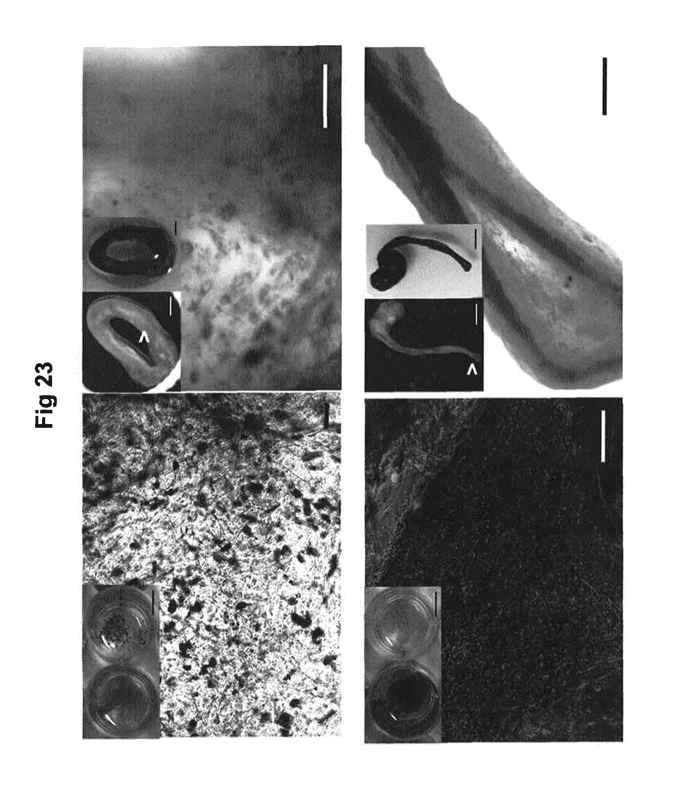
D00018
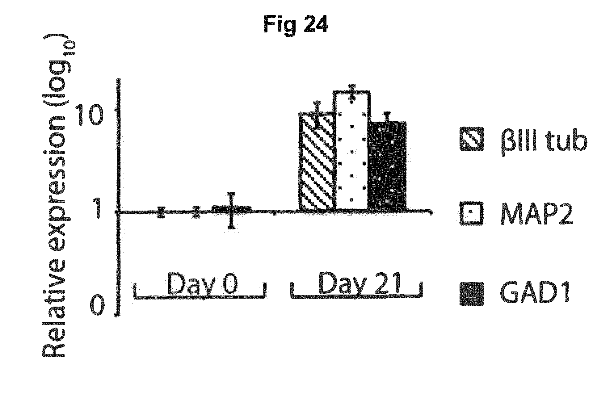
S00001
XML
uspto.report is an independent third-party trademark research tool that is not affiliated, endorsed, or sponsored by the United States Patent and Trademark Office (USPTO) or any other governmental organization. The information provided by uspto.report is based on publicly available data at the time of writing and is intended for informational purposes only.
While we strive to provide accurate and up-to-date information, we do not guarantee the accuracy, completeness, reliability, or suitability of the information displayed on this site. The use of this site is at your own risk. Any reliance you place on such information is therefore strictly at your own risk.
All official trademark data, including owner information, should be verified by visiting the official USPTO website at www.uspto.gov. This site is not intended to replace professional legal advice and should not be used as a substitute for consulting with a legal professional who is knowledgeable about trademark law.