Modified Kappa Light Chain-Binding Polypeptides
Rodrigo; Gustav ; et al.
U.S. patent application number 16/252015 was filed with the patent office on 2019-05-16 for modified kappa light chain-binding polypeptides. The applicant listed for this patent is GE HEALTHCARE BIOPROCESS R&D AB. Invention is credited to Mats Ander, Tomas Bjorkman, Gustav Rodrigo.
| Application Number | 20190144511 16/252015 |
| Document ID | / |
| Family ID | 55080083 |
| Filed Date | 2019-05-16 |


| United States Patent Application | 20190144511 |
| Kind Code | A1 |
| Rodrigo; Gustav ; et al. | May 16, 2019 |
Modified Kappa Light Chain-Binding Polypeptides
Abstract
A kappa light chain-binding polypeptide comprising or consisting essentially of one or more mutated binding domains of Peptostreptococcus Protein L.
| Inventors: | Rodrigo; Gustav; (Uppsala, SE) ; Ander; Mats; (Uppsala, SE) ; Bjorkman; Tomas; (Uppsala, SE) | ||||||||||
| Applicant: |
|
||||||||||
|---|---|---|---|---|---|---|---|---|---|---|---|
| Family ID: | 55080083 | ||||||||||
| Appl. No.: | 16/252015 | ||||||||||
| Filed: | January 18, 2019 |
Related U.S. Patent Documents
| Application Number | Filing Date | Patent Number | ||
|---|---|---|---|---|
| 15526849 | May 15, 2017 | 10208091 | ||
| PCT/EP2015/079387 | Dec 11, 2015 | |||
| 16252015 | ||||
| Current U.S. Class: | 530/322 |
| Current CPC Class: | C07K 2317/569 20130101; C07K 14/195 20130101; C07K 1/22 20130101; C07K 2317/55 20130101; C07K 16/00 20130101; C07K 2319/02 20130101 |
| International Class: | C07K 14/195 20060101 C07K014/195; C07K 16/00 20060101 C07K016/00; C07K 1/22 20060101 C07K001/22 |
Foreign Application Data
| Date | Code | Application Number |
|---|---|---|
| Dec 17, 2014 | SE | 1451563-9 |
| Dec 17, 2014 | SE | 1451564-7 |
Claims
1. A kappa light chain-binding polypeptide comprising a mutated domain 3 of Peptostreptococcus Protein L selected from the group consisting of SEQ ID NO:2, SEQ ID NO:3, SEQ ID NO:4, and SEQ ID NO:5 or at least 90% identity thereto, and wherein at least the amino acid at the position corresponding to position 45 in SEQ ID NOS:2-5 has been mutated to an amino acid which is not asparagine, proline or cysteine; and wherein the polypeptide binds the kappa light chain of an antibody or fragment thereof and has improved alkaline stability compared to a non-mutated domain 3 of Peptostreptococcus Protein L.
2. The polypeptide of claim 1, comprising a multimer of mutated domain 3.
3. The polypeptide of claim 1, wherein the amino acid corresponding to position 45 in SEQ ID NOS:2-5 has been mutated to alanine.
4. The polypeptide of claim 1, wherein the amino acid at the position corresponding to position 10 in SEQ ID NOS:2-5 has been mutated to an amino acid which is not asparagine, proline or cysteine.
5. The polypeptide of claim 1, wherein the amino acid at the position corresponding to position 10 in SEQ ID NOS:2-5 has been mutated to glutamine.
6. The polypeptide of claim 1, wherein the amino acid at the position corresponding to position 60 in SEQ ID NOS:2-5 has been mutated to an amino acid which is not asparagine, proline or cysteine.
7. The polypeptide of claim 1, wherein the amino acid at the position corresponding to position 60 in SEQ ID NOS:2-5 has been mutated to glutamine.
8. The polypeptide of claim 1, wherein the amino acid at the position corresponding to position 19 in SEQ ID NOS:2-5 has been mutated to an amino acid which is not glutamine, asparagine, proline or cysteine.
9. The polypeptide of claim 1, wherein the amino acid at the position corresponding to position 19 in SEQ ID NOS:2-5 has been mutated to glutamic acid or alanine.
10. The polypeptide of claim 1, wherein the amino acid sequence of mutated domain 3 has at least 95% sequence identity to SEQ ID NO:2.
11. The polypeptide of claim 1 wherein the amino acid sequence of mutated domain 3 is selected from the group consisting of sequences defined by SEQ ID NO:7, SEQ ID NO:8, SEQ ID NO:9, SEQ ID NO:10 and SEQ ID NO:11.
12. The polypeptide of claim 1, wherein the amino acid sequence of mutated domain 3 is SEQ ID NO:11.
13. The polypeptide of claim 1, further comprising two, three, four, five, six or seven such mutated domain 3 amino acid sequences.
14. The polypeptide of claim 13, wherein the domains are linked to each other by elements comprising up to five or fifteen amino acid residues.
15. The polypeptide of claim 1, wherein the amino acid sequence of mutated domain 3 is selected from the group consisting of SEQ ID NOS: 15-18.
16. The polypeptide of claim 1, wherein the alkaline stability is improved as measured by the remaining binding capacity for kappa light chain-containing proteins after 96-100 10 min incubation cycles in 0.1 M aqueous NaOH at 22+/-2.degree. C.
17. A nucleic acid or a vector encoding a polypeptide according to claim 1.
18. An expression system, which comprises a nucleic acid or vector according to claim 17.
19. A separation matrix, wherein a plurality of polypeptides of claim 1 have been coupled to a solid support.
20. The separation matrix according to claim 19, wherein the binding capacity of the matrix for kappa light chain-containing proteins after 100 15 min incubation cycles in 0.1 M NaOH at 22+/-2.degree. C. is at least 40% of the binding capacity before the incubation.
Description
CROSS REFERENCES TO RELATED APPLICATIONS
[0001] This application is a continuation of U.S. application Ser. No. 15/526,849 filed on May 15, 2017, which claims the priority benefit of PCT/EP2015/079387 filed on Dec. 11, 2015 and which claims priority benefit of Swedish Application No. 1451563-9 filed Dec. 17, 2014. The entire contents of which are hereby incorporated by reference herein.
SEQUENCE LISTING
[0002] The instant application contains a Sequence Listing which has been submitted electronically in ASCII format and is hereby incorporated by reference in its entirety. Said ASCII copy, created on May 8, 2017, is named 39130038_1.txt and is 21,902 bytes in size.
TECHNICAL FIELD OF THE INVENTION
[0003] The present invention relates to the field of affinity chromatography, and more specifically to polypeptides comprising kappa light chain-binding domains of Protein L, which are useful in affinity chromatography of many types of immunoglobulins and immunoglobulin fragments. The invention also relates to separation matrices containing the polypeptides and to separation methods using such separation matrices.
BACKGROUND OF THE INVENTION
[0004] Immunoglobulins and immunoglobulin fragments represent the most prevalent biopharmaceutical products in either manufacture or development worldwide. The high commercial demand for and hence value of this particular therapeutic market has led to the emphasis being placed on pharmaceutical companies to maximize the productivity of their respective manufacturing processes whilst controlling the associated costs.
[0005] Affinity chromatography, typically on matrices comprising staphylococcal Protein A or variants thereof, is normally used as one of the key steps in the purification of intact immunoglobulin molecules. The highly selective binding of Protein A to the Fc chain of immunoglobulins provides for a generic step with very high clearance of impurities and contaminants.
[0006] For antibody fragments, such as Fab, single-chain variable fragments (scFv), bi-specific T-cell engagers (BiTEs), domain antibodies etc., which lack the Fc chain but have a subclass 1, 3 or 4 kappa light chain, matrices comprising Protein L derived from Peptostreptococcus magnus (B .ANG.kerstrom, L Bjorck: J. Biol. Chem. 264, 19740-19746, 1989; W Kastern et al: J. Biol. Chem. 267, 12820-12825, 1992; B H K Nilson et al: J. Biol. Chem. 267, 2234-2239, 1992 and U.S. Pat. No. 6,822,075) show great promise as a purification platform providing the high selectivity needed. The Protein L disclosed in U.S. Pat. No. 6,822,075 comprises the amino acid sequence SEQ ID NO: 1 plus an additional AVEN sequence at the N-terminus.
TABLE-US-00001 (Protein L) SEQ ID NO: 1 KEETPETPETD SEEEVTIKAN LIFANGSTQT AEFKGTFEKA TSEAYAYADT LKKDNGEYTV DVADKGYTLN IKFAGKEKTPEE PKEEVTIKAN LIYADGKTQT AEFKGTFEEA TAEAYRYADA LKKDNGEYTV DVADKGYTLN IKFAGKEKTPEE PKEEVTIKAN LIYADGKTQT AEFKGTFEEA TAEAYRYADL LAKENGKYTV DVADKGYTLN IKFAGKEKTPEE PKEEVTIKAN LIYADGKTQT AEFKGTFAEA TAEAYRYADL LAKENGKYTA DLEDGGYTIN IRFAGKKVDEKPEE
[0007] Protein L matrices are commercially available as Capto.TM. L from GE Healthcare Bio-Sciences AB, Sweden (Capto L data file 29-0100-08 AC, 2014) and can be used for separation of kappa light chain-containing proteins such as intact antibodies, Fab fragments, scFv fragments, domain antibodies etc. About 75% of the antibodies produced by healthy humans have a kappa light chain and many therapeutic monoclonal antibodies and antibody fragments contain kappa light chains.
[0008] Any bioprocess chromatography application requires comprehensive attention to definite removal of contaminants. Such contaminants can for example be non-eluted molecules adsorbed to the stationary phase or matrix in a chromatographic procedure, such as non-desired biomolecules or microorganisms, including for example proteins, carbohydrates, lipids, bacteria and viruses. The removal of such contaminants from the matrix is usually performed after a first elution of the desired product in order to regenerate the matrix before subsequent use. Such removal usually involves a procedure known as cleaning-in-place (CIP), wherein agents capable of eluting contaminants from the stationary phase are used. One such class of agents often used with chromatography media is alkaline solutions that are passed over the matrix. At present the most extensively used cleaning and sanitizing agent is NaOH, and it is desirable to use it in concentrations ranging from 0.05 up to e.g. 1 M, depending on the degree and nature of contamination. Protein L is however a rather alkali-sensitive protein compared to e.g. Protein A and only tolerates up to about 15 mM NaOH over a large number of cycles. This means that additional, less desirable cleaning solutions, e.g. urea or guanidinium salts, may also have to be used in order to ensure sufficient cleaning.
[0009] An extensive research has earlier been focused on the development of engineered protein A ligands that exhibit an improved capacity to withstand alkaline pH-values. For example, WO2003/080655A1 discloses that Protein A domains with particular asparagine mutations are considerably more alkali stable than the native protein.
[0010] There is thus still a need in this field to obtain a separation matrix containing Protein L-derived ligands having an improved stability towards alkaline cleaning procedures.
SUMMARY OF THE INVENTION
[0011] One aspect of the invention is to provide a polypeptide with improved alkaline stability. This is achieved with a polypeptide as defined in claim 1.
[0012] One advantage is that the alkaline stability is improved over Protein L and the parental polypeptides. A further advantage is that the highly selective binding towards kappa light chain-containing proteins demonstrated for Protein L is retained in the polypeptides of the invention.
[0013] A second aspect of the invention is to provide a nucleic acid or a vector encoding a polypeptide or multimer with improved alkaline stability. This is achieved with a nucleic acid or vector as defined in the claims.
[0014] A third aspect of the invention is to provide an expression system capable of expressing a polypeptide or multimer with improved alkaline stability. This is achieved with an expression system as defined in the claims.
[0015] A fourth aspect of the invention is to provide a separation matrix capable of selectively binding kappa light chain-containing proteins and exhibiting an improved alkaline stability. This is achieved with a separation matrix as defined in the claims.
[0016] A fifth aspect of the invention is to provide an efficient and economical method of isolating a kappa light chain-containing protein. This is achieved with a method as defined in the claims.
[0017] Further suitable embodiments of the invention are described in the dependent claims.
Definitions
[0018] The terms "antibody" and "immunoglobulin" are used interchangeably herein, and are understood to include also fragments of antibodies, fusion proteins comprising antibodies or antibody fragments and conjugates comprising antibodies or antibody fragments.
[0019] The terms a "kappa light chain-binding polypeptide" and "kappa light chain-binding protein" herein mean a polypeptide or protein respectively, capable of binding to a subclass 1, 3 or 4 kappa light chain of an antibody (also called V.kappa.I, V.kappa.III and V.kappa.IV, as in B H K Nilson et al: J. Biol. Chem. 267, 2234-2239, 1992), and include e.g. Protein L, and any variant, fragment or fusion protein thereof that has maintained said binding property.
[0020] The term "kappa light chain-containing protein" is used as a synonym of "immunoglobulin kappa light chain-containing protein" and herein means a protein comprising a subclass 1, 3 or 4 kappa light chain (also called V.sub..kappa.I, V.sub..kappa.III and V.sub..kappa.IV, as in B H K Nilson et al: J. Biol. Chem. 267, 2234-2239, 1992) derived from an antibody and includes any intact antibodies, antibody fragments, fusion proteins, conjugates or recombinant proteins containing a subclass 1, 3 or 4 kappa light chain.
BRIEF DESCRIPTION OF FIGURES
[0021] FIG. 1 shows an alignment of the five kappa light chain-binding domains of Protein L as described in U.S. Pat. No. 6,822,075 and W Kastern et al: J Biol. Chem. 267, 12820-12825, 1992.
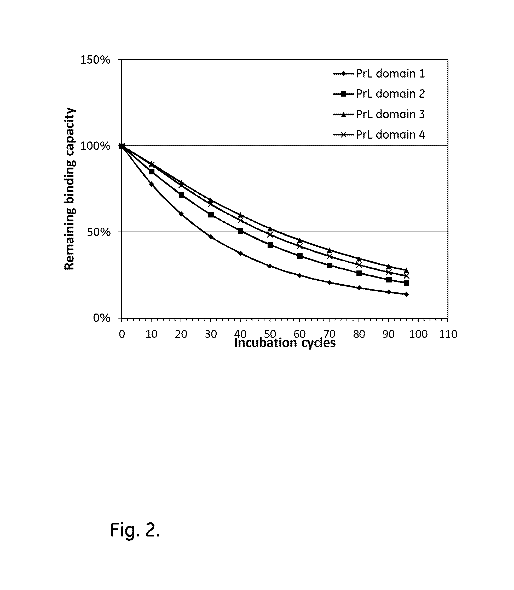
[0022] FIG. 2 shows the alkali stability of different kappa light chain-binding domains of Protein L.
[0023] FIG. 3 shows the alkali stability of mutated kappa light chain-binding domains of Protein L.
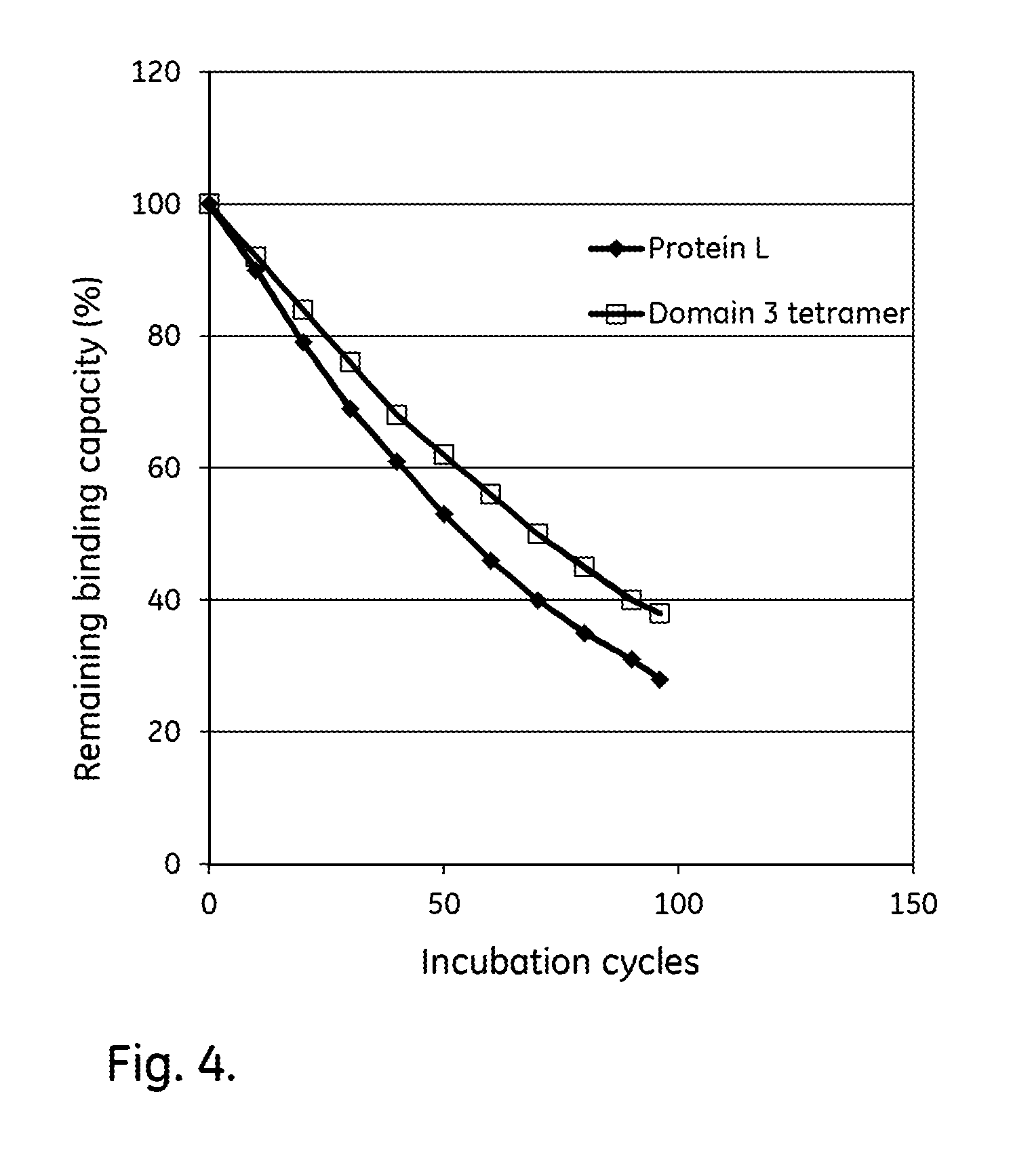
[0024] FIG. 4 shows the alkali stability of Protein L ligands comprising four domains.
[0025] FIG. 5 shows the alkali stability of mutated dimeric, tetrameric and hexameric kappa light chain-binding domains of Protein L in comparison with Protein L.
DETAILED DESCRIPTION OF EMBODIMENTS
[0026] In one aspect the present invention discloses a kappa light chain-binding polypeptide comprising or consisting essentially of one or more binding domains of Peptostreptococcus magnus Protein L, wherein each of these domains is selected from the group consisting of Domain 2, Domain 3 and Domain 4. Domain 2 can have an amino acid sequence defined by SEQ ID NO:3 or SEQ ID NO:12, or it can have at least 90%, such as at least 95%, sequence homology with SEQ ID NO:3 or 12. SEQ ID NO: 12 is a variant of SEQ ID NO: 3, with an alanine in position 31. Domain 3 can have an amino acid sequence defined by SEQ ID NO:4, or it can have at least 90%, such as at least 95%, sequence homology with SEQ ID NO:4. Domain 4 can have an amino acid sequence defined by SEQ ID NO:5, or it can have at least 90%, such as at least 95%, sequence homology with SEQ ID NO:5.
[0027] In some embodiments of the polypeptide, each domain is selected from the group consisting of Domain 3 and Domain 4, or each of the domains is Domain 3. Specifically, the polypeptide may comprise or consist essentially of a multimer of Domain 3.
[0028] In certain embodiments, at least two of the domains are selected from the group consisting of Domain 2, Domain 3 and Domain 4, or from the group consisting of Domain 3 and Domain 4.
[0029] In some embodiments, the polypeptide does not contain any Domain 1 of Peptostreptococcus Protein L. Domain 1 can have an amino acid sequence as defined by SEQ ID NO:2, or it can have at least 90%, such as at least 95% sequence homology with SEQ ID NO:2.
[0030] In certain embodiments of the polypeptide, at least the amino acid at the position corresponding to position 45 in SEQ ID NO:2-5 (e.g. the amino acid at position 45 in SEQ ID NO: 2-5 or 12) in one or more, such as all, of the binding domains has been mutated to an amino acid which is not asparagine, proline or cysteine. The amino acid at position 45 can e.g. be mutated to an alanine.
[0031] In some embodiments of the polypeptide, at least the amino acid at the position corresponding to position 10 in SEQ ID NO:2-5 (e.g. the amino acid at position 10 in SEQ ID NO: 2-5 or 12) in one or more, such as all, of the binding domains has been mutated to an amino acid which is not asparagine, proline or cysteine. The amino acid at position 10 can e.g. be mutated to a glutamine.
[0032] In certain embodiments of the polypeptide, at least the amino acid at the position corresponding to position 60 in SEQ ID NO:2-5 (e.g. the amino acid at position 60 in SEQ ID NO: 2-5 or 12) in one or more, such as all, of the binding domains has been mutated to an amino acid which is not asparagine, proline or cysteine. The amino acid at position 60 can e.g. be mutated to a glutamine.
[0033] Specifically, one or more, such as all, of the binding domains may have mutations selected from the group consisting of N10Q; N45A; N60Q; N10Q,N45A; N45A,N60Q, N10Q,N60Q and N10Q,N45A,N60Q, or alternatively selected from the group consisting of N45A; N10Q,N45A; N45A,N60Q and N10Q,N45A,N60Q.
[0034] In some embodiments of the polypeptide, at least the amino acid at the position corresponding to position 19 in SEQ ID NO:2-5 (e.g. the amino acid at position 19 in SEQ ID NO: 2-5 or 12) in one or more, such as all, of the binding domains has been mutated to an amino acid which is not glutamine, asparagine, proline or cysteine. The amino acid at position 19 can e.g. be mutated to a glutamic acid or an alanine. Specifically, one or more, such as all, of the binding domains may have mutations selected from the group consisting of Q19E and Q19A.
[0035] In certain embodiments of the polypeptide, one or more, such as all, of said binding domains has an amino acid sequence selected from the group consisting of sequences defined by SEQ ID NO:7, SEQ ID NO:8, SEQ ID NO:9, SEQ ID NO:10, SEQ ID NO:11, SEQ ID NO:13 and SEQ ID NO:14. One or more, such as all, of said binding domains can alternatively have an amino acid sequence selected from the group consisting of sequences defined by SEQ ID NO:7, SEQ ID NO:8, SEQ ID NO:9, SEQ ID NO:10 and SEQ ID NO:11. The polypeptide may further at the N-terminus comprise a plurality of amino acid residues originating from the cloning process or constituting a residue from a cleaved off signaling sequence. The number of additional amino acid residues may e.g. be 15 or less, such as 10 or less or 5 or less. As a specific example, the polypeptide may comprise an AQV sequence at the N-terminus.
TABLE-US-00002 (Domain 3, N45A mutation) SEQ ID NO: 7 PKEEVTIKAN LIYADGKTQT AEFKGTFEEA TAEAYRYADL LAKEAGKYTV DVADKGYTLN IKFAGKEKTPEE (Domain 3, N10Q, N45A mutation) SEQ ID NO: 8 PKEEVTIKAQ LIYADGKTQT AEFKGTFEEA TAEAYRYADL LAKEAGKYTV DVADKGYTLN IKFAGKEKTPEE (Domain 3, N45A, N60Q mutation) SEQ ID NO: 9 PKEEVTIKAN LIYADGKTQT AEFKGTFEEA TAEAYRYADL LAKEAGKYTV DVADKGYTLQ IKFAGKEKTPEE (Domain 3, N10Q, N60Q mutation) SEQ ID NO: 10 PKEEVTIKAQ LIYADGKTQT AEFKGTFEEA TAEAYRYADL LAKENGKYTV DVADKGYTLQ IKFAGKEKTPEE (Domain 3, N10Q, N45A ,N60Q mutation) SEQ ID NO: 11 PKEEVTIKAQ LIYADGKTQT AEFKGTFEEA TAEAYRYADL LAKEAGKYTV DVADKGYTLQ IKFAGKEKTPEE (variant of Domain 2) SEQ ID NO: 12 PKEEVTIKAN LIYADGKTQT AEFKGTFEEA AAEAYRYADA LKKDNGEYTV DVADKGYTLN IKFAGKEKTPEE (Domain 3, Q19A mutation) SEQ ID NO: 13 PKEEVTIKAN LIYADGKTAT AEFKGTFEEA TAEAYRYADL LAKENGKYTV DVADKGYTLN IKFAGKEKTPEE (Domain 3, Q19E mutation) SEQ ID NO: 14 PKEEVTIKAN LIYADGKTET AEFKGTFEEA TAEAYRYADL LAKENGKYTV DVADKGYTLN IKFAGKEKTPEE
[0036] In some embodiments, the polypeptide is a multimer comprising, or consisting essentially of, a plurality of mutated or non-mutated domains as defined by any embodiment disclosed above. The multimer can e.g. be a dimer, a trimer, a tetramer, a pentamer or a hexamer. It can be a homomultimer, where all the units in the multimer are identical or it can be a heteromultimer, where at least one unit differs from the others. Advantageously, all the units in the multimer are alkali stable, such as by comprising the mutations disclosed above. The domains can be linked to each other directly by peptide bonds between the C- and N-termini of the domains. Alternatively, two or more units in the multimer can be linked by elements comprising oligomeric or polymeric species, such as elements comprising up to 15 or 30 amino acids, such as 1-5, 1-10 or 5-10 amino acids. The nature of such a link should preferably not destabilize the spatial conformation of the domains. This can e.g. be achieved by avoiding the presence of cysteine in the links. Furthermore, said link should preferably also be sufficiently stable in alkaline environments not to impair the properties of the domains. For this purpose, it is advantageous if the links do not contain asparagine. It can additionally be advantageous if the links do not contain glutamine. The multimer may further at the N-terminus comprise a plurality of amino acid residues originating from the cloning process or constituting a residue from a cleaved off signaling sequence. The number of additional amino acid residues may e.g. be 15 or less, such as 10 or less or 5 or less. As a specific example, the multimer may comprise an AQV sequence at the N-terminus.
[0037] In certain embodiments, the multimer may comprise, or consist essentially, of a sequence selected from the group consisting of: SEQ ID NO: 15, SEQ ID NO: 16, SEQ ID NO: 17 and SEQ ID NO: 18, such as a sequence selected from the group consisting of SEQ ID NO: 16, SEQ ID NO: 17 and SEQ ID NO: 18.
TABLE-US-00003 (Domain 3, tetramer) SEQ ID NO: 15 PKEEVTIKAN LIYADGKTQT AEFKGTFEEA TAEAYRYADL LAKENGKYTV DVADKGYTLN IKFAGKEKTPEE PKEEVTIKAN LIYADGKTQT AEFKGTFEEA TAEAYRYADL LAKENGKYTV DVADKGYTLN IKFAGKEKTPEE PKEEVTIKAN LIYADGKTQT AEFKGTFEEA TAEAYRYADL LAKENGKYTV DVADKGYTLN IKFAGKEKTPEE PKEEVTIKAN LIYADGKTQT AEFKGTFEEA TAEAYRYADL LAKENGKYTV DVADKGYTLN IKFAGKEKTPEE Domain 3(N10Q, N45A, N60Q)2 SEQ ID NO: 16 PKEEVTIKAQ LIYADGKTQT AEFKGTFEEA TAEAYRYADL LAKEAGKYTV DVADKGYTLQ IKFAGKEKTPEE PKEEVTIKAQ LIYADGKTQT AEFKGTFEEA TAEAYRYADL LAKEAGKYTV DVADKGYTLQ IKFAGKEKTPEE Domain 3 (N10Q, N45A, N60Q)4 SEQ ID NO: 17 PKEEVTIKAQ LIYADGKTQT AEFKGTFEEA TAEAYRYADL LAKEAGKYTV DVADKGYTLQ IKFAGKEKTPEE PKEEVTIKAQ LIYADGKTQT AEFKGTFEEA TAEAYRYADL LAKEAGKYTV DVADKGYTLQ IKFAGKEKTPEE PKEEVTIKAQ LIYADGKTQT AEFKGTFEEA TAEAYRYADL LAKEAGKYTV DVADKGYTLQ IKFAGKEKTPEE PKEEVTIKAQ LIYADGKTQT AEFKGTFEEA TAEAYRYADL LAKEAGKYTV DVADKGYTLQ IKFAGKEKTPEE Domain 3 (N10Q, N45A, N60Q)6 SEQ ID NO: 18 PKEEVTIKAQ LIYADGKTQT AEFKGTFEEA TAEAYRYADL LAKEAGKYTV DVADKGYTLQ IKFAGKEKTPEE PKEEVTIKAQ LIYADGKTQT AEFKGTFEEA TAEAYRYADL LAKEAGKYTV DVADKGYTLQ IKFAGKEKTPEE PKEEVTIKAQ LIYADGKTQT AEFKGTFEEA TAEAYRYADL LAKEAGKYTV DVADKGYTLQ IKFAGKEKTPEE PKEEVTIKAQ LIYADGKTQT AEFKGTFEEA TAEAYRYADL LAKEAGKYTV DVADKGYTLQ IKFAGKEKTPEE PKEEVTIKAQ LIYADGKTQT AEFKGTFEEA TAEAYRYADL LAKEAGKYTV DVADKGYTLQ IKFAGKEKTPEE PKEEVTIKAQ LIYADGKTQT AEFKGTFEEA TAEAYRYADL LAKEAGKYTV DVADKGYTLQ IKFAGKEKTPEE
[0038] In some embodiments, the polypeptide and/or multimer, as disclosed above, further comprises at the C-terminus or N-terminus one or more coupling elements, selected from the group consisting of a cysteine residue, a plurality of lysine residues and a plurality of histidine residues. The coupling element may e.g. be a single cysteine at the C-terminus. The coupling element(s) may be directly linked to the C- or N-terminus, or it/they may be linked via a linker comprising up to 15 amino acids, such as 1-5, 1-10 or 5-10 amino acids. This stretch should preferably also be sufficiently stable in alkaline environments not to impair the properties of the mutated protein. For this purpose, it is advantageous if the stretch does not contain asparagine. It can additionally be advantageous if the stretch does not contain glutamine. An advantage of having a C- or N-terminal cysteine is that endpoint coupling of the protein can be achieved through reaction of the cysteine thiol with an electrophilic group on a support. This provides excellent mobility of the coupled protein which is important for the binding capacity.
[0039] The alkali stability of the polypeptide or multimer can be assessed by coupling it to an SPR chip, e.g. to Biacore CM5 sensor chips as described in the examples, and measuring the kappa light chain-binding capacity of the chip, using e.g. a specific kappa light chain-containing protein or polyclonal human IgG (where the majority of the IgG molecules have a kappa light chain), before and after incubation in alkaline solutions at a specified temperature, e.g. 22+/-2.degree. C. The incubation can e.g. be performed in 0.1 M NaOH for a number of 10 min cycles, such as 50, 96 or 100 cycles. The binding capacity of the matrix after 96-100 10 min incubation cycles in 0.1 M NaOH at 22+/-2.degree. C. can be at least 40, such as at least 50, or at least 55% of the binding capacity before the incubation. Alternatively, the remaining binding capacity after 96-100 cycles for a particular mutant measured as above can be compared with the remaining binding capacity for the parental polypeptide/multimer. In this case, the remaining binding capacity for the mutant may be at least 105%, such as at least 110%, at least 125%, at least 150% or at least 200% of the parental polypeptide/multimer.
[0040] The invention also discloses a kappa light chain-binding polypeptide comprising at least one mutated binding domain of Peptostreptococcus Protein L, in which at least one asparagine residue of a parental domain defined by, or having at least 95% or 98% sequence homology with, SEQ ID NO: 2-6 or 12 has been mutated to another amino acid residue which is not asparagine, proline or cysteine. The polypeptide may comprise at least the mutation N45A and/or the mutation N60Q. In specific embodiments, the mutation(s) are selected from the group consisting of N45A; N10Q,N45A; N45A,N60Q, N10Q,N60Q and N10Q,N45A,N60Q, or alternatively selected from the group consisting of N45A; N10Q,N45A; N45A,N60Q and N10Q,N45A,N60Q. The alkali stability relative to a parental polypeptide can be improved and measured as disclosed above.
[0041] In some embodiments the polypeptide comprises or consists essentially of a plurality of mutated binding domains, such as 2, 3, 4, 5 or 6 domains, wherein each domain comprises at least one of the mutations N10Q, N45A and N60Q, such as N45A and/or N60Q. Specifically, the mutation(s) in each domain can be selected from the group consisting of N45A; N10Q,N45A; N45A,N60Q, N10Q,N60Q and N10Q,N45A,N60Q, or alternatively selected from the group consisting of N45A; N10Q,N45A; N45A,N60Q and N10Q,N45A,N60Q. The domains can optionally be linked to each other by elements comprising up to 15 amino acids.
[0042] In a second aspect the present invention discloses a nucleic acid encoding a polypeptide or multimer according to any embodiment disclosed above. Thus, the invention encompasses all forms of the present nucleic acid sequence such as the RNA and the DNA encoding the polypeptide or multimer. The invention embraces a vector, such as a plasmid, which in addition to the coding sequence comprises the required signal sequences for expression of the polypeptide or multimer according the invention. In one embodiment, the vector comprises nucleic acid encoding a multimer according to the invention, wherein the separate nucleic acids encoding each unit may have homologous or heterologous DNA sequences.
[0043] In a third aspect the present invention discloses an expression system, which comprises, a nucleic acid or a vector as disclosed above. The expression system may e.g. be a gram-positive or gram-negative prokaryotic host cell system, e.g. Bacillus sp. or Escherichia coli which has been modified to express the present polypeptide or multimer. In an alternative embodiment, the expression system is a eukaryotic host cell system, such as a yeast, e.g. Pichea pastoris or Saccharomyces cerevisiae.
[0044] In a fourth aspect, the present invention discloses a separation matrix, wherein a plurality of polypeptides or multimers according to any embodiment disclosed above have been coupled to a solid support. Such a matrix is useful for separation of kappa light chain-containing proteins and, due to the improved alkali stability of the polypeptides/multimers, the matrix will withstand highly alkaline conditions during cleaning, which is essential for long-term repeated use in a bioprocess separation setting. The alkali stability of the matrix can be assessed by measuring the kappa light chain-binding capacity, using e.g. a specific kappa light chain-containing protein or polyclonal human IgG, before and after incubation in alkaline solutions at a specified temperature, e.g. 22+/-2.degree. C. The incubation can e.g. be performed in 0.1 M NaOH for a number of 15 min cycles, such as 100, 200 or 300 cycles. The binding capacity of the matrix after 100 15 min incubation cycles in 0.1 M NaOH at 22+/-2.degree. C. can be at least 80, such as at least 85, at least 90 or at least 95% of the binding capacity before the incubation. Alternatively, the incubation can be performed in 0.1 M NaOH for a number of 4 h cycles, such as 6 cycles giving a total incubation time of 24 h. The binding capacity of the matrix after 24 h min total incubation time in 0.1 M NaOH at 22+/-2.degree. C. can be at least 80, such as at least 85, at least 90 or at least 95% of the binding capacity before the incubation.
[0045] As the skilled person will understand, the expressed polypeptide or multimer should be purified to an appropriate extent before been immobilized to a support. Such purification methods are well known in the field, and the immobilization of protein-based ligands to supports is easily carried out using standard methods. Suitable methods and supports will be discussed below in more detail.
[0046] The solid support of the matrix according to the invention can be of any suitable well-known kind. A conventional affinity separation matrix is often of organic nature and based on polymers that expose a hydrophilic surface to the aqueous media used, i.e. expose hydroxy (--OH), carboxy (--COOH), carboxamido (--CONH.sub.2, possibly in N-- substituted forms), amino (--NH.sub.2, possibly in substituted form), oligo- or polyethylenoxy groups on their external and, if present, also on internal surfaces. The solid support can suitably be porous. The porosity can be expressed as a Kav or Kd value (the fraction of the pore volume available to a probe molecule of a particular size) measured by inverse size exclusion chromatography, e.g. according to the methods described in Gel Filtration Principles and Methods, Pharmacia LKB Biotechnology 1991, pp 6-13. By definition, both Kd and Kay values always lie within the range 0-1. The Kay value can advantageously be 0.6-0.95, e.g. 0.7-0.90 or 0.6-0.8, as measured with dextran of Mw 110 kDa as a probe molecule. An advantage of this is that the support has a large fraction of pores able to accommodate both the polypeptides/multimers of the invention and immunoglobulins binding to the polypeptides/multimers and to provide mass transport of the immunoglobulins to and from the binding sites.
[0047] The polypeptides or multimers may be attached to the support via conventional coupling techniques utilising e.g. thiol, amino and/or carboxy groups present in the ligand. Bisepoxides, epichlorohydrin, CNBr, N-hydroxysuccinimide (NHS) etc. are well-known coupling reagents. Between the support and the polypeptide/multimer, a molecule known as a spacer can be introduced, which improves the availability of the polypeptide/multimer and facilitates the chemical coupling of the polypeptide/multimer to the support. Suitable spacers can be introduced e.g. by activation of the support with epichlorohydrin, butanediol diepoxide, allyl glycidyl ether etc. Alternatively, the polypeptide/multimer may be attached to the support by non-covalent bonding, such as physical adsorption or biospecific adsorption.
[0048] In some embodiments the matrix comprises 5-20, such as 5-15 mg/ml, 5-11 mg/ml or 8-11 mg/ml of the polypeptide or multimer coupled to the support. The amount of coupled polypeptide/multimer can be controlled by the concentration of polypeptide/multimer used in the coupling process, by the coupling conditions used and/or by the pore structure of the support used. As a general rule the absolute binding capacity of the matrix increases with the amount of coupled polypeptide/multimer, at least up to a point where the pores become significantly constricted by the coupled polypeptide/multimer. The relative binding capacity per mg coupled polypeptide/multimer will decrease at high coupling levels, resulting in a cost-benefit optimum within the ranges specified above.
[0049] In some embodiments the polypeptides are coupled to the support via multipoint attachment. This can suitably be done by using such coupling conditions that a plurality of reactive groups in the polypeptide react with reactive groups in the support. Typically, multipoint attachment can involve the reaction of several intrinsic reactive groups of amino acid residues in the sequence, such as amines in lysines, with the reactive groups on the support, such as epoxides, cyanate esters (e.g. from CNBr activation), succinimidyl esters (e.g. from NHS activation) etc. It is however also possible to deliberately introduce reactive groups at different positions in the polypeptides to affect the coupling characteristics. In order to provide multipoint coupling via lysines, the coupling reaction is suitably carried out at a pH where a significant fraction of the lysine primary amines are in the non-protonated nucleophilic state, e.g. at pH higher than 8.0, such as above 10.
[0050] In certain embodiments the polypeptides or multimers are coupled to the support via thioether bonds. Methods for performing such coupling are well-known in this field and easily performed by the skilled person in this field using standard techniques and equipment. Thioether bonds are flexible and stable and generally suited for use in affinity chromatography. In particular when the thioether bond is via a terminal or near-terminal cysteine residue on the polypeptide or multimer, the mobility of the coupled polypeptide/multimer is enhanced which provides improved binding capacity and binding kinetics. In some embodiments the polypeptide/multimer is coupled via a C-terminal cysteine provided on the protein as described above. This allows for efficient coupling of the cysteine thiol to electrophilic groups, e.g. epoxide groups, halohydrin groups etc. on a support, resulting in a thioether bridge coupling. The polypeptide/multimer can e.g. be coupled via single-point attachment, e.g. via a single cysteine or by directed multipoint attachment, using e.g. a plurality of lysines or other coupling groups near a terminus of the polypeptide/multimer.
[0051] In certain embodiments the support comprises a polyhydroxy polymer, such as a polysaccharide. Examples of polysaccharides include e.g. dextran, starch, cellulose, pullulan, agar, agarose etc. Polysaccharides are inherently hydrophilic with low degrees of nonspecific interactions, they provide a high content of reactive (activatable) hydroxyl groups and they are generally stable towards alkaline cleaning solutions used in bioprocessing.
[0052] In some embodiments the support comprises agar or agarose. The supports used in the present invention can easily be prepared according to standard methods, such as inverse suspension gelation (S Hjerten: Biochim Biophys Acta 79(2), 393-398 (1964). Alternatively, the base matrices are commercially available products, such as crosslinked agarose beads sold under the name of SEPHAROSE.TM. FF (GE Healthcare). In an embodiment, which is especially advantageous for large-scale separations, the support has been adapted to increase its rigidity using the methods described in U.S. Pat. Nos. 6,602,990 or 7,396,467, which are hereby incorporated by reference in their entirety, and hence renders the matrix more suitable for high flow rates.
[0053] In certain embodiments the support, such as a polysaccharide or agarose support, is crosslinked, such as with hydroxyalkyl ether crosslinks. Crosslinker reagents producing such crosslinks can be e.g. epihalohydrins like epichlorohydrin, diepoxides like butanediol diglycidyl ether, allylating reagents like allyl halides or allyl glycidyl ether. Crosslinking is beneficial for the rigidity of the support and improves the chemical stability. Hydroxyalkyl ether crosslinks are alkali stable and do not cause significant nonspecific adsorption.
[0054] Alternatively, the solid support is based on synthetic polymers, such as polyvinyl alcohol, polyhydroxyalkyl acrylates, polyhydroxyalkyl methacrylates, polyacrylamides, polymethacrylamides etc. In case of hydrophobic polymers, such as matrices based on divinyl and monovinyl-substituted benzenes, the surface of the matrix is often hydrophilised to expose hydrophilic groups as defined above to a surrounding aqueous liquid. Such polymers are easily produced according to standard methods, see e.g. "Styrene based polymer supports developed by suspension polymerization" (R Arshady: Chimica e L'Industria 70(9), 70-75 (1988)). Alternatively, a commercially available product, such as SOURCE.TM. (GE Healthcare) is used. In another alternative, the solid support according to the invention comprises a support of inorganic nature, e.g. silica, zirconium oxide etc.
[0055] In yet another embodiment, the solid support is in another form such as a surface, a chip, capillaries, or a filter (e.g. a membrane or a depth filter matrix).
[0056] As regards the shape of the matrix according to the invention, in one embodiment the matrix is in the form of a porous monolith. In an alternative embodiment, the matrix is in beaded or particle form that can be porous or non-porous. Matrices in beaded or particle form can be used as a packed bed or in a suspended form. Suspended forms include those known as expanded beds and pure suspensions, in which the particles or beads are free to move. In case of monoliths, packed bed and expanded beds, the separation procedure commonly follows conventional chromatography with a concentration gradient. In case of pure suspension, batch-wise mode will be used.
[0057] In a sixth aspect, the present invention discloses a method of isolating a kappa light chain-containing protein, wherein a separation matrix as disclosed above is used.
[0058] In certain embodiments, the method comprises the steps of: [0059] a) contacting a liquid sample comprising a kappa light chain-containing protein with a separation matrix as disclosed above, [0060] b) washing said separation matrix with a washing liquid, [0061] c) eluting the kappa light chain-containing protein from the separation matrix with an elution liquid, and [0062] d) cleaning the separation matrix with a cleaning liquid.
[0063] The method may also comprise steps of, before step a), providing an affinity separation matrix according to any of the embodiments described above and providing a solution comprising a kappa light chain-containing protein and at least one other substance as a liquid sample and of, after step c), recovering the eluate and optionally subjecting the eluate to further separation steps, e.g. by anion or cation exchange chromatography, multimodal chromatography and/or hydrophobic interaction chromatography. Suitable compositions of the liquid sample, the washing liquid and the elution liquid, as well as the general conditions for performing the separation are well known in the art of affinity chromatography and in particular in the art of Protein L chromatography. The liquid sample comprising a kappa light chain-containing protein and at least one other substance may comprise host cell proteins (HCP), such as chinese hamster ovary (CHO) cell, E. coli or yeast cell proteins. Contents of CHO cell and E. coli proteins can conveniently be determined by immunoassays directed towards these proteins, e.g. the CHO HCP or E. coli HCP ELISA kits from Cygnus Technologies. The host cell proteins or CHO cell/E. coli/yeast proteins may be desorbed during step b).
[0064] The elution may be performed by using any suitable solution used for elution from Protein L media. This can e.g. be a solution or buffer with pH 4 or lower, such as pH 2.5-4 or 2.8-3.5. In some embodiments the elution buffer or the elution buffer gradient comprises at least one mono- di- or trifunctional carboxylic acid or salt of such a carboxylic acid. In certain embodiments the elution buffer or the elution buffer gradient comprises at least one anion species selected from the group consisting of acetate, citrate, glycine, succinate, phosphate, and formiate.
[0065] In some embodiments, the cleaning liquid is alkaline, such as with a pH of 12-14. Such solutions provide efficient cleaning of the matrix, in particular at the upper end of the interval.
[0066] In certain embodiments, the cleaning liquid comprises 0.01-1.0 M NaOH or KOH, such as 0.05-1.0 or 0.05-0.1 M NaOH or KOH. The high stability of the polypeptides of the invention enables the use of such comparatively strong alkaline solutions.
[0067] In some embodiments, steps a)-d) are repeated at least 10 times, such as at least 50 times or 50-200 times. This is important for the process economy in that the matrix can be re-used many times.
EXAMPLES
Mutagenesis of Protein
[0068] Monomer constructs were designed from a Protein L disclosed in U.S. Pat. No. 6,822,075 (SEQ ID NO: 1), containing four kappa light chain-binding domains. These are numbered 1, 2, 3 and 4, starting from the N-terminus (FIG. 1). The DNA fragments were purchased from a DNA synthesizing company (DNA2.0). Four monomer constructs were prepared in a pJexpress201 cloning vector, each with an N-terminal cysteine. For an overview of constructs, see SEQ ID NO: 2,4,5,12.
[0069] Constructs were subcloned to expression vector pGO, containing E. coli GAP promoter and OmpA signal peptide sequence for periplasmatic localization of the target protein. The sequence encoding the four domains was prepared by amplifying with oligonucleotides containing the restriction enzyme recognition sites for FspI and PstI on the 5' side and 3' side, respectively. The prepared DNA fragment encoding each domain was digested with FspI and PstI (New England Biolabs). Separately, expression vector was prepared with digestion with FspI and PstI and purified by agarose gel electrophoresis and recovered. Both were mixed and ligated with Quick ligation kit (New England Biolabs). The ligated plasmid expressing each domain was transformed into a chemical competent E. coli K12 strain with a heat shock method.
[0070] Further mutations of amino acids N10, N45, Q19, and N60 in domain 3 were prepared in expression vector pJexpress401 (DNA2.0) containing T5 promoter under a lac operon control mechanism (SEQ ID NO: 7-11, 13-14). Constructs were designed with and without OmpA signal peptide but without a C-terminal cysteine.
[0071] Tetramers of domain 3, dimer, tetramer and hexamer of domain 3 with N45, N10 and N60 mutations were also prepared in pJexpress401, with and without C-terminal cysteine (SEQ ID NO: 15-18).
Construct Expression and Purification
[0072] The E. coli K12 recombinant cells were cultured in shake flasks with LB-broth (10 g peptone, 5 g yeast extract, 5 g NaCl) supplemented with 25 mg/l kanamycin at 37.degree. C. until optical density at 600 nm reached 0.8. At this point protein expression was induced with Isopropyl .beta.-D-1-thiogalactopyranoside (VWR International) with final concentration of 1 mM. Upon induction the temperature was lowered to 30.degree. C. and the cultures were incubated for 5 hours. The cultivation was stopped and cells were centrifuged for 15 minutes at 4000.times.g and the supernatant was discarded. Cells were resuspended in 1/10 of culturing volume with phosphate buffered saline (PBS) and sonicated using pulse-sonication with an active time of 2 minutes. The sonicated samples were clarified from cell debris by centrifugation at 6000.times.g for 30 minutes, followed by microfiltration with a membrane having a 0.2 .mu.m pore size.
[0073] The purified ligands were analyzed with LC-MS to determine the purity and to ascertain that the molecular weight corresponded to the expected (based on the amino acid sequence).
Example 1
[0074] The purified monomeric ligands listed in Table 1, further comprising, in the cases of the non-mutated single domains, a cysteine at the C terminus and an AQV sequence at the N-terminus, were immobilized on Biacore CM5 sensor chips (GE Healthcare, Sweden), using the amine coupling kit of GE Healthcare (for carbodiimide coupling of amines on the carboxymethyl groups on the chip) in an amount sufficient to give a signal strength of about 1000RU in a Biacore instrument (GE Healthcare, Sweden). To follow the IgG binding capacity of the immobilized surface 1 mg/ml human polyclonal IgG (Gammanorm) was flowed over the chip and the signal strength was noted. The surface was then cleaned-in-place (CIP), i.e. flushed with 100 mM NaOH for 10 minutes at room temperature (22+/-2.degree. C.). This was repeated for 96 cycles and the immobilized ligand alkaline stability was followed as the relative loss of IgG binding capacity (signal strength) after each cycle. The results for the non-mutated domains are shown in FIG. 2 and indicate that Domain 1 has a distinctly lower alkali stability than the other domains and that Domain 3 has the highest alkali stability. Results for single-domain asparagine mutants of Domain 3 are shown in FIG. 3 and show an improved alkali stability for all the mutants in comparison with the parental Domain 3, which was used as a reference in parallel with the mutations.
TABLE-US-00004 TABLE 1 Retained capacity Ref. after 96 capacity Sample/ref. Ligand Sequence cycles (%) (%) ratio Domain 1 (D1) SEQ ID NO: 2 13 31 0.42 Domain 2 (D2) SEQ ID NO: 12 22 31 0.71 Domain 3 (D3) SEQ ID NO: 4 31 31 1.00 Domain 4 (D4) SEQ ID NO: 5 26 31 0.84 D3(N45A)1 SEQ ID NO: 7 44 31 1.42 D3(N10Q, N45A)1 SEQ ID NO: 8 48 31 1.55 D3(N45A, N60Q)1 SEQ ID NO: 9 59 31 1.90 D3(N10Q, N45A, N60Q)1 SEQ ID NO: 11 59 31 1.90 Domain 3 (D3) SEQ ID NO: 4 28 28 1.00 D3(Q19A)1 SEQ ID NO: 13 28 28 1.00 D3(Q19E)1 SEQ ID NO: 14 31 28 1.11
Example 2
[0075] The purified multidomain ligands listed in Table 2 were immobilized on Biacore CM5 sensor chips (GE Healthcare, Sweden), using the amine coupling kit of GE Healthcare (for carbodiimide coupling of amines on the carboxymethyl groups on the chip) in an amount sufficient to give a signal strength of about 1000RU in a Biacore instrument (GE Healthcare, Sweden). The Protein L had an additional AIHNRA sequence at the N-terminus. To follow the IgG binding capacity of the immobilized surface 1 mg/ml human polyclonal IgG (Gammanorm) was flowed over the chip and the signal strength was noted. The surface was then cleaned-in-place (CIP), i.e. flushed with 100 mM NaOH for 10 minutes at room temperature (22+/-2.degree. C.). This was repeated for 96 cycles and the immobilized ligand alkaline stability was followed as the relative loss of IgG binding capacity (signal strength) after each cycle. The results are shown in Table 2 and FIG. 4 and show that the tetrameric Domain 3 has an improved alkali stability in comparison with Protein L which was run in parallel as a reference.
TABLE-US-00005 TABLE 2 Retained capacity Ref. after 96 capacity Sample/ref. Ligand Sequence cycles (%) (%) ratio Protein L SEQ ID NO: 1 28 28 1.00 Domain 3 SEQ ID NO: 15 38 28 1.36 tetramer
Example 3
[0076] The purified multidomain ligands listed in Table 3 were immobilized on Biacore CM5 sensor chips and evaluated by the methods used in Example 2. -cys at the end of the ligand designation indicates that the ligand has a C-terminal cysteine in addition to the sequence defined by SEQ ID NO: 16-18. The results are shown in Table 3 and FIG. 5 and show that all the mutated Domain 3 dimers, tetramers and hexamers have an improved alkali stability in comparison with Protein L which was run in parallel as a reference.
TABLE-US-00006 TABLE 3 Retained capacity Ref. after 100 capacity Sample/ref. Ligand Sequence cycles (%) (%) ratio Protein L SEQ ID NO: 1 23 23 1.00 D3(N10Q, N45A, N60Q)2 SEQ ID NO: 16 59 23 2.56 D3(N10Q, N45A, N60Q)2-cys SEQ ID NO: 16 59 23 2.56 D3(N10Q, N45A, N60Q)4 SEQ ID NO: 17 60 23 2.61 D3(N10Q, N45A, N60Q)4-cys SEQ ID NO: 17 54 23 2.35 D3(N10Q, N45A, N60Q)6 SEQ ID NO: 18 58 23 2.52 D3(N10Q, N45A, N60Q)6-cys SEQ ID NO: 18 56 23 2.43
Example 4
[0077] The purified di-, tetra- and hexameric ligands of Table 4 were immobilized on agarose beads using the methods described below and assessed for capacity. The results are shown in Table 4.
TABLE-US-00007 TABLE 4 Ligand QB10 Fab QB10 Dab Ligand Sequence content mg/ml mg/ml D3(N10Q, N45A, N60Q)2 SEQ ID NO: 16 9.5 mg/ml 19.3 15.4 D3(N10Q, N45A, N60Q)4 SEQ ID NO: 17 8.8 mg/ml 19.3 15.9 D3(N10Q, N45A, N60Q)6 SEQ ID NO: 18 11.0 mg/ml 21.1 16.7
Activation
[0078] The base matrix used was rigid cross-linked agarose beads of 85 micrometers (volume-weighted) median diameter, prepared according to the methods of U.S. Pat. No. 6,602,990 and with a pore size corresponding to an inverse gel filtration chromatography Kay value of 0.70 for dextran of Mw 110 kDa, according to the methods described in Gel Filtration Principles and Methods, Pharmacia LKB Biotechnology 1991, pp 6-13.
[0079] 25 mL (g) of drained base matrix, 10.0 mL distilled water and 2.02 g NaOH (s) was mixed in a 100 mL flask with mechanical stirring for 10 min at 25.degree. C. 4.0 mL of epichlorohydrin was added and the reaction progressed for 2 hours. The activated gel was washed with 10 gel sediment volumes (GV) of water.
Coupling
[0080] The activated gel was washed with 5 GV 0.2 M phosphate/1 mM EDTA pH 11.5 (coupling buffer). 15 ml gel+13 mg ligand/ml gel (5.0 ml)+5.5 ml coupling buffer+4.7 g sodium sulfate were mixed in a 50 ml flask and stirred at 30.degree. C. for 18.5 h. The pH was measured as 10.8.
[0081] After immobilization the gels were washed 3.times.GV with distilled water and then 5.times.GV with 0.1 M phosphate/1 mM EDTA pH 8.5. The gels+1 GV {0.1 M phosphate/1 mM EDTA/7.5% thioglycerol pH 8.5} was mixed and the flask was stirred at 45.degree. C. for 2h 20 min. The gel was then washed alternately with 1.times.GV 0.1 M HAc and 1.times.GV {0.1 M TRIS/0.15 M NaCl pH 8.5} and and then 6.times.GV with distilled water. Gel samples were sent to an external laboratory for amino acid analysis and the ligand content (mg/ml gel) was calculated from the total amino acid content. The coupling protocol used provides multipoint coupling, with several lysines of each domain attached to the gel.
[0082] 2 ml of resin was packed in TRICORN.TM. 5 100 columns.
Protein
[0083] a) Purified Fab prepared from a papain-digested IgG mAb, diluted to 1 mg/ml in Equilibration buffer. [0084] b) Purified Dab prepared from heat-treated E. coli supernatant, diluted to 1 mg/ml in Equilibration buffer. The Dab contained solely a kappa light chain, without any antigen-binding site.
Equilibration Buffer
[0085] APB Phosphate buffer 20 mM+0.15 M NaCl, pH 7.4 (Medicago)
Adsorption Buffer
[0086] APB Phosphate buffer 20 mM+0.15 M NaCl, pH 7.4 (Medicago).
Elution Buffer
[0087] 25 mM citrate pH 2.5
[0088] The breakthrough capacity was determined with an AKTAExplorer 10 system at a residence time of 4 minutes. Equilibration buffer was run through the bypass column until a stable baseline was obtained. This was done prior to auto zeroing. Sample was applied to the column until a 100% UV signal was obtained. Then, equilibration buffer was applied again until a stable baseline was obtained.
[0089] Sample was loaded onto the column until a UV signal of 85% of maximum absorbance was reached. The column was then washed with equilibration buffer and eluted at pH 2.5 at a flow rate of 0.5 ml/min.
[0090] For calculation of breakthrough capacity at 10%, the equation below was used. That is the amount of Fab/Dab that is loaded onto the column until the concentration of Fab/Dab in the column effluent is 10% of the Fab/Dab concentration in the feed.
q 10 % = C 0 V C [ V app - V sys - .intg. V sys V app A ( V ) - A sub A 100 % - A sub * dv ] ##EQU00001##
[0091] A.sub.100%=100% UV signal;
[0092] A.sub.sub=absorbance contribution from non-binding proteins;
[0093] A(V)=absorbance at a given applied volume;
[0094] V.sub.c=column volume;
[0095] V.sub.app=volume applied until 10% breakthrough;
[0096] V.sub.sys=system dead volume;
[0097] C.sub.0=feed concentration.
[0098] The dynamic binding capacity (DBC) at 10% breakthrough was calculated and the appearance of the curve was studied. The curve was also studied regarding binding, elution and CIP peak. The dynamic binding capacity (DBC) was calculated for 10 and 80% breakthrough.
[0099] This written description uses examples to disclose the invention, including the best mode, and also to enable any person skilled in the art to practice the invention, including making and using any devices or systems and performing any incorporated methods. The patentable scope of the invention is defined by the claims, and may include other examples that occur to those skilled in the art. Such other examples are intended to be within the scope of the claims if they have structural elements that do not differ from the literal language of the claims, or if they include equivalent structural elements with insubstantial differences from the literal languages of the claims. All patents and patent applications mentioned in the text are hereby incorporated by reference in their entireties, as if they were individually incorporated.
Sequence CWU 1
1
181301PRTPeptostreptococcus magnus 1Lys Glu Glu Thr Pro Glu Thr Pro
Glu Thr Asp Ser Glu Glu Glu Val1 5 10 15Thr Ile Lys Ala Asn Leu Ile
Phe Ala Asn Gly Ser Thr Gln Thr Ala 20 25 30Glu Phe Lys Gly Thr Phe
Glu Lys Ala Thr Ser Glu Ala Tyr Ala Tyr 35 40 45Ala Asp Thr Leu Lys
Lys Asp Asn Gly Glu Tyr Thr Val Asp Val Ala 50 55 60Asp Lys Gly Tyr
Thr Leu Asn Ile Lys Phe Ala Gly Lys Glu Lys Thr65 70 75 80Pro Glu
Glu Pro Lys Glu Glu Val Thr Ile Lys Ala Asn Leu Ile Tyr 85 90 95Ala
Asp Gly Lys Thr Gln Thr Ala Glu Phe Lys Gly Thr Phe Glu Glu 100 105
110Ala Thr Ala Glu Ala Tyr Arg Tyr Ala Asp Ala Leu Lys Lys Asp Asn
115 120 125Gly Glu Tyr Thr Val Asp Val Ala Asp Lys Gly Tyr Thr Leu
Asn Ile 130 135 140Lys Phe Ala Gly Lys Glu Lys Thr Pro Glu Glu Pro
Lys Glu Glu Val145 150 155 160Thr Ile Lys Ala Asn Leu Ile Tyr Ala
Asp Gly Lys Thr Gln Thr Ala 165 170 175Glu Phe Lys Gly Thr Phe Glu
Glu Ala Thr Ala Glu Ala Tyr Arg Tyr 180 185 190Ala Asp Leu Leu Ala
Lys Glu Asn Gly Lys Tyr Thr Val Asp Val Ala 195 200 205Asp Lys Gly
Tyr Thr Leu Asn Ile Lys Phe Ala Gly Lys Glu Lys Thr 210 215 220Pro
Glu Glu Pro Lys Glu Glu Val Thr Ile Lys Ala Asn Leu Ile Tyr225 230
235 240Ala Asp Gly Lys Thr Gln Thr Ala Glu Phe Lys Gly Thr Phe Ala
Glu 245 250 255Ala Thr Ala Glu Ala Tyr Arg Tyr Ala Asp Leu Leu Ala
Lys Glu Asn 260 265 270Gly Lys Tyr Thr Ala Asp Leu Glu Asp Gly Gly
Tyr Thr Ile Asn Ile 275 280 285Arg Phe Ala Gly Lys Lys Val Asp Glu
Lys Pro Glu Glu 290 295 300272PRTPeptostreptococcus magnus 2Ser Glu
Glu Glu Val Thr Ile Lys Ala Asn Leu Ile Phe Ala Asn Gly1 5 10 15Ser
Thr Gln Thr Ala Glu Phe Lys Gly Thr Phe Glu Lys Ala Thr Ser 20 25
30Glu Ala Tyr Ala Tyr Ala Asp Thr Leu Lys Lys Asp Asn Gly Glu Tyr
35 40 45Thr Val Asp Val Ala Asp Lys Gly Tyr Thr Leu Asn Ile Lys Phe
Ala 50 55 60Gly Lys Glu Lys Thr Pro Glu Glu65
70372PRTPeptostreptococcus magnus 3Pro Lys Glu Glu Val Thr Ile Lys
Ala Asn Leu Ile Tyr Ala Asp Gly1 5 10 15Lys Thr Gln Thr Ala Glu Phe
Lys Gly Thr Phe Glu Glu Ala Thr Ala 20 25 30Glu Ala Tyr Arg Tyr Ala
Asp Ala Leu Lys Lys Asp Asn Gly Glu Tyr 35 40 45Thr Val Asp Val Ala
Asp Lys Gly Tyr Thr Leu Asn Ile Lys Phe Ala 50 55 60Gly Lys Glu Lys
Thr Pro Glu Glu65 70472PRTPeptostreptococcus magnus 4Pro Lys Glu
Glu Val Thr Ile Lys Ala Asn Leu Ile Tyr Ala Asp Gly1 5 10 15Lys Thr
Gln Thr Ala Glu Phe Lys Gly Thr Phe Glu Glu Ala Thr Ala 20 25 30Glu
Ala Tyr Arg Tyr Ala Asp Leu Leu Ala Lys Glu Asn Gly Lys Tyr 35 40
45Thr Val Asp Val Ala Asp Lys Gly Tyr Thr Leu Asn Ile Lys Phe Ala
50 55 60Gly Lys Glu Lys Thr Pro Glu Glu65
70574PRTPeptostreptococcus magnus 5Pro Lys Glu Glu Val Thr Ile Lys
Ala Asn Leu Ile Tyr Ala Asp Gly1 5 10 15Lys Thr Gln Thr Ala Glu Phe
Lys Gly Thr Phe Ala Glu Ala Thr Ala 20 25 30Glu Ala Tyr Arg Tyr Ala
Asp Leu Leu Ala Lys Glu Asn Gly Lys Tyr 35 40 45Thr Ala Asp Leu Glu
Asp Gly Gly Tyr Thr Ile Asn Ile Arg Phe Ala 50 55 60Gly Lys Lys Val
Asp Glu Lys Pro Glu Glu65 70665PRTPeptostreptococcus magnus 6Glu
Lys Glu Gln Val Thr Ile Lys Glu Asn Ile Tyr Phe Glu Asp Gly1 5 10
15Thr Val Gln Thr Ala Thr Phe Lys Gly Thr Phe Ala Glu Ala Thr Ala
20 25 30Glu Ala Tyr Arg Tyr Ala Asp Leu Leu Ser Lys Glu His Gly Lys
Tyr 35 40 45Thr Ala Asp Leu Glu Asp Gly Gly Tyr Thr Ile Asn Ile Arg
Phe Ala 50 55 60Gly65772PRTEscherichia coli 7Pro Lys Glu Glu Val
Thr Ile Lys Ala Asn Leu Ile Tyr Ala Asp Gly1 5 10 15Lys Thr Gln Thr
Ala Glu Phe Lys Gly Thr Phe Glu Glu Ala Thr Ala 20 25 30Glu Ala Tyr
Arg Tyr Ala Asp Leu Leu Ala Lys Glu Ala Gly Lys Tyr 35 40 45Thr Val
Asp Val Ala Asp Lys Gly Tyr Thr Leu Asn Ile Lys Phe Ala 50 55 60Gly
Lys Glu Lys Thr Pro Glu Glu65 70872PRTEscherichia coli 8Pro Lys Glu
Glu Val Thr Ile Lys Ala Gln Leu Ile Tyr Ala Asp Gly1 5 10 15Lys Thr
Gln Thr Ala Glu Phe Lys Gly Thr Phe Glu Glu Ala Thr Ala 20 25 30Glu
Ala Tyr Arg Tyr Ala Asp Leu Leu Ala Lys Glu Ala Gly Lys Tyr 35 40
45Thr Val Asp Val Ala Asp Lys Gly Tyr Thr Leu Asn Ile Lys Phe Ala
50 55 60Gly Lys Glu Lys Thr Pro Glu Glu65 70972PRTEscherichia coli
9Pro Lys Glu Glu Val Thr Ile Lys Ala Asn Leu Ile Tyr Ala Asp Gly1 5
10 15Lys Thr Gln Thr Ala Glu Phe Lys Gly Thr Phe Glu Glu Ala Thr
Ala 20 25 30Glu Ala Tyr Arg Tyr Ala Asp Leu Leu Ala Lys Glu Ala Gly
Lys Tyr 35 40 45Thr Val Asp Val Ala Asp Lys Gly Tyr Thr Leu Gln Ile
Lys Phe Ala 50 55 60Gly Lys Glu Lys Thr Pro Glu Glu65
701072PRTEscherichia coli 10Pro Lys Glu Glu Val Thr Ile Lys Ala Gln
Leu Ile Tyr Ala Asp Gly1 5 10 15Lys Thr Gln Thr Ala Glu Phe Lys Gly
Thr Phe Glu Glu Ala Thr Ala 20 25 30Glu Ala Tyr Arg Tyr Ala Asp Leu
Leu Ala Lys Glu Asn Gly Lys Tyr 35 40 45Thr Val Asp Val Ala Asp Lys
Gly Tyr Thr Leu Gln Ile Lys Phe Ala 50 55 60Gly Lys Glu Lys Thr Pro
Glu Glu65 701172PRTEscherichia coli 11Pro Lys Glu Glu Val Thr Ile
Lys Ala Gln Leu Ile Tyr Ala Asp Gly1 5 10 15Lys Thr Gln Thr Ala Glu
Phe Lys Gly Thr Phe Glu Glu Ala Thr Ala 20 25 30Glu Ala Tyr Arg Tyr
Ala Asp Leu Leu Ala Lys Glu Ala Gly Lys Tyr 35 40 45Thr Val Asp Val
Ala Asp Lys Gly Tyr Thr Leu Gln Ile Lys Phe Ala 50 55 60Gly Lys Glu
Lys Thr Pro Glu Glu65 701272PRTEscherichia coli 12Pro Lys Glu Glu
Val Thr Ile Lys Ala Asn Leu Ile Tyr Ala Asp Gly1 5 10 15Lys Thr Gln
Thr Ala Glu Phe Lys Gly Thr Phe Glu Glu Ala Ala Ala 20 25 30Glu Ala
Tyr Arg Tyr Ala Asp Ala Leu Lys Lys Asp Asn Gly Glu Tyr 35 40 45Thr
Val Asp Val Ala Asp Lys Gly Tyr Thr Leu Asn Ile Lys Phe Ala 50 55
60Gly Lys Glu Lys Thr Pro Glu Glu65 701372PRTEscherichia coli 13Pro
Lys Glu Glu Val Thr Ile Lys Ala Asn Leu Ile Tyr Ala Asp Gly1 5 10
15Lys Thr Ala Thr Ala Glu Phe Lys Gly Thr Phe Glu Glu Ala Thr Ala
20 25 30Glu Ala Tyr Arg Tyr Ala Asp Leu Leu Ala Lys Glu Asn Gly Lys
Tyr 35 40 45Thr Val Asp Val Ala Asp Lys Gly Tyr Thr Leu Asn Ile Lys
Phe Ala 50 55 60Gly Lys Glu Lys Thr Pro Glu Glu65
701472PRTEscherichia coli 14Pro Lys Glu Glu Val Thr Ile Lys Ala Asn
Leu Ile Tyr Ala Asp Gly1 5 10 15Lys Thr Glu Thr Ala Glu Phe Lys Gly
Thr Phe Glu Glu Ala Thr Ala 20 25 30Glu Ala Tyr Arg Tyr Ala Asp Leu
Leu Ala Lys Glu Asn Gly Lys Tyr 35 40 45Thr Val Asp Val Ala Asp Lys
Gly Tyr Thr Leu Asn Ile Lys Phe Ala 50 55 60Gly Lys Glu Lys Thr Pro
Glu Glu65 7015288PRTEscherichia coli 15Pro Lys Glu Glu Val Thr Ile
Lys Ala Asn Leu Ile Tyr Ala Asp Gly1 5 10 15Lys Thr Gln Thr Ala Glu
Phe Lys Gly Thr Phe Glu Glu Ala Thr Ala 20 25 30Glu Ala Tyr Arg Tyr
Ala Asp Leu Leu Ala Lys Glu Asn Gly Lys Tyr 35 40 45Thr Val Asp Val
Ala Asp Lys Gly Tyr Thr Leu Asn Ile Lys Phe Ala 50 55 60Gly Lys Glu
Lys Thr Pro Glu Glu Pro Lys Glu Glu Val Thr Ile Lys65 70 75 80Ala
Asn Leu Ile Tyr Ala Asp Gly Lys Thr Gln Thr Ala Glu Phe Lys 85 90
95Gly Thr Phe Glu Glu Ala Thr Ala Glu Ala Tyr Arg Tyr Ala Asp Leu
100 105 110Leu Ala Lys Glu Asn Gly Lys Tyr Thr Val Asp Val Ala Asp
Lys Gly 115 120 125Tyr Thr Leu Asn Ile Lys Phe Ala Gly Lys Glu Lys
Thr Pro Glu Glu 130 135 140Pro Lys Glu Glu Val Thr Ile Lys Ala Asn
Leu Ile Tyr Ala Asp Gly145 150 155 160Lys Thr Gln Thr Ala Glu Phe
Lys Gly Thr Phe Glu Glu Ala Thr Ala 165 170 175Glu Ala Tyr Arg Tyr
Ala Asp Leu Leu Ala Lys Glu Asn Gly Lys Tyr 180 185 190Thr Val Asp
Val Ala Asp Lys Gly Tyr Thr Leu Asn Ile Lys Phe Ala 195 200 205Gly
Lys Glu Lys Thr Pro Glu Glu Pro Lys Glu Glu Val Thr Ile Lys 210 215
220Ala Asn Leu Ile Tyr Ala Asp Gly Lys Thr Gln Thr Ala Glu Phe
Lys225 230 235 240Gly Thr Phe Glu Glu Ala Thr Ala Glu Ala Tyr Arg
Tyr Ala Asp Leu 245 250 255Leu Ala Lys Glu Asn Gly Lys Tyr Thr Val
Asp Val Ala Asp Lys Gly 260 265 270Tyr Thr Leu Asn Ile Lys Phe Ala
Gly Lys Glu Lys Thr Pro Glu Glu 275 280 28516144PRTEscherichia coli
16Pro Lys Glu Glu Val Thr Ile Lys Ala Gln Leu Ile Tyr Ala Asp Gly1
5 10 15Lys Thr Gln Thr Ala Glu Phe Lys Gly Thr Phe Glu Glu Ala Thr
Ala 20 25 30Glu Ala Tyr Arg Tyr Ala Asp Leu Leu Ala Lys Glu Ala Gly
Lys Tyr 35 40 45Thr Val Asp Val Ala Asp Lys Gly Tyr Thr Leu Gln Ile
Lys Phe Ala 50 55 60Gly Lys Glu Lys Thr Pro Glu Glu Pro Lys Glu Glu
Val Thr Ile Lys65 70 75 80Ala Gln Leu Ile Tyr Ala Asp Gly Lys Thr
Gln Thr Ala Glu Phe Lys 85 90 95Gly Thr Phe Glu Glu Ala Thr Ala Glu
Ala Tyr Arg Tyr Ala Asp Leu 100 105 110Leu Ala Lys Glu Ala Gly Lys
Tyr Thr Val Asp Val Ala Asp Lys Gly 115 120 125Tyr Thr Leu Gln Ile
Lys Phe Ala Gly Lys Glu Lys Thr Pro Glu Glu 130 135
14017288PRTEscherichia coli 17Pro Lys Glu Glu Val Thr Ile Lys Ala
Gln Leu Ile Tyr Ala Asp Gly1 5 10 15Lys Thr Gln Thr Ala Glu Phe Lys
Gly Thr Phe Glu Glu Ala Thr Ala 20 25 30Glu Ala Tyr Arg Tyr Ala Asp
Leu Leu Ala Lys Glu Ala Gly Lys Tyr 35 40 45Thr Val Asp Val Ala Asp
Lys Gly Tyr Thr Leu Gln Ile Lys Phe Ala 50 55 60Gly Lys Glu Lys Thr
Pro Glu Glu Pro Lys Glu Glu Val Thr Ile Lys65 70 75 80Ala Gln Leu
Ile Tyr Ala Asp Gly Lys Thr Gln Thr Ala Glu Phe Lys 85 90 95Gly Thr
Phe Glu Glu Ala Thr Ala Glu Ala Tyr Arg Tyr Ala Asp Leu 100 105
110Leu Ala Lys Glu Ala Gly Lys Tyr Thr Val Asp Val Ala Asp Lys Gly
115 120 125Tyr Thr Leu Gln Ile Lys Phe Ala Gly Lys Glu Lys Thr Pro
Glu Glu 130 135 140Pro Lys Glu Glu Val Thr Ile Lys Ala Gln Leu Ile
Tyr Ala Asp Gly145 150 155 160Lys Thr Gln Thr Ala Glu Phe Lys Gly
Thr Phe Glu Glu Ala Thr Ala 165 170 175Glu Ala Tyr Arg Tyr Ala Asp
Leu Leu Ala Lys Glu Ala Gly Lys Tyr 180 185 190Thr Val Asp Val Ala
Asp Lys Gly Tyr Thr Leu Gln Ile Lys Phe Ala 195 200 205Gly Lys Glu
Lys Thr Pro Glu Glu Pro Lys Glu Glu Val Thr Ile Lys 210 215 220Ala
Gln Leu Ile Tyr Ala Asp Gly Lys Thr Gln Thr Ala Glu Phe Lys225 230
235 240Gly Thr Phe Glu Glu Ala Thr Ala Glu Ala Tyr Arg Tyr Ala Asp
Leu 245 250 255Leu Ala Lys Glu Ala Gly Lys Tyr Thr Val Asp Val Ala
Asp Lys Gly 260 265 270Tyr Thr Leu Gln Ile Lys Phe Ala Gly Lys Glu
Lys Thr Pro Glu Glu 275 280 28518432PRTEscherichia coli 18Pro Lys
Glu Glu Val Thr Ile Lys Ala Gln Leu Ile Tyr Ala Asp Gly1 5 10 15Lys
Thr Gln Thr Ala Glu Phe Lys Gly Thr Phe Glu Glu Ala Thr Ala 20 25
30Glu Ala Tyr Arg Tyr Ala Asp Leu Leu Ala Lys Glu Ala Gly Lys Tyr
35 40 45Thr Val Asp Val Ala Asp Lys Gly Tyr Thr Leu Gln Ile Lys Phe
Ala 50 55 60Gly Lys Glu Lys Thr Pro Glu Glu Pro Lys Glu Glu Val Thr
Ile Lys65 70 75 80Ala Gln Leu Ile Tyr Ala Asp Gly Lys Thr Gln Thr
Ala Glu Phe Lys 85 90 95Gly Thr Phe Glu Glu Ala Thr Ala Glu Ala Tyr
Arg Tyr Ala Asp Leu 100 105 110Leu Ala Lys Glu Ala Gly Lys Tyr Thr
Val Asp Val Ala Asp Lys Gly 115 120 125Tyr Thr Leu Gln Ile Lys Phe
Ala Gly Lys Glu Lys Thr Pro Glu Glu 130 135 140Pro Lys Glu Glu Val
Thr Ile Lys Ala Gln Leu Ile Tyr Ala Asp Gly145 150 155 160Lys Thr
Gln Thr Ala Glu Phe Lys Gly Thr Phe Glu Glu Ala Thr Ala 165 170
175Glu Ala Tyr Arg Tyr Ala Asp Leu Leu Ala Lys Glu Ala Gly Lys Tyr
180 185 190Thr Val Asp Val Ala Asp Lys Gly Tyr Thr Leu Gln Ile Lys
Phe Ala 195 200 205Gly Lys Glu Lys Thr Pro Glu Glu Pro Lys Glu Glu
Val Thr Ile Lys 210 215 220Ala Gln Leu Ile Tyr Ala Asp Gly Lys Thr
Gln Thr Ala Glu Phe Lys225 230 235 240Gly Thr Phe Glu Glu Ala Thr
Ala Glu Ala Tyr Arg Tyr Ala Asp Leu 245 250 255Leu Ala Lys Glu Ala
Gly Lys Tyr Thr Val Asp Val Ala Asp Lys Gly 260 265 270Tyr Thr Leu
Gln Ile Lys Phe Ala Gly Lys Glu Lys Thr Pro Glu Glu 275 280 285Pro
Lys Glu Glu Val Thr Ile Lys Ala Gln Leu Ile Tyr Ala Asp Gly 290 295
300Lys Thr Gln Thr Ala Glu Phe Lys Gly Thr Phe Glu Glu Ala Thr
Ala305 310 315 320Glu Ala Tyr Arg Tyr Ala Asp Leu Leu Ala Lys Glu
Ala Gly Lys Tyr 325 330 335Thr Val Asp Val Ala Asp Lys Gly Tyr Thr
Leu Gln Ile Lys Phe Ala 340 345 350Gly Lys Glu Lys Thr Pro Glu Glu
Pro Lys Glu Glu Val Thr Ile Lys 355 360 365Ala Gln Leu Ile Tyr Ala
Asp Gly Lys Thr Gln Thr Ala Glu Phe Lys 370 375 380Gly Thr Phe Glu
Glu Ala Thr Ala Glu Ala Tyr Arg Tyr Ala Asp Leu385 390 395 400Leu
Ala Lys Glu Ala Gly Lys Tyr Thr Val Asp Val Ala Asp Lys Gly 405 410
415Tyr Thr Leu Gln Ile Lys Phe Ala Gly Lys Glu Lys Thr Pro Glu Glu
420 425 430
uspto.report is an independent third-party trademark research tool that is not affiliated, endorsed, or sponsored by the United States Patent and Trademark Office (USPTO) or any other governmental organization. The information provided by uspto.report is based on publicly available data at the time of writing and is intended for informational purposes only.
While we strive to provide accurate and up-to-date information, we do not guarantee the accuracy, completeness, reliability, or suitability of the information displayed on this site. The use of this site is at your own risk. Any reliance you place on such information is therefore strictly at your own risk.
All official trademark data, including owner information, should be verified by visiting the official USPTO website at www.uspto.gov. This site is not intended to replace professional legal advice and should not be used as a substitute for consulting with a legal professional who is knowledgeable about trademark law.