Anti-tl1a/anti-tnf-alpha Bispecific Antigen Binding Proteins And Uses Thereof
HSU; Hailing ; et al.
U.S. patent application number 16/062096 was filed with the patent office on 2019-04-25 for anti-tl1a/anti-tnf-alpha bispecific antigen binding proteins and uses thereof. This patent application is currently assigned to AMGEN INC.. The applicant listed for this patent is AMGEN INC.. Invention is credited to Edward J. BELOUSKI, Michelle HORTTER, Hailing HSU, Gunasekaran KANNAN, Kenneth W. WALKER.
| Application Number | 20190119407 16/062096 |
| Document ID | / |
| Family ID | 59057540 |
| Filed Date | 2019-04-25 |


View All Diagrams
| United States Patent Application | 20190119407 |
| Kind Code | A1 |
| HSU; Hailing ; et al. | April 25, 2019 |
ANTI-TL1A/ANTI-TNF-ALPHA BISPECIFIC ANTIGEN BINDING PROTEINS AND USES THEREOF
Abstract
The present invention concerns antigen binding proteins that bind TL1A, including bispecific antigen binding proteins (e.g., antibodies) to TL1A and TNF-.alpha.. Such bispecific antibodies can be in a tetrameric immunoglobulin format, in which one heavy chain-light chain pair of the antibody is directed to TL1A and the other to TNF-.alpha.. The bispecific antigen binding proteins may also be comprised in an IgG-scFv fusion, in which a conventional tetrameric antibody directed to one antigen is fused to a pair of single chain Fv units directed to the other. The bispecific antigen binding protein may also be comprised in an IgG-Fab fusion, in which a Fab molecule that binds to one antigen is fused to each heavy chain of a conventional tetrameric antibody directed to the other antigen. The invention further relates to uses of the anti-TL1A binding proteins and anti-TL1A/anti-TNF-.alpha. antigen binding proteins, and pharmaceutical formulations thereof.
| Inventors: | HSU; Hailing; (Moorpark, CA) ; KANNAN; Gunasekaran; (Daly City, CA) ; WALKER; Kenneth W.; (Newbury Park, CA) ; HORTTER; Michelle; (Camarillo, CA) ; BELOUSKI; Edward J.; (US) | ||||||||||
| Applicant: |
|
||||||||||
|---|---|---|---|---|---|---|---|---|---|---|---|
| Assignee: | AMGEN INC. Thousand Oaks CA |
||||||||||
| Family ID: | 59057540 | ||||||||||
| Appl. No.: | 16/062096 | ||||||||||
| Filed: | December 14, 2016 | ||||||||||
| PCT Filed: | December 14, 2016 | ||||||||||
| PCT NO: | PCT/US2016/066722 | ||||||||||
| 371 Date: | June 13, 2018 |
Related U.S. Patent Documents
| Application Number | Filing Date | Patent Number | ||
|---|---|---|---|---|
| 62333063 | May 6, 2016 | |||
| 62268432 | Dec 16, 2015 | |||
| Current U.S. Class: | 1/1 |
| Current CPC Class: | A61P 37/00 20180101; C07K 16/2875 20130101; C07K 16/468 20130101; C07K 2317/66 20130101; C07K 2319/00 20130101; C07K 2317/21 20130101; A61P 1/04 20180101; C07K 2317/31 20130101; C07K 2317/55 20130101; C07K 2317/76 20130101; C07K 16/241 20130101; C07K 2317/90 20130101; C07K 2317/622 20130101; A61P 19/02 20180101 |
| International Class: | C07K 16/46 20060101 C07K016/46; C07K 16/28 20060101 C07K016/28; C07K 16/24 20060101 C07K016/24 |
Claims
1. An antigen binding protein comprising a TL1A binding entity and a second binding entity that is not a TL1A binding entity, wherein: a. the TL1A binding entity has one or two light chain variable domains and one or two heavy chain variable domains; b. the TL1A binding entity light chain variable domain comprises a LCDR1, LCDR2, and LCDR3; c. the TL1A binding entity heavy chain variable domain comprises a HCDR1, HCDR2, and HCDR3; and d. the HCDR3 sequence is selected from Table A (SEQ ID NOS: 168, 174, 180, 186, 192, 489, 781, 787, 796, 802, 807, 813, 823, 829, 834, 840, 845, 851, 855, and 861) or is a sequence wherein for each HCDR3 sequence in Table A: i. one or more acidic residues are optionally replaced by any other acidic residues; ii. one or more amide residues are optionally replaced by any other amide residues; iii. one or more aromatic residues are optionally replaced by any other aromatic residues; iv. one or more basic residues are optionally replaced by any other basic residues; v. one or more hydrophilic residues are optionally replaced by any other hydrophilic residues; and vi. one or more non-functional residues are optionally replaced by any other non-functional residues; e. "acidic residue" means D or E; f. "amide residue" means N or Q; g. "aromatic residue" means F, Y, or W; h. "basic residue" means H, K, or R; i. "hydrophilic residue means S, T, N or Q; and j. "non-functional residue means M, G, A, V, I or L.
2. The antigen binding protein of claim 1, wherein the HCDR3 comprises a sequence selected from Tables A, B, and C (SEQ ID NOS: 168, 174, 180, 186, 192, 489, 781, 787, 796, 802, 807, 813, 823, 829, 834, 840, 845, 851, 855, 861, 641, 645, 650, 657, 676, 680, 681, 667, 671, 976, 985, 1000, 1012, 1013, 1014, 1015, 1018, 1019, 1020, 1024, 1043, 1049, 1050, 1051, 1052, 1063, 1071, 1075, 1076, 1077, 1091, 1105, 255, 258, 261, 264, 267, 270, 273, 672, 675, 952, 954, 958, 961, 964, 966, 967, 968, 969, and 970).
3. The antigen binding protein of claim 2, wherein the HCDR3 comprises a sequence selected from Table A (SEQ ID NOS: 168, 174, 180, 186, 192, 489, 781, 787, 796, 802, 807, 813, 823, 829, 834, 840, 845, 851, 855, and 861).
4. The antigen binding protein of claim 3, wherein the HCDR3 has a sequence selected from antibodies and 9C8 and 3B3 (SEQ ID NO: 180 and 192).
5. The antigen binding protein of claim 1, wherein the HCDR1 has a sequence selected from Table A (SEQ ID NOS: 164, 170, 182, 188, 777, 783, 792, 798, 804, 809, 815, 819, 265, 836, 847, and 857) and the HCDR2 has a sequence selected from Table A (SEQ ID NOS: 166, 172, 178, 184, 190, 196, 202, 483, 485, 779, 785, 794, 800, 811, 817, 821, 827, 832, 838, 843, 849, 853, 859, and 864), or HCDR1 and HCDR2 have sequences wherein for each HCDR1 and HCDR2 sequence in Table A: a. one or more acidic residues are optionally replaced by any other acidic residues; b. one or more amide residues are optionally replaced by any other amide residues; c. one or more aromatic residues are optionally replaced by any other aromatic residues; d. one or more basic residues are optionally replaced by any other basic residues; e. one or more hydrophilic residues are optionally replaced by any other hydrophilic residues; and f. one or more non-functional residues are optionally replaced by any other non-functional residues.
6. The antigen binding protein of claim 2 wherein: a. the HCDR1 has a sequence selected from Tables A, B, and C (SEQ ID NOS: 164, 170, 182, 188, 777, 783, 792, 798, 804, 809, 815, 819, 265, 836, 847, 857, 639, 654, 655, 632, 664, 955, 979, 994, 1010, 1017, 1022, 1027, 1028, 1029, 1037, 1045, 1087, 253, 256, 259, 262, 268, 271, 265, and 950) and b. the HCDR2 has a sequence selected from Tables A, B, and C (SEQ ID NOS: 166, 172, 178, 184, 190, 196, 202, 483, 485, 779, 785, 794, 800, 811, 817, 821, 827, 832, 838, 843, 849, 853, 859, 864, 640, 204, 678, 679, 644, 648, 649, 633, 656, 659, 660, 665, 666, 677, 114, 227, 230, 691, 715, 669, 711, 971, 972, 973, 974, 975, 980, 981, 982, 983, 984, 995, 996, 997, 998, 0999, 1011, 1023, 1030, 1038, 1039, 1040, 1041, 1042, 1046, 1047, 1048, 1058, 1059, 1060, 1061, 1062, 1066, 1067, 1068, 1069, 1070, 1074, 1088, 1089, 1090, 1095, 1096, 1097, 1102, 1103, 1104, 1108, 1109, 1110, 1111, 1112, 254, 260, 184, 269, 272, 266, 951, 953, 956, and 963).
7. The antigen binding protein of claim 3 wherein: a. the HCDR1 has a sequence selected from Table A (SEQ ID NOS: 164, 170, 182, 188, 777, 783, 792, 798, 804, 809, 815, 819, 265, 836, 847, and 857), and b. the HCDR2 has a sequence selected from Table A (SEQ ID NOS: 166, 172, 178, 184, 190, 196, 202, 483, 485, 779, 785, 794, 800, 811, 817, 821, 827, 832, 838, 843, 849, 853, 859, and 864).
8. The antigen binding protein of claim 4 wherein the HCDR1 has a sequence selected from antibodies 9C8 and 3B3 (SEQ ID NO: 170 and 188) and the HCDR2 has a sequence selected from antibodies 9C8 and 3B3 (SEQ ID NO: 178 and 190).
9. The antigen binding protein of claim 1 wherein the LCDR3 has a sequence selected from Table A (SEQ ID NOS: 102, 108, 263, 120, 126, 687, 693, 701, 707, 717, 727, 733, 739, 745, 751, 757, 763, 767, 771, and 775) or is a sequence wherein for each LCDR3 sequence in Table A: a. one or more acidic residues are optionally replaced by any other acidic residues; b. one or more amide residues are optionally replaced by any other amide residues; c. one or more aromatic residues are optionally replaced by any other aromatic residues; d. one or more basic residues are optionally replaced by any other basic residues; e. one or more hydrophilic residues are optionally replaced by any other hydrophilic residues; and f. one or more non-functional residues are optionally replaced by any other non-functional residues.
10. The antigen binding protein of claim 2 wherein the LCDR3 has a sequence selected from Tables A, B, and C (SEQ ID NOS:102, 108, 263, 120, 126, 687, 693, 701, 707, 717, 727, 733, 739, 745, 751, 757, 763, 767, 771, 775, 637, 638, 631, 653, 663, 637, 233, 940, 978, 989, 990, 991, 992, 993, 1005, 1006, 1007, 1008, 1009, 1034, 1035, 1036, 1055, 1056, 1057, 1065, 1086, 1093, 1094, 1099, 1100, 1101, 1107, 228, 231, 234, 237, 243, 240, 674, 936, 938, 701, 707, 949, 942, and 944),
11. The antigen binding protein of claim 3 wherein the LCDR3 has a sequence selected from Table A (SEQ ID NOS: 102, 108, 263, 120, 126, 687, 693, 701, 707, 717, 727, 733, 739, 745, 751, 757, 763, 767, 771, and 775)).
12. The antigen binding protein of claim 4 wherein the LCDR3 has a sequence selected from antibodies 9C8 and 3B3 (SEQ ID NOS: 263 and 126).
13. The antigen binding protein of claim 9, wherein the LCDR1 has a sequence selected from Table A (SEQ ID NOS: 98, 104, 110, 116, 122, 128, 467, 469, 683, 689, 235, 697, 146, 709, 713, 719, 723, 229, 226, 741, 747, 753, and 773) and the LCDR2 has a sequence selected from Table A (SEQ ID NOS: 100, 106, 112, 118, 124, 685, 699, 705, 721, 725, 731, 737, 749, 755, 761, and 769) or sequences wherein for each LCDR1 and LCDR2 sequence in Table A: a. one or more acidic residues are optionally replaced by any other acidic residues; b. one or more amide residues are optionally replaced by any other amide residues; c. one or more aromatic residues are optionally replaced by any other aromatic residues; d. one or more basic residues are optionally replaced by any other basic residues; e. one or more hydrophilic residues are optionally replaced by any other hydrophilic residues; and f. one or more non-functional residues are optionally replaced by any other non-functional residues.
14. The antigen binding protein of claim 10 wherein: a. the LCDR1 has a sequence selected from Tables A, B, and C (SEQ ID NOS: 98, 104, 110, 116, 122, 128, 467, 469, 683, 689, 235, 697, 146, 709, 713, 719, 723, 229, 226, 741, 747, 753, 773, 635, 642, 646, 651, 658, 661, 668, 759, 743, 765, 977, 986, 987, 1001, 1016, 1021, 1025, 1031, 1032, 1033, 1044, 1053, 1064, 1072, 1078, 1079, 1080, 1106, 226, 229, 232, 235, 241, 238, 1113, 146, and 947) and b. the LCDR2 has a sequence selected from Tables A, B, and C (SEQ ID NOS: 100, 106, 112, 118, 124, 235, 699, 705, 721, 725, 731, 737, 749, 755, 761, 769, 636, 643, 647, 652, 662, 789, 988, 1001, 1002, 1004, 1026, 1054, 1073, 1081, 1082, 1083, 1084, 1085, 1092, 1098, 242, 935, 148, 948, and 943).
15. The antigen binding protein of claim 11 wherein: a. the LCDR1 has a sequence selected from Table A (SEQ ID NOS: 98, 104, 110, 116, 122, 128, 467, 469, 683, 689, 235, 697, 146, 709, 713, 719, 723, 229, 226, 741, 747, 753, and 773) and b. the LCDR2 has a sequence selected from Table A (SEQ ID NOS: 100, 106, 112, 118, 124, 685, 699, 705, 721, 725, 731, 737, 749, 755, 761, and 769).
16. The antigen binding protein of claim 12 wherein the LCDR1 has a sequence selected from antibodies 9C8 and 3B3 (SEQ ID NOS: 110 and 122) and the LCDR2 has a sequence selected from antibodies 9C8 and 3B3 (SEQ ID NOS: 112 and 124).
17. The antigen binding protein of claim 6, wherein: a. the LCDR1 has a sequence selected from Tables A, B, and C (SEQ ID NOS: 98, 104, 110, 116, 122, 128, 467, 469, 683, 689, 235, 697, 146, 709, 713, 719, 723, 229, 226, 741, 747, 753, 773, 635, 642, 646, 651, 658, 661, 668, 759, 743, 765, 977, 986, 987, 1001, 1016, 1021, 1025, 1031, 1032, 1033, 1044, 1053, 1064, 1072, 1078, 1079, 1080, 1106, 226, 229, 232, 235, 241, 238, 1113, 146, and 947); b. the LCDR2 has a sequence selected from Tables A, B, and C (SEQ ID NOS: 100, 106, 112, 118, 124, 235, 699, 705, 721, 725, 731, 737, 749, 755, 761, 769, 636, 643, 647, 652, 662, 789, 988, 1001, 1002, 1004, 1026, 1054, 1073, 1081, 1082, 1083, 1084, 1085, 1092, 1098, 242, 935, 148, 948, and 943); and c. the LCDR3 has a sequence selected from Tables A, B, and C (SEQ ID NOS:102, 108, 263, 120, 126, 687, 693, 701, 707, 717, 727, 733, 739, 745, 751, 757, 763, 767, 771, 775, 637, 638, 631, 653, 663, 637, 233, 940, 978, 989, 990, 991, 992, 993, 1005, 1006, 1007, 1008, 1009, 1034, 1035, 1036, 1055, 1056, 1057, 1065, 1086, 1093, 1094, 1099, 1100, 1101, 1107, 228, 231, 234, 237, 243, 240, 674, 936, 938, 701, 707, 949, 942, and 944).
18. The antigen binding protein of claim 7, wherein: a. the LCDR1 has a sequence selected from Table A (SEQ ID NOS: 98, 104, 110, 116, 122, 128, 467, 469, 683, 689, 235, 697, 146, 709, 713, 719, 723, 229, 226, 741, 747, 753, and 773); b. the LCDR2 has a sequence selected from Table A (SEQ ID NOS: 100, 106, 112, 118, 124, 685, 699, 705, 721, 725, 731, 737, 749, 755, 761, and 769); and c. the LCDR3 has a sequence selected from Table A (SEQ ID NOS: 102, 108, 263, 120, 126, 687, 693, 701, 707, 717, 727, 733, 739, 745, 751, 757, 763, 767, 771, and 775).
19. The antigen binding protein of claim 8, wherein: a. the LCDR1 has a sequence selected from antibodies 9C8 and 3B3 (SEQ ID NO: 110 and 122); b. the LCDR2 has a sequence selected from antibodies 9C8 and 3B3 (SEQ ID NO: 112 and 124); and c. the LCDR3 has a sequence selected from antibodies 9C8 and 3B3 (SEQ ID NO: 108 and 126).
20. The antigen binding protein of claim 1, wherein the light chain variable domain comprises a sequence at least about 90% identical to a light chain variable domain sequence selected from Table D (SEQ ID NOS: 6, 10, 14, 18, 22, 26, 30, 491, 493, 866, 870, 874, 878, 882, 886, 890, 894, 898, 902, 906, 910, 914, 918, 922, 924, 928, and 932,).
21. The antigen binding protein of claim 20, wherein the light chain variable domain comprises a light chain variable domain sequence selected from Table D (SEQ ID NOS: 6, 10, 14, 18, 22, 26, 30, 491, 493, 866, 870, 874, 878, 882, 886, 890, 89 4, 898, 902, 906, 910, 914, 918, 922, 924, 928, and 932).
22. The antigen binding protein of claim 1 wherein the heavy chain variable domain comprises a sequence at least about 90% identical to a heavy chain variable domain sequence selected from Table D (SEQ ID NOS: 8, 12, 16, 20, 24, 28, 32, 495, 497, 868, 872, 876, 880, 884, 888, 892, 896, 900, 904, 908, 912, 916, 920, 926, 930, and 934).
23. The antigen binding protein of claim 20 wherein the heavy chain variable domain comprises a sequence at least about 90% identical to a heavy chain variable domain sequence selected from Table D (SEQ ID NOS: 8, 12, 16, 20, 24, 28, 32, 495, 497, 868, 872, 876, 880, 884, 888, 892, 896, 900, 904, 908, 912, 916, 920, 926, 930, and 934).
24. The antigen binding protein of claim 20 wherein the heavy chain variable domain comprises a heavy chain variable domain sequence selected from Table D (SEQ ID NOS: 8, 12, 16, 20, 24, 28, 32, 495, 497, 868, 872, 876, 880, 884, 888, 892, 896, 900, 904, 908, 912, 916, 920, 926, 930, and 934).
25. The antigen binding protein of claim 21 wherein the heavy chain variable domain comprises a heavy chain variable domain sequence selected from Table D (SEQ ID NOS: 8, 12, 16, 20, 24, 28, 32, 495, 497, 868, 872, 876, 880, 884, 888, 892, 896, 900, 904, 908, 912, 916, 920, 926, 930, and 934).
26. The antigen binding protein of claim 20 wherein the heavy chain variable domain comprises a heavy chain variable domain sequence selected from Table D (SEQ ID NOS: 8, 12, 16, 20, 24, 28, and 32).
27. The antigen binding protein of claim 25 wherein the light chain variable domain sequence comprises a light chain variable domain sequence selected from antibodies 9C8 and 3B3 (SEQ ID NOS: 14 and 22) and the heavy chain variable domain comprises a heavy chain variable domain sequence selected from antibodies 9C8 and 3B3 (SEQ ID NOS: 16 and 24).
28. The antigen binding protein of claim 1 wherein the protein comprises a light chain having a light chain sequence selected from Table E (SEQ ID NOS: 66, 54, 58, 62, 66, 70, 74, 455, 459, 1116, 1120, 1124, 1128, 1132, 1136, 1140, 1144, 1148, 1152, 1156, 1160, 1164, 1168, 1172, 1176, 1180, 1184 and 1188).
29. The antigen binding protein of claim 1 wherein the protein comprises a heavy chain having a heavy chain sequence selected from Table E (SEQ ID NOS: 52, 56, 60, 64, 68, 72, 76, 457, 461, 1118, 1122, 1126, 1130, 1134, 1138, 1142, 1146, 1150, 1154, 1158, 1162, 1166, 1170, 1174, 1178, 1182, 1186 and 1190).
30. The antigen binding protein of claim 1 comprising the light and heavy chain sequences of an antibody selected from Table E, SEQ ID NOS: a. 50 and 52, b. 54 and 56, c. 58 and 60, d. 62 and 64, e. 66 and 68, f. 70 and 72, g. 74 and 76, h. 455 and 457, i. 459 and 461 j. 1116 and 1118, k. 1120 and 1122, l. 1124 and 1126, m. 1128 and 1130, n. 1132 and 1134, o. 1136 and 1138, p. 1140 and 1142, q. 1144 and 1146, r. 1148 and 1150, s. 1152 and 1154, t. 1156 and 1158, u. 1160 and 1162, v. 1164 and 1166, w. 1168 and 1170, x. 1172 and 1174, y. 1176 and 1178, z. 1180 and 1182, aa. 1184 and 1186, and bb. 1188 and 1190.
31. The antigen binding protein of claim 1, wherein the second binding entity is a TNF-.alpha. binding entity.
32. The antigen binding protein of claim 31, wherein: a. the TNF-.alpha. binding entity has one or two light chain variable domains and one or two heavy chain variable domains; b. the TNF-.alpha. binding entity light chain variable domain comprises an LCDR1, LCDR2, and LCDR3; and c. the TNF-.alpha. binding entity heavy chain variable domain comprises an HCDR1, HCDR2, and HCDR3.
33. The antigen binding protein of claim 2, wherein: a. the second binding entity is a TNF-.alpha. binding entity having one or two light chain variable domains and one or two heavy chain variable domains; b. the TNF-.alpha. binding entity light chain variable domain comprises a LCDR1, LCDR2, and LCDR3; c. the TNF-.alpha. binding entity heavy chain variable domain comprises a HCDR1, HCDR2, and HCDR3; and d. the TNF-.alpha. binding entity HCDR3 comprises a sequence selected from Table G (SEQ ID NOS: 162, 222, 210, and 216).
34. The antigen binding protein of claim 3, wherein: a. the second binding entity is a TNF-.alpha. binding entity having one or two light chain variable domains and one or two heavy chain variable domains; b. the TNF-.alpha. binding entity light chain variable domain comprises a LCDR1, LCDR2, and LCDR3; c. the TNF-.alpha. binding entity heavy chain variable domain comprises a HCDR1, HCDR2, and HCDR3; and d. the TNF-.alpha. binding entity HCDR3 comprises a sequence selected from Table G (SEQ ID NOS: 162, 222, 210, and 216).
35. The antigen binding protein of claim 4, wherein: a. the second binding entity is a TNF-.alpha. binding entity having one or two light chain variable domains and one or two heavy chain variable domains; b. the TNF-.alpha. binding entity light chain variable domain comprises a LCDR1, LCDR2, and LCDR3; c. the TNF-.alpha. binding entity heavy chain variable domain comprises a HCDR1, HCDR2, and HCDR3; and d. the TNF-.alpha. binding entity HCDR3 comprises a sequence selected from adalimumab, certolizumab, or antibody C234 (SEQ ID NO: 162, 210 and 222).
36. The antigen binding protein of claim 6, wherein: a. the second binding entity is a TNF-.alpha. binding entity having one or two light chain variable domains and one or two heavy chain variable domains; b. the TNF-.alpha. binding entity light chain variable domain comprises a LCDR1, LCDR2, and LCDR3; c. the TNF-.alpha. binding entity heavy chain variable domain comprises a HCDR1, HCDR2, and HCDR3; d. the TNF-.alpha. binding entity HCDR1 comprises a sequence selected from Table G (SEQ ID NOS: 158, 218, 206, and 212); e. the TNF-.alpha. binding entity HCDR2 comprises a sequence selected from Table G (SEQ ID NOS: 160, 220, 208, and 214); and f. the TNF-.alpha. binding entity HCDR3 comprises a sequence selected from Table G (SEQ ID NOS: 162, 222, 210, and 216).
37. The antigen binding protein of claim 7, wherein: a. the second binding entity is a TNF-.alpha. binding entity having one or two light chain variable domains and one or two heavy chain variable domains; b. the TNF-.alpha. binding entity light chain variable domain comprises a LCDR1, LCDR2, and LCDR3; c. the TNF-.alpha. binding entity heavy chain variable domain comprises a HCDR1, HCDR2, and HCDR3; d. the TNF-.alpha. binding entity HCDR1 comprises a sequence selected from Table G (SEQ ID NOS: 158, 218, 206, and 212); e. the TNF-.alpha. binding entity HCDR2 comprises a sequence selected from Table G (SEQ ID NOS: 160, 220, 208, and 214); and f. the TNF-.alpha. binding entity HCDR3 comprises a sequence selected from Table G (SEQ ID NOS: 162, 222, 210, and 216).
38. The antigen binding protein of claim 32, wherein: a. the TL1A binding entity light chain sequence comprises: i. an LCDR1 sequence selected from Tables A, B, and C (SEQ ID NOS: 98, 104, 110, 116, 122, 128, 467, 469, 635, 642, 646, 651, 658, 661, 668, 226, 229, 232, 235, 241, 238 and) and ii. an LCDR2 sequence selected from Tables A, B, and C (SEQ ID NOS: 100, 106, 112, 118, 124, 636, 643, 647, 652, 662, 227, 230, 233, 242, and 473); iii. an LCDR3 sequence selected from Tables A, B, and C (SEQ ID NOS: 102, 108, 263, 120, 126, 637, 638, 631, 653, 663, 228, 231, 234, 237, 243, 240, and 674), b. the TL1A binding entity heavy chain sequence comprises: i. an HCDR1 sequence selected from Tables A, B, and C (SEQ ID NOS: 164, 170, 182, 188, 639, 654, 655, 632, 664, 253, 256, 259, 262, 268, 271, and 265), ii. an HCDR2 sequence selected from Tables A, B, and C (SEQ ID NOS: 166, 172, 178, 184, 190, 196, 202, 483, 485, 640, 204, 644, 648, 633, 656, 659, 660, 665, 666, 114, 669, 254, 172, 260, 184, 269, 272, 266, and 254), and iii. an HCDR3 sequence selected from Tables A, B, and C (SEQ ID NOS: 168, 174, 180, 186, 192, 489, 641, 645, 650, 657, 676, 657, 667, 671, 255, 258, 261, 264, 267, 270, 273, 672, and 675); c. the TNF-.alpha. binding entity light chain sequence comprises: i. an LCDR1 sequence selected from Table G (SEQ ID NOS:92, 152, 140, and 146), ii. an LCDR2 sequence selected from Table G (SEQ ID NOS: 94, 154, 112, and 148), and iii. an LCDR3 sequence selected from Table G (SEQ ID NOS: 96, 156, 144, and 150); and d. the TNF-.alpha. binding entity heavy chain sequence comprises: i. an HCDR1 sequence selected from Table G (SEQ ID NOS:158, 218, 206, and 212), ii. an HCDR2 sequence selected from Table G (SEQ ID NOS:160, 220, 208, and 214), and iii. an HCDR3 sequence selected from Table G (SEQ ID NOS: 162, 222, 210, and 216).
39. The antigen binding protein of claim 38, wherein: a. the TL1A binding entity light chain sequence comprises: i. an LCDR1 sequence selected from Table A (SEQ ID NOS: 98, 104, 110, 116, 122, 128, 467, and 469), ii. an LCDR2 sequence selected from Table A (SEQ ID NOS: 100, 106, 112, 118, and 124); iii. an LCDR3 sequence selected from Table A (SEQ ID NOS: 102, 108, 263, 120, and 126), b. the TL1A binding entity heavy chain sequence comprises: i. an HCDR1 sequence selected from Table A (SEQ ID NOS: 164, 170, 182, and 188), ii. an HCDR2 sequence selected from Table A (SEQ ID NOS: 166, 172, 178, 184, 190, 196, 202, 483, and 485), and iii. an HCDR3 sequence selected from Table A (SEQ ID NOS: 168, 174, 180, 186, 192, and 489).
40. The antigen binding protein of claim 20, wherein: a. the second binding entity is a TNF-.alpha. binding entity having one or two light chain variable domains and one or two heavy chain variable domains; and b. the TNF-.alpha. binding entity light chain variable domain comprises a sequence at least about 90% identical to a light chain variable domain sequence selected from Table H (SEQ ID NOS: 2, 42, 38, and 34).
41. The antigen binding protein of claim 21, wherein: a. the second binding entity is a TNF-.alpha. binding entity having one or two light chain variable domains and one or two heavy chain variable domains; and b. the light chain variable domain sequence is selected from Table H (SEQ ID NOS: 2, 42, 38, and 34).
42. The antigen binding protein of claim 22, wherein: a. the second binding entity is a TNF-.alpha. binding entity having one or two light chain variable domains and one or two heavy chain variable domains; b. the heavy chain variable domain comprises a sequence at least about 90% identical to a heavy chain variable domain sequence selected from Table H (SEQ ID NOS: 4, 44, 318, 40, and 36).
43. The antigen binding protein of claim 23, wherein: a. the second binding entity is a TNF-.alpha. binding entity having one or two light chain variable domains and one or two heavy chain variable domains; b. the heavy chain variable domain sequence is selected from Table H (SEQ ID NOS: 4, 44, 318, 40, and 36).
44. The antigen binding protein of claim 24, wherein: a. the second binding entity is a TNF-.alpha. binding entity having one or two light chain variable domains and one or two heavy chain variable domains; b. the TNF-.alpha. binding entity light chain variable domain comprises a sequence at least about 90% identical to a light chain variable domain sequence selected from Table H (SEQ ID NOS: 2, 42, 38, and 34), and c. the TNF-.alpha. binding entity heavy chain variable domain comprises a sequence at least about 90% identical to a heavy chain variable domain sequence selected from Table H (SEQ ID NOS: 4, 44, 318, 40, and 36).
45. The antigen binding protein of claim 25, wherein: a. the second binding entity is a TNF-.alpha. binding entity having one or two light chain variable domains and one or two heavy chain variable domains; b. the TNF-.alpha. binding entity light chain variable domain sequence is selected from Table H (SEQ ID NOS: 2, 42, 38, and 34) and c. the TNF-.alpha. binding entity heavy chain variable domain sequence is selected from Table H (SEQ ID NOS: 4, 44, 318, 40, and 36).
46. The antigen binding protein of claim 32, wherein the antigen binding protein comprises variable region sequences selected from an antigen binding protein in Table I, SEQ ID NOS: a. 42, 286, 288, and 290; b. 42, 44, 292, and 294; c. 42, 44, 296, and 298; d. 42, 44, 300 and 302; e. 42, 44, 304, and 306; f. 42, 44, 308, and 310; g. 312, 314, 288, and 290; h. 312, 314, 292, and 294; i. 312, 314, 300 and 302; j. 312, 314, 304, and 306; k. 312, 314, 308, and 310; l. 42, 318, 288, and 290; m. 42, 318, 292, and 294; n. 42, 318, 296, and 298; o. 42, 318, 304, and 306; p. 42, 318, 308, and 310; q. 320, 322, 288, and 290; r. 320; 322, 292, and 294; s. 320, 322, 31, and 298; t. 320, 322, 300, and 302; u. 320, 322, 304, and 306; or v. 320, 322, 308, and 310.
47. The antigen binding protein of claim 38, wherein: a. the heavy and light chains are IgG1, IgG2, or IgG4; b. the antigen binding protein comprises one TL1A binding entity heavy chain variable domain, one TL1A binding entity light chain variable domain, one TNF-.alpha. binding entity heavy chain variable domain, and one TNF-.alpha. binding entity light chain variable domain; c. the TL1A binding entity light chain variable domain is comprised in a light chain separate from the TL1A binding entity heavy chain variable domain, the TNF-.alpha. binding entity heavy chain variable domain, and the TNF-.alpha. binding entity light chain variable domain; d. the TL1A binding entity heavy chain variable domain is comprised in a heavy chain separate from the TL1A binding entity light chain variable domain, the TNF-.alpha. binding entity heavy chain variable domain, and the TNF-.alpha. binding entity light chain variable domain; e. the TNF-.alpha. binding entity heavy chain variable domain is comprised in a heavy chain separate from the TNF-.alpha. binding entity light chain domain, the TL1A binding entity heavy chain variable domain, and the TL1A binding entity light chain variable domain; f. the heavy chain comprising the TL1A binding entity heavy chain variable domain is covalently bound to the light chain comprising the TL1A binding entity light chain variable domain; g. the heavy chain comprising the TNF-.alpha. binding entity heavy chain variable domain is covalently bound to the light chain comprising the TNF-.alpha. binding entity light chain variable domain; and h. the heavy chain comprising the TL1A binding entity heavy chain variable domain is covalently bound to the heavy chain comprising the TNF-.alpha. binding entity heavy chain variable domain.
48. The antigen binding protein of claim 47, wherein the antigen binding protein comprises sequences selected from an antigen binding protein in Table J, SEQ ID NOS: a. 132, 136, 134, and 130; b. 132, 136, 239, and 138; c. 132, 136, 253, and 323; d. 132, 136, 327, and 325; e. 132, 136, 331, and 329; 132, 136, 335, and 329; g. 337, 339, 134, and 130; h. 337, 339, 323, and 253; i. 337, 339, 327, and 325; j. 337, 339, 331, and 329; k. 337, 339, 335, and 329; l. 132, 316, 239, and 138; m. 132, 316, 323, and 253; n. 132, 316, 335, and 329; o. 132, 316, 331, and 329; p. 333, 316, 134, and 130; q. 333, 316, 239, and 138; r. 333, 463, 323, and 253; s. 333, 463, 331, and 329; t. 333, 463, 335, and 329; u. 341, 343, 134, and 130; v. 341, 343, 239, and 138; w. 341, 343, 323, and 253; x. 341, 343, 327, and 325; Y. 341, 343, 331, and 329; z. 341, 343, 335, and 329; aa. 510, 514, 512, and 555; bb. 510, 514, 541, and 558; cc. 510, 514, 539, and 562; dd. 510, 514, 543, and 565; ee. 510, 514, 543, and 568; ff. 510, 514, 544, and 568; gg. 542, 571, 512, and 555; hh. 542, 571, 541, and 558; ii. 542, 571, 539, and 562; jj. 542, 571, 543, and 565; kk. 542, 571, 543, and 568; ll. 542, 571, 544, and 568; mm. 510, 573, 512, and 555; nn. 510, 573, 541, and 558; oo. 510, 573, 539, and 562; pp. 510, 573, 543, and 568; qq. 510, 573, 544, and 568; rr. 540, 573, 512, and 555; ss. 540, 573, 541, and 558; tt. 540, 573, 539, and 562; uu. 540, 573, 543, and 568; vv. 540, 573, 544, and 568; ww. 540, 487, 512, and 555; xx. 540, 487, 541, and 558; yy. 540, 487, 539, and 562; zz. 540, 487, 543, and 568; aaa. 540, 487, 544, and 568; bbb. 518, 522, 512, and 555; ccc. 518, 522, 541, and 558; ddd. 518, 522, 539, and 562; eee. 518, 522, 543, and 565; fff. 518, 522, 543, and 568; ggg. 518, 522, 544, and 568; hhh. 545, 578, 546, and 579; iii. 545, 578, 548, and 562; jjj. 545, 578, 549, and 585; kkk. 545, 578, 550, and 588; lll. 545, 578, 551, and 591; mmm. 545, 578, 552, and 591; nnn. 547, 595, 546, and 579; ooo. 547, 595, 548, and 582; ppp. 547, 595, 549, and 585; qqq. 547, 595, 550, and 588; rrr. 547, 595, 551, and 591; sss. 547, 595, 552, and 591; ttt. 132, 599, 134, and 598; uuu. 132, 599, 239, and 601; vvv. 132, 599, 323, and 603; www. 132, 599, 327, and 605; xxx. 132, 599, 331, and 607; yyy. 132, 599, 335, and 607; zzz. 337, 609, 134, and 598; aaaa. 337, 609, 239, and 601; bbbb. 337, 609, 323, and 603; cccc. 337, 609, 327, and 605; dddd. 337, 609, 335, and 607; eeee. 132, 611, 134, and 598; ffff. 132, 611, 239, and 601; gggg. 132, 611, 323, and 603; hhhh. 132, 611, 331, and 607; iiii. 132, 611, 335, and 607; jjjj. 333, 611, 134, and 598; kkkk. 333, 611, 239, and 601; llll. 333, 611, 323, and 603; mmmm. 333, 611, 331, and 607; nnnn. 333, 611, 335, and 607; oooo. 333, 613, 134, and 598; pppp. 333, 613, 239, and 601; qqqq. 333, 613, 323, and 603; rrrr. 333, 613, 335, and 607; ssss. 341, 615, 134, and 598; tttt. 341, 615, 239, and 601; uuuu. 341, 615, 323, and 603; vvvv. 341, 615, 327, and 605; wwww. 341, 615, 331, and 607; xxxx. 341, 615, 335, and 607; yyyy. 333, 613, 331, and 607; zzzz. 132, 463, 239, and 138; aaaaa. 132, 463, 331, and 329; bbbbb. 132, 463, 335, and 320; ccccc. 510, 487, 512, and 555; ddddd. 510, 487, 541, and 558; eeeee. 510, 487, 543, and 568; fffff. 510, 487, 544, and 568; ggggg. 132, 613, 134, and 598; hhhhh. 132, 613, 323, and 603; iiiii. 132, 613, 331, and 607; jjjjj. 132, 613, 335, and 607; kkkkk. 132, 463, 134, and 130; lllll. 540, 616, 512, and 508; mmmmm. 540, 620, 512, and 508; nnnnn. 540, 616, 541, and 558; ooooo. 540, 616, 539, and 562; ppppp. 540, 620, 539, and 562; qqqqq. 538, 623, 512, and 502; rrrrr. 537, 626, 512, and 508; sssss. 546, 514, 194, and 535; ttttt. 536, 616, 194, and 535; uuuuu. 536, 620, 194, and 535; vvvvv. 546, 487, 194, and 535; wwwww. 471, 136, 473, and 198; xxxxx. 176, 136, 520, and 198; yyyyy. 475, 477, 520, and 198; zzzzz. 471, 463, 473, and 198; aaaaaa. 176, 463, 473, and 198; bbbbbb. 471, 465, 473, and 198; cccccc. 176, 463, 570, and 198; dddddd. 471, 465, 473, and 198; eeeeee. 176, 465, 520, and 198; ffffff. 510, 514, 512, and 508; gggggg. 518, 522, 512, and 508; hhhhhh. 540, 616, 512, and 508; iiiiii. 540, 620, 512, and 508; and jjjjjj. 537, 626, 512, and 508.
49. The antigen binding protein of claim 47, wherein the antigen binding protein comprises the sequences of iPS No. 376543 (SEQ ID NOS: 510, 516, 512, and 508).
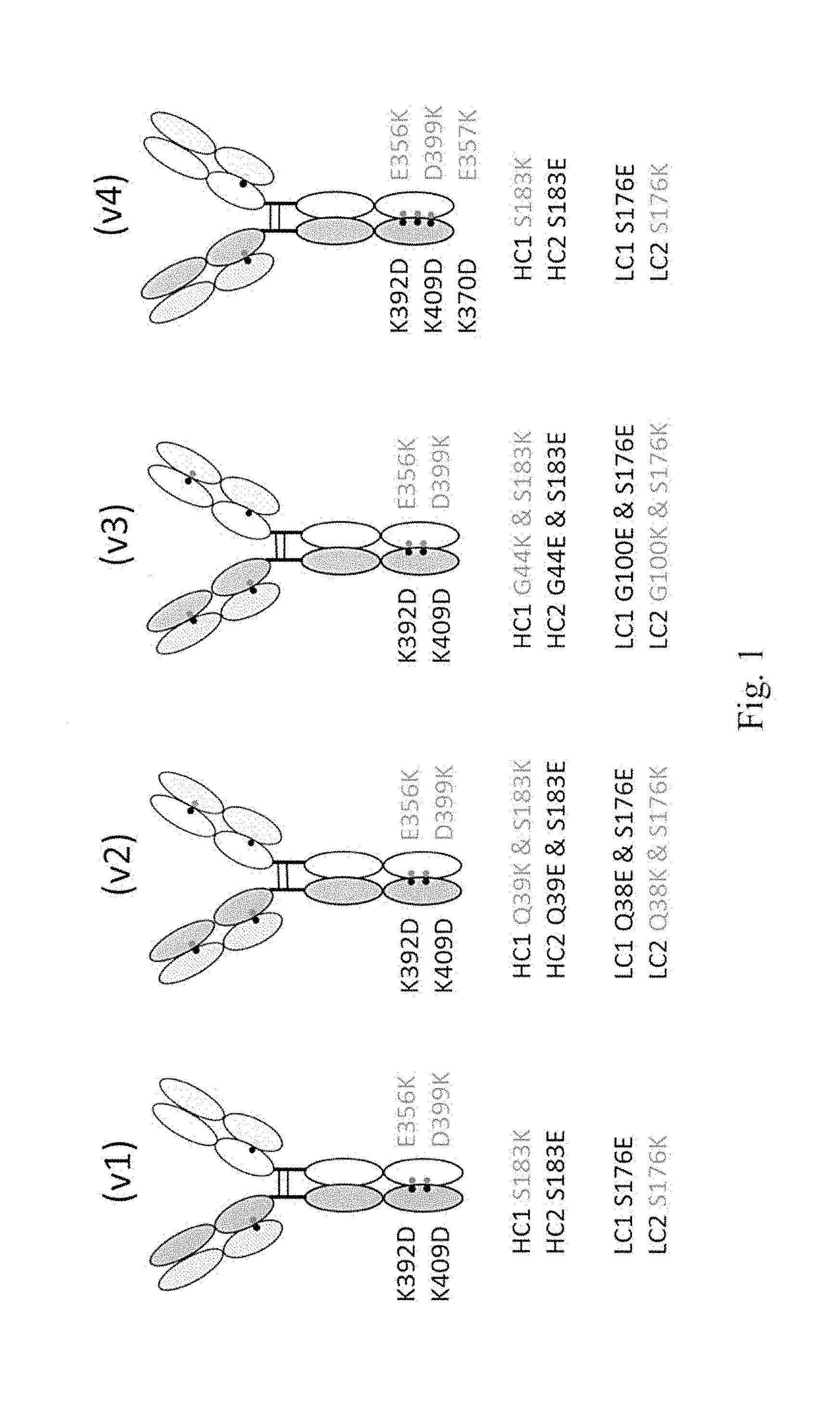
50. The antigen binding protein of claim 47, wherein: a. one heavy chain comprises substitutions K392D and K409D and the other heavy chain comprises substitutions E356K and D399K using EU numbering; or b. one heavy chain comprises substitutions K392D, K409D and K370D and the other heavy chain comprises substitutions E356K, D399K and E357K using EU numbering.
51. The antigen binding protein of claim 47, wherein: a. one heavy chain comprises substitution S183K, the other heavy chain comprises substitution S183E, one light chain comprises substitution S176E, and the other light chain comprises substitution S176K using EU numbering; or b. one heavy chain comprises substitutions Q39K and S183K, the other heavy chain comprises substitutions Q39E and S183E, one light chain comprises substitutions Q38E and S176E, and the other light chain comprises substitutions Q38K and S176K using EU numbering; or c. one heavy chain comprises substitutions G44K and S183K, the other heavy chain comprises substitutions G44E and S183E, one light chain comprises substitutions G100E and S176E, and the other light chain comprises substitutions G100K and S176K using EU numbering; or d. one heavy chain comprises substitution S183K, the other heavy chain comprises substitution S183E, one light chain comprises substitution S176E, and the other light chain comprises substitution S176K using EU numbering.
52. The antigen binding protein of claim 50, wherein: a. one heavy chain comprises substitution S183K, the other heavy chain comprises substitution S183E, one light chain comprises substitution S176E, and the other light chain comprises substitution S176K using EU numbering; or b. one heavy chain comprises substitutions Q39K and S183K, the other heavy chain comprises substitutions Q39E and S183E, one light chain comprises substitutions Q38E and S176E, and the other light chain comprises substitutions Q38K and S176K using EU numbering; or c. one heavy chain comprises substitutions G44K and S183K, the other heavy chain comprises substitutions G44E and S183E, one light chain comprises substitutions G100E and S176E, and the other light chain comprises substitutions G100K and S176K using EU numbering; or d. one heavy chain comprises substitution S183K, the other heavy chain comprises substitution S183E, one light chain comprises substitution S176E, and the other light chain comprises substitution S176K using EU numbering.
53. The antigen binding protein of claim 47, wherein the bispecific antigen binding protein comprises two IgG1 heavy chains comprising substitutions R292C, V302C and N297G using EU numbering.
54. The antigen binding protein of claim 50, wherein the bispecific antigen binding protein comprises two IgG1 heavy chains comprising substitutions R292C, V302C and N297G using EU numbering.
55. The antigen binding protein of claim 51, wherein the bispecific antigen binding protein comprises two IgG1 heavy chains comprising substitutions R292C, V302C and N297G using EU numbering.
56. The antigen binding protein of claim 52, wherein the bispecific antigen binding protein comprises two IgG1 heavy chains comprising substitutions R292C, V302C and N297G using EU numbering.
57. The antigen binding protein of claim 39, wherein: a. the antigen binding protein comprises two light chains and two heavy chains: b. the heavy and light chains comprise IgG1, IgG2, or IgG4 constant domains; c. the antigen binding protein comprises two TL1A binding entity heavy chain variable domains, two TL1A binding entity light chain variable domains, two TNF-.alpha. binding entity heavy chain variable domains, and two TNF-.alpha. binding entity light chain variable domains; d. each light chain comprises a TL1A binding entity light chain variable domain sequence; and e. each heavy chain comprises a TL1A binding entity heavy chain variable domain sequence, a TNF-.alpha. binding entity heavy chain variable domain sequence and a TNF-.alpha. binding entity light chain variable domain sequence.
58. The antigen binding protein of claim 57, wherein: a. the TL1A binding entity heavy chain variable domain comprises a heavy chain variable domain sequence selected from Table D (SEQ ID NOS: 8, 12, 16, 20, 24, 28, and 32); b. the TL1A binding entity light chain variable domain comprises a light chain variable domain sequence selected from Table D (SEQ ID NOS: 6, 10, 14, 18, 22, 26, and 30); c. the TNF-.alpha. binding entity light chain variable domain sequence is selected from Table H (SEQ ID NOS: 2, 42, 38, and 34) and d. the TNF-.alpha. binding entity heavy chain variable domain sequence is selected from Table H (SEQ ID NOS: 4, 44, 318, 40, and 36).
59. The antigen binding protein of claim 57, wherein the anti-TNF-.alpha. binding entity heavy chain variable domain comprises a cysteine residue at position 44 and the anti-TNF-.alpha. light chain binding domain comprises a cysteine residue at position 100, numbering as in Kabat.
60. The antigen binding protein of claim 57 wherein the protein comprises the heavy and light chain sequences of an antigen binding protein selected from Table L, SEQ ID NOS: a. 355 and 447, b. 357 and 447, c. 359 and 447, d. 361 and 447, e. 363 and 447, f. 365 and 58, g. 367 and 70, h. 369 and 70, i. 371 and 70, j. 373 and 70, k. 375 and 70, l. 377 and 70, m. 391 and 467, n. 391 and 66, o. 395 and 66, p. 397 and 66, q. 399 and 66, r. 401 and 66, s. 403 and 66, t. 405 and 66 u. 407 and 447, v. 409 and 447, w. 411 and 66, x. 413 and 66, y. 415 and 66, z. 417 and 66, aa. 419 and 70, bb. 421 and 70, cc. 423 and 70, and dd. 425 and 70.
61. The antigen binding protein of claim 57 wherein: a. the light chain sequence comprises SEQ ID NO: 453 and the heavy chain sequence comprises SEQ ID NO: 441; b. the light chain sequence comprises SEQ ID NO: 451 and the heavy chain sequence comprises SEQ ID NO: 433; or c. the light chain sequence comprises SEQ ID NO: 453 and the heavy chain sequence comprises SEQ ID NO: 445.
62. The antigen binding protein of claim 39, wherein: a. the antigen binding protein comprises two light chains and two heavy chains: b. the heavy and light chains comprise IgG1, IgG2, or IgG4 constant domains; c. the antigen binding protein comprises two TL1A binding entity heavy chain variable domains, two TL1A binding entity light chain variable domains, two TNF-.alpha. binding entity heavy chain variable domains, and two TNF-.alpha. binding entity light chain variable domains; d. each light chain comprises a TNF-.alpha. binding entity light chain sequence; and e. each heavy chain comprises a TNF-.alpha. binding entity heavy chain sequence, a TL1A binding entity heavy chain binding domain sequence and a TL1A binding entity light chain binding domain sequence.
63. The antigen binding protein of claim 62, wherein: a. the TL1A binding entity heavy chain variable domain comprises a heavy chain variable domain sequence selected from Table D (SEQ ID NOS: 8, 12, 16, 20, 24, 28, and 32); b. the TL1A binding entity light chain variable domain comprises a light chain variable domain sequence selected from Table D (SEQ ID NOS: 6, 10, 14, 18, 22, 26, and 30); c. the TNF-.alpha. binding entity light chain variable domain sequence is selected from Table H (SEQ ID NOS: 2, 42, 38, and 34) and d. the TNF-.alpha. binding entity heavy chain variable domain sequence is selected from Table H (SEQ ID NOS: 4, 44, 318, 40, and 36).
64. The antigen binding protein of claim 62, wherein the TL1A binding entity heavy chain variable domain comprises a cysteine residue at position 44 and the TL1A binding entity light chain binding domain comprises a cysteine residue at position 100, numbering as in Kabat.
65. The antigen binding protein of claim 62, wherein the protein comprises the heavy and light chain sequences of an antigen binding protein selected from Table M, SEQ ID NOS: a. 82 and 46, b. 284 and 46, c. 286 and 46, d. 441 and 453 e. 312 and 46, f. 445 and 453, g. 314 and 84, h. 320 and 84, i. 322 and 84, j. 345 and 84, k. 347 and 78, l. 349 and 78, m. 351 and 78, n. 353 and 78, o. 379 and 46, p. 381 and 46, q. 383 and 84, r. 385 and 84, s. 387 and 78, t. 389 and 78, u. 433 and 451, v. 427 and 449, w. 429 and 449, x. 431 and 451, y. 433 and 451, z. 435 and 451, aa. 437 and 451, bb. 439 and 453, cc. 441 and 453, dd. 443 and 453, and ee. 445 and 453.
66. The antigen binding protein of claim 62 comprising SEQ ID NOS: a. 441 and 453; b. 433 and 451; or c. 445 and 453.
67. One or more isolated nucleic acids encoding the antigen binding protein of any one of claims 1 to 66 and 131 to 152.
68. One or more expression vectors comprising the nucleic acid or acids of claim 67.
69. A host cell comprising the one or more expression vectors of claim 68.
70. A method for the preparation of an antigen binding protein, comprising: a. culturing the host cell of claim 69 under conditions that allow expression of the antigen binding protein; and b. recovering the antigen binding protein from the culture.
71. A pharmaceutical composition comprising the antigen binding protein of any one of claims 1 to 66 and 131 to 152 and a pharmaceutically acceptable diluent, excipient or carrier.
72. A method for treating a condition associated with TL1A and/or TNF-.alpha. in a patient in need thereof, comprising administering to the patient an effective amount of the antigen binding protein of any one of claims 1 to 66 and 131 to 152.
73. The method of claim 72, wherein the condition is inflammatory bowel disease.
74. The method of claim 72, wherein the condition is Crohn's disease.
75. The method of claim 72, wherein the condition is ulcerative colitis.
76. Use of the antigen binding protein of any one of claims 1 to 66 and 131 to 152 in the preparation of a medicament for treating a condition associated with TL1A in a patient in need thereof.
77. The use of claim 76 wherein the condition is inflammatory bowel disease.
78. The use of claim 76 wherein the condition is Crohn's disease.
79. The use of claim 76 wherein the condition is ulcerative colitis.
80. The antigen binding protein of any one of claims 1 to 66 and 131 to 152 for use in a method for treating a condition associated with TL1A and/or TNF-.alpha. in a patient in need thereof.
81. The antigen binding protein of claim 80 wherein the condition is inflammatory bowel disease.
82. The antigen binding protein of claim 80 wherein the condition is Crohn's disease.
83. The antigen binding protein of claim 80 wherein the condition is ulcerative colitis.
84. An antigen binding protein specific for TL1A wherein: a. The antigen binding protein comprises one or two light chain variable domains and one or two heavy chain variable domains; b. the light chain variable domain comprises a LCDR1, LCDR2, and LCDR3; c. the heavy chain variable domain comprises a HCDR1, HCDR2, and HCDR3; and d. the HCDR3 sequence is selected from Table A (SEQ ID NOS: 168, 174, 180, 186, 192, 489, 781, 787, 796, 802, 807, 813, 823, 829, 834, 840, 845, 851, 855, and 861) or is a sequence wherein for each HCDR3 sequence in Table A: i. one or more acidic residues are optionally replaced by any other acidic residues; ii. one or more amide residues are optionally replaced by any other amide residues; iii. one or more aromatic residues are optionally replaced by any other aromatic residues; iv. one or more basic residues are optionally replaced by any other basic residues; v. one or more hydrophilic residues are optionally replaced by any other hydrophilic residues; and vi. one or more non-functional residues are optionally replaced by any other non-functional residues; e. "acidic residue" means D or E; f. "amide residue" means N or Q; g. "aromatic residue" means F, Y, or W; h. "basic residue" means H, K, or R; i. "hydrophilic residue means S, T, N or Q; and j. "non-functional residue means M, G, A, V, I or L.
85. The antigen binding protein of claim 84, wherein the HCDR3 comprises a sequence selected from Tables A, B, and C (SEQ ID NOS: 168, 174, 180, 186, 192, 489, 781, 787, 796, 802, 807, 813, 823, 829, 834, 840, 845, 851, 855, 861, 641, 645, 650, 657, 676, 680, 681, 667, 671, 976, 985, 1000, 1012, 1013, 1014, 1015, 1018, 1019, 1020, 1024, 1043, 1049, 1050, 1051, 1052, 1063, 1071, 1075, 1076, 1077, 1091, 1105, 255, 258, 261, 264, 267, 270, 273, 672, 675, 952, 954, 958, 961, 964, 966, 967, 968, 969, and 970).
86. The antigen binding protein of claim 85, wherein the HCDR3 comprises a sequence selected from Table A (SEQ ID NOS: 168, 174, 180, 186, 192, 489, 781, 787, 796, 802, 807, 813, 823, 829, 834, 840, 845, 851, 855, and 861).
87. The antigen binding protein of claim 86, wherein the HCDR3 has a sequence selected from antibodies and 9C8 and 3B3 (SEQ ID NO: 180 and 192).
88. The antigen binding protein of claim 84, wherein the HCDR1 has a sequence selected from Table A (SEQ ID NOS: 164, 170, 182, 188, 777, 783, 792, 798, 804, 809, 815, 819, 265, 836, 847, and 857) and the HCDR2 has a sequence selected from Table A (SEQ ID NOS: 166, 172, 178, 184, 190, 196, 202, 483, 485, 779, 785, 794, 800, 811, 817, 821, 827, 832, 838, 843, 849, 853, 859, and 864), or HCDR1 and HCDR2 have sequences wherein for each HCDR1 and HCDR2 sequence in Table A: a. one or more acidic residues are optionally replaced by any other acidic residues; b. one or more amide residues are optionally replaced by any other amide residues; c. one or more aromatic residues are optionally replaced by any other aromatic residues; d. one or more basic residues are optionally replaced by any other basic residues; e. one or more hydrophilic residues are optionally replaced by any other hydrophilic residues; and f. one or more non-functional residues are optionally replaced by any other non-functional residues.
89. The antigen binding protein of claim 85 wherein: a. the HCDR1 has a sequence selected from Tables A, B, and C (SEQ ID NOS: 164, 170, 182, 188, 777, 783, 792, 798, 804, 809, 815, 819, 265, 836, 847, 857, 639, 654, 655, 632, 664, 955, 979, 994, 1010, 1017, 1022, 1027, 1028, 1029, 1037, 1045, 1087, 253, 256, 259, 262, 268, 271, 265, 950, and) and b. the HCDR2 has a sequence selected from Tables A, B, and C (SEQ ID NOS: 166, 172, 178, 184, 190, 196, 202, 483, 485, 779, 785, 794, 800, 811, 817, 821, 827, 832, 838, 843, 849, 853, 859, 864, 640, 204, 678, 679, 644, 648, 649, 633, 656, 659, 660, 665, 666, 677, 114, 227, 230, 691, 715, 669, 711, 971, 972, 973, 974, 975, 980, 981, 982, 983, 984, 995, 996, 997, 998, 0999, 1011, 1023, 1030, 1038, 1039, 1040, 1041, 1042, 1046, 1047, 1048, 1058, 1059, 1060, 1061, 1062, 1066, 1067, 1068, 1069, 1070, 1074, 1088, 1089, 1090, 1095, 1096, 1097, 1102, 1103, 1104, 1108, 1109, 1110, 1111, 1112, 254, 260, 184, 269, 272, 266, 254, 951, 953, 956, and 963).
90. The antigen binding protein of claim 86 wherein: a. the HCDR1 has a sequence selected from Table A (SEQ ID NOS: 164, 170, 182, 188, 777, 783, 792, 798, 804, 809, 815, 819, 265, 836, 847, and 857), and b. the HCDR2 has a sequence selected from Table A (SEQ ID NOS: 166, 172, 178, 184, 190, 196, 202, 483, 485, 779, 785, 794, 800, 811, 817, 821, 827, 832, 838, 843, 849, 853, 859, and 864).
91. The antigen binding protein of claim 87 wherein the HCDR1 has a sequence selected from antibodies 9C8 and 3B3 (SEQ ID NO: 170 and 188) and the HCDR2 has a sequence selected from antibodies 9C8 and 3B3 (SEQ ID NO: 178 and 190).
92. The antigen binding protein of claim 84 wherein the LCDR3 has a sequence selected from Table A (SEQ ID NOS: 102, 108, 263, 120, 126, 687, 693, 701, 707, 717, 727, 733, 739, 745, 751, 757, 763, 767, 771, and 775) or is a sequence wherein for each LCDR3 sequence in Table A: a. one or more acidic residues are optionally replaced by any other acidic residues; b. one or more amide residues are optionally replaced by any other amide residues; c. one or more aromatic residues are optionally replaced by any other aromatic residues; d. one or more basic residues are optionally replaced by any other basic residues; e. one or more hydrophilic residues are optionally replaced by any other hydrophilic residues; and f. one or more non-functional residues are optionally replaced by any other non-functional residues.
93. The antigen binding protein of claim 85 wherein the LCDR3 has a sequence selected from Tables A, B, and C (SEQ ID NOS:102, 108, 263, 120, 126, 687, 693, 701, 707, 717, 727, 733, 739, 745, 751, 757, 763, 767, 771, 775, 637, 638, 631, 653, 663, 637, 233, 940, 978, 989, 990, 991, 992, 993, 1005, 1006, 1007, 1008, 1009, 1034, 1035, 1036, 1055, 1056, 1057, 1065, 1086, 1093, 1094, 1099, 1100, 1101, 1107, 228, 231, 234, 237, 243, 240, 674, 936, 938, 701, 707, 949, 942, and 944),
94. The antigen binding protein of claim 86 wherein the LCDR3 has a sequence selected from Table A (SEQ ID NOS: 102, 108, 263, 120, 126, 687, 693, 701, 707, 717, 727, 733, 739, 745, 751, 757, 763, 767, 771, and 775)).
95. The antigen binding protein of claim 87 wherein the LCDR3 has a sequence selected from antibodies 9C8 and 3B3 (SEQ ID NOS: 263 and 126).
96. The antigen binding protein of claim 92, wherein the LCDR1 has a sequence selected from Table A (SEQ ID NOS: 98, 104, 110, 116, 122, 128, 467, 469, 683, 689, 235, 697, 146, 709, 713, 719, 723, 229, 226, 741, 747, 753, and 773) and the LCDR2 has a sequence selected from Table A (SEQ ID NOS: 100, 106, 112, 118, 124, 685, 699, 705, 721, 725, 731, 737, 749, 755, 761, and 769) or sequences wherein for each LCDR1 and LCDR2 sequence in Table A: a. one or more acidic residues are optionally replaced by any other acidic residues; b. one or more amide residues are optionally replaced by any other amide residues; c. one or more aromatic residues are optionally replaced by any other aromatic residues; d. one or more basic residues are optionally replaced by any other basic residues; e. one or more hydrophilic residues are optionally replaced by any other hydrophilic residues; and f. one or more non-functional residues are optionally replaced by any other non-functional residues.
97. The antigen binding protein of claim 93 wherein: a. the LCDR1 has a sequence selected from Tables A, B, and C (SEQ ID NOS: 98, 104, 110, 116, 122, 128, 467, 469, 683, 689, 235, 697, 146, 709, 713, 719, 723, 229, 226, 741, 747, 753, 773, 635, 642, 646, 651, 658, 661, 668, 759, 743, 765, 977, 986, 987, 1001, 1016, 1021, 1025, 1031, 1032, 1033, 1044, 1053, 1064, 1072, 1078, 1079, 1080, 1106, 226, 229, 232, 235, 241, 238, 1113, 146, and 947) and b. the LCDR2 has a sequence selected from Tables A, B, and C (SEQ ID NOS: 100, 106, 112, 118, 124, 235, 699, 705, 721, 725, 731, 737, 749, 755, 761, 769, 636, 643, 647, 652, 662, 789, 988, 1001, 1002, 1004, 1026, 1054, 1073, 1081, 1082, 1083, 1084, 1085, 1092, 1098, 242, 935, 148, 948, and 943).
98. The antigen binding protein of claim 94 wherein: a. the LCDR1 has a sequence selected from Table A (SEQ ID NOS: 98, 104, 110, 116, 122, 128, 467, 469, 683, 689, 235, 697, 146, 709, 713, 719, 723, 229, 226, 741, 747, 753, and 773) and b. the LCDR2 has a sequence selected from Table A (SEQ ID NOS: 100, 106, 112, 118, 124, 685, 699, 705, 721, 725, 731, 737, 749, 755, 761, and 769).
99. The antigen binding protein of claim 95 wherein the LCDR1 has a sequence selected from antibodies 9C8 and 3B3 (SEQ ID NOS: 110 and 122) and the LCDR2 has a sequence selected from antibodies 9C8 and 3B3 (SEQ ID NOS: 112 and 124).
100. The antigen binding protein of claim 89, wherein: a. the LCDR1 has a sequence selected from Tables A, B, and C (SEQ ID NOS: 98, 104, 110, 116, 122, 128, 467, 469, 683, 689, 235, 697, 146, 709, 713, 719, 723, 229, 226, 741, 747, 753, 773, 635, 642, 646, 651, 658, 661, 668, 759, 743, 765, 977, 986, 987, 1001, 1016, 1021, 1025, 1031, 1032, 1033, 1044, 1053, 1064, 1072, 1078, 1079, 1080, 1106, 226, 229, 232, 235, 241, 238, 1113, 146, and 947); b. the LCDR2 has a sequence selected from Tables A, B, and C (SEQ ID NOS: 100, 106, 112, 118, 124, 235, 699, 705, 721, 725, 731, 737, 749, 755, 761, 769, 636, 643, 647, 652, 662, 789, 988, 1001, 1002, 1004, 1026, 1054, 1073, 1081, 1082, 1083, 1084, 1085, 1092, 1098, 242, 935, 148, 948, and 943); and c. the LCDR3 has a sequence selected from Tables A, B, and C (SEQ ID NOS:102, 108, 263, 120, 126, 687, 693, 701, 707, 717, 727, 733, 739, 745, 751, 757, 763, 767, 771, 775, 637, 638, 631, 653, 663, 637, 233, 940, 978, 989, 990, 991, 992, 993, 1005, 1006, 1007, 1008, 1009, 1034, 1035, 1036, 1055, 1056, 1057, 1065, 1086, 1093, 1094, 1099, 1100, 1101, 1107, 228, 231, 234, 237, 243, 240, 674, 936, 938, 701, 707, 949, 942, and 944).
101. The antigen binding protein of claim 90, wherein: a. the LCDR1 has a sequence selected from Table A (SEQ ID NOS: 98, 104, 110, 116, 122, 128, 467, 469, 683, 689, 235, 697, 146, 709, 713, 719, 723, 229, 226, 741, 747, 753, and 773); b. the LCDR2 has a sequence selected from Table A (SEQ ID NOS: 100, 106, 112, 118, 124, 685, 699, 705, 721, 725, 731, 737, 749, 755, 761, and 769); and c. the LCDR3 has a sequence selected from Table A (SEQ ID NOS: 102, 108, 263, 120, 126, 687, 693, 701, 707, 717, 727, 733, 739, 745, 751, 757, 763, 767, 771, and 775).
102. The antigen binding protein of claim 91, wherein: a. the LCDR1 has a sequence selected from antibodies 9C8 and 3B3 (SEQ ID NO: 110 and 122); b. the LCDR2 has a sequence selected from antibodies 9C8 and 3B3 (SEQ ID NO: 112 and 124); and c. the LCDR3 has a sequence selected from antibodies 9C8 and 3B3 (SEQ ID NO: 108 and 126).
103. The antigen binding protein of claim 84, wherein the light chain variable domain comprises a sequence at least about 90% identical to a light chain variable domain sequence selected from Table D (SEQ ID NOS: 6, 10, 14, 18, 22, 26, 30, 491, 493, 866, 870, 874, 878, 882, 886, 890, 894, 898, 902, 906, 910, 914, 918, 922, 924, 928, and 932,).
104. The antigen binding protein of claim 20, wherein the light chain variable domain comprises a light chain variable domain sequence selected from Table D (SEQ ID NOS: 6, 10, 14, 18, 22, 26, 30, 491, 493, 866, 870, 874, 878, 882, 886, 890, 89 4, 898, 902, 906, 910, 914, 918, 922, 924, 928, and 932).
105. The antigen binding protein of claim 84 wherein the heavy chain variable domain comprises a sequence at least about 90% identical to a heavy chain variable domain sequence selected from Table D (SEQ ID NOS: 8, 12, 16, 20, 24, 28, 32, 495, 497, 868, 872, 876, 880, 884, 888, 892, 896, 900, 904, 908, 912, 916, 920, 926, 930, and 934).
106. The antigen binding protein of claim 103 wherein the heavy chain variable domain comprises a sequence at least about 90% identical to a heavy chain variable domain sequence selected from Table D (SEQ ID NOS: 8, 12, 16, 20, 24, 28, 32, 495, 497, 868, 872, 876, 880, 884, 888, 892, 896, 900, 904, 908, 912, 916, 920, 926, 930, and 934).
107. The antigen binding protein of claim 103 wherein the heavy chain variable domain comprises a heavy chain variable domain sequence selected from Table D (SEQ ID NOS: 8, 12, 16, 20, 24, 28, 32, 495, 497, 868, 872, 876, 880, 884, 888, 892, 896, 900, 904, 908, 912, 916, 920, 926, 930, and 934).
108. The antigen binding protein of claim 104 wherein the heavy chain variable domain comprises a heavy chain variable domain sequence selected from Table D (SEQ ID NOS: 8, 12, 16, 20, 24, 28, 32, 495, 497, 868, 872, 876, 880, 884, 888, 892, 896, 900, 904, 908, 912, 916, 920, 926, 930, and 934).
109. The antigen binding protein of claim 103 wherein the heavy chain variable domain comprises a heavy chain variable domain sequence selected from Table D (SEQ ID NOS: 8, 12, 16, 20, 24, 28, and 32).
110. The antigen binding protein of claim 108 wherein the light chain variable domain sequence comprises a light chain variable domain sequence selected from antibodies 9C8 and 3B3 (SEQ ID NOS: 14 and 22) and the heavy chain variable domain comprises a heavy chain variable domain sequence selected from antibodies 9C8 and 3B3 (SEQ ID NOS: 16 and 24).
111. The antigen binding protein of claim 84 wherein the protein comprises a light chain having a light chain sequence selected from Table E (SEQ ID NOS: 66, 54, 58, 62, 66, 70, 74, 455, 459, 1116, 1120, 1124, 1128, 1132, 1136, 1140, 1144, 1148, 1152, 1156, 1160, 1164, 1168, 1172, 1176, 1180, 1184 and 1188).
112. The antigen binding protein of claim 84 wherein the protein comprises a heavy chain having a heavy chain sequence selected from Table E (SEQ ID NOS: 52, 56, 60, 64, 68, 72, 76, 457, 461, 1118, 1122, 1126, 1130, 1134, 1138, 1142, 1146, 1150, 1154, 1158, 1162, 1166, 1170, 1174, 1178, 1182, 1186 and 1190).
113. The antigen binding protein of claim 84 comprising the light and heavy chain sequences of an antibody selected from Table E, SEQ ID NOS: a. 50 and 52, b. 54 and 56, c. 58 and 60, d. 62 and 64, e. 66 and 68, f. 70 and 72, g. 74 and 76, h. 455 and 457, i. 459 and 461, j. 1116 and 1118, k. 1120 and 1122, l. 1124 and 1126, m. 1128 and 1130, n. 1132 and 1134, o. 1136 and 1138, p. 1140 and 1142, q. 1144 and 1146, r. 1148 and 1150, s. 1152 and 1154, t. 1156 and 1158, u. 1160 and 1162, v. 1164 and 1166, w. 1168 and 1170, x. 1172 and 1174, y. 1176 and 1178, z. 1180 and 1182, aa. 1184 and 1186, and bb. 1188 and 1190.
114. One or more isolated nucleic acids encoding the antigen binding protein of any one of claims 84 to 113.
115. One or more expression vectors comprising the nucleic acid or acids of claim 114.
116. A host cell comprising the one or more expression vectors of claim 115.
117. A method for the preparation of an antigen binding protein, comprising: a. culturing the host cell of claim 116 under conditions that allow expression of the antigen binding protein; and b. recovering the antigen binding protein from the culture.
118. A pharmaceutical composition comprising the antigen binding protein of any one of claims 84 to 113 and a pharmaceutically acceptable diluent, excipient or carrier.
119. A method for treating a condition associated with TL1A in a patient in need thereof, comprising administering to the patient an effective amount of the antigen binding protein of any one of claims 84 to 113.
120. The method of claim 119, wherein the condition is inflammatory bowel disease.
121. The method of claim 119, wherein the condition is Crohn's disease.
122. The method of claim 119, wherein the condition is ulcerative colitis.
123. Use of the antigen binding protein of any one of claims 84 to 113 in the preparation of a medicament for treating a condition associated with TL1A in a patient in need thereof.
124. The use of claim 123 wherein the condition is inflammatory bowel disease.
125. The use of claim 123 wherein the condition is Crohn's disease.
126. The use of claim 123 wherein the condition is ulcerative colitis.
127. The antigen binding protein of any one of claims 84 to 113 for use in a method for treating a condition associated with TL1A in a patient in need thereof.
128. The antigen binding protein of claim 127 wherein the condition is inflammatory bowel disease.
129. The antigen binding protein of claim 127 wherein the condition is Crohn's disease.
130. The antigen binding protein of claim 127 wherein the condition is ulcerative colitis.
131. The antigen binding protein of claim 38, comprising: a. a first polypeptide comprising a first heavy chain of a first antibody comprising a first heavy chain variable region (VH1) and a first CH1 domain, wherein the first heavy chain is fused through its C-terminus to the N-terminus of a polypeptide comprising a second heavy chain variable region of a second antibody (VH2), wherein the VH2 is fused through its C-terminus to the N-terminus of a second CH1 domain, and wherein the first antibody comprises one of the TL1A binding entity or the TNF-.alpha. binding entity and the second antibody comprises the other; wherein: i. the VH1 or first CH1 domain comprises at least one amino acid substitution to introduce a positively charged amino acid at a residue selected from the group consisting of positions 39, 44, and 183 using EU numbering; and ii. the VH2 or second CH1 domain comprises at least one amino acid substitution to introduce a negatively charged amino acid at a residue selected from the group consisting of a residue that corresponds to positions 39, 44, and 183 using EU numbering; and b. a second polypeptide comprising a first light chain of the first antibody of a., wherein the first light chain comprises a first light chain variable region (VL1) and a first CL region; and wherein the VL1 or first CL domain comprises at least one amino acid substitution to introduce a negatively charged amino acid at a residue selected from the group consisting of positions 38, 100, and 176 using EU numbering; and c. a third polypeptide comprising a second light chain of the second antibody of a., wherein the second light chain comprises a second light chain variable region (VL2) and a second CL region; and wherein the VL1 or first CL domain comprises at least one amino acid substitution to introduce a positively charged amino acid at a residue selected from the group consisting of positions 38, 100, and 176 using EU numbering.
132. The antigen binding protein of claim 131, wherein the first heavy chain is fused to the VH2 via a peptide linker.
133. The antigen binding protein according to claim 132, wherein the peptide linker comprises a sequence selected from the group consisting of (Gly.sub.3Ser).sub.2, (Gly.sub.4Ser).sub.2, (Gly.sub.3Ser).sub.3, (Gly.sub.4Ser).sub.3, (Gly.sub.3Ser).sub.4, (Gly.sub.4Ser).sub.4, (Gly.sub.3Ser).sub.5, (Gly.sub.4Ser).sub.5, (Gly.sub.3Ser).sub.6, and (Gly.sub.4Ser).sub.6.
134. The antigen binding protein according to claim 131, wherein: a. the VH1 or first CH1 domain comprises a mutation selected from the group consisting of Q39K, G44K, and S183K using EU numbering; b. the VH2 or second CH1 domain comprises a mutation selected from the group consisting of Q39E, G44E, and S183E using EU numbering; c. the VL1 or first CL domain comprises a mutation selected from the group consisting of Q38E, G100E, and S176E using EU numbering; and d. the VL2 or second CL domain comprises a mutation selected from the group consisting of Q38K, G100K, and S176K using EU numbering.
135. The antigen binding protein according to claim 134, wherein: a. the first CH1 domain comprises a S183K mutation using EU numbering; b. the second CH1 domain comprises a S183E mutation using EU numbering; c. the first CL domain comprises a S176E mutation using EU numbering; and d. the second CL domain comprises a S176K mutation using EU numbering.
136. The antigen binding protein according to claim 134, wherein: a. the VH1 comprises a Q39K mutation and the first CH1 domain comprises a S183K mutation using EU numbering; b. the VH2 comprises a Q39E mutation and the second CH1 domain comprises a S183E mutation using EU numbering; c. the VL1 comprises a Q38E mutation and the first CL domain comprises a S176E mutation using EU numbering; and d. the VL2 comprises a Q38K mutation and the second CL domain comprises a S176K mutation using EU numbering.
137. The antigen binding protein according to claim 134, wherein: a. the first CH1 domain comprises G44K and S183K mutations using EU numbering; b. the second CH1 domain comprises G44E and S183E mutations using EU numbering; c. the first CL domain comprises G100E and S176E mutations using EU numbering; and d. the second CL domain comprises G100K and S176K mutations using EU numbering.
138. The antigen binding protein of claim 38, comprising: a. a first polypeptide comprising a first heavy chain of a first antibody comprising a first heavy chain variable region (VH1) and a first CH1 domain, wherein the first heavy chain is fused through its C-terminus to the N-terminus of a polypeptide comprising a second heavy chain variable region of a second antibody (VH2), wherein the VH2 is fused through its C-terminus to the N-terminus of a second CH1 domain, and wherein the first antibody comprises one of the TL1A binding entity and the TNF-.alpha. binding entity and the second antibody comprises the other; wherein: i. the VH1 or first CH1 domain comprises at least one amino acid substitution to introduce a negatively charged amino acid at a residue selected from the group consisting of positions 39, 44, and 183 using EU numbering; and ii. the VH2 or second CH1 domain comprises at least one amino acid substitution to introduce a positively charged amino acid at a residue selected from the group consisting of a residue that corresponds to positions 39, 44, and 183 using EU numbering; and b. a second polypeptide comprising a first light chain of the first antibody of a., wherein the first light chain comprises a first light chain variable region (VL1) and a first CL region; and wherein the VL1 or first CL domain comprises at least one amino acid substitution to introduce a positively charged amino acid at a residue selected from the group consisting of positions 38, 100, and 176 using EU numbering; and c. a third polypeptide comprising a second light chain of the second antibody of a., wherein the second light chain comprises a second light chain variable region (VL2) and a second CL region; and wherein the VL1 or first CL domain comprises at least one amino acid substitution to introduce a negatively charged amino acid at a residue selected from the group consisting of positions 38, 100, and 176 using EU numbering.
139. The antigen binding protein according to claim 138, wherein the first heavy chain is fused to the VH2 via a peptide linker.
140. The antigen binding protein according to claim 139, wherein the peptide linker comprises a sequence selected from the group consisting of (Gly.sub.3Ser).sub.2, (Gly.sub.4Ser).sub.2, (Gly.sub.3Ser).sub.3, (Gly.sub.4Ser).sub.3, (Gly.sub.3Ser).sub.4, (Gly.sub.4Ser).sub.4, (Gly.sub.3Ser).sub.5, (Gly.sub.4Ser).sub.5, (Gly.sub.3Ser).sub.6, and (Gly.sub.4Ser).sub.6.
141. The antigen binding protein according to claim 138, wherein: a. the VH1 or first CH1 domain comprises a mutation selected from the group consisting of Q39E, G44E, and S183E using EU numbering; b. the VH2 or second CH1 domain comprises a mutation selected from the group consisting of Q39K, G44K, and S183K using EU numbering; c. the VL1 or first CL domain comprises a mutation selected from the group consisting of Q38K, G100K, and S176K using EU numbering; and d. the VL2 or second CL domain comprises a mutation selected from the group consisting of Q38E, G100E, and S176E using EU numbering.
142. The antigen binding protein according to claim 141, wherein: a. the first CH1 domain comprises a S183E mutation using EU numbering; b. the second CH1 domain comprises a S183K mutation using EU numbering; c. the first CL domain comprises a S176K mutation using EU numbering; and d. the second CL domain comprises a S176E mutation using EU numbering.
143. The antigen binding protein according to claim 141, wherein: a. the VH1 comprises a Q39E mutation and the first CH1 domain comprises a S183E mutation using EU numbering; b. the VH2 comprises a Q39K mutation and the second CH1 domain comprises a S183K mutation using EU numbering; c. the VL1 comprises a Q38K mutation and the first CL domain comprises a S176K mutation using EU numbering; and d. the VL2 comprises a Q38E mutation and the second CL domain comprises a S176E mutation using EU numbering.
144. The antigen binding protein according to claim 141, wherein: a. the first CH1 domain comprises G44E and S183E mutations using EU numbering; b. the second CH1 domain comprises G44K and S183K mutations using EU numbering; c. the first CL domain comprises G100K and S176K mutations using EU numbering; and d. the second CL domain comprises G100E and S176E mutations using EU numbering.
145. The antigen binding protein of claim 38, comprising: a. a first polypeptide comprising a first heavy chain of a first antibody comprising a first heavy chain variable region (VH1) and a first CH1 domain, wherein the first heavy chain is fused through its C-terminus to the N-terminus of a polypeptide comprising a second heavy chain variable region of a second antibody (VH2), wherein the VH2 is fused through its C-terminus to the N-terminus of a second CH1 domain, and wherein the first antibody comprises one of the TL1A binding entity and the TNF-.alpha. binding entity and the second antibody comprises the other; wherein: i. the VH1 or first CH1 domain comprises at least one amino acid substitution to introduce a charged amino acid at a residue selected from the group consisting of positions 39, 44, and 183 using EU numbering; and ii. the VH2 or second CH1 domain comprises at least one amino acid substitution to introduce a charged amino acid at a residue selected from the group consisting of a residue that corresponds to positions 39, 44, and 183 using EU numbering, wherein the charge is the opposite of the substituted residue of the VH1 or first CH1 of the first heavy chain; and b. a second polypeptide comprising a first light chain of the first antibody of a., wherein the first light chain comprises a first light chain variable region (VL1) and a first CL region; and wherein the VL1 or first CL domain comprises at least one amino acid substitution to introduce a charged amino acid at a residue selected from the group consisting of positions 38, 100, and 176 using EU numbering, wherein; i. the charge at position 38 is the opposite of the substituted residue of the VH1 or first CH1 of the first heavy chain at position 39 using EU numbering; the charge at position 100 using EU numbering is the opposite of the substituted residue of the VH1 or first CH1 of the first heavy chain at position 44 using EU numbering; the charge at position 176 is the opposite of the substituted residue of the VH1 or first CH1 of the first heavy chain at position 183 using EU numbering; and c. a third polypeptide comprising a second light chain of the second antibody of a., wherein the second light chain comprises a second light chain variable region (VL2) and a second CL region; and wherein the VL2 or second CL domain comprises at least one amino acid substitution to introduce a charged amino acid at a residue selected from the group consisting of positions 38, 100, and 176 using EU numbering, wherein: i. the charge at position 38 is the opposite of the substituted residue of the VH2 or second CH1 of the second heavy chain at position 39; the charge at position 100 is the opposite of the substituted residue of the VH2 or second CH1 of the second heavy chain at position 44; the charge at position 176 is the opposite of the substituted residue of the VH2 or second CH1 of the second heavy chain at position 183.
146. The antigen binding protein according to claim 145, wherein the first heavy chain is fused to the VH2 via a peptide linker.
147. The antigen binding protein according to claim 146, wherein the peptide linker comprises a sequence selected from the group consisting of (Gly.sub.3Ser).sub.2, (Gly.sub.4Ser).sub.2, (Gly.sub.3Ser).sub.3, (Gly.sub.4Ser).sub.3, (Gly.sub.3Ser).sub.4, (Gly.sub.4Ser).sub.4, (Gly.sub.3Ser).sub.5, (Gly.sub.4Ser).sub.5, (Gly.sub.3Ser).sub.6, and (Gly.sub.4Ser).sub.6.
148. The antigen binding protein according to claim 145, wherein: a. the VH1 comprises a Q39E mutation and the first CH1 domain comprises a S183K mutation using EU numbering; b. the VH2 comprises a Q39K mutation and the second CH1 domain comprises a S183E mutation using EU numbering; c. the VL1 comprises a Q38K mutation and the first CL domain comprises a S176E mutation using EU numbering; and d. the VL2 comprises a Q38E mutation and the second CL domain comprises a S176K mutation using EU numbering.
149. The antigen binding protein according to claim 146, wherein: a. the first CH1 domain comprises G44E and S183K mutations using EU numbering; b. the second CH1 domain comprises G44K and S183E mutations using EU numbering; c. the first CL domain comprises G100K and S176E mutations using EU numbering; and d. the second CL domain comprises G100E and S176K mutations using EU numbering.
150. The antigen binding protein according to claim 146, wherein: a. the VH1 comprises a Q39K mutation and the first CH1 domain comprises a S183E mutation using EU numbering; b. the VH2 comprises a Q39E mutation and the second CH1 domain comprises a S183K mutation using EU numbering; c. the VL1 comprises a Q38E mutation and the first CL domain comprises a S176K mutation using EU numbering; and d. the VL2 comprises a Q38K mutation and the second CL domain comprises a S176E mutation using EU numbering.
151. The antigen binding protein according to claim 146, wherein: a. the first CH1 domain comprises G44K and S183E mutations using EU numbering; b. the second CH1 domain comprises G44E and S183K mutations using EU numbering; c. the first CL domain comprises G100E and S176K mutations using EU numbering; and d. the second CL domain comprises G100K and S176E mutations using EU numbering.
152. The antigen binding protein of claim 38, comprising: a. a first polypeptide comprising a first heavy chain (VH2-CH1-CH2-CH3) from a first antibody, wherein the first heavy chain is fused at its carboxyl terminus (optionally through a peptide linker) to a polypeptide comprising VH2-CH1 domains of a second antibody, b. a second polypeptide comprising a light chain from the first antibody (VL1-CL) and c. a third polypeptide comprising VL2-CL domains of the second antibody; wherein the first antibody comprises one of a TL1A binding entity and a TNF-.alpha. binding entity and the second antibody comprises the other.
153. The antigen binding protein of claim 152, wherein: a. CL from the first antibody comprises E at position 230, CH1 from the first antibody comprises K at position 230, CL from the second antibody comprises K at position 230, and CH1 from the second antibody comprises E at position 230; b. CL from the first antibody comprises E at position 230, CH1 from the first antibody comprises K at position 230, VL from the first antibody comprises E at position 46, VH from the first antibody comprises K at position 46, CL from the second antibody comprises K at position 230, CH1 from the second antibody comprises E at position 230, VL from the second antibody comprises E at position 46, and VH from the second antibody comprises K at position 46; or c. CL from the first antibody comprises E at position 230, CH1 from the first antibody comprises K at position 230, VL from the first antibody comprises E at position 141, VH from the first antibody comprises K at position 51, CL from the second antibody comprises K at position 230, CH1 from the second antibody comprises E at position 230, VL from the second antibody comprises E at position 51, and VH from the second antibody comprises K at position 141; wherein all positions are according to EU numbering.
154. The antigen binding protein of claim 38, comprising: a. a first polypeptide comprising VH1-CL-CH2-CH3 domains of a first antibody fused at their carboxyl terminus (optionally through a peptide linker) to a polypeptide comprising VH2-CH1 domains of a second antibody, b. a second polypeptide comprising a light chain from the first antibody (VL1-CH1) and c. a third polypeptide comprising VL2-CL domains of the second antibody; wherein the first antibody comprises one of a TL1A binding entity or a TNF-.alpha. binding entity and the second antibody comprises the other.
155. The antigen binding protein of claim 154, wherein: a. CL from the first antibody comprises K at position 230, CH1 from the first antibody comprises E at position 230, CL from the second antibody comprises K at position 230, and CH1 from the second antibody comprises E at position 230; b. CL from the first antibody comprises K at position 230, CH1 from the first antibody comprises E at position 230, VL from the first antibody comprises E at position 46, VH from the first antibody comprises K at position 46, CL from the second antibody comprises K at position 230, CH1 from the second antibody comprises E at position 230, VL from the second antibody comprises E at position 46, and VH from the second antibody comprises K at position 46; or c. CL from the first antibody comprises K at position 230, CH1 from the first antibody comprises E at position 230, VL from the first antibody comprises E at position 141, VH from the first antibody comprises K at position 51, CL from the second antibody comprises K at position 230, CH1 from the second antibody comprises E at position 230, VL from the second antibody comprises E at position 51, and VH from the second antibody comprises K at position 141; wherein all positions are according to EU numbering.
156. The antigen binding protein of claim 38, comprising: a. a first polypeptide comprising a first heavy chain (VH1-CH1-CH2-CH3) of a first antibody, wherein the first heavy chain is fused at its carboxyl terminus (optionally through a peptide linker) to a polypeptide comprising VH2-CL domains of a second antibody, b. a second polypeptide comprising a light chain from a first antibody (VL1-CL) and c. a third polypeptide comprising VL2-CH1 domains of the second antibody; wherein the first antibody comprises one of a TL1A binding entity or a TNF-.alpha. binding entity and the second antibody comprises the other.
157. The antigen binding protein of claim 156, wherein: a. CL from the first antibody comprises E at position 230, CH1 from the first antibody comprises K at position 230, CL from the second antibody comprises E at position 230, and CH1 from the second antibody comprises K at position 230; b. CL from the first antibody comprises E at position 230, CH1 from the first antibody comprises K at position 230, VL from the first antibody comprises E at position 46, VH from the first antibody comprises K at position 46, CL from the second antibody comprises E at position 230, CH1 from the second antibody comprises K at position 230, VL from the second antibody comprises E at position 46, and VH from the second antibody comprises K at position 46; or c. CL from the first antibody comprises E at position 230, CH1 from the first antibody comprises K at position 230, VL from the first antibody comprises E at position 141, VH from the first antibody comprises K at position 51, CL from the second antibody comprises E at position 230, CH1 from the second antibody comprises K at position 230, VL from the second antibody comprises E at position 51, and VH from the second antibody comprises K at position 141; wherein all positions are according to EU numbering.
158. The antigen binding protein of claim 38, comprising: a. a first polypeptide comprising VH1-CL-CH2-CH3 domains of a first antibody fused at their carboxyl terminus (optionally through a peptide linker) to a polypeptide comprising VH2-CL domains of a second antibody, b. a second polypeptide comprising VL1-CH1 domains from the first antibody and c. a third polypeptide comprising VL2-CH1 domains of the second antibody; wherein the first antibody comprises one of a TL1A binding entity or a TNF-.alpha. binding entity and the second antibody comprises the other.
159. The antigen binding protein of claim 158, wherein: a. CL from the first antibody comprises K at position 230, CH1 from the first antibody comprises E at position 230, CL from the second antibody comprises E at position 230, and CH1 from the second antibody comprises K at position 230; b. CL from the first antibody comprises K at position 230, CH1 from the first antibody comprises E at position 230, VL from the first antibody comprises E at position 46, VH from the first antibody comprises K at position 46, CL from the second antibody comprises E at position 230, CH1 from the second antibody comprises K at position 230, VL from the second antibody comprises E at position 46, and VH from the second antibody comprises K at position 46; or c. CL from the first antibody comprises K at position 230, CH1 from the first antibody comprises E at position 230, VL from the first antibody comprises E at position 141, VH from the first antibody comprises K at position 51, CL from the second antibody comprises E at position 230, CH1 from the second antibody comprises K at position 230, VL from the second antibody comprises E at position 51, and VH from the second antibody comprises K at position 141; wherein all positions are according to EU numbering.
160. The antigen binding protein according to claims 131, 138, 145, 152, 154, 156, and 158 wherein the antigen binding protein comprises: a. CDRL1, CDRL2, and CDRL3 amino acid sequences selected from Table 21.2A, SEQ ID NOS: i. 92, 110, 122, 128, and 146; ii. 112, 118, 124, 148, and 242; iii. 96, 108, 120, 126 and 150; and b. CDRH1, CDRH2, and CDRH3 amino acid sequences selected from Table 21.2B, SEQ ID NOS: i. 158, 170, 182, 188, and 212; ii. 160, 190, 196, 214, and 633; iii. 162, 180, 186, 192 and 216.
161. The antigen binding protein according to claims 131, 138, 145, 152, 154, 156, and 158 wherein the antigen binding protein comprises sequences of an antigen binding protein selected from Table 21.1, SEQ ID NOS: a. 1254, 1256, and 1258; b. 1260, 1262, and 1264; c. 1266, 1268, and 1270; d. 1272, 1274, and 1276; e. 1278, 1280, and 1282; f. 1284, 1286, and 1288; g. 1290, 1292, and 1294; h. 1296, 1298, and 1300; i. 1302, 1304, and 1306; j. 1308, 1310, and 1312; k. 1314, 1316, and 1318; l. 1320, 1322, and 1324; m. 1326, 1328, and 1330; n. 1332, 1334, and 1336; o. 1338, 1340, and 1342; p. 1344, 1346, and 1348; q. 1350, 1352, and 1354; r. 1356, 1358, and 1360; s. 1362, 1364, and 1366; t. 1368, 1370, and 1372; u. 1374, 1376, and 1378; v. 1380, 1382, and 1384; w. 1386, 1388, and 1390; x. 1392, 1394, and 1396; y. 1398, 1400, and 1402; z. 1404, 1406, and 1408; aa. 1410, 1412, and 1414; bb. 1416, 1418, and 1420; cc. 1422, 1424, and 1426; dd. 1428, 1430, and 1432; ee. 1434, 1436, and 1438; ff. 1440, 1442, and 1444; gg. 1446, 1448, and 1450; hh. 1452, 1454, and 1456; ii. 1458, 1460, and 1462; jj. 1464, 1466, and 1468; kk. 1470, 1472, and 1474; ll. 1476, 1478, and 1480; mm. 1482, 1484, and 1486; nn. 1488, 1490, and 1492; oo. 1494, 1496, and 1498; pp. 1500, 1502, and 1504; qq. 1506, 1508, and 1510; rr. 1512, 1514, and 1516; ss. 1518, 1520, and 1522; tt. 1524, 1526, and 1528; uu. 1530, 1532, and 1534; vv. 1536, 1538, and 1540; ww. 1542, 1544, and 1546; xx. 1548, 1550, and 1552; yy. 1554, 1556, and 1558; zz. 1560, 1562, and 1564; aaa. 1566, 1568, and 1570; bbb. 1572, 1574, and 1576; ccc. 1578, 1580, and 1582; ddd. 1584, 1586, and 1588; eee. 1590, 1592, and 1594; fff. 1596, 1598, and 1600; ggg. 1602, 1604, and 1606; hhh. 1608, 1610, and 1612; iii. 1614, 1616, and 1618; jjj. 1620, 1622, and 1624; kkk. 1626, 1628, and 1630; lll. 1632, 1634, and 1636; mmm. 1638, 1640, and 1642; nnn. 1644, 1646, and 1648; ooo. 1650, 1652, and 1654; ppp. 1656, 1658, and 1660; qqq. 1662, 1664, and 1666; rrr. 1668, 1670, and 1672; sss. 1674, 1676, and 1678; ttt. 1680, 1682, and 1684; uuu. 1686, 1688, and 1690; vvv. 1692, 1694, and 1696; www. 1698, 1700, 1702; xxx. 1704, 1706, and 1708; yyy. 1710, 1712, and 1714; zzz. 1716, 1718, and 1720; aaaa. 1722, 1724, and 1726; bbbb. 1728, 1730, and 1732; cccc. 1734, 1736, and 1738; dddd. 1740, 1742, and 1744; eeee. 1746, 1748, and 1750; ffff. 1752, 1754, and 1756; gggg. 1758, 1760, and 1762; hhhh. 1764, 1766, and 1768; iiii. 1770, 1772, and 1774; jjjj. 1776, 1778, and 1780; kkkk. 1782, 1784, and 1786; llll. 1788, 1790, and 1792; mmmm. 1794, 1796, and 1798; nnnn. 1800, 1802, and 1804; oooo. 1806, 1808, and 1810; pppp. 1812, 1814, and 1816; qqqq. 1818, 1820, and 1822; rrrr. 1824, 1826, and 1828; ssss. 1830, 1832, and 1834; tttt. 1836, 1838, and 1840; uuuu. 1842, 1844, and 1846; vvvv. 1848, 1950, and 1852; wwww. 1854, 1856, and 1858; xxxx. 1860, 1862, and 1864; yyyy. 1866, 1868, and 1870; zzzz. 1872, 1874, and 1876; aaaaa. 1878, 1880, and 1882; bbbbb. 1884, 1886, and 1888; ccccc. 1890, 1892, and 1894; ddddd. 1896, 1898, and 1900; eeeee. 1902, 1904, and 1906; fffff. 1908, 1910, and 1912; ggggg. 1914, 1916, and 1918; hhhhh. 1920, 1922, and 1924; iiiii. 1926, 1928, and 1930; jjjjj. 1932, 1934, and 1936; kkkkk. 1938, 1940, and 1942; lllll. 1944, 1946, and 1948; mmmmm. 1950, 1952, and 1954; nnnnn. 1956, 1958, and 1960; ooooo. 1962, 1964, and 1966; ppppp. 1968, 1970, and 1972; qqqqq. 1974, 1976, and 1978; rrrrr. 1980, 1982, and 1984; sssss. 1986, 1988, and 1990; ttttt. 1992, 1994, and 1996; uuuuu. 1998, 2000, and 2002; vvvvv. 2004, 2006, and 2008; wwwww. 2010, 2012, and 2014; xxxxx. 2016, 2018, and 2020; yyyyy. 2022, 2024, and 2026; zzzzz. 2028, 2030, and 2032; aaaaaa. 2034, 2036, and 2038; bbbbbb. 2040, 2042, and 2044; cccccc. 2046, 2048, and 2050; dddddd. 2052, 2054, and 2056; eeeeee. 2058, 2060, and 2062; ffffff. 2064, 2066, and 2068; gggggg. 2070, 2072, and 2074; hhhhhh. 2076, 2078, and 2080; iiiiii. 2082, 2084, and 2086; jjjjjj. 2088, 2090, and 2092; kkkkkk. 2094, 2096, and 2098; llllll. 2100, 2102, and 2104; mmmmmm. 2106, 2108, and 2110; and nnnnnn. 2112, 2114, and 2116.
Description
CROSS-REFERENCE TO RELATED APPLICATIONS
[0001] This application claims the benefit of Application No. PCT/US2016/052006, filed Sep. 15, 2016, and U.S. Provisional Application Nos. 62/268,432, filed Dec. 16, 2015 and 62/333,063, filed May 6, 2016. Each of the foregoing applications is hereby incorporated by reference in its entirety.
FIELD OF THE INVENTION
[0002] This invention relates to biopharmaceuticals, particularly to therapeutic antigen binding proteins, methods of use thereof, pharmaceutical compositions thereof, and processes of making them. In particular, this invention relates to therapeutic antigen binding proteins that are capable of binding the cytokines TL1A and TNF-.alpha..
BRIEF DESCRIPTION OF THE SEQUENCE LISTING
[0003] Incorporated herein by reference in its entirety is a Sequence Listing entitled, "A-1937-WO-PCT_ST25.txt", comprising SEQ ID NO:1 through SEQ ID NO:2136, which includes nucleic acid and/or amino acid sequences disclosed herein. The Sequence Listing has been submitted herein in ASCII text format via EFS, and thus constitutes both the paper and computer readable form thereof. The Sequence Listing was first created using PatentIn on Nov. 22, 2016, and is 3.45 MB in size.
BACKGROUND OF THE INVENTION
[0004] Cytokines are soluble, small proteins that mediate a variety of biological effects concerning the immune system. Such biological effects include induction of immune cell proliferation, development, differentiation, and/or migration; regulation of the growth and differentiation of many cell types; an inflammatory response through local or systemic accumulation of immune cells; and host-protective effects. See, for example, Arai et al., Annu. Rev. Biochem. 59:783 (1990); Mosmann, Curr. Opin. Immunol. 3:311 (1991); Paul et al., Cell, 76:241 (1994)). Such immune effects can produce pathological consequences when the effect leads to excessive and/or chronic inflammation, as in autoimmune disorders (such as multiple sclerosis) and cancer/neoplastic diseases. Oppenheim et al., eds., Cytokine Reference, Academic Press, San Diego, Calif. (2001); von Andrian et al., New Engl. J. Med., 343:1020 (2000); Davidson et al., New Engl. J. Med., 345:340 (2001); Lu et al., Mol. Cancer Res., 4:221 (2006); Dalgleish et al., Cancer Treat Res., 130:1 (2006).
[0005] TLIA (TNFSF15) is a cytokine, a TNF-.alpha. family member involved in T cell activation (Richard et al., J Leukoc Biol. 2015 September, 98(3):333-45). It is the ligand for Death Receptor 3 (DR3), also known as TNFRSF25. TL1A is mainly expressed at low basal level in monocytes, dendritic cells and endothelial cells, but highly induced after immune complex and cytokine and microbes stimulation. Multiple genome-wide association studies (GWAS) demonstrated TL1A single nucleotide polymorphisms (SNPs) associated with Crohn's disease in various ethnic populations. In addition, TL1A inflammatory bowel disease (IBD) risk SNPs were reported to be associated with Crohn's disease severity (Hirano, IBD 19: 526, 2013). In the preclinical studies, TL1A protein treatment exacerbated colitis development in the colitis prone mdr1a-/- mice, but not in the wild-type mice. Altogether, human genomic data and preclinical colitis model data demonstrate that TL1A plays an important role in IBD development, and its blockage will be beneficial for IBD treatment.
[0006] Tumor Necrosis Factor-.alpha. (TNF-.alpha.) is a cytokine involved in the regulation of various physiological and pathological process (Buetler, et al., J Rheumatol Suppl. 57: 16-21, 1999). It is implicated in tumor regression, septic shock, cachexia, and a number of inflammatory and autoimmune conditions. Fransen et al. (June 1985), "Molecular cloning of mouse tumor necrosis factor cDNA and its eukaryotic expression," Nucleic Acids Res. 13 (12): 4417-29; Kriegler et al. (April 1988), "A novel form of TNF-.alpha./cachectin is a cell surface cytotoxic transmembrane protein: ramifications for the complex physiology of TNF-.alpha.," Cell 53 (1): 45-53. TNF-.alpha. inhibitors are a class of therapeutics approved to treat rheumatoid arthritis, psoriatic arthritis, juvenile idiopathic arthritis, ankylosing spondylitis, plaque psoriasis, Crohn's disease, and ulcerative colitis (Sethi et al., Adv Exp Med Biol. 2009; 647:37-51). TNF-.alpha. inhibitors include etanercept, adalimumab, certolizumab pegol, infliximab, and golimumab.
[0007] Approximately 50% of patients respond to treatment with TNF inhibitors. However, only approximately 40% of those patients maintain responses after one year of treatment. Therefore, there is strong need for therapeutics with large effect size with durable response. We hypothesize that targeting multiple inflammatory pathways, such as TNF and TLIA, will likely achieve larger effect size with anticipated safety profile.
SUMMARY OF THE INVENTION
[0008] This invention relates to the development of antigen binding proteins, particularly fully human antibodies that specifically bind TL1A. Preferred antigen binding proteins have strong binding affinity to both human and cynomolgus TL1A, and block TL1A mediated NF-kB activation and T cell activation. These antigen binding proteins have potential to be used as therapeutics for treatment of inflammatory bowel disease (IBD) and other autoimmune and inflammatory conditions.
[0009] The invention further relates to bispecific antigen binding proteins, particularly bispecific antibodies. Bispecific antigen binding proteins of the invention comprise a TL1A binding entity and a TNF-.alpha. binding entity. TL1A blocking proteins and TNF-.alpha. blocking proteins have different effects in studies measuring inhibition of induction of cytokines IFN.gamma., IL-5, IL-6, IL-8, and IL-10. TL1A but not TNF-.alpha. induces T cell activation in vivo. TL1A and TNF-.alpha. induce NF-.kappa.B in different cell types in human peripheral blood monocytes (PBMCs). TL1A and TNF-.alpha. further induce different cytokines in human PBMCs. TL1A and TNF-.alpha. induce some of the same genes but in different cell types (16 overlapping genes induced by TL1A in whole blood and TNF-.alpha. in NCM460 cells; 13 overlapping genes induced by TL1A in whole blood and TNF-.alpha. in PBMCs). There is a strong genetic association of TL1A with inflammatory bowel disease (IBD) and anti-TNF agents are clinically validated for treatment of IBD. Such bispecific antigen binding proteins thus are useful for treatment of IBD and other autoimmune and inflammatory conditions. For these and other reasons, there is a benefit to bispecific antigen binding proteins that specifically bind TL1A and TNF-.alpha..
[0010] One format for such bispecific antigen binding proteins is heterodimeric immunoglobulins (hetero Ig). Such hetero Ig antigen binding proteins have one heavy chain-light chain pair directed to TL1A and another directed to TNF-.alpha..
[0011] Another format for such bispecific antigen binding proteins is IgG-scFv molecules. In an IgG-scFv, each heavy chain of an antibody that specifically binds one target is linked to a single chain antibody (scFv) that specifically binds the other target. The scFv portion can be linked to the heavy chain of the IgG portion directly or through a peptide linker. In the IgG-scFv format, the IgG is directed to TL1A and the scFv portion to TNF-.alpha. or vice-versa. Bispecific antigen binding proteins in the IgG-scFv format have the advantage of being bivalent for both of their target antigens.
[0012] A further format for such bispecific antigen binding proteins is IgG-Fab molecules. In this format, the antigen binding protein has a structure such that a Fab molecule that binds to one target is fused to each heavy chain of an IgG molecule that binds to another target. One chain of a Fab portion can be linked directly or through a peptide linker to the C-terminus of a heavy chain of the IgG portion. The resulting molecule has the advantage of being tetravalent and bispecific. All variations of the IgG-Fab format preferably comprise the CDR sequences of Tables 21.2A and 21.2B hereinafter. Preferred heavy and light chain sequences of IgG-Fab molecules appear in Table 21.1 hereinafter.
[0013] Other formats for bispecific antigen binding proteins are within the scope of this invention. One such format is IgG-Fab molecules. In this format, each heavy chain of an antibody that specifically binds to one target is linked to a Fab fragment that specifically binds to the other target. In the IgG-Fab format, the IgG is directed to TL1A and the Fab portion to TNF-.alpha. or vice-versa. Bispecific antigen binding proteins in the IgG-Fab format have the advantage of being bivalent for both of their target antigens. Other formats for bispecific antigen binding proteins within the scope of this invention are described infra.
[0014] The invention also relates to isolated nucleic acids encoding the TL1A-specific antigen binding proteins of the invention, as well as vectors comprising the nucleic acids, host cells comprising the vectors, and methods of making and using the TL1A-specific antigen binding proteins.
[0015] The invention also relates to isolated nucleic acids encoding the bispecific antigen binding proteins of the invention, as well as vectors comprising the nucleic acids, host cells comprising the vectors, and methods of making and using the bispecific antigen binding proteins.
[0016] In other embodiments, the present invention provides compositions comprising the TL1A-specific antigen binding proteins, the bispecific antigen binding proteins and kits comprising the anti-TL1A or bispecific antigen binding proteins, as well as articles of manufacture comprising the anti-TL1A or bispecific antigen binding proteins.
[0017] The TL1A-specific antigen binding proteins and the bispecific antigen binding proteins described herein can be used in the manufacture of pharmaceutical compositions or medicaments for the treatment of conditions associated with TL1A and/or, in the case of bispecific antigen binding proteins, conditions associated with TNF-.alpha.. Thus, the present invention also provides pharmaceutical compositions comprising a TL1A-specific antigen binding protein or a bispecific antigen binding protein and a pharmaceutically acceptable diluent, excipient or carrier.
[0018] The invention further relates to methods of treatment using the TL1A-specific antigen binding proteins. The TL1A-specific antigen binding proteins of the present invention are useful for the inhibition of the proinflammatory cytokine TL1A. The antibodies can be used to reduce, limit, neutralize, or block the proinflammatory effects of TL1A. Thus, in some embodiments, the invention relates to the treatment of IBD and other autoimmune or inflammatory conditions using the TL1A-specific antigen binding proteins.
[0019] The invention further relates to methods of treatment using bispecific antigen binding proteins. The bispecific antigen binding proteins of the present invention are useful for the inhibition of the proinflammatory cytokines, TL1A and TNF-.alpha.. The bispecific binding proteins can be used to reduce, limit, neutralize, or block the proinflammatory effects of TL1A. Likewise, the bispecific antigen binding proteins hereof can be used to reduce, limit, neutralize, or block the proinflammatory effects of TNF-.alpha.. Thus, in some embodiments, the invention relates to the treatment of IBD and other autoimmune or inflammatory conditions using TL1A-specific/TNF-specific antigen binding proteins.
BRIEF DESCRIPTION OF THE DRAWINGS
[0020] FIG. 1 shows a schematic representation of four bispecific hetero Ig formats used to generate anti-TL1A/anti-TNF-.alpha. bispecific antigen binding proteins. As shown, a heavy chain directed to one antigen is disulfide-bonded to a heavy chain directed to a different antigen. A light chain for each antigen is disulfide-bonded to the heavy chain for the corresponding antigen. FIG. 1 shows a preferred embodiment, in which the heavy and light chains comprise charge mutations to aid in correct association of the heavy and light chains. The Kabat-Eu numbering scheme is used to denote the positions of charge pair mutations within each of the chains and for all sequences throughout this specification. This IgG-like bispecific antigen binding protein format is a heterotetramer comprising two different light chains and two different heavy chains. HC1 and LC1 refer to the heavy chain and light chain, respectively, of one Fab binding arm and HC2 and LC2 refers to the heavy chain and light chain, respectively, of the second Fab binding arm. For example, in the schematic, HC1 and LC1 correspond to the anti-TL1A receptor binding arm and HC2 and LC2 correspond to the anti-TNF-.alpha. binding arm. However, the two binding arms can be switched such that HC1 and LC1 correspond to the anti-TNF-.alpha. binding arm and HC2 and LC2 correspond to the anti-TL1A receptor binding arm.
[0021] FIG. 2 shows a schematic representation of the IgG-scFv format used to generate anti-TL1A/anti-TNF-.alpha. bispecific antigen binding proteins. As shown, the structure incorporates a full tetrameric IgG directed to one antigen. A single-chain variable fragment (scFv), which comprises variable domains from a second antibody linked together by a glycine-serine linker, is fused to the carboxyl terminus of the heavy chain of a first antibody through a peptide linker to produce a modified heavy chain. Although the VH-VL orientation of the variable domains within the scFv is shown, the variable domains may also be organized in a VL-VH orientation. The complete molecule is a multimer comprising two heavy chains (but one unique heavy chain sequence) and two light chains (but one unique light chain sequence) from the first antibody.
[0022] FIG. 3 concerns MabSelect SuRe affinity chromatography of an anti-TL1A/anti-TNF-.alpha. Hetero-Ig. It shows a representative FPLC protein A affinity capture chromatogram of an anti-TL1A/anti-TNF-.alpha. hetero-Ig. The protein was eluted with a step gradient of 100 mM acetic acid (conductivity: black trace, dashed), pH 3.6 and pooled based on the A280 (black trace, solid).
[0023] FIG. 4 shows a representative FPLC SP high performance sepharose purification chromatogram of an anti-TL1A/anti-TNF-.alpha. hetero-Ig. Protein was eluted with an increasing salt gradient (conductivity: black trace, dashed) and was pooled based on the A280 elution profile (black trace, solid) and Caliper LabChip analysis of fractions.
[0024] FIG. 5 shows a representative FPLC SP high performance sepharose purification chromatogram of an anti-TL1A/anti-TNF-.alpha. hetero-Ig. Protein was eluted with decreasing ammonium sulfate gradient (conductivity: black trace, dashed) and pooled based on the A280 elution profile (black trace, solid) and Caliper LabChip analysis of fractions.
[0025] FIG. 6 concerns Caliper analysis of a TL1A/TNF-.alpha. hetero-Ig. It shows non-reduced and reduced Caliper analysis of an anti-TL1A/anti-TNF-.alpha. hetero-Ig.
[0026] FIG. 7 concerns SE-HPLC analysis of an anti-TL1A/anti-TNF-.alpha. hetero-Ig. It shows size exclusion chromatography on 30 .mu.g of the final anti-TL1A/anti-TNF-.alpha. hetero-Ig product injected onto a Sepax Zenix-C SEC-300 column (7.8.times.300 mm) in 50 mM NaH.sub.2PO.sub.4, 250 mM NaCl, pH 6.9 at 1 ml/min, observing the absorbance at 280 nm (black trace).
[0027] FIG. 8 concerns LC-MS of Non-Reduced Hetero-Ig (Theoretical mass: 145495 Da). FIG. 8 shows mass analysis of 20 .mu.g non-reduced anti-TL1A/anti-TNF-.alpha. hetero-Ig eluted from a reverse-phase HPLC gradient using an Agilent Zorbax 300SB-C8 column (2.1.times.50 mm 3.5 .mu.m) and mobile phases of 0.1% TFA and 90% n-Propanol/0.1% TFA (mobile phases A and B, respectively), equipped with an Agilent 6230 ESI-TOF Mass Spectrometer.
[0028] FIG. 9 shows mass analysis of 20 .mu.g TL1A/TNF-.alpha. hetero-Ig after limited Lysyl Endoproteinase C digestion for 30 minutes in 100 mM TRIS, pH 8. Reverse-phase HPLC was conducted using an Agilent Zorbax 300SB-C8 column (2.1.times.50 mm 3.5 .mu.m) and mobile phases of 0.1% TFA and 90% n-propanol/0.1% TFA (mobile phases A and B respectively) and mass detection on an Agilent 6230 ESI-TOF Mass Spectrometer. Theoretical masses for TL1A Fab, TNF Fab, and Fc are 47350 Da, 48002 Da, and 50175 Da, respectively.
[0029] FIG. 10 concerns MabSelect SuRe affinity chromatography of a anti-TL1A/anti-TNF-.alpha. IgG-scFv. It shows a representative FPLC protein A affinity capture chromatogram of an anti-TL1A/anti-TNF-.alpha. IgG-scFv. The protein was eluted with a step gradient of 100 mM acetic acid, pH 3.6 and pooled based on the A280 (black trace).
[0030] FIG. 11 concerns SP Sepharose High Performance chromatography of an anti-TL1A/anti-TNF-.alpha. IgG-scFv. It shows a representative FPLC Superdex 200 purification chromatogram of an anti-TL1A/anti-TNF-.alpha. IgG-scFv. Protein was eluted with isocratic gradient of buffer and pooled based on the A280 elution profile (black trace) and SE-HPLC analysis of fractions.
[0031] FIG. 12 shows non-reduced and reduced Caliper analysis of an anti-TL1A/anti-TNF-.alpha. IgG-scFv.
[0032] FIG. 13 concerns SE-HPLC analysis of an anti-TL1A/anti-TNF-.alpha. IgG-scFv. It shows size exclusion chromatography on 30 .mu.g of the final anti-TL1A/anti-TNF-.alpha. IgG-scFv product injected onto a Sepax Zenix-C SEC-300 column (7.8.times.300 mm) in 50 mM NaH.sub.2PO.sub.4, 250 mM NaCl, pH 6.9 at 1 ml/min, observing the absorbance at 280 nm.
[0033] FIG. 14 concerns IdeS Protease digested Ig-scFv. It shows mass analysis of 20 .mu.g Ig-scFv using reverse-phase HPLC separation on an Agilent Zorbax 300SB column (2.1.times.50 mm, 3.5 .mu.m) with mobile phases of 0.1% TFA and 90% n-propanol/0.1% TFA (mobile phases A and B, respectively), and detection on an Agilent 6230 ESI-TOF Mass Spectrometer.
[0034] FIG. 15 shows genotyping of TL1A SNPs. To evaluate potential TL1A genotype association with expression, genomic DNA (gDNA) was isolated from healthy PBMC donors using Gentra Puregene Tissue kit from Qiagen. Genomic DNA were genotyped using TaqMan SNP genotyping assays for rs7848647, rs6478109, rs6478108, and rs3810936 assays from LifeTech and standard protocols on the Bio-Rad droplet digital PCR platform. Donors were considered homozygous risk haplotype if only risk alleles were present at all 4 genotyped SNPs (rs7848647, rs6478109, rs6478108, and rs3810936). Donors were considered homozygous non-risk haplotype if only non-risk alleles were present at all 4 genotyped SNPs. Donors were considered heterozygous haplotype if both risk and non-risk alleles were present at all 4 genotyped SNPs. Donors that were considered "recombinant" had only homozygous risk alleles at rs7848647, rs6478109, and rs6478108, but were heterozygous (risk and non-risk alleles present) at rs3810936.
[0035] FIGS. 16A and 16B show higher fold induction of TL1A risk allele than non-risk allele in a heterozygous PBMC AEI Study. Frequency of risk vs. non-risk allele usage in heterozygous PBMC at basal level or after immune complex stimulation at various time points was examined by droplet digit PCR (ddPCR) using synonymous SNP (rs3810936) allelic specific fluorescent probes. The allelic expression ratio was calculated by dividing the copies/ml of the risk allele by the copies/ml of the non-risk allele. Total copy number of each allele was normalized by the input amount of cDNA (copies/ng), and the fold-induction for each allele at each time point was calculated by dividing the copies/ng at the time point of interest by the copies/ng at baseline (0 hr).
[0036] FIG. 17 shows that TL1A SNPs in promoter and intron contribute to allelic expression imbalance regulation. The inventor(s) identified a small number of donors homozygous for risk alleles at rs7848647, rs6478109, and rs6478108, but heterozygous at rs3810936, likely due to recombination between rs6478109 and synonymous SNP rs3810936. Frequency of risk vs. non-risk allele usage in heterozygous or recombinant donor PBMC was examined by droplet digit PCR (ddPCR) using synonymous SNP (rs3810936) allelic specific fluorescent probes. Compared to heterozygous donors, no allelic expression imbalance was detected in these recombinant individuals before or after immune complex stimulation.
[0037] FIGS. 18A and 18B show that TL1A risk SNPs are associated with expression quantitative trait loci (eQTL), PBMC donors with homozygous TL1A risk allele, homozygous non-risk alleles or heterozygous alleles were treated with immune complex. Total expression of TL1A (TNFSF15) in each donor was determined by digital PCR. In brief, cDNA from each sample was mixed with ddPCR.TM. Supermix for Probes (Bio-Rad) and PrimeTime.RTM. qPCR assay ID Hs.PT.56a.41003970. Droplets were generated for each reaction using the QX100.TM. droplet generator (Bio-Rad) and subjected to thermal cycling on a C1000 Touch.TM. thermal cycler (Bio-Rad). Following amplification, droplet fluorescence was read on a QX100.TM. droplet reader (Bio-Rad). Data were analyzed using QuantaSoft software (Bio-Rad) and copies/.mu.l of TL1A was determined and normalized for the amount of input cDNA (copies/ng). P values for differences in TL1A expression levels between different TNFSF15 haplotypes were determined using the student t test. At basal level, PBMCs from donors homozygous for TL1A risk SNPs have lower TL1A mRNA compared to non-risk homozygous donors. However, after immune complex stimulation, PBMCs from donors homozygous for TL1A risk SNPs have higher TL1A expression compared to non-risk homozygous donors.
[0038] FIGS. 19A and 19B show cell-type specific regulation of TL1A synonymous SNP allelic expression imbalance. HUVEC cells from various donors were genotyped as described previously. Genotyped HUVEC cells from various donors were treated with IL-1 at various time points. Total copy number of each allele was measured as described previously. The allelic expression ratio was calculated by dividing the copies/ml of the risk allele by the copies/ml of the non-risk allele. No allelic expression imbalance was observed in HUVEC cells with or without IL-1 treatment.
[0039] FIGS. 20A and 20B show that prophylactic treatment with anti-TL1A monoclonal antibody inhibited spontaneous colitis development in mdr1a.sup.-/- mice. Mice (n=10, 6-7 weeks age) were randomized to different groups and treated intra-peritoneally once a week with 500 .mu.g of anti-mouse TL1A antibody, anti-IL23p19 antibody, anti-mouse IL17RA antibody, or mouse isotype control or no treatment once per week for 8 weeks. Clinical disease activity was monitored by evaluating anal inflammation and stool consistency. Sections of the intestine were subjected to histopath analysis. Statistical analysis was performed using one-way ANOVA with Dunett's compared to mIgG1 group.
[0040] FIGS. 21A and 21B show that TL1A exacerbated colitis in mdr1a.sup.+ mice. Mdr1a.sup.-/- mice or wild-type control mice at 6-8 weeks of age were treated intra-peritoneally once a week with 150 .mu.g of recombinant mFc-TL1A fusion protein or isotype control three times each week for 4 weeks. Clinical disease activity was monitored by evaluating anal inflammation and stool consistency as described previously. After 4 weeks of treatment, mice were sacrificed, sections of the intestine were subjected to histopath analysis. Statistical analysis was performed using one way ANOVA with Dunett's compared to mIgG1 group.
[0041] FIGS. 22A to 22F shows increased inflammatory cells in lamina propria from Fc-TL1A challenged mdr1a.sup.-/- mice. Mdr1a.sup.-/- mice or wild-type control mice at 6-8 weeks of age were treated intra-peritoneally once a week with 150 .mu.g of recombinant mFc-TL1A fusion protein or isotype control three times each week for 4 weeks. Lamina propria lymphocytes were isolated and stained for surface antigens and analyzed by FACS. TL1A challenge in mdr1a.sup.-/- mice resulted in increased inflammatory cells in lamina propria.
[0042] FIGS. 23A to 23E show distinct cytokine induction by TL1A and TNF challenge in mice. To evaluate if TL1A and TNF challenge result in similar or different pharmacodynamics effects, C57Bl/6 mice (8 week, female) were first intraperitonially injected with 500 .mu.g/mice of anti mouse TL1A, anti mouse TNF-.alpha. or PBS. After 4 hours, the mice were then challenged with 100 .mu.g/mice of TL1A or 10 .mu.g/mice of TNF-.alpha. or without challenging. The sera were collected after 24 hours. The cytokines were measured by MSD (IL-22 was measured by ELISA).
[0043] FIGS. 24A to 24F shows distinct cytokine induction by TL1A and TNF treatment in human PBMC. PBMC freshly isolated from human blood were cultured in media (RPMI1640 supplemented with 10% FBS, 2 mM glutamine, 1 mM sodium pyruvate, 5.times.10.sup.-5 M 2-ME, and antibiotics) in the presence of 100 ng/ml of human TL1A or TNF-.alpha.. Supernatant was collected after 72 hours. The cytokines in the supernatant were measured by MSD (IL-22 was measured by ELISA).
[0044] FIG. 25 depicts a schematic representation of a bispecific IgG-Fab format for an anti-TL1A anti-TNF-.alpha. bispecific antigen binding protein within the present invention. In this format, one polypeptide chain of a Fab fragment from a second antibody (e.g. the heavy chain (VH2-CH1) is fused to the carboxyl terminus of each heavy chain of a first antibody through a peptide linker to produce a modified heavy chain. The complete molecule is a homohexamer comprising two modified heavy chains, two light chains from the first antibody, and two polypeptide chains containing the other half of the Fab fragment from the second antibody (e.g. the light chain (VL2-CL)). Charge pair mutations (represented by the circles) can be introduced into the Fab regions of the first antibody (Fab 1) and/or second antibody (Fab 2) to promote correct heavy chain-light chain pairing.
[0045] FIG. 26 depicts a schematic representation of a bispecific IgG-Fab format for an anti-TL1A anti-TNF-.alpha. bispecific antigen binding protein within the present invention using immunoglobulin domain crossover. In this variation of the IgG-Fab format, one polypeptide chain of a Fab fragment from a second antibody (e.g. the heavy chain (VH2-CH1) is fused to the carboxyl terminus of the heavy chain comprising a CL instead of a CH1 domain of a first antibody through a peptide linker to produce a modified heavy chain. In this way, the CH1 and the CL domains of Fab 1 are "swapped." This swap is referred to as a Fab1 swap or an N-terminal swap herein. The complete molecule is a homohexamer comprising two modified heavy chains, two light chains from the first antibody comprising a CH1 domain instead of a CL domain, and two polypeptide chains containing the other half of the Fab fragment from the second antibody (e.g. the light chain (VL2-CL)). Charge pair mutations (represented by the circles) can be introduced into the Fab regions of the first antibody (Fab 1) and/or second antibody (Fab 2) to promote correct heavy chain-light chain pairs. Not shown but also within the scope of this invention are IgG-Fab molecules in which (i) the CH1 and CL domains of Fab 2 are swapped instead of those of Fab 1 (referred to herein as a Fab 2 swap or a C-terminal swap) or (ii) both the CH1 and CL domains of Fab 1 are swapped and the CH1 and CL domains of Fab 2 are swapped (referred to herein as a dual swap).
[0046] FIG. 27 compares the expression titer of IgG-Fabs based on domain swapping format.
[0047] FIG. 28 compares the expression titer of IgG-Fabs based on type of charge pair mutation(s).
[0048] FIG. 29 compares the purity of IgG-Fabs based on domain swapping format.
[0049] FIG. 30 compares the anti-TL1A potency of IgG-Fabs based on domain swapping format.
[0050] FIG. 31 compares the anti-TNF-.alpha. potency of IgG-Fabs based on domain swapping format.
[0051] FIG. 32 compares the anti-TL1A potency of IgG-Fabs based on type of charge pair mutation(s).
[0052] FIG. 33 compares the anti-TNF-.alpha. potency of IgG-Fabs based on type of charge pair mutation(s).
DETAILED DESCRIPTION OF THE INVENTION
Definition of Terms
[0053] In the description that follows, a number of terms are used extensively. The following definitions are provided to facilitate understanding of the invention.
[0054] Unless otherwise specified, "a", "an", "the", and "at least one" are used interchangeably and mean one or more than one.
[0055] A "binding entity" as used herein means any monomeric or multimeric protein or protein fragment that specifically binds a specified target antigen. The term "binding entity" includes but is not limited to antibodies and binding parts thereof, such as immunologically functional fragments. Peptibodies and peptides are other examples of binding entities. The term "immunologically functional fragment" (or simply "fragment") of an antibody or immunoglobulin chain (heavy or light chain) binding entity, as used herein, is a species of binding entity comprising a portion (regardless of how that portion is obtained or synthesized) of an antibody that lacks at least some of the amino acids present in a full-length chain but which is still capable of specifically binding to an antigen. Such fragments are biologically active in that they bind to the target antigen and can compete with other binding entities, including intact antibodies, for binding to a given epitope. In some embodiments, the fragments are neutralizing fragments. In some embodiments, the fragments can block or reduce the likelihood of the interaction between the target antigen (TL1A or TNF-.alpha.) and its receptor. In one aspect, such a fragment will retain at least one CDR present in the full-length light or heavy chain, and in some embodiments will comprise a single heavy chain and/or light chain or portion thereof. These biologically active fragments can be produced by recombinant DNA techniques, or can be produced by enzymatic or chemical cleavage of binding entities, including intact antibodies. Immunologically functional immunoglobulin fragments include, but are not limited to, Fab, a diabody (heavy chain variable domain on the same polypeptide as a light chain variable domain, connected via a short peptide linker that is too short to permit pairing between the two domains on the same chain), Fab', F(ab').sub.2, Fv, domain antibodies and single-chain antibodies, and can be derived from any mammalian source, including but not limited to human, mouse, rat, camelid or rabbit. It is further contemplated that a functional portion of the binding entities disclosed herein, for example, one or more CDRs, could be covalently bound to a second protein or to a small molecule to create a therapeutic agent directed to a particular target in the body, possessing bifunctional therapeutic properties, or having a prolonged serum half-life. As will be appreciated by one of skill in the art, a binding entity can include non-protein components. In some sections of the present disclosure, examples of binding entities are named herein in terms of "number/letter/number" (e.g., 23B3), with some binding entities further identified by additional letter/number combinations (e.g., VH4). In these cases, the exact name denotes a specific antibody. That is, a binding entity named 23B3 may have some degree of sequence identity with but is not the same as an antibody named 23B3 VH4 (unless they are explicitly taught to be the same in the specification).
[0056] A "TL1A binding entity" is a binding entity that specifically binds to human TL1A; i.e., a binding entity for which human TL1A is the target antigen.
[0057] A "TNF-.alpha. binding entity" is a binding entity that specifically binds to human TNF-.alpha.; i.e., a binding entity for which human TNF-.alpha. is the target antigen.
[0058] "Antigen binding protein" refers to a protein or polypeptide that comprises an antigen-binding region or antigen-binding portion that has a strong affinity for another molecule to which it binds (antigen). Antigen-binding proteins encompass antibodies, peptibodies, antibody fragments, antibody derivatives, antibody analogs, fusion proteins, and antigen receptors including chimeric antigen receptors (CARs).
[0059] "Antibodies" (Abs) and "immunoglobulins" (Igs) are glycoproteins having the same structural characteristics. While antibodies exhibit binding specificity to a specific antigen, immunoglobulins include both antibodies and other antibody-like molecules that lack antigen specificity. Polypeptides of the latter kind are, for example, produced at low levels by the lymph system and at increased levels by myelomas. Thus, as used herein, the term "antibody" or "antibody peptide(s)" refers to an intact antibody, an antibody that competes for specific binding with an antibody disclosed in this specification, or an antigen-binding fragment thereof that competes with the intact antibody for specific binding and includes chimeric, humanized, fully human, and bispecific antibodies. In certain embodiments, antigen-binding fragments are produced, for example, by recombinant DNA techniques. In additional embodiments, antigen-binding fragments are produced by enzymatic or chemical cleavage of intact antibodies. Antigen-binding fragments include, but are not limited to, Fab, Fab', F(ab).sup.2, F(ab').sup.2, Fv, and single-chain antibodies.
[0060] The term "isolated antibody" as used herein refers to an antibody that has been identified and separated and/or recovered from a component of its natural environment. Contaminant components of its natural environment are materials which would interfere with diagnostic or therapeutic uses for the antibody, and may include enzymes, hormones, and other proteinaceous or nonproteinaceous solutes. In preferred embodiments, the antibody will be purified (1) to greater than 95% by weight of antibody as determined by the Lowry method, and most preferably more than 99% by weight, (2) to a degree sufficient to obtain at least 15 residues of N-terminal or internal amino acid sequence by use of a spinning cup sequenator, or (3) to homogeneity by SDS-PAGE under reducing or nonreducing conditions using Coomassie blue or, preferably, silver stain. Isolated antibody includes the antibody in situ within recombinant cells since at least one component of the antibody's natural environment will not be present. Ordinarily, however, isolated antibody will be prepared by at least one purification step.
[0061] The term "agonist" refers to any compound including a protein, polypeptide, peptide, antibody, antibody fragment, large molecule, or small molecule (less than 10 kD), that increases the activity, activation or function of another molecule.
[0062] The term "antagonist" refers to any compound including a protein, polypeptide, peptide, antibody, antibody fragment, large molecule, or small molecule (less than 10 kD), that decreases the activity, activation or function of another molecule.
[0063] The term "bind(ing)" of an antigen or other polypeptide includes, but is not limited to, the binding of a ligand polypeptide of the present invention to a receptor; the binding of a receptor polypeptide of the present invention to a ligand; the binding of an antibody of the present invention to an antigen or epitope; the binding of an antigen or epitope of the present invention to an antibody; the binding of an antibody of the present invention to an anti-idiotypic antibody; the binding of an anti-idiotypic antibody of the present invention to a ligand; the binding of an anti-idiotypic antibody of the present invention to a receptor; the binding of an anti-anti-idiotypic antibody of the present invention to a ligand, receptor or antibody, etc.
[0064] A "bispecific" antigen binding protein is a molecule with binding entities derived from both a first antigen binding protein (e.g., antibody) that specifically binds a first target molecule of interest and a second antigen binding protein (e.g., antibody) that specifically binds a second target molecule of interest. Bispecific antigen binding proteins may be produced by a variety of methods including, but not limited to, fusion of hybridomas or linking of Fab' fragments. See, e.g., Songsivilai et al., Clin. Exp. Immunol., 79:315-321 (1990); Kostelny et al., J. Immunol., 148:1547-1553 (1992), hereby incorporated by reference. Molecules in the formats depicted in FIGS. 1 and 2 are bispecific antigen binding proteins in accordance with this definition. Various additional formats of bispecific antigen binding proteins within this definition are disclosed in WO 2014/159725; WO 2013/041687; U.S. Pat. Nos. 8,945,553; 8,945,553; 8,258,268; US Pat. App. No. 2012/0195900; International patent application WO 2012/088302; U.S. Prov. App. 62/218,977; Fischer and Leger (2007), Pathobiology 74:3-14; van Spriel et al. (2000), Immunology Today 21(8): 391-7; Kufer et al. (2004), Trends in Biotechnology 22(5): 238-44; Byrne et al. (2013), Trends in Biotechnology 31(11): 621-31; Muller and Kontermann (2010), Biodrugs 24(2): 89-98; Chames and Baty (2009), http://dx.doi.org/10.4161/mabs.1.6.10015; Kontermann (2012), http://dx.doi.org/10.4161/mabs.4.2.19000; Holliger et al. (1993), Proc. Natl. Acad. Sci. USA 90: 6444-8; Speiss et al., Mol. Immunol. (2015), 67: 95-106, http://dx.doi.org/10.1016/j.molimm.2015.01.003; Holliger et al. (1993), Proc. Natl. Acad. Sci. USA 90: 6444-8; Speiss et al., Mol. Immunol. (2015), http://dx.doi.org/10.1016/j.molimm.2015.01.003; Ridgway et al, (1996), Protein Eng. 9: 617; Gunasekaran et al (2010), J. Biol. Chem. 285:19637; Davis (2010), Protein Eng. Des. & Sel. 23:195; DiGiammarino et al. (2012), Methods Mol. Biol. 899:145, 2012); Lindhofer et al. (1995), J. Immunol. 155: 219: Schaefer et al. (2011), Proc. Natl. Acad. Sci. USA, 108: 11187; Regula et al, US Patent Application No: 2010/0322934; Bostrom et al. (2009), Science 323:1610; US Pat. App. 2011/0076722; Rossi et al. (2008), Cancer Res. 68:8384; each of which is hereby incorporated by reference.
[0065] The term "epitope" refers to the portion of an antigen to which an antibody specifically binds. Thus, the term "epitope" includes any protein determinant capable of specific binding to an immunoglobulin or T-cell receptor. Epitopic determinants usually consist of chemically active surface groupings of molecules such as amino acids or sugar side chains and usually have specific three dimensional structural characteristics, as well as specific charge characteristics. More specifically, the term "IL-17 epitope", "TNF-.alpha. epitope" and/or "TNF-.alpha./p19 epitope" as used herein refers to a portion of the corresponding polypeptide having antigenic or immunogenic activity in an animal, preferably in a mammal, and most preferably in a mouse or a human. An epitope having immunogenic activity is a portion of, for example, an IL-17A or IL-17F or TNF-.alpha./p19 polypeptide that elicits an antibody response in an animal. An epitope having antigenic activity is a portion of, for example, an IL-17A or IL-17F or TNF-.alpha./p19 polypeptide to which an antibody immunospecifically binds as determined by any method well known in the art, for example, by immunoassays, protease digest, crystallography or HID-Exchange. Antigenic epitopes need not necessarily be immunogenic. Such epitopes can be linear in nature or can be a discontinuous epitope. Thus, as used herein, the term "conformational epitope" refers to a discontinuous epitope formed by a spatial relationship between amino acids of an antigen other than an unbroken series of amino acids.
[0066] The term "immunoglobulin" refers to a protein consisting of one or more polypeptides substantially encoded by immunoglobulin genes. One form of immunoglobulin constitutes the basic structural unit of an antibody. This form is a tetramer and consists of two identical pairs of immunoglobulin chains, each pair having one light and one heavy chain. In each pair, the light and heavy chain variable regions are together responsible for binding to an antigen, and the constant regions are responsible for the antibody effector functions.
[0067] Full-length immunoglobulin "light chains" (about 25 Kd or about 214 amino acids) are encoded by a variable region gene at the NH2-terminus (about 110 amino acids) and a kappa or lambda constant region gene at the COOH-terminus. Full-length immunoglobulin "heavy chains" (about 50 Kd or about 446 amino acids), are similarly encoded by a variable region gene (about 116 amino acids) and one of the other aforementioned constant region genes (about 330 amino acids). Heavy chains are classified as gamma, mu, alpha, delta, or epsilon, and define the antibody's isotype as IgG (such as IgG1, IgG2, IgG3 and IgG4), IgM, IgA, IgD and IgE, respectively. Within light and heavy chains, the variable and constant regions are joined by a "J" region of about 12 or more amino acids, with the heavy chain also including a "D" region of about 10 more amino acids. (See generally, Fundamental Immunology (Paul, W., ed., 2nd Edition, Raven Press, NY (1989)), Chapter 7 (incorporated by reference in its entirety for all purposes).
[0068] An immunoglobulin light or heavy chain variable region consists of a "framework" region interrupted by three hypervariable regions. Thus, the term "hypervariable region" refers to the amino acid residues of an antibody which are responsible for antigen binding. The hypervariable region comprises amino acid residues from a "Complementarity Determining Region" or "CDR" (Kabat et al., Sequences of Proteins of Immunological Interest, 5th Edition, Public Health Service, National Institutes of Health, Bethesda, Md. (1991)) and/or those residues from a "hypervariable loop" (Chothia et al., J. Mol. Biol. 196: 901-917 (1987)) (both of which are incorporated herein by reference). "Framework Region" or "FR" residues are those variable domain residues other than the hypervariable region residues as herein defined. The sequences of the framework regions of different light or heavy chains are relatively conserved within a species. Thus, a "human framework region" is a framework region that is substantially identical (about 85% or more, usually 90-95% or more) to the framework region of a naturally occurring human immunoglobulin. The framework region of an antibody, that is the combined framework regions of the constituent light and heavy chains, serves to position and align the CDR's. The CDR's are primarily responsible for binding to an epitope of an antigen. Accordingly, the term "humanized" immunoglobulin refers to an immunoglobulin comprising a human framework region and one or more CDR's from a non-human (usually a mouse or rat) immunoglobulin. The non-human immunoglobulin providing the CDR's is called the "donor" and the human immunoglobulin providing the framework is called the "acceptor". Constant regions need not be present, but if they are, they must be substantially identical to human immunoglobulin constant regions, i.e., at least about 85-90%, preferably about 95% or more identical. Hence, all parts of a humanized immunoglobulin, except possibly the CDR's, are substantially identical to corresponding parts of natural human immunoglobulin sequences. Further, one or more residues in the human framework region may be back mutated to the parental sequence to retain optimal antigen-binding affinity and specificity. In this way, certain framework residues from the non-human parent antibody are retained in the humanized antibody in order to retain the binding properties of the parent antibody while minimizing its immunogenicity. The term "human framework region" as used herein includes regions with such back mutations. A "humanized antibody" is an antibody comprising a humanized light chain and a humanized heavy chain immunoglobulin. For example, a humanized antibody would not encompass a typical chimeric antibody as defined above, e.g., because the entire variable region of a chimeric antibody is non-human.
[0069] As used herein, the term "modified heavy chain" refers to a fusion protein comprising an immunoglobulin heavy chain, particularly a human IgG1 or human IgG2 heavy chain, and a functional antibody fragment (e.g., a Fab or scFv) or portion thereof (e.g. immunoglobulin light chain or Fd fragment), wherein the fragment or portion thereof is fused at its N-terminus, optionally through a peptide linker, to the C-terminus of the heavy chain.
[0070] The term "humanized" immunoglobulin refers to an immunoglobulin comprising a human framework region and one or more CDR's from a non-human, e.g., mouse, rat or rabbit, immunoglobulin. The non-human immunoglobulin providing the CDR's is called the "donor" and the human immunoglobulin providing the framework is called the "acceptor". Constant regions need not be present, but if they are, they must be substantially identical to human immunoglobulin constant regions, i.e., at least about 85-90%, preferably about 95% or more identical. Hence, all parts of a humanized immunoglobulin, except possibly the CDR's and possibly a few back-mutated amino acid residues in the framework region (e.g., 1-15 residues), are substantially identical to corresponding parts of natural human immunoglobulin sequences. A "humanized antibody" is an antibody comprising a humanized light chain and a humanized heavy chain immunoglobulin. For example, a humanized antibody would not encompass a typical chimeric antibody as defined above, e.g., because the entire variable region of a chimeric antibody is non-human.
[0071] The terms "human antibody" and "fully human antibody" each refer to an antibody that has an amino acid sequence of a human immunoglobulin, including antibodies isolated from human immunoglobulin libraries or from animals transgenic for one or more human immunoglobulins and that do not express endogenous immunoglobulins; for example, Xenomouse antibodies and antibodies as described by Kucherlapati et al. in U.S. Pat. No. 5,939,598.
[0072] The term "genetically altered antibodies" means antibodies wherein the amino acid sequence has been varied from that of a native antibody. Because of the relevance of recombinant DNA techniques in the generation of antibodies, one need not be confined to the sequences of amino acids found in natural antibodies; antibodies can be redesigned to obtain desired characteristics. The possible variations are many and range from changing just one or a few amino acids to complete redesign of, for example, the variable and/or constant region. Changes in the constant region will, in general, be made in order to improve or alter characteristics, such as complement fixation, interaction with membranes and other effector functions. Changes in the variable region will be made in order to improve the antigen binding characteristics.
[0073] A "Fab fragment" is comprised of one light chain and the Cm and variable regions of one heavy chain. The heavy chain of a Fab molecule cannot form a disulfide bond with another heavy chain molecule.
[0074] A "Fab' fragment" contains one light chain and one heavy chain that contains more of the constant region, between the C.sub.H1 and C.sub.H2 domains, such that an interchain disulfide bond can be formed between two heavy chains to form a F(ab').sub.2 molecule.
[0075] A "F(ab').sub.2 fragment" contains two light chains and two heavy chains containing a portion of the constant region between the C.sub.H1 and C.sub.H2 domains, such that an interchain disulfide bond is formed between two heavy chains.
[0076] The term "native Fc" refers to molecule or sequence comprising the sequence of a non-antigen-binding fragment resulting from digestion of whole antibody, whether in monomeric or multimeric form. The original immunoglobulin source of the native Fc is preferably of human origin and may be any of the immunoglobulins, although IgG1, IgG2 and IgG4 are preferred. Native Fc's are made up of monomeric polypeptides that may be linked into dimeric or multimeric forms by covalent (i.e., disulfide bonds) and non-covalent association. The number of intermolecular disulfide bonds between monomeric subunits of native Fc molecules ranges from 1 to 4 depending on class (e.g., IgG, IgA, IgE) or subclass (e.g., IgG1, IgG2, IgG3, IgG4, IgA1, IgGA2). One example of a native Fc is a disulfide-bonded dimer resulting from papain digestion of an IgG (see Ellison et al. (1982), Nucleic Acids Res. 10: 4071-9). The term "native Fc" as used herein is generic to the monomeric, dimeric, and multimeric forms.
[0077] The term "Fc variant" refers to a molecule or sequence that is modified from a native Fc but still comprises a binding site for the salvage receptor, FcRn. International applications WO 97/34631 (published 25 Sep. 1997) and WO 96/32478 describe exemplary Fc variants, as well as interaction with the salvage receptor, and are hereby incorporated by reference. Thus, the term "Fc variant" comprises a molecule or sequence that is humanized from a non-human native Fc. Furthermore, a native Fc comprises sites that may be removed because they provide structural features or biological activity that are not required for the fusion molecules of the present invention. Thus, the term "Fc variant" comprises a molecule or sequence that lacks one or more native Fc sites or residues that affect or are involved in (1) disulfide bond formation, (2) incompatibility with a selected host cell (3)N-terminal heterogeneity upon expression in a selected host cell, (4) glycosylation, (5) interaction with complement, (6) binding to an Fc receptor other than a salvage receptor, or (7) antibody-dependent cellular cytotoxicity (ADCC). Fc variants are described in further detail hereinafter.
[0078] The term "Fc domain" encompasses native Fc and Fc variant molecules and sequences as defined above. As with Fc variants and native Fc's, the term "Fc domain" includes molecules in monomeric or multimeric form, whether digested from whole antibody or produced by other means.
[0079] The term "multimer" as applied to Fc domains or molecules comprising Fc domains refers to molecules having two or more polypeptide chains associated covalently, noncovalently, or by both covalent and non-covalent interactions. IgG molecules typically form dimers; IgM, pentamers; IgD, dimers; and IgA, monomers, dimers, trimers, or tetramers. Multimers may be formed by exploiting the sequence and resulting activity of the native Ig source of the Fc or by derivatizing (as defined below) such a native Fc.
[0080] The term "dimer" as applied to Fc domains or molecules comprising Fc domains refers to molecules having two polypeptide chains associated covalently or non-covalently. Thus, exemplary dimers within the scope of this invention are as shown in FIG. 2.
[0081] The terms "Fv fragment" and "single chain antibody" refer to polypeptides containing antibody variable regions from both heavy and light chains but lacking constant regions. Like a whole antibody, it is able to bind selectively to a specific antigen. With a molecular weight of only about 25 kDa, Fv fragments are much smaller than common antibodies (150-160 kD) which are composed of two heavy protein chains and two light chains, and even smaller than Fab fragments (about 50 kDa, one light chain and half a heavy chain).
[0082] A "single domain antibody" is an antibody fragment consisting of a single domain Fv unit, e.g., V.sub.H or V.sub.L. Like a whole antibody, it is able to bind selectively to a specific antigen. With a molecular weight of only 12-15 kDa, single-domain antibodies are much smaller than common antibodies (150-160 kDa) which are composed of two heavy protein chains and two light chains, and even smaller than Fab fragments (.about.50 kDa, one light chain and half a heavy chain) and single-chain variable fragments (.about.25 kDa, two variable domains, one from a light and one from a heavy chain). The first single-domain antibodies were engineered from heavy-chain antibodies found in camelids. Although most research into single-domain antibodies is currently based on heavy chain variable domains, light chain variable domains and nanobodies derived from light chains have also been shown to bind specifically to target epitopes.
[0083] The term "monoclonal antibody" as used herein is not limited to antibodies produced through hybridoma technology. The term "monoclonal antibody" refers to an antibody that is derived from a single clone, including any eukaryotic, prokaryotic, or phage clone, and not the method by which it is produced.
[0084] In some embodiments, the anti-TL1A antigen binding protein, bispecific antigen binding protein or functional fragment of either thereof from which the anti-TL1A binding domain is derived selectively inhibits human TL1A relative to TNF superfamily ligands. An antibody or functional fragment thereof "selectively inhibits" a specific receptor or ligand relative to other receptors or ligands when the IC50 of the antibody in an inhibition assay of the specific receptor is at least 50-fold lower than the IC50 in an inhibition assay of another "reference" ligand or receptor. An "IC50" is the dose/concentration required to achieve 50% inhibition of a biological or biochemical function. With radioactive ligands, IC50 is the concentration of a competing ligand that displaces 50% of the specific binding of the radioactive ligand. The IC50 of any particular substance or antagonist can be determined by constructing a dose-response curve and examining the effect of different concentrations of the drug or antagonist on reversing agonist activity in a particular functional assay. IC50 values can be calculated for a given antagonist or drug by determining the concentration needed to inhibit half of the maximum biological response of the agonist. Thus, the IC50 value for any anti-TL1A antibody or functional fragment thereof can be calculated by determining the concentration of the antibody or fragment needed to inhibit half of the maximum biological response of TL1A in activating the human TL1A receptor in any functional assay, such as the assay described in Example 14 hereinafter. An antibody or functional fragment thereof that selectively inhibits a specific ligand or receptor is understood to be a neutralizing antibody or neutralizing fragment with respect to that ligand or receptor. Thus, in some embodiments, the anti-TL1A antibody or functional fragment thereof from which the anti-TL1A binding domain of the bispecific antigen binding proteins of the invention is derived is a neutralizing antibody or fragment of human TL1A.
[0085] The variable regions or CDR regions of any anti-TL1A antibody or functional fragment thereof can be used to construct the anti-TL1A binding entity of any of the bispecific antigen binding proteins described herein. Likewise, the variable regions or CDR sequences of any anti-TNF-.alpha. antibody or functional fragment thereof can be used to construct the anti-TNF-.alpha. binding entity of any of the bispecific antigen binding proteins described herein. For instance, the anti-TL1A binding domain of the bispecific antigen binding proteins of the invention may comprise VH and/or VL regions or one or more CDRs from any of the anti-human TL1A antibodies described in U.S. Pat. No. 7,820,798; US Pat. App. 2008/0003221; U.S. Pat. No. 8,263,743; US Pat. App. 2011/0217310; US Pat. App. 2012/0263718; US Pat. App. 2014/0308271; WO 2012/161856; WO 2013/044298; WO 2005/018571; US Pat. App. 2014/0120109; U.S. Pat. No. 6,521,422; US Pat. App. 2014/0255302; and US Pat. App. 2015/0132311; each of which is hereby incorporated by reference in its entirety. In some embodiments, the anti-TL1A antibody from which the anti-TL1A binding entity is derived cross-blocks one or more of the human anti-TL1A antibodies described in one of the references just mentioned or one or more of the human anti-TL1A antibodies described below. The terms "cross-block," "cross-blocked," and "cross-blocking" are used interchangeably herein to mean the ability of an antibody to interfere with the binding of other antibodies or binding fragments to a target (e.g. human TL1A). The extent to which an antibody or binding fragment is able to interfere with the binding of another to a target (e.g., human TL1A) and therefore whether it can be said to cross-block, can be determined using competition binding assays. In some embodiments, a cross-blocking antibody or binding fragment thereof reduces human TL1A binding of a reference antibody between about 40% and 100%, such as about 60% and about 100%, specifically preferably between about 70% and 100%, and more specifically preferably between about 80% and 100%. A particularly suitable quantitative assay for detecting cross-blocking uses a Biacore machine which measures the extent of interactions using surface plasmon resonance technology. Another suitable quantitative cross-blocking assay uses a FACS-based approach to measure competition between antibodies in terms of their binding to human TL1A. An exemplary assay regarding such cross-blocking appears in US Pat. App. 2015/0132311, example 2, paragraphs [0922]-[0924], hereby incorporated by reference.
[0086] The term "nucleic acid" or "nucleic acid molecule" refers to polynucleotides, such as deoxyribonucleic acid (DNA) or ribonucleic acid (RNA), oligonucleotides, fragments generated by the polymerase chain reaction (PCR), and fragments generated by any of ligation, scission, endonuclease action, and exonuclease action. Nucleic acid molecules can be composed of monomers that are naturally-occurring nucleotides (such as DNA and RNA), or analogs of naturally-occurring nucleotides (e.g., .alpha.-enantiomeric forms of naturally-occurring nucleotides), or a combination of both. Modified nucleotides can have alterations in sugar moieties and/or in pyrimidine or purine base moieties. Sugar modifications include, for example, replacement of one or more hydroxyl groups with halogens, alkyl groups, amines, and azido groups, or sugars can be functionalized as ethers or esters. Moreover, the entire sugar moiety can be replaced with sterically and electronically similar structures, such as aza-sugars and carbocyclic sugar analogs. Examples of modifications in a base moiety include alkylated purines and pyrimidines, acylated purines or pyrimidines, or other well-known heterocyclic substitutes. Nucleic acid monomers can be linked by phosphodiester bonds or analogs of such linkages. Analogs of phosphodiester linkages include phosphorothioate, phosphorodithioate, phosphoroselenoate, phosphorodiselenoate, phosphoroanilothioate, phosphoranilidate, phosphoramidate, and the like. The term "nucleic acid molecule" also includes so-called "peptide nucleic acids", which comprise naturally-occurring or modified nucleic acid bases attached to a polyamide backbone. Nucleic acids can be either single stranded or double stranded.
[0087] The term "complement of a nucleic acid molecule" refers to a nucleic acid molecule having a complementary nucleotide sequence and reverse orientation as compared to a reference nucleotide sequence.
[0088] The term "degenerate nucleotide sequence" denotes a sequence of nucleotides that includes one or more degenerate codons as compared to a reference nucleic acid molecule that encodes a polypeptide. Degenerate codons contain different triplets of nucleotides, but encode the same amino acid residue (i.e., GAU and GAC triplets each encode Asp).
[0089] An "isolated nucleic acid molecule" is a nucleic acid molecule that is not integrated in the genomic DNA of an organism. For example, a DNA molecule that encodes a growth factor that has been separated from the genomic DNA of a cell is an isolated DNA molecule. Another example of an isolated nucleic acid molecule is a chemically-synthesized nucleic acid molecule that is not integrated in the genome of an organism. A nucleic acid molecule that has been isolated from a particular species is smaller than the complete DNA molecule of a chromosome from that species
[0090] A "nucleic acid molecule construct" is a nucleic acid molecule, either single- or double-stranded, that has been modified through human intervention to contain segments of nucleic acid combined and juxtaposed in an arrangement not existing in nature
[0091] "Complementary DNA (cDNA)" is a single-stranded DNA molecule that is formed from an mRNA template by the enzyme reverse transcriptase. Typically, a primer complementary to portions of mRNA is employed for the initiation of reverse transcription. Those skilled in the art also use the term "cDNA" to refer to a double-stranded DNA molecule consisting of such a single-stranded DNA molecule and its complementary DNA strand. The term "cDNA" also refers to a clone of a cDNA molecule synthesized from an RNA template.
[0092] A "promoter" is a nucleotide sequence that directs the transcription of a structural gene. Typically, a promoter is located in the 5' non-coding region of a gene, proximal to the transcriptional start site of a structural gene. Sequence elements within promoters that function in the initiation of transcription are often characterized by consensus nucleotide sequences. These promoter elements include RNA polymerase binding sites, TATA sequences, CAAT sequences, differentiation-specific elements (DSEs; McGehee et al., Mol. Endocrinol., 7:551 (1993)), cyclic AMP response elements (CREs), serum response elements (SREs; Treisman, Seminars in Cancer Biol., 1:47 (1990)), glucocorticoid response elements (GREs), and binding sites for other transcription factors, such as CRE/ATF (O'Reilly et al., J. Biol. Chem., 267:19938 (1992)), AP2 (Ye et al., J. Biol. Chem., 269:25728 (1994)), SP1, cAMP response element binding protein (CREB; Loeken, Gene Expr., 3:253 (1993)) and octamer factors (see, in general, Watson et al., eds., Molecular Biology of the Gene, 4th Edition, The Benjamin/Cummings Publishing Company, Inc. (1987), and Lemaigre et al., Biochem. J., 303:1 (1994)). If a promoter is an inducible promoter, then the rate of transcription increases in response to an inducing agent. In contrast, the rate of transcription is not regulated by an inducing agent if the promoter is a constitutive promoter. Repressible promoters are also known.
[0093] A "regulatory element" is a nucleotide sequence that modulates the activity of a core promoter. For example, a regulatory element may contain a nucleotide sequence that binds with cellular factors enabling transcription exclusively or preferentially in particular cells, tissues, or organelles. These types of regulatory elements are normally associated with genes that are expressed in a "cell-specific", "tissue-specific", or "organelle-specific" manner.
[0094] An "enhancer" is a type of regulatory element that can increase the efficiency of transcription, regardless of the distance or orientation of the enhancer relative to the start site of transcription.
[0095] "Heterologous DNA" refers to a DNA molecule, or a population of DNA molecules, that does not exist naturally within a given host cell. DNA molecules heterologous to a particular host cell may contain DNA derived from the host cell species (i.e., endogenous DNA) so long as that host DNA is combined with non-host DNA (i.e., exogenous DNA). For example, a DNA molecule containing a non-host DNA segment encoding a polypeptide operably linked to a host DNA segment comprising a transcription promoter is considered to be a heterologous DNA molecule. Conversely, a heterologous DNA molecule can comprise an endogenous gene operably linked with an exogenous promoter. As another illustration, a DNA molecule comprising a gene derived from a wild-type cell is considered to be heterologous DNA if that DNA molecule is introduced into a mutant cell that lacks the wild-type gene.
[0096] An "expression vector" is a nucleic acid molecule encoding a gene that is expressed in a host cell. Typically, an expression vector comprises a transcription promoter, a gene, and a transcription terminator. Gene expression is usually placed under the control of a promoter, and such a gene is said to be "operably linked to" the promoter. Similarly, a regulatory element and a core promoter are operably linked if the regulatory element modulates the activity of the core promoter.
[0097] A "recombinant host" is a cell that contains a heterologous nucleic acid molecule, such as a cloning vector or expression vector. In the present context, an example of a recombinant host is a cell that produces an antagonist of the present invention from an expression vector. In contrast, such an antagonist can be produced by a cell that is a "natural source" of said antagonist, and that lacks an expression vector.
[0098] The terms "amino-terminal" and "carboxyl-terminal" are used herein to denote positions within polypeptides. Where the context allows, these terms are used with reference to a particular sequence or portion of a polypeptide to denote proximity or relative position. For example, a certain sequence positioned carboxyl-terminal to a reference sequence within a polypeptide is located proximal to the carboxyl terminus of the reference sequence, but is not necessarily at the carboxyl terminus of the complete polypeptide.
[0099] A "fusion protein" is a hybrid protein expressed by a nucleic acid molecule comprising nucleotide sequences of at least two genes. For example, a fusion protein can comprise at least part of a IL-17RA polypeptide fused with a polypeptide that binds an affinity matrix. Such a fusion protein provides a means to isolate large quantities of IL-17A using affinity chromatography.
[0100] The term "receptor" denotes a cell-associated protein that binds to a bioactive molecule termed a "ligand." This interaction mediates the effect of the ligand on the cell. Receptors can be membrane bound, cytosolic or nuclear; monomeric (e.g., thyroid stimulating hormone receptor, beta-adrenergic receptor) or multimeric (e.g., PDGF receptor, growth hormone receptor, IL-3 receptor, GM-CSF receptor, G-CSF receptor, erythropoietin receptor and IL-6 receptor). Membrane-bound receptors are characterized by a multi-domain structure comprising an extracellular ligand-binding domain and an intracellular effector domain that is typically involved in signal transduction. In certain membrane-bound receptors, the extracellular ligand-binding domain and the intracellular effector domain are located in separate polypeptides that comprise the complete functional receptor. In general, the binding of ligand to receptor results in a conformational change in the receptor that causes an interaction between the effector domain and other molecule(s) in the cell, which in turn leads to an alteration in the metabolism of the cell. Metabolic events that are often linked to receptor-ligand interactions include gene transcription, phosphorylation, dephosphorylation, increases in cyclic AMP production, mobilization of cellular calcium, mobilization of membrane lipids, cell adhesion, hydrolysis of inositol lipids and hydrolysis of phospholipids.
[0101] The term "expression" refers to the biosynthesis of a gene product. For example, in the case of a structural gene, expression involves transcription of the structural gene into mRNA and the translation of mRNA into one or more polypeptides.
[0102] The term "complement/anti-complement pair" denotes non-identical moieties that form a non-covalently associated, stable pair under appropriate conditions. For instance, biotin and avidin (or streptavidin) are prototypical members of a complement/anti-complement pair. Other exemplary complement/anti-complement pairs include receptor/ligand pairs, antibody/antigen (or hapten or epitope) pairs, sense/antisense polynucleotide pairs, and the like. Where subsequent dissociation of the complement/anti-complement pair is desirable, the complement/anti-complement pair preferably has a binding affinity of less than 10.sup.9M.sup.-1.
[0103] A "detectable label" is a molecule or atom which can be conjugated to an antibody moiety to produce a molecule useful for diagnosis. Examples of detectable labels include chelators, photoactive agents, radioisotopes, fluorescent agents, paramagnetic ions, or other marker moieties.
[0104] The term "affinity tag" is used herein to denote a polypeptide segment that can be attached to a second polypeptide to provide for purification or detection of the second polypeptide or provide sites for attachment of the second polypeptide to a substrate. In principal, any peptide or protein for which an antibody or other specific binding agent is available can be used as an affinity tag. Affinity tags include a polyhistidine tract, protein A (Nilsson et al., EMBO J., 4:1075 (1985); Nilsson et al., Methods Enzymol., 198:3 (1991)), glutathione S transferase (Smith et al., Gene, 67:31 (1988)), Glu-Glu affinity tag (Grussenmeyer et al., Proc. Natl. Acad. Sci. USA, 82:7952 (1985)), substance P, FLAG.RTM. peptide (Hopp et al., Biotechnology, 6:1204 (1988)), streptavidin binding peptide, or other antigenic epitope or binding domain. See, in general, Ford et al., Protein Expression and Purification, 2:95 (1991). DNA molecules encoding affinity tags are available from commercial suppliers (e.g., Pharmacia Biotech, Piscataway, N.J.).
[0105] The term "acidic residue" refers to amino acid residues having sidechains comprising acidic groups. Exemplary acidic residues include D and E.
[0106] The term "amide residue" refers to amino acids having sidechains comprising amide derivatives of acidic groups. Exemplary residues include N and Q.
[0107] The term "aromatic residue" refers to amino acid residues having sidechains comprising aromatic groups. Exemplary aromatic residues include F, Y, and W.
[0108] The term "basic residue" refers to amino acid residues having sidechains comprising basic groups. Exemplary basic residues include H, K, and R.
[0109] The term "hydrophilic residue" refers to amino acid residues having sidechains comprising polar groups. Exemplary hydrophilic residues include C, S, T, N, and Q.
[0110] The term "non-functional residue" refers to amino acid residues having sidechains that lack acidic, basic, or aromatic groups. Exemplary non-functional amino acid residues include M, G, A, V, I, L and norleucine (Nle).
[0111] Both the EU index as in Kabat et al. (1991), Sequences of Proteins of Immunological Interest, 5th Ed. Public Health Service, National Institutes of Health, Bethesda, Md. and AHo numbering schemes (Honegger and Pluckthun (2001), J Mol Biol. 8; 309(3): 657-70) can be used in the present invention. Amino acid positions and CDRs and FRs of a given antibody may be identified using either system. For example, EU heavy chain positions 39, 44, 183, 356, 357, 370, 392, 399, and 409 are equivalent to AHo heavy chain positions 46, 51, 230, 484, 485, 501, 528, 535, and 551, respectively. Similarly, EU light chain positions 38, 100, and 176 are equivalent to AHO light chain positions 46 141, and 230, respectively. Tables (i), (ii) and (iii) below demonstrate the equivalence between numbering positions for v1, v2, and v3 versions of charge positions to aid correct assembly of, for example, IgG-Fab bispecific antigen binding proteins.
TABLE-US-00001 TABLE (i) v1 Chain Domain Mutation AHo # EU # Kabat # LC-E Constant E 230 176 176 LC-K Constant K 230 176 176 HC-E CH1 E 230 183 188 HC-K CH1 K 230 183 188
TABLE-US-00002 TABLE (ii) v2 Chain Domain Mutation AHo # EU # Kabat # LC-E Variable E 46 38 38 Constant E 230 176 176 LC-K Variable K 46 38 38 Constant K 230 176 176 HC-E Variable E 46 39 39 CH1 E 230 183 188 HC-K Variable K 46 39 39 CH1 K 230 183 188
TABLE-US-00003 TABLE (iii) v3 Chain Domain Mutation AHo # EU # Kabat # LC-E Variable E 141 100 100 Constant E 230 176 176 LC-K Variable K 141 100 100 Constant K 230 176 176 HC-E Variable E 51 44 44 CH1 E 230 183 188 HC-K Variable K 51 44 44 CH1 K 230 183 188
Bispecific Antigen Binding Protein Formats of the Invention
[0112] One aspect of the invention concerns novel TL1A-specific antigen binding proteins and antibodies. Such antibodies are useful to treat conditions known in the art and described hereinafter that are amenable to treatment by inhibition of TL1A biological activity.
[0113] Another aspect of the invention concerns bispecific antigen binding proteins in which one light chain-heavy chain pair specifically binds TL1A and another light chain-heavy chain pair binds TNF-.alpha.. Such bispecific antigen binding proteins can be whole antibodies (see FIG. 1) or F(ab').sub.2 fragments. In this aspect of the invention, the TL1A binding entity is monovalent for TL1A and the TNF-.alpha. binding entity is monovalent for TNF-.alpha..
[0114] In another bispecific antigen binding protein embodiment, the TL1A binding entity and the TNF-.alpha. binding entity are arranged in an overall structure referred to herein as IgG-scFv. In this embodiment, a first binding entity has the structure of an antibody--i.e., two pairs of immunoglobulin chains, each pair having one light chain and one heavy chain. A second binding entity comprises the structure of a pair of Fv units--i.e., each member of the pair has a variable domain from a heavy chain and a variable domain from a light chain linked in tandem as a single chain. In the IgG-scFv configuration, each Fv unit is covalently bound to the C-terminus of a heavy chain constant domain (Fc) of the first binding entity (see FIG. 2). Each Fv unit can be linked to the Fc domain of the first binding entity directly or through a linker. In this embodiment of the invention, each binding entity is bivalent for its target antigen. In a preferred embodiment, the TL1A binding entity has the structure of an antibody and the TNF-.alpha. binding entity has the structure of a pair of Fv units.
[0115] The invention further concerns bispecific antibodies with mutations to aid correct heavy-heavy and heavy-light chain pairing. Such mutations are described in U.S. Pat. No. 8,592,562; WO 2009/089004; WO 2006/106905; WO 2014/4082179; WO 2014/081955; Speiss et al., Mol. Immunol. (2015), http://dx.doi.org/10.1016/j.molimm.2015.01.003; each of which is hereby incorporated by reference.
[0116] The invention further concerns a TL binding entity and a TNF-.alpha. binding entity arranged in other bispecific antigen binding protein formats known in the art. Specifically, the invention concerns the TL1A and TNF-.alpha. binding entities in the formats described in: WO 2014/159 725; WO 2013/041687; U.S. Pat. Nos. 8,945,553; 8,945,553; 8,258,268; US Pat. App. No. 2012/0195900; International patent application WO 2012/088 302; U.S. Prov. App. 62/218,977; Fischer and Leger (2007), Pathobiology 74:3-14; van Spriel et al. (2000), Immunology Today 21(8): 391-7; Kufer et al. (2004), Trends in Biotechnology 22(5): 238-44; Byrne et al. (2013), Trends in Biotechnology 31(11): 621-31; Muller and Kontermann (2010), Biodrugs 24(2): 89-98; Chames and Baty (2009), http://dx.doi.org/10.4161/mabs.1.6.10015; Kontermann (2012), http://dx.doi.org/10.4161/mabs.4.2.19000; Holliger et al. (1993), Proc. Natl. Acad. Sci. USA 90: 6444-8; Speiss et al., Mol. Immunol. (2015), http://dx.doi.org/10.1016/j.molimm.2015.01.003; WO 2009/089004, published 16 Jul. 2009; Holliger et al. (1993), Proc. Natl. Acad. Sci. USA 90: 6444-8; Speiss et al., Mol. Immunol. (2015), http://dx.doi.org/10.1016/j.molimm.2015.01.003; Ridgway et al, (1996), Protein Eng. 9: 617; Gunasekaran et al (2010), J. Biol. Chem. 285:19637; Davis (2010), Protein Eng. Des. & Sel. 23:195; DiGiammarino et al. (2012), Methods Mol. Biol. 899:145, 2012); Lindhofer et al. (1995), J. Immunol. 155: 219: Schaefer et al. (2011), Proc. Natl. Acad. Sci. USA, 108: 11187; Regula et al, US Patent Application No: 2010/0322934; Bostrom et al. (2009), Science 323:1610; US Pat. App. 2011/0076722; Rossi et al. (2008), Cancer Res. 68:8384; each of which is hereby incorporated by reference.
Preferred Embodiments
[0117] The amino acid sequences of the antigen binding proteins and binding entities are preferably based upon the sequences of human and/or humanized monoclonal antibodies against TL1A and TNF-.alpha.. The invention also comprises sequences having at least 90%, at least 95%, or at least 99% sequence identity to the preferred sequences set forth hereinafter. The amino acid sequences shown in the following tables are preferred for the antigen binding proteins of this invention, including bispecific antigen binding proteins in any format.
TL1A-Specific Antigen Binding Proteins
[0118] TL1A-specific antigen binding proteins and antigen binding entities preferably comprise the complementarity determining region (CDR) sequences derived from preferred TL1A antibodies disclosed herein. Table A shows the preferred CDR sequences, alongside the preferred antibodies from which they were derived. Throughout, LCDR1, LCDR2, and LCDR3 refer to the light chain CDRs; HCDR1, HCDR2, and HCDR3 to the heavy chain CDRs, Throughout, the sequence identification number (SEQ ID NO) for each sequence appears in parentheses after the sequence in the table.
TABLE-US-00004 TABLE A Preferred TL1A-binding CDR sequences Source LCDR1 LCDR2 LCDR3 HCDR1 HCDR2 HCDR3 Antibody (SEQ ID (SEQ ID (SEQ ID (SEQ ID (SEQ ID (SEQ ID designation NO) NO) NO) NO) NO) NO) 3C6 RTSQDIRD DASSLQS LQHNSYPP SYGMH VISYDGNN PTVTLYYY DLG (98) (100) T (102) (164) KLYTDSVK YGMDV G (166) (168) 2G11 RASQSINN ATSSLQS QQSYSTPR SYFWS YIYYSGST EIGSYYGF YLN (110) (106) T (108) (170) NYNPSLKS DY (174) (172) 9C8 RASQSINN AASSLQS QQSYSTPR SYFWS YIYYSGNT ETGSYYGF YLN (110) (112) T (108) (170) KYNPSLKS DY (180) (178) 23B3 KSSQSVLY WASTRES QQYYKTPL TNSVAWN RTYYRSK EDGDSYYR SSNNKNYL (118) T (120) (182) WYNDYAV YGMDV V (116) SVKS (184) (186) 23B3 VH4 RSSQSVLY WASTRES QQYYKTPL TNSVAWN RTYYRSK EDGDSYYR SSNNKNYL (118) T (120) (182) WYNDYAV YGMDV V (128) SLKS (196) (186) 23B3 VH3 RSSQSVLY WASTRES QQYYKTPL TNSVAWN RTYYRSK EDGDSYYR SSNNKNYL (118) T (120) (182) WYNDYAV YGMDV V (128) SVKG (202) (186) 3B3 RASQSVRS GASSRAT QQYGSSPT GYYWN EINHAGNT GYCRSTTC SYLA (122) (124) (126) (188) NYNPSLKS YFDY (192) (190) 5G4 RASQSVRS GASSRAT QQYGSSPT GYYWN EINHSGITN GYCRSTTC SYLA (122) (124) (126) (188) YNPSLKS YFDY (192) (483) 17E9 RASQGISN AASSLQS LQHNSYPP SYGMH VMSYDGN DETETLYY DLA (469) (112) T (102) (164) NKLYADSV YYGIDV KG (485) (489) 53D3 RSSQSLLY LGSSRAS MQPLQTPL TYYMS SISSSSSFI DRIAAPGT SNGYNYLD (685) T (687) (777) YYADSVKG YYYYGMD (683) (779) V (781) 54E5 KSSQNILYS WASTRES QQYYSIPW TYGMH VIWYDGSN EERDSYYH SNNKNYLA (118) T (693) (783) KDYADSVK YGMDV (689) G (785) (787) 56E1 KSSQSVLY WASTRES QQYYSIPW SYGMH VIWYDGSN EERDSYYH SSNNKNYL (118) T (693) (164) KDYADSVK YGMDV A (235) G (785) (787) 57A8 TGSSSNIG GNNNRPS QSYDSSLS SYVMS VISGSGGS GGTNYYYY AGYNVH (699) GWV (701) (792) TYYADSVK YSGMDV (697) G (794) (796) 58G5 TGSSSNIG GNSHRPS QSYDSSLS NYAMS VISGRGGS DGYSSAW AGYDVH (705) GYV (707) (798) TYYADSVK YFDY (802) (146) G (800) 60G11 TGSGSNIG GNSHRPS QSYDSSLS NYAMN VISGRGGS DGYSSAW AGYDVH (705) GYV (707) (804) TYYADSVK FFDY (807) (709) G (800) 73C2 RASQSFSS AASSLQS QQSYSTPR SSSATWN RTYQRSK EVVAGPR YLN (713) (112) S (717) (809) WNNDYAV WFDP (813) SVKS (811) 76A4 RASQSVTS TASSLQS QQSYSTPR SNSATWN RTYYRSKL EVVAGPR YLN (719) (721) S (717) (815) YNDYAVSV WFDP (813) KS (817) 77D12 SESNSDIG SNNKRPS ATWDDNL GFYMH WINPDSGG EGIAVALTY TNAVN (725) NGPL (727) (819) TNYAQKFQ (823) (723) G (821) 87H11 RASQSISS VASSLQS QQSYSNP GYYWS EINHSGRT DSGWHFS YLN (229) (731) QECS (733) (265) NYNPSLKS FDI (829) (827) 88H9 RASQGIRN AASGLQG LQHNSYPT SYGMH VIWFDGSN ERWFGELL DLG (226) (737) WT (739) (164) EYYADSVK DY (834) G (832) 89H9 RASQSISY AASSLQS QQSYSSIT SYAMS GISGGGGS EMAGAFDI YLN (741) (112) (745) (836) TYYADSVK (840) G (838) 91D7 RASQNISS TASSLQS QQSYSNPP GYYWS EINPVGRT DNGWHYA YLN (747) (749) ESS (751) (265) NYKPSLKS FDI (845) (843) 91F10_LC1 RSSQSLVY KVSNWDS MQGTHWP AYYMH WINPNSGG GGSWEGF SDGNTYLN (755) (757) (847) TNYAQKFR DY (851) (753) G (849) 91F10_LC2 RASQSISS AASRLQS QQSDSTPI AYYMH WINPNSGG GGSWEGF YLN (229) (761) T (763) (847) TNYAQKFR DY (851) G (849) 91G8 RASQSISS AASSLQS QQSFSTIT SYAMS GISGSGGS EVAGAFDI YLN (229) (112) (767) (836) RYYADSVK (855) G (853) 92D3 RASQSISS GASRLQS QQSDTTPI GYYMH WIIPNSGG GSSWEGF YLN (229) (769) T (771) (857) TNYAQKFQ DY (861) G (859) 92E5 RASQSISH AASSLQS QQSFSSIT SYAMS GISGRGGS EVAGAFDI YLN (773) (112) (775) (836) TYYADSVK (855) G (864)
[0119] Each of the foregoing sequences is encoded by the nucleic acid sequence immediately preceding it in the Sequence Listing. Throughout, the sequences from antibodies 9C8 and 3B3 are preferred.
[0120] In the antigen binding proteins of this invention, it is preferable to avoid isomerization sites. The two-amino acid sequences DG, DH, DS, and DT are known isomerization sites. The present invention relates also to antigen binding proteins in which the source antibody sequences, including the sequences of CDRs, are modified to eliminate isomerization sites. With that consideration, the invention relates to antigen binding proteins wherein HCDR2 is a modified form of the HCDR2 from source antibody 3C6 having the sequence VISYDXNNKLYTDXVKG (SEQ ID NO: 204) wherein each X is independently a residue other than G, H, S, or T (i.e., A, C, D, E, F, I, K, L, M, N, P, Q, R, V, W, or Y), with A preferred. The invention further relates to antigen binding proteins wherein HCDR2 is a modified form of the HCDR2 from source antibody 3C6 in which the acidic residue D is replaced with E so as to remove the isomerization sites, resulting in the sequence VISYEGNNKLYTESVKG (SEQ ID NO: 678). The invention further relates to antigen binding proteins wherein HCDR3 is a modified form of HCDR3 from source antibody 23B3 having the sequence EDGDXYYRYGMDV (SEQ ID NO: 676). The invention further relates to antigen binding proteins wherein the isomerization site of HCDR3 from source antibody 23B3 is removed by replacing the acidic residue D with E, resulting in the sequence EEGESYYRYGMDV (SEQ ID NO: 657).
[0121] In the antigen binding proteins of this invention, it is preferable to avoid deamidation sites. The two-amino acid sequences NG, NH, NS, and NT are known deamidation sites. The present invention relates also to antigen binding proteins in which the source antibody sequences, including the sequences of CDRs, are modified to eliminate deamidation sites. One way to eliminate the deamidation sites is to replace the second amino acid residue in the sites NG, NH, NS, and NT with a residue other than G, H, S, or T (i.e., A, C, D, E, F, I, K, L, M, N, P, Q, R, V, W, or Y). A preferred way to eliminate deamidation sites is to replace the N in NG, NH, NS and NT with Q. With that consideration, the invention relates to antigen binding proteins having the following sequences in addition to those listed in Table A:
[0122] LCDR3 may have the sequence LQHQSYPPT (SEQ ID NO: 631) based on modification of the sequence from the source antibody 3C6;
[0123] HCDR1 may have the sequences TQSVAWN (SEQ ID NO: 632) based on modification of the sequence from the source antibody 23B3 VH4;
[0124] HCDR2 may have the sequences YIYYSGQTKYNPSLKS (SEQ ID NO: 633) or EIQHAGQTNYNPSLKS (SEQ ID NO: 677) based on modification of the sequences from the source antibodies 9C8 and 3B3, respectively.
[0125] Antigen binding proteins of this invention may further result from substitution of an acidic residue for another acidic residue, an amide residue for another amide residue, and likewise for aromatic residues, basic residues, hydrophilic residues, non-functional residues, neutral polar residues, and polar hydrophobic residues. Such substitutions of non-functional residues of the source antibody CDRs results in the sequences of the invention shown in Table B, wherein each X is independently M, G, A, V, I, or L.
TABLE-US-00005 TABLE B TL1A-binding CDR sequences with substitutions Source LCDR1 LCDR2 LCDR3 HCDR1 HCDR2 HCDR3 Antibody (SEQ ID (SEQ ID (SEQ ID (SEQ ID (SEQ ID (SEQ ID designation NO) NO) NO) NO) NO) NO) 3C6 RTSQDXRD DXSSLQS XQHQSYPP SYXXH XXSYDXNN PTXTXYYY DLX (635) (636) T (637), (639) KXYTDXXK YXXDV XQHNXYPP X (640), (641) T (638), VISYDXNN LQHQSYPP KLYTDXVK T (631), G (204), VISYEGNN KLYTESVK G (678), XXSYEXNN KLYTESXK X (679) 2G11 RXSQSXNN XTSSXQS QQSYSTPR SYFWS YXYYSXST EXXSYYXF YLN (642) (643) T (108) (170) NYNPSXKS DY (645) (644) 9C8 RXSQSXNN XXSSXQS QQSYSTPR SYFWS YXYYSXQT ETXSYYXF YXN (646) (647) T (108) (170) KYNPSXKS DY (650) (648), YXYYSXNX KYNPSXKS (649), YIYYSGQT KYNPSLKS (633) 23B3 KSSQSVXY WXSTRES QQYYKTPX TQSXXWN RTYYRSK EDXDSYYR SSNNKNYX (652) T (653) (654), WYNDYXV YXXDV X (651) TNXXXWN SXKS (656) (657), (655), EDGDXYYR TQSVAWN YGMDV (632) (676), EEGESYYR YGMDV (680), EEXESYYR YXXDX (681) 23B3 VH4 RSSQSVXY WXSTRES QQYYKTPX TQSXXWN RTYYRSK EDXDSYYR SSNNKNYX (652) T (653) (654), WYNDYXX YXXDV X (658) TNXXXWN SXKS (659) (657), (655), EDGDXYYR TQSVAWN YGMDV (632) (676), EEGESYYR YGMDV (680), EEXESYYR YXXDX (681) 23B3 VH3 RSSQSVXY WXSTRES QQYYKTPX TQSXXWN RTYYRSK EDXDSYYR SSNNKNYX (652) T (653) (654), WYNDYXX YXXDV X (658) TNXXXWN SVKX (660) (657), (655), EDGDXYYR TQSVAWN YGMDV (632) (676), EEGESYYR YGMDV (680), EEXESYYR YXXDX (681) 3B3 RXSQSVRS XXSSRXT QQYXSSPT XYYWN EXNHXXNT XYCRSTTC SYXX (661) (662) (663) (664) NYNPSXKS YFDY (667) (665), EXNHXXNT NYNPSXKS (666), EIQHAGQT NYNPSLKS (677) 5G4 RXSQSXRS XXSSRXT QQYXSSPT XYYWN EXNXSXXT XYCRSTTC SYXX (668) (662) (663) (664) NYNPSXKS YFDY (667) (114), EIQHSGITN YNPSLKS (227), XQHSXXTN YNPSXKS (230) 17E9 RXSQXXSN XXSSXQS XQHQSYPP SYXXH VMSYDXN DETETXYY DX (646) (647) T (637), (639) NKLYADXV YYXXDX XQHNSYPP KG (691), (671) T (233) VMSYEGN NKLYAESV KG (715), XXSYDXNN KXYXDSXK X (669), XXSYEXNN KXYXESXK X (711) 53D3 RSSQSXXY XXSSRXS XQPXQTPX TYYXS SISSSSSFI DRXXXPXT SNXYNYXD (789) T (940) (955) YYADXVKG YYYYXXDX (759), (971), (976) RSSQSLLY SISSSSSFI SQGYNYLD YYAESVKG (743), (972), RSSQSXXY SXSSSSSF SQXYNYXD XYYXDSXK (765) X (973), SXSSSSSF XYYXESXK X (974), SXSSSSSF XYYXDXXK X (975) 54E5 KSSQNXLY WXSTRES QQYYSXP TYXXH VIWYDXSN EERDSYYH SSNNKNYX (652) WT (978) (979) KDYADXVK YXXDX X (977) G (980), (985) VIWYEGSN KDYAESVK G (981), XXWYDXS NKDYXDSX KX (982), XXWYDXS NKDYXDXV KG (983), XXWYEXS NKDYXESX KG (984) 56E1 KSSQSXXY WXSTRES QQYYSXP SYXXH VIWYDXSN EERDSYYH SSNNKNYX (652) WT (978) (639) KDYADXVK YXXDX X (986) G (980), (985) VIWYEGSN KDYAESVK G (981), XXWYDXS NKDYXDSX KX (982), XXWYDXS NKDYXDXV KG (983), XXWYEXS NKDYXESX KG (984) 57A8 TXSSSNXX XNNNRPS QSYDXSLS SYXXS VISGSGGS XXTNYYYY XXYNXH (988) GWV (989), (994) TYYADXVK YSXXDX (987) QSYESSLS G (995), (1000) GWV (990), VISGSGGS QSYDSSXS TYYAESVK XWX (991), G (996), QSYDXSXS XXSXSXXS XWV (992), TYYXDSXKX QSYESSXS (997), XXSX XWV (993) SXXSTYYX DXXKX (998), XXSXSXXS TYYXESXK X (999) 58G5 TXSSSNXX XNXHRPS QSYDXSLS NYXXS XXSXRXXS DXYSSAW XXYDXH (1002), GYV (1010) TYYXDSXK YFDY (1001) GQSHRPS (1005), QSY X (1011) (1012), (1003), ESSLSGYV EGYSSAW XQSHRPS (1006), YFDY (1004) QSYDSSXS (1013), XYX (1007), DXYSSXW QSYDXSXS YFDY XYX (1008), (1014), QSYESSXS EXYSSXWY XYX (1009) FDY (1015) 60G11 TXSXSNXX XNSHRPS QSYDSSXS NYXXN XXSXRXXS DXYSSAWF XXYDXH (1002), XYX (1007) (1017) TYYXDSXK FDY (1018), (1016) GQSHRPS X (1011) EGYSSAW (1003), FFDY XQSHRPS (1019), (1004) DXYSSXWF FDY (1020) 73C2 RXSQSFSS XXSSXQS [none] SSSXTWN RTYQRSK EXXXXPR YXN (1021) (647) (1022) WNNDYXX WFDP SXKS (1024) (1023) 76A4 RXSQSXTS TXSSXQS [none] SNSXTWN RTYYRSKX EXXXXPR YXN (1025) (1026) (1027), YNDYXXSX WFDP SQSATWN KS (1030) (1024) (1028) SQSXTWN (1029) 77D12 SESNXDXX [none] XTWDDNX XFYXH WINPDXGG EXXXXXXT TNXXN NXPX (1037) TNYAQKFQ Y (1043) (1031), (1034), G (1038), SESQSDIG ATWDDNL WINPESGG TNAVN QGPL TNYAQKFQ (1032) (1035) G (1039), SESQSDXX XTWDDNX WXNPDSX TNXXN QXPX XTNYXQKF (1033) (1036) QX (1040), WXNPDXX XTNYXQKF QX (1041), WXNPESX XTNYXQKF QX (1042) 87H11 RXSQSXSS XXSSXQS [none] XYYWS EXNXSXRT DXGWHFS YXN (1044) (647) (1045) NYNPSXKS FDI (1049), (1046), ESGWHFS EIQHSGRT FDI (1050), NYNPSLKS DSXWHFSF (1047) DX (1051), EXQHSXRT ESXWHFSF NYNPSXKS DX (1052) (1048) 88H9 RXSQXXRN XXSXXQX XQHNSYPT SYXXH VIWFDXSN ERWFXEXX DLX (1053) (1054) WT (1055), (639) EYYADXVK DY (1063) LQHQSYPT G (1058), WT (1056) VIWFEGSN XQHQSYPT EYYADSVK WT (1057) G (1059), VIWFDGSN EYYAESVK G (1060), VIWFEGSN EYYAESVK G (1061), XWFDXSN EYYXDSXK X (1062) 89H9 RXSQSXSY XXSSXQS QQSYSSXT SYXXS GISGGGGS EXXXXFDX YXN (1064) (647) (1065) (994) TYYADXVKG (1071) (1066), GIS GGGGSTY YAESVKG (1067),
XXSXXXXS TYYXDSXK X (1068), XXSXXXXS TYYXDXXKX (1069), XXS XXXXSTYY XESXKX (1070) 91D7 RXSQNXSS TXSSXQS [none] XYYWS EXNPXXRT DNXWHYX YXN (1072) (1073) (1045) NYKPSXKS FDX (1075), (1074) DQGWHYA FDI (1076), DQXWHYX FDX (1077) 91F10_LC1 RSSQSXXY KVSNWDX XQXTHWP XYYXH WXNPNXX XXSWEXFD SDXNTYXN (1081), KVS (1086) (1087) XTNYXQKF Y (1091) (1078), NWES RX (1088), RSSQSLVY (1082), WINPQSGG SDGQTYLN KXSNWDS TNYAQKFR (1079), (1083), G (1089), RSSQSXXY KXSNWDX WXNPQSX SDXQTYXN (1084), XTNYXQKF (1080) KXSNWES RX (1090) (1085) 91F10_LC2 RXSQSXSS XXSRXQS QQSDSTPX XYYXH WXNPNXX XXSWEXFD YXN (1044) (1092) T (1093) (1087) XTNYXQKF Y (1091) RX (1088), WINPQSGG TNYAQKFR G (1089), WXNPQSX XTNYXQKF RX (1090) 91G8 RXSQSXSS XXSSXQS QQSFSTXT SYXXS GISGSGGS EXXXXFDX YXN (1044) (647) (1094) (994) RYYADXVK (1071) G (1095), XXSXSXXS RYYXDSXK X (1096), XXSXSXXS RYYXDXXK X (1097) 92D3 RXSQSXSS XXSRXQS QQSDXTPI XYYXH WXXPNXX XSSWEXFD YXN (1044) (1098) T (1099), (1087) XTNYXQKF Y (1105) QQSDTTPX QX (1102), T (1100), WIIPQSGG QQSDXTPX TNYAQKFQ T (1101) G (1103), WXXPQSX XTNYXQKF QX (1104) 92E5 RXSQSXSH XXSSXQS QQSFSSXT SYXXS GISGRGGS EXXXXFDX YXN (1106) (647) (1107) (994) TYYADXVK (1071) G (1108), GISGRGGS TYYAESVK G (1109). XXSXRXXS TYYXDSVK X (1110), XXSXRXXS TYYXDXVKX (1111), XXS XRXXSTYY XESVKX (1112)
[0126] Also preferred are antigen binding proteins comprising the CDR germline sequences related to the antibodies shown in Table A. Such sequences are shown in Table C.
TABLE-US-00006 TABLE C Related germline CDR sequences of anti-TL1A antibodies Antibody designation LCDR1 LCDR2 LCDR3 HCDR1 HCDR2 HCDR3 related to (SEQ ID (SEQ ID (SEQ ID (SEQ ID (SEQ ID (SEQ ID germline NO) NO) NO) NO) NO) NO) 3C6 RASQGIRN AASSLQS LQHNSYP SYAMH VISYDGSN TTVTYYYY DLG (226) (112) WT (228) (253) KYYADSVK YGMD (255) G (254) 2G11 RASQSISS AASSLQS QQSYSTP SYYWS YIYYSGST SYYYFD YLN (229) (112) WT (231) (256) NYNPSLKS (258) (172) 9C8 RASQSISS AASSLQS QQSYSTPY SYYWS YIYYSGST LTGYFD YLN (229) (112) T (234) (259) NYNPSLKS (261) (260) 23B3 KSSQSVLY WASTRES QQYYSTPL SNSAAWN RTYYRSK RDGYNYYY SSNNKNYL (118) T (237) (262) WYNDYAV YYYGMD A (235) SVKS (184) (264) 23B3 VH4 RASQGISN AASTLQS QKYNSAPL SGSYYWS YIYYSGST RDGYNYYY YLA (241) (242) T (243) (268) NYNPSLKS YYYGMD (269) (270) 23B3 VH3 RASQGISN AASTLQS QKYNSAPL SNYMS IYSGGSTY RDGYNYYY YLA (241) (242) T (243) (271) YADSVKG YYYGMD (272) (273) 3B3 RASQSVSS GASSRAT QQYGSSPI GYYWS EINHSGST GYCSSTSC SYLA (238) (124) T (240) (265) NYNPSLKS YTYFD (266) (267) 5G4 RASQSVSS GASSRAT QQYGSSPI GYYWS EINHSGST GYCSSTSC SYLA (238) (124) T (240) (265) NYNPSLKS YTYFDY (266) (672) 17E9 RASQGIRN AASSLQS LQHNSYPL SYAMH VISYDGSN GTTGTYYY DLG (226) (112) T (674) (253) KYYADSVK YYGMDV G (254) (675) 53D3 RSSQSLLH LGSNRAS MQALQTPL SYSMN SISSSSSYI GIAAAGYY SNGYNYLD (935) T (936) (950) YYADSVKG YYYGMDV (1113) (951) (952) 54E5 KSSQSVLY WASTRES QQYYSTP SYGMH VIWYDGSN EYSSSSYY SSNNKNYL (118) WT (938) (164) KYYADSVK YYYGMDV A (235) G (953) (954) 56E1 KSSQSVLY WASTRES QQYYSTP SYGMH VIWYDGSN EYSSSSYY SSNNKNYL (118) WT (938) (164) KYYADSVK YYYGMDV A (235) G (953) (954) 57A8 TGSSSNIG GNSNRPS QSYDSSLS SYAMS AISGSGGS GTTGTYYY AGYDVH (148) GWV (701) (836) TYYADSVK YYGMDV (146) G (956) (675) 58G5 TGSSSNIG GNSNRPS QSYDSSLS SYAMS AISGSGGS RDGYNYYF AGYDVH (148) GYV (707) (836) TYYADSVK DY (958) (146) G (956) 60G11 TGSSSNIG GNSNRPS QSYDSSLS SYAMS AISGSGGS RDGYNYYF AGYDVH (148) GYV (707) (836) TYYADSVK DY (958) (146) G (956) 73C2 RASQSISS AASSLQS QQSYSTPY SNSAAWN RTYYRSK GIAVAGNW YLN (229) (112) T (234) (262) WYNDYAV FDP (961) SVKS (184) 76A4 RASQSISS AASSLQS QQSYSTPY SNSAAWN RTYYRSK GIAVAGNW YLN (229) (112) T (234) (262) WYNDYAV FDP (961) SVKS (184) 77D12 SGSSSNIG SNNQRPS AAWDDSL GYYMH WINPNSGG SIAARAEYF SNTVN (948) NGVV (857) TNYAQKFQ QH (964) (947) (949) G (963) 87H11 RASQSISS AASSLQS QQSYSTPY GYYWS EINHSGST DYGDYDAF YLN (229) (112) T (234) (265) NYNPSLKS DI (966) (266) 88H9 RASQGIRN AASSLQS LQHNSYP SYGMH VIWYDGSN RWLQLYFD DLG (226) (112) WT (228) (164) KYYADSVK Y (967) G (953) 89H9 RASQSISS AASSLQS QQSYSTPI SYAMS AISGSGGS VEMATIDA YLN (229) (112) T (942) (836) TYYADSVK FDI (968) G (956) 91D7 RASQSISS AASSLQS QQSYSTPY GYYWS EINHSGST DYGDYDAF YLN (229) (112) T (234) (265) NYNPSLKS DI (966) (266) 91F10_LC1 RSSQSLVY KVSNRDS MQGTHWP GYYMH WINPNSGG GYSYGYYF SDGNTYLN (943) LT (944) (857) TNYAQKFQ DY (969) (753) G (963) 91F10_LC2 RASQSISS AASSLQS QQSYSTPI GYYMH WINPNSGG GYSYGYYF YLN (229) (112) T (942) (857) TNYAQKFQ DY (969) G (963) 91G8 RASQSISS AASSLQS QQSYSTPI SYAMS AISGSGGS SIAARDAF YLN (229) (112) T (942) (836) TYYADSVK DI (970) G (956) 92D3 RASQSISS AASSLQS QQSYSTPI GYYMH WINPNSGG GYSYGYYF YLN (229) (112) T (942) (857) TNYAQKFQ DY (969) G (963) 92E5 RASQSISS AASSLQS QQSYSTPI SYAMS AISGSGGS SIAARDAF YLN (229) (112) T (942) (836) TYYADSVK DI (970) G (956) 88H9 RASQGIRN AASSLQS LQHNSYP SYGMH VIWYDGSN RWLQLYFD DLG (226) (112) WT (228) (164) KYYADSVK Y (967) G (953) 54E5 KSSQSVLY WASTRES QQYYSTP SYGMH VIWYDGSN EYSSSSYY SSNNKNYL (118) WT (938) (164) KYYADSVK YYYGMDV A (235) G (953) (954) 56E1 KSSQSVLY WASTRES QQYYSTP SYGMH VIWYDGSN EYSSSSYY SSNNKNYL (118) WT (938) (164) KYYADSVK YYYGMDV A (235) G (953) (954) 53D3 RSSQSLLH LGSNRAS MQALQTPL SYSMN SISSSSSYI GIAAAGYY SNGYNYLD (935) T (936) (950) YYADSVKG YYYGMDV (1113) (951) (952) 89H9 RASQSISS AASSLQS QQSYSTPI SYAMS AISGSGGS VEMATIDA YLN (229) (112) T (942) (836) TYYADSVK FDI (968) G (956) 91G8 RASQSISS AASSLQS QQSYSTPI SYAMS AISGSGGS SIAARDAF YLN (229) (112) T (942) (836) TYYADSVK DI (970) G (956) 92E5 RASQSISS AASSLQS QQSYSTPI SYAMS AISGSGGS SIAARDAF YLN (229) (112) T (942) (836) TYYADSVK DI (970) G (956) 92D3 RASQSISS AASSLQS QQSYSTPI GYYMH WINPNSGG GYSYGYYF YLN (229) (112) T (942) (857) TNYAQKFQ DY (969) G (963) 91F10_LC2 RASQSISS AASSLQS QQSYSTPI GYYMH WINPNSGG GYSYGYYF YLN (229) (112) T (942) (857) TNYAQKFQ DY (969) G (963) 91D7 RASQSISS AASSLQS QQSYSTPY GYYWS EINHSGST DYGDYDAF YLN (229) (112) T (234) (265) NYNPSLKS DI (966) (266) 76A4 RASQSISS AASSLQS QQSYSTPY SNSAAWN RTYYRSK GIAVAGNW YLN (229) (112) T (234) (262) WYNDYAV FDP (961) SVKS (184) 87H11 RASQSISS AASSLQS QQSYSTPY GYYWS EINHSGST DYGDYDAF YLN (229) (112) T (234) (265) NYNPSLKS DI (966) (266) 73C2 RASQSISS AASSLQS QQSYSTPY SNSAAWN RTYYRSK GIAVAGNW YLN (229) (112) T (234) (262) WYNDYAV FDP (961) SVKS (184) 91F10_LC1 RSSQSLVY KVSNRDS MQGTHWP GYYMH WINPNSGG GYSYGYYF SDGNTYLN (943) LT (944) (857) TNYAQKFQ DY (969) (753) G (963) 60G11 TGSSSNIG GNSNRPS QSYDSSLS SYAMS AISGSGGS RDGYNYYF AGYDVH (148) GYV (707) (836) TYYADSVK DY (958) (146) G (956) 58G5 TGSSSNIG GNSNRPS QSYDSSLS SYAMS AISGSGGS RDGYNYYF AGYDVH (148) GYV (707) (836) TYYADSVK DY (958) (146) G (956) 57A8 TGSSSNIG GNSNRPS QSYDSSLS SYAMS AISGSGGS GTTGTYYY AGYDVH (148) GWV (701) (836) TYYADSVK YYGMDV (146) G (956) (675) 77D12 SGSSSNIG SNNQRPS AAWDDSL GYYMH WINPNSGG SIAARAEYF SNTVN (948) NGVV (142) (857) TNYAQKFQ QH (964) (947) G (963)
[0127] Further preferred are antigen binding proteins comprising the variable domain sequences of the preferred anti-TL1A antibodies, as shown in Table D. Throughout, "VH" as shown in Table D refers to the variable domain of the heavy chain, "VL" to that of the light chain. Molecules within this invention may incorporate modifications to the sequences shown in Table D, including truncations or substitutions for stability or other functionality or removal of hotspots (chemical or physical modification of amino acids). Also included within the invention are molecules having at least 90% sequence identity with the sequences shown in Table D.
TABLE-US-00007 TABLE D Variable domain sequences of preferred anti-TL1A antibodies Antibody designation Amino acid sequence (SEQ ID NO) 3C6 VL DIQMTQSPSSLSASVGDRVTITCRTSQDIRDDLGWYQQKPGKAPKRLIYDASSLQSGV PSRFSGSGSGTEFTLTISSLQPEDFATYYCLQHNSYPPTFGQGTKVEIK (6) 3C6 VH QVQLVESGGGVVQPGRSLRLSCAASGFTFSSYGMHWVRQAPGKGLEWVAVISYDG NNKLYTDSVKGRFTISRDDSKSTLYLQMNSLRAEDTAVYYCARDPTVTLYYYYGMDV WGQGTTVTVSS (8) 2G11 VL DIQMTQSPSSLSASVGDRVTITCRASQSINNYLNWYQQKPGIAPKLLIYATSSLQSGVP SRFSGSGSGTDFSLTISSLQPEDFATYFCQQSYSTPRTFGQGTKVEIK (10) 2G11 VH QVQLQESGPGLVKPSETLSLTCTVSGGSISSYFWSWIRQPPGKGLEWIGYIYYSGSTN YNPSLKSRVTMSIDTSKNQFSLKLSSVTAADTAVYYCAREIGSYYGFDYWGQGALVTV SS (12) 9C8 VL DIQMTQSPSSLSASVGDRVTITCRASQSINNYLNWYQQRPGKAPKLLIYAASSLQSGV PSRFSGSGSGTDFTLTISSLQPGDFATYYCQQSYSTPRTFGQGTKLEIK (14) 9C8 VH QVQLQESGPGLVKPSETLSLTCTVSGGSISSYFWSWIRQPPGKGLEWIGYIYYSGNTK YNPSLKSRVTISIDTSKNQFSLKLSSVTAADTAVYYCARETGSYYGFDYWGQGTLVTV SS (16) 23B3 VL DIVMTQSPDSLAVSLGERATINCKSSQSVLYSSNNKNYLVWYQQKPGQPPKLLIYWAS TRESGVPDRFSGSGSGTDFTLTISSLQAEDVAVYYCQQYYKTPLTFGGGTKVEIK (18) 23B3 VH QVQLQQSGPGLVKPSQTLSLTCVISGDSVSTNSVAWNWIRQSPSRGLEWLGRTYYR SKWYNDYAVSVKSRITINPDTSKNQFSLQLNSVTPEDTAVYYCAREDGDSYYRYGMD VWGQGTTVIVSS (20) 3B3 VL EIVLIQSPGILSLSPGERATLSCRASQSVRSSYLAWYQQKPGQAPRLLIYGASSRATG IPDRFSGSGSGTDFTLTISRLEPEDFAVYYCQQYGSSPTFGQGTRLEIK (22) 3B3 VH QVQLQQWGAGLLKPSETLSLTCAVHGGSFSGYYWNWIRQPPGKGLEWIGEINHAGN TNYNPSLKSRVTISLDTSKNQFSLTLTSVTAADTAVYYCARGYCRSTTCYFDYWGQGT LVTVSS (24) 23B3 VH4 VL DIQMTQSPSSLSASVGDRVTITCRSSQSVLYSSNNKNYLVWYQQKPGKVPKLLIYWAS TRESGVPSRFSGSGSGTDFTLTISSLQPEDVATYYCQQYYKTPLTFGGGTKVEIK (26) 23B3 VH4 QVQLQESGPGLVKPSETLSLTCTISGDSVSTNSVAWNWIRQPPGKGLEWIGRTYYRS VH KWYNDYAVSLKSRVTISPDTSKNQFSLKLSSVTAADTAVYYCAREDGDSYYRYGMDV WGQGTTVTVSS (28) 23B3 VH3 VL DIQMTQSPSSLSASVGDRVTITCRSSQSVLYSSNNKNYLVWYQQKPGKVPKLLIYWAS TRESGVPSRFSGSGSGTDFTLTISSLQPEDFATYYCQQYYKTPLTFGGGTKVEIK (30) 23B3 VH3 EVQLLESGGGLVQPGGSLRLSCAISGDSVSTNSVAWNWIRQAPGKGLEWVSRTYYR VH SKWYNDYAVSVKGRFTISPDTSKNIFYLQMNSLRAEDTAVYYCAREDGDSYYRYGM DVWGQGTTVTVSS (32) 5G4 VL EIVLIQSPGILSLSPGERVILSCRASQSVRSSYLAWYQQRAGQAPRLLIYGASSRATG IPDRFSGSGSGTDFTLTISRLEPEDFAVYYCQQYGSSPTFGQGTRLEIK (491) 5G4 VH QVQLQQWGAGLLKPSETLSLTCDVYGGSFSGYYWNWIRQPPGKGLEWIGEINHSGIT NYNPSLKSRVTISVDTSKNQFSLKLSSVTAADTAVYYCARGYCRSTTCYFDYWGQGT LVTVSS (495) 17E9 VL DIQMTQSPSSLSASVGDRVTITCRASQGISNDLAWYQQKPGKAPKRLIFAASSLQSGV PSRFSGSGSGTEFTLTISSLQPEDFATYYCLQHNSYPPTFGGGTKVEIK (493) 17E9 VH QVQLVESGGGVVQPGRSLRLSCAASGFTLSSYGMHWVRQAPGKGLEWVAVMSYDG NNKLYADSVKGRFTISRDNSKKTLYLQMNSLRAEDTAVYYCARDETETLYYYYGIDVW GQGTTVTVSS (497) 53D3 VL DIVMTQSPLSLPVTPGEPASISCRSSQSLLYSNGYNYLDWYLQKPGQSPQLLIYLGSS RASGVPDRFSGSGSGTDFTLKISRVEAEDVGVYYCMQPLQTPLTFGGGTKVEIK (866) 53D3 VH EVQLVESGGGLVKPGGSLRLSCAASEFTFSTYYMSWVRQAPGKGLEWVSSISSSSSF IYYADSVKGRFTISRDNAKNSLYLQMNSLRAEDTAVYYCARDRIAAPGTYYYYGMDVW GQGTTVTVSS (868) 54E5 VL DIVMTQSPDSLAVSLGERATINCKSSQNILYSSNNKNYLAWYQQKPGQPPSLLIYWAS TRESGVPDRFSGSGSGTDFILTLSSLQAEDVAVYFCQQYYSIPWTFGQGTKVEIK (870) 54E5 VH QVQLVESGGGVVQPGRSLRLSCAASGFTFSTYGMHWVRQAPGKGLEWVAVIWYDG SNKDYADSVKGRFTVSRDNSRDTLYLQMNSLRAEDTAVYYCAREERDSYYHYGMDV WGQGTTVTVSS (872) 56E1 VL DIVMTQSPDSLIVSLGEGATINCKSSQSVLYSSNNKNYLAWYQQKPGQPPSLLIYWAS TRESGVPDRFSGSGSGTDFILTISSLQAEDVAVYYCQQYYSIPWTFGQGTKVEIK (874) 56E1 VH QVQLVESGGGVVQPGRSLRLSCAASGFIFSSYGMHWVRQAPGKGLEWVAVIWYDGS NKDYADSVKGRFTVSRDNSRDTLYLQMNSLRAEDTAVYYCAREERDSYYHYGMDVW GQGTTVTVSS (876) 57A8VL QSVLTQPPSVSGAPGQRVTISCIGSSSNIGAGYNVHWYQQLPGTAPKLLIYGNNNRP SGVPDRFSGSKSGTSASLAITGLQTEDEADYYCQSYDSSLSGWVFGGGTKLTVL (878) 57A8VH EIQLLESGGGLVQPGGSLRLSCVASGFTFSSYVMSWVRQAPGKGLEWVSVISGSGG STYYADSVKGRFTISRDNSKTTLYLQMNSLRAEDTAVYYCAKGGTNYYYYYSGMDVW GQGTTVTVSS (880) 58G5VL QSVLTQPPSVSGAPGQRVTISCIGSSSNIGAGYDVHWYQQLPGTAPKLLIYGNSHRP SGVPDRFSGSKSGTSASLAITGLQAEDEADYYCQSYDSSLSGYVFGTGTKVTVL (882) 58G5VH EVQLLESGGGLVQPGGSLRLSCAASGFTFSNYAMSWVRQAPGKGLQWVSVISGRGG STYYADSVKGRFTISRDNSKNTLYLQMNSLRAEDTAVYYCAKDGYSSAWYFDYWGQ GTLVTVSS (884) 60G11VL QSVLTQPPSVSGAPGQRVTISCTGSGSNIGAGYDVHWYQQLPGTAPKLLIYGNSHRP SGIPDRFSGSKSGTSASLAITGLQAEDEADYYCQSYDSSLSGYVFGTGTKVTVL (886) 60G11VH EVQLLESGGGLVQPGGSLRLSCAASGFTFSNYAMNWVRQAPGKGLEWVSVISGRGG STYYADSVKGRFTISRDNSRNTLYLQMNSLRAEDTAIYYCAKDGYSSAWFFDYWGQG TLITVSS (888) 73C2VL DIQMTQSPSSLSASVGDRVTITCRASQSFSSYLNWYQQKPGKAPELLIYAASSLQSGV PSRFSGSGSGTDFTLTISSLQPEDFASYFCQQSYSTPRSFGQGTKLEIK (890) 73C2VH QVQLQQSGPGLVKPSQTLSLTCAISGDSVSSSSATWNWIRQSPSRGLEWLGRTYQR SKWNNDYAVSVKSRITINPDTSRNQFSLQLNSVTPEDTAVYYCAREVVAGPRWFDPW GQGTLVTVSS (892) 76A4VL DIQMTQSPSSLSASVGDRVTITCRASQSVTSYLNWYQQKPGKAPKLLIYTASSLQSGIP SRFSGSGSGTDFTLTISSLQPEDFATYFCQQSYSTPRSFGQGTKLEIK (894) 76A4VH QVQLQQSGPGLVKPSQTLSLTCAISGDSVSSNSATWNWIRQSPSRGLEWLGRTYYR SKLYNDYAVSVKSRITINPDTSKNQFSLQLNSVTPEDTAVYYCAREVVAGPRWFDPW GQGTLVTVSS (896) 77D12VL QSVLTQPPSASGTPGQRVTISCSESNSDIGTNAVNWYQQFPGTAPKFLIYSNNKRPSG VPDRFSGSKSGTSASLAISGLQSEDEADYYCATWDDNLNGPLFGGGTKLTVL (898) 77D12VH QVQLVQSGAEVKKPGASVRVSCKASGYTFTGFYMHWVRQAPGQGLEWMGWINPDS GGTNYAQKFQGRVTMTRDTSISTAYMELSRLRSDDTAVYYCAREGIAVALTYWGQGT LVTVSS (900) 87H11VL DIQMTQSPSSLSASVGDRVTITCRASQSISSYLNWYQQKPGKAPKLLIYVASSLQSGV PSRFSGSGSGTDFTLTISSLQPEDFATYYCQQSYSNPQECSFGQGTKLEIK (902) 87H11VH QEQLQQWGAGLLKPSETLSLTCAVYGGSFSGYYWSWIRQPPGKGLEWIGEINHSGR TNYNPSLKSRVTISVDTSKNQFSLKLSSVTAADTAVYYCARDSGWHFSFDIWDQGTM VTVSS (904) 88H9VL DIQMTQSPSSLSASVGDRVTITCRASQGIRNDLGWYQEKPGKAPKRLIYAASGLQGG DPSRFSGSGSGTEFTLTISSLQPEDFATYYCLQHNSYPTWTFGQGTKVEIK (906) 88H9VH QVQLVESGAGVVQPGRSLRLSCAASGFTFSSYGMHWVRQAPGMGLEWVAVIWFDG SNEYYADSVKGRFTISRDNSKNTLYLQMNSLRAEDTAVYYCARERWFGELLDYWGQ GTLVTVSS (908) 89H9VL DIQMIQSPSSLSASVGDSVTITCRASQSISYYLNWYQQKPGKAPKFLIYAASSLQSGVP SRFSGSGSGTDFTLTISSLQPEDFATYYCQQSYSSITFGQGTRLEIK (910) 89H9VH EVHLLESGGGLVQPGGSLRLSCAASGFTFSSYAMSWVRQAPGKGLEWVSGISGGGG STYYADSVKGRFTISRDNSKNTLYLQMNSLRAENTAVYYCAIEMAGAFDIWGQGTMVT VSS (912) 91D7VL DIQMTQSPSSLSASVGDRVTITCRASQNISSYLNWYQQRPGKAPNLLLFTASSLQSGV PSRFSGSGSGTDFTLTINSLQPEDFATYYCQQSYSNPPESSFGQGTKLEIK (914) 91D7VH QVQLQQWGAGLLKPSETLSLTCAVYGGSFSGYYWSWIRQPPGKGLEWIGEINPVGR TNYKPSLKSRVTISVDTSKNQFSLKLSSVTAADTALYYCARDNGWHYAFDIWGQGTM VTVSS (916) 91F10_LC1VL DVVMTQSPLSLPVTLGQPASISCRSSQSLVYSDGNTYLNWFQQRPGRSPRRLIYKVS NWDSGVPDRFSGSGSGTDSTLIISRVEAEDVGVYYYMQGTHWPLGGGTKVEIK (918) 91F10_LC1VH QGQLVQSGAEVKKPGASVKVSCQASGYTFTAYYMHWVRQAPGQGLEWMGWINPNS GGTNYAQKFRGRVTMTRDTSISTAYMELSRLRSDDTAVYYCATGGSWEGFDYWGQ GTLVTVSS (920) 91F10_LC2VL DIQMTQSPSSLSASVGDRVTITCRASQSISSYLNWYQQKPGKAPKFLIYAASRLQSGV PSRFSGSGSGTDFTLTISSLQPEDFATYYCQQSDSTPITFGQGTRLEIK (922) 91F10_LC2VH QGQLVQSGAEVKKPGASVKVSCQASGYTFTAYYMHWVRQAPGQGLEWMGWINPNS GGTNYAQKFRGRVTMTRDTSISTAYMELSRLRSDDTAVYYCATGGSWEGFDYWGQ GTLVTVSS (920) 91G8VL DIQMIQSPSSLSASVGDRVTITCRASQSISSYLNWYQQKPGKAPKFLIYAASSLQSGVP SRFSGSGSGTDFTLTISSLQPEDFATYYCQQSFSTITFGQGTRLEIK (924) 91G8VH EVQLLESGGGLVQPGGSLRLSCVASGFTFSSYAMSWVRQAPGKGLEWVSGISGSGG SRYYADSVKGRFTISRDNSKNTLYLQMNSLRAEDTAVYYCAIEVAGAFDIWGQGTMVT VSS (926) 92D3VL DIQMTQSPSSLSASVGDRVTITCRASQSISSYLNWYQQKPGTAPKFLIYGASRLQSGV PSRFSGSGSGTDFTLTISSLQPEDFATYYCQQSDTTPITFGQGTRLEIK (928) 92D3VH QVQLVQSGAEVKKPGASVQVSCKASGYTFTGYYMHWIRQAPGQGLEWLGWIIPNSG GTNYAQKFQGRVTMTRDTSISTAYMELSRLRSDDTAVYYCATGSSWEGFDYWGQGT LVTVSS (930) 92E5VL DIQMIQSPSSLSASVGDRVTITCRASQSISHYLNWYQQKPGKAPKFLIYAASSLQSGVP SRFSGSGSGTDFTLTISSLQPEDFATYYCQQSFSSITFGQGTRLEIK (932) 92E5VH EVHLLESGGGLVQPGGSLRLSCAASGFTFSSYAMSWVRQVPGKGLEWVSGISGRGG STYYADSVKGRFTISRDNSKNTLYLQMDSLRAEDTAVYNCAIEVAGAFDIWGQGTMVT VSS (934)
[0128] Each of the foregoing polypeptides is encoded by a nucleic acid having the nucleotide sequence immediately preceding the polypeptide sequence in the Sequence Listing. The sequences from antibodies 9C8 and 3B3 are preferred.
[0129] The full anti-TL1A antibody sequences shown in Table E are preferred for the anti-TL1A antibodies. LC refers to the antibody light chain, HC to the heavy chain. Also encompassed within the invention are molecules having at least 90% sequence identity with either or both of the light chain and heavy chain sequences in Table E.
TABLE-US-00008 TABLE E Preferred anti-TL1A Antibodies Antibody Light chain Heavy chain iPS (molecule designation SEQ ID NO SEQ ID NO designation) 3C6 50 52 284112 2G11 54 56 285396 9C8 58 60 285412 23B3 62 64 290043 3B3 66 68 308844 23B3 HC4 70 72 325246 23B3 HC3 74 76 325336 5G4 455 457 284510 17E9 459 461 284078 88H9 1116 1118 427485 54E5 1120 1122 427576 60G11 1124 1126 427493 53D3 1128 1130 427504 89H9 1132 1134 427509 91D7 1136 1138 427514 57A8 1140 1142 427519 91G8 1144 1146 427524 76A4 1148 1150 427529 87H11 1152 1154 427534 58G5 1156 1158 427539 57A8 1160 1162 427519 73C2 1164 1166 427544 77D12 1168 1170 427549 56E1 1172 1174 427554 92E5 1176 1178 427559 92D3 1180 1182 427564 91F10_LC1 1184 1186 427580 91F10_LC2 1188 1190 427572
[0130] Each of the amino acid sequences in Table E is encoded by the nucleic acid sequence immediately preceding it in the Sequence Listing. The sequences from antibodies 9C8 and 3B3 are preferred.
[0131] The foregoing anti-TL1A antibody sequences can be used in bispecific antigen proteins in any of the formats described herein. Preferably, such sequences are used in hetero Ig antigen binding proteins and IgG-scFv bispecific antigen binding proteins as described in this specification.
[0132] Hetero Ig Bispecific Antigen Binding Proteins
[0133] The invention further relates to hetero Ig bispecific antigen binding proteins as shown in FIG. 1 that comprise TL1A-specific binding entities. Structurally, hetero Ig bispecific antigen binding proteins comprise:
[0134] (a) a TL1A binding entity light chain variable domain comprised in a light chain separate from a TL1A binding entity heavy chain variable domain, a heavy chain variable domain directed to a different antigen (e.g., TNF-.alpha.), and a binding entity light chain variable domain directed to the different antigen;
[0135] (b) a TL1A binding entity heavy chain variable domain comprised in a heavy chain separate from the TL1A binding entity light chain variable domain, the heavy chain variable domain directed to a different antigen, and the light chain variable domain directed to a different antigen;
[0136] (c) the heavy chain variable domain directed to a different antigen comprised in a heavy chain separate from the light chain domain directed to said different antigen, the TL1A binding entity heavy chain variable domain, and the TL1A binding entity light chain variable domain;
[0137] (d) the heavy chain comprising the TL1A binding entity heavy chain variable domain covalently bound to the light chain comprising the TL1A binding entity light chain variable domain;
[0138] (e) the heavy chain comprising the heavy chain variable domain directed to said different antigen covalently bound to the light chain comprising light chain variable domain directed to the different antigen; and
[0139] (f) the heavy chain comprising the TL1A binding entity heavy chain variable domain covalently bound to the heavy chain comprising the heavy chain variable domain directed to the different antigen.
[0140] For such hetero Ig bispecific antibodies, the TL1A-specific binding entities are preferred to comprise one or more of the CDRs listed in Table A. The variable region domains in Table D may be employed but it is preferred that the TL1A binding entity comprise charged amino acids that aid in correct assembly of the hetero Ig bispecific antigen binding protein (see FIG. 1). Variable region sequences preferred for the hetero Ig bispecific antibodies are shown in Table F. As before, VL in Table F refers to the light chain variable region, VH to the heavy chain variable region. The SEQ ID NO from the Sequence Listing appears after each sequence in Table F.
TABLE-US-00009 TABLE F Hetero Ig TL1A-specific variable regions Antibody designation Amino acid sequence (SEQ ID NO) 3B3 variant EIVLTQSPGTLSLSPGERATLSCRASQSVRSSYLAWYQQKPGQAPRLLIYGASSRATG VL IPDRFSGSGSGTDFTLTISRLEPEDFAVYYCQQYGSSPTFGQGTRLEIK (288) 3B3 variant QVQLQQWGAGLLKPSETLSLTCAVHGGSFSGYYWNWIRQPPGKGLEWIGEINHAGIT VH NYNPSLKSRVTISLDTSKNQFSLKLTSVTAADTAVYYCARGYCRSTTCYFDYWGQGTL VTVSS (290) 2G11 variant DIQMTQSPSSLSASVGDRVTITCRASQSINNYLNWYQQKPGKAPKLLIYATSSLQSGV VL PSRFSGSGSGTDFSLTISSLQPEDFATYFCQQSYSTPRTFGQGTKVEIK (292) 2G11 variant QVQLQESGPGLVKPSETLSLTCTVSGGSISSYFWSWIRQPPGKGLEWIGYIYYSGSTN VH YNPSLKSRVTMSIDTSKNQFSLKLSSVTAADTAVYYCAREIGSYYGFDYWGQGTLVTV SS (294) 23B3 VH4 DIQMTQSPSSLSASVGDRVTITCRSSQSVLYSSNNKNYLVWYQQKPGKVPKLLIYWAS VL TRESGVPSRFSGSGSGTDFTLTISSLQPEDVATYYCQQYYKTPLTFGGGTKVEIK (296) 23B3 VH4 QVQLQESGPGLVKPSETLSLTCTISGDSVSTNSVAWNWIRQPPGKGLEWIGRTYYRS VH KWYNDYAVSLKSRVTISPDTSKNQFSLKLSSVTAADTAVYYCAREDGDSYYRYGMDV WGQGTTVTVSS (298) 23B3 VH3 DIQMTQSPSSLSASVGDRVTITCRSSQSVLYSSNNKNYLVWYQQKPGKVPKLLIYWAS VL TRESGVPSRFSGSGSGTDFTLTISSLQPEDFATYYCQQYYKTPLTFGGGTKVEIK (300) 23B3 VH3 EVQLLESGGGLVQPGGSLRLSCAISGDSVSTNSVAWNWIRQAPGKGLEWVSRTYYR VH SKWYNDYAVSVKGRFTISPDTSKNTFYLQMNSLRAEDTAVYYCAREDGDSYYRYGM DVWGQGTTVTVSS (302) 3C6 variant DIQMTQSPSSLSASVGDRVTITCRTSQDIRDDLGWYQQKPGKAPKLLIYDASSLQSGV VL PSRFSGSGSGTEFTLTISSLQPEDFATYYCLQHNSYPPTFGQGTKVEIK (304) 3C6 variant QVQLVESGGGVVQPGRSLRLSCAASGFTFSSYGMHWVRQAPGKGLEWVAVISYDG VH NNKLYTDSVKGRFTISRDDSKSTLYLQMNSLRAEDTAVYYCARDPTVTLYYYYGMDV WGQGTTVTVSS (306) 3C6 DIQMTQSPSSLSASVGDRVTITCRTSQDIRDDLGWYQQKPGKAPKRLIYDASSLQSGV VL PSRFSGSGSGTEFTLTISSLQPEDFATYYCLQHNSYPPTFGQGTKVEIK (308) 3C6 QVQLVESGGGVVQPGRSLRLSCAASGFTFSSYGMHWVRQAPGKGLEWVAVISYDG VH NNKLYTDSVKGRFTISRDDSKSTLYLQMNSLRAEDTAVYYCARDPTVTLYYYYGMDV WGQGTTVTVSS (310)
[0141] Each of the polypeptides in Table F is encoded by a nucleic acid having the sequence immediately preceding the polypeptide sequence in the Sequence Listing.
[0142] Within the scope of this invention are hetero IgG molecules with a TNF-specific binding entity, preferably together with a TL1A-specific binding entity. The TNF-specific binding entity preferably comprises one or more sequences of CDRs or variable domains of antibodies 3.2 (described hereinafter), 234 (described hereinafter), certolizumab, adalimumab, infliximab, golimumab, and the antibodies disclosed in U.S. Pat. No. 7,285,269, which is hereby incorporated by reference. Throughout this specification, "certolizumab" refers to an antibody having variable region sequences derived from certolizumab pegol. Preferred sequences based on such CDRs are shown in Table G.
TABLE-US-00010 TABLE G Preferred CDR sequences from anti-TNF-.alpha. antibodies LCDR1 LCDR2 LCDR3 HCDR1 HCDR2 HCDR3 Antibody (SEQ ID (SEQ ID (SEQ ID (SEQ ID (SEQ ID (SEQ ID designation NO) NO) NO) NO) NO) NO) adalimumab RASQGIRN AASTLQS QRYNRAPY DYAMH AITWNSGH VSYLSTAS YLA (92) (242) T (96) (158) IDYADSVE SLDY (162) G (160) certolizumab KASQNVGT SASFLYS QQYNIYPL DYGMN WINTYIGEP GYRSYAM NVA (152) (154) T (156) (218) IYADSVKG DY (222) (220) C234 RASQDIRN AASSLQS LQHNSYPL SYDMH VISYDGSIK EVRSGSYY DLG (140) (112) T (144) (206) YYADSVKG YYYSMDV (208) (210) 3.2 TGSSSNIG GNSNRPS QSYDSSLS SYWIG IIYLGDSDT SNWGLDY AGYDVH (148) GSV (150) (212) RYSPSFQG (216) (146) (214)
[0143] Each of the polypeptides in Table G is encoded by a nucleic acid having the sequence immediately preceding the sequence of the polypeptide in the Sequence Listing. The sequences in Table G may be used in any bispecific antigen binding protein format, preferably a hetero Ig or IgG-scFv format and preferably together with a TL1A antigen binding entity.
[0144] The TNF binding entities of the bispecific antigen binding proteins more preferably comprise the sequences of the variable region of selected anti-TNF antibodies. The sequences of such variable regions are set forth in Table H. As before, VL refers to the variable region of the light chain and VH refers to the variable region of the heavy chain. The invention further encompasses molecules having sequences at least about 90% identical to any of the sequences shown in Table H.
TABLE-US-00011 TABLE H Variable domain sequences of preferred anti-TNF-.alpha. antibodies Source Antibody designation Amino acid sequence (SEQ ID NO) adalimumab DIQMTQSPSSLSASVGDRVTITCRASQGIRNYLAWYQQKPGKAPKLLIYAASTLQSGV VL PSRFSGSGSGTDFTLTISSLQPEDVATYYCQRYNRAPYTFGQGTKVEIK (2) adalimumab EVQLVESGGGLVQPGRSLRLSCAASGFTFDDYAMHWVRQAPGKGLEWVSAITWNS VH GHIDYADSVEGRFTISRDNAKNSLYLQMNSLRAEDTAVYYCAKVSYLSTASSLDYWG QGTLVTVSS (4) certolizumab DIQMTQSPSSLSASVGDRVTITCKASQNVGINVAWYQQKPGKAPKALIYSASFLYSGV VL PYRFSGSGSGTDFTLTISSLQPEDFATYYCQQYNIYPLTFGQGTKVEIK (42) certolizumab EVQLVESGGGLVQPGGSLRLSCAASGYVFTDYGMNWVRQAPGKGLEWMGWINTYI VH GEPIYADSVKGRFTFSLDTSKSTAYLQMNSLRAEDTAVYYCARGYRSYAMDYWGQG TLVTVSS (44) certolizumab DIQMTQSPSSLSASVGDRVTITCKASQNVGTNVAWYQQKPGKAPKALIYSASFLYSGV variant VL PYRFSGSGSGTDFTLTISSLQPEDFATYYCQQYNIYPLTFGQGTKVEIK (42) certolizumab EVQLVESGGGLVQPGGSLRLSCAASGYVFTDYGMNWVRQAPGKGLEWMGWINTYI variant VH GEPIYADSVKGRFTISLDTSKSTAYLQMNSLRAEDTAVYYCARGYRSYAMDYWGQGT LVTVSS (318) 3.2 VL QSVLTQPPSVSGAPGQRVTISCIGSSSNIGAGYDVHWYQQFPGTAPKLLIQGNSNRP SGVPDRFSGSKSGTSASLAITGLQAEDEADYYCQSYDSSLSGSVFGGGTKLTVL (38) 3.2 VH EVQLVQSGAEVKKPGESLKISCKTSEYSFTSYWIGWVRQMPGKGLEWMGIIYLGDSD TRYSPSFQGQVTISADKSISTAYLQWSSLKASDTAMYYCARSNWGLDYWGQGTLVTV SS (40) C234 VL DIQMTQSPSSLSASVGDRVTITCRASQDIRNDLGWYQQKPGKAPKRLIYAASSLQSGV PSRFSGSGSGPEFTLTISSLQPEDFATYYCLQHNSYPLTFGGGTKVEIK (34) C234 VH QVQLVESGGGVVQPGRSLRLSCAASGFTFSSYDMHWVRQAPGKGLEWVAVISYDGS IKYYADSVKGRFTISRDNSKNTLYLQVNSLRAEDTAVYYCAREVRSGSYYYYYSMDV WGQGTTVTVSS (36)
[0145] Each of the foregoing polypeptides is encoded by a nucleic acid having the nucleotide sequence immediately preceding the polypeptide sequence in the Sequence Listing.
[0146] Preferred anti-TL1A/anti-TNF hetero IgG bispecific antibodies are shown in Table I. The hetero Ig molecule designation is based on the foregoing designations for the parental antibodies used to construct the hetero Ig molecule. The molecule designated "certolizumab/3B3 variant," for example, employs polypeptides having the sequences from antibodies designated certolizumab and 3B3 variant herein. As before, VL refers to the variable region of the light chain, VH to the variable region of the heavy chain. Such molecules may include a "C clamp," cysteine molecules substituted into the sequences to provide an additional disulfide bond for stabilization. Such cysteine substitutions are preferably introduced at the VL-VH interface, at positions 44 in the VH and 100 in the VL in the Kabat numbering scheme. Additional substitutions may be made for stability or to introduce other functionality. Embodiments of the invention include molecules with at least 90% sequence identity to any of the sequences shown in Table I.
TABLE-US-00012 TABLE I Variable region sequences of anti-TL1A/anti- TNF-.alpha. hetero IgG molecules Hetero Ig Anti-TNF-.alpha. binding entity Anti-TL1A binding entity molecule VL VH VL VH designation SEQ ID NO SEQ ID NO SEQ ID NO SEQ ID NO Certolizumab/ 42 286 288 290 3B3 variant Certolizumab/ 42 44 292 294 2G11 variant Certolizumab/ 42 44 296 298 23B3 VH4 Certolizumab/ 42 44 300 302 23B3 VH3 Certolizumab/ 42 44 304 306 3C6 variant Certolizumab/ 42 44 308 310 3C6 3.2/ 312 314 288 290 3B3 variant 3.2/ 312 314 292 294 2G11 variant 3.2/ 312 314 296 298 23B3 VH4 3.2/ 312 314 300 302 23B3 VH3 3.2/ 312 314 304 306 3C6 variant 3.2/ 312 314 308 310 3C6 Certolizumab 42 318 288 290 variant/ 3B3 variant Certolizumab 42 318 292 294 variant/ 2G11 variant Certolizumab 42 318 296 298 variant/ 23B3 VH4 Certolizumab 42 318 304 306 variant/ 3C6 variant Certolizumab 42 318 308 310 variant/ 3C6 C234/ 320 322 288 290 3B3 variant C234/ 320 322 292 294 2G11 variant C234/ 320 322 31 298 23B3 VH4 C234/ 320 322 300 302 23B3 VH3 C234/ 320 322 304 306 3C6 variant C234/ 320 322 308 310 3C6
[0147] Each of the foregoing polypeptides is encoded by a nucleic acid having the nucleotide sequence immediately preceding the amino acid sequence of the polypeptide in the Sequence Listing.
[0148] Table J lists the full light chain and heavy chain sequences of preferred hetero Ig bispecific antigen binding proteins in accordance with this invention. The listed sequences are preferred for all hetero Ig molecules, whether or not in the listed combination. The listed combinations are further preferred. "Var" in the molecule designation refers to variant. Such molecules may include charge variants to aid in assembly, as described for example in WO 2009/089004, published 16 Jul. 2009, hereby incorporated by reference.
TABLE-US-00013 TABLE J Full sequences of anti-TL1A/anti-TNF-.alpha. hetero IgG molecules Anti-TNF-.alpha. Anti-TL1A binding entity binding entity Light Heavy Light Heavy Hetero Ig chain chain chain chain Molecule molecule SEQ ID SEQ ID SEQ ID SEQ ID designation designation NO NO NO NO (iPS no.) certolizumab/ 132 136 134 130 340593 3B3 var certolizumab/ 132 136 239 138 340595 2G11 var certolizumab/ 132 136 253 323 340596 23B3 VH4 certolizumab/ 132 136 327 325 340597 23B3 VH3 certolizumab/ 132 136 331 329 340598 3C6 var certolizumab/ 132 136 335 329 340599 3C6 3.2/ 337 339 134 130 340601 3B3 var 3.2/ 337 339 239 138 340602 2G11 var 3.2/ 337 339 323 253 340603 23B3 VH4 3.2/ 337 339 327 325 340604 23B3 VH3 3.2/ 337 339 331 329 340605 3C6 var 3.2/ 337 339 335 329 340606 3C6 certolizumab var/ 132 316 134 130 340607 3B3 var certolizumab var/ 132 316 239 138 340608 2G11 var certolizumab var/ 132 316 323 253 340609 23B3 VH4 certolizumab var/ 132 316 335 329 340611 3C6 var certolizumab var/ 132 316 331 329 340610 3C6 certolizumab var/ 333 316 134 130 340612 3B3 var certolizumab var/ 333 316 239 138 340613 2G11 var certolizumab var/ 333 316 323 253 340614 23B3 VH4 certolizumab var/ 333 316 331 329 340615 3C6 var certolizumab var/ 333 316 335 329 340616 3C6 certolizumab var/ 333 463 134 130 340617 3B3 var Certolizumab var/ 333 463 239 138 340618 2G11 var Certolizumab var/ 333 463 323 253 340619 23B3 VH4 certolizumab var/ 333 463 331 329 340620 3C6 var certolizumab var/ 333 463 335 329 340621 3C6 C234/ 341 343 134 130 349421 3B3 var C234/ 341 343 239 138 349425 2G11 var C234/ 341 343 323 253 349428 23B3 VH4 C234/ 341 343 327 325 349431 23B3 VH3 C234/ 341 343 331 329 349433 3C6 var C234/ 341 343 335 329 349435 3C6 certolizumab/ 510 514 512 555 349437 3B3 var certolizumab/ 510 514 541 558 349439 2G11 var certolizumab/ 510 514 539 562 349441 23B3 VH4 certolizumab/ 510 514 543 565 349443 23B3 VH3 Certolizumab/ 510 514 543 568 349445 3C6 var Certolizumab/ 510 514 544 568 349448 3C6 3.2/ 542 571 512 555 349451 3B3 var 3.2/ 542 571 541 558 349453 2G11 var 3.2/ 542 571 539 562 349455 23B3 VH4 3.2/ 542 571 543 565 349456 23B3 VH3 3.2/ 542 571 543 568 349457 3C6 var 3.2/ 542 571 544 568 349458 3C6 Certolizumab var/ 510 573 512 555 349460 3B3 var Certolizumab var/ 510 573 541 558 349461 2G11 var Certolizumab var/ 510 573 539 562 349463 23B3 VH4 Certolizumab var/ 510 573 543 568 349465 3C6 var Certolizumab var/ 510 573 544 568 349467 3C6 Certolizumab var/ 540 573 512 555 349468 3B3 var Certolizumab var/ 540 573 541 558 349470 2G11 var Certolizumab var/ 540 573 539 562 349471 23B3 VH4 Certolizumab var/ 540 573 543 568 349473 3C6 var Certolizumab var/ 540 573 544 568 349474 3C6 Certolizumab var/ 540 487 512 555 349476 3B3 var Certolizumab var/ 540 487 541 558 349479 2G11 var Certolizumab var/ 540 487 539 562 349480 23B3 VH4 Certolizumab var/ 540 487 543 568 349482 3C6 var Certolizumab var/ 540 487 544 568 349484 3C6 C234/ 518 522 512 555 349485 3B3 var C234/ 518 522 541 558 349486 2G11 var C234/ 518 522 539 562 349487 23B3 VH4 C234/ 518 522 543 565 349488 23B3 VH3 C234/ 518 522 543 568 349489 3C6 var C234/ 518 522 544 568 349490 3C6 Certolizumab/ 545 578 546 579 349491 3B3 var Certolizumab/ 545 578 548 582 349492 2G11 var Certolizumab/ 545 578 549 585 349493 23B3 VH4 Certolizumab/ 545 578 550 588 349495 23B3 VH3 Certolizumab/ 545 578 551 591 349496 3C6 var Certolizumab/ 545 578 552 591 349498 3C6 C234/ 547 595 546 579 349499 3B3 var C234/ 547 595 548 582 349501 2G11 var C234/ 547 595 549 585 349502 23B3 VH4 C234/ 547 595 550 588 349503 23B3 VH3 C234/ 547 595 551 591 349504 3C6 var C234/ 547 595 552 591 349505 3C6 Certolizumab/ 132 599 134 598 349506 3B3 var Certolizumb/ 132 599 239 601 349507 2G11 var Certolizumab/ 132 599 323 603 349508 23B3 VH4 Certolizumab/ 132 599 327 605 349509 23B3 VH3 Certolizumab/ 132 599 331 607 349510 3C6 var Certolizumab/ 132 599 335 607 349511 3C6 3.2/ 337 609 134 598 349512 3B3 var 3.2/ 337 609 239 601 349513 2G11 var 3.2/ 337 609 323 603 349514 23B3 VH4 3.2/ 337 609 327 605 349515 23B3 VH3 3.2/ 337 609 331 607 349516 3C6 variant 3.2/ 337 609 335 607 349517 3C6 Certolizumab var/ 132 611 134 598 349518 3B3 var Certolizumab var/ 132 611 239 601 349519 2G11 var Certolizumab var/ 132 611 323 603 349520 23B3 VH4 Certolizumab var/ 132 611 331 607 349521 3C6 var Certolizumab var/ 132 611 335 607 349522 3C6 Certolizumab var/ 333 611 134 598 349523 3B3 var Certolizumab var/ 333 611 239 601 349524 2G11 var Certolizumab var/ 333 611 323 603 349525 23B3 VH4 Certolizumab var/ 333 611 331 607 349526 3C6 var Certolizumab var/ 333 611 335 607 349527 3C6 Certolizumab var/ 333 613 134 598 349528 3B3 var Certolizumab var/ 333 613 239 601 349529 2G11 var Certolizumab var/ 333 613 323 603 349530 23B3 VH4 Certolizumab var/ 333 613 335 607 349531 3C6 var Certolizumab var/ 341 615 134 598 349532 3C6 C234/ 341 615 239 601 349533 2G11 var C234/ 341 615 323 603 349534 23B3 VH4 C234/ 341 615 327 605 349535 23B3 VH3 C234/ 341 615 331 607 349536 3C6 var C234/ 341 615 335 607 349537 3C6 Certolizumab var/ 333 613 331 607 349539 3C6 var Certolizumab var/ 132 463 239 138 361830 2G11 var Certolizumab var/ 132 463 331 329 361831 3C6 var Certolizumab var/ 132 463 335 320 361832 3C6 Certolizumab var/ 510 487 512 555 361833 3B3 var Certolizumab var/ 510 487 541 558 361834 2G11 var Certolizumab var/ 510 487 539 562 361835 23B3 VH4 Certolizumab var/ 510 487 543 568 361836 3C6 var Certolizumab var/ 510 487 544 568 361837 3C6 Certolizumab var/ 132 613 134 598 361838 3B3 var
Certolizumab var/ 132 613 323 603 361839 23B3 VH4 Certolizumab var/ 132 613 331 607 361840 3C6 var Certolizumab var/ 132 613 335 607 361841 3C6 Certolizumab var/ 132 463 134 130 361842 3B3 var Certolizumab var/ 540 616 512 508 381084 3B3 var Certolizumab var/ 540 620 512 508 381089 3B3 var Certolizumab var/ 540 616 541 558 381094 2G11 var Certolizumab var/ 540 620 541 558 381096 2G11 var Certolizumab var/ 540 616 539 562 381098 23B3 VH4 Certolizumab var/ 540 620 539 562 381100 23B3 VH4 C234 var/ 538 623 512 502 381102 3B3 var C234 var/ 537 626 512 508 381109 3B3 var Certolizumab var/ 546 514 194 535 381208 3B3 var Certolizumab var/ 536 616 194 535 381216 3B3 var Certolizumab/ 536 620 194 535 381220 3B3 var Certolizumab var/ 546 487 194 535 381222 3B3 var Certolizumab/ 471 136 473 198 381292 3B3 var Certolizumab var/ 479 477 473 198 381300 3B3 var Certolizumab/ 176 136 520 198 381306 3B3 var Certolizumab var/ 475 477 520 198 381312 3B3 var Certolizumab var/ 471 463 473 198 381316 3B3 var Certolizumab var/ 176 463 570 198 381318 3B3 var Certolizumab var/ 471 465 473 198 381320 3B3 var Certolizumab var/ 176 465 520 198 381325 3B3 var certolizumab/ 510 514 512 508 376541 3B3 variant C234/ 518 522 512 508 376542 3B3 variant certolizumab/ 510 516 512 508 376543 3B3 variant
[0149] Each of the foregoing polypeptides is encoded by a nucleic acid having the nucleotide sequence immediately preceding the amino acid sequence of the polypeptide in the Sequence Listing, except as noted in Table J-1.
TABLE-US-00014 TABLE J-1 Nucleic Acids Encoding Polypeptides of Table J Polypeptide Encoding Nucleic Acid SEQ ID NO: SEQ ID NO: 176 526 194 627 198 529 239 236 253 250 320 332 323 257 333 464 465 528 471 531 473 530 475 534 477 532 479 533 487 515 508 618 520 527 535 629 536 630 537 625 538 622 539 559 540 560 541 556 542 570 543 563 544 569 545 576 546 574, 628 547 593 548 580 549 583 550 587 551 590 552 592 570 527 571 553 579 575 588 586 591 589 598 596 599 597 616 617 623 621 626 624
[0150] IgG-scFv Bispecific Antigen Binding Proteins
[0151] IgG-scFv antigen binding proteins have the structure shown in FIG. 2. Such molecules comprise the heavy and light chains of an antibody (the IgG portion of the molecule), which comprises a first antigen binding entity of the IgG-scFv molecule. Such molecules further comprise an scFv-a fusion protein having in one chain the heavy chain variable domain and light chain variable domain of a second antibody. This scFv portion of the molecule comprises a second antigen binding entity of the IgG-scFv molecule. An scFv portion is fused to each heavy chain of the IgG portion of the molecule.
[0152] An IgG-scFv antigen binding protein herein comprises an anti-TL1A binding entity, particularly the amino acid sequences of anti-TL1A binding entities as described hereinabove. Preferred IgG-scFv antigen binding proteins comprise the sequences of anti-TL1A CDRs as described in Tables A, B and C, with those of Table A most preferred. More preferred IgG-scFv antigen binding proteins comprise the anti-TL1A variable domain sequences of Table D. Also preferred are IgG-scFv antigen binding proteins having the full antibody sequences of Table E.
[0153] This specification further relates to IgG-scFv molecules comprising an anti-TNF-.alpha. antigen binding entity, preferably comprising CDR sequences as described in Table G. Further preferred are IgG-scFv molecules comprising the sequences of anti-TNF-.alpha. variable domains as described in Table H. Further preferred are IgG-scFv molecules comprising the full amino acid sequences of anti-TNF-.alpha. antibodies as described in Table K.
TABLE-US-00015 TABLE K Preferred anti-TNF-.alpha. Antibodies Antibody Light chain Heavy chain designation SEQ ID NO SEQ ID NO iPS no. Adalimumab 46 48 104628 C234 78 80 330194 3.2 84 86 330237 certolizumab 88 90 333788
[0154] Each of the amino acid sequences in Table K is encoded by the nucleic acid sequence immediately preceding it in the Sequence Listing.
[0155] In certain embodiments, the heavy chain of the IgG portion is fused to the scFv portion via a peptide linker. In certain embodiments, the peptide linker comprises a sequence selected from the group consisting of (Gly.sub.3Ser).sub.2, (Gly.sub.4Ser).sub.2, (Gly.sub.3Ser).sub.3, (Gly.sub.4Ser).sub.3, (Gly.sub.3Ser).sub.4, (Gly.sub.4Ser).sub.4, (Gly.sub.3Ser).sub.5, (Gly.sub.4Ser).sub.5, (Gly.sub.3Ser).sub.6, and (Gly.sub.4Ser).sub.6.
[0156] Further preferred are IgG-scFv antigen binding proteins having the heavy and light chain sequences as disclosed in Tables L and M. The heavy chain sequences in Tables L and M include the ScFv portion of the molecule fused to the heavy chain of the IgG portion of the molecule. In the molecule designation in the left column of these tables, the first antibody name identifies the IgG portion of the molecule, such that the IgG-scFv molecule comprises the full heavy and light chain sequences of the noted antibody. The second antibody name identifies the scFv portion of the molecule, such that the heavy chain sequence comprises the heavy chain variable domain and the light chain variable domain sequences as disclosed previously herein. Thus, "adalimumab IgG-9C8 scFv" includes the light chain of adalimumab and the heavy chain of adalimumab fused to heavy chain variable domain and light chain variable domain of antibody 9C8. "CC" in the molecule designations in Tables L and M refers to a cysteine clamp; molecules that include CC in their designation have been modified to have an additional cysteine disulfide bond. Such cysteine substitutions are preferably introduced at the VL-VH interface, at positions 44 in the VH and 100 in the VL in the Kabat numbering scheme. "Var" in Table L means variant.
TABLE-US-00016 TABLE L Preferred TL1A binding entity IgG- TNF binding entity scFv antigen binding proteins heavy light chain chain SEQ ID SEQ ID Molecule designation NO NO iPS 9C8 IgG-adalimumab scFv 355 447 370012 9C8 IgG-3.2 scFv 357 447 370013 9C8 IgG-C234 scFv 359 318 370014 9C8 IgG-adalimumab scFv + CC 361 447 370015 9C8 IgG-3.2 scFv + CC 363 447 370016 9C8 IgG-C234 scFv + CC 365 58 370017 23B3 VH4 IgG-adalimumab scFv 367 70 370018 23B3 VH4 IgG-3.2 scFv 369 70 370019 23B3 VH4 IgG-C234 scFv 371 70 370020 23B3 VH4 IgG-adalimumab scFV + CC 373 70 370021 23B3 VH4 IgG-3.2 scFv + CC 375 70 370022 23B3 VH4 IgG-C234 scFv + CC 377 70 370023 3B3 var 2 IgG-adalimumab scFv 391 523 371218 3B3v2 IgG-3.2 scFv 393 523 371219 3B3v2 IgG-C234 scFv 395 523 371220 3B3v2 IgG-adalimumab scFv + CC 397 523 371221 3B3v2 IgG-3.2 scFv + CC 399 523 371222 3B3v2 IgG-C234 scFv + CC 401 523 371223 3B3 var IgG-3.2 var scFv 403 523 381031 3B3 var IgG-3.2 var scFv 405 523 381035 9C8 var IgG-3.2 var scFv 407 447 381040 9C8 var IgG-3.2 var scFv 409 447 381044 3B3 var IgG-3.2 var scFv + CC 411 523 381048 3B3 var IgG-3.2 var scFv + CC 413 523 381053 3B3 var IgG-C234 var scFv 415 523 381058 3B3 var IgG-C234 var scFv 417 523 381062 23B3 VH4 var IgG-adalimumab var scFv 419 70 381066 23B3 VH4 var IgG-adalimumab var scFv 421 70 381070 23B3 VH4 var IgG-adalimumab var scFv + 423 70 381074 CC 23B3 VH4 var IgG-adalimumab var scFv + 425 70 381079 CC
[0157] Each of the polypeptides in Table L is encoded by a nucleic acid having the nucleotide sequence immediately preceding the polypeptide sequence in the Sequence Listing.
TABLE-US-00017 TABLE M Preferred TNF-.alpha. binding entity IgG- TL1A binding entity scFv antigen binding proteins heavy light chain chain SEQ ID SEQ ID Molecule designation NO NO iPS Adalimumab IgG-9C8 scFv 82 46 369989 Adalimumab IgG-23B3 VH4 scFv 284 46 369990 Adalimumab IgG-9C8 scFv + CC 286 46 369992 Adalimumab IgG-9C8 var scFv + CC 441 453 381505 Adalimumab IgG-23B3 VH4 scFv + CC 312 46 369993 3.2 IgG-9C8 scFv 314 84 369995 3.2 IgG-23B3 VH4 scFv 320 84 369996 3.2 IgG-9C8 scFv + CC 322 84 369998 3.2 IgG-23B3 VH4 scFv + CC 345 84 369999 C234 IgG-9C8 scFv 347 78 370001 C234 IgG-23B3 VH4 scFv 349 78 370002 C234 IgG-9C8 scFv + CC 351 78 370004 C234 IgG 23B3 VH4 scFv + CC 353 78 370005 Adalimumab IgG-3B3v2 scFv 379 46 371212 Adalimumab IgG-3Bv2 scFv + CC 381 46 371213 3.2 IgG-3B3v2 scFv 383 84 371214 3.2 IgG-3B3v2 scFv + CC 385 84 371215 C234 IgG-3B3v2 scFv 387 78 371216 C234 IgG-3B3v2 scFv + CC 389 78 371217 3.2 var IgG-9C8 var scFv 427 449 381327 3.2 var IgG-9C8 var scFv 429 449 381331 C234 var IgG-9C8 var scFv 431 451 381485 C234 var IgG-9C8 var scFv + CC 433 451 381489 C234 var IgG-3B3 var scFv 435 451 381493 C234 var IgG-3B3 var scFv + CC 437 451 381497 Adalimumab var IgG-9C8 var scFv 439 453 381501 Adalimumab var IgG-9C8 var scFv + CC 441 453 381505 Adalimumab var IgG-3B3 var scFv 443 453 381509 Adalimumab var IgG-3B3 var scFv + CC 445 453 381513
[0158] Each of the polypeptides in Table M is encoded by a nucleic acid having the nucleotide sequence immediately preceding the polypeptide sequence in the Sequence Listing.
[0159] IgG-Fab Bispecific Antigen Binding Proteins
[0160] In the IgG-Fab format, the bispecific, multivalent antigen binding protein comprises (i) a first polypeptide comprising a first heavy chain (VH2-CH1-CH2-CH3) from a first antibody, wherein the first heavy chain is fused at its carboxyl terminus (optionally through a peptide linker) to a polypeptide comprising VH2-CH1 domains of a second antibody to form a modified heavy chain, (ii) a second polypeptide comprising a light chain from a first antibody (VL1-CL) and (iii) a third polypeptide comprising VL2-CL domains of the second antibody. The CL and CH1 domains of the first antibody may be switched ("swapped") in some embodiments between the first and second polypeptide. In such embodiments, the second polypeptide comprises VL1-CH1, while the first polypeptide comprises VH1-CL-CH2-CH3-VH2-CH1 with VH1-CL-CH2-CH3 fused at its C-terminus to VH2-CH1 optionally through a peptide linker. The third polypeptide comprises VL2-CL. Alternatively, the CL and CH1 domains of the second antibody may be switched in some embodiments between the first and third polypeptides. In such embodiments, the third polypeptide comprises VL2-CH1, while the first polypeptide comprises VH1-CH1-CH2-CH3-VH2-CL wherein VH1-CH1-CH2-CH1 is fused at its C-terminus to VH2-CL optionally through a peptide linker. The second polypeptide comprises VL1-CL. In yet another embodiment, the CL and CH1 domains of both antibodies are switched between the first, second and third polypeptides. In such embodiments, the first polypeptide comprises VH1-CL-CH2-CH3-VH2-CL, with VH1-CL-CH2-CH3 optionally fused at its C-terminus to VH2-CL, the second polypeptide comprises VL1-CH1, and the third polypeptide comprises VL2-CH1. Within the scope of this invention are such IgG-Fab molecules in which the first antibody comprises a TL1A binding entity and the second antibody comprises a TNF-.alpha. binding entity. Likewise, also within the scope of this invention are such IgG-Fab molecules in which the first antibody comprises a TNF-.alpha. binding entity and the second antibody comprises a TL1A binding entity. An expression having the same meaning as the last two sentences is that the first antibody comprises one of a TL1A binding entity or a TNF-.alpha. binding entity and the second antibody comprises the other.
[0161] In one aspect of the IgG-Fab format within this invention, the bispecific, tetravalent antigen binding protein comprises a) a first heavy chain of a first antibody (VH1), wherein the first antibody specifically binds to a first antigen, and wherein the first heavy chain is fused through its C-terminus to the N-terminus of a moiety comprising a second heavy chain of a second antibody (VH2), wherein the second antibody specifically binds to a second antigen; b) two light chains of the first antibody of a); and c) two light chains of the second antibody of a). Within this invention, the first antibody may specifically bind TL1A and the second may specifically bind TNF-.alpha. or vice-versa.
[0162] In another aspect of the IgG-Fab format within this invention, the bispecific antigen binding protein comprises (i) a first binding domain that specifically binds to a first antigen comprising a first light chain immunoglobulin variable region (VL1) and a first heavy chain immunoglobulin variable region (VH1); (ii) a second binding domain that specifically binds to a second antigen comprising a second light chain immunoglobulin variable region (VL2) and a second heavy chain immunoglobulin variable region (VH2); and (iii) a human immunoglobulin Fc region, wherein one of the binding domains is positioned at the amino terminus of the Fc region and the other binding domain is positioned at the carboxyl terminus of the Fc region, wherein the carboxyl-terminal binding domain is a Fab and is fused through a peptide linker to the carboxyl terminus of the Fc region, and wherein the Fab is fused to the Fc region through the amino terminus of the VH region of the Fab. Within this invention, the first antigen may be TL1A and the second TNF-.alpha. or vice-versa.
[0163] In another aspect of the IgG-Fab format, the bispecific, tetravalent antigen binding protein comprises:
[0164] a) a first polypeptide comprising a first heavy chain of a first antibody comprising a first heavy chain variable region (VH1) and a first CH1 domain, wherein the first antibody specifically binds to a first antigen, and wherein the first heavy chain is fused through its C-terminus to the N-terminus of a polypeptide comprising a second heavy chain variable region of a second antibody (VH2), wherein the VH2 is fused through its C-terminus to the N-terminus of a second CH1 domain, and wherein the second antibody specifically binds to a second antigen; wherein [0165] i) the VH1 or first CH1 domain comprises at least one amino acid substitution to introduce a charged (e.g., positively charged) amino acid at a residue selected from the group consisting of positions 39, 44, and 183 using EU numbering; and [0166] ii) the VH2 or second CH1 domain comprises at least one amino acid substitution to introduce a charged (preferably oppositely charged from that of i) above) amino acid at a residue selected from the group consisting of a residue that corresponds to positions 39, 44, and 183 using EU numbering, wherein the charge is the opposite of the substituted residue of the VH1 or first CH1 of the first heavy chain; and
[0167] b) a second polypeptide comprising a first light chain of the first antibody of a), wherein the first light chain comprises a first light chain variable region (VL1) and a first CL region; and wherein the VL1 or first CL domain comprises at least one amino acid substitution to introduce a charged amino acid at a residue selected from the group consisting of positions 38, 100, and 176 using EU numbering, wherein the charge at position 38 is the opposite of the substituted residue of the VH1 or first CH1 of the first heavy chain at position 39; the charge at position 100 is the opposite of the substituted residue of the VH1 or first CH1 of the first heavy chain at position 44; the charge at position 176 is the opposite of the substituted residue of the VH1 or first CH1 of the first heavy chain at position 183; and
[0168] c) a third polypeptide comprising a second light chain of the second antibody of a), wherein the second light chain comprises a second light chain variable region (VL2) and a second CL region; and wherein the VL2 or second CL domain comprises at least one amino acid substitution to introduce a charged amino acid at a residue selected from the group consisting of positions 38, 100, and 176 using EU numbering, wherein the charge at position 38 is the opposite of the substituted residue of the VH2 or second CH1 of the second heavy chain at position 39; the charge at position 100 is the opposite of the substituted residue of the VH2 or second CH1 of the second heavy chain at position 44; the charge at position 176 is the opposite of the substituted residue of the VH2 or second CH1 of the second heavy chain at position 183.
[0169] Within this invention, the first antigen in subparagraph a) above may be one of TL1A and TNF-.alpha. and the second antigen may be the other. Another way of stating this same binding specificity is to say that the first antibody in subparagraph a) comprises one of a TL1A binding entity or a TNF-.alpha. binding entity and the second antibody comprises the other.
[0170] In some embodiments of the bispecific antigen binding proteins of the invention in which the carboxyl-terminal binding domain is a Fab fragment, the binding domain positioned at the amino terminus of the Fc region (i.e., the amino-terminal binding domain) is also a Fab fragment. The amino-terminal Fab fragment can be fused to the amino terminus of the Fc region through a peptide linker or an immunoglobulin hinge region described herein. In some embodiments, the amino-terminal Fab fragment is joined to the amino terminus of the Fc region through a human IgG1 hinge region. In other embodiments, the amino-terminal Fab fragment is joined to the amino terminus of the Fc region through a human IgG2 hinge region. In one embodiment, the amino-terminal Fab fragment is fused to the Fc region through the carboxyl terminus of the CH1 region of the Fab.
[0171] In some embodiments, the bispecific antigen binding protein of the invention comprises a first antibody that specifically binds to a first target where one polypeptide chain (e.g. the heavy chain (VH2-CH1)) of a Fab fragment from a second antibody that specifically binds to a second target is fused to the carboxyl terminus of the heavy chain of the first antibody. The bispecific antigen binding protein in such embodiments also comprises a polypeptide chain containing the other half of the Fab fragment from the second antibody (e.g., the light chain (VL2-CL)). This format is referred to herein as the "IgG-Fab" format, and one embodiment of this type of molecule is shown schematically in FIG. 25. Thus, in certain embodiments, the present invention includes a bispecific, multivalent antigen binding protein comprising: (i) a light chain from a first antibody, (ii) a heavy chain from the first antibody, wherein the heavy chain is fused at its carboxyl terminus through a peptide linker to a first polypeptide comprising VH-CH1 domains of a second antibody to form a modified heavy chain, and (iii) a second polypeptide comprising VL-CL domains of the second antibody. When dimerized, the bispecific antigen binding protein is a homohexamer comprising two modified heavy chains, two light chains from the first antibody, and two polypeptide chains containing the other half of the Fab fragment from the second antibody (the Fd fragment). In one embodiment, the first polypeptide, which is fused to the carboxyl terminus of the heavy chain, comprises VH and CH1 domains from the second antibody, and the second polypeptide comprises VL and CL domains from the second antibody.
[0172] In certain embodiments, the antigen binding proteins of the invention comprise (i) a first binding domain that specifically binds a first target antigen, (ii) a second binding domain that specifically binds to a second target antigen, and (iii) a human immunoglobulin Fc region, wherein one of the binding domains is positioned at the amino terminus of the Fc region and the other binding domain is positioned at the carboxyl terminus of the Fc region. In some such embodiments, each of the first and second binding domains comprises immunoglobulin variable regions. For instance, in certain embodiments, the first binding domain comprises a first light chain variable region (VL1) and a first heavy chain variable region (VH1) from an anti-first target antigen antibody and the second binding domain comprises a second light chain variable region (VL2) and a second heavy chain variable region (VH2) from an anti-second target antigen antibody.
[0173] In certain embodiments of the bispecific antigen binding proteins of the invention, the binding domain positioned at the amino terminus of the Fc region (i.e. the amino-terminal binding domain) is a Fab fragment fused to the amino terminus of the Fc region through a peptide linker described herein or through an immunoglobulin hinge region. An "immunoglobulin hinge region" refers to the amino acid sequence connecting the CH1 domain and the CH2 domain of an immunoglobulin heavy chain. The hinge region of human IgG1 is generally defined as the amino acid sequence from about Glu216 or about Cys226, to about Pro230. Hinge regions of other IgG isotypes may be aligned with the IgG1 sequence by placing the first and last cysteine residues forming inter-heavy chain disulfide bonds in the same positions and are determinable to those of skill in the art. In some embodiments, the amino-terminal binding domain is joined to the amino terminus of the Fc region through a human IgG1 hinge region. In other embodiments, the amino-terminal binding domain is joined to the amino terminus of the Fc region through a human IgG2 hinge region. In one embodiment, the amino-terminal binding domain (e.g. Fab fragment) is fused to the Fc region through the carboxyl terminus of the CH1 region of the Fab.
[0174] In some embodiments of the antigen binding proteins of the invention, the binding domain positioned at the carboxyl terminus of the Fc region (i.e. the carboxyl-terminal binding domain) is a Fab fragment. In such embodiments, the Fab is fused or otherwise connected to the carboxyl terminus of the Fc region (e.g. the carboxyl terminus of the CH3 domain) through a peptide linker through the amino terminus of the VH region of the Fab fragment. Thus, in one embodiment, the Fab is fused to an Fc region through the amino terminus of the VH region of the Fab such that the resulting fusion protein comprises, from N-terminus to C-terminus, a CH2 domain, a CH3 domain, a peptide linker, a VH region, and a CH1 region.
[0175] In certain embodiments, the first heavy chain of an antigen binding protein of the present invention is fused to the VH2 via a peptide linker. In certain embodiments, the peptide linker comprises a sequence selected from the group consisting of (Gly.sub.3Ser).sub.2, (Gly.sub.4Ser).sub.2, (Gly.sub.3Ser).sub.3, (Gly.sub.4Ser).sub.3, (Gly.sub.3Ser).sub.4, (Gly.sub.4Ser).sub.4, (Gly.sub.3Ser).sub.5, (Gly.sub.4Ser).sub.5, (Gly.sub.3Ser).sub.6, and (Gly.sub.4Ser).sub.6 These sequences can also be written as:
TABLE-US-00018 (SEQ ID NO: 673) GGGSGGGS, (SEQ ID NO: 695) GGGGSGGGGS, (SEQ ID NO: 703) GGGSGGGSGGGS, (SEQ ID NO: 729) GGGGSGGGGSGGGGS, (SEQ ID NO: 735) GGGSGGGSGGGSGGGS, (SEQ ID NO: 825) GGGGSGGGGSGGGGSGGGGS, (SEQ ID NO: 937) GGGSGGGSGGGSGGGSGGGS, (SEQ ID NO: 939) GGGGSGGGGSGGGGSGGGGSGGGGS, (SEQ ID NO: 941) GGGSGGGSGGGSGGGSGGGSGGGS, and (SEQ ID NO: 945) GGGGSGGGGSGGGGSGGGGSGGGGS.
[0176] The peptide linker joining the Fc region to the carboxyl-terminal Fab can be any of the peptide linkers described herein. In particular embodiments, the peptide linker joining the Fc region to the carboxyl-terminal Fab fragment is at least 5 amino acids in length. In other embodiments, the peptide linker joining the Fc region to the carboxyl-terminal Fab fragment is at least 8 amino acids in length. Particularly suitable peptide linkers for joining the Fc region to the carboxyl-terminal Fab fragment are glycine-serine linkers, such as (Gly.sub.xSer).sub.m wherein x=3 or 4 and n=2, 3, 4, 5 or 6. In one embodiment, the peptide linker connecting the Fc region to the carboxyl-terminal Fab fragment is a L10 (G.sub.4S).sub.2 linker. In another embodiment, the peptide linker connecting the Fc region to the carboxyl-terminal Fab fragment is a L9 or G.sub.3SG.sub.4S linker (GGGSGGGGS, SEQ ID NO: 946).
Chain Mutations
[0177] In particular embodiments, the bispecific antigen binding proteins described herein comprise a Fc region from a human IgG1 antibody modified to remove effector functions. Such modified IgG1 Fc regions comprise substitutions R292C and V302C. In such embodiments, the Fc region may also comprise substitution N297G (known as a SEFL2 scaffold),
[0178] Preferred embodiments further include mutations in the heavy and light chains to aid in correct assembly of a hetero IgG bispecific antigen binding protein. An approach for promoting heterodimer formation to the exclusion of homodimer formation entails utilizing an electrostatic steering mechanism (see Gunasekaran et al. (2010), J. Biol. Chem., Vol. 285: 19637-19646, which is hereby incorporated by reference in its entirety). This approach involves introducing or exploiting charged residues in the CH3 domain in each heavy chain so that the two different heavy chains associate through opposite charges that cause electrostatic attraction. Homodimerization of the identical heavy chains are disfavored because the identical heavy chains have the same charge and therefore are repelled. This same electrostatic steering technique can be used to prevent mispairing of light chains with the non-cognate heavy chains by introducing residues having opposite charges in the correct light chain-heavy chain pair at the binding interface. The electrostatic steering technique and suitable charge pair mutations for promoting heterodimers and correct light chain-heavy chain pairing is described in WO2009/089004 and WO2014/081955, both of which are hereby incorporated by reference in their entireties.
[0179] In embodiments in which the bispecific antigen binding proteins of the invention are heterodimeric antibodies comprising a first light chain (LC1) and first heavy chain (HC1) from a first antibody that specifically binds to a first target antigen and a second light chain (LC2) and second heavy chain (HC2) from a second antibody that specifically binds to a second target, HC1 or HC2 may comprise one or more amino acid substitutions to replace a positively-charged amino acid with a negatively-charged amino acid. For instance, in one embodiment, the CH3 domain of HC1 or the CH3 domain of HC2 comprises an amino acid sequence differing from a wild-type IgG amino acid sequence such that one or more positively-charged amino acids (e.g., lysine, histidine and arginine) in the wild-type human IgG amino acid sequence are replaced with one or more negatively-charged amino acids (e.g., aspartic acid and glutamic acid) at the corresponding position(s) in the CH3 domain. In these and other embodiments, amino acids (e.g. lysine) at one or more positions selected from 370, 392 and 409 (EU numbering system) are replaced with a negatively-charged amino acid (e.g., aspartic acid and glutamic acid). An amino acid substitution in an amino acid sequence is typically designated herein with a one-letter abbreviation for the amino acid residue in a particular position, followed by the numerical amino acid position relative to an original sequence of interest, which is then followed by the one-letter symbol for the amino acid residue substituted in. For example, "T30D" symbolizes a substitution of a threonine residue by an aspartate residue at amino acid position 30, relative to the original sequence of interest. Another example, "S218G" symbolizes a substitution of a serine residue by a glycine residue at amino acid position 218, relative to the original amino acid sequence of interest.
[0180] In certain embodiments, HC1 or HC2 of the heterodimeric antibodies may comprise one or more amino acid substitutions to replace a negatively-charged amino acid with a positively-charged amino acid. For instance, in one embodiment, the CH3 domain of HC1 or the CH3 domain of HC2 comprises an amino acid sequence differing from wild-type IgG amino acid sequence such that one or more negatively-charged amino acids in the wild-type human IgG amino acid sequence are replaced with one or more positively-charged amino acids at the corresponding position(s) in the CH3 domain. In these and other embodiments, amino acids (e.g., aspartic acid or glutamic acid) at one or more positions selected from 356, 357, and 399 (EU numbering system) of the CH3 domain are replaced with a positively-charged amino acid (e.g., lysine, histidine and arginine).
[0181] In particular embodiments, the heterodimeric antibody comprises a first heavy chain comprising negatively-charged amino acids at positions 392 and 409 (e.g., K392D and K409D substitutions), and a second heavy chain comprising positively-charged amino acids at positions 356 and 399 (e.g., E356K and D399K substitutions). In other particular embodiments, the heterodimeric antibody comprises a first heavy chain comprising negatively-charged amino acids at positions 392, 409, and 370 (e.g., K392D, K409D, and K370D substitutions), and a second heavy chain comprising positively-charged amino acids at positions 356, 399, and 357 (e.g., E356K, D399K, and E357K substitutions). In related embodiments, the first heavy chain is from an anti-first target antigen antibody and the second heavy chain is from an anti-second target antigen antibody.
[0182] To facilitate the association of a particular heavy chain with its cognate light chain, both the heavy and light chains may contain complementary amino acid substitutions. As used herein, "complementary amino acid substitutions" refer to a substitution to a positively-charged amino acid in one chain paired with a negatively-charged amino acid substitution in the other chain. For example, in some embodiments, the heavy chain comprises at least one amino acid substitution to introduce a charged amino acid and the corresponding light chain comprises at least one amino acid substitution to introduce a charged amino acid, wherein the charged amino acid introduced into the heavy chain has the opposite charge of the amino acid introduced into the light chain. In certain embodiments, one or more positively-charged residues (e.g., lysine, histidine or arginine) can be introduced into a first light chain (LC1) and one or more negatively-charged residues (e.g., aspartic acid or glutamic acid) can be introduced into the companion heavy chain (HC1) at the binding interface of LC1/HC1, whereas one or more negatively-charged residues (e.g., aspartic acid or glutamic acid) can be introduced into a second light chain (LC2) and one or more positively-charged residues (e.g., lysine, histidine or arginine) can be introduced into the companion heavy chain (HC2) at the binding interface of LC2/HC2. The electrostatic interactions will direct the LC1 to pair with HC1 and LC2 to pair with HC2, as the opposite charged residues (polarity) at the interface attract. The heavy/light chain pairs having the same charged residues (polarity) at an interface (e.g. LC1/HC2 and LC2/HC1) will repel, resulting in suppression of the unwanted HC/LC pairings.
[0183] In these and other embodiments, the CH1 domain of the heavy chain or the CL domain of the light chain comprises an amino acid sequence differing from a wild-type IgG amino acid sequence such that one or more positively-charged amino acids in a wild-type IgG amino acid sequence is replaced with one or more negatively-charged amino acids. Alternatively, the CH1 domain of the heavy chain or the CL domain of the light chain comprises an amino acid sequence differing from a wild-type IgG amino acid sequence such that one or more negatively-charged amino acids in a wild-type IgG amino acid sequence is replaced with one or more positively-charged amino acids. In some embodiments, one or more amino acids in the CH1 domain of the first and/or second heavy chain in the heterodimeric antibody at an EU position selected from F126, P127, L128, A141, L145, K147, D148, H168, F170, P171, V173, Q175, S176, S183, V185 and K213 is replaced with a charged amino acid. In certain embodiments, a heavy chain residue for substitution with a negatively- or positively-charged amino acid is S183 (EU numbering system). In some embodiments, S183 is substituted with a positively-charged amino acid. In alternative embodiments, S183 is substituted with a negatively-charged amino acid. For instance, in one embodiment, 5183 is substituted with a negatively-charged amino acid (e.g. S183E) in the first heavy chain, and 5183 is substituted with a positively-charged amino acid (e.g. S183K) in the second heavy chain.
[0184] In embodiments in which the light chain is a kappa light chain, one or more amino acids in the CL domain of the first and/or second light chain in the heterodimeric antibody at a position (EU numbering in a kappa light chain) selected from F116, F118, S121, D122, E123, Q124, 5131, V133, L135, N137, N138, Q160, 5162, T164, 5174 and 5176 is replaced with a charged amino acid. In embodiments in which the light chain is a lambda light chain, one or more amino acids in the CL domain of the first and/or second light chain in the heterodimeric antibody at a position (EU numbering in a lambda chain) selected from T116, F118, 5121, E123, E124, K129, T131, V133, L135, 5137, E160, T162, 5165, Q167, A174, 5176 and Y178 is replaced with a charged amino acid. In some embodiments, a residue for substitution with a negatively- or positively-charged amino acid is S176 (EU numbering system) of the CL domain of either a kappa or lambda light chain. In certain embodiments, S176 of the CL domain is replaced with a positively-charged amino acid. In alternative embodiments, S176 of the CL domain is replaced with a negatively-charged amino acid. In one embodiment, S176 is substituted with a positively-charged amino acid (e.g. S176K) in the first light chain, and S176 is substituted with a negatively-charged amino acid (e.g. S176E) in the second light chain.
[0185] In addition to or as an alternative to the complementary amino acid substitutions in the CH1 and CL domains, the variable regions of the light and heavy chains in the heterodimeric antibody may contain one or more complementary amino acid substitutions to introduce charged amino acids. For instance, in some embodiments, the VH region of the heavy chain or the VL region of the light chain of a heterodimeric antibody comprises an amino acid sequence differing from wild-type IgG amino acid sequence such that one or more positively-charged amino acids in wild-type IgG amino acid sequence is replaced with one or more negatively-charged amino acids. Alternatively, the VH region of the heavy chain or the VL region of the light chain comprises an amino acid sequence differing from a wild-type IgG amino acid sequence such that one or more negatively-charged amino acids in a wild-type IgG amino acid sequence is replaced with one or more positively-charged amino acids.
[0186] V region interface residues (i.e., amino acid residues that mediate assembly of the VH and VL regions) within the VH region include EU positions 1, 3, 35, 37, 39, 43, 44, 45, 46, 47, 50, 59, 89, 91, and 93. One or more of these interface residues in the VH region can be substituted with a charged (positively- or negatively-charged) amino acid. In certain embodiments, the amino acid at EU position 39 in the VH region of the first and/or second heavy chain is substituted for a positively-charged amino acid, e.g., lysine. In alternative embodiments, the amino acid at EU position 39 in the VH region of the first and/or second heavy chain is substituted for a negatively-charged amino acid, e.g., glutamic acid. In some embodiments, the amino acid at EU position 39 in the VH region of the first heavy chain is substituted for a negatively-charged amino acid (e.g., G39E), and the amino acid at EU position 39 in the VH region of the second heavy chain is substituted for a positively-charged amino acid (e.g., G39K). In some embodiments, the amino acid at EU position 44 in the VH region of the first and/or second heavy chain is substituted for a positively-charged amino acid; for example, lysine. In alternative embodiments, the amino acid at EU position 44 in the VH region of the first and/or second heavy chain is substituted for a negatively-charged amino acid; for example, glutamic acid. In certain embodiments, the amino acid at EU position 44 in the VH region of the first heavy chain is substituted for a negatively-charged amino acid (e.g., G44E), and the amino acid at EU position 44 in the VH region of the second heavy chain is substituted for a positively-charged amino acid (e.g., G44K).
[0187] V region interface residues (i.e., amino acid residues that mediate assembly of the VH and VL regions) within the VL region include EU positions 32, 34, 35, 36, 38, 41, 42, 43, 44, 45, 46, 48, 49, 50, 51, 53, 54, 55, 56, 57, 58, 85, 87, 89, 90, 91, and 100. One or more interface residues in the VL region can be substituted with a charged amino acid, preferably an amino acid that has an opposite charge to those introduced into the VH region of the cognate heavy chain. In some embodiments, the amino acid at EU position 100 in the VL region of the first and/or second light chain is substituted for a positively-charged amino acid; for example, lysine. In alternative embodiments, the amino acid at EU position 100 in the VL region of the first and/or second light chain is substituted for a negative-charged amino acid; for example, s glutamic acid. In certain embodiments, the amino acid at EU position 100 in the VL region of the first light chain is substituted for a positively-charged amino acid (e.g. G100K), and the amino acid at EU position 100 in the VL region of the second light chain is substituted for a negatively-charged amino acid (e.g. G100E).
[0188] In certain embodiments, a heterodimeric antibody of the invention comprises a first heavy chain and a second heavy chain and a first light chain and a second light chain, wherein the first heavy chain comprises amino acid substitutions at positions 44 (EU), 183 (EU), 392 (EU) and 409 (EU), wherein the second heavy chain comprises amino acid substitutions at positions 44 (EU), 183 (EU), 356 (EU) and 399 (EU), wherein the first and second light chains comprise an amino acid substitution at positions 100 (EU) and 176 (EU), and wherein the amino acid substitutions introduce a charged amino acid at the positions. In related embodiments, the glycine at position 44 (EU) of the first heavy chain is replaced with glutamic acid, the glycine at position 44 (EU) of the second heavy chain is replaced with lysine, the glycine at position 100 (EU) of the first light chain is replaced with lysine, the glycine at position 100 (EU) of the second light chain is replaced with glutamic acid, the serine at position 176 (EU) of the first light chain is replaced with lysine, the serine at position 176 (EU) of the second light chain is replaced with glutamic acid, the serine at position 183 (EU) of the first heavy chain is replaced with glutamic acid, the lysine at position 392 (EU) of the first heavy chain is replaced with aspartic acid, the lysine at position 409 (EU) of the first heavy chain is replaced with aspartic acid, the serine at position 183 (EU) of the second heavy chain is replaced with lysine, the glutamic acid at position 356 (EU) of the second heavy chain is replaced with lysine, and/or the aspartic acid at position 399 (EU) of the second heavy chain is replaced with lysine.
[0189] In other embodiments, a heterodimeric antibody of the invention comprises a first heavy chain and a second heavy chain and a first light chain and a second light chain, wherein the first heavy chain comprises amino acid substitutions at positions 183 (EU), 392 (EU) and 409 (EU), wherein the second heavy chain comprises amino acid substitutions at positions 183 (EU), 356 (EU) and 399 (EU), wherein the first and second light chains comprise an amino acid substitution at position 176 (EU), and wherein the amino acid substitutions introduce a charged amino acid at the positions. In related embodiments, the serine at position 176 (EU) of the first light chain is replaced with lysine, the serine at position 176 (EU) of the second light chain is replaced with glutamic acid, the serine at position 183 (EU) of the first heavy chain is replaced with glutamic acid, the lysine at position 392 (EU) of the first heavy chain is replaced with aspartic acid, the lysine at position 409 (EU) of the first heavy chain is replaced with aspartic acid, the serine at position 183 (EU) of the second heavy chain is replaced with lysine, the glutamic acid at position 356 (EU) of the second heavy chain is replaced with lysine, and/or the aspartic acid at position 399 (EU) of the second heavy chain is replaced with lysine.
[0190] In still other embodiments, a heterodimeric antibody of the invention comprises a first heavy chain and a second heavy chain and a first light chain and a second light chain, wherein the first heavy chain comprises amino acid substitutions at positions 183 (EU), 392 (EU), 409 (EU), and 370 (EU), wherein the second heavy chain comprises amino acid substitutions at positions 183 (EU), 356 (EU), 399 (EU), and 357 (EU), wherein the first and second light chains comprise an amino acid substitution at position 176 (EU), and wherein the amino acid substitutions introduce a charged amino acid at the positions. In related embodiments, the serine at position 176 (EU) of the first light chain is replaced with lysine, the serine at position 176 (EU) of the second light chain is replaced with glutamic acid, the serine at position 183 (EU) of the first heavy chain is replaced with glutamic acid, the lysine at position 392 (EU) of the first heavy chain is replaced with aspartic acid, the lysine at position 409 (EU) of the first heavy chain is replaced with aspartic acid, the lysine at position 370 (EU) of the first heavy chain is replaced with aspartic acid, the serine at position 183 (EU) of the second heavy chain is replaced with lysine, the glutamic acid at position 356 (EU) of the second heavy chain is replaced with lysine, the aspartic acid at position 399 (EU) of the second heavy chain is replaced with lysine, and/or the glutamic acid at position 357 (EU) of the second heavy chain is replaced with lysine.
[0191] Any of the constant domains can be modified to contain one or more of the charge pair mutations described above to facilitate correct assembly of a heterodimeric antibody.
[0192] The inventive heterodimeric antibodies also encompass antibodies comprising the heavy chain(s) and/or light chain(s), where one, two, three, four or five amino acid residues are lacking from the N-terminus or C-terminus, or both, in relation to any one of the heavy and light chains, e.g., due to post-translational modifications resulting from the type of host cell in which the antibodies are expressed. For instance, Chinese Hamster Ovary (CHO) cells frequently cleave off a C-terminal lysine from antibody heavy chains.
[0193] Charge pair mutations or complementary amino acid substitutions as described herein can be introduced into the Fab regions of the first antibody (Fab 1) or second antibody (Fab 2) to promote correct heavy chain-light chain pairing. For instance, in some embodiments, the amino acid at EU position 38 of the VL domain in Fab 1 is replaced with a negatively-charged amino acid (e.g. glutamic acid) and the amino acid at EU position 39 of the VH domain in Fab 1 is replaced with a positively-charged amino acid (e.g. lysine). In other embodiments, the amino acid at EU position 38 of the VL domain in Fab 1 is replaced with a positively-charged amino acid (e.g. lysine) and the amino acid at EU position 39 of the VH domain in Fab 1 is replaced with a negatively-charged amino acid (e.g. glutamic acid). In certain embodiments, the amino acid at EU position 38 of the VL domain in Fab 2 is replaced with a negatively-charged amino acid (e.g. glutamic acid) and the amino acid at EU position 39 of the VH domain in Fab 2 is replaced with a positively-charged amino acid (e.g. lysine). In other embodiments, the amino acid at EU position 38 of the VL domain in Fab 2 is replaced with a positively-charged amino acid (e.g. lysine) and the amino acid at EU position 39 of the VH domain in Fab 2 is replaced with a negatively-charged amino acid (e.g. glutamic acid).
[0194] In embodiments in which the VH-CH1 region (i.e. Fd fragment) from the second antibody is fused to the heavy chain of the first antibody, the heavy chain from the first antibody comprises a S183E mutation (EU numbering), the light chain from the first antibody comprises a S176K mutation (EU numbering), the light chain from the second antibody comprises a S176E mutation (EU numbering), and the Fd region from the second antibody (which is fused to the C-terminus of the heavy chain from the first antibody) comprises a S183K mutation (EU numbering). In other embodiments, the heavy chain from the first antibody comprises a G44E mutation (EU) and S183E mutation (EU numbering), the light chain from the first antibody comprises a G100K mutation (EU) and S176K mutation (EU numbering), the light chain from the second antibody comprises a G100E mutation (EU) and S176E mutation (EU numbering), and the Fd region from the second antibody (which is fused to the C-terminus of the heavy chain from the first antibody) comprises a G44K mutation (EU) and S183K mutation (EU numbering). The charges in the foregoing examples may be reversed so long as the charge on the corresponding light or heavy chain is also reversed so that the correct heavy/light chain pairs have opposite charges.
[0195] In one embodiment, the present invention is directed to a bispecific, tetravalent antigen binding protein, comprising:
[0196] a) a first polypeptide comprising a first heavy chain of a first antibody comprising a first heavy chain variable region (VH1) and a first CH1 domain, wherein the first antibody specifically binds to a first antigen, and wherein the first heavy chain is fused through its C-terminus to the N-terminus of a polypeptide comprising a second heavy chain variable region of a second antibody (VH2), wherein the VH2 is fused through its C-terminus to the N-terminus of a second CH1 domain, and wherein the second antibody specifically binds to a second antigen; wherein [0197] i) the VH1 or first CH1 domain comprises at least one amino acid substitution to introduce a charged (e.g., positively charged) amino acid at a residue selected from the group consisting of positions 39, 44, and 183 using EU numbering; and [0198] ii) the VH2 or second CH1 domain comprises at least one amino acid substitution to introduce an oppositely charged (e.g., negatively charged) amino acid at a residue selected from the group consisting of a residue that corresponds to positions 39, 44, and 183 using EU numbering; and
[0199] b) a second polypeptide comprising a first light chain of the first antibody of a), wherein the first light chain comprises a first light chain variable region (VL1) and a first CL region; and wherein the VL1 or first CL domain comprises at least one amino acid substitution to introduce a negatively charged amino acid at a residue selected from the group consisting of positions 38, 100, and 176 using EU numbering; and
[0200] c) a third polypeptide comprising a second light chain of the second antibody of a), wherein the second light chain comprises a second light chain variable region (VL2) and a second CL region; and wherein the VL1 or first CL domain comprises at least one amino acid substitution to introduce a positively charged amino acid at a residue selected from the group consisting of positions 38, 100, and 176 using EU numbering.
[0201] Within this invention, the first antigen in subparagraph a) above may be one of TL1A and TNF-.alpha. and the second antigen may be the other. Another way of stating this same binding specificity is to say that the first antibody in subparagraph a) comprises one of a TL1A binding entity or a TNF-.alpha. binding entity and the second antibody comprises the other.
[0202] "Corresponds to" as it pertains to the VH2 and second CH1 domain means that the amino acid residues of the VH2 and second CH1 domain are counted from the C-terminus of the first heavy chain if there is no linker. If there is a peptide linker, the amino acid residues of the VH2 and second CH1 domain are counted from the C-terminus of the peptide linker. In neither case are the amino acid residues counted from the N-terminus of the first heavy chain. Rather, for the VH2 and second CH1 domain, counting begins at the first amino acid residue of the VH2 domain. The counting of amino acid residues is performed using the EU or AHo convention.
[0203] In certain embodiments: a) the VH1 or first CH1 domain comprises a mutation selected from the group consisting of Q39K, G44K, and S183K using EU numbering; b) the VH2 or second CH1 domain comprises a mutation selected from the group consisting of Q39E, G44E, and S183E using EU numbering; c) the VL1 or first CL domain comprises a mutation selected from the group consisting of Q38E, G100E, and S176E using EU numbering; and d) the VL2 or second CL domain comprises a mutation selected from the group consisting of Q38K, G100K, and S176K using EU numbering.
[0204] In certain embodiments: a) the VH1 comprises a Q39K mutation and the first CH1 domain comprises a S183K mutation using EU numbering; b) the VH2 comprises a Q39E mutation and the second CH1 domain comprises a S183E mutation using EU numbering; c) the VL1 comprises a Q38E mutation and the first CL domain comprises a S176E mutation using EU numbering; and d) the VL2 comprises a Q38K mutation and the second CL domain comprises a S176K mutation using EU numbering.
[0205] In certain embodiments: a) the first CH1 domain comprises G44K and S183K mutations using EU numbering; b) the second CH1 domain comprises G44E and S183E mutations using EU numbering; c) the first CL domain comprises G100E and S176E mutations using EU numbering; and d) the second CL domain comprises G100K and S176K mutations using EU numbering.
[0206] In one embodiment the present invention is directed to a bispecific, tetravalent antigen binding protein, comprising:
[0207] a) a first polypeptide comprising a first heavy chain of a first antibody comprising a first heavy chain variable region (VH1) and a first CH1 domain, wherein the first antibody specifically binds to a first antigen, and wherein the first heavy chain is fused through its C-terminus to the N-terminus of a polypeptide comprising a second heavy chain variable region of a second antibody (VH2), wherein the VH2 is fused through its C-terminus to the N-terminus of a second CH1 domain, and wherein the second antibody specifically binds to a second antigen; wherein [0208] i) the VH1 or first CH1 domain comprises at least one amino acid substitution to introduce a negatively charged amino acid at a residue selected from the group consisting of positions 39, 44, and 183 using EU numbering; and [0209] ii) the VH2 or second CH1 domain comprises at least one amino acid substitution to introduce a positively charged amino acid at a residue selected from the group consisting of a residue that corresponds to positions 39, 44, and 183 using EU numbering; and
[0210] b) a second polypeptide comprising a first light chain of the first antibody of a), wherein the first light chain comprises a first light chain variable region (VL1) and a first CL region; and wherein the VL1 or first CL domain comprises at least one amino acid substitution to introduce a positively charged amino acid at a residue selected from the group consisting of positions 38, 100, and 176 using EU numbering; and
[0211] c) a third polypeptide comprising a second light chain of the second antibody of a), wherein the second light chain comprises a second light chain variable region (VL2) and a second CL region; and wherein the VL1 or first CL domain comprises at least one amino acid substitution to introduce a negatively charged amino acid at a residue selected from the group consisting of positions 38, 100, and 176 using EU numbering.
[0212] Within this invention, the first antigen in subparagraph a) above may be one of TL1A and TNF-.alpha. and the second antigen may be the other. Another way of stating this same binding specificity is to say that the first antibody in subparagraph a) comprises one of a TL1A binding entity or a TNF-.alpha. binding entity and the second antibody comprises the other.
[0213] In certain embodiments: a) the VH1 or first CH1 domain comprises a mutation selected from the group consisting of Q39E, G44E, and S183E using EU numbering; b) the VH2 or second CH1 domain comprises a mutation selected from the group consisting of Q39K, G44K, and S183K using EU numbering; c) the VL1 or first CL domain comprises a mutation selected from the group consisting of Q38K, G100K, and S176K using EU numbering; and d) the VL2 or second CL domain comprises a mutation selected from the group consisting of Q38E, G100E, and S176E using EU numbering.
[0214] In certain embodiments: a) the first CH1 domain comprises a S183E mutation using EU numbering; b) the second CH1 domain comprises a S183K mutation using EU numbering; c) the first CL domain comprises a S176K mutation using EU numbering; and d) the second CL domain comprises a S176E mutation using EU numbering.
[0215] In certain embodiments: a) the VH1 comprises a Q39E mutation and the first CH1 domain comprises a S183E mutation using EU numbering; b) the VH2 comprises a Q39K mutation and the second CH1 domain comprises a S183K mutation using EU numbering; c) the VL1 comprises a Q38K mutation and the first CL domain comprises a S176K mutation using EU numbering; and d) the VL2 comprises a Q38E mutation and the second CL domain comprises a S176E mutation using EU numbering.
[0216] In certain embodiments: a) the first CH1 domain comprises G44E and S183E mutations using EU numbering; b) the second CH1 domain comprises G44K and S183K mutations using EU numbering; c) the first CL domain comprises G100K and S176K mutations using EU numbering; and d) the second CL domain comprises G100E and S176E mutations using EU numbering.
[0217] In one embodiment the present invention is directed to a bispecific, tetravalent antigen binding protein, comprising:
[0218] a) a first polypeptide comprising a first heavy chain of a first antibody comprising a first heavy chain variable region (VH1) and a first CH1 domain, wherein the first antibody specifically binds to a first antigen, and wherein the first heavy chain is fused through its C-terminus to the N-terminus of a polypeptide comprising a second heavy chain variable region of a second antibody (VH2), wherein the VH2 is fused through its C-terminus to the N-terminus of a second CH1 domain, and wherein the second antibody specifically binds to a second antigen; wherein [0219] i) the VH1 or first CH1 domain comprises at least one amino acid substitution to introduce a charged amino acid at a residue selected from the group consisting of positions 39, 44, and 183 using EU numbering; and [0220] ii) the VH2 or second CH1 domain comprises at least one amino acid substitution to introduce a charged amino acid at a residue selected from the group consisting of a residue that corresponds to positions 39, 44, and 183 using EU numbering, wherein the charge is the opposite of the substituted residue of the VH1 or first CH1 of the first heavy chain; and
[0221] b) a second polypeptide comprising a first light chain of the first antibody of a), wherein the first light chain comprises a first light chain variable region (VL1) and a first CL region; and wherein the VL1 or first CL domain comprises at least one amino acid substitution to introduce a charged amino acid at a residue selected from the group consisting of positions 38, 100, and 176 using EU numbering, wherein
[0222] the charge at position 38 is the opposite of the substituted residue of the VH1 or first CH1 of the first heavy chain at position 39; the charge at position 100 is the opposite of the substituted residue of the VH1 or first CH1 of the first heavy chain at position 44; the charge at position 176 is the opposite of the substituted residue of the VH1 or first CH1 of the first heavy chain at position 183; and
[0223] c) a third polypeptide comprising a second light chain of the second antibody of a), wherein the second light chain comprises a second light chain variable region (VL2) and a second CL region; and wherein the VL2 or second CL domain comprises at least one amino acid substitution to introduce a charged amino acid at a residue selected from the group consisting of positions 38, 100, and 176 using EU numbering, wherein
[0224] the charge at position 38 is the opposite of the substituted residue of the VH2 or second CH1 of the second heavy chain at position 39; the charge at position 100 is the opposite of the substituted residue of the VH2 or second CH1 of the second heavy chain at position 44; the charge at position 176 is the opposite of the substituted residue of the VH2 or second CH1 of the second heavy chain at position 183.
[0225] Within this invention, the first antigen in subparagraph a) above may be one of TL1A and TNF-.alpha. and the second antigen may be the other. Another way of stating this same binding specificity is to say that the first antibody in subparagraph a) comprises one of a TL1A binding entity or a TNF-.alpha. binding entity and the second antibody comprises the other.
[0226] In certain embodiments: a) the VH1 comprises a Q39E mutation and the first CH1 domain comprises a S183K mutation using EU numbering; b) the VH2 comprises a Q39K mutation and the second CH1 domain comprises a S183E mutation using EU numbering; c) the VL1 comprises a Q38K mutation and the first CL domain comprises a S176E mutation using EU numbering; and d) the VL2 comprises a Q38E mutation and the second CL domain comprises a S176K mutation using EU numbering.
[0227] In certain embodiments: a) the first CH1 domain comprises G44E and S183K mutations using EU numbering; b) the second CH1 domain comprises G44K and S183E mutations using EU numbering; c) the first CL domain comprises G100K and S176E mutations using EU numbering; and d) the second CL domain comprises G100E and S176K mutations using EU numbering.
[0228] In certain embodiments: a) the VH1 comprises a Q39K mutation and the first CH1 domain comprises a S183E mutation using EU numbering; b) the VH2 comprises a Q39E mutation and the second CH1 domain comprises a S183K mutation using EU numbering; c) the VL1 comprises a Q38E mutation and the first CL domain comprises a S176K mutation using EU numbering; and d) the VL2 comprises a Q38K mutation and the second CL domain comprises a S176E mutation using EU numbering.
[0229] In certain embodiments: a) the first CH1 domain comprises G44K and S183E mutations using EU numbering; b) the second CH1 domain comprises G44E and S183K mutations using EU numbering; c) the first CL domain comprises G100E and S176K mutations using EU numbering; and d) the second CL domain comprises G100K and S176E mutations using EU numbering.
[0230] In one embodiment the present invention is directed to a method for preparing a bispecific, tetravalent antigen binding protein, comprising:
[0231] 1) co-expressing in a host cell; [0232] a) a first polynucleotide wherein the first polynucleotide encodes a first polypeptide comprising a first heavy chain of a first antibody comprising a first heavy chain variable region (VH1) and a first CH1 domain, wherein the first antibody specifically binds to a first antigen, and wherein the first heavy chain is fused through its C-terminus to the N-terminus of a polypeptide comprising a second heavy chain variable region of a second antibody (VH2), wherein the VH2 is fused through its C-terminus to the N-terminus of a second CH1 domain, and wherein the second antibody specifically binds to a second antigen; wherein [0233] i) the VH1 or first CH1 domain comprises at least one amino acid substitution to introduce a positively charged amino acid at a residue selected from the group consisting of positions 39, 44, and 183 using EU numbering; and [0234] ii) the VH2 or second CH1 domain comprises at least one amino acid substitution to introduce a negatively charged amino acid at a residue selected from the group consisting of a residue that corresponds to positions 39, 44, and 183 using EU numbering; and [0235] b) a second polynucleotide wherein the second polynucleotide encodes a second polypeptide comprising a light chain of the first antibody of a), wherein the light chain comprises a first light chain variable region (VL1) and a first CL region; and wherein the VL1 or first CL domain comprises at least one amino acid substitution to introduce a negatively charged amino acid at a residue selected from the group consisting of positions 38, 100, and 176 using EU numbering; [0236] c) a third polynucleotide wherein the third polynucleotide encodes a third polypeptide comprising a light chain of the second antibody of a), wherein the light chain comprises a second light chain variable region (VL2) and a second CL region; and wherein the VL1 or first CL domain comprises at least one amino acid substitution to introduce a positively charged amino acid at a residue selected from the group consisting of positions 38, 100, and 176 using EU numbering;
[0237] 2) cultivating the host cell under conditions such that the polypeptides are produced; and
[0238] 3) recovering from the host cell the antigen binding protein.
[0239] Within this invention, the first antigen in subparagraph a) above may be one of TL1A and TNF-.alpha. and the second antigen may be the other. Another way of stating this same binding specificity is to say that the first antibody in subparagraph a) comprises one of a TL1A binding entity or a TNF-.alpha. binding entity and the second antibody comprises the other.
[0240] In one embodiment the present invention is directed to a method for preparing a bispecific, tetravalent antigen binding protein, comprising:
[0241] 1) co-expressing in a host cell: [0242] a) a first polynucleotide wherein the first polynucleotide encodes a first polypeptide comprising a first heavy chain of a first antibody comprising a first heavy chain variable region (VH1) and a first CH1 domain, wherein the first antibody specifically binds to a first antigen, and wherein the first heavy chain is fused through its C-terminus to the N-terminus of a polypeptide comprising a second heavy chain variable region of a second antibody (VH2), wherein the VH2 is fused through its C-terminus to the N-terminus of a second CH1 domain, and wherein the second antibody specifically binds to a second antigen; wherein [0243] i) the VH1 or first CH1 domain comprises at least one amino acid substitution to introduce a negatively charged amino acid at a residue selected from the group consisting of positions 39, 44, and 183 using EU numbering; and [0244] ii) the VH2 or second CH1 domain comprises at least one amino acid substitution to introduce a positively charged amino acid at a residue selected from the group consisting of a residue that corresponds to positions 39, 44, and 183 using EU numbering; and [0245] b) a second polynucleotide wherein the second polynucleotide encodes a second polypeptide comprising a first light chain of the first antibody of a), wherein the first light chain comprises a first light chain variable region (VL1) and a first CL region; and wherein the VL1 or first CL domain comprises at least one amino acid substitution to introduce a positively charged amino acid at a residue selected from the group consisting of positions 38, 100, and 176 using EU numbering; and [0246] c) a third polynucleotide wherein the third polynucleotide encodes a third polypeptide comprising a second light chain of the second antibody of a), wherein the second light chain comprises a second light chain variable region (VL2) and a second CL region; and wherein the VL1 or first CL domain comprises at least one amino acid substitution to introduce a negatively charged amino acid at a residue selected from the group consisting of positions 38, 100, and 176 using EU numbering;
[0247] 2) cultivating the host cell under conditions such that the polypeptides are produced; and
[0248] 3) recovering from the host cell the antigen binding protein.
[0249] Within this invention, the first antigen in subparagraph a) above may be one of TL1A and TNF-.alpha. and the second antigen may be the other. Another way of stating this same binding specificity is to say that the first antibody in subparagraph a) comprises one of a TL1A binding entity or a TNF-.alpha. binding entity and the second antibody comprises the other.
[0250] In one embodiment the present invention is directed to a method for preparing a bispecific, tetravalent antigen binding protein, comprising:
[0251] 1) co-expressing in a host cell: [0252] a) a first polynucleotide wherein the first polynucleotide encodes a first polypeptide comprising a first heavy chain of a first antibody comprising a first heavy chain variable region (VH1) and a first CH1 domain, wherein the first antibody specifically binds to a first antigen, and wherein the first heavy chain is fused through its C-terminus to the N-terminus of a polypeptide comprising a second heavy chain variable region of a second antibody (VH2), wherein the VH2 is fused through its C-terminus to the N-terminus of a second CH1 domain, and wherein the second antibody specifically binds to a second antigen; wherein [0253] i) the VH1 or first CH1 domain comprises at least one amino acid substitution to introduce a charged amino acid at a residue selected from the group consisting of positions 39, 44, and 183 using EU numbering; and [0254] ii) the VH2 or second CH1 domain comprises at least one amino acid substitution to introduce a charged amino acid at a residue selected from the group consisting of a residue that corresponds to positions 39, 44, and 183 using EU numbering, wherein the charge is the opposite of the substituted residue of the VH1 or first CH1 of the first heavy chain; and [0255] b) a second polynucleotide wherein the second polynucleotide encodes a second polypeptide comprising a light chain of the first antibody of a), wherein the light chain comprises a first light chain variable region (VL1) and a first CL region; and wherein the VL1 or first CL domain comprises at least one amino acid substitution to introduce a charged amino acid at a residue selected from the group consisting of positions 38, 100, and 176 using EU numbering, wherein [0256] the charge at position 38 is the opposite of the substituted residue of the VH1 or first CH1 of the first heavy chain at position 39; the charge at position 100 is the opposite of the substituted residue of the VH1 or first CH1 of the first heavy chain at position 44; the charge at position 176 is the opposite of the substituted residue of the VH1 or first CH1 of the first heavy chain at position 183; and [0257] c) a third polynucleotide wherein the third polynucleotide encodes a third polypeptide comprising a light chain of the second antibody of a), wherein the light chain comprises a second light chain variable region (VL2) and a second CL region; and wherein the VL1 or first CL domain comprises at least one amino acid substitution to introduce a positively charged amino acid at a residue selected from the group consisting of positions 38, 100, and 176 using EU numbering, wherein [0258] the charge at position 38 is the opposite of the substituted residue of the VH2 or second CH1 of the second heavy chain at position 39; the charge at position 100 is the opposite of the substituted residue of the VH2 or second CH1 of the second heavy chain at position 44; the charge at position 176 is the opposite of the substituted residue of the VH2 or second CH1 of the second heavy chain at position 183;
[0259] 2) cultivating the host cell under conditions such that the polypeptides are produced; and
[0260] 3) recovering from the host cell the antigen binding protein.
[0261] Within this invention, the first antigen in subparagraph a) above may be one of TL1A and TNF-.alpha. and the second antigen may be the other. Another way of stating this same binding specificity is to say that the first antibody in subparagraph a) comprises one of a TL1A binding entity or a TNF-.alpha. binding entity and the second antibody comprises the other.
[0262] Additionally or alternatively, correct heavy-light chain pairing may be facilitated by swapping the CH1 and CL domains in the carboxyl-terminal Fab binding domain. By way of example, the first polypeptide, which is fused to the carboxyl terminus of the heavy chain, may comprise a VL domain and CH1 domain from the second antibody, and the second polypeptide may comprise a VH domain and CL domain from the second antibody. In another embodiment, the first polypeptide, which is fused to the carboxyl terminus of the heavy chain, may comprise a VH domain and a CL domain from the second antibody, and the second polypeptide may comprise a VL domain and CH1 domain from the second antibody.
[0263] The heavy chain constant regions or the Fc regions of the bispecific antigen binding proteins described herein may comprise one or more amino acid substitutions that affect the glycosylation and/or effector function of the antigen binding protein. One of the functions of the Fc region of an immunoglobulin is to communicate to the immune system when the immunoglobulin binds its target. This is commonly referred to as "effector function." Communication leads to antibody-dependent cellular cytotoxicity (ADCC), antibody-dependent cellular phagocytosis (ADCP), and/or complement dependent cytotoxicity (CDC). ADCC and ADCP are mediated through the binding of the Fc region to Fc receptors on the surface of cells of the immune system. CDC is mediated through the binding of the Fc with proteins of the complement system, e.g., C1q. In some embodiments, the bispecific antigen binding proteins of the invention comprise one or more amino acid substitutions in the constant region to enhance effector function, including ADCC activity, CDC activity, ADCP activity, and/or the clearance or half-life of the antigen binding protein. Exemplary amino acid substitutions (EU numbering) that can enhance effector function include, but are not limited to, E233L, L2341, L234Y, L235S, G236A, S239D, F243L, F243V, P2471, D280H, K290S, K290E, K290N, K290Y, R292P, E294L, Y296W, S298A, S298D, S298V, S298G, S298T, T299A, Y300L, V3051, Q311M, K326A, K326E, K326W, A330S, A330L, A330M, A330F, I332E, D333A, E333S, E333A, K334A, K334V, A339D, A339Q, P396L, or combinations of any of the foregoing.
[0264] In other embodiments, the bispecific antigen binding proteins of the invention comprise one or more amino acid substitutions in the constant region to reduce effector function. Exemplary amino acid substitutions (EU numbering) that can reduce effector function include, but are not limited to, C220S, C226S, C229S, E233P, L234A, L234V, V234A, L234F, L235A, L235E, G237A, P238S, S267E, H268Q, N297A, N297G, V309L, E318A, L328F, A330S, A331S, P331S or combinations of any of the foregoing.
[0265] Exemplary substitutions to aid in correct assembly of hetero Ig molecules are shown in FIG. 1. Preferred substitutions for the hetero Ig format are shown in (v2) in FIG. 1 and Table N. All positions in Table N are according to EU numbering.
TABLE-US-00019 TABLE N Mutations in the hetero-Ig Molecules Chain Domain Mutation EU # Light chain 1 Variable E 38 Constant E 176 Light chain 2 Variable K 38 Constant K 176 Heavy chain 1 Variable K 39 CH1 K 183 CH3 D 392 CH3 D 409 Heavy chain 2 Variable E 39 CH1 E 183 CH3 K 356 CH3 K 399
[0266] Preferred charged amino acids for the IgG-Fab format, as well as CH/CL domain swaps as described herein for FIG. 26, are shown in Table O. "Fab 1" and "Fab 2" in Table O are as depicted in FIGS. 25 and 26. Fab 1 is at the N-terminus of the IgG-Fab molecule and Fab 2 is at its C-terminus. The identity and position of the charged amino acids in the Fab appear to the right of the domain listed in Table O. All positions are according to EU numbering.
TABLE-US-00020 TABLE O Mutations in the IgG-Fab Molecules Designation Domain Mutation EU # Domain Mutation EU # CPMv1 Fab 1 CL E 230 Fab 2 CL K 230 Fab 1 CH1 K 230 Fab 2 CH1 E 230 CPMv1 Fab 1 CL K 230 Fab 2 CL K 230 Fab 1 swap Fab 1 CH1 E 230 Fab 2 CH1 E 230 CPMv1 Fab 1 CL E 230 Fab 2 CL E 230 Fab 2 swap Fab 1 CH1 K 230 Fab 2 CH1 K 230 CPMv1 Fab 1 CL K 230 Fab 2 CL E 230 Fab 1&2 swap Fab 1 CH1 E 230 Fab 2 CH1 K 230 CPMv2 Fab 1 CL E 230 Fab 2 CL K 230 Fab 1 CH1 K 230 Fab 2 CH1 E 230 Fab 1 VL E 46 Fab 2 VL E 46 Fab 1 VH K 46 Fab 2 VH K 46 CPMv2 Fab 1 CL K 230 Fab 2 CL K 230 Fab 1 swap Fab 1 CH1 E 230 Fab 2 CH1 E 230 Fab 1 VL E 46 Fab 2 VL E 46 Fab 1 VH K 46 Fab 2 VH K 46 CPMv2 Fab 1 CL E 230 Fab 2 CL E 230 Fab 2 swap Fab 1 CH1 K 230 Fab 2 CH1 K 230 Fab 1 VL E 46 Fab 2 VL E 46 Fab 1 VH K 46 Fab 2 VH K 46 CPMv2 Fab 1 CL K 230 Fab 2 CL E 230 Fab 1 & 2 swap Fab 1 CH1 E 230 Fab 2 CH1 K 230 Fab 1 VL E 46 Fab 2 VL E 46 Fab 1 VH K 46 Fab 2 VH K 46 CPMv3 Fab 1 CL E 230 Fab 2 CL K 230 Fab 1 CH1 K 230 Fab 2 CH1 E 230 Fab 1 VL E 141 Fab 2 VL E 51 Fab 1 VH K 51 Fab 2 VH K 141 CPMv3 Fab 1 CL K 230 Fab 2 CL K 230 Fab 1 swap Fab 1 CH1 E 230 Fab 2 CH1 E 230 Fab 1 VL E 141 Fab 2 VL E 51 Fab 1 VH K 51 Fab 2 VH K 141 CPMv3 Fab 1 CL E 230 Fab 2 CL E 230 Fab 2 swap Fab 1 CH1 K 230 Fab 2 CH1 K 230 Fab 1 VL E 141 Fab 2 VL E 51 Fab 1 VH K 51 Fab 2 VH K 141 CPMv3 Fab 1 CL K 230 Fab 2 CL E 230 Fab 1 & 2 swap Fab 1 CH1 E 230 Fab 2 CH1 K 230 Fab 1 VL E 141 Fab 2 VL E 51 Fab 1 VH K 51 Fab 2 VH K 141
Binding Affinity and Biological Activity
[0267] In another embodiment of the foregoing aspects of the invention, an anti-TL1A antibody (or an antigen-binding fragment thereof) or the TL1A binding entity of a hetero Ig bispecific antigen binding protein or an IgG-scFv antigen binding protein binds TL1A with a binding affinity (K.sub.D1) of at least 1.times.10.sup.-10 M.sup.-1, at least 5.times.10.sup.-10 M.sup.-1, at least 1.times.10.sup.-10 M.sup.-1, at least 5.times.10.sup.-10 M.sup.-1, at least 8.times.10.sup.-10 M.sup.-1, or at least 1.times.10.sup.-10 M.sup.-1 wherein the binding affinity is measured by surface plasmon resonance, such as Biacore. More particularly, the invention relates to the following embodiments:
[0268] an anti-TL1A antibody (or an antigen-binding fragment thereof) binds to human TL1A when the antibody is immobilized on an SCM5 sensor chip at a binding affinity of about 1 to about 5 KD pM or to cynomolgus TL1A when the antibody is immobilized on an SCM5 sensor chip at a binding affinity of about 1.sup.-10 to about 7.5.sup.-10 M.sup.-1 KD;
[0269] an anti-TNF-.alpha. binding entity of a hetero Ig bispecific antigen binding protein binds to human TNF-.alpha. when the antigen binding protein is immobilized on an SCM5 sensor chip at a binding affinity of about 1.sup.-10 to about 5.sup.-10 M.sup.-1 KD pM, preferably about 10 to about 12 pM;
[0270] an anti-TL1A binding entity of a hetero Ig bispecific antigen binding protein binds to human TL1A when the antigen binding protein is immobilized on an SCM5 sensor chip at a binding affinity of about 2.sup.-10 to about 6.5.sup.-10 M.sup.-1 KD, preferably about 10 to about 11 PM;
[0271] an anti-TNF-.alpha. binding entity of an IgG-scFv bispecific antigen binding protein binds to human TNF-.alpha. when the antigen binding protein is immobilized on an SCM5 sensor chip at a binding affinity of about 4.5.sup.-10 to about 7.5.sup.-10 M.sup.-1 KD, preferably about 10 to about 20 pM; and
[0272] a TL1A binding entity of an IgG-scFv bispecific antigen binding protein binds to human TL1A when the antigen binding protein is immobilized on an SCM5 sensor chip at a binding affinity of about 1.5 to about 160 pM KD.
[0273] Further details on TL1A and TNF-.alpha. binding appear in Examples 13 and 15 hereinafter.
[0274] In another embodiment of the foregoing aspects of the invention, a TNF-.alpha. binding entity of an IgG-Fab bispecific antigen binding protein binds a TNF-.alpha. trimer with a binding affinity (K.sub.D1) of 1.times.10.sup.-10 M.sup.-1, at least 5.times.10.sup.-10 M.sup.-1, at least 1.times.10.sup.-10 M.sup.-1, at least 5.times.10.sup.-10 M.sup.-1, at least 8.times.10.sup.-10 M.sup.-1, or at least 1.times.10.sup.-10 M.sup.-1 wherein the binding affinity is measured by surface plasmon resonance, such as Biacore.
[0275] In other embodiments of the foregoing aspects of the invention:
[0276] an anti-TL1A antibody (or an antigen-binding fragment thereof) neutralizes or inhibits human TL1A in a TF-1 NF-.kappa.B reporter cell line with an IC.sub.50 of about 0.08 to about 3 nM, cynomolgus TL1A TL1A in a TF-1 NF-.kappa.B reporter cell line with an IC.sub.50 of about 0.375 to about 10 nM;
[0277] a TNF-.alpha. binding entity of a hetero Ig bispecific antigen binding protein neutralizes or inhibits human TNF-.alpha. in a TF-1 NF-.kappa.B reporter cell line with an IC.sub.50 of about 50 to 550 pM, with about 50 to about 110 pM preferred;
[0278] a TL1A binding entity of a hetero Ig bispecific antigen binding protein neutralizes or inhibits human TL1A in a TF-1 NF-.kappa.B reporter cell line with an IC.sub.50 of about 45 to about 3500 pM, with about 45 to 190 pM preferred;
[0279] a TNF-.alpha. binding entity of an IgG-scFv bispecific antigen binding protein neutralizes or inhibits soluble human TNF-.alpha. in a TF-1 NF-.kappa.B reporter cell line with an IC.sub.50 of about 20 to about 75 pM; and
[0280] a TL1A binding entity of an IgG-scFv bispecific antigen binding protein neutralizes or inhibits human TL1A in a TF-1 NF-.kappa.B reporter cell line with an IC.sub.50 of about 19 to about 200 pM.
[0281] Further details on TL1A inhibition and TNF-.alpha. inhibition appears in Examples 14 and 16 hereinafter.
Immunoconjugates, Derivatives, Variants
[0282] The antibodies and bispecific antibodies of the invention may be used alone or as immunoconjugates with a therapeutic agent (e.g., a cytotoxic agent). In some embodiments, the agent is a chemotherapeutic agent. In some embodiments, the agent is a radioisotope, including but not limited to Lead-212, Bismuth-212, Astatine-211, Iodine-131, Scandium-47, Rhenium-186, Rhenium-188, Yttrium-90, Iodine-123, Iodine-125, Bromine-77, Indium-111, and fissionable nuclides such as Boron-10 or an Actinide. In other embodiments, the agent is a toxin or cytotoxic drug, including but not limited to ricin, abrin, modified Pseudomonas enterotoxin A, Pseudomonas exotoxin, calicheamicin, adriamycin, 5-fluorouracil, diphtheria toxin, and the like. Methods of conjugation of antibodies to such agents are known in the literature, and include direct and indirect conjugation.
[0283] Suitable detectable molecules may be directly or indirectly attached to the antibodies and bispecific antibodies of the present invention. Suitable detectable molecules include radionuclides, enzymes, substrates, cofactors, inhibitors, fluorescent markers, chemiluminescent markers, magnetic particles and the like. For indirect attachment of a detectable or cytotoxic molecule, the detectable or cytotoxic molecule can be conjugated with a member of a complementary/anti-complementary pair, where the other member is bound to the binding polypeptide or antibody portion. For these purposes, biotin/streptavidin is an exemplary complementary/anti-complementary pair.
[0284] The bispecific antibodies and antibodies of the invention also include derivatives that are modified, e.g., by the covalent attachment of any type of molecule to the antibody such that covalent attachment does not prevent the antibody from binding to its epitope. Examples of suitable derivatives include but are not limited to antibodies or bispecific antibodies that are fucosylated, glycosylated, acetylated, pegylated, phosphorylated, or amidated. The antibodies and bispecific antibodies and derivatives thereof of the invention may themselves by derivatized by known protecting/blocking groups, proteolytic cleavage, linkage to a cellular ligand or other proteins, and the like. In some embodiments of the invention, at least one heavy chain of the antibody or bispecific antigen binding protein is PEGylated. In some embodiments, the PEGylation is N-linked or is linked through the sidechain of an amino acid (e.g., lysine).
[0285] Glycosylation can contribute to the effector function of antibodies, particularly IgG1 antibodies. Thus, in some embodiments, the antigen binding proteins of the invention may comprise one or more amino acid substitutions that affect the level or type of glycosylation of the binding proteins. Glycosylation of polypeptides is typically either N-linked or O-linked. N-linked refers to the attachment of the carbohydrate moiety to the side chain of an asparagine residue. The tri-peptide sequences asparagine-X-serine and asparagine-X-threonine, where X is any amino acid except proline, are the recognition sequences for enzymatic attachment of the carbohydrate moiety to the asparagine side chain. Thus, the presence of either of these tri-peptide sequences in a polypeptide creates a potential glycosylation site. O-linked glycosylation refers to the attachment of one of the sugars N-acetylgalactosamine, galactose, or xylose, to a hydroxyamino acid, most commonly serine or threonine, although 5-hydroxyproline or 5-hydroxylysine may also be used.
[0286] In certain embodiments, glycosylation of the antigen binding proteins described herein is increased by adding one or more glycosylation sites, e.g., to the Fc region of the binding protein. Addition of glycosylation sites to the antigen binding protein can be conveniently accomplished by altering the amino acid sequence such that it contains one or more of the above-described tri-peptide sequences (for N-linked glycosylation sites). The alteration may also be made by the addition of, or substitution by, one or more serine or threonine residues to the starting sequence (for O-linked glycosylation sites). For ease, the antigen binding protein amino acid sequence may be altered through changes at the DNA level, particularly by mutating the DNA encoding the target polypeptide at preselected bases such that codons are generated that will translate into the desired amino acids.
[0287] The invention also encompasses production of antigen binding proteins with altered carbohydrate structure resulting in altered effector activity, including antigen binding proteins with absent or reduced fucosylation that exhibit improved ADCC activity. Various methods are known in the art to reduce or eliminate fucosylation. For example, ADCC effector activity is mediated by binding of the antibody molecule to the Fc.gamma.RIII receptor, which has been shown to be dependent on the carbohydrate structure of the N-linked glycosylation at the N297 residue of the CH2 domain. Non-fucosylated antibodies bind this receptor with increased affinity and trigger Fc.gamma.RIII-mediated effector functions more efficiently than native, fucosylated antibodies. For example, recombinant production of non-fucosylated antibody in CHO cells in which the alpha-1,6-fucosyl transferase enzyme has been knocked out results in antibody with 100-fold increased ADCC activity (see Yamane-Ohnuki et al., Biotechnol Bioeng. 87(5):614-22, 2004). Similar effects can be accomplished through decreasing the activity of alpha-1,6-fucosyl transferase enzyme or other enzymes in the fucosylation pathway, e.g., through siRNA or antisense RNA treatment, engineering cell lines to knockout the enzyme(s), or culturing with selective glycosylation inhibitors (see Rothman et al., Mol Immunol. 26(12):1113-23, 1989). Some host cell strains, e.g. Lec13 or rat hybridoma YB2/0 cell line naturally produce antibodies with lower fucosylation levels (see Shields et al., J Biol Chem. 277(30):26733-40, 2002 and Shinkawa et al., J Biol Chem. 278(5):3466-73, 2003). An increase in the level of bisected carbohydrate, e.g. through recombinantly producing antibody in cells that overexpress GnTIII enzyme, has also been determined to increase ADCC activity (see Umana et al., Nat Biotechnol. 17(2):176-80, 1999).
[0288] In other embodiments, glycosylation of the antigen binding proteins described herein is decreased or eliminated by removing one or more glycosylation sites, e.g., from the Fc region of the binding protein. Amino acid substitutions that eliminate or alter N-linked glycosylation sites can reduce or eliminate N-linked glycosylation of the antigen binding protein. In certain embodiments, the bispecific antigen binding proteins described herein comprise a mutation at position N297 (EU numbering), such as N297Q, N297A, or N297G. In one particular embodiment, the bispecific antigen binding proteins of the invention comprise a Fc region from a human IgG1 antibody with a N297G mutation. To improve the stability of molecules comprising a N297 mutation, the Fc region of the molecules may be further engineered. For instance, in some embodiments, one or more amino acids in the Fc region are substituted with cysteine to promote disulfide bond formation in the dimeric state. Residues corresponding to V259, A287, R292, V302, L306, V323, or I332 (EU numbering) of an IgG1 Fc region may thus be substituted with cysteine. In one embodiment, specific pairs of residues are substituted with cysteine such that they preferentially form a disulfide bond with each other, thus limiting or preventing disulfide bond scrambling. In certain embodiments pairs include, but are not limited to, A287C and L306C, V259C and L306C, R292C and V302C, and V323C and I332C. In particular embodiments, the bispecific antigen binding proteins described herein comprise a Fc region from a human IgG1 antibody with mutations at R292C and V302C. In such embodiments, the Fc region may also comprise a N297G mutation.
[0289] Modifications of the antigen binding proteins of the invention to increase serum half-life also may desirable, for example, by incorporation of or addition of a salvage receptor binding epitope (e.g., by mutation of the appropriate region or by incorporating the epitope into a peptide tag that is then fused to the antigen binding protein at either end or in the middle, e.g., by DNA or peptide synthesis; see, e.g., WO96/32478) or adding molecules such as PEG or other water soluble polymers, including polysaccharide polymers. The salvage receptor binding epitope preferably constitutes a region wherein any one or more amino acid residues from one or two loops of a Fc region are transferred to an analogous position in the antigen binding protein. In one embodiment, three or more residues from one or two loops of the Fc region are transferred. In one embodiment, the epitope is taken from the CH2 domain of the Fc region (e.g., an IgG Fc region) and transferred to the CH1, CH3, or VH region, or more than one such region, of the antigen binding protein. Alternatively, the epitope is taken from the CH2 domain of the Fc region and transferred to the CL region or VL region, or both, of the antigen binding protein. See International applications WO 97/34631 and WO 96/32478 for a description of Fc variants and their interaction with the salvage receptor.
[0290] The bispecific antibodies and antibodies of the invention include variants having single or multiple amino acid substitutions, deletions, additions, or replacements that retain their biological properties (e.g., blocking the binding of TL1A and/or TNF-.alpha. to their respective receptors, inhibiting the biological activity of TL1A and TNF-.alpha.). A person of ordinary skill in the art can produce variants having single or multiple amino acid substitutions, deletions, additions or replacements. These variants may include, inter alia: (a) variants in which one or more amino acid residues are substituted with conservative or non-conservative amino acids, (b) variants in which one or more amino acids are added to or deleted from the polypeptide, (c) variants in which one or more amino acids include a substituent group, and (d) variants in which the polypeptide is fused with another peptide or polypeptide such as a fusion partner, a protein tag or other chemical moiety, that may confer useful properties to the polypeptide, such as, for example, an epitope for an antibody, a polyhistidine sequence, a biotin moiety and the like. Antibodies and bispecific antibodies of the invention may include variants in which amino acid residues from one species are substituted for the corresponding residue in another species, either at the conserved or non-conserved positions. In another embodiment, amino acid residues at non-conserved positions are substituted with conservative or non-conservative residues. The techniques for obtaining these variants, including genetic (suppressions, deletions, mutations, etc.), chemical, and enzymatic techniques, are known to the person having ordinary skill in the art.
Nucleic Acids, Vectors, Host Cells
[0291] The invention also includes isolated nucleic acids encoding the bispecific antibodies of the invention, which includes, for instance, the light chain, light chain variable region, light chain constant region, heavy chain, heavy chain variable region, heavy chain constant region, linkers, and any and all components and combinations thereof of the bispecific antibodies disclosed herein. Nucleic acids of the invention include nucleic acids having at least 80%, more preferably at least about 90%, more preferably at least about 95%, and most preferably at least about 98% homology to nucleic acids of the invention. The terms "percent similarity", "percent identity" and "percent homology" when referring to a particular sequence are used as set forth in the University of Wisconsin GCG.RTM. software program. Nucleic acids of the invention also include complementary nucleic acids. In some instances, the sequences will be fully complementary (no mismatches) when aligned. In other instances, there may be up to about a 20% mismatch in the sequences. In some embodiments of the invention are provided nucleic acids encoding both a heavy chain and a light chain of an antibody of the invention.
[0292] Nucleic acids of the invention can be cloned into a vector, such as a plasmid, cosmid, bacmid, phage, artificial chromosome (BAC, YAC) or virus, into which another genetic sequence or element (either DNA or RNA) may be inserted so as to bring about the replication of the attached sequence or element. In some embodiments, the expression vector contains a constitutively active promoter segment (such as but not limited to CMV, SV40, Elongation Factor or LTR sequences) or an inducible promoter sequence such as the steroid inducible pIND vector (Invitrogen), where the expression of the nucleic acid can be regulated. Expression vectors of the invention may further comprise regulatory sequences, for example, an internal ribosomal entry site. The expression vector can be introduced into a cell by transfection, for example.
[0293] For IgG-Fab bispecific antigen binding proteins, nucleic acids encoding each of the three components may be cloned into the same expression vector. In some embodiments, the nucleic acid encoding the light chain of the IgG-Fab molecule and the nucleic acid encoding the second polypeptide (which comprises the other half of the C-terminal Fab domain) are cloned into one expression vector, whereas the nucleic acid encoding the modified heavy chain (fusion protein comprising a heavy chain and half of a Fab domain) is cloned into a second expression vector. In certain embodiments, all components of the bispecific antigen binding proteins described herein are expressed from the same host cell population. For example, even if one or more components is cloned into a separate expression vector, the host cell is co-transfected with both expression vectors such that one cell produces all components of the bispecific antigen binding proteins.
[0294] In another embodiment, the present invention provides an expression vector comprising the following operably linked elements; a transcription promoter; a first nucleic acid molecule encoding the heavy chain of a bispecific antigen binding protein, antibody or antigen-binding fragment of the invention; a second nucleic acid molecule encoding the light chain of a bispecific antigen binding protein, antibody or antigen-binding fragment of the invention; and a transcription terminator. In another embodiment, the present invention provides an expression vector comprising the following operably linked elements; a first transcription promoter; a first nucleic acid molecule encoding the heavy chain of a bispecific antigen binding protein, antibody or antigen-binding fragment of the invention; a first transcription terminator; a second transcription promoter a second nucleic acid molecule encoding the light chain of a bispecific antigen binding protein, antibody or antigen-binding fragment of the invention; and a second transcription terminator.
[0295] A secretory signal peptide sequence can also, optionally, be encoded by the expression vector, operably linked to the coding sequence of interest, so that the expressed polypeptide can be secreted by the recombinant host cell, for more facile isolation of the polypeptide of interest from the cell, if desired. For instance, in some embodiments, signal peptide sequences may be appended/fused to the amino terminus of any of the polypeptide sequences listed in Tables E, J, K, and L. In certain embodiments, a signal peptide having the amino acid sequence of MKHLWFFLLLVAAPRWVLS (SEQ ID NO: 499) is fused to the amino terminus of any of the polypeptide sequences in Tables D, I, J, and K. In other embodiments, a signal peptide having the amino acid sequence of METPAQLLFLLLLWLPDTTG (SEQ ID NO: 501) is fused to the amino terminus of any of the polypeptide sequences in Tables E, J, K, and L. In still other embodiments, a signal peptide having the amino acid sequence of MDMRVPAQLLGLLLLWLRGARC (SEQ ID NO: 503) is fused to the amino terminus of any of the polypeptide sequences in Tables E, J, K, and L. Each of the foregoing signal peptides is encoded by the nucleic acid having a sequence immediately preceding it in the Sequence Listing. Other suitable signal peptide sequences that can be fused to the amino terminus of the polypeptide sequences described herein include: MEAPAQLLFLLLLWLPDTTG (SEQ ID NO: 504), MEWTWRVLFLVAAATGAHS (SEQ ID NO: 505), and MEWS WVFLFFLSVTTGVHS (SEQ ID NO: 506). Other signal peptides are known to those of skill in the art and may be fused to any of the polypeptide chains listed in Tables E, J, K and L, for example, to facilitate or optimize expression in particular host cells.
[0296] Recombinant host cells comprising such vectors and expressing the heavy and light chains are also provided.
[0297] Antibody-producing cells and bispecific antigen binding protein producing cells contain, depending on the bispecific antigen binding protein format, nucleic acids encoding the heavy chain, light chain, heavy chain-scFv construct, heavy chain-Fab heavy chain variable domain construct, and Fab light chain variable domain. Such nucleic acids can be used to produce the antibodies or bispecific antibodies of the invention in accordance with techniques known in the art. The present invention, in one embodiment, provides a method of producing a bispecific antigen binding protein or antibody comprising culturing a recombinant host cell expressing the heavy and light chains or other constructs noted above and isolating the bispecific antigen binding protein or antibody produced by the cell.
[0298] The recombinant host cell may be a prokaryotic cell, for example an E. coli cell, or a eukaryotic cell, for example a mammalian cell or a yeast cell. Yeast cells include Saccharomyces cerevisiae, Schizosaccharomyces pombe, and Pichia pastoris cells. Mammalian cells include VERO, HeLa, Chinese hamster Ovary (CHO), W138, baby hamster kidney (BHK), COS-7, MDCK, human embryonic kidney line 293, normal dog kidney cell lines, normal cat kidney cell lines, monkey kidney cells, African green monkey kidney cells, COS cells, and non-tumorigenic mouse myoblast G8 cells, fibroblast cell lines, myeloma cell lines, mouse NIH/3T3 cells, LMTK31 cells, mouse sertoli cells, human cervical carcinoma cells, buffalo rat liver cells, human lung cells, human liver cells, mouse mammary tumor cells, TRI cells, MRC 5 cells, and FS4 cells. Antibody-producing and bispecific antigen binding protein-producing cells of the invention also include any insect expression cell line known, such as for example, Spodoptera frugiperda cells. In a preferred embodiment, the cells are mammalian cells. In a most preferred embodiment, the mammalian cells are CHO cells.
[0299] The antibody-producing cells preferably are substantially free of TL1A and TNF-.alpha. binding competitors. In preferred embodiments, the antibody-producing cells comprise less than about 10%, preferably less than about 5%, more preferably less than about 1%, more preferably less than about 0.5%, more preferably less than about 0.1%, and most preferably 0% by weight TL1A or TNF-.alpha. binding competitors. In some embodiments, the antibodies and bispecific antibodies produced are substantially free of TL1A and TNF-.alpha. competitors. In preferred embodiments, the antibodies and bispecific antibodies produced comprise less than about 10%, preferably less than about 5%, more preferably less than about 1%, more preferably less than about 0.5%, more preferably less than about 0.1%, and most preferably 0% by weight both TL1A and TNF-.alpha. binding competitors.
Purification
[0300] Methods of antibody purification are known in the art and can be employed with production of the antibodies and bispecific antibodies of the present invention. In some embodiments of the invention, methods for antibody purification include filtration, affinity column chromatography, cation exchange chromatography, anion exchange chromatography, and concentration. The filtration step preferably comprises ultrafiltration, and more preferably ultrafiltration and diafiltration. Filtration is preferably performed at least about 5-50 times, more preferably 10 to 30 times, and most preferably 14 to 27 times. Affinity column chromatography, may be performed using, for example, PROSEP.RTM. Affinity Chromatography (Millipore, Billerica, Mass.). In a preferred embodiment, the affinity chromatography step comprises PROSEP.RTM.-vA column chromatography. Eluate may be washed in a solvent detergent. Cation exchange chromatography may include, for example, SP-Sepharose Cation Exchange Chromatography. Anion exchange chromatography may include, for example but not limited to, Q-Sepharose Fast Flow Anion Exchange. The anion exchange step is preferably non-binding, thereby allowing removal of contaminants including DNA and BSA. The antibody product is preferably nanofiltered, for example, using a Pall DV 20 Nanofilter. The antibody product may be concentrated, for example, using ultrafiltration and diafiltration. The method may further comprise a step of size exclusion chromatography to remove aggregates. Further parameters of purification appear in the working examples hereinafter.
[0301] The bispecific antibodies, antibodies or antigen-binding fragments may also be produced by other methods known in the art, for example by chemical coupling of antibodies and antibody fragments.
Uses of the Monospecific and Bispecific Antibodies of the Invention
[0302] The bispecific antibodies and monospecific antibodies of the present invention are useful, for example, for the inhibition of proinflammatory cytokines, such as TL1A and TNF-.alpha.. The bispecific antibodies and monospecific antibodies of the invention can be used to treat inflammatory disorders and autoimmune diseases, such as inflammatory bowel disease (IBD), Crohn's disease (CD), ulcerative colitis (UC), irritable bowel syndrome (IBS), bladder syndrome/intersticial cystitis, urinary bowel disfunction, sepsis, uveitis, encephalomyelitis, myasthenia gravis, Sjogren's syndrome (SS), scleroderma, multiple sclerosis (MS), cystic fibrosis (CF), inflammation in chronic kidney disease (CKD), psoriasis (Pso), psoriatic arthritis (PsA), ankylosing spondylitis (AS), rheumatoid arthritis (RA), juvenile rheumatoid arthritis (JRA), osteoarthritis (OA), spondyloarthropathy, primary sclerosing cholangitis, primary biliary cirrhosis, atherosclerosis, splenomegaly, inflammation in chronic kidney disease (CKD), atopic dermatitis (AD), eczematous dermatitis, contact dermatitis systemic sclerosis, systemic lupus erythematosus (SLE), lupus nephritis (LN), cutaneous lupus erythematosus, autoimmune thyroiditis, IgA nephropathy, diabetic kidney disease, antineutrophil cytoplasmic antibodies (ANCA)-associated vasculitis (AAV), minimal change disease (lipoid nephrosis), focal segmental glomerulosclerosis (FSGS), nephrogenic systemic fibrosis (NSF), nephrogenic fibrosing dermopathy, fibrosing cholestatic hepatitis, eosinophilic fasciitis (Shulman's syndrome), scleromyxedema (popular mucinosis), scleroderma, lichen sclerosusetatrophicus, inflammatory lung injury such as idiopathic pulmonary fibrosis, asthma, chronic obstructive pulmonary disease (COPD), airway hyper-responsiveness, chronic bronchitis, allergic asthma, eczema, Helicobacter pylori infection, intraabdominal adhesions and/or abscesses as results of peritoneal inflammation (e.g., from infection, injury, etc.), nephrotic syndrome, idiopathic demyelinating polyneuropathy, Guillain-Barre syndrome, transplant rejection, organ allograft rejection, graft vs. host disease (GVHD) (e.g., from a transplant, such as blood, bone marrow, kidney, pancreas, liver, orthotopic liver, lung, heart, intestine, small intestine, large intestine, thymus, allogeneic stem cell, reduced-intensity allogeneic, bone, tendon, cornea, skin, heart valves, veins, arteries, blood vessels, stomach and testis), IgA nephropathy, diabetic kidney disease, diabetes mellitus, minimal change disease (lipoid nephrosis), nephrogenic systemic fibrosis (NSF), nephrogenic fibrosing dermopathy, fibrosing cholestatic hepatitis, eosinophilic fasciitis (Shulman's syndrome), scleromyxedema (popular mucinosis), scleroderma, lichen sclerosusetatrophicus, Takatsuki disease (or PEP syndrome), nephrotic syndrome, POEMs syndrome, Crow-Fukase syndrome, nephrotic syndrome, antineutrophil cytoplasmic antibodies, vasculitis, giant cell arteritis and multiple-myeloma-induced lytic bone disease, streptococcal cell wall (SCW)-induced arthritis, gingivitis/periodontitis, herpetic stromal keratitis, gluten-sensitive enteropathy restenosis, Kawasaki's disease, and immune-mediated renal diseases. The bispecific antibodies and monospecific antibodies described herein can also be used to treat cancer, including angiogenesis.
[0303] In one embodiment, the invention concerns methods of treating one or more of the aforementioned diseases and disorders in a mammal in need of such treatment by administering a therapeutically effective amount of a monospecific or bispecific antigen binding protein of the present invention. In a preferred embodiment the mammal is a human. In another preferred embodiment, the disease or disorder is IBD, CD, or UC.
[0304] The invention further concerns the use of the bispecific and monospecific antibodies of the present invention in the treatment of inflammatory diseases characterized by the presence of elevated levels of TL1A or/(in the case of the bispecific antibodies) TNF-.alpha., and in the treatment of cancers characterized by the presence of elevated levels of TL1A and/or TNF-.alpha..
[0305] Accordingly, in one embodiment, the present invention provides a method of inhibiting one or more of proinflammatory cytokines, e.g., TL1A and TNF-.alpha., in a mammal in need of such treatment comprising administering a therapeutically effective amount of a bispecific or monospecific antibody of the invention to a mammal in need of such treatment. In a preferred embodiment, the mammal is a human. The method may be used to treat a disorder characterized by elevated expression or activity of TL1A or TNF-.alpha.. The monospecific or bispecific antigen binding protein may be administered with another pharmaceutical agent, either in the same formulation or separately.
[0306] In another embodiment, the present invention provides a composition comprising a monospecific or bispecific antigen binding protein as described herein and a pharmaceutically acceptable carrier. A pharmaceutical composition comprising an antibody, e.g., a bispecific antigen binding protein, of the invention can be formulated according to known methods to prepare pharmaceutically useful compositions, whereby the therapeutic antibodies are combined in a mixture with a pharmaceutically acceptable carrier. A composition is said to comprise a "pharmaceutically acceptable carrier" if its administration can be tolerated by a recipient patient. Sterile phosphate-buffered saline is one example of a pharmaceutically acceptable carrier. Other suitable carriers are well-known to those in the art. See, for example, Getman), ed., Remington's Pharmaceutical Sciences, 19th Edition, Mack Publishing Company (1995).
[0307] For pharmaceutical use, an antibody, e.g., a bispecific antigen binding protein, of the present invention is formulated for parenteral, particularly intravenous or subcutaneous, delivery according to conventional methods. Intravenous administration may be by bolus injection, controlled release, e.g., using mini-pumps or other appropriate technology, or by infusion over a typical period of one to several hours. In general, pharmaceutical formulations will include an antibody, e.g., a bispecific antigen binding protein, of the invention in combination with a pharmaceutically acceptable carrier, such as saline, buffered saline, 5% dextrose in water or the like. Formulations may further include one or more excipients, preservatives, solubilizers, buffering agents, albumin to prevent protein loss on vial surfaces, etc. When utilizing such a combination therapy, the antibodies, which include bispecific antibodies, may be combined in a single formulation or may be administered in separate formulations. Methods of formulation are well known in the art and are disclosed, for example, in Gennaro, ed., Remington's Pharmaceutical Sciences, Mack Publishing Co., Easton Pa. (1990), which is incorporated herein by reference. Therapeutic doses will generally be in the range of 0.1 to 100 mg/kg of patient weight per day, preferably 0.5-20 mg/kg per day, with the exact dose determined by the clinician according to accepted standards, taking into account the nature and severity of the condition to be treated, patient traits, etc. Determination of dose is within the level of ordinary skill in the art. More commonly, the antibodies will be administered over one week or less, often over a period of one to three days. Generally, the dosage of administered antibodies will vary depending upon such factors as the patient's age, weight, height, sex, general medical condition and previous medical history. Typically, it is desirable to provide the recipient with a dosage of antibodies which is in the range of from about 1 pg/kg to 10 mg/kg (amount of agent/body weight of patient), although a lower or higher dosage also may be administered as circumstances dictate.
[0308] Administration of an antibody, e.g., bispecific antigen binding protein, of the invention to a subject can be intravenous, intraarterial, intraperitoneal, intramuscular, subcutaneous, intrapleural, intrathecal, by perfusion through a regional catheter, or by direct intralesional injection. When administering therapeutic antibodies by injection, the administration may be by continuous infusion or by single or multiple boluses.
[0309] Additional routes of administration include oral, mucosal-membrane, pulmonary, and transcutaneous. Oral delivery is suitable for polyester microspheres, zein microspheres, proteinoid microspheres, polycyanoacrylate microspheres, and lipid-based systems (see, for example, DiBase et al., "Oral Delivery of Microencapsulated Proteins", in Sanders et al., eds., Protein Delivery: Physical Systems, pp. 255-288, Plenum Press (1997)). The feasibility of an intranasal delivery is exemplified by such a mode of insulin administration (see, for example, Hinchcliffe et al., Adv. Drug Deliv. Rev., 35:199 (1999)). Dry or liquid particles comprising antibodies of the invention can be prepared and inhaled with the aid of dry-powder dispersers, liquid aerosol generators, or nebulizers (e.g., Pettit et al., TIBTECH, 16:343 (1998); Patton et al., Adv. Drug Deliv. Rev., 35:235 (1999)). This approach is illustrated by the AERX.RTM. diabetes management system, which is a hand-held electronic inhaler that delivers aerosolized insulin into the lungs. Studies have shown that proteins as large as 48,000 kDa have been delivered across skin at therapeutic concentrations with the aid of low-frequency ultrasound, which illustrates the feasibility of transcutaneous administration (Mitragotri et al., Science, 269:850 (1995)). Transdermal delivery using electroporation provides another means to administer a molecule having IL-17 and TNF-.alpha./p19 binding activity (Potts et al., Pharm. Biotechnol., 10:213 (1997)).
[0310] For purposes of therapy, compositions comprising an antibody, e.g., a bispecific antigen binding protein, of the invention and a pharmaceutically acceptable carrier are administered to a patient in a therapeutically effective amount. A combination of an antibody, e.g., a bispecific antigen binding protein, of the present invention and a pharmaceutically acceptable carrier is said to be administered in a "therapeutically effective amount" if the amount administered is physiologically significant. An agent is physiologically significant if its presence results in a detectable change in the physiology of a recipient patient. For example, an agent used to treat inflammation is physiologically significant if its presence alleviates the inflammatory response. Effective treatment may be assessed in a variety of ways. In one embodiment, effective treatment is determined by reduced inflammation. In other embodiments, effective treatment is marked by inhibition of inflammation. In still other embodiments, effective therapy is measured by increased well-being of the patient including such signs as weight gain, regained strength, decreased pain, thriving, and subjective indications from the patient of better health.
[0311] A pharmaceutical composition comprising an antibody, e.g., a bispecific antigen binding protein, of the invention can be furnished in liquid form, in an aerosol, or in solid form. Liquid forms, are illustrated by injectable solutions and oral suspensions. Exemplary solid forms include capsules, tablets, and controlled-release forms. The latter form is illustrated by miniosmotic pumps and implants (Bremer et al., Pharm. Biotechnol., 10:239 (1997); Ranade, "Implants in Drug Delivery", in Ranade et al., eds., Drug Delivery Systems, pp. 95-123, CRC Press (1995); Bremer et al., "Protein Delivery with Infusion Pumps", in Sanders et al., eds., Protein Delivery: Physical Systems, pp. 239-254, Plenum Press (1997); Yewey et al., "Delivery of Proteins from a Controlled Release Injectable Implant", in Sanders et al., eds., Protein Delivery: Physical Systems, pp. 93-117, Plenum Press (1997).
[0312] Liposomes provide one means to deliver therapeutic polypeptides to a subject intravenously, intraperitoneally, intrathecally, intramuscularly, subcutaneously, or via oral administration, inhalation, or intranasal administration. Liposomes are microscopic vesicles that consist of one or more lipid bilayers surrounding aqueous compartments (see, generally, Bakker-Woudenberg et al., Eur. J. Clin. Microbiol. Infect. Dis., 12(Suppl. 1):561 (1993), Kim, Drugs, 46:618 (1993), and Ranade, "Site-Specific Drug Delivery Using Liposomes as Carriers", in Ranade et al., eds., Drug Delivery Systems, pp. 3-24, CRC Press (1995)). Liposomes are similar in composition to cellular membranes and as a result, liposomes can be administered safely and are biodegradable. Depending on the method of preparation, liposomes may be unilamellar or multilamellar, and liposomes can vary in size with diameters ranging from 0.02.mu.m to greater than 10.mu.m. A variety of agents can be encapsulated in liposomes: hydrophobic agents partition in the bilayers and hydrophilic agents partition within the inner aqueous space(s) (see, for example, Machy et al., Liposomes in Cell Biology and Pharmacology, John Libbey (1987), and Ostro et al., American J. Hosp. Pharm., 46:1576 (1989)). Moreover, it is possible to control the therapeutic availability of the encapsulated agent by varying liposome size, the number of bilayers, lipid composition, as well as the charge and surface characteristics of the liposomes.
[0313] Liposomes can absorb to virtually any type of cell and then slowly release the encapsulated agent. Alternatively, an absorbed liposome may be endocytosed by cells that are phagocytic. Endocytosis is followed by intralysosomal degradation of liposomal lipids and release of the encapsulated agents (Scherphof et al., Ann. N.Y. Acad. Sci., 446:368 (1985)). After intravenous administration, small liposomes (0.1 to 1.0.mu.m) are typically taken up by cells of the reticuloendothelial system, located principally in the liver and spleen, whereas liposomes larger than 3.0.mu.m are deposited in the lung. This preferential uptake of smaller liposomes by the cells of the reticuloendothelial system has been used to deliver chemotherapeutic agents to macrophages and to tumors of the liver.
[0314] The reticuloendothelial system can be circumvented by several methods including saturation with large doses of liposome particles, or selective macrophage inactivation by pharmacological means (Claassen et al., Biochim. Biophys. Acta, 802:428 (1984)). In addition, incorporation of glycolipid- or polyethelene glycol-derivatized phospholipids into liposome membranes has been shown to result in a significantly reduced uptake by the reticuloendothelial system (Allen et al., Biochim. Biophys. Acta, 1068:133 (1991); Allen et al., Biochim. Biophys. Acta, 1150:9 (1993)).
[0315] Liposomes can also be prepared to target particular cells or organs by varying phospholipid composition or by inserting receptors or ligands into the liposomes. For example, liposomes, prepared with a high content of a nonionic surfactant, have been used to target the liver (Hayakawa et al., Japanese Patent No. 04-244,018; Kato et al., Biol. Pharm. Bull., 16:960 (1993)). These formulations were prepared by mixing soybean phosphatidylcholine, .alpha.-tocopherol, and ethoxylated hydrogenated castor oil (HCO-60) in methanol, concentrating the mixture under vacuum, and then reconstituting the mixture with water. A liposomal formulation of dipalmitoylphosphatidylcholine (DPPC) with a soybean-derived sterylglucoside mixture (SG) and cholesterol (Ch) has also been shown to target the liver (Shimizu et al., Biol. Pharm. Bull., 20:881 (1997)).
[0316] Alternatively, various targeting ligands can be bound to the surface of the liposome, such as antibodies, antibody fragments, carbohydrates, vitamins, and transport proteins. For example, liposomes can be modified with branched type galactosyllipid derivatives to target asialoglycoprotein (galactose) receptors, which are exclusively expressed on the surface of liver cells (Kato et al., Crit. Rev. Ther. Drug Carrier Syst., 14:287 (1997); Murahashi et al., Biol. Pharm. Bull., 20:259 (1997)). Similarly, Wu et al., Hepatology, 27:772 (1998), have shown that labeling liposomes with asialofetuin led to a shortened liposome plasma half-life and greatly enhanced uptake of asialofetuin-labeled liposome by hepatocytes. On the other hand, hepatic accumulation of liposomes comprising branched type galactosyllipid derivatives can be inhibited by preinjection of asialofetuin (Murahashi et al., Biol. Pharm. Bull., 20:259 (1997)). Polyaconitylated human serum albumin liposomes provide another approach for targeting liposomes to liver cells (Kamps et al., Proc. Nat'l Acad. Sci. USA, 94:11681 (1997)). Moreover, Geho et al. U.S. Pat. No. 4,603,044, describe a hepatocyte-directed liposome vesicle delivery system, which has specificity for hepatobiliary receptors associated with the specialized metabolic cells of the liver.
[0317] In a more general approach to tissue targeting, target cells are prelabeled with biotinylated antibodies specific for a ligand expressed by the target cell (Harasym et al., Adv. Drug Deliv. Rev., 32: 99 (1998)). After plasma elimination of free antibody, streptavidin-conjugated liposomes are administered. In another approach, targeting antibodies are directly attached to liposomes (Harasym et al., Adv. Drug Deliv. Rev., 32: 99 (1998)).
[0318] Antibodies can be encapsulated within liposomes using standard techniques of protein microencapsulation (see, for example, Anderson et al., Infect. Immun., 31:1099 (1981), Anderson et al., Cancer Res., 50:1853 (1990), and Cohen et al., Biochim. Biophys. Acta, 1063:95 (1991), Alving et al. "Preparation and Use of Liposomes in Immunological Studies", in Gregoriadis, ed., Liposome Technology, 2nd Edition, Vol. III, p. 317, CRC Press (1993), Wassef et al., Meth. Enzymol., 149:124 (1987)). As noted above, therapeutically useful liposomes may contain a variety of components. For example, liposomes may comprise lipid derivatives of poly(ethylene glycol) (Allen et al., Biochim. Biophys. Acta, 1150:9 (1993)).
[0319] Degradable polymer microspheres have been designed to maintain high systemic levels of therapeutic proteins. Microspheres are prepared from degradable polymers such as poly(lactide-co-glycolide) (PLG), polyanhydrides, poly (ortho esters), nonbiodegradable ethylvinyl acetate polymers, in which proteins are entrapped in the polymer (Gombotz et al., Bioconjugate Chem., 6:332 (1995); Ranade, "Role of Polymers in Drug Delivery", in Ranade et al., eds., Drug Delivery Systems, pp. 51-93, CRC Press (1995); Roskos et al., "Degradable Controlled Release Systems Useful for Protein Delivery", in Sanders et al., eds., Protein Delivery: Physical Systems, pp. 45-92, Plenum Press (1997); Bartus et al., Science, 281:1161 (1998); Putney et al., Nature Biotechnology, 16:153 (1998); Putney, Curr. Opin. Chem. Biol., 2:548 (1998)). Polyethylene glycol (PEG)-coated nanospheres can also provide carriers for intravenous administration of therapeutic proteins (see, for example, Gref et al., Pharm. Biotechnol., 10:167 (1996).
[0320] The formulation can also contain more than one active compound as necessary for the particular indication being treated, preferably those with complementary activities that do not adversely affect each other. Alternatively, or in addition, the composition can comprise an agent that enhances its function, such as, for example, a cytotoxic agent, cytokine, chemotherapeutic agent, or growth-inhibitory agent. Such molecules are suitably present in combination in amounts that are effective for the purpose intended.
[0321] In one embodiment, an antibody, e.g., a bispecific antigen binding protein, of the invention is administered in combination therapy, i.e., combined with other agents, e.g., therapeutic agents, that are useful for treating pathological conditions or disorders, such as autoimmune disorders and inflammatory diseases. The term "in combination" in this context means that the agents are given substantially contemporaneously, either simultaneously or sequentially. If given sequentially, at the onset of administration of the second compound, the first of the two compounds is preferably still detectable at effective concentrations at the site of treatment.
[0322] For example, the combination therapy can include one or more an antibodies, e.g., bispecific antibodies, of the invention coformulated with, and/or coadministered with, one or more additional therapeutic agents, e.g., one or more cytokine and growth factor inhibitors, immunosuppressants, anti-inflammatory agents, metabolic inhibitors, enzyme inhibitors, and/or cytotoxic or cytostatic agents, as described in more detail below. Furthermore, one or more antibodies, e.g., bispecific antibodies, described herein may be used in combination with two or more of the therapeutic agents described herein. Such combination therapies may advantageously utilize lower dosages of the administered therapeutic agents, thus avoiding possible toxicities or complications associated with the various monotherapies.
[0323] Preferred therapeutic agents used in combination with an antibody, e.g., bispecific antigen binding protein, of the invention are those agents that interfere at different stages in an inflammatory response. In one embodiment, one or more antibodies, e.g., bispecific antibodies, described herein may be co-formulated with, and/or co-administered with, one or more additional agents such as other cytokine or growth factor antagonists (e.g., soluble receptors, peptide inhibitors, small molecules, ligand fusions); or antibodies or antigen binding fragments thereof that bind to other targets (e.g., antibodies that bind to other cytokines or growth factors, their receptors, or other cell surface molecules); and anti-inflammatory cytokines or agonists thereof. Non-limiting examples of the agents that can be used in combination with the antibodies described herein, include, but are not limited to, antagonists of one or more interleukins (ILs) or their receptors, e.g., antagonists of IL-1, IL-2, IL-6, IL-7, IL-8, IL-12, IL-13, IL-15, IL-16, IL17A-F, IL-18, IL-20, IL-21, IL-22, IL-25 and IL-31; antagonists of cytokines or growth factors or their receptors, such as, LT, EMAP-II, GM-CSF, FGF and PDGF. Antibodies of the invention can also be combined with inhibitors of e.g., antibodies to, cell surface molecules such as CD2, CD3, CD4, CD8, CD20 (e.g., the CD20 inhibitor rituximab (RITUXAN.RTM.), CD25, CD28, CD30, CD40, CD45, CD69, CD80 (B7.1), CD86 (B7.2), CD90, or their ligands, including CD154 (gp39 or CD40L), or LFA-1/ICAM-1 and VLA-4/VCAM-1 (Yusuf-Makagiansar et al., Med. Res. Rev., 22:146-167 (2002)). Preferred antagonists that can be used in combination with one or more antibodies, e.g., bispecific antibodies, described herein include antagonists of IL-1, IL-6, IL-12, TNF-.alpha., IL-15, IL-18, IL-20, IL-22 and IL-31.
[0324] Examples of those agents include IL-12 antagonists, such as chimeric, humanized, human or in vitro-generated antibodies (or antigen binding fragments thereof) that bind to IL-12 (preferably human IL-12), e.g., the antibody disclosed in WO 00/56772; IL-12 receptor inhibitors, e.g., antibodies to human IL-12 receptor; and soluble fragments of the IL-12 receptor, e.g., human IL-12 receptor. Examples of IL-15 antagonists include antibodies (or antigen binding fragments thereof) against IL-15 or its receptor, e.g., chimeric, humanized, human or in vitro-generated antibodies to human IL-15 or its receptor, soluble fragments of the IL-15 receptor, and IL-15-binding proteins. Examples of IL-17 antagonists include brodalumab, secukinumab, and ixekizumab. Examples of IL-18 antagonists include antibodies, e.g., chimeric, humanized, human or in vitro-generated antibodies (or antigen binding fragments thereof), to human IL-18, soluble fragments of the IL-18 receptor, and IL-18 binding proteins (IL-18BP). Examples of IL-1 antagonists include Interleukin-1-converting enzyme (ICE) inhibitors, such as Vx740, IL-1 antagonists, e.g., IL-1 RA (anakinra, Kineret.RTM.) sIL1RII, and anti-IL-1 receptor antibodies (or antigen binding fragments thereof).
[0325] In other embodiments, one or more antibodies, e.g., bispecific antibodies, described herein may be administered in combination with one or more of the following: IL-13 antagonists, e.g., soluble IL-13 receptors (sIL-13) and/or antibodies against IL-13; IL-2 antagonists, e.g., DAB 486-IL-2 and/or DAB 389-IL-2 (IL-2 fusion proteins, Seragen), and/or antibodies to IL-2R, e.g., anti-Tac (humanized anti-IL-2R, Protein Design Labs). Yet another combination includes one or more antibodies, e.g., bispecific antibodies, of the invention, antagonistic small molecules, and/or inhibitory antibodies in combination with nondepleting anti-CD4 inhibitors (DEC-CE9.1/SB 210396; non-depleting primatized anti-CD4 antibody; IDEC/SmithKline). Yet other preferred combinations include antagonists of the costimulatory pathway CD80 (B7.1) or CD86 (B7.2), including antibodies, soluble receptors or antagonistic ligands; as well as p-selectin glycoprotein ligand (PSGL), anti-inflammatory cytokines, e.g., IL-4 (DNAX/Schering); IL-10 (SCH 52000; recombinant IL-10 DNAX/Schering); IL-13 and TGF-beta, and agonists thereof (e.g., agonist antibodies).
[0326] In other embodiments, one or more antibodies, e.g., bispecific antibodies, of the invention can be co-formulated with, and/or co-administered with, one or more anti-inflammatory drugs, immunosuppressants, or metabolic or enzymatic inhibitors. Non-limiting examples of the drugs or inhibitors that can be used in combination with the antibodies described herein, include, but are not limited to, one or more of: nonsteroidal anti-inflammatory drug(s) (NSAIDs), e.g., ibuprofen, tenidap, naproxen, meloxicam, piroxicam, diclofenac, and indomethacin; sulfasalazine; corticosteroids such. as prednisolone; cytokine suppressive anti-inflammatory drug(s) (CSAIDs); inhibitors of nucleotide biosynthesis, e.g., inhibitors of purine biosynthesis, folate antagonists (e.g., methotrexate (N-[4-[[(2,4-diamino-6-pteridinyl)methyl]methylamino]benzoyl]-glutamic acid); and inhibitors of pyrimidine biosynthesis, e.g., dihydroorotate dehydrogenase (DHODH) inhibitors. Preferred therapeutic agents for use in combination with one or more antibodies, e.g., bispecific antibodies, of the invention include NSAIDs, CSAIDs, (DHODH) inhibitors (e.g., leflunomide), and folate antagonists (e.g., methotrexate).
[0327] Examples of additional inhibitors include one or more of: corticosteroids (oral, inhaled and local injection); immunosuppressants, e.g., cyclosporin, tacrolimus (FK-506); and mTOR inhibitors, e.g., sirolimus (rapamycin--RAPAMUNE.RTM. or rapamycin derivatives, e.g., soluble rapamycin derivatives (e.g., ester rapamycin derivatives, e.g., CCI-779); agents which interfere with signaling by proinflammatory cytokines such as IL-1 (e.g., IRAK, NIK, IKK, p38 or MAP kinase inhibitors); COX2 inhibitors, e.g., celecoxib, rofecoxib, and variants thereof; phosphodiesterase inhibitors, e.g., R973401 (phosphodiesterase Type IV inhibitor); phospholipase inhibitors, e.g., inhibitors of cytosolic phospholipase 2 (cPLA2) (e.g., trifluoromethyl ketone analogs); inhibitors of vascular endothelial cell growth factor or growth factor receptor, e.g., VEGF inhibitor and/or VEGF-R inhibitor; and inhibitors of angiogenesis. Preferred therapeutic agents for use in combination with the antibodies of the invention are immunosuppressants, e.g., cyclosporin, tacrolimus (FK-506); mTOR inhibitors, e.g., sirolimus (rapamycin) or rapamycin derivatives, e.g., soluble rapamycin derivatives (e.g., ester rapamycin derivatives, e.g., CCI-779); COX2 inhibitors, e.g., celecoxib and variants thereof; and phospholipase inhibitors, e.g., inhibitors of cytosolic phospholipase 2 (cPLA2), e.g., trifluoromethyl ketone analogs.
[0328] Additional examples of therapeutic agents that can be combined with an antibody, e.g., bispecific antigen binding protein, of the invention include one or more of: 6-mercaptopurines (6-MP); azathioprine sulphasalazine; mesalazine; olsalazine; chloroquine/hydroxychloroquine (PLAQUENIL.RTM.); pencillamine; aurothiornalate (intramuscular and oral); azathioprine; coichicine; beta-2 adrenoreceptor agonists (salbutamol, terbutaline, salmeteral); xanthines (theophylline, aminophylline); cromoglycate; nedocromil; ketotifen; ipratropium and oxitropium; mycophenolate mofetil; adenosine agonists; antithrombotic agents; complement inhibitors; and adrenergic agents.
[0329] Anti-TL1A antibodies of the invention may be combined with TNF-.alpha. antagonists for treatment of the same conditions as noted herein for anti-TL1A/anti-TNF-.alpha. bispecific antigen binding proteins. Such TNF-.alpha. antagonists include, for example, etanercept, adalimumab, infliximab, golimumab, and certolizumab pegol.
[0330] Non-limiting examples of agents for treating or preventing arthritic disorders (e.g., rheumatoid arthritis, inflammatory arthritis, rheumatoid arthritis, juvenile rheumatoid arthritis, osteoarthritis and psoriatic arthritis), with which an antibody, e.g., bispecific antigen binding protein, of the invention may be combined include one or more of the following: IL-12 antagonists as described herein; NSAIDs; CSAIDs; nondepleting anti-CD4 antibodies as described herein; IL-2 antagonists as described herein; anti-inflammatory cytokines, e.g., IL-4, IL-10, IL-13 and TGF-.alpha., or agonists thereof; IL-1 or IL-1 receptor antagonists as described herein; phosphodiesterase inhibitors as described herein; Cox-2 inhibitors as described herein; iloprost: methotrexate; thalidomide and thalidomide-related drugs (e.g., Celgen); leflunomide; inhibitor of plasminogen activation, e.g., tranexamic acid; cytokine inhibitor, e.g., T-614; prostaglandin E1; azathioprine; an inhibitor of interleukin-1 converting enzyme (ICE); zap-70 and/or 1 ck inhibitor (inhibitor of the tyrosine kinase zap-70 or 1 ck); an inhibitor of vascular endothelial cell growth factor or vascular endothelial cell growth factor receptor as described herein; an inhibitor of angiogenesis as described herein; corticosteroid anti-inflammatory drugs (e.g., SB203580); TNF-.alpha.-convertase inhibitors; IL-1; IL-13; IL-17 inhibitors; gold; penicillamine; chloroquine; hydroxychloroquine; chlorambucil; cyclophosphamide; cyclosporine; total lymphoid irradiation; antithymocyte globulin; CD5-toxins; orally administered peptides and collagen; lobenzarit disodium; cytokine regulating agents (CRAs) HP228 and HP466 (Houghten Pharmaceuticals, Inc.); ICAM-1 antisense phosphorothioate oligodeoxynucleotides (ISIS 2302; Isis Pharmaceuticals, Inc.); soluble complement receptor 1 (TP 10; T Cell Sciences, Inc.); prednisone; orgotein; glycosaminoglycan polysulphate; minocycline (MINOCIN.RTM.); anti-IL2R antibodies; marine and botanical lipids (fish and plant seed fatty acids); auranofin; phenylbutazone; meclofenamic acid; flufenamic acid; intravenous immune globulin; zileuton; mycophenolic acid (RS-61443); tacrolimus (FK-506); sirolimus (rapamycin); amiprilose (therafectin); cladribine (2-chlorodeoxyadenosine); and azaribine. Preferred combinations include one or more antibodies, e.g., bispecific antibodies, of the invention in combination with methotrexate or leflunomide, and in moderate or severe rheumatoid arthritis cases, cyclosporine.
[0331] Preferred examples of inhibitors to use in combination with one or more antigen binding proteins, e.g., bispecific antigen binding proteins, of the invention to treat arthritic disorders include antagonists of IL-12, IL-15, IL-18, IL-22; T cell and B cell-depleting agents (e.g., anti-CD4 or anti-CD22 antibodies); small molecule inhibitors, e.g., methotrexate and leflunomide; sirolimus (rapamycin) and analogs thereof, e.g., CCI-779; cox-2 and cPLA2 inhibitors; NSAIDs; p38 inhibitors, TPL-2, Mk-2 and NF.kappa.B inhibitors; RAGE or soluble RAGE; P-selectin or PSGL-1 inhibitors (e.g., small molecule inhibitors, antibodies to PSGL-1, antibodies to P-selectin); estrogen receptor beta (ERB) agonists or ERB-NF.kappa.B antagonists. Most preferred additional therapeutic agents that can be co-administered and/or co-formulated with one or more antibodies, e.g., bispecific antibodies, of the invention include one or more of: methotrexate, leflunomide, or a sirolimus (rapamycin) or an analog thereof, e.g., CCI-779.
[0332] Non-limiting examples of agents for treating or preventing an inflammatory disease or disorder, (e.g., IBD, CD, UC, IBS) with which an antibody, e.g., bispecific antigen binding protein, of the invention can be combined include the following: budenoside; epidermal growth factor; corticosteroids; cyclosporine; sulfasalazine; aminosalicylates; 6-mercaptopurine; azathioprine; metronidazole; lipoxygenase inhibitors; mesalamine; olsalazine; balsalazide; antioxidants; thromboxane inhibitors; IL-1 receptor antagonists; anti-IL-1 monoclonal antibodies; anti-IL-6 monoclonal antibodies (e.g., anti-IL-6 receptor antibodies and anti-IL-6 antibodies); growth factors; elastase inhibitors; pyridinyl-imidazole compounds; IL-4, IL-10, IL-13 and/or TGF.beta. cytokines or agonists thereof (e.g., agonist antibodies); IL-11; glucuronide- or dextran-conjugated prodrugs of prednisolone, dexamethasone or budesonide; ICAM-1 antisense phosphorothioate oligodeoxynucleotides (ISIS 2302; Isis Pharmaceuticals, Inc.); soluble complement receptor 1 (TP10; T Cell Sciences, Inc.); slow-release mesalazine; methotrexate; antagonists of platelet activating factor (PAF); ciprofloxacin; and lignocaine.
[0333] Non-limiting examples of agents for treating or preventing multiple sclerosis with one or more antibodies, e.g., bispecific antibodies, of the invention can be combined include the following: interferons, e.g., interferon-.alpha. (e.g., AVONEX.RTM., Biogen) and interferon-1b (BETASERON.RTM., Chiron/Berlex); Copolymer 1 (Cop-1; COPAXONE.RTM., Teva Pharmaceutical Industries, Inc.); dimethyl fumarate (e.g., BG-12; Biogen); hyperbaric oxygen; intravenous immunoglobulin; cladribine; corticosteroids; prednisolone; methylprednisolone; azathioprine; cyclophosphamide; cyclosporine; cyclosporine A, methotrexate; 4-aminopyridine; and tizanidine. Additional antagonists that can be used in combination with antibodies of the invention include antibodies to or antagonists of other human cytokines or growth factors, for example, LT, IL-1, IL-2, IL-6, EL-7, IL-8, IL-12 IL-15, IL-16, IL-18, EMAP-11, GM-CSF, FGF, and PDGF. Antibodies as described herein can be combined with antibodies to cell surface molecules such as CD2, CD3, CD4, CD8, CD25, CD28, CD30, CD40, CD45, CD69, CD80, CD86, CD90 or their ligands. One or more antibodies, e.g., bispecific antibodies, of the invention may also be combined with agents, such as methotrexate, cyclosporine, FK506, rapamycin, mycophenolate mofetil, leflunomide, NSAIDs, for example, ibuprofen, corticosteroids such as prednisolone, phosphodiesterase inhibitors, adenosine agonists, antithrombotic agents, complement inhibitors, adrenergic agents, agents which interfere with signaling by proinflammatory cytokines as described herein, IL-1b converting enzyme inhibitors (e.g., Vx740), anti-P7s, PSGL, TACE inhibitors, T-cell signaling inhibitors such as kinase inhibitors, metalloproteinase inhibitors, sulfasalazine, azathioprine, 6-mercaptopurines, angiotensin converting enzyme inhibitors, soluble cytokine receptors and derivatives thereof, as described herein, and anti-inflammatory cytokines (e.g., IL-4, IL-10, IL-13 and TGF).
[0334] Preferred examples of therapeutic agents for multiple sclerosis with which the antibodies of the invention can be combined include dimethyl fumarate (e.g., BG-12; Biogen), interferon-beta, for example, IFN-.beta.-1a and IFN-.beta.-1b; COPAXONE.RTM., corticosteroids, IL-1 inhibitors, antibodies to CD40 ligand and CD80, IL-12 antagonists.
[0335] Non-limiting examples of agents for treating or preventing psoriasis with which an antibody, e.g., bispecific antigen binding protein, of the invention can be combined include the following: corticosteroids; vitamin. D3 and analogs thereof; retinoiods (e.g., soriatane); methotrexate; cyclosporine, 6-thioguanine; Accutane; hydrea; hydroxyurea; sulfasalazine; mycophenolate mofetil; azathioprine; tacrolimus; fumaric acid esters; biologics such as AMEVIVE.RTM., Raptiva ustekinumab, and XP-828L; phototherapy; and photochemotherapy (e.g., psoralen and ultraviolet phototherapy combined).
[0336] Non-limiting examples of agents for treating or preventing inflammatory airway/respiratory disease (e.g., chronic obstructive pulmonary disorder, asthma) with which an antibody, e.g., bispecific antigen binding protein, of the invention can be combined include the following: beta2-adrenoceptor agonists (e.g., salbutamol (albuterol), levalbuterol, terbutaline, bitolterol); long-acting beta2-adrenoceptor agonists (e.g., salmeterol, formoterol, bambuterol); adrenergic agonists (e.g., inhaled epinephrine and ephedrine tablets); anticholinergic medications (e.g., ipratropium bromide); combinations of inhaled steroids and long-acting bronchodilators (e.g., fluticasone/salmeterol (ADVAIR.RTM. in the United States, and Seretide in the United Kingdom)) or. budesonide/formoterol (SYMBICORT.RTM.)); inhaled glucocorticoids (e.g., ciclesonide, beclomethasone, budesonide, flunisolide, fluticasone, mometasone, triamcinolone); leukotriene modifiers (e.g., montelukast, zafirlukast, pranlukast, and zileuton); mast cell stabilizers (e.g., cromoglicate (cromolyn), and nedocromil); antimuscarinics/anticholinergics (e.g., ipratropium, oxitropium, tiotropium); methylxanthines (e.g., theophylline, aminophylline); antihistamines; IgE blockers (e.g., omalizumab); M.sub.3 muscarinic antagonists (anticholinergics) (e.g., ipratropium, tiotropium); cromones (e.g., chromoglicate, nedocromil); zanthines (e.g., theophylline); IL-17 inhibitors (e.g., brodalumab, secukinumab, ixekizumab), IL-4 inhibitors; and IL-13 inhibitors.
[0337] In one embodiment, an antibody, e.g., bispecific antigen binding protein, of the invention can be used in combination with one or more antibodies directed at other targets involved in regulating immune responses, e.g., transplant rejection.
[0338] Non-limiting examples of agents for treating or preventing immune responses with which an antibody, e.g., bispecific antigen binding protein, of the invention can be combined include the following: antibodies against other cell surface molecules, including but not limited to CD25 (interleukin-2 receptor-.alpha.), CD11a (LFA-1), CD54 (ICAM-1), CD4, CD45, CD28/CTLA4 (CD80 (B7.1), e.g., CTLA4 Ig (abatacept, ORENCIA.RTM.), ICOSL, ICOS and/or CD86 (B7.2). In yet another embodiment, an antibody of the invention is used in combination with one or more general immunosuppressive agents, such as cyclosporin A or FK506.
[0339] In other embodiments, antibodies are used as vaccine adjuvants against autoimmune disorders, inflammatory diseases, etc. The combination of adjuvants for treatment of these types of disorders are suitable for use in combination with a wide variety of antigens from targeted self-antigens, i.e., autoantigens, involved in autoimmunity, e.g., myelin basic protein; inflammatory self-antigens (e.g., amyloid peptide protein) or transplant antigens (e.g., alloantigens). The antigen may comprise peptides or polypeptides derived from proteins, as well as fragments of any of the following: saccharides, proteins, polynucleotides or oligonucleotides, autoantigens, amyloid peptide protein, transplant antigens, allergens, or other macromolecular components. In some instances, more than one antigen is included in the antigenic composition.
[0340] For example, desirable vaccines for moderating responses to allergens in a vertebrate host, which contain the adjuvant combinations of this invention, include those containing an allergen or fragment thereof. Examples of such allergens are described in U.S. Pat. No. 5,830,877 and PCT Publication No. WO 99/51259, which are hereby incorporated by reference in their entireties, and include pollen, insect venoms, animal dander, fungal spores and drugs (such as penicillin). The vaccines interfere with the production of IgE antibodies, a known cause of allergic reactions. In another example, desirable vaccines for preventing or treating disease characterized by amyloid deposition in a vertebrate host, which contain the adjuvant combinations of this invention, include those containing portions of amyloid peptide protein (APP). This disease is referred to variously as Alzheimer's disease, amyloidosis or amyloidogenic disease. Thus, the vaccines of this invention include the adjuvant combinations of this invention plus A.beta. peptide, as well as fragments of A.beta. peptide and antibodies to A.beta. peptide or fragments thereof.
[0341] In another embodiment, pharmaceutical compositions may be supplied as a kit comprising a container that comprises an antibody, bispecific antigen binding protein or antigen-binding fragment of the invention. Antibodies, e.g., bispecific antibodies, of the invention can be provided in the form of an injectable solution for single or multiple doses, or as a sterile powder that will be reconstituted before injection. Alternatively, such a kit can include a dry-powder disperser, liquid aerosol generator, or nebulizer for administration of the antigen binding protein (e.g., anti-TL1A/anti-TNF-.alpha. bispecific antigen binding protein). Such a kit may further comprise written information on indications and usage of the pharmaceutical composition. Moreover, such information may include a statement that the antibody composition is contraindicated in patients with known hypersensitivity to TL1A and TNF-.alpha..
[0342] In a further embodiment, the invention provides an article of manufacture, comprising: (a) a composition of matter comprising an antibody, bispecific antigen binding protein or antigen-binding fragment as described herein; (b) a container containing said composition; and (c) a label affixed to said container, or a package insert included in said container referring to the use of said antibody in the treatment of an immune related disease.
[0343] In another aspect, the composition comprises a further active ingredient, which may, for example, be a further antibody or an anti-inflammatory, cytotoxic or chemotherapeutic agent. Preferably, the composition is sterile.
[0344] The antibodies, e.g., bispecific antibodies, as described herein are also useful to prepare medicines and medicaments for the treatment of immune-related and inflammatory diseases, including for example, IBS, IBD, CD, UC. In a specific aspect, such medicines and medicaments comprise a therapeutically effective amount of a bispecific antigen binding protein, antibody or antigen-binding fragment of the invention with a pharmaceutically acceptable carrier. In an embodiment, the admixture is sterile.
[0345] The bispecific antigen binding proteins of the invention are useful for detecting TL1A and/or TNF-.alpha. in biological samples and identification of cells or tissues that express the TL1A receptor DR3 and/or TNF-.alpha. receptor. For instance, the bispecific antigen binding proteins can be used in diagnostic assays, e.g., binding assays to detect and/or quantify TL1A and/or TNF-.alpha. binding and/or expression in a tissue or cell. In addition, the bispecific antigen binding proteins described herein can be used to inhibit TL1A from forming a complex with its receptor DR3, thereby modulating the biological activity of TL1A or DR3 in a cell or tissue. Likewise, the bispecific antigen binding proteins described herein can be used to inhibit TNF-.alpha. from forming a complex with its receptor, thereby modulating the biological activity of TNF-.alpha. or its receptor in a cell or tissue. Exemplary activity that can be modulated includes, but is not limited to, inhibiting and/or reducing inflammation.
[0346] The bispecific antigen binding proteins described herein can be used for diagnostic purposes to detect, diagnose, or monitor diseases and/or conditions associated with TL1A and/or TNF-.alpha.. Also provided are methods for the detection of the presence of TL1A and/or TNF-.alpha. in a sample using classical immunohistological methods known to those of skill in the art. See, for example, Tijssen (1993), Practice and Theory of Enzyme Immunoassays, Vol 15 (Eds R. H. Burdon and P. H. van Knippenberg, Elsevier, Amsterdam); Zola (1987), Monoclonal Antibodies: A Manual of Techniques, pp. 147-158 (CRC Press, Inc.); Jalkanen et al. (1985), J. Cell. Biol. 101:976-985; Jalkanen et al. (1987), J. Cell Biol. 105:3087-3096. The detection of either TL1A and/or TNF-.alpha. can be performed in vivo or in vitro.
[0347] Diagnostic applications provided herein include use of the antigen binding proteins to detect expression of TL1A and/or TNF-.alpha. and binding of these ligands to their receptors. Examples of methods useful in the detection of the presence of the ligand include immunoassays, such as the enzyme linked immunosorbent assay (ELISA) and the radioimmunoassay (RIA).
[0348] For diagnostic applications, the antigen binding protein typically will be labeled with a detectable labeling group. Suitable labeling groups include, but are not limited to, the following: radioisotopes or radionuclides (e.g., .sup.3H, .sup.14C, .sup.15N, .sup.35S, .sup.90Y, .sup.99Tc, .sup.111In, .sup.125I, .sup.131I), fluorescent groups (e.g., FITC, rhodamine, lanthanide phosphors), enzymatic groups (e.g., horseradish peroxidase, .beta.-galactosidase, luciferase, alkaline phosphatase), chemiluminescent groups, biotinyl groups, or predetermined polypeptide epitopes recognized by a secondary reporter (e.g., leucine zipper pair sequences, binding sites for secondary antibodies, metal binding domains, epitope tags). In some embodiments, the labeling group is coupled to the antigen binding protein via spacer arms of various lengths to reduce potential steric hindrance. Various methods for labeling proteins are known in the art and may be used.
[0349] In another embodiment, the bispecific antigen binding protein described herein can be used to identify a cell or cells that express TL1A and/or TNF-.alpha.. In a specific embodiment, the antigen binding protein is labeled with a labeling group and the binding of the labeled antigen binding protein to TL1A and/or TNF-.alpha. is detected. In a further specific embodiment, the binding of the antigen binding protein to TL1A and/or TNF-.alpha. is detected in vivo. In a further specific embodiment, the bispecific antigen binding protein is isolated and measured using techniques known in the art. See, for example, Harlow and Lane, 1988, Antibodies: A Laboratory Manual, New York: Cold Spring Harbor (ed. 1991 and periodic supplements); John E. Coligan, ed., 1993, Current Protocols In Immunology New York: John Wiley & Sons.
[0350] Another aspect of the invention provides for detecting the presence of a test molecule that competes for binding to TL1A and/or TNF-.alpha. with the antigen binding proteins described herein. An example of one such assay would involve detecting the amount of free antigen binding protein in a solution containing an amount of TL1A and/or TNF-.alpha. in the presence or absence of the test molecule. An increase in the amount of free antigen binding protein (i.e., the antigen binding protein not bound to TL1A and/or TNF-.alpha.) would indicate that the test molecule is capable of competing for TL1A and/or TNF-.alpha. binding with the bispecific antigen binding protein. In one embodiment, the antigen binding protein is labeled with a labeling group. Alternatively, the test molecule is labeled and the amount of free test molecule is monitored in the presence and absence of the antigen binding protein.
WORKING EXAMPLES
[0351] The invention is further illuminated by the following working examples, which exemplify but do not limit the scope of the invention
Example 1
Preparation of XenoMouse.RTM. Anti-TL1A Monoclonal Antibodies
Mouse Strains
[0352] Fully human antibodies to human TL1A were generated by immunizing XENOMOUSE.RTM. transgenic mice. U.S. Pat. Nos. 6,114,598; 6,162,963; 6,833,268; 7,049,426; 7,064,244, which are incorporated herein by reference in their entirety; Green et al. (1994), Nature Genetics 7:13-21; Mendez et al. (1997), Nature Genetics 15:146-156; Green and Jakobovitis (1998), J. Ex. Med, 188:483-495; Kellerman and Green (2002), Current Opinion in Biotechnology 13, 593-597; each of which is incorporated by reference in its entirety. Animals from the XMG1-KL, XMG2-K, XMG2-K/Balbc, XMG2-KL, XMG4-K and XMG4-KL XENOMOUSE.RTM. strains were used for all immunizations.
Generation of TL1A Immunogen
[0353] TL1A polypeptides containing the N-terminal His tag (H6) of which the first 22 amino acids are the VK1 signal peptide) were generated by transiently transfecting 293HEK cells with the corresponding cDNAs. The commonly used polyHis tag was employed to facilitate detection and subsequent purification.
[0354] 293-6E cells at 9.48.times.10.sup.5 cells/ml were transfected with 0.5 mg/L DNA (0.1 mg/L His-TL1A in pTT5 vector with 0.4 mg/L empty pTT5 vector) (Durocher et al. (2002) NRCC, Nucleic Acids. Res. 30, e9) with 3 ml PEI/mg DNA in FreeStyle 293 media (Invitrogen). Tryptone N1 was added to cultures 1 hour after transfection. Cells were grown in suspension in FreeStyle 293 expression medium supplemented with 0.1% Pluronic F68 and 50 .mu.g/ml Geneticin for 7 days and harvested for purification.
Example 2
Generation of TL1A
[0355] TLIA polypeptides containing the N-terminal His tag (H6) of which the first 22 amino acids are the VK1 signal peptide) were generated by transiently transfecting 293HEK cells with the corresponding cDNAs. The commonly used polyHis tag was employed to facilitate detection and subsequent purification.
[0356] 293-6E cells at 9.48.times.10.sup.5 cells/ml were transfected with 0.5 mg/L DNA (0.1 mg/L His-TLIA in pTT5 vector with 0.4 mg/L empty pTT5 vector) (Durocher et al. NRCC, Nucleic Acids. Res. (2002) 30, e9) with 3 ml PEI/mg DNA in FreeStyle 293 media (Invitrogen). Tryptone N1 was added to cultures 1 hour after transfection. Cells were grown in suspension in FreeStyle 293 expression medium supplemented with 0.1% Pluronic F68 and 50 .mu.g/ml Geneticin for 7 days and harvested for purification.
Immunizations
[0357] Immunizations are conducted using one or more suitable forms of TL1A antigen, including recombinant human TLIA expressed on cells and recombinant human TL1A soluble protein or combinations thereof.
[0358] A suitable amount of immunogen (i.e., 10 .mu.g of protein delivered by injection to the abdomen) is used for initial immunization in XenoMouse.RTM.. Following the initial immunization, subsequent boost immunizations of immunogen (i.e., 2.times.10 e6 cells or 5 .mu.g of protein) are administered on a schedule and for the duration necessary to induce a suitable titer of anti-TLIA antibody in the mice. Titers are determined by any suitable method, for example, enzyme immunoassay or fluorescence activated cell sorting (FACS).
[0359] Multiple immunogens and routes of immunization were used to generate anti-human TLIA immune responses. For genetic immunizations, mice were immunized 12-16 times over 6-8 weeks using the Helios Gene Gun system according to the manufacturer's instructions (BioRad, Hercules, Calif.). Briefly, expression vectors encoding wild type human or cynomolgus TLIA were coated onto gold beads (BioRad, Hercules, Calif.) and delivered to the epidermis of a shaved mouse abdomen. For cell-based immunizations, mice were immunized with a suspension-adapted CHO-K1 cell line (Invitrogen, Carlsbad, Calif.), stably transfected with an expression vector encoding human TLIA. Animals were immunized with cells mixed with Alum prepared from aluminum potassium sulfate (EMD Chemicals Inc., Gibbstown, N.J.) and CpG-ODN (Eurofins MWG Operon LLC, Huntsville, Ala.) 10-12 times over 6-8 weeks using a protocol that alternated between subcutaneous and intraperitoneal injections. The initial boost was comprised of 4.times.10.sup.6 cells while subsequent boosts contained 2.times.10.sup.6 cells. For soluble, recombinant protein immunizations, mice were immunized with a 6.times. His-tagged, trimeric form of the human and cynomolgus TL1A extracellular domain (amino acids 72-251 of the TL1A sequences). Animals were immunized with recombinant protein mixed with Alum and CpG-ODN, 8-12 times over 4-8 weeks using sub-cutaneous injections. The initial boost was comprised of 10 .mu.g while subsequent boosts contained 5 .mu.g. Human TL1A-specific serum titers were monitored by live-cell FACS analysis on an Accuri or FacsCalibur (BD Biosciences) flow cytometer. Animals with the highest antigen-specific serum titers directed against human and cynomolgus TL1A were sacrificed and used for hybridoma generation (Kohler and Milstein, 1975).
Example 3
Preparation of Monoclonal Antibodies
Hybridoma Generation
[0360] Animals exhibiting suitable titers are identified, and lymphocytes are obtained from draining lymph nodes and, if necessary, pooled for each cohort. Pooled lymphocytes (from each immunization cohort) were dissociated from lymphoid tissue by grinding in a suitable medium (for example, Dulbecco's Modified Eagle Medium (DMEM); Invitrogen, Carlsbad, Calif.). B cells may be selected and/or expanded using a suitable method, and fused with suitable fusion partner; for example, non-secretory myeloma P3X63Ag8.653 cells (American Type Culture Collection CRL 1580; Kearney et al J. Immunol. 123, 1979, 1548-1550), using techniques that are known in the art. B cells were selected and/or expanded using standard methods, and fused with a suitable fusion partner using techniques that were known in the art.
[0361] In one suitable fusion method, lymphocytes are mixed with fusion partner cells at a ratio of 1:4. The cell mixture is gently pelleted by centrifugation at 400.times.g for 4 minutes, the supernatant decanted, and the cell mixture gently mixed (for example, by using a 1 ml pipette). Fusion is induced with PEG/DMSO (polyethylene glycol/dimethyl sulfoxide; obtainable from Sigma-Aldrich, St. Louis Mo.; 1 ml per million lymphocytes). PEG/DMSO is slowly added with gentle agitation over one minute followed, by one minute of mixing. IDMEM (DMEM without glutamine; 2 ml per million B cells), is then added over 2 minutes with gentle agitation, followed by additional IDMEM (8 ml per million B-cells) which is added over 3 minutes.
[0362] The fused cells are gently pelleted (400.times.g, 6 minutes) and resuspended in 20 ml Selection media (for example, DMEM containing Azaserine and Hypoxanthine [HA] and other supplemental materials as necessary) per million B-cells. Cells are incubated for 20-30 minutes at 37 C and then resuspended in 200 ml Selection media and cultured for three to four days in T175 flasks prior to 96-well plating.
[0363] Cells are distributed into 96-well plates using standard techniques to maximize clonality of the resulting colonies. After several days of culture, supernatants are collected and subjected to screening assays as detailed in the examples below, including confirmation of binding to human TL1A, evaluation of cross-reactivity with other species' TL1A (for example, cynomologous monkey TL1A), and ability to inhibit the activity of TL1A. Positive cells are further selected and subjected to standard cloning and subcloning techniques. Clonal lines may be expanded in vitro, and the secreted human antibodies obtained for analysis.
[0364] In this manner, mice were immunized with recombinant human TL1A soluble protein for a total of 15 immunizations over a period of approximately 2 months; several hybridoma cell lines secreting TL1A-specific antibodies were obtained, and the antibodies were further characterized. The sequences thereof are presented in the Sequence Listing and in Tables A, C and D and results of various tests using these antibodies are shown herein.
[0365] Tables A to E herein show the sequences of anti-TL1A antibodies prepared in accordance with this working example.
Example 4
Antigen Enrichment of Hybridoma Pools
[0366] Fused hybridoma pools from select immune tissue harvest were used as a source of material for FACS-based enrichments. To enrich for hybridomas expressing antibodies specific to native (full length, on-cell) human TL1A membranes were prepared from 293T cells transiently expressing the TL1A cDNA construct. 24 hours after transfection using 293Fectin.TM. (ThermoFisher Scientific Inc.) cells were biotinylated with E-Z link NHS-LC-LC-Biotin according to the manufacturer's recommendation (ThermoFisher Scientific Inc.). After biotinylation, cells were homogenized with a needle and syringe to form membrane fragments and referred to as "membrane preps". The biotinylated membrane preps were then used to detect hybridomas expressing surface antibodies specific to the target of interest via standard biotin-streptavidin chemistry.
[0367] To enrich hybridoma pools for the antigen of interest, they were first incubated with the membrane prep probe. Unbound probe was then washed away and the antigen-specific hybridomas were identified by simultaneous detection of surface IgG (with an Alexa 488 conjugated Gt anti-human Fc secondary antibody; Jackson ImmunoResearch) and the biotinylated membrane prep TL1A probe (Alexa Fluor 647 conjugated streptavidin; Jackson ImmunoResearch). Hybridomas expressing surface IgG and binding antigen were detected by FACS analysis on an Accuri flow cytometer. Dual positive events were sorted as single cells into 384-well plates on a FACS Aria cell sorter (BD Biosciences). After several days of culture, the hybridoma supernatants containing monoclonal antibodies were collected and used in the screening assays described in the examples below.
Example 5
Initial Selection of TL1A-Specific Binding Antibodies
[0368] Human TL1A was expressed on host Human Embryonic Kidney 293 cells by transfection using an expression vector expressing huTL1A cDNA, Gibco.TM. Opti-MEMO media (Gibco, Cat. No. 31985088) and 293Fectin.TM. reagent (Invitrogen, Cat. No. 12347019) following the protocol set out by the manufacturer. Hybridoma supernatants were screened for the presence of huTL1A-specific monoclonal antibodies using the FMAT 8200 Screening System (Molecular Devices) and the CellInsight.TM. High Content Imaging Platform (ThermoFisher Scientific). The number of huTL1A positive wells (i.e., those that have signal over irrelevant hybridoma supernatant) is presented in Table 5.1. For CellInsight screens, 15 .mu.l/well of hybridoma supernatant (and positive and negative controls) were added to black, 384-well, clear bottom plates (Corning Can. No. 3712) followed by the addition of 30 .mu.l/well of a mixture of TL1A/293T cells, nuclear Hoescht stain (Pierce, Cat. No. 62249) and Alexa 488-goat anti-human IgG (H+L) (Jackson, Cat. No. 109-545-088). After 3 hours of incubation at room temperature, plates were washed 2 times on an AquaMax 4000 plate washer (fitted with a 384-well cell wash head) and read on the CellInsight instrument according to the manufacturer's recommendations. For FMAT-based screens, 20 .mu.l/well of hybridoma supernatant (and positive and negative controls) were added to black, 384-well, clear bottom plates followed by addition of 40 .mu.l/well of a mixture of TL1A/293T cells, 293T parental cells and Cy5-goat anti-human IgG (Fc) (Jackson, Cat. No. 109-075). After 3 hours of incubation at room temperature, plates were read on the FMAT 8200 system according to the manufacturer's recommendations.
TABLE-US-00021 TABLE 5.1 TL1A-specific antibodies selected Harvest # Platform Total # Positive 1 FMAT 8200 1488 2 FMAT 8200 900 5 Cellinsight 1830 6 FMAT 8200 438 7&8 Cellinsight 1541
Example 6
Identification of TL1A Receptor-Ligand Blocking Antibodies
[0369] Biotinylated huTL1A was prepared by reacting 100 .mu.g/ml of NHS LC LC biotin (Pierce, Cat. No. 21338) and 100 .mu.g huTL1A (prepared as described in Example 2) in 1 ml of PBS pH 8.5 for 1 hr at room temperature. Un-reacted biotin was removed by ultra-filtration using a 5 kDa Amicon Ultra spin column (Millipore, Cat. No. UFC8 005). Hybridoma supernatants containing huTL1A-binding antibodies were assayed for their ability to block huTL1A binding to human Death Receptor 3 (huDR3) via an ELISA-based receptor-ligand assay. ELISA plates (Corning Cat. No. 3702) were coated with 40 .mu.l/well of human DR3-Fc chimera (1 .mu.g/ml) (R&D Systems, Cat. No. 943-D3) in coating buffer (lx PBS/0.05% azide), then incubated overnight at 4.degree. C. Plates were then washed with water 3 times and blocked with 90 .mu.l diluent (lx PBS/1% milk) for 30 min at room temperature. 15 .mu.l of anti-TL1A hybridoma supernatant was preincubated with 45 .mu.l of biotinylated TL1A (30 ng/ml final) in 96-well storage plates (Sigma, Cat. No. P6866) in assay diluent for 2 hr at room temperature prior to adding to the pre-blocked DR3 ELISA plates. Assay plates were then incubated for 1 hr at room temperature. Sample plates were subsequently washed 3 times followed by the addition of 40 .mu.l/well of streptavidin-HRP (Pierce, Cat. No. 21126) and another 1 hr incubation at room temperature. Plates were washed an additional three times and 40 .mu.l TMB (Neogen, Cat. No. 308177) was added. The TMB reaction was incubated for 30 min at room temperature and then quenched with 40 .mu.l/well of 1 N hydrochloric acid. Finally, plates were read on an ELISA plate reader at a wavelength of 450 nm. Cutoffs were set at <38% of signal of negative control, irrelevant ESNs (exhausted supernatants). The numbers of samples able to block the huTL1A-DR3 interaction (as defined by this cutoff) are indicated in Table 6.1.
TABLE-US-00022 TABLE 6.1 Selected TL1A/DR3 blockers Harvest # Total # TL1A/DR3 blockers 1 208 2 39
Example 7
TL1A Functional Blocking Assays
[0370] In order to screen for hybridomas capable of blocking TL1A functional activity, IFN.gamma. release from primary T cells or a NF-.kappa.B reporter assay in TF1 cells were employed. For the IFN.gamma. release assay, purified primary human T cells (Biological Speciality Corp., Cat. #215-01-10) were stimulated with soluble human TL1A in the presence or absence of hybridoma supernatants specific to TL1A. 2.times.10.sup.5 primary human T cells were stimulated with 8-16 ng/mL human TL1A (Amgen), 1-2 ng/mL IL-12 (Peprotech) and 0.5-1 ng/mL IL-18 (R&D Systems) in the presence of hybridoma supernatants containing anti-TL1A antibodies in 96-well round bottom plate at 37.degree. C. for 72 hours. Culture supernatants were then tested for IFN.gamma. level by ELISA according to the manufacturer's instructions (R&D Systems). For the TL1A responsive reporter assay, a TF-1 NF-.kappa.B reporter cell line (Amgen) was stimulated with soluble or membrane-bound human TL1A or soluble cynomolgus monkey TL1A. 0.2-3 nM or 2-20 nM of soluble human or cynomolgus monkey TL1A (respectively) was incubated with 10.sup.4-10.sup.5 TF-1 NF-.kappa.B reporter cells in the presence of serially diluted hybridoma supernatants (or controls) in 96 or 384-well plates at 37.degree. C. overnight. For testing samples against membrane-bound TL1A, activity assays were performed by co-culturing the TF-1 NF-.kappa.B reporter cell line with human TL1A-expressing AMID cells. 10.sup.5 TF-1 NF-.kappa.B reporter cells and 10.sup.3 AMID cells were co-cultured in the presence of 5 ug/mL of anti-TL1A antibody (or controls) in a 384-well plate at 37.degree. C. overnight. Reporter signal in each well was determine using the Steady-Glo Luciferase Assay System according to the manufacturer's recommendation (Promega).
Example 8
Molecular Rescue and Sequencing of TL1A Receptor-Ligand Blocking Antibodies
[0371] RNA (total or mRNA) was purified from wells containing the TL1A-neutralizing antibody-producing hybridoma cells using a Qiagen RNeasy mini or the Invitrogen mRNA catcher plus kit. Purified RNA was used to amplify the antibody heavy and light chain variable region (V) genes using cDNA synthesis via reverse transcription, followed by a polymerase chain reaction (RT-PCR). The fully human antibody gamma heavy chain was obtained using the Qiagen One Step Reverse Transcriptase PCR kit (Qiagen). This method was used to generate the first strand cDNA from the RNA template and then to amplify the variable region of the gamma heavy chain using multiplex PCR (see Table 8.1 for the complete primer list, SEQ ID NOS: 1191 to 1252, respectively). The 5' gamma chain-specific primer annealed to the signal sequence of the antibody heavy chain, while the 3' primer annealed to a region of the gamma constant domain. The fully human kappa light chain was obtained using the Qiagen One Step Reverse Transcriptase PCR kit (Qiagen). This method was used to generate the first strand cDNA from the RNA template and then to amplify the variable region of the kappa light chain using multiplex PCR. The 5' kappa light chain-specific primer annealed to the signal sequence of the antibody light chain while the 3' primer annealed to a region of the kappa constant domain. The fully human lambda light chain was obtained using the Qiagen One Step Reverse Transcriptase PCR kit (Qiagen). This method was used to generate the first strand cDNA from the RNA template and then to amplify the variable region of the lambda light chain using multiplex PCR. The 5' lambda light chain-specific primer annealed to the signal sequence of light chain while the 3' primer annealed to a region of the lambda constant domain.
[0372] The amplified cDNA was purified enzymatically using exonuclease I and alkaline phosphatase and the purified PCR product was sequenced directly. Amino acid sequences were deduced from the corresponding nucleic acid sequences bioinformatically. Two additional, independent RT-PCR amplification and sequencing cycles were completed for each hybridoma sample in order to confirm that any mutations observed were not a consequence of the PCR. The derived amino acid sequences were then analyzed to determine the germline sequence origin of the antibodies and to identify deviations from the germline sequence. The amino acid sequences corresponding to CDRs of the sequenced antibodies were aligned and these alignments were used to group the clones by similarity.
TABLE-US-00023 TABLE 8.1 Multiplex primers used to amplify antibody V genes. Heavy Chain SEQ ID Sequence 5' to 3' NO. 5' primer C ACC ATG GAC TGG ACC TGG AGG ATC 1191 C ACC ATG GAC TGG ACC TGG AGC ATC 1192 C ACC ATG GAC TGC ACC TGG AGG ATC 1193 C ACC ATG GAC TGG ACC TGG AGA ATC 1194 C ACC ATG GAC TGG ACC TGG AGG G 1195 C ACC ATG GAC TGG ATT TGG AGG ATC C 1196 C ACC ATG GAC ACA CTT TGC TCC ACG 1197 C ACC ATG GAC ACA CTT TGC TAC ACA CTC C 1198 C ACC ATG GAG TTT GGG CTG AGC TG 1199 C ACC ATG GAA TTG GGG CTG AGC TG 1200 C ACC ATG GAG TTG GGG CTG AGC TG 1201 C ACC ATG GAA CTG GGG CTC CGC 1202 C ACC ATG GAA TTT GGG CTG AGC TGG 1203 C ACC ATG GAG TTG GGG CTG TGC TG 1204 C ACC ATG GAG TTT GGG CTT AGC TGG 1205 C ACC ATG GAG TTT TGG CTG AGC TGG 1206 C ACC ATG AAA CAC CTG TGG TTC TTC CTC 1207 C ACC ATG AAG CAC CTG TGG TTC TTC C 1208 C ACC ATG AAA CAT CTG TGG TTC TTC CTT CTC 1209 C ACC ATG GGG TCA ACC GCC ATC C 1210 C ACC ATG TCT GTC TCC TTC CTC ATC TTC 1211 3' primer GCTGAGGGAGTAGAGTCCTGAGGACTGT 1212 Kappa chain SEQ ID Sequence 5' to 3' NO. 5' primer C ACC ATG GAC ATG AGG GTC CCC G 1213 C ACC ATG GAC ATG AGG GTC CCT GC 1214 C ACC ATG GAC ATG AGG GTC CTC GC 1215 C ACC ATG AGG CTC CCT GCT CAG C 1216 C ACC ATG AGG CTC CTT GCT CAG CTT C 1217 C ACC ATG GAA ACC CCA GCG CAG C 1218 C ACC ATG GAA GCC CCA GCG CAG 1219 C ACC ATG GAA GCC CCA GCT CAG 1220 C ACC ATG GAA CCA TGG AAG CCC CAG 1221 C ACC ATG GTG TTG CAG ACC CAG GTC 1222 C ACC ATG GGG TCC CAG GTT CAC C 1223 C ACC ATG TTG CCA TCA CAA CTC ATT GGG 1224 C ACC ATG GTG TCC CCG TTG CAA TTC 1225 3' primer ACCCGATTGGAGGGCGTTATCCACC 1226 Lambda chain SEQ ID Sequence 5' to 3' NO. Sense (5') C ACC ATG GCC TGG TCC CCT CTC 1227 C ACC ATG GCC TGG TCT CCT CTC C 1228 C ACC ATG GCC AGC TTC CCT CTC C 1229 C ACC ATG GCC GGC TTC CCT CTC 1230 C ACC ATG ACC TGC TCC CCT CTC C 1231 C ACC ATG GCC TGG GCT CTG CTC 1232 C ACC ATG GCC TGG GCT CTG CTG 1233 C ACC ATG GCA TGG ATC CCT CTC TTC 1234 C ACC ATG GCC TGG ACC GCT CTC 1235 C ACC ATG GCC TGG ACC CCT CTC 1236 C ACC ATG GCC TGG ATC CCT CTC C 1237 C ACC ATG GCC TGG ACC GTT CTC C 1238 C ACC ATG GCA TGG GCC ACA CTC C 1239 C ACC ATG GCC TGG ATC CCT CTA C 1240 C ACC ATG GCC TGG GTC TCC TTC TAC 1241 C ACC ATG GCC TGG ACC CAA CTC C 1242 C ACC ATG GCT TGG ACC CCA CTC C 1243 C ACC ATG GCC TGG ACT CCT CTC C 1244 C ACC ATG GCC TGG ACT CCT CTT CTT C 1245 C ACC ATG GCC TGG ACT CTT CTC CTT C 1246 C ACC ATG GCC TGG GCT CCA CTA C 1247 C ACC ATG GCC TGG ACT CCT CTC TTT C 1248 C ACC ATG GCC TGG ATG ATG CTT CTC C 1249 C ACC ATG GCC TGG GCT CCT CTG 1250 C ACC ATG CCC TGG GCT CTG CTC 1251 Anti-Sense (3') GGA GGG TKT GGT GGT CTC CAC TCC C 1252 (where K = G + T)
Example 9
Preparation of Hetero Ig Constructs
[0373] Generation of a bispecific antibody through co-expression of two different antibodies leads to contaminants primarily consisting of mispaired heavy and light chains. The preferred bispecific, heterotetramer molecule with two different heavy chains associated with correctly paired light chains is only a minority of the total amount of combinations that can assemble. The contaminants occur mainly due to two different reasons. The first reason is that the heavy chain that comes together at the Fc region of the antibody can homodimerize, leading to conventional monospecific antibody, or heterodimerize, leading to a potential bispecific antibody. The second reason is that light chain is promiscuous and can pair with either of the heavy chains, leading to mispaired light-heavy chain Fab assembly that may not retain binding to the desired target. For these reasons, the bispecific engineering is a two-step process. The first goal is to prevent the homodimerization of the heavy chains and encourage heterodimerization. This can be achieved through engineering the Fc region of the antibodies, using, for example, the knobs-into-holes or charge pair mutations strategies. The second goal is to engineer the light-heavy chain interface in such a way that the light chain is specifically associated only with its cognate heavy chain.
[0374] The "Hetero-Ig" platform technology (see, e.g., WO2009089004 and WO2014081955, both of which are hereby incorporated by reference in their entireties) takes advantage of the electrostatic steering mechanism to overcome the pairing problems mentioned above. Specifically, charged residues are introduced or exploited to drive heavy chain heterodimerization and correct light-heavy chain association. The charge pair mutations (CPMs) in the CH3 domain of the Fc region drive the heterodimerization of the two different heavy chains through opposite charges that cause electrostatic attraction (see, e.g., WO2009089004 and U.S. Pat. No. 8,592,562); the two identical heavy chain combinations have identical juxtaposed charges and are therefore repelled.
[0375] The correct heavy chain-light chain pairing is facilitated by CPMs at the HC/LC binding interface or the HC1/HC2 binding interface (see FIG. 1). The correct heavy chain-light chain combinations will have opposite charges and therefore be attracted to each other, whereas the incorrect heavy chain-light chain combinations will have the same charges juxtaposed, resulting in repulsion. In FIG. 1, correctly assembled hetero-Ig molecules have two or three HC1/HC2 CPMs and two to four HC/LC CPMs that drive the assembly of the preferred heterotetramer comprising two different heavy chains and two different light chains so that the heterotetramer will be the majority component generated by the expression system. The DNAs encoding anti-TL1A/anti-TNF-.alpha. hetero Ig-s contain fragments coding for anti-TL1A (or anti-TNF-.alpha.) heavy chain and anti-TNF-.alpha. (or anti-TL1A) Fab. The DNAs were cloned into pTT5.1 vector. These expression vectors were then used to transfect and express anti-TL1A/anti-TNF-.alpha. bi-specifics in human 293 6E cells.
[0376] Anti-TNF-.alpha. antibodies 3.2, 234 and certolizumab were used with anti-TL1A antibodies 3B3, 2G11, 23B3 VH3, 23B3 VH4, and 3C6 to engineer hetero Ig molecules as described in Table J using high throughput cloning, expression and purification. Each of the bispecific hetero Ig molecules had one of the four formats shown in FIG. 1 using the IgG1 effector functionless scaffold or an IgG2 scaffold. Preferred IgG molecules incorporate the charge mutations shown in FIG. 1 (v2), which are shown in Table M. The IgG1 effector functionless scaffold comprises substitutions R292C and V302C and may also comprise substitution N297G (also known as the SEFL2 scaffold).
Example 10
Method for Expression and Purification of Anti-TL1A/Anti-TNF.alpha. Hetero-Ig Molecules
[0377] Hetero-Ig expression was performed via transient transfection of 293-6E cells. One day prior to transfection N-1 culture was set up in a 20 L Wave bag, at 36.degree. C.-37.degree. C., 5% CO.sub.2, 0.2 LPM overlay with a total volume of 9 L of culture at 8.5 E5 vc/mL in Freestyle F-17 media (Thermo Fisher). The transfection complex was then prepared by mixing FreeStyle F-17 media, pre-warmed with 0.5 mg/L transfection DNA, with a 1:1:1:1 plasmid DNA chain ratio. Transfection complex (media+DNA+PEI reagent) volume was 10% of final culture volume. Four hours post-transfection, a feed of yeastolate and glucose was added. Culture was harvested on day six at 2.11 E6 cells/mL and 78.2% viability. Expression titer was measured using the ForteBio Octet Q System at 84.0 mg/L. The conditioned media was harvested by centrifugation and filtered using 0.2 .mu.m cellulose acetate filter using a peristaltic pump.
[0378] For purification, the hetero-Ig molecules were affinity captured by MabSelect SuRe chromatography (GE Life Sciences, Piscataway, N.J.), using Dulbecco's PBS without divalent cations (Invitrogen, Carlsbad, Calif.) as the wash buffer and 100 mM acetic acid, pH 3.6 as the elution buffer (FIG. 3). All separations were carried out at ambient temperature. The elution peak was pooled based on the chromatogram, neutralized to pH 7.0 using 2 M tris base, diluted with 5-volumes water, and filtered through a 0.22 .mu.m cellulose acetate filter. To remove half antibody species, the sample was then loaded on to an SP-HP sepharose column (GE Life Sciences, Piscataway, N.J.) and washed with 8 column volumes of SP-Buffer A (20 mM sodium phosphate, pH 7.0) followed by elution using a 20 column volume gradient to 60% SP-Buffer B (20 mM sodium phosphate, 1 M NaCl, pH 7.0) (FIG. 4). A pool was made based on the chromatogram and Caliper LabChip (Perkin Elmer, Waltham, Mass.) analysis of fractions. The pool was conditioned with an equal volume of 2.times.HIC Buffer (200 mM sodium phosphate, 1.5 M ammonium sulfate, pH 7.0) and filtered through a 0.22 .mu.m cellulose acetate filter. To remove mispaired species, the sample was loaded on to a Butyl-HP sepharose column (GE Life Sciences, Piscataway, N.J.) and washed with 8 column volumes of Butyl-Buffer A (50 mM sodium phosphate, 0.75 M ammonium sulfate, pH 7.0) followed by elution using a 20 column volume gradient to 100% Butyl-Buffer B (50 mM sodium phosphate, pH 7.0) (FIG. 5). A pool was made based on the chromatogram and Caliper LabChip (Perkin Elmer, Waltham, Mass.) analysis of fractions under non-reducing and reducing conditions. The pool was diafiltered against approximately 30 volumes of 10 mM sodium acetate, 9% sucrose, pH 5.2 using Slide-A-Lyzer dialysis cassettes with a 10 kDa cutoff membrane (Pierce, Rockford, Ill.) and further concentrated using a Vivaspin-20 centrifugal concentrator with a 10 kDa cutoff membrane (Sartorius Stedim Biotech, Goettingen, Germany). The concentrated material was then filtered through a 0.8/0.2 .mu.m cellulose acetate filter and the concentration was determined by the absorbance at 280 nm using an extinction coefficient of approximately 212,000. Sample purity was determined by Caliper LabChip analysis under reducing (with 2% 2-mercaptoethanol) and non-reducing (with 25 mM iodoacetamide) conditions (FIG. 6). Analytical SEC was carried out using a Zenix-C SEC-300 column (Sepax Technologies, Newark, Del.) with an isocratic elution in 50 mM sodium phosphate, 250 mM NaCl, pH 6.9 over 18' (FIG. 7).
[0379] In order to assess both hetero-Ig integrity and confirm heavy chain-light chain pairing, LC-MS was performed on non-reduced hetero-Ig, as well as hetero-Ig after limited lysyl endoproteinase C (Wako, Richmond, Va.) digestion to produce the hetero-Ig Fab's (fragment antigen binding) regions of the hetero-Ig. Analysis of non-reduced hetero-Ig was performed by simple dilution of 30 .mu.g native hetero-Ig sample, 1:1 in 0.1% TFA, and injecting 20 .mu.g. Analysis of Fab's was achieved by incubation of hetero-Ig in the presence of lysyl endoproteinase C using a 1:400 enzyme/substrate ratio in the presence of 100 mM Tris, adjusted to pH8, for 30 minutes at 37.degree. C. The reaction was then stopped by dilution in an equal volume of 0.1% TFA. Initial cleavage of antibody by Lysyl Endoproteinase C is just above the hinge disulfides after the lysine in the sequence motif SCDK/THTCPPC yielding Fab and Fc fragments.
[0380] Mass analysis was performed using an Agilent 6230 ESI-TOF Mass Spectrometer and 1260 quaternary HPLC system equipped with a Zorbax 300SB-C8, 2.1.times.50 mm 3.5 .mu.m column (Agilent, Santa Clara, Calif.). Mobile phase A consisted of 0.1% TFA, and mobile phase B consisted of 90% n-propanol, 0.1% TFA in water. Chromatographic gradient conditions for analysis of non-reduced hetero-Ig were as follows: 20% mobile phase B for 1 minute; 1-9 min, 20-70% B; 9-10 min, 70-100% B; 10-11 min, 100% B. Column temperature was kept at 75.degree. C. and post column equilibration at 20% mobile phase B was performed for 7 minutes prior to injection of the next sample. The ESI-TOF settings were as follows: capillary voltage, 5900 V; gas temperature, 340.degree. C.; dry gas, 13 L/min; nebulizer pressure, 25 psig; fragmentor voltage 460 V and skimmer voltage, 95 V (FIG. 8).
[0381] Analysis of hetero-Ig digested with lysl endoproteinase C was conducted by injecting 20 .mu.g onto the reverse-phase HPLC column and eluting using the following LC gradient: 2% mobile phase held for 2 minutes; 2-12 min, 2-45% B; 12-16 min, 45-90% B; 16-17 min 90% B. Column temperature was kept at 75.degree. C. and post column equilibration at 20% mobile phase B was performed for 7 minutes prior to injection of the next sample (FIG. 9).
Example 11
Preparation of IgG-ScFv Constructs
[0382] Each anti-TL1A/anti-TNF-.alpha. IgG-scFv antigen binding protein consists of two antigen binding domains, one directed against TL1A and the other against TNF-.alpha.. The DNAs encoding anti-TL1A/anti-TNF-.alpha. IgG-scFv contain fragments coding for anti-TL1A (or ant-TNF-.alpha.) heavy chain (HC) in which the C-terminus is fused to anti-TNF-.alpha. (or anti-TL1A) antibody single chain Fv (scFv) (see FIG. 2) with or without cysteine clamp for the purpose of improving biophysical properties. In order to introduce the cysteine clamp, positions 44 (Kabat numbering) VH and 100 (Kabat numbering) in VL were mutated to cysteine. The DNAs were cloned into pTT5.1 vector. These expression vectors were then used to transfect and express anti-TL1A/anti-TNF-.alpha. bi-specifics in human 293-6E cells. Full sequences for anti-TL1A/anti-TNF-.alpha. IgG-scFv antigen binding proteins produced are shown in Tables L and M.
Example 12
Method for Purifying Anti-TL1A/Anti-TNF.alpha. IgG-scFv Molecules
[0383] IgG-ScFv expression was performed via transient 293 productions. One day prior to transfection, cultures were set up in eight 5 L Thompson Ultra Yield flasks, with a total volume of 2.250 L of culture at 8.5E5 vc/mL each in Freestyle F-17 media (Thermo Fisher). The cultures were kept at 36.degree. C.-37.degree. C., 5% CO.sub.2 at and shaking at 120 RPM. The transfection complex was prepared by mixing FreeStyle F-17 media with 0.5 mg/L DNA, using a 20% of coding plasmid and 80% empty pTT5 vector and PEI at 10% final culture volume Four hours later a yeast lysate and glucose feed was added to each flask. Six days post transfection the cells were harvested through centrifugation, pooled and filtered. Average titer was measured by Forte Bio Octet at 25 mg/L.
[0384] The IgG-scFv molecules were affinity captured by MabSelect SuRe chromatography (GE Life Sciences, Piscataway, N.J.), using Dulbecco's PBS without divalent cations (Invitrogen, Carlsbad, Calif.) as the wash buffer and 100 mM acetic acid, pH 3.6 as the elution buffer (FIG. 10). Affinity separations were carried out at ambient temperature. The elution peak was pooled based on the chromatogram, neutralized to pH 7.0 using 2 M tris base, diluted with one volume 10 mM citrate, 75 mM lysine, 4% trehalose, pH 7.0, and concentrated to approximately 20 mg/mL using a Vivacell-100 centrifugal concentrator with a 30 kDa cutoff membrane (Sartorius Stedim Biotech, Goettingen, Germany). To remove high molecular weight aggregates, the sample was filtered through a 0.8/0.2-.mu.m cellulose acetate filter then loaded on to a Superdex 200 Prep Grade column (GE Life Sciences, Piscataway, N.J.) equilibrated with 10 mM citrate, 75 mM lysine, 4% trehalose, pH 7.0 and eluted with an isocratic gradient with the same buffer (FIG. 11). Gel filtration separations were carried out at approximately 7.degree. C. A pool was made based on the chromatogram and analytical SEC of the fractions using a Zenix-C SEC-300 column (Sepax Technologies, Newark, Del.). The pool was further concentrated using a Vivaspin-20 centrifugal concentrator with a 30 kDa cutoff membrane (Sartorius Stedim Biotech, Goettingen, Germany) and filtered through a 0.8/0.2-.mu.m cellulose acetate filter. The concentration was determined by the absorbance at 280 nm using an extinction coefficient of approximately 341,000. Sample purity was determined by Caliper LabChip analysis under reducing (with 2% 2-mercaptoethanol) and non-reducing (with 25 mM iodoacetamide) conditions (FIG. 12). Analytical SEC was carried out using a Zenix-C SEC-300 column (Sepax Technologies, Newark, Del.) with an isocratic elution in 50 mM sodium phosphate, 250 mM NaCl, pH 6.9 over 18' (FIG. 13).
[0385] Due to the large size of the Ig-scFvs, 200 kDa or greater, accurate mass was not achievable by ESI-TOF mass analysis. In order to verify mass and assess the quality, Ig-scFv's were analyzed after digestion with IdeS protease (Promega, Madison Wis.), which cleaves just below the IgG hinge disulfides, between glycines in the sequence motif, CPPCPAPELLG/GP yielding (Fab).sub.2 and Fc with C-terminally fused scFv. The Ig-scFv's were incubated in the presence of IdeS protease using a 1:10 enzyme substrate ratio for 1 hour at 37.degree. C. The sample was then diluted 1:1 in 0.1% TFA, and 20 .mu.g was injected onto LC-MS.
[0386] Mass analysis was performed using an Agilent 6230 ESI-TOF Mass Spectrometer and 1260 quaternary HPLC system equipped with a Zorbax 300SB-C8, 2.1.times.50 mm 3.5 .mu.m column (Santa Clara, Calif.). Mobile phase A consisted of 0.1% TFA, and mobile phase B consisted of 90% n-propanol, 0.1% TFA in water. Analysis of Ig-scFv digested with IdeS Protease was conducted by injecting 20 .mu.g and eluting using the following LC gradient: 2% mobile phase held for 2 minutes; 2-12 min, 2-45% B; 12-16 min, 45-90% B; 16-17 min 90% B. Column temperature was kept at 75.degree. C. and post column equilibration at 20% mobile phase B was performed for 7 minutes prior to injection of the next sample (FIG. 14).
Example 13
TL1A Binding Assay
[0387] Goat anti-human Fc (Jackson Lab, Cat#109-005-098) was first immobilized on an SCM5 sensor chip using amine coupling. Anti-TL1A monoclonal antibodies, Fab fragment (using anti-huFab antibody, GE Healthcare cat #28-9583-25) or anti-TL1A/anti-TNF-.alpha. bispecific molecules were injected to the chip at 10 .mu.l/min for 1 minute. Various concentrations of human or cynomolgus TL1A protein from 0.78 to 25 nM in sample buffer (PBS, 0.005% P20, 0.1 mg/ml BSA) were injected at flow rate of 50 .mu.l/min for 3 min association, 10 min dissociation. On rate, off rate and equilibrium dissociation constant were calculated using 1:1 binding model on BIAevaluation software.
[0388] Tables 13.1-13.3 show anti-TL1A binding data for anti-TL1A antibodies, hetero Ig bispecific antibodies, and IgG-scFv bispecific antibodies.
TABLE-US-00024 TABLE 13.1 TL1A binding of anti-TL1A antibodies Anti- TL1A SEQ ID human TL1A cyno TL1A mAb Protein ID NOS: ka (1/Ms) kd (1/s) KD (M) ka(1/Ms) kd (1/s) KD (M) 3C6 PL-39112 50, 52 NA <5.0E-05 <1.0E-10 5.7E+05 1.4E-03 2.4E-09 3B3 PL-39114 66, 68 4.0E+05 9.5E-05 2.4E-10 2.5E+05 1.4E-04 5.7E-10 parental 5G4 PL-39115 455, 457 5.0E+05 1.8E-04 3.6E-10 2.9E+05 1.9E-04 6.6E-10 17E9 PL-39113 459, 461 2.5E+05 5.3E-05 2.2E-10 7.3E+05 2.0E-03 2.8E-09 9C8 PL-39116 58, 60 1.6E+05 5.3E-05 3.4E-10 9.4E+04 6.9E-05 7.3E-10 23B3 PL-36543 62, 64 4.7E+05 4.2E-04 8.9E-10 2.3E+05 3.2E-04 1.4E-09 2G11 PL-36544 54, 56 1.7E+05 1.8E-04 1.1E-09 6.6E+04 4.9E-05 7.3E-10 3B3 PL-36552 130, 134 4.3E+05 2.2E-04 5.0E-10 2.8E+05 1.4E-04 5.0E-10 variant
TABLE-US-00025 TABLE 13.2 TL1A binding of anti-TL1A Fab fragment Anti- TL1A human TL1A cyno TL1A Fab Protein ID ka (1/Ms) kd (1/s) KD (M) ka (1/Ms) kd (1/s) KD (M) 3C6 PL-39193 NA <5.0E-05 <1.0E-10 NB up to 100 nM 3B3 PL-39195 4.1E+05 1.6E-04 4.0E-10 3.5E+05 1.8E-04 5.1E-10 parental 5G4 PL-39196 4.7E+05 3.3E-04 7.0E-10 4.4E+05 1.6E-04 3.7E-10 17E9 PL-39194 NA <5.0E-05 <1.0E-10 NB up to 100 nM 9C8 PL-39197 NA <5.0E-05 <1.0E-10 NA <5.0E-05 <1.0E-10 23B3 PL-38024 7.6E+05 6.4E-04 8.5E-10 5.3E+05 1.6E-04 3.1E-10 2G11 PL-38025 NA <5.0E-05 <3.0E-10 NA <5.0E-05 <1.0E-10 3B3 PL-38026 3.8E+05 4.1E-04 1.1E-09 4.5E+05 5.6E-05 1.3E-10 variant
TABLE-US-00026 TABLE 13.3 TL1A binding of hetero Ig bispecific antibodies iPS anti-TNF anti-TL1A ka (1/Ms) kd (1/s) KD (M) 376541 Certo- 3B3 variant 7.1E+05 2.1E-04 3.0E-10 lizumab (322520) parent 376542 C234 3B3 variant 7.1E+05 2.1E-04 3.0E-10 (322520) 376543 Certo- 3B3 variant 7.2E+05 2.3E-04 3.2E-10 lizumab (322520) variant 349461 Certo- 2G11 Variant 5.4E+05 1.3E-04 2.5E-10 lizumab Variant 349463 Certo- 23B3 VH4 6.3E+05 4.0E-04 6.4E-10 lizumab Variant 371222 3.2/CC 3B3 variant 6.6E+05 1.4E-04 2.1E-10 (322520)
Example 14
TL1A Activity Assay
[0389] TL1A activity assay was performed using TF-1 NF-.kappa.B reporter cell line. In brief, 30 ng/ml (EC90) of human or cynomolgus monkey TL1A was incubated with 10.sup.4 TF-1 NF-.kappa.B reporter cells in the presence of serially diluted anti-TL1A antibodies or anti-TL1A/anti-TNF-.alpha. bispecific molecules in 96-well plate at 37.degree. C. overnight. Each well was supplemented with 50 .mu.l of Steady-glo Luciferase testing solution (Promega). Plate was covered and incubated while shaking for 10 minutes. Luciferase activity was analyzed by microbeta reader.
[0390] Tables 14.1-14.3 show anti-TL1A quality control (QC) and activity data for anti-TL1A antibodies, hetero Ig bispecific antibodies, IgG-scFv bispecific antibodies, and Fab's with both human and cynomolgus monkey TL1A.
TABLE-US-00027 TABLE 14.1 anti-TL1A activity of anti-TL1A antibodies and Fab fragments huTL1A (0.2 nM) cyno TL1A (2 nM) mAb mAb/Fab NF-kB IC50 nM NF-kB IC50 nM 3C6 mAb 0.153 >100 Fab 0.162 >100 17E9 mAb 0.168 >100 Fab 70.83 >100 3B3S65A mAb 0.083 0.595 Fab 0.178 0.434 3B3/7B3 mAb 0.037 0.390 Fab 0.112 0.647 5G4 mAb 0.058 0.622 Fab 2.838 10.06 2G11 mAb 0.223 0.938 Fab 0.551 1.637 9C8 mAb 0.131 0.721 Fab 2.434 2.748 23B3 mAb 0.089 0.619 Fab 2.992 2.629
TABLE-US-00028 TABLE 14.2 QC and activity of hetero Ig bispecific antibodies Activity Final Final SE-HPLC Caliper TL1A TNF-.alpha. iPS No. TL1A TNF-.alpha. Conc. Yield HMW Main NR 1 NR 2 HC1 H2 LC1 LC2 (pM) (pM) 376541 Certolizumab 3B3 12.05 295 0.7 99.3 72 28 42 58 51 49 187 51 (parent) (variant) 349461 Certolizumab 2G11 10.49 188 0.7 99.3 45 55 100 0 51 49 1284 60 (variant) (variant) 349463 Certolizumab 23B3 10.61 450 0.6 99.4 63 37 50 50 33 67 3464 64 (variant) (VH4) 376542 C234 3B3 9.76 174 0.0 100.0 71 29 44 56 50 50 147 544 (variant) 376543 Certolizumab 3B3 13.60 320 0.4 99.6 75 25 43 57 51 49 158 107 (variant) (variant)
TABLE-US-00029 TABLE 14.3 QC and activity of IgG-scFv bispecific antibodies Activity Final Final SE-HPLC Caliper TL1A TNF-.alpha. iPS IgG scFv Conc. Yield HMW Main Shoulder NR 1 NR 2 HC1 LC1 (pM) (pM) 371213 TNF-.alpha. TL1A 12.40 310 .05 98.3 1.4 11 89 81 19 61 64 (adalimumab) (3B3 Var2) 371217 TNF-.alpha. TL1A 13.06 290 0.4 98.5 1.2 39 61 81 18 69 40 (C234) (3B3 Var2/CC) 369989 TNF-.alpha. TL1A 13.83 199 0.3 98.1 1.1 27 73 76 18 140 59 (adalimumab) (9C8) 369995 TNF-.alpha. TL1A 15.81 217 1.0 97.9 1.6 100 80 17 199 53 (3.2) (9C8) 370001 TNF-.alpha. TL1A 12.41 289 0.2 98.2 1.1 42 58 84 16 119 52 (C234) (9C8) 369992 TNF-.alpha. TL1A 11.48 169 0.3 98.2 1.6 16 84 84 16 151 72 (adalimumab) (9C8/CC) 370004 TNF-.alpha. TL1A 11.34 111 0.3 98.2 1.5 45 55 84 16 163 70 (C234) (9C8/CC) 370013 TL1A TNF-.alpha. 16.53 464 0.3 98.3 1.5 41 59 79 18 171 25 (9C8) (3.2) 371219 TL1A TNF-.alpha. 10.9 197 0.4 98.1 1.4 100 70 20 53 22 (3B3 Var2) (3.2) 371222 TL1A TNF-.alpha. 20.07 157 0.2 98.3 1.5 100 78 18 86 26 (3B3 Var2) (3.2/CC) 370018 TL1A TNF-.alpha. 11.59 112 2.2 96.5 1.5 100 86 14 157 25 (23B3) (adalimumab) 370021 TL1A TNF-.alpha. 15.29 321 0.0 100.0 1.3 7 93 86 14 139 24 (23B3) (adalimumab/CC)
Example 15
TNF-.alpha. Binding Assay
[0391] Goat anti-human Fc was first immobilized on SCM5 sensor chip using amine coupling. Anti-TNF monoclonal antibodies, Fab fragment or TL1A/TNF bispecifics were injected to the chip at 10 .mu.l/min for 1 min. Various concentration of human or cynomologus TNF protein from 0.78 to 25 nM in sample buffer (PBS, 0.005% P20, 0.1 mg/ml BSA) were injected at flow rate of 50 .mu.l/min for 3 min association, 10 min dissociation. On rate, off rate and equilibrium dissociation constant were calculated using 1:1 binding model on BIAevaluation software.
[0392] Tables 15.1-15.3 show TNF-.alpha. binding activity data for anti-TNF-.alpha. antibodies, hetero Ig bispecific antibodies, IgG-scFv bispecific antibodies, and IgG-Fab bispecific antibodies.
TABLE-US-00030 TABLE 15.1 TNF-.alpha. binding activity of anti-TNF-.alpha. antibodies and Fab fragment Human TNF-.alpha. Cyno TNF-.alpha. k.sub.a (1/Ms) k.sub.d (1/s) KD (M) k.sub.a (1/Ms) k.sub.d (1/s) KD(M) 3.2 mAb 1.5E+06 7.3E-05 4.8E-11 9.0E+05 9.9E-05 1.1E-10 Fab <5.0E-5 <5.0E-5 4.14 mAb 5.3E+05 6.1E-05 1.2E-10 3.5E+05 1.0E-04 2.9E-10 Fab <5.0E-5 <5.0E-5 234 mAb 1.5E+06 7.9E-05 5.2E-11 1.2E+06 1.0E-04 8.7E-11 Fab <5.0E-5 <5.0E-5 Adalimumab mAb 7.1E+05 6.1E-05 8.6E-11 6.4E+05 5.5E-05 8.7E-11 Fab 4.5E+05 1.4E-04 3.2E-10 3.6E+05 8.1E-05 2.3E-10 Certolizumab Peg- 3.9E+06 6.2E-05 1.6E-11 6.4E+06 2.2E-03 3.5E-10 pegol Fab
TABLE-US-00031 TABLE 15.2 TNF-.alpha. binding activity of hetero Ig bispecific antibodies iPS Lot anti-TNF-.alpha. anti-TL1A ka (1/Ms) kd (1/s) KD (M) 376541 PL-42786 Certolizumab 3B3 variant 3.2E+06 4.4E-05* 1.4E-11 parent (322520) 376542 PL-42789 C234 3B3 variant 2.3E+06 1.1E-04 4.8E-11 (322520) 376543 PL-42790 Certolizumab 3B3 variant 3.6E+06 5.5E-05 1.6E-11 variant (322520) 349461 PL-42787 Certolizumab 2G11 Variant 3.4E+06 6.4E-05 1.9E-11 variant 349463 PL-42788 Certolizumab 23B3 VH4 3.4E+06 5.5E-05 1.6E-11 variant
TABLE-US-00032 TABLE 15.3 TNF-.alpha. binding activity of IgG-scFv bispecific antibodies iPS Lot IgG scFV ka (1/Ms) kd (1/s) KD (M) 369989 PL-42765 TNF-.alpha. TL1A 1.2E+06 7.6E-05 6.6E-11 (Adalimumab) (9C8) 369992 PL-42770 TNF-.alpha. TL1A 1.2E+06 8.4E-05 7.3E-11 (Adalimumab) (9C8/CC) 369995 PL-42779 TNF-.alpha. TL1A 1.7E+06 7.7E-05 4.4E-11 (3.2) (9C8) 370001 PL-42767 TNF-.alpha. TL1A 1.7E+06 9.9E-05 5.9E-11 (C234) (9C8) 370004 PL-42785 TNF-.alpha. TL1A 1.7E+06 1.0E-04 5.9E-11 (C234) (9C8/CC) 370013 PL-42781 TL1A TNF-.alpha. 1.3E+06 8.8E-05 6.5E-11 (9C8) (3.2) 370018 PL-42763 TL1A TNF-.alpha. 1.1E+06 5.3E-05 5.0E-11 (23B3) (Adalimumab) 370021 PL-42778 TL1A TNF-.alpha. 1.1E+06 4.9E-05* 4.5E-11 (23B3) (Adalimumab/CC) 371213 PL-42771 TNF-.alpha. TL1A 1.3E+06 8.2E-05 6.5E-11 (Adalimumab) (3B3.322520/CC) 371217 PL-42782 TNF-.alpha. TL1A 1.9E+06 8.8E-05 4.8E-11 (C234) (3B3.322520/CC) 371219 PL-42774 TL1A TNF-.alpha. 1.3E+06 5.5E-05 4.2E-11 (3B3.322520) (3.2) 371222 PL-42783 TL1A TNF-.alpha. 1.3E+06 6.3E-05 4.7E-11 (3B3.322520) (3.2/CC)
Example 16
TNF-.alpha. Activity Assays
[0393] TNF-.alpha. activity assay was performed using TF-1 NF-kB reporter cell line. In brief, 1 ng/ml (EC90) of human or cynomolgus monkey TNF-.alpha. was incubated with 10.sup.4 TF-1 NF-.kappa.B reporter cells in the presence of a serially diluted anti-TNF-.alpha. antibodies or TL1A/TNF-.alpha. bispecific molecules in 96-well plate at 37.degree. C. overnight. 50 .mu.l of Steady-glo Luciferase testing solution (Promega) was added to each well. Plate was covered and incubated while shaking for 10 minutes. Luciferase activity was analyzed by microbeta reader.
[0394] Table 16.1 shows anti-TNF-.alpha. QC and activity data for anti-TNF antibodies. Tables 14.2 and 14.3 show anti-TNF-.alpha. binding activity for hetero Ig bispecific antigen binding proteins, and IgG-scFv bispecific antigen binding proteins, respectively.
TABLE-US-00033 TABLE 16.1 Anti-TNF-.alpha. activity of TNF-.alpha. antibodies and Fab fragments Antibody Human TNF (20 pM) Cyno TNF (20 pM) designation Modality IC50 (pM) NFkB IC50 (pM) NFkB 3.2 mAb 75 334 Fab 367 2950 4.14 mAb 127 739 Fab 526 3105 234 mAb 74 138 Fab 825 2027 adalimumab mAb 119 180 Fab 670 1800 certolizumab Peg-Fab 91 1.38 .mu.M pegol
Example 17
Bispecific Molecule Activity
[0395] The human and cynomolgus monkey TL1A and TNF-.alpha. binding activities of bispecific antigen binding proteins were determined as described in Examples 13 and 15. Data are shown in Table 17.1
TABLE-US-00034 TABLE 17.1 human and cyno TL1A and TNF binding activities TL1A binding TNF binding (Kd pM) (Kd pM) TL1A TNF-.alpha. Hu Cyno Hu Cyno leads warhead warhead iPS no. TL1A TL1A TNF TNF Hetero-Ig 3B3 V2 certolizumab 376543 11 26 12 210 IgG-scFv 9C8/CC adalimumab 381505 120 150 21 33 IgG-scFv 9C8/CC C234 381489 160 94 10 19 IgG-scFv 3B3 V2/CC adalimumab 381513 1.8 6.6 19 23
[0396] The human and cynomolgus monkey TL1A and TNF-.alpha. blocking activities of bispecific antigen binding proteins were determined as described in Examples 14 and 16. Data are shown in Table 17.2.
TABLE-US-00035 TABLE 17.2 human and cyno TL1A and TNF-.alpha. blocking activites TL1A assay IC50 pM TNF assay IC50 pM Anti-TL1A Anti-TNF-.alpha. soluble mem soluble soluble mem soluble leads warhead warhead iPS No. hu hu cyno hu hu cyno Hetero-Ig 3B3 V2 certolizumab 376543 45.5 50.1 49.7 101.1 942.6 0.9 uM IgG-scFv 9C8/CC adalimumab 381505 52.2 46.5 45.3 72.9 719.5 96.4 IgG-scFv 9C8/CC C234 381489 66.7 46.3 62.6 95.8 639.2 157.2 IgG-scFv 3B3 V2/CC adalimumab 381513 35.6 19.2 23.7 69.7 906.3 91.3
Example 18
SNP Genotyping
[0397] To evaluate potential TL1A genotype association with expression, genomic DNA (gDNA) was isolated from healthy PBMC donors using Gentra Puregene Tissue kit from Qiagen. Genomic DNA were genotyped using TaqMan SNP genotyping assays for rs7848647, rs6478109, rs6478108, and rs3810936 assays from LifeTech and standard protocols on the Bio-Rad droplet digital PCR platform. Briefly, 10 ng of gDNA from each donor was mixed with ddPCR.TM. Supermix for Probes (Bio-Rad) and TaqMan SNP genotyping assay (LifeTech) for each SNP of interest. Droplets were generated for each reaction using the QX100.TM. droplet generator (Bio-Rad) and subjected to thermal cycling on a C1000 Touch.TM. thermal cycler (Bio-Rad). Following amplification, droplet fluorescence was read on a QX100.TM. droplet reader (Bio-Rad) and data were analyzed using QuantaSoft software (Bio-Rad). Donors were considered homozygous risk haplotype if only risk alleles were present at all 4 genotyped SNPs (rs7848647, rs6478109, rs6478108, and rs3810936). Donors were considered homozygous non-risk haplotype if only non-risk alleles were present at all 4 genotyped SNPs. Donors were considered heterozygous haplotype if both risk and non-risk alleles were present at all 4 genotyped SNPs. Donors that were considered "recombinant" had only homozygous risk alleles at rs7848647, rs6478109, and rs6478108, but were heterozygous (risk and non-risk alleles present) at rs3810936.
[0398] The presence of synonymous SNP (rs3810936) in the exon 4 of the TL1A gene enabled us to track risk allele vs non-risk allele expression by droplet digit PCR (ddPCR) using allelic specific fluorescent probes in the allelic expression imbalance (AEI) study. The frequency of risk vs non-risk allele usage in heterozygous PBMC at basal level or after immune complex stimulation was evaluated at various time points. In brief, PBMCs heterozygous for TL1A genotype were treated with immune complex for various lengths of time. RNA was then isolated from PBMC using an RNeasy mini kit with on-column DNase I digestion from Qiagen. RNA from each sample was converted to cDNA using the High Capacity cDNA Reverse Transcription Kit according to manufacturer's instructions (LifeTech). Allele-specific expression at rs3810936 was determined using TaqMan SNP genotyping assay specific for rs3810936 (LifeTech) and standard protocols on the Bio-Rad droplet digital PCR platform. In brief, the amount of input cDNA was first optimized for TNFSF15 expression at each time point in order to increase precision by maximizing the number of positive droplets counted for each sample by QuantaSoft software. The optimal amount of cDNA per sample was mixed with ddPCR.TM. Supermix for Probes (Bio-Rad) and TaqMan SNP genotyping assay (LifeTech) for rs3810936. Droplets were generated for each reaction using the QX100.TM. droplet generator (Bio-Rad) and subjected to thermal cycling on a C1000 Touch thermal cycler (Bio-Rad). Following amplification, droplet fluorescence was read on a QX100.TM. droplet reader (Bio-Rad). Data were analyzed using QuantaSoft software (Bio-Rad) and copies/ml of each allele at rs3810936 was determined. The allelic expression ratio was calculated by dividing the copies/ml of the risk allele by the copies/ml of the non-risk allele. Total copy number of each allele was normalized by the input amount of cDNA (copies/ng), and the fold-induction for each allele at each time point was calculated by dividing the copies/ng at the time point of interest by the copies/ng at baseline (0 hr). Higher induction of TL1A risk allele was observed in PBMC after immune complex treatment compared to non-risk allele in heterozygous PBMC in allelic expression imbalance study.
[0399] The inventor(s) identified a small number of donors homozygous for risk alleles at rs7848647, rs6478109, and rs6478108, but heterozygous at rs3810936, likely due to recombination between rs6478109 and synonymous SNP rs3810936. Since these donors are heterozygous at synonymous SNP rs381093, the inventor(s) could track the risk allele and non-risk allele usage and evaluate if the allelic expression imbalance was still retained in these donors. Interestingly, compared to heterozygous donors, no allelic expression imbalance was detected in these recombinant individuals before or after immune complex stimulation. This demonstrates that synonymous SNP rs381093 itself is not responsible for allelic imbalance regulation. The regulatory SNPs likely derive from rs7848647, rs6478109, rs6478108 and/or other SNPs 5' to the synonymous SNP rs3810936.
[0400] To evaluate if TL1A allelic expression imbalance data correlate with expression quantitative trait loci (eQTL), PBMC donors with homozygous TL1A risk allele, homozygous non-risk alleles or heterozygous alleles were treated with immune complex. RNA from each sample was isolated and then converted to cDNA as described previously. Total expression of TL1A (TNFSF15) in each donor was determined by digital PCR. In brief, cDNA from each sample was mixed with ddPCR.TM. Supermix for Probes (Bio-Rad) and PrimeTime.RTM. qPCR assay ID Hs.PT.56a.41003970. Droplets were generated for each reaction using the QX100.TM. droplet generator (Bio-Rad) and subjected to thermal cycling on a C1000 Touch thermal cycler (Bio-Rad). Following amplification, droplet fluorescence was read on a QX100.TM. droplet reader (Bio-Rad). Data were analyzed using QuantaSoft software (Bio-Rad) and copies/.mu.l of TL1A was determined and normalized for the amount of input cDNA (copies/ng). P values for differences in TL1A expression levels between different TNFSF15 haplotypes were determined using the student t test. TL1A is expressed at a very low basal level in PBMCs, but strongly induced after immune complex stimulation. At basal level, PBMCs from donors homozygous for TL1A risk SNPs have lower TL1A mRNA compared to non-risk homozygous donors, albeit at very low copy number, consistent with the allelic expression imbalance study which demonstrated lower ratio of risk allele versus non-risk allele without stimulation. However, after immune complex stimulation, PBMCs from donors homozygous for TL1A risk SNPs have higher TL1A expression compared to non-risk homozygous donors. These data, together, demonstrated TL1A risk SNPs regulate higher TL1A induction. The inventor(s) speculate that in inflammatory conditions such as IBD, donors carrying TL1A risk SNPs likely encounter higher TL1A induction upon encounter of stimuli such as cytokines, opsonized or non-opsonized microbes. Therefore, TL1A risk SNPs could be used for patient stratification for TL1A inhibitor or anti-TL1A/anti-TNF-.alpha. bispecific inhibitors.
[0401] To evaluate if TL1A genotype-mediated expression regulation is tissue-specific, HUVEC cells from various donors were genotyped as described previously. Genotyped HUVEC cells from various donors were treated with IL-1 at various time points. Total copy number of each allele was measured as described previously. The allelic expression ratio was calculated by dividing the copies/ml of the risk allele by the copies/ml of the non-risk allele. No allelic expression imbalance was observed in HUVEC cells with or without IL-1 treatment.
Example 19
Mouse IBD Models
[0402] To evaluate role of TL1A in IBD, a series of preclinical experiments were performed in mouse IBD models. The effect of TL1A inhibition was evaluated in mouse mdr1a-/- spontaneous colitis model. Mdr1a gene encodes multiple drug resistance gene for P-glycoprotein 170. Mice with knockout of mdr1a gene are prone to develop spontaneous colitis from 12 weeks of age due to weakened intestinal barrier. Disease course is affected by gut microbes. In our experiment, female Mdr1a.sup.-/- or wild type FVB controls were obtained from Taconic at 4-6 weeks of age. Animal weight and clinical disease activity was monitored regularly. The clinical disease activity score was arrived at using a summation of the scores obtained evaluating anal inflammation (0=none, 1=mild, 2=moderate, 3=severe, 4=rectal prolapse) and stool consistency (0=normal, 1=moist/sticky, 2=soft, 3=diarrhea, 4=bloody). Mice (n=10, 6-7 weeks age) were randomized to different groups based upon baseline clinical disease activity measurements, and treated intra-peritoneally once a week with 500 .mu.g of anti-mouse TL1A antibody, anti-IL23p19 antibody, anti-mouse IL17RA antibody, or mouse isotype control or no treatment once per week for 8 weeks. Mice were then sacrificed, sections of the intestine were taken and processed for H&E staining prior to scoring for disease by a pathologist. The following score was assigned based upon the histopathology: 0=normal; 1=minimal, mononuclear infiltrate and/or epithelial hypertrophy/hyperplasia; 2=mild, mononuclear infiltrate and/or epithelial hypertrophy/hyperplasia; 3=moderate, mononuclear infiltrate and/or epithelial hypertrophy/hyperplasia with rare crypt abscesses; 4=marked, same as for 3 plus abundant crypt abscesses and crypt dropout and/or focal ulceration; 5=severe, same as for 4 but with large areas of crypt dropout and/or extensive areas of ulceration. Statistical analysis was performed using one-way ANOVA with Dunett's compared to mIgG1 group. In conclusion, prophylactic treatment with anti-TL1A mAb inhibited spontaneous colitis development in mdr1a.sup.-/- Mice.
[0403] The inventor(s) also evaluated if challenge with TL1A protein will exacerbate colitis development in mdr1a.sup.-/- mice, which are prone to develop spontaneous colitis from 12 weeks of age due to a weakened intestinal barrier. Mdr1a.sup.-/- mice or wild-type control mice at 6-8 weeks of age were treated intra-peritoneally once a week with 150 .mu.g of recombinant mFc-TL1A fusion protein or isotype control three times each week for 4 weeks. Clinical disease activity was monitored by evaluating anal inflammation and stool consistency as described previously. After 4 weeks of treatment, mice were sacrificed, sections of the intestine were taken and processed for H&E staining prior to scoring for disease as described previously. Statistical analysis was performed using one-way ANOVA with Dunett's compared to mIgG1 group. Challenge of TL1A protein severely exacerbated colitis development in mdr1a.sup.-/- mice but not in wild-type mice.
[0404] The inventor(s) further examined the impact of TL1A challenge in lamina propria lymphocytes in mdr1a.sup.-/- mice versus wild-type mice. In brief, lamina propria lymphocytes were isolated as previously described (D'Souza W N et al., JI, V168:5566-5572, 2002). In brief, intestines were isolated, cut open longitudinally, rinsed with buffer and cut into 0.5 cm pieces. They were then washed twice in EDTA and the supernatants were discarded. The pieces were washed with RPMI and incubated in collagenase/DNase for 30 minutes. The isolated cells were run through a percoll gradient and the cells collected at the interphase were stained for surface antigens and analyzed by FACS. TL1A challenge in mdr1a.sup.-/- mice resulted in increased inflammatory cells in lamina propria.
Example 20
Pharmacodynamics Studies
[0405] To evaluate if TL1A and TNF challenges result in similar or different pharmacodynamics effects, C57Bl/6 mice (8 week, female) were first intraperitonially injected with 500 .mu.g/mice of anti-mouse TL1A, anti-mouse TNF-.alpha. or PBS. After 4 hours, the mice were then challenged with 100 .mu.g/mice of TL1A or 10 .mu.g/mice of TNF-.alpha. or without challenging. The sera were collected after 24 hours. The cytokines were measured by MSD (IL-22 was measured by ELISA). Challenge with TL1A or TNF protein in mice resulted in distinct cytokine induction. TL1A challenge mainly induced IFN-.gamma. and IL-5, whereas TNF challenge induced IL-6, IL-8 and IL-10.
[0406] The inventor(s) evaluated cytokine induction by TL1A or TNF treatment in human PBMC. In brief, PBMC freshly isolated from human blood were cultured in media (RPMI1640 supplemented with 10% FBS, 2 mM glutamine, 1 mM sodium pyruvate, 5.times.10.sup.-5 M 2-ME, and antibiotics) in the presence of 100 ng/ml of human TL1A or TNF-.alpha.. Supernatant was collected after 72 hours. The cytokines in the supernatant were measured by MSD (IL-22 was measured by ELISA). Similar to the mice challenge experiment, treatment of TL1A with human PBMC resulted in induction of IFN-.gamma., IL-5 and IL-22, whereas TNF treatment resulted in increase of IL-8, IL-10 and MCP-1. Therefore, TL1A and TNF induce distinct cytokine profiles in human PBMC.
Example 21
IgG-Fab Molecules
[0407] Bispecific antigen binding proteins were prepared with a subset of the anti-TNF.alpha. and anti-TL1A antibodies. In some embodiments of this IgG-Fab format, a polypeptide comprising a VH-CH1 domain from a second antibody is fused through a peptide linker to the carboxyl-terminus of the heavy chain of a first antibody to form a modified heavy chain. A polypeptide comprising the remaining domains of the Fab fragment from the first antibody (i.e. a VL-CL domain) is co-expressed with the light chain of the first antibody and the modified heavy chain to produce the complete molecule. Assembly of the full molecule creates a tetravalent binding protein having two antigen binding domains against a first antigen located on the amino terminal side of a dimerized immunoglobulin Fc region and two antigen binding domains against a second antigen located on the carboxyl terminal side of the dimerized Fc region.
[0408] The TNF.alpha./TL1A IgG-Fab consists of two antigen binding domains, one directed against TNF.alpha. and the other against TL1A. The DNA molecules encoding TNF.alpha./TL1A IgG-Fab molecules contain fragments encoding an anti-TNF.alpha. (or anti-TL1A) antibody light chain, an anti-TNF.alpha. (or anti-TL1A) antibody heavy chain in which the C-terminus is fused to (i) an anti-TL1A (or anti-TNF.alpha.) antibody light chain or (ii) an anti-TL1A (or anti-TNF.alpha.) Fd (VH-CH1), and a third polypeptide comprising the other half of the Fab fragment to complete the carboxy-terminal binding domain; for example, (i) an anti-TL1A (or anti-TNF.alpha.) Fd or (ii) an anti-TL1A (or anti-TNF.alpha.) antibody light chain. The IgG-Fab bispecific molecules contain charge pair mutations introduced into CH1 and CL domains of each Fab region (Fab 1 and Fab 2 as illustrated in FIG. 3). The charge pairs are designed to allow preferential assembly of anti-TNFAR light chain/VHCH1(Fd) pair and anti-TL1A light chain/VHCH1 (Fd) pair. As an additional approach to promote correct pairing of the light chain/VHCH1 (Fd) pair, for a subset of the IgG-Fab molecules generated, the CL and CH1 regions in the carboxyl-terminal Fab (i.e. Fab 2) were swapped such that the polypeptide fused to the carboxyl-terminal region of the heavy chain of the second antibody comprised VL and CH1 regions from the first antibody and the second polypeptide comprised VH and CL regions from the first antibody. See molecules listed in Tables 21.1 and 21.3, with the VL and VH CDRs listed in Table 21.2A and 21.2B and purity listed in Table 21.4. The DNA molecules were generated by synthesized gBlocks and cloned into the pTT5.1 vector. These expression vectors were used to transfect and express the TNF.alpha./TL1A bispecific molecules in human 293-6E cells. 144 different IgG-Fab bispecific molecules were generated. The full sequences for each molecule are set forth in Table 21.1 and the Sequence Listing.
[0409] The IgG-Fab molecules were purified using affinity captured by MabSelect SuRe chromatography (GE Life Sciences, Piscataway, N.J.) using a Large Format Autosampler (LFAS, Amgen, Inc., Thousand Oaks, Calif.). Clarified, conditioned media was loaded onto a 1 mL HiTrap MabSelect SuRe column (GE Life Sciences, Piscataway, N.J.) equilibrated with Dulbecco's phosphate buffered saline without divalent cations (D-PBS, Life Technologies, Grand Island, N.Y.). MabSelect columns were washed with 8 column volumes of D-PBS and eluted with 100 mM acetic acid, pH 3.6. When protein A eluates had an absorbance above 5 mAU at 280 nm, the eluent was directly loaded onto a HiTrap Desalting column (GE Life Sciences, Piscataway, N.J.) and developed with 1.2 column volumes of 10 mM sodium acetate, 150 mM NaCl, pH 5.0. When desalting eluates had an absorbance above 3 mAU at 280 nm, sample collection was triggered and fractions were collected in 96-well deepwell blocks to a maximum of 2 mL each. Sample purity was determined by Caliper LabChip analysis under reducing (with 2% 2-mercaptoethanol) and non-reducing (with 25 mM iodoacetamide) conditions. Analytical SEC was carried out using a Zenix-C SEC-300 column (Sepax Technologies, Newark, Del.) with an isocratic elution in 50 mM sodium phosphate, 250 mM NaCl, pH 6.9 over 8'.
[0410] The IgG-Fab molecules were tested for their expressability (titer and recovery) and activity. The results are shown in FIGS. 27 to 33 and in Table 21.3. In Table 21.3 and throughout, "ada" refers to adalimumab.
[0411] TL1A activity assay was performed using TF-1 NF-.kappa.B reporter cell line. In brief, 30 ng/ml (EC90) of human or cynomolgus monkey TL1A was incubated with 10.sup.4 TF-1 NF-.kappa.B reporter cells in the presence of serially diluted anti-TL1A antibodies or TL1A/TNF-.alpha. bispecific molecules in 96-well plate at 37.degree. C. overnight. Each well was supplemented with 50 .mu.l of Steady-glo Luciferase testing solution (Promega). Plate was covered and incubated while shaking for 10 minutes. Luciferase activity was analyzed by microbeta reader.
[0412] TNF.alpha. activity assay was performed using TF-1 NF-kB reporter cell line. In brief, 1 ng/ml (EC90) of human or cynomolgus monkey TNF-.alpha. was incubated with 10.sup.4 TF-1 NF-.kappa.B reporter cells in the presence of a serially diluted anti-TNF-.alpha. antibodies or TL1A/TNF.alpha. bispecific molecules in 96-well plate at 37.degree. C. overnight. 50 .mu.l of Steady-glo Luciferase testing solution (Promega) was added to each well. Plate was covered and incubated while shaking for 10 minutes. Luciferase activity was analyzed by microbeta reader.
TABLE-US-00036 TABLE 21.1 anti-TL1A anti-TNF-.alpha. IgG-Fab molecule amino acid sequences Molecule Designation Light chain 1 Light chain 2 Heavy chain (iPS no.) IgG source, Fab source, aa substitutions SEQ ID NO SEQ ID NO SEQ ID NO 376597 001_Adalimumab_IgG.001_3B3v2_VH_CH1(S183E) 1254 1256 1258 376601 002_Adalimumab_IgG.001_9C8_VH_CH1(S183E) 1260 1262 1264 376605 003_Adalimumab_IgG.001_23B3_VH4_VH_CH1(S183E) 1266 1268 1270 376609 004_3.2_IgG.001_3B3v2_VH_CH1(S183E) 1272 1274 1276 376613 005_3.2_IgG.001_9C8_VH_CH1(S183E) 1278 1280 1282 376617 006_3.2_IgG.001_23B3_VH4_VH_CH1(S183E) 1284 1286 1288 376621 007_3B3v2_IgG.001_Adalimumab_VH_CH1(S183E) 1290 1292 1294 376625 008_3B3v2_IgG.001_3.2_VH_CH1(S183E) 1296 1298 1300 376629 009_9C8_IgG.001_Adalimumab_VH_CH1(S183E) 1302 1304 1306 376633 010_9C8_IgG.001_3.2_VH_CH1(S183E) 1308 1310 1312 376637 011_23B3_VH4_IgG.001_Adalimumab_VH_CH1(S183E) 1314 1316 1318 376641 012_23B3_VH4_IgG.001_3.2_VH_CH1(S183E) 1320 1322 1324 376645 013_Adalimumab_CK(S176K)_Fc_3B3v2_VH_CH1(S183E) 1326 1328 1330 376651 014_Adalimumab_CK(S176K)_Fc_9C8_VH_CH1(S183E) 1332 1334 1336 376655 015_Adalimumab_CK(S176K)_Fc_23B3_VH4_VH_CH1(S183E) 1338 1340 1342 376659 016_3.2_CL(S176K)_Fc_3B3v2_VH_CH1(S183E) 1344 1346 1348 376665 017_3.2_CL(S176K)_Fc_9C8_VH_CH1(S183E) 1350 1352 1354 376669 018_3.2_CL(S176K)_Fc_23B3_VH4_VH_CH1(S183E) 1356 1358 1360 376673 019_3B3v2_CK(S176K)_Fc_Adalimumab_VH_CH1(S183E) 1362 1364 1366 376679 020_3B3v2_CK(S176K)_Fc_3.2_VH_CH1(S183E) 1368 1370 1372 376683 021_9C8_CK(S176K)_Fc_Adalimumab_VH_CH1(S183E) 1374 1376 1378 376689 022_9C8_CK(S176K)_Fc_3.2_VH_CH1(S183E) 1380 1382 1384 376693 023_23B3_VH4_CK(S176K)_Fc_Adalimumab_VH_CH1(S183E) 1386 1388 1390 376699 024_23B3_VH4_CK(S176K))_Fc_ 3.2_VH_CH1(S183E) 1392 1394 1396 376703 025_Adalimumab_IgG.001_3B3v2_VH_CK(S176E) NA 1398 1400 1402 376709 026_Adalimumab_IgG.001_9C8_VH_CK(S176E) 1404 1406 1408 376715 027_Adalimumab_IgG.001_23B3_VH4_VH_CK(S176E) 1410 1412 1414 376721 028_3.2_IgG.001_3B3v2_VH_CK(S176E) 1416 1418 1420 376725 029_3.2_IgG.001_9C8_VH_CK(S176E) 1422 1424 1426 376729 030_3.2_IgG.001_23B3_VH4_VH_CK(S176E) 1428 1430 1432 376733 031_3B3v2_IgG.001_Adalimumab_VH_CK(S176E) 1434 1436 1438 376739 032_3B3v2_IgG.001_3.2_VH_CL(S176E) 1440 1442 1444 376745 033_9C8_IgG.001_Adalimumab_VH_CK(S176E) 1446 1448 1450 376750 034_9C8_IgG.001_3.2_VH_CL(S176E) 1452 1454 1456 376755 035_23B3_VH4_IgG.001_Adalimumab_VH_CK(S176E) 1458 1460 1462 376760 036_23B3_VH4_IgG.001_3.2_VH_CL(S176E) 1464 1466 1468 376765 037_Adalimumab_CK(S176K)_Fc_3B3v2_VH_CK(S176E) 1470 1472 1474 376770 038_Adalimumab_CK(S176K)_Fc_9C8_VH_CK(S176E) 1476 1478 1480 376775 039_Adalimumab_CK(S176K)_Fc_23B3_VH4_VH_CK(S176E) 1482 1484 1486 376779 040_3.2_CL(S176K)_Fc_3B3v2_VH_CK(S176E) 1488 1490 1492 376784 041_3.2_CL(S176K)_Fc_9C8_VH_CK(S176E) 1494 1496 1498 376789 042_3.2_CL(S176K)_Fc_23B3_VH4_VH_CK(S176E) 1500 1502 1504 376793 043_3B3v2_CK(S176K)_Fc_Adalimumab_VH_CK(S176E) 1506 1508 1510 376797 044_3B3v2_CK(S176K)_Fc_3.2_VH_CL(S176E) 1512 1514 1516 376801 045_9C8_CK(S176K)_Fc_Adalimumab_VH_CK(S176E) 1518 1520 1522 376806 046_9C8_CK(S176K)_Fc_3.2_VH_CL(S176E) 1524 1526 1528 376811 047_23B3_VH4_CK(S176K)_Fc_Adalimumab_VH_CK(S176E) 1530 1532 1534 376816 048_23B3_VH4_CK(S176K)_Fc_3.2_VH_CL(S176E) 1536 1538 1540 376821 059_Adalimumab_IgG.002_3B3v2_VH_CH1(S183E).001 1542 1544 1546 376826 060_Adalimumab_IgG.002_9C8_VH_CH1(S183E).001 1548 1550 1552 376830 061_Adalimumab_IgG.002_23B3_VH4_VH_CH1(S183E).001 1554 1556 1558 376834 062_3.2_IgG.002_3B3v2_VH_CH1(S183E).001 1560 1562 1564 376868 063_3.2_IgG.002_9C8_VH_CH1(S183E).001 1566 1568 1570 376842 064_3.2_IgG.002_23B3_VH4_VH_CH1(S183E).001 1572 1574 1576 376846 065_3B3v2_IgG.002_Adalimumab_VH_CH1(S183E).001 1578 1580 1582 376850 066_3B3v2_IgG.002_3.2_VH_CH1(S183E).001 1584 1586 1588 376854 067_9C8_IgG.002_Adalimumab_VH_CH1(S183E).001 1590 1592 1594 376858 068_9C8_IgG.002_3.2_VH_CH1(S183E).001 1596 1598 1600 376862 069_23B3_VH4_IgG.002_Adalimumab_VH_CH1(S183E).001 1602 1604 1606 376867 070_23B3_VH4_IgG.002_3.2_VH_CH1(S183E).001 1608 1610 1612 376872 071_Adalimumab_CK(S176K)_Fc.001_3B3v2_VH_CH1(S183E).001 1614 1616 1618 376879 072_Adalimumab_CK(S176K)_Fc.001_9C8_VH_CH1(S183E).001 1620 1622 1624 376884 073_Adalimumab_CK(S176K)_Fc.001_23B3_VH4_VH_CH1(S183E).001 1626 1628 1630 376889 074_3.2_CL(S176K)_Fc.001_3B3v2_VH_CH1(S183E).001 1632 1634 1636 376896 075_3.2_CL(S176K)_Fc.001_9C8_VH_CH1(S183E).001 1638 1640 1642 376901 076_3.2_CL(S176K)_Fc.001_23B3_VH4_VH_CH1(S183E).001 1644 1646 1648 376906 077_3B3v2_CK(S176K)_Fc.001_Adalimumab_VH_CH1(S183E).001 1650 1652 1654 376913 078_3B3v2_CK(S176K)_Fc.0013.2_VH_CH1(S183E).001 1656 1658 1660 376918 079_9C8_CK(S176K)_Fc.001_Adalimumab_VH_CH1(S183E).001 1662 1664 1666 376925 080_9C8_CK(S176K)_Fc.001_3.2_VH_CH1(S183E).001 1668 1670 1672 376930 081_23B3_VH4_CK(S176K)_Fc.001_Adalimumab_VH_CH1(S183E).001 1674 1676 1678 376937 082_23B3_VH4_CK(S176K)_Fc.001_3.2_VH_CH1(S183E).001 1680 1682 1684 376941 083_Adalimumab_IgG.002_3B3v2_VH_CK(S176E).001 1686 1688 1690 376948 084_Adalimumab_IgG.002_9C8_VH_CK(S176E).001 1692 1694 1696 376955 085_Adalimumab_IgG.002_23B3_VH4_VH_CK(S176E).001 1698 1700 1702 376962 086_3.2_IgG.002_3B3v2_VH_CK(S176E).001 1704 1706 1708 376967 087_3.2_IgG.002_9C8_VH_CK(S176E).001 1710 1712 1714 376972 088_3.2_IgG.002_23B3_VH4_VH_CK(S176E).001 1716 1718 1720 376976 089_3B3v2_IgG.002_Adalimumab_VH_CK(S176E).001 1722 1724 1726 376983 090_3B3v2_IgG.002_3.2_VH_CL(S176E).001 1728 1730 1732 376990 091_9C8_IgG.002_Adalimumab_VH_CK(S176E).001 1734 1736 1738 376995 092_9C8_IgG.002_3.2_VH_CL(S176E).001 1740 1742 1744 377000 093_23B3_VH4_IgG.002_Adalimumab_VH_CK(S176E).001 1746 1748 1750 377005 094_23B3_VH4_IgG.002_3.2_VH_CL(S176E).001 1752 1754 1756 377010 095_Adalimumab_CK(S176K)_Fc.001_3B3v2_VH_CK(S176E).001 1758 1760 1762 377015 096_Adalimumab_CK(S176K)_Fc.001_9C8_VH_CK(S176E).001 1764 1766 1768 377020 097_Adalimumab_CK(S176K)_Fc.001_23B3_VH4_VH_CK(S176E).001 1770 1772 1774 377025 098_3.2_CL(S176K)_Fc.001_3B3v2_VH_CK(S176E).001 1776 1778 1780 377030 099_3.2_CL(S176K)_Fc.001_9C8_VH_CK(S176E).001 1782 1784 1786 377035 100_3.2_CL(S176K)_Fc.001_23B3_VH4_VH_CK(S176E).001 1788 1790 1792 377040 101_3B3v2_CK(S176K)_Fc.001_Adalimumab_VH_CK(S176E).001 1794 1796 1798 377045 102_3B3v2_CK(S176K)_Fc.001_3.2_VH_CL(S176E).001 1800 1802 1804 377050 103_9C8_CK(S176K)_Fc.001_Adalimumab_VH_CK(S176E).001 1806 1808 1810 377055 104_9C8_CK(S176K)_Fc.001_3.2_VH_CL(S176E).001 1812 1814 1816 377060 105_23B3_VH4_CK(S176K)_Fc.001_Adalimumab_VH_CK(S176E).001 1818 1820 1822 377064 106_23B3_VH4_CK(S176K)_Fc.001_3.2_VH_CL(S176E).001 1824 1826 1828 377068 117_Adalimumab_IgG.003_3B3v2_VH_CH1(S183E).002 1830 1832 1834 377072 118_Adalimumab_IgG.003_9C8_VH_CH1(S183E).002 1836 1838 1840 377077 119_Adalimumab_IgG.003_23B3_VH4_VH_CH1(S183E).002 1842 1844 1846 377082 120_3.2_IgG.003_3B3v2_VH_CH1(S183E).002 1848 1850 1852 377087 121_3.2_IgG.003_9C8_VH_CH1(S183E).002 1854 1856 1858 377092 122_3.2_IgG.003_23B3_VH4_VH_CH1(S183E).002 1860 1862 1864 377097 123_3B3v2_IgG.003_Adalimumab_VH_CH1(S183E).002 1866 1868 1870 377102 124_3B3v2_IgG.003_3.2_VH_CH1(S183E).002 1872 1874 1876 377107 125_9C8_IgG.003_Adalimumab_VH_CH1(S183E).002 1878 1880 1882 377112 126_9C8_IgG.003_3.2_VH_CH1(S183E).002 1884 1886 1888 377116 127_23B3_VH4_IgG.003_Adalimumab_VH_CH1(S183E).002 1890 1892 1894 377121 128_23B3_VH4_IgG.003_3.2_VH_CH1(S183E).002 1896 1898 1900 377126 129_Adalimumab_CK(S176K)_Fc.002_3B3v2_VH_CH1(S183E).002 1902 1904 1906 377133 130_Adalimumab_CK(S176K)_Fc.002_9C8_VH_CH1(S183E).002 1908 1910 1912 377138 131_Adalimumab_CK(S176K)_Fc.002_23B3_VH4_VH_CH1(S183E).002 1914 1916 1918 377142 132_3.2_CL(S176K)_Fc.002_3B3v2_VH_CH1(S183E).002 1920 1922 1924 377148 133_3.2_CL(S176K)_Fc.002_9C8_VH_CH1(S183E).002 1926 1928 1930 377152 134_3.2_CL(S176K)_Fc.002_23B3_VH4_VH_CH1(S183E).002 1932 1934 1936 377156 135_3B3v2_CK(S176K)_Fc.002_Adalimumab_VH_CH1(S183E).002 1938 1940 1942 377162 136_3B3v2_CK(S176K)_Fc.002_3.2_VH_CH1(S183E).002 1944 1946 1948 377166 137_9C8_CK(S176K)_Fc.002_Adalimumab_VH_CH1(S183E).002 1950 1952 1954 377172 138_9C8_CK(S176K)_Fc.002_3.2_VH_CH1(S183E).002 1956 1958 1960 377176 139_23B3_VH4_CK(S176K)_Fc.002_Adalimumab_VH_CH1(S183E).002 1962 1964 1966 377182 140_23B3_VH4_CK(S176K)_Fc.002_3.2_VH_CH1(S183E).002 1968 1970 1972 377186 141_Adalimumab_IgG.003_3B3v2_VH_CK(S176E).002 1974 1976 1978 377193 142_Adalimumab_IgG.003_9C8_VH_CK(S176E).002 1980 1982 1984 377200 143_Adalimumab_IgG.003_23B3_VH4_VH_CK(S176E).002 1986 1988 1990 377207 144_3.2_IgG.003_3B3v2_VH_CK(S176E).002 1992 1994 1996 377212 145_3.2_IgG.003_9C8_VH_CK(S176E).002 1998 2000 2002 377217 146_3.2_IgG.003_23B3_VH4_VH_CK(S176E).002 2004 2006 2008 377222 147_3B3v2_IgG.003_Adalimumab_VH_CK(S176E).002 2010 2012 2014 377229 148_3B3v2_IgG.003_3.2_VH_CL(S176E).002 2016 2018 2020 377236 149_9C8_IgG.003_Adalimumab_VH_CK(S176E).002 2022 2024 2026 377241 150_9C8_IgG.003_3.2_VH_CL(S176E).002 2028 2030 2032 377246 151_23B3_VH4_IgG.003_Adalimumab_VH_CK(S176E).002 2034 2036 2038 377251 152_23B3_VH4_IgG.003_3.2_VH_CL(S176E).002 2040 2042 2044 377256 153_Adalimumab_CK(S176K)_Fc.002_3B3v2_VH_CK(S176E).002 2046 2048 2050 377261 154_Adalimumab_CK(S176K)_Fc.002_9C8_VH_CK(S176E).002 2052 2054 2056 377266 155_Adalimumab_CK(S176K)_Fc.002_23B3_VH4_VH_CK(S176E).002 2058 2060 2062 377271 156_3.2_CL(S176K)_Fc.002_3B3v2_VH_CK(S176E).002 2064 2066 2068 377276 157_3.2_CL(S176K)_Fc.002_9C8_VH_CK(S176E).002 2070 2072 2074 377281 158_3.2_CL(S176K)_Fc.002_23B3_VH4_VH_CK(S176E).002 2076 2078 2080 377286 159_3B3v2_CK(S176K)_Fc.002_Adalimumab_VH_CK(S176E).002 2082 2084 2086 377290 160_3B3v2_CK(S176K)_Fc.002_3.2_VH_CL(S176E).002 2088 2090 2092 377295 161_9C8_CK(S176K)_Fc.002_Adalimumab_VH_CK(S176E).002 2094 2096 2098 377300 162_9C8_CK(S176K)_Fc.002_3.2_VH_CL(S176E).002 2100 2102 2104 377305 163_23B3_VH4_CK(S176K)_Fc.002_Adalimumab_VH_CK(S176E).002 2106 2108 2110 377310 164_23B3_VH4_CK(S176K)_Fc.002_3.2_VH_CL(S176E).002 2112 2114 2116
[0413] The foregoing amino acid sequences are encoded by the nucleic acid sequences immediately preceding them in the Sequence Listing.
[0414] The CDR sequences of the VL and VH domains of the foregoing are as shown in Tables 21.2A and 21.2B below.
TABLE-US-00037 TABLE 21.2A anti-TL1A/anti-TNF-.alpha. IgG-Fab VL CDR sequences Ab Type CDRL1 CDRL2 CDRL3 Anti-TNF- NA CGCGCGTCCCAGGGAATCCGGA GCCGCCTCGACTCTTCA CAGAGATACAACCGAGCGC alpha ATTACCTCGCA GAGT CTTACACA (Adalimumab) SEQ ID NO: 957 SEQ ID NO: 959 SEQ ID NO: 960 AA RASQGIRNYLA AASTLQS QRYNRAPYT SEQ ID NO: 92 SEQ ID NO: 242 SEQ ID NO: 96 Anti-TNF- NA ACTGGGAGCAGTTCCAACATCG GGTAACAGCAATCGGCC CAGTCCTATGACAGCAGCC alpha (3.2) GGGCAGGTTATGATGTACAC CTCA TGAGTGGTTCGGTG SEQ ID NO: 962 SEQ ID NO: 965 SEQ ID NO: 1189 AA TGSSSNIGAGYDVH GNSNRPS QSYDSSLSGSV SEQ ID NO: 146 SEQ ID NO: 148 SEQ ID NO: 150 Anti-TL1A NA AGGGCCAGTCAGAGTGTTAGAA GGTGCATCCAGCAGGGC CAGCAGTATGGTAGCTCAC (3B3v2) GCAGTTACTTAGCC CACT CTACC SEQ ID NO: 94 SEQ ID NO: 467 SEQ ID NO: 104 AA RASQSVRSSYLA GASSRAT QQYGSSPT SEQ ID NO: 122 SEQ ID NO: 124 SEQ ID NO: 126 Anti-TL1A NA CGGGCAAGTCAGAGCATTAACA GCTGCATCCAGTTTGCA CAACAGAGTTACAGTACCC (9C8) ACTATTTAAAT AAGT CTCGGACG SEQ ID NO: 263 SEQ ID NO: 2117 SEQ ID NO: 2118 AA RASQSINNYLN AASSLQS QQSYSTPRT SEQ ID NO: 110 SEQ ID NO: 112 SEQ ID NO: 108 Anti-TL1A NA AGGTCCAGCCAGAGTGTGTTAT TGGGCATCTACCCGGGA CAGCAATATTATAAGACTC (23B3) ACAGCTCCAACAATAAGAACTA ATCC CTCTCACT CTTAGTT SEQ ID NO: 2119 SEQ ID NO: 2120 SEQ ID NO: 2121 AA RSSQSVLYSSNNKNYLV WASTRES QQYYKTPLT SEQ ID NO: 128 SEQ ID NO: 118 SEQ ID NO: 120
TABLE-US-00038 TABLE 21.2B anti-TL1A/anti-TNF-.alpha. IgG-Fab VH CDR sequences Ab Type CDRH1 CDRH2 CDRH3 Anti-TNF- NA GATTATGCGATGCAT GCCATTACGTGGAATAGCGGAC GTCTCCTACTTGTCTACAGCTT alpha ACATCGATTATGCAGACAGTGT CGTCGCTCGACTAT (Adalimumab) GGAGGGC SEQ ID NO: 2122 SEQ ID NO: 2123 SEQ ID NO: 2124 AA DYAMH AITWNSGHIDYADSVEG VSYLSTASSLDY SEQ ID NO: 158 SEQ ID NO: 160 SEQ ID NO: 162 Anti-TNF- NA AGCTACTGGATCGGC ATCATCTATCTTGGTGACTCAG AGTAACTGGGGTCTTGACTAC alpha (3.2) ATACCAGATACAGCCCGTCCTT CCAAGGC SEQ ID NO: 2125 SEQ ID NO: 2126 SEQ ID NO: 2127 AA SYWIG IIYLGDSDTRYSPSFQG SNWGLDY SEQ ID NO: 212 SEQ ID NO: 214 SEQ ID NO: 216 Anti-TL1A NA GGTTACTACTGGAAC GAAATCAATCATGCTGGAAACA GGATATTGTAGAAGTACCACC (3B3v2) CCAACTACAACCCGTCCCTCAA TGCTACTTTGACTAC GAGT SEQ ID NO: 2128 SEQ ID NO: 2129 SEQ ID NO: 2130 AA GYYWN EINHAGNTNYNPSLKS GYCRSTTCYFDY SEQ ID NO: 188 SEQ ID NO: 190 SEQ ID NO: 192 Anti-TL1A NA AGTTACTTCTGGAGC TATATCTATTACAGTGGGCAGA GAAACTGGGAGCTACTACGGC (9C8) CCAAATACAACCCCTCCCTCAA TTTGACTAC GAGT SEQ ID NO: 2131 SEQ ID NO: 2132 SEQ ID NO: 2133 AA SYFWS YIYYSGQTKYNPSLKS ETGSYYGFDY SEQ ID NO: 170 SEQ ID NO: 633 SEQ ID NO: 180 Anti-TL1A NA ACCAACAGTGTTGCT AGGACATACTACAGGTCCAAGT GAGGATGGGGATAGCTACTAC (23B3) TGGAAC GGTATAATGATTATGCAGTTTC CGCTACGGTATGGACGTC TCTGAAAAGT SEQ ID NO: 2134 SEQ ID NO: 2135 SEQ ID NO: 2136 AA TNSVAWN RTYYRSKWYNDYAVSLKS EDGDSYYRYGMDV SEQ ID NO: 182 SEQ ID NO: 196 SEQ ID NO: 186
TABLE-US-00039 TABLE 21.3 Titer, Recovery, and Activity of IgG-Fabs hu hu TL1A hu hu TNF.alpha. CL/CH1 Titer Recovery TL1A Fold TNF.alpha. Fold iPS No. IgG Fab Swap CPM (mg/L) (mg/L) (pM) Change (pM) Change 376597 TNFa TL1A No v1 63.5 56.26698 197.3 3.653704 87.5 0.603448 (ada) (3B3v2) swap 376601 TNFa TL1A No v1 49.4 44.46469 136.5 0.903974 96.5 0.665517 (ada) (9C8) swap 376605 TNFa TL1A No v1 46 37.35632 689.6 5.56129 941.7 6.494483 (ada) (23B3) swap 376609 TNFa TL1A No v1 28.9 37.88288 4408 81.62963 94.2 1.345714 (3.2) (3B3v2) swap 376613 TNFa TL1A No v1 34.5 41.14599 260.3 1.723841 75.3 1.075714 (3.2) (9C8) swap 376617 TNFa TL1A No v1 30.3 37.13529 1025 8.266129 79.6 1.137143 (3.2) (23B3) swap 376621 TL1A TNFa No v1 53.1 55.23171 66.3 1.227778 33.3 0.229655 (3B3v2) (ada) swap 376625 TL1A TNFa No v1 51.2 62.13592 63.8 1.181481 37.3 0.532857 (3B3v2) (3.2) swap 376629 TL1A TNFa No v1 46.1 43.10706 142.1 0.94106 22.4 0.154483 (9C8) (ada) swap 376633 TL1A TNFa No v1 44.3 53.37693 215.7 1.428477 44.6 0.637143 (9C8) (3.2) swap 376637 TL1A TNFa No v1 45.9 43.74483 348.8 2.812903 27.5 0.189655 (23B3) (ada) swap 376641 TL1A TNFa No v1 49.7 56.93 208.8 1.683871 43.9 0.627143 (23B3) (3.2) swap 376645 TNFa TL1A N-term v1 5.82 4.959868 139.1 2.575926 131 0.903448 (ada) (3B3v2) swap 376651 TNFa TL1A N-term v1 5.73 7.33602 337.8 2.237086 157.5 1.086207 (ada) (9C8) swap 376655 TNFa TL1A N-term v1 6.54 6.945786 200.2 1.614516 198.5 1.368966 (ada) (23B3) swap 376659 TNFa TL1A N-term v1 7.33 7.576692 73.6 1.362963 92.4 1.32 (3.2) (3B3v2) swap 376665 TNFa TL1A N-term v1 18.3 20.86174 91 0.602649 41.7 0.595714 (3.2) (9C8) swap 376669 TNFa TL1A N-term v1 21.8 21.92793 77.6 0.625806 73.5 1.05 (3.2) (23B3) swap 376673 TL1A TNFa N-term v1 27.3 27.46576 4825 89.35185 41.7 0.287586 (3B3v2) (ada) swap 376679 TL1A TNFa N-term v1 28.8 40.93307 6250 115.7407 39.6 0.565714 (3B3v2) (3.2) swap 376683 TL1A TNFa N-term v1 33.4 28.83716 414 2.741722 35.2 0.242759 (9C8) (ada) swap 376689 TL1A TNFa N-term v1 32.4 39.77615 787.1 5.212583 45 0.642857 (9C8) (3.2) swap 376693 TL1A TNFa N-term v1 25.4 19.95322 29.5 0.203448 (23B3) (ada) swap 376699 TL1A TNFa N-term v1 22.9 25.85043 40.7 0.581429 (23B3) (3.2) swap 376703 TNFa TL1A C-term v1 39.2 30.58153 150.4 2.785185 78.6 0.542069 (ada) (3B3v2) swap 376709 TNFa TL1A C-term v1 50 41.99633 199.5 1.321192 80.4 0.554483 (ada) (9C8) swap 376715 TNFa TL1A C-term v1 40.7 29.97569 640.4 5.164516 64.2 0.442759 (ada) (23B3) swap 376721 TNFa TL1A C-term v1 23.2 24.91097 213.2 3.948148 92.8 1.325714 (3.2) (3B3v2) swap 376725 TNFa TL1A C-term v1 26.2 31.24415 185.8 1.230464 48.3 0.69 (3.2) (9C8) swap 376729 TNFa TL1A C-term v1 31.7 28.69498 330.8 2.667742 79 1.128571 (3.2) (23B3) swap 376733 TL1A TNFa C-term v1 36.9 31.95548 66.1 1.224074 263 1.813793 (3B3v2) (ada) swap 376739 TL1A TNFa C-term v1 2.95 1.921946 (3B3v2) (3.2) swap 376745 TL1A TNFa C-term v1 24.9 22.1839 108.7 0.719868 135.3 0.933103 (9C8) (ada) swap 376750 TL1A TNFa C-term v1 7.71 8.223697 223 1.476821 71.4 1.02 (9C8) (3.2) swap 376755 TL1A TNFa C-term v1 9.11 7.361222 271.5 2.189516 433.3 2.988276 (23B3) (ada) swap 376760 TL1A TNFa C-term v1 7.17 5.960717 305.6 2.464516 129.8 1.854286 (23B3) (3.2) swap 376765 TNFa TL1A Both v1 3.66 4.304958 343.6 6.362963 182 1.255172 (ada) (3B3v2) swap 376770 TNFa TL1A Both v1 2.68 1.712581 0 (ada) (9C8) swap 376775 TNFa TL1A Both v1 4.99 2.967115 389.7 3.142742 90.4 0.623448 (ada) (23B3) swap 376779 TNFa TL1A Both v1 4.92 3.717088 707.1 13.09444 226.8 3.24 (3.2) (3B3v2) swap 376785 TNFa TL1A Both v1 9.36 9.546385 149.5 0.990066 42 0.6 (3.2) (9C8) swap 376789 TNFa TL1A Both v1 12.2 11.32345 554.2 4.469355 74 1.057143 (3.2) (23B3) swap 376793 TL1A TNFa Both v1 19.3 15.35021 1937.5 35.87963 347.9 2.39931 (3B3v2) (ada) swap 376797 TL1A TNFa Both v1 1 0 (3B3v2) (3.2) swap 376801 TL1A TNFa Both v1 13.6 11.14213 286.8 1.899338 310 2.137931 (9C8) (ada) swap 376806 TL1A TNFa Both v1 1 0.961822 (9C8) (3.2) swap 376811 TL1A TNFa Both v1 3.22 1.671524 (23B3) (ada) swap 376817 TL1A TNFa Both v1 1 0 (23B3) (3.2) swap 376822 TNFa TL1A No v2 53.6 43.05822 128.5 2.37963 84.7 0.584138 (ada) (3B3v2) swap 376826 TNFa TL1A No v2 27.4 22.92398 106 0.701987 80.8 0.557241 (ada) (9C8) swap 376830 TNFa TL1A No v2 34.2 25.0415 262 2.112903 73.3 0.505517 (ada) (23B3) swap 376834 TNFa TL1A No v2 18 17.62724 246.8 4.57037 97 1.385714 (3.2) (3B3v2) swap 376838 TNFa TL1A No v2 17.3 17.06775 548.8 3.634437 31.6 0.451429 (3.2) (9C8) swap 376842 TNFa TL1A No v2 14.9 12.78592 469.85 3.789113 82.9 1.184286 (3.2) (23B3) swap 376846 TL1A TNFa No v2 46.7 45.57382 71.8 1.32963 31.5 0.217241 (3B3v2) (ada) swap 376850 TL1A TNFa No v2 24.4 25.47009 83.3 1.542593 84.8 1.211429 (3B3v2) (3.2) swap 376854 TL1A TNFa No v2 39.5 36.01855 229.3 1.518543 30.8 0.212414 (9C8) (ada) swap 376858 TL1A TNFa No v2 17.5 17.37068 190.8 1.263576 35 0.5 (9C8) (3.2) swap 376862 TL1A TNFa No v2 23.5 19.86708 436.3 3.518548 34.4 0.237241 (23B3) (ada) swap 376867 TL1A TNFa No v2 11.3 10.11778 109 0.879032 29 0.414286 (23B3) (3.2) swap 376872 TNFa TL1A N-term v2 5.26 5.362855 27.725 0.513426 519.5 3.582759 (ada) (3B3v2) swap 376879 TNFa TL1A N-term v2 4.24 2.812933 253.2 1.676821 173.2 1.194483 (ada) (9C8) swap 376885 TNFa TL1A N-term v2 3.01 2.592265 310.3 2.502419 940.8 6.488276 (ada) (23B3) swap 376889 TNFa TL1A N-term v2 1 0.817101 (3.2) (3B3v2) swap 376897 TNFa TL1A N-term v2 2.76 5.494626 760 5.033113 472.9 6.755714 (3.2) (9C8) swap 376902 TNFa TL1A N-term v2 1 0.985458 (3.2) (23B3) swap 376907 TL1A TNFa N-term v2 36.7 29.31227 3213 59.5 35.8 0.246897 (3B3v2) (ada) swap 376913 TL1A TNFa N-term v2 19.4 25.22426 813 15.05556 52.55 0.750714 (3B3v2) (3.2) swap 376918 TL1A TNFa N-term v2 39.6 31.18441 203.6 1.348344 17.855 0.123138 (9C8) (ada) swap 376925 TL1A TNFa N-term v2 22.4 27.25854 189.65 1.25596 29.35 0.419286 (9C8) (3.2) swap 376931 TL1A TNFa N-term v2 22.9 20.43729 7195 58.02419 26.77 0.184621 (23B3) (ada) swap 376937 TL1A TNFa N-term v2 12.2 9.722544 176.95 2.527857 (23B3) (3.2) swap 376941 TNFa TL1A C-term v2 40.6 31.79866 22.73 0.420926 50.4 0.347586 (ada) (3B3v2) swap 376949 TNFa TL1A C-term v2 38.7 35.043 48.165 0.318974 49.095 0.338586 (ada) (9C8) swap 376955 TNFa TL1A C-term v2 30 24.1244 333.95 2.693145 57.15 0.394138 (ada) (23B3) swap 376962 TNFa TL1A C-term v2 9.96 13.79268 540 10 1957 27.95714 (3.2) (3B3v2) swap 376967 TNFa TL1A C-term v2 8.73 12.01796 (3.2) (9C8) swap 376972 TNFa TL1A C-term v2 2.58 4.419175 12490 100.7258 7870 112.4286 (3.2) (23B3) swap 376976 TL1A TNFa C-term v2 26.3 16.32049 31.31 0.579815 172.2 1.187586 (3B3v2) (ada) swap 376983 TL1A TNFa C-term v2 1 0.993437 (3B3v2) (3.2) swap 376991 TL1A TNFa C-term v2 40.5 38.42849 59.9 0.396689 148 1.02069 (9C8) (ada) swap 376995 TL1A TNFa C-term v2 1 0.339488 (9C8) (3.2) swap 377001 TL1A TNFa C-term v2 25.8 25.49058 41.675 0.336089 158.45 1.092759 (23B3) (ada) swap 377006 TL1A TNFa C-term v2 1 0.124147 (23B3) (3.2) swap 377011 TNFa TL1A Both v2 1 3.586728 2702 50.03704 3065.5 21.14138 (ada) (3B3v2) swap 377015 TNFa TL1A Both v2 3.2 3.312442 141050 972.7586 (ada) (9C8) swap 377020 TNFa TL1A Both v2 1 1.15137 (ada) (23B3) swap 377025 TNFa TL1A Both v2 1 0.778064 (3.2) (3B3v2) swap 377031 TNFa TL1A Both v2 1 2.940239 17640 116.8212 12965 185.2143 (3.2) (9C8) swap 377036 TNFa TL1A Both v2 1 0.595615 (3.2) (23B3) swap 377041 TL1A TNFa Both v2 10.8 10.82989 1689.5 31.28704 2014.5 13.8931 (3B3v2) (ada) swap 377045 TL1A TNFa Both v2 1 0 (3B3v2) (3.2) swap 377050 TL1A TNFa Both v2 22.1 16.88003 269.05 1.781788 67.2 0.463448 (9C8) (ada) swap 377056 TL1A TNFa Both v2 1 0 (9C8) (3.2) swap 377060 TL1A TNFa Both v2 1 3.78306 2371 19.12097 171.5 1.182759 (23B3) (ada) swap 377064 TL1A TNFa Both v2 1 0 (23B3) (3.2) swap 377068 TNFa TL1A No v3 69.2 61.35414 23.5 0.435185 56.1 0.386897 (ada) (3B3v2) swap 377073 TNFa TL1A No v3 64.9 57.53099 47.08 0.311788 36.795 0.253759 (ada) (9C8) swap 377077 TNFa TL1A No v3 60 50.44568 67.9 0.547581 49.66 0.342483 (ada) (23B3) swap 377082 TNFa TL1A No v3 35.6 50.01275 194.9 3.609259 1213.5 17.33571 (3.2) (3B3v2) swap 377087 TNFa TL1A No v3 44.7 33.18984 148.8 0.98543 41.555 0.593643 (3.2) (9C8) swap 377092 TNFa TL1A No v3 35 43.58861 598.5 4.826613 38.845 0.554929 (3.2) (23B3) swap 377097 TL1A TNFa No v3 70.5 74.25303 37.29 0.690556 20.85 0.143793 (3B3v2) (ada) swap 377102 TL1A TNFa No v3 68.1 84.23872 31.565 0.584537 29.98 0.428286 (3B3v2) (3.2) swap 377107 TL1A TNFa No v3 57 55.22568 78.35 0.518874 16.775 0.11569 (9C8) (ada) swap 377112 TL1A TNFa No v3 37.9 45.76692 53.5 0.354305 26.315 0.375929 (9C8) (3.2) swap 377116 TL1A TNFa No v3 44.3 44.02405 73.9 0.595968 15.23 0.105034 (23B3) (ada) swap 377121 TL1A TNFa No v3 40.7 28.59812 62.55 0.504435 22.85 0.326429 (23B3) (3.2) swap 377126 TNFa TL1A N-term v3 1 1.981845 (ada) (3B3v2) swap 377133 TNFa TL1A N-term v3 2.65 2.948434 806.5 5.34106 552 3.806897 (ada) (9C8) swap 377138 TNFa TL1A N-term v3 1 1.33942 (ada) (23B3) swap 377142 TNFa TL1A N-term v3 10.2 11.75934 32.78 0.607037 45.34 0.647714 (3.2) (3B3v2) swap 377148 TNFa TL1A N-term v3 11.1 13.07635 99.65 0.659934 48.495 0.692786 (3.2) (9C8) swap 377152 TNFa TL1A N-term v3 13 12.69725 46.51 0.375081 39.74 0.567714 (3.2) (23B3) swap 377156 TL1A TNFa N-term v3 30.8 34.83478 1584.5 29.34259 19.9 0.137241 (3B3v2) (ada) swap 377162 TL1A TNFa N-term v3 26.2 34.40556 2504 46.37037 24.875 0.355357 (3B3v2) (3.2) swap 377166 TL1A TNFa N-term v3 31.9 26.76967 83 0.549669 16.955 0.116931 (9C8) (ada) swap 377172 TL1A TNFa N-term v3 31.3 38.70441 81.5 0.539735 27.26 0.389429 (9C8) (3.2) swap 377176 TL1A TNFa N-term v3 11.3 11.48623 5305 42.78226 28.73 0.198138 (23B3) (ada) swap 377182 TL1A TNFa N-term v3 14 15.5418 28185 227.2984 48.17 0.688143 (23B3) (3.2) swap 377186 TNFa TL1A C-term v3 46.4 36.6753 37.57 0.695741 35.75 0.246552 (ada) (3B3v2) swap 377193 TNFa TL1A C-term v3 32 28.52527 128.05 0.848013 30.93 0.21331
(ada) (9C8) swap 377200 TNFa TL1A C-term v3 56.6 43.6029 217.35 1.752823 38.425 0.265 (ada) (23B3) swap 377208 TNFa TL1A C-term v3 22.1 26.10432 212.35 3.932407 170.15 2.430714 (3.2) (3B3v2) swap 377212 TNFa TL1A C-term v3 17.6 18.67336 874 5.788079 306.8 4.382857 (3.2) (9C8) swap 377217 TNFa TL1A C-term v3 27.4 25.63219 271.4 2.18871 114.8 1.64 (3.2) (23B3) swap 377223 TL1A TNFa C-term v3 40.5 36.07182 30.175 0.558796 167 1.151724 (3B3v2) (ada) swap 377229 TL1A TNFa C-term v3 3.05 3.031093 79.5 1.472222 1711.5 24.45 (3B3v2) (3.2) swap 377236 TL1A TNFa C-term v3 48.3 43.315 54.25 0.359272 61.7 0.425517 (9C8) (ada) swap 377241 TL1A TNFa C-term v3 8.95 8.490845 606 4.013245 268.4 3.834286 (9C8) (3.2) swap 377246 TL1A TNFa C-term v3 22.4 20.52445 68.7 0.554032 118.5 0.817241 (23B3) (ada) swap 377252 TL1A TNFa C-term v3 5.05 5.690246 219.8 1.772581 1381 19.72857 (23B3) (3.2) swap 377256 TNFa TL1A Both v3 1 3.203042 (ada) (3B3v2) swap 377261 TNFa TL1A Both v3 1 3.032322 (ada) (9C8) swap 377266 TNFa TL1A Both v3 2.57 3.395799 (ada) (23B3) swap 377272 TNFa TL1A Both v3 1 3.071647 13300 246.2963 (3.2) (3B3v2) swap 377276 TNFa TL1A Both v3 14 16.05846 1237.5 8.195364 362.4 5.177143 (3.2) (9C8) swap 377281 TNFa TL1A Both v3 12.6 13.70576 1937.5 15.625 282.35 4.033571 (3.2) (23B3) swap 377286 TL1A TNFa Both v3 25.5 29.40929 486.7 9.012963 521.5 3.596552 (3B3v2) (ada) swap 377290 TL1A TNFa Both v3 1 0.304074 (3B3v2) (3.2) swap 377295 TL1A TNFa Both v3 26.3 29.74724 177.55 1.175828 132.35 0.912759 (9C8) (ada) swap 377300 TL1A TNFa Both v3 3.58 4.355968 17575 116.3907 69750 996.4286 (9C8) (3.2) swap 377305 TL1A TNFa Both v3 11 13.60209 595.5 4.106897 (23B3) (ada) swap 377310 TL1A TNFa Both v3 1 0.64267 (23B3) (3.2) swap
TABLE-US-00040 TABLE 21.4 anti-TL1A/anti-TNF-.alpha. IgG-Fab purity SE-HPLC Caliper pre-MP post- Non-reduced Reduced iPS No. HMW MP MP MP 1 MP 2 MP 3 LMW HC 1 HC 2 HC 3 LC 1 R LC 2 376597 3.832 96 1 0 1 0.206897 0.793103 376601 2.894 97 1 0 1 0.35316 0.64684 376605 3.362 96.634 1 0 1 1 376609 1.241 99 1 0 0.153846 0.846154 1 376613 1.838 98 1 0 1 0.298658 0.701342 376617 1.162 99 1 0 1 1 376621 1.827 98.172 1 0 1 0 1 376625 2.552 97 1 0 0.136503 0.863497 0.511494 0.488506 376629 1.868 98 1 0 1 0.521429 0.478571 376633 5.854 94 1 0 1 0.454225 0.545775 376637 1.099 98.901 1 0 1 0.683333 0.316667 376641 3.255 96.745 1 0 1 0.44898 0.55102 376645 8.844 63.754 1 0 1 0.336066 0.663934 376651 39.874 60 0.46 0.54 0 1 0.364929 0.635071 376655 8.191 91.809 1 0.335052 0.664948 376659 3.515 96.485 0.91 0.09 0.07438 0.92562 0.514599 0.485401 376665 2.981 97 1 0 1 0.498168 0.501832 376669 2.213 97.787 1 0 1 1 376673 4.635 60.842 0.95 0.05 0.110115 0.889885 1 376679 1.774 98.226 0.7 0.3 0.123494 0.203008 0.673499 1 376683 5.787 94 0.53 0.22 0.25 1 0.330189 0.669811 376689 3.896 96.104 0.7 0.3 1 0.209386 0.790614 376693 2.32 96.131 1.548 0.8 0.1 1 1 376699 1.27 99 0.715 0.285 1 1 376703 7.46 93 1 0 0.164756 0.835244 0.437086 0.562914 376709 8.439 92 1 0 1 0.43985 0.56015 376715 2.451 98 0.4 0.6 0 1 0.285124 0.714876 376721 5.544 94.456 0.85 0.15 0.197595 0.802405 0.327751 0.672249 376725 7.766 71.445 1 0 1 0.413333 0.586667 376729 4.728 95 1 0 1 0.414343 0.585657 376733 9.277 91 1 0 1 0.474359 0.525641 376739 11.358 70.083 1 0.719481 0.280519 376745 18.482 82 1 0 1 0.479675 0.520325 376750 5.36 94.64 1 0 0.153509 0.846491 1 376755 4.451 96 1 0 1 0.509225 0.490775 376760 6.609 93.391 1 0 1 1 376765 4.751 57.868 1 0 1 0.305136 0.694864 376770 7.176 50.33 1 1 376775 36 63.705 1 0.527027 0.472973 376779 5.25 94.75 1 0 1 0.416107 0.583893 376785 19.124 80.876 1 0 1 0.443333 0.556667 376789 2.556 97 1 0 1 0.385294 0.614706 376793 9.161 79.135 1 0 0.120112 0.879888 1 376797 376801 16.037 83.963 1 0 1 1 376806 3.911 96 376811 35.57 41.522 1 1 376817 376822 3.67 96 1 0 1 0.304688 0.695313 376826 1.521 98.479 1 0 0.097792 0.902208 0.487738 0.512262 376830 3.377 14.015 1 0 1 1 376834 3.397 96.613 1 0 0.495667 0.504333 0.229858 0.770142 376838 2.644 97.356 1 0 1 0.25463 0.74537 376842 2.839 97 1 0 1 0.149877 0.850123 376846 2.28 97.72 1 0 1 1 376850 2.28 97.72 1 0 0.332192 0.667808 0.735577 0.264423 376854 0.963 99.037 1 0 1 0.533951 0.466049 376858 2.903 97.097 1 0 1 0.513889 0.486111 376862 2.567 97.433 1 0 1 0.666667 0.333333 376867 2.062 97.938 1 0 1 0.495702 0.504298 376872 50.192 50 1 0.171254 0.828746 376879 10.705 65.812 1 0.303207 0.696793 376885 1.202 77.072 1 0.232628 0.767372 376889 9.579 83.982 0.546 0.454 376897 6.197 93.803 1 0.540146 0.459854 376902 7.841 92.1259 1 1 376907 2.508 97.492 0.81 0.19 0.213946 0.786054 0.176152 0.823848 376913 1.59 98.41 0.63763 0.3621 0.589091 0.410909 0.142539 0.857461 376918 2.689 97 0.607 0.393 1 0.310078 0.689922 376925 2.701 97.479 1 1 0.245283 0.754717 376931 3.825 17 23.971 0.845 0.15 1 1 376937 6.425 73 6.777 1 0 1 1 376941 6.09 93.91 0.995 0 0.104452 0.895548 0.456731 0.543269 376949 4.915 95.085 0.136 0.805 0.059 1 0.432161 0.567839 376955 3.589 96.411 0.197 0.287 0.51 0 1 0.222841 0.777159 376962 5.594 86.48 7.926 0.636074 0.636074 0.363926 1 0.321678 0.678322 376967 6.547 93.453 1 0 1 0.486869 0.513131 376972 3.031 96.969 1 0.42549 0.57451 376976 3.387 96.613 1 0 1 0.487965 0.512035 376983 34.082 66 376991 9.467 90.533 1 0 1 0.501219 0.498781 376995 0 377001 2.545 97.455 1 0 1 0.519288 0.480712 377006 0 66.136 33.864 377011 2.172 79 0.544944 0.455056 0.247312 0.752688 377015 9.233 57 1 1 0 377020 7.127 85 51.551 377025 8.132 85 15.948 377031 3.924 91 0.473149 0.526851 0.577419 0.422581 377036 3.788 96 377041 16.2361 83.982 1 0 0.315871 0.684129 1 377045 377050 11.164 77 10.496 1 0 1 1 377056 377060 7.075 77 33.064 1 1 377064 377068 3.292 97 1 0 1 0.299145 0.700855 377073 1.927 98.074 1 0.376947 0.623053 377077 2.583 97 1 0.110749 0.889251 377082 1.135 99 1 0 0.225086 0.774914 0.071429 0.928571 377087 2.006 97.929 1 0 1 0.274933 0.725067 377092 1.994 98.006 1 0 1 0.094395 0.905605 377097 1.261 98.739 1 0 1 1 377102 2.11 97.767 1 0 0.164251 0.835749 0.501319 0.498681 377107 1.501 98 1 0 1 0.485893 0.514107 377112 3.322 97 1 0 1 0.468657 0.531343 377116 2.65 97 1 0 1 0.557003 0.442997 377121 3.284 97 1 0 1 0.459701 0.540299 377126 12.404 66 1 1 377133 6.553 57 1 0.436508 0.563492 377138 3.988 73 1 0.400932 0.599068 377142 2.077 98 1 0 1 0.52861 0.47139 377148 3.772 96 1 0 1 0.529745 0.470255 377152 1.436 99 1 0 1 0.50289 0.49711 377156 2.614 77 1 0 0.118734 0.886544 1 377162 1.394 98.536 0.64 0.33 0.065466 0.145663 0.485 0.172237 0.827763 377166 5.575 94 2.313 0.295 0.45 0.254 1 0.359116 0.640884 377172 3.898 96 0.261 0.325 0.414 1 0.349333 0.650667 377176 19.399 77 1 0 1 1 377182 32.588 50 1 1 1 377186 6.813 93 1 0 0.171975 0.828025 0.454054 0.545946 377193 1.394 98.605 1 0 1 0.446108 0.553892 377200 5.103 95 0.15 0.165 0.688 0 1 0.334448 0.665552 377208 3.092 97 0.84 0.16 0.229787 0.770213 0.338983 0.661017 377212 20.005 80 0.867 0.133 1 0.412888 0.587112 377217 5.089 95 1 0 1 0.44709 0.55291 377223 7.45 93 1 0 0.065574 0.934426 0.497423 0.502577 377229 20.138 62 1 1 377236 18.44 82 1 0 1 0.488439 0.511561 377241 10.022 90 1 1 377246 7 92.694 1 0 1 0.519878 0.480122 377252 16 83.804 6.111 1 1 377256 0 100 1 377261 0 100 1 0 1 377266 0 100 1 1 377272 8.675 84 1 0.387841 0.612159 377276 11.006 88.626 1 0 1 0.486486 0.513514 377281 4.722 95.279 1 0 1 0.485488 0.514512 377286 5.497 64 1 0 0.134328 0.865672 1 377290 0 100 377295 13.807 10.899 1 0 1 1 377300 6.932 87 1 0.566553 0.433447 377305 11.808 83 1 0 0.331418 0.668582 0.8125 0.1875 377310 0 100
[0415] All publications, patents, and patent applications discussed and cited herein are hereby incorporated by reference in their entireties. It is understood that the disclosed invention is not limited to the particular methodology, protocols and materials described as these can vary. It is also understood that the terminology used herein is for the purposes of describing particular embodiments only and is not intended to limit the scope of the appended claims.
[0416] Those skilled in the art will recognize, or be able to ascertain using no more than routine experimentation, many equivalents to the specific embodiments of the invention described herein. Such equivalents are intended to be encompassed by the claims that follow.
Abbreviations
[0417] Abbreviated terms used throughout this specification are defined as follows.
aa, AA amino acid AEI allelic expression imbalance ANOVA analysis of variance BSA bovine serum albumin CDR complementarity determining regions CHO Chinese hamster ovary cells CPM charge pair mutation
DMEM Dulbecco's Modified Eagle Medium
[0418] DMSO dimethyl sulfoxide ELISA enzyme-linked immunosorbent assay eQTL expression quantitative trait loci ESI-TOF electrospray ionization time of flight ESN exhausted supernatant FACS fluorescence-activated cell sorting FBS fetal bovine serum FPLC fast protein liquid chromatography FVB a strain of mice inbred for the Friend leukemia virus 1b (Fv1b) allele H&E Hematoxylin and eosin HA hypoxanthine HIC hydrophobic interaction chromatography HPLC high performance liquid chromatography HRP horse radish peroxidase HUVEC human umbilical vein epithelial cell IBD inflammatory bowel disease IDMEM DMEM without glutamine IFN interferon IL interleukin MCP monocyte chemotactic protein MSD macromolecular structure database NA nucleic acid PBMC peripheral blood mononuclear cell PBS phosphate-buffered saline PCR polymerase chain reaction PEG polyethylene glycol PEI polyethylenimine QTL quantitative trait loci RPMI media developed at Roswell Park Memorial Institute RT-PCR polymerase chain reaction at room temperature SNP single nucleotide polymorphism TFA trifluoroacetic acid TL1A TNF-like ligand 1A (TNFSF15) TMB tetramethylbenzene TNF tumor necrosis factor-.alpha.
Sequence CWU 0 SQTB SEQUENCE LISTING The patent application
contains a lengthy "Sequence Listing" section. A copy of the
"Sequence Listing" is available in electronic form from the USPTO
web site
(http://seqdata.uspto.gov/?pageRequest=docDetail&DocID=US20190119407A1).
An electronic copy of the "Sequence Listing" will also be available
from the USPTO upon request and payment of the fee set forth in 37
CFR 1.19(b)(3).
0 SQTB SEQUENCE LISTING The patent application contains a lengthy
"Sequence Listing" section. A copy of the "Sequence Listing" is
available in electronic form from the USPTO web site
(http://seqdata.uspto.gov/?pageRequest=docDetail&DocID=US20190119407A1).
An electronic copy of the "Sequence Listing" will also be available
from the USPTO upon request and payment of the fee set forth in 37
CFR 1.19(b)(3).
References
D00000
D00001
D00002
D00003
D00004
D00005
D00006
D00007
D00008
D00009
D00010
D00011
D00012

D00013

D00014

D00015

D00016

D00017
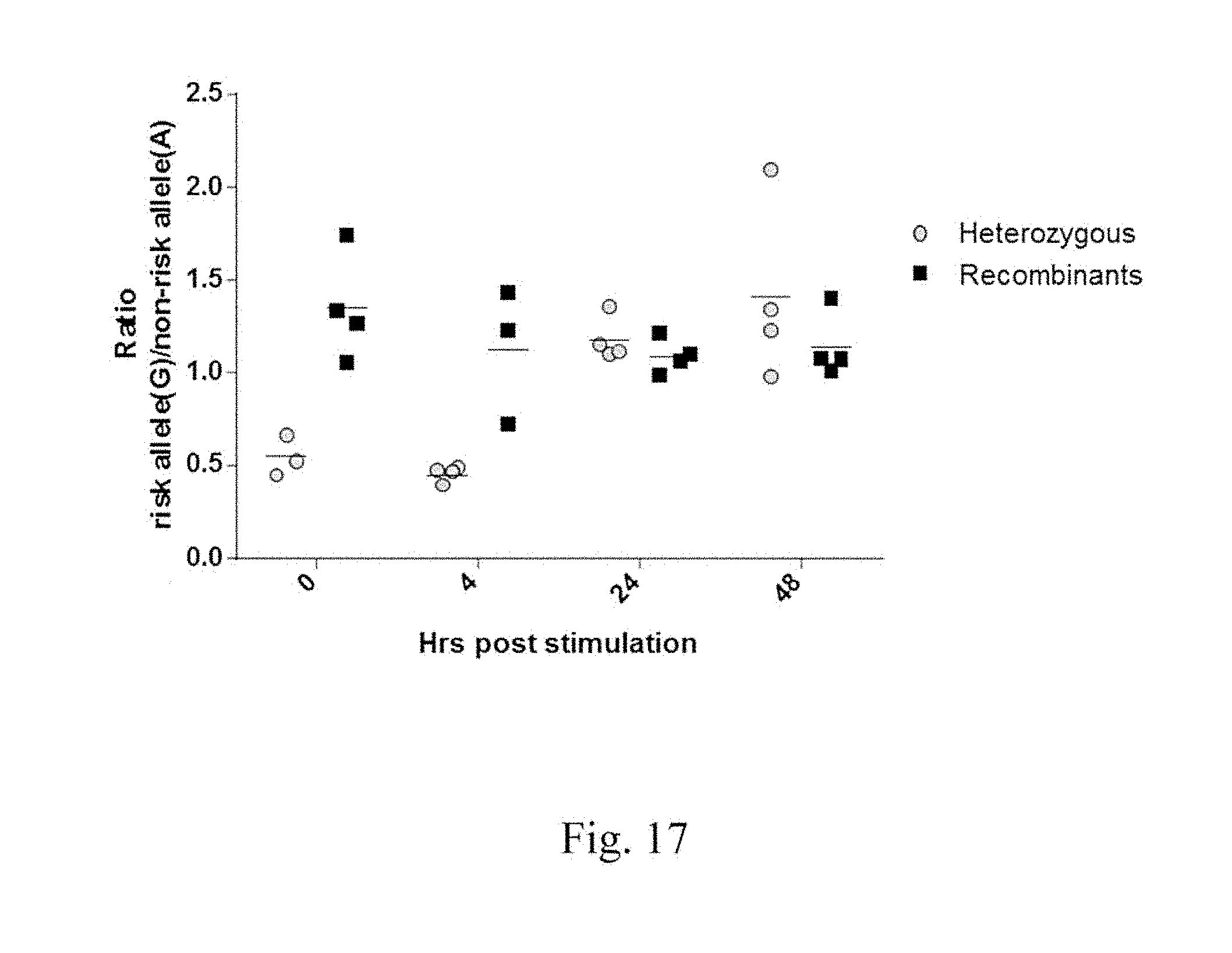
D00018
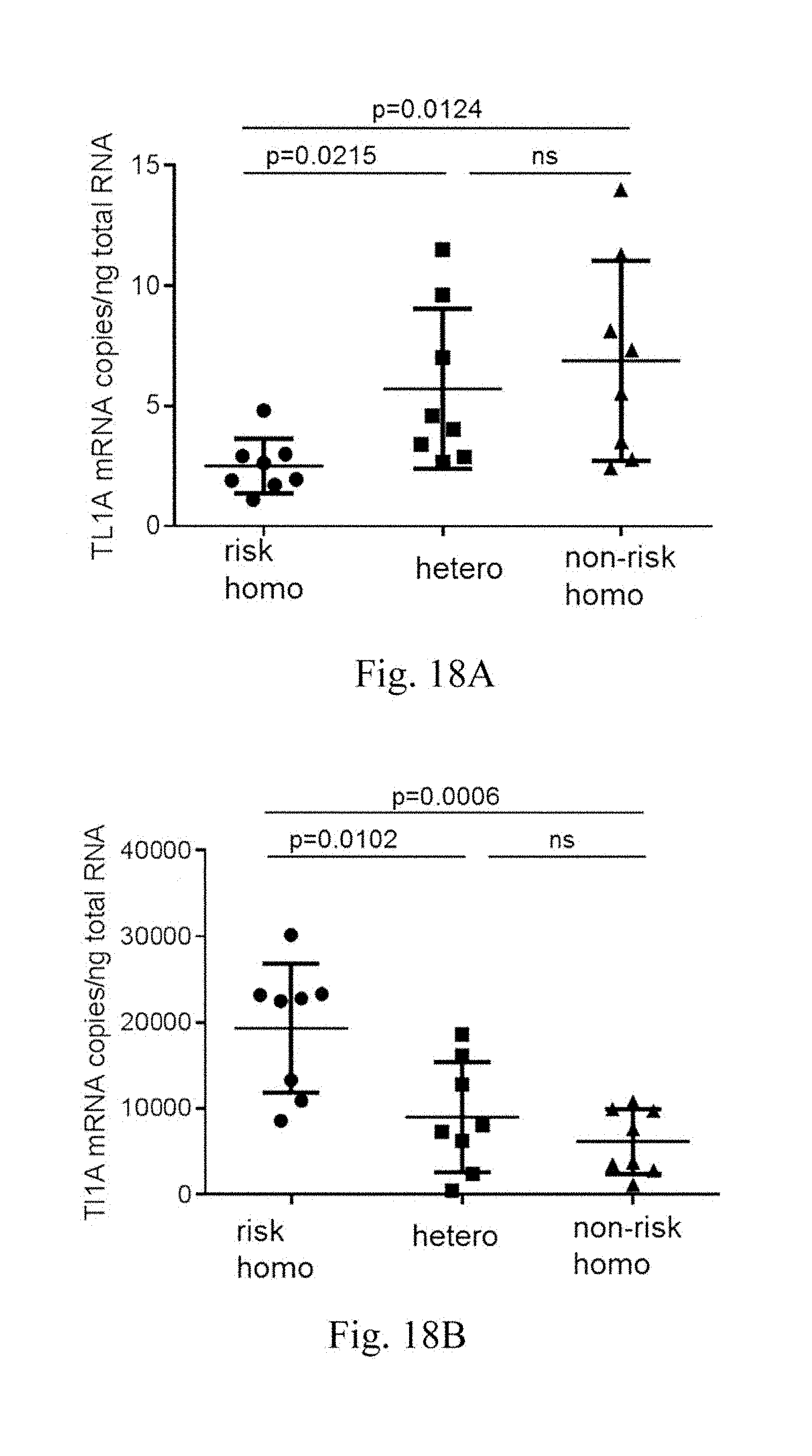
D00019

D00020

D00021

D00022

D00023
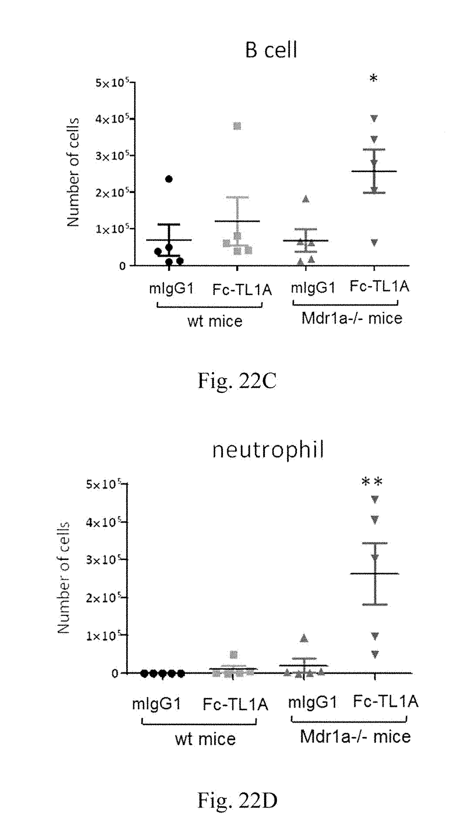
D00024

D00025
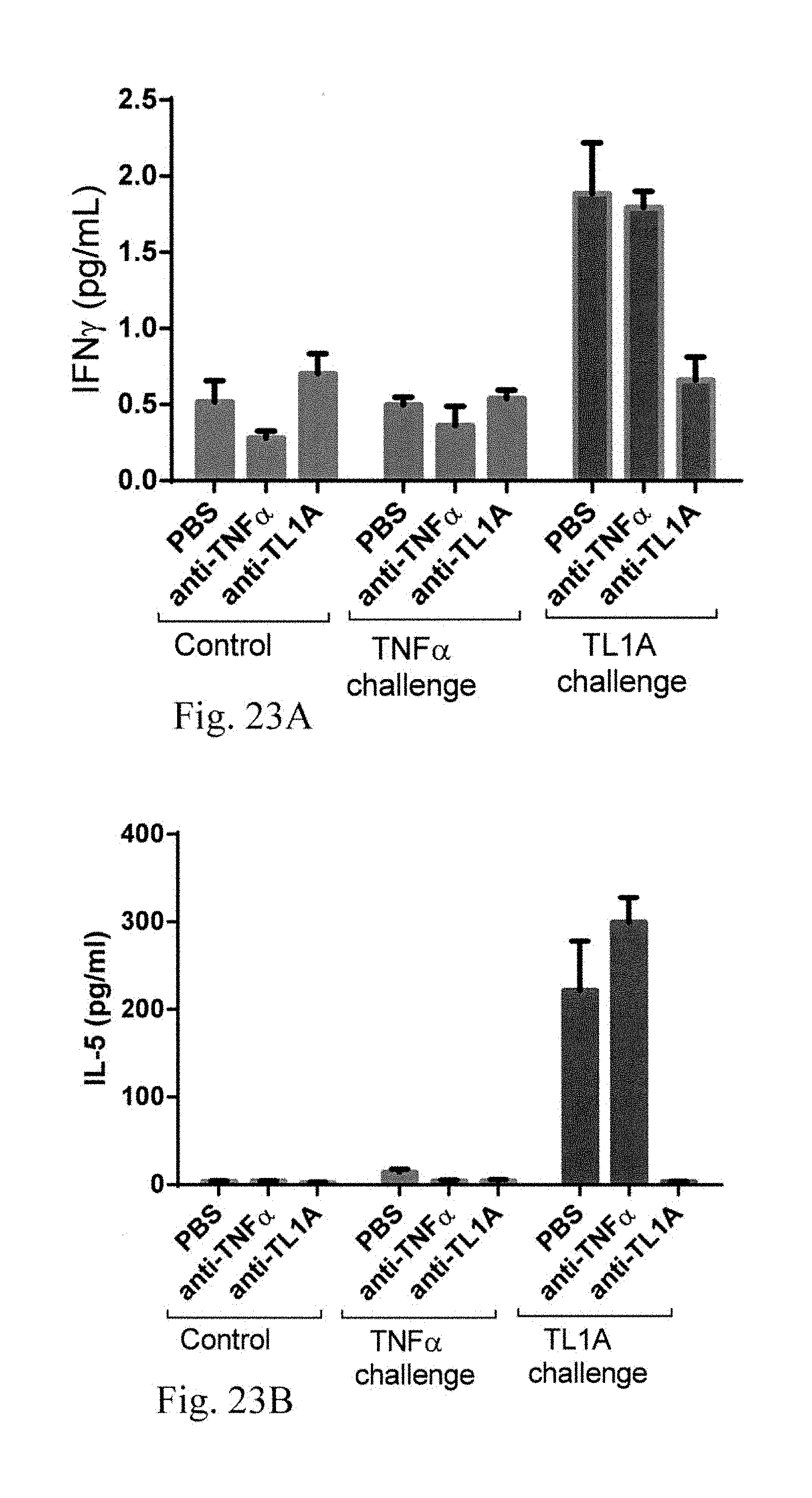
D00026
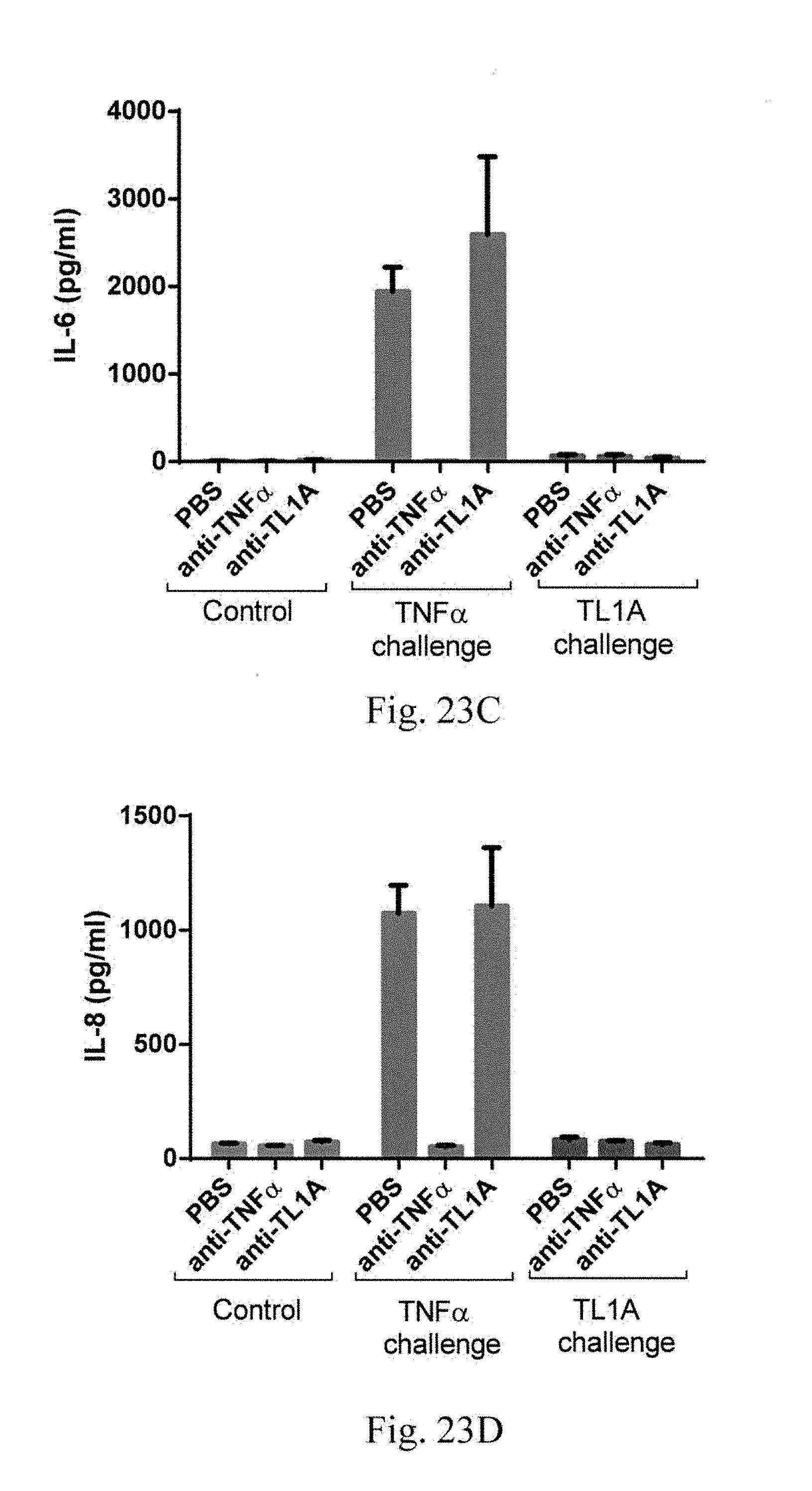
D00027

D00028

D00029

D00030
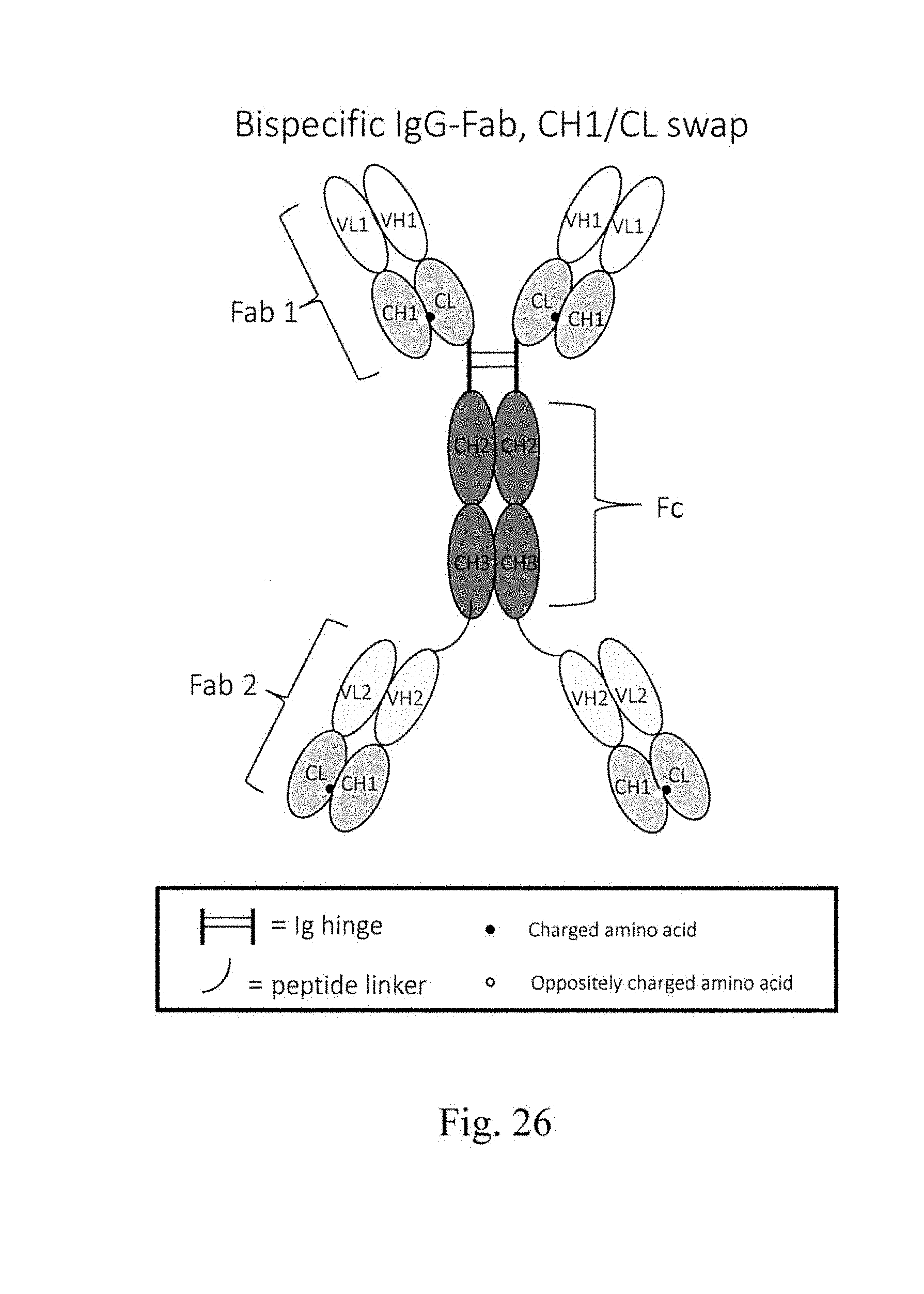
D00031
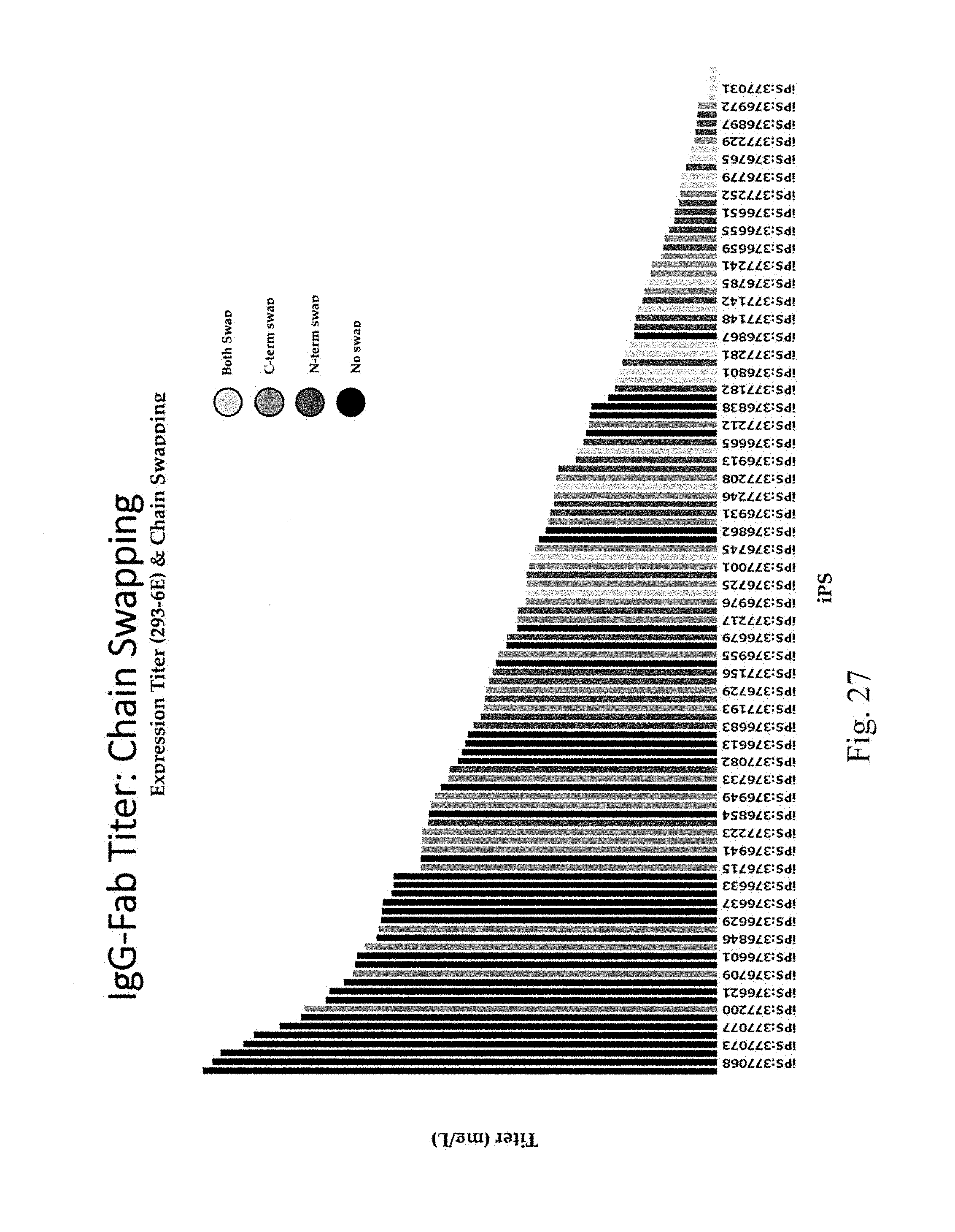
D00032

D00033

D00034

D00035

D00036

D00037
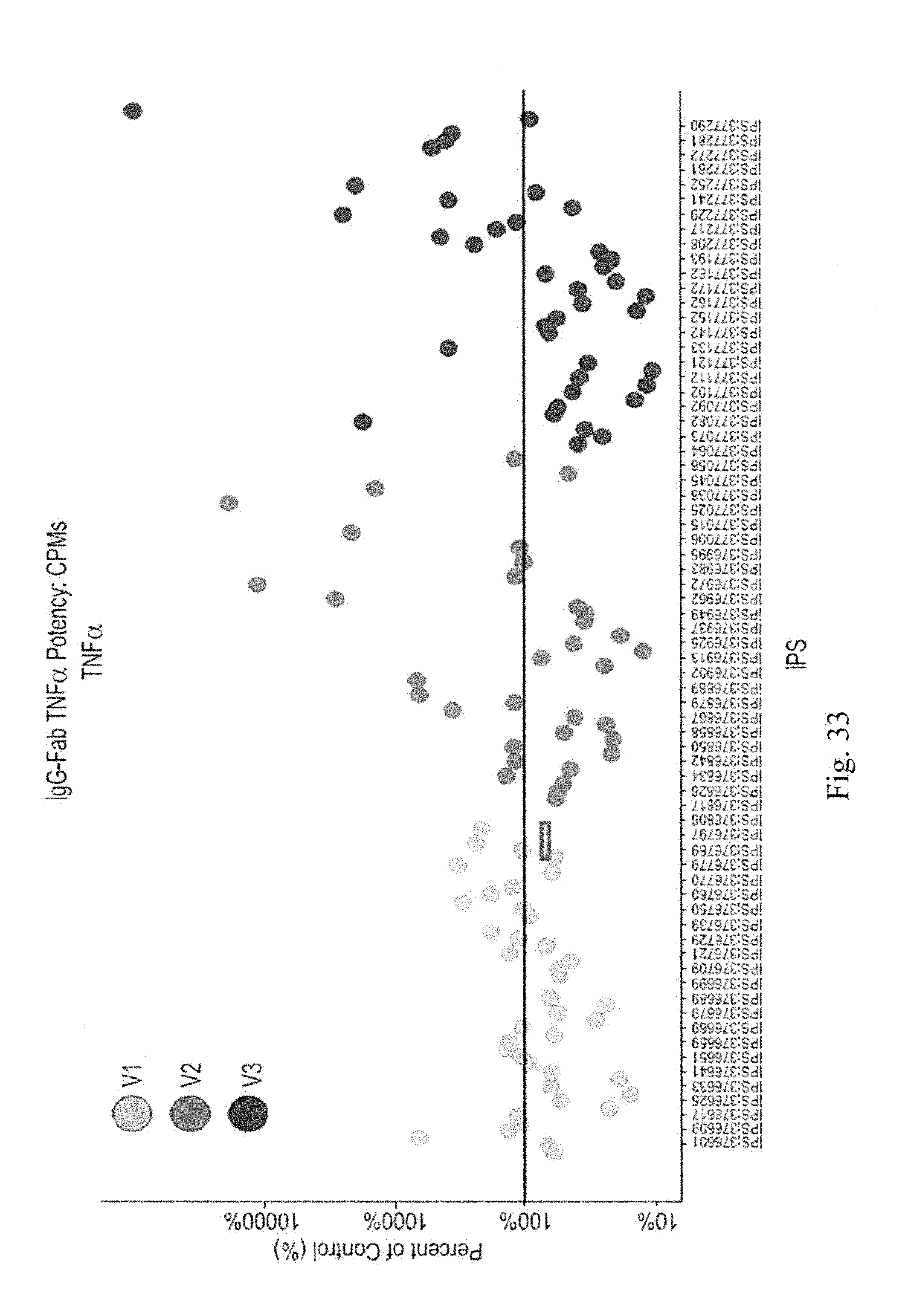
XML
uspto.report is an independent third-party trademark research tool that is not affiliated, endorsed, or sponsored by the United States Patent and Trademark Office (USPTO) or any other governmental organization. The information provided by uspto.report is based on publicly available data at the time of writing and is intended for informational purposes only.
While we strive to provide accurate and up-to-date information, we do not guarantee the accuracy, completeness, reliability, or suitability of the information displayed on this site. The use of this site is at your own risk. Any reliance you place on such information is therefore strictly at your own risk.
All official trademark data, including owner information, should be verified by visiting the official USPTO website at www.uspto.gov. This site is not intended to replace professional legal advice and should not be used as a substitute for consulting with a legal professional who is knowledgeable about trademark law.