Methods For Treatment Of Melanoma
Zon; Leonard ; et al.
U.S. patent application number 16/029265 was filed with the patent office on 2019-04-25 for methods for treatment of melanoma. This patent application is currently assigned to CHILDREN'S MEDICAL CENTER CORPORATION. The applicant listed for this patent is CHILDREN'S MEDICAL CENTER CORPORATION, DANA-FARBER CANCER INSTITUTE. Invention is credited to Richard M. White, Leonard Zon.
| Application Number | 20190117635 16/029265 |
| Document ID | / |
| Family ID | 54333760 |
| Filed Date | 2019-04-25 |


View All Diagrams
| United States Patent Application | 20190117635 |
| Kind Code | A1 |
| Zon; Leonard ; et al. | April 25, 2019 |
METHODS FOR TREATMENT OF MELANOMA
Abstract
Embodiments of the present invention are directed to methods for treatment of melanoma using an inhibitor of dihydroorotate dehydrogenase (DHODH) and to combination therapies that involve administering to a subject an inhibitor of oncogenic BRAF (e.g. BRAF(V600E)), as well as an inhibitor of dihydroorotate dehydrogenase (DHODH). Assays for identifying compounds useful for the treatment of melanoma are also provided. The methods comprise screening for compounds or agents that inhibit neural crest progenitor formation in a zebra fish model of melanoma.
| Inventors: | Zon; Leonard; (Wellesley, MA) ; White; Richard M.; (Cambridge, MA) | ||||||||||
| Applicant: |
|
||||||||||
|---|---|---|---|---|---|---|---|---|---|---|---|
| Assignee: | CHILDREN'S MEDICAL CENTER
CORPORATION Boston MA DANA-FARBER CANCER INSTITUTE Boston MA |
||||||||||
| Family ID: | 54333760 | ||||||||||
| Appl. No.: | 16/029265 | ||||||||||
| Filed: | July 6, 2018 |
Related U.S. Patent Documents
| Application Number | Filing Date | Patent Number | ||
|---|---|---|---|---|
| 14733498 | Jun 8, 2015 | 10016402 | ||
| 16029265 | ||||
| 13983090 | Oct 4, 2013 | |||
| PCT/US2012/024295 | Feb 8, 2012 | |||
| 14733498 | ||||
| 61440475 | Feb 8, 2011 | |||
| Current U.S. Class: | 1/1 |
| Current CPC Class: | A61K 31/381 20130101; A61K 45/06 20130101; A61K 31/42 20130101; A61K 31/437 20130101; A61K 31/44 20130101; A61K 31/196 20130101; A61K 31/42 20130101; A61K 2300/00 20130101; A61K 31/437 20130101; A61K 2300/00 20130101 |
| International Class: | A61K 31/44 20060101 A61K031/44; A61K 31/42 20060101 A61K031/42; A61K 31/381 20060101 A61K031/381; A61K 31/196 20060101 A61K031/196; A61K 31/437 20060101 A61K031/437; A61K 45/06 20060101 A61K045/06 |
Goverment Interests
GOVERNMENT SUPPORT
[0002] This invention was made with Government support under Grant No. R01CA103846-09 awarded by the National Institute of Health. The government has certain rights in the invention.
Claims
1. A composition comprising an inhibitor of dihydroorotate dehydrogenase (DHODH) and an inhibitor of oncogenic BRAF, wherein the inhibitor of dihydroorotate dehydrogenase is selected from the group consisting of: ##STR00001## ##STR00002##
2. The composition of claim 1, wherein the composition further comprises a pharmaceutically acceptable carrier or diluent.
3. The composition of claim 1, wherein the inhibitor of dihydroorotate dehydrogenase is ##STR00003##
4. The composition of claim 3, wherein the inhibitor of dihydroorotate dehydrogenase is ##STR00004##
5. The composition of claim 3, wherein the inhibitor of dihydroorotate dehydrogenase is ##STR00005##
6. The composition of claim 1, wherein the oncogenic BRAF is BRAF(V600E).
7. The composition of claim 1, wherein the inhibitor of oncogenic BRAF is selected from the group consisting of: Sorafenib, RAF265, XL281, AZ628, GSK2118436, GDC-0879, PLX4032, and PLX4720.
8. The composition of claim 7, wherein the inhibitor of oncogenic BRAF is PLX4032 or PLX4720.
9. The composition of claim 8, wherein the inhibitor of oncogenic BRAF is PLX4032.
10. The composition of claim 8, wherein the inhibitor of oncogenic BRAF is PLX4720.
11. A method of screening for an agent that inhibits melanoma growth comprising: (a) contacting a zebrafish embryo with a test agent for a period of time, (b) rinsing the test agent from the embryos of step (a); and (c) assaying the number of neural crest progenitors as compared to a control zebrafish embryo that has not been contacted with the test agent, wherein a reduced number of neural crest progenitors indicates that the compound is capable of inhibiting melanoma.
12. The method of claim 11, wherein the reduced number of neural crest progenitors is due to their differentiation into melanocytes.
13. The method of claim 11, wherein the zebrafish embryo is a wild type zebra fish embryo.
14. The method of claim 11, wherein the zebrafish embryo is a transgenic zebra fish embryo.
15. The method of claim 11, wherein the zebrafish embryo expresses green fluorescent protein operably linked to the melanocyte mitfa promoter.
16. The method of claim 11, wherein the zebrafish embryo expresses a nucleic acid encoding a mutant human BRAF.
17. The method of claim 11, wherein the zebrafish embryo expresses a nucleic acid encoding a mutant tumor suppressor or protooncogene.
18. The method of claim 11, wherein the number of neural crest progenitors is assayed by monitoring crestin expression
19. The method of claim 11, wherein the number of neural crest progenitors is assayed by monitoring sox10 expression.
20. The method of claim 11, wherein the number of neural crest progenitors is assayed by monitoring dct expression.
Description
CROSS REFERENCE TO RELATED APPLICATIONS
[0001] This Application is a Continuation of U.S. Ser. No. 14/733,498, filed Jun. 8, 2015, which is a Continuation-in-Part application of U.S. Ser. No. 13/983,090, filed Oct. 4, 2013, which is a 35 U.S.C. 371 National Stage Entry Application of International Application No. PCT/US2012/024295 filed Feb. 8, 2012, which designates the U.S. and claims the benefit under 35 U.S.C .sctn. 119(e) of U.S. Provisional Application No. 61/440,475 filed Feb. 8, 2011, the contents of which are incorporated herein by reference in their entireties.
SEQUENCE LISTING
[0003] The instant application contains a Sequence Listing which has been submitted electronically in ASCII format and is hereby incorporated by reference in its entirety. Said ASCII copy, created on Jul. 6, 2018, is named 701039-064585C2_SL.txt and is 25,809 bytes in size.
FIELD OF INVENTION
[0004] The present invention relates to methods for treatment of melanoma using an inhibitor of dihydroorotate dehydrogenase (DHODH) and to combination therapies that involve administering to a subject an inhibitor of oncogenic BRAF (e.g. BRAF(V600E)), as well as an inhibitor of dihydroorotate dehydrogenase (DHODH). Assays for identifying compounds useful for the treatment of melanoma are also provided. The methods comprise screening for compounds or agents that inhibit neural crest progenitor formation in a zebra fish model of melanoma.
BACKGROUND OF THE INVENTION
[0005] Melanoma is a malignant tumor of melanocytes. Primarily melanoma is a skin tumor, but it is also seen, though less frequently, in the melanocytes of the eye (uveal melanoma). Even though it represents one of the rarer forms of skin cancer, melanoma underlies the majority of skin cancer-related deaths and despite many years of intensive laboratory and clinical research, there are still limited treatments for melanoma.
[0006] One effective cure for melanoma (prior to metastasis) is surgical resection of the primary tumor before it achieves a thickness of greater than 1 mm. If the tumor is more invasive, surgery can be combined with radiation and/or chemotherapy. Since these conventional modalities cannot cure patients of lethal metastasized tumors, efficacy of alternative treatments such as immunotherapy are being investigated in clinical trials.
[0007] Oncogenic BRAF mutations are present in a majority of melanomas and have been implicated in malignant growth of melanoma cells. BRAF(V600E) mutation is the most common oncogenic BRAF mutation found in melanoma cells. Recently, Zelboraf.TM. (also known as Vemurafenb or PLX4032: Hoffman-La-Roche (Madison Wis.)/Daiichi Sankyo (Parsippany, N.J.)) was approved for treatment of unresectable (inoperable) or metastatic melanoma with a BRAF(V600E) mutation. There have been positive results with Zelboraf.TM. (Hoffman-La-Roche (Madison Wis.)/Daiichi Sankyo (Parsippany, N.J.)), however resistance is a problem. In addition, there are unwanted side effects, including back pain, constipation, cough, diarrhea, dizziness, dry skin, hair loss, headaches, joint or muscle pain, loss of appetite; nausea, taste changes, thickening of the skin, tiredness, vomiting, and weakness, as well as severe allergic reactions. Thus, treatments for melanoma still need to be improved.
SUMMARY
[0008] We have determined by gene expression analysis that melanoma cells adopt a fate similar to multipotent neural crest progenitors. We used zebra-fish embryos to identify the initiating transcriptional events that occur upon activation of oncogenic BRAF(V600E) in the neural crest lineage. Zebrafish embryos that are transgenic for mitfa:BRAF(V660E) and which lack p53 have been found to have a gene signature that is enriched for markers of multipotent neural crest cells, and neural crest progenitors from these embryos fail to terminally differentiate. In particular, we discovered that BRAF(V600E):p53.sup.-/- embryos exhibit an abnormal expansion in the number of crestin.sup.+ progenitors. Furthermore, in the adult, BRAF(V600E):p53.sup.-/- virtually all tumor cells, but no normal cells, are positive for crestin. Thus, we concluded that suppressors of neural crest progenitors may have utility in the treatment of melanoma. Accordingly, we developed a screen to identify inhibitors of the crestin.sup.+ lineage during development, allowing for identification of compounds or agents useful for melanoma treatment. We screened a library of 2,000 chemicals for suppressors of neural crest development in BRAF(V600E):p53.sup.-/- zebrafish and successfully identified compounds useful in the treatment of melanoma.
[0009] Embodiments of the invention are based on the development of a screening assay for inhibitors of melanoma, and on the discovery of one class of compounds, i.e. inhibitors of dihydroorotate dehydrogenase (DHODH), that result in almost complete abrogation of neural crest development in zebrafish. In particular, we have determined that inhibitors of DHODH, when used alone or in combination with an inhibitor of the BRAF oncogene (e.g. BRAF(V600E)), decrease melanoma growth both in vitro and in vivo in mouse xenograft studies. The combination therapy required only subclinical doses of each compound suggesting that there may be therapeutic synergy. Unexpectedly, 40% of the treated mice had complete regression of tumors using a combination of the specific oncogenic BRAF(V600E) inhibitor PLX4720 (Plexxikon Inc., Berkeley, Calif., USA) and the DHODH inhibitor Leflunomide (Arava.TM. Sanofi-Aventis (Paris, France), which led abrogation of tumor growth in nude mice transplanted with A375 human melanoma cells.
[0010] We have also determined that many other inhibitors of DHODH, e.g. when tested in zebra fish, on human A375 melanoma cells and in primary tumor xenographs, when used alone or in combination with an inhibitor of the BRAF oncogene (e.g. BRAF(V600E)), decrease melanoma growth both in vitro and in vivo. In particular, see the compounds of FIG. 21, FIG. 22 and FIG. 23, which are labeled 1-10 and Example III.
[0011] Accordingly, provided herein are methods for treatment of melanoma comprising administrating to a subject (diagnosed with, or at risk of having melanoma), an inhibitor of DHODH. Also provided is a combination therapy for treatment of melanoma that involves administrating an effective amount of an inhibitor of DHODH and an effective amount of an inhibitor of oncogenic BRAF (e.g. BRAF(V600E) oncogene), where the inhibitors are administered simultaneously, or sequentially. Methods of screening for agents that inhibit melanoma growth are also described.
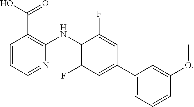
[0012] In one aspect of the invention, a method for treating melanoma in a subject is provided, which comprises administering to a subject in need thereof a therapeutically effective amount of an inhibitor of dihydroorotate dehydrogenase (DHODH) (e.g. a small molecule, a nucleic acid RNA, a nucleic acid DNA, a protein, a peptide, or an antibody). In one embodiment, the inhibitor of dihydroorotate dehydrogenase (DHODH) is selected from the group consisting of: leflunomide, teriflunomide, brequinar, dichloroallyl lawsone, maritimus, redoxal and NSC210627, or a derivative thereof. In one embodiment, the DHODH inhibitor is selected from the group consisting of C.sub.19H.sub.14F.sub.2N.sub.2O.sub.3, compound 1; C.sub.20H.sub.16F.sub.2N.sub.2O.sub.3, compound 2; C.sub.18H.sub.13F.sub.6NO.sub.4, compound 3; C.sub.19H.sub.16F.sub.3NO.sub.4, compound 4; C.sub.19H.sub.10F.sub.5NO.sub.4S, compound 5; C.sub.19H.sub.14FNO.sub.4S, compound 6; C.sub.20H.sub.15F.sub.4NO.sub.4, compound 7; C.sub.19H.sub.17NO.sub.4, compound 8; C.sub.20H.sub.17F.sub.2NO.sub.4, compound 9; and C.sub.20H.sub.18FNO.sub.4, compound 10, each as depicted in FIG. 21, FIG. 22 and FIG. 23.
[0013] In another aspect of the invention, a combination therapy for treating melanoma is provided. The method comprises administering to a subject in need thereof a therapeutically effective amount of an inhibitor of dihydroorotate dehydrogenase (DHODH) and an effective amount of an inhibitor of oncogenic BRAF. The inhibitors may be a small molecule, a nucleic acid RNA, a nucleic acid DNA, a protein, a peptide, or an antibody. The inhibitors (compounds/agents) may be administered simultaneously, or sequentially. In one embodiment, each inhibitor is administered within minutes, within hours, or within days, of one another.
[0014] Any oncogenic BRAF can be inhibited, e.g. in one embodiment, the oncogenic BRAF that is inhibited is BRAF(V600E). Alternative oncogenic BRAFs are described within the specification. The inhibitor may be specific for a particular oncogenic BRAF mutation or alternatively may generally inhibit multiple BRAF mutations, and/or wild type BRAF.
[0015] Any inhibitor of dihydroorotate dehydrogenase (DHODH) may be used. In one embodiment, the inhibitor of dihydroorotate dehydrogenase (DHODH) to be used in combination with an inhibitor of oncogenic BRAF is selected from the group consisting of: leflunomide, teriflunomide, brequinar, dichloroallyl lawsone, maritimus, redoxal and NSC210627, or a derivative thereof. In one embodiment, the DHODH inhibitor is selected from the group consisting of C.sub.19H.sub.14F.sub.2N.sub.2O.sub.3, compound 1; C.sub.20H.sub.16F.sub.2N.sub.2O.sub.3, compound 2; C.sub.18H.sub.13F.sub.6NO.sub.4, compound 3; C.sub.19H.sub.16F.sub.3NO.sub.4, compound 4; C.sub.19H.sub.10F.sub.5NO.sub.4S, compound 5; C.sub.19H.sub.14FNO.sub.4S, compound 6; C.sub.20H.sub.15F.sub.4NO.sub.4, compound 7; C.sub.19H.sub.17NO.sub.4, compound 8; C.sub.20H.sub.17F.sub.2NO.sub.4, compound 9; and C.sub.20H.sub.18FNO.sub.4, compound 10, each as depicted in FIG. 21, FIG. 22 and FIG. 23.
[0016] In one embodiment, the inhibitor of oncogenic BRAF is selected from the group consisting of: Sorafenib, RAF265, XL281, AZ628, GSK2118436, GDC-0879, PLX4032, and PLX4720, or a derivative thereof.
[0017] In one embodiment, the inhibitor of oncogenic BRAF is PLX4032 and the inhibitor of dihydroorotate dehydrogenase (DHODH) is leflunomide, or a derivative thereof.
[0018] In another embodiment, the inhibitor of oncogenic BRAF is PLX4720 and the inhibitor of dihydroorotate dehydrogenase (DHODH) is leflunomide, or a derivative thereof.
[0019] In another embodiment, the methods of treatment described herein, further comprise selecting a subject that has, or is at risk of having melanoma, e.g. a melanoma that expresses oncogenic BRAF. In one embodiment, the subject has a melanoma that expresses an oncogenic BRAF comprising a mutation in BRAF selected from the group consisting of: VAL600GLU, ARG461ILE, ILE462SER, GLY463GLU, and LYS600GLU, GLY465VAL and LEU596ARG, and GLY468ARG, GLY468ALA and ASP593GLY.
[0020] The methods of the present invention can be used either alone, or in conjunction with other treatment methods known to those of skill in the art. For example, such methods may include, but are not limited to, chemotherapy, radiation therapy, or surgery.
[0021] Administration of the inhibitors can be performed by intravenous, intramuscular, subcutaneous, intradermal, topical, intraperitoneal, intrathecal, intrapleural, intrauterine, rectal, vaginal, intrasynovial, intraocular/periocular, intratumor or parenteral administration.
[0022] In one embodiment, the subject is at risk for developing melanoma and the combination therapy, or DHODH inhibitor, is administered prophylactically. The risk can be determined genetically. Alternatively, the risk can be determined by measuring levels of marker proteins in the biological fluids (i.e. blood, urine) of a patient. In one embodiment, the methods of treatment described herein further comprises the step of selecting a subject in need thereof of treatment, e.g. selecting a subject diagnosed with melanoma, or a subject at increased risk of melanoma (e.g. potential cancer relapse).
[0023] In another aspect of the invention, screening methods for identifying agents that inhibit melanoma growth are provided. The methods comprise, (a) contacting a zebrafish embryo with a test agent for a period of time, (b) rinsing the test agent from the embryos of step (a); and (c) assaying the number of neural crest progenitors as compared to a control zebrafish embryo that has not been contacted with the test agent, wherein a reduced number of neural crest progenitors (e.g. and their differentiation into melanocytes) indicates that the compound is capable of inhibiting melanoma. The zebrafish embryos may be wild type zebra fish embryos or transgenic zebrafish embryos.
[0024] In one embodiment, the number of neural crest progenitors is assayed by monitoring crestin expression, or sox10 expression, or dct expression, e.g. by quantitation of ISH studies.
[0025] In one embodiment, the transgenic zebrafish embryo expresses green fluorescent protein operably linked to the melanocyte mitfa promoter and melanocyte neural crest progenitors are monitored by GFP expression.
BRIEF DESCRIPTION OF FIGURES
[0026] FIGS. 1a to 1c show zebrafish melanoma and neural crest gene expression. FIG. 1a, Transgenic zebrafish expressing BRAF.sup.V600E under the melanocyte specific mitfa promoter develop pigmentation abnormalities, and melanoma when crossed with p53.sup.-/- fish. Gross embryonic development is largely normal. FIG. 1b, Gene expression analysis reveals a unique gene signature at 72hpf in the BRAF.sup.V600E; p53.sup.-/- strain (left). Gene set enrichment analysis (GSEA) reveals an enrichment between the embryonic gene signature and the adult melanomas which form 4-12 months later (middle and right; see Methods for full GSEA methods). Embryo heat map columns represent average of 3 clutches (log 2 scale, range -2 to +2 fold); adult heat map columns represent individual fish (log 2 scale, range -10 to +10 fold). FIG. 1c, Sagittal section of WT and BRAF.sup.V600E; p53.sup.-/- adults reveal homogeneous crestin expression (blue staining, shown as dark grey) only within the dorsal melanoma, whereas it is absent in normal adult tissues.
[0027] FIGS. 2a to 2b show ISH staining in Zebrafish. A chemical genetic screen to identify suppressors of neural crest development. FIG. 2a, A chemical genetic screen to identify suppressors of the crestin.sup.+ lineage during embryogenesis identified NSC210627, a compound which completely abrogates expression by ISH (FIG. 2a, top and middle). The Discovery gate chemoinformatic algorithm revealed structural similarity between NSC210627 and brequinar (see FIG. 5), an inhibitor of dihydroorotate dehydrogenase (DHODH). Leflunomide, FIG. 2a structurally distinct DHODH inhibitor, phenocopies the crestin phenotype of NSC210627 (FIG. 2a, right). FIG. 2a bottom panel; Leflunomide caused an absence of multiple neural crest derivatives, including pigmented melanocytes, mitf-GFP.sup.+ melanocyte progenitors, and mbp-mCherry.sup.+ glial cells. FIG. 2b, Leflunomide or A771726 (see FIG. 6a) significantly reduced the number of multipotent daughter cells that could be subcloned from individual primary neural crest stem cell colonies (Values shown are mean+/-SD of n=3 replicates; *, p<0.05 compared to control, t-test).
[0028] FIGS. 3a to 3c show that DHODH inhibition modulates transcriptional elongation. FIG. 3a, The hypomorphic spt5.sup.m806 mutant has only a mild pigment defect on its own (top). Treatment with low-dose leflunomide (3 uM) leads to an almost complete absence of neural crest derived melanocytes in the mutant line. See FIG. 7 for dose-response quantification of this effect. FIG. 3b, Metagene analysis of RNA pol II occupancy in A375 human melanoma cells after treatment with leflunomide. Pol II occupancy at the promoter region is unaffected, but diminished at the 3' end of the genes. Inset shows a higher magnification of the 3' region of the genes. FIG. 3c, Representative examples of myc target genes which demonstrate defects in transcriptional elongation after leflunomide treatment, along with a non-affected gene. For Npm1, the TR in DMSO=5.04, and in LEF=8.10. For Ccnd1, the TR in DMSO=3.47, and in LEF=4.67.
[0029] FIGS. 4a to 4c show graphs. DHODH blockade suppresses melanoma growth in concert with BRAF.sup.V600E inhibition. FIG. 4a, Leflunomide causes a dose-dependent decrease in melanoma proliferation as measured by CellTiterGlo assay in 3 BRAF.sup.V600E melanoma cells lines tested (A375, RPMI7951, Hs.294T). FIG. 4b, FIG. 4c Leflunomide cooperates with the BRAF.sup.V600E inhibitor PLX4720 in inhibiting melanoma cell proliferation in the A375 (FIG. 4b) and Hs.294T (FIG. 4) cell lines as well as the other tested lines (See FIG. 8). FIG. 4d, After subcutaneous transplantation of A375 cells (3.times.10.sup.5) into nude mice, both leflunomide and PLX4720 impair tumor progression, with the combination showing a nearly complete abrogation of tumor growth and in 2/5 animals complete tumor regression. (*p=0.036 DMSO vs. PLX; **p=0.006 DMSO vs. LEF; ***p=0.006 PLX or LEF vs. PLX/LEF; PLX vs. LEF: p=NS, ANOVA followed by Tukey post-hoc analysis). Values shown are mean+/-SEM of n=3-5 replicates, as shown.
[0030] FIG. 5 shows chemical structures of DHODH inhibitors NSC210627, brequinar, leflunomide and A771726 (teriflunomide) and oncogenic BRAF inhibitor PXL4720.
[0031] FIGS. 6a to 6c: The effects the leflunomide derivative A771726, on rat neural crest stem cells. FIG. 6a, The effects of A771726 on rat neural crest stem cell self-renewal (as described in FIG. 2, *, p<0.05, compared to control, t-test). FIG. 6b, FIG. 6c, Both compounds cause no defect in plating efficiency or affect individual progeny (NGM=nerve, glia, smooth muscle) without (N, G, or M colonies) survival or differentiation capacity (p>0.05, n=3, ANOVA). Values shown are mean+/-SD of n=3 replicates.
[0032] FIGS. 7a to 7d show graphs of the effect of low-doses of leflunomide on pigmentation in the spt5.sub.m806 hypomorphic mutant. Quantification of the effect described in FIG. 3a, in which 3, 4 or 5 uM leflunomide was applied to a spt5.sup.m806 hypomorphic incross. FIG. 7a, Pigmentation scores in untreated embryonic offspring of an incross of the spt5.sup.m806 hypomorphic mutant. There is a mild pigmentation defect only in homozygous embryos. FIG. 7b, FIG. 7c, FIG. 7d Leflunomide at 3, 4 or 5 uM demonstrates that both heterozygous and homozygous mutants embryos show increased sensitivity to pigment loss when compared to wild-type (*, p=0.000018, Kruskal-Wallis, n values as indicated in Figure).
[0033] FIGS. 8a to 8d show graphs. FIG. 8a, FIG. 8ab, In vitro proliferation assay testing the effects of combined A771726 and the BRAF inhibitor PLX4720 on RPMI7951 and 294T cells shows significant augmentation of the effects of the BRAF inhibitor alone. FIG. 8c, PLX4720 is only effective in BRAF.sup.V600E melanoma cells. Although RPMI7951 is BRAF.sup.V600E, it is significantly less sensitive than the other tested cell lines. FIG. 8d, Proliferation in the presence of A771726 in pancreatic cancer (8988, PANC1) and breast cancer (MDA-MD-231) cell lines. Values shown are mean+/-SEM of n=4 replicates.
[0034] FIGS. 9a to 9b, show a graph of a human melanoma tissue microarray (n=70 samples) which was analyzed for markers of neural crest progenitors (ednrb, edn3) and melanocyte progenitors (mitf, dct). The majority of human melanomas are positive for neural crest markers but only a fraction express more differentiated markers. FIG. 9b, shows marker staining of tumors, a representative tumor in which all 4 markers are expressed in different areas of the tumor.
[0035] FIG. 10 shows cumulative results of the chemical screen described in the Methods section. 2000 chemicals were screened, and positive "hits" were those with a staining score of less than 2 (i.e. 3%). Representative examples of each score are shown on top. The percentage of chemicals resulting in a given score are shown below.
[0036] FIG. 11 shows graphs depicting enzymatic inhibition. The effect of NSC210627 on purified human and malarial DHODH. This demonstrates a strong inhibition of both enzymes, at lower concentrations in the human versus malarial forms. Error bars are mean+/-SEM of n=3 replicates.
[0037] FIG. 12 shows in situ hybridization for neural crest and non-neural crest lineages in the presence of leflunomide. No significant difference is seen in intensity for the mesoderm marker ntl (although the pattern of expression is altered in some embryos) and blood marker gata1. A disruption in the pattern of foxd3 expression, and absence of sox10 and dct expression confirms broad defects in neural crest development in the presence of leflunomide.
[0038] FIGS. 13a to 13c show in situ hybridization and graphs. FIG. 13a, The spt5sk8-/- transcriptional elongation mutant phenocopies the absence of crestin+ progenitors in a manner analogous to leflunomide. FIG. 13b, GSEA was used to compare the gene expression signature of leflunomide treated embryos and spt5sk8-/- embryos at 24hpf Venn diagram shows the number of genes up or downregulated in the spt5sk8-/- mutant that are similarly up or downregulated after leflunomide treatment. (see Methods for full GSEA analysis methods) FIG. 13c, qRT-PCR on whole embryos treated with DMSO or leflunomide was used to measure transcription elongation at the 5' or 3' end of the noted gene. Leflunomide treatment caused a significant decrease in 3' transcription of mitf, pvalb2, her4.2 and dlb, whereas control genes showed no bias towards 5' or 3' effects (n=6 replicates in each group, *p<0.05, two-sided t-test, leflunomide vs. DMSO). Values shown are mean+/-SEM.
[0039] FIGS. 14a to 14b show metagene analysis graphs. ChIP-seq results in melanoma MAMLE-3M cells. Data are the same as that described in FIG. 3b,3c (A375 melanoma cells). FIG. 14a, Metagene analysis of RNA polymerase II occupancy of the promoter and gene body in MAMLE-3M cells treated with DMSO (control) or leflunomide 50 uM. Leflunomide causes no defect in promoter occupancy but significantly decreases pol II occupancy in the body of the gene, consistent with an inhibition of transcriptional elongation. FIG. 14b, Representative examples of affected (left) and unaffected genes in this cell line.
[0040] FIGS. 15a to 15c show a Western blot and graphs. FIG. 15a, Western blot showing extent of DHODH knockdown induced by shRNA#877 as compared to scrambled (scr) control shRNA in A375 melanoma cells. FIG. 15b, Cell counts measured over an 8 day period in A375-scr and A375-shRNA#877 cells shows a 57% decrease in cellular proliferation with DHODH knockdown. FIG. 15c, ChIP-PCR in A375-scr and A375-shRNA#877 cells demonstrates decreased RNA pol II binding at the 3' end of myc, npm1 and otub, consistent with an inhibition of transcriptional elongation (*p<0.05, +p=0.1 t-test, A375-sh877 vs. A375-scr cells, n=3-4). Values shown are mean+/-SEM.
[0041] FIG. 16 show mice after treatment with inhibitors. Representative examples of mice after treatment with DMSO, PLX alone, leflunomide alone or the combination. See FIG. 4 for quantification.
[0042] FIG. 17 shows the chemical structures of Sorafenib (Bayer) and RAF265.
[0043] FIG. 18 shows the chemical structures of AZ628, and GSK2118463.
[0044] FIG. 19 shows the chemical structures of PLX4032 and GDC-0879.
[0045] FIG. 20 shows the chemical structures of DHODH inhibitors dichlorallyl lawsone, maritumus (see U.S. Pat. No. 7,256,008), and redoxal.
[0046] FIG. 21 shows the chemical structures of DHODH inhibitors Compound 1, Compound 2, Compound 3, and Compound 4, which are used in Example III. Synthesis of compound 1 and compound 2 is described in U.S. Patent Publication 2010/007898. Synthesis of compounds 3 and 4 is described in U.S. Pat. No. 7,074,831.
[0047] FIG. 22 shows the chemical structures of DHODH inhibitors: Compound 5 and Compound 6, which are used in Example III. Synthesis of compound 5 and compound 6 is described in U.S. Patent Publication 2004/0192758.
[0048] FIG. 23 shows the chemical structures of DHODH inhibitors: Compound 7, Compound 8, Compound 9, and Compound 10, which are used in Example III. Synthesis of these compounds is described in U.S. Patent Publication 2006/0199859 and in U.S. Pat. No. 7,176,241.
[0049] FIGS. 24a to 24b show chemical structures and in situ hybridization (ISH) staining in Zebrafish; FIG. 24a shows the chemical structures for leflunomide, which gets converted into its active form A771726 also shown, which causes dihydroorotate dehydrogenase inhibition. Also shown in FIG. 24a is a structurally different DHODH inhibitor, inhibitor DH#1, also known as Compound 1 herein, as used in Example III. FIG. 24b are ISH staining showing the effect of DHODH inhibitor compound 1 (DH#1); leflunomide; and A771726 on neural crest development, in the presence of DMSO, Decylubiquinone, Aphiadicolin, or Progesterone. FIG. 24b shows that all of these inhibitors (leflunomide, A77 1726, or DH #1) lead to reduction in expression of the neural crest, depicted by an in situ hybridization for the neural crest marker crestin. However, chemicals including decylubiquinone, aphidicolin, and progesterone suppress these drugs and lead to a rescue of the neural crest population.
[0050] FIGS. 25a to 25b show in situ hybridization (ISH) staining in Zebrafish embryos to monitor neural crest development in the presence of compound 1, compound 7, leflunomide, compound 5 and compound 6 (See FIGS. 21-23 for compound structure and formula). Different concentrations of the DHODH inhibitors and in situ for crestin revealed compound 1 and compound 7 (FIG. 25a (#1 and #7) impair crestin expression at lower doses compared to leflunomide and compounds 5 and 6, FIG. 25b #5 and #6, respectively. The compounds showed varied strength in their ability to inhibit neural crest development, the effect of compound 1>compound 7>leflunomide, which is approximately the same as compounds 5 and 6.
[0051] FIG. 26 shows in situ hybridization (ISH) staining in Zebrafish embryos to monitor neural crest development in the presence of compound 4, compound 9 and compound 2 (#, 4 #9 and #2 respectively; See FIGS. 21 to 23 for compound structure and formula). When referring also to FIG. 25, it is shown that DHODH inhibitors #4, #9 and #2 are less effective than leflunomide. Indeed at 1.75 uM these drugs do not affect crestin expression like observed upon the same dose of leflunomide in FIG. 25b.
[0052] FIG. 27 shows a graph depicting the effect of all of the DHODH inhibitors compounds 1-10 (DHODH analogs) (See FIGS. 21-23 for compounds) on A375 cells after 3 days of treatment. y-axis percent inhibition, x-axis concentration. Compounds 1, 4, and 7 significantly decrease cell proliferation at lower doses compared to leflunomide and A771726.
[0053] FIGS. 28a to 28e show graphs depicting the effects of leflunomide, Compound 1 (PH-1), and Compound 7 (PH-7) (See FIG. 21 (compound 1) and FIG. 23 (compound 7)) on the indicated melanoma cells in vitro. FIG. 28a, M481 cells. FIG. 28b, A375 cells. FIG. 28c, M405 cells. FIG. 28d, M528 cells. FIG. 28e, M715 cells. Compound 1, Compound 7 and leflunomide affect cell viability of human primary melanoma cells. FIGS. 28a and 28b show respectively that leflunomide as well as DHODH #1 (PH-01, i.e. compound 1), DHODH#7 (PH-07, i.e. compound 7) impair cell survival in primary human melanoma cells (M481) and in the melanoma cell line (A375). FIG. 28c shows that the effect of these inhibitors is variable in different primary melanoma cells (M405, M528 and M715).
[0054] FIGS. 29a to 29d are graphs showing the in vivo effects of leflunomide in combination with digitoxin on M405 tumor xenografts in mouse. FIG. 29a, effect on tumor diameter (cm) vs. time. FIG. 29b, effect on tumor weight. FIG. 29c, effect on frequency of melanoma cells in the blood. FIG. 29d, Total flux(photons/second). M405 were not sensitive to drug treatment as shown in FIG. 28 and accordingly there is no effect on tumor growth (FIG. 29a) and number of melanoma cells in blood circulating cells (FIG. 29b). But there is significant difference in blood circulating cells in co-treatment with Leflunomide and Digitoxin. No change in flux was observed (FIG. 29d).
[0055] FIG. 30 is a graph of in vivo effects of the combination of DHODH inhibitor and Braf inhibitor on tumor volume, of M481 primary human melanoma xenografts in mice. FIG. 30 shows the effect of leflunomide in combination with Plexicon in xenografts mouse model. M841 primary human melanoma xenografts in mice revealed that leflunomide is more effective when used in combination with Plexicon.
[0056] FIG. 31 is a graph that shows the effect leflunomide in combination with Braf and Mek inhibitors on primary human melanoma cells xenografts in mice. Human BRAFV600 (M491) cells were treated with a combination of drugs including BRAF inhibitors (BRAFi), MEK inhibitors (MEKi) and Leflunomide. Leflunomide significantly impair tumor growth in combination with BRAFi and MEKi.
DETAILED DESCRIPTION
[0057] The present invention relates generally to a method of treating melanoma in a subject having, or at risk of having, melanoma.
[0058] As used herein, the term "subject" or "patient" or refers to any mammal. The patient is preferably a human, but can also be a mammal in need of veterinary treatment.
[0059] The term "melanoma" as used herein includes all types of melanoma, including, for example, melanoma skin cancer, ocular melanoma, and mucosal melanoma. Melanoma is caused by changes melanocytes that produce melanin. There are four major types of melanoma: 1) superficial spreading melanoma, which is usually flat and irregular in shape and color, with different shades of black and brown and is most common in Caucasians, 2) nodular melanoma, which usually starts as a raised area that is dark blackish-blue or bluish-red, but can be colorless, 3) Lentigo maligna melanoma, which usually occurs in the elderly and is most common in sun-damaged skin on the face, neck, and arms. The abnormal skin areas are usually large, flat, and tan with areas of brown, 4) Acral lentiginous melanoma, which is the least common form and usually occurs on the palms, soles, or under the nails and is more common in African Americans. Melanomas may also appear in the mouth, iris of the eye, or retina at the back of the eye and can be found during dental or eye examinations. Although very rare, melanoma can also develop in the vagina, esophagus, anus, urinary tract, and small intestine.
[0060] The presence of melanoma can be determined by means well known to those of skill in the art, e.g. tissue biopsies and in situ assays in which malignant melanoma (malignant melanocytes scattered in all epidermal layers) show atrophic epidermis, prominent dermal solar elastosis and almost always lymphocytic infiltration. Invasion of the dermis by melanocytes may occur in lentigo maligna melanoma. In addition, melanoma may be detected by methods that include, but are not limited, immunohistochemistry using the melanoma specific antibody HMB-45, or RT-PCR with different melanoma associated antigens (MAA) including, but not limited to tyrosinase, MART-1/Melan A, Pmel-17, TRP-1, and TRP-2 (see, e.g., Hatta N., et al., J Clin Pathol. 1998 August; 51(8):597-601). Biomarkers for melanoma are also known and can be used for example to assess subjects at risk of melanoma. Non-limiting example biomarkers for melanoma are described in PCT Publications WO 2008/141275, WO 2009/073513, or in U.S. Pat. No. 7,442,507, which are herein incorporated by reference in their entirety.
[0061] Symptoms of melanoma include, but are not limited to, a mole, sore, lump, or growth on the skin that may bleed, or exhibit change in skin coloring. Often patients are told of an ABCDE system the can help them remember possible symptoms of melanoma to watch out for: Asymmetry: a mole where one half of the abnormal area is different from the other half, Borders, the edges of the growth are irregular; Color, the color changes from one area to another, with shades of tan, brown, or black, and sometimes white, red, or blue, e.g. a mixture of colors may appear within one sore; Diameter, the spot is usually (but not always) larger than 6 mm in diameter, about the size of a pencil eraser; and Evolution, the mole keeps changing appearance.
[0062] As used herein, "treatment", "treating", "prevention" or "amelioration" of melanoma refers to inhibition of growth of a melanoma, inhibiting metastasis of melanoma, delaying or preventing the onset of melanoma, or reversing, alleviating, ameliorating, inhibiting, slowing down, or stopping the progression of melanoma. The term "treatment" as used herein is not intended encompass 100% cure of melanoma. However, in one embodiment, the therapeutic methods described herein may result in 100% reversal of disease.
[0063] In one embodiment of the methods described herein, at least one symptom of melanoma is alleviated by at least 5%, at least 10%, at least 20%, at least 30%, at least 40%, at least 50%, at least 60%, at least 70%, at least 80%, or at least 90%. In such an embodiment, the clinical signs and/or the symptoms associated with the melanoma are lessened as a result of the administration of the inhibitor/s. The signs or symptoms to be monitored are characteristic of a particular melanoma and are known to the skilled clinician, as well as the methods for monitoring the signs and conditions.
[0064] In one embodiment of the methods described herein, the melanoma lesion size is reduced by at least 5%, at least 10%, at least 20%, at least 30%, at least 40%, at least 50%, at least 60%, at least 70%, at least 80%, or at least 90%.
[0065] In one embodiment of the methods describe herein, melanoma cell proliferation, or melanoma growth, is inhibited by at least 5%, at least 10%, at least 20%, at least 30%, at least 40%, at least 50%, at least 60%, at least 70%, at least 80%, or at least 90%. The skilled clinician may monitor the size or rate of growth of a tumor using a diagnostic imaging method typically used for the particular tumor (e.g., using ultrasound or magnetic resonance image (MRI) to monitor a tumor).
[0066] In one embodiment, the method for treatment of melanoma comprises administering to a subject in need thereof a therapeutically effective amount of an inhibitor of DHODH.
[0067] As used herein the term "dihydroorotate dehydrogenase (DHODH)" refers to the enzyme that catalyzes the fourth step in the de novo biosynthesis of pyrimidine. DHODH converts dihydroorotate to orotate. The sequences of DHODH are well known to those of skill in the art, e.g. a human DHODH protein sequence is found at GenBank accession no. AAA50163.1 (SEQ ID NO:2), gene (SEQ ID NO:4). Enzymatic activity of DHODH can be assessed using in vitro assays and monitoring the reduction in 2,6-dichloroindophenol (DCIP) (e.g. see Examples).
[0068] Any inhibitor of DHODH can be used in methods of the invention. As used herein, the phrase "inhibitor of DHODH" means a compound or agent that inhibits the biological activity of DHODH. The biological activity of DHODH can be inhibited using a compound or agent that inhibits the enzymatic activity of DHODH, or a compound or agent that down regulates expression or availability of DHODH in a cell or organism (e.g. siRNA, shRNA). Many inhibitors of DHODH are known to those skilled in the art. For example, various inhibitors are described in: Leban et al. (2005) SAR, species specificity, and cellular activity of cyclopentene dicarboxylic acid amides as DHODH inhibitors, Bioorganic & Medicinal Chemistry Letters 15(21): 4854-4857; and Fritzson et al. (2010) Inhibition of human DHODH by 4-Hydroxycoumarins, fenamic acids, and N-(alkylcarbonyl)anthranilic acids identified by structure-guided fragment selection Chem Med Chem 5(4): 608-617; Kulkarni et al. (2010) 4SC-101 a novel small molecule dihydroorotate dehydrogenase inhibitor, suppresses systemic lupus erythematosus in MRL-(Fas)lpr mice Am J Pathol. 176(6):2840-7;
[0069] Various DHODH inhibitors have been disclosed for the treatment or prevention of autoimmune diseases, immune and inflammatory diseases, destructive bone disorders, malignant neoplastic diseases, angiogenic-related disorders, viral diseases, and infectious diseases. See for example WO2010083975; WO2011138665; WO200137081; WO2009133379; WO 2009021696; WO200 082691; WO2009029473; WO2009153043; US2009209557; US2009 062318; US2009082374; WO2008097180; W02QQ8Q77639; US2008027079; US2007 299U4; US2007027193; US2007224672; WO2007149211; JP2007015952; WO2006 044741; WO2006001961; WO2006051937; WO2006038606; WO2006022442; US2006 199856; WO2005075410; U.S. Pat. No. 7,074,831; WO2004056797; U.S. Pat. No. 7,247,736; WO2004056747; WO 2004056746; JP2004099586; WO2003097574; WO2003030905; WO2003006425; WO2003 006424; US2003203951; WO2002080897; U.S. Pat. Nos. 7,176,241; 7,423,057; WO2001024785; U.S. Pat. No. 6,841,561; WO9945926; WO9938846; WO9941239; EP767167 and U.S. Pat. No. 5,976,848, each of which are herein incorporated by reference in their entirety. For additional reviews and literature regarding DHODH inhibitors see Bio & Med. Chem. Letters, 20(6), 2010, pages 1981-1984; Med. Chem. 2009, 52, 2683-2693; J Med. Chem. 2008, 51 (12), 3649-3653.
[0070] For example, inhibitors include, but are not limited to, leflunomide, teriflunomide, brequinar (NSC 368390) (Cancer Research 1992, 52, 3521-3527), Dichloroallyl lawsone (The Journal of Biological Chemistry 1986, 261(32), 14891-14895; McKelvey et al. Clin Pharmacol Ther. 1979 May; 25(5 Pt 1):586-90.), Maritimus (F 778) (Drugs of the Future 2002, 27(8), 733-739) and Redoxal (The Journal of Biological Chemistry 2002, 277(44), 41827-41834). See also Example 1 and FIG. 5, which describes a compound of previously unknown function, NSC210627, with similarity to brequinar, a known inhibitor of DHODH.
[0071] In one embodiment, the DHODH inhibitor is selected from the group consisting of C.sub.19H.sub.14F.sub.2N.sub.2O.sub.3, compound 1; C.sub.20H.sub.16F.sub.2N.sub.2O.sub.3, compound 2; C.sub.18H.sub.13F.sub.6NO.sub.4, compound 3; C.sub.19H.sub.16F.sub.3NO.sub.4, compound 4; C.sub.19H.sub.10F.sub.5NO.sub.4S, compound 5; C.sub.19H.sub.14FNO.sub.4S, compound 6; C.sub.20H.sub.15F.sub.4NO.sub.4, compound 7; C.sub.19H.sub.17NO.sub.4, compound 8; C.sub.20H.sub.17F.sub.2NO.sub.4, compound 9; and C.sub.20H.sub.18FNO.sub.4, compound 10, each as depicted in FIG. 21, FIG. 22 and FIG. 23. Compound 1, Compound 2, Compound 3, and Compound 4, which are used in Example III. Synthesis of compound 1 and compound 2 is described in U.S. Patent Publication 2010/007898, which is herein incorporated by reference in its entirety. Synthesis of compounds 3 and 4 is described in U.S. Pat. No. 7,074,831, which is herein incorporated by reference in its entirety. FIG. 22 shows the chemical structures of DHODH inhibitors: Compound 5 and Compound 6, which are used in Example III. Synthesis of compound 5 and compound 6 is described in U.S. Patent Publication 2004/0192758, which is herein incorporated by reference in its entirety. FIG. 23 shows the chemical structures of DHODH inhibitors: Compound 7, Compound 8, Compound 9, and Compound 10, which are used in Example III. Synthesis of these compounds is described in U.S. Patent Publication 2006/0199859 and in U.S. Pat. No. 7,176,241, which are herein incorporated by reference in their entirety.
[0072] Leflunomide is sold under the trade name Arava (EP 0 780 128, WO 97/34600), and was the first DHODH inhibitor that reached the market place. Leflunomide is the prodrug of teriflunomide (A771726), which is the active metabolite that inhibits human DHODH with a moderate potency. In one specific embodiment, the inhibitor of DHODH is not leflonomide or its derivative A771726. In one embodiment, the inhibitor of DHODH is not maritumis.
[0073] In one embodiment, the inhibitor of DHODH is not C.sub.19H.sub.14F.sub.2N.sub.2O.sub.3, compound 1 (FIG. 21). In one embodiment, the inhibitor of DHODH is not C.sub.20H.sub.16F.sub.2N.sub.2O.sub.3, compound 2 (FIG. 21). In one embodiment, the inhibitor of DHODH is not C.sub.18H.sub.13F.sub.6NO.sub.4, compound 3 (FIG. 21). In one embodiment, the inhibitor of DHODH is not C.sub.19H.sub.16F.sub.3NO.sub.4, compound 4 (FIG. 21). In one embodiment, the inhibitor of DHODH is not C.sub.19H.sub.10F.sub.5NO.sub.4S, compound 5 (FIG. 22). C.sub.19H.sub.14FNO.sub.4S, compound 6 (FIG. 22). In one embodiment, the inhibitor of DHODH is not C.sub.20H.sub.15F.sub.4NO.sub.4, compound 7 (FIG. 23). In one embodiment, the inhibitor of DHODH is not C.sub.19H.sub.17NO.sub.4, compound 8 (FIG. 23). In one embodiment, the inhibitor of DHODH is not C.sub.20H.sub.17F.sub.2NO.sub.4, compound 9 (FIG. 23). In one embodiment, the inhibitor of DHODH is not C.sub.20H.sub.18FNO.sub.4, compound 10 (FIG. 23).
[0074] Additional compounds or agents that inhibit DHODH expression or activity may be readily identified using known screening methods. In one embodiment, the compounds identified binds specifically to the DHODH polypeptide and inhibit its enzymatic activity. The ability of a compound to inhibit DHODH can be determined using enzymatic assays well known to those of skill in the art. Target validation models are known to those of skill in the art, e.g. Zameitat et al. (2007) Functional Expression of Human Dihydroorotate Dehydrogenase (DHODH) in pyr4 Mutants of Ustilago maydis Allows Target Validation of DHODH Inhibitors In Vivo Appl. Environ. Microbiol. 73(10) 3371-3379.
[0075] The term "agent" or "compound" as used herein and throughout the specification means any organic or inorganic molecule, including modified and unmodified nucleic acids such as antisense nucleic acids, RNAi, such as siRNA or shRNA, peptides, small molecules peptidomimetics, receptors, ligands, and antibodies. e.g. molecules and/or compositions that inhibit DHODH activity or inhibit BRAF activity. The compounds/agents include, but are not limited to, chemical compounds and mixtures of chemical compounds, e.g., small organic or inorganic molecules; saccharines; oligosaccharides; polysaccharides; biological macromolecules, e.g., peptides, proteins, and peptide analogs and derivatives; peptidomimetics; nucleic acids; nucleic acid analogs and derivatives; extracts made from biological materials such as bacteria, plants, fungi, or animal cells or tissues; naturally occurring or synthetic compositions; peptides; aptamers; and antibodies, or fragments thereof.
[0076] A compound/agent can be a nucleic acid RNA or DNA, and can be either single or double stranded. Example nucleic acid compounds include, but are not limited to, a nucleic acid encoding a protein inhibitor (e.g. transcriptional inhibitors), oligonucleotides, nucleic acid analogues (e.g. peptide-nucleic acid (PNA), pseudo-complementary PNA (pc-PNA), locked nucleic acid (LNA) etc.), antisense molecules, ribozymes, small inhibitory or activating nucleic acid sequences (e.g. RNAi, shRNAi, siRNA, micro RNAi (mRNAi), antisense oligonucleotides etc.) A protein and/or peptide agent can be any protein that modulates gene expression or protein activity. Non-limiting examples include mutated proteins; therapeutic proteins and truncated proteins, e.g. wherein the protein is normally absent or expressed at lower levels in the target cell. Proteins can also be selected from genetically engineered proteins, peptides, synthetic peptides, recombinant proteins, chimeric proteins, antibodies, midibodies, minibodies, triabodies, diabodies, humanized proteins, humanized antibodies, chimeric antibodies, modified proteins and fragments thereof. A compound or agent that increases expression of a gene or increases the activity of a protein encoded by a gene is known as an activator or activating compound. A compound or agent that decreases expression of a gene or decreases the activity of a protein encoded by a gene is also known as an inhibitor or inhibiting compound.
[0077] The terms "polypeptide," "peptide" and "protein" refer to a polymer of amino acid residues. The terms apply to amino acid polymers in which one or more amino acid residue is an artificial chemical mimetic of a corresponding naturally occurring amino acid, as well as to naturally occurring amino acid polymers and non-naturally occurring amino acids.
[0078] In some embodiments, the methods of the invention are directed to a combination therapy wherein one component is administration of an effective amount of an inhibitor of BRAF oncogene (e.g. BRAF(V006E)), and a second component is administration of an effective amount of an inhibitor of DHODH.
[0079] As used herein the term "BRAF" refers to the serine/threonine kinase BRAF polypeptide as well as the gene encoding it. BRAF is known to transduce regulatory signals from RAS through MEK (MAPK kinase) to ERK. Sequences for BRAF genes and proteins are well known to those of skill in the art, e.g. the protein sequence for human wild type BRAF is found at GenBank accession no. NP_004324.2 (SEQ ID NO: 1). BRAF oncogenic mutations lead to constitutive activation of the RAS-RAF-mitogen activated protein kinase/ERK kinase (MEK)-extracellular signal regulated kinase (ERK) signaling pathway, which is essential for cell proliferation, differentiation and survival (Davies et al., Nature 417:949-54 (2002)); English et al., Trends Pharmacol. Sci. 23:40-5 (2002)). A complete cDNA for human BRAF is disclosed in Sithanandam, G., et al. complete coding sequence of a human B-raf cDNA and detection of B-raf protein kinase with isozyme specific antibodies Oncogene 5 (12), 1775-1780 (1990), see also GenBank accession No: NM_004333.4, (SEQ ID NO: 5).
[0080] As used herein, "oncogenic BRAF" refers to a BRAF gene/polypeptide that is aberrantly expressed or aberrantly active. This increase in expression or activity can result from a mutation(s) in BRAF, which constitutively activates the MEK/ERK or other pathways, or from a defect resulting in a detectable increase in BRAF expression or activity in cancer cell compared to a non-cancerous. In addition to mutations, such increased expression and/or activity may result from amplification of a wild-type BRAF nucleic acid, overexpression of a wild-type BRAF protein, e.g., by aberrant regulation of the BRAF regulatory region such as the promoter, overexpression or activation of BRAF due to aberrant regulation of an upstream regulator (e.g., RAS mutation, or inhibition of a BRAF inhibitor) or by stabilization of BRAF.
[0081] Oncogenic BRAF mutations have been identified in about 70% of malignant melanomas (Davies et al., Nature 417:949-54 (2002)) and are implicated in the malignant growth of melanoma cells (Wellbrock et al., Cancer Res. 64:2338-42 (2004); Hingorani et al., Cancer Res. 63:5198-202 (2003)). The term "mutation" includes substitution, deletions, inversions, insertions, premature terminations and the like. A T1796A transversion in exon 15, which results in a V600E (also known as V599E) substitution in the BRAF kinase domain, accounts for about 90% of BRAF mutations detected in melanoma samples (Davies et al., supra). The V600E mutation increases BRAF kinase activity (Davies et al., supra; Dong et al., Cancer Res. 2003; 63: 3883-3885). Additional mutations of the BRAF gene in human cancer, include but are not limited to, ARG461ILE, ILE462SER, GLY463GLU, and LYS600GLU (Rajagopalan, H., et al. (Letter) Nature 418: 934, 2002), GLY465VAL and LEU596ARG (Naoki, K., et al., Cancer Res. 62: 7001-7003, 2002), and GLY468ARG, GLY468ALA and ASP593GLY (Lee, J. W., et al., Brit. J Cancer 89: 1958-1960, 2003), the references of which are each incorporated by reference in their entirety.
[0082] In one embodiment, the oncogenic BRAF to be inhibited in the combination therapy of the invention has a mutation selected from the group consisting of VAL600GLU (also named as VAL599GLU) (Davies et al. Nature. Jun. 27, 2002; 417(6892):949-54), ARG461ILE, ILE462SER, GLY463GLU, and LYS600GLU (Rajagopalan, H., et al. (Letter) Nature 418: 934, 2002), GLY465VAL and LEU596ARG (Naoki, K., et al., Cancer Res. 62: 7001-7003, 2002), and GLY468ARG, GLY468ALA and ASP593GLY (Lee, J. W., et al., Brit. J Cancer 89: 1958-1960, 2003), the references of which are each incorporated herein by reference in their entirety. In one embodiment, the inhibitor binds specifically to oncogenic BRAF polypeptide.
[0083] Any inhibitor of oncogenic BRAF (e.g. BRAFV600E) can be used in methods of the invention. As used herein, the phrase "inhibitor of oncogenic BRAF" means a compound or agent that inhibits the biological activity of a BRAF oncogene, mutated or aberrantly expressed wild type. Multiple BRAF inhibitors are well known to those of skill in the art. For example, BRAF has been a target of small-molecule therapies to treat cancer (See e.g. Halilovic E, and Solit D B, Therapeutic strategies for inhibiting oncogenic BRAF signaling. Curr Opin Pharmacol 2008; 8:419-26; McCubrey J A et al. Targeting the Raf/MEK/ERK pathway with small-molecule inhibitors. Curr Opin Investig Drugs 2008, 9:614-30; and Michaloglou et al., Peeper DS. BRAF(E600) in benign and malignant human tumors. Oncogene 2008; 27:877-95).
[0084] Raf inhibitors, such as BAY 43-9006 (sorafenib), are not selective for BRAF, with activity against multiple kinase targets (Wilhelm S M, et al. (2004) BAY 43-9006 exhibits broad spectrum oral antitumor activity and targets the RAF/MEK/ERK pathway and receptor tyrosine kinases involved in tumor progression and angiogenesis. Cancer Res; 64:7099-109). Other small-molecule BRAF inhibitors include RAF265, XL281, AZ628, GSK2118436, and GDC-0879: See ClinicalTrials.gov. A study to evaluate RAF265, an oral drug administered to subjects with locally advanced or metastatic melanoma. ClinicalTrials.gov identifier NCT00304525. Accessed Jan. 21, 2010; Schwartz et al. (2008). A phase I study of XL281, a potent and selective inhibitor of RAF kinases, administrated orally to patients with advanced solid tumors. 20th Eur J Cancer 6: 120, Abstract 383; and ClinicalTrials.gov. A phase I study to investigate the safety, pharmacokinetics, and pharmacodynamics of GSK2118436 in subjects with solid tumors. ClinicalTrials.gov identifier NCT00880321. Accessed Jan. 21, 2010; Montagut et al (2008) Elevated CRAF as a potential mechanism of acquired resistance to BRAF inhibition in melanoma Cancer Res 68:4853-61; and Hoeflich K P, et al (2009) Antitumor efficacy of the novel RAF inhibitor GDC-0879 is predicted by BRAFV600E mutational status and sustained extracellular signal-regulated kinase/mitogen-activated protein kinase pathway suppression. Cancer Res; 69: 3042-51.
[0085] BRAF inhibitors that have undergone clinical trials are described in Shepard et al. (2010) B-RAF Inhibitors: An Evolving Role in the Therapy of Malignant Melanoma Current Oncology Reports Volume 12, Number 3, which is herein incorporated by reference in its entirety.
[0086] RG7204 (formerly PLX4032) is a small-molecule inhibitor that inhibits BRAFV600E with a IC50 of 30 nmol/L (Sala E, et al. (2008) BRAF silencing by short hairpin RNA or chemical blockade by PLX4032 leads to different responses in melanoma and thyroid carcinoma cells. Mol Cancer Res 6: 751-9).
[0087] In one embodiment, the inhibitor of oncogenic BRAF(V600E) is Vemurafenib (also known as PLX4032, RG7204 or RO5185426, and marketed as Zelboraftm (Hoffman-La Roches (Madisin, Wis.)/Daiichi Sankyo (Parsippany, N.J.)). Vemurafenib is a selective inhibitor of BRAF(V600E), and is described in U.S. Pat. No. 7,863,288, herein incorporated by reference in its entirety.
[0088] In one embodiment, the inhibitor is a specific inhibitor of oncogenic BRAF(V600E) that is PLX4720 (Plexxikon Inc., Berkeley, Calif., USA) (Tsai J, Lee J T, Wang W, et al. Discovery of a selective inhibitor of oncogenic B-Raf kinase with potent antimelanoma activity. Proc Natl Acad Sci USA 2008; 105:3041-3046), see FIG. 5.
[0089] In addition, compounds that inhibit oncogenic BRAF (BRAF or mutated BRAF) expression or activity may be readily identified using screening methods well known to those of skill in the art (e.g. see US 2008/0072337). In one embodiment, compounds identified by the screening methods bind specifically to a BRAF nucleic acid or to BRAF polypeptide. In one embodiment, the compounds or agents antagonize BRAF and inhibit a downstream biological effect (e.g., inhibit the phosphorylation of MEK and ERK) that is associated with constitutive BRAF activity. In vivo or cell culture assays may be used to determine whether a test compound functions as an antagonist to inhibit BRAF in cells. For instance, cell culture assays may be used to measure a test compound's ability to modulate an activity, such as detecting inhibition of endogenous phospho-MEK levels, or increase sensitivity to chemotherapy, in tumor cells treated with a test compound. Such assays generally comprise contacting a cell that expresses BRAF or mutated BRAF with a test compound and comparing it to control cells not contacted with the test compound. Cell assays include those utilizing conventional, reporter gene-based assays, among others.
[0090] In one embodiment, the inhibitor of oncogenic BRAF is selected from the group consisting of Sorafenib (Bayer), RAF265 (Novartis), XL281 (BMS-908662, Bristol-Myers Squibb; Exelixis), AZ628 (Montagut et al (2008) Cancer Res 68:4853-61), GSK2118436 (GlaxoSmithKline,), GDC-0879 (Selleck Chemicals LLC, Houston, Tex.), PLX4032 (Vemurafenib, Plexxikon, Berkeley, Calif., USA), and PLX4720 (Difluorophenyl-sulfonamine, Plexxikon Inc., Berkeley, Calif., USA); and the inhibitor of DHODH is selected from the group consisting of: leflunomide, teriflunomide, brequinar, dichloroallyl lawsone, maritimus, redoxal and NSC210627.
[0091] In one specific embodiment the combination therapy described herein comprises administration of an inhibitor of BRAF(V006E) that is PLX4720 and an inhibitor of DHODH that is leflunomide or teriflunomide. In another specific embodiment, the combination therapy described herein comprises administration of an inhibitor of BRAF(V006E) that is PLX4032 and an inhibitor of DHODH that is leflunomide or teriflunomide.
[0092] In another embodiment, the invention contemplates the practice of the method in conjunction with other therapies such as conventional chemotherapy, radiation therapy or surgery directed against solid tumors and for control of establishment of metastases. The administration of angiogenesis-inhibiting amounts of combination therapy may be conducted before, during or after chemotherapy, radiation therapy or surgery.
[0093] Pharmaceutical Compositions and Administration
[0094] Embodiments of the invention comprise administering to a subject an inhibitor of oncogenic BRAF and/or and inhibitor of DHODH for the treatment of melanoma. The administration of the DHODH inhibitor, or combination therapy comprising administration of a DHODH and a BRAF inhibitor, may be for either "prophylactic" or "therapeutic" purpose. When provided prophylactically, therapy is provided in advance of any symptom. The prophylactic administration of the therapy serves to prevent formation of melanoma. Prophylactic administration may be given to a patient with, for example, a family history of cancer, or a patient that has had a melanoma removed surgically. Alternatively, administration of the combination therapy may be given to a patient with rising cancer marker protein levels, for example melanoma markers described in PCT Publications WO 2008/141275, WO 2009/073513, or in U.S. Pat. No. 7,442,507
[0095] When provided therapeutically, the administration of the DHODH inhibitor, or combination therapy comprising administration of a DHODH inhibitor and a BRAF inhibitor, is provided at (or after) the onset of a symptom, or indication of melanoma.
[0096] For the combination therapy, the inhibitor of DHODH and the inhibitor of BRAF can be present in the same or different pharmaceutical composition. When administrated at different times, the inhibitor of DHODH and the inhibitor of BRAF can be administered within 5 minutes, 10 minutes, 20 minutes, 60 minutes, 2 hours, 3 hours, 4, hours, 8 hours, 12 hours, 24 hours of administration of the other. When the inhibitors are administered in different pharmaceutical compositions, routes of administration can be different.
[0097] The effective dosage range for the administration of the inhibitors depends upon the form of the inhibitor and its potency. It is an amount large enough to produce the desired effect in which symptoms of melanoma are ameliorated (e.g. inhibition of tumor growth). The phrase "therapeutically-effective amount" as used herein means that amount of inhibitory compound or composition comprising the inhibitor/s which is effective for producing the desired therapeutic effect, in at least a sub-population of cells, in a subject at a reasonable benefit/risk ratio applicable to any medical treatment. For example, an amount of a compound administered to a subject that is sufficient to produce a statistically significant, measurable change in at least one symptom of melanoma. Determination of a therapeutically effective amount is well within the capability of those skilled in the art. Generally, a therapeutically effective amount can vary with the subject's history, age, condition, sex, as well as the severity and type of the medical condition in the subject, and administration of other pharmaceutically active agents. There are preclinical melanoma models that are well known to those of skill in the art which can be used to determine therapeutically effective amounts of the compound or agents and to optimize administration regimes. See for example Yang et al. (2010) RG7204 (PLX4032), A selective BRAFV600E inhibitor, displays potent antitumor activity in preclinical melanoma models, Cancer Research, 70:5518-5527, which is herein incorporated by reference its entirety.
[0098] In one embodiment, a therapeutically effective amount of oncogenic BRAF inhibitor (e.g. BRAF(V600E)) and/or DHODH inhibitor, inhibits melanoma tumor volume in a preclinical model by at least 5%, at least 10%, at least 20%, at least 30%, at least 40%, at least 50%, at least 60%, at least 70%, at least 80%, at least 90% and reduces at least one symptom of melanoma by at least 5%, at least 10%, at least 20%, at least 30%, at least 40%, at least 50%, at least 60%, at least 70%, at least 80%, or at least 90%. For example, tumor volumes in xenograft mice can be calculated using the following ellipsoid formula: [D.times.(d2)]/2, in which D represents the large diameter of the tumor, and d represents the small diameter. Tumor volumes of treated groups are presented as percentages of tumor volumes of the control groups (% T/C) using the following formula: 100.times.[(T-T.sub.0)/(C-C.sub.0)], in which T represents mean tumor volume of a treated group on a specific day during the experiment, T.sub.0 represents mean tumor volume of the same treated group on the first day of treatment, C represents mean tumor volume of a control group on the specific day during the experiment, and C.sub.0 represents mean tumor volume of the same treated group on the first day of treatment. Percent tumor growth inhibition can be calculated as 100-% T/C, with >100% tumor growth inhibition representing regression. Survival can be calculated using a predefined cutoff volume of 2,000 mm.sup.3 as a surrogate for mortality (See e.g. Yang et al. (2010), Supra).
[0099] In one embodiment a therapeutically effective amount of the DHODH inhibitor inhibits the enzymatic activity of DHODH by at least 5%, at least 10%, at least 20%, at least 30%, at least 40%, at least 50%, at least 60%, at least 70%, at least 80%, or at least 90% and reduces at least one symptom of melanoma by at least 5%, at least 10%, at least 20%, at least 30%, at least 40%, at least 50%, at least 60%, at least 70%, at least 80%, or at least 90%.
[0100] In one embodiment a therapeutically effective amount of the oncognic BRAF inhibitor and/or DHODH inhibitor inhibits cellular proliferation in a preclinical model by at least 5%, at least 10%, at least 20%, at least 30%, at least 40%, at least 50%, at least 60%, at least 70%, at least 80%, or at least 90% and reduces at least one symptom of melanoma by at least 5%, at least 10%, at least 20%, at least 30%, at least 40%, at least 50%, at least 60%, at least 70%, at least 80%, or at least 90%. Inhibition of cellular proliferation may be evaluated by 3-(4,5-dimethylthiazole-2-yl)-2,5-diphenyl-2H-tetrazolium bromide (MTT; Sigma) assay. For example cells can be plated in 96-well microtiter plates at a density of 1,000 to 5,000 cells per well in a volume of 180 .mu.L. Twenty-four hours after cell plating, 20 .mu.L of an appropriate compound/agent dilution can be added to plates in duplicate. The plates may then be assayed for proliferation 6 days after the cells were plated according to the procedure originally described by Mosmann, Rapid colomeric assay for cellular growth and survival: application to proliferation and cytotoxicity assays. J. Immunol. Methods 1883:65:55-63). Percent inhibition can then be calculated and the IC50 determined from the regression of a plot of the logarithm of the concentration versus percent inhibition by XLfit (version 4.2; IDBS) using a Dose-Response One-Site Model (#205) (see e.g. Yang et al. Supra)
[0101] The therapeutically effective dose can be estimated initially from a suitable cell culture or enzymatic assays (e.g. melanoma cell growth assays, phosphorylation assays, or DHODH enzymatic activity assays), then a dose of each compound and treatment regime may be formulated in animal models to achieve a circulating plasma concentration range that includes the IC50 as determined in cell culture.
[0102] For administration to a subject, the compounds or agents can be provided in pharmaceutically acceptable compositions. These pharmaceutically acceptable compositions comprise a therapeutically-effective amount of one or more of inhibitors, formulated together with one or more pharmaceutically acceptable carriers (additives) and/or diluents. The pharmaceutical compositions of the present invention can be specially formulated for administration in solid or liquid form, including those adapted for the following: (1) oral administration, for example, drenches (aqueous or non-aqueous solutions or suspensions), lozenges, dragees, capsules, pills, tablets (e.g., those targeted for buccal, sublingual, and systemic absorption), boluses, powders, granules, pastes for application to the tongue; (2) parenteral administration, for example, by subcutaneous, intramuscular, intravenous or epidural injection as, for example, a sterile solution or suspension, or sustained-release formulation; (3) topical application, for example, as a cream, ointment, or a controlled-release patch or spray applied to the skin; (4) intravaginally or intrarectally, for example, as a pessary, cream or foam; (5) sublingually; (6) ocularly; (7) transdermally; (8) transmucosally (e.g. as a nasal spray or suppository); or (9) nasally. Additionally, compounds can be implanted into a patient or injected using a drug delivery system. See, for example, Urquhart, et al., Ann. Rev. Pharmacol. Toxicol. 24: 199-236 (1984); Lewis, ed. "Controlled Release of Pesticides and Pharmaceuticals" (Plenum Press, New York, 1981); U.S. Pat. No. 3,773,919; and U.S. Pat. No. 35 3,270,960. Guidance for formulations can be found in e.g. Remington: The Science and Practice of Pharmacy by Alfonso R. Gelmaro (Ed.) 20.sup.th edition: Dec. 15, 2000, Lippincott, Williams $ Wilkins, ISBN: 0683306472, and are briefly described below.
[0103] As used here, the term "pharmaceutically acceptable" refers to those compounds, materials, compositions, and/or dosage forms which are, within the scope of sound medical judgment, suitable for use in contact with the tissues of human beings and animals without excessive toxicity, irritation, allergic response, or other problem or complication, commensurate with a reasonable benefit/risk ratio.
[0104] As used here, the term "pharmaceutically-acceptable carrier" means a pharmaceutically-acceptable material, composition or vehicle, such as a liquid or solid filler, diluent, excipient, manufacturing aid (e.g., lubricant, talc magnesium, calcium or zinc stearate, or steric acid), or solvent encapsulating material, involved in carrying or transporting the subject compound from one organ, or portion of the body, to another organ, or portion of the body. Each carrier must be "acceptable" in the sense of being compatible with the other ingredients of the formulation and not injurious to the patient. Some examples of materials which can serve as pharmaceutically-acceptable carriers include: (1) sugars, such as lactose, glucose and sucrose; (2) starches, such as corn starch and potato starch; (3) cellulose, and its derivatives, such as sodium carboxymethyl cellulose, methylcellulose, ethyl cellulose, microcrystalline cellulose and cellulose acetate; (4) powdered tragacanth; (5) malt; (6) gelatin; (7) lubricating agents, such as magnesium stearate, sodium lauryl sulfate and talc; (8) excipients, such as cocoa butter and suppository waxes; (9) oils, such as peanut oil, cottonseed oil, safflower oil, sesame oil, olive oil, corn oil and soybean oil; (10) glycols, such as propylene glycol; (11) polyols, such as glycerin, sorbitol, mannitol and polyethylene glycol (PEG); (12) esters, such as ethyl oleate and ethyl laurate; (13) agar; (14) buffering agents, such as magnesium hydroxide and aluminum hydroxide; (15) alginic acid; (16) pyrogen-free water; (17) isotonic saline; (18) Ringer's solution; (19) ethyl alcohol; (20) pH buffered solutions; (21) polyesters, polycarbonates and/or polyanhydrides; (22) bulking agents, such as polypeptides and amino acids (23) serum component, such as serum albumin, HDL and LDL; (22) C.sub.2-C.sub.12 alcohols, such as ethanol; and (23) other non-toxic compatible substances employed in pharmaceutical formulations. Wetting agents, coloring agents, release agents, coating agents, sweetening agents, flavoring agents, perfuming agents, preservative and antioxidants can also be present in the formulation. The amount of compound which can be combined with a carrier material to produce a single dosage form will generally be that amount of the compound which produces a therapeutic effect. Generally out of one hundred percent, this amount will range from about 0.1% to 99% of compound, preferably from about 5% to about 70%, most preferably from 10% to about 30%.
[0105] Formulations suitable for parenteral administration conveniently include sterile aqueous preparation of the active compound which is preferably isotonic with the blood of the recipient. Thus, such formulations may conveniently contain distilled water, 5% dextrose in distilled water or saline. Useful formulations also include concentrated solutions or solids containing the compound which upon dilution with an appropriate solvent give a solution suitable for parental administration above.
[0106] For enteral administration, a compound can be incorporated into an inert carrier in discrete units such as capsules, cachets, tablets or lozenges, each containing a predetermined amount of the active compound; as a powder or granules; or a suspension or solution in an aqueous liquid or non-aqueous liquid, e.g., a syrup, an elixir, an emulsion or a draught. Suitable carriers may be starches or sugars and include lubricants, flavorings, binders, and other materials of the same nature.
[0107] A tablet may be made by compression or molding, optionally with one or more accessory ingredients. Compressed tablets may be prepared by compressing in a suitable machine the active compound in a free-flowing form, e.g., a powder or granules, optionally mixed with accessory ingredients, e.g., binders, lubricants, inert diluents, surface active or dispersing agents. Molded tablets may be made by molding in a suitable machine, a mixture of the powdered active compound with any suitable carrier.
[0108] A syrup or suspension may be made by adding the active compound to a concentrated, aqueous solution of a sugar, e.g., sucrose, to which may also be added any accessory ingredients. Such accessory ingredients may include flavoring, an agent to retard crystallization of the sugar or an agent to increase the solubility of any other ingredient, e.g., as a polyhydric alcohol, for example, glycerol or sorbitol.
[0109] Formulations for rectal administration may be presented as a suppository with a conventional carrier, e.g., cocoa butter or Witepsol S55 (trademark of Dynamite Nobel Chemical, Germany), for a suppository base.
[0110] Formulations for oral administration may be presented with an enhancer. Orally-acceptable absorption enhancers include surfactants such as sodium lauryl sulfate, palmitoyl carnitine, Laureth-9, phosphatidylcholine, cyclodextrin and derivatives thereof; bile salts such as sodium deoxycholate, sodium taurocholate, sodium glycochlate, and sodium fusidate; chelating agents including EDTA, citric acid and salicylates; and fatty acids (e.g., oleic acid, lauric acid, acylcarnitines, mono- and diglycerides). Other oral absorption enhancers include benzalkonium chloride, benzethonium chloride, CHAPS (3-(3-cholamidopropyl)-dimethylammonio-1-propanesulfonate), Big-CHAPS (N, N-bis(3-D-gluconamidopropyl)-cholamide), chlorobutanol, octoxynol-9, benzyl alcohol, phenols, cresols, and alkyl alcohols. An especially preferred oral absorption enhancer for the present invention is sodium lauryl sulfate.
[0111] As used herein, the term "administer" or "administering" refers to the placement of a composition into a subject by a method or route which results in at least partial localization of the composition at a desired site such that desired effect is produced. A compound or composition described herein can be administered by any appropriate route known in the art including, but not limited to, oral or parenteral routes, including intravenous, intramuscular, subcutaneous, transdermal, airway (aerosol), pulmonary, nasal, rectal, and topical (including buccal and sublingual) administration.
[0112] Exemplary modes of administration include, but are not limited to, injection, infusion, instillation, inhalation, or ingestion. "Injection" includes, without limitation, intravenous, intramuscular, intraarterial, intrathecal, intraventricular, intracapsular, intraorbital, intracardiac, intradermal, intraperitoneal, transtracheal, subcutaneous, subcuticular, intraarticular, sub capsular, subarachnoid, intraspinal, intracerebro spinal, and intrasternal injection and infusion.
[0113] Methods of delivering RNAi interfering (RNAi) agents (e.g., an siRNA), other nucleic acid modulators, or vectors containing modulatory nucleic acids, to the target cells (e.g., melanocytes) can include, for example directly contacting the cell with a composition comprising a modulatory nucleic acid, or local or systemic injection of a composition containing the modulatory nucleic acid. In one embodiment, nucleic acid agents (e.g. RNAi, siRNA, or other nucleic acid) are injected directly into a tumor. In some embodiments modulatory nucleic may be delivered by systemic administration, wherein the nucleic acid is complexed with, or alternatively contained within a carrier. Example carriers for modulatory nucleic acid compounds include, but are not limited to, peptide carriers, viral vectors, gene therapy reagents, and/or liposome carrier complexes and the like.
[0114] Alternatively, the compound may be administered in liposomes or microspheres (or microparticles). Methods for preparing liposomes and microspheres for administration to a patient are well known to those of skill in the art. U.S. Pat. No. 4,789,734, the contents of which are hereby incorporated by reference, describes methods for encapsulating biological materials in liposomes. A review of known methods is provided by G. Gregoriadis, Chapter 14, "Liposomes," Drug Carriers in Biology and Medicine, pp. 287-341 (Academic Press, 1979) also U.S. Pat. Nos. 4,906,474, 4,925,673 and 3,625,214, and Jein, TIPS 19:155-157 (1998), the contents of which are hereby incorporated by reference.
[0115] In some embodiments, the compound/agents described herein for treatment of melanoma can be administered to a subject in combination with additional pharmaceutically active agents. Exemplary pharmaceutically active compound include, but are not limited to, those found in Harrison's Principles of Internal Medicine, 13.sup.th Edition, Eds. T. R. Harrison et al. McGraw-Hill N.Y., NY; Physicians Desk Reference, 50.sup.th Edition, 1997, Oradell N.J., Medical Economics Co.; Pharmacological Basis of Therapeutics, 8.sup.th Edition, Goodman and Gilman, 1990; United States Pharmacopeia, The National Formulary, USP XII NF XVII, 1990; current edition of Goodman and Oilman's The Pharmacological Basis of Therapeutics; and current edition of The Merck Index, the complete contents of all of which are incorporated herein by reference.
[0116] Toxicity and therapeutic efficacy can be determined by standard pharmaceutical procedures in cell cultures or experimental animals, e.g., for determining the LD50 (the dose lethal to 50% of the population) and the ED50 (the dose therapeutically effective in 50% of the population). The dose ratio between toxic and therapeutic effects is the therapeutic index and it can be expressed as the ratio LD50/ED50. Compositions that exhibit large therapeutic indices, are preferred.
[0117] The data obtained from the cell culture assays and animal studies can be used in formulating a range of dosage for use in humans. The dosage of such compounds lies preferably within a range of circulating concentrations that include the ED50 with little or no toxicity. The dosage may vary within this range depending upon the dosage form employed and the route of administration utilized.
[0118] The therapeutically effective dose can be estimated initially from cell culture assays. A dose may be formulated in animal models to achieve a circulating plasma concentration range that includes the IC50 (i.e., the concentration of the therapeutic which achieves a half-maximal inhibition of symptoms) as determined in cell culture. Levels in plasma may be measured, for example, by high performance liquid chromatography. The effects of any particular dosage can be monitored by a suitable bioassay.
[0119] The dosage may be determined by a physician and adjusted, as necessary, to suit observed effects of the treatment. Generally, the compositions are administered so that a compound is given at a dose from 1 .mu.g/kg to 150 mg/kg, 1 .mu.g/kg to 100 mg/kg, 1 .mu.g/kg to 50 mg/kg, 1 .mu.g/kg to 20 mg/kg, 1 .mu.g/kg to 10 mg/kg, 1 g/kg to 1 mg/kg, 100 .mu.g/kg to 100 mg/kg, 100 .mu.g/kg to 50 mg/kg, 100 .mu.g/kg to 20 mg/kg, 100 .mu.g/kg to 10 mg/kg, 100 g/kg to 1 mg/kg, 1 mg/kg to 100 mg/kg, 1 mg/kg to 50 mg/kg, 1 mg/kg to 20 mg/kg, 1 mg/kg to 10 mg/kg, 10 mg/kg to 100 mg/kg, 10 mg/kg to 50 mg/kg, or 10 mg/kg to 20 mg/kg. It is to be understood that ranges given here include all intermediate ranges, for example, the range 1 mg/kg to 10 mg/kg includes 1 mg/kg to 2 mg/kg, 1 mg/kg to 3 mg/kg, 1 mg/kg to 4 mg/kg, 1 mg/kg to 5 mg/kg, 1 mg/kg to 6 mg/kg, 1 mg/kg to 7 mg/kg, 1 mg/kg to 8 mg/kg, 1 mg/kg to 9 mg/kg, 2 mg/kg to 10 mg/kg, 3 mg/kg to 10 mg/kg, 4 mg/kg to 10 mg/kg, 5 mg/kg to 10 mg/kg, 6 mg/kg to 10 mg/kg, 7 mg/kg to 10 mg/kg, 8 mg/kg to 10 mg/kg, 9 mg/kg to 10 mg/kg etc. . . . . It is to be further understood that the ranges intermediate to the given above are also within the scope of this invention, for example, in the range 1 mg/kg to 10 mg/kg, dose ranges such as 2 mg/kg to 8 mg/kg, 3 mg/kg to 7 mg/kg, 4 mg/kg to 6 mg/kg etc.
[0120] With respect to duration and frequency of treatment, it is typical for skilled clinicians to monitor subjects in order to determine when the treatment is providing therapeutic benefit, and to determine whether to increase or decrease dosage, increase or decrease administration frequency, discontinue treatment, resume treatment or make other alteration to treatment regimen. The dosing schedule can vary from once a week to daily depending on a number of clinical factors, such as the subject's sensitivity.
[0121] The desired dose can be administered at one time or divided into subdoses, e.g., 2-4 subdoses and administered over a period of time, e.g., at appropriate intervals through the day or other appropriate schedule. Such sub-doses can be administered as unit dosage forms. In some embodiments, administration is chronic, e.g., one or more doses daily over a period of weeks or months. Examples of dosing schedules are administration daily, twice daily, three times daily or four or more times daily over a period of 1 week, 2 weeks, 3 weeks, 4 weeks, 1 month, 2 months, 3 months, 4 months, 5 months, or 6 months or more. The pharmaceutical compositions can be administered during infancy (between 0 to about 1 year of life), childhood (the period of life between infancy and puberty) and during puberty (between about 8 years of life to 18 years of life). The pharmaceutical compositions can also be administered to treat adults (greater than about 18 years of life). A dose administered at least once, may be provided as a bolus, a continuous administration or sustained release. Multiple administration over a period of weeks or months may be preferable. It may also be preferable to administer the dose at least once/week and even more frequent administrations (e.g. daily). Subsequent doses may be administered as indicated.
[0122] In one embodiment, the inhibitor of DHODH (e.g. leflunomide) is administered to a subject using an administration regime that results in a steady state concentration of 70 mcg/mL, or 60 mcg/mL, or 50 mcg/mL, 40 mcg/mL, 30 mcg/ml, or 20 mcg/mL (Rozman, B. (2002) Clinical pharmacokinetics of leflunomide, Clin. Pharmacokinetic, 41:421-430). In one embodiment, the steady state concentration is 60 mcg/mL or less, 50 mcg/mL or less, 40 mcg/mL or less, 30 mcg/ml or less, or 20 mcg/mL or less.
[0123] In one embodiment, the inhibitor of DHODH (e.g. leflunomide) is administered at 100 mg daily for 3 days, followed by lower daily doses of 20 mg/ml for a sustained concentration of 60 mcg/mL.
[0124] In one embodiment, the inhibitor of BRAF (e.g. PLX4302) is administered at a dosage of 960 mg twice a day. In one embodiment, the inhibitor of BRAF (e.g. PLX4302) is administered at a dosage less than 900 mg twice a day, less than 850 mg twice a day, less than 800 mg twice a day, or less than 700 mg twice per day.
[0125] The efficacy of a given treatment regime for melanoma can be determined by the skilled clinician, for example by assessing physical indicators of melanoma, such as e.g., tumor size or lesion size, metastasis, tumor growth rate, etc. However, a treatment is considered "effective treatment," as the term is used herein, if any one or all of the signs or symptoms of the cancer are altered in a beneficial manner, e.g. improved or ameliorated by at least 10% following treatment with a compound or agent that inhibits BRAF and/or DHODH. Efficacy can also be measured by a failure of an individual to worsen as assessed by stabilization of tumor growth, hospitalization or need for medical interventions (i.e., progression of the melanoma is halted or at least slowed). Methods of measuring these indicators are known to those of skill in the art and/or described herein. Treatment includes in an individual includes: (1) inhibiting the disease, e.g., arresting, or slowing melanoma tumor or lesion growth; or (2) relieving the disease, e.g., causing regression of symptoms, reducing tumor or lesion size; and (3) preventing or reducing the likelihood of the development of melanoma, or preventing metastasis of the melanoma.
[0126] Screening Methods
[0127] In another aspect of the invention, methods of screening for compounds that are useful for treatment of melanoma are provided.
[0128] We have previously described a transgenic zebrafish melanoma model (Patton, E. E. et al. BRAF mutations are sufficient to promote nevi formation and cooperate with p53 in the genesis of melanoma. Curr. Biol 15, 249-54 (2005); U.S. 2008/0072337). Transgenic zebrafish expressing human BRAF(V600E) under the melanocyte-specific mitf promoter (mitf-BRAF(V600E)) develop melanoma at 4-12 months of age when crossed with p53-/- mutants. Herein, we determined, by gene expression analysis, that the melanoma cells adopted a fate similar to that of the BRAFV600E; p53-/- embryos (See Examples). We discovered that BRAF(V600E):p53-/- embryos exhibit an abnormal expansion in the number of crestin+ progenitors (marker of embryonic neural crest progenitors), and that in the adult BRAF(V600E):p53-/- virtually all tumor cells, but no normal cells, are positive for crestin. Crestin is normally downregulated after terminal differentiation of neural crest progenitors. Thus, we developed a screen for compounds useful for melanoma treatment that comprises screening for inhibitors of neural crest progenitors during embryogenesis, e.g. screening for inhibitors of the crestin+lineage.
[0129] Methods of screening for agents that inhibit melanoma growth are provided. The method comprises: (a) contacting a zebrafish embryo with a test agent for a period of time (e.g. hours, days, or weeks), (b) rinsing the embryos of step (a); and (c) assaying the number of neural crest progenitors as compared to a control zebrafish embryo that has not been contacted with the test agent, wherein a reduced number of neural crest progenitors indicates that the compound is capable of inhibiting melanoma growth.
[0130] In one embodiment, the method further comprises the step of measuring inhibition of melanoma growth, e.g. in a transgenic melanoma zebrafish model, or in a melanoma proliferation assay, or mouse xenograft melanoma model (See e.g. Examples and U.S. patent application publication 2008/0072337).
[0131] The embryos may be contacted at any time of embryonic development. In in one embodiment, the embryos are contacted with an agent at approximately 20%, 30%, 40%, 50%, 60%, or 70% epiboly.
[0132] In one embodiment, the zebrafish embryos used in the screen are wild type (wt) zebrafish embryos.
[0133] In one embodiment, inhibition of neural crest progenitors is scored by monitoring for inhibition of crestin expression in cells of the neural crest of the embryo. Crestin expression can be monitored by any means known to those of skill in the art, including but not limited to in situ hybridization with a crestin riboprobe. When monitoring expression of crestin by ISH the method further comprises the step of fixing the embryos prior to performing ISH using means well known to those of skill in the art (e.g. fixing in PFA and methanol). Expression can be scored using visualization, or other means known to those of skill in the art, e.g. manual scoring may comprises assigning a score within a range of 1 (no crestin staining) to 3 (no change in crestin) to 5, a marked increase in crestin, e.g. compounds having a score of 2 or less may be considered to inhibit the number of crestin.sup.+ cells.
[0134] In one embodiment the zebrafish embryos are of mitfa-GFP transgenic zebrafish (express green fluorescent protein (GFP) operably linked to mitfa melanocyte specific promoter), and inhibition of neural crest progenitors is scored by monitoring for inhibition of the number of melanocytes along the neural crest of the embryo, e.g. by determining the amount of GFP expression in the neural crest, e.g. by visual scoring or computerized means.
[0135] In one embodiment, inhibition of neural crest progenitors is scored by monitoring for inhibition of sox10 expression, or dct expression in cells of the neural crest of the embryo.
[0136] The methods of screening described herein, can be performed as high throughput assays. For example a plurality of embryos may be contacted with a plurality of compounds or agents, e.g. in a multi-well format, and the step of assaying the number of neural crest progenitors may be atomized, e.g. using automated in situ hybridization or fluorescence imaging. Compounds showing inhibition of neural crest progenitors may then be selected.
[0137] An exemplary assay may involve plating wild type zebrafish embryos in multi-well plates in appropriate media, e.g. at a density of about 10 embryos per well, and at about 50% epiboly, adding the test agent to the wells. Any concentration of test agent can be used. In one embodiment, the test agent concentration ranges from, 1 uM to 50 uM, 0.1 nm to 1000 mM, or e.g. 1 uM to 1 mm, or more. High concentrations may be toxic to the embryos, which can easily be determined by testing and the concentration can be adjusted to minimize toxicity. 24 hours after treatment the cells embryos are rinsed of test drug and fixed for in situ hybridization, e.g. of crestin, sox10, dct, edn, etc. Compounds resulting in statistically significant inhibition of neural crest progenitor cells are selected, and are optionally tested for quantitation of inhibition of melanoma growth in other assays, e.g. % inhibition of melanocyte proliferation (See Examples), or tumor inhibition studies, e.g. using xenograft mice. Means for plating and manipulating zebrafish are well known to those of skill in the art, see e.g. Kaufman, C. K., white, R. M. & Zon, L. chemical genetic screening in zebrafish embryo. Nat. Protoc. 4, 1422-1432 (2009). This is merely an exemplary assay, there are multiple means well known to those of skill in the art to monitor neural crest progenitor cell formation that are suitable for use in the screening assay.
[0138] In an alternative embodiment, the zebrafish embryo used in the screening methods described herein, has been genetically manipulated to express a nucleic acid encoding a mutant human BRAF and a mutant tumor suppressor and/or protooncogene, e.g. mitf-BRAF(V600E); p53-/-, or other oncogenic BRAF zebrafish melanoma models. Such models are described in detail in U.S. patent application publication 2008/0072337, which is herein incorporated by reference in its entirety.
[0139] As used herein, the term "test compound" refers to compounds and/or compositions that are to be screened for their ability to inhibit the number of neural crest progenitors. The test compounds can include a wide variety of different compounds, including chemical compounds and mixtures of chemical compounds, e.g., small organic or inorganic molecules; saccharines; oligosaccharides; polysaccharides; biological macromolecules, e.g., peptides, proteins, and peptide analogs and derivatives; peptidomimetics; nucleic acids; nucleic acid analogs and derivatives; an extract made from biological materials such as bacteria, plants, fungi, or animal cells; animal tissues; naturally occurring or synthetic compositions; and any combinations thereof. In some embodiments, the test compound is a small molecule.
[0140] As used herein, the term "small molecule" refers to in organic or organic compounds. However, small molecules typically are characterized in that they contain several carbon-carbon bonds, and have a molecular weight of less than 5000 Daltons (5 kD), preferably less than 3 kD, still more preferably less than 2 kD, and most preferably less than 1 kD. In some cases it is preferred that a small molecule has a molecular weight equal to or less than 700 Daltons.
[0141] The number of possible test compounds runs into millions. Methods for developing small molecule, polymeric and genome based libraries are described, for example, in Ding, et al. J Am. Chem. Soc. 124: 1594-1596 (2002) and Lynn, et al., J. Am. Chem. Soc. 123: 8155-8156 (2001). Commercially available compound libraries can be obtained from, e.g., ArQule, Pharmacopia, graffinity, Panvera, Vitas-M Lab, Biomol International and Oxford. These libraries can be screened using the screening devices and methods described herein. Chemical compound libraries such as those from NIH Roadmap, Molecular Libraries Screening Centers Network (MLSCN) can also be used. Compound libraries are well known and readily available in the art. A chemical library or compound library is a collection of stored chemicals usually used ultimately in high-throughput screening or industrial manufacture. The chemical library can consist in simple terms of a series of stored chemicals. Each chemical has associated information stored in some kind of database with information such as the chemical structure, purity, quantity, and physiochemical characteristics of the compound.
[0142] In the methods of the invention, the compounds/agents are typically provided free in solution, however the compounds/or agent may be in complex with solid forms.
[0143] In some embodiments, the test compound inhibits neural crest progenitors by at least 5%, 10%, 20%, 30%, 40%, 50%, 50%, 70%, 80%, 90%, 1-fold, 1.1-fold, 1.5-fold, 2-fold, 3-fold, 4-fold, 5-fold, 10-fold, 50-fold, 100-fold or more higher relative to an untreated control.
Definitions
[0144] As used herein the term "comprising" or "comprises" is used in reference to compositions, methods, and respective component(s) thereof, that are essential to the invention, yet open to the inclusion of unspecified elements, whether essential or not.
[0145] As used herein the term "consisting essentially of" refers to those elements required for a given embodiment. The term permits the presence of additional elements that do not materially affect the basic and novel or functional characteristic(s) of that embodiment of the invention.
[0146] The term "consisting of" refers to compositions, methods, and respective components thereof as described herein, which are exclusive of any element not recited in that description of the embodiment.
[0147] Other than in the operating examples, or where otherwise indicated, all numbers expressing quantities of ingredients or reaction conditions used herein should be understood as modified in all instances by the term "about." The term "about" when used in connection with percentages may mean.+-.1%.
[0148] The singular terms "a," "an," and "the" include plural referents unless context clearly indicates otherwise. Similarly, the word "or" is intended to include "and" unless the context clearly indicates otherwise. It is further to be understood that all base sizes or amino acid sizes, and all molecular weight or molecular mass values, given for nucleic acids or polypeptides are approximate, and are provided for description.
[0149] The abbreviation, "e.g." is derived from the Latin exempli gratia, and is used herein to indicate a non-limiting example. Thus, the abbreviation "e.g." is synonymous with the term "for example."
[0150] The terms "decrease", "reduced", "reduction", "decrease" or "inhibit" are all used herein generally to mean a decrease by a statistically significant amount. However, for avoidance of doubt, ""reduced", "reduction" or "decrease" or "inhibit" means a decrease by at least 10% as compared to a reference level, for example a decrease by at least about 20%, or at least about 30%, or at least about 40%, or at least about 50%, or at least about 60%, or at least about 70%, or at least about 80%, or at least about 90% or up to and including a 100% decrease (e.g. absent level as compared to a reference sample), or any decrease between 10-100% as compared to a reference level.
[0151] The terms "increased", "increase" or "enhance" or "activate" are all used herein to generally mean an increase by a statically significant amount; for the avoidance of any doubt, the terms "increased", "increase" or "enhance" or "activate" means an increase of at least 10% as compared to a reference level, for example an increase of at least about 20%, or at least about 30%, or at least about 40%, or at least about 50%, or at least about 60%, or at least about 70%, or at least about 80%, or at least about 90% or up to and including a 100% increase or any increase between 10-100% as compared to a reference level, or at least about a 2-fold, or at least about a 3-fold, or at least about a 4-fold, or at least about a 5-fold or at least about a 10-fold increase, or any increase between 2-fold and 10-fold or greater as compared to a reference level.
[0152] The term "statistically significant" or "significantly" refers to statistical significance and generally means a two standard deviation (2SD) below normal, or lower, concentration of the marker. The term refers to statistical evidence that there is a difference. It is defined as the probability of making a decision to reject the null hypothesis when the null hypothesis is actually true. The decision is often made using the p-value.
[0153] As used herein, the term "IC50" refers to the concentration of an inhibitor that produces 50% of the maximal inhibition of activity or expression measurable using the same assay in the absence of the inhibitor. The IC50 can be as measured in vitro or in vivo. The IC50 can be determined by measuring activity using a conventional in vitro assay (e.g. protein activity assay, or gene expression assay).
[0154] It should be understood that this invention is not limited to the particular methodology, protocols, and reagents, etc., described herein and as such may vary. The terminology used herein is for the purpose of describing particular embodiments only, and is not intended to limit the scope of the present invention, which is defined solely by the claims.
[0155] As used herein and in the claims, the singular forms include the plural reference and vice versa unless the context clearly indicates otherwise. Other than in the operating examples, or where otherwise indicated, all numbers expressing quantities of ingredients or reaction conditions used herein should be understood as modified in all instances by the term "about."
[0156] All patents and other publications identified are expressly incorporated herein by reference for the purpose of describing and disclosing, for example, the methodologies described in such publications that might be used in connection with the present invention. These publications are provided solely for their disclosure prior to the filing date of the present application. Nothing in this regard should be construed as an admission that the inventors are not entitled to antedate such disclosure by virtue of prior invention or for any other reason. All statements as to the date or representation as to the contents of these documents is based on the information available to the applicants and does not constitute any admission as to the correctness of the dates or contents of these documents.
[0157] Unless defined otherwise, all technical and scientific terms used herein have the same meaning as those commonly understood to one of ordinary skill in the art to which this invention pertains.
[0158] The present invention may be as defined in any one of the following numbered paragraphs.
[0159] 1. A method for treating melanoma in a subject comprising administering to a subject in need thereof a therapeutically effective amount of an inhibitor of dihydroorotate dehydrogenase (DHODH).
[0160] 2. The method of para. 1, wherein the inhibitor of dihydroorotate dehydrogenase (DHODH) is selected from the group consisting of: leflunomide, teriflunomide, brequinar, dichloroallyl lawsone, maritimus, redoxal and NSC210627, or a derivative thereof.
[0161] 3. A method for treating melanoma in a subject comprising administering to a subject in need thereof a therapeutically effective amount of an inhibitor of dihydroorotate dehydrogenase (DHODH) and an effective amount of an inhibitor of oncogenic BRAF.
[0162] 4. The method of para. 3, wherein the inhibitor of oncogenic BRAF is selected from the group consisting of: a small molecule, a nucleic acid RNA, a nucleic acid DNA, a protein, a peptide, and an antibody.
[0163] 5. The method of any of para.'s 1-4, wherein the inhibitor of dihydroorotate dehydrogenase is selected from the group consisting of: a small molecule, a nucleic acid RNA, a nucleic acid DNA, a protein, a peptide, and an antibody.
[0164] 6. The method of any of para.'s 3-5, wherein the oncogenic BRAF is BRAF(V600E).
[0165] 7. The method of any of para.'s 3-6, wherein the inhibitor of oncogenic BRAF is selected from the group consisting of: Sorafenib, RAF265, XL281, AZ628, GSK2118436, GDC-0879, PLX4032, and PLX4720.
[0166] 8. The method of any of para.'s 3-5, wherein the inhibitor of dihydroorotate dehydrogenase (DHODH) is selected from the group consisting of leflunomide, teriflunomide, brequinar, dichloroallyl lawsone, maritimus, redoxal and NSC210627.
[0167] 9. The method of any of para.'s 3-6, wherein the inhibitor of oncogenic BRAF is PLX4032 and the inhibitor of dihydroorotate dehydrogenase (DHODH) is leflunomide, or a derivative thereof.
[0168] 10. The method of any of para.'s 3-6, wherein the inhibitor of oncogenic BRAF is PLX4720 and the inhibitor of dihydroorotate dehydrogenase (DHODH) is leflunomide, or a derivative thereof.
[0169] 11. The method of any of para.'s 3-10, wherein the inhibitor of oncogenic BRAF and inhibitor of dihydroorotate dehydrogenase (DHODH) are administered to the subject sequentially or simultaneously.
[0170] 12. A method of screening for an agent that inhibits melanoma growth comprising (a) contacting a zebrafish embryo with a test agent for a period of time, [0171] (b) rinsing the test agent from the embryos of step (a); and [0172] (c) assaying the number of neural crest progenitors as compared to a control zebrafish embryo that has not been contacted with the test agent, [0173] wherein a reduced number of neural crest progenitors indicates that the compound is capable of inhibiting melanoma.
[0174] 13. The method of para. 12, wherein the reduced number of neural progenitors is due to their differentiation to melanocytes (e.g. a measured using melanocyte markers or assessing terminal differentiation)
[0175] 14. The method of paras. 12-13, wherein the zebrafish embryo is a wild type zebra fish embryo.
[0176] 15. The method of any of para.'s 12-14, wherein the zebrafish embryo is a transgenic zebra fish embryo.
[0177] 16. The method of para. 15, wherein the transgenic zebrafish expresses green fluorescent protein operably linked to the melanocyte mitfa promoter.
[0178] 17. The method of any of para.'s 1-16, wherein the number of neural crest progenitors is assayed by monitoring crestin expression.
[0179] 18. The method of any of para.'s 1-17, wherein the number of neural crest progenitors is assayed by monitoring sox10 expression.
[0180] 19. The method of any of para.'s 1-18, wherein the number of neural crest progenitors is assayed by monitoring dct expression.
[0181] 20. Use of an effective amount of an inhibitor of dihydroorotate dehydrogenase (DHODH) for the manufacture of a medicament for treatment of melanoma.
[0182] 21. The use of para. 20, wherein the inhibitor of dihydroorotate dehydrogenase (DHODH) is selected from the group consisting of: leflunomide, teriflunomide, brequinar, dichloroallyl lawsone, maritimus, redoxal and NSC210627, or a derivative thereof.
[0183] 22. Use of an effective amount of an inhibitor of dihydroorotate dehydrogenase (DHODH) and an effective amount of an inhibitor of oncogenic BRAF for the treatment of melanoma.
[0184] 23. The use of para. 22, wherein the inhibitor of oncogenic BRAF is selected from the group consisting of: a small molecule, a nucleic acid RNA, a nucleic acid DNA, a protein, a peptide, and an antibody.
[0185] 24. The use of any of para.'s 19-23, wherein the inhibitor of dihydroorotate dehydrogenase is selected from the group consisting of: a small molecule, a nucleic acid RNA, a nucleic acid DNA, a protein, a peptide, and an antibody.
[0186] 25. The use of any of the para.'s 21-24, wherein the oncogenic BRAF is BRAF(V600E).
[0187] 26. The use of any of para.'s 21-25, wherein the inhibitor of oncogenic BRAF is selected from the group consisting of: Sorafenib, RAF265, XL281, AZ628, GSK2118436, GDC-0879, PLX4032, and PLX4720.
[0188] 27. The use of any of para.'s 21-26, wherein the inhibitor of dihydroorotate dehydrogenase (DHODH) is selected from the group consisting of leflunomide, teriflunomide, brequinar, dichloroallyl lawsone, maritimus, redoxal and NSC210627.
[0189] 28. The use of any of para.'s 21-27, wherein the inhibitor of oncogenic BRAF is PLX4032 and the inhibitor of dihydroorotate dehydrogenase (DHODH) is leflunomide, or a derivative thereof.
[0190] 29. The use of any of para.'s 21-28, wherein the inhibitor of oncogenic BRAF is PLX4720 and the inhibitor of dihydroorotate dehydrogenase (DHODH) is leflunomide, or a derivative thereof.
[0191] 30. The method of any of para.'s 21-29, wherein the inhibitor of oncogenic BRAF and inhibitor of dihydroorotate dehydrogenase (DHODH) are administered to the subject sequentially or simultaneously.
[0192] All references described herein, in the Examples and throughout the Specification, are incorporated herein by reference in their entirety.
EXAMPLES
Example I
[0193] In melanoma, it is unknown to what extent BRAF.sup.V600E mutations depend upon transcriptional programs present in the developmental lineage of tumor initiation. These programs may be therapeutic targets when combined with BRAF.sup.V600E inhibition. We have utilized zebrafish embryos to identify small molecule suppressors of neural crest progenitors which give rise to melanoma. Transgenic zebrafish expressing human BRAF.sup.V600E under the melanocyte-specific mitf promoter (mitf-BRAF.sup.V600E) develop melanoma at 4-12 months of age when crossed with p53-mutants (FIG. 1a). As the mitf promoter drives BRAF.sup.V600E starting at 16 hours post fertilization (hpf), overlapping with other markers such as sox10, events that occur early in embryogenesis are analogous to those occurring at tumor initiation. To gain insight into initiating events, we compared gene expression profiles of BRAF.sup.V600E; p53.sup.-/- embryos to BRAF.sup.V600E; p53.sup.-/- melanomas using Gene Set Enrichment Analysis (GSEA) (FIG. 1b). This revealed a 123 gene overlap signature, notable for markers of embryonic neural crest progenitors (crestin, sox10, ednrb) and melanocytes (tyr, dct) (data not shown). This is similar to that of a multipotent neural crest progenitor, and suggested that the melanoma adopted this fate.
[0194] We analyzed alterations in embryonic neural crest development using in situ hybridization(ISH). At 24hpf, BRAF.sup.V600E; p53.sup.-/- embryos exhibit an abnormal expansion in the number of crestin.sup.+ progenitors, along with an increase in other markers from the 123 gene signature such as spry4 and rab3il1 and eden1 (data not shown). Hema toxylin & Eosin (H & E) and crestin ISH on zebrafish melanoma from the dorsum of the fish. Although only a small fraction of the tumor shows pigmented cells, virtually the entire tumor mass was positive for the neural crest marker crestin (data not shown). By 72hpf, crestin aberrantly persists within the head, tail and dorsal epidermis only in BRAF.sup.V600E; p53.sup.-/- embryos and is absent in non tumor borne strains (data not shown). crestin, a zebrafish specific gene.sup.2, is normally downregulated after terminal differentiation of neural crest progenitors.sup.3, suggesting that activated BRAF.sup.V600E promotes maintenance of multipotency in neural crest progenitors, which become expanded during tumorigenesis. In adult BRAF.sup.V600E; p53.sup.-/- melanomas, virtually all tumor cells, but no normal cells, were positive for crestin (FIG. 1c). Only 10-15% of the melanoma cells are pigmented (FIG. 9), consistent with the concept that adult zebrafish melanomas retain a progenitor-like state. A human melanoma tissue array showed similar findings: 75.0% were positive for the neural crest progenitor gene ednrb, but 12.8% for the melanocyte lineage marker dct (FIG. 9), in agreement with findings that most human melanomas express the neural crest marker sox10.sup.4. These data indicate that the majority of human melanomas reflect events that lead to the maintenance of a neural crest progenitor phenotype.sup.5.
[0195] We hypothesized that chemical suppressors of neural crest progenitors would have utility in treatment of melanoma. We screened 2,000 chemicals to identify inhibitors of the crestin.sup.+ lineage during embryogenesis. Most chemicals (90%) had minimal effect or were toxic (FIG. 10). NSC210627, a molecule of unknown function, strongly abrogated expression of crestin (FIG. 2a, left and middle). The chemoinformatic Discoverygate algorithm.sup.6 revealed similarity between NSC210627 and brequinar (FIG. 5), an inhibitor of dihydroorotate dehydrogenase (DHODH).sup.7. NSC210627 inhibited DHODH activity in vitro (FIG. 11). Leflunomide, a structurally distinct DHODH inhibitor.sup.8, phenocopied NSC210627 (FIG. 2a, right) and was used for further studies given its availability.
[0196] We examined neural crest derivatives affected by leflunomide. Treated zebrafish embryos were devoid of pigmented melanocytes at 36-48hpf FIG. 2a-2b and iridophores (data not shown) at 72hpf. DHODH inhibition led to a loss of ventral melanocytes in stage 38 Xenopus embryos (data not shown) [Leflunomide treatment (2.5 uM) from 5-72hpf showed a reduction of reflective iridophores on the surface of the eye. Also microphthalmia was induced by leflunomide. Melanocytes were present in this animal due to the lower dose of leflunomide used (data not shown). Also seen was a reduction of ventral melanocytes in Xenopus embryos treated with the DHODH inhibitor NSC210627)data not shown)]. Leflunomide led to a nearly complete loss of mitf-GFP+ cells at 24hpf (FIG. 2a-2b), reduction of myelin basic protein (mbp)-mCherry glial cells, and jaw cartilage disruption at 72hpf (FIG. 2a-2b and data not shown). Leflunomide reduced expression of sox10 and dct while leaving other lineages such as blood and notochord less affected (FIG. 12). Microarray analysis of leflunomide treated embryos showed downregulation of 49% of the genes upregulated in the 123-gene melanoma signature, and over half of those are neural crest related (data not shown).
[0197] The loss of multiple neural crest derivatives suggested that leflunomide acts on neural crest stem cells. We tested leflunomide, and its derivative A771726, on neural crest stem cells (NCSCs) isolated from the fetal(E14.5) rat gut.sup.9, 10. Both reduced the number of self-renewing NCSCs from primary stem cell colonies to 27+/-5.35% and 35+/-6.16% of controls (p<0.0003 and p<0.00007, t-test). Colony size was reduced compared to controls (by 18% and 24%, respectively, p<0.02, t-test) but there was no effect on differentiation or survival of specific progeny (FIG. 6b, FIG. 6c). These results demonstrate that DHODH inhibitors negatively regulate NCSC self-renewal and affect NCSCs from multiple species.
[0198] DHODH is the fourth step in the synthesis of pyrimidine nucleotides(NTPs).sup.11. We noted striking morphological similarity between leflunomide treated embryos and the spt5/spt6 mutants.sup.12, suggesting that leflunomide acted to suppress transcriptional elongation. We found a lack of crestin expression and pigmented melanocytes (similar to leflunomide) in the spt5.sup.sk8 null mutant (FIG. 13a). The expression profiles of 24hpf spt5.sup.sk8 mutants and leflunomide treated embryos.sup.13 were nearly identical; of 223 genes downregulated after leflunomide treatment, 183 of these were similarly downregulated in spt5.sup.sk8 (data not shown and FIG. 13b) including neural crest genes (crestin, sox10, mitf) and members of the notch pathway (her2, dlb). We examined the interaction of DHODH with spt5 using low-doses of leflunomide (3-5 uM) incubated with the hypomorphic spt5.sup.m806 mutant (which has only mild melanocyte defects).sup.14 and then analyzing the number of pigmented melanocytes. spt5.sup.m806 embryos showed enhanced sensitivity to leflunomide (FIG. 3a and FIG. 7); at 3 uM, 99% of mutant embryos had few or no melanocytes compared to 0% of wild type embryos (Kruskal-Wallis, p=0.000018). These data confirm that DHODH inhibition impacts transcriptional elongation, consistent with previous data demonstrating that reduction in nucleotide pools in vitro leads to defects in elongation.sup.15.
[0199] We assessed whether leflunomide specifically caused defects in the transcriptional elongation of genes required for neural crest development using qRT-PCR (FIG. 13c and data not shown). Leflunomide caused no change or an increase of 5' transcript abundance, but a significant downregulation of 3' transcripts of mitf (5':3.75+/-1.19 vs. 3': 0.39+/-0.07 fold, p<0.05), and dlb (5':1.13+/-0.14 vs. 3':0.74+/-0.07 fold, p<0.05), but not in control genes such as beta-actin (5':1.03+/-0.07 vs. 3':0.99+/-0.06 fold, p=NS, t-test). In the presence of leflunomide, transcription is initiated normally, but these transcripts accumulate and do not undergo productive elongation.
[0200] To confirm that this mechanism is conserved in human melanoma, we performed chromatin immunoprecipitation using an antibody to RNA polymerase II (Pol II), followed by sequencing (ChIP-seq). Transcriptional elongation was measured using the traveling ratio, TR.sup.16, where the ratio of Pol II density in the promoter-proximal region is compared to the gene body. In both A375 and MAMLE-3M cells, leflunomide caused a significant inhibition of transcriptional elongation (measured as an increase in the TR), particularly for genes with an initially low TR<7.5. For example, in A375, the TR increased by >1.3 fold in 21.3% of loci; in MAMLE-3M, this was 36.3% of loci (data not shown). Examination of pol II occupancy using metagene analysis at a variety of fold-change cutoffs (FIG. 3b [A375], FIG. 14 [MAMLE-3M], and data not shown) revealed no defect in transcription initiation, but a decrease in elongation pronounced at the 3' end of genes such as Npm1 and Ccnd1 (FIG. 3c). Ingenuity Pathway Analysis on the loci affected in both cell lines revealed a strong enrichment for c-Myc targets and pathway members.sup.17 (data not shown). c-Myc, in addition to its requirement in neural crest development,.sup.18 was recently shown to be a potent regulator of transcriptional pause release in ES cells.sup.16. Our data suggests the regulation of c-Myc target genes at the transcriptional elongation level is an operative mechanism in neural crest-derived melanoma as well. Taken together, the genetic and biochemical data demonstrate that leflunomide acts to modulate transcriptional elongation in both neural crest development and human melanoma.
[0201] Given the effect of DHODH inhibition on neural crest development, we tested its effects on melanoma growth. A771726 caused a dose-dependent decrease in proliferation of human melanoma cell lines (A375, Hs.294t, RPMI7951; FIG. 4A). Similarly, an shRNA against DHODH led to a 57.7% decrease in proliferation of A375 cells, as well as a decrease in elongation as measured by ChIP-PCR (FIG. 15). Microarray analysis of the A375 cell line treated with leflunomide revealed downregulation of genes required for neural crest development (i.e. snai2) and members of the notch pathway (e.g. hes6, jag1), consistent with the effects in embryos (data not shown).
[0202] NTP production is regulated at the level of carbamoyl-phosphate synthetase (CAD).sup.19, the enzyme upstream of DHODH. CAD is phosphorylated by MAP kinase.sup.20, which would be activated in melanoma due to the BRAF.sup.V600E mutation. We reasoned that combined blockade of BRAF.sup.V600Eand DHODH would cooperate to suppress melanoma growth. We measured melanoma proliferation utilizing the BRAF.sup.V600E inhibitor PLX4720.sup.21 together with A771726 (FIG. 4b, FIG. 4c and FIG. 8a, FIG. 8b), and found that the combination led to a cooperative suppression of melanoma growth. PLX4720 had no effect in non-melanoma cell lines (BRAF.sup.WT, FIG. 8c). A771726 demonstrated mild antiproliferative activity but was less potent in these cells (FIG. 8d).
[0203] We examined the in vivo effects of leflunomide and PLX4720 using xenografts of A375 melanoma cells transplanted into nude mice (FIG. 4c and FIG. 16). At 12 days post-treatment, DMSO tumors grew 7.4+/-1.3-fold, compared to 5.7+/-0.16-fold with PLX4720 and 4.7+/-0.12-fold with leflunomide. The combination of PLX4720 and leflunomide led to an enhanced abrogation of tumor growth, with only 2.2+/-0.9-fold growth, and in 40% of animals led to nearly complete tumor regression (p<0.001, PLX/LEF vs. PLX or LEF alone, ANOVA followed by Tukey's LSD). At clinically meaningful doses, we find that an inhibitor of embryonic neural crest development, leflunomide, blocks in vivo tumor growth in combination with the oncogenic BRAF.sup.V600E inhibitor PLX4720.
[0204] Our data suggest that inhibition of DHODH abrogates transcriptional elongation of genes required for both neural crest development and melanoma growth, including mitfand myc targets. Although DHODH inhibition would be expected to lead to ubiquitous defects, human mutations in DHODH cause Miller's syndrome.sup.22, a craniofacial disorder similar to syndromes with defects in neural crest development. Our data support recent findings that elongation factors are important for both neural crest development.sup.23 and cancer growth.sup.24. Developmental regulators of transcriptional elongation have recently been identified in hematopoiesis.sup.25, and identification of such factors in the neural crest awaits further study.
[0205] Chemical genetic approaches in zebrafish and Xenopus allow for identification of molecules that require in vivo contexts for the expression of relevant phenotypes.sup.26. Inhibition of DHODH may provide a unique in vivo mechanism for modulating transcriptional elongation. Leflunomide is a well-tolerated arthritis drug in humans.sup.27, and our data suggest it would be most effective in combination with a BRAF.sup.V600E inhibitor. This may help to overcome resistance to BRAF.sup.V600E inhibitors.sup.28. As an increasing number of genomic changes are identified in cancer, the challenge is to target these in concert with lineage-specific factors to uncover therapeutic synergy. Our approach to identifying lineage-specific suppressors in zebrafish embryos can be generalized to other cell types, with direct relevance to human cancer.
[0206] Methods Summary
[0207] Microarray analysis was performed on 4 groups of 72hpf embryos: 1) WT, 2) mitf-BRAF.sup.V600E, 3) p53.sup.-/- or 4) mitf-BRAF.sup.V600E; p53.sup.-/-. Arrays were similarly performed on adult mitf-BRAF.sup.V600E; p53.sup.-/- melanomas and adjacent skin. The transcriptional signature of the melanomas was used in GSEA to identify genes significantly enriched in the mitf-BRAF.sup.V600E; p53.sup.-/- embryos. This signature (123 genes), enriched for markers of the neural crest, were concordantly up/downregulated in both BRAF.sup.V600E; p53.sup.-/- embryos and tumors. In situ hybridization (ISH) for crestin (a pan NC marker) and other NC genes, was examined in embryos (24-72hpf) and adult tumors. Chemical screening was performed to identify suppressors of the crestin.sup.+ lineage by treating wild-type embryos from 50% epiboly to 24hpf, followed by robotic ISH. Two inhibitors of DHODH abrogated crestin expression: NSC210627 and leflunomide. The latter was used for further studies due to more widespread availability. The effect of leflunomide on zebrafish embryonic neural crest development was assessed by scoring for embryonic melanocytes, iridophores, and glial cells. Leflunomide was further assessed for its ability to affect multipotent self-renewal of purified p75.sup.+.alpha..sub.4.sup.+ rat neural crest stem cells. The effects of leflunomide on transcriptional elongation in the neural crest was tested using the spt5.sup.m806 allele, and measuring pigmentation in response to 3-5 uM leflunomide. Elongation in melanoma cells was assayed by ChIP-Seq using an antibody to RNA Polymerase II and measuring the traveling ratio. Leflunomide was tested for anti-melanoma effects in human melanoma cells lines in the presence or absence of the BRAF.sup.V600E inhibitor PLX4720. In vitro proliferation assays were performed using the CellTiterGlo system (Promega). In vivo effects were tested by treatment of established A375 xenografts by daily intraperitoneal dosing of PLX4720 alone, leflunomide alone or the combination, and tumor growth rate measured on day 4, 7 and 12.
[0208] Generation of Zebrafish Lines
[0209] Wild-type strains used in these studies were the Tu variety. The p53.sup.-/- and transgenic mitf-BRAF.sup.V600E lines were established as previously described.sup.29, 30. Double homozygous animals were genotyped using PCR primers for WT BRAF (Forward: TGCTCTTGACCTCAGACTGG (SEQ ID NO:6), Reverse: CCTCAATAAACACCCTACGG)(SEQ ID NO:7), BRAF V600E (Forward: GAGGCTTTGTCGAATCGGACCGGTG (SEQ ID NO:8), Reverse: TTGAACAGAGCCTGGCCCGGCT) (SEQ ID NO:9), and p53 (Forward: TGTGTCTGTCCATCTGTTTAACAGTCA (SEQ ID NO:10), Reverse: GATAGCCTAGTGCGAGCACACTCTT) (SEQ ID NO: 11), The four genotypes used for the microarray analyses were produced by incrossing of each respective line.
[0210] RNA Preparation
[0211] Embryos: Fertilized eggs were collected immediately after mating, and placed at 28.5 C. Chorions were removed by pronase solution (2.5 mg/ml in E3 water), and embryos allowed to grow until either 24hpf or 3dpf Clutches containing .about.50 embryos were homogenized in Trizol, RNA precipitated with chloroform/isopropanol, DNaseI treated and the cleaned up using RNEasy columns. RNA was then utilized for Affymetrix Zebrafish Genome chips.
[0212] Adults: A single melanoma was dissected from the head, dorsum or tail of an adult BRAF.sup.V600E; p53.sup.-/- fish (.about.6 months of age). Extraneous tissue was removed. Simultaneously, the melanized skin directly adjacent to each given tumor was excised and acted as the comparator for that given fish. Tissues were prepared identically to the embryo RNA preparations.
[0213] Microarray Analysis
[0214] BRAF.sup.V600E; p53-/- signature: To identify genes enriched between the BRAF.sup.V600E; p53.sup.-/- embryos and tumors, we initially identified genes that were at least 5-fold up or downregulated in the melanomas compared to the adjacent melanized skin. Significant genes were identified with Welch's T-test followed by Benjamini-Hochberg FDR at a q value <0.05. The top 200 up or down regulated genes were then used as the gene signature for GSEA (version 2), queried against the entire gene set (15,617 genes) for WT, p53, BRAF or BRAF.sup.V600E; p53.sup.-/- embryos. Genes that were significantly enriched in the BRAF.sup.V600E; p53.sup.-/- embryos compared to the other 3 strains were identified using weighting enrichment, the Signal2Noise ranking metric, and permutation=1000.
[0215] Leflunomide and spt5 signature: Tu embryos were treated with 6.5 uM leflunomide from 50% epiboly to 24hpf, and then chorions removed and RNA extracted as above. Gene that were 2-fold up or downregulated by leflunomide were identified and then significance assessed by Welch's T-test followed by Benjamini-Hochberg FDR. This yielded 223 downregulated and 60 upregulated genes, which were then used in GSEA against a previously published spt5 (sk8) null mutant signature. Genes enriched between the leflunomide treatment and spt5 null mutants were identified using the same GSEA algorithm as above.
[0216] A375 signature: A375 cells were treated with 25 uM leflunomide for 3 days, and RNA then isolated using Trizol as above. Expression analysis was performed using Illumina WG6 v3 arrays, and the data was analyzed using Illumina Genome Studio. Genes significantly affected by leflunomide are those with an Illumina difference score of +/-30 or greater, which corresponds to a p value<0.001.
[0217] Chemical Screening Methodology
[0218] The screen was performed essentially as previously described.sup.31. In brief, WT embryos were collected, staged and then plated at a density of 10 embryos per well in a well-well tissue culture plate in 200 ul of E3 water. At 50% epiboly, 100 ul of a 3.times. chemical stock was added to each well using a Hamilton liquid handling robot, yielding a final 1.times. concentration. For the majority of the chemicals in the library, this yielded a concentration of .about.33 uM. The libraries used were the BIOMOL 480, Sigma LOPAC 1280, and selected additional compounds obtained through the Children's Hospital Boston Chemical Screening facility. Compounds known to affect pigmentation in Xenopus were obtained from a screen previously described.sup.32, 33. After treatment, embryos were rinsed in E3 water, pronase treated to remove chorions, washed 4.times. in E3 and 1.times. in PBT, and then fixed in 4% PFA for 24 hours. Fixed embryos were changed to 100% methanol 24 h later, and kept until the time of ISH.
[0219] Embryos underwent automated ISH using a Biolane HTI robot with a crestin riboprobe as previously described.sup.31. All wells were manually scored by eye, assigning a score within a range of 1 (no crestin staining) to 3 (no change in crestin) to 5 (marked increase in crestin). Note was also taken of chemicals that affected migratory patterns in both A-P and D-V directions, and overall morphology. Approximately 7% of chemicals were generally toxic, 2.2% showed a decrease in crestin, and 1% showed an increase in crestin. All hits were validated in larger, independent clutches of 100-200 embryos using chemicals obtained from independent suppliers.
[0220] In Vitro DHODH Assay
[0221] The DHODH activity assay monitored the reduction of 2,6-dichloroindophenol (DCIP) and was conducted in 50 .mu.l of 100 mM HEPES (pH 8.0) containing 150 mM NaCl, 5% glycerol, 0.05% Triton X-100, 175 .mu.M L-DHO, 18 .mu.M decylubiquinone (CoQD), and 95 .mu.M DCIP, arrayed in a 384-well format. The concentrations of enzymes used were as follows: P. falciparum 12.5 nM, human 7 nM. Following a 20 minute incubation at room temperature the absorbance was measured at 600 nm (Envision, PerkinElmer). A sigmoidal dose-response curve was generated by plotting % inhibition as a function of the log of compound concentration (range: 1.5 nM to 30 .mu.M), and an IC50 value representing the concentration at which inhibition was half-maximal was determined.
[0222] Zebrafish Neural Crest Assay
[0223] For melanocyte assays, embryos were treated with leflunomide 5-6.5 uM from 50% epiboly until 48hpf, at which point melanocytes along the dorsal axis and ventral pathways were assessed using visual scoring. The mitf-GFP transgenic was scored at 24hpf, as this is the point of maximal GFP intensity. For the iridophore, mbp-mCherry and jaw cartilage experiments, the embryos were treated with 2.5 uM leflunomide to allow them to live beyond 48hpf.
[0224] Rat Neural Crest Stem Cell Assays
[0225] Adherent Culture of NCSC
[0226] Enteric NCSCs from the fetal (E14.5) rat gut were isolated using flow cytometry to select the 1%-2% of cells expressing the highest levels p75 and .alpha.4 integrin as previously described.sup.9. Cells were plated at clonal density in two wells/treatment and cultured in self-renewal medium for 8 days in LEF, A771726 or the vehicle (DMSO) followed by differentiation medium+/-LEF, A771726 or DMSO for 6 days. Cultures were fixed in 4% PFA then stained with antibodies against peripherin, GFAP, and SMA to identify neurons (N), glia (G) and myofibroblasts (M) respectively (U=unstained cells). A total of 3 to 4 independent experiments were performed, and differences measured by ANOVA followed by Bonferonni posthoc comparison. Self-renewal medium contains: DMEM-low glucose, 15% Chick Embryo Extract (CEE), Penn/Strep (P/S), 1% N2 supplement, 2% B27 supplement, 110 nM Retinoic Acid, 50 .mu.M 2-mercaptoethanol 20 ng/ml bFGF, and 20 ng/ml IGF-1. Differentiation medium is the same except it contains 1% CEE, 10 ng/ml bFGF and no IGF-1. Differences between groups were calculated using ANOVA followed by posthoc Sheffe.
[0227] Non-Adherent Culture of NCSC
[0228] Dissociated fetal rat gut cells were plated at clonal density in non-adherent conditions in self-renewal medium with LEF, a77 or DMSO for 10 days.sup.10. Three to five primary neurospheres per treatment were individually re-plated in adherent conditions with LEF, a77 or DMSO and after three days were dissociated into a single cell suspension using trypsin and collagenase IV. Dissociated cells from each neurosphere were re-plated in non-adherent conditions without LEF or a77 and after 10 days, the number of secondary neurospheres were counted and compared. To assess the number of multipotent primary and secondary neurospheres, a subset of neurospheres were plated adherently for 8 days in self-renewal medium followed by 6 days in differentiation medium. Cultures were fixed in 4% PFA and stained with antibodies against peripherin, GFAP, and SMA to identify neurons (N), glia (G) and myofibroblasts (M) respectively. A total of 5 independent experiments were analyzed, and the number of multipotent secondary neurospheres on a given day are normalized to the DMSO control wells, and differences calculated by ANOVA.
[0229] Spt5 Hypomorphic Experiments
[0230] The spt5.sup.m806 allele was used to assess whether leflunomide interacted with the elongation machinery. This allele has only a mild pigment defect on its own. Heterozygous spt5.sup.m806 adults were incrossed, and embryos then collected and treated with leflunomide 3, 4 or 5 uM, as above. Embryos were scored at 48hpf for pigmentation, scored as "unpigmented", "light", or "fully pigmented" as compared to untreated wells. Individual embryos were then genotyped for the spt5.sup.m806 mutation as previously described.sup.14. Kruskall-Wallis analysis was used to detect a difference between the genotyped groups.
[0231] 5'-3' Transcriptional Elongation Assays
[0232] Embryos were treated with DMSO or leflunomide 6.5 uM from 50% epiboly until 24hpf, and then RNA prepared as above. Primers were designed to the noted genes, situated at the very 5' end or 3' end of the gene, situated in the UTR if annotated. qRT-PCR was then performed using SYBR Green, and each primer pair was normalized to an average of the Ct value for the 5' beta-actin and 3' beta-actin primers. Fold-change was calculated using the delta-delta Ct method, and all values are expressed relative to the DMSO control, after log 2 transformation. Differences between groups were assessed using t-tests. Primer sequences available upon request.
[0233] RNA Pol II ChIP-Seq Analysis Following Leflunomide Treatment
[0234] Chromatin Immunoprecipitation
[0235] RNA Pol II ChIP-seq analysis was done in A375 and MAMLE-3M cells with and without Leflunomide treatment as described in.sup.16. In brief, A375 and MAMLE-3M cells were grown as described above and treated with 25 uM (A375) or 50 uM (MAMLE-3M) leflunomide (dissolved in DMSO) or DMSO alone for 72 hours. Cells were crosslinked for 10 minutes with the addition of one-tenth of the volume of 11% formaldehyde solution (11% formaldehyde, 50 mM Hepes pH7.3, 100 mM NaCl, 1 mM EDTA pH8.0, 0.5 mM EGTA pH8.0) to the growth media followed by two washes with PBS. Cells were scraped and frozen in liquid nitrogen. 100 ul of Dynal magnetic beads (Sigma) were blocked with 0.5% BSA (w/v) in PBS. Magnetic beads were bound with 10 ug of total Pol II antibody (Rpb1 N-terminus, Santa Cruz sc-899). Unbound antibody was washed away following an overnight incubation at 4.degree. C. Crosslinked cells were lysed with lysis buffer 1 (50 mM Hepes pH7.3, 140 mM NaCl, 1 mM EDTA, 10% glycerol, 0.5% NP-40, and 0.25% Triton X-100) and washed with lysis buffer 2 (10 mM Tris-HCl pH8.0, 200 mM NaCl, 1 mM EDTA pH8.0 and 0.5 mM EGTA pH8.0). Cells were resuspended and sonicated in sonication buffer (50 mM Tris-HCl pH7.5, 140 mM NaCl, 1 mM EDTA, 1 mM EGTA, 1% Triton X-100, 0.1% Na-deoxycholate, 0.1% SDS) for 8 cycles at 30 seconds each on ice (18 watts) with 60 seconds on ice between cycles. Sonicated lysates were cleared and incubated overnight at 4.degree. C. with magnetic beads bound with antibody to enrich for DNA fragments bound by the indicated factor. Beads were washed three times with sonication buffer, one time with sonication buffer with 500 mM NaCl, one time with LiCl wash buffer (20 mM Tris pH8.0, 1 mM EDTA, 250 mM LiCl, 0.5% NP-40, 0.5% Na-deoxycholate) and one time with TE. DNA was eluted in elution buffer. Cross-links were reversed overnight. RNA and protein were digested using RNAse A and Proteinase K, respectively and DNA was purified with phenol chloroform extraction and ethanol precipitation. The purified DNA from A375+Leflunomide, A375+DMSO, A375 input DNA, Mamle+Leflunomide, Mamle+DMSO and Mamle input DNA samples were then used for preparation of Illumina sequencing libraries.
[0236] Sample Preparation
[0237] Purified DNA was prepared for sequencing according to a modified version of the Illumina/Solexa Genomic DNA protocol. Fragmented DNA was prepared by repairing the ends and adding a single adenine nucleotide overhang to allow for directional ligation. A 1:30 dilution (in water) of the Adaptor Oligo Mix (Illumina) was used in the ligation step. A subsequent PCR step with limited (18) amplification cycles added additional linker sequence to the fragments to prepare them for annealing to the Genome Analyzer flow-cell. The amplified library was size selected by separation on a 2% agarose gel and a band between 150-300 bp (representing shear fragments between 50 and 200 nt in length and .about.100 bp of primer sequence) was excised. The DNA was purified from the agarose and this DNA library was subsequently used for polony generation and sequencing.
[0238] Polony Generation and Sequencing
[0239] The DNA library (2-4 pM) was applied to the flow-cell (8 samples per flow-cell) using the Cluster Station device from Illumina. The concentration of library applied to the flow-cell was calibrated such that polonies generated in the bridge amplification step originate from single strands of DNA. Multiple rounds of amplification reagents were flowed across the cell in the bridge amplification step to generate polonies of approximately 1,000 strands in 1 .mu.m diameter spots. Double stranded polonies were visually checked for density and morphology by staining with a 1:5000 dilution of SYBR Green I (Invitrogen) and visualizing with a microscope under fluorescent illumination. Validated flow-cells were stored at 4.degree. C. until sequencing.
[0240] Flow-cells were removed from storage and subjected to linearization and annealing of sequencing primer on the Cluster Station. Primed flow-cells were loaded into the Illumina Genome Analyzer 1G. After the first base was incorporated in the Sequencing-by-Synthesis reaction the process was paused for a key quality control checkpoint. A small section of each lane was imaged and the average intensity value for all four bases was compared to minimum thresholds. Flow-cells with low first base intensities were re-primed and if signal was not recovered the flow-cell was aborted. Flow-cells with signal intensities meeting the minimum thresholds were resumed and sequenced for 26 or 32 cycles.
[0241] Solexa Data Analysis
[0242] Images acquired from the Illumina/Solexa sequencer were processed through the bundled Solexa image extraction pipeline, which identified polony positions, performed base-calling and generated QC statistics. Sequences were aligned using ELAND software to NCBI Build 36 (UCSC hg18) of the human genome. Only sequences that mapped uniquely to the genome with zero or one mismatch were used for further analysis. When multiple reads mapped to the same genomic position, a maximum of two reads mapping to the same position were used. Analysis methods were derived from previously published methods.sup.34-37. Each read was extended 100 bp, towards the interior of the sequenced fragment, based on the strand of the alignment. The number of ChIP-Seq reads across the genome, in 25 bp bins within a 1 kb window surrounding each bin (+/-500 bp) was tabulated. The 25 bp genomic bins that contained statistically significant ChIP-Seq enrichment was identified by comparison to a Poissonian background model. Assuming background reads are spread randomly throughout the genome, the probability of observing a given number of reads in a 1 kb window can be modeled as a Poisson process in which the expectation can be estimated as the number of mapped reads multiplied by the number of bins (40) into which each read maps, divided by the total number of bins available (estimated at 70%). Enriched bins within 1 kb of one another were combined into regions.
[0243] The Poissonian background model assumes a random distribution of background reads, however we have observed deviations from this expectation. Some of these non-random events can be detected as sites of apparent enrichment in negative control DNA samples and can create many false positives in ChIP-Seq experiments. To remove these regions when determining enriched regions, we compared genomic bins and regions that meet the statistical threshold for enrichment to a set of reads obtained from Solexa sequencing of DNA from whole cell extract (WCE) in matched cell samples. We required that enriched bins and enriched regions have five-fold greater ChIP-Seq density in the specific IP sample, compared with the control sample, normalized to the total number of reads in each dataset. This served to filter out genomic regions that are biased to having a greater than expected background density of ChIP-Seq reads. For comparison of Pol II occupancy following Leflunomide treatment with a control dataset, rank normalization was used to normalize the datasets being compared. This normalization method is described in.sup.38. Briefly, a quantile normalization method was used for analysis. For each dataset compared, the genomic bin with the greatest ChIP-Seq density was identified. The average of these values was calculated and the highest signal bin in each dataset was assigned this average value. This was repeated for all genomic bins from the greatest signal to the least, assigning each the average ChIP-Seq signal for all bins of that rank across all datasets.
[0244] Traveling Ratio Calculation
[0245] Traveling ratios (TR) were calculated as described in.sup.16. Briefly, TR compares the ratio between Pol II density in the promoter and in the gene body region. The promoter region is defined as -300 bp to +300 bp relative to the TSS and the gene body is defined as the remaining length of the gene plus 2500 bp relative to the gene end. We chose to use the gene body plus the 2.5 kb after the gene end in our TR calculations because we have noticed both here and in Rahl et al, that changes in elongating Pol II density are most pronounced just after the end of the gene, presumably in the transcription termination region.
[0246] Meta Gene Analysis
[0247] In order to quantify the effects of Leflunomide treatment on the promoter and gene body regions, we first created density vectors for all genes. The densities in the promoter, gene body, and gene end region were placed into 360 bins to create a density vector. 80 fixed width bins for the -2000 to 0 promoter region, 200 variable bins equally dividing the length of the gene, and 80 fixed bins for the 0 to +2 kb after transcript end region. For each gene, the promoter density vector was defined as the density in the 12 bins upstream of the TSS plus the density in the 6 variable length bins downstream. The gene body density vector was defined as the density in the last 160 variable length bins comprising the gene plus the 80 fixed width bins after the gene end.
[0248] To quantify the effects of Leflunomide treatment, the matrix of density vectors for a given gene set in either DMSO or Leflunomide treated cells was compared using a Welch's t-test. This was performed for genes with TR changes at various fold-change cut offs in both A375 and MAMLE cells, with comparison of pol II in the promoter or gene body of loci at each fold change cutoff.
[0249] The method used here is a variation of the analysis used in Rahl et al, and is more sensitive due to the fact that gene densities are treated as vectors rather than mean values.
[0250] Prediction of c-Myc Target Gene Enrichment in ChIP-Seq Data
[0251] The genes most inhibited in elongation were entered into Ingenuity Pathway Analysis (IPA) software as per manufacturer's instructions. Pathway analysis, which included the myc module, was performed as part of standard IPA analysis. For identification of c-myc target genes contained within Supplemental Table 4, we utilized the data found in.sup.17.
[0252] ChIP-PCR
[0253] ChIP for RNA polymerase II was carried out as above using A375-scramble and A375-shRNA#877 cells. For the shRNA against DHODH, we utilized TRNC clone TRCN0000025839 (sequence: CCGGCGATGGGCTGATTGTTACGAACTCGAGTTCGTAACAATCAGCCCATCGTTTTT (SE ID NO: 12). A375 cells were infected with 200 ul of viral supernatant in 2 ml of media with protamine, and cells selected with puromycin. Cells were grown to 60% confluency and crosslinked and harvested with formaldehyde. ChIP DNA was obtained for both IgG control as well as pol II (Santa Cruz sc-899). PCR primers were designed based on the peaks seen in ChIP-Seq data from the DMSO and leflunomide assays. qPCR using SYBR GreenER (Invitrogen) was performed for 50 cycles with an extension/read time of 60 seconds at 60 C. The fold enrichment for the sh877 compared to scramble cells was calculated as 2 [(C.sub.tPol2-C.sub.tIgG).sub.scramble-(C.sub.tPol2-C.sub.tIgG).sub.sh877- ].
[0254] In Vitro Melanoma Proliferation Assays
[0255] Human melanoma cell lines (A375, RPMI7951, Hs.294T, MAMLE-3M) were obtained from the American Type Culture Collection. The 8988 cell line was provided by Alec Kimmelman at Dana Farber, and the PANC1 and MDA-231-MB lines were provided by Rania Hartouche at Harvard. Cells were cultured in DMEM/10% FCS at 37 C, 5% CO.sub.2. For proliferation assays, 96-well clear bottom plates were used to plate cells at a density of 5,000 cells/well (RPMI7951 and A375) or 10,000 cells/well (MAMLE-3M-3M and Hs.294T) in 100 ul containing indicated concentrations of A771726 or PLX4720 or DMSO. All conditions were tested in triplicate. Media was changed at day 2, and proliferation rate was measured at day 4 using the CellTiterGlo assay kit (Promega). All values are expressed are the % of luciferase relative to the DMSO control well. Differences between groups were statistically analyzed using a generalized linear ANOVA model taking into account factors of PLX4720, A771726 and dose. IC50's were calculated using a log-logistic model.
[0256] For proliferation assays in the A375-scramble and A375-sh877 cells, 50,000 cells were plated on day 0 and grown for the indicated time in puromycin containing media. Cells were harvested (in triplicate) and counted with a hemocytometer on days 4, 6 and 8.
[0257] In Vivo Xenograft Studies
[0258] The A375 cell line was chosen for xenografts based on prior experience with PLX4720 in this line. Cells were grown to 60-70% confluence, trypsinized and 3.5.times.10.sup.5 cells were transplanted into the right flank of nude mice in a volume of 200 ul. Tumors were allowed to develop for 2 weeks (day 0) at which time daily intraperitoneal dosing of either DMSO, PLX4720 2.5 mg/kg, Leflunomide 7.5 mg/kg or both PLX4720/Leflunomide was begun. Tumors were measured with digital calipers on day 4, 7 and 12 and growth rate was compared to the tumor size on day 0. Growth rates were compared between DMSO, PLX, LEF or PLX4720 using ANOVA with repeated measures to identify significant interactions. For the day 12 data, differences between the groups were compared using ANOVA followed by posthoc Tukey's least-squared difference.
[0259] Human Tissue Array
[0260] Human melanoma array ME1001 (US Biomax) was stained using antibodies to ednrb (Abcam ab1921), edn3 (PTG Inc. 10674-1-AP), mitf(Abcam 3201) or dct (Sigma HPA010743). Melanomas were scored by an independent pathologist (SG) for reactivity to given antibody. In some cases, tumors stained for multiple antibodies in different areas of the same tumor, and these were scored as positive for each individual antigen.
[0261] All the references cited below and cited throughout the specification, are herein incorporated by reference in their entirety.
EXAMPLE REFERENCES
[0262] 1. Davies, H. et al. Mutations of the BRAF gene in human cancer. Nature 417, 949-954 (2002). [0263] 2. Rubinstein, A. L., Lee, D., Luo, R., Henion, P. D. & Halpern, M. E. Genes dependent on zebrafish cyclops function identified by AFLP differential gene expression screen. Genesis. 26, 86-97 (2000). [0264] 3. Luo, R., An, M., Arduini, B. L. & Henion, P. D. Specific pan-neural crest expression of zebrafish Crestin throughout embryonic development. Dev. Dyn. 220, 169-174 (2001). [0265] 4. Bakos, R. M. et al. Nestin and SOX9 and SOX10 transcription factors are coexpressed in melanoma. Exp. Dermatol. 19, e89-94 (2010). [0266] 5. Boiko, A. D. et al. Human melanoma-initiating cells express neural crest nerve growth factor receptor CD271. Nature 466, 133-137 (2010). [0267] 6. [0268] 7. McLean, J. E., Neidhardt, E. A., Grossman, T. H. & Hedstrom, L. Multiple inhibitor analysis of the brequinar and leflunomide binding sites on human dihydroorotate dehydrogenase. Biochemistry 40, 2194-2200 (2001). [0269] 8. Kaplan, M. J. Leflunomide Aventis Pharma. Curr. Opin. Investig Drugs 2, 222-230 (2001). [0270] 9. Bixby, S., Kruger, G. M., Mosher, J. T., Joseph, N. M. & Morrison, S. J. Cell-intrinsic differences between stem cells from different regions of the peripheral nervous system regulate the generation of neural diversity. Neuron 35, 643-656 (2002). [0271] 10. Molofsky, A. V. et al. Bmi-1 dependence distinguishes neural stem cell self-renewal from progenitor proliferation. Nature 425, 962-967 (2003). [0272] 11. Loffler, M., Jockel, J., Schuster, G. & Becker, C. Dihydroorotat-ubiquinone oxidoreductase links mitochondria in the biosynthesis of pyrimidine nucleotides. Mol. Cell. Biochem. 174, 125-129 (1997). [0273] 12. Keegan, B. R. et al. The elongation factors Pandora/Spt6 and Foggy/Spt5 promote transcription in the zebrafish embryo. Development 129, 1623-1632 (2002). [0274] 13. Krishnan, K., Salomonis, N. & Guo, S. Identification of Spt5 target genes in zebrafish development reveals its dual activity in vivo. PLoS One 3, e3621 (2008). [0275] 14. Guo, S. et al. A regulator of transcriptional elongation controls vertebrate neuronal development. Nature 408, 366-369 (2000). [0276] 15. Wada, T. et al. DSIF, a novel transcription elongation factor that regulates RNA polymerase II processivity, is composed of human Spt4 and Spt5 homologs. Genes Dev. 12, 343-356 (1998). [0277] 16. Rahl, P. B. et al. c-Myc regulates transcriptional pause release. Cell 141, 432-445 (2010). [0278] 17. Chen, Y. et al. Integration of genome and chromatin structure with gene expression profiles to predict c-MYC recognition site binding and function. PLoS Comput. Biol. 3, e63 (2007). [0279] 18. Hong, S. K., Tsang, M. & Dawid, I. B. The mych gene is required for neural crest survival during zebrafish development. PLoS One 3, e2029 (2008). [0280] 19. Aoki, T., Morris, H. P. & Weber, G. Regulatory properties and behavior of activity of carbamoyl phosphate synthetase II (glutamine-hydrolyzing) in normal and proliferating tissues. J. Biol. Chem. 257, 432-438 (1982). [0281] 20. Graves, L. M. et al. Regulation of carbamoyl phosphate synthetase by MAP kinase. Nature 403, 328-332 (2000). [0282] 21. Tsai, J. et al. Discovery of a selective inhibitor of oncogenic B-Raf kinase with potent antimelanoma activity. Proc. Natl. Acad. Sci. U.S.A. 105, 3041-3046 (2008). [0283] 22. Ng, S. B. et al. Exome sequencing identifies the cause of a mendelian disorder. Nat. Genet. 42, 30-35 (2010). [0284] 23. Nguyen, C. T., Langenbacher, A., Hsieh, M. & Chen, J. N. The PAF1 complex component Leo1 is essential for cardiac and neural crest development in zebrafish. Dev. Biol. 341, 167-175 (2010). [0285] 24. Mueller, D. et al. Misguided transcriptional elongation causes mixed lineage leukemia. PLoS Biol. 7, e1000249 (2009). [0286] 25. Bai, X. et al. TIF1gamma controls erythroid cell fate by regulating transcription elongation. Cell 142, 133-143 (2010). [0287] 26. Wheeler, G. N. & Brandli, A. W. Simple vertebrate models for chemical genetics and drug discovery screens: lessons from zebrafish and Xenopus. Dev. Dyn. 238, 1287-1308 (2009). [0288] 27. Schiff, M. H., Strand, V., Oed, C. & Loew-Friedrich, I. Leflunomide: efficacy and safety in clinical trials for the treatment of rheumatoid arthritis. Drugs Today (Barc) 36, 383-394 (2000). [0289] 28. Flaherty, K. T. et al. Inhibition of mutated, activated BRAF in metastatic melanoma. N. Engl. J. Med. 363, 809-819 (2010). [0290] 29. Berghmans, S. et al. Tp53 Mutant Zebrafish Develop Malignant Peripheral Nerve Sheath Tumors. Proc. Natl. Acad. Sci. U.S.A. 102, 407-412 (2005). [0291] 30. Patton, E. E. et al. BRAF mutations are sufficient to promote nevi formation and cooperate with p53 in the genesis of melanoma. Curr Biol 15, 249-54 (2005). [0292] 31. Kaufman, C. K., White, R. M. & Zon, L. Chemical genetic screening in the zebrafish embryo. Nat. Protoc. 4, 1422-1432 (2009). [0293] 32. Tomlinson, M. L., Rejzek, M., Fidock, M., Field, R. A. & Wheeler, G. N. Chemical genomics identifies compounds affecting Xenopus laevis pigment cell development. Mol. Biosyst 5, 376-384 (2009). [0294] 33. Tomlinson, M. L. et al. A chemical genomic approach identifies matrix metalloproteinases as playing an essential and specific role in Xenopus melanophore migration. Chem. Biol. 16, 93-104 (2009). [0295] 34. Mikkelsen, T. S. et al. Genome-wide maps of chromatin state in pluripotent and lineage-committed cells. Nature 448, 553-560 (2007). [0296] 35. Johnson, D. S., Mortazavi, A., Myers, R. M. & Wold, B. Genome-wide mapping of in vivo protein-DNA interactions. Science 316, 1497-1502 (2007). [0297] 36. Marson, A. et al. Connecting microRNA genes to the core transcriptional regulatory circuitry of embryonic stem cells. Cell 134, 521-533 (2008). [0298] 37. Guenther, M. G. et al. Aberrant chromatin at genes encoding stem cell regulators in human mixed-lineage leukemia. Genes Dev. 22, 3403-3408 (2008). [0299] 38. Bilodeau, S., Kagey, M. H., Frampton, G. M., Rahl, P. B. & Young, R. A. SetDB1 contributes to repression of genes encoding developmental regulators and maintenance of ES cell state. Genes Dev. 23, 2484-2489 (2009).
Example II
[0300] Example guidance for a Phase II trial to test the efficacy of a combination therapy of the combination therapy of BRAF inhibitor PLX4032 and leflunomide.
[0301] We have found Leflunomide to block neural crest cell fate by inhibiting transcriptional elongation, wherein a reduced number of neural crest progenitors leading to a decrease in the number of differentiated melanocyte cells unexpectedly indicated that the compound is capable of inhibiting melanoma. The targets of these events include c-myc and mitf (FIG. 9, FIG. 13) directed genes. Prior to this finding, it was not obvious and was unexpected that Leflunomide could be utilized in therapies to treat melanoma given its previous use as an anti-arthritic agent predicated on its ability to target lymphocytes (See e.g., Fox et al., (1999) Mechanism of Action for Leflunomide in Rhuematoid Arthritis, Clinical immunology 93(3): 198-208). Non-lymphocytes were previously believed to be unaffected because of their ability to meet their ribonucleotide needs utilizing the salvage pyrimidine pathway. Melanoma is a non-lymphocyte derived cancer characterized by uncontrolled growth of melanocytes. Thus, the combination of the Leflunomide, which attacks melanoma cell fate, coupled to BRAF-inhibitor that targets cell proliferation is a novel and non-obvious combination aimed to treat melanoma. Furthermore, the fact that only subclinical dosing are required points to unexpected synergy, which renders this invention both a more effective and safer therapy than current approaches.
[0302] To undertake a phase II trial testing the efficacy of the neural crest stem cell inhibitor leflunomide will be used in combination with the BRAF inhibitor PLX4032. Such a clinical trial will be the first to target BRAF (e.g. cell proliferation) and to target melanoma cell fate using leflunomide. We are the first to show that Leflunomide blocks neural crest cell fate by inhibiting transcriptional elongation (e.g. inhibiting c-myc and mitf (FIG. 9, 13) directed genes.)
[0303] Combination therapies that include BRAF inhibitors will be particularly important for treating melanoma, especially within the context of developing long term responses. PLX40321 (Tsai, J. et al. Discovery of a selective inhibitor of oncogenic B-Raf kinase with potent antimelanoma activity. Proc. Natl. Acad. Sci. U.S.A. 105, 3041-3046 (2008)), and GSK211843616 (Kefford, R. A. et al. Phase I/II study of GSK2118436, a selective inhibitor of oncogenic mutant BRAF kinase, in patients with metastatic melanoma and other solid tumors. J. Clin. Oncol. 27, Abstract 8503 (2010)), the first generation of highly selective BRAF inhibitors, have completed phase II and (in the case of PLX4032) phase III testing; both have demonstrated unprecedented short-term efficacy in patients with metastatic melanoma that harbors a BRAF mutation. Approximately 60% of patients achieve objective responses early in the course of therapy, and approximately 90% of all patients realize some degree of tumor regression. However, most responses are not long-lasting and the overall median progression-free survival (PFS) is 6 to 7 months for all patients treated. Very few patients (3-5%) achieve complete responses, but when they do occur they are more long lasting. Thus, one metrics for judging improvement in outcome for the combination regimen with BRAF inhibitors is to improve the duration of response (PFS) or increase the percentage of patients with complete responses, by a statistically significant amount. It is expected that the combination of BRAF inhibitor PLX4032 and leflunomide will significantly improve PFS, or significantly increase the percent of patients with complete responses.
[0304] For controls, tumor biopsy samples before, during and at progression using single-agents PLX and GSK2118436 will be taken and analyzed; and compared to the combination therapy of the phase II trial that combines leflunomide and PLX.
[0305] Leflunomide is given to patients with arthritis, at doses that produce steady state concentrations exceeding those used in our preclinical studies. Thus, we will start with the FDA-approved dose of leflunomide (Kaplan, M. J. Leflunomide Aventis Pharma. Curr. Opin. Investig Drugs 2, 222-230 (2001)). We expect that doses much smaller than those required for therapeutic efficacy in arthritis patients will be needed for therapeutic efficacy in melanoma treatment. We will also start with the standard dose of PLX in our proposed trial. Given the efficacy of the combination in preclinical studies, we expect that doses smaller than those required for therapeutic efficacy in arthritis patients will be needed for therapeutic efficacy in melanoma treatment.
[0306] An example dose and administration regime follows: Within 6-12 hours of a single dose, leflunomide is metabolized into a single active metabolite that has a long half-life (.about.2 weeks). Therefore, when leflunomide is given in 3 daily loading doses (100 mg daily for 3 days), followed by lower daily doses (20 mg per day), it produces steady-state concentrations of approximately 60 mcg/mL (Rozman, B. Clinical pharmacokinetics of leflunomide. Clin. Pharmacokinet. 41, 421-430 (2002)). Neither the parent compound nor the active metabolite is metabolized by CYP450 isoenzymes, so there is very little possibility of a drug-drug interaction with PLX. In placebo-controlled clinical trials with use of leflunomide in arthritis patients, the only treatment-related adverse event that was more prevalent for leflunomide than with placebo was elevation of liver transaminase. While this is also seen with PLX, it is rarely dose limiting. We thus have a low concern for leflunomide contributing to the toxicity of PLX. As is standard practice, we will monitor liver transaminases in our patients using means well known to those of skill in the art.
[0307] The primary endpoint of the proposed phase II trial that combines leflunomide with PLX will be PFS; we expect improvement in median PFS from 7 months (observed in phase I, II and III trials with single-agent PLX) to 10 months or more. Assuming 90% power and a type I error of 5%, this would require 43 patients. Our secondary clinical endpoint is complete response. We will use 90% power to detect an increase in complete rate to 12% or higher. A critical secondary endpoint will be the determination of pharmacodynamic effects of leflunomide in patients' tumors: to accomplish this, patients will start leflunomide as a single-agent for two weeks, before initiating PLX. Of the 43 patients to be accrued, we will require that 20 patients have tumors (cutaneous, subcutaneous or palpable lymph node metastases) that can be biopsied before and during treatment with use of a core needle or 3-4 mm punch biopsy. The remaining 23 patients may be accrued independently of the ability to perform biopsies, and once enrolled, only patients with tumors that can be biopsied will be accrued for the remainder of the trial. Biopsies will be performed before the initiation of treatment and between days 10-14 of single-agent leflunomide treatment.
Example III Additional Compounds for the Treatment of Melanoma
[0308] We assessed the ability of multiple DHODH inhibitors for their ability to treat melanoma both in vitro and in vivo: C.sub.19H.sub.14F.sub.2N.sub.2O.sub.3, compound 1; C.sub.20H.sub.16F.sub.2N.sub.2O.sub.3, compound 2; C.sub.18H.sub.13F.sub.6NO.sub.4, compound 3; C.sub.19H.sub.16F.sub.3NO.sub.4, compound 4; C.sub.19H.sub.10F.sub.5NO.sub.4S, compound 5; C.sub.19H.sub.14FNO.sub.4S, compound 6; C.sub.20H.sub.15F.sub.4NO.sub.4, compound 7; C.sub.19H.sub.17NO.sub.4, compound 8; C.sub.20H.sub.17F.sub.2NO.sub.4, compound 9; and C.sub.20H.sub.18FNO.sub.4, compound 10. FIG. 21 shows the chemical structures of DHODH inhibitors compounds 1-4.
[0309] Compounds 1 and 2 were synthesized as described in U.S. Patent Publication 2010/007898 and compounds 3 and 4 were synthesized as described in U.S. Pat. No. 7,074,831, each of which are herein incorporated by reference in their entireties. FIG. 22 show the chemical structures of DHODH inhibitors for compounds 5 to 10. Synthesis of compound 5 and compound 6 is described in U.S. Patent Publication 2004/0192758, which is herein incorporated by reference in its entirety. Compounds 7-10 are described in U.S. Patent Publication 2006/0199859, and in U.S. Pat. No. 7,176,241, which are herein incorporated by reference in their entirety.
[0310] We used in situ hybridization (ISH) staining in Zebrafish embryos to monitor the suppression of neural crest progenitors in the presence of the various compounds as a means to assess the efficacy of the compounds in treatment of melanoma. FIG. 24 shows the chemical structures of leflunomide and its active metabolite A77126, and a structurally different DHODH inhibitor compound 1 (inhibitor DH#1). FIG. 24b shows the effect of compound 1, leflunomide, and A77126, on Zebrafish embryos on neural crest development, in the presence of DMSO, Decylubiquinone, Aphiadicolin, or Progesterone. ISH staining indicates that each of these inhibitors (leflunomide, A771726, or DH #1) leads to reduction in expression of the neural crest, depicted by an in situ hybridization for the neural crest marker crestin. However, chemicals including decylubiquinone, aphidicolin, and progesterone suppress these drugs and lead to a rescue of the neural crest population.
[0311] The efficacy of compound 1, compound 7, leflunomide, compound 5 and compound 6, was also determined by in situ hybridization (ISH) staining in Zebrafish embryos by monitoring neural crest development in the presence of the compounds (See FIGS. 25a to 25b; FIGS. 21-23 for compound structure and formula). Different concentrations of the DHODH inhibitors and in situ for crestin revealed compound 1 and compound 7 (FIG. 25a (#1 and #7) impair crestin expression at lower doses compared to leflunomide and compounds 5 and 6, FIG. 25b #5 and #6, respectively. The compounds showed varied strength in their ability to inhibit neural crest development, e.g. the effect of compound 1>compound 7>leflunomide, which is approximately the same as compounds 5 and 6.
[0312] The efficacy of compound 4, compound 9 and compound 2 is shown in FIG. 26 (#, 4 #9 and #2 respectively; See FIGS. 21 to 23 for compound structure and formula). When referring also to the results obtained in FIG. 25, it is shown that DHODH inhibitors #4, #9 and #2 are less effective than leflunomide. Indeed at 1.75 uM these drugs do not affect crestin expression like observed upon the same dose of leflunomide in FIG. 25b. The effect of compound 1>compound 7>leflunomide, which is approximately the same as compounds 5 and 6, which >compound 4, which is about the same as compound 9, which >compound 10>compound 2.
[0313] We also tested the effect of all of the DHODH inhibitors compounds 1-10 (DHODH analogs) (See FIGS. 21-23) on A375 cells after 3 days of treatment, See FIG. 27 for the results. Compounds 1, 4, and 7 significantly decreased cell proliferation at lower doses compared to leflunomide and A771726.
[0314] FIGS. 28a to 28e show graphs depicting the effects of leflunomide, compound 1 (PH-1), and compound 7 (PH-7) (See FIG. 21 (compound 1) and FIG. 23 (compound 7)) on the indicated melanoma cells in vitro. FIG. 28a, M481 cells. FIG. 28b, A375 cells. FIG. 28c, M405 cells. FIG. 28d, M528 cells. FIG. 28e, M715 cells. Compound 1, compound 7 and leflunomide affect cell viability of human primary melanoma cells. FIGS. 28a and 28b show respectively that leflunomide as well as DHODH #1 (PH-01, i.e. compound 1), DHODH#7 (PH-07, i.e. compound 7) impair cell survival in primary human melanoma cells (M481) and in the melanoma cell line (A375). FIG. 28c shows that the effect of these inhibitors is variable in different primary melanoma cells (M405, M528 and M715).
[0315] We also tested the in vivo effects of leflunomide in combination with digitoxin on M405 tumor xenografts in mouse. FIG. 29a, shows the effect on tumor diameter (cm) vs. time. FIG. 29b, effect on tumor weight. FIG. 29c, effect on frequency of melanoma cells in the blood. FIG. 29d, Total flux(photons/second). The M405 cells were not sensitive to drug treatment as shown in FIG. 28 and accordingly there is no effect on tumor growth (FIG. 29a) and number of melanoma cells in blood circulating cells (FIG. 29b). But there is significant difference in blood circulating cells in co-treatment with Leflunomide and Digitoxin. No change in flux was observed (FIG. 29d).
[0316] In addition, M841 primary human melanoma xenografts in mice revealed that leflunomide and other DHODH inhibitors are more effective when used in combination with Plexicon (FIG. 30 and data not shown). FIG. 30 shows the in vivo effects of the combination of DHODH inhibitor and Braf inhibitor on tumor volume, of M481 primary human melanoma xenografts in mice.
[0317] We have also determined that Leflunomide and other DHODH inhibitors significantly impair tumor growth in combination with BRAFi and MEKi (FIG. 31 and Data not shown). FIG. 31 shows the effect leflunomide in combination with Braf and Mek inhibitors on primary human melanoma cells xenografts in mice. Human BRAFV600 (M491) cells were treated with a combination of drugs including BRAF inhibitors (BRAFi), MEK inhibitors (MEKi) and Leflunomide.
TABLE-US-00001 SEQUENCES Human wt BRAF (SEQ ID NO: 1) 1 maalsggggg gaepgqalfn gdmepeagag agaaassaad paipeevwni kqmikltqeh 61 iealldkfgg ehnppsiyle ayeeytskld alqqreqqll eslgngtdfs vsssasmdtv 121 tsssssslsv lpsslsvfqn ptdvarsnpk spqkpivrvf lpnkqrtvvp arcgvtvrds 181 lkkalmmrgl ipeccavyri qdgekkpigw dtdiswltge elhvevlenv pltthnfvrk 241 tfftlafcdf crkllfqgfr cqtcgykfhq rcstevplmc vnydqldllf vskffehhpi 301 pqeeaslaet altsgsspsa pasdsigpqi ltspspsksi pipqpfrpad edhrnqfgqr 361 drsssapnvh intiepvnid dlirdqgfrg dggsttglsa tppaslpgsl tnvkalqksp 421 gpqrerksss ssedrnrmkt lgrrdssddw eipdgqitvg qrigsgsfgt vykgkwhgdv 481 avkmlnvtap tpqqlqafkn evgvlrktrh vnillfmgys tkpqlaivtq wcegsslyhh 541 lhiietkfem iklidiarqt aqgmdylhak siihrdlksn niflhedltv kigdfglatv 601 ksrwsgshqf eqlsgsilwm apevirmqdk npysfqsdvy afgivlyelm tgqlpysnin 661 nrdqiifmvg rgylspdlsk vrsncpkamk rlmaeclkkk rderplfpqi lasiellars 721 lpkihrsase pslnragfqt edfslyacas pktpiqaggy gafpvh Human dihydroorotate dehydrogenase (DHODH) protein (SEQ ID NO: 2) 1 klpwrhlqkr aqdaviilgg ggllfasylm atgderfyae hlmptlqgll dpesahrlav 61 rftslgllpr arfqdsdmle vrvlghkfrn pvgiaagfdk hgeavdglyk mgfgfveigs 121 vtpkpqegnp rprvfrlped qavinrygfn shglsvvehr lrarqqkqak ltedglplgv 181 nlgknktsvd aaedyaegvr vlgpladylv vnvsspntag lrslqgkael rrlltkvlqe 241 rdglrrvhrp avlvkiapdl tsqdkedias vvkelgidgl ivtnttvsrp aglqgalrse 301 tgglsgkplr dlstqtirem yaltqgrvpi igvggvssgq dalekiraga slvqlytalt 361 fwgppvvgkv kreleallke qgfggvtdai gadhrr Human BRAF(V600E) (SEQ ID NO: 3) 1 maalsggggg gaepgqalfn gdmepeagag agaaassaad paipeevwni kqmikltqeh 61 iealldkfgg ehnppsiyle ayeeytskld alqqreqqll eslgngtdfs vsssasmdtv 121 tsssssslsv lpsslsvfqn ptdvarsnpk spqkpivrvf lpnkqrtvvp arcgvtvrds 181 lkkalmmrgl ipeccavyri qdgekkpigw dtdiswltge elhvevlenv pltthnfvrk 241 tfftlafcdf crkllfqgfr cqtcgykfhq rcstevplmc vnydqldllf vskffehhpi 301 pqeeaslaet altsgsspsa pasdsigpqi ltspspsksi pipqpfrpad edhrnqfgqr 361 drsssapnvh intiepvnid dlirdqgfrg dggsttglsa tppaslpgsl tnvkalqksp 421 gpqrerksss ssedrnrmkt lgrrdssddw eipdgqitvg qrigsgsfgt vykgkwhgdv 481 avkmlnvtap tpqqlqafkn evgvlrktrh vnillfmgys tkpqlaivtq wcegsslyhh 541 lhiietkfem iklidiarqt aqgmdylhak siihrdlksn niflhedltv kigdfglate 601 ksrwsgshqf eqlsgsilwm apevirmqdk npysfqsdvy afgivlyelm tgqlpysnin 661 nrdqiifmvg rgylspdlsk vrsncpkamk rlmaeclkkk rderplfpqi lasiellars 721 lpkihrsase pslnragfqt edfslyacas pktpiqaggy gafpvh Human dihydroorotate dehydrogenase (DHODH) mRNA - GenBank accession NM_001361 (SEQ ID NO: 4) 1 gggcttaatg acggaaggag catggcgtgg agacacctga aaaagcgggc ccaggatgct 61 gtgatcatcc tggggggagg aggacttctc ttcgcctcct acctgatggc cacgggagat 121 gagcgtttct atgctgaaca cctgatgccg actctgcagg ggctgctgga cccggagtca 181 gcccacagac tggctgttcg cttcacctcc ctggggctcc ttccacgggc cagatttcaa 241 gactctgaca tgctggaagt gagagttctg ggccataaat tccgaaatcc agtaggaatt 301 gctgcaggat ttgacaagca tggggaagcc gtggacggac tttataagat gggctttggt 361 tttgttgaga taggaagtgt gactccaaaa cctcaggaag gaaaccctag acccagagtc 421 ttccgcctcc ctgaggacca agctgtcatt aacaggtatg gatttaacag tcacgggctt 481 tcagtggtgg aacacaggtt acgggccaga cagcagaagc aggccaagct cacagaagat 541 ggactgcctc tgggggtcaa cttggggaag aacaagacct cagtggacgc cgcggaggac 601 tacgcagaag gggtgcgcgt actgggcccc ctggccgact acctggtggt gaatgtgtcc 661 agccccaaca ctgccgggct gcggagcctt cagggaaagg ccgagctgcg ccgcctgctg 721 accaaggtgc tgcaggagag ggatggcttg cggagagtgc acaggccggc agtcctggtg 781 aagatcgctc ctgacctcac cagccaggat aaggaggaca ttgccagtgt ggtcaaagag 841 ttgggcatcg atgggctgat tgttacgaac accaccgtga gtcgccctgc gggcctccag 901 ggtgccctgc gctctgaaac aggagggctg agtgggaagc ccctccggga tttatcaact 961 caaaccattc gggagatgta tgcactcacc caaggccgag ttcccataat tggggttggt 1021 ggtgtgagca gcgggcagga cgcgctggag aagatccggg caggggcctc cctggtgcag 1081 ctgtacacgg ccctcacctt ctgggggcca cccgttgtgg gcaaagtcaa gcgggaactg 1141 gaggcccttc tgaaagagca gggctttggc ggagtcacag atgccattgg agcagatcat 1201 cggaggtgag gacagcgtct gacgggaagc ctgatctgga accttcccaa ggactcaggc 1261 aagcctttgt ggctggatca tgagaggagg gactccatct tgagccatgt cccccagcca 1321 tggcatggct gcactgtaaa cgccaatcgg ggggtcacca ggatcaaccg caggctttct 1381 tcagtccctt ggtcagacca taaactgcat ttttgattct ttgtggattc aaaccctagg 1441 atccatcagt cttgcaagga cattgaatat taggaggaaa aagtcatgga aaaaataaag 1501 ccatttagaa cctgggtttc aacgctagcc ctttctggtt tgccataggc cctgccaaga 1561 tactgcaggt ccatccaggc ctctgctatc tgcatctgca gtgggcttcc caggaacttg 1621 actgtctttc atttgatctt tatttttgtt tatttaatat tttaaacttt attttaaaaa 1681 tatttcaaac ataagggcgg ggtgtggtgg ctcatgcctg taatcccagc actttgggag 1741 gccgaggcgg gcggatcacc tgaggccagg agttggagac cagccaggcc accatggtga 1801 aaccctgtct ctaccaaaaa tacaaaaaat tagccagata ttgtggcagg cacttttaat 1861 cccagctact cgggaggctg aggcaggaga attgcttgaa cctgggaggc ggaggttgca 1921 gtgagccaag attgcaccag tgcactccag cttgggcgac agatcaagac tctgtcacac 1981 acaaaacaaa caaacaaatt ttcccgtttc tctctgtccc tttctctata acatggataa 2041 aatattcacg tcttatgtat ttattattgc tgagtcattg gtgccttatt cttcagtgtt 2101 tcagtctgta tctcctaagc atcctgatgt tagttttttg actataaaat tgacctgcat 2161 tctttactag caattcaaac ggtacagcac tcttgaagtg taaacattcc tgttcctcct 2221 caccccactc tgcagacatg cctttctgtc tgtcctccca gacttttccc ctgcataaag 2281 atgttcattt tgtacataca ctcagacata catgtggctg tattttttgt ataccgattt 2341 ctggatggaa tgcacactgt tatctgttta gttactttat gttttcatac aaatgacatc 2401 atcccacaaa tagaattctg ccatttaaaa aaaaaaaa Human BRAF mRNA: v-raf murine sarcoma viral oncogene homolog B1 (BRAF), (SEQ ID NO: 5) 1 cgcctccctt ccccctcccc gcccgacagc ggccgctcgg gccccggctc tcggttataa 61 gatggcggcg ctgagcggtg gcggtggtgg cggcgcggag ccgggccagg ctctgttcaa 121 cggggacatg gagcccgagg ccggcgccgg cgccggcgcc gcggcctctt cggctgcgga 181 ccctgccatt ccggaggagg tgtggaatat caaacaaatg attaagttga cacaggaaca 241 tatagaggcc ctattggaca aatttggtgg ggagcataat ccaccatcaa tatatctgga 301 ggcctatgaa gaatacacca gcaagctaga tgcactccaa caaagagaac aacagttatt 361 ggaatctctg gggaacggaa ctgatttttc tgtttctagc tctgcatcaa tggataccgt 421 tacatcttct tcctcttcta gcctttcagt gctaccttca tctctttcag tttttcaaaa 481 tcccacagat gtggcacgga gcaaccccaa gtcaccacaa aaacctatcg ttagagtctt 541 cctgcccaac aaacagagga cagtggtacc tgcaaggtgt ggagttacag tccgagacag 601 tctaaagaaa gcactgatga tgagaggtct aatcccagag tgctgtgctg tttacagaat 661 tcaggatgga gagaagaaac caattggttg ggacactgat atttcctggc ttactggaga 721 agaattgcat gtggaagtgt tggagaatgt tccacttaca acacacaact ttgtacgaaa 781 aacgtttttc accttagcat tttgtgactt ttgtcgaaag ctgcttttcc agggtttccg 841 ctgtcaaaca tgtggttata aatttcacca gcgttgtagt acagaagttc cactgatgtg 901 tgttaattat gaccaacttg atttgctgtt tgtctccaag ttctttgaac accacccaat 961 accacaggaa gaggcgtcct tagcagagac tgccctaaca tctggatcat ccccttccgc 1021 acccgcctcg gactctattg ggccccaaat tctcaccagt ccgtctcctt caaaatccat 1081 tccaattcca cagcccttcc gaccagcaga tgaagatcat cgaaatcaat ttgggcaacg 1141 agaccgatcc tcatcagctc ccaatgtgca tataaacaca atagaacctg tcaatattga 1201 tgacttgatt agagaccaag gatttcgtgg tgatggagga tcaaccacag gtttgtctgc 1261 taccccccct gcctcattac ctggctcact aactaacgtg aaagccttac agaaatctcc 1321 aggacctcag cgagaaagga agtcatcttc atcctcagaa gacaggaatc gaatgaaaac 1381 acttggtaga cgggactcga gtgatgattg ggagattcct gatgggcaga ttacagtggg 1441 acaaagaatt ggatctggat catttggaac agtctacaag ggaaagtggc atggtgatgt 1501 ggcagtgaaa atgttgaatg tgacagcacc tacacctcag cagttacaag ccttcaaaaa 1561 tgaagtagga gtactcagga aaacacgaca tgtgaatatc ctactcttca tgggctattc 1621 cacaaagcca caactggcta ttgttaccca gtggtgtgag ggctccagct tgtatcacca 1681 tctccatatc attgagacca aatttgagat gatcaaactt atagatattg cacgacagac 1741 tgcacagggc atggattact tacacgccaa gtcaatcatc cacagagacc tcaagagtaa 1801 taatatattt cttcatgaag acctcacagt aaaaataggt gattttggtc tagctacagt 1861 gaaatctcga tggagtgggt cccatcagtt tgaacagttg tctggatcca ttttgtggat 1921 ggcaccagaa gtcatcagaa tgcaagataa aaatccatac agctttcagt cagatgtata 1981 tgcatttgga attgttctgt atgaattgat gactggacag ttaccttatt caaacatcaa 2041 caacagggac cagataattt ttatggtggg acgaggatac ctgtctccag atctcagtaa 2101 ggtacggagt aactgtccaa aagccatgaa gagattaatg gcagagtgcc tcaaaaagaa 2161 aagagatgag agaccactct ttccccaaat tctcgcctct attgagctgc tggcccgctc 2221 attgccaaaa attcaccgca gtgcatcaga accctccttg aatcgggctg gtttccaaac 2281 agaggatttt agtctatatg cttgtgcttc tccaaaaaca cccatccagg cagggggata 2341 tggtgcgttt cctgtccact gaaacaaatg agtgagagag ttcaggagag tagcaacaaa 2401 aggaaaataa atgaacatat gtttgcttat atgttaaatt gaataaaata ctctcttttt 2461 ttttaaggtg aaccaaagaa cacttgtgtg gttaaagact agatataatt tttccccaaa 2521 ctaaaattta tacttaacat tggattttta acatccaagg gttaaaatac atagacattg 2581 ctaaaaattg gcagagcctc ttctagaggc tttactttct gttccgggtt tgtatcattc 2641 acttggttat tttaagtagt aaacttcagt ttctcatgca acttttgttg ccagctatca
2701 catgtccact agggactcca gaagaagacc ctacctatgc ctgtgtttgc aggtgagaag 2761 ttggcagtcg gttagcctgg gttagataag gcaaactgaa cagatctaat ttaggaagtc 2821 agtagaattt aataattcta ttattattct taataatttt tctataacta tttcttttta 2881 taacaatttg gaaaatgtgg atgtctttta tttccttgaa gcaataaact aagtttcttt 2941 ttataaaaa.
Sequence CWU 1
1
121766PRTHomo sapiens 1Met Ala Ala Leu Ser Gly Gly Gly Gly Gly Gly
Ala Glu Pro Gly Gln1 5 10 15Ala Leu Phe Asn Gly Asp Met Glu Pro Glu
Ala Gly Ala Gly Ala Gly 20 25 30Ala Ala Ala Ser Ser Ala Ala Asp Pro
Ala Ile Pro Glu Glu Val Trp 35 40 45Asn Ile Lys Gln Met Ile Lys Leu
Thr Gln Glu His Ile Glu Ala Leu 50 55 60Leu Asp Lys Phe Gly Gly Glu
His Asn Pro Pro Ser Ile Tyr Leu Glu65 70 75 80Ala Tyr Glu Glu Tyr
Thr Ser Lys Leu Asp Ala Leu Gln Gln Arg Glu 85 90 95Gln Gln Leu Leu
Glu Ser Leu Gly Asn Gly Thr Asp Phe Ser Val Ser 100 105 110Ser Ser
Ala Ser Met Asp Thr Val Thr Ser Ser Ser Ser Ser Ser Leu 115 120
125Ser Val Leu Pro Ser Ser Leu Ser Val Phe Gln Asn Pro Thr Asp Val
130 135 140Ala Arg Ser Asn Pro Lys Ser Pro Gln Lys Pro Ile Val Arg
Val Phe145 150 155 160Leu Pro Asn Lys Gln Arg Thr Val Val Pro Ala
Arg Cys Gly Val Thr 165 170 175Val Arg Asp Ser Leu Lys Lys Ala Leu
Met Met Arg Gly Leu Ile Pro 180 185 190Glu Cys Cys Ala Val Tyr Arg
Ile Gln Asp Gly Glu Lys Lys Pro Ile 195 200 205Gly Trp Asp Thr Asp
Ile Ser Trp Leu Thr Gly Glu Glu Leu His Val 210 215 220Glu Val Leu
Glu Asn Val Pro Leu Thr Thr His Asn Phe Val Arg Lys225 230 235
240Thr Phe Phe Thr Leu Ala Phe Cys Asp Phe Cys Arg Lys Leu Leu Phe
245 250 255Gln Gly Phe Arg Cys Gln Thr Cys Gly Tyr Lys Phe His Gln
Arg Cys 260 265 270Ser Thr Glu Val Pro Leu Met Cys Val Asn Tyr Asp
Gln Leu Asp Leu 275 280 285Leu Phe Val Ser Lys Phe Phe Glu His His
Pro Ile Pro Gln Glu Glu 290 295 300Ala Ser Leu Ala Glu Thr Ala Leu
Thr Ser Gly Ser Ser Pro Ser Ala305 310 315 320Pro Ala Ser Asp Ser
Ile Gly Pro Gln Ile Leu Thr Ser Pro Ser Pro 325 330 335Ser Lys Ser
Ile Pro Ile Pro Gln Pro Phe Arg Pro Ala Asp Glu Asp 340 345 350His
Arg Asn Gln Phe Gly Gln Arg Asp Arg Ser Ser Ser Ala Pro Asn 355 360
365Val His Ile Asn Thr Ile Glu Pro Val Asn Ile Asp Asp Leu Ile Arg
370 375 380Asp Gln Gly Phe Arg Gly Asp Gly Gly Ser Thr Thr Gly Leu
Ser Ala385 390 395 400Thr Pro Pro Ala Ser Leu Pro Gly Ser Leu Thr
Asn Val Lys Ala Leu 405 410 415Gln Lys Ser Pro Gly Pro Gln Arg Glu
Arg Lys Ser Ser Ser Ser Ser 420 425 430Glu Asp Arg Asn Arg Met Lys
Thr Leu Gly Arg Arg Asp Ser Ser Asp 435 440 445Asp Trp Glu Ile Pro
Asp Gly Gln Ile Thr Val Gly Gln Arg Ile Gly 450 455 460Ser Gly Ser
Phe Gly Thr Val Tyr Lys Gly Lys Trp His Gly Asp Val465 470 475
480Ala Val Lys Met Leu Asn Val Thr Ala Pro Thr Pro Gln Gln Leu Gln
485 490 495Ala Phe Lys Asn Glu Val Gly Val Leu Arg Lys Thr Arg His
Val Asn 500 505 510Ile Leu Leu Phe Met Gly Tyr Ser Thr Lys Pro Gln
Leu Ala Ile Val 515 520 525Thr Gln Trp Cys Glu Gly Ser Ser Leu Tyr
His His Leu His Ile Ile 530 535 540Glu Thr Lys Phe Glu Met Ile Lys
Leu Ile Asp Ile Ala Arg Gln Thr545 550 555 560Ala Gln Gly Met Asp
Tyr Leu His Ala Lys Ser Ile Ile His Arg Asp 565 570 575Leu Lys Ser
Asn Asn Ile Phe Leu His Glu Asp Leu Thr Val Lys Ile 580 585 590Gly
Asp Phe Gly Leu Ala Thr Val Lys Ser Arg Trp Ser Gly Ser His 595 600
605Gln Phe Glu Gln Leu Ser Gly Ser Ile Leu Trp Met Ala Pro Glu Val
610 615 620Ile Arg Met Gln Asp Lys Asn Pro Tyr Ser Phe Gln Ser Asp
Val Tyr625 630 635 640Ala Phe Gly Ile Val Leu Tyr Glu Leu Met Thr
Gly Gln Leu Pro Tyr 645 650 655Ser Asn Ile Asn Asn Arg Asp Gln Ile
Ile Phe Met Val Gly Arg Gly 660 665 670Tyr Leu Ser Pro Asp Leu Ser
Lys Val Arg Ser Asn Cys Pro Lys Ala 675 680 685Met Lys Arg Leu Met
Ala Glu Cys Leu Lys Lys Lys Arg Asp Glu Arg 690 695 700Pro Leu Phe
Pro Gln Ile Leu Ala Ser Ile Glu Leu Leu Ala Arg Ser705 710 715
720Leu Pro Lys Ile His Arg Ser Ala Ser Glu Pro Ser Leu Asn Arg Ala
725 730 735Gly Phe Gln Thr Glu Asp Phe Ser Leu Tyr Ala Cys Ala Ser
Pro Lys 740 745 750Thr Pro Ile Gln Ala Gly Gly Tyr Gly Ala Phe Pro
Val His 755 760 7652396PRTHomo sapiens 2Lys Leu Pro Trp Arg His Leu
Gln Lys Arg Ala Gln Asp Ala Val Ile1 5 10 15Ile Leu Gly Gly Gly Gly
Leu Leu Phe Ala Ser Tyr Leu Met Ala Thr 20 25 30Gly Asp Glu Arg Phe
Tyr Ala Glu His Leu Met Pro Thr Leu Gln Gly 35 40 45Leu Leu Asp Pro
Glu Ser Ala His Arg Leu Ala Val Arg Phe Thr Ser 50 55 60Leu Gly Leu
Leu Pro Arg Ala Arg Phe Gln Asp Ser Asp Met Leu Glu65 70 75 80Val
Arg Val Leu Gly His Lys Phe Arg Asn Pro Val Gly Ile Ala Ala 85 90
95Gly Phe Asp Lys His Gly Glu Ala Val Asp Gly Leu Tyr Lys Met Gly
100 105 110Phe Gly Phe Val Glu Ile Gly Ser Val Thr Pro Lys Pro Gln
Glu Gly 115 120 125Asn Pro Arg Pro Arg Val Phe Arg Leu Pro Glu Asp
Gln Ala Val Ile 130 135 140Asn Arg Tyr Gly Phe Asn Ser His Gly Leu
Ser Val Val Glu His Arg145 150 155 160Leu Arg Ala Arg Gln Gln Lys
Gln Ala Lys Leu Thr Glu Asp Gly Leu 165 170 175Pro Leu Gly Val Asn
Leu Gly Lys Asn Lys Thr Ser Val Asp Ala Ala 180 185 190Glu Asp Tyr
Ala Glu Gly Val Arg Val Leu Gly Pro Leu Ala Asp Tyr 195 200 205Leu
Val Val Asn Val Ser Ser Pro Asn Thr Ala Gly Leu Arg Ser Leu 210 215
220Gln Gly Lys Ala Glu Leu Arg Arg Leu Leu Thr Lys Val Leu Gln
Glu225 230 235 240Arg Asp Gly Leu Arg Arg Val His Arg Pro Ala Val
Leu Val Lys Ile 245 250 255Ala Pro Asp Leu Thr Ser Gln Asp Lys Glu
Asp Ile Ala Ser Val Val 260 265 270Lys Glu Leu Gly Ile Asp Gly Leu
Ile Val Thr Asn Thr Thr Val Ser 275 280 285Arg Pro Ala Gly Leu Gln
Gly Ala Leu Arg Ser Glu Thr Gly Gly Leu 290 295 300Ser Gly Lys Pro
Leu Arg Asp Leu Ser Thr Gln Thr Ile Arg Glu Met305 310 315 320Tyr
Ala Leu Thr Gln Gly Arg Val Pro Ile Ile Gly Val Gly Gly Val 325 330
335Ser Ser Gly Gln Asp Ala Leu Glu Lys Ile Arg Ala Gly Ala Ser Leu
340 345 350Val Gln Leu Tyr Thr Ala Leu Thr Phe Trp Gly Pro Pro Val
Val Gly 355 360 365Lys Val Lys Arg Glu Leu Glu Ala Leu Leu Lys Glu
Gln Gly Phe Gly 370 375 380Gly Val Thr Asp Ala Ile Gly Ala Asp His
Arg Arg385 390 3953766PRTHomo sapiens 3Met Ala Ala Leu Ser Gly Gly
Gly Gly Gly Gly Ala Glu Pro Gly Gln1 5 10 15Ala Leu Phe Asn Gly Asp
Met Glu Pro Glu Ala Gly Ala Gly Ala Gly 20 25 30Ala Ala Ala Ser Ser
Ala Ala Asp Pro Ala Ile Pro Glu Glu Val Trp 35 40 45Asn Ile Lys Gln
Met Ile Lys Leu Thr Gln Glu His Ile Glu Ala Leu 50 55 60Leu Asp Lys
Phe Gly Gly Glu His Asn Pro Pro Ser Ile Tyr Leu Glu65 70 75 80Ala
Tyr Glu Glu Tyr Thr Ser Lys Leu Asp Ala Leu Gln Gln Arg Glu 85 90
95Gln Gln Leu Leu Glu Ser Leu Gly Asn Gly Thr Asp Phe Ser Val Ser
100 105 110Ser Ser Ala Ser Met Asp Thr Val Thr Ser Ser Ser Ser Ser
Ser Leu 115 120 125Ser Val Leu Pro Ser Ser Leu Ser Val Phe Gln Asn
Pro Thr Asp Val 130 135 140Ala Arg Ser Asn Pro Lys Ser Pro Gln Lys
Pro Ile Val Arg Val Phe145 150 155 160Leu Pro Asn Lys Gln Arg Thr
Val Val Pro Ala Arg Cys Gly Val Thr 165 170 175Val Arg Asp Ser Leu
Lys Lys Ala Leu Met Met Arg Gly Leu Ile Pro 180 185 190Glu Cys Cys
Ala Val Tyr Arg Ile Gln Asp Gly Glu Lys Lys Pro Ile 195 200 205Gly
Trp Asp Thr Asp Ile Ser Trp Leu Thr Gly Glu Glu Leu His Val 210 215
220Glu Val Leu Glu Asn Val Pro Leu Thr Thr His Asn Phe Val Arg
Lys225 230 235 240Thr Phe Phe Thr Leu Ala Phe Cys Asp Phe Cys Arg
Lys Leu Leu Phe 245 250 255Gln Gly Phe Arg Cys Gln Thr Cys Gly Tyr
Lys Phe His Gln Arg Cys 260 265 270Ser Thr Glu Val Pro Leu Met Cys
Val Asn Tyr Asp Gln Leu Asp Leu 275 280 285Leu Phe Val Ser Lys Phe
Phe Glu His His Pro Ile Pro Gln Glu Glu 290 295 300Ala Ser Leu Ala
Glu Thr Ala Leu Thr Ser Gly Ser Ser Pro Ser Ala305 310 315 320Pro
Ala Ser Asp Ser Ile Gly Pro Gln Ile Leu Thr Ser Pro Ser Pro 325 330
335Ser Lys Ser Ile Pro Ile Pro Gln Pro Phe Arg Pro Ala Asp Glu Asp
340 345 350His Arg Asn Gln Phe Gly Gln Arg Asp Arg Ser Ser Ser Ala
Pro Asn 355 360 365Val His Ile Asn Thr Ile Glu Pro Val Asn Ile Asp
Asp Leu Ile Arg 370 375 380Asp Gln Gly Phe Arg Gly Asp Gly Gly Ser
Thr Thr Gly Leu Ser Ala385 390 395 400Thr Pro Pro Ala Ser Leu Pro
Gly Ser Leu Thr Asn Val Lys Ala Leu 405 410 415Gln Lys Ser Pro Gly
Pro Gln Arg Glu Arg Lys Ser Ser Ser Ser Ser 420 425 430Glu Asp Arg
Asn Arg Met Lys Thr Leu Gly Arg Arg Asp Ser Ser Asp 435 440 445Asp
Trp Glu Ile Pro Asp Gly Gln Ile Thr Val Gly Gln Arg Ile Gly 450 455
460Ser Gly Ser Phe Gly Thr Val Tyr Lys Gly Lys Trp His Gly Asp
Val465 470 475 480Ala Val Lys Met Leu Asn Val Thr Ala Pro Thr Pro
Gln Gln Leu Gln 485 490 495Ala Phe Lys Asn Glu Val Gly Val Leu Arg
Lys Thr Arg His Val Asn 500 505 510Ile Leu Leu Phe Met Gly Tyr Ser
Thr Lys Pro Gln Leu Ala Ile Val 515 520 525Thr Gln Trp Cys Glu Gly
Ser Ser Leu Tyr His His Leu His Ile Ile 530 535 540Glu Thr Lys Phe
Glu Met Ile Lys Leu Ile Asp Ile Ala Arg Gln Thr545 550 555 560Ala
Gln Gly Met Asp Tyr Leu His Ala Lys Ser Ile Ile His Arg Asp 565 570
575Leu Lys Ser Asn Asn Ile Phe Leu His Glu Asp Leu Thr Val Lys Ile
580 585 590Gly Asp Phe Gly Leu Ala Thr Glu Lys Ser Arg Trp Ser Gly
Ser His 595 600 605Gln Phe Glu Gln Leu Ser Gly Ser Ile Leu Trp Met
Ala Pro Glu Val 610 615 620Ile Arg Met Gln Asp Lys Asn Pro Tyr Ser
Phe Gln Ser Asp Val Tyr625 630 635 640Ala Phe Gly Ile Val Leu Tyr
Glu Leu Met Thr Gly Gln Leu Pro Tyr 645 650 655Ser Asn Ile Asn Asn
Arg Asp Gln Ile Ile Phe Met Val Gly Arg Gly 660 665 670Tyr Leu Ser
Pro Asp Leu Ser Lys Val Arg Ser Asn Cys Pro Lys Ala 675 680 685Met
Lys Arg Leu Met Ala Glu Cys Leu Lys Lys Lys Arg Asp Glu Arg 690 695
700Pro Leu Phe Pro Gln Ile Leu Ala Ser Ile Glu Leu Leu Ala Arg
Ser705 710 715 720Leu Pro Lys Ile His Arg Ser Ala Ser Glu Pro Ser
Leu Asn Arg Ala 725 730 735Gly Phe Gln Thr Glu Asp Phe Ser Leu Tyr
Ala Cys Ala Ser Pro Lys 740 745 750Thr Pro Ile Gln Ala Gly Gly Tyr
Gly Ala Phe Pro Val His 755 760 76542438DNAHomo sapiens 4gggcttaatg
acggaaggag catggcgtgg agacacctga aaaagcgggc ccaggatgct 60gtgatcatcc
tggggggagg aggacttctc ttcgcctcct acctgatggc cacgggagat
120gagcgtttct atgctgaaca cctgatgccg actctgcagg ggctgctgga
cccggagtca 180gcccacagac tggctgttcg cttcacctcc ctggggctcc
ttccacgggc cagatttcaa 240gactctgaca tgctggaagt gagagttctg
ggccataaat tccgaaatcc agtaggaatt 300gctgcaggat ttgacaagca
tggggaagcc gtggacggac tttataagat gggctttggt 360tttgttgaga
taggaagtgt gactccaaaa cctcaggaag gaaaccctag acccagagtc
420ttccgcctcc ctgaggacca agctgtcatt aacaggtatg gatttaacag
tcacgggctt 480tcagtggtgg aacacaggtt acgggccaga cagcagaagc
aggccaagct cacagaagat 540ggactgcctc tgggggtcaa cttggggaag
aacaagacct cagtggacgc cgcggaggac 600tacgcagaag gggtgcgcgt
actgggcccc ctggccgact acctggtggt gaatgtgtcc 660agccccaaca
ctgccgggct gcggagcctt cagggaaagg ccgagctgcg ccgcctgctg
720accaaggtgc tgcaggagag ggatggcttg cggagagtgc acaggccggc
agtcctggtg 780aagatcgctc ctgacctcac cagccaggat aaggaggaca
ttgccagtgt ggtcaaagag 840ttgggcatcg atgggctgat tgttacgaac
accaccgtga gtcgccctgc gggcctccag 900ggtgccctgc gctctgaaac
aggagggctg agtgggaagc ccctccggga tttatcaact 960caaaccattc
gggagatgta tgcactcacc caaggccgag ttcccataat tggggttggt
1020ggtgtgagca gcgggcagga cgcgctggag aagatccggg caggggcctc
cctggtgcag 1080ctgtacacgg ccctcacctt ctgggggcca cccgttgtgg
gcaaagtcaa gcgggaactg 1140gaggcccttc tgaaagagca gggctttggc
ggagtcacag atgccattgg agcagatcat 1200cggaggtgag gacagcgtct
gacgggaagc ctgatctgga accttcccaa ggactcaggc 1260aagcctttgt
ggctggatca tgagaggagg gactccatct tgagccatgt cccccagcca
1320tggcatggct gcactgtaaa cgccaatcgg ggggtcacca ggatcaaccg
caggctttct 1380tcagtccctt ggtcagacca taaactgcat ttttgattct
ttgtggattc aaaccctagg 1440atccatcagt cttgcaagga cattgaatat
taggaggaaa aagtcatgga aaaaataaag 1500ccatttagaa cctgggtttc
aacgctagcc ctttctggtt tgccataggc cctgccaaga 1560tactgcaggt
ccatccaggc ctctgctatc tgcatctgca gtgggcttcc caggaacttg
1620actgtctttc atttgatctt tatttttgtt tatttaatat tttaaacttt
attttaaaaa 1680tatttcaaac ataagggcgg ggtgtggtgg ctcatgcctg
taatcccagc actttgggag 1740gccgaggcgg gcggatcacc tgaggccagg
agttggagac cagccaggcc accatggtga 1800aaccctgtct ctaccaaaaa
tacaaaaaat tagccagata ttgtggcagg cacttttaat 1860cccagctact
cgggaggctg aggcaggaga attgcttgaa cctgggaggc ggaggttgca
1920gtgagccaag attgcaccag tgcactccag cttgggcgac agatcaagac
tctgtcacac 1980acaaaacaaa caaacaaatt ttcccgtttc tctctgtccc
tttctctata acatggataa 2040aatattcacg tcttatgtat ttattattgc
tgagtcattg gtgccttatt cttcagtgtt 2100tcagtctgta tctcctaagc
atcctgatgt tagttttttg actataaaat tgacctgcat 2160tctttactag
caattcaaac ggtacagcac tcttgaagtg taaacattcc tgttcctcct
2220caccccactc tgcagacatg cctttctgtc tgtcctccca gacttttccc
ctgcataaag 2280atgttcattt tgtacataca ctcagacata catgtggctg
tattttttgt ataccgattt 2340ctggatggaa tgcacactgt tatctgttta
gttactttat gttttcatac aaatgacatc 2400atcccacaaa tagaattctg
ccatttaaaa aaaaaaaa 243852949DNAHomo sapiens 5cgcctccctt ccccctcccc
gcccgacagc ggccgctcgg gccccggctc tcggttataa 60gatggcggcg ctgagcggtg
gcggtggtgg cggcgcggag ccgggccagg ctctgttcaa 120cggggacatg
gagcccgagg ccggcgccgg cgccggcgcc gcggcctctt cggctgcgga
180ccctgccatt ccggaggagg tgtggaatat caaacaaatg attaagttga
cacaggaaca 240tatagaggcc ctattggaca aatttggtgg ggagcataat
ccaccatcaa tatatctgga 300ggcctatgaa gaatacacca gcaagctaga
tgcactccaa caaagagaac aacagttatt 360ggaatctctg gggaacggaa
ctgatttttc tgtttctagc tctgcatcaa tggataccgt 420tacatcttct
tcctcttcta gcctttcagt gctaccttca tctctttcag tttttcaaaa
480tcccacagat gtggcacgga gcaaccccaa gtcaccacaa aaacctatcg
ttagagtctt 540cctgcccaac aaacagagga cagtggtacc tgcaaggtgt
ggagttacag tccgagacag 600tctaaagaaa gcactgatga tgagaggtct
aatcccagag tgctgtgctg tttacagaat 660tcaggatgga gagaagaaac
caattggttg ggacactgat atttcctggc ttactggaga 720agaattgcat
gtggaagtgt tggagaatgt tccacttaca acacacaact ttgtacgaaa
780aacgtttttc accttagcat tttgtgactt ttgtcgaaag ctgcttttcc
agggtttccg 840ctgtcaaaca tgtggttata aatttcacca gcgttgtagt
acagaagttc cactgatgtg 900tgttaattat gaccaacttg atttgctgtt
tgtctccaag ttctttgaac accacccaat 960accacaggaa gaggcgtcct
tagcagagac tgccctaaca tctggatcat ccccttccgc 1020acccgcctcg
gactctattg ggccccaaat tctcaccagt ccgtctcctt caaaatccat
1080tccaattcca cagcccttcc gaccagcaga tgaagatcat cgaaatcaat
ttgggcaacg 1140agaccgatcc tcatcagctc ccaatgtgca tataaacaca
atagaacctg tcaatattga 1200tgacttgatt agagaccaag gatttcgtgg
tgatggagga tcaaccacag gtttgtctgc 1260taccccccct gcctcattac
ctggctcact aactaacgtg aaagccttac agaaatctcc 1320aggacctcag
cgagaaagga agtcatcttc atcctcagaa gacaggaatc gaatgaaaac
1380acttggtaga cgggactcga gtgatgattg ggagattcct gatgggcaga
ttacagtggg 1440acaaagaatt ggatctggat catttggaac agtctacaag
ggaaagtggc atggtgatgt 1500ggcagtgaaa atgttgaatg tgacagcacc
tacacctcag cagttacaag ccttcaaaaa 1560tgaagtagga gtactcagga
aaacacgaca tgtgaatatc ctactcttca tgggctattc 1620cacaaagcca
caactggcta ttgttaccca gtggtgtgag ggctccagct tgtatcacca
1680tctccatatc attgagacca aatttgagat gatcaaactt atagatattg
cacgacagac 1740tgcacagggc atggattact tacacgccaa gtcaatcatc
cacagagacc tcaagagtaa 1800taatatattt cttcatgaag acctcacagt
aaaaataggt gattttggtc tagctacagt 1860gaaatctcga tggagtgggt
cccatcagtt tgaacagttg tctggatcca ttttgtggat 1920ggcaccagaa
gtcatcagaa tgcaagataa aaatccatac agctttcagt cagatgtata
1980tgcatttgga attgttctgt atgaattgat gactggacag ttaccttatt
caaacatcaa 2040caacagggac cagataattt ttatggtggg acgaggatac
ctgtctccag atctcagtaa 2100ggtacggagt aactgtccaa aagccatgaa
gagattaatg gcagagtgcc tcaaaaagaa 2160aagagatgag agaccactct
ttccccaaat tctcgcctct attgagctgc tggcccgctc 2220attgccaaaa
attcaccgca gtgcatcaga accctccttg aatcgggctg gtttccaaac
2280agaggatttt agtctatatg cttgtgcttc tccaaaaaca cccatccagg
cagggggata 2340tggtgcgttt cctgtccact gaaacaaatg agtgagagag
ttcaggagag tagcaacaaa 2400aggaaaataa atgaacatat gtttgcttat
atgttaaatt gaataaaata ctctcttttt 2460ttttaaggtg aaccaaagaa
cacttgtgtg gttaaagact agatataatt tttccccaaa 2520ctaaaattta
tacttaacat tggattttta acatccaagg gttaaaatac atagacattg
2580ctaaaaattg gcagagcctc ttctagaggc tttactttct gttccgggtt
tgtatcattc 2640acttggttat tttaagtagt aaacttcagt ttctcatgca
acttttgttg ccagctatca 2700catgtccact agggactcca gaagaagacc
ctacctatgc ctgtgtttgc aggtgagaag 2760ttggcagtcg gttagcctgg
gttagataag gcaaactgaa cagatctaat ttaggaagtc 2820agtagaattt
aataattcta ttattattct taataatttt tctataacta tttcttttta
2880taacaatttg gaaaatgtgg atgtctttta tttccttgaa gcaataaact
aagtttcttt 2940ttataaaaa 2949620DNAArtificial SequenceDescription
of Artificial Sequence Synthetic primer 6tgctcttgac ctcagactgg
20720DNAArtificial SequenceDescription of Artificial Sequence
Synthetic primer 7cctcaataaa caccctacgg 20825DNAArtificial
SequenceDescription of Artificial Sequence Synthetic primer
8gaggctttgt cgaatcggac cggtg 25922DNAArtificial SequenceDescription
of Artificial Sequence Synthetic primer 9ttgaacagag cctggcccgg ct
221027DNAArtificial SequenceDescription of Artificial Sequence
Synthetic primer 10tgtgtctgtc catctgttta acagtca
271125DNAArtificial SequenceDescription of Artificial Sequence
Synthetic primer 11gatagcctag tgcgagcaca ctctt 251257DNAArtificial
SequenceDescription of Artificial Sequence Synthetic
oligonucleotide 12ccggcgatgg gctgattgtt acgaactcga gttcgtaaca
atcagcccat cgttttt 57
D00001
D00002
D00003
D00004
D00005
D00006
D00007
D00008

D00009

D00010

D00011

D00012

D00013

D00014

D00015

D00016

D00017

D00018

D00019

D00020
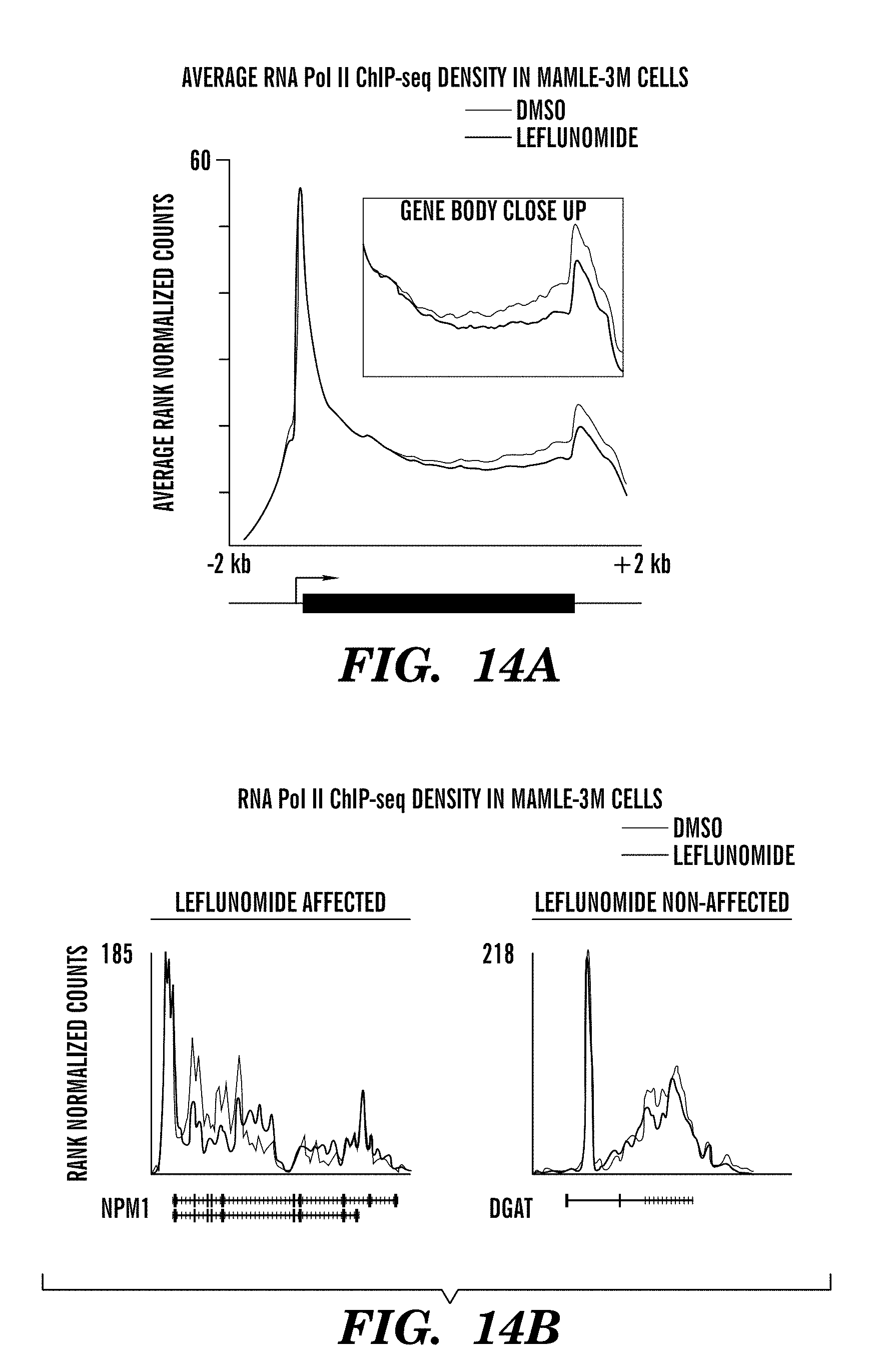
D00021

D00022

D00023

D00024

D00025

D00026

D00027
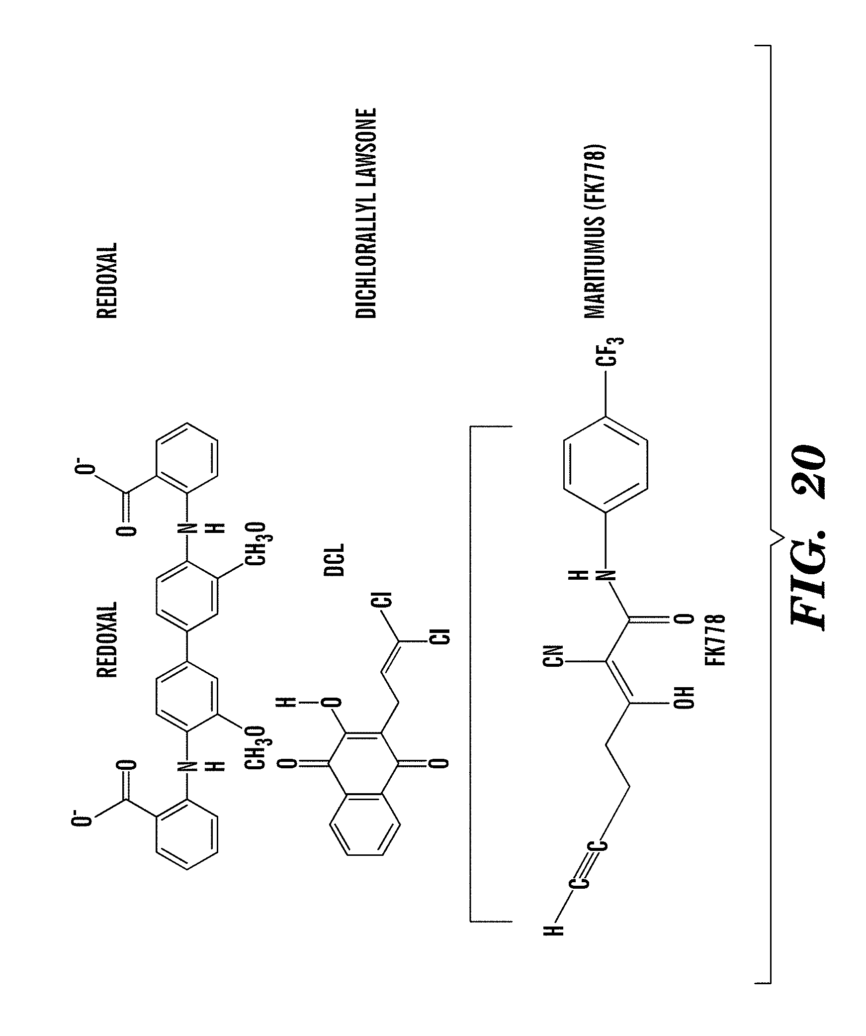
D00028

D00029

D00030

D00031

D00032

D00033

D00034

D00035

D00036

D00037

D00038

D00039

S00001
XML
uspto.report is an independent third-party trademark research tool that is not affiliated, endorsed, or sponsored by the United States Patent and Trademark Office (USPTO) or any other governmental organization. The information provided by uspto.report is based on publicly available data at the time of writing and is intended for informational purposes only.
While we strive to provide accurate and up-to-date information, we do not guarantee the accuracy, completeness, reliability, or suitability of the information displayed on this site. The use of this site is at your own risk. Any reliance you place on such information is therefore strictly at your own risk.
All official trademark data, including owner information, should be verified by visiting the official USPTO website at www.uspto.gov. This site is not intended to replace professional legal advice and should not be used as a substitute for consulting with a legal professional who is knowledgeable about trademark law.