Compositions And Methods For Tumor Transduction
Lobb; Roy ; et al.
U.S. patent application number 16/052397 was filed with the patent office on 2019-04-18 for compositions and methods for tumor transduction. The applicant listed for this patent is Aleta Biotherapeutics Inc.. Invention is credited to Roy Lobb, Paul Rennert.
| Application Number | 20190112386 16/052397 |
| Document ID | / |
| Family ID | 58631223 |
| Filed Date | 2019-04-18 |


| United States Patent Application | 20190112386 |
| Kind Code | A1 |
| Lobb; Roy ; et al. | April 18, 2019 |
COMPOSITIONS AND METHODS FOR TUMOR TRANSDUCTION
Abstract
The invention relates to cancer therapeutics, in particular, the system of making cancer cells more susceptible to effector cells by introduction of cellular therapy targets into the cancer cells.
| Inventors: | Lobb; Roy; (Wellesley, MA) ; Rennert; Paul; (Holliston, MA) | ||||||||||
| Applicant: |
|
||||||||||
|---|---|---|---|---|---|---|---|---|---|---|---|
| Family ID: | 58631223 | ||||||||||
| Appl. No.: | 16/052397 | ||||||||||
| Filed: | August 1, 2018 |
Related U.S. Patent Documents
| Application Number | Filing Date | Patent Number | ||
|---|---|---|---|---|
| 15670535 | Aug 7, 2017 | 10072094 | ||
| 16052397 | ||||
| 15317877 | Dec 9, 2016 | 10066023 | ||
| PCT/US16/59578 | Oct 28, 2016 | |||
| 15670535 | ||||
| 62249183 | Oct 30, 2015 | |||
| Current U.S. Class: | 1/1 |
| Current CPC Class: | A61K 38/177 20130101; C07K 2317/732 20130101; A61K 39/001166 20180801; C07K 14/70578 20130101; A61K 47/6851 20170801; C07K 14/70503 20130101; A61K 47/6803 20170801; A61K 39/39558 20130101; C07K 16/30 20130101; A61K 47/646 20170801; C12N 5/0636 20130101; C12N 2510/00 20130101; A61K 39/001112 20180801; C12N 2750/14143 20130101; A61K 47/6839 20170801; A61K 2039/5156 20130101; A61K 38/1774 20130101; A61K 39/001106 20180801; A61K 39/001104 20180801; C07K 14/70596 20130101; C07K 14/71 20130101; A61K 2039/507 20130101; C07K 2319/74 20130101; C07K 2319/33 20130101; C07K 16/32 20130101; C12N 15/86 20130101; C07K 2319/03 20130101; C07K 2317/622 20130101; A61P 35/00 20180101; A61K 39/395 20130101 |
| International Class: | C07K 16/32 20060101 C07K016/32; A61K 47/68 20060101 A61K047/68; A61K 39/395 20060101 A61K039/395; A61K 47/64 20060101 A61K047/64; C12N 15/86 20060101 C12N015/86; A61K 38/17 20060101 A61K038/17; C07K 16/30 20060101 C07K016/30; C07K 14/71 20060101 C07K014/71; C07K 14/705 20060101 C07K014/705 |
Claims
1-32. (canceled)
33. An adeno-associated viral (AAV) vector, an oncolytic viral vector, or a chimeric AAV/phage (AAVP) vector, comprising a nucleotide sequence encoding one or more secreted fusion proteins, each fusion protein comprising: (a) two or more antibodies, or antigen-binding fragments thereof, that bind one or more tumor antigen; and (b) a polypeptide antigen, wherein the polypeptide antigen: (i) is not a target for endogenous immune cells in an individual; and (ii) is a target antigen for an administered therapeutic selected from the group consisting of: cellular therapeutics, antibodies, and antibody-drug conjugates.
34. The vector of claim 33, wherein the polypeptide antigen is a B cell antigen.
35. The vector of claim 34, wherein the B cell antigen is CD19 or CD22.
36. The vector of claim 33, wherein the tumor antigen is HER-2/neu, c-met, EGFR, Ga733\EpCAM, CD20, ROR1, or BCMA.
37. The vector of claim 33, wherein the antigen-binding fragments are an Fab, scFv, Fv, or VHH or combination thereof.
38. The vector of claim 33, wherein the antigen-binding fragments are at least two scFvs.
39. The vector of claim 33, wherein the antigen-binding fragments are a VHH and an scFv.
40. The vector of claim 33, wherein the cellular therapeutic is a CAR-T cell or CAR-NK cell.
41. The vector of claim 33, wherein the oncolytic viral vector is an autonomous parvoviral vector, myxoma viral vector, paramyxoviral vector, reoviral vector, picornaviral vector, vaccinia viral vector, adenoviral vector, herpes simplex viral vector, or a vesicular stomatitis viral vector.
42. A method of treating a subject having a tumor, comprising administering to the subject: (i) an adeno-associated viral (AAV) vector, an oncolytic viral vector, or a chimeric AAV/phage (AAVP), vector comprising a nucleotide sequence encoding one or more secreted fusion proteins, each fusion comprising: (a) two or more antibodies, or antigen-binding fragments thereof, that bind one or more tumor antigens; and (b) a polypeptide antigen, wherein the polypeptide antigen: (1) is not a target antigen for endogenous immune cells in an individual; and (2) is a target antigen for an administered therapeutic selected from the group consisting of: cellular therapeutics, antibodies, and antibody-drug conjugates; and (ii) the cellular therapeutic, antibody, or antibody-drug conjugate, wherein the cellular therapeutic, antibody, or antibody-drug conjugate binds to the fusion protein, and said binding induces killing of the tumor, thereby treating the subject.
43. The method of claim 42, wherein the polypeptide antigen is a B cell antigen.
44. The method of claim 43, wherein the B cell antigen is CD19 or CD22.
45. The method of claim 42, wherein the tumor antigen is HER-2/neu, c-met, EGFR, Ga733\EpCAM, CD20, ROR1, or BCMA.
46. The method of claim 42, wherein the antigen-binding fragments are an Fab, scFv, Fv, or VHH or combination thereof.
47. The method of claim 42, wherein the antigen-binding fragments are at least two scFvs.
48. The method of claim 42, wherein the antigen-binding fragments are a VHH and an scFv.
49. The method of claim 42, wherein the AAV vector or AAVP vector transforms a tumor cell in the subject.
50. The method of claim 49, wherein the transformed tumor cell secretes the one or more fusion proteins.
51. The method of claim 50, wherein the one or more fusion proteins bind a tumor antigen on the transformed tumor cell.
52. The method of claim 50, wherein the one or more fusion proteins bind a tumor antigen on a non-transformed tumor cell.
53. The method of claim 42, wherein the cellular therapeutic is a CAR-T cell or CAR-NK cell.
54. The method of claim 42, wherein the oncolytic viral vector is an autonomous parvoviral vector, myxoma viral vector, paramyxoviral vector, reoviral vector, picornaviral vector, vaccinia viral vector, adenoviral vector, herpes simplex viral vector, or a vesicular stomatitis viral vector.
55. An adeno-associated viral (AAV) vector, an oncolytic viral vector, or a chimeric AAV/phage (AAVP) vector, comprising a nucleotide sequence encoding two or more different secreted fusion proteins, each fusion protein comprising: (a) one or more antibodies, or antigen-binding fragments thereof, that bind one or more tumor antigen; and (b) a polypeptide antigen, wherein the polypeptide antigen: (i) is not a target for endogenous immune cells in an individual; and (ii) is a target antigen for an administered therapeutic selected from the group consisting of: cellular therapeutics, antibodies, and antibody-drug conjugates.
56. The vector of claim 55, wherein the polypeptide antigen is a B cell antigen.
57. The vector of claim 56, wherein the B cell antigen is CD19 or CD22.
58. The vector of claim 55, wherein the tumor antigen is HER-2/neu, c-met, EGFR, Ga733\EpCAM, CD20, ROR1, or BCMA.
59. The vector of claim 55, wherein the antigen-binding fragments are an Fab, scFv, Fv, or VHH or combination thereof.
60. The vector of claim 55, wherein the cellular therapeutic is a CAR-T cell or CAR-NK cell.
61. The vector of claim 55, wherein the oncolytic viral vector is an autonomous parvoviral vector, myxoma viral vector, paramyxoviral vector, reoviral vector, picornaviral vector, vaccinia viral vector, adenoviral vector, herpes simplex viral vector, or a vesicular stomatitis viral vector.
Description
CROSS REFERENCE TO RELATED APPLICATIONS
[0001] This application is a continuation of U.S. patent application Ser. No. 15/670,535 filed Aug. 7, 2017 which is a continuation of U.S. patent application Ser. No. 15/317,877, filed Dec. 9, 2016, which is the National Stage of International Application No. PCT/US2016/059578, filed Oct. 28, 2016, which claims priority to U.S. Provisional Patent Application No. 62/249,183 filed Oct. 30, 2015, the entire contents of each of which are hereby incorporated by reference.
SEQUENCE LISTING
[0002] In accordance with 37 CFR 1.52(e)(5), the present specification makes reference to a Sequence Listing submitted electronically in the form of a text file (entitled "2012106-0008_SL.txt", created on Nov. 17, 2016 and is 29,112 bytes in size) the entire contents of which are incorporated herein by reference in their entirety.
BACKGROUND OF THE INVENTION
[0003] Recent examples of chimeric antigen receptor (CAR) T cell directed to solid tumors have been clinical (or even preclinical) failures. The invention described herein presents solutions for solid tumor and other oncology indications.
[0004] Cellular therapeutics that are introduced into cancer patients and traffic into tumor microenvironments encounter multiple barriers that limit their efficacy. These barriers include, but are not limited to: cellular anergy, intrinsic cell death, suboptimal cell trafficking, limited proliferation, limited effector function, poor "take" (survival, persistence and differentiation), active exclusion from tumors, persistent immunosuppression in the tumor microenvironment, and tumor-induced cell death. Almost all attempts to date to overcome these limitations require non-physiological manipulation of the cellular therapeutic both in vitro and in vivo. The efficacy of cellular therapeutics is therefore broadly hindered by both intrinsic and extrinsic factors.
[0005] Targeting solid tumors presents an additional set of challenges to overcome, for example, their overall lesser sensitivity to T cell mediated cytotoxicity, a microenvironment with differing immunosuppressive mechanisms between tumor types, and a lack of target antigens with favorable expression profiles. Additionally, solid tumors are heavily fortified against cellular therapeutic attack as they deploy an impenetrable extracellular matrix, hypoxia, and acidic pH. Despite the vast number of targets that have been investigated, only a small number are tumor-specific (e.g., expression is restricted to the tumor cell) and this finding, as such, illustrates the difficulty and the need to discover an effective solution for eliminating or reducing the tumor. Yet another problem for targeting tumors is the heterogeneity of the tumor cells which express different antigens and different levels of antigens. The invention described herein addresses these problems and provides additional benefits as well.
SUMMARY OF THE INVENTION
[0006] The present invention is based, at least in part, upon the introduction of cellular therapy targets into cancer cells (e.g., tumor cells such as solid tumor cells) such that they are more susceptible to effector cells that can eliminate and/or reduce the cancer cells. As further detailed herein, cancer cells (e.g., tumors) can be transduced with fusion proteins. Such fusion proteins consist of a binding component fused to a target component. For example, the fusion protein can be an antibody or antibody fragment which binds to one or more tumor-associated antigens (TAA) or tumor-specific antigens (TSA), fused to a target for cellular therapy, e.g. CD19; a cytokine-target fusion; a bi-specific T-cell engager (BiTE). They can also be transduced with CD19 variants (or mutants). These fusion proteins or variants (or mutants) can be secreted by the tumor cells and/or permeate the tumor microenvironment.
[0007] Accordingly, in one aspect, the invention provides for recombinant vectors encoding an antibody or antibody fragment to a tumor-associated antigens (TAA) or tumor-specific antigens (TSA) and a therapeutic agent, as a fusion protein. In other embodiments, the therapeutic agent is selected from the group consisting of a cytokines, peptides, proteins, antibodies, antibody fragments, T-cell engager and NK-cell engager. In other embodiments, the therapeutic agent is selected from the group consisting of glioma-associated antigen, carcinoembryonic antigen (CEA), .beta.-human chorionic gonadotropin, alphafetoprotein (AFP), lectin-reactive AFP, thyroglobulin, RAGE-1, MN-CA IX, human telomerase reverse transcriptase, RU1, RU2 (AS), intestinal carboxyi esterase, mut hsp70-2, M-CSF, prostase, prostate-specific antigen (PSA), PAP, NY-ESO-1, LAGE-1a, p53, prostein, PSMA, Her2/neu, survivin and telomerase, prostate-carcinoma tumor antigen-1 (PCTA-1), MAGE, ELF2M, neutrophil elastase, ephrinB2, CD22, insulin growth factor (IGF)-I, IGF-II, IGF-I receptor and mesothelin, EphA2, HER2, GD2, Glypican-3, 5T4, 8H9, .alpha.v.beta.6 integrin, BCMA, B7-H3, B7-H6, CAIX, CA9, CD19, CD20, CD22, kappa light chain, CD30, CD33, CD38, CD44, CD44v6, CD44v7/8, CD70, CD123, CD138, CD171, CEA, CSPG4, EGFR, EGFRvIII, EGP2, EGP40, EPCAM, ERBB3, ERBB4, ErbB3/4, FAP, FAR, FBP, fetal AchR, Folate Receptor a, GD2, GD3, HLA-AI MAGE A1, HLA-A2, IL1 1Ra, IL13Ra2, KDR, Lambda, Lewis-Y, MCSP, Mesothelin, Muc1, Muc16, NCAM, NKG2D ligands, NY-ESO-1, PRAME, PSCA, PSC1, PSMA, ROR1, SURVIVIN, TAG72, TEM1, TEM8, VEGRR2, carcinoembryonic antigen, HMW-MAA, and VEGF receptors.
[0008] In other embodiments, the cytokine is capable of being secreted by a tumor cell comprising said vector. In other embodiments, the vector is a viral vector. In other embodiments, the vector integrates into the genome of a cancer cell. In other embodiments, the therapeutic agent is an antigen is an antigen for a chimeric antigen receptor (CAR). In other embodiments, the therapeutic agent is an antigen for a T cell receptor (TCR). In other embodiments, the therapeutic agent is an antigen for an ADC antibody, ADCC antibody, and/or a radiotherapeutic antibody. In other embodiments, the therapeutic agent is an immunostimulatory cytokine or molecule selected from the group consisting of TLR agonists, PAMP, DAMP and other stimulators. In other embodiments, the therapeutic agent is a peptide with immunomodulatory or anti-tumor properties. In other embodiments, the TAA or TSA and therapeutic agent are expressed as a fusion protein. In other embodiments, the TAA or TSA is selected from the group consisting of a glioma-associated antigen, carcinoembryonic antigen (CEA), .beta.-human chorionic gonadotropin, alphafetoprotein (AFP), lectin-reactive AFP, thyroglobulin, RAGE-1, MN-CA IX, human telomerase reverse transcriptase, RU1, RU2 (AS), intestinal carboxyi esterase, mut hsp70-2, M-CSF, prostase, prostate-specific antigen (PSA), PAP, NY-ESO-1, LAGE-1a, p53, prostein, PSMA, Her2/neu, survivin and telomerase, prostate-carcinoma tumor antigen-1 (PCTA-1), MAGE, ELF2M, neutrophil elastase, ephrinB2, CD22, insulin growth factor (IGF)-I, IGF-II, IGF-I receptor and mesothelin, EphA2, HER2, GD2, Glypican-3, 5T4, 8H9, .alpha.v.beta.6 integrin, BCMA, B7-H3, B7-H6, CAIX, CA9, CD19, CD20, CD22, kappa light chain, CD30, CD33, CD38, CD44, CD44v6, CD44v7/8, CD70, CD123, CD138, CD171, CEA, CSPG4, EGFR, EGFRvIII, EGP2, EGP40, EPCAM, ERBB3, ERBB4, ErbB3/4, FAP, FAR, FBP, fetal AchR, Folate Receptor a, GD2, GD3, HLA-AI MAGE A1, HLA-A2, IL1 1Ra, IL13Ra2, KDR, Lambda, Lewis-Y, MCSP, Mesothelin, Mud, Muc16, NCAM, NKG2D ligands, NY-ESO-1, PRAME, PSCA, PSC1, PSMA, ROR1, SURVIVIN, TAG72, TEM1, TEM8, VEGRR2, carcinoembryonic antigen, HMW-MAA, and VEGF receptors
[0009] In other aspects, the invention provides for recombinant vectors encoding a cytokine to a tumor expressed cytokine receptor and a therapeutic agent as a fusion protein. In some embodiments, the cytokine receptor is a type I receptor or a type II receptor.
[0010] In other aspects, the invention provides for recombinant tumor cells comprising a vector as described above and herein. In some embodiments, the tumor cell expresses one or more fusion proteins.
[0011] In other aspects, the invention provides for methods of producing a recombinant tumor cell capable of expressing a fusion protein comprising an antibody or antibody fragment to a tumor-associated antigens (TAA) or tumor-specific antigens (TSA) and a therapeutic agent, said method comprising (a) introducing a vector as described above and herein into a tumor cell; and (b) culturing the tumor cell such that the vector is transduced into the tumor cell. In some embodiments, the vector and/or its components integrate in the tumor cells' genome. In some embodiments, the TAA or TSA is selected from the group consisting of glioma-associated antigen, carcinoembryonic antigen (CEA), .beta.-human chorionic gonadotropin, alphafetoprotein (AFP), lectin-reactive AFP, thyroglubilin, RAGE-1, MN-CA IX, human telomerase reverse transcriptase, RU1, RU2 (AS), intestinal carboxylesterase, mut hsp70-2, M-CSF, prostase, prostate-specific antigen (PSA), PAP, NY-ESO-1, LAGE-1a, p53, prostein, PSMA, Her2/neu, survivin and telomerase, prostate-carcinoma tumor antigen-1 (PCTA-1), MAGE, ELF2M, neutrophil elastase, ephrinB2, CD22, insulin growth factor (IGF)-I, IGF-II, IGF-I receptor and mesothelin, EphA2, HER2, GD2, Glypican-3, 5T4, 8H9, .alpha.v.beta.6 integrin, BCMA, B7-H3, B7-H6, CAIX, CA9, CD19, CD20, CD22, kappa light chain, CD30, CD33, CD38, CD44, CD44v6, CD44v7/8, CD70, CD123, CD138, CD171, CEA, CSPG4, EGFR, EGFRvIII, EGP2, EGP40, EPCAM, ERBB3, ERBB4, ErbB3/4, FAP, FAR, FBP, fetal AchR, Folate Receptor a, GD2, GD3, HLA-AI MAGE A1, HLA-A2, IL11Ra, IL13Ra2, KDR, Lambda, Lewis-Y, MCSP, Mesothelin, Muc1, Muc16, NCAM, NKG2D ligands, NY-ESO-1, PRAME, PSCA, PSC1, PSMA, ROR1, SURVIVIN, TAG72, TEM1, TEM8, VEGRR2, carcinoembryonic antigen, HMW-MAA, and VEGF receptors. In some embodiments, the TAA or TSA is an antigen for a chimeric antigen receptor (CAR). In some embodiments, the TAA or TSA is an antigen for a T cell receptor (TCR). In some embodiments, the therapeutic agent is an antigen for an ADC antibody, ADCC antibody, and/or a radiotherapeutic antibody. In some embodiments, the therapeutic agent is an immunostimulatory cytokine or molecule selected from the group consisting of TLR agonists, PAMP, DAMP and other stimulators. In some embodiments, the therapeutic agent is a peptide with immunomodulatory or anti-tumor properties.
[0012] In other aspects, the invention provides for methods of treating cancer in an individual in need thereof comprising administering a composition comprising a vector as described above and herein. In some embodiments, a fusion protein is expressed. In other embodiments, the cancer cell expresses a therapeutic agent that increases immune response against the cancer cell. In some embodiments, the cancer cell express one or more protein or peptide antigens that is capable of being recognized by a CAR. In some embodiments, the cancer cell express one or more protein or peptide antigens that is capable of being recognized by a TCR. In some embodiments, the cancer cell express one or more fusion proteins containing therapeutic agents capable of being recognized by immune cells in the individual. In some embodiments, the method further comprises administration of CAR T cells to the individual.
BRIEF DESCRIPTION OF THE DRAWINGS
[0013] FIG. 1 is a schematic showing a non-limiting example of the methods of introduction of a cellular therapy target into solid tumor cells. Step one corresponds to the transduction construct (e.g., tumor-selective transduction via e.g., viral transduction, RNA transduction, and gene insertion), and/or in vivo transduction and integration. Step two corresponds to the expression of a fusion protein (e.g., an antibody fragment to a tumor antigen fused to a CAR antigen, for example an scFv anti-tumor associated antigen (TAA) fused to CD19 or a fragment thereof). Step three corresponds to the antibody fragment fused to an antigen, e.g. CD19, which binds to tumor cells and presents the antigen, and all nearby tumor cells are coated with the antibody fragment fused to a protein, peptide, or other agent, for example, CD19, which is an antigen recognized by CAR T cells. Step four corresponds to the infusion in vivo of CAR T cells that recognize the CAR antigen (e.g., anti-CD19 CAR T cells). The response becomes cytotoxic to the CD19-coated tumor cell.
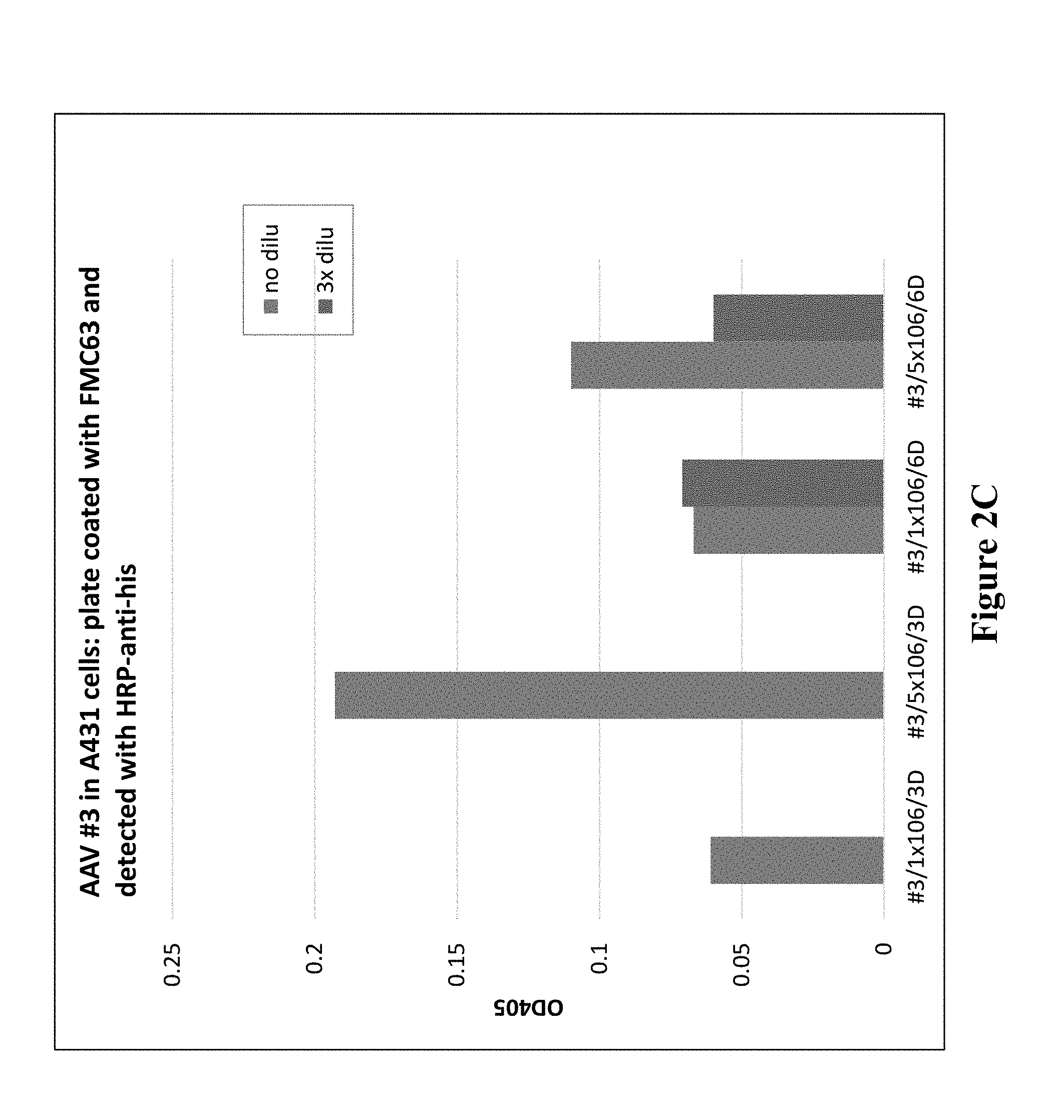
[0014] FIGS. 2A-2C demonstrate expression of fusion proteins from AAV transduced cells.
[0015] FIGS. 3A and 3B show summary results of an IFN.gamma. ELISA measuring induction of IFN.gamma. in CAR19 T- cells by expressed fusion proteins.
[0016] FIGS. 4A-4C show induction of cytotoxicity upon incubation of CAR19 T cells with AAV-1-expressed supernatant and BT474 cells.
DETAILED DESCRIPTION OF THE INVENTION
[0017] The present invention is based, at least in part, upon the introduction of cellular therapy targets into solid tumor cells. Tumor cells can be transduced with fusion proteins such that the fusion proteins are secreted by the tumor cells and released to the tumor microenvironment to combat the tumor.
I. Definitions
[0018] As used herein, the term "antibody" refers to a polypeptide that includes canonical immunoglobulin sequence elements sufficient to confer specific binding to a particular target antigen. As is known in the art, intact antibodies as produced in nature are approximately 150 kD tetrameric agents comprised of two identical heavy chain polypeptides (about 50 kD each) and two identical light chain polypeptides (about 25 kD each) that associate with each other into what is commonly referred to as a "Y-shaped" structure. Each heavy chain is comprised of at least four domains (each about 110 amino acids long)--an amino-terminal variable (VH) domain (located at the tips of the Y structure), followed by three constant domains: CH1, CH2, and the carboxy-terminal CH3 (located at the base of the Y's stem). A short region, known as the "switch", connects the heavy chain variable and constant regions. The "hinge" connects CH2 and CH3 domains to the rest of the antibody. Two disulfide bonds in this hinge region connect the two heavy chain polypeptides to one another in an intact antibody. Each light chain is comprised of two domains--an amino-terminal variable (VL) domain, followed by a carboxy-terminal constant (CL) domain, separated from one another by another "switch". Intact antibody tetramers are composed of two heavy chain-light chain dimers in which the heavy and light chains are linked to one another by a single disulfide bond; two other disulfide bonds connect the heavy chain hinge regions to one another, so that the dimers are connected to one another and the tetramer is formed. Naturally-produced antibodies are also glycosylated, typically on the CH2 domain. Each domain in a natural antibody has a structure characterized by an "immunoglobulin fold" formed from two beta sheets (e.g., 3-, 4-, or 5-stranded sheets) packed against each other in a compressed antiparallel beta barrel. Each variable domain contains three hypervariable loops known as "complement determining regions" (CDR1, CDR2, and CDR3) and four somewhat invariant "framework" regions (FR1, FR2, FR3, and FR4). When natural antibodies fold, the FR regions form the beta sheets that provide the structural framework for the domains, and the CDR loop regions from both the heavy and light chains are brought together in three-dimensional space so that they create a single hypervariable antigen binding site located at the tip of the Y structure. The Fc region of naturally-occurring antibodies binds to elements of the complement system, and also to receptors on effector cells, including for example effector cells that mediate cytotoxicity. As is known in the art, affinity and/or other binding attributes of Fc regions for Fc receptors can be modulated through glycosylation or other modification. In some embodiments, antibodies produced and/or utilized in accordance with the present disclosure include glycosylated Fc domains, including Fc domains with modified or engineered such glycosylation. For purposes of the present disclosure, in certain embodiments, any polypeptide or complex of polypeptides that includes sufficient immunoglobulin domain sequences as found in natural antibodies can be referred to and/or used as an "antibody", whether such polypeptide is naturally produced (e.g., generated by an organism reacting to an antigen), or produced by recombinant engineering, chemical synthesis, or other artificial system or methodology. In some embodiments, an antibody is polyclonal; in some embodiments, an antibody is monoclonal. In some embodiments, an antibody has constant region sequences that are characteristic of mouse, rabbit, primate, or human antibodies. In some embodiments, antibody sequence elements are fully human, or are humanized, primatized, chimeric, etc, as is known in the art. Moreover, the term "antibody" as used herein, can refer in appropriate embodiments (unless otherwise stated or clear from context) to any of the art-known or developed constructs or formats for utilizing antibody structural and functional features in alternative presentation. For example, in some embodiments, an antibody utilized in accordance with the present disclosure is in a format selected from, but not limited to, intact IgG, IgE and IgM, bi- or multi-specific antibodies (e.g., Zybodies.RTM., etc), single chain Fvs, polypeptide-Fc fusions, Fabs, cameloid antibodies, masked antibodies (e.g., Probodies.RTM.), Small Modular ImmunoPharmaceuticals ("SMIPs.TM."), single chain or Tandem diabodies (TandAb.RTM.), VHHs, Anticalins.RTM., Nanobodies.RTM., minibodies, BiTE.RTM.s, ankyrin repeat proteins or DARPINs.RTM., Avimers.RTM., a DART, a TCR-like antibody, Adnectins.RTM., Affilins.RTM., Trans-Bodies.RTM., Affibodies.RTM., a TrimerX.RTM., MicroProteins, Fynomers.RTM., Centyrins.RTM., and a KALBITOR.RTM.. In some embodiments, an antibody may lack a covalent modification (e.g., attachment of a glycan) that it would have if produced naturally. In some embodiments, an antibody may contain a covalent modification (e.g., attachment of a glycan, a payload (e.g., a detectable moiety, a therapeutic moiety, a catalytic moiety, etc.), or other pendant group (e.g., poly-ethylene glycol, etc.)).
[0019] As used herein, an "antibody fragment" includes a portion of an intact antibody, such as, for example, the antigen-binding or variable region of an antibody. Examples of antibody fragments include Fab, Fab', F(ab')2, and Fv fragments; triabodies; tetrabodies; linear antibodies; single-chain antibody molecules; and multi-specific antibodies formed from antibody fragments. For example, antibody fragments include isolated fragments, "Fv" fragments (consisting of the variable regions of the heavy and light chains), recombinant single chain polypeptide molecules in which light and heavy chain variable regions are connected by a peptide linker ("ScFv proteins"), and minimal recognition units consisting of the amino acid residues that mimic the hypervariable region. In many embodiments, an antibody fragment contains sufficient sequence of the parent antibody of which it is a fragment that it binds to the same antigen as does the parent antibody; in some embodiments, a fragment binds to the antigen with a comparable affinity to that of the parent antibody and/or competes with the parent antibody for binding to the antigen. Examples of antigen binding fragments of an antibody include, but are not limited to, Fab fragment, Fab' fragment, F(ab')2 fragment, scFv fragment, Fv fragment, dsFv diabody, dAb fragment, Fd' fragment, Fd fragment, and an isolated complementarity determining region (CDR) region. An antigen binding fragment of an antibody may be produced by any means. For example, an antigen binding fragment of an antibody may be enzymatically or chemically produced by fragmentation of an intact antibody and/or it may be recombinantly produced from a gene encoding the partial antibody sequence. Alternatively or additionally, antigen binding fragment of an antibody may be wholly or partially synthetically produced. An antigen binding fragment of an antibody may optionally comprise a single chain antibody fragment. Alternatively or additionally, an antigen binding fragment of an antibody may comprise multiple chains which are linked together, for example, by disulfide linkages. An antigen binding fragment of an antibody may optionally comprise a multimolecular complex. A functional antibody fragment typically comprises at least about 50 amino acids and more typically comprises at least about 200 amino acids.
[0020] By "antigen" is defined as a molecule that provokes an immune response. This immune response may involve either antibody production, or the activation of specific immunologically-competent cells, or both. The skilled artisan will understand that any macromolecule, including virtually all proteins or peptides, can serve as an antigen. Furthermore, antigens can be derived from recombinant or genomic DNA. A skilled artisan will understand that any DNA, which comprises a nucleotide sequences or a partial nucleotide sequence encoding a protein that elicits an immune response therefore encodes an "antigen" as that term is used herein. Furthermore, one skilled in the art will understand that an antigen need not be encoded solely by a full length nucleotide sequence of a gene. Moreover, a skilled artisan will understand that an antigen need not be encoded by a "gene" at all. An antigen can be generated synthesized or can be derived from a biological sample. Such a biological sample can include, but is not limited to a tissue sample, a tumor sample, a cell or a biological fluid.
[0021] The term, "autologous" refers to any material derived from the same individual to which it is later re-introduced into the individual.
[0022] The term "cell therapy antigen" as used herein is meant to refer to one or more antigens (that are genetically engineered or naturally occurring) that can be recognized by effector cells (e.g., genetically engineered CAR T cell or genetically engineered or naturally occurring TCR). An antigen that is expressed on tumors (i.e., "tumor-associated antigen" or TAA) is one type of cell therapy antigen. An antigen that is expressed selectively on tumors (i.e., "tumor-selective antigen" or TSA) is also a type of cell therapy antigen. An antigen can also be expressed on tumor cells through therapeutic intervention, for example, by transducing tumor cells, in vivo, with genetic materials of the present invention, as described herein.
[0023] As used herein, the term "fusion protein" generally refers to a polypeptide including at least two segments, each of which shows a high degree of amino acid identity to a peptide moiety that (1) occurs in nature, and/or (2) represents a functional domain of a polypeptide. Typically, a polypeptide containing at least two such segments is considered to be a fusion protein if the two segments are moieties that (1) are not included in nature in the same peptide, and/or (2) have not previously been linked to one another in a single polypeptide, and/or (3) have been linked to one another through action of the hand of man.
[0024] The term "promoter" refers to a region of a DNA sequence that directs expression of genes. The promoter is typically upstream from the start of transcription start site and is involved in recognition and binding of RNA polymerase and other transcription machinery (e.g., other proteins) to initiate transcription of a polynucleotide sequence.
[0025] The term "solid tumor" is meant as an abnormal mass that usually does not contain cysts or liquid areas. Solid tumors can be benign or malignant. Different types of solid tumors are named for the types of cells that form them (such as sarcomas, carcinomas, and lymphomas). Examples of solid tumors such as sarcomas and carcinoma, include fibrosarcoma, myxosarcoma, liposarcoma, chondrosarcoma, osteosarcoma, and other sarcomas, synovioma, mesothelioma, Ewing's tumor, leiomyosarcoma, rhabdomyosarcoma, colon carcinoma, lymphoid malignancy, pancreatic cancer, breast cancer, lung cancers, ovarian cancer, prostate cancer, hepatocellular carcinoma, squamous cell carcinoma, basal cell carcinoma, adenocarcinoma, sweat gland carcinoma, medullary thyroid carcinoma, papillary thyroid carcinoma, pheochromocytomas sebaceous gland carcinoma, papillary carcinoma, papillary adenocarcinomas, medullary carcinoma, bronchogenic carcinoma, renal cell carcinoma, hepatoma, bile duct carcinoma, choriocarcinoma, Wilms' tumor, cervical cancer, testicular tumor, seminoma, bladder carcinoma, melanoma, and CNS tumors (such as a glioma (such as brainstem glioma and mixed gliomas), glioblastoma (also known as glioblastoma multiforme) astrocytoma, CNS lymphoma, germinoma, medulloblastoma, Schwannoma craniopharyogioma, ependymoma, pinealoma, hemangioblastoma, acoustic neuroma, oligodendroglioma, menangioma, neuroblastoma, retinoblastoma and brain metastases.
[0026] An "individual" or "subject" can be a vertebrate, a mammal, or a human. Mammals include, but are not limited to, farm animals, sport animals, pets, primates, mice and rats. In one aspect, a subject is a human. An "individual" or "subject" can be a "patient" (e.g., under the care of a physician) but in some cases, an individual or subject is not a patient.
[0027] "Pseudoviruses" or "papilloma pseudoviruses" or "papillomavirus gene transfer vectors" refer to one or more papillomavirus capsid proteins that assemble and package heterologous nucleic acids (e.g., DNA) with or without viral nucleic acids (e.g., DNA) into infectious particles. The methods used to produce papilloma pseudoviruses are known in the art and are described, for example, in U.S. Pat. Nos. 6,599,739, 7,205,126, and 6,416,945; and in Buck and Thomspon, Production of Papillomavirus-Based Gene Transfer Vectors. Current Protocols in Cell Biology 26.1.1-26.1.19, December 2007, all of which are incorporated by reference.
[0028] The term "T cell receptor" or "TCR" refers to a heterodimeric receptor found on the surface of T lymphocytes. TCRs are antigen-specific molecules that are responsible for recognizing antigenic peptides of the major histocompatibility complex (MEW) on the surface of antigen presenting cells (APCs), or any other nucleated cell. They are members of the immunoglobulin superfamily, and typically consist of two chains; alpha (.alpha.) and beta (.beta.), while a small subset of TCRs are formed by variable gamma (.gamma.) and delta (.delta.) chains. The chains pair on the surface of a T cell to form a heterodimeric receptor.
[0029] The term "transfected" or "transformed" or "transduced" is defined as a process by which exogenous nucleic acid is transferred or introduced into the host cell. A "transfected" or "transformed" or "transduced" cell is one which has been transfected, transformed or transduced with exogenous nucleic acid. The cell includes the primary subject cell and its progeny. In some embodiments, the host cell is a cancer cell, for example a tumor cell such as solid tumor cell.
[0030] The term "tropism" refers to the movement or targeting of a viral vector towards a receptor.
[0031] A "vector" is a composition which comprises an isolated nucleic acid, and which can be used to deliver the isolated nucleic acid to the interior of a cell. Numerous vectors are known in the art including, but not limited to, linear polynucleotides, polynucleotides associated with ionic or amphiphilic compounds, plasmids, and viruses. Thus, the term "vector" includes an autonomously replicating plasmid or a virus. The term should also be construed to include non-plasmid and non-viral compounds which facilitate transfer of nucleic acid into cells, such as, for example, polylysine compounds, liposomes, and the like. Examples of viral vectors include, but are not limited to, adenoviral vectors, adeno-associated virus and phage vectors (AAVP), retroviral vectors, human papilloma virus (HPV) pseudovirus vectors, and the like.
II. Compositions
[0032] The invention provides for compositions that can be used for introduction of fusion proteins into cancer cells (e.g., tumor cells) that will make the cancer cells more susceptible for destruction by either an individual's immune system and/or additional therapeutic agents. Compositions can include, but are not limited to, vectors and various constructs described herein, host cells (including tumor cells) containing such vectors and/or constructs, host cells expressing or capable of expressing these vectors and/or constructs, kits containing the vectors, constructs, instructions, and/or reagents and the like.
[0033] Cancer cells (e.g., tumors) can be transduced with fusion proteins. As the fusion proteins are expressed (and/or secreted), the proteins can also permeate the tumor microenvironment. Non-limiting examples of fusion proteins contemplated by the invention include: antibody fusions so that the antibody or antibody fragment that bind to one or more tumor-associated antigens (TAA) or tumor-specific antigens (TSA), cytokine target fusions, bi-specific T-cell engagers (BITES) or CD19 variants.
Vector Design
[0034] The nucleic acid sequences coding the desired gene of interest can be cloned into a number of types of vectors. For example, the nucleic acid can be cloned into a plasmid, a phagemid, a phage derivative, an animal virus, and a cosmid. Other vectors can include expression vectors, replication vectors, probe generation vectors, sequencing vectors, and viral vectors. In other examples, the vector can be a foamy viral (FV) vector, a type of retroviral vector made from spumavirus. Viral vector design and technology is well known in the art as described in Sambrook et al, (Molecular Cloning: A Laboratory Manual, 2001), and in other virology and molecular biology manuals.
Transduction Methods
[0035] Transfer of nucleic acid to a cell for gene-modification of the cell in order for the cell to express a gene of interest is widely performed via transduction (e.g., viral transduction). The nucleic acid sequence coding for the desired gene of interest or portion thereof (e.g., tumor associated antigen) can be obtained using recombinant methods known in the art. Exemplary methods include screening libraries from cells expressing the gene, deriving the gene from a vector, or isolating directly from cells and tissues. These methods are performed using standard techniques. In other embodiments, the gene of interest can be produced synthetically rather than cloned. Gene delivery methods are well-known in the art, for example, U.S. Pat. No. 5,399,346.
Viral Transduction
[0036] Viruses are highly efficient at nucleic acid delivery to specific cell types, while often avoiding detection by the infected host immune system. These features make certain viruses attractive candidates as vehicles for introduction of cellular therapy targets into cancer cells, e.g., solid tumor cells. A number of viral based systems have been developed for gene transfer into mammalian cells. Examples of viral vectors include, but are not limited to, retroviruses, adenoviruses, adeno-associated viruses, herpes viruses, lentiviruses, poxviruses, herpes simplex 1 virus, herpes virus, oncoviruses (e.g., murine leukemia viruses), and the like. In general, a suitable vector contains an origin of replication functional in at least one organism, a promoter sequence, convenient restriction endonuclease sites, and one or more selectable markers, (e.g., WO 01/96584; WO 01/29058; and U.S. Pat. No. 6,326,193).
[0037] Lentiviral and Retroviral transduction can be enhanced by the addition of polybrene (SantaCruz sc-134220; Millipore TR-1003-G; Sigma 107689), a cationic polymer (also known as hexamehtrine bromide) that is used to increase the efficiency of the retrovirus transduction.
[0038] For example, retroviruses provide a platform for gene delivery systems. Retroviruses are enveloped viruses that belong to the viral family Retroviridae. Once in a host's cell, the virus replicates by using a viral reverse transcriptase enzyme to transcribe its RNA into DNA. The retroviral DNA replicates as part of the host genome, and is referred to as a provirus. A selected gene can be inserted into a vector and packaged in retroviral particles using techniques known in the art. The recombinant virus can then be isolated and delivered to cells of the subject either in vivo or ex vivo. A number of retroviral systems are known in the art, for example See U.S. Pat. Nos. 5,994,136, 6,165,782, and 6,428,953.
[0039] Retroviruses include the genus of Alpharetrovirus (e.g., avian leukosis virus), the genus of Betaretrovirus; (e.g., mouse mammary tumor virus) the genus of Deltaretrovirus (e.g., bovine leukemia virus and human T-lymphotropic virus), the genus of Epsilonretrovirus (e.g., Walleye dermal sarcoma virus), and the genus of Lentivirus.
[0040] In other embodiments, the retrovirus is a lentivirus a genus of viruses of the Retroviridae family, characterized by a long incubation period. Lentiviruses are unique among the retroviruses in being able to infect non-dividing cells; they can deliver a significant amount of genetic information into the DNA of the host cell, so they are one of the most efficient methods of a gene delivery vector. Lentiviral vectors have an advantage to other viral vectors in that they can transduce non-proliferating cells and show low immunogenicity. In some examples, the lentivirus includes, but is not limited to human immunodeficiency viruses (HIV-1 and HIV-2), simian immunodeficiency virus (S1V), feline immunodeficiency virus (FIV), equine infections anemia (EIA), and visna virus. Vectors derived from lentiviruses offer the means to achieve significant levels of gene transfer in vivo.
[0041] In embodiments, the vector is an adenovirus vector. Adenoviruses are a large family of viruses containing double stranded DNA. They replicate the DNA of the host cell, while using the host's cell machinery to synthesize viral RNA DNA and proteins. Adenoviruses are known in the art to affect both replicating and non-replicating cells, to accommodate large transgenes, and to code for proteins without integrating into the host cell genome.
[0042] In some embodiments, an AAVP vector is used. The AAVP vector is a hybrid of prokaryotic-eukaryotic vectors, which are chimeras of genetic cis-elements of recombinant adeno-associated virus and phage. An AAVP combines selected elements of both phage and AAV vector systems, providing a vector that is simple to produce in bacteria with no packaging limit, while allowing infection of mammalian cells combined with integration into the host chromosome. Vectors containing many of the appropriate elements are commercially available, and can be further modified by standard methodologies to include the necessary sequences. Among other things, AAVPs do not require helper viruses or trans-acting factors. In addition, the native tropism of AAV for mammalian cells is eliminated since there is not AAV capsid formation. Other methods and details are in U.S. Pat. No. 8,470,528 and Hajitou A. et al., Cell, 125: 358-398, both of which are incorporated herein by reference.
[0043] In other aspects, a human papilloma (HPV) pseudovirus is used. Recent studies have shown that DNA plasmids can be packaged into papillomavirus L1 and L2 capsid protein to generate pseudovirion that can efficiently deliver DNA. The encapsulation protects the DNA from nucleases and provides a targeted delivery with great stability. Many of the safety concerns associated with the use of viral vectors are mitigated with the HPV pseudoviros because its construct is different from the natural HPV viral genome. Other methods and examples are in Hung, C., et al., Plos One, 7:7(e40983); 2012, U.S. Pat. No. 8,394,411, and Kines, R., et al Int J of Cancer, 2015, all of which are incorporated herein by reference.
[0044] In some aspects, an oncolytic virus is used. Oncolytic virus therapy selectively replicates the virus in cancer cells, and subsequently spreads within a tumor without affecting normal tissue. Alternatively, the oncolytic virus preferentially infects and kills cells without causing damage to normal tissues. Oncolytic viruses are also effective at inducing immune responses to themselves as well as to the infected tumor cell. Typically, oncolytic viruses fall into two classes: (I) viruses that naturally replicate preferentially in cancer cells and are nonpathogenic in humans. Exemplary class (I) oncolytic viruses include autonomous parvoviruses, myxoma virus (poxvirus), Newcastle disease virus (NDV; paramyxovirus), reovirus, and Seneca valley virus (picornavirus). A second class (II), include viruses that are genetically manipulated for use as vaccine vectors, including measles virus (paramyxovirus), poliovirus (picornavirus), and vaccinia virus (poxvirus). Additionally, oncolytic viruses may include those genetically engineered with mutations/deletions in genes required for replication in normal but not in cancer cells including adenovirus, herpes simplex virus, and vesicular stomatitis virus. Oncolytic viruses can be used as a viral transduction method due to their low probability of genetic resistance because they can target multiple pathways and replicate in a tumor-selective method. The viral dose within the tumor can increase with time due to in situ viral amplification (as compared to small molecule therapies which decrease with time), and safety features can be built in (i.e., drug and immune sensitivity).
Integration
[0045] In some aspects of the invention, the nucleic acids encoding cellular therapy target(s) or tumor-associated antigen(s) are integrated as part of the tumor cells' genetic makeup. Without being bound by theory, integration of the nucleic acid encoding cellular therapy target(s) can be helpful in that as the tumor cell replicates, progeny tumor cells would express the cellular therapy target(s) and, as such, be susceptible to the effector cells and other therapeutic agents (including combination therapy agents). It follows that in some indications, such as metastatic disease, the state of rapid tumor cell proliferation becomes useful in propagating the therapeutics of the present invention.
[0046] In one embodiment, integration can be achieved by using viruses that naturally integrate into the host cell. Integration is a crucial step in replication of retroviruses as it is a virus genome that has been integrated into the DNA of a host cell. Integration is not part of the viral replication cycle, but it can occasionally occur. The virus does not directly make new DNA copies of itself while integrated into the host genome; alternatively, it is passively replicated along with the host genome and passed on to the original cell's offspring. Integration of the viral DNA results in permanent insertion of the viral genome into the host chromosomal DNA, referred as a provirus in the case of retroviruses.
[0047] In other embodiments, integration can be achieved by commercially available kits, including the CompoZr.RTM. Targeted Integration Kit which is designed to integrate a gene of interest into the adeno-associated virus integration site 1 (AAVS1) on human chromosome 19.
[0048] In addition to the expression of TAA, the cancer cells (e.g., solid tumor cells) can be engineered so that they also express one or more fusion protein of an antibody or antibody fragment (e.g., scFv) coupled to an antigen that an effector cell will recognize. The antibody or antibody fragment recognizes the tumor cell and binds and presents the antigen for an effector cell to recognize. In one non-limiting example, a solid tumor cell can be transduced with a fusion protein that includes an anti-tumor TAA scFv that binds to a tumor cell, and also part or all of the human CD19 protein. The scFv portion binds to the tumor cell and the CD19 is presented to effector cells for them to target the tumor cell and destroy the tumor cell. The effector cells can be naturally occurring or genetically engineered.
Expressed Gene Approach for Solid Tumor Targeting
[0049] Genes productively introduced into tumor cells will provide improved cellular therapeutic activity by addressing or circumventing critical barriers to efficacy. These genes will improve therapeutic efficacy by "propagating" the anti-tumor response, optimize cytokine support in the local environment, reverse local immunosuppression, improve cellular effector functions, and promote cellular access to tumors. Any gene can be included in an expressed construct as described herein, and the present disclosure is not limited to any particular gene. Exemplary, non-limiting types of genes that can be included as cellular therapy targets include, e.g., targets for additional cellular therapeutics, polypeptide antigens, antibodies, cytokines, agents targeting tumor microenvironment, and agents supporting immune cell growth/proliferation.
[0050] An expressed gene contains a promotor with its associated elements, and a gene sequence. An expressed gene contains three essential characteristics: 1) an optimal promoter for expression in the tumor cell, 2) an optimal expressed gene sequence, and 3) an optimized expression pattern with defined kinetics.
[0051] A set of promoters is developed to drive diverse expression patterns. Examples include; rapid and sustained expression, measured in days, or rapid but reversible expression, or delayed expression. Also, the level of expression can be modified by selective use of regulatory and promoter elements. Such methods are well understood, for example, E. D. Papadakis, et al., Current Gene Therapy, 4: 89-113 (incorporated herein by reference).
III. Methods for Treating Cancer
[0052] The invention provides for compositions and methods for treating cancer by engineering a system whereby the cancer cells (e.g., tumor cells or solid tumor cells) secrete fusion proteins that include TAA or TSA and a therapeutic agent. An individual having cancer or suspected of having cancer can be given a composition that allows for the in vivo transduction of their cancer cells. The administration can be by any means, including, but not limited to intravenously, systemically, intramuscularly, intraperitonally, or intra-tumoral injection.
Targets for Additional Cellular Therapeutics
[0053] In some embodiments, the cell therapy antigen that is expressed on local tumor cells following transduction and secretion of fusion proteins is recognized by the effector cells, and can comprise a tumor associated antigen (TAA). In one embodiment, TAA expression can be restricted to the tumor cell population alone, expressed by all tumor cells, and expressed on the tumor cell surface. Other antigens are overexpressed on tumor cells, but may be found on normal cells at lower levels of expression and thus are tumor-selective antigens (TSA). In addition, some tumor antigens arise as "passenger mutations", i.e. are non-essential antigens expressed by tumor cells that have defective control over DNA repair, thus accumulating mutations in diverse proteins. Some tumor antigens are proteins that are produced by tumor cells that elicit an immune response; particularly T-cell mediated immune responses. Tumor-specific molecules that may be targeted by cellular therapy targets may include tumor associated antigens well known in the art and can include, for example, a glioma-associated antigen, carcinoembryonic antigen (CEA), .beta.-human chorionic gonadotropin, alphafetoprotein (AFP), lectin-reactive AFP, thyroglobulin, RAGE-1, MN-CA IX, human telomerase reverse transcriptase, RU1, RU2 (AS), intestinal carboxyi esterase, mut hsp70-2, M-CSF, prostase, prostate-specific antigen (PSA), PAP, NY-ESO-1, LAGE-1a, p53, prostein, PSMA, Her2/neu, survivin and telomerase, prostate-carcinoma tumor antigen-1 (PCTA-1), MAGE, ELF2M, neutrophil elastase, ephrinB2, CD22, insulin growth factor (IGF)-I, IGF-II, IGF-I receptor and mesothelin. The application of technologies, including next generation sequencing (NGS) of tumor genomes and exomes, and high-sensitivity mass spectrometry (protein sequencing) analysis of the tumor proteome, is continuing to identify novel TAA and TSA antigens of use for this invention.
[0054] NGS is a method of high-throughput sequencing that performs massively parallel sequencing, during which millions of fragments of DNA from a single sample are sequenced in unison. NGS facilitates high-throughput sequencing, which allows an entire genome to be sequenced in less than one day. The creation of NGS platforms has made sequencing accessible to more labs, rapidly increasing the amount of research and clinical diagnostics being performed with nucleic acid sequencing, and thus have revolutionized genomics and molecular biology. Some NGS technologies include Illumina (Solexa) sequencing, Roche 454 sequencing, Ion torrent: proton/PGM sequencing, and SOLiD sequencing.
[0055] An alternative method for identifying tumor specific antigens is direct protein sequencing. Protein sequencing of enzymatic digests using multidimensional MS techniques (MSn) including tandem mass spectrometry (MS/MS)) can also be used to identify antigens. Such proteomic approaches permit rapid, highly automated analysis (See, e.g., K. Gevaert and J. Vandekerckhove, Electrophoresis 21: 1145-1154 (2000, incorporated herein by reference)). Furthermore, high-throughput methods for de novo sequencing of unknown proteins may be used to analyze the proteome of a patient's tumor to identify expressed antigens. For example, meta shotgun protein sequencing may be used to identify expressed antigens (See e.g., Guthals et al. (2012), Molecular and Cellular Proteomics 11(10): 1084-96, incorporated herein by reference).
[0056] Non-limiting examples of tumor antigens that can used include EphA2, HER2, GD2, Glypican-3, 5T4, 8H9, .alpha.v.beta.6 integrin, BCMA, B7-H3, B7-H6, CAIX, CA9, CD19, CD20, CD22, kappa light chain, CD30, CD33, CD38, CD44, CD44v6, CD44v7/8, CD70, CD123, CD138, CD171, CEA, CSPG4, EGFR, EGFRvIII, EGP2, EGP40, EPCAM, ERBB3, ERBB4, ErbB3/4, FAP, FAR, FBP, fetal AchR, Folate Receptor a, GD2, GD3, HLA-AI MAGE A1, HLA-A2, IL1 1Ra, IL13Ra2, KDR, Lambda, Lewis-Y, MCSP, Mesothelin, Mud, Muc16, NCAM, NKG2D ligands, NY-ESO-1, PRAME, PSCA, PSC1, PSMA, ROR1, SURVIVIN, TAG72, TEM1, TEM8, VEGRR2, carcinoembryonic antigen, HMW-MAA, and VEGF receptors. Other exemplary antigens that can be used are antigens that are present with in the extracellular matrix of tumors, such as oncofetal variants of fibronectin, tenascin, or necrotic regions of tumors.
[0057] Additional tumor-selective molecules can be used include any membrane protein or biomarker that is expressed or overexpressed in tumor cells including, but not limited to, integrins (e.g., integrin .alpha.v.beta.3, .alpha.5.beta.1), EGF Receptor Family (e.g., EGFR2, Erbb2/HER2/neu, Erbb3, Erbb4), proteoglycans (e.g., heparan sulfate proteoglycans), disialogangliosides (e.g., GD2, GD3), B7-H3 (aka CD276), cancer antigen 125 (CA-125), epithelial cell adhesion molecule (EpCAM), vascular endothelial growth factor receptors 1 and 2 (VEGFR-1, VEGFR-2), CD52, carcinoembryonic antigen (CEA), tumor associated glycoproteins (e.g., TAG-72), cluster of differentiation 19 (CD19), CD20, CD22, CD30, CD33, CD40, CD44, CD74, CD152, mucin 1 (MUC1), tumor necrosis factor receptors (e.g., TRAIL-R2), insulin-like growth factor receptors, folate receptor a, transmembrane glycoprotein NMB (GPNMB), C-C chemokine receptors (e.g., CCR4), prostate specific membrane antigen (PSMA), recepteur d'origine nantais (RON) receptor, cytotoxic T-lymphocyte antigen 4 (CTLA4), and other tumor specific receptors or antigens.
[0058] As exemplified by the non-limiting examples of TAA and TSA targets recited herein, there is a large and diverse selection of antigens to which one of skill in the art can direct the transduced and expressed therapeutics described herein. For example, the therapeutics might comprise a secreted fusion protein that contains a scFv antigen binding domain (e.g. anti-MUC16, anti-CEA, anti-PSMA) fused to the target antigen (e.g. CD19) that can be recognized by a CAR T cell. In this embodiment, the secreted fusion protein has two functional domains, the scFv that binds to the target tumor cell surface, and the CD19 protein domain that is presented as a target for the CAR T cell (FIG. 1). It will be immediately apparent that the scFv can be selected to target one of many diverse antigens, and that the protein domain to be recognized by the cellular therapeutic (also, the "effector cell" of the present invention), can also be diverse, e.g. recognized by a specific CAR T cell, or a TCR T cell, or a characterized TIL or an NK cell. It will be further recognized that modern antibody engineering allows the use of bispecific recognition to be built into the scFv portion of the therapeutic, such that effective binding to the tumor cells is accomplished only when both arms of a bispecific scFV can bind. The range of bispecific technologies available is broad and the molecular biology and protein chemistry tools and principles required for effective bispecific antibody engineering are well understood, see for example, Kontermann, R. MAbs 2012; 4(2) 182-97 (incorporated herein by reference).
[0059] Further, multiple genes can be encoded in a single CAR expression construct by using, for example, in frame or independent IRES initiation sites for individual elements. Alternatively, inducible methods have been described, whereby the application of a small molecule can induce or block expression of one or more CAR elements. A wide variety of such methods are disclosed, such as inhibitory CARs, costimulatory CARs, "cideCARs", on switches and others, the use of which can further modify or alter the activity of CAR T cells (see Baas, T. SciBX 7(25); doi:10.1038/scibx.2014.725).
Antibody-Drug Conjugate Targets
[0060] In other embodiments, the transduced tumor cells secrete fusion proteins that are targeted by antibody-drug conjugates that are known and include, e.g., brentuximab vedotin (ADCETRIS, Seattle Genetics); trastuzumab emtansine (Roche); Gemtuzumab ozogamicin (Pfizer); CMC-544; SAR3419; CDX-011; PSMA-ADC; BT-062; CD30, HER2, and IMGN901 (see, e.g., Sassoon et al., Methods Mol. Biol. 1045:1-27 (2013); Bouchard et al., Bioorganic Med. Chem. Lett. 24: 5357-5363 (2014)). In other embodiments, the transduced tumor cells secrete fusion protins which can be targeted by antibodies having antibody dependent cellular cytotoxicity function such as those recognized by rituximab, ocrelizumab, ipilimumab, cituximab, erbitux and many others. Accordingly, in some embodiments, a nucleic acid encoding a polypeptide antigen as part of the fusion protein that binds to one or more of such known antibody-drug conjugates can be included in as a cellular therapy target described herein.
Cytokine Fusion Proteins
[0061] In some embodiments, the tumor and tumor microenvironment is transduced with a cytokine fusion protein, e.g., a fusion protein of a cytokine (e.g., an anti-tumor cytokine) and a target for one or more additional cellular therapeutics described herein (e.g., a CAR-T target). Exemplary cytokines may bind to the tumors, and present the tumor with the cytokine. For example, some contemplated targets include CD19, CD20, CD21, CD22, CD24, and BCMA. Such a cellular therapy can provide both a target for one or more additional cellular therapeutics (e.g., a CAR-T target) and a stimulatory cytokine at a tumor surface. For example, an expressed and/or secreted construct can encode a cytokine-CD19 fusion protein, or a fusion of a cytokine and a CD19 fragment, e.g., a CD19 fragment to which a CD19- CAR-T cell binds. In some embodiments, a CD19 fragment is a CD19 IgC domain. Without wishing to be bound by theory, a single expressed construct encoding such a fusion protein advantageously allows a cellular therapeutic to be genetically engineered using a minimal (e.g., a single) transgene. An additional benefit of using cytokine fusion proteins is to utilize their tight binding to their receptors, in addition to the cytokine functional effect.
[0062] In some embodiments, one or more cytokines secreted as part of a cytokine fusion protein bind to cells at high affinity (e.g., KD of about 10.sup.-7, 10.sup.-8, 10.sup.-9, 10.sup.-10, 10.sup.-11, or less) and/or have low internalization rates (e.g., less than about 10, 10.sup.2, 10.sup.3, 10.sup.4, or 10.sup.5 cytokine molecules per cell per day). Binding affinity and internalization rates of various cytokines are known in the art and/or can be measured using known methods.
Pro-Immune Response Agents
[0063] In some embodiments, the recombinant tumor described herein encodes, as a fusion protein as described herein, one or more pro-immune response agents, e.g., one or more cytokines used in cancer therapy. Non-limiting, exemplary cytokines that can be included include, e.g., IFN.alpha., IFN.beta., IFN.gamma., IL-1, IL-2, IL-7, IL-12, IL-15, IL-21, IL-36, TNF, LT.alpha., GM-CSF, G-CSF, a TLR agonist, and an immune checkpoint antibody fragment.
[0064] Known problems associated with cytokine therapy include, e.g., high dose requirements, toxicity, and limited efficacy. Thus, in some embodiments, expressed constructs are used to deliver one or more cytokines at a specific site and/or at a specific dose (e.g., to reduce or eliminate one or more risks associated with cytokine therapy).
[0065] In some embodiments, expression of a cytokine (e.g., an immunostimulatory cytokine) at or near a surface of a tumor induces an immune response to the tumor. In some embodiments, an expressed cytokine fusion protein can be a target for one or more additional cellular therapeutics (e.g., one or more additional CAR-T cells). In some embodiments, secretion of a cytokine fusion protein near a surface of a tumor induces an immune response to the tumor and is also used as a target for one or more additional cellular therapeutics (e.g., one or more additional CAR-T cells). An example is an Interferon alpha cytokine fused to the CD19 protein domain recognized by a CAR T cell with CD19 reactivity. The IFNalpha molecule binds with high affinity to interferon receptors on or near the tumor cell, thus supporting cellular therapeutic activity directed to CD19. In another example, the interferon alpha cytokine is fused to a scFV that recognizes a TAA or a TSA on the same tumor cell type, thus binding back on to the cell and surrounding cells, and inducing or supporting an IFNalpha-driven immune response.
[0066] For example, release of IL-21 can be used to induce expansion and/or effector differentiation of CD8+ T cells and/or support NK cell activation and cytolytic activity. In one exemplary method, an expressed construct encodes IL-21. Upon binding of a scFv directed an antigen on a tumor cell, a cellular therapeutic described herein exhibits prolonged release of IL-21. Exemplary cellular therapeutics include, e.g., CAR-T cells, CAR-NK cells, TCR-T cells, TIL cells, allogenic NK cells, and autologous NK cells.
[0067] In another exemplary method, induced release of IL-15 fusion proteins can be used to support NK cell expansion and/or to recruit NK cells to promulgate an anti-tumor response and to support the survival and expansion of cellular therapeutics. Exemplary cellular therapeutics include, e.g., CAR-T cells, CAR-NK cells, TCR-T cells, TIL cells, allogenic NK cells, and autologous NK cells. In this example a scFv that recognizes a TAA or a TSA on the target tumor cell type binds, and presents IL-15 in the local environment. In a further exemplification, of relevance to all of the cytokine examples proposed, the secreted fusion protein is trifunctional, having a scFV that recognizes a TAA or a TSA on the target tumor cell type, a cytokine encoded into a beta loop or beta strand within the heavy or light chain variable region that is not engaged in antigen binding, and expressing a target antigen for the binding of a cellular therapeutic. The utilization of CDRs within the heavy and light variable domains of a scFv is readily determined from the sequence of the CDR as well as through the use of databases that indicate which residues are involved in antigen engagement and which are not involved but are otherwise solvent exposed, i.e. useful for expression of an encoded sequence such as a cytokine.
Cell Recruiting Moieties
[0068] In some examples, the recombinant tumor cells can express, as part of a fusion protein, scFv or TCR that may be fused to cell recruiting moieties (e.g., anti-CD3 or anti-CD16). In other embodiments, bispecific T cell engager (BiTE.RTM.) technology is utilized to help engage the body's endogenous T cells and to target cancer cells that have been engineered to express one or more TAA's. The antibody constructs of the BiTE.RTM. technology are constructed by genetically linking the minimal binding domains of monoclonal antibodies for TAA or tumor-associated surface antigens and for the T cell receptor-associated molecule, onto a single polypeptide chain. One antibody is specific for a selected surface antigen on a targeted tumor cell, and the other antibody is specific for moiety (e.g., CD3), tied to the T-cell receptor complex on the surface of T cells. The BiTE.RTM. technology binds polyclonal cytotoxic T cells and targeted malignant cells.
Polypeptide Antigens
[0069] In some embodiments, a target for one or more additional cellular therapeutics is or comprises a polypeptide antigen. The polypeptide antigen to be expressed by an expressed construct, and is not limited to any particular polypeptide or portion thereof, provided that an additional cellular therapeutic (e.g., CAR-T cell) can be engineered to recognize and bind to such polypeptide target. In some embodiments, the polypeptide target is a polypeptide that is not a tumor-associated antigen. In some embodiments, the target is a tumor antigen, e.g., BCMA, CD19, CD20, CD22, ROR1, mesothelin, CD33/IL3Ra, c-Met, PSMA, Glycolipid F77, EGFRvIII, GD-2, NY-ESO-1 TCR, or MAGE A3 TCR.
Antibody Fusion Proteins
[0070] In some embodiments, the tumor and tumor microenvironment is transduced with a fusion protein comprising an antibody (or antigen-binding fragment thereof, e.g., secreted scFv or other antibody formats) and a target for one or more additional cellular therapeutics (e.g., a CAR-T target). An antibody (or fragment) can be selected to bind, e.g., to a tumor antigen (e.g., a tumor antigen described herein), and its fusion partner can include a target for one or more additional cellular therapeutics. Such antibodies (or antigen-binding fragments) include, e.g., a monoclonal antibody (mAb), including, for example, scFv and full length mAbs, a VHH domain, a diabody, a nanobody, etc. In one example, a construct encodes a secreted fusion protein consisting of a mAb (e.g., an anti-tumor associated antigen mAb or antigen-binding fragment) and CD19 or a fragment thereof (e.g., a CD19 Ig domain).
[0071] In some embodiments, an antibody (or fragment) binds to an antigen expressed on several types of cells. In some embodiments, an antibody (or fragment) binds to a tumor-selective antigen. In some embodiments, an antibody (or fragment) binds to a tumor-selective, but not specific, antigen. In some embodiments, an antibody (or fragment) binds to a tumor antigen associated with a hematologic malignancy. In some embodiments, an antibody (or fragment) binds to a tumor antigen associated with a solid tumor. In some embodiments, an antibody (or fragment) binds to one or more of BCMA, CD19, CD20, CD22, ROR1, mesothelin, CD33/IL3Ra, c-Met, PSMA, Glycolipid F77, EGFRvIII, GD-2, NY-ESO-1 TCR, and MAGE A3 TCR.
[0072] The target, which is placed on the tumor cells, can be CD19. Other B cell targets can be used, including but not limited to CD20, CD21, CD22, CD23, CD24, CD72, CD79a, CD79b, and BCMA. These B cell targets have particular advantages as CAR T cell targets, along with the list of other targets. In addition, the target can include CD30, Her 2, a target for ADC's or radioimmunotherapy (e.g., a monoclonal antibody carrying a radioisotope).
Peptides that Inhibit Local (e.g., Tumor Microenvironment) Factors
[0073] In some embodiments, peptides (e.g., polypeptides and fragments thereof) that inhibit local factors can be expressed as a fusion protein by the tumor cell. Nucleic acids coding for these peptides can be engineered as described herein and/or by any method known to one of skill in the art such that the peptide(s) are expressed. Non-limiting examples include TGFbeta, adenosine receptor 2, vascular endothelial growth factor (VEGF), platelet-derived growth factor (PDGF), indoleamine 2,3-dioxygenase 1 (IDO1), and matrix metalloproteinases (MMPs)).
CD19 as a Scaffold for Inducible CD19 Variant Proteins and CD19 Variant Fusion Proteins
[0074] CD19 is a 95 kd transmembrane glycoprotein belonging to the Ig superfamily and includes two extracellular C2-type Ig domains (see, e.g., Tedder Nature Rev. Rheum. 5:572-577 (2009); Wang et al., Exp. Hematol. Oncol. 2012 Nov. 29; 1(1):36. doi: 10.1186/2162-3619-1-36.)). In some embodiments, one or both of the C2-type Ig domains are used as scaffolds for mutagenesis, and CD19 variants (e.g., CD19 or a portion thereof that include one or more mutations within one or both C2-type Ig domains) can be screened and selected for binding to a target antigen described herein.
[0075] The nucleotide sequence of human CD19 is known (see Genbank Accession No. M84371.1). To provide variant nucleic acid sequences that encode CD19 variants that bind a particular antigen, a number of methods known in the art may be utilized. In some embodiments, a screening procedure is used that enables identification and/or isolation of nucleic acids that encode CD19 variants that bind a particular antigen. Exemplary methods include a so-called biopanning step, known from technologies such as phage display (Kang, A. S. et al. 1991. Proc Natl Acad Sci USA 88, 4363-4366), ribosome display (Schaffitzel, C. et al. 1999. J. Immunol. Methods 231, 119-135), DNA display (Cull, M. G. et al. 1992. Proc Natl Acad Sci USA 89, 1865-1869), RNA-peptide display (Roberts, R. W., Szostak, J. W., 1997. Proc Natl Acad Sci USA 94, 12297-12302), covalent display (WO 98/37186), bacterial surface display (Fuchs, P. et al. 1991. Biotechnology 9, 1369-1372), yeast surface display (Boder, E. T., Wittrup, K. D., 1997. Nat Biotechnol 15, 553-557) and eukaryotic virus display (Grabherr, R., Ernst, W., 2001. Comb. Chem. High Throughput. Screen. 4, 185-192). FACS and magnetic bead sorting are also applicable for enrichment (panning) purposes using labeled antigen. Immunodetection assays such as ELISA (Dreher, M. L. et al. 1991. J. Immunol. Methods 139, 197-205) and ELISPOT (Czerkinsky, C. C. et. al. 1983. J Immunol Methods. 65, 109-21) can also be used either following a biopanning step or alone.
[0076] Thus, in some embodiments, an inducible construct described herein encodes a CD19 variant (or fragment), either alone or as part of a fusion protein described herein. For example, an inducible construct described herein can encode a CD19 variant (or fragment) selected to bind to a tumor agent and which, upon expression, can bind to the tumor antigen and that itself can be a target for an additional cellular therapeutic (e.g., a CAR-T cell that binds CD19). In some embodiments, an inducible construct described herein encodes a CD19 variant that includes a C2-type Ig domain variant selected to bind a tumor antigen. Upon expression of the CD19 variant, the C2-type Ig domain binds to the tumor antigen on a tumor cell. Subsequently, treatment with (e.g., administration to a subject of) a CAR-T cell that recognizes CD19 kills the tumor cell to which the CD19 variant is bound. In some embodiments, an inducible construct described herein encodes a CD19 variant that includes variants of both C2-type Ig domains, each of which is selected to bind a tumor antigen (e.g., different epitopes of the tumor antigen). Upon expression of the CD19 variant, the C2-type Ig domains bind to the tumor antigen on a tumor cell. Subsequently, treatment with (e.g., administration to a subject of) a CAR-T cell that recognizes CD19 kills the tumor cell to which the CD19 variant is bound.
[0077] In some embodiments, a CD19 variant selected for binding to a target antigen is included in a fusion protein. For example, a CD19 variant that includes a C2-type Ig domain variant selected to bind a tumor antigen can be fused to an antibody or fragment thereof that also binds to the tumor antigen (e.g., to a different epitope on the tumor antigen). Exemplary fusion proteins include, e.g., CD19 variant/scFv fusion proteins and CD19 variant/VHH fusion proteins. An inducible construct described herein can encode such a CD19 variant/antibody fusion protein and upon expression, the CD19 variant and the antibody of the fusion protein bind to the tumor antigen on a tumor cell. Subsequently, treatment with (e.g., administration to a subject of) a CAR-T cell that recognizes CD19 kills the tumor cell to which the CD19 variant/antibody fusion protein is bound. In some embodiments, as described herein, the CD19 scaffold genes that are useful in the context of inducible expression will also be useful when modified for the production in vitro of soluble, purified fusion proteins. Additional, non-limiting examples of fusion proteins that include CD19 variants (or fragment) as a scaffold include, e.g., CD19 variant/cytokine fusion proteins and CD19 variant/TLR agonist fusion proteins.
[0078] Additional, non-limiting examples of fusion proteins that include CD19 (or fragment) as a scaffold include, e.g., CD19-cytokine fusion proteins, CD19-TLR agonist fusion proteins. Other B cell restricted cell surface markers which contain immunoglobulin-like domains include CD22, CD79a and CD79b. These Ig domains can also be mutagenized to generate variants binding TAA's and TSA's.
Polypeptide Antigens and Antibodies
[0079] The below Table 1 presents a non-comprehensive list of certain human polypeptide antigens targeted by known, available antibody agents, and notes certain cancer indications for which the antibody agents have been proposed to be useful:
TABLE-US-00001 TABLE 1 Antibody (commercial or Human Antigen scientific name) Cancer indication CD2 Siplizumab Non-Hodgkin`s Lymphoma CD3 UCHT1 Peripheral or Cutaneous T-cell Lymphoma CD4 HuMax-CD4 CD19 SAR3419, MEDI-551 Diffuse Large B-cell Lymphoma CD19 and CD3 or Bispecific antibodies such as Non-Hodgkin`s Lymphoma CD22 Blinatumomab, DT2219ARL CD20 Rituximab, Veltuzumab, B cell malignancies (Non-Hodgkin`s Tositumomab, Ofatumumab, lymphoma, Chronic lymphocytic leukemia) Ibritumomab, Obinutuzumab, CD22 (SIGLEC2) Inotuzumab, tetraxetan, CAT- Chemotherapy-resistant hairy cell leukemia, 8015, DCDT2980S, Bectumomab Hodgkin`s lymphoma CD30 Brentuximab vedotin CD33 Gemtuzumab ozogamicin Acute myeloid leukemia (Mylotarg) CD37 TRU-016 Chronic lymphocytic leukemia CD38 Daratumumab Multiple myeloma, hematological tumors CD40 Lucatumumab Non-Hodgkin's lymphoma CD52 Alemtuzumab (Campath) Chronic lymphocytic leukemia CD56 (NCAM1) Lorvotuzumab Small Cell Lung Cancer CD66e (CEA) Labetuzumab Breast, colon and lung tumors CD70 SGN-75 Non-Hodgkin's lymphoma CD74 Milatuzumab Non-Hodgkin's lymphoma CD138 (SYND1) BT062 Multiple Myeloma CD152 (CTLA-4) Ipilimumab Metastatic melanoma CD221 (IGF1R) AVE1642, IMC-A12, MK-0646, Glioma, lung, breast, head and neck, R150, CP 751871 prostate and thyroid cancer CD254 (RANKL) Denosumab Breast and prostate carcinoma CD261 (TRAILR1) Mapatumumab Colon, lung and pancreas tumors and haematological malignancies CD262 (TRAILR2) HGS-ETR2, CS-1008 CD326 (Epcam) Edrecolomab, 17-1A, IGN101, Colon and rectal cancer, malignant ascites, Catumaxomab, Adecatumumab epithelial tumors (breast, colon, lung) CD309 (VEGFR2) IM-2C6, CDP791 Epithelium-derived solid tumors CD319 (SLAMF7) HuLuc63 Multiple myeloma CD340 (HER2) Trastuzumab, Pertuzumab, Ado- Breast cancer trastuzumab emtansine CAIX (CA9) cG250 Renal cell carcinoma EGFR (c-erbB) Cetuximab, Panitumumab, Solid tumors including glioma, lung, breast, nimotuzumab and 806 colon, and head and neck tumors EPHA3 (HEK) KB004, IIIA4 Lung, kidney and colon tumors, melanoma, glioma and haematological malignancies Episialin Epitumomab Epithelial ovarian tumors FAP Sibrotuzumab and F19 Colon, breast, lung, pancreas, and head and neck tumors HLA-DR beta Apolizumab Chronic lymphocytic leukemia, non- Hodkin`s lymphoma FOLR-1 Farletuzumab Ovarian tumors 5T4 Anatumomab Non-small cell lung cancer GD3/GD2 3F8, ch14.18, KW-2871 Neuroectodermal and epithelial tumors gpA33 huA33 Colorectal carcinoma GPNMB Glembatumumab Breast cancer HER3 (ERBB3) MM-121 Breast, colon, lung, ovarian, and prostate tumors Integrin .alpha.V.beta.3 Etaracizumab Tumor vasculature Integrin .alpha.5.beta.1 Volociximab Tumor vasculature Lewis-Y antigen hu3S193, IgN311 Breast, colon, lung and prostate tumors MET (HGFR) AMG 102, METMAB, Breast, ovary and lung tumors SCH900105 Mucin-1/CanAg Pemtumomab, oregovomab, Breast, colon, lung and ovarian tumors Cantuzumab PSMA ADC, J591 Prostate Cancer Phosphatidylserine Bavituximab Solid tumors TAG-72 Minretumomab Breast, colon and lung tumors Tenascin 81C6 Glioma, breast and prostate tumours VEGF Bevacizumab Tumour vasculature
[0080] Accordingly, a cellular therapeutic that targets an expressed construct encoding a polypeptide antigen can be used in combination with one or more of these (or other) known antibodies.
[0081] The following examples are provided for illustrative purposes only and are not intended to limit the invention in any manner.
EXEMPLIFICATION
Example 1: An Antigen-Activation Controlled Promoter Promotes Cytokine Release after Cellular Therapeutic Cells Encounter an Antigen (e.g., Tumor Cells or their Local Environment)
[0082] Cytokine support for cancer therapeutics has a long history (e.g. the systemic use of TNF, LTa, IFNa and IL-2). Inherent problems with systemic cytokine therapy include high dosage requirement, minimal efficacy, and toxicity (lethal in the case of TNF and LTa). Such toxicity limits the use of IL-12 and IL-15.
[0083] For example, systemic recombinant IL-12 induced multiple serious adverse effects, including renal and systemic toxicity. High-dose levels were linked to temporary immune suppression, which would be unfavorable for effective immunotherapy. (See, Leonard et al., Blood 1997; 90:2541-8, and Colombo, M P et al., Cytokine Growth Factor Rev 2002; 13:155-68, incorporated herein by reference. The majority of systemic IL-12 trials were associated with toxic adverse events and limited efficacy, since the cytokine did not reach the tumor site(s) in sufficient concentration (S Tugues, et al., 2015 Cell Death Differ 22: 237-246, incorporated herein by reference.
[0084] Recombinant IL-15 induces NK and CD8 cell mediated toxicities. Dose-limiting toxicities observed in patients receiving rIL-15 included grade 3 hypotension, thrombocytopenia, and elevations of ALT and AST, resulting in a suboptimal maximum-tolerated dose, having minimal clinical efficacy. (See, Conlon, K., et al., J. of Clinical Oncology Jan. 1, 2015: 74-82, incorporated herein by reference).
[0085] These and similar studies illustrate the dose-limiting toxicity and minimal efficacy associated with the uncontrolled administration and systemic distribution of cytokines. Accordingly, an individual with cancer or suspected of having cancer (e.g., tumor) is administered with a composition that allows for the direct transduction of the tumor cells so that a fusion protein of a cytokines and a target. The administration can be intravenously or intra-tumorally.
Example 2 Release of Cytokines from Transduced Tumor Cells
[0086] CAR T cells, CAR NK cells, TCR T cells, TIL, allogeneic NK cells and autologous NK cells are engineered for prolonged release of IL-21 as a cytokine fusion protein with TAA. Expansion and effector differentiation is induced in CD8+ T cells, and NK cell activation and cytolytic activity is supported.
[0087] Engagement of activation signals rapidly secretes IL-15 under the control of the TNF promoter, which supports NK cell expansion (e.g., CAR NK, allogeneic NK cells, and autologous NK cells) or recruits NK cell promulgation in an anti-tumor response (e.g., CAR T, TCR, TIL). Alternatively, various combinations of cytokines expressed and promoters are used, and are expressed in a suitable manner (IL-2, IL-12, IL-36g, IFNg) as a fusion protein.
[0088] Sustained local production of an anti-TAA scFv antibody fused to IL-12 recruits immune cells to support and expand the anti-tumor immune response triggered by engagement with the CAR T cell or other cellular therapeutic.
Example 3: The Targeting Method Utilizes a Single Chain Variable Fragment (scFv) Antibody (or Fragment Thereof), or Secreted Heterodimer TCR Alpha/Beta or Gamma/Delta Chains are Fused to an Antibody-Drug Conjugate Target
[0089] The target is the polypeptide sequence, protein domain or domains recognized by the ADC, which is an antibody coupled to a toxin. ADC targets are the HER2 receptor, the CD30 cell surface protein, folate receptor alpha, and CD19, among many others.
[0090] The targeting method utilizes an scFv antibody or fragment thereof, and is fused to an antibody-drug conjugate target. Alternatively, a secreted heterodimer TCR alpha/beta or gamma/delta chain targeting method is used. Example antibody-drug conjugate target includes CD30, HER2, or ADCC antibody targets.
Example 4: The Targeting Method Utilizes a Single Chain Variable Fragment (scFv) Antibody (or Fragment Thereof), or Secreted Heterodimer TCR Alpha/Beta or Gamma/Delta Chains are Fused to a Pro-Immune Response Agent
[0091] A "pro-immune response agent" is any agent capable of stimulating the immune response by stimulation cells comprising the immune response cell populations. Agents that stimulate immune responses directly include cytokines, the "danger signals" (DAMPS, PAMPs, TLR agonists etc.), agonist antibodies or ligands (e.g. anti-4-1BB, CD40L, B7-1, and many others), inhibitors of immunosuppressive signals (antagonists of PD-1, PD-L1, CTLA4, Lag-3, TIM-3, IDO1, adenosine receptor, TGFbeta, and many others)
[0092] The targeting method utilizes a scFv antibody (or fragment thereof), alternatively, the targeting method is a secreted heterodimer TCR alpha/beta or gamma/delta chain. In both targeting methods, the pro-immune response agent includes IL-2, IFNalpha, IL-12, IL-15, IL-21, any TLR agonist and any immune checkpoint antibody (or fragment thereof).
Example 5: The Targeting Method Utilizes a Single Chain Variable Fragment (scFv) Antibody (or Fragment Thereof), or Secreted Heterodimer TCR Alpha/Beta or Gamma/Delta Chains Fused to Cell Recruiting Moieties
[0093] Another method for recruitment of immune effector cells is the development of bispecific T-cell Engagers (e.g. BiTEs) which consist of two scFvs connected by a linker. One arm is an anti-TAA scFv, and the second scFv binds to the CD3 subunit of the TCR complex and therefore triggers T cell activation. Importantly, BiTEs and related molecules (DARTS, diabodies, fCabs etc) elicit repeated rounds of tumor cell lysis by T cells at very low effector/target (E/T) cell ratios. The cytotoxicity is mediated by membrane perforation and subsequent induction of granzymes and apoptosis, exactly the type of killing that one wished to elicit in the anti-tumor setting. Normally, the affinity of the anti-TAA scFv tumor-associated antigen is much higher than that directed against CD3--this enforces specificity for the targeted tumor. Many BiTEs have been generated, including BiTEs that target CD19, EpCAM, HER2, carcinoembryonic antigen (CEA), ephrin A2 (EphA2), CD33, and melanoma-associated chondroitin sulfate proteoglycan (MCSP), Other BiTEs that are in clinical studies are composed of EpCAM, prostate-specific membrane antigen (PSM), or CEA-binding molecules combined with CD3-binding modules.
[0094] In some examples, the targeting method utilizes an scFv antibody (or fragment thereof), alternatively, the targeting method includes a secreted heterodimer TCR alpha/beta or gamma/delta chain. In both targeting methods, the cell recruiting moiety is fused to anti-CD16. In alternative examples, the cell recruiting moiety is fused to an anti-CD3 moiety, and utilizes the BiTE.RTM. technology described herein.
Example 6 Transfection of a Tumor In Vivo Using an HPV Pseudovirus with an ScFv-CD19 Fusion Protein in which the ScFv Binds the TAA Her2, and the Tumor is Killed by CD19-Directed CART Cells
[0095] HPV pseudovirus comprising an ScFv-CD19 fusion protein is injected IV to transduce tumor cells in a xenograft mouse model of breast cancer expressing Her2. The transduced tumors secrete the ScFv-CD19 fusion protein, resulting in coating of tumor cells with the ScFv-Cd19 fusion protein via ScFv binding to Her2. Addition of CD19-directed CART cells kills the tumor cells coated with CD19. CAR T cells directed to CD19 are made by standard methods.
Example 7: Transfection of a Tumor In Vivo Using an AAV-Phage Chimeric Virus with an ScFv-CD19 Fusion Protein in which the ScFv Binds the TAA Her2, and the Tumor is Killed by CD19-Directed CAR T Cells
[0096] A chimeric AAV/phage virus is injected IV to transduce tumor cells in a xenograft mouse model of breast cancer expressing Her2. The transduced tumors secrete the ScFv-CD19 fusion protein, resulting in coating of tumor cells with the ScFv-CD19 fusion protein via ScFv binding to Her2. Addition of CD19-directed CART cells kills the tumor cells coated with CD19. The generation of CD19 directed CART cells is well established.
Example 8: Transfection of a Tumor In Vivo Using an HPV Pseudovirus with an ScFv-CD30 Fusion Protein in which the ScFv Binds the TAA Her2, and the Tumor is Killed by a CD30-Targeted ADC
[0097] HPV pseudovirus comprising an ScFv-CD30 fusion protein is injected IV to transduce tumor cells in a xenograft mouse model of breast cancer expressing Her2. The transduced tumors secrete the ScFv-CD30 fusion protein, resulting in coating of tumor cells with the ScFv-CD30 fusion protein via ScFv binding to Her2. Addition of CD30-directed antibody-drug conjugate (ADC) kills the tumor cells coated with CD30. Adcetris is a commercially available ADC from Seattle Genetics.
Example 9: Transduction of Cells with AAV Viral Particles Encoding Fusion Proteins Results in Secreted Functional Fusion Proteins Capable of Directing CAR19 T Cell Targeting and Activation
[0098] The present Example demonstrates expression of fusion proteins from cells transduced with AAV viral particles encoding fusion proteins. Further, the present example demonstrates that the expressed fusion proteins are capable of being bound by the anti-CD19 antibody FMC63 and detected by an anti-His antibody binding to the C-terminal His tag. Once expressed the fusion proteins are able to activate CAR19 T cells in the presence of cells that are CD19 negative.
The following table lists the various constructs tested in this example:
TABLE-US-00002 Amino Acid Nucleotide Construct # Description SEQ ID NO: SEQ ID NO: 1 CD19-D1 + 2-Trastuzumab 1 2 scFv (VH/VL) 3 CD19 D1 + D2-MOC31 3 4 scFv (VH/VL) 5 CD19 D1 + D2-LY2875358 5 6 scFv (VH/VL) 7 CD19 D1 + D2-Panitumumab 7 8 scFv (VH/VL)
Methods
[0099] A 12 well plate was seeded with either A431 or 293T cells around 4.times.10e5 cells/well in DMEM+10% FBS media. The following day, one well was counted to get an accurate count for the viral infections. Cells were infected with a multiplicity of infection (MOI) of 1.times.10.sup.6 or 5.times.10.sup.6. AAV2 viral particles were made by Vigene (Rockville, Md.). The viral particles were generated from plasmids where the inserts containing the CD19 D1+D2-scFv-His sequences were cloned into pAV-FH. The viral particles AAV-1 (CD19-D1+2-Trastuzumab scFv (VH/VL), SEQ ID NO:1), AAV-3 (CD19 D1+D2-MOC31 scFv (VH/VL), SEQ ID NO:3), AAV-5 (CD19 D1+D2-LY2875358 scFv (VH/VL), SEQ ID NO:5) and AAV-7 (CD19 D1+D2-Panitumumab scFv (VH/VL), SEQ ID NO:7) had titers of 8.39.times.10.sup.13, 1.51.times.10.sup.14, 3.03.times.10.sup.14 and 2.13.times.10.sup.14 GC/ml, respectively. Infections were done in 0.6 ml/well DMEM+2% FBS where the virus was added directly into the media. Different concentrations of virus were used from 10.sup.4 (aka "104" or "10e4") to 5.times.10.sup.6 (aka "5.times.106" or "5.times.10e6"). The following day, the media was changed to DMEM+10% FBS and the incubation continued for 3 or 6 days.
ELISA
[0100] Expression analysis was examined in the supernatant using an ELISA where anti-CD19 FMC63 was used for capture and anti-His used for detection. Briefly, 96 well plates (Pierce, Cat#15041) were coated with 1.0 .mu.g/ml reagent in 0.1 M carbonate, pH 9.5 for 0/N at 4 C. The plates were then blocked with 0.3% nonfat dry milk (NFD) in TBS (200 .mu.l/well) for 1 hr at RT. Plates were then washed 3.times. with wash buffer (1.times.TBST: 0.1 M Tris, 0.5 M NaCl, 0.05% Tween20). Titrations were performed from undiluted cell culture supernatant or purified protein at 1.0 .mu.g/ml with serial 3.times. dilutions, 100 .mu.l per well and incubate for 1h at RT. Dilution buffer is 1% BSA in 1.times.TBS (0.1 M Tris, 0.5 M NaCl) followed by washing 3.times. with wash buffer. Secondary reagents were added (if needed) such as Biotinylated-reagents at 1 .mu.g/ml concentration at RT for 1 hour. HRP-conjugated reagents were added at 1:2000, applied 100 .mu.l per well, incubated at RT in dark for 1 hr. 100 .mu.l 1-Step Ultra TMB-ELISA (Thermo Fisher, Prod#34028) was added per well. Plates were read at 405 nm when color had developed.
XTT Cell Proliferation Assay (ATCC, Cat#30-1011K)
[0101] An aliquot of the XTT reagent and the activation reagent was rapidly thawed at 37.degree. C. prior to use. 0.1 ml of activation reagent was then added to 5.0 ml of the XTT reagent. 50 .mu.l of the activated -XTT solution was then added to each well. The plate was placed in the cell culture incubator for 2-4 hours and monitored for color development. The absorbance of the plate was read at wavelength 450 nm. The % cell death (aka cytotoxicity) was calculated as follows:
% killing=[1-OD(experimental wells-corresponding number of T cells)/OD (tumor cells without T cell-medium)].times.100
Interferon Gamma Concentration Assay by ELISA
[0102] A 96 well plate (Pierce, product #15041) was coated with 1.0 .mu.g/ml mouse anti-human IFN.gamma. (BD Pharmingen, Cat#551221) in 0.1 M carbonate buffer, pH 9.5, overnight at 4.degree. C. The plate was blocked with 0.3% non-fat dry milk solution in tris-buffered saline (TBS) using 200 .mu.l/well for 1 hour at room temperature. The plate was washed .times.3 with wash buffer (1.times.TBS/Tween: 0.1 M Tris, 0.5 M NaCl, 0.05% Tween20). 100 .mu.l culture supernatant from the 24 hour or 48 hour culture plates (see above) were added to the ELISA plate. A titration of recombinant human IFN.gamma. (Thermo Fisher, Cat#RIFNG100) was also performed in the same plate from 300 ng/ml with serial 3.times. dilutions to 2 pg/ml to generate a standard curve. The plate was then incubated for 1 hour at room temperature. The dilution buffer was 1.times.TBS (0.1 M Tris, 0.5 M NaCl) plus 1% BSA. The plate was washed .times.3 with wash buffer. Biotinylated mouse anti-human IFN.gamma. (BD Pharmingen, Cat#554550) was added at 1 .mu.g/ml concentration and the plate was incubated at room temperature for 1 hour. The plate was washed again .times.3 with wash buffer. HRP-conjugated Streptavidin (Thermo Fisher, Cat#21130) was added at a 1:2000 dilution from the stock, with 100 .mu.l added per well. The plate was then incubated at room temperature for 1 hour in the dark. The plate was washed again .times.3 with wash buffer. 100 .mu.l per well of 1-Step Ultra TMB-ELISA development solution (Thermo Fisher, Cat #34028) was added per well. The plate was read at wavelength 405 nm when color developed sufficiently.
Results
[0103] FIGS. 2A-2C demonstrate expression of fusion proteins from transduced cells. FIG. 2A shows detection of fusion protein CD19-D1+2-Trastuzumab scFv (VH/VL) (AAV#1) is expressed after transduction of A431 or 293T cells with AAV viral particles encoding the fusion protein. The expressed fusion protein is capable of being bound by the anti-CD19 antibody FMC63 and detected by an anti-His antibody binding to the C-terminal His tag. FIG. 2B demonstrates detection of expression of fusion proteins AAV-3 (CD19 D1+D2-MOC31 scFv (VH/VL), SEQ ID NO:3), AAV-5 (CD19 D1+D2-LY2875358 scFv (VH/VL), SEQ ID NO:5) and AAV-7 (CD19 D1+D2-Panitumumab scFv (VH/VL), SEQ ID NO:7) resulting from transduction of 293T cells with AAV particles encoding the indicated fusion protein. FIG. 2C demonstrates detection of expression of fusion protein AAV-3 (CD19 D1+D2-MOC31 scFv (VH/VL) resulting from transduction of A431 cells with AAV particles encoding the fusion protein.
[0104] FIGS. 3A and 3B show a summary of results of the IFN.gamma. ELISA measuring induction of IFN.gamma. upon incubation of CAR19 T cells (Promab) with AAV-1-expressed supernatant and BT474 cells. FIG. 3A shows the results of the IFN.gamma. ELISA at 24 hrs at a 10:1 effector:target ratio. FIG. 3B shows the results of the IFN.gamma. ELISA at 48 hrs at a 10:1 effector:target ratio.
[0105] FIGS. 4A-4C show induction of cytotoxicity upon incubation of CAR19 T cells (Promab) with AAV-1-expressed supernatant and BT474 cells. FIG. 4A shows summary XTT-cytotoxicity results for 2:1 effector:target ratio after 24 hours. FIG. 4B shows summary XTT-cytotoxicity results for 2:1 effector:target ratio after 48 hours. FIG. 4C shows summary XTT-cytotoxicity results for 10:1 effector:target ratio after 48 hours. These results demonstrate that AAV transduction of cells (e.g. tumor cells) can result in expression of functional fusion proteins which can direct CAR19 T cell activation and cytotoxicity.
EQUIVALENTS
[0106] Those skilled in the art will recognize, or be able to ascertain using no more than routine experimentation, many equivalents to the specific embodiments of the invention described herein. The scope of the present invention is not intended to be limited to the above Description, but rather is as set forth in the following claims:
TABLE-US-00003 Listing of Sequences SEQ ID NO. 1 MPPPRLLFFLLFLTPMEVRPEEPLVVKVEEGDNAVLQCLKGTSDGPTQQLTWSRESPLKP FLKLSLGLPGLGIHMRPLAIWLFIFNVSQQMGGFYLCQPGPPSEKAWQPGWTVNVEGSGE LFRWNVSDLGGLGCGLKNRSSEGPSSPSGKLMSPKLYVWAKDRPEIWEGEPPCLPPRDSL NQSLSQDLTMAPGSTLWLSCGVPPDSVSRGPLSWTHVHPKGPKSLLSLELKDDRPARDMW VMETGLLLPRATAQDAGKYYCHRGNLTMSFHLEITARPGGGGSGGGGSGGGGSGGGGSEV QLVESGGGLVQPGGSLRLSCAASGFNIKDTYIHWVRQAPGKGLEWVARTYPTNGYTRYAD SVKGRFTISADTSKNTAYLQMNSLRAEDTAVYYCSRWGGDGFYAMDYWGQGTLVTVSSAS TGGGGSGGGGSGGGGSDIQMTQSPSSLSASVGDRVTITCRASQDVNTAVAWYQQKPGKAP KLLIYSASFLYSGVPSRFSGSRSGTDFTLTISSLQPEDFATYYCQQHYTTPPTFGQGTKV EIKRTSRHHHHHH SEQ ID NO. 2 ATGCCACCTCCTCGCCTCCTCTTCTTCCTCCTCTTCCTCACCCCCATGGAAGTCAGGCCCGAGGAACCTCTAGT- GGTG AAGGTGGAAGAGGGAGATAACGCTGTGCTGCAGTGCCTCAAGGGGACCTCAGATGGCCCCACTCAGCAGCTGAC- CTGG TCTCGGGAGTCCCCGCTTAAACCCTTCTTAAAACTCAGCCTGGGGCTGCCAGGCCTGGGAATCCACATGAGGCC- CCTG GCCATCTGGCTTTTCATCTTCAACGTCTCTCAACAGATGGGGGGCTTCTACCTGTGCCAGCCGGGGCCCCCCTC- TGAG AAGGCCTGGCAGCCTGGCTGGACAGTCAATGTGGAGGGCAGCGGGGAGCTGTTCCGGTGGAATGTTTCGGACCT- AGGT GGCCTGGGCTGTGGCCTGAAGAACAGGTCCTCAGAGGGCCCCAGCTCCCCTTCCGGGAAGCTCATGAGCCCCAA- GCTG TATGTGTGGGCCAAAGACCGCCCTGAGATCTGGGAGGGAGAGCCTCCGTGTCTCCCACCGAGGGACAGCCTGAA- CCAG AGCCTCAGCCAGGACCTCACCATGGCCCCTGGCTCCACACTCTGGCTGTCCTGTGGGGTACCCCCTGACTCTGT- GTCC AGGGGCCCCCTCTCCTGGACCCATGTGCACCCCAAGGGGCCTAAGTCATTGCTGAGCCTAGAGCTGAAGGACGA- TCGC CCGGCCAGAGATATGTGGGTAATGGAGACGGGTCTGTTGTTGCCCCGGGCCACAGCTCAAGACGCTGGAAAGTA- TTAT TGTCACCGTGGCAACCTGACCATGTCATTCCACCTGGAGATCACTGCTCGGCCAGGGGGAGGTGGGTCTGGAGG- TGGA GGATCTGGTGGAGGTGGGTCTGGTGGAGGTGGGTCTGAGGTGCAGCTGGTGGAGTCTGGTGGTGGTCTTGTTCA- ACCT GGTGGTTCTCTTCGTCTTTCTTGTGCTGCTTCTGGTTTTAATATTAAAGATACTTATATTCATTGGGTTCGTCA- AGCT CCTGGTAAAGGTCTTGAATGGGTTGCTCGTATTTATCCTACTAATGGTTATACTCGTTATGCTGATTCTGTTAA- AGGT CGTTTTACTATTTCTGCTGATACTTCTAAAAATACTGCTTATCTTCAAATGAACTCTCTTCGTGCTGAAGATAC- TGCT GTTTATTATTGTTCTCGTTGGGGTGGTGATGGTTTTTATGCTATGGATTATTGGGGTCAAGGTACTCTTGTCAC- CGTC TCCTCAGCTAGCACCGGGGGAGGTGGGTCTGGAGGTGGAGGATCTGGTGGAGGTGGGTCTGACATCCAGATGAC- CCAG TCTCCTTCTTCTCTTTCTGCTTCTGTTGGTGATCGTGTTACTATTACTTGTCGTGCTTCTCAAGATGTTAATAC- TGCT GTTGCTTGGTATCAACAAAAACCTGGTAAAGCTCCTAAACTTCTTATTTATTCTGCTTCTTTTCTTTATTCTGG- TGTT CCTTCTCGTTTTTCTGGTTCTCGTTCTGGTACTGATTTTACTCTTACTATTTCTTCTCTTCAACCTGAAGATTT- TGCT ACTTATTATTGTCAACAACATTATACTACTCCTCCTACTTTTGGTCAAGGTACCAAGGTGGAGATCAAACGTAC- GTCT AGACATCATCACCATCACCAT SEQ ID NO. 3 MPPPRLLFFLLFLTPMEVRPEEPLVVKVEEGDNAVLQCLKGTSDGPTQQLTWSRESPLKP FLKLSLGLPGLGIHMRPLAIWLFIFNVSQQMGGFYLCQPGPPSEKAWQPGWTVNVEGSGE LFRWNVSDLGGLGCGLKNRSSEGPSSPSGKLMSPKLYVWAKDRPEIWEGEPPCLPPRDSL NQSLSQDLTMAPGSTLWLSCGVPPDSVSRGPLSWTHVHPKGPKSLLSLELKDDRPARDMW VMETGLLLPRATAQDAGKYYCHRGNLTMSFHLEITARPGGGGSGGGGSGGGGSGGGGSQV QLVQSGAEDKKPGESVKISCKASGYTFTNYGMNWVRQAPGQGLKWMGWINTYTGESTYAD DFKGRFAFSLDTSASTAYLQLSSLRGEDTAVYFCARFAIKGDYWGQGTTVTVSSASTGGG GSGGGGSGGGGSGGGGSDIVMTQSPLSLEVSPGEPASISCRSTKSLLHSDGITYLYWYLQ KPGQSPQLLIYQLSNLASGVPDRFSSSGSGTDFTLKISRVEAEDEGTYYCAQNLEIPRTF GQGTLKEIKRTHHHHHH SEQ ID NO. 4 ATGCCACCTCCTCGCCTCCTCTTCTTCCTCCTCTTCCTCACCCCCATGGAAGTCAGGCCCGAGGAACCTCTAGT- GGTG AAGGTGGAAGAGGGAGATAACGCTGTGCTGCAGTGCCTCAAGGGGACCTCAGATGGCCCCACTCAGCAGCTGAC- CTGG TCTCGGGAGTCCCCGCTTAAACCCTTCTTAAAACTCAGCCTGGGGCTGCCAGGCCTGGGAATCCACATGAGGCC- CCTG GCCATCTGGCTTTTCATCTTCAACGTCTCTCAACAGATGGGGGGCTTCTACCTGTGCCAGCCGGGGCCCCCCTC- TGAG AAGGCCTGGCAGCCTGGCTGGACAGTCAATGTGGAGGGCAGCGGGGAGCTGTTCCGGTGGAATGTTTCGGACCT- AGGT GGCCTGGGCTGTGGCCTGAAGAACAGGTCCTCAGAGGGCCCCAGCTCCCCTTCCGGGAAGCTCATGAGCCCCAA- GCTG TATGTGTGGGCCAAAGACCGCCCTGAGATCTGGGAGGGAGAGCCTCCGTGTCTCCCACCGAGGGACAGCCTGAA- CCAG AGCCTCAGCCAGGACCTCACCATGGCCCCTGGCTCCACACTCTGGCTGTCCTGTGGGGTACCCCCTGACTCTGT- GTCC AGGGGCCCCCTCTCCTGGACCCATGTGCACCCCAAGGGGCCTAAGTCATTGCTGAGCCTAGAGCTGAAGGACGA- TCGC CCGGCCAGAGATATGTGGGTAATGGAGACGGGTCTGTTGTTGCCCCGGGCCACAGCTCAAGACGCTGGAAAGTA- TTAT TGTCACCGTGGCAACCTGACCATGTCATTCCACCTGGAGATCACTGCTCGGCCAGGAGGAGGTGGGTCTGGAGG- TGGA GGATCTGGTGGAGGTGGGTCTGGAGGAGGTGGGTCTCAGGTGCAGCTGGTGCAGAGCGGCGCCGAGGACAAGAA- GCCC GGCGAGAGCGTGAAGATCAGCTGCAAGGCCAGCGGCTACACCTTCACCAACTACGGCATGAACTGGGTGAGGCA- GGCC CCCGGCCAGGGCCTGAAGTGGATGGGCTGGATCAACACCTACACCGGCGAGAGCACCTACGCCGACGACTTCAA- GGGC AGGTTCGCCTTCAGCCTGGACACCAGCGCCAGCACCGCCTACCTGCAGCTGAGCAGCCTGAGGGGCGAGGACAC- CGCC GTGTACTTCTGCGCCAGGTTCGCCATCAAGGGCGACTACTGGGGCCAGGGCACCACCGTGACCGTGAGCAGCGC- CAGC ACCGGCGGCGGCGGCAGCGGCGGCGGCGGCAGCGGCGGCGGCGGCAGCGGCGGCGGCGGCAGCGACATCGTGAT- GACC CAGAGCCCCCTGAGCCTGGAGGTGAGCCCCGGCGAGCCCGCCAGCATCAGCTGCAGGAGCACCAAGAGCCTGCT- GCAC AGCGACGGCATCACCTACCTGTACTGGTACCTGCAGAAGCCCGGCCAGAGCCCCCAGCTGCTGATCTACCAGCT- GAGC AACCTGGCCAGCGGCGTGCCCGACAGGTTCAGCAGCAGCGGCAGCGGCACCGACTTCACCCTGAAGATCAGCAG- GGTG GAGGCCGAGGACGAGGGCACCTACTACTGCGCCCAGAACCTGGAGATCCCCAGGACCTTCGGCCAGGGCACCAA- GCTG GAGATCAAGAGGACCCATCATCACCATCACCAT SEQ ID NO. 5 MPPPRLLFFLLFLTPMEVRPEEPLVVKVEEGDNAVLQCLKGTSDGPTQQLTWSRESPLKP FLKLSLGLPGLGIHMRPLAIWLFIFNVSQQMGGFYLCQPGPPSEKAWQPGWTVNVEGSGE LFRWNVSDLGGLGCGLKNRSSEGPSSPSGKLMSPKLYVWAKDRPEIWEGEPPCLPPRDSL NQSLSQDLTMAPGSTLWLSCGVPPDSVSRGPLSWTHVHPKGPKSLLSLELKDDRPARDMW VMETGLLLPRATAQDAGKYYCHRGNLTMSFHLEITARPGGGGSGGGGSGGGGSGGGGSQV QLVQSGAEDVKPDASVKLSCKASGYTFTDYYMHWVRQAPGQGLEWMGRVNPNRRGTTYNQ KFEGRVTMTTDTSTSTAYMQLSSLRGEDTAVYYCARANWLDYWGQGTTVTVSSASTGGGG SGGGGSGGGGSGGGGSDIQMTQSPSSLEASVGDPVTITCSVSSSVSSIYLHWYQQKPGKS PKLLIYSTSNLASGVPDRFSGSGSGTDFTLTISSLQAEDEGTYYCQVYSGYPLTPGGGTK PKIKRTHHHHHH SEQ ID NO. 6 ATGCCACCTCCTCGCCTCCTCTTCTTCCTCCTCTTCCTCACCCCCATGGAAGTCAGGCCCGAGGAACCTCTAGT- GGTG AAGGTGGAAGAGGGAGATAACGCTGTGCTGCAGTGCCTCAAGGGGACCTCAGATGGCCCCACTCAGCAGCTGAC- CTGG TCTCGGGAGTCCCCGCTTAAACCCTTCTTAAAACTCAGCCTGGGGCTGCCAGGCCTGGGAATCCACATGAGGCC- CCTG GCCATCTGGCTTTTCATCTTCAACGTCTCTCAACAGATGGGGGGCTTCTACCTGTGCCAGCCGGGGCCCCCCTC- TGAG AAGGCCTGGCAGCCTGGCTGGACAGTCAATGTGGAGGGCAGCGGGGAGCTGTTCCGGTGGAATGTTTCGGACCT- AGGT GGCCTGGGCTGTGGCCTGAAGAACAGGTCCTCAGAGGGCCCCAGCTCCCCTTCCGGGAAGCTCATGAGCCCCAA- GCTG TATGTGTGGGCCAAAGACCGCCCTGAGATCTGGGAGGGAGAGCCTCCGTGTCTCCCACCGAGGGACAGCCTGAA- CCAG AGCCTCAGCCAGGACCTCACCATGGCCCCTGGCTCCACACTCTGGCTGTCCTGTGGGGTACCCCCTGACTCTGT- GTCC AGGGGCCCCCTCTCCTGGACCCATGTGCACCCCAAGGGGCCTAAGTCATTGCTGAGCCTAGAGCTGAAGGACGA- TCGC CCGGCCAGAGATATGTGGGTAATGGAGACGGGTCTGTTGTTGCCCCGGGCCACAGCTCAAGACGCTGGAAAGTA- TTAT TGTCACCGTGGCAACCTGACCATGTCATTCCACCTGGAGATCACTGCTCGGCCAGGAGGAGGTGGGTCTGGAGG- TGGA GGATCTGGTGGAGGTGGGTCTGGAGGAGGTGGGTCTCAGGTTCAGCTGGTGCAGTCTGGTGCTGAGGATGTGAA- GCCT GATGCCTCAGTGAAGCTCTCCTGCAAGGCTTCTGGTTACACATTCACTGACTACTACATGCACTGGGTGCGTCA- GGCC CCTGGTCAAGGTCTTGAGTGGATGGGTCGTGTTAATCCTAACCGGAGGGGTACTACCTACAACCAGAAATTCGA- GGGC CGTGTCACCATGACCACAGACACATCCACGAGCACAGCCTACATGCAGCTGAGTAGCCTGCGTGGTGAAGACAC- GGCC GTGTATTACTGTGCGCGTGCGAACTGGCTTGACTACTGGGGCCAGGGCACCACCGTCACCGTCTCCTCCGCCTC- CACC GGGGGAGGTGGGTCTGGAGGTGGAGGATCTGGTGGAGGTGGGTCTGGTGGAGGTGGGTCTGACATCCAGATGAC- CCAG TCTCCATCCTCCCTGGAGGCATCTGTAGGAGACAGAGTCACCATCACTTGCAGTGTCAGCTCAAGTGTATCCTC- CATT
TACTTGCACTGGTATCAGCAGAAACCAGGGAAAAGCCCTAAGCTCCTGATCTATAGCACATCCAACTTGGCTTC- TGGA GTCCCAGATAGGTTCAGTGGCAGTGGATCTGGGACAGATTTCACTCTCACCATCAGCAGTCTGCAAGCCGAAGA- TGAG GGCACTTACTACTGTCAAGTCTACAGTGGTTACCCGCTCACGTTCGGCGGAGGGACCAAGCTGGAGATCAAACG- AACT CATCATCACCATCACCAT SEQ ID NO. 7 MPPPRLLFFLLFLTPMEVRPEEPLVVKVEEGDNAVLQCLKGTSDGPTQQLTWSRESPLKP FLKLSLGLPGLGIHMRPLAIWLFIFNVSQQMGGFYLCQPGPPSEKAWQPGWTVNVEGSGE LFRWNVSDLGGLGCGLKNRSSEGPSSPSGKLMSPKLYVWAKDRPEIWEGEPPCLPPRDSL NQSLSQDLTMAPGSTLWLSCGVPPDSVSRGPLSWTHVHPKGPKSLLSLELKDDRPARDMW VMETGLLLPRATAQDAGKYYCHRGNLTMSFHLEITARPGGGGSGGGGSGGGGSGGGGSQV QLQESGPGDVKPSETLSLTCTVSGGSVSSGDYYWTWIRQSPGKGLEWIGHIYYSGNTNYN PSLKSRLTISIDTSKTTFSLQLSSVTGEDTAIYYCVRDRVTGAFDIWGQGTTVTVSSAST GGGGSGGGGSGGGGSGGGGSDIQMTQSPSSLEASVGDRVTITCQASQDISNYLNWYQQKP GKSPKLLIYDASNLETGVPDRFSGSGSGTDFTFTISSLQAEDEGTYFCQHPDHLPLAFGG GTKLEIKRTHHHHHH SEQ ID NO. 8 ATGCCACCTCCTCGCCTCCTCTTCTTCCTCCTCTTCCTCACCCCCATGGAAGTCAGGCCCGAGGAACCTCTAGT- GGTG AAGGTGGAAGAGGGAGATAACGCTGTGCTGCAGTGCCTCAAGGGGACCTCAGATGGCCCCACTCAGCAGCTGAC- CTGG TCTCGGGAGTCCCCGCTTAAACCCTTCTTAAAACTCAGCCTGGGGCTGCCAGGCCTGGGAATCCACATGAGGCC- CCTG GCCATCTGGCTTTTCATCTTCAACGTCTCTCAACAGATGGGGGGCTTCTACCTGTGCCAGCCGGGGCCCCCCTC- TGAG AAGGCCTGGCAGCCTGGCTGGACAGTCAATGTGGAGGGCAGCGGGGAGCTGTTCCGGTGGAATGTTTCGGACCT- AGGT GGCCTGGGCTGTGGCCTGAAGAACAGGTCCTCAGAGGGCCCCAGCTCCCCTTCCGGGAAGCTCATGAGCCCCAA- GCTG TATGTGTGGGCCAAAGACCGCCCTGAGATCTGGGAGGGAGAGCCTCCGTGTCTCCCACCGAGGGACAGCCTGAA- CCAG AGCCTCAGCCAGGACCTCACCATGGCCCCTGGCTCCACACTCTGGCTGTCCTGTGGGGTACCCCCTGACTCTGT- GTCC AGGGGCCCCCTCTCCTGGACCCATGTGCACCCCAAGGGGCCTAAGTCATTGCTGAGCCTAGAGCTGAAGGACGA- TCGC CCGGCCAGAGATATGTGGGTAATGGAGACGGGTCTGTTGTTGCCCCGGGCCACAGCTCAAGACGCTGGAAAGTA- TTAT TGTCACCGTGGCAACCTGACCATGTCATTCCACCTGGAGATCACTGCTCGGCCAGGAGGAGGTGGGTCTGGAGG- TGGA GGATCTGGTGGAGGTGGGTCTGGAGGAGGTGGGTCTCAGGTGCAGCTGCAGGAGAGCGGCCCCGGCGACGTGAA- GCCC AGCGAGACCCTGAGCCTGACCTGCACCGTGAGCGGCGGCAGCGTGAGCAGCGGCGACTACTACTGGACCTGGAT- CAGG CAGAGCCCCGGCAAGGGCCTGGAGTGGATCGGCCACATCTACTACAGCGGCAACACCAACTACAACCCCAGCCT- GAAG AGCAGGCTGACCATCAGCATCGACACCAGCAAGACCACCTTCAGCCTGCAGCTGAGCAGCGTGACCGGCGAGGA- CACC GCCATCTACTACTGCGTGAGGGACAGGGTGACCGGCGCCTTCGACATCTGGGGCCAGGGCACCACCGTGACCGT- GAGC AGCGCCAGCACCGGCGGCGGCGGCAGCGGCGGCGGCGGCAGCGGCGGCGGCGGCAGCGGCGGCGGCGGCAGCGA- CATC CAGATGACCCAGAGCCCCAGCAGCCTGGAGGCCAGCGTGGGCGACAGGGTGACCATCACCTGCCAGGCCAGCCA- GGAC ATCAGCAACTACCTGAACTGGTACCAGCAGAAGCCCGGCAAGAGCCCCAAGCTGCTGATCTACGACGCCAGCAA- CCTG GAGACCGGCGTGCCCGACAGGTTCAGCGGCAGCGGCAGCGGCACCGACTTCACCTTCACCATCAGCAGCCTGCA- GGCC GAGGACGAGGGCACCTACTTCTGCCAGCACTTCGACCACCTGCCCCTGGCCTTCGGCGGCGGCACCAAGCTGGA- GATC AAGAGGACCCATCATCACCATCACCAT
Sequence CWU 1
1
81553PRTArtificial SequenceDescription of Artificial Sequence
Synthetic polypeptide 1Met Pro Pro Pro Arg Leu Leu Phe Phe Leu Leu
Phe Leu Thr Pro Met1 5 10 15Glu Val Arg Pro Glu Glu Pro Leu Val Val
Lys Val Glu Glu Gly Asp 20 25 30Asn Ala Val Leu Gln Cys Leu Lys Gly
Thr Ser Asp Gly Pro Thr Gln 35 40 45Gln Leu Thr Trp Ser Arg Glu Ser
Pro Leu Lys Pro Phe Leu Lys Leu 50 55 60Ser Leu Gly Leu Pro Gly Leu
Gly Ile His Met Arg Pro Leu Ala Ile65 70 75 80Trp Leu Phe Ile Phe
Asn Val Ser Gln Gln Met Gly Gly Phe Tyr Leu 85 90 95Cys Gln Pro Gly
Pro Pro Ser Glu Lys Ala Trp Gln Pro Gly Trp Thr 100 105 110Val Asn
Val Glu Gly Ser Gly Glu Leu Phe Arg Trp Asn Val Ser Asp 115 120
125Leu Gly Gly Leu Gly Cys Gly Leu Lys Asn Arg Ser Ser Glu Gly Pro
130 135 140Ser Ser Pro Ser Gly Lys Leu Met Ser Pro Lys Leu Tyr Val
Trp Ala145 150 155 160Lys Asp Arg Pro Glu Ile Trp Glu Gly Glu Pro
Pro Cys Leu Pro Pro 165 170 175Arg Asp Ser Leu Asn Gln Ser Leu Ser
Gln Asp Leu Thr Met Ala Pro 180 185 190Gly Ser Thr Leu Trp Leu Ser
Cys Gly Val Pro Pro Asp Ser Val Ser 195 200 205Arg Gly Pro Leu Ser
Trp Thr His Val His Pro Lys Gly Pro Lys Ser 210 215 220Leu Leu Ser
Leu Glu Leu Lys Asp Asp Arg Pro Ala Arg Asp Met Trp225 230 235
240Val Met Glu Thr Gly Leu Leu Leu Pro Arg Ala Thr Ala Gln Asp Ala
245 250 255Gly Lys Tyr Tyr Cys His Arg Gly Asn Leu Thr Met Ser Phe
His Leu 260 265 270Glu Ile Thr Ala Arg Pro Gly Gly Gly Gly Ser Gly
Gly Gly Gly Ser 275 280 285Gly Gly Gly Gly Ser Gly Gly Gly Gly Ser
Glu Val Gln Leu Val Glu 290 295 300Ser Gly Gly Gly Leu Val Gln Pro
Gly Gly Ser Leu Arg Leu Ser Cys305 310 315 320Ala Ala Ser Gly Phe
Asn Ile Lys Asp Thr Tyr Ile His Trp Val Arg 325 330 335Gln Ala Pro
Gly Lys Gly Leu Glu Trp Val Ala Arg Ile Tyr Pro Thr 340 345 350Asn
Gly Tyr Thr Arg Tyr Ala Asp Ser Val Lys Gly Arg Phe Thr Ile 355 360
365Ser Ala Asp Thr Ser Lys Asn Thr Ala Tyr Leu Gln Met Asn Ser Leu
370 375 380Arg Ala Glu Asp Thr Ala Val Tyr Tyr Cys Ser Arg Trp Gly
Gly Asp385 390 395 400Gly Phe Tyr Ala Met Asp Tyr Trp Gly Gln Gly
Thr Leu Val Thr Val 405 410 415Ser Ser Ala Ser Thr Gly Gly Gly Gly
Ser Gly Gly Gly Gly Ser Gly 420 425 430Gly Gly Gly Ser Asp Ile Gln
Met Thr Gln Ser Pro Ser Ser Leu Ser 435 440 445Ala Ser Val Gly Asp
Arg Val Thr Ile Thr Cys Arg Ala Ser Gln Asp 450 455 460Val Asn Thr
Ala Val Ala Trp Tyr Gln Gln Lys Pro Gly Lys Ala Pro465 470 475
480Lys Leu Leu Ile Tyr Ser Ala Ser Phe Leu Tyr Ser Gly Val Pro Ser
485 490 495Arg Phe Ser Gly Ser Arg Ser Gly Thr Asp Phe Thr Leu Thr
Ile Ser 500 505 510Ser Leu Gln Pro Glu Asp Phe Ala Thr Tyr Tyr Cys
Gln Gln His Tyr 515 520 525Thr Thr Pro Pro Thr Phe Gly Gln Gly Thr
Lys Val Glu Ile Lys Arg 530 535 540Thr Ser Arg His His His His His
His545 55021659DNAArtificial SequenceDescription of Artificial
Sequence Synthetic polynucleotide 2atgccacctc ctcgcctcct cttcttcctc
ctcttcctca cccccatgga agtcaggccc 60gaggaacctc tagtggtgaa ggtggaagag
ggagataacg ctgtgctgca gtgcctcaag 120gggacctcag atggccccac
tcagcagctg acctggtctc gggagtcccc gcttaaaccc 180ttcttaaaac
tcagcctggg gctgccaggc ctgggaatcc acatgaggcc cctggccatc
240tggcttttca tcttcaacgt ctctcaacag atggggggct tctacctgtg
ccagccgggg 300cccccctctg agaaggcctg gcagcctggc tggacagtca
atgtggaggg cagcggggag 360ctgttccggt ggaatgtttc ggacctaggt
ggcctgggct gtggcctgaa gaacaggtcc 420tcagagggcc ccagctcccc
ttccgggaag ctcatgagcc ccaagctgta tgtgtgggcc 480aaagaccgcc
ctgagatctg ggagggagag cctccgtgtc tcccaccgag ggacagcctg
540aaccagagcc tcagccagga cctcaccatg gcccctggct ccacactctg
gctgtcctgt 600ggggtacccc ctgactctgt gtccaggggc cccctctcct
ggacccatgt gcaccccaag 660gggcctaagt cattgctgag cctagagctg
aaggacgatc gcccggccag agatatgtgg 720gtaatggaga cgggtctgtt
gttgccccgg gccacagctc aagacgctgg aaagtattat 780tgtcaccgtg
gcaacctgac catgtcattc cacctggaga tcactgctcg gccaggggga
840ggtgggtctg gaggtggagg atctggtgga ggtgggtctg gtggaggtgg
gtctgaggtg 900cagctggtgg agtctggtgg tggtcttgtt caacctggtg
gttctcttcg tctttcttgt 960gctgcttctg gttttaatat taaagatact
tatattcatt gggttcgtca agctcctggt 1020aaaggtcttg aatgggttgc
tcgtatttat cctactaatg gttatactcg ttatgctgat 1080tctgttaaag
gtcgttttac tatttctgct gatacttcta aaaatactgc ttatcttcaa
1140atgaactctc ttcgtgctga agatactgct gtttattatt gttctcgttg
gggtggtgat 1200ggtttttatg ctatggatta ttggggtcaa ggtactcttg
tcaccgtctc ctcagctagc 1260accgggggag gtgggtctgg aggtggagga
tctggtggag gtgggtctga catccagatg 1320acccagtctc cttcttctct
ttctgcttct gttggtgatc gtgttactat tacttgtcgt 1380gcttctcaag
atgttaatac tgctgttgct tggtatcaac aaaaacctgg taaagctcct
1440aaacttctta tttattctgc ttcttttctt tattctggtg ttccttctcg
tttttctggt 1500tctcgttctg gtactgattt tactcttact atttcttctc
ttcaacctga agattttgct 1560acttattatt gtcaacaaca ttatactact
cctcctactt ttggtcaagg taccaaggtg 1620gagatcaaac gtacgtctag
acatcatcac catcaccat 16593557PRTArtificial SequenceDescription of
Artificial Sequence Synthetic polypeptide 3Met Pro Pro Pro Arg Leu
Leu Phe Phe Leu Leu Phe Leu Thr Pro Met1 5 10 15Glu Val Arg Pro Glu
Glu Pro Leu Val Val Lys Val Glu Glu Gly Asp 20 25 30Asn Ala Val Leu
Gln Cys Leu Lys Gly Thr Ser Asp Gly Pro Thr Gln 35 40 45Gln Leu Thr
Trp Ser Arg Glu Ser Pro Leu Lys Pro Phe Leu Lys Leu 50 55 60Ser Leu
Gly Leu Pro Gly Leu Gly Ile His Met Arg Pro Leu Ala Ile65 70 75
80Trp Leu Phe Ile Phe Asn Val Ser Gln Gln Met Gly Gly Phe Tyr Leu
85 90 95Cys Gln Pro Gly Pro Pro Ser Glu Lys Ala Trp Gln Pro Gly Trp
Thr 100 105 110Val Asn Val Glu Gly Ser Gly Glu Leu Phe Arg Trp Asn
Val Ser Asp 115 120 125Leu Gly Gly Leu Gly Cys Gly Leu Lys Asn Arg
Ser Ser Glu Gly Pro 130 135 140Ser Ser Pro Ser Gly Lys Leu Met Ser
Pro Lys Leu Tyr Val Trp Ala145 150 155 160Lys Asp Arg Pro Glu Ile
Trp Glu Gly Glu Pro Pro Cys Leu Pro Pro 165 170 175Arg Asp Ser Leu
Asn Gln Ser Leu Ser Gln Asp Leu Thr Met Ala Pro 180 185 190Gly Ser
Thr Leu Trp Leu Ser Cys Gly Val Pro Pro Asp Ser Val Ser 195 200
205Arg Gly Pro Leu Ser Trp Thr His Val His Pro Lys Gly Pro Lys Ser
210 215 220Leu Leu Ser Leu Glu Leu Lys Asp Asp Arg Pro Ala Arg Asp
Met Trp225 230 235 240Val Met Glu Thr Gly Leu Leu Leu Pro Arg Ala
Thr Ala Gln Asp Ala 245 250 255Gly Lys Tyr Tyr Cys His Arg Gly Asn
Leu Thr Met Ser Phe His Leu 260 265 270Glu Ile Thr Ala Arg Pro Gly
Gly Gly Gly Ser Gly Gly Gly Gly Ser 275 280 285Gly Gly Gly Gly Ser
Gly Gly Gly Gly Ser Gln Val Gln Leu Val Gln 290 295 300Ser Gly Ala
Glu Asp Lys Lys Pro Gly Glu Ser Val Lys Ile Ser Cys305 310 315
320Lys Ala Ser Gly Tyr Thr Phe Thr Asn Tyr Gly Met Asn Trp Val Arg
325 330 335Gln Ala Pro Gly Gln Gly Leu Lys Trp Met Gly Trp Ile Asn
Thr Tyr 340 345 350Thr Gly Glu Ser Thr Tyr Ala Asp Asp Phe Lys Gly
Arg Phe Ala Phe 355 360 365Ser Leu Asp Thr Ser Ala Ser Thr Ala Tyr
Leu Gln Leu Ser Ser Leu 370 375 380Arg Gly Glu Asp Thr Ala Val Tyr
Phe Cys Ala Arg Phe Ala Ile Lys385 390 395 400Gly Asp Tyr Trp Gly
Gln Gly Thr Thr Val Thr Val Ser Ser Ala Ser 405 410 415Thr Gly Gly
Gly Gly Ser Gly Gly Gly Gly Ser Gly Gly Gly Gly Ser 420 425 430Gly
Gly Gly Gly Ser Asp Ile Val Met Thr Gln Ser Pro Leu Ser Leu 435 440
445Glu Val Ser Pro Gly Glu Pro Ala Ser Ile Ser Cys Arg Ser Thr Lys
450 455 460Ser Leu Leu His Ser Asp Gly Ile Thr Tyr Leu Tyr Trp Tyr
Leu Gln465 470 475 480Lys Pro Gly Gln Ser Pro Gln Leu Leu Ile Tyr
Gln Leu Ser Asn Leu 485 490 495Ala Ser Gly Val Pro Asp Arg Phe Ser
Ser Ser Gly Ser Gly Thr Asp 500 505 510Phe Thr Leu Lys Ile Ser Arg
Val Glu Ala Glu Asp Glu Gly Thr Tyr 515 520 525Tyr Cys Ala Gln Asn
Leu Glu Ile Pro Arg Thr Phe Gly Gln Gly Thr 530 535 540Lys Leu Glu
Ile Lys Arg Thr His His His His His His545 550
55541671DNAArtificial SequenceDescription of Artificial Sequence
Synthetic polynucleotide 4atgccacctc ctcgcctcct cttcttcctc
ctcttcctca cccccatgga agtcaggccc 60gaggaacctc tagtggtgaa ggtggaagag
ggagataacg ctgtgctgca gtgcctcaag 120gggacctcag atggccccac
tcagcagctg acctggtctc gggagtcccc gcttaaaccc 180ttcttaaaac
tcagcctggg gctgccaggc ctgggaatcc acatgaggcc cctggccatc
240tggcttttca tcttcaacgt ctctcaacag atggggggct tctacctgtg
ccagccgggg 300cccccctctg agaaggcctg gcagcctggc tggacagtca
atgtggaggg cagcggggag 360ctgttccggt ggaatgtttc ggacctaggt
ggcctgggct gtggcctgaa gaacaggtcc 420tcagagggcc ccagctcccc
ttccgggaag ctcatgagcc ccaagctgta tgtgtgggcc 480aaagaccgcc
ctgagatctg ggagggagag cctccgtgtc tcccaccgag ggacagcctg
540aaccagagcc tcagccagga cctcaccatg gcccctggct ccacactctg
gctgtcctgt 600ggggtacccc ctgactctgt gtccaggggc cccctctcct
ggacccatgt gcaccccaag 660gggcctaagt cattgctgag cctagagctg
aaggacgatc gcccggccag agatatgtgg 720gtaatggaga cgggtctgtt
gttgccccgg gccacagctc aagacgctgg aaagtattat 780tgtcaccgtg
gcaacctgac catgtcattc cacctggaga tcactgctcg gccaggagga
840ggtgggtctg gaggtggagg atctggtgga ggtgggtctg gaggaggtgg
gtctcaggtg 900cagctggtgc agagcggcgc cgaggacaag aagcccggcg
agagcgtgaa gatcagctgc 960aaggccagcg gctacacctt caccaactac
ggcatgaact gggtgaggca ggcccccggc 1020cagggcctga agtggatggg
ctggatcaac acctacaccg gcgagagcac ctacgccgac 1080gacttcaagg
gcaggttcgc cttcagcctg gacaccagcg ccagcaccgc ctacctgcag
1140ctgagcagcc tgaggggcga ggacaccgcc gtgtacttct gcgccaggtt
cgccatcaag 1200ggcgactact ggggccaggg caccaccgtg accgtgagca
gcgccagcac cggcggcggc 1260ggcagcggcg gcggcggcag cggcggcggc
ggcagcggcg gcggcggcag cgacatcgtg 1320atgacccaga gccccctgag
cctggaggtg agccccggcg agcccgccag catcagctgc 1380aggagcacca
agagcctgct gcacagcgac ggcatcacct acctgtactg gtacctgcag
1440aagcccggcc agagccccca gctgctgatc taccagctga gcaacctggc
cagcggcgtg 1500cccgacaggt tcagcagcag cggcagcggc accgacttca
ccctgaagat cagcagggtg 1560gaggccgagg acgagggcac ctactactgc
gcccagaacc tggagatccc caggaccttc 1620ggccagggca ccaagctgga
gatcaagagg acccatcatc accatcacca t 16715552PRTArtificial
SequenceDescription of Artificial Sequence Synthetic polypeptide
5Met Pro Pro Pro Arg Leu Leu Phe Phe Leu Leu Phe Leu Thr Pro Met1 5
10 15Glu Val Arg Pro Glu Glu Pro Leu Val Val Lys Val Glu Glu Gly
Asp 20 25 30Asn Ala Val Leu Gln Cys Leu Lys Gly Thr Ser Asp Gly Pro
Thr Gln 35 40 45Gln Leu Thr Trp Ser Arg Glu Ser Pro Leu Lys Pro Phe
Leu Lys Leu 50 55 60Ser Leu Gly Leu Pro Gly Leu Gly Ile His Met Arg
Pro Leu Ala Ile65 70 75 80Trp Leu Phe Ile Phe Asn Val Ser Gln Gln
Met Gly Gly Phe Tyr Leu 85 90 95Cys Gln Pro Gly Pro Pro Ser Glu Lys
Ala Trp Gln Pro Gly Trp Thr 100 105 110Val Asn Val Glu Gly Ser Gly
Glu Leu Phe Arg Trp Asn Val Ser Asp 115 120 125Leu Gly Gly Leu Gly
Cys Gly Leu Lys Asn Arg Ser Ser Glu Gly Pro 130 135 140Ser Ser Pro
Ser Gly Lys Leu Met Ser Pro Lys Leu Tyr Val Trp Ala145 150 155
160Lys Asp Arg Pro Glu Ile Trp Glu Gly Glu Pro Pro Cys Leu Pro Pro
165 170 175Arg Asp Ser Leu Asn Gln Ser Leu Ser Gln Asp Leu Thr Met
Ala Pro 180 185 190Gly Ser Thr Leu Trp Leu Ser Cys Gly Val Pro Pro
Asp Ser Val Ser 195 200 205Arg Gly Pro Leu Ser Trp Thr His Val His
Pro Lys Gly Pro Lys Ser 210 215 220Leu Leu Ser Leu Glu Leu Lys Asp
Asp Arg Pro Ala Arg Asp Met Trp225 230 235 240Val Met Glu Thr Gly
Leu Leu Leu Pro Arg Ala Thr Ala Gln Asp Ala 245 250 255Gly Lys Tyr
Tyr Cys His Arg Gly Asn Leu Thr Met Ser Phe His Leu 260 265 270Glu
Ile Thr Ala Arg Pro Gly Gly Gly Gly Ser Gly Gly Gly Gly Ser 275 280
285Gly Gly Gly Gly Ser Gly Gly Gly Gly Ser Gln Val Gln Leu Val Gln
290 295 300Ser Gly Ala Glu Asp Val Lys Pro Asp Ala Ser Val Lys Leu
Ser Cys305 310 315 320Lys Ala Ser Gly Tyr Thr Phe Thr Asp Tyr Tyr
Met His Trp Val Arg 325 330 335Gln Ala Pro Gly Gln Gly Leu Glu Trp
Met Gly Arg Val Asn Pro Asn 340 345 350Arg Arg Gly Thr Thr Tyr Asn
Gln Lys Phe Glu Gly Arg Val Thr Met 355 360 365Thr Thr Asp Thr Ser
Thr Ser Thr Ala Tyr Met Gln Leu Ser Ser Leu 370 375 380Arg Gly Glu
Asp Thr Ala Val Tyr Tyr Cys Ala Arg Ala Asn Trp Leu385 390 395
400Asp Tyr Trp Gly Gln Gly Thr Thr Val Thr Val Ser Ser Ala Ser Thr
405 410 415Gly Gly Gly Gly Ser Gly Gly Gly Gly Ser Gly Gly Gly Gly
Ser Gly 420 425 430Gly Gly Gly Ser Asp Ile Gln Met Thr Gln Ser Pro
Ser Ser Leu Glu 435 440 445Ala Ser Val Gly Asp Arg Val Thr Ile Thr
Cys Ser Val Ser Ser Ser 450 455 460Val Ser Ser Ile Tyr Leu His Trp
Tyr Gln Gln Lys Pro Gly Lys Ser465 470 475 480Pro Lys Leu Leu Ile
Tyr Ser Thr Ser Asn Leu Ala Ser Gly Val Pro 485 490 495Asp Arg Phe
Ser Gly Ser Gly Ser Gly Thr Asp Phe Thr Leu Thr Ile 500 505 510Ser
Ser Leu Gln Ala Glu Asp Glu Gly Thr Tyr Tyr Cys Gln Val Tyr 515 520
525Ser Gly Tyr Pro Leu Thr Phe Gly Gly Gly Thr Lys Leu Glu Ile Lys
530 535 540Arg Thr His His His His His His545 55061656DNAArtificial
SequenceDescription of Artificial Sequence Synthetic polynucleotide
6atgccacctc ctcgcctcct cttcttcctc ctcttcctca cccccatgga agtcaggccc
60gaggaacctc tagtggtgaa ggtggaagag ggagataacg ctgtgctgca gtgcctcaag
120gggacctcag atggccccac tcagcagctg acctggtctc gggagtcccc
gcttaaaccc 180ttcttaaaac tcagcctggg gctgccaggc ctgggaatcc
acatgaggcc cctggccatc 240tggcttttca tcttcaacgt ctctcaacag
atggggggct tctacctgtg ccagccgggg 300cccccctctg agaaggcctg
gcagcctggc tggacagtca atgtggaggg cagcggggag 360ctgttccggt
ggaatgtttc ggacctaggt ggcctgggct gtggcctgaa gaacaggtcc
420tcagagggcc ccagctcccc ttccgggaag ctcatgagcc ccaagctgta
tgtgtgggcc 480aaagaccgcc ctgagatctg ggagggagag cctccgtgtc
tcccaccgag ggacagcctg 540aaccagagcc tcagccagga cctcaccatg
gcccctggct ccacactctg gctgtcctgt 600ggggtacccc ctgactctgt
gtccaggggc cccctctcct ggacccatgt gcaccccaag 660gggcctaagt
cattgctgag cctagagctg aaggacgatc gcccggccag agatatgtgg
720gtaatggaga cgggtctgtt gttgccccgg gccacagctc aagacgctgg
aaagtattat 780tgtcaccgtg gcaacctgac catgtcattc cacctggaga
tcactgctcg gccaggagga 840ggtgggtctg gaggtggagg atctggtgga
ggtgggtctg gaggaggtgg gtctcaggtt 900cagctggtgc agtctggtgc
tgaggatgtg aagcctgatg cctcagtgaa gctctcctgc 960aaggcttctg
gttacacatt cactgactac tacatgcact gggtgcgtca ggcccctggt
1020caaggtcttg agtggatggg tcgtgttaat cctaaccgga ggggtactac
ctacaaccag 1080aaattcgagg gccgtgtcac catgaccaca
gacacatcca cgagcacagc ctacatgcag 1140ctgagtagcc tgcgtggtga
agacacggcc gtgtattact gtgcgcgtgc gaactggctt 1200gactactggg
gccagggcac caccgtcacc gtctcctccg cctccaccgg gggaggtggg
1260tctggaggtg gaggatctgg tggaggtggg tctggtggag gtgggtctga
catccagatg 1320acccagtctc catcctccct ggaggcatct gtaggagaca
gagtcaccat cacttgcagt 1380gtcagctcaa gtgtatcctc catttacttg
cactggtatc agcagaaacc agggaaaagc 1440cctaagctcc tgatctatag
cacatccaac ttggcttctg gagtcccaga taggttcagt 1500ggcagtggat
ctgggacaga tttcactctc accatcagca gtctgcaagc cgaagatgag
1560ggcacttact actgtcaagt ctacagtggt tacccgctca cgttcggcgg
agggaccaag 1620ctggagatca aacgaactca tcatcaccat caccat
16567555PRTArtificial SequenceDescription of Artificial Sequence
Synthetic polypeptide 7Met Pro Pro Pro Arg Leu Leu Phe Phe Leu Leu
Phe Leu Thr Pro Met1 5 10 15Glu Val Arg Pro Glu Glu Pro Leu Val Val
Lys Val Glu Glu Gly Asp 20 25 30Asn Ala Val Leu Gln Cys Leu Lys Gly
Thr Ser Asp Gly Pro Thr Gln 35 40 45Gln Leu Thr Trp Ser Arg Glu Ser
Pro Leu Lys Pro Phe Leu Lys Leu 50 55 60Ser Leu Gly Leu Pro Gly Leu
Gly Ile His Met Arg Pro Leu Ala Ile65 70 75 80Trp Leu Phe Ile Phe
Asn Val Ser Gln Gln Met Gly Gly Phe Tyr Leu 85 90 95Cys Gln Pro Gly
Pro Pro Ser Glu Lys Ala Trp Gln Pro Gly Trp Thr 100 105 110Val Asn
Val Glu Gly Ser Gly Glu Leu Phe Arg Trp Asn Val Ser Asp 115 120
125Leu Gly Gly Leu Gly Cys Gly Leu Lys Asn Arg Ser Ser Glu Gly Pro
130 135 140Ser Ser Pro Ser Gly Lys Leu Met Ser Pro Lys Leu Tyr Val
Trp Ala145 150 155 160Lys Asp Arg Pro Glu Ile Trp Glu Gly Glu Pro
Pro Cys Leu Pro Pro 165 170 175Arg Asp Ser Leu Asn Gln Ser Leu Ser
Gln Asp Leu Thr Met Ala Pro 180 185 190Gly Ser Thr Leu Trp Leu Ser
Cys Gly Val Pro Pro Asp Ser Val Ser 195 200 205Arg Gly Pro Leu Ser
Trp Thr His Val His Pro Lys Gly Pro Lys Ser 210 215 220Leu Leu Ser
Leu Glu Leu Lys Asp Asp Arg Pro Ala Arg Asp Met Trp225 230 235
240Val Met Glu Thr Gly Leu Leu Leu Pro Arg Ala Thr Ala Gln Asp Ala
245 250 255Gly Lys Tyr Tyr Cys His Arg Gly Asn Leu Thr Met Ser Phe
His Leu 260 265 270Glu Ile Thr Ala Arg Pro Gly Gly Gly Gly Ser Gly
Gly Gly Gly Ser 275 280 285Gly Gly Gly Gly Ser Gly Gly Gly Gly Ser
Gln Val Gln Leu Gln Glu 290 295 300Ser Gly Pro Gly Asp Val Lys Pro
Ser Glu Thr Leu Ser Leu Thr Cys305 310 315 320Thr Val Ser Gly Gly
Ser Val Ser Ser Gly Asp Tyr Tyr Trp Thr Trp 325 330 335Ile Arg Gln
Ser Pro Gly Lys Gly Leu Glu Trp Ile Gly His Ile Tyr 340 345 350Tyr
Ser Gly Asn Thr Asn Tyr Asn Pro Ser Leu Lys Ser Arg Leu Thr 355 360
365Ile Ser Ile Asp Thr Ser Lys Thr Thr Phe Ser Leu Gln Leu Ser Ser
370 375 380Val Thr Gly Glu Asp Thr Ala Ile Tyr Tyr Cys Val Arg Asp
Arg Val385 390 395 400Thr Gly Ala Phe Asp Ile Trp Gly Gln Gly Thr
Thr Val Thr Val Ser 405 410 415Ser Ala Ser Thr Gly Gly Gly Gly Ser
Gly Gly Gly Gly Ser Gly Gly 420 425 430Gly Gly Ser Gly Gly Gly Gly
Ser Asp Ile Gln Met Thr Gln Ser Pro 435 440 445Ser Ser Leu Glu Ala
Ser Val Gly Asp Arg Val Thr Ile Thr Cys Gln 450 455 460Ala Ser Gln
Asp Ile Ser Asn Tyr Leu Asn Trp Tyr Gln Gln Lys Pro465 470 475
480Gly Lys Ser Pro Lys Leu Leu Ile Tyr Asp Ala Ser Asn Leu Glu Thr
485 490 495Gly Val Pro Asp Arg Phe Ser Gly Ser Gly Ser Gly Thr Asp
Phe Thr 500 505 510Phe Thr Ile Ser Ser Leu Gln Ala Glu Asp Glu Gly
Thr Tyr Phe Cys 515 520 525Gln His Phe Asp His Leu Pro Leu Ala Phe
Gly Gly Gly Thr Lys Leu 530 535 540Glu Ile Lys Arg Thr His His His
His His His545 550 55581665DNAArtificial SequenceDescription of
Artificial Sequence Synthetic polynucleotide 8atgccacctc ctcgcctcct
cttcttcctc ctcttcctca cccccatgga agtcaggccc 60gaggaacctc tagtggtgaa
ggtggaagag ggagataacg ctgtgctgca gtgcctcaag 120gggacctcag
atggccccac tcagcagctg acctggtctc gggagtcccc gcttaaaccc
180ttcttaaaac tcagcctggg gctgccaggc ctgggaatcc acatgaggcc
cctggccatc 240tggcttttca tcttcaacgt ctctcaacag atggggggct
tctacctgtg ccagccgggg 300cccccctctg agaaggcctg gcagcctggc
tggacagtca atgtggaggg cagcggggag 360ctgttccggt ggaatgtttc
ggacctaggt ggcctgggct gtggcctgaa gaacaggtcc 420tcagagggcc
ccagctcccc ttccgggaag ctcatgagcc ccaagctgta tgtgtgggcc
480aaagaccgcc ctgagatctg ggagggagag cctccgtgtc tcccaccgag
ggacagcctg 540aaccagagcc tcagccagga cctcaccatg gcccctggct
ccacactctg gctgtcctgt 600ggggtacccc ctgactctgt gtccaggggc
cccctctcct ggacccatgt gcaccccaag 660gggcctaagt cattgctgag
cctagagctg aaggacgatc gcccggccag agatatgtgg 720gtaatggaga
cgggtctgtt gttgccccgg gccacagctc aagacgctgg aaagtattat
780tgtcaccgtg gcaacctgac catgtcattc cacctggaga tcactgctcg
gccaggagga 840ggtgggtctg gaggtggagg atctggtgga ggtgggtctg
gaggaggtgg gtctcaggtg 900cagctgcagg agagcggccc cggcgacgtg
aagcccagcg agaccctgag cctgacctgc 960accgtgagcg gcggcagcgt
gagcagcggc gactactact ggacctggat caggcagagc 1020cccggcaagg
gcctggagtg gatcggccac atctactaca gcggcaacac caactacaac
1080cccagcctga agagcaggct gaccatcagc atcgacacca gcaagaccac
cttcagcctg 1140cagctgagca gcgtgaccgg cgaggacacc gccatctact
actgcgtgag ggacagggtg 1200accggcgcct tcgacatctg gggccagggc
accaccgtga ccgtgagcag cgccagcacc 1260ggcggcggcg gcagcggcgg
cggcggcagc ggcggcggcg gcagcggcgg cggcggcagc 1320gacatccaga
tgacccagag ccccagcagc ctggaggcca gcgtgggcga cagggtgacc
1380atcacctgcc aggccagcca ggacatcagc aactacctga actggtacca
gcagaagccc 1440ggcaagagcc ccaagctgct gatctacgac gccagcaacc
tggagaccgg cgtgcccgac 1500aggttcagcg gcagcggcag cggcaccgac
ttcaccttca ccatcagcag cctgcaggcc 1560gaggacgagg gcacctactt
ctgccagcac ttcgaccacc tgcccctggc cttcggcggc 1620ggcaccaagc
tggagatcaa gaggacccat catcaccatc accat 1665
D00001
D00002
D00003
D00004
D00005
D00006
S00001
XML
uspto.report is an independent third-party trademark research tool that is not affiliated, endorsed, or sponsored by the United States Patent and Trademark Office (USPTO) or any other governmental organization. The information provided by uspto.report is based on publicly available data at the time of writing and is intended for informational purposes only.
While we strive to provide accurate and up-to-date information, we do not guarantee the accuracy, completeness, reliability, or suitability of the information displayed on this site. The use of this site is at your own risk. Any reliance you place on such information is therefore strictly at your own risk.
All official trademark data, including owner information, should be verified by visiting the official USPTO website at www.uspto.gov. This site is not intended to replace professional legal advice and should not be used as a substitute for consulting with a legal professional who is knowledgeable about trademark law.