Reducing Systemic Regulatory T Cell Levels or Activity for Treatment of Disease and Injury of the CNS
Eisenbach-Schwartz; Michal ; et al.
U.S. patent application number 16/167226 was filed with the patent office on 2019-04-18 for reducing systemic regulatory t cell levels or activity for treatment of disease and injury of the cns. This patent application is currently assigned to Yeda Research and Development Co. Ltd. The applicant listed for this patent is Yeda Research and Development Co. Ltd. Invention is credited to Kuti Baruch, Michal Eisenbach-Schwartz, Neta Rosenzweig.
| Application Number | 20190112371 16/167226 |
| Document ID | / |
| Family ID | 53488381 |
| Filed Date | 2019-04-18 |


View All Diagrams
| United States Patent Application | 20190112371 |
| Kind Code | A1 |
| Eisenbach-Schwartz; Michal ; et al. | April 18, 2019 |
Reducing Systemic Regulatory T Cell Levels or Activity for Treatment of Disease and Injury of the CNS
Abstract
A pharmaceutical composition comprising an active agent that causes reduction of the level of systemic immunosuppression in an individual for use in treating a disease, disorder, condition or injury of the CNS that does not include the autoimmune neuroinflammatory disease, relapsing-remitting multiple sclerosis (RRMS), is provided. The pharmaceutical composition is for administration by a dosage regimen comprising at least two courses of therapy, each course of therapy comprising in sequence a treatment session followed by an interval session.
| Inventors: | Eisenbach-Schwartz; Michal; (Rehovot, IL) ; Baruch; Kuti; (Rehovot, IL) ; Rosenzweig; Neta; (Rehovot, IL) | ||||||||||
| Applicant: |
|
||||||||||
|---|---|---|---|---|---|---|---|---|---|---|---|
| Assignee: | Yeda Research and Development Co.
Ltd Rehovot IL |
||||||||||
| Family ID: | 53488381 | ||||||||||
| Appl. No.: | 16/167226 | ||||||||||
| Filed: | October 22, 2018 |
Related U.S. Patent Documents
| Application Number | Filing Date | Patent Number | ||
|---|---|---|---|---|
| 15125249 | Sep 12, 2016 | 10144778 | ||
| PCT/IL2015/050265 | Mar 12, 2015 | |||
| 16167226 | ||||
| 62030164 | Jul 29, 2014 | |||
| 61951783 | Mar 12, 2014 | |||
| Current U.S. Class: | 1/1 |
| Current CPC Class: | A61K 39/3955 20130101; A61P 9/10 20180101; C07K 2317/75 20130101; A61P 25/28 20180101; A61P 25/24 20180101; A61K 38/208 20130101; A61K 2039/572 20130101; A61K 45/06 20130101; A61K 2039/505 20130101; A61P 27/06 20180101; A61K 31/404 20130101; A61K 2039/507 20130101; C07K 2317/76 20130101; A61K 39/39541 20130101; A61P 21/00 20180101; C07K 16/2827 20130101; A61K 38/18 20130101; A61K 38/168 20130101; A61K 2039/545 20130101; A61K 33/36 20130101; A61K 38/005 20130101; A61P 25/14 20180101; A61P 25/16 20180101; A61K 2039/57 20130101; A61K 39/395 20130101; A61K 38/14 20130101; A61K 2121/00 20130101; A61P 25/00 20180101; A61K 31/7068 20130101; A61K 38/00 20130101; C07K 16/2818 20130101; A61K 38/02 20130101; C07K 2317/21 20130101; C07K 2317/24 20130101; A61K 31/4155 20130101; A61P 37/02 20180101; A61P 27/02 20180101; C07K 16/2803 20130101; A61K 38/208 20130101; A61K 2300/00 20130101; A61K 33/36 20130101; A61K 2300/00 20130101; A61K 31/404 20130101; A61K 2300/00 20130101; A61K 31/4155 20130101; A61K 2300/00 20130101; A61K 31/7068 20130101; A61K 2300/00 20130101 |
| International Class: | C07K 16/28 20060101 C07K016/28; A61K 38/02 20060101 A61K038/02; A61K 38/00 20060101 A61K038/00; A61K 31/4155 20060101 A61K031/4155; A61K 38/16 20060101 A61K038/16; A61K 38/18 20060101 A61K038/18; A61K 38/20 20060101 A61K038/20; A61K 38/14 20060101 A61K038/14; A61K 45/06 20060101 A61K045/06; A61K 31/404 20060101 A61K031/404; A61K 39/395 20060101 A61K039/395; A61K 33/36 20060101 A61K033/36; A61K 31/7068 20060101 A61K031/7068 |
Claims
1. A method of treating an Alzheimer's Disease, the method comprising administering to an individual in need thereof a composition comprising a human or a humanized antibody against an immune checkpoint molecule, or an active fragment thereof, as the only active ingredient, wherein the composition is administered by a dosage regime comprising at least two courses of therapy, each course of therapy comprising in sequence a treatment session where the composition is administered to the individual followed by a non-treatment period where the composition is not administered to the individual, wherein the non-treatment period is longer than the treatment session; wherein, if administration of the composition during the treatment session is a repeated administration, the non-treatment period is longer than the period between repeated administrations during the treatment session; wherein administration of the composition transiently reduces levels of systemic immunosuppression and increases choroid plexus gateway activity in facilitating selective recruitment of immune cells into the central nervous system, thereby treating the individual.
2. The method according to claim 1, wherein the administration of the composition during the treatment session is a single administration.
3. The method according to claim 1, wherein the administration of the composition during the treatment session is a repeated administration.
4. The method according to claim 3, wherein the repeated administration occurs once every two, three, four, five or six days.
5. The method according to claim 3, wherein the repeated administration occurs once weekly.
6. The method according to claim 3, wherein the repeated administration occurs once every two, three or four weeks.
7. The method according to claim 1, wherein the treatment session is from 3 days to four weeks.
8. The method according to claim 7, wherein the treatment session is from one week to four weeks.
9. The method according to claim 1, wherein the non-treatment period is from one week to six months.
10. The method according to claim 9, wherein the non-treatment period is from two weeks to six months.
11. The method according to claim 10, wherein the non-treatment period is from three weeks to six months.
12. The method according to claim 1, wherein the antibody is an antibody that neutralizes or blocks activity of an immune checkpoint molecule which negatively regulates an IFN.gamma.-dependent immune response.
13. The method according to claim 1, wherein the antibody is an antibody that activates or stimulates activity of an immune checkpoint molecule which positively regulates an IFN.gamma.-dependent immune response.
14. The method according to claim 1, wherein the human or humanized antibody is an anti-PD-1 antibody, an anti-PD-L1 antibody, an anti-PD-L2 antibody, an anti-CTLA-4 antibody, an anti-CD47 antibody, an anti-OX40 antibody, an anti-VEGF-A antibody, an anti-CD25 antibody, an anti-GITR antibody, an anti-CCR4 antibody, an anti-TIM-3 antibody, an anti-Galectin9 antibody, an anti-killer-cell immunoglobulin-like receptors (KIR) antibody, an anti-LAG-3 antibody, an anti-4-1BB antibody, an antigen-binding fragment thereof or any combination thereof.
15. The method according to claim 1, wherein the transient reduction in the level of systemic immunosuppression is associated with an increase in a systemic presence or activity of IFN.gamma.-producing leukocytes and/or an increase in a systemic presence or activity of an IFN.gamma. cytokine.
16. The method according to claim 1, wherein the transient reduction in the level of systemic immunosuppression is associated with an increase in a systemic presence or activity of effector T cells.
17. The method according to claim 1, wherein the transient reduction in the level of systemic immunosuppression is associated with a decrease in a systemic presence or activity of regulatory T cells and/or a decrease in a systemic presence of an IL-10 cytokine.
18. The method according to claim 1, wherein the transient reduction in the level of systemic immunosuppression is associated with a decrease in a systemic presence of myeloid-derived suppressor cells (MDSCs).
19. The method according to claim 1, wherein the transient reduction in the level of systemic immunosuppression occurs by release of a restraint imposed on the immune system by one or more immune checkpoint molecules.
20. The method according to claim 1, wherein the administration of the composition during the treatment session is maintained at least until a systemic presence or activity of IFN.gamma.-producing leukocytes and/or an IFN.gamma. cytokine rises above a reference, at which point the administration is stopped, and the non-treatment period is maintained as long as the systemic presence or activity of IFN.gamma.-producing leukocytes and/or an IFN.gamma. cytokine is above the reference, wherein the reference includes a) a level of a systemic presence or activity of IFN.gamma.-producing leukocytes and/or an IFN.gamma. cytokine measured in the most recent blood sample obtained from the individual before the administering; or b) a level of a systemic presence or activity of IFN.gamma.-producing leukocytes and/or an IFN.gamma. cytokine characteristic of a population of individuals afflicted with the Alzheimer's Disease.
21. The method according to claim 1, wherein a cerebral level of soluble amyloid beta peptide is reduced in the individual, a cerebral amyloid beta (A.beta.) plaque burden is reduced or cleared in the individual, a hippocampal gliosis is reduced in the individual, a cerebral level of a pro-inflammatory cytokine is reduced in the individual, a brain inflammation is decreased in the individual and/or a cognitive function is improved in the individual.
22. The method according to claim 21, wherein the improved cognitive function is learning, memory, creation of imagery, plasticity, thinking, awareness, reasoning, spatial ability, speech and language skills, language acquisition, capacity for judgment, attention or any combination thereof.
23. The method according to claim 1, wherein the immune cells include monocytes, macrophages, or T cells.
24. The method according to claim 23, wherein the T cells include regulatory T cells.
Description
CROSS-REFERENCE TO RELATED APPLICATIONS
[0001] The present application is a continuation that claims the benefit of priority and the filing date pursuant to 35 U.S.C. 120 to Ser. No. 15/125,249, filed Sep. 12, 2016, a 35 U.S.C. .sctn. 371 national stage filing of International Patent Application No. PCT/IL2015/050265, filed Mar. 12, 2015, in which the United States is designated, and claims the benefit of priority and filing date from U.S. Provisional Patent Application No. 61/951,783, filed Mar. 12, 2014, and U.S. Provisional Patent Application No. 62/030,164, filed Jul. 29, 2014, the entire content of each of which is hereby incorporated by reference in its entirety as if fully disclosed herein.
FIELD OF THE INVENTION
[0002] The present invention relates in general to methods and compositions for treating disease, disorder, condition or injury of the Central Nervous System (CNS) by transiently reducing the level of systemic immunosuppression in the circulation.
BACKGROUND OF THE INVENTION
[0003] Most central nervous system (CNS) pathologies share a common neuroinflammatory component, which is part of disease progression, and contributes to disease escalation. Among these pathologies is Alzheimer's disease (AD), an age-related neurodegenerative disease characterized by progressive loss of memory and cognitive functions, in which accumulation of amyloid-beta (A.beta.) peptide aggregates was suggested to play a key role in the inflammatory cascade within the CNS, eventually leading to neuronal damage and tissue destruction (Akiyama et al, 2000; Hardy & Selkoe, 2002; Vom Berg et al, 2012). Despite the chronic neuroinflammatory response in neurodegenerative diseases, clinical and pre-clinical studies over the past decade, investigating immunosuppression-based therapies in neurodegenerative diseases, have raised the question as to why anti-inflammatory drugs fall short (Breitner et al, 2009; Group et al, 2007; Wyss-Coray & Rogers, 2012). We provide a novel answer that overcomes the drawbacks of existing therapies of AD and similar diseases and injuries of the CNS; this method is based on our unique understanding of the role of the different components of systemic and central immune system in CNS maintenance and repair.
SUMMARY OF INVENTION
[0004] In one aspect, the present invention provides a pharmaceutical composition comprising an active agent that causes reduction of the level of systemic immunosuppression in an individual for use in treating a disease, disorder, condition or injury of the CNS that does not include the autoimmune neuroinflammatory disease, relapsing-remitting multiple sclerosis (RRMS), wherein said pharmaceutical composition is for administration by a dosage regimen comprising at least two courses of therapy, each course of therapy comprising in sequence a treatment session followed by an interval session of non-treatment.
[0005] In another aspect, the present invention provides method for treating a disease, disorder, condition or injury of the Central Nervous System (CNS) that does not include the autoimmune neuroinflammatory disease relapsing-remitting multiple sclerosis (RRMS), said method comprising administering to an individual in need thereof a pharmaceutical composition according to any one of claims 1 to 24, wherein said pharmaceutical composition is administered by a dosage regime comprising at least two courses of therapy, each course of therapy comprising in sequence a treatment session followed by an interval session of a non-treatment period.
BRIEF DESCRIPTION OF DRAWINGS
[0006] FIGS. 1A-B depict the choroid plexus (CP) activity along disease progression in the 5XFAD transgenic mouse model of AD (AD-Tg). (A) mRNA expression levels for the genes icam1, vcam1, cxcl10 and ccl2, measured by RT-qPCR, in CPs isolated from 1, 2, 4 and 8-month old AD-Tg mice, shown as fold-change compared to age-matched WT controls (n=6-8 per group; Student's t test for each time point). (B) Representative microscopic images of CPs of 8-month old AD-Tg mice and age-matched WT controls, immunostained for the epithelial tight junction molecule Claudin-1, Hoechst nuclear staining, and the integrin lignad, ICAM-1 (scale bar, 50 .mu.m). In all panels, error bars represent mean.+-.s.e.m.; *, P<0.05; **, P<0.01; ***, P<0.001.
[0007] FIGS. 2A-C show (A) Quantification of ICAM-1 immunoreactivity in human postmortem CP of young and aged non-CNS diseased, and AD patients (n=5 per group; one-way ANOVA followed by Newman-Keuls post hoc analysis); (B) flow cytometry analysis for IFN-.gamma.-expressing immune cells (intracellularly stained, and pre-gated on CD45) in CPs of 8-month old AD-Tg mice and age-matched WT controls. Shaded histogram represents isotype control (n=4-6 per group; Student's t test); and (C) mRNA expression levels of ifn-.gamma., measured by RT-qPCR, in CP tissues isolated from 4- and 8-month old AD-Tg mice, compared to age-matched WT controls (n=5-8 per group; Student's t test for each time point). In all panels, error bars represent mean.+-.s.e.m.; *, P<0.05; **, P<0.01; ***, P<0.001.
[0008] FIGS. 3A-B depict (A) representative flow cytometry plots of CD4.sup.+Foxp3.sup.+ splenocyte frequencies (pre-gated on TCR.beta.) in 8-month old AD-Tg and WT control mice; and (B) quantitative analysis of splenocytes from 1, 2, 4 and 8-month AD-Tg and WT control mice (n=6-8 per group; Student's t test for each time point). In all panels, error bars represent mean.+-.s.e.m.; *, P<0.05; **, P<0.01; ***, P<0.001.
[0009] FIG. 4 shows gating strategy and representative flow cytometry plots of splenocytes from AD-Tg/Foxp3-DTR.sup.+/- mice, 1 day after the last injection of DTx. DTx was injected i.p. for 4 constitutive days, achieving .about.99% depletion of Foxp3.sup.+ cells.
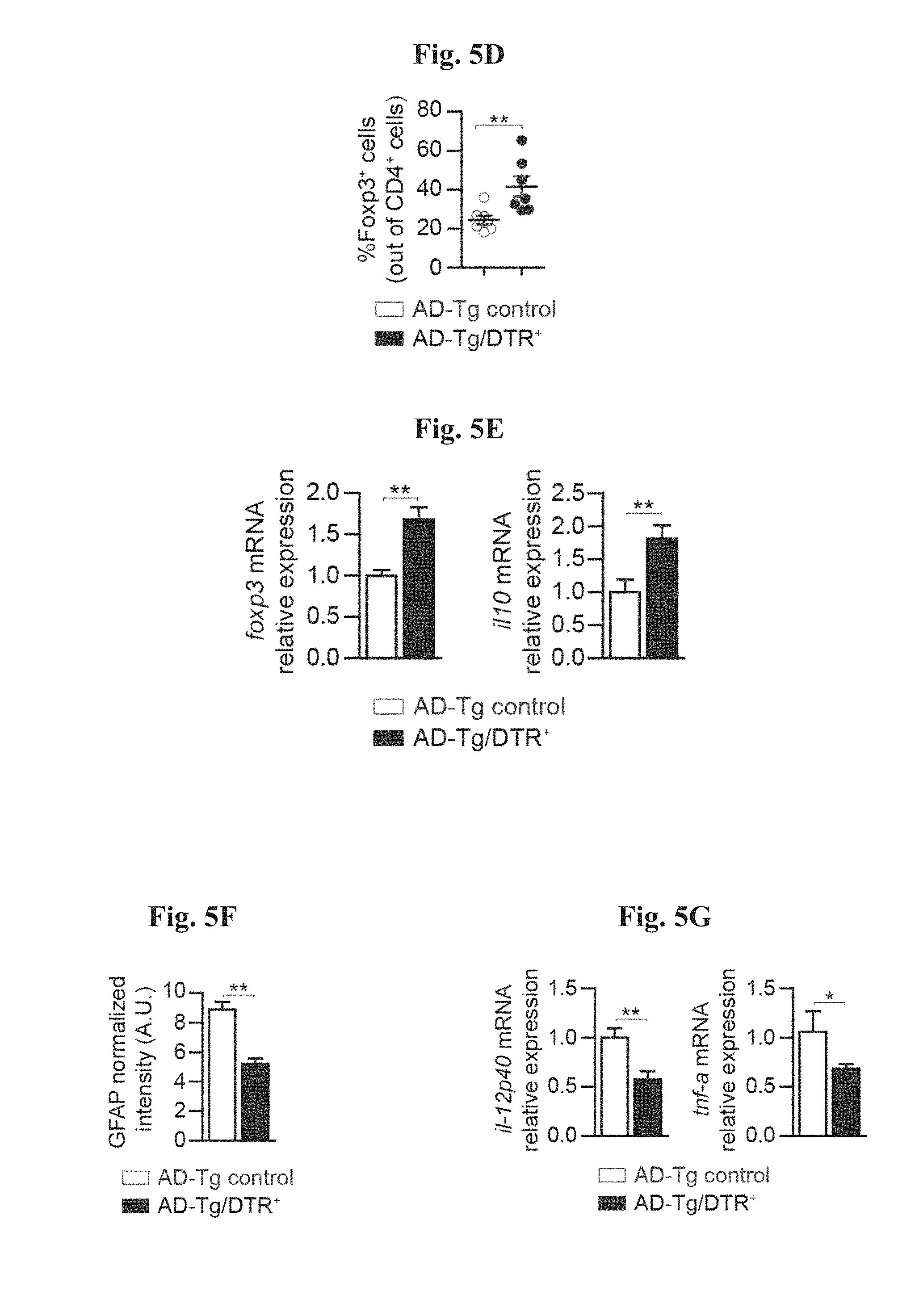
[0010] FIGS. 5A-G show the effects of transient depletion of Tregs in AD-Tg mice. (A) AD-Tg/Foxp3-DTR.sup.+ (which express the DTR transgene) and anon-DTR-expressing AD-Tg littermate (AD-Tg/Foxp3-DTR.sup.-) control group were treated with DTx for 4 constitutive days. CP mRNA expression levels for the genes icam1, cxcl10 and ccl2, measured by RT-qPCR, in 6-month old DTx-treated AD-Tg mice, 1 day after the last DTx injection (n=6-8 per group; Student's t test). (B-D) Flow cytometry analysis of the brain parenchyma (excluding the choroid plexus, which was separately excised) of 6-month old DTx-treated AD-Tg mice and controls, 3, weeks following the last DTx injection. Quantitative flow cytometry analysis showing increased numbers of CD11b.sup.high/CD45.sup.high mo-M.PHI. and CD4.sup.+ T cells (B), and representative flow cytometry plots (C) and quantitative analysis (D) of CD4.sup.+Foxp3.sup.+ Treg frequencies, in the brain parenchyma of AD-Tg/Foxp3-DTR.sup.+ mice and AD-Tg/Foxp3-DTR- controls treated with DTx (n=3-7 per group; Student's t test). (E) mRNA expression levels of foxp3 and il10 in the brain parenchyma of 6-month old DTx-treated AD-Tg AD-Tg/Foxp3-DTR.sup.+ and AD-Tg/Foxp3-DTR- controls, 3 weeks after the last DTx injection (n=6-8 per group; Student's t test). (F) quantitative analysis of GFAP immunostaining, showing reduced astrogliosis in hippocampal sections from 6-month old DTx-treated AD-Tg/Foxp3-DTR.sup.+ and AD-Tg/Foxp3-DTR- control mice. 3 weeks following the last DTx injection (scale bar, 50 .mu.m; n=3-5 per group; Student's t test). (G) mRNA expression levels of il-12p40 and tnf-a in the brain parenchyma, 3 weeks following the last DTx injection (n=6-8 per group; Student's t test). In all panels, error bars represent mean.+-.s.e.m.; *, P<0.05; **, P<0.01; ***, P<0.001.
[0011] FIGS. 6A-E show the effect of transient depletion of Tregs on A.beta. plaques learning/memory performance. (A) Representative microscopic images and (B) quantitative analysis of the brains of 5-month old DTx-treated AD-Tg/Foxp3-DTR.sup.+ and AD-Tg/Foxp3-DTR.sup.- control mice, 3 weeks after the last DTx injection, immunostained for A.beta. plaques and Hoechst nuclear staining (scale bar, 250 .mu.m). Mean A.beta. plaque area and numbers in the hippocampal dentate gyrus (DG) and the 5.sup.th layer of the cerebral cortex were quantified (in 6 .mu.m brain slices; n=5-6 per group; Student's t test). FIGS. 6C-E) show Morris water maze (MWM) test performance of 6-month old DTx-treated AD-Tg/Foxp3-DTR.sup.+ and control mice, 3 weeks after the last DTx injection. Following transient Treg depletion, AD-Tg mice showed better spatial learning/memory performance in the (C) acquisition, (D) probe and (E) reversal phases of the MWM, relative to AD-Tg controls (n=7-9 per group; two-way repeated measures ANOVA followed by Bonferroni post-hoc analysis for individual pair comparisons; *, P<0.05 for overall acquisition, probe, and reversal). In all panels, error bars represent mean.+-.s.e.m.; *, P<0.05; **, P<0.01; ***, P<0.001.
[0012] FIG. 7 shows mRNA expression levels of ifn-.gamma., measured by RT-qPCR, in CPs isolated from 6- and 12-month old APP/PS1 AD-Tg mice (a mouse model for Alzheimer's disease (see Materials and Methods)), compared to age-matched WT controls (n=5-8 per group; Student's t test). Error bars represent mean.+-.s.e.m.; *, P<0.05.
[0013] FIGS. 8A-I show the therapeutic effect of administration of weekly Glatiramer acetate (GA) in AD-Tg mice. (A) Schematic representation of weekly-GA treatment regimen. Mice (5-month old) were s.c. injected with GA (100 .mu.g), twice during the first week (on day 1 and 4), and once every week thereafter, for an overall period of 4 weeks. The mice were examined for cognitive performance, 1 week (MWM), 1 month (RAWM) and 2 months (RAWM, using different experimental spatial settings) after the last injection, and for hippocampal inflammation. FIGS. 8B-D show mRNA expression levels of genes in the hippocampus of untreated AD-Tg mice, and AD-Tg mice treated with weekly-GA, at the age of 6 m, showing (B) reduced expression of pro-inflammatory cytokines such as TNF-.alpha., IL-1.beta. and IL-12p40, (C) elevation of the anti-inflammatory cytokines IL-10 and TGF-.beta., and of (D) the neurotropic factors, IGF-1 and BDNF, in weekly-GA treated mice (n=6-8 per group; Student's t test). In FIGS. 8E-G, AD-Tg mice (5 months old) were treated with either weekly-GA or with vehicle (PBS), and compared to age-matched WT littermates in the MWM task at the age of 6 m. Treated mice showed better spatial learning/memory performance in the acquisition (E), probe (F) and reversal (G) phases of the MWM, relative to controls (n=6-9 per group; two-way repeated measures ANOVA followed by Bonferroni post-hoc for individual pair comparisons). FIGS. 8H-I show cognitive performance of the same mice in the RAWM task, 1 month (H) or 2 months (I) following the last GA injection (n=6-9 per group; two-way repeated measures ANOVA followed by Bonferroni post-hoc for individual pair comparisons). Data are representative of at least three independent experiments. In all panels, error bars represent mean.+-.s.e.m.; *, P<0.05; **, P<0.01; ***, P<0.001.
[0014] FIGS. 9A-H show further therapeutic effects of administration of weekly-GA in AD-Tg mice. A-B shows 5XFAD AD-Tg mice that were treated with either weekly-GA, or vehicle (PBS), and were examined at the end of the 1.sup.st week of the administration regimen (after a total of two GA injections). Flow cytometry analysis for CD4.sup.+Foxp3.sup.+ splenocyte frequencies (A), and CP IFN-.gamma.-expressing immune cells (B; intracellularly stained and pre-gated on CD45), in treated 6-month old AD-Tg mice, compared to age-matched WT controls (n=4-6 per group; one-way ANOVA followed by Newman-Keuls post hoc analysis). (C) mRNA expression levels for the genes icam1, cxcl10 and ccl2, measured by RT-qPCR, in CPs of 4-month old AD-Tg mice, treated with either weekly-GA or vehicle, and examined either at the end of the 1.sup.st or 4.sup.th week of the weekly-GA regimen (n=6-8 per group; one-way ANOVA followed by Newman-Keuls post hoc analysis). FIGS. 9D-E show representative images of brain sections from 6-month old AD-Tg/CX.sub.3CR1.sup.GFP/+ BM chimeras following weekly-GA. CX.sub.3CR1.sup.GFP cells were localized at the CP of the third ventricle (3V; i), the adjacent ventricular spaces (ii), and the CP of the lateral ventricles (LV; iii) in AD-Tg mice treated with weekly-GA (D; scale bar, 25 .mu.m). Representative orthogonal projections of confocal z-axis stacks, showing co-localization of GFP.sup.+ cells with the myeloid marker, CD68, in the CP of 7-month old AD-Tg/CX.sub.3CR1.sup.GFP/+ mice treated with weekly-GA, but not in control PBS-treated AD-Tg/CX.sub.3CR1.sup.GFP/+ mice (E; scale bar, 25 .mu.m). (F) CX.sub.3CR1.sup.GFP cells are co-localized with the myeloid marker IBA-1 in brains of GA-treated AD-Tg/CX.sub.3CR1.sup.GFP/+ mice in the vicinity of A.beta. plaques, and co-expressing the myeloid marker, IBA-1 (scale bar, 25 .mu.m). FIGS. 9G-H show representative flow cytometry plots of cells isolated from the hippocampus of 4-month old WT, untreated AD-Tg, and AD-Tg mice, on the 2.sup.nd week of the weekly-GA regimen. CD1b.sup.high/CD45.sup.high mo-M.PHI. were gated (G) and quantified (H; n=4-5 per group; one-way ANOVA followed by Newman-Keuls post hoc analysis). In all panels, error bars represent mean.+-.s.e.m.; *, P<0.05; **, P<0.01; ***, P<0.001.
[0015] FIGS. 10A-H depict the therapeutic effect of administration of a p300 inhibitor (C646) in AD-Tg mice. In FIGS. 10A-B, aged mice (18 months) were treated with either p300i or vehicle (DMSO) for a period of 1 week, and examined a day after cessation of treatment. Representative flow cytometry plots showing elevation in the frequencies of CD4.sup.+ T cells expressing IFN-.gamma. in the spleen (A), and IFN-.gamma.-expressing immune cell numbers in the CP (B), following p300i treatment. FIGS. 10C-E show representative microscopic images (C), and quantitative analysis, of A.beta. plaque burden in the brains of 10-month old AD-Tg mice, which received either p300i or vehicle (DMSO) for a period of 1 week, and were subsequently examined after 3 additional weeks. Brains were immunostained for A.beta. plaques and by Hoechst nuclear staining (n=5 per group; Scale bar, 250 .mu.m). Mean A.beta. plaque area and plaque numbers were quantified in the hippocampal DG (D) and the 5.sup.th layer of the cerebral cortex (E) (in 6 .mu.m brain slices; n=5-6 per group; Student's t test). (F) Schematic representation of the p300i treatment (or DMSO as vehicle) administration regimen to the different groups of AD-Tg mice at the age of 7 months, in either 1 or 2 sessions. FIGS. 10G-H show the change mean of A.beta. plaque percentage coverage of the cerebral cortex (5.sup.th layer) (G), and the change in mean cerebral soluble A.beta..sub.1-40 and A.beta..sub.1-42 protein levels (H), relative to the untreated AD-Tg group (A.beta..sub.1-40 and A.beta..sub.1-42 mean level in untreated group, 90.5.+-.11.2 and 63.8.+-.6.8 pg/mg total portion, respectively; n=5-6 per group; one-way ANOVA followed by Newman-Keuls post hoc analysis). In all panels, error bars represent mean.+-.s.e.m.; *, P<0.05; **, P<0.01; ***, P<0.001.
[0016] FIGS. 11A-B show the therapeutic effect of administration of anti-PD1 antibody in AD-Tg mice. (A) Schematic representation of the experimental groups of mice, their age, treatment administration regimens, and the time point in which mice were examined. At 10 months of age, 5XFAD Alzheimer's' disease (AD) transgenic (Tg) mice were injected i.p. with either 250 .mu.g of anti-PD1 (RMP1-14) or control IgG (rat) antibodies, on day 1 and day 4 of the experiment, and were examined 3 weeks after for their cognitive performance by radial arm water maze (RAWM) spatial learning and memory task. Age matched untreated WT and AD-Tg mice were used as controls. (B) show cognitive performance, as assessed by radial arm water maze (RAWM) spatial learning and memory task. Data were analyzed using two-way repeated-measures ANOVA, and Bonferroni post-hoc procedure was used for follow-up pairwise comparison. n=6-12 per group. In all panels, error bars represent mean.+-.s.e.m.; *, P<0.05; **, P<0.01; ***, P<0.001.
[0017] FIGS. 12A-B show the systemic effect on IFN-.gamma..sup.+ producing T cells of administration of anti-PD1 antibody in AD-Tg mice. (A) Schematic representation of the experimental groups of mice, their age, treatment administration regimens, and the time point in which mice were examined. Mice were injected i.p. with either 250 .mu.g of anti-PD1 (RMP1-14) or control IgG (rat) antibodies, on day 1 and day 4 of the experiment, and examined on day 7. (B) Flow cytometry analysis for CD4.sup.+ IFN-.gamma..sup.+ T cell splenocyte frequencies (intracellularly stained and pre-gated on CD45 and TCR-.beta.), in PD-1 or IgG treated AD-Tg mice, and untreated AD-Tg and WT controls (n=4-6 per group; one-way ANOVA followed by Newman-Keuls post hoc analysis; **, P<0.01 between the indicted treated groups; error bars represent mean.+-.s.e.m.).
[0018] FIGS. 13A-B show the effect on the CP following anti-PD1 treatment in AD-Tg mice. AD-Tg mice at the age of 10 months were either treated with PD-1, IgG, or left untreated. Mice were injected i.p. with either 250 .mu.g of anti-PD1 (RMP1-14) or control IgG (rat) antibodies, on day 1 and day 4 of the experiment, and examined on day 7. (A) CP IFN-.gamma. levels, as measured by real-time quantitative PCR (RT-qPCR), positively correlated (Pearson's r=0.6284, P<0.05), to and negatively correlated to CD4.sup.+IFN-.gamma..sup.+ T cell splenocyte frequencies, as measured by flow cytometry. An opposite, negative trend was observed in the same mice, when CP IFN-.gamma. levels were compared to CD4.sup.+Foxp3.sup.+CD25.sup.+ Tregs splenocyte frequencies (n=3-4 per group). (B) mRNA expression levels for the genes cxcl10 and ccl2, measured by RT-qPCR, in CPs of the same mice (n=3-4 per group; one-way ANOVA followed by Newman-Keuls post hoc analysis). In all panels, error bars represent mean.+-.s.e.m.; *, P<0.05.
[0019] FIGS. 14A-B show the therapeutic effect of administration of anti-PD1 antibody in AD-Tg mice, when comparing one vs. two courses of treatment. Half of the mice in the group of mice described in FIG. 11A-B which received one course of anti-PD1 treatment, either received another course of anti-PD1 treatment following the first RAWM task, or left untreated. Following additional 3 weeks, all mice were tested by RAWM using different and new experimental settings of spatial cues for cognitive learning and memory. (A) Schematic representation of the experimental groups of mice, their age, treatment administration regimens, and the time point in which mice were examined. For each course of treatment, mice were injected i.p. with either 250 ug of anti-PD1 (RMP1-14) or control IgG (rat) antibodies. (B) show cognitive performance, as assessed by RAWM spatial learning and memory task. Data were analyzed using two-way repeated-measures ANOVA, and Bonferroni post-hoc procedure was used for follow-up pairwise comparison. n=6-12 per group. In all panels, error bars represent mean.+-.s.e.m.; *, P<0.05; **, P<0.01; ***, P<0.001.
[0020] FIGS. 15A-H show the adverse effect on AD pathology of systemic Treg levels augmented by all-trans retinoic acid (ATRA). FIGS. 15A-B show representative flow cytometry plots (A), and quantitative analysis (B), showing elevation in frequencies of CD4.sup.+/Foxp3.sup.+/CD25.sup.+ Treg splenocytes in 5-month old AD-Tg mice, which received either ATRA or vehicle (DMSO) for a period of 1 week (n=5 per group; Student's t test). FIGS. 15C-F show representative microscopic images (C), and quantitative analysis (D, E, F), of A.beta. plaque burden and astrogliosis in the brains of AD-Tg mice, which at the age of 5-months were treated with either ATRA or vehicle (DMSO) for a period of 1 week, and subsequently examined after 3 additional weeks. Brains were immunostained for A.beta. plaques, GFAP (marking astrogliosis), and by Hoechst nuclear staining (n=4-5 per group; Scale bar, 250 .mu.m). Mean A.beta. plaque area and plaque numbers were quantified in the hippocampal DG and the 5.sup.th layer of the cerebral cortex, and GFAP immunoreactivity was measured in the hippocampus (in 6 .mu.m brain slices; n=5-6 per group; Student's t test). (G) Levels of soluble A.beta..sub.1-40 and A.beta..sub.1-42, quantified by ELISA, in the cerebral brain parenchyma AD-Tg mice, which at the age of 5-months were treated with either ATRA or vehicle (DMSO) for a period of 1 week, and subsequently examined after 3 additional weeks (n=5-6 per group; Student's t test). (H) Cognitive performance in the RAWM task of AD-Tg mice which at the age of 5-months were treated with either ATRA or vehicle (DMSO) for a period of 1 week, and subsequently examined after 3 additional weeks (n=5 per group; two-way repeated measures ANOVA followed by Bonferroni post-hoc for individual pair comparisons). In all panels, error bars represent mean.+-.s.e.m.; *, P<0.05; **, P<0.01; ***, P<0.001.
[0021] FIGS. 16A-F show the adverse effect on AD pathology of systemic Treg levels augmented by weekly-GA administration. (A) Schematic representation of daily-GA treatment regimen compared to the weekly-GA regimen. In the daily-GA treated group, mice were s.c. injected daily with 100 .mu.g of GA for a period of 1 month. (B) Cognitive performance of daily-GA and weekly-GA treated 7-month old AD-Tg mice, compared to age-matched WT and untreated AD-Tg mice, as assessed by the average numbers of errors per day in the RAWM learning and memory task (n=6-8 per group; one-way ANOVA followed by Newman-Keuls post hoc analysis). (C) Representative microscopic images of the cerebral cortex and the hippocampus (HC) of untreated AD-Tg, and daily or weekly-GA treated AD-Tg mice, immunostained for A.beta. plaques and for Hoechst nuclear staining (scale bar, 250 .mu.m). FIGS. 16D-F show quantification of A.beta. plaque size and numbers (per 6 .mu.m slices) in GA treated (daily-GA and weekly-GA groups) and untreated AD-Tg mice. Weekly-GA treated AD-Tg mice showed reduction in A.beta. plaque load as a percentage of the total area of their hippocampal dentate gyrus (DG), and in mean A.beta. plaque numbers (n=6 per group; one-way ANOVA followed by Newman-Keuls post hoc analysis). In all panels, error bars represent mean.+-.s.e.m.; *, P<0.05; **, P<0.01; ***, P<0.001.
DETAILED DESCRIPTION
[0022] It has been found in accordance with the present invention that a short-term transient depletion of Foxp3.sup.+ regulatory T cells (Tregs) in a mouse model of Alzheimer's disease (AD-Tg mice) results in improved recruitment of leukocytes to the CNS through the brain's choroid plexus, elevated numbers of CNS-infiltrating anti-inflammatory monocyte-derived macrophages mo-M.PHI. and CD4.sup.+ T cells, and a marked enrichment of Foxp3.sup.+ Tregs that accumulates within the brain. Furthermore, the long-term effect of a single session of treatment lead to a reduction in hippocampal gliosis and reduced mRNA expression levels of pro-inflammatory cytokines within the brain. Importantly, the effect on disease pathology includes reduced cerebral amyloid beta (A.beta.) plaque burden in the hippocampal dentate gyrus, and the cerebral cortex (5.sup.th layer), two brain regions exhibiting robust A.beta. plaque pathology in the AD-Tg mice. Most importantly, the short-term transient depletion of Tregs is followed by a dramatic improvement in spatial learning and memory, reaching cognitive performance similar to that of wild type mice (Examples 2 and 3). Taken together, these findings demonstrate that a short session of Treg depletion, followed by a period of no intervention, results in transiently breaking Treg-mediated systemic immune suppression in AD-Tg mice, which enables recruitment of inflammation-resolving cells, mo-M.PHI. and Tregs, to the brain, and lead to resolution of the neuroinflammatory response, clearance of A.beta., and reversal of cognitive decline. These findings strongly argue against the common wisdom in this field of research, according to which increasing systemic immune suppression would result in mitigation of the neuroinflammatory response. On the contrary, our findings show that boosting of the systemic response, by a short-term, brief and transient, reduction in systemic Treg-mediated suppression, is needed in order to achieve inflammation-resolving immune cell accumulation, including Tregs themselves, within the brain, thus fighting off AD pathology.
[0023] The specificity of the inventors approach presented herein has been substantiated by using several independent experimental paradigms, as detailed below. Briefly, first the inventors used an immunomodulatory compound in two different administration regimens that led to opposite effects on peripheral Treg levels, on CP activation, and on disease pathology; a daily administration regimen that augments peripheral Treg levels (Weber et al, 2007), and a weekly administration regimen, which they found to reduce peripheral Treg levels (Example 3 and Example 5). The inventors also provide a direct functional linkage between peripheral Treg levels and disease pathology when demonstrating in AD-Tg mice, by either transient in vivo genetic depletion of Tregs (Example 2), or by pharmacologic inhibition of their Foxp3 function (Examples 3 and 4), that these manipulations result in activation of the CP for facilitating leukocyte trafficking to the CNS, inflammation-resolving immune cell accumulation at sites of pathology, clearance of cerebral A.beta. plaques, and skewing of the immunological milieu of the brain parenchyma towards the resolution of inflammation.
[0024] It has further been found in accordance with the present invention that infrequent administration of a universal antigen, Copolymer-1, for a limited period of time (representing one session of treatment) reduces Treg-mediated systemic immune suppression, and improves selective infiltration of leukocytes into the CNS by increasing the brain's choroid plexus gateway activity, leading to dramatic beneficial effect in Alzheimer's disease pathology (Example 3), while daily administration of Copolymer 1, that enhance Treg immune suppression (Hong et al, 2005; Weber et al, 2007), showed no beneficial effect, or even some modest detrimental effect, on disease pathology (Example 5). The inventors of the present invention further show herein that direct interference with Foxp3 Treg activity, either by inhibition of p300 with a specific small molecule inhibitor (p300i), or interaction with the PD-1 receptor by an anti-PD-1 antibody, improves choroid plexus gateway activity in AD-Tg mice, and mitigates Alzheimer's disease pathology (Example 4).
[0025] Importantly, each of these examples provided by the inventors, demonstrate a different intervention which causes short term reduction in systemic immune suppression: Copolymer-1 acts as an immunomodulatory compound, p300i as a small molecule which decreases Foxp3 acetylation and Treg function, and anti-PD-1 is used as a neutralizing antibody for PD-1 expressed on Tregs. These therapeutic approaches were used for a short session of treatment that transiently augmented immune response in the periphery, mainly by elevation of peripheral IFN-.gamma. levels and IFN-.gamma.-producing cells, thus activating the brain's choroid plexus allowing selective infiltration of T cells and monocytes into the CNS, and homing of these cells to sites of pathology and neuroinflammation. It was also found herein that repeated sessions of treatment interrupted by interval sessions of non-treatment dramatically improve the efficacy of the treatment relative to a single session of treatment (Example 4). The following time interval of non-treatment allowed transient augmentation in Treg levels and activities within the brain, facilitating the resolution of neuroinflammation, and inducing environmental conditions in favor of CNS healing and repair, subsequently leading to tissue recovery. In each of these cases the effect on brain pathology was robust, involving the resolution of the neuroinflammatory response, amyloid beta plaque clearance from AD mice brains, and reversal of cognitive decline. The specificity of the current approach has further been substantiated using a genetic model of transient depletion of Foxp3.sup.+ regulatory T cells, in transgenic mouse model of AD (Example 2).
[0026] Thus, it has been found in accordance with the present invention that systemic Foxp3.sup.+CD4.sup.+ Treg-mediated immunosuppression interferes with ability to fight off AD pathology, acting at least in part, by inhibiting IFN-.gamma.-dependent activation of the CP, needed for orchestrating recruitment of inflammation-resolving leukocytes to the CNS (Schwartz & Baruch, 2014b). Systemic Tregs are crucial for maintenance of autoimmune homeostasis and protection from autoimmune diseases (Kim et al, 2007). However, our findings suggest that under neurodegenerative conditions, when a reparative immune response is needed in the brain, the ability to mount this response is interfered with by systemic Tregs. Nevertheless according to our results, Tregs are needed within the brain, home to sites of neuropathology, and perform locally an anti-inflammatory activity. The present invention represents a unique and unexpected solution for the apparent contradictory needs in fighting off progressive neuronal death as in AD; transiently reducing/inhibiting Tregs in the circulation on behalf of increasing Tregs in the diseased brain. Hence, a short-term and transient reduction in peripheral immune suppression, which allows the recruitment of anti-inflammatory cells, including Tregs and mo-M.PHI., to sites of cerebral plaques, leads to a long-term effect on pathology. Notably, however, a transient reduction of systemic Treg levels or activities may contribute to disease mitigation via additional mechanisms, including supporting a CNS-specific protective autoimmune response (Schwartz & Baruch, 2014a), or augmenting the levels of circulating monocytes that play a role in clearance of vascular A.beta. (Michaud et al, 2013).
[0027] Though neurodegenerative diseases of different etiology, share a common local neuroinflammatory component, our results strongly argue against simplistic characterization of all CNS pathologies as diseases that would uniformly benefit from systemic anti-inflammatory therapy. Thus, while autoimmune inflammatory brain pathologies, such as Relapsing-Remitting Multiple Sclerosis (RRMS), benefit from continuous systemic administration of anti-inflammatory and immune-suppressive drugs to achieve long lasting peripheral immune suppression, it will either be ineffective or detrimentally affect (Example 5) pathology in chronic neurodegenerative diseases such as in the case of AD, primary progressive multiple sclerosis (PP-MS) and secondary-progressive multiple sclerosis (SP-MS). Moreover, our findings shed light on the misperception regarding the role of systemic vs. tissue-associated Tregs in these pathologies (He & Balling, 2013). Since the immune-brain axis is part of life-long brain plasticity (Baruch et al, 2014), and neurodegenerative diseases are predominantly age-related, our present findings also point to a more general phenomenon, in which systemic immune suppression interferes with brain function. Accordingly, short-term periodic courses of reducing systemic immune suppression may represent a therapeutic or even preventive approach, applicable to a wide range of brain pathologies, including AD and age-associated dementia.
[0028] Importantly, the inventors approach and findings present herein in AD mouse models, do not directly target any disease-specific factor in AD, such as amyloid beta or tau pathology, but rather demonstrate a novel approach which is expected to be clinically applicable in a wide range of CNS pathologies--transient reduction of systemic Treg-mediated immune suppression in order to augment recruitment of inflammation-resolving immune cells to sites of pathology within the CNS.
[0029] In view of the unexpected results described above, the present invention provides a pharmaceutical composition comprising an active agent that causes reduction of the level of systemic immunosuppression in an individual for use in treating a disease, disorder, condition or injury of the CNS that does not include the autoimmune neuroinflammatory disease, relapsing-remitting multiple sclerosis (RRMS), wherein said pharmaceutical composition is for administration by a dosage regimen comprising at least two courses of therapy, each course of therapy comprising in sequence a treatment session followed by an interval session of non-treatment.
[0030] In certain embodiments, the dosage regimen is calibrated such that the level of systemic immunosuppression is transiently reduced.
[0031] The term "treating" as used herein refers to means of obtaining a desired physiological effect. The effect may be therapeutic in terms of partially or completely curing a disease and/or symptoms attributed to the disease. The term refers to inhibiting the disease, i.e. arresting or slowing its development; or ameliorating the disease, i.e. causing regression of the disease.
[0032] The term "non-treatment session" is used interchangeably herein with the term "period of no treatment" and refers to a session during which no active agent is administered to the individual being treated.
[0033] The term "systemic presence" of regulatory T cells as used herein refers to the presence of the regulatory T cells (as measured by their level or activity) in the circulating immune system, i.e. the blood, spleen and lymph nodes. It is a well-known fact in the field of immunology that the cell population profile in the spleen is reflected in the cell population profile in the blood (Zhao et al, 2007).
[0034] The present treatment is applicable to both patients that show elevation of systemic immune suppression, as well as to patients that do not show such an elevation. Sometimes the individual in need for the treatment according to the present invention has a certain level of peripheral immunosuppression, which is reflected by elevated frequencies or numbers of Tregs in the circulation, and/or their enhanced functional activity and/or a decrease in IFN.gamma.-producing leukocytes and/or decreased proliferation of leukocytes in response to stimulation. The elevation of frequencies or numbers of Tregs can be in total numbers or as percentage of the total CD4 cells. For example, it has been found in accordance with the present invention that an animal model of Alzheimer's disease has higher frequencies of Foxp3 out of CD4 cells as compared with wild-type mice. However, even if the levels of systemic Treg cells is not elevated, their functional activity is not enhanced, the level of IFN.gamma.-producing leukocytes is not reduced or the proliferation of leukocytes in response to stimulation is not decreased, in said individual, the method of the present invention that reduces the level or activity of systemic immunosuppression is effective in treating disease, disorder, condition or injury of the CNS that does not include the autoimmune neuroinflammatory disease RRMS. Importantly, said systemic immune suppression can also involve additional immune cell types except of Tregs, such as myeloid-derived suppressor cells (MDSCs) (Gabrilovich & Nagaraj, 2009).
[0035] The level of systemic immunosuppression may be detected by various methods that are well known to those of ordinary skill in the art. For example, the level of Tregs may be measured by flow cytometry analysis of peripheral blood mononuclear cells or T lymphocytes, immunostained either for cellular surface markers or nuclear intracellular markers of Treg (Chen & Oppenheim, 2011), CD45, TCR-.beta., or CD4 markers of lymphocytes, and measuring the amount of antibody specifically bound to the cells. The functional activity of Tregs may be measured by various assays; For example the thymidine incorporation assay is being commonly used, in which suppression of anti-CD3 mAb stimulated proliferation of CD4.sup.+CD25.sup.- T cells (conventional T cells) is measured by [.sup.3H]thymidine incorporation or by using CFSE (5-(and 6)-carboxyfluorescein diacetate succinimidyl ester, which is capable of entering the cells; cell division is measured as successive halving of the fluorescence intensity of CFSE). The number of IFN.gamma.-producing leukocytes or their activity or their proliferation capacity can easily be assessed by a skilled artisan using methods known in the art; For example, the level of IFN.gamma.-producing leukocytes may be measured by flow cytometry analysis of peripheral blood mononuclear cells, following short ex-vivo stimulation and golgi-stop, and immunostaining by IFN.gamma. intracellular staining (using e.g., BD Biosciences Cytofix/Cytoperm.TM. fixation/permeabilization kit), by collecting the condition media of these cells and quantifying the level of secreted cytokines using ELISA, or by comparing the ratio of different cytokines in the condition media, for example IL2/IL10, IL2/IL4, INF.gamma./TGF.beta., etc. The levels of MDSCs in the human peripheral blood easily can be assessed by a skilled artisan, for example by using flow cytometry analysis of frequency of DR.sup.-/LIN.sup.-/CD11b+, DR.sup.-/LIN.sup.-/CD15+, DR.sup.-/LIN.sup.-/CD33+ and DR(-/low)/CD14+ cells, as described (Kotsakis et al, 2012).
[0036] In humans, the peripheral/systemic immunosuppression may be considered elevated when the total number of Tregs in the circulation is higher than 10, 20, 30, 40, 50, 60, 70, 80, 90, or 100% or more than in a healthy control population, the percentage of Treg cells out of the total CD4+ cells is elevated by 10, 20, 30, 40, 50, 60, 70, 80, 90, or 100% or more than in a healthy control population, or the functional activity of Tregs is elevated by 10, 20, 30, 40, 50, 60, 70, 80, 90, or 100% or more than in a healthy control population. Alternatively, the peripheral/systemic immunosuppression may be considered elevated when the level of IFN.gamma.-producing leukocytes or their activity is reduced relative to that of a healthy control population by 10, 20, 30, 40, 50, 60, 70, 80, 90 or 100%; or the proliferation of leukocytes in response to stimulation is reduced relative to that of a healthy control population by 10, 20, 30, 40, 50, 60, 70, 80, 90 or 100%.
[0037] An agent may be considered an agent that causes reduction of the level of systemic immunosuppression when, upon administration of the agent to an individual, the total number of Tregs in the circulation of this individual is reduced by 10, 20, 30, 40, 50, 60, 70, 80, 90 or 100% as compared with the level before administration of the agent, the percentage of Treg cells out of the total CD4+ cells drops by 10, 20, 30, 40, 50, 60, 70, 80, 90 or 100% relative to that of a healthy control population or the functional activity of Tregs is reduced by 10, 20, 30, 40, 50, 60, 70, 80, 90 or 100% as compared with the level before administration of the agent. Alternatively, an agent may be considered an agent that causes reduction of the level of systemic immunosuppression when, upon administration of the agent to an individual, the total number of IFN.gamma.-producing leukocytes or their activity is increased by 10, 20, 30, 40, 50, 60, 70, 80, 90, or 100%; or the proliferation of leukocytes in response to stimulation is increased relative to that of a healthy control population by 10, 20, 30, 40, 50, 60, 70, 80, 90 or 100%.
[0038] The agent used according to the present invention may be any agent that down-regulates the level or activity of regulatory T cells or interfere with their activity, but may alternatively be limited to a group of such agents excluding an agent selected from the group consisting of: (i) dopamine or a pharmaceutically acceptable salt thereof, (ii) a dopamine precursor or a pharmaceutically acceptable salt thereof, (iii) an agonist of the dopamine receptor type 1 family (D1-R agonist) or a pharmaceutically acceptable salt thereof, and (iv) an antagonist of the dopamine receptor type 2 family (D2-R antagonist) or a pharmaceutically acceptable salt thereof, even though these agents are not know for use according to the course of therapy according to the present invention.
[0039] In certain embodiments, the treatment session comprises administering the pharmaceutical composition to the individual and the treatment session is maintained at least until the level falls below a reference, the administering is paused during the interval session, and the interval session is maintained as long as the level is below the reference. The reference may be selected from (a) the level of systemic presence or activity of regulatory T cells or myeloid-derived suppressor cells measured in the most recent blood sample obtained from said individual before said administering; or (b) the level of systemic presence or activity of regulatory T cells or myeloid-derived suppressor cells characteristic of a population of individuals afflicted with a disease, disorder, condition or injury of the CNS.
[0040] Alternatively, the treatment session comprises administering the pharmaceutical composition to the individual and the treatment session is maintained at least until the systemic presence or level of IFN.gamma.-producing leukocytes, or the rate of proliferation of leukocytes in response to stimulation rises above a reference, the administering is paused during the interval session, and the interval session is maintained as long as said level is above said reference, wherein the reference is selected from (a) the level of systemic presence or activity of IFN.gamma.-producing leukocytes, or the rate of proliferation of leukocytes in response to stimulation, measured in the most recent blood sample obtained from said individual before said administering; or (b) the level of systemic presence or activity of IFN.gamma.-producing leukocytes, or the rate of proliferation of leukocytes in response to stimulation, characteristic of a population of individuals afflicted with a disease, disorder, condition or injury of the CNS.
[0041] The length of the treatment and interval sessions may be determined by physicians in clinical trials directed to a certain patient population and then applied consistently to this patient population, without the need for monitoring the level of immunosuppression on a personal basis.
[0042] In certain embodiments, the treatment session may be between 3 days and four weeks long, for example between one and four weeks long.
[0043] In certain embodiments, the interval session may be between one week and six months, for example between two weeks and six months long, in particular between 3 weeks and six months long.
[0044] In the treatments session, the administration of the pharmaceutical composition may be repeated administration, for example the pharmaceutical composition may be administered daily, or once every two, three, four, five or six days, once weekly, once every two weeks, once every three weeks or once every four weeks. These frequencies are applicable to any active agent, may be based on commonly used practices in the art, and may finally be determined by physicians in clinical trials. Alternatively, the frequency of the repeated administration in the treatment session could be adapted according to the nature of the active agent, wherein for example, a small molecule may be administered daily; an antibody may be administered once every 3 days; and copolymer 1 is administered weekly, once every two weeks, once every three weeks or once every four weeks. It should be understood that when an agent, such as copolymer 1, is administered during a treatment session at a relatively low frequency, for example once per week during a treatment session of one month, or once per month during a treatment session of six months, this treatment session is followed by a non-treatment interval session, the length of which is longer than the period between the repeated administrations during the treatment session (i.e. longer than one week or one month, respectively, in this example). The pause of one week or one month between the administrations during the treatment session in this example is not considered an interval session.
[0045] The lengths of the treatment session and the interval session may be adjusted to the frequency of the administration such that, for example, a frequency of administering the active agent once every 3 days may result in a treatment session of 6 or 9 days and an interval session that is commenced accordingly.
[0046] As an alternative to a predetermined general treatment regiment, the level of immunosuppression may be calibrated to a desired level for each patient who is being treated (personalized medicine), by monitoring the level or activity of Treg cells (or IFN-.gamma.-producing leukocytes or proliferation rate of leukocytes in response to stimulation) individually, and adjusting the treatment session, the frequency of administration and the interval session empirically and personally as determined from the results of the monitoring.
[0047] Thus, the length of the treatment session may be determined by (a) monitoring the level of systemic presence or activity of regulatory T cells in the individual by measuring the level in a blood sample obtained from the individual within a predetermined time-period following said administering; (b) comparing the level measured in (a) with the reference mentioned above and determining whether the level is different from the reference; (c) deciding, based on the relation of said level measured in (a) to said reference, whether to continue the treatment session by repeating the administering or starting the next interval session by refraining from repeating the administration; and (d) repeating the administering or starting the next interval session according to the decision in (c). Alternatively, the level of IFN-.gamma.-producing leukocytes or proliferation rate of leukocytes in response to stimulation may be monitored and compared with an appropriate reference as mentioned above.
[0048] Similarly, the length of the interval session may be determined by (a) monitoring the level of systemic presence or activity of regulatory T cells in the individual by measuring the level in a blood sample obtained from the individual within a predetermined time-period following said administering; (b) comparing the level measured in (a) with the reference mentioned above and determining whether the level is different from the reference; (c) deciding, based on the relation of said level measured in (a) to said reference, whether to start a new course of therapy by repeating the administering and steps (a) and (b) or to prolong the interval session by repeating only steps (a) and (b); and (d) repeating the administering and steps (a) and (b) or only steps (a) and (b) according to the decision in (c). Alternatively, the level of IFN.gamma.-producing leukocytes or proliferation rate of leukocytes in response to stimulation may be monitored and compared with an appropriate reference as mentioned above.
[0049] In any case, the dosage regimen, i.e. the length of the treatment session and the interval session, is calibrated such that the reduction in the level of immunosuppression, for example as measured by a reduction in the level of systemic presence or activity of regulatory T cells in the individual, is transient.
[0050] In certain embodiments, the predetermined time-period, i.e. the time passed between the most recent administration of the active agent and the monitoring step, is between 2 days and six months.
[0051] In certain embodiments, the regulatory T cells that are monitored are CD4+ cells selected from FoxP3.sup.+ cells expressing one or more of CD25, CD127, GITR, CTLA-4 or PD-1; or FoxP3.sup.- cells expressing one or more of CD25, CD127, GITR, CTLA-4 or PD-1 surface molecules. In particular, a common phenotype of regulatory T cells is CD4.sup.+CD25.sup.+FoxP3.sup.+ cells or CD4.sup.+CD25.sup.+FoxP3.sup.- cells.
[0052] Agents capable of reducing the level of regulatory T cells are known in the art (Colombo & Piconese, 2007) and these agents can be used in accordance with the present invention. Each one of the cited publications below is incorporated by reference as if fully disclosed herein.
[0053] Thus, the agent may be selected from, but is not necessarily limited to: (i) an antibody such as: (a) anti-PD-1, (b) anti-PD-L1 (c) anti-PD-L2 (Coyne & Gulley, 2014; Duraiswamy et al, 2014; Zeng et al, 2013); (d) anti-CTLA-4 (Simpson et al, 2013; Terme et al, 2012); (e) anti-PD-1 in combination with interferon .alpha. (Terawaki et al, 2011); (f) anti-PD-1 in combination with anti-CTLA4; (g) anti-CD47 (Tseng et al, 2013); (h) anti-OX40 (Voo et al, 2013); (i) anti-VEGF-A (bevacizumab) (Terme et al, 2013); (j) anti-CD25 (Zhou et al, 2013); (k) anti-GITR (GITR triggering mAb (DTA-1) (Colombo & Piconese, 2007); (l) anti-CCR4; (m) anti-TIM-3/Galectin9 (Ju et al, 2014); (n) anti-killer-cell immunoglobulin-like receptors (KIR); (o) anti-LAG-3; or (p) anti-4-1BB (ii) any combination of (a) to (p); (iii) any combination of (a) to (p) in combination with an adjuvant, for example anti-CTLA-4 antibody in combination with anti OX40 antibody and a TLR9 ligand such as CpG (Marabelle et al, 2013); (iv) a small molecule selected from: (a) A p300 inhibitor (Liu et al, 2013), such as gemcitabine (low dose) (Shevchenko et al, 2013), or C646 or analogs thereof, i.e. a compound of the formula I:
##STR00001##
[0054] wherein
[0055] R.sub.1 is selected from H, --CO.sub.2R.sub.6, --CONR.sub.6R.sub.7, --SO.sub.3H, or --SO.sub.2NR.sub.6R.sub.7;
[0056] R.sub.2 is selected from H, --CO.sub.2R.sub.6, or halogen, preferably Cl;
[0057] R.sub.3 is selected from halogen, preferably F, --NO.sub.2, --CN, --CO.sub.2R.sub.6, preferably CO.sub.2CH.sub.3 or CO.sub.2CH.sub.2CH.sub.3, or --CH.sub.2OH;
[0058] R.sub.4 and R.sub.5 each independently is H or --C.sub.1-C.sub.6 alkyl, preferably methyl;
[0059] R.sub.6 is H or --C.sub.1-C.sub.6 alkyl, preferably H, methyl or ethyl; and
[0060] R.sub.7 is H or --C.sub.1-C.sub.6 alkyl, preferably H or methyl [see (Bowers et al, 2010)];
[0061] (b) Sunitinib (Terme et al, 2012); (c) Polyoxometalate-1 (POM-1) (Ghiringhelli et al, 2012); (d) .alpha.,.beta.-methyleneadenosine 5'-diphosphate (APCP) (Ghiringhelli et al, 2012); (e) arsenic trioxide (As.sub.2O.sub.3) (Thomas-Schoemann et al, 2012); (f) GX15-070 (Obatoclax) (Kim et al, 2014); (g) a retinoic acid antagonist such as Ro 41-5253 (a synthetic retinoid and selective small molecule antagonist) (Galvin et al, 2013) or LE-135 (Bai et al, 2009); (h) an SIRP.alpha. (CD47) antagonist, such as CV1-hIgG4 (SIRP.alpha. variant) as sole agent or in combination with anti-CD47 antibody (Weiskopf et al, 2013); (i) a CCR4 antagonist, such as AF399/420/18025 as sole agent or in combination with anti-CCR4 antibody (Pere et al, 2011); (j) an adenosin A2B receptor antagonist, such as PSB603 (Nakatsukasa et al, 2011); (k) an antagonist of indoleamine-2,3-dioxygenase (IDO); or (l) an HIF-1 regulator; (iv) a protein selected from: (a) Neem leaf glycoprotein (NLGP) (Roy et al, 2013); or (b) sCTLA-4 (soluble isoform of CTLA-4) (Ward et al, 2013); (vi) a silencing molecule such as miR-126 antisense (Qin et al, 2013) and anti-galectin-1 (Gal-1) (Dalotto-Moreno et al, 2013); (vii) OK-432 (lyophilized preparation of Streptococcus pyogenes) (Hirayama et al, 2013); (viii) a combination of IL-12 and anti-CTLA-4; (ix) Copolymer 1 or a copolymer that modulates Treg activity or level; (x) an antibiotic agent, such as vancomycin (Brestoff & Artis, 2013; Smith et al, 2013) or (xi) any combination of (i) to (x).
[0062] In certain embodiments, the agent is an anti-PD-1 antibody, i.e. an antibody specific for PD-1.
[0063] Many anti-PD-1 antibodies are known in the art. For example, the anti-PD-1 antibody used in accordance with the present invention may be selected from those disclosed in Ohaegbulam et al. (Ohaegbulam et al, 2015), the entire contents of which being hereby incorporated herein by reference, i.e. CT-011 (pidilizumab; Humanized IgG1; Curetech), MK-3475 (lambrolizumab, pembrolizumab; Humanized IgG4; Merck), BMS-936558 (nivolumab; Human IgG4; Bristol-Myers Squibb), AMP-224 (PD-L2 IgG2a fusion protein; AstraZeneca), BMS-936559 (Human IgG4; Bristol-Myers Squibb), MEDI4736 (Humanized IgG; AstraZeneca), MPDL3280A (Human IgG; Genentech), MSB0010718C (Human IgG1; Merck-Serono); or the antibody used in accordance with the present invention may be MEDI0680 (AMP-514; AstraZeneca) a humanized IgG4 mAb.
[0064] In certain embodiments, the CT-011 antibody may be administered to a human at a dosage of 0.2-6 mg/kg or between 1.5-6 mg/kg; the MK-3475 antibody may be administered to a human at a dosage of 1-10 mg/kg; BMS-936558 may be administered to a human at a dosage of 0.3-20 mg/kg, 0.3-10 mg/kg, 1-10 mg/kg or at 1 or 3 mg/kg; BMS-936559 may be administered to a human at a dosage of 0.3-10 mg/kg; MPDL3280A may be administered to a human at a dosage of 1-20 mg/kg; MEDI4736 may be administered to a human at a dosage of 0.1-15 mg/kg; and MSB0010718C may be administered to a human at a dosage of 1-20 mg/kg.
[0065] The anti-CTLA4 antibody may be Tremelimumab (Pfizer), a fully human IgG2 monoclonal antibody; or ipilimumab, a fully human human IgG1 monoclonal antibody.
[0066] The anti-killer-cell immunoglobulin-like receptors (KIR) antibody may be Lirilumab (BMS-986015; developed by Innate Pharma and licensed to Bristol-Myers Squibb), a fully human monoclonal antibody.
[0067] The anti-LAG-3 antibody is directed against lymphocyte activation gene-3. One such antibody that may be used according to the present invention is the monoclonal antibody BMS-986016 (pembrolizumab; Humanized IgG4; Merck).
[0068] The anti-4-1BB antibody may be PF-05082566 (Pfizer Oncology), a fully humanized IgG2 agonist monoclonal antibody; or Urelumab (BMS-663513; Bristol-Myers Squibb), a fully human IgG4 monoclonal antibody, targeting 4-1BB.
[0069] In certain embodiments, combinations of antibodies may be used such as but not limited to: CT-011 in combination with Rituximab (trade names Rituxan, MabThera and Zytux) a chimeric monoclonal antibody against the protein CD20, for example, each at 3 mg/kg; BMS-936558 (for example 1 mg/kg) in combination with ipilimumab; for example at 3 mg/kg); or BMS-936558 (e.g. 1-10 mg/kg) in combination with a an HLA-A*0201-restricted multipeptide vaccine (Weber et al, 2013).
TABLE-US-00001 TABLE 1* ##STR00002## C646 ##STR00003## C375 ##STR00004## C146 *Based on Bowers et al. (2010)
[0070] In certain embodiments, the agent is a p300 inhibitor, which formulas are listed in Table 1, i.e. C646 (4-(4-((5-(4,5-dimethyl-2-nitrophenyl)furan-2-yl)methylene)-3-methyl-5-ox- o-4,5-dihydro-1H-pyrazol-1-yl)benzoic acid), C146 (4-hydroxy-3-(((2-(3-iodophenyl)benzo[d]oxazol-5-yl)imino)methyl)benzoic acid) or C375 (2-chloro-4-(5-((2,4-dioxo-3-(2-oxo-2-(p-tolylamino)ethyl)thiazolidin-5-y- lidene)methyl)furan-2-yl)benzoic acid). In particular, the p300 inhibitor is C646.
[0071] In certain embodiments, the small molecule inhibitor of the indoleamine-2,3-dioxygenase pathway may be Indoximod (NLG-9189; NewLink Genetics), INCB024360 (Incyte) or NLG-919 (NewLink Genetics).
[0072] The HIF-1 regulator may be M30, 5-[N-methyl-N-propargylaminomethyl]-8-hydroxyquinoline described in Zheng et al. (Zheng et al, 2015).
[0073] In certain embodiments, the agent can be derived from a broad spectrum of antibiotics which targets gram-positive and gram-negative bacteria, and thereby facilitating immunomodulation of Tregs, e.g. vancomycin which targets gram-positive bacteria and has been shown to reduce Treg levels/activity (Brestoff & Artis, 2013; Smith et al, 2013).
[0074] In certain embodiments, the agent may be any copolymer that in a certain regimen will lead to down regulation of Tregs such as YFAK, VYAK, VWAK, VEAK, FEAK, FAK, VAK or WAK. As used herein, the terms "Cop-1" and "Copolymer 1" are used interchangeably.
[0075] The pharmaceutical composition of the invention may comprise as active agent a random copolymer that modulates Treg activity or level comprising a suitable quantity of a positively charged amino acid such as lysine or arginine, in combination with a negatively charged amino acid (preferably in a lesser quantity) such as glutamic acid or aspartic acid, optionally in combination with a non-charged neutral amino acid such as alanine or glycine, serving as a filler, and optionally with an amino acid adapted to confer on the copolymer immunogenic properties, such as an aromatic amino acid like tyrosine or tryptophan. Such compositions may include any of those copolymers disclosed in WO 00/05250, the entire contents of which being hereby incorporated herein by reference.
[0076] More specifically, the composition for use in the present invention comprises at least one copolymer selected from the group consisting of random copolymers comprising one amino acid selected from each of at least three of the following groups: (a) lysine and arginine; (b) glutamic acid and aspartic acid; (c) alanine and glycine; and (d) tyrosine and tryptophan.
[0077] The copolymers for use in the present invention can be composed of L- or D-amino acids or mixtures thereof. As is known by those of skill in the art, L-amino acids occur in most natural proteins. However, D-amino acids are commercially available and can be substituted for some or all of the amino acids used to make the terpolymers and other copolymers used in the present invention. The present invention contemplates the use of copolymers containing both D- and L-amino acids, as well as copolymers consisting essentially of either L- or D-amino acids.
[0078] In certain embodiments, the pharmaceutical composition of the invention comprises Copolymer 1, a mixture of random polypeptides consisting essentially of the amino acids L-glutamic acid (E), L-alanine (A), L-tyrosine (Y) and L-lysine (K) in an approximate ratio of 1.5:4.8:1:3.6, having a net overall positive electrical charge and of a molecular weight from about 2 KDa to about 40 KDa. In certain embodiments, the Cop 1 has average molecular weight of about 2 KDa to about 20 KDa, of about 4.7 KDa to about 13 K Da, of about 4 KDa to about 8.6 KDa, of about 5 KDa to 9 KDa, or of about 6.25 KDa to 8.4 KDa. In other embodiments, the Cop 1 has average molecular weight of about 13 KDa to about 20 KDa, of about 13 KDa to about 16 KDa or of about 15 KDa to about 16 KDa. Other average molecular weights for Cop 1, lower than 40 KDa, are also encompassed by the present invention. Copolymer 1 of said molecular weight ranges can be prepared by methods known in the art, for example by the processes described in U.S. Pat. No. 5,800,808, the entire contents of which are hereby incorporated by reference in the entirety. The Copolymer 1 may be a polypeptide comprising from about 15 to about 100, or from about 40 to about 80, amino acids in length. In certain embodiments, the Cop 1 is in the form of its acetate salt known under the generic name glatiramer acetate, that has been approved in several countries for the treatment of multiple sclerosis (MS) under the trade name, Copaxone.RTM. (a trademark of Teva Pharmaceuticals Ltd., Petach Tikva, Israel). The activity of Copolymer 1 for the pharmaceutical composition disclosed herein is expected to remain if one or more of the following substitutions is made: aspartic acid for glutamic acid, glycine for alanine, arginine for lysine, and tryptophan for tyrosine.
[0079] In certain embodiments of the invention, the copolymer that modulates Treg activity or level is a copolymer of three different amino acids each from a different one of three groups of the groups (a) to (d). These copolymers are herein referred to as terpolymers.
[0080] In one embodiment, the copolymer that modulates Treg activity or level is a terpolymer containing tyrosine, alanine, and lysine, hereinafter designated YAK, in which the average molar fraction of the amino acids can vary: tyrosine can be present in a mole fraction of about 0.05-0.250; alanine in a mole fraction of about 0.3-0.6; and lysine in a mole fraction of about 0.1-0.5. The molar ratios of tyrosine, alanine and lysine may be about 0.10:0.54:0.35, respectively. It is possible to substitute arginine for lysine, glycine for alanine, and/or tryptophan for tyrosine.
[0081] In certain embodiments, the copolymer that modulates Treg activity or level is a terpolymer containing tyrosine, glutamic acid, and lysine, hereinafter designated YEK, in which the average molar fraction of the amino acids can vary: glutamic acid can be present in a mole fraction of about 0.005-0.300, tyrosine can be present in a mole fraction of about 0.005-0.250, and lysine can be present in a mole fraction of about 0.3-0.7. The molar ratios of glutamic acid, tyrosine, and lysine may be about 0.26:0.16:0.58, respectively. It is possible to substitute aspartic acid for glutamic acid, arginine for lysine, and/or tryptophan for tyrosine.
[0082] In certain embodiments, the copolymer that modulates Treg activity or level is a terpolymer containing lysine, glutamic acid, and alanine, hereinafter designated KEA, in which the average molar fraction of the amino acids can vary: glutamic acid can be present in a mole fraction of about 0.005-0.300, alanine in a mole fraction of about 0.005-0.600, and lysine can be present in a mole fraction of about 0.2-0.7. The molar ratios of glutamic acid, alanine and lysine may be about 0.15:0.48:0.36, respectively. It is possible to substitute aspartic acid for glutamic acid, glycine for alanine, and/or arginine for lysine.
[0083] In certain embodiments, the copolymer that modulates Treg activity or level is a terpolymer containing tyrosine, glutamic acid, and alanine, hereinafter designated YEA, in which the average molar fraction of the amino acids can vary: tyrosine can be present in a mole fraction of about 0.005-0.250, glutamic acid in a mole fraction of about 0.005-0.300, and alanine in a mole fraction of about 0.005-0.800. The molar ratios of glutamic acid, alanine, and tyrosine may be about 0.21:0.65:0.14, respectively. It is possible to substitute tryptophan for tyrosine, aspartic acid for glutamic acid, and/or glycine for alanine.
[0084] The average molecular weight of the terpolymers YAK, YEK, KEA and YEA can vary between about 2 KDa to 40 KDa, preferably between about 3 KDa to 35 KDa, more preferably between about 5 KDa to 25 KDa.
[0085] Copolymer 1 and the other copolymers that modulates Treg activity or level may be prepared by methods known in the art, for example, under condensation conditions using the desired molar ratio of amino acids in solution, or by solid phase synthetic procedures.
[0086] Condensation conditions include the proper temperature, pH, and solvent conditions for condensing the carboxyl group of one amino acid with the amino group of another amino acid to form a peptide bond. Condensing agents, for example dicyclohexylcarbodiimide, can be used to facilitate the formation of the peptide bond. Blocking groups can be used to protect functional groups, such as the side chain moieties and some of the amino or carboxyl groups against undesired side reactions.
[0087] For example, the copolymers can be prepared by the process disclosed in U.S. Pat. No. 3,849,550, wherein the N-carboxyanhydrides of tyrosine, alanine, .gamma.-benzyl glutamate and N .epsilon.-trifluoroacetyl-lysine are polymerized at ambient temperatures (20.degree. C.-26.degree. C.) in anhydrous dioxane with diethylamine as an initiator. The .gamma.-carboxyl group of the glutamic acid can be deblocked by hydrogen bromide in glacial acetic acid. The trifluoroacetyl groups are removed from lysine by 1M piperidine. One of skill in the art readily understands that the process can be adjusted to make peptides and polypeptides containing the desired amino acids, that is, three of the four amino acids in Copolymer 1, by selectively eliminating the reactions that relate to any one of glutamic acid, alanine, tyrosine, or lysine.
[0088] The molecular weight of the copolymers can be adjusted during polypeptide synthesis or after the copolymers have been made. To adjust the molecular weight during polypeptide synthesis, the synthetic conditions or the amounts of amino acids are adjusted so that synthesis stops when the polypeptide reaches the approximate length which is desired. After synthesis, polypeptides with the desired molecular weight can be obtained by any available size selection procedure, such as chromatography of the polypeptides on a molecular weight sizing column or gel, and collection of the molecular weight ranges desired. The copolymers can also be partially hydrolyzed to remove high molecular weight species, for example, by acid or enzymatic hydrolysis, and then purified to remove the acid or enzymes.
[0089] In one embodiment, the copolymers with a desired molecular weight may be prepared by a process, which includes reacting a protected polypeptide with hydrobromic acid to form a trifluoroacetyl-polypeptide having the desired molecular weight profile. The reaction is performed for a time and at a temperature which is predetermined by one or more test reactions. During the test reaction, the time and temperature are varied and the molecular weight range of a given batch of test polypeptides is determined. The test conditions which provide the optimal molecular weight range for that batch of polypeptides are used for the batch. Thus, a trifluoroacetyl-polypeptide having the desired molecular weight profile can be produced by a process, which includes reacting the protected polypeptide with hydrobromic acid for a time and at a temperature predetermined by test reaction. The trifluoroacetyl-polypeptide with the desired molecular weight profile is then further treated with an aqueous piperidine solution to form a low toxicity polypeptide having the desired molecular weight.
[0090] In certain embodiments, a test sample of protected polypeptide from a given batch is reacted with hydrobromic acid for about 10-50 hours at a temperature of about 20-28.degree. C. The best conditions for that batch are determined by running several test reactions. For example, in one embodiment, the protected polypeptide is reacted with hydrobromic acid for about 17 hours at a temperature of about 26.degree. C.
[0091] As binding motifs of Cop 1 to MS-associated HLA-DR molecules are known (Fridkis-Hareli et al, 1999), polypeptides having a defined sequence can readily be prepared and tested for binding to the peptide binding groove of the HLA-DR molecules as described in the Fridkis-Hareli et al (1999) publication. Examples of such peptides are those disclosed in WO 00/05249 and WO 00/05250, the entire contents of which are hereby incorporated herein by reference, and include the peptides of SEQ ID NOs. 1-32 (Table 2).
[0092] Such peptides and other similar peptides would be expected to have similar activity as Cop 1. Such peptides, and other similar peptides, are also considered to be within the definition of copolymers that cross-react with CNS myelin antigens and their use is considered to be part of the present invention.
TABLE-US-00002 TABLE 2 SEQ ID NO. Peptide Sequence 1 AAAYAAAAAAKAAAA 2 AEKYAAAAAAKAAAA 3 AKEYAAAAAAKAAAA 4 AKKYAAAAAAKAAAA 5 AEAYAAAAAAKAAAA 6 KEAYAAAAAAKAAAA 7 AEEYAAAAAAKAAAA 8 AAEYAAAAAAKAAAA 9 EKAYAAAAAAKAAAA 10 AAKYEAAAAAKAAAA 11 AAKYAEAAAAKAAAA 12 EAAYAAAAAAKAAAA 13 EKKYAAAAAAKAAAA 14 EAKYAAAAAAKAAAA 15 AEKYAAAAAAAAAAA 16 AKEYAAAAAAAAAAA 17 AKKYEAAAAAAAAAA 18 AKKYAEAAAAAAAAA 19 AEAYKAAAAAAAAAA 20 KEAYAAAAAAAAAAA 21 AEEYKAAAAAAAAAA 22 AAEYKAAAAAAAAAA 23 EKAYAAAAAAAAAAA 24 AAKYEAAAAAAAAAA 25 AAKYAEAAAAAAAAA 26 EKKYAAAAAAAAAAA 27 EAKYAAAAAAAAAAA 28 AEYAKAAAAAAAAAA 29 AEKAYAAAAAAAAAA 30 EKYAAAAAAAAAAAA 31 AYKAEAAAAAAAAAA 32 AKYAEAAAAAAAAAA
[0093] The definition of a "copolymer that modulates Treg activity or level" according to the invention is meant to encompass other synthetic amino acid copolymers such as the random four-amino acid copolymers described by Fridkis-Hareli et al., 2002 and U.S. Pat. No. 8,017,125 (as candidates for treatment of multiple sclerosis), namely copolymers VFAK comprising amino acids valine (V), phenylalanine (F), alanine (A) and lysine (K); VYAK comprising amino acids valine (V), tyrosine (Y), alanine (A) and lysine (K); VWAK comprising amino acids valine (V), tryptophan (W), alanine (A) and lysine (K); VEAK comprising amino acids valine (V), glutamic acid (E), alanine (A) and lysine (K); FEAK comprising amino acids phenylalanine (F), glutamic acid (E), alanine (A) and lysine (K); FAK comprising amino acids phenylalanine (F), alanine (A) and lysine (K); VAK comprising amino acids valine (V), alanine (A) and lysine (K); and WAK comprising amino acids tryptophan (W), alanine (A) and lysine (K).
[0094] The pharmaceutical composition according to the present invention may be for treating a disease, disorder or condition of the CNS that is a neurodegenerative disease, disorder or condition selected from Alzheimer's disease, amyotrophic lateral sclerosis, Parkinson's disease Huntington's disease, primary progressive multiple sclerosis; secondary progressive multiple sclerosis, corticobasal degeneration, Rett syndrome, a retinal degeneration disorder selected from the group consisting of age-related macular degeneration and retinitis pigmentosa; anterior ischemic optic neuropathy; glaucoma; uveitis; depression; trauma-associated stress or post-traumatic stress disorder, frontotemporal dementia, Lewy body dementias, mild cognitive impairments, posterior cortical atrophy, primary progressive aphasia or progressive supranuclear palsy. In certain embodiments, the condition of the CNS is aged-related dementia.
[0095] In certain embodiments, the condition of the CNS is Alzheimer's disease, amyotrophic lateral sclerosis, Parkinson's disease Huntington's disease.
[0096] The pharmaceutical composition according to the present invention may further be for treating an injury of the CNS selected from spinal cord injury, closed head injury, blunt trauma, penetrating trauma, hemorrhagic stroke, ischemic stroke, cerebral ischemia, optic nerve injury, myocardial infarction, organophosphate poisoning and injury caused by tumor excision
[0097] As stated above, the inventors have found that the present invention improves the cognitive function in mice that emulates Alzheimer's disease. Thus, the pharmaceutical composition may be for use in improving CNS motor and/or cognitive function, for example for use in alleviating age-associated loss of cognitive function, which may occur in individuals free of a diagnosed disease, as well as in people suffering from neurodegenerative disease. Furthermore, the pharmaceutical composition may be for use in alleviating loss of cognitive function resulting from acute stress or traumatic episode. The cognitive function mentioned herein above may comprise learning, memory or both.
[0098] The term "CNS function" as used herein refers, inter alia, to receiving and processing sensory information, thinking, learning, memorizing, perceiving, producing and understanding language, controlling motor function and auditory and visual responses, maintaining balance and equilibrium, movement coordination, the conduction of sensory information and controlling such autonomic functions as breathing, heart rate, and digestion.
[0099] The terms "cognition", "cognitive function" and "cognitive performance" are used herein interchangeably and are related to any mental process or state that involves but is not limited to learning, memory, creation of imagery, thinking, awareness, reasoning, spatial ability, speech and language skills, language acquisition and capacity for judgment attention. Cognition is formed in multiple areas of the brain such as hippocampus, cortex and other brain structures. However, it is assumed that long term memories are stored at least in part in the cortex and it is known that sensory information is acquired, consolidated and retrieved by a specific cortical structure, the gustatory cortex, which resides within the insular cortex.
[0100] In humans, cognitive function may be measured by any know method, for example and without limitation, by the clinical global impression of change scale (CIBIC-plus scale); the Mini Mental State Exam (MMSE); the Neuropsychiatric Inventory (NPI); the Clinical Dementia Rating Scale (CDR); the Cambridge Neuropsychological Test Automated Battery (CANTAB) or the Sandoz Clinical Assessment-Geriatric (SCAG). Cognitive function may also be measured indirectly using imaging techniques such as Positron Emission Tomography (PET), functional magnetic resonance imaging (fMRI), Single Photon Emission Computed Tomography (SPECT), or any other imaging technique that allows one to measure brain function.
[0101] An improvement of one or more of the processes affecting the cognition in a patient will signify an improvement of the cognitive function in said patient, thus in certain embodiments improving cognition comprises improving learning, plasticity, and/or long term memory. The terms "improving" and "enhancing" may be used interchangeably.
[0102] The term "learning" relates to acquiring or gaining new, or modifying and reinforcing, existing knowledge, behaviors, skills, values, or preferences.
[0103] The term "plasticity" relates to synaptic plasticity, brain plasticity or neuroplasticity associated with the ability of the brain to change with learning, and to change the already acquired memory. One measurable parameter reflecting plasticity is memory extinction.
[0104] The term "memory" relates to the process in which information is encoded, stored, and retrieved. Memory has three distinguishable categories: sensory memory, short-term memory, and long-term memory.
[0105] The term "long term memory" is the ability to keep information for a long or unlimited period of time. Long term memory comprises two major divisions: explicit memory (declarative memory) and implicit memory (non-declarative memory). Long term memory is achieved by memory consolidation which is a category of processes that stabilize a memory trace after its initial acquisition. Consolidation is distinguished into two specific processes, synaptic consolidation, which occurs within the first few hours after learning, and system consolidation, where hippocampus-dependent memories become independent of the hippocampus over a period of weeks to years.
[0106] In an additional aspect, the present invention is directed to a method for treating a disease, disorder, condition or injury of the Central Nervous System (CNS) that does not include the autoimmune neuroinflammatory disease relapsing-remitting multiple sclerosis (RRMS), said method comprising administering to an individual in need thereof a pharmaceutical composition according to the present invention as defined above, wherein said pharmaceutical composition is administered by a dosage regime comprising at least two courses of therapy, each course of therapy comprising in sequence a treatment session followed by an interval session.
[0107] In certain embodiments, the treatment session comprises administering the pharmaceutical composition to the individual and the treatment session is maintained at least until the level falls below a reference, the administering is paused during the interval session, and the interval session is maintained as long as the level is below the reference. The reference may be selected from (a) the level of systemic presence or activity of regulatory T cells or myeloid-derived suppressor cells measured in the most recent blood sample obtained from said individual before said administering; or (b) the level of systemic presence or activity of regulatory T cells or myeloid-derived suppressor cells characteristic of a population of individuals afflicted with a disease, disorder, condition or injury of the CNS.
[0108] Alternatively, the treatment session comprises administering the pharmaceutical composition to the individual and the treatment session is maintained at least until the systemic presence or level of IFN.gamma.-producing leukocytes, or the rate of proliferation of leukocytes in response to stimulation rises above a reference, the administering is paused during the interval session, and the interval session is maintained as long as said level is above said reference, wherein the reference is selected from (a) the level of systemic presence or activity of IFN.gamma.-producing leukocytes, or the rate of proliferation of leukocytes in response to stimulation, measured in the most recent blood sample obtained from said individual before said administering; or (b) the level of systemic presence or activity of IFN.gamma.-producing leukocytes, or the rate of proliferation of leukocytes in response to stimulation, characteristic of a population of individuals afflicted with a disease, disorder, condition or injury of the CNS.
[0109] The embodiments above that describe different features of the pharmaceutical composition of the present invention are relevant also for the method of the invention, because the method employs the same pharmaceutical composition.
[0110] In yet an additional aspect, the present invention provides a pharmaceutical composition for use in treating a disease, disorder, condition or injury of the CNS that does not include the autoimmune neuroinflammatory disease RRMS, said pharmaceutical composition comprising an active agent that causes reduction of the level of systemic immunosuppression in an individual selected from: (i) an antibody specific for: (a) CD47; (b) OX40; (c) VEGF-A (bevacizumab); (d) CD25; (e) GITR (GITR triggering mAb (DTA-1)); (f) CCR4; or (g) TIM-3/Galectin9; (h) an anti-killer-cell immunoglobulin-like receptor; (i) an anti-LAG-3; or (j) an anti-4-1BB; (ii) any combination of (a) to (j); (iii) any combination of (a) to (j) in combination with an adjuvant such as a TLR9 ligand such as CpG; (iv) a protein selected from: (a) Neem leaf glycoprotein (NLGP); or (b) sCTLA-4; (v) a small molecule selected from: (a) Sunitinib; (b) Polyoxometalate-1 (POM-1); (c) .alpha.,.beta.-methyleneadenosine 5'-diphosphate (APCP); (d) arsenic trioxide (As.sub.2O.sub.3); (e) GX15-070 (Obatoclax); (f) a retinoic acid antagonist such as Ro 41-5253 or LE-135; (g) an SIRP.alpha. (CD47) antagonist, such as CV1-hIgG4 as sole agent or in combination with anti-CD47 antibody; (h) a CCR4 antagonist, such as AF399/420/18025 as sole agent or in combination with anti-CCR4 antibody; or (i) an adenosin A2B receptor antagonist, such as PSB603; (j) an antagonist of indoleamine-2,3-dioxygenase; (k) an HIF-1 regulator; (vi) a silencing molecule such as miR-126 antisense and anti-galectin-1 (Gal-1); (vii) OK-432; (viii) a combination of IL-12 and anti-CTLA-4; (ix) an antibiotic agent such as vancomycin; or (x) any combination of (i) to (ix).
[0111] Pharmaceutical compositions for use in accordance with the present invention may be formulated in conventional manner using one or more physiologically acceptable carriers or excipients. The carrier(s) must be "acceptable" in the sense of being compatible with the other ingredients of the composition and not deleterious to the recipient thereof.
[0112] The following exemplification of carriers, modes of administration, dosage forms, etc., are listed as known possibilities from which the carriers, modes of administration, dosage forms, etc., may be selected for use with the present invention. Those of ordinary skill in the art will understand, however, that any given formulation and mode of administration selected should first be tested to determine that it achieves the desired results.
[0113] Methods of administration include, but are not limited to, parenteral, e.g., intravenous, intraperitoneal, intramuscular, subcutaneous, mucosal (e.g., oral, intranasal, buccal, vaginal, rectal, intraocular), intrathecal, topical and intradermal routes. Administration can be systemic or local.
[0114] The term "carrier" refers to a diluent, adjuvant, excipient, or vehicle with which the active agent is administered. The carriers in the pharmaceutical composition may comprise a binder, such as microcrystalline cellulose, polyvinylpyrrolidone (polyvidone or povidone), gum tragacanth, gelatin, starch, lactose or lactose monohydrate; a disintegrating agent, such as alginic acid, maize starch and the like; a lubricant or surfactant, such as magnesium stearate, or sodium lauryl sulphate; and a glidant, such as colloidal silicon dioxide.
[0115] For oral administration, the pharmaceutical preparation may be in liquid form, for example, solutions, syrups or suspensions, or may be presented as a drug product for reconstitution with water or other suitable vehicle before use. Such liquid preparations may be prepared by conventional means with pharmaceutically acceptable additives such as suspending agents (e.g., sorbitol syrup, cellulose derivatives or hydrogenated edible fats); emulsifying agents (e.g., lecithin or acacia); non-aqueous vehicles (e.g., almond oil, oily esters, or fractionated vegetable oils); and preservatives (e.g., methyl or propyl-p-hydroxybenzoates or sorbic acid). The pharmaceutical compositions may take the form of, for example, tablets or capsules prepared by conventional means with pharmaceutically acceptable excipients such as binding agents (e.g., pregelatinized maize starch, polyvinyl pyrrolidone or hydroxypropyl methylcellulose); fillers (e.g., lactose, microcrystalline cellulose or calcium hydrogen phosphate); lubricants (e.g., magnesium stearate, talc or silica); disintegrants (e.g., potato starch or sodium starch glycolate); or wetting agents (e.g., sodium lauryl sulphate). The tablets may be coated by methods well-known in the art.
[0116] Preparations for oral administration may be suitably formulated to give controlled release of the active compound.
[0117] For buccal administration, the compositions may take the form of tablets or lozenges formulated in conventional manner.
[0118] The compositions may be formulated for parenteral administration by injection, e.g., by bolus injection or continuous infusion. Formulations for injection may be presented in unit dosage form, e.g., in ampoules or in multidose containers, with an added preservative. The compositions may take such forms as suspensions, solutions or emulsions in oily or aqueous vehicles, and may contain formulatory agents such as suspending, stabilizing and/or dispersing agents. Alternatively, the active ingredient may be in powder form for constitution with a suitable vehicle, e.g., sterile pyrogen free water, before use.
[0119] The compositions may also be formulated in rectal compositions such as suppositories or retention enemas, e.g., containing conventional suppository bases such as cocoa butter or other glycerides.
[0120] For administration by inhalation, the compositions for use according to the present invention are conveniently delivered in the form of an aerosol spray presentation from pressurized packs or a nebulizer, with the use of a suitable propellant, e.g., dichlorodifluoromethane, trichlorofluoromethane, dichlorotetrafluoroethane, carbon dioxide or other suitable gas. In the case of a pressurized aerosol the dosage unit may be determined by providing a valve to deliver a metered amount. Capsules and cartridges of, e.g., gelatin, for use in an inhaler or insufflator may be formulated containing a powder mix of the compound and a suitable powder base such as lactose or starch.
[0121] The determination of the doses of the active ingredient to be used for human use is based on commonly used practices in the art, and will be finally determined by physicians in clinical trials. An expected approximate equivalent dose for administration to a human can be calculated based on the in vivo experimental evidence disclosed herein below, using known formulas (e.g. Reagan-Show et al. (2007) Dose translation from animal to human studies revisited. The FASEB Journal 22:659-661). According to this paradigm, the adult human equivalent dose (mg/kg body weight) equals a dose given to a mouse (mg/kg body weight) multiplied with 0.081.
[0122] The invention will now be illustrated by the following non-limiting examples.
EXAMPLES
Materials and Methods
[0123] Animals.
[0124] 5XFAD transgenic mice (Tg6799) that co-overexpress familial AD mutant forms of human APP (the Swedish mutation, K670N/M671L; the Florida mutation, I716V; and the London mutation, V717I) and PS1 (M146L/L286V) transgenes under transcriptional control of the neuron-specific mouse Thy-1 promoter (Oakley et al, 2006), and AD double transgenic B6.Cg-Tg (APPswe, PSEN1dE9) 85Dbo/J mice (Borchelt et al, 1997) were purchased from The Jackson Laboratory. Genotyping was performed by PCR analysis of tail DNA, as previously described (Oakley et al, 2006). Heterozygous mutant cx.sub.3cr1.sup.GFP/+ mice (Jung et al, 2000) (B6.129P-cx.sub.3cr1.sup.tmILitt/J, in which one of the CX.sub.3CR1 chemokine receptor alleles was replaced with a gene encoding GFP) were used as donors for BM chimeras. Foxp3.LuciDTR mice (Suffner et al, 2010) were bred with 5XFAD mice to enable conditional depletion of Foxp3.sup.+ Tregs. Animals were bred and maintained by the Animal Breeding Center of the Weizmann Institute of Science. All experiments detailed herein complied with the regulations formulated by the Institutional Animal Care and Use Committee (IACUC) of the Weizmann Institute of Science.
[0125] RNA Purification, cDNA Synthesis, and Quantitative Real-Time PCR Analysis.
[0126] Total RNA of the hippocampal dentate gyrus (DG) was extracted with TRI Reagent (Molecular Research Center) and purified from the lysates using an RNeasy Kit (Qiagen). Total RNA of the choroid plexus was extracted using an RNA MicroPrep Kit (Zymo Research). mRNA (1 .mu.g) was converted into cDNA using a High Capacity cDNA Reverse Transcription Kit (Applied Biosystems). The expression of specific mRNAs was assayed using fluorescence-based quantitative real-time PCR (RT-qPCR). RT-qPCR reactions were performed using Fast-SYBR PCR Master Mix (Applied Biosystems). Quantification reactions were performed in triplicate for each sample using the standard curve method. Peptidylprolyl isomerase A (ppia) was chosen as a reference (housekeeping) gene. The amplification cycles were 95.degree. C. for 5 s, 60.degree. C. for 20 s, and 72.degree. C. for 15 s. At the end of the assay, a melting curve was constructed to evaluate the specificity of the reaction. For ifn-.gamma. and ppia gene analysis, the cDNA was pre-amplified in 14 PCR cycles with non-random PCR primers, thereby increasing the sensitivity of the subsequent real-time PCR analysis, according to the manufacturer's protocol (PreAmp Master Mix Kit; Applied Biosystems). mRNA expression was determined using TaqMan RT-qPCR, according to the manufacturer's instructions (Applied Biosystems). All RT-qPCR reactions were performed and analyzed using StepOne software V2.2.2 (Applied Biosystems). The following TaqMan Assays-on-Demand.TM. probes were used: Mm02342430_g1 (ppia) and Mm01168134_m1 (ifn-.gamma.).
For all other genes examined, the following primers were used:
TABLE-US-00003 ppia (SEQ ID NO: 33) forward 5'-AGCATACAGGTCCTGGCATCTTGT-3' and (SEQ ID NO: 34) reverse 5'-CAAAGACCACATGCTTGCCATCCA-3'; icam1 (SEQ ID NO: 35) forward 5'-AGATCACATTCACGGTGCTGGCTA-3' and (SEQ ID NO: 36) reverse 5'-AGCTTTGGGATGGTAGCTGGAAGA-3'; vcam1 (SEQ ID NO: 37) forward 5'-TGTGAAGGGATTAACGAGGCTGGA-3' and (SEQ ID NO: 38) reverse 5'-CCATGTTTCGGGCACATTTCCACA-3'; cxcl10 (SEQ ID NO: 39) forward 5'-AACTGCATCCATATCGATGAC-3' and (SEQ ID NO: 40) reverse 5'-GTGGCAATGATCTCAACAC-3'; ccl2 (SEQ ID NO: 41) forward 5'-CATCCACGTGTTGGCTCA-3' and (SEQ ID NO: 42) reverse 5'-GATCATCTTGCTGGTGAATGAGT-3'; tnf-.gamma. (SEQ ID NO: 43) forward 5'-GCCTCTTCTCATTCCTGCTT-3' (SEQ ID NO: 44) reverse CTCCTCCACTTGGTGGTTTG-3'; il-1.beta. (SEQ ID NO: 45) forward 5'-CCAAAAGATGAAGGGCTGCTT-3' and (SEQ ID NO: 46) reverse 5'-TGCTGCTGCGAGATTTGAAG-3'; il-12p40 (SEQ ID NO: 47) forward 5'-GAAGTTCAACATCAAGAGCA-3' and (SEQ ID NO: 48) reverse 5'-CATAGTCCCTTTGGTCCAG-3'; il-10 (SEQ ID NO: 49) forward 5'-TGAATTCCCTGGGTGAGAAGCTGA-3' and (SEQ ID NO: 50) reverse 5'-TGGCCTTGTAGACACCTTGGTCTT-3'; tgf.beta.2 (SEQ ID NO: 51) forward 5'-AATTGCTGCCTTCGCCCTCTTTAC-3' and (SEQ ID NO: 52) reverse 5'-TGTACAGGCTGAGGACTTTGGTGT-3'; igf-1 (SEQ ID NO: 53) forward 5'-CCGGACCAGAGACCCTTTG and (SEQ ID NO: 54) reverse 5'-CCTGTGGGCTTGTTGAAGTAAAA-3'; bdnf (SEQ ID NO: 55) forward 5'-GATGCTCAGCAGTCAAGTGCCTTT-3' and (SEQ ID NO: 56) reverse 5'-GACATGTTTGCGGCATCCAGGTAA-3';
[0127] Immunohistochemistry.
[0128] Tissue processing and immunohistochemistry were performed on paraffin embedded sectioned mouse (6 .mu.m thick) and human (10 .mu.m thick) brains. For human ICAM-1 staining, primary mouse anti-ICAM (1:20 Abcam; ab2213) antibody was used. Slides were incubated for 10 min with 3% H2O2, and a secondary biotin-conjugated anti-mouse antibody was used, followed by biotin/avidin amplification with Vectastain ABC kit (Vector Laboratories). Subsequently, 3,3'-diaminobenzidine (DAB substrate) (Zytomed kit) was applied; slides were dehydrated and mounted with xylene-based mounting solution. For tissue stainings, mice were transcardially perfused with PBS prior to tissue excision and fixation. CP tissues were isolated under a dissecting microscope (Stemi DV4; Zeiss) from the lateral, third, and fourth ventricles of the brain. For whole mount CP staining, tissues were fixated with 2.5% paraformaldehyde (PFA) for 1 hour at 4.degree. C., and subsequently transferred to PBS containing 0.05% sodium azide. Prior to staining, the dissected tissues were washed with PBS and blocked (20% horse serum, 0.3% Triton X-100, and PBS) for 1 h at room temperature. Whole mount staining with primary antibodies (in PBS containing 2% horse serum and 0.3% Triton X-100), or secondary antibodies, was performed for 1 h at room temperature. Each step was followed by three washes in PBS. The tissues were applied to slides, mounted with Immu-mount (9990402, from Thermo Scientific), and sealed with cover-slips. For staining of sectioned brains, two different tissue preparation protocols (paraffin embedded or microtomed free-floating sections) were applied, as previously described (Baruch et al, 2013; Kunis et al, 2013). The following primary antibodies were used: mouse anti-A.beta. (1:300, Covance, #SIG-39320); rabbit anti-GFP (1:100, MBL, #598); rat anti-CD68 (1:300, eBioscience, #14-0681); rat anti-ICAM-1 (1:200, Abcam, #AB2213); goat anti-GFP (1:100, Abcam, #ab6658); rabbit anti-IBA-1 (1:300, Wako, #019-19741); goat anti-IL-10 (1:20, R&D systems, #AF519); rat anti-Foxp3 (1:20, eBioscience, #13-5773-80); rabbit anti-CD3 (1:500, Dako, #IS503); mouse anti-ZO-1, mouse anti-E-Cahedrin, and rabbit anti-Claudin-1 (all 1:100, Invitrogen, #33-9100, #33-4000, #51-9000); rabbit anti-GFAP (1:200, Dako, #Z0334). Secondary antibodies included: Cy2/Cy3/Cy5-conjugated donkey anti-mouse/goat/rabbit/rat antibodies (1:200; all from Jackson Immunoresearch). The slides were exposed to Hoechst nuclear staining (1:4000; Invitrogen Probes) for 1 min. Two negative controls were routinely used in immunostaining procedures, staining with isotype control antibody followed by secondary antibody, or staining with secondary antibody alone. For Foxp3 intracellular staining, antigen retrieval from paraffin-embedded slides was performed using Retreivagen Kit (#550524, #550527; BD Pharmingen.TM.). Microscopic analysis, was performed using a fluorescence microscope (E800; Nikon) or laser-scanning confocal microscope (Carl Zeiss, Inc.). The fluorescence microscope was equipped with a digital camera (DXM 1200F; Nikon), and with either a 20.times. NA 0.50 or 40.times. NA 0.75 objective lens (Plan Fluor; Nikon). The confocal microscope was equipped with LSM 510 laser scanning capacity (three lasers: Ar 488, HeNe 543, and HeNe 633). Recordings were made on postfixed tissues using acquisition software (NIS-Elements, F3 [Nikon] or LSM [Carl Zeiss, Inc.]). For quantification of staining intensity, total cell and background staining was measured using ImageJ software (NIH), and intensity of specific staining was calculated, as previously described (Burgess et al, 2010). Images were cropped, merged, and optimized using Photoshop CS6 13.0 (Adobe), and were arranged using Illustrator CS5 15.1 (Adobe).
[0129] Paraffin Embedded Sections of Human CP.
[0130] Human brain sections of young and aged postmortem non-CNS-disease individuals, as well as AD patients, were obtained from the Oxford Brain Bank (formerly known as the Thomas Willis Oxford Brain Collection (TWOBC)) with appropriate consent and Ethics Committee approval (TW220). The experiments involving these sections were approved by the Weizmann Institute of Science Bioethics Committee.
[0131] Flow Cytometry, Sample Preparation and Analysis.
[0132] Mice were transcardially perfused with PBS, and tissues were treated as previously described (Baruch et al, 2013). Brains were dissected and the different brain regions were removed under a dissecting microscope (Stemi DV4; Zeiss) in PBS, and tissues were dissociated using the gentleMACS.TM. dissociator (Miltenyi Biotec). Choroid plexus tissues were isolated from the lateral, third and fourth ventricles of the brain, incubated at 37.degree. C. for 45 min in PBS (with Ca.sup.2+/Mg.sup.2+) containing 400 U/ml collagenase type IV (Worthington Biochemical Corporation), and then manually homogenized by pipetting. Spleens were mashed with the plunger of a syringe and treated with ACK (ammonium chloride potassium) lysing buffer to remove erythrocytes. In all cases, samples were stained according to the manufacturers' protocols. All samples were filtered through a 70 .mu.m nylon mesh, and blocked with anti-Fc CD16/32 (1:100; BD Biosciences). For intracellular staining of IFN-.gamma., the cells were incubated with para-methoxyamphetamine (10 ng/ml; Sigma-Aldrich) and ionomycin (250 ng/ml; Sigma-Aldrich) for 6 h, and Brefeldin-A (10 .mu.g/ml; Sigma-Aldrich) was added for the last 4 h. Intracellular labeling of cytokines was done with BD Cytofix/Cytoperm.TM. Plus fixation/permeabilization kit (cat. no. 555028). For Treg staining, an eBioscience FoxP3 staining buffer set (cat. no. 00-5523-00) was used. The following fluorochrome-labeled monoclonal antibodies were purchased from BD Pharmingen, BioLegend, R&D Systems, or eBiosciences, and used according to the manufacturers' protocols: PE or Alexa Fluor 450-conjugated anti-CD4; PE-conjugated anti-CD25; PerCP-Cy5.5-conjugated anti-CD45; FITC-conjugated anti-TCR.beta.; APC-conjugated anti-IFN-.gamma.; APC-conjugated anti-FoxP3; Brilliant-violet-conjugated anti-CD45. Cells were analyzed on an LSRII cytometer (BD Biosciences) using FlowJo software. In each experiment, relevant negative control groups, positive controls, and single stained samples for each tissue were used to identify the populations of interest and to exclude other populations.
[0133] Preparation of BM Chimeras.
[0134] BM chimeras were prepared as previously described (Shechter et al, 2009; Shechter et al, 2013). In brief, gender-matched recipient mice were subjected to lethal whole-body irradiation (950 rad) while shielding the head (Shechter et al, 2009). The mice were then injected intravenously with 5.times.10.sup.6 BM cells from CX.sub.3CR1.sup.GFP/+ donors. Mice were left for 8-10 weeks after BM transplantation to enable reconstitution of the hematopoietic lineage, prior to their use in experiments. The percentage of chimerism was determined by FACS analysis of blood samples according to percentages of GFP expressing cells out of circulating monocytes (CD11b.sup.+). In this head-shielded model, an average of 60% chimerism was achieved, and CNS-infiltrating GFP.sup.+ myeloid cells were verified to be CD45.sup.high/CD11b.sup.high, representing monocyte-derived macrophages and not microglia (Shechter et al, 2013).
[0135] Morris Water Maze.
[0136] Mice were given three trials per day, for 4 consecutive days, to learn to find a hidden platform located 1.5 cm below the water surface in a pool (1.1 m in diameter). The water temperature was kept between 21-22.degree. C. Water was made opaque with milk powder. Within the testing room, only distal visual shape and object cues were available to the mice to aid in location of the submerged platform. The escape latency, i.e., the time required to find and climb onto the platform, was recorded for up to 60 s. Each mouse was allowed to remain on the platform for 15 s and was then removed from the maze to its home cage. If the mouse did not find the platform within 60 s, it was manually placed on the platform and returned to its home cage after 15 s. The inter-trial interval for each mouse was 10 min. On day 5, the platform was removed, and mice were given a single trial lasting 60 s without available escape. On days 6 and 7, the platform was placed in the quadrant opposite the original training quadrant, and the mouse was retrained for three sessions each day. Data were recorded using the EthoVision V7.1 automated tracking system (Noldus Information Technology). Statistical analysis was performed using analysis of variance (ANOVA) and the Bonferroni post-hoc test. All MWM testing was performed between 10 a.m. and 5 p.m. during the lights-off phase.
[0137] Radial Arm Water Maze.
[0138] The radial-arm water maze (RAWM) was used to test spatial learning and memory, as was previously described in detail (Alamed et al, 2006). Briefly, six stainless steel inserts were placed in the tank, forming six swim arms radiating from an open central area. The escape platform was located at the end of one arm (the goal arm), 1.5 cm below the water surface, in a pool 1.1 m in diameter. The water temperature was kept between 21-22.degree. C. Water was made opaque with milk powder. Within the testing room, only distal visual shape and object cues were available to the mice to aid in location of the submerged platform. The goal arm location remained constant for a given mouse. On day 1, mice were trained for 15 trials (spaced over 3 h), with trials alternating between a visible and hidden platform, and the last 4 trails with hidden platform only. On day 2, mice were trained for 15 trials with the hidden platform. Entry into an incorrect arm, or failure to select an arm within 15 sec, was scored as an error. Spatial learning and memory were measured by counting the number of arm entry errors or the escape latency of the mice on each trial. Training data were analyzed as the mean errors or escape latency, for training blocks of three consecutive trials.
[0139] GA Administration.
[0140] Each mouse was subcutaneously (s.c.) injected with a total dose of 100 .mu.g of GA (batch no. P53640; Teva Pharmaceutical Industries, Petah Tiqva, Israel) dissolved in 200 .mu.l of PBS. Mice were either injected according to a weekly-GA regimen (Butovsky et al, 2006), or daily-GA administration (FIG. 8 and FIG. 16). Mice were euthanized either 1 week after the last GA injection, or 1 month after treatment, as indicated for each experiment.
[0141] Conditional Ablation of Treg.
[0142] Diphtheria toxin (DTx; 8 ng/g body weight; Sigma) was injected intraperitoneally (i.p.) daily for 4 consecutive days to Foxp3.LuciDTR mice (Suffner et al, 2010). The efficiency of DTx was confirmed by flow cytometry analysis of immune cells in the blood and spleen, achieving almost complete (>99%) depletion of the GFP-expressing FoxP3.sup.+ CD4.sup.+ Treg cells (FIG. 4).
[0143] P300 Inhibition.
[0144] Inhibition of p300 in mice was performed similarly to previously described (Liu et al, 2013). p300i (C646; Tocris Bioscience) was dissolved in DMSO and injected i.p. daily (8.9 mg kg.sup.-1 d.sup.-1, i.p.) for 1 week. Vehicle-treated mice were similarly injected with DMSO.
[0145] ATRA Treatment.
[0146] All-trans retinoic acid (ATRA) administration to mice was performed similarly to previously described (Walsh et al, 2014). ATRA (Sigma) was dissolved in DMSO and injected i.p. (8 mg kg.sup.-1 d.sup.-1) every other day over the course of 1 week. Vehicle-treated mice were similarly injected with DMSO.
[0147] Soluble A.beta. (sA.beta.) Protein Isolation and Quantification.
[0148] Tissue homogenization and sA.beta. protein extraction was performed as previously described (Schmidt et al, 2005). Briefly, cerebral brain parenchyma was dissected and snap-frozen and kept at -80.degree. C. until homogenization. Proteins were sequentially extracted from samples to obtain separate fractions containing proteins of differing solubility. Samples were homogenized in 10 volumes of ice-cold tissue homogenization buffer, containing 250 mM of sucrose, 20 mM of Tris base, 1 mM of ethylenediaminetetraacetic acid (EDTA), and 1 mM of ethylene glycol tetraacetic acid (pH 7.4), using a ground glass pestle in a Dounce homogenizer. After six strokes, the homogenate was mixed 1:1 with 0.4% diethylamine (DEA) in a 100-mM NaCl solution before an additional six strokes, and then centrifuged at 135,000 g at 4.degree. C. for 45 min. The supernatant (DEA-soluble fraction containing extracellular and cytosolic proteins) was collected and neutralized with 10% of 0.5M of Tris-HCl (pH 6.8). A.beta..sub.1-40 and A.beta..sub.1-42 were individually measured by enzyme-linked immunosorbent assay (ELISA) from the soluble fraction using commercially available kits (Biolegend; #SIG-38954 and #SIG-38956, respectively) according to the manufacturer instructions.
[0149] A.beta. Plaque Quantitation.
[0150] From each brain, 6 .mu.m coronal slices were collected, and eight sections per mouse, from four different pre-determined depths throughout the region of interest (dentate gyrus or cerebral cortex) were immunostained. Histogram-based segmentation of positively stained pixels was performed using the Image-Pro Plus software (Media Cybernetics, Bethesda, Md., USA). The segmentation algorithm was manually applied to each image, in the dentate gyrus area or in the cortical layer V, and the percentage of the area occupied by total A.beta. immunostaining was determined. Plaque numbers were quantified from the same 6 .mu.m coronal brain slices, and are presented as average number of plaques per brain region. Prior to quantification, slices were coded to mask the identity of the experimental groups, and plaque burden was quantified by an observer blinded to the identity of the groups.
[0151] Statistical Analysis.
[0152] The specific tests used to analyze each set of experiments are indicated in the figure legends. Data were analyzed using a two-tailed Student's t test to compare between two groups, one-way ANOVA was used to compare several groups, followed by the Newman-Keuls post-hoc procedure for pairwise comparison of groups after the null hypothesis was rejected (P<0.05). Data from behavioral tests were analyzed using two-way repeated-measures ANOVA, and Bonferroni post-hoc procedure was used for follow-up pairwise comparison. Sample sizes were chosen with adequate statistical power based on the literature and past experience, and mice were allocated to experimental groups according to age, gender, and genotype. Investigators were blinded to the identity of the groups during experiments and outcome assessment. All inclusion and exclusion criteria were pre-established according to the IACUC guidelines. Results are presented as means.+-.s.e.m. In the graphs, y-axis error bars represent s.e.m. Statistical calculations were performed using the GraphPad Prism software (GraphPad Software, San Diego, Calif.).
[0153] Introduction.
[0154] Alzheimer's disease (AD) is an age-related neurodegenerative disease characterized by neuronal damage, amyloid beta (A.beta.) plaque formation, and chronic inflammation within the central nervous system (CNS), leading to gradual loss of cognitive function and brain tissue destruction (Akiyama et al, 2000; Hardy & Selkoe, 2002). Under these conditions, circulating myeloid cells, and the resident myeloid cells of the CNS, the microglia, play non-redundant roles in mitigating the neuroinflammatory response (Britschgi & Wyss-Coray, 2007; Cameron & Landreth, 2010; Lai & McLaurin, 2012). Specifically, whereas microglia fail to ultimately clear A.beta. deposits, CNS-infiltrating monocyte-derived macrophages (mo-M.PHI.) play a beneficial role in limiting A.beta. plaque formation and fighting off AD-like pathology (Butovsky et al, 2007; Koronyo-Hamaoui et al, 2009; Mildner et al, 2011; Simard et al, 2006; Town et al, 2008). The brain's choroid plexus (CP), whose epithelial layers form the blood-CSF-barrier (BCSFB), has been identified as a selective gateway for leukocyte entry to the CNS, enabling recruitment of mo-M.PHI. and T cells following neural tissue damage (Kunis et al, 2013; Shechter et al, 2013). Here, we hypothesized that in AD, suboptimal recruitment of inflammation-resolving immune cells to the diseased parenchyma is an outcome of systemic immune failure, involving CP gateway dysfunction.
Example 1. Choroid Plexus (CP) Gateway Activity Along Disease Progression in the Mouse Model of AD
[0155] We first examined CP activity along disease progression in the 5XFAD transgenic mouse model of AD (AD-Tg); these mice co-express five mutations associated with familial AD and develop cerebral A.beta. pathology and gliosis as early as 2 months of age (Oakley et al, 2006). We found that along the progressive stages of disease pathology, the CP of AD-Tg mice, compared to age-matched wild-type (WT) controls, expressed significantly lower levels of leukocyte homing and trafficking determinants, including icam1, vcam1, cxcl10, and ccl2 (FIG. 1A), shown to be upregulated by the CP in response to acute CNS damage, and needed for transepithelial migration of leukocytes (Kunis et al, 2013; Shechter et al, 2013). Immunohistochemical staining for the integrin ligand, ICAM-1, confirmed its reduced expression by the CP epithelium of AD-Tg mice (FIG. 1b). In addition, staining for ICAM-1 in human postmortem brains, showed its age-associated reduction in the CP epithelium, in line with our previous observations (Baruch et al, 2014), and quantitative assessment of this effect revealed further decline in AD patients compared to aged individuals without CNS disease (FIG. 2A). Since the induction of leukocyte trafficking determinants by the CP is dependent on epithelial interferon (IFN)-.gamma. signaling (Kunis et al, 2013), we next tested whether the observed effects could reflect loss of IFN-.gamma. availability at the CP. Examining the CP of 5XFAD AD-Tg mice using flow cytometry intracellular staining, revealed significantly lower numbers of IFN-.gamma.-producing cells in this compartment (FIG. 2B), and quantitative real-time PCR (RT-qPCR) analysis confirmed lower mRNA expression levels of ifn-.gamma. at the CP of AD-Tg mice compared to age-matched WT controls (FIG. 2C).
Example 2. The Functional Relationships Between Treg-Mediated Systemic Immune Suppression, CP Gateway Activity, and AD Pathology
[0156] Regulatory T cells (Tregs) play a pivotal role in suppressing systemic effector immune responses (Sakaguchi et al, 2008). We envisioned that Treg-mediated systemic immune suppression affects IFN-.gamma. availability at the CP, and therefore focused on the involvement of Tregs in AD pathology. In line with previous reports of elevated Treg levels and suppressive activities in AD patients (Rosenkranz et al, 2007; Saresella et al, 2010; Torres et al, 2013), evaluating Foxp3.sup.+ Treg frequencies in splenocytes of 5XFAD AD-Tg mice, relative to their age-matched WT littermates, revealed their elevated levels along disease progression (FIG. 3A, B). To study the functional relationships between Treg-mediated systemic immune suppression, CP gateway activity, and AD pathology, we crossbred 5XFAD AD-Tg mice with Foxp3-diphtheria toxin receptor (DTR.sup.+) mice, enabling transient conditional in vivo depletion of Foxp3.sup.+ Tregs in AD-Tg/DTR.sup.+ mice by administration of diphtheria toxin (DTx) (FIG. 4A). Transient depletion of Tregs resulted in elevated mRNA expression of leukocyte trafficking molecules by the CP of AD-Tg/DTR.sup.+ mice relative to DTx-treated AD-Tg/DTR.sup.- littermates (FIG. 5A). Analysis of the long-term effect of the transient Treg depletion on the brain parenchyma (3 weeks later), revealed immune cell accumulation in the brain, including elevated numbers of CD45.sup.high/CD11b.sup.high myeloid cells, representing infiltrating mo-M.PHI. (Shechter et al, 2013), and CD4.sup.+ T cells (FIG. 5B). In addition, the short and transient depletion of Tregs resulted in a marked enrichment of Foxp3.sup.+ Tregs among the CD4.sup.+ T cells that accumulated within the brain, as assessed by flow cytometry (FIG. 5C, D). RT-qPCR analysis of the hippocampus showed increased expression of foxp3 and il10 mRNA (FIG. 5E).
[0157] We next examined whether the short-term depletion of Tregs, which was followed by accumulation of immunoregulatory cells in sites of brain pathology, led to a long-term effect on brain function. We observed reduction in hippocampal gliosis (FIG. 5F), and reduced mRNA expression levels of pro-inflammatory cytokines, such as il-12p40 and tnf-.alpha. (FIG. 5G). Moreover, cerebral A.beta. plaque burden in the hippocampal dentate gyrus, and the cerebral cortex (5.sup.th layer), two brain regions exhibiting robust A.beta. plaque pathology in 5XFAD AD-Tg mice (Oakley et al, 2006), was reduced (FIG. 6A, B). Evaluating the effect on cognitive function, using the Morris water maze (MWM) test, revealed a significant improvement in spatial learning and memory in AD-Tg/DTR.sup.+ mice following the Treg depletion, relative to DTx-treated AD-Tg/DTR.sup.- aged matched mice, reaching performance similar to that of WT mice (FIG. 6C-E). Taken together, these data demonstrated that transiently breaking Treg-mediated systemic immune suppression in AD-Tg mice resulted in accumulation of inflammation-resolving cells, including mo-M.PHI. and Tregs, in the brain, and was followed by resolution of the neuroinflammatory response, clearance of A13, and reversal of cognitive decline.
Example 3. Weekly Administration of Copolymer-1 Reduces Treg-Mediated Systemic Immune Suppression, Improves CP Gateway Activity, and Mitigates AD Pathology
[0158] To further substantiate the causal nature of the inverse relationship between systemic immune suppression, CP function and AD pathology, we next made use of the immunomodulatory compound, Glatiramer acetate (GA; also known as Copolymer-1, or Copaxone.RTM.), which in a weekly administration regimen was found to have a therapeutic effect in the APP/PS1 mouse model of AD (Butovsky et al, 2006); this effect was functionally associated with mo-M.PHI. recruitment to cerebral sites of disease pathology (Butovsky et al, 2007). Here, we first examined whether the CP in APP/PS1 AD-Tg mice, similarly to our observation in 5XFAD AD-Tg mice, is also deficient with respect to IFN-.gamma. expression levels. We found that in APP/PS1 AD-Tg mice, IFN-.gamma. levels at the CP were reduced relative to age-matched WT controls (FIG. 7A). These results encouraged us to test whether the therapeutic effect of weekly-GA in APP/PS1 mice (Butovsky et al, 2006), could be reproduced in 5XFAD AD-Tg mice, and if so, whether it would affect systemic Tregs, and activation of the CP for mo-M.PHI. trafficking. We therefore treated 5XFAD AD-Tg mice with a weekly administration regimen of GA over a period of 4 weeks (henceforth, "weekly-GA"; schematically depicted in FIG. 8A). We found that 5XFAD AD-Tg mice treated with weekly-GA, showed reduced neuroinflammation (FIG. 8B-D), and improved cognitive performance, which lasted up to 2 months after the treatment (FIG. 8E-I). Examining by flow cytometry the effect of weekly-GA on systemic immunity and on the CP, we found reduced splenocyte Foxp3.sup.+ Treg levels (FIG. 9A), and an increase in IFN-.gamma.-producing cells at the CP of the treated 5XFAD AD-Tg mice, reaching similar levels as those observed in WT controls (FIG. 9B). The elevated level of IFN-.gamma.-expressing cells at the CP in the weekly-GA treated mice, was accompanied by upregulated epithelial expression of leukocyte trafficking molecules (FIG. 9C).
[0159] To detect infiltrating mo-M.PHI. entry to the CNS, we used 5XFAD AD-Tg/CX.sub.3CR1.sup.GFP/+ bone marrow (BM) chimeric mice (prepared using head protection), allowing the visualization of circulating (green fluorescent protein (GFP).sup.+ labeled) myeloid cells (Shechter et al, 2009; Shechter et al, 2013). We found increased homing of GFP.sup.+ mo-M.PHI. to the CP and to the adjacent ventricular spaces following weekly-GA treatment, as compared to vehicle-treated AD-Tg/CX.sub.3CR1.sup.GFP/+ controls (FIG. 9D-E). Immunohistochemistry of the brain parenchyma revealed the presence of GFP.sup.+ mo-M.PHI. accumulation at sites of cerebral plaque formation (FIG. 9F), and quantification of infiltrating myeloid cells, by flow cytometry analysis of the hippocampus in AD-Tg non-chimeric mice, showed increased numbers of CD11b.sup.highCD45.sup.high-expressing cells (FIG. 9G, H). Together, these results substantiated the functional linkage between mo-M.PHI. recruitment to sites of AD pathology, reduction of systemic Treg levels and IFN-.gamma.-dependent activation of the CP.
Example 4. A Short-Term Direct Interference with Treg Activity, Improves CP Gateway Activity, and Mitigates AD Pathology
[0160] 4.1 Interference with Treg Activity Using a Small Molecule Histone Acetyltransferase Inhibitor.
[0161] The findings above, which suggested that Treg-mediated systemic immune suppression interferes with the ability to fight AD pathology, are reminiscent of the function attributed to Tregs in cancer immunotherapy, in which these cells hinder the ability of the immune system to mount an effective anti-tumor response (Bos & Rudensky, 2012; Nishikawa & Sakaguchi, 2010). Therefore, we considered that a treatment that directly interferes with Foxp3.sup.+ Treg cell activity might be advantageous in AD. We tested p300i (C646 (Bowers et al, 2010)), a nonpeptidic inhibitor of p300, a histone acetyltransferase that regulates Treg function (Liu et al, 2013); this inhibitor was shown to affect Treg suppressive activities while leaving protective T effector cell responses intact (Liu et al, 2013). We found that mice treated with p300i, compared to vehicle (DMSO) treated controls, showed elevated levels of systemic IFN-.gamma.-expressing cells in the spleen (FIG. 10A), as well as in the CP (FIG. 10B). We next treated AD-Tg mice with either p300i or vehicle over the course of 1 week, and examined them 3 weeks later for cerebral A.beta. plaque burden. Immunohistochemical analysis revealed a significant reduction in cerebral A.beta. plaque load in the p300i treated AD-Tg mice (FIG. 10C-E). We also tested whether the effect on plaque pathology following one course of treatment would last beyond the 3 weeks, and if so, whether additional courses of treatment would contribute to a long-lasting effect. We therefore compared AD-Tg mice that received a single course of p300i treatment and were examined 2 month later, to an age-matched group that received two courses of treatments during this period, with a 1-month interval in between (schematically depicted in FIG. 10F). We found that the reduction of cerebral plaque load was evident even two months after a single course of treatment, but was stronger in mice that received two courses of treatments with a 1-month interval in between (FIG. 10G). Since impaired synaptic plasticity and memory in AD is associated with elevated cerebral levels of soluble A.beta..sub.1-40/A.beta..sub.1-42 (sA.beta.) levels (Shankar et al, 2008), we also measured sA.beta. levels following a single or repeated cycles of p300i treatment. Again, we found that both one and two courses (with an interval of 1 month in between) were effective in reducing cerebral sA.beta., yet this effect was stronger following repeated courses with respect to the effect on sA.beta..sub.1-42 (FIG. 10H). These results indicated that while a single short-term course of treatment is effective, repeated courses of treatments would be advantageous to maintain a long-lasting therapeutic effect, similar to our observations following weekly-GA treatment.
[0162] 4.2 Interference with Treg Activity Using an Anti-PD1 Antibody.
[0163] At 10 months of age, 5XFAD Alzheimer's' disease (AD) transgenic (Tg) mice were injected i.p. with either 250 ug of anti-PD1 (RMP1-14; #BE0146; Bioxcell Lifesciences Pvt. LTD.) or control IgG (IgG2a; #BE0089; Bioxcell Lifesciences Pvt. LTD.) antibodies, on day 1 and day 4 of the experiment, and were examined 3 weeks after (schematically depicted in FIG. 11A) for their cognitive performance by radial arm water maze (RAWM) spatial learning and memory task, as was previously described in detail (Alamed et al, 2006). Briefly, on day 1 of the RAWM task, mice were trained for 15 trials (spaced over 3 h), with trials alternating between a visible and hidden platform, and the last 4 trails with hidden platform only. On day 2, mice were trained for 15 trials with the hidden platform. Entry into an incorrect arm, or failure to select an arm within 15 sec, was scored as an error. Spatial learning and memory were measured by counting the number of arm entry errors or the escape latency of the mice on each trial. Age matched untreated WT and AD-Tg mice were used as controls. We found that 5XFAD AD-Tg mice treated with one treatment session, n which included two injections of anti-PD1 (on day 1 and day 4), showed, as assessed 3 weeks after, significant improved spatial cognitive performance in the RAWM (FIG. 11B).
[0164] We next examined whether the effect on disease pathology was associated with reduction of systemic immune suppression. We repeated the experiment described above, this time examining the mice at the end of treatment session (day 7 of the experiment; schematically depicted in FIG. 12A). We observed that in this time point, attenuation of systemic immune suppression in PD-1-treated AD-Tg mice, was accompanied by a systemic effect of elevation of IFN-.gamma.-producing CD4 splenocytes (FIG. 12B), which correlated to a local effect at the CP of elevation of IFN-.gamma. mRNA levels (FIG. 13A), and elevation in expression of CP leukocyte trafficking molecules, the chemokines CCL2 and CXCL10 (FIG. 13B). These data showed that the short session of anti-PD-1 treatment in AD-Tg mice was associated with a systemic response of attenuating Treg-mediated immune suppression, as expected (Naidoo et al, 2014), and activation of the CP gateway activity for leukocyte trafficking to the CNS.
[0165] Finally, we examined the effect on disease pathology in AD-Tg mice, and whether an additional session of treatment was advantageous in its effect on pathology. To this end, 10 months old AD-Tg mice, either received 1 session of anti-PD-1 treatment, as described above, or an additional treatment with an interval of 3 weeks. Control groups were either treated with IgG or untreated, and all groups of mice were tested for their cognitive performance 3 weeks later (schematically depicted in FIG. 14A). We found that while AD-Tg mice treated with 1 session of anti-PD-1 ("AD-Tg+PD-1 X1") and examined 2 months later, displayed significant cognitive improvement in comparison to IgG-treated and untreated AD-Tg mice, the effect was less robust than when the same mice were assessed for the cognitive performance a month earlier. In contrast, AD-Tg mice which received a second session of anti-PD-1 treatment ("AD-Tg+PD-1 X2") showed significantly better spatial learning and memory abilities in the RAWM compared to AD-Tg which received 1 session, as well as compared to IgG-treated or untreated AD-Tg mice (FIG. 14B). These findings revealed that in order to maintain a long lasting therapeutic effect, repeated sessions of are needed.
[0166] 4.3 Interference with Treg Activity Using a Combination of Anti-PD1 Antibody and Anti-CTLA4 Antibody.
[0167] At 10 months of age, 5XFAD Alzheimer's' disease (AD) transgenic (Tg) mice are injected i.p. with either 250 .mu.g of anti-PD1 (RMP1-14; #BE0146; Bioxcell Lifesciences Pvt. LTD.) and 250 .mu.g anti-CTLA4 (InVivoMAb anti-mCD152; #BE0131; Bioxcell Lifesciences Pvt. LTD.) or control IgG (IgG2a, #BE0089 or Polyclonal Syrian Hamster IgG, #BE0087; Bioxcell Lifesciences Pvt. LTD.) antibodies, on day 1 and day 4 of the experiment, and are examined 3 weeks after for their cognitive performance by radial arm water maze (RAWM) spatial learning and memory task, as described above.
[0168] Some mice receive an additional treatment session with an interval session of 3 weeks. Control groups are either treated with IgG or untreated, and all groups of mice are tested for their cognitive performance 3 weeks later.
[0169] It is expected that the mice treated with the combination of antibodies display significant cognitive improvement in comparison to IgG-treated and untreated AD-Tg mice as well as a significant reduction of cerebral plaque load.
Example 5. Augmentation of Treg Activity has an Adverse Effect on AD Pathology
[0170] To substantiate the negative role of Treg-mediated systemic immune suppression in AD, we next investigated whether augmenting systemic Treg levels could have an opposite, adverse effect on AD pathology. To test this, we potentiated Treg-suppressive function in AD-Tg mice by administration of all-trans retinoic acid (ATRA), which induces Treg differentiation (Mucida et al, 2007), stabilizes Treg phenotype (Zhou et al, 2010), and renders Tregs more suppressive (Zhou et al, 2010). We used 5XFAD AD-Tg mice at relatively early stages of disease progression, and treated them with either ATRA or vehicle (DMSO). ATRA-treated AD-Tg mice showed significantly higher splenocyte frequencies of Foxp3.sup.+CD25.sup.+ Tregs (FIG. 15A, B). Examining the mice 3 weeks after the last ATRA injection, revealed a higher cerebral A.beta. plaque burden and gliosis (approximately 2-3 fold increase; FIG. 15C-E), and assessment of sA.beta. revealed increased cerebral sA.beta..sub.1-40 and sA.beta..sub.1-42 levels following augmentation of systemic Tregs (FIG. 15F-G). Assessment of cognitive performance, using the RAWM, showed worsening of the spatial memory deficits in ATRA-treated AD-Tg mice, relative to vehicle-treated AD-Tg mice (FIG. 15H).
[0171] Given our present findings of the negative effect of systemic Tregs on AD pathology, together with the fact that daily administration of GA is known to induce Tregs and is used in the clinic for treating multiple sclerosis (MS) (Haas et al, 2009; Hong et al, 2005; Weber et al, 2007), we tested whether GA in daily regimen (over a period of 1 month), in contrast to weekly-GA, might have a negative effect on disease pathology in AD-Tg mice. We compared the effect of daily- vs. weekly-GA administration (schematically depicted in FIG. 16A), in 5XFAD AD-Tg mice. Assessment of cognitive performance by the RAWM task revealed that, in contrast to the beneficial effect of weekly-GA treatment, either no beneficial effect on spatial memory, or a tendency towards a worsening effect, was observed in AD-Tg mice that received daily-GA (FIG. 16B). Additionally, unlike the robust effect of the weekly-GA administration on plaque clearance, daily-GA treated AD-Tg mice did not show any beneficial effect, or showed a moderate adverse effect on plaque load (FIG. 16C-F). These findings emphasize how MS and AD, two CNS pathologies associated with neuroinflammation, could be oppositely and distinctively affected by the same immunomodulatory treatment with daily-GA (Schwartz & Baruch, 2014a).
Example 6. Direct Interference with Treg Activity Improves CP Gateway Activity and Prevents or Mitigate PTSD Pathology
[0172] Severely stressful conditions or chronic stress can lead to posttraumatic stress disorder (PTSD) and depression. We previously suggested that CP gateway activity may be critical for coping with mental stress, and in the case of suboptimal function of the CP, mental traumatic episode may lead to PTSD (Schwartz & Baruch, 2012). We further hypothesized that timely systemic intervention after the trauma, which will help modifying the CP response, may prevent development of chronic conditions of PTSD. Our findings that a short term attenuation of Treg-mediated systemic immune suppression has a long term effect on brain pathology, suggest that this intervention, immediately following the traumatic event, would prevent PTSD development.
[0173] To test our working hypothesis that the CP is involved in coping with traumatic stress and that it might dysfunction in cases where traumatic stress leads to the development of PTSD, we adopted a physiological PTSD-like animal model in which the mice exhibit hypervigilant behaviour, impaired attention, increased risk assessment, and poor sleep (Lebow et al, 2012). In this experimental model of PTSD induction, mice are habituated for 10 days to a reverse day/night cycle, inflicted with two episodes of electrical shocks (the trauma and the trigger), referred to as a "PTSD induction", and evaluated at different time points subsequent to trauma. Following the traumatic event mice are injected with said compound which transiently reduces peripheral immune suppression. The mice are treated according to one or more of the following regimens: [0174] Mice are injected i.p. with either 250 .mu.g of anti-PD1 (RMP1-14; #BE0146; Bioxcell Lifesciences Pvt. LTD.) or control IgG (IgG2a; #BE0089; Bioxcell Lifesciences Pvt. LTD.) antibodies, on day 1 and day 4 following the traumatic event, and examined after an additional interval session of two weeks; [0175] Mice are injected i.p. with either 250 .mu.g of anti-PD1 (RMP1-14; #BE0146; Bioxcell Lifesciences Pvt. LTD.) and 250 .mu.g anti-CTLA4 (InVivoMAb anti-mCD152; #BE0131; Bioxcell Lifesciences Pvt. LTD.) or control IgG (IgG2a, #BE0089 or Polyclonal Syrian Hamster IgG, #BE0087; Bioxcell Lifesciences Pvt. LTD.) antibodies, on day 1 and day 4 of the experiment, and examined after an interval session of two weeks; [0176] Mice are injected i.p. with weekly-GA as described above following the traumatic event, and examined after an interval session of two weeks; [0177] Mice are treated with p300i or vehicle over the course of 1 week following the traumatic event, and examined 3 weeks later as described above. Some mice receive an additional treatment session with an appropriate interval session.
[0178] It is expected that mice that receive the treatment do not display anxiety behavior associated with PTSD in this experimental model, as assessed by time spent exploring and risk assessing in dark/light maze or the other behavioral tasks described in (Lebow et al, 2012).
Example 7. Transient Reduction of Systemic Immune Suppression Mitigates Parkinson's Disease Pathology
[0179] Parkinson disease (PD) transgenic (Tg) mice are used in these experiment. The mice are treated at the progressive stages of disease according to one or more of the following regimens: [0180] Mice are injected i.p. with either 250 .mu.g of anti-PD1 (RMP1-14; #BE0146; Bioxcell Lifesciences Pvt. LTD.) or control IgG (IgG2a; #BE0089; Bioxcell Lifesciences Pvt. LTD.) antibodies, on day 1 and day 4 following the traumatic event, and examined after an additional interval session of two weeks; [0181] Mice are injected i.p. with either 250 .mu.g of anti-PD1 (RMP1-14; #BE0146; Bioxcell Lifesciences Pvt. LTD.) and 250 .mu.g anti-CTLA4 (InVivoMAb anti-mCD152; #BE0131; Bioxcell Lifesciences Pvt. LTD.) or control IgG (IgG2a, #BE0089 or Polyclonal Syrian Hamster IgG, #BE0087; Bioxcell Lifesciences Pvt. LTD.) antibodies, on day 1 and day 4 of the experiment, and examined after an interval session of two weeks; [0182] Mice are injected i.p. with weekly-GA as described above following the traumatic event, and examined after an interval session of two weeks; [0183] Mice are treated with p300i or vehicle over the course of 1 week following the traumatic event, and examined 3 weeks later as described above. Some mice receive an additional treatment session with an appropriate interval session (about 3 weeks to one month).
[0184] Motor neurological functions are evaluated using for example the rotarod performance test, which assesses the capacity of the mice to stay on a rotating rod.
[0185] It is expected that PD-Tg mice treated with one treatment session show significant improved motor performance, compared to IgG-treated or vehicle treated control group, or untreated group. PD-Tg mice which receive two courses of therapy, and examined after an appropriate interval session are expected to show a long-lasting therapeutic effect. To maintain this therapeutic effect mice are subjected to an active session of treatment with an appropriate interval session between each session.
Example 8. Transient Reduction of Systemic Immune Suppression Mitigates Huntington's Disease Pathology
[0186] The model used in these experiments may be the Huntington's disease (HD) R6/2 transgenic mice (Tg) test system. R6/2 transgenic mice over express the mutated human huntingtin gene that includes the insertion of multiple CAG repeats mice at the progressive stages of disease. These mice show progressive behavioral-motor deficits starting as early as 5-6 weeks of age, and leading to premature death at 10-13 weeks. The symptoms include low body weight, clasping, tremor and convulsions.
[0187] The mice are treated according to one or more of the following regimens when they are 45 days old: [0188] Mice are injected i.p. with either 250 .mu.g of anti-PD1 (RMP1-14; #BE0146; Bioxcell Lifesciences Pvt. LTD.) or control IgG (IgG2a; #BE0089; Bioxcell Lifesciences Pvt. LTD.) antibodies, on day 1 and day 4 following the traumatic event, and examined after an additional interval session of two weeks; [0189] Mice are injected i.p. with either 250 .mu.g of anti-PD1 (RMP1-14; #BE0146; Bioxcell Lifesciences Pvt. LTD.) and 250 g anti-CTLA4 (InVivoMAb anti-mCD152; #BE0131; Bioxcell Lifesciences Pvt. LTD.) or control IgG (IgG2a, #BE0089 or Polyclonal Syrian Hamster IgG, #BE0087; Bioxcell Lifesciences Pvt. LTD.) antibodies, on day 1 and day 4 of the experiment, and examined after an interval session of two weeks. [0190] Mice are injected i.p. with weekly-GA as described above following the traumatic event, and examined after an interval session of two weeks; [0191] Mice are treated with p300i or vehicle over the course of 1 week following the traumatic event, and examined 3 weeks later as described above. Some mice receive an additional treatment session with an appropriate interval session (about 3 weeks to one month).
[0192] Motor neurological functions are evaluated using for example the rotarod performance test, which assesses the capacity of the mice to stay on a rotating rod.
[0193] It is expected that HD-Tg mice treated with one treatment session show significant improved motor performance, compared to IgG-treated or vehicle treated control group, or untreated group. HD-Tg mice which receive which receive two courses of therapy, and examined after an appropriate interval session are expected to show a long-lasting therapeutic effect. To maintain this therapeutic effect mice are subjected to an active session of treatment with an appropriate interval session between each session.
Example 9. Transient Reduction of Systemic Immune Suppression Mitigates Amyotrophic Lateral Sclerosis Pathology
[0194] The model used in this experiment may be the transgenic mice overexpressing the defective human mutant SOD1 allele containing the Gly93.fwdarw.Ala (G93A) gene (B6SJL-TgN (SOD1-G93A)1Gur (herein "ALS mice"). This model develop motor neuron disease and thus constitute an accepted animal model for testing ALS.
[0195] The mice are treated according to one or more of the following regimens when they are 75 days old: [0196] Mice are injected i.p. with either 250 .mu.g of anti-PD1 (RMP1-14; #BE0146; Bioxcell Lifesciences Pvt. LTD.) or control IgG (IgG2a; #BE0089; Bioxcell Lifesciences Pvt. LTD.) antibodies, on day 1 and day 4 following the traumatic event, and examined after an additional interval session of two weeks; [0197] Mice are injected i.p. with either 250 .mu.g of anti-PD1 (RMP1-14; #BE0146; Bioxcell Lifesciences Pvt. LTD.) and 250 g anti-CTLA4 (InVivoMAb anti-mCD152; #BE0131; Bioxcell Lifesciences Pvt. LTD.) or control IgG (IgG2a, #BE0089 or Polyclonal Syrian Hamster IgG, #BE0087; Bioxcell Lifesciences Pvt. LTD.) antibodies, on day 1 and day 4 of the experiment, and examined after an interval session of two weeks. [0198] Mice are injected i.p. with weekly-GA as described above following the traumatic event, and examined after an interval session of two weeks; [0199] Mice are treated with p300i or vehicle over the course of 1 week following the traumatic event, and examined 3 weeks later as described above. Some mice receive an additional treatment session with an appropriate interval session (about 3 weeks to month).
[0200] Motor neurological functions are evaluated using for example the rotarod performance test, which assesses the capacity of the mice to stay on a rotating rod, or mice are allowed to grasp and hold onto a vertical wire (2 mm in diameter) with a small loop at the lower end. A vertical wire allows mice to use both fore- and hindlimbs to grab onto the wire. The wire is maintained in a vertically oriented circular motion (the circle radius was 10 cm) at 24 rpm. The time that the mouse is able to hang onto the wire is recorded with a timer.
[0201] It is expected that ALS mice treated with one treatment session show significant improved motor performance, compared to IgG-treated or vehicle treated control group, or untreated group. ALS mice which receive which receive two courses of therapy, and examined after an appropriate interval session are expected to show a long-lasting therapeutic effect. To maintain this therapeutic effect mice are subjected to an active session of treatment with an appropriate interval session between each session.
REFERENCES
[0202] Akiyama H, Barger S, Barnum S, Bradt B, Bauer J, Cole G M, Cooper N R, Eikelenboom P, Emmerling M, Fiebich B L, Finch C E, Frautschy S, Griffin W S, Hampel H, Hull M, Landreth G, Lue L, Mrak R, Mackenzie I R, McGeer P L, O'Banion M K, Pachter J, Pasinetti G, Plata-Salaman C, Rogers J, Rydel R, Shen Y, Streit W, Strohmeyer R, Tooyoma I, Van Muiswinkel F L, Veerhuis R, Walker D, Webster S, Wegrzyniak B, Wenk G, Wyss-Coray T (2000) Inflammation and Alzheimer's disease. Neurobiology of aging 21: 383-421 [0203] Alamed J, Wilcock D M, Diamond D M, Gordon M N, Morgan D (2006) Two-day radial-arm water maze learning and memory task; robust resolution of amyloid-related memory deficits in transgenic mice. Nature protocols 1: 1671-1679 [0204] Bai A, Lu N, Guo Y, Liu Z, Chen J, Peng Z (2009) All-trans retinoic acid down-regulates inflammatory responses by shifting the Treg/Th17 profile in human ulcerative and murine colitis. Journal of leukocyte biology 86: 959-969 [0205] Baruch K, Deczkowska A, David E, Castellano J M, Miller O, Kertser A, Berkutzki T, Barnett-Itzhaki Z, Bezalel D, Wyss-Coray T, Amit I, Schwartz M (2014) Aging. Aging-induced type I interferon response at the choroid plexus negatively affects brain function. Science 346: 89-93 [0206] Baruch K, Ron-Harel N, Gal H, Deczkowska A, Shifrut E, Ndifon W, Mirlas-Neisberg N, Cardon M, Vaknin I, Cahalon L, Berkutzki T, Mattson M P, Gomez-Pinilla F, Friedman N, Schwartz M (2013) CNS-specific immunity at the choroid plexus shifts toward destructive Th2 inflammation in brain aging. Proceedings of the National Academy of Sciences of the United States of America 110: 2264-2269 [0207] Borchelt D R, Ratovitski T, van Lare J, Lee M K, Gonzales V, Jenkins N A, Copeland N G, Price D L, Sisodia S S (1997) Accelerated amyloid deposition in the brains of transgenic mice coexpressing mutant presenilin 1 and amyloid precursor proteins. Neuron 19: 939-945 [0208] Bos P D, Rudensky A Y (2012) Treg cells in cancer: a case of multiple personality disorder. Science translational medicine 4: 164fs144 [0209] Bowers E M, Yan G, Mukherjee C, Orry A, Wang L, Holbert M A, Crump N T, Hazzalin C A, Liszczak G, Yuan H, Larocca C, Saldanha S A, Abagyan R, Sun Y, Meyers D J, Marmorstein R, Mahadevan L C, Alani R M, Cole P A (2010) Virtual ligand screening of the p300/CBP histone acetyltransferase: identification of a selective small molecule inhibitor. Chemistry & biology 17: 471-482 [0210] Breitner J C, Haneuse S J, Walker R, Dublin S, Crane P K, Gray S L, Larson E B (2009) Risk of dementia and AD with prior exposure to NSAIDs in an elderly community-based cohort. Neurology 72: 1899-1905 [0211] Brestoff J R, Artis D (2013) Commensal bacteria at the interface of host metabolism and the immune system. Nature immunology 14: 676-684 [0212] Britschgi M, Wyss-Coray T (2007) Immune cells may fend off Alzheimer disease. Nature medicine 13: 408-409 [0213] Burgess A, Vigneron S, Brioudes E, Labbe J C, Lorca T, Castro A (2010) Loss of human Greatwall results in G2 arrest and multiple mitotic defects due to deregulation of the cyclin B-Cdc2/PP2A balance. Proceedings of the National Academy of Sciences of the United States of America 107: 12564-12569 [0214] Butovsky O, Koronyo-Hamaoui M, Kunis G, Ophir E, Landa G, Cohen H, Schwartz M (2006) Glatiramer acetate fights against Alzheimer's disease by inducing dendritic-like microglia expressing insulin-like growth factor 1. Proceedings of the National Academy of Sciences of the United States of America 103: 11784-11789 [0215] Butovsky O, Kunis G, Koronyo-Hamaoui M, Schwartz M (2007) Selective ablation of bone marrow-derived dendritic cells increases amyloid plaques in a mouse Alzheimer's disease model. The European journal of neuroscience 26: 413-416 [0216] Cameron B, Landreth G E (2010) Inflammation, microglia, and Alzheimer's disease. Neurobiology of disease 37: 503-509 [0217] Chen X, Oppenheim J J (2011) Resolving the identity myth: key markers of functional CD4+FoxP3+ regulatory T cells. International immunopharmacology 11: 1489-1496 [0218] Colombo M P, Piconese S (2007) Regulatory-T-cell inhibition versus depletion: the right choice in cancer immunotherapy. Nature reviews Cancer 7: 880-887 [0219] Coyne G O, Gulley J L (2014) Adding fuel to the fire: Immunogenic intensification. Human vaccines & immunotherapeutics 10: 3306-3312 [0220] Dalotto-Moreno T, Croci D O, Cerliani J P, Martinez-Allo V C, Dergan-Dylon S, Mendez-Huergo S P, Stupirski J C, Mazal D, Osinaga E, Toscano M A, Sundblad V, Rabinovich G A, Salatino M (2013) Targeting galectin-1 overcomes breast cancer-associated immunosuppression and prevents metastatic disease. Cancer research 73: 1107-1117 [0221] Duraiswamy J, Freeman G J, Coukos G (2014) Dual blockade of PD-1 and CTLA-4 combined with tumor vaccine effectively restores T-cell rejection function in tumors--response. Cancer research 74: 633-634; discussion 635 [0222] Gabrilovich D I, Nagaraj S (2009) Myeloid-derived suppressor cells as regulators of the immune system. Nature reviews Immunology 9: 162-174 [0223] Galvin K C, Dyck L, Marshall N A, Stefanska A M, Walsh K P, Moran B, Higgins S C, Dungan L S, Mills K H (2013) Blocking retinoic acid receptor-alpha enhances the efficacy of a dendritic cell vaccine against tumours by suppressing the induction of regulatory T cells. Cancer immunology, immunotherapy: C1162: 1273-1282 [0224] Ghiringhelli F, Bruchard M, Chalmin F, Rebe C (2012) Production of adenosine by ectonucleotidases: a key factor in tumor immunoescape. Journal of biomedicine & biotechnology 2012: 473712 [0225] Group A R, Lyketsos C G, Breitner J C, Green R C, Martin B K, Meinert C, Piantadosi S, Sabbagh M (2007) Naproxen and celecoxib do not prevent AD in early results from a randomized controlled trial. Neurology 68: 1800-1808 [0226] Haas J, Korporal M, Balint B, Fritzsching B, Schwarz A, Wildemann B (2009) Glatiramer acetate improves regulatory T-cell function by expansion of naive CD4(+)CD25(+)FOXP3(+)CD31(+) T-cells in patients with multiple sclerosis. Journal of neuroimmunology 216: 113-117 [0227] Hardy J, Selkoe D J (2002) The amyloid hypothesis of Alzheimer's disease: progress and problems on the road to therapeutics. Science 297: 353-356 [0228] He F, Balling R (2013) The role of regulatory T cells in neurodegenerative diseases. Wiley interdisciplinary reviews Systems biology and medicine 5: 153-180 [0229] Hirayama M, Nishikawa H, Nagata Y, Tsuji T, Kato T, Kageyama S, Ueda S, Sugiyama D, Hori S, Sakaguchi S, Ritter G, Old L J, Gnjatic S, Shiku H (2013) Overcoming regulatory T-cell suppression by a lyophilized preparation of Streptococcus pyogenes. European journal of immunology 43: 989-1000 [0230] Hong J, Li N, Zhang X, Zheng B, Zhang J Z (2005) Induction of CD4+CD25+ regulatory T cells by copolymer-I through activation of transcription factor Foxp3. Proceedings of the National Academy of Sciences of the United States of America 102: 6449-6454 [0231] Ju Y, Shang X, Liu Z, Zhang J, Li Y, Shen Y, Liu Y, Liu C, Liu B, Xu L, Wang Y, Zhang B, Zou J (2014) The Tim-3/galectin-9 pathway involves in the homeostasis of hepatic Tregs in a mouse model of concanavalin A-induced hepatitis. Molecular immunology 58: 85-91 [0232] Jung S, Aliberti J, Graemmel P, Sunshine M J, Kreutzberg G W, Sher A, Littman D R (2000) Analysis of fractalkine receptor CX(3)CR1 function by targeted deletion and green fluorescent protein reporter gene insertion. Molecular and cellular biology 20: 4106-4114 [0233] Kim J M, Rasmussen J P, Rudensky A Y (2007) Regulatory T cells prevent catastrophic autoimmunity throughout the lifespan of mice. Nature immunology 8: 191-197 [0234] Kim P S, Jochems C, Grenga I, Donahue R N, Tsang K Y, Gulley J L, Schlom J, Farsaci B (2014) Pan-Bcl-2 inhibitor, GX15-070 (obatoclax), decreases human T regulatory lymphocytes while preserving effector T lymphocytes: a rationale for its use in combination immunotherapy. Journal of immunology 192: 2622-2633 [0235] Koronyo-Hamaoui M, Ko M K, Koronyo Y, Azoulay D, Seksenyan A, Kunis G, Pham M, Bakhsheshian J, Rogeri P, Black K L, Farkas D L, Schwartz M (2009) Attenuation of AD-like neuropathology by harnessing peripheral immune cells: local elevation of IL-10 and MMP-9. Journal of neurochemistry 111: 1409-1424 [0236] Kotsakis A, Harasymczuk M, Schilling B, Georgoulias V, Argiris A, Whiteside T L (2012) Myeloid-derived suppressor cell measurements in fresh and cryopreserved blood samples. Journal of immunological methods 381: 14-22 [0237] Kunis G, Baruch K, Rosenzweig N, Kertser A, Miller O, Berkutzki T, Schwartz M (2013) IFN-gamma-dependent activation of the brain's choroid plexus for CNS immune surveillance and repair. Brain: a journal of neurology 136: 3427-3440 [0238] Lai A Y, McLaurin J (2012) Clearance of amyloid-beta peptides by microglia and macrophages: the issue of what, when and where. Future neurology 7: 165-176 [0239] Lebow M, Neufeld-Cohen A, Kuperman Y, Tsoory M, Gil S, Chen A (2012) Susceptibility to PTSD-like behavior is mediated by corticotropin-releasing factor receptor type 2 levels in the bed nucleus of the stria terminalis. The Journal of neuroscience: the official journal of the Society for Neuroscience 32: 6906-6916 [0240] Liu Y, Wang L, Predina J, Han R, Beier U H, Wang L C, Kapoor V, Bhatti T R, Akimova T, Singhal S, Brindle P K, Cole P A, Albelda S M, Hancock W W (2013) Inhibition of p300 impairs Foxp3(+) T regulatory cell function and promotes antitumor immunity. Nature medicine 19: 1173-1177 [0241] Marabelle A, Kohrt H, Sagiv-Barfi I, Ajami B, Axtell R C, Zhou G, Rajapaksa R, Green M R, Torchia J, Brody J, Luong R, Rosenblum M D, Steinman L, Levitsky H I, Tse V, Levy R (2013) Depleting tumor-specific Tregs at a single site eradicates disseminated tumors. The Journal of clinical investigation 123: 2447-2463 [0242] Michaud J P, Bellavance M A, Prefontaine P, Rivest S (2013) Real-time in vivo imaging reveals the ability of monocytes to clear vascular amyloid beta. Cell reports 5: 646-653 [0243] Mildner A, Schlevogt B, Kierdorf K, Bottcher C, Emy D, Kummer M P, Quinn M, Bruck W, Bechmann I, Heneka M T, Priller J, Prinz M (2011) Distinct and non-redundant roles of microglia and myeloid subsets in mouse models of Alzheimer's disease. The Journal of neuroscience: the official journal of the Society for Neuroscience 31: 11159-11171 [0244] Mucida D, Park Y, Kim G, Turovskaya O, Scott I, Kronenberg M, Cheroutre H (2007) Reciprocal TH17 and regulatory T cell differentiation mediated by retinoic acid. Science 317: 256-260 [0245] Naidoo J, Page D B, Wolchok J D (2014) Immune modulation for cancer therapy. British journal of cancer 111: 2214-2219 [0246] Nakatsukasa H, Tsukimoto M, Harada H, Kojima S (2011) Adenosine A2B receptor antagonist suppresses differentiation to regulatory T cells without suppressing activation of T cells. Biochemical and biophysical research communications 409: 114-119 [0247] Nishikawa H, Sakaguchi S (2010) Regulatory T cells in tumor immunity. International journal of cancer Journal international du cancer 127: 759-767 [0248] Oakley H, Cole S L, Logan S, Maus E, Shao P, Craft J, Guillozet-Bongaarts A, Ohno M, Disterhoft J, Van Eldik L, Berry R, Vassar R (2006) Intraneuronal beta-amyloid aggregates, neurodegeneration, and neuron loss in transgenic mice with five familial Alzheimer's disease mutations: potential factors in amyloid plaque formation. The Journal of neuroscience: the official journal of the Society for Neuroscience 26: 10129-10140 [0249] Ohaegbulam K C, Assal A, Lazar-Molnar E, Yao Y, Zang X (2015) Human cancer immunotherapy with antibodies to the PD-1 and PD-L1 pathway. Trends in molecular medicine 21: 24-33 [0250] Pere H, Montier Y, Bayry J, Quintin-Colonna F, Merillon N, Dransart E, Badoual C, Gey A, Ravel P, Marcheteau E, Batteux F, Sandoval F, Adotevi O, Chiu C, Garcia S, Tanchot C, Lone Y C, Ferreira L C, Nelson B H, Hanahan D, Fridman W H, Johannes L, Tartour E (2011) A CCR4 antagonist combined with vaccines induces antigen-specific CD8+ T cells and tumor immunity against self antigens. Blood 118: 4853-4862 [0251] Qin A, Wen Z, Zhou Y, Li Y, Li Y, Luo J, Ren T, Xu L (2013) MicroRNA-126 regulates the induction and function of CD4(+) Foxp3(+) regulatory T cells through PI3K/AKT pathway. Journal of cellular and molecular medicine 17: 252-264 [0252] Rosenkranz D, Weyer S, Tolosa E, Gaenslen A, Berg D, Leyhe T, Gasser T, Stoltze L (2007) Higher frequency of regulatory T cells in the elderly and increased suppressive activity in neurodegeneration. Journal of neuroimmunology 188: 117-127 [0253] Roy S, Barik S, Banerjee S, Bhuniya A, Pal S, Basu P, Biswas J, Goswami S, Chakraborty T, Bose A, Baral R (2013) Neem leaf glycoprotein overcomes indoleamine 2,3 dioxygenase mediated tolerance in dendritic cells by attenuating hyperactive regulatory T cells in cervical cancer stage IIIB patients. Human immunology 74: 1015-1023 [0254] Sakaguchi S, Yamaguchi T, Nomura T, Ono M (2008) Regulatory T cells and immune tolerance. Cell 133: 775-787 [0255] Saresella M, Calabrese E, Marventano I, Piancone F, Gatti A, Calvo M G, Nemni R, Clerici M (2010) PD1 negative and PD1 positive CD4+ T regulatory cells in mild cognitive impairment and Alzheimer's disease. Journal of Alzheimer's disease: JAD 21: 927-938 [0256] Schmidt S D, Nixon R A, Mathews P M (2005) ELISA method for measurement of amyloid-beta levels. Methods in molecular biology 299: 279-297 [0257] Schwartz M, Baruch K (2012) Vaccine for the mind: Immunity against self at the choroid plexus for erasing biochemical consequences of stressful episodes. Human vaccines & immunotherapeutics 8: 1465-1468 [0258] Schwartz M, Baruch K (2014a) Breaking peripheral immune tolerance to CNS antigens in neurodegenerative diseases: boosting autoimmunity to fight-off chronic neuroinflammation. Journal of autoimmunity 54: 8-14 [0259] Schwartz M, Baruch K (2014b) The resolution of neuroinflammation in neurodegeneration: leukocyte recruitment via the choroid plexus. The EMBO journal 33: 7-22 [0260] Shankar G M, Li S, Mehta T H, Garcia-Munoz A, Shepardson N E, Smith I, Brett F M, Farrell M A, Rowan M J, Lemere C A, Regan C M, Walsh D M, Sabatini B L, Selkoe D J (2008) Amyloid-beta protein dimers isolated directly from Alzheimer's brains impair synaptic plasticity and memory. Nature medicine 14: 837-842 [0261] Shechter R, London A, Varol C, Raposo C, Cusimano M, Yovel G, Rolls A, Mack M, Pluchino S, Martino G, Jung S, Schwartz M (2009) Infiltrating blood-derived macrophages are vital cells playing an anti-inflammatory role in recovery from spinal cord injury in mice. PLoS medicine 6: e1000113 [0262] Shechter R, Miller O, Yovel G, Rosenzweig N, London A, Ruckh J, Kim K W, Klein E, Kalchenko V, Bendel P, Lira S A, Jung S, Schwartz M (2013) Recruitment of beneficial M2 macrophages to injured spinal cord is orchestrated by remote brain choroid plexus. Immunity 38: 555-569 [0263] Shevchenko I, Karakhanova S, Soltek S, Link J, Bayry J, Werner J, Umansky V, Bazhin A V (2013) Low-dose gemcitabine depletes regulatory T cells and improves survival in the orthotopic Panc02 model of pancreatic cancer.
International journal of cancer Journal international du cancer 133: 98-107 [0264] Simard A R, Soulet D, Gowing G, Julien J P, Rivest S (2006) Bone marrow-derived microglia play a critical role in restricting senile plaque formation in Alzheimer's disease. Neuron 49: 489-502 [0265] Simpson T R, Li F, Montalvo-Ortiz W, Sepulveda M A, Bergerhoff K, Arce F, Roddie C, Henry J Y, Yagita H, Wolchok J D, Peggs K S, Ravetch J V, Allison J P, Quezada S A (2013) Fc-dependent depletion of tumor-infiltrating regulatory T cells co-defines the efficacy of anti-CTLA-4 therapy against melanoma. The Journal of experimental medicine 210: 1695-1710 [0266] Smith P M, Howitt M R, Panikov N, Michaud M, Gallini C A, Bohlooly Y M, Glickman J N, Garrett W S (2013) The microbial metabolites, short-chain fatty acids, regulate colonic Treg cell homeostasis. Science 341: 569-573 [0267] Suffner J, Hochweller K, Kuhnle M C, Li X, Kroczek R A, Garbi N, Hammerling G J (2010) [0268] Dendritic cells support homeostatic expansion of Foxp3+ regulatory T cells in Foxp3.LuciDTR mice. Journal of immunology 184: 1810-1820 [0269] Terawaki S, Chikuma S, Shibayama S, Hayashi T, Yoshida T, Okazaki T, Honjo T (2011) IFN-alpha directly promotes programmed cell death-1 transcription and limits the duration of T cell-mediated immunity. Journal of immunology 186: 2772-2779 [0270] Terme M, Colussi O, Marcheteau E, Tanchot C, Tartour E, Taieb J (2012) Modulation of immunity by antiangiogenic molecules in cancer. Clinical & developmental immunology 2012: 492920 [0271] Terme M, Tartour E, Taieb J (2013) VEGFA/VEGFR2-targeted therapies prevent the VEGFA-induced proliferation of regulatory T cells in cancer. Oncoimmunology 2: e25156 [0272] Thomas-Schoemann A, Batteux F, Mongaret C, Nicco C, Chereau C, Annereau M, Dauphin A, Goldwasser F, Weill B, Lemare F, Alexandre J (2012) Arsenic trioxide exerts antitumor activity through regulatory T cell depletion mediated by oxidative stress in a murine model of colon cancer. Journal of immunology 189: 5171-5177 [0273] Torres K C, Araujo Pereira P, Lima G S, Bozzi I C, Rezende V B, Bicalho M A, Moraes E N, Miranda D M, Romano-Silva M A (2013) Increased frequency of T cells expressing IL-10 in Alzheimer disease but not in late-onset depression patients. Progress in neuro-psychopharmacology & biological psychiatry 47: 40-45 [0274] Town T, Laouar Y, Pittenger C, Mori T, Szekely C A, Tan J, Duman R S, Flavell R A (2008) Blocking TGF-beta-Smad2/3 innate immune signaling mitigates Alzheimer-like pathology. Nature medicine 14: 681-687 [0275] Tseng D, Volkmer J P, Willingham S B, Contreras-Trujillo H, Fathman J W, Femhoff N B, Seita J, Inlay M A, Weiskopf K, Miyanishi M, Weissman I L (2013) Anti-CD47 antibody-mediated phagocytosis of cancer by macrophages primes an effective antitumor T-cell response. Proceedings of the National Academy of Sciences of the United States of America 110: 11103-11108 [0276] Vom Berg J, Prokop S, Miller K R, Obst J, Kalin R E, Lopategui-Cabezas I, Wegner A, Mair F, Schipke C G, Peters O, Winter Y, Becher B, Heppner F L (2012) Inhibition of IL-12/IL-23 signaling reduces Alzheimer's disease-like pathology and cognitive decline. Nature medicine 18: 1812-1819 [0277] Voo K S, Bover L, Harline M L, Vien L T, Facchinetti V, Arima K, Kwak L W, Liu Y J (2013) Antibodies targeting human OX40 expand effector T cells and block inducible and natural regulatory T cell function. Journal of immunology 191: 3641-3650 [0278] Walsh J T, Zheng J, Smimov I, Lorenz U, Tung K, Kipnis J (2014) Regulatory T cells in central nervous system injury: a double-edged sword. Journal of immunology 193: 5013-5022 [0279] Ward F J, Dahal L N, Wijesekera S K, Abdul-Jawad S K, Kaewarpai T, Xu H, Vickers M A, Barker R N (2013) The soluble isoform of CTLA-4 as a regulator of T-cell responses. European journal of immunology 43: 1274-1285 [0280] Weber J S, Kudchadkar R R, Yu B, Gallenstein D, Horak C E, Inzunza H D, Zhao X, Martinez A J, Wang W, Gibney G, Kroeger J, Eysmans C, Sarnaik A A, Chen Y A (2013) Safety, efficacy, and biomarkers of nivolumab with vaccine in ipilimumab-refractory or -naive melanoma. Journal of clinical oncology: official journal of the American Society of Clinical Oncology 31: 4311-4318 [0281] Weber M S, Hohlfeld R, Zamvil S S (2007) Mechanism of action of glatiramer acetate in treatment of multiple sclerosis. Neurotherapeutics: the journal of the American Society for Experimental NeuroTherapeutics 4: 647-653 [0282] Weiskopf K, Ring A M, Schnorr P J, Volkmer J P, Volkmer A K, Weissman I L, Garcia K C (2013) Improving macrophage responses to therapeutic antibodies by molecular engineering of SIRPalpha variants. Oncoimmunology 2: e25773 [0283] Wyss-Coray T, Rogers J (2012) Inflammation in Alzheimer disease--a brief review of the basic science and clinical literature. Cold Spring Harbor perspectives in medicine 2: a006346 [0284] Zeng J, See A P, Phallen J, Jackson C M, Belcaid Z, Ruzevick J, Durham N, Meyer C, Harris T J, Albesiano E, Pradilla G, Ford E, Wong J, Hammers H J, Mathios D, Tyler B, Brem H, Tran P T, Pardoll D, Drake C G, Lim M (2013) Anti-PD-1 blockade and stereotactic radiation produce long-term survival in mice with intracranial gliomas. International journal of radiation oncology, biology, physics 86: 343-349 [0285] Zhao L, Sun L, Wang H, Ma H, Liu G, Zhao Y (2007) Changes of CD4+CD25+Foxp3+ regulatory T cells in aged Balb/c mice. Journal of leukocyte biology 81: 1386-1394 [0286] Zheng H, Fridkin M, Youdim M (2015) New approaches to treating Alzheimer's disease. Perspectives in medicinal chemistry 7: 1-8 [0287] Zhou S, Chen L, Qin J, Li R, Tao H, Zhen Z, Chen H, Chen G, Yang Y, Liu B, She Z, Zhong C, Liang C (2013) Depletion of CD4+ CD25+ regulatory T cells promotes CCL21-mediated antitumor immunity. PloS one 8: e73952 [0288] Zhou X, Kong N, Wang J, Fan H, Zou H, Horwitz D, Brand D, Liu Z, Zheng S G (2010) Cutting edge: all-trans retinoic acid sustains the stability and function of natural regulatory T cells in an inflammatory milieu. Journal of immunology 185: 2675-2679
Sequence CWU 1
1
56115PRTArtificial SequenceSynthetic 1Ala Ala Ala Tyr Ala Ala Ala
Ala Ala Ala Lys Ala Ala Ala Ala1 5 10 15215PRTArtificial
SequenceSynthetic 2Ala Glu Lys Tyr Ala Ala Ala Ala Ala Ala Lys Ala
Ala Ala Ala1 5 10 15315PRTArtificial SequenceSynthetic 3Ala Lys Glu
Tyr Ala Ala Ala Ala Ala Ala Lys Ala Ala Ala Ala1 5 10
15415PRTArtificial SequenceSynthetic 4Ala Lys Lys Tyr Ala Ala Ala
Ala Ala Ala Lys Ala Ala Ala Ala1 5 10 15515PRTArtificial
SequenceSynthetic 5Ala Glu Ala Tyr Ala Ala Ala Ala Ala Ala Lys Ala
Ala Ala Ala1 5 10 15615PRTArtificial SequenceSynthetic 6Lys Glu Ala
Tyr Ala Ala Ala Ala Ala Ala Lys Ala Ala Ala Ala1 5 10
15715PRTArtificial SequenceSynthetic 7Ala Glu Glu Tyr Ala Ala Ala
Ala Ala Ala Lys Ala Ala Ala Ala1 5 10 15815PRTArtificial
SequenceSynthetic 8Ala Ala Glu Tyr Ala Ala Ala Ala Ala Ala Lys Ala
Ala Ala Ala1 5 10 15915PRTArtificial SequenceSynthetic 9Glu Lys Ala
Tyr Ala Ala Ala Ala Ala Ala Lys Ala Ala Ala Ala1 5 10
151015PRTArtificial SequenceSynthetic 10Ala Ala Lys Tyr Glu Ala Ala
Ala Ala Ala Lys Ala Ala Ala Ala1 5 10 151115PRTArtificial
SequenceSynthetic 11Ala Ala Lys Tyr Ala Glu Ala Ala Ala Ala Lys Ala
Ala Ala Ala1 5 10 151215PRTArtificial SequenceSynthetic 12Glu Ala
Ala Tyr Ala Ala Ala Ala Ala Ala Lys Ala Ala Ala Ala1 5 10
151315PRTArtificial SequenceSynthetic 13Glu Lys Lys Tyr Ala Ala Ala
Ala Ala Ala Lys Ala Ala Ala Ala1 5 10 151415PRTArtificial
SequenceSynthetic 14Glu Ala Lys Tyr Ala Ala Ala Ala Ala Ala Lys Ala
Ala Ala Ala1 5 10 151515PRTArtificial SequenceSynthetic 15Ala Glu
Lys Tyr Ala Ala Ala Ala Ala Ala Ala Ala Ala Ala Ala1 5 10
151615PRTArtificial SequenceSynthetic 16Ala Lys Glu Tyr Ala Ala Ala
Ala Ala Ala Ala Ala Ala Ala Ala1 5 10 151715PRTArtificial
SequenceSynthetic 17Ala Lys Lys Tyr Glu Ala Ala Ala Ala Ala Ala Ala
Ala Ala Ala1 5 10 151815PRTArtificial SequenceSynthetic 18Ala Lys
Lys Tyr Ala Glu Ala Ala Ala Ala Ala Ala Ala Ala Ala1 5 10
151915PRTArtificial SequenceSynthetic 19Ala Glu Ala Tyr Lys Ala Ala
Ala Ala Ala Ala Ala Ala Ala Ala1 5 10 152015PRTArtificial
SequenceSynthetic 20Lys Glu Ala Tyr Ala Ala Ala Ala Ala Ala Ala Ala
Ala Ala Ala1 5 10 152115PRTArtificial SequenceSynthetic 21Ala Glu
Glu Tyr Lys Ala Ala Ala Ala Ala Ala Ala Ala Ala Ala1 5 10
152215PRTArtificial SequenceSynthetic 22Ala Ala Glu Tyr Lys Ala Ala
Ala Ala Ala Ala Ala Ala Ala Ala1 5 10 152315PRTArtificial
SequenceSynthetic 23Glu Lys Ala Tyr Ala Ala Ala Ala Ala Ala Ala Ala
Ala Ala Ala1 5 10 152415PRTArtificial SequenceSynthetic 24Ala Ala
Lys Tyr Glu Ala Ala Ala Ala Ala Ala Ala Ala Ala Ala1 5 10
152515PRTArtificial SequenceSynthetic 25Ala Ala Lys Tyr Ala Glu Ala
Ala Ala Ala Ala Ala Ala Ala Ala1 5 10 152615PRTArtificial
SequenceSynthetic 26Glu Lys Lys Tyr Ala Ala Ala Ala Ala Ala Ala Ala
Ala Ala Ala1 5 10 152715PRTArtificial SequenceSynthetic 27Glu Ala
Lys Tyr Ala Ala Ala Ala Ala Ala Ala Ala Ala Ala Ala1 5 10
152815PRTArtificial SequenceSynthetic 28Ala Glu Tyr Ala Lys Ala Ala
Ala Ala Ala Ala Ala Ala Ala Ala1 5 10 152915PRTArtificial
SequenceSynthetic 29Ala Glu Lys Ala Tyr Ala Ala Ala Ala Ala Ala Ala
Ala Ala Ala1 5 10 153015PRTArtificial SequenceSynthetic 30Glu Lys
Tyr Ala Ala Ala Ala Ala Ala Ala Ala Ala Ala Ala Ala1 5 10
153115PRTArtificial SequenceSynthetic 31Ala Tyr Lys Ala Glu Ala Ala
Ala Ala Ala Ala Ala Ala Ala Ala1 5 10 153215PRTArtificial
SequenceSynthetic 32Ala Lys Tyr Ala Glu Ala Ala Ala Ala Ala Ala Ala
Ala Ala Ala1 5 10 153324DNAArtificial SequenceSynthetic
33agcatacagg tcctggcatc ttgt 243424DNAArtificial SequenceSynthetic
34caaagaccac atgcttgcca tcca 243524DNAArtificial SequenceSynthetic
35agatcacatt cacggtgctg gcta 243624DNAArtificial SequenceSynthetic
36agctttggga tggtagctgg aaga 243724DNAArtificial SequenceSynthetic
37tgtgaaggga ttaacgaggc tgga 243824DNAArtificial SequenceSynthetic
38ccatgtttcg ggcacatttc caca 243921DNAArtificial SequenceSynthetic
39aactgcatcc atatcgatga c 214019DNAArtificial SequenceSynthetic
40gtggcaatga tctcaacac 194118DNAArtificial SequenceSynthetic
41catccacgtg ttggctca 184223DNAArtificial SequenceSynthetic
42gatcatcttg ctggtgaatg agt 234320DNAArtificial SequenceSynthetic
43gcctcttctc attcctgctt 204420DNAArtificial SequenceSynthetic
44ctcctccact tggtggtttg 204521DNAArtificial SequenceSynthetic
45ccaaaagatg aagggctgct t 214620DNAArtificial SequenceSynthetic
46tgctgctgcg agatttgaag 204720DNAArtificial SequenceSynthetic
47gaagttcaac atcaagagca 204819DNAArtificial SequenceSynthetic
48catagtccct ttggtccag 194924DNAArtificial SequenceSynthetic
49tgaattccct gggtgagaag ctga 245024DNAArtificial SequenceSynthetic
50tggccttgta gacaccttgg tctt 245124DNAArtificial SequenceSynthetic
51aattgctgcc ttcgccctct ttac 245224DNAArtificial SequenceSynthetic
52tgtacaggct gaggactttg gtgt 245319DNAArtificial SequenceSynthetic
53ccggaccaga gaccctttg 195423DNAArtificial SequenceSynthetic
54cctgtgggct tgttgaagta aaa 235524DNAArtificial SequenceSynthetic
55gatgctcagc agtcaagtgc cttt 245624DNAArtificial SequenceSynthetic
56gacatgtttg cggcatccag gtaa 24
D00001
D00002
D00003
D00004
D00005
D00006
D00007
D00008
D00009
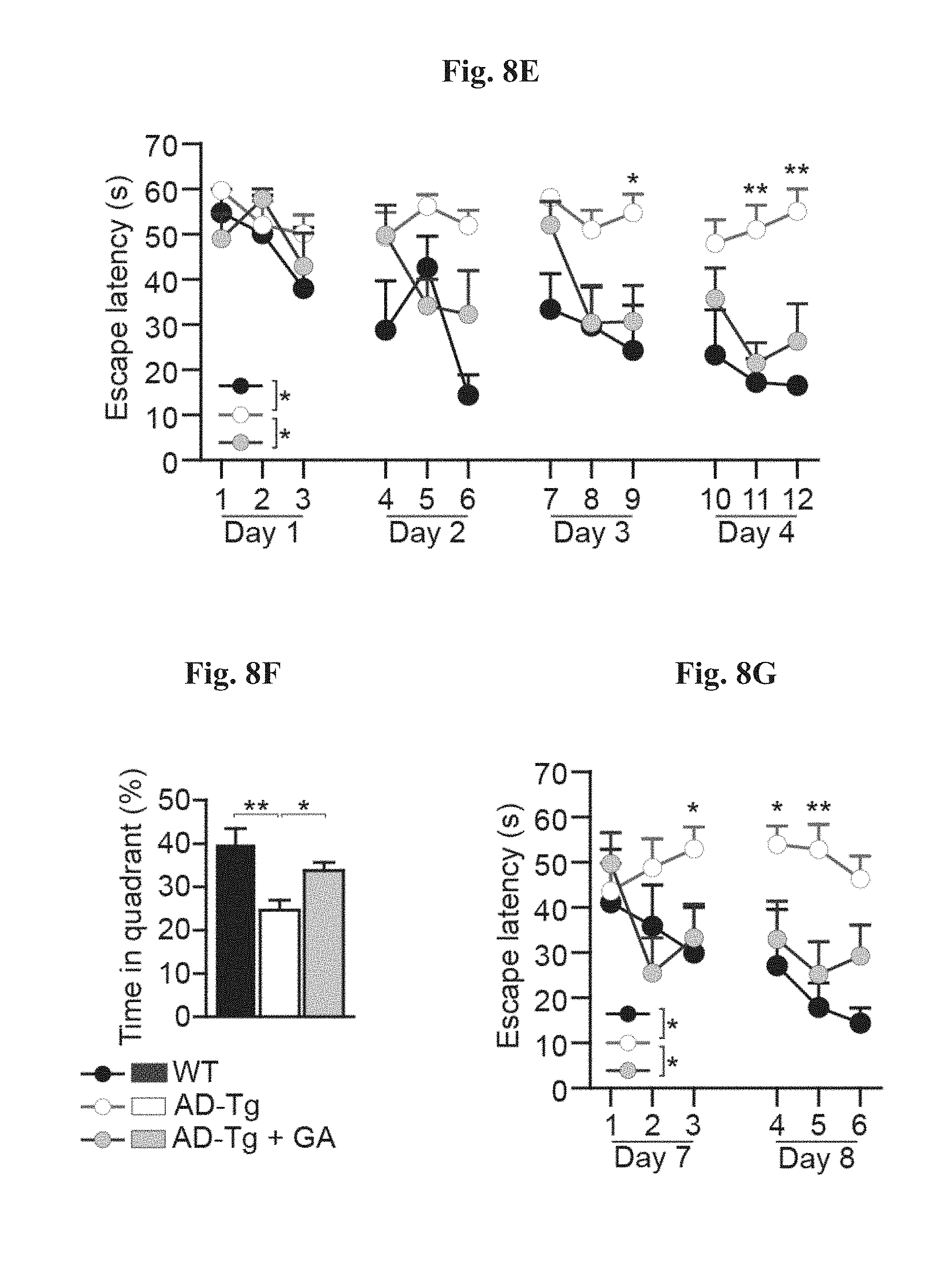
D00010

D00011
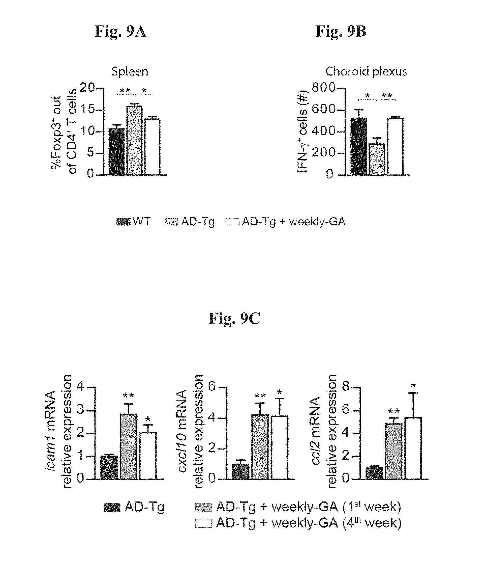
D00012

D00013

D00014

D00015

D00016

D00017
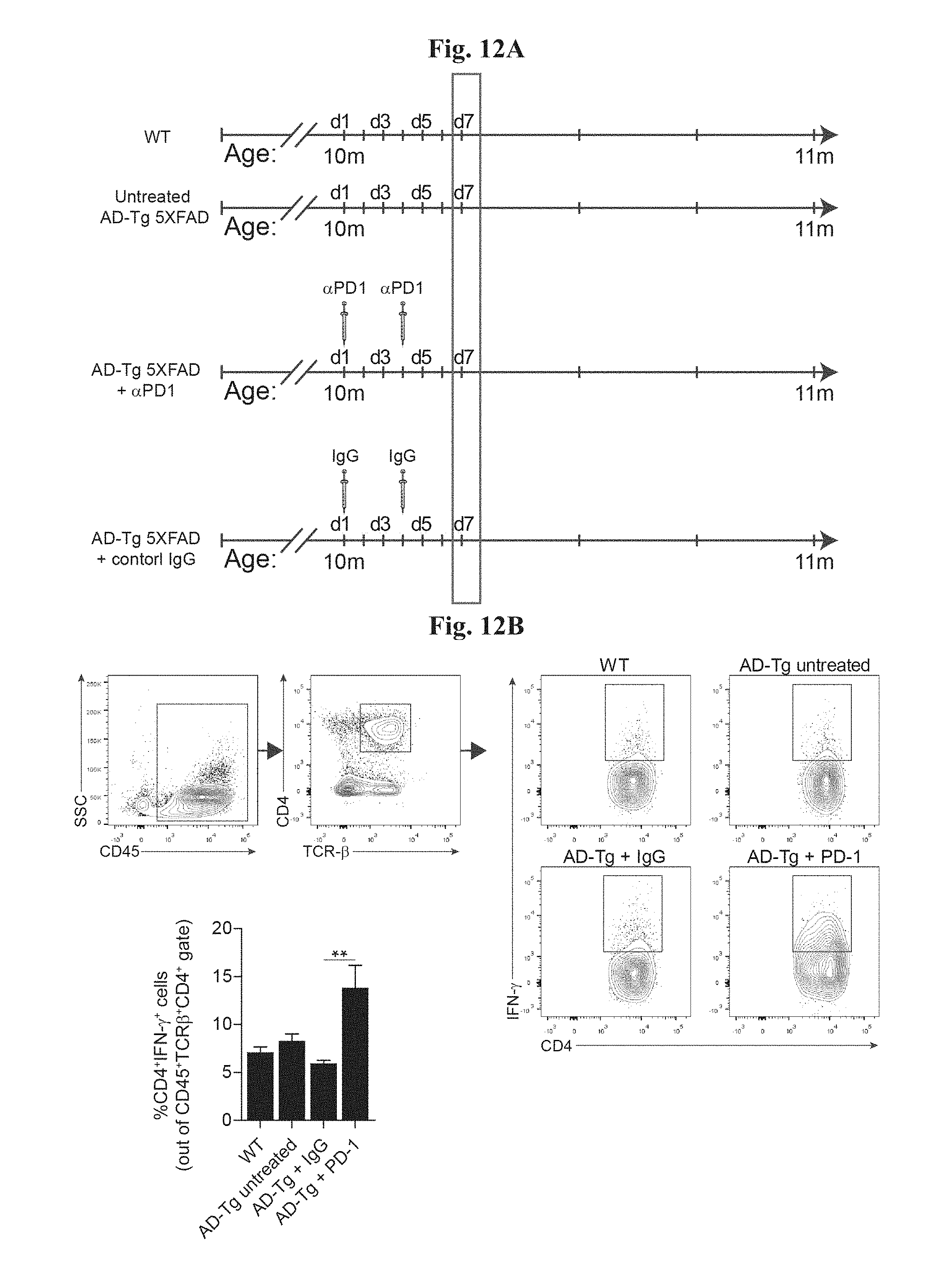
D00018

D00019

D00020

D00021

D00022

D00023

D00024

S00001
XML
uspto.report is an independent third-party trademark research tool that is not affiliated, endorsed, or sponsored by the United States Patent and Trademark Office (USPTO) or any other governmental organization. The information provided by uspto.report is based on publicly available data at the time of writing and is intended for informational purposes only.
While we strive to provide accurate and up-to-date information, we do not guarantee the accuracy, completeness, reliability, or suitability of the information displayed on this site. The use of this site is at your own risk. Any reliance you place on such information is therefore strictly at your own risk.
All official trademark data, including owner information, should be verified by visiting the official USPTO website at www.uspto.gov. This site is not intended to replace professional legal advice and should not be used as a substitute for consulting with a legal professional who is knowledgeable about trademark law.