Isolation Of Nucleic Acids
Bruinsma; Janelle J. ; et al.
U.S. patent application number 16/212143 was filed with the patent office on 2019-04-04 for isolation of nucleic acids. The applicant listed for this patent is Exact Sciences Corporation. Invention is credited to Janelle J. Bruinsma, Michael J. Domanico, Keith Kopitzke, Graham P. Lidgard, James P. Light, II, Hemanth D. Shenoi, William G. Weisburg, John Zeis, Hongzhi Zou.
| Application Number | 20190100788 16/212143 |
| Document ID | / |
| Family ID | 47140037 |
| Filed Date | 2019-04-04 |


View All Diagrams
| United States Patent Application | 20190100788 |
| Kind Code | A1 |
| Bruinsma; Janelle J. ; et al. | April 4, 2019 |
ISOLATION OF NUCLEIC ACIDS
Abstract
Provided herein is technology relating to isolating nucleic acids. In particular, the technology relates to methods and kits for extracting nucleic acids from problematic samples such as stool.
| Inventors: | Bruinsma; Janelle J.; (Madison, WI) ; Domanico; Michael J.; (Middleton, WI) ; Lidgard; Graham P.; (Madison, WI) ; Zou; Hongzhi; (Middleton, WI) ; Weisburg; William G.; (San Diego, CA) ; Shenoi; Hemanth D.; (Verona, WI) ; Light, II; James P.; (Middleton, WI) ; Kopitzke; Keith; (Fallbrook, CA) ; Zeis; John; (San Marcos, CA) | ||||||||||
| Applicant: |
|
||||||||||
|---|---|---|---|---|---|---|---|---|---|---|---|
| Family ID: | 47140037 | ||||||||||
| Appl. No.: | 16/212143 | ||||||||||
| Filed: | December 6, 2018 |
Related U.S. Patent Documents
| Application Number | Filing Date | Patent Number | ||
|---|---|---|---|---|
| 16046669 | Jul 26, 2018 | 10196676 | ||
| 16212143 | ||||
| 15807970 | Nov 9, 2017 | 10047390 | ||
| 16046669 | ||||
| 14939925 | Nov 12, 2015 | 9845491 | ||
| 15807970 | ||||
| 14713289 | May 15, 2015 | 9631228 | ||
| 14939925 | ||||
| 14145082 | Dec 31, 2013 | 9163278 | ||
| 14713289 | ||||
| PCT/US2012/037581 | May 11, 2012 | |||
| 14145082 | ||||
| 61485386 | May 12, 2011 | |||
| 61485448 | May 12, 2011 | |||
| 61485338 | May 12, 2011 | |||
| 61485214 | May 12, 2011 | |||
| Current U.S. Class: | 1/1 |
| Current CPC Class: | Y10T 436/143333 20150115; B01D 33/155 20130101; C12N 15/1017 20130101; B01L 3/00 20130101; C12Q 1/6886 20130101; C12Q 2600/158 20130101; B01L 3/5021 20130101; C12Q 2600/16 20130101; C12Q 1/6806 20130101; B04B 3/00 20130101; B03C 1/30 20130101; B01D 33/15 20130101; C12N 15/1013 20130101; C12N 15/1006 20130101 |
| International Class: | C12Q 1/6806 20060101 C12Q001/6806; B01D 33/15 20060101 B01D033/15; B03C 1/30 20060101 B03C001/30; B01L 3/00 20060101 B01L003/00; C12Q 1/6886 20060101 C12Q001/6886; C12N 15/10 20060101 C12N015/10; B04B 3/00 20060101 B04B003/00 |
Claims
1. A spin filter comprising a) a hollow body; b) a bottom end; and c) an open top end opposite the bottom end, wherein the hollow body is made from a porous filtering material.
2. The spin filter of claim 1 wherein the bottom end is made from a porous filtering material.
3. The spin filter of claim 1 wherein the porous filtering material is polyethylene.
4. The spin filter of claim 1 wherein the porous filtering material has a nominal pore size of 20 micrometers.
5. The spin filter of claim 1 wherein the bottom end has a shape selected from the group consisting of a hemisphere, a disc, a cone, or a portion of an ellipsoid.
6. A spin filter assembly comprising the spin filter of claim 1 and a collection vessel adapted to receive the spin filter.
7. A method of producing a filtrate from a sample, the method comprising: a) placing a sample to be filtered into a spin filter according to claim 1; and b) centrifuging said spin filter, wherein during said centrifuging, a fraction of said sample passes through a porous filtering material of said spin filter to produce a filtrate.
8. A method for removing an assay inhibitor from a crude sample preparation comprising a nucleic acid, the method comprising: a) adding insoluble polyvinylpyrrolidone to said crude sample preparation prior to isolating the nucleic acid under conditions wherein said assay inhibitor binds to said polyvinylpyrrolidone to produce a complex; b) separating the complex from the crude sample preparation to produce a clarified sample preparation comprising said nucleic acid.
9. The method of claim 8 wherein the crude sample preparation is a supernatant prepared from a stool sample.
10. The method of claim 9 wherein the stool sample has a mass of at least 4 grams.
11. The method of claim 9 wherein the stool sample has a mass of at least 8 grams.
12. The method of claim 8 wherein the polyvinylpyrrolidone is a polyvinylpolypyrrolidone.
13. The method of claim 8, wherein said separating comprises centrifuging.
14. The method of claim 8 wherein said clarified sample preparation is produced according to claim 7.
15. The method of claim 8 wherein the polyvinylpyrrolidone comprises particles having a diameter averaging from approximately 100 to approximately 130 micrometers.
16. A system for removing an assay inhibitor from a crude sample preparation comprising a nucleic acid, the system comprising: a) insoluble polyvinylpyrrolidone for binding the assay inhibitor and producing a complex; b) functionality for separating the complex from the crude sample preparation to produce a clarified sample preparation comprising said nucleic acid; and c) functionality for retaining a clarified sample preparation comprising the nucleic acid.
17. The system of claim 16 wherein said polyvinylpyrrolidone is a polyvinylpolypyrrolidone.
18. The system of claim 16, wherein said polyvinylpyrrolidone is in a premeasured form.
19. The system of claim 16, wherein said polyvinylpyrrolidone is provided as a tablet.
20. The system of claim 16, wherein said functionality for separating said complex comprises a spin filter according to any of claims 1-5.
21. The system of claim 16, wherein said functionality for retaining a clarified sample preparation comprising the nucleic acid is a collection vessel according to claim 6.
22. A method for isolating a plurality of different target nucleic acids from a stool sample, the method comprising: a) contacting a stool sample preparation comprising a plurality of different target nucleic acids with a first target-specific capture reagent under conditions wherein a first target nucleic acid binds to said first target-specific capture reagent to form a first complex; b) separating said first complex from said stool sample to produce a first residual stool sample preparation; c) contacting said first residual stool sample preparation with a second target-specific capture reagent under conditions wherein a second target nucleic acid binds to said second target-specific capture reagent to form a second complex; d) separating said second complex from said first residual stool sample to produce a second residual stool sample preparation; e) eluting said first target nucleic acid from said first complex to produce a first target nucleic acid solution; and f) eluting said second target nucleic acid from said second complex to produce a second target nucleic acid solution.
23. The method of claim 22 wherein said stool sample preparation is a clarified sample preparation produced according to claim 8, 9, 10, 11, 12, 13, 14, or 15.
24. The method of claim 22 wherein said first target nucleic acid and/or said second target nucleic acid is a human nucleic acid.
25. The method of claim 22, wherein said first target nucleic acid and/or said second target nucleic acid is a DNA.
26. The method of claim 22, further comprising the steps of: g) contacting said second residual stool sample preparation with a third target-specific capture reagent under conditions wherein a third target nucleic acid binds to said third target-specific capture reagent to form a third complex; h) separating said third complex from said second residual stool sample to produce a third residual stool sample preparation; and i) eluting said third target nucleic acid from said third complex to produce a third target nucleic acid solution;
27. The method of claim 22, wherein said first target-specific capture reagent and/or said second target-specific capture reagent comprises an oligonucleotide.
28. The method of claim 27 wherein said oligonucleotide is covalently attached to a surface.
29. The method of claim 28, wherein said surface is a magnetic particle or a paramagnetic particle.
30. The method of claim 29, wherein said separating said first complex and/or said separating said second complex comprises exposing said first complex and/or exposing said second complex to a magnet.
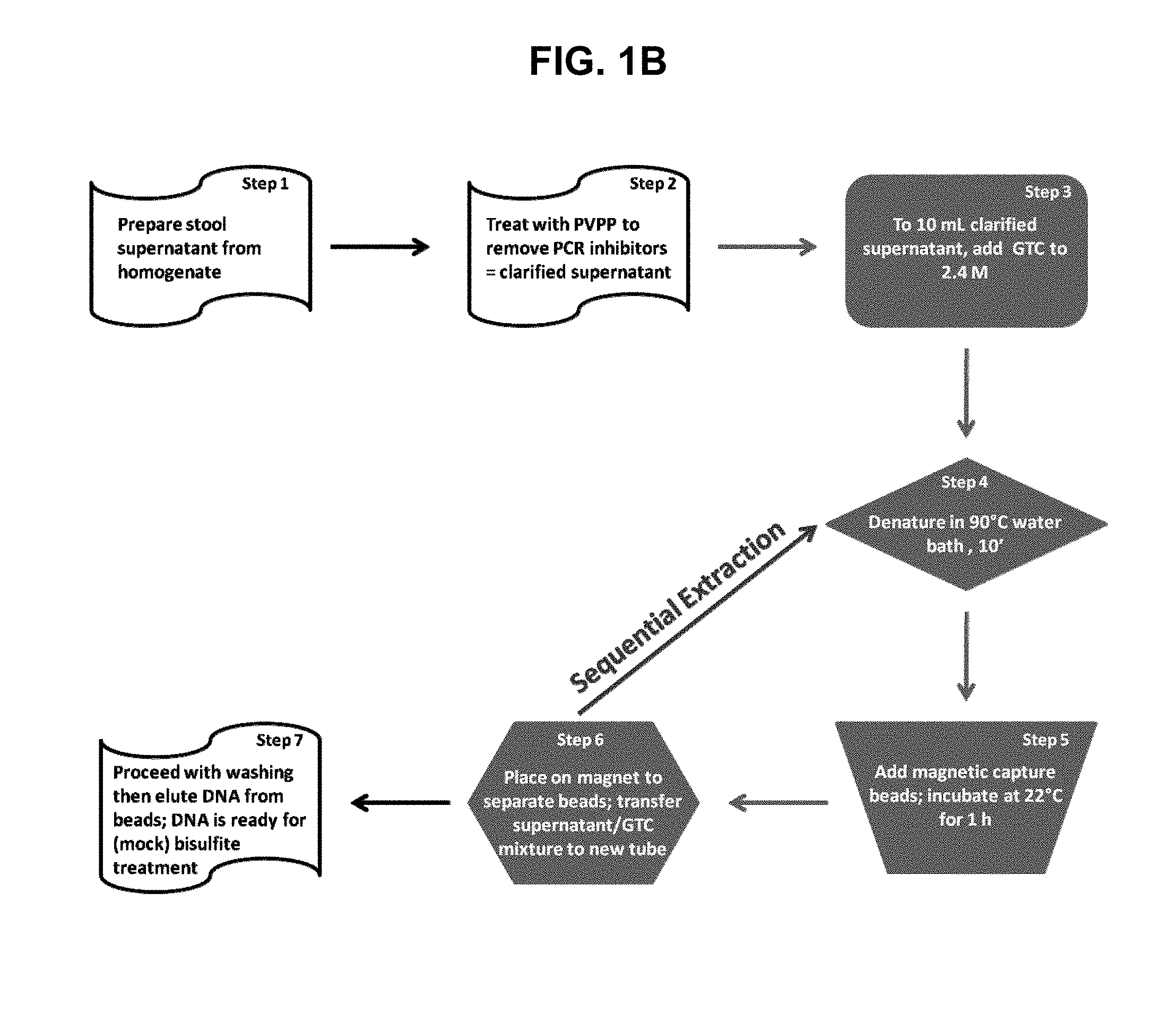
31. The method of claim 22, wherein said stool sample preparation comprises guanidine thiocyanate.
32. The method of claim 31, further comprising the steps of exposing said stool sample preparation to a condition that denatures nucleic acids prior to said contacting steps a and c.
33. The method of claim 32, wherein said condition that denatures nucleic acids comprises heating at 90.degree. C. for at least 10 minutes.
34. The method of claim 26, further comprising the steps of: j) contacting an nth residual stool sample preparation with a n+1th target-specific capture reagent under conditions wherein a n+1th target nucleic acid binds to said n+1th target-specific capture reagent to form a n+1th complex; k) separating said n+1th complex from said nth residual stool sample to produce a n+1th residual stool sample preparation; and l) eluting said n+1th target nucleic acid from said n+1th complex to produce a n+1th target nucleic acid solution, wherein n is greater than or equal to 3.
35. The method of any of claim 22, 26, or 34, wherein said contacting step a, c, and/or j comprises incubating said stool sample preparation, said first residual stool sample preparation, and/or said nth residual stool sample preparation contacted with said first target-specific capture reagent, said second target-specific capture reagent, and/or said n+1th target-specific capture reagent at room temperature for 1 hour.
36. A method for isolating a target human DNA from a human stool sample, the method comprising: a) obtaining a stool sample having a mass of at least 4 grams from a human subject; b) homogenizing said stool sample in an homogenization buffer to produce an homogenized stool sample; c) preparing a stool supernatant from the homogenized stool sample; d) treating said stool supernatant with PVP to produce a clarified stool supernatant; e) adding guanidine thiocyanate to 10 milliliters of clarified stool supernatant to produce a sample solution comprising 2-3 M guanidine thiocyanate; f) heating said sample solution to 90.degree. C. for 10 minutes; g) adding to said sample solution a target-specific capture reagent comprising an oligonucleotide covalently attached to a magnetic particle, wherein said oligonucleotide is complementary to at least a portion of said target human DNA; h) incubating said sample solution with said target-specific capture reagent at ambient temperature for approximately 1 hour to produce a complex comprising said target-specific capture reagent and said target human DNA; i) exposing the sample solution comprising said complex to a magnetic field to isolate the complex from the sample solution, and retaining the sample solution; j) eluting the target human DNA, from the complex to produce a target nucleic acid solution comprising the target nucleic acid, when present; k) repeating steps f-j of the method using a different target-specific capture reagent in each step g, to produce a different target nucleic acid solution in each step j; and l) performing a nucleic acid detection reaction on each different target nucleic acid solution, wherein at least one third of the volume of each said nucleic acid detection reaction is from said target nucleic acid solution.
37. The method of claim 36, wherein the target human DNA is from a gene associated with a disease state selected from the group consisting of colorectal cancer and colorectal adenoma.
38. The method of claim 36 wherein the repeating step k) is performed n times to produce n+1 different target nucleic acid solutions.
39. The method of claim 38 wherein n is at least 3.
40. A method for isolating a target nucleic acid from a sample, the method comprising: a) removing an assay inhibitor from said sample to produce a clarified sample preparation; b) capturing said target nucleic acid from said clarified sample preparation with a target-specific capture reagent to form a capture complex; c) isolating said capture complex from the clarified sample preparation; and d) recovering said target nucleic acid from said capture complex in a nucleic acid solution.
41. The method of claim 40, further comprising: e) retaining residual clarified sample preparation after said isolating step; f) repeating the capturing, isolating, and recovering steps using the residual clarified sample and a second capture reagent.
42. The method of claim 40 wherein removing said assay inhibitor from said sample comprises: a1) homogenizing said sample to produce a homogenate; a2) centrifuging said homogenate to produce a supernatant; a3) treating said supernatant with an inhibitor-adsorbing composition to bind inhibitor, if present, in an inhibitor complex; and a4) isolating said inhibitor complex from said supernatant to produce a clarified sample preparation.
43. The method of claim 42, wherein said inhibitor-adsorbing composition is a polyvinylpyrrolidone.
44. The method of claim 43, wherein said polyvinylpyrrolidone is insoluble.
45. The method of claim 43, wherein said polyvinylpyrrolidone is a polyvinylpolypyrrolidone.
46. The method of claim 43, wherein said polyvinylpyrrolidone is provided in a premeasured form.
47. The method of claim 43, wherein said polyvinylpyrrolidone is provided as a tablet.
48. The method of claim 42, wherein isolating said inhibitor complex comprises centrifuging to separate said inhibitor complex from said supernatant.
49. The method of claim 48, wherein said centrifuging comprises centrifuging through a spin filter of any one of claims 1-5.
50. The method of claim 40 wherein capturing said target nucleic acid comprises: b1) exposing said clarified sample preparation to a denaturing condition to produce a denatured sample; and b2) binding said target nucleic acid to a target-specific capture reagent to form a capture complex.
51. The method of claim 50, wherein said denaturing condition comprises heating.
52. The method of claim 50, wherein said denaturing condition comprises heating at 90.degree. C.
53. The method of claim 50, wherein said exposing said clarified sample preparation to a denaturing condition comprises adding a denaturant to said clarified sample preparation.
54. The method of claim 53, wherein said denaturant comprises guanidine thiocyanate.
55. The method of claim 50, wherein said target-specific capture reagent comprises an oligonucleotide complementary to at least a portion of said target nucleic acid.
56. The method of claim 50, wherein said target-specific capture reagent comprises a magnetic particle.
57. The method of claim 55, wherein said binding comprises hybridizing said oligonucleotide and said target nucleic acid.
58. The method of claim 56, wherein said isolating step comprises exposing said capture complex to a magnetic field.
59. The method of claim 56, wherein said isolating step comprises: c1) placing said clarified sample preparation in a magnetic field produced by a first magnet oriented with its north pole in close proximity to the sample and a second magnet oriented with its south pole in close proximity to the sample; and c2) moving said magnetic particles to a desired location.
60. The method of claim 40, wherein recovering said target nucleic acid comprises eluting said target nucleic acid from said capture complex.
61. The method of claim 60, wherein eluting comprises heating.
62. The method of claim 40, wherein said sample is a viscous sample.
63. The method of claim 40 wherein said sample is a stool sample.
64. The method of claim 40, wherein said sample has a mass of more than one gram.
65. The method of claim 40, wherein said sample has a mass of more than five grams.
66. The method of claim 40, wherein said sample has a viscosity of more than ten centipoise.
67. The method of claim 40, wherein said sample has a viscosity of more than twenty centipoise.
68. The method of claim 40, wherein the nucleic acid solution comprises a first amount of the assay inhibitor that is less than a second amount of the assay inhibitor, wherein the second amount of the assay inhibitor inhibits PCR when five microliters of the nucleic acid solution are used in a PCR having a volume of twenty-five microliters.
69. The method of claim 40, wherein said nucleic acid solution comprises a first amount of the assay inhibitor that is less than a second amount of the assay inhibitor, wherein the second amount of the assay inhibitor inhibits PCR when one microliter of the nucleic acid solution is used in a PCR having a volume of twenty-five microliters.
70. The method of claim 40, wherein said target nucleic acid is correlated with a disease state selected from the group consisting of colorectal cancer and colorectal adenoma.
71. A kit for preparing a nucleic acid solution from a sample, the kit comprising: a) polyvinylpyrrolidone for removing an inhibitor from the sample; b) a reagent for capturing a target nucleic acid from the sample; and c) a functionality for producing a magnetic field.
72. The kit of claim 71, further comprising a functionality for collecting said sample.
73. The kit of claim 71, further comprising a functionality for shipping said nucleic acid solution.
74. A kit comprising the spin filter of claim 1 and an instruction for use.
75. The kit of claim 74, further comprising a collection vessel.
76. A kit for removing an assay inhibitor from a crude sample preparation comprising a nucleic acid, the kit comprising: a) insoluble polyvinylpyrrolidone; and b) a spin column.
77. The kit of claim 76, wherein said polyvinylpyrrolidone is a polyvinylpolypyrrolidone.
78. The kit of claim 76, wherein said spin column comprises a spin filter according to any one of claims 1-5.
79. The kit of claim 76, wherein said polyvinylpyrrolidone comprises particles having a diameter averaging from approximately 100 to approximately 130 micrometers.
80. A kit for isolating a target human DNA from a human stool sample, the kit comprising: a) a volume of stool homogenization solution suitable for processing a human stool sample having a mass of at least 4 grams; and b) a target-specific capture reagent comprising an oligonucleotide covalently attached to a magnetic particle, wherein said oligonucleotide is complementary to at least a portion of said target human DNA.
81. The kit of claim 80, further comprising a magnet.
82. The kit of claim 80, further comprising polyvinylpyrrolidone.
83. The kit of claim 82 wherein the polyvinylpyrrolidone is a polyvinylpolypyrrolidone.
84. The kit of claim 82 wherein the polyvinylpyrrolidone is in a premeasured form.
85. The kit of claim 84 wherein the polyvinylpyrrolidone is provided as a tablet.
86. The kit of claim 80 further comprising guanidine thiocyanate.
87. The kit of claim 80, further comprising an elution or wash solution.
88. The kit of claim 80, further comprising an apparatus to produce a magnetic field.
89. The kit of claim 88, wherein said apparatus to produce a magnetic field comprises a first magnetic feature and a second magnetic feature, wherein a north pole of the first magnetic feature is placed in close proximity to the sample and a south pole of the second magnetic feature is placed in close proximity to the sample.
90. The kit of claim 80, further comprising a device for collecting a stool sample.
91. The kit of claim 90, wherein said device comprises: a) a body; b) a detachable sample capsule configured to move between an open state and a closed state, wherein the detachable sample capsule in said closed state comprises a sample collection space configured to enclose a sample; and c) an ejector mechanically connected to said body, said ejector configured to detach said detachable sample capsule from said body when said detachable sample capsule is in a closed state.
92. The kit of claim 80, further comprising a sample container configured to hold said stool sample.
93. The kit of claim 80 further comprising a vessel in which to hold isolated target human DNA.
94. The kit of claim 80, further comprising a shipping container.
95. The kit of claim 80, further comprising a spin filter according to any one of claims 1-5.
96. The kit of claim 80, further comprising a control reagent.
97. The kit of claim 96, wherein the control comprises a nucleic acid.
98. A system for preparing a nucleic acid solution from a sample, the system comprising: a) polyvinylpyrrolidone for removing an inhibitor from the sample; b) a reagent for capturing a target nucleic acid from the sample; and c) a functionality for producing a magnetic field.
99. The system of claim 98, further comprising a functionality for collecting said sample.
100. The system of claim 98, further comprising a functionality for shipping said nucleic acid solution.
101. A system comprising the spin filter of claim 1 and an instruction for use.
102. The system of claim 101, further comprising a collection vessel.
103. A system for removing an assay inhibitor from a crude sample preparation comprising a nucleic acid, the system comprising: a) insoluble polyvinylpyrrolidone; and b) a spin column.
104. The system of claim 103, wherein said polyvinylpyrrolidone is a polyvinylpolypyrrolidone.
105. The system of claim 103, wherein said spin column comprises a spin filter according to any one of claims 1-5.
106. The system of claim 103, wherein said polyvinylpyrrolidone comprises particles having a diameter averaging from approximately 100 to approximately 130 micrometers.
107. A system for isolating a target human DNA from a human stool sample, the system comprising: a) a volume of stool homogenization solution suitable for processing a human stool sample having a mass of at least 4 grams; and b) a target-specific capture reagent comprising an oligonucleotide covalently attached to a magnetic particle, wherein said oligonucleotide is complementary to at least a portion of said target human DNA.
108. The system of claim 107, further comprising a magnet.
109. The system of claim 107, further comprising a polyvinylpyrrolidone.
110. The system of claim 109 wherein said polyvinylpyrrolidone is a polyvinylpolypyrrolidone.
111. The system of claim 107 wherein said polyvinylpyrrolidone is in a premeasured form.
112. The system of claim 111 wherein said polyvinylpyrrolidone is provided as a tablet.
113. The system of claim 107 further comprising guanidine thiocyanate.
114. The system of claim 107, further comprising an elution or wash solution.
115. The system of claim 107, further comprising an apparatus to produce a magnetic field.
116. The system of claim 115, wherein said apparatus to produce a magnetic field comprises a first magnetic feature and a second magnetic feature, wherein a north pole of the first magnetic feature is placed in close proximity to the sample and a south pole of the second magnetic feature is placed in close proximity to the sample.
117. The system of claim 107, further comprising a device for collecting a stool sample.
118. The system of claim 117, wherein said device comprises a) a body; b) a detachable sample capsule configured to move between an open state and a closed state, wherein the detachable sample capsule in said closed state comprises a sample collection space configured to enclose a sample; and c) an ejector mechanically connected to said body, said ejector configured to detach said detachable sample capsule from said body when said detachable sample capsule is in a closed state.
119. The system of claim 107, further comprising a sample container configured to hold said stool sample.
120. The system of claim 107 further comprising a vessel in which to hold isolated target human DNA.
121. The system of claim 107, further comprising a shipping container.
122. The system of claim 107, further comprising a spin filter according to any one of claims 1-5.
123. The system of claim 107, further comprising a control reagent.
124. The system of claim 123, wherein said control comprises a nucleic acid.
21. The method of claim 20 wherein n is at least 3.
Description
[0001] The present application is a continuation of U.S. patent application Ser. No. 16/046,669, filed Jul. 26, 2018, now allowed, which is a continuation of U.S. patent application Ser. No. 15/807,970, filed Nov. 9, 2017, now U.S. Pat. No. 10,047,390, which is a continuation of U.S. patent application Ser. No. 14/939,925, filed Nov. 12, 2015, now U.S. Pat. No. 9,845,491, which is a continuation of U.S. patent application Ser. No. 14/713,289, filed May 15, 2015, now U.S. Pat. No. 9,631,228, which is a continuation of U.S. patent application Ser. No. 14/145,082, filed Dec. 31, 2013, now U.S. Pat. No. 9,163,278, which is a continuation of International Patent Application Serial Number PCT/US2012/03751, filed May 11, 2012, which claims the benefit of U.S. Provisional Patent Application Ser. Nos. 61/485,214, 61/485,338, 61/485,386, and 61/485,448, each of which was filed May 12, 2011, all of the above patent applications are incorporated herein by reference in their entirety.
FIELD OF INVENTION
[0002] Provided herein is technology relating to isolating nucleic acids. In particular, the technology relates to methods and kits for extracting nucleic acids from problematic samples such as stool.
BACKGROUND
[0003] Isolating specific target nucleic acids from a sample is an important step for many medical diagnostic assays. For example, certain mutations and methylation states in known genes are correlated, associated, and/or predictive of disease. DNA harboring these genes can be recovered from a sample and tested for the presence of the particular mutations and methylation states.
[0004] In practice, such assays require isolating and assaying several genetic targets from a sample. For many detection methods, detecting rare mutations or methylation events in a single gene requires isolating and testing a large quantity of DNA. This problem is compounded when assaying a panel of genes, each of which must be present in a large quantity for a robust diagnostic test. Thus, to detect rare mutations and methylation events in multiple genes, the isolated DNA must be highly concentrated and comprise a substantial portion of the detection assay.
[0005] This requirement imposes many problems, however. For example, preparing such quantities and concentrations of DNA requires a large sample as input (e.g., having a mass of several grams, e.g., approximately 2-4 grams) to provide sufficient nucleic acid for detection, and thus requires a method that can prepare DNA from a large sample. In addition, assay inhibitors are often isolated and concentrated with the DNA preparation. Consequently, concentrated DNA preparations produced by conventional methods also often retain unacceptable concentrations of inhibitors, which are then introduced into a subsequent assay. Moreover, if all targets of the panel are extracted simultaneously in a bulk, non-selective DNA preparation, the sensitivity of the assay is compromised because, as the preparation is divided into aliquots for testing, less extracted DNA from any one gene of the panel is present in the assay. If, on the other hand, all members of the panel are extracted and tested together and are thus present in the same assay mixture, the sensitivity of detecting any single particular target is compromised by the presence of the non-target DNA molecules.
[0006] In addition, if a particular diagnostic target is present in a complex sample, it will be present in a small amount relative to other materials--both nucleic acid and non-nucleic acid--in the sample, thus providing a challenge for analytical methods designed to detect it. For example, analyses of DNA from stool samples is complicated by the fact that bacteria compose approximately 60% of the dry mass of feces and the remainder is largely the remains of plant and animal matter ingested as food by the subject. As such, the human subject's cells, which are only those that slough off the lining of the digestive tract, are a very small fraction of the stool and substantial amounts of nucleic acids from other sources are present. Furthermore, in assays to detect gene modifications indicative of colon cancer, cells derived from a tumor that may be present in the colon would compose only a small fraction of the human subject's gut cells that slough off the digestive tract lining. Consequently, cancer cells (and the DNAs they contain) make up a minimal amount of the stool mass. Such samples are also often very viscous, which presents problems in sample preparation and isolation of nucleic acid.
[0007] Conventional methods and kits for isolating DNA from samples typically prepare total DNA (e.g., by a non-specific precipitation method) from a sample. For complex samples such as stool samples, this is a particular drawback of conventional methods, as total DNA isolated from a stool sample comprises DNA from the gut-resident bacteria (and any viruses, eukaryotes, and archaea present) along with DNA from the subject. Moreover, conventional methods and kits are primarily designed to prepare DNA from small samples, e.g., samples having masses of less than 1 gram, e.g., 50 to 200 milligrams, limiting the yield of target nucleic acid from complex samples to very small amounts. Additional drawbacks are that most conventional technology does not effectively remove inhibitors and often require long processing steps, e.g., incubations. Consequently, conventional methods are not suited to high-sensitivity and high-specificity multi-gene panel analysis because they cannot prepare sufficient amounts of highly concentrated, inhibitor-free DNA from large samples, such as a stool sample of several grams. Assays using DNA prepared with conventional methods will not provide a sample that can be assayed with the required sensitivity threshold for detecting rare mutation or methylation events. Using a conventional method or kit to attain the starting quantities needed to attain such sensitivity requires multiple DNA extractions (e.g., the use of multiple kits) from multiple samples in addition to extra purification steps to remove inhibitors. Therefore, what is needed is a method of preparing concentrated, inhibitor-free DNA from a sample for each member of a gene panel for use in diagnostic assays.
SUMMARY
[0008] Provided herein is technology relating to isolating nucleic acids. In particular, the technology relates to methods, systems, and kits for extracting and purifying nucleic acids from exfoliated intestinal cells in stool specimens for use in quantitative and sensitive assays. The technology is embodied in a novel method for purifying specific DNA from stool that utilizes inhibitor removal steps and direct capture of DNA from stool supernatant, or a combination of these steps. The technology further provides filtration devices suitable for use with complex and viscous samples, such as stool samples. Accordingly, provided herein is a method for isolating a target nucleic acid from a sample, the method comprising removing an assay inhibitor, if present, from the sample to produce a clarified sample; capturing the target nucleic acid, if present, from the clarified sample with a capture reagent to form a capture complex; isolating the capture complex from the clarified sample; and recovering the target nucleic acid, if present, from the capture complex in a nucleic acid solution. In some embodiments the method further comprises retaining the clarified sample after the capturing step; and repeating the isolating and recovering steps using the retained clarified sample and a second capture reagent.
[0009] In some embodiments, removing the inhibitor comprises homogenizing the sample to produce a homogenate; centrifuging the homogenate to produce a supernatant; treating the supernatant with an inhibitor-adsorbing composition to bind the inhibitor, if present, in an inhibitor complex; and isolating the inhibitor complex from the supernatant to produce a clarified sample. The inhibitor-adsorbing composition in some embodiments is a polyvinylpyrrolidone. In some embodiments, the polyvinylpyrrolidone is insoluble and in some embodiments the polyvinylpyrrolidone is a polyvinylpolypyrrolidone. It is useful in some embodiments to provide the polyvinylpyrrolidone in a premeasured form, for example in some embodiments the polyvinylpyrrolidone is provided as a tablet. Various techniques are used to separate the inhibitor complex from the sample. For example, in some embodiments isolating the inhibitor complex comprises centrifuging to separate the inhibitor complex from the supernatant.
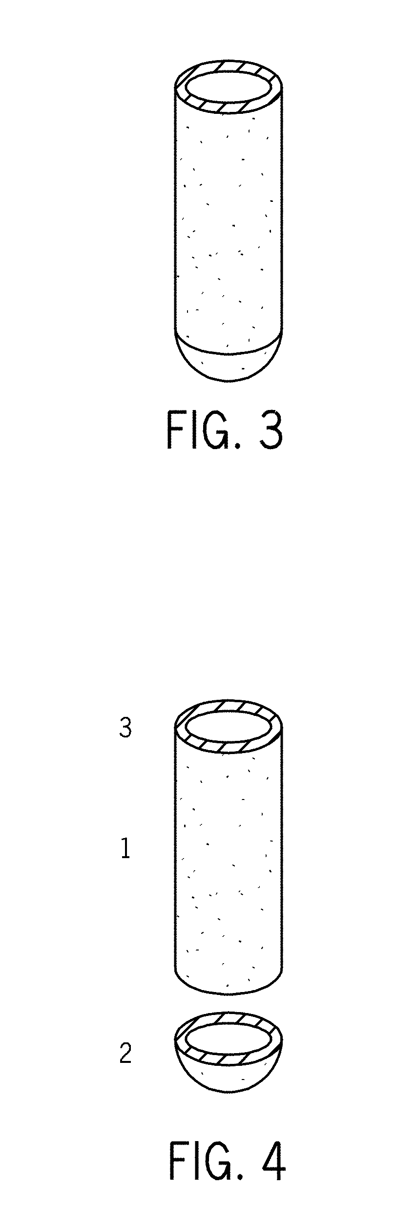
[0010] In some embodiments, the centrifuging comprises centrifuging through a spin column. Therefore, in some embodiments provided herein is technology relating to filtration and particularly, but not exclusively, to filters and methods for filtering by means of centrifugation. Specifically, some embodiments of the technology provided herein address the problem of spin filter clogging by providing technology in which both the bottom end and body of a spin filter are made from a porous or permeable material. That is, the walls of the spin filter are made of the same or similar material as that used for the filter means at the bottom end in conventional designs. As such, when the bottom portion of the filter becomes clogged during filtration, the walls provide additional surface through which the sample can be filtered.
[0011] This technology is provided herein as a spin filter comprising a hollow body, a bottom end, and an open top end opposite the bottom end, wherein the hollow body is made from a porous filtering material. In some embodiments the bottom end is made from a porous filtering material. The hollow body and bottom end of the spin filter assume any shape appropriate for the filtration application to which the filter is applied. For example, in some embodiments the hollow body is a tube and in some embodiments the bottom end is a hemisphere. In other embodiments, the bottom end is a disc, a cone, or a portion of an ellipsoid. Furthermore, the spin filter is made from any material that is appropriate for filtering a sample. Thus, in some embodiments the porous filtering material is polyethylene. Samples comprise varying sizes of particles, matter, precipitates, etc. that are to be removed by filtration. Accordingly, the filtering material can be selected to have physical properties that provide the desired separation. For example, in some embodiments the porous filtering material has a nominal pore size of 20 micrometers. In some embodiments, use of the filter produces a filtrate that a user retains for additional processing. As such, some embodiments provide a spin filter assembly comprising a spin filter as described and a collection vessel adapted to receive the spin filter and collect the filtrate.
[0012] Also provided herein are methods for producing a filtrate from a sample comprising placing a sample to be filtered into the spin filter and centrifuging the spin filter, wherein during centrifuging, a fraction of the sample passes through porous filtering material of said spin filter to produce a filtrate.
[0013] The technology can be provided as a kit for use in a sample separation. Embodiments of such a kit comprise a spin filter as described and an instruction for use. In some embodiments the kit further comprises a collection vessel. In some embodiments, a kit comprising a spin filter further comprises additional reagents and materials for sample preparation, e.g., for inhibitor removal and/or target nucleic acid isolation.
[0014] In some embodiments, the methods and systems of the technology comprise capturing a nucleic acid target. Capturing the target nucleic acid, in some embodiments, comprises exposing a sample, such as a clarified sample preparation, to a denaturing condition to produce a denatured sample; and binding target nucleic acid in the denatured sample to a capture reagent to form a capture complex. Many treatments and conditions find use in denaturing macromolecules such as DNA. For example, in some embodiments, the denaturing condition comprises heating, e.g., in some embodiments the denaturing condition comprises heating at 90.degree. C. Supplementing the sample to be denatured facilitates the denaturing; accordingly, in some embodiments, the clarified sample further comprises a denaturant. In certain preferred embodiments, the denaturant comprises guanidine thiocyanate. Furthermore, in some embodiments the capture reagent comprises an oligonucleotide complementary to at least a portion of the target nucleic acid. In some preferred embodiments, the capture reagent comprises particle, e.g., a magnetic particle. The oligonucleotide, in some embodiments of the technology, hybridizes to at least a portion of the target nucleic acid, and thus in some embodiments, the binding step comprises hybridizing the oligonucleotide and the target nucleic acid. Isolating the capture reagent (e.g., the capture reagent/target nucleic acid complex) is accomplished in some embodiments by exposing the capture reagent to a magnetic field; that is, in some embodiments provided herein, the isolating step comprises exposing the capture complex to a magnetic field and in some embodiments exposing the capture complex to the magnetic field localizes the target nucleic acid. The magnetic field is produced by any appropriate magnet or magnetic device for the method. For example, in some embodiments the isolating step comprises placing the sample in a magnetic field produced by a first magnet oriented with its north pole in close proximity to the sample and a second magnet oriented with its south pole in close proximity to the sample; and waiting for a time sufficient to allow the magnetic field to move the magnetic particles to the desired location. A device for producing a strong magnetic field is described, for example, in U.S. patent application Ser. No. 13/089,116, incorporated by reference herein.
[0015] The technologies provide for recovering target nucleic acid from the capture reagent. In some embodiments, recovering the target nucleic acid comprises eluting the target nucleic acid from the capture complex, e.g., in some embodiments, by heating. In some embodiments, elution of the target nucleic acid from the capture complex comprises exposing the capture complex to high pH, e.g., in some embodiments, by adding a solution of sodium hydroxide.
[0016] In some embodiments, the technology provides methods, systems and kits for capturing multiple nucleic acids from a single sample, e.g., a stool sample. For example, provided herein are methods for isolating a nucleic acid from a stool sample comprising contacting a stool sample with a target-specific capture reagent; binding a target nucleic acid, when present, to the target-specific capture reagent to form a complex; isolating the complex comprising the target-specific capture reagent and the target nucleic acid, when present, from the stool sample; eluting the target nucleic acid, when present, from the complex to produce a target nucleic acid solution comprising the target nucleic acid, when present; and repeating the method using a different target-specific capture reagent. The methods are appropriate for large samples, e.g., having a mass of at least 4 grams. Moreover, each eluted target nucleic acid is sufficiently purified, sufficiently concentrated, and sufficiently free of inhibitors such that each eluted target nucleic acid, when present, is detected by a quantitative PCR when the target nucleic acid solution composes up to approximately one-third of a volume of the quantitative PCR.
[0017] In some embodiments of the methods provided, the target nucleic acid is a human target nucleic acid. In additional embodiments, the target nucleic acid is a DNA. While not limited in the means by which the nucleic acid is isolated from the stool sample, in some embodiments the target-specific capture reagent is a sequence-specific nucleic acid capture reagent. In some embodiments, the sequence-specific nucleic acid capture reagent is an oligonucleotide and in some embodiments the oligonucleotide is covalently attached to a magnetic or paramagnetic particle. Some embodiments provide that a magnet is used for the isolating step and some embodiments provide for the simultaneous isolation of more than one target using multiple target-specific capture reagents in a single isolation step.
[0018] The method is not limited in the types of samples that are processed. For example, in some embodiments the sample is a viscous sample, e.g., having a viscosity of more than ten centipoise in some embodiments and having a viscosity of more than twenty centipoise in some embodiments. Additionally, the samples are of a wide range of sizes. The methods are used to process samples having, in some embodiments, a mass of more than one gram and in some embodiments the sample has a mass of more than five grams.
[0019] The technology provided herein is directed to removing inhibitors from samples below an amount that inhibits an assay. Thus, in some embodiments, the method provides that the nucleic acid solution comprises a first amount of the assay inhibitor that is less than a second amount of the assay inhibitor, wherein the second amount of the assay inhibitor inhibits PCR when five microliters of the nucleic acid solution are used in a PCR having a volume of twenty-five microliters. In some embodiments, the nucleic acid solution comprises a first amount of the assay inhibitor that is less than a second amount of the assay inhibitor, wherein the second amount of the assay inhibitor inhibits PCR when one microliter of the nucleic acid solution is used in a PCR having a volume of twenty-five microliters.
[0020] The technology is related to medical molecular diagnostics wherein querying the state, presence, amount, sequence, etc., of a biological substance (e.g., a molecule) is used to aid a medical assessment. Accordingly, in some embodiments, the target nucleic acid is correlated with a disease state selected from the set consisting of colon cancer and adenoma.
[0021] The technology described herein is provided in a kit form in some embodiments--for example, embodiments provide that the technology is a kit for isolating a target nucleic acid from a sample comprising a capture reagent comprising an oligonucleotide covalently attached to a magnetic particle, an apparatus to produce a magnetic field, polyvinylpyrrolidone, and an instruction for use. In some embodiments, the kit further comprises a homogenization solution. In some embodiments, the kit further comprises an elution solution and in some embodiments the kit further comprises guanidine thiocyanate. In some embodiments, it is convenient for the polyvinylpyrrolidone to be in a premeasured form. For example, the polyvinylpyrrolidone is provided in a tablet or capsule in some embodiments. Some embodiments of the kit provide a spin filter for removing polyvinylpyrrolidone.
[0022] In some embodiments, the target nucleic acid is isolated using a magnetic field. As such, embodiments of the kits described herein provide an apparatus that produces a magnetic field. One device that is used to produce a magnetic field suitable for use with embodiments of the technology provided herein comprises two magnets or sets of magnets and places the north pole(s) of the first magnet or set of magnets in close proximity to the sample and the south pole(s) of the second magnet or set of magnets in close proximity to the sample. In some embodiments, the kits further provide a device for collecting a sample, e.g., a device having a body and a detachable sample capsule attached to the body, wherein the detachable sample capsule comprises a sample collection space adapted to enclose a sample (for example, as described in U.S. Pat. Appl. Ser. No. 61/476,707).
[0023] In some embodiments, the kit provides vessels (e.g., a tube, a vial, a jar, and the like) used to process samples and hold various compositions used to process samples or that result from processing samples. For example, in some embodiments the kit further comprises a vessel in which to hold the sample and in some embodiments the kit further comprises a vessel in which to hold the isolated target nucleic acid. The kit, in some embodiments, is used at a location other than where the sample is processed and/or where the analyte is assayed. Accordingly, in some embodiments the kit further comprises a shipping container.
[0024] The technology provided herein finds use in systems for preparing a nucleic acid from a sample. In some embodiments, the system comprises polyvinylpyrrolidone for removing an inhibitor from the sample, a reagent for capturing a target nucleic acid from the sample, and a functionality for producing a magnetic field. In some embodiments, the system further comprises a functionality for collecting the sample and in some embodiments the system further comprises a functionality for shipping the nucleic acid solution.
[0025] Additional embodiments will be apparent to persons skilled in the relevant art based on the teachings contained herein.
BRIEF DESCRIPTION OF THE DRAWINGS
[0026] These and other features, aspects, and advantages of the present technology will become better understood with regard to the following drawings:
[0027] FIGS. 1A and 1B provide charts of aspects of the nucleic acid isolation process. FIG. 1A provides a chart showing the steps of the nucleic acid isolation process. FIG. 1B is a flowchart showing an embodiment of the process that finds use in the sequential extraction of multiple targets from the same sample as an aspect of the overall process of FIG. 1A.
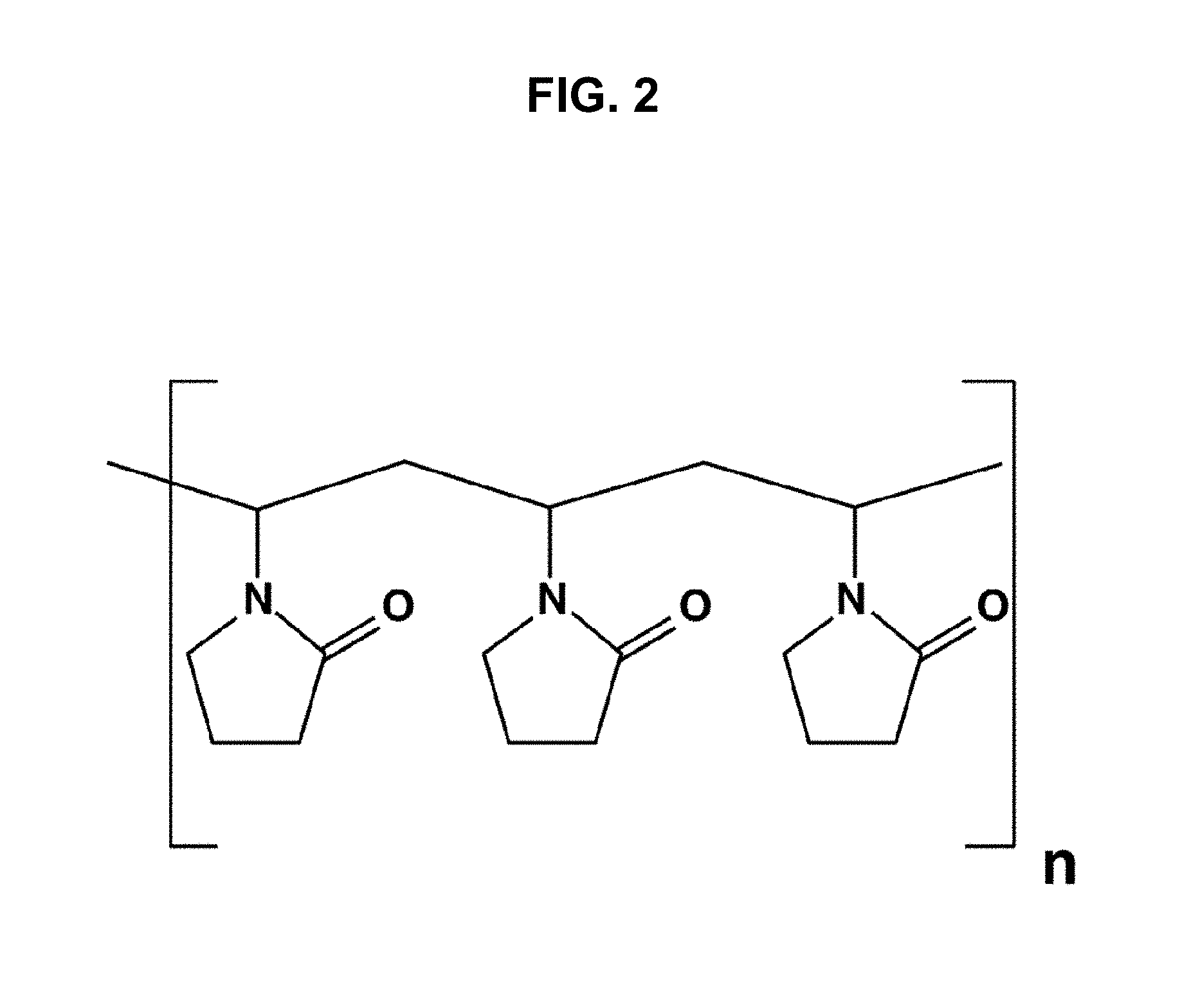
[0028] FIG. 2 is a chemical structure of a polyvinylpyrrolidone.
[0029] FIG. 3 is a drawing of an exemplary spin filter.
[0030] FIG. 4 is a drawing showing an exploded view of the spin filter shown in FIG. 3.
[0031] FIGS. 5A-5C are a series of drawings showing spin filter bottom ends associated with the spin filter of FIGS. 3 and 4. FIG. 5A is a drawing of a disc-shaped, solid (e.g., non-porous or non-permeable) bottom end; FIG. 5B is a drawing of a disc-shaped, porous (permeable) bottom end; FIG. 5C is a drawing of a porous, conical bottom end.
[0032] FIG. 6 is a drawing of a spin filter assembled with a collection tube.
[0033] FIG. 7 is a cut-away drawing of the spin filter depicted in FIG. 6.
[0034] FIGS. 8A and 8B are drawings of a spin filter comprising a body of a porous material and a bottom end provided by a filter support. FIG. 8A is an assembled view and FIG. 8B is an exploded view.
[0035] FIGS. 9A-D are plots showing the removal of inhibitors from a stool sample.
[0036] FIGS. 10A-D are plots showing that spin filtration improves the removal of inhibitors.
[0037] FIGS. 11A-B. FIG. 11A is a plot of data comparing the localization efficiency of the conventional technology for samples having viscosities of 1 centipoise and 25 centipoise.
[0038] FIG. 11B is a plot of data comparing the localization efficiency of the magnetic localization device provided by Light and Miller (U.S. patent application Ser. No. 13/089,116) provided for samples having viscosities of 1 centipoise and 25 centipoise.
[0039] FIGS. 12A-B. FIG. 12A is a plot showing the results of a quantitative PCR in which a single extraction from a stool sample recovers most of the target DNA. FIG. 12B shows the concentrations of Gene A and Gene V in nucleic acid solutions from a first extraction and a second extraction.
[0040] FIGS. 13A-D show plots showing the results of quantitative PCRs in which the recoveries of four target DNAs are similar regardless of the order in which the four target DNAs are extracted from a stool sample.
[0041] FIG. 14 provides a chart comparing the workflow of an embodiment (Process A) with an exemplary process for isolating DNA from stool samples using steps based on existing methods (Process B, see, e.g., WO 2010/028382).
DETAILED DESCRIPTION
[0042] The present technology is related to producing DNA samples and, in particular, to methods for producing DNA samples that comprise highly purified, low-abundance nucleic acids in a small volume (e.g., less than 100, less than 60 microliters) and that are substantially and/or effectively free of substances that inhibit assays used to test the DNA samples (e.g., PCR, INVADER, QuARTS, etc.). Such DNA samples find use in diagnostic assays that qualitatively detect the presence of, or quantitatively measure the activity, expression, or amount of, a gene, a gene variant (e.g., an allele), or a gene modification (e.g., methylation) present in a sample taken from a patient. For example, some cancers are correlated with the presence of particular mutant alleles or particular methylation states, and thus detecting and/or quantifying such mutant alleles or methylation states has predictive value in the diagnosis and treatment of cancer.
[0043] Many valuable genetic markers are present in extremely low amounts in samples and many of the events that produce such markers are rare. Consequently, even sensitive detection methods such as PCR require a large amount of DNA to provide enough of a low-abundance target to meet or supersede the detection threshold of the assay. Moreover, the presence of even low amounts of inhibitory substances compromise the accuracy and precision of these assays directed to detecting such low amounts of a target. Accordingly, provided herein are methods providing the requisite management of volume and concentration to produce such DNA samples.
[0044] Some biological samples, such as stool samples, contain a wide variety of different compounds that are inhibitory to PCR. Thus, the DNA extraction procedures include methods to remove and/or inactivate PCR inhibitors. As such, provided herein is technology relating to processing and preparing samples and particularly, but not exclusively, to methods, systems, and kits for removing assay inhibitors from samples comprising nucleic acids.
Definitions
[0045] To facilitate an understanding of the present technology, a number of terms and phrases are defined below. Additional definitions are set forth throughout the detailed description.
[0046] As used herein, "a" or "an" or "the" can mean one or more than one. For example, "a" widget can mean one widget or a plurality of widgets.
[0047] As used herein, an "inhibitor" means any compound, substance, or composition, or combination thereof, that acts to decrease the activity, precision, or accuracy of an assay, either directly or indirectly, with respect to the activity, precision, or accuracy of the assay when the inhibitor is absent. An inhibitor can be a molecule, an atom, or a combination of molecules or atoms without limitation.
[0048] As used herein, the process of passing a mixture through a filter is called "filtration". The liquid produced after filtering a suspension of a solid in a liquid is called "filtrate", while the solid remaining in the filter is called "retentate", "residue", or "filtrand".
[0049] As used herein, "insoluble" refers to the property that a substance does not substantially dissolve in water and is essentially immiscible therewith. Upon separation of an aqueous phase from a non-aqueous phase, an insoluble substance does not partition into or partition with the aqueous phase.
[0050] As used herein, the terms "subject" and "patient" refer to any animal, such as a dog, cat, bird, livestock, and particularly a mammal, preferably a human. In some instances, the subject is also a "user" (and thus the user is also the subject or patient).
[0051] As used herein, the term "sample" and "specimen" are used interchangeably, and in the broadest senses. In one sense, sample is meant to include a specimen or culture obtained from any source, as well as biological and environmental samples. Biological samples may be obtained from animals (including humans) and encompass fluids, solids, tissues, and gases. Biological samples include blood products, such as plasma, serum, stool, urine, and the like. Environmental samples include environmental material such as surface matter, soil, mud, sludge, biofilms, water, crystals, and industrial samples. Such examples are not however to be construed as limiting the sample types applicable to the present invention.
[0052] The term "target," when used in reference to a nucleic acid capture, detection, or analysis method, generally refers to a nucleic acid having a feature, e.g., a particular sequence of nucleotides to be detected or analyzed, e.g., in a sample suspected of containing the target nucleic acid. In some embodiments, a target is a nucleic acid having a particular sequence for which it is desirable to determine a methylation status. When used in reference to the polymerase chain reaction, "target" generally refers to the region of nucleic acid bounded by the primers used for polymerase chain reaction. Thus, the "target" is sought to be sorted out from other nucleic acid sequences that may be present in a sample. A "segment" is defined as a region of nucleic acid within the target sequence. The term "sample template" refers to nucleic acid originating from a sample that is analyzed for the presence of a target.
[0053] As used herein, the term "locus" refers to a particular position, e.g., of a mutation, polymorphism, or a C residue in a CpG dinucleotide, within a defined region or segment of nucleic acid, such as a gene or any other characterized sequence on a chromosome or RNA molecule. A locus is not limited to any particular size or length, and may refer to a portion of a chromosome, a gene, functional genetic element, or a single nucleotide or basepair. As used herein in reference to CpG sites that may be methylated, a locus refers to the C residue in the CpG dinucleotide.
[0054] As used herein, a "collection liquid" is a liquid in which to place a sample to preserve, stabilize, and otherwise maintain its integrity as a representative sample of the specimen from which the sample was taken. While not limited in the types of compositions that find use as collection liquids, examples of collection liquids are aqueous buffers optionally comprising a preservative and organic solvents, such as acetonitrile.
[0055] As used herein, "a capture reagent" refers to any agent that is capable of binding to an analyte (e.g., a target). Preferably, "a capture reagent" refers to any agent that is capable of specifically binding to an analyte, e.g., having a higher binding affinity and/or specificity to the analyte than to any other moiety. Any moiety, such as a cell, a cellular organelle, an inorganic molecule, an organic molecule and a mixture or complex thereof can be used as a capture reagent if it has the requisite binding affinity and/or specificity to the analyte. The capture reagents can be peptides, proteins, e.g., antibodies or receptors, oligonucleotides, nucleic acids, vitamins, oligosaccharides, carbohydrates, lipids, small molecules, or a complex thereof. Capture reagents that comprise nucleic acids, e.g., oligonucleotides, may capture a nucleic acid target by sequence-specific hybridization (e.g., through the formation of conventional Watson-Crick basepairs), or through other binding interactions. When a capture oligonucleotide hybridizes to a target nucleic acid, hybridization may involve a portion of the oligonucleotide, or the complete oligonucleotide sequence, and the oligonucleotide may bind to a portion of or to the complete target nucleic acid sequence.
[0056] As used herein, "PVP" refers to polyvinylpyrrolidone, which is a water-soluble polymer made from the monomer N-vinylpyrrolidone. The term PVP is used herein to refer to PVP in various states of cross-linked polymerization, including preparations of PVP that may also be known in the art as polyvinylpolypyrrolidone (PVPP).
[0057] As used herein, a "magnet" is a material or object that produces a magnetic field. A magnet may be a permanent magnet or an electromagnet.
[0058] The term "amplifying" or "amplification" in the context of nucleic acids refers to the production of multiple copies of a polynucleotide, or a portion of the polynucleotide, typically starting from a small amount of the polynucleotide (e.g., a single polynucleotide molecule), where the amplification products or amplicons are generally detectable. Amplification of polynucleotides encompasses a variety of chemical and enzymatic processes. The generation of multiple DNA copies from one or a few copies of a target or template DNA molecule during a polymerase chain reaction (PCR) or a ligase chain reaction (LCR; see, e.g., U.S. Pat. No. 5,494,810; herein incorporated by reference in its entirety) are forms of amplification. Additional types of amplification include, but are not limited to, allele-specific PCR (see, e.g., U.S. Pat. No. 5,639,611; herein incorporated by reference in its entirety), assembly PCR (see, e.g., U.S. Pat. No. 5,965,408; herein incorporated by reference in its entirety), helicase-dependent amplification (see, e.g., U.S. Pat. No. 7,662,594; herein incorporated by reference in its entirety), hot-start PCR (see, e.g., U.S. Pat. Nos. 5,773,258 and 5,338,671; each herein incorporated by reference in their entireties), intersequence-specfic PCR, inverse PCR (see, e.g., Triglia, et alet al. (1988) Nucleic Acids Res., 16:8186; herein incorporated by reference in its entirety), ligation-mediated PCR (see, e.g., Guilfoyle, R. et alet al., Nucleic Acids Research, 25:1854-1858 (1997); U.S. Pat. No. 5,508,169; each of which are herein incorporated by reference in their entireties), methylation-specific PCR (see, e.g., Herman, et al., (1996) PNAS 93(13) 9821-9826; herein incorporated by reference in its entirety), miniprimer PCR, multiplex ligation-dependent probe amplification (see, e.g., Schouten, et al., (2002) Nucleic Acids Research 30(12): e57; herein incorporated by reference in its entirety), multiplex PCR (see, e.g., Chamberlain, et al., (1988) Nucleic Acids Research 16(23) 11141-11156; Ballabio, et al., (1990) Human Genetics 84(6) 571-573; Hayden, et al., (2008) BMC Genetics 9:80; each of which are herein incorporated by reference in their entireties), nested PCR, overlap-extension PCR (see, e.g., Higuchi, et al., (1988) Nucleic Acids Research 16(15) 7351-7367; herein incorporated by reference in its entirety), real time PCR (see, e.g., Higuchi, et al., (1992) Biotechnology 10:413-417; Higuchi, et al., (1993) Biotechnology 11:1026-1030; each of which are herein incorporated by reference in their entireties), reverse transcription PCR (see, e.g., Bustin, S. A. (2000) J. Molecular Endocrinology 25:169-193; herein incorporated by reference in its entirety), solid phase PCR, thermal asymmetric interlaced PCR, and Touchdown PCR (see, e.g., Don, et al., Nucleic Acids Research (1991) 19(14) 4008; Roux, K. (1994) Biotechniques 16(5) 812-814; Hecker, et al., (1996) Biotechniques 20(3) 478-485; each of which are herein incorporated by reference in their entireties). Polynucleotide amplification also can be accomplished using digital PCR (see, e.g., Kalinina, et al., Nucleic Acids Research. 25; 1999-2004, (1997); Vogelstein and Kinzler, Proc Natl Acad Sci USA. 96; 9236-41, (1999); International Patent Publication No. WO05023091A2; US Patent Application Publication No. 20070202525; each of which are incorporated herein by reference in their entireties).
[0059] The term "polymerase chain reaction" ("PCR") refers to the method of K. B. Mullis U.S. Pat. Nos. 4,683,195, 4,683,202, and 4,965,188, that describe a method for increasing the concentration of a segment of a target sequence in a mixture of genomic or other DNA or RNA, without cloning or purification. This process for amplifying the target sequence consists of introducing a large excess of two oligonucleotide primers to the DNA mixture containing the desired target sequence, followed by a precise sequence of thermal cycling in the presence of a DNA polymerase. The two primers are complementary to their respective strands of the double stranded target sequence. To effect amplification, the mixture is denatured and the primers then annealed to their complementary sequences within the target molecule. Following annealing, the primers are extended with a polymerase so as to form a new pair of complementary strands. The steps of denaturation, primer annealing, and polymerase extension can be repeated many times (i.e., denaturation, annealing and extension constitute one "cycle"; there can be numerous "cycles") to obtain a high concentration of an amplified segment of the desired target sequence. The length of the amplified segment of the desired target sequence is determined by the relative positions of the primers with respect to each other, and therefore, this length is a controllable parameter. By virtue of the repeating aspect of the process, the method is referred to as the "polymerase chain reaction" ("PCR"). Because the desired amplified segments of the target sequence become the predominant sequences (in terms of concentration) in the mixture, they are said to be "PCR amplified" and are "PCR products" or "amplicons." Those of skill in the art will understand the term "PCR" encompasses many variants of the originally described method using, e.g., real time PCR, nested PCR, reverse transcription PCR (RT-PCR), single primer and arbitrarily primed PCR, etc.
[0060] As used herein, the term "nucleic acid detection assay" refers to any method of determining the nucleotide composition of a nucleic acid of interest. Nucleic acid detection assay include but are not limited to, DNA sequencing methods, probe hybridization methods, structure specific cleavage assays (e.g., the INVADER assay, (Hologic, Inc.) and are described, e.g., in U.S. Pat. Nos. 5,846,717, 5,985,557, 5,994,069, 6,001,567, 6,090,543, and 6,872,816; Lyamichev et al., Nat. Biotech., 17:292 (1999), Hall et al., PNAS, USA, 97:8272 (2000), and US 2009/0253142, each of which is herein incorporated by reference in its entirety for all purposes); enzyme mismatch cleavage methods (e.g., Variagenics, U.S. Pat. Nos. 6,110,684, 5,958,692, 5,851,770, herein incorporated by reference in their entireties); polymerase chain reaction (PCR), described above; branched hybridization methods (e.g., Chiron, U.S. Pat. Nos. 5,849,481, 5,710,264, 5,124,246, and 5,624,802, herein incorporated by reference in their entireties); rolling circle replication (e.g., U.S. Pat. Nos. 6,210,884, 6,183,960 and 6,235,502, herein incorporated by reference in their entireties); NASBA (e.g., U.S. Pat. No. 5,409,818, herein incorporated by reference in its entirety); molecular beacon technology (e.g., U.S. Pat. No. 6,150,097, herein incorporated by reference in its entirety); E-sensor technology (Motorola, U.S. Pat. Nos. 6,248,229, 6,221,583, 6,013,170, and 6,063,573, herein incorporated by reference in their entireties); cycling probe technology (e.g., U.S. Pat. Nos. 5,403,711, 5,011,769, and 5,660,988, herein incorporated by reference in their entireties); Dade Behring signal amplification methods (e.g., U.S. Pat. Nos. 6,121,001, 6,110,677, 5,914,230, 5,882,867, and 5,792,614, herein incorporated by reference in their entireties); ligase chain reaction (e.g., Baranay Proc. Natl. Acad. Sci USA 88, 189-93 (1991)); and sandwich hybridization methods (e.g., U.S. Pat. No. 5,288,609, herein incorporated by reference in its entirety).
[0061] In some embodiments, target nucleic acid is amplified (e.g., by PCR) and amplified nucleic acid is detected simultaneously using an invasive cleavage assay. Assays configured for performing a detection assay (e.g., invasive cleavage assay) in combination with an amplification assay are described in US Patent Publication US 20090253142 A1 (application Ser. No. 12/404,240), incorporated herein by reference in its entirety for all purposes. Additional amplification plus invasive cleavage detection configurations, termed the QuARTS method, are described in U.S. patent application Ser. Nos. 12/946,737; 12/946,745; and Ser. No. 12/946,752, incorporated herein by reference in their entireties for all purposes.
[0062] The term "invasive cleavage structure" as used herein refers to a cleavage structure comprising i) a target nucleic acid, ii) an upstream nucleic acid (e.g., an INVADER oligonucleotide), and iii) a downstream nucleic acid (e.g., a probe), where the upstream and downstream nucleic acids anneal to contiguous regions of the target nucleic acid, and where an overlap forms between the a 3' portion of the upstream nucleic acid and duplex formed between the downstream nucleic acid and the target nucleic acid. An overlap occurs where one or more bases from the upstream and downstream nucleic acids occupy the same position with respect to a target nucleic acid base, whether or not the overlapping base(s) of the upstream nucleic acid are complementary with the target nucleic acid, and whether or not those bases are natural bases or non-natural bases. In some embodiments, the 3' portion of the upstream nucleic acid that overlaps with the downstream duplex is a non-base chemical moiety such as an aromatic ring structure, e.g., as disclosed, for example, in U.S. Pat. No. 6,090,543, incorporated herein by reference in its entirety. In some embodiments, one or more of the nucleic acids may be attached to each other, e.g., through a covalent linkage such as nucleic acid stem-loop, or through a non-nucleic acid chemical linkage (e.g., a multi-carbon chain).
[0063] As used herein, the terms "complementary" or "complementarity" used in reference to polynucleotides (i.e., a sequence of nucleotides) refers to polynucleotides related by the base-pairing rules. For example, the sequence "5'-A-G-T-3'," is complementary to the sequence "3'-T-C-A-5'." Complementarity may be "partial," in which only some of the nucleic acids' bases are matched according to the base pairing rules. Or, there may be "complete" or "total" complementarity between the nucleic acids. The degree of complementarity between nucleic acid strands has significant effects on the efficiency and strength of hybridization between nucleic acid strands. This is of particular importance in amplification reactions, as well as detection methods that depend upon binding between nucleic acids.
[0064] As used herein, the term "primer" refers to an oligonucleotide, whether occurring naturally, as in a purified restriction digest, or produced synthetically, that is capable of acting as a point of initiation of synthesis when placed under conditions in which synthesis of a primer extension product that is complementary to a nucleic acid strand is induced (e.g., in the presence of nucleotides and an inducing agent such as a biocatalyst (e.g., a DNA polymerase or the like). The primer is typically single stranded for maximum efficiency in amplification, but may alternatively be partially or completely double stranded. The portion of the primer that hybridizes to a template nucleic acid is sufficiently long to prime the synthesis of extension products in the presence of the inducing agent. The exact lengths of the primers will depend on many factors, including temperature, source of primer and the use of the method. Primers may comprise labels, tags, capture moieties, etc.
[0065] As used herein, the term "nucleic acid molecule" refers to any nucleic acid containing molecule, including but not limited to, DNA or RNA. The term encompasses sequences that include any of the known base analogs of DNA and RNA including, but not limited to, 4 acetylcytosine, 8-hydroxy-N6-methyladenosine, aziridinylcytosine, pseudoisocytosine, 5-(carboxyhydroxyl-methyl) uracil, 5-fluorouracil, 5-bromouracil, 5-carboxymethylaminomethyl-2-thiouracil, 5-carboxymethyl-aminomethyluracil, dihydrouracil, inosine, N6-isopentenyladenine, 1-methyladenine, 1-methylpseudo-uracil, 1-methylguanine, 1-methylinosine, 2,2-dimethyl-guanine, 2-methyladenine, 2-methylguanine, 3-methyl-cytosine, 5-methylcytosine, N6-methyladenine, 7-methylguanine, 5-methylaminomethyluracil, 5-methoxy-amino-methyl-2-thiouracil, beta-D-mannosylqueosine, 5'-methoxycarbonylmethyluracil, 5-methoxyuracil, 2-methylthio-N-isopentenyladenine, uracil-5-oxyacetic acid methylester, uracil-5-oxyacetic acid, oxybutoxosine, pseudouracil, queosine, 2-thiocytosine, 5-methyl-2-thiouracil, 2-thiouracil, 4-thiouracil, 5-methyluracil, N-uracil-5-oxyacetic acid methylester, uracil-5-oxyacetic acid, pseudouracil, queosine, 2-thiocytosine, and 2,6-diaminopurine.
[0066] As used herein, the term "nucleobase" is synonymous with other terms in use in the art including "nucleotide," "deoxynucleotide," "nucleotide residue," "deoxynucleotide residue," "nucleotide triphosphate (NTP)," or deoxynucleotide triphosphate (dNTP).
[0067] An "oligonucleotide" refers to a nucleic acid that includes at least two nucleic acid monomer units (e.g., nucleotides), typically more than three monomer units, and more typically greater than ten monomer units. The exact size of an oligonucleotide generally depends on various factors, including the ultimate function or use of the oligonucleotide. To further illustrate, oligonucleotides are typically less than 200 residues long (e.g., between 15 and 100), however, as used herein, the term is also intended to encompass longer polynucleotide chains. Oligonucleotides are often referred to by their length. For example a 24 residue oligonucleotide is referred to as a "24-mer". Typically, the nucleoside monomers are linked by phosphodiester bonds or analogs thereof, including phosphorothioate, phosphorodithioate, phosphoroselenoate, phosphorodiselenoate, phosphoroanilothioate, phosphoranilidate, phosphoramidate, and the like, including associated counterions, e.g., H.sup.+, NH.sub.4.sup.+, Na.sup.+, and the like, if such counterions are present. Further, oligonucleotides are typically single-stranded. Oligonucleotides are optionally prepared by any suitable method, including, but not limited to, isolation of an existing or natural sequence, DNA replication or amplification, reverse transcription, cloning and restriction digestion of appropriate sequences, or direct chemical synthesis by a method such as the phosphotriester method of Narang et al. (1979) Meth Enzymol. 68: 90-99; the phosphodiester method of Brown et al. (1979) Meth Enzymol. 68: 109-151; the diethylphosphoramidite method of Beaucage et al. (1981) Tetrahedron Lett. 22: 1859-1862; the triester method of Matteucci et al. (1981) J Am Chem Soc. 103:3185-3191; automated synthesis methods; or the solid support method of U.S. Pat. No. 4,458,066, entitled "PROCESS FOR PREPARING POLYNUCLEOTIDES," issued Jul. 3, 1984 to Caruthers et al., or other methods known to those skilled in the art. All of these references are incorporated by reference.
[0068] A "sequence" of a biopolymer refers to the order and identity of monomer units (e.g., nucleotides, amino acids, etc.) in the biopolymer. The sequence (e.g., base sequence) of a nucleic acid is typically read in the 5' to 3' direction.
[0069] The term "wild-type" refers to a gene or gene product that has the characteristics of that gene or gene product when isolated from a naturally occurring source. A wild-type gene is that which is most frequently observed in a population and is thus arbitrarily designed the "normal" or "wild-type" form of the gene. In contrast, the terms "modified," "mutant," and "variant" refer to a gene or gene product that displays modifications in sequence and or functional properties (i.e., altered characteristics) when compared to the wild-type gene or gene product. It is noted that naturally occurring mutants can be isolated; these are identified by the fact that they have altered characteristics when compared to the wild-type gene or gene product.
[0070] As used herein, the term "gene" refers to a nucleic acid (e.g., DNA) sequence that comprises coding sequences necessary for the production of a polypeptide, precursor, or RNA (e.g., rRNA, tRNA). The polypeptide can be encoded by a full length coding sequence or by any portion of the coding sequence so long as the desired activity or functional properties (e.g., enzymatic activity, ligand binding, signal transduction, immunogenicity, etc.) of the full-length or fragment polypeptide are retained. The term also encompasses the coding region of a structural gene and the sequences located adjacent to the coding region on both the 5' and 3' ends for a distance of about 1 kb or more on either end such that the gene corresponds to the length of the full-length mRNA. Sequences located 5' of the coding region and present on the mRNA are referred to as 5' non-translated sequences. Sequences located 3' or downstream of the coding region and present on the mRNA are referred to as 3' non-translated sequences. The term "gene" encompasses both cDNA and genomic forms of a gene. A genomic form or clone of a gene contains the coding region interrupted with non-coding sequences termed "introns" or "intervening regions" or "intervening sequences." Introns are segments of a gene that are transcribed into nuclear RNA (e.g., hnRNA); introns may contain regulatory elements (e.g., enhancers). Introns are removed or "spliced out" from the nuclear or primary transcript; introns therefore are absent in the messenger RNA (mRNA) transcript. The mRNA functions during translation to specify the sequence or order of amino acids in a nascent polypeptide.
[0071] In addition to containing introns, genomic forms of a gene may also include sequences located on both the 5' and 3' end of the sequences that are present on the RNA transcript. These sequences are referred to as "flanking" sequences or regions (these flanking sequences are located 5' or 3' to the non-translated sequences present on the mRNA transcript). The 5' flanking region may contain regulatory sequences such as promoters and enhancers that control or influence the transcription of the gene. The 3' flanking region may contain sequences that direct the termination of transcription, post-transcriptional cleavage and polyadenylation.
[0072] As used herein, the term "kit" refers to any delivery system for delivering materials. In the context of nucleic acid purification systems and reaction assays, such delivery systems include systems that allow for the storage, transport, or delivery of reagents and devices (e.g., inhibitor adsorbants, particles, denaturants, oligonucleotides, spin filters etc. in the appropriate containers) and/or supporting materials (e.g., buffers, written instructions for performing a procedure, etc.) from one location to another. For example, kits include one or more enclosures (e.g., boxes) containing the relevant reaction reagents and/or supporting materials. As used herein, the term "fragmented kit" refers to a delivery system comprising two or more separate containers that each contains a subportion of the total kit components. The containers may be delivered to the intended recipient together or separately. For example, a first container may contain an materials for sample collection and a buffer, while a second container contains capture oligonucleotides and denaturant. The term "fragmented kit" is intended to encompass kits containing Analyte specific reagents (ASR's) regulated under section 520(e) of the Federal Food, Drug, and Cosmetic Act, but are not limited thereto. Indeed, any delivery system comprising two or more separate containers that each contains a subportion of the total kit components are included in the term "fragmented kit." In contrast, a "combined kit" refers to a delivery system containing all of the components of a reaction assay in a single container (e.g., in a single box housing each of the desired components). The term "kit" includes both fragmented and combined kits.
[0073] The term "system" as used herein refers to a collection of articles for use for a particular purpose. In some embodiments, the articles comprise instructions for use, as information supplied on e.g., an article, on paper, or on recordable media (e.g., diskette, CD, flash drive, etc.). In some embodiments, instructions direct a user to an online location, e.g., a website.
[0074] As used herein, the term "information" refers to any collection of facts or data. In reference to information stored or processed using a computer system(s), including but not limited to internets, the term refers to any data stored in any format (e.g., analog, digital, optical, etc.). As used herein, the term "information related to a subject" refers to facts or data pertaining to a subject (e.g., a human, plant, or animal). The term "genomic information" refers to information pertaining to a genome including, but not limited to, nucleic acid sequences, genes, percentage methylation, allele frequencies, RNA expression levels, protein expression, phenotypes correlating to genotypes, etc. "Allele frequency information" refers to facts or data pertaining to allele frequencies, including, but not limited to, allele identities, statistical correlations between the presence of an allele and a characteristic of a subject (e.g., a human subject), the presence or absence of an allele in an individual or population, the percentage likelihood of an allele being present in an individual having one or more particular characteristics, etc.
Embodiments of the Technology
[0075] Although the disclosure herein refers to certain illustrated embodiments, it is to be understood that these embodiments are presented by way of example and not by way of limitation.
1. Methods Generally
[0076] Provided herein are methods for isolating DNA, for example, from a stool sample. As summarized in FIG. 1, the process comprises homogenizing a sample (e.g., a stool sample) in a suitable buffer and preparing a supernatant from the homogenate. The supernatant is treated with a composition (e.g., a cross-linked polyvinylpyrrolidone (PVP) such as polyvinylpolypyrrolidone (PVPP)) to remove inhibitors and produce a clarified supernatant. DNA in the clarified supernatant is denatured, e.g., by adding guanidine thiocyanate (GTC) and/or by heating the sample. Then, a target capture reagent, e.g., a magnetic bead to which is linked an oligonucleotide complementary to the target, is added and the solution is incubated under conditions (e.g., ambient temperature for an hour) that promote the association (e.g., by hybridization) of the target with the capture reagent to produce a target:capture reagent complex. After isolating and removing the target:capture reagent complex (e.g., by application of a magnetic field), the resulting solution is heated again to denature the remaining DNA in the clarified supernatant and another target capture reagent can be added to isolate another target. The process can be repeated, e.g., at least four times, to isolate as many targets as are required for the assay (e.g., a sequential or serial extraction). The isolated target:capture reagent complexes from each capture and isolation step are washed and the target DNAs are eluted using a small volume of buffer suitable for downstream analysis.
2. Inhibitor Removal
[0077] The sample may be a sample of material that contains impurities that break down nucleic acids or inhibit enzymatic reactions. In particular, such impurities inhibit the catalytic activity of enzymes that interact with nucleic acids, e.g., nucleases such as restriction endonucleases, reverse transcriptases, nucleic acid polymerases, ligases, etc., particularly enzymes that are used for polymerase chain reaction (PCR), LCR (ligase chain reaction), TMA (transcription-mediated amplification), NASBA (nucleic acid base specific amplification), 3 SR (self-sustained sequence replication), and the like.
[0078] 2.1 PVP
[0079] In some embodiments, inhibitors in a sample are removed by treatment with polyvinylpyrrolidone (see also, e.g., U.S. Pat. Appl. Ser. No. 61/485,338, which is incorporated herein by reference). Polyvinylpyrrolidone (PVP) is a water-soluble polymer made from the monomer N-vinylpyrrolidone (see FIG. 2). Polyvinylpolypyrrolidone (PVPP) is a highly cross-linked modification of PVP. The extent of cross-linking varies and there is no defined threshold establishing a division between PVP and PVPP. Accordingly, the term PVP is used herein to refer to PVP in various states of cross-linked polymerization, including preparations of PVP that may also be known in the art as PVPP. An important property, however, is that as the extent of cross-linking is increased, the polymer becomes increasingly insoluble in water. The cross-linked forms absorb water, which causes the polymer to swell. The synthesis and physical properties of PVP and PVPP are well-known in the art (e.g., see Haaf, Sanner, & Straub. Polymers of N-vinylpyrrolidone: synthesis, characterization, and uses. Polymer J. 17(1): 143 (1985)).
[0080] PVP has been used in many technical applications including use as a blood plasma expander; as a binder in many pharmaceutical tablets; as an adhesive in glue stick and hot melts; as an additive for batteries, ceramics, fiberglass, inks, inkjet paper and in the chemical-mechanical planarization process; as an emulsifier and disintegrant for solution polymerization; as photoresist; for production of membranes, such as dialysis and water purification filters; as a thickening agent in tooth whitening gels, etc.
[0081] PVP has also found use in binding impurities and removing them from solutions, particularly in wine-making and beer-making to remove polyphenols (see, e.g., Redmanji, Gopal, & Mola. A novel stabilization of beer with Polyclar Brewbrite. MBAA TQ 39(1): 24 (2002)). The use of soluble and insoluble forms of PVP has been described in relation to processing biological samples, for example, as a means to neutralize phenols (see, e.g., U.S. Pat. No. 7,005,266; Shames, et al. Identification of widespread Helicobacter hepaticus infection in feces in commercial mouse colonies by culture and PCR assay. J. Clin. Microbiol. 33(11): 2968 (1995); Morgan et al. Comparison of PCR and microscopy for detection of Cryptosporidium parvum in human fecal specimens: Clinical trial. J. Clin. Microbiol. 36(4): 995 (1998)).
[0082] The PVP is provided in forms that allow its introduction into a sample that is to be processed, e.g., as a powder, slurry, suspension, in granules, and the like. In some embodiments of the technology provided herein, the PVP is provided premeasured in a ready-to-use form. For example, in some embodiments, the PVP is pressed into a tablet comprising the mass of PVP appropriate for treating a sample. Different sizes and shapes of tablets are provided for different volumes and types of samples. Inert binders, fillers, and other compositions may be added to the tablets to provide physical, thermal, chemical, and biological stability, or to provide other desired characteristics such as improved dispersion within the sample or controlled-release.
[0083] Both the degree of cross-linking and the size of the PVP particles are parameters affecting the downstream assay of the resulting nucleic acid preparations. For example, soluble PVP has been found to inhibit some downstream assays. Accordingly, the method benefits from using a PVP that is sufficiently insoluble (e.g., sufficiently cross-linked) to allow adequate removal of the PVP by downstream processing steps (e.g., centrifugation and/or spin filtration). In addition, when the cross-linked PVP particles are too small they pack too tightly in the spin column and restrict the effluent flow of the sample into the spin column collection space. For example, experiments performed during the development of some embodiments of the present technology demonstrated that a PVP having an average particle size of 100-130 micrometers produced satisfactory results while a PVP having an average particle size of 30-50 micrometers restricted flow and filtration. Further experimentation may indicate that other sizes and solubilities may be appropriate for embodiments of the method.
[0084] 2.2 Spin Filter
[0085] The technology provided herein encompasses use of a spin filter, for example, as provided in U.S. Pat. Appl. Ser. No. 61/485,214 to filter PVP-treated samples treated to remove inhibitors bound to the PVP. As discussed above, during the development of the PVP treatment method, experiments demonstrated that conventional spin columns having a filter frit in the bottom end clogged under some conditions. Accordingly, some embodiments of the technology comprise using a clog-resistant spin filter. FIGS. 3-8 depict various configurations of a clog-resistant spin filter in assembled and exploded views and associated with a collection tube. The clog-resistant filter is designed to allow the sample to be filtered through the body walls if the bottom end becomes clogged with residue from the sample.
[0086] Spin filters appropriate for use with the technology provided herein are generally made from a material is inert with respect to the sample--that is, the material does not react with or otherwise contaminate or modify the sample, other than filtering it, in a way that affects a subsequent assay (e.g., causes degradation of the sample, causes its decomposition, or the like). An example of such a material is polyethylene. Other suitable materials are, e.g., nylon, cellulose-acetate, polytetrafluoroethylene (PTFE, also known as Teflon), polyvinylidene fluoride (PVDF), polyester, and polyethersulfone. Operating pressure, the chemical and physical characteristics of the composition to be filtered, the size of the entity to remove from the sample, and the mechanical properties of the material (e.g., capability to withstand centrifugation at the speed required for the filtering application) are factors that are considered when selecting an appropriate spin filter.
[0087] Filters are manufactured to have various pore sizes appropriate for different filtering applications. For example, a filter with pore size of 0.2 micrometers is typically acknowledged to remove most bacteria while smaller pore sizes are required to remove viruses and bacterial spores. For removing larger particulates, a larger pore size is adequate. For example, while one aspect of the technology provided herein uses a spin filter having a 20-micrometer pore size, other pore sizes that find use in filtration applications are 0.22, 0.45, 10, 20, 30, and 45 micrometers. Accordingly, larger and smaller pore sizes are contemplated, as well as pore sizes intermediate within the intervals delimited by these particular values. For some filtration applications the filter is characterized by the average molecular weight of the molecules that are retained by the filter. For example, a filter with a 5,000 Da molecular weight cutoff (MWCO) is designed to retain molecules and complexes having at least a molecular weight of approximately 5,000 Da. Filters can provide MWCOs of 10,000 Da; 30,000 Da; 50,000 Da; 100,000 Da, and other limits required for the filtration task. Operating pressure and the size of the entity to remove from the sample are factors to consider when choosing a pore size or cutoff value.
3. Nucleic Acid Capture
[0088] The target nucleic acids are captured using a sequence-specific target capture reagent, e.g., a magnetic bead to which is linked an oligonucleotide complementary to the target. After adding the capture reagent, the solution is incubated under conditions that promote the association (e.g., by hybridization) of the target with the capture reagent to produce a target:capture reagent complex. After isolating and removing the target:capture reagent complex (e.g., by application of a magnetic field), the resulting solution is heated again to denature the remaining DNA in the clarified supernatant and another target capture reagent can be added to isolate another target (e.g., by hybridization and application of a magnetic field). The process can be repeated, e.g., at least four times, to isolate as many targets as are required for the assay (e.g., a sequential or serial isolation process as described, e.g., by U.S. Pat. Appl. Ser. No. 61/485,386, which is incorporated herein by reference). Also, more than one target can be isolated in a capture step by using a capture reagent comprising multiple capture sequences.
[0089] 3.1 Capture Reagents
[0090] In one aspect, the methods provided herein relate to the use of capture reagents. Such reagents are molecules, moieties, substances, or compositions that preferentially (e.g., specifically and selectively) interact with a particular target sought to be isolated and purified. Any capture reagent having desired binding affinity and/or specificity to the analyte target is used in the present technology. For example, in some embodiments the capture reagent is a macromolecule such as a peptide, a protein (e.g., an antibody or receptor), an oligonucleotide, a nucleic acid, (e.g., nucleic acids capable of hybridizing with the target nucleic acids), a vitamin, an oligosaccharide, a carbohydrate, a lipid, or a small molecule, or a complex thereof. As illustrative and non-limiting examples, an avidin target capture reagent may be used to isolate and purify targets comprising a biotin moiety, an antibody may be used to isolate and purify targets comprising the appropriate antigen or epitope, and an oligonucleotide may be used to isolate and purify a complementary oligonucleotide (e.g., a poly-dT oligonucleotide may be used to isolate and purify targets comprising a poly-A tail).
[0091] Any nucleic acids, including single-, double-, and triple-stranded nucleic acids, that are capable of binding, or specifically binding, to the target are used as the capture reagent in the present device. Examples of such nucleic acids include DNA, such as A-, B- or Z-form DNA, and RNA such as mRNA, tRNA and rRNA, aptamers, peptide nucleic acids, and other modifications to the sugar, phosphate, or nucleoside base. Thus, there are many strategies for capturing a target and accordingly many types of capture reagents are known to those in the art. While not limited in the means by which a target nucleic acid can be captured, embodiments of the technology provided herein comprise using an oligonucleotide that is complementary to the target and that thus captures the target by specifically and selectively hybridizing to the target nucleic acid.
[0092] In addition, target capture reagents comprise a functionality to localize, concentrate, aggregate, etc. the capture reagent and thus provide a way to isolate and purify the target when captured (e.g., bound, hybridized, etc.) to the capture reagent, e.g., when a target:capture reagent complex is formed. For example, in some embodiments the portion of the target capture reagent that interacts with the target (e.g., the oligonucleotide) is linked to a solid support (e.g., a bead, surface, resin, column) that allows manipulation by the user on a macroscopic scale. Often, the solid support allows the use of a mechanical means to isolate and purify the target:capture reagent complex from a heterogeneous solution. For example, when linked to a bead, separation is achieved by removing the bead from the heterogeneous solution, e.g., by physical movement. In embodiments in which the bead is magnetic or paramagnetic, a magnetic field is used to achieve physical separation of the capture reagent (and thus the target) from the heterogeneous solution. Magnetic beads used to isolate targets are described in the art, e.g., as described in U.S. Pat. No. 5,648,124 and European Pat. Appl. No. 87309308, incorporated herein by reference in their entireties for all purposes.
[0093] In some embodiments, the component of the capture reagent that interacts with the target (e.g., an oligonucleotide) is attached covalently to the component of the capture reagent that provides for the localization, concentration, and/or aggregation (e.g., the magnetic bead) of the target:capture reagent complex. Exemplary embodiments of such covalently-linked capture reagents are provided by Stone, et al. ("Detection of rRNA from four respiratory pathogens using an automated Q replicase assay", Molecular and Cellular Probes 10: 359-370 (1996)), which is incorporated herein by reference in its entirety for all purposes. These covalently-linked capture reagents find use in the sequential isolation of multiple specific targets from the same sample preparation. Moreover, these capture reagents provide for the isolation of DNA targets without many of the problems that are associated with other methods. For example, the use of a conventional streptavidin bead to capture a biotinylated target is problematic for processing samples that comprise large amounts of free biotin (e.g., a stool sample) because the free biotin interferes with isolation of the target.
[0094] 3.2 Magnetic Particle Localizer
[0095] The target:capture reagent complexes are captured using a magnetic particle localizer. However, sample viscosity can have a profound effect on localization efficiency due to the viscous drag affecting the magnetic microparticles. Stool samples have viscosities ranging from 20 centipoise to 40 centipoise, whereas, for reference, water at 20.degree. C. has a viscosity of approximately 1 centipoise and honey at 20.degree. C. has a viscosity of approximately 3,000 centipoise. Thus, for some applications, stronger magnetic fields may be preferred in order to provide for a more efficient isolation.
[0096] It has been found that particularly efficient isolations are obtained using magnetic devices having particular arrangements of magnets. For example, one particularly effective arrangement provides two sets of magnets circularly arranged in parallel planar layers around the sample, with the magnets in one layer oriented all with their north poles toward the sample and the magnets in the other layer are all oriented with their south poles toward the sample (i.e., the "N-S" configuration, as opposed to other orientations such as the "N-N" orientation in which all north poles or all south poles in both layers are oriented toward the sample). An example of such a device is provided by Light and Miller, U.S. patent application Ser. No. 13/089,116 ("Magnetic Microparticle Localization Device"), which is incorporated herein in its entirety for all purposes. In some configurations, the magnets of the device are arranged around a hole into which a sample tube (e.g., a 50 milliliter conical tube) is placed, such that they produce a magnetic flux in the sample. The magnetic flux effects the movement of the magnetic particles in the solution such that they are aggregated, concentrated, and/or isolated in an area of the sample tube that facilitates removal of the recovery of the target DNA (Light, supra).
[0097] Such devices have shown to be particularly effective for the localization of magnetic particles in large, viscous samples (e.g., stool samples) and thus are useful for the isolation of DNA from such samples (Light, supra). For example, FIGS. 11A and 11B show the effect of sample viscosity on the clearance of magnetic beads from solutions of 1 or 25 centipoise viscosity using conventional magnetic technology (11A) or the magnetic localization technology of Light and Miller (11B) (Light, supra). In the graphs shown, a decrease in absorbance indicates a decreased concentration of microparticles suspended in solution. The data collected for the 25 centipoise solutions are shown with squares (.box-solid.) and data collected for the 1 centipoise solution are shown with diamonds (.diamond-solid.). These graphs show that the increase in viscosity slows the separation dramatically when conventional technology is used, while the Light and Miller magnetic particle localization device clears the more viscous solution with only a modest reduction in speed.
[0098] The chemistries and processes described above, when used in combination, provide a system for the isolation of nucleic acids from complex and inhibitory samples, such as stool samples, that is significantly faster than previously used methods. Moreover, the system produces nucleic acid preparations that are substantially more free of inhibitory substances and results in a higher yield of target nucleic acid for, e.g., diagnostic testing. Further, embodiments of this system are readily integrated into the laboratory workflow for efficient sample processing for use with any downstream analysis or detection technology. A comparison of the workflow, timeline, and process yields of an embodiment of the instant system and an exemplary conventional system is shown in FIG. 14.
4. Kits
[0099] It is contemplated that embodiments of the technology are provided in the form of a kit. The kits comprise embodiments of the compositions, devices, apparatuses, etc. described herein, and instructions for use of the kit. Such instructions describe appropriate methods for preparing an analyte from a sample, e.g., for collecting a sample and preparing a nucleic acid from the sample. Individual components of the kit are packaged in appropriate containers and packaging (e.g., vials, boxes, blister packs, ampules, jars, bottles, tubes, and the like) and the components are packaged together in an appropriate container (e.g., a box or boxes) for convenient storage, shipping, and/or use by the user of the kit. It is understood that liquid components (e.g., a buffer) may be provided in a lyophilized form to be reconstituted by the user. Kits may include a control or reference for assessing, validating, and/or assuring the performance of the kit. For example, a kit for assaying the amount of a nucleic acid present in a sample may include a control comprising a known concentration of the same or another nucleic acid for comparison and, in some embodiments, a detection reagent (e.g., a primer) specific for the control nucleic acid. The kits are appropriate for use in a clinical setting and, in some embodiments, for use in a user's home. The components of a kit, in some embodiments, provide the functionalities of a system for preparing a nucleic acid solution from a sample. In some embodiments, certain components of the system are provided by the user.
EXAMPLES
Example 1
[0100] During the development of embodiments of the technology provided herein, it was demonstrated that PVP (e.g., PVPP) removes PCR inhibitors from a stool sample (see FIG. 9). Volumes of 20 milliliters were taken from the supernatants of two different stool supernatant samples. For each stool sample, one aliquot was treated with PVP and the other was left untreated. Otherwise, the samples were processed identically to capture two different nucleic acid targets (FIG. 9, Gene A and Gene V). After capture and final elution, the recoveries of the two targets were monitored by a SYBR Green quantitative PCR (qPCR) assay using 1 microliter of eluate in a 25 microliter reaction volume. For both targets from both stool supernatants, aliquots treated with PVP were amplified whereas the untreated aliquots failed to produce any qPCR signal. These results demonstrate the necessity and efficacy of PVP as an inhibitor-removal treatment when extracting DNA from stool samples for assay by a quantitative PCR assay.
Example 2
[0101] During the development of embodiments of the technology provided herein, data were collected demonstrating that spin filtering improves the removal of PCR inhibitors. The experiment compared PVP (e.g., PVPP) of different sizes for the ability to remove PCR inhibitors from stool supernatant samples. Two commercially available PVP compositions were compared: Polyclar.RTM. 10 and Polyplasdone.RTM. XL, which are composed of PVP particles having an average diameter of 30-50 micrometers and 100-130 micrometers, respectively. Inhibitor removal by the two PVP compositions was assessed by qPCR in which 1 microliter or 5 microliters of the isolated DNA eluates were used in a 25-microliter reaction volume. First, both types of PVP were separated from the stool supernatant by pelleting (centrifugation). For both PVP types, samples showed equal recovery and amplification curve shape when 1 microliter of eluted DNA was added to the qPCR. However, using 5 microliters of eluate failed to produce any qPCR signal, indicating that PCR inhibitors remained in the sample (see FIGS. 10A and 10B).
[0102] Next, spin column filtration was tried as an alternative method to separate the PVP from the stool supernatant. The smaller particle size PVP could not be processed in this manner as the PVP apparently packed down so tightly in the spin column that the liquid stool supernatant could not pass through. However, the larger particle size PVP did not have this same problem and the sample preparation could easily be spin filtered. The spin column contained a polyethylene frit (20-micrometer nominal pore size) to collect the PVP. When separating the large particle PVP from the stool supernatant via spin column filtration equipped with a polyethylene frit, the eluate volume in the qPCR could be increased to 5 microliters or 6 microliters without obvious inhibition (see FIGS. 10C and 10D). As shown in Table 1, when using 5 or 6 microliters of eluate, the calculated strand number was approximately five or six times the calculated strand number when using 1 microliter of eluate. These results demonstrate the benefits of PVP treatment plus spin column filtration for removal of PCR inhibitors from stool samples.
TABLE-US-00001 TABLE 1 Treatment Volume Strands % Expected PVPP 30-50 1 .mu.L 950 No spin filter 5 .mu.L No Signal 0 (complete inhibition) PVPP 100-130 1 .mu.L 907 No spin filter 5 .mu.L No Signal 0 (complete inhibition) PVPP 100-130 1 .mu.L 1136 With spin filter 5 .mu.L 6751 119 PVPP 100-130 1 .mu.L 3110 With spin filter 6 .mu.L 18600 99.68
Example 3
[0103] During the development of embodiments of the technology provided herein, experiments were performed to compare the localization efficiencies of the conventional technology (e.g., a Promega PolyA Tract backed with a 1-inch outer diameter.times.one-eighth-inch thick N52 neodymium magnet) and the magnetic microparticle localizing device of Light and Miller (grade N52 neodymium magnets in the S-N configuration) for samples of low (i.e., 1 centipoise) and high (i.e., 25 centipoise) viscosities.
[0104] Test solutions of the appropriate viscosity (e.g., 1 or 25 centipoise) were placed in a conventional device or an embodiment of the technology provided herein for testing. Samples were exposed to the magnetic field, the liquid was aspirated at the time intervals indicated for each sample, and the particles remaining in suspension were quantified by spectrometry. A decrease in absorbance indicates a decreased concentration of microparticles suspended in solution (i.e., more particles localized and removed from suspension by magnetic separation). Results for the conventional technology are provided below in FIG. 11A. Results for the magnetic microparticle localization device are provided in FIG. 11B. In FIGS. 11A and B, data collected for the 25 centipoise solution are shown with squares (.box-solid.) and data collected for the 1 centipoise solution are shown with diamonds (.diamond-solid.).
Example 4
[0105] During the development of embodiments of the technology provided herein, it was demonstrated that the majority of the DNA for a given target is depleted from a stool supernatant in a single extraction. The extraction was performed according to the flow chart shown in FIG. 1. After final elution, the recoveries of the two targets (Gene A and Gene V) from extractions 1 and 2 were monitored by SYBR Green qPCR assays using 1 microliter of eluate in a 25-microliter volume reaction. For both targets, extraction 1 yielded good recovery of target, whereas the eluate from extraction 2 failed to produce any qPCR signal for either target (FIG. 12).
Example 5
[0106] During the development of embodiments of the technology provided herein, it was demonstrated that DNA extraction can be performed repeatedly on a single sample through a minimum of four cycles of denaturation/hybridization without compromising the integrity of the human DNA in the stool supernatant. In this example, four targets (Genes A, F, V, and W) were captured from the sample and the order of their capture was varied. After elution, the recovery of each target was monitored by SYBR Green qPCR. In FIG. 13, plots show the amplification curves for each gene when it was captured first, second, third, and fourth in the extraction sequence. The superposition of the amplification curves demonstrates that recoveries were approximately equal regardless of the order of extraction. Table 3 quantifies the results from FIG. 13.
TABLE-US-00002 TABLE 3 Target Extraction Mean C.sub.p Mean Strands/.mu.L Gene A #1 28.92 862 #2 28.89 878 #3 28.85 907 #4 28.73 984 Gene F #1 29.32 499 #2 29.36 489 #3 29.29 511 #4 29.01 614 Gene V #1 31.29 129 #2 31.01 155 #3 31.18 139 #4 30.84 177 Gene W #1 29.17 724 #2 29.11 757 #3 28.99 819 #4 29.16 730
[0107] For all four genes, the mean C.sub.p (Crossing point--the cycle number at which the amplification curve crosses a fixed threshold) and strand numbers were essentially equal regardless of the order of extraction.
Example 6
[0108] Exemplary Procedure for Serial Isolation of a Plurality of Target Nucleic acids:
[0109] As diagrammed in FIG. 1: [0110] 1. A stool sample is homogenized, e.g., with a buffer, to form a stool homogenate. The homogenate treated to partition residual solids from the fluid, e.g., by centrifugation or filtration, to produce a "stool supernatant." [0111] 2. Stool supernatant is treated to remove assay inhibitors (e.g., with polyvinylpolypyrrolidone, as described in U.S. Pat. Appl. Ser. No. 61/485,338, which is incorporated herein by reference in its entirety), producing "clarified supernatant". [0112] 3. Ten milliliters of clarified supernatant (representing an equivalent of approximately 4 grams of stool) is mixed with guanidine thiocyanate (GTC) to a final concentration of 2.4 M; [0113] 4. The mixture is then heated in a 90.degree. C. water bath for 10 minutes to denature the DNA (and proteins) present in the stool. [0114] 5. Paramagnetic particles containing covalently attached (coupled) oligonucleotides complementary to the target sequence(s) of interest ("target-specific capture probes") are added to the sample. The sample is then incubated (e.g., at ambient temperature, about 22-25.degree. C.) for one hour to enable hybridization of the target DNA to the capture probes on the magnetic particles. [0115] 6. The mixture of clarified supernatant, GTC, and particles is exposed to a magnetic field to separate the particles (now containing target DNA hybridized to the capture probes) from the stool supernatant/GTC mixture, which is transferred to a new tube. See, e.g., U.S. patent application Ser. No. 13/089,116, which is incorporated herein by reference. [0116] 7. The paramagnetic particles are then washed and the target DNA eluted, ready for use in detection assays. [0117] 8. The supernatant/GTC mixture retained in step 6 is returned to the 90.degree. C. water bath for 10 minutes to repeat denaturation (step 4). Step 5 is then repeated by adding magnetic particles containing capture probes complementary to different targets DNAs, and the hybridization, particle separation and elution steps are repeated to produce a purified sample of a second DNA target.
[0118] The denaturation/hybridization/separation cycle (steps 4-6) can be repeated at least four or more times to serially extract different target DNAs from the same stool supernatant sample.
Example 7
[0119] During the development of embodiments of the technology provided herein, the methods were tested in a clinical application. The following provides an example of workflow using the systems and methods of the present invention.
[0120] Study Design
This study was based on well-characterized archival stools from multiple medical centers, including referral centers and community medical centers in the United States and Denmark. Approval by institutional review boards was obtained. Stools were procured from case patients with proven colorectal cancer (CRC), cases with at least one colorectal adenoma.gtoreq.1 centimeter, and age and sex matched control patients without neoplasia as assessed by colonoscopy. Patients had been recruited from both clinical and screening settings, and some were symptomatic. Those with known cancer syndromes or inflammatory bowel disease were excluded. Nearly 700 samples were tested, of which 133 were adenomas.gtoreq.1 centimeter and 252 were cancer patients. A multi-marker stool test was performed that included four methylated genes (vimentin, NDRG4, BMP3, and TFPI2), mutant KRAS, a reference gene beta-actin (ACTB), and hemoglobin. To evaluate test performance, case and control stools were distributed in balanced fashion to two different test sites; all assays were run by blinded technicians.
[0121] Stool Collection and Storage.
Prior to colonoscopy, which served as the gold standard, whole stools were collected in plastic buckets. A preservative buffer was added to the stool and buffered stools were archived at -80.degree. C. However, the timing of buffer addition, duration between defecation and freezing, and whether or not samples were homogenized prior to storage were not standardized and varied across participating centers.
[0122] Marker Selection.
[0123] Candidate genes were identified that individually or in combinations (e.g., KRAS+BMP3+NDRG4+TFPI2+vimentin+reference and/or ACTB+hemoglobin) yielded nearly complete separation of colorectal neoplasia from normal mucosa. Four methylated gene markers emerged as the most discriminant--NDRG4, BMP3, vimentin, and TFPI2. Mutant KRAS and hemoglobin detection complement methylated gene markers detected in stool and, accordingly, were also evaluated in the marker panel. Finally, assay of the reference gene beta-actin (ACTB) was used to determine total human genome equivalents in stool and, as human DNA levels in stool increase with colorectal neoplasia, to serve as a candidate marker itself.
[0124] Stool Processing and Target Gene Capture
[0125] Promptly after thawing, buffered stools were thoroughly homogenized and centrifuged. A 14-milliliter aliquot of stool supernatant was then treated with polyvinylpolypyrrolidone at a concentration of 50 milligrams per milliliter. Direct capture of target gene sequences by hybridization with oligonucleotide probes was performed on supernatant material. Briefly, 10 milliliters of insoluble PVP-treated supernatant was denatured in 2.4 M guanidine isothiocyanate (Sigma, St. Louis Mo.) at 90.degree. C. for 10 minutes; 300-500 micrograms of Sera-Mag carboxylate modified beads (ThermoFisher Scientific, Waltham Mass.) functionalized with each oligonucleotide capture probe were subsequently added to denatured stool supernatant and incubated at room temperature for one hour. Sera-Mag beads were collected on a magnetic rack and washed three times using MOPS washing buffer (10 mM MOPS; 150 mM NaCl, pH 7.5), and then eluted in 60 microliters of nuclease free water with 20 nanograms per microliter tRNA (Sigma). In this study, four selected methylated markers, vimentin, NDRG4, BMP3, and TFPI2, and one reference gene ACTB, were captured together in one hybridization reaction; the mutation marker KRAS was subsequently captured in another hybridization reaction. The capture probes used, shown here with their 5'-six carbon amino modified linkage (Integrated DNA Technology, Coralville, Iowa), were as follows:
TABLE-US-00003 for vimentin: (SEQ ID NO: 1) /5AmMC6/CTGTAGGTGCGGGTGGACGTAGTCACGTAGCTCCGGCTGG A-3'; for NDRG4: (SEQ ID NO: 2) /5AmMC6/TCCCTCGCGCGTGGCTTCCGCCTTCTGCGCGGCTGGGGTG CCCGGTGG-3'; for BMP3: (SEQ ID NO: 3) /5AmMC6/GCGGGACACTCCGAAGGCGCAAGGAG-3'; for TFPI2: (SEQ ID NO: 4) /5AmMC6/CGCCTGGAGCAGAAAGCCGCGCACCT-3'; for ACTB: (SEQ ID NO: 5) /5AmMC6/CCTTGTCACACGAGCCAGTGTTAGTACCTACACC-3'; for KRAS: (SEQ ID NO: 6) /5AmMC6/GGCCTGCTGAAAATGACTGAATATAAACTTGTGGTAGTTG GAGC-3'; and (SEQ ID NO: 7) /5AmMC6/CTCTATTGTTGGATCATATTCGTCCACAAAATGATTCTGA ATTAGC-3'
[0126] Methylation Assays.
[0127] Methylated markers were quantified by the QuARTS method, as we have previously described (see, e.g., U.S. patent application Ser. Nos. 12/946,737; 12/946,745; and Ser. No. 12/946,752, incorporated herein by reference in their entireties for all purposes). This method combines a polymerase-based target DNA amplification process with an invasive cleavage-based signal amplification process. We treated 45 microliters of captured DNA with bisulfite using the EZ-96 DNA Methylation Kit (Zymo Research, Irvine Calif.) and eluted the sample in 50 microliters of 10 mM Tris, 0.1 mM EDTA pH 8.0 with 20 nanograms per microliter tRNA (Sigma) on a 96-well PCR plate; 10 microliters of bisulfite-treated DNA was assayed with the QuARTS method in 30-microliter reaction volumes on a 96-well PCR plate. PCR plates were cycled in a LightCycler 480 (Roche).
Two separate triplex QuARTS assays were designed to detect the methylated markers vimentin, NDRG4, BMP3, and TFPI2 using ACTB as a reference gene for each. The first triplex assay contained ACTB, vimentin, and NDRG4, and the second contained ACTB, BMP3, and TFPI2. Each QuARTS reaction incorporated 400-600 nM primers and detection probes, 100 nM invasive oligonucleotide, 600-700 nM each of FAM (Hologic, Madison Wis.), Yellow (Hologic), and Quasor 670 (BioSearch Technologies, Novato Calif.) fluorescence resonance energy transfer reporter cassettes (FRETs), 6.675 nanogram per microliter Cleavase 2.0 (Hologic), 1 unit hot-start GoTaq DNA polymerase (Promega, Madison Wis.), 10 mM MOPS, 7.5 mM MgCl.sub.2, and 250 .mu.M each dNTP. QuARTS cycling conditions consisted of 95.degree. C. for 3 minutes, then 10 cycles each comprising 95.degree. C. for 20 seconds, 67.degree. C. for 30 seconds, and 70.degree. C. for 30 seconds, followed by 45 cycles each comprising 95.degree. C. for 20 seconds, 53.degree. C. for 1 minute, and 70.degree. C. for 30 seconds, and finally a 30-second hold at 40.degree. C. For each target below, the two methylation-specific primers and probe (Integrated DNA Technology, Coralville, Iowa) were as follows:
TABLE-US-00004 For vimentin: (SEQ ID NO: 8) Primer 5'-GGC GGT TCG GGT ATC G-3', (SEQ ID NO: 9) Primer 5'-CGT AAT CAC GTA ACT CCG AC T-3', (SEQ ID NO: 10) Probe 5'-GAC GCG GAG GCG AGT CGG TCG/3'C6/; for NDRG4: (SEQ ID NO: 11) Primer 5'-CGG TTT TCG TTC GTT TTT TCG-3', (SEQ ID NO: 12) Primer 5'-GTA ACT TCC GCC TTC TAC GC-3', (SEQ ID NO: 13) Probe 5'-CGC CGA GGG TTC GTT TAT CG/3'C6/; for BMP3: (SEQ ID NO: 14) Primer 5'-GTT TAA TTT TCG GTT TCG TCG TC-3', (SEQ ID NO: 15) Primer 5'-CTC CCG ACG TCG CTA CG-3', (SEQ ID NO: 16) Probe 5'-CGC CGA GGC GGT TTT TTG CG/3'C6/; and for TFPI2: (SEQ ID NO: 17) Primer 5'-TCG TTG GGT AAG GCG TTC-3', (SEQ ID NO: 18) Primer 5'-AAA CGA ACA CCC GAA CCG-3', (SEQ ID NO: 19 Probe 5'-GAC GCG GAG GCG GTT TTT TGT T/3'C6/.
The TFPI2 assay had a specific invasive oligonucleotide:
TABLE-US-00005 (SEQ ID NO: 20) 5'-GCG GGA GGA GGT GCC-3'.
Primers and probe for detecting bisulfate-treated ACTB were:
TABLE-US-00006 (SEQ ID NO: 21) Primer 5'-TTT GTT TTT TTG ATT AGG TGT TTA AGA-3', (SEQ ID NO: 22) Primer 5'-CAC CAA CCT CAT AAC CTT ATC-3', (SEQ ID NO: 23) Probe 5'-CCA CGG ACG ATA GTG TTG TGG/3'C6/.
[0128] Each plate included bisulfite-treated DNA samples, standard curve samples, positive and negative controls, and water blanks. Standard curves were made using 300 to 1000 target sequences cut from engineered plasmids. Bisulfite-treated CpGenome universal methylated DNA (Millipore, Billerica, Mass.) and human genomic DNA (Merck, Germany) were used as positive and negative controls. DNA strand number was determined by comparing the C.sub.p of the target gene to the standard curve for the relevant assay. Percent methylation for each marker was determined by dividing the strand number of the methylated gene by the ACTB strand number and multiplying by 100.
[0129] KRAS Mutation
[0130] The KRAS gene was first PCR amplified with primers flanking codons 12/13 using 10 microliters of captured KRAS DNA as template. PCR was conducted with 1.times. LightCycler.RTM. 480 SYBR Green I Master (Roche, Germany) and 200 nM each primer. Cycling conditions were 95.degree. C. for 3 minutes, followed by 15 cycles each at 95.degree. C. for 20 seconds, 62.degree. C. for 30 seconds, and 72.degree. C. for 30 seconds. Primer sequences were:
TABLE-US-00007 (SEQ ID NO: 24) 5'-AGG CCT GCT GAA AAT GAC TG-3', and (SEQ ID NO: 25) 5'-CTA TTG TTG GAT CAT ATT CG TC-3'.
[0131] Each amplified sample was diluted 500-fold in nuclease free water. A 10-microliter aliquot of the 500-fold sample dilutions was added to a 96-well PCR plate with an automated liquid handler (epMotion, Eppendorf, Hauppauge N.Y.). QuARTS assays were then used to evaluate seven mutations at codons 12/13 of the KRAS gene. Each mutation assay was designed as a singleplex assay. KRAS mutation-specific forward primers and probes were:
TABLE-US-00008 for G12S mutation: (SEQ ID NO: 26) Primer 5'-CTT GTG GTA GTT GGA GCA A-3' (SEQ ID NO: 27) Probe 5'-GCG CGT CCA GTG GCG TAG GC/3'C6/; for G12C mutation (SEQ ID NO: 28) Primer 5'-AAA CTT GTG GTA GTT GGA CCT T-3' (SEQ ID NO: 29) Probe 5'-GCG CGT CCT GTG GCG TAG GC/3'C6/; for G12R mutation (SEQ ID NO: 30) Primer 5'-TAT AAA CTT GTG GTA GTT GGA CCT C-3' (SEQ ID NO: 31) Probe 5'-GCG CGT CCC GTG GCG TAG GC/3'C6/; for G12D mutation (SEQ ID NO: 32) Primer 5'-ACT TGT GGT AGT TGG AGC TCA-3' (SEQ ID NO: 33) Probe 5'-GCG CGT CCA TGG CGT AGG CA/3'C6/; for G12V mutation (SEQ ID NO: 34) Primer 5'-ACT TGT GGT AGT TGG AGC TCT-3' (SEQ ID NO: 35) Probe 5'-GCG CGT CCT TGG CGT AGG CA/3'C6/; for G12A mutation (SEQ ID NO: 36) Primer 5'-AAC TTG TGG TAG TTG GAG ATG C-3' (SEQ ID NO: 37) Probe 5'-GCG CGT CCC TGG CGT AGG CA/3'C6/; for G13D mutation (SEQ ID NO: 38) Primer 5'-GGT AGT TGG AGC TGG TCA-3' (SEQ ID NO: 39) Probe 5'-GCG CGT CCA CGT AGG CAA GA/3'C6/.
For all KRAS mutants, the reverse primer used is
TABLE-US-00009 (SEQ ID NO: 40) 5'-CTA TTG TTG GAT CAT ATT CGT C-3'.
[0132] QuARTS cycling conditions and reagent concentrations for KRAS were the same as those in the methylation assays. Each plate contained standards made of engineered plasmids, positive and negative controls, and water blanks, and was run in a LightCycler 480 (Roche). DNA strand number was determined by comparing the C.sub.p of the target gene to the standard curve for that assay. The concentration of each mutation marker in 50 microliters of KRAS was calculated based on the 500-fold dilution factor and an amplification efficiency of 1.95. This value was divided by the ACTB concentration in the methylation assay and then multiplied by 100 to determine the percent mutation.
[0133] Hemoglobin Assay.
[0134] To quantify hemoglobin in stool, the semi-automated HemoQuant test was performed on two buffered stool aliquots (each normalized to 16 milligrams of stool) per patient, as described in Ahlquist, et al. ("HemoQuant, a new quantitative assay for fecal hemoglobin. Comparison with Hemoccult". Ann Intern Med 101:297-302 (1984)). This test allowed assessment of the complementary value of fecal hemoglobin.
[0135] Data Analysis
[0136] Using the combination of sample processing methods described herein, comprising inhibitor removal and target capture purification, combined with the methylation and mutation markers described, the present study of 678 samples achieved the following sensitivity levels: 63.8% sensitivity for adenoma detection and 85.3% sensitivity for colorectal cancer at a specificity level of 90%.
All publications and patents mentioned in the above specification are herein incorporated by reference in their entirety for all purposes. Various modifications and variations of the described compositions, methods, and uses of the technology will be apparent to those skilled in the art without departing from the scope and spirit of the technology as described. Although the technology has been described in connection with specific exemplary embodiments, it should be understood that the invention as claimed should not be unduly limited to such specific embodiments. Indeed, various modifications of the described modes for carrying out the invention that are obvious to those skilled in pharmacology, biochemistry, medical science, or related fields are intended to be within the scope of the following claims.
Sequence CWU 1
1
40141DNAArtificial SequenceSynthetic 1ctgtaggtgc gggtggacgt
agtcacgtag ctccggctgg a 41248DNAArtificial SequenceSynthetic
2tccctcgcgc gtggcttccg ccttctgcgc ggctggggtg cccggtgg
48326DNAArtificial SequenceSynthetic 3gcgggacact ccgaaggcgc aaggag
26426DNAArtificial SequenceSynthetic 4cgcctggagc agaaagccgc gcacct
26534DNAArtificial SequenceSynthetic 5ccttgtcaca cgagccagtg
ttagtaccta cacc 34644DNAArtificial SequenceSynthetic 6ggcctgctga
aaatgactga atataaactt gtggtagttg gagc 44746DNAArtificial
SequenceSynthetic 7ctctattgtt ggatcatatt cgtccacaaa atgattctga
attagc 46815DNAArtificial SequenceSynthetic 8ggcggttcgg gtatc
15921DNAArtificial SequenceSynthetic 9cgtaatcacg taactccgac t
211021DNAArtificial SequenceSynthetic 10gacgcggagg cgagtcggtc g
211121DNAArtificial SequenceSynthetic 11cggttttcgt tcgttttttc g
211220DNAArtificial SequenceSynthetic 12gtaacttccg ccttctacgc
201320DNAArtificial SequenceSynthetic 13cgccgagggt tcgtttatcg
201423DNAArtificial SequenceSynthetic 14gtttaatttt cggtttcgtc gtc
231517DNAArtificial SequenceSynthetic 15ctcccgacgt cgctacg
171620DNAArtificial SequenceSynthetic 16cgccgaggcg gttttttgcg
201718DNAArtificial SequenceSynthetic 17tcgttgggta aggcgttc
181818DNAArtificial SequenceSynthetic 18aaacgaacac ccgaaccg
181922DNAArtificial SequenceSynthetic 19gacgcggagg cggttttttg tt
222015DNAArtificial SequenceSynthetic 20gcgggaggag gtgcc
152127DNAArtificial SequenceSynthetic 21tttgtttttt tgattaggtg
tttaaga 272221DNAArtificial SequenceSynthetic 22caccaacctc
ataaccttat c 212321DNAArtificial SequenceSynthetic 23ccacggacga
tagtgttgtg g 212420DNAArtificial SequenceSynthetic 24aggcctgctg
aaaatgactg 202522DNAArtificial SequenceSynthetic 25ctattgttgg
atcatattcg tc 222619DNAArtificial SequenceSynthetic 26cttgtggtag
ttggagcaa 192720DNAArtificial SequenceSynthetic 27gcgcgtccag
tggcgtaggc 202822DNAArtificial SequenceSynthetic 28aaacttgtgg
tagttggacc tt 222920DNAArtificial SequenceSynthetic 29gcgcgtcctg
tggcgtaggc 203024DNAArtificial SequenceSynthetic 30tataaacttg
tggtagttgg acct 243120DNAArtificial SequenceSynthetic 31gcgcgtcccg
tggcgtaggc 203221DNAArtificial SequenceSynthetic 32acttgtggta
gttggagctc a 213320DNAArtificial SequenceSynthetic 33gcgcgtccat
ggcgtaggca 203421DNAArtificial SequenceSynthetic 34acttgtggta
gttggagctc t 213520DNAArtificial SequenceSynthetic 35gcgcgtcctt
ggcgtaggca 203622DNAArtificial SequenceSynthetic 36aacttgtggt
agttggagat gc 223720DNAArtificial SequenceSynthetic 37gcgcgtccct
ggcgtaggca 203818DNAArtificial SequenceSynthetic 38ggtagttgga
gctggtca 183920DNAArtificial SequenceSynthetic 39gcgcgtccac
gtaggcaaga 204022DNAArtificial SequenceSynthetic 40ctattgttgg
atcatattcg tc 22
D00001
D00002
D00003
D00004
D00005
D00006
D00007
D00008
D00009
D00010
D00011
D00012
D00013

D00014

D00015

D00016

D00017

D00018

D00019

D00020

D00021

D00022

D00023

S00001
XML
uspto.report is an independent third-party trademark research tool that is not affiliated, endorsed, or sponsored by the United States Patent and Trademark Office (USPTO) or any other governmental organization. The information provided by uspto.report is based on publicly available data at the time of writing and is intended for informational purposes only.
While we strive to provide accurate and up-to-date information, we do not guarantee the accuracy, completeness, reliability, or suitability of the information displayed on this site. The use of this site is at your own risk. Any reliance you place on such information is therefore strictly at your own risk.
All official trademark data, including owner information, should be verified by visiting the official USPTO website at www.uspto.gov. This site is not intended to replace professional legal advice and should not be used as a substitute for consulting with a legal professional who is knowledgeable about trademark law.