Prostate Cancer Diagnostic Method And Means
LUNA; Johana Andrea ; et al.
U.S. patent application number 16/082156 was filed with the patent office on 2019-03-28 for prostate cancer diagnostic method and means. The applicant listed for this patent is AIT AUSTRIAN INSTITUTE OF TECHNOLOGY GMBH. Invention is credited to Magdalena GAMPERL, Johana Andrea LUNA, Lisa MILCHRAM, Christa NOEHAMMER, Regina SOLDO, Klemens VIERLINGER, Andreas WEINHAUSEL.
| Application Number | 20190094228 16/082156 |
| Document ID | / |
| Family ID | 55699337 |
| Filed Date | 2019-03-28 |


| United States Patent Application | 20190094228 |
| Kind Code | A1 |
| LUNA; Johana Andrea ; et al. | March 28, 2019 |
PROSTATE CANCER DIAGNOSTIC METHOD AND MEANS
Abstract
A method is provided of diagnosing prostate cancer or the risk of prostate cancer in a patient by detecting antibodies against the following marker proteins or a selection of at least 2 or at least 20% of the marker proteins of any List provided herein in a patient, including the step of detecting antibodies binding the marker proteins in a sample of the patient; and systems and kits for such methods.
| Inventors: | LUNA; Johana Andrea; (Vienna, AT) ; VIERLINGER; Klemens; (Vienna, AT) ; NOEHAMMER; Christa; (Vienna, AT) ; SOLDO; Regina; (Vienna, AT) ; GAMPERL; Magdalena; (Vienna, AT) ; MILCHRAM; Lisa; (Mattersburg, AT) ; WEINHAUSEL; Andreas; (Neckenmarkt, AT) | ||||||||||
| Applicant: |
|
||||||||||
|---|---|---|---|---|---|---|---|---|---|---|---|
| Family ID: | 55699337 | ||||||||||
| Appl. No.: | 16/082156 | ||||||||||
| Filed: | March 3, 2017 | ||||||||||
| PCT Filed: | March 3, 2017 | ||||||||||
| PCT NO: | PCT/EP2017/054979 | ||||||||||
| 371 Date: | September 4, 2018 |
| Current U.S. Class: | 1/1 |
| Current CPC Class: | G01N 33/57434 20130101; G01N 33/6854 20130101; G16H 50/30 20180101; G01N 2800/52 20130101; G01N 2800/50 20130101 |
| International Class: | G01N 33/574 20060101 G01N033/574; G01N 33/68 20060101 G01N033/68 |
Foreign Application Data
| Date | Code | Application Number |
|---|---|---|
| Mar 4, 2016 | EP | 16158770.4 |
Claims
1. A method of diagnosing prostate cancer or the risk of prostate cancer in a patient by detecting antibodies against the following marker proteins or a selection of at least 2 or at least 20% of the marker proteins selected from OXA1L, GOLM1, NRXN2, PAPSS1, GNAI2, FTSJD2, CERS1, FNTB, MYO19, ADCK3, SDHA, FAM184A (List 1) in a patient, comprising the step of detecting antibodies binding said marker proteins in a sample of the patient.
2. The method of diagnosing prostate cancer or the risk of prostate cancer in a patient by detecting antibodies against at least 2 or at least 20% of the marker proteins selected from the markers of any one of List 2, 3, 4 or any combination thereof in a patient, comprising the step of detecting antibodies binding said marker proteins in a sample of the patient.
3. The method according to claim 2 comprising detecting an antibody against a marker protein selected from any one of Lists 5, 6, 7, 8, 9, 10, 11, 12 or 13 in a patient, comprising the step of detecting antibodies binding said marker protein in a sample of the patient.
4. The method according to claim 2 comprising detecting antibodies against at least 2 or at least 20% of the marker proteins selected from the markers of any one of Lists 5, 6, 7, 8, 9, 10, 11, 12 or 13 in a patient, comprising the step of detecting antibodies binding said marker proteins in a sample of the patient.
5. The method according to claim 2 comprising detecting antibodies against at least 2 or at least 20% of the marker proteins selected from the markers of any one of Lists 3p1, 3p2, 3p3 in a patient, comprising the step of detecting antibodies binding said marker proteins in a sample of the patient.
6. The method according to claim 1, comprising detecting at least markers SDHA and/or FAM184A in a patient, comprising the step of detecting antibodies binding said marker proteins in a sample of the patient.
7. The method according to claim 1, further comprising detecting PSA in a sample from a patient comprising the step of said marker protein or antigenic fragments thereof in a sample of the patient.
8. The method according to claim 7, wherein PSA protein in the sample is detected by an affinity assay, preferably with an immobilized affinity capturing agent.
9. The method of claim 1, wherein the step of detecting antibodies binding said marker proteins comprises comparing said detection signal with detection signals of a healthy control and comparing said detection signals, wherein an increase in the detection signal indicates prostate cancer.
10. The method of claim 1, wherein the step of detecting antibodies binding said marker proteins comprises comparing said detection signal with detection signals of one or more known prostate cancer control sample, preferably wherein the control signals are used to obtain a marker dependent signal pattern as indication classifier and the marker dependent signals of the patient is compared with and/or fitted onto said pattern, thereby obtaining information of the diagnosed condition.
11. The method of claim 1, wherein the step of detecting antibodies binding said marker proteins comprises comparing said detection signal with detection signals of a cancerous control and comparing said detection signals, wherein a detection signal from the sample of the patient in amplitude of at least 60%, preferably at least 80%, of the cancerous control indicates prostate cancer; or b) wherein a detection signal in at least 60%, preferably at least 75%, of the used markers indicates prostate cancer.
12. The method of treating a patient comprising prostate cancer, comprising detecting cancer according to claim 1 and removing said prostate cancer or treating prostate cancer cells of said patient by anti-cancer therapy, preferably with a chemo- or radiotherapeutic agent.
13. A kit of diagnostic agents suitable to detect antibodies against any marker or marker combination as defined in claim 1, wherein said diagnostic agents comprise marker proteins or antigenic fragments thereof suitable to bind antibodies in a sample, preferably wherein said diagnostic agents are immobilized on a solid support, optionally further comprising a computer-readable medium or a computer program product, comprising signal data for control samples with known conditions selected from cancer, and/or calibration or training data for analysing said markers provided in the kit for diagnosing prostate cancer or distinguishing conditions selected from healthy conditions, cancer.
14. The kit of claim 13 comprising a labelled secondary antibody, preferably for detecting an Fc part of antibodies of the patient.
15. The kit of claim 13 comprising at most 3000 diagnostic agents, preferably at most 2500 diagnostic agents, at most 2000 diagnostic agents, at most 1500 diagnostic agents, at most 1200 diagnostic agents, at most 1000 diagnostic agents, at most 800 diagnostic agents, at most 500 diagnostic agents, at most 300 diagnostic agents, at most 200 diagnostic agents, at most 100 diagnostic agents.
Description
[0001] The present invention discloses a method of diagnosing prostate cancer by using specific markers from a set, having diagnostic power for prostate cancer diagnosis and distinguishing prostate cancer in diverse samples.
[0002] Neoplasms and cancer are abnormal growths of cells. Cancer cells rapidly reproduce despite restriction of space, nutrients shared by other cells, or signals sent from the body to stop re-production. Cancer cells are often shaped differently from healthy cells, do not function properly, and can spread into many areas of the body. Abnormal growths of tissue, called tumours, are clusters of cells that are capable of growing and dividing uncontrollably. Tumours can be benign (noncancerous) or malignant (cancerous). Benign tumours tend to grow slowly and do not spread. Malignant tumours can grow rapidly, invade and destroy nearby normal tissues, and spread throughout the body. Malignant cancers can be both locally invasive and metastatic. Locally invasive cancers can invade the tissues surrounding it by sending out "fingers" of cancerous cells into the normal tissue. Metastatic cancers can send cells into other tissues in the body, which may be distant from the original tumour. Cancers are classified according to the kind of fluid or tissue from which they originate, or according to the location in the body where they first developed. All of these parameters can effectively have an influence on the cancer characteristics, development and progression and subsequently also cancer treatment. Therefore, reliable methods to classify a cancer state or cancer type, taking diverse parameters into consideration is desired.
[0003] In cancer-patients serum-antibody profiles change, as well as autoantibodies against the cancerous tissue are generated. Those profile-changes are highly potential of tumour associated antigens as markers for early diagnosis of cancer. The immunogenicity of tumour associated antigens is conferred to mutated amino acid sequences, which expose an altered non-self-epitope. Other explanations for its immunogenicity include alternative splicing, expression of embryonic proteins in adulthood, deregulation of apoptotic or necrotic processes and abnormal cellular localizations (e.g. nuclear proteins being secreted). Other explanations are also implicated of this immunogenicity, including alternative splicing, expression of embryonic proteins in adulthood, deregulation of apoptotic or necrotic processes, abnormal cellular localizations (e.g. nuclear proteins being secreted). Examples of epitopes of the tumour-restricted antigens, encoded by intron sequences (i.e. partially unspliced RNA were translated) have been shown to make the tumour associated antigen highly immunogenic. However until today technical prerequisites per-forming an efficient marker screen were lacking.
[0004] WO 02/081638 A2 and US 2007/099209 A1 relate to nucleic acid protein expression profiles in prostate cancer. WO 2009/138392 A described peptide markers in prostate cancer. EP 2000543 A2 relates to genetic expression profiling in prostate cancer.
[0005] An object of the present invention is therefore to provide improved markers and the diagnostic use thereof for the treatment of prostate carcinoma.
[0006] The provision of specific markers permits a reliable diagnosis and stratification of patients with prostate carcinoma, in particular by means of a protein biochip.
[0007] The invention therefore relates to the use of marker proteins for the diagnosis of prostate carcinoma, wherein at least one marker protein is selected from the marker proteins of List 4 or any other marker list presented herein. The markers of List 4 are (identified by Genesymbol): OXA1L, GOLM1, NRXN2, PAPSS1, GNAI2, FTSJD2, CERS1, FNTB, MYO19, ADCK3, DHCR24, TUBGCP2, LRFN5, PSA, ATAT1, SH3BGRL, LARP1, NPC2 (includes EG:10577), UNK, ATRX, PSMA7, LCMT1, VPS37D, MITD1, CRYGD, AKR1B1, PRKAR1B, ALKBH2, CCL2, GNAI2, MTF2 (includes EG:17765), RHOG, ARMCX1, LSM12 (includes EG:124801), WDR1, RSBN1L, LAMB2, DEDD2, NEUROD6, KRT8, STX6, MDFI, FBXW5, CYHR1, MGEA5, FAHD2B, EDC4, PSD, RPL36A, ZNF238, PIK3IP1, PPIA, PRKD2, DCP1A, LCAT, MYO1F, GSTM3, PRIC285, CRABP2, CCDC136, CSF1R, ARHGAP25, IDH2, NPM1, PAF1 (includes EG:361531), HNRPDL, COPZ1, PSMC3, PRDM8, ZNF514, UBR4, WDR73, RHOB, C19orf25, MMP14, LTBP3, NUP88, DPP9, SPSB3, TSKU, TNFAIP8L2, SYS1 (includes EG:336339), RPL37A, GSTM4, PKNOX1, DRAP1, HN1, BAG6, HSPA9, LRRC47, XRCC1 (includes EG:22594), CUX1, COPS6, NSUN5P1, PSAP, LSM14B, NCBP2, SDHA, FAM98C, MAD2L1, PPP2R1A, COL4A1, CYFIP1, PRDX5, FAM220A, RPS7, EZR, EXOSC8, FAM20C, SRA1, ETS2, SLA, SERPINA1, LARS, SLIT1, FHL1 (includes EG:14199), PTPRA, ELAVL3, BBIP1, HNRNPH1, PLXNA1, PPP2R1A, IVNS1ABP, PRDX1, THOC3, PELI1, PHF2, OCIAD2, PAK6, FIS1 (includes EG:288584), IL16, IDH1, SRSF1, PABPC1, C8orf33, ARHGEF18, ACTR1B, ANKS3, ZC3H12A, PCBP1, LCK, SRM, STMN4, EPC1, NLRP1, PTOV1, C12orf51, WDR1, TCF19, ZXDC, VARS, HTATIP2, PCM1, ATCAY, PRDX3, NSD1, DUS1L, GABARAP, FAM21A/FAM21C, SPRY1, ADAR, KNDC1, HMGN2, AHCTF1, NFKB1, DCHS1, CARHSP1, CORO7/CORO7-PAM16, SSR4, KIAA1109, ABT1, PCDH7, AXIN1, TPX2, SH2B1, RPS4Y1, AKR1C4, PAM, UNC13B, HLA-C, NUDT16L1, ZNF462, NPC2 (includes EG:10577), PUM1, EDF1, COMT, PSMB10, LSM14B, SNF8, CTSW, MTUS1, ARID5A, PSMC4, KIAA0753, SFTPB, EPS15L1, ABHD8, HK1, DNM2, WASL, VPS18, ASF1B, VAV2, PPAP2B, HDAC2, SNRPD3, MICU1, C1orf131, NTAN1, SCG5, REC8 (includes EG:290227), LRPPRC, PPDX, ENO1, PCDHB14, WASL, PLA2G2A, THOC3, PAFAH1B3, PTK7, SERBP1, HNRNPA1, RASGRP2, NUP88, FAM118B, TNKS1BP1, H19, NECAP2, TK1, PLBD1, CFL1, ITGA3, ZNF668, CDKN2D, RHOT2, AKT2, NARFL, PPP2R3B, ABTB1, EMILIN1, TBC1D9B, PKM, ADNP, PPP1R12A, MRC2, PPIL1, TNKS1BP1, FGB, PPIE, SRSF4, BLOC1S1, CNPY3, IRF3, WRB, TOP2B, PDXDC1, CRAT, TCERG1, CAPZB, BABAM1, HSPA5, CNOT3, EIF3C/EIF3CL, IL17RA, DUT, GIPC1, OGFR, LMTK2, BIRC2, LCP2, CDC37, FOSB, ARFRP1, GSTP1, MYH9 (includes EG:17886), MTCH1, PSMB5, HIST3H2A, PIK3R5, NCKAP5L, C9orf86, DDX39B, TINAGL1, RGS1, INPPL1, MAN2C1, PRKCZ, DDOST, EHD1, USP5, PLEC, SLC35A2, HARS, SMG8, RPL10A, ARHGDIA, C22orf46, KRBA1, NFATC3, ATP5D, COPE, SMYD4, E2F1, KDM3A, PIK3R2, CLIC1, USP28, MORF4L1, POLR2G, TRIM78P, COG4, RHOT2, TACC2, YWHAE, IP6K2, IKBKB, RPA3, AKR1B1, CACNA1E, POTEE/POTEF, KLHL23/PHOSPHO2-KLHL23, MEPCE, EIF5A, WDR1, DOCKS, PLXNB2, NR4A1, RPL4, MBD1, VCP, H19, RARA, CDH2, KIF2A, FXYD5, PPA1, EEF1G, RIC8A, ZNF12, B4GALT2, NONO, FNDC4, SMARCC2, CYR61, PPP1CA, NDUFS2, OBFC1, WASH1/WASH5P, HSPA4, PBXIP1, WASH1/WASH5P, PLCG1, HMGB2, GTF2F1, UBC, CELF3, KIF1A, KARS, RNF216, TGS1, NFIX, SGSH, PLEKHO1, TAOK2, MLL5, LAMB1, ZNF431, C17orf28, BAZ1B, UHRF2, ATP5SL, PEX7, TSC2, TMSB10/TMSB4X, HNRNPA1, LIMS2, TBC1D13, UROD, KLF4, BZW2, SULF2, HLA-E, PRRC2A, TBC1D2, H3F3A/H3F3B, GRK6, HIP1R, ARPC5L, NFKB2, SF3B2, PSMC3, ARPC1B, NEUROD2, MGA, Clorf122, SYNE2, NOA1, INPP5F, CDK5RAP3, PABPC1, MDN1, LARP4B, UBE3C, HAGH, NIN, HDAC10, RPS4Y2, GMIP, CCDC88C, ATP1B3, SPOCK2, CYFIP2, TAF1C, WDR25, BAZ1A, NFKBIA, HLA-B, TYK2, C19orf6, SERBP1, SLC25A3, QARS, PPP1R9B, DOCK2, AP2S1, DIS3L, CCNB1IP1, ZNF761, SMARCC2, MKS1 (includes EG:287612), FCHO1, TYMP, COQ6, TELO2, XPNPEP3, TXNDC11, TRIO, HIVEP3, CD44, KPNB1, PCBP2, NPEPL1, PLCB2, FBXO6, PRMT1, ATXN7L2, TADA3, MRPL38 (includes EG:303685), PTBP1, MAGED4/MAGED4B, SEC16A, SLC35B2, ADAMTS10, ZNF256, GBAS, DNMT3A, KCNJ14, PEPD, PITRM1, LSM14A, NDUFV1, TOX2, CAD, HCFC1, WDR11, POLR2J4, TOLLIP, SUGP1, CHGA, HDAC1, HSP90AB1, KLF5, SNX9, UQCRC1, GALK1, KIAA1731, HSPG2, TLN1, COPS6, TMED3, DUS2L, PPP1R9B, LOC407835, TNRC6B, PKM, DAK, VDAC1, LRP4, ULK3, PHKB, NBEA, GTF3C1, IVNS1ABP, AHCY, WDR82, HACL1, GOLGA4, USP22, KIF2A, APOBEC3A, TTC27, TMEM131, YWHAQ, SEC24B, ZNF439, HTRA1, WDTC1, LARP7, BIN3, PTPRO, GET4, SUPV3L1, TUBB2B, EEFSEC, DHX34, PDZD4, MYCBP2, BRD9, GATA1, USP39, DFFA, USP7, ATP8B3, UBE2N, C17orf28, EIF3C/EIF3CL, IMPDH1, SART3, ANXA1. The expression of any of these markers and the emergence of auto-antibodies in a patient are indicators for prostate cancer. Antibodies can be detected according to the invention.
[0008] Although the detection of a single marker can be sufficient to indicate a risk for prostate cancer, it is preferred to use more than one marker, e.g. 2, 3, 4, 5, 6, 7, 8, 9, 10, 11 or 12 or more markers in combination, especially if combined with statistical analysis. Means for statistical analysis can e.g. be provided on a computer-readable memory device for operation on a computer. Such analysis means, e.g. a computer program, may be capable to analyse marker measurement data and comparison to evaluate a risk of prostate cancer. From a diagnostic point of view, a single autoantigen based diagnosis can be improved by increasing sensitivity and specificity by using a panel of markers where multiple auto-antibodies are being detected simultaneously. Auto-antibodies in a sample can be detected by binding to the marker proteins or their antigenic fragments or epitopes. Particular preferred combinations are of markers within one of the marker lists 1 to 13, 3p1, 3p2, 3p3 as identified further herein.
[0009] The inventive markers are suitable protein antigens that are overexpressed in tumours. The markers usually cause an antibody reaction in a patient. Therefore, the most convenient method to detect the presence of these markers in a patient is to detect (auto) antibodies against these marker proteins in a sample from the patient, especially a body fluid sample, such as blood, plasma or serum.
[0010] To detect an antibody in a sample it is possible to use marker proteins as binding agents and subsequently to detect bound antibodies. It is not necessary to use the entire marker proteins but it is sufficient to use antigenic fragments that are bound by the antibodies. "Antigenic fragment" herein relates to a fragment of the marker protein that causes an immune reaction against said marker protein in a human, especially a male. Preferred antigenic fragments of any one of the inventive marker proteins are the fragments of the clones as identified by the UniqueID or cloneID. Such antigenic fragments may be antigenic in a plurality of humans, such as at least 5, or at least 10 individuals.
[0011] "Diagnosis" for the purposes of this invention means the positive determination of prostate carcinoma by means of the marker proteins according to the invention as well as the assignment of the patients to prostate carcinoma. The term "diagnosis" covers medical diagnostics and examinations in this regard, in particular in-vitro diagnostics and laboratory diagnostics, likewise proteomics and peptide blotting. Further tests can be necessary to be sure and to exclude other diseases. The term "diagnosis" therefore likewise covers the differential diagnosis of prostate carcinoma by means of the marker proteins according to the invention and the risk or prognosis of prostate carcinoma.
[0012] The invention and any marker described herein can be used to distinguish between normal benign prostate hyperplasia and prostate cancer. A positive result in distinguishing said indications can prompt a further cancer test, in particular more invasive tests than a blood test such as a biopsy. Especially preferred the invention is combined with a PSA test.
[0013] The inventive markers are preferably grouped in sets of high distinctive value. Such a grouping can be according to lists 3p1, 3p2, 3p3, 5-13.
[0014] In particular embodiments, the invention provides the method of diagnosing prostate cancer or the risk of prostate cancer in a patient by detecting at least 2, 3, 4, 5, 6 or more or any number as disclosed above, of the marker proteins selected from the markers of each List 1-13, 3p1, 3p2 or 3p2 in a patient comprising the step of detecting antibodies binding said marker proteins, detecting said marker proteins or antigenic fragments thereof in a sample of the patient. Also provided is a method of diagnosing prostate cancer or the risk of prostate cancer in a patient by detecting at least 20%, preferably at least 30%, especially preferred at least 40%, at least 50%, at least 60%, at least 70%, at least 80% at least 90% or all of the marker proteins selected from the markers of each List 1-13, 3p1, 3p2, 3p3 in a patient comprising the step of detecting antibodies binding said marker proteins, detecting said marker proteins or antigenic fragments thereof in a sample of the patient.
[0015] Especially preferred is a combination of detecting at least 2, 3, 4, 5, 6 or more of the markers of List 1, which are OXA1L, GOLM1, NRXN2, PAPSS1, GNAI2, FTSJD2, CERS1, FNTB, MYO19, ADCK3, SDHA, FAM184A. Especially preferred, in any set for detection of the invention, markers SDHA and/or FAM184A are used. These markers proved to have the highest versatility independent of detection platform, e.g. microarray detection or ELISA. These sets allow especially good results when combined with a PSA test. In particular preferred is a combination of OXA1L and GOLM1, which can be further combined with any one or more marker of List 1, e.g. NRXN2, PAPSS1, GNAI2, FTSJD2, CERS1, FNTB, MYO19, ADCK3, SDHA, FAM184A or with any one or more of the markers of List 4. Also preferred is a combination of detecting at least 2, 3, 4, 5, 6 or more of the markers of List 5, which are ATAT1, CCDC136, CDK5RAP3, GOLGA4, HCFC1, HLA-C, HNRNPA1, MYO19, NONO, PLEC, PPP1R9B, SNX9, SULF2, USP5, WDR1 and ZC3H12A. These markers resulted in very good prostate vs. benign classification.
[0016] Also preferred is a combination of detecting at least 2, 3, 4, 5, 6 or more of the markers of List 6, which are ARID5A, EIF3C, FCHO1, HAGH, IVNS1ABP, KLHL23, LARP7, NDUFS2, PLXNB2, SMARCC2, TOLLIP, TRIO and WDR11. These markers resulted in very good prostate vs. benign classification.
[0017] Also preferred is a combination of detecting at least 2, 3, 4, 5, 6 or more of the markers of List 7, which are AKR1C4, B4GALT2, BRD9, COPS6, EEFSEC, HCFC1, MYO1F, NBEA, NEUROD2, PPP1CA, PSMC4, RASGRP2, RPA3, SMG8, SUGP1, TMEM131 and TUBB2B. These markers resulted in very good prostate vs. benign classification.
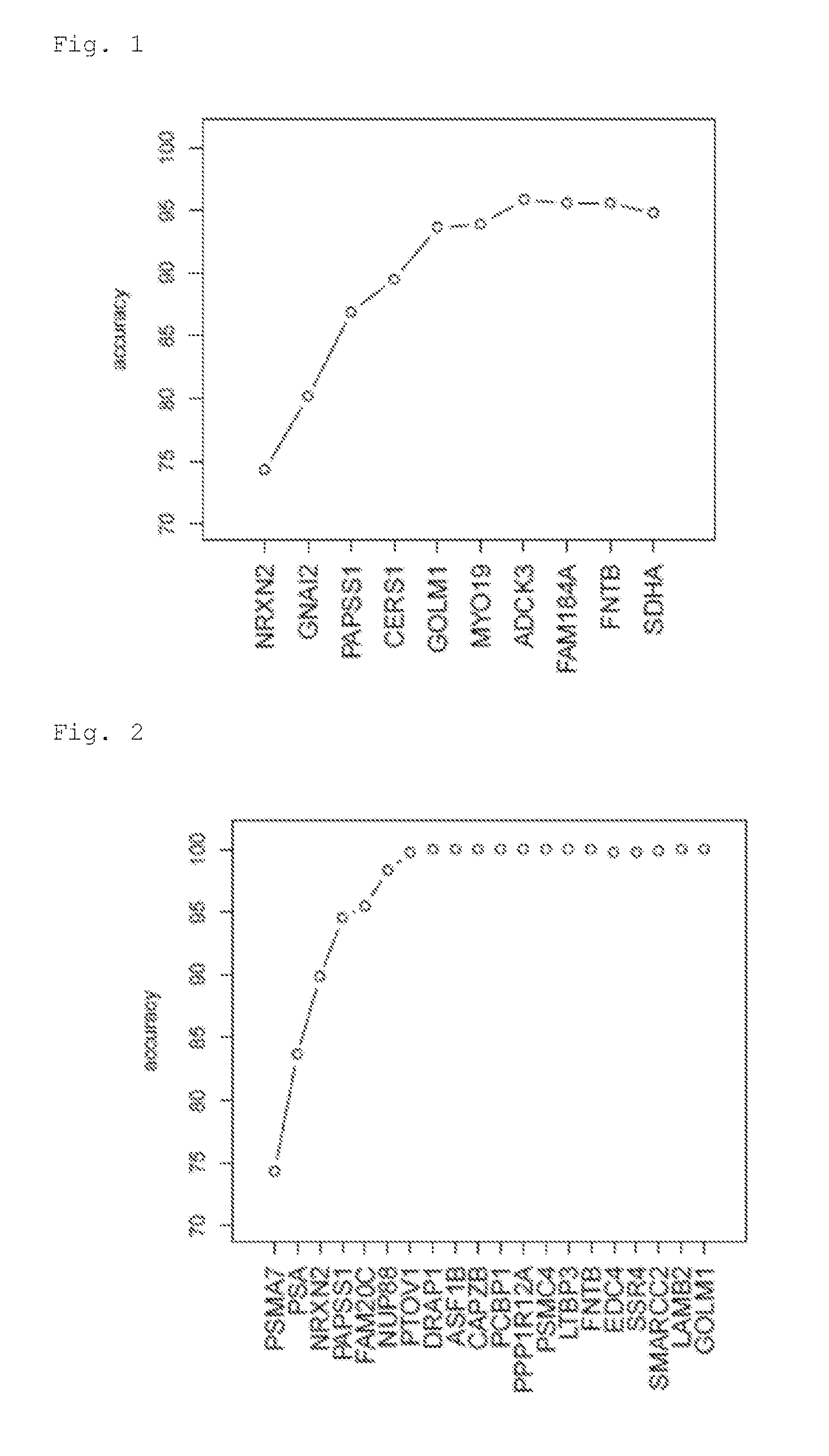
[0018] Also preferred is a combination of detecting at least 2, 3, 4, 5, 6 or more of the markers of List 8, which are NRXN2, GNAI2, PAPSS1, CERS1, GOLM1, MYO19, ADCK3, FAM184A, FNTB, SDHA. These markers resulted in very good discriminatory power.
[0019] Also preferred is a combination of detecting at least 2, 3, 4, 5, 6 or more of the markers of List 9, which are PSMA7, PSA, NRXN2, PAPSS1, FAM20C, NUP88, PTOV1, DRAP1, ASF1B, CAPZB, PCBP1, PPP1R12A, PSMC4, LTBP3, FNTB, EDC4, SSR4, SMARCC2, LAMB2, GOLM1. These markers resulted in very good discriminatory power.
[0020] Also preferred is a combination of detecting at least 2, 3, 4, 5, 6 or more of the markers of List 10, which are PSMC4, DNMT3A, TGS1, NRXN2, GRK6, TBC1D2, ZNF431, DUS2L, MGA, LSM14. These markers resulted in very good discriminatory power.
[0021] Also preferred is a combination of detecting at least 2, 3, 4, 5, 6 or more of the markers of List 11, which are PLEC, RPL36A, HSP90AB1, UBR4, NRXN2, ABTB1, GSTP1, HARS, ARFRP1, USP5. These markers resulted in very good discriminatory power.
[0022] Also preferred is a combination of detecting at least 2, 3, 4, 5, 6 or more of the markers of List 12, which are HIST3H2A, RPS4Y2, HAGH, HNRPDL, COPZ1, CRAT, GET4, SUPV3L1, ACTR1B, UBE3C. These markers resulted in very good discriminatory power.
[0023] Also preferred is a combination of detecting at least 2, 3, 4, 5, 6 or more of the markers of List 13, which are PSMA7, PSA, NRXN2, PAPSS1, PLXNB2, FAM20C, TOLLIP, LSM14B, KDM3A, SYNE2. These markers resulted in very good discriminatory power.
[0024] Also preferred is a combination of detecting at least 2, 3, 4, 5, 6, 7, 8, 9, 10 or more of the markers of List 3p1, which are. This list is given in the examples. List 3p1 is a part of list 3 and the markers performed remarkably well. Indeed any combination of markers of list 3p1. A random permutation analysis, i.e. repeated random picks of markers of this list showed even with low marker amounts exceptional classification rates (See FIG. 11).
[0025] Also preferred is a combination of detecting at least 2, 3, 4, 5, 6, 7, 8, 9, 10 or more of the markers of List 3p2, which are. This list is given in the examples. List 3p2 is a part of list 3 and the markers performed remarkably well. Indeed any combination of markers of list 3p2. A random permutation analysis, i.e. repeated random picks of markers of this list showed even with low marker amounts exceptional classification rates (See FIG. 12).
[0026] Also preferred is a combination of detecting at least 2, 3, 4, 5, 6, 7, 8, 9, 10 or more of the markers of List 3p3, which are. This list is given in the examples. List 3p3 is a part of list 3 and the markers performed remarkably well. Indeed any combination of markers of list 3p3. A random permutation analysis, i.e. repeated random picks of markers of this list showed even with low marker amounts exceptional classification rates (See FIG. 13).
[0027] In particular preferred are the markers as shown in FIGS. 1 to 6, which were evaluated according to a best subset selection from the indicated list of origin. From left to right, additional markers are added to the ones on the left and each incremental marker addition substantially increases classification accuracy. Preferably, the invention provides at least 2, 3, 4, 5, 6 or more markers from any set as disclosed in any of FIGS. 1 to 6. Preferably, the at least 2, 3, 4, 5, 6 or more markers are picked from the markers shown left to right as shown in the figures.
[0028] "Marker" or "marker proteins" are diagnostic indicators found in a patient and are detected, directly or indirectly by the inventive methods. Indirect detection is preferred. In particular, all of the inventive markers have been shown to cause the production of (auto)antigens in cancer patients or patients with a risk of developing cancer. The easiest way to detect these markers is thus to detect these (auto)antibodies in a blood or serum sample from the patient. Such antibodies can be detected by binding to their respective antigen in an assay. Such antigens are in particular the marker proteins themselves or antigenic fragments thereof. Suitable methods exist in the art to specifically detect such antibody-antigen reactions and can be used according to the invention. Preferably the entire antibody content of the sample is normalized (e.g. diluted to a pre-set concentration) and applied to the antigens. Preferably the IgG, IgM, IgD, IgA or IgE antibody fraction, is exclusively used. Preferred antibodies are IgG. Preferably the subject is a human, in particular a male.
[0029] Some markers are more preferred than others. Especially preferred markers are those which are represented at least 2, at least 3, at least 4, at least 5, at least 6, times in any one of lists 1 to 13, 3p1, 3p2, 3p3. These markers are preferably used in any one of the inventive methods or sets.
[0030] The present invention also relates to a method of selecting such at least 2 markers (or more as given above) or at least 20% of the markers (or more as given above) of any one of the inventive sets with high specificity. Such a method includes comparisons of signal data for the inventive markers of any one of the inventive markers sets, especially as listed in lists 1 to 13, with said signal data being obtained from control samples of known prostate cancer conditions or indications and further statistically comparing said signal data with said conditions thereby obtaining a significant pattern of signal data capable of distinguishing the conditions of the known control samples.
[0031] In particular, the control samples may comprise one or more cancerous control (preferably at least 5, or at least 10 cancerous controls) and a healthy or non-cancerous control (preferably at least 5, or at least 10 healthy controls). Preferably 2 different indications are selected that shall be distinguished
[0032] The control samples can be used to obtain a marker dependent signal pattern as indication classifier. Such a signal pattern can be obtained by routine statistical methods, such as binary tree methods. Common statistical methods calculate a (optionally multi-dimensional) vector within the multitude of control data signal values as diagnostically significant distinguishing parameter that can be used to distinguish one or more indications from other one or more indications. The step usually comprises the step of "training" a computer software with said control data. Such pre-obtained training data or signal data can be provided on a computer-readable medium to a practitioner who performs the inventive diagnosis.
[0033] Preferably, the method comprises optimizing the selection process, e.g. by selecting alternative or additional markers and repeating said comparison with the controls signals, until a specificity and/or sensitivity of at least 75% is obtained, preferably of at least 80%, at least 85%, at least 90%, at least 95%.
[0034] Binding events can be detected as known in the art, e.g. by using labelled secondary antibodies. Such labels can be enzymatic, fluorescent, radioactive or a nucleic acid sequence tag. Such labels can also be provided on the binding means, e.g. the antigens as described in the previous paragraph. Nucleic acid sequence tags are especially preferred labels since they can be used as sequence code that not only leads to quantitative information but also to a qualitative identification of the detection means (e.g. antibody with certain specificity). Nucleic acid sequence tags can be used in known methods such as Immuno-PCR. In multiplex assays, usually qualitative information is tied to a specific location, e.g. spot on a microarray. With qualitative information provided in the label, it is not necessary to use such localized immunoassays. In is possible to perform the binding reaction of the analyte and the detection means, e.g. the serum antibody and the labelled antigen, independent of any solid supports in solution and obtain the sequence information of the detection means bound to its analyte. A binding reaction allows amplification of the nucleic acid label in a detection reaction, followed by determination of the nucleic acid sequence determination. With said determined sequence the type of detection means can be determined and hence the marker (analyte, e.g. serum antibody with tumour associated antigen specificity).
[0035] Preferably the inventive method further comprises detecting PSA in a sample from a patient comprising the step of said marker protein or antigenic fragments thereof in a sample of the patient. PSA protein can be detected according to any standard test known. The PSA blood test is the current standard for prostate cancer diagnosis, and has an accuracy of about 60-66% if used alone. Surprisingly, the accuracy can be substantially increased if combined with any other marker or list combination according to the invention. The other markers are preferably tested by detecting auto-antibodies, contrary to PSA, which is preferably tested by determining blood, plasma or serum PSA protein that is bound directly to a detection agent, like an affinity capturing agent. Both, PSA protein (see example 5 and references therein) or nucleic acids (McDermed et al., 2012, Clinical Chemistry 58(4): 732-740) can be detected in the sample. PSA protein in the sample can be detected by an affinity assay, preferably with an immobilized affinity capturing agent. An affinity capturing agent is e.g. an antibody or functional fragment thereof. Immobilization is preferably on a solid support, e.g. a microtiter well, a microarray plate or a bead. Such a PSA capturing agent and preferably also a secondary antibody to PSA with a label can be used in the inventive method or provided in the inventive kit. Nucleic acids are preferably detected by a hybridization probe, with optional amplification, especially preferred is immune-PCR.
[0036] In preferred embodiments of the invention the step of detecting antibodies binding said marker proteins, detecting said marker proteins or antigenic fragments thereof comprises comparing said detection signal with detection signals of a benign prostate hyperplasia controls and comparing said detection signals, wherein an increase in the detection signal indicates prostate cancer or said risk of prostate cancer.
[0037] In preferred embodiments of the invention the step of detecting antibodies binding said marker proteins, detecting said marker proteins or antigenic fragments thereof comprises comparing said detection signal with detection signals of a cancerous control and comparing said detection signals. In particular preferred, especially in cases of using more marker sets of 2 or more markers as mentioned above, a statistical analysis of the control is performed, wherein the controls are used to obtain a marker dependent signal pattern as indication classifier and the marker dependent signals of the sample to be analysed is compared with and/or fitted onto said pattern thereby obtaining information of the diagnosed condition or indication. Such statistical analysis is usually dependent on the used analytical platform that was used to obtain the signal data, given that signal data may vary from platform to platform. Such platforms are e.g. different microarray or solution based setups (with different labels or analytes--such as antigen fragments--for a particular marker). Thus the statistical method can be used to calibrate each platform to obtain diagnostic information with high sensitivity and specificity. The step usually comprises the step of "training" a computer software with said control data. Alternatively, pre-obtained training data can be used. Such pre-obtained training data or signal data can be provided on a computer-readable medium to a practitioner.
[0038] In further embodiments a detection signal from the sample of a patient in amplitude of at least 60%, preferably at least 80%, of the cancerous control indicates prostate cancer or said risk of prostate cancer.
[0039] Usually not all of the inventive markers or detection agents may lead to a signal. Nevertheless only a fraction of the signals is suitable to arrive at a diagnostic decision. In preferred embodiments of the invention a detection signal in at least 60%, preferably at least 70%, least 75%, at least 85%, or in particular preferred at least 95%, even more preferred all, of the used markers indicates prostate cancer or said risk of prostate cancer.
[0040] The present diagnostic methods further provide necessary therapeutic information to decide on a surgical intervention. Therefore the present invention also provides a method of treating a patient comprising prostate cancer or according to any aspect or embodiment of the invention and removing said prostate cancer. "Stratification or therapy control" for the purposes of this invention means that the method according to the invention renders possible decisions for the treatment and therapy of the patient, whether it is the hospitalization of the patient, the use, effect and/or dosage of one or more drugs, a therapeutic measure or the monitoring of a course of the disease and the course of therapy or etiology or classification of a disease, e.g., into a new or existing subtype or the differentiation of diseases and the patients thereof.
[0041] One skilled in the art is familiar with expression libraries, they can be produced according to standard works, such as Sambrook et al, "Molecular Cloning, A laboratory handbook, 2nd edition (1989), CSH press, Cold Spring Harbor, N.Y. Expression libraries are also preferred which are tissue-specific (e.g., human tissue, in particular human organs). Members of such libraries can be used as inventive antigen for use as detection agent to bind analyte antibodies. Furthermore included according to the invention are expression libraries that can be obtained by exon-trapping. A synonym for expression library is expression bank. Also preferred are protein biochips or corresponding expression libraries that do not exhibit any redundancy (so-called: Uniclone.RTM. library) and that may be produced, for example, according to the teachings of WO 99/57311 and WO 99/57312. These preferred Uniclone libraries have a high portion of non-defective fully expressed proteins of a cDNA expression library. Within the context of this invention, the antigens can be obtained from organisms that can also be, but need not be limited to, transformed bacteria, recombinant phages, or transformed cells from mammals, insects, fungi, yeasts, or plants. The marker antigens can be fixed, spotted, or immobilized on a solid support. Alternatively, it is also possible to perform an assay in solution, such as an Immuno-PCR assay.
[0042] In a further aspect, the present invention provides a kit of diagnostic agents suitable to detect any marker or marker combination as described above, preferably wherein said diagnostic agents comprise marker proteins or antigenic fragments thereof suitable to bind antibodies in a sample, especially preferred wherein said diagnostic agents are immobilized on a solid support or in solution, especially when said markers are each labelled with a unique label, such as a unique nucleic acid sequence tag. The inventive kit may further comprise detection agents, such as secondary antibodies, in particular anti-human antibodies, and optionally also buffers and dilution reagents.
[0043] The invention therefore likewise relates to the object of providing a diagnostic device or an assay, in particular a protein biochip, ELISA or Immuno-PCR assay, which permits a diagnosis or examination for prostate carcinoma.
[0044] Additionally, the marker proteins (as binding moieties for antibody detection) can be present in the respective form of a fusion protein, which contains, for example, at least one affinity epitope or tag. The tag may be one such as contains c-myc, his tag, arg tag, FLAG, alkaline phosphatase, VS tag, T7 tag or strep tag, HAT tag, NusA, S tag, SBP tag, thioredoxin, DsbA, a fusion protein, preferably a cellulose-binding domain, green fluorescent protein, maltose-binding protein, calmodulin-binding protein, glutathione S-transferase, or lacZ, a nanoparticle or a nucleic acid sequence tag. Such a nucleic acid sequence can be e.g. DNA or RNA, preferably DNA.
[0045] In all of the embodiments, the term "solid support" covers embodiments such as a filter, a membrane, a magnetic or fluorophore-labeled bead, a silica wafer, glass, metal, ceramics, plastics, a chip, a target for mass spectrometry, a matrix, a bead or microtiter well. However, a filter is preferred according to the invention.
[0046] As a filter, furthermore PVDF, nitrocellulose, or nylon is preferred (e.g., Immobilon P Millipore, Protran Whatman, Hybond N+ Amersham).
[0047] In another preferred embodiment of the arrangement according to the invention, the arrangement corresponds to a grid with the dimensions of a microtiter plate (8-12 wells strips, 96 wells, 384 wells, or more), a silica wafer, a chip, a target for mass spectrometry, or a matrix.
[0048] Another method for detection of the markers is an immunosorbent assay, such as ELISA. When detecting autoantibodies, preferably the marker protein or at least an epitope containing fragment thereof, is bound to a solid support, e.g. a microtiter well. The autoantibody of a sample is bound to this antigen or fragment. Bound autoantibodies can be detected by secondary antibodies with a detectable label, e.g. a fluorescence label. The label is then used to generate a signal in dependence of binding to the autoantibodies. The secondary antibody may be an antihuman antibody if the patient is human or be directed against any other organism in dependence of the patient sample to be analysed. The kit may comprise means for such an assay, such as the solid support and preferably also the secondary antibody. Preferably the secondary antibody binds to the Fc part of the (auto) antibodies of the patient. Also possible is the addition of buffers and washing or rinsing solutions. The solid support may be coated with a blocking compound to avoid unspecific binding.
[0049] Preferably the inventive kit also comprises non-diagnostic control proteins, which can be used for signal normalization. These control proteins bind to moieties, e.g. proteins or antibodies, in the sample of a diseased patient same as in a benign prostate hyperplasia controls. In addition to the inventive marker proteins any number, but preferably at least 2 controls can be used in the method or in the kit.
[0050] Preferably the inventive kit is limited to a particular size. According to these embodiments of the invention the kit comprises at most 3000 diagnostic agents, preferably at most 2500 diagnostic agents, at most 2000 diagnostic agents, at most 1500 diagnostic agents, at most 1200 diagnostic agents, at most 1000 diagnostic agents, at most 800 diagnostic agents, at most 500 diagnostic agents, at most 300 diagnostic agents, at most 200 diagnostic agents, at most 100 diagnostic agents, such as marker proteins or antigenic fragments thereof.
[0051] In especially preferred embodiments of the invention the kit further comprises a computer-readable medium or a computer program product, such as a computer readable memory devices like a flash storage, CD-, DVD- or BR-disc or a hard drive, comprising signal data for the control samples with known conditions selected from cancer and/or of benign prostate hyperplasia controls, and/or calibration or training data for analysing said markers provided in the kit for diagnosing prostate cancer or distinguishing conditions or indications selected from benign prostate hyperplasia controls.
[0052] The kit may also comprise normalization standards, that result in a signal independent of a benign prostate hyperplasia controls condition and cancerous condition. Such normalization standards can be used to obtain background signals. Such standards may be specific for ubiquitous antibodies found in a human, such as antibodies against common bacteria such as E. coli. Preferably the normalization standards include positive and negative (leading to no specific signal) normalization standards.
[0053] Preferred embodiments of the invention that is described herein are defined as follows:
1. Method of diagnosing prostate cancer or the risk of prostate cancer in a patient by detecting the following marker proteins or a selection of at least 2 or at least 20% of the marker proteins selected from OXA1L, GOLM1, NRXN2, PAPSS1, GNAI2, FTSJD2, CERS1, FNTB, MYO19, ADCK3, SDHA, FAM184A (List 1) in a patient, comprising the step of detecting antibodies binding said marker proteins, detecting said marker proteins or antigenic fragments thereof in a sample of the patient. 2. Method of diagnosing prostate cancer or the risk of prostate cancer in a patient by detecting at least 2 or at least 20% of the marker proteins selected from the markers of any one of List 2, 3, 4 or any combination thereof in a patient, comprising the step of detecting antibodies binding said marker proteins, detecting said marker proteins or antigenic fragments thereof in a sample of the patient. 3. Method according to 2 comprising detecting a marker protein selected from any one of Lists 5, 6, 7, 8, 9, 10, 11, 12 or 13 in a patient, comprising the step of detecting antibodies binding said marker protein, detecting said marker protein or antigenic fragments thereof in a sample of the patient. 4. Method according to 2 comprising detecting at least 2 or at least 20% of the marker proteins selected from the markers of any one of Lists 5, 6, 7, 8, 9, 10, 11, 12 or 13 in a patient, comprising the step of detecting antibodies binding said marker proteins, detecting said marker proteins or antigenic fragments thereof in a sample of the patient. 5. Method according to 2 comprising detecting at least 2 or at least 20% of the marker proteins selected from the markers of any one of Lists 3p1, 3p2, 3p3 in a patient, comprising the step of detecting antibodies binding said marker proteins, detecting said marker proteins or antigenic fragments thereof in a sample of the patient. 6. Method according to any one of 1 to 5, comprising detecting at least markers SDHA and/or FAM184A in a patient, comprising the step of detecting antibodies binding said marker proteins, detecting said marker proteins or antigenic fragments thereof in a sample of the patient. 7. Method according to any one of 1 to 6, further comprising detecting PSA in a sample from a patient comprising the step of said marker protein or antigenic fragments thereof in a sample of the patient. 8. Method according to 7, wherein PSA protein in the sample is detected by an affinity assay, preferably with an immobilized affinity capturing agent. 9. The method of any one of 1 to 8, wherein the step of detecting antibodies binding said marker proteins, detecting said marker proteins or antigenic fragments thereof comprises comparing said detection signal with detection signals of a healthy control and comparing said detection signals, wherein an increase in the detection signal indicates prostate cancer. 10. The method of any one of 1 to 9, a) wherein the step of detecting antibodies binding said marker proteins, detecting said marker proteins or antigenic fragments thereof comprises comparing said detection signal with detection signals of one or more known prostate cancer control sample, preferably wherein the control signals are used to obtain a marker dependent signal pattern as indication classifier and the marker dependent signals of the patient is compared with and/or fitted onto said pattern, thereby obtaining information of the diagnosed condition. 11. The method of any one of 1 to 10, a) wherein the step of detecting antibodies binding said marker proteins, detecting said marker proteins or antigenic fragments thereof comprises comparing said detection signal with detection signals of a cancerous control and comparing said detection signals, wherein a detection signal from the sample of the patient in amplitude of at least 60%, preferably at least 80%, of the cancerous control indicates prostate cancer; or b) wherein a detection signal in at least 60%, preferably at least 75%, of the used markers indicates prostate cancer. 12. The method of treating a patient comprising prostate cancer, comprising detecting cancer according to any one of 1 to 11 and removing said prostate cancer or treating prostate cancer cells of said patient by anti-cancer therapy, preferably with a chemo- or radiotherapeutic agent. 13. A kit of diagnostic agents suitable to detect any marker or marker combination as defined in 1 to 9, preferably wherein said diagnostic agents comprise marker proteins or antigenic fragments thereof suitable to bind antibodies in a sample, especially preferred wherein said diagnostic agents are immobilized on a solid support, optionally further comprising a computer-readable medium or a computer program product, comprising signal data for control samples with known conditions selected from cancer, and/or calibration or training data for analysing said markers provided in the kit for diagnosing prostate cancer or distinguishing conditions selected from healthy conditions, cancer. 14. The kit of 13 comprising a labelled secondary antibody, preferably for detecting an Fc part of antibodies of the patient. 15. The kit of 13 or 14 comprising at most 3000 diagnostic agents, preferably at most 2500 diagnostic agents, at most 2000 diagnostic agents, at most 1500 diagnostic agents, at most 1200 diagnostic agents, at most 1000 diagnostic agents, at most 800 diagnostic agents, at most 500 diagnostic agents, at most 300 diagnostic agents, at most 200 diagnostic agents, at most 100 diagnostic agents.
[0054] The present invention is further illustrated by the following figures and examples, without being limited to these embodiments of the invention.
FIGURES
[0055] FIG. 1 shows the best subset selection for List 8.
[0056] FIG. 2 shows the best subset selection for List 9.
[0057] FIG. 3 shows the best subset selection for List 10.
[0058] FIG. 4 shows the best subset selection for List 11.
[0059] FIG. 5 shows the best subset selection for List 12.
[0060] FIG. 6 shows the best subset selection for List 13.
[0061] FIG. 7 shows a permutation analysis of the markers of List 1.
[0062] FIG. 8 shows a permutation analysis of the markers of List 2.
[0063] FIG. 9 shows a permutation analysis of the markers of List 3.
[0064] FIG. 10 shows a permutation analysis of the markers of List 4.
[0065] FIG. 11 shows a permutation analysis of the markers of List 3p1.
[0066] FIG. 12 shows a permutation analysis of the markers of List 3p2.
[0067] FIG. 13 shows a permutation analysis of the markers of List 3p3.
EXAMPLES
Example 1: Patient Samples
[0068] Biomarker screening has been performed with serum samples from a test set of serum samples derived from 49 individuals with confirmed prostate-carcinoma and 49 benign prostate hyperplasia controls (n=98). All these individuals have been elucidated either by histologically verified PCa cases (prostateoscopy) and hospital-based controls with benign prostate hyperplasia in which the presence of PCa was excluded either clinically (13/49 or 27%) or histologically (36/49 or 73%).
Example 2: Immunoglobuline (IgG) Purification from the Serum or Plasma Samples
[0069] The patient serum or plasma samples were stored at -80.degree. C. before they were put on ice to thaw them for IgG purification using Melon Gel 96-well Spin Plate according the manufacturer's instructions (Pierce). In short, 10 .mu.l of thawed sample was diluted in 90 .mu.l of the equilibrated purification buffer on ice, then transferred onto Melon Gel support and incubated on a plate shaker at 500 rpm for 5 minutes. Centrifugation at 1,000.times.g for 2 minutes was done to collect the purified IgG into the collection plate.
[0070] Protein concentrations of the collected IgG samples were measured by absorbance measures at 280 nm using an Epoch Micro-Volume Spectrophotometer System (Biotec, USA). IgG-concentrations of all samples were concentration-adjusted and 0.4 mg/ml of samples were diluted 1:1 in PBS2.times. buffer with TritonX 0.2% and 6% skim milk powder for microarray analyses.
Example 3: Microarray Design
[0071] A protein-chip named "16 k protein chip" from 15,417 human cDNA expression clones derived from the Unipex cDNA expression library plus technical controls was generated. Using this 16 k protein chip candidate markers were used to identify autoantibody profiles suitable for unequivocal distinction of prostate cancer and benign prostate hyperplasia controls.
[0072] Protein-microarray generation and processing was using the Unipex cDNA expression library for recombinant protein expression in E. coli. His-tagged recombinant proteins were purified using Ni-metal chelate chromatography and proteins were spotted in duplicates for generation of the microarray using ARChipEpoxy slides.
Example 4: Preparation, Processing and Analyses of Protein Microarrays
[0073] The microarray with printed duplicates of the protein marker candidates was blocked with DIG Easy Hyb (Roche) in a stirred glass tank for 30 minutes. Blocked slides were washed 3.times. for 5 minutes with fresh PBSTritonX 0.1% washing buffer with agitation. The slides were rinsed in distilled water for 15 seconds to complete the washing step and remove leftovers from the washing buffer. Arrays were spun dry at 900 rpm for 2 minutes. Microarrays were processed using the Agilent Microarray Hybridisation Chambers (Agilent) and Agilent's gasket slides filled with 490 .mu.l of the prepared sample mixture and processed in a hybridization oven for 4h at RT with a rotation speed of 12. During this hybridization time the samples were kept under permanent rotating conditions to assure a homolog dispensation.
[0074] After the hybridization was done, the microarray slides were washed 3.times. with the PBSTritonX 0.1% washing buffer in the glass tank with agitation for 5 minutes and rinsed in distilled water for about 15 seconds. Then, slides were dried by centrifugation at 900 rpm for 2 minutes. IgG bound onto the features of the protein-microarrays were detected by incubation with cy5 conjugated Alexa Fluor.RTM. 647 Goat Anti-Human IgG (H+L) (Invitrogen, Lofer, Austria), diluted in 1:10,000 in PBSTritonX 0.1% and 3% skim milk powder using rotating conditions for 1 h, with a final washing step as outlined above. Microarrays were then scanned and fluorescent data extracted from images (FIG. 1) using the GenePixPro 6.0 software (AXON).
Example 5: PSA Testing
[0075] Prostate-specific antigen (PSA) is a 33-kDa glycoprotein with serine protease activity, found in large amounts in the prostate and seminal plasma. PSA measurement is widely accepted and the current diagnostic standard tool for prostatic cancer diagnostics (Stamey et al., 1987 N Engl J Med 1987; 317:909-15; Hudson et al., 1991 J Urol 1991; 145:802-6).
[0076] The PSA ELISA test is based on the principle of a solid phase enzyme-linked immunosorbent assay. The assay system utilizes a PSA antibody directed against intact PSA for solid phase immobilization (on the microtiter wells). A monoclonal anti-PSA antibody conjugated to horseradish peroxidase (HRP) is in the antibody-enzyme conjugate solution. The test sample was allowed to react first with the immobilized rabbit antibody at room temperature for 60 minutes. The wells were washed to remove any unbound antigen. The monoclonal anti-PSA-HRP conjugate was then reacted with the immobilized antigen for 60 minutes at room temperature resulting in the PSA molecules being sandwiched between the solid phase and enzyme-linked antibodies.
[0077] The wells were washed to remove unbound-labeled antibodies. A solution of TMB Reagent was added and incubated at room temperature for 20 minutes, resulting in the development of a blue color. The color development was stopped with the addition of Stop Solution changing the color to yellow. The concentration of PSA is directly proportional to the color intensity of the test sample. Absorbance is measured spectrophotometrically. The results are reported as nanograms of PSA per milliliter (ng/mL) of blood. Sample signal data was calibrated with a set of standard concentrations.
Example 6: Data Analysis and Permutation Analysis
[0078] Data were 1) quantil normalised and alternatively 2) normalised with Combat transformation for removal of batch effects, when samples were processed on microarrays in 3 different runs; data analyses was conducted using BRB array tools (web at linus.nci.nih.gov/BRB-ArrayTools.html) upon quantile normalized data, and the R software upon the 2 different normalization strategies (quantil and Combat DWD normalized) followed by missing value imputation (Trevor Hastie, Robert Tibshirani, Balasubramanian Narasimhan and Gilbert Chu. impute: impute: Imputation for microarray data. R package version 1.42.0.).
[0079] For identification of tumour marker profiles and classifier markers, class prediction analyses applying cross-validation was used. Classifiers were built for distinguishing both classes of samples denoted "Carc" carcinoma patients, and "Contr" individuals with benign prostate hyperplasia.
[0080] Due to the large redundancy of genes/proteins involved in biological processes (such as tumorigenesis), redundant lists of genes are covered, of which a subset can be used for classification. To show how many randomly chosen markers are necessary for the task of classifying tumor versus control, random sets of 1, 2, 3, . . . markers are drawn from the marker lists and the classification accuracy in cross-validation is reported. Results are shown in FIG. 7-13.
Example 7: Results Summary
[0081] For distinguishing 1) Controls vs Carcinomas, after different normalization strategies (quantil and Combat DWD normalized) followed by missing value imputation, the best 10 classifiers were chosen from claim 3, run 1. It was also shown that using only isolated or only 2 markers from the present classifier sets enables correct classification of 1000 (Example 9.7). Therefore the marker-lists, subsets and single markers (antigens; proteins; peptides) are of particular diagnostic values.
[0082] In addition it has already been shown that peptides deduced from proteins or seroreactive antigens can be used for diagnostics and in the published setting even improve classification success (Syed 2012; Journal of Molecular Biochemistry; Vol 1, No 2, www.jmolbiochem.com/index.php/JmolBiochem/article/view/54).
Example 8: Group Results
[0083] Several lists of marker sets have been identified. All markers are grouped in List 4 recited above. Smaller marker selections portions are provided in Lists 2, 3, 3p1, 3p2 and 3p3. All markers are grouped together in List 4. Lists 3p1, 3p2 and 3p3 were pooled in list 3.
[0084] List 2: 268 Marker Proteins Given by their Gene Symbol.
[0085] OXA1L, GOLM1, NRXN2, PAPSS1, GNAI2, FTSJD2, CERS1, FNTB, MYO19, ADCK3, DHCR24, TUBGCP2, LRFN5, PSA, ATAT1, SH3BGRL, LARP1, NPC2 (includes EG:10577), UNK, ATRX, PSMA7, LCMT1, VPS37D, MITD1, CRYGD, AKR1B1, PRKAR1B, ALKBH2, CCL2, GNAI2, MTF2 (includes EG:17765), RHOG, ARMCX1, LSM12 (includes EG:124801), WDR1, RSBN1L, LAMB2, DEDD2, NEUROD6, KRT8, STX6, MDFI, FBXW5, CYHR1, MGEA5, FAHD2B, EDC4, PSD, RPL36A, ZNF238, PIK3IP1, PPIA, PRKD2, DCP1A, LCAT, MYO1F, GSTM3, PRIC285, CRABP2, CCDC136, CSF1R, ARHGAP25, IDH2, NPM1, PAF1 (includes EG:361531), HNRPDL, COPZ1, PSMC3, PRDM8, ZNF514, UBR4, WDR73, RHOB, C19orf25, MMP14, LTBP3, NUP88, DPP9, SPSB3, TSKU, TNFAIP8L2, SYS1 (includes EG:336339), RPL37A, GSTM4, PKNOX1, DRAP1, HN1, BAG6, HSPA9, LRRC47, XRCC1 (includes EG:22594), CUX1, COPS6, NSUN5P1, PSAP, LSM14B, NCBP2, SDHA, FAM98C, MAD2L1, PPP2R1A, COL4A1, CYFIP1, PRDX5, FAM220A, RPS7, EZR, EXOSC8, FAM20C, SRA1, ETS2, SLA, SERPINA1, LARS, SLIT1, FHL1 (includes EG:14199), PTPRA, ELAVL3, BBIP1, HNRNPH1, PLXNA1, PPP2R1A, IVNS1ABP, PRDX1, THOC3, PELI1, PHF2, OCIAD2, PAK6, FIS1 (includes EG:288584), IL16, IDH1, SRSF1, PABPC1, C8orf33, ARHGEF18, ACTR1B, ANKS3, ZC3H12A, PCBP1, SRM, STMN4, EPC1, NLRP1, PTOV1, C12orf51, WDR1, TCF19, ZXDC, VARS, HTATIP2, PCM1, ATCAY, PRDX3, NSD1, DUS1L, GABARAP, FAM21A/FAM21C, SPRY1, ADAR, KNDC1, HMGN2, AHCTF1, NFKB1, DCHS1, CARHSP1, CORO7/CORO7-PAM16, SSR4, KIAA1109, ABT1, PCDH7, AXIN1, TPX2, SH2B1, RPS4Y1, AKR1C4, PAM, UNC13B, HLA-C, NUDT16L1, ZNF462, NPC2 (includes EG:10577), PUM1, EDF1, COMT, PSMB10, LSM14B, SNF8, CTSW, MTUS1, ARID5A, PSMC4, KIAA0753, EPS15L1, ABHD8, HK1, DNM2, WASL, VPS18, ASF1B, VAV2, PPAP2B, HDAC2, SNRPD3, MICU1, Clorf131, NTAN1, SCG5, REC8 (includes EG:290227), LRPPRC, PPDX, ENO1, PCDHB14, PLA2G2A, THOC3, PAFAH1B3, PTK7, SERBP1, HNRNPA1, RASGRP2, NUP88, FAM118B, TNKS1BP1, H19, NECAP2, PLBD1, CFL1, ITGA3, ZNF668, CDKN2D, RHOT2, AKT2, NARFL, PPP2R3B, ABTB1, EMILIN1, TBC1D9B, PKM, ADNP, PPP1R12A, MRC2, PPIL1, TNKS1BP1, FGB, PPIE, SRSF4, BLOC1S1, CNPY3, IRF3, WRB, TOP2B, PDXDC1, TCERG1, CAPZB, BABAM1, HSPA5, CNOT3, EIF3C/EIF3CL, IL17RA, OGFR, BIRC2, LCP2, GSTP1, MYH9 (includes EG:17886), PIK3R5, NCKAP5L, RGS1, MAN2C1, EHD1, USP5, PLEC, SLC35A2, RPL10A, ARHGDIA, COPE, KDM3A, SMARCC2
[0086] List 3: 282 Marker Proteins Given by their Gene Symbol.
[0087] NRXN2, CERS1, MYO19, LRFN5, ATAT1, KRT8, FBXW5, MGEA5, RPL36A, PRKD2, DCP1A, MYO1F, ARHGAP25, HNRPDL, COPZ1, UBR4, WDR73, SPSB3, LRRC47, NSUN5P1, MAD2L1, SLA, FHL1 (includes EG:14199), IDH1, IL16, SRSF1, ZC3H12A, ACTR1B, LCK, VARS, SPRY1, SSR4, TPX2, RPS4Y1, ARID5A, PSMC4, SFTPB, WASL, RASGRP2, TK1, RHOT2, PPP2R3B, ABTB1, PPIL1, IRF3, CRAT, EIF3C/EIF3CL, DUT, GIPC1, LMTK2, CDC37, LCP2, FOSB, ARFRP1, GSTP1, MTCH1, PSMB5, HIST3H2A, PIK3R5, C9orf86, DDX39B, TINAGL1, INPPL1, MAN2C1, PRKCZ, DDOST, USP5, PLEC, HARS, RPL10A, C22orf46, KRBA1, NFATC3, ATP5D, SMYD4, E2F1, PIK3R2, CLIC1, USP28, MORF4L1, POLR2G, TRIM78P, COG4, RHOT2, TACC2, YWHAE, IP6K2, IKBKB, AKR1B1, CACNA1E, POTEE/POTEF, KLHL23/PHOSPHO2-KLHL23, MEPCE, EIF5A, DOCKS, PLXNB2, NR4A1, RPL4, MBD1, VCP, H19, RARA, CDH2, KIF2A, FXYD5, PPA1, EEF1G, RIC8A, ZNF12, B4GALT2, FNDC4, CYR61, OBFC1, WASH1/WASH5P, HSPA4, PBXIP1, WASH1/WASH5P, PLCG1, HMGB2, GTF2F1, UBC, CELF3, KIF1A, KARS, RNF216, TGS1, NFIX, SGSH, PLEKHO1, TAOK2, MLL5, LAMB1, ZNF431, C17orf28, BAZ1B, UHRF2, ATP5SL, PEX7, TSC2, TMSB10/TMSB4X, LIMS2, TBC1D13, UROD, KLF4, BZW2, SULF2, HLA-E, PRRC2A, TBC1D2, H3F3A/H3F3B, GRK6, HIP1R, ARPC5L, NFKB2, SF3B2, PSMC3, ARPC1B, MGA, Clorf122, SYNE2, NOA1, INPP5F, CDK5RAP3, PABPC1, MDN1, LARP4B, UBE3C, HAGH, NIN, HDAC10, RPS4Y2, GMIP, CCDC88C, ATP1B3, SPOCK2, CYFIP2, TAF1C, WDR25, BAZ1A, NFKBIA, HLA-B, TYK2, C19orf6, SERBP1, SLC25A3, QARS, PPP1R9B, DOCK2, AP2S1, DIS3L, CCNB1IP1, ZNF761, MKS1 (includes EG:287612), FCHO1, TYMP, COQ6, TELO2, XPNPEP3, TXNDC11, HIVEP3, CD44, KPNB1, PCBP2, NPEPL1, PLCB2, FBXO6, PRMT1, ATXN7L2, TADA3, MRPL38 (includes EG:303685), PTBP1, MAGED4/MAGED4B, SEC16A, SLC35B2, ADAMTS10, ZNF256, GBAS, DNMT3A, KCNJ14, PEPD, PITRM1, LSM14A, NDUFV1, TOX2, CAD, HCFC1, WDR11, POLR2J4, TOLLIP, CHGA, HDAC1, HSP90AB1, KLF5, UQCRC1, GALK1, KIAA1731, HSPG2, TLN1, TMED3, DUS2L, LOC407835, TNRC6B, PKM, DAK, VDAC1, LRP4, ULK3, PHKB, NBEA, GTF3C1, IVNS1ABP, AHCY, WDR82, HACL1, USP22, KIF2A, APO-BEC3A, TTC27, YWHAQ, SEC24B, ZNF439, HTRA1, WDTC1, LARP7, BIN3, PTPRO, GET4, SUPV3L1, DHX34, PDZD4, MYCBP2, GATA1, USP39, DFFA, USP7, ATP8B3, UBE2N, C17orf28, EIF3C/EIF3CL, IMPDH1, SART3, ANXA1.
[0088] Each of these markers has a high correct classification accuracy if taken alone. Classification accuracy is given in the following table by their AUC (area-under-curve) classification values:
TABLE-US-00001 TABLE 1 Clone wise AUC classification of the markers of list 2 SYMBOL AUC 1 OXA1L 0.8088 2 GOLM1 0.8034 3 NRXN2 0.8013 4 PAPSS1 0.7972 5 GNAI2 0.7968 6 FTSJD2 0.7959 7 CERS1 0.7905 8 FNTB 0.7893 9 MYO19 0.7880 10 ADCK3 0.7859 11 DHCR24 0.7822 12 TUBGCP2 0.7805 13 LRFN5 0.7793 14 PSA 0.7768 15 ATAT1 0.7759 16 SH3BGRL 0.7738 17 LARP1 0.7738 18 NPC2 0.7730 19 UNK 0.7726 20 ATRX 0.7722 21 PSMA7 0.7718 22 LCMT1 0.7705 23 VPS37D 0.7697 24 MITD1 0.7680 25 CRYGD 0.7676 26 AKR1B1 0.7672 27 PRKAR1B 0.7668 28 ALKBH2 0.7659 29 CCL2 0.7655 30 GNAI2 0.7655 31 MTF2 0.7634 32 RHOG 0.7626 33 ARMCX1 0.7626 34 LSM12 0.7622 35 WDR1 0.7618 36 RSBN1L 0.7618 37 LAMB2 0.7613 38 DEDD2 0.7605 39 NEUROD6 0.7601 40 KRT8 0.7601 41 STX6 0.7589 42 MDFI 0.7584 43 FBXW5 0.7580 44 CYHR1 0.7568 45 MGEA5 0.7559 46 FAHD2B 0.7551 47 EDC4 0.7551 48 PSD 0.7543 49 RPL36A 0.7539 50 ZNF238 0.7539 51 PIK3IP1 0.7539 52 PPIA 0.7534 53 PRKD2 0.7530 54 DCP1A 0.7518 55 LCAT 0.7505 56 MYO1F 0.7497 57 GSTM3 0.7493 58 PRIC285 0.7493 59 CRABP2 0.7493 60 CCDC136 0.7489 61 CSF1R 0.7476 62 ARHGAP25 0.7472 63 IDH2 0.7472 64 NPM1 0.7472 65 PAF1 0.7472 66 HNRPDL 0.7468 67 COPZ1 0.7468 68 PSMC3 0.7468 69 PRDM8 0.7464 70 ZNF514 0.7464 71 UBR4 0.7443 72 WDR73 0.7439 73 RHOB 0.7434 74 C19orf25 0.7434 75 MMP14 0.7430 76 LTBP3 0.7430 77 NUP88 0.7426 78 DPP9 0.7426 79 SPSB3 0.7426 80 TSKU 0.7414 81 TNFAIP8L2 0.7414 82 SYS1 0.7409 83 RPL37A 0.7409 84 GSTM4 0.7409 85 PKNOX1 0.7405 86 DRAP1 0.7397 87 HN1 0.7397 88 BAG6 0.7397 89 HSPA9 0.7389 90 LRRC47 0.7384 91 XRCC1 0.7380 92 CUX1 0.7376 93 COPS6 0.7372 94 NSUN5P1 0.7372 95 PSAP 0.7364 96 LSM14B 0.7359 97 NCBP2 0.7351 98 SDHA 0.7351 99 FAM98C 0.7343 100 MAD2L1 0.7343 101 PPP2R1A 0.7339 102 COL4A1 0.7339 103 CYFIP1 0.7334 104 PRDX5 0.7330 105 FAM220A 0.7326 106 RPS7 0.7326 107 EZR 0.7322 108 EXOSC8 0.7309 109 FAM20C 0.7309 110 SRA1 0.7305 111 ETS2 0.7305 112 SLA 0.7293 113 SERPINA1 0.7289 114 LARS 0.7284 115 SLIT1 0.7280 116 FHL1 0.7280 117 PTPRA 0.7276 118 ELAVL3 0.7276 119 BBIP1 0.7276 120 HNRNPH1 0.7272 121 PLXNA1 0.7272 122 PPP2R1A 0.7268 123 IVNS1ABP 0.7264 124 PRDX1 0.7264 125 THOC3 0.7259 126 PELI1 0.7259 127 PHF2 0.7255 128 OCIAD2 0.7251 129 PAK6 0.7251 130 FIS1 0.7247 131 IL16 0.7243 132 IDH1 0.7243 133 SRSF1 0.7243 134 PABPC1 0.7239 135 C8orf33 0.7239 136 ARHGEF18 0.7234 137 ACTR1B 0.7234 138 ANKS3 0.7234 139 ZC3H12A 0.7234 140 PCBP1 0.7230 141 SRM 0.7222 142 STMN4 0.7222 143 EPC1 0.7222 144 NLRP1 0.7222 145 PTOV1 0.7218 146 C12orf51 0.7218 147 WDR1 0.7218 148 TCF19 0.7214 149 ZXDC 0.7209 150 VARS 0.7209 151 HTATIP2 0.7205 152 PCM1 0.7205 153 ATCAY 0.7205 154 PRDX3 0.7205 155 NSD1 0.7201 156 DUS1L 0.7197 157 GABARAP 0.7197 158 FAM21A 0.7197 159 SPRY1 0.7193 160 ADAR 0.7193 161 KNDC1 0.7193 162 HMGN2 0.7189 163 AHCTF1 0.7189 164 NFKB1 0.7185 165 DCHS1 0.7185 166 CARHSP1 0.7180 167 CORO7 0.7180 168 SSR4 0.7176 169 KIAA1109 0.7176 170 ABT1 0.7172 171 PCDH7 0.7172 172 AXIN1 0.7164 173 TPX2 0.7164 174 SH2B1 0.7160 175 RPS4Y1 0.7160 176 AKR1C4 0.7160 177 PAM 0.7160 178 UNC13B 0.7155 179 HLA-C 0.7147 180 NUDT16L1 0.7147 181 ZNF462 0.7143 182 NPC2 0.7143 183 PUM1 0.7143 184 EDF1 0.7143 185 COMT 0.7139 186 PSMB10 0.7139 187 LSM14B 0.7139 188 SNF8 0.7130 189 CTSW 0.7130 190 MTUS1 0.7126 191 ARID5A 0.7122 192 PSMC4 0.7122 193 KIAA0753 0.7122 194 EPS15L1 0.7122 195 ABHD8 0.7118 196 HK1 0.7118 197 DNM2 0.7118 198 WASL 0.7118 199 VPS18 0.7110 200 ASF1B 0.7110 201 VAV2 0.7110 202 PPAP2B 0.7110 203 HDAC2 0.7110 204 SNRPD3 0.7110 205 MICU1 0.7105 206 C1orf131 0.7105 207 NTAN1 0.7105 208 SCG5 0.7101 209 REC8 0.7097 210 LRPPRC 0.7097 211 PPOX 0.7093 212 ENO1 0.7089 213 PCDHB14 0.7085 214 PLA2G2A 0.7080 215 THOC3 0.7080 216 PAFAH1B3 0.7080 217 PTK7 0.7080 218 SERBP1 0.7080 219 HNRNPA1 0.7080 220 RASGRP2 0.7076 221 NUP88 0.7072 222 FAM118B 0.7072 223 TNKS1BP1 0.7072 224 H19 0.7072 225 NECAP2 0.7064 226 PLBD1 0.7055 227 CFL1 0.7055 228 ITGA3 0.7055 229 ZNF668 0.7055 230 CDKN2D 0.7051 231 RHOT2 0.7047 232 AKT2 0.7043 233 NARFL 0.7039 234 PPP2R3B 0.7039 235 ABTB1 0.7030 236 EMILIN1 0.7030 237 TBC1D9B 0.7030 238 PKM 0.7026 239 ADNP 0.7026 240 PPP1R12A 0.7022 241 MRC2 0.7018 242 PPIL1 0.7018 243 TNKS1BP1 0.7014 244 FGB 0.7014 245 PPIE 0.7010
246 SRSF4 0.7005 247 BLOC1S1 0.7001 248 CNPY3 0.6985 249 IRF3 0.6985 250 WRB 0.6980 251 TOP2B 0.6968 252 PDXDC1 0.6968 253 TCERG1 0.6943 254 CAPZB 0.6935 255 BABAM1 0.6930 256 HSPA5 0.6930 257 CNOT3 0.6918 258 EIF3C 0.6914 259 IL17RA 0.6914 260 OGFR 0.6893 261 BIRC2 0.6880 262 LCP2 0.6880 263 GSTP1 0.6868 264 MYH9 0.6860 265 PIK3R5 0.6843 266 NCKAP5L 0.6843 267 RGS1 0.6830 268 MAN2C1 0.6801 269 EHD1 0.6797 270 USP5 0.6793 271 PLEC 0.6793 272 SLC35A2 0.6789 273 RPL10A 0.6768 274 ARHGDIA 0.6760 275 COPE 0.6735 276 KDM3A 0.6718 277 SMARCC2 0.6460
TABLE-US-00002 TABLE 2 Clone wise AUC classification of the markers of list 3 SYMBOL AUC 1 NRXN2 0.8013 2 CERS1 0.7905 3 MYO19 0.7880 4 LRFN5 0.7793 5 ATAT1 0.7759 6 KRT8 0.7601 7 FBXW5 0.7580 8 MGEA5 0.7559 9 RPL36A 0.7539 10 PRKD2 0.7530 11 DCP1A 0.7518 12 MYO1F 0.7497 13 ARHGAP25 0.7472 14 HNRPDL 0.7468 15 COPZ1 0.7468 16 UBR4 0.7443 17 WDR73 0.7439 18 SPSB3 0.7426 19 LRRC47 0.7384 20 NSUN5P1 0.7372 21 MAD2L1 0.7343 22 SLA 0.7293 23 FHL1 0.7280 24 IDH1 0.7243 25 IL16 0.7243 26 SRSF1 0.7243 27 ZC3H12A 0.7234 28 ACTR1B 0.7234 29 LCK 0.7222 30 VARS 0.7209 31 SPRY1 0.7193 32 SSR4 0.7176 33 TPX2 0.7164 34 RPS4Y1 0.7160 35 ARID5A 0.7122 36 PSMC4 0.7122 37 SFTPB 0.7122 38 WASL 0.7085 39 RASGRP2 0.7076 40 TK1 0.7060 41 RHOT2 0.7047 42 PPP2R3B 0.7039 43 ABTB1 0.7030 44 PPIL1 0.7018 45 IRF3 0.6985 46 CRAT 0.6955 47 EIF3C 0.6914 48 DUT 0.6905 49 GIPC1 0.6897 50 LMTK2 0.6889 51 CDC37 0.6880 52 LCP2 0.6880 53 FOSB 0.6880 54 ARFRP1 0.6876 55 GSTP1 0.6868 56 MTCH1 0.6860 57 PSMB5 0.6851 58 HIST3H2A 0.6847 59 PIK3R5 0.6843 60 C9orf86 0.6839 61 DDX39B 0.6835 62 TINAGL1 0.6830 63 INPPL1 0.6822 64 MAN2C1 0.6801 65 PRKCZ 0.6797 66 DDOST 0.6797 67 USP5 0.6793 68 PLEC 0.6793 69 HARS 0.6781 70 RPL10A 0.6768 71 C22orf46 0.6747 72 KRBA1 0.6743 73 NFATC3 0.6743 74 ATP5D 0.6743 75 SMYD4 0.6735 76 E2F1 0.6731 77 PIK3R2 0.6706 78 CLIC1 0.6701 79 USP28 0.6697 80 MORF4L1 0.6693 81 POLR2G 0.6689 82 TRIM78P 0.6685 83 COG4 0.6672 84 RHOT2 0.6668 85 TACC2 0.6668 86 YWHAE 0.6664 87 IP6K2 0.6664 88 IKBKB 0.6656 89 AKR1B1 0.6626 90 CACNA1E 0.6626 91 POTEE 0.6626 92 KLHL23 0.6622 93 MEPCE 0.6614 94 EIF5A 0.6593 95 DOCK9 0.6581 96 PLXNB2 0.6581 97 NR4A1 0.6576 98 RPL4 0.6576 99 MBD1 0.6560 100 VCP 0.6551 101 H19 0.6535 102 RARA 0.6535 103 CDH2 0.6514 104 KIF2A 0.6510 105 FXYD5 0.6506 106 PPA1 0.6497 107 EEF1G 0.6493 108 RIC8A 0.6493 109 ZNF12 0.6485 110 B4GALT2 0.6472 111 FNDC4 0.6468 112 CYR61 0.6443 113 OBFC1 0.6426 114 WASH1 0.6422 115 HSPA4 0.6418 116 PBXIP1 0.6418 117 WASH1 0.6418 118 PLCG1 0.6410 119 HMGB2 0.6410 120 GTF2F1 0.6406 121 UBC 0.6397 122 CELF3 0.6393 123 KIF1A 0.6389 124 KARS 0.6385 125 RNF216 0.6385 126 TGS1 0.6381 127 NFIX 0.6381 128 SGSH 0.6368 129 PLEKHO1 0.6368 130 TAOK2 0.6364 131 MLL5 0.6347 132 LAMB1 0.6347 133 ZNF431 0.6347 134 C17orf28 0.6343 135 BAZ1B 0.6343 136 UHRF2 0.6335 137 ATP5SL 0.6318 138 PEX7 0.6318 139 TSC2 0.6318 140 TMSB10 0.6310 141 LIMS2 0.6306 142 TBC1D13 0.6302 143 UROD 0.6302 144 KLF4 0.6293 145 BZW2 0.6289 146 SULF2 0.6277 147 HLA-E 0.6277 148 PRRC2A 0.6272 149 TBC1D2 0.6252 150 H3F3A 0.6227 151 GRK6 0.6227 152 HIP1R 0.6222 153 ARPC5L 0.6210 154 NFKB2 0.6210 155 SF3B2 0.6193 156 PSMC3 0.6185 157 ARPC1B 0.6185 158 MGA 0.6177 159 C1orf122 0.6177 160 SYNE2 0.6177 161 NOA1 0.6168 162 INPP5F 0.6168 163 CDK5RAP3 0.6168 164 PABPC1 0.6168 165 MDN1 0.6147 166 LARP4B 0.6139 167 UBE3C 0.6139 168 HAGH 0.6127 169 NIN 0.6122 170 HDAC10 0.6122 171 RPS4Y2 0.6118 172 GMIP 0.6118 173 CCDC88C 0.6102 174 ATP1B3 0.6077 175 SPOCK2 0.6064 176 CYFIP2 0.6064 177 TAF1C 0.6056 178 WDR25 0.6052 179 BAZ1A 0.6047 180 NFKBIA 0.6043 181 HLA-B 0.6035 182 TYK2 0.6027 183 C19orf6 0.6027 184 SERBP1 0.6022 185 SLC25A3 0.6018 186 QARS 0.6018 187 PPP1R9B 0.6018 188 DOCK2 0.6014 189 AP2S1 0.6006 190 DIS3L 0.6006 191 CCNB1IP1 0.5998 192 ZNF761 0.5993 193 MKS1 0.5956 194 FCHO1 0.5956 195 TYMP 0.5948 196 COQ6 0.5948 197 TELO2 0.5935 198 XPNPEP3 0.5927 199 TXNDC11 0.5914 200 HIVEP3 0.5902 201 CD44 0.5898 202 KPNB1 0.5868 203 PCBP2 0.5864 204 NPEPL1 0.5856 205 PLCB2 0.5852 206 FBXO6 0.5848 207 PRMT1 0.5835 208 ATXN7L2 0.5814 209 TADA3 0.5793 210 MRPL38 0.5789 211 PTBP1 0.5785 212 MAGED4 0.5781 213 SEC16A 0.5764 214 SLC35B2 0.5764 215 ADAMTS10 0.5756 216 ZNF256 0.5748 217 GBAS 0.5739 218 DNMT3A 0.5731 219 KCNJ14 0.5718 220 PEPD 0.5718 221 PITRM1 0.5706 222 LSM14A 0.5706 223 NDUFV1 0.5702 224 TOX2 0.5689 225 CAD 0.5685 226 HCFC1 0.5673 227 WDR11 0.5668 228 POLR2J4 0.5656 229 TOLLIP 0.5656 230 CHGA 0.5652 231 HDAC1 0.5643 232 HSP90AB1 0.5639 233 KLF5 0.5618 234 UQCRC1 0.5614 235 GALK1 0.5610 236 KIAA1731 0.5589 237 HSPG2 0.5589 238 TLN1 0.5577 239 TMED3 0.5569 240 DUS2L 0.5564 241 LOC407835 0.5556 242 TNRC6B 0.5556 243 PKM 0.5552 244 DAK 0.5552 245 VDAC1 0.5539
246 LRP4 0.5535 247 ULK3 0.5523 248 PHKB 0.5506 249 NBEA 0.5506 250 GTF3C1 0.5498 251 IVNS1ABP 0.5498 252 AHCY 0.5485 253 WDR82 0.5464 254 HACL1 0.5452 255 USP22 0.5402 256 KIF2A 0.5385 257 APOBEC3A 0.5385 258 TTC27 0.5369 259 YWHAQ 0.5360 260 SEC24B 0.5356 261 ZNF439 0.5352 262 HTRA1 0.5339 263 WDTC1 0.5339 264 LARP7 0.5335 265 BIN3 0.5319 266 PTPRO 0.5314 267 GET4 0.5310 268 SUPV3L1 0.5298 269 DHX34 0.5231 270 PDZD4 0.5219 271 MYCBP2 0.5214 272 GATA1 0.5169 273 USP39 0.5165 274 DFFA 0.5152 275 USP7 0.5144 276 ATP8B3 0.5144 277 UBE2N 0.5131 278 C17orf28 0.5102 279 EIF3C 0.5094 280 IMPDH1 0.5077 281 SART3 0.5040 282 ANXA1 0.5015
[0089] These markers are especially potent when used in combination with other markers. FIGS. 7-10 show a random permutation analysis of these markers when taken alone or in any combination of 2, 3, 4 or more markers.
[0090] When splitting the markers of list 3 into the following subgroups, even higher correct classification results from low numbers of random markers of these lists were obtained (see FIG. 11-13). The subgroups are:
List 3p1:
[0091] NRXN2, LRFN5, KRT8, FBXW5, MGEA5, DCP1A, MYO1F, ARHGAP25, WDR73, NSUN5P1, FHL1 (includes EG:14199), IDH1, VARS, SPRY1, PSMC4, SFTPB, WASL, RASGRP2, TK1, RHOT2, PPP2R3B, PPIL1, GIPC1, LMTK2, CDC37, FOSB, PIK3R5, C22orf46, NFATC3, E2F1, MORF4L1, YWHAE, CACNA1E, RPL4, VCP, RARA, KIF2A, EEF1G, B4GALT2, PBXIP1, GTF2F1, RNF216, TGS1, NFIX, TAOK2, MLL5, ZNF431, TMSB10/TMSB4X, LIMS2, PRRC2A, TBC1D2, GRK6, PSMC3, MGA, Clorf122, MDN1, LARP4B, NIN, CCDC88C, SPOCK2, NFKBIA, C19orf6, DOCK2, AP2S1, COQ6, TXNDC11, HIVEP3, PLCB2, PTBP1, DNMT3A, KCNJ14, LSM14A, CHGA, KLF5, GALK1, DUS2L, NBEA, WDR82, USP22, KIF2A, BIN3, PTPRO, USP39, UBE2N, ANXA1.
List 3p2:
[0092] NRXN2, MYO19, ATAT1, RPL36A, UBR4, SPSB3, LRRC47, IL16, ZC3H12A, LCK, TPX2, RPS4Y1, ABTB1, IRF3, EIF3C/EIF3CL, DUT, LCP2, ARFRP1, GSTP1, DDX39B, MAN2C1, PRKCZ, USP5, PLEC, HARS, RPL10A, KRBA1, CLIC1, USP28, POLR2G, TRIM78P, RHOT2, TACC2, IP6K2, IKBKB, EIF5A, NR4A1, MBD1, CDH2, FXYD5, RIC8A, FNDC4, OBFC1, HMGB2, UBC, SGSH, LAMB1, UHRF2, PEX7, TSC2, TBC1D13, SULF2, HLA-E, HIP1R, NFKB2, SF3B2, ARPC1B, SYNE2, CDK5RAP3, CYFIP2, BAZ1A, HLA-B, TYK2, SERBP1, DIS3L, ZNF761, TYMP, XPNPEP3, CD44, SEC16A, PEPD, HCFC1, HSP90AB1, UQCRC1, TLN1, DAK, PHKB, GTF3C1, HTRA1, DFFA, ATP8B3, UBE2N.
List 3p3:
[0093] CERS1, KRT8, PRKD2, HNRPDL, COPZ1, MAD2L1, SLA, SRSF1, ACTR1B, SSR4, ARID5A, CRAT, MTCH1, PSMB5, HIST3H2A, C9orf86, TINAGL1, INPPL1, DDOST, ATP5D, SMYD4, PIK3R2, COG4, AKR1B1, POTEE/POTEF, KLHL23/PHOSPHO2-KLHL23, MEPCE, DOCKS, PLXNB2, H19, PPA1, ZNF12, CYR61, WASH1/WASH5P, HSPA4, WASH1/WASH5P, PLCG1, CELF3, KIF1A, KARS, PLEKHO1, C17orf28, BAZ1B, ATP5SL, UROD, KLF4, BZW2, H3F3A/H3F3B, ARPC5L, NOA1, INPP5F, PABPC1, UBE3C, HAGH, HDAC10, RPS4Y2, GMIP, ATP1B3, TAF1C, WDR25, SLC25A3, QARS, PPP1R9B, CCNB1IP1, MKS1 (includes EG:287612), FCHO1, TELO2, KPNB1, PCBP2, NPEPL1, FBXO6, PRMT1, ATXN7L2, TADA3, MRPL38 (includes EG:303685), MAGED4/MAGED4B, SLC35B2, ADAMTS10, ZNF256, GBAS, PITRM1, NDUFV1, TOX2, CAD, WDR11, POLR2J4, TOLLIP, HDAC1, KI-AA1731, HSPG2, TMED3, LOC407835, TNRC6B, PKM, VDAC1, LRP4, ULK3, IVNS1ABP, AHCY, HACL1, APOBEC3A, TTC27, YWHAQ, SEC24B, ZNF439, WDTC1, LARP7, GET4, SUPV3L1, DHX34, PDZD4, MYCBP2, GATA1, USP39, USP7, C17orf28, EIF3C/EIF3CL, IMPDH1, SART3.
Example 9: Detailed Results
Example 9.1: "Carc Vs. Contr"--Top 10 Genes Selected by their AUC Value
[0094] The following markers were identified according to this example (Quantil-normalised data):
List 1: 12 Marker Proteins Given by their Gene Symbol:
[0095] OXA1L, GOLM1, NRXN2, PAPSS1, GNAI2, FTSJD2, CERS1, FNTB, MYO19, ADCK3, SDHA, FAM184A
TABLE-US-00003 SYMBOL AUC OXA1L 0.80883 GOLM1 0.803415 NRXN2 0.801333 PAPSS1 0.797168 GNAI2 0.796751 FTSJD2 0.795918 CERS1 0.790504 FNTB 0.789254 MYO19 0.788005 ADCK3 0.785923 SDHA 0.73511 FAM184A 0.556018
Example 9.2: "Carc Vs Contr"--8 Greedy Pairs Algorithm->1NN 100%
[0096] The following markers were identified according to this example (Quantil-normalised data):
List 5: 16 Marker Proteins Given by their Gene Symbol:
[0097] ATAT1, CCDC136, CDK5RAP3, GOLGA4, HCFC1, HLA-C, HNRNPA1, MYO19, NONO, PLEC, PPP1R9B, SNX9, SULF2, USP5, WDR1 and ZC3H12A.
[0098] The "greedy pairs" strategy was used for class prediction of the first 36 (18 carcinoma; 18 control) samples of run2, and it was possible to very efficiently build a classifier for distinguishing "Carc" versus "Contr". Using "8 greedy pairs" of features on arrays, the 1-Nearest Neighbour Predictor (1-NN) enabled correct classification of 100% of samples.
[0099] Greedy pairs algorithm was used to select 8 pairs of genes. Repeated 1 times K-fold (K=20) cross-validation method was used to compute misclassification rate.
Performance of Classifiers During Cross-Validation.
TABLE-US-00004 [0100] Diagonal Bayesian Compound Linear Support Compound Covariate Discriminant 3-Nearest Nearest Vector Covariate Predictor Analysis 1-Nearest Neighbors Centroid Machines Predictor Correct? Correct? Neighbor Correct? Correct? Correct? Correct? Mean percent 92 94 100 94 92 94 94 of correct classification:
Performance of the 1-Nearest Neighbor Classifier:
TABLE-US-00005 [0101] Class Sensitivity Specificity PPV NPV Case 1 1 1 1 Control 1 1 1 1
Example 9.3: "Carc Vs. Contr"--p<5e-06.fwdarw.100%
[0102] The following markers were identified according to this example (Quantil-normalised data):
List 6: 13 Marker Proteins Given by their Gene Symbol:
[0103] ARID5A, EIF3C, FCHO1, HAGH, IVNS1ABP, KLHL23, LARP7, NDUFS2, PLXNB2, SMARCC2, TOLLIP, TRIO and WDR11.
[0104] Genes significantly different between the classes at 5e-06 significance level were used for class prediction for the first (14 carcinoma; 14 control) samples of run3, and it was possible to very efficiently build classifiers for distinguishing "Contr" versus "Carc". The Diagonal Linear Discriminant Analysis (DLDA) and 3-Nearest Neighbor Predictor (3-NN) enabled best correct classification of 100% of samples.
[0105] Genes significantly different between the classes at 5e-06 significance level were used to select genes. Leave-one-out cross-validation method was used to compute misclassification rate.
Performance of Classifiers During Cross-Validation.
TABLE-US-00006 [0106] Diagonal Bayesian Compound Linear Support Compound Covariate Discriminant 3-Nearest Nearest Vector Covariate Predictior Analysis 1-Nearest Neighbors Centroid Machines Predictor Correct? Correct? Neighbor Correct? Correct? Correct? Correct? Mean percent 96 100 96 100 96 93 96 of correct classification:
Performance of the Diagonal Linear Discriminant Analysis Classifier:
TABLE-US-00007 [0107] Class Sensitivity Specificity PPV NPV Case 1 1 1 1 Control 1 1 1 1
Performance of the 3-Nearest Neighbors Classifier:
TABLE-US-00008 [0108] Class Sensitivity Specificity PPV NPV Case 1 1 1 1 Control 1 1 1 1
Example 9.4: "Carc Vs. Contr"-- p<0.000005.fwdarw.91%
[0109] The following markers were identified according to this example (Quantil-normalised data):
List 7: 17 Marker Proteins Given by their Gene Symbol:
[0110] AKR1C4, B4GALT2, BRD9, COPS6, EEFSEC, HCFC1, MYO1F, NBEA, NEU-ROD2, PPP1CA, PSMC4, RASGRP2, RPA3, SMG8, SUGP1, TMEM131 and TUBB2B.
[0111] As in the previous example, genes significantly different between the classes at 5e-06 significance level were used for class prediction for the first 35 (18 carcinoma; 17 control) samples of run 1, and it was possible to very efficiently build classifiers for distinguishing "Carc" versus "Contr". The 1-Nearest Neighbor Predictor (1-NN) enabled best correct classification of 91% of samples.
[0112] Genes significantly different between the classes at 5e-06 significance level were used to select genes. Leave-one-out cross-validation method was used to compute misclassification rate.
Performance of Classifiers During Cross-Validation.
TABLE-US-00009 [0113] Diagonal Bayesian Compound Linear Support Compound Covariate Discriminant 3-Nearest Nearest Vector Covariate Predictor Analysis 1-Nearest Neighbors Centroid Machines Predictor Correct? Correct? Neighbor Correct? Correct? Correct? Correct? Mean percent 89 86 91 89 89 86 90 of correct classification:
Performance of the 1-Nearest Neighbor Classifier:
TABLE-US-00010 [0114] Class Sensitivity Specificity PPV NPV Case 1 0.824 0.857 1 Control 0.824 1 1 0.857
Example 9.5: "Carc Vs. Contr"--Best Discriminatory Power
[0115] The top ten genes (by AUC value) discriminating between the classes from claim 1 were used for search of the best discriminatory power. A best subset selection was created by starting with the best discriminator (by cross-validated prediction accuracy using SVM) and sequentially adding new features from claim 1 which most improve classification accuracy. This was repeated for the first 10 features.
TABLE-US-00011 SYMBOL CV accuracy NRXN2 74.31973 GNAI2 80.13605 PAPSS1 86.90476 CERS1 89.52381 GOLM1 93.60544 MYO19 93.91156 ADCK3 95.81633 FAM184A 95.57823 FNTB 95.57823 SDHA 94.79592
List 8: 10 Marker Proteins Given by their Gene Symbol:
[0116] NRXN2, GNAI2, PAPSS1, CERS1, GOLM1, MYO19, ADCK3, FAM184A, FNTB, SDHA (see FIG. 1 for accuracy of best subset selection)
Example 9.6: "Carc Vs. Contr"--Best Discriminatory Power
[0117] The top ten genes (by AUC value) discriminating between the classes from claim 2 were used for search of the best discriminatory power. A best subset selection was created by starting with the best discriminator (by cross-validated prediction accuracy using SVM) and sequentially adding new features from claim 2 which most improve classification accuracy. The following is the list of the best subset selection. This was repeated for the first 20 features.
TABLE-US-00012 Symbol CV accuracy (SVM) PSMA7 74.38776 PSA 83.60544 NRXN2 89.82993 PAPSS1 94.4898 FAM20C 95.47619 NUP88 98.26531 PTOV1 99.69388 DRAP1 99.96599 ASF1B 99.96599 CAPZB 100 PCBP1 100 PPP1R12A 100 PSMC4 100 LTBP3 100 FNTB 99.96599 EDC4 99.7619 SSR4 99.72789 SMARCC2 99.79592 LAMB2 99.96599
List 9: 19 Marker Proteins Given by their Gene Symbol:
[0118] PSMA7, PSA, NRXN2, PAPSS1, FAM20C, NUP88, PTOV1, DRAP1, ASF1B, CAPZB, PCBP1, PPP1R12A, PSMC4, LTBP3, FNTB, EDC4, SSR4, SMARCC2, LAMB2, GOLM1 (see FIG. 2 for accuracy of best subset selection)
Example 9.7: "Carc Vs. Contr"--Best Discriminatory Power
[0119] Genes significantly different between the classes from claim 3, run 1 were used for search of the best discriminatory power. The following is the list of the best subset selection.
TABLE-US-00013 Symbol CV accuracy (SVM) PSMC4 93.33333 DNMT3A 100 TGS1 100 NRXN2 100 GRK6 100 TBC1D2 100 ZNF431 100 DUS2L 100 MGA 100
List 10: 9 Marker Proteins Given by their Gene Symbol.
[0120] PSMC4, DNMT3A, TGS1, NRXN2, GRK6, TBC1D2, ZNF431, DUS2L, MGA, LSM14A (see FIG. 3 for accuracy of best subset selection)
Example 9.8: "Carc Vs. Contr"--Best Discriminatory Power
[0121] Genes significantly different between the classes from claim 3, run 2 were used for search of the best discriminatory power. The following is the list of the best subset selection.
TABLE-US-00014 Symbol CV accuracy (SVM) PLEC 93.2381 RPL36A 94.47619 HSP90AB1 99.42857 UBR4 100 NRXN2 100 ABTB1 100 GSTP1 100 HARS 100 ARFRP1 100 USP5 100
List 11: 10 Marker Proteins Given by their Gene Symbol:
[0122] PLEC, RPL36A, HSP90AB1, UBR4, NRXN2, ABTB1, GSTP1, HARS, ARFRP1, USP5 (see FIG. 4 for accuracy of best subset selection)
Example 9.9: "Carc Vs. Contr"--Best Discriminatory Power
[0123] Genes significantly different between the classes from claim 3, run 3 were used for search of the best discriminatory power. The following is the list of the best subset selection.
TABLE-US-00015 Symbol CV accuracy (SVM) HIST3H2A 97.02381 RPS4Y2 100 HAGH 100 HNRPDL 100 COPZ1 100 CRAT 100 GET4 100 SUPV3L1 100 ACTR1B 100 UBE3C 100
List 12: 10 Marker Proteins Given by their Gene Symbol:
[0124] HIST3H2A, RPS4Y2, HAGH, HNRPDL, COPZ1, CRAT, GET4, SUPV3L1, ACTR1B, UBE3C (see FIG. 5 for accuracy of best subset selection)
Example 9.10: "Carc Vs. Contr"--Best Discriminatory Power
[0125] Genes significantly different between the classes from claim 4 were used for search of the best discriminatory power. The following is the list of the best subset selection.
TABLE-US-00016 Symbol CV accuracy (SVM) PSMA7 74.42177 PSA 83.60544 NRXN2 89.42177 PAPSS1 94.42177 PLXNB2 96.15646 FAM20C 97.92517 TOLLIP 99.69388 LSM14B 99.96599 KDM3A 100 SYNE2 99.96599
List 13: 10 Marker Proteins Given by their Gene Symbol:
[0126] PSMA7, PSA, NRXN2, PAPSS1, PLXNB2, FAM20C, TOLLIP, LSM14B, KDM3A, SYNE2 (see FIG. 6 for accuracy of best subset selection).
* * * * *
References
D00000
D00001
D00002
D00003
D00004
D00005
D00006
D00007
D00008
D00009
D00010
XML
uspto.report is an independent third-party trademark research tool that is not affiliated, endorsed, or sponsored by the United States Patent and Trademark Office (USPTO) or any other governmental organization. The information provided by uspto.report is based on publicly available data at the time of writing and is intended for informational purposes only.
While we strive to provide accurate and up-to-date information, we do not guarantee the accuracy, completeness, reliability, or suitability of the information displayed on this site. The use of this site is at your own risk. Any reliance you place on such information is therefore strictly at your own risk.
All official trademark data, including owner information, should be verified by visiting the official USPTO website at www.uspto.gov. This site is not intended to replace professional legal advice and should not be used as a substitute for consulting with a legal professional who is knowledgeable about trademark law.