Diagnosis Of Autoimmune Diseases
Sorek; Rachel ; et al.
U.S. patent application number 16/133295 was filed with the patent office on 2019-03-28 for diagnosis of autoimmune diseases. The applicant listed for this patent is IMMUNARRAY LTD.. Invention is credited to Keren Jakobi-Brook, Pennina Safer, Rachel Sorek.
| Application Number | 20190094219 16/133295 |
| Document ID | / |
| Family ID | 65807436 |
| Filed Date | 2019-03-28 |


| United States Patent Application | 20190094219 |
| Kind Code | A1 |
| Sorek; Rachel ; et al. | March 28, 2019 |
DIAGNOSIS OF AUTOIMMUNE DISEASES
Abstract
Assays and methods for diagnosing and treating autoimmune diseases. Particularly, the invention provides methods for differential diagnosis of specific autoimmune diseases, including autoimmune rheumatic disorders.
| Inventors: | Sorek; Rachel; (Moshav Tzafaria, IL) ; Jakobi-Brook; Keren; (Tel Aviv, IL) ; Safer; Pennina; (Rehovot, IL) | ||||||||||
| Applicant: |
|
||||||||||
|---|---|---|---|---|---|---|---|---|---|---|---|
| Family ID: | 65807436 | ||||||||||
| Appl. No.: | 16/133295 | ||||||||||
| Filed: | September 17, 2018 |
Related U.S. Patent Documents
| Application Number | Filing Date | Patent Number | ||
|---|---|---|---|---|
| 62564344 | Sep 28, 2017 | |||
| Current U.S. Class: | 1/1 |
| Current CPC Class: | G01N 2800/50 20130101; G01N 33/564 20130101; G01N 33/577 20130101; G01N 2800/7095 20130101; G01N 2800/102 20130101; G01N 2800/54 20130101; G01N 2800/104 20130101; G01N 2800/52 20130101 |
| International Class: | G01N 33/564 20060101 G01N033/564; G01N 33/577 20060101 G01N033/577 |
Claims
1. A method of diagnosing an autoimmune disease selected from the group consisting of systemic lupus erythematosus (SLE), rheumatoid arthritis (RA), scleroderma (SSc), Sjogren's Syndrome (SS), and anti-phospholipid syndrome (APS) in a subject, the method comprising: i. obtaining a sample from a subject suspected of having an autoimmune rheumatic disease due to inflammation and at least one rheumatic symptom selected from the group consisting of: joint pain, joint stiffness, joint swelling, joint redness, joint tenderness, joint warmth, and loss of joint range of motion; ii. determining the reactivity of antibodies in the sample obtained from the subject to a plurality of antigens selected from Table 1, thereby obtaining a reactivity pattern of said sample to the plurality of antigens; iii. comparing the reactivity pattern of said sample to a healthy control reactivity pattern to said plurality of antigens by a supervised classification algorithm, wherein a significantly different reactivity pattern of the sample obtained from the subject compared to the healthy control reactivity pattern indicates that said subject has an autoimmune rheumatic disease.
2. The method of claim 1, further comprising identifying the autoimmune disease in said subject by the steps comprising: iv. determining the reactivity of antibodies in the sample obtained from the subject to a plurality of antigens selected from at least one of Tables 8-12, thereby obtaining a reactivity pattern of said sample to the plurality of antigens; v. comparing the reactivity pattern of said sample to a healthy control reactivity pattern to said plurality of antigens, by a supervised classification algorithm, wherein: a significantly different reactivity pattern of the sample obtained from the subject to a plurality of antigens selected from Table 8 compared to the healthy control reactivity pattern indicates that said subject has SLE, a significantly different reactivity pattern of the sample obtained from the subject to a plurality of antigens selected from Table 9 compared to the healthy control reactivity pattern indicates that said subject has SSc, a significantly different reactivity pattern of the sample obtained from the subject to a plurality of antigens selected from Table 10 compared to the healthy control reactivity pattern indicates that said subject has APS, a significantly different reactivity pattern of the sample obtained from the subject to a plurality of antigens selected from Table 11 compared to the healthy control reactivity pattern indicates that said subject has SS, and/or a significantly different reactivity pattern of the sample obtained from the subject to a plurality of antigens selected from Table 12 compared to the healthy control reactivity pattern indicates that said subject has RA.
3. The method of claim 1, wherein the subject is suspected as having SLE or RA and the method comprises: (a) determining the reactivity of antibodies in the sample obtained from the subject to a plurality of antigens selected from Table 2, thereby obtaining a reactivity pattern of said sample to the plurality of antigens selected from Table 2; (b) comparing the reactivity pattern of said sample to the plurality of antigens selected from Table 2 to a control reactivity pattern to said plurality of antigens selected from Table 2 by a supervised classification algorithm, wherein the control is a RA control, and a significantly different reactivity pattern of the sample obtained from said subject compared to the RA control reactivity pattern indicates that said subject has SLE, and/or wherein the control is a SLE control, and a significantly different reactivity pattern of the sample obtained from said subject compared to the SLE control reactivity pattern indicates that said subject has RA; or wherein the subject is suspected as having SLE or SSc and the method comprises: (a) determining the reactivity of antibodies in the sample obtained from the subject to a plurality of antigens selected from Table 3, thereby obtaining a reactivity pattern of said sample to the plurality of antigens; (b) comparing the reactivity pattern of said sample obtained from the subject to the plurality of antigens selected from Table 3, to a control reactivity pattern to said plurality of antigens selected from Table 3 by a supervised classification algorithm, wherein the control is a SSc control, and a significantly different reactivity pattern of the sample obtained from said subject compared to the SSc control reactivity pattern indicates that said subject has SLE, and/or wherein the control is a SLE control, and a significantly different reactivity pattern of the sample obtained from said subject compared to the SLE control reactivity pattern indicates that said subject has SSc; or wherein the subject is suspected as having SLE or APS and the method comprises: (a) determining the reactivity of antibodies in the sample obtained from the subject to a plurality of antigens selected from Table 4, thereby obtaining a reactivity pattern of said sample to the plurality of antigens; (b) comparing the reactivity pattern of said sample obtained from the subject to the plurality of antigens selected from Table 4 to a control reactivity pattern to said plurality of antigens selected from Table 4 by a supervised classification algorithm, wherein the control is an APS control, and a significantly different reactivity pattern of the sample obtained from said subject compared to the APS control reactivity pattern indicates that said subject has SLE, and/or wherein the control is a SLE control, and a significantly different reactivity pattern of the sample obtained from said subject compared to the SLE control reactivity pattern indicates that said subject has APS; or wherein the subject is suspected as having SS or SSc and the method comprises: (a) determining the reactivity of antibodies in the sample obtained from the subject to a plurality of antigens selected from Table 5, thereby obtaining a reactivity pattern of said sample to the plurality of antigens; (b) comparing the reactivity pattern of said sample obtained from the subject to the plurality of antigens selected from Table 5 to a control reactivity pattern to said plurality of antigens selected from Table 5 by a supervised classification algorithm, wherein the control is a SSc control, and a significantly different reactivity pattern of the sample obtained from said subject compared to the SSc control reactivity pattern indicates that said subject has SS, and/or wherein the control is a SS control, and a significantly different reactivity pattern of the sample obtained from said subject compared to the SS control reactivity pattern indicates that said subject has SSc; or wherein the subject is suspected as having SS or RA and the method comprises: (a) determining the reactivity of antibodies in the sample obtained from the subject to a plurality of antigens selected from Table 6, thereby obtaining a reactivity pattern of said sample to the plurality of antigens; (b) comparing the reactivity pattern of said sample obtained from the subject to the plurality of antigens selected from Table 6 to a control reactivity pattern to said plurality of antigens selected from Table 6 by a supervised classification algorithm, wherein the control is a RA control, and a significantly different reactivity pattern of the sample obtained from said subject compared to the RA control reactivity pattern indicates that said subject has SS, and/or wherein the control is a SS control, and a significantly different reactivity pattern of the sample obtained from said subject compared to the SS control reactivity pattern indicates that said subject has RA; or wherein the subject is suspected as having SLE or SS and the method comprises: (a) determining the reactivity of antibodies in the sample obtained from the subject to a plurality of antigens comprising the antigen of Table 7, thereby obtaining a reactivity pattern of said sample to the plurality of antigens; (b) comparing the reactivity pattern of said sample obtained from the subject to the plurality of antigens to a control reactivity pattern to said plurality of antigens by a supervised classification algorithm, wherein the control is a SS control, and a significantly different reactivity pattern of the sample obtained from said subject compared to the SS control reactivity pattern indicates that said subject has SLE, and/or wherein the control is a SLE control, and a significantly different reactivity pattern of the sample obtained from said subject compared to the SLE control reactivity pattern indicates that said subject has SS.
4. The method according to claim 1, further comprising treating said subject diagnosed as having the autoimmune disease with a disease-specific treatment.
5. The method of claim 4, wherein the disease-specific treatment is selected from the group consisting of nonsteroidal anti-inflammatory drugs (NSAIDs), corticosteroids, glucocorticoids, immunosuppressants, hydroxychloroquine, cyclophosphamide, TNF-.alpha. inhibitors, chelating agents, endothelin receptor antagonist, PAH, PDE-5 inhibitors, gastrointestinal agents, immunomodulators, analgesics, anticoagulants, antiplatelet and other biologic medications.
6. The method of claim 4, wherein said disease is RA and said disease-specific treatment is selected from the group consisting of Etanercept (Enbrel), Adalimumab (Humira), Infliximab (Remicade), Certolizumab pegol (Cimzia), Golimumab (Simponi), Anakinra (Kineret), Tocilizumab (Actemra), Tofacitinib (Xeljanz), and any combination thereof.
7. The method of claim 4, wherein said disease is SS and said disease-specific treatment is selected from the group consisting of pilocarpine (Salagen), cevimeline (Evoxac), cyclosporine (Restasis), and any combination thereof.
8. The method of claim 4, wherein said disease is SLE and said disease-specific treatment is selected from the group consisting of NSAIDs, Ibuprofen, Naproxen, Prednisone, Methylprednisolone (A-Methapred, Medrol, Solu-Medrol, Depo-Medrol), Cellcept, Methotrexate, Imuran, mycophenolate mofetil, Abatacept, Arava, Immune globulin intravenous (Hizentra, Gammagard, Octagam, Privigen), Plaquenil, Myfortic, Endoxan, Cytoxan, Neosar, Procytox, Revimmune, Benilimumab (Benlysta), Rituximab, Cyclosporine (Gengraf, Neoral, Sandimmune).
9. The method of claim 4, wherein said disease is APS and said disease-specific treatment is selected from the group consisting of warfarin, rivaroxaban (Xarelto), dabigatran (Pradaxa), apixaban (Eliquis), edoxaban (Lixiana), heparin, aspirin, clopidogrel, Rituximab.
10. The method of claim 4, wherein said disease is SSc and said disease-specific treatment is selected from the group consisting of Ibuprofen, Naproxen, Prednisone, Cellcept, Methotrexate, Imuran, Cytoxan, Rituximab, Penicillamine (Cuprimine, Depen), Bosentan (Tracleer), Ambrisentan (Letairis), Macitentan (Opsumit), Tadalafil (Adcirca), Sildenafil (Revatio).
11. The method according to claim 1, wherein the sample is selected from the group consisting of a serum sample, a plasma sample, and a blood sample, and the reactivity of antibodies comprises IgM reactivity, IgG reactivity, or a combination thereof.
12. The method according to claim 1, wherein the supervised classification algorithm is selected from the group consisting of Support Vector Machine (SVM), Quadratic Discriminant Analysis (QDA), and Naive Bayesian Classifier (NB).
13. The method according to claim 1, wherein the reactivity of the antibodies to the plurality of antigens in the sample obtained from a control subject is selected from the group consisting of reactivity of antibodies in multiple samples of control subjects, and a stored set of data from control subjects, and wherein the antigens are in the form of an antigen probe set, an antigen array, or an antigen chip.
14. The method of claim 1, wherein said plurality of antigens includes: LAIR1, MICA, RO60calf, CD62P, Ferritin, Chromatin, and CENPB.
15. A method of diagnosing an autoimmune disease selected from the group consisting of systemic lupus erythematosus (SLE), rheumatoid arthritis (RA), scleroderma (SSc), Sjogren's Syndrome (SS), and anti-phospholipid syndrome (APS) in a subject, the method comprising: i. obtaining a sample from a subject suspected of having an autoimmune rheumatic disease; ii. determining the reactivity of antibodies in the sample obtained from the subject to a plurality of antigens selected from at least one of Tables 8-12, thereby obtaining a reactivity pattern of said sample to the plurality of antigens; iii. comparing the reactivity pattern of said sample to a healthy control reactivity pattern to said plurality of antigens, by a supervised classification algorithm, wherein: a significantly different reactivity pattern of the sample obtained from the subject to a plurality of antigens selected from Table 8 compared to the healthy control reactivity pattern indicates that said subject has SLE, a significantly different reactivity pattern of the sample obtained from the subject to a plurality of antigens selected from Table 9 compared to the healthy control reactivity pattern indicates that said subject has SSc, a significantly different reactivity pattern of the sample obtained from the subject to a plurality of antigens selected from Table 10 compared to the healthy control reactivity pattern indicates that said subject has APS, a significantly different reactivity pattern of the sample obtained from the subject to a plurality of antigens selected from Table 11 compared to the healthy control reactivity pattern indicates that said subject has SS, and/or a significantly different reactivity pattern of the sample obtained from the subject to a plurality of antigens selected from Table 12 compared to the healthy control reactivity pattern indicates that said subject has RA.
16. A method for differentially diagnosing an autoimmune disease, comprising: i. obtaining a sample from a subject diagnosed as having the autoimmune disease; ii. contacting the sample, under conditions such that a specific antigen-antibody complex is formed, with antigens selected from at least one of Tables 2-7; iii. determining the reactivity of antibodies in said sample to the antigens, thereby determining the reactivity pattern of said sample to said antigens; and iv. comparing the reactivity pattern of said sample obtained from the subject to a control reactivity pattern to said antigens by a supervised classification algorithm, wherein: a significantly different reactivity pattern of the sample obtained from the subject to a plurality of antigens selected from Table 2 compared to a RA control reactivity pattern to said antigens indicates that said subject has SLE, a significantly different reactivity pattern of the sample obtained from said subject to a plurality of antigens selected from Table 2 compared to a SLE control reactivity pattern to said antigens indicates that said subject has RA, a significantly different reactivity pattern of the sample obtained from said subject to a plurality of antigens selected from Table 3 compared to a SSc control reactivity pattern to said antigens indicates that said subject has SLE, a significantly different reactivity pattern of the sample obtained from said subject to a plurality of antigens selected from Table 3 compared to a SLE control reactivity pattern to said antigens indicates that said subject has SSc, a significantly different reactivity pattern of the sample obtained from said subject to a plurality of antigens selected from Table 4 compared to an APS control reactivity pattern to said antigens indicates that said subject has SLE, a significantly different reactivity pattern of the sample obtained from said subject to a plurality of antigens selected from Table 4 compared to an SLE control reactivity pattern to said antigens indicates that said subject has APS, a significantly different reactivity pattern of the sample obtained from said subject to a plurality of antigens selected from Table 5 compared to a SSc control reactivity pattern to said antigens indicates that said subject has SS, a significantly different reactivity pattern of the sample obtained from said subject to a plurality of antigens selected from Table 5 compared to a SS control reactivity pattern to said antigens indicates that said subject has SSc, a significantly different reactivity pattern of the sample obtained from said subject to a plurality of antigens selected from Table 6 compared to a RA control reactivity pattern to said antigens indicates that said subject has SS, a significantly different reactivity pattern of the sample obtained from said subject to a plurality of antigens selected from Table 6 compared to a SS control reactivity pattern indicates that said subject has RA. a significantly different reactivity pattern of the sample obtained from said subject to a plurality of antigens comprising the antigen of Table 7 compared to a SS control reactivity pattern to said antigens indicates that said subject has SLE, and/or a significantly different reactivity pattern of the sample obtained from said subject to a plurality of antigens comprising the antigen of Table 7 compared to a SLE control reactivity pattern indicates that said subject has SS.
17. The method according to claim 15, further comprising identifying an antigen-specific treatment for said subject, wherein the antigen is selected from the group consisting of the antigens listed in any one of Tables 1 to 12, and the reactivity of antibodies in the sample obtained from said subject to said antigen is significantly different from the control reactivity pattern to said antigen.
18. The method of claim 17, wherein the disease-specific treatment is an inhibitor of an antibody to an antigen selected from the group consisting of the antigens listed in any one of Tables 1 to 12, wherein the reactivity of the antibody to the antigen in the sample obtained from said subject is significantly different from the reactivity of an antibody to said antigen in a sample obtained from a healthy control subject.
19. The method of claim 15, wherein the antigen is selected from Table 1, preferably wherein the antigen is LAIR-1.
20. The method according to claim 16, further comprising identifying an antigen-specific treatment for said subject, wherein the antigen is selected from the group consisting of the antigens listed in any one of Tables 1 to 12, and the reactivity of antibodies in the sample obtained from said subject to said antigen is significantly different from the control reactivity pattern to said antigen.
Description
FIELD OF THE INVENTION
[0001] The invention provides methods for diagnosing and treating autoimmune diseases. Particularly, the invention provides methods and antigen probe sets, array or chips for differential diagnosis of specific autoimmune diseases, including autoimmune rheumatic disorders.
BACKGROUND OF THE INVENTION
[0002] Many types of autoimmune diseases exist, among which autoimmune rheumatic disorders (ARDs) are included. In ARDs, the immune system attacks the joints and certain systems. The cause of many of the ARDs is unknown. Among these disorders are systemic lupus erythematosus (SLE), rheumatoid arthritis (RA), and Sjogren's syndrome (SS), each having a long list of characteristic symptoms. While some of the symptoms are unique to each disease, and can therefore assist in differential diagnosis, such symptoms are not always present. In addition, ARDs are often difficult to distinguish due to overlapping signs and symptoms. Non-limiting examples for overlapping symptoms include Raynaud's phenomenon, joint pain, joint stiffness, joint swelling, joint redness, joint tenderness, joint warmth, impaired joint range of motion, fatigue, and inflammation.
[0003] The presence of autoantibodies is a hallmark of many autoimmune diseases, which has long been used for the diagnosis and classification of these diseases. Autoantibodies may exist years before the diagnosis of an autoimmune disease, and thus could potentially be used for early prediction of the disease onset. Anti-nuclear antibodies (ANAs) have been observed in a variety of autoimmune disorders, including autoimmune rheumatic diseases, and in chronic inflammatory diseases, infectious diseases and malignancies, and can also be induced by certain drugs. Thus, while ANA testing is being considered as an auxiliary diagnostic tool by physicians, it may only be used as a general indicator of such diseases due to lack of specificity.
[0004] Lab-associated variances in test results of ANA have been demonstrated, reducing the reliability of this assay. Therefore, it is currently recommended that ANA positive test results be followed by additional diagnostic tools.
[0005] To date, no single autoantibody has demonstrated clinically significant sensitivity and specificity for the diagnosis of individual ARD, specifically at early stage of the disease. There remains an unmet need to discover serological tests for the diagnosis of individual ARDs and other autoimmune diseases.
[0006] SLE is a chronic, recurrent, potentially fatal multisystem inflammatory disorder mainly affecting women. SLE is associated with a large spectrum of autoantibodies. IgG antibodies to more than 100 different antigens including DNA, nucleosomes, histones, viral antigens, transcription factors and more have been reported in different SLE patients. As there is no approved serologic diagnosis of SLE, SLE is diagnosed on the basis of eleven criteria defined by the American College of Rheumatology (ACR). These criteria include malar rash, discoid rash, photosensitivity, oral ulcers, arthritis, serositis, renal disorder, neurologic disorder, hematologic disorder (e.g., leucopenia, lymphopenia, hemolytic anemia or thrombocytopenia), immunologic disorder and antibody abnormalities (particularly ANA and anti-DNA antibodies). According to these criteria, subjects can be clinically diagnosed with SLE if they meet at least four of the eleven criteria. Recently, the Systemic Lupus Collaborating Clinics (SLICC) revised these criteria. Nevertheless, SLE is still possible even in cases when less than four criteria are present (`pre-clinical` lupus). Patients with indeterminate findings are evaluated with extensive serologic testing and repeated clinical examinations until a definitive diagnosis of SLE or some other disease can be made. These patients are followed extensively, with a mean time from the initial visit until disposition of about 6 years.
[0007] RA is a chronic inflammatory autoimmune disease characterized by joint inflammation, joint swelling, joint tenderness, and destruction of synovial joints, leading to severe disability and premature mortality. RA can be difficult to diagnose as many other ARDs share arthritis manifestations. There is no single serologic test for this disease. The most widely used antibodies for RA diagnosis are anti-rheumatoid factor (RF) antibodies and anti-citrullinated protein antibodies (ACPA) (tested as anti-cyclic citrullinated peptide [anti-CCP]). Autoimmunity and the overall systemic and articular inflammatory load drive the destructive progression of the disease. Structural changes, which can be visualized by conventional radiography or other imaging techniques, are also used as part of the differential diagnosis. Established joint damage is rarely apparent in the very early stages of disease, but rather accumulates consistently over time. Early diagnosis of RA is crucial for treatment initiation which can stop disease progression. Therefore, early diagnosis of RA remains a challenge to rheumatologists.
[0008] SS is a chronic, progressive autoimmune disease primarily affecting women. Diagnosis of SS presently requires an invasive salivary gland tissue biopsy, and a long delay from the start of the symptoms to final diagnosis has been frequently observed. The most widely used biomarkers of SS are serum IgG autoantibodies against two nuclear proteins, Ro-52/SSA and La/SSB. Although anti-SSA and anti-SSB are clinically useful biomarkers, they are neither specific to SS nor correlated with clinical severity of SS. In addition, anti-SSA and anti-SSB antibodies are present only in 5-15% of the patients with SS secondary to RA and 38.5% of patients with SS secondary to SLE.
[0009] SSc is a group of autoimmune diseases that can result in changes to the skin, blood vessels, muscles, and internal organs. The disease can be either localized to the skin or involve other organs in addition to the skin. Symptoms can include thickened skin, stiffness, feeling tired, and poor blood flow to the fingers or toes with cold exposure. There is no single test for scleroderma and hence the diagnosis is often a matter of exclusion. Laboratory testing is based on evaluating the levels of antitopoisomerase antibodies, like anti-sc170 (causing a diffuse systemic form), or anticentromere antibodies (causing a limited systemic form). Other autoantibodies can be seen, such as anti-U3 or anti-RNA polymerase.
[0010] APS is an autoimmune, hypercoagulable state caused by antiphospholipid antibodies. APS provokes blood clots (thrombosis) in both arteries and veins as well as pregnancy-related complications such as miscarriage, stillbirth, preterm delivery, and severe preeclampsia. The diagnostic criteria require one clinical event, i.e. thrombosis or pregnancy complication, and two antibody blood tests spaced at least three months apart that confirm the presence of either lupus anticoagulant or anti-.beta..sub.2-glycoprotein-I.
[0011] Attempts to develop diagnostic tests for various autoimmune diseases including ARDs have been reported. However, no test providing reliable and accurate differential diagnosis of these diseases has been approved for clinical use.
[0012] International Patent Application Publication No. WO 2011/099012 discloses methods and kits for diagnosing SLE in a subject. WO 2011/099012 further discloses a specific antibody profile useful in diagnosing SLE in a subject.
[0013] International Patent Application Publication No. WO 2014/091490 discloses methods and kits for diagnosing SLE or SSc in a subject. WO 2014/091490 further discloses a specific antibody reactivity profile useful in diagnosing SLE or scleroderma in a subject.
[0014] International Patent Application Publication No. WO 2015/101987 discloses method of assaying or monitoring the determining immunological competence in a subject. The method comprises measuring the levels of antibodies in a sample obtained from a subject to poly-guanine oligonucleotides.
[0015] International Patent Application Publication No. WO 2015/101988 discloses methods and kits for diagnosing SLE in a subject. WO 2015/101988 further discloses specific oligonucleotide antibody reactivities useful in diagnosing SLE in a subject.
[0016] U.S. Patent Application Publication No. 2017/0074875 discloses methods for identifying markers for SLE and to the markers identified with the aid of this method, which can differentiate between SLE and other autoimmune diseases and between different SLE subgroups.
[0017] Fattal et al. described the use of an antigen microarray and informatics analysis in investigating anti-DNA autoantibodies. Particularly, Fattal et al. examined IgM and IgG antibodies to poly-G and other oligonucleotides in the sera of healthy persons and those diagnosed with SLE, SSc, or pemphigus vulgaris (PV) (Immunology, 2015, Vol. 146(3):401-410).
[0018] Putterman et al. described the development, verification and validation of a rule-out test for a definitive rule-out diagnosis of SLE. The test uses micro-array technology platform to identify discriminating patterns of circulating autoantibodies among SLE patients compared to self-declared healthy individuals (J. Immunol. Methods, 2016, Vol. 429:1-6).
[0019] Hueber et al. reported the attempts to develop an antigen microarray technology to identify distinct serum antibody profiles in patients with RA for providing diagnostic information and allowing stratification of patients with early RA into clinically relevant disease subsets (Arthritis Rheum., 2005, Vol. 52(9):2645-55).
[0020] Szodoray and Alex reviewed the technology and the applications for protein arrays in the diagnosis and prognosis of RA. According to the authors, clinical assessment tools could be derived from protein arrays, which may provide a means to continually track patients, allowing better evaluation of intervention strategies on a patient-specific basis and identification of diagnostic and disease activity biomarkers that could be used to guide optimal therapy in RA (Mol. Diagn. Ther., 2011, Vol. 15(5):247-254).
[0021] Hu and coworkers attempted to identify salivary autoantibody biomarkers for primary SS (pSS) using a protein microarray approach, and identified 24 potential autoantibody biomarkers that can discriminate patients with pSS from both patients with SLE and healthy individuals (Proteomics, 2011, Vol. 11(8): 1499-1507).
[0022] Although autoantibodies have long been used for diagnostics in autoimmune diseases, there are two major factors that confound the effective use of autoantibody biomarkers for clinical applications. First, an autoantibody biomarker is often present in multiple autoimmune diseases. For instance, several autoantibodies present in SS patients were also found in patients with SLE and RA, two diseases often coexisting with SS. Second, the development of autoantibodies to any given antigen is typically found in only a fraction of patients due to disease heterogeneity. Consequently, to provide sensitive and reliable clinical tests for a specific autoimmune disease, one needs to employ multiple diagnostic assays to improve the sensitivity and specificity.
[0023] Despite considerable progress in diagnosing autoimmune diseases and disorders, there remains a need for methods for the differential diagnosis of each one of the autoimmune diseases, particularly of ARDs, in order to develop therapeutic means so as to prevent disease progression and organ damage.
SUMMARY OF THE INVENTION
[0024] Subjects can express a variety of inflammatory symptoms that can be common to many autoimmune and non-autoimmune diseases. The invention provides methods and assays, including antigen probe sets, antigen arrays and antigen chips, for diagnosing a subject as having an autoimmune disease when the subject expresses one or more inflammatory and possibly rheumatic symptoms, to identify the specific autoimmune disease, i.e., rheumatoid arthritis (RA), Sjogren's syndrome (SS), systemic lupus erythematosus (SLE), Scleroderma (SSc) or anti-phospholipid syndrome (APS), and to identify therapeutic targets for treating the autoimmune disease.
[0025] The methods and assays constructed according to the principles of the invention are based, in part, on the unexpected findings that different antibody reactivities to antigen arrays were observed when serum samples obtained from subjects having different autoimmune diseases were compared to serum samples obtained from healthy control subjects or from patients with other diseases. Surprisingly, distinct antibody reactivity profiles were identified in the serum samples obtained from patients having different diseases. Unexpectedly, specific antibody reactivities to selected antigens were identified which enable not only differentiation between subjects afflicted with an autoimmune disorder and healthy control subjects, but also differentiation between autoimmune diseases, including symptomatically and serologically related diseases such as RA, SS, SLE, SSc and APS.
[0026] According to one aspect of the invention, methods are provided for diagnosing an autoimmune disease in a subject, the method including: [0027] i. obtaining a sample from the subject; [0028] ii. determining the reactivity of antibodies in the sample obtained from said subject to a plurality of antigens selected from Table 1, thereby obtaining a reactivity pattern of said sample to the plurality of antigens; [0029] iii. comparing the reactivity pattern of said sample to a healthy control reactivity pattern to said plurality of antigens by a supervised classification algorithm, wherein a significantly different reactivity of the sample obtained from the subject compared to the healthy control reactivity pattern indicates that said subject has an autoimmune rheumatic disease.
[0030] According to some embodiments, the autoimmune disease to be diagnosed is selected from the group consisting of Systemic Lupus Erythematosus (SLE), Rheumatoid Arthritis (RA), Scleroderma (SSc), Sjogren's syndrome (SS), and Anti-Phospholipid Syndrome (APS).
[0031] According to additional embodiments, if the subject has been diagnosed by the above method as having an autoimmune disease, the method can further include identifying the autoimmune disease in said subject by the steps including: [0032] i. determining the reactivity of antibodies in the sample obtained from the subject to a plurality of antigens selected from at least one of Tables 8-12, thereby obtaining a reactivity pattern of said sample to the plurality of antigens; [0033] ii. comparing the reactivity pattern of said sample to a healthy control reactivity pattern to said plurality of antigens, by a supervised classification algorithm, [0034] wherein: [0035] a significantly different reactivity pattern of the sample obtained from the subject to a plurality of antigens selected from Table 8 compared to the healthy control reactivity pattern indicates that said subject has SLE, [0036] a significantly different reactivity pattern of the sample obtained from the subject to a plurality of antigens selected from Table 9 compared to the healthy control reactivity pattern indicates that said subject has SSc, [0037] a significantly different reactivity pattern of the sample obtained from the subject to a plurality of antigens selected from Table 10 compared to the healthy control reactivity pattern indicates that said subject has APS, [0038] a significantly different reactivity pattern of the sample obtained from the subject to a plurality of antigens selected from Table 11 compared to the healthy control reactivity pattern indicates that said subject has SS, and/or [0039] a significantly different reactivity pattern of the sample obtained from the subject to a plurality of antigens selected from Table 12 compared to the healthy control reactivity pattern indicates that said subject has RA.
[0040] According to another aspect of the invention, methods are provided for diagnosing an autoimmune disease selected from the group consisting of systemic lupus erythematosus (SLE), rheumatoid arthritis (RA), scleroderma (SSc), Sjogren's Syndrome (SS), and anti-phospholipid syndrome (APS) in a subject, the method may include: [0041] i. obtaining a sample from a subject suspected of having an autoimmune rheumatic disease; [0042] ii. determining the reactivity of antibodies in the sample obtained from the subject to a plurality of antigens selected from at least one of Tables 8-12, thereby obtaining a reactivity pattern of said sample to the plurality of antigens; [0043] iii. comparing the reactivity pattern of said sample to a healthy control reactivity pattern to said plurality of antigens, by a supervised classification algorithm, [0044] wherein: [0045] a significantly different reactivity pattern of the sample obtained from the subject to a plurality of antigens selected from Table 8 compared to the healthy control reactivity pattern indicates that said subject has SLE, [0046] a significantly different reactivity pattern of the sample obtained from the subject to a plurality of antigens selected from Table 9 compared to the healthy control reactivity pattern indicates that said subject has SSc, [0047] a significantly different reactivity pattern of the sample obtained from the subject to a plurality of antigens selected from Table 10 compared to the healthy control reactivity pattern indicates that said subject has APS, [0048] a significantly different reactivity pattern of the sample obtained from the subject to a plurality of antigens selected from Table 11 compared to the healthy control reactivity pattern indicates that said subject has SS, and/or [0049] a significantly different reactivity pattern of the sample obtained from the subject to a plurality of antigens selected from Table 12 compared to the healthy control reactivity pattern indicates that said subject has RA.
[0050] According to another aspect of the invention, methods are provided for differentially diagnosing an autoimmune disease, including: [0051] (i) obtaining a sample from a subject diagnosed as having the autoimmune disease; [0052] (ii) contacting the sample, under conditions such that a specific antigen-antibody complex is formed, with antigens selected from at least one of Tables 2-7; [0053] (iii) determining the reactivity of antibodies in said sample to the antigens, thereby determining the reactivity pattern of said sample to said antigens; and [0054] (iv) comparing the reactivity pattern of said sample obtained from the subject to a control reactivity pattern to said antigens by a supervised classification algorithm, [0055] wherein: [0056] a significantly different reactivity pattern of the sample obtained from the subject to a plurality of antigens selected from Table 2 compared to a RA control reactivity pattern to said antigens indicates that said subject has SLE, [0057] a significantly different reactivity pattern of the sample obtained from said subject to a plurality of antigens selected from Table 2 compared to a SLE control reactivity pattern to said antigens indicates that said subject has RA, [0058] a significantly different reactivity pattern of the sample obtained from said subject to a plurality of antigens selected from Table 3 compared to a SSc control reactivity pattern to said antigens indicates that said subject has SLE, [0059] a significantly different reactivity pattern of the sample obtained from said subject to a plurality of antigens selected from Table 3 compared to a SLE control reactivity pattern to said antigens indicates that said subject has SSc, [0060] a significantly different reactivity pattern of the sample obtained from said subject to a plurality of antigens selected from Table 4 compared to an APS control reactivity pattern to said antigens indicates that said subject has SLE, [0061] a significantly different reactivity pattern of the sample obtained from said subject to a plurality of antigens selected from Table 4 compared to an SLE control reactivity pattern to said antigens indicates that said subject has APS, [0062] a significantly different reactivity pattern of the sample obtained from said subject to a plurality of antigens selected from Table 5 compared to a SSc control reactivity pattern to said antigens indicates that said subject has SS, [0063] a significantly different reactivity pattern of the sample obtained from said subject to a plurality of antigens selected from Table 5 compared to a SS control reactivity pattern to said antigens indicates that said subject has SSc, [0064] a significantly different reactivity pattern of the sample obtained from said subject to a plurality of antigens selected from Table 6 compared to a RA control reactivity pattern to said antigens indicates that said subject has SS, [0065] a significantly different reactivity pattern of the sample obtained from said subject to a plurality of antigens selected from Table 6 compared to a SS control reactivity pattern indicates that said subject has RA, [0066] a significantly different reactivity pattern of the sample obtained from said subject to a plurality of antigens comprising the antigen of Table 7 compared to a SS control reactivity pattern to said antigens indicates that said subject has SLE, and/or [0067] a significantly different reactivity pattern of the sample obtained from said subject to a plurality of antigens comprising the antigen of Table 7 compared to a SLE control reactivity pattern indicates that said subject has SS.
[0068] According to some embodiments, the method of differentially diagnosing if the subject is having SLE or RA includes: [0069] (a) determining the reactivity of antibodies in the sample obtained from the subject to a plurality of antigens selected from Table 2, thereby obtaining a reactivity pattern of said sample to the plurality of antigens selected from Table 2; [0070] (b) comparing the reactivity pattern of said sample to the plurality of antigens selected from Table 2 to a control reactivity pattern to said plurality of antigens selected from Table 2 by a supervised classification algorithm, [0071] wherein the control is a RA control, and a significantly different reactivity pattern of the sample obtained from said subject compared to the RA control reactivity pattern indicates that said subject has SLE, and/or [0072] wherein the control is a SLE control, and a significantly different reactivity pattern of the sample obtained from said subject compared to the SLE control reactivity pattern indicates that said subject has RA.
[0073] According to additional embodiments, the method of differential diagnosing if the subject is having SLE or SSc includes: [0074] (a) determining the reactivity of antibodies in the sample obtained from the subject to a plurality of antigens selected from Table 3, thereby obtaining a reactivity pattern of said sample to the plurality of antigens; [0075] (b) comparing the reactivity pattern of said sample obtained from the subject to the plurality of antigens selected from Table 3, to a control reactivity pattern to said plurality of antigens selected from Table 3 by a supervised classification algorithm, [0076] wherein the control is a SSc control, and a significantly different reactivity pattern of the sample obtained from said subject compared to the SSc control reactivity pattern indicates that said subject has SLE, and/or [0077] wherein the control is a SLE control, and a significantly different reactivity pattern of the sample obtained from said subject compared to the SLE control reactivity pattern indicates that said subject has SSc.
[0078] According to still further embodiments, the method of differential diagnosing if the subject is having SLE or APS includes: [0079] (a) determining the reactivity of antibodies in the sample obtained from the subject to a plurality of antigens selected from Table 4, thereby obtaining a reactivity pattern of said sample to the plurality of antigens; [0080] (b) comparing the reactivity pattern of said sample obtained from the subject to the plurality of antigens selected from Table 4 to a control reactivity pattern to said plurality of antigens selected from Table 4 by a supervised classification algorithm, [0081] wherein the control is an APS control, and a significantly different reactivity pattern of the sample obtained from said subject compared to the APS control reactivity pattern indicates that said subject has SLE, and/or [0082] wherein the control is a SLE control, and a significantly different reactivity pattern of the sample obtained from said subject compared to the SLE control reactivity pattern indicates that said subject has APS.
[0083] According to yet further embodiments, the method of differential diagnosing if the subject is having SS or SSc includes: [0084] (a) determining the reactivity of antibodies in the sample obtained from the subject to a plurality of antigens selected from Table 5, thereby obtaining a reactivity pattern of said sample to the plurality of antigens; [0085] (b) comparing the reactivity pattern of said sample obtained from the subject to the plurality of antigens selected from Table 5 to a control reactivity pattern to said plurality of antigens selected from Table 5 by a supervised classification algorithm, [0086] wherein the control is a SSc control, and a significantly different reactivity pattern of the sample obtained from said subject compared to the SSc control reactivity pattern indicates that said subject has SS, and/or [0087] wherein the control is a SS control, and a significantly different reactivity pattern of the sample obtained from said subject compared to the SS control reactivity pattern indicates that said subject has SSc.
[0088] According to yet further embodiments, the method of differential diagnosing if the subject is having SS or RA includes: [0089] (a) determining the reactivity of antibodies in the sample obtained from the subject to a plurality of antigens selected from Table 6, thereby obtaining a reactivity pattern of said sample to the plurality of antigens; [0090] (b) comparing the reactivity pattern of said sample obtained from the subject to the plurality of antigens selected from Table 6 to a control reactivity pattern to said plurality of antigens selected from Table 6 by a supervised classification algorithm, [0091] wherein the control is a RA control, and a significantly different reactivity pattern of the sample obtained from said subject compared to the RA control reactivity pattern indicates that said subject has SS, and/or [0092] wherein the control is a SS control, and a significantly different reactivity pattern of the sample obtained from said subject compared to the SS control reactivity pattern indicates that said subject has RA.
[0093] According to still further embodiments, the method of differential diagnosing if the subject is having SLE or SS includes: [0094] (a) determining the reactivity of antibodies in the sample obtained from the subject to a plurality of antigens comprising the antigen of Table 7, thereby obtaining a reactivity pattern of said sample to the plurality of antigens; [0095] (b) comparing the reactivity pattern of said sample obtained from the subject to the plurality of antigens to a control reactivity pattern to said plurality of antigens by a supervised classification algorithm, [0096] wherein the control is a SS control, and a significantly different reactivity pattern of the sample obtained from said subject compared to the SS control reactivity pattern indicates that said subject has SLE, and/or [0097] wherein the control is a SLE control, and a significantly different reactivity pattern of the sample obtained from said subject compared to the SLE control reactivity pattern indicates that said subject has SS.
[0098] According to some embodiments, the sample being used for any one of the methods disclosed herein is selected from the group consisting of a serum sample, a plasma sample, and a blood sample.
[0099] According to additional embodiments, the reactivity of antibodies may include IgM reactivity, IgG reactivity, or a combination thereof. Exemplary embodiments of specific IgG and IgM reactivities are provided in Tables 1-28 and 30-34 herein.
[0100] According to further embodiments, the supervised classification algorithm is selected from the group consisting of Support Vector Machine (SVM), Quadratic Discriminant Analysis (QDA), and Naive Bayesian Classifier (NB).
[0101] According to yet further embodiments, the reactivity of the antibodies to the plurality of antigens in the sample obtained from a control subject is selected from the group consisting of reactivity of antibodies in multiple samples of control subjects, and a stored set of data from control subjects.
[0102] The term "plurality of antigens" as used herein refers to at least three antigens. According to still further embodiments, the reactivity of antibodies is determined to a plurality of antigens such as to at least 5 antigens, alternatively to at least 7 antigens, 10 antigens, 15 antigens, 20 antigens and any integer in between (e.g. at least 4, 6, 7, 8 or 9 antigens), or to more than 20 antigens. According to yet further embodiments, the plurality of antigens may comprise or consist of the entire antigens listed in a Table as disclosed herein.
[0103] According to additional embodiments, determining the reactivity of antibodies to a plurality of antigens selected from tables 1-12 can further include determining the reactivity of the antibodies to one or more additional antigens selected from any one of tables 16-28. Thus, reactivity of antigens can be determined to a plurality of antigens in Table 1, and optionally to one or more antigens in Table 16. Similarly, reactivity of antigens can be determined to a plurality of antigens in Table 8, and optionally to one or more antigens in Table 17, etc. Additionally or alternatively, the reactivity to one or more antigens selected from any one of tables 30-34 may be determined in certain embodiments, as detailed herein.
[0104] According to still further embodiments, the antigens are in the form of an antigen probe set, an antigen array, or an antigen chip.
[0105] According to further embodiments, the subject being diagnosed by the methods disclosed herein manifests inflammation and optionally at least one rheumatic symptom selected from the group consisting of: joint pain, joint stiffness, joint swelling, joint redness, joint tenderness, joint warmth, and loss of joint range of motion.
[0106] According to additional embodiments of the invention, the methods can further include treating the subject diagnosed as having the autoimmune disease with a disease-specific treatment.
[0107] According to further embodiments, the disease-specific treatment is selected from the group consisting of nonsteroidal anti-inflammatory drugs (NSAIDs), corticosteroids, glucocorticoids, immunosuppressants, hydroxychloroquine, cyclophosphamide, TNF-.alpha. inhibitors, chelating agents, endothelin receptor antagonist, PAH, PDE-5 inhibitors, gastrointestinal agents, immunomodulators, analgesics, anticoagulants, antiplatelet and other biologic medications.
[0108] According to still further embodiments, if the disease is RA, said disease-specific treatment is selected from the group consisting of Etanercept (Enbrel), Adalimumab (Humira), Infliximab (Remicade), Certolizumab pegol (Cimzia), Golimumab (Simponi), Anakinra (Kineret), Tocilizumab (Actemra), Tofacitinib (Xeljanz), and any combination thereof.
[0109] According to yet further embodiments, if the disease is SS, said disease-specific treatment is selected from the group consisting of pilocarpine (Salagen), cevimeline (Evoxac), cyclosporine (Restasis), and any combination thereof.
[0110] According to still further embodiments, if the disease is SLE, said disease-specific treatment is selected from the group consisting of NSAIDs, Ibuprofen, Naproxen, Prednisone, Methylprednisolone (A-Methapred, Medrol, Solu-Medrol, Depo-Medrol), Cellcept, Methotrexate, Imuran, mycophenolate mofetil, Abatacept, Arava, Immune globulin intravenous (Hizentra, Gammagard, Octagam, Privigen), Plaquenil, Myfortic, Endoxan, Cytoxan, Neosar, Procytox, Revimmune, Benilimumab (Benlysta), Rituximab, Cyclosporine (Gengraf, Neoral, Sandimmune).
[0111] According to yet further embodiments, if the disease is APS, said disease-specific treatment is selected from the group consisting of warfarin, rivaroxaban (Xarelto), dabigatran (Pradaxa), apixaban (Eliquis), edoxaban (Lixiana), heparin, aspirin, clopidogrel, Rituximab.
[0112] According to further embodiments, if disease is SSc, said disease-specific treatment is selected from the group consisting of Ibuprofen, Naproxen, Prednisone, Cellcept, Methotrexate, Imuran, Cytoxan, Rituximab, Penicillamine (Cuprimine, Depen), Bosentan (Tracleer), Ambrisentan (Letairis), Macitentan (Opsumit), Tadalafil (Adcirca), Sildenafil (Revatio).
[0113] According to some embodiments, the methods of diagnosing an autoimmune disease (including differential diagnosis) according to the principles of the invention can further include a step of identifying an antigen-specific treatment for the subject diagnosed as having an autoimmune disease, wherein the antigen is selected from the group consisting of the antigens listed in any one of Tables 1 to 12, and wherein the reactivity of antibodies in the sample obtained from said subject to said antigen is significantly different from the control reactivity pattern to said antigen.
[0114] According to additional embodiments, the disease-specific treatment is an inhibitor of an antibody to an antigen selected from the group consisting of the antigens listed in any one of Tables 1 to 12, wherein the reactivity of the antibody to the antigen in the sample obtained from said subject is significantly different from the reactivity of an antibody to said antigen in a sample obtained from a healthy control subject.
[0115] According to yet further embodiments, the antigen being identified for the antigen-specific treatment is selected from the antigens listed in Table 1. According to a certain embodiment, the antigen is LAIR-1. In another embodiment, the antigen is selected from the antigens listed in any one of Tables 1, 8-12, 16, 30 and 34. Each possibility represents a separate embodiment of the invention.
[0116] According to some embodiments of the invention, an antigen probe set is provided, including the antigens listed in at least one of Tables 1-12. According to other embodiments, the antigen probe set comprises the antigens listed in a Table as disclosed herein. Each possibility represents a separate embodiment of the invention.
[0117] According to further embodiments of the invention, an article of manufacture including the antigen probe set of the invention is provided.
[0118] According to some embodiments, the article of manufacture is in the form of an antigen probe set, an antigen array, or an antigen chip.
[0119] In another exemplary aspect, a method of diagnosing an autoimmune disease selected from the group consisting of systemic lupus erythematosus (SLE), rheumatoid arthritis (RA), scleroderma (SSc), Sjogren's Syndrome (SS), and anti-phospholipid syndrome (APS) in a subject comprises: [0120] i. obtaining a sample from a subject suspected of having an autoimmune rheumatic disease due to inflammation and at least one rheumatic symptom selected from the group consisting of: joint pain, joint stiffness, joint swelling, joint redness, joint tenderness, joint warmth, and loss of joint range of motion; [0121] ii. determining the reactivity of antibodies in the sample obtained from the subject to a plurality of antigens selected from Table 1, thereby obtaining a reactivity pattern of said sample to the plurality of antigens; [0122] iii. comparing the reactivity pattern of said sample to a healthy control reactivity pattern to said plurality of antigens by a supervised classification algorithm, [0123] wherein a significantly different reactivity pattern of the sample obtained from the subject compared to the healthy control reactivity pattern indicates that said subject has an autoimmune rheumatic disease.
[0124] In another exemplary embodiment, the method further comprises identifying the autoimmune disease in said subject by the steps comprising: [0125] iv. determining the reactivity of antibodies in the sample obtained from the subject to a plurality of antigens selected from at least one of Tables 8-12, thereby obtaining a reactivity pattern of said sample to the plurality of antigens; [0126] v. comparing the reactivity pattern of said sample to a healthy control reactivity pattern to said plurality of antigens, by a supervised classification algorithm,
[0127] wherein:
[0128] a significantly different reactivity pattern of the sample obtained from the subject to a plurality of antigens selected from Table 8 compared to the healthy control reactivity pattern indicates that said subject has SLE,
[0129] a significantly different reactivity pattern of the sample obtained from the subject to a plurality of antigens selected from Table 9 compared to the healthy control reactivity pattern indicates that said subject has SSc,
[0130] a significantly different reactivity pattern of the sample obtained from the subject to a plurality of antigens selected from Table 10 compared to the healthy control reactivity pattern indicates that said subject has APS,
[0131] a significantly different reactivity pattern of the sample obtained from the subject to a plurality of antigens selected from Table 11 compared to the healthy control reactivity pattern indicates that said subject has SS, and/or
[0132] a significantly different reactivity pattern of the sample obtained from the subject to a plurality of antigens selected from Table 12 compared to the healthy control reactivity pattern indicates that said subject has RA.
[0133] In another embodiment, the subject is suspected as having SLE or RA and the method comprises:
[0134] (a) determining the reactivity of antibodies in the sample obtained from the subject to a plurality of antigens selected from Table 2, thereby obtaining a reactivity pattern of said sample to the plurality of antigens selected from Table 2;
[0135] (b) comparing the reactivity pattern of said sample to the plurality of antigens selected from Table 2 to a control reactivity pattern to said plurality of antigens selected from Table 2 by a supervised classification algorithm,
[0136] wherein the control is a RA control, and a significantly different reactivity pattern of the sample obtained from said subject compared to the RA control reactivity pattern indicates that said subject has SLE, and/or
[0137] wherein the control is a SLE control, and a significantly different reactivity pattern of the sample obtained from said subject compared to the SLE control reactivity pattern indicates that said subject has RA;
[0138] or
[0139] the subject is suspected as having SLE or SSc and the method comprises:
[0140] (a) determining the reactivity of antibodies in the sample obtained from the subject to a plurality of antigens selected from Table 3, thereby obtaining a reactivity pattern of said sample to the plurality of antigens;
[0141] (b) comparing the reactivity pattern of said sample obtained from the subject to the plurality of antigens selected from Table 3, to a control reactivity pattern to said plurality of antigens selected from Table 3 by a supervised classification algorithm,
[0142] wherein the control is a SSc control, and a significantly different reactivity pattern of the sample obtained from said subject compared to the SSc control reactivity pattern indicates that said subject has SLE, and/or
[0143] wherein the control is a SLE control, and a significantly different reactivity pattern of the sample obtained from said subject compared to the SLE control reactivity pattern indicates that said subject has SSc;
[0144] or
[0145] the subject is suspected as having SLE or APS and the method comprises:
[0146] (a) determining the reactivity of antibodies in the sample obtained from the subject to a plurality of antigens selected from Table 4, thereby obtaining a reactivity pattern of said sample to the plurality of antigens;
[0147] (b) comparing the reactivity pattern of said sample obtained from the subject to the plurality of antigens selected from Table 4 to a control reactivity pattern to said plurality of antigens selected from Table 4 by a supervised classification algorithm,
[0148] wherein the control is an APS control, and a significantly different reactivity pattern of the sample obtained from said subject compared to the APS control reactivity pattern indicates that said subject has SLE, and/or
[0149] wherein the control is a SLE control, and a significantly different reactivity pattern of the sample obtained from said subject compared to the SLE control reactivity pattern indicates that said subject has APS;
[0150] or
[0151] the subject is suspected as having SS or SSc and the method comprises:
[0152] (a) determining the reactivity of antibodies in the sample obtained from the subject to a plurality of antigens selected from Table 5, thereby obtaining a reactivity pattern of said sample to the plurality of antigens;
[0153] (b) comparing the reactivity pattern of said sample obtained from the subject to the plurality of antigens selected from Table 5 to a control reactivity pattern to said plurality of antigens selected from Table 5 by a supervised classification algorithm,
[0154] wherein the control is a SSc control, and a significantly different reactivity pattern of the sample obtained from said subject compared to the SSc control reactivity pattern indicates that said subject has SS, and/or
[0155] wherein the control is a SS control, and a significantly different reactivity pattern of the sample obtained from said subject compared to the SS control reactivity pattern indicates that said subject has SSc;
[0156] or
[0157] the subject is suspected as having SS or RA and the method comprises:
[0158] (a) determining the reactivity of antibodies in the sample obtained from the subject to a plurality of antigens selected from Table 6, thereby obtaining a reactivity pattern of said sample to the plurality of antigens;
[0159] (b) comparing the reactivity pattern of said sample obtained from the subject to the plurality of antigens selected from Table 6 to a control reactivity pattern to said plurality of antigens selected from Table 6 by a supervised classification algorithm,
[0160] wherein the control is a RA control, and a significantly different reactivity pattern of the sample obtained from said subject compared to the RA control reactivity pattern indicates that said subject has SS, and/or
[0161] wherein the control is a SS control, and a significantly different reactivity pattern of the sample obtained from said subject compared to the SS control reactivity pattern indicates that said subject has RA;
[0162] or
[0163] the subject is suspected as having SLE or SS and the method comprises:
[0164] (a) determining the reactivity of antibodies in the sample obtained from the subject to a plurality of antigens comprising the antigen of Table 7, thereby obtaining a reactivity pattern of said sample to the plurality of antigens;
[0165] (b) comparing the reactivity pattern of said sample obtained from the subject to the plurality of antigens to a control reactivity pattern to said plurality of antigens by a supervised classification algorithm,
[0166] wherein the control is a SS control, and a significantly different reactivity pattern of the sample obtained from said subject compared to the SS control reactivity pattern indicates that said subject has SLE, and/or
[0167] wherein the control is a SLE control, and a significantly different reactivity pattern of the sample obtained from said subject compared to the SLE control reactivity pattern indicates that said subject has SS.
[0168] In another embodiment the method further comprises treating said subject diagnosed as having the autoimmune disease with a disease-specific treatment. In various embodiments, the disease-specific treatment is selected from the group consisting of nonsteroidal anti-inflammatory drugs (NSAIDs), corticosteroids, glucocorticoids, immunosuppressants, hydroxychloroquine, cyclophosphamide, TNF-.alpha. inhibitors, chelating agents, endothelin receptor antagonist, PAH, PDE-5 inhibitors, gastrointestinal agents, immunomodulators, analgesics, anticoagulants, antiplatelet and other biologic medications. In other specific embodiments, said disease is RA and said disease-specific treatment is selected from the group consisting of Etanercept (Enbrel), Adalimumab (Humira), Infliximab (Remicade), Certolizumab pegol (Cimzia), Golimumab (Simponi), Anakinra (Kineret), Tocilizumab (Actemra), Tofacitinib (Xeljanz), and any combination thereof. In other specific embodiments, said disease is SS and said disease-specific treatment is selected from the group consisting of pilocarpine (Salagen), cevimeline (Evoxac), cyclosporine (Restasis), and any combination thereof. In other specific embodiments, said disease is SLE and said disease-specific treatment is selected from the group consisting of NSAIDs, Ibuprofen, Naproxen, Prednisone, Methylprednisolone (A-Methapred, Medrol, Solu-Medrol, Depo-Medrol), Cellcept, Methotrexate, Imuran, mycophenolate mofetil, Abatacept, Arava, Immune globulin intravenous (Hizentra, Gammagard, Octagam, Privigen), Plaquenil, Myfortic, Endoxan, Cytoxan, Neosar, Procytox, Revimmune, Benilimumab (Benlysta), Rituximab, Cyclosporine (Gengraf, Neoral, Sandimmune). In other specific embodiments, said disease is APS and said disease-specific treatment is selected from the group consisting of warfarin, rivaroxaban (Xarelto), dabigatran (Pradaxa), apixaban (Eliquis), edoxaban (Lixiana), heparin, aspirin, clopidogrel, Rituximab. In other specific embodiments, said disease is SSc and said disease-specific treatment is selected from the group consisting of Ibuprofen, Naproxen, Prednisone, Cellcept, Methotrexate, Imuran, Cytoxan, Rituximab, Penicillamine (Cuprimine, Depen), Bosentan (Tracleer), Ambrisentan (Letairis), Macitentan (Opsumit), Tadalafil (Adcirca), Sildenafil (Revatio).
[0169] In other embodiments, the sample is selected from the group consisting of a serum sample, a plasma sample, and a blood sample, and the reactivity of antibodies comprises IgM reactivity, IgG reactivity, or a combination thereof. In other embodiments, the supervised classification algorithm is selected from the group consisting of Support Vector Machine (SVM), Quadratic Discriminant Analysis (QDA), and Naive Bayesian Classifier (NB). In other embodiments, the reactivity of the antibodies to the plurality of antigens in the sample obtained from a control subject is selected from the group consisting of reactivity of antibodies in multiple samples of control subjects, and a stored set of data from control subjects, and wherein the antigens are in the form of an antigen probe set, an antigen array, or an antigen chip.
[0170] In a particular embodiment, said plurality of antigens selected from Table 1 includes: LAIR1, MICA, RO60calf, CD62P, Ferritin, Chromatin, and CENPB.
[0171] In another exemplary aspect, a method of diagnosing an autoimmune disease selected from the group consisting of systemic lupus erythematosus (SLE), rheumatoid arthritis (RA), scleroderma (SSc), Sjogren's Syndrome (SS), and anti-phospholipid syndrome (APS) in a subject comprises: [0172] i. obtaining a sample from a subject suspected of having an autoimmune rheumatic disease; [0173] ii. determining the reactivity of antibodies in the sample obtained from the subject to a plurality of antigens selected from at least one of Tables 8-12, thereby obtaining a reactivity pattern of said sample to the plurality of antigens; [0174] iii. comparing the reactivity pattern of said sample to a healthy control reactivity pattern to said plurality of antigens, by a supervised classification algorithm,
[0175] wherein:
[0176] a significantly different reactivity pattern of the sample obtained from the subject to a plurality of antigens selected from Table 8 compared to the healthy control reactivity pattern indicates that said subject has SLE,
[0177] a significantly different reactivity pattern of the sample obtained from the subject to a plurality of antigens selected from Table 9 compared to the healthy control reactivity pattern indicates that said subject has SSc,
[0178] a significantly different reactivity pattern of the sample obtained from the subject to a plurality of antigens selected from Table 10 compared to the healthy control reactivity pattern indicates that said subject has APS,
[0179] a significantly different reactivity pattern of the sample obtained from the subject to a plurality of antigens selected from Table 11 compared to the healthy control reactivity pattern indicates that said subject has SS, and/or
[0180] a significantly different reactivity pattern of the sample obtained from the subject to a plurality of antigens selected from Table 12 compared to the healthy control reactivity pattern indicates that said subject has RA.
[0181] In another embodiment, the method further comprises identifying an antigen-specific treatment for said subject, wherein the antigen is selected from the group consisting of the antigens listed in any one of Tables 1 to 12, and the reactivity of antibodies in the sample obtained from said subject to said antigen is significantly different from the control reactivity pattern to said antigen. In other specific embodiments, the disease-specific treatment is an inhibitor of an antibody to an antigen selected from the group consisting of the antigens listed in any one of Tables 1 to 12, wherein the reactivity of the antibody to the antigen in the sample obtained from said subject is significantly different from the reactivity of an antibody to said antigen in a sample obtained from a healthy control subject. In a particular embodiment the antigen is selected from Table 1, preferably the antigen is LAIR-1.
[0182] In another exemplary aspect, a method for differentially diagnosing an autoimmune disease comprises: [0183] i. obtaining a sample from a subject diagnosed as having the autoimmune disease; [0184] ii. contacting the sample, under conditions such that a specific antigen-antibody complex is formed, with antigens selected from at least one of Tables 2-7; [0185] iii. determining the reactivity of antibodies in said sample to the antigens, thereby determining the reactivity pattern of said sample to said antigens; and [0186] iv. comparing the reactivity pattern of said sample obtained from the subject to a control reactivity pattern to said antigens by a supervised classification algorithm,
[0187] wherein:
[0188] a significantly different reactivity pattern of the sample obtained from the subject to a plurality of antigens selected from Table 2 compared to a RA control reactivity pattern to said antigens indicates that said subject has SLE,
[0189] a significantly different reactivity pattern of the sample obtained from said subject to a plurality of antigens selected from Table 2 compared to a SLE control reactivity pattern to said antigens indicates that said subject has RA,
[0190] a significantly different reactivity pattern of the sample obtained from said subject to a plurality of antigens selected from Table 3 compared to a SSc control reactivity pattern to said antigens indicates that said subject has SLE,
[0191] a significantly different reactivity pattern of the sample obtained from said subject to a plurality of antigens selected from Table 3 compared to a SLE control reactivity pattern to said antigens indicates that said subject has SSc,
[0192] a significantly different reactivity pattern of the sample obtained from said subject to a plurality of antigens selected from Table 4 compared to an APS control reactivity pattern to said antigens indicates that said subject has SLE,
[0193] a significantly different reactivity pattern of the sample obtained from said subject to a plurality of antigens selected from Table 4 compared to an SLE control reactivity pattern to said antigens indicates that said subject has APS,
[0194] a significantly different reactivity pattern of the sample obtained from said subject to a plurality of antigens selected from Table 5 compared to a SSc control reactivity pattern to said antigens indicates that said subject has SS,
[0195] a significantly different reactivity pattern of the sample obtained from said subject to a plurality of antigens selected from Table 5 compared to a SS control reactivity pattern to said antigens indicates that said subject has SSc,
[0196] a significantly different reactivity pattern of the sample obtained from said subject to a plurality of antigens selected from Table 6 compared to a RA control reactivity pattern to said antigens indicates that said subject has SS,
[0197] a significantly different reactivity pattern of the sample obtained from said subject to a plurality of antigens selected from Table 6 compared to a SS control reactivity pattern indicates that said subject has RA,
[0198] a significantly different reactivity pattern of the sample obtained from said subject to a plurality of antigens comprising the antigen of Table 7 compared to a SS control reactivity pattern to said antigens indicates that said subject has SLE, and/or
[0199] a significantly different reactivity pattern of the sample obtained from said subject to a plurality of antigens comprising the antigen of Table 7 compared to a SLE control reactivity pattern indicates that said subject has SS.
[0200] In another embodiment, the method further comprises identifying an antigen-specific treatment for said subject, wherein the antigen is selected from the group consisting of the antigens listed in any one of Tables 1 to 12, and the reactivity of antibodies in the sample obtained from said subject to said antigen is significantly different from the control reactivity pattern to said antigen. In other specific embodiments, the disease-specific treatment is an inhibitor of an antibody to an antigen selected from the group consisting of the antigens listed in any one of Tables 1 to 12, wherein the reactivity of the antibody to the antigen in the sample obtained from said subject is significantly different from the reactivity of an antibody to said antigen in a sample obtained from a healthy control subject. In a particular embodiment the antigen is selected from Table 1, preferably the antigen is LAIR-1.
[0201] Other objects, features and advantages of the invention will become clear from the following description and drawings.
DETAILED DESCRIPTION OF THE DRAWINGS
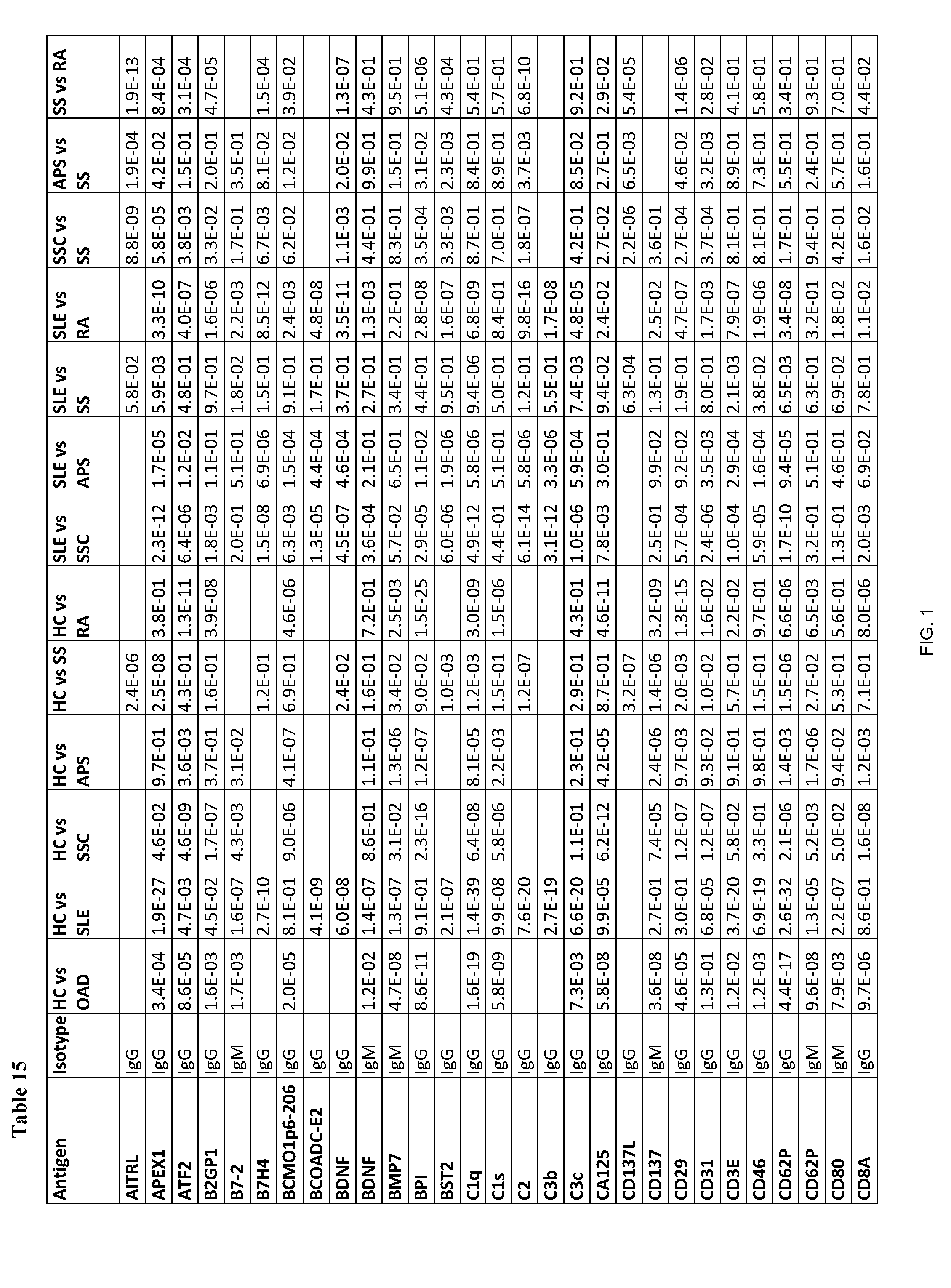
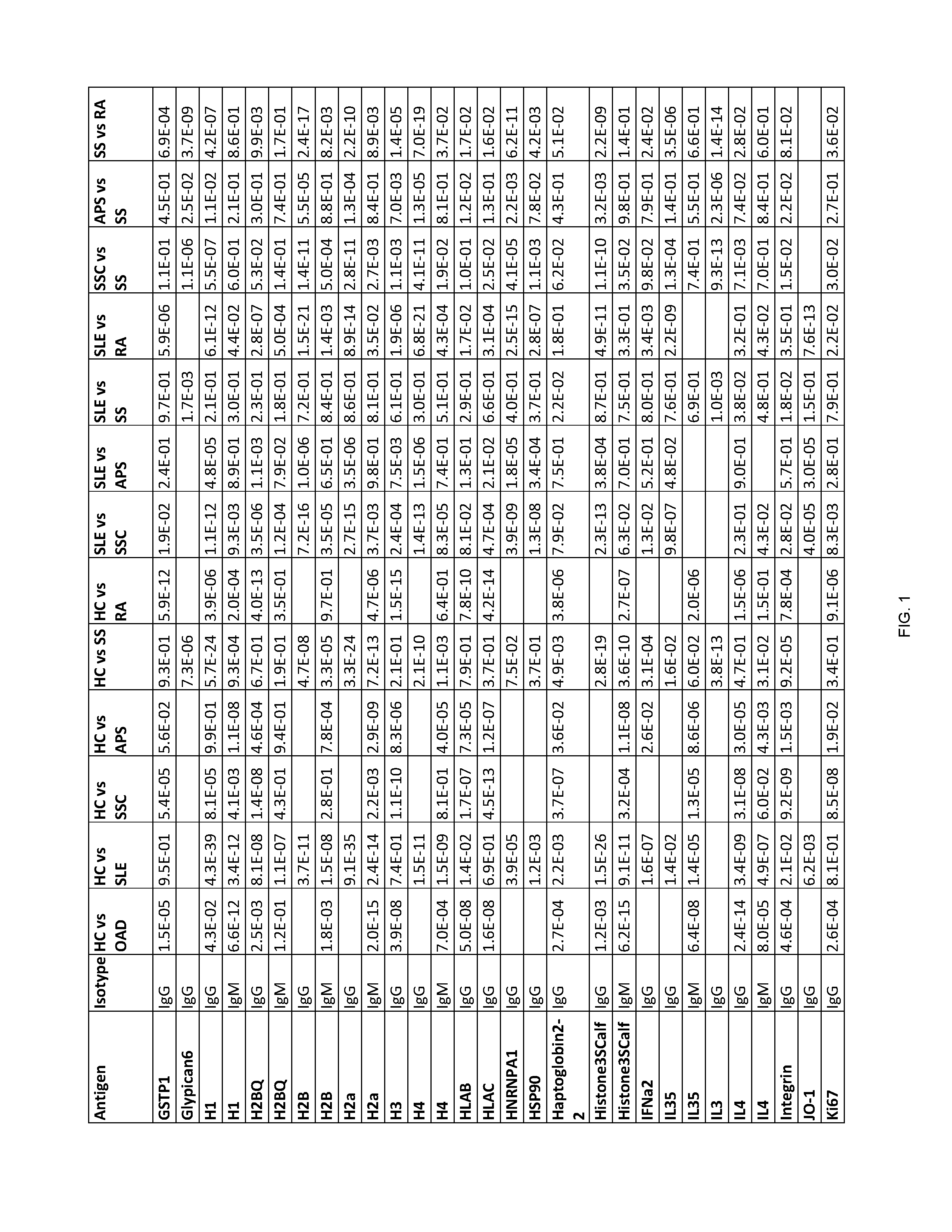
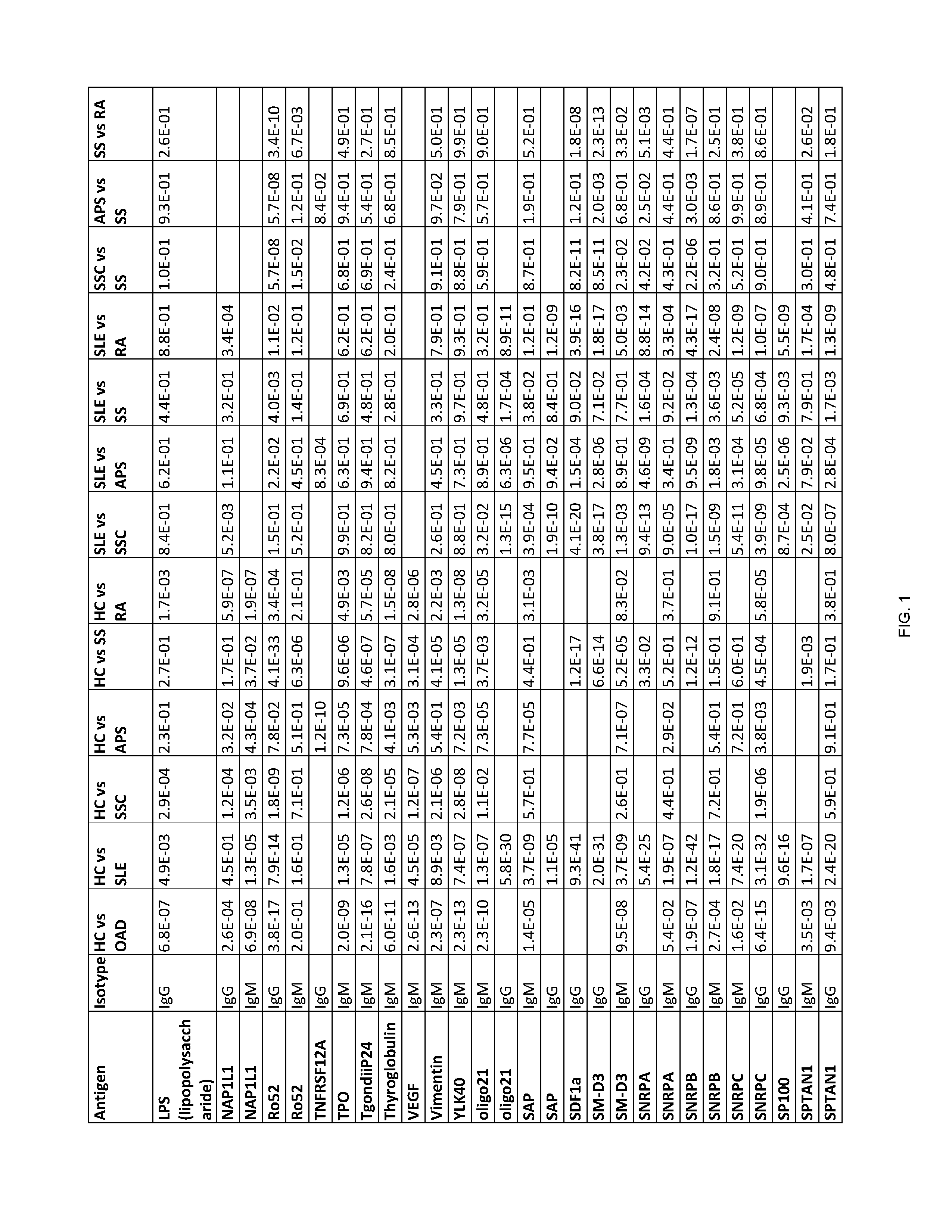
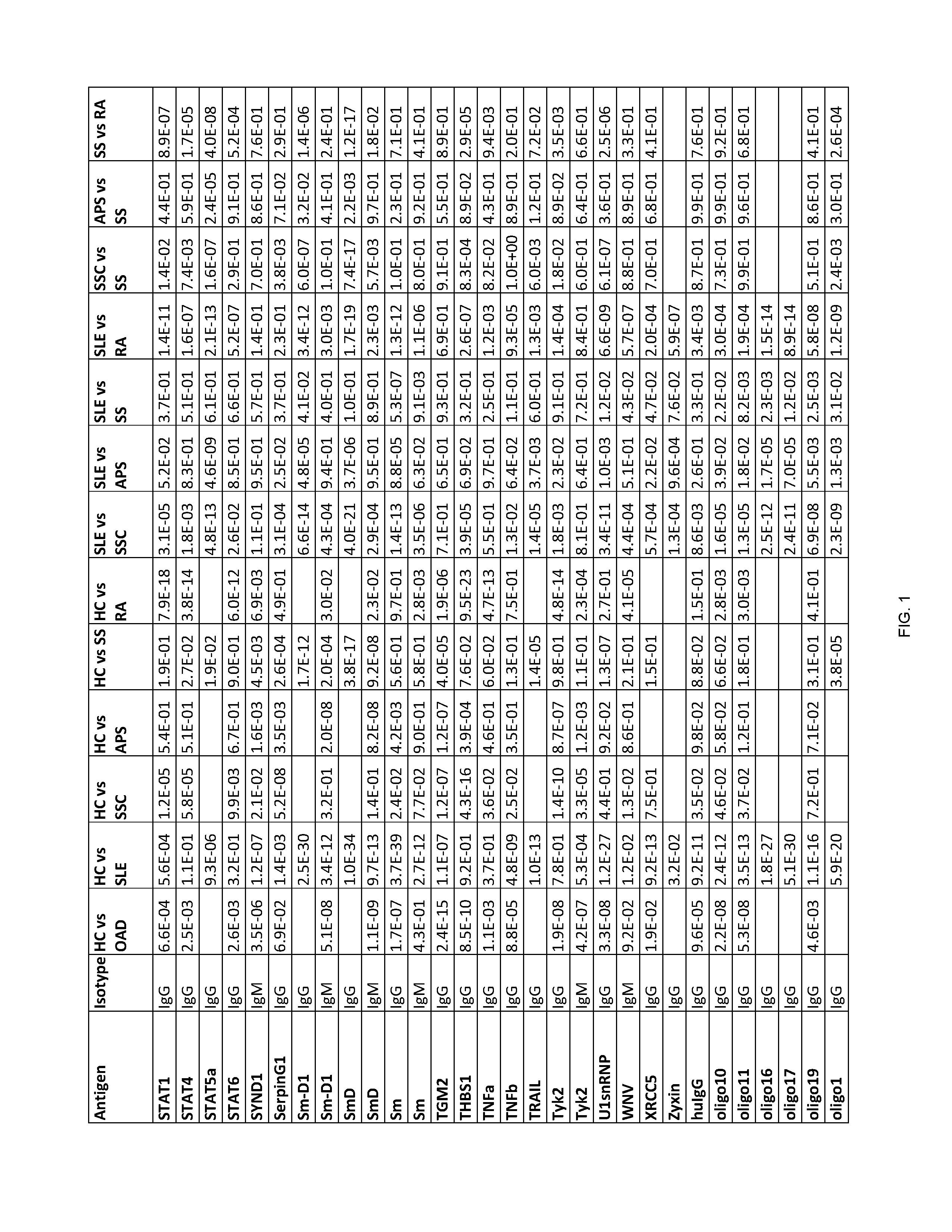
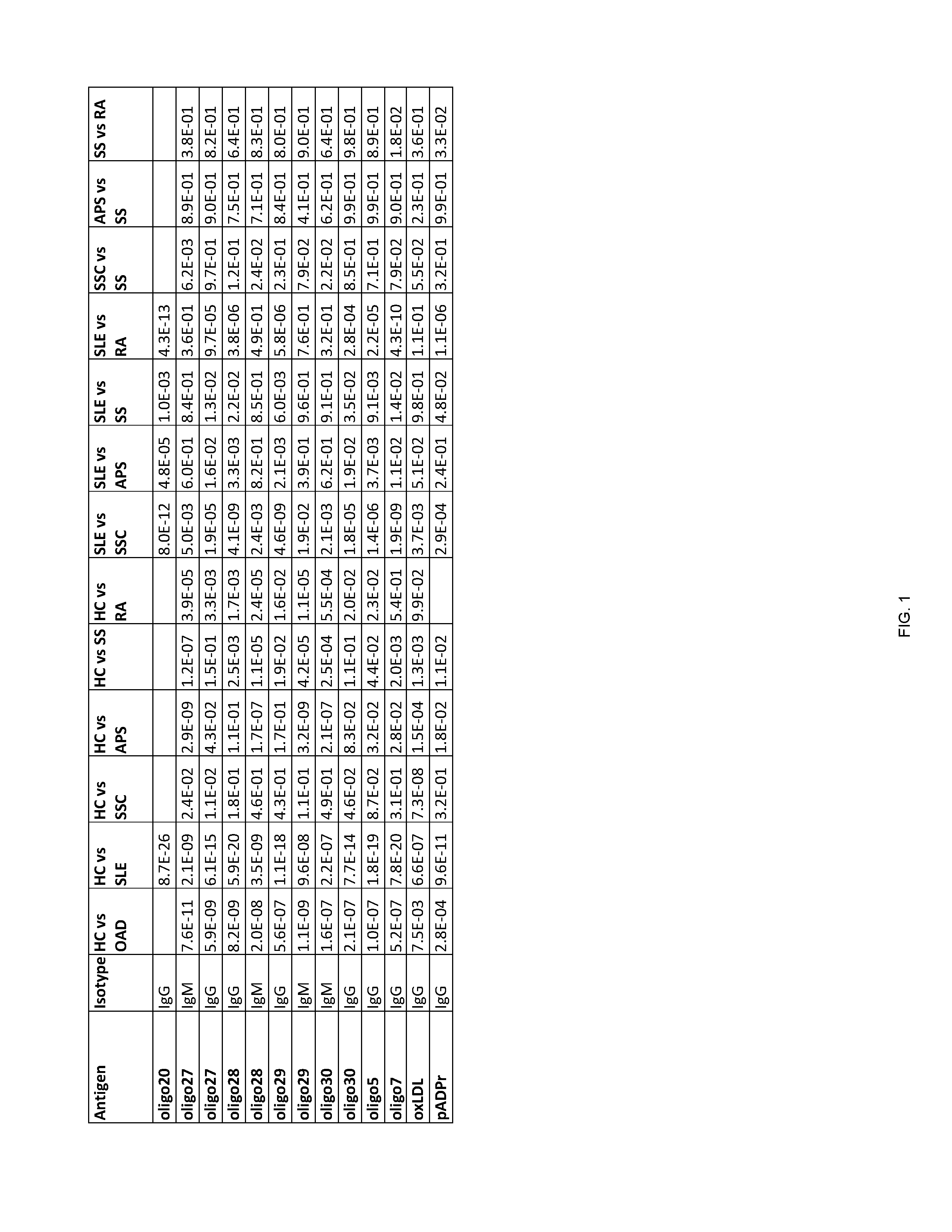
[0202] FIG. 1 shows Table 15, listing exemplary antigens to be used in embodiments of the invention.
DETAILED DESCRIPTION OF THE INVENTION
[0203] Provided are assays and methods for diagnosis of an autoimmune disease in a subject, and particularly identifying the specific autoimmune disease, namely systemic lupus erythematosus (SLE), rheumatoid arthritis (RA), scleroderma (SSc), Sjogren's syndrome (SS), or anti-phospholipid syndrome (APS) in a subject. As conventional methods, such as symptom-profiling, e.g., inflammation, joint pains, joint swelling, frequently correlate with a plurality of medically-relevant diseases and as such are not sufficient to identify a specific autoimmune disease, embodiments of the invention provide accurate means for such identification.
[0204] According to some embodiments of the invention, an antibody-containing sample is obtained from a subject in order to diagnose whether the subject has an autoimmune disease. Next, the antibodies are allowed to form complexes with a plurality of pre-determined antigens. The reactivity of the antibodies of the subject to the plurality of pre-determined antigens is then evaluated so as to form a reactivity pattern which is then compared to the reactivity of antibodies of healthy control subjects towards the same three antigens. The similarity and/or difference between the reactivity pattern of the subject's antibodies and the control antibodies towards the antigens is then used to diagnose whether the subject has an autoimmune disease. Exemplary antigens to be used in embodiments of the invention are listed in Table 15 herein.
[0205] Methods and assays constructed according to the principles of the invention further provide differential diagnosis between different autoimmune diseases such as SLE, SS, RA, SSc and APS. According to some embodiments of the invention, an antibody-containing sample is obtained from a subject in order to differentially diagnose between autoimmune diseases, such as between SLE and RA, or between SLE and SS, between SLE and SSc, between RA and SSc, between RA and SC, between SSc and APS, etc. Next, the antibodies are allowed to form complexes with at least three pre-determined antigens listed in the respective Tables 2-7. The reactivity of the antibodies of the subject to the antigens is then evaluated so as to form a reactivity pattern which is then compared to the reactivity of the antibodies of control samples, i.e. samples obtained from other subjects having a specific autoimmune disease, e.g., RA, SSc, SS or APS, towards the same antigens. The similarity and/or difference between the reactivity pattern of the subject's antibodies and the control antibodies towards the antigens is then used to differentially diagnose the subject. The antibodies being tested by the present methods can be of the IgG isotype or the IgM isotype, although any other isotype can be tested as well.
[0206] Comparing reactivity patterns can be performed using suitable classifiers or algorithms, including, but not limited to, learning and pattern recognition algorithms, supervised classifiers, and the like. A significant difference from control reactivities, such as from a healthy control as disclosed herein, may typically and conveniently be performed considering the respective values of both negative and positive control groups (e.g. healthy control subjects and autoimmune rheumatic diseased patients, respectively). The methods according to embodiments of the invention may include a step of determining the respective reactivity values to the test antigens in positive and/or negative control samples, or may employ comparison of the values measured in the test sample to the respective predetermined values or stored data. The test sample may thereby be classified as corresponding to (substantially similar to, or not substantially different from) either the positive or negative control group (e.g. health control or autoimmune disease), as disclosed herein. The positive and negative controls referred to herein typically and conveniently represent control sets, such as a panel of control samples from a set of similarly-diagnosed individuals, or a stored set of data obtained from similarly-diagnosed individuals.
[0207] It should therefore be noted that the step of identifying the subject as being afflicted with a specific disease, e.g. SLE, RA, SSc, SS, or APS can alternatively and/or additionally include a step of identifying the subject as having said disease, based on a significant similarity in the reactivity pattern of said sample compared to the reactivity pattern of an SLE, RA, SSc, SS, or APS control sample set. This can conveniently be done by detecting the similarity in the reactivity pattern of said sample compared to the reactivity pattern of a specific disease set (e.g. SLE, RA, SSc, SS, and/or APS).
[0208] In other embodiments of the invention, methods are provided for treatment of the diagnosed autoimmune disease including autoimmune rheumatic disorder. It is known that while certain therapeutic agents show some efficacy in a number of autoimmune rheumatic disorders, other therapeutic agents are specific for a particular disorder (e.g. RA, SLE or SS), and/or may be harmful when administered to subjects having other diseases. In addition, early diagnosis and initiation of therapy have proven to prevent disease progression and reduce morbidity. Additionally, certain dosage adjustments may be disease-specific, and accurate early diagnosis may ensure a fast onset of an effective treatment regimen.
[0209] According to some embodiments, the sample obtained from the subject can be an antibody-containing fluid sample, including, but not limited to, a blood derived sample (e.g. serum, plasma and whole blood). In certain embodiments, the sample may be processed prior to contacting with the antigens, e.g. by dilution, antibody purification and the like.
[0210] According to additional embodiments, the antigens are arranged on the surface of a solid support, typically a planar support such as a chemically activated glass support. In a particular embodiment, the support is planar glass support coated with an epoxy silane organic layer. Conveniently, the antigens are arranged in the form of an antigen array.
[0211] In other embodiments, the reactivity of the antibodies may be determined by various immunoassays enabling quantitative measurement of antigen-antibody binding, which may be subjected to image analysis and data analysis steps. For example, without limitation, images acquired by fluorescent scanning of fluorescently-labeled antibodies bound onto an antigen array may be pre-processed by methods including one or more of the following steps:
[0212] Determining representation of each spot through subtraction of background intensity from the foreground intensity; imputing negative values with low intensity spots, as negative spots has no meaning in the context of our platform; performing log transformation on each spot and combining spot replicates per array, wherein outliers spots may be removed based on Grubb's test; adjusting the mean per array per channel, wherein the mean per array may be calculated based on a set of antigens per channel, and the mean may then be adjusted against a constant value and the correction is applied to all antigens; and correcting for print lot and test session effect using reference sera, wherein the correction may be performed per test session per print lot, by subtraction of reference serum intensities from corresponding antigens in order to express all intensities as relative to the reference serum.
[0213] Advantageously, methods according to embodiments of the invention may employ the use of learning and pattern recognition analyzers, clustering algorithms and the like, in order to discriminate between reactivity patterns of control subjects to control sets of patients having autoimmune diseases such as RA, SLE, SSC, SS or APS. As such, this term specifically includes a difference measured by, for example, determining the reactivity of antibodies in a test sample to a plurality of antigens, and comparing the resulting reactivity pattern to the reactivity patterns of negative and positive control samples (e.g. samples obtained from healthy control subjects or patients afflicted with a specific disease, respectively) using such algorithms and/or analyzers. The difference may also be measured by comparing the reactivity pattern of the test sample to a predetermined classification rule obtained in such manner.
[0214] According to one aspect of the invention, methods are provided for detecting an autoimmune disease in a subject, the method including: [0215] i. obtaining a sample from the subject; [0216] ii. contacting the sample with a plurality of antigens selected from Table 1, under conditions such that a specific antigen-antibody complex may be formed for each antigen, and quantifying the amount of antigen-antibody complex formed for each antigen to generate a reactivity pattern of said sample to the plurality of antigens; [0217] iii. comparing the reactivity pattern of said sample to a healthy control reactivity pattern to said plurality of antigens by a supervised classification algorithm, and [0218] iv. detecting the autoimmune disease by quantifying whether the reactivity pattern of the sample obtained from the subject is significantly different from the healthy control reactivity pattern, wherein a significantly different reactivity pattern of the sample obtained from the subject compared to the healthy control reactivity pattern indicates that said subject has an autoimmune rheumatic disease.
[0219] According to some embodiments, the autoimmune disease to be diagnosed is selected from the group consisting of SLE, RA, SSc, SS, and APS.
[0220] According to additional embodiments, if the subject has been diagnosed by the above method as having an autoimmune disease, the method can further include identifying the autoimmune disease in said subject by the steps including: [0221] v. determining the reactivity of antibodies in the sample obtained from the subject to a plurality of antigens selected from at least one of Tables 8-12, thereby obtaining a reactivity pattern of said sample to the plurality of antigens; [0222] vi. comparing the reactivity pattern of said sample to a healthy control reactivity pattern to said plurality of antigens, by a supervised classification algorithm, [0223] wherein: [0224] a significantly different reactivity pattern of the sample obtained from the subject to a plurality of antigens selected from Table 8 compared to the healthy control reactivity pattern indicates that said subject has SLE, [0225] a significantly different reactivity pattern of the sample obtained from the subject to a plurality of antigens selected from Table 9 compared to the healthy control reactivity pattern indicates that said subject has SSc, [0226] a significantly different reactivity pattern of the sample obtained from the subject to a plurality of antigens selected from Table 10 compared to the healthy control reactivity pattern indicates that said subject has APS, [0227] a significantly different reactivity pattern of the sample obtained from the subject to a plurality of antigens selected from Table 11 compared to the healthy control reactivity pattern indicates that said subject has SS, and/or [0228] a significantly different reactivity pattern of the sample obtained from the subject to a plurality of antigens selected from Table 12 compared to the healthy control reactivity pattern indicates that said subject has RA.
[0229] According to another aspect of the invention, methods are provided for detecting an autoimmune disease selected from the group consisting of SLE, RA, SSc, SS, and APS in a subject, the method may include: [0230] i. obtaining a sample from a subject suspected of having an autoimmune rheumatic disease; [0231] ii. contacting the sample with a plurality of antigens selected from at least one of Tables 8-12 under conditions such that a specific antigen-antibody complex may be formed for each antigen, and quantifying the amount of antigen-antibody complex formed for each antigen to generate a reactivity pattern of said sample to the plurality of antigens; [0232] iii. comparing the reactivity pattern of said sample to a healthy control reactivity pattern to said plurality of antigens, by a supervised classification algorithm, and [0233] iv. detecting the autoimmune disease by quantifying whether the reactivity pattern of the sample obtained from the subject is significantly different from the healthy control reactivity pattern [0234] wherein: [0235] a significantly different reactivity pattern of the sample obtained from the subject to a plurality of antigens selected from Table 8 compared to the healthy control reactivity pattern indicates that said subject has SLE, [0236] a significantly different reactivity pattern of the sample obtained from the subject to a plurality of antigens selected from Table 9 compared to the healthy control reactivity pattern indicates that said subject has SSc, [0237] a significantly different reactivity pattern of the sample obtained from the subject to a plurality of antigens selected from Table 10 compared to the healthy control reactivity pattern indicates that said subject has APS, [0238] a significantly different reactivity pattern of the sample obtained from the subject to a plurality of antigens selected from Table 11 compared to the healthy control reactivity pattern indicates that said subject has SS, and/or [0239] a significantly different reactivity pattern of the sample obtained from the subject to a plurality of antigens selected from Table 12 compared to the healthy control reactivity pattern indicates that said subject has RA.
[0240] According to another aspect of the invention, methods are provided for detecting an autoimmune disease, including: [0241] (v) obtaining a sample from a subject diagnosed as having the autoimmune disease; [0242] (vi) contacting the sample with antigens selected from at least one of Tables 2-7, under conditions such that a specific antigen-antibody complex is formed for each antigen, and quantifying the amount of antigen-antibody complex formed for each antigen to generate a reactivity pattern of said sample to the plurality of antigens; [0243] (vii) comparing the reactivity pattern of said sample obtained from the subject to a control reactivity pattern to said antigens by a supervised classification algorithm, and [0244] (viii) detecting the autoimmune disease by quantifying whether the reactivity pattern of the sample obtained from the subject is significantly different from the control reactivity pattern, [0245] wherein: [0246] a significantly different reactivity pattern of the sample obtained from the subject to a plurality of antigens selected from Table 2 compared to a RA control reactivity pattern to said antigens indicates that said subject has SLE, [0247] a significantly different reactivity pattern of the sample obtained from said subject to a plurality of antigens selected from Table 2 compared to a SLE control reactivity pattern to said antigens indicates that said subject has RA, [0248] a significantly different reactivity pattern of the sample obtained from said subject to a plurality of antigens selected from Table 3 compared to a SSc control reactivity pattern to said antigens indicates that said subject has SLE, [0249] a significantly different reactivity pattern of the sample obtained from said subject to a plurality of antigens selected from Table 3 compared to a SLE control reactivity pattern to said antigens indicates that said subject has SSc, [0250] a significantly different reactivity pattern of the sample obtained from said subject to a plurality of antigens selected from Table 4 compared to an APS control reactivity pattern to said antigens indicates that said subject has SLE, [0251] a significantly different reactivity pattern of the sample obtained from said subject to a plurality of antigens selected from Table 4 compared to an SLE control reactivity pattern to said antigens indicates that said subject has APS, [0252] a significantly different reactivity pattern of the sample obtained from said subject to a plurality of antigens selected from Table 5 compared to a SSc control reactivity pattern to said antigens indicates that said subject has SS, [0253] a significantly different reactivity pattern of the sample obtained from said subject to a plurality of antigens selected from Table 5 compared to a SS control reactivity pattern to said antigens indicates that said subject has SSc, [0254] a significantly different reactivity pattern of the sample obtained from said subject to a plurality of antigens selected from Table 6 compared to a RA control reactivity pattern to said antigens indicates that said subject has SS, and/or [0255] a significantly different reactivity pattern of the sample obtained from said subject to a plurality of antigens selected from Table 6 compared to a SS control reactivity pattern indicates that said subject has RA. [0256] a significantly different reactivity pattern of the sample obtained from said subject to a plurality of antigens selected from Table 7 compared to a SS control reactivity pattern to said antigens indicates that said subject has SLE, and/or [0257] a significantly different reactivity pattern of the sample obtained from said subject to a plurality of antigens selected from Table 7 compared to a SLE control reactivity pattern indicates that said subject has SS.
Subjects and Autoimmune Diseases
[0258] Subjects to be diagnosed by the methods according to the principles of the invention are suspected of having an autoimmune disease, such as an autoimmune rheumatic disorder (ARD).
[0259] SLE patients usually display at least one, and in the majority of cases several of the following symptoms: painful, pale or purple fingers or toes triggered by cold temperature or emotional stress (Raynaud's phenomenon), joint pain, joint stiffness, joint swelling, joint redness, joint tenderness, joint warmth, loss of joint range of motion, fatigue, muscle pain, vasculitis (inflammation of blood vessels), dry eyes, dry mouth, and inflammation (determined by C-reactive protein (CRP), erythrocyte sedimentation rate (ESR) tests).
[0260] RA patients usually display at least one, and in the majority of cases several of the following symptoms: painful, pale or purple fingers or toes triggered by cold temperature or emotional stress (Raynaud's phenomenon), joint pain, joint stiffness, joint swelling, joint redness, joint tenderness, joint warmth, loss of joint range of motion, fatigue, vasculitis (inflammation of blood vessels), dry eyes, dry mouth, inflammation (determined by C-reactive protein (CRP), erythrocyte sedimentation rate (ESR) tests), fever, anemia, and shortness of breath/lung involvement.
[0261] SS patients usually display at least one, and in the majority of cases several, of the following symptoms: painful, pale or purple fingers or toes triggered by cold temperature or emotional stress (Raynaud's phenomenon), joint pain, joint stiffness, joint swelling, joint redness, joint tenderness, joint warmth, loss of joint range of motion, fatigue, muscle pain, vasculitis (inflammation of blood vessels), dry eyes, dry mouth, fever, anemia, shortness of breath/lung involvement and inflammation (determined by C-reactive protein (CRP), erythrocyte sedimentation rate (ESR) tests).
[0262] SSc patients display one or more of the following symptoms: skin thickening, stiffness, poor blood flow to the fingers or toes with cold exposure, fatigue, areas of dilated small blood vessels, and weight loss.
[0263] APS patients display one or more of the following symptoms: blood clots (thrombosis) in arteries and/or veins, pregnancy-related complications such as miscarriage, stillbirth, preterm delivery, and severe preeclampsia.
[0264] According to embodiments of the invention, the patient population to be diagnosed and optionally treated by the methods of the invention includes subjects displaying inflammation and optionally at least one rheumatic symptom, e.g. joint pain, joint stiffness, joint swelling, joint redness, joint tenderness, joint warmth, and loss of joint range of motion. According to particular embodiments, the methods include a step of obtaining or deriving a sample from a subject suspected of having an autoimmune rheumatic disease due to inflammation and at least one rheumatic symptom selected from the group consisting of: joint pain, joint stiffness, joint swelling, joint redness, joint tenderness, joint warmth, and loss of joint range of motion. The methods of the invention allow for early and effective diagnosis and selection of appropriate treatment for a subject while minimizing undue trial and error, associated with existing methods based on e.g. clinical symptom evaluation.
Antibodies, Samples and Immunoassays
[0265] In some embodiments of the invention, the methods are preceded by a step of obtaining a sample from the subject. In certain embodiments, the sample is obtained from the subject by non-invasive means or methods.
[0266] According to additional embodiments of the invention, the sample obtained from the subject is a biological fluid. According to some embodiments, the sample is selected from the group consisting of plasma, serum, blood, cerebrospinal fluid, synovial fluid, sputum, urine, saliva, tears, lymph specimen, or any other antigen-containing biological fluid known in the art. Each possibility represents a separate embodiment of the invention. According to one embodiment, the sample is a serum sample. In certain embodiments, the sample is obtained or derived from the subject by non-invasive means or methods.
[0267] According to some embodiments of the invention, the control sample is selected from the group consisting of a sample from at least one healthy individual, a panel of control samples from a set of healthy individuals, and a stored set of data obtained from healthy individuals. Each possibility represents a separate embodiment of the invention.
[0268] According to additional embodiments of the invention, the control sample is selected from the group consisting of a sample from at least one diagnosed individual, a panel of control samples from a set of similarly-diagnosed individuals, and a stored set of data obtained from similarly-diagnosed individuals. Each possibility represents a separate embodiment of the invention. Typically, a diagnosed individual is a subject afflicted with an autoimmune disease such as SLE (or any other form of lupus), RA, SSc, APS or SS.
[0269] According to further embodiments, the method comprises determining the reactivity of antibodies in the sample to a plurality of antigens (e.g. peptide, protein, lipid and/or nucleotide antigens as disclosed herein). According to some embodiments, the plurality of antigens is in one of the following forms: an antigen probe set, an antigen probe array, an antigen chip, dipstick, a lateral flow test, an ELISA plate, or in any other form known to those skilled in the art.
[0270] In certain embodiments, the plurality of antigens is in the form of a kit which further includes means for determining the reactivity of antibodies in a sample to a plurality of antigens. According to some embodiments, the kit further includes means for comparing reactivity of antibodies in different samples to a plurality of antigens. According to another embodiment, the kit further includes instructions for use of the kit for diagnosing whether a subject is afflicted with an autoimmune disease such as an ARD or for differentially diagnosing a specific autoimmune disease, e.g., SLE, RA, SSc, SS or APS.
[0271] According to another aspect, there is provided use of plurality of antigens described above for the preparation of a diagnostic kit for diagnosing whether a subject has an autoimmune disease, or for differentially diagnosing SLE, SS, RA, APS or SSc in a subject. Each possibility represents a separate embodiment of the invention. The diagnostic kit is, in some embodiments, useful for determining the reactivity of antibodies in a sample, thereby determining the reactivity pattern of the sample to plurality of antigens.
[0272] As used herein, the "reactivity of antibodies in a sample to a plurality of antigens" refers to the immune reactivity of each antibody in the sample to at least three specific antigens selected from a plurality of antigens. The immune reactivity of the antibody to the antigen, i.e. its ability to specifically bind the antigen, can be used to determine the amount of the antibody in the sample. The calculated levels of each one of the tested antibodies in the sample are selectively referred to as the reactivity pattern of the sample to these antigens.
[0273] An antibody "directed to" an antigen, as used herein is an antibody which is capable of specifically binding the antigen.
[0274] It should be understood that when the terms "antibody" or "antibodies" are used, this is intended to include intact antibodies, such as polyclonal antibodies or monoclonal antibodies (mAbs), as well as proteolytic fragments thereof such as the Fab or F(ab')2 fragments. Further included within the scope of the invention are chimeric antibodies; recombinant and engineered antibodies, and fragments thereof.
[0275] Exemplary functional antibody fragments comprising whole or essentially whole variable regions of both light and heavy chains are defined as follows:
[0276] (i) Fv, defined as a genetically engineered fragment consisting of the variable region of the light chain and the variable region of the heavy chain expressed as two chains;
[0277] (ii) single-chain Fv ("scFv"), a genetically engineered single-chain molecule including the variable region of the light chain and the variable region of the heavy chain, linked by a suitable polypeptide linker.
[0278] (iii) Fab, a fragment of an antibody molecule containing a monovalent antigen-binding portion of an antibody molecule, obtained by treating whole antibody with the enzyme papain to yield the intact light chain and the Fd fragment of the heavy chain, which consists of the variable and CH1 domains thereof;
[0279] (iv) Fab', a fragment of an antibody molecule containing a monovalent antigen-binding portion of an antibody molecule, obtained by treating whole antibody with the enzyme pepsin, followed by reduction (two Fab' fragments are obtained per antibody molecule); and
[0280] (v) F(ab')2, a fragment of an antibody molecule containing a monovalent antigen-binding portion of an antibody molecule, obtained by treating whole antibody with the enzyme pepsin (i.e., a dimer of Fab' fragments held together by two disulfide bonds).
[0281] The term "antigen" as used herein is a molecule or a portion of a molecule capable of being bound by an antibody. The antigen is typically capable of inducing an animal to produce antibody capable of binding to an epitope of that antigen. An antigen may have one or more epitopes. The specific reaction referred to above is meant to indicate that the antigen will react, in a highly selective manner, with its corresponding antibody and not with the multitude of other antibodies which may be evoked by other antigens. An "antigenic peptide" or an "antigenic oligonucleotide" is a peptide or an oligonucleotide, respectively, which is capable of specifically binding an antibody.
[0282] In some embodiments, detection of the capacity of an antibody to specifically bind an antigen probe may be performed by quantifying specific antigen-antibody complex formation. The term "specifically bind" as used herein means that the binding of an antibody to an antigen probe is not competitively inhibited by the presence of non-related molecules.
[0283] In accordance with the principles of the invention, any suitable immunoassay can be used with the antigens. Such techniques are well known to the ordinarily skilled artisan and have been described in many standard immunology manuals and texts. In certain preferable embodiments, determining the capacity of the antibodies to specifically bind the antigen probes is performed using an antigen probe array-based method. Preferably, the array is incubated with suitably diluted serum sample of the subject (e.g. diluted 1:10) so as to allow specific binding between the antibodies contained in the diluted serum sample and the immobilized antigen probes, washing out unbound serum from the array, incubating the washed array with a detectable label-conjugated antibodies of the desired isotype, washing out unbound label from the array, and measuring levels of the label bound to each antigen probe.
[0284] In other embodiments, various immunoassays may be used, including, without limitation, enzyme-linked immunosorbent assay (ELISA), flow cytometry with multiplex beads (such as the system made by Luminex), surface plasmon resonance (SPR), elipsometry, and various other immunoassays which employ, for example, laser scanning, light detecting, photon detecting via a photo-multiplier, photographing with a digital camera based system or video system, radiation counting, fluorescence detecting, magnetic detecting and any other system that allows quantitative measurement of antigen-antibody binding.
[0285] Various methods have been developed for preparing arrays suitable for the methods of the invention. State-of-the-art methods involve using a robotic apparatus to apply or "spot" distinct solutions containing antigen probes to closely spaced specific addressable locations on the surface of a planar support, typically a glass support, such as a microscope slide, which is subsequently processed by suitable thermal and/or chemical treatment to attach antigen probes to the surface of the support. Suitable supports may also include silicon, nitrocellulose, paper, cellulosic supports and the like.
[0286] Preferably, each antigen probe, or distinct subset of antigen probes that may be used in embodiments of the invention, which is attached to a specific addressable location of the array, is attached independently to at least two, more preferably to at least three separate specific addressable locations of the array in order to enable generation of statistically robust data.
[0287] In addition to antigen probes, the array may advantageously include control antigen probes or other standard chemicals. Such control antigen probes may include normalization control probes. The signals obtained from the normalization control probes provide a control for variations in binding conditions, label intensity, "reading" efficiency and other factors that may cause the signal of a given antibody-antigen probe interaction to vary. For example, signals, such as fluorescence intensity, read from all other antigen probes of the antigen probe array are divided or subtracted or shifted according to difference in mean intensities by the signal (e.g., fluorescence intensity) from the normalization control probes thereby normalizing the measurements. Normalization control probes can be bound to various addressable locations on the antigen probe array to control for spatial variation in antibody-antigen probe efficiency. Normalization control probes can be located at the corners or edges of the array to control for edge effects, as well as in the middle of the array.
[0288] The labeled antibodies or the ligand antibodies or the secondary antibodies may be of any of various suitable types of antibodies. Preferably, the secondary antibody is an antibody which is capable of specifically binding the Fc portion of the antibodies of the subject used. For example, where the antibodies of the subject are of the IgG isotype, the secondary antibody is preferably an antibody capable of specifically binding to the Fc region of IgG antibodies of the subject.
[0289] The secondary antibodies may be conjugated to any of various types of detectable labels. Preferably the label is a fluorophore, most preferably Cy3. Alternately, the fluorophore may be any of various fluorophores, including Cy5, fluorescein isothiocyanate (FITC), phycoerythrin (PE), rhodamine, Texas red, and the like. Suitable fluorophore-conjugated antibodies specific for antibodies of a specific isotype are widely available from commercial suppliers and methods of their production are well established.
[0290] Antibodies of the subject may be isolated for analysis of their antigen probe binding capacity in any of various ways, depending on the application and purpose. While the subject's antibodies may be suitably and conveniently in the form of blood serum or plasma or a dilution thereof (e.g., 1:10 dilution), the antibodies may be subjected to any desired degree of purification prior to being tested for their capacity to specifically bind antigen probes. The method of the invention may be practiced using whole antibodies of the subject, or antibody fragments of the subject which comprises an antibody variable region.
Diagnostic Methods
[0291] As disclosed herein, diagnostic methods according to the invention may generally include: [0292] i. obtaining a sample from a subject; [0293] ii. determining the reactivity of antibodies in the sample obtained from said subject to a plurality of antigens comprising one or more antigens selected from a Table as disclosed herein, thereby obtaining a reactivity pattern of said sample to the plurality of antigens;
[0294] comparing the reactivity pattern of said sample to a control reactivity pattern to said plurality of antigens by a supervised classification algorithm.
[0295] In various exemplary embodiments, Tables 1, 16 and 30 disclose antigens for differentiating between autoimmune rheumatic diseased patients and healthy control (HC) subjects; Tables 2, 25 and 31 disclose antigens for differentiating between SLE patients and RA patients; Tables 3, 22 and 32 disclose antigens for differentiating between SLE patients and SSc patients; Tables 4 and 23 disclose antigens for differentiating between SLE patients and APS patients; Tables 5 and 26 discloses antigens for differentiating between SS patients and SSc patients; Tables 6 and 28 disclose antigens for differentiating between SS patients and RA patients; Tables 7 and 24 disclose antigens for differentiating between SLE patients and SS patients; Tables 8, 17 and 34 disclose antigens for differentiating between SLE patients and HC subjects; Tables 9 and 18 disclose antigens for differentiating between SSc patients and HC subjects; Tables 10 and 19 disclose antigens for differentiating between APS patients and HC subjects; Tables 11 and 20 disclose antigens for differentiating between SS patients and HC subjects; Tables 12 and 21 disclose antigens for differentiating between RA patients and HC subjects; Table 27 disclose antigen for differentiating between. APS and SS patients; and Table 33 disclose antigen for differentiating between SSc patients and RA patients.
[0296] According to certain embodiments, advantageous antigens for differentiating between autoimmune rheumatic diseased patients and HC subjects include LAIR1, MICA, RO60calf, CD62P, Ferritin, Chromatin, and CENPB. In other embodiments, advantageous antigens for differentiating between SLE patients and RA patients include DLST, RNPsm, RO60calf, H1 and RPP0.
[0297] In a particular embodiment, the plurality of antigens used in methods of the invention include LAIR1. In another embodiment, said plurality of antigens do not include LAIR1.
Methods of Treatment
[0298] The invention provides methods for treating a subject diagnosed as having a specific autoimmune disease including ARD with at least one autoimmune disease-specific treatment. Exemplary methods of the invention allow for early and effective selection of appropriate treatment for a subject suspected of having an ARD, while minimizing undue trial and error, thereby minimizing both suffering for the patient and therapeutic costs alike.
[0299] In some embodiments, the SLE-specific treatment is selected from the group consisting of NSAIDs, Ibuprofen, Naproxen, Prednisone, Methylprednisolone (A-Methapred, Medrol, Solu-Medrol, Depo-Medrol), Cellcept, Methotrexate, Imuran, mycophenolate mofetil, Abatacept, Arava, Immune globulin intravenous (Hizentra, Gammagard, Octagam, Privigen), Plaquenil, Myfortic, Endoxan, Cytoxan, Neosar, Procytox, Revimmune, Benilimumab (Benlysta), Rituximab, Cyclosporine (Gengraf, Neoral, Sandimmune).
[0300] In additional embodiments, the RA-specific treatment is selected from the group consisting of Etanercept (Enbrel.RTM.), Adalimumab (Humira.RTM.), Infliximab (Remicade.RTM.), Certolizumab pegol (Cimzia.RTM.), Golimumab (Simponi.RTM.), Anakinra (Kineret.RTM.), Tocilizumab (Actemra.RTM.), Tofacitinib (Xeljanz.RTM.), and any combination thereof. Each possibility represents a separate embodiment of the invention.
[0301] In further embodiments, the SS-specific treatment is selected from the group consisting of pilocarpine (Salagen.RTM.), cevimeline (Evoxac.RTM.), cyclosporine (Restasis.RTM.), and any combination thereof. Each possibility represents a separate embodiment of the invention.
[0302] In yet further embodiments, the SSc-specific treatment is selected from the group consisting of Ibuprofen, Naproxen, Prednisone, Cellcept, Methotrexate, Imuran, Cytoxan, Rituximab, Penicillamine (Cuprimine, Depen), Bosentan (Tracleer), Ambrisentan (Letairis), Macitentan (Opsumit), Tadalafil (Adcirca), Sildenafil (Revatio).
[0303] In still further embodiments, the APS-specific treatment is selected from the group consisting of warfarin, rivaroxaban (Xarelto), dabigatran (Pradaxa), apixaban (Eliquis), edoxaban (Lixiana), heparin, aspirin, clopidogrel, Rituximab.
[0304] Doses and treatment regimens for disease-specific treatments e.g. as listed above are known in the art and may be determined and adjusted by the skilled artisan (e.g. treating physician) according to the patient's characteristics and disease manifestations. For example,
[0305] NSAIDs inhibit the generation of prostaglandins by blocking cyclooxygenase enzymes, COX-1 and COX-2. The major effect of these agents is to reduce acute inflammation thereby decreasing pain and improving function. All of these drugs also have mild to moderate analgesic properties independent of their anti-inflammatory effect. There are a large number of NSAIDs, some over the counter including ibuprofen (Advil.RTM., Motrin.RTM., Nuprin.RTM.) and naproxen (Alleve.RTM.) and many others are available by prescription including meloxicam (Mobic.RTM.), etodolac (Lodine.RTM.), nabumetone (Relafen.RTM.), sulindac (Clinoril.RTM.), tolementin (Tolectin.RTM.), choline magnesium salicylate (Trilasate.RTM.), diclofenac (Cataflam.RTM., Voltaren.RTM., Arthrotec.RTM.), diflusinal (Dolobid.RTM.), indomethacin (Indocin.RTM.), ketoprofen (Orudis.RTM., Oruvail.RTM.), meloxicam (Mobic.RTM.), oxaprozin (Daypro.RTM.), and piroxicam (Feldene.RTM.). Longer acting NSAIDs that allow daily or twice daily dosing may improve compliance. The NSAID class also includes drugs known as COX-2 inhibitors that are also effective in controlling inflammation, e.g. celecoxib, Celebrex.RTM.; etoricoxib, Arcoxia.RTM.; lumiracoxib, Prexige.RTM.. These drugs were designed to decrease the gastrointestinal risk of NSAIDS, but concerns of possible increases in cardiovascular risk with these agents has led to the withdrawal of two of these drugs from the market (rofecoxib, Vioxx.RTM.; valdecoxib, Bextra.RTM.). NSAID doses for the treatment of SLE are known in the art. For example, ibuprofen may be used as needed or in doses up to 3000 mg a day, and naproxen is typically used as 500 mg twice a day.
[0306] Corticosteroids (such as prednisone; methylprednisolone, Medrol.RTM.) have both anti-inflammatory and immunoregulatory activity. They can be given orally, intravenously, intramuscularly or can be injected directly into the joint. In mild SLE, prednisolone is given in doses starting at 0.1-0.3 mg/kg/day followed by a gradual tapering dose regimen according to clinical response. The dose rises to 0.4-0.6 mg/kg/day in moderate disease and as high as 0.7-1.5 mg/kg/day in very severe disease. At such high doses, pulse therapy with intravenous (IV) methylprednisolone (MP; 500-1000 mg on one to three occasions) is deemed by many physicians to be safer with fewer associated side effects. IV therapy is considered in patients that have not responded to oral therapy and/or have serious manifestations of SLE such as lupus nephritis, neuropsychiatric disease, severe refractory thrombocytopenia, hemolytic anemia, severe vasculitis and cardiopulmonary disease.
[0307] Azathioprine (Imuran.RTM.) is an immunosuppressive agent commonly used for the induction of remission and as a steroid-sparing agent in mild-to-moderate disease. It works by affecting cell-mediated and humoral immune responses via the inhibition of lymphocyte proliferation, reduction in antibody production and suppression of natural killer cell activity. In severe disease, it is used as maintenance therapy and data from lupus nephritis trials show significant improvement in disease activity following induction therapy with cyclophosphamide or mycophenolate mofetil.
[0308] Other biological agents and immunomodulators include for example monoclonal antibodies targeting several surface molecules on B cells, to reduce the formation of autoantibodies. Such exemplary drugs include rituximab (anti-CD20), ocrelizumab (humanized anti-CD20), belimumab (anti-BAFF/BLyS), atacicept (anti-BLys/APRIL) and epratuzumab (humanized anti-CD22). In addition, other key cell-surface markers have been developed to interfere with costimulatory molecules such as cytotoxic T lymphocyte antigen 4 (abatacept). In addition, Leflunomide (original brand name Arava) is an immunosuppressive pyrimidine synthesis inhibitor that works by inhibiting dihydroorotate dehydrogenase. Leflunomide is an immunomodulatory drug that inhibits the reproduction of rapidly dividing cells, especially lymphocytes. In addition, TNF-.alpha. inhibitors, e.g. Etanercept or Infliximab, may be used in some cases.
[0309] Exemplary treatment schedules for rheumatoid arthritis include 50 mg/week s.c. Etanercept, 40 mg every other week s.c. Adalimumab, 3 mg/kg-10 mg/kg i.v. for 2-3 hrs. every 4-8 weeks Infliximab, 200 mg-400 mg every 2-4 weeks Certolizumab Pegol, 50 mg/month Golimumab, and 4 mg/kg or 8 mg/kg/month Tocilizumab.
[0310] In another embodiment, a method for determining a disease-specific treatment for a subject comprises:
[0311] diagnosing the subject as having an autoimmune disease selected from the group consisting of SLE, RA, SSc, SS, and APS, by a method as disclosed herein, and
[0312] determining that said subject is amenable for receiving a treatment specific to said autoimmune disease. In various embodiments, said treatment is selected from the SLE-specific, RA-specific, SSc-specific, SS-specific, and APS-specific treatments as disclosed herein.
Data Analysis
[0313] Advantageously, the methods of the invention can employ the use of learning and pattern recognition analyzers, clustering algorithms and the like, in order to discriminate between reactivity patterns of healthy subjects and patients having different autoimmune diseases including ARDs. For example, the methods can comprise determining the reactivity of antibodies in a test sample to a plurality of antigens, and comparing the resulting pattern to the reactivity patterns of negative and positive control samples using such algorithms and/or analyzers.
[0314] In certain embodiments, one or more algorithms or computer programs may be used for comparing the amount of each antibody quantified in the test sample against a predetermined cutoff (or against a number of predetermined cutoffs). Alternatively, one or more instructions for manually performing the necessary steps by a human can be provided.
[0315] Algorithms for determining and comparing pattern analysis include, but are not limited to, Support Vector Machine (SVM), Quadratic Discriminant Analysis (QDA), Naive Bayesian Classifier (NBC), principal component analysis, Fischer linear analysis, neural network algorithms, genetic algorithms, fuzzy logic pattern recognition, and the like. After analysis is completed, the resulting information can, for example, be displayed on a display, transmitted to a host computer, or stored on a storage device for subsequent retrieval.
[0316] Many of the algorithms are neural network based algorithms. A neural network has an input layer, processing layers and an output layer. The information in a neural network is distributed throughout the processing layers. The processing layers are made up of nodes that simulate the neurons by the interconnection to their nodes. Similar to statistical analysis revealing underlying patterns in a collection of data, neural networks locate consistent patterns in a collection of data, based on predetermined criteria.
[0317] Suitable pattern recognition algorithms include, but are not limited to, principal component analysis (PCA), Fisher linear discriminant analysis (FLDA), soft independent modeling of class analogy (SIMCA), K-nearest neighbors (KNN), neural networks, genetic algorithms, fuzzy logic, and other pattern recognition algorithms. In some embodiments, the Fisher linear discriminant analysis (FLDA) and canonical discriminant analysis (CDA) as well as combinations thereof are used to compare the output signature and the available data from the database.
[0318] In other embodiments, principal component analysis is used. Principal component analysis (PCA) involves a mathematical technique that transforms a number of correlated variables into a smaller number of uncorrelated variables. The smaller number of uncorrelated variables is known as principal components. The first principal component or eigenvector accounts for as much of the variability in the data as possible, and each succeeding component accounts for as much of the remaining variability as possible. The main objective of PCA is to reduce the dimensionality of the data set and to identify new underlying variables.
[0319] Principal component analysis compares the structure of two or more covariance matrices in a hierarchical fashion. For instance, one matrix might be identical to another except that each element of the matrix is multiplied by a single constant. The matrices are thus proportional to one another. More particularly, the matrices share identical eigenvectors (or principal components), but their eigenvalues differ by a constant. Another relationship between matrices is that they share principal components in common, but their eigenvalues differ. The mathematical technique used in principal component analysis is called eigenanalysis. The eigenvector associated with the largest eigenvalue has the same direction as the first principal component. The eigenvector associated with the second largest eigenvalue determines the direction of the second principal component. The sum of the eigenvalues equals the trace of the square matrix and the maximum number of eigenvectors equals the number of rows of this matrix.
[0320] In another embodiment, the algorithm is a classifier. One type of classifier is created by "training" the algorithm with data from the training set and whose performance is evaluated with the test set data. Examples of classifiers used in conjunction with the invention are discriminant analysis, decision tree analysis, receiver operator curves or split and score analysis.
[0321] The term "decision tree" refers to a classifier with a flow-chart-like tree structure employed for classification. Decision trees consist of repeated splits of a data set into subsets. Each split consists of a simple rule applied to one variable, e.g., "if value of "variable 1" larger than "threshold 1"; then go left, else go right". Accordingly, the given feature space is partitioned into a set of rectangles with each rectangle assigned to one class.
[0322] The terms "test set" or "unknown" or "validation set" refer to a subset of the entire available data set consisting of those entries not included in the training set. Test data is applied to evaluate classifier performance.
[0323] The terms "training set" or "known set" or "reference set" refer to a subset of the respective entire available data set. This subset is typically randomly selected, and is solely used for the purpose of classifier construction.
[0324] Advantageously, the discrimination between patients having a disease (e.g. SS) and control individuals (e.g. individuals afflicted with SLE) is performed in multi-dimensional space. Conveniently, such analysis is performed by dividing the space into a region characteristic of patients and one for control individuals.
[0325] The following examples are presented in order to more fully illustrate some embodiments of the invention. They should, in no way be construed, however, as limiting the broad scope of the invention.
Example 1
Methods:
Patient Samples:
[0326] Disease serum samples and clinical information were obtained from the repositories of three independent medical centers and were approved by each respective IRB. SLE (N=30); SS (N=20); RA (N=30); SSc (N=40); and APS (N=16) control patients. The samples were collected from both males and females, females not known to be pregnant, between 18-81 years of age at the time of sample collection.
[0327] Self-declared healthy control (N=136) samples were collected in a manner compliant with the HIPPA and with appropriate informed consent. Healthy samples were collected from subjects who had no record of immunologically active disease or steroid use within three months of sample collection, and no first-degree relatives with autoimmune disease.
Study Design:
[0328] Testing was performed on 5 manufactured microarray print lots and 6 test sessions. To eliminate biases, samples were assigned to each test session based upon disease class, subject demographics and sample source. Samples were then randomized across microarray print lots, test days and operators
Microarray Preparation:
[0329] Glass slides were coated with an epoxy silane organic layer, using a YES1224p oven (Yield Engineering Systems, CA, USA). After coating, the slides were packed into slide boxes and vacuum-sealed until printing. Antigens were printed on the coated slides using a Scienion S11 non-contact arrayer (Scienion AG, Germany). The 8 frames slides were blocked with 250 uL of 1% casein (Sigma) and incubated on a rocker (1h, room temperature, 17 rpm), blocking solution was removed and the diluted serum was added as described below.
Antigen Array:
[0330] The microarray was used to display 519 protein and oligonucleotide antigens. Each slide includes 8 identical wells. Testing was performed using 8 well-frame.
Serum Testing:
[0331] The slides were allowed to reach room temperature and serum specimens were fully thawed before testing. Serum samples were diluted 1:75 in freshly filtered 1% casein. Diluted serum (150 .mu.l) was dispensed in each well and then incubated (1h, 37.degree. C.). Each well was then washed with 250 uL PBS, PBS-T, and PBS (5 minutes, room temperature, 17 rpm).
Secondary Antibody Application:
[0332] Slides were incubated with goat anti-human IgG-Cy3 (Jackson ImmunoResearch Laboratory) and goat anti-human IgM-Dy5 (Jackson ImmunoResearch Laboratory) diluted 1:2300 in 1% casein in PBS. 150 .mu.l of mixed secondary antibody was dispensed in each well and then incubated (1 hour, 37.degree. C., 8 rpm). Each well was then washed with 250 uL PBS, PBS-T, PBS (5 minutes, room temperature, 17 rpm) and ROW (1 minute, room temperature, 17 rpm). Slides are then centrifuged for 10 min at 2500 rpm at room temperature (23.degree. C.).
Slide Scanning and Data Processing:
[0333] The slides were scanned at two wavelengths (532 nm and 633 nm) using an Agilent scanner (Agilent Technologies, Santa Clara, Calif.) and images were extracted using the GenePix 7.0 feature extraction (FE) software (Molecular Devices, Sunnyvale, Calif.), using default settings. All scanned images were preprocessed.
[0334] Data preprocessing involved these major steps: subtraction of background, data transformation, removal of outliers, combination of replicates, adjustment of overall intensity per slide and correction of print lot effect. Specifically, the pre-processing procedure included the following steps:
[0335] Determining representation of each spot through subtraction of background intensity from the foreground intensity; imputing negative values with low intensity spots, as negative spots have no meaning in the context of the platform; performing log transformation on each spot and combining spot replicates per array, wherein outliers spots were removed based on Grubb's test; adjusting the mean per array per channel, wherein the mean per array was calculated based on a set of antigens per channel, and the mean may then be adjusted against a constant value and the correction was applied to all antigens; and correcting for print lot and test session effect using reference sera, wherein the correction was performed per test session per print lot, by subtraction of reference serum intensities from corresponding antigens in order to express all intensities as relative to the reference serum.
Results:
[0336] The samples were tested for their intensity of binding to the arrayed antigens as described herein above and univariate analysis (FDR adjusted p-values) was used to determine the ability of each antigen to separate between healthy controls (HC) and autoimmune diseased patients or between specific autoimmune rheumatic diseased patients, for example, SLE and RA, SLE and SSC, SLE and APS, SLE and SS, SS and SSc, SS and RA.
[0337] Table 13 summarized the univariate FDR adjusted p-values between healthy subjects and individuals having an autoimmune disease.
TABLE-US-00001 TABLE 13 Examples of univariate separation between healthy subjects and individuals having an autoimmune disease. HC vs SSC, APS, SS, RA and Antigen Isotype SLE patients (FDR-adjusted p-values) LAIR-1 IgG 2.78E-63 Collagen II IgG 5.02E-22 CD62P IgG 4.37E-17 TGM2 IgG 2.41E-15 Chromatin IgG 6.63E-15
[0338] Next, the multivariate comparisons as follows were addressed:
Healthy controls (HCs) vs all autoimmune rheumatic diseases patients, including SSc, APS, SS, RA, and SLE; HCs vs SLE; HCs vs SSC; HCs vs APS; HCs vs SS; HCs vs RA; SLE vs SSc; SLE vs APS; SLE vs SS; SLE vs RA; SSC vs APS; SSc vs. SS; SSC vs RA; APS vs SS; APS vs RA; and SS vs RA.
[0339] Multivariate analysis procedure used three models: SVM, QDA and Naive Bayes classifier. Classifier training and testing were performed based on 10-fold cross validation on all samples, and the performance of each classifier was determined. The classifiers typically included up to 20 antigens on average.
[0340] Tables 1-12, presented at Example 2 below, list exemplary antigens against which the antibody reactivity between the indicated groups was found to be significantly different. The results of some of the comparisons obtained mean accuracy, mean sensitivity and mean specificity as listed in Table 14. Table 15 lists the antibody reactivities (IgG or IgM) significance (FDR adjusted p-values) of some of the comparisons listed above. Tables 16-28, presented at Example 2 below, list additional exemplary antigens against which the antibody reactivity between the indicated groups was found to be significantly different.
TABLE-US-00002 TABLE 14 Mean accuracy per specific comparisons. Question Mean accuracy Mean Sensitivity Mean Specificity HCs vs RA 0.98 0.97 0.99 HCs vs APS 0.97 0.85 0.91 HCs vs SLE 0.96 0.89 0.98 HCs vs SS 0.96 0.90 0.97 SLE vs RA 0.95 0.92 0.98 HCs vs SSc, APS, 0.95 0.93 0.96 SS, RA, and SLE APS vs SS 0.94 0.90 0.98 HCs vs SSC 0.93 0.86 0.95 SLE vs SSC 0.92 0.92 0.93
Example 2
[0341] Tables 1-12 and 16-28, summarizing the antigens identified in the experiments described in Example 1, are presented below.
TABLE-US-00003 TABLE 1 Exemplary antigens against which antibody reactivity was found to be significantly different between autoimmune rheumatic diseased patients (SLE, SSc, APS, SS, and RA) and healthy control subjects. Antigen Antibody Antigen details BPI IgG Bactericidal permeability-increasing protein C1s IgG Complement component C1s C4 IgM Complement component C4 C4c IgM Complement Component C4c CA125 IgG Cancer Antigen 125 CD137 IgM TNF receptor superfamily member 9 CD62P IgG, IgM P-Selectin/CD62P CENPB IgG Centromere Protein-B, rh CHD4 IgM Chromodomain helicase DNA binding protein 4 Chromatin IgG chromatin collagenII IgG Collagen II CTLA4 IgM Cytotoxic T-Lymphocyte Associated Antigen-4/Fc Chimera DT IgG Diphtheria Toxin from Corynebacterium diphtheriae EPO IgM Erythropoietin ESD IgG Esterase-D EXOSC10 IgM Exosome component 10 Ferritin IgG Ferritin FPR1 IgG formyl peptide receptor 1 FSCN1 IgM fascin homolog 1, actin-bundling protein H1 IgM Histone H1 H3 IgG Histone-H3 HLAB IgG Homo sapiens major histocompatibility complex, class I, B HLAC IgG Major Histocompatibility Complex Class I-C IL35 IgM Interleukin 35 LAIR1 IgG Leukocyte-associated immunoglobulin-like receptor 1 LeptintA IgG Leptin Antagonist Triple MICA IgM MHC class I polypeptide-related sequence A MX1 IgG myxovirus (influenza virus) resistance 1, p78 interferon-inducible protein MYH1 IgG Myosin heavy chain 1 NAP1L1 IgM Nucleosome Assembly Protein 1-Like 1 RO60 IgG TROVE Domain Family Member 2 TGM2 IgG Tissue Transglutaminase TgondiiP24 IgM Toxoplasma Gondii p24 (GRA1) THBS1 IgG thrombospondin 1 Tyk2 IgG tyrosine kinase 2 YLK40 IgM Chitinase-3-like protein 1
TABLE-US-00004 TABLE 2 Exemplary antigens against which antibody reactivity was found to be significantly different between SLE patients and RA patients. Antigen Reactivity Antigen details APEX1 IgG APEX Nuclease-1 ATF2 IgG Activating Transcription Factor 2 B7H4 IgG V-Set Domain Containing T Cell Activation Inhibitor 1 BCOADC-E2 IgG 2-Oxo-Acid Dehydrogenase Complex E2 Human BDNF IgG Brain-Derived Neurotrophic Factor rec hu BPI IgM Bactericidal permeability-increasing protein BST2 IgG bone marrow stromal antigen 2 C2 IgG Complement component C2 C3b IgG Complement C3b protein CD29 IgG Integrin subunit beta 1 CD3E IgG CD3 ,CD3e molecule, epsilon (CD3-TCR complex) Protein CD62P IgG P-Selectin/CD62P CRYAB IgG Alpha-crystallin B chain CX3CL1 IgG Fractalkine/CX3CL1 chromatin IgG Chromatin DLST IgG 2-oxoglutarate dehydrogenase complex component E2 FABP1 IgG fatty acid binding protein 1, liver FASLG IgG Recombinant Human FAS Ligand FRP1 IgG formyl peptide receptor 1 Factor II IgG Clotting Factor II, hum prothrombin Factor P IgG Complement Factor P (Properdin) Ferritin IgG Ferritin GBE1 IgG Alpha-1,4-glucan branching enzyme 1 H1 IgG Histone H1 H2BQ IgG Human Histone cluster 2 H2BE H2B IgG Histone H2B, H4 IgG Histone H4 HNRNPA1 IgG Heterogeneous nuclear ribonucleoprotein A1 IL35 IgG Interleukin 35 JO-1 IgG Histidyl tRNA Synthetase LIGHT IgG Tumor Necrosis Factor (Ligand) Superfamily Member 14 NOR90 IgG Nucleolar transcription factor 1 OX40 IgG Tumor necrosis factor receptor superfamily member 4 PL-12 IgG Alanyl t-RNA Synthetase, rh RANTES IgM RANTES (CCL5), rh RNPsm IgM, IgG RNP-Sm complex RO60 IgG TROVE Domain Family Member 2 RPLP1 IgG 60S acidic ribosomal protein P1 RLP2 IgG Receptor-like protein 2 RPPO IgG Ribosomal Phosphoprotein P0 rh RibosomalP IgG Ribosomal P Antigen SAP IgG Serum Amyloid P SDF1a IgG Stromal Cell-Derived Factor-1 alpha (CXCL12) SNRPA IgG U1 small nuclear ribonucleoprotein A SNRPB IgG, IgM small nuclear ribonucleoprotein polypeptides B and B1 SNRPC IgM, IgG U1 small nuclear ribonucleoprotein C SP100 IgG Nuclear autoantigen Sp-100 SPTAN1 IgG Alpha II-spectrin STAT1 IgG STAT1 rh STAT4 IgG signal transducer and activator of transcription 4 STAT5a IgG Signal transducer and activator of transcription 5A Sm-D1 IgG Small nuclear ribonucleoprotein D1 SmD IgG Small nuclear ribonucleoprotein (D1, D2 and D3) THBS1 IgG thrombospondin 1 WNV IgM West Nile Virus Zyxin IgG Zyxin
TABLE-US-00005 TABLE 3 Exemplary antigens against which antibody reactivity was found to be significantly different between SLE patients and SSc patients. Antigen Reactivity Antigen details B7H4 IgG V-Set Domain Containing T Cell Activation Inhibitor 1 BDNF IgG Brain-Derived Neurotrophic Factor rec hu C2 IgG Complement component C2 C3b IgG Complement C3b protein CD62P IgG P-Selectin/CD62P chromatin IgG, IgM chromatin DLST IgG 2-oxoglutarate dehydrogenase complex component E2 FABP1 IgG fatty acid binding protein 1, liver FASLG IgG Recombinant Human FAS Ligand Factor II IgG Clotting Factor II, hum prothrombin Factor P IgG Complement Factor P (Properdin) GBE1 IgG Alpha-1,4-glucan branching enzyme 1 H1 IgG Histone H1 H2B IgG Histone H2B H4 IgG Histone H4 HNRNPA1 IgG Heterogeneous nuclear ribonucleoprotein A1 IL35 IgG Interleukin 35 LIGHT IgG Tumor Necrosis Factor (Ligand) Superfamily Member 14 LMNB1 IgG Lamin B1 RNPsm IgM, IgG RNP-Sm complex RO60 IgG TROVE Domain Family Member 2 RLPLP1 IgG Receptor-like protein 1 RPLP2 IgG 60S acidic ribosomal protein P2 RPPO IgG Ribosomal Phosphoprotein P0 rh RibosomalP IgG Ribosomal P Antigen SAP IgG Serum Amyloid P SDF1a IgG Stromal Cell-Derived Factor-1 alpha (CXCL12) SM-D3 IgG Small nuclear ribonucleoprotein D3 polypeptide SNRPA IgG U1 small nuclear ribonucleoprotein A SNRPB IgM small nuclear ribonucleoprotein polypeptides B and B1 SNRPC IgM, IgG U1 small nuclear ribonucleoprotein C SPTAN1 IgG Alpha II-spectrin STAT5a IgG Signal transducer and activator of transcription 5A Sm-Dl IgG Small nuclear ribonucleoprotein D1 SmD IgG Small nuclear ribonucleoprotein (D1, D2 and D3)
TABLE-US-00006 TABLE 4 Exemplary antigens against which antibody reactivity was found to be significantly different between SLE patients and APS patients. Antigen Reactivity Antigen details OX40 IgG Tumor necrosis factor receptor superfamily member 4 SNRPA IgG Ul small nuclear ribonucleoprotein A SNRPB IgG small nuclear ribonucleoprotein polypeptides B and B1 STAT5a IgG Signal transducer and activator of transcription 5A
TABLE-US-00007 TABLE 5 Exemplary antigens against which antibody reactivity was found to be significantly different between SS patients and SSc patients. Antigen Reactivity Antigen details AITRL IgG Tumor necrosis factor ligand superfamily member 18 C2 IgG Complement component C2 CRYAB IgG Alpha-crystallin B chain CXCL2 IgG C-X-C motif chemokine ligand 2 Endostatin P1 IgG, IgM Collagen XVIII (1154-1335) FASLG IgG Recombinant Human FAS Ligand FGFb IgG Fibroblast Growth Factor-basic rh Factor II IgG Clotting Factor II, hum prothrombin GCSF IgG Granulocyte-Colony Stimulating Factor H1 IgG Histone H1 H2B IgG Histone H2B H4 IgG Histone H4 IL3 IgG Interleukin-3 rh LFA1 IgG Lymphocyte function-associated antigen 1 MIP1b IgG Macrophage Inflammatory Protein-1 beta OX40L IgG Tumor necrosis factor ligand superfamily member 4 RO60 IgG, IgM TROVE Domain Family Member 2 SDF1a IgG Stromal Cell-Derived Factor-1 alpha (CXCL12) STAT5a IgG Signal transducer and activator of transcription 5A Sm-D1 IgG Small nuclear ribonucleoprotein D1 SmD IgG Small nuclear ribonucleoprotein (D1, D2 and D3)
TABLE-US-00008 TABLE 6 Exemplary antigens against which antibody reactivity was found to be significantly different between SS patients and RA patients. Antigen Reactivity Antigen details BDNF IgG Brain-Derived Neurotrophic Factor rec hu C2 IgG Complement component C2 CRYAB IgG Alpha-crystallin B chain CX3CL1 IgG Fractalkine/CX3CL1 CXCL2 IgG C-X-C motif chemokine ligand 2 Endostatin P1 IgG, IgM Collagen XVIII (1154-1335) FASLG IgG Recombinant Human FAS Ligand FGFb IgG Fibroblast Growth Factor-basic rh Factor II IgG Clotting Factor II, hum prothrombin GCSF IgG Granulocyte-Colony Stimulating Factor Glypican6 IgG Glypican6 H1 IgG Histone H1 H2B IgG Histone H2B H4 IgG Histone H4 HNRNPA1 IgG Heterogeneous nuclear ribonucleoprotein A1 IL3 IgG Interleukin-3 rh LFA1 IgG Lymphocyte function-associated antigen 1 MIP1b IgG Macrophage Inflammatory Protein-1 beta OX40L IgG Tumor necrosis factor ligand superfamily member 4 RO60 IgG, IgM TROVE Domain Family Member 2 RPLP1 IgG 60S acidic ribosomal protein P1 RibosomalP IgG Ribosomal P Antigen SDF1a IgG Stromal Cell-Derived Factor-1 alpha (CXCL12) SNRPB IgG small nuclear ribonucleoprotein polypeptides B and B1 STAT1 IgG STAT1 rh STAT5a IgG Signal transducer and activator of transcription 5A SmD IgG Small nuclear ribonucleoprotein (D1, D2 and D3)
TABLE-US-00009 TABLE 7 Exemplary antigen against which antibody reactivity was found to be significantly different between SLE patients and SS patients. Antigen Reactivity Antigen details RNPsm IgG RNP-Sm complex
TABLE-US-00010 TABLE 8 Exemplary antigens against which antibody reactivity was found to be significantly different between SLE patients and healthy control subjects. Antigen Reactivity Antigen details APEX IgG APEX Nuclease-1 B7-2 IgM B-lymphocyte antigen B7-2 B7H4 IgG V-Set Domain Containing T Cell Activation Inhibitor 1 BCOADC-E2 IgG 2-Oxo-Acid Dehydrogenase Complex E2 Human BDNF IgG, IgM Brain-Derived Neurotrophic Factor rec hu BMP7 IgM Bone Morphogenetic protein-7 BST2 IgG bone marrow stromal antigen 2 C1s IgG Complement component C1s C2 IgG Complement component C2 C3b IgG Complement C3b protein C3c IgG Complement Component C3c CD3e IgG CD3 ,CD3e molecule, epsilon (CD3-TCR complex) Protein, CD46 IgG Membrane cofactor protein CD62P IgG P-Selectin/CD62P CD80 IgM B-lymphocyte Activation Antigen B7-1 (CD80/B7-1) Protein CTLA4 IgM Cytotoxic T-Lymphocyte Associated Antigen-4/Fc Chimera chromatin IgG, IgM chromatin collagen II IgG collagen II DLST IgG 2-oxoglutarate dehydrogenase complex component E2 Factor P IgG Complement Factor P (Properdin) H1 IgG, IgM Histone H1 H2BQ IgG, IgM Human Histone cluster 2 H2BE H2B IgG, IgM Histone H2B, H4 IgG, IgM Histone H4 IFNa2 IgG Interferon-alpha 2 rhu IL4 IgG, IgM Interleukin-4 rh LAIR1 IgG Leukocyte-associated immunoglobulin-like receptor 1 LIGHT IgG Tumor Necrosis Factor (Ligand) Superfamily Member 14 MICA IgM MHC class I polypeptide-related sequence A MitoAg IgG Mitochondrial Antigen-Pyruvate dehydrogenase complex NFkB IgG Transcription Regulator NFkB, NF kappa B rh, OX40 IgG Tumor necrosis factor receptor superfamily member 4 RANTES IgM RANTES (CCL5), rh RNPsm IgM, IgG RNP-Sm complex RO60 IgG TROVE Domain Family Member 2 RPLP1 IgG 60S acidic ribosomal protein P1 RPLP2 IgG 60S acidic ribosomal protein P2 RPPO IgG Ribosomal Phosphoprotein P0 rh RibosomalP IgG, IgM Ribosomal P Antigen CENPB IgG Centromere Protein-B, rh HSV2 IgM Herpes Simplex Virus-2 gD Tgondii24 IgM Toxoplasma Gondii p24 (GRA1) YLK IgM Chitinase-3-like protein 1 SAP IgM Serum Amyloid P SDF1a IgG Stromal Cell-Derived Factor-1 alpha (CXCL12) SNRPA IgG, IgM U1 small nuclear ribonucleoprotein A SNRPB IgG, IgM small nuclear ribonucleoprotein polypeptides B and B1 SNRPC IgM, IgG U1 small nuclear ribonucleoprotein C SP100 IgG Nuclear autoantigen Sp-100 SPTAN1 IgM, IgG Alpha II-spectrin SYND1 IgM syndecan 1 Sm-D1 IgG, IgM Small nuclear ribonucleoprotein D1 SmD IgG, IgM Small nuclear ribonucleoprotein (D1, D2 and D3) TGM2 IgG Tissue Transglutaminase TNFb IgG Tumor Necrosis Factor-beta (TNF-b), rh TRAIL IgG TNF-Related Apoptosis Inducing Ligand/Apo2L rh XRcc5 IgG X-ray repair complementing defective repair in Chinese hamster cells 5 oxLDL IgG Oxidized low-density lipoprotein pADPr IgG Poly (ADP) Ribose Polymer-PAR
TABLE-US-00011 TABLE 9 Exemplary antigens against which antibody reactivity was found to be significantly different between SSc patients and healthy control subjects. Antigen Reactivity Antigen details ATF2 IgG Activating Transcription Factor 2 BPI IgG Bactericidal permeability-increasing protein CA125 IgG Cancer Antigen 125 CD29 IgG Integrin subunit beta 1 CD31 IgG PECAM1: platelet/endothelial cell adhesion molecule (CD31 antigen) CD8A IgG T-cell surface glycoprotein CD8 alpha chain CRYAB IgG Alpha-crystallin B chain Catalase IgG Catalase (bovine) collagen II IgG collagen II DT IgG Diphtheria Toxin from Corynebacterium diphtheriae ESD IgG Esterase-D FPR1 IgG formyl peptide receptor 1 Ferritin IgG Ferritin H2BQ IgG Human Histone cluster 2 H2BE H3 IgG Histone-H3 HLAB IgG Homo sapiens major histocompatibility complex, class I, B HLAC IgG Major Histocompatibility Complex Class I-C Haptoglobin 2- IgG Haptoglobin 2-2 2 IL4 IgG Interleukin-4 rh Ki67 IgG Ki67 LAIR1 IgG Leukocyte-associated immunoglobulin-like receptor 1 LeptinA IgG Leptin Antagonist Triple MASP2 IgG Mannan-binding lectin serine protease 2 MX1 IgG myxovirus (influenza virus) resistance 1, interferon-inducible protein p78 MYH1 IgG Myosin heavy chain 1 Neurofilament IgG Neurofilament 68 from ARP American Research Products, Inc. 68 RibosomalP IgG Ribosomal P Antigen CENPB IgG Centromere Protein-B, rh EXOSC10 IgM Exosome component 10 Tgondii24 IgM Toxoplasma Gondii p24 (GRA1) YLK40 IgM Chitinase-3-like protein 1 SerpinG1 IgG C1 esterase inhibitor TGM2 IgG Tissue Transglutaminase THBS1 IgG hrombospondin 1 Tyk2 IgG tyrosine kinase 2 oxLDL IgG Oxidized low-density lipoprotein
TABLE-US-00012 TABLE 10 Exemplary antigens against which antibody reactivity was found to be significantly different between APS patients and healthy control subjects. Antigen Reactivity Antigen details BCM01.sup.p6-206 IgG beta-carotene oxygenase 1 BPI IgG Bactericidal permeability-increasing protein Chromatin IgM chromatin Ferritin IgG Ferritin H1 IgM Histone H1 HLAC IgG Major Histocompatibility Complex Class I-C LAIR1 IgG Leukocyte-associated immunoglobulin- like receptor 1 MYH1 IgG Myosin heavy chain 1 Sm-D1 IgM Small nuclear ribonucleoprotein D1 SmD IgM Small nuclear ribonucleoprotein (D1, D2 and D3) TGM2 IgG Tissue Transglutaminase Tyk2 IgG tyrosine kinase 2
TABLE-US-00013 TABLE 11 Exemplary antigens against which antibody reactivity was found to be significantly different between SS patients and healthy control subjects. Antigen Reactivity Antigen details APEX IgG APEX Nuclease-1 C2 IgG Complement component C2 CD137L IgG Tumor Necrosis Factor (ligand) Superfamily Member 9 CRYAB IgG Alpha-crystallin B chain CXCL2 IgG C-X-C motif chemokine ligand 2 Chromatin IgG chromatin Endostatin IgG, IgM Collagen XVIII (1154-1335) P1 H1 IgG Histone H1 H2B IgG Histone H2B H4 IgG Histone H4 IL3 IgG Interleukin-3 rh LAIR1 IgG Leukocyte-associated immunoglobulin- like receptor 1 La IgG Ia-like antigen, LA/SS-B, rh, MIP1b IgG Macrophage Inflammatory Protein-1 beta OX40L IgG, IgM Tumor necrosis factor ligand superfamily member 4 RO60 IgG, IgM TROVE Domain Family Member 2 C4c IgM Complement Component C4c EXOSC10 IgM Exosome component 10 EXOSC9 IgM Exosome component 9 Tgondii24 IgM Toxoplasma Gondii p24 (GRA1) SDF1a IgG Stromal Cell-Derived Factor-1 alpha (CXCL12) SNRPB IgG small nuclear ribonucleoprotein polypeptides B and B1 Sm-D1 IgG Small nuclear ribonucleoprotein D1 SmD IgG, IgM Small nuclear ribonucleoprotein (D1, D2 and D3)
TABLE-US-00014 TABLE 12 Exemplary antigens against which antibody reactivity was found to be significantly different between RA patients and healthy control subjects. Antigen Reactivity Antigen details ATF2 IgG Activating Transcription Factor 2 BPI IgG Bactericidal permeability-increasing protein C4c IgM Complement Component C4c CA125 IgG Cancer Antigen 125 CD137 IgM TNF receptor superfamily member 9 CD29 IgG Integrin subunit beta 1 collagen II IgG collagen II CRYAB IgG Alpha-crystallin B chain DT IgG Diphtheria Toxin from Corynebacterium diphtheriae Entactin IgG Entactin/Nidogen EPO IgM Erythropoietin ESD IgG Esterase-D EXOSC10 IgM Exosome component 10 Ferritin IgG Ferritin FPR1 IgG formyl peptide receptor 1 GSTP1 IgG glutathione S-transferase pi 1 H2BQ IgG Human Histone cluster 2 H2BE H3 IgG Histone-H3 HLAB IgG Homo sapiens major histocompatibility complex, class I, B HLAC IgG Major Histocompatibility Complex Class I-C LAG3 IgG Lymphocyte activation gene 3 protein LAIR1 IgG Leukocyte-associated immunoglobulin-like receptor 1 Leptin A IgG Leptin Antagonist Triple LIGHT IgG Tumor Necrosis Factor (Ligand) Superfamily Member 14 MASP2 IgG Mannan-binding lectin serine protease 2 Mi-2 IgG Chromodomain-helicase-DNA-binding protein Mi-2 homolog MX1 IgG myxovirus (influenza virus) resistance 1, interferon-inducible protein p78 MYH1 IgG Myosin heavy chain 1 NAP1L1 IgG, IgM Nucleosome Assembly Protein 1-Like 1 Neurofilament.sup.68 IgG Neurofilament 68 from ARP American Research Products, Inc. NGAL IgM Neutrophil Gelatinase Associated rh NRBF2 IgG Nuclear receptor-binding factor 2 RibosomalP IgG Ribosomal P Antigen STAT1 IgG STAT1 rh STAT4 IgG signal transducer and activator of transcription 4 STAT6 IgG STAT6 THBS 1 IgG thrombospondin 1 Tyk2 IgG tyrosine kinase 2 YLK40 IgM Chitinase-3-like protein 1
TABLE-US-00015 TABLE 16 Additional exemplary antigens against which antibody reactivity was found to be significantly different between autoimmune rheumatic diseased patients (SLE, SSc, APS, SS, and RA) and healthy control subjects. Antigen Reactivity Antigen details C1q IgG Complement component C1Q DNAds IgM, IgG DNA double strand DNAss IgM, IgG DNA single strand H2a IgM Histone H2A Histone3S IgG, IGM Histone from calf thymus calf Type III-S Oligo 9 IgG, IGM GGG GGG GGG GGG GG CHMP2A IgM chromatin modifying protein 2A Collagen IgM Collagen III III EBV IgG Epstein-Barr Virus EXOSC5 IgM Exosome component 5 FABP IgM Fatty Acid Binding Protein, rh FABP3 HGF IgM Hepatocyte Growth Factor INFg IgM Interferon gamma LPS IgG Lipopolysaccharide Ro52 IgG, IgM Tripartite Motif Containing 21 (RO52) TPO IgM Thyroid Peroxidase Thyro- IgM Thyroglobulin from bovine globulin thyroid VEGF IgM Vascular Endothelial Growth Factor, rh, Vimentin IgM Vimentin from bovine lens Oligo 21 IgM TTA GGG TTA GGG TTA GGG TTA GGG Sm IgG smith antigen U1snRNP IgG U1 small nuclear ribo- nucleoprotein 70 kDa Oligo 10 IgG GGG GGG GGG GGG GGG GG Oligo 11 IgG GGG GGG GGG GGG GGG GGG GG Oligo 27 IgM GGG GGG GGG GGG GGG GT Oligo 27 IgG GGG GGG GGG GGG GGG GT Oligo 28 IgG, IgM GGG GGG G Oligo 29 IgG, IgM GGG GGG GGG Oligo 30 IgM, IgG TGG GGG GGG GGG GGG GG Oligo 5 IgG CGC GCG CGC GCG CGC GCG CG Oligo 7 IgG GAG AGA GAG AGA GAG AGA GA
TABLE-US-00016 TABLE 17 Additional exemplary antigens against which antibody reactivity was found to be significantly different between SLE patients and healthy control subjects. Antigen Reactivity Antigen details C1q IgG Complement component C1Q DNAds IgM, IgG DNA double strand DNAss IgG DNA single strand H2a IgG, IGM Histone H2A Histone 3S IgG, IgM Histone from calf thymus calf Type III-S IL4 IgG, IGM Interleukin-4 rh Laminin IgG Laminin Oligo 22 IgG GTT TTT TTT TTT TTT TTG Oligo 26 IgG TTT TTT TTT TTT TTT TGG Oligo 4 IgG CCC CCC CCC CCC CCC CCC CC Oligo 8 IgG GTG TGT GTG TGT GTG TGT GT Oligo 9 IgG GGG GGG GGG GGG GG PARP1 IgG Poly (ADP-Ribose) Polymerase 1 Human CENPH IgM centromere protein H CHMP2A IgM chromatin modifying protein 2A Collagen IgM Collagen III III EBV IgG Epstein-Barr Virus EXOSC5 IgM Exosome component 5 PARP IgM Poly (ADP-Ribose) Polymerase 1 Human HGF IgM Hepatocyte Growth Factor Ro52 IgG Tripartite Motif Containing 21 (RO52) Oligo 21 IgM, IgG TTA GGG TTA GGG TTA GGG TTA GGG SM-D3 IgG, IgM Small nuclear ribonucleoprotein D3 polypeptide Sm IgG, IGM smith antigen U1snRNP IgG U1 small nuclear ribo- nucleoprotein 70 kDa hulgG IgG human IgG oligo 10 IgG GGG GGG GGG GGG GGG GG oligo 11 IgG GGG GGG GGG GGG GGG GGG GG oligo 16 IgG CCA TAA TTG CAA ACG TTC TG oligo 17 IgG CCA TAA TTG CAA AGC TTC TG oligo 19 IgG TTG GGG GGG GGG GGG GGG TT oligo 1 IgG AAA AAA AAA AAA AAA AAA AA oligo 20 IgG CCA TAA TTC GAA ACG TTC TG oligo 27 IgM GGG GGG GGG GGG GGG GT oligo 27 IgG GGG GGG GGG GGG GGG GT oligo 28 IgG GGG GGG G oligo 28 IgM GGG GGG G oligo 29 IgG, IgM GGG GGG GGG oligo 30 IgM, IgG TGG GGG GGG GGG GGG GG oligo 5 IgG CGC GCG CGC GCG CGC GCG CG oligo 7 IgG GAG AGA GAG AGA GAG AGA GA
TABLE-US-00017 TABLE 18 Additional exemplary antigens against which antibody reactivity was found to be significantly different between SSc patients and healthy control subjects. Antigen Reactivity Antigen details B2GP 1 IgG B2GP1 Human Glycoprotein 1 Beta-2 Human, Glycoprotein 1, .beta.2 C1q IgG Complement component C1Q Collagen III IgM Collagen III EBV IgG Epstein-Barr Virus EXOSC5 IgM Exosome component 5 Ro52 IgG Tripartite Motif Containing 21 (RO52) VEGF IgM Vascular Endothelial Growth Factor, rh,
TABLE-US-00018 TABLE 19 Additional exemplary antigens against which antibody reactivity was found to be significantly different between APS patients and healthy control subjects. Antigen Reactivity Antigen details EXOSC5 IgM Exosome component 5 H2a IgM Histone H2A Histone 3S IgM Histone from calf thymus calf Type III-S Oligo 27 IgM GGG GGG GGG GGG GGG GT Oligo 28 IgM GGG GGG G SM-D3 IgG Small nuclear ribonucleo- protein D3 polypeptide TNFRSF 12A IgG human TNF ligand receptor superfamily member12A
TABLE-US-00019 TABLE 20 Additional exemplary antigens against which antibody reactivity was found to be significantly different between SS patients and healthy control subjects. Antigen Reactivity Antigen details Collagen III IgM Collagen III Collagen X IgG Collagen X EXOSC5 IgM Exosome component 5 H2a IgG, IgM Histone H2A Histone 3S IgG, IgM Histone from calf calf thymus Type III-S Oligo 27 IgM GGG GGG GGG GGG GGG GT Ro52 IgG, IgM Tripartite Motif Containing 21 (RO52) Sm IgG smith antigen SM-D3 IgG Small nuclear ribo- nucleoprotein D3 polypeptide Thyroglobulin IgM Thyroglobulin from bovine thyroid U1snRNP IgG U1 small nuclear ribonucleoprotein 70 kDa
TABLE-US-00020 TABLE 21 Additional exemplary antigens against which antibody reactivity was found to be significantly different between RA patients and healthy control subjects. Antigen Reactivity Antigen details B2GP 1 IgG B2GP1 Human Glycoprotein 1 Beta-2 Human, Glycoprotein 1, .beta.2 C1q IgG Complement component C1Q Collagen III IgM Collagen III Collagen X IgG Collagen X EXOSC5 IgM Exosome component 5 Histone 3S calf IgM Histone from calf thymus Type III-S INFg IgM Interferon gamma Sm IgM smith antigen Thyroglobulin IgM Thyroglobulin from bovine thyroid TNFa IgG Tumor Necrosis Factor-alpha
TABLE-US-00021 TABLE 22 Additional exemplary antigens against which antibody reactivity was found to be significantly different between SLE patients and SSc patients. Antigen Reactivity Antigen details C1Q IgG Complement component C1Q DNAds IgG DNA double strand DNAss IgG DNA single strand H2a IgG Histone H2A Histone 3S IgG Histone from calf thymus calf Type III-S HSP90 IgG HSP90 bovine, Hsp90 Native Human Protein, Laminin IgG Laminin Oligo 1 IgG AAA AAA AAA AAA AAA AAA AA Oligo 16 IgG CCA TAA TTG CAA ACG TTC TG Oligo 17 IgG CCA TAA TTG CAA AGC TTC TG Oligo 18 IgG TTT TTT TTT TTT TTT TTT TT Oligo 20 IgG CCA TAA TTC GAA ACG TTC TG Oligo 21 IgG TTA GGG TTA GGG TTA GGG TTA GGG Oligo 22 IgG GTT TTT TTT TTT TTT TTG Oligo 28 IgG, IgM GGG GGG G Oligo 29 IgG GGG GGG GGG Oligo 7 IgG GAG AGA GAG AGA GAG AGA GA PARP1 IgG Poly (ADP-Ribose) Polymerase 1 Human Sm IgG smith antigen Sm-D3 IgG Small nuclear ribonucleo- protein D3 polypeptide U1snRNP IgG U1 small nuclear ribo- nucleoprotein 70 kDa
TABLE-US-00022 TABLE 23 Additional exemplary antigen against which antibody reactivity was found to be significantly different between SLE patients and APS patients. Antigen Reactivity Antigen details Oligo 22 IgG GTT TTT TTT TTT TTT TTG
TABLE-US-00023 TABLE 24 Additional exemplary antigen against which antibody reactivity was found to be significantly different between SLE patients and SS patients. Antigen Reactivity Antigen details Sm IgG smith antigen
TABLE-US-00024 TABLE 25 Additional exemplary antigens against which antibody reactivity was found to be significantly different between SLE patients and RA patients. Antigen Reactivity Antigen details C1q IgM Complement component C1Q Collagen X IgG Collagen X DNAds IgG DNA double strand DNAss IgG DNA single strand H2a IgG Histone H2A Histone 3S IgG Histone from calf thymus calf Type III-S HSP90 IgG HSP90 bovine, Hsp90 Native Human Protein, Laminin IgG Laminin Oligo 1 IgG AAA AAA AAA AAA AAA AAA AA Oligo 16 IgG CCA TAA TTG CAA ACG TTC TG Oligo 17 IgG CCA TAA TTG CAA AGC TTC TG Oligo 19 IgG TTG GGG GGG GGG GGG GGG TT Oligo 20 IgG CCA TAA TTC GAA ACG TTC TG Oligo 21 IgG TTA GGG TTA GGG TTA GGG TTA GGG Oligo 22 IgG GTT TTT TTT TTT TTT TTG Oligo 26 IgG TTT TTT TTT TTT TTT TGG Oligo 7 IgG GAG AGA GAG AGA GAG AGA GA PARP1 IgG Poly (ADP-Ribose) Polymerase 1 Human Sm IgG smith antigen SM-D3 IgG Small nuclear ribonucleo- protein D3 polypeptide U1snRNP IgG U1 small nuclear ribo- nucleoprotein 70 kDa
TABLE-US-00025 TABLE 26 Additional exemplary antigens against which antibody reactivity was found to be significantly different between SSc patients and SS patients. Antigen Reactivity Antigen details Collagen X IgG Collagen X H2a IgG Histone H2A Histone 3S calf IgG Histone from calf thymus Type III-S Ro52 IgG Tripartite Motif Containing 21 (RO52) SM-D3 IgG Small nuclear ribonucleoprotein D3 polypeptide UlsnRNP IgG U1 small nuclear ribonucleoprotein 70 kDa
TABLE-US-00026 TABLE 27 Additional exemplary antigen against which antibody reactivity was found to be significantly different between APS patients and SS patients. Antigen Reactivity Antigen details Ro52 IgG Tripartite Motif Containing 21 (R052)
TABLE-US-00027 TABLE 28 Additional exemplary antigens against which antibody reactivity was found to be significantly different between SS patients and RA patients. Antigen Reactivity Antigen details Collagen X IgG Collagen X H2a IgG Histone H2A Histone 3S calf IgG Histone from calf thymus Type III-S Microglobulin- IgG microglobulin beta2, monocyte b2 chemoattractant protein Ro52 IgG Tripartite Motif Containing 21 (RO52) SM-D3 IgG Small nuclear ribonucleoprotein D3 polypeptide Sm IgG smith antigen
[0342] Table 29 lists the sequences of the oligonucleotide antigens presented in Table 15.
TABLE-US-00028 TABLE 29 Sequences of oligonucleotide antigens Oligo- SEQ nucleotide ID NO Sequence Oligo1 1 AAA AAA AAA AAA AAA AAA AA Oligo10 2 GGG GGG GGG GGG GGG GG Oligo11 3 GGG GGG GGG GGG GGG GGG GG Oligo16 4 CCA TAA TTG CAA ACG TTC TG Oligo17 5 CCA TAA TTG CAA AGC TTC TG Oligo18 6 TTT TTT TTT TTT TTT TTT TT Oligo19 7 TTG GGG GGG GGG GGG GGG TT Oligo20 8 CCA TAA TTC GAA ACG TTC TG Oligo21 9 TTA GGG TTA GGG TTA GGG TTA GGG Oligo22 10 GTT TTT TTT TTT TTT TTG Oligo23 11 GTT TTT TTT TTT TTT TT Oligo24 12 TTT TTT TTT TTT TTT TG Oligo26 13 TTT TTT TTT TTT TTT TGG Oligo27 14 GGG GGG GGG GGG GGG GT Oligo28 15 GGG GGG G Oligo29 16 GGG GGG GGG Oligo30 17 TGG GGG GGG GGG GGG GG Oligo4 18 CCC CCC CCC CCC CCC CCC CC Oligo5 19 CGC GCG CGC GCG CGC GCG CG Oligo7 20 GAG AGA GAG AGA GAG AGA GA Oligo8 21 GTG TGT GTG TGT GTG TGT GT Oligo9 22 GGG GGG GGG GGG GG
Example 3
[0343] In a separate experiment, performed as described in Example 1 on 55 HC subjects, 30 SLE patients, 19 RA patients, and 20 SSc patients, the following groups were compared:
[0344] HC (N=55) vs all autoimmune rheumatic diseases patients (AI), including SSc, RA, and SLE (N=69); SLE vs RA; SLE vs SSc; SSc vs RA; and SLE vs HC.
[0345] The results are presented in Tables 30-34, which list the antigens found to be effective in distinguishing between the test groups, along with their antibody reactivities and FDR corrected t-test p values for the comparisons.
[0346] As can be seen from Tables 30-34 several antigens were identified with IgG and/or IgM reactivities that could significantly distinguish between the groups compared.
TABLE-US-00029 TABLE 30 Exemplary antigens against which antibody reactivity was found to be significantly different between autoimmune rheumatic diseased patients (SLE, SSc, and RA) and healthy control subjects (HC). AI N69 vs HC N55 Corrected Antigen Reactivity Ttest p Antigen details AITRL-Pepro IgM 6.79E-03 Tumor necrosis factor ligand superfamily member 18 ANXA1 IgM 3.82E-03 Annexin A1 B2GP1 IgG 9.98E-03 B2GP1 Human Glycoprotein 1 C1q IgG 1.24E-03 Complement component C1Q C3b-Native IgG 8.93E-03 Complement C3b protein Cardiolipin IgM 8.93E-03 Cardiolipin CD46 IgG 8.93E-03 Membrane cofactor protein CD62P IgG 4.44E-03 P-Selectin/CD62P CENPB IgG 8.93E-03 Centromere Protein-B, rh Chromatin IgG 7.62E-03 chromatin CollagenX IgG 1.24E-03 CollagenX CXCL2 IgM 1.36E-03 C-X-C motif chemokine ligand 2 DDX21 IgG 2.16E-03 Nucleolar RNA helicase 2 DNAdsCalf IgG 3.82E-03 DNA double strand DNAssCalf IgG 3.40E-03 DNA single strand EIF4E.sup.p855-1205 IgG 9.79E-03 Eukaryotic translation initiation factor 4E Entactin IgG 7.18E-03 Entactin/Nidogen FactorP IgG 1.24E-03 Complement Factor P (Properdin) Ferritin IgG 4.44E-03 Ferritin FGFb IgM 8.57E-03 Fibroblast Growth Factor-basic rh Fibrillarin IgG 1.19E-03 Fibrillarin GangliosideGD3 IgM 8.93E-03 Disialoganglioside GD3 GSTP1 IgM 1.88E-03 glutathione S-transferase pi 1 H1N1 IgM 1.24E-03 Influenza A (H1N1) Antigen HNRNPC IgM 8.06E-03 Heterogeneous nuclear ribonucleoproteins C1/C2 HSP27 IgM 6.02E-03 Heat shock protein 27 HTLVcore IgG 7.18E-03 human T-cell lymphotropic virus (HTLV) core ICOS IgM 9.32E-03 inducible T-cell co-stimulator IFN-.gamma. IgM 1.19E-03 Interferon gamma IL-12 IgM 1.24E-03 Interleukin 12 IL-18 IgM 1.19E-03 Interferon gamma IL-18 IgG 8.93E-03 Disialoganglioside GD3 IMP2 IgM 4.44E-03 Insulin-like growth factor 2 mRNA-binding protein 2 LAIR1 IgG 1.65E-05 Leukocyte-associated immunoglobulin-like receptor 1 LDH2 IgM 1.57E-03 Lactate Dehydrogenase 2 LeptintA IgM 3.58E-03 Leptin Antagonist Triple MICA IgM 2.13E-03 MHC class I polypeptide-related sequence A MMP1 IgG 9.45E-03 Matrix Metalloproteinase-1 NGFb IgG 7.97E-03 Nerve Growth Factor-beta oligo10 IgG 8.58E-03 GGG GGG GGG GGG GGG GG oligo11 IgG 6.79E-03 GGG GGG GGG GGG GGG GGG GG oligo27 IgG 7.62E-03 GGG GGG GGG GGG GGG GT oligo29 IgG 5.42E-03 GGG GGG GGG OVA IgG 1.19E-03 Ovalbumin PARP1 IgG 8.93E-03 Poly (ADP-Ribose) Polymerase 1 Human RO52 IgG 1.24E-03 Tripartite Motif Containing 21 (RO52) Ro52 IgG 8.93E-03 Tripartite Motif Containing 21 (RO52) RO60calf IgG 2.18E-03 TROVE Domain Family Member 2 RPP0 IgG 7.18E-03 Ribosomal Phosphoprotein P0 SNRPB IgG 8.57E-03 small nuclear ribonucleoprotein polypeptides B and B1 TgondiiROP4 IgM 3.07E-03 Toxoplasma Gondii ROP4 (RH2) Mosaic TPM2 IgG 5.42E-03 tropomyosin beta chain
TABLE-US-00030 TABLE 31 Additional exemplary antigens against which antibody reactivity was found to be significantly different between SLE patients and RA patients AI N69 vs HC N55 Corrected Antigen Reactivity Ttest p Antigen details BCMO1 IgG 5.37E-03 beta-carotene oxygenase 1 C1r IgG 3.25E-02 Complement component C1R C1s IgG 5.37E-03 Complement component C1s CD46 IgG 3.25E-02 Membrane cofactor protein DLST IgG 3.14E-05 2-oxoglutarate dehydrogenase complex component E2 FactorBa IgG 3.64E-02 Ba fragment of factor B GM2M IgG 3.25E-02 Monosialoganglioside GM2 bovine brain H1 IgG 3.25E-02 Histone H1 HSV1 IgM 4.40E-02 Herpes Simplex Virus-1 HSV1gG IgG 3.25E-02 Herpes Simplex Virus (HSV)-1 gG HSV1gG IgM 3.25E-02 Herpes Simplex Virus (HSV)-1 gG RNPsm IgG 1.14E-02 RNP-Sm complex RNPsm IgM 1.14E-02 RNP-Sm complex RO60calf IgG 1.56E-02 TROVE Domain Family Member 2 RPP0 IgG 3.92E-02 Ba fragment of factor B SC5b9 IgG 4.40E-02 Serum Complement Membrane Attack Complex Sm IgG 3.64E-02 smith antigen
TABLE-US-00031 TABLE 32 Additional exemplary antigens against which antibody reactivity was found to be significantly different between SLE patients and SSc patients Antigen (IgG SLE N30 vs SSc N20 reactivity) Corrected Ttest p Antigen details CD23 4.19E-02 Fc epsilon RII CENPB 1.74E-02 Centromere Protein-B, rh DT 1.83E-02 Diphtheria Toxin from Corynebacterium diphtheriae RO60calf 1.83E-02 TROVE Domain Family Member 2
TABLE-US-00032 TABLE 33 Additional exemplary antigen against which antibody reactivity was found to be significantly different between SSc patients and RA patients Antigen (IgG SSC N20 vs RA N19 reactivity) Corrected Ttest p Antigen details CENPB 2.14E-02 Centromere Protein-B, rh
TABLE-US-00033 TABLE 34 Additional exemplary antigens against which antibody reactivity was found to be significantly different between SLE patients and HC subjects SLE N30 vs HC N55 Corrected Antigen Reactivity Ttest p Antigen details CCR2 IgM 8.41E-04 Chemokine (C-C motif) receptor 2 CD46 IgG 2.08E-05 Membrane cofactor protein Chromatin IgG 5.15E-05 chromatin DLST IgG 9.43E-04 2-oxoglutarate dehydrogenase complex component E2 FactorII IgM 7.06E-04 Clotting Factor II, hum prothrombin Fibrillarin IgG 5.61E-04 Fibrillarin H1 IgG 5.74E-05 Histone H1 IL-12 IgM 2.33E-04 Interleukin 12 IL-18 IgM 2.33E-04 Interleukin 18 IMP2 IgM 7.06E-04 Insulin-like growth factor 2 mRNA-binding protein 2 LDH2 IgM 6.83E-04 Lactate Dehydrogenase 2 MICA IgM 6.35E-04 MHC class I polypeptide-related sequence A oligo11 IgG 7.06E-04 GGG GGG GGG GGG GGG GGG GG oligo29 IgG 1.81E-04 GGG GGG GGG RNPsm IgG 6.84E-04 RNP-Sm complex RNPsm IgM 6.35E-04 RNP-Sm complex Ro52 IgG 9.78E-04 Tripartite Motif Containing 21 (RO52) RO60calf IgG 5.82E-10 TROVE Domain Family Member 2 RPP0 IgG 8.69E-06 Lactate Dehydrogenase 2 SNRPA IgG 2.33E-04 U1 small nuclear ribonucleoprotein A SNRPB IgG 2.08E-05 small nuclear ribonucleoprotein polypeptides B and B1 SNRPC IgG 1.71E-04 U1 small nuclear ribonucleoprotein C U1snRNP IgG 9.43E-04 U1 small nuclear ribonucleoprotein 70 kDa
[0347] Thus, the results presented herein demonstrate that classifiers comparing the reactivity patterns of the subjects to a plurality of antigens as set forth in any one of Tables 1-12, 16-28 and 30-34, using supervised classification algorithms, would be highly effective in differential diagnosis as disclosed herein.
[0348] The foregoing description of the specific embodiments will so fully reveal the general nature of the invention that others can, by applying current knowledge, readily modify and/or adapt for various applications such specific embodiments without undue experimentation and without departing from the generic concept, and, therefore, such adaptations and modifications should and are intended to be comprehended within the meaning and range of equivalents of the disclosed embodiments. It is to be understood that the phraseology or terminology employed herein is for the purpose of description and not of limitation. The means, materials, and steps for carrying out various disclosed functions may take a variety of alternative forms without departing from the invention.
Sequence CWU 1
1
22120DNAArtificial SequenceSynthetic oligonucleotide 1aaaaaaaaaa
aaaaaaaaaa 20217DNAArtificial SequenceSynthetic oligonucleotide
2gggggggggg ggggggg 17320DNAArtificial SequenceSynthetic
oligonucleotide 3gggggggggg gggggggggg 20420DNAArtificial
SequenceSynthetic oligonucleotide 4ccataattgc aaacgttctg
20520DNAArtificial SequenceSynthetic oligonucleotide 5ccataattgc
aaagcttctg 20620DNAArtificial SequenceSynthetic oligonucleotide
6tttttttttt tttttttttt 20720DNAArtificial SequenceSynthetic
oligonucleotide 7ttgggggggg ggggggggtt 20820DNAArtificial
SequenceSynthetic oligonucleotide 8ccataattcg aaacgttctg
20924DNAArtificial SequenceSynthetic oligonucleotide 9ttagggttag
ggttagggtt aggg 241018DNAArtificial SequenceSynthetic
oligonucleotide 10gttttttttt tttttttg 181117DNAArtificial
SequenceSynthetic oligonucleotide 11gttttttttt ttttttt
171217DNAArtificial SequenceSynthetic oligonucleotide 12tttttttttt
ttttttg 171318DNAArtificial SequenceSynthetic oligonucleotide
13tttttttttt ttttttgg 181417DNAArtificial SequenceSynthetic
oligonucleotide 14gggggggggg ggggggt 17157DNAArtificial
SequenceSynthetic oligonucleotide 15ggggggg 7169DNAArtificial
SequenceSynthetic oligonucleotide 16ggggggggg 91717DNAArtificial
SequenceSynthetic oligonucleotide 17tggggggggg ggggggg
171820DNAArtificial SequenceSynthetic oligonucleotide 18cccccccccc
cccccccccc 201920DNAArtificial SequenceSynthetic oligonucleotide
19cgcgcgcgcg cgcgcgcgcg 202020DNAArtificial SequenceSynthetic
oligonucleotide 20gagagagaga gagagagaga 202120DNAArtificial
SequenceSynthetic oligonucleotide 21gtgtgtgtgt gtgtgtgtgt
202214DNAArtificial SequenceSynthetic oligonucleotide 22gggggggggg
gggg 14
D00000
D00001
D00002
D00003
D00004
D00005
D00006
D00007
D00008
S00001
XML
uspto.report is an independent third-party trademark research tool that is not affiliated, endorsed, or sponsored by the United States Patent and Trademark Office (USPTO) or any other governmental organization. The information provided by uspto.report is based on publicly available data at the time of writing and is intended for informational purposes only.
While we strive to provide accurate and up-to-date information, we do not guarantee the accuracy, completeness, reliability, or suitability of the information displayed on this site. The use of this site is at your own risk. Any reliance you place on such information is therefore strictly at your own risk.
All official trademark data, including owner information, should be verified by visiting the official USPTO website at www.uspto.gov. This site is not intended to replace professional legal advice and should not be used as a substitute for consulting with a legal professional who is knowledgeable about trademark law.