Method For Isolation Of Soluble Polypeptides
Tanha; Jamshid
U.S. patent application number 16/210621 was filed with the patent office on 2019-03-28 for method for isolation of soluble polypeptides. This patent application is currently assigned to National Research Council of Canada. The applicant listed for this patent is National Research Council of Canada. Invention is credited to Jamshid Tanha.
| Application Number | 20190092840 16/210621 |
| Document ID | / |
| Family ID | 37023360 |
| Filed Date | 2019-03-28 |


View All Diagrams
| United States Patent Application | 20190092840 |
| Kind Code | A1 |
| Tanha; Jamshid | March 28, 2019 |
METHOD FOR ISOLATION OF SOLUBLE POLYPEPTIDES
Abstract
Polypeptides with desirable biophysical properties such as solubility, stability, high expression, monomericity, binding specificity or non-aggregation, including monomeric human V.sub.Hs and V.sub.Ls, are identified using a high throughput method for screening polypeptides, comprising the steps of obtaining a phage display library, allowing infection of a bacterial lawn by the library phage, and identifying phage which form larger than average plaques on the bacterial lawn. Sequences of monomeric human V.sub.Hs and V.sub.Ls are identified, which may be useful for immunotherapy or as diagnostic agents. Multimer complexes of human V.sub.Hs and V.sub.Ls are also identified. The V.sub.Hs and V.sub.Ls identified may be used to create further libraries for identifying additional polypeptides. Further, the V.sub.Hs and V.sub.Ls may be subjected to DNA shuffling to select for improved biophysical properties.
| Inventors: | Tanha; Jamshid; (Orleans, CA) | ||||||||||
| Applicant: |
|
||||||||||
|---|---|---|---|---|---|---|---|---|---|---|---|
| Assignee: | National Research Council of
Canada Ottawa CA |
||||||||||
| Family ID: | 37023360 | ||||||||||
| Appl. No.: | 16/210621 | ||||||||||
| Filed: | December 5, 2018 |
Related U.S. Patent Documents
| Application Number | Filing Date | Patent Number | ||
|---|---|---|---|---|
| 14851641 | Sep 11, 2015 | 10150807 | ||
| 16210621 | ||||
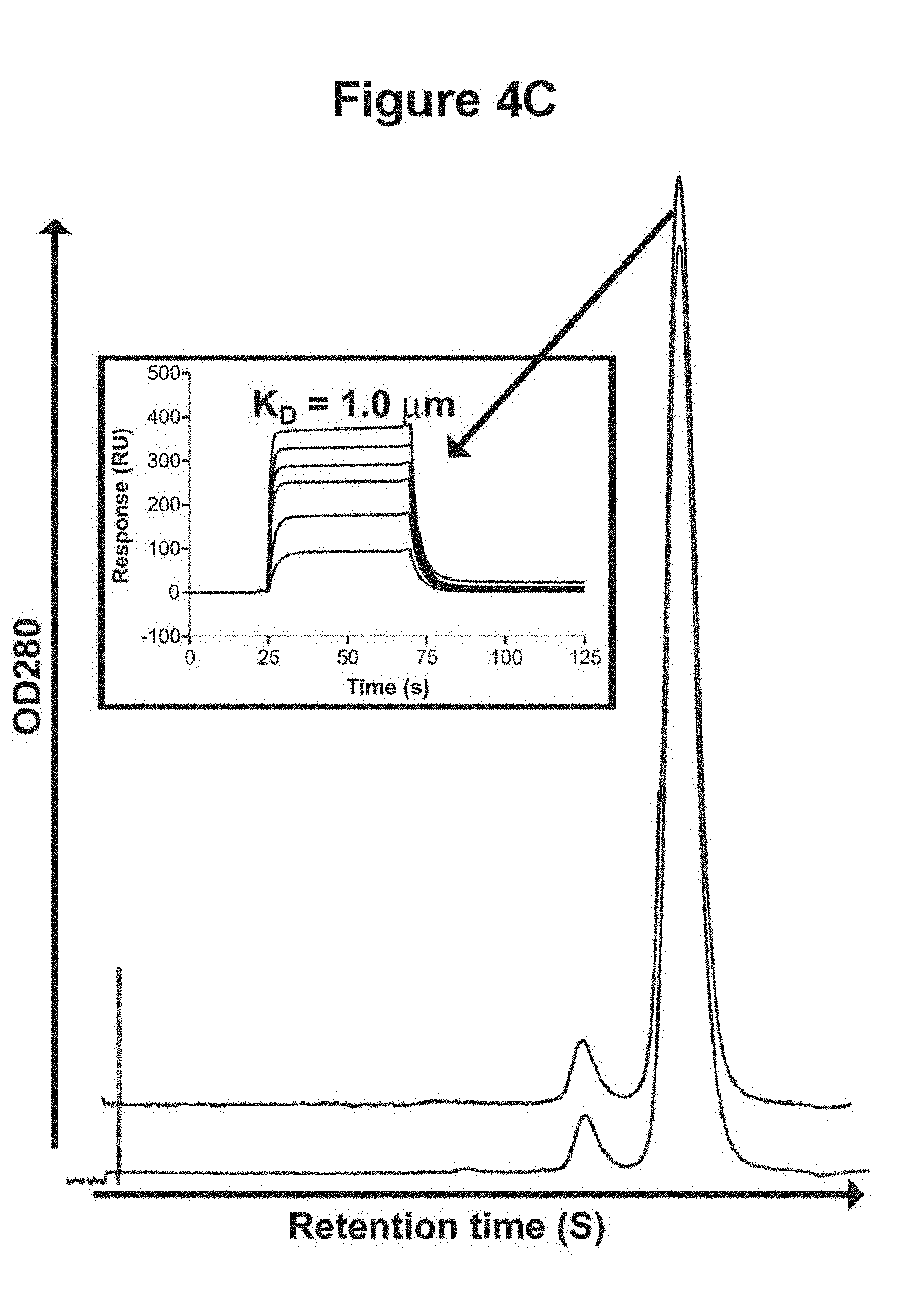
| 13656099 | Oct 19, 2012 | |||
| 14851641 | ||||
| 11887113 | Apr 16, 2008 | 8293233 | ||
| PCT/CA2006/000451 | Mar 24, 2006 | |||
| 13656099 | ||||
| 60664954 | Mar 25, 2005 | |||
| Current U.S. Class: | 1/1 |
| Current CPC Class: | C07K 2317/56 20130101; C07K 16/1271 20130101; A61P 31/12 20180101; C07K 2317/92 20130101; A61P 43/00 20180101; C12N 15/1037 20130101; A61P 37/02 20180101; A61P 31/04 20180101; C07K 16/005 20130101; A61P 35/00 20180101; C07K 2317/94 20130101; C07K 16/1282 20130101; A61P 29/00 20180101; A61P 15/02 20180101; C07K 2317/21 20130101; G01N 33/6845 20130101 |
| International Class: | C07K 16/00 20060101 C07K016/00; C12N 15/10 20060101 C12N015/10; C07K 16/12 20060101 C07K016/12; G01N 33/68 20060101 G01N033/68 |
Claims
1. An antigen-binding V.sub.L comprising a FR1 sequence of DIQMTQSPSSLSASVGDRVTITC (amino acids 1-23 of SEQ ID NO:48), a FR2 sequence of WYQQKPGKAPKLLIF (amino acids 35-49 of SEQ ID NO:48), a FR3 sequence of GVPSRFSGSGSGTDFTLTISNLQPEDFATYYC (amino acids 57-88 of SEQ ID NO:48), and a FR4 sequence of FGHGTKVTVL (amino acids 98-107 of SEQ ID NO:48).
2. An antigen-binding V.sub.L comprising the FR1, FR2, FR3, and FR4 portion of SEQ ID NO:48 and one or more randomized CDR sequences, wherein one or more of CDR1, CDR2 and CDR3 of SEQ ID NO:48 is replaced, respectively, with a randomized CDR1, CDR2 and CDR3.
3. The antigen-binding V.sub.L of claim 1, wherein the V.sub.L is in a multimeric form.
4. The antigen-binding V.sub.L of claim 1, wherein the V.sub.L is in a dimeric form.
5. The antigen-binding V.sub.L of claim 1, wherein V.sub.L is in a trimeric form.
6. The antigen-binding V.sub.L of claim 1, wherein the V.sub.L is in a pentameric form.
7. A display library constructed by preparing nucleic acid sequences coding for the antigen-binding V.sub.L of claim 1 or 2 and expressing the V.sub.L so as to display the antigen-binding V.sub.L sequence of claim 1 or 2.
8. The display library of claim 7, wherein the library is a phage display library.
9. The display library of claim 7, wherein the library is a ribosome display, ARM ribosome display, yeast display, bacterial cell display, or in vitro compartmentalization library.
10. A method for producing a V.sub.L library, comprising: a) providing a nucleotide sequence encoding the antigen-binding V.sub.L of claim 1; b) providing oligonucleotide sequences with randomized codons; c) incorporating the randomized oligonucleotides into the nucleotide sequence encoding the V.sub.L, such that one or more than one of the complementary determining regions is randomized, wherein one or more of CDR1, CDR2 and CDR3 of SEQ ID NO.: 48 is replaced, respectively, with a randomized CDR1, CDR2 and CDR3; d) expressing the nucleotide sequences produced in step c); and e) screening the expressed sequences for binding to a target polypeptide.
11. The method of claim 10, wherein the screening comprises panning against a target molecule.
12. A method of screening the display library of claim 7, comprising panning the displayed V.sub.L against a target molecule.
13. The antigen-binding V.sub.L of claim 2, wherein CDR3 of SEQ ID NO:48 is replaced with a randomized CDR3, and the randomized CDR3 comprises 10 amino acid residues.
14. The antigen-binding V.sub.L of claim 1, which is aggregation resistant.
Description
[0001] This application is a division of U.S. patent application Ser. No. 14/851,641, which is a division U.S. patent application Ser. No. 13/656,099, which is a division of U.S. patent application Ser. No. 11/887,113 issued as U.S. Pat. No. 8,293,233, which is a 371 of PCT Application No. PCT/CA2006/000451, which claims the benefit of U.S. Provisional Patent Application No. 60/664,954.
[0002] The sequence listing is provided herewith in electronic form under the file name 2012_12_06_sequence_listing.txt, created on Dec. 6, 2012, with a size of 56,279 bytes, and is hereby incorporated by reference.
FIELD OF THE INVENTION
[0003] This invention relates to the isolation, identification and manipulation of polypeptides, especially monomeric human antibody fragments.
BACKGROUND OF THE INVENTION
[0004] Antibodies in vertebrates are typically composed of paired heavy (H) and light (L) chains. The first domain of the combined H and L chains, the V.sub.H and V.sub.L, are more variable in sequence, and this is the portion of the antibody that recognizes and binds to the antigen. The V.sub.H and V.sub.L domains recognize the antigen as a pair.
[0005] The immune repertoire of camelidae (camels, dromedaries and llamas) is unique in that it possesses unusual types of antibodies referred to as heavy-chain antibodies (Hamers, Casterman C. et al., 1993). These antibodies lack light chains and thus their combining sites consist of one domain, termed V.sub.HH.
[0006] Recombinant V.sub.HH single-domain antibodies (sdAbs) provide several advantages over single-chain Fv (scFv) fragments derived from conventional four-chain antibodies. While sdAbs are comparable to their scFv counterparts in terms of affinity, they outperform scFvs in terms of solubility, stability, resistance to aggregation, refoldability, expression yield, and ease of DNA manipulation, library construction and 3-D structural determinations. Many of the aforementioned properties of V.sub.HH sdAbs are desired in applications involving antibodies.
[0007] However, the non-human nature of V.sub.HHs limits their use in human immunotherapy due to immunogenicity. In this respect, human V.sub.H and V.sub.L sdAbs are ideal candidates for immunotherapy applications because they are expected to be least immunogenic.
[0008] Human V.sub.Hs and V.sub.Ls, however, are by and large prone to aggregation, a characteristic common to V.sub.Hs and V.sub.Ls derived from conventional antibodies (Davies, J. et al., 1994; Tanha, J. et al., 2001; Ward, E. S. et al., 1989). Thus, attempts have been made to obtain monomer human V.sub.Hs and V.sub.Ls suitable for antibody applications. Such V.sub.Hs and V.sub.Ls have also displayed other useful properties typical of V.sub.HHs such as high expression yield, high refoldability and resistance to aggregation. Synthetic libraries built on these V.sub.Hs and V.sub.Ls as library scaffolds might serve as a promising source of therapeutic proteins.
[0009] Camelization as well as lamination which involves incorporating key solubility residues from camel and llama V.sub.HHs, respectively, into human V.sub.Hs or V.sub.Ls have been employed to generate monomeric human V.sub.Hs and V.sub.Ls. Synthetic sdAb libraries constructed based on these V.sub.Hs and V.sub.Ls and generated by CDR randomization were shown to be functional in terms of yielding binders to various antigens (Davies, J. et al., 1995; Tanha, J. et al., 2001).
[0010] In another approach, fully human monomeric V.sub.Hs and V.sub.Ls were isolated from human synthetic V.sub.H and V.sub.L libraries without resorting to engineering of the sort mentioned above. In one experiment a monomeric human V.sub.H, was discovered when a human V.sub.H library was panned against hen egg lysozyme (Jespers, L. et al., 2004b). More recently, a selection method based on reversible unfolding and affinity criteria yielded many monomeric V.sub.Hs from synthetic human V.sub.H libraries (Jespers, L. et al., 2004a). This finding underlined the fact that an appropriate selection method is key to efficient capturing of rare monomer human V.sub.Hs with desirable biophysical properties.
OBJECTS OF THE INVENTION
[0011] A first object of the invention is to provide a high throughput screening method for identifying polypeptides, especially antibody fragments, with improved biophysical properties, including solubility, high expression, and/or stability (such as high refolding after thermal denaturation, high resistance to chemical denaturant, and high resistance to proteases, in particular gastrointestinal proteases such as trypsin).
[0012] A second object of the invention is to provide a high throughput screening method for identifying monomeric human V.sub.Hs and V.sub.Ls.
[0013] A third object of the invention is to identify, isolate and characterize monomeric human V.sub.Hs and V.sub.Ls.
[0014] A fourth object of the invention is to construct and characterize multimers of antibody fragments, especially monomeric human V.sub.Hs and V.sub.Ls.
[0015] A fifth object of the invention is to construct display libraries from polypeptides, especially antibody fragments, and most especially monomeric human V.sub.Hs and V.sub.Ls.
[0016] A sixth object of the invention is to provide a DNA shuffling method for producing polypeptides, especially antibody fragments, and most especially monomeric human V.sub.Hs and V.sub.Ls with improved biophysical properties.
SUMMARY OF THE INVENTION
[0017] A method is provided for isolating polypeptides, preferably antibody fragments, and most preferably human V.sub.Hs and V.sub.Ls with desirable biophysical properties (solubility, stability, high expression, monomericity, non-aggregation, binding specificity). The method includes the steps of obtaining a phage display library capable of expressing a variety of polypeptide sequences, allowing infection of a bacterial lawn by the library phage, and identifying phage which form larger than average plaques on the bacterial lawn. The phage are then isolated, and steps are taken to sequence or otherwise characterize the polypeptide sequences.
[0018] The invention also provides for polypeptides, especially monomeric human V.sub.Hs and V.sub.Ls, identified by the above method, which may be useful for immunotherapy, and/or as diagnostic or detection agents. The monomeric human V.sub.Hs and V.sub.Ls may also be combined to form dimers, trimers, pentamers or other multimers, which may be useful for immunotherapy and/or as diagnostic or detection agents.
[0019] The polypeptides identified by the above method, including human V.sub.Hs and V.sub.Ls, can be manipulated by methods such as DNA shuffling to select for improved biophysical properties such as solubility, stability, monomericity, high expressibility, binding specificity and human origin.
[0020] The polypeptides identified by the above method, including human V.sub.Hs and V.sub.Ls, may also be used to generate further display libraries, which can then in turn be used to isolate further polypeptides by the above method.
[0021] In a first aspect, the present invention provides a method of identifying target polypeptides, comprising a) obtaining a phage display library capable of expressing a variety of polypeptide sequences, b) allowing infection of a bacterial lawn by the library phage and c) identifying phage which form larger than average plaques on the bacterial lawn.
[0022] In a second aspect, the present invention provides polypeptide having an amino acid sequence selected from the group consisting of: SEQ ID NO:8-54
[0023] In a third aspect, the present invention provides a V.sub.H antibody fragment comprising at least one amino acid sequence selected from the group consisting of: SEQ ID NO:8-22.
[0024] In a fourth aspect, the present invention provides a V.sub.L antibody fragment comprising at least one amino acid sequence selected from the group consisting of: SEQ ID NO:23-54.
[0025] In a fifth aspect, the present invention provides A method for producing polypeptides with desirable biophysical properties, comprising the steps of a) providing at least one first nucleic acid sequence that encodes an antibody fragment or that encodes a polypeptide sequence, and having a first desirable property; b) providing at least one second nucleic acid sequence that encodes an antibody fragment having a second desirable property; c) cutting the at least one first and at least one second nucleic acid sequences into random fragments; d) reassembling the random fragments; e) expressing the random fragments; and f) screening the expressed random fragments for the first and second desirable properties.
DETAILED DESCRIPTION OF THE DRAWINGS
[0026] Figure legends
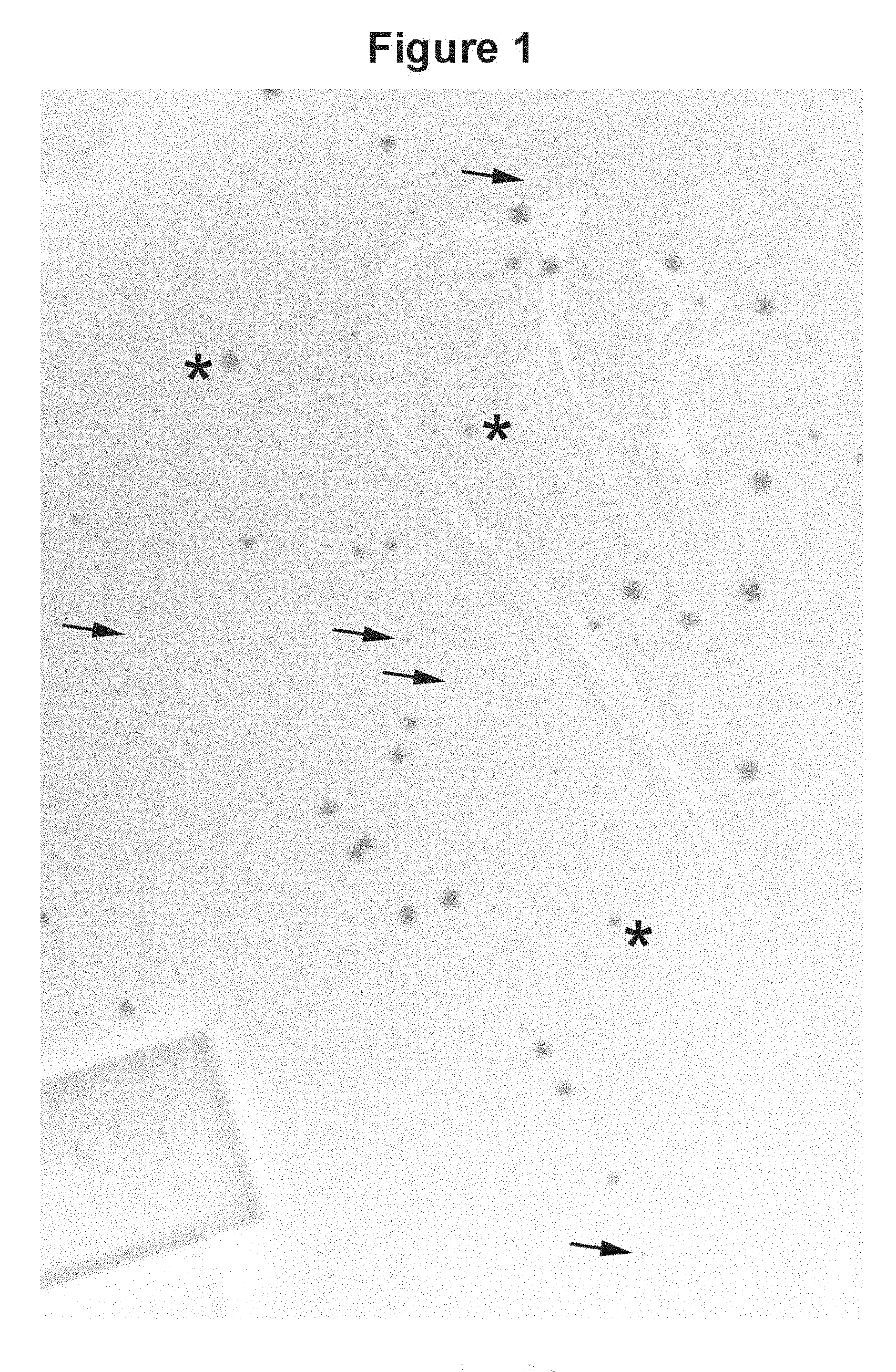
[0027] FIG. 1. A pictorial representation of selected example results: The contrast in plaque size between phages displaying a soluble V.sub.H (HVHP428) and those displaying an insoluble one (BT32/A6). The photo depicts a part of the bacterial lawn agar plate which was magnified to enhance plaque visualization. Although the plate contained an equal number of each of the two plaque types, the photo essentially contains the large, HVHP428 plaques. The majority of the BT32/A6 plaques were too small to produce clear, well-defined images in the photo. The plaques marked by arrows, thus, represent a minor proportion of BT32/A6 phages which were large enough to be visible in this image. Asterisks marks representative plaque sizes for HVHP428 phages. The identities of plaques were determined by DNA sequencing.
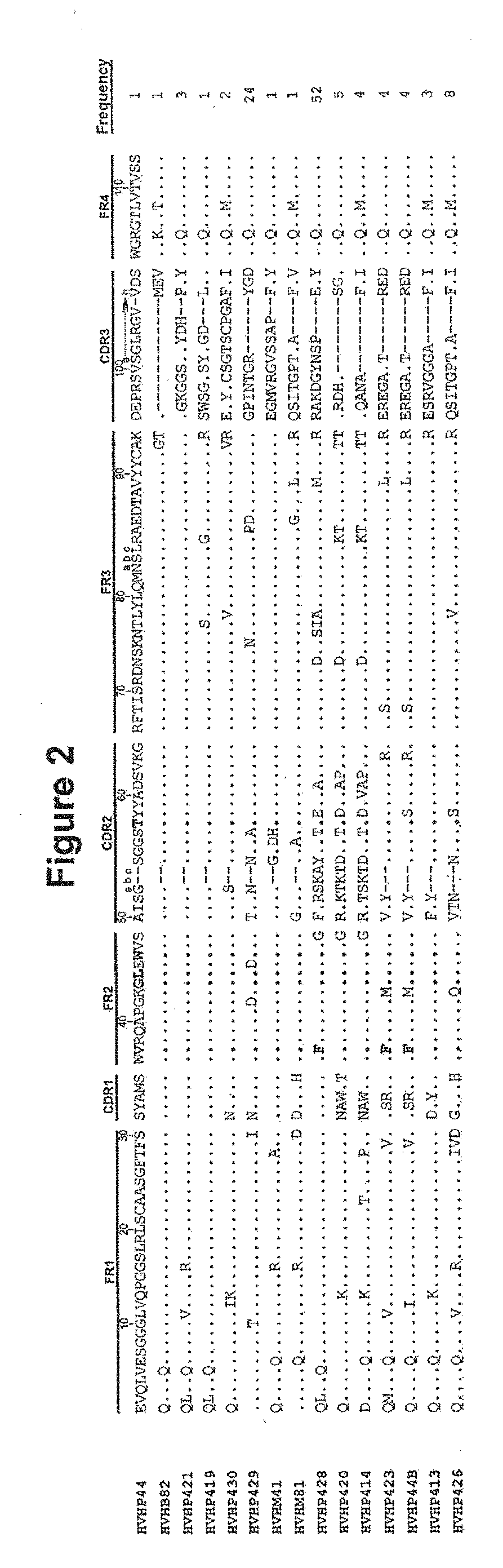
[0028] FIG. 2. Amino acid sequence of the human V.sub.Hs selected based on affinity for protein A and plaque size (SEQ ID NOS.: 8-22 disclosed respectively in order of appearance). The dots in the sequence entries indicate amino acid identity with HVHP2M10 or HVHP44. Dashes are included for sequence alignment. Residues at the key solubility positions and residue 57T which associates with V.sub.Hs/V.sub.HHs with protein A binding property are in bold. The Kabat numbering system is used. The total "frequency" value is 114. CDR=complementarity determining region; FR=framework region; gin seq=germline sequence
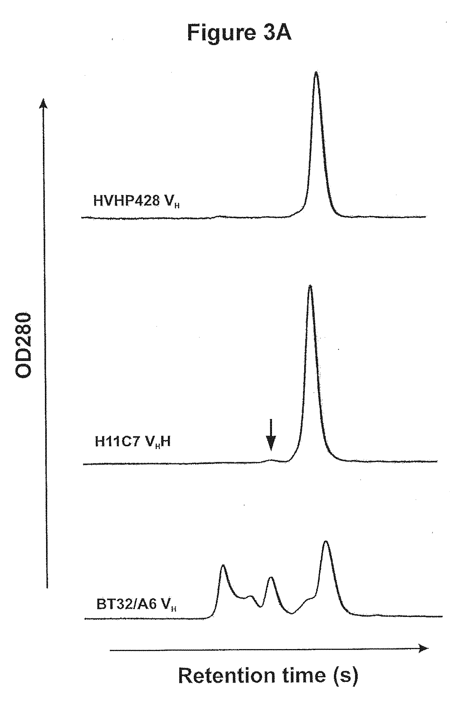
[0029] FIGS. 3A, 3B, 3C and 3D. Aggregation tendencies of the human V.sub.Hs. 3A Gel filtration chromatograms comparing the oligomerization state of a human V.sub.H isolated in this study (HVHP428) to that of a llama V.sub.HH (H11C7) and a typical human V.sub.H (BT32/A6). The peak eluting last in each chromatogram corresponds to monomeric V.sub.H. The dimeric H11C7 peak is marked by an arrow. 3B, C and D One-dimensional .sup.1H NMR spectra of HVHP414 at 800 MHz (3B), HVHP423 at 500 MHz (3C) and HVHP428 at 800 MHz (3D). The spectra in the left panel are scaled up by a factor of two to enable better viewing of low-intensity signals.
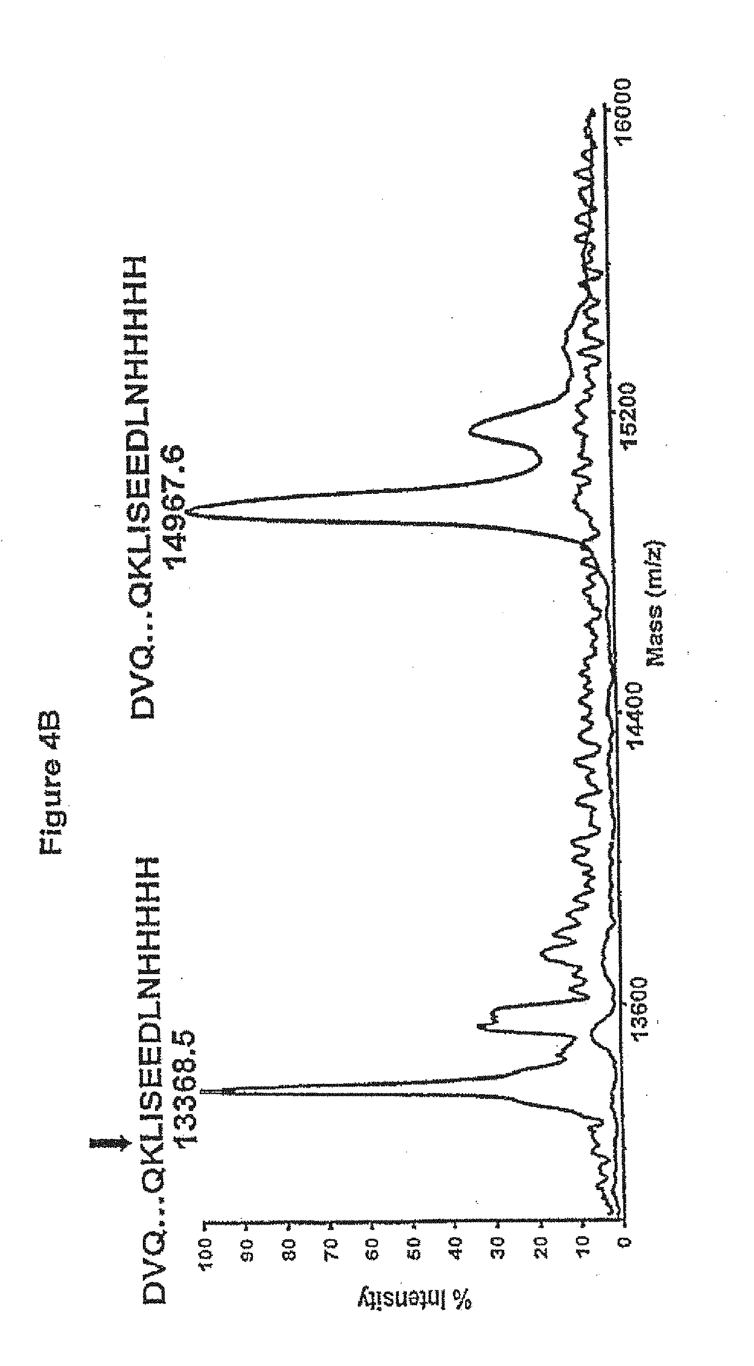
[0030] FIGS. 4A, 4B and 4C. Stability of the human V.sub.Hs in terms of their resistance to trypsin at 37.degree. C. and integrity following long incubation at 37.degree. C. 4A, SDS-PAGE comparing the mobilities of the untreated and trypsin-treated HVHP414 V.sub.H at 15, 30 and 60 min relative to a 21 kDa marker. HVHP414-cMyc denotes HVHP414 V.sub.H lacking the c-Myc. 4B, Molecular mass profiles obtained by mass spectrometry of untreated and trypsin-treated (60 min) HVHP414 V.sub.H (SEQ ID NO.: 18). The mass spectrometry profile of the treated V.sub.H is superimposed onto that for the untreated one to provide a better visual comparison. The experimental molecular mass of the untreated V.sub.H is 14,967.6 Da, which is essentially identical to the expected molecular mass, 14,967.7 Da. The observed molecular mass of the trypsin-treated V.sub.H (13,368.5 Da) indicates loss of 13 amino acids at the C-terminus by cleavage at K (Lys) in the c-Myc tag to give an expected molecular mass of 13,368.0 Da. The trypsin cleavage site is shown by a vertical arrow above the amino acids sequence of HVHP414. 4C, Gel filtration chromatograms comparing the oligomerization state of the 37.degree. C.-treated HVHP420 V.sub.H (upper profile) to that of untreated V.sub.H (lower profile). The chromatograms were shifted vertically because they were indistinguishable when superimposed. The major and minor peaks in each chromatogram correspond to monomeric and dimeric V.sub.Hs, respectively. The dimeric V.sub.H constitutes 3% of the total protein. The inset shows the sensorgram overlays for the binding of 37.degree. C.-treated HVHP420 to protein A at various concentrations. The V.sub.Hs used for temperature stability studies were from stocks which had already been at 4.degree. C. for several months.
[0031] FIG. 5. Sensogram overlays showing the binding of native (thick lines) and refolded (thin lines) HVHP423 to immobilized protein A at 75, 100, 150 and 200 nM concentrations. K.sub.Dn and K.sub.Dref were calculated from respective sensograms and used to determine RE as described below.
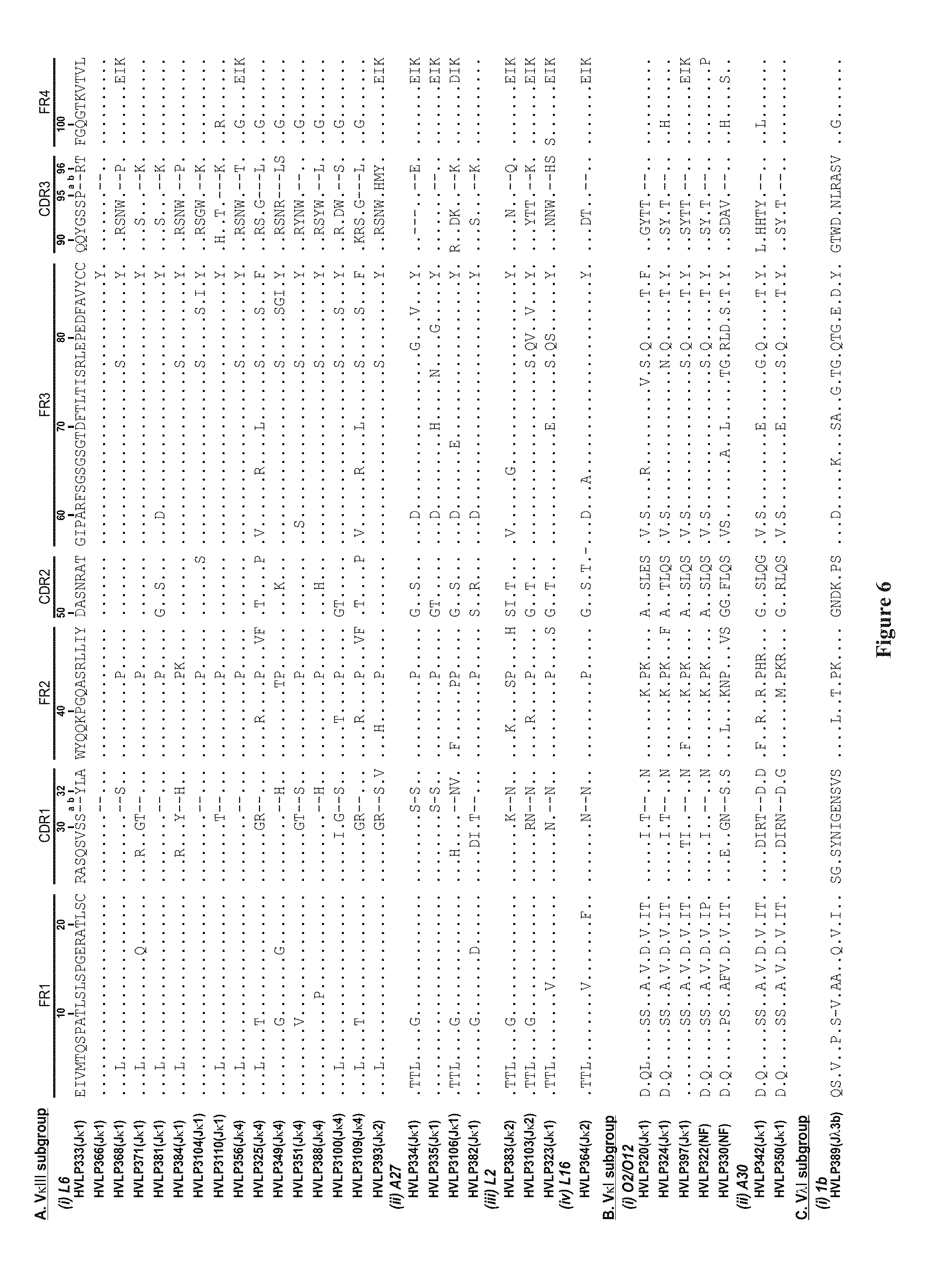
[0032] FIG. 6. Amino acid sequences of the human V.sub.Ls selected based on affinity for protein L and plaque size (SEQ ID NOS.: 23-654 disclosed respectively in order of appearance). The dots in the sequence entries indicate amino acid identity with HVLP333. Dashes are included for sequence alignment. See the V BASE for sequence numbering and CDR designation. L6, A27, L2, L16, O2/O12, A30 and 1b are V germline designation. J germline designations are in the brackets. NF, not found.
[0033] FIGS. 7A, 7B. Size exclusion chromatograms of human V.sub.L domains. In 7A, the V.sub.Ls were applied at a concentration of 0.6 mg/ml. In 7B, the V.sub.Ls were applied at their highest concentration available: HVLP342, 1.0 mg/ml; HVLP3103, 5.9 mg/ml; HVLP335, 4.9 mg/ml; HVLP351, 0.89 mg/ml. "#" and "*" represent aggregate and monomer peaks, respectively. The aggregates elute in the exclusion volume. The peak marked by an arrow in the HVLP342 panel (B) is the carry over from a previous run.
[0034] FIG. 8. Sensorgram overlays showing the binding of V.sub.Ls to immobilized protein L at concentrations of 0.2, 0.5, 0.75, 1, 2, 3, 5 and 10 .mu.M (HVLP389, HVLP351 and HVLP364); 1, 2, 3, 5, 7.5 and 10 nM (HVLP342); 0.2, 0.5, 1,2, 3, 5 and 10 .mu.M (HVLP335); 0.2, 0.5, 1, 1.5, 2 and 5 .mu.M (HVLP325), 0.2, 0.5, 0.75, 1, 1.5, 2, 3 and 5 .mu.M (HVLP3103) and 1, 2, 3, 4, 5 and 6 nM (HVLP324). The sensorgrams for HVLP324 and HVLP342 bindings to the low affinity site of protein L are not included but the calculated K.sub.DS are recorded in Table 3.
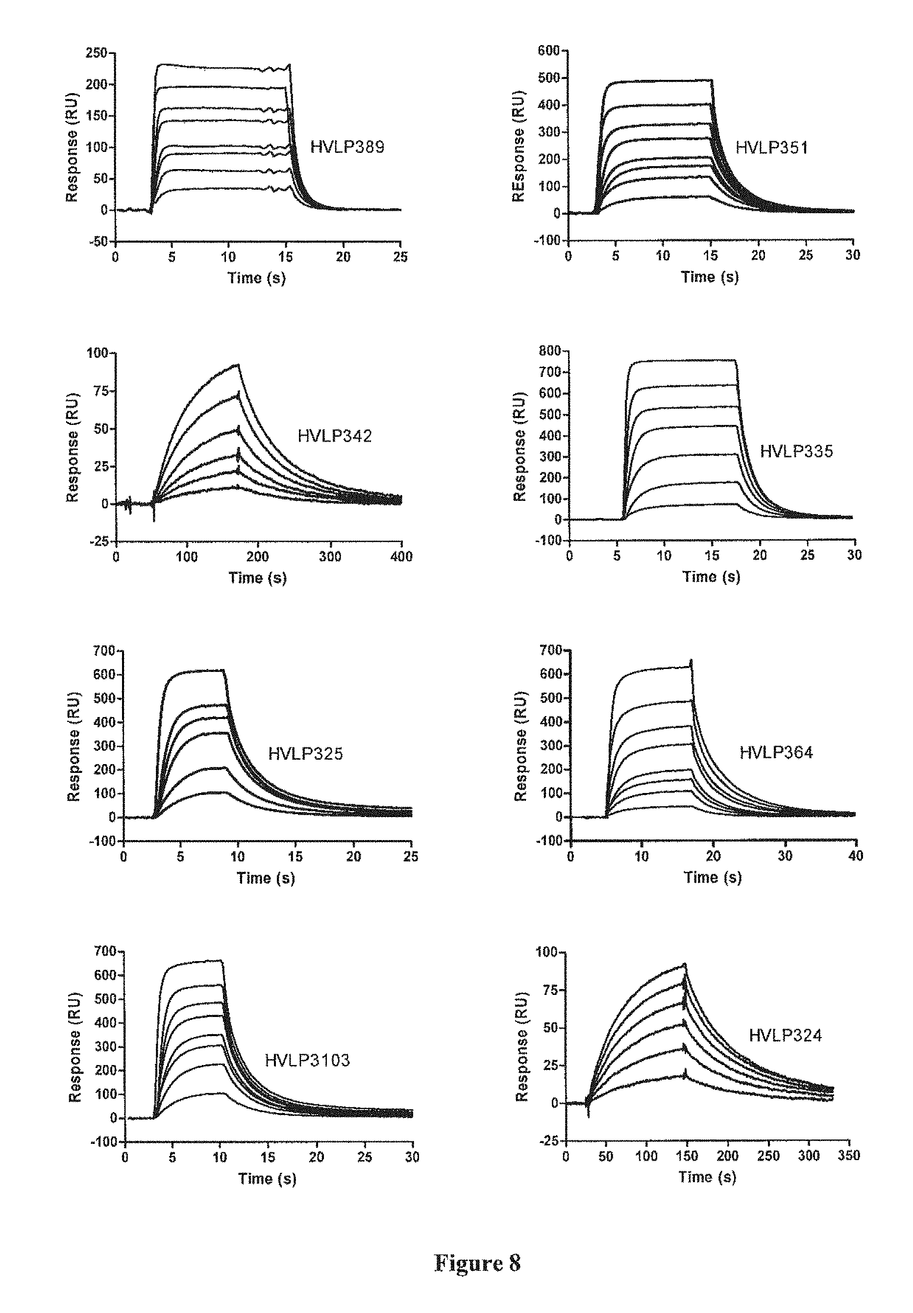
[0035] FIGS. 9A, 9B. Bindings of HVHP328PTV2 to protein A and HVLP335PTV2 to protein L in surface plasmon resonance experiments. (9A) Sensorgram overlays showing the binding of HVH28PTV2 to immobilized protein A at 1, 2, 3, 4, 6, 8 and 10 nM concentrations. (9B) Sensorgram overlays showing the binding of HVLP335PTV2 to immobilized protein L at 1, 2, 2.5, 3, 3.5, 4 and 4.5 nM concentrations. The binding data are recorded in Table 4.
[0036] FIG. 10. Figure showing the results of the microagglutination experiments with S. aureus cells. The concentration of the pentamers decreases two-fold from well 1 to well 11 with well 12 having the pentamers replaced with PBS buffer. The top row wells contain HVHP328PTV2 pentamer and the bottom ones HVLP335PTV2 pentamer. The concentrations of the pentamers in wells 1 to 6 are 215, 108, 54, 27, 13 and 7 .mu.g/ml, respectively.
DETAILED DESCRIPTION OF THE INVENTION
[0037] It is desirable to identify polypeptides, especially antibody fragments, that are of human origin, soluble, stable, resistant to aggregation, refoldable, highly expressed, easily manipulated at the DNA level, ideal for library construction and for 3-D structural determinations. Such antibody fragments are useful for a wide variety of immunotherapeutical applications, and also as diagnostic and detection agents. Human monomeric V.sub.H and V.sub.L antibodies are of particular interest, as they are likely to have many of the above-mentioned properties.
[0038] Polypeptides with the above-mentioned properties may be identified by high throughput screening of libraries capable of expressing a variety of polypeptide sequences. For example, phage display libraries (preferably filamentous phage such as M13 or fd) may be screened by infecting a field of bacteria susceptible to the phage (a bacterial lawn) with the phage, then determining which phages have successfully lysed the bacteria by looking for clear, bacteria-free areas known as plaques. Phages displaying monomeric laminated V.sub.Hs and V.sub.Ls form larger plaques on bacterial lawns than phages displaying fully human V.sub.Hs with aggregation tendencies. Thus, plaque size may be used as a means of identifying rare, naturally-occurring monomer V.sub.Hs and V.sup.Ls from the human V.sub.H repertoire.
[0039] The method disclosed herein is also useful in identifying soluble, stable (stability covers a number of characteristics, including but not limited to high thermal refolding efficiency, high melting temperature, maintaining functionality after long (several days) incubation at 37.degree. C., resistant to chemical denaturants, resistant to proteases, having a long shelf life at below 0.degree. C., and 4.degree. C., and at room temperature, maintaining functionality in intracellular environments, and maintaining functionality inside the human body, such as in the bloodstream) and high expressing proteins of differing origins, including: [0040] 1. V.sub.Hs, V.sub.Ls, Fabs, scFvs and whole antibodies such as IgGs, more specifically human ones [0041] 2. Protein variants based on non-antibody scaffolds single-chain T-cell receptors, T-cell receptor domains, transferin, lipocalins, kunitz domains, ankyrin repeats, and cytotoxic T-lymphocyte-associated antigen (CTLA-4), including human ones [0042] 3. Vaccines such as viral and bacterial protein vaccines [0043] 4. Therapeutic proteins, e.g., insulin, growth hormone, arythropoietin [0044] 5. Proteinacious diagnostic and biochemical reagents, e.g., protein A, protein G.
[0045] Once polypeptides have been identified by this method, they can be used to construct additional libraries. This is done by selecting a nucleic acid sequence of, for example, a VH. Oligonucleotides with randomized codons are created and incorporated into the VH sequence. Thus, each unique oligonucleotide is incorporated into a VH gene, and the modified VH genes constitute a library of sequences with slight variations. Typically, the oligonucleotides are designed such that the CDRs or loops of the VH are randomized. For example, one, two or all three of VH CDRs may be randomized. The VH library is then cloned into an appropriate vector, depending on the type of library to be used, and the nucleic acid sequences are expressed as polypeptides. The library is screened for molecules that bind to the library polypeptides, typically by panning. The libraries may be phage display libraries, or other display libraries such as ribosome display and yeast display.
[0046] Polypeptides identified by the method discussed herein may be used for immunotherapy by, for example, the cross-linking of monomers to form dimers, trimers, pentamers and other multimers. This may result in better affinity for antigen molecules and slower dissociation rates for some antigens. Another possible approach is to link or fuse polypeptides to a variety of molecules with various functions. For example, antibody fragments may be linked to radionuclides, cytotoxic drugs, toxins, peptides, proteins, enzymes, liposomes, lipids, T-cell superantigens or viruses in order to target and destroy or modify specific cells or molecules.
[0047] Once the V.sub.Hs or V.sub.Ls identified by the selection method described herein have been isolated, they can be further manipulated to select for improved biophysical properties such as solubility, stability, monomericity, binding specificity, human origin or high expressability. This can be achieved by in vitro recombination techniques such as DNA shuffling or a staggered extension process. DNA shuffling involves cutting the nucleic acid sequence of first (donor) and second (acceptor) polypeptides, such as antibody fragments, into random fragments, then reassembling the random fragments by a PCR-like reaction. The reassembled fragments are then screened to select for the desired properties.
[0048] For example, one or more VHs with high stability (donors) can be mixed with one or more VHs lacking sufficient stability (acceptors) and subjected to DNA shuffling. This generates mutants of the acceptor VHs which have incorporated stability residues from the donor VHs. The newly stable mutants can be identified by the methods described herein, or through other evolutionary protein screening systems such as ribosome display, yeast display, bacterial cell display and phage display. Similarly, this technique can be used to transfer desirable traits such as solubility, monomericity, and high expression.
[0049] This technique may be used where both donor and acceptor V.sub.Hs have desirable properties, to produce a V.sub.H with both properties. For example, an unstable donor V.sub.H which binds to an important therapeutic or diagnostic ligand can be shuffled with a stable acceptor V.sub.H. In order to ensure that new generated stable V.sub.Hs also have the ability to bind to the ligand, the screening system may involve a ligand binding step.
[0050] DNA shuffling may also be useful for humanizing non-human V.sub.Hs such as camelid heavy chain antibody variable domains and nurse shark and wobbegong shark variable domains, or non-human V.sub.Ls which bind to therapeutic targets. Human V.sub.Hs and V.sub.Ls with desirable properties such as solubility, stability, monomericity and high expressability may be used as donors. For example, one or more human V.sub.Hs with good stability (donors) can be mixed with one or more non-human therapeutic V.sub.Hs (acceptors) and subjected to DNA shuffling. This generates mutants of the acceptor V.sub.Hs which are both stable and humanized. The newly generated humanized and stable mutants can be identified by the methods described herein, or through other evolutionary protein screening systems such as ribosome display, yeast display, bacterial cell display and phage display. In a further example, the acceptor V.sub.H could be a therapeutic V.sub.HH (camelid heavy chain antibody variable domain).
[0051] Further, this technique is also useful for selecting desirable properties of polypeptides other than V.sub.Hs and V.sub.Ls. As discussed above, the donor polypeptide and the acceptor polypeptide may be both human, or the donor may be human and the acceptor non-human.
[0052] A possible approach for imparting solubility, monomericity, high expressability or stability to V.sub.Hs and V.sub.Ls may be through grafting complementarity determining regions (CDRs) onto acceptor V.sub.Hs and V.sub.Ls. Since CDRs are known to be involved in the solubility and stability of single-domain antibodies, and accordingly the grafting of these regions, such as the CDRs from V.sub.Hs and V.sub.Ls isolated by the methods described herein, may impart solubility and/or stability to acceptor V.sub.Hs and V.sub.Ls.
[0053] Human Monomeric V.sub.Hs and V.sub.Ls
[0054] Several monomeric human V.sub.Hs with different germline and overall sequences were identified (see FIG. 2 and SEQ ID NO. 8 through 22) from a naive human V.sub.H phage display library by this selection method based on phage plaque size. The V.sub.Hs remain functional and monomeric following trypsin treatment at 370.degree. C., weeks of incubations at 370.degree. C. or months of storage at 4.degree. C., have high thermal refolding efficiencies, are produced in good yields in E. coli and possess protein A binding activity.
[0055] In addition, several monomeric human V.sub.Ls were identified (see FIG. 6 and SEQ ID NO. 23 through 54). The V.sub.Ls are also produced in good yields in E. coli and possess protein L binding activity.
[0056] Such properties will also be manifested by V.sub.Hs from synthetic libraries that utilize the above V.sub.Hs as scaffolds. Thus, such libraries may yield therapeutic or diagnostic V.sub.Hs which would have good efficacy at physiological temperature, extended shelf life and a cost-effective production. High thermal refolding efficiency characteristic would further extend the biotechnological applications of these libraries to situations where V.sub.H binders are required to maintain their activity after exposure to transient high temperatures. The V.sub.Hs should also be very suitable for intrabody applications because of their desirable biophysical properties. The protein A binding property will simplify V.sub.H purification and detection in diagnostic tests, immunoblotting and immunocytochemistry and can be exploited to enhance library performance by removing nonfunctional V.sub.Hs from the libraries. Similarly, libraries that utilize V.sub.Ls as scaffolds will yield therapeutic or diagnostic V.sub.Ls which have similarly desirable properties. Since V.sub.Ls bind with protein L, V.sub.L purification and detection is simplified by taking advantage of this protein L binding property.
[0057] Display libraries built on the present V.sub.Hs and V.sub.Ls may also be a useful source of diagnostics and detection agents.
[0058] Previously reported fully human V.sub.Hs with favorable biophysical properties were based on a single V germline sequence: DP-47 ((Jespers, L. et al., 2004b; Jespers, L. et al., 2004a). The observation that the monomeric human V.sub.Hs in this study stem from six different germline sequences including DP-47, demonstrates that stable V.sub.Hs are not restricted in terms of germline gene usage. In fact, it is very likely that we would have isolated monomeric V.sub.Hs of family and germline origins different from the ones we describe here had we not restricted our selection to a subset of V.sub.H3 family V.sub.Hs with protein A binding activity. It is not possible to pinpoint amino acid mutations (Table 1) responsible for the observed biophysical behavior of the present V.sub.Hs due to the occurrence of multiple mutations in V.sub.Hs and the fact that CDR3 is also known to be involved in shaping the biophysical profiles of sdAbs. It may be, however, that mutations at positions known to be important for sdAbs stability and solubility, eg., V37F in HVHP423 and HVHP44B, or mutations occurring multiple times at the same position, e.g., L5V/Q and V5Q in nine V.sub.Hs, have a role in determining V.sub.Hs biophysical properties. In terms of library construction, it would be desirable that the monomericity of the present V.sub.Hs not be dependent on CDRs, in particular CDR3, so that CDR randomization be performed without the worry of jeopardizing library stability. In this regard, the V.sub.Hs with smaller CDR3, e.g, HVHB82, may be preferred scaffolds since there would be less dependence on CDR3 for stability.
[0059] The diversity of the present V.sub.Hs and V.sub.Ls in terms of overall sequence and CDR3 length should allow the construction of better-performing libraries. Synthetic V.sub.H libraries have been constructed on single scaffolds. Such an approach to repertoire generation is in sharp contrast to the natural, in vivo "approach" which utilizes a multiplicity of scaffolds. Based on the sequences reported here one can take advantage of the availability of the diverse set of V.sub.Hs and V.sub.Ls and create libraries which are based on multiple V.sub.H and V.sub.L scaffolds. Such libraries would be a better emulation of in vivo repertoires and therefore, would have a more optimal complexity. Of the three CDRs in sdAbs, CDR3 generally contributes most significantly to repertoire diversity and for this reason CDR3 randomization on V.sub.H and V.sub.L scaffolds are typically accompanied by concomitant varying of CDR3 length. While this significantly improves library complexity, it may also compromise library stability by disrupting the length of the parental scaffold CDR3. The heterogeneity of the V.sub.Hs and V.sub.Ls disclosed herein in terms of CDR3 length permit the creation of libraries with both good complexity, good stability and good biophysical characteristics. Such libraries would preferably consist of sub-libraries, where each sub-library is created by CDR3 randomization (and CDR1 and/or CDR2 randomization, if desired) on a single V.sub.H or V.sub.L scaffold without disrupting the parental CDR3 length.
[0060] The versatility of the present V.sub.Hs and V.sub.Ls is also beneficial in terms of choosing an optimal V.sub.H or V.sub.L framework for humanizing V.sub.HHs, V.sub.Hs and V.sub.Ls which are specific to therapeutic targets. High affinity camelid V.sub.HHs against therapeutic targets can be obtained from immune, non-immunized or synthetic V.sub.HH libraries with relative ease and be subsequently subjected to humanization (CDR grafting, resurfacing, deimmunization) to remove possible V.sub.HH immunogenicity, hence providing an alternative to human V.sub.H library approach for production of therapeutic V.sub.Hs. Generating high affinity therapeutic V.sub.Hs by the latter approach may often require additional tedious and time consuming in vitro affinity maturation of the lead binder(s) selected from the primary synthetic human V.sub.H libraries.
[0061] Nonhuman V.sub.Hs against therapeutic targets can be obtained from immune, non-immunized or synthetic V.sub.H libraries with relative ease and be subsequently subjected to humanization (CDR grafting, resurfacing, deimmunization) to eliminate nonhuman V.sub.H immunogenicity, hence providing an alternative to human V.sub.H library approach for production of therapeutic V.sub.Hs.
[0062] Nonhuman V.sub.Ls against therapeutic targets can be obtained from immune, non-immunized or synthetic V.sub.HH libraries with relative ease and be subsequently subjected to humanization (CDR grafting, resurfacing, deimmunization) to eliminate V.sub.HH immunogenicity, hence providing an alternative to human V.sub.L library approach for production of therapeutic V.sub.Ls.
[0063] A number of evolutionary approaches for selection of proteins with improved biophysical properties have been described (Forrer, P. et al., 1999; Waldo, G. S., 2003); (Jespers, L. et al., 2004a; Jung, S. et al., 1999; Matsuura, T. et al., 2003). Typically, stability pressure is required to ensure preferential selection of stable variants over unstable or less stable ones from a library population. For example, in a related work, heat treatment of V.sub.H phage display libraries was required to select aggregation resistant V.sub.Hs (Jespers, L. et al., 2004a). Examples of evolutionary selection approaches involving phage display include conventional phage display, selectively infective phage and the proteolysis approaches. In the first two approaches affinity selection is used to select stable species from a library, based on the assumption that stable proteins possess better binding properties for their ligand than the unstable ones. However, even with the additional inclusion of a stability selection step, these approaches may primarily enrich for higher affinity rather than for higher stability (Jung, S. et al., 1999). A binding step requirement also limits the applicability of these approaches to proteins with known ligands. The third, proteolysis approach is based on the fact that stable proteins are generally compact and therefore are resistant to proteases whereas the unstable ones are not. The phage display format is engineered in such a way that the protease stability of the displayed protein translates to phage infectivity. Thus, when a variant phage display library is treated with a protease, only the phages displaying stable proteins retain their infectivity and can subsequently be selected by infecting an E. coli host. Since this approach is independent of ligand binding, it has general utility. However, even stable and well folded proteins have protease sensitive sites, e.g., loops and linkers, and this could sometimes hinder the selection of stable species in a proteolysis approach (Bai, Y. et al., 2004).
[0064] By contrast, in the present evolutionary approach, proteins with superior biophysical properties are simply identified by the naked eye. The approach does not require ligand binding, proteolysis or destabilization steps, and thus, avoids complications which may be encountered in the reported selection approaches. No requirement for a binding step also means that this approach has general utility. As an option, a binding step may be included to ensure that the selected proteins are functional. However, the dependency of the present approach on plating (for plaque visualization) introduces a possible logistical limitation in terms of the number of plates that can be handled and thus limits its application to smaller libraries. Nonetheless, the utility of the current approach can be extended to large libraries, if the library is first reduced to a manageable size. This can be done, for example, by incorporating into the selection system a step which would remove large populations of unstable species, e.g., library adsorption on a protein A surface, or on a hydrophobic interaction column to remove poorly folded proteins with exposed hydrophobic surfaces (Matsuura, T. et al., 2003). Here, the approach was used to select V.sub.Hs and V.sub.Ls of good biophysical properties in a background of very unstable V.sub.Hs and V.sub.Ls. However, it may be more difficult to select the "best" species from a mutant library which is populated with proteins with reasonably good stabilities. In this case, the lead variants may be identified based on the rate of plaque formation by using shorter incubation times, or based on plaque size and frequency criteria.
[0065] The present selection approach can be extended to identification of stable and well-folded antibody fragments such as scFvs and Fabs with the optional inclusion, in the selection system, of a binding step involving protein L, A or any ligand, as well as stable non-antibody scaffolds and variants thereof. Moreover, the observed correlation between phage plaque size and V.sub.H expression yield means that one can utilize the present approach for acquiring high-expressing versions of proteins with otherwise poor or unsatisfactory expression from mutant phage display libraries. This application would be particularly appealing in the case of therapeutic proteins or expensive poor-expressing protein reagents where boosting protein expression would significantly offset protein production cost.
[0066] Binding Analyses of Pentamers
[0067] Both V.sub.Ls and V.sub.Hs are amenable to pentamerization and the pentamerization can be used to quickly convert a low affinity V.sub.L or V.sub.H monomer to a high affinity V.sub.L or V.sub.H pentamer. Such pentamers are invaluable diagnostics and detection agents. In such applications, the binding of a V.sub.L or V.sub.H pentamer to its target can be detected by a reporter molecule such as an enzyme (for example, horse radish peroxidase or alkaline phosphatase), or a fluorescent molecule conjugated to the pentamer. Alternatively, the binding of the pentamer can be detected by a secondary molecule which is conjugated to a reporter molecule. The secondary molecule can be specific to the pentamer itself or to a tag thereof, such as a 6His tag (SEQ ID NO.: 55) or c-Myc tag. For example, a typical secondary molecule is an immunoglobulin.
[0068] The interactions between the V.sub.Hs and protein A and V.sub.Ls with protein L are fundamentally different from those between V.sub.Hs and V.sub.Ls with their target antigens. The antigen binding of a V.sub.H or a V.sub.L involves three antigen binding loops which form the combining site of an antibody domain. The protein A binding of a V.sub.H with protein A binding activity and a V.sub.L with protein L binding activity involve binding sites and residues on the antibody domains that are totally distinct from the antibody combining site. Thus, a V.sub.H with protein A binding activity can simultaneously bind to protein A and its target antigen and a V.sub.L with protein L binding activity can simultaneously bind to protein L and its target antigen. Since the present V.sub.Hs and V.sub.Ls have affinity for protein A and L, respectively, protein A and L can be used as the secondary molecule for detection and diagnostic applications mentioned above. The human V.sub.H and V.sub.L pentamers can also be used for therapy.
[0069] Pathogen Detection by the Pentamers
[0070] The protein A and L binding activity of the V.sub.Hs and V.sub.Ls can be used to detect bacteria which have protein A and/or L on their surfaces. Protein A is present on the surface of the pathogenic bacteria, Staphylococcus aureus. Thus, the V.sub.Hs with protein A binding activity such as the ones described here can be used to detect S. aureus. Similarly, the V.sub.L monomers and V.sub.L pentamers with protein L binding activity can be used for the detection of bacteria, in particular pathogenic bacteria such as Peptostreptococcus magnus, which have protein L on their cell surface.
[0071] Protein L is implicated as a virulent factor in the pathogenesis of P. magnus (Ricci, S. et al., 2001) in humans. In vaginosis, protein L is thought to exert its effect by cross-linking surface associated IgE. V.sub.L monomers and/or pentamers with protein L binding activity have potential as therapeutics since they could interfere with the IgE cross-linking action of protein L.
[0072] Protein A is implicated as a virulent factor in the pathogenesis of S. aureus in humans (Fournier, B. et al., 2004). Its virulence has been attributed to its ability to interact with host components including binding to antibodies. V.sub.H monomers and/or pentamers with protein A binding activity have potential as therapeutics since they could interfere with the interaction of of protein A with host components.
EXAMPLES
[0073] Identification and sequence analysis of monomeric human V.sub.Hs
[0074] During the course of the construction of fully human and laminated human V.sub.H libraries, it was learned that the phages displaying monomeric laminated V.sub.Hs formed larger plaques on bacterial lawns than phages displaying fully human V.sub.Hs with aggregation tendencies. Thus, plaque size was used as a means of identifying rare, naturally-occurring monomer V.sub.Hs from the human V.sub.H repertoire (FIG. 1). To this end, a phage library displaying human V.sub.Hs with a size of 6.times.10.sup.8 was constructed and propagated as plaques on agar plates. On the titer plates, the library consisted essentially of small plaques interspersed with some large ones. PCR on twenty clones revealed that the small plaques corresponded to the V.sub.H-displaying phages while the large ones represented the wild type phages, i.e., phages lacking V.sub.H sequence inserts. None of the V.sub.H-displaying phages were found with large plaque morphology. This was not unexpected due to the paucity of the monomeric V.sub.Hs in the human repertoire and the large size of the library. To facilitate the identification of monomeric V.sub.Hs, it was decided to reduce the library to a manageable size and remove the interfering wild type phages with large-plaque-size morphology by panning the library against protein A which binds to a subset of human V.sub.Hs from V.sub.H3 family.
[0075] Following a few rounds of panning, the library became enriched for phage producing large plaques, and PCR and sequencing of more than 110 such plaques showed that all had complete V.sub.H open reading frames. The size of the large plaques which were picked for analysis is represented in FIG. 1. Sequencing revealed fifteen different V.sub.Hs which belonged to the V.sub.H3 family and utilized DP-38, DP-47, V3-49, V3-53, YAC-5 or 8-1B germline V segments (Table 1; FIG. 2). DP-38 and DP-47 germline sequences have been previously implicated in protein A binding. In addition, all V.sub.Hs had a Thr residue at position 57 (FIG. 2), consistent with their protein A binding activity. The most frequently-utilized germline V segment was DP-47 which occurred in over 50% of the V.sub.Hs, but the most frequent clone (i.e., HVHP428; relative frequency 46%) utilized the V3-49 germline V segment. HVHP429 with a DP-47 germline sequence was the second most abundant V.sub.H with a relative frequency of 21% (FIG. 2). The V.sub.H CDR3 lengths ranged from 4 amino acids for HVHB82 to 16 amino acids for HVHP430 amino acids, with HVHP430 having a pair of Cys residues in CDR3. Amino acid mutations with respect to the parental germline V segment (residues 1-94) and FR4 (residues 103-113) sequences, were observed in all V.sub.Hs and ranged from two mutations for HVHP44 (L5V and Q105R) and HVHB82 (E1Q and L5Q) to sixteen mutations for HVHP426 (Table 1). Mutations were concentrated in the V segments; only two mutations were detected in all the fifteen FR4s, at positions 105 and 108. HVHP44 and HVHB82 differed from other V.sub.Hs in that they both had a positively-charged amino acid at position 105 instead of a Gin (Table 1; FIG. 2). However, while the positively-charged amino acid in HVHP44 was acquired by mutation, the one in HVHB82 was germline-encoded. Except for HVHP423 and HVHP44B, the remaining V.sub.Hs had the germline residues at the key solubility positions: 37V/44G/45L/47W or 37F/44G/45L/47W (HVHP428); HVHP423 and HVHP44B had a V37F mutation. Mutations at other positions which are shown or hypothesized to be important in V.sub.H solubility included seven E6Q, three S35T/H, one R83G and one K83R, one A84P and one T84A and one M108L. Frequent mutations were also observed at positions 1 and 5 which included eleven E1Q, eight L5V/Q and one V5Q mutations.
[0076] Biophysical Characterization of the Human V.sub.Hs
[0077] All V.sub.Hs except HVHP44B, which was essentially the same as HVHP423, were expressed in one-litre-culture volumes in E. coli strain TG1 in fusion with c-Myc-His.sub.5 tag and purified to homogeneity from periplasmic extracts by immobilized metal affinity chromatography (IMAC). The expression yields ranged from 1.8 to 62.1 mg of purified protein per liter of bacterial culture in shaker flasks with majority of V.sub.Hs having yields in several milligrams (Table 2). In the instance of HVHP423 and HVHP430, another trial under "apparently" the same expression conditions gave yields of 2.4 and 6.4 mg as opposed to 62.1 and 23.7 mg, respectively. This implies that for many of the V.sub.Hs described here optimal expression conditions should be achieved, without much effort, resulting in expression yields significantly higher than the reported values in Table 2. As expected, all the V.sub.Hs bound to protein A in surface plasmon resonance (SPR) analyses, with K.sub.D s of 0.2-3 .mu.M, a range and magnitude comparable to the ones reported previously for llama V.sub.HH variants with protein A binding activity. None of the VHs bound to the Fab reference surface.
[0078] The aggregation tendency of the human V.sub.Hs was assessed in terms of their oligomerization states by gel filtration chromatography and NMR (Table 2). All V.sub.Hs were subjected to Superdex 75 gel filtration chromatography. Similar to a llama V.sub.HH, i.e., H11C7, all V.sub.Hs gave a symmetric single peak at the elution volume expected for a monomer, and were substantially free of any aggregates (see the example for HVHP428 in FIG. 3A. In contrast, a typical human V.sub.H (i.e., BT32/A6) formed considerable amount of aggregates. For three of the V.sub.Hs, a minor peak with a mobility expected for a V.sub.H dimer was also observed. SPR analyses of the minor peaks gave off-rate values which were significantly slower than those for the monomer V.sub.Hs, consistent with them being dimers. The dimer peak was also observed in the case of the llama V.sub.HH, H11C7. The folding and oligomerization states of the V.sub.Hs at high concentrations were further studied by NMR spectroscopy. As shown in Table II, all the V.sub.H proteins studied appeared to be relatively soluble and assumed a well-folded three-dimensional structure. One-dimensional NMR spectra of the V.sub.H fragments (FIG. 3B) showed structure folds characteristic of V.sub.H domains. The state of protein aggregation was also assessed by use of an PFG-NMR diffusion experiment for the HVHP414 fragment and two isoforms, VH14 and VH14-cMyc- with and without the c-Myc sequence, of the HVHP414. VH14 is a modified version of HVHP414 with a c-Myc N132E mutation and with an additional methionine residue at the N-terminus. In brief, the PFG-NMR data (not shown) indicated that all the protein samples had expected monomeric molecular weights even at the relatively high protein concentrations used for NMR experiments.
[0079] The stability of the V.sub.Hs was further investigated in terms of their resistance to trypsin at 37.degree. C. integrity following long incubations at 37.degree. C. Trypsin cleaves polypeptide amide backbones at the C-terminus of an Arg or a Lys residue. There are 9-13 Arg and Lys residues in the human V.sub.Hs (FIG. 2). There is also an additional Lys residue in the C-terminal c-Myc tag which is susceptible to digestion by trypsin. FIG. 4a is an SDS-PAGE analysis of HVHP414 during trypsin digestion. Within 1 h the original band was completely converted to a single product which had a mobility expected for the V.sub.H with no c-Myc-His.sub.5 tag. The same result was obtained for 12 other V.sub.Hs following a one-hour incubation with trypsin. Mass spectrometry on a randomly selected sample of the trypsin-treated V.sub.Hs (i.e., HVHP414, HVHP419, HVHP420, HVHP423, HVHP429, HVHP430 and HVHM81) confirmed that in every case the molecular mass of the digested product corresponded to a V.sub.H with the c-Myc Lys as the C-terminal residue. HVHM41 gave a significantly shorter fragment than the rest upon digestion, and in this case mass spectrometry experiments mapped the cleavage site to the Arg99 in CDR3 (data not shown).
[0080] Eleven V.sub.Hs ranging in concentration from 0.32 mg/ml (HVHP428) to 3.2 mg/ml (HVHP420) were incubated at 37.degree. C. for 17 days. Their stability was subsequently determined in terms of oligomerization state and protein A binding. As shown by gel filtration chromatography, treatment of V.sub.Hs at 37.degree. C. did not induce any aggregate formation: all V.sub.Hs gave chromatogram profiles which were virtually identical to those of untreated V.sub.Hs and stayed essentially as monomers (see the example for HVHP420; FIG. 4c). To ensure that the V.sub.Hs maintained their native fold following 37.degree. C. treatment, two V.sub.Hs, namely, HVHP414 (1.2 mg/ml) and HVHP420 (3.2 mg/ml), were selected at random and their K.sub.DS of binding to protein A were determined by SPR (Data shown for HVHP420; FIG. 4c inset) and compared to the K.sub.DS obtained for untreated V.sub.Hs (Table 2). The calculated K.sub.DS for the heat-treated V.sub.Hs were 1.4 .mu.M and 1.0 .mu.M for HVHP414 and HVHP420, respectively. These values are essentially identical to the corresponding values for the untreated V.sub.Hs (Table 2), demonstrating that 37.degree. C. treatment of V.sub.Hs did not affect their native fold. The possibility that V.sub.Hs may have been in a less compact, non-native fold during the 37.degree. C.-incubation periods and resumed their native fold upon returning to room temperature during gel filtration and SPR experiments is unlikely in light of the fact that the V.sub.Hs were resistant to trypsin at 37.degree. C. (see above), a property typically associated for well folded native proteins.
[0081] The refolding efficiency (RE) of the human V.sub.Hs was investigated by comparing the K.sub.Ds of the binding of the native (K.sub.Dn) and heat-treated, refolded (K.sub.Dref) V.sub.Hs to protein A (Tanha, J. et al., 2002). When a fraction of the V.sub.H is inactivated by heat treatment the measured K.sub.D would be higher, since this parameter is based on the concentration of folded, i.e., active, antibody fragment. Thus, the ratio of K.sub.Dn to K.sub.Dref gives a measure of V.sub.H RE. FIG. 5 compares sensorgrams for HVHP423 binding to immobilized protein A in native (thick lines) and refolded (thin lines) states at several selected V.sub.H concentrations. As can be seen, binding of the refolded V.sub.H to protein A is less in all instances, indicating that the unfolding is not fully reversible. For each of the 14 V.sub.Hs, protein A binding in both native and refolded states was measured at several concentrations, and the K.sub.Ds and subsequently REs were determined (Table 2; K.sub.Dref values are not shown). The K.sub.D s and REs of two anti-idiotypic llama V.sub.HHs, H11F9 and H11 B2, which were used as references, were also determined. Four V.sub.Hs had REs in the range of 92%-95%, similar to the REs for H11F9 and H11 B2, 95% and 100%, respectively. Another five had REs in the range of 84%-88% and three over 70%. Only two had significantly lower RE: HVHP413 (52%) and HVHP421 (14%). Several published V.sub.HHs examined previously had RE around 50% (van der Linden, R. H. et al., 1999).
[0082] Human V.sub.H Phage Display Library Construction and Panning.
[0083] cDNA was synthesized from human spleen mRNA (Ambion Inc., Austin, Tex.) using random hexanucletide primers and First Strand cDNA.TM. kit (GE Healthcare, Baie d'Urfe, QC, Canada). Using the cDNAs as template, V.sub.H genes with flanking C.sub.H sequences were amplified by polymerase chain reaction (PCR) in nine separate reactions using V.sub.H framework region 1 (FR1)-specific primers and an immunoglobin M-specific primer (de Haard, H. J. et al., 1999). The products were gel-purified and used as the template in the second round of PCR to construct V.sub.H genes using the FR1- and FR4-specific primers (de Haard, H. J. et al., 1999) that also introduced flanking Apal I and Not I restriction sites for cloning purposes. The resultant V.sub.H repertoire DNAs were cloned into fd-tetGIIID phage vector and a V.sub.H phage display library was constructed (Tanha, J. et al., 2001). Panning against protein A (Amersham Biosciences Inc.) was performed as described (Tanha, J. et al., 2001). Germline sequence assignment of the selected V.sub.Hs was performed using DNAPLOT software Version 2.0.1 and V BASE version 1.0. Llama V.sub.HHs H11C7, H11F9 and H11B2 were isolated from a llama V.sub.HH phage display library by panning against H11 scFv as described (Tanha, J. et al., 2002).
[0084] V.sub.H Expression and Purification.
[0085] V.sub.Hs were cloned into pSJF2 expression vectors by standard cloning techniques (Sambrook, J. Fritsch E. F. and Maniatis T, 1989). Periplasmic expression of sdAbs and subsequent purification by immobilized metal affinity chromatography (IMAC) were performed as described (Muruganandam, A. et al., 2002). Protein concentrations were determined by A.sub.280 measurements using molar absorption coefficients calculated for each protein (Pace, C. N. et al., 1995). Gel filtration chromatography of the purified V.sub.Hs was performed on a Superdex 75 column (GE Healthcare) as described (Deng, S. J. et al., 1995).
[0086] Binding and Refolding Efficiency Experiments.
[0087] Equilibrium dissociation constants (K.sub.Ds) and refolding efficiencies (REs) of V.sub.Hs/V.sub.HHs were derived from surface plasmon resonance (SPR) data collected with BIACORE 3000 biosensor system (Biacore Inc., Piscataway, N.J.). To measure the binding of V.sub.Hs to protein A, 2000 resonance units (RUs) of protein A or a reference antigen-binding fragment (Fab) were immobilized on research grade CM5 sensor chips (Biacore Inc.). Immobilizations were carried out at concentrations of 25 .mu.g/ml (protein A) or 50 .mu.g/ml (Fab) in 10 mM sodium acetate buffer pH 4.5, using the amine coupling kit provided by the manufacturer. To measure the binding of the anti-idiotypic llama V.sub.HHs to H11 scFv, 4100 RUs of 50 .mu.g/ml H11 scFv or 3000 RUs of 10 .mu.g/ml Se155-4 IgG reference were immobilized as described above. In all instances, analyses were carried out at 25.degree. C. in 10 mM HEPES, pH 7.4, containing 150 mM NaCl, 3 mM EDTA and 0.005% P20 at a flow rate of 40 .mu.l/min, and surfaces were regenerated by washing with the running buffer. To determine the binding activities of the refolded proteins, V.sub.Hs or V.sub.HHs were denatured by incubation at 85.degree. C. for 20 min at 10 .mu.g/ml concentrations. The protein samples were then cooled down to room temperature for 30 min to refold and were subsequently centrifuged in a microfuge at 14,000 rpm for 5 min at room temperature to remove any protein precipitates. The supernatants were recovered and analyzed for binding activity by SPR as described above. For both folded and refolded proteins data were fit to a 1:1 interaction model simultaneously using BIAevaluation 4.1 software (Biacore Inc.) and K.sub.D s were subsequently determined. REs were determined from
RE = K D n K D ref .times. 100 ##EQU00001##
Where K.sub.Dn is the K.sub.D of the native protein and K.sub.Dref is the K.sub.D of the refolded protein.
[0088] Tryptic Digest Experiments.
[0089] 3 .mu.l of a freshly prepared 0.1 .mu.g/.mu.l sequencing grade trypsin (Hoffmann-La Roche Ltd., Mississauga, ON, Canada) in 1 mM HCl was added to 60 .mu.g V.sub.H in 100 mM Tris-HCl buffer pH 7.8. Digestion reactions were carried out in a total volume of 60 .mu.l for 1 h at 37.degree. C. and stopped by adding 5 .mu.l of 0.1 .mu.g/.mu.l trypsin inhibitor (Sigma, Oakville, ON, Canada). Following completion of digestion, 5 .mu.l was removed and analyzed by SDS-PAGE; the remaining was desalted using ZipTip.sub.C4 (Millipore, Nepean, ON, Canada), eluted with 1% acetic acid in 50:50 methanol:water and subjected to V.sub.H mass determination by MALDI mass spectrometry.
[0090] Protein Stability Studies at 37.degree. C.
[0091] Single-domain antibodies (sdAbs) at 0.32-3.2 mg/ml concentrations were incubated at 37.degree. C. in PBS buffer for 17 days. Following incubation, the protein samples were spun down in a microfuge at maximum speed for 5 min even in the absence of any visible aggregate formation. The samples were then applied onto a Superdex 75 size exclusion column (GE Healthcare) and the monomeric peaks were collected for SPR analysis against protein A. SPR analyses were performed as described above except that 500 RUs of protein A or reference Fab was immobilized and that immobilizations were carried out at concentration of 50 .mu.g/ml.
[0092] NMR Experiments--
[0093] V.sub.H samples for NMR analysis were dissolved in 10 mM sodium phosphate, 150 mM NaCl, 0.5 mM EDTA, and 0.02% NaN.sub.3 at pH 7.0. The protein concentrations were 40 .mu.M-1.0 mM. All NMR experiments were carried out at 298 K on a Bruker Avance-800 or a Bruker Avance-500 NMR spectrometer. One-dimensional (1D).sup.1H NMR spectra were recorded with 16,384 data points and the spectral widths were 8,992.81 Hz at 500 MHz and 17,605.63 Hz at 800 MHz, respectively. Two-dimensional .sup.1H-.sup.1H NOESY spectra of 2,048.times.400 data points were acquired on a Bruker Avance-800 NMR spectrometer with a spectral width of 11,990.04 Hz and a mixing time of 120 ms. In all NMR experiments, water suppression was achieved using the WATERGATE method implemented through the 3-9-19 pulse train (Piotto, M. et al., 1992; Sklenar, V. et al., 1993). NMR data were processed and analyzed using the Bruker XWINNMR software package. All PFG-NMR diffusion measurements were carried out with the water-suppressed LED sequence (Altieri, A. S. et al., 1995), on a Bruker Avance-500 NMR spectrometer equipped with a triple-resonance probe with three-axis gradients. One-dimensional proton spectra were processed and analyzed using Bruker Xwinnmr software package. NMR signal intensities were obtained by integrating NMR spectra in the methyl and methylene proton region (2.3 ppm to -0.3 ppm) where all NMR signals were attenuated uniformly at all given PFG strengths.
[0094] Human V.sub.L Phage Display Library Construction and Panning.
[0095] cDNAs were synthesized from human spleen mRNA as described above for the human V.sub.Hs. The cDNA was used as template in PCR to amplify V.sub.L genes in 50 .mu.l reaction volumes using six V.sub..kappa. back primers, 11 V.sub..lamda. back primers (de Haard, H. J. et al., 1999), four V.sub..kappa. For primers and two V.sub..lamda. For primers (Sblattero, D. et al., 1998). The back and forward primers were modified to have flanking Apa LI and Not I restriction sites, respectively, for subsequent cloning purposes. Forward primers were pooled together in ratios which reflected their degree of degeneracy. V.sub..lamda. genes were PCRed in 11 separate reactions using the pooled V.sub..lamda. For primers and 11 individual V.sub..lamda. back primers. Similarly, V.sub..lamda. genes were amplified in 6 separate reactions using the pooled V.sub..kappa. For primers and 6 individual V.sub..lamda. back primers. The PCR products were pooled, gel purified and digested with Apa LI and Not I restriction endonucleases. The library was constructed as described for human V.sub.Hs. Plaque PCR was performed on individual library colonies and the amplified V.sub.L genes were sequenced as described (Tanha, J. et al., 2003). Panning against protein L (Biolynx Inc., Brockville, ON, Canada) and germline sequence assignment of the selected V.sub.Ls were performed as described above for human V.sub.H library.
[0096] V.sub.L Expression and Purification.
[0097] V.sub.L expression, purification, concentration determination and gel filtration chromatography were carried out as described for V.sub.Hs in "V.sub.H expression and purification.".
[0098] Expression and Purification of V.sub.L and V.sub.H Pentamers.
[0099] Specific primers were used in a standard PCR to amplify HVHP328 V.sub.H and HVLP335 V.sub.L genes. Standard cloning techniques were used to clone the HVHP328 and HVLP335 genes in fusion with VT1B pentamerization domain gene in an expression vector to yield HVHP328PVT2 and HVLP335PTV2 pentamers, (Zhang, J. et al., 2004). Pentamers were expressed and purified as described (Zhang, J. et al., 2004). Protein concentrations were determined as above.
[0100] Surface Plasmon Resonance of V.sub.Ls.
[0101] The binding kinetics for the interaction of the V.sub.Ls to protein L were determined by SPR using BIACORE 3000 biosensor system (Biacore, Inc., Piscataway, N.J.). 680 RUs of protein L or 870 RUs of a Fab reference were immobilized on research grade CM5 sensor chips (Biacore). Immobilizations were carried out at a protein concentration of 50 .mu.g/ml in 10 mM acetate buffer pH 4.5 using the amine coupling kit supplied by the manufacturer. All measurements were carried out at 25.degree. C. in 10 mM HEPES buffer pH 7.4, containing 150 mM NaCl, 3 mM EDTA and 0.005% P20 at a flow rate of 50 .mu.l/min or 100 .mu.l/min. Surfaces were regenerated by washing with the running buffer. Data were evaluated using the BIAevaluation 4.1 software (Biacore, Inc.).
[0102] Surface Plasmon Resonance of the Pentameric V.sub.L and V.sub.H.
[0103] The binding kinetics for the interaction of HVHP328PVT2 with protein A and HVLP335PTV2 with protein L were also determined by SPR. 520 RUs of protein A or a Fab reference were immobilized as above. For the V.sub.L pentamer, the same surfaces prepared above were used. Measurements were carried out as above but at a flow rate of 20 .mu.l/min. Surfaces were regenerated by washing with 50 mM HCl for 3 s. Data were evaluated as described for the monomers.
[0104] Cell Microagglutination
[0105] A single S. aureus colony from a BHI plate was used to inoculate 15 mL of BHI media. The bacteria were grown overnight at 37.degree. C. at 200 rpm. In the morning, the culture was spun down in a swinging bucket, Sorvall RT6000B refrigerated centrifuge at 4000 rpm for 10 min, the supernatant was removed and the cell pellet was re-suspended in PBS buffer. The cells were re-spun, the supernatant was removed and the cell pellet was re-suspended again in PBS buffer. The cells were diluted to an A.sub.600 of 1.0, and serial dilutions of the cells were spread on BHI plates at 37.degree. C. for overnight growth. The cell titer was determined in the morning. An A.sub.600 of 1.0 corresponded to 1.5.times.10.sup.9 cells ml.sup.-1. Identical steps were taken to prepare E. coli starin TG1 cells for subsequent microagglutination assays, except that the growth media was 2xYT. The viable counts were similar, A.sub.6001.0=2.1.times.10.sup.9 cells ml.sup.-1.
[0106] To perform microagglutination assays, two fold dilutions of HVHP328PVT2 in PBS were performed from wells 1 to 11 in a microtiter plate. Well 12 (blank) had only PBS. The total volume in each well was 50 .mu.l. Subsequently, 1.times.10.sup.8 S. aureus cells in 50 .mu.l PBS was added to all wells and the plate was incubated overnight at 4.degree. C. To have a permanent record of the results, a picture was taken from the plate in the morning. For the pentamer control experiment, HVHP328PVT2 was replaced with the V.sub.L pentamer, HVLP335PTV2. In the cell control experiments, the same two sets of experiments were repeated with E. coli TG1 cells.
[0107] Identification and Sequence Analysis of Monomeric Human V.sub.Ls
[0108] Essentially the same selection method which was employed to isolate soluble V.sub.Hs from a human V.sub.H phage display library was applied to a human V.sub.L library for isolating soluble, monomeric V.sub.Ls. A human V.sub.L library with a size of 3.times.10.sup.6 was constructed. Twenty four plaques from the library titer plates were picked and their V.sub.L genes were PCRed and sequenced. The sequences were diverse in terms of germ-line origin although 75% of the V.sub.Ls were of V.lamda. origin (data not shown). Three rounds of panning against protein L resulted in enrichment for large plaques. Thirty-nine of large plaques were sequenced and 32 unique sequences were identified (FIG. 6). HVLP325, HVLP335 and HVLP351 occurred at frequency of 3, 4 and 2, respectively. Except for HVLP389 which is of lambda class (subgroup V.lamda.1, germline 1b), the remaining 31 V.sub.Ls belonged to the V.kappa. class. Of the 31 kappa V.sub.Ls, 24 fall within the V.kappa.III subgroup and 7 within the V.kappa.1 subgroup. Sixteen of the 24 V.kappa.III sequences utilize L6 germline sequence with the remaining utilizing A27, L2 and L6 germline sequences. The V.kappa.1 subgroup V.sub.Ls are originated from O2/O12 or A30 germline sequence. Noticeable mutations occurred at position 96. The germline amino acids at this position are aromatic and hydrophobic amino acids Trp, Phe, Tyr, Leu or lie for kappa V.sub.Ls and Tyr, Val or Ala for lambda V.sub.Ls. But in the selected pool of kappa V.sub.Ls only 5 out of 31 have their germline amino acids at position 96: HVLP325, HVLP349, HVLP388, HVLP3109 and HVLP393. 21 amino acids at position 96 are charged of which 20 are positively-charged: Arg, Lys or His. Two amino acids are Pro, one Gin, one Ser and one Thr. Of seven kappa V.sub.Ls analyzed by gel filtration chromatography for monomericity, six which had Arg or Lys at position 96 were also monomers, whereas HVLP325 with the germline amino acid Leu at position 96 formed aggregates (see below). Similarly, HVLP389 which was of the lambda class and had a germline mutation to Ser was also monomeric (see below). These data correlates the deviation from the germline amino acids at position 96 (27 out of 32) with improved biophysical properties of V.sub.Ls such as monomericity.
[0109] Eighteen V.sub.Ls of the kappa class had their last three residues (105-107) replaced with amino acids Thr, Val and Leu which are only found in lambda V.sub.Ls. These substitutions may have had a role in improving the biophysical properties of the kappa V.sub.Ls, resulting in the selection of the aforementioned V.sub.Ls over the parental clones with the original kappa residues at position 105-107.
[0110] Characterization of the Human V.sub.Ls
[0111] Eight of the selected V.sub.Ls with different V germline origins were expressed in E. coli in one-liter cultures and purified: HVLP324, HVLP325, HVLP335, HVLP342, HVLP351, HVLP364, HVLP389 and HVLP3103 (Table 6). All were expressed in good yields ranging from 6.2 mg for HVLP324 to around 75 mg for HVLP335 and HVLP364.
[0112] The aggregation tendency of the human V.sub.Ls was assessed in terms of their oligomerization state by gel filtration chromatography. V.sub.Ls were subjected to Superdex 75 gel filtration chromatography at a concentration of 0.6 mg/ml. All except HVLP325 were essentially free of aggregates and gave symmetric single peaks with the mean apparent molecular mass of 12.7 kDa (range, 6.2-19.2 kDa) (FIG. 7A and Table 3). This is in agreement with the expected molecular mass for monomeric V.sub.Ls, 13.4-13.8 kDa. Variation in apparent molecular mass for single-domain antibodies has been reported previously (Jespers, L. et al., 2004a; (Stevens, F. J. et al., 1980). For HVLP325, the aggregates formed 11% of the total protein (aggregate plus monomer). HVLP351, HVLP342, HVLP335 and HVLP3103, were still monomer when tested at their highest concentration available, i.e., 0.89 mg/ml, 1.0 mg/ml, 4.9 mg/ml and 5.9 mg/ml, respectively (FIG. 7B)
[0113] V.sub.Ls were subjected to Superdex-75 chromatography prior to BIACORE analysis and purified monomer peaks collected even in the absence of any evidence of aggregated material. In SPR analysis, all selected V.sub.Ls bound to protein L (FIG. 8). This was not unexpected since the V.sub.Ls were isolated by panning against protein L. For all, the K.sub.DS of binding to protein L were in 0.6-3 .mu.M (Table 3). HVLP324 and HVLP342 had additional smaller K.sub.DS, 10 nM and 40 nM, respectively. Low affinity and high affinity bindings of V.sub.Ls of V.kappa.l subgroup to protein L have been reported previously (Reference). Both, HVLP324 and HVLP342, belong to V.kappa.l subgroup (Table 3). As expected, the kinetic and equilibrium data were consistent with the monomeric peak being indeed monomeric.
[0114] Binding Analyses of Pentamers
[0115] Bindings of HVHP328PVT2 pentamer to protein A and HVLP335PTV2 pentamer to protein L were determined by surface plasmon resonance (FIG. 9). The association rates were independently calculated from plots of k.sub.obs versus concentration. More than one dissociation rate (k.sub.d) could be calculated due to the heterogeneity in multivalent binding amongst the pentamer population. Therefore, more than one equilibrium dissociation constant, K.sub.D, could be obtained. HVHP328PTV2 and HVLP335PTV2 had minimum K.sub.DS of 2 nM and 200 .mu.M, respectively (Table 4). With slower k.sub.ds, HVHP328PTV2 and HVLP335PTV2 had K.sub.DS as low as 900 and 90 pM, respectively.
[0116] Pathogen Detection by V.sub.Ls and V.sub.Hs
[0117] The protein A and L binding activity of the V.sub.Hs and V.sub.Ls can be used to detect bacteria which have protein A and/or L on their surfaces. This is possible if the V.sub.Hs and V.sub.Ls are soluble and monomeric (lack of tendency to aggregate) such as the V.sub.Hs and V.sub.Ls here. Variable domains derived from antibodies which lack light chains such as camelid heavy chain antibodies or nurse shark and wobbegong shark IgNARs are naturally soluble and monomeric. From these, those with protein A and L binding activity. can also be used to detect bacteria which have protein A and/or L on their surfaces. Protein A is present on the surface of the pathogenic bacteria, Staphylococcus aureus. Thus, the V.sub.Hs with protein A binding activity such as the ones described here can be used to detect S. aureus. We performed a microagglutination assay to detect the ability of HVHP328PVT2 V.sub.H pentamer to bind to S. aureus. A constant number of bacterial cells were incubated with two-fold dilutions of HVHP328PVT2 in microtiter wells (wells 1-11) (FIG. 10). Well 12 had buffer instead of the pentamer. If the V.sub.Hs bind to the bacterial cells, then the pentamer because of its multimeric nature should be able to cross-link the cells and results in cell agglutination. The agglutinated cells will appear as diffused cells in a microtiter well (FIG. 10). In the absence of any binding, no agglutination should occur, hence no agglutination, and the cells will appear as a dot at the bottom of the well. As shown in FIG. 10, the pentamer binds to the S. aureus, since there is agglutination of cells. The agglutination is observed up to well 7. Beyond well 7 the concentration of the pentamer is too low for binding, hence no agglutination. The control V.sub.L pentamer does not show any agglutination, demonstrating the specificity of the V.sub.H pentamer to S. aureus (FIG. 10). The binding is also cell-specific since the V.sub.H pentamer as expected does not agglutinate E. coli (TG1 strain) or Salmonella cells (data not shown). Similarly, the V.sub.L monomers and V.sub.L pentamers with protein L binding activity can be used for the detection of bacteria, in particular pathogenic bacteria such as Peptostreptococcus magnus, which have protein L on their cells surface.
[0118] It is understood that the examples described above in no way serve to limit the true scope of this invention, but rather are presented for illustrative purposes.
REFERENCE LIST
[0119] Bai, Y. and Feng, H. (2004). Selection of stably folded proteins by phage-display with proteolysis. Eur. J. Biochem. 271: 1609-1614. [0120] Davies, J. and Riechmann, L. (Feb. 21, 1994). `Camelising` human antibody fragments: NMR studies on VH domains. FEBS Lett 339: 285-290. [0121] Davies, J. and Riechmann, L. (1995). Antibody VH domains as small recognition units. Biotechnology N.Y. 13: 475-479. [0122] de Haard, H. J., van Neer, N., Reurs, A., Hufton, S. E., Roovers, R. C., Henderikx, P., de Bruine, A. P., Arends, J. W., and Hoogenboom, H. R. (Jun. 25, 1999). A large non-immunized human Fab fragment phage library that permits rapid isolation and kinetic analysis of high affinity antibodies. J. Biol. Chem. 274: 18218-18230. [0123] Deng, S. J., MacKenzie, C. R., Hirama, T., Brousseau, R., Lowary, T. L., Young, N. M., Bundle, D. R., and Narang, S. A. (May 23, 1995). Basis for selection of improved carbohydrate-binding single-chain antibodies from synthetic gene libraries. Proc. Natl. Acad. Sci U.S.A 92: 4992-4996. [0124] Forrer, P., Jung, S., and Pluckthun, A. (1999). Beyond binding: using phage display to select for structure, folding and enzymatic activity in proteins. Curr. Opin. Struct. Biol. 9: 514-520. [0125] Fournier, B. and Klier, A. (2004). Protein A gene expression is regulated by DNA supercoiling which is modified by the ArlS-ArlR two-component system of Staphylococcus aureus. Microbiology 150: 3807-3819. [0126] Hamers, C. C., Atarhouch, T., Muyldermans, S., Robinson, G., Hamers, C., Songa, E. B., Bendahman, N., and Hamers, R. (Jun. 3, 1993). Naturally occurring antibodies devoid of light chains. Nature 363: 446-448. [0127] Jespers, L., Schon, O., Famm, K., and Winter, G. (2004a). Aggregation-resistant domain antibodies selected on phage by heat denaturation. Nat. Biotechnol. 22: 1161-1165. [0128] Jespers, L., Schon, O., James, L. C., Veprintsev, D., and Winter, G. (Apr. 2, 2004b). Crystal Structure of HEL4, a Soluble, Refoldable Human V(H) Single Domain with a Germ-line Scaffold. J. Mol. Biol. 337: 893-903. [0129] Jung, S., Honegger, A., and Pluckthun, A. (Nov. 19, 1999). Selection for improved protein stability by phage display. J. Mol. Biol. 294: 163-180. [0130] Matsuura, T. and Pluckthun, A. (Mar. 27, 2003). Selection based on the folding properties of proteins with ribosome display. FEBS Lett. 539: 24-28. [0131] Muruganandam, A., Tanha, J., Narang, S., and Stanimirovic, D. (2002). Selection of phage-displayed llama single-domain antibodies that transmigrate across human blood-brain barrier endothelium. FASEB J. 16: 240-242. [0132] Pace, C. N., Vajdos, F., Fee, L., Grimsley, G., and Gray, T. (1995). How to measure and predict the molar absorption coefficient of a protein. Protein Sci. 4: 2411-2423. [0133] Ricci, S., Medaglini, D., Marcotte, H., Olsen, A., Pozzi, G., and Bjorck, L. (2001). Immunoglobulin-binding domains of peptostreptococcal protein L enhance vaginal colonization of mice by Streptococcus gordonii. Microb. Pathog. 30: 229-235. [0134] Sambrook, J. F. E. F. a. M. T. (1989). "Molecular Cloning: A laboratory Manual (2.sup.nd ed.)", Cold Spring Harbor Laboratory, Cold Spring Harbor, N.Y. [0135] Sblattero, D. and Bradbury, A. (1998). A definitive set of oligonucleotide primers for amplifying human V regions. Immunotechnology. 3: 271-278. [0136] Tanha, J., Dubuc, G., Hirama, T., Narang, S. A., and MacKenzie, C. R. (May 1, 2002). Selection by phage display of llama conventional V(H) fragments with heavy chain antibody V(H)H properties. J. Immunol. Methods 263: 97-109. [0137] Tanha, J., Muruganandam, A., and Stanimirovic, D. (2003). Phage Display Technology for Identifying Specific Antigens on Brain Endothelial Cells. Methods Mol. Med. 89: 435-450. [0138] Tanha, J., Xu, P., Chen, Z. G., Ni, F., Kaplan, H., Narang, S. A., and MacKenzie, C. R. (Jul. 6, 2001). Optimal design features of camelized human single-domain antibody libraries. J. Biol. Chem 276: 24774-24780. [0139] van der Linden, R. H., Frenken, L. G., de Geus, B., Harmsen, M. M., Ruuls, R. C., Stok, W., de Ron, L., Wilson, S., Davis, P., and Verrips, C. T. (Apr. 12, 1999). Comparison of physical chemical properties of llama VHH antibody fragments and mouse monoclonal antibodies. Biochim. Biophys. Acta 1431: 37-46. [0140] Waldo, G. S. (2003). Genetic screens and directed evolution for protein solubility. Curr. Opin. Chem. Biol. 7: 33-38. [0141] Ward, E. S., Gussow, D., Griffiths, A. D., Jones, P. T., and Winter, G. (Oct. 12, 1989). Binding activities of a repertoire of single immunoglobulin variable domains secreted from Escherichia coli [see comments]. Nature 341: 544-546. [0142] Zhang, J., Li, Q., Nguyen, T. D., Tremblay, T. L., Stone, E., To, R., Kelly, J., and MacKenzie, C. R. (Jul. 30, 2004). A pentavalent single-domain antibody approach to tumor antigen discovery and the development of novel proteomics reagents. J. Mol. Biol. 341: 161-169.
TABLE-US-00001 [0142] TABLE 1 V.sub.H sequence deviations from parental germline sequences V/J Amino acid deviation from V.sub.H germlines V and FR4 germline sequences HVHP44 DP47/JH4b L5V, Q105R HVHB82 DP47/JH6c E1Q, L5Q HVHP421 DP47/JH4b E1Q, V2L, L5Q, L11V, G16R HVHP419 DP47/JH4b E1Q, V2L, L5Q, T77S, R83G, K94R HVHP430 DP47/JH3b E1Q, L5V, V12I, Q13K, S31N, G52AS, L78V, A93V, K94R HVHP429 DP47/JH4 L5V, G10T, S30I, S31N, G42D, E46D, A50T, G52aN, S53N, S56A K75N, A84P, E85D HVHM41 DP47/JH3a E1Q, L5V, E6Q, G16R, T28A, S53G, G55D, S56H, M108L HVHM81 DP47JH3a L5V, E6Q, G16R, S30D, S31D, S35H, A50G, G55A, E85G, V89L, K94R HVHP428 V3-49/JH4b E1Q, V2L, V5Q, R16G, T23A, G30S, D31S, T60A, G73D, K83R, T84A, V89M, T93A HVHP420 DP-38/JH4b E1Q, S35T, S52aT HVHP414 DP-38/JH3b E1D, E6Q, A23T, T28P, K52T, A60V HVHP423 V3-53/JH1 E1Q, V2M, E6Q, L11V, I12V, N32S, Y33R, V37F, K43M, K64R, T68S, V89L HVHP44B V3-53/JH1 E1Q, E6Q, N32S, Y33R, V37F, K43M, Y58S, K64R, T68S, V89L HVHP413 YAC-5/JH3b E1Q, E6Q, Q13K, V29F, S31D, N32Y, V50F HVHP426 8-1B/JH3b E1Q, E6Q, L11V, G16R, T28I, S30D, S31G, N32Y, Y33A, S35H, K43Q, I51T, Y52N, S53N, Y58S, L78V
TABLE-US-00002 TABLE 2 Biophysical characteristics of the human V.sub.Hs Exp.# K.sub.D Trypsin RE V.sub.H/V.sub.HH (mg) (.mu.M) resistance (%) HVHP44 8.2 1.3 93 HVHB82 5.9 0.2 71 HVHP421 5.5 1.0 14 HVHP419 3.4 1.6 84 HVHP430 6.4, 23.7 2.3 88 HVHP429 3.4 1.3 86 HVHM41 1.8 0.5 X 92 HVHM81 4.3 1.3 87 HVHP428 3.1 1.8 95 HVHP420 59.0 1.2 92 HVHP414 11.8 1.6 73 HVHP423 2.4, 62.1 3.0 86 HVHP413 5.8 0.3 52 HVHP426 6.3 0.8 70 H11F9* ND 3.5 ND 95 H11B2* ND 2.0 ND 100 #expression yield per liter of bacterial culture *K.sub.D s and RE s were determined against H11 scFv.
TABLE-US-00003 TABLE 3 Characteristics of the human V.sub.Ls Expression.sup.a K.sub.D Oligomerization V.sub.L Subgroup mg .mu.M state.sup.b HVLP324 V.kappa.I 6.9 0.2, 0.01.sup.c Monomer HVLP325 V.kappa.III 6.2 1 Monomer/Aggregate HVLP335 V.kappa.III 73.5 2 Monomer HVLP342 V.kappa.I 7.7 0.6, 0.04.sup.c Monomer HVLP351 V.kappa.III 8.9 2 Monomer HVLP364 V.kappa.III 77.1 3 Monomer HVLP389 V.lamda.I 16.7 1 Monomer HVLP3103 V.kappa.III 19.0 1 Monomer .sup.aExpression yield per liter of bacterial culture. .sup.bOligomerization state was determined by gel filtration chromatography. .sup.cThe smaller K.sub.D values correspond to the binding of the of HVLP324 and HVLP342 to the high affinity sites on protein L.
TABLE-US-00004 TABLE 4 Kinetic and equilibrium constants for the bindings of HVHP328PTV2 and HVLP335PTV2 to protein A and L, respectively Pentabody HVHP328PTV2 HVLP335PTV2 k.sub.a (M.sup.-1s.sup.-1) 4.3 .times. 10.sup.5 1.7 .times. 10.sup.6 k.sub.d (s.sup.-1) <1 .times. 10.sup.-3 <4 .times. 10.sup.-4 K.sub.D (M) <2 .times. 10.sup.-9 .sup. <2 .times. 10.sup.-10
Sequence CWU 1
1
551122PRTHomo sapiens 1Glu Val Gln Leu Val Glu Ser Gly Gly Gly Leu
Ile Gln Pro Gly Gly1 5 10 15Ser Leu Arg Leu Ser Cys Ser Ala Ser Gly
Phe Ile Val Ser Ser Asp 20 25 30Tyr Met Asn Trp Val Arg Gln Ala Pro
Gly Arg Gly Leu Glu Trp Val 35 40 45Ser Val Ile Asn Ser Asp Gly Ser
Thr Lys Tyr Ala Asp Ser Val Lys 50 55 60Gly Arg Phe Thr Ile Ser Arg
Asp Asn Ser Lys Asn Thr Leu Tyr Leu65 70 75 80Gln Met Asn Ser Leu
Arg Ala Glu Asp Thr Ala Val Tyr Tyr Cys Ala 85 90 95Arg Gly Asp Leu
Ala Tyr Cys Gly Gly His Cys Asp His Ser Pro Trp 100 105 110Gly Gln
Gly Thr Met Val Thr Val Ser Ser 115 1202124PRTHomo sapiens 2Gln Val
Gln Leu Val Gln Ser Gly Gly Gly Leu Val Lys Ser Gly Gly1 5 10 15Ser
Leu Arg Leu Ser Cys Ala Ala Ser Gly Phe Thr Phe Ser Ser Tyr 20 25
30Gly Met His Trp Val Arg Gln Ala Pro Gly Lys Gly Leu Glu Trp Val
35 40 45Ala Val Ile Ser Tyr Asp Gly Ser Asn Lys Tyr Tyr Ala Asp Ser
Val 50 55 60Lys Gly Arg Phe Thr Ile Ser Arg Asp Asn Ser Lys Asn Thr
Leu Tyr65 70 75 80Leu Gln Met Asn Ser Leu Arg Ala Glu Asp Thr Ala
Val Tyr Tyr Cys 85 90 95Ala Gly Thr Tyr Tyr Asp Ile Leu Thr Gly Pro
Thr Asn Gly Met Asp 100 105 110Val Trp Gly Gln Gly Thr Leu Val Thr
Val Ser Ser 115 1203113PRTHomo sapiens 3Gln Val Gln Leu Gln Glu Ser
Gly Gly Gly Leu Val Gln Pro Gly Gly1 5 10 15Ser Leu Arg Leu Ser Cys
Ala Ala Ser Gly Phe Thr Phe Ser Ser Tyr 20 25 30Ala Met Ser Trp Val
Arg Gln Ala Pro Gly Lys Gly Leu Glu Trp Val 35 40 45Ser Ala Ile Ser
Gly Ser Gly Gly Ser Thr Tyr Tyr Ala Asp Ser Val 50 55 60Lys Gly Arg
Phe Thr Ile Ser Arg Asp Asn Ser Lys Asn Thr Leu Tyr65 70 75 80Leu
Gln Met Asn Ser Leu Arg Ala Glu Asp Thr Ala Val Tyr Tyr Cys 85 90
95Gly Thr Asp Met Glu Val Trp Gly Lys Gly Thr Thr Val Thr Val Ser
100 105 110Ser4118PRTHomo sapiens 4Glu Val Gln Leu Val Gln Ser Gly
Gly Gly Leu Val Lys Pro Gly Gly1 5 10 15Ser Leu Arg Leu Ser Cys Ala
Ala Ser Gly Phe Thr Phe Ser Ser Tyr 20 25 30Ser Met Asn Trp Val Arg
Gln Ala Pro Gly Lys Gly Leu Glu Trp Val 35 40 45Gly Arg Ile Lys Ser
Lys Thr Asp Gly Gly Thr Thr Asp Tyr Ala Ala 50 55 60Pro Val Lys Gly
Arg Phe Thr Ile Ser Arg Asp Asp Ser Arg Asn Thr65 70 75 80Ile Tyr
Leu Gln Met Asn Ser Leu Lys Thr Glu Asp Thr Ala Met Tyr 85 90 95Tyr
Cys Thr Thr Asp Leu Thr Gln Trp Ala Ala Trp Gly Gln Gly Thr 100 105
110Met Val Thr Val Ser Ser 1155118PRTHomo sapiens 5Glu Val Gln Leu
Val Glu Ser Gly Gly Gly Leu Val Gln Pro Gly Gly1 5 10 15Ser Leu Arg
Leu Ser Cys Ala Ala Ser Gly Phe Thr Ile Ser Ser Tyr 20 25 30Asn Met
Asn Trp Val Arg Gln Ala Pro Gly Lys Gly Leu Glu Trp Val 35 40 45Ser
Tyr Ile Gly Ile Ser Tyr Asn Thr His Tyr Ala Asp Ser Val Lys 50 55
60Gly Arg Phe Thr Ile Ser Arg Asp His Ala Lys Asn Ser Leu Tyr Leu65
70 75 80Gln Met Asn Ser Leu Arg Asp Glu Asp Thr Ala Val Tyr Tyr Cys
Ala 85 90 95Arg Glu Ala Ser Gly Arg Asp Asp Phe Asp Ile Trp Gly Gln
Gly Thr 100 105 110Met Val Thr Val Ser Ser 1156119PRTHomo sapiens
6Glu Val Gln Leu Val Glu Thr Gly Gly Gly Leu Val Gln Pro Gly Gly1 5
10 15Ser Leu Arg Leu Ser Cys Ala Ala Ser Gly Phe Thr Phe Ser Ser
Tyr 20 25 30Ser Met Asn Trp Val Arg Gln Ala Pro Gly Lys Gly Leu Val
Trp Val 35 40 45Ser Arg Ile Lys Ser Asp Gly Ser Ser Thr Ser Tyr Ala
Asp Ser Val 50 55 60Lys Gly Arg Phe Thr Ile Ser Arg Asp Asn Ser Lys
Asn Thr Leu Tyr65 70 75 80Leu Gln Met Asn Ser Leu Arg Ala Glu Asp
Thr Ala Val Tyr Tyr Cys 85 90 95Ala Ser Glu Lys Ser Leu Glu Leu Pro
Asp Asp Tyr Trp Gly Gln Gly 100 105 110Thr Leu Val Thr Val Ser Ser
1157116PRTHomo sapiens 7Glu Val Gln Leu Val Glu Ser Gly Gly Gly Leu
Val Gln Pro Gly Gly1 5 10 15Ser Leu Arg Leu Ser Cys Ala Ala Ser Gly
Phe Thr Phe Ser Ser Tyr 20 25 30Ala Met Ser Trp Val Arg Gln Ala Pro
Gly Lys Gly Leu Glu Trp Val 35 40 45Ser Ile Ile His Gly Gly Gly Asn
Thr Tyr Tyr Ala Asp Ser Val Lys 50 55 60Gly Arg Phe Thr Ile Ser Arg
Asp Asn Ala Lys Asn Ser Leu Tyr Leu65 70 75 80Gln Met Asn Ser Leu
Arg Ala Glu Asp Thr Ala Val Tyr Tyr Cys Ala 85 90 95Arg Glu Tyr Ser
Gly Ala Phe Asp Ile Trp Gly Gln Gly Thr Met Val 100 105 110Thr Val
Ser Ser 1158124PRTHomo sapiens 8Glu Val Gln Leu Val Glu Ser Gly Gly
Gly Leu Val Gln Pro Gly Gly1 5 10 15Ser Leu Arg Leu Ser Cys Ala Ala
Ser Gly Phe Thr Phe Ser Ser Tyr 20 25 30Ala Met Ser Trp Val Arg Gln
Ala Pro Gly Lys Gly Leu Glu Trp Val 35 40 45Ser Ala Ile Ser Gly Ser
Gly Gly Ser Thr Tyr Tyr Ala Asp Ser Val 50 55 60Lys Gly Arg Phe Thr
Ile Ser Arg Asp Asn Ser Lys Asn Thr Leu Tyr65 70 75 80Leu Gln Met
Asn Ser Leu Arg Ala Glu Asp Thr Ala Val Tyr Tyr Cys 85 90 95Ala Lys
Asp Glu Pro Arg Ser Val Ser Gly Leu Arg Gly Val Val Asp 100 105
110Ser Trp Gly Arg Gly Thr Leu Val Thr Val Ser Ser 115
1209113PRTHomo sapiens 9Gln Val Gln Leu Gln Glu Ser Gly Gly Gly Leu
Val Gln Pro Gly Gly1 5 10 15Ser Leu Arg Leu Ser Cys Ala Ala Ser Gly
Phe Thr Phe Ser Ser Tyr 20 25 30Ala Met Ser Trp Val Arg Gln Ala Pro
Gly Lys Gly Leu Glu Trp Val 35 40 45Ser Ala Ile Ser Gly Ser Gly Gly
Ser Thr Tyr Tyr Ala Asp Ser Val 50 55 60Lys Gly Arg Phe Thr Ile Ser
Arg Asp Asn Ser Lys Asn Thr Leu Tyr65 70 75 80Leu Gln Met Asn Ser
Leu Arg Ala Glu Asp Thr Ala Val Tyr Tyr Cys 85 90 95Gly Thr Asp Met
Glu Val Trp Gly Lys Gly Thr Thr Val Thr Val Ser Ser 100 105
11010123PRTHomo sapiens 10Gln Leu Gln Leu Gln Glu Ser Gly Gly Gly
Val Val Gln Pro Gly Arg1 5 10 15Ser Leu Arg Leu Ser Cys Ala Ala Ser
Gly Phe Thr Phe Ser Ser Tyr 20 25 30Ala Met Ser Trp Val Arg Gln Ala
Pro Gly Lys Gly Leu Glu Trp Val 35 40 45Ser Ala Ile Ser Gly Ser Gly
Gly Ser Thr Tyr Tyr Ala Asp Ser Val 50 55 60Lys Gly Arg Phe Thr Ile
Ser Arg Asp Asn Ser Lys Asn Thr Leu Tyr65 70 75 80Leu Gln Met Asn
Ser Leu Arg Ala Glu Asp Thr Ala Val Tyr Tyr Cys 85 90 95Ala Lys Asp
Gly Lys Gly Gly Ser Ser Gly Tyr Asp His Pro Asp Tyr 100 105 110Trp
Gly Gln Gly Thr Leu Val Thr Val Ser Ser 115 12011122PRTHomo sapiens
11Gln Leu Gln Leu Gln Glu Ser Gly Gly Gly Leu Val Gln Pro Gly Gly1
5 10 15Ser Leu Arg Leu Ser Cys Ala Ala Ser Gly Phe Thr Phe Ser Ser
Tyr 20 25 30Ala Met Ser Trp Val Arg Gln Ala Pro Gly Lys Gly Leu Glu
Trp Val 35 40 45Ser Ala Ile Ser Gly Ser Gly Gly Ser Thr Tyr Tyr Ala
Asp Ser Val 50 55 60Lys Gly Arg Phe Thr Ile Ser Arg Asp Asn Ser Lys
Asn Ser Leu Tyr65 70 75 80Leu Gln Met Asn Ser Leu Gly Ala Glu Asp
Thr Ala Val Tyr Tyr Cys 85 90 95Ala Arg Ser Trp Ser Gly Ser Ser Tyr
Gly Gly Asp Leu Asp Ser Trp 100 105 110Gly Gln Gly Thr Leu Val Thr
Val Ser Ser 115 12012125PRTHomo sapiens 12Gln Val Gln Leu Val Glu
Ser Gly Gly Gly Leu Ile Lys Pro Gly Gly1 5 10 15Ser Leu Arg Leu Ser
Cys Ala Ala Ser Gly Phe Thr Phe Ser Asn Tyr 20 25 30Ala Met Ser Trp
Val Arg Gln Ala Pro Gly Lys Gly Leu Glu Trp Val 35 40 45Ser Ala Ile
Ser Ser Ser Gly Gly Ser Thr Tyr Tyr Ala Asp Ser Val 50 55 60Lys Gly
Arg Phe Thr Ile Ser Arg Asp Asn Ser Lys Asn Thr Val Tyr65 70 75
80Leu Gln Met Asn Ser Leu Arg Ala Glu Asp Thr Ala Val Tyr Tyr Cys
85 90 95Val Arg Glu Glu Tyr Arg Cys Ser Gly Thr Ser Cys Pro Gly Ala
Phe 100 105 110Asp Ile Trp Gly Gln Gly Thr Met Val Thr Val Ser Ser
115 120 12513119PRTHomo sapiens 13Glu Val Gln Leu Val Glu Ser Gly
Gly Thr Leu Val Gln Pro Gly Gly1 5 10 15Ser Leu Arg Leu Ser Cys Ala
Ala Ser Gly Phe Thr Phe Ile Asn Tyr 20 25 30Ala Met Ser Trp Val Arg
Gln Ala Pro Asp Lys Gly Leu Asp Trp Val 35 40 45Ser Thr Ile Ser Asn
Asn Gly Gly Ala Thr Tyr Tyr Ala Asp Ser Val 50 55 60Lys Gly Arg Phe
Thr Ile Ser Arg Asp Asn Ser Asn Asn Thr Leu Tyr65 70 75 80Leu Gln
Met Asn Ser Leu Arg Pro Asp Asp Thr Ala Val Tyr Tyr Cys 85 90 95Ala
Lys Gly Pro Ile Asn Thr Gly Arg Tyr Gly Asp Trp Gly Gln Gly 100 105
110Thr Leu Val Thr Val Ser Ser 11514123PRTHomo sapiens 14Gln Val
Gln Leu Val Gln Ser Gly Gly Gly Leu Val Gln Pro Gly Arg1 5 10 15Ser
Leu Arg Leu Ser Cys Ala Ala Ser Gly Phe Ala Phe Ser Ser Tyr 20 25
30Ala Met Ser Trp Val Arg Gln Ala Pro Gly Lys Gly Leu Glu Trp Val
35 40 45Ser Ala Ile Ser Gly Gly Gly Asp His Thr Tyr Tyr Ala Asp Ser
Val 50 55 60Lys Gly Arg Phe Thr Ile Ser Arg Asp Asn Ser Lys Asn Thr
Leu Tyr65 70 75 80Leu Gln Met Asn Ser Leu Arg Ala Glu Asp Thr Ala
Val Tyr Tyr Cys 85 90 95Ala Lys Glu Gly Met Val Arg Gly Val Ser Ser
Ala Pro Phe Asp Tyr 100 105 110Trp Gly Gln Gly Thr Leu Val Thr Val
Ser Ser 115 12015121PRTHomo sapiens 15Glu Val Gln Leu Val Gln Ser
Gly Gly Gly Leu Val Gln Pro Gly Arg1 5 10 15Ser Leu Arg Leu Ser Cys
Ala Ala Ser Gly Phe Thr Phe Asp Asp Tyr 20 25 30Ala Met His Trp Val
Arg Gln Ala Pro Gly Lys Gly Leu Glu Trp Val 35 40 45Ser Gly Ile Ser
Gly Ser Gly Ala Ser Thr Tyr Tyr Ala Asp Ser Val 50 55 60Lys Gly Arg
Phe Thr Ile Ser Arg Asp Asn Ser Lys Asn Thr Leu Tyr65 70 75 80Leu
Gln Met Asn Ser Leu Arg Ala Gly Asp Thr Ala Leu Tyr Tyr Cys 85 90
95Ala Arg Gln Ser Ile Thr Gly Pro Thr Gly Ala Phe Asp Val Trp Gly
100 105 110Gln Gly Thr Met Val Thr Val Ser Ser 115 12016123PRTHomo
sapiens 16Gln Leu Gln Leu Gln Glu Ser Gly Gly Gly Leu Val Gln Pro
Gly Gly1 5 10 15Ser Leu Arg Leu Ser Cys Ala Ala Ser Gly Phe Thr Phe
Ser Ser Tyr 20 25 30Ala Met Ser Trp Phe Arg Gln Ala Pro Gly Lys Gly
Leu Glu Trp Val 35 40 45Gly Phe Ile Arg Ser Lys Ala Tyr Gly Gly Thr
Thr Glu Tyr Ala Ala 50 55 60Ser Val Lys Gly Arg Phe Thr Ile Ser Arg
Asp Asp Ser Lys Ser Ile65 70 75 80Ala Tyr Leu Gln Met Asn Ser Leu
Arg Ala Glu Asp Thr Ala Met Tyr 85 90 95Tyr Cys Ala Arg Arg Ala Lys
Asp Gly Tyr Asn Ser Pro Glu Asp Tyr 100 105 110Trp Gly Gln Gly Thr
Leu Val Thr Val Ser Ser 115 12017119PRTHomo sapiens 17Gln Val Gln
Leu Val Glu Ser Gly Gly Gly Leu Val Lys Pro Gly Gly1 5 10 15Ser Leu
Arg Leu Ser Cys Ala Ala Ser Gly Phe Thr Phe Ser Asn Ala 20 25 30Trp
Met Thr Trp Val Arg Gln Ala Pro Gly Lys Gly Leu Glu Trp Val 35 40
45Gly Arg Ile Lys Thr Lys Thr Asp Gly Gly Thr Thr Asp Tyr Ala Ala
50 55 60Pro Val Lys Gly Arg Phe Thr Ile Ser Arg Asp Asp Ser Lys Asn
Thr65 70 75 80Leu Tyr Leu Gln Met Asn Ser Leu Lys Thr Glu Asp Thr
Ala Val Tyr 85 90 95Tyr Cys Thr Thr Asp Arg Asp His Ser Ser Gly Ser
Trp Gly Gln Gly 100 105 110Thr Leu Val Thr Val Ser Ser
11518119PRTHomo sapiens 18Asp Val Gln Leu Val Gln Ser Gly Gly Gly
Leu Val Lys Pro Gly Gly1 5 10 15Ser Leu Arg Leu Ser Cys Thr Ala Ser
Gly Phe Pro Phe Ser Asn Ala 20 25 30Trp Met Ser Trp Val Arg Gln Ala
Pro Gly Lys Gly Leu Glu Trp Val 35 40 45Gly Arg Ile Thr Ser Lys Thr
Asp Gly Gly Thr Thr Asp Tyr Val Ala 50 55 60Pro Val Lys Gly Arg Phe
Thr Ile Ser Arg Asp Asp Ser Lys Asn Thr65 70 75 80Leu Tyr Leu Gln
Met Asn Ser Leu Lys Thr Glu Asp Thr Ala Val Tyr 85 90 95Tyr Cys Thr
Thr Asp Gln Ala Asn Ala Phe Asp Ile Trp Gly Gln Gly 100 105 110Thr
Met Val Thr Val Ser Ser 11519118PRTHomo sapiens 19Gln Met Gln Leu
Val Gln Ser Gly Gly Gly Val Val Gln Pro Gly Gly1 5 10 15Ser Leu Arg
Leu Ser Cys Ala Ala Ser Gly Phe Thr Val Ser Ser Ser 20 25 30Arg Met
Ser Trp Phe Arg Gln Ala Pro Gly Met Gly Leu Glu Trp Val 35 40 45Ser
Val Ile Tyr Ser Gly Gly Ser Thr Tyr Tyr Ala Asp Ser Val Arg 50 55
60Gly Arg Phe Ser Ile Ser Arg Asp Asn Ser Lys Asn Thr Leu Tyr Leu65
70 75 80Gln Met Asn Ser Leu Arg Ala Glu Asp Thr Ala Leu Tyr Tyr Cys
Ala 85 90 95Arg Glu Arg Glu Gly Ala Val Thr Arg Glu Asp Trp Gly Gln
Gly Thr 100 105 110Leu Val Thr Val Ser Ser 11520118PRTHomo sapiens
20Gln Val Gln Leu Val Gln Ser Gly Gly Gly Leu Ile Gln Pro Gly Gly1
5 10 15Ser Leu Arg Leu Ser Cys Ala Ala Ser Gly Phe Thr Val Ser Ser
Ser 20 25 30Arg Met Ser Trp Phe Arg Gln Ala Pro Gly Met Gly Leu Glu
Trp Val 35 40 45Ser Val Ile Tyr Ser Gly Gly Ser Thr Ser Tyr Ala Asp
Ser Val Arg 50 55 60Gly Arg Phe Ser Ile Ser Arg Asp Asn Ser Lys Asn
Thr Leu Tyr Leu65 70 75 80Gln Met Asn Ser Leu Arg Ala Glu Asp Thr
Ala Leu Tyr Tyr Cys Ala 85 90 95Arg Glu Arg Glu Gly Ala Val Thr Arg
Glu Asp Trp Gly Gln Gly Thr 100 105 110Leu Val Thr Val Ser Ser
11521119PRTHomo sapiens 21Gln Val Gln Leu Val Gln Ser Gly Gly Gly
Leu Val Lys Pro Gly Gly1 5 10 15Ser Leu Arg Leu Ser Cys Ala Ala Ser
Gly Phe Thr Phe Ser Asp Tyr 20 25 30Tyr Met Ser Trp Val Arg Gln Ala
Pro Gly
Lys Gly Leu Glu Trp Val 35 40 45Ser Phe Ile Tyr Ser Gly Gly Ser Thr
Tyr Tyr Ala Asp Ser Val Lys 50 55 60Gly Arg Phe Thr Ile Ser Arg Asp
Asn Ser Lys Asn Thr Leu Tyr Leu65 70 75 80Gln Met Asn Ser Leu Arg
Ala Glu Asp Thr Ala Val Tyr Tyr Cys Ala 85 90 95Arg Glu Ser Arg Val
Gly Gly Gly Ala Phe Asp Ile Trp Gly Gln Gly 100 105 110Thr Met Val
Thr Val Ser Ser 11522120PRTHomo sapiens 22Gln Val Gln Leu Val Gln
Ser Gly Gly Gly Val Val Gln Pro Gly Arg1 5 10 15Ser Leu Arg Leu Ser
Cys Ala Ala Ser Gly Phe Ile Val Asp Gly Tyr 20 25 30Ala Met His Trp
Val Arg Gln Ala Pro Gly Gln Gly Leu Glu Trp Val 35 40 45Ser Val Thr
Asn Asn Gly Gly Ser Thr Ser Tyr Ala Asp Ser Val Lys 50 55 60Gly Arg
Phe Thr Ile Ser Arg Asp Asn Ser Lys Asn Thr Val Tyr Leu65 70 75
80Gln Met Asn Ser Leu Arg Ala Glu Asp Thr Ala Val Tyr Tyr Cys Ala
85 90 95Arg Gln Ser Ile Thr Gly Pro Thr Gly Ala Phe Asp Ile Trp Gly
Gln 100 105 110Gly Thr Met Val Thr Val Ser Ser 115 12023107PRTHomo
sapiens 23Glu Ile Val Met Thr Gln Ser Pro Ala Thr Leu Ser Leu Ser
Pro Gly1 5 10 15Glu Arg Ala Thr Leu Ser Cys Arg Ala Ser Gln Ser Val
Ser Ser Tyr 20 25 30Leu Ala Trp Tyr Gln Gln Lys Pro Gly Gln Ala Ser
Arg Leu Leu Ile 35 40 45Tyr Asp Ala Ser Asn Arg Ala Thr Gly Ile Pro
Ala Arg Phe Ser Gly 50 55 60Ser Gly Ser Gly Thr Asp Phe Thr Leu Thr
Ile Ser Arg Leu Glu Pro65 70 75 80Glu Asp Phe Ala Val Tyr Cys Cys
Gln Gln Tyr Gly Ser Ser Pro Arg 85 90 95Thr Phe Gly Gln Gly Thr Lys
Val Thr Val Leu 100 10524107PRTHomo sapiens 24Glu Ile Val Met Thr
Gln Ser Pro Ala Thr Leu Ser Leu Ser Pro Gly1 5 10 15Glu Arg Ala Thr
Leu Ser Cys Arg Ala Ser Gln Ser Val Ser Ser Tyr 20 25 30Leu Ala Trp
Tyr Gln Gln Lys Pro Gly Gln Ala Ser Arg Leu Leu Ile 35 40 45Tyr Asp
Ala Ser Asn Arg Ala Thr Gly Ile Pro Ala Arg Phe Ser Gly 50 55 60Ser
Gly Ser Gly Thr Asp Phe Thr Leu Thr Ile Ser Arg Leu Glu Pro65 70 75
80Glu Asp Phe Ala Val Tyr Tyr Cys Gln Gln Tyr Gly Ser Ser Pro Arg
85 90 95Thr Phe Gly Gln Gly Thr Lys Val Thr Val Leu 100
10525107PRTHomo sapiens 25Glu Ile Val Leu Thr Gln Ser Pro Ala Thr
Leu Ser Leu Ser Pro Gly1 5 10 15Glu Arg Ala Thr Leu Ser Cys Arg Ala
Ser Gln Ser Val Ser Ser Ser 20 25 30Leu Ala Trp Tyr Gln Gln Lys Pro
Gly Gln Ala Pro Arg Leu Leu Ile 35 40 45Tyr Asp Ala Ser Asn Arg Ala
Thr Gly Ile Pro Ala Arg Phe Ser Gly 50 55 60Ser Gly Ser Gly Thr Asp
Phe Thr Leu Thr Ile Ser Ser Leu Glu Pro65 70 75 80Glu Asp Phe Ala
Val Tyr Tyr Cys Gln Gln Arg Ser Asn Trp Pro Pro 85 90 95Thr Phe Gly
Gln Gly Thr Lys Val Glu Ile Lys 100 10526107PRTHomo sapiens 26Glu
Ile Val Leu Thr Gln Ser Pro Ala Thr Leu Ser Leu Ser Pro Gly1 5 10
15Gln Arg Ala Thr Leu Ser Cys Arg Ala Ser Arg Ser Val Gly Thr Tyr
20 25 30Leu Ala Trp Tyr Gln Gln Lys Pro Gly Gln Ala Pro Arg Leu Leu
Ile 35 40 45Tyr Asp Ala Ser Asn Arg Ala Thr Gly Ile Pro Ala Arg Phe
Ser Gly 50 55 60Ser Gly Ser Gly Thr Asp Phe Thr Leu Thr Ile Ser Arg
Leu Glu Pro65 70 75 80Glu Asp Phe Ala Val Tyr Tyr Cys Gln Gln Tyr
Ser Ser Ser Pro Lys 85 90 95Thr Phe Gly Gln Gly Thr Lys Val Thr Val
Leu 100 10527107PRTHomo sapiens 27Glu Ile Val Leu Thr Gln Ser Pro
Ala Thr Leu Ser Leu Ser Pro Gly1 5 10 15Glu Arg Ala Thr Leu Ser Cys
Arg Ala Ser Gln Ser Val Ser Ser Tyr 20 25 30Leu Ala Trp Tyr Gln Gln
Lys Pro Gly Gln Ala Pro Arg Leu Leu Ile 35 40 45Tyr Gly Ala Ser Ser
Arg Ala Thr Gly Ile Pro Asp Arg Phe Ser Gly 50 55 60Ser Gly Ser Gly
Thr Asp Phe Thr Leu Thr Ile Ser Arg Leu Glu Pro65 70 75 80Glu Asp
Phe Ala Val Tyr Tyr Cys Gln Gln Tyr Ser Ser Ser Pro Lys 85 90 95Thr
Phe Gly Gln Gly Thr Lys Val Thr Val Leu 100 10528107PRTHomo sapiens
28Glu Ile Val Leu Thr Gln Ser Pro Ala Thr Leu Ser Leu Ser Pro Gly1
5 10 15Glu Arg Ala Thr Leu Ser Cys Arg Ala Ser Arg Ser Val Ser Tyr
His 20 25 30Leu Ala Trp Tyr Gln Gln Lys Pro Gly Gln Ala Pro Lys Leu
Leu Ile 35 40 45Tyr Asp Ala Ser Asn Arg Ala Thr Gly Ile Pro Ala Arg
Phe Ser Gly 50 55 60Ser Gly Ser Gly Thr Asp Phe Thr Leu Thr Ile Ser
Ser Leu Glu Pro65 70 75 80Glu Asp Phe Ala Val Tyr Tyr Cys Gln Gln
Arg Ser Asn Trp Pro Pro 85 90 95Thr Phe Gly Gln Gly Thr Lys Val Thr
Val Leu 100 10529107PRTHomo sapiens 29Glu Ile Val Met Thr Gln Ser
Pro Ala Thr Leu Ser Leu Ser Pro Gly1 5 10 15Glu Arg Ala Thr Leu Ser
Cys Arg Ala Ser Gln Ser Val Ser Ser Tyr 20 25 30Leu Ala Trp Tyr Gln
Gln Lys Pro Gly Gln Ala Pro Arg Leu Leu Ile 35 40 45Tyr Asp Ala Ser
Asn Arg Ala Ser Gly Ile Pro Ala Arg Phe Ser Gly 50 55 60Ser Gly Ser
Gly Thr Asp Phe Thr Leu Thr Ile Ser Ser Leu Glu Pro65 70 75 80Glu
Asp Ser Ala Ile Tyr Tyr Cys Gln Gln Arg Ser Gly Trp Pro Lys 85 90
95Thr Phe Gly Gln Gly Thr Lys Val Thr Val Leu 100 10530106PRTHomo
sapiens 30Glu Ile Val Leu Thr Gln Ser Pro Ala Thr Leu Ser Leu Ser
Pro Gly1 5 10 15Glu Arg Ala Thr Leu Ser Cys Arg Ala Ser Gln Ser Val
Ser Thr Tyr 20 25 30Leu Ala Trp Tyr Gln Gln Lys Pro Gly Gln Ala Pro
Arg Leu Leu Ile 35 40 45Tyr Asp Ala Ser Asn Arg Ala Thr Gly Ile Pro
Ala Arg Phe Ser Gly 50 55 60Ser Gly Ser Gly Thr Asp Phe Thr Leu Thr
Ile Ser Arg Leu Glu Pro65 70 75 80Glu Asp Phe Ala Val Tyr Tyr Cys
Gln His Tyr Gly Thr Ser Lys Thr 85 90 95Phe Gly Arg Gly Thr Lys Val
Thr Val Leu 100 10531107PRTHomo sapiens 31Glu Ile Val Leu Thr Gln
Ser Pro Ala Thr Leu Ser Leu Ser Pro Gly1 5 10 15Glu Arg Ala Thr Leu
Ser Cys Arg Ala Ser Gln Ser Val Ser Ser Tyr 20 25 30Leu Ala Trp Tyr
Gln Gln Lys Pro Gly Gln Ala Pro Arg Leu Leu Ile 35 40 45Tyr Asp Ala
Ser Asn Arg Ala Thr Gly Ile Pro Ala Arg Phe Ser Gly 50 55 60Ser Gly
Ser Gly Thr Asp Phe Thr Leu Thr Ile Ser Ser Leu Glu Pro65 70 75
80Glu Asp Phe Ala Val Tyr Tyr Cys Gln Gln Arg Ser Asn Trp Pro Thr
85 90 95Thr Phe Gly Gly Gly Thr Lys Val Glu Ile Lys 100
10532106PRTHomo sapiens 32Glu Ile Val Leu Thr Gln Ser Pro Thr Thr
Leu Ser Leu Ser Pro Gly1 5 10 15Glu Arg Ala Thr Leu Ser Cys Arg Ala
Ser Gln Ser Val Gly Arg Tyr 20 25 30Leu Ala Trp Tyr Gln Gln Arg Pro
Gly Gln Ala Pro Arg Leu Leu Val 35 40 45Phe Asp Thr Ser Asn Arg Ala
Pro Gly Val Pro Ala Arg Phe Ser Gly 50 55 60Arg Gly Ser Gly Thr Leu
Phe Thr Leu Thr Ile Ser Ser Leu Glu Pro65 70 75 80Glu Asp Ser Ala
Val Tyr Phe Cys Gln Gln Arg Ser Ser Gly Leu Thr 85 90 95Phe Gly Gly
Gly Thr Lys Val Thr Val Leu 100 10533106PRTHomo sapiens 33Glu Ile
Val Met Thr Gln Ser Pro Gly Thr Leu Ser Leu Ser Pro Gly1 5 10 15Gly
Arg Ala Thr Leu Ser Cys Arg Ala Ser Gln Ser Val Ser Ser His 20 25
30Leu Ala Trp Tyr Gln Gln Lys Pro Gly Gln Thr Pro Arg Leu Leu Ile
35 40 45Tyr Asp Ala Ser Lys Arg Ala Thr Gly Ile Pro Ala Arg Phe Ser
Gly 50 55 60Ser Gly Ser Gly Thr Asp Phe Thr Leu Thr Ile Ser Ser Leu
Glu Pro65 70 75 80Glu Asp Ser Gly Ile Tyr Tyr Cys Gln Gln Arg Ser
Asn Arg Leu Ser 85 90 95Phe Gly Gly Gly Thr Lys Val Thr Val Leu 100
10534107PRTHomo sapiens 34Glu Ile Val Met Thr Gln Ser Pro Val Thr
Leu Ser Leu Ser Pro Gly1 5 10 15Glu Arg Ala Thr Leu Ser Cys Arg Ala
Ser Gln Ser Val Gly Thr Ser 20 25 30Leu Ala Trp Tyr Gln Gln Lys Pro
Gly Gln Ala Pro Arg Leu Leu Ile 35 40 45Tyr Asp Ala Ser Asn Arg Ala
Thr Gly Ile Ser Ala Arg Phe Ser Gly 50 55 60Ser Gly Ser Gly Thr Asp
Phe Thr Leu Thr Ile Ser Ser Leu Glu Pro65 70 75 80Glu Asp Phe Ala
Val Tyr Tyr Cys Gln Gln Arg Tyr Asn Trp Pro Arg 85 90 95Thr Phe Gly
Gly Gly Thr Lys Val Thr Val Leu 100 10535107PRTHomo sapiens 35Glu
Ile Val Met Thr Gln Ser Pro Ala Thr Leu Pro Leu Ser Pro Gly1 5 10
15Glu Arg Ala Thr Leu Ser Cys Arg Ala Ser Gln Ser Val Ser Ser His
20 25 30Leu Ala Trp Tyr Gln Gln Lys Pro Gly Gln Ala Pro Arg Leu Leu
Ile 35 40 45Tyr Asp Ala Ser His Arg Ala Thr Gly Ile Pro Ala Arg Phe
Ser Gly 50 55 60Ser Gly Ser Gly Thr Asp Phe Thr Leu Thr Ile Ser Ser
Leu Glu Pro65 70 75 80Glu Asp Phe Ala Val Tyr Tyr Cys Gln Gln Arg
Ser Tyr Trp Pro Leu 85 90 95Thr Phe Gly Gly Gly Thr Lys Val Thr Val
Leu 100 10536107PRTHomo sapiens 36Glu Ile Val Leu Thr Gln Ser Pro
Ala Thr Leu Ser Leu Ser Pro Gly1 5 10 15Glu Arg Ala Thr Leu Ser Cys
Arg Ala Ser Gln Ser Ile Ser Gly Ser 20 25 30Leu Ala Trp Tyr Gln Gln
Thr Pro Gly Gln Ala Pro Arg Leu Leu Ile 35 40 45Tyr Gly Thr Ser Asn
Arg Ala Thr Gly Ile Pro Ala Arg Phe Ser Gly 50 55 60Ser Gly Ser Gly
Thr Asp Phe Thr Leu Thr Ile Ser Ser Leu Glu Pro65 70 75 80Glu Asp
Ser Ala Val Tyr Tyr Cys Gln Gln Arg Gly Asp Trp Pro Ser 85 90 95Thr
Phe Gly Gly Gly Thr Lys Val Thr Val Leu 100 10537106PRTHomo sapiens
37Glu Ile Val Leu Thr Gln Ser Pro Thr Thr Leu Ser Leu Ser Pro Gly1
5 10 15Glu Arg Ala Thr Leu Ser Cys Arg Ala Ser Gln Ser Val Gly Arg
Tyr 20 25 30Leu Ala Trp Tyr Gln Gln Arg Pro Gly Gln Ala Pro Arg Leu
Leu Val 35 40 45Phe Asp Thr Ser Asn Arg Ala Pro Gly Val Pro Ala Arg
Phe Ser Gly 50 55 60Arg Gly Ser Gly Thr Leu Phe Thr Leu Thr Ile Ser
Ser Leu Glu Pro65 70 75 80Glu Asp Ser Ala Val Tyr Phe Cys Gln Lys
Arg Ser Ser Gly Leu Thr 85 90 95Phe Gly Gly Gly Thr Lys Val Thr Val
Leu 100 10538109PRTHomo sapiens 38Glu Ile Val Leu Thr Gln Ser Pro
Ala Thr Leu Ser Leu Ser Pro Gly1 5 10 15Glu Arg Ala Thr Leu Ser Cys
Arg Ala Ser Gln Ser Val Gly Arg Ser 20 25 30Leu Val Trp Tyr Gln His
Lys Pro Gly Gln Ala Pro Arg Leu Leu Ile 35 40 45Tyr Asp Ala Ser Asn
Arg Ala Thr Gly Ile Pro Ala Arg Phe Ser Gly 50 55 60Ser Gly Ser Gly
Thr Asp Phe Thr Leu Thr Ile Ser Ser Leu Glu Pro65 70 75 80Glu Asp
Phe Ala Val Tyr Tyr Cys Gln Gln Arg Ser Asn Trp Pro His 85 90 95Met
Tyr Thr Phe Gly Gln Gly Thr Lys Val Glu Ile Lys 100 10539105PRTHomo
sapiens 39Glu Thr Thr Leu Thr Gln Ser Pro Gly Thr Leu Ser Leu Ser
Pro Gly1 5 10 15Glu Arg Ala Thr Leu Ser Cys Arg Ala Ser Gln Ser Val
Ser Ser Ser 20 25 30Ser Leu Ala Trp Tyr Gln Gln Lys Pro Gly Gln Ala
Pro Arg Leu Leu 35 40 45Ile Tyr Gly Ala Ser Ser Arg Ala Thr Gly Ile
Pro Asp Arg Phe Ser 50 55 60Gly Ser Gly Ser Gly Thr Asp Phe Thr Leu
Thr Ile Ser Arg Leu Gly65 70 75 80Pro Glu Asp Val Ala Val Tyr Tyr
Cys Gln Gln Ser Pro Glu Thr Phe 85 90 95Gly Gln Gly Thr Lys Val Glu
Ile Lys 100 10540108PRTHomo sapiens 40Glu Ile Val Met Thr Gln Ser
Pro Ala Thr Leu Ser Leu Ser Pro Gly1 5 10 15Glu Arg Ala Thr Leu Ser
Cys Arg Ala Ser Gln Ser Val Ser Ser Ser 20 25 30Ser Leu Ala Trp Tyr
Gln Gln Lys Pro Gly Gln Ala Pro Arg Leu Leu 35 40 45Ile Tyr Gly Thr
Ser Asn Arg Ala Thr Gly Ile Pro Asp Arg Phe Ser 50 55 60Gly Ser Gly
Ser Gly Thr His Phe Thr Leu Thr Ile Asn Arg Leu Glu65 70 75 80Pro
Gly Asp Phe Ala Val Tyr Tyr Cys Gln Gln Tyr Gly Ser Ser Pro 85 90
95Arg Thr Phe Gly Gln Gly Thr Lys Val Glu Ile Lys 100
10541107PRTHomo sapiens 41Glu Thr Thr Leu Thr Gln Ser Pro Gly Thr
Leu Ser Leu Ser Pro Gly1 5 10 15Glu Arg Ala Thr Leu Ser Cys Arg Ala
Ser His Ser Val Ser Ser Asn 20 25 30Val Ala Trp Phe Gln Gln Lys Pro
Gly Gln Pro Pro Arg Leu Leu Ile 35 40 45Tyr Gly Ala Ser Ser Arg Ala
Thr Gly Ile Pro Asp Arg Phe Ser Gly 50 55 60Ser Gly Ser Glu Thr Asp
Phe Thr Leu Thr Ile Ser Arg Leu Glu Pro65 70 75 80Glu Asp Phe Ala
Val Tyr Tyr Cys Arg Gln Tyr Asp Lys Ser Pro Lys 85 90 95Thr Phe Gly
Gln Gly Thr Lys Val Asp Ile Lys 100 10542107PRTHomo sapiens 42Glu
Ile Val Met Thr Gln Ser Pro Gly Thr Leu Ser Leu Ser Pro Gly1 5 10
15Asp Arg Ala Thr Leu Ser Cys Arg Ala Ser Gln Asp Ile Ser Thr Tyr
20 25 30Leu Ala Trp Tyr Gln Gln Lys Pro Gly Gln Ala Pro Arg Leu Leu
Ile 35 40 45Tyr Ser Ala Ser Arg Arg Ala Thr Gly Ile Pro Asp Arg Phe
Ser Gly 50 55 60Ser Gly Ser Gly Thr Asp Phe Thr Leu Thr Ile Ser Arg
Leu Glu Pro65 70 75 80Glu Asp Phe Ala Val Tyr Tyr Cys Gln Gln Tyr
Ser Ser Ser Pro Lys 85 90 95Thr Phe Gly Gln Gly Thr Lys Val Thr Val
Leu 100 10543107PRTHomo sapiens 43Glu Thr Thr Leu Thr Gln Ser Pro
Gly Thr Leu Ser Leu Ser Pro Gly1 5 10 15Glu Arg Ala Thr Leu Ser Cys
Arg Ala Ser Gln Ser Val Ser Lys Asn 20 25 30Leu Ala Trp Tyr Gln Lys
Lys Pro Gly Gln Ser Pro Arg Leu Leu Ile 35 40 45His Ser Ile Ser Thr
Arg Ala Thr Gly Val Pro Ala Arg Phe Ser Gly 50 55 60Gly Gly Ser Gly
Thr Asp Phe Thr Leu Thr Ile Ser Arg Leu Glu Pro65 70 75 80Glu Asp
Phe Ala Val Tyr Tyr Cys Gln Gln Tyr Gly Asn Ser Pro Gln 85 90
95Thr Phe Gly Gln Gly Thr Lys Val Glu Ile Lys 100 10544107PRTHomo
sapiens 44Glu Thr Thr Leu Thr Gln Ser Pro Gly Thr Leu Ser Leu Ser
Pro Gly1 5 10 15Glu Arg Ala Thr Leu Ser Cys Arg Ala Ser Gln Ser Val
Arg Asn Asn 20 25 30Leu Ala Trp Tyr Gln Gln Arg Pro Gly Gln Ala Pro
Arg Leu Leu Ile 35 40 45Tyr Gly Ala Ser Thr Arg Ala Thr Gly Ile Pro
Ala Arg Phe Ser Gly 50 55 60Ser Gly Ser Gly Thr Asp Phe Thr Leu Thr
Ile Ser Ser Leu Gln Val65 70 75 80Glu Asp Val Ala Val Tyr Tyr Cys
Gln Gln Tyr Tyr Thr Thr Pro Lys 85 90 95Thr Phe Gly Gln Gly Thr Lys
Val Glu Ile Lys 100 10545107PRTHomo sapiens 45Glu Thr Thr Leu Thr
Gln Ser Pro Ala Thr Leu Ser Val Ser Pro Gly1 5 10 15Glu Arg Ala Thr
Leu Ser Cys Arg Ala Ser Gln Ser Val Asn Ser Asn 20 25 30Leu Ala Trp
Tyr Gln Gln Lys Pro Gly Gln Ala Pro Arg Leu Leu Ile 35 40 45Ser Gly
Ala Ser Thr Arg Ala Thr Gly Ile Pro Ala Arg Phe Ser Gly 50 55 60Ser
Gly Ser Gly Thr Glu Phe Thr Leu Thr Ile Ser Ser Leu Gln Ser65 70 75
80Glu Asp Phe Ala Val Tyr Tyr Cys Gln Gln Tyr Asn Asn Trp Pro His
85 90 95Ser Ser Gly Gln Gly Thr Lys Val Glu Ile Lys 100
10546107PRTHomo sapiens 46Glu Thr Thr Leu Thr Gln Ser Pro Ala Thr
Leu Ser Val Ser Pro Gly1 5 10 15Glu Arg Ala Thr Phe Ser Cys Arg Ala
Ser Gln Ser Val Ser Asn Asn 20 25 30Leu Ala Trp Tyr Gln Gln Lys Pro
Gly Gln Ala Pro Arg Leu Leu Ile 35 40 45Tyr Gly Ala Ser Ser Arg Thr
Thr Gly Ile Pro Asp Arg Phe Ser Ala 50 55 60Ser Gly Ser Gly Thr Asp
Phe Thr Leu Thr Ile Ser Arg Leu Glu Pro65 70 75 80Glu Asp Phe Ala
Val Tyr Tyr Cys Gln Gln Tyr Asp Thr Ser Pro Arg 85 90 95Thr Phe Gly
Gln Gly Thr Lys Val Glu Ile Lys 100 10547107PRTHomo sapiens 47Asp
Ile Gln Leu Thr Gln Ser Pro Ser Ser Leu Ser Ala Ser Val Gly1 5 10
15Asp Arg Val Thr Ile Thr Cys Arg Ala Ser Gln Ser Ile Ser Thr Tyr
20 25 30Leu Asn Trp Tyr Gln Gln Lys Pro Gly Lys Ala Pro Lys Leu Leu
Ile 35 40 45Tyr Ala Ala Ser Ser Leu Glu Ser Gly Val Pro Ser Arg Phe
Ser Gly 50 55 60Arg Gly Ser Gly Thr Asp Phe Thr Leu Thr Val Ser Ser
Leu Gln Pro65 70 75 80Glu Asp Phe Ala Thr Tyr Phe Cys Gln Gln Gly
Tyr Thr Thr Pro Arg 85 90 95Thr Phe Gly Gln Gly Thr Lys Val Thr Val
Leu 100 10548107PRTHomo sapiens 48Asp Ile Gln Met Thr Gln Ser Pro
Ser Ser Leu Ser Ala Ser Val Gly1 5 10 15Asp Arg Val Thr Ile Thr Cys
Arg Ala Ser Gln Ser Ile Ser Thr Tyr 20 25 30Leu Asn Trp Tyr Gln Gln
Lys Pro Gly Lys Ala Pro Lys Leu Leu Ile 35 40 45Phe Ala Ala Ser Thr
Leu Gln Ser Gly Val Pro Ser Arg Phe Ser Gly 50 55 60Ser Gly Ser Gly
Thr Asp Phe Thr Leu Thr Ile Ser Asn Leu Gln Pro65 70 75 80Glu Asp
Phe Ala Thr Tyr Tyr Cys Gln Gln Ser Tyr Ser Thr Pro Arg 85 90 95Thr
Phe Gly His Gly Thr Lys Val Thr Val Leu 100 10549107PRTHomo sapiens
49Glu Ile Val Met Thr Gln Ser Pro Ser Ser Leu Ser Ala Ser Val Gly1
5 10 15Asp Arg Val Thr Ile Thr Cys Arg Ala Ser Gln Thr Ile Ser Ser
Tyr 20 25 30Leu Asn Trp Phe Gln Gln Lys Pro Gly Lys Ala Pro Lys Leu
Leu Ile 35 40 45Tyr Ala Ala Ser Ser Leu Gln Ser Gly Val Pro Ser Arg
Phe Ser Gly 50 55 60Ser Gly Ser Gly Thr Asp Phe Thr Leu Thr Ile Ser
Ser Leu Gln Pro65 70 75 80Glu Asp Phe Ala Thr Tyr Tyr Cys Gln Gln
Ser Tyr Thr Thr Pro Arg 85 90 95Thr Phe Gly Gln Gly Thr Lys Val Glu
Ile Lys 100 10550107PRTHomo sapiens 50Asp Ile Gln Met Thr Gln Ser
Pro Ser Ser Leu Ser Ala Ser Val Gly1 5 10 15Asp Arg Val Thr Ile Pro
Cys Arg Ala Ser Gln Ser Ile Ser Ser Tyr 20 25 30Leu Asn Trp Tyr Gln
Gln Lys Pro Gly Lys Ala Pro Lys Leu Leu Ile 35 40 45Tyr Ala Ala Ser
Ser Leu Gln Ser Gly Val Pro Ser Arg Phe Ser Gly 50 55 60Ser Gly Ser
Gly Thr Asp Phe Thr Leu Thr Ile Ser Ser Leu Gln Pro65 70 75 80Glu
Asp Phe Ala Thr Tyr Tyr Cys Gln Gln Ser Tyr Ser Thr Pro Arg 85 90
95Thr Phe Gly Gln Gly Thr Lys Val Thr Val Pro 100 10551107PRTHomo
sapiens 51Asp Ile Gln Met Thr Gln Ser Pro Pro Ser Leu Ser Ala Phe
Val Gly1 5 10 15Asp Arg Val Thr Ile Thr Cys Arg Ala Ser Glu Ser Val
Gly Asn Ser 20 25 30Leu Ser Trp Tyr Gln Leu Lys Pro Gly Lys Asn Pro
Arg Leu Leu Val 35 40 45Ser Gly Gly Ser Phe Leu Gln Ser Gly Val Ser
Ala Arg Phe Ser Gly 50 55 60Ser Gly Ala Gly Thr Leu Phe Thr Leu Thr
Ile Thr Gly Leu Arg Leu65 70 75 80Asp Asp Ser Ala Thr Tyr Tyr Cys
Gln Gln Ser Asp Ala Val Pro Arg 85 90 95Thr Phe Gly His Gly Thr Lys
Val Ser Val Leu 100 10552107PRTHomo sapiens 52Asp Ile Gln Met Thr
Gln Ser Pro Ser Ser Leu Ser Ala Ser Val Gly1 5 10 15Asp Arg Val Thr
Ile Thr Cys Arg Ala Ser Gln Asp Ile Arg Thr Asp 20 25 30Leu Asp Trp
Phe Gln Gln Arg Pro Gly Arg Ala Pro His Arg Leu Ile 35 40 45Tyr Gly
Ala Ser Ser Leu Gln Gly Gly Val Pro Ser Arg Phe Ser Gly 50 55 60Ser
Gly Ser Gly Thr Glu Phe Thr Leu Thr Ile Ser Gly Leu Gln Pro65 70 75
80Glu Asp Phe Ala Thr Tyr Tyr Cys Leu Gln His His Thr Tyr Pro Arg
85 90 95Thr Phe Gly Leu Gly Thr Lys Val Thr Val Leu 100
10553107PRTHomo sapiens 53Asp Ile Gln Met Thr Gln Ser Pro Ser Ser
Leu Ser Ala Ser Val Gly1 5 10 15Asp Arg Val Thr Ile Thr Cys Arg Ala
Ser Gln Asp Ile Arg Asn Asp 20 25 30Leu Gly Trp Tyr Gln Gln Lys Pro
Gly Met Ala Pro Lys Arg Leu Ile 35 40 45Tyr Gly Ala Ser Arg Leu Gln
Ser Gly Val Pro Ser Arg Phe Ser Gly 50 55 60Ser Gly Ser Gly Thr Glu
Phe Thr Leu Thr Ile Ser Ser Leu Gln Pro65 70 75 80Glu Asp Phe Ala
Thr Tyr Tyr Cys Gln Gln Ser Tyr Ser Thr Pro Arg 85 90 95Thr Phe Gly
Gln Gly Thr Lys Val Thr Val Leu 100 10554110PRTHomo sapiens 54Gln
Ser Val Val Thr Gln Pro Pro Ser Val Ser Ala Ala Pro Gly Gln1 5 10
15Arg Val Thr Ile Ser Cys Ser Gly Ser Ser Tyr Asn Ile Gly Glu Asn
20 25 30Ser Val Ser Trp Tyr Gln Gln Leu Pro Gly Thr Ala Pro Lys Leu
Leu 35 40 45Ile Tyr Gly Asn Asp Lys Arg Pro Ser Gly Ile Pro Asp Arg
Phe Ser 50 55 60Gly Ser Lys Ser Gly Thr Ser Ala Thr Leu Gly Ile Thr
Gly Leu Gln65 70 75 80Thr Gly Asp Glu Ala Asp Tyr Tyr Cys Gly Thr
Trp Asp Ser Asn Leu 85 90 95Arg Ala Ser Val Phe Gly Gly Gly Thr Lys
Val Thr Val Leu 100 105 110556PRTArtificial SequenceDescription of
Artificial Sequence Synthetic 6xHis tag 55His His His His His His1
5
D00001
D00002
D00003
D00004
D00005
D00006
D00007
D00008
D00009
D00010
D00011
D00012
D00013
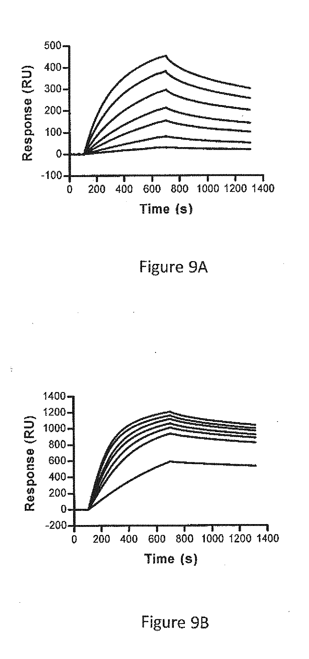
D00014


S00001
XML
uspto.report is an independent third-party trademark research tool that is not affiliated, endorsed, or sponsored by the United States Patent and Trademark Office (USPTO) or any other governmental organization. The information provided by uspto.report is based on publicly available data at the time of writing and is intended for informational purposes only.
While we strive to provide accurate and up-to-date information, we do not guarantee the accuracy, completeness, reliability, or suitability of the information displayed on this site. The use of this site is at your own risk. Any reliance you place on such information is therefore strictly at your own risk.
All official trademark data, including owner information, should be verified by visiting the official USPTO website at www.uspto.gov. This site is not intended to replace professional legal advice and should not be used as a substitute for consulting with a legal professional who is knowledgeable about trademark law.