Method Of Analyzing Genetically Abnormal Cells
MISHIMA; Yuji ; et al.
U.S. patent application number 16/183723 was filed with the patent office on 2019-03-14 for method of analyzing genetically abnormal cells. This patent application is currently assigned to OLYMPUS CORPORATION. The applicant listed for this patent is JAPANESE FOUNDATION FOR CANCER RESEARCH, OLYMPUS CORPORATION. Invention is credited to Takashi ABE, Kiyohiko HATAKE, Yuji MISHIMA.
| Application Number | 20190078153 16/183723 |
| Document ID | / |
| Family ID | 45622755 |
| Filed Date | 2019-03-14 |


| United States Patent Application | 20190078153 |
| Kind Code | A1 |
| MISHIMA; Yuji ; et al. | March 14, 2019 |
METHOD OF ANALYZING GENETICALLY ABNORMAL CELLS
Abstract
A method of analyzing eukaryotic cells includes conducting a reaction using an antibody against a surface antigen of a cell to be analyzed for genetic abnormality or a cell not to be analyzed for the abnormality, to distinguish cells to be analyzed for the abnormality, subjecting the cells to permeation treatment, and to immobilization treatment after at least one of the antigen-antibody reaction and permeation treatment steps, conducting fluorescence in situ hybridization using at least one nucleic acid probe, each probe having a different fluorescent label and specifically binding to a genetic sequence to be detected, obtaining fluorescence signals from the probes in the cells using a three-dimensional image analysis method, and determining whether any cells have the abnormality by analyzing the cells distinguished to be analyzed for the abnormality, at least one signal count of the fluorescence signals, the signal count corresponding respectively to the probe.
| Inventors: | MISHIMA; Yuji; (Tokyo, JP) ; HATAKE; Kiyohiko; (Tokyo, JP) ; ABE; Takashi; (Tokyo, JP) | ||||||||||
| Applicant: |
|
||||||||||
|---|---|---|---|---|---|---|---|---|---|---|---|
| Assignee: | OLYMPUS CORPORATION Tokyo JP JAPANESE FOUNDATION FOR CANCER RESEARCH Tokyo JP |
||||||||||
| Family ID: | 45622755 | ||||||||||
| Appl. No.: | 16/183723 | ||||||||||
| Filed: | November 7, 2018 |
Related U.S. Patent Documents
| Application Number | Filing Date | Patent Number | ||
|---|---|---|---|---|
| 13290231 | Nov 7, 2011 | |||
| 16183723 | ||||
| Current U.S. Class: | 1/1 |
| Current CPC Class: | C12Q 1/6841 20130101; C12Q 1/6886 20130101 |
| International Class: | C12Q 1/6841 20060101 C12Q001/6841; C12Q 1/6886 20060101 C12Q001/6886 |
Foreign Application Data
| Date | Code | Application Number |
|---|---|---|
| Nov 9, 2010 | JP | 2010-251107 |
Claims
1. A method of analyzing cells, the method comprising the steps of: (a) preparing a cell sample containing a plurality of eukaryotic cells, (b) conducting an antigen-antibody reaction to the plurality of cells contained in the cell sample after the step (a) using an antibody against a surface antigen of a type of cell to be analyzed for genetic abnormality or a type of cell which is not to be analyzed for the genetic abnormality, so as to distinguish cells of the type to be analyzed for the genetic abnormality among the plurality of cells contained in the cell sample, (c) subjecting the plurality of cells contained in the cell sample to a permeation treatment after the step (b), (d) subjecting the plurality of cells contained in the cell sample to an immobilization treatment after at least one of the step (b) and the step (c), (e) conducting FISH (fluorescence in situ hybridization) to the plurality of cells contained in the cell sample using at least one nucleic acid probe after the step (d), each of the at least one nucleic acid probe having a respective different fluorescent label, and each of the at least one nucleic acid probe specifically binding to a genetic sequence to be detected in analyzing for the genetic abnormality, (f) obtaining fluorescence signals from the at least one nucleic acid probe in the plurality of cells contained in the cell sample using a three-dimensional image analysis method after the step (e), and (g) determining whether or not any of the plurality of cells contained in the cell sample have the genetic abnormality by analyzing, among the cells distinguished as being cells of the type to be analyzed for the genetic abnormality in the step (b), at least one signal count of the fluorescence signals obtained in the step (f), the at least one signal count corresponding respectively to the at least one nucleic acid probe.
2. The method according to claim 1, wherein in the step (c), the permeation treatment is carried out by an enzyme reaction treatment with a proteolytic enzyme after an immersion treatment of the cells contained in the cell sample in a surfactant solution.
3. The method according to claim 1, further comprising the step of: (h) removing, from the cell sample prepared in the step (a), cells binding to an antibody which specifically binds to a molecule existing on a cell surface of a type of cell which is not to be analyzed for the genetic abnormality, before the step (b), wherein the step (b) is conducted after the step (h).
4. The method according to claim 1, further comprising the step of: (h) conducting an antigen-antibody reaction to the cells contained in the cell sample using at least one antibody, each of the at least one antibody specifically binding to a respective different molecule existing in an interior of eukaryotic cells, and each of the at least one antibody having a respective different fluorescent label, wherein the step (h) is conducted before the step (g) and after the step (c).
5. The method according to claim 1, wherein the at least one nucleic acid probe specifically binds to DNA of a cancer gene.
6. The method according to claim 1, wherein the cell sample comprises at least one of a body fluid, a sample containing cells separated from the body fluid, and a sample containing cells separated from a piece of tissue collected from a living body.
7. The method according to claim 1, wherein the cell sample comprises at least one of blood and a sample containing cells separated from blood.
8. The method according to claim 1, wherein: the cell sample comprises at least one of blood and a sample containing cells separated from blood; and the at least one nucleic acid probe is at least one of a nucleic acid probe which specifically binds to DNA of Her-2 gene, and a nucleic acid probe which specifically binds to DNA of CEP17 gene.
9. The method according to claim 3, wherein: the cell sample comprises at least one of blood and a sample containing cells separated from blood; the antibody used in the step (h) is an antibody which specifically binds to a molecule specifically existing on a cell surface of leukocyte cells; and the at least one nucleic acid probe is at least one of a nucleic acid probe which specifically binds to DNA of Her-2 gene, and a nucleic acid probe which specifically binds to DNA of CEP17 gene.
10. The method according to claim 4, wherein: the cell sample comprises at least one of blood and a sample containing cells separated from blood; the at least one antibody used in the step (h) is at least one antibody selected from the group consisting of an antibody which specifically binds to Cytokeratin, an antibody which specifically binds to CD45, an antibody which specifically binds to HER-2, and an antibody which specifically binds to EpCAM; and the at least one nucleic acid probe is at least one of a nucleic acid probe which specifically binds to DNA of Her-2 gene, and a nucleic acid probe which specifically binds to DNA of CEP17 gene.
11. The method according to claim 2, wherein: the surfactant solution is triton X-100; and the proteolytic enzyme is pepsin.
12. The method according to claim 2, further comprising the step of: (h) removing, from the cell sample prepared in the step (a), cells binding to an antibody which specifically binds to a molecule existing on a cell surface of a type of cell which is not to be analyzed for the genetic abnormality, before the step (b), wherein the step (b) is conducted after the step (h).
13. The method according to claim 2, further comprising the step of: (h) conducting an antigen-antibody reaction to the cells contained in the cell sample using at least one antibody, each of the at least one antibody specifically binding to a respective different molecule existing in an interior of eukaryotic cells, and each of the at least one antibody having a respective different fluorescent label, wherein the step (h) is conducted before the step (g) and after the step (c).
14. The method according to claim 3, further comprising the step of: (i) conducting an antigen-antibody reaction to the cells contained in the cell sample using at least one antibody, each of the at least one antibody specifically binding to a respective different molecule existing in an interior of eukaryotic cells, and each of the at least one antibody having a respective different fluorescent label, wherein the step (i) is conducted before the step (g) and after the step (c).
15. The method according to claim 12, further comprising the step of: (i) conducting an antigen-antibody reaction to the cells contained in the cell sample using at least one antibody, each of the at least one antibody specifically binding to a respective different molecule existing in an interior of eukaryotic cells, and each of the at least one antibody having a respective different fluorescent label, wherein the step (i) is conducted before the step (g) and after the step (c).
16. The method according to claim 1, wherein in the step (c), the permeation treatment is carried out by an enzyme reaction treatment with a proteolytic enzyme after an immersion treatment of the cells contained in the cell sample in a surfactant solution, the immersion treatment being conducted before the step (d), and the enzyme reaction treatment being conducted after the step (d).
17. The method according to claim 1, further comprising the step of: subjecting the plurality of cells contained in the cell sample to a crosslink treatment after the step (b) and before the step (e).
Description
CROSS-REFERENCE TO RELATED APPLICATIONS
[0001] This application is a Divisional application of U.S. patent application Ser. No. 13/290,231, filed Nov. 7, 2011, which is based on and claims priority from Japanese Patent Application No. 2010-251107, filed Nov. 9, 2010, the entire contents of both of which are incorporated herein by reference.
BACKGROUND OF THE INVENTION
Field of the Invention
[0002] The present invention relates to a method for detecting and analyzing cells having gene abnormality using fluorescence in situ hybridization (hereinafter abbreviated as FISH).
Description of the Related Art
[0003] Recently, molecularly-targeted drugs have intensively been developed. In particular, it can be expected that side effects are reduced as compared with conventional anticancer agents by using molecularly-targeted drugs, which act on a specific molecule involved in the onset and growth of cancer, in a cancer treatment. However, since molecularly-targeted drugs act on a specific molecule in vivo, sufficient therapeutic effects by medication sometimes cannot be expected depending on the genotype of the patient. Therefore, analysis of genetic mutation becomes more and more important for prediction of therapeutic effects of molecularly-targeted drugs.
[0004] For example, in various cancers such as breast cancer, ovarian cancer and stomach cancer, Her-2 gene amplification is carried out and a HER-2 protein is excessively expressed. It is said that breast cancer often has a poor prognosis when the HER-2 protein is excessively expressed. Also, a HER-2-targeting antibody drug (a humanized monoclonal antibody against HER-2) is effective against such HER-2-positive breast cancer, while therapeutic effects cannot be expected against HER-2-negative breast cancer in which excessive expression of the HER-2 protein is not recognized. Therefore, analysis of the presence or absence of Her-2 gene amplification is important so as to predict the presence or absence of adaptation of the HER-2-targeting antibody drug, a clinical course and the like.
[0005] In general, Her-2 gene amplification has been analyzed by FISH. Specifically, a nucleic acid probe (Her-2 probe) which binds to DNA of the Her-2 gene and a nucleic acid probe (CEP17 probe) which binds to a centromere DNA on chromosome 17 (CEP17) are labeled with different fluorescent substances, respectively, and the FISH is carried out using these two fluorescence-labeled nucleic acid probes. When the ratio of the total Her-2 signal count to the total CEP17 signal count (Her-2/CEP17 ratio) is a given value (for example, 2.0) or more, Her-2 gene amplification is positive (Her-2 gene amplification is carried out). When the ratio is less than the given value, it is determined that Her-2 gene amplification is negative.
[0006] Tumor cells systemically circulate and metastasize along the flow of blood and lymphocytes. Therefore, it is possible to test progression and metastatic property of tumor and to predict prognosis of tumor by detecting and analyzing tumor cells existing in the blood (peripheral circulatory tumor cells; CTCs). However, a large amount of leukocytes exist in blood, and a proportion of CTCs existing in blood is small. Moreover, it is difficult to determine whether cells are CTCs or not based on their morphology since a tissue structure of CTCs is not preserved unlike a tissue section or the like. Therefore, tumor cells are usually analyzed by separating and recovering epithelial cells or tumor cells from blood cells utilizing an antibody against a molecule specifically existing on the cell surface of epithelial cells or tumor cells, and then carrying out gene analysis, expression analysis or the like to detect the presence or absence of gene abnormality.
[0007] In a CTC analysis of breast cancer, for example, a method for analyzing whether cells are Her-2 gene amplification-positive cells or not is carried out by recovering EpCAM-positive cells from mononuclear cells in the blood using an antibody against EpCAM which is a cell surface antigen of epithelial cells, and carrying out FISH on the recovered EpCAM-positive cells using a Her-2 probe and a CEP17 probe to determine a Her-2/CEP17 ratio (see, for example, Non Patent Document 1).
[0008] The method for analyzing CTCs includes, in addition to the above method, a method in which floating cells having a cell surface antigen fluorescently stained are subjected to a fluorescence in situ hybridization method, and then nuclei are observed by focusing on the nuclei of the cells using a fluorescent microscope (see, for example, Patent Document 1). There is also a method in which an antibody, which specifically binds to biogenic substances other than a chromosome such as a cell surface antigen, and also has a reaction complex that cannot be detected with a fluorescent microscope, is bound to cells and the cells are subjected to FISH on the cells, and then a fluorescence-labeled substance, which reacts with the reaction complex, is bound to the cells (see, for example, Patent Document 2). A single cell is subjected to fluorescent immunostaining and FISH, and thus making it possible to reduce the requisite amount of a single specimen as compared with a case in which both are separately carried out.
PRIOR ART DOCUMENTS
Patent Documents
[0009] [Patent Document 1] Japanese Laid-Open Patent Application No. 2007-178193
[0010] [Patent Document 2] Japanese Laid-Open Patent Application No. 2009-530621
Non Patent Documents
[0011] [Non Patent Document 1] Flores et al., British Journal of Cancer, 2010, Vol. 102, No. 10, p.1495-1502
SUMMARY OF INVENTION
[0012] An object of the present invention is to provide a method for detecting and analyzing cells having gene abnormality in a highly accurate and simple manner using FISH.
[0013] The present inventors have diligently conducted experiments so as to achieve the above object and found that cells having gene abnormality can be detected in a highly accurate and simple manner even when only a trace amount of the cells exist in a specimen by carrying out immunostaining against a cell surface antigen and FISH to the same cell group, and carrying out analysis of fluorescence signals from the cell group using a three image analysis method. Thus, the present invention was acheived.
[0014] The present invention relates to following (1) to (11).
(1) a method of analyzing genetically abnormal cells, the method including the steps of:
[0015] (a) preparing a cell sample containing eukaryotic cells,
[0016] (b) conducting an antigen-antibody reaction to cells contained in the cell sample using one or more antibodies which specifically bind to a molecule existing on the cell surface of eukaryotic cells after the step (a),
[0017] (c) subjecting the cells contained in the cell sample to a permeation treatment after the step (b),
[0018] (d) subjecting the cells contained in the cell sample to an immobilization treatment after the step (b),
[0019] (e) conducting FISH (fluorescence in situ hybridization) to the cells contained in the cell sample using one or more nucleic acid probes after the step (d),
[0020] (f) analyzing fluorescence signals from the nucleic acid probes in the cells contained in the cell sample using a three-dimensional image analysis method after the step (e), and
[0021] (g) determining whether the cells contained in the cell sample are genetically abnormal cells or not based on the results of the step (b) and the results of the step (f).
(2) The method of analyzing genetically abnormal cells according to the above (1), wherein the permeation treatment is carried out by an enzyme reaction treatment with a proteolytic enzyme after an immersion treatment in a surfactant solution. (3) The method of analyzing genetically abnormal cells according to the above (1) or (2), further including the step of:
[0022] (h) removing cells binding to the antibody from the cell sample prepared in the step (a) using an antibody which specifically binds to a molecule existing on the cell surface of other cells than genetically abnormal cells which are analysis objects before the step (b), wherein
[0023] the step (b) is the step of subjecting cells remaining after removal of a portion of cells by the step (h) to an antigen-antibody reaction.
(4) The method of analyzing genetically abnormal cells according to any one of the above (1) to (3), further including the step of:
[0024] (i) conductiong an antigen-antibody reaction to the cells contained in the cell sample using one or more antibodies which specifically bind to a molecule existing in the interior of eukaryotic cells before the step (g) after the step (c).
(5) The method of analyzing genetically abnormal cells according to any one of the above (1) to (4), wherein the nucleic acid probes are nucleic acid probes specifically binding to DNA of the cancer gene. (6) The method of analyzing genetically abnormal cells according to any one of the above 1 to 5, wherein the cell sample is a body fluid, a sample containing cells separated from a body fluid, or a sample containing cells separated from a piece of tissue collected from the living body. (7) The method of analyzing genetically abnormal cells according to any one of the above (1) to (5), wherein the cell sample is blood, or a sample containing cells separated from blood. (8) The method of analyzing genetically abnormal cells according to any one of the above (1) to (5), wherein the cell sample is blood, or a sample containing cells separated from blood; and the nucleic acid probe is a nucleic acid probe specifically binding to DNA of the Her-2 gene, and a nucleic acid probe specifically binding to DNA of the CEP17 gene. (9) The method of analyzing genetically abnormal cells according to any one of the above (3) to (5), wherein the cell sample is blood, or a sample containing cells separated from blood; the antibody used in the step (h) is an antibody which specifically binds to a molecule specifically existing on the cell surface of leukocyte cells; and the nucleic acid probe is a nucleic acid probe specifically binding to DNA of the Her-2 gene, and a nucleic acid probe specifically binding to DNA of the CEP17 gene. (10) The method of analyzing genetically abnormal cells according to the above (4) or (5), wherein the cell sample is blood, or a sample containing cells separated from blood;
[0025] the antibody used in the step (b) is an antibody which specifically binds to a molecule specifically existing on the cell surface of leukocyte cells;
[0026] the antibody used in the step (h) is an antibody which specifically binds to a molecule specifically existing on the cell surface of leukocyte cells;
[0027] the antibody used in the step (i) is an antibody which specifically binds to one or more proteins selected from the group consisting of Cytokeratin, CD45, HER-2, and EpCAM; and
[0028] the nucleic acid probe is a nucleic acid probe specifically binding to DNA of the Her-2 gene, and a nucleic acid probe specifically binding to DNA of the CEP17 gene.
(11) The method of analyzing genetically abnormal cells according to any one of the above (2) to (10), wherein the surfactant solution is triton X-100; and the proteolytic enzyme is pepsin.
BRIEF DESCRIPTION OF THE DRAWINGS
[0029] FIG. 1 shows two-dimensional images prepared from three-dimensional images reconstructed with respect to one visual field containing Cytokeratin-positive cells in Example 1.
[0030] FIG. 2 shows two-dimensional images prepared from three-dimensional images reconstructed with respect to one visual field containing Cytokeratin-positive cells in Example 1.
[0031] FIG. 3 shows two-dimensional images prepared from three-dimensional image reconstructed with respect to a certain visual field in Example 1.
[0032] FIG. 4 shows two-dimensional images prepared from three-dimensional images reconstructed with respect to one visual field containing Cytokeratin-positive cells in Example 2.
[0033] FIG. 5 shows two-dimensional images prepared from three-dimensional images reconstructed with respect to one visual field containing Cytokeratin-positive cells in Example 3.
[0034] FIG. 6 shows two-dimensional images prepared from one visual field containing Cytokeratin-positive cells in Reference Example 1.
DETAILED DESCRIPTION OF THE INVENTION
[0035] The method for analyzing genetically abnormal cells of the present invention is characterized in that the same cells are subjected to immunostaining against a cell surface antigen and FISH. Since fluorescence signals of FISH are smaller than conventional immunostaining, it is often difficult to detect genetically abnormal cells among many cells only based on fluorescence signals of FISH. In the present invention, it is possible to detect genetically abnormal cells efficiently by carrying out immunostaining against a cell surface antigen, discriminating between cells that are likely to be genetically abnormal cells and cells not having a particular gene abnormality which is the analysis object among many cells contained in a cell sample, and preferentially analyzing fluorescence signals of FISH on cells of a cell group which possibly contain genetically abnormal cells. Also, immunostaining against a cell surface antigen is carried out to discriminate cell groups, and fluorescent immunostaining and FISH signals from cells after FISH are analyzed using a three-dimensional image analysis method, thereby making it possible to clearly and simply distinguish between an immunostaining image of a cell surface and fluorescence signals of FISH within a cell nucleus.
[0036] The gene abnormality which is the object of analysis in the method for analyzing genetically abnormal cells of the present invention may be a congenital abnormality or an acquired abnormality such as somatic mutation.
[0037] There is no particular limitation on the genetically abnormal cells which are objects of analysis in the method for analyzing genetically abnormal cells of the present invention as long as they are cells having gene abnormality, and are preferably cells having gene abnormality which is frequently observed in tumor cells. Examples of the genetically abnormal cells include cells having Her-2 gene amplification, cells having a Philadelphia chromosome and the like. The Philadelphia chromosome is a specific gene abnormality observed in chronic myeloid leukemia or the like.
[0038] The method for analyzing genetically abnormal cells of the present invention includes the steps of:
[0039] (a) preparing a cell sample containing eukaryotic cells,
[0040] (b) subjecting cells contained in the cell sample to an antigen-antibody reaction using one or more antibodies which specifically bind to a molecule existing on the cell surface of eukaryotic cells after the step (a),
[0041] (c) subjecting cells contained in the cell sample to a permeation treatment after the step (b),
[0042] (d) subjecting cells contained in the cell sample to an immobilization treatment after the step (b),
[0043] (e) subjecting cells contained in the cell sample to FISH (fluorescence in situ hybridization) using one or more nucleic acid probes after the step (d),
[0044] (f) analyzing fluorescence signals from the nucleic acid probes in cells contained in the cell sample using a three-dimensional image analysis method after the step (e), and
[0045] (g) determining whether cells contained in the cell sample are genetically abnormal cells or not from the results of the step (b) and the results of the step (f).
[0046] Each step will be described below.
[0047] First, as the step (a), a cell sample containing eukaryotic cells is prepared. There is no particular limitation on the cell sample as long as it contains eukaryotic cells. Examples of the cell sample include a biological sample, cells separated from a biological sample, a culture of cells separated from a biological sample, a cultured cell and the like. Examples of the biological sample include body fluids such as blood, lymph fluid, bone marrow fluid, ascites fluid, exudate fluid, amniotic fluid, sputum, saliva, semen, bile, pancreatic fluid or urine; and a piece of tissue collected from the living body, feces, an intestinal tract lavaged solution, a lung lavaged solution, a bronchi lavaged solution, a bladder lavaged solution and the like.
[0048] Examples of cells separated from the biological sample include a mononuclear cell component (buffy coat) separated and recovered from blood, a cell component separated and recovered from lymph fluid and the like, a solution of disjointed cells obtained by disintegration of connective tissues of a piece of tissue, a cell component separated and recovered after disjointing a piece of tissue and the like. There is no particular limitation on the method for obtaining a piece of tissue from the living body, or a biopsy specimen, a surgery sample and the like can be used.
[0049] The cell sample used in the present invention is preferably a body fluid, a sample containing cells separated from a body fluid, or a sample containing cells separated from a piece of tissue collected from the living body, more preferably a body fluid or a sample containing cells separated from a body fluid, further preferably blood, lymph fluid, or cells separated from them, and particularly preferably a mononuclear cell component. In this case, the preparation of a mononuclear cell component can be carried out by a conventional method such as a centrifugation method.
[0050] The cell sample may be provided for a next step (b) as it is, or may be used in the step (b) after removing at least a portion of cells having no specific gene abnormality which is the analysis object (non-genetically abnormal cells) such as normal cells contained in the cell sample. It is possible to increase the proportion of genetically abnormal cells in a cell sample subjected to the subsequent step and to increase detection sensitivity of genetically abnormal cells by removing non-genetically abnormal cells from a cell sample.
[0051] Specifically, removal of non-genetically abnormal cells can be carried out using an antibody which specifically binds to a molecule existing on the cell surface of non-genetically abnormal cells which are objects of analysis. It is possible to remove at least a portion of non-genetically abnormal cells which bind to the antibody from the cell sample by bringing a cell sample into contact with a carrier having an antibody bound and then separating the cell sample from the carrier.
[0052] When the cell sample is blood or a mononuclear cell component and genetically abnormal cells which are objects of analysis are CTCs, it is preferable to remove a portion or all of blood cell components such as leukocytes from the cell sample. Specifically, an antibody-bound carrier, which is prepared by binding an antibody of which an antigen is a molecule existing on the cell surface of leukocytes, platelets, red blood cells, or endothelial cells to a carrier such as magnetic beads or an agarose gel, is added to and mixed with blood or a mononuclear cell component. Subsequently, a liquid component after removing the carrier is used in the following step (b). Examples of antigens existing on the cell surface include CD45 existing on the surface of leukocytes, CD61 existing on the surface of platelets, CD235a existing on the surface of red blood cells, CD31 existing on the surface of endothelial cells and the like.
[0053] Examples of the technique for increasing the proportion of genetically abnormal cells in a cell sample include a method in which genetically abnormal cells are separated and recovered from a cell sample using an antibody against a surface antigen of genetically abnormal cells. When a particular cell group is separated and recovered using an antibody against a cell surface antigen. However, it is impossible to recover and analyze genetically abnormal cells having no surface antigen binding to an antibody used. Accordingly, it is preferred in the present invention that non-genetically abnormal cells are separated and removed from a cell sample so as to increase the proportion of genetically abnormal cells.
[0054] Next, as the step (b), an antigen-antibody reaction is carried out on cells contained in the above cell sample using one or more antibodies which specifically bind to a molecule existing on the cell surface of eukaryotic cells. It is possible to distinguish between a cell group containing genetically abnormal cells and a cell group not containing genetically abnormal cells by the presence or absence of the antigen-antibody reaction. For example, genetically abnormal cells can be detected and analyzed efficiently by carrying out an antigen-antibody reaction using a labeled antibody against a cell surface antigen of genetically abnormal cells, and preferentially analyzing, in subsequent FISH, fluorescence signals of cell nuclei in cells stained by binding of the labeled antibody (immunostaining). Conversely, genetically abnormal cells can be detected and analyzed efficiently by carrying out an antigen-antibody reaction using an antibody against a cell surface antigen of non-genetically abnormal cells, and preferentially analyzing, in subsequent FISH, fluorescence signals of cell nuclei in cells not having a labeled antibody bound and thus not stained.
[0055] The antigen-antibody reaction in the step (b) is a reaction in which a cell surface antigen is bound to an antibody specifically binding to the cell surface antigen. Thus, in the case of a direct method for immunostaining methods in which an antibody specifically binding to a cell surface antigen is directly labeled, all steps of the immunostaining are included in the step (b). On the other hand, in the case of an indirect method in which an antibody specifically binding to a cell surface antigen is used as a primary antibody and a secondary antibody (its antigen is the primary antibody) or streptavidin is labeled, only a reaction step in which the primary antibody is bound to a cell surface antigen is included in the step (b). In case a cell surface antigen is immunostained by an indirect method, an antigen-antibody reaction of the primary antibody bound to a cell surface antigen with a labeled secondary antibody or streptavidin may be carried out at any point before the step (f). For example, the reaction may be carried out successively with the step (b), simultaneously with the step (c), or after the step (d). When immunostaining is subsequently carried out on a molecule within cells, the reaction may be carried out simultaneously with the immunostaining.
[0056] Detection of an antibody bound to a cell surface antigen may be carried out by a fluorescence labeling method in which the antibody is labeled with a fluorescent substance or a fluorescent semiconductor quantum dot, or by an enzyme labeling method which uses a peroxidase-labeled antibody or an alkaline phosphatase-labeled antibody. In the present invention, the detection is preferably carried out using a fluorescence labeling method because it has high detection sensitivity and allows concomitant FISH analysis.
[0057] In the present invention, an antigen-antibody reaction of a cell surface antigen with an antibody specifically binding to the cell surface antigen is carried out on cells before a permeation treatment. Thus, in the case of a direct method, the permeation treatment is carried out after completion of an immune reaction. On the other hand, in the case of an indirect method, the permeation treatment is not carried out before completion of an antigen-antibody reaction of a primary antibody with cells. By carrying out an antigen-antibody reaction of a cell surface antigen with an antibody while not damaging a cell membrane, only the surface of the cell membrane can be stained unlike the case of carrying out the reaction after the permeation treatment. In other words, there is no overlap with the fluorescence signals of FISH and the S/N of FISH is improved by staining only the cell surface.
[0058] Next, as the step (c), a permeation treatment is carried out on cells contained in a cell sample. A pore is formed in a cell membrane or a nuclear membrane so that a nucleic acid probe for FISH can permeate into a nucleus by the permeation treatment. Examples of the permeation treatment include a treatment of immersing cells in a surfactant solution, an enzyme reaction treatment with a proteolytic enzyme and the like. It is possible to use, as the surfactant, Triton X-100, Tween 20, NP-40, saponin, digitonin and the like. It is possible to use, as the proteolytic enzyme, conventional enzymes such as pepsin and Proteinase K.
[0059] In the present invention, it is preferable to carry out an enzyme reaction treatment with a proteolytic enzyme, as a permeation treatment, after an immersion treatment in a surfactant solution. When the objects of analysis are CTCs, for example, the permeation treatment can be carried out by immersing cells in a 0.1 to 0.5% Triton X-100 solution for 1 to 30 minutes, preferably in a 0.1 to 0.3% Triton X-100 solution for 10 to 20 minutes, and more preferably in a 0.2% Triton X-100 solution for 15 minutes at room temperature, and then carrying out a pepsin treatment at room temperature for 0.5 to 10 minutes, preferably 0.5 to 5 minutes, and more preferably 1 to 2 minutes. Alternatively, the permeation treatment can be carried out by subjecting cells to an enzyme reaction treatment with a proteolytic enzyme such as a pepsin treatment without carrying out an immersion treatment in a surfactant solution.
[0060] When an immersion treatment in a surfactant solution and an enzyme reaction treatment with a proteolytic enzyme are carried out as a permeation treatment, both the treatments may be completed after an antigen-antibody reaction of a cell surface antigen with an antibody and before FISH. Both the treatments may be carried out continuously or discontinuously. In the present invention, it is preferable to carry out firstly an immersion treatment in a surfactant solution, then an immobilization treatment described below, and then an enzyme reaction treatment with a proteolytic enzyme.
[0061] After the step (b), as the step (d), an immobilization treatment of cells contained in a cell sample after an antigen-antibody reaction is carried out. It is possible to suppress denaturation of cells and an antibody bound by the immobilization treatment. Examples of the solution for the immobilization treatment include formalin, paraformaldehyde, glutaraldehyde, methanol, ethanol, acetone and the like. Treatment conditions such as types and concentrations of immobilization treatment solutions used for the immobilization treatment and treatment time can be determined suitably by taking account of types of cells, methods for immunostaining and the like so that cells and antibodies can be immobilized stably in subsequent steps and morphology of cells does not change so much. In the present invention, formalin is preferably used as an immobilization treatment solution.
[0062] After the step (b) and before the step (e), a crosslink treatment may be carried out on cells contained in a cell sample. It is possible to keep the binding of a cell surface antigen with an antibody stable by carrying out the crosslink treatment. In this case, the crosslink treatment may be carried out before a permeation treatment in the step (c) or an immobilization treatment in the step (d), or may be carried our after the permeation treatment or the immobilization treatment.
[0063] Examples of crosslinkers used for the crosslink treatment include DSS (Disuccinimidyl suberate), BS3 (Bis-sulfosuccinimidyl suberate) and the like. Treatment conditions such as types and concentrations of crosslinkers used and treatment time can be determined suitably by taking into account of the types of cells, methods for immunostaining and the like so that cells can be cross-linked with an antibody used in an immune reaction and fluorescence signals of subsequent FISH are not reduced largely. When the analysis objects are CTCs in the present invention, preferably the crosslink treatment is not carried out or the BS3 treatment is carried out, and more preferably the crosslink treatment is not carried out.
[0064] After the permeation treatment, as the step (e), FISH is carried out on cells contained in a cell sample using one, or two or more nucleic acid probes. Specifically, cells are immersed in a solution containing fluorescence-labeled nucleic acid probes which can bind to DNA in a chromosome which is the analysis object. The "nucleic acid probe which can bind to DNA" means a nucleic acid probe which can hybridize with a DNA fragment.
[0065] Nucleic acid probes used in the present invention may be those contributing to discrimination between cells having gene abnormality and normal cells, for example, those which can bind to DNA in a region having gene abnormality which is the object of analysis or those which can bind to DNA in a region not having the gene abnormality. In the present invention, it is preferable to use a nucleic acid probe which specifically binds to DNA of the cancer gene as a nucleic acid probe because tumor cells having gene abnormality can be analyzed.
[0066] In order to analyze the presence or absence of Her-2 gene amplification, for example, it is preferable to use a nucleic acid probe specifically binding to the Her-2 gene in combination with a nucleic acid probe specifically binding to DNA of CEP17. In this case, it can be determined that, when the Her-2/CEP17 ratio of a certain cell a given value or more, for example, 2.0 or more, preferably 2.2 or more, the cell is a genetically abnormal cell in which the Her-2 gene is abnormally amplified.
[0067] In order to analyze a Philadelphia chromosome, it is preferable to use a nucleic acid probe specifically binding to DNA of the c-abl gene in combination with a nucleic acid probe specifically binding to DNA of the bcr gene.
[0068] Next, as the step (f), fluorescence signals from the above nucleic acid probes in cells contained in the above cell sample are analyzed using a three-dimensional image analysis method. Thus, in the present invention, fluorescence signals in FISH are analyzed using a three-dimensional image analysis method. Specifically, in the three-dimensional image analysis method, two-dimensional images (tomographic images) of cross-sections of cells are shot and obtained sequentially by shifting focal length, and these images are combined to reconstruct a three-dimensional image. It is possible to detect labeled cells among many cells in a cell sample simply without extracting or recovering cells using the image analysis method. On the other hand, in a two-dimensional image analysis method, there is a possibility that multiple fluorescence signals overlapping in focus direction are misidentified as one fluorescence signal or a fluorescence signal by an immunostaining method. When signals are observed by a fluorescent microscope, it is necessary to observe cells by shifting focal length in order to avoid such a misidentification, and therefore, it is difficult to analyze many cells. To the contrary, by analyzing using a three-dimensional image analyzer, it is possible to analyze many cells at a time rapidly and simply with high accuracy.
[0069] Fluorescence signals in FISH can be detected and analyzed, for example, using a cell image analyzer which has a confocal optical system and allows fluorescence detection. Specifically, it is possible to use a confocal fluorescent microscope having an image capturing means such as a CCD camera, and the like. Also, when signals are analyzed by a two-dimensional image analysis method as described in Patent Document 1 or the like, FISH signals must be detected by observing cells as an observer gradually shifts focal length. Therefore, it is necessary for the observer to carry out observation by a microscope over a long period of time. To the contrary, in the present invention, it is possible to automatically construct a three-dimensional image in a single cell and then to analyze the resulting three-dimensional image on another day at another place. Also, it is easy to re-examine the image by preserving the three-dimensional image.
[0070] Finally, as the step (g), it is determined whether cells contained in the above cell sample are genetically abnormal cells or not, from the results of the antigen-antibody reaction of the step (b) and the results of the FISH of the step (f). It is determined whether the cells are genetically abnormal cells or not by analyzing the results of the FISH preferentially on cells determined as having a possibility of being genetically abnormal cells as a result of the antigen-antibody reaction.
[0071] For example, when a cell sample is blood or a mononuclear cell component and genetically abnormal cells which are analysis objects are CTCs, and in case an antigen-antibody reaction in the step (b) is carried out using an antibody which specifically binds to a molecule such as CD45 (cell surface antigen) specifically existing on the cell surface of leukocyte cells and FISH in the step (f) is carried out using a nucleic acid probe specifically binding to an amplification site observed in gene Her-2 gene amplification together with a nucleic acid probe specifically binding to DNA of the CEP17 gene, it can be determined that cells having a Her-2/CEP17 ratio of a given value or more, among cells to which an antibody does not bind in an antigen-antibody reaction in the step (b), are genetically abnormal cells.
[0072] After the permeation treatment of the step (d) and before the step (g), it is also possible to carry out immunostaining using one or more antibodies which specifically bind to a molecule existing in the interior of eukaryotic cells. In case a crosslink treatment is carried out in the present invention, for example, it is also possible to carry out immunostaining against a molecule within cells after the permeation treatment and then the crosslink treatment. The immunostaining can be carried out by a conventional method as in the case of an antigen-antibody reaction in the step (b). Intracellular molecules which serve as an antigen may be those existing within genetically abnormal cells, or may be those existing within non-genetically abnormal cells. Also, antibodies used in this case may be those contributing to discrimination between cells having gene abnormality and normal cells, or may be those used for further analyzing cells which are proved to be genetically abnormal cells.
[0073] In addition, cell nuclei may be stained after a permeation treatment of the step (d) and before the step (g) in the present invention. Each cell can be recognized more easily in an image analysis method by staining cell nuclei. In this case, cell nuclei can be stained by a conventional method suitably using a conventional nuclear staining agent such as DAPI (4',6-diamino-2-phenylindole), PI (propidium iodide), Hoechst and the like.
[0074] For example, when a cell sample is blood or a mononuclear cell component and genetically abnormal cells which are analysis objects are CTCs, it is possible to discriminate more clearly between blood cells and epithelial cells containing CTCs, together with an antigen-antibody reaction on the cell membrane surface in the step (b), using an antibody against a molecule mainly existing on leukocytes such as CD45, an antibody against a molecule mainly existing on the cell surface of epithelial cells such as EpCAM, an antibody against a molecule mainly existing in the interior of epithelial cells such as Cytokeratin, and the like. It is possible to detect genetically abnormal cells excessively expressing a HER-2 protein more clearly by carrying out an antigen-antibody reaction using an antibody specifically binding to HER-2. In the present invention, it is preferable to carry out an antigen-antibody reaction using an antibody which specifically binds to one or more proteins selected from the group consisting of Cytokeratin, CD45, HER-2, EpCAM, and FGFR (fibroblast growth factor receptor).
[0075] It is possible to detect and analyze genetically abnormal cells with high sensitivity and high efficiency by the method for analyzing genetically abnormal cells of the present invention. The analysis of genetically abnormal cells is not limited to only a determination of whether cells are genetically abnormal cells or not, or a determination of the degree of gene abnormality, and it is possible to carry out various analyses using an immunostaining method or FISH. For example, with the recent progress of fluorescence image analyzing techniques, it is possible to carry out immunostaining against multiple antigens on identical cells, using multiple fluorescent substances different in their fluorescence property and the like for labeling of antibodies. Thus, by carrying out immunostaining against a target antigen of a particular molecularly-targeted therapeutic agent, in addition to immunostaining which discriminate between a cell group containing genetically abnormal cells and a cell group not containing genetically abnormal cells, in the method for analyzing genetically abnormal cells of the present invention, it is also possible to carry out analysis of expression of the target antigen in the genetically abnormal cells and the like.
[0076] According to the method for analyzing genetically abnormal cells of the present invention, it is possible to detect and analyze cells having gene abnormality with a high accuracy and simple manner. In particular, the method for analyzing genetically abnormal cells of the present invention is suitable for analyzing genetically abnormal cells such as CTCs which exist in a specimen only in a trace amount.
EXAMPLES
[0077] The present invention will be described in more detail below by way of examples, but the invention is not limited thereto.
Example 1
[0078] Cells in blood were subjected to immunostaining against CD45 as well as FISH using a Her-2 FISH probe (nucleic acid probe specifically binding to the Her-2 gene) and a CEP17 FISH probe (nucleic acid probe specifically binding to CEP17).
Preparation of Cell Sample
[0079] 8 mL of blood derived from a stomach cancer patient (BD Vacutainer CPT mononuclear cell-separating blood collection tube, product code: #362753, manufactured by Becton, Dickinson and Company) was subjected to a specific gravity centrifugation treatment at 2,500 rpm for 30 minutes to divide into a plasma fraction and a mononuclear cell layer and a red blood cell layer, and the plasma fraction was removed to recover the mononuclear cell layer. EDTA-containing PBS was added to the recovered mononuclear cell layer to make a volume of 15 mL, and a centrifugation treatment was then carried out at 2,000 rpm for 10 minutes. The precipitated cells were then transferred to a dolphin tube, and further centrifugation treatment (swing arm, manufactured by Eppendorf Co. Ltd.) was carried out at 2,500 rpm for 3 minutes. After removing the supernatant, a MACS (registered trademark) buffer (manufactured by Miltenyi Boitec K. K.) was added to the precipitated cells to obtain 60 .mu.L of a cell suspension.
[0080] 20 .mu.L of CD45 microbeads (CD45-MB, manufactured by Miltenyi Boitec K. K.) and 20 .mu.L of CD61 microbeads (CD61-MB, manufactured by Miltenyi Boitec K. K.) were added to the cell suspension thus prepared, and the mixture was incubated at 4.degree. C. for 15 minutes. Non-binding microbeads in the cell suspension were washed with 1 mL of a MACS (registered trademark) buffer, and cells labeled with the microbeads were then suspended in 500 .mu.L of a MACS (registered trademark) buffer. Subsequently, a dolphin tube containing the suspension was mounted to AutoMACS (registered trademark, manufactured by Miltenyi Boitec K. K.), a "Depletes" program was conducted, and an N1 fraction (cell group binding to neither CD45-MB nor CD61-MB) was recovered in two dolphin tubes. The two dolphin tubes were subjected to a centrifugation treatment at 2,000 rpm for 10 minutes, and precipitated cells were then collected in one tube. A centrifugation treatment was again carried out at 2,000 rpm for 10 minutes, and the supernatant was removed to obtain a cell sample.
Immunostaining against Cell Surface Antigen and Surfactant Treatment
[0081] 50 .mu.L of a diluted solution of CD45-biotin was added to the cell sample thus prepared, and the mixture was incubated at room temperature for 10 minutes. The diluted solution of CD45-biotin was a solution obtained by diluting CD45-biotin (manufactured by Becton, Dickinson and Company) with a MACS (registered trademark) buffer to one tenth. Subsequently, the cells were washed with 1.5 mL of a MACS (registered trademark) buffer, 50 .mu.L of a diluted solution of Streptavidin-Qdot (registered trademark) 605 (SA-Q605) was added to the cells, and the mixture was incubated at room temperature for 15 minutes. Subsequently, the cells were washed with 1.5 mL of a MACS (registered trademark) buffer.
Crosslink Treatment
[0082] Next, 5 .mu.L of a 50 mM BS3 solution was added to the cell suspension, and the mixture was immediately pipetted and then incubated at room temperature for 15 minutes. Subsequently, 1 .mu.L of a 1 M Tris buffer was further added to the mixture, which was incubated at room temperature for 10 minutes. Subsequently, the cells were washed twice with 1.5 mL of EDTA-containing PBS.
Immobilization Treatment
[0083] The cells after washing were suspended in 50 .mu.L of IS-A [Dako Instruction Reagent A (formalin-containing fixative), manufactured by Dako] to incubate at room temperature for 15 minutes. Subsequently, the cells were washed with 1.5 mL of a MACS (registered trademark) buffer.
Immunostaining against Intracellular Antigen and Surfactant Treatment
[0084] A diluted solution of Cytokeratin-AF647 was added to the cell suspension after washing, and the mixture was incubated at room temperature for 15 minutes. The diluted solution of Cytokeratin-AF647 was a solution obtained by diluting Cytokeratin-Alexa 647 (manufactured by Abcam, an antibody obtained by covalently binding Alexa fluor 647 to clone AE1/AE3, 280 .mu.g/mL) with IS-B to one twenty-fifth. The cells after staining were washed with 1.5 mL of a MACS (registered trademark) buffer and then with 1 mL of PBS. Subsequently, the cells were suspended in 50 .mu.L of a potassium chloride solution (0.075 M) to incubate at room temperature for 15 minutes. The suspension was then subjected to a centrifugation treatment, and the supernatant was removed to leave 20 .mu.L of the potassium chloride solution. The resulting cell suspension was dropped on 5 MAS coat slides (manufactured by Matsunami Glass Ind., Ltd.) in an amount of one spot per slide, and air-dried.
Proteolytic Enzyme Treatment
[0085] A pepsin solution was added to MAS slides on which cells were mounted, and incubation was carried out at room temperature for 2 minutes. Then, the pepsin solution was removed from the slides, PBS was added to the slides, incubation was carried out at room temperature for 3 minutes, and the slides were air-dried. The pepsin solution was a solution obtained by diluting pepsin (manufactured by SIGMA) with a 10 mM hydrochloric acid solution to adjust to 100 .mu.g/mL.
Ethanol Treatment
[0086] MAS slides after the proteolytic enzyme treatment were immersed in a 70% ethanol solution for 1 minute, then in a 85% ethanol solution for 1 minute, further in a 100% ethanol solution for 1 minute, and then air-dried. Subsequently, the slides were further dried by a drier for 5 minutes on a block heater at 45.degree. C.
FISH
[0087] Stock solutions of a Her-2 FISH probe (Her-2-SpecrtrumOrange, manufactured by Vysis) and a CEP17 FISH probe (CEP17-SpectrumGreen, manufactured by Vysis) were added to the MAS slides after drying in an amount of 2 .mu.L per spot, cover glasses were further mounted on the slides and sealed with a paper bond. The MAS slides were incubated at 85.degree. C. for 3 minutes, and then at 37.degree. C. for 18 hours. After incubation, the bond was peeled off, the MAS slides were immersed in a post-hybridization washing solution (2.times.SSC, 0.3% NP40) and incubated at 75.degree. C. for 5 minutes. The MAS slides were rinsed with 2.times.SSC, and excess water was then removed.
Nuclear Staining
[0088] To the MAS slides, 2 .mu.L per spot of a DAPI solution was added, and the slides were then sealed with a top coat.
Measurement of Fluorescence Signals
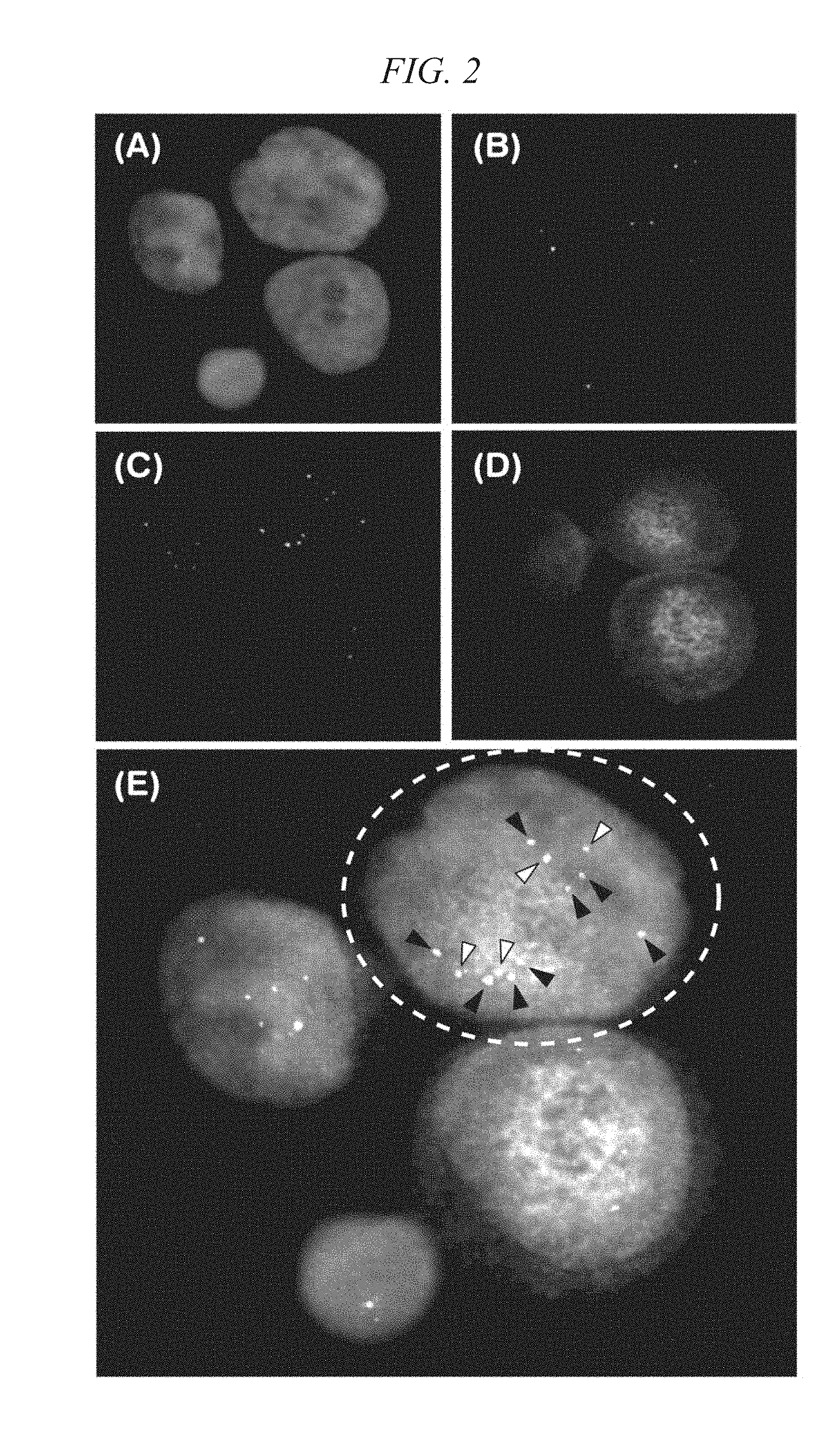
[0089] Fluorescence signals in the spots on the MAS slides after sealing were measured using a confocal laser scanning microscope FV-1000 (manufactured by Olympus Corp.) to construct three-dimensional images. FIGS. 1 and 2 show two-dimensional images prepared from three-dimensional images reconstructed with respect to one visual field containing Cytokeratin-positive cells, respectively. In FIGS. 1 and 2, (A) is a nucleus (DAPI) stained image, (B) is a signal image of the CEP17 FISH probe, (C) is a signal image of the Her-2 FISH probe, (D) is a stained image of Cytokeratin-Alexa 647, and (E) is an image obtained by overlapping (A) to (D).
[0090] As a result, signal counts of CEP17 were equal to signal counts of Her-2 in a Cytokeratin-positive cell in FIG. 1 [a cell surrounded by a dotted line in FIG. 1 (E)], and therefore, it was determined that the Her-2 gene of the Cytokeratin-positive cell was normal. On the other hand, in a right upper cell [a cell surrounded by a dotted line in FIG. 2(E)] among Cytokeratin-positive cells (cells stained by Cytokeratin-Alexa 647) in FIG. 2, the total count of CEP17 signals [signals shown by white arrowheads in FIG. 2(E)] was 4, the total count of Her-2 signals [signal shown by black arrowheads in FIG. 2(E)] was 8, and the Her-2/CEP17 ratio was 2. Thus, it was confirmed that a cell surrounded by a dotted line in FIG. 2(E) was a genetically abnormal cell having the Her-2 gene amplified. As is apparent from these results, the method for analyzing genetically abnormal cells of the present invention can analyze Her-2 gene amplification in a portion of Cytokeratin-positive cells contained in blood derived from cancer patients, i.e., that the method for analyzing genetically abnormal cells of the present invention can analyze CTCs in blood derived from cancer patients.
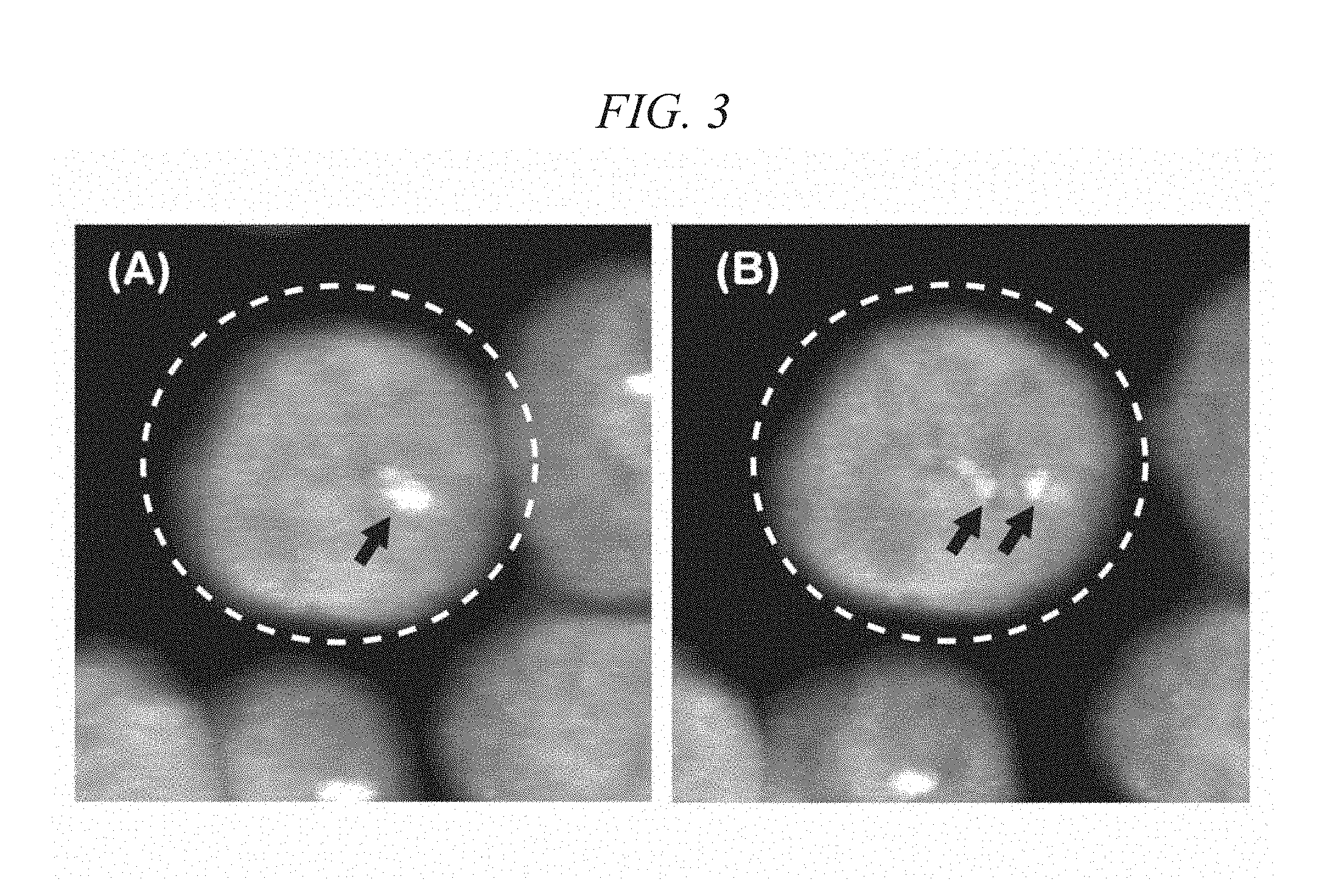
[0091] FIG. 3 shows two-dimensional images prepared from three-dimensional images reconstructed with respect to certain cells in which fluorescence signals were observed in this Example. FIGS. 3 (A) and 3(B) show two-dimensional images slightly different in an observing point. FIGS. 3 (A) and 3(B) are images obtained by overlapping a nucleus (DAPI) stained image and a signal image of a Her-2 FISH probe, and signals of the Her-2 FISH probe are shown by arrows. Signals of the Her-2 FISH probe in cells surrounded by a dotted line in FIGS. 3 (A) and 3(B) were detected as one signal in the two-dimensional image in FIG. 3(A) but detected as two separate signals in the two-dimensional image in FIG. 3(B). Thus, in the method for analyzing genetically abnormal cells of the present invention, multiple signals which are detected as one signal due to overlap in a Z-axis direction in a particular two-dimensional image can be detected easily in an individually separated manner by rotating a reconstructed three-dimensional image slightly.
Example 2
[0092] According to the method for analyzing genetically abnormal cells of the present invention, immunostaining against CD45 as well as FISH using a Her-2 FISH probe and a CEP17 FISH probe were carried out on endoscopic biopsy cells.
Preparation of Cell Sample
[0093] An endoscopic biopsy (about 1 mg) obtained from a stomach cancer patient was suspended in 0.25 mL of a DMEM medium, collagenase and dispase were then added to the suspension, and an enzyme treatment was carried out at 37.degree. C. for 60 minutes. Subsequently, the suspension was subjected to a centrifugation treatment at 2,000 rpm for 10 minutes, precipitated cells (mononuclear cells) were transferred to a dolphin tube, and a further centrifugation treatment (swing arm, manufactured by Eppendorf Co. Ltd.) was carried out at 2,500 rpm for 3 minutes. After removing the supernatant, a MACS (registered trademark) buffer (manufactured by Miltenyi Boitec K. K.) was added to the precipitated cells to obtain 60 .mu.L of a cell suspension.
[0094] A cell sample was prepared from the cell suspension prepared, in the same manner as in Example 1, by removing blood cell components by magnetic separation using CD45 microbeads and CD235a microbeads.
[0095] The cell sample prepared was sequentially subjected to immunostaining against a cell surface antigen and a surfactant treatment, a crosslink treatment, an immobilization treatment, immunostaining using Cytokeratin-AF647 and a surfactant treatment, a proteolytic enzyme treatment, an ethanol treatment, FISH, and nuclear staining in the same manner as in Example 1. Subsequently, fluorescence signals of stained cells were measured to construct three-dimensional images. FIG. 4 shows two-dimensional images prepared from the three-dimensional images reconstructed with respect to one visual field containing Cytokeratin-positive cells. In FIG. 4, FIG. 4(A) is a nucleus (DAPI) stained image, FIG. 4(B) is a signal image of the CEP17 FISH probe, FIG. 4(C) is a signal image of the Her-2 FISH probe, FIG. 4(D) is a stained image of Cytokeratin-Alexa 647, FIG. 4(E) is an image obtained by overlapping FIG. 4(A) to 4(D). As a result, signal counts of CEP17 were almost equal to signal counts of Her-2 in Cytokeratin-positive cells in FIG. 4, and therefore, it was determined that the Her-2 gene of the Cytokeratin-positive cells was normal.
Example 3
[0096] A cell sample was prepared by adding a tumor cell line to peripheral blood, and Her-2 gene-amplifying cells in the cell sample were analyzed according to the method for analyzing genetically abnormal cells of the present invention. As a tumor cell line, SKBr3 cells were used which were a cultured cell line derived from breast cancer. In this case, SKBr3 cells cultured by a conventional method were used.
Preparation of Cell Sample
[0097] First, 5,000 SKBr3 cells were added to 7.5 mL of peripheral blood obtained from healthy subjects. A mononuclear cell layer was recovered from the peripheral blood and further washed, and a MACS (registered trademark) buffer was then added to the layer to prepare 60 .mu.L of a cell suspension, in the same manner as in Example 1.
[0098] To the cell suspension thus prepared, 20 .mu.L of CD45 microbeads (CD45-MB, manufactured by Miltenyi Boitec K. K.) and 20 .mu.L of CD235a microbeads (CD235a-MB, manufactured by Miltenyi Boitec K. K.) were added, and the mixture was incubated at 4.degree. C. for 15 minutes. Non-binding microbeads in the cell suspension were washed with 1 mL of a MACS (registered trademark) buffer, and cells labeled with the microbeads were then suspended in 500 .mu.L of a MACS (registered trademark) buffer. Subsequently, a dolphin tube containing the suspension was loaded on AutoMACS (registered trademark, manufactured by Miltenyi Boitec K. K.) "Depletes", a "Depletes" program was conducted, and an N1 fraction (cell group binding to neither CD45-MB nor CD235a-MB) was recovered in two dolphin tubes. The two dolphin tubes were subjected to a centrifugation treatment at 2,000 rpm for 10 minutes, and precipitated cells were then collected in one tube. A centrifugation treatment was again carried out at 2,000 rpm for 10 minutes, and the supernatant was removed to obtain a cell sample.
Immunostaining against Cell Surface Antigen
[0099] 50 .mu.L of the diluted solution of CD45-biotin used in Example 1 was added to the cell sample prepared, and the mixture was incubated at room temperature for 10 minutes. The cells were washed with 1.5 mL of a MACS (registered trademark) buffer, 50 .mu.L of a diluted solution of Streptavidin-Qdot (registered trademark) 605 (SA-Q605) was added to the cells, and the mixture was incubated at room temperature for 15 minutes. In this connection, the diluted solution of SA-Q605 was a solution obtained by diluting SA-Q605 (manufactured by Invitrogen) with a MACS (registered trademark) buffer to one hundredth. Subsequently, the cells were washed twice with 1.5 mL of EDTA-containing PBS.
Immobilization Treatment
[0100] The cells after a permeation treatment were suspended in 50 .mu.L of IS-A [Dako Instruction ReagentA (formalin-containing fixative), manufactured by Dako] to incubate at room temperature for 15 minutes. Subsequently, the cells were washed with 1.5 mL of a MACS (registered trademark) buffer.
Immunostaining against Intracellular Antigen
[0101] To the cell suspension after washing, the diluted solution of Cytokeratin-AF647 used in Example 1 was added, and the mixture was incubated at room temperature for 15 minutes. The cells after staining were washed with 1.5 mL of a MACS (registered trademark) buffer and then with 1 mL of PBS. Subsequently, the cells were suspended in 50 .mu.L of a potassium chloride solution (0.075 M) to incubate at room temperature for 15 minutes. The suspension was then subjected to a centrifugation treatment, and the supernatant was removed to leave 20 .mu.L of the potassium chloride solution. The resulting cell suspension was dropped on 5 MAS coat slides (manufactured by Matsunami Glass Ind., Ltd.) in an amount of one spot per slide, and air-dried.
Proteolytic Enzyme Treatment
[0102] The pepsin solution used in Example 1 was added to MAS slides on which cells were mounted, and incubation was carried out at room temperature for 2 minutes. Then, the pepsin solution was removed from the slides, PBS was added to the slides, incubation was carried out at room temperature for 3 minutes, and the slides were air-dried.
Ethanol Treatment
[0103] MAS slides after the proteolytic enzyme treatment were immersed in a 70% ethanol solution for 1 minute, then in an 85% ethanol solution for 1 minute, further in a 100% ethanol solution for 1 minute, and then air-dried. Subsequently, the slides were further dried by applying a drier for 5 minutes on a block heater at 45.degree. C.
FISH and Nuclear Staining
[0104] FISH was carried out on MAS slides after drying, in the same manner as in Example 1. Subsequently, a DAPI solution was added to the slides, and the slides were sealed with a top coat.
Measurement of Fluorescence Signals
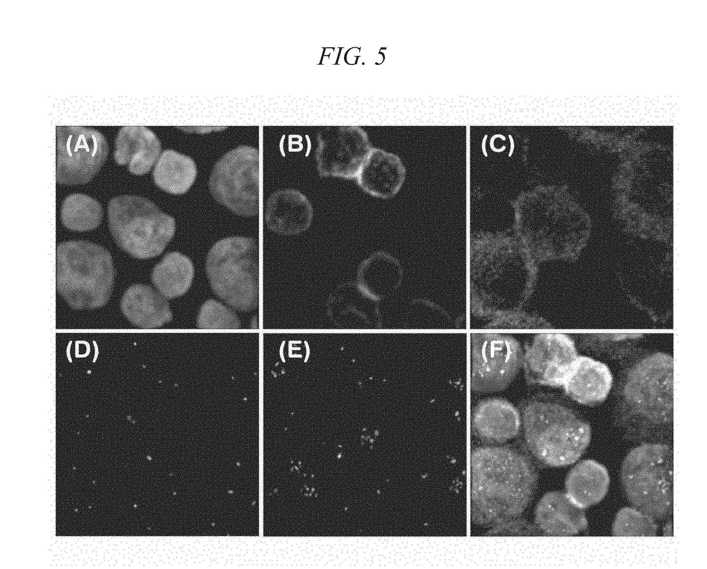
[0105] Fluorescence signals in the spots on the MAS slides after sealing were measured using confocal laser scanning microscope FV-1000 (manufactured by Olympus Corp.) to construct three-dimensional images. FIG. 5 shows two-dimensional images prepared from three-dimensional images reconstructed with respect to one visual field containing Cytokeratin-positive cells. In FIG. 5, FIG. 5(A) is a nucleus (DAPI) stained image, FIG. 5(B) is a stained image of CD45-biotin/Streptavidin-Qdot, FIG. 5(C) is a stained image of Cytokeratin-Alexa 647, FIG. 5(D) is a signal image of the CEP17 FISH probe, FIG. 5(E) is a signal image of the Her-2 FISH probe, and FIG. 5(F) is an image obtained by overlapping FIGS. 5(A) to 5(E).
[0106] As a result, the proportion of Cytokeratin-positive cells in total cells contained in the cell sample was very high, and the Cytokeratin-positive cells were efficiently concentrated by a magnetic separation treatment using CD45 microbeads and CD235a microbeads, as is apparent from FIGS. 5 (B) and 5(C). Also, the ratio of signal counts of CEP17 to signal counts of Her-2 in Cytokeratin-positive/CD45-negative cells in FIG. 5 was 3.5 on average, and therefore, it was determined that the Her-2 gene of the Cytokeratin-positive cells was amplified.
Reference Example 1
[0107] A cell sample was prepared for analyzing CTCs in blood, and epithelial cells contained in the cell sample were detected by immunostaining, in the same manner as in Example 1.
[0108] First, a mononuclear cell layer was recovered from 8 mL of blood derived from a stomach cancer patient (BD Vacutainer CPT mononuclear cell-separating blood collection tube, product code: #362753, manufactured by Becton, Dickinson and Company) and further washed, and a MACS (registered trademark) buffer was then added to the layer to prepare 60 .mu.L of a cell suspension, in the same manner as in Example 1. A cell sample was prepared from the cell suspension thus prepared, in the same manner as in Example 1, by removing leukocyte components by magnetic separation using CD45 microbeads and CD61 microbeads.
[0109] Next, 25 .mu.L of a diluted solution of EpCAM-Alexa Fluor 488 and 25 .mu.L of a diluted solution of CD45-biotin were added to the cell sample thus prepared, and the mixture was incubated at room temperature for 10 minutes. The diluted solution of EpCAM-Alexa Fluor 488 was a solution obtained by diluting EpCAM-Alexa Fluor 488 (manufactured by BioLegend) with a MACS (registered trademark) buffer to one twentieth. The diluted solution of CD45-biotin was a solution obtained by diluting CD45-biotin (manufactured by Becton, Dickinson and Company) with a MACS (registered trademark) buffer to one fifth. Subsequently, the cells were washed with 1.5 mL of a MACS (registered trademark) buffer, 50 .mu.L of a diluted solution of Streptavidin-Qdot (registered trademark) 605 (SA-Q605) was added to the cells, and the mixture was incubated at room temperature for 15 minutes. Subsequently, the cells were washed with 1.5 mL of a MACS (registered trademark) buffer.
[0110] The cells after a permeation treatment were sequentially subjected to an immobilization treatment, immunostaining using Cytokeratin-AF647, a proteolytic enzyme treatment, and an ethanol treatment in the same manner as in Example 3. Subsequently, in order to carry out nuclear staining, 2 .mu.L per spot of a DAPI solution was added to MAS slides after drying, and the slides were then sealed with a top coat.
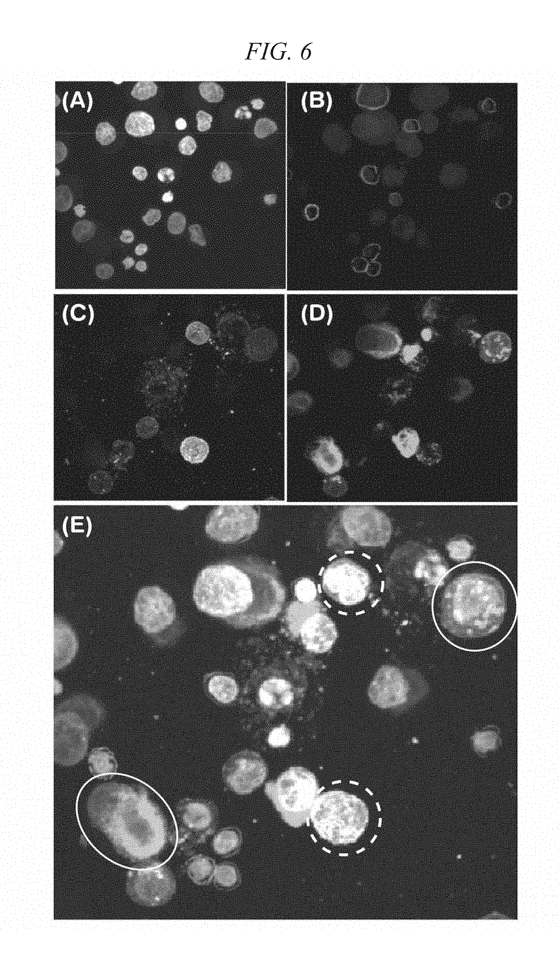
[0111] Fluorescence signals in the spots on the MAS slides after sealing were measured using confocal laser scanning microscope FV-1000 (manufactured by Olympus Corp.). FIG. 6 shows two-dimensional images prepared from three-dimensional images reconstructed with respect to one visual field containing Cytokeratin-positive cells. In FIG. 6, FIG. 6(A) is a nucleus (DAPI) stained image, FIG. 6(B) is a stained image of CD45-biotin/Streptavidin-Qdot, FIG. 6(C) is a stained image of EpCAM-Alexa Fluor 488, FIG. 6(D) is a stained image of Cytokeratin-Alexa 647, and FIG. 6(E) is an image obtained by overlapping (A) to (D).
[0112] As is apparent from FIG. 6(C) and FIG. 6(D), expression of an EpCAM antigen remarkably differs from one Cytokeratin-positive cell to another, and the cells contain both EpCAM-positive cells and EpCAM-negative cells. For example, cells in the region surrounded by a dashed line in FIG. 6(E) are Cytokeratin-positive and EpCAM-positive. On the other hand, cells in the region surrounded by a solid line are Cytokeratin-positive but express little EpCAM. Thus, in case EpCAM-positive cells are selectively recovered from a mononuclear cell layer recovered from blood as in the method described in Flores et al., British Journal of Cancer, 2010, Vol. 102, No. 10, p.1495-1502., EpCAM-negative CTCs are excluded from the analysis object, and therefore, it is difficult to detect CTCs in a blood specimen accurately. Accordingly, in order to increase the proportion of the presence of CTCs in a cell sample when analyzing CTCs in a blood specimen, the method for analyzing genetically abnormal cells of the present invention does not adopt a step of selectively recovering epithelial cells from a mononuclear cell layer recovered from blood utilizing a cell surface antigen on epithelial cells, but preferably adopts a step of separating and removing blood cells utilizing a cell surface antigen on blood cells.
INDUSTRIAL APPLICABILITY
[0113] According to the method for analyzing genetically abnormal cells of the present invention, since it is possible to detect and analyze genetically abnormal cells, which exist in a normal cell group only in a trace amount, in a highly accurate and simple manner, the present method is suitably utilized in the fields of clinical tests such as analysis of peripheral circulatory tumor cells and the like.
* * * * *
D00001
D00002
D00003
D00004
D00005
D00006
XML
uspto.report is an independent third-party trademark research tool that is not affiliated, endorsed, or sponsored by the United States Patent and Trademark Office (USPTO) or any other governmental organization. The information provided by uspto.report is based on publicly available data at the time of writing and is intended for informational purposes only.
While we strive to provide accurate and up-to-date information, we do not guarantee the accuracy, completeness, reliability, or suitability of the information displayed on this site. The use of this site is at your own risk. Any reliance you place on such information is therefore strictly at your own risk.
All official trademark data, including owner information, should be verified by visiting the official USPTO website at www.uspto.gov. This site is not intended to replace professional legal advice and should not be used as a substitute for consulting with a legal professional who is knowledgeable about trademark law.