Methods For The In Vitro Manufacture Of Gastric Fundus Tissue And Compositions Related To Same
Wells; James ; et al.
U.S. patent application number 16/084599 was filed with the patent office on 2019-03-14 for methods for the in vitro manufacture of gastric fundus tissue and compositions related to same. The applicant listed for this patent is Children's Hospital Medical Center. Invention is credited to Kyle McCracken, James Wells.
| Application Number | 20190078055 16/084599 |
| Document ID | / |
| Family ID | 60203655 |
| Filed Date | 2019-03-14 |


View All Diagrams
| United States Patent Application | 20190078055 |
| Kind Code | A1 |
| Wells; James ; et al. | March 14, 2019 |
METHODS FOR THE IN VITRO MANUFACTURE OF GASTRIC FUNDUS TISSUE AND COMPOSITIONS RELATED TO SAME
Abstract
The instant disclosure relates to methods for converting mammalian definitive endoderm (DE) cells into specific tissue(s) or organ(s) through directed differentiation. In particular, the disclosure relates to formation of gastric fundus tissue and/or organoids formed from differentiated definitive endoderm.
| Inventors: | Wells; James; (Cincinnati, OH) ; McCracken; Kyle; (Cincinnati, OH) | ||||||||||
| Applicant: |
|
||||||||||
|---|---|---|---|---|---|---|---|---|---|---|---|
| Family ID: | 60203655 | ||||||||||
| Appl. No.: | 16/084599 | ||||||||||
| Filed: | May 5, 2017 | ||||||||||
| PCT Filed: | May 5, 2017 | ||||||||||
| PCT NO: | PCT/US17/31309 | ||||||||||
| 371 Date: | September 13, 2018 |
Related U.S. Patent Documents
| Application Number | Filing Date | Patent Number | ||
|---|---|---|---|---|
| 62332194 | May 5, 2016 | |||
| Current U.S. Class: | 1/1 |
| Current CPC Class: | C12N 2501/155 20130101; C12N 2506/45 20130101; C12N 2501/385 20130101; C12N 5/0607 20130101; C12N 2501/11 20130101; C12N 5/0679 20130101; C12N 2506/025 20130101; C12N 5/0609 20130101; C12N 2501/115 20130101; C12N 2506/03 20130101; C12N 5/0606 20130101; C12N 2513/00 20130101; C12N 2501/415 20130101 |
| International Class: | C12N 5/071 20060101 C12N005/071; C12N 5/0735 20060101 C12N005/0735; C12N 5/074 20060101 C12N005/074; C12N 5/075 20060101 C12N005/075 |
Goverment Interests
GOVERNMENT SUPPORT CLAUSE
[0002] This invention was made with government support under AI116491 and DK092456. The government has certain rights in the invention.
Claims
1. An in vitro method of inducing formation of a gastric fundus tissue, comprising the steps of: a) contacting a mammalian definitive endoderm (DE) cell with a wnt pathway activator, an FGF signaling pathway activator, a BMP signalling pathway inhibitor, and retinoic acid, for a first period, wherein said first period is for a length of time sufficient to form a three-dimensional posterior foregut spheroid from said definitive endoderm; b) contacting said three-dimensional posterior foregut spheroid with a growth factor, said Wnt signalling pathway activator, said EGF signalling pathway activator, said BMP signalling pathway inhibitor, and retinoic acid for a second period, wherein said second period is for a length of time sufficient to induce a fundic lineage comprising fundal hGOs (hFGOs); c) culturing said hFGOs of step b) with said wnt pathway activator and said EGF signalling pathway activator for a third period; d) culturing said hFGOs of step c) with said wnt signalling pathway activator, said EGF signalling pathway activator, and FGF10 for a fourth period; e) contacting said hFGOs of step d) with a MEK inhibitor for a fifth period, wherein said fifth period is for a period of time sufficient to form said gastric fundus tissue comprising a functional fundic cell type.
2. The method of claim 1, wherein said first period is three days.+-.24 hours and wherein said retinoic acid is added for the third day of said period.+-.24 hours.
3. The method of claim 1, wherein said second period is three days.+-.24 hours.
4. The method of claim 1, wherein said third period is 11 days.+-.24 hours.
5. The method of claim 1, wherein said fourth period is 10 days.+-.24 hours.
6. The method of claim 1, wherein said fifth period is a two day period.+-.24 hours.
7. The method of claim 1, wherein step e) further comprises the step of contacting said fundal hGOs with an activator of BMP4 signalling.
8. The method of claim 1, wherein said functional fundic cell type is a parietal cell that expresses proton pump proteins and secretes acid.
9. The method of claim 1, wherein said functional fundic cell type is a chief cell that secretes pepsinogen.
10. The method of claim 1, wherein said step e is carried out for a period of time sufficient to develop SOX2+GATA+PDX1- epithelium.
11. The method of claim 1, wherein said step d and step e are carried out for a period of time sufficient to confer stable expression of lineage markers MUC5AC, MUC6, PGC, and GHRL.
12. The method of claim 1, wherein said definitive endoderm is derived from a precursor cell selected from an embryonic stem cell, an embryonic germ cell, an induced pluripotent stem cell, a mesoderm cell, a definitive endoderm cell, a posterior endoderm cell, a posterior endoderm cell, and a hindgut cell, a definitive endoderm derived from a pluripotent stem cell, a definitive endoderm derived from a pluripotent stem cell selected from an embryonic stem cell, an adult stem cell, or an induced pluripotent stem cell.
13. The method of claim 1, wherein said definitive endoderm is derived from contacting a pluripotent stem cell with one or more molecules selected from Activin, the BMP subgroups of the TGF-beta superfamily of growth factors; Nodal, Activin A, Activin B, BMP4, Wnt3a, and combinations thereof.
14. The method of claim 1, wherein said WNT pathway activator is one or more molecules selected from Wnt1, Wnt2, Wnt2b, Wnt3, Wnt3a, Wnt4, Wnt5a, Wnt5b, Wnt6, Wnt7a, Wnt7b, Wnt8a, Wnt8b, Wnt9a, Wnt9b, Wnt10a, Wnt10b, Wnt11, and Wnt16.
15. The method of claim 1, wherein said BMP signalling pathway inhibitor is selected from Noggin, Dorsomorphin, LDN189, DMH-1, and combinations thereof.
16. The method of claim 1, wherein said steps are conducted in vitro.
17. A composition comprising gastric tissue produced according to claim 1, wherein said gastric tissue is characterized by being free of innervation and/or blood vessels.
18. An in vitro method of inducing formation of a gastric fundus tissue, comprising the steps of contacting a fundal hGO (hFGO) with a wnt pathway activating agent and an EGF signalling pathway activating agent for a first period, and a MEK inhibitor for a second period, wherein said first and second periods are carried out for a period of time sufficient to form a functional fundic cell type; wherein said hFGO are obtained by contacting a three-dimensional posterior foregut spheroid with a growth factor, a wnt pathway activating agent, an EGF signalling pathway activator, a BMP signalling pathway inhibitor, and retinoic acid for a period of time sufficient to convert said three-dimensional posterior foregut spheroid to said hFGO; wherein said three-dimensional posterior foregut spheroids are obtained by contacting a mammalian definitive endoderm (DE) cells with a wnt pathway activating agent, an FGF signaling pathway activating agent, a BMP signalling pathway inhibitor, and retinoic acid.
Description
CROSS REFERENCE TO RELATED APPLICATIONS
[0001] This application claims priority to and benefit of U.S. Provisional Patent Application 62/332,194, filed May 5, 2016, the contents of which are incorporated by reference in their entirety.
BACKGROUND
[0003] Despite the global prevalence of gastric disease, there are few adequate models to study the fundus epithelium of the human stomach. The development of human fundic-type gastric organoids (hFGOs) would be a novel and powerful model system to study the molecular basis of human gastric physiology, pathophysiology, and drug discovery.
BRIEF SUMMARY
[0004] The instant disclosure relates to methods for converting mammalian definitive endoderm (DE) cells into specific tissue(s) or organ(s) through directed differentiation. In particular, the disclosure relates to formation of gastric fundus tissue and/or organoids formed from differentiated definitive endoderm.
BRIEF DESCRIPTION OF THE DRAWINGS
[0005] Those of skill in the art will understand that the drawings, described below, are for illustrative purposes only. The drawings are not intended to limit the scope of the present teachings in any way.
[0006] FIG. 1. Wnt/.beta.-catenin signaling is required for specification of the embryonic fundus in mice. a, Pdx1 and Sox2 were expressed in the antrum (a), whereas Pdx1 was absent in the fundus (f), identified by Atp4b-expressing parietal cells at E18.5. b, X-gal staining of an E10.5 foregut from an Axin2:LacZ reporter embryo showed that Wnt activity was restricted to the anterior domain of the stomach but excluded from the posterior stomach. c, Deletion of .beta.-catenin in the gastric epithelium caused an anterior expansion of Pdx1 into the fundic region of the stomach. d, In E18.5 Shh.sup.Cre/+.beta.-catenin.sup.fl/fl (cKO) embryos, Pdx1 was expressed throughout the stomach, except in some remaining patches of parietal cell-containing epithelium. Insets 1a-c and 2a-c show boxed regions in control and cKO stomach, respectively. e, In the cKO stomach, Ctnnb1 exhibited mosaic deletion, and parietal cells only differentiated in Ctnnb1-sufficient epithelium. Scale bars, 250 .mu.m (a), 200 .mu.m (c), and 500 .mu.m (d and e).
[0007] FIG. 2. .beta.-catenin activation promotes fundus development from human foregut progenitor spheroids. a, Schematized diagram of differentiation protocol for both fundic and antral hGOs. b, c, At day 9, CHIR-treated organoids exhibited reduction in PDX1, increase in IRX2, IRX3, and IRX5, and no change in gastric markers SOX2 or GATA4. *, p<0.05; two-tailed Student's t-test; n=3 biological replicates, data representative of 4 independent experiments. d, hFGOs grew comparably to hAGOs, but also exhibited glandular budding morphogenesis (white arrowheads). e, Both hGOs contained epithelium that expressed CDH1, KRT8, and CTNNB1, as well as gastric markers GATA4 and CLDN18. hAGOs exhibited nearly ubiquitous PDX1 expression while hFGOs did not. Scale bars, 50 .mu.m (c), 500 .mu.m (d) and 100 .mu.m (e). Error bars represent s.e.m.
[0008] FIG. 3. Differentiation of mucous and endocrine cell lineages in hGOs. a, Schematic of the shared and distinct lineages found in fundic and antral glands of the stomach. b, Both antral and fundic hGOs contained MUC5AC-positive surface mucous cells and MUC6-positive mucous neck cells. c, d, hFGOs contained endocrine cells expressing the pan-endocrine marker SYP. Diverse hormone cell types were identified in hFGOs, including GHRL-, SST-, and histamine-expressing endocrine cells. The antral-specific G-cell marker GAST was expressed in hAGOs but not hFGOs; conversely, GHRL was enriched in hFGOs. **, p<0.01; two-tailed Student's t-test; n=8 and 24 biological replicates in hAGOs and hFGOs respectively, data representative of 6 independent experiments. e, hAGOs, but not hFGOs, were competent to give rise to antral-specific GAST-expressing endocrine cells in response to expression of the pro-endocrine transcription actor NEUROG3 (+dox). *, p<0.01; two-tailed Student's t-test; n=4 biological replicates, data representative of 3 independent experiments. Error bars represent s.e.m.
[0009] FIG. 4. Formation of chief cells in hFGOs. a, hFGOs had a both MIST1 and Pepsinogen C (PGC) positive cells. b, High magnification of boxed region in panel (a) showing a gland with a cluster of cells with apical PGC staining. c, hFGOs had significantly increased expression of chief cell markers PGA5 (1,000-fold), PGC (100-fold), and MIST1 (>10-fold) as compared to hAGOs. **, p<0.05; two-tailed Student's t-test. n=3 biological replicates, data representative of 4 independent experiments. d, Transmission electron micrograph of an hFGO cell containing dense zymogen granules, indicative of a chief cell. e, Pepsinogen protein content in hFGOs as compared to hAGOs in the presence or absence of the MEK inhibitor (PD03). **, p<0.0001 compared to hAGOs, two-tailed Student's t-test, n=8, 12, and 11 biological replicates in hAGOs, control hFGOs and hFGOs (no PD03), respectively. Scale bars, 200 .mu.m (a), 25 .mu.m (b), and 10 .mu.m (d). Error bars represent s.e.m.
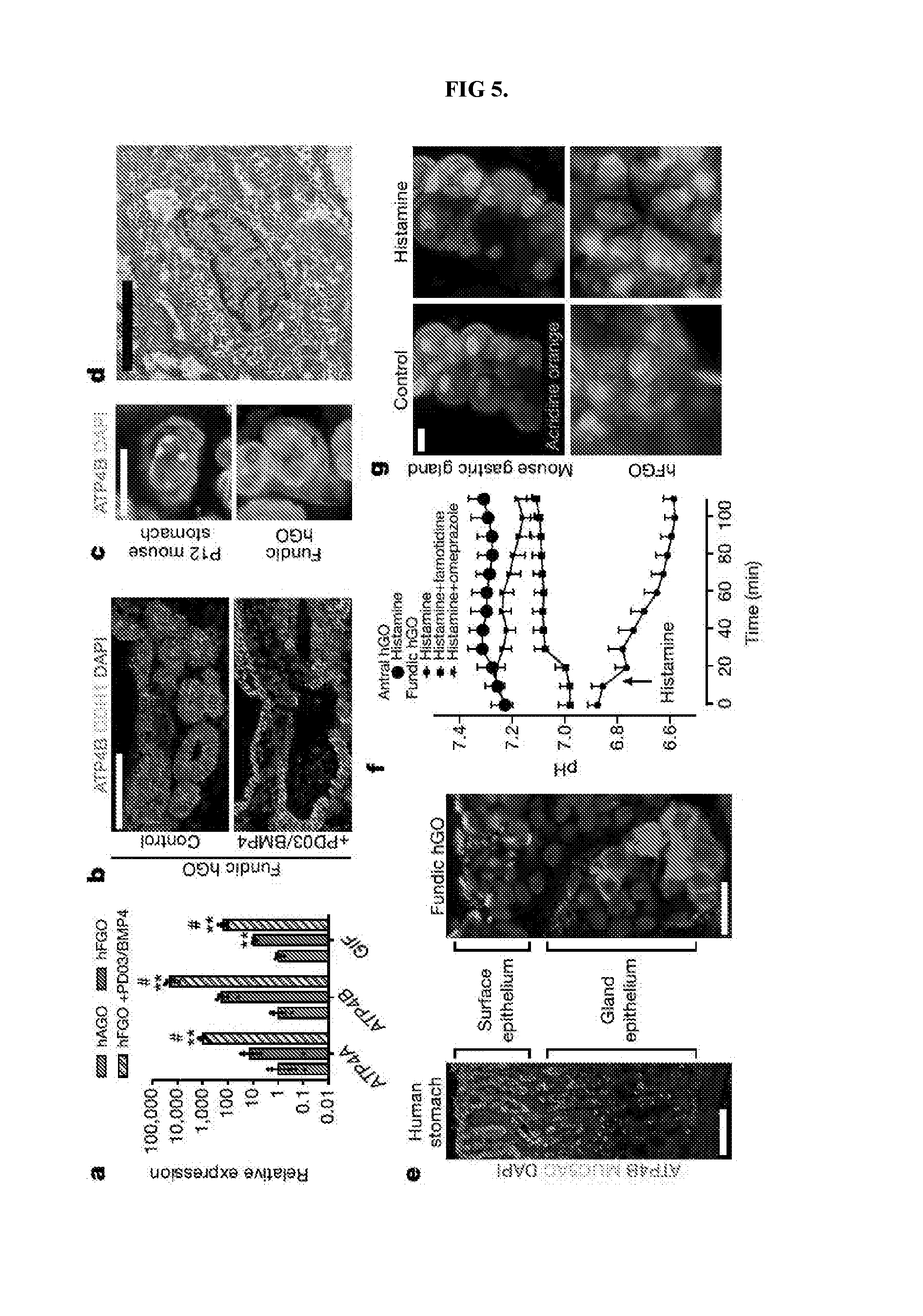
[0010] FIG. 5. Identification of pathways that drive differentiation of functional parietal cells in hFGOs. a, Expression of parietal cell genes ATP4, ATP4B, and GIF exhibited 10-100-fold increase in hFGOs compared to antral at baseline, but was dramatically increased by exposing hFGOs to a two-day pulse of PD03/BMP4. **, p<0.05 compared to hAGOs; #, p<0.05 compared to control hFGOs, two-tailed Student's t-test, n=4 biological replicates, data representative of 15 independent experiments. b, Stimulated differentiation of ATP4B-expressing parietal cells following treatment with PD03/BMP4. c, hFGO-derived parietal cells resembled those found in the maturing mouse fundic epithelium in vivo. d, Transmission electron micrograph of an hFGO cell with canalicular structure reminiscent of parietal cells. e, The epithelium of human fundic glands and hFGO epithelium were organized into MUC5AC-expressing cells in the surface epithelium and ATP4B-expressing parietal cells in the glandular units. f, Analysis of luminal pH in organoids in response to histamine by luminal injection of SNARF-5F. The luminal pH in hFGOs rapidly dropped, while hAGOs exhibited no response. The acidification was blocked by pretreating the organoids with either famotidine or omeprazole. n=9, 9, 7, and 4 biological replicates in hFGOs (histamine), hFGOs (histamine and famotidine), hFGOs (histamine and omeprazole), and hAGOs (histamine), respectively; data representative of three independent experiments. g, Histamine induced acridine orange (AO) dye accumulation in a canalicular-type pattern in isolated mouse gastric glands and in hFGOs after 60 minutes. Scale bars, 100 .mu.m (b), 10 .mu.m (c), 10 .mu.m (d), 100 .mu.m (e; human fundus), 20 .mu.m (e; hFGO), and 10 .mu.m (g). Error bars represent s.e.m.
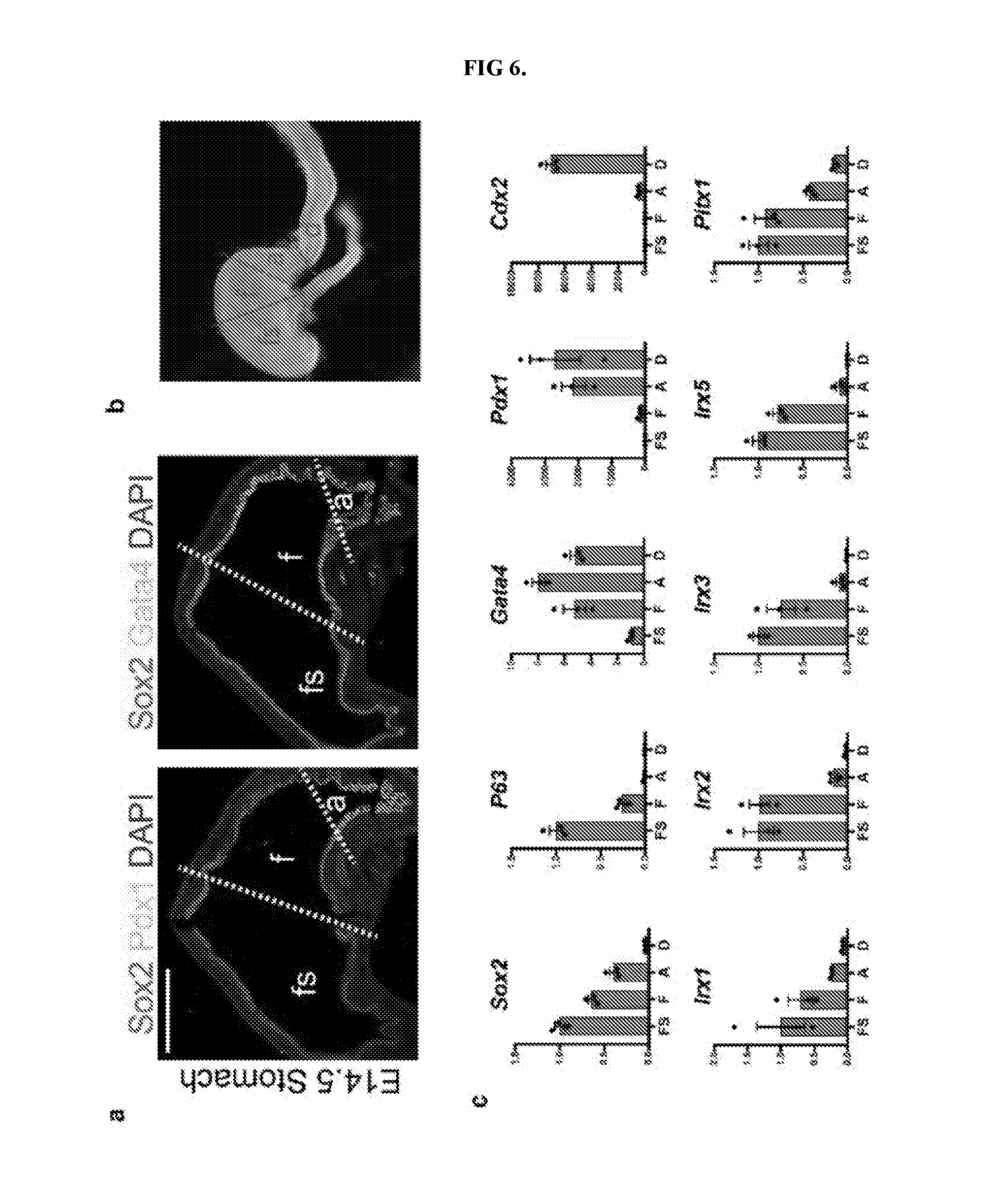
[0011] FIG. 6. Defining molecular domains in the developing stomach in vivo. a, Analysis of Sox2, Pdx1, and Gata4 in the embryonic mouse stomach (E14.5) showed that the fundus (f) was Sox2+Gata4+Pdx1-, whereas the antrum (a) was Sox2+Gata4+Pdx1+. The forestomach (fs) expressed Sox2 but neither Gata4 nor Pdx1. b, Brightfield stereomicrograph showing dissected regions of the E14.5 mouse stomach that were analyzed by qPCR. fs, forestomach; f, fundus; a, antrum; d, duodenum. c, Dissected regions in b were analyzed by qPCR for known regionally expressed markers (Sox2, P63, Gata4, Pdx1, and Cdx2) to validate the accuracy of micro-dissection. qPCR analysis of the dissected E14.5 stomach regions showed that putative fundus markers Irx1, Irx2, Irx3, Irx5, and Pitx1 were enriched in the fundus compared to the antrum. n=4 biological replicates per dissected region. Scale bar, 500 .mu.m. Error bars represent s.d.
[0012] FIG. 7. Analysis of .beta.-catenin cKO embryos. a, By E12.4 and E14.5, ectopic Pdx1 expression was observed throughout the dorsal gastric epithelium, as well as the most proximal gastric epithelium of the cKO embryo. b, qPCR analysis of dissected regions (FIG. 6, b) of E14.5 cKO foregut showed significant up-regulation of Pdx1 in the fundus and forestomach domains. Conversely, Irx2, Irx3, and Irx5 were markedly reduced in these proximal regions. *, p<0.05; two tailed Student's t-test n=3 biological replicates per dissected region for each genotype. c, Stereomicrographs of E18.5 dissected viscera demonstrated that cKO embryos exhibited lung agenesis as previously reported. The GI tract, particularly the stomach, was dramatically reduced in size. d, Immunofluorescent staining at E18.5 revealed mosaic deletion pattern of Ctnnb1. Boxed regions are shown in FIG. 1, e, In the E18.5 cKO stomach, recombined glands lacking Ctnnb1 staining did not contain parietal cells whereas robust parietal cell differentiation was observed in Ctnnb1-positive glands. Scale bars, 200 .mu.m (a), 500 .mu.m (d), and 50 .mu.m (e). Error bars represent s.d.
[0013] FIG. 8. Stable induction of fundic fate in hGOs and efficiency of protocol. a, Applicant investigated how long CHIR treatment was necessary to establish fundus identity. Brief CHIR treatment (d6-9) and subsequent growth of organoids in control growth medium until day 34 resulted in fundic organoids expressing the antral marker PDX1, suggesting that short CHIR treatment did not produce a stable fundic fate. Applicant then tested whether longer exposures to CHIR were required to retain fundic fate and found that only continuous treatment through at least day 29 could maintain low expression of the antral marker PDX1. *, p<0.05 compared to control antral hGOs; two tailed Student's t-test. n=3 biological replicates, data representative of 2 independent experiments. b, c, Over the course of the protocol, PDX1 remained low in CHIR-treated organoids, while IRX5 expression was persistently elevated. *, p<0.05; two-tailed Student's t-test; n=3 biological replicates per timepoint. d, Conversion of d6 posterior foregut spheroids to early stage gastric organoids (d20) is greater than 80% efficient in both the hAGO and hFGO protocols. e, At d20, hFGO epithelium is .about.90% GATA4+/SOX2+/PDX1- whereas hAGO epithelium is .about.90% GATA4+/SOX2+/PDX1+. **, p<0.001, two-tailed Student's t-test, n=4 biological replicates per experiment, two individual experiments shown. Scale bars, 100 .mu.m (c) and 200 .mu.m (d).
[0014] FIG. 9. BMP-dependence of Wnt/.beta.-catenin activation to induce intestinal fate from foregut progenitors. a, The intestine-specific transcription factor CDX2 was not significantly induced in CHIR-treated hGOs at either day 9 or day 20. b, Neither fundic nor antral hGOs expressed genes associated with intestinal cell types, including MUC2, CCK, and SCT, when compared to human intestinal organoids (hIOs). *, p<0.05 compared to hIO; two tailed Student's t-test. n=3 biological replicates. c, Anterior-posterior fate is coordinately controlled by WNT and BMP activity. In the presence of the BMP inhibitor Noggin, all organoids maintained foregut (SOX2+) regardless of Wnt/.beta.-catenin pathway activity; however in the presence of BMP4, all organoids were posteriorized (CDX2+). Activation of Wnt (CHIR) in a BMP inhibited state resulted in fundus pattern (SOX2+, PDX1-, CDX2-) whereas activation of WNT (CHIR) and addition of BMP4 resulted in an intestinal fate (CDX2+). *, p<0.05 compared to analogous Noggin-treated condition; two tailed Student's t-test. n=3 biological replicates. d, Immunofluorescent staining of human tissues revealed that CLDN18 was a gastric-specific epithelial marker that is not found in the intestine. Scale bar, 200 .mu.m. Error bars represent s.e.m.
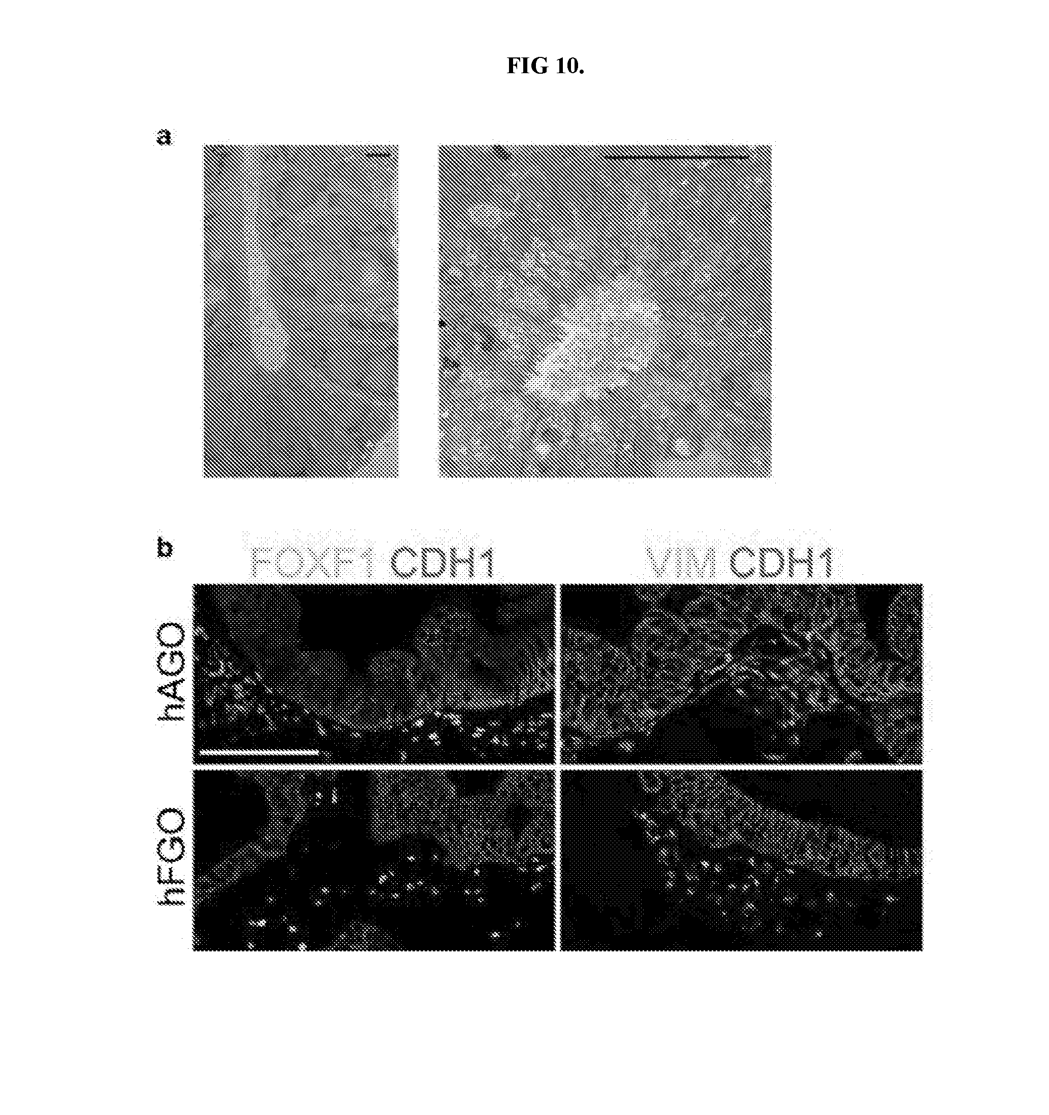
[0015] FIG. 10. hFGOs contain organized glands supported by associated mesenchymal layer. a, Transmission electron micrographs demonstrated that hFGO glands exhibited organized architecture with narrow apical membranes. b, Both hFGOs and hAGOs contained a supporting layer FOXF1+/VIM+ undifferentiated fibroblasts. Scale bars, 5 .mu.m (a) and 100 .mu.m (b).
[0016] FIG. 11. Region-specific cytodifferentiation in human gastric organoids. a, Antral and fundic hGOs exhibited comparable expression of mucous cell markers MUC5AC and MUC6. b, As shown in transmission electron micrograph, hFGOs contained abundant cells exhibiting granule pattern consistent with mucous neck cells, the precursors to differentiated chief cells. c, Exogenous expression of NEUROG3 in hGOs derived from NEUROG3-deficient hESC line induced robust differentiation of SYP-positive endocrine cells. While both hAGOs and hFGOs formed GHRL- and SST-expressing endocrine cells, specification of GAST+ G-cells was observed only in hAGOs. d, Expression comparison of cell lineage markers in hGOs and human gastric biopsy tissue. qPCR analyses demonstrated that hGOs exhibited comparable expression levels of several lineage markers (MUC5AC, ATP4B), while other genes were expressed at much lower levels (ATP4A, PGA5, and PGC) than found in the fully differentiated, mature human stomach. Scale bars, 5 .mu.m (b) and 100 .mu.m (c). Error bars represent s.d. (a) and s.e.m. (b).
[0017] FIG. 12. Analysis of murine chief cell development. a, Unlike parietal cells, which expressed functional markers (Atp4b) as early as late embryonic stages, chief cell gene products were not detectable until much later stages of development. In the embryonic (E18.5) and juvenile (P12) stomach, Gif and Pgc were not yet expressed, indicating that chief cells mature much later in development than other lineages in the gastric epithelium. b, Despite the absence of Pgc, the P12 mouse stomach did contain abundant glandular cells expressing nuclear Mist1, a chief cell-specific marker. Thus, chief cells were indeed specified earlier but took several weeks to develop robust expression of terminal differentiation markers. Scale bars, 100 .mu.m (a) and 200 .mu.m (b).
[0018] FIG. 13. Screen for pathways that promote differentiation of parietal cells in fundic hGOs. a, To test for growth factors/small molecules capable of inducing parietal cell differentiation, hFGOs were exposed for two days (30-32) to the indicated agonist or antagonist and then analyzed at day 34. In a screening experiment of different pathways, only MEK inhibition with PD03 was found to robustly induce expression of ATP4A/B. b, Reduction or removal of EGF from the culture medium was not sufficient to reproduce the effect of MEK inhibition. c, The ability of PD03/BMP4 to induce parietal cell development was exclusive to fundic hGOs, as antral hGOs did not express fundic markers in response to PD03/BMP4. d, Exposure to PD03/BMP4 rapidly increased expression of ATP4A and ATP4B in fundic hGOs. e, Induction of parietal cell generation with PD03/BMP4 did not significantly impact the differentiation of chief cells (PGA5 and PGC) and endocrine cells (CHGA). f, The manipulations at each stage of the hFGO differentiation protocol was required for robust parietal cell differentiation, as removal of any single step led to loss of ATP4A/B expression. Error bars represent s.d. (a-c) and s.e.m. (d-f).
[0019] FIG. 14. Live in vitro pH monitoring in gastric organoids. a, The dye SNAFR5F exhibits responsiveness over pH range of 5-8, which makes it well suited to detect physiologic changes in response to parietal cell-mediated acid secretion. b, Media and luminal pH measurements recorded before (closed circles) and 60 minutes following addition of histamine (open circles). Antral hGOs did not respond, while the fundic hGO luminal pH decreased in response to histamine. The acidification was inhibited by pre-treatment of organoids with either famotidine or omeprazole. Further, omeprazole was sufficient to raise the pH in fundic organoids prior to histamine exposure, suggesting a baseline acid secretion in the fundic organoids. Media pH did not change in any organoids. ***, p<0.001 compared to before histamine; $$$, p<0.001 compared to luminal pH without histamine; ###, p<0.001 compared to luminal pH with histamine; two tailed Student's t-test. c, hFGOs contained parietal cell-dense glands in which acridine orange (AO) accumulated in nearly all of the cells lining the lumen of the gland. d, AO accumulation was observed in a canalicular-type pattern in parietal cells in hFGOs. Scale bars, 10 .mu.m. Error bars represent s.d.
[0020] FIG. 15. Serial passaging of human gastric organoids. a, Schematic representation of experiments to determine the presence of gastric stem cells in hGOs. b, When fragments were grown in culture medium containing only EGF, they did not grow or expand to form new organoids. However, addition of CHIR and FGF10 to the culture medium was sufficient to support the growth of individual fragments into newly formed organoids. c, Following two passages, hFGOs still expressed genes consistent with a gastric phenotype, including PGC, MUC6, MUC5AC, and GHRL. This ability to undergo serial passaging with maintenance of gastric identity supports the conclusion that hFGOs contain cells with properties analogous to those of adult gastric stem cells. d, Although passaged hFGOs expressed markers associated with several differentiated gastric cell types, they did not express genes associated with parietal cells such as ATP4B. Further, differentiation of parietal cells could not be induced through MEK inhibition as they could prior to passaging. Error bars represent s.d.
DETAILED DESCRIPTION OF THE INVENTION
[0021] Unless otherwise noted, terms are to be understood according to conventional usage by those of ordinary skill in the relevant art.
[0022] As used herein, the term "gastric fundus tissue" means a fundic type of gastric epithelium found in the corpus that contains fundic cell types, including but not limited to acid-producing parietal cells and protease-producing chief cells.
[0023] As used herein, the term "definitive endoderm (DE) cell" means one of the three primary germ layers produced by the process of gastrulation.
[0024] As used herein the term "wnt signalling pathway" means the wnt/beta-catenin pathway and is a signal transduction pathway that is mediated by Wnt ligands and frizzled cell surface receptors that acts through the beta-catenin protein.
[0025] As used herein the term "activator" with respect to a pathway, such as a "wnt pathway" means a substance that activates the Wnt/beta-catenin pathway such that Wnt/beta-catenin targets are increased.
[0026] As used herein, the term "FGF signaling pathway activator" means a substance that activates the FGF pathway such that FGF targets are increased.
[0027] As used herein, the term "BMP signalling pathway inhibitor" a substance that interferes with the BMP pathway and causes BMP targets to be decreased.
[0028] As used herein, the term "growth factor" means a substance capable of stimulating cellular processes including but not limited to growth, proliferation, morphogenesis or differentiation.
[0029] As used herein, the term "fundic lineage" means cell types found in fundic epithelium in the corpus stomach.
[0030] As used herein, the term "SOX2+GATA+PDX1- epithelium" means epithelium that expresses the listed proteins.
[0031] As used herein, the term "stable expression" of a marker means expression that does not change upon modification of the growth environment.
[0032] As used herein, the term "totipotent stem cells" (also known as omnipotent stem cells) are stem cells that can differentiate into embryonic and extra-embryonic cell types. Such cells can construct a complete, viable, organism. These cells are produced from the fusion of an egg and sperm cell. Cells produced by the first few divisions of the fertilized egg are also totipotent.
[0033] As used herein, the term "pluripotent stem cells (PSCs)," also commonly known as PS cells, encompasses any cells that can differentiate into nearly all cells, i.e., cells derived from any of the three germ layers (germinal epithelium), including endoderm (interior stomach lining, gastrointestinal tract, the lungs), mesoderm (muscle, bone, blood, urogenital), and ectoderm (epidermal tissues and nervous system). PSCs can be the descendants of totipotent cells, derived from embryos (including embryonic germ cells) or obtained through induction of a non-pluripotent cell, such as an adult somatic cell, by forcing the expression of certain genes.
[0034] As used herein, the term "induced pluripotent stem cells (iPSCs)," also commonly abbreviated as iPS cells, refers to a type of pluripotent stem cells artificially derived from a normally non-pluripotent cell, such as an adult somatic cell, by inducing a "forced" expression of certain genes.
[0035] As used herein, the term "precursor cell" encompasses any cells that can be used in methods described herein, through which one or more precursor cells acquire the ability to renew itself or differentiate into one or more specialized cell types. In some embodiments, a precursor cell is pluripotent or has the capacity to becoming pluripotent. In some embodiments, the precursor cells are subjected to the treatment of external factors (e.g., growth factors) to acquire pluripotency. In some embodiments, a precursor cell can be a totipotent stem cell; a pluripotent stem cell (induced or non-induced); a multipotent stem cell; and a unipotent stem cell. In some embodiments, a precursor cell can be from an embryo, an infant, a child, or an adult. In some embodiments, a precursor cell can be a somatic cell subject to treatment such that pluripotency is conferred via genetic manipulation or protein/peptide treatment.
[0036] In developmental biology, cellular differentiation is the process by which a less specialized cell becomes a more specialized cell type. As used herein, the term "directed differentiation" describes a process through which a less specialized cell becomes a particular specialized target cell type. The particularity of the specialized target cell type can be determined by any applicable methods that can be used to define or alter the destiny of the initial cell. Exemplary methods include but are not limited to genetic manipulation, chemical treatment, protein treatment, and nucleic acid treatment.
[0037] As used herein, the term "cellular constituents" are individual genes, proteins, mRNA expressing genes, and/or any other variable cellular component or protein activities such as the degree of protein modification (e.g., phosphorylation), for example, that is typically measured in biological experiments (e.g., by microarray or immunohistochemistry) by those skilled in the art. Significant discoveries relating to the complex networks of biochemical processes underlying living systems, common human diseases, and gene discovery and structure determination can now be attributed to the application of cellular constituent abundance data as part of the research process. Cellular constituent abundance data can help to identify biomarkers, discriminate disease subtypes and identify mechanisms of toxicity.
[0038] Pluripotent Stem Cells Derived from Embryonic Cells
[0039] In some embodiments, an important step is to obtain stem cells that are pluripotent or can be induced to become pluripotent. In some embodiments, pluripotent stem cells are derived from embryonic stem cells, which are in turn derived from totipotent cells of the early mammalian embryo and are capable of unlimited, undifferentiated proliferation in vitro. Embryonic stem cells are pluripotent stem cells derived from the inner cell mass of the blastocyst, an early-stage embryo. Methods for deriving embryonic stem cells from blastocytes are well known in the art. Human embryonic stem cells H9 (H9-hESCs) are used in the exemplary embodiments described in the present application, but it would be understood by one of skill in the art that the methods and systems described herein are applicable to any stem cells.
[0040] Additional stem cells that can be used in embodiments in accordance with the present invention include but are not limited to those provided by or described in the database hosted by the National Stem Cell Bank (NSCB), Human Embryonic Stem Cell Research Center at the University of California, San Francisco (UCSF); WISC cell Bank at the Wi Cell Research Institute; the University of Wisconsin Stem Cell and Regenerative Medicine Center (UW-SCRMC); Novocell, Inc. (San Diego, Calif.); Cellartis AB (Goteborg, Sweden); ES Cell International Pte Ltd (Singapore); Technion at the Israel Institute of Technology (Haifa, Israel); and the Stem Cell Database hosted by Princeton University and the University of Pennsylvania. Exemplary embryonic stem cells that can be used in embodiments in accordance with the present invention include but are not limited to SA01 (SA001); SA02 (SA002); ES01 (HES-1); ES02 (HES-2); ES03 (HES-3); ES04 (HES-4); ES05 (HES-5); ES06 (HES-6); BG01 (BGN-01); BG02 (BGN-02); BG03 (BGN-03); TE03 (13); TE04 (14); TE06 (16); UC01 (HSF1); UC06 (HSF6); WA01 (H1); WA07 (H7); WA09 (H9); WA13 (H13); WA14 (H14).
[0041] More details on embryonic stem cells can be found in, for example, Thomson et al., 1998, "Embryonic Stem Cell Lines Derived from Human Blastocysts," Science 282 (5391):1145-1147; Andrews et al., 2005, "Embryonic stem (ES) cells and embryonal carcinoma (EC) cells: opposite sides of the same coin," Biochem Soc Trans 33:1526-1530; Martin 1980, "Teratocarcinomas and mammalian embryogenesis,". Science 209 (4458):768-776; Evans and Kaufman, 1981, "Establishment in culture of pluripotent cells from mouse embryos," Nature 292(5819): 154-156; Klimanskaya et al., 2005, "Human embryonic stem cells derived without feeder cells," Lancet 365 (9471): 1636-1641; each of which is hereby incorporated herein in its entirety.
[0042] Induced Pluripotent Stem Cells (iPSCs)
[0043] In some embodiments, iPSCs are derived by transfection of certain stem cell-associated genes into non-pluripotent cells, such as adult fibroblasts. Transfection is typically achieved through viral vectors, such as retroviruses. Transfected genes include the master transcriptional regulators Oct-3/4 (Pouf51) and Sox2, although it is suggested that other genes enhance the efficiency of induction. After 3-4 weeks, small numbers of transfected cells begin to become morphologically and biochemically similar to pluripotent stem cells, and are typically isolated through morphological selection, doubling time, or through a reporter gene and antibiotic selection. As used herein, iPSCs include but are not limited to first generation iPSCs, second generation iPSCs in mice, and human induced pluripotent stem cells. In some embodiments, a retroviral system is used to transform human fibroblasts intopluripotent stem cells using four pivotal genes: Oct3/4, Sox2, Klf4, and c-Myc. In alternative embodiments, a lentiviral system is used to transform somatic cells with OCT4, SOX2, NANOG, and LIN28. Genes whose expression are induced in iPSCs include but are not limited to Oct-3/4 (e.g., Pou5fl); certain members of the Sox gene family (e.g., Sox1, Sox2, Sox3, and Sox15); certain members of the Klf family (e.g., Klf1, Klf2, Klf4, and Klf5), certain members of the Myc family (e.g., C-myc, L-myc, and N-myc), Nanog, and LIN28.
[0044] In some embodiments, non-viral based technologies are employed to generate iPSCs. In some embodiments, an adenovirus can be used to transport the requisite four genes into the DNA of skin and liver cells of mice, resulting in cells identical to embryonic stem cells. Since the adenovirus does not combine any of its own genes with the targeted host, the danger of creating tumors is eliminated. In some embodiments, reprogramming can be accomplished via plasmid without any virus transfection system at all, although at very low efficiencies. In other embodiments, direct delivery of proteins is used to generate iPSCs, thus eliminating the need for viruses or genetic modification. In some embodiment, generation of mouse iPSCs is possible using a similar methodology: a repeated treatment of the cells with certain proteins channeled into the cells via poly-arginine anchors was sufficient to induce pluripotency. In some embodiments, the expression of pluripotency induction genes can also be increased by treating somatic cellswith FGF2 under low oxygen conditions.
[0045] More details on embryonic stem cells can be found in, for example, Kaji et al., 2009, "Virus free induction of pluripotency and subsequent excision of reprogramming factors," Nature 458:771-775; Woltj en et al., 2009, "piggyBac transposition reprograms fibroblasts to induced pluripotent stem cells," Nature 458:766-770; Okita et al., 2008, "Generation of Mouse Induced Pluripotent Stem Cells Without Viral Vectors," Science 322(5903):949-953; Stadtfeld et al., 2008, "Induced Pluripotent Stem Cells Generated without Viral Integration," Science 322(5903):945-949; and Zhou et al., 2009, "Generation of Induced Pluripotent Stem Cells Using Recombinant Proteins," Cell Stem Cell 4(5):381-384; each of which is hereby incorporated herein in its entirety.
[0046] In some embodiments, exemplary iPS cell lines include but not limited to iPS-DF19-9; iPS-DF19-9; iPS-DF4-3; iPS-DF6-9; iPS(Foreskin); iPS(IMR90); and iPS(IMR90).
[0047] More details on the functions of signaling pathways relating to DE development can be found in, for example, Zorn and Wells, 2009, "Vertebrate endoderm development and organ formation," Annu Rev Cell Dev Biol 25:221-251; Dessimoz et al., 2006, "FGF signaling is necessary for establishing gut tube domains along the anterior-posterior axis in vivo," Mech Dev 123:42-55; McLin et al., 2007, "Repression of Wnt/.beta.-catenin signaling in the anterior endoderm is essential for liver and pancreas development. Development," 134:2207-2217; Wells and Melton, 2000, Development 127:1563-1572; de Santa Barbara et al., 2003, "Development and differentiation of the intestinal epithelium," Cell Mol Life Sci 60(7): 1322-1332; each of which is hereby incorporated herein in its entirety.
[0048] Any methods for producing definitive endoderm from pluripotent cells (e.g., iPSCs or ESCs) are applicable to the methods described herein. In some embodiments, pluripotent cells are derived from a morula. In some embodiments, pluripotent stem cells are stem cells. Stem cells used in these methods can include, but are not limited to, embryonic stem cells. Embryonic stem cells can be derived from the embryonic inner cell mass or from the embryonic gonadal ridges. Embryonic stem cells or germ cells can originate from a variety of animal species including, but not limited to, various mammalian species including humans. In some embodiments, human embryonic stem cells are used to produce definitive endoderm. In some embodiments, human embryonic germ cells are used to produce definitive endoderm. In some embodiments, iPSCs are used to produce definitive endoderm.
[0049] Disclosed herein are methods for differentiating human pluripotent stem cells (PSCs) into gastric organoids containing fundic epithelium. Applicant first identified, and then recapitulated key events in embryonic fundus development to arrive at the claimed compositions. Applicant found that disruption of Wnt/.beta.-catenin signaling in mouse embryos led to conversion of fundic to antral epithelium, while .beta.-catenin activation in hPSC-derived foregut progenitors promoted the development of human fundic-type gastric organoids (hFGOs). Applicant then used hFGOs to identify temporally distinct roles for multiple signaling pathways in epithelial morphogenesis and differentiation of fundic cell types, including chief cells and functional parietal cells. While hFGOs are a powerful new model for studying the development of the human fundus and its lineages, they also represent a critical new model system to study the molecular basis of human gastric physiology, pathophysiology, and drug discovery.
[0050] In one aspect, an in vitro method of inducing formation of a gastric fundus tissue is disclosed. The method may comprise the steps of:
[0051] a) contacting a mammalian definitive endoderm (DE) cell with a wnt pathway activator, an FGF signaling pathway activator (for example, FGF4), a BMP signalling pathway inhibitor (e.g., Noggin), and retinoic acid, for a first period. Wnt signalling may be activated either with a protein like Wnt3a, for example, or via a chemical like Chiron, for example, which inhibits GSK3.beta.. The first period may be three days.+-.24 hours. The retinoic acid may be added for the third day of the first period.+-.24 hours. In one aspect, the first period may be carried out for a period of time sufficient to form a three-dimensional posterior foregut spheroid from the definitive endoderm.
[0052] b) suspending said three-dimensional posterior foregut spheroid in a basement membrane matrix with a growth factor, a Wnt signalling pathway activator, a EGF signalling pathway activator, a BMP signalling pathway inhibitor, and retinoic acid for a second period. The second period may be three days.+-.24 hours. The second period may be carried out for a period of time sufficient to induce a fundic lineage comprising fundal hGOs (hFGOs).
[0053] c) culturing the hFGOs of step b) with a wnt pathway activator and a EGF signalling pathway activator for a third period. The third period may be, for example, 11 days.+-.24 hours.
[0054] d) culturing the hFGOs of step c with a wnt signaling pathway activator, a EGF signalling pathway activator, and FGF10 for a fourth period. The fourth period may be, for example, 10 days.+-.24 hours.
[0055] e) contacting said hFGOs of step d with a MEK inhibitor for a fifth period. The MEK inhibitor may be, for example, PD0325901. The fifth period may be for a two-day period.+-.24 hours, or for a period of time sufficient to form a gastric fundus tissue comprising a functional fundic cell type.
[0056] In one aspect, step e) may further comprise the step of contacting the fundal hGOs with an activator of BMP4 signalling. In certain aspects, step e may be carried out for a period of time sufficient to develop SOX2+GATA+PDX1- epithelium.
[0057] In one aspect, the functional fundic cell type may be a parietal cell that expresses proton pump proteins and secretes acid. In one aspect, the functional fundic cell type may be a chief cell that secretes pepsinogen.
[0058] In one aspect, step d and step e are carried out for a period of time sufficient to confer stable expression of lineage markers MUC5AC, MUC6, PGC, and GHRL.
[0059] In one aspect, the definitive endoderm may be derived from a precursor cell selected from an embryonic stem cell, an embryonic germ cell, an induced pluripotent stem cell, a mesoderm cell, a definitive endoderm cell, a posterior endoderm cell, a posterior endoderm cell, and a hindgut cell, a definitive endoderm derived from a pluripotent stem cell, a definitive endoderm derived from a pluripotent stem cell selected from an embryonic stem cell, an adult stem cell, or an induced pluripotent stem cell.
[0060] In one aspect, the definitive endoderm may be derived from contacting a pluripotent stem cell with one or more molecules selected from Activin, the BMP subgroups of the TGF-beta superfamily of growth factors; Nodal, Activin A, Activin B, BMP4, Wnt3a, and combinations thereof.
[0061] There are many ways to activate the Wnt/beta-catenin pathway (see http://web.stanford.edu/group/nusselab/cgi-bin/wnt/). Suitable Some existing wnt signalling pathway activators include but are not limited to:
[0062] Protein-based activators: Wnt ligands including but not limited to Wnt1, Wnt2, Wnt2b, Wnt3, Wnt3a, Wnt8, et al; modifiers of Wnt ligand activity including but not limited to activated Wnt frizzled receptors, (LRP) co-receptors, R-spondin proteins, Dkk proteins, regulators of Wnt ligand secretion and trafficking (Wntless, Porcupine), inhibiting beta-catenin degredation APC and GSK3beta inhibition, activated beta-catenin, constitutively active TCF/Lef proteins.
[0063] Chemical activators: there are over 28 known chemicals that either activate or inhibit Wnt/beta-catenin signaling. Some activators include but are not limited to GSK3-beta inhibitors CHIR99021, BIO, LY2090314, SB-216763, lithium, porcupine inhibitors IWP, LGK974, C59, SFRP inhibitor WAY-316606, beta-catenin activator DCA.
[0064] In one aspect, the WNT pathway activator may be one or more molecules selected from Wnt1, Wnt2, Wnt2b, Wnt3, Wnt3a, Wnt4, Wnt5a, Wnt5b, Wnt6, Wnt7a, Wnt7b, Wnt8a, Wnt8b, Wnt9a, Wnt9b, Wnt10a, Wnt10b, Wnt11, and Wnt16, for example, Wnt3a, or for example, Wnt3a at a concentration between about 50 to about 1500 ng/ml.
[0065] Suitable FGF signalling pathway activators include: FGF ligands FGF2, 4, 5, 8, et al. Activated forms of FGF receptors. Proteins and chemicals that stimulate the FGF receptor and signaling components downstream of the receptors including MAPK, MEK, ERK proteins and chemicals that modulate their activity. FGF signaling can be activated by inhibiting inhibitors of FGF signaling pathways including but not limited to Sprouty protein family members.
[0066] In one aspect, the BMP signalling pathway inhibitor may be selected from Noggin, Dorsomorphin, LDN189, DMH-1, and combinations thereof, for example, wherein said precursor cell may be contacted with a BMP inhibitor at a concentration between about 50 to about 1500 ng/ml.
[0067] In one aspect, the steps are conducted in vitro.
[0068] In one aspect, a composition comprising gastric tissue produced according to the aforementioned method(s) is disclosed. The gastric tissue may be characterized, for example, by being free of innervation and/or blood vessels.
[0069] In one aspect, an in vitro method of inducing formation of a gastric fundus tissue is disclosed. The method may comprise the steps of contacting a fundal hGO (hFGO) with a wnt pathway activating agent and an EGF signalling pathway activating agent for a first period, and a MEK inhibitor for a second period, (wherein said MEK inhibitor may be PD0325901), wherein said first and second periods are carried out for a period of time sufficient to form a functional fundic cell type;
[0070] wherein said hFGO are obtained by contacting a three-dimensional posterior foregut spheroid in a basement membrane matrix with a growth factor, a wnt pathway activating agent, an EGF signalling pathway activator, a BMP signalling pathway inhibitor, and retinoic acid for a period of time sufficient to convert said three-dimensional posterior foregut spheroid to said hFGO;
[0071] wherein said three-dimensional posterior foregut spheroids are obtained by contacting a mammalian definitive endoderm (DE) cells with a wnt pathway activating agent, an FGF signaling pathway activating agent, a BMP signalling pathway inhibitor, and retinoic acid.
EXAMPLES
[0072] Recently, considerable progress has been made in the development of three-dimensional in vitro organoid systems.sup.1,2. Organoids have proven to be powerful experimental models that combine architectural complexity and cellular diversity with the tractability and scalability of traditional cell culture methods. Organoid generation through directed differentiation of pluripotent stem cells (PSCs; comprising both embryonic stem cells and induced PSCs) offers several advantages over other approaches including an unlimited source of starting material, no requirement for surgical acquisition of tissue, and ease of genetic manipulations. Further, PSC-based methods permit direct investigation of mechanisms underlying normal and aberrant human development.sup.3. However, differentiating PSCs into specific organoid types depends on a robust molecular knowledge of normal organ development. For some organs, such as the stomach, there are large gaps in understanding of molecular pathways that drive embryonic development.
[0073] The stomach is one of the most structurally diverse organs among mammals.sup.4. In humans, the gastric mucosa generally consists of two types of epithelial glands.sup.5,6. Located in the more proximal anatomic domains--the corpus and fundus--of the stomach, oxyntic glands comprise acid-secreting parietal cells, protease-producing chief cells, mucus-producing cells, and endocrine cells. Antral-type glands, located in the more distal antrum and pylorus, contain mostly mucous and endocrine cells. To simplify the anatomic- and species-specific systems of nomenclature, the terms `fundus` and `antrum` are used to broadly describe these two histologic types of gastric epithelia. Applicant has previously developed a method to direct the differentiation of hPSCs into three-dimensional gastric tissue (human gastric organoids; hGOs) that contained a pure antral epithelium with normal antral cell types.sup.7. While the antral hGOs (hAGOs) are a robust system for studying antral lineage allocation and host-microbe interactions in the stomach, they do not allow for studies of fundic biology and disease. More recently, Noguchi et. al. successfully differentiated mouse ESCs into organoids comprising various types of mouse gastric tissue.sup.8. However, this approach used mouse ESC aggregation and spontaneous differentiation resulting in organoids that were heterogeneous, evidenced by the presence of stratified epithelia. Moreover, species differences make the mouse stomach suboptimal for modeling human gastric disease.sup.9. Thus, a robust and efficient PSC-derived model of the human fundus epithelium would represent a significant advance in the field of gastric biology.
[0074] Embryonic organ development is guided by a series of instructive cues between neighboring tissues.sup.10,11, and differentiation of hPSCs into specific lineages has relied heavily on use of these signals to direct differentiation in vitro. Applicant previously identified a step-wise differentiation approach to generate hAGOs, whereby hPSCs were differentiated into definitive endoderm, patterned to posterior foregut, then specified into presumptive antral epithelium.sup.7. Applicant hypothesized that the fundus and antrum derive from a common population of posterior foregut progenitors, which could be directed toward the fundic lineage if provided with the appropriate signals. However, given that the mechanisms that drive fundus development in vivo were not previously known, Applicant first had to identify signaling pathways that pattern the embryonic stomach along the proximal-distal axis.
[0075] Embryonic Stomach Pattern Formation
[0076] To aid investigation of the pathways that regulate fundus specification during embryonic development, Applicant analyzed mouse embryos to identify molecular markers that could distinguish between presumptive fundus, antrum and forestomach. At E14.5 Applicant found that Sox2 was expressed in all foregut organ lineages while Gata4 was restricted to the glandular stomach epithelium. Within the Gata4+ domain, Pdx1 was specific to the presumptive antral region (FIG. 6, a); thus, the embryonic fundus domain is believed to be Sox2+Gata+Pdx1-. Further, Applicant analyzed published microarray datasets (GSM326648-GSM32665012 and GSM80809-GMS8081613) and dissected regions of the E14.5 foregut to demonstrate that expression of the transcription factors Irx2, Irx3, and Irx5 was greater than ten-fold enriched in the embryonic fundus compared to antrum (FIG. 6, b-c), indicating that their expression can further distinguish between regions of the glandular gastric epithelium.
[0077] At the molecular level, the presumptive fundic and antral domains of the stomach were already established by E10.5 (FIG. 6, a). At that point in development, the canonical Wnt signaling pathway was active in the proximal stomach but exhibited little or no activity in the distal stomach.sup.14, as shown using the Wnt reporter mouse strain Axin2-lacZ (FIG. 1b). While the regulation of Wnt/.beta.-catenin signaling is known to play a role in establishing the pyloric-duodenal boundary.sup.14,15, its role in gastric epithelial patterning had not been investigated. To determine whether Wnt/.beta.-catenin signaling was functionally required for establishing the fundus in vivo, Applicant deleted .beta.-catenin (Ctnnb1) in the foregut epithelium using Shh-cre (Shh-cre;.beta.-cateninfl/fl=cKO). Disruption of Wnt/.beta.-catenin signaling resulted in the loss of fundic identity, demonstrated by ectopic Pdx1 expression in the fundus at E10.5 (FIG. 1, c). Ectopic Pdx1 was initially restricted to the ventral half of the fundic epithelium, consistent with previously reported recombination activity using this Shh-cre line.sup.16, but it then expanded over time to include a majority of the proximal stomach and greater curvature by E14.5 (FIG. 7, a). Additionally, expression of the fundus markers Irx2, Irx3, and Irx5 were dramatically reduced in the cKO embryos (FIG. 7, b). Collectively, these data support the conclusion that epithelial Wnt/.beta.-catenin signaling regulates gastric pattern formation, as it is required for the initial specification of fundus identity while repressing antral fate in the embryonic mouse stomach.
[0078] To determine the impact of early Wnt/.beta.-catenin-mediated patterning abnormalities on subsequent cytodifferentiation, Applicant analyzed cKO embryos at E18.5. The stomach in cKO embryos was malformed and reduced in size at E18.5 (FIG. 1, d and FIG. 7, c-d), suggestive of a role for Wnt/.beta.-catenin in promoting stomach growth during late stages of development. Moreover, the cKO stomach was completely mis-patterned with ectopic Pdx1 expression throughout the proximal-most regions of the epithelium (FIG. 1, d). Parietal cells, a fundic cell type marked by expression of Atp4b, were reduced in the CKO stomach (FIG. 1, d) and completely absent in .beta.-catenin deficient epithelium (FIG. 1, e). In contrast, the parietal cells that did develop were only observed in .beta.-catenin-expressing epithelium (FIG. 1, e and FIG. 7, d-e). Taken together, these in vivo data support a model by which Wnt/.beta.-catenin signaling induces fundus specification and inhibits antral identity. Further, disruption of this early patterning coincides with subsequent cell autonomous loss of parietal cells, suggesting that cytodifferentiation is impaired secondary to developmental patterning defects.
[0079] Differentiation of Fundic hGOs from hPSCs
[0080] Applicant next investigated the role of Wnt/.beta.-catenin signaling in establishing fundic-antral pattern of the developing human stomach. To model early stages of stomach differentiation, Applicant started with a previously described protocol for differentiating hPSCs into antrum-like gastric organoids, which recapitulates the normal stages of early gastric development with high fidelity.sup.7. Starting with three-dimensional posterior foregut spheroids (SOX2+HNF1.beta.+), Applicant tested whether stimulation of Wnt/.beta.-catenin signaling would direct posterior foregut epithelium into the fundic (SOX2+GATA+PDX1-) lineage rather than antrum (SOX2+GATA+PDX1+) during the gastric specification stage (FIG. 2, a). Indeed, activating .beta.-catenin with the GSK3.beta. inhibitor CHIR99021 (CHIR) for three days resulted in nearly complete repression of PDX1 at day 9, accompanied by significantly increased expression of IRX2, IRX3, and IRX5 (FIG. 2, b-c). Importantly, SOX2 and GATA4 levels were unaffected by CHIR treatment, confirming that spheroids retained their gastric identity. Thus, CHIR exposure resulted in formation of SOX2+GATA+PDX1- epithelium with increased IRX expression, a signature consistent with the presumptive fundic epithelium.
[0081] Applicant then sought to determine whether CHIR-treated spheroids would further develop into more mature hGOs containing a fundus-like epithelium. Interestingly, a three-day pulse of CHIR from days 6-9 was not sufficient to irreversibly specify a fundic identity, as the hGOs ultimately reverted to a PDX1+ antral phenotype at later stages. However, continued Wnt stimulation via CHIR treatment through at least day 29 led to stable induction of fundic gene expression (FIG. 8, a). This was consistent with the prolonged activity of Wnt/.beta.-catenin signaling during embryonic stomach development in vivo. Although previous studies indicated that ectopic Wnt activation in the embryonic stomach promoted intestinal fate.sup.14,15, CHIR-treated hGOs did not exhibit a significant increase in intestinal markers CDX2, MUC2, CCK, or SCT (FIG. 8, e and FIG. 9, a-b). Applicant further demonstrated that CDX2 remained suppressed despite Wnt/.beta.-catenin activation due to concomitant inhibition of BMP signaling, as replacing Noggin with BMP4 led to robust expression of the intestinal transcription factor (FIG. 9, c).
[0082] Once regional domains are established in early development, the primitive gastric epithelium undergoes periods of growth, glandular morphogenesis, and differentiation of definitive cell types. Applicant previously showed that hAGOs underwent a similar progression of morphologic and cellular development7. CHIR-treated hFGOs grew at a similar rate and efficiency compared to hAGOs, as 75-90% of all spheroids plated grew into organoids (FIG. 8, d). At day 20, both types of hGOs contained epithelia that expressed the gastric SOX2/GATA4 signature in >90% of cells, while PDX1 was restricted to hAGOs (87.1.+-.8.4% in hAGOs and 3.9.+-.2.0% in hFGOs, p=3.07.times.10.sup.-6; FIG. 8, e). The organoids maintained their respective gastric identities throughout their development (FIG. 8, b-c). By day 34, hFGOs and hAGOs comprised CDH1+CTNNB1+KRT8+ polarized, columnar epithelia that ubiquitously expressed the gastric-specific.sup.17 claudin CLDN.sup.18 (FIG. 2, e and FIG. 9, d), as well as comparable undifferentiated mesenchymal cells (FIG. 10, b). One notable difference was that hFGOs had a distinctive architecture with organized glands that bud from the organoid epithelium (FIG. 2, d-e and FIG. 10, a), while hAGOs had complex folding and primitive gland-like organization but rarely glandular buds.sup.7. Thus, the novel Wnt/.beta.-catenin dependent mechanism of specifying fundus is conserved in humans and can be manipulated to generate three-dimensional hFGOs with a glandular epithelium that molecularly resembles the developing fundus.
[0083] Region-Specific Gastric Cytodifferentiation
[0084] Differentiated antral gastric cell types were first detected in hAGOs around day 27 and then increased by day 347, analogous to the first few weeks of postnatal development in the mouse stomach.sup.18. At day 34, hFGOs contained both MUC5AC+ surface mucous cells and MUC6+ mucous neck cells as expected, similar to the hAGOs (FIG. 3, a-b and FIG. 11, a). hFGOs also formed a variety of endocrine cell types (FIG. 3, c), but expression of the hormone GAST was specific to hAGOs while GHRL was enriched 10-fold in hFGOs (FIG. 3, d), consistent with the normal gastroendocrine pattern.sup.19. To functionally define the region-specific competence of hGOs, Applicant used an inducible system to over-express the proendocrine transcription factor NEUROG3. Expression of NEUROG3 in both hGO subtypes resulted in robust expression of the pan-endocrine marker SYP, as well as the common gastric hormones SST and GHRL (FIG. 11, c). However, only the hAGOs and not hFGOs were competent to give rise to GAST-expressing G-cells (FIG. 3, e and FIG. 11, c), consistent with the antrum-specific distribution of G-cells in the human stomach.sup.19.
[0085] Chief cells, the fundus-specific secretory lineage, reside in the base of oxyntic glands and have been proposed as a type of reserve stem cell.sup.20. hFGOs exhibited epithelial expression of the chief cell-specific.sup.21 transcription factor MIST1 (FIG. 4, a), had 100-1,000-fold increases in transcripts for the proenzymes PGA5 and PGC (FIG. 4, c), and contained significantly increased pepsinogen content measured by ELISA (FIG. 4, e). However the transcript levels were less than 1% those found in the adult human stomach (FIG. 11, d) and pepsinogen-positive cells were only rarely detectable by immunohistochemistry (FIG. 4, b-c). Consistent with this, zymogen granule-containing cells.sup.22 were identified by TEM (FIG. 4, d) but were rare. In contrast, cells with a more immature mucous granule pattern were abundant (FIG. 11, b). Since chief cells in vivo do not exhibit robust pepsinogen expression for the first few weeks of life (FIG. 12, a-b), Applicant concluded that the chief cells were present in hFGOs but were immature. hFGOs therefore represent a robust platform to dissect the intrinsic and extrinsic mechanisms that regulate chief cell maturation.
[0086] Pathways Controlling Parietal Cell Differentiation
[0087] At baseline, hFGOs contained only a small number of parietal cells (PCs; FIG. 5, a-b), the defining cell type of fundic glands that acidify the gastric lumen via the proton pump (consisting of ATP4A and ATP4B subunits). Identification of efficient methods to increase PC populations has remained elusive due to a lack of understanding of the signaling mechanisms that drive their development. Applicant therefore used PSC-derived hFGOs as a platform to functionally screen candidate signaling pathways for a role in regulating PC differentiation. For screening, Applicant exposed day 30 hFGOs to signaling agonists or antagonists for two days and analyzed PC differentiation at day 34. While the majority of signaling manipulations had no appreciable effect, transient inhibition of the MEK pathway with PD0325901 (PD03) resulted in substantial up-regulation of both ATP4A and ATB4B (FIG. 13, a). Further, while BMP4 alone did not affect PC gene expression, it could enhance the effect of PD03 (data not shown). Thus, a two-day pulse of PD03/BMP4 was sufficient to induce rapid and robust expression of PC markers ATP4A, ATP4B and GIF (FIG. 5, a-b and FIG. 13, d). Interestingly, this effect was not observed by simply removing EGF or FGF from the culture medium (FIG. 13, b), suggesting that there are likely endogenous signaling interactions upstream of MEK/ERK that are responsible for limiting PC differentiation in hFGO cultures. Further, PD03/BMP4 treatment only affected the PC lineage (FIG. 13, e), and was unable to induce PCs in hAGOs (FIG. 13, c), further emphasizing that early patterning of the gastric epithelium defines its ultimate differentiation potential.
[0088] At day 34 hFGO epithelia exhibited comparable organization to the human stomach, with mucous cells lining the surface domain and PCs concentrated in the glandular portion (FIG. 5, e). Moreover, parietal cell morphology closely resembled maturing parietal cells in vivo (FIG. 5, c). Given their resemblance to PCs in vivo and their tubulovesicular ultrastructure as seen on TEM (FIG. 5, d), Applicant hypothesized that the PCs in hFGOs would exhibit the ability to secrete acid in response to appropriate stimuli. Measured using a pH sensitive dye (SNARF5F) with real time confocal microscopy (FIG. 14, a), hFGOs produced a swift and marked decrease in luminal pH in response to histamine that was blocked by either the H2 antagonist famotidine or the H+K+-ATPase antagonist omeprazole (FIG. 5, f and FIG. 14, b). To visualize the cellular response to histamine, hGOs were cultured with the fluorescent dye acridine orange (AO), which shifts to an orange color when sequestered in acidic compartments.sup.23. Similar to isolated mouse gastric glands, AO accumulated in acidified cellular vesicles in hFGO glands in response to histamine (FIG. 5, g and FIG. 14, c-d). These data indicate that the PCs underwent appropriate changes in secretory canalicular structure in response to acid-inducing stimuli.
[0089] In vivo, differentiated gastric cell lineages are thought to derive from a common pool of undifferentiated stem or progenitor cells. Here Applicant has demonstrated the ability to alter the relative proportions of cell types in hFGOs, either through genetic means (NEUROG3-mediated regulation of endocrine cells) or by manipulation of extrinsic signaling pathways (PD03/BMP4 for PCs). These observations led to the hypothesis that hFGOs might contain a population of gastric stem cells analogous to those that have been isolated from the adult stomach. Indeed, Applicant found that dissociated day 34 hFGOs could be passaged serially to give rise to new organoids (FIG. 15, a-b). Re-growth of organoids from passaged hFGOs was dependent on high Wnt and high FGF culture medium, similar to what is used to grow primary gastric tissue organoids.sup.24,25. Following two rounds of passaging, hFGOs maintained expression of lineage markers MUC5AC, MUC6, PGC, and GHRL; however, they did not contain PCs and were refractory to PD03/BMP4-mediated induction of the parietal lineage (FIG. 15, c-d). This finding was similar to what has been observed in adult stem cell-derived gastric organoids, which do not robustly produce PCs despite being derived from the bona fide oxyntic mucosa.sup.20,26. Thus it will be important to identify conditions that preserve PC competence in long-term cultures of hGOs and adult gastric organoids.
[0090] In summary, Applicant has directly applied in vivo and in vitro discovery-based studies towards the differentiation of hPSCs into a new tissue type. Applicant has defined a novel function of Wnt/.beta.-catenin signaling in specifying the fundic domain during stomach development in mice, and used Wnt modulation as the mechanistic basis to direct differentiation of hPSCs into three-dimensional human fundic organoids. In both mouse and human, Wnt-mediated fundus specification was led to the subsequent formation of PCs. The fundus-specific manipulations at each stage of this directed differentiation protocol led to robust PC induction (FIG. 13, f). Previous reports identified that the mesenchymal factor Barx1 indirectly acts to repress Wnt signaling and that helps to prevent intestinal gene expression in the stomach.sup.14,15. Given that the current study identified an epithelial Wnt/.beta.-catenin function, and the previous work identified a mesenchymal pathway, it seems likely that Wnt/.beta.-catenin may have distinct roles in the epithelium versus mesenchyme. For example, the mesenchymal role for Wnt/.beta.-catenin could modulate other signaling pathways such as BMP.sup.27, which our data show synergizes with Wnt to promote intestinal specification from early endoderm (FIG. 7 and FIG. 9, c) The human gastric organoid systems might be useful, in combination with animal models, to dissect how these signaling pathways interact in the mesenchyme and epithelium to coordinate early embryonic gastrointestinal development.
[0091] Pathways that control differentiation of gastric progenitor cells into distinct lineages are also lacking. Applicant has demonstrated the utility of this new hGO platform to identify that MEK/ERK signaling potently represses parietal cell specification. Consistent with these findings, transgenic activation of MEK/MAPK-dependent pathways led to loss of parietal cells in vivo.sup.28,29. Therefore, hGOs are a new and tractable human model system to identify and study signaling mechanisms involved in normal cellular homeostasis in the fundus and antrum. Further, aberrant regulation of developmental programs may also contribute to gastric disease, as corpus/fundus pathology is often associated with parietal cell atrophy.sup.30-32, antral-type histology.sup.33, and even misexpression of Pdx134. Thus targeting of these pathways could have clinical utility, as Choi et. al. recently demonstrated that pharmacologic inhibition of MEK was sufficient to restore normal parietal cell differentiation in a mouse model of metaplasia.sup.35. Additionally, having now established both antral- and fundic-type hGOs, it is possible to study how these human gastric tissues interact physiologically, differentially respond to infection and injury, and respond to pharmacologic treatments.
[0092] Methods
[0093] Mouse Experiments
[0094] The following genetic mouse strains were obtained from The Jackson Laboratory, housed at Cincinnati Children's Hospital Research Foundation animal facility, and maintained according to IACUC protocol (0B09074): Axin2:LacZ (stock no. 009120), Shh:Cre (stock no. 005622), and .beta.-cateninfloxed (stock no. 004152). Timed matings, with the morning the vaginal plug was observed being denoted as E0.5, were used to generate embryos at various stages that were harvested for either wholemount staining or tissue dissection. At least two litters of embryos were analyzed at each developmental stage examined. Both male and female embryos were analyzed.
[0095] Pluripotent Stem Cell Culture
[0096] Human embryonic stem cell line WA01 (H1; obtained from WiCell) was supplied by the Pluripotent Stem Cell Facility at Cincinnati Children's Hospital Medical Center. Cell identity was confirmed by short tandem repeat analysis (Microsatellite STR Analysis; Applied Biosystems), and cells were routinely tested for mycoplasma contamination (MycoAlert Mycoplasma Detection Kit; Lonza). Pluripotent cells were maintained in feeder-free conditions on HESC-qualified Matrigel (BD Biosciences) in mTesR1 media (Stem Cell Technologies). Colonies were passaged every four days using dispase (Invitrogen).
[0097] Differentiation of Posterior Foregut Spheroids
[0098] The protocol for directed differentiation of gastric organoids was adapted from our previous protocol.sup.7, and Table 1 contains the complete list of media and growth factors for each stage. For differentiation, hPSCs were dissociated into single cells using Accutase (Stem Cell Technologies) and plated into 24-well plates at a density of roughly 200,000 cells per well in mTesR1 with Y-27632 (10 .mu.M; Stemgent). The following day, cells were differentiated into definitive endoderm (DE) by adding Activin A (100 ng/ml; Cell Guidance Systems) in RPMI 1640 media (Invitrogen) for three days. Media was also supplemented with NEAA (1.times.; Gibco) and defined FBS (dFBS; Invitrogen) at 0%, 0.2%, and 2.0% on days 1, 2, and 3, respectively. Additionally, BMP4 (50 ng/ml; R&D Systems) was added on the first day. Subsequently, DE was differentiated to posterior foregut endoderm by exposing cells to CHIR99021 (2 .mu.M; Stemgent), FGF4 (500 ng/ml; R&D Systems), and Noggin (200 ng/ml; R&D systems) for three days in RPMI 1640 supplemented with NEAA and 2.0% dFBS. Retinoic acid (2 .mu.M; Sigma Aldrich) was added for the final day. Media was changed every day. This process resulted in the spontaneous formation of three-dimensional posterior foregut spheroids.
TABLE-US-00001 TABLE 1 Differentiation protocol for fundic hGOs. Activin A (100 ng/ml; R&D Systems), CHIR99021 (2 uM; Stemgent), FGF4 (500 ng/ml; R&D systems), PD0325901 (2 uM; Stemgent), BMP4 (50 ng/ml; R&D Systems). Base Day Media Supplement Activin A CHIR99021 FGF4 Noggin RA EGF FGF10 PD03 BMP4 0-1 RPMI NEAA + + 1-2 RPMI 0.2% FCS, + NEAA 2-3 RPMI 2.0% FCS, + NEAA 3-5 RPMI 2.0% FCS, + + + NEAA 5-6 RPMI 2.0% FCS, + + + + NEAA 6-9 BGM* n/a +** + + + 9-13 BGM* n/a +** + + 13-20 BGM* n/a +** + 20-30 BGM* n/a +** + +** 30-32 BGM* n/a +** + +** + + 32-34 BGM* n/a +** + +** *BGM (basic gut media) = Advanced DMEM/F12, N2 (1X; Invitrogen), B27 (1X; Invitrogen) L-glutamine, HEPES (10 uM), and penicillin/streptomycin. **Specific to fundus hGO protocol.
[0099] Three-Dimensional Culture of Foregut Spheroids-Gastric Organoids
[0100] Posterior foregut spheroids were collected and transferred to a three-dimensional culture system as previously described.sup.36. Briefly, spheroids were suspended in 50 .mu.l Matrigel (BD Biosciences) and plated as a droplet into 24-well plates. The matrigel was allowed to solidify for 10 minutes in the tissue culture incubator, then overlayed with basic gut media (BGM) containing growth factors and/or small molecule agonsists. BGM consisted of Advanced DMEM/F12 media (Gibco) supplemented with N2 (1.times.; Invitrogen), B27 (1.times.; Invitrogen), HEPES (10 .mu.M; Gibco), L-glutamine, penicillin/streptomycin, and EGF (100 ng/ml; R&D Systems). During days 6-9, spheroids were cultured with RA and noggin to specify the antral lineage. For fundic specification, CHIR was added during this stage. Antral hGOs were subsequently cultured in BGM with only EGF. Fundic hGOs were continuously exposed to CHIR from day 6-30. In addition, FGF10 (50 ng/ml; R&D Systems) was added to fundic hGOs from day 20-30 as it was shown to enhance the glandular morphogenesis driven by CHIR (data not shown). On day 20, organoids were collected and re-plated at a dilution of 1:10-1:20.
[0101] For screening experiments to identify factors that increase parietal cell differentiation, hFGOs were grown to day 30, then exposed for two days to individual signaling pathway agonists and antagonists: DAPT (1 .mu.M; Stemgent), SB431542 (10 .mu.M; Stemgent), BMP4 (50 ng/ml; R&D Systems), PD0325901 (2 M; Stemgent), Gastrin (10 nM; Sigma Aldrich), Dexamethasone (50 nM; Sigma Aldrich), and Wnt5a (50 ng/ml; R&D Systems). Following treatment, hFGOs were grown for two more days to day 34, then analyzed by qPCR.
[0102] RNA Isolation and qPCR
[0103] Total RNA was isolated using Nucleospin RNA II kit (Machery Nagel) and converted to cDNA as previously described7. qPCR was performed on Quantstudio 6 (Applied Biosystems) using Quantitect SYBR-Green master mix (Qiagen), and primer sequences are listed below.
TABLE-US-00002 Primer Sequences Primers used for qPCR were the following: hATP4A, forward 5'-TGGTAGTAGCCAAAGCAGCC-3', reverse 5'-TGCCATCCAGGCTAGTGAG-3'; hATP4B, forward 5'-ACCACGTAGAAGGCCACGTA-3', reverse 5'-TGGAGGAGTTCCAGCGTTAC-3'; hAXIN2, forward 5'-CTGGTGCAAAGACATAGCCA-3', reverse 5'-AGTGTGAGGTCCACGGAAAC-3'; hCCK, forward 5'-CGGTCACTTATCCTGTGGCT-3', reverse 5'-CTGCGAAGATCAATCCAGCA-3'; hCDX2, forward 5'-CTGGAGCTGGAGAAGGAGTTTC-3', reverse 5'-ATTTTAACCTGCCTCTCAGAGAGC-3'; hCHGA, forward 5'-TGACCTCAACGATGCATTTC-3', reverse 5'-CTGTCCTGGCTCTTCTGCTC-3'; hGAPDH, forward 5'-CCCATCACCATCTTCCAGGAG-3', reverse 5'-CTTCTCCATGGTGGTGAAGACG-3'; hGAST, forward 5'-CAGAGCCAGTGCAAAGATCA-3', reverse 5'-AGAGACCTGAGAGGCACCAG-3'; hGATA4, forward 5'-TCCAAACCAGAAAACGGAAGC-3', reverse 5'-GCCCGTAGTGAGATGACAGG-3'; hGHRL, forward 5'-GCTGGTACTGAACCCCTGAC-3', reverse 5'-GATGGAGGTCAAGCAGAAGG-3'; hGIF, forward 5'-CATTTTCCGCGATATTGTTG-3', reverse 5'-GCACAGCGCAAAAATCCTAT-3'; hIRX2, forward 5'-GTGGTGTGCGCGTCGTA-3', reverse 5'-GGCGTTCAGCCCCTACC-3'; hIRX3, forward 5'-GGAGAGAGCCGATAAGACCA-3', reverse 5'-AGTGCCTTGGAAGTGGAGAA-3'; hIRX5, forward 5'-GGTGTGTGGTCGTAGGGAGA-3', reverse 5'-GCTACAACTCGCACCTCCA-3'; hMIST1, forward 5'-TGCTGGACATGGTCAGGAT-3', reverse 5'-CGGACAAGAAGCTCTCCAAG-3'; hMUC2, forward 5'-TGTAGGCATCGCTCTTCTCA-3', reverse 5'-GACACCATCTACCTCACCCG-3'; hMUC5AC, forward 5'-CCAAGGAGAACCTCCCATAT-3', reverse 5'-CCAAGCGTCATTCCTGAG-3'; hMUC6, forward 5'-CAGCAGGAGGAGATCACGTTCAAG-3', reverse 5'-GTGGGTGTTTTCCTGTCTGTCATC-3'; hPDX1, forward 5'-CGTCCGCTTGTTCTCCTC-3', reverse 5'-CCTTTCCCATGGATGAAGTC-3'; hSCT, forward 5'-GGTTCTGAAACCATAGGCCC-3', reverse 5'-GTCAGGGTCCAACATGCC-3'; hSOX2, forward 5'-GCTTAGCCTCGTCGATGAAC-3', reverse 5'-AACCCCAAGATGCACAACTC-3'; mCdx2, forward 5'-TCTGTGTACACCACCCGGTA-3', reverse 5'-GAAACCTGTGCGAGTGGATG-3'; mGata4, forward 5'-CCATCTCGCCTCCAGAGT-3', reverse 5'-CTGGAAGACACCCCAATCTC-3'; mGapdh, forward 5'-TTGATGGCAACAATCTCCAC-3', reverse 5'-CGTCCCGTAGACAAAATGGT-3'; mIrx1, forward 5'-AATAAGCAGGCGTTGTGTGG-3', reverse 5'-CTCAGCCTCTTCTCGCAGAT-3'; mIrx2, forward 5'-AGCTGGTATGGATAGGCCG-3', reverse 5'-GGCTTCCCGTCCTACGTG-3'; mIrx3, forward 5'-ATAAGACCAGAGCAGCGTCC-3', reverse 5'-GTGCCTTGGAAGTGGAGAAA-3'; mIrx5, forward 5'-GGAGTGTGGTCGTAGGGAGA-3', reverse 5'-GCTACAACTCGCACCTCCA-3'; mPdx1, forward 5'-ACGGGTCCTCTTGTTTTCCT-3', reverse 5'-TGGATGAAATCCACCAAAGC-3'; mPitx1, forward 5'-GTCCATGGAGGTGGGGAC-3', reverse 5'-GCTTAGGCGCCACTCTCTT-3'; mSox2, forward 5'-AAAGCGTTAATTTGGATGGG-3', reverse 5'-ACAAGAGAATTGGGAGGGGT-3'; mTrp63, forward 5'-AGCTTCTTCAGTTCGGTGGA-3', reverse 5'-CCTCCAACACAGATTACCCG-3'.
[0104] Immunofluorescent Staining
[0105] Tissues were fixed in 4% paraformaldehyde overnight at 4.degree. C., then washed thoroughly in PBS. For wholemount immunofluorescent staining, embryos were processed as previously described37. Briefly, they were permeabilized in Dent's Bleach (4:1:1 EtOH:DMSO:30% H2O2) for two hours at room temperature and rehydrated through series of methanol washes. Embryos were then blocked for one hour, incubated in primary antibody overnight at 4.degree. C., washed in PBS, incubated in primary antibody overnight at 4.degree. C., and thoroughly washed. For paraffin embedding, tissues were dehydrated through series of ethanol washes, washed in xylene, then embedded in paraffin. For staining, slides were deparaffinized and rehydrated. Antigen retrieval was performed in citrate buffer for 45 minutes in steamer. Primary antibodies were incubated overnight at 4.degree. C. Following primary antibody, slides were washed in PBS then incubated with secondary antibody (at dilution of 1:500) for one hour at room temperature. Secondary antibodies (Jackson ImmunoResearch Laboratories) were made in donkey and conjugated to Alexa Fluor 488, 594, or 647.
[0106] Primary Antibodies
[0107] Antibodies used for immunofluorescent staining are listed with antigen, host species, manufacturer and catalogue number, and dilution used for staining. Atp4b, rabbit, Santa Cruz sc84304, 1:500; Cdh1, goat, R&D Systems AF648, 1:500; Cdh1, mouse, BD Biosciences 610182, 1:500; Cdx2, mouse, Biogenex MU392A, 1:500, Cldn18, rabbit, Sigma HPA018446, 1:200; Ctnnb1, rabbit, Santa Cruz sc7190, 1:100; FoxF1, goat, R&D Systems F4798, 1:500, Gastrin, rabbit, Dako A0568, 1:1,000; Gata4, goat, Santa Cruz sc1237, 1:200; Gif, rabbit, Sigma HPA040774, 1:100; Ghr1, goat, Santa Cruz sc10368, 1:200; Histamine, rabbit, Immunostar 22939, 1:1,000; Krt8, rat, DSHB troma-1-s; 1:100; Mist1, rabbit, Sigma HPA047834, 1:200; Muc5ac, mouse, Abcam ab3649, 1:500; Muc6, mouse, Abcam ab49462, 1:100; Pdx1, goat, Abcam ab47383, 1:5,000; Pgc, sheep, Abcam ab31464, 1:10,000; Sst, goat, Santa Cruz sc7819, 1:100; Syp, guinea pig, Synaptic Systems 101004, 1:1,000; Vimentin, goat, Santa Cruz sc7557, 1:200
[0108] Imaging
[0109] Confocal imaging was performed on Nikon A1Rsi inverted confocal microscope. For wholemount imaging, embryos were dehydrated in methanol and cleared in Murray's clear (2:1 benzyl benzoate:benzyl alcohol) just prior to imaging. After staining, slides were mounted with Fluoromount G (SouthernBiotech), and air-dried overnight at room temperature.
[0110] Transmission Electron Microscopy
[0111] For TEM, hGOs were processed as previously described7. Briefly, organoids were fixed in 3% glutaraldehyde, washed in 0.1 M sodium cacodylate buffer, and incubated for one hour 4% osmium tetroxide. They were subsequently washed then dehydrated in ethanol series, and finally embedded in propylene oxide/LX112. Tissue was then sectioned and stained with 2% uranyl acetate followed by lead citrate. Images were visualized on Hitachi transmission electron microscope.
[0112] Pepsinogen ELISA
[0113] ELISA was performed using the Human Pepsinogen I (PGI) ELISA Kit (Thermo Scientific, EHPGI) according to manufacturer's instructions. Briefly, day 34 hGOs were collected and incubated in Cell Recovery Solution (Corning) for one hour at 4.degree. C. then washed in PBS. Organoids were lysed with RIPA buffer followed by vigorous vortexing at high velocity for 30 minutes at room temperature. Lysates were pelleted and supernatant was collected and stored at -80.degree. C. For ELISA, the samples and standards were performed in technical replicates. The reactions were measured on pQuant microplate plate reader (Bio Tek). Absorbance at 450 nm was measured, and the 570 nm absorbance was subtracted.
[0114] Acid Secretion Assays
[0115] Acid secretion assays were performed as previously described (Schumacher et al., 2015). hGOs were grown in the chambered coverglass (Thermo Scientific) and the chamber was placed on an inverted confocal microscope (Zeiss LSM 710), and experiments were performed under 5% CO2 and 37.degree. C. conditions (incubation chamber, PeCon, Erbach, Germany).
[0116] Freshly isolated mouse gastric fundic glands or cultured hGO were incubated with acridine orange (10 .mu.M), then acridine orange fluorescence was excited at 458 nm or 488 nm and images were collected at 600-650 nm (Red) or 500-550 nm (Green), respectively. On the other hand, to monitor hGOs luminal pH, the ratiometric pH sensitive dye, 5-(and-6)-carboxy SNARF-5F (5 mM stock: EX 560 nm, EM 565-605 (Green) and 620-680 (Red) nm: Invitrogen) was microinjected (46-92 nl) into the lumen and monitored. Fluorescent dye also added into medium. Histamine (100 .mu.M; Sigma) was added to media, while famotidine (100 .mu.M; Sigma) or omeprazole (100 .mu.M; Sigma) were pre-incubated at least 30 min before histamine. Images were analyzed using MetaMorph software (Molecular Devices, Downingtown, Pa.). Background corrected 620-680/565-605 nm ratio values were converted to pH using a standard curve.
[0117] Statistical Analysis
[0118] Statistical significance was determined using unpaired Student's T-test or one-way ANOVA with Dunnett's multiple comparison post-hoc test. A p value of <0.05 was considered significant.
[0119] Statistics and Experimental Reproducibility
[0120] No statistical analysis was used to determine experimental sample size, no specific method of randomization was used, and the investigators were not blinded during experiments. Statistical methods and measures are described in figure legends. The protocol for differentiation of fundic hGOs was successfully completed >20 times by seven independent users in the laboratory. In all cases, data shown are derived from a single experiment that is representative of multiple experiments.
EXEMPLARY COMBINATIONS
[0121] The following examples relate to various non-exhaustive ways in which the teachings herein may be combined or applied. It should be understood that the following examples are not intended to restrict the coverage of any claims that may be presented at any time in this application or in subsequent filings of this application. No disclaimer is intended. The following examples are being provided for nothing more than merely illustrative purposes. It is contemplated that the various teachings herein may be arranged and applied in numerous other ways. It is also contemplated that some variations may omit certain features referred to in the below examples. Therefore, none of the aspects or features referred to below should be deemed critical unless otherwise explicitly indicated as such at a later date by the inventors or by a successor in interest to the inventors. If any claims are presented in this application or in subsequent filings related to this application that include additional features beyond those referred to below, those additional features shall not be presumed to have been added for any reason relating to patentability.
Example 1
[0122] A gastric fundus tissue is generated in vitro, comprising the following steps:
[0123] a) a mammalian definitive endoderm (DE) cell is contacted with a wnt pathway activator, an FGF signaling pathway activator (for example, FGF4), a BMP signalling pathway inhibitor (e.g., Noggin), and retinoic acid, for a first period, wherein the first period is sufficient to form a three-dimensional posterior foregut spheroid from said definitive endoderm;
[0124] b) the three-dimensional posterior foregut spheroid is suspended in a basement membrane matrix (for example, Matrigel) with a growth factor, Wnt signalling pathway activator, EGF signalling pathway activator, BMP signalling pathway inhibitor, and retinoic acid for a second period, sufficient to induce a fundic lineage comprising fundal hGOs (hFGOs);
[0125] c) the hFGOs of step b) are cultured in the presence of wnt pathway activator and EGF signalling pathway activator for a third period,
[0126] d) the hFGOs of step c are cultured with wnt signaling pathway activator, EGF signalling pathway activator, and FGF10 for a fourth period;
[0127] e) the hFGOs of step d are contacted with a MEK inhibitor for a fifth period, (the MEK inhibitor may be, for example, PD0325901), for a period of time sufficient to form gastric fundus tissue comprising a functional fundic cell type.
Example 2
[0128] The method of Example 1, wherein said first period is three days.+-.24 hours and wherein said retinoic acid is added for the third day of said period.+-.24 hours
Example 3
[0129] The method of any preceding example, wherein said second period is three days.+-.24 hours
Example 4
[0130] The method of any preceding example, wherein said third period is 11 days.+-.24 hours
Example 5
[0131] The method of any preceding example, wherein said fourth period is 10 days.+-.24 hours
Example 6
[0132] The method of any preceding example, wherein said fifth period is a two day period.+-.24 hours
Example 7
[0133] The method of any preceding example, wherein step e) further comprises the step of contacting said fundal hGOs with an activator of BMP4 signalling.
Example 8
[0134] The method of any preceding example, wherein said functional fundic cell type is a parietal cell that expresses proton pump proteins and secretes acid.
Example 9
[0135] The method of any preceding example, wherein said functional fundic cell type is a chief cell that secretes pepsinogen.
Example 10
[0136] The method of any preceding example, wherein said step e is carried out for a period of time sufficient to develop SOX2+GATA+PDX1- epithelium.
Example 11
[0137] The method of any preceding example, wherein said step d and step e are carried out for a period of time sufficient to confer stable expression of lineage markers MUC5AC, MUC6, PGC, and GHRL.
Example 12
[0138] The method of any preceding example, wherein said definitive endoderm is derived from a precursor cell selected from an embryonic stem cell, an embryonic germ cell, an induced pluripotent stem cell, a mesoderm cell, a definitive endoderm cell, a posterior endoderm cell, a posterior endoderm cell, and a hindgut cell, a definitive endoderm derived from a pluripotent stem cell, a definitive endoderm derived from a pluripotent stem cell selected from an embryonic stem cell, an adult stem cell, or an induced pluripotent stem cell.
Example 13
[0139] The method of any preceding example, wherein said definitive endoderm is derived from contacting a pluripotent stem cell with one or more molecules selected from Activin, the BMP subgroups of the TGF-beta superfamily of growth factors; Nodal, Activin A, Activin B, BMP4, Wnt3a, and combinations thereof.
Example 14
[0140] The method of any preceding example, wherein said WNT pathway activator is one or more molecules selected from Wnt1, Wnt2, Wnt2b, Wnt3, Wnt3a, Wnt4, Wnt5a, Wnt5b, Wnt6, Wnt7a, Wnt7b, Wnt8a, Wnt8b, Wnt9a, Wnt9b, Wnt10a, Wnt10b, Wnt11, and Wnt16, for example, Wnt3a, or for example, Wnt3a at a concentration between about 50 to about 1500 ng/ml.
Example 15
[0141] The method of any preceding example, wherein said BMP signalling pathway inhibitor is selected from Noggin, Dorsomorphin, LDN189, DMH-1, and combinations thereof, for example, wherein said precursor cell is contacted with a BMP inhibitor at a concentration between about 50 to about 1500 ng/ml. The BMP inhibitor may be a protein and/or chemical capable of inhibiting the BMP signalling pathway.
Example 16
[0142] The method of any preceding example, wherein said steps are conducted in vitro.
Example 17
[0143] A composition comprising gastric tissue is produced according to any preceding Example. The gastric tissue is characterized by being free of innervation and/or blood vessels.
Example 18
[0144] A gastric fundus tissue is formed via the following steps: contacting a fundal hGO (hFGO) with a wnt pathway activating agent and an EGF signalling pathway activating agent for a first period, and a MEK inhibitor for a second period, (wherein said MEK inhibitor may be, for example, PD0325901), wherein the first and second periods are carried out for a period of time sufficient to form a functional fundic cell type;
[0145] wherein said hFGO are obtained by contacting a three-dimensional posterior foregut spheroid in a basement membrane matrix with a growth factor, a wnt pathway activating agent, an EGF signalling pathway activator, a BMP signalling pathway inhibitor, and retinoic acid for a period of time sufficient to convert said three-dimensional posterior foregut spheroid to said hFGO;
[0146] wherein said three-dimensional posterior foregut spheroids are obtained by contacting a mammalian definitive endoderm (DE) cells with a wnt pathway activating agent, an FGF signaling pathway activating agent, a BMP signalling pathway inhibitor, and retinoic acid.
REFERENCES
[0147] 1. Lancaster, M. A. & Knoblich, J. A. Organogenesis in a dish: modeling development and disease using organoid technologies. Science 345, 1247125-1247125 (2014). [0148] 2. Huch, M. & Koo, B.-K. Modeling mouse and human development using organoid cultures. Development 142, 3113-3125 (2015). [0149] 3. Zhu, Z. & Huangfu, D. Human pluripotent stem cells: an emerging model in developmental biology. Development 140, 705-717 (2013). [0150] 4. Kim, T.-H. & Shivdasani, R. A. Stomach development, stem cells and disease. Development 143, 554-565 (2016). [0151] 5. Mills, J. C. & Shivdasani, R. A. Gastric epithelial stem cells. Gastroenterology 140, 412-424 (2011). [0152] 6. Hoffmann, W. Current Status on Stem Cells and Cancers of the Gastric Epithelium. IJMS 16, 19153-19169 (2015). [0153] 7. McCracken, K. W. et al. Modelling human development and disease in pluripotent stem-cell-derived gastric organoids. Nature 516, 400-404 (2014). [0154] 8. Noguchi, T.-A. K. et al. Generation of stomach tissue from mouse embryonic stem cells. Nature Cell Biology 17, 984-993 (2015). [0155] 9. Peek, R. M. Helicobacter pylori infection and disease: from humans to animal models. Disease Models & Mechanisms 1, 50-55 (2008). [0156] 10. Zom, A. M. & Wells, J. M. Vertebrate Endoderm Development and Organ Formation. Annu. Rev. Cell Dev. Biol. 25, 221-251 (2009). [0157] 11. Kraus, M. R.-C. & Grapin-Botton, A. Patterning and shaping the endoderm in vivo and in culture. Curr. Opin. Genet. Dev. 22, 347-353 (2012). [0158] 12. Sherwood, R. I., Chen, T.-Y. A. & Melton, D. A. Transcriptional dynamics of endodermal organ formation. 238, 29-42 (2009). [0159] 13. Roth, R. B. et al. Gene expression analyses reveal molecular relationships among 20 regions of the human CNS. Neurogenetics 7, 67-80 (2006). [0160] 14. Kim, B.-M., Buchner, G., Miletich, I., Sharpe, P. T. & Shivdasani, R. A. The stomach mesenchymal transcription factor Barx1 specifies gastric epithelial identity through inhibition of transient Wnt signaling. Developmental Cell 8, 611-622 (2005). [0161] 15. Kim, B.-M., Woo, J., Kanellopoulou, C. & Shivdasani, R. A. Regulation of mouse stomach development and Barx1 expression by specific microRNAs. Development 138, 1081-1086 (2011). [0162] 16. Rodriguez, P. et al. BMP signaling in the development of the mouse esophagus and forestomach. Development 137, 4171-4176 (2010). [0163] 17. Lameris, A. L. et al. Expression profiling of claudins in the human gastrointestinal tract in health and during inflammatory bowel disease. Scand. J. Gastroenterol. 48, 58-69 (2013). [0164] 18. Keeley, T. M. & Samuelson, L. C. Cytodifferentiation of the postnatal mouse stomach in normal and Huntingtin-interacting protein 1-related-deficient mice. Am. J. Physiol. Gastrointest. Liver Physiol. 299, G1241-51 (2010). [0165] 19. Choi, E. et al. Cell lineage distribution atlas of the human stomach reveals heterogeneous gland populations in the gastric antrum. Gut 63, 1711-1720 (2014). [0166] 20. Stange, D. E. et al. Differentiated Troy+ chief cells act as reserve stem cells to generate all lineages of the stomach epithelium. Cell 155, 357-368 (2013). [0167] 21. Lennerz, J. K. M. et al. The transcription factor MIST1 is a novel human gastric chief cell marker whose expression is lost in metaplasia, dysplasia, and carcinoma. Am. J. Pathol. 177, 1514-1533 (2010). [0168] 22. Ramsey, V. G. et al. The maturation of mucus-secreting gastric epithelial progenitors into digestive-enzyme secreting zymogenic cells requires Mist1. Development 134, 211-222 (2007). [0169] 23. Lambrecht, N. W. G., Yakubov, I., Scott, D. & Sachs, G. Identification of the K efflux channel coupled to the gastric H-K-ATPase during acid secretion. Physiological Genomics 21, 81-91 (2005). [0170] 24. Schumacher, M. A. et al. The use of murine-derived fundic organoids in studies of gastric physiology. J. Physiol. (Lond.) 593, 1809-1827 (2015). [0171] 25. Barker, N. et al. Lgr5(+ve) stem cells drive self-renewal in the stomach and build long-lived gastric units in vitro. Cell Stem Cell 6, 25-36 (2010). [0172] 26. Bartfeld, S. et al. In vitro expansion of human gastric epithelial stem cells and their responses to bacterial infection. Gastroenterology 148, 126-136.e6 (2015). [0173] 27. Nielsen, C., Murtaugh, L. C., Chyung, J. C., Lassar, A. & Roberts, D. J. Gizzard Formation and the Role of Bapxl. Developmental Biology 231, 164-174 (2001). [0174] 28. Goldenring, J. R. et al. Overexpression of transforming growth factor-alpha alters differentiation of gastric cell lineages. Dig. Dis. Sci. 41, 773-784 (1996). [0175] 29. Speer, A. L. et al. Fibroblast growth factor 10-fibroblast growth factor receptor 2b mediated signaling is not required for adult glandular stomach homeostasis. PLoS ONE 7, e49127 (2012). [0176] 30. Goldenring, J. R. & Nomura, S. Differentiation of the gastric mucosa III. Animal models of oxyntic atrophy and metaplasia. AJP: Gastrointestinal and Liver Physiology 291, G999-1004 (2006). [0177] 31. Huh, W. J., Coffey, R. J. & Washington, M. K. Menetrier's Disease: Its Mimickers and Pathogenesis. J Pathol Transl Med 50, 10-16 (2016). [0178] 32. Park, Y. H. & Kim, N. Review of atrophic gastritis and intestinal metaplasia as a premalignant lesion of gastric cancer. J Cancer Prev 20, 25-40 (2015). [0179] 33. Weis, V. G. & Goldenring, J. R. Current understanding of SPEM and its standing in the preneoplastic process. Gastric Cancer 12, 189-197 (2009). [0180] 34. Nomura, S. et al. Evidence for Repatteming of the Gastric Fundic Epithelium Associated With Menetrier's Disease and TGF.alpha. Overexpression. Gastroenterology 128, 1292-1305 (2005). [0181] 35. Choi, E., Hendley, A. M., Bailey, J. M., Leach, S. D. & Goldenring, J. R. Expression of Activated Ras in Gastric Chief Cells of Mice Leads to the Full Spectrum of Metaplastic Lineage Transitions. Gastroenterology 0, (2015). [0182] 36. McCracken, K W, Howell J C, Wells, J M & Spence, J R, Generating human intestinal tissue from pluripotent stem cells in vitro. Nat. Protocols 6, 1920-1928 (2011). [0183] 37. Ahnfelt-Ronne, J et al., An improved method for three-dimensional reconstruction of protein expression patterns in intact mouse and chicken embryos and organs. J. Histochem. Cytochem. 55, 925-930 (2007).
[0184] All percentages and ratios are calculated by weight unless otherwise indicated.
[0185] All percentages and ratios are calculated based on the total composition unless otherwise indicated.
[0186] It should be understood that every maximum numerical limitation given throughout this specification includes every lower numerical limitation, as if such lower numerical limitations were expressly written herein. Every minimum numerical limitation given throughout this specification will include every higher numerical limitation, as if such higher numerical limitations were expressly written herein. Every numerical range given throughout this specification will include every narrower numerical range that falls within such broader numerical range, as if such narrower numerical ranges were all expressly written herein.
[0187] The dimensions and values disclosed herein are not to be understood as being strictly limited to the exact numerical values recited. Instead, unless otherwise specified, each such dimension is intended to mean both the recited value and a functionally equivalent range surrounding that value. For example, a dimension disclosed as "20 mm" is intended to mean "about 20 mm."
[0188] Every document cited herein, including any cross referenced or related patent or application, is hereby incorporated herein by reference in its entirety unless expressly excluded or otherwise limited. The citation of any document is not an admission that it is prior art with respect to any invention disclosed or claimed herein or that it alone, or in any combination with any other reference or references, teaches, suggests or discloses any such invention. Further, to the extent that any meaning or definition of a term in this document conflicts with any meaning or definition of the same term in a document incorporated by reference, the meaning or definition assigned to that term in this document shall govern.
[0189] While particular embodiments of the present invention have been illustrated and described, it would be obvious to those skilled in the art that various other changes and modifications can be made without departing from the spirit and scope of the invention. It is therefore intended to cover in the appended claims all such changes and modifications that are within the scope of this invention.
Sequence CWU 1
1
64120DNAArtificial SequencehATP4A forward 1tggtagtagc caaagcagcc
20219DNAArtificial SequencehATP4A reverse 2tgccatccag gctagtgag
19320DNAArtificial SequencehATP4B forward 3accacgtaga aggccacgta
20420DNAArtificial SequencehATP4B reverse 4tggaggagtt ccagcgttac
20520DNAArtificial SequencehAXIN2 forward 5ctggtgcaaa gacatagcca
20620DNAArtificial SequencehAXIN2 reverse 6agtgtgaggt ccacggaaac
20720DNAArtificial SequencehCCK forward 7cggtcactta tcctgtggct
20820DNAArtificial SequencehCCK reverse 8ctgcgaagat caatccagca
20922DNAArtificial SequencehCDX2 forward 9ctggagctgg agaaggagtt tc
221024DNAArtificial SequencehCDX2 reverse 10attttaacct gcctctcaga
gagc 241120DNAArtificial SequencehCHGA forward 11tgacctcaac
gatgcatttc 201220DNAArtificial SequencehCHGA reverse 12ctgtcctggc
tcttctgctc 201321DNAArtificial SequencehGAPDH forward 13cccatcacca
tcttccagga g 211422DNAArtificial SequencehGAPDH reverse
14cttctccatg gtggtgaaga cg 221520DNAArtificial SequencehGAST
reverse 15cagagccagt gcaaagatca 201620DNAArtificial SequencehGAST
reverse 16agagacctga gaggcaccag 201721DNAArtificial SequencehGATA4
forward 17tccaaaccag aaaacggaag c 211820DNAArtificial
SequencehGATA4 reverse 18gcccgtagtg agatgacagg 201920DNAArtificial
SequencehGHRL forward 19gctggtactg aacccctgac 202020DNAArtificial
SequencehGHRL reverse 20gatggaggtc aagcagaagg 202120DNAArtificial
SequencehGIF forward 21cattttccgc gatattgttg 202220DNAArtificial
SequencehGIF reverse 22gcacagcgca aaaatcctat 202317DNAArtificial
SequencehIRX2 forward 23gtggtgtgcg cgtcgta 172417DNAArtificial
SequencehIRX2 reverse 24ggcgttcagc ccctacc 172520DNAArtificial
SequencehIRX3 forward 25ggagagagcc gataagacca 202620DNAArtificial
SequencehIRX3 reverse 26agtgccttgg aagtggagaa 202720DNAArtificial
SequencehIRX5 forward 27ggtgtgtggt cgtagggaga 202819DNAArtificial
SequencehIRX5 reverse 28gctacaactc gcacctcca 192919DNAArtificial
SequencehMIST1 forward 29tgctggacat ggtcaggat 193020DNAArtificial
SequencehMIST1 reverse 30cggacaagaa gctctccaag 203120DNAArtificial
SequencehMUC2 forward 31tgtaggcatc gctcttctca 203220DNAArtificial
SequencehMUC2 reverse 32gacaccatct acctcacccg 203320DNAArtificial
SequencehMUC5AC forward 33ccaaggagaa cctcccatat 203418DNAArtificial
SequencehMUC5AC reverse 34ccaagcgtca ttcctgag 183524DNAArtificial
SequencehMUC6 forward 35cagcaggagg agatcacgtt caag
243624DNAArtificial SequencehMUC6 reverse 36gtgggtgttt tcctgtctgt
catc 243718DNAArtificial SequencehPDX1 forward 37cgtccgcttg
ttctcctc 183820DNAArtificial SequencehPDX1 reverse 38cctttcccat
ggatgaagtc 203920DNAArtificial SequencehSCT forward 39ggttctgaaa
ccataggccc 204018DNAArtificial SequencehSCT reverse 40gtcagggtcc
aacatgcc 184120DNAArtificial SequencehSOX2 forward 41gcttagcctc
gtcgatgaac 204220DNAArtificial SequencehSOX2 reverse 42aaccccaaga
tgcacaactc 204320DNAArtificial SequencemCdx2 forward 43tctgtgtaca
ccacccggta 204420DNAArtificial SequencemCdx2 reverse 44gaaacctgtg
cgagtggatg 204518DNAArtificial SequencemGata4 forward 45ccatctcgcc
tccagagt 184620DNAArtificial SequencemGata4 reverse 46ctggaagaca
ccccaatctc 204720DNAArtificial SequencemGapdh forward 47ttgatggcaa
caatctccac 204820DNAArtificial SequencemGapdh reverse 48cgtcccgtag
acaaaatggt 204920DNAArtificial SequencemIrx1 forward 49aataagcagg
cgttgtgtgg 205020DNAArtificial SequencemIrx1 reverse 50ctcagcctct
tctcgcagat 205119DNAArtificial SequencemIrx2 forward 51agctggtatg
gataggccg 195218DNAArtificial SequencemIrx2 reverse 52ggcttcccgt
cctacgtg 185320DNAArtificial SequencemIrx3 reverse 53ataagaccag
agcagcgtcc 205420DNAArtificial SequencemIrx3 reverse 54gtgccttgga
agtggagaaa 205520DNAArtificial SequencemIrx5 forward 55ggagtgtggt
cgtagggaga 205619DNAArtificial SequencemIrx5 reverse 56gctacaactc
gcacctcca 195720DNAArtificial SequencemPdx1 forward 57acgggtcctc
ttgttttcct 205820DNAArtificial SequencemPdx1 reverse 58tggatgaaat
ccaccaaagc 205918DNAArtificial SequencemPitx1 forward 59gtccatggag
gtggggac 186019DNAArtificial SequencemPitx1 reverse 60gcttaggcgc
cactctctt 196120DNAArtificial SequencemSox2 forward 61aaagcgttaa
tttggatggg 206220DNAArtificial SequencemSox2 reverse 62acaagagaat
tgggaggggt 206320DNAArtificial SequencemTrp63 forward 63agcttcttca
gttcggtgga 206420DNAArtificial SequencemTrp63 reverse 64cctccaacac
agattacccg 20
References
D00000
D00001
D00002
D00003
D00004
D00005
D00006
D00007
D00008
D00009
D00010
D00011
D00012
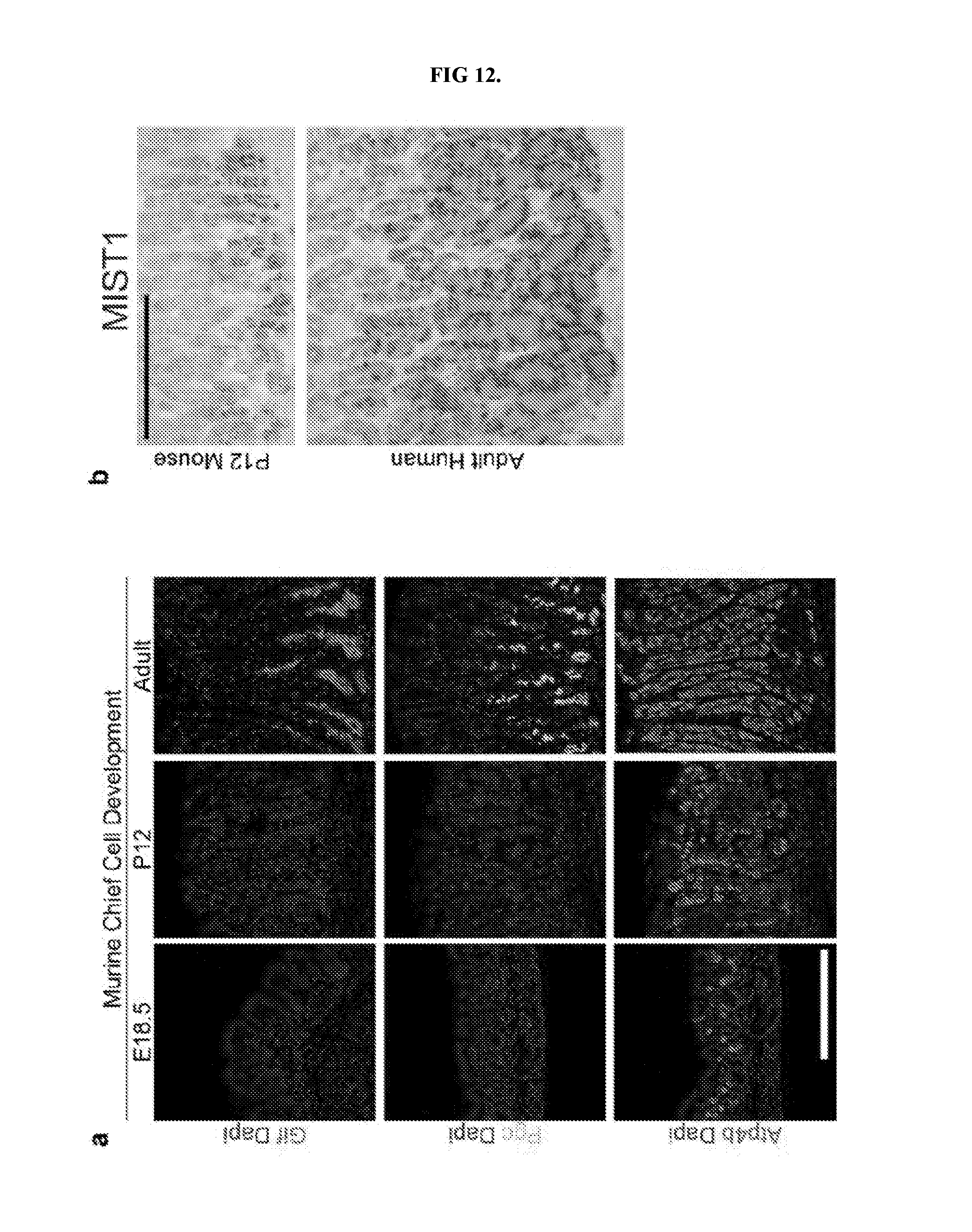
D00013
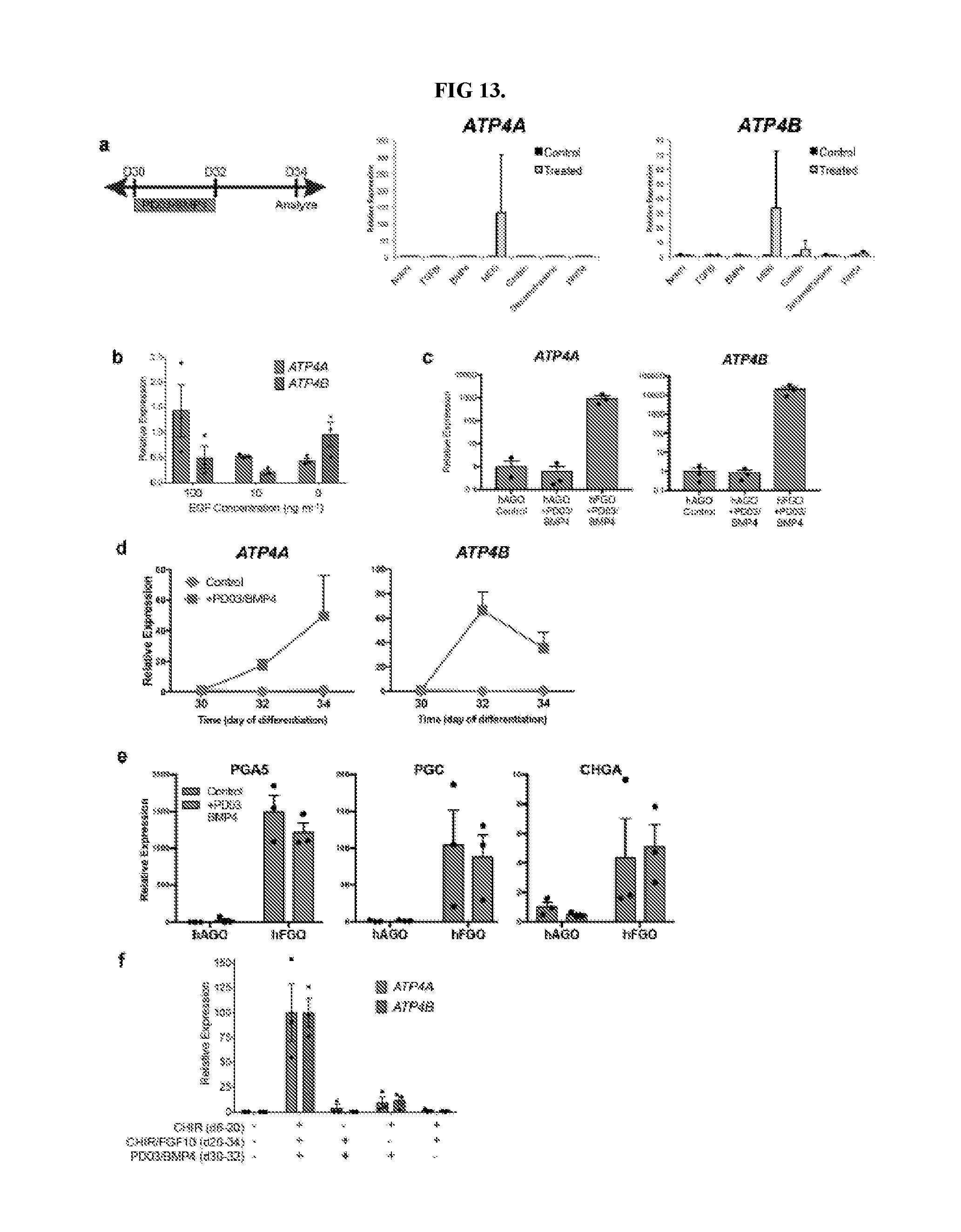
D00014
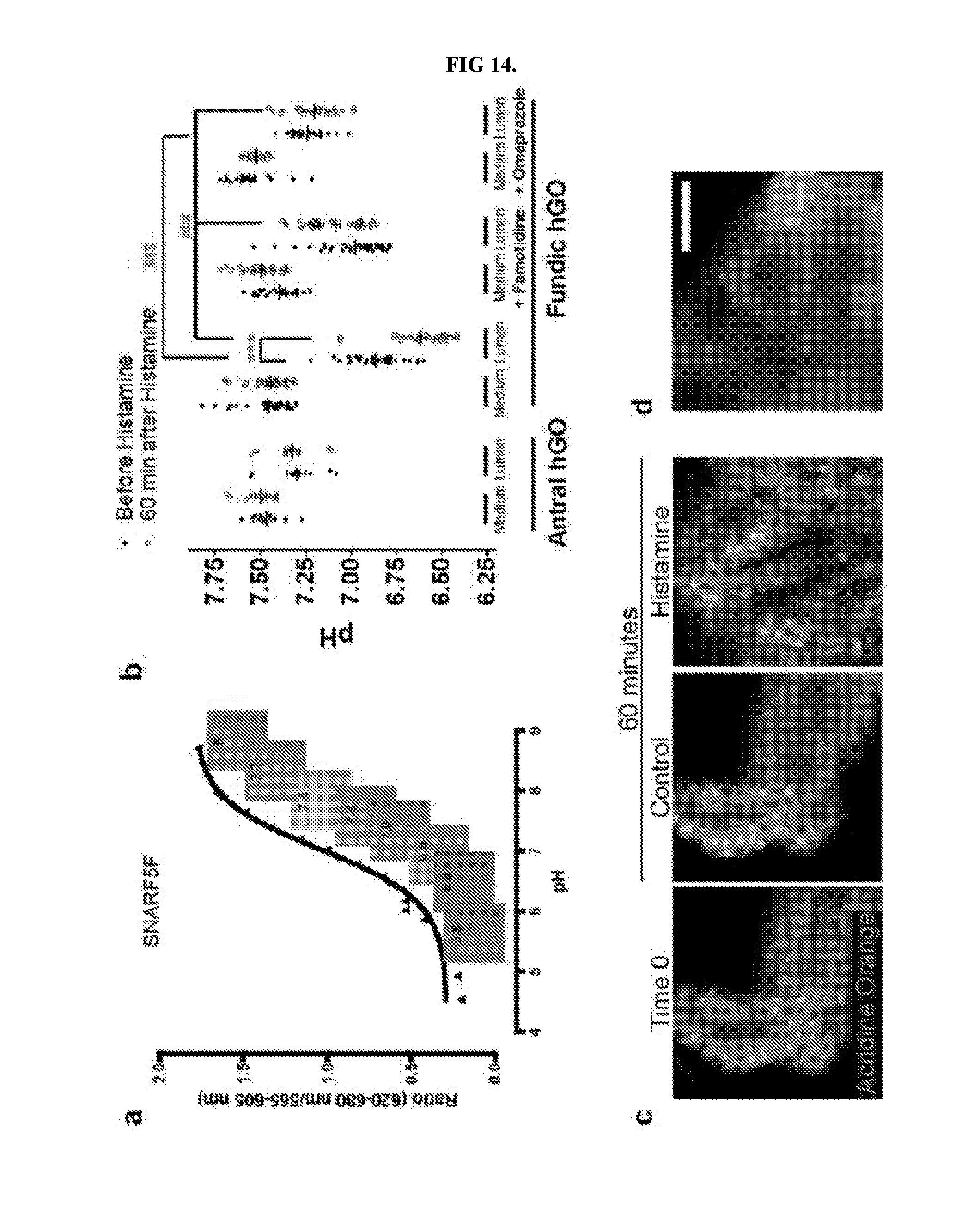
D00015
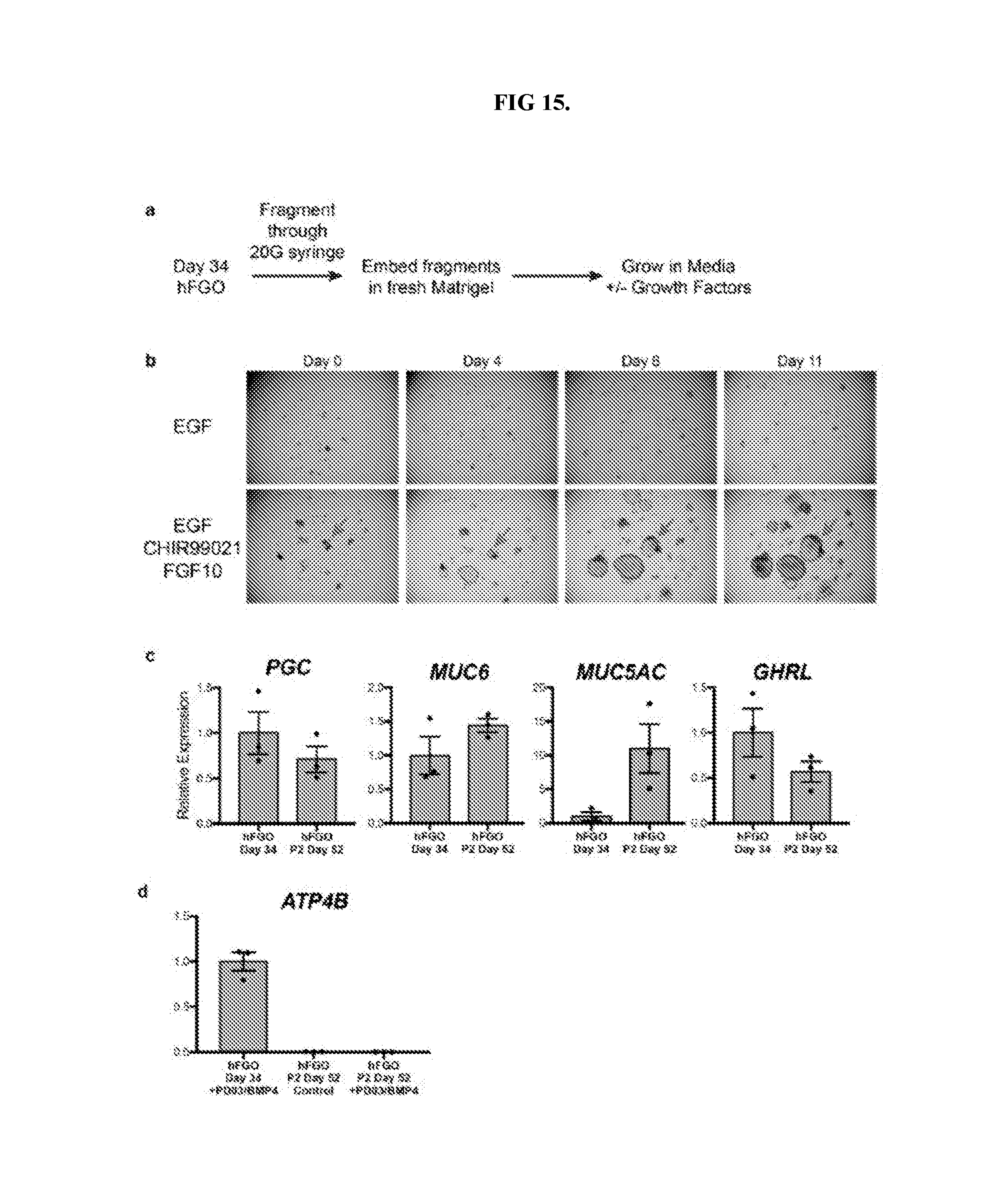
S00001
XML
uspto.report is an independent third-party trademark research tool that is not affiliated, endorsed, or sponsored by the United States Patent and Trademark Office (USPTO) or any other governmental organization. The information provided by uspto.report is based on publicly available data at the time of writing and is intended for informational purposes only.
While we strive to provide accurate and up-to-date information, we do not guarantee the accuracy, completeness, reliability, or suitability of the information displayed on this site. The use of this site is at your own risk. Any reliance you place on such information is therefore strictly at your own risk.
All official trademark data, including owner information, should be verified by visiting the official USPTO website at www.uspto.gov. This site is not intended to replace professional legal advice and should not be used as a substitute for consulting with a legal professional who is knowledgeable about trademark law.