Method For Determining The Cell Mediated Immune Competence Of A Subject
Tasker; Scott
U.S. patent application number 16/086811 was filed with the patent office on 2019-02-28 for method for determining the cell mediated immune competence of a subject. The applicant listed for this patent is Oxford Immunotec Limited. Invention is credited to Scott Tasker.
| Application Number | 20190064145 16/086811 |
| Document ID | / |
| Family ID | 56027490 |
| Filed Date | 2019-02-28 |


| United States Patent Application | 20190064145 |
| Kind Code | A1 |
| Tasker; Scott | February 28, 2019 |
METHOD FOR DETERMINING THE CELL MEDIATED IMMUNE COMPETENCE OF A SUBJECT
Abstract
The invention relates to a method for determining the cell mediated immune competence of a subject. The method comprises conducting a cell-mediated immunoassay (CMI) on a sample comprising immune cells from a subject. The method further comprises detecting in vitro an immune response to a pool of peptides, wherein the pool of peptides is derived from at least three viral antigens.
| Inventors: | Tasker; Scott; (London, GB) | ||||||||||
| Applicant: |
|
||||||||||
|---|---|---|---|---|---|---|---|---|---|---|---|
| Family ID: | 56027490 | ||||||||||
| Appl. No.: | 16/086811 | ||||||||||
| Filed: | March 27, 2017 | ||||||||||
| PCT Filed: | March 27, 2017 | ||||||||||
| PCT NO: | PCT/GB17/50855 | ||||||||||
| 371 Date: | September 20, 2018 |
| Current U.S. Class: | 1/1 |
| Current CPC Class: | G01N 33/5047 20130101; G01N 33/505 20130101; C07K 7/08 20130101; G01N 33/5052 20130101; C40B 40/08 20130101; G01N 33/56983 20130101 |
| International Class: | G01N 33/50 20060101 G01N033/50; C40B 40/08 20060101 C40B040/08; C07K 7/08 20060101 C07K007/08; G01N 33/569 20060101 G01N033/569 |
Foreign Application Data
| Date | Code | Application Number |
|---|---|---|
| Mar 29, 2016 | GB | 1605210.2 |
Claims
1. A method for determining the cell mediated immune competence of a subject, wherein the method comprises conducting a cell-mediated immunoassay (CMI) on a sample comprising immune cells from the subject, the method comprising detecting in vitro an immune response to a pool of peptides, wherein the pool of peptides is derived from at least three viral antigens.
2. The method of claim 1 wherein the immune cells are peripheral blood mononuclear cells (PBMCs).
3. The method of claim 1 or claim 2 wherein the immune cells are T cells.
4. A method according to claim 1, wherein the pool or peptides is derived from at least four, five or six viral antigens.
5. A method according to claim 1 wherein the pool of peptides is derived from three, four or five viral antigens.
6. A method according to any one of claims 1 to 5, wherein the viral antigens are selected from the hexon protein of human adenovirus 3, the matrix protein 1 (H3N2) of the influenza virus, the BZLF-1 protein of the Epstein Barr virus, the nucleocapsid protein of the influenza virus, the fusion glycoprotein G0 of the respiratory syncytial virus.
7. A method according to any one of claims 1 to 6, wherein the viral antigens consist of the hexon protein of human adenovirus 3, the fusion glycoprotein G0 of the respiratory syncytial virus and the nucleocapsid protein of the influenza virus.
8. A method according to any one of claims 1 to 6, wherein the viral antigens consist of the hexon protein of human adenovirus 3, the matrix protein 1 of the influenza virus, the nucleocapsid protein of the influenza virus and the fusion glycoprotein G0 of the respiratory syncytial virus.
9. A method according to any one of claims 1 to 6, wherein the viral antigens consist of the hexon protein of human adenovirus 3, the trans-activator protein BZLF1 of the Epstein Barr virus and the fusion glycoprotein G0 of the respiratory syncytial virus.
10. A method according to any one of claims 1 to 6, wherein the viral antigens consist of the hexon protein of human adenovirus 3, the BALF-4, BMLF-1, BRLF-1, BZLF-1 EBNA-1, EBNA-3A, EBNA-3B, EBNA-3C, EBNA-4, LMP-1 and LMP-2 proteins of the Epstein Barr virus, the matrix protein 1 of the influenza virus, the nucleocapsid protein of the influenza virus and the fusion glycoprotein G0 of the respiratory syncytial virus.
11. A method according to any preceding claim, wherein the pool of peptides comprise one or more fragments of the one or more of the viral antigens.
12. A method according to any preceding claim, wherein the pool of peptides comprise one or more fragments of each of the one or more viral antigens.
13. A method according to any one of claims 11 to 12, wherein the fragments form a protein fragment library encompassing at least 80% of the sequence of the viral antigen from which the fragments are derived.
14. A method according to claim 13, wherein the fragments form a protein fragment library encompassing the entire sequence of the viral antigen from which the fragments are derived.
15. A method according to claims 11 to 13, wherein the fragments have overlapping sequences.
16. A method according to claim 15, wherein the sequences overlap by 11 amino acids.
17. A method according to claims 11 to 16, wherein the fragments are 15 amino acids in length.
18. A method substantially herein as described with reference to the accompanying description and/or to any one of the drawings.
Description
FIELD OF THE INVENTION
[0001] The present invention relates to a method for determining the cell mediated immune competence of a subject. In particular, the method comprises conducting a cell mediated immunoassay on a sample comprising immune cells, for example peripheral blood mononuclear cells (PBMCs) obtained from the subject and detecting an immune response to at least three viral antigens.
BACKGROUND
[0002] Immune competence relates to the presence of a normally functioning immune response in an individual including adaptive (T cells and B cells) and innate (complement and toll-like receptor) immune responses. Assays designed to measure immune competence, in particular cell mediated immune competence, can be used to monitor the T cell response of a donor to an antigen. It is desirable to measure or monitor the immune competence in an individual, and in particular their general ability to mount a cell mediated immune response. For example, an assessment of immune competence is of use to clinicians for several disease conditions including transplantation, cancer, autoimmune diseases, HIV and vaccine trials.
[0003] Assays which provide a measure of immune functionality, include commercially available products Cylex Immunknow and QFT Monitor.RTM.. The Mixed Lymphocyte Reaction (MLR) can also be used to assess the immune response in an individual.
[0004] Cylex Immunknow measures ATP production from whole blood following stimulation with PHA. This relies on non-specific stimulation of the cells contained in the blood sample. The effectiveness of this assay for measuring immune functionality has received mixed results (Lipschultz et al. (2014))
[0005] Quantiferon Monitor.RTM. provides both a qualitative and quantitative measure of immune function (Quantiferon Monitor.RTM. Pack Insert 2014). In particular, the QFM involves stimulating whole blood with a combination of innate (TLR mediated)+adaptive (TCR mediated) antigens. IFN.gamma. secreted from cells responding to the antigens is detected by ELISA. The QFM readout puts donors into 3 categories: Low (<15 IU/mL IFN.gamma.), Medium (15-1000 IU/mL IFN.gamma.) and High (IU/mL IFN.gamma.).
[0006] The Mixed Lymphocyte Reaction (MLR) assay has been used within the research community as a predictor of T cell functionality (Manji et al. (1975)). In particular the MLR assay involves mixing a sample of PBMCs with PBMCs from control (treated to prevent proliferation), adding a radio dye and measuring proliferation of sample PBMCs by decay of radio dye.
SUMMARY OF INVENTION
[0007] The present inventors have identified that the cell mediated immune competence of an individual can be determined by using a selection of viral antigens. By selecting an appropriate pool of viral antigens, the immune competence of a wide range of individuals can be determined, allowing a single assay system to be developed for testing the immune competence of a wide range of individuals.
[0008] In accordance with the present invention, there is provided a method for determining the cell mediated immune competence of a subject, wherein the method comprises conducting a cell-mediated immunoassay (CMI) on a sample comprising immune cells, for example peripheral blood mononuclear cells (PBMCs) from the subject, the method comprising detecting in vitro an immune response to a pool of peptides, wherein the pool of peptides is derived from at least three viral antigens.
A method according to claim 1, wherein the pool or peptides is derived from at least four, five or six viral antigens.
[0009] In a preferred embodiment, the pool of peptides is derived from three, four or five viral antigens.
[0010] In one aspect, the viral antigens are selected from the hexon protein of human adenovirus 3, the matrix protein 1 (H3N2) of the influenza virus, the BZLF-1 protein of the Epstein Barr virus, the nucleocapsid protein of the influenza virus, the fusion glycoprotein G0 of the respiratory syncytial virus.
[0011] In one aspect of the invention, the viral antigens consist of the hexon protein of human adenovirus 3, the fusion glycoprotein G0 of the respiratory syncytial virus and the nucleocapsid protein of the influenza virus.
[0012] In another aspect of the invention, the viral antigens consist of the hexon protein of human adenovirus 3, the matrix protein 1 of the influenza virus, the nucleocapsid protein of the influenza virus and the fusion glycoprotein G0 of the respiratory syncytial virus.
[0013] In a further aspect of the invention, the viral antigens consist of the hexon protein of human adenovirus 3, the trans-activator protein BZLF1 of the Epstein Barr virus and the fusion glycoprotein G0 of the respiratory syncytial virus.
[0014] In a further aspect of the invention, the viral antigens consist of the hexon protein of human adenovirus 3, the BALF-4, BMLF-1, BRLF-1, BZLF-1 EBNA-1, EBNA-3A, EBNA-3B, EBNA-3C, EBNA-4, LMP-1 and LMP-2 proteins of the Epstein Barr virus, the matrix protein 1 of the influenza virus, the nucleocapsid protein of the influenza virus and the fusion glycoprotein G0 of the respiratory syncytial virus.
BRIEF DESCRIPTION OF THE DRAWINGS
[0015] FIG. 1. Example graphs for accelerated stability testing of Hexon, EBV, MP1, NP, FGF0 and BZLF1 in an ELISPOT assay.
[0016] FIG. 2. Example graphs for combined peptide pools (ICA Mix) vs individual peptide pools summed (Additive) in an ELISPOT assay.
[0017] FIG. 3. Testing donors of varied ethnicities in an ELISPOT assay.
[0018] FIG. 4. Example graphs showing the testing of the variation of responses in immune competent donors using ICA Mixes in an ELISPOT assay.
DETAILED DESCRIPTION OF THE INVENTION
[0019] It is to be understood that different applications of the disclosed methods may be tailored to the specific needs in the art. It is also to be understood that the terminology used herein is for the purpose of describing particular embodiments of the invention only, and is not intended to be limiting.
[0020] In addition, as used in this specification and the appended claims, the singular forms "a", "an", and "the" include plural referents unless the content clearly dictates otherwise. Thus, for example, reference to "a fragment" includes "fragments", reference to "a cell" includes two or more such cells, reference to "a subject" includes two or more such subjects, and the like.
[0021] All publications, patents and patent applications cited herein, whether supra or infra, are hereby incorporated by reference in their entirety.
Methods of the Invention
[0022] The present inventors have identified a method of determining immune competence of a subject using a cell mediated immunoassay (CMI assay). A CMI assay designed to measure immune competence, in particular cell mediated immune competence, can be used to monitor the T cell response of a donor to antigen. The inventors have identified that an appropriate pool of viral antigens can be selected for the detection of a cell mediated immune response in a wide variety of individuals. The pool of viral antigens provides a response in immune competent individuals, regardless of their background. Thus, the assay can be used to identify immunocompetent individuals, and also those individual who can not mount an effect immune response.
[0023] Accordingly, the present invention provides for a method of determining immune competence of a subject, wherein the method comprises conducting a cell mediated immunoassay (CMI) on a sample comprising immune cells, for example peripheral blood mononuclear cells (PBMCs) from the subject, the method comprising detecting in vitro an immune response to a pool of peptides, wherein the pool of peptides is derived from at least three viral antigens.
[0024] The method may comprise a pool of peptides derived from at least three, at least four, at least five, or at least six viral antigens, up to seven or eight viral antigens.
[0025] In a particularly preferred embodiment, the peptides are derived from three, four, five or six viral antigens. In a particularly preferred embodiment, the peptides are derived from three, four or five (but not more) viral antigens. In an alternative aspect of the invention, the peptides are derived from two, three or four viral antigens and additionally comprise a pool of antigens derived from EBV, but do not comprise any other viral antigens.
[0026] The pool of peptides may comprise peptides derived from viral antigens of a single virus, more than one virus, or all from different viruses. The pool of peptides may comprise peptides derived from viral antigens of the same virus and of different viruses. In a preferred aspect of the invention, the at least three viral antigens are derived from at least two viruses, preferably from three, four, five or six viruses. In a preferred embodiment, the at least three viral antigens are derived from one or more of the human adenovirus 3, the influenza virus, the Epstein Barr virus and the respiratory syncytial virus.
[0027] In a more preferred embodiment, the viral antigens are selected from the hexon protein derived from the human adenovirus 3, the matrix protein 1 derived from the influenza virus, the nucleocapsid protein derived from the influenza virus, the BZLF-1 protein derived from the Epstein Barr virus and the fusion glycoprotein G0 derived from the respiratory syncytial virus. The influenza virus can be any influenza strain. In a particularly preferred embodiment, the influenza virus is influenza strain H3N2.
[0028] In one embodiment of the invention, the pool of viral antigens consist of the hexon protein of human adenovirus 3, the fusion glycoprotein G0 of the respiratory syncytial virus and the nucleocapsid protein of the influenza virus.
[0029] In another embodiment of the invention, the pool of viral antigens consist of the hexon protein of human adenovirus 3, the matrix protein 1 of the influenza virus, the nucleocapsid protein of the influenza virus and the fusion glycoprotein G0 of the respiratory syncytial virus.
[0030] In a further embodiment of the invention, the pool of viral antigens consist of the hexon protein of human adenovirus 3, the trans-activator protein BZLF1 of the Epstein Barr virus and the fusion glycoprotein G0 of the respiratory syncytial virus.
[0031] In another preferred embodiment, one or more of the viral antigens are derived from Epstein Barr virus, wherein the viral antigens comprise one or more of the BALF-4, BMLF-1, BRLF-1, BZLF-1 EBNA-1, EBNA-3A, EBNA-3B, EBNA-3C, EBNA-4, LMP-1 and LMP-2 proteins. In a particularly preferred embodiment, a pool of peptides derived from EBV is provided, the pool of peptides including peptides derived from each of the BALF-4, BMLF-1, BRLF-1, BZLF-1 EBNA-1, EBNA-3A, EBNA-3B, EBNA-3C, EBNA-4, LMP-1 and LMP-2 proteins. Such a pool of EBV peptides may be provided in combination with the hexon protein of human adenovirus 3, the matrix protein 1 of the influenza virus, the nucleocapsid protein of the influenza virus and the fusion glycoprotein G0 of the respiratory syncytial virus.
Assays for CMI Responses
[0032] Cell Mediated Immune (CMI) responses are used to define the immune status of an individual. Typically, in the art of clinical immunology, the term CMI response encompasses skin testing, lymphocyte proliferation assays, and the detection of cytokines produced by immune cells, for example peripheral blood mononuclear cells (PBMC) in the presence of a specific antigen. The method of the present invention may comprise detecting an in vitro cell mediated immune response. In particular, the in vitro cytokine-based CMI response to peptides may be detected in the method of the present invention.
[0033] The cells of the immune system are capable of producing immune effector molecules such as cytokines following stimulation by antigen. CMI Assays involve incubating a cell sample with antigen and measuring for the presence (or absence) or quantity of an immune effector molecule such as a cytokine to provide an indication of the ability of the individual to generate a cell mediated immune response to the selected antigens. Cells for use in a CMI Assay may include a whole blood sample, a sample comprising immune cells, for example peripheral blood mononuclear cells (PMBCs) and isolated populations of lymphocytes (particularly T-cells) and antigen presenting cells (APCs). APCs are involved in processing the antigen in order that the antigen may be recognised by T-cell receptors on the surface of each T-cell. Antigen recognition may induce cytokine production.
[0034] Cells producing cytokines may be identified by flow cytometry. Flow cytometry may be used to quantify the frequency of cytokine producing cells, and/or the amount of cytokine production by the cells. Antigen-induced cytokines may be released into the assay medium and detected directly by, for example, ELISA methods, or quantified in terms of the frequency of cytokine-secreting T-cells using an enzyme-linked immunospot assay (ELISPOT). The method of the invention preferably comprises an ELISPOT.
[0035] The enzyme-linked immunospot assay (ELISPOT), otherwise known as the filter immunoplaque assay, was initially developed to detect and quantitate individual antibody-secreting B cells. At the time it was developed, the technique provided a rapid and versatile alternative to conventional plaque-forming cell assays. Recent modifications have improved the sensitivity of the ELISPOT such that cells producing as few as 100 molecules of a specific protein per second can be detected. This makes ELISPOT assays much more sensitive than conventional ELISA assays. ELISPOT assays take advantage of the relatively high concentration of a given proteinaceous cell product (such as a cytokine) in the environment immediately surrounding the protein-secreting cell. These cell products are captured and detected using high-affinity antibodies. The ELISPOT assay is reviewed in Current Protocols in Immunology, Unit 6.19 pages 6.19. 1-8.
[0036] The ELISPOT assay typically involves six steps: (1) coating a purified cytokine-specific antibody to a membrane-backed microtiter plate; (2) blocking the plate to prevent non-specific absorption of any other proteins; (3) incubating the cytokine-secreting cells with appropriate reagents; (4) removal of cells and reagents; (5) adding a labelled second anti-cytokine antibody; and (6) detecting the antibody-cytokine complex on the membrane.
Immune Responses
[0037] The immune response that is detected in vitro may be any response that is triggered by the at least three viral antigens of the present invention. The immune response may be mediated by any type of immune cell. In a particularly preferred embodiment, the immune response is mediated by peripheral blood mononuclear cells (PBMCs), which may comprise one or more types of immune cells selected from T-cells, natural killer (NK) cells and monocytes. Typically, the immune response is measured by assessing cytokine secretion, preferably by assessing interferon gamma (IFN-.gamma.) secretion. Methods of measuring cytokine secretion are well known in the art. The immune response is preferably a T-cell response.
[0038] The immune response may occur in vitro. Preferably, the immune response is an in vitro CMI response. A CMI response is an immune response that does not involve antibodies. Instead, a CMI response may involve cytotoxic-T cell activation, increase in production of various cytokines and/or the release of various cytokines in response to an antigen. Methods for detecting in vitro CMI responses are known in the art and are described in detail above.
[0039] The method of the invention may detect the presence or absence of an immune response. The presence of an immune response to the at least three viral antigens according to the method of the present invention may indicate that the subject is immune competent. The absence of an immune response to the at least three viral antigens according to the method of the present invention may indicate that the subject has low immune competence.
Fragments
[0040] A fragment of a viral antigen according to the method of the present invention may be a sequence comprising five or more amino acids that is derived by truncation at the N-terminus and/or C-terminus of the parent sequence. For instance, the fragment may comprise about 5 or more, about 6 or more, about 7 or more, about 8 or more, about 9 or more, about 10 or more, about 11 or more, about 12 or more, about 13 or more, about 14 or more, about 15 or more, about 16 or more, about 17 or more, about 18 or more, about 19 or more, about 20 or more, about 21 or more, about 22 or more, about 23 or more, about 24 or more, about 25 or more, about 26 or more, or about 27 or more amino acids. The fragment may be from about 5 to about 27, from about 6 to about 26, from about 7 to about 25, from about 8 to about 24, from about 9 to about 23, from about 10 to about 22, from about 11 to about 21, from about 12 to about 20, from about 13 to about 19, from about 14 to about 18, from about 12 to about 18, from about 12 to about 15, from about 15 to about 18, from about 13 to about 17, from about 14 to about 16, from about 5 to about 10, from about 10 to about 15, from about 15 to about 20, from about 20 to about 25 or from about 10 to about 20 amino acids in length.
[0041] The fragments may be chemically derived from the parent protein, for example by proteolytic cleavage, or can be derived in an intellectual sense from the parent protein, for example by making use of the amino acid sequence of the parent protein and synthesising fragments based on the sequence. Fragments may be synthesised using methods well known in the art.
[0042] The term "fragment" includes not only molecules in which amino acid residues are joined by peptide (--CO--NH--) linkages but also molecules in which the peptide bond is reversed. Such retro-inverso peptidomimetics may be made using methods known in the art, for example such as those described in Meziere et al (1997) J. Immunol. 159, 3230-3237. This approach involves making pseudopeptides containing changes involving the backbone, and not the orientation of side chains. Meziere et al (1997) show that, at least for MHC class II and T helper cell responses, these pseudopeptides are useful. Retro-inverse peptides, which contain NH--CO bonds instead of CO--NH peptide bonds, are much more resistant to proteolysis.
[0043] Similarly, the peptide bond may be dispensed with altogether provided that an appropriate linker moiety which retains the spacing between the carbon atoms of the amino acid residues is used; it is particularly preferred if the linker moiety has substantially the same charge distribution and substantially the same planarity as a peptide bond. It will also be appreciated that the fragment may conveniently be blocked at its N- or C-terminus so as to help reduce susceptibility to exoproteolytic digestion. For example, the N-terminal amino group of the peptides may be protected by reacting with a carboxylic acid and the C-terminal carboxyl group of the peptide may be protected by reacting with an amine. One or more additional amino acid residues may also be added at the N-terminus and/or C-terminus of the fragment, for example to increase the stability of the fragment. Other examples of modifications include glycosylation and phosphorylation. Another potential modification is that hydrogens on the side chain amines of R or K may be replaced with methylene groups (--NH.sub.2.fwdarw.--NH(Me) or --N(Me).sub.2).
[0044] Fragments of the at least three viral antigens according to the method of the present invention may also include variants of fragments that increase or decrease the fragments' half-life in vivo. Examples of variants capable of increasing the half-life of fragments according to the invention include peptoid analogues of the fragments, D-amino acid derivatives of the fragments, and peptide-peptoid hybrids. The fragment may also comprise D-amino acid forms of the fragment. The preparation of polypeptides using D-amino acids rather than L-amino acids greatly decreases any unwanted breakdown of such an agent by normal metabolic processes, decreasing the amounts of agent which needs to be administered, along with the frequency of its administration. D-amino acid forms of the parent protein may also be used.
[0045] The fragments according to the method of the present invention may be derived from splice variants of the parent proteins encoded by mRNA generated by alternative splicing of the primary transcripts encoding the parent protein chains. The fragments may also be derived from amino acid mutants, glycosylation variants and other covalent derivatives of the parent proteins which retain at least an MHC-binding or antibody-binding property of the parent protein. Exemplary derivatives include molecules wherein the fragments of the invention are covalently modified by substitution, chemical, enzymatic, or other appropriate means with a moiety other than a naturally occurring amino acid.
Fragment Pools
[0046] The method of the invention may comprise detecting in vitro an immune response to a pool of peptides, comprising one or more fragments. The pool of peptides may comprise fragments derived from viral antigens of a single virus, more than one virus, or from all different viruses. The pool of peptides may comprise fragments derived from viral antigens of the same virus and of different viruses. In a preferred aspect of the invention, one or more fragments are derived from at least three viral antigens, the at least three viral antigens derived from two viruses, preferably from three, four, five or six viruses. In a preferred embodiment, one or more fragments are derived from at least three viral antigens, the at least three viral antigens derived from one or more of the human adenovirus 3, the influenza virus, the Epstein Barr virus and the respiratory syncytial virus.
[0047] In a more preferred embodiment, the pool of peptides comprises one or more fragments of the one or more viral antigens selected from the hexon protein derived from the human adenovirus 3, the matrix protein 1 derived from the influenza virus, the nucleocapsid protein derived from the influenza virus, the BZLF protein derived from the Epstein Barr virus and the fusion glycoprotein G0 derived from the respiratory syncytial virus. The influenza virus can be any influenza strain. In a particularly preferred embodiment, the influenza virus is influenza strain H3N2.
[0048] In one embodiment of the invention, the pool of peptides comprises one or more fragments of the one or more viral antigens consisting of the hexon protein of human adenovirus 3, the fusion glycoprotein G0 of the respiratory syncytial virus and the nucleocapsid protein of the influenza virus.
[0049] In another embodiment of the invention, the pool of peptides comprises one or more fragments of the one or more viral antigens consisting of the hexon protein of human adenovirus 3, the matrix protein 1 of the influenza virus, the nucleocapsid protein of the influenza virus and the fusion glycoprotein G0 of the respiratory syncytial virus.
[0050] In a further embodiment of the invention, the pool of peptides comprises one or more fragments of the one or more viral antigens consisting of the hexon protein of human adenovirus 3, the trans-activator protein BZLF1 of the Epstein Barr virus and the fusion glycoprotein G0 of the respiratory syncytial virus.
[0051] In one embodiment of the invention, the pool of peptides comprises one or more fragments of the one or more viral antigens derived from Epstein Barr virus, wherein the viral antigens comprise BALF-4, BMLF-1, BRLF-1, BZLF-1 EBNA-1, EBNA-3A, EBNA-3B, EBNA-3C, EBNA-4, LMP-1 and/or LMP-2 proteins. In a particularly preferred embodiment, a pool of peptides comprising one or more fragments derived from EBV is provided, the pool of peptides including fragments derived from each of the BALF-4, BMLF-1, BRLF-1, BZLF-1 EBNA-1, EBNA-3A, EBNA-3B, EBNA-3C, EBNA-4, LMP-1 and/or LMP-2 proteins. Such a pool of EBV peptide fragments may be provided in combination with the hexon protein of human adenovirus 3, the matrix protein 1 of the influenza virus, the nucleocapsid protein of the influenza virus and the fusion glycoprotein G0 of the respiratory syncytial virus.
[0052] The pool may comprise one or more, two or more, three or more, four or more, five or more, six or more, seven or more, eight or more, nine of more, 10 or more, 15 or more, 20 or more, 25 or more, 50 or more, 75 or more, 100 or more, 200 or more, or 250 or more, fragments derived from the at least three viral antigens.
[0053] In one embodiment, the method of the invention comprises detecting in vitro an immune response to a pool of peptides comprising fragments. Preferably, the method comprises detecting in vitro an immune response to a pool of peptides wherein the pool of peptides comprises one or more fragments of the one or more viral antigens. Further preferably, the method comprises detecting in vitro an immune response to a pool of peptides wherein the pool of peptides comprises one or more fragments of each of the one or more viral antigens.
[0054] As set out below, the method may also comprise detecting in vitro an immune response to one or more protein fragment libraries.
Protein Fragment Libraries
[0055] In one embodiment, the fragments in a pool form a protein fragment library. A protein fragment library comprises a plurality of fragments derived from a parent protein, for the present invention, the hexon protein derived from the human adenovirus 3, the matrix protein 1 derived from the influenza virus, the nucleocapsid protein derived from the influenza virus, the BALF-4, BMLF-1, BRLF-1, BZLF-1 EBNA-1, EBNA-3A, EBNA-3B, EBNA-3C, EBNA-4, LMP-1 and LMP-2 proteins derived from the Epstein Barr virus and the fusion glycoprotein G0 derived from the respiratory syncytial virus, that together encompass at least 10%, such as at least 20%, at least 30%, at least 40%, at least 50%, at least 60%, at least 70%, at least 75%, at least 80%, at least 85%, at least 90%, at least 95%, at least 98%, at least 99%, or 100%, of the sequence of the parent protein. In the present invention, the fragments in a pool preferably form a protein fragment library encompassing at least 80% of the sequence of the protein from which the fragments are derived. More preferably, the fragments in a pool form a protein fragment library encompassing the entire sequence of the protein from which the fragments are derived.
[0056] The protein fragment library may comprise fragments that are capable of stimulating CD4+ and/or CD8+ T-cells. Preferably, the protein fragment library comprises fragments that are capable of stimulating both CD4+ and CD8+ T-cells. It is known in the art that the optimal fragment size for stimulation is different for CD4+ and CD8+ T-cells. Fragments consisting of about 9 amino acids (9mers) typically stimulate CD8+ T-cells only, and fragments consisting of about 20 amino acids (20mers) typically stimulate CD4+ T-cells only. Broadly speaking, this is because CD8+ T-cells tend to recognise their antigen based on its sequence, whereas CD4+ T-cells tend to recognise their antigen based on its higher-level structure. However, fragments consisting of about 15 amino acids (15mers) may stimulate both CD4+ and CD8+ T cells. Accordingly, the protein fragment library preferably comprises fragments that are about 15 amino acids, such as about 12 amino acids, about 13 amino acids, about 14 amino acids, about 15 amino acids, about 16 amino acids, about 17 amino acids or about 18 amino acids in length.
[0057] All of the fragments in a pool may be the same length. Alternatively, a pool may comprise fragments of different lengths. Fragment lengths are discussed above.
[0058] A protein fragment library may comprise fragments whose sequences overlap. Accordingly, each pool may comprise fragments whose sequences overlap. The sequences may overlap by one or more, such as two or more, three or more, four or more, five or more, six or more, seven or more, eight or more, nine or more, 10 or more, 11 or more, 12 or more, 13 or more, 14 or more, 15 or more, 16 or more, 17 or more, 18 or more, 19 or more, or 20 or more, amino acids. Preferably, the sequences overlap by 9 or more amino acids, such as 10 or more, 11 or more or 12 or more amino acids, as this maximises the number of fragments that comprise the 9mers capable of stimulating CD8+ T-cells. More preferably, the sequences overlap by 11 amino acids. All of the overlapping fragments in a pool may overlap by the same number of amino acids. Alternatively, a pool may comprise fragments whose sequences overlap by different numbers of amino acids.
[0059] The protein fragment library may comprise fragments of 12 to 18 (such as 12 to 15, 15 to 18, 13 to 17, or 14 to 16) amino acids in length that overlap by 9 to 12 (such as 9 to 11 or 10 to 12) amino acids. For instance, the protein fragment library may comprise fragments of (i) 14 amino acids in length that overlap by 9, 10, or 11 amino acids, (ii) 15 amino acids in length that overlap by 9, 10, or 11 amino acids, or (iii) 16 amino acids in length that overlap by 9, 10, or 11 amino acids. The protein fragment library preferably comprises fragments of 15 amino acids in length that overlap by 11 amino acids.
[0060] General properties of fragments are set out above.
[0061] The Epstein Barr virus pool may comprise 1, 2, 3, 4, 5, 6, 7, 8, 9, 10, 11, 12, 13, 14, 15, 16, 17, 18, 19, 20, 21, 22, 23, 24, 25 or 26 fragments derived from Epstein Barr virus peptides wherein overlapping fragments are not required. For example, the pool of peptides may comprise the peptide shown in Table 6.
Samples
[0062] The in vitro detection of an immune response to the at least three viral antigens of the present invention is performed using a sample obtained from the subject. In a preferred embodiment, the sample is a blood sample.
Subject
[0063] The method of the invention may be used to determine the immune competence of any suitable subject. The subject is generally a human subject.
Cells
[0064] In one embodiment, the method of the invention comprises conducting a cell-mediated immunoassay on a sample comprising immune cells, for example peripheral blood mononuclear cells, the method comprising detecting in vitro an immune response to a pool of peptides derived from at least three viral antigens. The pool of peptides may comprise one or more fragments of one or more of the viral antigens or one of more fragments of each of one of more viral antigens. The viral antigens may comprise one or more protein fragment libraries.
[0065] The sample is typically contacted with a sufficient amount of the at least three viral antigens to generate an immune response to the viral antigens. The sample may be contacted with the pool of peptides wherein the peptides or fragments have been combined prior to contacting the sample; or contacted with each viral antigen or fragment sequentially or concurrently using any combination of viral antigen or fragment. In a preferred embodiment, the sample is contacted with all the viral antigens concurrently, wherein the viral antigens have been combined prior to contacting the sample.
[0066] The sample may be contacted with any amount of the at least three viral antigens, such as about 1 ng/ml, about 5 ng/ml, about 10 ng/ml, about 50 ng/ml, about 100 ng ml, about 500 ng/ml, about 1 .mu.g/ml, about 5 .mu.g/ml, about 10 .mu.g/ml, about 50 .mu.g/ml, about 100 .mu.g ml, about 500 .mu.g/ml, 1 mg/ml, about 5 mg/ml, about 10 mg/ml, about 50 mg/ml, about 100 mg ml, or about 500 mg/ml of the at least three viral antigens. The sample may be contacted with the at least three viral antigens concurrently or sequentially.
[0067] The immune cells may comprise one or more types of immune cells selected from T-cells, B-cells, dendritic cells, neutrophils, basophils, mast cells, eosinophils, innate lymphoid cells (ILCs), natural killer (NK) cells, monocytes, macrophages and thymocytes. The sample comprising immune cells may comprise a population of immune cells. The population may comprise all of these types of immune cells. The population may comprise peripheral blood mononuclear cells (PBMCs). A sample of PBMCs comprises T cells, B cells, NK cells and monocytes. The population of immune cells may comprise T-cells. In one embodiment, the population of immune cells comprises peripheral blood mononuclear cells.
[0068] The contacting of the cell sample with the at least three viral antigens may be carried out in any suitable volume. Typical volumes of the samples range from about 10 .mu.l to about 1 ml, preferably from about 50 .mu.l to about 500 .mu.l, more preferably from about 100 .mu.l to about 200 .mu.l. Typically, the length of time for which the cells are contacted with the at least three viral antigens is from about 5 minutes to about 50 hours, for example from about 10 minutes to about 40 hours, from about 20 minutes to about 30 hours, from about 30 minutes to about 20 hours, from about 45 minutes to about 12 hours, from about 1 hour to about 6 hours, preferably from about 10 minutes to about 2 hours. The cells may be contacted with the antigens overnight.
[0069] The cells may be contacted with the antigen at any suitable temperature. The suitable temperature is typically in the same range as the normal body temperature of the human or animal from which the cells are derived. Typically, the incubation is carried out at a fixed temperature between about 4.degree. C. and about 38.degree. C., preferably from about 20.degree. C. to about 38.degree. C., more preferably at about 37.degree. C.
[0070] The cells are typically present in wells. The cells are preferably present in the wells of a flat plate, which is preferably a membrane-backed plate. The samples are more preferably present in the wells of a standard 96 or 384 well plate. Such plates are commercially available Fisher scientific, VWR suppliers, Nunc, Starstedt or Falcon. The wells typically have a capacity of from about 25 .mu.l to about 250 .mu.l, from about 30 .mu.l to about 200 .mu.l, from about 40 .mu.l to about 150 .mu.l or from about 50 to 1000 The cells obtained from the subject can be cultured before being used in the methods. This allows equal numbers of adherent cells to be present in each sample being assayed. Alternatively, if the cells are immobilized or captured, the cells, such as fresh blood cells, can be counted before plating. Techniques for culturing cells are well known to a person skilled in the art. The cells are typically cultured under standard conditions of 37.degree. C., 5% CO.sub.2 in medium supplemented with serum.
[0071] The cells may be cultured in any suitable flask or vessel and then be transferred to wells. The cells are typically cultured in wells. The cells are preferably cultured in a flat plate comprising two or more wells, such as a standard 96 or 384 well plate. Incubating the cells with the marker typically involves replacing the culture medium in each well with a suitable solution comprising the marker. Suitable solutions are well known to a person skilled in the art.
Immune Responsiveness
[0072] As set out above, the method of the invention comprises detecting in vitro an immune response to at least three viral antigens. Detection of an immune response indicates that the subject is immune competent. The lack of detection (or absence of detection) of an immune response indicates that the subject is not immune responsive and has low immune competence. For example, an immune competent donor within the immune competency assay is one that has no spots in the negative control, greater than 20 spots in the positive control and produces specific IFN-.gamma. spots in response to the immuno-competency assay (ICA) antigens.
Therapeutic Application
[0073] The present invention provides for a method for determining immune competence of a subject, wherein the method comprises conducting a cell-mediated immunoassay (CMI) on a sample comprising immune cells, for example peripheral blood mononuclear cells (PBMCs) from the subject, the method comprising detecting in vitro an immune response to a pool of peptides, wherein the pool of peptides is derived from at least three viral antigens. The present method may be useful in the clinic to monitor the broad T cell responses of donors to the at least three viral antigens. This may be helpful to clinicians when assessing a patient's immune competence for several disease conditions including transplantation, cancer, autoimmune diseases, HIV and in vaccine trials.
EXAMPLES
Example 1--Selection of the Most Immunogenic Antigens
[0074] Healthy donor samples (31-167) were screened with 23 commercially available peptide pools in the ELISPOT assay. To identify the peptide pools that gave the best coverage of immunocompetent donors, an arbitrary 10 spot cut off was defined. The responses to each peptide pool were then assessed using the cut-off. Table 1 ranks the peptide pools based on the percentage of responsive donors.
TABLE-US-00001 TABLE 1 Ranking of peptide pools. Percentage Responsive No of (10 spot arbitrary Peptides Virus/Bacteria Antigen Abbreviation cut off) in the pool Human Adenovirus 3 Hexon protein Hexon 85% (142/167 234 Donors) Influenza Matrix protein 1 (H3N2) MP1 77% (61/79 61 Donors) Epstein Barr EBV EBV 73% (112/153 26 Virus (EBV) Donors) Influenza Nucleocapsid protein NP 62% (70/112 122 (H3N2) Donors) Respiratory Fusion glycoprotein F0 FGF0 50% (54/106 141 Syncytial Virus Donors) (RSV) Human Adenovirus 5 Penton protein Penton 48% (81/167 140 Donors) EBV Epstein barr nuclear EBNA1 48% (20/41 158 antigen 1 donors) EBV Epstein barr nuclear EBNA-3a 47% (72/153 234 antigen 3a Donors) EBV Trans-activator protein BZLF1 45% (64/142 59 BZLF1 Donors) Influenza Influenza INF 40% (56/138 17 Donors) RSV Nucleocapsid protein N NCPN 29% (32/107 95 Donors) RSV Major surface glycoprotein MSG 26% (11/41 72 donors) Influenza Hemagglutinin of HA INF-bris 25% (18/72 139 Influenza A Donors) (H1N1/Brisbane) Adenovirus ADV ADV 19% (24/122 5 Donors) EBV Trans-activator protein BZLF1 17% (25/145 N/A BZLF1 Protein Donors) Candida Albicans Mannoprotein MP65 MP65 14% (6/41 donors) 92 EBV Epstein barr nuclear EBNA-3a 13% (19/137 N/A antigen-3A Protein Donors) RSV RSV RSV 12% (5/39 Donors) 28 EBV Latent membrane protein 2 LMP2 9% (4/41 donors) 122 Influenza Hemagglutinin of HA INF-ca 8% (4/48 Donors) 139 Influenza A (H1N1/California) EBV DNA polymerase BMRF1 7% (3/41 donors) 99 processivity factor BMRF1 Influenza Matrix protein 2 MP2 0% (0/34 Donors) 22 EBV Secreted protein BARF1 BARF1 0% (0/41 donors) 53
[0075] This analysis allowed the selection of the 5 most responsive antigens (based on arbitrary 10 spot cut off)--Hexon, MP1, EBV, NP and FGF0.
[0076] Donors that were weak responders to the Hexon, MP1, EBV, NP and FGF0 (<10 spots) were assessed for responses to the remaining screened antigens. The BZLF1 antigen gave spot counts greater than 10 within these donors, so it was selected for further testing.
Example 2--Solubility
[0077] Custom peptide pools (Hexon, EBV, MP1, NP, FGF0 and BZLF1) were synthesized and supplied as lyophilised pools. The peptide pools were dissolved to a concentration of 625 .mu.g/mL per peptide in the pool using Dimethyl Sulfoxide (DMSO).
[0078] Dissolution of all peptide pools was completed successfully. No solubility issues were observed.
Example 3--Assessment of the Stability of Individual Peptide Pools
[0079] The stability of each peptide pool at working concentration (3 .mu.g/mL) was assessed using an accelerated stability study. Briefly: peptides pools at working concentration were stored at 37.degree. C. for 8 weeks. The stressed peptide pools were then tested in comparison to freshly diluted peptide pools (control) in an ELISPOT assay. For each donor, 3 replicates were tested. The results from the studies are shown in FIG. 1.
[0080] Statistical analysis (Mann-Witney test) determined there was no statistical significant difference in the spot counts between the control and the peptides stored at 37.degree. C. for 8 weeks.
Example 4--Combining Immunogenic Antigens
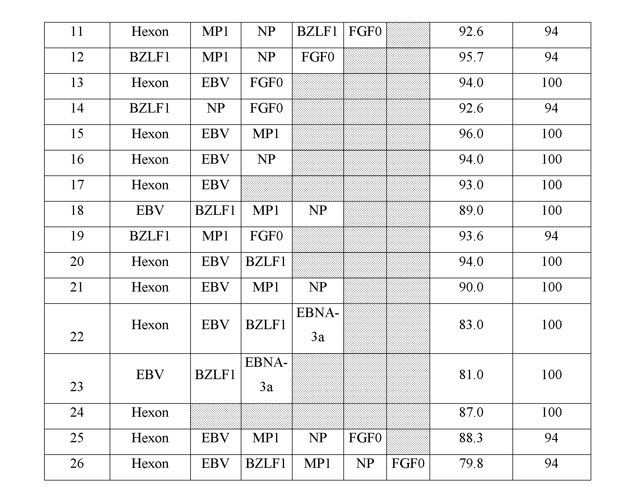
[0081] The data generated from the screening of peptide pools in an ELISPOT assay was re-assessed to determine the theoretical coverage of donors as if peptides from different antigens were combined into one pool. This was conducted by adding the spot counts for individual peptide pools (e.g. Donor 15, Hexon spot count=5, EBV spot count=26 NP spot count=5, Theoretical Mix 16 spot count=36). The percentage of donors within the 10-350 spot range was calculated for each mix. The results of this analysis are shown in table 2.
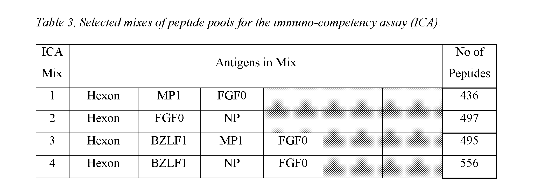
The theoretical analysis identified: [0082] 10 mixes would have given responses to >95% of the donors assessed. [0083] Mixes 25 and 26 would theoretically give spot counts that are at the upper limit of what is accurately countable on an ELISPOT plate reader (400+ spots). This reduced the percentage of donors within the 10-350 spot count range. [0084] 13 mixes were selected for further testing in an ELISPOT assay (Table 3).
[0085] To confirm that the predicted performance of the combined peptide pools would be achieved, the peptide pools were tested individually and in mixes in an ELISPOT assay with 32 donors. Obtained spot counts for the mixes were compared to the additive spot count from the individual antigens (e.g. Donor 9743, Hexon spot count=5, MP1 spot count=10 FGF0 spot count=6, Mix 1 spot count=28). The results of this comparison are shown in FIG. 2.
[0086] Statistical analysis (Mann-Witney test) determined there was no significant statistical difference between the additive spot counts of the individual peptide pools and the combined ICA mix.
Example 5--Preparation of ICA Mixes
[0087] The dissolved peptide pools (625 .mu.g/mL per peptide in the pool) were combined into the immuno-competency assay (ICA) mixes. No issues were observed during preparation.
Example 6--Ethnicity
[0088] The 13 selected ICA mixes were tested in an ELISPOT assay with blood samples collected from 35 Caucasians, 10 African Americans, 10 Hispanic and 9 Asian donors. The results of this testing are shown in FIG. 3.
[0089] A 10 spot cut off was used to determine the number of responsive donors (Table 4).
TABLE-US-00002 TABLE 4 Percentage of donors responsive to ICA mixes based on an arbitrary 10 spot cut off. Percentage Responsive Donors (Based on an arbitrary 10 spot cut off) Mix 1 Mix 2 Mix 3 Mix 4 Mix 5 Mix 6 Mix 7 Mix 8 Mix 9 Mix 10 Mix 11 Mix 12 Mix 13 Caucasian 94% 100% 100% 97% 97% 100% 100% 100% 100% 100% 100% 100% 100% (n = 35) African 90% 90% 90% 90% 80% 90% 80% 80% 90% 80% 90% 90% 90% American (n = 10) Hispanic 90% 100% 100% 100% 100% 100% 90% 100% 100% 100% 100% 100% 100% (n = 10) Asian 89% 100% 89% 89% 100% 100% 89% 89% 100% 78% 78% 100% 89% (n = 9)
[0090] 4 of the 13 mixes (ICA mixes 2, 6, 9 and 12) were responsive in >90% of the donors tested regardless of ethnicity, based on a 10 spot cut off.
Example 7--Fluctuation of Responses Over Time to ICA Mixes
[0091] To determine the natural variation of response induced by the ICA mixes, 6 immunocompetent blood donors were tested at time points: day 0, 3, 7, 14, 28, 56, and 84 with the ICA mixes in an ELISPOT assay.
[0092] FIG. 4 shows variation of response over a short period of time (7 days) and longer period of time (84 days) for two example mixes.
[0093] Statistical analysis was performed by BioBridges.
Briefly: The coefficient of variation (% CV) was calculated for the individual mixes with each donor using data from all time points. Following this, the % CV values for each mix from the 6 donors were grouped and a mean calculated. The mean of these % CV's is a measure of the variance of spot counts using ICA mixes in an ELISPOT assay, see Table 4 for summary.
TABLE-US-00003 TABLE 5 Statistical analysis of the natural variation of immunocompetent responses to ICA mixes in the ELISPOT assay. (n = 6) Variation Variation ICA Mix (Over 7 days) (Over 84 days) Mix 1 20% 26% Mix 2 16% 21% Mix 3 13% 23% Mix 4 11% 18% Mix 5 22% 25% Mix 6 18% 23% Mix 7 24% 31% Mix 8 18% 25% Mix 9 19% 25% Mix 10 21% 24% Mix 11 27% 34% Mix 12 21% 23% Mix 13 25% 24%
TABLE-US-00004 TABLE 6 EBV Pool Peptide Parent SEQ ID Sequence Protein Location Prot ID Number 1 FLDKGTYTL BALF-4 276-284 P03188 SEQ ID NO: 1 2 GLCTLVAML BMLF-1 259-267 Q04360 SEQ ID NO: 2 3 DYCNVLNKEF BRLF1 28-37 P03209 SEQ ID NO: 3 4 ATIGTAMYK BRLF1 134-142 P03209 SEQ ID NO: 4 5 RVRAYTYSK BRLF-1 148-156 P03209 SEQ ID NO: 5 6 RAKFKQLL BZLF-1 190-197 P03206 SEQ ID NO: 6 7 EPLPQGQLTAY BZLF-1 54-64 P03206 SEQ ID NO: 7 8 HPVGEADYFEY EBNA-1 407-417 P03211 SEQ ID NO: 8 9 QAKWRLQTL EBNA-3A 158-166 P12977 SEQ ID NO: 9 10 FLRGRAYGL EBNA-3A 193-201 P12977 SEQ ID NO: 10 11 RPPIFIRRL EBNA-3A 247-255 P12977 SEQ ID NO: 11 12 YPLEIEQHGM EBNA-3A 458-466 P12977 SEQ ID NO: 12 13 RLRAEAQVK EBNA-3A 603-611 P12977 SEQ ID NO: 13 14 AVFDRKSDAK EBNA-3B 399-408 I1YP20 SEQ ID NO: 14 15 RRIYDLIEL EBNA-3C 79-87 Q69140 SEQ ID NO: 15 16 EENLLDFVRF EBNA-3C 102-111 Q69140 SEQ ID NO: 16 17 QPRAPIRPI EBNA-3C 881-889 Q69140 SEQ ID NO: 17 18 IVTDFSVIK EBNA-4 416-424 P03203 SEQ ID NO: 18 19 YLLEMLWRL LMP-1 125-133 P03230 SEQ ID NO: 19 20 PYLFWLAAI LMP2 131-139 P13285 SEQ ID NO: 20 21 IEDPPFNSL LMP2 200-208 P13285 SEQ ID NO: 21 22 SSCSSCPLSK LMP-2 340-349 P13285 SEQ ID NO: 22 23 FLYALALLL LMP-2 356-364 P13285 SEQ ID NO: 23 24 TYGPVFMCL LMP-2 419-427 P13285 SEQ ID NO: 24 25 TYGPVFMSL LMP2 419-427 P13285 SEQ ID NO: 25 26 CLGGLLTMV LMP-2 426-434 P13285 SEQ ID NO: 26
TABLE-US-00005 Epstein Barr Virus, Transactivator protein BZLF1 Prot ID: P03206 SEQ ID NO: 27 MMDPNSTSEDVKFTPDPYQVPFVQAFDQATRVYQDLGGPSQAPLPCVLWPVLPEPLPQGQ LTAYHVSTAPTGSWFSAPQPAPENAYQAYAAPQLFPVSDITQNQQTNQAGGEAPQPGDNS TVQTAAAVVFACPGANQGQQLADIGVPQPAPVAAPARRTRKPQQPESLEECDSELEIKRY KNRVASRKCRAKFKQLLQHYREVAAAKSSENDRLRLLLKQMCPSLDVDSIIPRTPDVLHE DLLNF Human Adenovirus 3, Hexon Protein Prot ID: P36849 >sp|P36849|CAPSH_ADE03 Hexon protein OS = Human adenovirus B serotype 3 GN = L3 PE = 2 SV = 2 SEQ ID NO: 28 MATPSMMPQWAYMHIAGQDASGYLSPGLVQFARATDTYFSMGNKFRNPTVAPTHDVTTDR SQRLMLRFVPVDREDNTYSYKVRYTLAVGDNRVLDMASTFFDIRGVLDRGPSFKPYSGTA YNSLAPKGAPNTSQWIVTTNGDNAVTTTTNTFGIASMKGGNITKEGLQIGKDITTTEGEE KPIYADKTYQPEPQVGEESWTDTDGTNEKFGGRALKPATNMKPCYGSFARPTNIKGGQAK NRKVKPTTEGGVETEEPDIDMEFFDGRDAVAGALAPEIVLYTENVNLETPDSHVVYKPET SNNSHANLGQQAMPNRPNYIGFRDNFVGLMYYNSTGNMGVLAGQASQLNAVVDLQDRNTE LSYQULDSLGDRTRYFSMWNQAVDSYDPDVRIIENHGIEDELPNYCFPLNGIGPGHTYQ GIKVKTDDTNGWEKDANVAPANEITIGNNLAMEINIQANLWRSFLYSNVALYLPDVYKYT PPNITLPTNTNTYEYMNGRVVSPSLVDSYINIGARWSLDPMDNVNPFNHHRNAGLRYRSM LLGNGRYVPFHIQVPQKFFAVKNLLLLPGSYTYEWNFRKDVNMVLQSSLGNDLRTDGATI SFTSINLYATFFPMAHNTASTLEAMLRNDTNDQSFNDYLSAANMLYPIPANATNIPISIP SRNWAAFRGWSFTRLKTKETPSLGSGFDPYFVYSGSIPYLDGTFYLNHTFKKVAIMFDSS VSWPGNDRLLSPNEFEIKRTVDGEGYNVAQCNMTKDWFLVQMLANYNIGYQGFYIPEGYK DRMYSFFRNFQPMSRQVVDEVNYTDYKAVTLPYQHNNSGFVGYLAPTMRQGEPYPANYPY PLIGTTAVKSVTQKKFLCDRTMWRIPFSSNFMSMGALTDLGQNMLYANSAHALDMTFEVD PMDEPTLLYLLFEVFDVVRVHQPHRGVIEAVYLRTPFSAGNATT Influenza A (H3N2) Virus, Nucleoprotein ProtID: I6LFN1 >tr|I6LFN1|I6LFN1_9INFA Nucleoprotein OS = Influenza A virus (A/Nanchang/A2/1994(H3N2)) GN = NP PE = 3 SV = 1 SEQ ID NO: 29 MASQGTKRSYEQMETDGERQNATEIRASVGKMIDGIGRFYIQMCTELKLSDYEGRLIQNS LTIERMVLSAFDERRNRYLEEHPSAGKDPKKTGGPIYKRVDGRWMRELVLYDKEEIRRIW RQANNGDDATAGLTHMMIWHSNLNDTTYQRTRALVRTGMDPRMCSLMQGSTLPRRSGAAG AAVKGIGTMVMELIRMIKRGINDRNFWRGENGRKTRSAYERMCNILKGKFQTAAQRAMMD QVRESRNPGNAEIEDLIFSARSALILRGSVAHKSCLPACVYGPAVSSGYNFEKEGYSLVG IDPFKLLQNSQVYSLIRPNENPAHKSQLVWMACHSAAFEDLRLLSFIRGTKVSPRGKLST RGVQIASNENMDNMESSTLELRSRYWAIRTRSGGNTNQQRASAGQISVQPTFSVQRNLPF EKSTVMAAFTGNTEGRTSDMRAEIIRMMEGAKPEEVSFRGRGVFELSDEKATNPIVPSFD MSNEGSYFFGDNAEEYDN Respiratory Syncytial Virus, Fusion GlycoProtein F0 ProtID: O36634 >sp|O36634|FUS_HRSVB Fusion glycoprotein F0 OS = Human respiratory syncytial virus B (strain B1) GN = F PE = 1 SV = 1 SEQ ID NO: 30 MELLIHRLSAIFLTLAINALYLTSSQNITEEFYQSTCSAVSRGYFSALRTGWYTSVITIE LSNIKETKCNGTDTKVKLIKQELDKYKNAVTELQLLMQNTPAANNRARREAPQYMNYTIN TTKNLNVSISKKRKRRFLGFLLGVGSAIASGIAVSKVLHLEGEVNKIKNALLSTNKAVVS LSNGVSVLTSKVLDLKNYINNQLLPIVNQQSCRISNIETVIEFQQKNSRLLEINREFSVN AGVTTPLSTYMLTNSELLSLINDMPITNDQKKLMSSNVQIVRQQSYSIMSIIKEEVLAYV VQLPIYGVIDTPCWKLHTSPLCTTNIKEGSNICLTRTDRGWYCDNAGSVSFFPQADTCKV QSNRVFCDTMNSLTLPSEVSLCNTDIFNSKYDCKIMTSKTDISSSVITSLGAIVSCYGKT KCTASNKNRGIIKTFSNGCDYVSNKGVDTVSVGNTLYYVNKLEGKNLYVKGEPIINYYDP LVFPSDEFDASISQVNEKINQSLAFIRRSDELLHNVNTGKSTTNIMITTIIIVIIVVLLS LIAIGLLLYCKAKNTPVTLSKDQLSGINNIAFSK Influenza A virus (H3N2). Matrix Protein 1. ProtID: Q67157 >sp|Q67157|M1_I68A0 Matrix protein 1 OS = Influenza A virus (strain A/Aichi/2/1968 H3N2) GN = M PE = 3 SV = 1 SEQ ID NO: 31 MSLLTEVETYVLSIVPSGPLKAEIAQRLEDVFAGKNTDLEALMEWLKTRPILSPLTKGIL GFVFTLTCPSERGLQRRRFVQNALNGNGDPNNMDRAVKLYRKLKREITFHGAKEIALSYS AGALASCMGLIYNRMGAVTTEVAFGLVCATCEQIADSQHRSHRQMVTTTNPLIRHENRMV LASTTAKAMEQMAGSSEQAAEAMEVASQARQMVQAMRAIGTHPRSSAGLKDDLLENLQAY QKRMGVQMQRFK
Sequence CWU 1
1
3119PRTEpstein Barr Virus 1Phe Leu Asp Lys Gly Thr Tyr Thr Leu 1 5
29PRTEpstein Barr Virus 2Gly Leu Cys Thr Leu Val Ala Met Leu 1 5
310PRTEpstein Barr Virus 3Asp Tyr Cys Asn Val Leu Asn Lys Glu Phe 1
5 10 49PRTEpstein Barr Virus 4Ala Thr Ile Gly Thr Ala Met Tyr Lys 1
5 59PRTEpstein Barr Virus 5Arg Val Arg Ala Tyr Thr Tyr Ser Lys 1 5
68PRTEpstein Barr Virus 6Arg Ala Lys Phe Lys Gln Leu Leu 1 5
711PRTEpstein Barr Virus 7Glu Pro Leu Pro Gln Gly Gln Leu Thr Ala
Tyr 1 5 10 811PRTEpstein Barr Virus 8His Pro Val Gly Glu Ala Asp
Tyr Phe Glu Tyr 1 5 10 99PRTEpstein Barr Virus 9Gln Ala Lys Trp Arg
Leu Gln Thr Leu 1 5 109PRTEpstein Barr Virus 10Phe Leu Arg Gly Arg
Ala Tyr Gly Leu 1 5 119PRTEpstein Barr Virus 11Arg Pro Pro Ile Phe
Ile Arg Arg Leu 1 5 129PRTEpstein Barr Virus 12Tyr Pro Leu His Glu
Gln His Gly Met 1 5 139PRTEpstein Barr Virus 13Arg Leu Arg Ala Glu
Ala Gln Val Lys 1 5 1410PRTEpstein Barr Virus 14Ala Val Phe Asp Arg
Lys Ser Asp Ala Lys 1 5 10 159PRTEpstein Barr Virus 15Arg Arg Ile
Tyr Asp Leu Ile Glu Leu 1 5 1610PRTEpstein Barr Virus 16Glu Glu Asn
Leu Leu Asp Phe Val Arg Phe 1 5 10 179PRTEpstein Barr Virus 17Gln
Pro Arg Ala Pro Ile Arg Pro Ile 1 5 189PRTEpstein Barr Virus 18Ile
Val Thr Asp Phe Ser Val Ile Lys 1 5 199PRTEpstein Barr Virus 19Tyr
Leu Leu Glu Met Leu Trp Arg Leu 1 5 209PRTEpstein Barr Virus 20Pro
Tyr Leu Phe Trp Leu Ala Ala Ile 1 5 219PRTEpstein Barr Virus 21Ile
Glu Asp Pro Pro Phe Asn Ser Leu 1 5 2210PRTEpstein Barr Virus 22Ser
Ser Cys Ser Ser Cys Pro Leu Ser Lys 1 5 10 239PRTEpstein Barr Virus
23Phe Leu Tyr Ala Leu Ala Leu Leu Leu 1 5 249PRTEpstein Barr Virus
24Thr Tyr Gly Pro Val Phe Met Cys Leu 1 5 259PRTEpstein Barr Virus
25Thr Tyr Gly Pro Val Phe Met Ser Leu 1 5 269PRTEpstein Barr Virus
26Cys Leu Gly Gly Leu Leu Thr Met Val 1 5 27245PRTEpstein Barr
Virus 27Met Met Asp Pro Asn Ser Thr Ser Glu Asp Val Lys Phe Thr Pro
Asp 1 5 10 15 Pro Tyr Gln Val Pro Phe Val Gln Ala Phe Asp Gln Ala
Thr Arg Val 20 25 30 Tyr Gln Asp Leu Gly Gly Pro Ser Gln Ala Pro
Leu Pro Cys Val Leu 35 40 45 Trp Pro Val Leu Pro Glu Pro Leu Pro
Gln Gly Gln Leu Thr Ala Tyr 50 55 60 His Val Ser Thr Ala Pro Thr
Gly Ser Trp Phe Ser Ala Pro Gln Pro 65 70 75 80 Ala Pro Glu Asn Ala
Tyr Gln Ala Tyr Ala Ala Pro Gln Leu Phe Pro 85 90 95 Val Ser Asp
Ile Thr Gln Asn Gln Gln Thr Asn Gln Ala Gly Gly Glu 100 105 110 Ala
Pro Gln Pro Gly Asp Asn Ser Thr Val Gln Thr Ala Ala Ala Val 115 120
125 Val Phe Ala Cys Pro Gly Ala Asn Gln Gly Gln Gln Leu Ala Asp Ile
130 135 140 Gly Val Pro Gln Pro Ala Pro Val Ala Ala Pro Ala Arg Arg
Thr Arg 145 150 155 160 Lys Pro Gln Gln Pro Glu Ser Leu Glu Glu Cys
Asp Ser Glu Leu Glu 165 170 175 Ile Lys Arg Tyr Lys Asn Arg Val Ala
Ser Arg Lys Cys Arg Ala Lys 180 185 190 Phe Lys Gln Leu Leu Gln His
Tyr Arg Glu Val Ala Ala Ala Lys Ser 195 200 205 Ser Glu Asn Asp Arg
Leu Arg Leu Leu Leu Lys Gln Met Cys Pro Ser 210 215 220 Leu Asp Val
Asp Ser Ile Ile Pro Arg Thr Pro Asp Val Leu His Glu 225 230 235 240
Asp Leu Leu Asn Phe 245 28944PRTHuman Adenovirus 3 28Met Ala Thr
Pro Ser Met Met Pro Gln Trp Ala Tyr Met His Ile Ala 1 5 10 15 Gly
Gln Asp Ala Ser Gly Tyr Leu Ser Pro Gly Leu Val Gln Phe Ala 20 25
30 Arg Ala Thr Asp Thr Tyr Phe Ser Met Gly Asn Lys Phe Arg Asn Pro
35 40 45 Thr Val Ala Pro Thr His Asp Val Thr Thr Asp Arg Ser Gln
Arg Leu 50 55 60 Met Leu Arg Phe Val Pro Val Asp Arg Glu Asp Asn
Thr Tyr Ser Tyr 65 70 75 80 Lys Val Arg Tyr Thr Leu Ala Val Gly Asp
Asn Arg Val Leu Asp Met 85 90 95 Ala Ser Thr Phe Phe Asp Ile Arg
Gly Val Leu Asp Arg Gly Pro Ser 100 105 110 Phe Lys Pro Tyr Ser Gly
Thr Ala Tyr Asn Ser Leu Ala Pro Lys Gly 115 120 125 Ala Pro Asn Thr
Ser Gln Trp Ile Val Thr Thr Asn Gly Asp Asn Ala 130 135 140 Val Thr
Thr Thr Thr Asn Thr Phe Gly Ile Ala Ser Met Lys Gly Gly 145 150 155
160 Asn Ile Thr Lys Glu Gly Leu Gln Ile Gly Lys Asp Ile Thr Thr Thr
165 170 175 Glu Gly Glu Glu Lys Pro Ile Tyr Ala Asp Lys Thr Tyr Gln
Pro Glu 180 185 190 Pro Gln Val Gly Glu Glu Ser Trp Thr Asp Thr Asp
Gly Thr Asn Glu 195 200 205 Lys Phe Gly Gly Arg Ala Leu Lys Pro Ala
Thr Asn Met Lys Pro Cys 210 215 220 Tyr Gly Ser Phe Ala Arg Pro Thr
Asn Ile Lys Gly Gly Gln Ala Lys 225 230 235 240 Asn Arg Lys Val Lys
Pro Thr Thr Glu Gly Gly Val Glu Thr Glu Glu 245 250 255 Pro Asp Ile
Asp Met Glu Phe Phe Asp Gly Arg Asp Ala Val Ala Gly 260 265 270 Ala
Leu Ala Pro Glu Ile Val Leu Tyr Thr Glu Asn Val Asn Leu Glu 275 280
285 Thr Pro Asp Ser His Val Val Tyr Lys Pro Glu Thr Ser Asn Asn Ser
290 295 300 His Ala Asn Leu Gly Gln Gln Ala Met Pro Asn Arg Pro Asn
Tyr Ile 305 310 315 320 Gly Phe Arg Asp Asn Phe Val Gly Leu Met Tyr
Tyr Asn Ser Thr Gly 325 330 335 Asn Met Gly Val Leu Ala Gly Gln Ala
Ser Gln Leu Asn Ala Val Val 340 345 350 Asp Leu Gln Asp Arg Asn Thr
Glu Leu Ser Tyr Gln Leu Leu Leu Asp 355 360 365 Ser Leu Gly Asp Arg
Thr Arg Tyr Phe Ser Met Trp Asn Gln Ala Val 370 375 380 Asp Ser Tyr
Asp Pro Asp Val Arg Ile Ile Glu Asn His Gly Ile Glu 385 390 395 400
Asp Glu Leu Pro Asn Tyr Cys Phe Pro Leu Asn Gly Ile Gly Pro Gly 405
410 415 His Thr Tyr Gln Gly Ile Lys Val Lys Thr Asp Asp Thr Asn Gly
Trp 420 425 430 Glu Lys Asp Ala Asn Val Ala Pro Ala Asn Glu Ile Thr
Ile Gly Asn 435 440 445 Asn Leu Ala Met Glu Ile Asn Ile Gln Ala Asn
Leu Trp Arg Ser Phe 450 455 460 Leu Tyr Ser Asn Val Ala Leu Tyr Leu
Pro Asp Val Tyr Lys Tyr Thr 465 470 475 480 Pro Pro Asn Ile Thr Leu
Pro Thr Asn Thr Asn Thr Tyr Glu Tyr Met 485 490 495 Asn Gly Arg Val
Val Ser Pro Ser Leu Val Asp Ser Tyr Ile Asn Ile 500 505 510 Gly Ala
Arg Trp Ser Leu Asp Pro Met Asp Asn Val Asn Pro Phe Asn 515 520 525
His His Arg Asn Ala Gly Leu Arg Tyr Arg Ser Met Leu Leu Gly Asn 530
535 540 Gly Arg Tyr Val Pro Phe His Ile Gln Val Pro Gln Lys Phe Phe
Ala 545 550 555 560 Val Lys Asn Leu Leu Leu Leu Pro Gly Ser Tyr Thr
Tyr Glu Trp Asn 565 570 575 Phe Arg Lys Asp Val Asn Met Val Leu Gln
Ser Ser Leu Gly Asn Asp 580 585 590 Leu Arg Thr Asp Gly Ala Thr Ile
Ser Phe Thr Ser Ile Asn Leu Tyr 595 600 605 Ala Thr Phe Phe Pro Met
Ala His Asn Thr Ala Ser Thr Leu Glu Ala 610 615 620 Met Leu Arg Asn
Asp Thr Asn Asp Gln Ser Phe Asn Asp Tyr Leu Ser 625 630 635 640 Ala
Ala Asn Met Leu Tyr Pro Ile Pro Ala Asn Ala Thr Asn Ile Pro 645 650
655 Ile Ser Ile Pro Ser Arg Asn Trp Ala Ala Phe Arg Gly Trp Ser Phe
660 665 670 Thr Arg Leu Lys Thr Lys Glu Thr Pro Ser Leu Gly Ser Gly
Phe Asp 675 680 685 Pro Tyr Phe Val Tyr Ser Gly Ser Ile Pro Tyr Leu
Asp Gly Thr Phe 690 695 700 Tyr Leu Asn His Thr Phe Lys Lys Val Ala
Ile Met Phe Asp Ser Ser 705 710 715 720 Val Ser Trp Pro Gly Asn Asp
Arg Leu Leu Ser Pro Asn Glu Phe Glu 725 730 735 Ile Lys Arg Thr Val
Asp Gly Glu Gly Tyr Asn Val Ala Gln Cys Asn 740 745 750 Met Thr Lys
Asp Trp Phe Leu Val Gln Met Leu Ala Asn Tyr Asn Ile 755 760 765 Gly
Tyr Gln Gly Phe Tyr Ile Pro Glu Gly Tyr Lys Asp Arg Met Tyr 770 775
780 Ser Phe Phe Arg Asn Phe Gln Pro Met Ser Arg Gln Val Val Asp Glu
785 790 795 800 Val Asn Tyr Thr Asp Tyr Lys Ala Val Thr Leu Pro Tyr
Gln His Asn 805 810 815 Asn Ser Gly Phe Val Gly Tyr Leu Ala Pro Thr
Met Arg Gln Gly Glu 820 825 830 Pro Tyr Pro Ala Asn Tyr Pro Tyr Pro
Leu Ile Gly Thr Thr Ala Val 835 840 845 Lys Ser Val Thr Gln Lys Lys
Phe Leu Cys Asp Arg Thr Met Trp Arg 850 855 860 Ile Pro Phe Ser Ser
Asn Phe Met Ser Met Gly Ala Leu Thr Asp Leu 865 870 875 880 Gly Gln
Asn Met Leu Tyr Ala Asn Ser Ala His Ala Leu Asp Met Thr 885 890 895
Phe Glu Val Asp Pro Met Asp Glu Pro Thr Leu Leu Tyr Leu Leu Phe 900
905 910 Glu Val Phe Asp Val Val Arg Val His Gln Pro His Arg Gly Val
Ile 915 920 925 Glu Ala Val Tyr Leu Arg Thr Pro Phe Ser Ala Gly Asn
Ala Thr Thr 930 935 940 29498PRTInfluenza A Virus 29Met Ala Ser Gln
Gly Thr Lys Arg Ser Tyr Glu Gln Met Glu Thr Asp 1 5 10 15 Gly Glu
Arg Gln Asn Ala Thr Glu Ile Arg Ala Ser Val Gly Lys Met 20 25 30
Ile Asp Gly Ile Gly Arg Phe Tyr Ile Gln Met Cys Thr Glu Leu Lys 35
40 45 Leu Ser Asp Tyr Glu Gly Arg Leu Ile Gln Asn Ser Leu Thr Ile
Glu 50 55 60 Arg Met Val Leu Ser Ala Phe Asp Glu Arg Arg Asn Arg
Tyr Leu Glu 65 70 75 80 Glu His Pro Ser Ala Gly Lys Asp Pro Lys Lys
Thr Gly Gly Pro Ile 85 90 95 Tyr Lys Arg Val Asp Gly Arg Trp Met
Arg Glu Leu Val Leu Tyr Asp 100 105 110 Lys Glu Glu Ile Arg Arg Ile
Trp Arg Gln Ala Asn Asn Gly Asp Asp 115 120 125 Ala Thr Ala Gly Leu
Thr His Met Met Ile Trp His Ser Asn Leu Asn 130 135 140 Asp Thr Thr
Tyr Gln Arg Thr Arg Ala Leu Val Arg Thr Gly Met Asp 145 150 155 160
Pro Arg Met Cys Ser Leu Met Gln Gly Ser Thr Leu Pro Arg Arg Ser 165
170 175 Gly Ala Ala Gly Ala Ala Val Lys Gly Ile Gly Thr Met Val Met
Glu 180 185 190 Leu Ile Arg Met Ile Lys Arg Gly Ile Asn Asp Arg Asn
Phe Trp Arg 195 200 205 Gly Glu Asn Gly Arg Lys Thr Arg Ser Ala Tyr
Glu Arg Met Cys Asn 210 215 220 Ile Leu Lys Gly Lys Phe Gln Thr Ala
Ala Gln Arg Ala Met Met Asp 225 230 235 240 Gln Val Arg Glu Ser Arg
Asn Pro Gly Asn Ala Glu Ile Glu Asp Leu 245 250 255 Ile Phe Ser Ala
Arg Ser Ala Leu Ile Leu Arg Gly Ser Val Ala His 260 265 270 Lys Ser
Cys Leu Pro Ala Cys Val Tyr Gly Pro Ala Val Ser Ser Gly 275 280 285
Tyr Asn Phe Glu Lys Glu Gly Tyr Ser Leu Val Gly Ile Asp Pro Phe 290
295 300 Lys Leu Leu Gln Asn Ser Gln Val Tyr Ser Leu Ile Arg Pro Asn
Glu 305 310 315 320 Asn Pro Ala His Lys Ser Gln Leu Val Trp Met Ala
Cys His Ser Ala 325 330 335 Ala Phe Glu Asp Leu Arg Leu Leu Ser Phe
Ile Arg Gly Thr Lys Val 340 345 350 Ser Pro Arg Gly Lys Leu Ser Thr
Arg Gly Val Gln Ile Ala Ser Asn 355 360 365 Glu Asn Met Asp Asn Met
Glu Ser Ser Thr Leu Glu Leu Arg Ser Arg 370 375 380 Tyr Trp Ala Ile
Arg Thr Arg Ser Gly Gly Asn Thr Asn Gln Gln Arg 385 390 395 400 Ala
Ser Ala Gly Gln Ile Ser Val Gln Pro Thr Phe Ser Val Gln Arg 405 410
415 Asn Leu Pro Phe Glu Lys Ser Thr Val Met Ala Ala Phe Thr Gly Asn
420 425 430 Thr Glu Gly Arg Thr Ser Asp Met Arg Ala Glu Ile Ile Arg
Met Met 435 440 445 Glu Gly Ala Lys Pro Glu Glu Val Ser Phe Arg Gly
Arg Gly Val Phe 450 455 460 Glu Leu Ser Asp Glu Lys Ala Thr Asn Pro
Ile Val Pro Ser Phe Asp 465 470 475 480 Met Ser Asn Glu Gly Ser Tyr
Phe Phe Gly Asp Asn Ala Glu Glu Tyr 485 490 495 Asp Asn
30574PRTHuman Respiratory Syncytial Virus B 30Met Glu Leu Leu Ile
His Arg Leu Ser Ala Ile Phe Leu Thr Leu Ala 1 5 10 15 Ile Asn Ala
Leu Tyr Leu Thr Ser Ser Gln Asn Ile Thr Glu Glu Phe 20 25 30 Tyr
Gln Ser Thr Cys Ser Ala Val Ser Arg Gly Tyr Phe Ser Ala Leu 35 40
45 Arg Thr Gly Trp Tyr Thr Ser Val Ile Thr Ile Glu Leu Ser Asn Ile
50 55 60 Lys Glu Thr Lys Cys Asn Gly Thr Asp Thr Lys Val Lys Leu
Ile Lys 65 70 75 80 Gln Glu Leu Asp Lys Tyr Lys Asn Ala Val Thr Glu
Leu Gln Leu Leu 85 90 95 Met Gln Asn Thr Pro Ala Ala Asn Asn Arg
Ala Arg Arg Glu Ala Pro 100 105 110 Gln Tyr Met Asn Tyr Thr Ile Asn
Thr Thr Lys Asn Leu Asn Val Ser 115 120 125 Ile Ser Lys Lys Arg Lys
Arg Arg Phe Leu Gly Phe Leu Leu Gly Val 130 135 140 Gly Ser Ala Ile
Ala Ser Gly Ile Ala Val Ser Lys Val Leu His Leu 145 150 155 160 Glu
Gly Glu Val Asn Lys Ile Lys Asn Ala Leu Leu Ser Thr Asn Lys 165 170
175 Ala Val Val Ser Leu Ser Asn Gly Val Ser Val Leu Thr Ser Lys Val
180 185 190 Leu Asp Leu Lys Asn Tyr Ile Asn Asn Gln Leu Leu Pro Ile
Val Asn 195 200 205 Gln Gln Ser Cys Arg Ile Ser Asn Ile Glu Thr Val
Ile Glu Phe Gln 210 215 220 Gln Lys Asn Ser Arg Leu Leu Glu Ile Asn
Arg Glu Phe Ser Val Asn 225 230 235 240 Ala Gly Val Thr Thr Pro Leu
Ser Thr Tyr Met Leu Thr Asn Ser Glu 245 250 255 Leu Leu Ser Leu Ile
Asn Asp Met Pro Ile Thr Asn Asp Gln Lys Lys 260 265 270
Leu Met Ser Ser Asn Val Gln Ile Val Arg Gln Gln Ser Tyr Ser Ile 275
280 285 Met Ser Ile Ile Lys Glu Glu Val Leu Ala Tyr Val Val Gln Leu
Pro 290 295 300 Ile Tyr Gly Val Ile Asp Thr Pro Cys Trp Lys Leu His
Thr Ser Pro 305 310 315 320 Leu Cys Thr Thr Asn Ile Lys Glu Gly Ser
Asn Ile Cys Leu Thr Arg 325 330 335 Thr Asp Arg Gly Trp Tyr Cys Asp
Asn Ala Gly Ser Val Ser Phe Phe 340 345 350 Pro Gln Ala Asp Thr Cys
Lys Val Gln Ser Asn Arg Val Phe Cys Asp 355 360 365 Thr Met Asn Ser
Leu Thr Leu Pro Ser Glu Val Ser Leu Cys Asn Thr 370 375 380 Asp Ile
Phe Asn Ser Lys Tyr Asp Cys Lys Ile Met Thr Ser Lys Thr 385 390 395
400 Asp Ile Ser Ser Ser Val Ile Thr Ser Leu Gly Ala Ile Val Ser Cys
405 410 415 Tyr Gly Lys Thr Lys Cys Thr Ala Ser Asn Lys Asn Arg Gly
Ile Ile 420 425 430 Lys Thr Phe Ser Asn Gly Cys Asp Tyr Val Ser Asn
Lys Gly Val Asp 435 440 445 Thr Val Ser Val Gly Asn Thr Leu Tyr Tyr
Val Asn Lys Leu Glu Gly 450 455 460 Lys Asn Leu Tyr Val Lys Gly Glu
Pro Ile Ile Asn Tyr Tyr Asp Pro 465 470 475 480 Leu Val Phe Pro Ser
Asp Glu Phe Asp Ala Ser Ile Ser Gln Val Asn 485 490 495 Glu Lys Ile
Asn Gln Ser Leu Ala Phe Ile Arg Arg Ser Asp Glu Leu 500 505 510 Leu
His Asn Val Asn Thr Gly Lys Ser Thr Thr Asn Ile Met Ile Thr 515 520
525 Thr Ile Ile Ile Val Ile Ile Val Val Leu Leu Ser Leu Ile Ala Ile
530 535 540 Gly Leu Leu Leu Tyr Cys Lys Ala Lys Asn Thr Pro Val Thr
Leu Ser 545 550 555 560 Lys Asp Gln Leu Ser Gly Ile Asn Asn Ile Ala
Phe Ser Lys 565 570 31252PRTInfluenza A Virus 31Met Ser Leu Leu Thr
Glu Val Glu Thr Tyr Val Leu Ser Ile Val Pro 1 5 10 15 Ser Gly Pro
Leu Lys Ala Glu Ile Ala Gln Arg Leu Glu Asp Val Phe 20 25 30 Ala
Gly Lys Asn Thr Asp Leu Glu Ala Leu Met Glu Trp Leu Lys Thr 35 40
45 Arg Pro Ile Leu Ser Pro Leu Thr Lys Gly Ile Leu Gly Phe Val Phe
50 55 60 Thr Leu Thr Cys Pro Ser Glu Arg Gly Leu Gln Arg Arg Arg
Phe Val 65 70 75 80 Gln Asn Ala Leu Asn Gly Asn Gly Asp Pro Asn Asn
Met Asp Arg Ala 85 90 95 Val Lys Leu Tyr Arg Lys Leu Lys Arg Glu
Ile Thr Phe His Gly Ala 100 105 110 Lys Glu Ile Ala Leu Ser Tyr Ser
Ala Gly Ala Leu Ala Ser Cys Met 115 120 125 Gly Leu Ile Tyr Asn Arg
Met Gly Ala Val Thr Thr Glu Val Ala Phe 130 135 140 Gly Leu Val Cys
Ala Thr Cys Glu Gln Ile Ala Asp Ser Gln His Arg 145 150 155 160 Ser
His Arg Gln Met Val Thr Thr Thr Asn Pro Leu Ile Arg His Glu 165 170
175 Asn Arg Met Val Leu Ala Ser Thr Thr Ala Lys Ala Met Glu Gln Met
180 185 190 Ala Gly Ser Ser Glu Gln Ala Ala Glu Ala Met Glu Val Ala
Ser Gln 195 200 205 Ala Arg Gln Met Val Gln Ala Met Arg Ala Ile Gly
Thr His Pro Arg 210 215 220 Ser Ser Ala Gly Leu Lys Asp Asp Leu Leu
Glu Asn Leu Gln Ala Tyr 225 230 235 240 Gln Lys Arg Met Gly Val Gln
Met Gln Arg Phe Lys 245 250
D00000
D00001
D00002
D00003
D00004
P00001
P00002
P00003
P00004
S00001
XML
uspto.report is an independent third-party trademark research tool that is not affiliated, endorsed, or sponsored by the United States Patent and Trademark Office (USPTO) or any other governmental organization. The information provided by uspto.report is based on publicly available data at the time of writing and is intended for informational purposes only.
While we strive to provide accurate and up-to-date information, we do not guarantee the accuracy, completeness, reliability, or suitability of the information displayed on this site. The use of this site is at your own risk. Any reliance you place on such information is therefore strictly at your own risk.
All official trademark data, including owner information, should be verified by visiting the official USPTO website at www.uspto.gov. This site is not intended to replace professional legal advice and should not be used as a substitute for consulting with a legal professional who is knowledgeable about trademark law.