A Type Ii Pseudorabies Virus Attenuated Strain, Its Preparation Method And Application
ZHOU; Jiyong ; et al.
U.S. patent application number 15/918547 was filed with the patent office on 2019-02-28 for a type ii pseudorabies virus attenuated strain, its preparation method and application. The applicant listed for this patent is ZHEJIANG UNIVERSITY. Invention is credited to Weiren Dong, Jinyan Gu, Yulan Jin, Min Liao, Gang Xing, Yan Yan, Xiaojuan Zheng, Jiyong ZHOU.
| Application Number | 20190062712 15/918547 |
| Document ID | / |
| Family ID | 61600486 |
| Filed Date | 2019-02-28 |


| United States Patent Application | 20190062712 |
| Kind Code | A1 |
| ZHOU; Jiyong ; et al. | February 28, 2019 |
A TYPE II PSEUDORABIES VIRUS ATTENUATED STRAIN, ITS PREPARATION METHOD AND APPLICATION
Abstract
The present invention discloses a type II Pseudorabies virus attenuated strain and its preparation method and application. The attenuated strain of pseudorabies virus is gE/TK-double-deficient strain, which is named as PRV-HD/c strain of PRV dual-deletion strain, and the accession number is CGMCC No. 14325. The attenuated strain of the pseudorabies virus of the present invention is obtained from the newly isolated strain of pseudorabies virus type II after deletion of the gE and TK double genes and has reduced pathogenicity and strong immunogenicity and is inactivated by the attenuated strain of pseudorabies virus vaccines or live attenuated vaccines, which can provide effective immunity to PRV susceptible animals such as pigs and mice.
| Inventors: | ZHOU; Jiyong; (Hangzhou, CN) ; Xing; Gang; (Hangzhou, CN) ; Jin; Yulan; (Hangzhou, CN) ; Gu; Jinyan; (Hangzhou, CN) ; Yan; Yan; (Hangzhou, CN) ; Liao; Min; (Hangzhou, CN) ; Zheng; Xiaojuan; (Hangzhou, CN) ; Dong; Weiren; (Hangzhou, CN) | ||||||||||
| Applicant: |
|
||||||||||
|---|---|---|---|---|---|---|---|---|---|---|---|
| Family ID: | 61600486 | ||||||||||
| Appl. No.: | 15/918547 | ||||||||||
| Filed: | March 12, 2018 |
| Current U.S. Class: | 1/1 |
| Current CPC Class: | A61P 31/20 20180101; C12N 7/00 20130101; C12N 2710/16751 20130101; A61K 2039/5254 20130101; A61K 39/12 20130101; C12N 2710/16762 20130101; C12N 2710/16721 20130101; C12N 2710/16734 20130101; A61K 2039/543 20130101; A61K 2039/5252 20130101; A61K 2039/552 20130101; A61K 2039/55566 20130101; A61K 39/245 20130101 |
| International Class: | C12N 7/00 20060101 C12N007/00; A61K 39/245 20060101 A61K039/245 |
Foreign Application Data
| Date | Code | Application Number |
|---|---|---|
| Aug 31, 2017 | CN | 201710774869.5 |
Claims
1. An attenuated strain of pseudorabies virus with gE/TK double-deletion, which is named as PRV-HD/c strain of porcine pseudorabies virus Type II, and has an accession number of CGMCC No. 14325.
2. The method for preparing the attenuated strain of pseudorabies virus according to claim 1, characterized in that it comprises the following steps: (1) isolating a wild-type pseudorabies virus type II strain, which is named as PRV-DX, and the accession number was CGMCC No. 14326; (2) obtaining a pseudorabies virus attenuated strain was obtained by virus rescue after knockdown of both gE and TK genes in the genome of PRV-DX strain.
3. A method of using the attenuated PRV strain as claimed in claim 1 in the preparation of a pseudorabies vaccine, the method comprising the following steps: adding formaldehyde to the attenuated PRV strain to obtain a virus solution with a concentration of 0.1% (v/v); inactivating the virus solution by stirring at 37.degree. C. for 48 hours; inoculating vero cell monolayers with the inactivated virus solution at 37.degree. C. in a 5% CO.sub.2 incubator for 72 hours and harvesting; performing blotting 3 times blindly, and determining that venom is qualified when no cytopathic effect is found; and mixing the qualified venom with ISA adjuvant at a ratio of 3:1 (v/v), and emulsifying to prepare the oil-in-water PRV-HD/c strain inactivated vaccine.
4. The method according to claim 3, characterized in that an immune object of the pseudorabies vaccine is a pseudorabies virus susceptible animal.
5. The method according to claim 4, wherein the pseudorabies virus susceptible animal is a pig or a mouse.
6. An inactivated vaccine against pseudorabies disease, comprising an inactivated attenuated strain of pseudorabies virus according to claim 1.
7. A live attenuated pseudorabies vaccine, comprising the attenuated strain of pseudorabies virus according to claim 1.
Description
TECHNICAL FIELD
[0001] The present invention relates to the field of biotechnology, in particular to a type II Pseudorabies virus (PRV) attenuated strain and its preparation method and application.
BACKGROUND TECHNOLOGY
[0002] Pseudorabies is an acute infectious disease of pigs caused by pseudorabies virus (PRV). The outbreak of the disease in pigs can cause miscarriage in pregnancy sows, stillbirths, infertility of male pigs, mass death of newborn piglets, breathing difficulties and growth retardation of feeder pigs, etc. Pseudorabies is one of the major infectious diseases that endanger the global pig industry. It has brought tremendous economic losses to the world pig industry. At present, many European countries have announced the eradication of pseudorabies by vaccination combined with related serological diagnostic techniques. China also effectively controls the epidemic trend of PRV by vaccination. However, in 2011, pseudorabies was prevalent in China even though affected pigs were immunized with pseudorabies vaccine.
[0003] The pseudorabies virus is a herpesviridae virus with a circular envelope having a 150-180 nm diameter. The nucleocapsid is mainly composed of proteins encoded by UL19 gene and UL35 gene. There are tegument proteins between the nucleocapsid and the capsid, and at least 14 proteins are derived from the viral genome. The eleven (11) glycoproteins gb, gC, gD, gE, gG, gH, gI, gK, gL, gM and gN that have been identified for their functions. Proteins gB, gD, gH and gL are essential glycoproteins for in vitro replication and infection, and related to PRV nerve conduction. Proteins gC, gE, gG and gM are non-essential glycoprotein for virus replication, wherein gC, gE and gI are related to the PRV in the conduction direction among nerve cells. The virulence of PRV is controlled by a combination of multiple genes. The virulence-related genes are UL10, UL13, UL21, UL23 (TK), UL39/40, UL44, UL50 of UL region, US7, US8. Among them, the deletion of TK or PK leads to a significant decrease of virus virulence.
[0004] The gE gene is the encoded product of the US8 gene and is one of the six structural proteins found in the viral envelope. The gE protein is a glycoprotein that promotes cell fusion, promotes the fusion of virus-infected cells with neighboring normal cells, and participates in the spread of the virus among cells and the release of PRV virions. Numerous studies have shown that the absence of gE, although not significantly affecting the LD.sub.50 of the virus, can reduce the neurotropic nature of the virus without affecting its immunogenicity.
[0005] TK gene is located in the UL23 region, about 900 bp in length, encoding 297 amino acids, and is a major virulence gene of herpes virus. TK is not an essential gene for PRV growth, but plays a key role in the proliferation, replication, latent infection and resurrection of PRV in the host nerve tissue cells and is mainly involved in the replication and latent infection of PRV. The absence of TK can lead to a much reduced virulence of PRV to pigs and ruminants. Studies have shown that immunization of mice with TK-deficient strains can withstand 10.sup.2-10.sup.3 fold lethal dose of the parental challenge, and the loss of TK is also a key target of gene-deletion research.
[0006] As there is no effective treatment for pseudorabies, immunization with the vaccine becomes a fundamental measure to prevent and control pseudorabies. Now widely used is the vaccine made with the classical pseudorabies virus strains Bartha K61 and BUK. The Bartha K61 strain was repeatedly passed on from Bartha strain in allogeneic cells. BUK is obtained after passage of the attenuated strain of Bucharest strain through 800 generations passages for chicken embryos and chicken embryo fibroblasts, and has certain immunoprotective effects on pregnant sows and piglets. Gene-deleted vaccines are vaccines that use genetic engineering techniques to delete virulence-related genes or fragments of the PRV genome. The advantage of deleting seedlings lies in that the deletion locus is clear and the virulence recovery is not easy to occur, which can be used for distinguishing vaccine strains from wild-type strains. The vaccine is called serological "mark" vaccine and is the key to eradicating pseudorabies. At present, many countries have developed genetically modified strains of classical pseudorabies virus, which play an important role in the prevention, control and eradication of pseudorabies.
SUMMARY OF THE INVENTION
[0007] In the present invention, type II pseudorabies virus strain, which is different from the classical strain, is isolated and identified, and an attenuated strain of pseudorabies virus with two gE/TK deletions is obtained by gene engineering.
[0008] A strain of pseudorabies virus attenuated strain, designated as gE/TK-double virulent strain, was named PRV-HD/c, and deposited with China General Microbiological Culture Collection Center (CGMCC) located at No. 1 West Beichen Road, Chaoyang District, Beijing 100101, China with the accession number is CGMCC No. 14325, the deposition date was Jul. 6, 2017.
[0009] The present invention further provides a preparation method of the attenuated strain of pseudorabies virus type II, which comprises the following steps of: [0010] (1) isolating and identifying wild-type pseudorabies virus strain, named PRV II-DX, accession number CGMCC No. 14326; [0011] (2) obtaining the pseudorabies virus attenuated strain by virus rescue after knockdown of both gE and TK genes in the genome of PRV II-DX strain.
[0012] The preparation method and the gene knockout method are that the two genes of gE and TK are knocked out in turn in two steps by the Cre-LoxP recombination system.
[0013] The present invention also provides a method of using the attenuated pseudorabies virus in preparing a pseudorabies vaccine.
[0014] The pseudorabies vaccine is administered to PRV susceptible animals.
[0015] The pseudorabies virus susceptible animal is pig or mouse.
[0016] The present invention also provides a pseudorabies vaccine comprising the inactivated and attenuated pseudorabies virus.
[0017] The present invention further provides a pseudorabies virus inactivated vaccine, which comprises the pseudorabies virus attenuated strain.
[0018] The attenuated strain of the pseudorabies virus of the present invention is obtained from the newly isolated strain of type II pseudorabies virus after deletion of the gE and TK double genes and, the attenuated strain has reduced pathogenicity and strong immunogenicity. The attenuated strain can be used to prepare live or attenuated live vaccines that provide potent immunity to PRV-susceptible animals such as pigs and mice.
BRIEF DESCRIPTION OF THE DRAWINGS
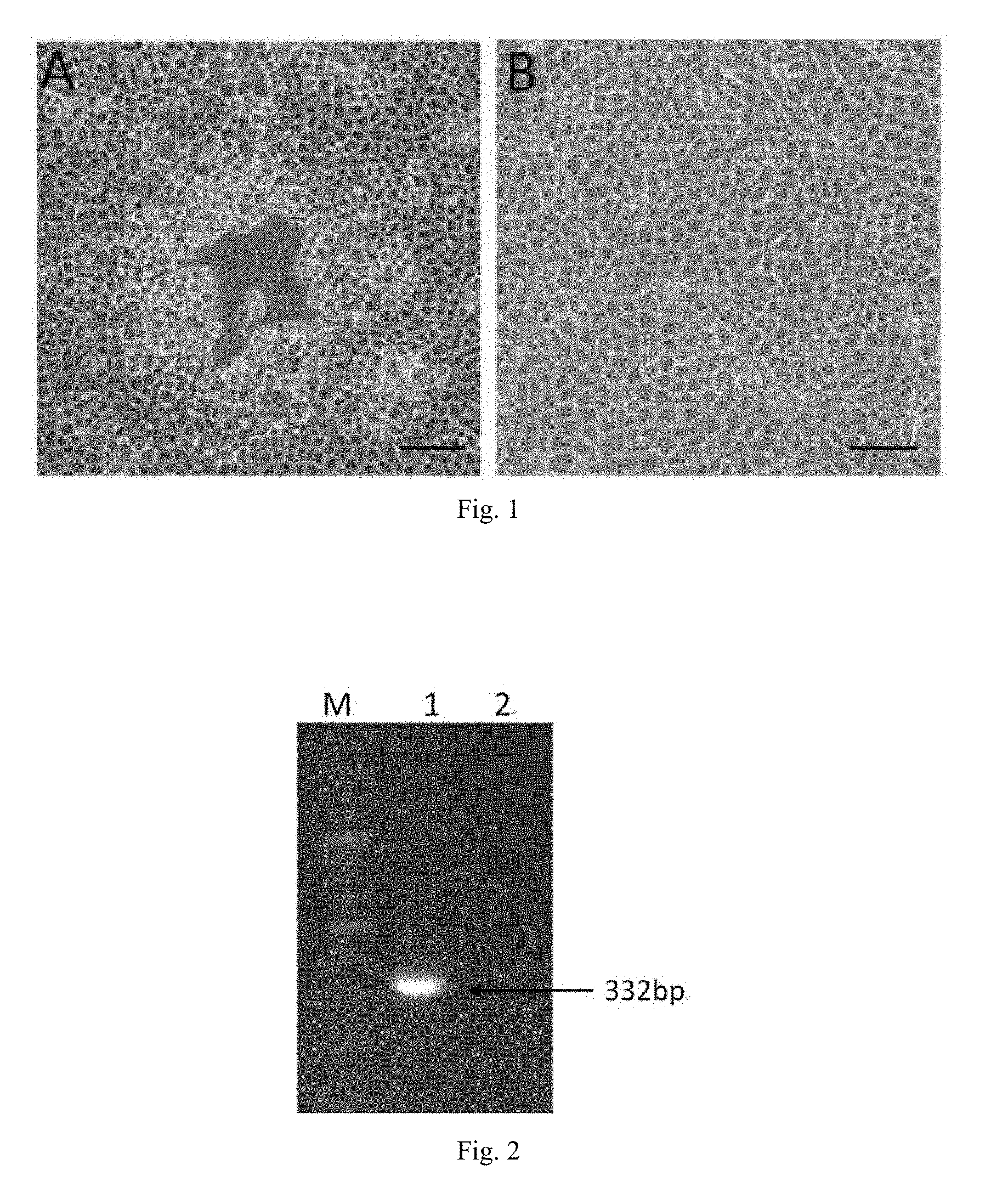
[0019] FIG. 1 is a sample of the supernatant inoculated with Vero cells in Example 1, wherein A is the experimental group, B is the negative control group.
[0020] FIG. 2 is a result of amplification of gE-specific gene fragments after DNA extraction of the disease supernatant in Example 1, wherein lane M: DNA Marker, lane 1: experimental group, lane 2: negative control group.
[0021] FIG. 3 is gB gene phylogenetic tree analysis results.
[0022] FIG. 4 is gC gene phylogenetic tree analysis results.
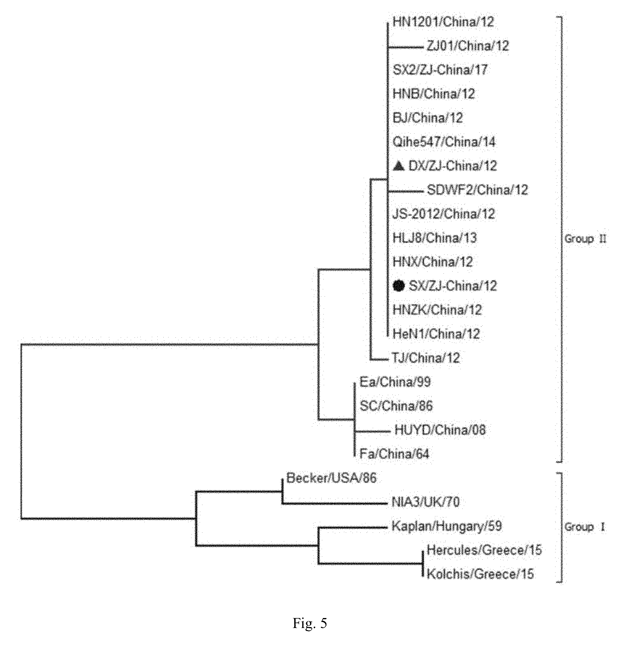
[0023] FIG. 5 is gE gene phylogenetic tree analysis results.
[0024] FIG. 6 is the TK gene phylogenetic tree analysis results.
[0025] FIG. 7 shows the results of genome-wide nucleotide sequence evolution analysis.
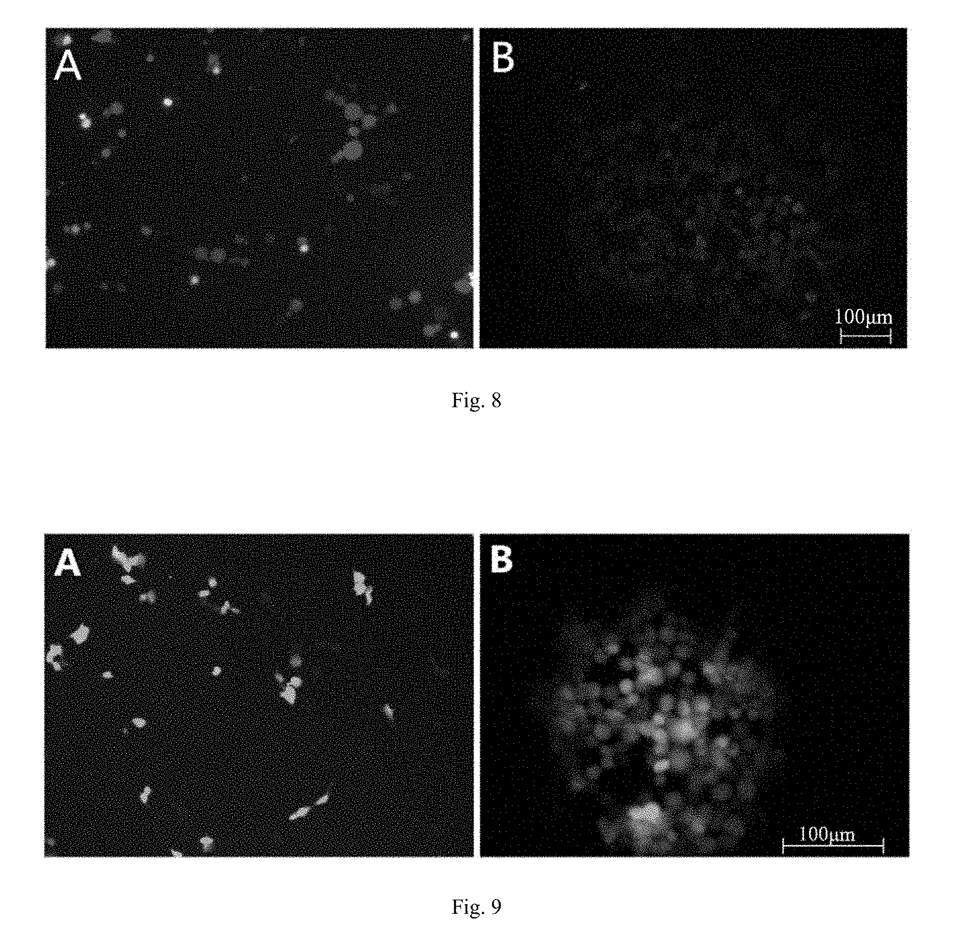
[0026] FIG. 8 is a fluorescence microscope observation of red fluorescence of recombinant virus on Vero cells, wherein A: two mixed viruses of PRV-DX and PRV-DX gE-/Cherry+; B: pure PRV-DX gE-/Cherry+.
[0027] FIG. 9 is a fluorescence microscope observation of green fluorescence of recombinant virus on Vero cells, wherein A: two mixed virulence of PRV-DX gE- and PRV-DX gE-/TK-/EGFP; B: pure PRV-DX gE-/TK-/EGFP+ recombinant virus.
SPECIFIC EMBODIMENTS OF THE PRESENT INVENTION
Materials:
[0028] Clinically suspected swine pseudorabies diseased pig brain tissue disease material was: from a pig farm in Hangzhou, Zhejiang Province of China.
[0029] The PRV-SC virus is a traditional PRV strain purchased from the China Veterinary Drug Administration.
[0030] Plasmid pMCherry-C1 and plasmid pEGFP-C2 were all conserved in this laboratory. Ezup column DNA extraction kit was purchased from Shanghai Biotechnology Co., Ltd. PrimeSTAR polymerase, dNTP mix, Pyrobest high-fidelity enzyme, rTaq polymerase, 10.times.PCR buffer, 2.times.GC buffer, 100 bp Maker, PUC18 plasmid and pMD18-T Kit were purchased from Bao Bioengineering (Dalian) Co., Ltd. Fetal bovine serum was purchased from Gbico Company; MEM medium was purchased from Invitrogen; Fetal bovine serum was purchased from Hangzhou Sijiqing Biological Engineering Materials Co., Ltd.; PCR product recovery kit, gel recovery kit, plasmid kit were purchased from TIANGEN. Low melting point agar powder was purchased from Sigma Co.
[0031] Balb/c mice aged 5-6 weeks were purchased from Laboratory Animal Center of Zhejiang Province. The 8-9-week-old and 60-day-old experimental pigs were purchased from Yuanhong Farm, Yuelong Street, Ninghai of China. The PRV, PPV, PCV2, PRRSV and CSFV antigens in the experimental pigs were negative, while the PRV and PRRSV antibodies were negative.
[0032] 25 cm.sup.2 and 75 cm.sup.2 cell culture bottles were purchased from ORANGE SCIENTIFIC Co. of Belgium. Fluorescence Quantitation Premix ExTaq.TM. (Pro qPCR) was purchased from TaKaRa Co. Fluorescent PCR tubes and plates were purchased from AXYGEN. TRIzol Reagent was purchased from GIBCO; RevertAid.TM. First Strand cDNA Synthesis Kit was purchased from Fermentas.
EXAMPLE 1
[0033] PRRSV positive material was screened by PCR and PRV was screened for the positive material grinding fluid. After sterile treatment, the supernatant was inoculated with Vero cells subculture. The refractive changes, rounding, shedding lesions of the suspected pseudorabies virus supernatant cell were observed after inoculation.
[0034] According to the gB and gE sequences of PRV in GenBank, two pairs of primers gB300F, gB300R, and gE300F, gE300R were designed to amplify the conserved regions of gB and gE, respectively. Three pairs of primers gB-F/R, gC-F/R and gE-F/R were designed according to the reference sequence of PRV genome in GenBank NC_006151. Beijing Liuhe Huada Gene Technology Co., Ltd. synthesized the primer sequence shown in Table 1.
TABLE-US-00001 TABLE 1 Primer Product Name Primer Sequence (5'-3') Length gB300F CGGCAAGTGCGTCTCCAAG 296 bp gB300R AGGGCGAAGGAGTCGTAGGG gE300-F GCTCTGCGTGCTGTGCTCC 332 bp gE300-R TCGTCACTTCCGGTTTCTCC gE-F TTTTATCTCCGTCCGCGCCGTTT 1993 bp gE-R CTCGCTGTAGTAGCAGTCCGAGT gB-F ACAGGGCGTCGGGGTCCTCGCTCTC 2749 bp gB-R CGAGGCGTCATGCCCGCT gC-F CCGTCGCCATGTGTGCCACT 1639 bp gC-R ACAAACAACCGGACGCGAT TK-F GGGTCAAGGACGCCTTCTTAA 1222 bp TK-R GGCACGGCAAACTTTATTGGG
[0035] In a six-well plate covered with Vero cells, the medium was discarded and the plate was washed once with a fresh medium. 1 mL of the suspected supernatant was added and the mixture was incubated at 37.degree. C. for 60 minutes. After completion of the sensory evaluation, the supernatant was discarded and 2 mL of MEM medium supplemented with 2% fetal bovine serum was added. The cells were cultured in a 37.degree. C. incubator containing 5% CO.sub.2, and the cytopathic effect was observed daily. When 80% lesions appeared in the cells, the cells were freeze-thawed three times and centrifuged to obtain the supernatant. The obtained virus was known as the first generation.
[0036] Viral plaque purification also used Vero cells. The resulting virus solution was diluted 10 times, then added to a six-well plate covered with the Vero cells and incubated at 37.degree. C. for 60 minutes. At the same time, they were provided with a 2% concentration of low melting point agarose and underwent high pressure sterilization. After the incubation, the high pressure 2% low melting point agarose solution and 2.times.MEM medium of equal volume were mixed, 2% fetal bovine serum was added. The mixture was sterile stored in a 37.degree. C. water bath. After discarding the supernatant in the cell plate, the cell plate was washed once with the fresh medium, the prepared agarose medium mixture was added, and it was coagulated at room temperature and then put into a 37.degree. C. incubator for 3 days. Macroscopically visible plaques were picked with a pipette pick, dissolved in 1 mL of MEM, frozen and thawed for three times after receiving poison, and then inoculated in Vero cells for propagation in preparation for the next round of plaque purification. The plaque purification operation was repeated five times and then inoculated to Vomon cells covered with monolayer. After incubation, the agar was covered. When plaques grew to the proper size, they were stained with 5% crystal violet for 1 hour. After agar was carefully taken out, the plaque morphology was observed and pictures were taken.
[0037] After inoculating Vero cells with the supernatant of the disease material, obvious cytopathic effect was observed after 24 hours, that is, the original spindle-shaped cells began to become round and translucent and then shedding to form a plaque (see FIG. 1A) with no change in negative cells (see FIG. 1B). After the DNA was extracted from the supernatant of the diseased material and subjected to PCR detection, a gE-specific partial sequence of 332 bp was obtained after amplification (see FIG. 2), indicating that the pseudorabies virus was isolated. The isolated virus was pseudorabies virus named PRV-DX of PRV, and was sent to a depositary institution for depositary preservation and was deposited as a Chinese microorganism strain at China General Microbiological Culture Collection Center (CGMCC) located at No. 1 West Beichen Road, Chaoyang District, Beijing 100101, China with the accession number is CGMCC No. 14325, the deposition date was Jul. 6, 2017.
EXAMPLE 2
Cloning and Analysis of gB, gC, gE and TK Genes of PRV-DX Strain.
[0038] The purified virus solution was used for the extraction of viral DNA as described above, and the resulting DNA solution was used as a PCR template for PCR amplification. The system (25 .mu.L) was as follows: 2.times.GC buffer 12.5 dNTPmix 2 .mu.L, primer 1 .mu.L, template 1 .mu.L, PrimeSTAR enzyme 0.25 .mu.L, and finally water was added to make up to a volume of 25 .mu.L. The PCR reaction procedure was pre-denaturation at 95.degree. C. for 5 minutes, denaturation at 98.degree. C. for 10 seconds, annealing at 57.degree. C. for 20 seconds and extension at 72.degree. C. for 2 minutes for 30 cycles. After the PCR product was detected by 1% agarose gel electrophoresis, the instruction manual of the PCR product of Sigma Co. was used to clean and recover the kit manual. The details are as follows:
[0039] In the PCR product, 3 times the volume of the product Buffer PCR-A was added (if the added buffer is less than 100 .mu.L, make it up to 100 .mu.L), mixed with a pipette until the mixture was mixed evenly. The mixture was then transferred to a preparation tube provided in the kit, and the preparation tube was placed in a 2 mL collection tube and centrifuged at 12000 rpm for 1 minute to discard the filtrate; the preparation tube was put back into the collection tube and 700 .mu.L of Buffer W2 was added, 12000 rpm centrifuged for 1 minute, the filtrate was discarded; and then the preparation tube was put back to the collection tube, 400 .mu.L of the above Buffer W2 was added, centrifuged at 12000 rpm for 1 minute, the filtrate was discarded; the preparation tube was put back to the collection tube, empty centrifuged at 12000 rpm for 1 minute. In a clean 1.5 mL centrifuge tube, 30-50 .mu.L of TE or deionized water was added to the center of the adsorption membrane, eluted for 2 minutes at room temperature, and centrifuged for 1 minute at 12000 rpm to collect the bottom solution.
[0040] The recovered product was treated with rTaq to add poly A sticky end and incubated at 72.degree. C. for 10 minutes. The system was as follows: 10.times.PCR buffer 2 .mu.L, rTaq enzyme 0.25 .mu.L, dNTPmix 0.5 .mu.L, PCR product 400 ng, and remaining water to make it to a volume of 20 .mu.L. The above A plus tail product was ligated to pMD18T at 16.degree. C. for 1 hour. The system was as follows: Solution I 10 .mu.L, pMD18T 1 .mu.L, and A-tail PCR product 9 .mu.L.
[0041] All the above ligation products were transformed into DH5.alpha. E. coli competent cells and then plated onto LB solid medium plates containing ampicillin and incubated overnight at 37.degree. C. When the colonies grew to the appropriate size, the single colonies were picked and the positive monoclonal antibodies were sent to Beijing Liuhe Huada Gene Technology Co., Ltd. for sequencing after identification of the PCR bacterial liquid.
[0042] Using MEGA5.0 software, the phylogenetic tree was obtained by comparing the gene sequence amplified by DX strain with the gene sequences of different strains downloaded from GenBank. The analysis used the neighbors method to calculate, Bootstrap value is 1000 times. At the same time, MEGA5.0 software was used to translate the previously aligned sequences into amino acid sequences and alignment analysis was conducted.
[0043] The results of phylogenetic tree (see FIG. 3, FIG. 4, FIG. 5, FIG. 6) show that the gC gene sequence of DX strain isolated in this study (SEQ ID No. 32) is consistent with the gC gene sequence of some newly isolated strains in China. They are all closer to some classical strains such as Kaplan and Becker. The same is true for the sequences of gB (SEQ ID No. 31) and gE (SEQ ID No. 33).
[0044] The results of amino acid sequence alignment showed that the gB gene of DX strain had 75-77 positions SPG three amino acid mutations inserted into the classical European strain and three SPG amino acid substitutions of 313 (V>M), 915 (S>N), mutation at position 921 (L>P). The gC gene is the insertion of seven amino acids AAASTPA consecutively at 63-69 positions. The gE gene of the DX strain differs significantly from these reference sequences by the insertion of aspartate (D) at positions 48 and 494. Position 54 is aspartic acid (D) instead of glycine (G), position 448 is isoleucine (I) instead of valine (V) and position 510 is serine (S) but not glycine (G).
EXAMPLE 3
[0045] The pseudorabies virus type IIDX virus was cultured, then the whole genome of the virus was extracted, sequenced by PacBio platform, and the whole genome sequence of the virus was obtained.
[0046] QIAGEN Genomic-tip 500G was used for DNA purification of the concentrated and purified virus solution, according to the specific instructions of the kit instruction manual. OD260/OD280 and nucleic acid content and amount were detected after the extraction. Meanwhile, 0.6% agarose gel was used for electrophoresis detection. Qualified genomic DNA samples were sent to Shenzhen Huada Gene Technologies Service Co., Ltd. for sequencing and assembly. Genewise software was used to predict homologous genes based on the reference sequence JQ809328.1 to predict each ORF. Software Versions: genewise 2.2.0, Parameters: genewise, -tfor, -sum, -genesf, -gff.
[0047] A full length genome sequence was finally obtained. The result showed that the full genome of PRV-DX strain was 143754 bp in length and GC content was 73.59%, encoding a total of 69 open reading frames, of which US1 and IE180 were internal repeat sequences. The whole genome of PRV-DX was combined with 21 published reference strains on NCBI for genome-wide analysis. The results are shown in FIG. 7. The phylogenetic analysis showed that all PRV strains can be divided into two distinct branches. Chinese strains including PRV-DX strain belong to another branch and are named genotype II. Further analysis of Chinese strains showed that the PRV-DX strain had the closest relationship with the epidemic strains isolated in China since 2012, followed by the strains Ea, Fa and Sc and the farthest evolutionary distance from LA strain.
[0048] Genome-wide sequence analysis shows that PRV-DX strain isolated in the present invention is a type II gene and is a brand new PRV.
EXAMPLE 4
[0049] A total of 70 female Balb/c mice aged 5-6 weeks were randomly divided into 7 groups, 10 mice/group. Groups 1-3 were subcutaneously injected with 0.1 mL PRV-DX (experimental group) virus solution containing 10.sup.4 TCID.sub.50/mL, 10.sup.5 TCID.sub.50/mL, and 10.sup.6 TCID.sub.50/mL subcutaneously on the back, respectively; 4-6 groups were subcutaneously injected with 0.1 mL each of 10.sup.4 TCID.sub.50/mL, 10.sup.5 TCID.sub.50/mL, and 10.sup.6 TCID.sub.50/mL PRV-SC strain (control group, conventional PRV strain) virus solution. Group 7 was subcutaneously inoculated with 0.1 mL of DMEM as a negative control. Within 2 weeks after challenge, the mice were continuously observed and recorded for clinical symptoms, pathology and death.
[0050] 5-6 weeks old Balb/c mice were inoculated with different concentrations of PRV-DX strain and PRV-SC strain, respectively. 3-7 days after the challenge, Groups of DX 10.sup.6 TCID.sub.50/mL, DX 10.sup.5 TCID.sub.50/mL, SC 10.sup.6 TCID.sub.50/mL group and the SC 10.sup.5 TCID.sub.50/mL group all showed clinical symptoms of varying degrees of apathetic, rough hair disorder and loss of appetite. Serious bite inoculation sites were also found, leading to skin bleeding and eventually death. In the DX 10.sup.4 TCID.sub.50/mL group and the SC 10.sup.4 TCID.sub.50/mL group, slight clinical symptoms such as apathetic symptoms began to occur on the 4th day after challenge, and the mice died. After 7 days, the above groups of mice returned to normal, no obvious clinical symptoms appeared. Negative control mice showed normal throughout the experimental period.
[0051] According to the statistics, the death time of mice mainly concentrated on days 3-7 after challenge. The death numbers of DX 10.sup.6, 10.sup.5 and 10.sup.4 TCID.sub.50/mL challenged mice were 9, 6 and 1, respectively. The death numbers of mice infected with SC 10.sup.6, 10.sup.5 and 10.sup.4 TCID.sub.50/mL were 9, 4 and 1; 10 mice in the negative control group all survived normally. According to the experimental results, the LD.sub.50 of PRV-DX strain was 10.sup.4.84 TCID.sub.50/mL. The LD.sub.50 of SC virulent strain in our country was 10.sup.5.16 TCID.sub.50/mL. It indicated that the virulence of PRV-DX strain obtained from new isolation and identification was twice or more stronger than that of PRV-SC.
EXAMPLE 5
[0052] Fifteen 60-day-old experimental pigs were randomly divided into three groups. The first group was inoculated with 4 mL of 10.sup.7.0 TCID.sub.50/mL PRV-DX strain of virus solution, the second group was inoculated with 4 mL of 10.sup.7.0 TCID.sub.50/mL PRV-SC strain of virus solution, and the negative control group was inoculated with 4 mL DMEM. Inoculation route was intranasal inoculation. These groups were subject to isolated feeding, free feeding, and continuous observation for 14 days. The temperature and clinical symptoms were recorded. The death pigs were dissected in time. Tissue sample collection, tissue sections and viral load testing were conducted.
[0053] On the second day after virus inoculation or challenge, the experimental animals with 10.sup.7.0 TCID.sub.50/mL and 10.sup.7.0 TCID.sub.50/mL strains of DX showed significant body temperature rise, with mean body temperature of 41.7.degree. C. and 41.9.degree. C., respectively. DX strain 10.sup.7.0 TCID.sub.50/mL group showed clinical symptoms such as appetite abatement and dyspnea on the 3rd day after challenge. All the experimental animals died on the 4th day after challenge, and the lethality was 100%. On the 3rd day after challenge, SC strain 10.sup.7.0 TCID.sub.50/mL showed only symptoms of loss of appetite. Until 14 days after challenge, only one pig died and the lethal rate was 20%. Negative control group were normal during the entire experimental period.
[0054] Experimental pigs died of experimental process or killed pigs after the end of the experiments were observed with the following general lesion findings: PRV-DX strain inoculating pig showed thickening and congestion of the brain membrane, and difficulties in separation; serious blood congestions in the brain organization; congestion and hemorrhage in lung, obvious intercostal pressure trace, slight liver congestion, and tonsil thickening congestion. There is no liver and tonsil visible macroscopic lesions in PRV-SC inoculated pigs except for slight hyperemia and swelling in brain organizations and lungs, have no. Brain, lung, liver, tonsil tissues were fixed. Paraffin sections and HE staining were conducted. The pathological sections were observed under the microscope. Capillary congestion was seen in the brain of the challenged group of PRV-DX strain, and infiltration of inflammatory cells around the blood vessels formed a typical phenomenon of "vasculitis". The focal alveolar space of the lungs became smaller and there were erythrocyte exudation, the alveolar wall had a large amount of inflammatory cell infiltration; there were visible interlobular artery congestion in the liver and tonsil inflammation infiltration seen. The PRV-SC strain challenged brain tissue also showed capillaries congestion, and a very small number of neuronal necrosis, nuclear condensation shrinkage, stained cytoplasm. There were visible alveolar wall thickening in lungs, increased inflammatory cells. No obvious lesion was observed in the liver and tonsil. The fluorescence quantitative PCR method was used to detect the gD gene fragment: upstream primer gDP-F: CACGCCGATGTGGTGGA, downstream primer gDP-R: GGTACTGGCCCTCGTTGAA, Probe: CY5-ACTACATGTTCCCCACGGAGGACGAG-BHQ2. The reaction system includes Premix Ex Taq qPCR): 10 .mu.L, 0.4 .mu.L of gDP-F, 0.4 .mu.L of gDP-R, 0.4 .mu.L of Probe, 6.8 .mu.L of ddH.sub.2O, and 2 .mu.L of a template. The reaction conditions were as follows: pre-denaturation at 95.degree. C. for 2 min, denaturation at 95.degree. C. for 5 seconds, annealing at 55.degree. C. for 30 seconds and extension at 60.degree. C. for 1 min for 40 cycles, setting a negative control, using pMD18T-gD standard plasmid as a positive control, and used as the template after double dilution. After amplified by the same method, a standard curve was drawn, the corresponding copy number was calculated according to the sample CT value and the standard curve, and the viral load in each organ after inoculation of the PRV-DX strain and SC strain were calculated. The virulence of tonsil and brain was higher than that of lung and liver after challenge with PRV-DX and SC strains of the same virus content. The midbrain and lung viral load of DX strain was significantly higher than that of SC strain. The lung viral load was significantly higher than the PRV-SC group.
[0055] The PRV-DX strain PRV-DX of type II pseudorabies virus obtained by the new isolation and identification of the present invention shows much greater virulence to pigs than the classical PRV strain SC strain.
EXAMPLE 6
[0056] 1. Primer Design and PCR Amplification
[0057] Primers F-LgE and R-LgE, F-RgE and R-RgE were designed to amplify the homologous recombination arms flanking the gE gene (SEQ ID No. 33) with EcoRI, KpnI, BamHI and HindIII cleavage sites point. A pair of mutation primers F-Mcherry and R-Mcherry for the transformation of the pMcherry-C1 plasmid were designed and used to delete the multiple cloning site (MCS) in the plasmid so as to obtain a cherry red fluorescence complete expression cassette without MCS. A pair of primers F-McherryCPS, R-McherryCPS for amplifying a complete cherry red fluorescence complete expression cassette and a loxp sequence of the same orientation were designed to amplify the entire cherry red fluorescently integrated expression cassette. Similarly, the TK gene was designed to amplify (SEQ ID No. 34). Primers F-LTK and R-LTK, F-RTK and R-RTK on both sides of the homologous recombination arm also carry EcoRI, KpnI, BamHI and HindIII restriction sites. A pair of mutation primers F-EGFP and R-EGFP for transforming pEGFP-C2 plasmid were designed and used to delete multiple cloning sites (MCS) in plasmid to obtain an EGFP green fluorescence complete expression cassette without MCS. A pair of primers, F-EGFPCPS, R-EGFPCPS, designed to amplify the complete EGFP green fluorescence complete expression cassette, were designed and the same loxp sequence was also added upstream and downstream. The specific primer sequences are shown in Table 2. The primers were synthesized by Beijing Liuhe Huada Gene Technology Co., Ltd.
TABLE-US-00002 TABLE 2 Primer sequences for amplifying the corresponding fragments. introduced into The length the restriction of the Primer Name Primer Sequence site product F-LgE CGGAATTCGACGATAGACGGGACGCT EcoRI 1021 bp R-LgE GGGGTACCGGGTGCCCAGGTTTAAAA KpnI F-RgE ACGCGTCGACCAGGACGACTCGGACTGCTAC BamHI 860 bp R-RgE CCCAAGCTTGTTCAGGACGGACGACCACTC HindIII F-Mcherry TACAAGTCCGGACTCGGATCTAGATAACTG none 4722 bp R-Mcherry CAGTTATCTAGATCCGAGTCCGGACTTGTA none F-McherryCPS GGGGTACCATAACTTCGTATAATGTATGCTATACG KpnI 1704 bp AAGTTATTAGTTATTAATAGTAATCAA R-McherryCPS CGGGATCCATAACTTCGTATAGCATACATTATACG BamHI AAGTTATGATGAGTTTGGACAAACCAC F-LTK CGGAATTCATCCTCCGGATCTACCTC EcoRI 455 bp R-LTK GGGGTACCAGCGAGGCCACCACCAGG KpnI F-RTK CGGGATCCATGGACGCGCTCGTGGCC BamHI 527 bp R-RTK CCCAAGCTTAGCTGGAAGACGAACCAC HindIII F-EGFP GGGGTACCAATAGTAATCAATTACGGGGTCATT none 4700 bp R-EGFP CGGGATCCAGATACATTGATGAGTTT none F-EGFPCPS GGGGTACCATAACTTCGTATAATGTATGCTATACG KpnI 1705 bp AAGTTATAATAGTAATCAATTACGGGGTCATT R-EGFPCPS CGGGATCCATAACTTCGTATAGCATACATTATACG BamHI AAGTTATAGATACATTGATGAGTTT
[0058] 2. gE Transfer Vector Construction
[0059] The homologous recombination arms LgE and RgE of gE were amplified with primers F-LgE and R-LgE, F-RgE and R-RgE and using the PRV-DX DNA of the PRV-DX as a template. The R-PCR system comprises: 2.times.GC buffer 12.5 .mu.L, dNTP mix 2 .mu.L, primer 1 .mu.L each, template DNA 1 .mu.L, PrimeSTAR enzyme 0.25 .mu.L, and finally made up to 25 .mu.L with water. The PCR reaction procedure was denaturation at 94.degree. C. for 5 min, denaturation at 98.degree. C. for 10 seconds, annealing at 57.degree. C. for 20 seconds and extension at 72.degree. C. for 2 min. PCR products were detected by 1% agarose gel electrophoresis. The PCR products were recovered and ligated to pMD18T respectively for delivery to the company for sequencing. The LgE fragment and the RgE fragment with the correct sequencing results were digested with EcoRI, KpnI, BamHI and HindIII, respectively, and then ligated into the corresponding positions of the same enzyme-treated PUC18 plasmid to obtain the plasmid pUC18-LrgE containing LgE and RgE in turn.
[0060] Using the pMCherry-C1 plasmid as a template, the pMCherry-C1.DELTA.MCS plasmid containing no multiple cloning sites was amplified by deleting the mutant pMCherry-C1 with the primers F-Mcherry and R-Mcherry. The PCR product was treated with DpnI enzyme to digest the template plasmid. i.e. the plasmid with MCS. The processed product is transformed directly into competent cells of TG1, picked monoclonal and then sent to the company for sequencing, sequencing to determine whether the deletion of the multiple cloning site (MCS). Using this modified correctly sequenced plasmid as a template, a complete Cherry expression cassette containing the promoter and SV40 tail was amplified using primers F-McherryCPS and P-McherryCPS. PCR products were detected by 1% agarose gel electrophoresis. The amplified Cherry expression cassette and PUC18-LrgE were double-digested with KpnI and BamHI, respectively, ligated and transformed into competent cells of TG1, and verified by sequencing, that is, the recombinant vector PUC18-gEloxpCherry was obtained, and the gE transfer vector plasmid was obtained.
[0061] The PUC18-gEloxpCherry transfer vector was about 6.3 kb in size (empty vector 2686 bp+LgE 1021 bp+CherryCPS 1704 bp+RgE 860 bp), as shown in FIG. 8, i.e., the resulting LgE, RgE and Cherry red fluorescent expression cassettes were PCR-Fragments according to their corresponding restriction sites, in turn connected to the corresponding position on PUC18 to obtain the vector, sent to the sequencing company correctly sequenced.
3. PRV-DX gE-Virus Construction
[0062] Liposomes lip2000 transfection reagent was used in transfection experiments. The specific operation in accordance with the instructions lip2000 is as follows: Taking six 2 mL EP tubes, marked 1 to 6, respectively, by adding 500 .mu.L MEM medium and 10 .mu.L lip2000, with a pipette blew evenly after standing 5 min; then taking the same six 6 EP tubes, also marked Nos. 1-6, each adding 500 .mu.L MEM medium. Then, 1 .mu.g of viral DNA and 3 .mu.g of PUC18-gEloxpCherry plasmid were added to tubes 1 to 4, respectively. The tube No. 5 was added 1 .mu.g of viral DNA as a single transfected virus DNA control. The tube No. 6 was added 3 .mu.g PUC18-gEloxpCherry plasmid as a separate transfected plasmid control and mixed with a pipette. Each 500 .mu.L MEM corresponding to the above markers was mixed. The mixture was allowed to rest at room temperature for 20 min. During the period, six-well plates covered with monolayer of Vero cells were discarded, and the medium was discarded and then washed twice with PBS. 1 mL of pre-treated mixture was added to each well and labeled. The plate was incubated at 37.degree. C. incubator culture. After 5 hours, the medium containing transfection reagents were discarded and replaced with 2 mL of fresh medium containing 2% fetal bovine serum in each well. Then, the cell plate was incubated in a 37.degree. C. incubator, and the cell changes in the co-transfected cells and the control transfected cells in the six-well plate were continuously observed. The co-transfected cells were harvested while the lesions and red fluorescence were harvested. That is, both the transfected DNA and the plasmid were expressed (FIG. 9A). At the same time, the cells transfected with plasmids only showed red fluorescence, and the cytopathic effect appeared only in cells transfected with parental DNA alone.
[0063] Under -70.degree. C., the virus liquid was repeatedly frozen and thawed three times and inoculated on Vero cells covered with monolayer cells to be lesion, picking the red fluorescent lesions of the virus diluted by 10 times with dilution ratio of 10.sup.-1-10.sup.-6 to inoculate Vero cells covered with monolayers and incubated at 37.degree. C. for 2 hours. Then all the supernatants were discarded, adding 1:1 mixture of 2.times.MEM (2% fetal bovine serum) and 2% low melting point agarose mixture, to be solidified and placed in a 37.degree. C. incubator, observed for several days. When the cells appeared moderate red fluorescent plaques, the appropriate fluorescent plaques were picked and dissolved in 1 mL MEM medium, repeatedly frozen and thawed at -70.degree. C. three times and then seeded in Vero cell monolayers for passage and plaque purification until The resulting clones of all diseased plaques showed red fluorescence, i.e., pure PRV-DX gE-/Cherry deletion mutants (FIG. 9B).
[0064] Genomic DNA of PRV-DX gE-/Cherry was extracted and treated with Cre recombinase at 37.degree. C. for 2 hours. The treated product was extracted twice with Tris-saturated phenol, and the supernatant was extracted and then extracted twice with an equal volume of chloroform. The supernatant was aspirated, 2.5 times the volume of pre-cooled anhydrous ethanol was added at -70.degree. C. for 1 hour to precipitate the DNA, and the precipitated DNA was washed with 75% ethanol. After the solution was dried and dissolved in TE, and the treated clean genomic DNA was obtained.
[0065] The above genomic DNA treated with Cre enzyme was transfected into Vero cells. When the medium was changed, a 1:1 mixture of 2.times.MEM (2% fetal bovine serum) and 2% low melting point agarose mixture were mixed and allowed to solidify Placed in 37.degree. C. incubator, continuous observation for several days. When the lesion appeared, whether the diseased cells have red fluorescence was observed, and the non-fluorescent plaque was picked under the fluorescence microscope and then the plaque purification was performed, and repeated until all the lesions have no red fluorescence Pure PRV-DX gE-deletion mutants.
4. PRV-DX gE-Strain Identification and TCID.sub.50 Determination
[0066] The DNA of the PRV-DX gE-deletion mutant was extracted and PCR was performed by using the primers gE-F and gE-R. Two controls were set up, one using PRV-DX strain parental DNA as a template and the other using PRV-DX gE-/Cherry DNA as a template. The PCR system is as follows: 2.times.GC buffer 12.5 .mu.L, dNTPmix 2 .mu.L, primer 1 .mu.L each, template DNA 1 .mu.L, PrimeSTAR enzyme 0.25 .mu.L, and finally made up to 25 .mu.L with water. The PCR reaction procedure was pre-denaturation at 94.degree. C. for 5 min, denaturation at 98.degree. C. for 10 seconds, annealing at 57.degree. C. for 20 seconds and extension at 72.degree. C. for 1 min 45 seconds. PCR products were detected by 1% agarose gel electrophoresis, sent to the company for sequencing verification. The results showed that the gE gene was indeed deleted by 1931 bp, leaving a loxp site. The result was in line with the theory that Cre recombinase cleaves the middle sequence and leaves a loxp site when there are two loxp sites in the DNA molecule that are aligned. The gE gene-deleted DX strain and DX parent strain respectively underwent TCID.sub.50 determination. When the Vero cells in the 96-well plate were covered with a monolayer, the passaged cells were inoculated into a 96-well plate of 1 to 10 with a dilution ratio of 10-1 to 10-10 to 100 .mu.L per well. The last two columns were added with equal volume of cell maintenance fluid as a negative control. 96 plates were cultured under 37.degree. C. and in a 5% CO.sub.2 incubator. The numbers of cytopathic holes were counted after 96 hours. The TCID.sub.50 of virus was 10.sup.8.5 TCID.sub.50/mL (gE gene deletion DX strain) by Reed-Muench method and 10.sup.8.6 TCID.sub.50/mL (DX parent strain). This indicates that deletion of gE does not affect the virus proliferation on Vero cells.
5. Construction of TK Transfer Vector
[0067] The virus DNA of PRV-DX gE-single gene deletion strain was used as a template to amplify TK homologous recombination arm LTK and RTK with primers F-LTK and R-LTK, F-RTK and R-RTK. The PCR product was detected by 1% agarose gel electrophoresis, and the PCR product recovered was treated with rTaq enzyme plus poly A sticky end.
[0068] The above ligation products were transformed into TG1 competent cells, and the positive clones were picked and sent to Buo Shang Ltd. for sequencing. The LTK fragment and RTK fragment with the correct sequencing results were digested with EcoRI and KpnI, BamHI and HindIII, respectively, and ligated to the corresponding position of the same enzyme-treated PUC18 plasmid to obtain the plasmid PUC18-LRTK. The pEGFP-C2.DELTA.MCS plasmid without multiple cloning sites was amplified by using the pEGFP-C2 plasmid as a template and the deletion mutation pEGFP-C2 with primers F-EGFP and R-EGFP. The PCR product was treated with DpnI enzyme to digest the template plasmid, i.e, the plasmid with MSC. The processed product was transformed directly into TG1, picked monoclonal and sent to the company for sequencing. A complete EGFP expression cassette containing the promoter and SV40 tail was amplified with primers F-EGFPCPS and P-EGFPCPS using the modified plasmid sequenced correctly as a template. PCR products were detected by 1% agarose gel electrophoresis. The amplified EGFP expression cassette and PUC18-LRTK were digested with KpnI and BamHI, respectively, and T4 ligase was ligated overnight at 16.degree. C. The ligated product was transformed into TG1 and cultured in a 37.degree. C. incubator. When its colonies appeared, the monoclonal antibody was picked for bacterial identification PCR. The bacterial PCR showed positive clones were sent to the company for sequencing identification. After sequencing, the recombinant vector PUC18-TkloxpEGFP was obtained and the strain was stored at -70.degree. C. According to TIANGEN plasmid mini kit operation, PUC18-TKloxpEGFP plasmid DNA was extracted to obtain TK transfer vector DNA.
6. Construction of PRV-DX gE-/TK-Gene Deletion
[0069] Liposomes lip2000 transfection reagent was used to conduct transfection experiments. Six (6) autoclaved 2 mL centrifuge tubes were obtained, labeled 1 to 6, and were added to 500 .mu.L DMEM medium and 10 .mu.L lip2000, mixed well and then stood at room temperature for 5 min. Then another six (6) centrifuge tubes were obtained, similarly marked 1.about.6, and 500 .mu.L DMEM medium were added. Then, 1 .mu.g of PRV-DX/gE-gene-deleted viral genomic DNA and 3 .mu.g of PUC18-TKloxpEGFP plasmid mixture were added to tubes Nos. 1 to 4. 1 .mu.g of PRV-DX/gE gene-deleted virus genomic DNA was added into tube No. 5 as a single transfected virus DNA control, 3 .mu.g of PUC18-TKloxpEGFP plasmid was added into tube No. 6 as a plasmid-independent control and mix well with a pipette tip. 500 .mu.L of each of the DMEMs corresponding to the above marker were mixed and the mixture rested at room temperature for 20 min. For six-well plates covered with monolayer of Vero cells in six-well plate, the medium was discarded, washed twice with PBS spar. 1 mL of pretreated mixture was added into each well and labeled numbers 1 to 6, the cell plate placed under 37.degree. C., and in a 5% CO.sub.2 incubator. After 5 hours, the medium containing transfection reagents were discarded and replaced with 2 mL fresh medium containing 2% fetal bovine serum in each well. The cells were then incubated in a 5% CO.sub.2 incubator at 37.degree. C. The cells in the co-transfected wells of the six-well plate and the cells in the control transfected wells were continuously observed. When the co-transfected wells were infected with lesions and green fluorescence, the cells were harvested Virus liquid, which means that the transfected DNA and plasmid have been expressed.
[0070] Viral fluid was repeatedly frozen and thawed three times under -70.degree. C. before inoculated on Vero cells. When lesions appeared in the cells, the green fluorescence of the lesion holes were picked, virus solution was harvested, vaccination Vero cells were diluted 10 times under the ratio of 10.sup.-1 to 10.sup.-6 and incubated for 2 hours at 37.degree. C., then all supernatants were discarded. 1:1 mixture of 2.times.DMEM (2% fetal bovine serum) and 2% low melting point agarose were added. After solidification, it was placed in an incubator under 37.degree. C. with 5% CO.sup.2 for several days to observe the green fluorescent plaque. Fluorescent plaques were picked up and dissolved in 1 mL of DMEM medium. The cells were seeded repeatedly and plaque purified until all of the lesion plaques showed green fluorescence, i.e., pure PRV-DX gE-/TK-/EGFP+ deletion mutants poison.
[0071] PRV-DX gE-/TK-/EGFP+ was seeded into small vials filled with monolayer Vero cells. When the cells appeared 80% lesions, genomic DNA was extracted and the concentration was determined, then treated with Cre recombinase at 37.degree. C. for 2 hours, 200 .mu.L Tris-saturated phenol was extracted twice in the treated product system, and the supernatant was extracted twice with an equal volume of chloroform. The supernatant was obtained by aspiration, 2.5 times the volume of pre-cooled anhydrous ethanol was added for reaction for 1 hour at -70.degree. C. to precipitate the DNA, and the precipitated DNA was washed with 75% ethanol. After the solution is dried and dissolved in TE, the treated clean genomic DNA is obtained.
[0072] The resulting DNA was PRV-DX gE-/TK-/EGFP+ and PRV-DX gE-/TK-two viral genomic DNA mixture since Cre enzyme was unable to 100% excise the Cherry gene. Genomic DNA previously treated with Cre enzyme was used to transfect Vero cells covered with monolayers as described above. When changing the medium, 2.times.DMEM (2% fetal bovine serum) and 2% low melting point agar Sugar mixture until it solidified and was placed in a 37.degree. C., 5% CO.sup.2 incubator, and was continuously observed for several days. When the lesion appeared, the diseased cells for green fluorescence was observed and the plaque without fluorescence was picked out, which is PRV-DX gE-/TK-recombinant virus, dissolved in 1 mL DMEM, freeze-thawed at -70.degree. C., melted three times after inoculation with monolayer of Vero cells for plaque purification, so repeated until all the lesions plaque does not appear green fluorescence clone obtained is the PRV-DX gE-/TK-deficient virus.
7. PRV-DX gE-/TK-Strain Identification and Determination of the Virus TCID.sub.50
[0073] Genomic DNA was extracted and PCR was performed using the primers TK623-F: GTTCGTAGAAGCGGTTGTGGCA and TK623-R: CGTGTTGACCAGCATGGCGTAG. The upstream primer was located in the TK-deleted sequence, so that any band could not be amplified after TK was deleted. The parental strain of PRV-DX gE gene deletion DNA was used as a positive control. PCR products were detected by 1% agarose gel electrophoresis. The purified gE/TK double gene deletion virus was identified by TK gene primers, and the TK gene could not be detected. It was proved that the gE/TK gene was knocked out and the virulent PRV-DX gE/TK double gene deletion virus generation was obtained. The obtained gE/TK double gene deletion virus was named PRV-HD/c strain of PRV, and was deposited with the China General Microbiological Culture Collection Center of General Microbiology (No. 3 Beichen West Road, Chaoyang District, Beijing) (CGMCC) under the accession number of CGMCC No. 14325, and the storage time is Jul. 6, 2017.
[0074] The TCID.sub.50 of gE/TK double gene deletion strain (PRV-HD/c strain), gE single gene deletion strain and DX parent strain were respectively measured in 96-well plates covered with monolayers of Vero cells. When the Vero cells in the 96-well plate were covered with a monolayer, the passaged cells were inoculated into a 96-well plate of 1-10 columns at a dilution of 10.sup.-1-10.sup.-10 to 100 L per well. The last two columns were added with equal volume of cell maintenance fluid as a negative control. 96 plates were cultured in a 37.degree. C., 5% CO.sub.2 incubator, and the numbers of cytopathic holes were counted after 96 hours. The virus TCID.sub.50 and gE/TK double gene deletion strains were calculated by Reed-Muench method (PRV-HD/c strain), gE single gene deletion strain and DX parent strain were 10.sup.8.6 TCID.sub.50/mL, 10.sup.8.5 TCID.sub.50/mL and 10.sup.8.6 TCID.sub.50/mL, respectively, indicating that the deletion of the gE and TK genes did not affect the virus proliferation on Vero cells.
EXAMPLE 7
1. Mice Pathogenicity Comparison
[0075] Experimental Methods: Seventy BALB/c mice aged 6 to 8 weeks were randomly divided into 7 groups of 10 rats. 1 to 3 groups were subcutaneously injected with 0.1 mL of virus solution of gE-/TK-double gene deletion strain (PRV-HD/c strain of PRV-HD/c strain) with 10.sup.6.0 TCID.sub.50 subcutaneously in the back, 0.1 mL PRV-DX strain containing 10.sup.6.0 TCID.sub.50 of parental virus, and the remaining one group was injected with 100 .mu.L DMEM as a negative control group. After inoculation observation for 14 consecutive days, the clinical symptoms and death of mice were recorded.
[0076] As a result, the mice inoculated with 10.sup.6.0 TCID.sub.50 PRV-DX strain developed apomorphism, loss of spirit and appetite, and inoculation site biting were observed at 48 hours after inoculation, and they all died within 72 hours. Mice vaccinated with the PRV-HD/c strain group showed no clinical signs of PRV and all survived during the experiment. This indicates that the deletion of gE and TK genes attenuates virulence and pathogenicity in mice.
2. Pig Pathogenicity Comparison
[0077] Experimental Methods: 15 PRV-negative, PRRSV-ELISA-negative, PRV-ELISA-negative 8-9-week-old breeding pigs were selected and pathogenicity comparison was conducted. The method of attack or challenge was as follows: 4 mL of each pig's nose was dripped, the feed after Co60 irradiation was fed, and the isolated pigs were fed by designated personnel. Body temperature was measured regularly every morning, and the anal swabs and nasal swabs were collected and immersed in 1 mL of PBS, respectively. Daily observation of the experimental pig mental state, appetite, health status and so on was conducted. Blood was collected from the anterior vena cava on days 0, 3, 7, 10 and 14 after challenge, and serum was separated.
[0078] As a result, all vaccinated pigs started to rise in body temperature on the first day after challenge with PRV-DX. On the second day, the body temperature of all vaccinated pigs was above 41.degree. C., with depression and loss of appetite, and one pig had obvious symptoms. On the third day, the pigs in the challenge group all had fever over 41.degree. C., one pig died, and the other four had obvious neurological symptoms. On the 4th day, all four died. For HD/c group, on the second day after inoculation, the body temperature of all vaccinated pigs increased slightly, and the body temperature lasted for 3 days. The body temperature of all pigs increased within 1.degree. C., and their mental and appetite conditions were normal without any death. In the control group, 5 piglets from start to finish are very healthy, mandatory execution on the 14.sup.th day. This indicated that the deletion of gE and TK attenuated the virulence and pathogenicity of pigs.
EXAMPLE 8
1. Inactivated Vaccine Preparation and Testing
[0079] The virus solution (PRV-HD/c strain) with a titer of 10.sup.8.6 TCID.sub.50/mL was slowly added with formaldehyde to a final concentration of 0.1% with stirring and inactivated by stirring at 37.degree. C. for 48 hours. Vero cell monolayers were inoculated with the inactivated virus solution and harvested at 37.degree. C. in a 5% CO.sub.2 incubator for 72 hours. Blotting was performed 3 times blindly, and no cytopathic effect was determined as inactivation test. The qualified venom was mixed with ISA adjuvant at a ratio of 3:1 (v/v), and emulsified to prepare the oil-in-water PRV-HD/c strain inactivated vaccine.
2. Mouse Immunogenicity Test
[0080] A total of 20 SPF mice aged 5.about.6 weeks were randomly divided into 2 groups with 10 mice in each group. One group received intraperitoneal injections of 0.2 mL of PRV-HD/c inactivated vaccine with an antigenic level of 10.sup.8.6 TCID.sub.50/mL before inoculation. One group was intraperitoneally inoculated with 0.2 mL of DMEM as a negative control. The challenge was then challenged with PRV-DX virulent strain with a TCID.sub.50 value of 10.sup.6.6 TCID.sub.50/mL 21 days after immunization at the dose of 0.1 mL subcutaneously. The mice were observed after the clinical symptoms of poisoning, and the number of deaths in mice was calculated.
[0081] Results: After immunization of mice with different antigen content, the mice were in good mental state and normal appetite. There was no difference with the control, indicating that the vaccine had no side effects on the mice. After challenge, the non-immune control group began to have obvious pseudorabies symptoms 72 hours after onset, such as apathetic, rough hair disorder, loss of appetite, weight loss, and constantly biting vaccination sites. Vaccine immunized group did not show clinical symptoms. After continuous observation for 14 days, the mortality rate of mice in non-immune challenge group was 90% and survival rate was 10%. The survival rate of mice in vaccination group was 90% and mortality rate was 10%. The results showed that PRV-HD/c inactivated vaccine produced 90% protection efficiency in mice when the antigen content was 10.sup.8.6 TCID.sub.50/mL.
3. Pig Immunogenicity Test
[0082] Ten 14-day-old crossbred pigs that were negative for PRV, negative for PRRSV-ELISA and negative for PRV-ELISA were selected for this experiment. The pigs were randomly divided into 2 groups with 5 pigs in each group. The first group was intramuscularly inoculated with PRV-HD/c inactivated vaccine with an antigenic level of 10.sup.8.6 TCID.sub.50/mL before inactivation, 2 mL/pig; the second group was not injected with the vaccine as blank control group (frozen cell lysate). Each group was isolated for feeding. The changes of body temperature after immunization were observed. PRV antibody was detected in the sera of 7-21 days after immunization. 21 days after immunization with PRV-DX virulent strain of 10.sup.4.87 TCID.sub.50/mL were challenged at a dose of 4 mL per pig intranasal injection to observe the clinical symptoms and death of immunized pigs, the death of pigs and on the 14th day, the animals were sacrificed to dissect the organs and tissues such as the brain, tonsils, lungs, spleen and inguinal lymph nodes of the animals and to observe the ocular and pathological changes. Serum and nasal, anal swabs and tissue samples were collected 7 to 14 days after challenge and the copy number of PRV virus nucleic acid and the PRV virus were determined.
[0083] The experimental pigs were observed for 21 days after immunization. The pigs in each immunized group and the control group showed good mental state and normal appetite. Except for a slight increase in body temperature after 24 hours of immunization, After 14 days of challenge, the body temperature of the non-immune control group began to rise on the second day after the challenge, and sustained high temperature for more than 3 days. All the pigs without vaccine vaccination died on the 7th day after the challenge and the mortality rate was 100%. 3 to 5 days after inoculation, there was a transient increase in body temperature of pigs vaccinated with inactivated vaccine, but no pigs died, the survival rate of 100%. The results showed that PRV-HD/c inactivated vaccine could produce 100% immune-potency on 14-21 day-old piglets when the antigen content was 10.sup.8.6 TCID.sub.50/mL.
[0084] The above test results show that the inactivated vaccine prepared by the PRV-HD/c virus obtained in the present invention has good immune-protective effect on mice and pigs and has strong immunogenicity.
EXAMPLE 9
[0085] Sixty five-to-six-week-old SPF mice were randomly divided into 6 groups of 10 pigs, two of which were vaccinated with the PRV-HD/c strain inactivated vaccine prepared in Example 8, and two groups were vaccinated with commercially available PRV inactivated vaccine A, two groups were vaccinated with DMEM control, inoculation dose was 0.2 ml/each. The method of inoculation was intraperitoneal injection. Twenty-one days after immunization, these mice were challenged with the virulent PRV-DX strain or PRV-SC classical strains at a dose of 0.1 ml (10.sup.5.6 TCID.sub.50/0.1 ml), respectively, and the mice were observed for clinical symptoms after 14 days of challenge and statistics the number of mice in each group.
[0086] Results: The mice were in good mental state and normal appetite after inoculation of different immunogens. There was no difference with the control group, indicating that the vaccine prepared in Example 8 of the present invention and the commercial vaccine were safe to mice. For the group with PRV HD/c inactivated vaccine, at 21 days after immunization, the mice were inoculated with virulent PRV-DX strain or PRV-SC classical virus at a dose of 0.1 ml (10.sup.5.6 TCID.sub.50/0.1 ml). After 14 days post-inoculation, the survival rates of mice were both 100%.
[0087] For commercial PRV vaccine immunized mice group A, at 21 days after immunization the mice were inoculated with PRV-DX and PRV-SC strain at a dose of 0.1 ml (10.sup.5.6 TCID.sub.50/0.1 ml), and the survival rates of mice were 80% and 90% respectively. The control group was also treated with PRV-DX strain and PRV-SC strain of attack at a dose of 0.1 ml (10.sup.5.6 TCID.sub.50/0.1 ml), the survival rates were 0. The result shows that the PRV HD/c inactivated vaccine has a good protective effect on mice, which can resist the absolute lethal dose of newly domestic and classic strains of PRV to cross-protect, and the PRV HD/c inactivated vaccines in mice immuno-protective effect is better than the commercial PRV inactivated vaccine A.
EXAMPLE 10
[0088] 120 SPF mice aged 5.about.6 weeks were randomly divided into 12 groups, 10 mice in each group. The three groups were inoculated intraperitoneally with 0.1 ml (10.sup.7.0 TCID.sub.50/0.1 ml) of PRV-HD/c virus solution, and the three groups were inoculated with 0.1 ml (10.sup.6.0 TCID.sub.50/0.1 ml) of PRV-HD/c virus solution, three groups inoculated with 0.1 mL (10.sup.5.0 TCID.sub.50/0.1 ml) of PRV-HD/c virus solution, three groups were inoculated with virus DMEM as a control. At 4 days, 7 days, and 10 days after vaccination, these mice were inoculated with virulent PRV-DX strain at 0.1 ml (10.sup.5.6 TCID.sub.50/0.1 ml) and were observed for 14 days.
[0089] Results: After being inoculated with PRV-HD/c with different antigen levels, the mice were in good mental state, with normal appetite and no clinical symptoms. There was no difference with the control, indicating that PRV-HD/c attenuated mice without side effects. Mice inoculated with PRV-DX strain on day 4 post challenge showed that more than 80% of deaths occurred in mice vaccinated with attenuated live vaccine of different antigen content on day 4, 7 days after vaccination. 7 days after immunization, PRV-DX was inoculated, the antigen concentration was 10.sup.5.0 TCID.sub.50 attenuated live vaccine group, the protection rate of mice was 20%. The antigen concentration was 10.sup.6.0 TCID.sub.50 for attenuated live vaccine group, the protection rate of mice was 40%. The antigen concentration was 10.sup.7.0 TCID.sub.50 for attenuated live vaccine group, the protection rate of mice was 70%. On the 10th day after vaccination, the virus was inoculated with PRV-DX strain, the protection rate was 50% in 10.sup.5.0 TCID.sub.50 attenuated live vaccine group, and the antigen content 10.sup.6.0 TCID.sub.50 minus toxic vaccine group, mouse protection rate was 40%, the antigen content 10.sup.7.0 TCID.sub.50 live attenuated vaccine group, the protection rate of mice was 90%; the mortality rate of mice in control group was above 100%. The results showed that the protective effect of PRV-HD/c attenuated live vaccine was not found 4 days after immunization, and the protective effect on mice 7 days after immunization did not reach the ideal level. But the challenge was again 10 days after immunization, the 10.sup.7 TCID.sub.50 immunized mice survived up to 90%, after being challenged with PRV-DX strain.
[0090] The above test results show that the attenuated live vaccine prepared with the PRV-HD/c virus strain obtained in the present invention has a good immune-protective effect on mice when the antigen content reaches 10.sup.6.0 TCID.sub.50/mL for 14 days after the immunization, with strong immunogenicity.
Sequence CWU 1
1
34119DNAArtificial SequenceSynthesized 1cggcaagtgc gtctccaag
19220DNAArtificial SequenceSynthesized 2agggcgaagg agtcgtaggg
20319DNAArtificial SequenceSynthesized 3gctctgcgtg ctgtgctcc
19420DNAArtificial SequenceSynthesized 4tcgtcacttc cggtttctcc
20523DNAArtificial SequenceSynthesized 5ttttatctcc gtccgcgccg ttt
23623DNAArtificial SequenceSynthesized 6ctcgctgtag tagcagtccg agt
23725DNAArtificial SequenceSynthesized 7acagggcgtc ggggtcctcg ctctc
25818DNAArtificial SequenceSynthesized 8cgaggcgtca tgcccgct
18920DNAArtificial SequenceSynthesized 9ccgtcgccat gtgtgccact
201019DNAArtificial SequenceSynthesized 10acaaacaacc ggacgcgat
191121DNAArtificial SequenceSynthesized 11gggtcaagga cgccttctta a
211221DNAArtificial SequenceSynthesized 12ggcacggcaa actttattgg g
211326DNAArtificial SequenceSynthesized 13cggaattcga cgatagacgg
gacgct 261426DNAArtificial SequenceSynthesized 14ggggtaccgg
gtgcccaggt ttaaaa 261531DNAArtificial SequenceSynthesized
15acgcgtcgac caggacgact cggactgcta c 311630DNAArtificial
SequenceSynthesized 16cccaagcttg ttcaggacgg acgaccactc
301730DNAArtificial SequenceSynthesized 17tacaagtccg gactcggatc
tagataactg 301830DNAArtificial SequenceSynthesized 18cagttatcta
gatccgagtc cggacttgta 301962DNAArtificial SequenceSynthesized
19ggggtaccat aacttcgtat aatgtatgct atacgaagtt attagttatt aatagtaatc
60aa 622062DNAArtificial SequenceSynthesized 20cgggatccat
aacttcgtat agcatacatt atacgaagtt atgatgagtt tggacaaacc 60ac
622126DNAArtificial SequenceSynthesized 21cggaattcat cctccggatc
tacctc 262226DNAArtificial SequenceSynthesized 22ggggtaccag
cgaggccacc accagg 262326DNAArtificial SequenceSynthesized
23cgggatccat ggacgcgctc gtggcc 262427DNAArtificial
SequenceSynthesized 24cccaagctta gctggaagac gaaccac
272533DNAArtificial SequenceSynthesized 25ggggtaccaa tagtaatcaa
ttacggggtc att 332626DNAArtificial SequenceSynthesized 26cgggatccag
atacattgat gagttt 262767DNAArtificial SequenceSynthesized
27ggggtaccat aacttcgtat aatgtatgct atacgaagtt ataatagtaa tcaattacgg
60ggtcatt 672860DNAArtificial SequenceSynthesized 28cgggatccat
aacttcgtat agcatacatt atacgaagtt atagatacat tgatgagttt
602922DNAArtificial SequenceSynthesized 29gttcgtagaa gcggttgtgg ca
223022DNAArtificial SequenceSynthesized 30cgtgttgacc agcatggcgt ag
22312745DNAPseudorabies virus strain PRV-DX 31ctagggggcg tcggggtcct
cgttctcgag gcgctggtag tgccggcggc gcgtggccat 60cgccccgacg cggctggcca
gcagcgcggg cccgctgttc ttcttgcgcg ccttgtgctc 120ctgctgctcg
agggccgaca cgatggacat gtaccggatc atgtcccggg cctggtccag
180cttggcctcg tccacgtcgt cctcttcgac gccgtcctcc ttgagcgcct
tcgtcgtgac 240ggggtacagg gccttcatgg ggttgcggcg caggcgcgag
atgtgccggt aggccaggaa 300ggccgcgacc aggccggcca gcaccagcag
cccgatggcg agcgccccga aggggttgga 360caggaaggac accatgccgc
cgacggccga gatcacggcc cccgtggcgc ccaggaccac 420cttgccgacg
gcggcgccca cgtcgccgag gccctggaag aagttggcga tgccgcgcag
480cagcaccacg ttgtggtcca ccttgaccac gcggtcaatg tcgtagaact
tgagcgcgtg 540cagctggttg cggcgctgga tctcgctgta gtccaggagg
cccgtgtcgg cgagctcctc 600gcgcgtgtac acctcgaggg gcaggaactc
gcggtcctcg agcagcgtca ggttcagggt 660cacccgcgtg ctgatcgtct
cgggcacctc caccatgcgc acgtagctgt agtcctcgta 720gtacacgtac
ccgccgccca gcttaaagta gcgccggtgg ttgccggtgc agggctcgat
780gaggtcgcgc gagatgagga gctcgttgtc gtcgccgagc tggccctcga
tcacgcccgt 840gccgttgtgc tcgaaggtca ccagcgggcg gctgtagcac
gtgccgcgct cgccgggcac 900gcgcatggag ttctgcacgt acacgccgcc
gcgcacctcc acgcaccgcg agatggccat 960cacgtcgccg agcatgcgcg
ccgagacgcg ctggcccagc gcggccgtgg ccacggcgct 1020ggggttcagg
cgcgacatct cgccccacag ggtgcggtcc ttgttctgca gctcgcacca
1080ggcggccgcg atgcggctca gcatgtcgtt cacgtgcgcc tggatgtggt
cgtaggtgaa 1140ctgcaggcgc gcaaactcgg ccgagcccgt ggtgatgcgc
aggtgccccg tgccgttgac 1200ggccggcggc tcgggcgtcc ccgccgggcc
gggggagcgc cgggcccgac gggcggccgc 1260gggggacgcg gggcccacga
cgccggcgag gccgaggcgc tcgagctcgc gcgcgtacag 1320ctgcgccagc
tcgttcgaga tcagcgggcg gaaggccacc acgaagcccc cgcgggcgag
1380gtacacctcg ggcttgtcgc cggccagcac gtgcgtgttg ttgtagcgcc
gccggtagat 1440ggcgtcgatg gcctccgagg cctcgcggag gacgcagtcg
cccaggtgca cgcgctgcag 1500gtcgagctgc gtgacgtcgc tgacgaagga
ggcgcccagg gcccgcgacg tgaagcggaa 1560ggacccgtcg cgcgtctcgt
cgcggatcat ctcctcggcc tcgcgccact tggccaggct 1620gcacacgcgc
cgcgtcttgg gggcccagtc ccaggccacc gtgaagtgcg gcgtgcgcag
1680aaagttgcgc gtcacgctct cggaggcgcg gaggcgcgag tccaggtcga
tggggtagta 1740gtgctccacc tgctggaagc gcccgggcgc gtagccgatg
tgctccccgt gggccccctc 1800gcgcaggccg tagaaggggg acatgtacat
gatgtccccc gtggacaggg cgaaggagtc 1860gtaggggtac acggagcgcg
cctccacctc ctcgacgatg cagttgacgg aggtgcccgt 1920gtggtagaag
cccgcggcgc cgatcttggt gtaggtgtcg ttggtggtgt gccagccgcg
1980ggtgccgagc gcgttcaggc gcgaggggcg caggtccacc tcgacggggt
tctcgtcgcg 2040gtcgaaggcg gtcaccttgt ggttgttgcg cacgtactcg
gccttggaga cgcacttgcc 2100gcggcggtcg atcacgtccg tgatctcctg
cacggggacg ggcacgcggt ccgtgaagcg 2160gttcgtgatg gccgcgtacg
tgctcccgga ccacacggtc gtgacgatga cgttcttgta 2220gtagatgtgg
gccttgaact tgtgcggggc gatgttctcc ttgaagagca cggcgatccc
2280ctccgtgaag ttgcgcccct gcgagtactc ggggcaggcc tgctcgggct
ccaggcgcac 2340caccgtggag ccggacggcg gcgggcagac gtagaagcgg
tcccgctcgg tcgcggccgc 2400gcgcacggcc gtgcgcgcgt ccaggtcgcc
gtactcgccg tcgggggcgt ccgaggggcc 2460gggggagacg gccccgtcga
tctcctcgag ggactcctcc gcggagaagc cgtctggggt 2520ggcgcccgtc
ccgggcgcgg gcgaggccga ggcggcccgc gtcacggccg ccgcgccgca
2580cgtcggggtc gcggcgagcg ccagcagcag cagcgctagc gcgacggcgc
cccgcgcagc 2640tgcagcgtgg tgtggagcag gccaaagacg tccgaggcca
gcaccgccgt ggtgcccggg 2700ccgatgcccg cggggcccgc gccaaagacc
gccaccagcg ggcat 2745321464DNAPseudorabies virus strain PRV-DX
32atggcctcgc tcgcgcgtgc gatgctcgcg ctgctggcgc tctacacggc ggccatcgcc
60gcggcgccgt cgtccacgac ggcgctcggc acgacgccca acgggggcgg gggcggcaac
120agcagcgcgg gcgagctctc gccctcgccg ccctcgacgc ccgagcccgt
ctcggggacg 180acgggggccg cggcctccac gcccgccgcc gtctcgacgc
cccgggtccc gccgccctcg 240gtctcgcgcc ggaagcccca gcggaacggc
aacaggacgc gcgtccacgg cgacaaggcc 300acctcgcacg ggcgcaagcg
catcgtgtgc cgcgagcggc tgttctcggc gagggtgggg 360gacgcggtca
gcttcgggtg cgccgtcgtc ccgcgcgccg gggagacctt cgaggtccgc
420ttctgccgcc gcgggcgctt ccgctcgccc gacgccgacc ccgagtactt
tgacgagccc 480ccgcgcccgg agctcccgcg ggagcggctc ctcttcagct
ccgccaacgc ctccctcgcc 540cacgcggacg cgctcgcctc cgccgtcgtc
gtcgagggcg agcgcgcgac cgtcgccaac 600gtctcgggcg aggtgtccgt
gcgcgtggcc gcggcggacg ccgagaccga gggcgtctac 660acgtggcgcg
tgctgtccgc caacggcacc gaggtccgca gcgccaacgt ctcgctcgtc
720ctgtaccacc agcccgagtt cggcctgagc gcgccgcccg tcctcttcgg
cgagcccttc 780cgggcggtgt gcgtcgtccg cgactactac ccgcggcgca
gcgtgcgcct gcgctggttc 840gcggacgagc acccggtgga cgccgccttc
gtgaccaaca gcaccgtggc cgacgagctc 900gggcgccgca cgcgcgtctc
cgtggtgaac gtgacgcgcg cggacgtccc gggcctcgcg 960gccgcggacg
acgcggacgc gctcgcgccg agcctgcgct gcgaggccgt gtggtaccgc
1020gacagcgtgg cctcgcagcg cttctccgag gccctgcgcc cccacgtcta
ccacccggcg 1080gcggtctcgg tgcgcttcgt cgagggcttc gccgtctgcg
acggcctctg cgtgcccccg 1140gaggcgcgcc tcgcctggtc cgaccacgcc
gccgacaccg tctaccacct cggcgcctgc 1200gccgagcacc ccggcctgct
caacgtgcgg agcgcccgcc cgctgtcgga cctcgacggg 1260cccgtcgact
acacctgccg cctcgagggc atgccctcgc agctgcccat cttcgaggac
1320acgcagcgct acgacgcctc ccccacgtcc gtgagctggc ccgtcgtgac
cagcatgatc 1380accgtcatcg ccggcatcgc catcctagcc atcgtgctgg
tcatcatggc gacgtgcgtc 1440tactaccgcc ggtccgcgct gtga
1464331902DNAPseudorabies virus strain PRV-DX 33gcacacaccg
gggttgagac catgcggccc tttctgctgc gcgccgcgca gctcctggcg 60ctgctggccc
tggcgctctc caccgaggcc ccgagcctct ccgccgagac gaccccgggc
120cccgtcaccg aggtcccgag tccctcggcc gaggtctggg acgacctctc
caccgaggcc 180gacgacgatg acctcaacgg cgacctcgac ggcgacgacc
gccgcgcggg cttcggctcg 240gccctcgcat ccctgaggga ggcgcccccg
gcccatctgg tgaacgtgtc cgagggcgcc 300aacttcaccc tcgacgcgcg
cggcgacggc gccgtgctgg ccgggatctg gacgttcctg 360cccgtccgcg
gctgcgacgc cgtgtcggtg accacggtgt gcttcgagac cgcgtgccac
420ccggacctgg tgctgggccg cgcctgcgtc cccgaggccc cggagatggg
catcggcgac 480tacctgccgc ccgaggtgcc gcggctccgg cgcgagccgc
ccatcgtcac cccggagcgg 540tggtcgccgc acctgagcgt cctgcgggcc
acgcccaacg acacgggcct ctacacgctg 600cacgacgcct cggggccgcg
ggccgtgttc tttgtggcgg tgggcgaccg gccgcccgcg 660ccggcggacc
cggtgggccc cgcgcgccac gagccccgct tccacgcgct cggcttccac
720tcgcagctct tctcgcccgg ggacacgttc gacctgatgc cgcgcgtggt
ctcggacatg 780ggcgactcgc gcgagaactt taccgccacg ctggactggt
actacgcgcg cgcgcccccg 840cggtgcctgc tgtactacgt gtacgagccc
tgcatctacc acccgcgcgc gcccgagtgc 900ctgcgcccgg tggacccggc
gtgcagcttc acctcgccgg cgcgcgcgcg gctggtggcg 960cgccgcgcgt
acgcctcgtg cagcccgctg ctcggggacc ggtggctgac cgcctgcccc
1020ttcgacgcct tcggcgagga ggtgcacacg aacgccaccg cggacgagtc
ggggctgtac 1080gtgctcgtga tgacccacaa cggccacgtc gccacctggg
actacacgct cgtcgccacc 1140gcggccgagt acgtcacggt catcaaggag
ctgacggccc cggcccgggc cccgggcacc 1200ccgtggggcc ccggcggcgg
cgacgacgcg atctacgtgg acggcgtcac gacgccggcg 1260ccgcccgcgc
gcccgtggaa cccgtacggc cggacgacgc ccgggcggct gtttgtgctg
1320gcgctgggct ccttcgtgat gacgtgcgtc gtcggggggg ccatctggct
ctgcgtgctg 1380tgctcccggc gccgggcggc ctcgcggccg ttccgggtgc
cgacgcgggc gcggacgcac 1440atgctctctc cggtgtacac cagcctgccc
acgcacgagg actactacga cggcgacgac 1500gacgacgacg aggaggcggg
cgtcatccgc cggcggcccg cctcccccag cggagacagc 1560ggctacgagg
ggccgtacgc gagcctggac cccgaggacg agttcagcag cgacgaggac
1620gacgggctgt acgtgcgccc cgaggaggcg ccccgctccg gcttcgacgt
ctggttccgc 1680gatccggaga aaccggaagt gacgaatgga cccaactatg
gcgtgaccgc caaccgcctg 1740ttgatgtccc gccccgctta aataccggga
gaaccggtcc gcccgcattc cgacatgccc 1800ggcgccgcct ccgtcgacat
ggacacgttt gaccccagcg cccccgtccc gacgagcgtc 1860tcgaacccgg
ccgccgacgt cctgctggcc cccaaggacc cc 190234963DNAPseudorabies virus
strain PRV-DX 34atgcgcatcc tccggatcta cctcgacggc gcctacggca
ccggcaagag caccacggcc 60cgggtgatgg cgctcggcgg ggcgctgtac gtgcccgagc
cgatggcgta ctggcgcact 120ctgttcgaca cggacacggt ggccggtatt
tacgatgcgc agacccggaa gcagaacggc 180agcctgagcg aggaggacgc
ggccctcgtc acggcgcagc accaggccgc cttcgcgacg 240ccgtacctgc
tgctgcacac gcgcctggtc ccgctcttcg ggcccgcggt cgagggcccg
300cccgagatga cggtcgtctt tgaccgccac ccggtggccg cgacggtgtg
cttcccgctg 360gcgcgcttca tcgtcgggga catcagcgcg gcggccttcg
tgggcctggc ggccacgctg 420cccggggagc cccccggcgg caacctggtg
gtggcctcgc tggacccgga cgagcacctg 480cggcgcctgc gcgcccgcgc
gcgcgccggg gagcacgtgg acgcgcgcct gctcacggcc 540ctgcgcaacg
tctacgccat gctggtcaac acgtcgcgct acctgagctc ggggcgccgc
600tggcgcgacg actgggggcg cgcgccgcgc ttcgaccaga ccgtgcgcga
ctgcctcgcg 660ctcaacgagc tctgccgccc gcgcgacgac cccgagctcc
aggacaccct cttcggcgcg 720tacaaggcgc ccgagctctg cgaccggcgc
gggcgcccgc tcgaggtgca cgcgtgggcg 780atggacgcgc tcgtggccaa
gctgctgccg ctgcgcgtct ccaccgtcga cctggggccc 840tcgccgcgcg
tctgcgccgc ggccgtggcg gcgcaggcgc gcggcatgga ggtgacggag
900tccgcgtacg gcgaccacat ccggcagtgc gtgtgcgcct tcacgtcgga
gatgggggtg 960tga 963
D00001
D00002
D00003
D00004
D00005
D00006
D00007
S00001
XML
uspto.report is an independent third-party trademark research tool that is not affiliated, endorsed, or sponsored by the United States Patent and Trademark Office (USPTO) or any other governmental organization. The information provided by uspto.report is based on publicly available data at the time of writing and is intended for informational purposes only.
While we strive to provide accurate and up-to-date information, we do not guarantee the accuracy, completeness, reliability, or suitability of the information displayed on this site. The use of this site is at your own risk. Any reliance you place on such information is therefore strictly at your own risk.
All official trademark data, including owner information, should be verified by visiting the official USPTO website at www.uspto.gov. This site is not intended to replace professional legal advice and should not be used as a substitute for consulting with a legal professional who is knowledgeable about trademark law.