Immunological Reagents
Pantaleo; Giuseppe ; et al.
U.S. patent application number 15/981983 was filed with the patent office on 2019-02-28 for immunological reagents. This patent application is currently assigned to MabQuest SA. The applicant listed for this patent is MabQuest SA. Invention is credited to Craig Fenwick, Giuseppe Pantaleo.
| Application Number | 20190062431 15/981983 |
| Document ID | / |
| Family ID | 65493752 |
| Filed Date | 2019-02-28 |
View All Diagrams
| United States Patent Application | 20190062431 |
| Kind Code | A1 |
| Pantaleo; Giuseppe ; et al. | February 28, 2019 |
Immunological Reagents
Abstract
This disclosure relates to binding agents with specificity for programmed cell death 1 (PD-1) and to methods for using the same to treat, prevent and/or ameliorate an infectious disease (e.g., human immunodeficiency virus (HIV)), cancer and/or autoimmunity. In addition, this disclosure identifies a novel binding patch ("P2") on PD-1 that is linked with a previously unidentified functional activity of PD-1 that is distinct from the interaction site involved with either the PD-L1 or PD-L2 ligands. Furthermore, we demonstrate that antibodies that interact with this region of PD-1 are able to act as antagonists of PD-1 and that this antagonism is further enhanced with the addition of antibodies that act through the blockade of the PD-1/PD-L1/L2 interaction.
| Inventors: | Pantaleo; Giuseppe; (Pully, CH) ; Fenwick; Craig; (Lausanne, CH) | ||||||||||
| Applicant: |
|
||||||||||
|---|---|---|---|---|---|---|---|---|---|---|---|
| Assignee: | MabQuest SA Pully CH |
||||||||||
| Family ID: | 65493752 | ||||||||||
| Appl. No.: | 15/981983 | ||||||||||
| Filed: | May 17, 2018 |
Related U.S. Patent Documents
| Application Number | Filing Date | Patent Number | ||
|---|---|---|---|---|
| 15272707 | Sep 22, 2016 | 9982052 | ||
| 15981983 | ||||
| 15014749 | Feb 3, 2016 | |||
| 15272707 | ||||
| 62286269 | Jan 22, 2016 | |||
| Current U.S. Class: | 1/1 |
| Current CPC Class: | C07K 2317/75 20130101; C07K 2317/31 20130101; C07K 16/1063 20130101; G01N 33/6893 20130101; C07K 2317/33 20130101; C07K 2317/76 20130101; A61K 2039/507 20130101; C07K 2317/34 20130101; C07K 2317/21 20130101; C07K 16/2818 20130101; C07K 2317/565 20130101; C07K 2317/92 20130101; C07K 2317/24 20130101; C07K 2317/74 20130101 |
| International Class: | C07K 16/28 20060101 C07K016/28; G01N 33/68 20060101 G01N033/68; C07K 16/10 20060101 C07K016/10 |
Claims
1-165. (canceled)
166. A binding agent that binds programmed cell death 1 (PD1) comprising at least one amino acid sequence selected from the group consisting of SEQ ID NOS. 1-138 and/or shown in Tables 1A and/or 1B.
Description
RELATED APPLICATIONS
[0001] This application is a continuation of U.S. Ser. No. 15/272,707 filed Sep. 22, 2016, now U.S. Pat. No. 9,982,052 B2, which is a continuation of U.S. Ser. No. 15/014,749 filed on Feb. 3, 2016, which claims priority to U.S. Ser. No. 62/286,269 filed Jan. 22, 2016. Each of these applications is hereby incorporated into this disclosure in their entirety.
FIELD OF THE DISCLOSURE
[0002] This disclosure relates to binding agents with specificity for programmed cell death 1 (PD-1) (e.g., human PD-1) and to methods for using the same to treat and/or prevent infection (e.g., by human immunodeficiency virus (HIV)), cancer and/or autoimmunity.
BACKGROUND OF THE DISCLOSURE
[0003] As we enter the fourth decade of the HIV epidemic, significant advances have been made in the understanding of HIV pathogenesis and in the development of potent and safe antiviral drugs. More than 30 antiviral drugs have been registered and the impact of combination antiretroviral therapy (ART) on both morbidity and mortality has been remarkable. However, despite the long-term suppression of HIV replication achieved in patients with optimal adherence to ART, HIV invariably rebounds after interruption of therapy. Furthermore, successful therapy does not induce or allow restoration/development of virus-specific immune responses capable of controlling HIV replication in the absence of ART. Thus, life-long ART is needed to control HIV replication and associated disease in the large majority of HIV infected subjects.
[0004] A population of long-lived central memory CD4 T-cells latently infected with HIV has been identified in the blood as an important component of the HIV cell reservoir and as the primary cause of HIV persistence. The life-span of this latent cell reservoir is estimated to be approximately 70 years in the presence of full HIV suppression with ART. However, recent studies have demonstrated that two populations of CD4 T-cells resident in lymph nodes serve as the primary CD4 T-cell compartment for HIV infection, replication and production. These two CD4 T-cell populations are defined by the expression of PD-1 and CXCR5 and include the PD-1+CXCR5+, i.e. T follicular helper cells (Tfh) and PD-1+CXCR5- CD4 T-cell populations.
[0005] A number of mechanisms responsible for the establishment and maintenance of the HIV latent cell reservoir(s) have been proposed. One of the mechanisms is the persistent of minimal virus replication under ART which may replenish the HIV cell reservoir. Therefore, ART is unable to induce full suppression of HIV replication and the "natural" HIV-1 specific immune response under ART is also unable to totally suppress and eliminate ongoing residual virus replication. The failures of ART and of the HIV-specific immune response provide the rationale for investigating alternative interventions to target also the persistent HIV cell reservoir.
[0006] A number of immunological interventions have been investigated in the past and currently being further developed with the goal to achieve HIV functional cure, wherein viral replication is suppressed without sustained antiviral therapy (9). Therapeutic vaccine strategies have been the primary intervention strategy investigated but the results have shown modest efficacy in experimental animal models and patients with the exception of a CMV-based vector HIV vaccine (50% efficacy in the NHP model; 10). Recent studies have generated interesting results on the possibility of using anti-envelope broad neutralizing antibodies (bNabs) as therapeutic agents in HIV infection (11, 12). Furthermore, antagonist PD-1 Abs have been shown to restore T-cell functions in HIV infected patients and the possibility to use these Abs as a therapeutic strategy to augment the potency of HIV-specific T-cell responses has been proposed (13, 14).
[0007] It is well established that infiltrating tumor-specific CD8 T-cells are dysfunctional with regard their ability to proliferate and to mediate cytotoxic activity. The large majority of infiltrating tumor-specific CD8 T-cells are in a so-called exhaustion functional state. The primary mechanism responsible for the exhaustion of infiltrating tumor-specific CD8 T-cells is the increased expression of a number of regulatory receptors and particularly PD-1 regulatory receptor. The observation that the blockade of the PD-1/PDL-1/2 (PD-1 ligands) is associated with the recovery of CD8 T-cells from exhaustion has provided the rationale for developing intervention strategies targeting the PD-1 molecule expressed by exhausted CD8 T-cells. Recent studies have shown very promising results with the use of PD-1 antibodies with antagonist activity in patients with advanced cancer-associated disease. Studies have show substantial rates of response, ranging from 18 to 40%, in patients with advanced melanoma, non-small cell lung carcinoma and renal carcinoma. Anti-PD-1 antibodies in these studies have been used either alone or in combination with an anti-CTL-A4 antibody. After these initial studies, the current studies are being performed in patients with a variety of tumors including also hematological tumors.
[0008] There is a need in the art for additional reagents for targeting PD-1 and methods for using the same. This disclosure addresses those needs by providing reagents and methods that may be used to target PD-1 and cells and/or tissues expressing the same.
BRIEF DESCRIPTION OF THE DRAWINGS
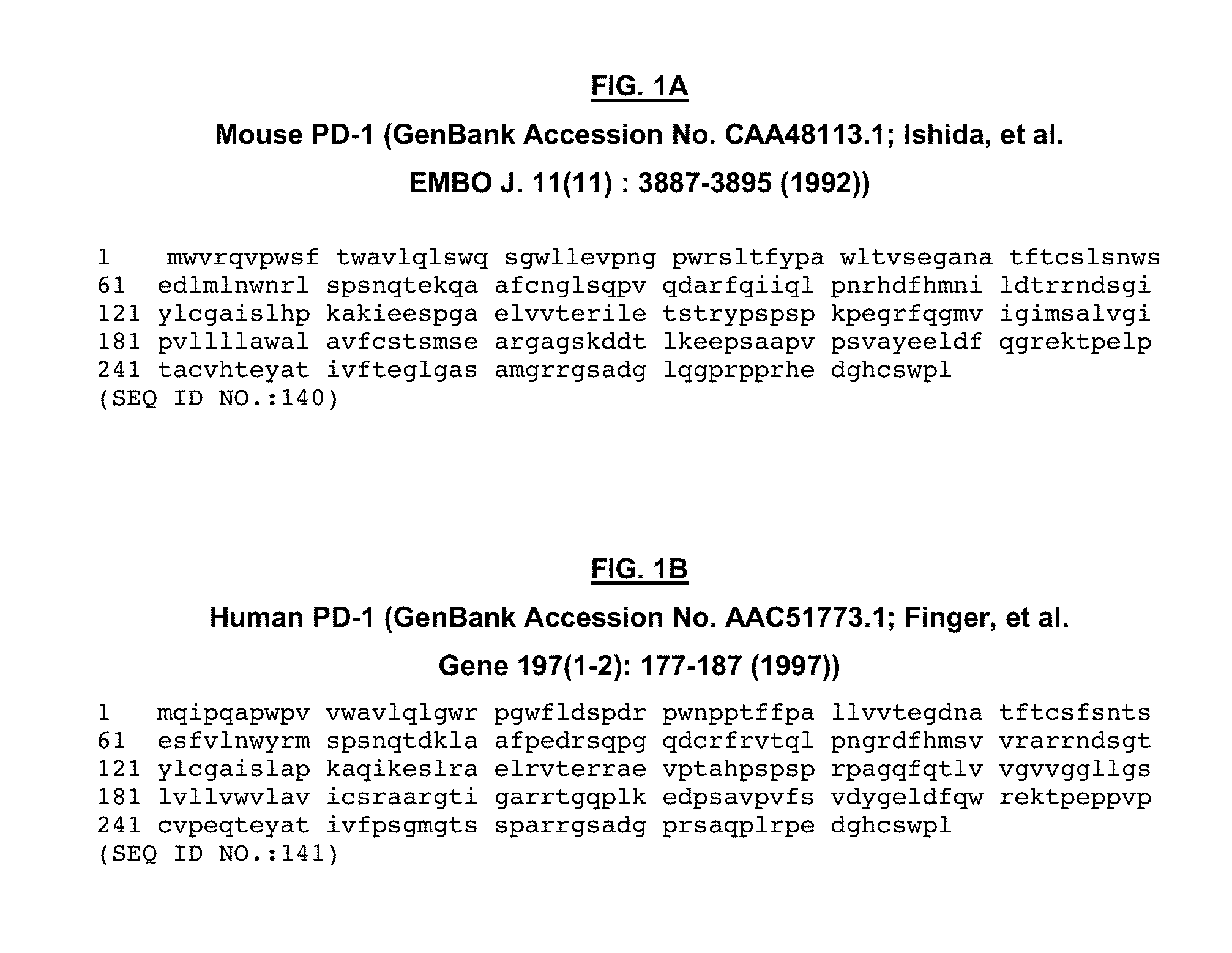
[0009] FIG. 1. FIG. 1A. Mouse PD-1. FIG. 1B. Human PD-1.
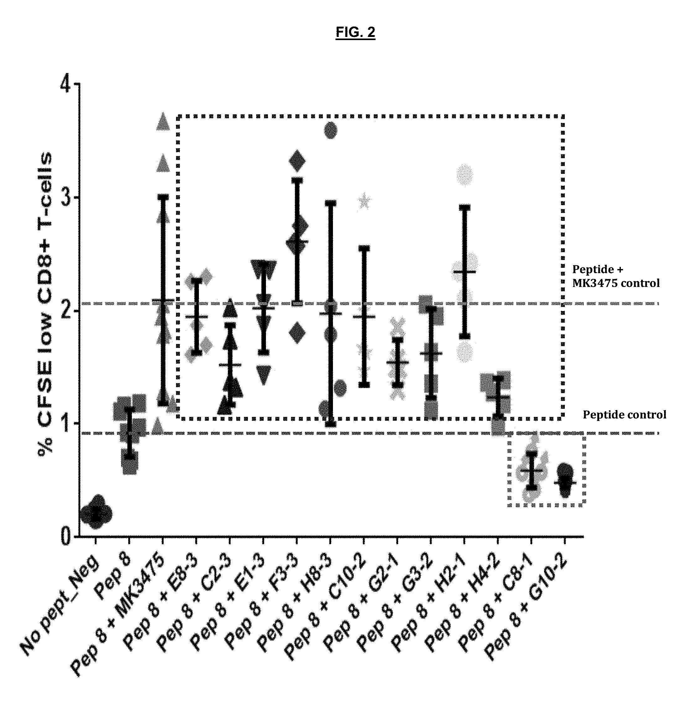
[0010] FIG. 2. CFSE assay to evaluate the functional effect of anti-PD1 antibodies on the proliferation of HIV specific CD8 T cells.
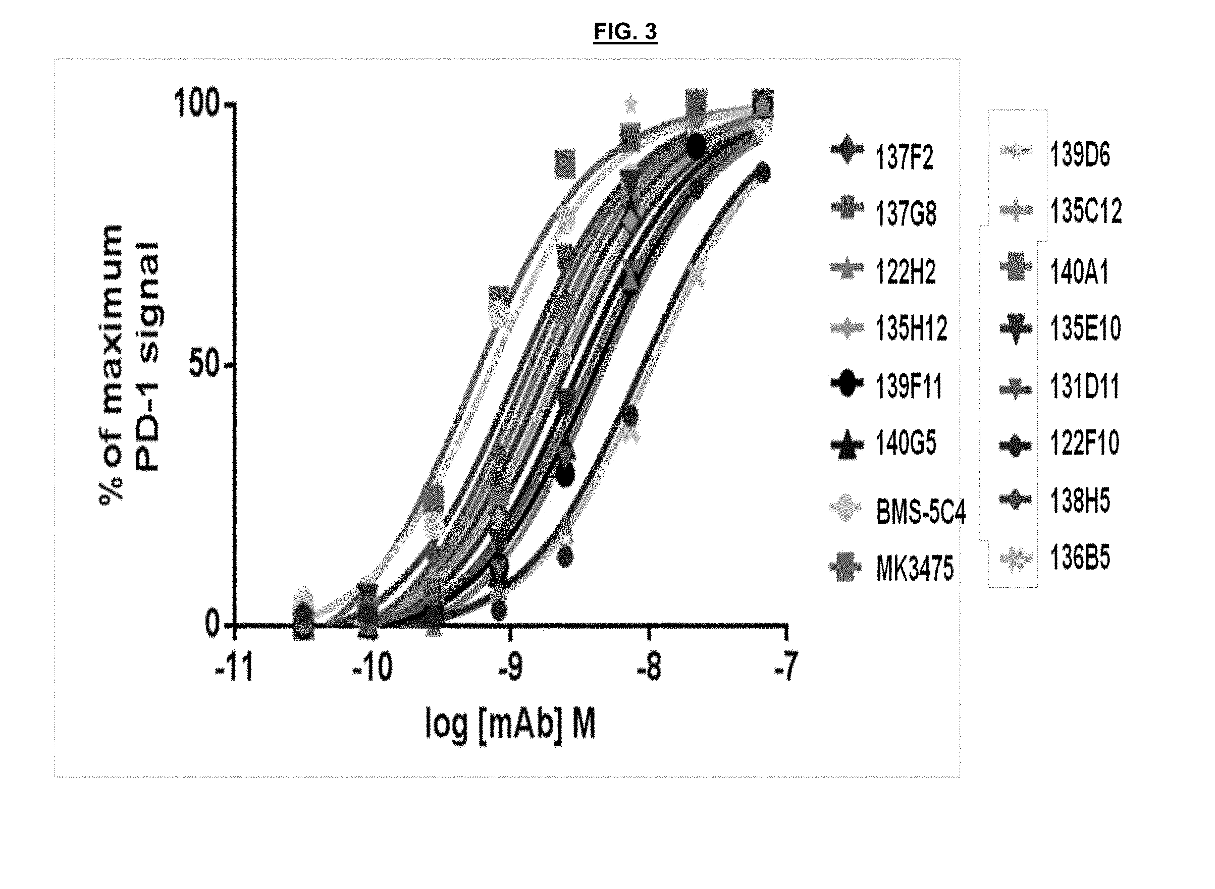
[0011] FIG. 3. Concentration response binding of anti-PD-1 antibodies to cell surface PD-1 on activated CD4 T-cells.
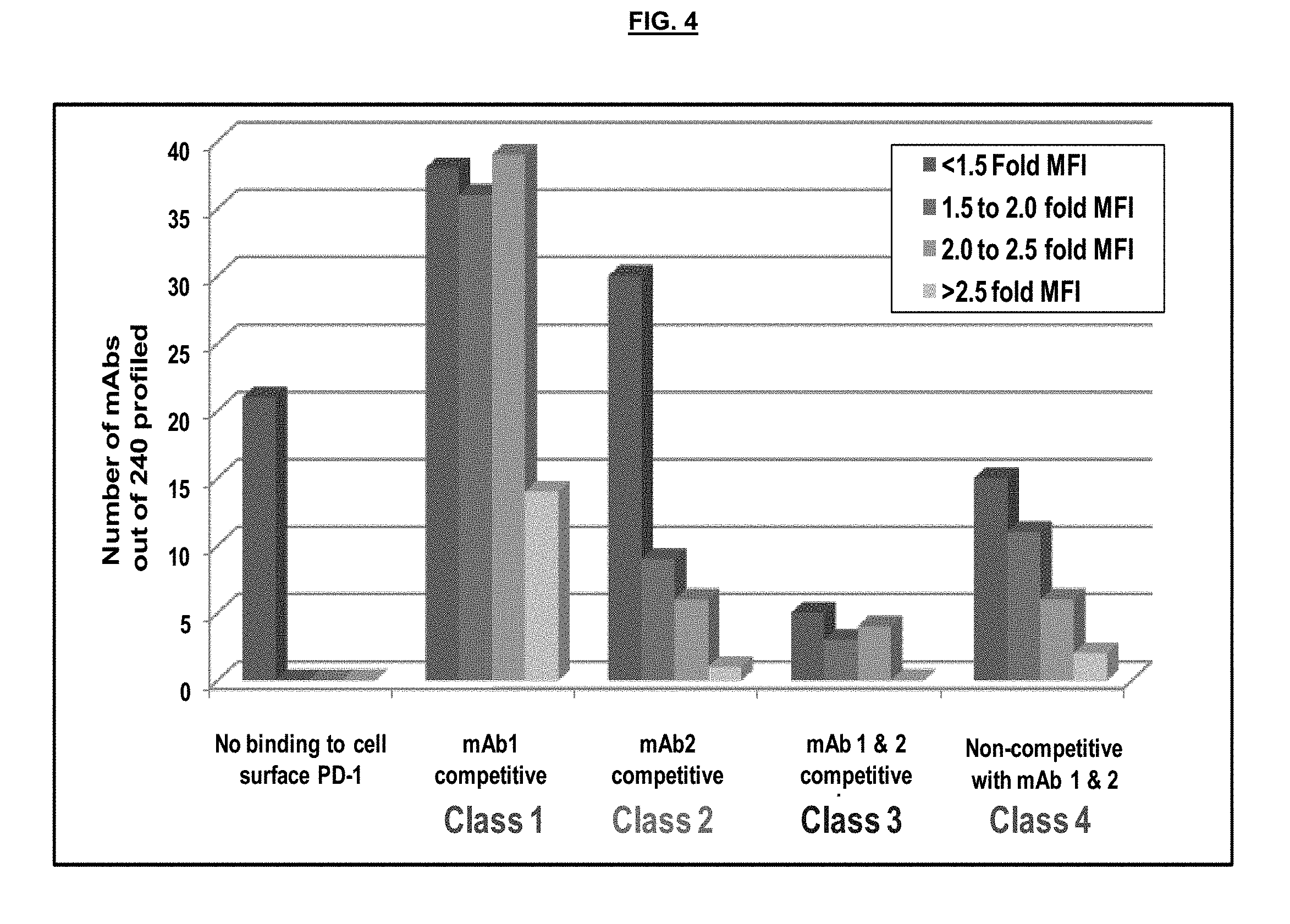
[0012] FIG. 4. Antibody classes.
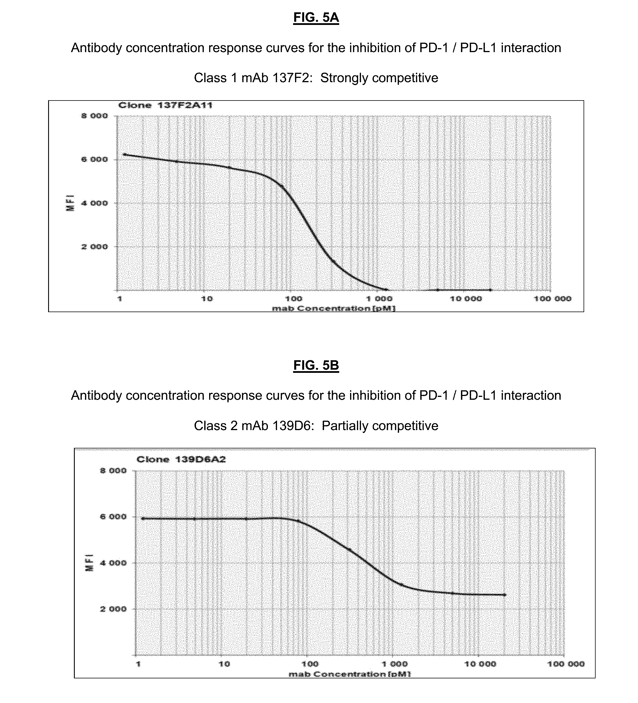
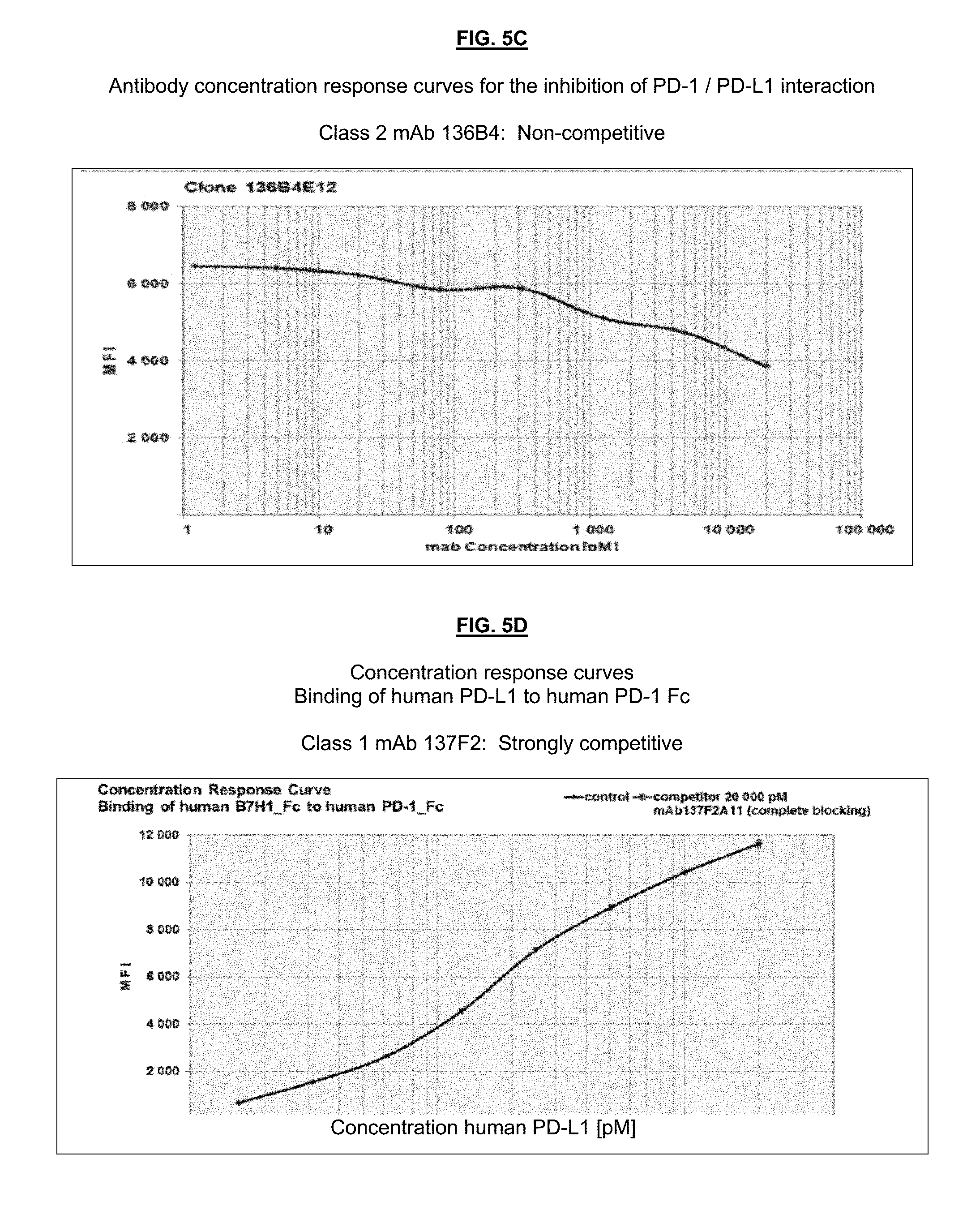
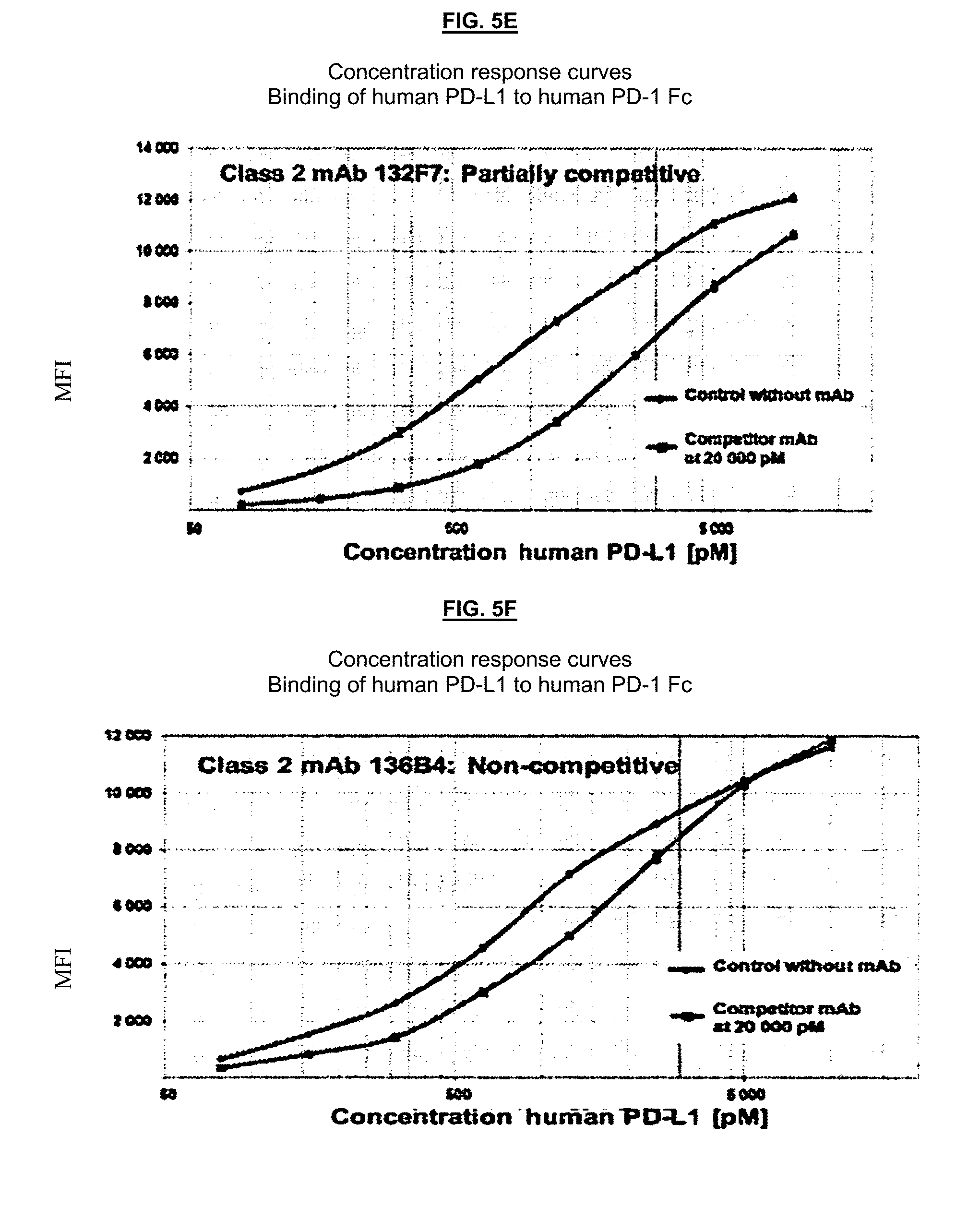
[0013] FIG. 5. Biochemical PD-1/PD-L1 interaction assay to evaluate if antibodies bind PD-1 competitively or non-competitively with PD-L1. FIG. 5A: Class 1 mAb 137F2; FIG. 5B: Class 2 mAb 139D6; FIG. 5C: Class 2 mAb 136B4; FIG. 5D: Class 1 mAb 137F2; FIG. 5E: Class 2 mAb 132F7; FIG. 5F: Class 2 mAb 136B4.
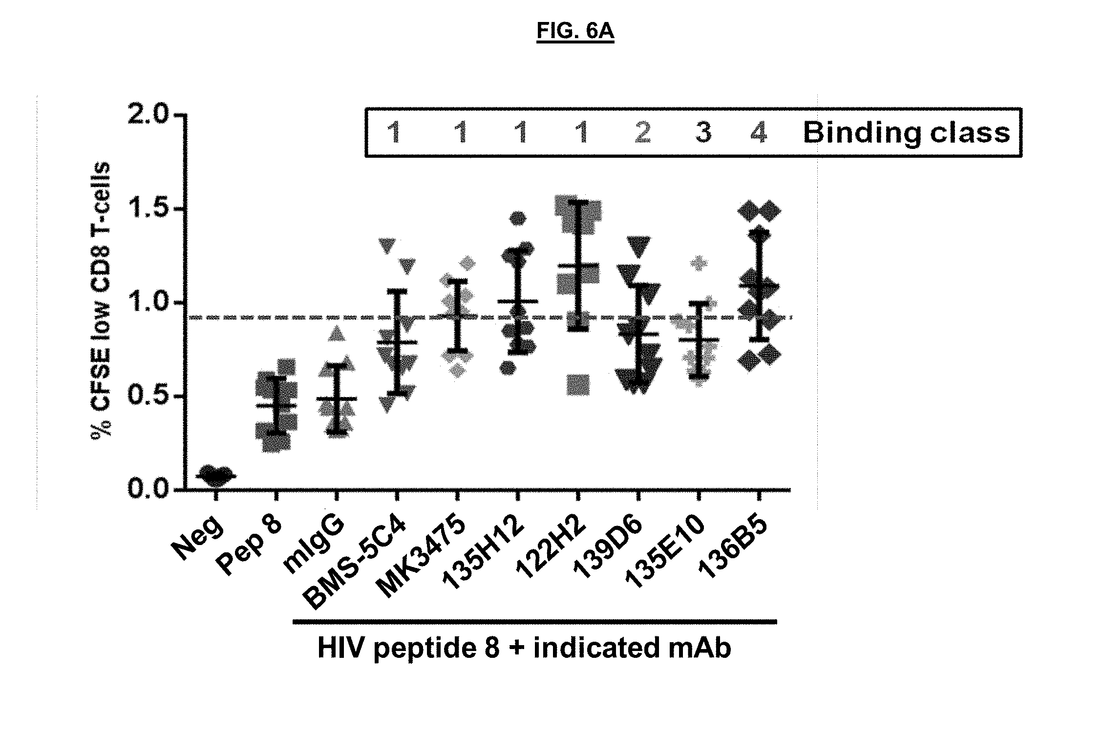
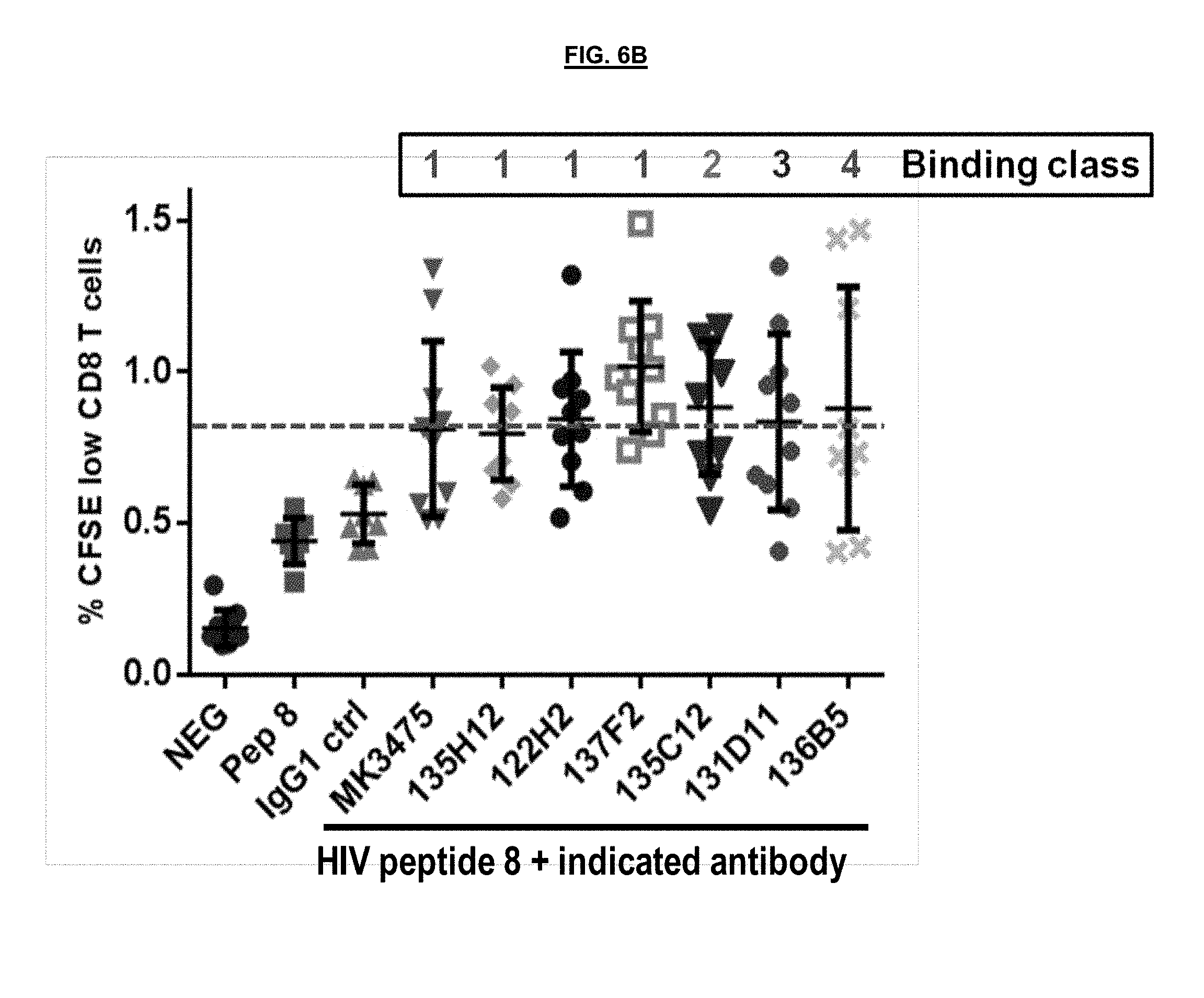
[0014] FIG. 6. Restoration of HIV peptide specific CD8 T-cell proliferation mediated by anti-PD-1 antibodies binding to different epitopes in a functional exhaustion recovery assay. FIG. 6A: Monoclonal antibodies BMS-5C4, MK3475, 135H12, 122H2, 139D6, 135E10, and 136B5. FIG. 6B: Monoclonal antibodies MK3475, 135H12, 122H2, 137F2, 135C12, 131D11, and 136B5.
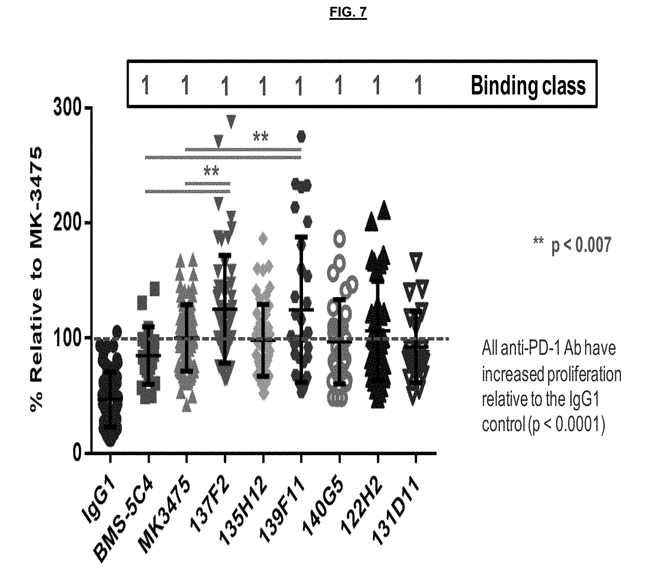
[0015] FIG. 7. Enhanced restoration of HIV peptide specific CD8 T-cell proliferation mediated by select anti-PD-1 antibodies relative to benchmark control antibodies in the functional exhaustion recovery assay.
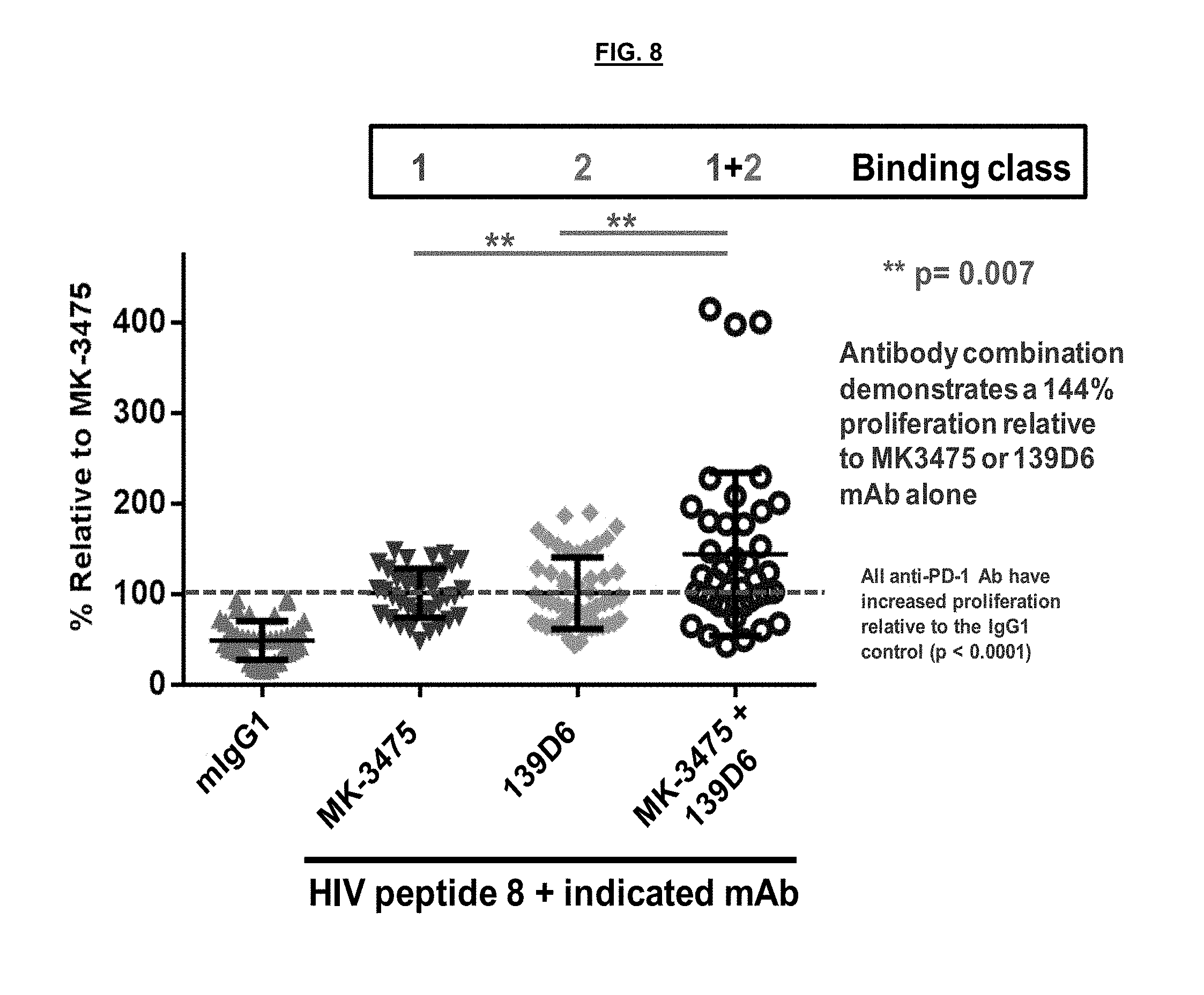
[0016] FIG. 8. Synergy between a first binding agent that blocks the interaction of PD-1 and PD-L1 with a second binding agent that does not block the interaction of PD-1 and PD-L1.
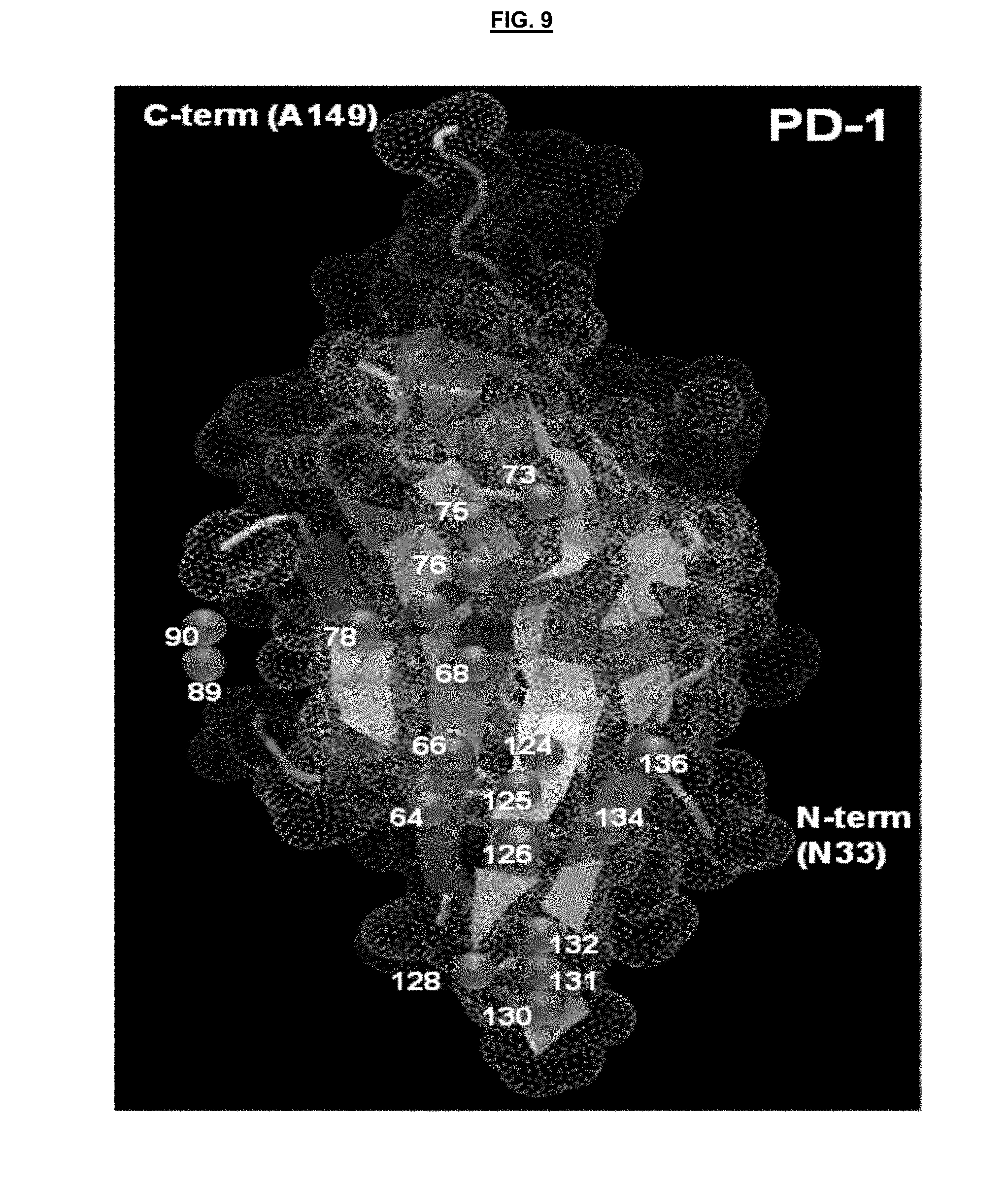
[0017] FIG. 9. Structural representation of the PD-1 protein ectodomain from residues 33 to 149. Amino acids implicated in the interaction with either PD-L1 or PD-L2 are situated on the structure by the purple circles with the residue number indicated.
[0018] FIG. 10. FIG. 10A: Amino acid sequence of the ectodomain for human PD-1. Residues that were targeted for amino acid substitutions are indicated for each of the 31 mutations (M1 to M31). Residues in purple text correspond to amino acids that are implicated in the PD-1/PD-L1 interaction and asparagine residues in green are potential sites for N-linked glycosylation. FIG. 10B: Modified PD-1 polypeptides M1, M2, M4 and M4. FIG. 10C: Modified PD-1 polypeptides M5, M6, M7, M8, M19 and M10. FIG. 10D:. Modified PD-1 polypeptides M11, M12, M13, M14, M15 and M16. FIG. 10E: Modified PD-1 polypeptides M17, M18, M19, M20, M21 and M22. FIG. 10F: Modified PD-1 polypeptides M23, M24, M25, M26, M27 and M28. FIG. 10G: Modified PD-1 polypeptides M29, M30 and M31. FIG. 10H: Human PD-1 amino acid sequence. FIG. 10I: Monkey PD-1 amino acid sequence.
[0019] FIG. 11. Binding of competitive and non-competitive anti-PD-1 antibodies to modified PD-1 protein expressed at the surface of transiently transfected HeLa cells. FIG. 11A: PD-1 WT. FIG. 11B: PD-1 M13. FIG. 11C: PD-1 M14. FIG. 11D:. PD-1 M23.
[0020] FIG. 12. Binding of competitive and non-competitive anti-PD-1 antibodies to modified PD-1 protein expressed at the surface of transiently transfected HeLa cells. FIG. 12A: PD-1 WT. FIG. 12B: PD-1 M13. FIG. 12C:. PD-1 M23. FIG. 12D: PD-1 M4.
[0021] FIG. 13. Binding of competitive and non-competitive anti-PD-1 antibodies to modified PD-1 protein expressed at the surface of transiently transfected HeLa cells. FIG. 13A: PD-1 WT. FIG. 13B: PD-1 M31. FIG. 13C: PD-1 M5. FIG. 13D: PD-1 M18.
[0022] FIG. 14. Binding of competitive and non-competitive anti-PD-1 antibodies to modified PD-1 protein expressed at the surface of transiently transfected HeLa cells. FIG. 14A: WT. FIG. 14B: M1. FIG. 14C: M9. FIG. 14D: M13. FIG. 14E: M17. FIG. 14F: M18. FIG. 14G: M23. FIG. 14H: M28.
[0023] FIG. 15. Combination of two antagonistic anti-PD-1 antibodies binding to different epitopes on PD-1, one competitive and one non-competitive with the PD-1/PD-L1 interaction, results in an enhanced relief of functional exhaustion and increased proliferation of HIV specific CD8 T cells beyond what either antibody alone can achieve.
[0024] FIG. 16. Individual antagonistic anti-PD-1 antibodies and combinations of two antagonistic anti-PD-1 antibodies binding to different epitopes on PD-1 result in enhanced proliferation (FIG. 16A) and IFN.gamma. production (FIG. 16B) from PD-1+ memory CD4 T cells in a mixed lymphocyte reaction assay.
[0025] FIG. 17. Structural representation of the P1 (FIG. 17A) and P2 (FIG. 17B)) evolutionarily conserved patches of PD-1 along with the alignments of PD-1 ectodomain amino acid sequences from different species (FIG. 17C).
[0026] FIG. 18. Antibody competitive binding studies for cell surface PD-1 on activated CD4+ T cells.
[0027] FIG. 19. Epitope mapping of antibody binding sites to the structure of PD-1 using a combination of both site directed mutagenesis and antibody competition binding results. FIG. 19A: First view. FIG. 19B: 180.degree. View.
[0028] FIG. 20. Selective increase in apoptosis (Annexing V staining) or cell death (Aqua staining) in PD-1 high CD4 T cells from a viremic HIV infected donor upon treatment with an anti-PD-1 ADC as opposed to either anti-PD-1 antibodies alone or an IgG control antibody. FIG. 20A: % Aqua positive. FIG. 20B: % Annexin positive.
[0029] FIG. 21. Cytotoxicity of anti-PD1 antibody drug conjugates. FIG. 21A: Annexin V positive. FIG. 21B: % Aqua positive.
[0030] FIG. 22. Evaluation of the anti-PD-1 antibody drug conjugate (ADC) mediated killing of PD-1 positive infected CD4 T cells from a chronically infected HIV donor. Following 5 days of antibody treatment, cells were used in a quantitative viral outgrown assay to monitor the number of infectious cells in the different treated samples.
SUMMARY OF THE DISCLOSURE
[0031] This disclosure relates to binding agents with specificity for programmed cell death 1 (PD-1) (e.g., human PD-1) and to methods for using the same such as to treat, prevent and/or ameliorate infection (e.g., by human immunodeficiency virus (HIV)), cancer and/or an autoimmune condition. Functional assays for identifying binding agents that interact with PD-1 are also provided. Combinations of binding agents, such as a first binding agent that blocks the interaction of PD-1 and PD-L1 with a second binding agent that does not block the interaction of PD-1 and PD-L1, are also provided that act synergistically to rescue T cells from exhaustion.
DETAILED DESCRIPTION
[0032] This disclosure relates to binding agents that bind programmed cell death (PD-1) protein (e.g., SEQ ID NO:1, FIG. 1A, FIG. 1B of U.S. Pat. No. 5,698,520 (Honjo, et al.) which is hereby incorporated by reference in its entirety) (e.g., human PD-1) on the surface of cells in vitro and/or in vivo. The binding agents may also bind isolated PD-1 polypeptide (e.g., human PD-1) and/or fragments and/or derivatives thereof, typically in vitro. Also provided are methods for using such binding agents to diagnose, treat, prevent and/or ameliorate one or more diseases associated with the existence of cells expressing PD-1. For instance, the binding agents may be antibodies (e.g., monoclonal antibodies) that may react with and/or bind to the epitopes of PD-1. The "binding agents" described herein may include, for example, an agonist or an antagonist of PD-1. An agonist binding agent is one that is not typically capable of restoring T-cell function and/or expression of PD-1. An agonist PD-1 binding agent may be useful for treating autoimmune diseases and others in which PD-1 expressing cells are involved in disease progression. In contrast, an antagonist binding agent is one capable for restoring T-cell function and/or expression of PD-1. For instance, a PD-1 antagonist binding agent may be capable of restoring the function of PD-1 expressing T-cells from functional exhaustion as is known to occur in HIV infection and in a variety of tumors. Restoration of T cell function may be determined by, for instance, measuring proliferation, cytokine production, cytotoxic activity or other characteristics of such cells. Another use for the binding agents described herein is the selective targeting and elimination of HIV-infected CD4.sup.+ T-cell populations containing replication competent HIV (e.g., in a latent and/or replication state). Such PD-1 expressing cells expressing PD-1 are known to serve as a major cell reservoir for replication competent HIV. A potential mechanism for the elimination of these CD4.sup.+ T-cell populations is antibody-dependent cellular cytotoxicity (ADCC) using the binding agents described herein (e.g., mono- and/or bi-specific PD-1 antibodies). In some embodiments, one or more PD-1 antagonistic binding agents having, for instance, different specificities (e.g., recognizing different epitopes) may be combined to induce rescue of antigen-specific CD8.sup.+ T-cells from functional exhaustion caused by PD-1 expression in those cells (e.g., restoring or improving proliferation, cytokine production and/or cytotoxic activity). In some embodiments, the binding agents described herein may also provide for the selective elimination and/or suppression of PD-1 expressing cells. In some embodiments, the PD-1 agonist binding agents described herein may be used to supress and/or eliminate PD-1 expressing cells to treat, for instance, infectious diseases (e.g., HIV), cancer, and/or, especially, autoimmune conditions. Other embodiments, uses and the like are described below.
[0033] The binding agents may be antibodies such as monoclonal antibodies that may comprise, for instance, any one or more of the amino acid sequences shown in Table 1 (and/or one or more fragments and/or derivatives thereof). This disclosure also provides for the use of such monoclonal antibodies to isolate, identify, and/or target cells expressing PD-1. In certain embodiments, these monoclonal antibodies may be reactive against PD-1 expressed on the surface of cells. The term "antibody" or "antibodies" may refer to whole or fragmented antibodies in unpurified or partially purified form (e.g., hybridoma supernatant, ascites, polyclonal antisera) or in purified form. The antibodies may be of any suitable origin or form including, for example, murine (e.g., produced by murine hybridoma cells), or expressed as humanized antibodies, chimeric antibodies, human antibodies, and the like. For instance, antibodies may be wholly or partially derived from human (e.g., IgG (IgG1, IgG2, IgG2a, Ig2b, IgG3, IgG4), IgM, IgA (IgA1 and IgA2), IgD, and IgE), canine (e.g., IgGA, IgGB, IgGC, IgGD), chicken (e.g., IgA, IgD, IgE, IgG, IgM, IgY), goat (e.g., IgG), mouse (e.g., IgG, IgD, IgE, IgG, IgM), and/or pig (e.g., IgG, IgD, IgE, IgG, IgM), rat (e.g., IgG, IgD, IgE, IgG, IgM) antibodies, for instance. Methods of preparing, utilizing and storing various types of antibodies are well-known to those of skill in the art and would be suitable in practicing the present invention (see, for example, Harlow, et al. Antibodies: A Laboratory Manual, Cold Spring Harbor Laboratory, 1988; Harlow, et al. Using Antibodies: A Laboratory Manual, Portable Protocol No. 1, 1998; Kohler and Milstein, Nature, 256:495 (1975)); Jones et al. Nature, 321:522-525 (1986); Riechmann et al. Nature, 332:323-329 (1988); Presta (Curr. Op. Struct. Biol., 2:593-596 (1992); Verhoeyen et al. (Science, 239:1534-1536 (1988); Hoogenboom et al., J. Mol. Biol., 227:381 (1991); Marks et al., J. Mol. Biol., 222:581 (1991); Cole et al., Monoclonal Antibodies and Cancer Therapy, Alan R. Liss, p. 77 (1985); Boerner et al., J. Immunol., 147(1):86-95 (1991); Marks et al., Bio/Technology 10, 779-783 (1992); Lonberg et al., Nature 368 856-859 (1994); Morrison, Nature 368 812-13 (1994); Fishwild et al., Nature Biotechnology 14, 845-51 (1996); Neuberger, Nature Biotechnology 14, 826 (1996); Lonberg and Huszar, Intern. Rev. Immunol. 13 65-93 (1995); as well as U.S. Pat. Nos. 4,816,567; 5,545,807; 5,545,806; 5,569,825; 5,625,126; 5,633,425; and, 5,661,016). In certain applications, the antibodies may be contained within hybridoma supernatant or ascites and utilized either directly as such or following concentration using standard techniques. In other applications, the antibodies may be further purified using, for example, salt fractionation and ion exchange chromatography, or affinity chromatography using Protein A, Protein G, Protein A/G, and/or Protein L ligands covalently coupled to a solid support such as agarose beads, or combinations of these techniques. The antibodies may be stored in any suitable format, including as a frozen preparation (e.g., -20.degree. C. or -70.degree. C.), in lyophilized form, or under normal refrigeration conditions (e.g., 4.degree. C.). When stored in liquid form, for instance, it is preferred that a suitable buffer such as Tris-buffered saline (TBS) or phosphate buffered saline (PBS) is utilized. In some embodiments, the binding agent may be prepared as an injectable preparation, such as in suspension in a non-toxic parenterally acceptable diluent or solvent. Suitable vehicles and solvents that may be utilized include water, Ringer's solution, and isotonic sodium chloride solution, TBS and/or PBS, among others. Such preparations may be suitable for use in vitro or in vivo may be prepared as is known in the art and the exact preparation may depend on the particular application.
[0034] However, the binding agents described are not in any way limited to antibodies. For example, the binding agent may be any compound exhibiting similar binding properties as another (e.g., a mimetic). For example, an exemplary binding agent may be one that binds PD-1 and/or can compete with binding agent having specificity therefor (e.g., a monoclonal antibody). In some embodiments, the mimetic may exhibit substantially the same affinity in binding assays as the binding agent (e.g., monoclonal antibody) to which it is being compared. The affinity a particular binding agent may be measured by any suitable assay including but not limited to FACS staining of endogenous cell surface PD-1 on activated CD4 T cells as described in the Examples. One binding agent may be said to have "substantially the same affinity" as another where the measurements (e.g., nm) are within about any of 1-20,1-5,5-10, 10-15, or 15-20 percent of one another. Exemplary mimetics may include, for example, organic compounds that specifically bind PD-1, or an affibody (Nygren, et al. FEBS J. 275 (11): 2668-76 (2008)), affilin (Ebersbach, et al. J. Mol. Biol. 372 (1): 172-85 (2007)), affitin (Krehenbrink, et al. J. Mol. Biol. 383 (5): 1058-68 (2008)), anticalin (Skerra, A. FEBS J. 275 (11): 2677-83 (2008)), avimer (Silverman, et al. Nat. Biotechnol. 23 (12): 1556-61 (2005)), DARPin (Stumpp, et al. Drug Discov. Today 13 (15-16): 695-701 (2008)), Fynomer (Grabulovski, et al. J. Biol. Chem. 282 (5): 3196-3204 (2007)), Kunitz domain peptide (Nixon, et al. Curr. Opin. Drug Discov. Devel. 9 (2): 261-8 (2006)), and/or a monobody (Koide, et al. Methods Mol. Biol. 352: 95-109 (2007)). Other mimetics may include, for example, a derivative of an antibody (of, for example, the monoclonal antibody 1E4,1G10, and/or 1G1) such as, for example, an F.sub.ab, F.sub.ab2, Fab' single chain antibody, F.sub.v, single domain antibody, mono-specific antibody, bi-specific antibody, tri-specific antibody, multi-valent antibody, chimeric antibody, canine-human chimeric antibody, canine-mouse chimeric antibody, antibody comprising a canine Fc, humanized antibody, human antibody, caninized, CDR-grafted antibody, shark antibody, nanobody, canelid antibody, microbody, and/or intrabody, or derivative thereof. Other binding agents are also provided herein as would be understood by one of ordinary skill in the art.
[0035] Any method known to those of ordinary skill in the art may be used to generate binding agents having specificity for (e.g., binding to) PD-1. For instance, to generate and isolate monoclonal antibodies an animal such as a mouse may be administered (e.g., immunized) with one or more PD-1 proteins (e.g., PD-1 Fc fusion protein and/or PD-1 His tag protein). Animals exhibiting serum reactivity to PD-1 expressed on activated human T lymphocytes (as determined by, for instance, flow cytometry and/or microscopy) may then be selected for generation of anti-PD-1 hybridoma cell lines. This may be repeated for multiple rounds. For instance, the primary criteria for the first round of binding agent selection may be include but are not limited to: i) level of staining of PD-1 on activated human T lymphocytes by flow cytometry; (ii) diversity of CDR VH and VL sequences as compared to those of the existing anti-PD-1 antibodies; and, (iii) epitope mapping performed by competitive binding studies with PD-1 conjugated Luminex beads pre-coupled with PD-L1 or one of several commercially available anti-PD-1 antibodies binding to different epitopes on PD-1. An exemplary first or second round of selection may also include, for instance, affinity binding (not a primary criteria since it may not correlate with the stimulatory potential of anti-PD-1 antibodies); and/or, functional characterization to identify the binding agent as an agonist or an antagonist.
[0036] As described in Example 1 herein, for instance, the Exhaustion Functional Recovery Assay (EFRA) may be used. In this assay, test binding agents may be assayed for the ability to rescue immune cells such as T cells from exhaustion. This may be determined by measuring the ability of a binding agent to restore proliferation to such cells in the presence of an antigen, such as a test peptide derived from a virus such as human immunodeficiency virus (HIV). Proliferation is measured in a CFSE assay in comparison to a control, such as the test peptide alone or a positive control anti-PD-1 antibody such as MK-3475 (pembrolizumab). In some embodiments, a binding agent is determined to restore proliferation where the comparison shows a significant difference (such as a P value of <0.001) compared to either a peptide alone control or peptide with an isotype control mouse IgG1 antibody. This assay may be used to identify binding agents (such as antibodies) that compete with other binding agents for binding to PD-1 (such as PD-L1 or PD-L2) and/or lead to the functional restoration of immune cells. Example 1 also describes two methods of epitope mapping the antibodies listed in Table 2 using Luminex-based assays. In one biochemical assay, a PD-1 Fc fusion protein is bound to beads and competitive binding studies are performed between the anti-PD-1 antibodies described in Table 2 and one of two different commercially available anti-PD-1 antibodies. Example 1 describes four classes of monoclonal antibodies binding to distinct epitopes on PD-1 that were: class 1 (competitive with a first monoclonal antibody that blocks the interaction of PD-1 with PD-L1), class 2 (competitive with a second monoclonal antibody that binds PD-1 but does not block the interaction of PD-1 with PD-L1), class 3 (competitive with both the first and second monoclonal antibodies), and class 4 (non-competitive with either the first or second antibodies). In a seperate assay, competition for binding to a recombinant PD-1 protein was evaluated for the anti-PD-1 antibodies listed in Table 2 and a biotinylated PD-L1 recombinant protein. Antibodies that induced proliferation in the EFRA were identified from all four binding classes that are proposed of binding to different epitopes on PD-1. Likewise, the EFRA allowed for the identification of anti-PD-1 antibodies that were either competitive, partially competitive or non-competitive with the PD-1/PD-L1 interaction and specifically restored proliferative function to HIV specific CD8.sup.+ T-cell.
[0037] Combinations of binding agents may also be identified. In some embodiments, the combinations may be identified to provide statistically significant differences from results obtained using only one or more of the binding agents and not others. In some embodiments, combinations exhibiting synergistic ability to restore immune cell function may be identified. In some embodiments, the combination may comprise a first binding agent that blocks the interaction of PD-1 and PD-L1 with a second binding agent that does not block the interaction of PD-1 and PD-L1. The first and second binding agents may be different entities such as two or more different monoclonal antibodies or derivatives thereof, or may be found on the same entity such as a bi-functional antibody (a single antibody or derivative thereof comprising multiple binding specificities). For instance, an exemplary bi-functional antibody may comprise a first binding region that blocks the interaction of PD-1 and PD-L1 and a second binding region that does not block the interaction of PD-1 and PD-L1. Also contemplated are combinations that provide multiple types of each binding agent. For instance, the combination may comprise multiple types of binding agents that block the interaction of PD-1 and PD-L1 with one or more that does not block the interaction of PD-1 and PD-L1. In some embodiments, the combination may comprise one or more of binding agents that block the interaction of PD-1 and PD-L1 with multiple binding agents that do not block the interaction of PD-1 and PD-L1. In some embodiments, the combination may comprise multiple binding agents that block the interaction of PD-1 and PD-L1 with multiple binding agents that do not block the interaction of PD-1 and PD-L1. Such combinations as described herein may also be combined with one or more other agents that may effect immune cell function such as antibodies against CTLA-4 and the like. One of ordinary skill in the art would recognize that many such combinations may be suitable for use as described herein.
[0038] Where the binding agent is an antibody, it may be identified with reference to the nucleotide and/or amino acid sequence corresponding to the variability and/or complementarity determining regions ("CDRs") thereof. The variable region/CDR sequences may be used in combination with one or more other variable region/CDR amino acid sequences. The variable region/CDR amino acid sequences may alternatively and/or also be adjoined to one or more types of constant region polypeptides of an antibody molecule. For instance, the CDR amino acid sequences shown in Tables 1A and 1B may be adjoined to or associated with the constant regions of any antibody molecule of the same or a different species (e.g., human, goat, rat, sheep, chicken) and/or antibody subtype of that from which the CDR amino acid sequence was derived. For instance, an exemplary binding agent may be, or may be derived from, or may be related to the monoclonal antibody produced by the hybridomas listed in, and/or may have about the same affinity and/or proliferation effect, and/or exhibit the same binding class shown in Table 2, 5, 6 or 7 and/or may have any one or more of the amino acid sequences of SEQ ID NOS. 1-138 and/or as shown in Tables 1A and 1B. The binding agent may comprise an antibody heavy and/or a light chain that each comprises one or more constant and/or variable regions. The variable regions typically comprise one or more CDRs that may determine the binding specificity of the antibody. The monoclonal antibodies may also be identified by analysis of the amino acid sequences of (e.g., which may be encoded by such nucleotide sequences) such variable regions. For instance, exemplary amino acid sequences of the heavy chain CDRs of binding agents that bind PD-1 may include any one or more of comprising at least one amino acid sequence selected from the group consisting of SEQ ID NOS. 1-138, and/or any other shown in Tables 1A and/or 1B. Any of the amino acid sequences described herein, and/or any fragments and/or derivatives thereof may also be combined with any other variable region and/or CDR in any order and/or combination to form hybrid and/or fusion binding agents and/or inserted into other heavy and/or light chain variable regions using standard techniques. Exemplary combinations of CDRs (e.g., combination of heavy and/or light chain CDR1, CDR2 and CDR3 amino acid sequences) that may be found in a PD-1 (e.g., human PD-1) binding agent of this disclosure may include, for instance, the embodiments shown in Tables 1A and/or 1B.
TABLE-US-00001 TABLE 1A Heavy chain: Amino acids sequence Clone CD121 CDR2 CDR3 122F10 DDFLH RIDPANGESRYAPKFQD TDYRGYYYAMDY (SEQ ID NO: 1) (SEQ ID NO: 24) (SEQ ID NO: 47) 139D6 NYYIH SIYPNYGDTNYNQKVKD GYSYAMDY (SEQ ID NO: 2) (SEQ ID NO: 25) (SEQ ID NO: 48) 135D1 NYYIH SIYPNYGETNYNQEFKG GYSYAMDY (SEQ ID NO: 3) (SEQ ID NO: 26) (SEQ ID NO: 49) 134D2 SNWMH AVNPGNSDTTYNQKFKG GRSYDGSFDY (SEQ ID NO: 4) (SEQ ID NO: 27) (SEQ ID NO: 50) 121G1 RYWMH NIDPSDSTTHYNPKFRD DLDDFYVGSHEDFDY (SEQ ID NO: 5) (SEQ ID NO: 28) (SEQ ID NO: 51) 136B5 SNWMH AVYPGNSDTTYNQNFKG GRSYDGSFDY (SEQ ID NO: 6) (SEQ ID NO: 29) (SEQ ID NO: 52) 127C2 NSYIH WISPGDGSTNYNEKFKG EEYDYDNY (SEQ ID NO: 7) (SEQ ID NO: 30) (SEQ ID NO: 53) 137F2 NYWIG DIYPGGGYTNYNEKFKG GYDFVLDR (SEQ ID NO: 8) (SEQ ID NO: 31) (SEQ ID NO: 54) 138H5 SYAMS TISGGGADTYYLDNVKG QRGENLFAH (SEQ ID NO: 9) (SEQ ID NO: 32) (SEQ ID NO: 55) 140A1 SDYAWN YINYSGYTNYNPFLKS YGGSYPWNFDV (SEQ ID NO: 10) (SEQ ID NO: 33) (SEQ ID NO: 56) 135H12 SYWIN NIYPGSSSIDYNEKFKS GLYWYFDV (SEQ ID NO: 11) (SEQ ID NO: 34) (SEQ ID NO: 57) 131D11 SSYIH WIFPGDGKTNYNEKFRD NDFDRGVY (SEQ ID NO: 12) (SEQ ID NO: 35) (SEQ ID NO: 58) 132F7 NHGMS SINTGGYSTYYPDNVKG DDYNWFAY (SEQ ID NO: 13) (SEQ ID NO: 36) (SEQ ID NO: 59) 126E4 NYWIG DIYPGSEYENYNEKFKG GYDFVLDH (SEQ ID NO: 14) (SEQ ID NO: 37) (SEQ ID NO: 60) 135G1 DSYIH RIDPAHGNVIYASKFRD IYYDYGEGDF (SEQ ID NO: 15) (SEQ ID NO: 38) (SEQ ID NO: 61) 136E10 DTYIH RIDLANDDILYASKFQG IYYDYGEGDY (SEQ ID NO: 16) (SEQ ID NO: 39) (SEQ ID NO: 62) 135C12 NFYIH SIYPNYGDTAYNQKFKD GYSYAMDY (SEQ ID NO: 17) (SEQ ID NO: 40) (SEQ ID NO: 63) 136F4 DSYIH RIDPARDNIIYASKFRD IYYDYGEGDY (SEQ ID NO: 18) (SEQ ID NO: 41) (SEQ ID NO: 64) 136B4 DDFLH RIDPANGESRYAPQFQD TDYRGYYYAMDY (SEQ ID NO: 19) (SEQ ID NO: 42) (SEQ ID NO: 65) 135E10 SYFMS GISTGGADTYYADSMKG LSHYYDGIPLDC (SEQ ID NO: 20) (SEQ ID NO: 43) (SEQ ID NO: 66) 140G5 NHGMS SISGGGDNTYYPDNLKG VRQLGLHRAAMDY (SEQ ID NO: 21) (SEQ ID NO: 44) (SEQ ID NO: 67) 122H2 NYWIG DIYPGGDHKNYNEKFKD GFDFVLDY (SEQ ID NO: 22) (SEQ ID NO: 45) (SEQ ID NO: 68) 139F11 SFAMS TITGGGVNTYYPDTVKG QAIYDGHYVLDY (SEQ ID NO: 23) (SEQ ID NO: 46) (SEQ ID NO: 69)
TABLE-US-00002 TABLE 1B Light chain: Amino acids sequence Clone CDR1 CDR2 CDR3 122F10 KSSQSVLYSSNQKNYLA WASTRES HQYLSSYT (SEQ ID NO: 70) (SEQ ID NO: 93) (SEQ ID NO: 116) 139D6 SASQGISDGLN HTSTLHS QQYSKFPLT (SEQ ID NO: 71) (SEQ ID NO: 94) (SEQ ID NO: 117) 135D1 SASQGISNGLN HTSTLHS QQYSKFPLT (SEQ ID NO: 72) (SEQ ID NO: 95) (SEQ ID NO: 118) 134D2 KASQDINKYIA YTSTLRP LQYDNLWT (SEQ ID NO: 73) (SEQ ID NO: 96) (SEQ ID NO: 119) 121G1 RSSQSIVYSNGNTYLE KVSHRFS FQGSHVPYT (SEQ ID NO: 74) (SEQ ID NO: 97) (SEQ ID NO: 120) 136B5 KASQDINKYMA YTSTLRP LQYDNLWT (SEQ ID NO: 75) (SEQ ID NO: 98) (SEQ ID NO: 121) 127C2 KASQNVGTNVG SASYRYN QQYNTYPWT (SEQ ID NO: 76) (SEQ ID NO: 99) (SEQ ID NO: 122) 137F2 KSSQSLFNSETQKNYLA WASTRES KQSYTLRT (SEQ ID NO: 77) (SEQ ID NO: 100) (SEQ ID NO: 123) 138H5 LASQTIGTWLA AATSLAD QQLYSTPWT (SEQ ID NO: 78) (SEQ ID NO: 101) (SEQ ID NO: 124) 140A1 RSSQTIVHNNGDTYLE KISNRFF FQGSHVPYT (SEQ ID NO: 79) (SEQ ID NO: 102) (SEQ ID NO: 125) 135H12 KSSQSLENSGTRKNYLA WASTRDS KQSYNLYT (SEQ ID NO: 80) (SEQ ID NO: 103) (SEQ ID NO: 126) 131D11 KASQNVDTNVA SASYRYN QQYNNYPYT (SEQ ID NO: 81) (SEQ ID NO: 104) (SEQ ID NO: 127) 132F7 KSSQSLLNSGNQKNYLT WASTRES QSDYSYPLT (SEQ ID NO: 82) (SEQ ID NO: 105) (SEQ ID NO: 128) 126E4 KSSQSLENSGTRKSYLA WASTRET MQSYNLRT (SEQ ID NO: 83) (SEQ ID NO: 106) (SEQ ID NO: 129) 135G1 HASQNINVWLS KASNLHT QQGQSWPLT (SEQ ID NO: 84) (SEQ ID NO: 107) (SEQ ID NO: 130) 136E10 HASQNINVWLS KASNLHT QQGQSYPLT (SEQ ID NO: 85) (SEQ ID NO: 108) (SEQ ID NO: 131) 135C12 SASQGISGDLN HTSSLHS QYYSKDLLT (SEQ ID NO: 86) (SEQ ID NO: 109) (SEQ ID NO: 132) 136F4 HASQNINVWLS KASNLHT QQGQSWPLT (SEQ ID NO: 87) (SEQ ID NO: 110) (SEQ ID NO: 133) 136B4 KSSQSVLYSSNQKNYLA WASTRES HQYLSSYT (SEQ ID NO: 88) (SEQ ID NO: 111) (SEQ ID NO: 134) 135E10 RASESVDNSGVSFLT AASNQGS QQTKEVPWT (SEQ ID NO: 89) (SEQ ID NO: 112) (SEQ ID NO: 135) 140G5 KASQSVSDDVS SAFFRYP QQDYSSPLT (SEQ ID NO: 90) (SEQ ID NO: 113) (SEQ ID NO: 136) 122H2 KSSQSLENSGTRKNYLA WASTRES MQSFNLRT (SEQ ID NO: 91) (SEQ ID NO: 114) (SEQ ID NO: 137) 139F11 RTSGNIHNYLA NVKTLTD QQFWSIPWT (SEQ ID NO: 92) (SEQ ID NO: 115) (SEQ ID NO: 138)
In addition, any of SEQ ID NOS. 1-69 may be combined with any one or more of SEQ ID NOS. 70-138 into a binding agent. In preferred embodiments, the heavy chain CDRs of each clone are combined with their respective light chain CDRs into a binding agent. In some embodiments, the binding agent may comprise the heavy chain CDRs and light chain CDRs shown below: [0039] 122F10 (SEQ ID NOS. 1, 24, 47, 70, 93, and 116); [0040] 139D6 (SEQ ID NOS. 2, 25, 48, 71, 94, and 117); [0041] 135D1 (SEQ ID NOS. 3, 26, 49, 72, 95, and 118); [0042] 134D2 (SEQ ID NOS. 4, 27, 50, 73, 96, and 119); [0043] 121G1 (SEQ ID NOS. 5, 28, 51, 74, 97, and 120); [0044] 136B5 (SEQ ID NOS. 6, 29, 52, 75, 98, and 121); [0045] 127C2 (SEQ ID NOS. 7, 30, 53, 76, 99, and 122); [0046] 137F2 (SEQ ID NOS. 8, 31, 54, 77, 100, and 123); [0047] 138H5 (SEQ ID NOS. 9, 32, 55, 78, 101, and 124); [0048] 140A1 (SEQ ID NOS. 10, 33, 56, 79, 102, and 125); [0049] 135H12 (SEQ ID NOS. 11, 34, 57, 80, 103, and 126); [0050] 131D11 (SEQ ID NOS. 12, 35, 58, 81, 104, and 127); [0051] 132F7 (SEQ ID NOS. 13, 36, 59, 82, 105, and 128); [0052] 126E4 (SEQ ID NOS. 14, 37, 60, 83, 106, and 129); [0053] 135G1 (SEQ ID NOS. 15, 38, 61, 84, 107, and 130); [0054] 136E10 (SEQ ID NOS. 16, 39, 62, 85, 108, and 131); [0055] 135C12 (SEQ ID NOS. 17, 40, 63, 86, 109, and 132); [0056] 136F4 (SEQ ID NOS. 18, 41, 64, 87, 110, and 133); [0057] 136B4 (SEQ ID NOS. 19, 42, 65, 88, 111, and 134); [0058] 135E10 (SEQ ID NOS. 20, 43, 66, 89, 112, and 135); [0059] 140G5 (SEQ ID NOS. 21, 44, 67, 90, 113, and 136); [0060] 122H2 (SEQ ID NOS. 22, 45, 68, 91, 114, and 137); or [0061] 139F11 (SEQ ID NOS. 23, 46, 69, 92, 115, and 138). Other combinations may also be useful as may ascertained by one of ordinary skill in the art.
[0062] Binding agents comprising the CDRs of Tables 1A and/or 1B, or those of the immediately preceding paragraph, may also exhibit the following characteristics:
TABLE-US-00003 TABLE 2 EFRA % Antibody relative to competition with peptide Affinity* Binding the PD-1/PD-L1 stimulation Clone (nM) Class** interaction*** alone.sup..dagger-dbl. 122F10 2.2 4 Non-competitive 146% 139D6 2.4 2 Partial competition 195% 135D1 6.5 2 Partial competition 187% 134D2 4.8 4 Competitive 205% 121G1 11.9 4 Non-competitive 120% 136B5 7.7 4 Competitive 200% 127C2 1.0 2 Non-competitive 100% 137F2 1.5 1 Competitive 250% 138H5 1.6 3 Competitive 210% 140A1 1.4 3 Competitive 160% 135H12 1.9 1 Competitive 190% 131D11 2.7 1 Competitive 180% 132F7 100 2 Non-competitive 210% 126E4 0.5 4 Competitive 130% 135G1 32 4 NA 138% 136E10 7.1 4 Non-competitive 148% 135C12 1.7 2 Partial competition 195% 136F4 8.3 4 Non-competitive 108% 136B4 1.4 2 Non-competitive 185% 135E10 1.5 3 Competitive 165% 140G5 1.6 1 Competitive 205% 122H2 4.3 1 Competitive 200% 139F11 3.1 1 Competitive 250% *Binding affinity for the antibodies listed in Table 1 was evaluated by FACS staining of endogenous cell surface PD-1 on activated CD4 T cells. **Binding class was determined by Luminex assay competitive binding studies. Binding class 1 mAb clones are competitive with the EH12.2H7 clone commercial antibody, class 2 mAb clones are competitive with the J116 clone commercial antibody, class 3 mAb clones are competitive with both EH12.2H7 and J116 antibodies and class 4 mAb clones bind in the presence of both EH12.2H7 and J116 antibodies. ***Antibody competition with the PD-1/PD-L1 interaction was determined in a second Luminex binding assay. In this assays, PD-1 Fc fusion protein coated beads were incubated in the absence or presence of an anti-PD-1 antibody from Table 2 at a concentration of 20 nM. A fixed concentration of 1.25 nM biotinylated PD-L1, approximately equivalent to the IC.sub.50 of the PD-1/PD-L1 interaction, was then incubated with the PD-1/antibody complex and PD-L1 binding was detected by fluorescence with phycoerythrin labeled streptavidin. Based on PD-L1 binding to the PD-1/antibody complex, antibodies were defined as being competitive, partially competitive or non-competitive with the PD-1/PD-L1 interaction. .sup..dagger-dbl.Proliferative effect is evaluated using a CFSE assay (an embodiment of the Exhaustion Functional Recovery Assay, "EFRA"). PBMCs isolated from a chronically infected HIV subject were stimulated with an HIV specific peptide in the presence and absence of an anti-PD-1 antibody. Following a 6 day incubation, proliferation of HIV specific CD8 T cells was evaluated in the anti-PD-1 treated samples relative to the peptide alone control. NA = not available
[0063] As explained in the Examples section, epitope mapping studies revealed at least two conserved patches (comprising linear and/or conformational epitopes) on PD-1 to which the binding agents described herein may bind, designated "P1" and "P2" (see, e.g., FIGS. 17a and 17b). The P1 patch is evolutionarily conserved and corresponds to the central region of PD-1 involved in the interaction between PD-1 and the PD-L1/PD-L2 ligands, and corresponds with purple circles in FIG. 9 and FIG. 19a. The second "patch" P2 is also evolutionarily conserved and occupies a similar surface area but different amino acid sequences (FIG. 17b) as the P1 patch. P2 has no previously identified structural or functional role on PD-1. PD-1 binding agents described herein, such as anti-PD-1 antibodies comprising the amino acid sequences of 135C12 (SEQ ID NOS. 17, 40, 63, 86, 109, and 132), 139D6 (SEQ ID NOS. 2, 25, 48, 71, 94, and 117), 135D1 (SEQ ID NOS. 3, 26, 49, 72, 95, and 118), and 136B4 (SEQ ID NOS. 19, 42, 65, 88, 111, and 134), bind epitopes overlapping the P2 patch, thereby providing direct evidence for the functional importance of this newly identified functional region of PD-1. The Examples section describes epitope mapping studies that were carried out using modified PD-1 polypeptides shown in FIG. 10 and SEQ ID NOS. 139-168 and No. 206. These studies revealed that the non-competitive antibodies with the greatest "functional potency" or "antagonistic activity" bind to a "patch" of PD-1 that overlaps with the region of the M4 amino acid substitutions (serine 38 to alanine, proline 39 to alanine and leucine 41 to alanine (FIG. 10; SEQ ID NO. 142)), and/or the region of the M17 amino acid substitutions (asparagine 102 to alanine and arginine 104 to leucine (FIG. 10; SEQ ID NO. 155)), and/or the region of the M18 amino acid substitutions (aspartate 105 to alanine (FIG. 10; SEQ ID NO. 156), and/or the region of the M31 amino acid substitutions (leucine 41 to alanine and valine 43 to leucine (FIG. 10; SEQ ID NO. 206). This "patch" is referred to herein as "P2". These studies demonstrate that P2 comprises at least one epitope (linear, conformational, or a combination of the same) to which such non-competitive antibodies bind. Given that this region has no previous implication in the functional activity of PD-1, it is proposed herein that binding to P2 represents a novel mechanism of action at a novel site on PD-1 at which antagonistic activity towards PD-1 may be exerted. It is further proposed herein that other antibodies, antibody fragments, or other protein binding agents that can interact with the P2 region of PD-1 may also act as PD-1 antagonists in a manner distinct from and complementary to anti-PD-1 antibodies that act through blockade of the PD-1/PD-L1 interaction. The binding agents described herein may, for instance, interact with an as yet unidentified ligand; interfere with, induce and/or enhance PD-1 multimerization; and/or, by interacting with (e.g., binding) P2, altering intracellular signaling associated with PD-1. Accordingly, the binding agents described herein that interact with (e.g., bind) P2 may provide the PD-1 antagonistic function through any of these, or any other yet to be identified, mechanisms.
[0064] Accordingly, this disclosure provides methods for affecting the function of PD-1 by interacting with this P2 patch of PD-1 in or on a cell. Amino acid residues in the P2 patch of PD-1 may comprise threonine 36, phenylalanine 37, serine 38, proline 39, leucine 41, valine 43, alanine 50, threonine 51, phenylalanine 52, threonine 53, cysteine 54, serine 55, asparagine 102, arginine 104, aspartic acid (aspartate) 105, phenylalanine 106, histidine 107, and methionine 108, the amino acid numbering corresponding to SEQ ID NO. 204. Aspartic acid 85 and/or arginine 86 may also be present in the P2 patch. In some embodiments, the amino acid residues of the P2 patch may comprise threonine 36, phenylalanine 37, alanine 50, threonine 51, phenylalanine 52, threonine 53, cysteine 54, serine 55, aspartic acid (aspartate) 105, phenylalanine 106, histidine 107 and methionine 108, the amino acid numbering corresponding to SEQ ID NO. 204. In some embodiments, the amino acid residues of the P2 patch may comprise amino acid residues serine 38, proline 39, leucine 41, valine 43, asparagine 102, arginine 104, and/or aspartic acid (aspartate) 105. Thus, in some embodiments, the method may comprise interacting with amino acids threonine 36, phenylalanine 37, serine 38, proline 39, leucine 41, valine 43, alanine 50, threonine 51, phenylalanine 52, threonine 53, cysteine 54, serine 55, asparagine 102, arginine 104, aspartic acid (aspartate) 105, phenylalanine 106, histidine 107, and/or methionine 108, the amino acid numbering corresponding to SEQ ID NO. 204. In some embodiments, the method may comprise interacting with amino acids threonine 36, phenylalanine 37, alanine 50, threonine 51, phenylalanine 52, threonine 53, cysteine 54, serine 55, aspartic acid (aspartate) 105, phenylalanine 106, histidine 107 and/or methionine 108. In some embodiments, the method may comprise interacting with amino acid serine 38, proline 39, leucine 41, valine 43, asparagine 102, arginine 104, and/or aspartic acid (aspartate) 105. In some embodiments, the method comprises interacting with any amino acids within and/or overlapping the P2 patch (e.g., portions of PD-1 comprising the above-described amino acid residues). In some embodiments of such methods, the method comprises interacting with one or more amino acid residues corresponding to serine 38, proline 39, and/or leucine 41 of SEQ ID NO. 204 (M4; FIG. 10; SEQ ID NO. 142); and/or one or more amino acid residues corresponding to asparagine 102 and/or arginine 104 relative to SEQ ID NO. 204 (M17; FIG. 2; SEQ ID NO. 155); and/or one or more amino acid residues corresponding to aspartic acid (aspartate) 105 relative to SEQ ID NO. 204 (M18; FIG. 10; SEQ ID NO. 156); and/or one or more amino acid residues corresponding to leucine 41 and/or valine 43 (M31; FIG. 10; SEQ ID NO. 204). In some such embodiments, the interaction may be decreased and/or eliminated by modifying (e.g., substituting, eliminating) any one or more of such amino acid residues. In some embodiments, the methods comprise antagonistically affecting the function of PD-1. In some embodiments, the methods comprise interacting with PD-1 using a PD-1 binding agent. In some embodiments, the PD-1 binding agent has specificity for a region (e.g., an epitope) comprising one or more of such amino acid residues corresponding such as, for instance, leucine41 and/or valine43 (M4) of SEQ ID NO. 204; and/or one or more amino acid residues corresponding to asparagine102 and/or arginine104 (M17) relative to SEQ ID NO. 204. In some embodiments, the interaction may be involve a binding agent having the ability to bind PD-1 (SEQ ID NO. 204) but not PD-1 M4 (SEQ ID NO. 142), and/or involve a binding agent having the ability to bind PD-1 (SEQ ID NO. 204) but not PD-1 M17 (SEQ ID NO. 155). In some embodiments, the methods may comprise interacting with PD-1 at a site involved in the interaction of PD-1 with PD-L1 and/or PD-L2 (e.g., the P1 patch) and P2. The phrase "one or more amino acid residues corresponding to" an amino acid "of SEQ ID NO. 204" refers to an amino acid in another version of PD-1 similarly positioned as found in SEQ ID NO. 204 (FIG. 10H). Those of ordinary skill in the art will understand, however, that an amino acid in a PD-1 polypeptide other than SEQ ID NO. 204 (FIG. 10H) may be determined to "correspond to" a particular amino acid in SEQ ID NO. 204 (FIG. 10H) by its context within the polypeptide. For instance, monkey PD-1 (SEQ ID NO. 205 (FIG. 10I)) comprises leucine at position 41, as does SEQ ID NO. 204. But the numbering of another PD-1 may differ due to, for instance, one or more additions, deletions, and/or substitution such that the "corresponding" leucine in that particular PD-1 may be found at, for instance, position 40 or 43. However, that leucine would be understood by those of ordinary skill in the art to "correspond to" leucine 41 relative to its context within SEQ ID NO. 204 (FIG. 10H) (e.g., it may be surrounded by the amino acids PA and LV as in SEQ ID NO. 204 (FIG. 10H)). Other embodiments of such methods and amino acids (e.g., one "corresponding to" another) are also contemplated herein, as would be understood by those of ordinary skill in the art.
[0065] Binding affinity may be determined by any technique available to those of ordinary skill in the art. The binding affinity data presented in Table 2 was evaluated by flow cytometry staining of endogenous cell surface PD-1 on CD4 T cells that were stimulated for a period of 3 to 6 days with phytohaemagglutinin (PHA). Binding class may also be determined by any technique available to those of ordinary skill in the art. The binding class data presented in Table 2 was determined by Luminex assay competitive binding studies. In Table 2, binding class 1 mAb antibodies are those determined to be competitive with the EH12.2H7 clone commercial antibody (available from BioLegend, San Diego, Calif. (e.g., Cat. No. 329905)); class 2 antibodies are those determined to be competitive with the J116 clone commercial antibody (available from Affymetrix eBioscience, San Diego, Calif. (e.g., Cat. No. 16-9989-80)); and class 3 antibodies are those determined to be competitive with both EH12.2H7 and J116 antibodies; and class 4 mAb clone antibodies are those determined to bind PD-1 in the presence of both EH12.2H7 and J116 antibodies.
[0066] Proliferative effect may be determined by any technique available to those of ordinary skill in the art. For instance, the EFRA system described above and used in Example 1 may be used. Such an assay was used to determine the proliferative effect data presented in Table 2. Briefly, a carboxyfluorescein succinimidyl ester (CFSE) assay in which peripheral blood mononuclear cells (PBMCs) were isolated from a chronically infected HIV subject and stimulated with an HIV-specific peptide in the presence and absence of an anti-PD-1 antibody. A control anti-PD1 antibody (the Merck antibody MK-3475) was also tested as a positive control. Following a six-day incubation, proliferation of HIV-specific CD8 T cells was evaluated in the anti-PD-1 treated samples relative to the peptide alone control and the result expressed as a percentage above control ("Proliferation effect").
[0067] In some embodiments, the techniques used to identify and characterize PD-1 binding agents such as antibodies may be combined to provide a system for identifying and characterizing such binding agents. For instance, one or more candidate binding agents such one or more monoclonal antibodies may be assayed by EFRA or a similar assay to determine the ability of the candidate binding agent to restore function to immune cells as measured by, for instance, proliferation in the presence of an immunogenic peptide. In some embodiments, this type of assay may be used as an initial screen to ensure the candidate binding agents to be further studied are capable of restoring immune cell function. In some embodiments, these types of assays may be followed by one for determining the binding affinity to immune cells such as activated peripheral blood mononuclear cells (PBMCs). In some embodiments, this assay may use a technique such as fluorescence activated cell sorting (FACS). In some embodiments, the assay may include the presence or absence of non-specific binding and/or competitive binding studies using known binding reagents such as anti-PD1 antibody (e.g., the Merck antibody MK-3475, also know as pembrolizumab). These assays may then be followed by sequencing of the CDRs of the candidate binding agents such as provided in Tables 1A and/or 1B above. Together, then, the EFRA, affinity determination, epitope mapping studies and CDR identification methods described herein provide a system with which a candidate binding agent may be identified.
[0068] Any of the amino acid sequences of Tables 1A and/or 1 B, and/or any of SEQ ID NOS. 170-176, 178-184, 186-193, and/or 195-202 (and/or any one or more fragments and/or derivatives thereof) may be also substituted by any other amino acid as desired by one of ordinary skill in the art. For example, one of skill in the art may make conservative substitutions by replacing particular amino acids with others as shown in Table 3 below. The specific amino acid substitution selected may depend on the location of the site selected. Such conservative amino acid substitutions may involve a substitution of a native amino acid residue with a non-native residue such that there is little or no effect on the size, polarity, charge, hydrophobicity, or hydrophilicity of the amino acid residue at that position and, in particular, does not result in decreased PD-1 binding.
TABLE-US-00004 TABLE 3 Preferred Original Exemplary Conservative ConservativeSub- Amino Acid Substitutions of the stitution of the Residues Original Amino Acid Original Amino Acid in SEQ ID Residues of SEQ ID Residues of SEQ ID NOS. 1-138 NOS. 1-138 NOS. 1-138 Ala Val, Leu, Ile Val Arg Lys, Gln, Asn Lys Asn Gln Gln Asp Glu Glu Cys Ser, Ala Ser Gln Asn Asn Glu Asp Asp Gly Pro, Ala Ala His Asn, Gln, Lys, Arg Arg Ile Leu, Val, Met, Ala, Phe, Leu Norleucine Leu Norleucine, Ile, Val, Met, Ala, Phe Ile Lys Arg, 1,4 Diamino-butyric Acid, Arg Gln, Asn Met Leu, Phe, Ile Leu Phe Leu, Val, Ile, Ala, Tyr Leu Pro Ala Gly Ser Thr, Ala, Cys Thr Thr Ser Ser Trp Tyr, Phe Tyr Tyr Trp, Phe, Thr, Ser Phe Val Ile, Met, Leu, Phe, Ala, Leu Norleucine
[0069] In some embodiments, this disclosure provides binding agents with multiple specificities such that PD-1 and at least one other secondary antigen (e.g., a cell surface protein) may be bound by a single binding agent. In some embodiments, the secondary antigen may be one expressed by cells infected by an infectious agent. For instance, an exemplary secondary antigen may be HIV Env antigen. Such binding agents may bind the secondary antigen and/or may serve to neutralize the infectious agent. In certain embodiments, such as for a bi-specific binding agent having dual specificity for PD-1 and an HIV antigen such as env and/or another antigen, for instance. The HIV immunogen may be derived from any of the subtypes described herein, or any other. In some embodiments, such binding agents may include: PD-1 agonist/Env binding; PD-1 agonist PD-1/Env binding and neutralization; PD-1 antagonist/Env binding; and/or PD-1 antagonist/PD-1/Env binding and neutralization. Given the prevelance of the various subtypes, it may be preferable to select antigens from HIV-1 subtypes B and/or C. It may also be desirable to include binding agents having specificity for antigens from multiple HIV subtypes (e.g., HIV-1 subtypes B and C, HIV-2 subtypes A and B, or a combination of HIV-1 and HIV-2 subtypes) in a single composition. For treating a disease such as cancer, it may be beneficial to obtain binding agents with multiple PD-1 specificities (e.g., bi-specific PD-1a/PD1b antagonist PD-1 antibodies specific to two different epitopes) and/or specificity to both PD-1 and one or more tumor antigens (e.g., cancer-testis (CT) antigen (i.e., MAGE, NY-ESO-1); melanocyte differentiation antigen (i.e., Melan A/MART-1, tyrosinase, gp100); mutational antigen (i.e., MUM-1, p53, CDK-4); overexpressed `self" antigen (i.e., HER-2/neu, p53); and/or viral antigens (i.e., HPV, EBV)). The binding agents (e.g., monoclonal antibodies) may be generated as generally described above. The specificities of such binding agents may be recombined into a single binding agent using techniques that are widely available to those of ordinary skill in the art. In some embodiments, multiple single specifity binding agents may also be combined and used (e.g., administered) to provide an effective multiple specificity reagent.
[0070] In some embodiments, the binding agents described herein may be conjugated to active agents to target and inhibit the function of and/or eliminate cell populations expressing PD-1 (and/or another antigen in the case of binding agents with multiple specificities). For instance, CD4.sup.+ T-cell populations containing replication competent HIV may be targeted and eliminated using binding agent/drug conjugates (e.g., antibody-drug conjugates (ADC)). Mono- and/or bi-specific candidate binding agents may be conjugated with one or more types of drugs (e.g., drugs damaging DNA, targeting microtubules). The binding agents described herein and/or derivatives thereof may also be adjoined to and/or conjugated to functional agents for in vitro and/or in vivo use. For instance, the binding agent may be adjoined to and/or conjugated to functional moieties such as cytotoxic drugs or toxins, and/or active fragments thereof such as diphtheria A chain, exotoxin A chain, ricin A chain, abrin A chain, curcin, crotin, phenomycin, enomycin, among others. Suitable functional moieties may also include radiochemicals. Binding agents, such as antibodies, may be adjoined to and/or conjugated to the one or more functional agents using standard techniques in the art.
[0071] In some embodiments, the binding agents may be administered in conjunction with other agents such as anti-infective agents (e.g., antibiotics, anti-viral medications). For instance, the binding agents described herein may be combined with monoclonal antibodies and/or other reagents such as Nivolumab (also known as MDX-1106, BMS-936558 (Topalian, et al. N. Eng. J. Med. 2012; 366(26): 2443-2454), MDX-1106, ONO-4538, a fully human IgG4 mAb available from Bristol-Myers Squibb), Lambrolizumab (also known as MK-3475 and SCH 900475, a humanized IgG4 monoclonal antibody available from Merck), Pidilizumab (a humanized IgG1 monoclonal antibody available from CureTech), AMP-224 (a B7-DC/IgG1 fusion protein available from GlaxoSmithKline/Amplimmune), and/or an antibody or other reagent or method described in any of U.S. Pat. No. 8,354,50962 (Carven, et al), U.S. Pat. No. 8,008,44962 (Korman, et al), WO 2012/135408A1 (Manoj, et al.), US 2010/026617 (Carven, et al.), WO 2011/110621A1 (Tyson, et al), U.S. Pat. No. 7,488,80262 (Collins, et al.), WO 2010/029435A1 (Simon, et al.), WO 2010/089411A2 (Olive, D.), WO 2012/145493A1 (Langermann, et al.), WO 2013/0435569A1 (Rolland, et al.), WO 2011/159877A2 (Kuchroo, et al.), U.S. Pat. No. 7,563,86962 (Ono Pharm.), U.S. Pat. No. 7,858,74662 (Honjo, et al.), U.S. Pat. No. 8,728,47462 (Ono Pharm.), U.S. Pat. No. 9,067,999 (Ono Pharm.), and/or U.S. Pat. No. 9,067,999, each of which is hereby incorporated in its entirety into this disclosure. According to a preferred embodiment, any of the PD-1 binding agents may be fused to other binding agents to form bi-specific binding molecules, in particular bi-specific antibodies. Such bi-specific molecules advantageously couple the PD1 binding agent with another PD1 binding agent, or with another binding agent that targets checkpoint inhibitors or modulators, in particular CTLA-4, LAG3, TIM3, CD137, 4-1BB, OX40, CD27, GITR (Glucocorticoid-induced Tumor Necrosis Factor), CD40, KIR, IDO IL-2, IL-21 and CSF-1 R (Colony Stimulatory Factor 1 Receptor). Other combinations and/or bi-specific binding molecules are also contemplated herein, as would be understood by those of ordinary skill in the art.
[0072] As mentioned above, the PD-1 binding agents described herein (e.g., a PD-1 antagonist) may be used to treat and/or prevent and/or ameliorate the symptoms of infection by HIV. As is well-known in the art, HIV isolates are now classified into discrete genetic subtypes. HIV-1 is known to comprise at least ten subtypes (A1, A2, A3, A4, B, C, D, E, F1, F2, G, H, J and K) (Taylor et al, NEJM, 359(18):1965-1966 (2008)). HIV-2 is known to include at least five subtypes (A, B, C, D, and E). Subtype B has been associated with the HIV epidemic in homosexual men and intravenous drug users worldwide. Most HIV-1 immunogens, laboratory adapted isolates, reagents and mapped epitopes belong to subtype B. In sub-Saharan Africa, India and China, areas where the incidence of new HIV infections is high, HIV-1 subtype B accounts for only a small minority of infections, and subtype HIV-1 C appears to be the most common infecting subtype. Any of these types of isolates may be addressed using the binding agents described herein. One or more binding agents may also be administered with or in conjunction with one or more agents used to prevent, treat and/or ameliorate HIV such as for example, a protease inhibitor, an HIV entry inhibitor, a reverse transcriptase inhibitor, and/or an anti-retroviral nucleoside analog. Suitable compounds include, for example, Agenerase (amprenavir), Combivir (Retrovir/Epivir), Crixivan (indinavir), Emtriva (emtricitabine), Epivir (3tc/lamivudine), Epzicom, Fortovase/Invirase (saquinavir), Fuzeon (enfuvirtide), Hivid (ddc/zalcitabine), Kaletra (lopinavir), Lexiva (Fosamprenavir), Norvir (ritonavir), Rescriptor (delavirdine), Retrovir/AZT (zidovudine), Reyatax (atazanavir, BMS-232632), Sustiva (efavirenz), Trizivir (abacavir/zidovudine/lamivudine), Truvada (Emtricitabine/Tenofovir DF), Videx (ddI/didanosine), Videx EC (ddI, didanosine), Viracept (nevirapine), Viread (tenofovir disoproxil fumarate), Zerit (d4T/stavudine), and Ziagen (abacavir) may be utilized. Other suitable agents are known to those of skill in the art and may be suitable for use as described herein. Such agents may either be used prior to, during, or after administration of the binding agents and/or use of the methods described herein.
[0073] As mentioned above, the PD-1 binding agents described herein (e.g., a PD-1 antagonist) may be used to treat and/or prevent and/or ameliorate the symptoms of cancer. Exemplary cancers may include, for instance, any of the breast, blood, colon, stomach, rectum, skeletal tissue, skin (e.g., melanoma) brain, lung, bladder, kidney, ovary, and/or liver, among others. In addition, the PD-1 binding agents described herein, preferably the antibodies that are non-competitive or only partially competitive with respect to the PD-1/PDL-1 interaction, in particular those referred to herein as binding Class II, such as but not limited to 135C12, 139D6, 136B4 and 135D1, are particularly useful, alone or in combination with one another and/or other PD-1 antibodies, for treating various types of malignancies, in particular melanoma (e.g., metastatic malignant melanoma), renal cancer (e.g., clear cell carcinoma), bladder cancer, prostate cancer (e.g., castration resistant prostate cancer), pancreatic cancer, breast cancer, colon cancer, lung cancer (e.g., non-small cell lung cancer), esophageal cancer, squamous cell carcinoma of the head and neck, Merkel cell carcinoma, liver cancer, ovarian cancer, cervical cancer, thyroid cancer, glioblastoma, glioma, leukemia, lymphoma, sarcomas and other neoplastic malignancies. The above preferred PD-1 binding agents are more particularly dedicated to the treatment of hematological malignancies such as Hodgkin's disease, Non-Hodgkin's Lymphoma such as Follicular lymphoma, Diffuse large B cell lymphoma, Multiple myeloma (MM), Acute myeloid leukemia (AML), Acute Lymphoblastic leukemia(ALL), and myelodysplastic syndromes. The binding agents described herein may be used to treat other types of cancers as well, as would be understood by those of ordinary skill in the art.
[0074] In some embodiments, one or more of the PD-1 binding agents may also be combined with and/or administered with or in conjunction with one or more agents used to prevent, treat and/or ameliorate cancer such as for example, an alkylating agent (e.g., any nitrogen mustard, nitrosourea, tetrazine, aziridine, cisplatin and/or derivative thereof), anti-metabolite (e.g., any of the methotrexates, pemetrexeds, fluoropyrimidines and/or derivative thereof), anti-microbtubule agent (e.g., vinca alkyloids, taxanes, podophyllotoxin and/or derivative thereof), topoisomerase I and/or II inhibitors (e.g., a camptothecin, irinotecan, topotecan, etoposide, doxorubicin, mitoxantrone, teniposide, novobiocin, merbarone, aclarubicin and/or derivative thereof) and/or cytotoxic antibiotic (e.g., any anthracyclines, actinomycin, bleomycin, plicamycin, and mitomycin and/or derivative thereof). The one or more binding agents may also, or alternatively, be combined with one or more other binding agents available to those of ordinary skill in the art for treating, preventing and/or ameliorating cancer such as, for example, Nivolumab, Lambrolizumab, Pidilizumab and/or other similar agents and/or derivatives thereof. The one or more PD-1 binding agents may also be used alone or in combination with other binding agents targeting PD-1, PDL-1 and/or other immune checkpoints effectors. These may also be used in combination with other anti-neoplastic agents or immunogenic agents (for example, attenuated cancerous cells, tumor antigens (including recombinant proteins, peptides, and carbohydrate molecules), antigen presenting cells such as dendritic cells pulsed with tumor derived antigen or nucleic acids, immune stimulating cytokines (for example, IL-2, IFNa2, GM-CSF), and/or cells transfected with genes encoding immune-stimulating cytokines such as but not limited to GM-CSF; standard cancer treatments (for example, chemotherapy, radiotherapy or surgery); or other agents directed to VEGF, EGFR, Her2/neu, VEGF receptors, other growth factor receptors. According to a preferred embodiment, the one or more PD-1 binding agents is combined with vaccine agents and/or immune checkpoint modulators acting more particularly on CTLA-4, LAG3, TIM3, CD137, 4-1BB, OX40, CD27, GITR (Glucocorticoid-induced Tumor Necrosis Factor), CD40, KIR, IDO IL-2, IL-21 and/or CSF-1 R (Colony Stimulatory Factor 1 Receptor) to form a therapeutic composition and/or a kit for sequential therapeutic administration. Other suitable agents are known to those of skill in the art and may be suitable for use as described herein. Such agents may either be used prior to, during, or after administration of the binding agents and/or use of the methods described herein.
[0075] As mentioned above, the PD-1 binding agents described herein (e.g., a PD-1 agonist) may be used to treat and/or prevent and/or ameliorate the symptoms of autoimmunity. Exemplary autoimmune conditions may include, for instance, any in which PD-1 is involved in maintaining self-tolerance and/or one involving inflammatory T cells (e.g., autoreactive or self antigen-specific T cells) such as, for instance, systemic lupus erythematosus (SLE), type I diabetes, rheumatoid arthritis, glomerulonephritis, and multiple sclerosis. Such PD-1 binding agents may also be combined with other agents such as anti-CTLA-4 agents (e.g., ipilimumab). One or more of the binding agents may also be combined with and/or administered with or in conjunction with one or more agents used to prevent, treat and/or ameliorate autoimmunity such as, for example, glucocorticoids, cytostatics (e.g., alkylating agent, anti-metabolite, methotrexate, azathioprine, mercaptopurine, cytotoxic antibiotics (e.g., dactinomycin, anthracyclines, mitomycin C, bleomycin, mithramycin), antibodies (e.g., Atgam, Thymoglobuline, Simulect, Zenapax), drugs acting on immunophilins (e.g., ciclosporin, tacrolimus, sirolimus), interferons, opioids, TNF-binding agents (e.g., Remicade, Enbrel, Humira), mycophenolate, fingolimod, myriocin, and/or derivatives thereof. Other suitable agents are known to those of skill in the art and may be suitable for use as described herein. Such agents may either be used prior to, during, or after administration of the binding agents and/or use of the methods described herein.
[0076] In some embodiments, the binding agents may be adjoined to and/or conjugated to one or more detectable labels. For instance, suitable detectable labels may include, for instance, fluorosceins (e.g., DyLight, Cy3, Cy5, FITC, HiLyte Fluor 555, HiLyte Fluor 647; 5-carboxy-2,7-dichlorofluorescein; 5-Carboxyfluorescein (5-FAM); 5-HAT (Hydroxy Tryptamine); 5-Hydroxy Tryptamine (HAT); 6-JOE; 6-carboxyfluorescein (6-FAM); FITC; 6-carboxy-1,4-dichloro-2',7'-dichlorofluorescein (TET); 6-carboxy-1,4-dichloro-2',4',5',7'-tetra-chlorofluorescein (HEX); 6-carboxy-4',5'-dichloro-2',7'-dimethoxyfluorescein (JOE); Alexa fluors (e.g., 350, 405, 430, 488, 500, 514, 532, 546, 555, 568, 594, 610, 633, 635, 647, 660, 680, 700, 750); BODIPY fluorophores (e.g., 492/515, 493/503, 500/510, 505/515, 530/550, 542/563, 558/568, 564/570, 576/589, 581/591, 630/650-X, 650/665-X, 665/676, FL, FL ATP, FI-Ceramide, R6G SE, TMR, TMR-X conjugate, TMR-X, SE, TR, TR ATP, TR-X SE)), rhodamines (e.g., 110, 123, B, B 200, BB, BG, B extra, 5-carboxytetramethylrhodamine (5-TAMRA), 5 GLD, 6-Carboxyrhodamine 6G, Lissamine, Lissamine Rhodamine B, Phallicidine, Phalloidine, Red, Rhod-2, ROX (6-carboxy-X-rhodamine), 5-ROX (carboxy-X-rhodamine), Sulphorhodamine B can C, Sulphorhodamine G Extra, TAMRA (6-carboxytetramethyl-rhodamine), Tetramethylrhodamine (TRITC), WT), Texas Red, and/or Texas Red-X. Other detectable labels known in the art may also be suitable for use. Binding agents, such as antibodies, may be adjoined to and/or conjugated to the one or more detectable labels using standard techniques in the art.
[0077] In certain embodiments, a nucleic acid molecule encoding one or more binding agents described herein may be inserted into one or more expression vectors, as discussed below in greater detail. In such embodiments, the binding agent may be encoded by nucleotides corresponding to the amino acid sequence. The particular combinations of nucleotides (codons) that encode the various amino acids (AA) are well known in the art, as described in various references used by those skilled in the art (e.g., Lewin, B. Genes V, Oxford University Press, 1994). The nucleotide sequences encoding the amino acids of said binding agents may be ascertained with reference to Table 4, for example. Nucleic acid variants may use any combination of nucleotides that encode the binding agent.
TABLE-US-00005 TABLE 4 Codons Encoding Amino Acids (AA) of SEQ ID NOS. 1-138 of Variants Thereof AA Codon AA Codons AA Codons AA Codons Phe (F) TTT Ser (S) TCT Tyr (Y) TAT Cys (C) TGT TTC TCC TAC TGC Leu (L) TTA TCA TERM TAA TERM TGA TTG TCG TAG Trp (W) TGG CTT Pro (P) CCT His (H) CAT Arg (R) CGT CTC CCC CAC CGC CTA CCA Gln (Q) CAA CGA CTG CCG CAG CGG Ile (I) ATT Thr (T) ACT Asn (N) AAT Ser (S) AGT ATC ACC AAC AGC ATA ACA Lys (K) AAA Arg (R) AGA Met (M) ATG ACG AAG AGG Val (V) GTT Ala (A) GCT Asp (D) GAT Gly (G) GGT GTC GCC GAC GGC GTA GCA Glu (E) GAA GGA GTG GCG GAG GGG
Those of ordinary skill in the art understand that the nucleotide sequence encoding a particular amino acid sequence may be easily derived from the amino acid sequence and the information presented in Table 4. For instance, it may be deduced from the amino acid sequence DDFLH (SEQ ID NO.: 1) and the information presented in Table 4 that the amino acid sequence may be encoded by the nucleotide sequence GAT GAT TTT TTA CAT (SEQ ID NO.:203). Those of ordinary skill in the art would understand that nucleotide sequences encoding SEQ ID NOS. 2-138, 170-176, 178-184, 186-193, and/or 195-202 may be deduced in the same way, and such nucleotide sequences are contemplated herein. Where the binding agents are antibodies, nucleotide sequences encoding the variable regions thereof may also be isolated from the phage and/or hybridoma cells expressing the same cloned into expression vectors to produce certain preparations (e.g., humanized antibodies). Methods for producing such preparations are well-known in the art.
[0078] To determine the amino acid sequences of the variable regions (e.g., CDRs) of interest, hybridoma cells from mice immunized with a PD-1 antigen/immunogen may be selected using the functional assays described herein and cloning techniques that are readily available to those of ordinary skill in the art. For instance, to isolate and sequence nucleic acids encoding the heavy and light chain variable regions of the selected hybridomas, total RNA may be extracted from fresh hybridoma cells using TRIzol reagent according to the manufacturer's protocol. cDNA may be synthesized from the RNA using isotype-specific anti-sense primers or universal primers using standard techniques (e.g., following the technical manual of PrimeScript.TM. 1st Strand cDNA Synthesis Kit). Polymerase chain reaction (PCR) may then be performed to amplify the nucleic acids encoding the variable regions (heavy and light chains) of the antibody produced by the selected hybridoma, which may then be cloned into a standard cloning vector separately and sequenced. Colony PCR screening may then be performed to identify clones with inserts of correct sizes. Preferably, no less than five single colonies with inserts of correct sizes are sequenced for each antibody variable region. Standard protocols may then be used for the expression and purification of the anti-PD-1 antibodies. For instance, hybridoma clones may be grown in serum-free medium and the cell culture broth centrifuged and then filtered. The filtered supernatant containing the antibody may then be loaded onto an affinity column (e.g., Protein A) column, washed and eluted with an appropriate buffer (e.g., Pierce IgG elute buffer). The eluted fractions may then be pooled and buffer-exchanged into PBS, pH 7.2. The purified antibody may then be analyzed by SDS-PAGE and Western blot by using standard protocols for molecular weight, yield and purity. Size exclusion chromatography HPLC may then be performed on an appropriate column (e.g., TSK GEL-G3000 SWXL column (Tosoh)) for biophysical characterization in order to ensure high antibody purity (generally >90%) with low presence of protein aggregates. These procedures were used in isolating and sequencing nucleic acids encoding SEQ ID NOS. 1-138, 170-176, 178-184, 186-193, and/or 195-202 from selected cells. These techniques, variations thereof, and/or other may also be of use for these purposes as would be understood by those of ordinary skill in the art.
[0079] Nucleic acid molecules encoding one or more PD-1 binding agents may be contained within a viral and/or a non-viral vector. In one embodiment, a DNA vector is utilized to deliver nucleic acids encoding one or more PD-1 binding agents to the patient. In doing so, various strategies may be utilized to improve the efficiency of such mechanisms including, for example, the use of self-replicating viral replicons (Caley, et al. 1999. Vaccine, 17: 3124-2135; Dubensky, et al. 2000. Mol. Med. 6: 723-732; Leitner, et al. 2000. Cancer Res. 60: 51-55), codon optimization (Liu, et al. 2000. Mol. Ther., 1: 497-500; Dubensky, supra; Huang, et al. 2001. J. Virol. 75: 4947-4951), in vivo electroporation (Widera, et al. 2000. J. Immunol. 164: 4635-3640), incorporation of nucleic acids encoding co-stimulatory molecules, cytokines and/or chemokines (Xiang, et al. 1995. Immunity, 2: 129-135; Kim, et al. 1998. Eur. J. Immunol., 28: 1089-1103; Iwasaki, et al. 1997. J. Immunol. 158: 4591-3601; Sheerlinck, et al. 2001. Vaccine, 19: 2647-2656), incorporation of stimulatory motifs such as CpG (Gurunathan, supra; Leitner, supra), sequences for targeting of the endocytic or ubiquitin-processing pathways (Thomson, et al. 1998. J. Virol. 72: 2246-2252; Velders, et al. 2001. J. Immunol. 166: 5366-5373), prime-boost regimens (Gurunathan, supra; Sullivan, et al. 2000. Nature, 408: 605-609; Hanke, et al. 1998. Vaccine, 16: 439-445; Amara, et al. 2001. Science, 292: 69-74), proteasome-sensitive cleavage sites, and the use of mucosal delivery vectors such as Salmonella (Darji, et al. 1997. Cell, 91: 765-775; Woo, et al. 2001. Vaccine, 19: 2945-2954). Other methods are known in the art, some of which are described below. Various viral vectors that have been successfully utilized for introducing a nucleic acid to a host include retrovirus, adenovirus, adeno-associated virus (AAV), herpes virus, and poxvirus, among others. The vectors may be constructed using standard recombinant techniques widely available to one skilled in the art. Such techniques may be found in common molecular biology references such as Molecular Cloning: A Laboratory Manual (Sambrook, et al., 1989, Cold Spring Harbor Laboratory Press), Gene Expression Technology (Methods in Enzymology, Vol. 185, edited by D. Goeddel, 1991. Academic Press, San Diego, Calif.), and PCR Protocols: A Guide to Methods and Applications (Innis, et al. 1990. Academic Press, San Diego, Calif.). "Non-viral" plasmid vectors may also be suitable in certain embodiments. Preferred plasmid vectors are compatible with bacterial, insect, and/or mammalian host cells. Such vectors include, for example, PCR-ii, PCR3, and pcDNA3.1 (Invitrogen, San Diego, Calif.), pBSii (Stratagene, La Jolla, Calif.), pet15 (Novagen, Madison, Wis.), pGEX (Pharmacia Biotech, Piscataway, N.J.), pEGFp-n2 (Clontech, Palo Alto, Calif.), pETI (Bluebacii, Invitrogen), pDSR-alpha (PCT pub. No. WO 90/14363) and pFASTBACdual (Gibco-BRL, Grand island, N.Y.) as well as Bluescript.RTM. plasmid derivatives (a high copy number COLe1-based phagemid, Stratagene Cloning Systems, La Jolla, Calif.), PCR cloning plasmids designed for cloning TAQ-amplified PCR products (e.g., TOPO.TM. TA cloning.RTM. kit, PCR2.1.RTM. plasmid derivatives, Invitrogen, Carlsbad, Calif.). Bacterial vectors may also be used. These vectors include, for example, Shigella, Salmonella, Vibrio cholerae, Lactobacillus, Bacille Calmette Guerin (BCG), and Streptococcus (see for example, WO 88/6626; WO 90/0594; WO 91/13157; WO 92/1796; and WO 92/21376). Many other non-viral plasmid expression vectors and systems are known in the art and may be use. Other delivery techniques may also suffice including, for example, DNA-ligand complexes, adenovirus-ligand-DNA complexes, direct injection of DNA, CaPO4 precipitation, gene gun techniques, electroporation, and colloidal dispersion systems. Colloidal dispersion systems include macromolecule complexes, nanocapsules, microspheres, beads, and lipid-based systems including oil-in-water emulsions, micelles, mixed micelles, and liposomes. The preferred colloidal system is a liposome, which are artificial membrane vesicles useful as delivery vehicles in vitro and in vivo. RNA, DNA and intact virions can be encapsulated within the aqueous interior and be delivered to cells in a biologically active form (Fraley, R., et al., 1981, Trends Biochem. Sci., 6: 77). The composition of the liposome is usually a combination of phospholipids, particularly high-phase-transition-temperature phospholipids, usually in combination with steroids, especially cholesterol. Other phospholipids or other lipids may also be used. The physical characteristics of liposomes depend on pH, ionic strength, and the presence of divalent cations. Examples of lipids useful in liposome production include phosphatidyl compounds, such as phosphatidylglycerol, phosphatidylcholine, phosphatidylserine, phosphatidylethanolamine, sphingolipids, cerebrosides, and gangliosides. Particularly useful are diacylphosphatidylglycerols, where the lipid moiety contains from 14-18 carbon atoms, particularly from 16-18 carbon atoms, and is saturated. Illustrative phospholipids include egg phosphatidylcholine, dipalmitoylphosphatidylcholine and distearoylphosphatidylcholine.
[0080] A cultured cell comprising the vector is also provided. The cultured cell may be a cultured cell transfected with the vector or a progeny of the cell, wherein the cell expresses the immunogenic polypeptide. Suitable cell lines are known to those of skill in the art and are commercially available, for example, through the American Type Culture Collection (ATCC). The transfected cells can be used in a method of producing an immunogenic polypeptide. The method comprises culturing a cell comprising the vector under conditions that allow expression of the immunogenic polypeptide, optionally under the control of an expression sequence. The immunogenic polypeptide can be isolated from the cell or the culture medium using standard protein purification methods.
[0081] The skilled artisan has many suitable techniques for using the binding agents (e.g., antibodies) described herein to identify biological samples containing proteins that bind thereto. For instance, antibodies may be utilized to isolate PD-1 using, for example, immunoprecipitation or other capture-type assay. This well-known technique is performed by attaching the antibody to a solid support or chromatographic material (e.g., a bead coated with Protein A, Protein G and/or Protein L). The bound antibody is then introduced into a solution either containing or believed to contain the PD-1 (e.g., an HIV-infected T cell lysate). PD-1 may then bind to the antibody and non-binding materials are washed away under conditions in which the PD-1 remains bound to the antibody. The bound protein may then be separated from the antibody and analyzed as desired. Similar methods for isolating a protein using an antibody are well-known in the art. The binding agents (e.g., antibodies) may also be utilized to detect PD-1 within a biological sample. For instance, the antibodies may be used in assays such as, for example, flow cytometric analysis, ELISA, immunoblotting (e.g., western blot), in situ detection, immunocytochemistry, and/or immunhistochemistry. Methods of carrying out such assays are well-known in the art.
[0082] The binding agents described herein may be also be used to determine the presence of a disease state in a patient, to predict prognosis, or to determine the effectiveness of a chemotherapeutic or other treatment regimen. Expression profile assays, performed as described herein or as is otherwise known in the art, may be used to determine the relative level of expression of PD-1. The level of expression may then be correlated with base (e.g., control) levels to determine whether a particular disease is present within the patient, the patient's prognosis, or whether a particular treatment regimen is effective. For example, if the patient is being treated with a particular anti-infective regimen, an increased or decreased level of expression of PD-1 in the patient's tissues (e.g., in peripheral blood, breast tissue biopsy) may indicate the regimen is worsening or improving the load of the infectious agent in that host. The increase or decrease in expression may indicate the regimen is having or not having the desired effect and another therapeutic modality may therefore be selected.
[0083] It is also possible to use the binding agents described herein as reagents in drug screening assays to test, for example, new drug candidates. The reagents may be used to ascertain the effect of a drug candidate on the expression of the immunogenic target in a cell line, or a cell or tissue of a patient. The expression profiling technique may be combined with high throughput screening techniques to allow rapid identification of useful compounds and monitor the effectiveness of treatment with a drug candidate (see, for example, Zlokarnik, et al., Science 279, 84-8 (1998)). Drug candidates may be chemical compounds, nucleic acids, proteins, antibodies, or derivatives therefrom, whether naturally occurring or synthetically derived. Drug candidates thus identified may be utilized, among other uses, as pharmaceutical compositions for administration to patients or for use in further screening assays.
[0084] In some embodiments, the binding agents are in purified form. A "purified" binding agent (e.g., antibody) may be one that is separated from at least about 50% of the proteins and/or other components with which it is initially found (e.g., as part of a hybridoma supernatant or ascites preparation in the case of a monoclonal antibody). A purified binding agent (e.g., antibody) may be one that is separated from at least about 50%, 60%, 75%, 90%, or 95% of the proteins and/or other components with which it is initially found.
[0085] The polypeptides and nucleic acids described herein may be combined with one or more pharmaceutically acceptable carriers prior to administration to a host. A pharmaceutically acceptable carrier is a material that is not biologically or otherwise undesirable, e.g., the material may be administered to a subject, without causing any undesirable biological effects or interacting in a deleterious manner with any of the other components of the pharmaceutical composition in which it is contained. The carrier would naturally be selected to minimize any degradation of the active ingredient and to minimize any adverse side effects in the subject, as would be well known to one of skill in the art. Suitable pharmaceutical carriers and their formulations are described in, for example, Remington's: The Science and Practice of Pharmacy, 21.sup.st Edition, David B. Troy, ed., Lippicott Williams & Wilkins (2005). Typically, an appropriate amount of a pharmaceutically-acceptable salt is used in the formulation to render the formulation isotonic. Examples of the pharmaceutically-acceptable carriers include, but are not limited to, sterile water, saline, buffered solutions like Ringer's solution, and dextrose solution. The pH of the solution is generally from about 5 to about 8 or from about 7 to about 7.5. Other carriers include sustained-release preparations such as semipermeable matrices of solid hydrophobic polymers containing polypeptides or fragments thereof. Matrices may be in the form of shaped articles, e.g., films, liposomes or microparticles. It will be apparent to those persons skilled in the art that certain carriers may be more preferable depending upon, for instance, the route of administration and concentration of composition being administered. Carriers are those suitable for administration of polypeptides and/or fragments thereof to humans or other subjects. Pharmaceutical compositions may also include carriers, thickeners, diluents, buffers, preservatives, surface active agents, adjuvants, immunostimulants, in addition to the immunogenic polypeptide. Pharmaceutical compositions may also include one or more active ingredients such as antimicrobial agents, antiinflammatory agents and anesthetics. The pharmaceutical composition may be administered orally, parentally, by inhalation spray, rectally, intranodally, or topically in dosage unit formulations containing conventional pharmaceutically acceptable carriers, adjuvants, and vehicles. The term "pharmaceutically acceptable carrier" or "physiologically acceptable carrier" as used herein refers to one or more formulation materials suitable for accomplishing or enhancing the delivery of a nucleic acid, polypeptide, or peptide as a pharmaceutical composition. A "pharmaceutical composition" is a composition comprising a therapeutically effective amount of a nucleic acid or polypeptide. The terms "effective amount" and "therapeutically effective amount" each refer to the amount of a binding agent, nucleic acid or the like used to observe the desired therapeutic effect (e.g., restore T cell function).
[0086] Methods for treating one or more disease conditions (e.g., HIV or cancer) in a mammalian host comprising administering to the mammal at least one or more effective doses of one or more binding agents (and/or derivative(s) thereof) described herein are also provided. In some embodiments, the binding agent is a monoclonal antibody or fragment or derivative thereof comprising one or more of SEQ ID NOS. 1-138, 170-176, 178-184, 186-193, and/or 195-202, and/or shown in Tables 1A and 1B. The one or more binding agents may be administered in a dosage amount of about 1 to about 50 mg/kg, about 1 to about 30 mg/kg, or about 5 to about 30 mg/kg (e.g., about any of 1, 2, 3, 4, 5, 6, 7, 8, 9, 10, 11, 12, 13, 14, 15, 16, 17, 18, 19, 20, 21, 22, 23, 24, 25, 26, 27, 28, 29, 30, 35, or 40 mg/kg). In certain embodiments, the one or more binding agents may be administered to the mammal (e.g., intradermally, intravenously, orally, rectally) at about 10 mg/kg one or more times. When multiple doses are administered, the doses may comprise about the same or different amount of binding agent in each dose. The doses may also be separated in time from one another by the same or different intervals. For instance, the doses may be separated by about any of 6, 12, 24, 36, 48, 60, 72, 84, or 96 hours, one week, two weeks, three weeks, one month, two months, three months, four months, five months, six months, seven months, eight months, nine months, 10 months, 11 months, 12 months, 1.5 years, 2 years, 3 years, 4 years, 5 years, or any time period before, after, and/or between any of these time periods. In some embodiments, the binding agents may be administered in conjunction with other agents (e.g., anti-infective agents and/or chemotherapeutic agent). Such other agents may be administered about simultaneously with the binding agents, or at a different time and/or frequency. Other embodiments of such methods may also be appropriate as could be readily determined by one of ordinary skill in the art.
[0087] To assist the skilled artisan in using the antibodies described herein, the same may be provided in kit format. A kit including such antibodies and optionally other components necessary for using the antibodies to detect cells expressing PD-1 is provided. The antibodies of the kit may be provided in any suitable form, including frozen, lyophilized, or in a pharmaceutically acceptable buffer such as TBS or PBS. The kit may also include other reagents required for utilization of the antibodies in vitro or in vivo such as buffers (e.g., TBS, PBS), blocking agents (solutions including nonfat dry milk, normal sera, Tween-20 Detergent, BSA, or casein), and/or detection reagents (e.g., goat anti-mouse IgG biotin, streptavidin-HRP conjugates, allophycocyanin, B-phycoerythrin, R-phycoerythrin, peroxidase, detectable labels, and other labels and/or staining kits (e.g., ABC Staining Kit, Pierce)). The kits may also include other reagents and/or instructions for using the antibodies in commonly utilized assays described above such as, for example, flow cytometric analysis, ELISA, immunoblotting (e.g., western blot), in situ detection, immunocytochemistry, immunhistochemistry. In one embodiment, the kit provides a binding agent in purified form. In another embodiment, the binding agent may be provided in biotinylated form either alone or along with an avidin-conjugated detection reagent (e.g., antibody). In another embodiment, the kit includes a binding agents comprising one or more detectable labels that may be used to directly detect PD-1. Buffers and the like required for using any of these systems are well-known in the art and/or may be prepared by the end-user or provided as a component of the kit. The kit may also include a solid support containing positive- and negative-control protein and/or tissue samples. For example, kits for performing spotting or western blot-type assays may include control cell or tissue lysates for use in SDS-PAGE or nylon or other membranes containing pre-fixed control samples with additional space for experimental samples. Kits for visualization of PD-1 in cells on slides may include pre-formatted slides containing control cell or tissue samples with additional space for experimental samples. Other embodiments of kits are also contemplated herein as would be understood by those of ordinary skill in the art.
[0088] Thus, this disclosure provides a binding agent that binds PD-1 agonistically or antagonistically. In some embodiments, the binding agent is a polypeptide comprising at least one amino acid sequence selected from the group consisting of SEQ ID NOS. 1-138, 170-176, 178-184, 186-193, and/or 195-202 and/or shown in Tables 1A and 1 B. In some embodiments, the binding agent is a polypeptide comprising one or more combinations of SEQ ID NOS. 1-138 (e.g., as shown in Tables 1A and/or 1B), and/or any of SEQ ID NOS. 170-176, 178-184, 186-193, and/or 195-202. In some embodiments, the binding agent is an antibody. In some embodiments, the binding agent is a polypeptide such as an antibody comprising a heavy chain CDR1 amino acid sequence selected from the group consisting of SEQ ID NOS. 1-23. In some embodiments, the binding agent is a polypeptide such as an antibody comprising a heavy chain CDR2 amino acid sequence selected from the group consisting of SEQ ID NOS. 24-46. In some embodiments, the binding agent is a polypeptide such as an antibody comprising a heavy chain CDR3 amino acid sequence selected from the group consisting of 47-69. In some embodiments, the binding agent is a polypeptide such as an antibody comprising a light chain CDR1 amino acid sequence selected from the group consisting of SEQ ID NOS. 70-92. In some embodiments, the binding agent is a polypeptide such as an antibody comprising a heavy chain CDR2 amino acid sequence selected from the group consisting of SEQ ID NOS. 93-115. In some embodiments, the binding agent is a polypeptide such as an antibody comprising a heavy chain CDR3 amino acid sequence selected from the group consisting of 116-138. In some embodiments, the binding agent is a polypeptide such as an antibody comprising one or more of SEQ ID NOS. 170-176, 178-184, 186-193, and/or 195-202. In some embodiments, the binding agent is a polypeptide such as an antibody comprising one or more of SEQ ID NOS. 170-176 and one or more of SEQ ID NOS. 178-184. In some embodiments, the binding agent is a polypeptide such as an antibody comprising one or more of SEQ ID NOS. 186-193 and one or more of SEQ ID NOS. 195-202. In some embodiments, the binding agent comprises the combinations of CDRs shown in Tables 1A and/or 1B and/or has the properties described in any one or more of Tables 2 and/or Tables 5-7.
[0089] In some embodiments, the binding agent is derived from or related to (e.g., by sequence or derivation) a human antibody, human IgG, human IgG1, human IgG2, human IgG2a, human IgG2b, human IgG3, human IgG4, human IgM, human IgA, human IgA1, human IgA2, human IgD, human IgE, canine antibody, canine IgGA, canine IgGB, canine IgGC, canine IgGD, chicken antibody, chicken IgA, chicken IgD, chicken IgE, chicken IgG, chicken IgM, chicken IgY, goat antibody, goat IgG, mouse antibody, mouse IgG, pig antibody, and/or rat antibody, and/or a derivative thereof. In some embodiments, the derivative may be selected from the group consisting of an F.sub.ab, F.sub.ab2, Fab' single chain antibody, F.sub.v, single chain, mono-specific antibody, bispecific antibody, trimeric antibody, multi-specific antibody, multivalent antibody, chimeric antibody, canine-human chimeric antibody, canine-mouse chimeric antibody, antibody comprising a canine Fc, humanized antibody, human antibody, caninized antibody, CDR-grafted antibody, shark antibody, nanobody, and/or canelid antibody. In some embodiments, the binding agent may be a humanized antibody comprising one or more of the amino acid sequence(s) (or be encoded by a nucleic acid sequence encoding such amino acid sequence(s)) of SEQ ID NOS. 170-176, 178-184, 186-193, and/or 194-202, and/or derivatives or variants thereof (e.g., conservatively substituted derivatives or variants thereof).
[0090] In some embodiments, the binding agent may be a humanized antibody comprising at least one of SEQ ID NOS. 170-176 and at least one of SEQ ID NOS. 178-184, and/or derivatives or variants thereof (e.g., conservatively substituted derivatives or variants thereof). In some embodiments, the binding agent exhibits approximately (i.e., within about 25%, about 20%, about 15%, or about 10% of; and/or a minimum 10 nM affinity (KD 10.sup.-8 M)) the binding affinities (ka (M.sup.-1/s.sup.-1), kd (s.sup.-1), and KD (M)) for human and monkey PD-1 demonstrated by humanized antibody A35796 (comprising SEQ ID NOS. 170 and 178), humanized antibody A35793 (comprising SEQ ID NOS. 171 and 179), humanized antibody A35818 (comprising SEQ ID NOS. 172 and 180), humanized antibody A35795 (comprising SEQ ID NOS. 173 and 181), humanized antibody A35797 (comprising SEQ ID NOS. 174 and 182), humanized antibody A35799 (comprising SEQ ID NOS. 175 and 183), or humanized antibody A35805 (comprising SEQ ID NOS. 176 and 184), as shown in Table 6.
[0091] In other embodiments, the binding agent may be a humanized antibody comprising at least one of SEQ ID NOS. 186-193 and at least one of SEQ ID NOS. 195-202, and/or derivatives or variants thereof (e.g., conservatively substituted derivatives or variants thereof). In some embodiments, the binding agent exhibits approximately (i.e., within about 25%, about 20%, about 15%, or about 10% of; and/or a minimum 10 nM affinity (KD 10.sup.-8 M)) the binding affinities (ka (M.sup.-1/s.sup.-1), kd (s.sup.-1), and KD (M)) for human and monkey PD-1 as humanized antibody A35775 (comprising SEQ ID NOS. 186 and 195), humanized antibody A35783 (comprising SEQ ID NOS. 187 and 196), humanized antibody A35774 (comprising SEQ ID NOS. 188 and 197), humanized antibody A36443 (comprising SEQ ID NOS. 189 and 198), humanized antibody A35777 (comprising SEQ ID NOS. 190 and 199), humanized antibody A35789 (comprising SEQ ID NOS. 191 and 200), humanized antibody A36448 (comprising SEQ ID NOS. 192 and 201), or humanized antibody A36437 (comprising SEQ ID NOS. 193 and 202), as shown in Table 7.
[0092] In some embodiments, the binding agent comprises at least a least a first and second specificity, the first being against PD-1 and the second being against a different antigen (e.g., an antigen of an infectious agent such as HIV (e.g., env) and/or a tumor antigen). In some embodiments, the binding agent and/or derivative thereof may comprise a detectable label fixably attached thereto. In some embodiments, the binding agent of any one and/or derivative thereof comprises an effector moiety (e.g., a cytotoxic drug, toxin, diphtheria A chain, exotoxin A chain, ricin A chain, abrin A chain, curcin, crotin, phenomycin, enomycin, and radiochemical) fixably attached thereto. In some embodiments, polynucleotides encoding one or more binding agents are also provided (e.g., as an expression vector). Host cells comprising and/or expressing the polypeptide products of such polynucleotides are also provided. In some embodiments, compositions comprising at least one binding agent or derivative; at least one isolated polynucleotide; at least one expression vector; and/or, at least one host cell; or a combination thereof; and, a pharmaceutically acceptable carrier are also provided.
[0093] This disclosure also provides methods for detecting PD-1 on a cell, the method comprising contacting a test biological sample with a binding agent or derivative described herein and detecting the binding agent bound to the biological sample or components thereof. Such methods may be an in vivo method or an in vitro method. In some embodiments, the method may comprise comparing the amount of binding to the test biological sample or components thereof to the amount of binding to a control biological sample or components thereof, wherein increased binding to the test biological sample or components thereof relative to the control biological sample or components thereof indicates the presence of a cell expressing PD-1 in the test biological sample (e.g., mammalian blood). In some embodiments, a system for identifying a PD-1 antibody binding agent by assaying the candidate binding agent by the exhaustion functional recovery assay (EFRA); determining the affinity of the candidate binding agent for PD-1; and, determining the nucleotide sequence of the CDR of the candidate binding agent is provided.
[0094] In some embodiments, a kit for detecting the expression of PD-1 in or on a cell, the kit comprising a binding agent or derivative thereof and instructions for use. In some embodiments, the binding agent and/or derivative thereof is in lyophilized form.
[0095] In some embodiments, this disclosure provides methods for treating, preventing and/or ameliorating an infectious disease, cancer and/or autoimmunity in a mammal comprising administering to the mammal at least one effective dose of a pharmaceutical composition comprising a binding agent or derivative thereof. In some embodiments, the infectious disease is human immunodeficiency virus (HIV). In some embodiments, the binding agent and/or derivative thereof used to treat infectious disease and/or cancer is a PD-1 antagonist. In some embodiments, the binding agent and/or derivative thereof used to treat an autoimmune condition is a PD-1 agonist. In some embodiments, multiple doses are administered to the animal. In some embodiments, the binding agent and/or derivative thereof may be administered in a dosage amount of about 1 to 50 mg/kg.
[0096] This disclosure also provides combinations of PD-1 binding agents. In some embodiments, the combination comprises a first binding agent that blocks the interaction of PD-1 and PD-L1 and a second binding agent that does not block the interaction of PD-1 and PD-L1. Exemplary combinations may include, for instance, one or more of the competitive anti-PD-1 antibodies and one or more of the non-competitive binding agents described herein, such as any one or more of those comprising the amino acid sequences of 135C12 (SEQ ID NOS. 17, 40, 63, 86, 109, and 132), 139D6 (SEQ ID NOS. 2, 25, 48, 71, 94, and 117), 135D1 (SEQ ID NOS. 3, 26, 49, 72, 95, and 118), and/or 136B4 (SEQ ID NOS. 19, 42, 65, 88, 111, and 134). Exemplary embodiments include the combination of competitive antibody MK3475 (Pembrolizumab) or a competitive binding agent comprising the amino acid sequences SEQ ID NOS. 8, 31, 54, 77, 100, and 123 (137F2) with a non-competitive PD-1 binding agent comprising the amino acid sequences (SEQ ID NOS. 17, 40, 63, 86, 109, and 132 (135C12). As illustrated in FIG. 15, both of these exemplary combinations results in an enhanced relief of functional exhaustion and increased proliferation of HIV-specific CD8.sup.+ T cells beyond what either antibody alone can achieve. Other exemplary non-competitive antibodies that may be useful in combination with the competitive antibodies MK3475 or 137F2 and the non-competitive P2 patch-specific (M4 (FIG. 10A, B)) binding agent 135D1 (comprising SEQ ID NOS. 3, 26, 49, 72, 95, and 118), and/or the non-competitive P2 patch-specific (M17 (FIG. 10A, E)) binding agent 136B4 (comprising SEQ ID NOS. 19, 42, 65, 88, 111, and 134). Other combinations may also be suitable as may be determined by those of ordinary skill in the art.
[0097] Such combinations may be used for any use described herein or as may be otherwise ascertained by those of ordinary skill in the art. For instance, such combinations may be used in the methods for treating, preventing and/or ameliorating an infectious disease, cancer and/or autoimmunity in a mammal described herein. In some embodiments, methods for treating inflammation in a subject comprising administering a combination of at least two antagonistic PD-1 binding agents, wherein each of said binding agents having specificity for different epitopes on PD-1, at least one of said binding agents being competitive with the interaction of PD-1 and PD-L1, and at least one of said binding agents is non-competitive with the interaction of PD-1 and PD-L1 are also provided. This disclosure also provides methods for treating a chronic neurogenerative condition in a subject by administering a combination of at least two antagonistic PD-1 binding agents, wherein each of said binding agents having specificity for different epitopes on PD-1, at least one of said binding agents being competitive with the interaction of PD-1 and PD-L1, and at least one of said binding agents is non-competitive with the interaction of PD-1 and PD-L1. In some embodiments, the chronic neurogenerative condition is Alzheimer's disease. In some embodiments, multiple doses are administered to the mammal which may be separated by about a week, about two weeks, about three weeks, or preferably about a month, or longer.
[0098] This disclosure also provides methods for producing the binding agents described herein by expressing the binding agent in a cell and isolating the binding agent from the cell or a culture supernatant of the cell. In some embodiments, such methods may further comprise expressing a nucleic acid encoding such binding agent(s). In some embodiments, such methods may also include combining the binding agent(s) following isolation with one or more pharmaceutically acceptable excipients.
[0099] Methods for producing a combination(s) of binding agents, such as a first binding agent that blocks the interaction of PD-1 and PD-L1 and a second binding agent that does not block the interaction of PD-1 and PD-L1, are also provided by this disclosure. In some embodiments, the second binding agent binds PD-1. In some embodiments, the first and/or second binding agents are antibodies such monoclonal antibodies or fragments or derivatives thereof. In some embodiments, the second binding agent comprises the amino acid sequences SEQ ID NOS. 17, 40, 63, 86, 109, and 132 (e.g., 135C12); SEQ ID NOS. 2, 25, 48, 71, 94 and 117 (e.g., 139D6); SEQ ID NOS. 3, 26, 49, 72, 95, and 118 (e.g., 135D1); and/or SEQ ID NOS. 19, 42, 65, 88, 111, and 134 (e.g.,136B4). In some embodiments, these methods may further include the addition of a pharmaceutically acceptable excipient.
[0100] The terms "about", "approximately", and the like, when preceding a list of numerical values or range, refer to each individual value in the list or range independently as if each individual value in the list or range was immediately preceded by that term. The terms mean that the values to which the same refer are exactly, close to, or similar thereto.
[0101] As used herein, a subject or a host is meant to be an individual. The subject can include domesticated animals, such as cats and dogs, livestock (e.g., cattle, horses, pigs, sheep, and goats), laboratory animals (e.g., mice, rabbits, rats, guinea pigs) and birds. In one aspect, the subject is a mammal such as a primate or a human.
[0102] Optional or optionally means that the subsequently described event or circumstance can or cannot occur, and that the description includes instances where the event or circumstance occurs and instances where it does not. For example, the phrase optionally the composition can comprise a combination means that the composition may comprise a combination of different molecules or may not include a combination such that the description includes both the combination and the absence of the combination (i.e., individual members of the combination).
[0103] Ranges may be expressed herein as from about one particular value, and/or to about another particular value. When such a range is expressed, another aspect includes from the one particular value and/or to the other particular value. Similarly, when values are expressed as approximations, by use of the antecedent about or approximately, it will be understood that the particular value forms another aspect. It will be further understood that the endpoints of each of the ranges are significant both in relation to the other endpoint, and independently of the other endpoint. Ranges (e.g., 90-100%) are meant to include the range per se as well as each independent value within the range as if each value was individually listed.
[0104] The term "combined" or "in combination" or "in conjunction" may refer to a physical combination of agents that are administered together or the use of two or more agents in a regimen (e.g., administered separately, physically and/or in time) for treating, preventing and/or ameliorating a particular disease.
[0105] When the terms treat, prevent, and/or ameliorate or derivatives thereof are used herein in connection with a given treatment for a given condition (e.g., preventing cancer or infection by HIV), it is meant to convey that the treated patient either does not develop a clinically observable level of the condition at all, or develops it more slowly and/or to a lesser degree than he/she would have absent the treatment. These terms are not limited solely to a situation in which the patient experiences no aspect of the condition whatsoever. For example, a treatment will be said to have prevented the condition if it is given during exposure of a patient to a stimulus that would have been expected to produce a given manifestation of the condition, and results in the patient's experiencing fewer and/or milder symptoms of the condition than otherwise expected. For instance, a treatment can "prevent" infection by resulting in the patient's displaying only mild overt symptoms of the infection; it does not imply that there must have been no penetration of any cell by the infecting microorganism.
[0106] Similarly, reduce, reducing, and reduction as used herein in connection with prevention, treatment and/or amelioration of a given condition by a particular treatment typically refers to a subject developing an infection more slowly or to a lesser degree as compared to a control or basal level of developing an infection in the absence of a treatment (e.g., administration of one or more PD-1 binding agents). A reduction in the risk of infection may result in the patient's displaying only mild overt symptoms of the infection or delayed symptoms of infection; it does not imply that there must have been no penetration of any cell by the infecting microorganism.
[0107] All references cited within this disclosure are hereby incorporated by reference in their entirety. Certain embodiments are further described in the following examples. These embodiments are provided as examples only and are not intended to limit the scope of the claims in any way.
EXAMPLES
Example 1
Generation and Characterization of PD-1 Binding Agents
[0108] Four mice strains (total of 16 mice) have been immunized with two PD-1 proteins, i.e. human PD-1 Fc fusion protein and a human PD-1 monomeric protein. Mice showing serum reactivity to PD-1 expressed on activated human T lymphocytes have been selected for generation of anti-PD-1 hybridoma cell lines. A total of 240 PD-1 hybridoma cell lines were selected for producing antibodies that bind to recombinant PD-1 protein. The primary criteria for the first round of antibodies selection were: i) staining of PD-1 on activated human T lymphocytes by flow cytometry; ii) diversity of CDR VH and VL sequences as compared to those of the existing anti-PD-1 antibodies; and, iii) epitope mapping performed by competitive binding studies with PD-1 conjugated Luminex beads with two commercially available anti-PD-1 antibodies binding to different epitopes on PD-1. A second round of selection was then carried out by: iv) affinity binding assays (not a primary criteria as it does not correlate with the stimulatory potential of anti-PD-1 antibodies); v) evaluation of anti-PD-1 antibodies that bind PD-1 and are either competitive, partially competitive or non-competitive with the binding of PD-L1 in a Luminex biochemical assay; and, vi) functional characterization of antibodies as agonist (not able to restore T-cells from functional exhaustion) or antagonist (able to restore T-cells from functional exhaustion). In these studies, the antibodies were tested and differentiated based on their ability to rescue proliferation in HIV-specific exhausted CD8 T-cells.
[0109] In a primary screen of antibody supernatants from individual cell cultured hybridoma cell clones, the EFRA assay was carried out to evaluate the functional effect of anti-PD1 antibodies on the proliferation of HIV-specific CD8 T cells (FIG. 2). Antibody clones in the upper box (E8-3, C2-3, E1-3, F3-3, H8-3, C10-2, G2-1, G3-2, H2-1, and H4-2) act as PD-1 antagonists and stimulate proliferation while antibody clones in the lower box (C8-1 and G10-2) are agonistic and promote the PD-1 negative regulatory effect. The level of proliferation induced by the peptide control (Pep 8) is indicated by the lower horizontal line (just below 1%) and the induced proliferation by the Merck MK-3475 anti-PD1 antibody is shown in the upper horizontal line (just above 2%). Antibodies of interest identified by these processes underwent a second round of subcloning and the resulting hybridoma clones were used for the production and purification of the antibodies in Table 2. Binding assays were carried out with the purified anti-PD-1 antibodies to ensure that the subclones retained their affinity for PD-1. The concentration response binding of anti-PD-1 antibodies to cell surface PD-1 was evaluated on activated CD4 T-cells (FIG. 3).
[0110] The EFRA provides for the selection of binding agents that restore T-cells from functional exhaustion but are not necessarily antagonistic, meaning those binding agents that do not necessarily interfere with the interaction between PD-1 and its biological ligand(s) (e.g., PD-L1 or PD-L2). An embodiment of the EFRA was to identify such binding agents (antibodies) that bind PD-1. Epitope mapping of antibody binding to PD-1 was performed with two seperate biochemical assays. In one assay, competitive binding to PD-1 Fc fusion protein labeled beads was evaluated between one of two commercial anti-PD-1 antibodies (clones EH12.2H7 and J116) and the anti-PD-1 antibodies listed (also described in Table 2). Four classes of monoclonal antibodies binding to distinct epitopes were identified based on this assay. These are: class 1 (competitive with the EH12.2H7 commercial monoclonal antibody clone that blocks the interaction of PD-1 with PD-L1), class 2 (competitive with the J116 commercial monoclonal antibody clone that binds PD-1 but does not effectively block the interaction of PD-1 with PD-L1), class 3 (competitive with both the EH12.2H7 and J116 commercial monoclonal antibodies), and class 4 (non-competitive with either the EH12.2H7 or J116 commercial antibodies). Antibodies were binned into one of the four binding classes and the relative binding of these antibodies for cell surface PD-1 is represented by the mean fluorescence intensity (MFI) relative to a control anti-PD-1 antibody in FIG. 4. These results show that tight binding antibodies were identified from all four binding classes. A second Luminex binding assay was used to directly evaluate if an anti-PD-1 antibody blocks the interaction between PD-1 and PD-L1. This assay was carried out using PD-1 Fc fusion protein coated beads that were incubated in the absence or presence of different concentrations of the anti-PD-1 antibodies listed in Table 2. A fixed concentration of biotinylated PD-L1, approximately equivalent to the 1050 of the PD-1/PD-L1 interaction, was then incubated with the PD-1/antibody complex and PD-L1 binding was detected using fluorescently labeled streptavidin (representative binding curves shown in FIG. 5a-c). Using this biochemical assay, antibodies were defined as being competitive, partially competitive or non-competitive with the PD-1/PD-L1 interaction. The competitive, partially competitive and non-competitive antibodies were identified which bind distinct sites on PD-1 and exhibit high binding affinity (Table 2). Further evaluation of antibodies that bind PD-1 and are non-competitive or partially competitive with PD-L1 were performed by incubating PD-1 Fc fusion protein coated Luminex beads with 20 nM of an anti-PD-1 antibody from Table 2 followed by an incubation of the PD-1/antibody complex with different concentrations of biotinylated PD-L1. FIG. 5d-f shows that a competive anti-PD-1 antibody competely blocks the interaction between PD-1 and PD-L1. Partially competitive and non-competitive anti-PD-1 antibodies listed in Table 2 results in a shift in the binding affinity of PD-L1 for PD-1, but does not block the interaction when concentration of PD-L1 are increased. Some of these antibodies were also shown to exhibit statistically significant increases in proliferation relative to controls (peptide 8 alone control or the peptide and IgG1 isotype control antibody in FIGS. 6a and 6b). Class 1 antibodies (competitive with the EH12.2H7 commercial monoclonal antibody that blocks the interaction of PD-1 with PD-L1) or competitive in the PD-1/PD-L1 intereaction assay were generally determined to provide improved proliferative restoration. In combining the data from multiple EFRA experiments and using MK-3475 as the common comparator, FIG. 7 shows that select antibodies described in Table 2 exhibited equivalent or statistically improved activity (p<0.007) compared to the benchmark MK-3475 antibody.
Example 2
Antibody Combinations
[0111] Combinations of antibodies binding to different PD-1 epitopes were found to enhance restoration of HIV peptide specific CD8 T-cell proliferation in a functional exhaustion recovery assay (FIG. 8). Synergy between antibody types was also observed. For instance, it was determined that class 1 (MK-3475 in FIG. 8) and class 2 (139D6 in FIG. 8) antibodies are able to simultaneously bind PD-1. While the maximum stimulation observed for MK-3475 is consistently about 200% relative to the HIV peptide, combinations of MK-3475 and 139D6 monoclonal antibodies at 5 .mu.g/ml exhibited synergy with a 288% increase in HIV-specific CD8 T cell proliferation relative to the HIV peptide control alone or a 144% increased proliferation relative to MK-3475 or 139D6 added alone (FIG. 8). This synergistic increase in proliferation was observed in several experimental tests with a statistically significant p value of 0.007. As a comparison, addition of 10 .mu.g/ml of either MK-3475 or 139D6 alone did not result in an increased proliferation in the EFRA. Thus, the combination of a first binding agent that blocks the interaction of PD-1 and PD-L1 with a second binding agent that does not block the interaction of PD-1 and PD-L1 has been determined to act synergistically to rescue T cells from exhaustion.
Example 3
Epitope Mapping of Anti-PD-1 Antibodies
[0112] Preliminary epitope mapping evaluations relied on biochemical competitive binding assay studies between commercially available anti-PD-1 antibodies (EH12.2H3 and J116) and the newly identified anti-PD-1 antibodies defined in this patent. This procedure allowed us to categorize the antibodies into one of four binding classes based on binding competitively or non-competitively with either of the two commercial antibodies. A more precise method of defining the antibody binding epitopes on PD-1 is to monitor the interaction of the antibody with different PD-1 proteins that have discreet amino acid substitutions at solvent accessible residues. If these residues are important for tight binding of the anti-PD-1 antibodies, the substitution can result in a reduced binding for the PD-1 protein. These studies were performed by site directed mutagenesis of the PD-1 gene encoded within the pReceiver-M67 mammalian expression vector under the control of CMV promoter. Using the 3RRQ PDB published structural data for the PD-1 protein, 31 different PD-1 clones were designed with single, double or triple amino acid substitutions at solvent accessible residues. The residues implicated in the interaction between PD-1 and PD-L1/L2 were determined based on the published crystal structure of the complex between human PD-1 and PD-L2 (Lazar-Molnar, PNAS, 2008, p10483-10488) and mapped to the PD-1 structure in FIG. 9 as purple spheres. Substitutions were selected at residues that are implicated in the PD-1/PD-L1 interaction (represented by M10, M11, M12, M14, M23, M24, M25), or on opposite or adjacent surfaces of the PD-1 protein not implicated in the PD-L1 interaction (represented by M1, M2, M3, M4, M5, M6, M7, M8, M9, M13, M15, M16, M17, M18, M19, M20, M21, M22, M26, M27, M28, M29, M30, M31) and are defined in FIG. 10. The PD-1 encoding DNA vectors were then used to transiently transfect HeLa cells using the Lipofectamine 2000 transfection reagent. Cells were incubated in a cell culture incubator at 37.degree. C. for 36-48 hours to allow for cell surface expression of the PD-1 protein, and then cells were re-suspended and incubated for 30 minutes with between 0.3 to 2 .mu.g/ml of a given anti-PD-1 antibody selected from Table 2. Following a wash step, cells were stained with a PE labeled anti-mouse IgG secondary antibody then analyzed by flow cytometry. In each experiment, the wild type PD-1 protein was used as a positive control for antibody binding and several antibodies binding different epitopes on PD-1 (as determine by being competitive or non-competitive with the PD-1/PD-L1 interaction or from competitive binding studies with commercially available PD-1 clones) were evaluated in parallel to monitor the relative expression level of the PD-1 protein with the different amino acid substitutions. All anti-PD-1 antibodies tested from Table 2 bound specifically to HeLa cells transfected with wild type PD-1 but not to cells that were transfected with an empty vector control. All mutant PD-1 proteins expressed at near wild type levels or gave a significant shift in mean fluorescence intensity relative to an untransfected contol when stained with anti-PD-1 antibodies that were either competitive or non-competitive with the PD-1/PD-L1 interaction. A select set of antibodies from Table 2 were then systematically tested for their ability to bind either wild type or mutant PD-1 protein expressed at the cell surface of the transfected HeLa cells. These results are summarized in Table 5 with the mutations indicated that abrogate or reduce binding for each of the different anti-PD-1 antibody clones. Flow cytometry histograms depicted in FIG. 11 to FIG. 14 give examples of different antibodies that either bind effectively or with reduced affinity to HeLa cells transfected with the indicated mutant PD-1 constructs. As predicted, antibodies that were shown to be competitive with the PD-1/PD-L1 interaction in a biochemical assay (MK-3475, 137F2, 140G5, and 139F11) also bound to epitopes on PD-1 at this interaction site. Consistent with the biochemical PD-1/PD-L1 interaction studies, antibodies that were non-competitive with the PD-1/PD-L1 interaction (136B4, 135C12, 136E10) bound to distinct epitopes that were distal to amino acid residues implicated in the PD-1/PD-L1 interaction.
TABLE-US-00006 TABLE 5 Epitope mapping of the different anti-PD-1 antibody clones that are either competitive or non-competitive with the PD-1/PD-L1 interaction. Antibody EFRA PD-1 mutation competition with % relative that induces Affinity* Binding the PD-1/PD-L1 to peptide a reduced Clone (nM) Class** interaction*** alone.sup..dagger-dbl. affinity 137F2 1.5 1 Competitive 250% M23 139F11 3.1 1 Competitive 250% M23 140G5 1.6 1 Competitive 205% M15 135C12 1.7 2 Partial competition 195% M4, M17.sup..sctn., M18.sup..sctn., M28.sup..sctn., M31.sup..sctn. 139D6 2.4 2 Partial competition 195% M4 135D1 6.5 2 Partial competition 187% M4 136B4 1.4 2 Non-competitive 185% M17, M18 140A1 1.4 3 Competitive 160% M13 122F10 2.2 4 Non-competitive 146% M17 134D2 4.8 4 Competitive 205% M13, M15, M17.sup..sctn. 136E10 7.1 4 Non-competitive 148% M1 MK3575 0.6 1 Competitive 198% M13, M14 *Binding affinity for the antibodies listed in Table 1 was evaluated by FACS staining of endogenous cell surface PD-1 on activated CD4 T cells. **Binding class was determined by Luminex assay competitive binding studies. Binding class 1 mAb clones are competitive with the EH12.2H7 clone commercial antibody, class 2 mAb clones are competitive with the J116 clone commercial antibody, class 3 mAb clones are competitive with both EH12.2H7 and J116 antibodies and class 4 mAb clones bind in the presence of both EH12.2H7 and J116 antibodies. ***Antibody competition with the PD-1/PD-L1 interaction was determined in a second Luminex binding assay. In this assays, PD-1 Fc fusion protein coated beads were incubated in the absence or presence of an anti-PD-1 antibody from Table 2 at a concentration of 20 nM. A fixed concentration of 1.25 nM biotinylated PD-L1, approximately equivalent to the IC.sub.50 of the PD-1/PD-L1 interaction, was then incubated with the PD-1/antibody complex and PD-L1 binding was detected by fluorescence with phycoerythrin labeled streptavidin. Based on PD-L1 binding to the PD-1/antibody complex, antibodies were defined as being competitive, partially competitive or non-competitive with the PD-1/PD-L1 interaction. .sup..dagger-dbl.Proliferative effect is evaluated using a CFSE assay (an embodiment of the Exhaustion Functional Recovery Assay, "EFRA"). PBMCs isolated from a chronically infected HIV subject were stimulated with an HIV specific peptide in the presence and absence of an anti-PD-1 antibody. Following a 6 day incubation, proliferation of HIV specific CD8 T cells was evaluated in the anti-PD-1 treated samples relative to the peptide alone control. .sup..sctn.Small shift in antibody binding to mutant PD-1 encoding amino acid substitutions. NA = not available
Example 4
Non-Blocking Antibody Epitope Connected with a Novel Antagonistic Action of PD-1
[0113] Antibodies binding to diverse epitopes on PD-1 were evaluated for their antagonistic activity in a functional exhaustion recovery assay. These studies revealed antagonistic antibodies could be either competitive or non-competitive with the PD-1/PD-L1 interaction. Considering that the mode of action attributed to functional activity of anti-PD-1 antibodies is through PD-1 blockade, these results point to a novel function of PD-1 that is being inhibited by the non-competitive antagonistic antibodies. This assertion is supported by the fact that the combination of competitive and non-competitive anti-PD-1 antibodies leads to an enhanced antagonistic activity in the in vitro exhaustion functional recovery assay. This enhanced functional activity is beyond the level of induction that can be reached with either antibody when administered alone and was demonstrated with different combination of competitive anti-PD-1 antibodies (MK3475 or 137F2) with the non-competitive anti-PD-1 antibody 135C12 (FIG. 15). As a further proof of their functional activity, anti-PD-1 antibodies were tested either alone or in combinations between PD-1/PD-L1 competitive and non-competitive antagonistic antibodies in a mixed lymphocyte reaction assay (MLR). The MLR assay involved the mixing of monocytes from one healthy donor with the PD-1+ memory CD4 T cells from a second healthy donor. Monocytes were isolated from peripheral blood mononuclear cells (PBMCs) by CD14 positive selection with magnetic beads. PBMCs from a second donor were first depleted of CD45RA expressing cells with magnetic beads then a different set of magnetic beads were used for positive selection for CD4 positive T cells. These memory CD4 T cells (CD4+ CD45RA-) were then stained with an Aqua Live/Dead staining kit and the 127C2 anti-PD-1 antibody clone (shown to have no antagonistic activity and to not compete with MK3475, 137F2 or 135C12 for binding to PD-1) and a secondary anti-Mouse PE antibody in order to sort on the viable PD-1+ cell population. After mixing monocytes and CD4 T cells from the two different donors at 1:2 or 1:5 ratios, cells were either left untreated or treated with 1 .mu.g/ml of the anti-PD-1 antibodies indicated in FIGS. 16a and 16b. Following 5 days in a cell culture incubator, aliquots of cell culture supernatants were collected and analyzed for secreted IFN.gamma. by ELISA. Tritiated thymidine (.sup.3H-TdR) was added to the cell culture medium and the cells were incubated for a further 18 hours. .sup.3H-TdR incorporation was used as a measure of cellular proliferation. This second independent in vitro assay confirmed the functional activity of our anti-PD-1 antibodies. In FIG. 16a, we see an increased proliferation of up to 2-fold for CD4 T cells induced by individual and combinations of anti-PD-1 antibodies. In FIG. 16b, we see a strong increase in IFN.gamma. production for all anti-PD-1 antibodies when tested individually with 137F2 giving the highest levels of induction. In combinations of competitive and non-competitive anti-PD-1 antibodies (either MK3475 and 135C12 or 137F2 and 135C12), we see a trend towards an increased proliferation and increased production of IFN.gamma. from PD-1+ memory CD4 T cells. Given that increased IFN.gamma. production from CD4.sup.+ T cells is associated with reduced pathology and improved memory in a mouse model for Alzheimer's disease when treated with a PD-1 immune checkpoint blockade therapy (Baruch K, Nature Med, 2015), treatment with anti-PD-1 antibodies or combinations of anti-PD-1 antibodies as outlined within, would be expected to have an equivalent or improved therapeutic benefit. As described below, epitope mapping revealed that the non-competitive antibodies with the greatest potency all bind to a similar patch of PD-1, referred to herein as the "P2 patch", that overlaps with the region of the M4 amino acid substitutions (serine38 to alanine, proline39 to alanine and leucine41 to alanine (FIG. 10; SEQ ID NO. 142)), and/or the region of the M17 amino acid substitutions (asparagine 102 to alanine and arginine 104 to leucine (FIG. 10; SEQ ID NO. 155)), and/or the region of the M18 amino acid substitutions (aspartate 105 to alanine (FIG. 10; SEQ ID NO. 156), and/or the region of the M31 amino acid substitutions (leucine 41 to alanine and valine 43 to leucine (FIG. 10; SEQ ID NO. 206).
[0114] To further investigate the regions of PD-1 that are implicated in the antagonistic activity of the anti-PD-1 antibodies, solvent accessible variant amino acid residue from different species were mapped to the human PD-1 structure. This structural representation based on the 3RRQ PDB shows the face of PD-1 that interacts with PD-L1/L2 in FIG. 17a and the opposite side of PD-1 in FIG. 17b. The residues that are dissimilar between monkey and human PD-1 are indicated by red circles, other variant residues found in rodent PD-1 (mouse and rat) are displayed as green circles and additional variant residues from dog PD-1 are shown in orange. Residues 46, 58, 74 and 116 have been implicated as sites from N-linked glycosylation and are marked by cyan circles. Alignments of human, monkey, horse, dog, mouse and rat PD-1 ectodomain amino acid sequences are shown in FIG. 17c and the corresponding homologies with human PD-1 are 96.6%, 79.9%, 73.2%, 62.4% and 66.4%, respectively. With these cross-species variant residues of PD-1 displayed on the structure, it becomes evident that there are two conserved patches on PD-1 that are designated P1 and P2 in FIGS. 17a and 17b. The P1 patch corresponds to the central region involved in the interaction between PD-1 and the PD-L1/PD-L2 ligands (compare the residues marked with purple circles in FIG. 9 with the circles shown in FIG. 17a). Cross-species conservation of this P1 patch is anticipated since variant residues that accumulate in PD-1 during evolution would still need to maintain the interaction with the PD-L1/L2 ligands. Select substitutions of residues implicated in the PD-1/PD-L1 interaction are observed with mouse variants (e.g. residues 64 and 68) and could be attributed to co-evolution of PD-1 with its PD-L1/L2 ligands however; the core interaction region within P1 remains conserved. The evolutionarily conserved P2 patch occupies a similar surface area to the P1 patch and yet, this region has no previously identified role for PD-1, either structurally or functionally. The fact that the P2 patch is evolutionarily conserved in the species investigated indicates that PD-1 was under functional pressure to preserve the P2 site. This conservation is all the more significant given that the PD-1 ectodomain is only 62.4% to 79.9% conserved in four of the species investigated. Taking into account all the variant residues across these four species compared to human PD-1 give an even lower 52.3% sequence conservation (FIG. 17c, blue arrows represent the structural region depicted in FIGS. 17a and 17b). Whether this functional pressure is attributed to the P2 patch serving as an interaction site for an as yet unidentified ligand, site for higher order complex formation or linked with PD-1 related signaling events remains to be determines. However, anti-PD-1 antibodies described in this patent including 135C12, 139D6, 135D1 and 136B4 that have overlapping binding epitopes with P2 patch provide direct evidence for the functional importance of this region.
[0115] Given that this region has no previous implication in the functional activity of PD-1, it is proposed herein that antibodies exemplified by 135C12 and binding to Patch 2 on PD-1 shown in FIG. 17b represent a novel mechanism and site to exert an antagonistic activity on PD-1. It is further proposed herein that other antibodies, antibody fragments, or other protein binding agents that can interact with this Patch 2 region of PD-1 would also act as an antagonist of PD-1 in a manner that is distinct from and complementary to anti-PD-1 antibodies that act through blockade of the PD-1/PD-L1 interaction as demonstrated in FIGS. 8, 15 and 16.
Example 5
Antibody Competitive Binding Studies
[0116] To further characterize the binding epitopes of select antibodies listed in Table 2, a series of antibody competition studies were performed for binding to cell surface PD-1. Antibodies binding to distinct epitopes on PD-1 (MK3475, 137F2, 135C12 and 134D2) were chemically conjugated using a Maxpar antibody labeling kit (Fluidigm). In this procedure, the antibody disulfide bonds are partially reduced in order to conjugate cysteine residues with a metal chelating polymer. These metal isotope modified antibodies were then be used to stain cell for the expression levels of surface PD-1 with the analysis performed by mass cytometry. In order to generate CD4 cells with high expression levels of PD-1, peripheral blood mononuclear cells from a health donor were stimulated with PHA and IL-2 and incubated for 5 days. Following cell staining, gating was performed to select viable, CD3+/CD4+ cells and cell surface levels of PD-1 were evaluated for each sample by mass cytometry. In the antibody competition assay, samples of 2.times.10.sup.6 cells were incubated for 30 minutes at 4.degree. C. with 15 .mu.g/ml of one of the indicated competitor antibodies indicated in FIG. 18. One of the staining anti-PD-1 antibodies labeled with a metal isotope (MK3475, 137F2, 135C12, or 134D2) was then added to the cells at <1 .mu.g/ml, incubated for a further 20 minutes then washed to remove unbound antibody and analyzed for the levels of cell surface PD-1. A mouse IgG1 isotype control antibody was used as a negative control and as a reference to calculate the percent PD-1 positive cells relative to samples incubated with the anti-PD-1 competitor antibody clones.
[0117] The Class 1 anti-PD-1 antibodies that are competitive with the PD-1/PD-L1 interaction are all competitive (indicated as a red box in FIG. 18 with <29% staining of PD-1 positive cells relative to the IgG1 control) in experiments were PD-1 high cells were stained with either MK3475 or 137F2. These results are expected given that epitope mapping for several of these antibodies have binding sites that are either identical or overlapping. Class 1 antibodies were also competitive or partially competitive (yellow box in FIG. 18 with 30-59% staining relative to the IgG1 control) with the 134D2 antibody. Although competitive with the PD-1/PD-L1 interaction, 134D2 binds to an epitope that is overlaps primarily with the M15 mutant but also to a lesser extent with the 17 mutation. Class 1 antibodies were found to be non-competitive with the 135C12 antibody confirming the epitope binding studies that show that these antibodies bind distinct sites on PD-1.
[0118] Using Class 2 antibodies as the competitors, both MK3475 and 137F2 labeled antibodies bind with little to no reduction in binding relative to the IgG1 control. The one exception is the 135D1 antibody that blocked the PD-1 binding of labeled 137F2. Given that the 135D1 epitope is near the M4 mutation, it seems likely that there is some stearic hingerance in the 137F2 antibody binding in the region of the M23 mutation region. The 135C12 and 139D6 clones also have a shift in binding to PD-1 encoding the M4 mutation, however given that they do not compete with the binding of 137F2, these class 2 antibodies bind closer to the P2 patch. The 135C12 antibody also had small shifts in binding to M17, M18, M28, and M31 mutations which are all found at the edge of the P2 patch. All Class 2 antibodies used as competitors block the binding of labeled 135C12 which confirms that they are binding to binding to overlapping epitopes on PD-1. Most Class 2 competitor antibodies do not compete with labeled 134D2 for binding to PD-1. The 136B4 antibody is an exception for the class 2 antibodies that blocks the binding of 134D2. These results are consistent with the epitope binding studies in Example 1 where 134D2 binding to PD-1 is partially disrupted by the M17 mutation but primarily by the M13 and M15 mutations. Binding of the 136B4 antibody is only disrupted by the M17 and M18 mutations that is on the edge or within the P2 patch.
[0119] The Class 3 antibodies are competitive to partially competitive with MK3475 and 137F2 and bind PD-1 non-competitively with 135C12. The 135E10 class 3 antibody is competitive with labeled 134D2 while the 140A1 class 3 antibody is non-competitive with labeled 134D2. Epitope mapping shows that 140A1 binds in the region of the M13 mutation which is distinct from the binding epitope for 134D2 (M15 major and M17 minor).
[0120] In evaluating the functional data for the Class 4 antibodies, only 134D2 and 136B5 have significant antagonistic activity that leads to an increased proliferation of HIV specific CD8 T cells in the exhaustion functional recovery assay (>150% increased proliferation as indicated in FIG. 18). These antibodies also block the interaction of PD-1 with PD-L1 in a biochemical assay and hence mostly likely function as antagonists through PD-1/PD-L1 blockade. Since excess 136B5 blocks the staining of PD-1+ cells with labeled 134D2, these antibodies bind PD-1 at similar or overlapping epitopes (FIG. 18). The competitive binding data with the remaining class 4 antibodies show that they bind to diverse sites on PD-1 and that this binding is generally non-overlapping with the 135C12 antibody. The 122F10 antibody is an exception which binds an analogous or overlapping epitope to 136B4 since these two antibodies have a similar binding profile in the antibody competition studies (FIG. 18) and both have reduced binding to the M17 mutant PD-1 construct (Table 5).
[0121] In combining the information from the epitope mapping studies performed by amino acid substitutions at solvent accessible residues of PD-1 (Example 1) and the antibody competition studies (FIG. 18), we can generate a consistent structural map of the regions on PD-1 that are bound by the different anti-PD-1 antibody clones (FIGS. 19a and 19b). These binding epitopes are indicated with different colored circular patches for each of the clones in Table 5. FIG. 19a shows the face of PD-1 that has the majority of the residues involved in the PD-1/PD-L1 or PD-1/PD-L2 interactions. The exceptions are residues 89 and 90 that are found on the left side of FIG. 19a on a flexible loop region. The primary set of antibodies that are competitive with the PD-1/PD-L1 interaction all bind to this face of PD-1 including clones 137F2, 139F11, 140A1, 140G5 and MK3475. It should be noted that the two antibodies that we identified with the highest antagonistic activity in our functional assay (Table 5) both bind to the center of the P1 patch that is shown in FIG. 17a and centered on residues 124-126 of PD-1. This finding is confirmed by the fact that 137F2 and 139F11 have significantly different heavy and light chain CDR sequences and belong to distinct families based on sequence alignments of all the antibody clones discussed in this application.
[0122] Antibody clones 134D2 and 136B5 were shown to be competitive with the PD-1/PD-L1 interaction in a biochemical assay although they bind to a region of PD-1 in FIG. 19b that is overlapping with the M15 and M17 mutations. Based on this mapped epitope, these antibodies may be partially overlapping with residues PD-1 89 and 90 involved in the interaction with PD-L1 but may also induce a conformational change in PD-1 that inhibits binding to PD-L1. Consistent with these observations, antibody competition studies show that addition of a competitor Class 1 antibodies results in a significantly reduced binding of the labeled 134D2 antibody (FIG. 18).
[0123] Antibodies that are non-competitive with the PD-1/PD-L1 interaction listed in Table 5 all bind to epitopes that are distant from residues involved in the PD-1/PD-L1 interaction. Antibodies that have strong functional activity all bind in epitopes that are overlapping with the P2 patch illustrated in FIG. 17b. The 135C12, 139D6 and 135D1 antibody clones have related heavy and light chain CDR sequences and bind to a site that overlaps with the M4, M18 and M31 mutations. The 135C12 antibody also binds competitively with the 136B4 clone but not with either the 134D2 or 136E10 clone. These antibody competitive binding studies further supports the binding of 135C12, 139D6 and 135C1 to an epitope that overlaps with the P2 patch. The binding epitope for the 136E10 antibody is localized by the M1 mutations which completely abrogates binding of this antibody to PD-1.
Example 6
Targeted Cell Killing through Antibody Drug Conjugates
[0124] As discussed herein, the binding agents such as antibodies (e.g., Table 2) may be conjugated with a toxin in order to perform a targeted killing of PD-1 expressing cells. An example of the therapeutic benefits of this strategy would include the targeted elimination of HIV-infected CD4.sup.+ T-cells that are predominantly PD-1 positive. To perform these studies, two different anti-PD-1 antibodies (140G5 and 136B5) were chemically conjugated with PEG4-vc-PAB-PNU-159682 (referred to as PNU from this point forward) through accessible cysteine groups following partial reduction of the antibody disulfide bonds. The antibody drug conjugates (ADC) were profiled by hydrophobic interaction chromatography and size exclusion chromatography, and both samples found to contain >95% PNU conjugated antibody. In the ADC killing assay, CD4.sup.+ T cells were isolated from the PBMCs of a patient that was chronically infected with the HIV-1 virus. These CD4.sup.+ T cells were incubated with an isotype control antibody, 140G5 mAb, 140G5-PNU mAb conjugate, 136B5 mAb, or 136B5-PNU mAb conjugate. Antibody concentrations were all at 10 .mu.g/ml and cells were incubated at 37.degree. C. in a cell culture incubator with 5% CO2 for 5 days. On the fifth day, cells were stained for flow cytometry analysis with antibodies for monitoring cell surface levels of CD4 and PD-1 along with Aqua staining for cell viability and Annexin V staining to identify cells undergoing apoptosis. As shown in FIG. 20, all samples treated with the different antibodies or ADCs have equivalent levels of Annexin V positive (FIG. 20a) or Aqua positive (FIG. 20b) CD4.sup.30 T-cells that possess low- to mid-level expression of PD-1. However, in cell populations with high levels of PD-1, there is a significant increase in the amount of Annexin V and Aqua positive cells in samples treated with the anti-PD-1 ADCs (140G5-PNU and 136B5-PNU in FIG. 20a and b).
[0125] To provide further evidence for the potential therapeutic benefit of an anti-PD-1 ADC, the CD4.sup.+ T cells treated for five days with the different antibodies or ADCs (isotype control antibody, 140G5 mAb, 140G5-PNU mAb conjugate, 136B5 mAb, or 136B5-PNU mAb conjugate) were evaluated for the frequency of HIV infected cells using a quantitative viral outgrowth assay. In this assay, the antibody or ADC treated cells from an HIV infected patients are either left unstimulated or stimulated with anti-CD3/anti-CD28 antibodies then incubated at different dilutions in the presence of allogenic CD8-depleted PBMCs. Performing the tests with different dilutions of CD4.sup.+ T cells from each sample allows for an estimate of the frequency of cells containing replication competent virus after the five-day treatment with antibodies or ADCs. Following a 14-day incubation of CD4.sup.+ T cells and allogenic CD8-depleted PBMCs, samples are tested in both a p24 ELISA and for the presence of HIV RNA in order to establish the frequency of cells containing replication competent virus. In accordance with the specific depletion of PD-1 high cells shown in FIG. 20, an increased percentage of PD-1 high cells that are Annexin V or Aqua positive was observed indicating increased levels of cell undergoing apoptosis and cell death, respectively (FIGS. 21a and 21b). In the viral outgrown assay shown in FIG. 22, samples of CD4.sup.+ T cells from an HIV infected donor that were treated with IgG1 isotope control antibody, anti-PD-1 antibodies (136B5 or 140G5) or left without treatment for five days all have a similar frequency of cells containing replication competent virus (16 to 28 HIV-1 RNA positive cells per million (RUPM)). However, in samples treated with anti-PD-1 ADCs (140G5-PNU mAb conjugate, 136B5 mAb, or 136B5-PNU mAb conjugate) there is a significant decrease in the frequency of infected cells that corresponds with a 4- to 5-fold reduction in cells that can produce infectious virus. These results have been reproduced in several separate experiments with up to a 10-fold reduction in RUPM associated with a five day anti-PD-1 ADC treatment. As such, these studies provide an in vitro proof of principle for the concept that an anti-PD-1 ADC could be an effective therapy for the depletion of infected cells from HIV-1 positive patients.
Example 7
Humanization of Mouse Anti-PD-1 Antibodies
[0126] The mouse IgG1 antibody sequences for select anti-PD-1 clones were compared against a human immunoglobulin germ line V gene database to find heavy and light chain frameworks most similar to those of mouse antibodies. Using these framework sequences, a combinatorial library was designed and used to construct a phage display library that incorporated the heavy and light chain CDR loops of the antibodies to be humanized. The phage library was then used in panning experiments against recombinant human PD-1 in an Fc fusion construct. A phage ELISA was used to evaluate the humanized anti-PD-1 output phage that bound recombinant PD-1. Fab-fragment DNA from positive clones were introduced into a FASEBA (fast screening for expression, biophysical-properties and affinity) library and used for the production of Fab protein fragments. These Fabs were evaluated for expression level, protein stability/biophysical properties and for affinity to recombinant human Fc-PD-1 proteins as determined by Biacore studies. The 137F2 and 135C12 mouse antibody clones of Table 2 were humanized in this manner and the resulting VH and VL sequences for clones with the desired expression, stability and affinity properties are shown below. Biacore affinity measurements of the 137F2 and 135C12 humanized Fab clones for human and monkey Fc-PD-1 proteins are shown in Tables 6 and 7, respectively. The amino acid sequences of these humanized antibodies are shown below.
TABLE-US-00007 A. Humanized 137F2 heavy chain sequences 1. Mouse VH reference sequence QVQLQQPGAELVRPGTSVKMSCKAAGYTFTNYWIGWIKQRPGHGLEWIGDIYPGGGYTNY NEKFKGKATLTADTSSSTAYMQVSSLTSEDTGIYYCARGYDFVLDRWGQGTSVTVSS (SEQ ID NO. 169) 2. A35796-VH QMQLVQSGPEVKKPGTSVKVSCKASGFTFTNYWIGWVRQAPGQALEWMGDIYPGGGYTN YNEKFKGRVTITADKSTSTAYMELSSLRSEDTAVYYCARGYDFVLDRWGQGTTVTVSS (SEQ ID NO. 170) 3. A35793-VH QMQLVQSGPEVKKPGTSVKVSCKASGFTFTNYWIGWVRQAPGQALEWMGDIYPGGGYTN YNEKFKGRVTITADKSTSTAYMELSSLRSEDTAVYYCARGYDFVLDRWGQGTTVTVSS (SEQ ID NO. 171) 4. A35818-VH QMQLVQSGPEVKKPGTSVKVSCKASGFTFTNYWIGWVRQAPGQALEWMGDIYPGGGYTN YNEKFKGRVTITADKSTSTAYMELSSLRSEDTAVYYCARGYDFVLDRWGQGTTVTVSS (SEQ ID NO. 172) 5. A35795-VH QVQLVQSGAEVKKPGASVKVSCKASGYTFTNYWIGWVRQAPGQGLEWMGDIYPGGGYTN YNEKFKGRVTITADKSTSTAYMELSSLRSEDTAVYYCARGYDFVLDRWGQGTTVTVSS (SEQ ID NO. 173) 6. A35797-VH EVQLVQSGAEVKKHGESLKISCKGSGYSFTNYWIGWVRQATGQGLEWMGDIYPGGGYTNY NEKFKGRVTITADKSTSTAYMELSSLRSEDTAVYYCARGYDFVLDRWGQGTTVTVSS (SEQ ID NO. 174) 7. A35799-VH QMQLVQSGAEVKKTGSSVKVSCKASGYTFTNYWIGWVRQMPGKGLEWMGDIYPGGGYTN YNEKFKGRVTITADKSTSTAYMELSSLRSEDTAVYYCARGYDFVLDRWGQGTTVTVSS (SEQ ID NO. 175) 8. A35805-VH QVQLVQSGSELKKPGASVKVSCKASGYTFTNYWIGWVRQAPGKGLEWMGDIYPGGGYTN YNEKFKGRVTITADKSTSTAYMELSSLRSEDTAVYYCARGYDFVLDRWGQGTTVTVSS (SEQ ID NO. 176) B. Humanized 137F2 light chain sequences 1. Mouse VL reference sequence DIVMSQSPSSLAVSTGEKVTMTCKSSQSLFNSETQKNYLAWYQQKPGQPPKLLIFWASTRE SGVPDRFLGSGSGTDFTLTISSVQAEDLAVYYCKQSYTLRTFGGGTKLEIK (SEQ ID NO. 177). 2. A35796-VL DVVMTQSPAFLSVTPGEKVTITCKSSQSLFNSETQKNYLAWYQQKPGQPPKWYWASTRE SGVPSRFSGSGSGTDFTLTISSLQPEDFATYYCKQSYTLRTFGGGTKLEIK (SEQ ID NO. 178) 3. A35793-VL DVVMTQSPAFLSVTPGEKVTITCKSSQSLFNSETQKNYLAWYQQKPGQPPKLLIYWASTRE SGVPSRFSGSGSGTDFTFTISSLEAEDAATYYCKQSYTLRTFGGGTKLEIK (SEQ ID NO. 179) 4. A35818-VL DVVMTQSPAFLSVTPGEKVTITCKSSQSLFNSETQKNYLAWYQQKPGQAPRLLIYWASTRE SGVPSRFSGSGSGTDFTFTISSLEAEDAATYYCKQSYTLRTFGGGTKLEIK (SEQ ID NO. 180) 5. A35795-VL DVVMTQSPAFLSVTPGEKVTITCKSSQSLFNSETQKNYLAWYQQKPGQAPRLLIYWASTRE SGVPSRFSGSGSGTDFTLTISSLQPEDFATYYCKQSYTLRTFGGGTKLEIK (SEQ ID NO. 181) 6. A35797-VL DIQMTQSPSSLSASVGDRVTITCKSSQSLFNSETQKNYLAWYQQKPGQPPKWYWASTRE SGVPSRFSGSGSGTDFTLTISSLQPEDFATYYCKQSYTLRTFGGGTKLEIK (SEQ ID NO. 182) 7. A35799-VL DVVMTQSPAFLSVTPGEKVTITCKSSQSLFNSETQKNYLAWYQQKPGQPPKWYWASTRE SGVPSRFSGSGSGTDFTLTISSLQPEDFATYYCKQSYTLRTFGGGTKLEIK (SEQ ID NO. 183) 8. A35805-VL DVVMTQSPAFLSVTPGEKVTITCKSSQSLFNSETQKNYLAWYQQKPGQPPKWYWASTRE SGVPSRFSGSGSGTDFTFTISSLEAEDAATYYCKQSYTLRTFGGGTKLEIK (SEQ ID NO. 184) C. Humanized 135C12 heavy chain sequences 1. Mouse VH reference sequence EVQLHQSGPELLKPGASVRMSCKASGYTFTNFYIHWVKQSHGKSIEWIGSIYPNYGDTAYN QKFKDKATLTVDKSSSTAYMALRSLTSEDSAVYYCARGYSYAMDYWGQGTSVTVSS (SEQ ID NO. 185) 2. A35775-VH VHEVQLVQSGAEVKKPGESLKISCKGSGYSFTNFYIHWVRQAPGQRLEWMGSIYPNYGDT AYNQKFKDRFVFSLDTSVSTAYLQISSLKAEDTAVYYCARGYSYAMDYWGQGTTVTVSS (SEQ ID NO. 186) 3. A35783-VH EVQLVQSGAEVKKPGESLKISCKGSGYSFTNFYIHWVRQARGQRLEWIGSIYPNYGDTAYN QKFKDRFVFSLDTSVSTAYLQISSLKAEDTAVYYCARGYSYAMDYWGQGTTVTVSS (SEQ ID NO. 187) 4. A35774-VH EVQLVQSGAEVKKPGESLKISCKGSGYSFTNFYIHWVRQAPGQRLEWMGSIYPNYGDTAY NQKFKDRFVFSLDTSVSTAYLQISSLKAEDTAVYYCARGYSYAMDYWGQGTTVTVSS (SEQ ID NO. 188) 5. A36443-VH EVQLVQSGAEVKKPGATVKISCKVSGYTFTNFYIHWVRQARGQRLEWIGSIYPNYGDTAYN QKFKDRVTITADKSTSTAYMELSSLRSEDTAVYYCARGYSYAMDYWGQGTTVTVSS (SEQ ID NO. 189) 6. A35777-VH EVQLVQSGAEVKKPGATVKISCKVSGYTFTNFYIHWVRQAPGKGLEWMGSIYPNYGDTAYN QKFKDRVTITADKSTSTAYMELSSLRSEDTAVYYCARGYSYAMDYWGQGTTVTVSS (SEQ ID NO. 190) 7. A35789-VH EVQLVQSGAEVKKPGESLKISCKGSGYSFTNFYIHWVRQMPGKGLEWMGSIYPNYGDTAY NQKFKDRVTITADKSTSTAYMELSSLRSEDTAVYYCARGYSYAMDYWGQGTTVTVSS (SEQ ID NO. 191) 8. A36448-VH EVQLVQSGAEVKKPGESLKISCKGSGYSFTNFYIHWVRQMPGKGLEWMGSIYPNYGDTAY NQKFKDRFVFSLDTSVSTAYLQISSLKAEDTAVYYCARGYSYAMDYWGQGTTVTVSS (SEQ ID NO. 192) 9. A36437-VH EVQLVQSGAEVKKPGESLKISCKGSGYSFTNFYIHWVRQMPGKGLEWMGSIYPNYGDTAY NQKFKDRFVFSLDTSVSTAYLQISSLKAEDTAVYYCARGYSYAMDYWGQGTTVTVSS (SEQ ID NO. 193) D. Humanized 135C12 Light chain sequences 1. Mouse VL reference sequence NIVMTQSPKSMSMSVGERVTLTCKASENVVTYVSWYQQKPEQSPKLLIYGASNRYTGVPDR FTGSGSATDFTLTISSVQAEDLADYHCGQGYSYPYTFGGGTKLEIK (SEQ ID NO. 194) 2. A35775-VL DIQMTQSPSSVSASVGDRVTITCSASQGISGDLNWYQQKPGQAPRLLIYHTSSLHSGVPSRF SGSGSGTDFTLTISSLQPEDFATYYCQYYSKDLLTFGGGTKLEIK (SEQ ID NO. 195) 3. A35783-VL DIQMTQSPSSLSASVGDRVTITCSASQGISGDLNWYQQKPGKTPKLLIYHTSSLHSGVPSRF SGSGSGTDFTLTISSLQPEDFATYYCQYYSKDLLTFGGGTKLEIK (SEQ ID NO. 196) 4. 35774-VL DIQMTQSPSSVSASVGDRVTITCSASQGISGDLNWYQQKPGKAPKLLIYHTSSLHSGVPSRF SGSGSGTDFTLTISSLQPEDFATYYCQYYSKDLLTFGGGTKLEIK (SEQ ID NO. 197) 5. A36443-VL DIQMTQSPSSVSASVGDRVTITCSASQGISGDLNWYQQKPGQAPRLLIYHTSSLHSGVPSRF SGSGSGTEFTLTISRLEPEDFAVYYCQYYSKDLLTFGGGTKLEIK (SEQ ID NO. 198) 6. A35777-VL DIQMTQSPSTLSASVGDRVTITCSASQGISGDLNWYQQKPGQAPRLLIYHTSSLHSGVPSRF SGSGSGTDFTLTISSLQPEDFATYYCQYYSKDLLTFGGGTKLEIK (SEQ ID NO. 199) 7. A35789-VL DIQMTQSPSSVSASVGDRVTITCSASQGISGDLNWYQQKPGQAPRLLIYHTSSLHSGIPARF SGSGSGTDFTLTISRLEPEDFAVYYCQYYSKDLLTFGGGTKLEIK (SEQ ID NO. 200) 8. A36448-VL DIQMTQSPSTLSASVGDRVTITCSASQGISGDLNWYQQKPGKTPKLLIYHTSSLHSGVPSRF SGSGSGTDFTLTISSLQPEDFATYYCQYYSKDLLTFGGGTKLEIK (SEQ ID NO. 201) 9. A36437-VL DIQMTQSPSTLSASVGDRVTITCSASQGISGDLNWYQQKPGQAPRLLIYHTSSLHSGIPARF SGSGSGTDFTLTISSLQPEDFAVYYCQYYSKDLLTFGGGTKLEIK (SEQ ID NO. 202)
Biacore binding affinity measurements for each of these humanized antibodies are presented in Tables 6 and 7 below.
TABLE-US-00008 TABLE 6 Biacore binding affinity measurements of humanized anti-PD-1 Fab clones relating to 137F2 for human and monkey PD-1 Human PD-1 Monkey PD-1 ka kd KD ka kd KD Ligand (M.sup.-1/s.sup.-1) (s.sup.-1) (M) (M.sup.-1/s.sup.-1) (s.sup.-1) (M) mouse 137F2A11 1.37E+05 2.10E-04 1.54E-09 1.38E+05 3.05E-04 2.22E-09 A35796 1.04E+05 1.23E-04 1.18E-09 9.53E+04 9.81E-05 1.03E-09 A35793 1.04E+05 9.49E-05 9.12E-10 9.32E+04 1.05E-04 1.13E-09 A35818 9.88E+04 1.04E-04 1.06E-09 9.14E+04 1.16E-04 1.27E-09 A35795 1.03E+05 8.24E-05 7.97E-10 9.25E+04 1.52E-04 1.64E-09 A35797 1.00E+05 8.02E-05 8.02E-10 9.37E+04 1.06E-04 1.13E-09 A35799 1.01E+05 7.43E-05 7.34E-10 8.97E+04 1.69E-04 1.88E-09 A35805 9.37E+04 8.78E-05 9.37E-10 8.82E+04 9.49E-05 1.08E-09
TABLE-US-00009 TABLE 7 Biacore binding affinity measurements of humanized anti-PD-1 Fab clones relating to 135C12 for human and monkey PD-1 Human PD-1 Monkey PD-1 ka kd KD ka kd KD Ligand (M.sup.-1/s.sup.-1) (s.sup.-1) (M) (M.sup.-1/s.sup.-1) (s.sup.-1) (M) mouse 135C12 9.52E+04 1.52E-04 1.60E-09 8.40E+04 2.38E-04 2.83E-09 A35775 8.02E+04 2.03E-04 2.54E-09 6.05E+04 3.47E-04 5.73E-09 A35783 7.68E+04 1.96E-04 2.55E-09 4.21E+05 8.00E-04 1.90E-09 A35774 8.09E+04 2.11E-04 2.61E-09 6.93E+04 1.94E-04 2.79E-09 A36443 7.12E+04 2.15E-04 3.02E-09 9.49E+04 5.85E-04 6.16E-09 A35777 6.86E+04 3.52E-04 5.13E-09 1.15E+05 5.75E-04 5.00E-09 A35789 7.01E+04 2.93E-04 4.17E-09 6.67E+04 4.13E-04 6.19E-09 A36448 8.96E+04 3.86E-04 4.31E-09 NA NA NA A36437 7.10E+04 3.17E-04 4.46E-09 6.07E+04 3.68E-04 6.07E-09
[0127] While certain embodiments have been described in terms of the preferred embodiments, it is understood that variations and modifications will occur to those skilled in the art. Therefore, it is intended that the appended claims cover all such equivalent variations that come within the scope of the following claims.
REFERENCES
[0128] 1. Chun T W et al. Nature 387: 183-188, 1997
[0129] 2. Chun T W et al. Proc Natl Acad Sci USA 94: 13193-13197, 1997
[0130] 3. Finzi D et al. Science 278: 1295-1300, 1997
[0131] 4. Chomont N et al. Nat Med 15:893-900, 2009
[0132] 5. Siliciano J D et al. Nat Med 9:727-728, 203
[0133] 6. Perreau Met al. J Exp Med 210: 143-156, 2013
[0134] 7. Rong L and Perelson A J Theoret Biol 260:308-331, 2009
[0135] 8. Sigal A et al. Nature 477:95-98, 2011
[0136] 9. Katlama C et al. Lancet 381:2109-2117, 2013
[0137] 10. Hansen S G et al. Nature 503:100-106, 2013
[0138] 11. Klein F et al. Nature 492: 518.522, 2012
[0139] 12. Barouch D H et al. Nature 503:224-228, 2013
[0140] 13. Trautmann L et al. Nat Med 12:1198-1202, 2006
[0141] 14. Day C L et al. Nature 443:350-354, 2006
[0142] 15. Archin N M et al. Nature 487:482-485, 2012
[0143] 16. Sievers E L and Senter P D Annu Rev Med. 64:15-29, 2013
[0144] 17. Zolot R S et al. Nat Rev Drug Discov 4:259-260, 2013
Sequence CWU 1
1
20615PRTHomo sapiens 1Asp Asp Phe Leu His 1 5 25PRTHomo sapiens
2Asn Tyr Tyr Ile His 1 5 35PRTHomo sapiens 3Asn Tyr Tyr Ile His 1 5
45PRTHomo sapiens 4Ser Asn Trp Met His 1 5 55PRTHomo sapiens 5Arg
Tyr Trp Met His 1 5 65PRTHomo sapiens 6Ser Asn Trp Met His 1 5
75PRTHomo sapiens 7Asn Ser Tyr Ile His 1 5 85PRTHomo sapiens 8Asn
Tyr Trp Ile Gly 1 5 95PRTHomo sapiens 9Ser Tyr Ala Met Ser 1 5
106PRTHomo sapiens 10Ser Asp Tyr Ala Trp Asn 1 5 115PRTHomo sapiens
11Ser Tyr Trp Ile Asn 1 5 125PRTHomo sapiens 12Ser Ser Tyr Ile His
1 5 135PRTHomo sapiens 13Asn His Gly Met Ser 1 5 145PRTHomo sapiens
14Asn Tyr Trp Ile Gly 1 5 155PRTHomo sapiens 15Asp Ser Tyr Ile His
1 5 165PRTHomo sapiens 16Asp Thr Tyr Ile His 1 5 175PRTHomo sapiens
17Asn Phe Tyr Ile His 1 5 185PRTHomo sapiens 18Asp Ser Tyr Ile His
1 5 195PRTHomo sapiens 19Asp Asp Phe Leu His 1 5 205PRTHomo sapiens
20Ser Tyr Phe Met Ser 1 5 215PRTHomo sapiens 21Asn His Gly Met Ser
1 5 225PRTHomo sapiens 22Asn Tyr Trp Ile Gly 1 5 235PRTHomo sapiens
23Ser Phe Ala Met Ser 1 5 2417PRTHomo sapiens 24Arg Ile Asp Pro Ala
Asn Gly Glu Ser Arg Tyr Ala Pro Lys Phe Gln 1 5 10 15 Asp
2517PRTHomo sapiens 25Ser Ile Tyr Pro Asn Tyr Gly Asp Thr Asn Tyr
Asn Gln Lys Val Lys 1 5 10 15 Asp 2617PRTHomo sapiens 26Ser Ile Tyr
Pro Asn Tyr Gly Glu Thr Asn Tyr Asn Gln Glu Phe Lys 1 5 10 15 Gly
2717PRTHomo sapiens 27Ala Val Asn Pro Gly Asn Ser Asp Thr Thr Tyr
Asn Gln Lys Phe Lys 1 5 10 15 Gly 2817PRTHomo sapiens 28Asn Ile Asp
Pro Ser Asp Ser Thr Thr His Tyr Asn Pro Lys Phe Arg 1 5 10 15 Asp
2917PRTHomo sapiens 29Ala Val Tyr Pro Gly Asn Ser Asp Thr Thr Tyr
Asn Gln Asn Phe Lys 1 5 10 15 Gly 3017PRTHomo sapiens 30Trp Ile Ser
Pro Gly Asp Gly Ser Thr Asn Tyr Asn Glu Lys Phe Lys 1 5 10 15 Gly
3117PRTHomo sapiens 31Asp Ile Tyr Pro Gly Gly Gly Tyr Thr Asn Tyr
Asn Glu Lys Phe Lys 1 5 10 15 Gly 3217PRTHomo sapiens 32Thr Ile Ser
Gly Gly Gly Ala Asp Thr Tyr Tyr Leu Asp Asn Val Lys 1 5 10 15 Gly
3316PRTHomo sapiens 33Tyr Ile Asn Tyr Ser Gly Tyr Thr Asn Tyr Asn
Pro Phe Leu Lys Ser 1 5 10 15 3417PRTHomo sapiens 34Asn Ile Tyr Pro
Gly Ser Ser Ser Thr Asp Tyr Asn Glu Lys Phe Lys 1 5 10 15 Ser
3517PRTHomo sapiens 35Trp Ile Phe Pro Gly Asp Gly Lys Thr Asn Tyr
Asn Glu Lys Phe Arg 1 5 10 15 Asp 3617PRTHomo sapiens 36Ser Ile Asn
Thr Gly Gly Tyr Ser Thr Tyr Tyr Pro Asp Asn Val Lys 1 5 10 15 Gly
3717PRTHomo sapiens 37Asp Ile Tyr Pro Gly Ser Glu Tyr Glu Asn Tyr
Asn Glu Lys Phe Lys 1 5 10 15 Gly 3817PRTHomo sapiens 38Arg Ile Asp
Pro Ala His Gly Asn Val Ile Tyr Ala Ser Lys Phe Arg 1 5 10 15 Asp
3917PRTHomo sapiens 39Arg Ile Asp Leu Ala Asn Asp Asp Ile Leu Tyr
Ala Ser Lys Phe Gln 1 5 10 15 Gly 4017PRTHomo sapiens 40Ser Ile Tyr
Pro Asn Tyr Gly Asp Thr Ala Tyr Asn Gln Lys Phe Lys 1 5 10 15 Asp
4117PRTHomo sapiens 41Arg Ile Asp Pro Ala Arg Asp Asn Ile Ile Tyr
Ala Ser Lys Phe Arg 1 5 10 15 Asp 4217PRTHomo sapiens 42Arg Ile Asp
Pro Ala Asn Gly Glu Ser Arg Tyr Ala Pro Gln Phe Gln 1 5 10 15 Asp
4317PRTHomo sapiens 43Gly Ile Ser Thr Gly Gly Ala Asp Thr Tyr Tyr
Ala Asp Ser Met Lys 1 5 10 15 Gly 4417PRTHomo sapiens 44Ser Ile Ser
Gly Gly Gly Asp Asn Thr Tyr Tyr Pro Asp Asn Leu Lys 1 5 10 15 Gly
4517PRTHomo sapiens 45Asp Ile Tyr Pro Gly Gly Asp His Lys Asn Tyr
Asn Glu Lys Phe Lys 1 5 10 15 Asp 4617PRTHomo sapiens 46Thr Ile Thr
Gly Gly Gly Val Asn Thr Tyr Tyr Pro Asp Thr Val Lys 1 5 10 15 Gly
4712PRTHomo sapiens 47Thr Asp Tyr Arg Gly Tyr Tyr Tyr Ala Met Asp
Tyr 1 5 10 488PRTHomo sapiens 48Gly Tyr Ser Tyr Ala Met Asp Tyr 1 5
498PRTHomo sapiens 49Gly Tyr Ser Tyr Ala Met Asp Tyr 1 5
5010PRTHomo sapiens 50Gly Arg Ser Tyr Asp Gly Ser Phe Asp Tyr 1 5
10 5115PRTHomo sapiens 51Asp Leu Asp Asp Phe Tyr Val Gly Ser His
Glu Asp Phe Asp Tyr 1 5 10 15 5210PRTHomo sapiens 52Gly Arg Ser Tyr
Asp Gly Ser Phe Asp Tyr 1 5 10 538PRTHomo sapiens 53Glu Glu Tyr Asp
Tyr Asp Asn Tyr 1 5 548PRTHomo sapiens 54Gly Tyr Asp Phe Val Leu
Asp Arg 1 5 559PRTHomo sapiens 55Gln Arg Gly Glu Asn Leu Phe Ala
His 1 5 5611PRTHomo sapiens 56Tyr Gly Gly Ser Tyr Pro Trp Asn Phe
Asp Val 1 5 10 578PRTHomo sapiens 57Gly Leu Tyr Trp Tyr Phe Asp Val
1 5 588PRTHomo sapiens 58Asn Asp Phe Asp Arg Gly Val Tyr 1 5
598PRTHomo sapiens 59Asp Asp Tyr Asn Trp Phe Ala Tyr 1 5 608PRTHomo
sapiens 60Gly Tyr Asp Phe Val Leu Asp His 1 5 6110PRTHomo sapiens
61Ile Tyr Tyr Asp Tyr Gly Glu Gly Asp Phe 1 5 10 6210PRTHomo
sapiens 62Ile Tyr Tyr Asp Tyr Gly Glu Gly Asp Tyr 1 5 10 638PRTHomo
sapiens 63Gly Tyr Ser Tyr Ala Met Asp Tyr 1 5 6410PRTHomo sapiens
64Ile Tyr Tyr Asp Tyr Gly Glu Gly Asp Tyr 1 5 10 6512PRTHomo
sapiens 65Thr Asp Tyr Arg Gly Tyr Tyr Tyr Ala Met Asp Tyr 1 5 10
6612PRTHomo sapiens 66Leu Ser His Tyr Tyr Asp Gly Ile Pro Leu Asp
Cys 1 5 10 6713PRTHomo sapiens 67Val Arg Gln Leu Gly Leu His Arg
Ala Ala Met Asp Tyr 1 5 10 688PRTHomo sapiens 68Gly Phe Asp Phe Val
Leu Asp Tyr 1 5 6912PRTHomo sapiens 69Gln Ala Ile Tyr Asp Gly His
Tyr Val Leu Asp Tyr 1 5 10 7017PRTHomo sapiens 70Lys Ser Ser Gln
Ser Val Leu Tyr Ser Ser Asn Gln Lys Asn Tyr Leu 1 5 10 15 Ala
7111PRTHomo sapiens 71Ser Ala Ser Gln Gly Ile Ser Asp Gly Leu Asn 1
5 10 7211PRTHomo sapiens 72Ser Ala Ser Gln Gly Ile Ser Asn Gly Leu
Asn 1 5 10 7311PRTHomo sapiens 73Lys Ala Ser Gln Asp Ile Asn Lys
Tyr Ile Ala 1 5 10 7416PRTHomo sapiens 74Arg Ser Ser Gln Ser Ile
Val Tyr Ser Asn Gly Asn Thr Tyr Leu Glu 1 5 10 15 7511PRTHomo
sapiens 75Lys Ala Ser Gln Asp Ile Asn Lys Tyr Met Ala 1 5 10
7611PRTHomo sapiens 76Lys Ala Ser Gln Asn Val Gly Thr Asn Val Gly 1
5 10 7717PRTHomo sapiens 77Lys Ser Ser Gln Ser Leu Phe Asn Ser Glu
Thr Gln Lys Asn Tyr Leu 1 5 10 15 Ala 7811PRTHomo sapiens 78Leu Ala
Ser Gln Thr Ile Gly Thr Trp Leu Ala 1 5 10 7916PRTHomo sapiens
79Arg Ser Ser Gln Thr Ile Val His Asn Asn Gly Asp Thr Tyr Leu Glu 1
5 10 15 8017PRTHomo sapiens 80Lys Ser Ser Gln Ser Leu Phe Asn Ser
Gly Thr Arg Lys Asn Tyr Leu 1 5 10 15 Ala 8111PRTHomo sapiens 81Lys
Ala Ser Gln Asn Val Asp Thr Asn Val Ala 1 5 10 8217PRTHomo sapiens
82Lys Ser Ser Gln Ser Leu Leu Asn Ser Gly Asn Gln Lys Asn Tyr Leu 1
5 10 15 Thr 8317PRTHomo sapiens 83Lys Ser Ser Gln Ser Leu Phe Asn
Ser Gly Thr Arg Lys Ser Tyr Leu 1 5 10 15 Ala 8411PRTHomo sapiens
84His Ala Ser Gln Asn Ile Asn Val Trp Leu Ser 1 5 10 8511PRTHomo
sapiens 85His Ala Ser Gln Asn Ile Asn Val Trp Leu Ser 1 5 10
8611PRTHomo sapiens 86Ser Ala Ser Gln Gly Ile Ser Gly Asp Leu Asn 1
5 10 8711PRTHomo sapiens 87His Ala Ser Gln Asn Ile Asn Val Trp Leu
Ser 1 5 10 8817PRTHomo sapiens 88Lys Ser Ser Gln Ser Val Leu Tyr
Ser Ser Asn Gln Lys Asn Tyr Leu 1 5 10 15 Ala 8915PRTHomo sapiens
89Arg Ala Ser Glu Ser Val Asp Asn Ser Gly Val Ser Phe Leu Thr 1 5
10 15 9011PRTHomo sapiens 90Lys Ala Ser Gln Ser Val Ser Asp Asp Val
Ser 1 5 10 9117PRTHomo sapiens 91Lys Ser Ser Gln Ser Leu Phe Asn
Ser Gly Thr Arg Lys Asn Tyr Leu 1 5 10 15 Ala 9211PRTHomo sapiens
92Arg Thr Ser Gly Asn Ile His Asn Tyr Leu Ala 1 5 10 937PRTHomo
sapiens 93Trp Ala Ser Thr Arg Glu Ser 1 5 947PRTHomo sapiens 94His
Thr Ser Thr Leu His Ser 1 5 957PRTHomo sapiens 95His Thr Ser Thr
Leu His Ser 1 5 967PRTHomo sapiens 96Tyr Thr Ser Thr Leu Arg Pro 1
5 977PRTHomo sapiens 97Lys Val Ser His Arg Phe Ser 1 5 987PRTHomo
sapiens 98Tyr Thr Ser Thr Leu Arg Pro 1 5 997PRTHomo sapiens 99Ser
Ala Ser Tyr Arg Tyr Asn 1 5 1007PRTHomo sapiens 100Trp Ala Ser Thr
Arg Glu Ser 1 5 1017PRTHomo sapiens 101Ala Ala Thr Ser Leu Ala Asp
1 5 1027PRTHomo sapiens 102Lys Ile Ser Asn Arg Phe Phe 1 5
1037PRTHomo sapiens 103Trp Ala Ser Thr Arg Asp Ser 1 5 1047PRTHomo
sapiens 104Ser Ala Ser Tyr Arg Tyr Asn 1 5 1057PRTHomo sapiens
105Trp Ala Ser Thr Arg Glu Ser 1 5 1067PRTHomo sapiens 106Trp Ala
Ser Thr Arg Glu Thr 1 5 1077PRTHomo sapiens 107Lys Ala Ser Asn Leu
His Thr 1 5 1087PRTHomo sapiens 108Lys Ala Ser Asn Leu His Thr 1 5
1097PRTHomo sapiens 109His Thr Ser Ser Leu His Ser 1 5 1107PRTHomo
sapiens 110Lys Ala Ser Asn Leu His Thr 1 5 1117PRTHomo sapiens
111Trp Ala Ser Thr Arg Glu Ser 1 5 1127PRTHomo sapiens 112Ala Ala
Ser Asn Gln Gly Ser 1 5 1137PRTHomo sapiens 113Ser Ala Phe Phe Arg
Tyr Pro 1 5 1147PRTHomo sapiens 114Trp Ala Ser Thr Arg Glu Ser 1 5
1157PRTHomo sapiens 115Asn Val Lys Thr Leu Thr Asp 1 5 1168PRTHomo
sapiens 116His Gln Tyr Leu Ser Ser Tyr Thr 1 5 1179PRTHomo sapiens
117Gln Gln Tyr Ser Lys Phe Pro Leu Thr 1 5 1189PRTHomo sapiens
118Gln Gln Tyr Ser Lys Phe Pro Leu Thr 1 5 1198PRTHomo sapiens
119Leu Gln Tyr Asp Asn Leu Trp Thr 1 5 1209PRTHomo sapiens 120Phe
Gln Gly Ser His Val Pro Tyr Thr 1 5 1218PRTHomo sapiens 121Leu Gln
Tyr Asp Asn Leu Trp Thr 1 5 1229PRTHomo sapiens 122Gln Gln Tyr Asn
Thr Tyr Pro Trp Thr 1 5 1238PRTHomo sapiens 123Lys Gln Ser Tyr Thr
Leu Arg Thr 1 5 1249PRTHomo sapiens 124Gln Gln Leu Tyr Ser Thr Pro
Trp Thr 1 5 1259PRTHomo sapiens 125Phe Gln Gly Ser His Val Pro Tyr
Thr 1 5 1268PRTHomo sapiens 126Lys Gln Ser Tyr Asn Leu Tyr Thr 1 5
1279PRTHomo sapiens 127Gln Gln Tyr Asn Asn Tyr Pro Tyr Thr 1 5
1289PRTHomo sapiens 128Gln Ser Asp Tyr Ser Tyr Pro Leu Thr 1 5
1298PRTHomo sapiens 129Met Gln Ser Tyr Asn Leu Arg Thr 1 5
1309PRTHomo sapiens 130Gln Gln Gly Gln Ser Trp Pro Leu Thr 1 5
1319PRTHomo sapiens 131Gln Gln Gly Gln Ser Tyr Pro Leu Thr 1 5
1329PRTHomo sapiens 132Gln Tyr Tyr Ser Lys Asp Leu Leu Thr 1 5
1339PRTHomo sapiens 133Gln Gln Gly Gln Ser Trp Pro Leu Thr 1 5
1348PRTHomo sapiens 134His Gln Tyr Leu Ser Ser Tyr Thr 1 5
1359PRTHomo sapiens 135Gln Gln Thr Lys Glu Val Pro Trp Thr 1 5
1369PRTHomo sapiens 136Gln Gln Asp Tyr Ser Ser Pro Leu Thr 1 5
1378PRTHomo sapiens 137Met Gln Ser Phe Asn Leu Arg Thr 1 5
1389PRTHomo sapiens 138Gln Gln Phe Trp Ser Ile Pro Trp Thr 1
5139288PRTHomo sapiens 139Met Gln Ile Pro Gln Ala Pro Trp Pro Val
Val Trp Ala Val Leu Gln 1 5 10 15 Leu Gly Trp Arg Pro Gly Trp Phe
Leu Ala Ser Pro Ala Arg Pro Trp 20 25 30 Asn Pro Pro Thr Phe Ser
Pro Ala Leu Leu Val Val Thr Glu Gly Asp 35 40 45 Asn Ala Thr Phe
Thr Cys Ser Phe Ser Asn Thr Ser Glu Ser Phe Val 50 55 60 Leu Asn
Trp Tyr Arg Met Ser Pro Ser Asn Gln Thr Asp Lys Leu Ala 65 70 75 80
Ala Phe Pro Glu Asp Arg Ser Gln Pro Gly Gln Asp Cys Arg Phe Arg 85
90 95 Val Thr Gln Leu Pro Asn Gly Arg Asp Phe His Met Ser Val Val
Arg 100 105 110 Ala Arg Arg Asn Asp Ser Gly Thr Tyr Leu Cys Gly Ala
Ile Ser Leu 115 120 125 Ala Pro Lys Ala Gln Ile Lys Glu Ser Leu Arg
Ala Glu Leu Arg Val 130 135 140 Thr Glu Arg Arg Ala Glu Val Pro Thr
Ala His Pro Ser Pro Ser Pro 145 150 155 160 Arg Pro Ala Gly Gln Phe
Gln Thr Leu Val Val Gly Val Val Gly Gly 165 170 175 Leu Leu Gly Ser
Leu Val Leu Leu Val Trp Val Leu Ala Val Ile Cys 180 185 190 Ser Arg
Ala Ala Arg Gly Thr Ile Gly Ala Arg Arg Thr Gly Gln Pro 195 200 205
Leu Lys Glu Asp Pro Ser Ala Val Pro Val Phe Ser Val Asp Tyr Gly 210
215 220 Glu Leu Asp Phe Gln Trp Arg Glu Lys Thr Pro Glu Pro Pro Val
Pro 225 230 235 240 Cys Val Pro Glu Gln Thr Glu Tyr Ala Thr Ile Val
Phe Pro Ser Gly 245 250 255 Met Gly Thr Ser Ser Pro Ala Arg Arg Gly
Ser Ala Asp Gly Pro Arg 260 265 270 Ser Ala Gln Pro Leu Arg Pro Glu
Asp Gly His Cys Ser Trp Pro Leu 275 280 285 140288PRTHomo sapiens
140Met Gln Ile Pro Gln Ala Pro Trp Pro Val Val Trp Ala Val Leu Gln
1 5 10 15 Leu Gly Trp Arg Pro Gly Trp Phe Leu Asp Ser Pro Asp Ala
Pro Ala 20 25 30 Asn Pro Pro Thr Phe Ser Pro Ala Leu Leu Val Val
Thr Glu Gly Asp 35 40 45 Asn Ala Thr Phe Thr Cys Ser Phe Ser Asn
Thr Ser Glu Ser Phe Val 50 55 60 Leu Asn Trp Tyr Arg Met Ser Pro
Ser Asn Gln Thr Asp Lys Leu Ala 65 70 75 80 Ala Phe Pro Glu Asp Arg
Ser Gln Pro Gly Gln Asp Cys Arg Phe Arg 85 90 95 Val Thr Gln Leu
Pro Asn Gly Arg Asp Phe His Met Ser Val Val Arg 100 105 110 Ala Arg
Arg Asn Asp Ser Gly Thr Tyr Leu Cys Gly Ala Ile Ser Leu 115 120 125
Ala Pro Lys Ala Gln Ile Lys Glu Ser Leu Arg Ala Glu Leu Arg Val 130
135 140 Thr Glu Arg Arg Ala Glu Val Pro Thr Ala His Pro Ser Pro Ser
Pro 145 150 155 160 Arg Pro Ala Gly Gln Phe Gln Thr Leu Val Val Gly
Val Val Gly Gly 165 170 175 Leu Leu Gly Ser Leu Val Leu Leu Val Trp
Val Leu Ala Val Ile Cys 180 185 190 Ser Arg Ala Ala Arg Gly Thr Ile
Gly Ala Arg Arg Thr Gly Gln Pro 195 200 205 Leu Lys Glu Asp Pro Ser
Ala Val Pro Val Phe Ser Val Asp Tyr Gly 210 215 220
Glu Leu Asp Phe Gln Trp Arg Glu Lys Thr Pro Glu Pro Pro Val Pro 225
230 235 240 Cys Val Pro Glu Gln Thr Glu Tyr Ala Thr Ile Val Phe Pro
Ser Gly 245 250 255 Met Gly Thr Ser Ser Pro Ala Arg Arg Gly Ser Ala
Asp Gly Pro Arg 260 265 270 Ser Ala Gln Pro Leu Arg Pro Glu Asp Gly
His Cys Ser Trp Pro Leu 275 280 285 141288PRTHomo sapiens 141Met
Gln Ile Pro Gln Ala Pro Trp Pro Val Val Trp Ala Val Leu Gln 1 5 10
15 Leu Gly Trp Arg Pro Gly Trp Phe Leu Asp Ser Pro Asp Arg Pro Trp
20 25 30 Asn Pro Pro Lys Phe Ser Pro Ala Leu Leu Val Val Thr Glu
Gly Asp 35 40 45 Asn Ala Thr Phe Thr Cys Ser Phe Ser Asn Thr Ser
Glu Ser Phe Val 50 55 60 Leu Asn Trp Tyr Arg Met Ser Pro Ser Asn
Gln Thr Asp Lys Leu Ala 65 70 75 80 Ala Phe Pro Glu Asp Arg Ser Gln
Pro Gly Gln Asp Cys Arg Phe Arg 85 90 95 Val Thr Gln Leu Pro Asn
Gly Arg Asp Phe His Met Ser Val Val Arg 100 105 110 Ala Arg Arg Asn
Asp Ser Gly Thr Tyr Leu Cys Gly Ala Ile Ser Leu 115 120 125 Ala Pro
Lys Ala Gln Ile Lys Glu Ser Leu Arg Ala Glu Leu Arg Val 130 135 140
Thr Glu Arg Arg Ala Glu Val Pro Thr Ala His Pro Ser Pro Ser Pro 145
150 155 160 Arg Pro Ala Gly Gln Phe Gln Thr Leu Val Val Gly Val Val
Gly Gly 165 170 175 Leu Leu Gly Ser Leu Val Leu Leu Val Trp Val Leu
Ala Val Ile Cys 180 185 190 Ser Arg Ala Ala Arg Gly Thr Ile Gly Ala
Arg Arg Thr Gly Gln Pro 195 200 205 Leu Lys Glu Asp Pro Ser Ala Val
Pro Val Phe Ser Val Asp Tyr Gly 210 215 220 Glu Leu Asp Phe Gln Trp
Arg Glu Lys Thr Pro Glu Pro Pro Val Pro 225 230 235 240 Cys Val Pro
Glu Gln Thr Glu Tyr Ala Thr Ile Val Phe Pro Ser Gly 245 250 255 Met
Gly Thr Ser Ser Pro Ala Arg Arg Gly Ser Ala Asp Gly Pro Arg 260 265
270 Ser Ala Gln Pro Leu Arg Pro Glu Asp Gly His Cys Ser Trp Pro Leu
275 280 285 142288PRTHomo sapiens 142Met Gln Ile Pro Gln Ala Pro
Trp Pro Val Val Trp Ala Val Leu Gln 1 5 10 15 Leu Gly Trp Arg Pro
Gly Trp Phe Leu Asp Ser Pro Asp Arg Pro Trp 20 25 30 Asn Pro Pro
Thr Phe Ala Ala Ala Ala Leu Val Val Thr Glu Gly Asp 35 40 45 Asn
Ala Thr Phe Thr Cys Ser Phe Ser Asn Thr Ser Glu Ser Phe Val 50 55
60 Leu Asn Trp Tyr Arg Met Ser Pro Ser Asn Gln Thr Asp Lys Leu Ala
65 70 75 80 Ala Phe Pro Glu Asp Arg Ser Gln Pro Gly Gln Asp Cys Arg
Phe Arg 85 90 95 Val Thr Gln Leu Pro Asn Gly Arg Asp Phe His Met
Ser Val Val Arg 100 105 110 Ala Arg Arg Asn Asp Ser Gly Thr Tyr Leu
Cys Gly Ala Ile Ser Leu 115 120 125 Ala Pro Lys Ala Gln Ile Lys Glu
Ser Leu Arg Ala Glu Leu Arg Val 130 135 140 Thr Glu Arg Arg Ala Glu
Val Pro Thr Ala His Pro Ser Pro Ser Pro 145 150 155 160 Arg Pro Ala
Gly Gln Phe Gln Thr Leu Val Val Gly Val Val Gly Gly 165 170 175 Leu
Leu Gly Ser Leu Val Leu Leu Val Trp Val Leu Ala Val Ile Cys 180 185
190 Ser Arg Ala Ala Arg Gly Thr Ile Gly Ala Arg Arg Thr Gly Gln Pro
195 200 205 Leu Lys Glu Asp Pro Ser Ala Val Pro Val Phe Ser Val Asp
Tyr Gly 210 215 220 Glu Leu Asp Phe Gln Trp Arg Glu Lys Thr Pro Glu
Pro Pro Val Pro 225 230 235 240 Cys Val Pro Glu Gln Thr Glu Tyr Ala
Thr Ile Val Phe Pro Ser Gly 245 250 255 Met Gly Thr Ser Ser Pro Ala
Arg Arg Gly Ser Ala Asp Gly Pro Arg 260 265 270 Ser Ala Gln Pro Leu
Arg Pro Glu Asp Gly His Cys Ser Trp Pro Leu 275 280 285
143288PRTHomo sapiens 143Met Gln Ile Pro Gln Ala Pro Trp Pro Val
Val Trp Ala Val Leu Gln 1 5 10 15 Leu Gly Trp Arg Pro Gly Trp Phe
Leu Asp Ser Pro Asp Arg Pro Trp 20 25 30 Asn Pro Pro Thr Phe Ser
Pro Ala Leu Leu Val Val Thr Glu Gly Ala 35 40 45 Asn Ala Thr Phe
Thr Cys Ser Phe Ser Asn Thr Ser Glu Ser Phe Val 50 55 60 Leu Asn
Trp Tyr Arg Met Ser Pro Ser Asn Gln Thr Asp Lys Leu Ala 65 70 75 80
Ala Phe Pro Glu Asp Arg Ser Gln Pro Gly Gln Asp Cys Arg Phe Arg 85
90 95 Val Thr Gln Leu Pro Asn Gly Arg Asp Phe His Met Ser Val Val
Arg 100 105 110 Ala Arg Arg Asn Asp Ser Gly Thr Tyr Leu Cys Gly Ala
Ile Ser Leu 115 120 125 Ala Pro Lys Ala Gln Ile Lys Glu Ser Leu Arg
Ala Glu Leu Arg Val 130 135 140 Thr Glu Arg Arg Ala Glu Val Pro Thr
Ala His Pro Ser Pro Ser Pro 145 150 155 160 Arg Pro Ala Gly Gln Phe
Gln Thr Leu Val Val Gly Val Val Gly Gly 165 170 175 Leu Leu Gly Ser
Leu Val Leu Leu Val Trp Val Leu Ala Val Ile Cys 180 185 190 Ser Arg
Ala Ala Arg Gly Thr Ile Gly Ala Arg Arg Thr Gly Gln Pro 195 200 205
Leu Lys Glu Asp Pro Ser Ala Val Pro Val Phe Ser Val Asp Tyr Gly 210
215 220 Glu Leu Asp Phe Gln Trp Arg Glu Lys Thr Pro Glu Pro Pro Val
Pro 225 230 235 240 Cys Val Pro Glu Gln Thr Glu Tyr Ala Thr Ile Val
Phe Pro Ser Gly 245 250 255 Met Gly Thr Ser Ser Pro Ala Arg Arg Gly
Ser Ala Asp Gly Pro Arg 260 265 270 Ser Ala Gln Pro Leu Arg Pro Glu
Asp Gly His Cys Ser Trp Pro Leu 275 280 285 144288PRTHomo sapiens
144Met Gln Ile Pro Gln Ala Pro Trp Pro Val Val Trp Ala Val Leu Gln
1 5 10 15 Leu Gly Trp Arg Pro Gly Trp Phe Leu Asp Ser Pro Asp Arg
Pro Trp 20 25 30 Asn Pro Pro Thr Phe Ser Pro Ala Leu Leu Val Val
Thr Glu Gly Asp 35 40 45 Ala Ala Ala Phe Thr Cys Ser Phe Ser Asn
Thr Ser Glu Ser Phe Val 50 55 60 Leu Asn Trp Tyr Arg Met Ser Pro
Ser Asn Gln Thr Asp Lys Leu Ala 65 70 75 80 Ala Phe Pro Glu Asp Arg
Ser Gln Pro Gly Gln Asp Cys Arg Phe Arg 85 90 95 Val Thr Gln Leu
Pro Asn Gly Arg Asp Phe His Met Ser Val Val Arg 100 105 110 Ala Arg
Arg Asn Asp Ser Gly Thr Tyr Leu Cys Gly Ala Ile Ser Leu 115 120 125
Ala Pro Lys Ala Gln Ile Lys Glu Ser Leu Arg Ala Glu Leu Arg Val 130
135 140 Thr Glu Arg Arg Ala Glu Val Pro Thr Ala His Pro Ser Pro Ser
Pro 145 150 155 160 Arg Pro Ala Gly Gln Phe Gln Thr Leu Val Val Gly
Val Val Gly Gly 165 170 175 Leu Leu Gly Ser Leu Val Leu Leu Val Trp
Val Leu Ala Val Ile Cys 180 185 190 Ser Arg Ala Ala Arg Gly Thr Ile
Gly Ala Arg Arg Thr Gly Gln Pro 195 200 205 Leu Lys Glu Asp Pro Ser
Ala Val Pro Val Phe Ser Val Asp Tyr Gly 210 215 220 Glu Leu Asp Phe
Gln Trp Arg Glu Lys Thr Pro Glu Pro Pro Val Pro 225 230 235 240 Cys
Val Pro Glu Gln Thr Glu Tyr Ala Thr Ile Val Phe Pro Ser Gly 245 250
255 Met Gly Thr Ser Ser Pro Ala Arg Arg Gly Ser Ala Asp Gly Pro Arg
260 265 270 Ser Ala Gln Pro Leu Arg Pro Glu Asp Gly His Cys Ser Trp
Pro Leu 275 280 285 145288PRTHomo sapiens 145Met Gln Ile Pro Gln
Ala Pro Trp Pro Val Val Trp Ala Val Leu Gln 1 5 10 15 Leu Gly Trp
Arg Pro Gly Trp Phe Leu Asp Ser Pro Asp Arg Pro Trp 20 25 30 Asn
Pro Pro Thr Phe Ser Pro Ala Leu Leu Val Val Thr Glu Gly Asp 35 40
45 Asn Ala Thr Ala Ala Cys Ala Phe Ser Asn Thr Ser Glu Ser Phe Val
50 55 60 Leu Asn Trp Tyr Arg Met Ser Pro Ser Asn Gln Thr Asp Lys
Leu Ala 65 70 75 80 Ala Phe Pro Glu Asp Arg Ser Gln Pro Gly Gln Asp
Cys Arg Phe Arg 85 90 95 Val Thr Gln Leu Pro Asn Gly Arg Asp Phe
His Met Ser Val Val Arg 100 105 110 Ala Arg Arg Asn Asp Ser Gly Thr
Tyr Leu Cys Gly Ala Ile Ser Leu 115 120 125 Ala Pro Lys Ala Gln Ile
Lys Glu Ser Leu Arg Ala Glu Leu Arg Val 130 135 140 Thr Glu Arg Arg
Ala Glu Val Pro Thr Ala His Pro Ser Pro Ser Pro 145 150 155 160 Arg
Pro Ala Gly Gln Phe Gln Thr Leu Val Val Gly Val Val Gly Gly 165 170
175 Leu Leu Gly Ser Leu Val Leu Leu Val Trp Val Leu Ala Val Ile Cys
180 185 190 Ser Arg Ala Ala Arg Gly Thr Ile Gly Ala Arg Arg Thr Gly
Gln Pro 195 200 205 Leu Lys Glu Asp Pro Ser Ala Val Pro Val Phe Ser
Val Asp Tyr Gly 210 215 220 Glu Leu Asp Phe Gln Trp Arg Glu Lys Thr
Pro Glu Pro Pro Val Pro 225 230 235 240 Cys Val Pro Glu Gln Thr Glu
Tyr Ala Thr Ile Val Phe Pro Ser Gly 245 250 255 Met Gly Thr Ser Ser
Pro Ala Arg Arg Gly Ser Ala Asp Gly Pro Arg 260 265 270 Ser Ala Gln
Pro Leu Arg Pro Glu Asp Gly His Cys Ser Trp Pro Leu 275 280 285
146288PRTHomo sapiens 146Met Gln Ile Pro Gln Ala Pro Trp Pro Val
Val Trp Ala Val Leu Gln 1 5 10 15 Leu Gly Trp Arg Pro Gly Trp Phe
Leu Asp Ser Pro Asp Arg Pro Trp 20 25 30 Asn Pro Pro Thr Phe Ser
Pro Ala Leu Leu Val Val Thr Glu Gly Asp 35 40 45 Asn Ala Thr Phe
Thr Cys Ser Phe Ala Ala Thr Ser Glu Ser Phe Val 50 55 60 Leu Asn
Trp Tyr Arg Met Ser Pro Ser Asn Gln Thr Asp Lys Leu Ala 65 70 75 80
Ala Phe Pro Glu Asp Arg Ser Gln Pro Gly Gln Asp Cys Arg Phe Arg 85
90 95 Val Thr Gln Leu Pro Asn Gly Arg Asp Phe His Met Ser Val Val
Arg 100 105 110 Ala Arg Arg Asn Asp Ser Gly Thr Tyr Leu Cys Gly Ala
Ile Ser Leu 115 120 125 Ala Pro Lys Ala Gln Ile Lys Glu Ser Leu Arg
Ala Glu Leu Arg Val 130 135 140 Thr Glu Arg Arg Ala Glu Val Pro Thr
Ala His Pro Ser Pro Ser Pro 145 150 155 160 Arg Pro Ala Gly Gln Phe
Gln Thr Leu Val Val Gly Val Val Gly Gly 165 170 175 Leu Leu Gly Ser
Leu Val Leu Leu Val Trp Val Leu Ala Val Ile Cys 180 185 190 Ser Arg
Ala Ala Arg Gly Thr Ile Gly Ala Arg Arg Thr Gly Gln Pro 195 200 205
Leu Lys Glu Asp Pro Ser Ala Val Pro Val Phe Ser Val Asp Tyr Gly 210
215 220 Glu Leu Asp Phe Gln Trp Arg Glu Lys Thr Pro Glu Pro Pro Val
Pro 225 230 235 240 Cys Val Pro Glu Gln Thr Glu Tyr Ala Thr Ile Val
Phe Pro Ser Gly 245 250 255 Met Gly Thr Ser Ser Pro Ala Arg Arg Gly
Ser Ala Asp Gly Pro Arg 260 265 270 Ser Ala Gln Pro Leu Arg Pro Glu
Asp Gly His Cys Ser Trp Pro Leu 275 280 285 147288PRTHomo sapiens
147Met Gln Ile Pro Gln Ala Pro Trp Pro Val Val Trp Ala Val Leu Gln
1 5 10 15 Leu Gly Trp Arg Pro Gly Trp Phe Leu Asp Ser Pro Asp Arg
Pro Trp 20 25 30 Asn Pro Pro Thr Phe Ser Pro Ala Leu Leu Val Val
Thr Glu Gly Asp 35 40 45 Asn Ala Thr Phe Thr Cys Ser Phe Ser Asn
Thr Ser Ala Ala Phe Val 50 55 60 Leu Asn Trp Tyr Arg Met Ser Pro
Ser Asn Gln Thr Asp Lys Leu Ala 65 70 75 80 Ala Phe Pro Glu Asp Arg
Ser Gln Pro Gly Gln Asp Cys Arg Phe Arg 85 90 95 Val Thr Gln Leu
Pro Asn Gly Arg Asp Phe His Met Ser Val Val Arg 100 105 110 Ala Arg
Arg Asn Asp Ser Gly Thr Tyr Leu Cys Gly Ala Ile Ser Leu 115 120 125
Ala Pro Lys Ala Gln Ile Lys Glu Ser Leu Arg Ala Glu Leu Arg Val 130
135 140 Thr Glu Arg Arg Ala Glu Val Pro Thr Ala His Pro Ser Pro Ser
Pro 145 150 155 160 Arg Pro Ala Gly Gln Phe Gln Thr Leu Val Val Gly
Val Val Gly Gly 165 170 175 Leu Leu Gly Ser Leu Val Leu Leu Val Trp
Val Leu Ala Val Ile Cys 180 185 190 Ser Arg Ala Ala Arg Gly Thr Ile
Gly Ala Arg Arg Thr Gly Gln Pro 195 200 205 Leu Lys Glu Asp Pro Ser
Ala Val Pro Val Phe Ser Val Asp Tyr Gly 210 215 220 Glu Leu Asp Phe
Gln Trp Arg Glu Lys Thr Pro Glu Pro Pro Val Pro 225 230 235 240 Cys
Val Pro Glu Gln Thr Glu Tyr Ala Thr Ile Val Phe Pro Ser Gly 245 250
255 Met Gly Thr Ser Ser Pro Ala Arg Arg Gly Ser Ala Asp Gly Pro Arg
260 265 270 Ser Ala Gln Pro Leu Arg Pro Glu Asp Gly His Cys Ser Trp
Pro Leu 275 280 285 148288PRTHomo sapiens 148Met Gln Ile Pro Gln
Ala Pro Trp Pro Val Val Trp Ala Val Leu Gln 1 5 10 15 Leu Gly Trp
Arg Pro Gly Trp Phe Leu Asp Ser Pro Asp Arg Pro Trp 20 25 30 Asn
Pro Pro Thr Phe Ser Pro Ala Leu Leu Val Val Thr Glu Gly Asp 35 40
45 Asn Ala Thr Phe Thr Cys Ser Phe Ser Asn Thr Ser Glu Ser Phe Ala
50 55 60 Leu Ala Trp Ala Arg Met Ser Pro Ser Asn Gln Thr Asp Lys
Leu Ala 65 70 75 80 Ala Phe Pro Glu Asp Arg Ser Gln Pro Gly Gln Asp
Cys Arg Phe Arg 85 90 95 Val Thr Gln Leu Pro Asn Gly Arg Asp Phe
His Met Ser Val Val Arg 100 105 110 Ala Arg Arg Asn Asp Ser Gly Thr
Tyr Leu Cys Gly Ala Ile Ser Leu 115 120 125 Ala Pro Lys Ala Gln Ile
Lys Glu Ser Leu Arg Ala Glu Leu Arg Val 130 135 140 Thr Glu Arg Arg
Ala Glu Val Pro Thr Ala His Pro Ser Pro Ser Pro 145 150 155 160 Arg
Pro Ala Gly Gln Phe Gln Thr Leu Val Val Gly Val Val Gly Gly 165 170
175 Leu Leu Gly Ser Leu Val Leu Leu Val Trp Val Leu Ala Val Ile Cys
180 185 190 Ser Arg Ala Ala Arg Gly Thr Ile Gly Ala Arg Arg Thr Gly
Gln Pro 195
200 205 Leu Lys Glu Asp Pro Ser Ala Val Pro Val Phe Ser Val Asp Tyr
Gly 210 215 220 Glu Leu Asp Phe Gln Trp Arg Glu Lys Thr Pro Glu Pro
Pro Val Pro 225 230 235 240 Cys Val Pro Glu Gln Thr Glu Tyr Ala Thr
Ile Val Phe Pro Ser Gly 245 250 255 Met Gly Thr Ser Ser Pro Ala Arg
Arg Gly Ser Ala Asp Gly Pro Arg 260 265 270 Ser Ala Gln Pro Leu Arg
Pro Glu Asp Gly His Cys Ser Trp Pro Leu 275 280 285 149288PRTHomo
sapiens 149Met Gln Ile Pro Gln Ala Pro Trp Pro Val Val Trp Ala Val
Leu Gln 1 5 10 15 Leu Gly Trp Arg Pro Gly Trp Phe Leu Asp Ser Pro
Asp Arg Pro Trp 20 25 30 Asn Pro Pro Thr Phe Ser Pro Ala Leu Leu
Val Val Thr Glu Gly Asp 35 40 45 Asn Ala Thr Phe Thr Cys Ser Phe
Ser Asn Thr Ser Glu Ser Phe Val 50 55 60 Leu Asn Trp Tyr Arg Met
Ser Pro Ala Asn Ala Ala Asp Lys Leu Ala 65 70 75 80 Ala Phe Pro Glu
Asp Arg Ser Gln Pro Gly Gln Asp Cys Arg Phe Arg 85 90 95 Val Thr
Gln Leu Pro Asn Gly Arg Asp Phe His Met Ser Val Val Arg 100 105 110
Ala Arg Arg Asn Asp Ser Gly Thr Tyr Leu Cys Gly Ala Ile Ser Leu 115
120 125 Ala Pro Lys Ala Gln Ile Lys Glu Ser Leu Arg Ala Glu Leu Arg
Val 130 135 140 Thr Glu Arg Arg Ala Glu Val Pro Thr Ala His Pro Ser
Pro Ser Pro 145 150 155 160 Arg Pro Ala Gly Gln Phe Gln Thr Leu Val
Val Gly Val Val Gly Gly 165 170 175 Leu Leu Gly Ser Leu Val Leu Leu
Val Trp Val Leu Ala Val Ile Cys 180 185 190 Ser Arg Ala Ala Arg Gly
Thr Ile Gly Ala Arg Arg Thr Gly Gln Pro 195 200 205 Leu Lys Glu Asp
Pro Ser Ala Val Pro Val Phe Ser Val Asp Tyr Gly 210 215 220 Glu Leu
Asp Phe Gln Trp Arg Glu Lys Thr Pro Glu Pro Pro Val Pro 225 230 235
240 Cys Val Pro Glu Gln Thr Glu Tyr Ala Thr Ile Val Phe Pro Ser Gly
245 250 255 Met Gly Thr Ser Ser Pro Ala Arg Arg Gly Ser Ala Asp Gly
Pro Arg 260 265 270 Ser Ala Gln Pro Leu Arg Pro Glu Asp Gly His Cys
Ser Trp Pro Leu 275 280 285 150288PRTHomo sapiens 150Met Gln Ile
Pro Gln Ala Pro Trp Pro Val Val Trp Ala Val Leu Gln 1 5 10 15 Leu
Gly Trp Arg Pro Gly Trp Phe Leu Asp Ser Pro Asp Arg Pro Trp 20 25
30 Asn Pro Pro Thr Phe Ser Pro Ala Leu Leu Val Val Thr Glu Gly Asp
35 40 45 Asn Ala Thr Phe Thr Cys Ser Phe Ser Asn Thr Ser Glu Ser
Phe Val 50 55 60 Leu Asn Trp Tyr Arg Met Ser Pro Ser Asn Gln Ala
Asp Ala Leu Ala 65 70 75 80 Ala Phe Pro Glu Asp Arg Ser Gln Pro Gly
Gln Asp Cys Arg Phe Arg 85 90 95 Val Thr Gln Leu Pro Asn Gly Arg
Asp Phe His Met Ser Val Val Arg 100 105 110 Ala Arg Arg Asn Asp Ser
Gly Thr Tyr Leu Cys Gly Ala Ile Ser Leu 115 120 125 Ala Pro Lys Ala
Gln Ile Lys Glu Ser Leu Arg Ala Glu Leu Arg Val 130 135 140 Thr Glu
Arg Arg Ala Glu Val Pro Thr Ala His Pro Ser Pro Ser Pro 145 150 155
160 Arg Pro Ala Gly Gln Phe Gln Thr Leu Val Val Gly Val Val Gly Gly
165 170 175 Leu Leu Gly Ser Leu Val Leu Leu Val Trp Val Leu Ala Val
Ile Cys 180 185 190 Ser Arg Ala Ala Arg Gly Thr Ile Gly Ala Arg Arg
Thr Gly Gln Pro 195 200 205 Leu Lys Glu Asp Pro Ser Ala Val Pro Val
Phe Ser Val Asp Tyr Gly 210 215 220 Glu Leu Asp Phe Gln Trp Arg Glu
Lys Thr Pro Glu Pro Pro Val Pro 225 230 235 240 Cys Val Pro Glu Gln
Thr Glu Tyr Ala Thr Ile Val Phe Pro Ser Gly 245 250 255 Met Gly Thr
Ser Ser Pro Ala Arg Arg Gly Ser Ala Asp Gly Pro Arg 260 265 270 Ser
Ala Gln Pro Leu Arg Pro Glu Asp Gly His Cys Ser Trp Pro Leu 275 280
285 151288PRTHomo sapiens 151Met Gln Ile Pro Gln Ala Pro Trp Pro
Val Val Trp Ala Val Leu Gln 1 5 10 15 Leu Gly Trp Arg Pro Gly Trp
Phe Leu Asp Ser Pro Asp Arg Pro Trp 20 25 30 Asn Pro Pro Thr Phe
Ser Pro Ala Leu Leu Val Val Thr Glu Gly Asp 35 40 45 Asn Ala Thr
Phe Thr Cys Ser Phe Ser Asn Thr Ser Glu Ser Phe Val 50 55 60 Leu
Asn Trp Tyr Arg Met Ser Pro Ser Asn Gln Thr Asp Lys Leu Ala 65 70
75 80 Ala Phe Pro Glu Ala Ala Ser Gln Pro Gly Gln Asp Cys Arg Phe
Arg 85 90 95 Val Thr Gln Leu Pro Asn Gly Arg Asp Phe His Met Ser
Val Val Arg 100 105 110 Ala Arg Arg Asn Asp Ser Gly Thr Tyr Leu Cys
Gly Ala Ile Ser Leu 115 120 125 Ala Pro Lys Ala Gln Ile Lys Glu Ser
Leu Arg Ala Glu Leu Arg Val 130 135 140 Thr Glu Arg Arg Ala Glu Val
Pro Thr Ala His Pro Ser Pro Ser Pro 145 150 155 160 Arg Pro Ala Gly
Gln Phe Gln Thr Leu Val Val Gly Val Val Gly Gly 165 170 175 Leu Leu
Gly Ser Leu Val Leu Leu Val Trp Val Leu Ala Val Ile Cys 180 185 190
Ser Arg Ala Ala Arg Gly Thr Ile Gly Ala Arg Arg Thr Gly Gln Pro 195
200 205 Leu Lys Glu Asp Pro Ser Ala Val Pro Val Phe Ser Val Asp Tyr
Gly 210 215 220 Glu Leu Asp Phe Gln Trp Arg Glu Lys Thr Pro Glu Pro
Pro Val Pro 225 230 235 240 Cys Val Pro Glu Gln Thr Glu Tyr Ala Thr
Ile Val Phe Pro Ser Gly 245 250 255 Met Gly Thr Ser Ser Pro Ala Arg
Arg Gly Ser Ala Asp Gly Pro Arg 260 265 270 Ser Ala Gln Pro Leu Arg
Pro Glu Asp Gly His Cys Ser Trp Pro Leu 275 280 285 152288PRTHomo
sapiens 152Met Gln Ile Pro Gln Ala Pro Trp Pro Val Val Trp Ala Val
Leu Gln 1 5 10 15 Leu Gly Trp Arg Pro Gly Trp Phe Leu Asp Ser Pro
Asp Arg Pro Trp 20 25 30 Asn Pro Pro Thr Phe Ser Pro Ala Leu Leu
Val Val Thr Glu Gly Asp 35 40 45 Asn Ala Thr Phe Thr Cys Ser Phe
Ser Asn Thr Ser Glu Ser Phe Val 50 55 60 Leu Asn Trp Tyr Arg Met
Ser Pro Ser Asn Gln Thr Asp Lys Leu Ala 65 70 75 80 Ala Phe Pro Glu
Asp Arg Ser Gln Ala Ala Gly Asp Cys Arg Phe Arg 85 90 95 Val Thr
Gln Leu Pro Asn Gly Arg Asp Phe His Met Ser Val Val Arg 100 105 110
Ala Arg Arg Asn Asp Ser Gly Thr Tyr Leu Cys Gly Ala Ile Ser Leu 115
120 125 Ala Pro Lys Ala Gln Ile Lys Glu Ser Leu Arg Ala Glu Leu Arg
Val 130 135 140 Thr Glu Arg Arg Ala Glu Val Pro Thr Ala His Pro Ser
Pro Ser Pro 145 150 155 160 Arg Pro Ala Gly Gln Phe Gln Thr Leu Val
Val Gly Val Val Gly Gly 165 170 175 Leu Leu Gly Ser Leu Val Leu Leu
Val Trp Val Leu Ala Val Ile Cys 180 185 190 Ser Arg Ala Ala Arg Gly
Thr Ile Gly Ala Arg Arg Thr Gly Gln Pro 195 200 205 Leu Lys Glu Asp
Pro Ser Ala Val Pro Val Phe Ser Val Asp Tyr Gly 210 215 220 Glu Leu
Asp Phe Gln Trp Arg Glu Lys Thr Pro Glu Pro Pro Val Pro 225 230 235
240 Cys Val Pro Glu Gln Thr Glu Tyr Ala Thr Ile Val Phe Pro Ser Gly
245 250 255 Met Gly Thr Ser Ser Pro Ala Arg Arg Gly Ser Ala Asp Gly
Pro Arg 260 265 270 Ser Ala Gln Pro Leu Arg Pro Glu Asp Gly His Cys
Ser Trp Pro Leu 275 280 285 153288PRTHomo sapiens 153Met Gln Ile
Pro Gln Ala Pro Trp Pro Val Val Trp Ala Val Leu Gln 1 5 10 15 Leu
Gly Trp Arg Pro Gly Trp Phe Leu Asp Ser Pro Asp Arg Pro Trp 20 25
30 Asn Pro Pro Thr Phe Ser Pro Ala Leu Leu Val Val Thr Glu Gly Asp
35 40 45 Asn Ala Thr Phe Thr Cys Ser Phe Ser Asn Thr Ser Glu Ser
Phe Val 50 55 60 Leu Asn Trp Tyr Arg Met Ser Pro Ser Asn Gln Thr
Asp Lys Leu Ala 65 70 75 80 Ala Phe Pro Glu Asp Arg Ser Gln Pro Gly
Ala Ala Cys Arg Phe Arg 85 90 95 Val Thr Gln Leu Pro Asn Gly Arg
Asp Phe His Met Ser Val Val Arg 100 105 110 Ala Arg Arg Asn Asp Ser
Gly Thr Tyr Leu Cys Gly Ala Ile Ser Leu 115 120 125 Ala Pro Lys Ala
Gln Ile Lys Glu Ser Leu Arg Ala Glu Leu Arg Val 130 135 140 Thr Glu
Arg Arg Ala Glu Val Pro Thr Ala His Pro Ser Pro Ser Pro 145 150 155
160 Arg Pro Ala Gly Gln Phe Gln Thr Leu Val Val Gly Val Val Gly Gly
165 170 175 Leu Leu Gly Ser Leu Val Leu Leu Val Trp Val Leu Ala Val
Ile Cys 180 185 190 Ser Arg Ala Ala Arg Gly Thr Ile Gly Ala Arg Arg
Thr Gly Gln Pro 195 200 205 Leu Lys Glu Asp Pro Ser Ala Val Pro Val
Phe Ser Val Asp Tyr Gly 210 215 220 Glu Leu Asp Phe Gln Trp Arg Glu
Lys Thr Pro Glu Pro Pro Val Pro 225 230 235 240 Cys Val Pro Glu Gln
Thr Glu Tyr Ala Thr Ile Val Phe Pro Ser Gly 245 250 255 Met Gly Thr
Ser Ser Pro Ala Arg Arg Gly Ser Ala Asp Gly Pro Arg 260 265 270 Ser
Ala Gln Pro Leu Arg Pro Glu Asp Gly His Cys Ser Trp Pro Leu 275 280
285 154288PRTHomo sapiens 154Met Gln Ile Pro Gln Ala Pro Trp Pro
Val Val Trp Ala Val Leu Gln 1 5 10 15 Leu Gly Trp Arg Pro Gly Trp
Phe Leu Asp Ser Pro Asp Arg Pro Trp 20 25 30 Asn Pro Pro Thr Phe
Ser Pro Ala Leu Leu Val Val Thr Glu Gly Asp 35 40 45 Asn Ala Thr
Phe Thr Cys Ser Phe Ser Asn Thr Ser Glu Ser Phe Val 50 55 60 Leu
Asn Trp Tyr Arg Met Ser Pro Ser Asn Gln Thr Asp Lys Leu Ala 65 70
75 80 Ala Phe Pro Glu Asp Arg Ser Gln Pro Gly Gln Asp Cys Arg Phe
Ala 85 90 95 Val Ala Gln Leu Pro Asn Gly Arg Asp Phe His Met Ser
Val Val Arg 100 105 110 Ala Arg Arg Asn Asp Ser Gly Thr Tyr Leu Cys
Gly Ala Ile Ser Leu 115 120 125 Ala Pro Lys Ala Gln Ile Lys Glu Ser
Leu Arg Ala Glu Leu Arg Val 130 135 140 Thr Glu Arg Arg Ala Glu Val
Pro Thr Ala His Pro Ser Pro Ser Pro 145 150 155 160 Arg Pro Ala Gly
Gln Phe Gln Thr Leu Val Val Gly Val Val Gly Gly 165 170 175 Leu Leu
Gly Ser Leu Val Leu Leu Val Trp Val Leu Ala Val Ile Cys 180 185 190
Ser Arg Ala Ala Arg Gly Thr Ile Gly Ala Arg Arg Thr Gly Gln Pro 195
200 205 Leu Lys Glu Asp Pro Ser Ala Val Pro Val Phe Ser Val Asp Tyr
Gly 210 215 220 Glu Leu Asp Phe Gln Trp Arg Glu Lys Thr Pro Glu Pro
Pro Val Pro 225 230 235 240 Cys Val Pro Glu Gln Thr Glu Tyr Ala Thr
Ile Val Phe Pro Ser Gly 245 250 255 Met Gly Thr Ser Ser Pro Ala Arg
Arg Gly Ser Ala Asp Gly Pro Arg 260 265 270 Ser Ala Gln Pro Leu Arg
Pro Glu Asp Gly His Cys Ser Trp Pro Leu 275 280 285 155288PRTHomo
sapiens 155Met Gln Ile Pro Gln Ala Pro Trp Pro Val Val Trp Ala Val
Leu Gln 1 5 10 15 Leu Gly Trp Arg Pro Gly Trp Phe Leu Asp Ser Pro
Asp Arg Pro Trp 20 25 30 Asn Pro Pro Thr Phe Ser Pro Ala Leu Leu
Val Val Thr Glu Gly Asp 35 40 45 Asn Ala Thr Phe Thr Cys Ser Phe
Ser Asn Thr Ser Glu Ser Phe Val 50 55 60 Leu Asn Trp Tyr Arg Met
Ser Pro Ser Asn Gln Thr Asp Lys Leu Ala 65 70 75 80 Ala Phe Pro Glu
Asp Arg Ser Gln Pro Gly Gln Asp Cys Arg Phe Arg 85 90 95 Val Thr
Gln Leu Pro Ala Gly Ala Asp Phe His Met Ser Val Val Arg 100 105 110
Ala Arg Arg Asn Asp Ser Gly Thr Tyr Leu Cys Gly Ala Ile Ser Leu 115
120 125 Ala Pro Lys Ala Gln Ile Lys Glu Ser Leu Arg Ala Glu Leu Arg
Val 130 135 140 Thr Glu Arg Arg Ala Glu Val Pro Thr Ala His Pro Ser
Pro Ser Pro 145 150 155 160 Arg Pro Ala Gly Gln Phe Gln Thr Leu Val
Val Gly Val Val Gly Gly 165 170 175 Leu Leu Gly Ser Leu Val Leu Leu
Val Trp Val Leu Ala Val Ile Cys 180 185 190 Ser Arg Ala Ala Arg Gly
Thr Ile Gly Ala Arg Arg Thr Gly Gln Pro 195 200 205 Leu Lys Glu Asp
Pro Ser Ala Val Pro Val Phe Ser Val Asp Tyr Gly 210 215 220 Glu Leu
Asp Phe Gln Trp Arg Glu Lys Thr Pro Glu Pro Pro Val Pro 225 230 235
240 Cys Val Pro Glu Gln Thr Glu Tyr Ala Thr Ile Val Phe Pro Ser Gly
245 250 255 Met Gly Thr Ser Ser Pro Ala Arg Arg Gly Ser Ala Asp Gly
Pro Arg 260 265 270 Ser Ala Gln Pro Leu Arg Pro Glu Asp Gly His Cys
Ser Trp Pro Leu 275 280 285 156288PRTHomo sapiens 156Met Gln Ile
Pro Gln Ala Pro Trp Pro Val Val Trp Ala Val Leu Gln 1 5 10 15 Leu
Gly Trp Arg Pro Gly Trp Phe Leu Asp Ser Pro Asp Arg Pro Trp 20 25
30 Asn Pro Pro Thr Phe Ser Pro Ala Leu Leu Val Val Thr Glu Gly Asp
35 40 45 Asn Ala Thr Phe Thr Cys Ser Phe Ser Asn Thr Ser Glu Ser
Phe Val 50 55 60 Leu Asn Trp Tyr Arg Met Ser Pro Ser Asn Gln Thr
Asp Lys Leu Ala 65 70 75 80 Ala Phe Pro Glu Asp Arg Ser Gln Pro Gly
Gln Asp Cys Arg Phe Arg 85 90 95 Val Thr Gln Leu Pro Asn Gly Arg
Ala Phe His Met Ser Val Val Arg 100 105 110 Ala Arg Arg Asn Asp Ser
Gly Thr Tyr Leu Cys Gly Ala Ile Ser Leu 115 120 125 Ala Pro Lys Ala
Gln Ile Lys Glu Ser Leu Arg Ala Glu Leu Arg Val 130 135 140 Thr Glu
Arg Arg Ala Glu Val Pro Thr Ala His Pro Ser Pro Ser Pro 145 150 155
160 Arg Pro Ala Gly Gln Phe Gln Thr Leu Val Val Gly Val Val Gly Gly
165 170 175 Leu Leu Gly Ser Leu Val Leu Leu Val Trp
Val Leu Ala Val Ile Cys 180 185 190 Ser Arg Ala Ala Arg Gly Thr Ile
Gly Ala Arg Arg Thr Gly Gln Pro 195 200 205 Leu Lys Glu Asp Pro Ser
Ala Val Pro Val Phe Ser Val Asp Tyr Gly 210 215 220 Glu Leu Asp Phe
Gln Trp Arg Glu Lys Thr Pro Glu Pro Pro Val Pro 225 230 235 240 Cys
Val Pro Glu Gln Thr Glu Tyr Ala Thr Ile Val Phe Pro Ser Gly 245 250
255 Met Gly Thr Ser Ser Pro Ala Arg Arg Gly Ser Ala Asp Gly Pro Arg
260 265 270 Ser Ala Gln Pro Leu Arg Pro Glu Asp Gly His Cys Ser Trp
Pro Leu 275 280 285 157288PRTHomo sapiens 157Met Gln Ile Pro Gln
Ala Pro Trp Pro Val Val Trp Ala Val Leu Gln 1 5 10 15 Leu Gly Trp
Arg Pro Gly Trp Phe Leu Asp Ser Pro Asp Arg Pro Trp 20 25 30 Asn
Pro Pro Thr Phe Ser Pro Ala Leu Leu Val Val Thr Glu Gly Asp 35 40
45 Asn Ala Thr Phe Thr Cys Ser Phe Ser Asn Thr Ser Glu Ser Phe Val
50 55 60 Leu Asn Trp Tyr Arg Met Ser Pro Ser Asn Gln Thr Asp Lys
Leu Ala 65 70 75 80 Ala Phe Pro Glu Asp Arg Ser Gln Pro Gly Gln Asp
Cys Arg Phe Arg 85 90 95 Val Thr Gln Leu Pro Asn Gly Arg Asp Phe
Ala Met Ala Val Val Arg 100 105 110 Ala Arg Arg Asn Asp Ser Gly Thr
Tyr Leu Cys Gly Ala Ile Ser Leu 115 120 125 Ala Pro Lys Ala Gln Ile
Lys Glu Ser Leu Arg Ala Glu Leu Arg Val 130 135 140 Thr Glu Arg Arg
Ala Glu Val Pro Thr Ala His Pro Ser Pro Ser Pro 145 150 155 160 Arg
Pro Ala Gly Gln Phe Gln Thr Leu Val Val Gly Val Val Gly Gly 165 170
175 Leu Leu Gly Ser Leu Val Leu Leu Val Trp Val Leu Ala Val Ile Cys
180 185 190 Ser Arg Ala Ala Arg Gly Thr Ile Gly Ala Arg Arg Thr Gly
Gln Pro 195 200 205 Leu Lys Glu Asp Pro Ser Ala Val Pro Val Phe Ser
Val Asp Tyr Gly 210 215 220 Glu Leu Asp Phe Gln Trp Arg Glu Lys Thr
Pro Glu Pro Pro Val Pro 225 230 235 240 Cys Val Pro Glu Gln Thr Glu
Tyr Ala Thr Ile Val Phe Pro Ser Gly 245 250 255 Met Gly Thr Ser Ser
Pro Ala Arg Arg Gly Ser Ala Asp Gly Pro Arg 260 265 270 Ser Ala Gln
Pro Leu Arg Pro Glu Asp Gly His Cys Ser Trp Pro Leu 275 280 285
158288PRTHomo sapiens 158Met Gln Ile Pro Gln Ala Pro Trp Pro Val
Val Trp Ala Val Leu Gln 1 5 10 15 Leu Gly Trp Arg Pro Gly Trp Phe
Leu Asp Ser Pro Asp Arg Pro Trp 20 25 30 Asn Pro Pro Thr Phe Ser
Pro Ala Leu Leu Val Val Thr Glu Gly Asp 35 40 45 Asn Ala Thr Phe
Thr Cys Ser Phe Ser Asn Thr Ser Glu Ser Phe Val 50 55 60 Leu Asn
Trp Tyr Arg Met Ser Pro Ser Asn Gln Thr Asp Lys Leu Ala 65 70 75 80
Ala Phe Pro Glu Asp Arg Ser Gln Pro Gly Gln Asp Cys Arg Phe Arg 85
90 95 Val Thr Gln Leu Pro Asn Gly Arg Asp Phe His Met Ser Val Ala
Ala 100 105 110 Ala Arg Arg Asn Asp Ser Gly Thr Tyr Leu Cys Gly Ala
Ile Ser Leu 115 120 125 Ala Pro Lys Ala Gln Ile Lys Glu Ser Leu Arg
Ala Glu Leu Arg Val 130 135 140 Thr Glu Arg Arg Ala Glu Val Pro Thr
Ala His Pro Ser Pro Ser Pro 145 150 155 160 Arg Pro Ala Gly Gln Phe
Gln Thr Leu Val Val Gly Val Val Gly Gly 165 170 175 Leu Leu Gly Ser
Leu Val Leu Leu Val Trp Val Leu Ala Val Ile Cys 180 185 190 Ser Arg
Ala Ala Arg Gly Thr Ile Gly Ala Arg Arg Thr Gly Gln Pro 195 200 205
Leu Lys Glu Asp Pro Ser Ala Val Pro Val Phe Ser Val Asp Tyr Gly 210
215 220 Glu Leu Asp Phe Gln Trp Arg Glu Lys Thr Pro Glu Pro Pro Val
Pro 225 230 235 240 Cys Val Pro Glu Gln Thr Glu Tyr Ala Thr Ile Val
Phe Pro Ser Gly 245 250 255 Met Gly Thr Ser Ser Pro Ala Arg Arg Gly
Ser Ala Asp Gly Pro Arg 260 265 270 Ser Ala Gln Pro Leu Arg Pro Glu
Asp Gly His Cys Ser Trp Pro Leu 275 280 285 159288PRTHomo sapiens
159Met Gln Ile Pro Gln Ala Pro Trp Pro Val Val Trp Ala Val Leu Gln
1 5 10 15 Leu Gly Trp Arg Pro Gly Trp Phe Leu Asp Ser Pro Asp Arg
Pro Trp 20 25 30 Asn Pro Pro Thr Phe Ser Pro Ala Leu Leu Val Val
Thr Glu Gly Asp 35 40 45 Asn Ala Thr Phe Thr Cys Ser Phe Ser Asn
Thr Ser Glu Ser Phe Val 50 55 60 Leu Asn Trp Tyr Arg Met Ser Pro
Ser Asn Gln Thr Asp Lys Leu Ala 65 70 75 80 Ala Phe Pro Glu Asp Arg
Ser Gln Pro Gly Gln Asp Cys Arg Phe Arg 85 90 95 Val Thr Gln Leu
Pro Asn Gly Arg Asp Phe His Met Ser Val Val Arg 100 105 110 Ala Ala
Ala Asn Asp Ser Gly Thr Tyr Leu Cys Gly Ala Ile Ser Leu 115 120 125
Ala Pro Lys Ala Gln Ile Lys Glu Ser Leu Arg Ala Glu Leu Arg Val 130
135 140 Thr Glu Arg Arg Ala Glu Val Pro Thr Ala His Pro Ser Pro Ser
Pro 145 150 155 160 Arg Pro Ala Gly Gln Phe Gln Thr Leu Val Val Gly
Val Val Gly Gly 165 170 175 Leu Leu Gly Ser Leu Val Leu Leu Val Trp
Val Leu Ala Val Ile Cys 180 185 190 Ser Arg Ala Ala Arg Gly Thr Ile
Gly Ala Arg Arg Thr Gly Gln Pro 195 200 205 Leu Lys Glu Asp Pro Ser
Ala Val Pro Val Phe Ser Val Asp Tyr Gly 210 215 220 Glu Leu Asp Phe
Gln Trp Arg Glu Lys Thr Pro Glu Pro Pro Val Pro 225 230 235 240 Cys
Val Pro Glu Gln Thr Glu Tyr Ala Thr Ile Val Phe Pro Ser Gly 245 250
255 Met Gly Thr Ser Ser Pro Ala Arg Arg Gly Ser Ala Asp Gly Pro Arg
260 265 270 Ser Ala Gln Pro Leu Arg Pro Glu Asp Gly His Cys Ser Trp
Pro Leu 275 280 285 160288PRTHomo sapiens 160Met Gln Ile Pro Gln
Ala Pro Trp Pro Val Val Trp Ala Val Leu Gln 1 5 10 15 Leu Gly Trp
Arg Pro Gly Trp Phe Leu Asp Ser Pro Asp Arg Pro Trp 20 25 30 Asn
Pro Pro Thr Phe Ser Pro Ala Leu Leu Val Val Thr Glu Gly Asp 35 40
45 Asn Ala Thr Phe Thr Cys Ser Phe Ser Asn Thr Ser Glu Ser Phe Val
50 55 60 Leu Asn Trp Tyr Arg Met Ser Pro Ser Asn Gln Thr Asp Lys
Leu Ala 65 70 75 80 Ala Phe Pro Glu Asp Arg Ser Gln Pro Gly Gln Asp
Cys Arg Phe Arg 85 90 95 Val Thr Gln Leu Pro Asn Gly Arg Asp Phe
His Met Ser Val Val Arg 100 105 110 Ala Arg Arg Asn Ala Ser Gly Thr
Tyr Leu Cys Gly Ala Ile Ser Leu 115 120 125 Ala Pro Lys Ala Gln Ile
Lys Glu Ser Leu Arg Ala Glu Leu Arg Val 130 135 140 Thr Glu Arg Arg
Ala Glu Val Pro Thr Ala His Pro Ser Pro Ser Pro 145 150 155 160 Arg
Pro Ala Gly Gln Phe Gln Thr Leu Val Val Gly Val Val Gly Gly 165 170
175 Leu Leu Gly Ser Leu Val Leu Leu Val Trp Val Leu Ala Val Ile Cys
180 185 190 Ser Arg Ala Ala Arg Gly Thr Ile Gly Ala Arg Arg Thr Gly
Gln Pro 195 200 205 Leu Lys Glu Asp Pro Ser Ala Val Pro Val Phe Ser
Val Asp Tyr Gly 210 215 220 Glu Leu Asp Phe Gln Trp Arg Glu Lys Thr
Pro Glu Pro Pro Val Pro 225 230 235 240 Cys Val Pro Glu Gln Thr Glu
Tyr Ala Thr Ile Val Phe Pro Ser Gly 245 250 255 Met Gly Thr Ser Ser
Pro Ala Arg Arg Gly Ser Ala Asp Gly Pro Arg 260 265 270 Ser Ala Gln
Pro Leu Arg Pro Glu Asp Gly His Cys Ser Trp Pro Leu 275 280 285
161288PRTHomo sapiens 161Met Gln Ile Pro Gln Ala Pro Trp Pro Val
Val Trp Ala Val Leu Gln 1 5 10 15 Leu Gly Trp Arg Pro Gly Trp Phe
Leu Asp Ser Pro Asp Arg Pro Trp 20 25 30 Asn Pro Pro Thr Phe Ser
Pro Ala Leu Leu Val Val Thr Glu Gly Asp 35 40 45 Asn Ala Thr Phe
Thr Cys Ser Phe Ser Asn Thr Ser Glu Ser Phe Val 50 55 60 Leu Asn
Trp Tyr Arg Met Ser Pro Ser Asn Gln Thr Asp Lys Leu Ala 65 70 75 80
Ala Phe Pro Glu Asp Arg Ser Gln Pro Gly Gln Asp Cys Arg Phe Arg 85
90 95 Val Thr Gln Leu Pro Asn Gly Arg Asp Phe His Met Ser Val Val
Arg 100 105 110 Ala Arg Arg Asn Asp Ser Gly Thr Tyr Leu Cys Ala Gly
Ala Ser Ala 115 120 125 Ala Pro Lys Ala Gln Ile Lys Glu Ser Leu Arg
Ala Glu Leu Arg Val 130 135 140 Thr Glu Arg Arg Ala Glu Val Pro Thr
Ala His Pro Ser Pro Ser Pro 145 150 155 160 Arg Pro Ala Gly Gln Phe
Gln Thr Leu Val Val Gly Val Val Gly Gly 165 170 175 Leu Leu Gly Ser
Leu Val Leu Leu Val Trp Val Leu Ala Val Ile Cys 180 185 190 Ser Arg
Ala Ala Arg Gly Thr Ile Gly Ala Arg Arg Thr Gly Gln Pro 195 200 205
Leu Lys Glu Asp Pro Ser Ala Val Pro Val Phe Ser Val Asp Tyr Gly 210
215 220 Glu Leu Asp Phe Gln Trp Arg Glu Lys Thr Pro Glu Pro Pro Val
Pro 225 230 235 240 Cys Val Pro Glu Gln Thr Glu Tyr Ala Thr Ile Val
Phe Pro Ser Gly 245 250 255 Met Gly Thr Ser Ser Pro Ala Arg Arg Gly
Ser Ala Asp Gly Pro Arg 260 265 270 Ser Ala Gln Pro Leu Arg Pro Glu
Asp Gly His Cys Ser Trp Pro Leu 275 280 285 162288PRTHomo sapiens
162Met Gln Ile Pro Gln Ala Pro Trp Pro Val Val Trp Ala Val Leu Gln
1 5 10 15 Leu Gly Trp Arg Pro Gly Trp Phe Leu Asp Ser Pro Asp Arg
Pro Trp 20 25 30 Asn Pro Pro Thr Phe Ser Pro Ala Leu Leu Val Val
Thr Glu Gly Asp 35 40 45 Asn Ala Thr Phe Thr Cys Ser Phe Ser Asn
Thr Ser Glu Ser Phe Val 50 55 60 Leu Asn Trp Tyr Arg Met Ser Pro
Ser Asn Gln Thr Asp Lys Leu Ala 65 70 75 80 Ala Phe Pro Glu Asp Arg
Ser Gln Pro Gly Gln Asp Cys Arg Phe Arg 85 90 95 Val Thr Gln Leu
Pro Asn Gly Arg Asp Phe His Met Ser Val Val Arg 100 105 110 Ala Arg
Arg Asn Asp Ser Gly Thr Tyr Leu Cys Gly Ala Ile Ser Leu 115 120 125
Ala Pro Thr Ala Gln Ile Lys Glu Ser Leu Arg Ala Glu Leu Arg Val 130
135 140 Thr Glu Arg Arg Ala Glu Val Pro Thr Ala His Pro Ser Pro Ser
Pro 145 150 155 160 Arg Pro Ala Gly Gln Phe Gln Thr Leu Val Val Gly
Val Val Gly Gly 165 170 175 Leu Leu Gly Ser Leu Val Leu Leu Val Trp
Val Leu Ala Val Ile Cys 180 185 190 Ser Arg Ala Ala Arg Gly Thr Ile
Gly Ala Arg Arg Thr Gly Gln Pro 195 200 205 Leu Lys Glu Asp Pro Ser
Ala Val Pro Val Phe Ser Val Asp Tyr Gly 210 215 220 Glu Leu Asp Phe
Gln Trp Arg Glu Lys Thr Pro Glu Pro Pro Val Pro 225 230 235 240 Cys
Val Pro Glu Gln Thr Glu Tyr Ala Thr Ile Val Phe Pro Ser Gly 245 250
255 Met Gly Thr Ser Ser Pro Ala Arg Arg Gly Ser Ala Asp Gly Pro Arg
260 265 270 Ser Ala Gln Pro Leu Arg Pro Glu Asp Gly His Cys Ser Trp
Pro Leu 275 280 285 163288PRTHomo sapiens 163Met Gln Ile Pro Gln
Ala Pro Trp Pro Val Val Trp Ala Val Leu Gln 1 5 10 15 Leu Gly Trp
Arg Pro Gly Trp Phe Leu Asp Ser Pro Asp Arg Pro Trp 20 25 30 Asn
Pro Pro Thr Phe Ser Pro Ala Leu Leu Val Val Thr Glu Gly Asp 35 40
45 Asn Ala Thr Phe Thr Cys Ser Phe Ser Asn Thr Ser Glu Ser Phe Val
50 55 60 Leu Asn Trp Tyr Arg Met Ser Pro Ser Asn Gln Thr Asp Lys
Leu Ala 65 70 75 80 Ala Phe Pro Glu Asp Arg Ser Gln Pro Gly Gln Asp
Cys Arg Phe Arg 85 90 95 Val Thr Gln Leu Pro Asn Gly Arg Asp Phe
His Met Ser Val Val Arg 100 105 110 Ala Arg Arg Asn Asp Ser Gly Thr
Tyr Leu Cys Gly Ala Ile Ser Leu 115 120 125 Ala Pro Lys Ala Gln Ile
Thr Ala Ser Leu Arg Ala Glu Leu Arg Val 130 135 140 Thr Glu Arg Arg
Ala Glu Val Pro Thr Ala His Pro Ser Pro Ser Pro 145 150 155 160 Arg
Pro Ala Gly Gln Phe Gln Thr Leu Val Val Gly Val Val Gly Gly 165 170
175 Leu Leu Gly Ser Leu Val Leu Leu Val Trp Val Leu Ala Val Ile Cys
180 185 190 Ser Arg Ala Ala Arg Gly Thr Ile Gly Ala Arg Arg Thr Gly
Gln Pro 195 200 205 Leu Lys Glu Asp Pro Ser Ala Val Pro Val Phe Ser
Val Asp Tyr Gly 210 215 220 Glu Leu Asp Phe Gln Trp Arg Glu Lys Thr
Pro Glu Pro Pro Val Pro 225 230 235 240 Cys Val Pro Glu Gln Thr Glu
Tyr Ala Thr Ile Val Phe Pro Ser Gly 245 250 255 Met Gly Thr Ser Ser
Pro Ala Arg Arg Gly Ser Ala Asp Gly Pro Arg 260 265 270 Ser Ala Gln
Pro Leu Arg Pro Glu Asp Gly His Cys Ser Trp Pro Leu 275 280 285
164288PRTHomo sapiens 164Met Gln Ile Pro Gln Ala Pro Trp Pro Val
Val Trp Ala Val Leu Gln 1 5 10 15 Leu Gly Trp Arg Pro Gly Trp Phe
Leu Asp Ser Pro Asp Arg Pro Trp 20 25 30 Asn Pro Pro Thr Phe Ser
Pro Ala Leu Leu Val Val Thr Glu Gly Asp 35 40 45 Asn Ala Thr Phe
Thr Cys Ser Phe Ser Asn Thr Ser Glu Ser Phe Val 50 55 60 Leu Asn
Trp Tyr Arg Met Ser Pro Ser Asn Gln Thr Asp Lys Leu Ala 65 70 75 80
Ala Phe Pro Glu Asp Arg Ser Gln Pro Gly Gln Asp Cys Arg Phe Arg 85
90 95 Val Thr Gln Leu Pro Asn Gly Arg Asp Phe His Met Ser Val Val
Arg 100 105 110 Ala Arg Arg Asn Asp Ser Gly Thr Tyr Leu Cys Gly Ala
Ile Ser Leu 115 120 125 Ala Pro Lys Ala Gln Ile Lys Glu Ser Leu Ala
Ala Ala Leu Arg Val 130 135 140 Thr Glu Arg Arg Ala Glu Val Pro Thr
Ala His Pro Ser Pro Ser Pro 145 150 155
160 Arg Pro Ala Gly Gln Phe Gln Thr Leu Val Val Gly Val Val Gly Gly
165 170 175 Leu Leu Gly Ser Leu Val Leu Leu Val Trp Val Leu Ala Val
Ile Cys 180 185 190 Ser Arg Ala Ala Arg Gly Thr Ile Gly Ala Arg Arg
Thr Gly Gln Pro 195 200 205 Leu Lys Glu Asp Pro Ser Ala Val Pro Val
Phe Ser Val Asp Tyr Gly 210 215 220 Glu Leu Asp Phe Gln Trp Arg Glu
Lys Thr Pro Glu Pro Pro Val Pro 225 230 235 240 Cys Val Pro Glu Gln
Thr Glu Tyr Ala Thr Ile Val Phe Pro Ser Gly 245 250 255 Met Gly Thr
Ser Ser Pro Ala Arg Arg Gly Ser Ala Asp Gly Pro Arg 260 265 270 Ser
Ala Gln Pro Leu Arg Pro Glu Asp Gly His Cys Ser Trp Pro Leu 275 280
285 165288PRTHomo sapiens 165Met Gln Ile Pro Gln Ala Pro Trp Pro
Val Val Trp Ala Val Leu Gln 1 5 10 15 Leu Gly Trp Arg Pro Gly Trp
Phe Leu Asp Ser Pro Asp Arg Pro Trp 20 25 30 Asn Pro Pro Thr Phe
Ser Pro Ala Leu Leu Val Val Thr Glu Gly Asp 35 40 45 Asn Ala Thr
Phe Thr Cys Ser Phe Ser Asn Thr Ser Glu Ser Phe Val 50 55 60 Leu
Asn Trp Tyr Arg Met Ser Pro Ser Asn Gln Thr Asp Lys Leu Ala 65 70
75 80 Ala Phe Pro Glu Asp Arg Ser Gln Pro Gly Gln Asp Cys Arg Phe
Arg 85 90 95 Val Thr Gln Leu Pro Asn Gly Arg Asp Phe His Met Ser
Val Val Arg 100 105 110 Ala Arg Arg Asn Asp Ser Gly Thr Tyr Leu Cys
Gly Ala Ile Ser Leu 115 120 125 Ala Pro Lys Ala Gln Ile Lys Glu Ser
Leu Arg Ala Glu Leu Arg Val 130 135 140 Thr Ala Arg Arg Ala Glu Val
Pro Thr Ala His Pro Ser Pro Ser Pro 145 150 155 160 Arg Pro Ala Gly
Gln Phe Gln Thr Leu Val Val Gly Val Val Gly Gly 165 170 175 Leu Leu
Gly Ser Leu Val Leu Leu Val Trp Val Leu Ala Val Ile Cys 180 185 190
Ser Arg Ala Ala Arg Gly Thr Ile Gly Ala Arg Arg Thr Gly Gln Pro 195
200 205 Leu Lys Glu Asp Pro Ser Ala Val Pro Val Phe Ser Val Asp Tyr
Gly 210 215 220 Glu Leu Asp Phe Gln Trp Arg Glu Lys Thr Pro Glu Pro
Pro Val Pro 225 230 235 240 Cys Val Pro Glu Gln Thr Glu Tyr Ala Thr
Ile Val Phe Pro Ser Gly 245 250 255 Met Gly Thr Ser Ser Pro Ala Arg
Arg Gly Ser Ala Asp Gly Pro Arg 260 265 270 Ser Ala Gln Pro Leu Arg
Pro Glu Asp Gly His Cys Ser Trp Pro Leu 275 280 285 166288PRTHomo
sapiens 166Met Gln Ile Pro Gln Ala Pro Trp Pro Val Val Trp Ala Val
Leu Gln 1 5 10 15 Leu Gly Trp Arg Pro Gly Trp Phe Leu Asp Ser Pro
Asp Arg Pro Trp 20 25 30 Asn Pro Pro Thr Phe Ser Pro Ala Leu Leu
Val Val Thr Glu Gly Asp 35 40 45 Asn Ala Thr Phe Thr Cys Ser Phe
Ser Asn Thr Ser Glu Ser Phe Val 50 55 60 Leu Asn Trp Tyr Arg Met
Ser Pro Ser Asn Gln Thr Asp Lys Leu Ala 65 70 75 80 Ala Phe Pro Glu
Asp Arg Ser Gln Pro Gly Gln Asp Cys Arg Phe Arg 85 90 95 Val Thr
Gln Leu Pro Asn Gly Arg Asp Phe His Met Ser Val Val Arg 100 105 110
Ala Arg Arg Asn Asp Ser Gly Thr Tyr Leu Cys Gly Ala Ile Ser Leu 115
120 125 Ala Pro Lys Ala Gln Ile Lys Glu Ser Leu Arg Ala Glu Leu Arg
Val 130 135 140 Thr Glu Ala Ala Ala Glu Val Pro Thr Ala His Pro Ser
Pro Ser Pro 145 150 155 160 Arg Pro Ala Gly Gln Phe Gln Thr Leu Val
Val Gly Val Val Gly Gly 165 170 175 Leu Leu Gly Ser Leu Val Leu Leu
Val Trp Val Leu Ala Val Ile Cys 180 185 190 Ser Arg Ala Ala Arg Gly
Thr Ile Gly Ala Arg Arg Thr Gly Gln Pro 195 200 205 Leu Lys Glu Asp
Pro Ser Ala Val Pro Val Phe Ser Val Asp Tyr Gly 210 215 220 Glu Leu
Asp Phe Gln Trp Arg Glu Lys Thr Pro Glu Pro Pro Val Pro 225 230 235
240 Cys Val Pro Glu Gln Thr Glu Tyr Ala Thr Ile Val Phe Pro Ser Gly
245 250 255 Met Gly Thr Ser Ser Pro Ala Arg Arg Gly Ser Ala Asp Gly
Pro Arg 260 265 270 Ser Ala Gln Pro Leu Arg Pro Glu Asp Gly His Cys
Ser Trp Pro Leu 275 280 285 167288PRTHomo sapiens 167Met Gln Ile
Pro Gln Ala Pro Trp Pro Val Val Trp Ala Val Leu Gln 1 5 10 15 Leu
Gly Trp Arg Pro Gly Trp Phe Leu Asp Ser Pro Asp Arg Pro Trp 20 25
30 Asn Pro Pro Thr Phe Ser Pro Ala Leu Leu Val Val Thr Glu Gly Asp
35 40 45 Asn Ala Thr Phe Thr Cys Ser Phe Ser Asn Thr Ser Glu Ser
Phe Val 50 55 60 Leu Asn Trp Tyr Arg Met Ser Pro Ser Asn Gln Thr
Asp Lys Leu Ala 65 70 75 80 Ala Phe Pro Glu Asp Arg Ser Gln Pro Gly
Gln Asp Cys Arg Phe Arg 85 90 95 Val Thr Gln Leu Pro Asn Gly Arg
Asp Phe His Met Ser Val Val Arg 100 105 110 Ala Arg Arg Asn Asp Ser
Gly Thr Tyr Leu Cys Gly Ala Ile Ser Leu 115 120 125 Ala Pro Lys Ala
Gln Ile Lys Glu Ser Leu Arg Ala Glu Leu Arg Val 130 135 140 Thr Glu
Arg Arg Ala Glu Val Pro Thr Ala Ala Ala Ala Pro Ser Pro 145 150 155
160 Arg Pro Ala Gly Gln Phe Gln Thr Leu Val Val Gly Val Val Gly Gly
165 170 175 Leu Leu Gly Ser Leu Val Leu Leu Val Trp Val Leu Ala Val
Ile Cys 180 185 190 Ser Arg Ala Ala Arg Gly Thr Ile Gly Ala Arg Arg
Thr Gly Gln Pro 195 200 205 Leu Lys Glu Asp Pro Ser Ala Val Pro Val
Phe Ser Val Asp Tyr Gly 210 215 220 Glu Leu Asp Phe Gln Trp Arg Glu
Lys Thr Pro Glu Pro Pro Val Pro 225 230 235 240 Cys Val Pro Glu Gln
Thr Glu Tyr Ala Thr Ile Val Phe Pro Ser Gly 245 250 255 Met Gly Thr
Ser Ser Pro Ala Arg Arg Gly Ser Ala Asp Gly Pro Arg 260 265 270 Ser
Ala Gln Pro Leu Arg Pro Glu Asp Gly His Cys Ser Trp Pro Leu 275 280
285 168288PRTHomo sapiens 168Met Gln Ile Pro Gln Ala Pro Trp Pro
Val Val Trp Ala Val Leu Gln 1 5 10 15 Leu Gly Trp Arg Pro Gly Trp
Phe Leu Asp Ser Pro Asp Arg Pro Trp 20 25 30 Asn Pro Pro Thr Phe
Ser Pro Ala Leu Leu Val Val Thr Glu Gly Asp 35 40 45 Asn Ala Thr
Phe Thr Cys Ser Phe Ser Asn Thr Ser Glu Ser Phe Val 50 55 60 Leu
Asn Trp Tyr Arg Met Ser Pro Ser Asn Gln Thr Asp Lys Leu Ala 65 70
75 80 Ala Phe Pro Glu Asp Arg Ser Gln Pro Gly Gln Asp Cys Arg Phe
Arg 85 90 95 Val Thr Gln Leu Pro Asn Gly Arg Asp Phe His Met Ser
Val Val Arg 100 105 110 Ala Arg Arg Asn Asp Ser Gly Thr Tyr Leu Cys
Gly Ala Ile Ser Leu 115 120 125 Ala Pro Lys Ala Gln Ile Lys Glu Ser
Leu Arg Ala Glu Leu Arg Val 130 135 140 Thr Glu Arg Arg Ala Glu Val
Pro Thr Ala His Pro Ser Pro Ser Pro 145 150 155 160 Ala Ala Ala Gly
Gln Phe Gln Thr Leu Val Val Gly Val Val Gly Gly 165 170 175 Leu Leu
Gly Ser Leu Val Leu Leu Val Trp Val Leu Ala Val Ile Cys 180 185 190
Ser Arg Ala Ala Arg Gly Thr Ile Gly Ala Arg Arg Thr Gly Gln Pro 195
200 205 Leu Lys Glu Asp Pro Ser Ala Val Pro Val Phe Ser Val Asp Tyr
Gly 210 215 220 Glu Leu Asp Phe Gln Trp Arg Glu Lys Thr Pro Glu Pro
Pro Val Pro 225 230 235 240 Cys Val Pro Glu Gln Thr Glu Tyr Ala Thr
Ile Val Phe Pro Ser Gly 245 250 255 Met Gly Thr Ser Ser Pro Ala Arg
Arg Gly Ser Ala Asp Gly Pro Arg 260 265 270 Ser Ala Gln Pro Leu Arg
Pro Glu Asp Gly His Cys Ser Trp Pro Leu 275 280 285 169117PRTHomo
sapiens 169Gln Val Gln Leu Gln Gln Pro Gly Ala Glu Leu Val Arg Pro
Gly Thr 1 5 10 15 Ser Val Lys Met Ser Cys Lys Ala Ala Gly Tyr Thr
Phe Thr Asn Tyr 20 25 30 Trp Ile Gly Trp Ile Lys Gln Arg Pro Gly
His Gly Leu Glu Trp Ile 35 40 45 Gly Asp Ile Tyr Pro Gly Gly Gly
Tyr Thr Asn Tyr Asn Glu Lys Phe 50 55 60 Lys Gly Lys Ala Thr Leu
Thr Ala Asp Thr Ser Ser Ser Thr Ala Tyr 65 70 75 80 Met Gln Val Ser
Ser Leu Thr Ser Glu Asp Thr Gly Ile Tyr Tyr Cys 85 90 95 Ala Arg
Gly Tyr Asp Phe Val Leu Asp Arg Trp Gly Gln Gly Thr Ser 100 105 110
Val Thr Val Ser Ser 115 170117PRTHomo sapiens 170Gln Met Gln Leu
Val Gln Ser Gly Pro Glu Val Lys Lys Pro Gly Thr 1 5 10 15 Ser Val
Lys Val Ser Cys Lys Ala Ser Gly Phe Thr Phe Thr Asn Tyr 20 25 30
Trp Ile Gly Trp Val Arg Gln Ala Pro Gly Gln Ala Leu Glu Trp Met 35
40 45 Gly Asp Ile Tyr Pro Gly Gly Gly Tyr Thr Asn Tyr Asn Glu Lys
Phe 50 55 60 Lys Gly Arg Val Thr Ile Thr Ala Asp Lys Ser Thr Ser
Thr Ala Tyr 65 70 75 80 Met Glu Leu Ser Ser Leu Arg Ser Glu Asp Thr
Ala Val Tyr Tyr Cys 85 90 95 Ala Arg Gly Tyr Asp Phe Val Leu Asp
Arg Trp Gly Gln Gly Thr Thr 100 105 110 Val Thr Val Ser Ser 115
171117PRTHomo sapiens 171Gln Met Gln Leu Val Gln Ser Gly Pro Glu
Val Lys Lys Pro Gly Thr 1 5 10 15 Ser Val Lys Val Ser Cys Lys Ala
Ser Gly Phe Thr Phe Thr Asn Tyr 20 25 30 Trp Ile Gly Trp Val Arg
Gln Ala Pro Gly Gln Ala Leu Glu Trp Met 35 40 45 Gly Asp Ile Tyr
Pro Gly Gly Gly Tyr Thr Asn Tyr Asn Glu Lys Phe 50 55 60 Lys Gly
Arg Val Thr Ile Thr Ala Asp Lys Ser Thr Ser Thr Ala Tyr 65 70 75 80
Met Glu Leu Ser Ser Leu Arg Ser Glu Asp Thr Ala Val Tyr Tyr Cys 85
90 95 Ala Arg Gly Tyr Asp Phe Val Leu Asp Arg Trp Gly Gln Gly Thr
Thr 100 105 110 Val Thr Val Ser Ser 115 172117PRTHomo sapiens
172Gln Met Gln Leu Val Gln Ser Gly Pro Glu Val Lys Lys Pro Gly Thr
1 5 10 15 Ser Val Lys Val Ser Cys Lys Ala Ser Gly Phe Thr Phe Thr
Asn Tyr 20 25 30 Trp Ile Gly Trp Val Arg Gln Ala Pro Gly Gln Ala
Leu Glu Trp Met 35 40 45 Gly Asp Ile Tyr Pro Gly Gly Gly Tyr Thr
Asn Tyr Asn Glu Lys Phe 50 55 60 Lys Gly Arg Val Thr Ile Thr Ala
Asp Lys Ser Thr Ser Thr Ala Tyr 65 70 75 80 Met Glu Leu Ser Ser Leu
Arg Ser Glu Asp Thr Ala Val Tyr Tyr Cys 85 90 95 Ala Arg Gly Tyr
Asp Phe Val Leu Asp Arg Trp Gly Gln Gly Thr Thr 100 105 110 Val Thr
Val Ser Ser 115 173117PRTHomo sapiens 173Gln Val Gln Leu Val Gln
Ser Gly Ala Glu Val Lys Lys Pro Gly Ala 1 5 10 15 Ser Val Lys Val
Ser Cys Lys Ala Ser Gly Tyr Thr Phe Thr Asn Tyr 20 25 30 Trp Ile
Gly Trp Val Arg Gln Ala Pro Gly Gln Gly Leu Glu Trp Met 35 40 45
Gly Asp Ile Tyr Pro Gly Gly Gly Tyr Thr Asn Tyr Asn Glu Lys Phe 50
55 60 Lys Gly Arg Val Thr Ile Thr Ala Asp Lys Ser Thr Ser Thr Ala
Tyr 65 70 75 80 Met Glu Leu Ser Ser Leu Arg Ser Glu Asp Thr Ala Val
Tyr Tyr Cys 85 90 95 Ala Arg Gly Tyr Asp Phe Val Leu Asp Arg Trp
Gly Gln Gly Thr Thr 100 105 110 Val Thr Val Ser Ser 115
174117PRTHomo sapiens 174Glu Val Gln Leu Val Gln Ser Gly Ala Glu
Val Lys Lys His Gly Glu 1 5 10 15 Ser Leu Lys Ile Ser Cys Lys Gly
Ser Gly Tyr Ser Phe Thr Asn Tyr 20 25 30 Trp Ile Gly Trp Val Arg
Gln Ala Thr Gly Gln Gly Leu Glu Trp Met 35 40 45 Gly Asp Ile Tyr
Pro Gly Gly Gly Tyr Thr Asn Tyr Asn Glu Lys Phe 50 55 60 Lys Gly
Arg Val Thr Ile Thr Ala Asp Lys Ser Thr Ser Thr Ala Tyr 65 70 75 80
Met Glu Leu Ser Ser Leu Arg Ser Glu Asp Thr Ala Val Tyr Tyr Cys 85
90 95 Ala Arg Gly Tyr Asp Phe Val Leu Asp Arg Trp Gly Gln Gly Thr
Thr 100 105 110 Val Thr Val Ser Ser 115 175117PRTHomo sapiens
175Gln Met Gln Leu Val Gln Ser Gly Ala Glu Val Lys Lys Thr Gly Ser
1 5 10 15 Ser Val Lys Val Ser Cys Lys Ala Ser Gly Tyr Thr Phe Thr
Asn Tyr 20 25 30 Trp Ile Gly Trp Val Arg Gln Met Pro Gly Lys Gly
Leu Glu Trp Met 35 40 45 Gly Asp Ile Tyr Pro Gly Gly Gly Tyr Thr
Asn Tyr Asn Glu Lys Phe 50 55 60 Lys Gly Arg Val Thr Ile Thr Ala
Asp Lys Ser Thr Ser Thr Ala Tyr 65 70 75 80 Met Glu Leu Ser Ser Leu
Arg Ser Glu Asp Thr Ala Val Tyr Tyr Cys 85 90 95 Ala Arg Gly Tyr
Asp Phe Val Leu Asp Arg Trp Gly Gln Gly Thr Thr 100 105 110 Val Thr
Val Ser Ser 115 176117PRTHomo sapiens 176 Gln Val Gln Leu Val Gln
Ser Gly Ser Glu Leu Lys Lys Pro Gly Ala 1 5 10 15 Ser Val Lys Val
Ser Cys Lys Ala Ser Gly Tyr Thr Phe Thr Asn Tyr 20 25 30 Trp Ile
Gly Trp Val Arg Gln Ala Pro Gly Lys Gly Leu Glu Trp Met 35 40 45
Gly Asp Ile Tyr Pro Gly Gly Gly Tyr Thr Asn Tyr Asn Glu Lys Phe 50
55 60 Lys Gly Arg Val Thr Ile Thr Ala Asp Lys Ser Thr Ser Thr Ala
Tyr 65 70 75 80 Met Glu Leu Ser Ser Leu Arg Ser Glu Asp Thr Ala Val
Tyr Tyr Cys 85 90 95 Ala Arg Gly Tyr Asp Phe Val Leu Asp Arg Trp
Gly Gln Gly Thr Thr 100 105 110 Val Thr Val Ser Ser 115
177112PRTHomo sapiens 177 Asp Ile Val Met Ser Gln Ser Pro Ser Ser
Leu Ala Val Ser Thr Gly 1 5 10 15 Glu Lys Val Thr Met Thr Cys Lys
Ser Ser Gln Ser Leu Phe Asn Ser 20 25 30 Glu Thr Gln Lys Asn Tyr
Leu Ala Trp Tyr Gln Gln Lys Pro Gly Gln
35 40 45 Pro Pro Lys Leu Leu Ile Phe Trp Ala Ser Thr Arg Glu Ser
Gly Val 50 55 60 Pro Asp Arg Phe Leu Gly Ser Gly Ser Gly Thr Asp
Phe Thr Leu Thr 65 70 75 80 Ile Ser Ser Val Gln Ala Glu Asp Leu Ala
Val Tyr Tyr Cys Lys Gln 85 90 95 Ser Tyr Thr Leu Arg Thr Phe Gly
Gly Gly Thr Lys Leu Glu Ile Lys 100 105 110 178112PRTHomo sapiens
178Asp Val Val Met Thr Gln Ser Pro Ala Phe Leu Ser Val Thr Pro Gly
1 5 10 15 Glu Lys Val Thr Ile Thr Cys Lys Ser Ser Gln Ser Leu Phe
Asn Ser 20 25 30 Glu Thr Gln Lys Asn Tyr Leu Ala Trp Tyr Gln Gln
Lys Pro Gly Gln 35 40 45 Pro Pro Lys Leu Leu Ile Tyr Trp Ala Ser
Thr Arg Glu Ser Gly Val 50 55 60 Pro Ser Arg Phe Ser Gly Ser Gly
Ser Gly Thr Asp Phe Thr Leu Thr 65 70 75 80 Ile Ser Ser Leu Gln Pro
Glu Asp Phe Ala Thr Tyr Tyr Cys Lys Gln 85 90 95 Ser Tyr Thr Leu
Arg Thr Phe Gly Gly Gly Thr Lys Leu Glu Ile Lys 100 105 110
179112PRTHomo sapiens 179Asp Val Val Met Thr Gln Ser Pro Ala Phe
Leu Ser Val Thr Pro Gly 1 5 10 15 Glu Lys Val Thr Ile Thr Cys Lys
Ser Ser Gln Ser Leu Phe Asn Ser 20 25 30 Glu Thr Gln Lys Asn Tyr
Leu Ala Trp Tyr Gln Gln Lys Pro Gly Gln 35 40 45 Pro Pro Lys Leu
Leu Ile Tyr Trp Ala Ser Thr Arg Glu Ser Gly Val 50 55 60 Pro Ser
Arg Phe Ser Gly Ser Gly Ser Gly Thr Asp Phe Thr Phe Thr 65 70 75 80
Ile Ser Ser Leu Glu Ala Glu Asp Ala Ala Thr Tyr Tyr Cys Lys Gln 85
90 95 Ser Tyr Thr Leu Arg Thr Phe Gly Gly Gly Thr Lys Leu Glu Ile
Lys 100 105 110 180112PRTHomo sapiens 180Asp Val Val Met Thr Gln
Ser Pro Ala Phe Leu Ser Val Thr Pro Gly 1 5 10 15 Glu Lys Val Thr
Ile Thr Cys Lys Ser Ser Gln Ser Leu Phe Asn Ser 20 25 30 Glu Thr
Gln Lys Asn Tyr Leu Ala Trp Tyr Gln Gln Lys Pro Gly Gln 35 40 45
Ala Pro Arg Leu Leu Ile Tyr Trp Ala Ser Thr Arg Glu Ser Gly Val 50
55 60 Pro Ser Arg Phe Ser Gly Ser Gly Ser Gly Thr Asp Phe Thr Phe
Thr 65 70 75 80 Ile Ser Ser Leu Glu Ala Glu Asp Ala Ala Thr Tyr Tyr
Cys Lys Gln 85 90 95 Ser Tyr Thr Leu Arg Thr Phe Gly Gly Gly Thr
Lys Leu Glu Ile Lys 100 105 110 181112PRTHomo sapiens 181Asp Val
Val Met Thr Gln Ser Pro Ala Phe Leu Ser Val Thr Pro Gly 1 5 10 15
Glu Lys Val Thr Ile Thr Cys Lys Ser Ser Gln Ser Leu Phe Asn Ser 20
25 30 Glu Thr Gln Lys Asn Tyr Leu Ala Trp Tyr Gln Gln Lys Pro Gly
Gln 35 40 45 Ala Pro Arg Leu Leu Ile Tyr Trp Ala Ser Thr Arg Glu
Ser Gly Val 50 55 60 Pro Ser Arg Phe Ser Gly Ser Gly Ser Gly Thr
Asp Phe Thr Leu Thr 65 70 75 80 Ile Ser Ser Leu Gln Pro Glu Asp Phe
Ala Thr Tyr Tyr Cys Lys Gln 85 90 95 Ser Tyr Thr Leu Arg Thr Phe
Gly Gly Gly Thr Lys Leu Glu Ile Lys 100 105 110 182112PRTHomo
sapiens 182Asp Ile Gln Met Thr Gln Ser Pro Ser Ser Leu Ser Ala Ser
Val Gly 1 5 10 15 Asp Arg Val Thr Ile Thr Cys Lys Ser Ser Gln Ser
Leu Phe Asn Ser 20 25 30 Glu Thr Gln Lys Asn Tyr Leu Ala Trp Tyr
Gln Gln Lys Pro Gly Gln 35 40 45 Pro Pro Lys Leu Leu Ile Tyr Trp
Ala Ser Thr Arg Glu Ser Gly Val 50 55 60 Pro Ser Arg Phe Ser Gly
Ser Gly Ser Gly Thr Asp Phe Thr Leu Thr 65 70 75 80 Ile Ser Ser Leu
Gln Pro Glu Asp Phe Ala Thr Tyr Tyr Cys Lys Gln 85 90 95 Ser Tyr
Thr Leu Arg Thr Phe Gly Gly Gly Thr Lys Leu Glu Ile Lys 100 105 110
183112PRTHomo sapiens 183Asp Val Val Met Thr Gln Ser Pro Ala Phe
Leu Ser Val Thr Pro Gly 1 5 10 15 Glu Lys Val Thr Ile Thr Cys Lys
Ser Ser Gln Ser Leu Phe Asn Ser 20 25 30 Glu Thr Gln Lys Asn Tyr
Leu Ala Trp Tyr Gln Gln Lys Pro Gly Gln 35 40 45 Pro Pro Lys Leu
Leu Ile Tyr Trp Ala Ser Thr Arg Glu Ser Gly Val 50 55 60 Pro Ser
Arg Phe Ser Gly Ser Gly Ser Gly Thr Asp Phe Thr Leu Thr 65 70 75 80
Ile Ser Ser Leu Gln Pro Glu Asp Phe Ala Thr Tyr Tyr Cys Lys Gln 85
90 95 Ser Tyr Thr Leu Arg Thr Phe Gly Gly Gly Thr Lys Leu Glu Ile
Lys 100 105 110 184112PRTHomo sapiens 184Asp Val Val Met Thr Gln
Ser Pro Ala Phe Leu Ser Val Thr Pro Gly 1 5 10 15 Glu Lys Val Thr
Ile Thr Cys Lys Ser Ser Gln Ser Leu Phe Asn Ser 20 25 30 Glu Thr
Gln Lys Asn Tyr Leu Ala Trp Tyr Gln Gln Lys Pro Gly Gln 35 40 45
Pro Pro Lys Leu Leu Ile Tyr Trp Ala Ser Thr Arg Glu Ser Gly Val 50
55 60 Pro Ser Arg Phe Ser Gly Ser Gly Ser Gly Thr Asp Phe Thr Phe
Thr 65 70 75 80 Ile Ser Ser Leu Glu Ala Glu Asp Ala Ala Thr Tyr Tyr
Cys Lys Gln 85 90 95 Ser Tyr Thr Leu Arg Thr Phe Gly Gly Gly Thr
Lys Leu Glu Ile Lys 100 105 110 185117PRTHomo sapiens 185Glu Val
Gln Leu His Gln Ser Gly Pro Glu Leu Leu Lys Pro Gly Ala 1 5 10 15
Ser Val Arg Met Ser Cys Lys Ala Ser Gly Tyr Thr Phe Thr Asn Phe 20
25 30 Tyr Ile His Trp Val Lys Gln Ser His Gly Lys Ser Ile Glu Trp
Ile 35 40 45 Gly Ser Ile Tyr Pro Asn Tyr Gly Asp Thr Ala Tyr Asn
Gln Lys Phe 50 55 60 Lys Asp Lys Ala Thr Leu Thr Val Asp Lys Ser
Ser Ser Thr Ala Tyr 65 70 75 80 Met Ala Leu Arg Ser Leu Thr Ser Glu
Asp Ser Ala Val Tyr Tyr Cys 85 90 95 Ala Arg Gly Tyr Ser Tyr Ala
Met Asp Tyr Trp Gly Gln Gly Thr Ser 100 105 110 Val Thr Val Ser Ser
115 186119PRTHomo sapiens 186Val His Glu Val Gln Leu Val Gln Ser
Gly Ala Glu Val Lys Lys Pro 1 5 10 15 Gly Glu Ser Leu Lys Ile Ser
Cys Lys Gly Ser Gly Tyr Ser Phe Thr 20 25 30 Asn Phe Tyr Ile His
Trp Val Arg Gln Ala Pro Gly Gln Arg Leu Glu 35 40 45 Trp Met Gly
Ser Ile Tyr Pro Asn Tyr Gly Asp Thr Ala Tyr Asn Gln 50 55 60 Lys
Phe Lys Asp Arg Phe Val Phe Ser Leu Asp Thr Ser Val Ser Thr 65 70
75 80 Ala Tyr Leu Gln Ile Ser Ser Leu Lys Ala Glu Asp Thr Ala Val
Tyr 85 90 95 Tyr Cys Ala Arg Gly Tyr Ser Tyr Ala Met Asp Tyr Trp
Gly Gln Gly 100 105 110 Thr Thr Val Thr Val Ser Ser 115
187117PRTHomo sapiens 187Glu Val Gln Leu Val Gln Ser Gly Ala Glu
Val Lys Lys Pro Gly Glu 1 5 10 15 Ser Leu Lys Ile Ser Cys Lys Gly
Ser Gly Tyr Ser Phe Thr Asn Phe 20 25 30 Tyr Ile His Trp Val Arg
Gln Ala Arg Gly Gln Arg Leu Glu Trp Ile 35 40 45 Gly Ser Ile Tyr
Pro Asn Tyr Gly Asp Thr Ala Tyr Asn Gln Lys Phe 50 55 60 Lys Asp
Arg Phe Val Phe Ser Leu Asp Thr Ser Val Ser Thr Ala Tyr 65 70 75 80
Leu Gln Ile Ser Ser Leu Lys Ala Glu Asp Thr Ala Val Tyr Tyr Cys 85
90 95 Ala Arg Gly Tyr Ser Tyr Ala Met Asp Tyr Trp Gly Gln Gly Thr
Thr 100 105 110 Val Thr Val Ser Ser 115 188117PRTHomo sapiens
188Glu Val Gln Leu Val Gln Ser Gly Ala Glu Val Lys Lys Pro Gly Glu
1 5 10 15 Ser Leu Lys Ile Ser Cys Lys Gly Ser Gly Tyr Ser Phe Thr
Asn Phe 20 25 30 Tyr Ile His Trp Val Arg Gln Ala Pro Gly Gln Arg
Leu Glu Trp Met 35 40 45 Gly Ser Ile Tyr Pro Asn Tyr Gly Asp Thr
Ala Tyr Asn Gln Lys Phe 50 55 60 Lys Asp Arg Phe Val Phe Ser Leu
Asp Thr Ser Val Ser Thr Ala Tyr 65 70 75 80 Leu Gln Ile Ser Ser Leu
Lys Ala Glu Asp Thr Ala Val Tyr Tyr Cys 85 90 95 Ala Arg Gly Tyr
Ser Tyr Ala Met Asp Tyr Trp Gly Gln Gly Thr Thr 100 105 110 Val Thr
Val Ser Ser 115 189117PRTHomo sapiens 189Glu Val Gln Leu Val Gln
Ser Gly Ala Glu Val Lys Lys Pro Gly Ala 1 5 10 15 Thr Val Lys Ile
Ser Cys Lys Val Ser Gly Tyr Thr Phe Thr Asn Phe 20 25 30 Tyr Ile
His Trp Val Arg Gln Ala Arg Gly Gln Arg Leu Glu Trp Ile 35 40 45
Gly Ser Ile Tyr Pro Asn Tyr Gly Asp Thr Ala Tyr Asn Gln Lys Phe 50
55 60 Lys Asp Arg Val Thr Ile Thr Ala Asp Lys Ser Thr Ser Thr Ala
Tyr 65 70 75 80 Met Glu Leu Ser Ser Leu Arg Ser Glu Asp Thr Ala Val
Tyr Tyr Cys 85 90 95 Ala Arg Gly Tyr Ser Tyr Ala Met Asp Tyr Trp
Gly Gln Gly Thr Thr 100 105 110 Val Thr Val Ser Ser 115
190117PRTHomo sapiens 190Glu Val Gln Leu Val Gln Ser Gly Ala Glu
Val Lys Lys Pro Gly Ala 1 5 10 15 Thr Val Lys Ile Ser Cys Lys Val
Ser Gly Tyr Thr Phe Thr Asn Phe 20 25 30 Tyr Ile His Trp Val Arg
Gln Ala Pro Gly Lys Gly Leu Glu Trp Met 35 40 45 Gly Ser Ile Tyr
Pro Asn Tyr Gly Asp Thr Ala Tyr Asn Gln Lys Phe 50 55 60 Lys Asp
Arg Val Thr Ile Thr Ala Asp Lys Ser Thr Ser Thr Ala Tyr 65 70 75 80
Met Glu Leu Ser Ser Leu Arg Ser Glu Asp Thr Ala Val Tyr Tyr Cys 85
90 95 Ala Arg Gly Tyr Ser Tyr Ala Met Asp Tyr Trp Gly Gln Gly Thr
Thr 100 105 110 Val Thr Val Ser Ser 115 191117PRTHomo sapiens
191Glu Val Gln Leu Val Gln Ser Gly Ala Glu Val Lys Lys Pro Gly Glu
1 5 10 15 Ser Leu Lys Ile Ser Cys Lys Gly Ser Gly Tyr Ser Phe Thr
Asn Phe 20 25 30 Tyr Ile His Trp Val Arg Gln Met Pro Gly Lys Gly
Leu Glu Trp Met 35 40 45 Gly Ser Ile Tyr Pro Asn Tyr Gly Asp Thr
Ala Tyr Asn Gln Lys Phe 50 55 60 Lys Asp Arg Val Thr Ile Thr Ala
Asp Lys Ser Thr Ser Thr Ala Tyr 65 70 75 80 Met Glu Leu Ser Ser Leu
Arg Ser Glu Asp Thr Ala Val Tyr Tyr Cys 85 90 95 Ala Arg Gly Tyr
Ser Tyr Ala Met Asp Tyr Trp Gly Gln Gly Thr Thr 100 105 110 Val Thr
Val Ser Ser 115 192117PRTHomo sapiens 192Glu Val Gln Leu Val Gln
Ser Gly Ala Glu Val Lys Lys Pro Gly Glu 1 5 10 15 Ser Leu Lys Ile
Ser Cys Lys Gly Ser Gly Tyr Ser Phe Thr Asn Phe 20 25 30 Tyr Ile
His Trp Val Arg Gln Met Pro Gly Lys Gly Leu Glu Trp Met 35 40 45
Gly Ser Ile Tyr Pro Asn Tyr Gly Asp Thr Ala Tyr Asn Gln Lys Phe 50
55 60 Lys Asp Arg Phe Val Phe Ser Leu Asp Thr Ser Val Ser Thr Ala
Tyr 65 70 75 80 Leu Gln Ile Ser Ser Leu Lys Ala Glu Asp Thr Ala Val
Tyr Tyr Cys 85 90 95 Ala Arg Gly Tyr Ser Tyr Ala Met Asp Tyr Trp
Gly Gln Gly Thr Thr 100 105 110 Val Thr Val Ser Ser 115
193117PRTHomo sapiens 193Glu Val Gln Leu Val Gln Ser Gly Ala Glu
Val Lys Lys Pro Gly Glu 1 5 10 15 Ser Leu Lys Ile Ser Cys Lys Gly
Ser Gly Tyr Ser Phe Thr Asn Phe 20 25 30 Tyr Ile His Trp Val Arg
Gln Met Pro Gly Lys Gly Leu Glu Trp Met 35 40 45 Gly Ser Ile Tyr
Pro Asn Tyr Gly Asp Thr Ala Tyr Asn Gln Lys Phe 50 55 60 Lys Asp
Arg Phe Val Phe Ser Leu Asp Thr Ser Val Ser Thr Ala Tyr 65 70 75 80
Leu Gln Ile Ser Ser Leu Lys Ala Glu Asp Thr Ala Val Tyr Tyr Cys 85
90 95 Ala Arg Gly Tyr Ser Tyr Ala Met Asp Tyr Trp Gly Gln Gly Thr
Thr 100 105 110 Val Thr Val Ser Ser 115 194107PRTHomo sapiens
194Asn Ile Val Met Thr Gln Ser Pro Lys Ser Met Ser Met Ser Val Gly
1 5 10 15 Glu Arg Val Thr Leu Thr Cys Lys Ala Ser Glu Asn Val Val
Thr Tyr 20 25 30 Val Ser Trp Tyr Gln Gln Lys Pro Glu Gln Ser Pro
Lys Leu Leu Ile 35 40 45 Tyr Gly Ala Ser Asn Arg Tyr Thr Gly Val
Pro Asp Arg Phe Thr Gly 50 55 60 Ser Gly Ser Ala Thr Asp Phe Thr
Leu Thr Ile Ser Ser Val Gln Ala 65 70 75 80 Glu Asp Leu Ala Asp Tyr
His Cys Gly Gln Gly Tyr Ser Tyr Pro Tyr 85 90 95 Thr Phe Gly Gly
Gly Thr Lys Leu Glu Ile Lys 100 105 195107PRTHomo sapiens 195Asp
Ile Gln Met Thr Gln Ser Pro Ser Ser Val Ser Ala Ser Val Gly 1 5 10
15 Asp Arg Val Thr Ile Thr Cys Ser Ala Ser Gln Gly Ile Ser Gly Asp
20 25 30 Leu Asn Trp Tyr Gln Gln Lys Pro Gly Gln Ala Pro Arg Leu
Leu Ile 35 40 45 Tyr His Thr Ser Ser Leu His Ser Gly Val Pro Ser
Arg Phe Ser Gly 50 55 60 Ser Gly Ser Gly Thr Asp Phe Thr Leu Thr
Ile Ser Ser Leu Gln Pro 65 70 75 80 Glu Asp Phe Ala Thr Tyr Tyr Cys
Gln Tyr Tyr Ser Lys Asp Leu Leu 85 90 95 Thr Phe Gly Gly Gly Thr
Lys Leu Glu Ile Lys 100 105 196107PRTHomo sapiens 196Asp Ile Gln
Met Thr Gln Ser Pro Ser Ser Leu Ser Ala Ser Val Gly 1 5 10 15 Asp
Arg Val Thr Ile Thr Cys Ser Ala Ser Gln Gly Ile Ser Gly Asp 20 25
30 Leu Asn Trp Tyr Gln Gln Lys Pro Gly Lys Thr Pro Lys Leu Leu Ile
35 40 45 Tyr His Thr Ser Ser Leu His Ser Gly Val Pro Ser Arg Phe
Ser Gly 50 55 60 Ser Gly Ser Gly Thr Asp Phe Thr Leu Thr Ile Ser
Ser Leu Gln Pro 65 70 75 80 Glu Asp Phe Ala Thr Tyr Tyr Cys Gln Tyr
Tyr Ser Lys Asp Leu Leu 85 90 95 Thr Phe Gly Gly Gly Thr Lys Leu
Glu Ile Lys 100 105 197107PRTHomo sapiens
197Asp Ile Gln Met Thr Gln Ser Pro Ser Ser Val Ser Ala Ser Val Gly
1 5 10 15 Asp Arg Val Thr Ile Thr Cys Ser Ala Ser Gln Gly Ile Ser
Gly Asp 20 25 30 Leu Asn Trp Tyr Gln Gln Lys Pro Gly Lys Ala Pro
Lys Leu Leu Ile 35 40 45 Tyr His Thr Ser Ser Leu His Ser Gly Val
Pro Ser Arg Phe Ser Gly 50 55 60 Ser Gly Ser Gly Thr Asp Phe Thr
Leu Thr Ile Ser Ser Leu Gln Pro 65 70 75 80 Glu Asp Phe Ala Thr Tyr
Tyr Cys Gln Tyr Tyr Ser Lys Asp Leu Leu 85 90 95 Thr Phe Gly Gly
Gly Thr Lys Leu Glu Ile Lys 100 105 198107PRTHomo sapiens 198Asp
Ile Gln Met Thr Gln Ser Pro Ser Ser Val Ser Ala Ser Val Gly 1 5 10
15 Asp Arg Val Thr Ile Thr Cys Ser Ala Ser Gln Gly Ile Ser Gly Asp
20 25 30 Leu Asn Trp Tyr Gln Gln Lys Pro Gly Gln Ala Pro Arg Leu
Leu Ile 35 40 45 Tyr His Thr Ser Ser Leu His Ser Gly Val Pro Ser
Arg Phe Ser Gly 50 55 60 Ser Gly Ser Gly Thr Glu Phe Thr Leu Thr
Ile Ser Arg Leu Glu Pro 65 70 75 80 Glu Asp Phe Ala Val Tyr Tyr Cys
Gln Tyr Tyr Ser Lys Asp Leu Leu 85 90 95 Thr Phe Gly Gly Gly Thr
Lys Leu Glu Ile Lys 100 105 199107PRTHomo sapiens 199Asp Ile Gln
Met Thr Gln Ser Pro Ser Thr Leu Ser Ala Ser Val Gly 1 5 10 15 Asp
Arg Val Thr Ile Thr Cys Ser Ala Ser Gln Gly Ile Ser Gly Asp 20 25
30 Leu Asn Trp Tyr Gln Gln Lys Pro Gly Gln Ala Pro Arg Leu Leu Ile
35 40 45 Tyr His Thr Ser Ser Leu His Ser Gly Val Pro Ser Arg Phe
Ser Gly 50 55 60 Ser Gly Ser Gly Thr Asp Phe Thr Leu Thr Ile Ser
Ser Leu Gln Pro 65 70 75 80 Glu Asp Phe Ala Thr Tyr Tyr Cys Gln Tyr
Tyr Ser Lys Asp Leu Leu 85 90 95 Thr Phe Gly Gly Gly Thr Lys Leu
Glu Ile Lys 100 105 200107PRTHomo sapiens 200Asp Ile Gln Met Thr
Gln Ser Pro Ser Ser Val Ser Ala Ser Val Gly 1 5 10 15 Asp Arg Val
Thr Ile Thr Cys Ser Ala Ser Gln Gly Ile Ser Gly Asp 20 25 30 Leu
Asn Trp Tyr Gln Gln Lys Pro Gly Gln Ala Pro Arg Leu Leu Ile 35 40
45 Tyr His Thr Ser Ser Leu His Ser Gly Ile Pro Ala Arg Phe Ser Gly
50 55 60 Ser Gly Ser Gly Thr Asp Phe Thr Leu Thr Ile Ser Arg Leu
Glu Pro 65 70 75 80 Glu Asp Phe Ala Val Tyr Tyr Cys Gln Tyr Tyr Ser
Lys Asp Leu Leu 85 90 95 Thr Phe Gly Gly Gly Thr Lys Leu Glu Ile
Lys 100 105 201107PRTHomo sapiens 201Asp Ile Gln Met Thr Gln Ser
Pro Ser Thr Leu Ser Ala Ser Val Gly 1 5 10 15 Asp Arg Val Thr Ile
Thr Cys Ser Ala Ser Gln Gly Ile Ser Gly Asp 20 25 30 Leu Asn Trp
Tyr Gln Gln Lys Pro Gly Lys Thr Pro Lys Leu Leu Ile 35 40 45 Tyr
His Thr Ser Ser Leu His Ser Gly Val Pro Ser Arg Phe Ser Gly 50 55
60 Ser Gly Ser Gly Thr Asp Phe Thr Leu Thr Ile Ser Ser Leu Gln Pro
65 70 75 80 Glu Asp Phe Ala Thr Tyr Tyr Cys Gln Tyr Tyr Ser Lys Asp
Leu Leu 85 90 95 Thr Phe Gly Gly Gly Thr Lys Leu Glu Ile Lys 100
105 202107PRTHomo sapiens 202Asp Ile Gln Met Thr Gln Ser Pro Ser
Thr Leu Ser Ala Ser Val Gly 1 5 10 15 Asp Arg Val Thr Ile Thr Cys
Ser Ala Ser Gln Gly Ile Ser Gly Asp 20 25 30 Leu Asn Trp Tyr Gln
Gln Lys Pro Gly Gln Ala Pro Arg Leu Leu Ile 35 40 45 Tyr His Thr
Ser Ser Leu His Ser Gly Ile Pro Ala Arg Phe Ser Gly 50 55 60 Ser
Gly Ser Gly Thr Asp Phe Thr Leu Thr Ile Ser Ser Leu Gln Pro 65 70
75 80 Glu Asp Phe Ala Val Tyr Tyr Cys Gln Tyr Tyr Ser Lys Asp Leu
Leu 85 90 95 Thr Phe Gly Gly Gly Thr Lys Leu Glu Ile Lys 100 105
20315DNAHomo sapiens 203gatgattttt tacat 15204288PRTHomo sapiens
204Met Gln Ile Pro Gln Ala Pro Trp Pro Val Val Trp Ala Val Leu Gln
1 5 10 15 Leu Gly Trp Arg Pro Gly Trp Phe Leu Asp Ser Pro Asp Arg
Pro Trp 20 25 30 Asn Pro Pro Thr Phe Ser Pro Ala Leu Leu Val Val
Thr Glu Gly Asp 35 40 45 Asn Ala Thr Phe Thr Cys Ser Phe Ser Asn
Thr Ser Glu Ser Phe Val 50 55 60 Leu Asn Trp Tyr Arg Met Ser Pro
Ser Asn Gln Thr Asp Lys Leu Ala 65 70 75 80 Ala Phe Pro Glu Asp Arg
Ser Gln Pro Gly Gln Asp Cys Arg Phe Arg 85 90 95 Val Thr Gln Leu
Pro Asn Gly Arg Asp Phe His Met Ser Val Val Arg 100 105 110 Ala Arg
Arg Asn Asp Ser Gly Thr Tyr Leu Cys Gly Ala Ile Ser Leu 115 120 125
Ala Pro Lys Ala Gln Ile Lys Glu Ser Leu Arg Ala Glu Leu Arg Val 130
135 140 Thr Glu Arg Arg Ala Glu Val Pro Thr Ala His Pro Ser Pro Ser
Pro 145 150 155 160 Arg Pro Ala Gly Gln Phe Gln Thr Leu Val Val Gly
Val Val Gly Gly 165 170 175 Leu Leu Gly Ser Leu Val Leu Leu Val Trp
Val Leu Ala Val Ile Cys 180 185 190 Ser Arg Ala Ala Arg Gly Thr Ile
Gly Ala Arg Arg Thr Gly Gln Pro 195 200 205 Leu Lys Glu Asp Pro Ser
Ala Val Pro Val Phe Ser Val Asp Tyr Gly 210 215 220 Glu Leu Asp Phe
Gln Trp Arg Glu Lys Thr Pro Glu Pro Pro Val Pro 225 230 235 240 Cys
Val Pro Glu Gln Thr Glu Tyr Ala Thr Ile Val Phe Pro Ser Gly 245 250
255 Met Gly Thr Ser Ser Pro Ala Arg Arg Gly Ser Ala Asp Gly Pro Arg
260 265 270 Ser Ala Gln Pro Leu Arg Pro Glu Asp Gly His Cys Ser Trp
Pro Leu 275 280 285 205288PRTHomo sapiens 205Met Gln Ile Pro Gln
Ala Pro Trp Pro Val Val Trp Ala Val Leu Gln 1 5 10 15 Leu Gly Trp
Arg Pro Gly Trp Phe Leu Glu Ser Pro Asp Arg Pro Trp 20 25 30 Asn
Pro Pro Thr Phe Ser Pro Ala Leu Leu Leu Val Thr Glu Gly Asp 35 40
45 Asn Ala Thr Phe Thr Cys Ser Phe Ser Asn Ala Ser Glu Ser Phe Val
50 55 60 Leu Asn Trp Tyr Arg Met Ser Pro Ser Asn Gln Thr Asp Lys
Leu Ala 65 70 75 80 Ala Phe Pro Glu Asp Arg Ser Gln Pro Gly Arg Asp
Cys Arg Phe Arg 85 90 95 Val Thr Gln Leu Pro Asn Gly Arg Asp Phe
His Met Ser Val Val Arg 100 105 110 Ala Arg Arg Asn Asp Ser Gly Thr
Tyr Leu Cys Gly Ala Ile Ser Leu 115 120 125 Ala Pro Lys Ala Gln Ile
Lys Glu Ser Leu Arg Ala Glu Leu Arg Val 130 135 140 Thr Glu Arg Arg
Ala Glu Val Pro Thr Ala His Pro Ser Pro Ser Pro 145 150 155 160 Arg
Pro Ala Gly Gln Phe Gln Ala Leu Val Val Gly Val Val Gly Gly 165 170
175 Leu Leu Gly Ser Leu Val Leu Leu Val Trp Val Leu Ala Val Ile Cys
180 185 190 Ser Arg Ala Ala Gln Gly Thr Ile Glu Ala Arg Arg Thr Gly
Gln Pro 195 200 205 Leu Lys Glu Asp Pro Ser Ala Val Pro Val Phe Ser
Val Asp Tyr Gly 210 215 220 Glu Leu Asp Phe Gln Trp Arg Glu Lys Thr
Pro Glu Pro Pro Ala Pro 225 230 235 240 Cys Val Pro Glu Gln Thr Glu
Tyr Ala Thr Ile Val Phe Pro Ser Gly 245 250 255 Leu Gly Thr Ser Ser
Pro Ala Arg Arg Gly Ser Ala Asp Gly Pro Arg 260 265 270 Ser Pro Arg
Pro Leu Arg Pro Glu Asp Gly His Cys Ser Trp Pro Leu 275 280 285
206288PRTHomo sapiens 206Met Gln Ile Pro Gln Ala Pro Trp Pro Val
Val Trp Ala Val Leu Gln 1 5 10 15 Leu Gly Trp Arg Pro Gly Trp Phe
Leu Asp Ser Pro Asp Arg Pro Trp 20 25 30 Asn Pro Pro Thr Phe Ser
Pro Ala Ala Leu Leu Val Thr Glu Gly Asp 35 40 45 Asn Ala Thr Phe
Thr Cys Ser Phe Ser Asn Thr Ser Glu Ser Phe Val 50 55 60 Leu Asn
Trp Tyr Arg Met Ser Pro Ser Asn Gln Thr Asp Lys Leu Ala 65 70 75 80
Ala Phe Pro Glu Asp Arg Ser Gln Pro Gly Gln Asp Cys Arg Phe Arg 85
90 95 Val Thr Gln Leu Pro Asn Gly Arg Asp Phe His Met Ser Val Val
Arg 100 105 110 Ala Arg Arg Asn Asp Ser Gly Thr Tyr Leu Cys Gly Ala
Ile Ser Leu 115 120 125 Ala Pro Lys Ala Gln Ile Lys Glu Ser Leu Arg
Ala Glu Leu Arg Val 130 135 140 Thr Glu Arg Arg Ala Glu Val Pro Thr
Ala His Pro Ser Pro Ser Pro 145 150 155 160 Arg Pro Ala Gly Gln Phe
Gln Thr Leu Val Val Gly Val Val Gly Gly 165 170 175 Leu Leu Gly Ser
Leu Val Leu Leu Val Trp Val Leu Ala Val Ile Cys 180 185 190 Ser Arg
Ala Ala Arg Gly Thr Ile Gly Ala Arg Arg Thr Gly Gln Pro 195 200 205
Leu Lys Glu Asp Pro Ser Ala Val Pro Val Phe Ser Val Asp Tyr Gly 210
215 220 Glu Leu Asp Phe Gln Trp Arg Glu Lys Thr Pro Glu Pro Pro Val
Pro 225 230 235 240 Cys Val Pro Glu Gln Thr Glu Tyr Ala Thr Ile Val
Phe Pro Ser Gly 245 250 255 Met Gly Thr Ser Ser Pro Ala Arg Arg Gly
Ser Ala Asp Gly Pro Arg 260 265 270 Ser Ala Gln Pro Leu Arg Pro Glu
Asp Gly His Cys Ser Trp Pro Leu 275 280 285
D00001
D00002
D00003
D00004
D00005
D00006
D00007
D00008
D00009
D00010
D00011
D00012
D00013
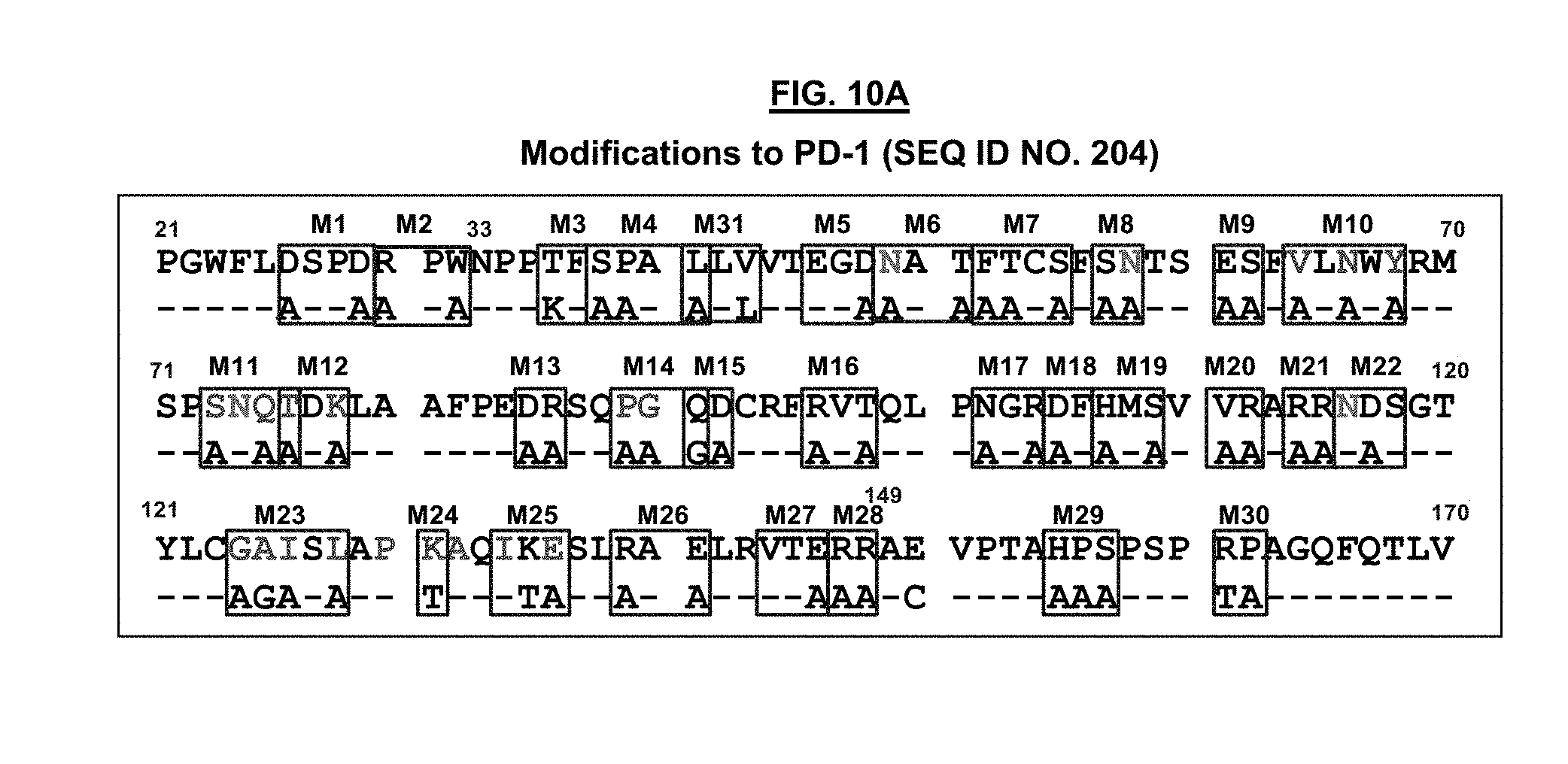
D00014
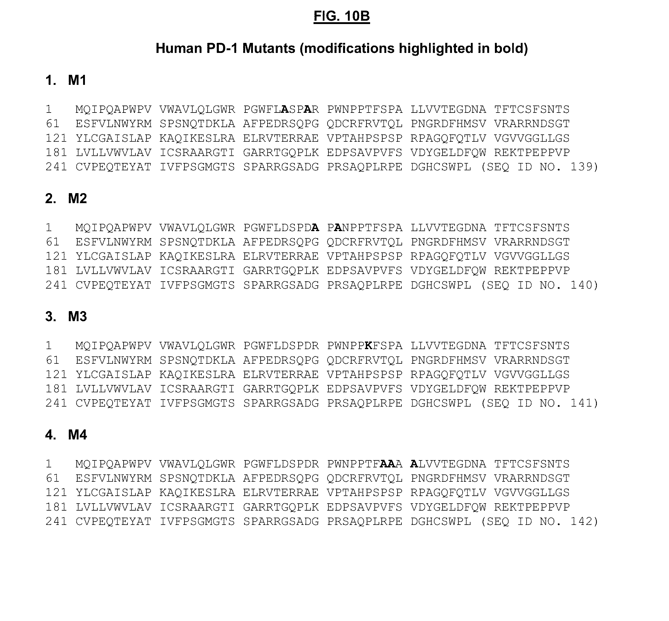
D00015
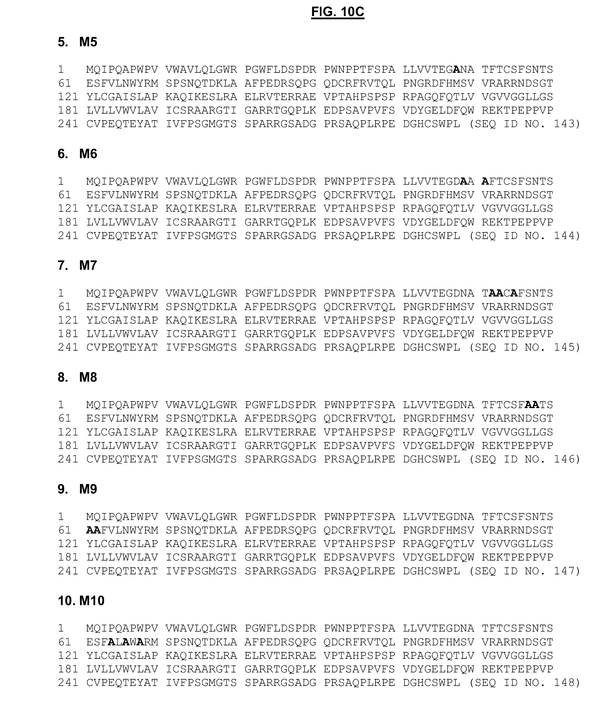
D00016
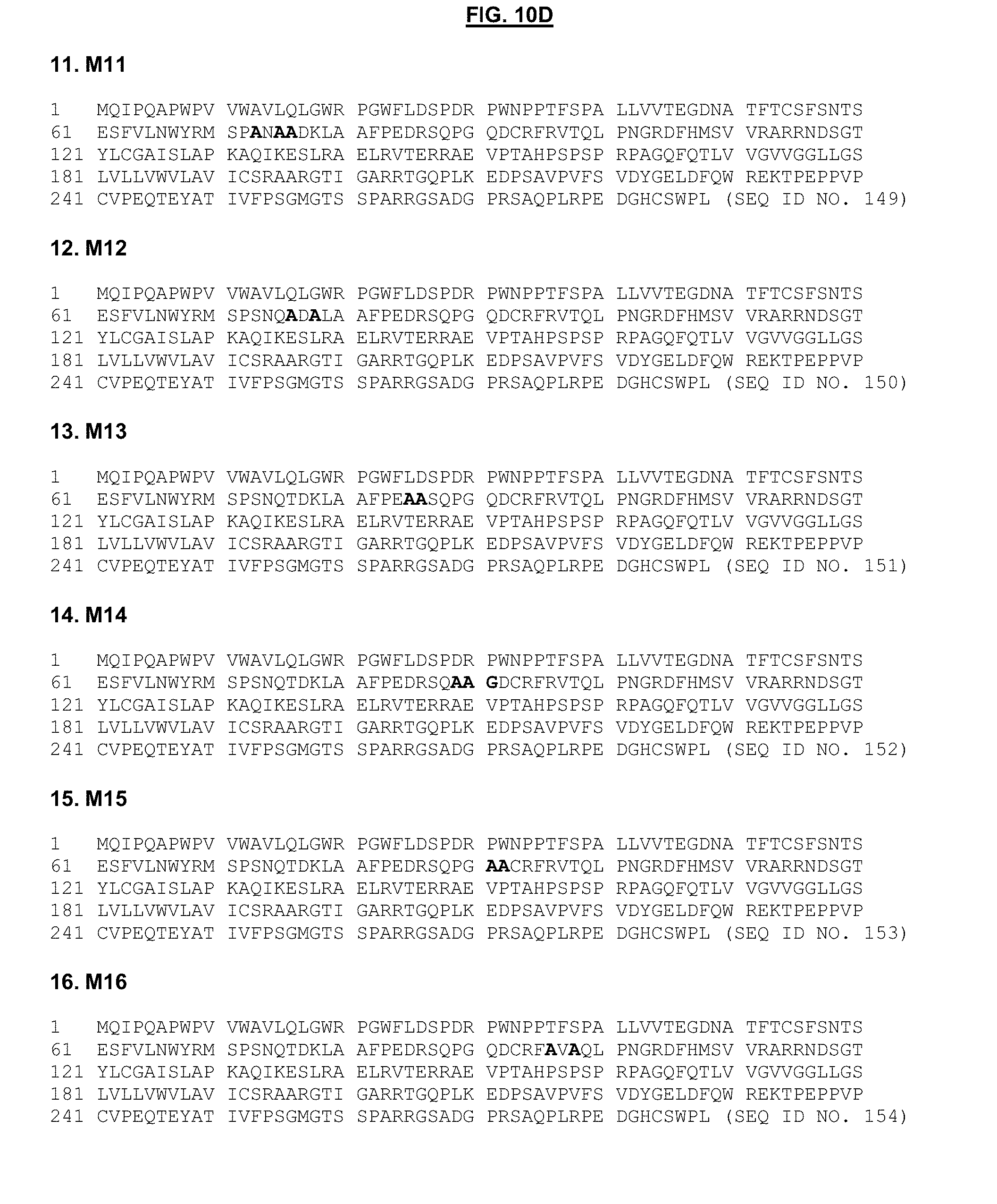
D00017
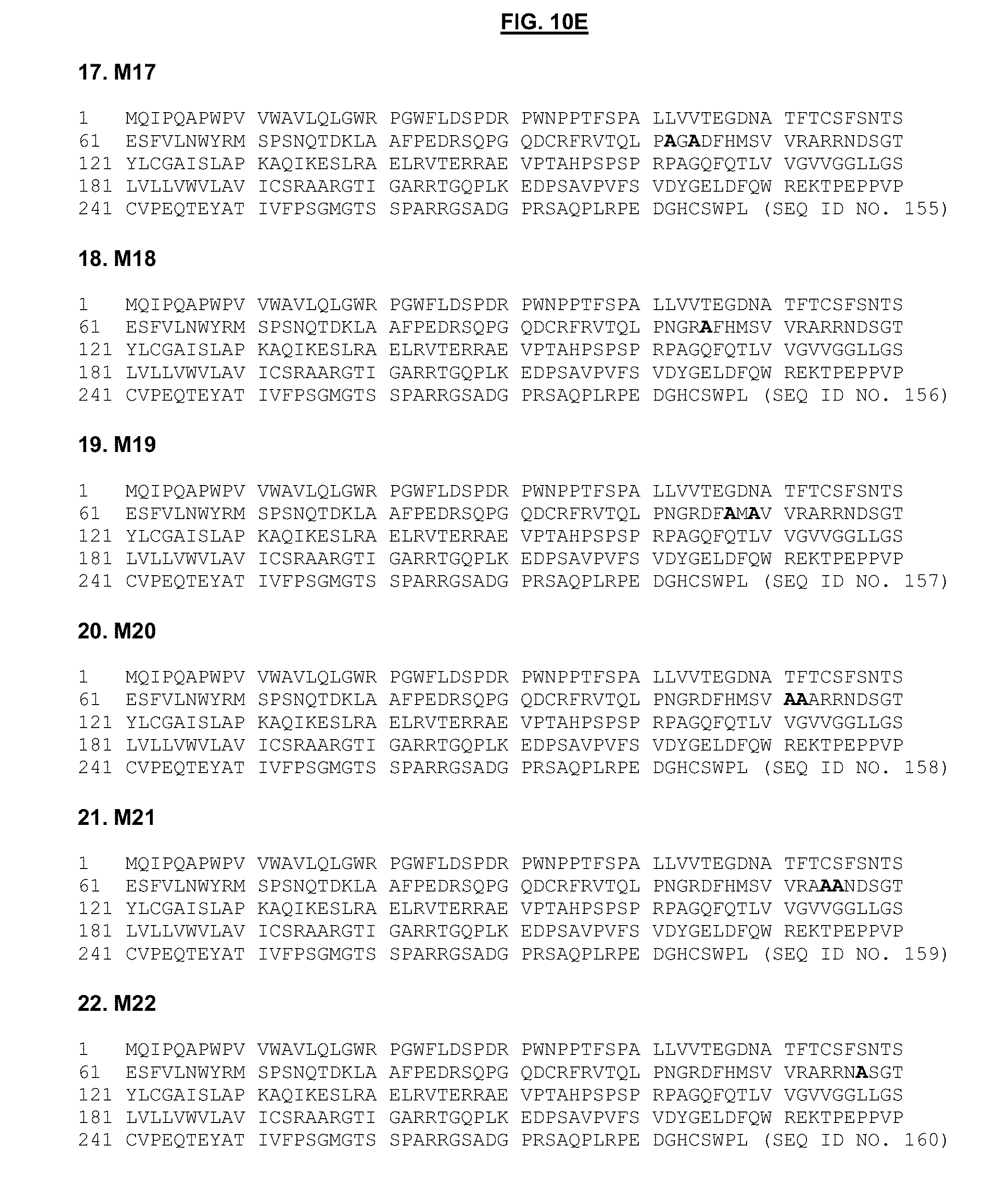
D00018
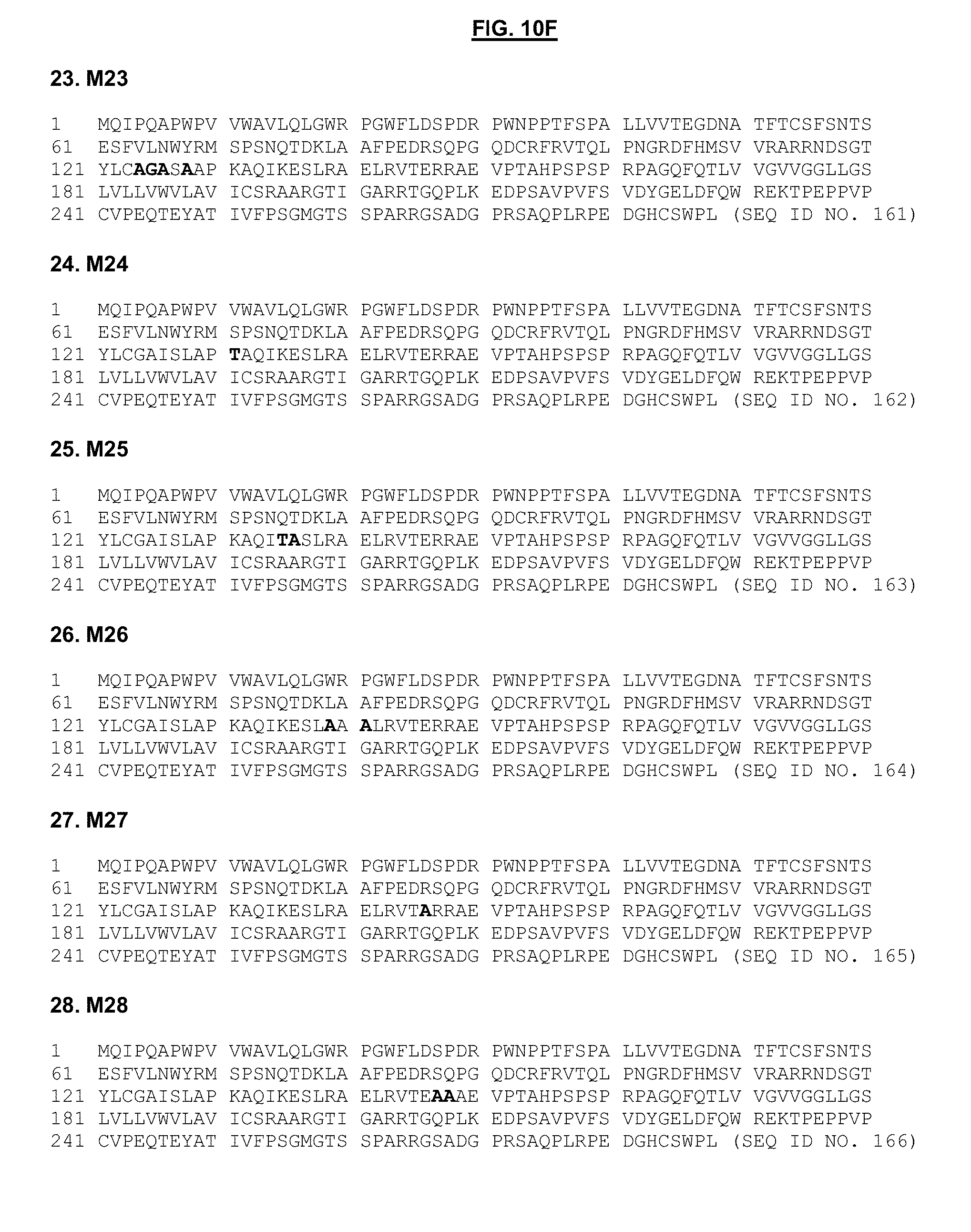
D00019
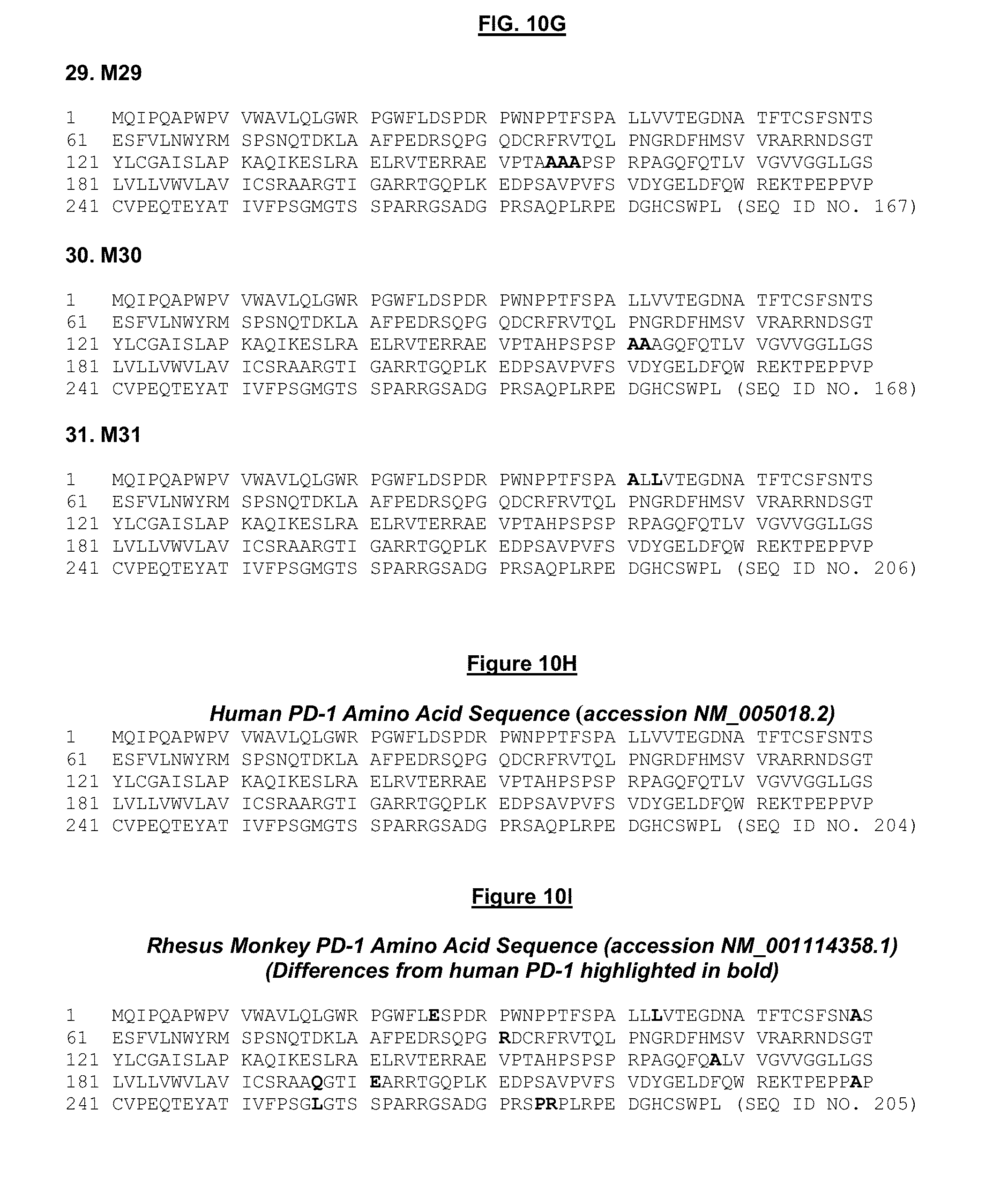
D00020
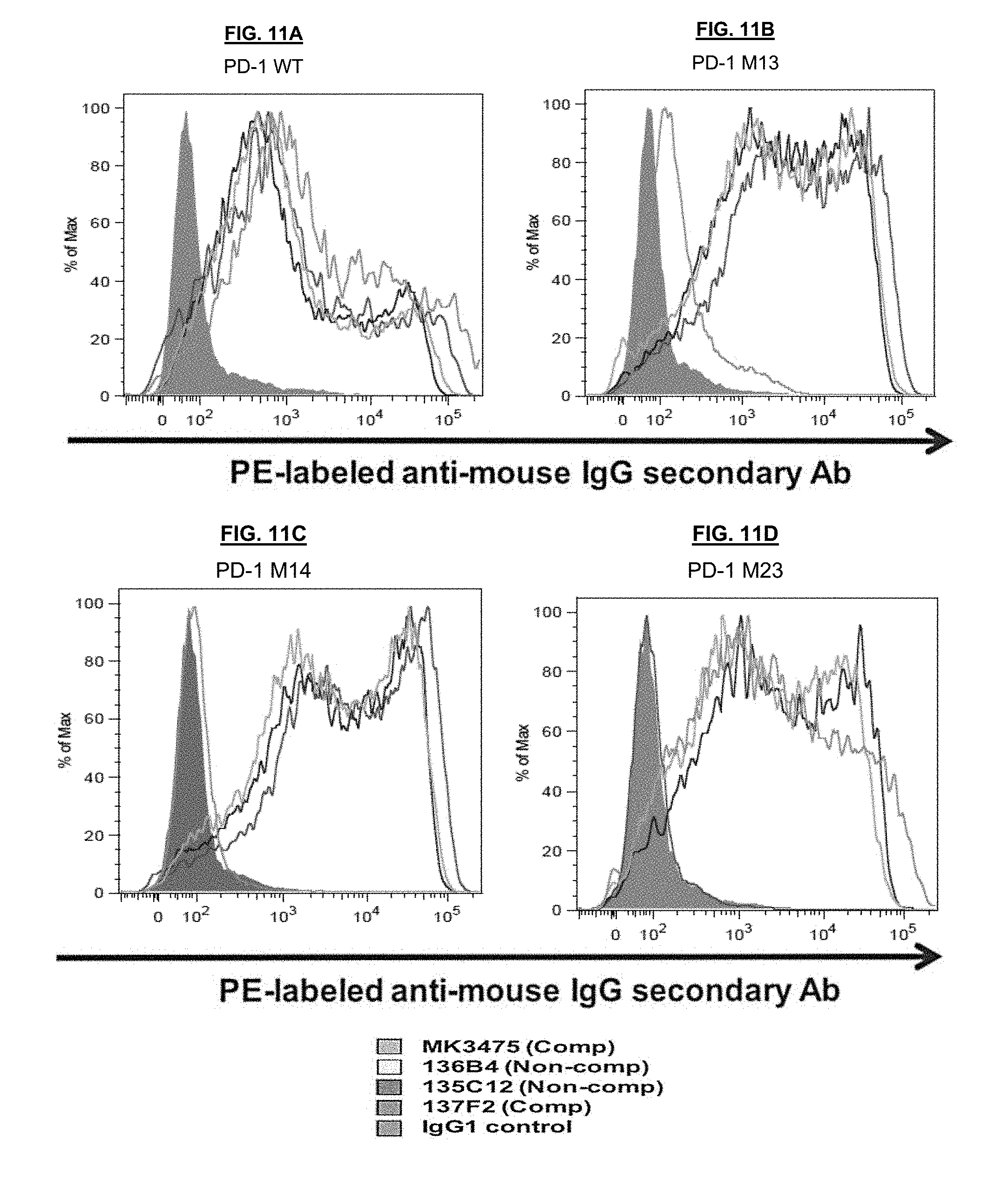
D00021
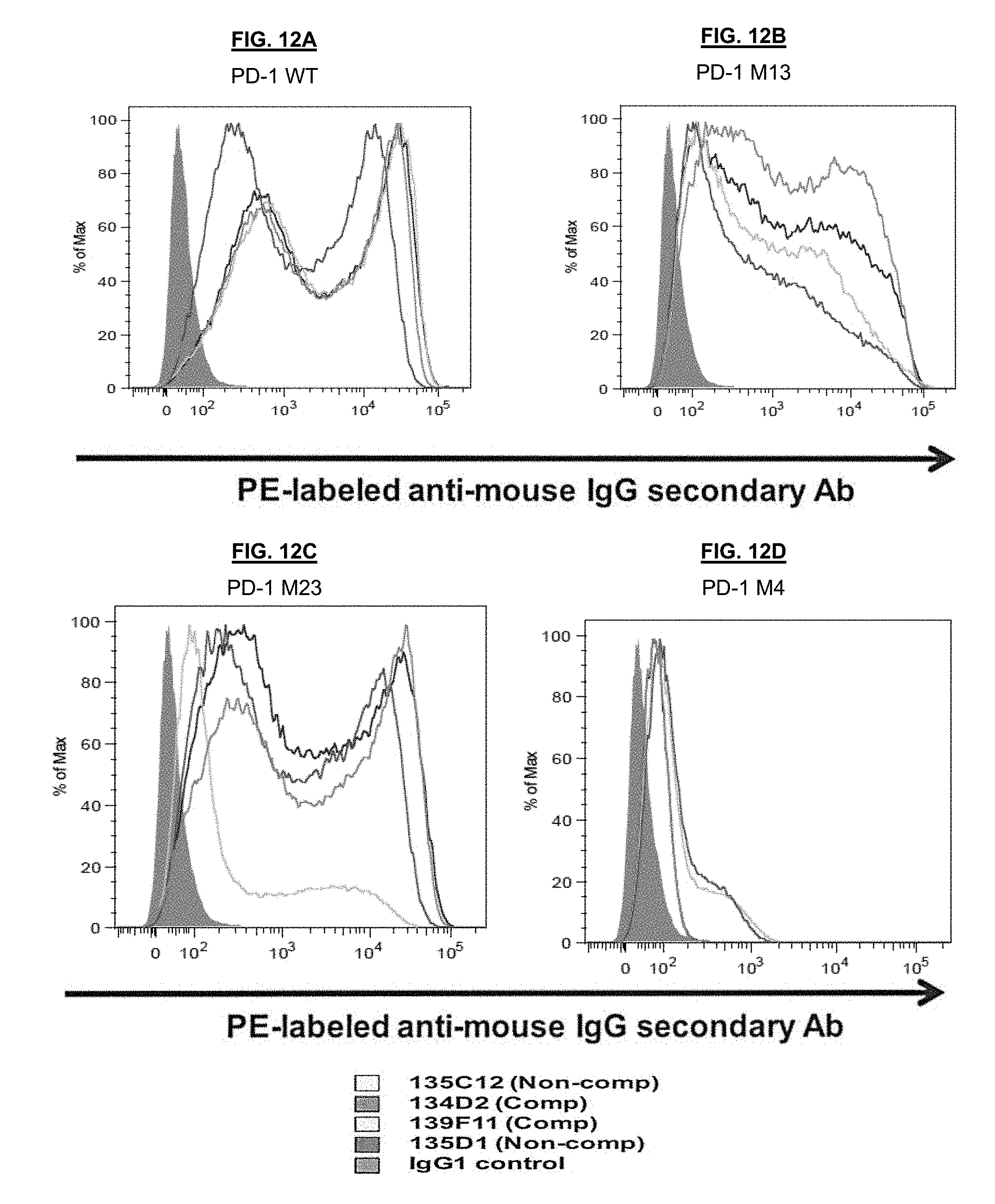
D00022
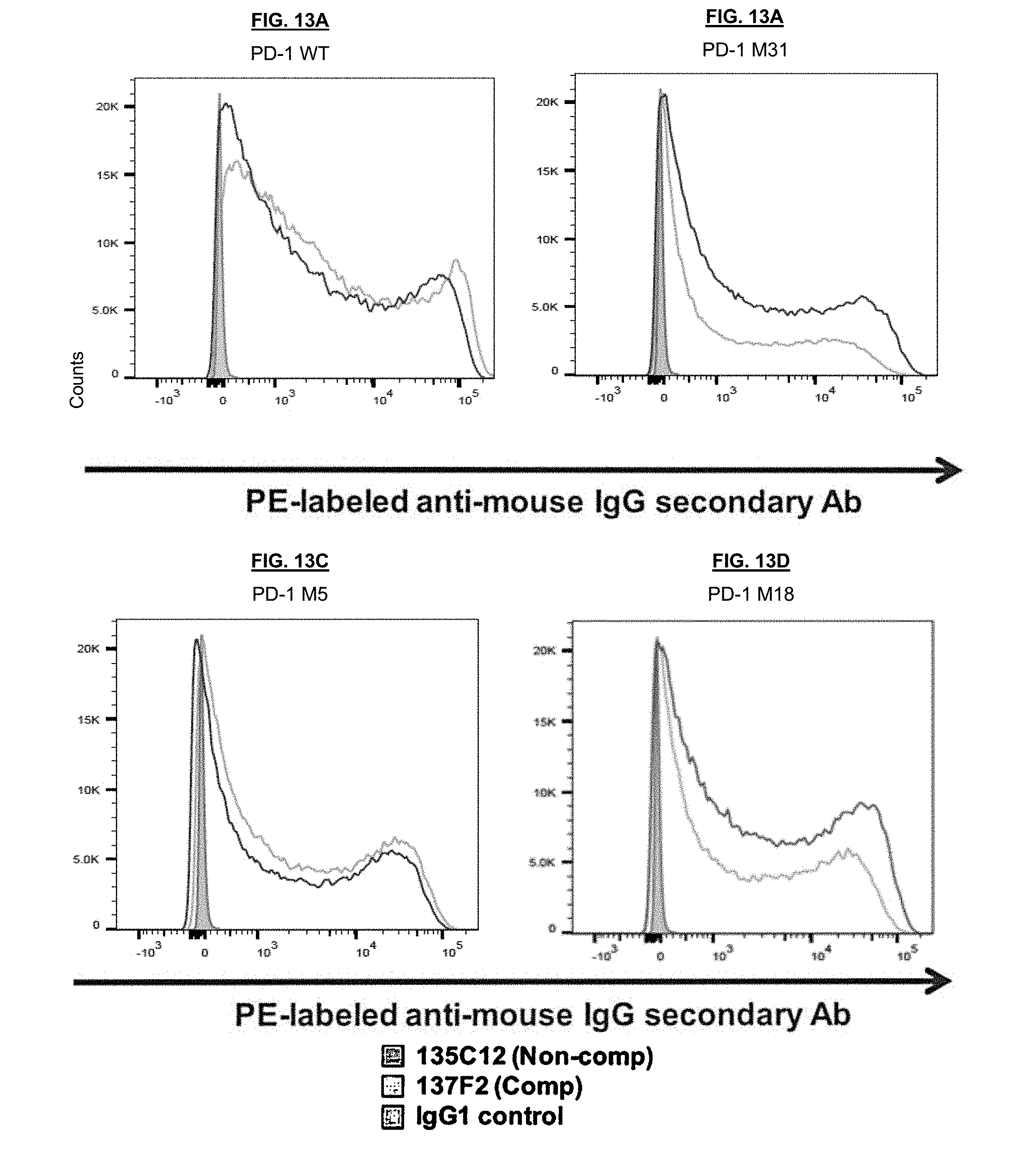
D00023
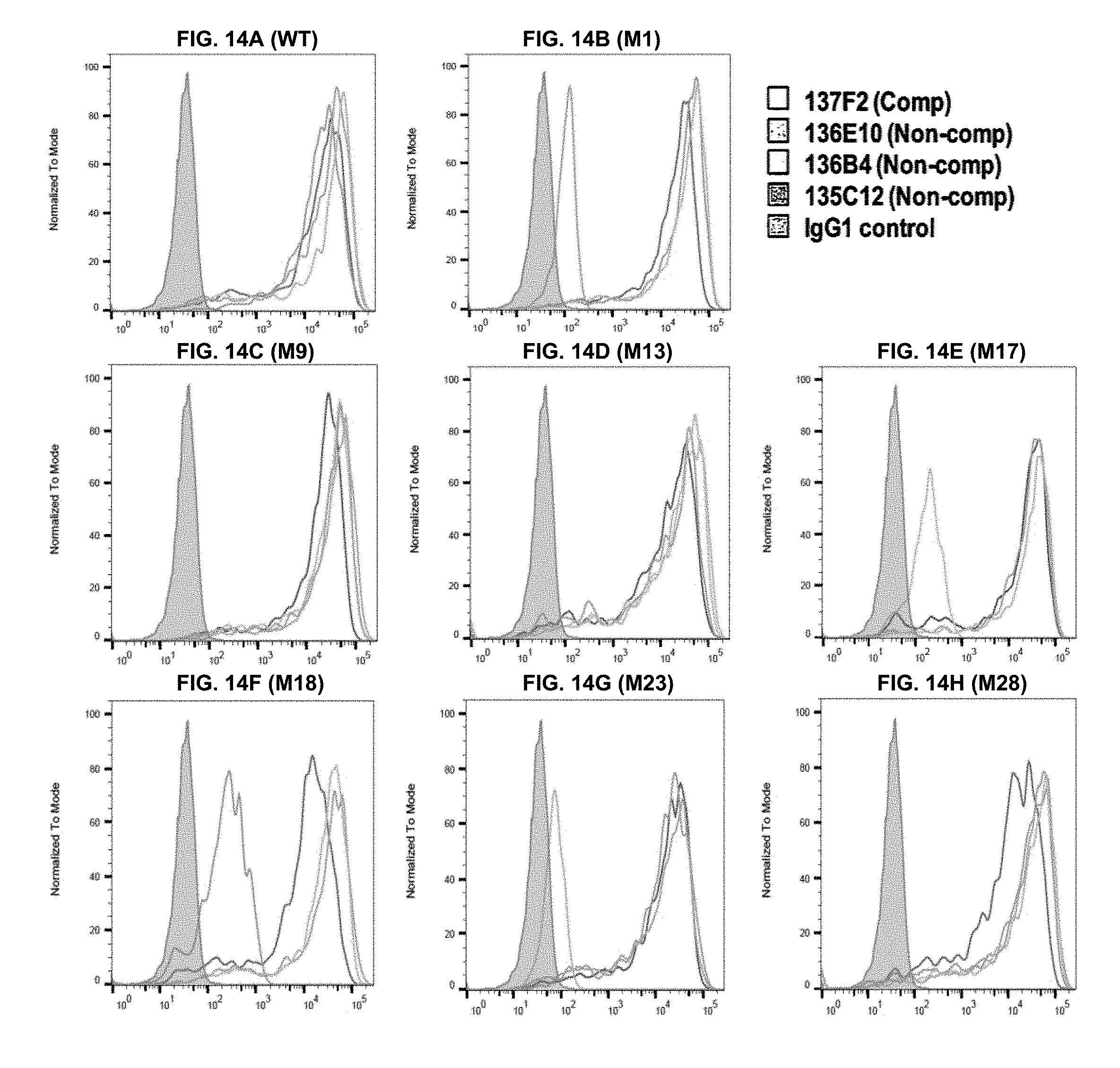
D00024
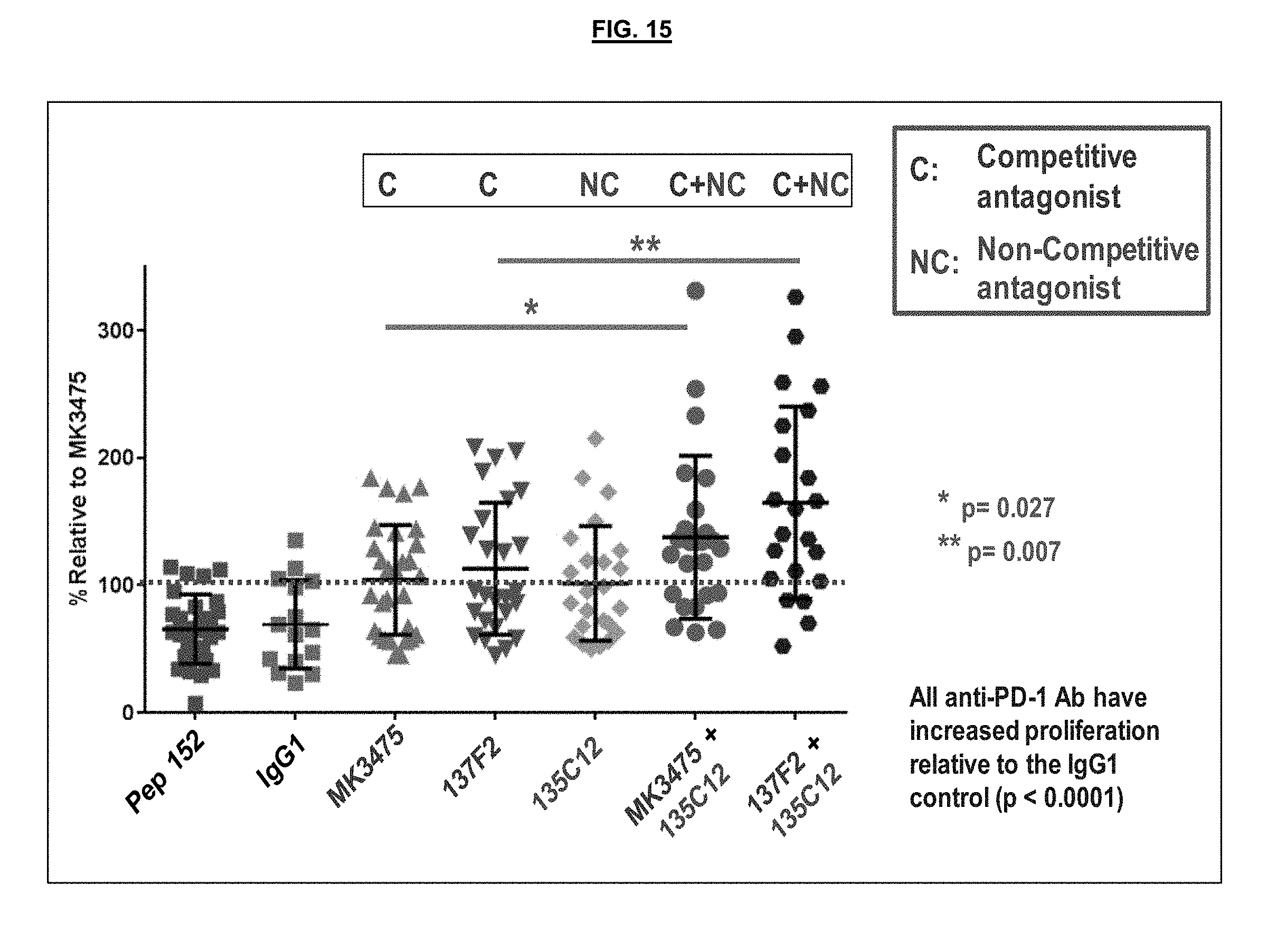
D00025
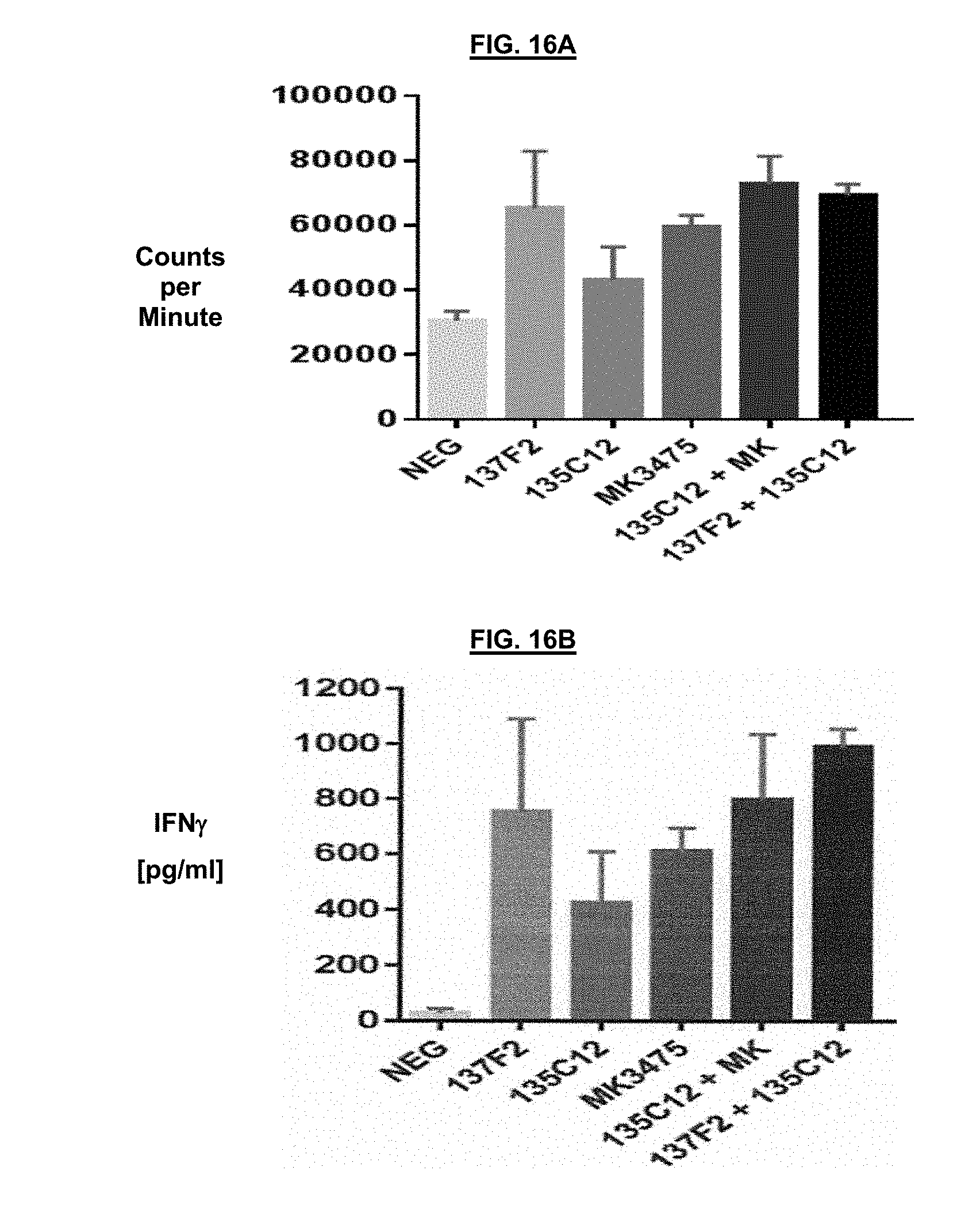
D00026
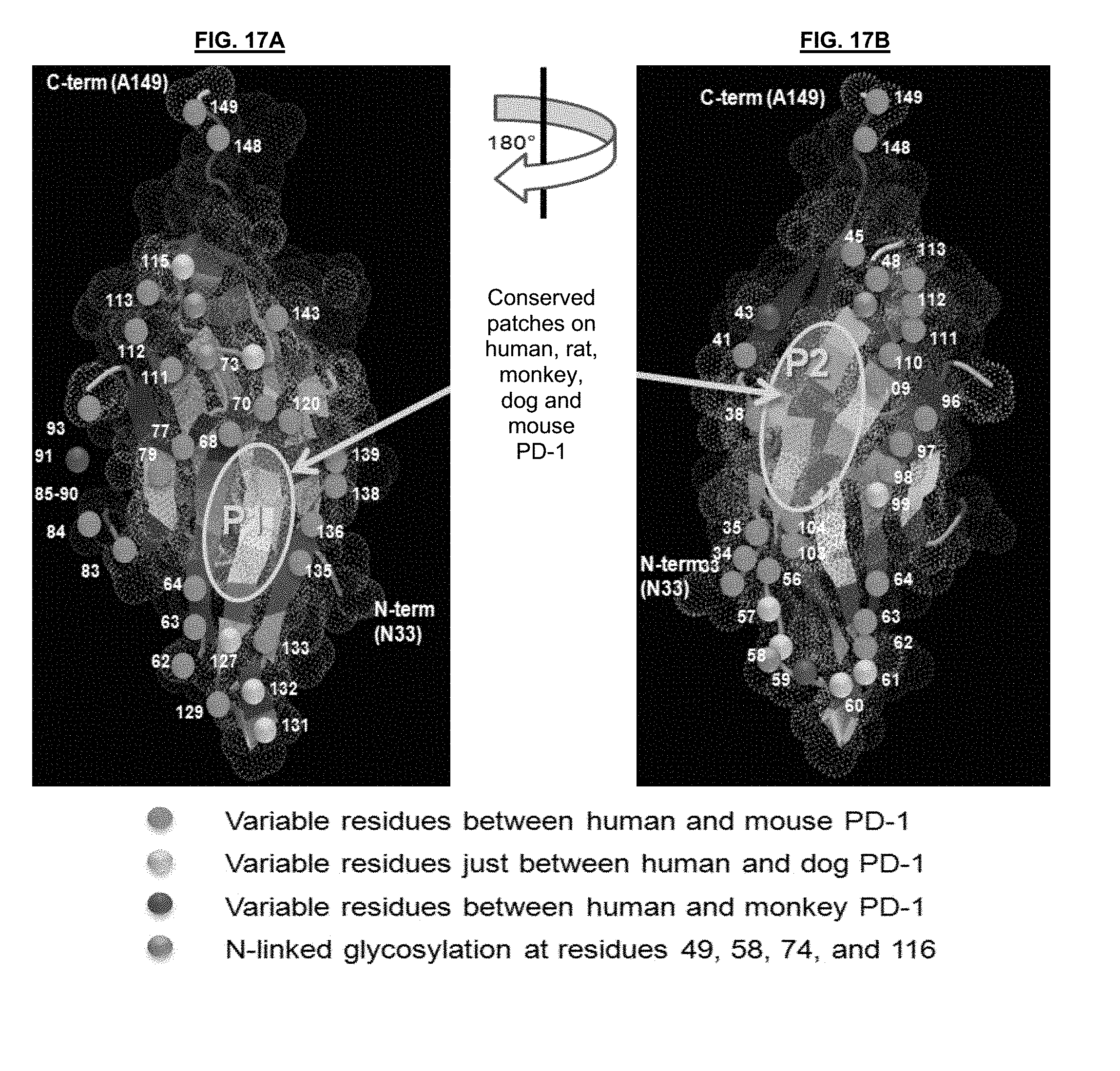
D00027
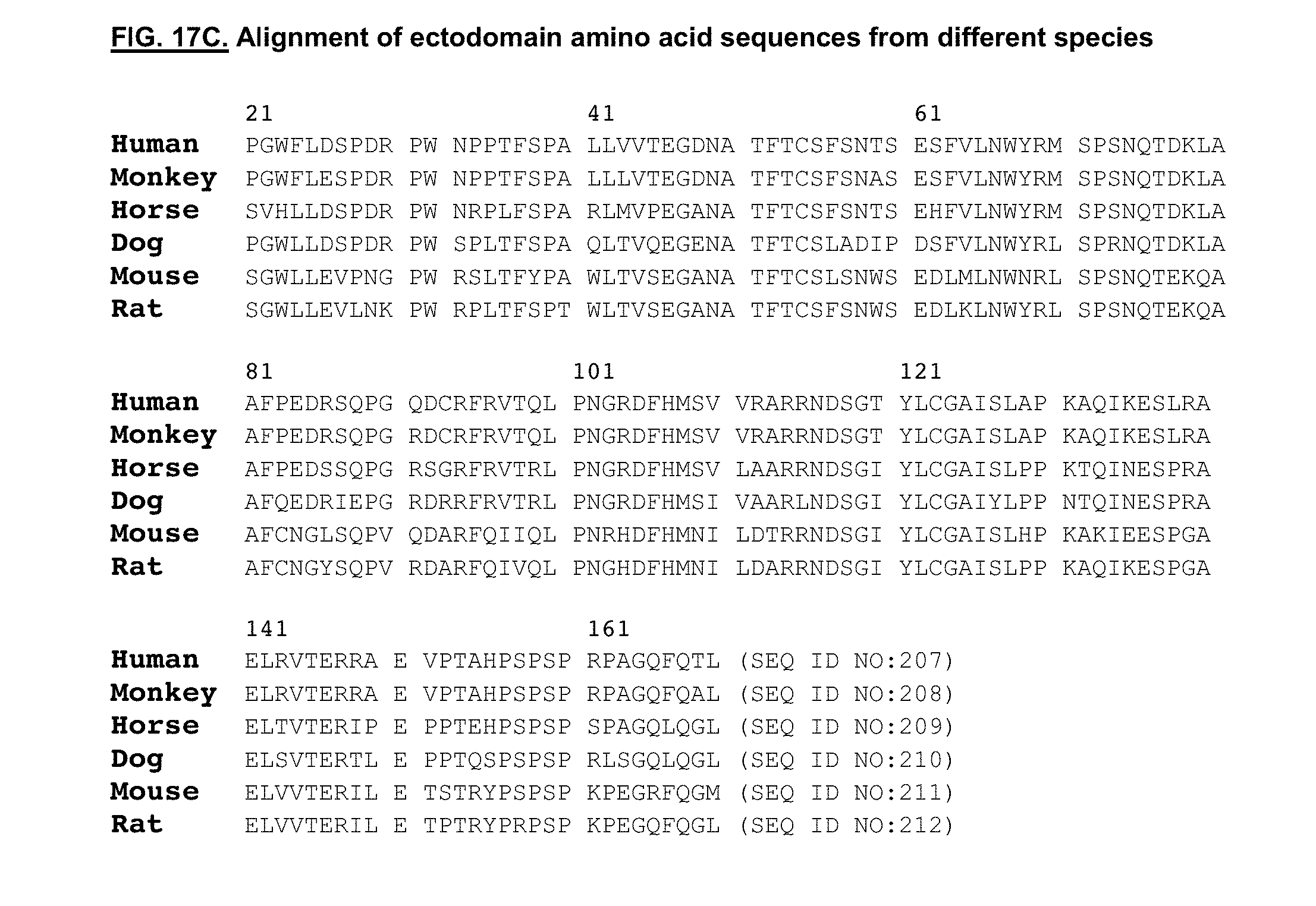
D00028
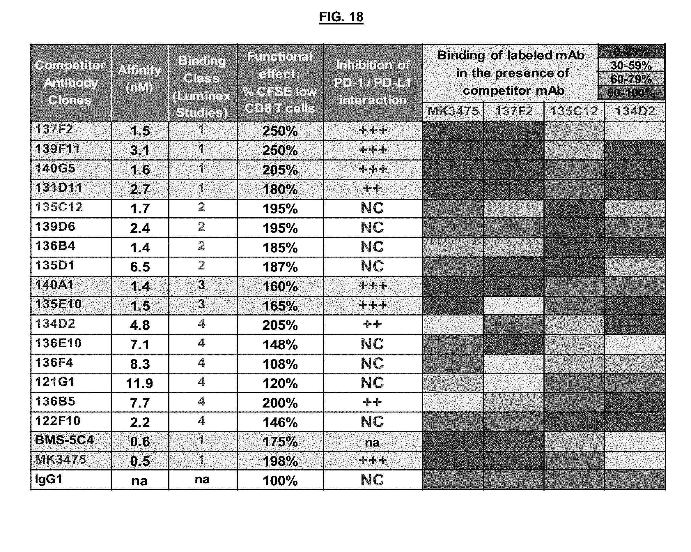
D00029
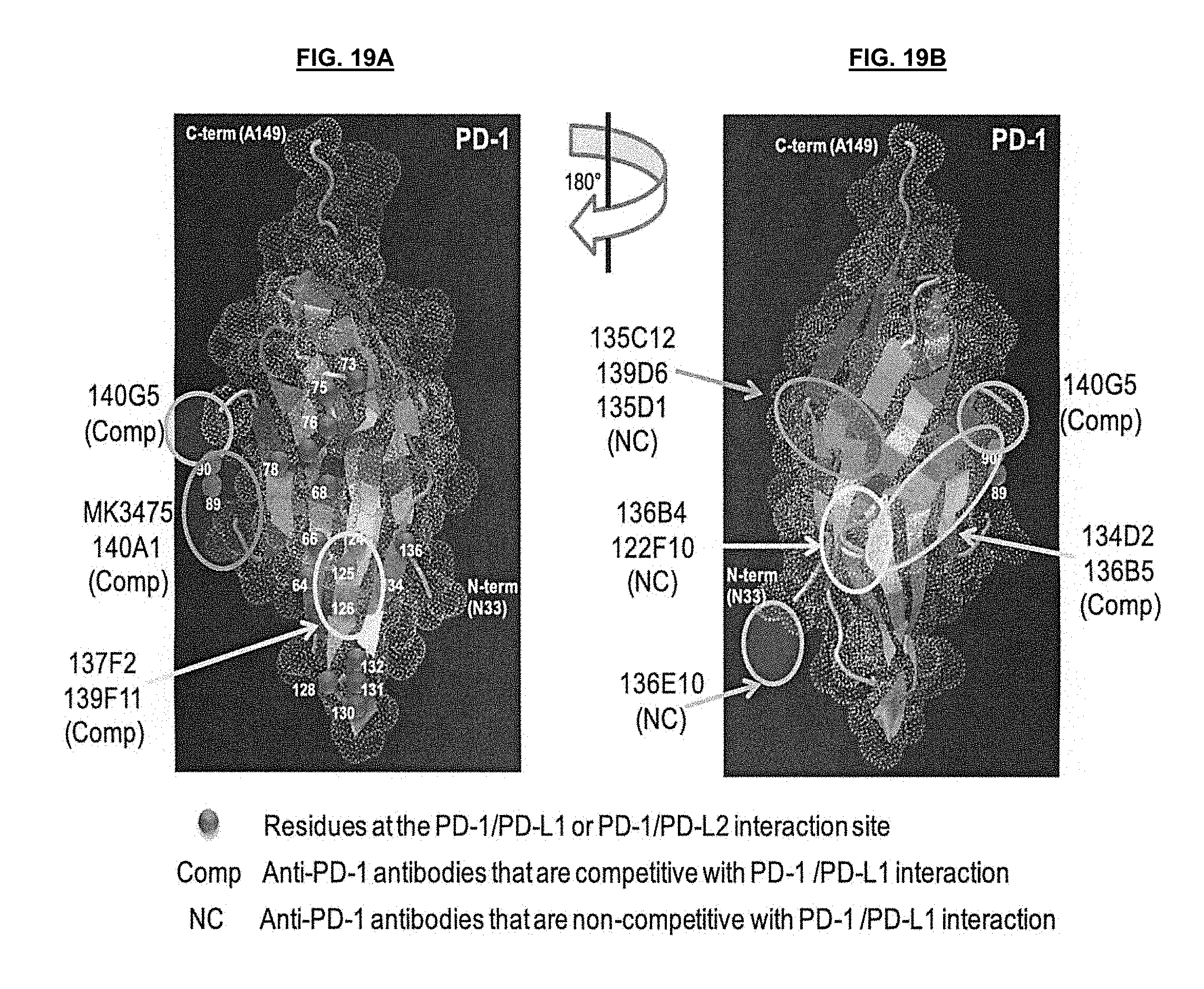
D00030
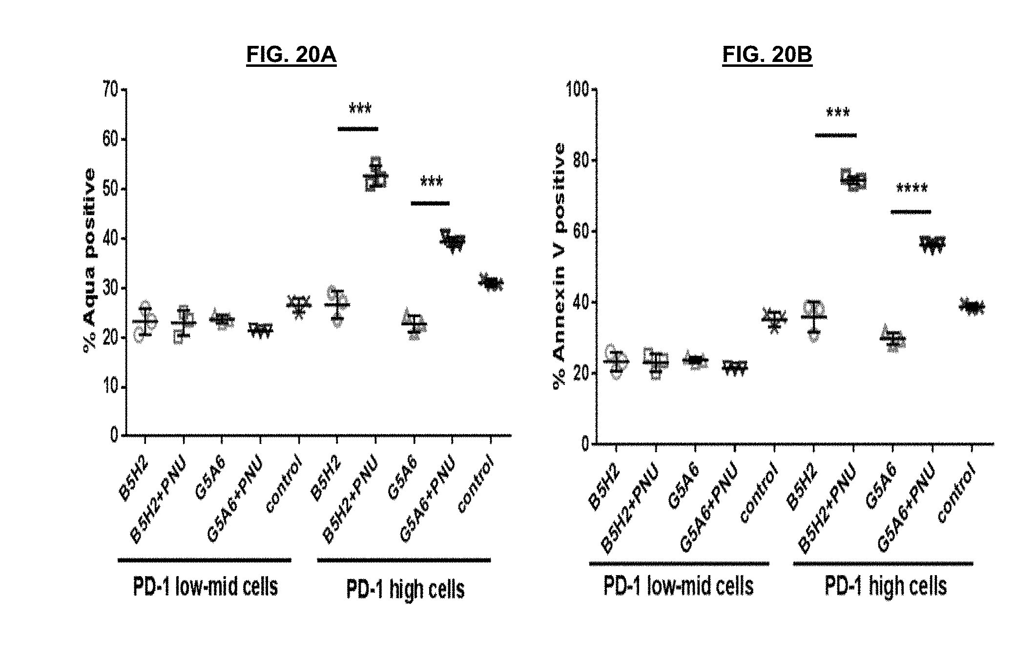
D00031
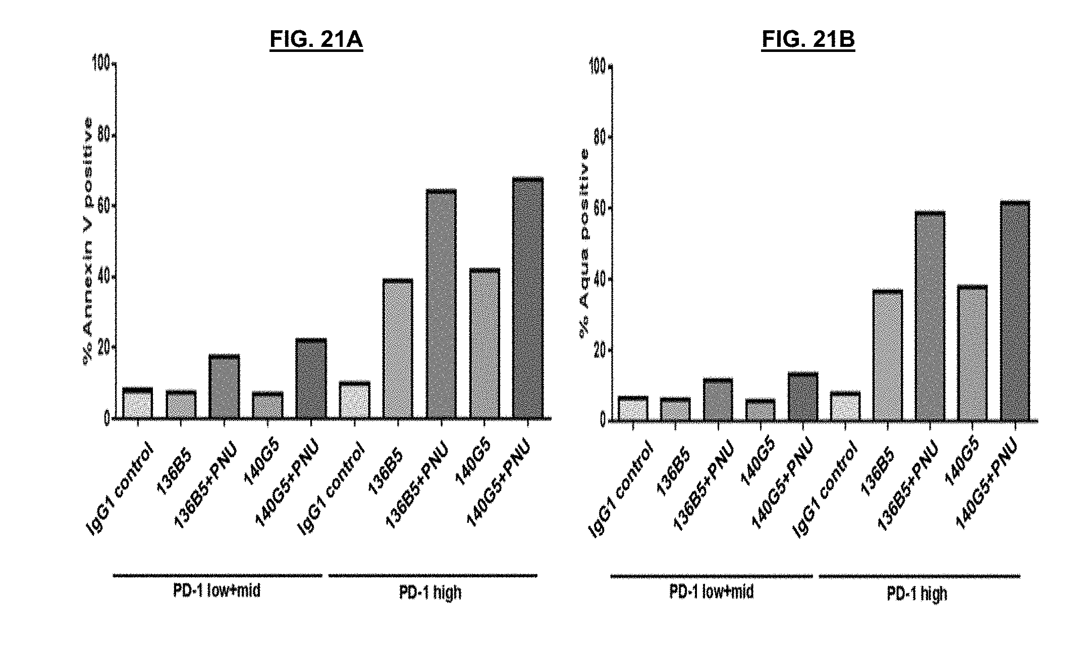
D00032
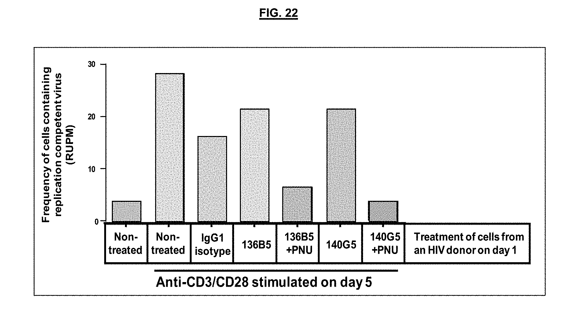
S00001
XML
uspto.report is an independent third-party trademark research tool that is not affiliated, endorsed, or sponsored by the United States Patent and Trademark Office (USPTO) or any other governmental organization. The information provided by uspto.report is based on publicly available data at the time of writing and is intended for informational purposes only.
While we strive to provide accurate and up-to-date information, we do not guarantee the accuracy, completeness, reliability, or suitability of the information displayed on this site. The use of this site is at your own risk. Any reliance you place on such information is therefore strictly at your own risk.
All official trademark data, including owner information, should be verified by visiting the official USPTO website at www.uspto.gov. This site is not intended to replace professional legal advice and should not be used as a substitute for consulting with a legal professional who is knowledgeable about trademark law.