Method For Establishing Eukaryotic Expression Cell Line Of Cd36 Mutant Gene That Encodes Cd36 Deficiency
WU; Guoguang ; et al.
U.S. patent application number 16/188187 was filed with the patent office on 2019-02-28 for method for establishing eukaryotic expression cell line of cd36 mutant gene that encodes cd36 deficiency. The applicant listed for this patent is Nan-Ning Institute of Transfusion Medicine. Invention is credited to Jierun CHEN, Lihong JIANG, Haiyan LI, Lilan LI, Guoguang WU.
| Application Number | 20190062387 16/188187 |
| Document ID | / |
| Family ID | 58180516 |
| Filed Date | 2019-02-28 |


| United States Patent Application | 20190062387 |
| Kind Code | A1 |
| WU; Guoguang ; et al. | February 28, 2019 |
METHOD FOR ESTABLISHING EUKARYOTIC EXPRESSION CELL LINE OF CD36 MUTANT GENE THAT ENCODES CD36 DEFICIENCY
Abstract
A method for establishing eukaryotic expression cell line of CD36 mutant gene that encodes CD36 deficiency, the method including: (1) extracting total RNA from a whole blood sample derived from a CD36-deficient individual, and amplifying a coding sequence (CDS) in CD36 mRNA, to obtain a cDNA sequence fragment of the mutant CD36 gene; (2) splicing and amplifying the mutant CD36 gene and the EGFP fluorescent gene by SOE-PCR (Gene Splicing By Overlap Extension PCR) using four forward and reverse primers, to obtain a mutant gene fragment of MT-CD36-EGFP; (3) constructing and amplifying a MT-CD36-EGFP-pLV4/StripII-HIS10 eukaryotic expression vector including the mutant CD36 gene and the EGFP fluorescent gene; (4) transfecting the MT-CD36-EGFP-pLV4/StripII-HIS10 eukaryotic expression vector into the CHO-K1 cell line by using virus-mediated transfection of eukaryotic cells.
| Inventors: | WU; Guoguang; (Nanning, CN) ; LI; Lilan; (Nanning, CN) ; JIANG; Lihong; (Nanning, CN) ; LI; Haiyan; (Nanning, CN) ; CHEN; Jierun; (Nanning, CN) | ||||||||||
| Applicant: |
|
||||||||||
|---|---|---|---|---|---|---|---|---|---|---|---|
| Family ID: | 58180516 | ||||||||||
| Appl. No.: | 16/188187 | ||||||||||
| Filed: | November 12, 2018 |
Related U.S. Patent Documents
| Application Number | Filing Date | Patent Number | ||
|---|---|---|---|---|
| PCT/CN2017/075785 | Mar 6, 2017 | |||
| 16188187 | ||||
| Current U.S. Class: | 1/1 |
| Current CPC Class: | C12Q 1/6806 20130101; C07K 14/473 20130101; C12N 2740/15043 20130101; C07K 14/70596 20130101; C12Y 605/01001 20130101; C12Q 1/686 20130101; C12N 5/0602 20130101; C12N 9/93 20130101; C12N 2740/16043 20130101; C12N 15/65 20130101; C12Q 1/6876 20130101; C12N 15/85 20130101 |
| International Class: | C07K 14/47 20060101 C07K014/47; C12N 9/00 20060101 C12N009/00; C12N 15/65 20060101 C12N015/65; C12Q 1/6806 20060101 C12Q001/6806; C12Q 1/686 20060101 C12Q001/686; C12Q 1/6876 20060101 C12Q001/6876; C12N 5/071 20060101 C12N005/071; C12N 15/85 20060101 C12N015/85 |
Foreign Application Data
| Date | Code | Application Number |
|---|---|---|
| Nov 1, 2016 | CN | 201610967814.1 |
Claims
1. A method for establishing eukaryotic expression cell line of CD36 mutant gene that encodes CD36 deficiency, the method comprising: (1) extracting total RNA from a whole blood sample derived from a CD36-deficient individual, and amplifying a coding sequence (CDS) in CD36 mRNA by Reverse Transcription PCR (RT-PCR) using a pair of primers, to obtain a cDNA sequence fragment of the mutant CD36 gene, wherein primers for comprise an upstream primer YC-36F having a sequence of SEQ. ID. NO. 1, and a downstream primer YC-36R having a sequence of SEQ. ID. NO. 2; (2) splicing and amplifying the coding sequence (CDS) of the mutant CD36 cDNA (excluding the termination condons) and EGFP fluorescent gene by Gene Splicing by Overlap Extension PCR (SOE-PCR), to obtain a mutant gene fragment of MT-CD36-EGFP comprising a full-length coding sequence (CDS) excluding the terminator of the mutant CD36 cDNA, a full-length EGFP fluorescent gene and desired cleavage sites, wherein primers for SOE-PCR comprises: using a forward primer CD36-F2 and a reverse primer CD36-R3 to amplify a target gene fragment of MT-CD36-EGFP-1 comprising an EcoR 1 cleavage site and protective bases, a full-length coding sequence (CDS) excluding the terminator of CD36 cDNA, a fusion gene BamH I and protective bases, and a portion of the 5'-terminal sequence of the EGFP fluorescent reporter gene, wherein the forward primer CD36-F2 also serves as a forward primer for the subsequent splicing and amplification of MT-CD36-EGFP by SOE-PCR, and has a sequence of SEQ. ID. NO. 3, and the reverse primer CD36-R3 has a sequence of SEQ. ID. NO. 4; and using a forward primer EGFP-F4 and a reverse primer EGFP-R2 to amplify a target gene fragment of MT-CD36-EGFP-2 comprising a portion of the 3'-terminal fragment of the CD36 CDS cDNA, a fusion gene BamH I and protective bases, a full-length of EGFP CDS fluorescent reporter gene, and an XhoI cleavage site and protective bases, wherein the reverse primer EGFP-R2 also serves as a reverse primer for the subsequent splicing and amplification of MT-CD36-EGFP by SOE-PCR, wherein the forward primer EGFP-F4 has a sequence of SEQ. ID. NO. 5, and the reverse primer EGFP-R2 has a sequence of SEQ. ID. NO. 6; (3) constructing and amplifying a MT-CD36-EGFP-pLV4/StripII-HIS10 eukaryotic expression vector by ligating the MT-CD36-EGFP to a pLV4/StripII-HIS 10 vector; the MT-CD36-EGFP-pLV4/StripII-HIS10 eukaryotic expression vector comprising the full-length CDS excluding the terminator of the mutant CD36 cDNA and the EGFP fluorescent gene by ligating MT-CD36-EGFP to a pLV4/StripII-HIS10 vector; (4) transfecting the MT-CD36-EGFP-pLV4/StripII-HIS10 eukaryotic expression vector into the CHO-K1 cell line by using virus-mediated transfection of eukaryotic cells, and screening and constructing a eukaryotic cell line MT-CD36-CHO-K1 stablely expressing the mutant CD36 gene that encodes CD36 deficiency; and (5) repeating aforesaid steps, to establish a eukaryotic cell line Normal-CD36-CHO-K1 stably expressing normal CD36 gene as a positive control, and establishing a EGFP-pLV4-CHO-K1 line stably expressing the EGFP fluorescent protein alone as a negative control, wherein in the construction of the negative control EGFP-pLV4-CHO-K1 line, a target gene fragment encoding EGFP comprising an EcoR 1 cleavage site and protective bases, the full-length gene sequence of the EGFP fluorescent reporter gene, and an XhoI cleavage site and protective bases is obtained by amplification using a pair of PRC primers, wherein the PCR primers comprise an upstream primer EGFP-F3 having a sequence of SEQ. ID. NO. 7, and a downstream primer EGFP-R2 that is a reverse primer of the gene fragment of MT-CD36-EGFP-2.
2. The method of claim 1, wherein the upstream primer YC-36F for the RT-PCR amplification of the coding sequence (CDS) of CD36 cDNA is located in a 5'-promoter region of the CD36 mRNA sequence, and the downstream primer YC-36R is located in a 3'-terminator region of the CD36 mRNA sequence, the amplification region covers the coding sequence (CDS) of CD36 mRNA, and a plurality of mutant CD36 genes with mutations within a linker region of 5'-terminal and 3'-terminal primer binding regions of the template is obtained through amplification; and for the length of the fragment amplified by using the primers for RT-PCR amplification of the mutant CD36 gene, if the target mutant CD36 gene amplified is a gene with point mutations, the length of the amplified fragment is 1432 bp, and if the target mutant CD36 gene amplified is a mutant gene with base insertions or deletions, the length of the amplified fragment is increased or decreased on the basis of 1432 bp by the base number inserted or deleted.
3. The method of claim 1, wherein the cDNA sequence fragment of the mutant CD36 gene and the EGFP fluorescent gene are spliced and amplified by SOE-PCR, to obtain the target gene fragment of MT-CD36-EGFP comprising the full-length CDS excluding the terminator of the mutant CD36 cDNA, the full-length EGFP fluorescent gene and desired cleavage sites, wherein (1) during the SOE-PCR, the forward primer CD36-F2 for obtaining the target gene fragment of MT-CD36-EGFP-1 is located in a 5'-promoter region of the cDNA sequence fragment of the mutant CD3 6 gene; the target gene fragment of MT-CD36-EGFP-1 comprises an EcoR 1 cleavage site and protective bases, a full-length CDS excluding the terminator of the CD36 cDNA, a fusion gene BamH I and protective bases, and a portion of the 5'-terminal sequence of the EGFP fluorescent reporter gene, the EcoR 1 cleavage site and protective bases are comprised at a 5' terminus of the primer, and the forward primer CD36-F2 also serves as the forward primer for the subsequent splicing and amplification of MT-CD36-EGFP by SOE-PCR; the reverse primer CD36-R3 is located in a 3'-terminal region excluding the terminator of the CD36 cDNA sequence fragment of the mutant CD36 gene, and the portion of the 5'-terminal promoter region sequence of the EGFP fluorescent reporter gene and the fusion gene BamH I and protective bases are comprised at the 5' terminus of the reverse primer CD36-R3; and for the length of the target gene fragment of MT-CD36-EGFP-1, when the mutant CD36 gene comprise point mutations, the length of the target gene fragment of MT-CD36-EGFP-1 is 1441 bp, and when the mutant CD36 gene comprise base insertions or deletions, the length of the target gene fragment of MT-CD36-EGFP-1 is increased or decreased on the basis of 1441 bp by the base number inserted or deleted; (2) during the SOE-PCR, the forward primer EGFP-F4 for obtaining the target gene fragment of MT-CD36-EGFP-2 is located in a 5'-promoter region of the sequence of the EGFP fluorescent reporter gene; the target gene fragment of MT-CD36-EGFP-2 comprises a portion of the 3'-terminal fragment of the CD36 coding cDNA region, a fusion gene BamH I and protective bases, a full-length EGFP fluorescent reporter gene, and an XhoI cleavage site and protective bases, a 3'-terminal 10-base sequence before the terminator TAA of the CD36 cDNA CDS sequence and the fusion gene BamH I and protection bases are comprised at the 5' terminus of the forward primer EGFP-F4; the reverse primer EGFP-R2 is located in the 3'-terminator region of the EGFP fluorescent gene, and the XhoI cleavage site and protective bases are comprised at a 5' terminus of the reverse primer EGFP-R2; and the length of the target gene fragment of MT-CD36-EGFP-2 is 753 bp; and (3) by using the forward primer CD36-F2 and the reverse primer EGFP-R2, and using MT-CD36-EGFP-1 and MT-CD36-EGFP-2 obtained through PCR amplification as templates, the gene fragment of MT-CD36-EGFP is obtained by splicing and amplification by SOE-PCR; the fragment of MT-CD36-EGFP comprises the full-length CDS excluding the terminator of the mutant CD36 gene, a fusion gene BamH I and protective bases the full-length fragment CDS of the EGFP fluorescent gene, and desired EcoR1 and XhoI cleavage sites and for the length of MT-CD36-EGFP, when the mutant CD36 gene comprise point mutations, the length of the amplified fragment of MT-CD36-EGFP is 2165 bp, and when the mutant CD36 gene comprise base insertions or deletions, the length of the amplified fragment of MT-CD36-EGFP is increased or decreased on the basis of 2165 bp by the base number inserted or deleted.
4. The method of claim 1, wherein during the construction of the negative control EGFP-pLV4-CHO-K1 line, the upstream primer EGFP-F3 for PCR amplification to obtain the target gene fragment encoding EGFP is located in a 5'-promoter region of the sequence of the EGFP fluorescent gene; the target gene fragment encoding EGFP comprises a EcoR 1 cleavage site and protective bases, the full-length gene sequence of the CDS of the EGFP fluorescent reporter gene, and a XhoI cleavage site and protective bases, and the EcoR 1 cleavage site and protective bases are further comprised at a 5' terminus of the upstream primer EGFP-F3; the downstream primer EGFP-R2 are also the reverse primer for amplification of the gene fragment of MT-CD36-EGFP-2; and the length of the target gene fragment encoding EGFP by PCR is 743 bp.
5. The method of claim 2, wherein the RT-PCR amplification using one pair of primers for RT-PCR is carried out in a PCR machine using the TaKaRa One step RNA PCR Kit (AMV), through a process which is performed under the following cycling parameters: TABLE-US-00031 50.degree. C. 30 min 94.degree. C. 2 min; 30 amplification cycles of 94.degree. C. 30 sec 60.degree. C. 30 sec 72.degree. C. 2 min; and extension and storage at 72.degree. C. 10 min 12.degree. C. .infin..
6. The method of claim 3, wherein the reaction system for PCR amplification of the target fragment of MT-CD36-EGFP-1 has a composition of TABLE-US-00032 ddH.sub.2O 15 .mu.L 5x SF Buffer avalaible from Vazyme, 5 .mu.L with 10 mM MgCl.sub.2 dNTP mix, 10 mM each 0.5 .mu.L CD36-F2, 10 .mu.M 1 .mu.L CD36-R3, 10 .mu.M 1 .mu.L MT-CD36 cDNA 1 .mu.L DMSO 1 .mu.L HS suffer-Fieviy DNA polymerase 0.5 .mu.L; from Vazyme
the reaction system for PCR amplification of the target fragment of MT-CD36-EGFP-2 has a composition of TABLE-US-00033 ddH.sub.2O 16 .mu.L 5x SF Buffer from Vazyme, with 10 mM MgCl.sub.2 5 .mu.L dNTP mix, 10 mM each 0.5 .mu.L EGFP-F4, 10 .mu.M 0.5 .mu.L EGFP-R2, 10 .mu.M 0.5 .mu.L pEGFP-N1 plasmid 1 .mu.L DMSO 1 .mu.L HS suffer-Fieviy DNA polymerase from Vazyme 0.5 .mu.L.
7. The method of claim 6, wherein the PCR amplification using two pairs of PCR primers is carried out in a PCR machine through a process which is performed under the following cycling parameters: TABLE-US-00034 96.degree. C. 5 min 33 amplification cycles of: 96.degree. C. 30 sec 60.degree. C. 30 sec 72.degree. C. 90 sec; and extension and storage at 72.degree. C. 10 min 12.degree. C. .infin..
8. The method of claim 3, wherein the reaction system for PCR amplification to obtain the target fragment of MT-CD36-EGFP by SOE-PCR has a composition of TABLE-US-00035 H.sub.2O 24 .mu.L phanta .TM. 5x SF Buffer, with 10 mM Mgcl2 10 .mu.L dNTP Mix, 10 mM each 1 .mu.L MT-CD36-EGFP-1 4 .mu.L MT-CD36-EGFP-2 4 .mu.L phanta .TM. HS Suffer-Fideliy DNA Polymerase 1 .mu.L;
the amplification is carried out in a PCR machine through a process which is performed under the following cycling parameters: TABLE-US-00036 98.degree. C. 5 min; and 15 amplification cycles of 98.degree. C. 5 min 72.degree. C. 1 min
a system having the following composition is added to the reaction tube after the reaction is completed: TABLE-US-00037 CD36-F2, 10 .mu.M 2 .mu.L EGFP-R2, 10 .mu.M 2 .mu.L DMSO 2 .mu.L;
and amplification is continued through a process which is performed under the following cycling parameters: TABLE-US-00038 20 amplification cycles of 98.degree. C. 10 sec 56.degree. C. 30 sec 72.degree. C. 60 sec; and extension and storage at: 72.degree. C. 10 min 12.degree. C. .infin..
9. The method of claim 4, wherein the reaction system for PCR amplification to obtain the target fragment of the EGFP gene comprising a EcoR 1 cleavage site and protective bases, the full-length CDS of EFGP flurescent gene, and a XhoI cleavage site and protective bases has the same composition as that of the reaction system for PCR amplification to obtain the target gene fragment of MT-CD36-EGFP-2, except that the upstream primer is EGFP-F3; and the cycling parameters for amplification in a PCR machine under the following cycling parameters: TABLE-US-00039 96.degree. C. 5 min 33 amplification cycles of: 96.degree. C. 30 sec 60.degree. C. 30 sec 72.degree. C. 90 sec; and extension and storage at 72.degree. C. 10 min 12.degree. C. .infin..
10. The method of claim 1, wherein a process for constructing and amplifying the MT-CD36-EGFP-pLV4/StripII-HIS10 eukaryotic expression vector by ligating MT-CD36-EGFP to the pLV4/StripII-HIS10 vector comprises: (1) ligating the enzymatically cleaved products MT-CD36-EGFP and pLV4/StripII-HIS10 vector by using the T4 DNA Ligase Kit (Promega), of that the enzymatically cleaved products have been cleaved by using the EcoR1/XhoI DNA Endonuclease Kit (Beyotime Biotechnology) and than have been purified, to obtain the pLV4/StripII-HIS10 expression vector MT-CD36-EGFP-pLV4/StripII-HIS 10 comprising the target fragment of the mutant CD36 gene and the EGFP fluorescent gene, wherein the reaction system for ligation comprises: TABLE-US-00040 MT-CD36-EGFP:eukaryotic vector pLV4/StripII-HIS10 molar ratio 7:1, total volume: 1-8 .mu.L T4 DAN ligase 10x Buffer 1 .mu.L T4 DAN ligase 1 .mu.L H.sub.2O q.s. to 10 .mu.L;
and (2) transforming the ligated product MT-CD3 6-EGFP-pLV4/StripII-HIS10 into DH5.alpha. Chemically Competent Cells (Beijing TransGen Biotech, Inc) under the temperature conditions comprising: TABLE-US-00041 ice bath 30 min; heat shock at 42.degree. C. 1 min; and ice bath 2 min.
11. The method of claim 10, wherein the colony of the DH5.alpha. Chemically Competent Cells successfully transformed with the MT-CD36-EGFP-pLV4/StripII-HIS10 is confirmed by colony PCR by amplifying the gene fragment of MT-CD36-EGFP in the MT-CD36-EGFP-pLV4/StripII-HIS10 plasmid in the transfected colony using the upstream primer CD36-F2 and the downstream primer EGFP-R2 for SOE-PCR, wherein the colony PCR is carried out using 2.times.Taq Master Mix Kit (Vazyme), and the colony is suspended and lyzed by incubating for 20 min at 100.degree. C. before the colony PCR, where the reaction system for colony PCR comprises: TABLE-US-00042 H.sub.2O 0.5 .mu.L 2 .times. Taq Master Mix 12.5 .mu.L upstream primer CD36-F2, 10 .mu.M 1 .mu.L downstream primer EGFP-R2, 10 .mu.M 1 .mu.L bacterial suspension 10 .mu.L.
12. The method of claim 11, wherein the colony PCR using one pair of primers is carried out in a PCR machine through a process which is performed under the following cycling parameters: TABLE-US-00043 98.degree. C. 5 min 33 amplification cycles of: 98.degree. C. 10 sec 56.degree. C. 30 sec 72.degree. C. 90 sec; and extension and storage at: 72.degree. C. 10 min 12.degree. C. .infin..
13. The method of claim 1, wherein transfecting the MT-CD36-EGFP-pLV4/StripII-HIS10 eukaryotic expression vector into the CHO-K1 cell line by using virus-mediated transfection of eukaryotic cells, and screening and constructing a eukaryotic cell line MT-CD36-CHO-K1 stablely expressing the mutant CD36 gene comprises: (1) subjecting the 293-T cell line in logarithmic growth phase to lentiviral packaging by using the Lenti-Pac.TM. HIV Expression packaging Kit (GeneCopoeia) to obtain a virus suspension, wherein 2 hrs before the 293-T cell line is transfected by lentiviral packaging, the cell culture medium of the 293-T cell line is changed to a serum-free medium; and (2) infecting the CHO-K1 cell line with the virus suspension obtained after lentiviral packaging, and screening the CHO-K1 cell line infected with the virus suspension using puromycin (Sigma), to establish a eukaryotic cell line MT-CD36-CHO-K1 stablely expressing the mutant CD36 gene, wherein the concentration of puromycin used for screening is 0.2 .mu.g/mL.
Description
CROSS-REFERENCE TO RELATED APPLICATIONS
[0001] This application is a continuation-in-part of International Patent Application No. PCT/CN2017/075785 with an international filing date of Mar. 6, 2017, designating the United States, now pending, and further claims foreign priority benefits to Chinese Patent Application No. 201610967814.1 filed Nov. 1, 2016. The contents of all of the aforementioned applications, including any intervening amendments thereto, are incorporated herein by reference. Inquiries from the public to applicants or assignees concerning this document or the related applications should be directed to: Matthias Scholl P. C., Attn.: Dr. Matthias Scholl Esq., 245 First Street, 18th Floor, Cambridge, Mass. 02142.
BACKGROUND OF THE INVENTION
Field of the Invention
[0002] The disclosure relates to biomedicine, and, in particular, to a method for establishing eukaryotic expression cell line of CD36 mutant gene that encodes CD36 deficiency.
Description of the Related Art
[0003] CD36 (Leukocyte Differentiation Antigen 36), also known as Platelet Glycoprotein IV (GPIV), GP88, GPIIIb, FAT or SCARB3, belongs to the Class B scavenger receptor family of transmembrane glycoproteins. The human CD36 protein consists of 472 amino acids, and mainly functions to act as a thrombin receptor, a collagen receptor, a long-chain fatty acid receptor, a receptor for selective cholesteryl ester uptake, a lysosome membrane protein II receptor, etc.; and it is also involved in inducing apoptosis, removing oxidized low-density lipoproteins in the plasma, and enhancing cytoadherence of abnormal shaped red blood cells, etc. CD36 plays an important role in a variety of pathophysiological processes by binding to various ligands and interacting with many ligands, as well as by serving as adhesion molecules on the cell surface. Numerous studies have demonstrated that CD36 is involved in the development of hemostasis, thrombosis, alloimmune platelet disorders, hypercholesterolemia, obesity, peripheral atherosclerosis, arterial hypertension, cardiomyopathy, diabetes, malaria, presenile dementia (Alzheimer's disease) and other diseases.
[0004] Some healthy individuals may have CD36 deficiency in platelets and monocytes, which can be divided into two types according to the phenotype: type I CD36 deficiency, in which no CD36 is expressed on platelets and monocytes, and type II CD36 deficiency, in which CD36 is expressed not on platelets, but on monocytes.
[0005] The variation of human CD36 gene is an important reason leading to the deficiency of CD36 antigen. The molecular basis underlying the mutant CD36 genes that have been reported to cause CD36 protein deficiency can be substantially divided into two types: (1) insertion or deletion mutations, which mainly cause frameshift or exon skipping; and (2) single nucleotide polymorphism (SNP), which causes amino acid substitutions.
[0006] Numerous studies have shown that the low or absence of CD36 expression on human cells and tissues is closely related to the development and severity of various CD36-related diseases, especially, that has direct influence on the development and severity of malaria, hypercholesterolemia, obesity, peripheral atherosclerosis, cardiomyopathy, diabetes, pre-senile dementia and other diseases. In CD36-deficient individuals, the CD36 protein can act as an isoantigen to immunize the CD36-deficient individuals to produce an anti-CD36 isoantibody through blood transfusion, pregnancy and bone marrow transplantation, leading to the occurrence of clinical problems including immune refractoriness to platelet transfusion, post-transfusion purpura, fetal/neonatal alloimmune thrombocytopenia, passive alloimmune thrombocytopenia, and various other types of alloimmune platelet abnormalities.
[0007] Until now, most of the reported studies on mutant CD36 genes that cause CD36 deficiency have focused on the cDNA sequencing and prokaryotic cloning of the CD36 gene. There have been no reports on methods for establishing eukaryotic cell lines stably expressing the mutant CD36 gene leading to human CD36 deficiency.
SUMMARY OF THE INVENTION
[0008] One object of the disclosure is to provide a method for establishing a eukaryotic cell line stably expressing mutant CD36 gene. The method is also applicable to the establishment of a eukaryotic cell line stably expressing normal CD36 gene. By means of the establishment method, the problems of permanent and stable preservation and preparation of various CD36 mutant genes and expressed proteins are solved; an infinitely valuable genetic resources is provided for studying the molecular structure and functions of CD36, elucidating the mechanism of action of CD36 in various physiological and pathological processes in human and further for deep insight into the important role of CD36 in clinical medicine; a high-quality work platform is provided for the development of drugs and screening of drug targets in the treatment of CD36-related hemostasis, thrombosis, alloimmune platelet disorders, hypercholesterolemia, obesity, peripheral atherosclerosis, arterial hypertension, cardiomyopathy, diabetes, malaria, presenile dementia (Alzheimer's disease) and other diseases; and the established cell line can be directly used as a panel cell for detecting anti-CD36 antibodies causing platelet transfusion refractoriness, to ensure the safety and efficacy of platelet transfusion therapy.
[0009] To achieve the above objects, the disclosure provides a method for establishing eukaryotic stable expression cell line of CD36 mutant gene that encodes CD36 deficiency. The method comprises:
[0010] (1) extracting total RNA from a whole blood sample derived from a CD36-deficient individual, and amplifying the coding sequence (CDS) in CD36 mRNA by Reverse Transcription PCR (RT-PCR) using a pair of primers for RT-PCR amplification, to obtain a cDNA sequence fragment of the mutant CD36 gene that encodes CD36 deficiency, wherein the primers for RT-PCR amplification comprise an upstream primer YC-36F having a sequence of 5'-ATC CTC GAG ATG GGC TGT GAC CGG A-3' (SEQ. ID. NO. 1), and a downstream primer YC-36R having a sequence of 5'-GCA GAA TTC GTT TTA TTG TTT TCG ATC-3' (SEQ. ID. NO. 2);
[0011] (2) splicing and amplifying the mutant CD36 gene and the EGFP fluorescent gene by SOE-PCR (Gene Splicing by Overlap Extension PCR) using four forward and reverse primers, to obtain a mutant gene fragment of MT-CD36-EGFP comprising the full-length cDNA encoding region of the mutant CD36 gene, the full-length EGFP fluorescent gene and desired restriction enzyme cleveagesites, wherein the primers for SOE-PCR comprises:
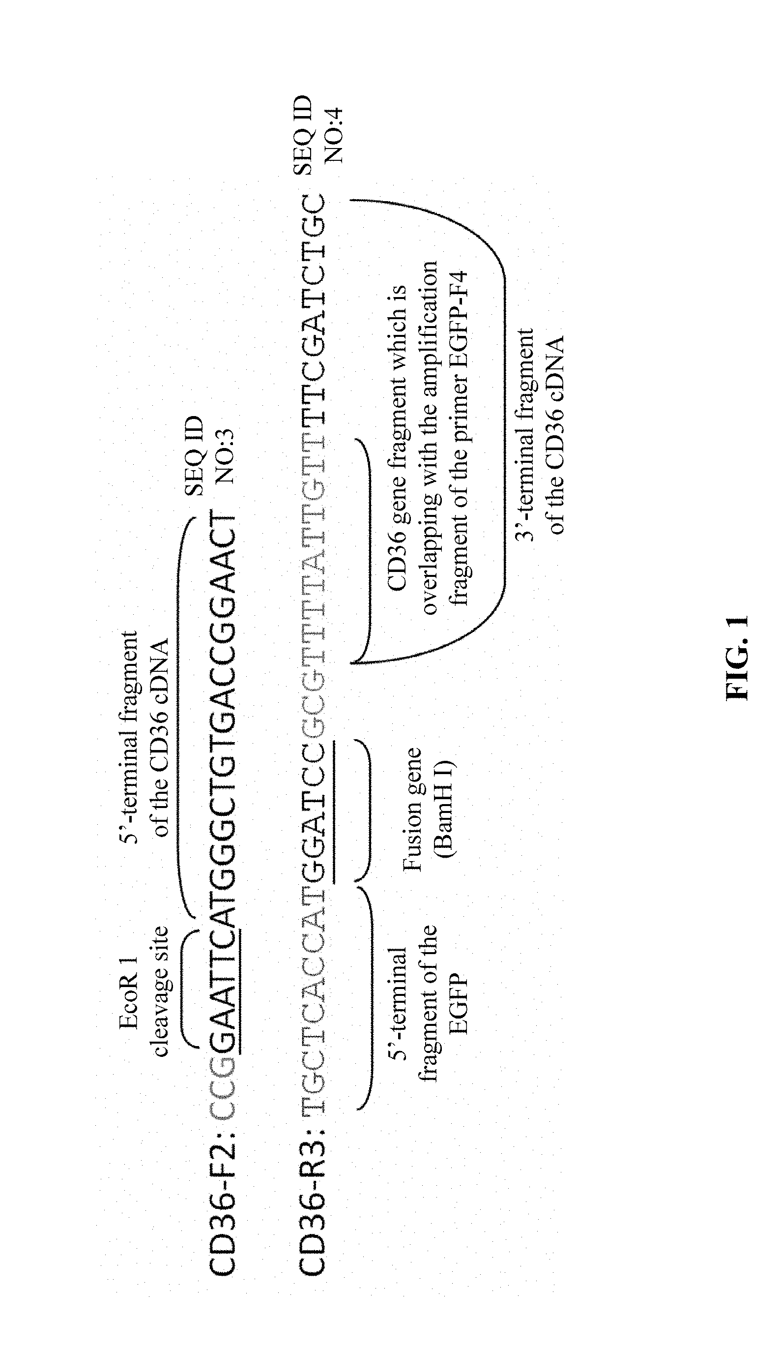
[0012] a forward primer CD36-F2 and a reverse primer CD36-R3 for amplification to obtain a target gene fragment of MT-CD36-EGFP-1 comprising an EcoR 1 cleavage site and protective bases, a full-length coding sequence (CDS) excluding the terminator of CD36 cDNA, a fusion gene BamH I and protective bases, and a portion of the 5'-terminal sequence of the EGFP fluorescent reporter gene, wherein the forward primer CD36-F2 also serves as a forward primer for the subsequent splicing and amplification of MT-CD36-EGFP by SOE-PCR, and has a sequence of 5'-CCG GAA TTC ATG GGC TGT GAC CGG AAC T-3' (SEQ. ID. NO. 3), and the reverse primer CD36-R3 has a sequence of 5'-TGC TCA CCA TGG ATC CGC GTT TTA TTG TTT TCG ATC TGC-3' (SEQ. ID. NO. 4); and
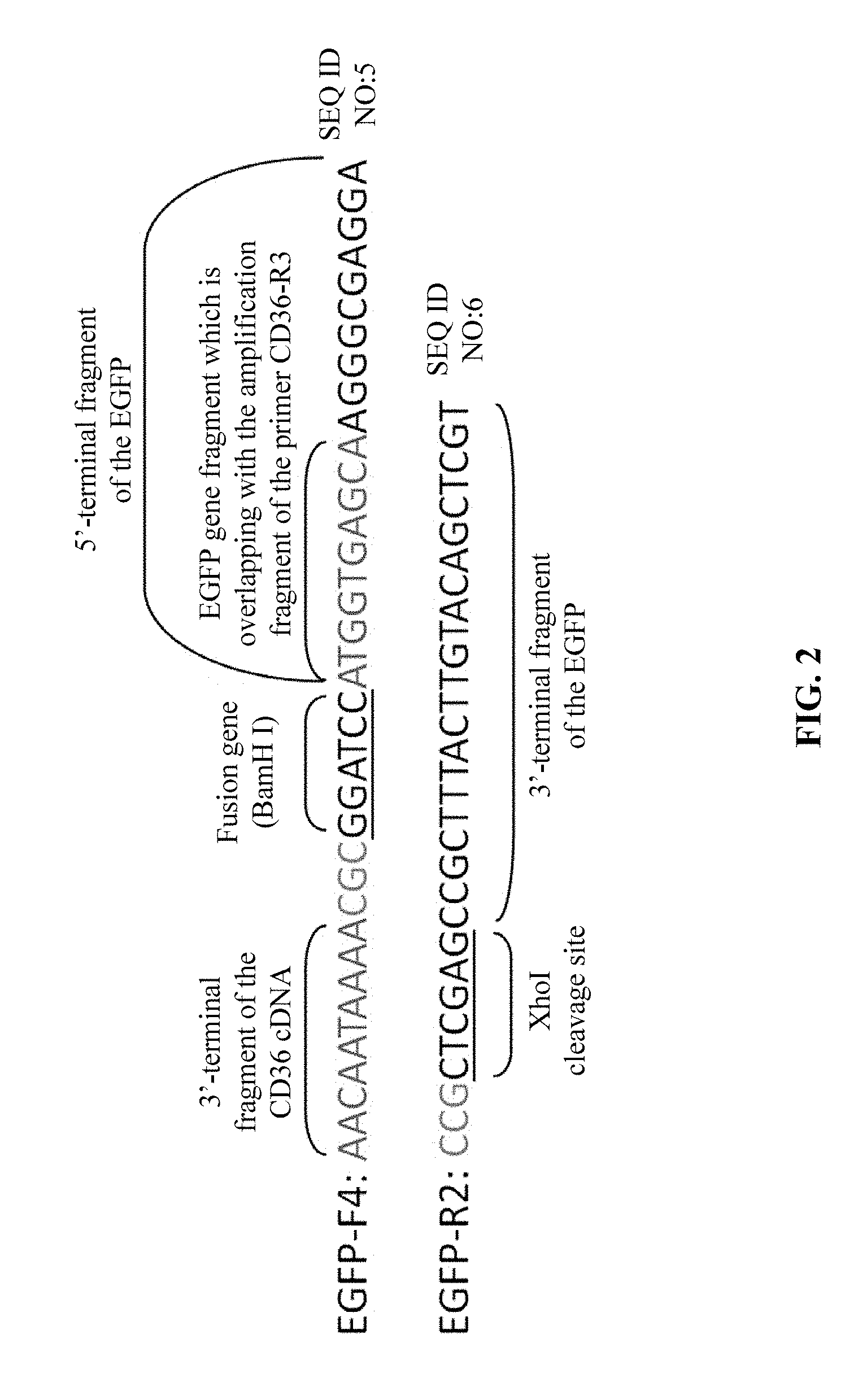
[0013] a forward primer EGFP-F4 and a reverse primer EGFP-R2 for amplification to obtain a target gene fragment of MT-CD36-EGFP-2 comprising a portion of the 3'-terminal fragment of the CD36 coding region cDNA, a fusion gene BamH I and protective bases, a full-length gene sequence of the EGFP fluorescent reporter gene, and an XhoI cleavage site and protective bases, wherein the reverse primer EGFP-R2 also serves as a reverse primer for the subsequent splicing and amplification of MT-CD36-EGFP by SOE-PCR, wherein the forward primer EGFP-F4 has a sequence of 5'-AAC AAT AAA ACG CGG ATC CAT GGT GAG CAA GGG CGA GGA-3' (SEQ. ID. NO. 5), and the reverse primer EGFP-R2 has a sequence of 5'-CCG CTC GAG CCG CTT TAC TTG TAC AGC TCG T-3' (SEQ. ID. NO. 6);
[0014] (3) constructing and amplifying a MT-CD36-EGFP-pLV4/StripII-HIS10 eukaryotic expression vector comprising the mutant CD36 gene and the EGFP fluorescent gene by ligating MT-CD36-EGFP to a pLV4/StripII-HIS10 vector;
[0015] (4) transfecting the MT-CD36-EGFP-pLV4/StripII-HIS10 eukaryotic expression vector into the CHO-K1 cell line by using virus-mediated transfection of eukaryotic cells, and screening and constructing a eukaryotic cell line MT-CD36-CHO-K1 stablely expressing the mutant CD36 gene that encodes CD36 deficiency; and
[0016] (5) by using the method of the present disclosure, establishing a eukaryotic cell line Normal-CD36-CHO-K1 stably expressing normal CD36 gene as a positive control, and establishing a EGFP-pLV4-CHO-K1 line stably expressing the EGFP fluorescent protein alone as a negative control, wherein in the construction of the negative control EGFP-pLV4-CHO-K1 line, a target gene fragment encoding EGFP comprising an EcoR 1 cleavage site and protective bases, the full-length gene sequence of the EGFP fluorescent reporter gene, and an XhoI cleavage site and protective bases is obtained by amplification using a pair of PRC primers, wherein the PCR primers comprise an upstream primer EGFP-F3 having a sequence of 5'-CCG GAA TTC ATG GTG AGC AAG GGC GAG GA-3' (SEQ. ID. NO. 7), and a downstream primer EGFP-R2 that is the reverse primer for amplification of the gene fragment of MT-CD36-EGFP-2 as described in (2)
[0017] Specific description of the method according to the present disclosure:
[0018] 1. In step (1), the primers for amplifying the cDNA of the mutant CD36 gene (MT-CD36 cDNA) by RT-PCR comprises the upstream primer YC-36F and the downstream primer YC-36R, where the upstream primer YC-36F has a sequence of 5'-ATCCTCGAGATGGGCTGTGACCGGA-3'(SEQ. ID. NO. 1), and the downstream primer YC-36R has a sequence of 5'-GCAGAATTCGTTTTATTGTTTTCGATC-3'(SEQ. ID. NO. 2). The upstream primer YC-36F is located in a 5'-promoter region of the CD36 mRNA sequence, and the downstream primer YC-36R is located in a 3'-terminator region of the CD36 mRNA sequence. The amplification region covers the coding sequence (CDS) excluding the terminator of CD36 mRNA, and a plurality of mutant CD36 genes with mutations within a linker region of 5'-terminal and 3'-terminal primer binding regions of the template. If the target gene amplified is a mutant CD36 gene with point mutations, the length of the amplified fragment is 1432 bp, and if the target gene amplified is a mutant CD36 gene with base insertions or deletions, the length of the amplified fragment is increased or decreased on the basis of 1432 bp by the base number inserted or deleted. The RT-PCR amplification is carried out by using the TaKaRa One step RNA PCR Kit (AMV) (one-step method), where the reaction system has a composition of (the total volume of the reaction system is 50 .mu.L):
TABLE-US-00001 10x one step RNA PCR Buffer 5 .mu.L 25 mM MgCl2 10 .mu.L 10 mM dNTPs 5 .mu.L Rnase Inhibitor (40 u/.mu.L) 1 .mu.L AMV Rtase XL 1 .mu.L AMV-Optimized Taq 1 .mu.L Primer YC-36F (20 .mu.M) 1 .mu.L Primer YC-36R (20 .mu.M) 1 .mu.L RNA (100-500 ng/.mu.L) 10 .mu.L H.sub.2O 15 .mu.L
[0019] The cycling parameters comprise:
TABLE-US-00002 50.degree. C. 30 min 94.degree. C. 2 min 30 amplification cycles of: 94.degree. C. 30 sec 60.degree. C. 30 sec 72.degree. C. 2 min; and extension and storage at: 72.degree. C. 10 min 12.degree. C. .infin..
[0020] The target fragment of MT-CD36 cDNA is sequenced.
[0021] 2. In step(2), the primers for SOE-PCR comprise:
[0022] the forward primer CD36-F2 and the reverse primer CD36-R3 for amplification to obtain the target gene fragment of MT-CD36-EGFP-1 comprising an EcoR 1 cleavage site and protective bases, a full-length the coding sequence (CDS) excluding the terminator of CD36 cDNA, a fusion gene BamH I and protective bases, and a portion of the 5'-terminal sequence of the EGFP fluorescent reporter gene, where the forward primer CD36-F2 has a sequence of 5'-CCGGAATTCATGGGCTGTGACCGGAACT-3' (SEQ. ID. NO. 3), and the reverse primer CD36-R3 has a sequence of 5'-TGCTCACCATGGATCCGCGTTTTATTGTTTTCGATCTGC-3' (SEQ. ID. NO. 4), as schematically shown in FIG. 1; the forward primer CD36-F2 is located in a 5'-promoter region of the template, the reverse primer CD36-R3 is located in a 3'-terminal region excluding the terminator of the template; the amplification region covers the coding sequence (CDS) excluding the terminator of CD36 cDNA excluding the termination codons, the EcoR1 cleavage site at the 5' end, and a fusion gene BamH I and 10 bases comprising the promoter of the coding sequence (CDS) of EGFP at the 3' end; and for the length of the target fragment amplified, if the target gene amplified is a mutant CD36 gene with point mutations, the length of the amplified fragment is 1441 bp, and if the target gene amplified is a mutant CD36 gene with base insertions or deletions, the length of the amplified fragment is increased or decreased on the basis of 1441 bp by the base number inserted or deleted; and
[0023] the forward primer EGFP-F4 and the reverse primer EGFP-R2 for amplification to obtain the target gene fragment of MT-CD36-EGFP-2 comprising a portion of the 3'-terminal fragment of the CD36 encoding cDNA region, a fusion gene BamH I and protective bases, a full-length gene sequence of the EGFP fluorescent reporter gene, and an XhoI cleavage site and protective bases, where the forward primer EGFP-F4 has a sequence of 5'-AACAATAAAACGCGGATCCATGGTGAGCAAGGGCGAGGA-3' (SEQ. ID. NO. 5), and the reverse primer EGFP-R2 has a sequence of 5'-CCGCTCGAGCCGCTTTACTTGTACAGCTCGT-3' (SEQ. ID. NO. 6), as schematically shown in FIG. 2; the 5' terminus of the EGFP-F4 comprises a 3'-terminal 10-base sequence before the terminator TAA of the CD36 cDNA sequence and the fusion gene BamH I and protective bases; the amplification region covers the full length sequence comprising the promoter and the terminator of the EGFP CDS, the 3'-terminal 10-base sequence excluding the terminator of the CD36 protein encoding cDNA region at 5' end of the amplification region, and the XhoI cleavage site at 3' end of the amplification region; and the length of the amplified target fragment is 753 bp.
[0024] The reaction system for PCR amplification to obtain the gene fragment of MT-CD36-EGFP-1 by using MT-CD36 cDNA obtained in step(1) as a template and using the CD36-F2 and CD36-R3 respectively as a forward and reverse primer has a composition of (the total volume of the reaction system is 25 .mu.L):
TABLE-US-00003 ddH.sub.2O 15 .mu.L 5 .times. SF Buffer (with 10 mM MgCl2) (Vazyme) 5 .mu.L dNTP mix (10 mM each) 0.5 .mu.L CD36-F2 (10 .mu.M) 1 .mu.L CD36-R3 (10 .mu.M) 1 .mu.L MT-CD36 cDNA 1 .mu.L DMSO 1 .mu.L HS suffer-Fieviy DNA polymerase (Vazyme) 0.5 .mu.L
[0025] The reaction system for PCR amplification to obtain the gene fragment of MT-CD36-EGFP-2 by using the eukaryotic plasmid pEGFP-N1 containing the EGFP fluorescent reporter gene (Suzhou Genepharma Co., Ltd) as a template and using the EGFP-F4 and EGFP-R2 respectively as a forward and reverse primer has a composition of (the total volume of the reaction system is 25 .mu.L):
TABLE-US-00004 ddH.sub.2O 16 .mu.L 5 .times. SF Buffer (with 10 mM MgCl2) (Vazyme) 5 .mu.L dNTP mix (10 mM each) 0.5 .mu.L EGFP-F4 (10 .mu.M) 0.5 .mu.L EGFP-R2 (10 .mu.M) 0.5 .mu.L pEGFP-N1 plasmid 1 .mu.L DMSO 1 .mu.L HS suffer-Fieviy DNA polymerase (Vazyme) 0.5 .mu.L
[0026] The cycling parameters for PCR amplification of the gene fragments of MIT-CD36-EGFP-1 and MT-CD36-EGFP-2 comprise:
TABLE-US-00005 96.degree. C. 5 min 33 amplification cycles of: 96.degree. C. 30 sec 60.degree. C. 30 sec 72.degree. C. 90 sec; and extension and storage at: 72.degree. C. 10 min 12.degree. C. .infin..
[0027] By using MT-CD36-EGFP-1 and MT-CD36-EGFP-2 obtained through PCR amplification as templates, and using the forward primer CD36-F2 and the reverse primer EGFP-R2, the gene fragment of MT-CD36-EGFP comprising the full-length cDNA encoding region of the mutant CD36 gene, a fusion gene BamH I and protective bases, the full-length fragment of the EGFP fluorescent gene, and desired EcoR1 and XhoI cleavage sites is obtained by splicing and amplification of the mutant CD36 gene and the EGFP fluorescent gene by SOE-PCR. The amplification region covers the EcoR1 cleavage site and protective bases, the CDS excluding the termnator of CD36 cDNA, the fusion gene BamH I and protective bases, the full length fragment of the EGFP CDS, and the XhoI cleavage site and protective bases. For the length of the target fragment amplified, if the target gene amplified is a mutant CD36 gene with point mutations, the length of the amplified fragment is 2165 bp, and if the target gene amplified is a mutant CD36 gene with base insertions or deletions, the length of the amplified fragment is increased or decreased on the basis of 2165 bp by the base number inserted or deleted. The reaction system comprises:
TABLE-US-00006 H.sub.2O 24 .mu.L 5 .times. SF Buffer (with 10 mM MgCl2) (phanta .TM.) 10 .mu.L dNTP Mix (10 mM each) 1 .mu.L MT-CD36-EGFP-1 4 .mu.L MT-CD36-EGFP-2 4 .mu.L phanta .TM.HS Suffer-Fideliy DNA Polymerase 1 .mu.L
[0028] The cycling parameters comprise:
TABLE-US-00007 98.degree. C. 5 min 15 amplification cycles of: 98.degree. C. 5 min 72.degree. C. 1 min.
[0029] A system having the following composition is added to the reaction tube after the reaction is completed:
TABLE-US-00008 CD36-F2 (10 .mu.M) 2 .mu.L EGFP-R2 (10 .mu.M) 2 .mu.L DMSO 2 .mu.L
[0030] Amplification is continued through a process which is performed under the following cycling parameters:
TABLE-US-00009 20 amplification cycles of: 98.degree. C. 10 sec 56.degree. C. 30 sec 72.degree. C. 60 sec; and extension and storage at: 72.degree. C. 10 min 12.degree. C. .infin..
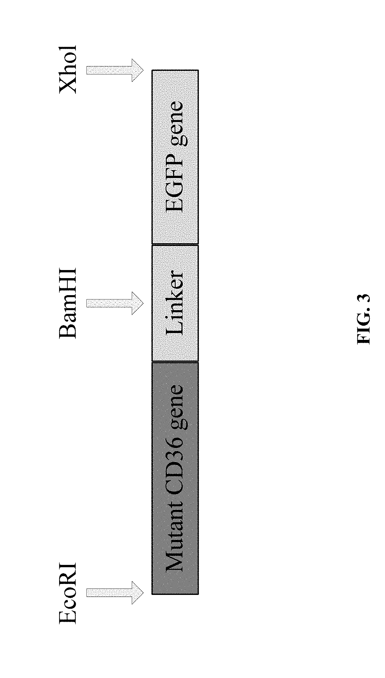
[0031] The product MT-CD36-EGFP (as schematically shown in FIG. 3) obtained by SOE-PCR amplification is subjected to 1.5% agarose gel electrophoresis (105 v, 40 min), gel cutting, and gel extraction by using the AxyPrep DNA Gel Extraction Kit (AXYGEN). The extracted product is sequenced, to confirm that the amplified product is consistent in sequence with the target fragment. Then, the extracted product is used in the construction of the eukaryotic expression vector in step (3).
[0032] 3. In step (3), the target gene fragment of MT-CD36-EGFP obtained in step (2) that was purified by gel extraction and the eukaryotic vector pLV4/StripII-HIS10 (Shenzhen Angran Biotechnology Co., Ltd.) are enzymatically cleaved by using the EcoR1/Xhol DNA Endonuclease Kit (Beyotime Biotechnology), in which the reaction system comprises (the total volume of the reaction system is 20 .mu.L):
TABLE-US-00010 MT-CD36-EGFP or eukaryotic 14 .mu.L vector pLV4/StripII-HIS10 10 .times. Buffer Y 2 .mu.L ECOR 1 1 .mu.L Xhol 1 .mu.L ddH.sub.2O 2 .mu.L
[0033] The reaction condition for enzymatic cleavage comprises: incubation at 37.degree. C. for 1 hr.
[0034] After enzymatic cleavage, the enzymatically cleaved product is purified through the following process.
[0035] Anhydrous ethanol that is 2-3 times the volume of the enzymatically cleaved product and pre-frozen at -80.degree. C. is added to the enzymatically cleaved product, stood at -20.degree. C. for 20 min, and centrifuged at 4.degree. C. for 10 min. The supernatant is removed, and the ethanol is air dried (for 5-8 min). 12 .mu.L of TE Buffer (elution buffer) is added, and stood for 8 min. The concentration of MT-CD36-EGFP and the eukaryotic vector pLV4/StripII-HIS 10 is determined respectively and then stored at -20.degree. C. until use.
[0036] Next, the purified enzymatically cleaved products of MT-CD36-EGFP and the eukaryotic vector pLV4/StripII-HIS10 are ligated by using the T4 DNA Ligase Kit (Promega), to obtain the pLV4/StripII-HIS10 expression vector (MT-CD36-EGFP-pLV4/StripIH-HIS10) comprising the target fragment of the mutant CD36 gene and the EGFP fluorescent gene. The reaction system for ligation comprises:
TABLE-US-00011 MT-CD36-EGFP: eukaryotic vectorpLV4/StripII-HIS10 molar ratio 7:1 (total volume: 1-8 .mu.L) T4 DAN ligase 10 .times. Buffer 1 .mu.L T4 DAN ligase 1 .mu.L H.sub.2O q.s. to 10 .mu.L.
[0037] Reaction condition for ligation comprises: incubation overnight at 4.degree. C.
[0038] Finally, the ligated product is transformed into DH5a Chemically Competent Cells (Beijing TransGen Biotech, Inc). Positive clones are picked up from the E. coli DH5a cell line, cultured, multiplied, and confirmed, to obtain a successfully constructed MT-CD36-EGFP-pLV4/StripII-HIS10 eukaryotic expression vector. The process is specifically as follows.
[0039] The DH5.alpha. Chemically Competent Cells (Beijing TransGen Biotech, Inc) are removed from a freezer at -80.degree. C., and thawed by standing on ice for 5 min. 50 .mu.L of the DH5a Chemically Competent Cells is added to 10 .mu.L of the ligated product obtained in the previous step, stood in an ice bath for 30 min, heat shocked at 42.degree. C. for 1 min, and then stood in an ice bath for 2 min. 1 mL of the SOC medium is then added, and shaken for 1 hr on a shaker at 180 rpm. 300 .mu.L of the bacterial suspension is aspirated and inoculated in a plate (a plate with LB medium to which Ampicillin (Amp, final concentration: 100 .mu.g/mL) is added). The plate is transferred to an incubator and incubated overnight at 37.degree. C. The single colony is picked up from the plate with well-grown transformants and transferred to a 1.5 mL centrifuge tube (to which 1 mL of Amp-containing SOC medium is added). The bamboo sticks for picking up the bacteria are correspondingly transferred one by one to 0.2 mL centrifuge tubes (to which 12 .mu.L sterilized ddH.sub.2O is added), and labeled. The 1.5 mL centrifuge tube containing the bacterial suspension is shaken for 7-8 hrs on a shaker at 37.degree. C.
[0040] During the culture and multiplication process, the bacterial suspension in the 0.2 mL centrifuge tube is lyzed by incubating for 20 min at 100.degree. C., and then colony PCR is carried out using the 2.times.Taq Master Mix Kit (Vazyme). The reaction system for colony PCR comprises:
TABLE-US-00012 H.sub.2O 0.5 .mu.L 2 .times. Taq Master Mix 12.5 .mu.L upstream primer CD36-F2 (10 .mu.M) 1 .mu.L downstream primer EGFP-R2 (10 .mu.M) 1 .mu.L bacterial suspension 10 .mu.L.
[0041] The cycling parameters comprise:
TABLE-US-00013 98.degree. C. 5 min 33 amplification cycles of: 98.degree. C. 10 sec 56.degree. C. 30 sec 72.degree. C. 90 sec; and extension and storage at: 72.degree. C. 10 min 12.degree. C. .infin..
[0042] Selected the multiplied and culture colony that had be confirm having target band by colony PCR, and a part is aspirated for validation by sequencing, and the remaining is preserved for later use. 100-200 .mu.L of the bacterial suspension transformed with the clone that is confirmed to be proper by sequencing is added to 16 mL LB medium, and shaken overnight on a shaker at 37.degree. C. The plasmid is extracted by using an endotoxin-free Plasmid mini Preparation Kit (Tiangen Biotech Co, Ltd.). 5 .mu.L of the extracted plasmid is subjected to 1.5% agarose gel electrophoresis, to observe that the size of the band is proper. The remaining plasmid is stored at -20.degree. C. for later use.
[0043] 4. The experimental method described in step(4) comprises the following.
[0044] Lentiviral packaging by using the Lenti-Pac.TM. HIV Expression packaging Kit (GeneCopoeia): 2.times.10.sup.8 cells of the 293-T cell line in logarithmic growth phase are passaged and inoculated into a 25 mL culture flask, cultured for 24 hrs, and transfected when the cells reach 70%-80% confluence. 2 hrs before transfection, the cell culture medium is changed to Opti-MEM.RTM. I medium (GIBCO). 200 .mu.L of a lentiviral packaging plasmid mix is formulated (in a sterilized 1.5 mL centrifuge tube, where 5 .mu.g of the lentiviral recombinant expression plasmid MT-CD36-EGFP-pLV4/StripII-HIS10 constructed in step (3) and 5 .mu.L of the LentiPac HIV reagent are added, and diluted with Opti-MEM.RTM. I medium to a final volume of 200 .mu.L). 200 .mu.L of a solution containing EndoFectin transfection reagent is formulated in another sterilized 1.5 mL centrifuge tube by adding 185 .mu.L Opti-MEM.RTM. I medium and 15 .mu.LEndoFectin transfection reagent. The formulated solution of the EndoFectin transfection reagent is slowly added dropwise to the lentiviral packaging plasmid mix with gently shaking to mix them uniformly. Then, the solution is incubated for 10-25 min at room temperature, to produce a DNA-EndoFectin mix. The DNA-EndoFectin mix is transferred to a culture of 293T cells, mixed uniformly, and incubated for 8 hrs in a cell incubator at 5% CO.sub.2 and 37.degree. C. The medium containing the transfection mix was removed, and 5 mL of DMEM medium containing 10% fetal bovine serum is added to each flask of cells. 10 .mu.L of TiterBoost reagent is further added, and the culture flask is gently shaken, to mix the medium and the reagent uniformly, and then incubated in a cell incubator at 37.degree. C. and 5% CO.sub.2.
[0045] Harvest of the virus suspension: The culture supernatants of 293T cells are collected at 48 hours and 72 hours after transfection (after the virus suspension is collected at 48 hrs, 5 mL of DMEM medium containing 10% fetal bovine serum and 10 .mu.L of TiterBoost reagent are added to the culture flask), and centrifuged at 4000 g for 10 min at 4.degree. C. to remove cell debris. The supernatant is filtered through a 0.45 .mu.M filter into a 15 mL centrifuge tube, biologically measured for the virus titer and stored at -80.degree. C. until use.
[0046] Establishment of a CHO-K1 cell line (MT-CD36-CHO-K1 line) stably expressing the mutant CD36 gene by virus-mediated transfection: 4.times.10.sup.7 CHO-K1 cells in logarithmic growth phase are inoculated into a 25 mL culture flask, added with DMEM medium containing 10% fetal bovine serum and 1% penicillin--streptomycin solution, and cultured for 24 hrs. The medium is removed when the cells reach 80%-90% confluence, and 4 mL of DMEM medium (containing 10% heat-inactivated fetal bovine serum and 1% penicillin--streptomycin solution) and 100 .mu.L of the viral suspension (with a virus titer of 2.times.10.sup.5 TU/mL) are added, and cultured overnight at 5% CO.sub.2 and 37.degree. C. The medium was removed, and the cells are screened by using 5 mL of DMEM medium containing 0.2 .mu.g/mL puromycin (Sigma) (containing 10% heat-inactivated fetal bovine serum and 1% penicillin--streptomycin solution). At this time, cells successfully transfected with lentiviral packaging plasmid show green fluorescence under a fluorescence microscope, while cells that are not successfully transfected show no fluorescence. After screening by adding medium containing puromycin, the state of cells is observed every 24 hours. The medium containing puromycin is replaced every 48 hours and the culture is continued for at least one month.
[0047] Verification of the established MT-CD36-CHO-K1 line: The MT-CD36-CHO-K1 line in logarithmic growth phase is collected, and RNA is extracted and amplified by RT-PCR following the method as described in step(1), to obtain a cDNA fragment of mutant CD36 gene. The cDNA fragment is sequenced, and the sequencing result is in agreement with that in step(1), suggesting that the established MT-CD36-CHO-K1 line can accurately express the mutant CD36 gene at a molecular level. The CD36 expression on the cell surface of the MT-CD36-CHO-K1 line is confirmed by flow cytometry, and the expression of the CD36 protein by MT-CD36-CHO-K1 line is confirmed by western-blotting.
[0048] 5. In step(5), the processes for establishing the positive control Normal-CD36-CHO-K1 line stably expressing normal CD36 and the negative control EGFP-pLV4-CHO-K1 line stably expressing the EGFP fluorescent protein alone are as follows.
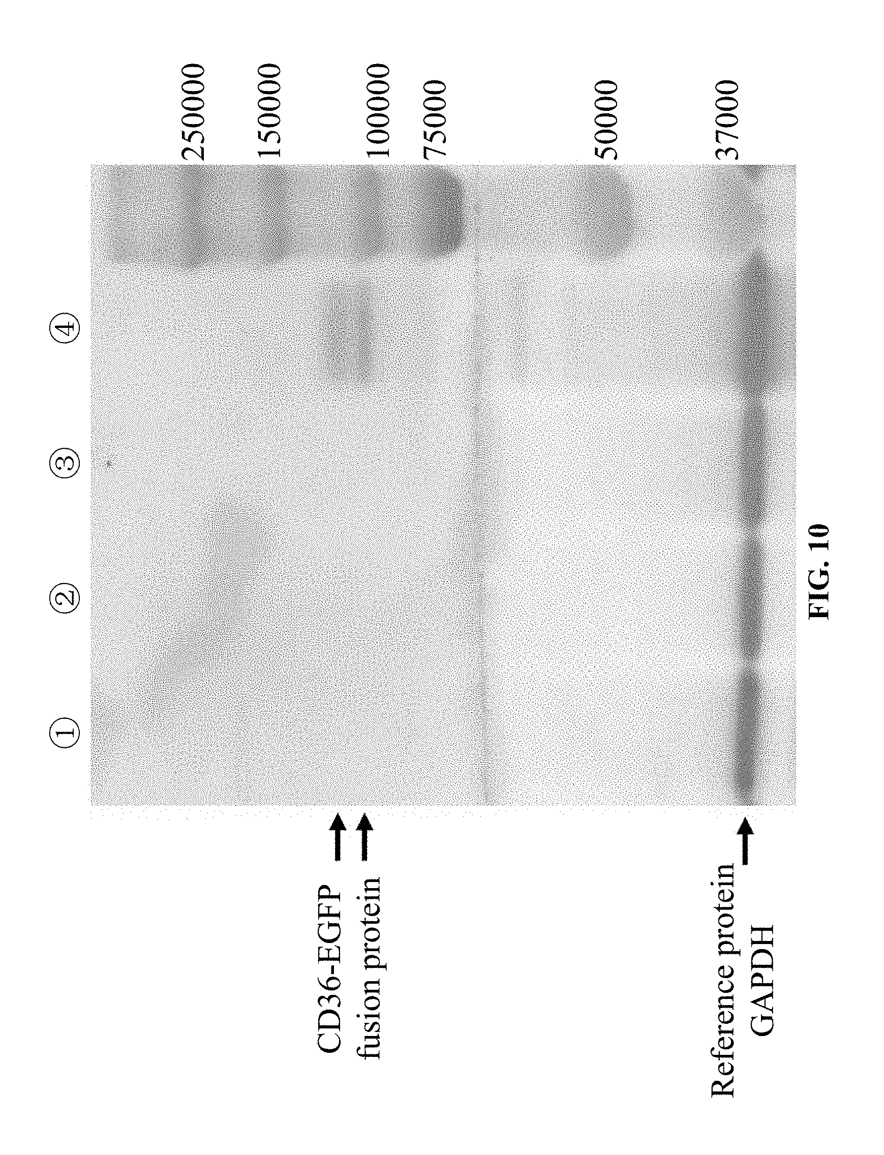
[0049] In the disclosure, total RNA is extracted from a whole blood sample derived from a CD36 expression-positive individual, and following the process in steps (1)-(4), a Normal-CD36-CHO-K1 line stably expressing normal CD36 gene is established as a positive control in an experiment. The cell line is positive in CD36 expression, and in a western-blotting experiment, the expressed CD36-EGFP fusion protein appears as two CD36 positive bands at about 115000D on the western blot, which may be attributed to different protein conformations.
[0050] A EGFP-pLV4-CHO-K1 line having EGFP fluorescent gene-containing plasmid transformed therein and stably expressing the EGFP fluorescent protein alone is established as a negative control. The experimental method is as follows.
[0051] A target EGFP gene fragment comprising an EcoR 1 cleavage site and protective bases, the full-length gene sequence of the EGFP fluorescent reporter gene, and a XhoI cleavage site and protective bases is obtained by amplification using a forward and reverse primer, in which the forward primer EGFP-F3 has a sequence of 5'-CCGGAATTCATGGTGAGCAAGGGCGAGGA-3' (SEQ. ID. NO. 7), and the reverse primer is the downstream primer EGFP-R2 (SEQ. ID. NO. 6) used in amplification of the gene fragment of MT-CD36-EGFP-2 in step(2). The length of the amplified target fragment is 743 bp. The upstream primer EGFP-F3 comprises a EcoR1 cleavage site and protective bases at the 5' end. The amplification region covers the EcoR1 cleavage site and protective bases, the full-length fragment of the EGFP CDS, and the XhoI cleavage site and protective bases.
[0052] PCR amplification is carried out by using the eukaryotic plasmid pEGFP-N1 containing the EGFP fluorescent reporter gene (manufactured by Suzhou Genepharma Co., Ltd) as a template and using the EGFP-F3 and EGFP-R2 as a forward and reverse primer, to obtain the target EGFP gene fragment. The reaction system has a composition of (the total volume of the reaction system is 25 .mu.L):
TABLE-US-00014 ddH.sub.2O 16 .mu.L 5 .times. SF Buffer (with 10 mM MgCl2) (Vazyme) 5 .mu.L dNTP mix (10 mM each) 0.5 .mu.L EGFP-F3 (10 .mu.M) 0.5 .mu.L EGFP-R2 (10 .mu.M) 0.5 .mu.L pEGFP-N1 plasmid 1 .mu.L DMSO 1 .mu.L HS suffer-Fieviy DNA polymerase (Vazyme) 0.5 .mu.L
[0053] The cycling parameters for PCR amplification are the same as those for amplification of MT-CD36-EGFP-1 and MT-CD36-EGFP-2 in step(2).
[0054] The amplified EGFP gene product is subjected to 1.5% agarose gel electrophoresis (105 v, 40 min), gel cutting, and gel extraction by using the AxyPrep DNA Gel Extraction Kit (AXYGEN). The extracted product is sequenced, to confirm that the amplified product is consistent in sequence with the target fragment. Following the processes insteps (3) and (4), a eukaryotic expression vector for expressing the EGFP gene is constructed, and an EGFP-pLV4-CHO-K1 line stably expressing the EGFP fluorescent protein alone is established.
[0055] Critical features and contributions of the method according to the present disclosure
[0056] The disclosure is based on a thorough understanding of CD36 genetics and the important role of CD36 on human in physiology, pathology, functions and clinical medicines, and recognizes that the mutant CD36 gene is a key factor critical to CD36 research and application. A method for establishing a eukaryotic cell line stably expressing mutant CD36 gene is developed and the method is also applicable to the establishment of a eukaryotic cell line stably expressing normal CD36 gene.
[0057] In the method of the disclosure, a target gene fragment for ligation during the construction of a eukaryotic vector expressing the mutant CD36 gene is prepared by using two primers for RT-PCR of mRNA and four primers for SOE-PCR, exploring the optimal annealing temperature and adjusting the conditions comprising Mg.sup.2+ concentration and primer concentration; the eukaryotic vector expressing the mutant CD36 gene is constructed by exploring the optimal enzymatic cleavage conditions in the construction of the eukaryotic vector expressing the mutant CD36 gene and the reaction conditions for ligating a insert fragment to the eukaryotic vector; and the purpose of establishing a eukaryotic cell line stably expressing the mutant CD36 gene that encodes CD36 deficiency is achieved by exploring the viral packaging, transfection, and screening conditions for establishing the eukaryotic cell line stably expressing the mutant CD36 gene by virus-mediated transfection. Because the integrity of the gene fragment of the CD36 open reading frame and the expression of the EGFP fluorescent gene are fully taken into account when designing the primers, the integrity of the target CD36 gene fragment and the EGFP fluorescent gene ligated is ensured, and the ligated EGFP fluorescent gene allows the observation of cells screened under a fluorescence microscope during the screening process in the subsequent establishment of the eukaryotic cell lines stably expressing the mutant CD36 gene during screening. During the establishment of the eukaryotic stable expression cell line of CD36 mutant gene that encodes CD36 deficiency, in the steps of RT-PCR, SOE-PCR, and constructing the MT-CD36-EGFP-pLV4/StripII-HIS10 eukaryotic expression vector, gene sequencing is employed to ensure the accuracy of the sequence of the target fragments in each step.
[0058] Expression of the ligated target MT-CD36 gene in the established eukaryotic CHO-K1 cell line (MT-CD36-CHO-K1 line) stably expressing the mutant CD36 gene is confirmed at a molecular level by extracting RNA from the eukaryotic expression cell line, reverse transcribing, and sequencing. The variation in CD36 protein expression caused by the MT-CD36 gene is confirmed by flow cytometry and western-blotting. This method is not only applicable to the establishment of a eukaryotic cell line stably expressing the CD36 gene with mutations leading to CD36 deficiency such as single nucleotide substitution, base insertion, and base deletion, etc (where the mutation point cannot be located in a 18-base sequence in the 5' to 3' direction starting from the protein expression promoter ATG and the 3' to 5' direction starting from the stop codon) and is also applicable to the establishment of a CD36-positive eukaryotic cell line stably expressing normal CD36 gene.
[0059] Briefly, the establishment method contributes mainly in the following aspects.
[0060] 1. The developed method for establishing eukaryotic stable expression cell line of CD36 mutant gene that encodes CD36 deficiency is also applicable to the establishment of eukaryotic stable expression cell line of normal CD36 gene that encodes normal CD36.
[0061] 2. The problems of permanent and stable preservation and preparation of various CD36 mutant genes and expressed proteins are solved. An infinitely valuable genetic resources basis is provided for studying the molecular structure, expression, regulation and functions of CD36, elucidating the mechanism of action of CD36 in various physiological and pathological processes in human and further for deep insight into the important role of CD36 in clinical treatment.
[0062] 3. A high-quality work platform is provided for development of drugs and screening of drug target in the treatment of CD36-related hemostasis, thrombosis, alloimmune platelet disease, hypercholesterolemia, obesity, peripheral atherosclerosis, arterial hypertension, cardiomyopathy, diabetes, malaria, presenile dementia (Alzheimer's disease) and other diseases.
[0063] 4. The established cell line can be directly used as a panel cell for detecting anti-CD36 antibodies causing platelet transfusion refractoriness to ensure the safety and efficacy of platelet transfusion therapy.
BRIEF DESCRIPTION OF THE DRAWINGS
[0064] FIG. 1 is a schematic view showing the primers for amplification of a target fragment of CD36-EGFP-1 by SOE-PCR.
[0065] FIG. 2 is a schematic view showing the primers for amplification of a target fragment of CD36-EGFP-2 by SOE-PCR.
[0066] FIG. 3 is a schematic view showing the final target fragment of MT-CD36-EGFP amplified by SOE-PCR.
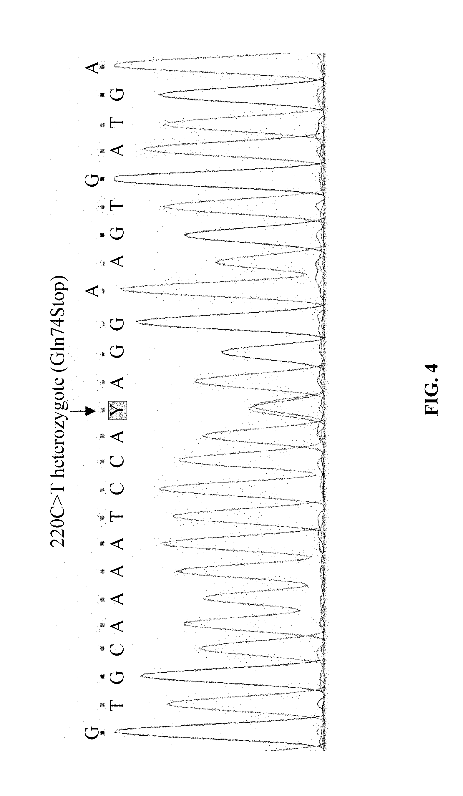
[0067] FIG. 4 shows the exon sequencing result of the CD36 gene in a CD36-deficient individual ZYT in Example 1, showing that the exon-4 has C220T mutation.
[0068] FIG. 5 shows amplification of the CD36 cDNA from the CD36-deficient individual ZYT (1), the target fragments of ZYT-CD36-EGFP-1 (2) and ZYT-CD36-EGFP-2 (3), and the product ZYT-CD36-EGFP (4) ligated by SOE-PCR in Example 1.
[0069] FIG. 6 shows the cDNA sequencing result of the CD36 gene in a CD36-deficient individual ZYT in Example 1, showing 220C>T (Gln74Stop) mutation.
[0070] FIG. 7 shows the sequencing result of the 220T-CD36-EGFP-pLV4/StripII-HIS 10 eukaryotic expression vector successfully constructed in Example 1.
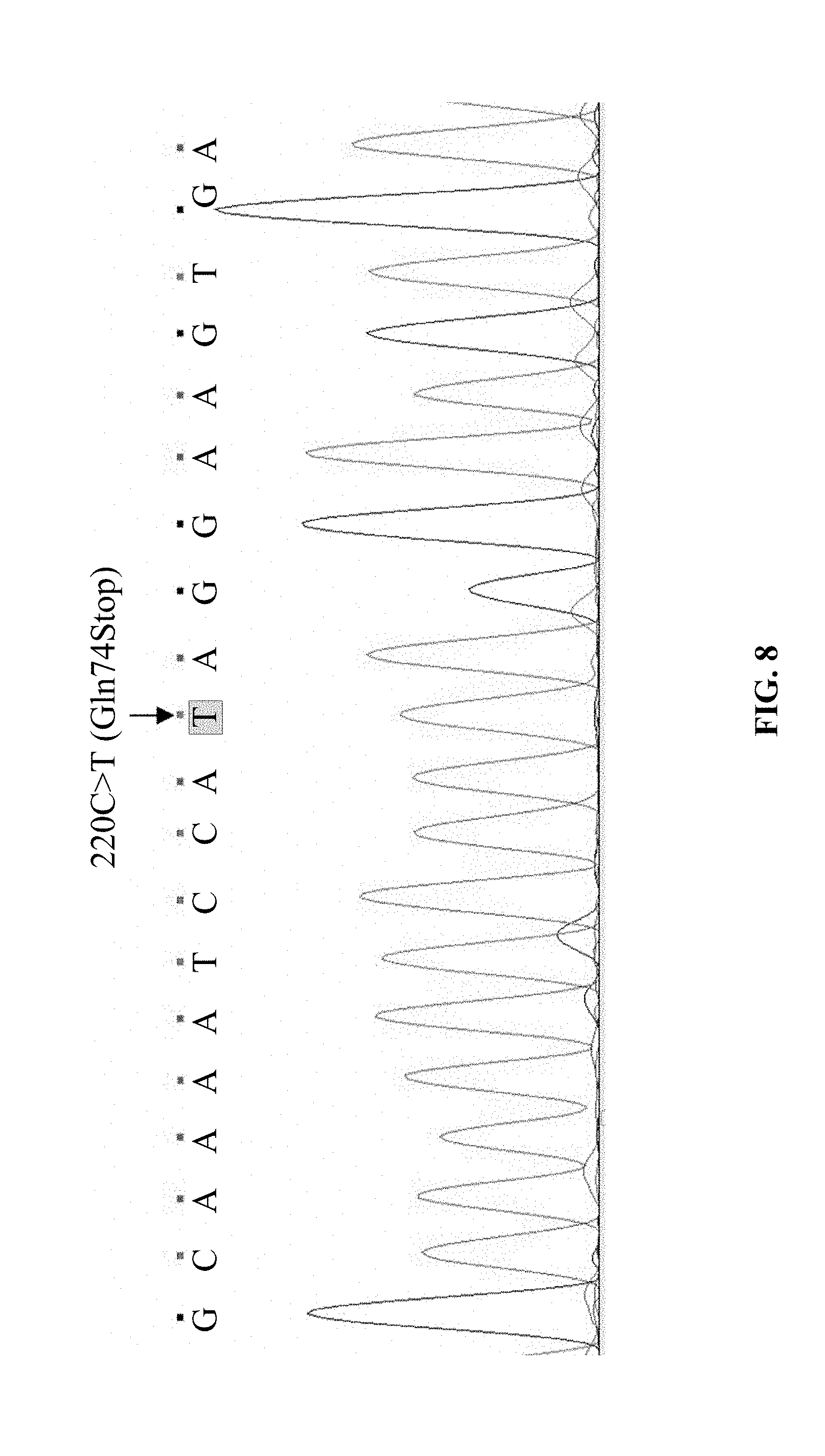
[0071] FIG. 8 shows the cDNA sequencing result of the eukaryotic CHO-K1 cell line (220T-CD36-CHO-K1 line) stably expressing the mutant CD36 gene (220C>T) in Example 1.
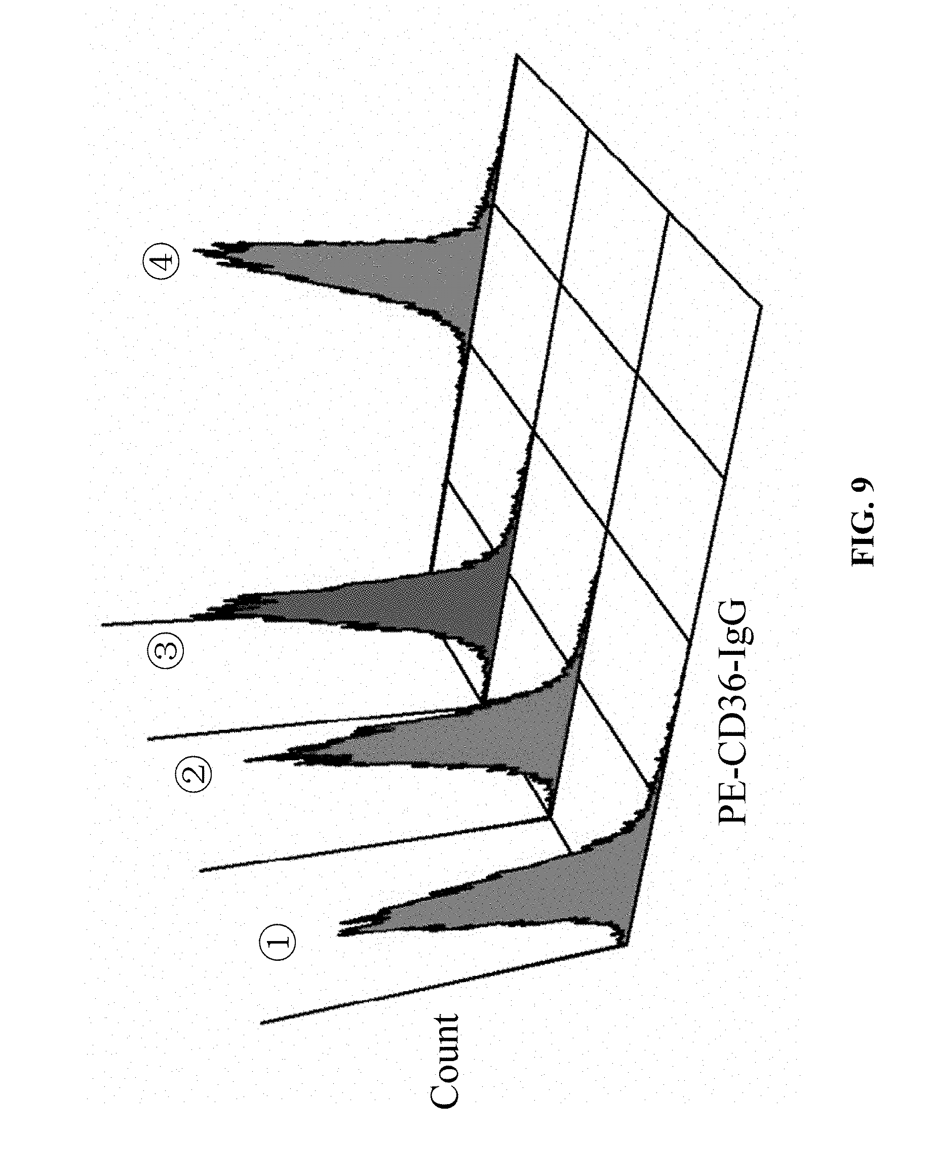
[0072] FIG. 9 shows the results of confirming CD36 protein expression in the eukaryotic CHO-K1 cell line (220T-CD36-CHO-K1 line) stably expressing the mutant CD36 gene (220C>T) by flow cytometry in Example 1, in which (1): the 220T-CD36-CHO-K1 line is negative in CD36 protein expression; (2): the negative control EGFP-pLV4-CHO-K1 line is negative in CD36 protein expression; (3) the blank control CHO-K1 line CD36 is negative in CD36 protein expression; and (4): the positive control NORMAL-CD36-CHO-K1 line is positive in CD36 protein expression.
[0073] FIG. 10 shows the results of confirming CD36 protein expression in the eukaryotic CHO-K1 cell line (220T-CD36-CHO-K1 line) stably expressing the mutant CD36 gene (220C>T) by western-blotting in Example 1, in which (1): the 220T-CD36-CHO-K1 line is negative in CD36 protein expression; (2): the negative control EGFP-pLV4-CHO-K1 line is negative in CD36 protein expression; (3) the blank control CHO-K1 line is negative in CD36 protein expression; and (4): the positive control NORMAL-CD36-CHO-K1 line is positive in CD36 protein expression.
DETAILED DESCRIPTION OF THE EMBODIMENTS
[0074] The following examples are further explanations and illustrations of the disclosure, and not intended to limit the disclosure in any way.
[0075] On the basis of molecular genetics leading to CD36 deficiency, the disclosure provides a method for establishing eukaryotic stable expression cell line of CD36 mutant gene that encodes human CD36 deficiency. The method for establishing eukaryotic stable expression cell line of CD36 mutant gene that encodes CD36 deficiency can be established by designing primers for RT-PCR of mutant CD36 gene and primers for construction of a eukaryotic vector, exploring the optimal annealing temperature and adjusting the conditions comprising Mg.sup.2+ concentration and primer concentration, exploring the optimal enzymatic cleavage conditions in the construction of the eukaryotic vector for the mutant CD36 gene and the reaction conditions for ligating a insert fragment to the eukaryotic vector, and exploring the viral packaging, transfection, and screening conditions for establishing the eukaryotic cell line stably expressing the mutant CD36 gene by virus-mediated transfection.
Example 1
[0076] In this example, an implementation for establishing a eukaryotic cell line, "220T-CD36-CHO-K1 line" stably expressing the mutant CD36 gene 220C>T(Gln74stop) (GenBank Accession No.: KF539919.1) encoding CD36 deficiency by the method for establishing eukaryotic stable expression cell line of CD36 mutant gene that encodes CD36 deficiency according to the disclosure is specifically described.
[0077] A CD36-deficient individual that was confirmed to have mutation of C220T by exon sequencing of the CD36 gene (where the individual was designated as ZYT, and the exon sequencing results were shown in FIG. 4) was chosen. 5 mL of venous blood was drawn from the individual with informed consent, EDTA was added for anti-coagulation, and total RNA was extracted from the blood sample. A target fragment of CD36 cDNA (ZYT-CD36 cDNA) was amplified by RT-PCR using the first pair of PCR primers of the disclosure comprising an upstream primer YC-36F and a downstream primer YC-36R, and using the TaKaRa One step RNA PCR Kit (AMV). The amplification was carried out in ABI 9700PCR machine, and the reaction system for PCR amplification comprised:
TABLE-US-00015 10 .times. one step RNA PCR Buffer 5 .mu.L 25 mM MgCl2 10 .mu.L 10 mM dNTPs 5 .mu.L Rnase Inhibitor (40 u/.mu.L) 1 .mu.L AMV Rtase XL 1 .mu.L AMV-Optimized Taq 1 .mu.L Primer YC-36F (20 .mu.M) 1 .mu.L Primer YC-36R (20 .mu.M) 1 .mu.L RNA (100-500 ng/.mu.L) 10 .mu.L H.sub.2O 15 .mu.L
[0078] The cycling parameters comprised:
TABLE-US-00016 50.degree. C. 30 min 94.degree. C. 2 min 30 amplification cycles of: 94.degree. C. 30 sec 60.degree. C. 30 sec 72.degree. C. 2 min; and extension and storage at: 72.degree. C. 10 min 12.degree. C. .infin..
[0079] 5 .mu.L of the PCR product was stained by DNA green fluorescent dye (Beijing Tiandz Gene Technology Co., Ltd), and subjected to 1.5% agarose gel electrophoresis. With DL2000 Plus DNA Marker from Vazyme as a control, a band of specific PCR product was observed in a gel imaging system. The band of the PCR product was clear and specific, as shown in FIG. 5(1). 5 .mu.L of the PCR product was sequenced, and the sequencing results were shown in FIG. 6. The remaining PCR product was stored at -80.degree. C. until use.
[0080] Next, a target gene fragment of ZYT-CD36-EGFP-1 comprising an EcoR 1 cleavage site and protective bases, a full-length gene sequence of the CD36 protein coding region, a fusion gene BamH I and protective bases, and a portion of the 5'-terminal sequence of the EGFP fluorescent reporter gene was obtained by amplification using the second pair of PCR primers of the present invention comprising an upstream primer CD36-F2 and a downstream primer CD36-R3 and using ZYT-CD36 cDNA as a DNA template. The amplification was carried out in ABI 9700 PCR machine, and the reaction system for PCR amplification comprised a composition of:
TABLE-US-00017 ddH.sub.2O 15 .mu.L 5 .times. SF Buffer (with 10 mM MgCl2) (Vazyme) 5 .mu.L dNTP mix (10 mM each) 0.5 .mu.L CD36-F2 (10 .mu.M) 1 .mu.L CD36-R3 (10 .mu.M) 1 .mu.L ZYT-CD36 cDNA 1 .mu.L DMSO 1 .mu.L HS suffer-Fieviy DNA polymerase (Vazyme) 0.5 .mu.L
[0081] The cycling parameters comprised:
TABLE-US-00018 96.degree. C. 5 min 33 amplification cycles of: 96.degree. C. 30 sec 60.degree. C. 30 sec 72.degree. C. 90 sec; and extension and storage at: 72.degree. C. 10 min 12.degree. C. .infin..
[0082] 5 .mu.L of the PCR product was stained by DNA green fluorescent dye (Beijing Tiandz Gene Technology Co., Ltd), and subjected to 1.5% agarose gel electrophoresis.
[0083] With DL2000 Plus DNA Marker from Vazyme as a control, a band of specific PCR product was observed in a gel imaging system. The band of the PCR product was clear and specific, as shown in FIG. 5(2). The remaining PCR product was stored at -80.degree. C. until use.
[0084] A target gene fragment of ZYT-CD36-EGFP-2 comprising a portion of the 3'-terminal fragment of the CD36 encoding cDNA region, a fusion gene BamH I and protective bases, the full-length gene sequence of the EGFP fluorescent reporter gene, and a XhoI cleavage site and protective bases was obtained by amplification using the third pair of PCR primers of the disclosure comprising an upstream primer EGFP-F4 and a downstream primer EGFP-R2 and using the eukaryotic plasmid pEGFP-N1 containing the EGFP fluorescent reporter gene (manufactured by Suzhou Genepharma Co., Ltd) as a template. The amplification was carried out in ABI 9700 PCR machine, and the reaction system for PCR amplification comprised a composition of:
TABLE-US-00019 ddH.sub.2O 16 .mu.L 5 .times. SF Buffer (with 10 mM MgCl2) (Vazyme) 5 .mu.L dNTP mix (10 mM each) 0.5 .mu.L EGFP-F4 (10 .mu.M) 0.5 .mu.L EGFP-R2 (10 .mu.M) 0.5 .mu.L pEGFP-N1 plasmid 1 .mu.L DMSO 1 .mu.L HS suffer-Fieviy DNA polymerase (Vazyme) 0.5 .mu.L
[0085] The cycling parameters comprised:
TABLE-US-00020 96.degree. C. 5 min 33 amplification cycles of: 96.degree. C. 30 sec 60.degree. C. 30 sec 72.degree. C. 90 sec; and extension and storage at: 72.degree. C. 10 min 12.degree. C. .infin.
[0086] 5 .mu.L of the PCR product was stained by DNA green fluorescent dye (Beijing Tiandz Gene Technology Co., Ltd), and subjected to 1.5% agarose gel electrophoresis.
[0087] With DL2000 Plus DNAMarker from Vazyme as a control, a band of specific PCR product was observed in a gel imaging system. The band of the PCR product was clear and specific, as shown in FIG. 5(3). The remaining PCR product was stored at -80.degree. C. until use.
[0088] A gene fragment of ZYT-CD36-EGFP comprising the full-length cDNA encoding region of the mutant CD36 gene ZYT, a fusion gene BamH I and protective bases, the full-length EGFP fluorescent gene, and desired EcoR1 and XhoI cleavage site was obtained by splicing and amplication by SOE-PCR using the upstream primer CD36-F2 in the second pair of primers and the downstream primer EGFP-R2 in the third pair of primers of the disclosure, and using ZYT-CD36-EGFP-1 and ZYT-CD36-EGFP-2 as templates. The amplification was carried out in ABI 9700 PCR machine, and the reaction system for PCR amplification comprised a composition of:
TABLE-US-00021 H.sub.2O 24 .mu.L 5 .times. SF Buffer (with 10 mM MgCl2) (phanta .TM.) 10 .mu.L dNTP Mix (10 mM each) 1 .mu.L ZYT-CD36-EGFP-1 4 .mu.L ZYT-CD36-EGFP-2 4 .mu.L phanta .TM.HS Suffer-Fideliy DNA Polymerase 1 .mu.L
[0089] The cycling parameters comprised:
TABLE-US-00022 98.degree. C. 5 min
[0090] 15 amplification cycles of:
TABLE-US-00023 98.degree. C. 5 min 72.degree. C. 1 min.
[0091] A system having the following composition was added to the reaction tube after the reaction was completed:
TABLE-US-00024 CD36-F2 2 .mu.L EGFP-R2 2 .mu.L DMSO 2 .mu.L.
[0092] Amplification is continued through a process which is performed under the following cycling parameters:
TABLE-US-00025 20 amplification cycles of: 98.degree. C. 10 sec 56.degree. C. 30 sec 72.degree. C. 60 sec; and extension and storage at: 72.degree. C. 10 min 12.degree. C. .infin..
[0093] 5 .mu.L of the PCR product was stained by DNA green fluorescent dye (Beijing Tiandz Gene Technology Co., Ltd), and subjected to 1.5% agarose gel electrophoresis. With DL2000 Plus DNAMarker from Vazyme as a control, a band of specific PCR product was observed in a gel imaging system. The band of the PCR product was clear and specific, as shown in FIG. 5(4). The target fragment of 2165 bp was taken by gel cutting, and subjected to gel extraction by using the AxyPrep DNA Gel Extraction Kit (AXYGEN). 4 .mu.L of the extracted product was sequenced, to confirm that the amplified product is consistent in sequence with the target fragment. Then, the remaining extracted product was stored at -80.degree. C. until use.
[0094] After the purified target fragment of ZYT-CD36-EGFP was obtained, ZYT-CD36-EGFP and the eukaryotic vector pLV4/StripII-HIS10 (Shenzhen Angran Biotechnology Co., Ltd.) were enzymatically cleaved by using the EcoR1/XhoI DNA Endonuclease Kit (Beyotime Biotechnology). The reaction system comprised:
TABLE-US-00026 ZYT-CD36-EGFP or eukaryotic 14 .mu.L vector pLV4/StripII-HIS10 10 .times. Buffer Y 2 .mu.L ECOR 1 1 .mu.L Xhol 1 .mu.L ddH.sub.2O 2 .mu.L
[0095] The reaction condition for enzymatic cleavage comprised: incubation at 37.degree. C. for 1 hr.
[0096] After enzymatic cleavage, the enzymatically cleaved product was purified through the following process.
[0097] Anhydrous ethanol that was 2-3 times the volume of the enzymatically cleaved product and pre-frozen at -80.degree. C. was added to the enzymatically cleaved product, stood at -20.degree. C. for 20 min, and centrifuged at 4.degree. C. for 10 min. The supernatant was removed and, and the ethanol was air dried (for 5-8 min). 12 .mu.L of TE Buffer (elution buffer) was added, and stood for 8 min. The concentration of MT-CD36-EGFP and the eukaryotic vector pLV4/StripII-HIS10 was determined respectively and then stored at -20.degree. C. for later use.
[0098] The purified enzymatically cleaved products of MT-CD36-EGFP and the eukaryotic vector pLV4/StripII-HIS10 were ligated by using the T4 DNA Ligase Kit (Promega), to obtain a pLV4/StripII-HIS10 expression vector (C220T-CD36-EGFP-pLV4/StripII-HIS10) comprising the target fragment of the mutant CD36 gene and the EGFP fluorescent gene. The reaction system for ligation comprised:
TABLE-US-00027 ZYT-CD36-EGFP: eukaryotic vectorpLV4/StripII-HIS10 molar ratio 7:1 (total volume: 1-8 .mu.L) T4 DAN ligase 10 .times. Buffer 1 .mu.L T4 DAN ligase 1 .mu.L H.sub.2O q.s. to 10 .mu.L.
[0099] Reaction condition for ligation comprised: incubation overnight at 4.degree. C.
[0100] The ligated product was transformed into DH5a Chemically Competent Cells (Beijing TransGen Biotech, Inc). Positive clones were picked up from the E. coli DH5a cell line, cultured, multiplied, and confirmed, to obtain a successfully constructed 220T-CD36-EGFP-pLV4/StripII-HIS10eukaryotic expression vector. The process was specifically as follows.
[0101] The DH5.alpha. Chemically Competent Cells (Beijing TransGen Biotech, Inc) were removed from a freezer at -80.degree. C., and thawed by standing on ice for 5 min. 50 .mu.L of the DH5a Chemically Competent Cells was aspirated to 10 .mu.L of the ligated product obtained in the previous step (1 .mu.L of a solution III was added to the ligated product before transformation), stood in an ice bath for 30 min, heat shocked at 42.degree. C. for 1 min, and then stood in an ice bath for 2 min. 1 mL of the SOC medium was then added, and shaken for 1 hr on a shaker at 180 rpm. 300 .mu.L of the bacterial suspension was aspirated and inoculated in a plate (a plate with LB medium to which Ampicillin (Amp, final concentration: 100 .mu.g/mL) was added). The plate was transferred to an incubator and incubated overnight at 37.degree. C. A single colony was picked up from the plate with well-grown transformants and transferred to a 1.5 mL centrifuge tube (to which 1 mL of Amp-containing SOC medium was added). The bamboo sticks for picking up the bacteria were correspondingly transferred one by one to 0.2 mL centrifuge tubes (to which 12 .mu.L sterilized ddH.sub.2O was added), and labeled. The 1.5 mL centrifuge tube containing the bacterial suspension was shaken for 7-8 hrs on a shaker at 37.degree. C.
[0102] During the culture and multiplication process, colony PCR amplification was carried out by using the 2.times.Taq Master Mix Kit (Vazyme) using the upstream primer CD36-F2 in the second pair of primers and the downstream primer EGFP-R2 in the third pair of primers of the present disclosure and using the bacterial suspension in the 0.2 mL centrifuge tube (which was lyzed by incubating for 20 min at 100.degree. C. before PCR amplification) as a template. The amplification was carried out in ABI 9700 PCR machine, and the reaction system for PCR amplification comprised a composition of:
TABLE-US-00028 H.sub.2O 0.5 .mu.L 2 .times. Taq Master Mix 12.5 .mu.L upstream primer CD36-F2 (10 .mu.M) 1 .mu.L downstream primer EGFP-R2 (10 .mu.M) 1 .mu.L bacterial suspension 10 .mu.L
[0103] The cycling parameters comprised:
TABLE-US-00029 98.degree. C. 5 min 33 amplification cycles of: 98.degree. C. 10 sec 56.degree. C. 30 sec 72.degree. C. 90 sec; and extension and storage at: 72.degree. C. 10 min 12.degree. C. .infin..
[0104] 5 .mu.L of the PCR product was stained by DNA green fluorescent dye (Beijing Tiandz Gene Technology Co., Ltd), and subjected to 1.5% agarose gel electrophoresis. With DL2000 Plus DNAMarker from Vazyme as a control, a band of specific PCR product was observed in a gel imaging system. The band of the PCR product of the picked colony was clear and specific, and the size of the fragment was in agreement with the size of the product ZYT-CD36-EGFP amplified by SOE-PCR. The corresponding colony was multiplied and cultured, and a part was aspirated for validation by sequencing and the remaining is preserved for later use. The result of confirmation by sequencing indicates CD36 220T (as shown in FIG. 7). 100-200 .mu.L of the bacterial suspension transformed with a clone in which the remaining sequence is consistent with that in the ligated sequence of ZYT-CD36-EGFP was added to 16 mL LB medium, shaken overnight on a shaker at 37.degree. C., multiplied and cultured to obtain a successfully constructed 220T-CD36-EGFP-pLV4/StripII-HIS10 eukaryotic expression vector. The plasmid was extracted by using an endotoxin-free Plasmid mini Preparation Kit (Tiangen Biotech Co, Ltd.). 5 .mu.L of the extracted plasmid was subjected to 1.5% agarose gel electrophoresis, to observe that the size of the band is proper. The remaining plasmid was stored at -20.degree. C. for later use.
[0105] Finally, the 220T-CD36-EGFP-pLV4/StripII-HIS10 eukaryotic expression vector was transfected into the CHO-K1 cell line by using virus-mediated transfection of eukaryotic cells, and screened to construct a eukaryotic cell line (220T-CD36-CHO-K1) stably expressing the mutant CD36 220C>T gene encoding CD36 deficiency. The process was specifically as follows.
[0106] Lentiviral packaging by using the Lenti-Pac.TM. HIV Expression packaging Kit (GeneCopoeia): 2.times.10.sup.8 cells of the 293-T cell line in logarithmic growth phase were passaged and inoculated into a 25 mL cell culture flask, cultured for 24 hrs, and transfected when the cells reached 70%-80% confluence. 2 hrs before transfection, the cell culture medium was changed to Opti-MEM.RTM. I medium (GIBCO). 200 .mu.L of a lentiviral packaging plasmid mix was formulated (in a sterilized 1.5 mL centrifuge tube, where 5 .mu.g of the eukaryotic expression plasmid 220T-CD36-EGFP-pLV4/StripII-HIS10 successfully constructed and 5 .mu.L of the LentiPac HIV reagent were added, and diluted with Opti-MEM.RTM. I medium to a final volume of 200 .mu.L). 200 .mu.L of a solution containing EndoFectin transfection reagent was formulated in another sterilized 1.5 mL centrifuge tube by adding 185 .mu.L Opti-MEM.RTM. I medium and 15 .mu.L EndoFectin transfection reagent. The formulated solution of the EndoFectin transfection reagent was slowly added dropwise to the lentiviral packaging plasmid mix with gently shaking to mix them uniformly. Then, the solution was incubated for 10-25 min at room temperature, to produce a DNA-EndoFectin mix. The DNA-EndoFectin mix was transferred to a culture of 293T cells, mixed uniformly, and incubated for 8 hrs in a cell incubator at 5% CO.sub.2 and 37.degree. C. The medium containing the transfection mix was removed, and 5 mL of DMEM medium containing 10% fetal bovine serum was added to each flask of cells. 10 .mu.L of TiterBoost reagent was further added, and the culture flask was gently shaken, to mix the medium and the reagent uniformly, and then incubated in a cell incubator at 37.degree. C. and 5% CO.sub.2.
[0107] Harvest of the virus suspension: The culture supernatants of 293T cells were collected 48 hours and 72 hours after transfection (after the virus suspension was collected at 48 hrs, 5 mL of DMEM medium containing 10% fetal bovine serum and 10 .mu.L of TiterBoost reagent were added to the culture flask), and centrifuged at 4000 g for 10 min at 4.degree. C. to remove cell debris. The supernatant was filtered through a 0.45 .mu.M filter into a 15 mL centrifuge tube, biologically measured for the virus titer and stored at -80.degree. C. until use.
[0108] Establishment of a eukaryotic cell line (220T-CD36-CHO-K1 line) stably expressing the mutant CD36 220C>T gene by transfecting the CHO-K1 cell line using the harvested virus suspension, and screening with puromycin: 4.times.10.sup.7 CHO-K1 cells in logarithmic growth phase were inoculated into a 25 mL culture flask, added with DMEM medium containing 10% fetal bovine serum and 1% penicillin--streptomycin solution, and cultured for 24 hrs. The medium was removed when the cells reached 80%-90% confluence, and 4 mL of DMEM medium (containing 10% heat-inactivated fetal bovine serum and 1% penicillin--streptomycin solution) and 100 .mu.L of the viral suspension (with a virus titer of 2.times.10.sup.5 TU/mL) were added, and cultured overnight at 5% CO.sub.2 and 37.degree. C. The medium was removed, and the cells were screened by using 5 mL of DMEM medium containing 0.2 .mu.g/mL puromycin (Sigma) (containing 10% heat-inactivated fetal bovine serum and 1% penicillin--streptomycin solution). The state of cells was observed every 24 hours. The medium containing puromycin was replaced every 48 hours and the culture was continued for at least one month.
[0109] A Normal-CD36-CHO-K1 line stably expressing normal CD36 and a EGFP-pLV4-CHO-K1 line having merely EGFP fluorescent gene-containing plasmid transformed therein and stably expressing the EGFP fluorescent protein alone were established as a positive control and a negative control respectively in experiment. The process was as follows.
[0110] Total RNA was extracted from a whole blood sample derived from a CD36 expression-positive individual, and following the process in the above steps, a Normal-CD36-CHO-K1 line stably expressing normal CD36 gene was established as a positive control in an experiment.
[0111] A EGFP-pLV4-CHO-K1 line having EGFP fluorescent gene-containing plasmid transformed therein and stably expressing the EGFP fluorescent protein alone was established as a negative control. The experimental method was as follows.
[0112] PCR amplification was carried out by using the eukaryotic plasmid pEGFP-N1 containing the EGFP fluorescent reporter gene (manufactured by Suzhou Genepharma Co., Ltd) as a template and using EGFP-F3 and EGFP-R2 as forward and reverse primer, to obtain a target EGFP gene fragment comprising an EcoR 1 cleavage site and protective bases, the full-length gene sequence of the EGFP fluorescent reporter gene, and a XhoI cleavage site and protective bases. The reaction system had a composition of (the total volume of the reaction system was 25 .mu.L):
TABLE-US-00030 ddH.sub.2O 16 .mu.L 5 .times. SF Buffer (with 10 mM Mgcl2) (Vazyme) 5 .mu.L dNTP mix (10 mM each) 0.5 .mu.L EGFP-F3 (10 .mu.M) 0.5 .mu.L EGFP-R2 (10 .mu.M) 0.5 .mu.L pEGFP-N1 plasmid 1 .mu.L DMSO 1 .mu.L HS suffer-Fieviy DNA polymerase (Vazyme) 0.5 .mu.L
[0113] The cycling parameters for PCR amplification were the same as those for amplification of the target fragments of ZYT-CD36-EGFP-1 and ZYT-CD36-EGFP-2 in the above steps.
[0114] The amplified EGFP gene product was subjected to 1.5% agarose gel electrophoresis (105 v, 40 min), gel cutting, and gel extraction by using the AxyPrep DNA Gel Extraction Kit (AXYGEN). The extracted product was sequenced, to confirm that the amplified product is consistent in sequence with the target fragment. Following the method for constructing the 220T-CD36-EGFP-pLV4/StripH-HIS10 plasmid and the method for establishing and screening the 220T-CD36-CHO-K1 cell line, a eukaryotic expression vector for expressing the EGFP gene was constructed, and an EGFP-pLV4-CHO-K1 line stably expressing the EGFP fluorescent protein alone was established.
[0115] Confirmation of the established 220T-CD36-CHO-K1 line:
[0116] RNA was respectively extracted from the 220T-CD36-CHO-K1 line and the positive control Normal-CD36-CHO-K1 line in logarithmic growth phase. A target fragment of CD36 cDNA was amplified by RT-PCR using the first pair of PCR primers comprising the upstream primer YC-36F and the downstream primer YC-36R and using TaKaRa One step RNA PCR Kit (AMV). The amplification was carried out in ABI 9700 PCR machine, and the reaction system for amplification and the cycling parameters were as described in the step 1. 5 .mu.L of the PCR product was stained by DNA green fluorescent dye (Beijing Tiandz Gene Technology Co., Ltd), and subjected to 1.5% agarose gel electrophoresis. With DL2000 Plus DNAMarker from Vazyme as a control, a band of specific PCR product was observed in a gel imaging system. The band of the PCR product was clear and specific. The size of the fragment was in agreement with the size of the product ZYT-CD36 cDNA fragment amplified in the step 1. 5 .mu.L of the PCR product was sequenced. The cDNA sequencing results of the 220T-CD36-CHO-K1 line show that the sequence is in agreement with the sequence of the CD36 gene fragment in the transfected plasmid 220T-CD36-EGFP-pLV4/StripII-HIS10, as shown in FIG. 8. This suggests that the established 220T-CD36-CHO-K1 line can accurately express the mutant CD36 220C>T gene at a molecular level. The cDNA sequencing results of the positive control Normal-CD36-CHO-K1 line show that the sequence is in agreement with the sequence of the CD36 gene fragment in the transfected plasmid Normal-CD36-EGFP-pLV4/StripII-HIS10. This suggests that the established Normal-CD36-CHO-K1 line can accurately express normal CD36 gene at molecular level. The cDNA sequencing results of the negative control EGFP-pLV4-CHO-K1 line show that the sequence is in agreement with the sequence of the EGFP gene fragment in the transfected plasmid EGFP-pLV4/StripII-HIS 10. This suggests that the established EGFP-pLV4-CHO-K1 line can accurately express the EGFP gene at molecular level.
[0117] The 220T-CD36-CHO-K1 line, the positive control Normal-CD36-CHO-K1 line, the negative control EGFP-pLV4-CHO-K1 line, and the blank control CHO-K1 line in logarithmic growth phase were collected, and detected for the CD36 expression by flow cytometry. The detection results are shown in FIG. 9, and indicate that the 220T-CD36-CHO-K1 line, the negative control EGFP-pLV4-CHO-K1 line, and the blank control CHO-K1 line are all negative in CD36 expression, and the positive control NORMAL-CD36-CHO-K1 line is positive in CD36 expression. Total membrance proteins were extracted from the several cell lines, and the expression of the CD36 protein by each cell line was detected by western-blotting. The detection results are shown in FIG. 10 and indicate that the 220T-CD36-CHO-K1 line, the negative control EGFP-pLV4-CHO-K1 line, and the blank control CHO-K1 line are negative in CD36 expression, and the positive control Normal-CD36-CHO-K1 line is positive in CD36 expression.
[0118] Unless otherwise indicated, the numerical ranges involved in the invention include the end values. While particular embodiments of the invention have been shown and described, it will be obvious to those skilled in the art that changes and modifications may be made without departing from the invention in its broader aspects, and therefore, the aim in the appended claims is to cover all such changes and modifications as fall within the true spirit and scope of the invention.
Sequence CWU 1
1
7125DNAHomo sapiens 1atcctcgaga tgggctgtga ccgga 25227DNAHomo
sapiens 2gcagaattcg ttttattgtt ttcgatc 27328DNAHomo sapiens
3ccggaattca tgggctgtga ccggaact 28439DNAHomo sapiens 4tgctcaccat
ggatccgcgt tttattgttt tcgatctgc 39539DNAAequorea victoria
5aacaataaaa cgcggatcca tggtgagcaa gggcgagga 39631DNAAequorea
victoria 6ccgctcgagc cgctttactt gtacagctcg t 31729DNAAequorea
victoria 7ccggaattca tggtgagcaa gggcgagga 29
D00000
D00001
D00002
D00003
D00004
D00005
D00006
D00007
D00008
D00009
D00010
S00001
XML
uspto.report is an independent third-party trademark research tool that is not affiliated, endorsed, or sponsored by the United States Patent and Trademark Office (USPTO) or any other governmental organization. The information provided by uspto.report is based on publicly available data at the time of writing and is intended for informational purposes only.
While we strive to provide accurate and up-to-date information, we do not guarantee the accuracy, completeness, reliability, or suitability of the information displayed on this site. The use of this site is at your own risk. Any reliance you place on such information is therefore strictly at your own risk.
All official trademark data, including owner information, should be verified by visiting the official USPTO website at www.uspto.gov. This site is not intended to replace professional legal advice and should not be used as a substitute for consulting with a legal professional who is knowledgeable about trademark law.