Treatment Of Disease With Poly-n-acetylglucosamine Nanofibers
Finkielsztein; Sergio ; et al.
U.S. patent application number 15/922765 was filed with the patent office on 2019-02-28 for treatment of disease with poly-n-acetylglucosamine nanofibers. This patent application is currently assigned to Marine Polymer Technologies, Inc.. The applicant listed for this patent is Marine Polymer Technologies, Inc.. Invention is credited to Sergio Finkielsztein, John N. Vournakis.
| Application Number | 20190060348 15/922765 |
| Document ID | / |
| Family ID | 51659125 |
| Filed Date | 2019-02-28 |
| United States Patent Application | 20190060348 |
| Kind Code | A1 |
| Finkielsztein; Sergio ; et al. | February 28, 2019 |
TREATMENT OF DISEASE WITH POLY-N-ACETYLGLUCOSAMINE NANOFIBERS
Abstract
Described herein are compositions comprising shortened fibers of poly-N-acetylglucosamine and/or a derivative thereof ("sNAG nanofibers") and the use of such compositions in the treatment of various diseases, in particular, diseases associated with decreased tensile strength of tissue, decreased elasticity of tissue, increased collagen content or abnormal collagen content in tissue, abnormal alignment of collagen in tissue, and/or increased myofibroblast content in tissue.
| Inventors: | Finkielsztein; Sergio; (Newton, MA) ; Vournakis; John N.; (Charleston, SC) | ||||||||||
| Applicant: |
|
||||||||||
|---|---|---|---|---|---|---|---|---|---|---|---|
| Assignee: | Marine Polymer Technologies,
Inc. Danvers MA |
||||||||||
| Family ID: | 51659125 | ||||||||||
| Appl. No.: | 15/922765 | ||||||||||
| Filed: | March 15, 2018 |
Related U.S. Patent Documents
| Application Number | Filing Date | Patent Number | ||
|---|---|---|---|---|
| 15666337 | Aug 1, 2017 | |||
| 15922765 | ||||
| 15385208 | Dec 20, 2016 | |||
| 15666337 | ||||
| 14210054 | Mar 13, 2014 | |||
| 15385208 | ||||
| 61784765 | Mar 14, 2013 | |||
| Current U.S. Class: | 1/1 |
| Current CPC Class: | A61K 9/0092 20130101; A61K 9/0014 20130101; A01N 59/20 20130101; A61L 2400/12 20130101; A61K 9/70 20130101; A61K 8/027 20130101; A61P 19/02 20180101; A01N 25/34 20130101; A61L 2400/06 20130101; A61P 19/10 20180101; A61K 2800/413 20130101; A61K 8/73 20130101; A61K 9/0019 20130101; A01N 43/16 20130101; A61K 31/715 20130101; A61L 31/042 20130101; A61P 17/00 20180101; A61L 27/20 20130101; A61K 2800/412 20130101; C08L 5/08 20130101; A61L 27/50 20130101; A61L 26/0023 20130101; A61K 2800/91 20130101; A61L 2300/412 20130101; A61Q 19/08 20130101; A61K 31/726 20130101; A61P 17/02 20180101; A01N 43/16 20130101; A01N 25/34 20130101; A01N 59/20 20130101; A61L 31/042 20130101; C08L 5/08 20130101; A61L 26/0023 20130101; C08L 5/08 20130101; A61L 27/20 20130101; C08L 5/08 20130101 |
| International Class: | A61K 31/715 20060101 A61K031/715; A61K 9/00 20060101 A61K009/00; A61Q 19/08 20060101 A61Q019/08; A61K 31/726 20060101 A61K031/726; A61K 9/70 20060101 A61K009/70; A61K 8/73 20060101 A61K008/73; A61K 8/02 20060101 A61K008/02; A61L 27/50 20060101 A61L027/50; A01N 43/16 20060101 A01N043/16; A61L 27/20 20060101 A61L027/20; A61L 31/04 20060101 A61L031/04; A61L 26/00 20060101 A61L026/00; A01N 25/34 20060101 A01N025/34; A01N 59/20 20060101 A01N059/20; C08L 5/08 20060101 C08L005/08 |
Claims
1. A method for treating a symptom of Ehlers-Danlos in a human subject, comprising topically administering a composition comprising shortened fibers of poly-N-acetylglucosamine (sNAG nanofibers) to the human subject, wherein the sNAG nanofibers comprise 70% or more of N-acetylglucosamine monosaccharides, and wherein more than 50% of the sNAG nanofibers are between about 1 to 15 .mu.m in length.
2. The method of claim 1, wherein the symptom is a skin-related symptom.
3. The method of claim 1, wherein the skin-related symptom is soft skin, fragile skin, skin that bruises easily, excessive scarring of the skin, or blunted wound healing in the skin.
4. The method of claim 2, wherein the sNAG nanofibers are administered directly to the skin affected by the skin-related symptom.
5. A method for treating a symptom of scleroderma in a human subject, comprising topically administering a composition comprising shortened fibers of poly-N-acetylglucosamine (sNAG nanofibers) to the human subject, wherein the sNAG nanofibers comprise 70% or more of N-acetylglucosamine monosaccharides, and wherein more than 50% of the sNAG nanofibers are between about 1 to 15 .mu.m in length.
6. The method of claim 5, wherein the symptom is a skin-related symptom.
7. The method of claim 6, wherein the skin-related symptom is swollen skin, thickened skin, shiny skin, discoloration of skin, or numbness of skin.
8. The method of claim 6, wherein the sNAG nanofibers are administered directly to the skin affected by the skin-related symptom.
9. A method for treating a symptom of Epidermolysis bullosa in a human subject, comprising topically administering a composition comprising shortened fibers of poly-N-acetylglucosamine (sNAG nanofibers) to a human subject, wherein the sNAG nanofibers comprise 70% or more of N-acetylglucosamine monosaccharides, and wherein more than 50% of the sNAG nanofibers are between about 1 to 15 .mu.m in length.
10. The method of claim 9, wherein the symptom is a skin-related symptom or a mucosal membrane-related symptom.
11. The method of claim 10, wherein the skin-related symptom or the mucosal membrane-related symptom is a blister.
12. The method of claim 10, wherein the sNAG nanofibers are administered directly to the skin affected by the skin-related symptom or the mucosal membrane-related symptom.
13.-27. (canceled)
28. The method of claim 1, wherein the sNAG nanofibers increase the metabolic rate of serum-starved human umbilical cord vein endothelial cells in a MTT assay and/or do not rescue apoptosis of serum-starved human umbilical cord endothelial cells in a trypan blue exclusion test.
29. The method of claim 1, wherein more than 50% of the sNAG nanofibers are between about 2 to 10 .mu.m in length, or about 4 to 7 .mu.m in length.
30. (canceled)
31. (canceled)
32. The method of claim 1, wherein the sNAG nanofibers were produced by gamma irradiation of poly-N-acetylglucosamine, and wherein the poly-.beta.-N-acetylglucosamine was irradiated in the form of dried fibers at 500-2,000 kgy, or the poly-N-acetylglucosamine was irradiated in the form of wet fibers at 100-500 kgy.
33. The method of claim 1, wherein the sNAG nanofibers were produced from a microalgal poly-N-acetylglucosamine.
34. (canceled)
35. The method of claim 1, wherein more than 90% or more than 95% of the monosaccharides of the sNAG nanofibers are N-acetylglucosamine monosaccharides.
36. (canceled)
37. The method of claim 5, wherein the sNAG nanofibers increase the metabolic rate of serum-starved human umbilical cord vein endothelial cells in a MTT assay and/or do not rescue apoptosis of serum-starved human umbilical cord endothelial cells in a trypan blue exclusion test.
38. The method of claim 5, wherein more than 50% of the sNAG nanofibers are between about 2 to 10 .mu.m in length, or about 4 to 7 .mu.m in length.
39. The method of claim 5, wherein the sNAG nanofibers were produced by gamma irradiation of poly-N-acetylglucosamine, and wherein the poly-.beta.-N-acetylglucosaminev was irradiated in the form of dried fibers at 500-2,000 kgy, or the poly-N-acetylglucosamine was irradiated in the form of wet fibers at 100-500 kgy.
40. The method of claim 5, wherein the sNAG nanofibers were produced from a microalgal poly-N-acetylglucosamine.
41. The method of claim 5, wherein more than 90% or more than 95% of the monosaccharides of the sNAG nanofibers are N-acetylglucosamine monosaccharides.
42. The method of claim 9, wherein the sNAG nanofibers increase the metabolic rate of serum-starved human umbilical cord vein endothelial cells in a MTT assay and/or do not rescue apoptosis of serum-starved human umbilical cord endothelial cells in a trypan blue exclusion test.
43. The method of claim 9, wherein more than 50% of the sNAG nanofibers are between about 2 to 10 .mu.m in length, or about 4 to 7 .mu.m in length.
44. The method of claim 9, wherein the sNAG nanofibers were produced by gamma irradiation of poly-N-acetylglucosamine, and wherein the poly-.beta.-N-acetylglucosaminev was irradiated in the form of dried fibers at 500-2,000 kgy, or the poly-N-acetylglucosamine was irradiated in the form of wet fibers at 100-500 kgy.
45. The method of claim 9, wherein the sNAG nanofibers were produced from a microalgal poly-N-acetylglucosamine.
46. The method of claim 9, wherein more than 90% or more than 95% of the monosaccharides of the sNAG nanofibers are N-acetylglucosamine monosaccharides.
Description
[0001] This application is a continuation of U.S. application Ser. No. 15/666,337, filed on Aug. 1, 2017, which is a continuation of U.S. application Ser. No. 15/385,208, filed on Dec. 20, 2016, which is a continuation of U.S. application Ser. No. 14/210,054, filed on Mar. 13, 2014, which claims the benefit of U.S. provisional application No. 61/784,765, filed on Mar. 14, 2013, each of which is incorporated herein by reference in its entirety.
1. INTRODUCTION
[0002] Described herein are compositions comprising shortened fibers of poly-N-acetylglucosamine and/or a derivative thereof ("sNAG nanofibers") and the use of such compositions in the treatment of various conditions and diseases, in particular, those associated with decreased tensile strength of tissue, decreased elasticity of tissue, increased collagen content or abnormal collagen content in tissue, abnormal alignment of collagen in tissue, and/or increased myofibroblast content in tissue.
2. BACKGROUND
[0003] A number of conditions and diseases that are either incurable at this time or have suboptimal treatments available, due to only partial effectiveness of such treatments or side effects associated with such treatments. For example, there a number of incurable or only partially curable conditions and diseases associated with decreased tensile strength of tissue, decreased elasticity of tissue, increased collagen content or abnormal collagen content in tissue, abnormal alignment of collagen in tissue, and/or increased myofibroblast content in tissue. Such conditions and diseases include, among others, Ehlers-Danlos Syndrome, Epidermolysis bullosa, scleroderma, osteoporosis, intervertebral disc disorder, degenerative disc disorder, osteoarthritis, fibrosis, wrinkling of the skin, and scarring associated with wounds. There remains a need for an effective treatment for these conditions and diseases that can be used alone, or in combination with a standard therapy, that is safe and effective.
3. SUMMARY
[0004] Provided herein are methods of treating various diseases associated with decreased tensile strength of tissue, decreased elasticity of tissue, increased collagen content or abnormal collagen content in tissue, abnormal or disorganized alignment of collagen in tissue, and/or increased myofibroblast content in tissue. Further, provided herein are methods of treating various diseases associated with increase in collagen type I content (e.g., expression) in tissue, decrease of collagen type III content (e.g., expression) in tissue, decrease in elastin content (e.g., expression) in tissue, or increase in alpha smooth muscle actin content in tissue.
[0005] In a specific embodiment, provided herein is a method of treating a symptom of Ehler-Danols syndrome in a human subject, comprising administering a composition comprising sNAG nanofibers to the human subject, wherein more than 50% of the sNAG nanofibers are between about 1 to 15 .mu.m in length. In one embodiment, the symptom is a skin-related symptom. In a further embodiment, the skin-related symptom is soft skin, fragile skin, skin that bruises easily, excessive scarring of the skin, or blunted wound healing in the skin. In a specific embodiment, the composition is administered topically to the subject. In yet another embodiment, the composition is administered directly to the skin affected by the skin-related symptom.
[0006] In a specific embodiment, provided herein is a method of treating a symptom of scleroderma in a human subject, comprising administering a composition comprising sNAG nanofibers to the human subject, wherein more that 50% of the sNAG nanofibers are between 1 to 15 .mu.m in length. In one embodiment, the symptom is a skin-related symptom. In another embodiment, the skin-related symptom is swollen skin, thickened skin, shiny skin, discoloration of skin, or numbness of skin. In a specific embodiment, the composition is administered topically to the subject. In another embodiment, the composition is administered directly to the skin affected by the skin-related symptom.
[0007] In a specific embodiment, provided herein is a method for treating a symptom of Epidermolysis bullosa in a human subject, comprising administering a composition comprising sNAG nanofibers to a human subject, wherein more than 50% of the sNAG nanofibers are between about 1 to 15 .mu.m in length. In one embodiment of the method the symptom is a skin-related symptom or a mucosal membrane-related symptom. In another embodiment, the skin-related symptom or the mucosal membrane-related symptom is a blister. In a specific embodiment, the composition is administered topically to the subject. In yet another embodiment, the composition is administered directly to the skin affected by the skin-related symptom or the mucosal membrane-related symptom.
[0008] In a specific embodiment, provided herein is a method for treating or preventing wrinkles or depressions in the skin's surface in a human subject, comprising administering a composition comprising sNAG nanofibers to the human subject, wherein more than 50% of the sNAG nanofibers are between about 1 to 15 .mu.m in length. In a specific embodiment, the composition is administered topically to the subject.
[0009] In a specific embodiment, provided herein is a method for treating wrinkles or depressions in the skin's surface in a human subject, comprising topically administering a composition comprising sNAG nanofibers to a human subject having wrinkles of depressions, wherein more than 50% of the sNAG nanofibers are between about 1 to 15 .mu.m in length. In some embodiments, the composition is administered directly to the wrinkles or depressions in the skin's surface in a human subject.
[0010] In a specific embodiment, provided herein is a method of reducing scarring associated with cutaneous wounds in a human subject, comprising administering a composition comprising sNAG nanofibers to a cutaneous wound in a human subject, wherein more than 50% of the sNAG nanofibers are between about 1 to 15 .mu.m in length. In a specific embodiment, the composition is administered topically to the subject. In one embodiment, the subject has a scar from a cutaneous wound, and wherein the sNAG nanofibers are administered topically to the area of the scar. In a particular embodiment, the composition is administered topically for 21 days.
[0011] In a specific embodiment, provided herein is a method of treating a symptom of osteoporosis in a human subject, comprising administering a composition comprising sNAG nanofibers to the human subject, wherein more than 50% of the sNAG nanofibers are between about 1 to 15 .mu.m in length. In one embodiment, the composition is administered to an area of low bone density in a human subject. In another embodiment, the sNAG nanofibers are administered by local injection. In a specific embodiment, the composition is administered topically to the subject.
[0012] In a specific embodiment, provided herein is a method of treating a symptom of intervertebral disc disorder or degenerative disc disorder in a human subject, comprising topically administering a composition comprising sNAG nanofibers to the human subject, wherein more than 50% of the sNAG nanofibers are between about 1 to 15 .mu.m in length. In one embodiment, the composition is administered in the disc in the human subject in the area of lower back pain. In another embodiment the composition is administered by local injection. In a specific embodiment, the composition is administered topically to the subject.
[0013] In a specific embodiment, provided herein is a method for treating a symptom of osteoarthritis in a human subject, comprising administering a composition comprising sNAG nanofibers to the human subject, wherein more than 50% of the sNAG nanofibers are between about 1 to 15 .mu.m in length. In one embodiment, the sNAG nanofibers are administered topically to the joints of the human subject. In a specific embodiment, the composition is administered topically to the subject.
[0014] In a specific embodiment, provided herein is a method of treating fibrosis or a symptom of fibrosis in a human subject, comprising administering a composition comprising sNAG nanofibers to the human subject, wherein more than 50% of the sNAG nanofibers are between 1 to 15 .mu.m in length. In some embodiments, a composition comprising sNAG nanofibers is administered directly to the organ or tissue that is at risk of fibrosis or has fibrosis. In one embodiment, a composition comprising sNAG nanofibers is administered directly to the fibrotic tissue (e.g., on the skin). In a specific embodiment, the composition is administered topically to the subject.
[0015] In some embodiments, the subject (e.g., human) treated in accordance with the methods described herein has an increased content or expression of collagen type I, a decreased content or expression of collagen type III, a decreased content or expression of elastin, and/or an increased content or expression of smooth muscle actin, in a tissue (e.g., skin). In further embodiments, the subject (e.g., human) treated in accordance with the methods described herein has decreased tensile strength of tissue (e.g., skin) and/or decreased elasticity of tissue (e.g., skin). In further embodiments, the subject (e.g., human) treated in accordance with the methods described herein has an increased myofibroblast content in a tissue (e.g., skin).
[0016] In certain embodiments, the sNAG nanofibers are non-reactive when tested in an intramuscular implantation test. In other embodiments, the sNAG nanofibers increase the metabolic rate of serum-starved human umbilical cord vein endothelial cells in a MTT assay and/or do not rescue apoptosis of serum-starved human umbilical cord endothelial cells in a trypan blue exclusion test. In further embodiments of the methods, more than 50% of the sNAG nanofibers are between 2 to 10 .mu.m in length. In other embodiments, more than 50% of the sNAG nanofibers are between 4 to 7 .mu.m in length. In other embodiments, more than 100% of the sNAG nanofibers are between 1 to 15 .mu.m in length.
[0017] In specific embodiments of the methods described herein, the sNAG nanofibers were produced gamma irradiation of poly-N-acetylglucosamine and/or a derivative thereof, and wherein the poly-.beta.-N-acetylglucosamine and/or a derivative thereof was irradiated in the form of dried fibers at 500-2,000 kgy, or the poly-N-acetylglucosamine and/or a derivative thereof was irradiated in the form of wet fibers at 100-500 kgy. In particular embodiments, the sNAG nanofibers were produced from microalgal poly-N-acetylglucosamine. In further embodiments, the sNAG nanofibers comprise N-acetylglucosamine monosaccharides and/or glucosamine monosaccharides, wherein more than 70% of the monosaccharides of the sNAG nanofibers are N-acetylglucosamine monosaccharides. In other embodiments, the sNAG nanofibers comprise N-acetylglucosamine monosaccharides and/or glucosamine monosaccharides, wherein more than 90% of the monosaccharides of the sNAG nanofibers are N-acetylglucosamine monosaccharides. In still other embodiments, the sNAG nanofibers comprise N-acetylglucosamine monosaccharides and/or glucosamine monosaccharides, wherein more than 95% of the monosaccharides of the sNAG nanofibers are N-acetylglucosamine monosaccharides.
3.1 Terminology
[0018] As used herein, the terms "sNAG nanofiber," "sNAG," "Taliderm," or "Talymed" (formerly known as "Taliderm") are used interchangeably to refer to shortened fibers of poly-N-acetylglucosamine and/or derivatives thereof. In a preferred embodiment, sNAG nanofibers consist entirely of shortened fibers of poly-N-acetylglucosamine and/or derivatives thereof. Taliderm or Talymed are examples of sNAG nanofibers which are membranes consisting entirely of shortened fibers of poly-N-acetylglucosamine and/or derivatives thereof.
[0019] As used herein, the term "about" means a range around a given value wherein the resulting value is the same or substantially the same (e.g., within 10%, 5% or 1%) as the expressly recited value. In one embodiment, "about" means within 10% of a given value or range. In another embodiment, the term "about" means within 5% of a given value or range. In another embodiment, the term "about" means within 1% of a given value or range.
[0020] As used herein, the terms "disease" and "disorder" are used interchangeably to refer to a condition in a subject. Exemplary diseases/disorders that can be treated or prevented in accordance with the methods described herein include, without limitation, Ehlers-Danlos Syndrome, Epidermolysis bullosa, scleroderma, osteoporosis, intervertebral disc disorder, degenerative disc disorder, osteoarthritis, fibrosis, wrinkling of the skin, and scarring associated with wounds.
[0021] As used herein, the term "subject" and "patient" are used interchangeably to refer to an animal (e.g., cow, horse, sheep, pig, chicken, turkey, quail, cat, dog, mouse, rat, rabbit, guinea pig, etc.). In some embodiments, the subject is a mammal such as a non-primate and a primate (e.g., monkey and human). In specific embodiments, the subject is a human.
[0022] As used herein, the term "effective amount" in the context of administering a sNAG nanofiber or composition thereof to a subject refers to the amount of a sNAG nanofiber or composition thereof that results in a beneficial or therapeutic effect. In specific embodiments, an "effective amount" of a sNAG nanofiber or composition thereof refers to an amount of a sNAG nanofiber or composition thereof which is sufficient to achieve at least one, two, three, four or more of the following effects: (i) reduction or amelioration of the severity of a disease in the subject or population of subjects or a symptom associated therewith; (ii) reduction of the duration of a symptom associated with a disease; (iii) prevention of the progression of a disease in the subject or population of subjects or a symptom associated therewith; (iv) regression of a symptom associated with a disease; (v) prevention of the development or onset of a symptom associated with a disease; (vi) prevention of the recurrence of a symptom associated with a disease; (vii) reduction of the incidence of hospitalization of the subject or population of subjects; (viii) reduction of the hospitalization length of the subject or population of subjects; (ix) an increase the survival of the subject or population of subjects; (x) elimination of a condition in the subject or population of subjects; (xi) enhancement or improvement of the prophylactic or therapeutic effect(s) of another therapy in the subject or population of subjects; (xii) reduction of the number of symptoms of a disease in the subject or population of subjects; (xiiii) the increase in the tensile strength of a tissue in a subject; (xiv) the increase in elasticity in a tissue of a subject; (xv) the increase in elastin content or production in a tissue of a subject; (xvi) the reduction in scar size in a tissue of a subject; (xvii) the decrease in total collagen content in a tissue of a subject; (xviii) the decrease of collagen I expression or content in a tissue of a subject; (xix) the increase in collagen III expression or content in a tissue of a subject; (xx) the inducement of more organized collagen alignment in a tissue of a subject; (xxi) the reduction in smooth muscle actin content or expression, or the reduction in myofibroblast content in a tissue of a subject; (xxii) the prevention of the onset, development or recurrence of a condition caused by or associated with one or more of: decreased tensile strength of tissue, decreased elasticity of tissue, increased collagen content or abnormal collagen content in tissue, increased collagen I expression in tissue, decreased collagen III expression in tissue, abnormal alignment of collagen in tissue, increased smooth muscle actin expression in tissue, and increased myofibroblast content in tissue; and/or (xxiii) improvement in quality of life as assessed by methods well known in the art, e.g., a questionnaire. In specific embodiments, an "effective amount" of a sNAG nanofiber refers to an amount of a sNAG nanofiber composition specified herein, e.g., in Section 5.6, infra.
[0023] As used herein, the term "premature human infant" refers to a human infant born at less than 37 weeks of gestational age.
[0024] As used herein, the term "human infant" refers to a newborn to 1 year old human.
[0025] As used herein, the term "premature human infant" refers to a newborn to 1 year old year human who was born of less than 37 weeks gestational age (e.g., before 37 weeks, 36 weeks, 35 weeks, 34 weeks, 33 weeks, 32 weeks, 31 weeks, 30 weeks, 29 weeks, 28 weeks, or less than 28 weeks of pregnancy).
[0026] As used herein, the term "human toddler" refers to a human that is 1 years to 3 years old.
[0027] As used herein, the term "human child" refers to a human that is 1 year to 18 years old.
[0028] As used herein, the term "human adult" refers to a human that is 18 years or older.
[0029] As used herein, the term "elderly human" refers to a human 65 years or older.
[0030] As used herein the "normal" expression of one or more gene products is: (i) the average expression level known to be found in subjects not displaying symptoms or not diagnosed with the condition and disease to be treated; (ii) the average expression level detected in three, five, ten, twenty, twenty-five, fifty or more subjects not displaying symptoms or not diagnosed with the condition and disease to be treated; and/or (iii) the level of expression detected in a patient to be administered a composition described herein before the onset of the condition and disease.
[0031] As used herein, the term "low expression," or "low level of expression" in the context of expression of a gene (e.g., based on the level of protein, peptide and/or mRNA produced by the gene) refers to an expression that is less than the "normal" expression of the gene. In a specific embodiment, "low expression" refers to expression of a gene that is less than 99%, less than 95%, less than 90%, less than 85%, less than 75%, less than 70%, less than 65%, less than 60%, less than 55%, less than 50%, less than 45%, less than 40%, less than 35%, less than 30%, less than 25%, or less than 20% of the "normal" expression of the gene. In another specific embodiment, "low expression" refers to expression of a gene that is about 20-fold, about 15-fold, about 10-fold, about 5-fold, about 4-fold, about 3-fold, about 2-fold, or about 1.5 fold less than the "normal" expression of the gene. In further embodiments, "low expression" refers to expression of a gene that is more than about 1.25 fold, 1.5 fold, 2 fold, 2.5 fold, 3 fold, 3.5 fold, 4 fold, 4.5 fold, 5 fold, 6 fold, 7 fold, 8 fold, 9 fold, 10 fold lower than "normal" expression of a gene.
[0032] As used herein, the term "high expression", or "high level of expression" in the context of expression of a gene (e.g., based on the level of protein, peptide and/or mRNA produced by the gene) refers to an expression that is more than the "normal" expression of the gene. In a specific embodiment, "high expression" refers to expression of a gene that is more than 99%, more than 95%, more than 90%, more than 85%, more than 75%, more than 70%, more than 65%, more than 60%, more than 55%, more than 50%, more than 45%, more than 40%, more than 35%, more than 30%, more than 25%, or more than 20% of the "normal" expression of the gene. In another specific embodiment, "high expression" refers to expression of a gene that is about 20-fold, about 15-fold, about 10-fold, about 5-fold, about 4-fold, about 3-fold, about 2-fold, or about 1.5 fold more than the "normal" expression of the gene. In further embodiments, "high expression" refers to expression of a gene that is more than about 1.25 fold, 1.5 fold, 2 fold, 2.5 fold, 3 fold, 3.5 fold, 4 fold, 4.5 fold, 5 fold, 6 fold, 7 fold, 8 fold, 9 fold, 10 fold higher than "normal" expression of a gene.
[0033] As used herein, the term "altered expression" or "altered level of expression" of a gene product is a level that differs (e.g., by more than 20%, 25%, 30%, 50%, 75%, 100%, 150%, 200%, 250%, 300%) from the normal level of expression of the gene.
[0034] As used herein, the term "majority" refers to greater than 50%, including, e.g., 50.5%, 51%, 55%, etc.
[0035] As used herein, the terms "therapies" and "therapy" can refer to any protocol(s), method(s), compositions, formulations, and/or agent(s) that can be used in the prevention and/or treatment of any disease or disorder associated with decreased tensile strength or elasticity of tissue, increased total collagen content in tissue, increased collagen type I content (e.g., expression) in tissue, decreased collagen type III content in tissue, abnormal (e.g., disorganized) collagen alignment in tissue, decreased elastin content (e.g., expression) in tissue, increased myofibroblast content in tissue, and/or increased alpha smooth muscle actin content (e.g., expression) in tissue. Examples of diseases or disorders include, without limitation, Ehlers-Danlos Syndrome, Epidermolysis bullosa, scleroderma, osteoporosis, intervertebral disc disorder, degenerative disc disorder, osteoarthritis, fibrosis, wrinkling of the skin, and scarring associated with wounds. In certain embodiments, the terms "therapies" and "therapy" refer to drug therapy, adjuvant therapy, radiation, surgery, biological therapy, supportive therapy, and/or other therapies useful in treatment and/or prevention of the diseases or disorders listed herein. In certain embodiments, the term "therapy" refers to a therapy other than a sNAG nanofiber or a composition thereof. In specific embodiments, an "additional therapy" and "additional therapies" refer to a therapy other than a treatment using a sNAG nanofiber or a composition thereof. In a specific embodiment, a therapy includes the use of a sNAG nanofiber as an adjuvant therapy. For example, using a sNAG nanofiber in conjunction with a drug therapy, biological therapy, surgery, and/or supportive therapy.
4. BRIEF DESCRIPTION OF FIGURES
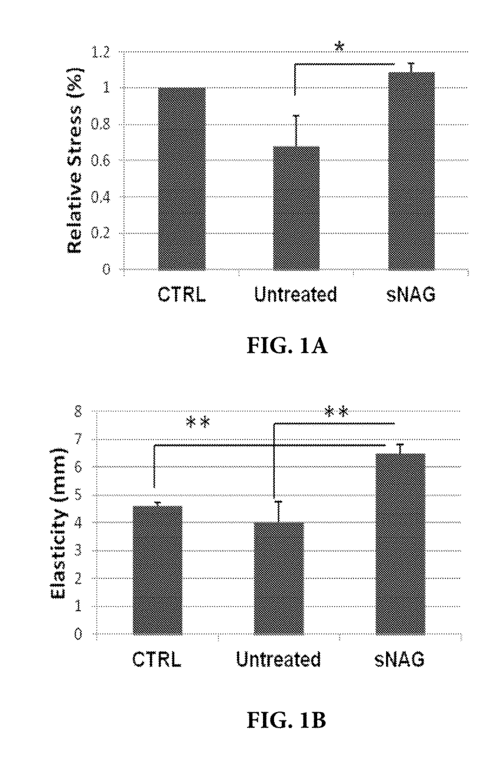
[0036] FIGS. 1A-1B. sNAG nanofibers increase tensile strength (relative stress) and elasticity of tissue. On day 21 post-wounding, wounds, both treated and untreated and unwounded control skin were harvested and subjected to tensile strength and elasticity testing using an Instron 5942 strain gauge extensometer and Bluehill 3 Testing Software. Tensile strength of the skin was determined by measuring the relative stress the skin could bear before breaking 20% and elasticity was measured in the mm extension. (FIG. 1A) Tensile strength measurement. (FIG. 1B) Elasticity measurement.
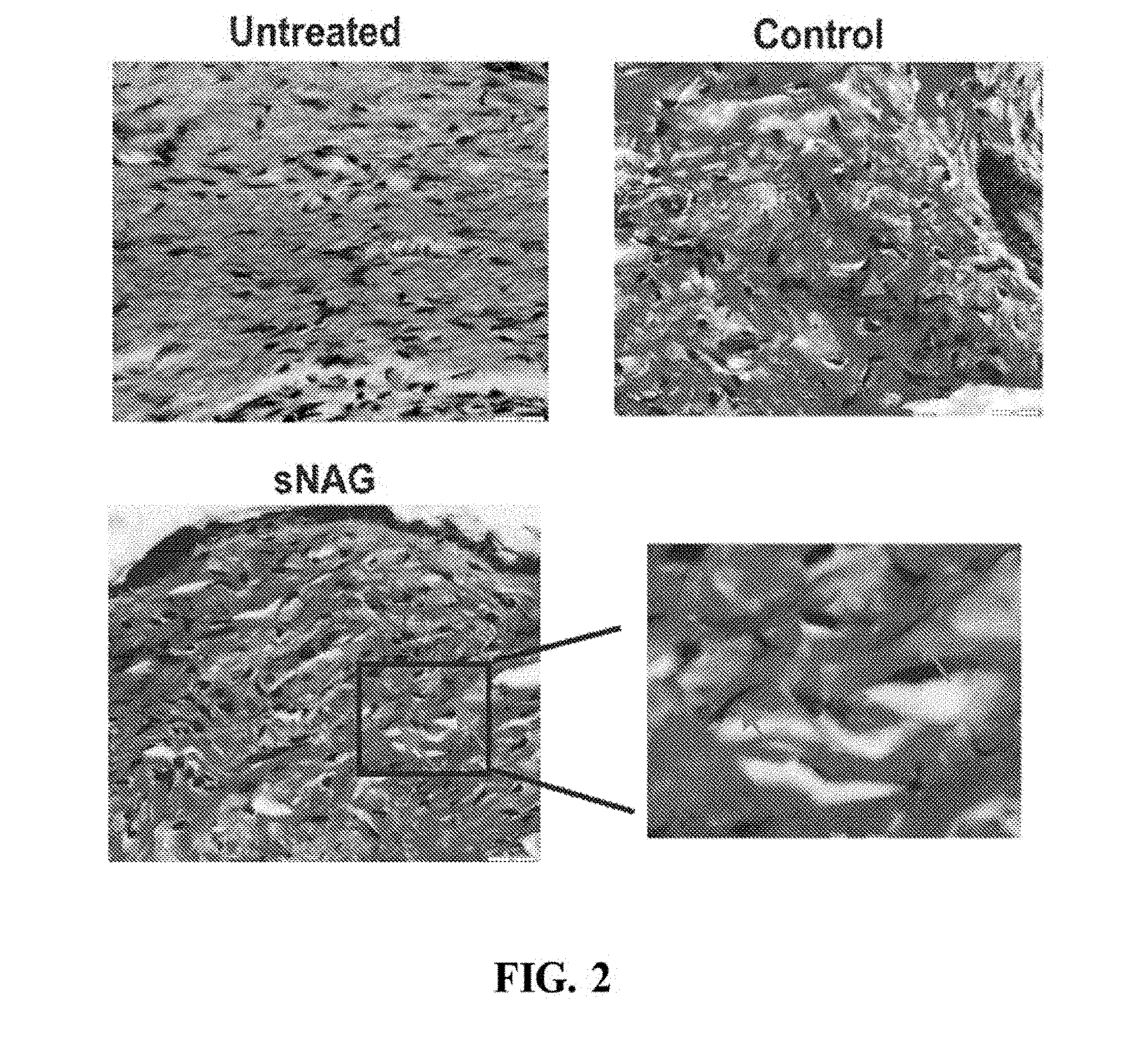
[0037] FIG. 2. sNAG nanofibers increase elastin production in tissue. On day 10 post-wounding, tissue sections from wounded animals treated with sNAG and control (untreated) were stained for elastin fibers using Van Geison staining procedures.
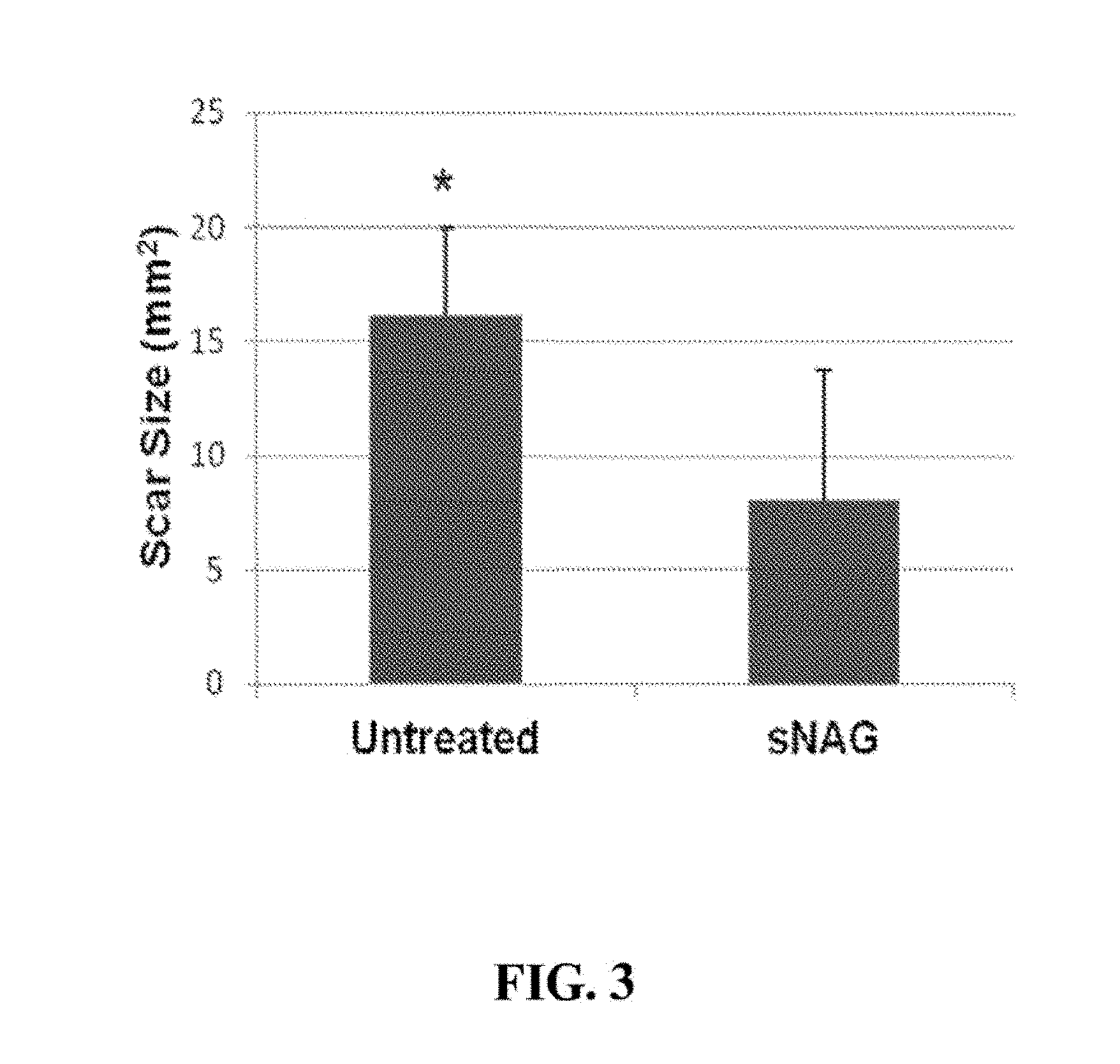
[0038] FIG. 3. sNAG nanofibers reduce scar size in tissue. On day 21 post-wounding, scars of wounded animals treated with sNAG and control (untreated) were measured using a caliper.
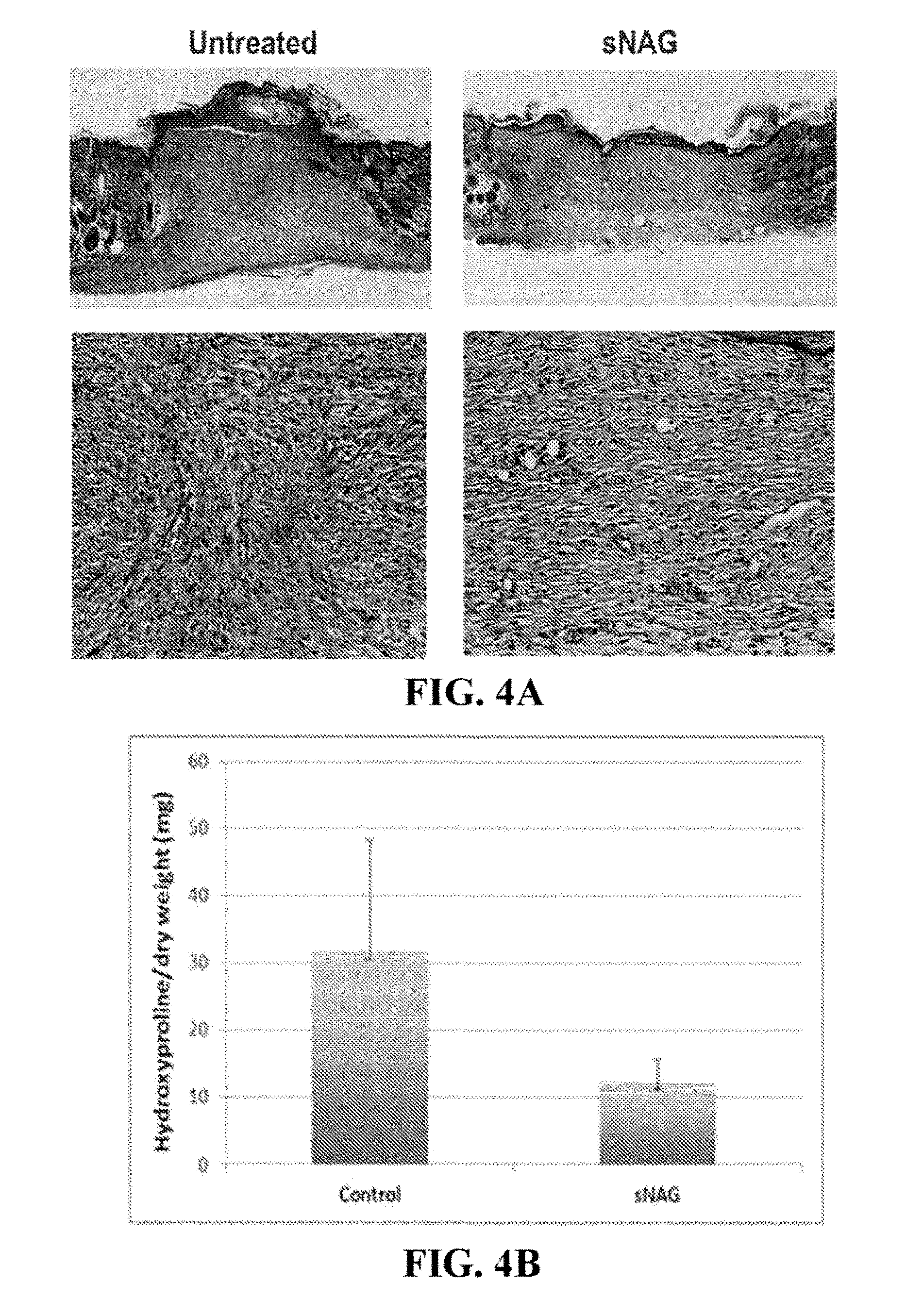
[0039] FIGS. 4A-4B. sNAG nanofibers increase amount of collagen and induce an organized alignment of collagen. (FIG. 4A) Masson's Trichrome stain of tissue section from wounds treated with sNAG and control (untreated), 10 days post-wounding. (FIG. 4B) Hydroxyproline assay quantitatively analyzing the amount of collagen deposition in wounds treated with sNAG and control (untreated), 10 days post-wounding.
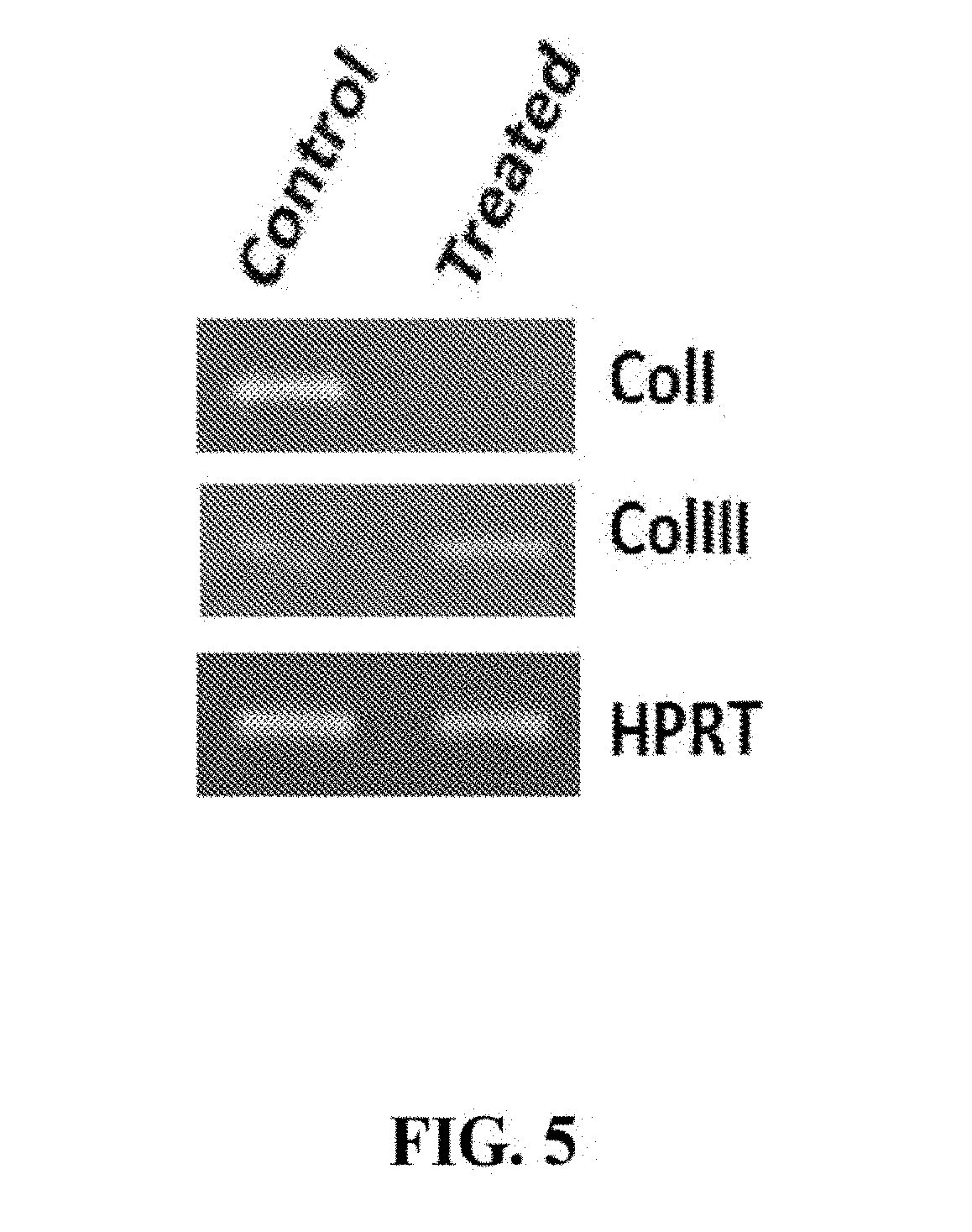
[0040] FIG. 5. sNAG nanofibers decrease collagen I expression and increase collagen III expression. RNA isolated from wounds sNAG treated and untreated (control) at day 5 post wounding were tested for expression of collagen type I and collagen type III by RT-PCR.
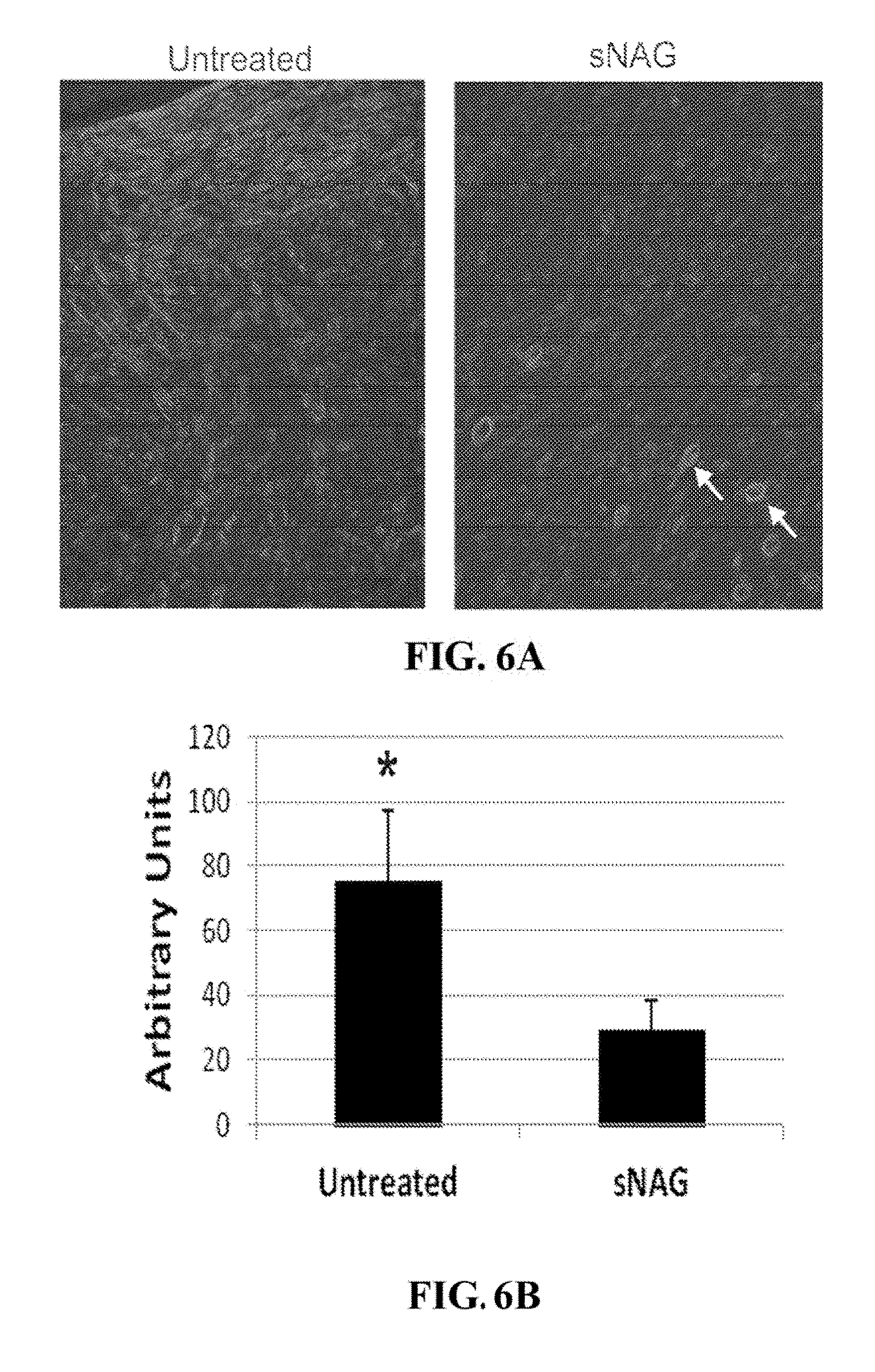
[0041] FIGS. 6A-6B. sNAG nanofibers decrease .alpha.-smooth muscle actin. (FIG. 6A) Wound sections treated with sNAG or untreated were labeled with an antibody directed against .alpha.-smooth muscle actin. (FIG. 6B) Quantification of the expression of .alpha.-smooth muscle actin in wound sections treated with sNAG or untreated.
5. DETAILED DESCRIPTION
[0042] The inventors of the present invention have found that sNAG nanofibers can increase tensile strength of tissue, increase elasticity of tissue, decrease total collagen content or abnormal collagen content in tissue, decrease collagen type I expression in tissue, increase collagen type III expression in tissue, induce organized alignment of collagen in tissue, increase elastin production in tissue, decrease smooth muscle actin expression in tissue, and/or decrease myofibroblast content in tissue. In particular, as demonstrated in the examples presented in Section 6, infra, the inventors of the present invention have found that sNAG nanofibers can increase tensile strength, increase elasticity, increase elastin production, decrease total collagen content, decrease collagen type I expression, increase collagen type III expression, induce organized alignment of collagen, and decrease alpha smooth muscle actin during cutaneous wound healing.
[0043] Thus, without being bound by any mechanism of action, sNAG nanofibers may act in the treatment of any conditions and diseases that are associated with decreased tensile strength of tissue, decreased elasticity of tissue, decreased elastin content (e.g., expression) in tissue, increased total collagen content or abnormal collagen content in tissue, increased collagen type I expression in tissue, decreased collagen type III expression in tissue, abnormal alignment of collagen in tissue, increased smooth muscle actin expression in tissue, and/or increased myofibroblast content in tissue. In one aspect, sNAG nanofibers may act in the treatment of any conditions, disorders and diseases that are associated with decreased tensile strength of the skin, decreased elasticity of the skin, decreased elastin content (e.g., expression) in the skin, increased total collagen content or abnormal collagen content in the skin, increased collagen type I content (e.g., expression) in the skin, decreased collagen type III content (e.g., expression) in the skin, abnormal (e.g., disorganized) alignment of collagen in the skin, increased smooth muscle actin expression in the skin, and/or increased myofibroblast content in the skin. In some embodiments, sNAG nanofibers may act to increase tensile strength, mediate organized alignment of collagen in the cells, increase elasticity and/or increase elastin production in the skin. In particular embodiments, sNAG nanofibers may act in the treatment of any conditions, disorders and diseases that are associated with cutaneous wound healing. In one embodiment, sNAG nanofibers may act to decrease scarring, increase tensile strength and/or mediate organized alignment of cells or collagen in the cells during cutaneous wound healing.
[0044] Accordingly, described herein is the use of sNAG nanofibers in methods for preventing and/or treating of any condition and disease associated with decreased tensile strength of tissue, decreased elasticity of tissue, decreased elastin content (e.g., expression) in tissue, increased total collagen content or abnormal collagen content in tissue, increased collagen type I content (e.g., expression) in tissue, decreased collagen type III content (e.g., expression) in tissue, abnormal (e.g., disorganized) alignment of collagen in tissue, increased smooth muscle actin content (e.g., expression) in tissue (such as, increased alpha smooth muscle actin content in tissue), and/or increased myofibroblast content in tissue. In particular, described herein are topical uses of sNAG nanofibers in methods for preventing and/or treating of any condition, disorder or disease associated with decreased tensile strength of tissue, decreased elasticity of tissue, decreased elastin content (e.g., expression) in tissue, increased total collagen content or abnormal collagen content in tissue, increased collagen type I content (e.g., expression) in tissue, decreased collagen type III content (e.g., expression) in tissue, abnormal (e.g., disorganized) alignment of collagen in tissue, increased smooth muscle actin (e.g., alpha smooth muscle actin) content (e.g., expression) in tissue, and/or increased myofibroblast content in tissue. Also described herein is the use of the sNAG nanofibers in the methods for decreasing scarring, increasing elasticity, or increasing tensile strength of the skin. In a particular embodiment, described herein is the use of the sNAG nanofibers in the methods for preventing or treating wrinkles or scars in the skin of a patient. In other embodiments, described herein is the use of sNAG nanofibers in the methods for decreasing scarring associated with cutaneous wounds using sNAG nanofibers. In some embodiments, described herein is the use of the sNAG nanofibers in methods for treatment of wrinkles, scars or cutaneous wounds in a patient, wherein the patient has decreased tensile strength of tissue, decreased elasticity of tissue, decreased elastin content in tissue, increased total collagen content or abnormal collagen content in tissue, increased collagen type I expression in tissue, decreased collagen type III expression in tissue, abnormal alignment of collagen in tissue, increased alpha smooth muscle actin, or increased myofibroblast content. In other embodiments, described herein is the use of sNAG nanofibers in the methods for treatment of Ehlers-Danlos Syndrome, Epidermolysis bullosa, scleroderma, osteoporosis, intervertebral disc disorder, degenerative disc disorder, osteoarthritis, or fibrosis. For example, the sNAG nanofibers may be used to reduce one or more symptoms of the above-listed disorders or diseases.
[0045] 5.1 sNAG Nanofibers
[0046] Described herein are sNAG nanofiber compositions. The sNAG nanofibers comprise fibers of poly-N-acetylglucosamine and/or a derivative(s) thereof, the majority of which are less than 30 microns in length and at least 1 micron in length as measured by any method known to one skilled in the art, for example, by scanning electron microscopy ("SEM"). Such sNAG nanofibers may be obtained, for example, as described herein.
[0047] In certain embodiments, the majority (and in certain embodiments, at least 60%, 70%, 80%, 90%, 95%, 98%, 99%, 99.5%, 99.8%, 99.9%, or 100%, or between 55% to 65%, 55% to 75%, 65% to 75%, 75% to 85%, 75% to 90%, 80% to 95%, 90% to 95%, or 95% to 99%) of the sNAG nanofibers are less than about 30, 25, 20, 15, 12, 10, 9, 8, 7, 6, 5, 4, or 3 microns in length, and at least 1 micron in length as measured by any method known to one skilled in the art, for example, by SEM. In specific embodiments, the majority (and in certain embodiments, at least 60%, 70%, 80%, 90%, 95%, 98%, 99%, 99.5%, 99.8%, 99.9%, or 100%, or between 55% to 65%, 55% to 75%, 65% to 75%, 75% to 85%, 75% to 90%, 80% to 95%, 90% to 95%, or 95% to 99%) of the sNAG nanofibers are less than about 15 microns or less than about 12 microns in length, and at least 1 micron in length as measured by any method known to one skilled in the art, for example, by SEM. In specific embodiments, all (100%) of the sNAG nanofibers are less than about 15 microns or less than about 10 microns in length, and at least 1 micron in length as measured by any method known to one skilled in the art, for example, by SEM. In certain embodiments, the majority (and in certain embodiments, at least 60%, 70%, 80%, 90%, 95%, 98%, 99%, 99.5%, 99.8%, 99.9%, or 100%, or between 55% to 65%, 55% to 75%, 65% to 75%, 75% to 85%, 75% to 90%, 80% to 95%, 90% to 95%, or 95% to 99%) of the sNAG nanofibers are equal to or less than 14, 13, 12, 11, 10, 9, 8 or 7 microns in length, and at least 1 micron in length as measured by any method known to one skilled in the art, for example, by SEM. In some embodiments, the majority (and in certain embodiments, at least 60%, 70%, 80%, 90%, 95%, 98%, 99%, 99.5%, 99.8%, 99.9%, or 100%, or between 55% to 65%, 55% to 75%, 65% to 75%, 75% to 85%, 75% to 90%, 80% to 95%, 90% to 95%, or 95% to 99%) of the sNAG nanofibers are between 1 to 15, 2 to 15, 2 to 14, 1 to 12, 2 to 12, 1 to 10, 2 to 10, 3 to 12, 3 to 10, 4 to 12, 4 to 10, 5 to 12, 5 to 10, 1 to 9, 2 to 9, 3 to 9, 1 to 8, 2 to 8, 3 to 8, 4 to 8, 1 to 7, 2 to 7, 3 to 7, 4 to 7, 1 to 6, 1 to 5, 1 to 4, or 1 to 3 microns in length as measured by any method known to one skilled in the art, for example, by SEM.
[0048] In a specific embodiment, the majority (and in certain embodiments, at least 60%, 70%, 80%, 90%, 95%, 98%, 99%, 99.5%, 99.8%, 99.9%, or 100%, or between 55% to 65%, 55% to 75%, 65% to 75%, 75% to 85%, 75% to 90%, 80% to 95%, 90% to 95%, or 95% to 99%) of the sNAG nanofibers are about 8, 7, 6, 5, 4, 3 or 2 microns in length as measured by any method known to one skilled in the art, for example, by SEM. In another specific embodiment, the majority (and in certain embodiments, at least 60%, 70%, 80%, 90%, 95%, 98%, 99%, 99.5%, 99.8%, 99.9%, or 100%, or between 55% to 65%, 55% to 75%, 65% to 75%, 75% to 85%, 75% to 90%, 80% to 95%, 90% to 95%, or 95% to 99%) of the sNAG nanofibers are between about 2 to about 10 microns, about 3 to about 8 microns, about 4 to about 7 microns, about 4 to about 10 microns, or about 5 to about 10 microns in length as measured by any method known to one skilled in the art, for example, by SEM. In another specific embodiment, all (100%) of the sNAG nanofibers are between about 2 to about 10 microns, about 3 to about 8 microns, about 4 to about 7 microns, about 4 to about 10 microns, or about 5 to about 10 microns in length as measured by any method known to one skilled in the art, for example, by SEM.
[0049] In certain embodiments, the sNAG nanofibers fibers are in a range between 0.005 to 5 microns in thickness and/or diameter as determined by electron microscopy. In specific embodiments, the sNAG nanofibers are about 0.01, 0.02, 0.03, 0.04, 0.05, 0.06, 0.07, 0.08, 0.09, 0.1, 0.2, 0.25, 0.3, 0.35, 0.4, 0.45, 0.5, 0.55, 0.6, 0.65, 0.7, 0.75, 0.8, 0.85, 0.9, 1, 1.1, 1.2, 1.3, 1.4, 1.5, 1.6, 1.7, 1.8, 1.9, 2, 2.2, 2.4, 2.6, 2.8, 3 or 4 microns in thickness and/or diameter on average, or any range in between (e.g., 0.02 to 2 microns, 0.02 to 1 microns, 0.02 to 0.75 microns, 0.02 to 0.5 microns, 0.02 to 0.5 microns, 0.05 to 1 microns, 0.05 to 0.75 microns, 0.05 to 0.5 microns, 0.1 to 1 microns, 0.1 to 0.75 microns, 0.1 to 0.5 microns, etc.). In specific embodiments, the majority (and in certain embodiments, at least 60%, 70%, 80%, 90%, 95%, 98%, 99%, 99.5%, 99.8%, 99.9%, or 100%, or between 55% to 65%, 55% to 75%, 65% to 75%, 75% to 85%, 75% to 90%, 80% to 95%, 90% to 95%, or 95% to 99%) of the sNAG nanofibers have a thickness or diameter of about 0.02 to 1 microns. In other specific embodiments, the majority (and in certain embodiments, at least 60%, 70%, 80%, 90%, 95%, 98%, 99%, 99.5%, 99.8%, 99.9%, or 100%, or between 55% to 65%, 55% to 75%, 65% to 75%, 75% to 85%, 75% to 90%, 80% to 95%, 90% to 95%, or 95% to 99%) of the sNAG nanofibers have a thickness or diameter of about 0.05 to 0.5 microns. In specific embodiments, all (100%) of the sNAG nanofibers have a thickness or diameter of about 0.02 to 1 microns or about 0.05 to 0.5 microns. In certain embodiments, the majority (and in certain embodiments, at least 60%, 70%, 80%, 90%, 95%, 98%, 99%, 99.5%, 99.8%, 99.9%, or 100%, or between 55% to 65%, 55% to 75%, 65% to 75%, 75% to 85%, 75% to 90%, 80% to 95%, 90% to 95%, or 95% to 99%) of the sNAG nanofibers have a thickness or diameter of about 0.02 to 2 microns, 0.02 to 1 microns, 0.02 to 0.75 microns, 0.02 to 0.5 microns, 0.02 to 0.5 microns, 0.05 to 1 microns, 0.05 to 0.75 microns, 0.05 to 0.5 microns, 0.1 to 1 microns, 0.1 to 0.75 microns, or 0.1 to 0.5 microns.
[0050] In certain embodiments, the majority (and in certain embodiments, at least 60%, 70%, 80%, 90%, 95%, 98%, 99%, 99.5%, 99.8%, 99.9%, or 100%, or between 55% to 65%, 55% to 75%, 65% to 75%, 75% to 85%, 75% to 90%, 80% to 95%, 90% to 95%, or 95% to 99%) of the sNAG nanofibers are between 1 and 15 microns, or between (or in the range of) 1 to 10 microns, 2 to 10 microns, 3 to 10 microns, 4 to 10 microns, 4 to 7 microns, 5 to 10 microns, or 5 to 15 microns in length and have a thickness or diameter of about 0.02 to 1 microns.
[0051] In certain embodiments, the molecular weight of the sNAG nanofibers is less than 100 kDa, 90 kDa, 80 kDa, 75 kDa, 70 kDa, 65 kDa, 60 kDa, 55 kDa, 50 kDa, 45 kDA, 40 kDa, 35 kDa, 30 kDa, or 25 kDa. In certain embodiments, the majority (and in certain embodiments, at least 60%, 70%, 80%, 90%, 95%, 98%, 99%, 99.5%, 99.8%, 99.9%, or 100%, or between 55% to 65%, 55% to 75%, 65% to 75%, 75% to 85%, 75% to 90%, 80% to 95%, 90% to 95%, or 95% to 99%) of the sNAG nanofibers have a molecular weight of less than 100 kDa, 90 kDa, 80 kDa, 75 kDa, 70 kDa, 65 kDa, 60 kDa, 55 kDa, 50 kDa, 45 kDA, 40 kDa, 35 kDa, 30 kDa, or 25 kDa. In other embodiments, the majority (and in certain embodiments, at least 60%, 70%, 80%, 90%, 95%, 98%, 99%, 99.5%, 99.8%, 99.9%, or 100%, or between 55% to 65%, 55% to 75%, 65% to 75%, 75% to 85%, 75% to 90%, 80% to 95%, 90% to 95%, or 95% to 99%) of the sNAG nanofibers have a molecular weight between about 5 kDa to 100 kDa, about 10 kDa to 100 kDa, about 20 kDa to 100 kDa, about 10 kDa to 80 kDa, about 20 kDa to 80 kDa, 20 kDa to 75 kDa, about 25 kDa to about 75 kDa, about 30 kDa to about 80 kDa, about 30 kDa to about 75 kDa, about 40 kda to about 80 kDa, about 40 kDa to about 75 kDa, about 40 kDa to about 70 kDa, about 50 kDa to about 70 kDa, or about 55 kDa to about 65 kDa. In one embodiment, the majority (and in certain embodiments, at least 60%, 70%, 80%, 90%, 95%, 98%, 99%, 99.5%, 99.8%, 99.9%, or 100%, or between 55% to 65%, 55% to 75%, 65% to 75%, 75% to 85%, 75% to 90%, 80% to 95%, 90% to 95%, or 95% to 99%) of the sNAG nanofibers have a molecular weight of about 60 kDa.
[0052] In certain embodiments, 1% to 5%, 5% to 10%, 5% to 15%, 20% to 30% or 25% to 30% of the sNAG nanofibers are deacetylated. In some embodiments, 1%, 5%, 10%, 15%, 20%, 25%, or 30% of the sNAG nanofibers are deacetylated. In other embodiments, less than 30%, 25%, 20%, 15%, 10%, 5%, 4%, 3%, 2% or 1% of the sNAG nanofibers are deacetylated. In some embodiments, equal to or more than 1%, 5%, 10%, 15%, 20%, 25%, 30%, 35%, 40%, 45%, 50%, 55%, 60%, 65%, 70%, 75%, 80%, 85%, 90%, 95% or 99%, or all (100%), of the sNAG nanofibers are deacetylated. In other embodiments, less than 1%, 5%, 10%, 15%, 20%, 25%, 30%, 35%, 40%, 45%, 50%, 55%, 60%, 65%, 70%, 75%, 80%, 85%, 90%, 95%, 99%, or 100% of the sNAG nanofibers are deacetylated.
[0053] In certain embodiments, 70% to 80%, 75% to 80%, 75% to 85%, 85% to 95%, 90% to 95%, 90% to 99% or 95% to 100% of the sNAG nanofibers are acetylated. In some embodiments, 70%, 75%, 80%, 85%, 90%, 95%, 98%, 99% or 100% of the sNAG nanofibers are acetylated. In other embodiments, more than 70%, 75%, 80%, 85%, 90%, 95%, 97%, 98%, 99%, 99.5% or 99.9% of the sNAG nanofibers are acetylated. In some embodiments, equal to or more than 1%, 5%, 10%, 15%, 20%, 25%, 30%, 35%, 40%, 45%, 50%, 55%, 60%, 65%, 70%, 75%, 80%, 85%, 90%, 95%, 97%, 98% or 99%, or all (100%), of the sNAG nanofibers are acetylated. In other embodiments, less than 1%, 5%, 10%, 15%, 20%, 25%, 30%, 35%, 40%, 45%, 50%, 55%, 60%, 65%, 70%, 75%, 80%, 85%, 90%, 95%, 97%, 98%, 99%, or 100% of the sNAG nanofibers are acetylated.
[0054] In some embodiments, the majority (and in certain embodiments, at least 60%, 70%, 80%, 90%, 95%, 98%, 99%, 99.5%, 99.9%, or 100%) of the sNAG nanofibers are between (or in the range of) 2 to 12 microns, 2 to 10 microns, 4 to 15 microns, 4 to 10 microns, 5 to 15 microns, or 5 to 10 microns, and such sNAG nanofibers are at least 70%, 75%, 80%, 85%, 90%, 95%, 97%, 98%, 99% or 100% acetylated.
[0055] In some embodiments, the sNAG nanofibers comprise at least one glucosamine monosaccharide, and may further comprise at least 10%, 20%, 30%, 40%, 50%, 60%, 70%, 80%, 90%, 95% or 99% of the N-acetylglucosamine monosaccharides. In other embodiments, the sNAG nanofibers comprise at least one N-acetylglucosamine monosaccharide, and may further comprise at least 10%, 20%, 30%, 40%, 50%, 60%, 70%, 80%, 90%, 95% or 99% of glucosamine monosaccharides.
[0056] In one aspect, the sNAG nanofibers increase the metabolic rate of serum-starved human umbilical cord vein endothelial cells ("EC") in a MTT assay. A MTT assay is a laboratory test and a standard colorimetric assay (an assay which measures changes in color) for measuring cellular proliferation (cell growth). Briefly, yellow MTT (3-(4,5-Dimethylthiazol-2-yl)-2,5-diphenyltetrazolium bromide, a tetrazole) is reduced to purple formazan in the mitochondria of living cells. This reduction takes place only when mitochondrial reductase enzymes are active, and therefore conversion can be directly related to the number of viable (living) cells. The MTT assay is described in WO2011/130646 and WO2012/142581, each of which is incorporated by reference herein in its entirety. The metabolic rate of cells may also be determined by other techniques commonly known to the skilled artisan.
[0057] In another aspect, the sNAG nanofibers do not rescue apoptosis of serum-starved EC in a trypan blue exclusion test. A trypan blue exclusion test is a dye exclusion test used to determine the number of viable cells present in a cell suspension. It is based on the principle that live cells possess intact cell membranes that exclude certain dyes, such as trypan blue, Eosin, or propidium, whereas dead cells do not. The trypan blue assay is described in WO2011/130646 and WO2012/142581, each of which is incorporated by reference herein in its entirety. The viability of cells may also be determined by other techniques commonly known to the skilled artisan.
[0058] In certain embodiments, compositions comprising the sNAG nanofibers are described, wherein the sNAG nanofibers increase the metabolic rate of serum-starved human umbilical cord vein endothelial cells in a MTT assay and/or do not rescue apoptosis of serum-starved human umbilical cord vein endothelial cells in a trypan blue exclusion test. In some embodiments, the sNAG nanofibers increase the metabolic rate of serum-starved human umbilical cord vein endothelial cells in a MTT assay and do not rescue apoptosis of serum-starved human umbilical cord vein endothelial cells in a trypan blue exclusion test.
[0059] In a specific embodiment, the sNAG nanofibers are biocompatible. Biocompatibility may be determined by a variety of techniques, including, but not limited to such procedures as the elution test, intramuscular implantation, or intracutaneous or systemic injection into animal subjects. Such tests are described in U.S. Pat. No. 6,686,342 (see, e.g., Example 10), which is incorporated by reference herein in its entirety. Some of the biocompatibility tests are also described in WO2011/130646 and WO2012/142581, each of which is incorporated by reference herein in its entirety.
[0060] In certain embodiments, the sNAG nanofibers used in the methods described herein are non-reactive in a biocompatibility test or tests. For example, the sNAG nanofibers used in the methods described herein may be non-reactive when tested in an elution test, an intramuscular implantation test, an intracutaneous test, and/or a systemic test. In other embodiments, the sNAG nanofibers used in the methods described herein have Grade 0 or Grade 1 test score when tested in an elution test, an intramuscular implantation test, an intracutaneous test, or a systemic test. In yet another embodiment, the sNAG nanofibers used in the methods described herein are at most mildly reactive when tested in an elution test, an intramuscular implantation test, an intracutaneous test, and/or a systemic test. In certain embodiments, the compositions described herein do not cause an allergenic reaction or an irritation. In other embodiments, the compositions described herein cause at most a mild allergenic reaction or a mild irritation, e.g., at the site of application. The relevant tests and evaluation of test results are described in, e.g., U.S. Pat. No. 6,686,342, WO2011/130646 and WO2012/142581, each of which is incorporated by reference herein in its entirety.
[0061] In a specific embodiment, the sNAG nanofibers are non-reactive when tested in an intramuscular implantation test. In one aspect, an intramuscular implantation test is an intramuscular implantation test--ISO 4 week implantation, as described in Section 6.8.3, infra. In certain embodiments, the sNAG nanofibers display no biological reactivity as determined by an elution test (Elution Test Grade=0). In some embodiments, the sNAG nanofibers have a test score equal to "0" and/or are at most a negligible irritant as determined by intracutaneous injection test. In some embodiments, the sNAG nanofibers elicit no intradermal reaction (i.e., Grade I reaction) in Kligman test and/or have a weak allergenic potential as determined by Kligman test. WO2011/130646 and WO2012/142581, each of which is incorporated by reference herein in its entirety, show that sNAG nanofibers are non-reactive in an intramuscular implantation test, an intracutaneous injection test, and Kligman test.
[0062] In certain aspects, the sNAG nanofibers are immunoneutral (i.e., they do not elicit an immune response).
[0063] In some embodiments, the sNAG nanofibers are biodegradable. The sNAG nanofibers preferably degrade within about 1 day, 2 days, 3 days, 5 days, 7 days (1 week), 8 days, 10 days, 12 days, 14 days (2 weeks), 17 days, 21 days (3 weeks), 25 days, 28 days (4 weeks), 30 days, 1 month, 35 days, 40 days, 45 days, 50 days, 55 days, 60 days, 2 months, 65 days, 70 days, 75 days, 80 days, 85 days, 90 days, 3 months, 95 days, 100 days or 4 months after administration or implantation into a patient.
[0064] In certain embodiments, the sNAG nanofibers do not cause a detectable foreign body reaction. A foreign body reaction, which may occur during wound healing, includes accumulation of exudate at the site of injury, infiltration of inflammatory cells to debride the area, and the formation of granulation tissue. The persistent presence of a foreign body can inhibit full healing. Rather than the resorption and reconstruction that occurs in wound healing, the foreign body reaction is characterized by the formation of foreign body giant cells, encapsulation of the foreign object, and chronic inflammation. Encapsulation refers to the firm, generally avascular collagen shell deposited around a foreign body, effectively isolating it from the host tissues. In one embodiment, treatment of a site (e.g., a wound or a site of a bacterial infection in a wound) with the sNAG nanofibers does not elicit a detectable foreign body reaction in 1 day, 3 days, 5 days, 7 days, 10 days or 14 days after treatment. In one such embodiment, treatment of a site (e.g., a wound) with the sNAG nanofibers does not elicit a foreign body encapsulations in 1 day, 3 days, 5 days, 7 days, 10 days or 14 days after treatment.
[0065] In some embodiments, the sNAG nanofibers (i) comprise fibers, wherein majority of the fibers are between about 1 and 15 microns in length, and (ii) (a) increase the metabolic rate of serum-starved EC in a MTT assay and/or do not rescue apoptosis of serum-starved EC in a trypan blue exclusion test, and (b) are non-reactive when tested in an intramuscular implantation test. In certain embodiments, the sNAG nanofibers (i) comprise fibers, wherein majority of the fibers are between about 1 and 12 microns in length, and (ii) (a) increase the metabolic rate of serum-starved EC in a MTT assay and/or do not rescue apoptosis of serum-starved EC in a trypan blue exclusion test, and (b) are non-reactive when tested in an intramuscular implantation test. In some embodiments, the sNAG nanofibers (i) comprise fibers, wherein majority of the fibers are between (or in the range of) 1 to 10 microns, 2 to 10 microns, 4 to 10 microns, 5 to 10 microns, or 5 to 15 microns in length, and (ii) (a) increase the metabolic rate of serum-starved EC in a MTT assay and/or do not rescue apoptosis of serum-starved EC in a trypan blue exclusion test, and (b) are non-reactive when tested in an intramuscular implantation test. In some embodiments, the sNAG nanofibers (i) comprise fibers, wherein majority of the fibers are between about 4 and 10 microns in length, and (ii) (a) increase the metabolic rate of serum-starved EC in a MTT assay and/or do not rescue apoptosis of serum-starved EC in a trypan blue exclusion test, and (b) are non-reactive when tested in an intramuscular implantation test. In certain embodiments, the sNAG nanofibers (i) comprise fibers, wherein majority of the fibers are between about 4 and 7 microns in length, and (ii) (a) increase the metabolic rate of serum-starved EC in a MTT assay and/or do not rescue apoptosis of serum-starved EC in a trypan blue exclusion test, and (b) are non-reactive when tested in an intramuscular implantation test.
[0066] In certain embodiments, the sNAG nanofibers do not have a direct effect on the growth or survival of bacteria, such as S. aureus, as determined by one skilled in the art. In other embodiments, sNAG nanofibers do not have a direct effect on the growth or survival of bacteria, such as S. aureus, as determined by the methods set forth in WO2011/130646, which is incorporated by reference herein in its entirety. In some embodiments, the sNAG nanofibers do not have a direct effect in vitro on bacterial growth or survival. In one embodiment, the sNAG nanofibers do not have a direct effect (e.g., in vitro) on growth or survival of gram-negative bacteria. In another embodiment, the sNAG nanofibers do not have a direct effect (e.g., in vitro) on growth or survival of gram-positive bacteria. In yet another embodiment, the sNAG nanofibers do not have a direct effect (e.g., in vitro) on growth or survival of either gram-positive or gram-negative bacteria.
[0067] In some embodiments, the sNAG nanofibers (i) comprise fibers, wherein majority of the fibers are between (or in the range of) about 1 and 15 microns, 1 and 12 microns, 1 and 10 microns, 4 and 10 microns, 4 and 15 microns, 5 and 10 microns, 5 and 15 microns, or 4 and 7 microns in length, (ii) do not have an effect on bacterial growth or survival of Staphylococcus aureus bacterial cultures in vitro, and (iii) are non-reactive when tested in a biocompatibility test (e.g., an intramuscular implantation test).
[0068] In certain embodiments, the sNAG nanofibers induce a certain pattern of gene expression (RNA or protein expression as determined by, e.g., RT-PCR, microarray or ELISA) in a cell, tissue or organ treated with or exposed to a sNAG nanofiber composition.
[0069] In certain embodiments, the sNAG nanofibers or a composition comprising the sNAG nanofibers reduces expression of collagen type I. In certain embodiments, the sNAG nanofibers or a composition comprising the sNAG nanofibers increases expression of collagen type III. In certain embodiments, the sNAG nanofibers or a composition comprising the sNAG nanofibers reduces total expression of collagen proteins.
[0070] In certain embodiments, the sNAG nanofibers or a composition comprising the sNAG nanofibers increases expression of elastin protein.
[0071] In certain embodiments, the sNAG nanofibers or a composition comprising the sNAG nanofibers reduces expression of one or more actin proteins. In certain embodiments, the sNAG nanofibers or a composition comprising the sNAG nanofibers reduces expression of one or more actin proteins in smooth muscle cells (e.g., alpha smooth muscle actin protein).
[0072] In some embodiments, the sNAG nanofibers or a composition comprising the sNAG nanofibers reduce expression of one or more of the above-listed proteins in the amount equal to or more than about 0.25 fold, 0.5 fold, 1 fold, 1.5 fold, 2 fold, 2.5 fold, 3 fold, 3.5 fold, 4 fold, 4.5 fold, 5 fold, 6 fold, 7 fold, 8 fold, 9 fold, 10 fold, 12 fold, 15 fold or 20 fold as compared to the level of expression of the one or more of the above-listed proteins in a cell, tissue or organ of a subject before treatment with the sNAG nanofibers (e.g., a known average level of expression of the one or more of the above-listed proteins). In some embodiments, the sNAG nanofibers or a composition comprising the sNAG nanofibers reduce expression of one or more of the above-listed proteins in the amount equal to or more than about 10%, 25%, 50%, 75% or 100%, 125%, 150%, 175%, 200%, 225%, 250%, 275%, 300%, 350%, 400%, 450%, 500%, 550%, 600%, 650%, 700%, 750%, 800%, 900% or 1000% the level of expression of the one or more of the above-listed proteins in a cell, tissue or organ of a subject before treatment with the sNAG nanofibers (e.g., a known average level of expression of the one or more of the above-listed proteins).
[0073] In some embodiments, the sNAG nanofibers or a composition comprising the sNAG nanofibers induce expression of one or more defensin proteins, one or more defensin-like proteins, and/or one or more Toll-like receptors.
[0074] In certain embodiments, the sNAG nanofibers or a composition comprising the sNAG nanofibers induces/increases expression of one or more .alpha.-defensins (e.g., DEFA1 (i.e., .alpha.-defensin 1), DEFA1B, DEFA3, DEFA4, DEFA5, DEFA6), one or more .beta.-defensins (e.g., DEFB1 (i.e., .beta.-defensin 1), DEFB2, DEFB4, DEFB103A, DEFB104A, DEFB105B, DEFB107B, DEFB108B, DEFB110, DEFB112, DEFB114, DEFB118, DEFB119, DEFB123, DEFB124, DEFB125, DEFB126, DEFB127, DEFB128, DEFB129, DEFB131, DEFB136), and/or one or more .theta.-defensins (e.g., DEFT1P). In some embodiments, the sNAG nanofibers or a composition comprising the sNAG nanofibers induce/increase expression of one or more of DEFA1, DEFA3, DEFA4, DEFA5, DEFB1, DEFB3, DEFB103A, DEFB104A, DEFB108B, DEFB112, DEFB114, DEFB118, DEFB119, DEFB123, DEFB124, DEFB125, DEFB126, DEFB128, DEFB129 and DEFB131. In certain embodiments, the sNAG nanofibers or a composition comprising the sNAG nanofibers induces/increases expression of one or more Toll receptors (e.g., TLR1, TLR2, TLR3, TLR4, TLR5, TLR6, TLR7, TLR8, TLR9, TLR10, TLR11, and/or TLR12). In other embodiments, the sNAG nanofibers or a composition comprising the sNAG nanofibers induces/increases expression of one or more of IL-1, CEACAM3, SPAG11, SIGIRR (IL1-like receptor), IRAK1, IRAK2, IRAK4, TBK1, TRAF6 and IKKi. In some embodiments, the sNAG nanofibers or a composition comprising the sNAG nanofibers induces/increases expression of one or more of IRAK2, SIGIRR, TLR1, TLR2, TLR4, TLR7, TLR8, TLR10 and TRAF6. In one embodiment, the sNAG nanofibers or a composition comprising the sNAG nanofibers induces/increases expression of at least one of the above-listed gene products.
[0075] In some embodiments, the sNAG nanofibers or a composition comprising the sNAG nanofibers induces/increases expression of one or more of the above-listed gene products in the amount equal to or more than about 0.25 fold, 0.5 fold, 1 fold, 1.5 fold, 2 fold, 2.5 fold, 3 fold, 3.5 fold, 4 fold, 4.5 fold, 5 fold, 6 fold, 7 fold, 8 fold, 9 fold, 10 fold, 12 fold, 15 fold or 20 fold as compared to the level of expression of the one or more of the above-listed gene products in a cell, tissue or organ of a subject before treatment with the sNAG nanofibers (e.g., a known average level of expression of the one or more of the above-listed gene products). In some embodiments, the sNAG nanofibers or a composition comprising the sNAG nanofibers induces/increases expression of one or more of the above-listed gene products in the amount equal to or more than about 10%, 25%, 50%, 75%, 100%, 125%, 150%, 175%, 200%, 225%, 250%, 275%, 300%, 350%, 400%, 450%, 500%, 550%, 600%, 650%, 700%, 750%, 800%, 900% or 1000% the level of expression of the one or more of the above-listed gene products in a cell, tissue or organ of a subject before treatment with the sNAG nanofibers (e.g., a known average level of expression of the one or more of the above-listed gene products).
[0076] In some embodiments, the sNAG nanofibers but not long poly-N-acetylglucosamine, chitin and/or chitosan induce/increase expression of the one or more gene products listed above, as determined by a method known to one skilled in the art, or described herein. In some of these embodiments, long poly-N-acetylglucosamine, chitin and/or chitosan do not induce/increase expression of the one or more gene products listed above or induce lower level (e.g., more than 1.25 fold, 1.5 fold, 2 fold, 2.5 fold, 3 fold, 3.5 fold, 4 fold, 4.5 fold, 5 fold, 6 fold, 7 fold, 8 fold, 9 fold, or 10 fold lower) of expression of the one or more gene products listed above as compared to the level of expression of the one or more gene products listed above induced by the sNAG nanofibers, as determined by a method known to one skilled in the art, or described herein.
[0077] In certain embodiments, the sNAG nanofibers but not long poly-N-acetylglucosamine, chitin and/or chitosan reduce/decrease expression of the one or more gene products listed above, as determined by a method known to one skilled in the art, or described herein. In some of these embodiments, long poly-N-acetylglucosamine, chitin and/or chitosan do not reduce/decrease expression of the one or more gene products listed above or induce a lower level (e.g., more than 1.25 fold, 1.5 fold, 2 fold, 2.5 fold, 3 fold, 3.5 fold, 4 fold, 4.5 fold, 5 fold, 6 fold, 7 fold, 8 fold, 9 fold, or 10 fold lower) of expression of the one or more gene products listed above as compared to the level of expression of the one or more gene products listed above reduced by the sNAG nanofibers, as determined by a method known to one skilled in the art, or described herein.
[0078] In certain embodiments, the sNAG nanofibers or a composition comprising the sNAG nanofibers induce a gene expression profile that is consistent with, similar to, about the same as, or equivalent to one or more gene expression profiles demonstrated in WO 2011/130646 and WO 2012/142581, each of which is incorporated by reference herein in its entirety (see Tables I, II, III, V, VIII and IX, Sections 6.2-6.5).
[0079] In certain embodiments, the sNAG nanofibers or a composition comprising the sNAG nanofibers induce a gene expression profile that differs from the profile induced by long poly-N-acetylglucosamine polymers or fibers. In specific embodiments, a gene expression profile induced by the sNAG nanofibers is consistent with, similar to, about the same as, or equivalent to that shown in WO 2011/130646 and WO 2012/142581, each of which is incorporated by reference herein in its entirety (see Tables I, II, III, V, VIII and IX, Sections 6.2-6.5), whereas gene expression profile induced by long poly-N-acetylglucosamine polymers or fibers is consistent with, similar to, about the same with, or equivalent to that shown in Table VIII and/or IX, Section 6.5 of WO 2011/130646 and WO 2012/142581. In other embodiments, the sNAG nanofibers or a composition comprising the sNAG nanofibers induce a gene expression profile that differs from the gene expression profile induced by chitin or chitosan.
[0080] In a specific embodiment, the sNAG nanofibers are obtained by irradiating poly-N-acetylglucosamine and/or a derivative thereof. See Section 5.1.1, infra, regarding poly-N-acetylglucosamine and derivatives thereof and Section 5.2, infra, regarding methods for producing the sNAG nanofibers using irradiation. Irradiation may be used to reduce the length of poly-N-acetylglucosamine fibers and/or poly-N-acetylglucosamine derivative fibers to form shortened poly-.beta.-1.fwdarw.4-N-acetylglucosamine fibers and/or shortened poly-N-acetylglucosamine derivative fibers, i.e. sNAG nanofibers. Specifically, irradiation may be used to reduce the length and molecular weight of poly-N-acetylglucosamine or a derivative thereof without disturbing its microstructure. The infrared spectrum (IR) of sNAG nanofibers is similar to, about the same as, or equivalent to that of the non-irradiated poly-.beta.-1.fwdarw.4-N-acetylglucosamine or a derivative thereof.
[0081] In one embodiment, the sNAG nanofibers are not derived from chitin or chitosan. Whereas in another embodiment, the compositions described herein may be derived from chitin or chitosan, or the sNAG nanofibers may be derived from chitin or chitosan.
[0082] 5.1.1 Poly-N-Acetylglucosamine and Derivatives Thereof
[0083] U.S. Pat. Nos. 5,622,834; 5,623,064; 5,624,679; 5,686,115; 5,858,350; 6,599,720; 6,686,342; 7,115,588 and U.S. Patent Pub. 2009/0117175 (each of which is incorporated herein by reference in its entirety) describe the poly-N-acetylglucosamine and derivatives thereof, and methods of producing the same. In some embodiments, the poly-N-acetylglucosamine has a .beta.-1.fwdarw.4 configuration. In other embodiments, the poly-N-acetylglucosamine has a .alpha.-1.fwdarw.4 configuration. The poly-N-acetylglucosamine and derivatives thereof may be in the form of a polymer or in the form of a fiber.
[0084] Poly-N-acetylglucosamine can, for example, be produced by, and may be purified from, microalgae, preferably diatoms. The diatoms which may be used as starting sources for the production of the poly-N-acetylglucosamine include, but are not limited to members of the Coscinodiscus genus, the Cyclotella genus, and the Thalassiosira genus. Poly-N-acetylglucosamine may be obtained from diatom cultures via a number of different methods, including the mechanical force method and chemical/biological method known in the art (see, e.g., U.S. Pat. Nos. 5,622,834; 5,623,064; 5,624,679; 5,686,115; 5,858,350; 6,599,720; 6,686,342; and 7,115,588, each of which is incorporated herein by reference in its entirety). In certain embodiments, the poly-N-acetylglucosamine is not derived from one or more of the following: a shell fish, a crustacean, an insect, a fungi or yeasts.
[0085] In one embodiment, poly-.beta.-1.fwdarw.4-N-acetylglucosamine is derived from a process comprising a) treating a microalgae comprising a cell body and a poly-.beta.-1.fwdarw.4-N-acetylglucosamine polymer fiber with a biological agent (such as hydrofluoric) capable of separating the N-acetylglucosamine polymer fiber from the cell body for a sufficient time so that the poly-.beta.-1.fwdarw.4-N-acetylglucosamine polymer fiber is released from the cell body; b) segregating the poly-.beta.-1.fwdarw.4-N-acetylglucosamine polymer fiber from the cell body; and c) removing contaminants from the segregated poly-.beta.-1.fwdarw.4-N-acetylglucosamine polymer fiber, so that the poly-.beta.-1.fwdarw.4-N-acetylglucosamine polymer is isolated and purified.
[0086] In other embodiments, the poly-.beta.-1.fwdarw.4-N-acetylglucosamine may be derived from one or more of the following: a shell fish, a crustacean, an insect, a fungi or yeasts. In certain embodiments, the compositions described herein do not comprise chitin or chitosan.
[0087] One or more of the monosaccharide units of the poly-N-acetylglucosamine may be deacetylated. In certain embodiments, 1% to 5%, 5% to 10%, 5% to 15%, 20% to 30% or 25% to 30% of the poly-N-acetylglucosamine is deacetylated. In some embodiments, 1%, 5%, 10%, 15%, 20%, 25%, or 30% of the poly-N-acetylglucosamine is deacetylated. In other embodiments, less than 30%, 25%, 20%, 15%, 10%, 5%, 4%, 3%, 2% or 1% of the poly-N-acetylglucosamine is deacetylated. In some embodiments, equal to or more than 1%, 5%, 10%, 15%, 20%, 25%, 30%, 35%, 40%, 45%, 50%, 55%, 60%, 65%, 70%, 75%, 80%, 85%, 90%, 95% or 99%, or all (100%), of the poly-N-acetylglucosamine is deacetylated. In other embodiments, less than 1%, 5%, 10%, 15%, 20%, 25%, 30%, 35%, 40%, 45%, 50%, 55%, 60%, 65%, 70%, 75%, 80%, 85%, 90%, 95%, 99%, or 100% of the poly-N-acetylglucosamine is deacetylated.
[0088] In certain embodiments, a poly-N-acetylglucosamine composition comprises 70% to 80%, 75% to 80%, 75% to 85%, 85% to 95%, 90% to 95%, 90% to 99% or 95% to 100% of acetylated glucosamine (i.e., N-acetylglucosamine) monosaccharides. In some embodiments, a poly-N-acetylglucosamine composition comprises 70%, 75%, 80%, 85%, 90%, 95%, 97%, 98%, 99% or 100% of acetylated glucosamine (i.e., N-acetylglucosamine) monosaccharides. In other embodiments, a poly-N-acetylglucosamine composition comprises more than 70%, 75%, 80%, 85%, 90%, 95%, 97%, 98%, 99%, 99.5% or 99.9% of the acetylated glucosamine. In some embodiments, a poly-N-acetylglucosamine composition comprises equal to or more than 1%, 5%, 10%, 15%, 20%, 25%, 30%, 35%, 40%, 45%, 50%, 55%, 60%, 65%, 70%, 75%, 80%, 85%, 90%, 95%, 97%, 98%, or 99%, or all (100%), of the acetylated glucosamine. In other embodiments, a poly-N-acetylglucosamine composition comprises less than 1%, 5%, 10%, 15%, 20%, 25%, 30%, 35%, 40%, 45%, 50%, 55%, 60%, 65%, 70%, 75%, 80%, 85%, 90%, 95%, 99%, or 100% of the acetylated glucosamine.
[0089] In some embodiments, a poly-N-acetylglucosamine composition comprises at least one glucosamine monosaccharide, and may further comprise at least 10%, 20%, 30%, 40%, 50%, 60%, 70%, 80%, 90%, 95% or 99% of N-acetylglucosamine monosaccharides. In other embodiments, a poly-N-acetylglucosamine composition comprises at least one N-acetylglucosamine monosaccharide, and may further comprise at least 10%, 20%, 30%, 40%, 50%, 60%, 70%, 80%, 90%, 95% or 99% of glucosamine monosaccharides.
[0090] Derivatives of poly-N-acetylglucosamine may also be used in a composition described herein. Derivatives of poly-N-acetylglucosamine and methods of making such derivatives are described in U.S. Pat. No. 5,623,064 (see, e.g., Section 5.4), which is incorporated by reference herein in its entirety. Derivatives of poly-N-acetylglucosamine may include, but are not limited to, partially or completely deacetylated poly-N-acetylglucosamine, or its deacetylated derivatives. Further, poly-N-acetylglucosamine may be derivatized by being sulfated, phosphorylated and/or nitrated. Poly-N-acetylglucosamine derivatives include, e.g., sulfated poly-N-acetylglucosamine derivatives, phosphorylated poly-N-acetylglucosamine derivatives, or nitrated poly-N-acetylglucosamine derivatives. Additionally, one or more of the monosaccharide units of the poly-N-acetylglucosamine may contain one or more sulfonyl groups one or more O-acyl groups. In addition, one or more of the monosaccharides of the deacetylated poly-N-acetylglucosamine may contain an N-acyl group. One or more of the monosaccharides of the poly-N-acetylglucosamine or of its deacetylated derivative, may contain an O-alkyl group. One or more of the monosaccharide units of the poly-N-acetylglucosamine may be an alkali derivative. One or more of the monosaccharide units of the deacetylated derivative of poly-N-acetylglucosamine may contain an N-alkyl group. One or more of the monosaccharide units of the deacetylated derivative of poly-N-acetylglucosamine may contain at least one deoxyhalogen derivative. One or more of the monosaccharide units of the deacetylated derivative of poly-N-acetylglucosamine may form a salt. One or more of the monosaccharide units of the deacetylated derivative of poly-N-acetylglucosamine may form a metal chelate. In a specific embodiment, the metal is zinc. One or more of the monosaccharide units of the deacetylated derivative of poly-N-acetylglucosamine may contain an N-alkylidene or an N-arylidene group. In one embodiment, the derivative is an acetate derivative. In another embodiment, the derivative is not an acetate derivative. In one embodiment the poly-N-acetylglucosamine or deacetylated poly-N-acetylglucosamine is derivatized with lactic acid. Wherein, in another embodiment, the derivative is not derivatized with lactic acid.
[0091] 5.2 Methods of Producing sNAG Nanofibers
[0092] The poly-N-acetylglucosamine polymers or fibers, and any derivatives of poly-N-acetylglucosamine polymers or fibers described above, can be irradiated as dry polymers or fibers or polymer or fiber membranes. Alternatively, poly-N-acetylglucosamine polymers or fibers, and any derivatives of poly-N-acetylglucosamine polymers or fibers described above, can be irradiated when wet. The methods of making sNAG nanofibers by irradiation and the sNAG nanofibers so produced have been described in U.S. Patent Pub. No. 2009/0117175, which is incorporated by reference herein in its entirety.
[0093] In certain embodiments, the poly-N-acetylglucosamine polymers or fibers are formulated into a suspension/slurry or wet cake for irradiation. Irradiation can be performed prior to, concurrently with or following the formulation of the polymers or fibers into its final formulation, such as a dressing. Generally, the polymer or fiber content of suspensions/slurries and wet cakes can vary, for example from about 0.5 mg to about 50 mg of polymer or fiber per 1 ml of distilled water are used for slurries and from about 50 mg to about 1000 mg of polymer or fiber per 1 ml of distilled water are use for wet cake formulations. The polymer or fiber may first be lyophilized, frozen in liquid nitrogen, and pulverized, to make it more susceptible to forming a suspension/slurry or wet cake. Also, the suspensions/slurries can be filtered to remove water such that a wet cake is formed. In certain aspects, the polymer or fiber is irradiated as a suspension comprising about 0.5 mg, 1 mg, 2 mg, 3 mg, 4 mg, 5 mg, 6 mg, 7 mg, 8 mg, 9 mg, 10 mg, 12 mg, 15 mg, 18 mg, 20 mg, 25 mg or 50 mg of polymer or fiber per ml of distilled water, or any range in between the foregoing embodiments (e.g., 1-10 mg/ml, 5-15 mg/ml, 2-8 mg/ml, 20-50 mg/ml, etc.). In other aspects, the polymer or fiber is irradiated as a wet cake, comprising about 50-1,000 mg polymer or fiber per 1 ml of distilled water. In specific embodiments, the wet cake comprises about 50, 100, 200, 300, 400, 500, 600, 700, 800, 900 or 1000 mg of polymer or fiber per 1 ml distilled water, or any range in between (e.g., 100-500 mg/ml, 300-600 mg/ml, 50-1000 mg/ml, etc.).
[0094] The irradiation is preferably in the form of gamma radiation, e-beam radiation, or x-rays. Two sources of irradiation are preferred: radioactive nuclides and electricity. In specific embodiment, the radioactive nuclides are cobalt-60 and cesium-137. Both of these nuclides emit gamma rays, which are photons containing no mass. The gamma rays have energies from 0.66 to 1.3 MeV. Using electricity, electrons are generated and accelerated to energies up to 10 MeV or higher. When irradiating polymers or fibers to reduce their size, a consideration to take into account is that the depth of penetration of materials with densities similar to water by 10 MeV electrons is limited to about 3.7 cm with one-sided exposure or about 8.6 cm with two-sided exposure. Depth of penetration decreases at lower electron energies. Electron energy can be converted to x-rays by placing a metal (usually tungsten or tantalum) target in the electron beam path. Conversion to x-rays is limited to electrons with energies up to 5 MeV. X-rays are photons with no mass and can penetrate polymers or fibers similar to gamma rays. There is only about 8% efficiency in the conversion of electron energy to x-ray energy. High powered electron beam machines are needed in x-ray production facilities to account for the low conversion efficiency.
[0095] In a specific embodiment, the irradiation is gamma irradiation.
[0096] The absorbed dose of radiation is the energy absorbed per unit weight of product, measured in gray (gy) or kilogray (kgy). For dried polymers or fibers, the preferred absorbed dose is about 500-2,000 kgy of radiation, most preferably about 750-1,250 kgy or about 900-1,100 kgy of radiation. For wet polymers or fibers, the preferred absorbed dose is about 100-500 kgy of radiation, most preferably about 150-250 kgy or about 200-250 kgy of radiation.
[0097] The dose of radiation can be described in terms of its effect on the length of the polymers or fibers. In specific embodiments, the dose of radiation used preferably reduces the length of the polymer or fiber by anywhere from about 10% to 90% of the starting length of the polymer or fiber, respectively. In specific embodiments, the average length is reduced by about 10%, by about 20%, by about 30%, by about 40%, by about 50%, by about 60%, by about 70%, by about 80%, or by about 90%, or any range in between (e.g., 20-40%, 30-70%, and so on and so forth). Alternatively, the dose of radiation used preferably reduces the length of the polymer or fiber to anywhere from 1 to 100 microns. In specific embodiments, and depending on the starting fiber length, the average length of the polymer or fiber is reduced to less than about 15 microns, less than about 14 microns, less than about 13 microns, less than about 12 microns, less than about 11 microns, less than about 10 microns, less than about 8 microns, less than about 7 microns, less than about 5 microns, less than about 4 microns, less than about 3 microns, less than 2 microns, or less than 1 microns. In certain embodiments, the length of the majority (and in certain embodiments, at least 60%, 70%, 80%, 90%, 95%, 98%, 99%, 99.5%, 99.8%, 99.9%, or 100%, or between 55% to 65%, 55% to 75%, 65% to 75%, 75% to 85%, 75% to 90%, 80% to 95%, 90% to 95%, or 95% to 99%) of the polymers or fibers is reduced to no greater than about 20 microns, no greater than about 15 microns, no greater than about 12 microns, no greater than about 10 microns, no greater than about 8 microns, no greater than about 7 microns, or no greater than about 5 microns. In certain embodiments, irradiation of the polymers or fibers reduces the length of the majority (and in certain embodiments, at least 60%, 70%, 80%, 90%, 95%, 98%, 99%, 99.5%, 99.8%, 99.9%, or 100%, or between 55% to 65%, 55% to 75%, 65% to 75%, 75% to 85%, 75% to 90%, 80% to 95%, 90% to 95%, or 95% to 99%) of the fibers to anywhere between about 1 to 20 microns, between about 1 to 15 microns, between about 2 to 15 microns, between about 1 to 12 microns, between about 2 to 12 microns, between about 1 to 10 microns, between about 2 to 10 microns, between about 1 to 8 microns, between about 2 to 8 microns, between about 1 to 7 microns, between about 2 to 7 microns, between about 3 to 8 microns, between about 4 to 10 microns, between about 4 to 7 microns, between about 5 to 10 microns, between about 1 to 5 microns, between about 2 to 5 microns, between about 3 to 5 microns, between about 4 to 10 microns, or any ranges between the foregoing lengths, which are also encompassed.
[0098] The dose of radiation can also be described in terms of its effect on the molecular weight of the polymer or fiber. In specific embodiments, the dose of radiation used preferably reduces the molecular weight of the polymer or fiber by anywhere from about 10% to 90% of the starting weight of the polymer or fiber. In specific embodiments, the average molecular weight is reduced by about 10%, by about 20%, by about 30%, by about 40%, by about 50%, by about 60%, by about 70%, by about 80%, or by about 90%, or any range in between (e.g., 20-40%, 30-70%, and so on and so forth). Alternatively, the dose of radiation used preferably reduces the molecular weight of the polymer or fiber to anywhere from 1,000 to 1,000,000 daltons. In specific embodiments, and depending on the starting molecular weight, the average molecular weight of the polymer or fiber is reduced to less than 1,000,000 daltons, less than 750,000 daltons, less than 500,000 daltons, less than 300,000 daltons, less than 200,000 daltons, less than 100,000 daltons, less than 90, 000 daltons, less than 80,000 daltons, less than 70,000 daltons, less than 60,000 daltons, less than 50,000 daltons, less than 25,000 daltons, less than 10,000 daltons, or less than 5,000 daltons. In certain embodiments, the average molecular weight is reduced to no less than 500 daltons, no less than 1,000 daltons, no less than 2,000 daltons, no less 3,500 daltons, no less than 5,000 daltons, no less than 7,500 daltons, no less than 10,000 daltons, no less than 25,000 daltons, no less than 50,000 daltons, no less than 60, 000 daltons or no less than 100,000 daltons. Any ranges between the foregoing average molecular weights are also encompassed; for example, in certain embodiments, irradiation of the polymer or fiber reduces the average molecular weight to anywhere between 10,000 to 100,000 daltons, between 1,000 and 25,000 daltons, between 50,000 and 500,000 daltons, between 25,000 and 100,000 daltons, between 30,000 and 90,000 daltons, between about 40,000 and 80,000 daltons, between about 25,000 and 75,000 daltons, between about 50,000 and 70,000 daltons, or between about 55,000 and 65,000 daltons and so on and so forth. In certain embodiments, irradiation of the polymers or fibers reduces the molecular weight of the majority and in certain embodiments, at least 60%, 70%, 80%, 90%, 95%, 98%, 99%, 99.5%, 99.8%, 99.9%, or 100%, or between 55% to 65%, 55% to 75%, 65% to 75%, 75% to 85%, 75% to 90%, 80% to 95%, 90% to 95%, or 95% to 99%) of the fibers to anywhere between about 20,000 and 100,000 daltons, about 25,000 and 75,000 daltons, about 30,000 and 90,000 daltons, about 40,000 and 80,000 daltons, about 50,000 and 70,000 daltons, or about 55,000 and 65,000 daltons. In certain embodiments, irradiation of the polymers or fibers reduces the molecular weight of the majority and in certain embodiments, at least 60%, 70%, 80%, 90%, 95%, 98%, 99%, 99.5%, 99.8%, 99.9%, or 100%, or between 55% to 65%, 55% to 75%, 65% to 75%, 75% to 85%, 75% to 90%, 80% to 95%, 90% to 95%, or 95% to 99%) of the fibers to about 60,000 daltons.
[0099] Following irradiation, slurries can be filtered and dried, and wet cakes can be dried, to form compositions (e.g., dressings and other compositions described herein) that are useful in the practice of the invention.
[0100] 5.3 Compositions Comprising sNAG Nanofibers
[0101] The sNAG nanofibers may be formulated in a variety of compositions by any route of administration (e.g., topical, oral, intramuscular, intravenous, rectal, subcutaneous, systemic or local) as described herein.
[0102] A composition comprising the sNAG nanofibers may be formulated as a cream, a membrane, a film, a liquid solution, a suspension (e.g., a thick suspension), a powder, a paste, an ointment, a suppository, a gelatinious composition, an aerosol, a gel, or a spray. In one embodiment, a composition comprising the sNAG nanofibers is formulated as an ultra-thin membrane. In some embodiments, a composition comprising the sNAG nanofibers is formulated as a dressing, a mat, or a bandage. In particular embodiments, compositions comprising sNAG nanofibers are not solid or barrier-forming. In another embodiment, compositions comprising sNAG nanofibers are solid or barrier-forming. Solid formulations suitable for solution in, or suspension in, liquids prior to administration are also contemplated. It is also possible that such compositions are incorporated in or coated on implantable devices, such as orthopedic implants (for hip, knee, shoulder; pins, screws, etc.), cardiovascular implants (stents, catheters, etc.) and the like where the antibacterial activity would be of benefit.
[0103] In certain embodiments, a composition comprising sNAG nanofibers is formulated for systemic administration (e.g., parenteral administration). In some embodiments, a composition comprising sNAG nanofibers is formulated for oral, intramuscular, intravenous, rectal, or subcutaneous administration. In other embodiments, a composition comprising sNAG nanofibers is formulated for local (not systemic) administration. In specific embodiments, a composition comprising sNAG nanofibers is formulated for topical administration.
[0104] A composition comprising the sNAG nanofibers may include one or more of pharmaceutically acceptable excipients. Suitable excipients may include water, saline, salt solution, dextrose, glycerol, ethanol and the like, or combinations thereof. Suitable excipients also include starch, glucose, lactose, sucrose, gelatin, malt, rice, flour, chalk, silica gel, sodium stearate, glycerol monostearate, oil (including those of petroleum, animal, vegetable or synthetic origin, such as peanut oil, soybean oil, mineral oil, sesame oil and the like), talc, sodium chloride, dried skim milk, propylene, glycol and the like. In addition, a composition comprising the sNAG nanofibers may include one or more of wetting agents, emulsifying agents, pH buffering agents, and other agents. The sNAG nanofiber compositions may also be incorporated in a physiologically acceptable carrier, for example in a physiologically acceptable carrier suitable for topical application. The term "pharmaceutically acceptable" means approved by a regulatory agency of the Federal or a state government or listed in the U.S. Pharmacopeia or other generally recognized pharmacopeia for use in animals, and more particularly in humans. Examples of suitable pharmaceutical carriers are described in "Remington's Pharmaceutical Sciences" by E. W. Martin.
[0105] The final amount of the sNAG nanofibers in a composition may vary. For example, the amount of the sNAG nanofibers in a composition (e.g., prepared for administration to a patient) may be greater than or equal to about 50%, about 60%, about 70%, about 75%, about 80%, about 85%, about 90%, about 95%, about 98%, or about 99% weight by volume. In one embodiment, the amount of the sNAG nanofibers in a composition is about 95%, about 98%, about 99, or about 100%. Also, the amount of the sNAG nanofibers in a composition (e.g., prepared for administration to a patient) may be about 50%-100%, about 60%-100%, about 70%-100%, about 75%-100%, about 80%400%, about 90%-100%, about 95%-100%, about 70%-95%, about 75%-95%, about 80%-95%, about 90%-95%, about 70%-90%, about 75%-90%, or about 80%-90% weight/volume. A composition may comprise more than 30%, 40%, 50%, 60%, 70%, 75%, 80%, 90%, 95% or 99% solution of the sNAG nanofibers.
[0106] A sNAG nanofiber composition may be formulated into a wound dressing. In certain embodiments, a sNAG nanofiber composition is formulated as a wound dressing in the form of a barrier, a membrane, or a film. Alternatively, a sNAG nanofiber composition may be added to dressing backings, such as barriers, membranes, or films. A barrier, membrane, or film can be supplied in a variety of standard sizes, which can be further cut and sized to the area being treated. The backing can be a conventional dressing material, such as a bandage or gauze to which a polymer or fiber is added or coated on, prior to application to the patient. Alternatively, the sNAG nanofibers can be formulated as a barrier, membrane, or film made out of strings, microbeads, microspheres, or microfibrils, or the composition can be formulated as a barrier-forming mat. In certain embodiments, at least 75%, at least 85%, at least 90%, or at least 95% of a dressing is composed of the sNAG nanofibers. In certain aspects, a dressing does not contain a conventional dressing material such as a gauze or bandage. In such embodiments, the sNAG nanofiber itself is formulated as a wound dressing.
[0107] In a specific embodiment, the sNAG nanofiber composition is not formulated into a wound dressing.
[0108] A sNAG nanofiber composition may be formulated into a cream, a membrane, a film, a liquid solution, a suspension (e.g., a thick suspension), a powder, a paste, an ointment, a suppository, a gelatinious composition, an aerosol, a gel, or a spray. In certain embodiments, at least 75%, at least 85%, at least 90%, or at least 95% of the formulation is composed of the sNAG nanofibers.
[0109] A composition comprising the sNAG nanofibers may further comprise any suitable natural or synthetic polymers or fibers. Examples of suitable polymers or fibers include cellulose polymers, xanthan, polyaramides, polyamides, polyimides, polyamide/imides, polyamidehydrazides, polyhydrazides, polyimidazoles, polybenzoxazoles, polyester/amide, polyester/imide, polycarbonate/amides, polycarbonate/imides, polysulfone/amides, polysulfone imides, and the like, copolymers and blends thereof. Other suitable classes of polymers or fibers include polyvinyledene fluorides and polyacrylonitriles. Examples of these polymers or fibers include those described in U.S. Pat. Nos. RE 30,351; 4,705,540, 4,717,393; 4,717,394; 4,912,197; 4,838,900; 4,935,490; 4,851,505; 4,880,442; 4,863,496; 4,961,539; and European Patent Application 0 219 878, all of which are incorporated by reference. The polymers or fibers can include at least one of either of cellulose polymers, polyamides, polyaramides, polyamide/imides or polyimides. In certain embodiments, the polymers or fibers include polyaramides, polyester, urethan and polytetrafluoroethylene. In one embodiment, the compositions described herein comprise more than one type of polymer (e.g., the sNAG nanofiber and cellulose).
[0110] In certain aspects, the sNAG nanofiber is the only active ingredient in a composition.
[0111] In other embodiments, a composition comprises one or more additional active ingredients, e.g., an anti-viral agent, an anti-fungal agent, an anti-yeast agent, a chemotherapeutic agent or any other agent. In some embodiments, the additional active ingredient is one or more of an anti-viral agent, an anti-fungal agent, an anti-yeast agent, a defensin peptide, a defensin-like peptide, or a Toll-receptor-like peptide), or a growth factor. In specific embodiments, the additional active ingredient is a growth factor such as one or more of PDGF-AA, PDGF-AB, PDGF-BB, PDGF-CC, PDGF-DD, FGF-1, FGF-2, FGF-5, FGF-7, FGF-10, EGF, TGF-.alpha., (HB-EGF), amphiregulin, epiregulin, betacellulin, neuregulins, epigen, VEGF-A, VEGF-B, VEGF-C, VEGF-D, VEGF-E, placenta growth factor (PLGF), angiopoietin-1, angiopoietin-2, IGF-I, IGF-II, hepatocyte growth factor (HGF), and macrophage-stimulating protein (MSP). In other embodiments, the additional active ingredient is an agent that boost the immune system, a pain relief agent, or a fever relief agent. Any one or more additional active ingredients that are known in the art can be used in combination with the sNAG nanofibers. For example, any of the additional active ingredients described in WO 2011/130646 and WO 2012/142581, each of which is incorporated herein by reference in its entirety, can be used in combination with the sNAG nanofibers.
[0112] In certain embodiments, the additional active ingredient is an anti-viral agent. Any anti-viral agents well-known to one of skill in the art (e.g., described in WO 2011/130646 or WO 2012/142581, incorporated herein by reference in their entireties) may be used in a sNAG nanofiber composition.
[0113] In some embodiments, the additional active ingredient is an anti-inflammatory agent. Non-limiting examples of anti-inflammatory agents include non-steroidal anti-inflammatory drugs (NSAIDs) (e.g., celecoxib (CELEBREX.TM.), diclofenac (VOLTAREN.TM.), etodolac (LODINE.TM.), fenoprofen (NALFON.TM.), indomethacin (INDOCIN.TM.), ketoralac (TORADOL.TM.), oxaprozin (DAYPRO.TM.), nabumentone (RELAFEN.TM.), sulindac (CLINORIL.TM.), tolmentin (TOLECTIN.TM.), rofecoxib (VIOXX.TM.), naproxen (ALEVE.TM. NAPROSYN.TM.), ketoprofen (ACTRON.TM.) and nabumetone (RELAFEN.TM.)), steroidal anti-inflammatory drugs (e.g., glucocorticoids, dexamethasone (DECADRON.TM.), corticosteroids (e.g., methylprednisolone (MEDROL.TM.)), cortisone, hydrocortisone, prednisone (PREDNISONE.TM. and DELTASONE.TM.), and prednisolone (PRELONE.TM. and PEDIAPRED.TM.)), anticholinergics (e.g., atropine sulfate, atropine methylnitrate, and ipratropium bromide (ATROVENT.TM.)), beta2-agonists (e.g., abuterol (VENTOLIN.TM. and PROVENTIL.TM.), bitolterol (TORNALATE.TM.), levalbuterol (XOPONEX.TM.), metaproterenol (ALUPENT.TM.), pirbuterol (MAXAIR.TM.), terbutlaine (BRETHAIRE.TM. and BRETHINE.TM.), albuterol (PROVENTIL.TM., REPETABS.TM., and VOLMAX.TM.), formoterol (FORADIL AEROLIZER.TM.), and salmeterol (SEREVENT.TM. and SEREVENT DISKUS.TM.)), and methylxanthines (e.g., theophylline (UNIPHYL.TM., THEO-DUR.TM., SLO-BID.TM., AND TEHO-42.TM.)).
[0114] A sNAG nanofiber composition may contain collagen, although in certain aspects a sNAG nanofiber composition does not contain collagen.
[0115] In certain embodiments, a sNAG nanofiber composition does not comprise any additional therapy. In certain embodiments, a sNAG nanofiber composition does not comprise any additional anti-viral agent, anti-cancer agent, anti-fungal agent, anti-yeast agent, anti-inflammatory agent, chemotherapeutic agent, anti-angiogenic agent, a defensin peptide, a defensin-like peptide, a Toll-receptor-like peptide, or a growth factor.
[0116] In some embodiments, the additional active ingredient is not an anti-bacterial agent (e.g., an antibiotic, a defensin peptide, a defensin-like peptide, or a Toll-receptor-like peptide), or a growth factor. In specific embodiments, the additional active ingredient is not a growth factor, such as PDGF-AA, PDGF-AB, PDGF-BB, PDGF-CC, PDGF-DD, FGF-1, FGF-2, FGF-5, FGF-7, FGF-10, EGF, TGF-.alpha., (HB-EGF), amphiregulin, epiregulin, betacellulin, neuregulins, epigen, VEGF-A, VEGF-B, VEGF-C, VEGF-D, VEGF-E, placenta growth factor (PLGF), angiopoietin-1, angiopoietin-2, IGF-I, IGF-II, hepatocyte growth factor (HGF), and macrophage-stimulating protein (MSP). In certain embodiments, the additional active ingredient is not an agents that boost the immune system, a pain relief agent, or a fever relief agent.
[0117] In certain embodiments, the additional active ingredient is not an antibiotic.
[0118] In other aspects, a sNAG nanofiber composition does not comprise a significant amount of protein material. In specific embodiments, the protein content of a sNAG nanofiber composition is no greater than 0.1%, 0.5% or 1% by weight. In other embodiments, the protein content of the composition is undetectable by Coomassie staining.
[0119] In one embodiment, zinc is also included in a sNAG nanofiber composition. In addition to its antimicrobial properties, zinc also plays a role in wound healing (see Andrews et al., 1999, Adv Wound Care 12:137-8). The zinc is preferably added in the form of a salt, such as zinc oxide, zinc sulphate, zinc acetate or zinc gluconate.
[0120] 5.4 Prophylactic and Therapeutic Uses
[0121] In certain embodiments, the sNAG nanofibers or compositions thereof can be used to prevent and/or treat diseases, disorders or syndromes with symptoms that would benefit from an increase in tensile strength and/or elasticity, a reduction in collagen, organization of collagen, and/or a reduction in smooth muscle actin expression or a reduction in myofibroblast content. Such diseases may be associated with or have symptoms comprising decreased tensile strength of tissue, decreased elasticity of tissue, decreased elastin content in tissue, increased total collagen content or abnormal collagen content in tissue, increased collagen type I expression in tissue, decreased collagen type III expression in tissue, abnormal alignment of collagen in tissue, increased smooth muscle actin expression in tissue, and/or increased myofibroblast content in tissue.
[0122] Increase in Tensile Strength
[0123] The inventors have surprisingly discovered that sNAG nanofibers can increase the tensile strength and elasticity of tissue. Accordingly, the sNAG nanofibers described herein can be used to treat and/or prevent diseases and/or syndromes associated with a decrease in the tensile strength of tissue or a decrease in tissue elasticity.
[0124] In a specific embodiment, presented herein are methods for treating and/or preventing a symptom of Ehlers-Danlos syndrome in a subject, comprising administering to the subject sNAG nanofibers described herein or a composition thereof. Ehlers-Danlos syndrome is a group of heritable collagen or connective tissue disorders caused by faulty collagen. There are six major types of Ehlers-Danlos syndrome, and each type is classified based on its own unique signs and symptoms. In particular, the six major types of Ehlers-Danlos syndrome are Hypermobility type 3, Classical types 1 and 2, Vascular type 4, Kyphoscoliosis, Arthrochalasis, and Dermatosparaxis. Although the signs of the syndrome may vary depending on which type of Ehlers-Danlos syndrome the patient has, major signs and symptoms of the syndrome include: musculoskeletal symptoms (such as, e.g., unstable joints that are prone to sprain, dislocation, subluxation and hyperextension, early onset of advanced osteoarthritis, chronic degenerative joint disease, swan neck deformity of the fingers, muscle fatigue that increase with use, weak muscle tone in infancy, osteopenia, stretchy ligaments and tendons, and tearing of tendons or muscles), skin symptoms (such as, e.g., stretchy skin with a velvety texture, fragile skin that tears easily, easy bruising, abnormal wound healing and scar formation, leading to widened atrophic scars, redundant skin folds, molluscoid tumors, subcutaneous spheroids, fatty growths on forearms or skins, and angioplasia), and cardiovascular symptoms (such as, e.g., fragile blood vessels, carotoid-cavernous fistula, valvular heart disease, postural orthostatic tachycardia syndrome, orthostatic intolerance, dilation and/or rupture of ascending aorta, cystic medial necrosis, varicose veins, and vascular skin conditions (e.g., Raynaud's phenomenon or Livedo reticularis). In specific embodiments, the sNAG nanofibers or a composition thereof is used to treat and/or prevent one, two, three or more symptoms of Ehlers-Danlos syndrome. In some embodiments, the sNAG fibers or a composition thereof is used to treat and/or prevent one, two, three or more symptoms of one, two, three, four, five or all major types of Ehlers-Danlos syndrome. In a specific embodiment, the sNAG nanofibers or a composition thereof are used to treat and/or prevent one, two or more of the common skin-related symptoms of Ehlers-Danlos syndrome, including, e.g., soft skin, fragile skin, skin that bruises easily, excessive scarring, blunted wound healing and the development of fleshy tumors over tender points in the body's tissues. In other embodiments, the sNAG nanofibers or a composition thereof are used to treat and/or prevent one, two or more of the joint-related symptoms of Ehlers-Danlos, including, e.g., loose or unstable joints, frequent joint dislocations, joint pain and early onset of degenerative joint disease.
[0125] In another specific embodiment, presented herein are methods for treating and/or preventing a symptom of Epidermolysis bullosa (EB), an inherited connective tissue disease characterized by extreme fragility of the skin, in a subject, comprising administering to the subject sNAG nanofibers described herein or a composition thereof. Epidermolysis bullosa has an incidence of 1/50,000 and its severity ranges from mild to lethal. Epidermolysis bullosa can be divided into three types: Epidermolysis bullosa simplex, Junctional epidermolysis bullosa, and Dystrophic epidermolysis bullosa. In certain embodiments, presented herein are methods for treating and/or preventing a symptom of one, two or all types of Epidermolysis bullosa in a subject, comprising administering to the subject sNAG nanofibers described herein or a composition thereof.
[0126] In subjects with healthy skin, there are protein anchors between the layers that prevent them from moving independently from one another (e.g., shearing). In those born with Epiderolysis bullosa, however, those top skin layers lack the protein anchors that hold them together, and any action that creates friction between them (like rubbing or pressure) will separate the layers of the skin and cause blisters and painful sores in the skin and mucosal membranes. Thus, in one embodiment, the sNAG nanofibers or a composition thereof are used to prevent and/or treat symptoms of Epidermolysis bullosa, such as blisters, both on the skin and on the surface of mucosal membranes.
[0127] In another specific embodiment, presented herein are methods for treating and/or preventing wrinkles or depressions in a subject skin, comprising administering to the subject sNAG nanofibers described herein or a composition thereof. The wrinkles or depressions may be coarse or fine depending on their depth and may extend from a few micrometers to several millimeters into the skin. Specifically, coarse wrinkles, often referred to as expression lines, appear on the forehead, outer corners of the eyes (e.g., crow's feet) and as vertical lines on either side of the mouth (e.g., laugh lines) while fine wrinkles comprise a shallower network of lines or indentations that appear on the skin, especially in areas of facial movement (e.g., eye, mouth, upper lip, etc.). Wrinkles or depressions in the skin occur as a result of one or more of a reduction in muscle mass and skin thickness, cross-linking of collagen and elastin in the dermis, and dehydration of the Stratum Corneum (SC). These structural changes in the skin cause a loss of mechanical strength and elasticity and culminate in visible wrinkles apparent on the surface of the skin. Thus, the sNAG nanofibers described herein or a composition thereof can be applied topically to treat and/or prevent loss of mechanical strength and elasticity in the skin and, accordingly, the wrinkles or depressions associated with those structural changes.
[0128] Reduction in Collagen
[0129] The inventors have also discovered that sNAG nanofibers can reduce collagen levels. Accordingly, the sNAG nanofibers described herein can be used to treat and/or prevent diseases and/or syndromes having symptoms associated with increased collagen formation.
[0130] In a specific embodiment, presented herein are methods for treating and/or preventing a symptom of scleroderma, a connective tissue disorder, in a subject, comprising administering to the subject sNAG nanofibers described herein or a composition thereof. Without being bound by any mechanism of action, it is believed that the fibroblasts (the most common cells in connective tissue) of a subject with scleroderma generate excessive amounts of collagen. This excessive collagen can form a thick band of connective tissue that accumulates within the skin and internal organs, which can impair organ functioning. Moreover, excessive collagen production can affect blood vessels and joints and may be symptomatic of diseases such as Raynaud's phenomenon and/or numbness, pain and discoloration in the fingers or toes, gastroesophageal reflux disease or GERD, and skin changes such as swollen fingers and hands, shiny skin and thickened patches of skin. Accordingly, sNAG nanofibers or a composition thereof can be used to prevent and/or treat the collagen-associated symptoms of scleroderma and/or associated diseases or syndromes. In a specific embodiment, the sNAG nanofibers or a composition thereof are applied topically to the skin of the affected area or locally injected to reduce collagen levels and relieve the associated symptoms.
[0131] Some types of scleroderma affect only the skin, whereas other types of scleroderma affect the entire body. Generally, localized scleroderma affects only the skin on the hands and face. In contrast, systemic scleroderma (sclerosis) may affect large areas of skin and organs, such as, e.g., the heart, lungs, or kidneys. There are two types of systemic scleroderma: limited disease (CREST syndrome) and diffuse disease. In certain embodiments, the sNAG nanofibers or a composition thereof are used to prevent and/or treat one, two or more symptoms of systemic scleroderma. In other embodiments, the sNAG nanofibers or a composition thereof are used to prevent and/or treat one, two or more symptoms of localized scleroderma.
[0132] Symptoms of scleroderma include skin symptoms (e.g., fingers or toes that turn blue or white in response to hot and cold temperatures (see Raynaud's phenomenon); hair loss; skin hardness; skin that is abnormally dark or light; skin thickening, stiffness, and tightness of fingers, hands, and forearm; small white lumps beneath the skin; sores (ulcers) on the fingertips or toes; and tight and mask-like skin on the face), bone and muscle symptoms (e.g., joint pain; numbness and pain in the feet; pain, stiffness and swelling of fingers and joints; and wrist pain), breathing problems (e.g., dry cough, shortness of breath, and wheezing), and digestive tract problems (e.g., bloating after meals, constipation, diarrhea, difficulty swallowing, and esophageal reflux or heartburn). In specific embodiments, the sNAG nanofibers or a composition thereof are used to treat and/or prevent one, two, three or more of these symptoms of scleroderma. In certain embodiments, the sNAG nanofibers or a composition thereof are used to treat and/or prevent one, two, three or more of the skin symptoms of scleroderma. In some embodiments, the sNAG nanofibers or a composition thereof are used to treat and/prevent one, two or more of the bone and muscle symptoms of scleroderma.
[0133] Organized Collagen Phenotype
[0134] The inventors have further discovered that sNAG nanofibers can increase collagen fiber organization and/or orientation. Accordingly, the sNAG nanofibers described herein can be used to treat or prevent diseases and/or syndromes having symptoms associated with poor organization of collagen fibers.
[0135] In another embodiment, the sNAG nanofibers described herein or a composition thereof are used to treat low bone density in a subject. Increased skeletal fragility and bone brittleness is attributed to reduced strength of the bone matrix. This reduced strength in bone matrix is in turn thought to result, in part, from poor organization of collagen fiber. Thus, the sNAG nanofibers described herein or a composition thereof can be used to treat diseases and/or disorders characterized by low bone density (e.g., osteoporosis). The sNAG nanofibers or a composition thereof can be injected into areas of low bone density to increase or correct collagen fiber organization or orientation.
[0136] In a specific embodiment, presented herein are methods for treating and/or preventing a symptom of osteoporosis in a subject, comprising administering to the subject sNAG nanofibers described herein or a composition thereof. In certain embodiments, the sNAG nanofibers or a composition thereof are injected into an area of low bone density. The sNAG nanofibers may be administered in the form of, e.g., a gel or a suspension.
[0137] In a specific embodiment, presented herein are methods for treating and/or preventing a symptom of intervertebral disc disorder and/or degenerative disc disorder in a subject, comprising administering to the subject sNAG nanofibers described herein or a composition thereof. The morphology of the intervertebral disc is dependent on the type of components present and the manner in which they are assembled. This, in turn, will determine how the tissue carries out its primary physiological functions of load bearing and allowing movement in all directions of the otherwise rigid spine. Although the components of the disc start out ordered, with the outer annulus fibrosus consisting of a series of regular concentric bundles of collagen fibers around the central gelatinous nucleus pulposus, upon advancing age, there is increased complexity of lamellae, with more bifurcations, interdigitations and irregularity in number and size of lamellar bands. The change in the collagen organization of the intervertebral disc can lead to altered load bearing, and may establish a self-perpetuating cycle of disruption to disc morphology, which, once started, could be irreversible. There are also alterations to cell organization the intervertebral disc with disease and degeneration. Accordingly, the sNAG nanofibers or a composition thereof can be used to treat and/or prevent a symptom of intervertebral disc disorder of degenerative disc disorder by topically administering the sNAG nanofibers, by, e.g., injecting the sNAG nanofiber composition into, around or near the diseased disc. In particular, the sNAG nanofibers or a composition thereof can be injected into the disc between the L4 and L5 vetebrae.
[0138] In a specific embodiment, presented herein are methods for treating and/or preventing a symptom of osteoarthritis in a subject, comprising administering to the subject sNAG nanofibers described herein or a composition thereof. Break down of the joint cartilage is primarily responsible for osteoarthritis, and collagen makes up 95% of this joint cartilage. The rapid loss of proteoglycan content relative to collagen during the progression of osteoarthritis leads to a severe perturbation of collagen organization in the joint, and the pain and stiffness that characterizes osteoarthritis. Thus, the sNAG nanofibers described herein or a composition thereof can be used to treat and/or prevent osteoarthritis and restore this collagen structure/organization. The sNAG nanofibers described herein or a composition thereof can be applied topically by injecting the sNAG nanofibers into the affected osteoarthritic joint(s) of an individual.
[0139] Reduction in Smooth Muscle Actin Expression/Reduction in Myofibroblast Content
[0140] The inventors have also discovered that sNAG nanofibers can reduce smooth muscle actin expression and/or reduce myofibroblast content. Accordingly, the sNAG nanofibers described herein can be used to treat and/or prevent diseases and/or syndromes having symptoms associated with increased smooth muscle actin expression or myofibroblast content.
[0141] In a specific embodiment, presented herein are methods for treating and/or preventing fibrosis and/or scarring in a subject, comprising administering to the subject sNAG nanofibers described herein or a composition thereof. Fibrosis is the formation of excess fibrous connective tissue in an organ or tissue in a reparative or reactive process. Scarring is confluent fibrosis that obliterates the architecture of the underlying organ or tissue. Tissue destruction by organ fibrosis contributes to the lethal outcomes associated with heart, lung, liver, kidney, and skin diseases. The cell responsible for the detrimental fibrotic tissue contractures is the myofibroblast, which has a phenotype characterized by excessive production of collagenous extracellular matrix (ECM) and tensile force. Myofibroblasts play a pivotal role in the establishment of fibrotic conditions in the tissue and, further, depend on the expression of .alpha.-smooth muscle actin to form fibrotic stress fibers in tissue. Thus, the sNAG nanofibers described herein or a composition thereof can be used to treat the fibrosis that characterizes a number of diseases and conditions. The sNAG nanofibers or a composition thereof can be administered topically on the skin or by injecting them into, around or near the affected tissue and/or organ. In one embodiment, the sNAG nanofibers described herein or a composition thereof is not used to treat fibrosis associated with inflammatory bowel disease, and/or is not used to treat inflammatory bowel disease.
[0142] In a particular embodiment, presented herein are methods for treating and/or preventing scarring associated with wounds (such as cutaneous wounds) in a subject, comprising administering to the subject sNAG nanofibers described herein or a composition thereof. For example, the subject sNAG nanofibers described herein or a composition thereof can be administered to a fresh wound or to a wound 1 h, 6 h, 12 h, 24 h, 5 days, 1 week, 2 weeks, 1 month, 2 months, 3 months, 6 months, or later after the infliction of the wound, to treat scarring associated with the wound in a subject. In some embodiments, sNAG nanofibers described herein or a composition thereof are administered to a partially healed or a fully healed wound to treat scarring associated with the wound in the subject. In specific embodiments, sNAG nanofibers described herein or a composition thereof are administered directly to and/or in the proximity to the wound or to the scar left by the wound. The wounds contemplated herein can be any wound such as a surgical wound, a gunshot wound, a laceration, an incision, a penetration, an abrasion, or a burn. The wound can be an open wound. The wound can be a wound that is at an increased risk of causing scarring.
[0143] Defensins
[0144] In a specific embodiment, the sNAG nanofibers described herein or a composition thereof are used to treat and/or prevent a disease which is associated with no or low level of expression of one or more defensin peptides; or a mutation/deletion/low gene copy number ("GCN") in a gene or genes encoding one or more of defensin peptides. Exemplary defensin genes that may be mutated/deleted/have low GCN/not expressed or whose expression may be low or altered include any of the known .alpha.-defensins. In some embodiments, the sNAG nanofibers described herein or a composition thereof are used to treat and/or prevent a disease which is associated with no, low, or altered level of expression of or a mutation/deletion/low GCN of one or more Toll receptors (e.g., TLR1, TLR2, TLR3, TLR4, TLR5, TLR6, TLR7, TLR8, TLR9, TLR10, TLR11, and/or TLR12). In yet other embodiments, the compositions described herein are used to treat and/or prevent a disease which is associated with no, low, or altered level of expression of or a mutation/deletion/low GCN of one or more of IL-1, CEACAM3, SPAG11, SIGIRR (IL1-like receptor), IRAK1, IRAK2, IRAK4, TBK1, TRAF6 and IKKi.
[0145] 5.5 Patient Populations
[0146] In certain embodiments, a composition described herein may be administered to a naive subject, i.e., a subject that does not have a disease or disorder. In one embodiment, a composition described herein is administered to a naive subject that is at risk of acquiring a disease or disorder.
[0147] In one embodiment, a sNAG nanofiber composition described herein may be administered to a patient who has been diagnosed with a disease or disorder. In another embodiment, a composition described herein may be administered to a patient who displays one or more symptoms of a disease or disorder. In certain embodiments, a patient is diagnosed with a disease or disorder prior to administration of a composition described herein
[0148] In other specific embodiments, the compositions described herein may be administered to a patient diagnosed with or displaying one or more symptoms of a disease described herein, e.g., Ehlers-Danlos syndrome, Epidermolysis bullosa, scleroderma, osteoporosis, intervertebral disc disorder, degenerative disc disorder, osteoarthritis, or fibrosis. In other specific embodiments, the compositions described herein may be administered to a patient that has a condition associated with decreased tensile strength and/or elasticity of the skin, such as wrinkles or depressions in the skin's surface. In one embodiment, the compositions described herein may be administered to a patient that has a scar. In another embodiment, the composition described herein is administered to a patient that does not have a wound. In particular embodiments, the compositions described herein may be administered to a patient that has a wound (e.g., a cutaneous wound), for example, to a wound that is at risk of causing scarring upon healing. In specific embodiments, the compositions described herein may be administered to a patient that is at an increased risk of scarring. In certain embodiments, a patient is diagnosed with a condition and a disease (e.g., one of the diseases listed above) or displays one or more symptoms of a condition and a disease prior to administration of a composition described herein. A disease may be diagnosed by any method known to a skilled artisan, including evaluation of the patient's symptoms and/or detection of a pathogen in a biological sample of the patient (e.g., as described above). In one example, the compositions described herein may be administered to a patient diagnosed with a disease or condition by a treating physician or another medical professional. In another example, a patient may use the compositions described herein upon detection of one or more symptoms of a disease or condition.
[0149] In certain embodiments, a subject to be administered a composition described herein is a subject with high level of expression of (or, for other reasons, high cellular or tissue content of) collagen type I and smooth muscle actin (e.g., alpha smooth muscle actin). In some embodiments, a subject to be administered a composition described herein is a subject with high level of expression of (or, for other reasons, high cellular or tissue content of) collagen proteins (as measured by the total collagen content). In certain embodiments, a subject to be administered a composition described herein is a subject with no or low level of expression of or a mutation/deletion in (or, for other reasons, low cellular or tissue content of) elastin protein or collagen type III.
[0150] In certain embodiments, a subject to be administered a composition described herein is a subject with no or low level of expression of one or more defensin peptides or a mutation/deletion in a gene or genes encoding one or more defensin peptides. In some embodiments, a subject to be administered a composition described herein is a subject with no or low or altered level of expression of one or more .alpha.-defensins (e.g., DEFA1, DEFA1B, DEFA3, DEFA4, DEFA5, DEFA6), one or more .beta.-defensins (e.g., DEFB1, DEFB2, DEFB4, DEFB103A, DEFB104A, DEFB105B, DEFB107B, DEFB108B, DEFB110, DEFB112, DEFB114, DEFB118, DEFB119, DEFB123, DEFB124, DEFB125, DEFB126, DEFB127, DEFB128, DEFB129, DEFB131, DEFB136), and/or one or more .theta.-defensins (e.g., DEFT1P). In some embodiment, a subject to be administered a composition described herein is a subject with no or low or altered level of expression of one or more of DEFA1, DEFA3, DEFA4, DEFA5, DEFB1, DEFB3, DEFB103A, DEFB104A, DEFB108B, DEFB112, DEFB114, DEFB118, DEFB119, DEFB123, DEFB124, DEFB125, DEFB126, DEFB128, DEFB129 and DEFB131. In certain embodiments, a subject to be administered a composition described herein is a subject with no or low or altered level of expression of one or more Toll receptors (e.g., TLR1, TLR2, TLR3, TLR4, TLR5, TLR6, TLR7, TLR8, TLR9, TLR10, TLR11, and/or TLR12). In yet other embodiments, a subject to be administered a composition described herein is a subject with no or low or altered level of expression of one or more of IL-1, CEACAM3, SPAG11, SIGIRR (IL1-like receptor), IRAK1, IRAK2, IRAK4, TBK1, TRAF6 and IKKi. In some embodiments, a subject to be administered a composition described herein is a subject with no or low or altered level of expression of one or more of IRAK2, SIGIRR, TLR1, TLR2, TLR4, TLR7, TLR8, TLR10 and TRAF6.
[0151] In certain embodiments, a subject to be administered a composition described herein is a subject has a decreased tensile strength of tissue (e.g., skin), a decreased elasticity of tissue (e.g., skin), an abnormal (e.g., disorganized) alignment of collagen in tissue (e.g., skin), and/or an increased myofibroblast content in tissue (e.g., skin). In one embodiment, the subject has a decreased tensile strength of tissue (e.g., skin). In one embodiment, the subject has a decreased elasticity of tissue (e.g., skin). In one embodiment, the subject has an abnormal (e.g., disorganized) alignment of collagen in tissue (e.g., skin). In one embodiment, the subject has an increased myofibroblast content in tissue (e.g., skin). An increase or a decrease in a characteristic or property of a tissue is a difference that is more than 1.25 fold, 1.5 fold, 2 fold, 2.5 fold, 3 fold, 3.5 fold, 4 fold, 4.5 fold, 5 fold, 6 fold, 7 fold, 8 fold, 9 fold, or 10 fold than the normal characteristic or property of the tissue. Wherein the "normal" characteristic or property of the tissue is: (i) the average characteristic or property of the tissue known to be found in subjects not displaying symptoms or not diagnosed with the condition and disease to be treated; (ii) the average characteristic or property of the tissue detected in three, five, ten, twenty, twenty-five, fifty or more subjects not displaying symptoms or not diagnosed with the condition and disease to be treated; and/or (iii) the characteristic or property of the tissue detected in a patient to be administered a composition described herein before the onset of the condition and disease.
[0152] In certain embodiments, a composition described herein is administered to a patient who has (e.g., has been diagnosed with) Ehlers-Danlos syndrome or displays one, two or more symptoms of Ehlers-Danlos syndrome.
[0153] In certain embodiments, a composition described herein is administered to a patient who has (e.g., has been diagnosed with) Epidermolysis bullosa or displays one, two or more symptoms of Epidermolysis bullosa.
[0154] In certain embodiments, a composition described herein is administered to a patient who has wrinkles and/or skin depressions.
[0155] In certain embodiments, a composition described herein is administered to a patient who has a scar (e.g., a scar caused by a wound such a cutaneous wound). In some embodiments, a composition described herein is administered to a patient who has a wound such a cutaneous wound (e.g., a wound that is at risk of causing scarring upon healing).
[0156] In other embodiments, a composition described herein is not administered to a patient who has a wound (e.g., a cutaneous wound).
[0157] In certain embodiments, a composition described herein is administered to a patient who has (e.g., has been diagnosed with) scleroderma or displays one, two or more symptoms of scleroderma.
[0158] In certain embodiments, a composition described herein is administered to a patient who has (e.g., has been diagnosed with) osteoporosis or displays one or more symptoms of osteoporosis.
[0159] In certain embodiments, a composition described herein is administered to a patient who has (e.g., has been diagnosed with) intervertebral disc disorder or displays one or more symptoms of intervertebral disc disorder.
[0160] In certain embodiments, a composition described herein is administered to a patient who has (e.g., has been diagnosed with) degenerative disc disorder or displays one or more symptoms of degenerative disc disorder.
[0161] In certain embodiments, a composition described herein is administered to a patient who has (e.g., has been diagnosed with) osteoarthitis or displays one or more symptoms of osteoarthritis.
[0162] In certain embodiments, a composition described herein is administered to a patient who has (e.g., has been diagnosed with) fibrosis or displays one or more symptoms of fibrosis. In some embodiments, the treated patient does not have an inflammatory bowel disease, or fibrosis associated with inflammatory bowel disease.
[0163] In some embodiments, a composition described herein is administered to an immunosuppressed patient, and/or a patient susceptible to acute or chronic disease or infection (e.g., an HIV positive patient, or a patient immunosuppressed as a result of cancer treatment or a transplantation procedure). In one embodiment, a composition described herein is administered to a patient diagnosed with cystic fibrosis.
[0164] In some embodiments, a composition described herein is administered to a patient with a condition and disease before symptoms of the condition and disease manifest or before symptoms of the condition and disease become severe (e.g., before the patient requires treatment or hospitalization). In some embodiments, a composition described herein is administered to a patient with a disease after symptoms of the disease, disorder or condition manifest or after symptoms of the condition and disease become severe (e.g., after the patient requires treatment or hospitalization).
[0165] In some embodiments, a subject to be administered a composition described herein is an animal. In certain embodiments, the animal is a bird. In certain embodiments, the animal is a canine. In certain embodiments, the animal is a feline. In certain embodiments, the animal is a horse. In certain embodiments, the animal is a cow. In certain embodiments, the animal is a mammal, e.g., a horse, swine, mouse, or primate, preferably a human.
[0166] In certain embodiments, a subject to be administered a composition described herein is a human adult. In certain embodiments, a subject to be administered a composition described herein is a human adult more than 50 years old. In certain embodiments, a subject to be administered a composition described herein is an elderly human subject.
[0167] In certain embodiments, a subject to be administered a composition described herein is a human toddler. In certain embodiments, a subject to be administered a composition described herein is a human child. In certain embodiments, a subject to be administered a composition described herein is a human infant. In certain embodiments, a subject to be administered a composition described herein is a premature human infant.
[0168] In a specific embodiment, a composition described herein is not administered to a subject to treat a wound (e.g., an open wound such as an incision, a laceration, a penetration, an abrasion, or a burn).
[0169] 5.6 Modes of Administration
[0170] In certain embodiments, methods are described herein for treating and/or preventing a condition and disease or a symptom thereof, wherein a composition comprising the sNAG nanofibers is topically administered to a patient in need of such treatment.
[0171] In other embodiments, methods are described herein for treating and/or preventing a condition and disease or a symptom thereof, wherein a composition comprising the sNAG nanofibers is injected locally (i.e., non-systemically) into an organ or tissue of a patient in need of such treatment. For example, a composition comprising the sNAG nanofibers can be injected into a bone (e.g., into the area of low bone density), or injected into an intervertebral disc (e.g., between L4 and L5). In other examples, a composition comprising the sNAG nanofibers can be administered to the surface of a bone, or administered to the surface of an intervertebral disc.
[0172] In some embodiments, methods are described herein for treating and/or preventing a condition and disease or a symptom thereof, wherein a composition comprising the sNAG nanofibers is topically administered to a surface tissue (e.g., skin surface or mucosal surface) in a patient in need of such treatment. In other embodiments, methods are described herein for treating or preventing a condition and disease or a symptom thereof, wherein a composition comprising the sNAG nanofibers is topically administered to an internal organ or tissue (e.g., post-operatively) in a patient in need of such treatment.
[0173] In some embodiments, an effective amount of the sNAG nanofibers and/or a sNAG nanofiber composition is administered to a subject.
[0174] In some embodiments, a composition comprising the sNAG nanofibers is administered topically to the skin, for example, to the site of symptom of a condition and disease. In yet other embodiments, a composition comprising the sNAG nanofibers is administered topically to the site and around the site of a condition and disease (e.g., the site of symptom of a condition and disease). In yet other embodiments, a composition comprising sNAG nanofibers is applied in proximity to the site of a condition and disease (e.g., the site of symptom of a disease or disorder). In yet another embodiment, a composition comprising the sNAG nanofibers is administered topically to the site at high risk of a condition and disease (e.g., the site of symptom of such condition and disease).
[0175] The sNAG nanofiber compositions described herein may be administered by any of the many suitable means of topical administration which are well known to those skilled in the art, including but not limited to topically to the skin, topically to any other surface of the body (e.g., mucosal surface), by inhalation, intranasally, vaginally, rectally, buccally, or sublingually. The mode of topical administration may vary depending upon the condition and disease to be treated or prevented. The sNAG nanofiber compositions can be formulated for the various types of topical administration.
[0176] In a specific embodiment, the compositions disclosed herein are applied topically, for example to the skin of a patient in need of such treatment or to another tissue of a patient in need of such treatment. In some embodiments, the compositions may be applied directly to the site of a condition and disease (or a symptom thereof) and/or in the proximity to the site of a condition and disease (or a symptom thereof). In some embodiments, the compositions may be applied directly to a site where a condition and disease might potentially develop (e.g., to an open wound).
[0177] In certain embodiments, methods are described herein for treating and/or preventing a condition and disease or a symptom thereof, wherein a composition comprising the sNAG nanofibers is systemically (e.g., parenterally) administered to a patient in need of such treatment.
[0178] In certain embodiments, methods are described herein for treating and/or preventing a condition and disease or a symptom thereof, wherein a composition comprising the sNAG nanofibers is orally administered to a patient in need of such treatment.
[0179] In certain embodiments, methods are described herein for treating and/or preventing a condition and disease or a symptom thereof, wherein a composition comprising the sNAG nanofibers is intramuscularly administered to a patient in need of such treatment.
[0180] In certain embodiments, methods are described herein for treating and/or preventing a condition and disease or a symptom thereof, wherein a composition comprising the sNAG nanofibers is intraveneously administered to a patient in need of such treatment.
[0181] In certain embodiments, methods are described herein for treating and/or preventing a condition and disease or a symptom thereof, wherein a composition comprising the sNAG nanofibers is rectally administered to a patient in need of such treatment.
[0182] In certain embodiments, methods are described herein for treating and/or preventing a condition and disease or a symptom thereof, wherein a composition comprising the sNAG nanofibers is subcutaneously administered to a patient in need of such treatment.
[0183] In other embodiments, methods are described herein for treating and/or preventing a condition and disease or a symptom thereof, wherein a composition comprising the sNAG nanofibers is not systemically (e.g., parenterally) administered to a patient in need of such treatment.
[0184] In one embodiment, a composition comprising sNAG nanofibers is applied to the skin of a patient. For example, such a composition may be applied topically to the skin of a patient for treating or preventing a condition and disease of the skin.
[0185] In another embodiment, a composition described herein may be applied topically to a mucosal surface of a patient. For example, such a composition may be applied topically to the oral mucosa for treating or preventing condition and disease of the mouth or gums.
[0186] The above-listed methods for topical administration may include administration of a sNAG nanofiber in the form of a suspension (e.g., a thick suspension), a cream, an ointment, a gel, a liquid solution, a membrane, a spray, a paste, a powder or any other formulation described herein or known in the art. A sNAG nanofiber may also be applied in a dressing or a bandage, for example to treat localized diseases or infections on the skin of a patient. In particular embodiments, compositions comprising sNAG nanofibers are not solid or barrier-forming.
[0187] In some embodiments, a composition described herein may be applied as a spray.
[0188] In some embodiments, a composition described herein may be applied topically with a syringe or another type of applicator (e.g., a spatula, a cotton swab, a tube such as a squeeze tube) suitable for topical delivery of the composition to the patient. For example, a composition described herein formulated as a suspension (e.g., thick suspension), a liquid solution, a cream, an ointment, or a gel can be administered topically to the skin, mucous membrane or other surface tissue of a patient via an applicator (e.g., syringe).
[0189] In another embodiment, a composition described herein may be applied at the site of a surgical procedure. For example, such composition may be sprayed, applied as a cream, suspension (e.g., a thick suspension), liquid solution, ointment, gel, membrane, or powder, or coated on the surface of the tissue or organ to be subjected to a surgical procedure or that has been subjected to the surgical procedure. In one embodiment, a composition described herein is applied at the site of the surgical incision, at the site of the excised tissue, or at the site of surgical stitches or sutures. Such administration of a composition described herein may, e.g., treat or prevent tissue fibrosis. For example, a composition described herein may be used during or after a surgical procedure which is known to pose high risk of fibrosis. A composition described herein may be applied at the site of any of the above-listed or other surgical procedures.
[0190] In yet other embodiments, a composition described herein may be coated on a device, for example an oral hygiene product, a catheter, a surgical instrument or another product, to be used in or inserted into a patient, in order to treat or prevent a condition and disease in a patient.
[0191] In some embodiments, methods contemplated herein include a step that includes detection/diagnosis of a disease in a patient. In some embodiments, detection/diagnosis involves a test or assay for the disease in a biological sample of the patient. In other embodiments, diagnosis involves assessing whether the patient has one or more symptoms of a disease.
[0192] The compositions described herein may exhibit sustained release properties and/or may be administered in a formulation resulting in a sustained release of such compositions. In some embodiments, the sNAG nanofibers biodegrade over time as described in Section 5.1, supra, and these properties of sNAG nanofibers may lead to or contribute to sustained release of the compositions described herein. In yet other embodiments, the compositions described herein are formulated to display sustained release capabilities using any methods known in the art. The compositions described herein may exhibit sustained release over a time period equal to or more than about 6 hours, 12 hours, 18 hours, 24 hours (1 day), 2 days, 3 days, 5 days, 7 days (1 week), 10 days, 14 days (2 weeks), 3 weeks or 4 weeks after administration of the composition to the patient.
[0193] Contemplated treatment regimes include a single dose or a single application of a sNAG nanofiber composition; two doses or two applications of a sNAG nanofiber composition; or a regiment of multiple doses or multiple applications of a sNAG nanofiber composition. A dose or an application may be administered hourly, daily, weekly or monthly. For example, a dose of a sNAG nanofiber composition may be administered once a day, twice a day, three times a day, four times a day, once a week, 2 times a week, 3 times a week, every other day, once in 2 weeks, once in 3 weeks, once in 4 weeks, once a month, or once in two months.
[0194] A sNAG nanofiber composition may be administered for a duration equal to or greater than 2 days, 3 days, 4 days, 5 days, 1 week, 2 weeks, 3 weeks, 1 month, 2 months, 3 months, 4 months, 5 months, 6 months, 9 months, 1 year, 1.5 years, 2 years, 2.5 years, 3 years, 4 years, 5 years, 7 years, 10 years or more. In some embodiments, a sNAG fiber composition is administered to a patient once or twice a day for a duration equal to or greater than 2 days, 3 days, 4 days, 5 days, 1 week, 2 weeks, 3 weeks, 1 month, 2 months, 3 months, 4 months, 5 months, 6 months, 9 months, or 1 year. In one such embodiment, a sNAG nanofiber composition does not cause any side effects or causes only mild side effects during the duration of the treatment. In another embodiment, a sNAG nanofiber composition does not cause irritation (e.g., moderate or severe irritation) or allergy (e.g., moderate or severe allergy).
[0195] The concentration of sNAG nanofibers in a composition may vary. In general, an effective amount of sNAG nanofibers are used in the compositions described herein to treat the conditions or diseases described herein. An effective amount may be an amount sufficient to achieve one or more of the effects described herein, for example an amount effective to treat a disease or reduce or eradicate one or more symptoms of a condition and disease. For example, a composition may comprise about 0.2 to 20 mg/cm.sup.2 of sNAG nanofibers per dose/application of the composition in a form suitable for topical delivery to a patient. In certain embodiments, a composition described herein comprises about 0.25 to 20 mg/cm', about 0.5 to 20 mg/cm', about 1 to 20 mg/cm', about 1 to 15 mg/cm', about 1 to 12 mg/cm', about 1 to 10 mg/cm', about 1 to 8 mg/cm', about 1 to 5 mg/cm', about 2 to 8 mg/cm', or about 2 to 6 mg/cm' of sNAG nanofibers per dose/application of the composition in a form suitable for topical delivery to a patient. In some embodiments, compositions described herein can comprise about 5 to 50 mg/ml of sNAG nanofibers per dose/application of the composition in a form suitable for topical delivery to a patient. In certain embodiments, a composition described herein comprises about 5 to 40 mg/ml, about 5 to 35 mg/ml, about 10 to 50 mg/ml, about 10 to 40 mg/ml, about 10 to 35 mg/ml, about 10 to 30 mg/ml, about 15 to 40 mg/ml, about 15 to 35 mg/ml, about 15 to 30 mg/ml, or about 20 to 30 mg/ml of sNAG nanofibers per dose/application of the composition in a form suitable for topical delivery to a patient. In specific embodiments, a composition described herein comprises about 10 mg/ml, 12 mg/ml, 15 mg/ml, 20 mg/ml, 25 mg/ml or 30 mg/ml of sNAG nanofibers per dose/application of the composition in a form suitable for topical delivery to a patient. In certain embodiments, compositions described herein can comprise an amount of total solution or suspension (comprising sNAG nanofibers) in the range of about 50 to 100 .mu.l, 50 to 200 .mu.l, 50 to 250 .mu.l, 50 to 300 .mu.l, 50 to 350 .mu.l, 50 to 400 .mu.l, 50 to 450 .mu.l, 50 to 500 .mu.l, 100 to 200 .mu.l, 100 to 300 .mu.l, 100 to 400 .mu.l, 100 to 500 .mu.l per 0.5 cm.sup.2 or 1 cm.sup.2 of the surface to be treated in a patient (e.g., skin, mucosal surface or other tissue surface). The total solution or suspension can comprise saline, buffer, solution (e.g., Hank buffer solution), or any other physiologically compatible solution.
[0196] 5.7 Combination Therapy
[0197] In various embodiments, the sNAG nanofibers described herein or compositions thereof may be administered to a subject in combination with one or more other therapies. The one or more other therapies may be beneficial in the treatment or prevention of a disease or may ameliorate a symptom or associated with a condition and disease. In certain embodiments, the therapies are administered less than 5 minutes apart, less than 30 minutes apart, 1 hour apart, at about 1 hour apart, at about 1 to about 2 hours apart, at about 2 hours to about 3 hours apart, at about 3 hours to about 4 hours apart, at about 4 hours to about 5 hours apart, at about 5 hours to about 6 hours apart, at about 6 hours to about 7 hours apart, at about 7 hours to about 8 hours apart, at about 8 hours to about 9 hours apart, at about 9 hours to about 10 hours apart, at about 10 hours to about 11 hours apart, at about 11 hours to about 12 hours apart, at about 12 hours to 18 hours apart, 18 hours to 24 hours apart, 24 hours to 36 hours apart, 36 hours to 48 hours apart, 48 hours to 52 hours apart, 52 hours to 60 hours apart, 60 hours to 72 hours apart, 72 hours to 84 hours apart, 84 hours to 96 hours apart, or 96 hours to 120 hours part. In specific embodiments, two or more therapies are administered within the same patent visit.
[0198] In certain embodiments, the one or more therapies is surgery. In a specific embodiment, surgery is performed on an organ or tissue in a patient, and a composition described herein is administered to the operated site (e.g., the wound site) before, during, and/or after the surgery.
[0199] In a particular embodiment, surgery is performed to remove all or part of a solid tumor or skin cancer, and a composition described herein is administered to the site of the tumor before, during, and/or after the surgery. In certain embodiments, the one or more therapies is radiation therapy.
[0200] In certain embodiments, the one or more therapies is an anti-viral or anti-bacterial agent. Any anti-viral agents well-known to one of skill in the art may used in combination with the sNAG nanofibers described herein or compositions thereof. The anti-viral and anti-bacterial agents that can be used in combination with the sNAG nanofibers are described in WO 2011/130646 and WO 2012/142581, each of which is incorporated herein by reference in its entirety.
[0201] In other embodiments, the compositions described herein do not comprise any additional anti-viral and/or anti-bacterial agents. In some embodiments, the sNAG nanofibers are not administered to a subject in combination with one or more anti-viral and/or anti-bacterial agents.
[0202] In some embodiments, the therapy(ies) used in combination with the sNAG nanofibers described herein or compositions thereof is an anti-inflammatory agent. Non-limiting examples of anti-inflammatory agents include non-steroidal anti-inflammatory drugs (NSAIDs) (e.g., celecoxib (CELEBREX.TM.), diclofenac (VOLTAREN.TM.), etodolac (LODINE.TM.) fenoprofen (NALFON.TM.), indomethacin (INDOCIN.TM.), ketoralac (TORADOL.TM.), oxaprozin (DAYPRO.TM.), nabumentone (RELAFEN.TM.), sulindac (CLINORIL.TM.), tolmentin (TOLECTIN.TM.), rofecoxib (VIOXX.TM.), naproxen (ALEVE.TM., NAPROSYN.TM.), ketoprofen (ACTRON.TM.) and nabumetone (RELAFEN.TM.)), steroidal anti-inflammatory drugs (e.g., glucocorticoids, dexamethasone (DECADRON.TM.), corticosteroids (e.g., methylprednisolone (MEDROL.TM.)), cortisone, hydrocortisone, prednisone (PREDNISONE.TM. and DELTASONE.TM.), and prednisolone (PRELONE.TM. and PEDIAPRED.TM.)), anticholinergics (e.g., atropine sulfate, atropine methylnitrate, and ipratropium bromide (ATROVENT.TM.)), beta2-agonists (e.g., abuterol (VENTOLIN.TM. and PROVENTIL.TM.), bitolterol (TORNALATE.TM.), levalbuterol (XOPONEX.TM.), metaproterenol (ALUPENT.TM.), pirbuterol (MAXAIR.TM.), terbutlaine (BRETHAIRE.TM. and BRETHINE.TM.), albuterol (PROVENTIL.TM., REPETABS.TM., and VOLMAX.TM.), formoterol (FORADIL AEROLIZER.TM.), and salmeterol (SEREVENT.TM. and SEREVENT DISKUS.TM.)), and methylxanthines (e.g., theophylline (UNIPHYL.TM., THEO-DUR.TM., SLO-BID.TM., AND TEHO-42.TM.)).
[0203] In some embodiments, the therapy(ies) used in combination with the sNAG nanofibers described herein or compositions thereof is an anti-pain medication (e.g., an analgesic). In some embodiments, the therapy(ies) used in combination with the sNAG nanofibers described herein or compositions thereof is an anti-fever medication.
[0204] 5.8 Kits
[0205] Also provided herein is a pharmaceutical pack or kit comprising one or more of the sNAG nanofiber compositions described herein. The pack or kit may comprise one or more containers filled with one or more ingredients comprising the compositions described herein. The composition is preferably contained within a sealed, water proof, sterile package which facilitates removal of the composition without contamination. Materials from which containers may be made include aluminum foil, plastic, or another conventional material that is easily sterilized. The kit can contain material for a single administration or for multiple administrations of the composition, preferably wherein the material for each administration is provided in a separate, waterproof, sterile package.
[0206] In another embodiment, a container having dual compartments is provided. A first compartment contains any of the above-described sNAG nanofiber compositions described herein, while the second compartment contains another active agent such as another agent to be used in combination with the sNAG nanofiber composition. In the field or the clinic, the composition in the first compartment can be readily combined with the agent in the second compartment for subsequent administration to a patient.
[0207] The kit can also contain an applicator for administration of one or more of the sNAG nanofiber compositions described herein, and/or for administration of another active agent such as another agent to be used in combination with the sNAG nanofiber composition. In one embodiment, the kit comprises an applicator for topical administration of a sNAG nanofiber composition. Examples of applicators for topical administration of a sNAG nanofiber composition include, without limitation, a syringe, a spatula, a tube (a squeeze tube), and a cotton swab.
[0208] Additionally, a kit designed for emergency or military use can also contain disposable pre-sterilized instruments, such as scissors, scalpel, clamp, tourniquet, elastic or inelastic bandages, or the like.
[0209] Optionally associated with such kit or pack can be a notice in the form prescribed by a governmental agency regulating the manufacture, use or sale of pharmaceuticals or biological products, which notice reflects approval by the agency of manufacture, use or sale for human administration. For example, a kit can comprise a notice regarding FDA approval and/or instructions for use.
[0210] The kits encompassed herein can be used in the above applications and methods.
6. EXAMPLES
[0211] The examples below show that treatment of cutaneous wounds with poly-N-acetyl-glucosamine nanofibers (sNAG) results in decreased scar sizes as compared to untreated wounds, and increased tensile strengths and elasticity. Visualization of collagen content using Masson trichrome staining by the inventors suggested that sNAG treated wounds display a reduction in collagen content and an organized collagen phenotype where collagen fibrils are aligned similarly to unwounded tissue. Further, the examples below show that sNAG treatment reduced smooth muscle actin expression within the wound, suggesting a reduction in myofibroblast content. Taken together, the data shown in Examples 1-5 below suggest that treatment of tissue with sNAG nanofibers reduces scarring by a mechanism that results in decreased collagen content, appropriate collagen fibril alignment and increased collagen III expression, and increases tissue elasticity and elastin content.
[0212] Further, the inventors found that treatment in vitro and in vivo with sNAG nanofibers results in an Akt1 dependent increased expression of Epithelial Stromal Interaction Protein 1 (EPSTI1), a novel protein involved in epithelial/stromal interactions. Co-staining of OPSTI1, Hsp47 (a collagen chaperone) and vimentin showed that this protein is up regulated in properly aligned collagen producing cells. The inventors developed a fibrin gel to assess the regulation of fibroblast alignment in vitro. The gel-like matrix is formed within a well containing two "pins" that provide focal points upon which the gel can exert force as the cells align form pole to pole. The inventors found that sNAG stimulation of embedded fibroblasts resulted in better alignment as compared to untreated controls, by a process that is Akt1 dependent. Further, the inventors found that in Akt1 null animals sNAG treatment does not increase tensile strength or elasticity. Taken together, these data suggest that sNAG nanofibers stimulate an Akt1 dependent pathway that results in the proper alignment of fibroblasts, decreased scarring, and increased tensile strength during cutaneous wound healing.
[0213] sNAG nanofibers (also known as Taliderm) used in the examples provided below are diatom-derived short biodegradable p-GlcNAc fibers obtained from a longer form of the fibers (known as NAG), they have an average length of 4-7 .mu.m and a polymer molecular weight of approximately 60,000 Da. sNAG nanofibers used in the examples provided below were produced by Marine Polymer Technologies and formed into suitable patches for treatment.
6.1 Example 1: sNAG Nanofibers Increase Tensile Strength and Elasticity of Tissue
[0214] This example shows that sNAG nanofibers increase tensile strength and elasticity of tissue. In particular, this example demonstrates that cutaneous wounds treated with sNAG nanofibers exhibited tensile strength similar to that of unwounded tissue. This example further demonstrates that treatment of cutaneous wounds with sNAG nanofibers increased elasticity of tissue as compared to both untreated cutaneous wounds and unwounded control skin.
Materials and Methods
[0215] Eight adult male wild type C57Bl/6 mice between 8-12 weeks were used in the experiment. Four mice were left unwounded during the 21 days as the control group (a representative sample of normal unwounded skin from wild type mice) and four animals were the experimental group. In the experimental group (four mice) hair was removed by depilation and the area was washed and sterilized using 70% ethanol. Mice in the experimental group were anesthetized using an O2/Isoflurane vaporizing anesthesia machine (VetEquip, Inc.). Isoflurane was used at 4% for induction; 2% for surgery. Two full thickness cutaneous wounds were created using a 4 mm biopsy punch (Miltex), to create two identical wounds on each flank. One flank was treated with a thin sNAG membrane (Marine Polymer Technologies, Inc.) moistened with distilled water or the other flank was left untreated. The wound sites were covered with a polyurethane transparent dressing (Tegaderm, 3M) and left to heal for 21 days. On day 21, wounds (treated and untreated) were harvested and skin was trimmed (15 mm.times.7 mm) to insure even tension. Flank tissue from the control animals that were not wounded were harvested in the same manner (unwounded control).
[0216] Wounds, both treated and untreated and unwounded control skin were subjected to tensile strength and elasticity testing using an Instron 5942 strain gauge extensometer and Bluehill 3 Testing Software. Tensile strength of the skin was determined by measuring the relative stress the skin could bear before breaking 20% and elasticity was measured in the mm extension.
Results and Conclusions
[0217] FIGS. 1A and 1B show the result for Tensile Strength (Relative Stress) and Elasticity. Analysis of mechanical testing shows that sNAG treated cutaneous wounds of WT animals display an approximate 40% increase in tensile strength compared to untreated wounds (FIG. 1A). Additionally, sNAG treated wounds exhibited tensile strength recovery at levels similar to unwounded control skin (FIG. 1A). During tensile strength measurements, it was noted that sNAG treated cutaneous wounds from WT animals were more elastic than control or untreated counterparts. FIG. 1B illustrates that sNAG treatment results in significantly increased elasticity of the healed tissue as compared to both untreated cutaneous wounds and unwounded control skin.
6.2 Example 2: sNAG Nanofibers Increase Elastin Production in Tissue
[0218] This example shows that sNAG nanofibers increase elastin production. In particular, this example demonstrates that cutaneous wounds treated with sNAG nanofibers exhibited elastin production whereas untreated wounds did not.
Materials and Methods
[0219] Four adult male wild type C57Bl/6 mice between 8-12 weeks were used in the experiment. The hair was removed by depilation and the area was washed and sterilized using 70% ethanol. Mice were anesthetized using an 02/Isoflurane vaporizing anesthesia machine (VetEquip, Inc.). Isoflurane was used at 4% for induction; 2% for surgery. Two full thickness cutaneous wounds were created using a 4 mm biopsy punch (Miltex), to create two identical wounds on each flank. One flank was treated with a thin sNAG membrane (Marine Polymer Technologies, Inc.) moistened with distilled water or the other flank was left untreated. The wound sites were covered with a polyurethane transparent dressing (Tegaderm, 3M) and left to heal for 10 days. On day 10, Wounds were fixed in 4% paraformaldehyde overnight at 4.degree. C., embedded in paraffin, and sectioned for analysis.
[0220] Tissue sections from wounded animals, as described above, were stained for elastin fibers using Van Geison staining procedures. Briefly, sections were cleared in xylene, rehydrated through a series of graded alcohols to distilled water, stained in hematoxylin (Sigma-Aldrich), differentiated in 2% ferric chloride and washed. Tissue sections were then stained in Van Geison's counterstain prior to dehydration, clearing in xylene, and mounting with Cytoseal-XYL (Richard-Allan Scientific). Tissue sections were sections were visualized using an Olympus BX40 microscope and captured using an Olympus Camera (Model DP25) and DP2-BSW acquisition software.
Results and Conclusions
[0221] FIG. 2 shows that wounds derived from treated animals show elastin production (as visualized by the thin black structures) in the newly healed wound whereas untreated wounds do not.
6.3 Example 3: sNAG Nanofibers Reduce Scarring of Tissue
[0222] This example shows that sNAG nanofibers decrease scarring of tissue. In particular, this example demonstrates that cutaneous wounds treated with sNAG nanofibers exhibited approximately 2-fold reduction in scar size as compared to untreated wounds.
Materials and Methods
[0223] Five adult male wild type C57Bl/6 mice between 8-12 weeks were used in the experiment. The hair was removed by depilation and the area was washed and sterilized using 70% ethanol. Mice were anesthetized using an 02/Isoflurane vaporizing anesthesia machine (VetEquip, Inc.). Isoflurane was used at 4% for induction; 2% for surgery. Two full thickness cutaneous wounds were created using a 4 mm biopsy punch (Miltex), to create two identical wounds on each flank. One flank was treated with a thin sNAG membrane (Marine Polymer Technologies, Inc.) moistened with distilled water or the other flank was left untreated. The wound sites were covered with a polyurethane transparent dressing (Tegaderm, 3M) and left to heal for 21 days. On day 21, animals were euthanized and scars were measured using a caliper.
Results and Conclusions
[0224] As shown in FIG. 3, sNAG treated wounds show an approximately 2-fold reduction in scar size as compared to untreated wounds.
6.4 Example 4: sNAG Nanofibers Reduce Collagen Content and Help to Achieve Organized Collagen Alignment in Tissue
[0225] This example shows that sNAG nanofibers reduce collagen content and induce formation of organized collagen alignment in tissue. In particular, this example demonstrates that cutaneous wounds treated with sNAG nanofibers exhibited decreased collagen content and more organized collagen alignment as compared to untreated wounds.
[0226] 6.4.1 Analysis of Collagen Content and Alignment Using Masson's Trichrome Stain
Materials and Methods
[0227] Four adult male wild type C57Bl/6 mice between 8-12 weeks were used in the experiment. The hair was removed by depilation and the area was washed and sterilized using 70% ethanol. Mice were anesthetized using an 02/Isoflurane vaporizing anesthesia machine (VetEquip, Inc.). Isoflurane was used at 4% for induction; 2% for surgery. Two full thickness cutaneous wounds were created using a 4 mm biopsy punch (Miltex), to create two identical wounds on each flank. One flank was treated with a thin sNAG membrane (Marine Polymer Technologies, Inc.) moistened with distilled water or the other flank was left untreated. The wound sites were covered with a polyurethane transparent dressing (Tegaderm, 3M) and left to heal for 10 days. On day 10, wounds were fixed in 4% paraformaldehyde overnight at 4.degree. C., embedded in paraffin, and sectioned for analysis.
[0228] Masson's Trichrome stain (Sigma-Aldrich) was performed according to manufacturer's instructions for tissue sections. Briefly, sections were deparrafanized to water and incubated in Bouin's solution. Slides were subjected to a series of incubations using hematoxylin, Biebrich Scarlet-Acid Fucshin, Phosphotungstic/Phosphomolybdic acid solution, Aniline Blue solution, and Acetic Acid as described by the manufacturer tissue sections were then dehydrated, cleared in xylene, and mounted using Cytoseal-XYL (Richard-Allan Scientific). Masson's trichrome sections were visualized using an Olympus BX40 microscope and images were captured using an Olympus Camera (Model DP25) and DP2-BSW acquisition software.
Results and Conclusions
[0229] To examine the amount and quality of collagen in treated and untreated wounds, Masson's trichrome staining was performed on tissue sections from 10 days post wounding. As seen in FIG. 4A, sNAG treatment of cutaneous wounds results in decreased collagen content as indicated by less blue staining and more organized collagen alignment, especially as visualized at the wound borders, where new collagen in appropriately aligned with existing collagen.
[0230] 6.4.2 Analysis of Collagen Content Using Hydroxyproline Assay
[0231] Hydroxyproline assays were performed to quantitatively analyze the amount of collagen deposition in treated and untreated wounds.
Materials and Methods
[0232] Four adult male wild type C57Bl/6 mice between 8-12 weeks were used in the experiment. The hair was removed by depilation and the area was washed and sterilized using 70% ethanol. Mice were anesthetized using an 02/Isoflurane vaporizing anesthesia machine (VetEquip, Inc.). Isoflurane was used at 4% for induction; 2% for surgery. Two full thickness cutaneous wounds were created using a 4 mm biopsy punch (Miltex), to create two identical wounds on each flank. One flank was treated with a thin sNAG membrane (Marine Polymer Technologies, Inc.) moistened with distilled water or the other flank was left untreated. The wound sites were covered with a polyurethane transparent dressing (Tegaderm, 3M) and left to heal for 10 days. On day 10, the following steps were performed: [0233] 1. Tissue was lyophilize. Tissue was weighed to ascertain dry weight of tissue. [0234] 2. Lyophilized tissue was pulverized (the finer the ground the better, such as minced in the tube with a small weighing spatula) and placed in a hydrolysis tube (such as pyrex tubes--Fisher cat. #14-932A). 5 ml of 6N HCl was added in a fume hood. Tubes were held under nitrogen gas to expel air (blowing N.sub.2 for .about.20 seconds right down next to surface). Tubes were capped tightly. Tubes were gently agitated without vortexing. Tubes were placed in oven and hydrolyzed for 3 hours (or overnight) at 120 degrees. [0235] 3. After incubation, sample was transferred to a 50 ml conical tube containing 10 ml H.sub.2O and 2 ml working buffer. [0236] 4. pH was adjusted to 7-8 using 4N NaOH, and 6N HCl was used to correct. The volume after adjusting pH was noted as it might differ significantly between samples. The differences were corrected to get accurate quantification when analyzing results. [0237] 5. .about.50 mg activated charcoal was added to each sample, vortexed to suspend the charcoal and centrifuged at .about.3,000 rpm for 10 minutes. [0238] 6. During this period, standards were made. Standards for tissue should be 0 (blank), 1, 2, 3, 4, 5, 7, and 10 ug/ml hydroxyproline. The assay is sensitive to 0.125 ug/ml and can also quantify collagen levels in tissue culture (e.g.). The standards were made fresh for each set of samples to be run from a frozen 1000 ug/ml stock aliquot. Standards were made in 15 ml tube. [0239] 7. 2 mls of sample supernatant or 2 mls of standard was added to fresh 15 ml tube (one tube needed for each sample and one for each standard). Blank: 2 mls of working solution. [0240] 8. 1 ml of Chloramine T was added to tube, vortexed and allowed to stand at room temp for 20 minutes. [0241] 9. 1 ml of Ehrlich's reagent was added to the reaction tube, vortexed, and incubates in water bath at 60 degrees C. for 15 minutes. [0242] 10. Reaction tubes were cooled in tap water, and read in spectrophotometer at 558 nm. [0243] 11. Solution was discarded into proper hazard container. [0244] 12. Readings from the standard curve are in ug/ml. Multiply by final volume after pH for total hydroxyproline. Divide by dry weight for ug hypro/mg tissue.
Hydroxyproline Stock Used
[0245] 10 mg of L-hydroxyproline (Sigma) was weighed, placed in dH.sub.2O to create a 1 mg/ml concentration, and frozen in 1 ml aliquots.
Dilutions for Standards
[0246] 1 ml of 1000 ug/ml (stock): 9 ml dH.sub.2O=100 ug/ml 1.2 ml of 100 ug/ml: 10.8 ml dH.sub.2O=10 ug/ml (need 11 ml) 1 ml of 10 ug/ml: 9 ml dH.sub.2O=1 ug/ml 7 ug/ml . . . 3.5 ml of 10 ug/ml: 1.5 ml dH.sub.2O 5 ug/ml . . . 2 ml of 10 ug/ml: 2 ml dH.sub.2O 4 ug/ml . . . 2 ml of 10 ug/ml: 3 ml dH.sub.2O 3 ug/ml . . . 1.5 ml of 10 ug/ml: 3.5 ml dH.sub.2O 2 ug/ml . . . 1 ml of 10 ug/ml: 4 ml dH.sub.2O 1 ug/ml . . . 1 ml of 10 ug/ml: 9 ml dH.sub.2O 0.75 ug/ml . . . 3 ml of 1 ug/ml: 1 ml dH.sub.2O 0.50 ug/ml . . . 2 ml of 1 ug/ml: 2 ml dH.sub.2O 0.25 ug/ml . . . 1 ml of 1 ug/ml: 3 ml dH.sub.2O 0.125 ug/ml . . . 0.5 ml of 1 ug/ml: 3.5 ml dH.sub.2O
Blank
Acetic Acid (0.5 M)
14.4 ml Acetic Acid
[0247] 485.6 dH.sub.2O
Stock Buffer Preparation (500 ml)
25 g Anhydrous Citric Acid
[0248] 6 ml 0.5M Acetic acid
60 g Sodium Acetate
17 g Sodium Hydroxide
[0249] Reagents were dissolved in dH.sub.2O for a total volume of 500 ml.
Working Buffer
500 ml Stock Buffer
[0250] 100 ml dH.sub.2O
150 ml 1-Propanol
[0251] Adjust pH to 7-8 with Acetic Acid if necessary. Stable for several months at 4.degree. C.
Chloramine T
1.41 g Chloramine T
[0252] 10 ml dH.sub.2O
10 ml 1-Propanol
[0253] 80 ml Working buffer pH read between 6-7. Stored in a dark bottle at 4.degree. C.
Ehrlich's Reagent
[0254] 7.5 g p-dimethyl-amino-benzaldehyde 30 ml 1-propanol 13 ml perchloric acid (60%)
Makes 50 ml.
[0255] Stable for several hours only. Made prior to each set of determination.
[0256] All reagents were purchased from Sigma.
Results and Conclusions
[0257] Hydroxyproline assays were performed to quantitatively analyze the amount of collagen deposition in treated and untreated wounds. As shown in FIG. 4B, sNAG treated wounds have an approximately 3-fold decrease in overall collagen content.
6.5 Example 5: sNAG Nanofibers Decrease Collagen I Expression and Increase Collagen III Expression
[0258] This example shows that sNAG nanofibers decrease collagen I expression and increase collagen III expression. In particular, this example demonstrates that the expression of collagen I is decreased and the expression of collagen III is increased in wounds treated with sNAG nanofibers as compared to untreated wounds.
Materials and Methods
[0259] Four adult male wild type C57Bl/6 mice between 8-12 weeks were used in the experiment. The hair was removed by depilation and the area was washed and sterilized using 70% ethanol. Mice were anesthetized using an O2/Isoflurane vaporizing anesthesia machine (VetEquip, Inc.). Isoflurane was used at 4% for induction; 2% for surgery. Two full thickness cutaneous wounds were created using a 4 mm biopsy punch (Miltex), to create two identical wounds on each flank. One flank was treated with a thin sNAG membrane (Marine Polymer Technologies, Inc.) moistened with distilled water or the other flank was left untreated. The wound sites were covered with a polyurethane transparent dressing (Tegaderm, 3M) and left to heal for 5 days. On day 5, RNA isolated from wounds (treated and untreated) were tested for expression of collagen type I and collagen type III by PCR.
[0260] For semi-quantitative RT-PCR cDNA is synthesized from total RNA (2-5 .mu.g), isolated using RNA-STAT 60 (Tel-Test, Inc.) in procedures described by the manufacturer, with a Superscript First Strand Synthesis Kit purchased from Gibco BRL using Oligo(dT) following the manufacturer's instructions. PCR reactions contained equal amounts of cDNA and 1.25 .mu.M of the appropriate primer pair (Proligo, Inc.). The primer sequences are as follows: Collagen I: forward 5' ACGGCTGCACGAGTCACAC 3' (SEQ ID NO:1), reverse 5' GGCAGGCGGGAGGTCTT 3' (SEQ ID NO:2), Collagen III: forward 5' GTTCTAGAGGATGGCTGTACTAAACACA 3' (SEQ ID NO:3), reverse 5' TTGCCTTGCGTGTTTGATATTC 3' (SEQ ID NO:4) and HPRT: forward 5' AAGGACCTCTCGAAGTGTTGGATA 3' (SEQ ID NO:5) reverse 5' CATTTAAAAGGAACTGTTGACAACG 3' (SEQ ID NO:6). Cycling conditions were: 94.degree. C. for 5 min; 20-35 cycles of 94.degree. C. for 1 min, 50-65.degree. C. (based on primer Tm) for 1 min, 72.degree. C. for 1 min 45 sec+2 sec/cycle; 72.degree. C. for 7 min and cooled to 4.degree. C. Cycle number was empirically determined to be within the linear range of the assay for each primer pair used. All semi-quantitative RT-PCR was performed in tandem with HPRT primers as an internal control.
Results and Conclusions
[0261] To test if sNAG induced a change in the type of collagen expressed, RNA isolated from wounds (treated and untreated) at day 5 post wounding were tested for expression of collagen type I and collagen type III by PCR.
[0262] As shown in FIG. 5, sNAG treated wounds have a decrease in collagen I expression and an increase in collagen III expression.
6.6 Example 6: Reduction in Smooth Muscle Actin
[0263] This example demonstrates that sNAG nanofibers reduce myofibroblast content as assessed by measuring alpha smooth muscle actin. In particular, sNAG nanofiber-treated cutaneous wounds have at least a 2 fold reduction in alpha smooth muscle actin as compared to untreated cutaneous wounds.
Materials and Methods
[0264] Four adult male wild type C57Bl/6 mice between 8-12 weeks were used in the experiment. The hair was removed by depilation and the area was washed and sterilized using 70% ethanol. Mice were anesthetized using an 02/Isoflurane vaporizing anesthesia machine (VetEquip, Inc.). Isoflurane was used at 4% for induction; 2% for surgery. Two full thickness cutaneous wounds were created using a 4 mm biopsy punch (Miltex), to create two identical wounds on each flank. One flank was treated with a thin sNAG membrane (Marine Polymer Technologies, Inc.) moistened with distilled water or the other flank was left untreated. The wound sites were covered with a polyurethane transparent dressing (Tegaderm, 3M) and left to heal for 10 days. On day 10, wounds were fixed in 4% paraformaldehyde overnight at 4.degree. C., embedded in paraffin, and sectioned for analysis.
[0265] Paraffin embedded tissue sections were re-hydrated through xylene and a series of graded alcohols. Sections were treated with 0.01% Triton-X100 and subjected to antigen retrieval using antigen unmasking solution (Vector Laboratories) in a pressure cooker for 5 min and allowed to cool. Samples were incubated in Background Buster (Innovex Biosciences) for 30 minutes prior to antibody labeling. Skin sections were labeled with mouse monoclonal anti-Actin .alpha.-Smooth Muscle antibody (Sigma). Sections were incubated in primary antibody overnight at 4.degree. C., washed, and incubated with the appropriate secondary immunofluorescent antibodies (Invitrogen) for 1 hour at room temperature. Control sections for each antibody were stained without primary antibody. Tissue sections were visualized using an Olympus FluroView laser scanning confocal microscope (Model IX70) and captured at ambient temperature using an Olympus camera (Model FV5-ZM) and Fluoview 5.0 acquisition software.
Results and Conclusions
[0266] Myofibroblasts are an important cell type in tissue repair and have been implicated in the generation of scarring via collagen production. Myofibroblast populations are reduced during fetal wound healing where scarring is absent. To visualize the distribution of myofibrobalsts populations, wound sections were labeled with an antibody directed against .alpha.-smooth muscle actin. As shown in FIG. 6A and quantified in FIG. 6B, sNAG-treated wounds show at least a 2-fold reduction in the expression of .alpha.-smooth muscle actin as compared to untreated wounds. In FIG. 6B, the pixels contained in the red fluorescence are expressed as "arbitrary units" on the Y axis; such units are per field of tissue, providing a quantitative assessment of alpha smooth muscle actin expression and myofibroblast content.
7. INCORPORATION BY REFERENCE
[0267] The disclosures of all references such as publications, patents and patent applications cited in this specification are hereby incorporated by reference herein in their entireties as if each individual publication or patent application were specifically and individually indicated to be incorporated by reference. Although the foregoing invention has been described in some detail by way of illustration and example for purposes of clarity of understanding, it will be readily apparent to those of ordinary skill in the art in light of the teachings of this invention that certain changes and modifications may be made thereto without departing from the spirit or scope of the appended claims.
8. SEQUENCE LISTING
[0268] The present specification contains a Sequence Listing, which has been submitted in electronic format via EFS-Web and is hereby incorporated by reference in its entirety. The Sequence Listing is provided as a computer readable format (CRF) file entitled 07867-158-999_SEQ_LISTING.txt, which was created on Nov. 13, 2018, and is 1,682 bytes in size.
Sequence CWU 1
1
6119DNAArtificial SequenceCollagen I forward primer for RT-PCR
1acggctgcac gagtcacac 19217DNAArtificial SequenceCollagen I reverse
primer for RT-PCR 2ggcaggcggg aggtctt 17328DNAArtificial
SequenceCollagen III forward primer for RT-PCR 3gttctagagg
atggctgtac taaacaca 28422DNAArtificial SequenceCollagen III reverse
primer for RT-PCR 4ttgccttgcg tgtttgatat tc 22524DNAArtificial
SequenceHPRT forward primer for RT-PCR 5aaggacctct cgaagtgttg gata
24625DNAArtificial SequenceHPRT reverse primer for RT-PCR
6catttaaaag gaactgttga caacg 25
D00001
D00002
D00003
D00004
D00005
D00006
S00001
XML
uspto.report is an independent third-party trademark research tool that is not affiliated, endorsed, or sponsored by the United States Patent and Trademark Office (USPTO) or any other governmental organization. The information provided by uspto.report is based on publicly available data at the time of writing and is intended for informational purposes only.
While we strive to provide accurate and up-to-date information, we do not guarantee the accuracy, completeness, reliability, or suitability of the information displayed on this site. The use of this site is at your own risk. Any reliance you place on such information is therefore strictly at your own risk.
All official trademark data, including owner information, should be verified by visiting the official USPTO website at www.uspto.gov. This site is not intended to replace professional legal advice and should not be used as a substitute for consulting with a legal professional who is knowledgeable about trademark law.