Oligonucleotides, Oligonucleotide Set, Kit For Diagnosis And Discrimination Of HTLV-1/2 Infection, Polynucleotyde Suitable For Use As A Reference Target For Primer And Probe Design For Detection And Differentiation of HTLV-1 and HTLV-2, Amplicon, And Method For Detecting At Least One HTLV Target
Rocha, JR.; Mauricio Cristiano ; et al.
U.S. patent application number 15/517449 was filed with the patent office on 2019-02-21 for oligonucleotides, oligonucleotide set, kit for diagnosis and discrimination of htlv-1/2 infection, polynucleotyde suitable for use as a reference target for primer and probe design for detection and differentiation of htlv-1 and htlv-2, amplicon, and method for detecting at least one htlv target. The applicant listed for this patent is FUNDA O HEMOCENTRO DE RIBEIR O PRETO, UNIVERSIDADE DE S O PAULO - USP. Invention is credited to Tadeu Dimas Covas, Simone Kashima Haddad, Mauricio Cristiano Rocha, JR..
| Application Number | 20190055599 15/517449 |
| Document ID | / |
| Family ID | 55652412 |
| Filed Date | 2019-02-21 |


View All Diagrams
| United States Patent Application | 20190055599 |
| Kind Code | A1 |
| Rocha, JR.; Mauricio Cristiano ; et al. | February 21, 2019 |
Oligonucleotides, Oligonucleotide Set, Kit For Diagnosis And Discrimination Of HTLV-1/2 Infection, Polynucleotyde Suitable For Use As A Reference Target For Primer And Probe Design For Detection And Differentiation of HTLV-1 and HTLV-2, Amplicon, And Method For Detecting At Least One HTLV Target
Abstract
The presence of human T-cell lymphotropic virus (HTLV) can be detected and the virus can be typed as type 1 or 2 by the method described herein, which involves amplification of HTLV DNA sequences by real-time polymerase chain reaction. To this end, primers used to amplify a particular region of the HTLV1 and 2 genome were developed. The presence of HTLV-1 and/or HTLV-2 in a sample is indicated by the generation of fluorescence released by the specific probes for each subtype.
| Inventors: | Rocha, JR.; Mauricio Cristiano; (Sao Paulo, BR) ; Haddad; Simone Kashima; (Sao Paulo, BR) ; Covas; Tadeu Dimas; (Sao Paulo, BR) | ||||||||||
| Applicant: |
|
||||||||||
|---|---|---|---|---|---|---|---|---|---|---|---|
| Family ID: | 55652412 | ||||||||||
| Appl. No.: | 15/517449 | ||||||||||
| Filed: | September 25, 2015 | ||||||||||
| PCT Filed: | September 25, 2015 | ||||||||||
| PCT NO: | PCT/BR2015/050161 | ||||||||||
| 371 Date: | December 1, 2017 |
| Current U.S. Class: | 1/1 |
| Current CPC Class: | C12Q 2600/166 20130101; C12Q 2600/16 20130101; C12Q 1/702 20130101; C12Q 1/6806 20130101; C12Q 1/686 20130101; C12Q 1/6876 20130101; C12Q 1/68 20130101; C12Q 2600/112 20130101; C12N 15/11 20130101 |
| International Class: | C12Q 1/6876 20060101 C12Q001/6876; C12Q 1/70 20060101 C12Q001/70; C12Q 1/6806 20060101 C12Q001/6806; C12Q 1/686 20060101 C12Q001/686 |
Foreign Application Data
| Date | Code | Application Number |
|---|---|---|
| Oct 6, 2014 | BR | BR1020140249052 |
Claims
1. An oligonucleotide characterized in that it can bind to the pol region of HTLV and be adapted as a primer comprising at least 10-15 consecutive nucleotides of the sequences selected from SEQ ID Nos: 1, 2, 4, 5 and 6.
2. The oligonucleotide, according to claim 1, characterized in that it comprises the sequences from SEQ ID Nos: 1, 2, 4, 5 and 6.
3. The oligonucleotide, according to claim 1, characterized in that it consists of SEQ ID Nos: 1, 2, 4, 5 and 6.
4. An oligonucleotide characterized in that it can bind to the pol region of HTLV and be adapted as a probe, said oligonucleotide comprising at least 10-15 consecutive nucleotides of the sequences selected from SEQ ID Nos: 3 and 7.
5. The oligonucleotide, according to claim 4, characterized in that it comprises the sequences of SEQ ID Nos: 3 and 7.
6. The oligonucleotide, according to claim 4, characterized in that it consists of the sequences of SEQ ID Nos: 3 and 7.
7. The oligonucleotide, according to any one of claims 4-6, characterized in that it is labelled with a detectable label, preferably a fluorescent group.
8. The oligonucleotide, according to claim 7, characterized in that the fluorescent group comprises a donor fluorophore-quencher pair.
9. An oligonucleotide set characterized in that it comprises at least two oligonucleotides selected from the sequences comprising SEQ ID Nos: 1, 2, 3, 4, 5, 6, 7.
10. A method for detecting at least one HTLV target, characterized in that it comprises the steps of: a) producing at least one amplicon using at least two oligonucleotides, said oligonucleotides being suitable primers for the amplification of at least one reference target selected from the group consisting of the sequence located between positions 3340 and 3414 of SEQ ID NO: 11 Land the sequence located between positions 4483 and 4563 of SEQ ID NO: 12, and b) detecting the amplicon through at least one probe.
11. The method, according to claim 10, characterized in that said method is carried out with at least one primer, as defined in claim 1, 2 or 3.
12. The method, according to claim 11, characterized in that said method is carried out with the primers of SEQ ID Nos: 1 and 2.
13. The method, according to claim 11, characterized in that it is carried out with the set of primers selected from SEQ ID Nos: 4 and 5, SEQ ID Nos: 4 and 6, SEQ ID Nos: 4, 5 and 6.
14. The method, according to claim 10, characterized in that said step of detecting the ampicon is carried out with at least one probe, as defined in any one of claims 4-8.
15. The method, according to any one of claims 10-14, characterized in that said step of producing at least one amplicon comprises at least one multiplex, singleplex, quantitative, qualitative, conventional or real-time PCR amplification.
16. The method, according to any one of claim 10, 11, 14 or 15, characterized in that it allows discriminating between HTLV-1 and HTLV-2 infections.
17. A kit for diagnosis and discrimination of HTLV-1/2 infections, characterized in that it comprises: a) at least one oligonucleotide as defined in any one of claims 1-3; and/or b) at least a set of oligonucleotides as defined in any one of claims 4-8; and c) optionally, instructions for use.
18. The kit, according to claim 17, characterized in that it further includes a negative control and/or a positive reaction control.
19. A polynucleotide suitable for use as reference target for the design of primers and probes for detecting and differentiating HTLV-1 and HTLV-2 characterized in that it is selected from the group consisting of the sequence localized between position 3340 and 3414 of SEQ ID NO: 11 and the sequence located between positions 4483 and 4563 of SEQ ID NO: 12.
20. An amplicon characterized in that it can be obtained according to any one of claims 10-16 in a sample containing HTLV.
21. The amplicon, according to claim 20, characterized in that it is obtained with a pair of primers selected from: a primer of SEQ ID No: 1 and a primer of SEQ ID NO: 2; or a primer of SEQ ID NO: 4 and a primer of SEQ ID NO: 5; or a primer of SEQ ID NO: 4 and a primer of SEQ ID NO: 6.
Description
FIELD OF THE INVENTION
[0001] In general, the invention relates to the amplification and detection of nucleic acids. More specifically, it provides methods, oligonucleotides and diagnostic kits to confirm, quantify and discriminate HTLV-1 and/or HTLV-2 infections from a patient's samples.
BACKGROUND OF THE INVENTION
[0002] The first clue that viral infections may be the cause of neoplasms in humans was documented by Rous (J Exp Med, v. 13, n. 4, p. 397-411, 1911). In 1979, the isolation of human T-cell lymphotropic virus type 1 (HTLV-1) from cancer cells from a patient affected by cutaneous T-cell lymphoma (Poiesz et al., Proc Natl Acad Sci USA, v. 77, n. 12, p. 7415-9, 1980) confirmed the hypothesis of the existence of a retrovirus as etiological agent of cancer development. HTLV-1 was the first human retrovirus described, definitively establishing the involvement of retroviruses in human infections. Shortly after its identification, a second human retrovirus was described, HTLV-2, which in turn was isolated from a T-cell line obtained from a patient with hairy cell leukemia (Kalyanaraman et al., Science, v. 218, n. 4572, p. 571-3, 1982.).
[0003] HTLV-1 is associated with two main clinical manifestations: i) n acute hematologic disease involving T cells called adult T-cell leukemia/lymphoma (ATLL) (Poiesz et al., Proc Natl Acad Sci USA, v. 77, n. 12, p. 7415-9, 1980; Yoshida et al., Proc Natl Acad Sci USA, v. 79, n. 6, p. 2031-5, 1982); ii) a degenerative and progressive neuroinflammatory disease called myelopathy associated with HTLV-1/Tropical Spastic Paraparesis (HAM/TSP). The risk for the development of both ATLL and HAM/TSP is about 3-5%, making the majority of individuals carrying this retrovirus asymptomatic. HTLV-2 infection has been associated with sporadic cases of neurological diseases that resemble HAM/TSP. In 2005, two new types of HTLV were identified in Cameroonian hunters of primates: HTLV-3 and HTLV-4 (Calattini et al., Retrovirology, v. 2, p. 30, 2005; Wolfe et al., Proc Natl Acad Sci USA, v. 102, n. 22, p. 7994-9, 2005). However, information on these new viral types is not enough to determine whether these retroviruses are transmissible or whether they are capable of triggering diseases in their carriers (Mahieux and Gessain, Pathol Biol (Paris), v. 57, n. 2, p. 161-6, 2009).
[0004] The dissemination potential of HTLV-1/2 through blood transfusion prompted serological screening in blood banks, initially in Japan in 1986, followed by the United States in 1988, being gradually implemented in other countries (Vrielink et al., Transfus Med Rev, v. 11, n. 3, p. 173-9, 1997). In Brazil, serological screening in blood donors became compulsory in 1993, under Administrative Ruling N.sup.o. 1,376 (Nov. 19, 1993) of the Ministry of Health.
[0005] Nowadays, it is known that, in addition to transmission via blood transfusion and/or blood products, HTLV-1 and HTLV-2 are also transmitted through sexual contact (Murphy et al. Ann. Intern. Med. 111:555-560, 1989); from mother to child through breastfeeding (Hino et al. Jpn. J. Cancer. Res. 198576:474-480, 1985; Vitek et al. J. Infect. Dis. 171: 1022-1026, 1995) and sharing of intravenous syringes by drug users (Van Brussel et al. Rev. Med. Virol. 9:155-170, 1999). Thus, it is important to use appropriate methodologies for an effective and safe diagnosis, in order to increase the safety of transplantation of solid organs and transfusion, the correct counseling of patients and the adoption of preventive measures regarding the transmission of HTLV-1/2, directly impacting the reduction of transmission of this retrovirus.
[0006] Currently, worldwide, diagnostic testing procedures require additional confirmatory testing in addition to screening tests, all based on serological tests, which have high costs and yet are insufficient for the differentiation between infections caused by HTLV-1 and HTLV-2. Thus, there are problems related to the conclusion of the diagnosis, such as the high number of indeterminate results.
[0007] For illustrative purposes, in Brazil, the diagnosis of HTLV infection is performed in two stages: screening and confirmation. In screening, serological tests detect the presence of antibodies targeting the virus, such as enzyme-linked immunosorbent assay (EIA), chemiluminescence and agglutination of sensitized latex microparticles (Verdier et al., J Clin Microbiol, v. 28, no. 9, pp. 1988-93, 1990; Thorstensson et al., Transfusion, v. 42, No. 6, pp. 780-91, 2002). The antigens often used in the commercially available tests are those found in the viral lysate of HTLV-1 and HTLV-2 plus recombinant proteins derived from the viral envelope, which increase test sensitivity. For confirmation Western Blot (WB), polymerase chain reaction (PCR), radioimmunoprecipitation (RIPA) and indirect immunoflorescence (IFA) (Verdier et al., J Virol Methods, v. 30, Et al., J. Virol Methods, v. 173, n. 3, pp. 283-9, December 1990; Thorstensson et al., Transfusion, v. 42, n. 6, p. 780-91, 2002. Costa et al. 2, pp. 280-6, 2011).
[0008] Routinely, WB is used as a confirmatory method, but often presents inconclusive results due to non-specific reactivities (Kwok et al., Transfusion, v. 30, n. 6, p. 491-4, 1990; Lal, J Acquir Immune Defic Syndr Hum Retrovirol, v. 13 Suppl 1, p. S170-8, 1996; Thorstensson et al., Transfusion, v. 42, n. 6, p. 780-91, 2002; Abrams et al., Viruses, v. 3, n. 8, p. 1320-31, 2011). The test still presents a high cost, which makes it impractical for its implementation in the diagnostic routine (Carneiro-Proietti et al. Rev Panam Salud Publica, v. 19, n. 1, p. 44-53, 2006; Andrade et al. Rev Soc Bras Med Trop, v. 43, n. 2, p. 111-5, 2010). Furthermore, FITLY infection cannot be detected during the pre-seroconversion period (immunological window), which may range from 36 to 72 days, or when the immune response is deficient.
[0009] For this reason, although the algorithms for serological screening in blood donors, including the Brazilian algorithm, recommend the use of a highly sensitive method such as the EIA, the confirmation of positive serological screening results by another method, such as WB, still present problems of test inefficiency associated with high cost. In Brazil, there is still the aggravating circumstance that WB is not mandatory due to these problems.
[0010] In view of these drawbacks, the development of highly sensitive and specific molecular tools for the successful conclusion of the diagnosis of HTLV infection, complementing or replacing the WB test, is necessary.
[0011] Molecular diagnosis performed through the polymerase chain reaction (PCR) to investigate HTLV proviral genomic sequences in the clinical specimen has high sensitivity and specificity due to the amplification of predetermined DNA fragments. Thus, PCR allows the identification of divergent strains that are not included in the serological tests, as well as the identification of the infection in patients who are in the immunological window period (Madeleine et al. Rev Panam Salud Publica, v. 54, n. 2, p. 255-60, 1993; Andrade et al. Rev Soc Bras Med Trop, v. 31, n. 2, p. 193-7, 1998).
[0012] HTLV-1/2 proviral DNA can be detected and/or quantified by means of the amplification of different regions of the viral genome. The regions gag, pol, env, tax and rex are commonly used in this procedure (Gabbai et al., Am J Trop Med Hyg, v. 49, n. 6, p. 664-71, 1993; Poiesz et al. Rev Panam Salud Publica, v. 40, n. 8, p. 924-30, 2000; Andrade et al. Transfusion, v. 42, n. 6, p. 780-91, 2002; Vitone et al., BMC Infect Dis, v. 6, p. 41, 2006). In order to increase the sensibility and specificity, nested-PCR can be performed, in which a second amplification reaction is carried out, where a second amplification reaction is carried out using the product of the first reaction. In 1992, Higuchi and collaborators (Biotechnology (N Y), v. 10, n. 4, p. 413-7, 1992) introduced the idea of real-time PCR (qPCR). This technique can detect and quantify in real time PCR products during the amplification reaction, measuring the increase in fluorescence intensity during the reaction. This technology detects the specific products of a PCR reaction by cleaving a double-labeled fluorophore probe, which hybridizes in the DNA region between the primer binding sites. Due to the characteristic of high sensitivity and specificity, qPCR is a method able to clarify indeterminate serological conditions, and to discriminate the infection caused by HTLV-1 and HTLV-2.
[0013] Several authors describe different qPCR protocols for the diagnosis of HTLV-1/2 (Dehee et al. J Virol Methods, v. 102, n. 1-2, p. 37-51, 2002; Adaui et al., J. Neurovirol, v. 12, n. 6, p. 456-65, 2006; Estes e Sevall, Mol Cell Probes, v. 17, n. 2-3, p. 59-68, 2003; Besson e Kazanji, J Clin Microbiol, v. 47, n. 4, p. 1129-35, 2009; Moens et al., J Clin Microbiol, v. 47, n. 11, p. 3682-91, 2009; Waters et al., J Clin Virol, v. 52, n. 1, p. 38-44, 2011; Besson et al., Blood, 99: 88-94, 2002; Tamegao-Lopes et al., Rev. Soc. Bras. Med. Trop., 39 (6): 548-552, 2006; Tamegao-Lopes et al., Rev. Soc. Bras. Med. Trop., 43 (2): 111-115, 2010; Costa et al., J. Virol. Methods, 173: 280-286, 2011; US Patent Application No. 2012052501). However, these and other described protocols use viral gene regions and/or primers and/or probes and/or detection methods other than the ones described in the present invention. More importantly, the methodologies they suggest present deficiencies in standardization, and their validation processes are considered incomplete, directly impacting reliability, sensitivity and specificity of the tests.
[0014] Additionally, to date, the inventors are not aware of the existence of commercial diagnostic tests based on nucleic acid amplification technology for HTLV-1/2. The implementation of the technology described herein could override all the aforementioned methodological drawbacks, allowing the safe conclusion of the diagnosis.
[0015] In this regard, the inventors point to the advantages of the present invention:
[0016] 1) It is a methodology capable of detecting and discriminating HTLV-1/2 infection in a single reaction, in addition to the presence of endogenous control, mandatory in diagnostic tests that use the PCR methodology.
[0017] 2) Real-time PCR methods in the singleplex format are described for HTLV diagnosis, however, most of these are directed only to HTLV-1. Discrimination among viral types is of crucial importance because clinical manifestations of the infection are mainly associated with HTLV-1. Furthermore, since the two viral types share the same transmission pathways, failing to detect HTLV-2 would imply the continuous dissemination of this retrovirus. Moreover, singleplex protocols, besides adding cost, require a longer execution time, since at least two reagents are necessary for the definition of the diagnosis. In the platform developed here, only one reaction is necessary to complete the diagnosis, significantly reducing both the cost and time of the test.
[0018] 3) A through and complete validation process has been carried out. Hence:
[0019] 3.1) In addition to the singleplex methodologies for the diagnosis of HTLV-1, a restricted number of multiplex methods capable of detecting and discriminating HTLV-1 and HTLV-2 in the same reaction is described. Methods including endogenous control-mandatory in diagnostic methods based on PCR--in the multiplex are even more restricted. However, these tests show serious flaws in the analytical and diagnostic validation processes, which in turn compromise the accuracy and reliability of the results. As for the analytical parameters, the sensitivity or limit of detection (LoD) of the available tests is determined by means of serial dilutions of positive controls, in which LoD is defined as the last dilution that showed an amplification curve. This strategy is inadequate because it does not represent the real value of this parameter. According to the College of American Pathologists, LoD should be calculated by means of a linear regression (Probit) in which the LoD will be defined as a probability between 0 and 100% of a positive result. In the process of validation of the platform proposed here, LoD was obtained using this model.
[0020] 3.2) The specificity of PCR-based methods should be performed both in vitro and in silico. Most of the encountered methods did not evaluate this parameter, or only performed the evaluation of in silico specificity, comparing the sequence of the probes and primers against a public database (BLAST). Another problem of the state of the art is that, when this parameter is evaluated, the great majority of methods perform tests on a statistically very small number of individuals. There is no minimum number of samples or microorganisms to be evaluated, however, genetically similar organisms must be tested. Inappropriate evaluation of this parameter may not identify possible cross-reactions with other organisms not yet investigated, drastically reducing the specificity of the assay. In the developed test, the set of probes and primers was evaluated in silico, as well as in vitro, by means of reactions that included viruses HAV, HBV, HCV, HIV-1, HIV-2, B19, in over 30 patients carrying HBV, HCV and HIV.
[0021] 3.3) Another parameter that should be considered in diagnostic tests is reproducibility. Most of the described trials, when evaluating this parameter, do so in an unsystematic and often inadequate manner. In the proposed test, reproducibility was evaluated by means of inter-assays and intra-assays, using different analyte concentrations (high, medium and low).
[0022] 3.4) The parameter `robustness`, accepted as the potential cross-contamination of a positive sample to a negative sample evaluated in this platform, was not contemplated in any of the methodologies previously described.
[0023] 3.5) As for the diagnostic parameters, the inventors note that the developed methodologies use a restricted and inadequate number of positive samples for both viral types. Still, other works simulate positive samples by adding DNA extracted from infected cells to blood samples obtained from uninfected individuals, which does not reflect the actual infection status. Inadequate sampling space of positive samples may overestimate or underestimate the value of the diagnostic sensitivity of the test, and may generate inconsistent results when applied in the diagnostic routine. Finally, the methodologies--available or not--evaluated the specificity of the test with samples of uninfected individuals or used an inadequate number of samples, which may mask the absence of false positive results, putting at risk the safe conclusion of the diagnosis.
[0024] Thus, it is highly desirable to provide an improved method for simultaneously detecting and differentiating, in a single reaction, infections by HTLV-1 and/or HTLV-2. Still, it is highly desirable that this method be rapid and easy to perform on a large scale in any molecular biology laboratory with high sensitivity and specificity, carrying out accurately the standardization and validation tests. In particular, the improved test should be able to detect all positive samples, with a minimum number of false positives, and still be able to distinguish HTLV-1 from HTLV-2.
SUMMARY OF THE INVENTION
[0025] In one aspect, the invention provides an oligonucleotide capable of binding to the pol region of HTLV and being suitable as primer comprising at least 10-15 consecutive nucleotides of the sequences selected from SEQ ID Nos: 1, 2, 4, 5 and 6.
[0026] In one embodiment, the oligonucleotide suitable as primer comprises the sequences of SEQ ID Nos: 1, 2, 4, 5 and 6.
[0027] In another embodiment, the oligonucleotide suitable as primer comprises SEQ ID Nos: 1, 2, 4, 5 and 6.
[0028] In another aspect, it provides an oligonucleotide capable of binding to the pol region of HTLV and being suitable as probe comprising at least 8-15 consecutive nucleotides of the sequences selected from SEQ ID Nos: 3 and 7.
[0029] In one embodiment, this oligonucleotide comprises the sequences SEQ ID Nos: 3 and 7. In another embodiment, this oligonucleotide comprises the sequences of SEQ ID Nos: 3 and 7.
[0030] In an additional embodiment, the oligonucleotide suitable as probe is labeled with a detectable label, preferably a fluorescent group, comprising a donor fluorophore/quencher pair.
[0031] In an additional aspect, the invention further relates to a set of oligonucleotides comprising at least two oligonucleotides selected from sequences comprising SEQ ID Nos: 1, 2, 3, 4, 5, 6, 7.
[0032] Another additional aspect discloses a method for detecting at least one HTLV target comprising the steps of: [0033] a) producing at least one amplicon using at least two oligonucleotides, said oligonucleotides being suitable primers for the amplification of at least one reference target selected from the group consisting of the sequence located between positions 3340 and 3414 of SEQ ID NO: 11 and the sequence located between positions 4483 and 4563 of SEQ ID NO: 12, and [0034] b) detecting the amplicon through at least one probe.
[0035] In one embodiment, such a method is performed with at least one primer comprising at least 10-15 consecutive nucleotides of the sequences selected from SEQ ID Nos: 1, 2, 4, 5 and 6; or a primer comprising the sequences of SEQ ID Nos: 1, 2, 4, 5 and 6; or a primer consisting of SEQ ID Nos: 1, 2, 4, 5 and 6.
[0036] In one embodiment, the method is carried out with primers of SEQ ID Nos: 1 and 2.
[0037] In another embodiment, the method is carried out with primer sets selected from SEQ ID Nos: 4 and 5, SEQ ID Nos: 4 and 6, SEQ ID Nos: 4, 5 and 6.
[0038] In another embodiment, the step of the method for detecting the amplicon is performed with at least one probe as defined above.
[0039] In another embodiment, said step of producing at least one amplicon comprises at least one resulting from amplification by multiplex, singleplex, quantitative, qualitative, conventional or real-time PCR.
[0040] In another additional embodiment, the method allows to discriminate between infections by HTLV-1 and HTLV-2.
[0041] Another aspect of the invention relates to a kit for diagnosis and discrimination by HTLV-1/2, comprising: a) at least one oligonucleotide suitable as primer, as defined above; and/or b) at least one set of oligonucleotides suitable as probe, as defined above; and c) optionally, instruction for use.
[0042] Moreover, in an embodiment, the kit of the invention further includes a negative control and/or a positive reaction control.
[0043] Another aspect of the invention discloses a polynucleotide suitable for use as reference target for the design of primers and probes for detecting and differentiating HTLV-1 and HTLV-2, selected from the group consisting of the sequence localized between position 3340 and 3414 of SEQ ID NO: 11 and the sequence located between positions 4483 and 4563 of SEQ ID NO: 12.
[0044] In an additional aspect, the invention discloses an amplicon obtainable by the method described above, from a sample containing HTLV.
[0045] In an embodiment, the amplicon is obtained with a pair of primers selected from: one primer of SEQ ID No: 1 and a primer of SEQ ID NO: 2; or a primer of SEQ ID NO: 4 and SEQ ID NO: 5; or a primer of SEQ ID NO: 4 and SEQ ID NO: 6.
BRIEF DESCRIPTION OF THE INVENTION
[0046] FIG. 1: HTLV-1 and HTLV-2 isolates used in multiple alignment.
[0047] FIG. 2: Partial alignment of gene pol of HTLV-1 and HTLV-2. (A) Primers and probe for detecting HTLV-1. (B) Primers and probe for detecting HTLV-2. The yellow boxes represent the sites chosen for primer design. The green box represents HTLV-1 probe design site and the red box represents HTLV-2 probe design site.
[0048] FIG. 3: Amplification curves and standard curve in the singleplex format. Serial decimal dilutions ranging from 10.sup.5-10.sup.0 viral copies/reaction of MT-2 (HTLV-1) and Gu (HTLV-2) positive controls were used in the qPCR reactions. (A) HTLV-1 amplification curve. (B) HTLV-1 standard curve. (C) HTLV-2 amplification curve. (D) HTLV-2 standard curve. (E) Internal control amplification curves. (F) internal control standard curve.
[0049] FIG. 4: Amplification curves in singleplex and multiplex formats (biplex and triplex). Serial decimal dilutions ranging from 10.sup.5-10.sup.0 viral copies/reaction of MT-2 (HTLV-1) and Gu (HTLV-2) positive controls were used in the qPCR reagents. (A) HTLV-1 amplification curve in the singleplex format. (B) HTLV-1 and internal control amplification curves in the biplex format. (C) HTLV-2 amplification curve in the singleplex format. (D) HTLV-2 and internal control amplification curves in the biplex format. (E) HTLV-1 and HTLV-2 amplification curves in the biplex format. (F) HTLV-1, HTLV-2 and internal control amplification curves in the triplex format.
[0050] FIG. 5: (C) HTLV-2 amplification curve in the singleplex formal. Each reaction was carried out in duplicate and contained 10.sup.5, 10.sup.3 and 10.sup.1 viral copy/reaction. The red curve represent a concentration of 100 nM of the probe, the yellow one 200 nM, and the green one 300 nM. Let us note that fluorescence increases (ORn) as probe concentration increases.
[0051] FIG. 6: Schematic of the HTLV-2 second reverse primer. Let us note that the use of two reverse primers result in the generation of two amplicons of different sizes, targeted by a single probe. As the number of generated amplicons increases, more probes will be annealed thereto, providing increased fluorescence, reduction in Ct, and thus increased sensitivity.
[0052] FIG. 7: Comparison between the amplification curves in the singleplex format using one or two HTLV-2 reverse primers. Red amplification curves refer to the reagents containing one reverse primer. Green amplification curves refer to reagents containing two reverse primers. By adding the second reverse primer to the reaction, we noted an increase in fluorescence associated with a slight reduction in the cycle threshold (Ct) values.
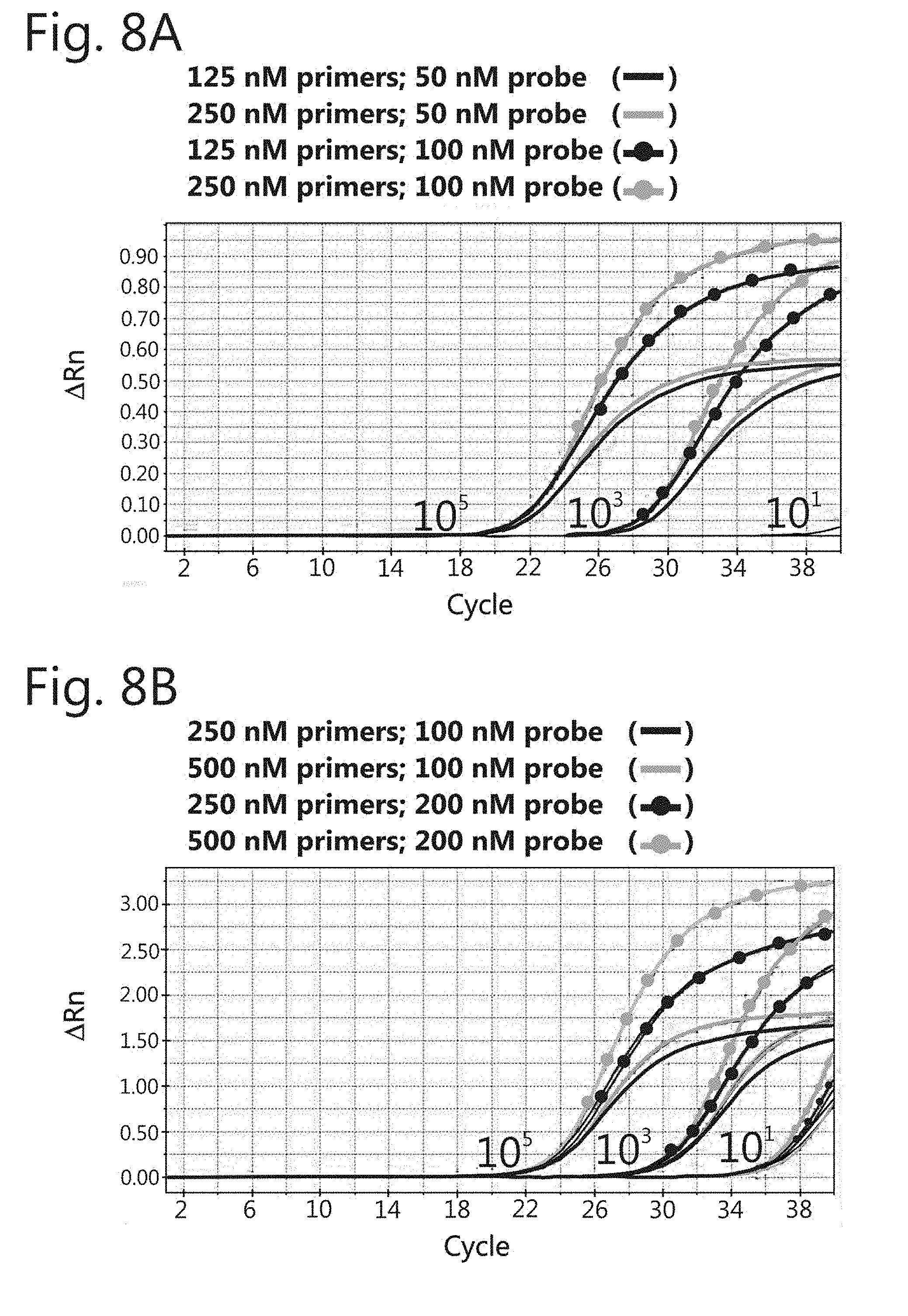
[0053] FIG. 8: Amplification curves in the singleplex format using different concentrations of primers and probes. Three dilutions (10.sup.5, 10.sup.3, and 10.sup.1) of positive controls MT-2 (HTLV-1) and Gu (HTLV-2) were used in the qPCR reactions. (A) Internal control amplification curves. (B) HTLV-1 amplification curve. (C) HTLV-2 amplification curves. (D) HTLV-2 amplification curves.
[0054] FIG. 9: Amplification curve in the multiplex format containing three targets (HTLV-1, HTLV-2 and internal control). A dilution of 10.sup.5 viral copies/reaction of MT-2 (HTLV-1) and Gu (HTLV-2) positive controls was used in the qPCR reaction.
[0055] FIG. 10: Amplification curves and standard curves in the multiplex format containing three targets (HTLV-1, HTLV-2 and internal control). Serial decimal dilutions ranging from 10.sup.5-10.sup.1 viral copies/reaction of MT-2 (HTLV-1) and Gu (HTLV-2) positive controls were used in the qPCR reactions.
[0056] FIG. 11: Schematic of the assembly of the reaction plate for the robustness test. HTLV-1 positive samples were pipetted side by side with non-target samples throughout the plate in the pattern of "chess table".
DETAILED DESCRIPTION OF THE INVENTION
[0057] Unless otherwise defined, all technical and scientific terms used herein have the same meaning as understood by one skilled in the art to which the invention belongs. Conventional techniques of molecular biology are well known to one skilled in the art and can be found, for example, in Ausubel et al., eds. Current Protocols in Molecular Biology, John Wiley & Sons, Inc. N.Y. (1987-2008), including all the supplements; Sambrook et al., Molecular Cloning: A Laboratory Manual, 2.sup.nd edition, Cold Spring Harbor, N.Y. (1989). The specification also provides definitions of terms to help in the interpretation of what is described in the claims. Unless otherwise indicated, all numbers expressing quantities, percentages, and proportions, and other numerical values used in the specification and claims, shall be understood to be modified in all cases by the term "about". Thus, unless otherwise noted, the numerical parameters shown in the specification and claims are approximations that may vary depending on the properties to be obtained.
[0058] The invention described herein relates to novel oligonucleotides for amplifying, detecting, differentiating and quantifying HTLV subtypes, and related methods and kits. More specifically, the present invention provides oligonucleotides including primers and probes, which are adapted for the detection and discrimination of HTLV-1 and discrimination of HTLV-1 and HTLV-2.
Oligonucleotides and Diagnostic Kits
[0059] "Oligonucleotide" refers to any short nucleotide polymer, where the nucleotides can be ribonucleotides, deoxyribonucleotides, dideoxyribonucleotides, degenerate nucleotides, and similar. Said oligonucleotide are preferably single stranded. The length of said oligonucleotides can vary, and usually is shorter than 150 nucleotides (nt), preferably ranging between 10-100 nt, more preferably between 10-60 nt, even more preferably between 13-50 nt. They may also exhibit chemical modifications, such as a tag or a marking, for example, fluorescent, radioactive, biotinylated, DIG (digoxigenin), and similar. The oligonucleotides of the invention may be both forward (sense) and reverse (antisense).
[0060] In one aspect, the oligonucleotides according to the present invention include primers and probes.about.. Unless otherwise noted, the sequences are shown in the 5' to 3' direction. Said oligonucleotides can be in various forms, for example, in solubilization/suspension in a suitable solvent and in a desired concentration, dried or lyophilized. The person skilled in the art knows solvents, concentrations and suitable storage conditions for the oligonucleotides of the invention. In particular, one skilled in the art knows how to prepare such oligonucleotides as stock solutions. The oligonucleotides according to the invention may have various degrees of purity, which can be evaluated by one skilled in the art, for example by HPLC.
[0061] Furthermore, it should be remembered here that, although preferred functions may be mentioned in connection with some oligonucleotides, it is obvious that a given oligonucleotide may assume several functions, and may be used in different forms according to the present invention. As a person skilled in the art knows, in some situations, a primer may be used as a probe and vice versa, in addition to being applicable in procedures of hybridization, detection etc. Thus, let us note that the products according to the present invention, especially, inter alia, oligonucleotides, are not limited to the uses shown herein, but, on the contrary, the uses are to be interpreted broadly, independent of the use indicated herein. Moreover, when an oligonucleotide is described as being useful as a probe capable of binding to an amplicon, a person skilled in the art also understands that the complementary sequence of this oligonucleotide is also useful as a probe to bind to the same amplicon. The same applies to sequences described as useful primers. In addition, it is also obvious that any suitable primer for a multiplex protocol may as well be used in a singleplex protocol, within the meaning and scope of the present invention. The same applies to a suitable primer for a real-time PCR protocol, which can be used in a conventional PCR protocol, within the meaning of the present invention.
[0062] The terms "hybridize" and "anneal" are used interchangeably and refer to the base pairing interaction of a nucleic acid with another nucleic acid that results in the formation of a biplex, triplex, or other more complex structures. In some embodiments, the primary interaction is specific, for instance, C/G and AVT, by bonding by forming hydrogen bridges.
[0063] In this regard, a person skilled in the art understands that the oligonucleotides of the present invention, namely primers and probes, do not need to be completely complementary to a portion of the target sequence. The primer may have sufficient complementarity to hybridize to the target sequence and perform the intrinsic functions of a primer. The same applies to a probe, that is, a probe may have sufficient complementarity to hybridize to the target sequence and perform the intrinsic functions of a probe. Thus, a primer or a probe, in one embodiment does not need to be completely complementary to the target sequence. In an embodiment, the primer or the probe can hybridize or anneal with a portion of the target to form a double strand. Hybridization conditions of a nucleic acid are described by Joseph Sambrook et al., Molecular Cloning, A Laboratory Manual, Cold Spring Harbor Laboratory Press, Cold Spring Harbor, N.Y. (2001) and Haymes et al., Nucleic Acid Hybridization, A Practical Approach, IRL Press, Washington, D.C. (1985). Thus, since complete complementarity is not required for annealing, one skilled in the art will understand that the primer and probe sequences described herein can be modified to some extent without the loss of their usefulness as primers and probes specific for HTLV-1 and HTVL-2.
[0064] With respect to the definition of "primer", one skilled in the art knows that any oligonucleotide includes single strand capable of annealing to a complementary target template under conditions of suitable stringency and serves as the starting point for the synthesis of an extension product (amplicon) from the primer, by elongation of the strand by a DNA polymerase under suitable conditions. These conditions include 4 different types of deoxynucleoside triphosphates and DNA polymerase or reverse transcriptase under suitable temperature conditions and in a suitable buffer solution. Primer length may vary according to several factors, but the typical length of a primer is 5-50 nt, preferably 15-30 nt, more preferably 15, 16, 17, 18, 19, 20, 21, 22, 23, 24, 25, 26, 27, 28, 29 or 30 nt. According to the present invention, each primer has preferably 10-30 nt. Forward and reverse primers are primers which bind, respectively, to a 3' end and a 5' end of a specific region of the target that is amplified by the PCR reaction.
[0065] The primers disclosed by the present invention are designed based on the consensus sequences of the genomes of HTLV-1 and HTLV-2, resulting from the analysis of several HTLV-1 and HTLV-2 isolates (see FIG. 1, and SEQ ID Nos: 11 and 12). More specifically, HLTV-1 and HTLV-2 pol region was the target. Preferably, such primers are, but are not particularly limited to, a primer comprising at least 10 to 15 consecutive nucleotides of any of the sequences described in SEQ ID Nos: 1-2 and 4-6, and their complementary sequences, capable of amplifying a region of HTLV-1 and HTLV-2 pol gene. The primer may also consist of any of the sequences of SEQ ID Nos: 1-2 and 4-6, and their complementary sequences.
[0066] The definition of probe is also known to one skilled in the art, and includes any oligonucleotide capable of hybridizing to a complementary target sequence under suitable hybridization conditions. Since the probe is labeled, it can be used to detect the presence of given nucleotide sequences. The probes may be prepared as single stranded DNA, double stranded DNA, RNA or DNA-RNA hybrid. The typical length of a probe is 10-60 nt, preferably 15-55 nt, more preferably 20-50 nt, more preferably 30-45 nt, even more preferably 10-30 nt. The probes disclosed by the present invention are also designed based on the consensus sequences of the HTLV-1 and HTLV-2 genomes, resulting from the sequence analysis of various HTLV-1 and HTLV-2 isolates, more specifically by binding to the HTLV-1 and HTLV-2 pol region. Hence, according to the present invention, the probe can include or comprise at least 8-15 consecutive nucleotides of any one of the sequences described in SEQ ID Nos: 3 or 7. The probe may also consist of any of the sequences from SEQ ID Nos: 3 or 7, and their complementary sequences.
[0067] Several probe formats can be used to carry out a real-time PCR, such as fluorescent-labeled probes. More specifically, probes can be of the FRET (fluorescence resonance energy transfer) type, which include, but is not limited to, probe of TaqMan.TM., Molecular Beacon.TM., Scorpion.TM., and LUX.TM. types. In A preferred embodiment, probes according to the invention are of the TaqMan.TM. type.
[0068] More specifically, as for TaqMan.TM. probe, an oligonucleotide whose 5' terminal region is modified with a fluorophore and the 3' terminal region is modified with a quencher, is added to the PCR reaction. It is also understood that it is possible to attach the fluorophore on the 3' terminal region and the quencher on the 5' terminal region. Reaction products are detected by the fluorescence generated upon the 5'->3' exonuclease activity of the DNA polymerase. Fluorophores, which are fluorescent compounds emitting light upon excitation by light having a wavelength shorter than the light which is emitted, may be, but are not limited to, FAM, TAMRA, VIC, JOE, TET, HEX, ROX, RED610, RED670, NED, Cy3, Cy5, and Texas Red. Quenchers may be, but are not limited to, 6-TAMRA, BHQ-1,2,3 and MGB-NFQ. The choice of the fluorophore-quencher pair can be made so that the quencher excitation spectrum has an overlap with the emission spectrum of the fluorophore. An example is the FAM-TAMRA, FAM-MGB, VIC-MGB pair an so on. One skilled in the art will know how to recognize other suitable pairs.
[0069] In a preferred embodiment according to the invention, the spectrum properties of said probes are chosen so that one probe does not interfere with the other. In particular, when probes are used in multiplex reactions, each probe will have its own fluorophore being spectral and significantly different from another probe, i.e., the absorption/emission spectra of the different probes are essentially non-overlapping.
This advantageously allows the detection of each probe individually, since individual signals do not interfere with one another during detection.
[0070] The fluorescence emitted during the amplification reaction of the target nucleic acid is measured for the purpose of monitoring the accumulation of specific amplification products. The fluorescence signal is proportional to the amount of specific amplicon produced. In the presence of HTLV-1 and/or HTLV-2 target sequences, the fluorescence will increase. In the absence of the target sequences, the fluorescence will remain consistently low throughout the reaction. An increase in fluorescence or an unchanged level of fluorescence indicates the presence or absence of the targets of HTLV-1 and/or HTLV-2, respectively.
[0071] Furthermore, to provide a standard for determining nucleic acid extraction from a biological sample comprising the target sequence of HTLV or for determining the presence or absence of potential reaction inhibitors, primers capable of amplification, and probes capable of detecting, amplicons resulting from the amplification of human sequences can be used.
A non-limiting example is a primer comprising or consisting of the sequence of SEQ ID Nos: 8 and 9, and a probe comprising or consisting of SEQ ID NO: 10, whose target is the human beta-globulin gene. It should be noted that one skilled in the art can determine other suitable primers and probes targeting other sequences useful for this purpose.
[0072] Moreover, to provide a means of serving as a positive control and/or to facilitate quantification of a viral load in a given biological sample to be analyzed, a positive control may be incorporated. In the present invention, a nucleic acid sample containing copies of the HTLV-1 and/or HTLV-2 target, for instance, a cassette or vector comprising the target sequence to be amplified, or a certain amount of nucleic acid sample from a human cell line containing a known number of insertions of viral sequences. Non-limiting examples include, for instance, cell lines MT-2 and Gu.
[0073] Similarly, a negative reaction control may be incorporated. This control can be a nucleic acid sample which contains no copy of the HTLV-1 and HTLV-2 targets, for instance, human cell line DNA which does not have HTLV-1 and HTLV-2 viral sequence insertions.
[0074] Another aspect of the invention is a kit used to diagnose, differentiate and quantify infections caused by HTLV subtypes, preferably HTLV-1 and HTLV-2 infections, simultaneously, comprising at least one oligonucleotide set. By "oligonucleotide set" is meant any combination comprising at least one oligonucleotide, preferably at least two, for instance, 2-20 oligonucleotides. Said set can, for instance, comprise at least one primer and at least one probe, or at least a pair of primers and at least a probe, and so on. Said oligonucleotides can be maintained separate or can be partially mixed, or totally mixed.
[0075] Preferably, said kit comprises at least one oligonucleotide set according to the invention, specifically designed for HTLV-1 and HTLV-2. Said oligonucleotides can be maintained separate, or can be partially mixed, or totally mixed. The oligonucleotides may be provided in the dried form, or solubilized in a suitable solvent, according to the prior art. For instance, suitable solvents include TE, ultrapure water, and similar.
[0076] In one embodiment, the kit according to the invention can also contain additional suitable reagents for the amplification reaction, including water, nuclease-free water, RNase-free water, DNase-free water, ultrapure water, salts (such as magnesium, potassium, salts), buffers (such as conventional PCR, know in the prior art), enzymes, including thermostable polymerase, such as Taq, Vent, Pwo, Pfu, reverse transcriptase, and similar, nucleotides such as deoxynucleotides, dideoxynucleotides, dNTPs, dATP, dTTP, dCTP, dGTP, dUTP, other reagents such as additives, RNase or DNase inhibitors, and polynucleotides such as polyT, polydT, and other nucleotides such as primers and probes for other pathogens, for instance, HIV, HBV, HCV and for internal control, such as human beta-globin. The reactants may be provided in a concentrated form to be diluted to a suitable concentration by the final user. Moreover, at least part of the reactants can be provided in the pre-mix form.
[0077] Such reagents may be accommodated in containers which for the purposes of the present invention include, but are not limited to, microtubes, tubes, PCR plates with different amounts of pellets, chips, or any other suitable and inert medium wherein the amplification reaction can take place, and which does not react with the fluids and solutions of the present invention. Furthermore, the container can be further labeled and identified, for instance, with colors, to avoid confusion and provide ease of use for a technician in the laboratory.
[0078] Moreover, in an embodiment, the kit according to the invention contains instructions for the use thereof. Said instructions can be in a brochure, card, or similar. These instructions can be in two forms: a detailed one, providing exhaustive information regarding the kit and its use, possibly also including literature data; and a simple one, in the form of a quick guide, providing essential information necessary for the use of the kit.
[0079] In a preferred form, said kit is a diagnostic kit, especially an in vitro diagnostic kit, for instance, an HTLV diagnostic kit. More preferably, said kit is a kit for diagnosis and differentiation between HTLV-1 and HTLV-2.
[0080] In another preferred embodiment of the present invention, the diagnostic kit may further comprise a kit for extracting and isolating nucleic acids from a biological sample. Said extraction kit may comprise a lysis buffer, a washing buffer and an elution buffer. The extraction kit may further be provided with empty containers and adsorption columns for nucleic acid extraction and isolation.
Nucleic Acid Extraction from Biological Samples
[0081] HTLV polynucleotides, more preferably HTLV-1 and HTLV-2, are the targets or the target source for the amplification reaction of the present invention. The expression "target sequence", or simply "target", refers to a nucleic acid sequence that serves as a template for amplification in a PCR reaction. These nucleic acid sequences may contain deoxyribonucleotides, ribonucleotides, and/or analogs thereof. The sequence may be a gene or gene fragment, mRNA, cDNA, isolated total DNA, isolated total RNA, and similar. Examples of target sequences include, but are not limited to, HTLV integrated provirus DNA, HTLV cDNA present in a cell prior to viral integration into the host genome, viral RNA extracted from viral particles or from host cells during viral replication, HTLV primary RNA transcripts, spliced mRNA, etc.
[0082] More specifically, the target sequences of the present invention are localized in the pol gene of HTLV-1 and HTLV2 genomic sequences. HTLV-1 target sequence is localized between bases 3340 and 3414 of the consensus sequence of HTLV-1 genome, as shown in SEQ ID NO: 11. HTLV-2 target sequence is localized between bases 4483 and 4563 of the consensus sequence of HTLV-2 genome, as shown in SEQ ID NO: 12. For the purposes of this invention, these sequences are reference template sequences, that is, suitable primers according to the present invention can amplify at least these reference sequences. Likewise, suitable probes according to the present invention are also capable of hybridizing with at least these reference sequences.
[0083] In addition, a human target sequence may be used as the standard or control of nucleic acid extraction from a biological sample. Non-limiting example of control target is the human beta-globin gene. A person skilled in the art will certainly be able to determine other suitable control targets which can consist of other human genes.
[0084] In an embodiment, the target sequence is present in a sample of biological material collected from an individual. Thus, by "sample" is meant any biological substance or material which may contain an HTLV, an HTLV-infected cell, or an HTLV target sequence, and includes, but is not limited to, blood, plasma, serum, blood cells, seminal fluid, vaginal secretions, breast milk, saliva, and similar. More preferably, the sample comprises or consists of human blood cells and/or whole blood.
[0085] The procedures for nucleic acid extraction and purification are well-known in the prior art. Examples of methods to extract nucleic acids from whole blood are taught, for instance, in Casareale et al. (Genome Res., 2: 149-153, 1992) and in U.S. Pat. No. 5,334,499.
[0086] Furthermore, several commercial kits are available for the nucleic acid isolation from whole blood. Examples of kits include, but are not limited to, QIAamp DNA Blood Mini Kit (Qiagen); Spin Plus ReliaPrep.TM. Blood gDNA Miniprep System (Promega) and BIOPUR Kit Extracao Mini Spin Plus (Biopur).
Target Sequence Amplification by PCR and Diagnostic Method
[0087] Once the primers are prepared, the target nucleic acid amplification can be performed through a variety of methods, including, but not limited to, conventional PCR, real-time PCR, RT-PCR, nested-PCR, quantitative and others. Preferably, the method used is real-time PCR.
[0088] "Amplification" refers to nucleic acid amplification procedures using primers and polymerases that generate multiple copies of a target nucleic acid. Such amplification reactions are known to the person skilled in the art as "PCR" (polymerase chain reaction), which in turn includes, for the purposes of this invention, any PCR-based method including conventional, qualitative, semi-quantitative, real-time, reverse transcriptase reaction (RT-PCR) PCR, singleplex and multiplex PCR, and similar.
[0089] An "amplicon" or "PCR product", the terms being used interchangeably, refer to a nucleic acid (or collectively, the plurality of nucleic acid molecules) that was synthesized during the amplification procedures. An amplicon is typically, but not exclusively, a DNA fragment.
[0090] "Real-time PCR" includes any PCR-based method that allows to monitor the fluorescence emitted during the reaction as an indicator of the production of the PCR product or amplicon during each PCR cycle, as opposed to detection at the end of the completion of all cycles in the conventional PCR methods
[0091] "Quantitative PCR" (qPCR) refers to any PCR-based method that allows the estimation of the initial amount of a given target sequence in a given sample.
[0092] As used herein, "multiplex PCR" refers to any PCR reaction whose purpose is that of amplifying more than one target. For instance, multiplex PCR includes biplex (two targets) PCR, triplex (three targets) PCR, and so on. Multiplex PCR includes PCR reactions with more than one pair of primers, for instance, two pair of primers. In this case, there may be four different primers, but it is also possible that there is a common primer, for example the primer forward, and two distinct primers reverse. Multiplex PCR also includes PCR reactions with a single pair of primers, but with more than one probe. Still, as non-limiting examples, multiplex amplification includes amplification reagents from different genes, different alleles of a single gene and/or different fragments of a single gene.
[0093] A "buffer" is a composition added to the amplification reaction, comprising a buffering agent, which modifies the stability, activity and/or longevity of one or more components of the amplification reaction, by regulating the pH of the amplification reaction. The buffering agents of the invention are compatible with the activity of the polymerase to be used, that is, a DNA polymerase. Buffering agents are well-known in the prior art and include, but are not limited to, Tris, Tricinae, MOPS ((3-(N-morpholino)propanesulfonic acid)), and HEPES (4-(2hydroxyethyl)-1-piperazine-ethane sulfonic acid).
[0094] Furthermore, PCR buffers can usually contain about 70 mM KCl and about 1.5 mM or more of MgCl.sub.2, and about 50-500 .mu.M of each one of dATP, dCTP, dGTP and dTTP nucleotides.
[0095] The buffers of the invention can also contain additives. An additive is a compound added to a composition that modifies the stability, activity and/or longevity of one or more components of the composition. In some embodiments, the composition is an amplification composition. In some embodiments, an additive inactivates contaminating enzymes, stabilizes protein folding, and/or decreases aggregation. According to the invention, additives may be added to enhance the selectivity of the annealing of a primer and/or a probe, as long as the additive does not interfere with the activity of the DNA polymerase.
[0096] Examples of additives are, but are not limited to betaine, glycerol, formamide, KCl, CaCl.sub.2, MgOAc, MgCl.sub.2, NaCl.sub.2, NH.sub.4OAc, NaI, Na(CO.sub.3).sub.2, LiCl, MnOAc, NMP, trehlose, DMSO, ethylene glycol, dithiothreitol ("DTT"), pyrophosphate (including, but not limited to inorganic pyrophosphate from Thermoplasma acidophilum ("TAP")), bovine serum albumin ("BSA"), propylene glycol, glycinamide, CHES, Percoll.TM., aurintricarboxylic acid, Tween 20, Tween 21, Tween 40, Tween 60, Tween 85, Brij 30, NP-40, Triton X-100, CHAPS, CHAPSO, Mackernium, LDAO (N,Ndimethyldodecylamine N-oxide), Zwittergente 3-10, Xwittergente 3-14, Xwittergente SB 3-16, Empigen, NDSB-20, T4G32, SSB from E. coli, RecA, 7-deazaG, dUTP, UNG, anionic detergents, cationic detergents, non-ionic detergents, zwittergente, esterols, cations, and any other chemical, proteins, or cofactors that may alter amplification efficiency.
[0097] As used herein, the term "thermostable", when applied to the enzyme, refers to an enzyme that retains its biological activity at elevated temperatures (for instance, 55.degree. C. or more), or maintains its biological activity after repeated heating and cooling cycles. Thermostable nucleotide polymerases are particularly preferred for the present invention, since they eliminate the need to add enzyme prior to each PCR cycle.
[0098] "Polymerase activity" refers to an enzymatic activity that catalyzes the deoxyribonucleotide polymerization. Generally, the enzyme will initiate the synthesis at the 3' end of the annealed primer to the target sequence, and will proceed toward the 5' end of the template strand. At given concentrations, this enzyme is a thermostable DNA polymerase.
[0099] Non-limiting examples of thermostable DNA polymerase include, but are not limited to, polymerase isolated from Thermus aquaticus (Taq polymerase), Thermus thermophilus (Tth polymerase), Thermococcus litoralis (Tli or VENT.TM. polymerase), Pyrococcus furiosus (Pfu or DEEPVENT.TM. polymerase), Pyrococcus woosii (Pwo polymerase) and from other species of Pyrococcus, Bacillus stearothermophilus (Bst polymerase), SulfolobUS acidocaldarius (Sac polymerase), Thermoplasma acidophilum (Tac polymerase), Thermus rubber (Tru polymerase), Thermus brockianus (DYNAZYME.TM. polymerase) (Tne polymerase), Thermotoga maritime (Tma) e outras especies de Thermotoga genus (Tsp polymerase), e Methanobacterium thermoautotrophicum (Mth polymerase).
[0100] The PCR reaction can contain more than one thermostable polymerase enzyme with complementary properties, resulting in a more efficient amplification of the target sequences. For instance, a polymerase with a high ability to amplify large nucleotide segments may be complemented with another polymerase capable of correcting errors occurring during elongation of the target nucleic acid sequence, thus creating a PCR reaction that can amplify a long target sequence with high fidelity. The thermostable polymerase may be used in its wild type, or alternatively the polymerase may be modified to contain a fragment of an enzyme or to contain a mutation which provides beneficial properties to facilitate the PCR reaction. In an embodiment, the polymerase can be Taq polymerase. Many variants of Taq polymerase with enhanced properties are known and include, but are not limited to AmpliTaq.TM., Stoffel fragment, SuperTaq.TM., SuperTaq.TM. plus, LA Taq.TM., LApro Taq.TM., and EX Taq.TM..
[0101] As already mentioned above, the expression "hybridization conditions" refers to conditions that allow the primer or probe to anneal to the nucleotide sequence of interest. These conditions depend on the temperature and the ionic strength of the solution in which the hybridization occurs. These are the conditions of stringency. As understood by one skilled in the art, annealing stringency may be altered in order to identify or detect identical or related polynucleotide sequences. As it will be appreciated by one skilled in the art, the melting temperature, Tm, can be calculated by formulas known in the art, depending on various parameters, such as primer length or probe length, number of nucleotides, or ingredients present in the buffer and conditions. For this, see, for example, T. Maniatis et al., Molecular Cloning: A Laboratory Manual, Cold Spring Harbor Laboratory, Cold Spring Harbor, N.Y., 1982 e J. Sambrook et al., Molecular Cloning: A Laboratory Manual, Cold Spring Harbor Laboratory, Cold Spring Harbor, N.Y., 1989 e J. Sambrook et al., Molecular Cloning: [000100] For illustration purposes, for the determination of hybridization conditions, the following formulas are basically used: for the basic calculation of Tm for sequences longer than 13 nt, the following formula can be used:
Tm=64.9+41*(yG+zC-16.4)/(wA+xT+yG+zC)
[0102] where "w", "x", "y" and "z" are the base numbers A, T, G and C in the sequence, respectively (Marmur and Doty. J Mol Biol 5:109-118, 1962). This equation assumes that annealing occurs under standard conditions of 50 nM primer, 50 mM Na, and pH 7.0.
[0103] For the basic calculation of salt-adjusted Tm, the following equation can be used:
Tm=100.5+(41*(yG+zC)/(wA+xT+yG+zC))-(820/(wA+xT+yG+zC))+16.6*log 10([Na.sup.+])
[0104] where "w", "x", "y" and "z" are the base numbers A, T, G and C in the sequence, respectively.
[0105] The term 16.6*log 10([Na.sup.+]) adjusts Tm to changes in salt concentration (see for additional information, for example, Howley et al., J. Biol. Chem. 254, 4876-4883, 1979.
[0106] Annealing temperature ranges can vary between about 50.degree. C. and 62.degree. C., but the primers can be designed to perform optimally at about 58.degree. C.-60.degree. C. One additional consideration when designing the primers is guanine and cytosine content. GC content for a primer may be usually about 30-70%, but may be lower and may be appropriately adjusted by one skilled in the art. The annealing of oligonucleotides complementary or partially complementary to a particular target may be obtained by modifying annealing conditions in order to increase or decrease stringency, for example by adjusting the temperature or salt concentration in the buffer. Such modifications to maintain HTLV-1 and HTLV-2 specificity can be routinely performed by one skilled in the art.
[0107] For amplification, a pair of primers of a specific type may be used alone (for example, a primer forward and an HTLV-1 primer reverse, a primer forward and an HTLV2 primer reverse, or a primer forward and one or two HTLV-2 primer reverse, and so on). Multiplex amplification can be used to amplify HTLV-1 and HTLV-2 regions concomitantly. The final concentrations of the primers may be adjusted appropriately, ranging from about 50 nM to about 2000 nM, preferably from about 100 nM to about 1000 nM, more preferably from about 200 nM to about 600 nM, more preferably from about 250 nM to about 500 nM, of each of the primers represented by SEQ ID Nos: 1-2, 4-6 and 8-9.
[0108] The final concentrations of the probes may also be appropriately adjusted by one skilled in the art, ranging from about 50 nM to 1000 nM. Most preferably, the final concentration ranges from about 100 to about 300 nM, more preferably from 150 to 250 nM of each one of the probes represented by SEQ ID Nos: 3, 7 and 10.
[0109] Another aspect of the invention discloses a method of detecting the presence of HTLV from nucleic acids extracted from a biological sample, comprising the step of mixing in a suitable container the dNTPs, DNA polymerase, buffer, at least one primer, and at least one probe as described in the present application, the nucleic acid extracted from the biological sample, and subjecting the vessel containing the mixture to incubation in a thermocycler.
[0110] In another aspect the invention discloses a method for detecting the presence of HTLV comprising performing a polymerase chain reaction using at least one primer or a set of primers selected from the primer forward group of SEQ ID Nos: 1 and 4, and at least one primer or a set of primers selected from the group of reverse primers of SEQ ID Nos: 2, 5 and 6.
[0111] A person skilled in the art is aware of the PCR reaction conditions, in particular the thermal cycling conditions, for instance, temperatures, duration, number of cycles, heating/cooling rate, etc. In a preferred embodiment, the PCR reaction conditions include conditions suitable for a multiplex PCR. In another preferred embodiment, such conditions include those suitable for a quantitative real-time multiplex PCR. In another preferred embodiment, said method comprises the step of placing the sample in the presence of probe(s) in conditions suitable for annealing said probe(s) to the amplicon. In another preferred embodiment, the method comprises the step of detecting at least one amplicon in real time, allowing evaluation of the presence or absence of HTLV-1 and/or HTLV-2 in the sample. This is achieved in an advantageous way by the measures of fluorescence intensity.
[0112] Fluorescence measuring procedures are known in the prior art. In brief, the sample is illuminated around the excitation wave of the fluorophore, and the emission intensity is measured. In another embodiment, said method comprises the step of measuring at least once, and more preferably in real time, the amount of probe that anneals to the amplicon. In another preferred embodiment, said method comprises the step of estimating at least once the number of copies of the target initially present in the sample. A person skilled in the art knows how to carry out such step. For instance, this can be carried out using calibration standards and/or internal controls. Preferably, this step includes determining the so-called cycle threshold (CT) for each sample, which correlates with the number of copies of the template target initially present in each sample.
[0113] In a preferred embodiment, at least one step, preferably several steps, most preferably most steps, is/are performed on a PCR plate, including those with 24 wells, 48 wells, 96 wells and 384 wells. The use of plates advantageously ensures that the samples can be processed in parallel during the reaction. In addition, it allows the method to be carried out on a large scale, which saves time.
[0114] In another preferred embodiment, at least one step, preferably several steps, most preferably most steps is/are carried out in a thermocycler. Said thermo cycler may be equipped with a spectrofluorophotometer, for instance, Mx3000p (Stratagene), Chromo 4 (BioRad), RocheLightcycler 480, ABI 7900, 7500 and 7300r Real-time PCR Systems (Applied Biosystems).
[0115] The primers of the present invention for detecting and differentiating HTLV-1 and HTLV-2 are identified in Table 1 as SEQ ID Nos: 1-2 and 4-6, respectively. These primers were designed to anneal in a region located on the pod gene of HTLV-1 and HTLV-2 (see FIG. 2).
[0116] The present invention is illustrated by the examples below, which are only intended to exemplify one of the innumerable ways of carrying out the invention, however, without limiting the scope thereof. Various modifications or suggestions in light thereof which may be suggested by one skilled in the art are included within the spirit and scope of the claims. In particular, although suitable for detection via multiplex and/or real-time protocols, the methods, oligonucleotides, oligonucleotide assemblies and kits of the present invention are, of course, also suitable for singleplex, duplex, triplex and similar, qualitative, quantitative, conventional PCR and combinations thereof.
EXAMPLES
Example 1: In Silico Analysis for Defining the Target Region--Primer and Probe Design
[0117] To define the viral gene region multiple alignments were performed with the HTLV-1 and HTLV-2 total genome sequences available in a public database with the aid of the software BioEdit (version 7.0.5.3), and Mega (version 5-1993/2011), which use the algorithm Clustal W. The access number of the used sequences are described in FIG. 1.
[0118] All complete genomes entered in GenBank until the filing date were used for alignment.
[0119] The primers and probes were prepared based on the recommendations described below. For the primers, the melting temperature (Tm) was between 58 and 60.degree. C., in addition to being approximately 10.degree. C. below the Tm of the probes, in order to ensure that the probe binds to the template before the primers. The last bases of the 3' end consisted of as few cytosine (C) and/or guanine (G) bases as possible, which reduces the formation of non-specific products. G/C content remained between 61 and 71% within the recommended range (30-80%). The probes were designed as close as possible to the binding region of the primer forward or reverse to improve the efficiency of the reactions. Tm was defined between 68.degree. C. and 70.degree. C., and G/C content remained between 47 and 69%. To avoid the quencher effect, probes containing G at the 5' end were discarded. A minor groove binder system (MGB) was used to design the probes. This system allows the Tm of the probe to increase without increasing its size, which allowed the production of shorter probes (13-20 bases).
[0120] A total of four sets of primers designed for the SYBR.TM. Green methodology targeting the tax and env regions, and six set of primers and probes designed for the TaqMan.TM. methodology targeting the tax, gag, pol, and env regions were assessed. These tests were standardized, however, they showed unsatisfactory results in the validation process (data not shown).
[0121] TaqMan.TM. primers and probes targeting the pol viral region proved to be suitable. The nucleotide sequences of the primers and probes used herein are shown in Table 1.
TABLE-US-00001 TABLE 1 Primers and probes for detecting HTLV-1 and HTLV-2. SEQ ID Primer/ NO: probe Sequence (5'-3') Tm.degree. C. 1 HTLV-1 CAGCCCCTTCACAGTCTCTACTG 59 For_pol 2 HTLV-1 AGAAGGATTTAAATATATTTGOGTCTCGG 58.5 Rev_pol 3 HTLV-1 CCTTACAAAGGCATACTGAT 69 Probe 4 HTLV-2 CAAGGTGATGTAACCCATTATAAGTACAA 58.8 For_pol 5 HTLV-2 AACCGCACCGGAGAAGGT 59.1 Rev_pol 6 HTLV-2 AGAAACCAGCTGTGAGACTATCAGC 59.1 Rev'_pol 7 HTLV-2 AAATACAAATACTGCCTCCACGT 68 Probe
[0122] In Table 1, the probe of SEQ ID NO: 3 was labeled with FAM at the 5' end, and with MGB at the 3' end, and the probe of SEQ ID NO: 7 was labeled with VIC at the 5' end, and with MGB at the 3' end. The amplicon generated for HTLV-1 has 75 base pairs, and the one for HTLV-2 has 81 base pairs.
Example 2: Culture and Expansion of MT-2 and Gu Cell Lines (Positive Controls)
[0123] MT-2 cell line consists of lymphoblasts isolated from a subject with ATLL (catalog number 93121518/ECACC). This lineage contains HTLV-1 integrated into its genome, presenting 2.1 viral copies per cell (Albrecht et al., J Virol Methods, v. 75, n. 2, p. 123-40, 1998). Gu lineage, originated from the in vitro infection of BJAB cell line, contains 8.3 copies of HTLV-2 integrated into its genome (Moens et al., J Clin Microbiol, v. 47, n. 11, p. 3682-91, 2009). This lineage has been kindly provided by the Rega Institute for Medical Research--Katholieke Universiteit, Leuven, Belgium. MT-2 and Gu cell lines were cultivated in 15 cm.sup.2-bottles (Greiner Bio One) with 15 mL of Roswell Park Memorial Institute (RPMI) culture medium 1640 (Sigma-Aldrich) supplemented with 10% inactivated fetal bovine serum (Hyclone); 100 U/mL of penicillin, and 100 U/mL of streptomycin (Invitrogen). The cultured were were kept in incubatorS Steri-cult 200 Thermoform (Forma Scientific) at a temperature of 37.degree. C., 5% CO.sub.2 and 85% of relative humidity. After expansion, the cells were collected in 15 ml polypropylene tubes, washed with PBS buffer (1.times.) by centrifugation at 200.times.g for 10 minutes, resuspended in PBS (1.times.) and counted in Neubauer's chamber.
Example 3: DNA Extraction from Cell Lines and Positive Control Products
[0124] For DNA extraction, approximately 2.times.10.sup.7 cells resuspended in 1 mL of PBS (1.times.) were used. Cells were transferred to a 1.5 mL-polypropylene tube and subjected to centrifugation for 5 minutes at 300.times.g. After centrifugation, the supernatant was removed, remaining 200 .mu.L in the tube, which was then transferred to a 15 mL polypropylene tube.
[0125] DNA extraction was carried out using the Gentra Puregene cell kit (Gentra Systems), following the manufacturer's instructions. 3 mL of cell lysis solution was added and was homogenized in vortex for 10 seconds. 15 .mu.L of RNase were added, and homogenized by inversion for about 25 times. This mixture was incubated at 37.degree. C. for 5 minutes and quickly cooled in ice for 3 minutes. Then, 1 ml of protein precipitant solution was added followed by vigorous vortexing for 20 seconds, and then centrifugation at 2000.times.g for 10 minutes. After centrifugation, the supernatant was transferred to a new 15 mL polypropylene tube, to which 3 mL of isopropanol was added and homogenized by inversion about 50 times to precipitate the DNA. The samples were centrifuged for 3 minutes at 2000.times.g and the supernatants were discarded. To wash the DNA pellet, 3 mL of 70% ethanol was added and the system was homogenized several times for efficient washing. The DNA was collected and transferred to a sterile 1.5 mL tube. After ethanol evaporation, DNA was resuspended in 300 .mu.L deionized water and incubated at 65.degree. C. for 1 hour until complete DNA elution. After extraction, DNA was quantified by spectrophotometry (UV) at a wavelength of 260 nm and 280 nm in the equipment Nanodrop 2000c spectrophotometer (Thermo Fisher Scientific).
[0126] Positive controls were prepared from DNA extracted from MT-2 and Gu cell lines, according to what has been described above.
[0127] To this end, four aliquots containing 200 .mu.L of DNA of the MT-2 cell line were transferred to a polypropylene tube, free of DNAse, RNAse and pyrogens, to obtain a single aliquot. DNA concentration was adjusted to approximately 500 ng/.mu.L. Due to the heterogeneity and high viscosity of the sample, it was transferred to a column composed of a silica membrane and subjected to centrifugation at 16.900.times.g for 3 minutes. After centrifugation, the sample was quantified by spectrophotometry (UV) at a wavelength of 260 nm and 280 nm in the equipment Nanodrop 2000c spectrophotometer (Thermo Fisher Scientific). The same procedures were adopted for the Gu cell line. The single aliquots obtained from MT-2 and Gu cell lines were pooled for the composition of a pool of positive controls (MT-2+Gu). From this pool 192 aliquots of 15 .mu.L and 192 aliquots of 50 .mu.L of the positive control containing 10.sup.3 viral copies each 5 .mu.L and 60 aliquots of 50 .mu.L of the positive control containing 10.sup.5 viral copies each 5 .mu.L were prepared. The aliquots were stored at -20.degree. C. until use.
Example 4: Negative Control Production
[0128] For the negative control of qPCR reactions, five whole blood samples obtained from blood donors were pooled and homogenized for 5 minutes in a circular blood homogenizer. These samples exhibited no serological reactivity to the immunoenzymatic test (ETA) for HTLV-1/2.
[0129] After homogenization, aliquots of the negative pool were made in 1.5 mL polypropylene tubes so that each tube remained with 200 .mu.L of pool, totaling 20 tubes. At each aliquot dispensed, the pool was homogenized for 30 seconds to maintain control homogeneity. The aliquots were stored at -20.degree. C. At each DNA extraction from the samples, the negative control was included in the process.
Example 5: Amplification Internal Control (IC)
[0130] The internal control (IC) is a DNA sequence that is present in the same qPCR reaction, in which the test samples are amplified. This sequence is co-amplified simultaneously with the target sequence and aims to monitor the entire process, from the extraction of nucleic acids to the fmal analysis of the data, validating the negative reactions and demonstrating the presence of inhibitors or failures in the pre-analytical and analytical processes of the samples. The beta-globin gene was used as an IC in the qPCR reactions.
[0131] Based on the sequence of human beta-globin gene with access number GU324922, and with the aid of the software Primer Express.TM.3.0 (Applied Biosystems), a pair of primers and a probe were designed in the TaqMan.TM.MGB system. The same recommendations described in example 1 were used for the preparation of these oligonucleotides. The sequences of the internal control primers and probe are shown in Table 2.
TABLE-US-00002 TABLE 2 Primers and probes for detecting the internal control, human beta-globin SEQ ID NO: Primer/probe Sequence (5'-3') TMOC 8 Globin_For TGAAGGCTCATGGCAAGAAA 58 9 Globin_Rev GGTGAGCCAGGCCATCAC 59 10 Globin Probe TGCTCGGTGCCTTT 69
[0132] In Table 2, the probe of SEQ ID NO: 10 was labeled with NED at the 5' end, and with MGB at the 3' end. The amplicon generated for the internal beta-globin control has 54 base pairs.
Example 6: Standardization of qPCR Reactions in Singleplex Format--Preparation of Standard Curve from the Positive Controls
[0133] Considering that the human haploid genome weighs 3.3 pg and that the MT-2 cell line has approximately one copy of the HTLV-1 integrated into its genome, calculations to obtain a stock solution containing 10.sup.5 viral copies per 5 .mu.L of solution were carried out. In the same way, calculations were made for the Gu cell line, however, this line has approximately four copies of HTLV-2 integrated into its genome. These calculations were based on the Avogadro constant and the DNA concentration of the samples evaluated by spectrophotometry. Stock solutions were stored at -20.degree. C. until use.
[0134] To evaluate the reagent kinetics and the quality of the primers and probes, the linear efficiencies and correlations (R.sup.2) of the amplification reagents were evaluated in the singleplex format (one probe and one target per reagent). For this, standard curves were constructed by serial decimal dilutions (10.sup.5 to 10.degree. viral copies/reagent) of the positive controls MT-2 (HTLV-1) and Gu (HTLV-2). Efficiencies for HTLV-1, HTLV-2 and internal control (CI) probes and primers were greater than 95% and correlations (R.sup.2) were higher than 0.99 (FIG. 3).
Example 7: Standardization of qPCR Reactions in Singleplex and Multiplex Formats (Biplex and Triplicate)
[0135] To evaluate the efficiency and linear correlation (R.sup.2) of qPCR reactions in the singleplex (presence of a target and a probe per reaction) and multiplex (presence of more than a target and more than a probe per reaction) format, serial decimal dilutions (10.sup.5-10.sup.0 viral copies/reaction) of the positive controls MT-2 (HTLV-1) and Gu (HTLV-2) were carried out. These dilutions were used in real-time PCR reactions in standard curve format. The only targets of the reactions were HTLV-1 or HTLV-2 or internal control (IC) (singleplex), HTLV-1 and IC or HTLV-2 and ICI (biplex) or HTLV-1, HTLV-2 and IC simultaneously co-amplified (triplex). Real-time PCR reactions were standardized using the TaqMan.TM. Universal PCR Master Mix (Applied Biosystems) kit, and amplification and data collection were carried out in ABI 7500 Real-Time PCR system (Applied Biosystems). Thus, each amplification reaction consisted of 5 .mu.L of DNA from the positive controls and 12.5 .mu.L TaqMan.TM. Universal PCR Master Mix (Applied Biosystems). The used concentration of the probes was 100 nM and the one of the primers was 250 nM. Water volume was adjusted for each reaction to a final volume of 25 .mu.L/reaction. Thermal cycling conditions consisted of a first activation step at 50.degree. C. for 2 minutes followed by 45 cycles at 95.degree. C. for 15 seconds, and 60.degree. C. for 1 minute.
[0136] The reactions were carried out in the following formats: i) biplex containing targets and probes for HTLV-1 or HTLV-2 and IC; ii) triplex containing targets and probes for HTLV-1, HTLV-2 and IC. Reactions in the biplex format (HTLV-1 e CI) proved to be suitable (FIG. 4). Biplex regions (HTLV-1 and HTLV-2; HTLV-2 and IC) curves for HTLV-2 have undergone a change in their shape, becoming more linear than sigmoid (FIG. 4). In the triplex format (HTLV-1, HTLV-2 and IC), the curves for HTLV-1 and IC proved to be suitable, however, the curves for HTLV-2 underwent a marked change in their shape and a significant reduction of fluorescence as observed in FIG. 4.
[0137] The cycle threshold (Ct) of the reactions in the singleplex and multiplex formats (triplex) were similar (Table 3).
TABLE-US-00003 TABLE 3 Comparison of PCR reactions in singleplex and triplex (multiplex) formats. Cycle threshold values and standard deviation of dilutions of positive controls MT-2 (HTLV-1) and Gu (HTLV-2). The dilutions varied from 10.sup.5 to 10.sup.0 copy/reaction. Ct Value Target 10.sup.5 10.sup.4 10.sup.3 10.sup.2 10.sup.1 10.sup.0 HTLV-1 Singleplex 20.362 23.894 27.318 31.067 34.041 36.705 Triplex 20.422 23.992 27.402 31.256 35.233 37.015 DP 0.042 0.069 0.059 0.133 0.842 0.2194 HTLV-2 Singleplex 23.765 27.042 30.761 34.041 37.67032 ND Triplex 23.629 27.243 30.766 35.07 36.53651 ND DP 0.096 0.142 0.003288 0.728 0.801726 ND HTLV: Human T-cell lymphotropic virus; SD: standard deviation; Ct: cycle threshold; ND: non detected
Example 8: Standardization of the Concentrations of Probes and Primers Used in qPCR Reactions
[0138] Real-time PCR reactions in the singleplex format were standardized using the TaqMan.TM. Universal PCR Master Mix kit (Applied Biosystems) and the thermal conditions of cycling, amplification and data acquisition were performed according to example 7. Each amplification reaction consisted of 5 .mu.L of DNA obtained from positive controls MT-2 and Gu, 12.5 .mu.L TaqMan.TM. Universal PCR Master Mix (Applied Biosystems). The concentration of the probes for HTLV-1 (labeled with the fluorochrome FAM.TM.) and HTLV-2 (labeled with the fluorochrome VIC.TM.) varied from 100 to 300 nM, while for the internal control (IC) (labeled with the fluorochrome NED.TM.) it varied from 50 to 200 nM. Primer concentrations vary from 250 to 500 nM for HTLV-1 and HTLV-2, and from 125 to 250 nM for IC. Water volume was adjusted for each reaction to a final volume of 25 .mu.L/reaction. Subsequently, the concentrations of reagents that presented better performance were evaluated in the multiplex format.
[0139] Also, different concentrations of probes were tested to enhance curves and fluorescence in the multiplex format, focusing on the curve for HTLV-2. The dilutions of the positive controls MT-2 (HTLV-1) and Gu (HTLV-2), containing 10.sup.5, 10.sup.3, and 10.sup.1 viral copy/reaction were used in qPCR reactions. The reactions were carried out in the singleplex and multiplex formats (biplex and triplex). FIG. 5 shows the evaluated probe concentrations, which were 100, 200 and 300 nM.
Example 9: Design of an Extra Primer Reverse (Reverse') for HTLV-2
[0140] Furthermore, the possibility of using more than one primer reverse in addition to the primer of SEQ ID NO: 5 was tested, to verify a sensitivity increase of the reaction for HTLV-2. Said primer is presented under SEQ ID NO: 6.
[0141] Thus, two amplicons of different sizes are generated (FIG. 6) by increasing the targets for probe bonding, which generates an increase in fluorescence and reduction of Ct, as can be seen in FIG. 7.
Example 10: Concentration Test of Primers and Probes for qPCR Reactions in the Singleplex Format
[0142] Using the singleplex format, different conditions of the qPCR reactions were tested. The dilutions of cell lines MT-2 (HTLV-1) and Gu (HTLV-2), containing 10.sup.5, 10.sup.3, and 10.sup.1 viral copy/reaction were used. The concentration of the primers tested for the HTLV-1 and HTLV-2 reactions were 250 and 500 nM, those of the probes were 100 and 200 nM, and for the IC, were 125 and 250 nM of primer and 50 and 100 nM of probe. The best conditions observed, in the case of HTLV-1 were 500 nM of primers and 200 nM of probes, in the case of HTLV-2 were 500 nM of primer forward, 250 nM of primers reverse, and 200 nM of probe, and in the case of IC, 250 nM of primers and 100 nM of probe (FIG. 8).
Example 11: Primers and Probes Concentration Test for the qPCR Reactions in the Multiplex Format
[0143] Using the multiplex format, different conditions of the PCR reactions were tested. Therefore, in the case of HTLV-1, primer concentrations of 250 and 500 nM, and probe concentrations of 100, 125, 150, 175, and 200 nM where used, while in the case of HTLV-2, primer were used at concentrations of 250 and 500 nM, and the probe concentrations of 200, 225 and 250 nM, and in the case of IC, primers concentration of 250 and probe concentrations of 100, 125, 150 and 200 nM were used. The best condition was observed, in the case of HTLV-1, when using 500 nM of primers and 200 nM of probes, in the case of HTLV-2, when using 500 nM of primer forward, 250 nM of primers reverse, and 225 nM of probe, and in the case of IC, when using 250 nM of primers and 200 nM of probe, as shown in FIG. 9.
[0144] Thus, from the ideal concentrations of the defined probes and primers, the linear efficiencies and correlations of the qPCR reactions were evaluated by means of standard curves from HTLV1 and HTLV12 positive controls. Efficiencies for HTLV-1, HTLV-2 and internal control (CI) probes and primers were greater than 95% and correlations (R.sup.2) were higher than 0.99 (FIG. 10).
Example 12: Production of Mix Batch (Primer+Probes) for qPCR Reactions
[0145] A mix consisting of all the probes and primers used in the qPCR reactions was produced. Therefore, primers forward e reverse were diluted to a concentration of 25 .mu.M. The forward primer for HTLV-2 was diluted to a concentration of 25 .mu.M, while the primers reverse and reverse' were diluted to 12.5 .mu.M. Primers forward and reverse for internal control (IC) were diluted to a concentration of 12.5 .mu.M. The probes for HTLV-1 and IC were diluted to 10 .mu.M and the probe for HTLV-2 was diluted to 11.25 .mu.M. All oligonucleotides were mixed in a 12 m polypropylene tube free of DNAse, RNAse and pyrogens to constitute a single mix. An amount of ultrapure water was used so that the final concentrations of the oligonucleotides were 500 nM of the primers forward and reverse for HTLV-1, 500 nM of primer forward for HTLV-2, 250 nM of primers reverse and reverse' for HTLV-2, and 250 nM of primers forward and reverse for IC, 200 nM of probe for HTLV-1 and IC, and 225 nM of probe for HTLV-2. The oligonucleotide mix was aliquoted. At each dispensed aliquot, the mix was homogenized in vortex for 10 seconds. The aliquots were wrapped in aluminum foil and conditioned at -20.degree. C. until use. Thawing of the oligonucleotides stock used for the preparation of the mix was performed on ice, and the same was homogenized for 10 seconds prior to the dilution procedures.
Example 13: Assessment of the Homogeneity of the Mix (Probes+Primers)
[0146] , To evaluate the homogeneity of the mix (probes+primers) five aliquots were randomly selected and evaluated in qPCR reactions containing 10.sup.3 copies of the positive control, where the values of Cycle threshold (Ct) for HTLV-1 and HTLV-2 were analyzed.
[0147] To do that, five of 20 aliquots of the positive controls containing 10.sup.3 viral copies/reaction were randomly selected and evaluated in the qPCR reactions. Mean, standard deviation and coefficient of variation (CV) among the aliquots are described in Table 4.
TABLE-US-00004 TABLE 4 Assessment of the homogeneity of the mix (primers + probes). HTLV-1 Ct HTLV-2 Ct (aliquots) Average (aliquots) Average 1 31.942 1 31.723 5 31.968 5 31.839 2 32.084 2 31.750 7 31.989 7 31.805 3 31.893 3 31.707 Average 31.975 Average 31.765 DP 0.071 DP 0.056 CV % 0.2 CV % 0.2 DP: standard deviation; CV: coefficient of variation; Ct: cycle threshold; HTLV: human T-cell lymphotropic virus.
[0148] The low value of Ct standard deviation between the evaluated aliquots shows that the production process and the aliquot of the mix were homogeneous.
Example 14: Mix Stability Evaluation (Probes+Primers)
[0149] To evaluate the degradation or degeneration of the mix, an aliquot was subjected to 18-fold unfreezing and freezing processes over a 120 day interval. Upon each thaw, the mix was evaluated by qPCR reactions in which the Ct of the positive controls were analyzed. As a control, in parallel, 18 aliquots of the mix were thawed, used and discarded each day. Obtained Ct values for HTLV-1 and HTLV-2 were subject to the calculation of average, standard deviation and CV. There were no significant differences in the Ct mean of the controls and the aliquots evaluated. Therefore, the mix was stable, without significant changes throughout the evaluation (Table 5).
TABLE-US-00005 TABLE 5 Assessment of the mix stability (primers + probes). Average of the Ct of the Stability of the Average of Date of the Ct of the Stability of the control (HTLV-1) mix the gPCR (Ct HTLV-1) control (HTLV-2) mix (Ct HTLV-2) Day 01 30.334 30.235 31.017 30.834 Day 02 30.311 30.236 30.894 30.89 Day 03 30.817 30.688 31.507 31.374 Day 04 30.106 30.13 30.629 30.7 Day 05 30.236 29.922 30.795 30.576 Day 06 29.894 30.32 30.525 30.921 Day 07 30.397 30.348 30.885 30.788 Day 08 30.157 29.985 30.686 30.743 Day 09 30.106 30.045 30.504 30.694 Day 10 30.461 30.362 30.843 31.004 Day 11 30.159 30.219 30.699 30.847 Day 12 30.082 30.058 30.58 30.655 Day 13 30.573 30.365 30.94 31.001 Day 14 30.361 30.287 30.955 30.847 Day 15 30.653 30.494 31.272 31.178 Day 16 30.28 30.154 30.845 30.929 Day 17 30.21 30.209 30.731 30.743 Day 18 30.559 30.518 31.217 31 Average 30.316 30.254 30.862 30.873 DP 0.231 0.195 0.266 0.194 CV % 0.762 0.645 0.862 0.628 DP: standard deviation; CV: coefficient of variation; Ct: cycle threshold; HTLV: human T-cell lymphotropic virus.
Example 15: Validation Process Applied to Diagnostic Tests: Precision
[0150] In the precision tests, the parameters of repeatability (intra-assay) and reproducibility (inter-assay) are evaluated. Repeatability refers to the maximum concordance between repeated tests of the same sample under the same operating conditions. This test was carried out using three dilutions of the positive control (MT-2+Gu) containing 10.sup.5, 10.sup.4, 10.sup.3 viral copies/reaction with five replica of each dilution. Three qPCR runs were performed on the same day, using the same batch of reagents and prepared by a single operator.
[0151] Reproducibility evaluates the maximum concordance between successive results of the same analyte under different operating conditions. This test was carried out using three dilutions of the positive control (MT-2+Gu) containing 10.sup.5, 10.sup.4, 10.sup.3 viral copies/reaction with five replica of each dilution. Three runs were performed on alternate days, using the same batch of reagents and prepared by three different operators (Tables 6 and 7).
TABLE-US-00006 TABLE 6 Intra-assay for HTLV-1 and HTLV-2. HTLV-1 HTLV-2 Ct Ct Dilution Average CV % Average CV % Run 1 10.sup.5 24.496 0.113 24.179 0.181 10.sup.4 27.847 0.150 27.593 0.331 10.sup.3 31.076 0.131 30.970 0.370 Run 2 10.sup.5 24.445 0.254 24.129 0.219 10.sup.4 27.791 0.137 27.539 0.192 10.sup.3 31.008 0.225 30.887 0.240 Run 3 10.sup.5 24.428 0.157 24.172 0.177 10.sup.4 27.687 0.181 27.538 0.343 10.sup.3 30.969 0.033 30.942 0.408 HTLV: Human T-cell lymphotropic virus; Ct: cycle threshold; CV: coefficient of variation
TABLE-US-00007 TABLE 7 Inter-assay for HTLV-1 and HTLV-2. HTLV-1 HTLV-2 Ct Ct dilution Average CV (%) Average CV (%) 10.sup.5 24.404 0.643 24.176 0.475 10.sup.4 27.722 0.553 27.593 0.340 10.sup.3 HTLV: Human T-cell lymphotropic virus; Ct: cycle threshold; CV: coefficient of variation.
[0152] The coefficients of variation (CV) of the tests did not exceed 0.653%, result within the limit proposed by the National Virus Reference Laboratory, Ireland (Moens et al., J Clin Microbiol, v. 47, n. 11, p. 3682-91, 2009; Waters et al., J Clin Virol, v. 52, n. 1, p. 38-44, 2011).
Example 16: Validation Process Applied to Diagnostic Tests: Robustness
[0153] The robustness of a test refers to the cross-contamination potential of the negative samples with positive samples by carry-over. This parameter was evaluated by the "chess table" method (FIG. 11), in which a positive sample is pippeted alongside a negative sample along the qPCR reagent plate. Reactions were pipetted either manually or by an automated pipetting workstation (liquid handling).
[0154] HTLV-1 positive samples were pipetted side-by-side with non-target samples (reaction blank) on a plate. No reaction blank showed amplification curve, indicating that there was no cross-contamination during the preparation of the reactions
Example 16: Validation Process Applied to Diagnostic Tests: Analytical Sensitivity
[0155] The analytical sensitivity or minimum detection limit evaluates the lowest concentration or amount that a diagnostic method is capable of detecting. To meet this parameter positive control (MT-3+Gu) was diluted. The stock dilution of the positive control (MT-2+Gu) containing 10.sup.5 viral copies was subject to a first dilution of ratio 10 to obtain a solution containing 10.sup.4 viral copies, followed by a five-fold dilution, resulting in a solution containing 2000 viral copies. From this solution, 11 serial dilutions of two were performed, until a solution containing 0.976 viral copies was obtained. At each dilution, the sample was vortexed for 10 seconds, followed by rapid centrifugation to minimize the formation of aerosols. During the dilution process, the tips used in automatic pipettors were not been changed. Dilutions containing 62.5, 31.25, 15.625, 7.81, 3.9, 1.95 and 0.975 viral copies, in eight replicates of each dilution, were submitted to qPCR reactions, which were pipetted by three distinct operators in a single day. The results obtained were used to calculate the analytical sensitivity by the statistical method of Probit linear regression in a 95% confidence interval, with the aid of the Software Statistical Package for Social Science (SPSS) version 17.0 (SPSS Inc.). Two different batches of primers and probes were submitted to analysis.
[0156] Thus, in order to evaluate the minimum detection limit of the test, seven levels of serial dilutions of ratio two (positive controls for HTLV-1 and HTLV-2) were tested, varying from 62.5 to 0.975 viral copies/reaction. All reactions were performed in multiplex format, in eight replicates of each dilution. Two tests were performed on different days using different batches of primers and probes, pipetted by three different operators (Tables 8 and 9). The analytical sensitivity (batch 01) obtained by the mean between the operators was 4.64 copies/reaction for HTLV-1 (ranging from 3.83 to 5.54 copies/reaction), and 10.77 copies/reaction for HTLV-2 (ranging from 7.02 to 17.21 copies/reaction). The analytical sensitivity (batch 02) was 3.06 copies/reaction for HTLV-1 (ranging from 2.81 to 3.55 copies/reaction), and 10.99 copies/reaction for HTLV-2 (ranging from 8.62 to 13.53 copies/reaction). The detection average between the experiments was 3.85 copies/reaction for HTLV-1 (ranging from 2.81 to 5.54 copies/reaction), and 10.88 copies/reaction for HTLV-2 (ranging from 7.02 to 17.21 copies/reaction).
TABLE-US-00008 TABLE 8 Analytical Sensitivity Assessment for HTLV-1 and HTLV-2 (batch 01) using the Probit linear regression method (95% IC). No. of detected replicas Detection limit Copies/reaction OP 01 OP 02 OP 03 (IC 95%) HTLV-1 62.5 8/8 8/8 8/8 4.64 (3.83-5.54) 31.25 8/8 8/8 8/8 15.625 8/8 8/8 8/8 7.81 8/8 8/8 8/8 3.9 7/8 8/8 7/8 1.95 4/8 3/8 6/8 0.975 5/8 3/8 3/8 HTLV-2 62.5 8/8 8/8 8/8 10.77 (7.02-17.21) 31.25 8/8 8/8 8/8 15.625 8/8 7/8 8/8 7.81 8/8 5/8 7/8 3.9 5/8 6/8 7/8 1.95 3/8 3/8 3/8 0.975 3/8 0/8 2/8 HTLV: human T-cell lymphotropic virus; No: number; OP: operator; CI: confidence interval.
TABLE-US-00009 TABLE 9 Analytical Sensitivity Assessment for HTLV-1 and HTLV-2 (batch 02) using the Probit linear regression method (95% IC). No. of detected replicas Detection threshold Copies/reaction OP 01 OP 02 OP 03 (IC 95%) HTLV-1 62.5 8/8 8/8 8/8 3.06 (2.81-3.55) 31.25 8/8 8/8 8/8 15.625 8/8 8/8 8/8 7.81 8/8 8/8 8/8 3.9 7/8 8/8 8/8 1.95 6/8 5/8 4/8 0.975 1/8 1/8 3/8 HTLV-2 62.5 8/8 8/8 8/8 10.99 (8.62-13.53) 31.25 8/8 8/8 8/8 15.625 7/8 8/8 8/8 7.81 8/8 7/8 6/8 3.9 5/8 6/8 6/8 1.95 1/8 2/8 2/8 0.975 1/8 2/8 1/8 HTLV: human T-cell lymphotropic virus; No: number; OP: operator; CI: confidence interval.
Example 17: Validation Process Applied to Diagnostic Tests: Analytic Specificity
[0157] Analytical specificity corresponds to the ability of the test to identify exclusively the target substance or organism rather than similar substances or organisms in a sample. Therefore, in vitro and in silico tests were performed. For in vitro validation, qPCR reactions containing the virus HAV, HBV, HCV, HIV-1, HIV-2, B19 were carried out with panels purchased from the National Institute for Biological Standards and Control (NIBSC). In addition to the tested panel, 30 samples from HIV-infected individuals, 30 samples from HBV-infected individuals, and 30 samples from HCV-infected individuals were evaluated on qPCR reactions. In silico validation was performed by comparing the sequences of the primers and probes against a BLAST public database in order to verify possible homologies with other organisms.
[0158] TaqMan.RTM. primer and probe sets targeting the viral pol viral region of HTLV-1, HTLV-2 and internal control (IC) were tested in vitro, from nucleic acids extracted from a panel purchased from the National Institute for Biological Stantadards and Control (NIBSC), containing HAV, HBV, HCV, HIV and B19 viruses. In addition, 30 samples from patients infected by the Human Immunodeficiency Virus (HIV), 30 samples from patients infected by Hepatitis B Virus (HBV), and 30 samples from patients infected by Hepatitis C Virus (HCV) were also analyzed in qPCR reactions. In silico evaluation was carried out by comparing the sequences of the probe and primers with the public database BLAST, to check homology with other microorganisms. The reactions did not show cross-reactivity with the viral types tested, and the primer and probes sequences showed 100% similarity to HTLV and beta globin (IC), thus proving to be suitable for the assay.
Example 18: Validation Process Applied to Diagnostic Tests: Diagnostic Sensitivity
[0159] The diagnostic sensitivity of a method refers to the probability of obtaining a positive result in the presence of the infection, that is, it evaluates the ability of the test to detect as positive an infected individual (true positive). Samples used for this test showed to be reagents in serology, Western Blot and/or nested PCR-positive. Therefore, 320 samples from an individual infected by HTLV-1/2 were tested, among which, 278 HTLV-1 samples, and 42 HTLV-2 samples. The formula for calculating diagnostic sensitivity is described below:
Diagnostic sensitivity = True positive individuals False positive individuals + True positive individuals ##EQU00001##
[0160] Samples that showed a cycle threshold (Ct) greater than 40 or amplification in only one of the duplicates were considered negative.
[0161] To evaluate this parameter, 278 samples from patients infected by HTLV-1, and 42 samples from patients infected by HTLV-2 were tested. The test, performed in the multiplex format, showed 94.6% sensitivity for HTLV-1, and 78.6% for HTLV-2. All the reactions were performed in duplicate.
Example 19: Validation Process Applied to Diagnostic Tests: Diagnostic Specificity
[0162] The diagnostic specificity of a method refers to the probability of obtaining a negative result in the absence of the infection, that is, it evaluates the ability of the test to identify as negative an infected individual (true negative). To this end, 288 samples from individuals that presented a non-reagent serological profile for HTLV-1/2 by the enzyme-linked immunosorbent assay (EIA) were submitted to qPCR reactions. The formula for calculating diagnostic sensitivity is described below:
Diagnostic specificity = True negative individuals False Positive Individuals + True Negative Individuals ##EQU00002##
[0163] Samples that presented a cycle threshold greater than 40 were considered negative.
[0164] Conditions of cycling, amplification and data acquisition of the entire validation process were performed according to Example 7. The used concentration of the probes for HTLV-1 (FAM) and IC was 200 nM and HTLV-2 (VIC) 225 nM. The concentrations of the primers for HTLV-1 were 500 nM, HTLV-2 250 nM, and IC 250 nM, except for the primer reverse' for HTLV-2, which was used at a concentration of 500 nM. All the reactions were performed in duplicate and multiplex format.
[0165] All real-time PCR (qPCR) reactions performed in the validation process contained 80-500 ng DNA, with an average value of approximately 200 ng per reaction.
[0166] The primer and probe set was subject to the diagnostic specificity test. To this end, 288 non-reagent sorology samples for HTLV-1/2 were submitted to qPCR reaction in the multiplex format. No amplification of the samples was observed, exhibiting 100% diagnostic specificity. All the reactions were performed in duplicate.
Example 20: Resolution of Cases without Definition of Viral Type and Indeterminate in the Western Blot (WB) Test
[0167] In some cases, the Western Blot test does not allow discriminating between an infection by HTLV-1 from an infection by HTLV-2. Also, there are occasions when the band pattern does not define the infection status, remaining with indeterminate result. A total of 11 samples evaluated by WB remained without definition of viral type, and 14 samples showed to be indeterminate. The developed platform was able to define the viral type in 10 of 11 untyped samples and complete the infection status in six out of 14 samples evaluated (Table 10).
TABLE-US-00010 TABLE 10 Samples that exhibited an undetermined pattern and that were not typed in the WB method evaluated by the developed real-time PCR platform. Western Blot gPCR Platform Classification Samples No. HTLV-1 HTLV-2 Undetermined 14 4 2 Untyped 11 10 0 qPCR: Real-time PCR; HTLV: human T-cell lymphotropic virus.
[0168] One sample, showed a pattern of bands related to the double infection by HTLV-1 and HTLV-2, which was correctly identified by the developed qPCR test.
Example 21: Threshold Definition for qPCR Reactions
[0169] The threshold of qPCR reactions was defined based on the automatic selection of thresholds by the software 7500, version 2.0.5 (Applied Biosystems), obtained from HTLV-1 and HTLV-2 positive controls, in 21 qPCR runs performed on alternate days. Upon calculation of the median, the threshold for HTLV-1 was defined at 0.518759, and for HTLV-2, at 0.409368. These values were used throughout the validation process.
Example 22: Cut Off Definition for qPCR Reactions
[0170] All qPCR runs were performed in 45 amplification cycles. After testing the negative samples and controls, we noted rare late amplifications (cycle threshold>40). Accordingly, amplifications above this Ct value were considered negative.
Example 23: Statistical Analyses
[0171] Accuracy analysis and the comparison between singleplex and multiplex formats of real-time PCR reactions (qPCR) were performed through the calculation of the mean, standard deviation and coefficient of variation of cycle threshold (Ct). The threshold was defined by means of the evaluation of Ct median. ANOVA one-way test with Bonferroni post-test correction was used to evaluate the efficiency of the extraction methods, while the t-test was used to evaluate the stability of the controls and reagents. In addition, the linear regression method Probit was used to evaluate the analytical sensitivity of this platform. The software GraphPad Prism 5 and SPSS were used to carry out the statistical analyses.
[0172] It is clear that the above examples have been outlined for illustrative purposes only, and that modifications and variations thereof, obvious to those skilled in the art, are considered to be within the scope of the present invention, as defined in the following claims.
Sequence CWU 1
1
12123DNAArtificial SequencePrimer HTLV-1 For_pol 1cagccccttc
acagtctcta ctg 23228DNAArtificial SequencePrimer HTLV-1 Rev_pol
2agaaggattt aaatatattt ggtctcgg 28320DNAArtificial SequenceProbe
HTLV-1misc_feature(1)..(1)5' FAMmisc_feature(20)..(20)3' MGB
3ccttacaaag gcatactgat 20429DNAArtificial SequencePrimer HTLV-2
For_pol 4caaggtgatg taacccatta taagtacaa 29518DNAArtificial
SequencePrimer HTLV-2 Rev_pol 5aaccgcaccg gagaaggt
18625DNAArtificial SequencePrimer HTLV-2 Rev_pol 6agaaaccagc
tgtgagacta tcagc 25723DNAArtificial SequenceProbe
HTLV-2misc_feature(1)..(1)5' VICmisc_feature(23)..(23)3' MGB
7aaatacaaat actgcctcca cgt 23820DNAArtificial SequencePrimer
Globin_For 8tgaaggctca tggcaagaaa 20918DNAArtificial SequencePrimer
Globin_Rev 9ggtgagccag gccatcac 181014DNAArtificial SequenceProbe
Globinmisc_feature(1)..(1)5' NEDmisc_feature(14)..(14)3' MGB
10tgctcggtgc cttt 14119034DNAArtificial SequenceConsensus sequence
of HTLV-1 genome 11tgacaatgac catgagcccc aaatatcccc cgggggctta
gagcctccca gtgaaaaaca 60tttccgagaa acagaagtct gaaaaggtca gggcccagac
taaggctctg acgtctcccc 120ccggagggac agctcagcac cggctcgggc
taggccctga cgtgtccccc tgaagacaaa 180tcataagctc agacctccgg
gaagccaccg ggaaccaccc atttcctccc catgtttgtc 240aagccgtcct
caggcgttga cgacaacccc tcacctcaaa aaacttttca tggcacgcat
300atggctcaat aaactaacag gagtctataa aagcgtggag acagttcagg
agggggctcg 360catctctcct tcacgcgccc gccgccctac ctgaggccgc
catccacgcc ggttgagtcg 420cgttctgccg cctcccgcct gtggtgcctc
ctgaactgcg tccgccgtct aggtaagttt 480aaagctcagg tcgagaccgg
gcctttgtcc ggcgctccct tggagcctac ctagactcag 540ccggctctcc
acgctttgcc tgaccctgct tgctcaactc tacgtctttg tttcgttttc
600tgttctgcgc cgttacagat cgaaagttcc acccctttcc ctttcattca
cgactgactg 660ccggcttggc ccacggccaa gtaccggcga ctccgttggc
tcggagccag cgacagccca 720tcctatagca ctctccagga gagaaattta
gtacacagtt gggggctcgt ccgggatacg 780agcgcccctt tattccctag
gcaatgggcc aaatcttttc ccgtagcgct agccctattc 840cgcggccgcc
ccgggggctg gccgctcatc actggcttaa cttcctccag gcggcatatc
900gcctagaacc cggtccctcc agttacgatt tccaccagtt aaaaaaattt
cttaaaatag 960ctttagaaac accagtctgg atctgtccca ttaactactc
cctcctagcc agcctactcc 1020caaaaggata ccccggccgg gtgaatgaaa
ttttacacat actcatccaa acccaagccc 1080agatcccgtc ccgtcccgcg
ccaccgccgc cgtcatcccc cacccacgac cccccggatt 1140ctgatccaca
aatcccccct ccctatgttg agcctacggc cccccaagtc cttccagtca
1200tgcacccaca tggtgcccct cccaaccatc gcccatggca aatgaaagac
ctacaggcca 1260ttaagcaaga agtctcccaa gcagcccctg ggagccccca
gtttatgcag accatccggc 1320ttgcggtgca gcagtttgac cccactgcca
aagacctcca agacctcctg cagtaccttt 1380gctcctccct cgtggcttcc
ctccatcacc agcagctaga tagccttata tcagaggccg 1440aaacccgagg
tattacaggt tataacccct tagccggtcc cctccgtgtc caagccaaca
1500atccacaaca acaaggatta aggcgagaat accagcaact ctggctcgcc
gccttcgccg 1560ccctgccggg gagtgccaaa gacccttcct gggcctctat
cctccaaggc ctggaggagc 1620cttaccacgc cttcgtagaa cgcctcaaca
tagctcttga caatgggctg ccagaaggca 1680cgcccaaaga ccccatctta
cgttccttag cctactccaa tgcaaacaaa gaatgccaaa 1740aattactaca
ggcccgagga cacactaata gccctctagg agatatgttg cgggcttgtc
1800agacctggac ccccaaagac aaaaccaaag tgttagttgt ccagcctaaa
aaaccccccc 1860caaatcagcc gtgcttccgg tgcgggaaag caggccactg
gagtcgggac tgcactcagc 1920ctcgcccccc ccccgggcca tgccccctat
gtcaagaccc aactcactgg aagcgagact 1980gcccccgcct aaagcccact
atcccagaac cagagccaga ggaagatgcc ctcctattag 2040acctccccgc
tgacatccca cacccaaaaa actccatagg gggggaggtt taacctcccc
2100ccccacatta cagcaagtcc ttcctaacca agacccagca tctattctgc
cagttatacc 2160gttagatccc gcccgtcggc ccgtaattaa agcccaggtt
gacacccaga ccagccaccc 2220aaagactatc gaagctttac tagatacagg
agcagacatg acagtccttc cgatagcctt 2280gttctcaagt aatactcccc
tcaaaaatac atccgtatta ggggcagggg gccaaaccca 2340agatcacttt
aagctcacct cccttcctgt gctaatacgc ctccctttcc ggacaacgcc
2400tattgtttta acatcttgcc tagttgatac caaaaacaac tgggccatca
taggtcgtga 2460tgccttacaa caatgccaag gcgtcctgta cctccctgag
gcaaaaaggc cgcctgtaat 2520cttgccaata caggcgccag ccgtccttgg
gctagaacac ctcccaaggc cccccgaaat 2580cagccagttc cctttaaacc
agaacgcctc caggccttgc aacacttggt ccggaaggcc 2640ctggaggcag
gccatatcga accctacacc gggccaggga ataacccagt attcccagtt
2700aaaaaggcca atggaacctg gcgattcatc cacgacctgc gggccactaa
ctctctaacc 2760atagatctct catcatcttc ccccgggccc cctgacttgt
ccagcctgcc aaccacacta 2820gcccacttgc aaactataga ccttaaagac
gcctttttcc aaatcccctt acctaaacag 2880ttccagccct actttgcttt
cactgtccca cagcagtgta actacggccc cggcactaga 2940tacgcctgga
aagtactacc ccaagggttt aaaaatagtc ccaccctgtt cgaaatgcag
3000ctggcccata tcctgcagcc cattcggcaa gctttccccc aatgcactat
tcttcagtac 3060atggatgaca ttctcctagc aagcccctcc catgaggacc
tactactact ctcagaggcc 3120acaatggctt ccctaatctc ccatgggttg
cctgtgtccg aaaacaaaac ccagcaaacc 3180cctggaacaa ttaagttcct
agggcagata atttcaccca atcacctcac ttatgatgca 3240gtccccacgg
tacctatacg gtcccgctgg gcgctacctg aacttcaagc cctacttggc
3300gagattcagt gggtctccaa aggaactcct accttacgcc agccccttca
cagtctctac 3360tgtgccttac aaaggcatac tgatccccga gaccaaatat
atttaaatcc ttctcaagtt 3420caatcattag tgcagctgcg gcaggccctg
tcacagaact gccgcagtag actagtccaa 3480accctgcccc tcctaggggc
tattatgctg accctcactg gcaccactac tgtagtgttc 3540cagtccaagc
agcagtggcc acttgtctgg ctacatgccc ccctacccca cactagccag
3600tgcccctggg ggcagctact tgcctcagct gtgttattac tcgacaaata
caccttgcaa 3660tcctatgggc tactctgcca aaccatacat cataacatct
ccacccaaac cttcaaccaa 3720ttcattcaaa catctgacca ccccagtgtt
cctatcttac tccaccacag tcaccgattc 3780aaaaatttag gtgcccagac
tggagaactt tggaacactt ttcttaaaac agctgcccca 3840ttggctcctg
tgaaagccct catgccagtg tttactcttt ccccggtgat cataaacacc
3900gccccctgcc tgttttcaga cggatctacc tcccgggcag cctatattct
ctgggacaag 3960caaatattgt cacaaagatc attccccctt ccgccaccgc
acaagtcggc ccaacgggcc 4020gaacttctcg gacttttgca tggcctctcc
agcgcccgtt cgtggcgctg tctcaacata 4080tttctagact ccaagtatct
ttatcattac cttcggaccc ttgccctggg caccttccaa 4140ggcaggtcct
ctcaggcccc ctttcaggcc cttctgcccc gcttactatc gcgtaaggtc
4200gtctatttgc accacgttcg cagccatacc aatctacctg atcccatctc
caggctcaac 4260gctctcacag atgccctact aatcacccct gtcctgcagc
tctctcctgc agaactacac 4320agtttcaccc attgcggaca gacggccctc
acattgcaag gggcaaccac aactgaggct 4380tccaatatcc tgcgctcttg
ccacgcctgc cgcaaaaata acccacaaca tcagatgcct 4440cggggacaca
tccgccgtgg cctacttcct aaccacatct ggcaaggcga cattacccat
4500ttcaaatata aaaatacgct gtatcgcctt catgtatggg tagacacctt
ttcaggagcc 4560atctcagcta cccaaaagag aaaagaaaca agctcagaag
ctatttcctc tttgcttcag 4620gccattgcct atctaggcaa gcctagctac
ataaacacag acaacggccc tgcctatatt 4680tcccaagact tcctcaatat
gtgtacctcc cttgctattc gccatactac ccatgtcccc 4740tacaatccaa
ccagctcagg acttgtagaa cgctctaatg gcattcttaa aaccctatta
4800tataagtact ttactgacaa acccgaccta cccatggata atgctctatc
catagcccta 4860tggacaatca accacctgaa tgtgttaacc aactgccaca
aaacccgatg gcagcttcac 4920cactcccccc gactccagcc gatcccagag
acacgttccc tcagcaataa acaaacccat 4980tggtattatt tcaagcttcc
tggtcttaat agccgccagt ggaaaggacc acaggaggct 5040ctccaagaag
ctgccggcgc tgctctcatc ccggtaagcg ctagttctgc ccagtggatc
5100ccgtggagac tcctcaagcg agctgcatgc ccaagacccg tcggaggccc
cgccgatccc 5160aaagaaaaag accaccaaca ccatgggtaa gtttctcgcc
actttgattt tattcttcca 5220gttctgcccc ctcatcttcg gtgattacag
ccccagctgc tgtactctca caattggagt 5280ctcctcatac cactctaaac
cctgcaatcc tgcccagcca gtttgttcgt ggaccctcga 5340cctgctggcc
ctttcagcag atcaggccct acagcccccc tgccctaacc tagtaagtta
5400ctccagctac catgccacct attccctata tctattccct cattggatta
aaaagccaaa 5460ccgaaatggc ggaggctatt attcagcctc ttattcagac
ccttgttcct taaagtgccc 5520atacctgggg tgccaatcat ggacctgccc
ctatacagga gccgtctcca gcccctactg 5580gaagtttcag cacgatgtca
attttactca agaagtttca cgcctcaata ttaatctcca 5640tttttcaaaa
tgcggttttc ccttctccct tctagtcgac gctccaggat atgaccccat
5700ctggttcctt aataccgaac ccagccaact gcctcccacc gcccctcctc
tactccccca 5760ctctaaccta gaccacatcc tcgagccctc tataccatgg
aaatcaaaac tcctgaccct 5820tgtccagtta accctacaaa gcactaatta
tacttgcatt gtctgtatcg atcgtgccag 5880cctctccact tggcacgtcc
tatactctcc caacgtctct gttccatcct cttcttctac 5940ccccctcctt
tacccatcgt tagcgcttcc agccccccac ctgacgttac catttaactg
6000gacccactgc tttgaccccc agattcaagc tatagtctcc tccccctgtc
ataactccct 6060catcctgccc cccttttcct tgtcacctgt tcccacccta
ggatcccgct cccgccgagc 6120ggtaccggtg gcggtctggc ttgtctccgc
cctggccatg ggagccggag tggctggcgg 6180gattaccggc tccatgtccc
tcgcctcagg aaagagcctc ctacatgagg tggacaaaga 6240tatttcccag
ttaactcaag caatagtcaa aaaccacaaa aatctactca aaattgcgca
6300gtatgctgcc cagaacagac gaggccttga tctcctgttc tgggagcaag
gaggattatg 6360caaagcatta caagaacagt gctgttttct gaatattact
aattcccatg tctcaatact 6420acaagaaaga cccccccttg agaatcgagt
cctgactggc tggggcctta actgggacct 6480tggcctctca cagtgggctc
gagaggcctt acaaactgga atcacccttg tcgcgctact 6540ccttcttgtt
atccttgcag gaccatgcat cctccgtcag ctacgacacc tcccctcgcg
6600cgtcagatac ccccattact ctcttataaa ccctgagtca tccctgtaaa
ccaagcacgc 6660aattattgca accacatcgc ctccagcctc ccctgccaat
aattaacctc tcccatcaaa 6720tcctccttct cctgcagcaa cttcctccgt
tcagcctcca aggactccac ctcgccttcc 6780aactgtctag tatagccatc
aatccccaac tcctgcattt tttctttcct agcactatgc 6840tgtttcgcct
tctcagcccc ttgtctccac ttgcgctcac ggcgctcctg ctcttcctgc
6900ttcctcctgg cgacgtcagc ggccttcttc tccgcccgcc tcctgcgccg
tgccttctcc 6960tcttccttcc ttttcaaata ctcagcaatc tgcttttcct
cctctttctc ccgctctttt 7020tttcgcttcc tcttctcctc ggcccgtcgc
tgccgatcac gatgcgtttc cccgcgaggt 7080ggcgctttct cccctggagg
gccccgtcgc agccggccgc ggctttcctc ttctaaggat 7140agcaaaccgt
caagcacagc ttcctcctcc tccttgtcct ttaactcttc ctccaaggat
7200aatagcccgt ccaccaattc ctccaccagc aggtcctccg ggcatgacac
aggcaagcat 7260cgaaacagcc ctgcagatac aaagttaacc atgcttatta
tcagcccact tcccagggtt 7320tggacagagt cttcttttcg gatacccagt
ctacgtgttt ggagactgtg tacaaggcga 7380ctggtgcccc atctctgggg
gactatgttc ggcccgccta catcgtcacg ccctactggc 7440cacctgtcca
gagcatcaga tcacctggga ccccatcgat ggacgcgtta tcggctcagc
7500tctacagttc cttatccctc gactcccctc cttccccacc cagagaacct
ctaagaccct 7560caaggtcctt accccgccaa tcactcatac aacccccaac
attccaccct ccttcctcca 7620ggccatgcgc aaatactccc ccttccgaaa
tggatacatg gaaccaaccc ttgggcagca 7680cctcccaacc ctgtcttttc
cagaccccgg actccggccc caaaacctgt acaccctctg 7740gggaggctcc
gttgtctgca tgtacctcta ccagctttcc ccccccatca cctggcccct
7800cctgccccac gtgatttttt gccaccccgg ccagctcggg gccttcctca
ccaatgttcc 7860ctacaagcga atagaagaac tcctctataa aatttccctc
accacagggg ccctaataat 7920tctacccgaa gactgtttgc ccaccaccct
tttccagcct gctagggcac ccgtcacgct 7980aacagcctgg caaaacggcc
tccttccgtt ccactcaacc ctcaccactc caggccttat 8040ttggacattt
accgatggca cgcctatgat ttccgggccc tgccctaaag atggccagcc
8100atctttagta ctacagtcct cctcctttat atttcacaaa tttcaaacca
aggcctacca 8160cccctcattt ctactctcac acggcctcat acagtactct
tcctttcata gtttacatct 8220cctgtttgaa gaatacacca acatccccat
ttctctactt tttaacgaaa aagaggcaga 8280tgacaatgac catgagcccc
aaatatcccc cgggggctta gagcctccca gtgaaaaaca 8340tttccgagaa
acagaagtct gaaaaggtca gggcccagac taaggctctg acgtctcccc
8400ccggagggac agctcagcac cggctcgggc taggccctga cgtgtccccc
tgaagacaaa 8460tcataagctc agacctccgg gaagccaccg ggaaccaccc
atttcctccc catgtttgtc 8520aagccgtcct caggcgttga cgacaacccc
tcacctcaaa aaacttttca tggcacgcat 8580atggctcaat aaactaacag
gagtctataa aagcgtggag acagttcagg agggggctcg 8640catctctcct
tcacgcgccc gccgccctac ctgaggccgc catccacgcc ggttgagtcg
8700cgttctgccg cctcccgcct gtggtgcctc ctgaactgcg tccgccgtct
aggtaagttt 8760aaagctcagg tcgagaccgg gcctttgtcc ggcgctccct
tggagcctac ctagactcag 8820ccggctctcc acgctttgcc tgaccctgct
tgctcaactc tacgtctttg tttcgttttc 8880tgttctgcgc cgttacagat
cgaaagttcc acccctttcc ctttcattca cgactgactg 8940ccggcttggc
ccacggccaa gtaccggcga ctccgttggc tcggagccag cgacagccca
9000tcctatagca ctctcaggag agaaatttag taca 9034128953DNAArtificial
SequenceConsensus sequence of HTLV-2 genome 12tgacaatggc gaccagcctc
ctgagccagc cgcccagggc gagtcatcga cccaaaaggt 60cagaccgtct cacacaaaca
atcccaagta aaggctctga cgtctccccc tttataggaa 120ctgaaaccaa
ggccctgacg tcccccccag gaaccaggaa aaagctctcc agaaaaataa
180acctcgccct tacccacttc ccctagcact gaaaaacaag gctctgacga
ttacccccct 240gcccataaaa tttgcctagc caaaaataaa ggatgccgag
tctataaaag cgcaaggaca 300gttcaggagg tctctcgctc catcaccgac
cctccggtca cggagactca ccttggggat 360ccatcctctc caagcggcct
cggtcgagac gccttccgtg ggactgtctc ccggcctcag 420cacctcctga
actgctcctt ccagggtaag tctcctctca ggtcgagctc ggctgccttt
480taggtagtcg ctccccgagg gtctttagag acacccgggt tcccgcctgc
gctcggctag 540actctggctt gaaacctcac ttccgcgttc ttggtctcgt
tctttcctct tcgtcgtcac 600tgaaaacgaa acttcaacgc cgcccttctg
gcaggcttgg cccggggcca acatactgcc 660gcggaggcgc agtaagggct
agggcttcct gaacctctcc gggagaggtc catcgctata 720ggcaggcccg
ccccaggagc atctgtcttc ccggggaaga caaacaagtg ggggctcgtc
780cgggatctga attcctccat tctcacatta tggggcaaat ccacgggctt
tccccaactc 840caatacccaa ggcccccagg gggctatcga tccaccactg
gcttaatttt ctccaggctg 900cttaccgcct gcagcctggg ccctccgatt
tcgacttcca acagctacga cgctttctta 960aactagccct taaaacgccc
atttggctaa atcctatcga ctactcgctt ttagctagcc 1020ttatccccaa
aggatatccg ggaagggtgg tagagattat aaacatcctt gtcaaaaacc
1080aagtctctcc tagcgccccc gcccccccag ttccgacacc tatctgccct
accactaccc 1140ctccgccacc tcccccccct tccccggagg cccatgtccc
ccccccttac gtagaaccca 1200ctaccacaca atgctttcct atcttacatc
cccctggagc cccctcagct cacaggccct 1260ggcagatgaa agatttacag
gccatcaagc aggaggtcag ctcctctgcc cctggcagcc 1320cccagttcat
gcagaccctc cggctggcgg tacaacagtt tgaccccacc gccaaggatt
1380tacaagatct cctccagtac ctatgctcct ccctcgtggt ttccttacac
catcagcagc 1440tcaacacact aatcaccgag gctgagactc gcggggtgac
aggctacaac cccatggcag 1500ggcccctaag aatgcaggct aataaccccg
cccaacaagg tcttagacgg gaataccaga 1560acctttggct ggctgctttt
tccacccttc caggcaatac ccgtgacccc tcttgggcgg 1620ctatcctaca
ggggctggaa gaaccctatt gcgcgttcgt agagcgcctt aatgtggccc
1680ttgacaacgg cctccccgag ggcaccccca aagagcccat cttacgctcc
ctagcgtact 1740caaatgccaa caaagaatgc caaaaaatct tacaagcccg
tggacacact aacagccccc 1800tcggggagat gcttcgggca tgccaggcgt
ggacacccaa ggacaaaacc aaggtccttg 1860tggtccaacc acggaggccc
ccccccacac agccctgctt tcgttgtggc aaggtaggac 1920actggagtcg
ggactgcacc cagccacgcc cccctcctgg cccctgcccc ctatgccaag
1980atccttctca ctggaaaagg gactgcccac agcttaaacc ccctcaggag
gaaggggaac 2040ccctcctgtt ggatctctcc tccacctcag gtactactga
ggaaaaaaac tccttagggg 2100gggagatcta atctcccccc atcccgatca
agacatctca atactcccac tcattcccct 2160gcggcaacaa caacaaccaa
ttctaggagt ccggatctcc gttatgggac aaacacctca 2220gcctacccaa
gcgctacttg acacaggagc cgaccgtacg gttatacctc agacactcgt
2280gcctgggccg gtaaagctcc acgacaccct ggtcctaggc gccagtgggc
aaactaatac 2340ccagttcaaa ctcctccaaa cccccctaca catattctta
cccttccgaa agtcccccgt 2400tattcttccc tcctgtctct tagacaccca
caacaaatgg accatcattg gaagggacgc 2460cctacaacaa tgccaggggc
ttctatacct tccagacgat cccagccccc accaattgct 2520gccaatagcc
actccacaca ccataggcct cgaacacctt cccccaccgc cccaggtgga
2580ccaatttcct ttaaacctga gcgcctccag gccctaaatg acctggtctc
caaggccctg 2640gaggctggcc acattgaacc gtactcagga ccaggcaata
accccgtctt ccccgttaaa 2700aaaccaaatg gcaaatggag gttcattcat
gacctaagag ctaccaacgc catcactacc 2760accctcacct ctccttcccc
agggcccccc gacctcacta gcctaccaac agccttaccc 2820cacctacaaa
tcatagacct tactgacgcc tttttccaaa tccccctccc taagcagttc
2880cagccatact tcgccttcac catcccccag tcatgtaatt gtggccccgg
gaccagatat 2940gcatggactg tccttccaca ggggtttaaa aacagcccca
ccctcttcga gcaacaatta 3000gcggctgtcc tcaaccccat gagaaaaatg
tttcccacgt cgaccattgt ccaatacatg 3060gatgacatac ttttggccag
ccccaccaat aaggaattac aacaactctc ccagttaacc 3120ctccaggcac
tgaccacaca tggccttcca atctcccagg gaaaaacgca acgtacccca
3180ggccagatac gcttcttagg acaagtcatc tcccctaacc acattacata
tgaaagtacc 3240cctgctattc ccataaaatc ccaatggaca ctcactgagc
tacaggttat cctaggagaa 3300atccagtggg tctctaaagg tacccccatc
cttcgcaaac acctgcagtc cctatattct 3360gcccttcgcg ggtaccggga
cccaagagcc tgtatcaccc ttacaccaca acaactccat 3420gcgctacatg
ccatccaaca agctctacaa cataactgcc gtggccgcct caaccctgcc
3480ctacctctcc tcggccttat ctcgttaagt acatctggca caacatctgt
catctttcaa 3540cccaagcaaa attggcccct ggcttggctc catacccccc
accctccgac cagtttatgt 3600ccttggggtc acctactggc ctgtaccatt
ctaactctag acaaatacac cctacaacat 3660tatggccagc tctgccaatc
tttccaccac aacatgtcaa aacaggccct ttgcgacttc 3720ctaaggaatt
cccctcatcc aagtgtcggc atcctcattc accacatggg ccgcttccat
3780aaccttggca gtcaaccgtc tggcccgtgg aagactctct tacaccttcc
aacccttctc 3840caggaaccac gactcctcag accaattttc accctctccc
ccatcgtgct tgacacggcc 3900ccctgccttt tttccgatgg ctcccctcaa
aaggcagcgt acgtcctctg ggaccagact 3960atccttcaac aagacattac
tcccctgccc cctgacgaaa caaattccgc acaaaaggga 4020gaactccttg
cacttatcta tggactacgt gctgccaagc catggccctc ccttaatatc
4080ttcttagact ctaaatactt aatcaaatac ctacactccc tcgccattgg
agccttcctc 4140ggcacttccg cccatcaaac cctccaggcg gccttaccac
ccctactaca gggcaagacc 4200atctacctcc atcatgttcg tagccacacc
aatctccccg acccaatttc taccttcaat 4260gaatacacag actcccttat
tgtagctccc cttgtccccc tgacgcccca gggcctccac 4320ggcctcaccc
attgcaacca aagggctcta gtctcctttg gcgccacacc aagggaagcc
4380aagtcccttg tacagacttg ccatacctgc cagatcatca actcacaaca
tcatatgcct 4440caagggcaca ttcgccgggg cctcctaccc aaccacatat
ggcaaggtga tgtaacccat 4500tataagtaca aaaaatacaa atactgcctc
cacgtctggg tagacacctt ctccggtgcg 4560gtttccgtct cctgtaagaa
gaaagaaacc agctgtgaga ctatcagcgc cttccttcag 4620gccatttccc
tcctgggaaa accactccac attaatacag ataatgggcc agccttcttg
4680tcacaagaat tccaggagtt ttgtacctcc tatcacatca aacattctac
ccatatacca
4740tacaacccca ccagctcagg cctggtcgaa aggaccaatg gtatagtcaa
aaacttacta 4800aacaaatatc tactagattg tcctaacctt cccctagaca
atgccattaa caaagccctt 4860tggaccctca atcagctaaa tgtcatgaac
cccagtggta aaacccgatg gcaaatccat 4920cacagtcctc cattgccacc
cattcctgaa acctctaacc ctcccaaacc accatctaaa 4980tggttttatt
ataaactccc cggccttacc aatcagcggt ggaaaggtcc attacaatcc
5040ctccaggaag ctgctggggc agccttgctc tccatagacg gctccccccg
gtggatcccg 5100tggcgattcc tgaaaagagc tgcatgccca agaccagacg
ccagcgaacc cgccgagcac 5160gccgcaacag accaccaaca ccatgggtaa
cgttttcttc ctacttttat tcagtctcac 5220acacttcaca ccagtccagc
agagccgatg cacactcacg gttggtattt cctcctacca 5280ttccaacccc
tgtagcccaa cccaacccgt ctgcacgtgg aacctcgacc ttaattccct
5340aacgacggac caacgactac atcccccctg ccctaaccta attacttact
ctggcttcca 5400caagacttat tccttatact tattcccaca ttggataaag
aagccaaaca gacagggcct 5460aggatactac tcgccctcct ataatgaccc
ttgctcactg caatgcccct acttaggctg 5520ccaatcatgg acatgcccat
acacgggccc cgtctccagt ccatcctgga agtttcattc 5580agatgtaaat
ttcacccaag aagtcagcca agtgtccctt cgactacact tctctaagtg
5640cggctcctcc atgacccttc tagtagatgc ccctggatat gatcctttat
ggttcatcac 5700ctcagaaccc actcagcctc ccccaactcc tcccccattg
gtccatgact ccgaccttga 5760acacgtccta accccctcca cgtcttggac
aaccaaaatg ctcaagttta tccagctgac 5820cttgcagagc accaattact
cctgcatggt ttgcgtggat agatccagcc tctcatcctg 5880gcatgtgctc
tacaccccca acatctccat tccccaacaa ccctcctccc gaaccatcct
5940ctttccttct cttgccctgc ccgctccccc attccaaccc ttcccttgga
cccattgcta 6000ccaacctcgc ctacaggcaa taacgacaga taactgcaac
aactccatta tcctcccccc 6060tttttccctc gcccccgtac ctcctccggc
gacaagacgc cgccgcgccg ttccaatagc 6120agtgtggctt gtctccgctc
tagcggccgg gacaggtatc gctggcggag taacaggctc 6180cctatctcta
gcttccagta aaagccttct cttcgaggtt gacaaagata tctcccacct
6240tacccaggcc atagtcaaaa atcatcaaaa catcctccgg gttgcacaat
atgcagccca 6300gaatagacga ggattagacc tcctattctg ggaacaagga
ggtttgtgca aagccataca 6360ggagcaatgt tgcttcctca atatcagtaa
cactcatgta tccgtcctcc aagaacggcc 6420ccctcttgaa aagcgtgtca
tcactggttg gggactaaac tgggatcttg gtctgtccca 6480gtgggcacga
gaagccctcc agacaggcat aaccattctc gccctactcc tccttttcat
6540attgtttggc ccctgtatcc tccgccaaat tcaaaccctt ccgcagcggt
tacaaaaccg 6600acacaaccag tatgccctta ttaacccaga gaccatgcta
taatagaccc gctagcttct 6660gcagcaaatc cccatggttc atccccctgc
cattgaccca tccacagtct tctatgccag 6720atgagtcacc cccgatgtcc
agccccgact caaactcaat aattgcctca aatagctcct 6780ccaacccccg
ctcacattcc tcccataggg cctttttttc ctcttccaaa aaatccacat
6840aaccctgaag cagatcacaa aacccatcaa aacccaggag tcctatacat
tccaactgct 6900gatgcctctc ttccctctcc cggcgctttt gatccttttc
ccgcaggcgc tcctttctgc 6960gccgctcccg ctcctcacgc tcctgcagaa
gcttcaagat ctcccgctgc tcctccgcca 7020atagcttccg acgagagtct
cgcacctgct cgctgaccga tcccgacccc agagggcggc 7080cttttgctgt
ccttcttggt tcctctccag ggggaggcac accagatgtc agactctcct
7140ccccctggtc tcctaacggc aatctcctaa aatagtctaa aaagattaca
cataattaca 7200accctgtctc ctctcagccc atttcccagg atttggacag
agcctcctat atagataccc 7260cgtctacgtg tctggcgatt gtgtacaggc
cgattggtgt cccgtctcag gtggtctatg 7320ttccacccgc ctacatcgac
atgccctcct ggccacctgt ccagagcacc agctcacctg 7380ggaccccatc
gatggacgcg ttgtcagctc tcctctccaa taccttatcc ctcgcctccc
7440ctccttcccc acccagagaa cctccaagac cctcaaggtc cttacccctc
ccaccactcc 7500tgtctccccc aaggttccac ccgccttctt ccaatcaatg
cgaaaacaca ccccctatcg 7560caatggatgc ctggaaccaa ccctcgggga
tcagctcccc tccctcgcct tccctgaacc 7620tggcctccgt ccccaaaaca
tctacaccac ctggggaaaa accgtagtgt gcctatacct 7680attccagctt
tccccaccca tgacctggcc acttataccc catgtcatat tctgccaccc
7740aagacaatta ggagccttcc tcaccaaggt gcctctaaaa cgactagaag
aacttctata 7800caaaatgttc ctacacacag gggcagtcat agtcctcccg
gaggacgacc tacccaccac 7860aatgttccag cccgtaaggg ctccctgtat
ccagactgcc tggtgtacag gacttctccc 7920ctatcactcc atcctaacaa
ccccaggcct aatatggacc ttcaacgatg gctcaccaat 7980gatttccggc
ccttgcccta aggcagggca gccatcttta gtagttcaat cctctctatt
8040aatcttcgaa aaattccaaa ccaaagcctt ccatccctct tatctactct
ctcatcaact 8100tatacaatac tcctcctccc ataaccttca cctcctattc
gacgaataca ccaacatccc 8160tgtctctatt ttatttaata aagaagaggc
ggatgacaat ggcgaccagc ctcctgagcc 8220agccgcccag ggcgagtcat
cgacccaaaa ggtcagaccg tctcacacaa acaatcccaa 8280gtaaaggctc
tgacgtctcc ccctttatag gaactgaaac caaggccctg acgtcccccc
8340caggaaccag gaaaaagctc tccagaaaaa taaacctcgc ccttacccac
ttcccctagc 8400actgaaaaac aaggctctga cgattacccc cctgcccata
aaatttgcct agccaaaaat 8460aaaggatgcc gagtctataa aagcgcaagg
acagttcagg aggtctctcg ctccatcacc 8520gaccctccgg tcacggagac
tcaccttggg gatccatcct ctccaagcgg cctcggtcga 8580gacgccttcc
gtgggactgt ctcccggcct cagcacctcc tgaactgctc cttccagggt
8640aagtctcctc tcaggtcgag ctcggctgcc ttttaggtag tcgctccccg
agggtcttta 8700gagacacccg ggttcccgcc tgcgctcggc tagactctgg
cttgaaacct cacttccgcg 8760ttcttggtct cgttctttcc tcttcgtcgt
cactgaaaac gaaacttcaa cgccgccctt 8820ctggcaggct tggcccgggg
ccaacatact gccgcggagg cgcagtaagg gctagggctt 8880cctgaacctc
tccgggagag gtccatcgct ataggcaggc ccgccctagg agcattgtct
8940tcaagccgaa ttc 8953
D00000
D00001
D00002
D00003
D00004
D00005
D00006
D00007
D00008
D00009
D00010
D00011
D00012

D00013

D00014

D00015


S00001
XML
uspto.report is an independent third-party trademark research tool that is not affiliated, endorsed, or sponsored by the United States Patent and Trademark Office (USPTO) or any other governmental organization. The information provided by uspto.report is based on publicly available data at the time of writing and is intended for informational purposes only.
While we strive to provide accurate and up-to-date information, we do not guarantee the accuracy, completeness, reliability, or suitability of the information displayed on this site. The use of this site is at your own risk. Any reliance you place on such information is therefore strictly at your own risk.
All official trademark data, including owner information, should be verified by visiting the official USPTO website at www.uspto.gov. This site is not intended to replace professional legal advice and should not be used as a substitute for consulting with a legal professional who is knowledgeable about trademark law.