Massively Parallel Single Cell Analysis
Fan; Christina ; et al.
U.S. patent application number 16/161981 was filed with the patent office on 2019-02-07 for massively parallel single cell analysis. The applicant listed for this patent is Becton, Dickinson and Company. Invention is credited to Geoffrey Richard Facer, Christina Fan, Stephen P.A. Fodor, Glenn Fu, Julie Wilhelmy.
| Application Number | 20190040474 16/161981 |
| Document ID | / |
| Family ID | 51519182 |
| Filed Date | 2019-02-07 |


View All Diagrams
| United States Patent Application | 20190040474 |
| Kind Code | A1 |
| Fan; Christina ; et al. | February 7, 2019 |
MASSIVELY PARALLEL SINGLE CELL ANALYSIS
Abstract
The disclosure provides for methods, compositions, and kits for multiplex nucleic acid analysis of single cells. The methods, compositions and systems may be used for massively parallel single cell sequencing. The methods, compositions and systems may be used to analyze thousands of cells concurrently. The thousands of cells may comprise a mixed population of cells (e.g., cells of different types or subtypes, different sizes).
| Inventors: | Fan; Christina; (San Jose, CA) ; Fodor; Stephen P.A.; (Palo Alto, CA) ; Fu; Glenn; (Dublin, CA) ; Facer; Geoffrey Richard; (Redwood City, CA) ; Wilhelmy; Julie; (Santa Cruz, CA) | ||||||||||
| Applicant: |
|
||||||||||
|---|---|---|---|---|---|---|---|---|---|---|---|
| Family ID: | 51519182 | ||||||||||
| Appl. No.: | 16/161981 | ||||||||||
| Filed: | October 16, 2018 |
Related U.S. Patent Documents
| Application Number | Filing Date | Patent Number | ||
|---|---|---|---|---|
| 15459977 | Mar 15, 2017 | |||
| 16161981 | ||||
| 14872377 | Oct 1, 2015 | 9637799 | ||
| 15459977 | ||||
| 14472363 | Aug 28, 2014 | 9567645 | ||
| 14872377 | ||||
| 62012237 | Jun 13, 2014 | |||
| 61952036 | Mar 12, 2014 | |||
| 61871232 | Aug 28, 2013 | |||
| Current U.S. Class: | 1/1 |
| Current CPC Class: | C12Q 1/6881 20130101; C12Q 1/6876 20130101; C12Q 2600/16 20130101; C12N 15/1093 20130101; C12Q 1/6874 20130101; C12Q 2600/158 20130101; C12Q 1/6888 20130101; C12Q 1/6874 20130101; C12Q 2563/159 20130101; C12Q 2563/185 20130101 |
| International Class: | C12Q 1/6888 20060101 C12Q001/6888; C12Q 1/6874 20060101 C12Q001/6874; C12Q 1/6876 20060101 C12Q001/6876; C12Q 1/6881 20060101 C12Q001/6881 |
Claims
1. A composition comprising: a single bead, wherein the bead comprises a plurality of oligonucleotides, wherein each of the plurality of oligonucleotides comprises a cellular label sequence, a molecular label sequence, and a target-binding region, wherein the cellular label sequence of each of the plurality of oligonucleotides is the same, wherein the cellular label sequence comprises 4-300 nucleotides, wherein the molecular label sequence comprises 4-300 nucleotides, and at least 100 of the plurality of oligonucleotides comprise different molecular label sequences.
2. The composition of claim 1, further comprising a single cell, or a lysate of a single cell.
3. The composition of claim 2, wherein said plurality of oligonucleotides is capable of labeling individual occurrences of target molecules associated with said single cell.
4. The composition of claim 3, wherein said plurality of oligonucleotides is capable of labeling the individual occurrences of said target molecules associated with said single cell via hybridization of the individual occurrences of said target molecules to the target-binding regions of said plurality of oligonucleotides.
5. The composition of claim 3, wherein said target molecules comprise nucleic acid molecules.
6. The composition of claim 5, wherein said plurality of oligonucleotides is capable of labeling the individual occurrences of said target molecules associated with said single cell via a nucleic acid extension reaction.
7. The composition of claim 6, wherein the nucleic acid extension reaction comprises a reverse transcription reaction.
8. The composition of claim 6, wherein the nucleic acid extension reaction is performed using a reverse transcriptase, a DNA polymerase, or a combination thereof.
9. The composition of claim 3, wherein a target molecule of said target molecules comprises a messenger ribonucleic acid (mRNA) molecule.
10. The composition of claim 3, wherein a target molecule of said target molecules comprises a deoxyribonucleic acid (DNA) molecule.
11. The composition of claim 3, wherein a target molecule of said target molecules comprises a sample tag oligonucleotide.
12. The composition of claim 11, wherein the sample tag oligonucleotide is 25-300 nucleotides in length.
13. The composition of claim 1, wherein the molecular label sequence is 4-30 nucleotides in length.
14. The composition of claim 1, wherein the cellular label sequence is 4-30 nucleotides in length.
15. The composition of claim 1, wherein at least 10,000 of said plurality of oligonucleotides comprise different molecular label sequences.
16. The composition of claim 1, wherein about 1,000,000 of said plurality of oligonucleotides comprise different molecular label sequences.
17. The composition of claim 3, wherein a target molecule of said target molecules is associated with said single cell via a peptide.
18. The composition of claim 17, wherein the peptide comprises an antibody.
19. The composition of claim 18, wherein the peptide is capable of binding to said single cell.
20. The composition of claim 1, comprising a peptide.
21. The composition of claim 20, wherein the peptide comprises an antibody.
22. The composition of claim 20, wherein the peptide is associated with a sample tag oligonucleotide.
23. The composition of claim 22, wherein the plurality of oligonucleotides is capable of labeling the sample tag oligonucleotide.
24. The method of claim 1, wherein said plurality of oligonucleotides comprises at least 700,000 oligonucleotide.
25. The method of claim 1, wherein said plurality of oligonucleotides comprises about 1,000,000 oligonucleotide.
26. The composition of claim 1, wherein said target-binding region comprises a sequence selected from the group consisting of an oligo-dT sequence, a gene-specific sequence, a target-specific sequence, a multimer sequence, a random multimer sequence, and a complement thereof.
27. The composition of claim 1, wherein said single bead comprises silica gel, Wang resin, Merrifield resin, polydimethylsiloxane (PDMS), polystyrene, glass, controlled pore glass, polypropylene, agarose, gelatin, hydrogel, a paramagnetic material, ceramic, plastic, glass, methylstyrene, acrylic polymer, titanium, latex, Sephadex, Sepharose, cellulose, nylon, silicone, or a combination thereof.
28. The composition of claim 1, wherein said single bead is a hydrogel bead, a magnetic bead, or a combination thereof.
29. A partition comprising: a. a composition of claim 1; and b. a single cell, or a lysate of a single cell.
30. The partition of claim 29, wherein the partition is a well or a droplet.
Description
CROSS REFERENCE TO RELATED APPLICATIONS
[0001] The present application is a continuation of U.S. patent application Ser. No. 15/459,977, filed on Mar. 15, 2017, which is a continuation of U.S. patent application Ser. No. 14/872,377, filed on Oct. 1, 2015, now U.S. Pat. No. 9,637,799, which is a continuation of U.S. patent application Ser. No. 14/472,363, filed on Aug. 28, 2014, now U.S. Pat. No. 9,567,645, which claims the benefit of U.S. Provisional Application No. 62/012,237, filed on Jun. 13, 2014, U.S. Provisional Application No. 61/952,036, filed on Mar. 12, 2014, and U.S. Provisional Application No. 61/871,232, filed on Aug. 28, 2013. All of the aforementioned priority applications are incorporated herein by reference in their entireties.
SEQUENCE LISTING
[0002] The instant application contains a Sequence Listing which has been submitted electronically in ASCII format and is hereby incorporated by reference in its entirety. Said ASCII copy, created on Oct. 16, 2018, is named Sequence_Listing_BDCRI_006C10.txt and is 206 kilobytes in size.
BACKGROUND
[0003] Multicellular masses, such as tissues and tumors, may comprise a heterogeneous cellular milieu. These complex cellular environments may often display multiple phenoytpes, which may be indicative of multiple genotypes. Distilling multicellular complexity down to single cell variability is an important facet of understanding multicellular heterogeneity. This understanding may be important in the development of therapeutic regimens to combat diseases with multiple resistance genotypes.
SUMMARY OF THE INVENTION
[0004] One aspect provided is a method, comprising obtaining a sample comprising a plurality of cells; labeling at least a portion of two or more polynucleotide molecules, complements thereof, or reaction products therefrom, from a first cell of the plurality and a second cell of the plurality with a first same cell label specific to the first cell and a second same cell label specific to the second cell; and a molecular label specific to each of the two or more polynucleotide molecules, complements thereof, or reaction products therefrom, wherein each molecular label of the two or more polynucleotide molecules, complements thereof, or reaction products therefrom, from the first cell are unique with respect to each other, and wherein each molecular label of the two or more polynucleotide molecules, complements thereof, or reaction products therefrom, from the second cell are unique with respect to each other. In some embodiments, the method further comprises sequencing the at least a portion of two or more polynucleotide molecules, complements thereof, or reaction products therefrom. In some embodiments, the method further comprises analyzing sequence data from the sequencing to identify a number of individual molecules of the polynucleotides in a specific one of the cells. In some embodiments, the cells are cancer cells. In some embodiments, the cells are infected with viral polynucleotides. In some embodiments, the cells are bacteria or fungi. In some embodiments, the sequencing comprises sequencing with read lengths of at least 100 bases. In some embodiments, the sequencing comprises sequencing with read lengths of at least 500 bases. In some embodiments, the polynucleotide molecules are mRNAs or micro RNAs, and the complements thereof and reaction products thereof are complements of and reaction products therefrom the mRNAs or micro RNAs. In some embodiments, the molecular labels are on a bead. In some embodiments, the label specific to an individual cell is on a bead. In some embodiments, the label specific to an individual cell and the molecular labels are on beads. In some embodiments, the method is performed at least in part in an emulsion. In some embodiments, the method is performed at least in part in a well or microwell of an array. In some embodiments, the presence of a polynucleotide that is associated with a disease or condition is detected. In some embodiments, the disease or condition is a cancer. In some embodiments, at least a portion of a microRNA, complement thereof, or reaction product therefrom is detected. In some embodiments, the disease or condition is a viral infection. In some embodiments, the viral infection is from an enveloped virus. In some embodiments, the viral infection is from a non-enveloped virus. In some embodiments, the virus contains viral DNA that is double stranded. In some embodiments, the virus contains viral DNA that is single stranded. In some embodiments, the virus is selected from the group consisting of a pox virus, a herpes virus, a vericella zoster virus, a cytomegalovirus, an Epstein-Barr virus, a hepadnavirus, a papovavirus, polyomavirus, and any combination thereof. In some embodiments, the first cell is from a person not having a disease or condition and the second cell is from a person having the disease or condition. In some embodiments, the persons are different. In some embodiments, the persons are the same but cells are taken at different time points. In some embodiments, the first cell is from a person having the disease or condition and the second cell is from the same person. In some embodiments, the cells in the sample comprise cells from a tissue or organ. In some embodiments, the cells in the sample comprise cells from a thymus, white blood cells, red blood cells, liver cells, spleen cells, lung cells, heart cells, brain cells, skin cells, pancreas cells, stomach cells, cells from the oral cavity, cells from the nasal cavity, colon cells, small intestine cells, kidney cells, cells from a gland, brain cells, neural cells, glial cells, eye cells, reproductive organ cells, bladder cells, gamete cells, human cells, fetal cells, amniotic cells, or any combination thereof.
[0005] One aspect provided is a solid support comprising a plurality of oligonucleotides each comprising a cellular label and a molecular label, wherein each cellular label of the plurality of oligonucleotides are the same, and each molecular label of the plurality of oligonucleotides are different; and wherein the solid support is a bead, the cellular label is specific to the solid support, the solid support, when placed at the center of a three dimensional Cartesian coordinate system, has oligonucleotides extending into at least seven of eight octants, or any combination thereof. In some embodiments, the plurality of oligonucleotides further comprises at least one of a sample label; a universal label; and a target nucleic acid binding region. In some embodiments, the solid support comprises the target nucleic acid binding region, wherein the target nucleic acid binding region comprises a sequence selected from the group consisting of a gene-specific sequence, an oligo-dT sequence, a random multimer, and any combination thereof. In some embodiments, the solid support further comprises a target nucleic acid or complement thereof. In some embodiments, the solid support comprises a plurality of target nucleic acids or complements thereof comprising from about 0.01% to about 100% of transcripts of a transcriptome of an organism or complements thereof, or from about 0.01% to about 100% of genes of a genome of an organism or complements thereof. In some embodiments, the cellular labels of the plurality of oligonucleotides comprise a first random sequence connected to a second random sequence by a first label linking sequence; and the molecular labels of the plurality of oligonucleotides comprise random sequences. In some embodiments, the solid support is selected from the group consisting of a polydimethylsiloxane (PDMS) solid support, a polystyrene solid support, a glass solid support, a polypropylene solid support, an agarose solid support, a gelatin solid support, a magnetic solid support, a pluronic solid support, and any combination thereof. In some embodiments, the plurality of oligonucleotides comprise a linker comprising a linker functional group, and the solid support comprises a solid support functional group; wherein the solid support functional group and linker functional group connect to each other. In some embodiments, the linker functional group and the solid support functional group are individually selected from the group consisting of C6, biotin, streptavidin, primary amine(s), aldehyde(s), ketone(s), and any combination thereof. In some embodiments, molecular labels of the plurality of oligonucleotides comprise at least 15 nucleotides.
[0006] One aspect provided is a kit comprising any of the solid supports described herein, and instructions for use. In some embodiments, the kit further comprises a well. In some embodiments, the well is comprised in an array. In some embodiments, the well is a microwell. In some embodiments, the kit further comprises a buffer. In some embodiments, the kit is contained in a package. In some embodiments, the package is a box. In some embodiments, the package or box has a volume of 2 cubic feet or less. In some embodiments, the package or box has a volume of 1 cubic foot or less.
[0007] One aspect provided is an emulsion comprising any of the solid supports described herein.
[0008] One aspect provided is a composition comprising a well and any of the solid supports described herein.
[0009] One aspect provided is a composition comprising a cell and any of the solid supports described herein.
[0010] In some embodiments, the emulsion or composition further comprises a cell. In some embodiments, the cell is a single cell. In some embodiments, the well is a microwell. In some embodiments, the microwell has a volume ranging from about 1,000 .mu.m.sup.3 to about 120,000 .mu.m.sup.3.
[0011] One aspect provided is a method, comprising contacting a sample with any solid support disclosed herein, hybridizing a target nucleic acid from the sample to an oligonucleotide of the plurality of oligonucleotides. In some embodiments, the method further comprises amplifying the target nucleic acid or complement thereof. In some embodiments, the method further comprises sequencing the target nucleic acid or complement thereof, wherein the sequencing comprises sequencing the molecular label of the oligonucleotide to which the target nucleic acid or complement thereof is bound. In some embodiments, the method further comprises determining an amount of the target nucleic acid or complement thereof, wherein the determining comprises quantifying levels of the target nucleic acid or complement thereof; counting a number of sequences comprising the same molecular label; or a combination thereof. In some embodiments, the method does not comprise aligning any same molecular labels or any same cellular labels. In some embodiments, the amplifying comprises reverse transcribing the target nucleic acid. In some embodiments, the amplifying employs a method selected from the group consisting of: PCR, nested PCR, quantitative PCR, real time PCR, digital PCR, and any combination thereof. In some embodiments, the amplifying is performed directly on the solid support; on a template transcribed from the solid support; or a combination thereof. In some embodiments, the sample comprises a cell. In some embodiments, the cell is a single cell. In some embodiments, the contacting occurs in a well. In some embodiments, the well is a microwell and is contained in an array of microwells.
[0012] One aspect provided is a device, comprising a plurality of microwells, wherein each microwell of the plurality of microwells has a volume ranging from about 1,000 .mu.m.sup.3 to about 120,000 .mu.m.sup.3. In some embodiments, each microwell of the plurality of microwells has a volume of about 20,000 .mu.m.sup.3. In some embodiments, the plurality of microwells comprises from about 96 to about 200,000 microwells. In some embodiments, the microwells are comprised in a layer of a material. In some embodiments, at least about 10% of the microwells further comprise a cell. In some embodiments, the device further comprises any of the solid supports described herein.
[0013] One aspect provided is an apparatus comprising any of the devices described herein, and a liquid handler. In some embodiments, the liquid handler delivers liquid to the plurality of microwells in about one second. In some embodiments, the liquid handler delivers liquid to the plurality of microwells from a single input port. In some embodiments, the apparatus further comprises a magnet. In some embodiments, the apparatus further comprises at least one of: an inlet port, an outlet port, a pump, a valve, a vent, a reservoir, a sample collection chamber, a temperature control apparatus, or any combination thereof. In some embodiments, the apparatus comprises the sample collection chamber, wherein the sample collection chamber is removable from the apparatus. In some embodiments, the apparatus further comprises an optical imager. In some embodiments, the optical imager produces an output signal which is used to control the liquid handler. In some embodiments, the apparatus further comprises a thermal cycling mechanism configured to perform a polymerase chain reaction (PCR) amplification of oligonucleotides.
[0014] One aspect provided is a method of producing a clinical diagnostic test result, comprising producing the clinical diagnostic test result with any device or apparatus described herein; any solid support described herein; any method described herein; or any combination thereof. In some embodiments, the clinical diagnostic test result is transmitted via a communication medium.
[0015] One aspect provided is a method of making any of the solid supports described herein, comprising attaching to a solid support: a first polynucleotide comprising a first portion of the cellular label, and a first linker; and contacting a second polynucleotide comprising a second portion of the cellular label, a sequence complementary to the first liker, and the molecular label. In some embodiments, the third polynucleotide further comprises a target nucleic acid binding region.
[0016] In some embodiments, an emulsion, microwell, or well contains only one cell. In some embodiments, from 1 to 2,000,000 emulsions, microwells, or wells each contain only one cell. In some embodiments, the method comprises distributing at most one cell into each emulsion, microwell, or well. In some embodiments, a single solid support and a single cell are distributed to an emulsion, microwell, or well. In some embodiments, from 1 to 2,000,000 emulsions, microwells, or wells each have distributed thereto one cell and one solid support. In some embodiments, the method comprises distributing at most one solid support per emulsion, microwell, or well. In some embodiments, the method comprises distributing one solid support and one cell to each of from 1 to 2,000,000 microwells, emulsions, or wells. In some embodiments, cell distribution is random or non-random. In some embodiments, cell distribution is stochastic. In some embodiments, a cell is distributed by a cell sorter. In some embodiments, a cell is distributed by contacting one or more wells, microwells, or emulsions with a dilute solution of cells diluted so that at most one cell is distributed to the one or more wells, microwells, or emulsions.
[0017] In some embodiments, the target specific regions, target specific regions of the plurality of oligonucleotides, or the target specific region of the two or more polynucleotide molecules, comprise sequences complementary to two or more targets of a target panel. In some embodiments, the two or more targets of the target panel are biomarkers. In some embodiments, the biomarkers are biomarkers for a disease or condition. In some embodiments, the disease or condition is a cancer, an infection, a viral infection, an inflammatory disease, a neurodegenerative disease, a fungal disease, a bacterial infection, or any combination thereof. In some embodiments, the panel comprises from: 2-50,000, 2-40,000, 2-30,000, 2-20,000, 2-10,000, 2-9000, 2-8,000, 2-7,000, 2-6,000, 2-5,000, 2-1,000, 2-800, 2-700, 2-600, 2-500, 2-400, 2-300, 2-200, 2-100, 2-75, 2-50, 2-40, 2-30, 2-20, 2-10, or 2-5 biomarkers.
INCORPORATION BY REFERENCE
[0018] All publications, patents, and patent applications mentioned in this specification are herein incorporated by reference to the same extent as if each individual publication, patent, or patent application was specifically and individually indicated to be incorporated by reference
BRIEF DESCRIPTION OF THE DRAWINGS
[0019] The novel features of the invention are set forth with particularity in the appended claims. A better understanding of the features and advantages of the present invention will be obtained by reference to the following detailed description that sets forth illustrative embodiments, in which the principles of the invention are utilized, and the accompanying drawings of which:
[0020] FIG. 1 depicts an exemplary solid support conjugated with an exemplary oligonucleotide. FIG. 1 discloses "dT(17)V" as SEQ ID NO: 829.
[0021] FIG. 2A-C depicts an exemplary workflow for synthesizing oligonucleotide coupled beads using split-pool synthesis.
[0022] FIG. 3 depicts an exemplary oligonucleotide coupled bead. FIG. 3 discloses "dT(17)V" as SEQ ID NO: 829.
[0023] FIG. 4 illustrates an exemplary embodiment of a microwell array.
[0024] FIG. 5 depicts an exemplary distribution of solid supports in a microwell array.
[0025] FIG. 6A-C show exemplary distribution cells onto microwell arrays. FIG. 6A shows the distribution of K562 cells (large cell size). FIG. 6B shows the distribution of Ramos cells (small cell size). FIG. 6C shows the distribution of Ramos cells and oligonucleotide coupled beads onto microwell arrays, with solid arrows pointing to the Ramos cells and dashed arrows pointing to the oligonucleotide coupled beads.
[0026] FIG. 7 shows exemplary statistics of the microwell volume, solid support volume, and amount of biological material obtained from lysis.
[0027] FIG. 8A-C illustrates an exemplary embodiment of bead cap sealing. FIG. 8A-B show images of a microarray well with cells and oligonucleotide beads distributed into wells of a microarray well and with larger sephadex beads used to seal the wells. Dotted arrows point to the cells, dashed arrows point to the oligonucleotide coupled beads and the solid arrows point to the sephadex beads. FIG. 8C depicts a schematic of the cell and oligonucleotide bead (e.g., oligobead) deposited within a well with a sephadex bead used to seal the well.
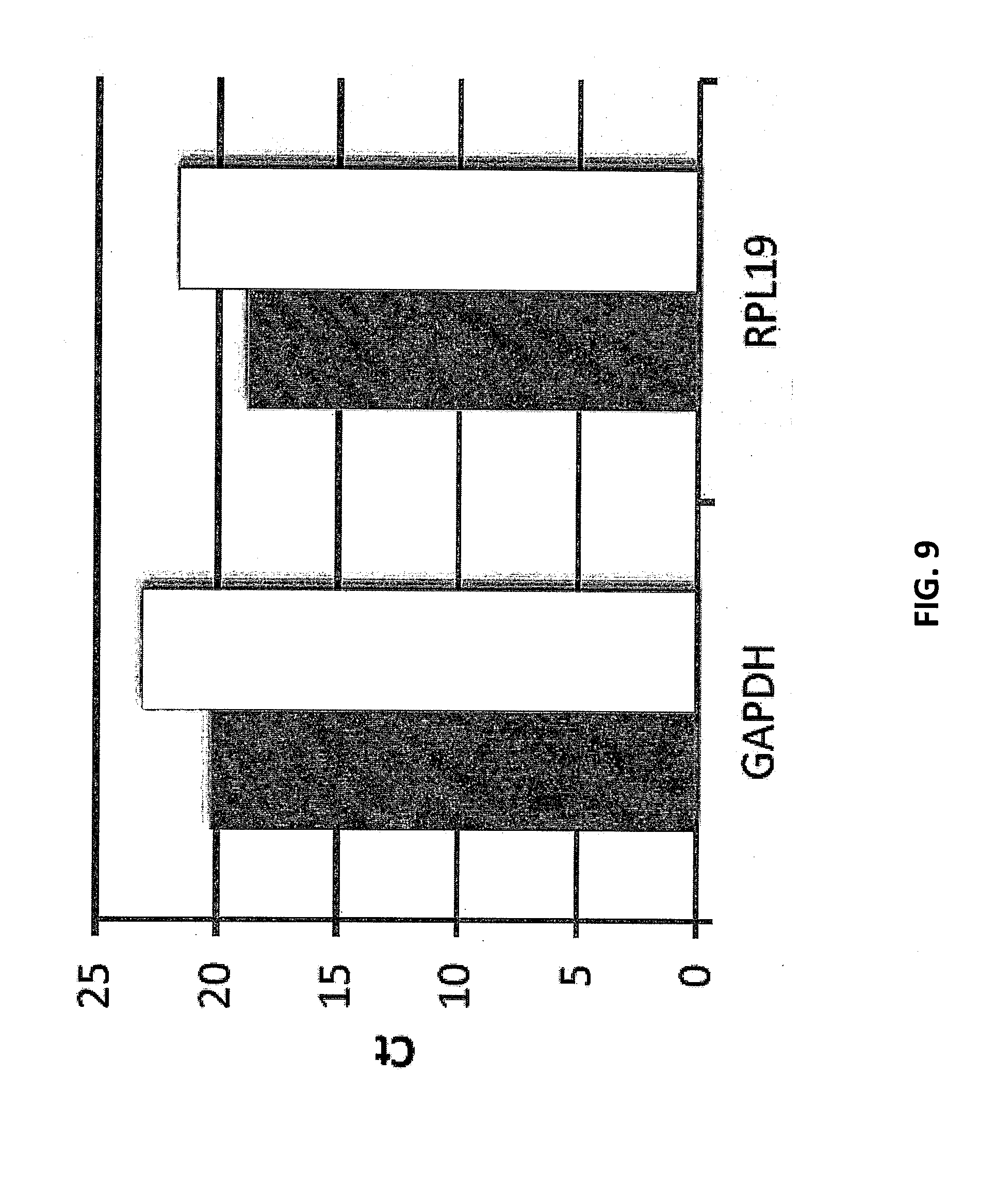
[0028] FIG. 9 depicts a bar graph comparing amplification efficiency of GAPDH and RPL19 amplified from microwells and tubes. The grey bars represent data from the microwell. The white bars represent data from the tube.
[0029] FIG. 10 depicts an agarose gel comparing amplification specificity of three different genes directly on a solid support.
[0030] FIG. 11A-I show graphical representations of the sequencing results.
[0031] FIG. 12A-C show a histogram of the sequencing results for the K562-only sample, Ramos-only sample, and K562+Ramos mixture sample, respectively.
[0032] FIG. 12D-E shows a graph of the copy number for genes listed in Table 3 for the Ramos-only cell sample and K562-only cell sample, respectively.
[0033] FIG. 12F-I show the copy number for individual genes.
[0034] FIG. 12J-M show graphs of the number of unique molecules per gene (y-axis) for the beads with the 100 unique barcode combinations.
[0035] FIG. 12N-O show enlarged graphs of two beads that depict the general pattern of gene expression profiles for the two cell types.
[0036] FIG. 12P shows a scatter plot of results based on principal component analysis of gene expression profile of 768 beads with >30 molecules per bead from the K562+Ramos mixture sample.
[0037] FIG. 12Q-R show histograms of the copy number per amplicon per bead for the K562-like cells (beads on the left of the first principal component based on FIG. 12P) and Ramos-like cells (beads on the right of the first principal component based on FIG. 12P), respectively.
[0038] FIG. 12S-T show the copy number per bead or single cell of the individual genes for the K562-like cells (beads on the left of the first principal component based on FIG. 12P) and Ramos-like cells (beads on the right of the first principal component based on FIG. 12P), respectively.
[0039] FIG. 13A depicts general gene expression patterns for the mouse and Ramos cells.
[0040] FIG. 13B-C show scatter plots of results based on principal component analysis of gene expression profile of the high density sample and low density sample, respectively.
[0041] FIG. 13D-E depict graphs of the read per barcode (bc) combination (y-axis) versus the unique barcode combination, sorted by the total number of molecules per bc combination (x-axis) for Ramos-like cells and mouse-like cells from the high density sample, respectively.
[0042] FIG. 13F-G depict graphs of the number of molecules per barcode (bc) combination (y-axis) versus the unique barcode combination, sorted by the total number of molecules per bc combination (x-axis) for Ramos-like cells and mouse-like cells from the high density sample, respectively.
[0043] FIG. 13H-I depict graphs of the read per barcode (bc) combination (y-axis) versus the unique barcode combination, sorted by the total number of molecules per barcode combination (x-axis) for Ramos-like cells and mouse-like cells from the low density sample, respectively.
[0044] FIG. 13J-K depict graphs of the number of molecules per barcode combination (y-axis) versus the unique barcode combination, sorted by the total number of molecules per barcode combination (x-axis) for Ramos-like cells and mouse-like cells from the low density sample, respectively.
[0045] FIG. 14 shows a graph depicting the genes on the X-axis and the log 10 of the number of reads.
[0046] FIG. 15A shows a graph of the distribution of genes detected per three-part cell label (e.g., cell barcode). FIG. 15B shows a graph of the distribution of unique molecules detected per bead (expressing the gene panel).
[0047] FIG. 16 depicts the cell clusters based on the genes associated with a cell barcode.
[0048] FIG. 17A-D show the analysis of monocyte specific markers. FIG. 17E shows the cell cluster depicted in FIG. 16.
[0049] FIG. 18A-B show the analysis of the T cell specific markers. FIG. 18C shows the cell cluster depicted in FIG. 16.
[0050] FIG. 19A-B show the analysis of the CD8+ T cell specific markers. FIG. 19C shows the cell cluster depicted in FIG. 16.
[0051] FIG. 20A shows the analysis of CD4+ T cell specific markers. FIG. 20B shows the cell cluster depicted in FIG. 16.
[0052] FIG. 21A-D show the analysis of Natural Killer (NK) cell specific markers. FIG. 21E shows the cell cluster depicted in FIG. 16.
[0053] FIG. 22A-E show the analysis of B cell specific markers. FIG. 22F shows the cell cluster depicted in FIG. 16.
[0054] FIG. 23A-F show the analysis of Toll-like receptors. Toll-like receptors are mainly expressed by monocytes and some B cells. FIG. 23G shows the cell cluster depicted in FIG. 16.
[0055] FIG. 24 depicts a graph of the genes versus the log 10 of the number of reads.
[0056] FIG. 25A-D shows graphs of the molecular barcode versus the number of reads or log 10 of the number of reads for two genes.
[0057] FIG. 26A shows a graph of the number of genes in the panel expressed per cell barcode versus the number of unique cell barcodes/single cell. FIG. 26B shows a histogram of the number of unique molecules detected per bead versus frequency of the number of cells per unique cell barcode carrying a given number of molecules. FIG. 26C shows a histogram of the number of unique GAPDH molecules detected per bead versus frequency of the number of cells/unique cell barcode carrying a given number of molecules.
[0058] FIG. 27 shows a scatterplot of the 856 cells.
[0059] FIG. 28 shows a heat map of expression of the top 100 (in terms of the total number of molecules detected).
[0060] FIG. 29 shows a workflow for Example 12.
[0061] FIG. 30 shows a workflow for Example 13. FIG. 30 discloses "dT(17)V" as SEQ ID NO: 829 and "AAAAAAAAAA" as SEQ ID NO: 830.
[0062] FIG. 31A-C. Clustering of single cells in controlled mixtures containing two distinct cell types. FIG. 31A. Clustering of a 1:1 mixture of K562 and Ramos cells by principal component analysis of the expression of 12 genes. The biplot shows two distinct clusters, with one cluster expressing Ramos specific genes and the other expressing K562 specific genes. FIG. 31B. Principal component analysis of a mixture containing a small percentage of Ramos cells in a background of primary B cells from a healthy individual using a panel of 111 genes. The color of each data point indicates the total number of unique transcript molecules detected across the entire gene panel. A set of 18 cells (circled) out of 1198 cells displays a distinct gene expression profile and with much higher transcription levels. FIG. 31C. Heatmap showing expression level of each gene in the top 100 cells in the sample of FIG. 31B, ranked by the total number of transcript molecules detected in the gene panel. Genes are ordered via hierarchical clustering in terms of correlation. The top 18 cells, indicated by the horizontal red bar, expressed preferentially a set of genes known to be associated with follicular lymphoma, as indicated by the vertical red bar.
[0063] FIG. 31D. PCA analysis of primary B cells with spiked in Ramos cells. Color of each data point (single cell) indicates the log of the number of transcript molecules each cell carries for the particular gene. Top 7 rows: Genes that are preferentially expressed by the subset of 18 cells that are likely Ramos cells. First row genes (from left to right) include GAPDH, TCL1A, MKI67 and BCL6. Second row genes (from left to right) include MYC, CCND3, CD81 and GNAI2. Third row of genes (from left to right) include IGBP1, CD20, BLNK and DOCKS. Fourth row of genes (from left to right) include IRF4, CD22, IGHM and AURKB. Fifth row of genes (from left to right) include CD38, CD10, LEFT and AICDA. Sixth row of genes (from left to right) include CD40, CD27, IL4R and PRKCD. Seventh row of genes (from left to right) include RGS1, MCL1, CD79a and HLA-DRA. Last row: Genes that are expressed preferentially by a subset of primary B cells but not especially enriched in those 18 cells. Genes in the last row (from left to right) include IL6, CD23a, CCR7 and CXCR5.
[0064] FIG. 32 Expression of GAPDH. Color indicates natural log of the number of unique transcript molecules observed per cell.
[0065] FIG. 33A-F shows the principal component analysis (PCA) for monocyte associated genes. FIG. 33A shows the PCA for CD16. FIG. 33B shows the PCA for CCRvarA. FIG. 33C shows the PCA for CD14. FIG. 33D shows the PCA for S100A12. FIG. 33E shows the PCA for CD209. FIG. 33F shows the PCA for IFNGR1.
[0066] FIG. 34A-B shows the principal component analysis (PCA) for pan-T cell markers (CD3). FIG. 34A shows the PCA for CD3D and FIG. 34B shows the PCA for CD3E.
[0067] FIG. 35A-E shows the principal component analysis (PCA) for CD8 T cell associated genes. FIG. 35A shows the PCA for CD8A. FIG. 35B shows the PCA for EOMES. FIG. 35C shows the PCA for CD8B. FIG. 35D shows the PCA for PRF1. FIG. 35E shows the PCA for RUNX3.
[0068] FIG. 36A-C shows the principal component analysis (PCA) for CD4 T cell associated genes. FIG. 36A shows the PCA for CD4. FIG. 36B shows the PCA for CCR7. FIG. 36C shows the PCA for CD62L.
[0069] FIG. 37A-F shows the principal component analysis (PCA) for B cell associated genes. FIG. 37A shows the PCA for CD20. FIG. 37B shows the PCA for IGHD. FIG. 37C shows the PCA for PAX5. FIG. 37D shows the PCA for TCL1A. FIG. 37E shows the PCA for IGHM. FIG. 37F shows the PCA for CD24.
[0070] FIG. 38A-C shows the principal component analysis (PCA) for Natural Killer cell associated genes. FIG. 38A shows the PCA for KIR2DS5. FIG. 38B shows the PCA for CD16. FIG. 38C shows the PCA for CD62L.
[0071] FIG. 39 Simultaneous identification of major cell types in a human PBMC sample (632 cells) by PCA analysis of 81 genes assayed by CytoSeq Cells with highly correlated expression profile are coded with similar color.
[0072] FIG. 40A-B Correlation analysis of single cell gene expression profile of PBMC sample. 40A. A matrix showing the pairwise correlation coefficient across 632 cells in the sample. The cells are ordered such that those with highly correlated gene expression profile are grouped together. FIG. 40B. Heatmap showing the expression of each gene by each cell. The cells (columns) are ordered in the same manner as the correlation matrix above. The genes (rows) are ordered such that genes that share highly similar expression pattern across the cells are grouped together. The cell type of each cluster of cells may be identified by the group of genes the cells co-expressed. Within each major cell cluster, there is substantial degree of heterogeneity in terms of gene expression.
[0073] FIG. 41 data represents that of 731 cells from a replicate experiment of PBMC sample from the same donor. Cells with similar gene expression profile (based on hierarchical clustering using correlation coefficient) are plotted with similar color.
[0074] FIG. 42 shows a heat map demonstrating the correlation in gene expression profile between genes.
[0075] FIG. 43 Description of CytoSeq. FIG. 43A. Experimental procedure for CytoSeq. FIG. 43B. Structure of oligonucleotides attached to beads.
[0076] FIGS. 44A-C illustrate dissecting sub-populations of CD3+ T cells. FIG. 44A. PCA of Donor 1 unstimulated sample reveals two major branches of cells. The expression level (log of unique transcript molecule) of a particular gene within each cell is indicated with color. Helper T cell associated cytokine and effector genes are enriched in cells in the lower branch, while cytotoxic T cell associated genes are enriched in the upper branch. Shown here are representative genes. First row shows helper T cell related genes and include (from left to right) CD4, SELL and CCR7. Second row shows cytotoxic T cell related genes and include (from left to right) CD8A, NKG2D and EOMES. FIG. 44B. PCA of Donor 1 anti-CD3/anti-CD28 stimulated sample showing enrichment of expression of indicated genes to one of the two main branches representing helper and cytotoxic T cells. These genes are present at low amounts in the unstimulated sample. First two rows show genes that are known to be associated with activated T cells and include (from left to right) in the first row IRF4, CD69 and MYC and in the second row GAPDH, TNF and IFNG. The third row shows genes that are known to be associated with activated helper T cells and include (from left to right) IL2, LTA and CD40LG. The fourth row shows genes that are known to be associated with activated cytotoxic T cells and include (from left to right) CCL4, CCL3 and GZMB. FIG. 44C. Number of cells that contribute to the overall expression level of genes that exhibit large fold-changes when comparing stimulated over unstimulated samples in aggregate data. For several cytokines (red arrows), the contribution from only a small number of cells is responsible for large overall gene expression change in the entire population.
[0077] FIGS. 45A-C illustrate PCA plots of T cell samples that have undergone stimulation with anti-CD28/anti-CD3 beads in the two donors, and the corresponding unstimulated samples, with emphasis on the expression of genes that clearly show preferential expression in either helper or cytotoxic subsets in the unstimulated samples. The color of each data point (single cell) indicates log(number of unique transcript molecule) per cell for the indicated gene. For each pair of stimulated and unstimulated graphs in each donor, the color range is adjusted to be the same. FIG. 45A. Genes that are known to be associated with both helper and cytotoxic T cells. FIG. 45B. Genes that are known to be associated with cytotoxic T cells. FIG. 45C. Genes that are known to be associated with helper T cells.
[0078] FIG. 46A-D PCA plots of T cell samples that have undergone stimulation with anti-CD28/anti-CD3 beads in the two donors, and the corresponding unstimulated samples, with emphasis on the expression of genes that are expressed in the stimulated samples but at low or undetectable level in the unstimulated samples. The color of each data point (single cell) indicates log(number of unique transcript molecule) per cell for the indicated gene. For each pair of stimulated and unstimulated graphs in each donor, the color range is adjusted to be the same. 46A and 46D. Genes that are expressed by both branches of cells upon activation. 46B. Genes that are expressed preferentially by cells in the upper branch upon activation. These genes are known to be associated with activated cytotoxic T cells. 46C. Genes that are expressed preferentially by cells in the lower branch upon activation. These genes are known to be associated with activated helper T cells.
[0079] FIG. 47 Clustering of data from Donor 1's unstimulated CD3+ T cells shows separations of CD4 and CD8 cells, as well as a group of cells that express Granzyme K and Granzyme A but little CD8. Top: Heatmap showing correlation between each pair of cells. Cells that are highly correlated are grouped together. Bottom: Heatmap showing the level of expression of each gene of each cell. Cells and genes are ordered via bidirectional hierarchical clustering.
[0080] FIG. 48. Similar to FIG. 47, but showing data from anti-CD3/anti-CD28 stimulated CD3+ T cell sample of Donor 1. Top: Heatmap showing correlation between each pair of cells. Cells that are highly correlated are grouped together. Bottom: Heatmap showing the level of expression of each gene of each cell. Cells and genes are ordered via bidirectional hierarchical clustering.
[0081] FIG. 49A-C In donor 1, large overall fold change was observed for various cytokines in the antiCD28/antiCD3 stimulated sample, as compared to the unstimulated one. FIGS. A-B: The large fold changes of these cytokines were mostly contributed by only a few single cells (dots that are enclosed with squares or circles). A number of these cytokines were contributed by the same small number of cells. FIG. 49C: The co-expression patterns of these cytokines coincide with the signature cytokine combination for the Th2 and Th17 subsets of helper T cells.
[0082] FIG. 50A-B. Dissecting sub-populations of CD8+ T cells. FIG. 50A. Clustering of CytoSeq data defines two major groups of CD8+ cells--one group expresses genes shared by central memory/naive cells, and the other group expresses genes shared by effector memory/effector cells. Shown here is data of Donor 2's unstimulated sample. Top: Heatmap showing correlation between each pair of cells. Bottom: Heatmap showing the level of expression of each gene in each cell. Cells and genes are ordered via bidirectional hierarchical clustering. FIG. 50B. Identification of rare antigen specific T cell by expression of gamma interferon (IFNG) in CD8+ T cells from two donors after stimulation with CMV peptide pool. Each cell is plotted on the 2D principal component space. Cells expressing IFNG (circled) are usually among those with the most total detected transcripts in the panel (indicated by the color). In donor 2, the top expressing cell (square) does not produce IFNG but expresses cytokines IL6 and IL1B. Number next to each circle indicates the rank in descending order the number of total unique transcript molecules detected for that cell.
[0083] FIG. 51. Similar to FIG. 50A except the data here represents that of Donor 2 CMV stimulated sample. A. Clustering of CytoSeq data defines two major groups of CD8+ cells--one group expresses genes shared by central memory/naive cells, and the other group expresses genes shared by effector memory/effector cells. Shown here is data of Donor 2's unstimulated sample. Top: Heatmap showing correlation between each pair of cells. Bottom: Heatmap showing the level of expression of each gene in each cell. Cells and genes are ordered via bidirectional hierarchical clustering.
[0084] FIGS. 52A-F illustrate data plotted in principal component space. Color indicates log(number of unique transcript molecules detected) for the particular gene. FIG. 52A. Genes that appear to be expressed by a larger proportion of cells upon stimulation by CMV peptide pool. FIG. 52B. Genes that are enriched in one branch of cells. These genes are also known to be associated with naive and central memory CD8+ T cells. FIG. 52C. Genes that are enriched in the other branch of cells. These genes are known to be associated with effector and effector memory CD8+ T cells. FIG. 52D. Granzyme K expressing cells occupy a region between the naive/central memory and effector/effector memory cells on the PC space. FIG. 52E. HLA-DRA expressing cells constitute a special subset. FIG. 52F. Genes that are expressed in both branches of cells.
[0085] FIG. 53. Same as FIG. 50B, except the data represents those of the unstimulated controls. None of the cells in Donor 1's sample expressed IFNG, while one cell in Donor 2's sample expressed IFNG yet with overall low expression across the entire gene panel (rank 1069). Color scale is adjusted to match that of the respective graph for the stimulated sample.
[0086] FIG. 54. Heatmaps showing the heterogeneous expression of the gene panel in cells that express gamma interferon (IFNG) in CMV stimulated CD8+ T cells of Donors 1 and 2. Also shown is the cell that carries most total transcripts detected in Donor 2. This particular cell does not express IFNG but expresses strongly IL6, IL1B and CCL4. The cells and genes are ordered by bidirectional hierarchical clustering based on correlation. Cell ID refers to the rank in total number of detected transcripts of the gene panel, and are indicated in the PCA plots in FIG. 50.
[0087] FIG. 55. Amplification scheme. The first PCR amplifies molecules attached to the bead using a gene specific primer and a primer against the universal Illumina sequencing primer 1 sequence. The second PCR amplifies the first PCR products using a nested gene specific primer flanked by Illumina sequencing primer 2 sequence, and a primer against the universal Illumina sequencing primer 1 sequence. The third PCR adds P5 and P7 and sample index to turn PCR products into Illumina sequencing library. 150 bp.times.2 sequencing reveals the cell label and molecule label on read 1, the gene on read 2, and the sample index on index 1 read.
[0088] FIG. 56 depicts a schematic of a workflow for analyzing molecules from a sample. FIG. 56 discloses "AAAAAAAAAAAAAAAAAAAAAAAAAAAAAAA" as SEQ ID NO: 831.
[0089] FIG. 57 depicts a schematic of a workflow for analyzing molecules from a sample.
[0090] FIG. 58A-B depict agarose gels of PCR products.
[0091] FIG. 59 depicts a plot of sequencing reads for a plurality of genes.
[0092] FIG. 60A-D depicts plots of the reads observed per label detected (RPLD) for Lys, Phe, Thr, and Dap spike-in controls, respectively. FIG. 60E depicts a plot of Reads versus Input.
[0093] FIG. 61 depicts a plot of the reads observed per label detected (RPLD) for various genes.
[0094] FIG. 62 depicts a plot of the reads observed per label detected (RPLD) for various genes.
[0095] FIG. 63 depicts a plot of total reads (labels) versus rpld for various genes.
[0096] FIG. 64 depicts a plot of RPKM for undetected genes.
[0097] FIG. 65 depicts a schematic for the synthesis of molecular barcodes. FIG. 65 discloses "1001" as SEQ ID NO: 832 and "1003" as SEQ ID NO: 833 and "1005" as SEQ ID NOS 832 and 833, respectively, in order of appearance.
[0098] FIG. 66A-C depict schematics for the synthesis of molecular barcodes. FIG. 66A discloses "1121" as SEQ ID NO: 834, "1127" as SEQ ID NO: 835, "1128" as SEQ ID NO: 836 and "1129" as SEQ ID NO: 837. FIG. 66B discloses "1150" as SEQ ID NO: 838, "1159" as SEQ ID NO: 839 and "1158" as SEQ ID NO: 840. FIG. 66C discloses "1170" as SEQ ID NO: 841, "1176" as SEQ ID NO: 842 and "1177" as SEQ ID NO: 843.
[0099] FIG. 67 shows a schematic of a workflow for stochastically labeling nucleic acids. FIG. 67 discloses "AAAAAAAAAAAAAAAAAAAAAAAAAAAAAAA" as SEQ ID NO: 831.
[0100] FIG. 68 is a schematic of a workflow for stochastically labeling nucleic acids. FIG. 68 discloses "AAAAAAAAAAAAAAAAAAAAAAAAAAAAAAA" as SEQ ID NO: 831.
[0101] FIG. 69 illustrates a mechanical fixture within which microwell array substrates may be clamped, thereby forming a reaction chamber or well into which samples and reagents may be pipetted for performing multiplexed, single cell stochastic labeling/molecular indexing experiments. Upper: exploded view showing the upper and lower parts of the fixture and an elastomeric gasket for forming a leak-proof seal with the microwell array substrate. Lower: exploded side-view of the fixture.
[0102] FIG. 70 illustrates a mechanical fixture which creates two reaction chambers or wells when a microwell array substrate is clamped within the fixture.
[0103] FIG. 71 illustrates two examples of elastomeric (e.g., polydimethylsiloxane) gaskets for use with the mechanical fixtures illustrated in FIGS. 69 and 70. The elastomeric gaskets provide for a leak-proof seal with the microwell array substrate to create a reagent well around the microwell array. The gaskets may contain one (upper), two (lower), or more openings for creating reagent wells.
[0104] FIG. 72 depicts one embodiment of a cartridge within which a microwell array is packaged. Left: An exploded view of the cartridge illustrating (from bottom to top) the microwell array substrate, a gasket that defines the flow cell or array chamber, a reagent and/or waste reservoir component for defining compartments to contain pre-loaded assay reagents or store spent reagents, and a cover for sealing the reagent and waste reservoirs and defining the sample inlet and outlet ports. Right: An assembled view of one embodiment of the cartridge design illustrating relief for bringing an external magnet into close proximity with the microwell array.
[0105] FIG. 73 depicts one embodiment of a cartridge designed to include onboard assay reagents with the packaged microwell array.
[0106] FIG. 74 provides a schematic illustration of an instrument system for performing multiplexed, single cell stochastic labeling/molecular indexing assay. The instrument system may provide a variety of control and analysis capabilities, and may be packaged as individual modules or as a fully integrated system. Microwell arrays may be integrated with flow cells that are either a fixed component of the system or are removable, or may be packaged within removable cartridges that further comprise pre-loaded assay reagent reservoirs and other functionality.
[0107] FIG. 75 illustrates one embodiment of the process steps to be performed by an automated system for performing multiplexed, single cell stochastic labeling/molecular indexing assays.
[0108] FIG. 76 illustrates one embodiment of a computer system or processor for providing instrument control and data analysis capabilities for the assay system presently disclosed.
[0109] FIG. 77 shows a block diagram illustrating one example of a computer system architecture that can be used in connection with example embodiments of the assay systems of the present disclosure.
[0110] FIG. 78 depicts a diagram showing a network with a plurality of computer systems, cell phones, personal data assistants, and Network Attached Storage (NAS), that can be used with example embodiments of the assay systems of the present disclosure.
[0111] FIG. 79 depicts a block diagram of a multiprocessor computer system that can be used with example embodiments of the assay systems of the present disclosure.
[0112] FIG. 80 depicts a diagram of analysis of a test sample and communication of test result obtained from the test sample via a communication media.
DETAILED DESCRIPTION
[0113] Disclosed herein are methods, kits, and compositions for analyzing molecules in a plurality of samples. Generally, the methods, kits, and compositions comprise (a) stochastically labeling molecules in two or more samples with molecular barcodes to produce labeled molecules; and (b) detecting the labeled molecules. The molecular barcodes may comprise one or more target specific regions, label regions, sample index regions, universal PCR regions, adaptors, linkers, or a combination thereof. The labeled molecules may comprise a) a molecule region; b) a sample index region; and c) a label region. The molecule region may comprise at least a portion of the molecule from the molecular barcode was originally attached to. The molecule region may comprise a fragment of the molecule from the molecular barcode was originally attached to. The sample index region may be used to determine the source of the molecule region. The sample index region may be used to determine from which sample the molecule region originated from. The sample index region may be used to differentiate molecule regions from two or more different samples. The label region may be used to confer a unique identity to identical molecule regions originating from the same source. The label region may be used to confer a unique identity to identical molecule regions originating from the same sample.
[0114] The method for analyzing molecules in a plurality of samples may comprise: a) producing a plurality of sample-tagged nucleic acids by: i) contacting a first sample comprising a plurality of nucleic acids with a plurality of first sample tags to produce a plurality of first sample-tagged nucleic acids; and ii) contacting a second sample comprising a plurality of nucleic acids with a plurality of second sample tags to produce a plurality of second sample-tagged nucleic acids, wherein the plurality of second sample tags are different from the first sample tags; b) contacting the plurality of sample-tagged nucleic acids with a plurality of molecular identifier labels to produce a plurality of labeled nucleic acids; and c) detecting at least a portion of the labeled nucleic acids, thereby determining a count of a plurality of nucleic acids in a plurality of samples. The plurality of samples may comprise a single cell.
[0115] Alternatively, the method for analyzing molecules in a plurality of samples may comprise: a) producing a plurality of labeled nucleic acids comprising: i) contacting a first sample with a first plurality of sample tags, wherein the first plurality of sample tags comprises identical nucleic acid sequences; ii) contacting the first sample with a first plurality of molecular identifier labels may comprise different nucleic acid sequences, wherein contacting the first sample with the first plurality of sample tags or first plurality of molecular identifier labels occurs simultaneously or sequentially to produce a plurality of first-labeled nucleic acids; iii) contacting a second sample with a second plurality of sample tags, wherein the second plurality of sample tags may comprise identical nucleic acid sequences; iv) contacting the second sample with a second plurality of molecular identifier labels may comprise different nucleic acid sequences, wherein contacting the second sample with the second plurality of sample tags or second plurality of molecular identifier labels occurs simultaneously or sequentially to produce a plurality of second-labeled nucleic acids, wherein the plurality of labeled nucleic acids may comprise the plurality of first-labeled nucleic acids and the second-labeled nucleic acids; and b) determining a number of different labeled nucleic acids, thereby determining a count of a plurality of nucleic acids in a plurality of samples.
[0116] The method for analyzing molecules in a plurality of samples may comprise: a) contacting a plurality of samples may comprise two or more different nucleic acids with a plurality of sample tags and a plurality of molecular identifier labels to produce a plurality of labeled nucleic acids, wherein: i) the plurality of labeled nucleic acids may comprise two or more nucleic acids attached to two or more sample tags and two or more molecular identifier labels; ii) the sample tags attached to nucleic acids from a first sample of the plurality of samples are different from the sample tags attached to nucleic acid molecules from a second sample of the plurality of samples; and iii) two or more identical nucleic acids in the same sample are attached to two or more different molecular identifier labels; and b) detecting at least a portion of the labeled nucleic acids, thereby determining a count of two or more different nucleic acids in the plurality of samples.
[0117] FIG. 56 depicts an exemplary workflow for the quantification of RNA molecules in a sample. As shown in Step 1 of FIG. 56, RNA molecules (110) may be reverse transcribed to produce cDNA molecules (105) by the stochastic hybridization of a set of molecular identifier labels (115) to the polyA tail region of the RNA molecules. The molecular identifier labels (115) may comprise an oligodT region (120), label region (125), and universal PCR region (130). The set of molecular identifier labels may contain 960 different types of label regions. As shown in Step 2 of FIG. 56, the labeled cDNA molecules (170) may be purified to remove excess molecular identifier labels (115). Purification may comprise Ampure bead purification. As shown in Step 3 of FIG. 56, the labeled cDNA molecules (170) may be amplified to produce a labeled amplicon (180). Amplification may comprise multiplex PCR amplification. Amplification may comprise a multiplex PCR amplification with 96 multiplex primers in a single reaction volume. Amplification may comprise a custom primer (135) and a universal primer (140). The custom primer (135) may hybridize to a region within the cDNA (105) portion of the labeled cDNA molecule (170). The universal primer (140) may hybridize to the universal PCR region (130) of the labeled cDNA molecule (170). As shown in Step 4, the labeled amplicons (180) may be further amplified by nested PCR. The nested PCR may comprise multiplex PCR with 96 multiplex primers in a single reaction volume. Nested PCR may comprise a custom primer (145) and a universal primer (140). The custom primer (135) may hybridize to a region within the cDNA (105) portion of the labeled amplicon (180). The universal primer (140) may hybridize to the universal PCR region (130) of the labeled amplicon (180). As shown in Step 5, one or more adaptors (150, 155) may be attached to the labeled amplicon (180) to produce an adaptor-labeled amplicon (190). The one or more adaptors may be attached to the labeled amplicon (180) via ligation. As shown in Step 6, the one or more adaptors (150, 155) may be used to conduct one or more additional assays on the adaptor-labeled amplicon (190). The one or more adaptors (150, 155) may be hybridized to one or more primers (160, 165). The one or more primers (160, 165) be PCR amplification primers. The one or more primers (160, 165) may be sequencing primers. The one or more adaptors (150, 155) may be used for further amplification of the adaptor-labeled amplicons. The one or more adaptors (150, 155) may be used for sequencing the adaptor-labeled amplicon.
[0118] FIG. 57 depicts an exemplary schematic of a workflow for analyzing nucleic acids from two or more samples. As shown in FIG. 57, a method for analyzing nucleic acids from two or more samples may comprise selecting two or more genes for analysis and designing custom primers based on the selected genes (210). The method may further comprise supplementing one or more samples comprising nucleic acids (e.g., RNA) with one or more spike-in controls (220). The nucleic acids in the sample may be amplified by multiplex RT-PCR (230) with molecular barcodes (or sample tags or molecular identifier labels) and the custom primers to produce labeled amplicons. The labeled amplicons may further treated with one or more sequencing adaptors to produce adaptor labeled amplicons (240). The adaptor labeled amplicons can be analyzed (250). As shown in FIG. 57, analysis of the labeled amplicons (250) may comprise one or more of (1) detection of a universal PCR primer seq, polyA and/or molecular barcode (or sample tag, molecular identifier label); (2) map read on the end of the adaptor labeled amplicons (e.g., 96 genes and spike-in controls) that is not attached to the adaptor and/or barcode (e.g., molecular barcode, sample tag, molecular identifier label); and (3) count and/or summarize the number of different adaptor labeled amplicons.
[0119] FIG. 67 shows a schematic of a workflow for stochastically labeling nucleic acids with molecular barcodes (1220). As shown in step 1 of FIG. 67, RNA molecules may be stochastically labeled with a set of molecular barcodes (1220). The molecular barcodes (1220) may comprise a target binding region (1221), label region (1222), sample index region (1223) and universal PCR region (1224). In some instances, the target binding region comprises an oligodT sequence that hybridizes to a polyA sequence in the RNA molecules. The label region (1222) may contain a unique sequence that may be used to distinguish two or more different molecular barcodes. When the molecular barcode hybridizes to an RNA molecule, the label region may be used to confer a unique identity to identical RNA molecules. The sample index region (1223) may be identical for a set of molecular barcodes. The sample index region (1223) may be used to distinguish labeled nucleic acids from different samples. The universal PCR region (1224) may serve as a primer binding site for amplification of the labeled molecules. Once the RNA molecules are labeled with the molecular barcodes, the RNA molecules may be reverse transcribed to produce labeled cDNA molecules (1230) containing a cDNA copy of the RNA molecule (1210) and the molecular barcode (1220).
[0120] As shown in Step 2 of FIG. 67, excess oligos (e.g., molecular barcodes) may be removed by Ampure bead purification. As shown in Step 3 of FIG. 67, the labeled cDNA molecules may be amplified by multiplex PCR. Multiplex PCR of the labeled cDNA molecules may be performed by using a first set of forward primers (F1, 1235 in FIG. 67) and universal primers (1240) in a single reaction volume to produce labeled amplicons (1245). As shown in Step 4 of FIG. 67, the labeled amplicons may be further amplified by multiplex PCR using nested primers. Nested primer amplification of the labeled amplicons may be performed by using a second set of forward primers (F2, 1250 in FIG. 67) and universal primers (1240) in a single reaction volume to produce labeled nested PCR amplicons. In some instances, the F2 primers (1250) contain an adaptor (1251) and a target binding region (1252). The target binding region (1252) of the F2 primers may hybridize to the labeled amplicons and may prime amplification of the labeled amplicons. The adaptor (1251) and the universal PCR region (1224) of the nested PCR amplicons may be used in the sequencing of the labeled nested PCR amplicons. The amplicons may be sequenced by MiSeq. Alternatively, the amplicons may be sequenced by HiSeq.
[0121] FIG. 68 shows a schematic of a workflow for stochastically labeling nucleic acids. As shown in Step 1 of FIG. 68, RNA molecules (1305) may be stochastically labeled with a set molecular barcodes (1320). The molecular barcodes may comprise a target binding region (1321), label region (1322), and universal PCR region (1323). Once the molecular barcodes are attached to the RNA molecules, the RNA molecules (1305) may be reverse transcribed to produce labeled cDNA molecules (1325) comprising a cDNA copy of the RNA molecule (1310) and the molecular barcode (1320). As shown in Step 2 of FIG. 68, the labeled cDNA molecules may be purified by Ampure bead purification to remove excess oligos (e.g., molecular barcodes). As shown in Step 3 of FIG. 68, the labeled amplicons may be amplified by multiplex PCR. Multiplex PCR of the labeled cDNA molecules may be performed by using a first set of forward primers (F1, 1330 in FIG. 68) and universal primers (1335) in a single reaction volume to produce labeled amplicons (1360). As shown in Step 4 of FIG. 67, the labeled amplicons may be further amplified by multiplex PCR using nested primers. Nested primer amplification of the labeled amplicons may be performed by using a second set of forward primers (F2, 1340 in FIG. 68) and sample index primers (1350) in a single reaction volume to produce labeled nested PCR amplicons. In some instances, the F2 primers (1340) contain an adaptor (1341) and a target binding region (1342). The target binding region (1342) of the F2 primers may hybridize to the labeled amplicons and may prime amplification of the labeled amplicons. The sample index primers (1350) may comprise a universal primer region (1351), sample index region (1352), and adaptor region (1353). As shown in Step 4 of FIG. 68, the universal primer region (1351) of the sample index primer may hybridize to the universal PCR region of the labeled amplicons. The sample index region (1352) of the sample index primer may be used to distinguish two or more samples. The adaptor regions (1341, 1353) may be used to sequence the labeled nested PCR amplicons. The amplicons may be sequenced by MiSeq. Alternatively, the amplicons may be sequenced by HiSeq.
[0122] Further disclosed herein are methods of producing one or more libraries. The one or more libraries may comprise a plurality of labeled molecules. The one or more libraries may comprise a plurality of labeled amplicons. The one or more libraries may comprise a plurality of enriched molecules or a derivative thereof (e.g., labeled molecules, labeled amplicons). Generally, the method of producing one or more libraries comprises (a) stochastically labeling a plurality of molecules from two or more samples to produce a plurality of labeled molecules, wherein the labeled molecules comprise a molecule region, a sample index region, and label region; and (b) producing one or more libraries from the plurality of labeled molecules, wherein (i) the one or more libraries comprise two or more different labeled molecules, (ii) the two or more different labeled molecules differ by the molecule region, sample index region, label region, or a combination thereof.
[0123] The method for producing one or more libraries may comprise: a) producing a plurality of sample-tagged nucleic acids by: i) contacting a first sample comprising a plurality of nucleic acids with a plurality of first sample tags to produce a plurality of first sample-tagged nucleic acids; and ii) contacting a second sample comprising a plurality of nucleic acids with a plurality of second sample tags to produce a plurality of second sample-tagged nucleic acids, wherein the plurality of first sample tags are different from the second sample tags; and b) contacting the plurality of sample-tagged nucleic acids with a plurality of molecular identifier labels to produce a plurality of labeled nucleic acids, thereby producing a labeled nucleic acid library.
[0124] The contacting to a sample can be random or non-random. For example, the contacting of a sample with sample tags can be a random or non-random contacting. In some embodiments, the sample is contacted with sample tags randomly. In some embodiments, the sample is contacted with sample tags non-randomly. The contacting to a plurality of nucleic acids can be random or non-random. For example, the contacting of a plurality of nucleic acids with sample tags can be a random or non-random contacting. In some embodiments, the plurality of nucleic acids is contacted with sample tags randomly. In some embodiments, the plurality of nucleic acids is contacted with sample tags non-randomly.
[0125] Further disclosed herein are methods of producing one or more sets of labeled beads. The method of producing the one or more sets of labeled beads may comprise attaching one or more nucleic acids to one or more beads, thereby producing one or more sets of labeled beads. The one or more nucleic acids may comprise one or more molecular barcodes. The one or more nucleic acids may comprise one or more sample tags. The one or more nucleic acids may comprise one or more molecular identifier labels. The one or more nucleic acids may comprise a) a primer region; b) a sample index region; and c) a linker or adaptor region. The one or more nucleic acids may comprise a) a primer region; b) a label region; and c) a linker or adaptor region. The one or more nucleic acids may comprise a) a sample index region; and b) a label region. The one or more nucleic acids may further comprise a primer region. The one or more nucleic acids may further comprise a target specific region. The one or more nucleic acids may further comprise a linker region. The one or more nucleic acids may further comprise an adaptor region. The one or more nucleic acids may further comprise a sample index region. The one or more nucleic acids may further comprise a label region.
[0126] Further disclosed herein are methods for selecting one or more custom primers. The method of selecting a custom primer for analyzing molecules in a plurality of samples may comprise: a) a first pass, wherein primers chosen may comprise: i) no more than three sequential guanines, no more than three sequential cytosines, no more than four sequential adenines, and no more than four sequential thymines; ii) at least 3, 4, 5, or 6 nucleotides that are guanines or cytosines; and iii) a sequence that does not easily form a hairpin structure; b) a second pass, comprising: i) a first round of choosing a plurality of sequences that have high coverage of all transcripts; and ii) one or more subsequent rounds, selecting a sequence that has the highest coverage of remaining transcripts and a complementary score with other chosen sequences no more than 4; and c) adding sequences to a picked set until coverage saturates or total number of customer primers is less than or equal to about 96.
[0127] Further disclosed herein are kits for use in analyzing two or more molecules from two or more samples. The kit may comprise (a) a first container comprising a first set of molecular barcodes, wherein (i) a molecular barcode of the first set of molecular barcodes comprise a sample index region and a label region; (ii) the sample index region of two or more barcodes of the first set of molecular barcodes are the same; and (iii) the label region of two or more barcodes of the first set of molecular barcodes are different; and (b) a second container comprising a second set of molecular barcodes, wherein (i) a molecular barcode of the second set of molecular barcodes comprise a sample index region and a label region; (ii) the sample index region of two or more barcodes of the second set of molecular barcodes are the same; (iii) the label region of two or more barcodes of the second set of molecular barcodes are different; (iv) the sample index region of the barcodes of the second set of molecular barcodes are different from the sample index region of the barcodes of the first set of molecular barcodes; and (v) the label region of two or more barcodes of the second set of molecular barcodes are identical to the label region of two or more barcodes of the first set of molecular barcodes.
[0128] Alternatively, the kit comprises: a) a plurality of beads, wherein one or more beads of the plurality of beads may comprise at least one of a plurality of nucleic acids, wherein at least one of a plurality nucleic acids may comprise: i) at least one primer sequence, wherein the primer sequence of at least one of the plurality of nucleic acids is the same for the plurality of beads; ii) a bead-specific sequence, wherein the bead-specific sequence of any one of the plurality of nucleic acids is the same, and wherein the bead-specific sequence is different for any one of the plurality of beads; and iii) a stochastic sequence, wherein the stochastic sequence is different for any one of the plurality of nucleic acids; b) a primer may comprise a sequence complementary to the primer sequence; and c) one or more amplification agents suitable for nucleic acid amplification.
[0129] Alternatively, the kit comprises: a) a first container comprising a first set of sample tags, wherein (i) a sample tag of the first set of sample tags comprises a sample index region; and (ii) the sample index regions of the sample tags of the first set of sample tags are at least about 80% identical; and b) a second container comprising a first set of molecular identifier labels, wherein (i) a molecular identifier label of the first set of molecular identifier labels comprises a label region; and (ii) at least about 30% of the label regions of the total molecular identifier labels of the first set of molecular identifier labels are different
[0130] Before the present methods, kits and compositions are described in greater detail, it is to be understood that this invention is not limited to particular method, kit or composition described, as such may, of course, vary. It is also to be understood that the terminology used herein is for the purpose of describing particular embodiments only, and is not intended to be limiting, since the scope of the present invention will be limited only by the appended claims. Examples are put forth so as to provide those of ordinary skill in the art with a complete disclosure and description of how to make and use the present invention, and are not intended to limit the scope of what the inventors regard as their invention nor are they intended to represent that the experiments below are all or the only experiments performed. Efforts have been made to ensure accuracy with respect to numbers used (e.g., amounts, temperature, etc.) but some experimental errors and deviations should be accounted for. Unless indicated otherwise, parts are parts by weight, molecular weight is weight average molecular weight, temperature is in degrees Centigrade, and pressure is at or near atmospheric.
[0131] Methods, kits and compositions are provided for stochastic labeling of nucleic acids in a plurality of samples or in a complex nucleic acid preparation. These methods, kits and compositions find use in unraveling mechanisms of cellular response, differentiation or signal transduction and in performing a wide variety of clinical measurements. These and other objects, advantages, and features of the invention will become apparent to those persons skilled in the art upon reading the details of the methods, kits and compositions as more fully described below.
[0132] The methods disclosed herein comprise attaching one or more molecular barcodes, sample tags, and/or molecular identifier labels to two or more molecules from two or more samples. The molecular barcodes, sample tags and/or molecular identifier labels may comprise one or more oligonucleotides. In some instances, attachment of molecular barcodes, sample tags, and/or molecular identifier labels to the molecules comprises stochastic labeling of the molecules. Methods for stochastically labeling molecules may be found, for example, in U.S. Ser. Nos. 12/969,581 and 13/327,526. Generally, the stochastic labeling method comprises the random attachment of a plurality of the tag and label oligonucleotides to one or more molecules. The molecular barcodes, sample tags, and/or molecular identifier labels are provided in excess of the one or more molecules to be labeled. In stochastic labeling, each individual molecule to be labeled has an individual probability of attaching to the plurality of the molecular barcodes, sample tags, and/or molecular identifier labels. The probability of each individual molecule to be labeled attaching to a particular molecular barcodes, sample tags, and/or molecular identifier labels may be about the same as any other individual molecule to be labeled. Accordingly, in some instances, the probability of any of the molecules in a sample finding any of the tags and labels is assumed to be equal, an assumption that may be used in mathematical calculations to estimate the number of molecules in the sample. In some circumstances the probability of attaching may be manipulated by, for example electing tags and labels with different properties that would increase or decrease the binding efficiency of that molecular barcodes, sample tags, and/or molecular identifier labels with an individual molecule. The tags and labels may also be varied in numbers to alter the probability that a particular molecular barcodes, sample tags, and/or molecular identifier labels will find a binding partner during the stochastic labeling. For example, one label is overrepresented in a pool of labels, thereby increasing the chances that the overrepresented label finds at least one binding partner.
[0133] The methods disclosed herein may further comprise combining two or more samples. The methods disclosed herein may further comprise combining one or more molecules from two or more samples. For example, the methods disclosed herein comprise combining a first sample and a second sample. The two or more samples may be combined after conducting one or more stochastic labeling procedures. The two or more samples may be combined after attachment of one or more sets of molecular barcodes to two or more molecules from the two or more samples. The two or more samples may be combined after attachment of one or more sets of sample tags to two or more molecules from the two or more samples. The two or more samples may be combined after attachment of one or more sets of molecular identifier labels to two or more molecules from the two or more samples. For example, the first and second samples are combined prior to contact with the plurality of molecular identifier labels.
[0134] Alternatively, the two or more samples may be combined prior to conducting one or more stochastic labeling procedures. The two or more samples may be combined prior to attachment of one or more sets of molecular barcodes to two or more molecules from the two or more samples. The two or more samples may be combined prior to attachment of one or more sets of sample tags to two or more molecules from the two or more samples. The two or more samples may be combined prior to attachment of one or more sets of molecular identifier labels to two or more molecules from the two or more samples.
[0135] The two or more samples may be combined after conducting one or more assays on two or more molecules or derivatives thereof (e.g., labeled molecules, amplicons) from the two or more samples. The one or more assays may comprise one or more amplification reactions. The one or more assays may comprise one or more enrichment assays. The one or more assays may comprise one or more detection assays. For example, the first and second samples are combined after detecting the labeled nucleic acids.
[0136] The two or more samples may be combined prior to conducting one or more assays on two or more molecules or derivatives thereof (e.g., labeled molecules, amplicons) from the two or more samples. The one or more assays may comprise one or more amplification reactions. The one or more assays may comprise one or more enrichment assays. The one or more assays may comprise one or more detection assays. For example, the first and second samples are combined prior to detecting the labeled nucleic acids.
Supports
[0137] The present disclosure comprises compositions and methods for multiplex sequence analysis from single cells. The methods and compositions of the present disclosure provide for the use of solid supports. In some instances, the methods, kits, and compositions disclosed herein comprise a support.
[0138] The terms "support", "solid support", "semi-solid support", and "substrate" may be used interchangeably and refer to a material or group of materials having a rigid or semi-rigid surface or surfaces. A support may refer to any surface that is transferable from solution to solution or forms a structure for conducting oligonucleotide-based assays. The support or substrate may be a solid support. Alternatively, the support is a non-solid support. A support may refer to an insoluble, semi-soluble, or insoluble material. A support may be referred to as "functionalized" when it includes a linker, a scaffold, a building block, or other reactive moiety attached thereto, whereas a solid support may be "nonfunctionalized" when it lack such a reactive moiety attached thereto. The support may be employed free in solution, such as in a microtiter well format; in a flow-through format, such as in a column; or in a dipstick.
[0139] The support or substrate may comprise a membrane, paper, plastic, coated surface, flat surface, glass, slide, chip, or any combination thereof. In many embodiments, at least one surface of the support may be substantially flat, although in some embodiments it may be desirable to physically separate synthesis regions for different compounds with, for example, wells, raised regions, pins, etched trenches, or the like. According to other embodiments, the solid support(s) may take the form of resins, gels, microspheres, or other geometric configurations. Alternatively, the solid support(s) comprises silica chips, microparticles, nanoparticles, plates, and arrays. Solid supports may include beads (e.g., silica gel, controlled pore glass, magnetic beads, Dynabeads, Wang resin; Merrifield resin, Sephadex/Sepharose beads, cellulose beads, polystyrene beads etc.), capillaries, flat supports such as glass fiber filters, glass surfaces, metal surfaces (steel, gold silver, aluminum, silicon and copper), glass supports, plastic supports, silicon supports, chips, filters, membranes, microwell plates, slides, or the like. plastic materials including multiwell plates or membranes (e.g., formed of polyethylene, polypropylene, polyamide, polyvinylidenedifluoride), wafers, combs, pins or needles (e.g., arrays of pins suitable for combinatorial synthesis or analysis) or beads in an array of pits or nanoliter wells of flat surfaces such as wafers (e.g., silicon wafers), wafers with pits with or without filter bottoms.
[0140] Methods and techniques applicable to polymer (including protein) array synthesis have been described in U.S. Patent Pub. No. 20050074787, WO 00/58516, U.S. Pat. Nos. 5,143,854, 5,242,974, 5,252,743, 5,324,633, 5,384,261, 5,405,783, 5,424,186, 5,451,683, 5,482,867, 5,491,074, 5,527,681, 5,550,215, 5,571,639, 5,578,832, 5,593,839, 5,599,695, 5,624,711, 5,631,734, 5,795,716, 5,831,070, 5,837,832, 5,856,101, 5,858,659, 5,936,324, 5,968,740, 5,974,164, 5,981,185, 5,981,956, 6,025,601, 6,033,860, 6,040,193, 6,090,555, 6,136,269, 6,269,846 and 6,428,752, in PCT Publication No. WO 99/36760 and WO 01/58593, which are all incorporated herein by reference in their entirety for all purposes. Patents that describe synthesis techniques in specific embodiments include U.S. Pat. Nos. 5,412,087, 6,147,205, 6,262,216, 6,310,189, 5,889,165, and 5,959,098. Nucleic acid arrays are described in many of the above patents, but many of the same techniques may be applied to polypeptide arrays. Additional exemplary substrates are disclosed in U.S. Pat. No. 5,744,305 and US Patent Pub. Nos. 20090149340 and 20080038559.
[0141] The attachment of the labeled nucleic acids to the support may comprise amine-thiol crosslinking, maleimide crosslinking, N-hydroxysuccinimide or N-hydroxysulfosuccinimide, Zenon or SiteClick. Attaching the labeled nucleic acids to the support may comprise attaching biotin to the plurality of labeled nucleic acids and coating the one or more beads with streptavadin.
[0142] In some instances, a solid support may comprise a molecular scaffold. Exemplary molecular scaffolds may include antibodies, antigens, affinity reagents, polypeptides, nucleic acids, cellular organelles, and the like. Molecular scaffolds may be linked together (e.g., a solid support may comprise a plurality of connected molecular scaffolds). Molecular scaffolds may be linked together by an amino acid linker, a nucleic acid linker, a small molecule linkage (e.g., biotin and avidin), and/or a matrix linkage (e.g., PEG or glycerol). Linkages may be non-covalent. Linkages may be covalent. In some instances, molecular scaffolds may not be linked. A plurality of individual molecular scaffolds may be used in the methods of the disclosure.
[0143] In some instances a support may comprise a nanoparticle. The nanoparticle may be a nickel, gold, silver, carbon, copper, silicate, platinum cobalt, zinc oxide, silicon dioxide crystalline, and/or silver nanoparticle. Alternatively, or additionally, the nanoparticle may be a gold nanoparticle embedded in a porous manganese oxide. The nanoparticle may be an iron nanoparticle. The nanoparticle may be a nanotetrapod studded with nanoparticles of carbon.
[0144] A support may comprise a polymer. A polymer may comprise a matrix. A matrix may further comprise one or more beads. A polymer may comprise PEG, glycerol, polysaccharide, or a combination thereof. A polymer may be a plastic, rubber, nylon, silicone, neoprene, and/or polystyrene. A polymer may be a natural polymer. Examples of natural polymers include, but are not limited to, shellac, amber, wool, silk, cellulose, and natural rubber. A polymer may be a synthetic polymer. Examples of synthetic polymers include, but are not limited to, synthetic rubber, phenol formaldehyde resin (or Bakelite), neoprene, nylon, polyvinyl chloride (PVC or vinyl), polystyrene, polyethylene, polypropylene, polyacrylonitrile, PVB, and silicone.
[0145] A support may be a semi-solid support. A support may comprise a gel (e.g., a hydrogel). The terms "hydrogel", "gel" and the like, are used interchangeably herein and may refer to a material which is not a readily flowable liquid and not a solid but a gel which gel is comprised of from 0.5% or more and preferably less than 40% by weight of gel forming solute material and from 95% or less and preferably more than 55% water. The gels of the invention may be formed by the use of a solute which is preferably a synthetic solute (but could be a natural solute, e.g., for forming gelatin) which forms interconnected cells which binds to, entrap, absorb and/or otherwise hold water and thereby create a gel in combination with water, where water includes bound and unbound water. The gel may be the basic structure of the hydrogel patch of the invention will include additional components beyond the gel forming solute material and water such as an enzyme and a salt which components are further described herein. The gel may be a polymer gel.
[0146] A solid support may comprise a structured nanostructure. For example, the structured nanostructure may comprise capture containers (e.g., a miniaturized honeycomb) which may comprise the oligonucleotides to capture the cell and/or contents of the cell. In some instances, structured nanostructures may not need the addition of exogenous reagents.
[0147] In some instances, the support comprises a bead. A bead may encompass any type of solid or hollow sphere, ball, bearing, cylinder, or other similar configuration composed of plastic, ceramic, metal, or polymeric material onto which a nucleic acid may be immobilized (e.g., covalently or non-covalently). A bead may comprise nylon string or strings. A bead may be spherical in shape. A bead may be non-spherical in shape. Beads may be unpolished or, if polished, the polished bead may be roughened before treating, (e.g., with an alkylating agent). A bead may comprise a discrete particle that may be spherical (e.g., microspheres) or have an irregular shape. Beads may comprise a variety of materials including, but not limited to, paramagnetic materials, ceramic, plastic, glass, polystyrene, methylstyrene, acrylic polymers, titanium, latex, sepharose, cellulose, nylon and the like. A bead may be attached to or embedded into one or more supports. A bead may be attached to a gel or hydrogel. A bead may be embedded into a gel or hydrogel. A bead may be attached to a matrix. A bead may be embedded into a matrix. A bead may be attached to a polymer. A bead may be embedded into a polymer. The spatial position of a bead within the support (e.g., gel, matrix, scaffold, or polymer) may be identified using the oligonucleotide present on the bead which serves as a location address. Examples of beads include, but are not limited to, streptavidin beads, agarose beads, magnetic beads, Dynabeads.RTM., MACS.RTM. microbeads, antibody conjugated beads (e.g., anti-immunoglobulin microbead), protein A conjugated beads, protein G conjugated beads, protein A/G conjugated beads, protein L conjugated beads, oligodT conjugated beads, silica beads, silica-like beads, anti-biotin microbead, anti-fluorochrome microbead, and BcMag Carboxy-Terminated Magnetic Beads. The diameter of the beads may be about 5 .mu.m, 10 .mu.m, 20 .mu.m, 25 .mu.m, 30 .mu.m, 35 .mu.m, 40 .mu.m, 45 .mu.m or 50 .mu.m. A bead may refer to any three dimensional structure that may provide an increased surface area for immobilization of biological particles and macromolecules, such as DNA and RNA.
[0148] A support may be porous. A support may be permeable or semi-permeable. A support may be solid. A support may be semi-solid. A support may be malleable. A support may be flexible. In some instances, a support may be molded into a shape. For example, a support may be placed over an object and the support may take the shape of the object. In some instances, the support is placed over an organ and takes the shape of the organ. In some instances, the support is produced by 3D-printing.
[0149] The support (e.g., beads, nanoparticles) may be at least about 0.1, 0.5, 1, 2, 3, 4, 5, 6, 7, 8, 9, 10, 20, 30, 100, 500, 1000, or 2000 or more micrometers in diameter. The solid supports (e.g., beads) may be at most about 0.1, 0.5, 1, 2, 3, 4, 5, 6, 7, 8, 9, 10, 20, 30, 100, 500, 1000, or 2000 or more micrometers in diameter. The diameter of the bead may be about 20 microns.
[0150] In some instances, a solid support comprises a dendrimer. A dendrimer may be smaller than a bead. A dendrimer may be subcellular. A dendrimer may be less than 1, 0.9, 0.8, 0.7, 0.6, 0.5, 0.4, 0.3, 0.2, or 0.1 micron in diameter. A dendrimer may be less than 0.09, 0.08, 0.07, 0.06, 0.05, 0.04, 0.03, or 0.01 micron in diameterA dendrimer may comprise three major portions, a core, an inner shell, and an outer shell. A dendrimer may be synthesized to have different functionality in each of these portions. The different functionality of the portions of the dendrimer may control properties such as solubility, thermal stability, and attachment of compounds for particular applications. A dendrimer may be synthetically processed. A dendrimer may be synthesized by divergent synthesis. Divergent synthesis may comprise assembling a dendrimer from a multifunctional core, which is extended outward by a series of reactions. Divergent synthesis may comprise a series of Michael reactions. Alternatively, a dendrimer may be synthesized by convergent synthesis. Convergent synthesis may comprise building dendrimers from small molecules that end up at the surface of the sphere, and reactions may proceed inward and are eventually attached to a core. Dendrimers may also be prepared by click chemistry. Click chemistry may comprise Diels-Alder reactions, thiol-yne reactions, azide-alkyne reactions, or a combination thereof. Examples of dendrimers include, but are not limited to, poly(amidoamine) (PAMAM) dendrimer, PEG-core denderimer, phosphorous dendrimer, polypropylenimine dendrimer, and polylysine dendrimer. A dendrimer may be a chiral dendrimer. Alternatively, a dendrimer may be an achiral dendrimer.
[0151] A solid support may comprise a portion of a dendrimer. The portion of the dendrimer may comprise a dendron. A dendron may comprise monodisperse wedge-shaped dendrimer sections with multiple terminal groups and a single reaction function at the focal point. A solid support may comprise a polyester dendrom. Examples of dendrons include, but are not limited to, polyester-8-hydroxyl-1-acetylene bis-MPA dendron, polyester-16-hydroxyl-1-acetylene bis-MPA dendron, polyester-32-hydroxyl-1-acetylene bis-MPA dendron, polyester-8-hydroxyl-1-carboxyl bis-MPA dendron, polyester-16-hydroxyl-1-carboxyl bis-MPA dendron, and polyester-32-hydroxyl-1-carboxyl bis-MPA dendron.
[0152] A solid support may comprise a hyberbranched polymer. A hyperbranched polymer may comprise polydisperse dendritic macromolecules that possess dendrimer-like properties. Often, hyberbranched polymers are prepared in a single synthetic polymerization step. The hyperbranched polymer may be based on 2,2-bis(hydroxymethyl)propanoic acid (bis-MPA) monomer. Examples of hyperbranched polymers include, but are not limited to, hyperbranched bis-MPA polyester-16-hydroxyl, hyperbranched bis-MPA polyester-32-hydroxyl, and hyperbranched bis-MPA polyester-64-hydroxyl.
[0153] The solid support may be an array or microarray. The solid support may comprise discrete regions. The solid support may be an addressable array. In some instances, the array comprises a plurality of probes fixed onto a solid surface. The plurality of probes enables hybridization of the labeled-molecule and/or labeled-amplicon to the solid surface. The plurality of probes comprises a sequence that is complementary to at least a portion of the labeled-molecule and/or labeled-amplicon. In some instances, the plurality of probes comprises a sequence that is complementary to at least a portion of the sample tag, molecular identifier label, nucleic acid, or a combination thereof. In other instances, the plurality of probes comprises a sequence that is complementary to the junction formed by the attachment of the sample tag or molecular identifier label to the nucleic acid.
[0154] The array may comprise one or more probes. The probes may be in a variety of formats. The array may comprise a probe comprising a sequence that is complementary to at least a portion of the target nucleic acid and a sequence that is complementary to the unique identifier region of a sample tag or molecular identifier label, wherein the sample tag or molecular identifier label comprises an oligonucleotide. The sequence that is complementary to at least a portion of the target nucleic acid may be attached to the array. The sequence that is complementary to the unique identifier region may be attached to the array. The array may comprise a first probe comprising a sequence that is complementary to at least a portion of the target nucleic acid and a second probe that is complementary to the unique identifier region. There are various ways in which a stochastically labeled nucleic acid may hybridize to the arrays. For example, the junction of the unique identifier region and the target nucleic acid of the stochastically labeled nucleic acid may hybridize to the probe on the array. There may be a gap in the regions of the stochastically labeled nucleic acid that may hybridize to the probe on the array. Different regions of the stochastically labeled nucleic acid may hybridize to two or more probes on the array. Thus, the array probes may be in many different formats. The array probes may comprise a sequence that is complementary to a unique identifier region, a sequence that is complementary to the target nucleic acid, or a combination thereof. Hybridization of the stochastically labeled nucleic acid to the array may occur by a variety of ways. For example, two or more nucleotides of the stochastically labeled nucleic acid may hybridize to one or more probes on the array. The two or more nucleotides of the stochastically labeled nucleic acid that hybridize to the probes may be consecutive nucleotides, non-consecutive nucleotides, or a combination thereof. The stochastically labeled nucleic acid that is hybridized to the probe may be detected by any method known in the art. For example, the stochastically labeled nucleic acids may be directly detected. Directly detecting the stochastically labeled nucleic acid may comprise detection of a fluorophore, hapten, or detectable label. The stochastically labeled molecules may be indirectly detected. Indirect detection of the stochastically labeled nucleic acid may comprise ligation or other enzymatic or non-enzymatic methods.
[0155] The array may be in a variety of formats. For example, the array may be in a 16-, 32-, 48-, 64-, 80-, 96-, 112-, 128-, 144-, 160-, 176-, 192-, 208-, 224-, 240-, 256-, 272-, 288-, 304-, 320-, 336-, 352-, 368-, 384-, or 400-format. Alternatively, the array is in an 8.times.60K, 4.times.180K, 2.times.400K, 1.times.1M format. In other instances, the array is in an 8.times.15K, 4.times.44K, 2.times.105K, 1.times.244K format.
[0156] The array may comprise a single array. The single array may be on a single substrate. Alternatively, the array is on multiple substrates. The array may comprise multiple formats. The array may comprise a plurality of arrays. The plurality of arrays may comprise two or more arrays. For example, the plurality of arrays may comprise at least about 2, 3, 4, 5, 6, 7, 8, 9, 10, 11, 12, 13, 14, 15, 16, 17, 18, 19, 20, 25, 30, 35, 40, 45, 50, 55, 60, 65, 70, 75, 80, 85, 90, 95, or 100 arrays. In some instances, at least two arrays of the plurality of arrays are identical. Alternatively, at least two arrays of the plurality of arrays are different.
[0157] In some instances, the array comprises symmetrical chambered areas. For example, the array comprises 0.5.times.0.5 millimeters (mm), 1.times.1 mm, 1.5.times.1.5 mm, 2.times.2 mm, 2.5.times.2.5 mm, 3.times.3 mm, 3.5.times.3.5 mm, 4.times.4 mm, 4.5.times.4.5 mm, 5.times.5 mm, 5.5.times.5.5 mm, 6.times.6 mm, 6.5.times.6.5 mm, 7.times.7 mm, 7.5.times.7.5 mm, 8.times.8 mm, 8.5.times.8.5 mm, 9.times.9 mm, 9.5.times.9.5 mm, 10.times.10 mm, 10.5.times.10.5 mm, 11.times.11 mm, 11.5.times.11.5 mm, 12.times.12 mm, 12.5.times.12.5 mm, 13.times.13 mm, 13.5.times.13.5 mm, 14.times.14 mm, 14.5.times.14.5 mm, 15.times.15 mm, 15.5.times.15.5 mm, 16.times.16 mm, 16.5.times.16.5 mm, 17.times.17 mm, 17.5.times.17.5 mm, 18.times.18 mm, 18.5.times.18.5 mm, 19.times.19 mm, 19.5.times.19.5 mm, or 20.times.20 mm chambered areas. In some instances, the array comprises 6.5.times.6.5 mm chambered areas. Alternatively, the array comprises asymmetrical chambered areas. For example, the array comprises 6.5.times.0.5 mm, 6.5.times.1 mm, 6.5.times.1.5 mm, 6.5.times.2 mm, 6.5.times.2.5 mm, 6.5.times.3 mm, 6.5.times.3.5 mm, 6.5.times.4 mm, 6.5.times.4.5 mm, 6.5.times.5 mm, 6.5.times.5.5 mm, 6.5.times.6 mm, 6.5.times.6.5 mm, 6.5.times.7 mm, 6.5.times.7.5 mm, 6.5.times.8 mm, 6.5.times.8.5 mm, 6.5.times.9 mm, 6.5.times.9.5 mm, 6.5.times.10 mm, 6.5.times.10.5 mm, 6.5.times.11 mm, 6.5.times.11.5 mm, 6.5.times.12 mm, 6.5.times.12.5 mm, 6.5.times.13 mm, 6.5.times.13.5 mm, 6.5.times.14 mm, 6.5.times.14.5 mm, 6.5.times.15 mm, 6.5.times.15.5 mm, 6.5.times.16 mm, 6.5.times.16.5 mm, 6.5.times.17 mm, 6.5.times.17.5 mm, 6.5.times.18 mm, 6.5.times.18.5 mm, 6.5.times.19 mm, 6.5.times.19.5 mm, or 6.5.times.20 mm chambered areas.
[0158] The array may comprise at least about 1 micron (.mu.m), 2 .mu.m, 3 .mu.m, 4 .mu.m, 5 .mu.m, 6 .mu.m, 7 .mu.m, 8 .mu.m, 9 .mu.m, 10 .mu.m, 15 .mu.m, 20 .mu.m, 25 .mu.m, 30 .mu.m, 35 .mu.m, 40 .mu.m, 45 .mu.m, 50 .mu.m, 55 .mu.m, 60 .mu.m, 65 .mu.m, 70 .mu.m, 75 .mu.m, 80 .mu.m, 85 .mu.m, 90 .mu.m, 95 .mu.m, 100 .mu.m, 125 .mu.m, 150 .mu.m, 175 .mu.m, 200 .mu.m, 225 .mu.m, 250 .mu.m, 275 .mu.m, 300 .mu.m, 325 .mu.m, 350 .mu.m, 375 .mu.m, 400 .mu.m, 425 .mu.m, 450 .mu.m, 475 .mu.m, or 500 .mu.m spots. In some instances, the array comprises 70 .mu.m spots.
[0159] The array may comprise at least about 1 .mu.m, 2 .mu.m, 3 .mu.m, 4 .mu.m, 5 .mu.m, 6 .mu.m, 7 .mu.m, 8 .mu.m, 9 .mu.m, 10 .mu.m, 15 .mu.m, 20 .mu.m, 25 .mu.m, 30 .mu.m, 35 .mu.m, 40 .mu.m, 45 .mu.m, 50 .mu.m, 55 .mu.m, 60 .mu.m, 65 .mu.m, 70 .mu.m, 75 .mu.m, 80 .mu.m, 85 .mu.m, 90 .mu.m, 95 .mu.m, 100 .mu.m, 125 .mu.m, 150 .mu.m, 175 .mu.m, 200 .mu.m, 225 .mu.m, 250 .mu.m, 275 .mu.m, 300 .mu.m, 325 .mu.m, 350 .mu.m, 375 .mu.m, 400 .mu.m, 425 .mu.m, 450 .mu.m, 475 .mu.m, or 500 .mu.m, 525 .mu.m, 550 .mu.m, 575 .mu.m, 600 .mu.m, 625 .mu.m, 650 .mu.m, 675 .mu.m, 700 .mu.m, 725 .mu.m, 750 .mu.m, 775 .mu.m, 800 .mu.m, 825 .mu.m, 850 .mu.m, 875 .mu.m, 900 .mu.m, 925 .mu.m, 950 .mu.m, 975 .mu.m, 1000 .mu.m feature pitch. In some instances, the array comprises 161 .mu.m feature pitch.
[0160] The array may comprise one or more probes. In some instances, the array comprises at least about 5, 10, 15, 20, 25, 30, 40, 50, 60, 70, 80, 90, or 100 probes. Alternatively, the array comprises at least about 200, 300, 400, 500, 600, 700, 800, 900, 1000, 1100, 1200, 1300, 1400, 1500, 1600, 1700, 1800, 1900, 2000, 2100, 2200, 2300, 2400, 2500, 2600, 2700, 2800, 2900, or 3000 probes. The array may comprise at least about 3500, 4000, 4500, 5000, 5500, 6000, 6500, 7000, 7500, 8000, 8500, 9000, 9500, or 10000 probes. In some instances, the array comprises at least about 960 probes. Alternatively, the array comprises at least about 2780 probes. The probes may be specific for the plurality of oligonucleotide tags. The probes may be specific for at least a portion of the plurality of oligonucleotide tags. The probes may be specific for at least about 5%, 10%, 20%, 30%, 40%, 50%, 60%, 70%, 80%, 90%, 95%, 97% or 100% of the total number of the plurality of oligonucleotide tags. Alternatively, the probes are specific for at least about 5%, 10%, 20%, 30%, 40%, 50%, 60%, 70%, 80%, 90%, 95%, 97% or 100% of the total number of different oligonucleotide tags of the plurality of oligonucleotide tags. The probes may be oligonucleotides. The oligonucleotides may be at least about 1, 2, 5, 10, 20, 30, 40, 50, 60, 70, 80, 90, 100 nucleotides long. In other instances, the probes are non-specific probes. For example, the probes may be specific for a detectable label that is attached to the labeled-molecule. The probe may be streptavidin.
[0161] The array may be a printed array. In some instances, the printed array comprises one or more oligonucleotides attached to a substrate. For example, the printed array comprises 5' amine modified oligonucleotides attached to an epoxy silane substrate.
[0162] Alternatively, the array comprises a slide with one or more wells. The slide may comprise at least about 2, 3, 4, 5, 6, 7, 8, 9, 10, 11, 12, 13, 14, 15, 16, 17, 18, 19, 20, 25, 30, 35, 40, 45, 50, 55, 60, 65, 70, 75, 80, 85, 90, 95, or 100 wells. Alternatively, the slide comprises at least about 125, 150, 175, 200, 250, 300, 350, 400, 450, 500, 650, 700, 750, 800, 850, 900, 950, or 1000 wells. In some instances, the slide comprises 16 wells. Alternatively, the slide comprises 96 wells. In other instances, the slide comprises at least about 80, 160, 240, 320, 400, 480, 560, 640, 720, 800, 880, or 960 wells.
[0163] In some instances, the solid support is an Affymetrix 3K tag array, Arrayjet non-contact printed array, or Applied Microarrays Inc (AMI) array. Alternatively, the support comprises a contact printer, impact printer, dot printer, or pin printer.
[0164] The solid support may comprise the use of beads that self-assemble in microwells. For example, the solid support comprises Illumina's BeadArray Technology. Alternatively, the solid support comprises Abbott Molecular's Bead Array technology, and Applied Microarray's FlexiPlex.TM. system.
[0165] In other instances, the solid support is a plate. Examples of plates include, but are not limited to, MSD multi-array plates, MSD Multi-Spot.RTM. plates, microplate, ProteOn microplate, AlphaPlate, DELFIA plate, IsoPlate, and LumaPlate.
[0166] The method may further comprise attaching at least one of a plurality of labeled nucleic acids to a support. The support may comprise a plurality of beads. The support may comprise an array. The support may comprise a glass slide.
[0167] The glass slide may comprise one or more wells. The one or more wells may be etched on the glass slide. The one or more wells may comprise at least 960 wells. The glass slide may comprise one or more probes. The one or more probes may be printed onto the glass slide. The one or more wells may further comprise one or more probes. The one or more probes may be printed within the one or more wells. The one or more probes may comprise 960 nucleic acids.
[0168] The methods and kits disclosed herein may further comprise distributing the plurality of first sample tags, the plurality of second sample tags, the plurality of molecular identifier labels, or any combination thereof in a microwell plate. The methods and kits disclosed herein may further comprise distributing one or more beads in the microwell plate. The methods and kits disclosed herein may further comprise distributing the plurality of samples in a plurality of wells of a microwell plate. The one or more of the plurality of samples may comprise a plurality of cells. One or more of the plurality of samples may comprise a plurality of nucleic acids. The method may further comprise distributing one or fewer cells to the plurality of wells. The plurality of cells may be lysed in the microwell plate. The method may further comprise synthesizing cDNA in the microwell plate. Synthesizing cDNA may comprise reverse transcription of mRNA. The microwell plate may comprise a microwell plate fabricated on PDMS by soft lithography, etched on a silicon wafer, etched on a glass slide, patterned photoresist on a glass slide, or a combination thereof. The microwell may comprise a hole on a microcapillary plate. The microwell plate may comprise a water-in-oil emulsion. The microwell plate may comprise at least one or more wells. The microwell plate may comprise at least about 6 wells, 12 wells, 48 wells, 96 wells, 384 wells, 960 wells or 1000 wells.
[0169] The methods and kits may further comprise a chip. The microwell plate may be attached to the chip. The chip may comprise at least about 6 wells, 12 wells, 48 wells, 96 wells, 384 wells, 960 wells, 1000 wells, 2000 wells, 3000 wells, 4000 wells, 5000 wells, 6000 wells, 7000 wells, 8000 wells, 9000 wells, 10,000 wells, 20,000 wells, 30,000 wells, 40,000 wells, 50,000 wells, 60,000 wells, 70,000 wells, 80,000 wells, 90,000 wells, 100,000 wells, 200,000 wells, 500,000 wells, or a million wells. The wells may comprise an area of at least about 300 microns.sup.2, 400 microns.sup.2, 500 microns.sup.2, 600 microns.sup.2, 700 microns.sup.2, 800 microns.sup.2, 900 microns.sup.2, 1000 microns.sup.2, 1100 microns.sup.2, 1200 microns.sup.2, 1300 microns.sup.2, 1400 microns.sup.2, 1500 microns.sup.2. The method may further comprise distributing between about 10,000 and 30,000 samples on the chip.
Functionalized Surfaces and Oligonucleotides
[0170] The bead may comprise a functionalized surface. A functionalized surface may refer to the surface of the solid support comprising a functional group. A functional group may be a group capable of forming an attachment with another functional group. For example, a functional group may be biotin, which may form an attachment with streptavidin, another functional group. Exemplary functional groups may include, but are not limited to, aldehydes, ketones, carboxy groups, amino groups, biotin, streptavidin, nucleic acids, small molecules (e.g., for click chemistry), homo- and hetero-bifunctional reagents (e.g., N-succinimidyl(4-iodoacetyl) aminobenzoate (STAB), dimaleimide, dithio-bis-nitrobenzoic acid (DTNB), N-succinimidyl-S-acetyl-thioacetate (SATA), N-succinimidyl-3-(2-pyridyldithio) propionate (SPDP), succinimidyl 4-(N-mafeimidomethyl)-cyclohexane-1-carboxylate (SMCC) and 6-hydrazinonicotimide (HYNIC), and antibodies. In some instances the functional group is a carboxy group (e.g., COOH).
[0171] Oligonucleotides (e.g., nucleic acids) may be attached to functionalized solid supports. The immobilized oligonucleotides on solid supports or similar structures may serve as nucleic acid probes, and hybridization assays may be conducted wherein specific target nucleic acids may be detected in complex biological samples.
[0172] The solid support (e.g., beads) may be functionalized for the immobilization of oligonucleotides. An oligonucleotide may be conjugated to a solid support through a covalent amide bond formed between the solid support and the oligonucleotide.
[0173] A support may be conjugated to at least about 10, 20, 30, 40, 50, 60, 70, 80, 90, 100, 200, 300, 400, 500, 600, 700, 800, 900 or 1000 or more oligonucleotides. A support may be conjugated to at least about 100000, 200000, 300000, 400000, 500000, 600000, 700000, 800000, 900000, 1000000, 2000000, 3000000, 4000000, 5000000, 6000000, 7000000, 8000000, 9000000 or 10000000, 100000000, 500000000, 1000000000 or more oligonucleotides. A support may be conjugated to at least about 100000, 200000, 300000, 400000, 500000, 600000, 700000, 800000, 900000, 1000000, 2000000, 3000000, 4000000, 5000000, 6000000, 7000000, 8000000, 9000000 or 10000000, 100000000, 500000000, 1000000000 or more oligonucleotides. A support may be conjugated to at least 1 million oligonucleotides. A support may be conjugated to at least 10 million oligonucleotides. A support may be conjugated to at least 25 million oligonucleotides. A support may be conjugated to at least 50 million oligonucleotides. A support may be conjugated to at least 100 million oligonucleotides. A support may be conjugated to at least 250 million oligonucleotides. A support may be conjugated to at least 500 million oligonucleotides. A support may be conjugated to at least 750 million oligonucleotides. A support may be conjugated to at least about 1, 2, 3 4, 5, 6, 7, 8, 9, 10, 11, 12 13, 14, or 15 billion oligonucleotides. A support may be conjugated to at least 1 billion oligonucleotides. A support may be conjugated to at least 5 billion oligonucleotides.
[0174] The oligonucleotides may be attached to the support (e.g., beads, polymers, gels) via a linker. Conjugation may comprise covalent or non-covalent attachment. Conjugation may introduce a variable spacer between the beads and the nucleic acids. The linker between the support and the oligonucleotide may be cleavable (e.g., photocleavable linkage, acid labile linker, heat sensitive linker, and enzymatically cleavable linker).
[0175] Cross-linking agents for use for conjugating molecules to supports may include agents capable of reacting with a functional group present on a surface of the solid support and with a functional group present in the molecule. Reagents capable of such reactivity may include aldehydes, ketones, carboxy groups, amino groups, biotin, streptavidin, nucleic acids, small molecules (e.g., for click chemistry), homo- and hetero-bifunctional reagents (e.g., N-succinimidyl(4-iodoacetyl) aminobenzoate (SLAB), dimaleimide, dithio-bis-nitrobenzoic acid (DTNB), N-succinimidyl-S-acetyl-thioacetate (SATA), N-succinimidyl-3-(2-pyridyldithio) propionate (SPDP), succinimidyl 4-(N-mafeimidomethyl)-cyclohexane-1-carboxylate (SMCC) and 6-hydrazinonicotimide (HYNIC).
[0176] A bead may be functionalized with a carboxy functional group and an oligonucleotide may be functionalized with an amino functional group.
[0177] A support may be smooth. Alternatively, or additionally, a support may comprise divets, ridges, or wells. A support may comprise a microwell array. A microwell array may be functionalized with functional groups that facilitate the attachment of oligonucleotides. The functional groups on the microwell array may be different for different positions on the microwell array. The functional groups on the microwell array may be the same for all regions of the microwell array.
Assay System Components
Microwell Arrays
[0178] As described above, microwell arrays are used to entrap single cells and beads (one bead per cell) within a small reaction chamber of defined volume. Each bead comprises a library of oligonucleotide probes for use in stochastic labeling and digital counting of the entire complement of cellular mRNA molecules, which are released upon lysis of the cell. In one embodiment of the present disclosure, the microwell arrays are a consumable component of the assay system. In other embodiments, the microwell arrays may be reusable. In either case, they may be configured to be used as a stand-alone device for use in performing assays manually, or they may be configured to comprise a removable or fixed component of an instrument that provides for full or partial automation of the assay procedure.
[0179] The microwells of the array can be fabricated in a variety of shapes and sizes, which are chosen to optimize the efficiency of trapping a single cell and bead in each well. Appropriate well geometries include, but are not limited to, cylindrical, conical, hemispherical, rectangular, or polyhedral (e.g., three dimensional geometries comprised of several planar faces, for example, hexagonal columns, octagonal columns, inverted triangular pyramids, inverted square pyramids, inverted pentagonal pyramids, inverted hexagonal pyramids, or inverted truncated pyramids). The microwells may comprise a shape that combines two or more of these geometries. For example, in one embodiment it may be partly cylindrical, with the remainder having the shape of an inverted cone. In another embodiment, it may include two side-by-side cylinders, one of larger diameter than the other, that are connected by a vertical channel (that is, parallel to the cylinder axes) that extends the full length (depth) of the cylinders. In general, the open end (or mouth) of each microwell will be located at an upper surface of the microwell array, but in some embodiments the openings may be located at a lower surface of the array. In general, the closed end (or bottom) of the microwell will be flat, but curved surfaces (e.g., convex or concave) are also possible. In general, the shape (and size) of the microwells will be determined based on the types of cells and/or beads to be trapped in the microwells.
[0180] Microwell dimensions may be characterized in terms of the diameter and depth of the well. As used herein, the diameter of the microwell refers to the largest circle that can be inscribed within the planar cross-section of the microwell geometry. In one embodiment of the present disclosure, the diameter of the microwells may range from about 0.1 to about 5-fold the diameter of the cells and/or beads to be trapped within the microwells. In other embodiments, the microwell diameter is at least 0.1-fold, at least 0.5-fold, at least 1-fold, at least 2-fold, at least 3-fold, at least 4-fold, or at least 5-fold the diameter of the cells and/or beads to be trapped within the microwells. In yet other embodiments, the microwell diameter is at most 5-fold, at most 4-fold, at most 3-fold, at most 2-fold, at most 1-fold, at most 0.5-fold, or at most 0.1-fold the diameter of the cells and/or beads to be trapped within the microwells. In one embodiment, the microwell diameter is about 2.5-fold the diameter of the cells and/or beads to be trapped within the microwells. Those of skill in the art will appreciate that the microwell diameter may fall within any range bounded by any of these values (e.g., from about 0.2-fold to about 3.5-fold the diameter of the cells and/or beads to be trapped within the microwells). Alternatively, the diameter of the microwells can be specified in terms of absolute dimensions. In one embodiment of the present disclosure, the diameter of the microwells may range from about 5 to about 50 microns. In other embodiments, the microwell diameter is at least 5 microns, at least 10 microns, at least 15 microns, at least 20 microns, at least 25 microns, at least 30 microns, at least 35 microns, at least 40 microns, at least 45 microns, or at least 50 microns. In yet other embodiments, the microwell diameter is at most 50 microns, at most 45 microns, at most 40 microns, at most 35 microns, at most 30 microns, at most 25 microns, at most 20 microns, at most 15 microns, at most 10 microns, or at most 5 microns. In one embodiment, the microwell diameter is about 30 microns. Those of skill in the art will appreciate that the microwell diameter may fall within any range bounded by any of these values (e.g., from about 28 microns to about 34 microns).
[0181] The microwell depth is chosen to optimize cell and bead trapping efficiency while also providing efficient exchange of assay buffers and other reagents contained within the wells. In one embodiment of the present disclosure, the depth of the microwells may range from about 0.1 to about 5-fold the diameter of the cells and/or beads to be trapped within the microwells. In other embodiments, the microwell depth is at least 0.1-fold, at least 0.5-fold, at least 1-fold, at least 2-fold, at least 3-fold, at least 4-fold, or at least 5-fold the diameter of the cells and/or beads to be trapped within the microwells. In yet other embodiments, the microwell depth is at most 5-fold, at most 4-fold, at most 3-fold, at most 2-fold, at most 1-fold, at most 0.5-fold, or at most 0.1-fold the diameter of the cells and/or beads to be trapped within the microwells. In one embodiment, the microwell depth is about 2.5-fold the diameter of the cells and/or beads to be trapped within the microwells. Those of skill in the art will appreciate that the microwell depth may fall within any range bounded by any of these values (e.g., from about 0.2-fold to about 3.5-fold the diameter of the cells and/or beads to be trapped within the microwells). Alternatively, the diameter of the microwells can be specified in terms of absolute dimensions. In one embodiment of the present disclosure, the depth of the microwells may range from about 10 to about 60 microns. In other embodiments, the microwell depth is at least 10 microns, at least 20 microns, at least 25 microns, at least 30 microns, at least 35 microns, at least 40 microns, at least 50 microns, or at least 60 microns. In yet other embodiments, the microwell depth is at most 60 microns, at most 50 microns, at most 40 microns, at most 35 microns, at most 30 microns, at most 25 microns, at most 20 microns, or at most 10 microns. In one embodiment, the microwell depth is about 30 microns. Those of skill in the art will appreciate that the microwell depth may fall within any range bounded by any of these values (e.g., from about 24 microns to about 36 microns).
[0182] The wells of the microwell array are arranged in a one dimensional, two dimensional, or three dimensional array, where three dimensional arrays may be achieved, for example, by stacking a series of two or more two dimensional arrays (that is, by stacking two or more substrates comprising microwell arrays). The pattern and spacing between wells is chosen to optimize the efficiency of trapping a single cell and bead in each well, as well as to maximize the number of wells per unit area of the array. The wells may be distributed according to a variety of random or non-random patterns, for example, they may be distributed entirely randomly across the surface of the array substrate, or they may be arranged in a square grid, rectangular grid, or hexagonal grid. In one embodiment of the present disclosure, the center-to-center distance (or spacing) between wells may vary from about 15 microns to about 75 microns. In other embodiments, the spacing between wells is at least 15 microns, at least 20 microns, at least 25 microns, at least 30 microns, at least 35 microns, at least 40 microns, at least 45 microns, at least 50 microns, at least 55 microns, at least 60 microns, at least 65 microns, at least 70 microns, or at least 75 microns. In yet other embodiments, the microwell spacing is at most 75 microns, at most 70 microns, at most 65 microns, at most 60 microns, at most 55 microns, at most 50 microns, at most 45 microns, at most 40 microns, at most 35 microns, at most 30 microns, at most 25 microns, at most 20 microns, or at most 15 microns. In one embodiment, the microwell spacing is about 55 microns. Those of skill in the art will appreciate that the microwell depth may fall within any range bounded by any of these values (e.g., from about 18 microns to about 72 microns).
[0183] The microwell array may comprise surface features between the microwells that are designed to help guide cells and beads into the wells and/or prevent them from settling on the surfaces between wells. Examples of suitable surface features include, but are not limited to, domed, ridged, or peaked surface features that encircle the wells and/or straddle the surface between wells.
[0184] The total number of wells in the microwell array is determined by the pattern and spacing of the wells and the overall dimensions of the array. In one embodiment of the present disclosure, the number of microwells in the array may range from about 96 to about 5,000,000 or more. In other embodiments, the number of microwells in the array is at least 96, at least 384, at least 1,536, at least 5,000, at least 10,000, at least 25,000, at least 50,000, at least 75,000, at least 100,000, at least 500,000, at least 1,000,000, or at least 5,000,000. In yet other embodiments, the number of microwells in the array is at most 5,000,000, at most 1,000,000, at most 75,000, at most 50,000, at most 25,000, at most 10,000, at most 5,000, at most 1,536, at most 384, or at most 96 wells. In one embodiment, the number of microwells in the array is about 96. In another embodiment, the number of microwells is about 150,000. Those of skill in the art will appreciate that the number of microwells in the array may fall within any range bounded by any of these values (e.g., from about 100 to 325,000).
[0185] Microwell arrays may be fabricated using any of a number of fabrication techniques known to those of skill in the art. Examples of fabrication methods that may be used include, but are not limited to, bulk micromachining techniques such as photolithography and wet chemical etching, plasma etching, or deep reactive ion etching; micro-molding and micro-embossing; laser micromachining; 3D printing or other direct write fabrication processes using curable materials; and similar techniques.
[0186] Microwell arrays may be fabricated from any of a number of substrate materials known to those of skill in the art, where the choice of material typically depends on the choice of fabrication technique, and vice versa. Examples of suitable materials include, but are not limited to, silicon, fused-silica, glass, polymers (e.g., agarose, gelatin, hydrogels, polydimethylsiloxane (PDMS; elastomer), polymethylmethacrylate (PMMA), polycarbonate (PC), polypropylene (PP), polyethylene (PE), high density polyethylene (HDPE), polyimide, cyclic olefin polymers (COP), cyclic olefin copolymers (COC), polyethylene terephthalate (PET), and epoxy resins), metals or metal films (e.g., aluminum, stainless steel, copper, nickel, chromium, and titanium), and the like. Typically, a hydrophilic material is desirable for fabrication of the microwell arrays (to enhance wettability and minimize non-specific binding of cells and other biological material), but hydrophobic materials that can be treated or coated (e.g., by oxygen plasma treatment, or grafting of a polyethylene oxide surface layer) can also be used. The use of porous, hydrophilic materials for the fabrication of the microwell array may be desirable in order to facilitate capillary wicking/venting of entrapped air bubbles in the device. In some embodiments, the microwell array is fabricated with an optical adhesive. In some embodiments, the microwell array is fabricated with a plasma or corona treated material. The use of plasma or corona treated materials can make the material hydrophillic. In some embodiments, plasma or corona treated materials, such as a hydrophillic material, can be more stable than non-treated materials. In some embodiments, the microwell array is fabricated from a single material. In other embodiments, the microwell array may comprise two or more different materials that have been bonded together or mechanically joined.
[0187] A variety of surface treatments and surface modification techniques may be used to alter the properties of microwell array surfaces. Examples include, but are not limited to, oxygen plasma treatments to render hydrophobic material surfaces more hydrophilic, the use of wet or dry etching techniques to smooth (or roughen) glass and silicon surfaces, adsorption and/or grafting of polyethylene oxide or other polymer layers to substrate surfaces to render them more hydrophilic and less prone to non-specific adsorption of biomolecules and cells, the use of silane reactions to graft chemically-reactive functional groups to otherwise inert silicon and glass surfaces, etc. Photodeprotection techniques can be used to selectively activate chemically-reactive functional groups at specific locations in the array structure, for example, the selective addition or activation of chemically-reactive functional groups such as primary amines or carboxyl groups on the inner walls of the microwells may be used to covalently couple oligonucleotide probes, peptides, proteins, or other biomolecules to the walls of the microwells. In general, the choice of surface treatment or surface modification utilized will depend both on the type of surface property that is desired and on the type of material from which the microwell array is made.
[0188] In some embodiments, it may be advantageous to seal the openings of microwells during, for example, cell lysis steps, to prevent cross hybridization of target nucleic acid between adjacent microwells. A microwell may be sealed using a cap such as a solid support or a bead, where the diameter of the bead is larger than the diameter of the microwell. For example, a bead used as a cap can be at least about 10, 20, 30, 40, 50, 60, 70, 80 or 90% larger than the diameter of the microwell. Alternatively, a cap may be at most about 10, 20, 30, 40, 50, 60, 70, 80 or 90% larger than the diameter of the microwell.
[0189] A bead used as a cap may comprise cross-linked dextran beads (e.g., Sephadex). Cross-linked dextran can range from about 10 micrometers to about 80 micrometers. The cross-linked dextran of the bead cap can be from 20 micrometers to about 50 micrometers. A cap can comprise, for example, inorganic nanopore membranes (e.g., aluminum oxides), dialysis membranes, glass slides, coverslips, and/or hydrophilic plastic film (e.g., film coated with a thin film of agarose hydrated with lysis buffer).
[0190] In some embodiments, the cap may allow buffer to pass into and out of the microwell, while preventing macromolecules (e.g., nucleic acids) from migrating out of the well. A macromolecule of at least about 1, 2, 3, 4, 5, 6, 7, 8, 9, 10, 12, 13, 14, 15, 16, 17, 18, 19, or 20 or more nucleotides can be blocked from migrating into or out of the microwell by the cap. A macromolecule of at most about 1, 2, 3, 4, 5, 6, 7, 8, 9, 10, 12, 13, 14, 15, 16, 17, 18, 19, or 20 or more nucleotides can be blocked from migrating into or out of the microwell by the cap.
[0191] In some embodiments, a sealed microwell array can comprise a single layer of beads on top of the microwells. In some embodiments, a sealed microwell array can comprise multiple layers of beads on top of the microwells. A sealed microwell array can comprise about 1, 2, 3, 4, 5, or 6 or more layers of beads.
Mechanical Fixtures
[0192] When performing multiplexed, single cell stochastic labeling/molecular indexing assays manually, it is convenient to mount the microwell array in a mechanical fixture to create a reaction chamber and facilitate the pipetting or dispensing of cell suspensions and assay reagents onto the array (FIGS. 69 and 70). In the example illustrated in FIG. 69, the fixture accepts a microwell array fabricated on a 1 mm thick substrate, and provides mechanical support in the form of a silicone gasket to confine the assay reagents to a reaction chamber that is 16 mm wide.times.35 mm long.times.approximately 4 mm deep, thereby enabling the use of 800 microliters to 1 milliliter of cell suspension and bead suspension (comprising bead-based oligonucleotide labels) to perform the assay.
[0193] The fixture consists of rigid, machined top and bottom plates (e.g., aluminum) and a compressible (e.g., silicone, polydimethylsiloxane) gasket for creating the walls of the chamber or well. Design features include: (i) Chamfered aperture edges and clearance for rotating microscope objectives in and out of position as needed (for viewing the microwell array at different magnifications). (ii) Controlled compression of the silicone gasket to ensure uniform, repeatable formation of a leak-proof seal with the microwell array substrate. (iii) Captive fasteners for convenient operation. (iv) A locating clamp mechanism for secure and repeatable positioning of the array. (v) Convenient disassembly for removal of the array during rinse steps.
[0194] The top and bottom plates may be fabricated using any of a variety of techniques (e.g., conventional machining, CNC machining, injection molding, 3D printing, etc.) using a variety of materials (e.g., aluminum, anodized aluminum, stainless steel, teflon, polymethylmethacrylate (PMMA), polycarbonate (PC), or similar rigid polymer materials).
[0195] The silicone (polydimethylsiloxane; PDMS) gasket may be configured to create multiple chambers (see FIG. 71) in order to run controls and experiments (or replicate experiments, or multiple independent experiments) in parallel. The gasket is molded from PDMS or similar elastomeric material using a Teflon mold that includes draft angles for the vertical gasket walls to provide for good release characteristics. Alternatively, molds can be machined from aluminum or other materials (e.g., black delrin, polyetherimide (ultem), etc.), and coated with Teflon if necessary to provide for good release characteristics. The gasket mold designs are inverted, i.e. so that the top surface of the molded part (i.e. the surface at the interface with a glass slide or silicon wafer used to cover the mold during casting) becomes the surface for creating a seal with the microwell array substrate during use, thereby avoiding potential problems with mold surface roughness and surface contamination in creating a smooth gasket surface (to ensure a leak-proof seal with the array substrate), and also providing for a flexible choice of substrate materials and the option of pre-assembly by using the microwell array substrate as a base during casting. The gasket mold designs may also include force focusing ridges at the boundaries of the well areas, i.e. the central mesa(s) in the mold (which form the well(s)) have raised ridges at the locations which become the perimeter of the well(s), so that a cover placed on top of the mold after filling rests on a small contact area at the precise location where good edge profile is critical for forming a leak-proof seal between the gasket and substrate during use.
Instrument Systems
[0196] The present disclosure also includes instrument systems and consumables to support the automation of multiplexed, single cell stochastic labeling/molecular indexing assays. Such systems may include consumable cartridges that incorporate microwell arrays integrated with flow cells, as well as the instrumentation necessary to provide control and analysis functionality such as (i) fluidics control, (ii) temperature control, (iii) cell and/or bead distribution and collection mechanisms, (iv) cell lysis mechanisms, (v) imaging capability, and (vi) image processing. In some embodiments, the input for the system comprises a cell sample and the output comprises a bead suspension comprising beads having attached oligonucleotides that incorporate sample tags, cell tags, and molecular indexing tags. In other embodiments, the system may include additional functionality, such as thermal cycling capability for performing PCR amplification, in which case the input for the system comprises a cell sample and the output comprises an oligonucleotide library resulting from amplification of the oligonucleotides incorporating sample tags, cell tags, and molecular indexing tags that were originally attached to beads. In yet other embodiments, the system may also include sequencing capability, with or without the need for oligonucleotide amplification, in which case the input for the system is a cell sample and the output comprises a dataset further comprising the sequences of all sample tag, cell tag, and molecular indexing tags associated with the target sequences of interest.
Microwell Array Flow Cells
[0197] In many embodiments of the automated assay system, the microwell array substrate will be packaged within a flow cell that provides for convenient interfacing with the rest of the fluid handling system and facilitates the exchange of fluids, e.g., cell and bead suspensions, lysis buffers, rinse buffers, etc., that are delivered to the microwell array. Design features may include: (i) one or more inlet ports for introducing cell samples, bead suspensions, and/or other assay reagents, (ii) one or more microwell array chambers designed to provide for uniform filling and efficient fluid-exchange while minimizing back eddies or dead zones, and (iii) one or more outlet ports for delivery of fluids to a sample collection point and/or a waste reservoir. In some embodiments, the design of the flow cell may include a plurality of microarray chambers that interface with a plurality of microwell arrays such that one or more cell samples may be processed in parallel. In some embodiments, the design of the flow cell may further include features for creating uniform flow velocity profiles, i.e. "plug flow", across the width of the array chamber to provide for more uniform delivery of cells and beads to the microwells, for example, by using a porous barrier located near the chamber inlet and upstream of the microwell array as a "flow diffuser", or by dividing each array chamber into several subsections that collectively cover the same total array area, but through which the divided inlet fluid stream flows in parallel. In some embodiments, the flow cell may enclose or incorporate more than one microwell array substrate. In some embodiments, the integrated microwell array/flow cell assembly may constitute a fixed component of the system. In some embodiments, the microwell array/flow cell assembly may be removable from the instrument.
[0198] In general, the dimensions of fluid channels and the array chamber(s) in flow cell designs will be optimized to (i) provide uniform delivery of cells and beads to the microwell array, and (ii) to minimize sample and reagent consumption. In some embodiments, the width of fluid channels will be between 50 microns and 20 mm. In other embodiments, the width of fluid channels may be at least 50 microns, at least 100 microns, at least 200 microns, at least 300 microns, at least 400 microns, at least 500 microns, at least 750 microns, at least 1 mm, at least 2.5 mm, at least 5 mm, at least 10 mm, or at least 20 mm. In yet other embodiments, the width of fluid channels may at most 20 mm, at most 10 mm, at most 5 mm, at most 2.5 mm, at most 1 mm, at most 750 microns, at most 500 microns, at most 400 microns, at most 300 microns, at most 200 microns, at most 100 microns, or at most 50 microns. In one embodiment, the width of fluid channels is about 2 mm. Those of skill in the art will appreciate that the width of the fluid channels may fall within any range bounded by any of these values (e.g., from about 250 microns to about 3 mm).
[0199] In some embodiments, the depth of the fluid channels will be between 50 microns and 10 mm. In other embodiments, the depth of fluid channels may be at least 50 microns, at least 100 microns, at least 200 microns, at least 300 microns, at least 400 microns, at least 500 microns, at least 750 microns, at least 1 mm, at least 1.25 mm, at least 1.5 mm, at least 1.75 mm, at least 2 mm, at least 2.5 mm, at least 3 mm, at least 3.5 mm, at least 4 mm, at least 4.5 mm, at least 5 mm, at least 5.5 mm, at least 6 mm, at least 6.5 mm, at least 7 mm, at least 7.5 mm, at least 8 mm, at least 8.5 mm, at least 9 mm, or at least 9.5 mm. In other embodiments, the depth of fluid channels may be at most 10 mm, at most 9.5 mm, at most 9 mm, at most 8.5 mm, at most 8 mm, at most 7.5 mm, at most 7 mm, at most 6.5 mm, at most 6 mm, at most 5.5 mm, at most 5 mm, at most 4.5 mm, at most 4 mm, at most 3.5 mm, at most 3 mm, at most 2 mm, at most 1.75 mm, at most 1.5 mm, at most 1.25 mm, at most 1 mm, at most 750 microns, at most 500 microns, at most 400 microns, at most 300 microns, at most 200 microns, at most 100 microns, or at most 50 microns. In one embodiment, the depth of the fluid channels is about 1 mm. Those of skill in the art will appreciate that the depth of the fluid channels may fall within any range bounded by any of these values (e.g., from about 800 microns to about 1 mm).
[0200] Flow cells may be fabricated using a variety of techniques and materials known to those of skill in the art. In general, the flow cell will be fabricated as a separate part and subsequently either mechanically clamped or permanently bonded to the microwell array substrate. Examples of suitable fabrication techniques include conventional machining, CNC machining, injection molding, 3D printing, alignment and lamination of one or more layers of laser or die-cut polymer films, or any of a number of microfabrication techniques such as photolithography and wet chemical etching, dry etching, deep reactive ion etching, or laser micromachining Once the flow cell part has been fabricated it may be attached to the microwell array substrate mechanically, e.g., by clamping it against the microwell array substrate (with or without the use of a gasket), or it may be bonded directly to the microwell array substrate using any of a variety of techniques (depending on the choice of materials used) known to those of skill in the art, for example, through the use of anodic bonding, thermal bonding, ultrasonic welding, or any of a variety of adhesives or adhesive films, including epoxy-based, acrylic-based, silicone-based, UV curable, polyurethane-based, or cyanoacrylate-based adhesives.
[0201] Flow cells may be fabricated using a variety of materials known to those of skill in the art. Examples of suitable materials include, but are not limited to, silicon, fused-silica, glass, any of a variety of polymers, e.g., polydimethylsiloxane (PDMS; elastomer), polymethylmethacrylate (PMMA), polycarbonate (PC), polypropylene (PP), polyethylene (PE), high density polyethylene (HDPE), polyimide, cyclic olefin polymers (COP), cyclic olefin copolymers (COC), polyethylene terephthalate (PET), epoxy resins, metals (e.g., aluminum, stainless steel, copper, nickel, chromium, and titanium), or a combination of these materials.
Cartridges
[0202] In many embodiments of the automated assay system, the microwell array, with or without an attached flow cell, will be packaged within a consumable cartridge that interfaces with the instrument system and which may incorporate additional functionality. Design features of cartridges may include (i) one or more inlet ports for creating fluid connections with the instrument and/or manually introducing cell samples, bead suspensions, and/or other assay reagents into the cartridge, (ii) one or more bypass channels, i.e. for self-metering of cell samples and bead suspensions, to avoid overfilling and/or back flow, (iii) one or more integrated microwell array/flow cell assemblies, or one or more chambers within which the microarray substrate(s) are positioned, (iv) integrated miniature pumps or other fluid actuation mechanisms for controlling fluid flow through the device, (v) integrated miniature valves for compartmentalizing pre-loaded reagents and/or controlling fluid flow through the device, (vi) vents for providing an escape path for trapped air, (vii) one or more sample and reagent waste reservoirs, (viii) one or more outlet ports for creating fluid connections with the instrument and/or providing a processed sample collection point, (ix) mechanical interface features for reproducibly positioning the removable, consumable cartridge with respect to the instrument system, and for providing access so that external magnets can be brought into close proximity with the microwell array, (x) integrated temperature control components and/or a thermal interface for providing good thermal contact with the instrument system, and (xi) optical interface features, e.g., a transparent window, for use in optical interrogation of the microwell array. In some embodiments, the cartridge is designed to process more than one sample in parallel. In some embodiments of the device, the cartridge may further comprise one or more removable sample collection chamber(s) that are suitable for interfacing with stand-alone PCR thermal cyclers and/or sequencing instruments. In some embodiments of the device, the cartridge itself is suitable for interfacing with stand-alone PCR thermal cyclers and/or sequencing instruments.
[0203] In some embodiments of the device, the cartridge may further comprise components that are designed to create physical and/or chemical barriers that prevent diffusion of (or increase path lengths and diffusion times for) large molecules in order to minimize cross-contamination between microwells. Examples of such barriers include, but are not limited to, a pattern of serpentine channels used for delivery of cells and beads to the microwell array, a retractable platen or deformable membrane that is pressed into contact with the surface of the microwell array substrate during lysis or incubation steps, the use of larger beads, e.g., Sephadex beads as described previously, to block the openings of the microwells, or the release of an immiscible, hydrophobic fluid from a reservoir within the cartridge during lysis or incubation steps, to effectively separate and compartmentalize each microwell in the array. Any or all of these barriers, or an embodiment without such barriers, may be combined with raising the viscosity of the solution in and adjacent to the microwells, e.g., through the addition of solution components such as glycerol or polyethylene glycol.
[0204] In general, the dimensions of fluid channels and the array chamber(s) in cartridge designs will be optimized to (i) provide uniform delivery of cells and beads to the microwell array, and (ii) to minimize sample and reagent consumption. In some embodiments, the width of fluid channels will be between 50 microns and 20 mm. In other embodiments, the width of fluid channels may be at least 50 microns, at least 100 microns, at least 200 microns, at least 300 microns, at least 400 microns, at least 500 microns, at least 750 microns, at least 1 mm, at least 2.5 mm, at least 5 mm, at least 10 mm, or at least 20 mm. In yet other embodiments, the width of fluid channels may at most 20 mm, at most 10 mm, at most 5 mm, at most 2.5 mm, at most 1 mm, at most 750 microns, at most 500 microns, at most 400 microns, at most 300 microns, at most 200 microns, at most 100 microns, or at most 50 microns. In one embodiment, the width of fluid channels is about 2 mm. Those of skill in the art will appreciate that the width of the fluid channels may fall within any range bounded by any of these values (e.g., from about 250 microns to about 3 mm).
[0205] In some embodiments, the depth of the fluid channels in cartridge designs will be between 50 microns and 10 mm. In other embodiments, the depth of fluid channels may be at least 50 microns, at least 100 microns, at least 200 microns, at least 300 microns, at least 400 microns, at least 500 microns, at least 750 microns, at least 1 mm, at least 1.25 mm, at least 1.5 mm, at least 1.75 mm, at least 2 mm, at least 2.5 mm, at least 3 mm, at least 3.5 mm, at least 4 mm, at least 4.5 mm, at least 5 mm, at least 5.5 mm, at least 6 mm, at least 6.5 mm, at least 7 mm, at least 7.5 mm, at least 8 mm, at least 8.5 mm, at least 9 mm, or at least 9.5 mm. In yet other embodiments, the depth of fluid channels may be at most 10 mm, at most 9.5 mm, at most 9 mm, at most 8.5 mm, at most 8 mm, at most 7.5 mm, at most 7 mm, at most 6.5 mm, at most 6 mm, at most 5.5 mm, at most 5 mm, at most 4.5 mm, at most 4 mm, at most 3.5 mm, at most 3 mm, at most 2 mm, at most 1.75 mm, at most 1.5 mm, at most 1.25 mm, at most 1 mm, at most 750 microns, at most 500 microns, at most 400 microns, at most 300 microns, at most 200 microns, at most 100 microns, or at most 50 microns. In one embodiment, the depth of the fluid channels is about 1 mm. Those of skill in the art will appreciate that the depth of the fluid channels may fall within any range bounded by any of these values (e.g., from about 800 microns to about 1 mm).
[0206] Cartridges may be fabricated using a variety of techniques and materials known to those of skill in the art. In general, the cartridges will be fabricated as a series of separate component parts (FIG. 72) and subsequently assembled (FIGS. 72 and 73) using any of a number of mechanical assembly or bonding techniques. Examples of suitable fabrication techniques include, but are not limited to, conventional machining, CNC machining, injection molding, thermoforming, and 3D printing. Once the cartridge components have been fabricated they may be mechanically assembled using screws, clips, and the like, or permanently bonded using any of a variety of techniques (depending on the choice of materials used), for example, through the use of thermal or ultrasonic bonding/welding or any of a variety of adhesives or adhesive films, including epoxy-based, acrylic-based, silicone-based, UV curable, polyurethane-based, or cyanoacrylate-based adhesives.
[0207] Cartridge components may be fabricated using any of a number of suitable materials, including but not limited to silicon, fused-silica, glass, any of a variety of polymers, e.g., polydimethylsiloxane (PDMS; elastomer), polymethylmethacrylate (PMMA), polycarbonate (PC), polypropylene (PP), polyethylene (PE), high density polyethylene (HDPE), polyimide, cyclic olefin polymers (COP), cyclic olefin copolymers (COC), polyethylene terephthalate (PET), epoxy resins, or metals (e.g., aluminum, stainless steel, copper, nickel, chromium, and titanium).
[0208] As described above, the inlet and outlet features of the cartridge may be designed to provide convenient and leak-proof fluid connections with the instrument, or may serve as open reservoirs for manual pipetting of samples and reagents into or out of the cartridge. Examples of convenient mechanical designs for the inlet and outlet port connectors include, but are not limited to, threaded connectors, swaged connectors, Luer lock connectors, Luer slip or "slip tip" connectors, press fit connectors, and the like. In some embodiments, the inlet and outlet ports of the cartridge may further comprise caps, spring-loaded covers or closures, phase change materials, or polymer membranes that may be opened or punctured when the cartridge is positioned in the instrument, and which serve to prevent contamination of internal cartridge surfaces during storage and/or which prevent fluids from spilling when the cartridge is removed from the instrument. As indicated above, in some embodiments the one or more outlet ports of the cartridge may further comprise a removable sample collection chamber that is suitable for interfacing with stand-alone PCR thermal cyclers and/or sequencing instruments.
[0209] As indicated above, in some embodiments the cartridge may include integrated miniature pumps or other fluid actuation mechanisms for control of fluid flow through the device. Examples of suitable miniature pumps or fluid actuation mechanisms include, but are not limited to, electromechanically- or pneumatically-actuated miniature syringe or plunger mechanisms, chemical propellants, membrane diaphragm pumps actuated pneumatically or by an external piston, pneumatically-actuated reagent pouches or bladders, or electro-osmotic pumps.
[0210] As described above, in some embodiments the cartridge may include miniature valves for compartmentalizing pre-loaded reagents and/or controlling fluid flow through the device. Examples of suitable miniature valves include, but are not limited to, one-shot "valves" fabricated using wax or polymer plugs that can be melted or dissolved, or polymer membranes that can be punctured; pinch valves constructed using a deformable membrane and pneumatic, hydraulic, magnetic, electromagnetic, or electromechanical (solenoid) acutation, one-way valves constructed using deformable membrane flaps, and miniature gate valves.
[0211] As indicated above, in some embodiments the cartridge may include vents for providing an escape path for trapped air. Vents may be constructed according to a variety of techniques known to those of skill in the art, for example, using a porous plug of polydimethylsiloxane (PDMS) or other hydrophobic material that allows for capillary wicking of air but blocks penetration by water. Vents may also be constructed as apertures through hydrophobic barrier materials, such that wetting to the aperture walls does not occur at the pressures used during operation.
[0212] In general, the mechanical interface features of the cartridge provide for easily removable but highly precise and repeatable positioning of the cartridge relative to the instrument system. Suitable mechanical interface features include, but are not limited to, alignment pins, alignment guides, mechanical stops, and the like. In some embodiments, the mechanical design features will include relief features for bringing external apparatus, e.g., magnets or optical components, into close proximity with the microwell array chamber (FIG. 72).
[0213] In some embodiments, the cartridge will also include temperature control components or thermal interface features for mating to external temperature control modules. Examples of suitable temperature control elements include, but are not limited to, resistive heating elements, miniature infrared-emitting light sources, Peltier heating or cooling devices, heat sinks, thermistors, thermocouples, and the like. Thermal interface features will typically be fabricated from materials that are good thermal conductors (e.g., copper, gold, silver, aluminium, etc.) and will typically comprise one or more flat surfaces capable of making good thermal contact with external heating blocks or cooling blocks.
[0214] In many embodiments, the cartridge will include optical interface features for use in optical imaging or spectroscopic interrogation of the microwell array. Typically, the cartridge will include an optically transparent window, e.g., the microwell substrate itself or the side of the flow cell or microarray chamber that is opposite the microwell array, fabricated from a material that meets the spectral requirements for the imaging or spectroscopic technique used to probe the microwell array. Examples of suitable optical window materials include, but are not limited to, glass, fused-silica, polymethylmethacrylate (PMMA), polycarbonate (PC), cyclic olefin polymers (COP), or cyclic olefin copolymers (COC). Typically, the cartridge will include a second optically transparent or translucent window or region which can be used to illuminate the microwell array in transverse, reflected, or oblique illumination orientations.
Instruments
[0215] The present disclosure also includes instruments for use in the automation of multiplexed, single cell stochastic labeling/molecular indexing assays. As indicated above, these instruments may provide control and analysis functionality such as (i) fluidics control, (ii) temperature control, (iii) cell and/or bead distribution and collection mechanisms, (iv) cell lysis mechanisms, (v) magnetic field control, (vi) imaging capability, and (vii) image processing. In some embodiments, the instrument system may comprise one or more modules (one possible embodiment of which is illustrated schematically in FIG. 74), where each module provides one or more specific functional feature sets to the system. In other embodiments, the instrument system may be packaged such that all system functionality resides within the same package. FIG. 75 provides a schematic illustration of the process steps included in one embodiment of the automated system. As indicated above, in some embodiments, the system may comprise additional functional units, either as integrated components or as modular components of the system, that expand the functional capabilities of the system to include PCR amplification (or other types of oligonucleotide amplification techniques) and oligonucleotide sequencing.
[0216] In general, the instrument system will provide fluidics capability for delivering samples and/or reagents to the one or more microarray chamber(s) or flow cell(s) within one or more assay cartridge(s) connected to the system. Assay reagents and buffers may be stored in bottles, reagent and buffer cartridges, or other suitable containers that are connected to the cartridge inlets. The system may also include waste reservoirs in the form of bottles, waste cartridges, or other suitable waste containers for collecting fluids downstream of the assay cartridge(s). Control of fluid flow through the system will typically be performed through the use of pumps (or other fluid actuation mechanisms) and valves. Examples of suitable pumps include, but are not limited to, syringe pumps, programmable syringe pumps, peristaltic pumps, diaphragm pumps, and the like. In some embodiments, fluid flow through the system may be controlled by means of applying positive pneumatic pressure at the one or more inlets of the reagent and buffer containers, or at the inlets of the assay cartridge(s). In some embodiments, fluid flow through the system may be controlled by means of drawing a vacuum at the one or more outlets of the waste reservoirs, or at the outlets of the assay cartridge(s). Examples of suitable valves include, but are not limited to, check valves, electromechanical two-way or three-way valves, pneumatic two-way and three-way valves, and the like. In some embodiments, pulsatile flow may be applied during assay wash/rinse steps to facilitate complete and efficient exchange of fluids within the one or more microwell array flow cell(s) or chamber(s).
[0217] As indicated above, in some embodiments the instrument system may include mechanisms for further facilitating the uniform distribution of cells and beads over the microwell array. Examples of such mechanisms include, but are not limited to, rocking, shaking, swirling, recirculating flow, low frequency agitation (for example, using a rocker plate or through pulsing of a flexible (e.g., silicone) membrane that forms a wall of the chamber or nearby fluid channel), or high frequency agitation (for example, through the use of piezoelectric transducers). In some embodiments, one or more of these mechanisms is utilized in combination with physical structures or features on the interior walls of the flow cell or array chamber, e.g., mezzanine/top hat structures, chevrons, or ridge arrays, to facilitate mixing and/or to help prevent pooling of cells or beads within the array chamber. Flow-enhancing ribs on upper or lower surfaces of the flow cell or array chamber may be used to control flow velocity profiles and reduce shear across the microwell openings (i.e. to prevent cells or beads from being pulled out of the microwells during reagent exchange and rinse steps).
[0218] In some embodiments, the instrument system may include mechanical cell lysis capability as an alternative to the use of detergents or other reagents. Sonication using a high frequency piezoelectric transducer is one example of a suitable technique.
[0219] In some embodiments, the instrument system will include temperature control functionality for the purpose of facilitating the accuracy and reproducibility of assay results, for example, cooling of the microwell array flow cell or chamber may be advantageous for minimizing molecular diffusion between microwells. Examples of temperature control components that may be incorporated into the instrument system design include, but are not limited to, resistive heating elements, infrared light sources, Peltier heating or cooling devices, heat sinks, thermistors, thermocouples, and the like. In some embodiments of the system, the temperature controller may provide for programmable changes in temperature over specified time intervals.
[0220] As indicated elsewhere in this disclosure, many embodiments of the disclosed methods utilize magnetic fields for removing beads from the microwells upon completion of the assay. In some embodiments, the instrument system may further comprise use of magnetic fields for transporting beads into or out of the microwell array flow cell or chamber. Examples of suitable means for providing control of magnetic fields include, but are not limited to, use of electromagnets in fixed position(s) relative to the cartridge, or the use of permanent magnets that are mechanically repositioned as necessary. In some embodiments of the instrument system, the strength of the applied magnetic field(s) will be varied by varying the amount of current applied to one or more electromagnets. In some embodiments of the instrument system, the strength of the applied magnetic fields will be varied by changing the position of one or more permanent magnets relative to the position of the microarray chamber(s) using, for example, stepper motor-driven linear actuators, servo motor-driven linear actuators, or cam shaft mechanisms. In some embodiments of the instrument system, the use of pulsed magnetic fields may be advantageous, for example, to prevent clustering of magnetic beads. In some embodiments, a magnet in close proximity to the array or chamber may be moved, once or multiple times, between at least two positions relative to the microwell array. Motion of the magnets can serve to agitate beads within microwells, to facilitate removal of beads from microwells, or to collect magnetic beads at a desired location.
[0221] As indicated above, in many embodiments the instrument system will include optical imaging and/or other spectroscopic capabilities. Such functionality may be useful, for example, for inspection of the microwell array(s) to determine whether or not the array has been uniformly and optimally populated with cells and/or beads. Any of a variety of imaging modes may be utilized, including but not limited to, bright-field, dark-field, and fluorescence/luminescence imaging. The choice of imaging mode will impact the design of microwell arrays, flow cells, and cartridge chambers in that the array substrate and/or opposing wall of the flow cell or array chamber will necessarily need to be transparent or translucent over the spectral range of interest. In some embodiments, each microwell array may be imaged in its entirety within a single image. In some embodiments, a series of images may be "tiled" to create a high resolution image of the entire array. In some embodiment, a single image that represents a subsection of the array may be used to evaluate properties, e.g., cell or bead distributions, for the array as a whole. In some embodiments, dual wavelength excitation and emission (or multi-wavelength excitation and/or emission) imaging may be performed. Any of a variety of light sources may be used to provide the imaging and/or excitation light, including but not limited to, tungsten lamps, tungsten-halogen lamps, arc lamps, lasers, light emitting diodes (LEDs), or laser diodes. Any of a variety of image sensors may be used for imaging purposes, including but not limited to, photodiode arrays, charge-coupled device (CCD) cameras, or CMOS image sensors. The optical system will typically include a variety of optical components for steering, shaping, filtering, and/or focusing light beams through the system. Examples of suitable optical components include, but are not limited to, lenses, mirrors, prisms, diffraction gratings, colored glass filters, narrowband interference filters, broadband interference filters, dichroic reflectors, optical fibers, optical waveguides, and the like. In some embodiments, the instrument system may use an optically transparent microarray substrate as a waveguide for delivering excitation light to the microwell array. The choice of imaging mode may also enable the use of other types of assays to be run in parallel with stochastic labeling/molecular indexing assays, for example, the use of trypan blue live cell/dead cell assays with bright field imaging, the use of fluorescence-based live cell/dead cell assays with fluorescence imaging, etc. Correlation of viability data for individual cells with the cell tag associated with each bead in the associated microwell may provide an additional level of discrimination in analyzing the data from multiplexed, single cell assays. Alternatively, viability data in the form of statistics for multiple cells may be employed for enhancing the analytical capabilities and quality assurance of the assay.
[0222] In some embodiments, the system may comprise non-imaging and/or non-optical capabilities for probing the microwell array. Examples of non-imaging and/or non-optical techniques for detecting trapped air bubbles, determining the cell and/or bead distribution over the array, etc., include but are not limited to measurements of light scattering, ultraviolet/visible/infrared absorption measurements (e.g., using stained cells and/or beads that incorporate dyes), coherent raman scattering, and conductance measurements (e.g., using microfabricated arrays of electrodes in register with the microwell arrays).
System Processor and Software
[0223] In general, instrument systems designed to support the automation of multiplexed, single cell stochastic labeling/molecular indexing assays will include a processor or computer, along with software to provide (i) instrument control functionality, (ii) image processing and analysis capability, and (iii) data storage, analysis, and display functionality.
[0224] In many embodiments, the instrument system will comprise a computer (or processor) and computer-readable media that includes code for providing a user interface as well as manual, semi-automated, or fully-automated control of all system functions, i.e. control of the fluidics system, the temperature control system, cell and/or bead distribution functions, magnetic bead manipulation functions, and the imaging system. Examples of fluid control functions provided by the instrument control software include, but are not limited to, volumetric fluid flow rates, fluid flow velocities, the timing and duration for sample and bead addition, reagent addition, and rinse steps. Examples of temperature control functions provided by the instrument control software include, but are not limited to, specifying temperature set point(s) and control of the timing, duration, and ramp rates for temperature changes. Examples of cell and/or bead distribution functions provided by the instrument control software include, but are not limited to, control of agitation parameters such as amplitude, frequency, and duration. Examples of magnetic field functions provided by the instrument control software include, but are not limited to, the timing and duration of the applied magnetic field(s), and in the case of electromagnets, the strength of the magnetic field as well. Examples of imaging system control functions provided by the instrument control software include, but are not limited to, autofocus capability, control of illumination and/or excitation light exposure times and intensities, control of image acquisition rate, exposure time, and data storage options.
[0225] In some embodiments of the instrument system, the system will further comprise computer-readable media that includes code for providing image processing and analysis capability. Examples of image processing and analysis capability provided by the software include, but are not limited to, manual, semi-automated, or fully-automated image exposure adjustment (e.g., white balance, contrast adjustment, signal-averaging and other noise reduction capability, etc.), automated object identification (i.e. for identifying cells and beads in the image), automated statistical analysis (i.e. for determining the number of cells and/or beads identified per unit area of the microwell array, or for identifying wells that contain more than one cell or more than one bead), and manual measurement capabilities (e.g., for measuring distances between objects, etc.). In some embodiments, the instrument control and image processing/analysis software will be written as separate software modules. In some embodiments, the instrument control and image processing/analysis software will be incorporated into an integrated package. In some embodiments, the system software may provide integrated real-time image analysis and instrument control, so that cell and bead sample loading steps can be prolonged or repeated until optimal cell/bead distributions are achieved.
[0226] In some embodiments of the instrument system, the system will comprise computer-readable media that includes code for providing sequence data analysis. Examples of sequence data analysis functionality that may be provided by the data analysis software includes, but is not limited to, (i) algorithms for determining the number of reads per gene per cell, and the number of unique transcript molecules per gene per cell, based on the data provided by sequencing the oligonucleotide library created by running the assay, (ii) statistical analysis of the sequencing data, e.g., principal component analysis, for predicting confidence intervals for determinations of the number of transcript molecules per gene per cell, etc., (iii) sequence alignment capabilities for alignment of gene sequence data with known reference sequences, (iv) decoding/demultiplexing of sample barcodes, cell barcodes, and molecular barcodes, and (v) automated clustering of molecular labels to compensate for amplification or sequencing errors.
[0227] In general, the computer or processor included in the presently disclosed instrument systems, as illustrated in FIG. 76, may be further understood as a logical apparatus that can read instructions from media 511 and/or a network port 505, which can optionally be connected to server 509 having fixed media 512. The system 500, such as shown in FIG. 76 can include a CPU 501, disk drives 503, optional input devices such as keyboard 515 and/or mouse 516 and optional monitor 507. Data communication can be achieved through the indicated communication medium to a server at a local or a remote location. The communication medium can include any means of transmitting and/or receiving data. For example, the communication medium can be a network connection, a wireless connection or an internet connection. Such a connection can provide for communication over the World Wide Web. It is envisioned that data relating to the present disclosure can be transmitted over such networks or connections for reception and/or review by a party 522 as illustrated in FIG. 76.
[0228] FIG. 77 is a block diagram illustrating a first example architecture of a computer system 100 that can be used in connection with example embodiments of the present disclosure. As depicted in FIG. 77, the example computer system can include a processor 102 for processing instructions. Non-limiting examples of processors include: Intel Xeon.TM. processor, AMD Opteron.TM. processor, Samsung 32-bit RISC ARM 1176JZ(F)-S v1.0.TM. processor, ARM Cortex-A8 Samsung S5PC100.TM. processor, ARM Cortex-A8 Apple A4.TM. processor, Marvell PXA 930.TM. processor, or a functionally-equivalent processor. Multiple threads of execution can be used for parallel processing. In some embodiments, multiple processors or processors with multiple cores can also be used, whether in a single computer system, in a cluster, or distributed across systems over a network comprising a plurality of computers, cell phones, and/or personal data assistant devices.
[0229] As illustrated in FIG. 77, a high speed cache 104 can be connected to, or incorporated in, the processor 102 to provide a high speed memory for instructions or data that have been recently, or are frequently, used by processor 102. The processor 102 is connected to a north bridge 106 by a processor bus 108. The north bridge 106 is connected to random access memory (RAM) 110 by a memory bus 112 and manages access to the RAM 110 by the processor 102. The north bridge 106 is also connected to a south bridge 114 by a chipset bus 116. The south bridge 114 is, in turn, connected to a peripheral bus 118. The peripheral bus can be, for example, PCI, PCI-X, PCI Express, or other peripheral bus. The north bridge and south bridge are often referred to as a processor chipset and manage data transfer between the processor, RAM, and peripheral components on the peripheral bus 118. In some alternative architectures, the functionality of the north bridge can be incorporated into the processor instead of using a separate north bridge chip.
[0230] In some embodiments, system 100 can include an accelerator card 122 attached to the peripheral bus 118. The accelerator can include field programmable gate arrays (FPGAs) or other hardware for accelerating certain processing. For example, an accelerator can be used for adaptive data restructuring or to evaluate algebraic expressions used in extended set processing.
[0231] Software and data are stored in external storage 124 and can be loaded into RAM 110 and/or cache 104 for use by the processor. The system 100 includes an operating system for managing system resources; non-limiting examples of operating systems include: Linux, Windows.TM., MACOS.TM., BlackBerry OS.TM., iOS.TM., and other functionally-equivalent operating systems, as well as application software running on top of the operating system for managing data storage and optimization in accordance with example embodiments of the present invention.
[0232] In this example, system 100 also includes network interface cards (NICs) 120 and 121 connected to the peripheral bus for providing network interfaces to external storage, such as Network Attached Storage (NAS) and other computer systems that can be used for distributed parallel processing.
[0233] FIG. 78 is a diagram showing a network 200 with a plurality of computer systems 202a, and 202b, a plurality of cell phones and personal data assistants 202c, and Network Attached Storage (NAS) 204a, and 204b. In example embodiments, systems 212a, 212b, and 212c can manage data storage and optimize data access for data stored in Network Attached Storage (NAS) 214a and 214b. A mathematical model can be used for the data and be evaluated using distributed parallel processing across computer systems 212a, and 212b, and cell phone and personal data assistant systems 212c. Computer systems 212a, and 212b, and cell phone and personal data assistant systems 212c can also provide parallel processing for adaptive data restructuring of the data stored in Network Attached Storage (NAS) 214a and 214b. FIG. 78 illustrates an example only, and a wide variety of other computer architectures and systems can be used in conjunction with the various embodiments of the present invention. For example, a blade server can be used to provide parallel processing. Processor blades can be connected through a back plane to provide parallel processing. Storage can also be connected to the back plane or as Network Attached Storage (NAS) through a separate network interface.
[0234] In some example embodiments, processors can maintain separate memory spaces and transmit data through network interfaces, back plane or other connectors for parallel processing by other processors. In other embodiments, some or all of the processors can use a shared virtual address memory space.
[0235] FIG. 79 is a block diagram of a multiprocessor computer system 300 using a shared virtual address memory space in accordance with an example embodiment. The system includes a plurality of processors 302a-f that can access a shared memory subsystem 304. The system incorporates a plurality of programmable hardware memory algorithm processors (MAPs) 306a-f in the memory subsystem 304. Each MAP 306a-f can comprise a memory 308a-f and one or more field programmable gate arrays (FPGAs) 310a-f. The MAP provides a configurable functional unit and particular algorithms or portions of algorithms can be provided to the FPGAs 310a-f for processing in close coordination with a respective processor. For example, the MAPs can be used to evaluate algebraic expressions regarding the data model and to perform adaptive data restructuring in example embodiments. In this example, each MAP is globally accessible by all of the processors for these purposes. In one configuration, each MAP can use Direct Memory Access (DMA) to access an associated memory 308a-f, allowing it to execute tasks independently of, and asynchronously from, the respective microprocessor 302a-f. In this configuration, a MAP can feed results directly to another MAP for pipelining and parallel execution of algorithms.
[0236] The above computer architectures and systems are examples only, and a wide variety of other computer, cell phone, and personal data assistant architectures and systems can be used in connection with example embodiments, including systems using any combination of general processors, co-processors, FPGAs and other programmable logic devices, system on chips (SOCs), application specific integrated circuits (ASICs), and other processing and logic elements. In some embodiments, all or part of the computer system can be implemented in software or hardware. Any variety of data storage media can be used in connection with example embodiments, including random access memory, hard drives, flash memory, tape drives, disk arrays, Network Attached Storage (NAS) and other local or distributed data storage devices and systems.
[0237] In example embodiments, the computer subsystem of the present disclosure can be implemented using software modules executing on any of the above or other computer architectures and systems. In other embodiments, the functions of the system can be implemented partially or completely in firmware, programmable logic devices such as field programmable gate arrays (FPGAs) as referenced in FIG. 79, system on chips (SOCs), application specific integrated circuits (ASICs), or other processing and logic elements. For example, the Set Processor and Optimizer can be implemented with hardware acceleration through the use of a hardware accelerator card, such as accelerator card 122 illustrated in FIG. 77.
Oligonucleotides (e.g., Molecular Barcodes)
[0238] The methods and kits disclosed herein may comprise one or more oligonucleotides or uses thereof. The oligonucleotides may be attached to a solid support disclosed herein. Attachment of the oligonucleotide to the solid support may occur through functional group pairs on the solid support and the oligonucleotide. The oligonucleotide may be referred to as a molecular bar code. The oligonucleotide may be referred to as a label (e.g., molecular label, cellular label) or tag (e.g., sample tag).
[0239] Oligonucleotides may comprise a universal label. A universal label may be the same for all oligonucleotides in a sample. A universal label may be the same for oligonucleotides in a set of oligonucleotides. A universal label may be the same for two or more sets of oligonucleotides. A universal label may comprise a sequence of nucleic acids that may hybridize to a sequencing primer. Sequencing primers may be used for sequencing oligonucleotides comprising a universal label. Sequencing primers (e.g., universal sequencing primers) may comprise sequencing primers associated with high-throughput sequencing platforms. A universal label may comprise a sequence of nucleic acids that may hybridize to a PCR primer. A universal label may comprise a sequence of nucleic acids that may hybridize to a sequencing primer and a PCR primer. The sequence of nucleic acids of the universal label that may hybridize to a sequencing and/or PCR primer may be referred to as a primer binding site. A universal label may comprise a sequence that may be used to initiate transcription of the oligonucleotide. A universal label may comprise a sequence that may be used for extension of the oligonucleotide or a region within the oligonucleotide. A universal label may be at least about 1, 2, 3, 4, 5, 10, 15, 20, 25, 30, 35, 40, 45, 50 or more nucleotides in length. A universal label may comprise at least about 10 nucleotides. A universal label may be at most about 1, 2, 3, 4, 5, 10, 15, 20, 25, 30, 35, 40, 45, 50 or more nucleotides in length.
[0240] Oligonucleotides may comprise a cellular label. A cellular label may comprise a nucleic acid sequence that may provide information for which cell the oligonucleotide is contacted to (e.g., determining which nucleic acid originated from which cell). At least 60%, 70%, 80%, 85%, 90%, 95%, 97%, 99% or 100% of oligonucleotides on the same solid support may comprise the same cellular label. At least 60% of oligonucleotides on the same solid support may comprise the same cellular label. At least 95% of oligonucleotides on the same solid support may comprise the same cellular label. All the oligonucleotides on a same solid support may comprise the same cellular label. The cellular label of the oligonucleotides on a first solid support may be different than the cellular labels of the oligonucleotides on the second solid support.
[0241] A cellular label may be at least about 1, 2, 3, 4, 5, 10, 15, 20, 25, 30, 35, 40, 45, 50 or more nucleotides in length. A cellular label may be at most about 300, 200, 100, 90, 80, 70, 60, 50, 40, 30, 20, 15, 12, 10, 9, 8, 7, 6, 5, 4 or fewer or more nucleotides in length. A cellular label may comprise between about 5 to about 200 nucleotides. A cellular label may comprise between about 10 to about 150 nucleotides. A cellular label may comprise between about 20 to about 125 nucleotides in length.
[0242] Oligonucleotides may comprise a molecular label. A molecular label may comprise a nucleic acid sequence that may provide identifying information for the specific nucleic acid species hybridized to the oligonucleotide. Oligonucleotides conjugated to a same solid support may comprise different molecular labels. In this way, the molecular label may distinguish the types of target nucleic acids (e.g., genes), that hybridize to the different oligonucleotides. A molecular label may be at least about 1, 2, 3, 4, 5, 10, 15, 20, 25, 30, 35, 40, 45, 50 or more nucleotides in length. A molecular label may be at most about 300, 200, 100, 90, 80, 70, 60, 50, 40, 30, 20, 15, 12, 10, 9, 8, 7, 6, 5, 4 or fewer nucleotides in length.
[0243] Oligonucleotides may comprise a sample label (e.g., sample index). A sample label may comprise a nucleic acid sequence that may provide information about from where a target nucleic acid originated. For example, a sample label may be different on different solid supports used in different experiments. A sample label may be at least about 1, 2, 3, 4, 5, 10, 15, 20, 25, 30, 35, 40, 45, 50 or more nucleotides in length. A sample label may be at most about 300, 200, 100, 90, 80, 70, 60, 50, 40, 30, 20, 15, 12, 10, 9, 8, 7, 6, 5, 4 or fewer nucleotides in length.
[0244] An oligonucleotide may comprise a universal label, a cellular label, a molecular label and a sample label, or any combination thereof. In combination, the sample label may be used to distinguish target nucleic acids between samples, the cellular label may be used to distinguish target nucleic acids from different cells in the sample, the molecular label may be used to distinguish the different target nucleic acids in the cell (e.g., different copies of the same target nucleic acid), and the universal label may be used to amplify and sequence the target nucleic acids.
[0245] A universal label, a molecular label, a cellular label, linker label and/or a sample label may comprise a random sequence of nucleotides. A random sequence of nucleotides may be computer generated. A random sequence of nucleotides may have no pattern associated with it. A universal label, a molecular label, a cellular label, linker label and/or a sample label may comprise a non-random (e.g., the nucleotides comprise a pattern) sequence of nucleotides. Sequences of the universal label, a molecular label, a cellular label, linker label and/or a sample label may be commercially available sequences. Sequences of the universal label, a molecular label, a cellular label, linker label and/or a sample label may be comprise randomer sequences. Randomer sequences may refer to oligonucleotide sequences composed of all possible sequences for a given length of the randomer. Alternatively, or additionally, a universal label, a molecular label, a cellular label, linker label and/or a sample label may comprise a predetermined sequence of nucleotides.
[0246] FIG. 1 shows an exemplary oligonucleotide of the disclosure comprising a universal label, a cellular label and a molecular label.
[0247] FIG. 3 shows an exemplary oligonucleotide coupled solid support comprising a solid support (301) coupled to an oligonucleotide (312). The oligonucleotide (312) comprises a chemical group (5' amine, 302), a universal label (303), a cellular label (311), a molecular label (Molecular BC, 311), and a target binding region (oligodT, 310). In this schematic, the cellular label (311) comprises a first cell label (CL Part 1, 304), a first linker (Linked, 305), a second cell label (CL Part 2, 306), a second linker (Linker2, 307), a third cell label (CL Part 3, 308). The cellular label (311) is common for each oligonucleotide on the solid support. The cellular labels (311) for two or more beads may be different. The cellular labels (311) for two or more beads may differ by the cell labels (e.g., CL Part 1 (304), CL Part 2 (306), CL Part 3 (308)). The cellular labels (311) for two or more beads may differ by the first cell label (304), second cell label (306), third cell label (308), or a combination thereof. The first and second linkers (303, 305) of the cellular labels (311) may be identical for two or more oligonucleotide coupled solid supports. The universal label (303) may be identical for two or more oligonucleotide coupled solid supports. The universal label (303) may be identical for two or more oligonucleotides on the same solid support. The molecular label (311) may be different for at least two or more oligonucleotides on the solid support. The solid support may comprise 100 or more oligonucleotides. The solid support may comprise 1000 or more oligonucleotides. The solid support may comprise 10000 or more oligonucleotides. The solid support may comprise 100000 or more oligonucleotides.
[0248] In addition to a universal label, a cellular label, and a molecular label, an oligonucleotide may comprise a target binding region. A target binding region may comprise a nucleic acid sequence that may bind to a target nucleic acid (e.g., a cellular nucleic acid to be analyzed). A target binding region may be a gene specific sequence. For example, a target binding region may comprise a nucleic acid sequence that may attach (e.g., hybridize) to a specific location of a specific target nucleic acid. A target binding region may comprise a non-specific target nucleic acid sequence. A non-specific target nucleic acid sequence may refer to a sequence that may bind to multiple target nucleic acids, independent of the specific sequence of the target nucleic acid. For example, target binding region may comprise a random multimer sequence or an oligo dT sequence (e.g., a stretch of thymidine nucleotides that may hybridize to a poly-adenylation tail on mRNAs). A random multimer sequence can be, for example, a random dimer, trimer, quatramer, pentamer, hexamer, septamer, octamer, nonamer, decamer, or higher multimer sequence of any length. A target binding region may be at least about 5, 10, 15, 20, 25, 30, 35, 40, 45, 50 or more nucleotides in length. A target binding region may be at most about 5, 10, 15, 20, 25, 30, 35, 40, 45, 50 or more nucleotides in length.
[0249] An oligonucleotide may comprise a plurality of labels. For example an oligonucleotide may comprise at least about 1, 2, 3, 4, 5, 6, 7, or 8 or more universal labels. An oligonucleotide may comprise at most about 1, 2, 3, 4, 5, 6, 7, or 8 or more universal labels. An oligonucleotide may comprise at least about 1, 2, 3, 4, 5, 6, 7, or 8 or more cellular labels. An oligonucleotide may comprise at most about 1, 2, 3, 4, 5, 6, 7, or 8 or more cellular labels. An oligonucleotide may comprise at least about 1, 2, 3, 4, 5, 6, 7, or 8 or more molecular labels. An oligonucleotide may comprise at most about 1, 2, 3, 4, 5, 6, 7, or 8 or more molecular labels. An oligonucleotide may comprise at least about 1, 2, 3, 4, 5, 6, 7, or 8 or more sample labels. An oligonucleotide may comprise at most about 1, 2, 3, 4, 5, 6, 7, or 8 or more sample labels. An oligonucleotide may comprise at least about 1, 2, 3, 4, 5, 6, 7, or 8 or more target binding regions. An oligonucleotide may comprise at most about 1, 2, 3, 4, 5, 6, 7, or 8 or more target binding regions.
[0250] When an oligonucleotide comprises more than one of a type of label (e.g., more than one cellular label or more than one molecular label), the labels may be interspersed with a linker label sequence. A linker label sequence may be at least about 5, 10, 15, 20, 25, 30, 35, 40, 45, 50 or more nucleotides in length. A linker label sequence may be at most about 5, 10, 15, 20, 25, 30, 35, 40, 45, 50 or more nucleotides in length. In some instances, a linker label sequence is 12 nucleotides in length. A linker label sequence may be used to facilitate the synthesis of the oligonucleotide, such as diagrammed in FIG. 2A.
[0251] The number of oligonucleotides conjugated to a solid support may be 1, 2, 3, 4, 5, 6, 7, 8, 9, or 10-fold more than the number of target nucleic acids in a cell. In some instances, at least 10, 20, 30, 40, 50, 60, 70, 80, 90 or 100% of the oligonucleotides are bound by a target nucleic acid. In some instances, at most 10, 20, 30, 40, 50, 60, 70, 80, 90 or 100% of the oligonucleotides are bound by a target nucleic acid. In some instances, at least 1, 2, 3, 4, 5, 6, 7, 8, 9, 10, 20, 30, 40, 50, 60, 70, 80, 90 or 100 or more different target nucleic acids are captured by the oligonucleotides on a solid support. In some instances, at most 1, 2, 3, 4, 5, 6, 7, 8, 9, 10, 20, 30, 40, 50, 60, 70, 80, 90 or 100 or more different target nucleic acids are captured by the oligonucleotides on a solid support.
[0252] A polymer may comprise additional solid supports. For example, a polymer may be dotted with beads. The beads may be spatially located at different regions of the polymer. The beads or supports comprising oligonucleotides of the disclosure may be spatially addressed. The beads or supports may comprise a barcode corresponding to a spatial address on the polymer. For example, each bead or support of a plurality of beads or supports may comprise barcode that corresponds to a position on a polymer, such as a position on an array or a particular microwall of a plurality of microwells. The spatial address can be decoded to determine the location from which a bead or support was positioned. For example, a spatial address, such as a barcode, can be decoded by hybridization of an oligonucleotide to the barcode or by sequencing the barcode. Alternatively, beads or supports can bear other types of barcodes, such as graphical features, chemical groups, colors, fluorescence, or combinations any combination thereof, for spatial address decoding purposes.
[0253] The methods and kits disclosed herein may comprise one or more sets of molecular barcodes. One or more molecular barcodes may comprise a sample index region and a label region. Two or more molecular barcodes of a set of molecular barcodes may comprise the same sample index region and two or more different label regions. Two or more molecular barcodes of two or more sets of molecular barcodes may comprise two or more different sample index regions. Two or more molecular barcodes from a set of molecular barcodes may comprise different label regions. Two or more molecular barcodes of two or more sets of molecular barcodes may comprise the same label region. Molecular barcodes from two or more sets of molecular barcodes may differ by their sample index regions. Molecular barcodes from two or more sets of molecular barcodes may be similar based on their label regions.
[0254] The molecular barcodes may further comprise a target specific region, an adapter region, a universal PCR region, a target specific region or any combination thereof. The molecular barcode may comprise a universal PCR region and a target specific region. The molecular barcode may comprise one or more secondary structures. The molecular barcode may comprise a hairpin structure. The molecular barcode may comprise a target specific region and a cleavable stem.
[0255] The methods and kits disclosed herein may comprise one or more sets of sample tags. One or more sample tags may comprise a sample index region. One or more sample tags may comprise a sample index region. Two or more sample tags of a set of sample tags may comprise the same sample index region. Two or more sample tags of two or more sets of sample tags may comprise two or more different sample index regions.
[0256] The sample tags may further comprise a target specific region, an adapter region, a universal PCR region, a target specific region or any combination thereof. The sample tag may comprise a universal PCR region and a target specific region. The sample tag may comprise one or more secondary structures. The sample tag may comprise a hairpin structure. The sample tag may comprise a target specific region and a cleavable stem.
[0257] The methods and kits disclosed herein may comprise one or more sets of, molecular identifier labels. One or more molecular identifier labels may comprise a label region. One or more molecular identifier labels may comprise a label region. Two or more molecular identifier labels of a set of molecular identifier labels may comprise two or more different label regions. Two or more molecular identifier labels of two or more sets of molecular identifier labels may comprise two or more identical label regions. The molecular identifier labels may further comprise a target specific region, an adapter region, a universal PCR region, a target specific region or any combination thereof. The molecular identifier label may comprise a universal PCR region and a target specific region. The molecular identifier label may comprise one or more secondary structures. The molecular identifier label may comprise a hairpin structure. The molecular identifier label may comprise a target specific region and a cleavable stem.
[0258] The molecular barcode, sample tag or molecular identifier label may comprise at least about 1, 2, 3, 4, 5, 6, 7, 8, 9, 10, 11, 12, 13, 14, 15, 16, 17, 18, 19, 20, 30, 40, 50, 60, 70, 80, 90, 100, 200, 300, 400, 500, 600, 700, 800, 900, or 1000 nucleotides or base pairs. In another example, the sample tag or molecular identifier label comprises at least about 1500, 2,000; 2500, 3,000; 3500, 4,000; 4500, 5,000; 5500, 6,000; 6500, 7,000; 7500, 8,000; 8500, 9,000; 9500, or 10,000 nucleotides or base pairs.
[0259] The molecular barcodes, sample tags or molecular identifier labels may be multimers, e.g., random multimers. A multimer sequence can be, for example, a non-random or random dimer, trimer, quatramer, pentamer, hexamer, septamer, octamer, nonamer, decamer, or higher multimer sequence of any length. The tags may be randomly generated from a set of mononucleotides. The tags may be assembled by randomly incorporating mononucleotides.
[0260] The molecular barcodes, sample tags or molecular identifier labels may also be assembled without randomness, to generate a library of different tags which are not randomly generated but which includes sufficient numbers of different tags to practice the methods.
[0261] In some embodiments a molecular barcode, sample tag or molecular identifier label may comprise a cutback in a target nucleic acid. The cutback may be, for example, an enzymatic digestion of one or both ends of a target nucleic acid. The cutback may be used in conjunction with the addition of added molecular barcodes, sample tags (e.g., sample index region, sample label), cellular label, and molecular identifier labels (e.g., molecular label). The combination of the cutback and the added tags may contain information related to the particular starting molecule. By adding a random cutback to the molecular barcode, sample tag or molecular identifier label, a smaller diversity of the added tags may be necessary for counting the number of target nucleic acids when detection allows a determination of both the random cutback and the added oligonucleotides.
[0262] The molecular barcode, sample tag or molecular identifier label may comprise a target specific region. The target specific region may comprise a sequence that is complementary to the molecule. In some instances, the molecule is an mRNA molecule and the target specific region comprises an oligodT sequence that is complementary to the polyA tail of the mRNA molecule. The target specific region may also act as a primer for DNA and/or RNA synthesis. For example, the oligodT sequence of the target specific region may act as a primer for first strand synthesis of a cDNA copy of the mRNA molecule. Alternatively, the target specific region comprises a sequence that is complementary to any portion of the molecule. In other instances, the target specific region comprises a random sequence that may be hybridized or ligated to the molecule. The target specific region may enable attachment of the sample tag or molecular identifier label to the molecule. Attachment of the sample tag or molecular identifier label may occur by any of the methods disclosed herein (e.g., hybridization, ligation). In some instances, the target specific region comprises a sequence that is recognized by one or more restriction enzymes. The target specific region may comprise at least about 1, 2, 3, 4, 5, 6, 7, 8, 9, 10, 11, 12, 13, 14, 15, 16, 17, 18, 19, 20, 30, 40, 50, 60, 70, 80, 90, 100, 200, 300, 400, 500, 600, 700, 800, 900, or 1000 nucleotides or base pairs. In another example, the target specific region comprises at least about 1500, 2000, 2500, 3000, 3500, 4000, 4500, 5000, 5500, 6000, 6500, 7000, 7500, 8000, 8500, 9000, 9500, or 10000 nucleotides or base pairs. Preferably, the target specific region comprises at least about 5-10, 10-15, 10-20, 10-30, 15-30, or 20-30 nucleotides or base pairs.
[0263] In some instances, the target specific region is specific for a particular gene or gene product. For example, the target specific region comprises a sequence complementary to a region of a p53 gene or gene product. Therefore, the sample tags and molecular identifier labels may only attach to molecules comprising the p53-specific sequence. Alternatively, the target specific region is specific for a plurality of different genes or gene products. For example, the target specific region comprises an oligodT sequence. Therefore, the sample tags and molecular identifier labels may attach to any molecule comprising a polyA sequence. In another example, the target specific region comprises a random sequence that is complementary to a plurality of different genes or gene products. Thus, the sample tag or molecular identifier label may attach to any molecule with a sequence that is complementary to the target specific region. In other instances, the target specific region comprises a restriction site overhang (e.g., EcoRI sticky-end overhang). The sample tag or molecular identifier label may ligate to any molecule comprising a sequence complementary to the restriction site overhang.
[0264] In some instances, the target specific region is specific for a particular microRNA or microRNA product. For example, the target specific region comprises a sequence complementary to a region of a specific microRNA or microRNA product. For example, the target specific regions comprise sequences complementary to regions of a specific panel of microRNAs or panel of microRNA products. Therefore, the sample tags and molecular identifier labels may only attach to molecules comprising the micoRNA-specific sequence. Alternatively, the target specific region is specific for a plurality of different micoRNAs or micoRNA products. For example, the target specific region comprises a sequence complimentary to a region comprised in two or more microRNAs, such as a panel of microRNAs containing a common sequence. Therefore, the sample tags and molecular identifier labels may attach to any molecule comprising the common microRNA sequence. In another example, the target specific region comprises a random sequence that is complementary to a plurality of different microRNAs or microRNA products. Thus, the sample tag or molecular identifier label may attach to any microRNA molecule with a sequence that is complementary to the target specific region. In other instances, the target specific region comprises a restriction site overhang (e.g., EcoRI sticky-end overhang). The sample tag or molecular identifier label may ligate to any microRNA molecule comprising a sequence complementary to the restriction site overhang.
[0265] The molecular barcode or molecular identifier label disclosed herein often comprises a label region. The label region may be used to uniquely identify occurrences of target species thereby marking each species with an identifier that may be used to distinguish between two otherwise identical or nearly identical targets. The label region of the plurality of sample tags and molecular identifier labels may comprise a collection of different semiconductor nanocrystals, metal compounds, peptides, oligonucleotides, antibodies, small molecules, isotopes, particles or structures having different shapes, colors, barcodes or diffraction patterns associated therewith or embedded therein, strings of numbers, random fragments of proteins or nucleic acids, different isotopes, or any combination thereof. The label region may comprise a degenerative sequence. The label region may comprise at least about 1, 2, 3, 4, 5, 6, 7, 8, 9, 10, 11, 12, 13, 14, 15, 16, 17, 18, 19, 20, 30, 40, 50, 60, 70, 80, 90, 100, 200, 300, 400, 500, 600, 700, 800, 900, or 1000 nucleotides or base pairs. In another example, the label region comprises at least about 1500; 2,000; 2500, 3,000; 3500, 4,000; 4500, 5,000; 5500, 6,000; 6500, 7,000; 7500, 8,000; 8500, 9,000; 9500, or 10,000 nucleotides or base pairs. Preferably, the label region comprises at least about 10-30, 15-40, or 20-50 nucleotides or base pairs.
[0266] In some instances, the molecular barcode, sample tag or molecular identifier label comprises a universal primer binding site. The universal primer binding site allows the attachment of a universal primer to the labeled-molecule and/or labeled-amplicon. Universal primers are well known in the art and include, but are not limited to, -47F (M13F), alfaMF, AOX3', AOX5', BGH_r, CMV_-30, CMV_-50, CVM_f, LACrmt, lamgda gt10F, lambda gt 10R, lambda gt11F, lambda gt11R, M13 rev, M13Forward(-20), M13Reverse, male, p10SEQP_pQE, pA_-120, pet_4, pGAP Forward, pGL_RVpr3, pGLpr2_R, pKLAC1_4, pQE_FS, pQE_RS, puc_U1, puc_U2, revers_A, seq_IRES_tam, seq_IRES_zpet, seq_ori, seq_PCR, seq_RES-, seq_pIRES+, seq_pSecTag, seq_pSecTag+, seq_retro+PSI, SP6, T3-prom, T7-prom, and T7-term Inv. Attachment of the universal primer to the universal primer binding site may be used for amplification, detection, and/or sequencing of the labeled-molecule and/or labeled-amplicon. The universal primer binding site may comprise at least about 1, 2, 3, 4, 5, 6, 7, 8, 9, 10, 11, 12, 13, 14, 15, 16, 17, 18, 19, 20, 30, 40, 50, 60, 70, 80, 90, 100, 200, 300, 400, 500, 600, 700, 800, 900, or 1000 nucleotides or base pairs. In another example, the universal primer binding site comprises at least about 1500; 2,000; 2500, 3,000; 3500, 4,000; 4500, 5,000; 5500, 6,000; 6500, 7,000; 7500, 8,000; 8500, 9,000; 9500, or 10,000 nucleotides or base pairs. Preferably, the universal primer binding site comprises 10-30 nucleotides or base pairs.
[0267] The molecular barcode, sample tag or molecular identifier label may comprise an adapter region. The adapter region may enable hybridization of one or more probes. The adapter region may enable hybridization of one or more HCR probes.
[0268] The molecular barcode, sample tag or molecular identifier label may comprise one or more detectable labels.
[0269] The molecular barcode, sample tag or molecular identifier label may act as an initiator for a hybridization chain reaction (HCR). The adapter region of the sample tag or molecular identifier label may act as an initiation for HCR. The universal primer binding site may act as an initiator for HCR.
[0270] In some instances, the molecular barcode, sample tag or molecular identifier label is single-stranded. In other instances, the molecular barcode, sample tag or molecular identifier label is double-stranded. The molecular barcode, sample tag or molecular identifier label may be linear. Alternatively, the molecular barcode, sample tag or molecular identifier label comprises a secondary structure. As used herein, "secondary structure" includes tertiary, quaternary, etc. . . . structures. In some instances, the secondary structure is a hairpin, a stem-loop structure, an internal loop, a bulge loop, a branched structure or a pseudoknot, multiple stem loop structures, cloverleaf type structures or any three dimensional structure. In some instances, the secondary structure is a hairpin. The hairpin may comprise an overhang sequence. The overhang sequence of the hairpin may act as a primer for a polymerase chain reaction and/or reverse transcription reaction. The overhang sequence comprises a sequence that is complementary to the molecule to which the sample tag or molecular identifier label is attached and the overhang sequence hybridizes to the molecule. The overhang sequence may be ligated to the molecule and acts as a template for a polymerase chain reaction and/or reverse transcription reaction. In some embodiments, molecular barcode, the sample tag, or molecular identifier label comprises nucleic acids and/or synthetic nucleic acids and/or modified nucleic acids.
[0271] In some instances, the plurality of molecular barcodes, sample tags (e.g., sample index region, sample label), cellular label, and molecular identifier labels (e.g., molecular label) comprises at least about 2, 3, 4, 5, 6, 7, 8, 9, 10, 15, 20, 25, 30, 35, 40, 45, 50, 55, 60, 65, 70, 75, 80, 85, 90, 95, or 100 different molecular barcodes, sample tags (e.g., sample index region, sample label), cellular label, and molecular identifier labels (e.g., molecular label). In other instances, the plurality of molecular barcodes, sample tags (e.g., sample index region, sample label), cellular label, and molecular identifier labels (e.g., molecular label) comprises at least about 200; 300; 400; 500; 600; 700; 800; 900; 1,000; 2,000; 3,000; 4,000; 5,000; 6,000; 7,000; 8,000; 9,000; or 10000 different molecular barcodes, sample tags (e.g., sample index region, sample label), cellular label, and molecular identifier labels (e.g., molecular label). Alternatively; the plurality of molecular barcodes, sample tags (e.g., sample index region, sample label), cellular label, and molecular identifier labels (e.g., molecular label) comprises at least about 20,000; 30,000; 40,000; 50,000; 60,000; 70,000; 80,000; 90,000; or 100,000 different molecular barcodes, sample tags (e.g., sample index region, sample label), cellular label, and molecular identifier labels (e.g., molecular label).
[0272] The number of molecular barcodes, sample tags (e.g., sample index region, sample label), cellular label, and molecular identifier labels (e.g., molecular label) in the plurality of molecular barcodes, sample tags (e.g., sample index region, sample label), cellular label, and molecular identifier labels (e.g., molecular label) is often in excess of the number of molecules to be labeled. In some instances, the number of molecular barcodes, sample tags (e.g., sample index region, sample label), cellular label, and molecular identifier labels (e.g., molecular label) in the plurality of molecular barcodes, sample tags (e.g., sample index region, sample label), cellular label, and molecular identifier labels (e.g., molecular label) is at least about 2, 3, 4, 5, 6, 7, 8, 9, 10, 15, 20, 30, 40, 50, 60, 70, 80, 90, or 100 times greater than the number of molecules to be labeled.
[0273] The number of different molecular barcodes, sample tags (e.g., sample index region, sample label), cellular label, and molecular identifier labels (e.g., molecular label) in the plurality of molecular barcodes, sample tags (e.g., sample index region, sample label), cellular label, and molecular identifier labels (e.g., molecular label) is often in excess of the number of different molecules to be labeled. In some instances, the number of different molecular barcodes, sample tags (e.g., sample index region, sample label), cellular label, and molecular identifier labels (e.g., molecular label) in the plurality of molecular barcodes, sample tags (e.g., sample index region, sample label), cellular label, and molecular identifier labels (e.g., molecular label) is at least about 1, 1.5, 2, 2.5, 3, 3.5, 4, 4.5, 5, 6, 7, 8, 9, 10, 15, 20, 30, 40, 50, 60, 70, 80, 90, or 100 times greater than the number of different molecules to be labeled.
[0274] In some instances, stochastic labeling of a molecule comprises a plurality of molecular barcodes, sample tags (e.g., sample index region, sample label), cellular label, and molecular identifier labels (e.g., molecular label), wherein the concentration of the different molecular barcodes, sample tags (e.g., sample index region, sample label), cellular label, and molecular identifier labels (e.g., molecular label) in the plurality of molecular barcodes, sample tags (e.g., sample index region, sample label), cellular label, and molecular identifier labels (e.g., molecular label) is the same. In such instances, the plurality of molecular barcodes, sample tags (e.g., sample index region, sample label), cellular label, and molecular identifier labels (e.g., molecular label) comprises equal numbers of each different molecular barcode, sample tag or molecular identifier label.
[0275] In some instances, stochastic labeling of a molecule comprises a plurality of molecular barcodes, sample tags (e.g., sample index region, sample label), cellular label, and molecular identifier labels (e.g., molecular label), wherein the concentration of the different molecular barcodes, sample tags (e.g., sample index region, sample label), cellular label, and molecular identifier labels (e.g., molecular label) in the plurality of molecular barcodes, sample tags (e.g., sample index region, sample label), cellular label, and molecular identifier labels (e.g., molecular label) is different. In such instances, the plurality of molecular barcodes, sample tags (e.g., sample index region, sample label), cellular label, and molecular identifier labels (e.g., molecular label) comprises different numbers of each different molecular barcode, sample tag or molecular identifier label.
[0276] In some instances, some molecular barcodes, sample tags (e.g., sample index region, sample label), cellular label, and molecular identifier labels (e.g., molecular label) are present at higher concentrations than other molecular barcodes, sample tags (e.g., sample index region, sample label), cellular label, and molecular identifier labels (e.g., molecular label) in the plurality of molecular barcodes, sample tags (e.g., sample index region, sample label), cellular label, and molecular identifier labels (e.g., molecular label). In some instances, stochastic labeling with different concentrations of molecular barcodes, sample tags (e.g., sample index region, sample label), cellular label, and molecular identifier labels (e.g., molecular label) extends the sample measurement dynamic range without increasing the number of different labels used. For example, consider stochastically labeling 3 nucleic acid sample molecules with 10 different molecular barcodes, sample tags (e.g., sample index region, sample label), cellular label, and molecular identifier labels (e.g., molecular label) all at equal concentration. We expect to observe 3 different labels. Now instead of 3 nucleic acid molecules, consider 30 nucleic acid molecules, and we expect to observe all 10 labels. In contrast, if we still used 10 different stochastic labels and alter the relative ratios of the labels to 1:2:3:4 . . . 10, then with 3 nucleic acid molecules, we would expect to observe between 1-3 labels, but with 30 molecules we would expect to observe only approximately 5 labels thus extending the range of measurement with the same number of stochastic labels.
[0277] The relative ratios of the different molecular barcodes, sample tags (e.g., sample index region, sample label), cellular label, and molecular identifier labels (e.g., molecular label) in the plurality of molecular barcodes, sample tags (e.g., sample index region, sample label), cellular label, and molecular identifier labels (e.g., molecular label) may be 1:X, where X is at least about 1, 2, 3, 4, 5, 6, 7, 8, 9, 10, 15, 20, 25, 30, 35, 40, 45, 50, 55, 60, 65, 70, 75, 80, 85, 90, 95, or 100. Alternatively, the relative ratios of "n" different molecular barcodes, sample tags (e.g., sample index region, sample label), cellular label, and molecular identifier labels (e.g., molecular label) in the plurality of molecular barcodes, sample tags (e.g., sample index region, sample label), cellular label, and molecular identifier labels (e.g., molecular label) is 1:A:B:C: . . . Zn, where A, B, C . . . Zn is at least about 1, 2, 3, 4, 5, 6, 7, 8, 9, 10, 15, 20, 25, 30, 35, 40, 45, 50, 55, 60, 65, 70, 75, 80, 85, 90, 95, or 100.
[0278] In some instances, the concentration of two or more different molecular barcodes, sample tags (e.g., sample index region, sample label), cellular label, and molecular identifier labels (e.g., molecular label) in the plurality of molecular barcodes, sample tags (e.g., sample index region, sample label), cellular label, and molecular identifier labels (e.g., molecular label) is the same. For "n" different molecular barcodes, sample tags (e.g., sample index region, sample label), cellular label, and molecular identifier labels (e.g., molecular label), the concentration of at least 2, 3, 4, . . . n different molecular barcodes, sample tags (e.g., sample index region, sample label), cellular label, and molecular identifier labels (e.g., molecular label) is the same. Alternatively, the concentration of two or more different molecular barcodes, sample tags (e.g., sample index region, sample label), cellular label, and molecular identifier labels (e.g., molecular label) in the plurality of molecular barcodes, sample tags (e.g., sample index region, sample label), cellular label, and molecular identifier labels (e.g., molecular label) is different. For "n" different molecular barcodes, sample tags (e.g., sample index region, sample label), cellular label, and molecular identifier labels (e.g., molecular label), the concentration of at least 2, 3, 4, . . . n different molecular barcodes, sample tags (e.g., sample index region, sample label), cellular label, and molecular identifier labels (e.g., molecular label) is different. In some instances, for "n" different molecular barcodes, sample tags (e.g., sample index region, sample label), cellular label, and molecular identifier labels (e.g., molecular label), the difference in concentration for at least 2, 3, 4, . . . n different molecular barcodes, sample tags (e.g., sample index region, sample label), cellular label, and molecular identifier labels (e.g., molecular label) is at least about 0.1, 0.2, 0.3, 0.4, 0.5, 0.6, 0.7, 0.8, 0.9, 1, 1.25, 1.5, 1.75, 2, 2.25, 2.5, 2.75, 3, 4, 5, 6, 7, 8, 9, 10, 15, 20, 30, 40, 50, 60, 70, 80, 90, 100, 200, 300, 400, 500, 600, 700, 800, 900, or 1000-fold.
[0279] In some instances, at least about 1%, 2%, 3%, 4%, 5%, 6%, 7%, 8%, 9%, 10%, 15%, 20%, 25%, 30%, 35%, 40%, 45%, 50%, 55%, 60%, 65%, 70%, 75%, 80%, 85%, 90%, 95%, 97%, or 100% of the different molecular barcodes, sample tags (e.g., sample index region, sample label), cellular label, and molecular identifier labels (e.g., molecular label) in the plurality of molecular barcodes, sample tags (e.g., sample index region, sample label), cellular label, and molecular identifier labels (e.g., molecular label) have the same concentration. Alternatively, at least about 1%, 2%, 3%, 4%, 5%, 6%, 7%, 8%, 9%, 10%, 15%, 20%, 25%, 30%, 35%, 40%, 45%, 50%, 55%, 60%, 65%, 70%, 75%, 80%, 85%, 90%, 95%, 97%, or 100% of the different molecular barcodes, sample tags (e.g., sample index region, sample label), cellular label, and molecular identifier labels (e.g., molecular label) in the plurality of molecular barcodes, sample tags (e.g., sample index region, sample label), cellular label, and molecular identifier labels (e.g., molecular label) have a different concentration.
[0280] As shown in FIG. 65, molecular barcodes (1004) may be synthesized separately. The molecular barcodes (1004) may comprise a universal PCR region (1001), one or more identifier regions (1002), and a target specific region. The one or more identifier regions may comprise a sample index region, label region, or a combination thereof. The one or more identifier regions may be adjacent. The one or more identifier regions may be non-adjacent. The individual molecular barcodes may be pooled to produce a plurality of molecular barcodes (1005) comprising a plurality of different identifier regions. Sample tags may be synthesized in a similar manner as depicted in FIG. 65, wherein the one or more identifier regions comprise a sample index region. Molecular identifier labels may be synthesized in a similar manner as depicted in FIG. 65, wherein the one or more identifier regions comprises a label region.
[0281] The target specific region may be ligated to the identifier region to produce a molecular barcode comprising a target specific region. 5' and 3' exonucleases may be added to the reaction to remove non-ligated products. The molecular barcode may comprise the universal primer binding site, label region and target specific region and may be resistant to 5' and 3' exonucleases. As used herein, the terms "universal primer binding site" and "universal PCR region" may be used interchangeably and refer to a sequence that can be used to prime an amplification reaction. The 3' phosphate group from the ligated identifier region may be removed to produce a molecular barcode without a 3' phosphate group. The 3' phosphate group may be removed enzymatically. For example, a T4 polynucleotide kinase may be used to remove the 3' phosphate group.
[0282] Another method of synthesizing molecular barcodes is depicted in FIG. 66A. As shown in FIG. 66A, a molecular barcode (1128) may be synthesized by ligating two or more oligonucleotide fragments (1121 and 1127). One oligonucleotide fragment (1121) may comprise a universal primer binding site (1122), identifier region (1123) and a first splint (1123). The other oligonucleotide fragment (1128) may comprise a second splint (1125) and a target specific region (1126). A ligase (e.g., T4 DNA ligase) may be used to join the two oligonucleotide fragments (1121 and 1127) to produce a molecular barcode (1128). Double stranded ligation of the first splint (1124) and second splint (1125) may produce a molecular barcode (1128) with a bridge splint (1129).
[0283] An alternative method of synthesizing a molecular barcode by ligating two oligonucleotide fragments is depicted in FIG. 66B. As shown in FIG. 66B, a molecular barcode (1158) is synthesized by ligating two oligonucleotide fragments (1150 and 1158). One oligonucleotide fragment (1150) may comprise a universal primer binding site (1151), one or more identifier region (1152), and a ligation sequence (1153). The other oligonucleotide fragment (1158) may comprise a ligation sequence (1154) that is complementary to the ligation sequence (1153) of the first oligonucleotide fragment (1150), a complement of a target specific region (1155), and a label (1156). The oligonucleotide fragment (1159) may also comprise a 3' phosphate which prevents extension of the oligonucleotide fragment. As shown in Step 1 of FIG. 66B, the ligation sequences (1153 and 1154) of the two oligonucleotide fragments may anneal and a polymerase may be used to extend the 3' end of the first oligonucleotide fragment (1150) to produce molecular barcode (1158). The molecular barcode (1158) may comprise a universal primer binding site (1151), one or more identifier regions (1152), ligation sequence (1153), and a target specific sequence (1157). The target specific sequence (1157) of the molecular barcode (1158) may be the complement of the complement of the target specific region (1155) of the second oligonucleotide fragment (1159). The oligonucleotide fragment comprising the label (1156) may be removed from the molecular barcode (1158). For example, the label (1156) may comprise biotin and oligonucleotide fragments (1159) comprising the biotin label (1156) may be removed via streptavidin capture. In another example, the label (1156) may comprise a 5' phosphate and oligonucleotide fragments (1159) comprising the 5' phosphate (1156) may be removed via an exonuclease (e.g., Lambda exonuclease).
[0284] As depicted in FIG. 66C, a first oligonucleotide fragment (1170) comprising a universal primer binding site (1171), one or more identifier regions (1172), a first ligation sequence (1173) is annealed to a second oligonucleotide fragment (1176) comprising a second ligation sequence (1174) and an RNA complement of the target sequence (1175). Step 1 may comprise annealing the first and second ligation sequences (1173 and 1174) followed by reverse transcription of the RNA complement of the target sequence (1175) to produce molecular barcode (1177) comprising a universal primer binding site (1171), one or more identifier regions (1172), a first ligation sequence (1173), and a target specific region (1178). The oligonucleotide fragments comprising the RNA complement of the target sequence may be selectively degraded by RNAse treatment.
[0285] The sequences of the molecular barcodes, sample tags (e.g., sample index region, sample label), cellular label, and molecular identifier labels (e.g., molecular label) may be optimized to minimize dimerization of molecular barcodes, sample tags (e.g., sample index region, sample label), cellular label, and molecular identifier labels (e.g., molecular label). The molecular barcode, sample tag or molecular identifier label dimer may be amplified and result in the formation of an amplicon comprising two universal primer binding sites on each end of the amplicon and a target specific region and a unique identifier region. Because the concentration of the molecular barcodes, sample tags (e.g., sample index region, sample label), cellular label, and molecular identifier labels (e.g., molecular label) are far greater that the number of DNA templates, these molecular barcode, sample tag or molecular identifier label dimers may outcompete the labeled DNA molecules in an amplification reaction. Unamplified DNAs lead to false negatives, and amplified molecular barcode, sample tag or molecular identifier label dimers lead to high false positives. Thus, the molecular barcodes, sample tags (e.g., sample index region, sample label), cellular label, and molecular identifier labels (e.g., molecular label) may be optimized to minimize molecular barcode, sample tag or molecular identifier label dimer formation. Alternatively, molecular barcodes, sample tags (e.g., sample index region, sample label), cellular label, and molecular identifier labels (e.g., molecular label) that dimerize are discarded, thereby eliminating molecular barcode, sample tag or molecular identifier label dimer formation.
[0286] Alternatively, molecular barcode, sample tag or molecular identifier label dimer formation may be eliminated or reduced by incorporating one or more modifications into the molecular barcode, sample tag or molecular identifier label sequence. A molecular barcode, sample tag or molecular identifier label comprising a universal primer binding site, unique identifier region, and target specific region comprising uracils and a 3' phosphate group is annealed to a target nucleic acid. The target nucleic acid may be a restriction endonuclease digested fragment. The restriction endonuclease may recognize the recognition site. PCR amplification may comprise one or more forward primers and one or more reverse primers. PCR amplification may comprise nested PCR with a forward primer specific for the universal primer binding site of the molecular barcode, sample tag or molecular identifier label and a forward primer specific for the target specific region of the molecular barcode, sample tag or molecular identifier label and reverse primers that are specific for the target nucleic acid. The target nucleic acid may be amplified using a Pfu DNA polymerase, which cannot amplify template comprising one or more uracils. Thus, any dimerized molecular barcodes, sample tags (e.g., sample index region, sample label), cellular label, and molecular identifier labels (e.g., molecular label) cannot be amplified by Pfu DNA polymerase.
Methods to Synthesize Oligonucleotides (e.g., Molecular Barcodes)
[0287] An oligonucleotide may be synthesized. An oligonucleotide may be synthesized, for example, by coupling (e.g., by 1-Ethyl-3-(3-dimethylaminopropyl) carbodiimide) of a 5' amino group on the oligonucleotide to the carboxyl group of the functionalized solid support.
[0288] Uncoupled oligonucleotides may be removed from the reaction mixture by multiple washes. The solid supports may be split into wells (e.g., 96 wells). Each solid support may be split into a different well. Oligonucleotide synthesis may be performed using the split/pool method of synthesis. The split/pool method may utilize a pool of solid supports comprising reactive moieties (e.g., oligonucleotides to be synthesized). This pool may be split into a number of individual pools of solid supports. Each pool may be subjected to a first reaction that may result in a different modification to the solid supports in each of the pools (e.g., a different nucleic acid sequence added to the oligonucleotide). After the reaction, the pools of solid supports may be combined, mixed, and split again. Each split pool may be subjected to a second reaction or randomization that again is different for each of the pools. The process may be continued until a library of target compounds is formed.
[0289] Using split/pool synthesis, the nucleic acid sequence to be added to the oligonucleotide may be incorporated by primer extension (e.g., Klenow extension). The nucleic acid sequence to be added to the oligonucleotide may be referred to as a primer fragment. Each primer fragment for each individual pool may comprise a different sequence (e.g., either in the cellular label, the molecular label, the sample label, or any combination thereof). The primer fragment may comprise a sequence that may hybridize to the linker label sequence of the oligonucleotide (e.g., the oligonucleotide coupled to the solid support). The primer fragment may further comprise a second cell label and a second linker label sequence. Primer extension may be used to introduce the second cell label sequence and the second linker label sequence onto the oligonucleotide coupled to the solid support (See FIG. 2B). After primer extension incorporates the new sequences, the solid supports may be combined. The combined solid supports may be heated to denature the enzyme. The combined solid supports may be heated to disrupt hybridization. The combined solid supports may be split into wells again. The process may be repeated to add additional sequences to the solid support-conjugated oligonucleotide.
[0290] The split/pool process may lead to the creation of at least about 1000, 10000, 100000, 500000, or 1000000 or more different oligonucleotides. The process may lead to the creation of at most about 1000, 10000, 100000, 500000, or 1000000 or more different oligonucleotides.
[0291] Split pool synthesis may comprise chemical synthesis. Different oligonucleotides may be synthesized using DMT chemistry on solid supports in individual reactions, then pooled into reactions for synthesis. The split/pool process may be repeated 1, 2, 3, 4, 5, 6, 7, 8, 9, 10 or more times. The split/pool process may be repeated 11, 12, 13, 14, 15, 16, 17, 18, 19, 20, 25, 30, 35, 40, 45, 50, 55, 60, 65, 70, 75, 80, 85, 90, 95, 100 or more times. The split/pool process may be repeated 2 or more times. The split/pool process may be repeated 3 or more times. The split/pool process may be repeated 5 or more times. The split/pool process may be repeated 10 or more times.
[0292] Further disclosed herein are methods of producing one or more sets of labeled beads (e.g., oligonucleotide conjugated beads). The method of producing the one or more sets of labeled beads may comprise attaching one or more nucleic acids to one or more beads, thereby producing one or more sets of labeled beads. The one or more nucleic acids may comprise one or more molecular barcodes. The one or more nucleic acids may comprise one or more sample tags (e.g., sample labels, sample index regions). The one or more nucleic acids may comprise one or more cellular labels. The one or more nucleic acids may comprise one or more molecular identifier labels (e.g., molecular labels). The one or more nucleic acids may comprise a) a primer region; b) a sample index region; and c) a linker or adaptor region. The one or more nucleic acids may comprise a) a primer region; b) a label region (e.g., molecular label); and c) a linker or adaptor region. The one or more nucleic acids may comprise a) a sample index region (e.g., sample tag); and b) a label region (e.g., molecular label). The one or more nucleic acids may comprise a) a sample index region; and b) a cellular label. The one or more nucleic acids may comprise a) a cellular label; and b) a molecular label. The one or more nucleic acids may comprise a) a sample index region; b) cellular label; and c) a molecular label. The one or more nucleic acids may further comprise a primer region. The one or more nucleic acids may further comprise a target specific region. The one or more nucleic acids may further comprise a linker region. The one or more nucleic acids may further comprise an adaptor region. The one or more nucleic acids may further comprise a sample index region. The one or more nucleic acids may further comprise a label region.
[0293] Alternatively, the method comprises: a) depositing a plurality of first nucleic acids into a plurality of wells, wherein two or more different wells of the plurality of wells may comprise two or more different nucleic acids of the plurality of nucleic acids; b) contacting one or more wells of the plurality of wells with one or fewer beads to produce a plurality of single label beads, wherein a single label bead of the plurality of first labeled beads comprises a bead attached to a nucleic acid of the plurality of first nucleic acids; c) pooling the plurality of first labeled beads from the plurality of wells to produce a pool of first labeled beads; d) distributing the pool of first labeled beads to a subsequent plurality of wells, wherein two or more wells of the subsequent plurality of wells comprise two or more different nucleic acids of a plurality of subsequent nucleic acids; and e) attaching one or more nucleic acids of the plurality of subsequent nucleic acids to one or more first labeled beads to produce a plurality of uniquely labeled beads.
Libraries
[0294] Disclosed herein are methods of producing molecular libraries. The method may comprise: (a) stochastically labeling two or more molecules from two or more samples to produce labeled molecules, wherein the labeled molecules comprise (i) a molecule region based on or derived from the two or more molecules, (ii) a sample index region for use in differentiating two or more molecules from two or more samples; and (iii) a label region for use in differentiating two or more molecules from a single sample. Stochastic labeling may comprise the use of one or more sets of molecular barcodes. Stochastic labeling may comprise the use of one or more sets of sample tags. Stochastic labeling may comprise the use of one or more sets of molecular identifier labels.
[0295] Stochastically labeling the two or more molecules may comprise contacting the two or more samples with a plurality of sample tags and the plurality of molecule specific labels to produce the plurality of labeled nucleic acids. The contacting can be random. The method may further comprise amplifying one or more of the labeled molecules, thereby producing an enriched population of labeled molecules of the library. The method may further comprise conducting one or more assays on the two or more molecules from the two or more samples. The method may further comprise conducting one or more pull-down assays.
[0296] The method of producing a labeled nucleic acid library may further comprise adding one or more controls to the two or more of samples. The one or more controls may be stochastically labeled to produce labeled controls. The one or more controls may be used to measure an efficiency of producing the labeled molecules.
[0297] The libraries disclosed herein may be used in a variety of applications. For example, the library could be used for sequencing applications. The library may be stored and used multiple times to generate samples for analysis. Some applications include, for example, genotyping polymorphisms, studying RNA processing, and selecting clonal representatives to do sequencing.
Sample Preparation and Applications
[0298] The oligonucleotides (e.g., molecular bar code, sample tag, molecular label, cellular label) disclosed herein may be used in a variety of methods. The oligonucleotides may be in methods for nucleic acid analysis. Nucleic acid analysis may include, but is not limited to, genotyping, gene expression, copy number variation, and molecular counting.
[0299] The disclosure provides for methods of multiplex nucleic acid analysis. The method may comprise (a) contacting one or more oligonucleotides from a cell with one or more oligonucleotides attached to a support, wherein the one or more oligonucleotides attached to the support comprise (i) a cell label region comprising two or more randomer sequences connected by a non-random sequence; and (ii) a molecular label region; and (b) conducting one or more assays on the one or more oligonucleotides from the cell.
[0300] Further disclosed herein are methods of producing single cell nucleic acid libraries. The method may comprise (a) contacting one or more oligonucleotides from a cell with one or more oligonucleotides attached to a support, wherein the one or more oligonucleotides attached to the support comprise (i) a cell label region comprising two or more randomer sequences connected by a non-random sequence; and (ii) a molecular label region; and (b) conducting one or more assays on the one or more oligonucleotides from the cell.
[0301] In some instances, the method comprises adding a one or more cells onto a microwell array. The number of cells to be added may be determined from counting. Excess or unbound cells may be washed away using a buffer (e.g., phosphobuffered saline buffer, HEPES, Tris). The number of cells that may be captured by the wells of the microwell array may be related to the size of the cell. For example, depending on the design of the microwell, larger cells may be more easily captured than smaller cells, as depicted in FIG. 6. Different microwells (e.g., different dimensions) may be used for capturing different cell types.
[0302] The methods described here allow for the addition of sequences that can nucleic acids for sequencing or other molecular analyses. These methods can allow detection of nucleic acid variants, mutants, polymorphisms, inversions, deletions, reversions and other qualitative events found in a population of RNA or DNA molecules. For example, the methods can allow for identification of target frequencies (e.g., gene expression or allelic distribution). For example, the methods also allow for identification of mutations or SNPs in a genome or transcriptome, such as from a diseased or non-diseased subject. The methods also allow for determining the presence or absence of contamination or infections in a biological sample from a subject, such as foreign organisms or viruses, such as a bacteria or a fungus.
[0303] Cells can be added into microwells by any method. In some embodiments, cells are added to microwells as a diluted cell sample. In some embodiments, cells are added to microwells and allowed to settle in the microwells by gravity. In some embodiments, cells are added to microwells and centrifugation is used to settle the cells in the microwells. In some embodiments, cells are added to microwells by injecting one or more cells into one or more microwells. For example, a single cell can be added to a microwell by injecting the single cell in to a microwell. The injecting of a cell can be through the use of any device or method, such as through the use of a micro manipulator. In some embodiments, cell can be added to microwells using a magnet. For example, cells can coated on their surface with magnetic particles, such as magnetic microparticles or magnetic nanoparticles and added to microwells using a magnet or a magnetic field.
[0304] The microwell array comprising cells may be contacted with an oligonucleotide conjugated solid support (e.g., bead). Uncaptured oligonucleotide conjugated solid supports may be removed (e.g., washed away with buffer). FIG. 5 depicts a microwell array with captured solid supports. A microwell may comprise at least one solid support. A microwell may comprise at least two solid supports. A microwell may comprise at most one solid support. A microwell may comprise at most two solid supports. A microwell may comprise at least about 1, 2, 3, 4, 5, 6, 7, 8, 9, or 10 or more solid supports. A microwell may comprise at most about 1, 2, 3, 4, 5, 6, 7, 8, 9, or 10 or more solid supports. Some of the microwells of the microwell array may comprise one solid support and some of the microwells of the microwell array may comprise two or more solid supports, as shown in FIG. 5. The microwell may not need to be covered for any of the methods of the disclosure. In other words, microwells may not need to be sealed during the method. When the microwells are not covered (e.g., sealed), the wells may be spaced apart such that the contents of one microwell may not diffuse into another microwell.
[0305] Alternatively, or additionally, cells may be captured and/or purified prior to being contacted with an oligonucleotide conjugated support. Methods to capture and/or purify cells may comprise use of antibodies, molecular scaffolds, and/or beads. Cells may be purified by flow cytometry. Commercially available kits may be used to capture or purify cells. For example, Dynabeads.RTM. may be used to isolate cells. Magnetic isolation may be used to purify cells. Cells may be purified by centrifugation.
[0306] Cells may be contacted with oligonucleotide conjugated supports by creating a suspension comprising cells and the supports. The suspension may comprise a gel. Cells may be immobilized on a support or in a solution prior to contact with the oligonucleotide conjugated supports. Alternatively, cells may be added to a suspension comprising the oligonucleotide conjugated support. For example, cells may be added to a hydrogel that is embedded with oligonucleotide conjugated supports.
[0307] A single cell may be contacted with a single oligonucleotide coupled solid support. A single cell may be contacted with multiple oligonucleotide conjugated solid supports. Multiple cells may interact with a single oligonucleotide conjugated solid support. Multiple cells may interact with multiple oligonucleotide conjugated solid supports. The oligonucleotide conjugated solid supports may be cell-type specific. Alternatively, the oligonucleotide conjugated support may interact with two or more different cell types.
Lysis
[0308] Cells in the microwells may be lysed. Lysis may be performed by mechanical lysis, heat lysis, optical lysis, and/or chemical lysis. Chemical lysis may include the use of digestive enzymes such as proteinase K, pepsin, and trypsin. Lysis may be performed by the addition of a lysis buffer to the microwells. A lysis buffer may comprise Tris HCl. A lysis buffer may comprise at least about 0.01, 0.05, 0.1, 0.5, or 1M or more Tris HCl. A lysis buffer may comprise at most about 0.01, 0.05, 0.1, 0.5, or 1M or more Tris HCL. A lysis buffer may comprise about 0.1 M Tris HCl. The pH of the lysis buffer may be at least about 1, 2, 3, 4, 5, 6, 7, 8, 9, or 10 or more. The pH of the lysis buffer may be at most about 1, 2, 3, 4, 5, 6, 7, 8, 9, or 10 or more. In some instances, the pH of the lysis buffer is about 7.5. The lysis buffer may comprise a salt (e.g., LiCl). The concentration of salt in the lysis buffer may be at least about 0.1, 0.5, or 1M or more. The concentration of salt in the lysis buffer may be at most about 0.1, 0.5, or 1M or more. In some instances, the concentration of salt in the lysis buffer is about 0.5M. The lysis buffer may comprise a detergent (e.g., SDS, Li dodecyl sufate, triton X, tween, NP-40). The concentration of the detergent in the lysis buffer may be at least about 0.0001, 0.0005, 0.001, 0.005, 0.01, 0.05, 0.1, 0.5, 1, 2, 3, 4, 5, 6, or 7% or more. The concentration of the detergent in the lysis buffer may be at most about 0.0001, 0.0005, 0.001, 0.005, 0.01, 0.05, 0.1, 0.5, 1, 2, 3, 4, 5, 6, or 7% or more. In some instances, the concentration of the detergent in the lysis buffer is about 1% Li dodecyl sulfate. The time used in the method for lysis may be dependent on the amount of detergent used. In some instances, the more detergent used, the less time needed for lysis. The lysis buffer may comprise a chelating agent (e.g., EDTA, EGTA). The concentration of a chelating agent in the lysis buffer may be at least about 1, 5, 10, 15, 20, 25, or 30 mM or more. The concentration of a chelating agent in the lysis buffer may be at most about 1, 5, 10, 15, 20, 25, or 30 mM or more. In some instances, the concentration of chelating agent in the lysis buffer is about 10 mM. The lysis buffer may comprise a reducing reagent (e.g., beta-mercaptoethanol, DTT). The concentration of the reducing reagent in the lysis buffer may be at least about 1, 5, 10, 15, or 20 mM or more. The concentration of the reducing reagent in the lysis buffer may be at most about 1, 5, 10, 15, or 20 mM or more. In some instances, the concentration of reducing reagent in the lysis buffer is about 5 mM. In some instances, a lysis buffer may comprise about 0.1M TrisHCl, about pH 7.5, about 0.5M LiCl, about 1% lithium dodecyl sulfate, about 10 mM EDTA, and about 5 mM DTT.
[0309] Lysis may be performed at a temperature of about 4, 10, 15, 20, 25, or 30 C. Lysis may be performed for about 1, 5, 10, 15, or 20 or more minutes. A lysed cell may comprise at least about 100000, 200000, 300000, 400000, 500000, 600000, or 700000 or more target nucleic acid molecules. A lysed cell may comprise at most about 100000, 200000, 300000, 400000, 500000, 600000, or 700000 or more target nucleic acid molecules. FIG. 7 illustrates exemplary statistics about the concentration of target nucleic acid (i.e., mRNA) that may be obtained from lysis.
Sealing
[0310] The microwells of the microwell array may be sealed during lysis. Sealing may be useful for preventing cross hybridization of target nucleic acid between adjacent microwells. A microwell may be sealed using a cap as shown in FIGS. 8A and B. A cap may be a solid support. A cap may comprise a bead. The diameter of the bead may be larger than the diameter of the microwell. For example, a cap may be at least about 10, 20, 30, 40, 50, 60, 70, 80 or 90% larger than the diameter of the microwell. For example, a cap may be at most about 10, 20, 30, 40, 50, 60, 70, 80 or 90% larger than the diameter of the microwell.
[0311] A cap may comprise cross-linked dextran beads (e.g., Sephadex). Cross-linked dextran may range from about 10 micrometers to about 80 micrometers. The cross-linked dextran of the cap may be from 20 micrometers to about 50 micrometers. A cap may comprise, for example, anopore inorganic membranes (e.g., aluminum oxides), dialysis membranes, glass slides, coverslips, and/or hydrophilic plastic film (e.g., film coated with a thin film of agarose hydrated with lysis buffer).
[0312] The cap may allow buffer to pass through into and out of the microwell, but may prevent macromolecules (e.g., nucleic acid) from migrating out of the well. A macromolecule of at least about 1, 2, 3, 4, 5, 6, 7, 8, 9, 10, 12, 13, 14, 15, 16, 17, 18, 19, or 20 or more nucleotides may be blocked from migrating into or out of the microwell by the cap. A macromolecule of at most about 1, 2, 3, 4, 5, 6, 7, 8, 9, 10, 12, 13, 14, 15, 16, 17, 18, 19, or 20 or more nucleotides may be blocked from migrating into or out of the microwell by the cap.
[0313] A sealed microwell array may comprise a single layer of beads on top of the microwells. A sealed microwell array may comprise multiple layers of beads on top of the microwells. A sealed microwell array may comprise about 1, 2, 3, 4, 5, or 6 or more layers of beads.
[0314] Depositing a bead, or plurality of beads, onto a solid support (e.g., a microwell array) can be random or non-random. For example, contacting a bead with a microwell array can be a random or non-random contacting. In some embodiments, the bead is contacted with a microwell array randomly. In some embodiments, the bead is contacted with a microwell array non-randomly. Depositing of a plurality of beads to a microwell array can be random or non-random. For example, the contacting of a plurality of beads to a microwell array can be a random or non-random contacting. In some embodiments, the plurality of beads is contacted to a microwell array randomly. In some embodiments, the plurality of beads is contacted to a microwell array non-randomly.
Stochastic Labeling of Molecules
[0315] Wherein the sample tag or molecular identifier label is an oligonucleotide, attachment of the oligonucleotide to a nucleic acid may occur by a variety of methods, including, but not limited to, hybridization of the oligonucleotide to the nucleic acid. In some instances, the oligonucleotide comprises a target specific region. The target specific region may comprise a sequence that is complementary to at least a portion of the molecule to be labeled. The target specific region may hybridize to the molecule, thereby producing a labeled nucleic acid. Hybridization of the oligonucleotide to the nucleic acid may be followed by a nucleic acid extension reaction. The nucleic acid extension reaction may be reverse transcription.
[0316] Attaching, alternatively referred to as contacting, the plurality of nucleic acids with the sample tag may comprise hybridizing the sample tag to one or more of the plurality of nucleic acids. Contacting the plurality of nucleic acids with the sample tag may comprise performing a nucleic acid extension reaction. The nucleic acid extension reaction may be a reverse transcription reaction.
[0317] Contacting the plurality of nucleic acids with the molecular identifier label may comprise hybridizing the molecular identifier label to one or more of the plurality of nucleic acids. Contacting the plurality of nucleic acids with the molecular identifier label may comprise performing a nucleic acid extension reaction. The nucleic acid extension reaction may comprise reverse transcription.
[0318] Contacting the plurality of nucleic acids with the molecular identifier label may comprise hybridizing the sample tag to one or more of the plurality of nucleic acids. Contacting the plurality of nucleic acids with the molecular identifier label may comprise hybridizing the molecular identifier label to the sample tag.
[0319] Contacting the plurality of nucleic acids with the sample tag may comprise hybridizing the molecular identifier label to one or more of the plurality of nucleic acids. Contacting the plurality of nucleic acids with the sample tag may comprise hybridizing the sample tag to the molecular identifier label.
[0320] Attachment of the sample tag and/or the molecular identifier label to a nucleic acid may occur by ligation. Contacting the plurality of nucleic acids with the sample tag may comprise ligating the sample tag to any one of the plurality of nucleic acids. Contacting the plurality of nucleic acids with the molecular identifier label may comprise ligating the molecular identifier label to one or more of the plurality of nucleic acids. Contacting the plurality of nucleic acids with the sample tag may comprise ligating the molecular identifier label one or more the nucleic acids. Contacting the plurality of nucleic acids with the molecular identifier label may comprise ligating the sample tag to one or more of the nucleic acids. Ligation techniques comprise blunt-end ligation and sticky-end ligation. Ligation reactions may include DNA ligases such as DNA ligase I, DNA ligase III, DNA ligase IV, and T4 DNA ligase. Ligation reactions may include RNA ligases such as T4 RNA ligase I and T4 RNA ligase II.
[0321] Methods of ligation are described, for example in Sambrook et al. (2001) and the New England BioLabs catalog both of which are incorporated herein by reference for all purposes. Methods include using T4 DNA Ligase which catalyzes the formation of a phosphodiester bond between juxtaposed 5' phosphate and 3' hydroxyl termini in duplex DNA or RNA with blunt and sticky ends; Taq DNA Ligase which catalyzes the formation of a phosphodiester bond between juxtaposed 5' phosphate and 3' hydroxyl termini of two adjacent oligonucleotides which are hybridized to a complementary target DNA; E. coli DNA ligase which catalyzes the formation of a phosphodiester bond between juxtaposed 5'-phosphate and 3'-hydroxyl termini in duplex DNA containing cohesive ends; and T4 RNA ligase which catalyzes ligation of a 5' phosphoryl-terminated nucleic acid donor to a 3' hydroxyl-terminated nucleic acid acceptor through the formation of a 3'.fwdarw.5' phosphodiester bond, substrates include single-stranded RNA and DNA as well as dinucleoside pyrophosphates; or any other methods described in the art. Fragmented DNA may be treated with one or more enzymes, for example, an endonuclease, prior to ligation of adaptors to one or both ends to facilitate ligation by generating ends that are compatible with ligation.
[0322] In some instances, both ends of the oligonucleotide are attached to the molecule. For example, both ends of the oligonucleotide may be hybridized and/or ligated to one or more ends of the molecule. In some instances, attachment of both ends of the oligonucleotide to both ends of the molecule results in the formation of a circularized labeled nucleic acid. Both ends of the oligonucleotide may also be attached to the same end of the molecule. For example, the 5' end of the oligonucleotide is ligated to the 3' end of the molecule and the 3' end of the oligonucleotide is hybridized to the 3' end of the molecule, resulting in a labeled nucleic acid with a hairpin structure at one end. In some instances the oligonucleotide is attached to the middle of the molecule.
[0323] In some instances, attachment of the oligonucleotide to the nucleic acid comprises attaching one or more oligonucleotide linkers to the plurality of nucleic acids. The method may further comprise attaching one or more oligonucleotide linkers to the sample-tagged nucleic acids. The method may further comprise attaching one or more oligonucleotide linkers to the labeled nucleic acids. Attaching one or more oligonucleotide linkers to a nucleic acid, sample tag or molecular identifier label may comprise ligating one or more oligonucleotide linkers to a nucleic acid, sample tag or molecular identifier label. The one or more linkers may comprise at least about 1, 2, 3, 4, 5, 6, 7, 8, 9, 10, 11, 12, 13, 14, 15, 16, 17, 18, 19, 20, 25, 30, 35, 40, 45, 50, 55, 60, 70, 80, 90, 100 nucleotides. In some instances, the linker may comprise at least about 1000 nucleotides.
[0324] In some instances, attachment of the molecular barcode to the molecule comprises the use of one or more adaptors. As used herein, the terms "adaptors" and "adaptor regions" may be used interchangeably. Adaptors may comprise a target specific region, which allows the attachment of the adaptor to the molecule, and an oligonucleotide specific region, which allows attachment of the molecular barcode to the adaptor. Adaptors may further comprise a universal primer. Adaptors may further comprise a universal PCR region. Adaptors may be attached to the molecule and/or molecular barcodes by methods including, but not limited to, hybridization and/or ligation.
[0325] Methods for ligating adaptors to fragments of nucleic acid are well known. Adaptors may be double-stranded, single-stranded or partially single-stranded. In some aspects, adaptors are formed from two oligonucleotides that have a region of complementarity, for example, about 10 to 30, or about 15 to 40 bases of perfect complementarity; so that when the two oligonucleotides are hybridized together they form a double stranded region. Optionally, either or both of the oligonucleotides may have a region that is not complementary to the other oligonucleotide and forms a single stranded overhang at one or both ends of the adaptor. Single-stranded overhangs may be about 1 to about 8 bases, or about 2 to about 4. The overhang may be complementary to the overhang created by cleavage with a restriction enzyme to facilitate "sticky-end" ligation. Adaptors may include other features, such as primer binding sites and restriction sites. In some aspects the restriction site may be for a Type IIS restriction enzyme or another enzyme that cuts outside of its recognition sequence, such as EcoP151 (see, Mucke et al. J Mol Biol 2001, 312(4):687-698 and U.S. Pat. No. 5,710,000 which is incorporated herein by reference in its entirety).
[0326] In some instances, stochastically counting the number of copies of a nucleic acid in a plurality of samples comprises detecting the adaptor, a complement of the adaptor, a reverse complement of the adaptor or a portion thereof to determine the number of different labeled nucleic acids. Detecting the adaptor, a complement of the adaptor, a reverse complement of the adaptor or a portion thereof may comprise sequencing the adaptor, a complement of the adaptor, a reverse complement of the adaptor or a portion thereof.
[0327] The molecular barcode may be attached to any region of a molecule. For example, the molecular barcode may be attached to the 5' or 3' end of a polynucleotide (e.g., DNA, RNA). For example, the target-specific region of the molecular barcode comprises a sequence that is complementary to a sequence in the 5' region of the molecule. The target-specific region of the molecular barcode may also comprise a sequence that is complementary to a sequence in the 3' region of the molecule. In some instances, the molecular barcode is attached a region within a gene or gene product. For example, genomic DNA is fragmented and a sample tag or molecular identifier label is attached to the fragmented DNA. In other instances, an RNA molecule is alternatively spliced and the molecular barcode is attached to the alternatively spliced variants. In another example, the polynucleotide is digested and the molecular barcode is attached to the digested polynucleotide. In another example, the target-specific region of the molecular barcode comprises a sequence that is complementary to a sequence within the molecule.
[0328] A molecular barcode, sample tag (e.g., sample index), cellular label, or molecular identifier label (e.g., molecular label) comprising a hairpin may act as a probe for a hybridization chain reaction (HCR), and, thus, may be referred to as an HCR probe. The HCR probe may comprise a molecular barcode comprising a hairpin structure. The HCR probe may comprise a sample tag comprising a hairpin structure. The HCR probe may comprise a molecular identifier label comprising a hairpin structure. Further disclosed herein is a stochastic label-based hybridization chain reaction (HCR) method comprising stochastically labeling one or more nucleic acid molecules with an HCR probe, wherein the HCR probe comprises a molecular barcode comprising a hairpin and the one or more nucleic acid molecules act as initiators for a hybridization chain reaction. Further disclosed herein is a stochastic label-based hybridization chain reaction (HCR) method comprising stochastically labeling one or more nucleic acid molecules with an HCR probe, wherein the HCR probe comprises a sample tag comprising a hairpin and the one or more nucleic acid molecules act as initiators for a hybridization chain reaction. Further disclosed herein is a stochastic label-based hybridization chain reaction (HCR) method comprising stochastically labeling one or more nucleic acid molecules with an HCR probe, wherein the HCR probe comprises a molecular identifier label comprising a hairpin and the one or more nucleic acid molecules act as initiators for a hybridization chain reaction.
[0329] The HCR probe may comprise a hairpin with an overhang region. The overhang region of the hairpin may comprise a target specific region. The overhang region may comprise an oligodT sequence. The sample comprising the one or more nucleic acid molecules may be treated with one or more restriction nucleases prior to stochastic labeling. The overhang region may comprise a restriction enzyme recognition sequence. The sample comprising the one or more nucleic acid molecules may be contacted with one or more adapters prior to stochastic labeling to produce an adapter-nucleic acid molecule hybrid. The overhang region and the stem may be complementary to the one or more adapters. The HCR probe may comprise a hairpin with a loop. The loop of the HCR probe may comprise a label region and/or sample index region.
[0330] Hybridization of a first HCR probe to the nucleic acid molecules may result in the formation of a labeled nucleic acid, wherein the first HCR probe is linearized to produce a first linearized HCR probe. The first linearized HCR probe of the labeled nucleic acid may act as an initiator for hybridization of a second HCR probe to the labeled nucleic acid to produce a labeled nucleic acid with two linearized HCR probes. The second linearized HCR probe may act as an initiator for another hybridization reaction. This process may be repeated multiple times to produce a labeled nucleic acid with multiple linearized HCR probes. The detectable labels on the HCR probe may enable detection of the labeled nucleic acid. The detectable labels may be any type of label (e.g., fluorophore, chromophore, small molecule, nanoparticle, hapten, enzyme, antibody, magnet). The detectable labels may comprise fragments of a single label. The detectable labels may generate a detectable signal when they are in close proximity. When the HCR probe is a hairpin, the detectable labels may be too far away to produce a detectable signal. When the HCR probe is linearized and multiple linearized HCR probes are hybridized together, the detectable labels may be in close enough proximity to generate a detectable signal. For example, a HCR probe may comprise two pyrene moieties as detectable labels. Alternatively, the detectable labels may be nanoparticles. The stochastic label-based HCR method may enable attachment of multiple hairpin HCR probes to a labeled nucleic acid, which may result in signal amplification. Stochastic label-based HCR may increase the sensitivity of detection, analysis and/or quantification of the nucleic acid molecules. Stochastic label-based HCR may increase the accuracy of detection, analysis, and/or quantification of one or more nucleic acid molecules.
[0331] After lysis the target nucleic acid of the cells may hybridize to the oligonucleotide conjugated to the solid support. The target nucleic acid may hybridize to the target binding region of the oligonucleotide. The nucleic acid may hybridize to any region of the olignucleotide.
[0332] In some instances, not all oligonucleotides may bind a target nucleic acid. This is because in some instances, the number of oligonucleotides is larger than the number of target nucleic acids. The number of oligonucleotides conjugated to a solid support may be 1, 2, 3, 4, 5, 6, 7, 8, 9, or 10-fold more than the number of target nucleic acids in a cell. At least 10, 20, 30, 40, 50, 60, 70, 80, 90 or 100% of the oligonucleotides may be bound by a target nucleic acid. At most 10, 20, 30, 40, 50, 60, 70, 80, 90 or 100% of the oligonucleotides may be bound by a target nucleic acid. In some instances, at least 1, 2, 3, 4, 5, 6, 7, 8, 9, 10, 20, 30, 40, 50, 60, 70, 80, 90 or 100 or more different target nucleic acids may be captured by the oligonucleotides on a solid support. In some instances, at most 1, 2, 3, 4, 5, 6, 7, 8, 9, 10, 20, 30, 40, 50, 60, 70, 80, 90 or 100 or more different target nucleic acids may be captured by the oligonucleotides on a solid support.
[0333] In some instances, at least about 40, 50, 60, 70, 80, 90, 95, 96, 97, 98, 99, or 100% of the number of copies of a target nucleic acid are bound to oligonucleotides on a solid support. In some instances, at most about 40, 50, 60, 70, 80, 90, 95, 96, 97, 98, 99, or 100% of the number of copies of a target nucleic acid are bound to oligonucleotides on a solid support.
Retrieval
[0334] After lysis, the solid supports may be retrieved. Retrieval of the solid supports may be performed by using a magnet. Retrieval of the solid supports may be performed by melting the microwell array and/or sonication. Retrieval of the solid supports may comprise centrifugation. Retrieval of the solid supports may comprise size exclusion. In some instances, at least about 50, 60, 70, 80, 90, 95, or 100% of the solid supports are recovered from the microwells. In some instances, at most about 50, 60, 70, 80, 90, 95, or 100% of the solid supports are recovered from the microwells.
Reverse Transcription
[0335] The methods disclosed herein may further comprise reverse transcription of a labeled-RNA molecule to produce a labeled-cDNA molecule. In some instances, at least a portion of the oligonucleotide acts as a primer for the reverse transcription reaction. The oligodT portion of the oligonucleotide may act as a primer for first strand synthesis of the cDNA molecule.
[0336] In some instances the labeled cDNA molecule may be used as a molecule for a new stochastic labeling reaction. The labeled cDNA may have a first tag or set of tags from attachment to the RNA prior to reverse transcription and a second tag or set of tags attached to the cDNA molecule. These multiple labeling reactions can, for example, be used to determine the efficiency of events that occur between the attachment of the first and second tags, e.g., an optional amplification reaction or the reverse transcription reaction.
[0337] In another example, an oligonucleotide is attached to the 5' end of an RNA molecule to produce a labeled-RNA molecule. Reverse transcription of the labeled-RNA molecule may occur by the addition of a reverse transcription primer. In some instances, the reverse transcription primer is an oligodT primer, random hexanucleotide primer, or a target-specific oligonucleotide primer. Generally, oligodT primers are 12-18 nucleotides in length (SEQ ID NO: 1) and bind to the endogenous poly(A)+ tail at the 3' end of mammalian mRNA. Random hexanucleotide primers may bind to mRNA at a variety of complementary sites. Target-specific oligonucleotide primers typically selectively prime the mRNA of interest.
[0338] In some instances, the method comprises repeatedly reverse transcribing the labeled-RNA molecule to produce multiple labeled-cDNA molecules. The methods disclosed herein may comprise conducting at least about 1, 2, 3, 4, 5, 6, 7, 8, 9, 10, 11, 12, 13, 14, 15, 16, 17, 18, 19, or 20 reverse transcription reactions. The method may comprise conducting at least about 25, 30, 35, 40, 45, 50, 55, 60, 65, 70, 75, 80, 85, 90, 95, or 100 reverse transcription reactions.
[0339] Nucleic acid synthesis (e.g., cDNA synthesis) may be performed on the retrieved solid supports. Nucleic acid synthesis may be performed in a tube and/or on a rotor to keep the solid supports suspended. The resulting synthesized nucleic acid may be used in subsequent nucleic acid amplification and/or sequencing technologies. Nucleic acid synthesis may comprise generating cDNA copies on a RNA attached to the oligonucleotide on the solid support. Generating cDNA copies may comprise using a reverse transcriptase (RT) or DNA polymerases having RT activity. This may result in the production of single-stranded cDNA molecules. After nucleic acid synthesis, unused oligonucleotides may be removed from the solid support. Removal of the oligonucleotides may occur by exonuclease treatment (e.g., by ExoI).
[0340] In some embodiments, nucleic acids can be removed from the solid support using chemical cleavage. For example, a chemical group or a modified base present in a nucleic acid can be used to facilitate its removal from a solid support. For example, an enzyme can be used to remove a nucleic acid from a solid support. For example, a nucleic acid can be removed from a solid support through a restriction endonuclease digestion. For example, treatment of a nucleic acid containing a dUTP or ddUTP with uracil-d-glycosylase (UDG) can be used to remove a nucleic acid from a solid support. For example, a nucleic acid can be removed from a solid support using an enzyme that performs nucleotide excision, such as a base excision repair enzyme, such as an apurinic/apyrimidinic (AP) endonuclease. In some embodiments, a nucleic acid can be removed from a solid support using a photocleavable group and light. In some embodiments, a cleavable linker can be used to remove a nucleic acid from the solid support. For example, the cleavable linker can comprise at least one of biotin/avidin, biotin/streptavidin, biotin/neutravidin, Ig-protein A, a photolabile linker, acid or base labile linker group, or an aptamer.
[0341] In some embodiments, nucleic acids are not amplified. In some embodiments, nucleic acids are not amplified prior to sequencing the nucleic acids. In some embodiments, nucleic acids not attached to a solid support can be directly sequenced without prior amplification. In some embodiments, nucleic acids can be directly sequenced without performing amplification when attached to a solid support, for example, nucleic acids attached to a solid support can be directly sequenced while attached to the solid support. In some embodiments, a nucleic acid that has been removed from a solid support can be directly sequenced. For example, a nucleic acid that has been removed from a solid support can be directly sequenced without performing amplification. Any sequencing platform conducive to sequencing without amplification can be used to perform the sequencing.
Amplification
[0342] After the nucleic acid has been synthesized (e.g., reverse transcribed), it may be amplified. Amplification may be performed in a multiplex manner, wherein multiple target nucleic acid sequences are amplified simultaneously. Amplification may add sequencing adaptors to the nucleic acid. Amplification may be performed by polymerase chain reaction (PCR). PCR may refer to a reaction for the in vitro amplification of specific DNA sequences by the simultaneous primer extension of complementary strands of DNA. PCR may encompass derivative forms of the reaction, including but not limited to, RT-PCR, real-time PCR, nested PCR, quantitative PCR, multiplexed PCR, digital PCR, and assembly PCR.
[0343] The method may further comprise conducting one or more amplification reactions to produce labeled nucleic acid amplicons. The labeled nucleic acids may be amplified prior to detecting the labeled nucleic acids. The method may further comprise combining the first and second samples prior to conducting the one or more amplification reactions.
[0344] The amplification reactions may comprise amplifying at least a portion of the sample tag. The amplification reactions may comprise amplifying at least a portion of the label. The amplification reactions may comprise amplifying at least a portion of the sample tag, label, nucleic acid, or a combination thereof. The amplification reactions may comprise amplifying at least 1%, 2%, 3%, 4%, 5%, 6%, 7%, 8%, 9%, 10%, 15%, 20%, 25%, 30%, 35%, 40%, 45%, 50%, 55%, 60%, 65%, 70%, 75%, 80%, 85%, 90%, 95%, 97%, or 100% of the plurality of nucleic acids. The method may further comprise conducting one or more cDNA synthesis reactions to produce one or more cDNA copies of the sample-tagged nucleic acids or molecular identifier labeled nucleic acids.
[0345] Amplification of the labeled nucleic acids may comprise PCR-based methods or non-PCR based methods. Amplification of the labeled nucleic acids may comprise exponential amplification of the labeled nucleic acids. Amplification of the labeled nucleic acids may comprise linear amplification of the labeled nucleic acids.
[0346] In some instances, amplification of the labeled nucleic acids comprises non-PCR based methods. Examples of non-PCR based methods include, but are not limited to, multiple displacement amplification (MDA), transcription-mediated amplification (TMA), nucleic acid sequence-based amplification (NASBA), strand displacement amplification (SDA), real-time SDA, rolling circle amplification, or circle-to-circle amplification. Other non-PCR-based amplification methods include multiple cycles of DNA-dependent RNA polymerase-driven RNA transcription amplification or RNA-directed DNA synthesis and transcription to amplify DNA or RNA targets (WO 89/01050; WO 88/10315; and U.S. Pat. Nos. 5,130,238; 5,409,818; 5,466,586; 5,514,545; 5,554,517; 5,888,779; 6,063,603; and 6,197,554), a ligase chain reaction (LCR), a Q.beta. replicase (Q.beta.) method as described in U.S. Pat. No. 4,786,600, use of palindromic probes, strand displacement amplification, oligonucleotide-driven amplification using a restriction endonuclease, an amplification method in which a primer is hybridized to a nucleic acid sequence and the resulting duplex is cleaved prior to the extension reaction and amplification, strand displacement amplification using a nucleic acid polymerase lacking 5' exonuclease activity (U.S. Pat. No. 6,214,587), rolling circle amplification, and ramification extension amplification (RAM) (U.S. Pat. No. 5,942,391).
[0347] Amplification of the labeled nucleic acids may comprise hybridization chain reaction (HCR) based methods (Dirks and Pierce, PNAS, 2004; Zhang et al., Anal Chem, 2012). HCR based methods may comprise DNA-based HCR. HCR based methods may comprise one or more labeled probes. The one or more labeled probes may comprise one or more sample tags or molecular identifier labels, or the complement thereof, disclosed herein.
[0348] In some instances, the methods disclosed herein further comprise conducting a polymerase chain reaction on the labeled nucleic acid (e.g., labeled-RNA, labeled-DNA, labeled-cDNA) to produce a labeled-amplicon. The labeled-amplicon may be double-stranded molecule. The double-stranded molecule may comprise a double-stranded RNA molecule, a double-stranded DNA molecule, or a RNA molecule hybridized to a DNA molecule. One or both of the strands of the double-stranded molecule may comprise the sample tag or molecular identifier label. Alternatively, the labeled-amplicon is a single-stranded molecule. The single-stranded molecule may comprise DNA, RNA, or a combination thereof. The nucleic acids of the present invention may comprise synthetic or altered nucleic acids.
[0349] The polymerase chain reaction may be performed by methods such as PCR, HD-PCR, Next Gen PCR, digital RTA, or any combination thereof. Additional PCR methods include, but are not limited to, allele-specific PCR, Alu PCR, assembly PCR, asymmetric PCR, droplet PCR, emulsion PCR, helicase dependent amplification HDA, hot start PCR, inverse PCR, linear-after-the-exponential (LATE)-PCR, long PCR, multiplex PCR, nested PCR, hemi-nested PCR, quantitative PCR, RT-PCR, real time PCR, single cell PCR, touchdown PCR or combinations thereof.
[0350] Multiplex PCR reactions may comprise nested PCR reactions. The method may comprise a pair of primers wherein a first primer that anneals to any one of the plurality of nucleic acids at least 300 to 400 nucleotides from the 3' end of any one of the plurality of nucleic acids and a second primer that anneals to any one of the plurality of nucleic acids at least 200 to 300 nucleotides from the 3' end of any one of the plurality of nucleic acids, wherein the first primer and second primer generate complementary DNA synthesis towards the 3' end of any one of the plurality of nucleic acids.
[0351] In some instances, conducting a polymerase chain reaction comprises annealing a first target specific primer to the labeled nucleic acid. Alternatively or additionally, conducting a polymerase chain reaction further comprises annealing a universal primer to a universal primer binding site region of the sample tag or molecular identifier label, wherein the sample tag or molecular identifier label is on a labeled nucleic acid or labeled-amplicon. The methods disclosed herein may further comprise annealing a second target specific primer to the labeled nucleic acid and/or labeled-amplicon.
[0352] In some instances, the method comprises repeatedly amplifying the labeled nucleic acid to produce multiple labeled-amplicons. The methods disclosed herein may comprise conducting at least about 1, 2, 3, 4, 5, 6, 7, 8, 9, 10, 11, 12, 13, 14, 15, 16, 17, 18, 19, or 20 amplification reactions. Alternatively, the method comprises conducting at least about 25, 30, 35, 40, 45, 50, 55, 60, 65, 70, 75, 80, 85, 90, 95, or 100 amplification reactions.
[0353] Other suitable amplification methods include the ligase chain reaction (LCR) (for example, Wu and Wallace, Genomics 4, 560 (1989), Landegren et al., Science 241, 1077 (1988) and Barringer et al. Gene 89:117 (1990)), transcription amplification (Kwoh et al., Proc. Natl. Acad. Sci. USA 86, 1173 (1989) and WO88/10315), self-sustained sequence replication (Guatelli et al., Proc. Nat. Acad. Sci. USA, 87, 1874 (1990) and WO90/06995), selective amplification of target polynucleotide sequences (U.S. Pat. No. 6,410,276), consensus sequence primed polymerase chain reaction (CP-PCR) (U.S. Pat. No. 4,437,975), arbitrarily primed polymerase chain reaction (AP-PCR) (U.S. Pat. Nos. 5,413,909, 5,861,245), rolling circle amplification (RCA) (for example, Fire and Xu, PNAS 92:4641 (1995) and Liu et al., J. Am. Chem. Soc. 118:1587 (1996)) and U.S. Pat. No. 5,648,245, strand displacement amplification (see Lasken and Egholm, Trends Biotechnol. 2003 21(12):531-5; Barker et al. Genome Res. 2004 May; 14(5):901-7; Dean et al. Proc Natl Acad Sci USA 2002; 99(8):5261-6; Walker et al. 1992, Nucleic Acids Res. 20(7):1691-6, 1992 and Paez, et al. Nucleic Acids Res. 2004; 32(9):e71), Qbeta Replicase, described in PCT Patent Application No. PCT/US87/00880 and nucleic acid based sequence amplification (NABSA). (See, U.S. Pat. Nos. 5,409,818, 5,554,517, and 6,063,603, each of which is incorporated herein by reference), Other amplification methods that may be used are described in, U.S. Pat. Nos. 6,582,938, 5,242,794, 5,494,810, 4,988,617, and US Pub. No. 20030143599 each of which is incorporated herein by reference. DNA may also be amplified by multiplex locus-specific PCR or using adaptor-ligation and single primer PCR (See Kinzler and Vogelstein, NAR (1989) 17:3645-53. Other available methods of amplification, such as balanced PCR (Makrigiorgos, et al. (2002), Nat Biotechnol, Vol. 20, pp. 936-9), may also be used.
[0354] Molecular inversion probes ("MIPs") may also be used for amplification of selected targets. MIPs may be generated so that the ends of the pre-circle probe are complementary to regions that flank the region to be amplified. The gap may be closed by extension of the end of the probe so that the complement of the target is incorporated into the MIP prior to ligation of the ends to form a closed circle. The closed circle may be amplified and detected by sequencing or hybridization as previously disclosed in Hardenbol et al., Genome Res. 15:269-275 (2005) and in U.S. Pat. No. 6,858,412.
[0355] Amplification may further comprise adding one or more control nucleic acids to one or more samples comprising a plurality of nucleic acids. Amplification may further comprise adding one or more control nucleic acids to a plurality of nucleic acids. The control nucleic acids may comprise a control label.
[0356] Amplification may comprise use of one or more non-natural nucleotides. Non-natural nucleotides may comprise photolabile and/or triggerable nucleotides. Examples of non-natural nucleotides include, but are not limited to, peptide nucleic acid (PNA), morpholino and locked nucleic acid (LNA), as well as glycol nucleic acid (GNA) and threose nucleic acid (TNA). Non-natural nucleotides may be added to one or more cycles of an amplification reaction. The addition of the non-natural nucleotides may be used to identify products as specific cycles or time points in the amplification reaction.
[0357] Conducting the one or more amplification reactions may comprise the use of one or more primers. The one or more primers may comprise one or more oligonucleotides. The one or more oligonucleotides may comprise at least about 7-9 nucleotides. The one or more oligonucleotides may comprise less than 12-15 nucleotides. The one or more primers may anneal to at least a portion of the plurality of labeled nucleic acids. The one or more primers may anneal to the 3' end and/or 5' end of the plurality of labeled nucleic acids. The one or more primers may anneal to an internal region of the plurality of labeled nucleic acids. The internal region may be at least about 50, 100, 150, 200, 220, 230, 240, 250, 260, 270, 280, 290, 300, 310, 320, 330, 340, 350, 360, 370, 380, 390, 400, 410, 420, 430, 440, 450, 460, 470, 480, 490, 500, 510, 520, 530, 540, 550, 560, 570, 580, 590, 600, 650, 700, 750, 800, 850, 900 or 1000 nucleotides from the 3' ends the plurality of labeled nucleic acids. The one or more primers may comprise a fixed panel of primers. The one or more primers may comprise at least one or more custom primers. The one or more primers may comprise at least one or more control primers. The one or more primers may comprise at least one or more housekeeping gene primers. The one or more oligonucleotides may comprise a sequence selected from a group consisting of sequences in Table 23. The one or more primers may comprise a universal primer. The universal primer may anneal to a universal primer binding site. The one or more custom primers may anneal to the first sample tag, the second sample tag, the molecular identifier label, the nucleic acid or a product thereof. The one or more primers may comprise a universal primer and a custom primer. The custom primer may be designed to amplify one or more target nucleic acids. The target nucleic acids may comprise a subset of the total nucleic acids in one or more samples. The target nucleic acids may comprise a subset of the total labeled nucleic acids in one or more samples. The one or more primers may comprise at least 96 or more custom primers. The one or more primers may comprise at least 960 or more custom primers. The one or more primers may comprise at least 9600 or more custom primers. The one or more custom primers may anneal to two or more different labeled nucleic acids. The two or more different labeled nucleic acids may correspond to one or more genes.
[0358] Disclosed herein is a method of selecting a custom primer comprising: a) a first pass, wherein primers chosen may comprise: i) no more than three sequential guanines, no more than three sequential cytosines, no more than four sequential adenines, and no more than four sequential thymines; ii) at least 3, 4, 5, or 6 nucleotides that are guanines or cytosines; and iii) a sequence that does not easily form a hairpin structure; b) a second pass, comprising: i) a first round of choosing a plurality of sequences that have high coverage of all transcripts; and ii) one or more subsequent rounds, selecting a sequence that has the highest coverage of remaining transcripts and a complementary score with other chosen sequences no more than 4; and c) adding sequences to a picked set until coverage saturates or total number of customer primers is less than or equal to about 96.
[0359] The method of selecting the custom primer may further comprise selecting the at least one common primer based on one or more mRNA transcripts, non-coding transcripts including structural RNAs, transcribed pseudogenes, model mRNA provided by a genome annotation process, sequences corresponding to the genomic contig, or any combination thereof.
[0360] The method of selecting the custom primer may further comprise a primer selection method that enriches for one or more subsets of nucleic acids. The one or more subsets may comprise low abundance mRNAs.
[0361] The method of selecting the custom primer may further comprise a computational algorithm. Primers used in the method may be designed with the use of the Primer 3, a computer program which suggests primer sequences based on a user defined input sequence. Other primer designs may also be used, or primers may be selected by eye without the aid of computer programs. There are many options available with the program to tailor the primer design to most applications. Primer3 may consider many factors, including, but not limited to, oligo melting temperature, length, GC content, 3' stability, estimated secondary structure, the likelihood of annealing to or amplifying undesirable sequences (for example interspersed repeats) and the likelihood of primer-dimer formation between two copies of the same primer. In the design of primer pairs, Primer3 may consider product size and melting temperature, the likelihood of primer-dimer formation between the two primers in the pair, the difference between primer melting temperatures, and primer location relative to particular regions of interest to be avoided.
[0362] The methods, compositions and kits disclosed herein may comprise one or more primers disclosed in Tables 23-24.
Sequencing
[0363] In some aspects, determining the number of different labeled nucleic acids may comprise determining the sequence of the labeled nucleic acid or any product thereof (e.g., labeled-amplicons, labeled-cDNA molecules). In some instances, an amplified target nucleic acid may be subjected to sequencing. Determining the sequence of the labeled nucleic acid or any product thereof may comprise conducting a sequencing reaction to determine the sequence of at least a portion of the sample tag, molecular identifier label, at least a portion of the labeled nucleic acid, a complement thereof, a reverse complement thereof, or any combination thereof. In some instances only the sample tag or a portion of the sample tag is sequenced. In some instances only the molecular identifier label or a portion of the molecular identifier label is sequenced.
[0364] Determining the sequence of the labeled nucleic acid or any product thereof may be performed by sequencing methods such as Helioscope.TM. single molecule sequencing, Nanopore DNA sequencing, Lynx Therapeutics' Massively Parallel Signature Sequencing (MPSS), 454 pyrosequencing, Single Molecule real time (RNAP) sequencing, Illumina (Solexa) sequencing, SOLiD sequencing, Ion Torrent.TM., Ion semiconductor sequencing, Single Molecule SMRT.TM. sequencing, Polony sequencing, DNA nanoball sequencing, and VisiGen Biotechnologies approach. Alternatively, determining the sequence of the labeled nucleic acid or any product thereof may use sequencing platforms, including, but not limited to, Genome Analyzer IIx, HiSeq, and MiSeq offered by Illumina, Single Molecule Real Time (SMRT.TM.) technology, such as the PacBio RS system offered by Pacific Biosciences (California) and the Solexa Sequencer, True Single Molecule Sequencing (tSMS.TM.) technology such as the HeliScope.TM. Sequencer offered by Helicos Inc. (Cambridge, Mass.).
[0365] In some embodiments, the labeled nucleic acids comprise nucleic acids representing from about 0.01% of the genes of an organism's genome to about 100% of the genes of an organism's genome. For example, about 0.01% of the genes of an organism's genome to about 100% of the genes of an organism's genome can be sequenced using a target complimentary region comprising a plurality of multimers by capturing the genes containing a complimentary sequence from the sample. In some embodiments, the labeled nucleic acids comprise nucleic acids representing from about 0.01% of the transcripts of an organism's transcriptome to about 100% of the transcripts of an organism's transcriptome. For example, about 0.501% of the transcripts of an organism's transcriptome to about 100% of the transcripts of an organism's transcriptome can be sequenced using a target complimentary region comprising a poly-T tail by capturing the mRNAs from the sample.
[0366] In some instances, determining the sequence of the labeled nucleic acid or any product thereof comprises paired-end sequencing, nanopore sequencing, high-throughput sequencing, shotgun sequencing, dye-terminator sequencing, multiple-primer DNA sequencing, primer walking, Sanger dideoxy sequencing, Maxim-Gilbert sequencing, pyrosequencing, true single molecule sequencing, or any combination thereof. Alternatively, the sequence of the labeled nucleic acid or any product thereof may be determined by electron microscopy or a chemical-sensitive field effect transistor (chemFET) array.
[0367] Determination of the sequence of a nucleic acid (e.g., amplified nucleic acid, labeled nucleic acid, cDNA copy of a labeled nucleic acid, etc.) may be performed using variety of sequencing methods including, but not limited to, sequencing by hybridization (SBH), sequencing by ligation (SBL), quantitative incremental fluorescent nucleotide addition sequencing (QIFNAS), stepwise ligation and cleavage, fluorescence resonance energy transfer (FRET), molecular beacons, TaqMan reporter probe digestion, pyrosequencing, fluorescent in situ sequencing (FISSEQ), FISSEQ beads, wobble sequencing, multiplex sequencing, polymerized colony (POLONY) sequencing; nanogrid rolling circle sequencing (ROLONY), allele-specific oligo ligation assays (e.g., oligo ligation assay (OLA), single template molecule OLA using a ligated linear probe and a rolling circle amplification (RCA) readout, ligated padlock probes, and/or single template molecule OLA using a ligated circular padlock probe and a rolling circle amplification (RCA) readout) and the like. High-throughput sequencing methods, such as cyclic array sequencing using platforms such as Roche 454, Illumina Solexa, ABI-SOLiD, ION Torrents, Complete Genomics, Pacific Bioscience, Helicos, Polonator platforms, may also be utilized. Sequencing may comprise MiSeq sequencing. Sequencing may comprise HiSeq sequencing. Sequencing may read the cell label, the molecular label and/or the gene that was on the original oligonucleotide.
[0368] In another example, determining the sequence of labeled nucleic acids or any product thereof comprises RNA-Seq or microRNA sequencing. Alternatively, determining the sequence of labeled nucleic acids or any products thereof comprises protein sequencing techniques such as Edman degradation, peptide mass fingerprinting, mass spectrometry, or protease digestion.
[0369] The sequencing reaction can, in certain embodiments, occur on a solid or semi-solid support, in a gel, in an emulsion, on a surface, on a bead, in a drop, in a continuous follow, in a dilution, or in one or more physically separate volumes.
[0370] Sequencing may comprise sequencing at least about 10, 20, 30, 40, 50, 60, 70, 80, 90, 100 or more nucleotides or base pairs of the labeled nucleic acid. In some instances, sequencing comprises sequencing at least about 200, 300, 400, 500, 600, 700, 800, 900, 1000 or more nucleotides or base pairs of the labeled nucleic acid. In other instances, sequencing comprises sequencing at least about 1500; 2,000; 3,000; 4,000; 5,000; 6,000; 7,000; 8,000; 9,000; or 10,000 or more nucleotides or base pairs of the labeled nucleic acid.
[0371] Sequencing may comprise at least about 200, 300, 400, 500, 600, 700, 800, 900, 1000 or more sequencing reads per run. In some instances, sequencing comprises sequencing at least about 1500; 2,000; 3,000; 4,000; 5,000; 6,000; 7,000; 8,000; 9,000; or 10,000 or more sequencing reads per run. Sequencing may comprise less than or equal to about 1,600,000,000 sequencing reads per run. Sequencing may comprise less than or equal to about 200,000,000 reads per run.
[0372] Determining the number of different labeled nucleic acids may comprise one or more arrays.
[0373] Determining the number of different labeled nucleic acids may comprise contacting the labeled nucleic acids with the one or more probes.
[0374] Probes, as described herein, may comprise a sequence that is complementary to at least a portion of the labeled nucleic acid or labeled-amplicon. The plurality of probes may be arranged on the solid support in discrete regions, wherein a discrete region on the solid support comprises probes of identical or near-identical sequences. In some instances, two or more discrete regions on the solid support comprise two different probes comprising sequences complementary to the sequence of two different unique identifier regions of the oligonucleotide tag.
[0375] In some instances, the plurality of probes is hybridized to the array. The plurality of probes may allow hybridization of the labeled-molecule to the array. The plurality of probes may comprise a sequence that is complementary to the stochastic label oligo dT. Alternatively, or additionally, the plurality of probes comprises a sequence that is complementary to the molecule.
[0376] Determining the number of different labeled nucleic acids may comprise contacting the labeled nucleic acids with an array of a plurality of probes. Determining the number of different labeled nucleic acids may comprise contacting the labeled nucleic acids with a glass slide of a plurality of probes.
[0377] Determining the number of different labeled nucleic acids may comprise labeled probe hybridization, target-specific amplification, target-specific sequencing, sequencing with labeled nucleotides specific for target small nucleotide polymorphism, sequencing with labeled nucleotides specific for restriction enzyme digest patterns, sequencing with labeled nucleotides specific for mutations, or a combination thereof.
[0378] Determining the number of different labeled nucleic acids may comprise flow cytometry sorting of a sequence-specific label. Determining the number of different labeled nucleic acids may comprise detection of the labeled nucleic acids attached to the beads. Detection of the labeled nucleic acids attached to the beads may comprise fluorescence detection.
[0379] Determining the number of different labeled nucleic acids may comprise counting the plurality of labeled nucleic acids by fluorescence resonance energy transfer (FRET), between a target-specific probe and a labeled nucleic acid or a target-specific labeled probe.
Detection of Labeled Nucleic Acids
[0380] The methods disclosed herein may further comprise detection of the labeled nucleic acids and/or labeled-amplicons. Detection of the labeled nucleic acids and/or labeled-amplicons may comprise hybridization of the labeled nucleic acids to surface, e.g., a solid support. The method may further comprise immunoprecipitation of a target sequence with a nucleic-acid binding protein. Detection of the labeled nucleic acids and/or labeled amplicons may enable or assist in determining the number of different labeled nucleic acids.
[0381] In some instances, the method further comprises contacting the labeled nucleic acids and/or labeled-amplicons with a detectable label to produce a detectable-label conjugated labeled nucleic acid. The methods disclosed herein may further comprise detecting the detectable-label conjugated labeled nucleic acid. Detection of the labeled nucleic acids or any products thereof (e.g., labeled-amplicons, detectable-label conjugated labeled nucleic acid) may comprise detection of at least a portion of the sample tag or molecular identifier label, molecule, detectable label, a complement of the sample tag or molecular identifier label, a complement of the molecule, or any combination thereof.
[0382] Detection of the labeled nucleic acids or any products thereof may comprise an emulsion or a droplet. For example, the labeled nucleic acids or any products thereof may be in an emulsion or droplet. A droplet can be a small volume of a first liquid that is encapsulated by an immiscible second liquid, such as a continuous phase of an emulsion (and/or by a larger droplet). The volume of a droplet, and/or the average volume of droplets in an emulsion, can, for example, be less than about one microliter (or between about one microliter and one nanoliter or between about one microliter and one picoliter), less than about one nanoliter (or between about one nanoliter and one picoliter), or less than about one picoliter (or between about one picoliter and one femtoliter), among others. A droplet (or droplets of an emulsion) can have a diameter (or an average diameter) of less than about 1000, 100, or 10 micrometers, or about 1000 to 10 micrometers, among others. A droplet can be spherical or nonspherical. Droplets can be generated having an average diameter of about, less than about, or more than about 0.001, 0.01, 0.05, 0.1, 1, 5, 10, 20, 30, 40, 50, 60, 70, 80, 100, 120, 130, 140, 150, 160, 180, 200, 300, 400, or 500 microns. Droplets can have an average diameter of about 0.001 to about 500, about 0.01 to about 500, about 0.1 to about 500, about 0.1 to about 100, about 0.01 to about 100, or about 1 to about 100 microns. A droplet can be a simple droplet or a compound droplet. The term emulsion, as used herein, can refer to a mixture of immiscible liquids (such as oil and water). Oil-phase and/or water-in-oil emulsions allow for the compartmentalization of reaction mixtures within aqueous droplets. The emulsions can comprise aqueous droplets within a continuous oil phase. The emulsions provided herein can be oil-in-water emulsions, wherein the droplets are oil droplets within a continuous aqueous phase. When an emulsion or droplet is used to isolate, for example, spatially isolate, single cells, a solid support may not be used. Thus the nucleic acids to be tagged and analyzed may not be bound to a solid support and in such instances; a cellular label can correspond to the single cell or population of cells present in the emulsion or droplet when tagged. The emulsion or droplet can thus effectively isolate the tagging or labeling steps with a single cell or plurality of cells and the cellular label can be used to identify the nucleic acids that came from the single cell or plurality of cells. In some embodiments, droplets can be applied to microwells, for example, similarly to application of beads to microwell arrays.
[0383] Alternatively, detection of the labeled nucleic acids or any products thereof comprises one or more solutions. In other instances, detection of the labeled nucleic acids comprises one or more containers.
[0384] Detection of the labeled nucleic acids or any products thereof (e.g., labeled-amplicons, detectable-label conjugated labeled nucleic acid) may comprise detecting each labeled nucleic acid or products thereof. For example, the methods disclosed herein comprise sequencing at least a portion of each labeled nucleic acid, thereby detecting each labeled nucleic acid.
[0385] In some instances, detection of the labeled nucleic acids and/or labeled-amplicons comprises electrophoresis, spectroscopy, microscopy, chemiluminescence, luminescence, fluorescence, immunofluorescence, colorimetry, or electrochemiluminescence methods. For example, the method comprises detection of a fluorescent dye. Detection of the labeled nucleic acid or any products thereof may comprise colorimetric methods. For example, the colorimetric method comprises the use of a colorimeter or a colorimetric reader. A non-limiting list of colorimeters and colorimetric readers include Sensovation's Colorimetric Array Imaging Reader (CLAIR), ESEQuant Lateral Flow Immunoassay Reader, SpectraMax 340PC 38, SpectraMax Plus 384, SpectraMax 190, VersaMax, VMax, and EMax.
[0386] Additional methods used alone or in combination with other methods to detect the labeled nucleic acids and/or amplicons may comprise the use of an array detector, fluorescence reader, non-fluorescent detector, CR reader, luminometer, or scanner. In some instances, detecting the labeled nucleic acids and/or labeled-amplicons comprises the use of an array detector. Examples of array detectors include, but are not limited to, diode-array detectors, photodiode array detectors, HLPC photodiode array detectors, array detectors, Germanium array detectors, CMOS and CCD array detectors, Gated linear CCD array detectors, InGaAs photodiode array systems, and TE cooled CCD systems. The array detector may be a microarray detector. Non-limiting examples of microarray detectors include microelectrode array detectors, optical DNA microarray detection platforms, DNA microarray detectors, RNA microarray detectors, and protein microarray detectors.
[0387] In some instances, a fluorescence reader is used to detect the labeled nucleic acid and/or labeled-amplicons. The fluorescence reader may read 1, 2, 3, 4, 5, or more color fluorescence microarrays or other structures on biochips, on slides, or in microplates. In some instances, the fluorescence reader is a Sensovation Fluorescence Array imaging Reader (FLAIR). Alternatively, the fluorescence reader is a fluorescence microplate reader such as the Gemini XPS Fluorescence microplate reader, Gemini EM Fluorescence microplate reader, Finstruments.RTM. Fluoroskan filter based fluorescence microplate reader, PHERAstar microplate reader, FlUOstar microplate reader, POLARstar Omega microplate reader, FLUOstar OPTIMA multi-mode microplate reader and POLARstar OPTIMA multi-mode microplate reader. Additional examples of fluorescence readers include PharosFX.TM. and PharosFX Plus systems.
[0388] In some instances, detection of the labeled nucleic acid and/or labeled-amplicon comprises the use of a microplate reader. In some instances, the microplate reader is an xMark.TM. microplate absorbance spectrophotometer, iMark microplate absorbance reader, EnSpire.RTM. Multimode plate reader, EnVision Multilabel plate reader, VICTOR X Multilabel plate reader, FlexStation, SpectraMax Paradigm, SpectraMax M5e, SpectraMax M5, SpectraMax M4, SpectraMax M3, SpectraMax M2-M2e, FilterMax F series, Fluoroskan Ascent FL Microplate Fluoremeter and Luminometer, Fluoroskan Ascent Microplate Fluoremeter, Luminoskan Ascent Microplate Luminometer, Multiskan EX Microplate Photometer, Muliskan FC Microplate Photometer, and Muliskan GO Microplate Photometer. In some instances, the microplate reader detects absorbance, fluorescence, luminescence, time-resolved fluorescence, light scattering, or any combination thereof. In some embodiments, the microplate reader detects dynamic light scattering. The microplate reader, may in some instances, detect static light scattering. In some instances, detection of the labeled nucleic acids and/or labeled-amplicons comprises the use of a microplate imager. In some instances, the microplate imager comprises ViewLux uHTS microplate imager and BioRad microplate imaging system.
[0389] Detection of labeled nucleic acids and/or products thereof may comprise the use of a luminometer. Examples of luminometers include, but are not limited to, SpectraMax L, GloMax0-96 microplate luminometer, GloMax.RTM.-20/20 single-tube luminometer, GloMax.RTM.-Multi+ with Instinct.TM. software, GloMax.RTM.-Multi Jr single tube multimode reader, LUMIstar OPTIMA, LEADER HC+ luminometer, LEADER 450i luminometer, and LEADER 50i luminometer.
[0390] In some instances, detection of the labeled nucleic acids and/or labeled-amplicons comprises the use of a scanner. Scanners include flatbed scanners such as those provided by Cannon, Epson, HP, Fujitsu, and Xerox. Additional examples of flatbed scanners include the FMBIO.RTM. fluorescence imaging scanners (e.g., FMBIO.RTM. II, III, and III Plus systems). Scanners may include microplate scanners such as the Arrayit ArrayPix.TM. microarray microplate scanner. In some instances, the scanner is a Personal Molecular Imager.TM. (PMI) system provided by Bio-rad.
[0391] Detection of the labeled nucleic acid may comprise the use of an analytical technique that measures the mass-to-charge ratio of charged particles, e.g., mass spectrometry. In some embodiments the mass-to-charge ratio of charged particles is measured in combination with chromatographic separation techniques. In some embodiments sequencing reactions are used in combination with mass-to-charge ratio of charged particle measurements. In some embodiments the tags comprise isotopes. In some embodiments the isotope type or ratio is controlled or manipulated in the tag library.
[0392] Detection of the labeled nucleic acids or any products thereof comprises the use of small particles and/or light scattering. For example, the amplified molecules (e.g., labeled-amplicons) are attached to haptens or directly to small particles and hybridized to the array. The small particles may be in the nanometer to micrometer range in size. The particles may be detected when light is scattered off of its surface.
[0393] A colorimetric assay may be used where the small particles are colored, or haptens may be stained with colorimetric detection systems. In some instances, a flatbed scanner may be used to detect the light scattered from particles, or the development of colored materials. The methods disclosed herein may further comprise the use of a light absorbing material. The light absorbing material may be used to block undesirable light scatter or reflection. The light absorbing material may be a food coloring or other material. In some instances, detection of the labeled nucleic acid or any products thereof comprises contacting the labeled nucleic acids with an off-axis white light.
[0394] In some embodiments, two or more different types of biological materials from a sample can be detected simultaneously. For example, two or more different types of biological materials selected from the group consisting of DNA, RNA (e.g., microRNA, mRNA, etc.), nucleotide, protein, and carbohydrate, from a sample can be detected simultaneously. For example, DNA and RNA from a sample can be detected simultaneously using the methods described herein.
Data Analysis
[0395] The sequencing data may be used to count the number of target nucleic acid molecules in a cell. For example, a plurality of copies of a target nucleic acid in a cell may bind to a different oligonucleotide on the solid support. When the plurality of target nucleic acids are amplified and sequenced, they may comprise different molecular labels. The number of molecular labels for a same target nucleic acid may be indicative of the number of copies of the target nucleic acid in the cell. Determining the copy number of a target nucleic acid may be useful for removing amplification bias when determining the concentration of a target nucleic acid in a cell.
[0396] The sequencing data may be used to genotype a subject. By comparing target nucleic acids with different cellular labels, the copy number variation and/or concentration of the target nucleic acid may be determined. By comparing concentrations of target nucleic acids with different cellular labels, the sequencing data may be used to determine cellular genotype heterogeneity. For example, a first cell of a sample may comprise a target nucleic acid at high concentrations, whereas a second cell of the sample may not comprise the target nucleic acid, or may comprise the target nucleic acid at low concentrations, thereby indicating the heterogeneity of the cellular sample.
[0397] Determining cellular genotype heterogeneity may be useful for diagnosing, prognosing, and determining a course of treatment of a disease. For example, if a first cell of a sample comprises the target nucleic acid, but a second cell of the sample does not comprise the target nucleic acid, but comprises a second target nucleic acid, then a course of a treatment may include an agent (e.g., drug) to target the first genotype and an agent (e.g., drug) to target the second genotype.
[0398] In some embodiments, certain sequence types can be linked to a DNA or RNA profile. For example, T-cell receptor and/or B-cell receptor sequences can be linked to a transcription profile, microRNA profile, or genomic mutation profile of a sample, such as a single cell. In some embodiments, certain sequence types can be linked to an antigenicity or protein expression profile. For example, T-cell receptor and/or B-cell receptor sequences can be linked to an antigenicity or protein expression profile via binding antibodies to a surface, such as a surface comprising proteins, such as protein targets of antibodies comprising the T-cell receptor and/or B-cell receptor sequences.
[0399] In some embodiments, the presence or absence of a sequence, such as a viral sequence, can be linked to a DNA or RNA profile. For example, the presence or absence of a sequence, such as a viral sequence, can be linked to a transcription profile, microRNA profile, or genomic mutation profile of a sample, such as a single cell.
Kits
[0400] The present disclosure provides kits for carrying out the methods of the disclosure. A kit may comprise one or more of: a microwell array, an oligonucleotide, and a solid support. A kit may comprise a reagent for reconstituting and/or diluting the oligonucleotides and/or solid support. A kit may comprise reagents for conjugating the oligonucleotides to the solid support. A kit may further comprise one or more additional reagents, where such additional reagents may be selected from: a wash buffer; a control reagent, an amplification agent for amplifying (e.g., performing cDNA synthesis and PCR) a target nucleic acid, and a conjugation agent for conjugating an oligonucleotide to the solid support. Components of a subject kit may be in separate containers, or may be combined in a single container.
[0401] A kit may comprise instructions for using the components of the kit to practice the subject methods. The instructions for practicing the subject methods may be recorded on a suitable recording medium. For example, the instructions may be printed on a substrate, such as paper or plastic, etc. As such, the instructions may be present in the kits as a package insert, in the labeling of the container of the kit or components thereof (i.e., associated with the packaging or subpackaging) etc. In some embodiments, the instructions may be present as an electronic storage data file present on a suitable computer readable storage medium, e.g., CD-ROM, diskette, flash drive, etc. In some embodiments, the actual instructions may not be present in the kit, but means for obtaining the instructions from a remote source, e.g., via the internet, are provided. For example a kit may comprise a web address where the instructions may be viewed and/or from which the instructions may be downloaded. As with the instructions, this means for obtaining the instructions is recorded on a suitable substrate.
[0402] Further disclosed herein are kits for use in analyzing two or more molecules from two or more samples. The kits disclosed herein may comprise a plurality of beads, a primer and amplification agents sufficient to process at least about 384 samples. Any one of the samples may comprise a single cell. The nucleic acid amplification may result in a measurement of about 1, 2, 5, 10, 15, 20, 25, 30, 35, 40, 45, 50, 55, 60, 65, 70, 75, 80, 85, 90, 95, 100, 200, 300, 400, 500, 600, 700, 800, 900 or 1000 targeted nucleic acids in a sample. The nucleic acid amplification may result in a measurement of about 1000 targeted nucleic acids in a sample. The nucleic acid amplification may result in a measurement of about 100 targeted nucleic acids in a sample. The nucleic acid amplification may result in a measurement of about 5%, 10%, 15%, 20%, 25%, 30%, 35%, 40%, 45%, 50%, 55%, 60%, 65%, 70%, 75%, 80%, 85%, 90%, 95%, 100% of total nucleic acids in single cells. The nucleic acid amplification may result in a global measurement of all nucleic acid sequences in single cells. The nucleic acid amplification may result in a measurement of targeted nucleic acid sequences in single cells by sequencing. The nucleic acid amplification may result in a measurement of targeted nucleic acid sequences in single cells by an array.
[0403] The amplification agents may comprise a fixed panel of primers. The amplification agents may comprise at least one pair of custom primers. The amplification agents may comprise at least one pair of control primers. The amplification agents may comprise at least one pair of housekeeping gene primers. The amplification agents suitable may comprise a PCR master mix. The kit may further comprise instructions for primer design and optimization. The kit may further comprise a microwell plate, wherein the microwell plate may comprise at least one well in which no more than one bead is distributed. The kit may further comprise one or more additional containers. The one or more additional containers may comprise one or more additional plurality of sample tags. The plurality of one or more additional sample tags in the one or more additional containers are different from the first plurality of sample tags in the first container. The one or more additional containers may comprise one or more additional molecular identifier labels. The one or more additional molecular identifier labels of the one or more additional containers are the same as the one or more additional molecular identifier labels of the second container.
[0404] The methods and kits disclosed herein may comprise the use of one or more pipette tips and/or containers (e.g., tubes, vials, multiwell plates, microwell plates, eppendorf tubes, glass slides, beads). In some instances, the pipet tips are low binding pipet tips. Alternatively, or additionally, the containers may be low binding containers. Low binding pipet tips and low binding containers may have reduced leaching and/or subsequent sample degradation associated with silicone-based tips and non-low binding containers. Low binding pipet tips and low binding containers may have reduced sample binding as compared to non-low binding pipet tips and containers. Examples of low binding tips include, but are not limited to, Corning.RTM. DeckWorks.TM. low binding tips and Avant Premium low binding graduated tips. A non-limiting list of low-binding containers include Corning.RTM. Costar.RTM. low binding microcentrifuge tubes and Cosmobrand low binding PCR tubes and microcentrifuge tubes.
[0405] Any of the kits disclosed herein can further comprise software. For example, a kit can comprise software for analyzing sequences, such as barcodes or target sequences. For example, a kit can comprise software for analyzing sequences, such as barcodes or target sequences for counting unique target molecules, such as unique target molecules from a single cell. For example, a kit can comprise software for analyzing sequences, such as barcodes or target sequences for counting unique target molecules, such as unique target molecules from a gene, such as a gene from a single cell.
Microwells and Microwell Arrays
[0406] In some instances, the methods of the disclosure provide for contacting a solid support comprising a conjugated oligonucleotide with a cell. The contacting step may be performed on a surface. Exemplary surfaces may include a microwell, a tube, a flask, and chip. In some instances, the surface comprises a microwell. In some instances, the microwell is part of a microwell array.
[0407] The microwells of a microwell array may be of a size and shape capable of containing at least 1, 2, 3, 4, 5, 6, 7, 8, 9, or 10 or more cells per microwell. The microwells may be of a size and shape capable of containing at most 1, 2, 3, 4, 5, 6, 7, 8, 9, or 10 or more cells per microwell. The microwells of a microwell array may be of a size and shape capable of containing at least 1, 2, 3, 4, 5, 6, 7, 8, 9, or 10 or more solid supports per microwell. The microwells may be of a size and shape capable of containing at most 1, 2, 3, 4, 5, 6, 7, 8, 9, or 10 or more solid supports per microwell. A microwell may comprise at most one cell and one solid support. A microwell may comprise at most one cell and two solid supports. A microwell may comprise at least one cell and at most one solid support. A microwell may comprise at least one cell and at most two solid supports.
[0408] Microwells on the microwell array may be arranged horizontally. The microwells may be arranged vertically. The microwells may be arranged with equal or near equal spacing. The microwell array may have markers associated with one or more microwells. For example, the microwells of the microwell array may be divided into groups each comprised of a prescribed number of microwells. These groups may be provided on the principal surface of the substrate. Markers may be provided so that the position of each group may be determined. A marker may be detectable by the naked eye. A marker may be a marker that requires optics to see (e.g., fluorescent marker, emission marker, UV marker).
[0409] A microwell array may comprise at least about 96, 384, 1000, 5000, 10000, 15000, 100000, 150000, 500000, 1000000, or 5000000 or more microwells. A microwell array may comprise at most about 96, 384, 1000, 5000, 10000, 15000, 100000, 150000 500000, 1000000, or 5000000 or more microwells.
[0410] The shape of the microwell may be cylindrical. The shape of the microwell may be noncylindrical, such as a polyhedron comprised of multiple faces (for example, a parallelepiped, hexagonal column, or octagonal column), an inverted cone, an inverted pyramid (inverted triangular pyramid, inverted square pyramid, inverted pentagonal pyramid, inverted hexagonal pyramid, or an inverted polygonal pyramid with seven or more angles). The microwell may comprise a shape combining two or more of these shapes. For example, it may be partly cylindrical, with the remainder having the shape of an inverted cone. The shape of the microwell may be one in which a portion of the top of an inverted cone or inverted pyramid is cut off. The mouth of the microwell may be on the top of the microwell or the bottom of the microwell. The bottom of the microwell may be flat, but curved surfaces (e.g., convex or concave) are also possible. The shape and size of the microwell may be determined in consideration of the type of cell and/or solid substrate (e.g., shape, size) to be stored in the microwell.
[0411] The diameter of the microwell may refer to the largest circle that may be inscribed in the planar shape of the microwell. The diameter of the microwell may be at least about 0.1, 0.5, 1, 2, or 3-fold or more the diameter of the cell and/or solid support to be contained in the microwell. The diameter of the microwell may be at most about 0.1, 0.5, 1, 2, or 3-fold or more the diameter of the cell and/or solid support to be contained in the microwell. The diameter of the microwell may be at least about 10, 20, 30, 40, or 50% or more the diameter of the solid support. The diameter of the microwell may be at most about 10, 20, 30, 40, or 50% or more the diameter of the solid support. The diameter of the microwell may be at least about 5, 10, 15, 20, 25, 30, 35, 40, 45, or 50 or more micrometers. The diameter of the microwell may be at most about 5, 10, 15, 20, 25, 30, 35, 40, 45, or 50 or more micrometers. The diameter of the microwell is about 25 micrometers. In some instances, the diameter of the microwell is about 30 micrometers. In some instances, the diameter of the microwell is about 28 micrometers.
[0412] The difference between the microwell volume and the solid support volume may be at least about 1.times.10(.sup.-14) m.sup.3, 1.5.times.10(.sup.-14) m.sup.3, 1.7.times.10(.sup.-14) m.sup.3, 2.0.times.10(.sup.-14) m.sup.3, 2.5.times.10(.sup.-14) m.sup.3, or 3.0.times.10(.sup.-14) m.sup.3 or more. The difference between the microwell volume and the solid support volume may be at most about 1.times.10(.sup.-14) m.sup.3, 1.5.times.10(.sup.-14) m.sup.3, 1.7.times.10(.sup.-14) m.sup.3, 2.0.times.10(.sup.-14) m.sup.3, 2.5.times.10(.sup.-14) m.sup.3, or 3.0.times.10(.sup.-14) m.sup.3 or more. The difference between the microwell volume and the solid support volume may be at least about 1.times.10(.sup.-11) L, 1.5.times.10(.sup.-11) L, 1.7.times.10(.sup.-11) L, 2.0.times.10(.sup.-11) L, 2.5.times.10(.sup.-11) L, or 3.0.times.10(.sup.-11) L or more. The difference between the microwell volume and the solid support volume may be at most about 1.times.10(.sup.-11) L, 1.5.times.10(.sup.-11) L, 1.7.times.10(.sup.-11) L, 2.0.times.10(.sup.-11) L, 2.5.times.10(.sup.-11) L, or 3.0.times.10(.sup.-11) L or more. FIG. 7 illustrates exemplary statistics about the volume of the microwell, the solid support, and the differences between the microwell and the solid support volumes.
[0413] The depth of the microwell may be at least about 0.1, 0.5, 1, 2, 3, 4, or 5-fold or more the diameter of the cell and/or solid support to be contained in the microwell. The depth of the microwell may be at most about 0.1, 0.5, 1, 2, 3, 4, or 5-fold or more the diameter of the cell and/or solid support to be contained in the microwell. The depth of the microwell may be at least about 10, 20, 30, 40, or 50% or more the depth of the solid support. The depth of the microwell may be at most about 10, 20, 30, 40, or 50% or more the depth of the solid support. The depth of the microwell may be at least about 5, 10, 15, 20, 25, 30, 35, 40, 45, or 50 or more micrometers. The depth of the microwell may be at most about 5, 10, 15, 20, 25, 30, 35, 40, 45, or 50 or more micrometers. The depth of the microwell may be about 30 micrometers. The depth of the microwell may be about 28 micrometers. The microwell may be flat, or substantially flat.
[0414] A microwell array may comprise spacing between the wells. The spacing between the wells may be at least about 5, 10, 25, 20, 25, 30, 35, 40, 45, or 50 or more micrometers. The spacing between the wells may be at most about 5, 10, 25, 20, 25, 30, 35, 40, 45, or 50 or more micrometers. The spacing between the wells may be about 15 micrometers. The spacing between the wells may be about 25 micrometers.
[0415] There may be differences in the height of dips and rises at any position on the inner wall of a microwell. By creating dips and rises on a portion of the inner wall of a well that has been treated for smoothness, functionality may be added to the well. The inner wall of a microwell may be smoothed by etching. The degree of vacuum in the etching device, the type of etching gas, the etching steps, and the like may be suitably selected. For example, smoothing of the inner wall of a microwell may be conducted by wet etching or by combining a hot oxidation step with oxide film etching. The inner wall of the microwell may be functionalized (e.g., functionalized with an oligonucleotide, a reactive group, a functional group).
[0416] The microwell array may be made of silicon, metal (e.g., aluminum, stainless steel, copper, nickel, chromium, and titanium), PDMS (elastomer), glass, polypropylene, agarose, gelatin, pluoronic (e.g., pluronic F127), plastics (e.g., plastics that are naturally hydrophilic, such as PMMA), plastics (e.g., PP, COP, COC) and elastomer (e.g., PDMS) that are hydrophobic but may be treated to be made hydrophilic), hydrogels (e.g., polyacrylamide, alginate), or resin (e.g., polyimide, polyethylene, vinyl chloride, polypropylene, polycarbonate, acrylic, and polyethylene terephthalate). The microwell array may be made of a material that is hydrophobic. The microwell array may be made of a material that is hydrophobic but coated to be made hydrophilic (e.g., by oxygen plasma treatment). The microwell array may be made of a material that is hydrophilic but coated to be made hydrophobic.
[0417] A microwell array may be assembled. Microwell array assembly may comprise obtaining a silicon wafter with patterning (e.g., patterned posts made with SU8 photoresist) and incubating it with PDMS material to create arrays of wells through soft lithography (e.g., at 80 C for a few hours). For example, uncured PDMS may be liquid. Uncured PDMS may fill gaps between posts. When PDMS is cured by heat, it may be come solid, thereby generating the array of wells. An optical adhesive (e.g., NOA81/NOA63) may be applied to the PDMS material (e.g., using UV light) to create an array of posts (e.g., a plurality of arrays). The application may be performed for at least about 1 second, 2 seconds, 3 seconds, 4 seconds, 5 seconds, 6 seconds, 7 seconds, 8 seconds, 9 seconds, 10 seconds or 1 minute, 2 minutes, 3 minutes, 4 minutes, or 5 or more minutes. A layer comprising agarose may be applied to the optical adhesive. The agarose layer may be at least about 1, 2, 3, 4, 5, 6, 7, 8, 9, 10% or more agarose. The agarose layer may be most about 1, 2, 3, 4 5, 6, 7, 8, 9, 10% or more agarose. The agarose layer may be about 5% agarose. The agarose layer may be set on Gelbond film, or any hydrophilic substrate that the agarose may adhere to. The incubation of the agarose layer on the optical surface may be at least about 1, 2, 3, 4, 5, 6, 7, 8, 9, 10 or more minutes. The incubation of the agarose layer on the optical surface may be at most about 1, 2, 3, 4, 5, 6, 7, 8, 9, 10 or more minutes.
[0418] In some instances, the methods of the disclosure may use a surface that may not comprise microwells. The surface may be glass, plastic, metal. The surface may be coated with solid supports, extracellular matrix, polymers. The surface may not comprise wells. The surface may comprise solid supports spatially arranged to limit molecular diffusion. The methods of the disclosure of capturing cells and/or cell contents may occur on a flat surface. The methods of the disclosure of capturing cells and/or cell contents may occur in a suspension.
Cells and Samples
[0419] The cell and of the disclosure may be a cell from an animal (e.g., human, rat, pig, horse, cow, dog, mouse). In some instances, the cell is a human cell. The cell may be a fetal human cell. The fetal human cell may be obtained from a mother pregnant with the fetus. The cell may be a cell from a pregnant mother. The cell may be a cell from a vertebrate, invertebrate, fungi, archae, or bacteria. The cell may be from a multicellular tissue (e.g., an organ (e.g., brain, liver, lung, kidney, prostate, ovary, spleen, lymph node, thyroid, pancreas, heart, skeletal muscle, intestine, larynx, esophagus, and stomach), a blastocyst). The cell may be a cell from a cell culture. The cell may be a HeLa cell, a K562 cell, a Ramos cell, a hybridoma, a stem cell, an undifferentiated cell, a differentiated cell, a circulating cell, a CHO cell, a 3T3 cell, and the like.
[0420] In some instances, the cell is a cancerous cell. Non-limiting examples of cancer cells may include a prostate cancer cell, a breast cancer cell, a colon cancer cell, a lung cancer cell, a brain cancer cell, and an ovarian cancer cell. In some instances, the cell is from a cancer (e.g., a circulating tumor cell). Non-limiting examples of cancers may include, adenoma, adenocarcinoma, squamous cell carcinoma, basal cell carcinoma, small cell carcinoma, large cell undifferentiated carcinoma, chondrosarcoma, and fibrosarcoma.
[0421] In some instances, the cell is a rare cell. A rare cell can be a circulating tumor cell (CTC), circulating epithelial cell (CEC), circulating stem cell (CSC), stem cells, undifferentiated stem cells, cancer stem cells, bone marrow cells, progenitor cells, foam cells, fetal cells, mesenchymal cells, circulating endothelial cells, circulating endometrial cells, trophoblasts, immune system cells (host or graft), connective tissue cells, bacteria, fungi, or pathogens (for example, bacterial or protozoa), microparticles, cellular fragments, proteins and nucleic acids, cellular organelles, other cellular components (for example, mitochondria and nuclei), and viruses.
[0422] In some instances, the cell is from a tumor. In some instances, the tumor is benign or malignant. The tumor cell may comprise a metastatic cell. In some instances, the cell is from a solid tissue that comprises a plurality of different cell types (e.g., different genotypes).
[0423] The cell may comprise a virus, bacterium, fungus, and parasite. Viruses may include, but are not limited to, DNA or RNA animal viruses (e.g., Picornaviridae (e.g., polioviruses), Reoviridae (e.g., rotaviruses), Togaviridae (e.g., encephalitis viruses, yellow fever virus, rubella virus), Orthomyxoviridae (e.g., influenza viruses), Paramyxoviridae (e.g., respiratory syncytial virus, measles virus, mumps virus, parainfluenza virus), Rhabdoviridae (e.g., rabies virus), Coronaviridae, Bunyaviridae, Flaviviridae, Filoviridae, Arenaviridae, Bunyaviridae and Retroviridae (e.g., human T cell lymphotropic viruses (HTLV), human immunodeficiency viruses (HIV), Papovaviridae (e.g., papilloma viruses), Adenoviridae (e.g., adenovirus), Herpesviridae (e.g., herpes simplex viruses), and Poxviridae (e.g., variola viruses)).
[0424] Exemplary bacteria that may be used in the methods of the disclosure may include Actinomedurae, Actinomyces israelii, Bacillus anthracis, Bacillus cereus, Clostridium botulinum, Clostridium difficile, Clostridium perfringens, Clostridium tetani, Corynebacterium, Enterococcus faecalis, Listeria monocytogenes, Nocardia, Propionibacterium acnes, Staphylococcus aureus, Staphylococcus epiderm, Streptococcus mutans, Streptococcus pneumoniae and the like. Gram negative bacteria include, but are not limited to, Afipia felis, Bacteroides, Bartonella bacilliformis, Bortadella pertussis, Borrelia burgdorferi, Borrelia recurrentis, Brucella, Calymmatobacterium granulomatis, Campylobacter, Escherichia coli, Francisella tularensis, Gardnerella vaginalis, Haemophilius aegyptius, Haemophilius ducreyi, Haemophilius influenziae, Heliobacter pylori, Legionella pneumophila, Leptospira interrogans, Neisseria meningitidia, Porphyromonas gingivalis, Providencia sturti, Pseudomonas aeruginosa, Salmonella enteridis, Salmonella typhi, Serratia marcescens, Shigella boydii, Streptobacillus moniliformis, Streptococcus pyogenes, Treponema pallidum, Vibrio cholerae, Yersinia enterocolitica, Yersinia pestis and the like. Other bacteria may include Myobacterium avium, Myobacterium leprae, Myobacterium tuberculosis, Bartonella henseiae, Chlamydia psittaci, Chlamydia trachomatis, Coxiella burnetii, Mycoplasma pneumoniae, Rickettsia akari, Rickettsia prowazekii, Rickettsia rickettsii, Rickettsia tsutsugamushi, Rickettsia typhi, Ureaplasma urealyticum, Diplococcus pneumoniae, Ehrlichia chafensis, Enterococcus faecium, Meningococci and the like.
[0425] Exemplary fungi to be used in the methods of the disclosure may include, but are not limited to Aspergilli, Candidae, Candida albicans, Coccidioides immitis, Cryptococci, and combinations thereof.
[0426] Exemplary parasites to be used in the methods of the disclosure may include, but are not limited to, Balantidium coli, Cryptosporidium parvum, Cyclospora cayatanensis, Encephalitozoa, Entamoeba histolytica, Enterocytozoon bieneusi, Giardia lamblia, Leishmaniae, Plasmodii, Toxoplasma gondii, Trypanosomae, trapezoidal amoeba, worms (e.g., helminthes), particularly parasitic worms including, but not limited to, Nematoda (roundworms, e.g., whipworms, hookworms, pinworms, ascarids, filarids and the like), Cestoda (e.g., tapeworms).
[0427] The sample of the disclosure may be a sample from an animal (e.g., human, rat, pig, horse, cow, dog, mouse). In some instances, the sample is a human sample. The sample may be a fetal human sample. The fetal human sample may be obtained from a mother pregnant with the fetus. The sample may be a sample from a pregnant mother. The sample may be a sample from a vertebrate, invertebrate, fungi, archae, or bacteria. The sample may be from a multicellular tissue (e.g., an organ (e.g., brain, liver, lung, kidney, prostate, ovary, spleen, lymph node, thyroid, pancreas, heart, skeletal muscle, intestine, larynx, esophagus, and stomach), a blastocyst). The sample may be a cell from a cell culture.
[0428] The sample may comprise a plurality of cells. The sample may comprise a plurality of the same type of cell. The sample may comprise a plurality of different types of cells. The sample may comprise a plurality of cells at the same point in the cell cycle and/or differentiation pathway. The sample may comprise a plurality of cells at different points in the cell cycle and/or differentiation pathway. A sample may comprise a plurality of samples.
[0429] The plurality of samples may comprise at least 5, 10, 20, 30, 40, 50, 60, 70, 80, 90 or 100 or more samples. The plurality of samples may comprise at least about 100, 200, 300, 400, 500, 600, 700, 800, 900 or 1000 or more samples. The plurality of samples may comprise at least about 1000, 2000, 3000, 4000, 5000, 6000, 7000, 8000 samples, 9000, or 10,000 samples, or 100,000 samples, or 1,000,000 or more samples. The plurality of samples may comprise at least about 10,000 samples.
[0430] The one or more nucleic acids in the first sample may be different from one or more nucleic acids in the second sample. The one or more nucleic acids in the first sample may be different from one or more nucleic acids in a plurality of samples. The one or more nucleic acids may comprise a length of at least about 1 nucleotide, 2 nucleotides, 5 nucleotides, 10 nucleotides, 20 nucleotides, 50 nucleotides, 100 nucleotides, 200 nucleotides, 300 nucleotides, 500 nucleotides, 1000 nucleotides, 2000 nucleotides, 3000 nucleotides, 4000 nucleotides, 5000 nucleotides, 10,000 nucleotides, 100,000 nucleotides, 1,000,000 nucleotides.
[0431] The first sample may comprise one or more cells and the second sample may comprise one or more cells. The one or more cells of the first sample may be of the same cell type as the one or more cells of the second sample. The one or more cells of the first sample may be of a different cell type as one or more different cells of the plurality of samples. The cell type may be chondrocyte, osteoclast, adipocyte, myoblast, stem cell, endothelial cell or smooth muscle cell. The cell type may be an immune cell type. The immune cell type may be a T cell, B cell, thrombocyte, dendritic cell, neutrophil, macrophage or monocyte.
[0432] The plurality of samples may comprise one or more malignant cell. The one or more malignant cells may be derived from a tumor, sarcoma or leukemia.
[0433] The plurality of samples may comprise at least one bodily fluid. The bodily fluid may comprise blood, urine, lymphatic fluid, saliva. The plurality of samples may comprise at least one blood sample.
[0434] The plurality of samples may comprise at least one cell from one or more biological tissues. The one or more biological tissues may be a bone, heart, thymus, artery, blood vessel, lung, muscle, stomach, intestine, liver, pancreas, spleen, kidney, gall bladder, thyroid gland, adrenal gland, mammary gland, ovary, prostate gland, testicle, skin, adipose, eye or brain.
[0435] The biological tissue may comprise an infected tissue, diseased tissue, malignant tissue, calcified tissue or healthy tissue.
[0436] The plurality of samples may be from one or more sources. The plurality of samples may be from two or more sources. The plurality of samples may be from one or more subjects. The plurality of samples may be from two or more subjects. The plurality of samples may be from the same subject. The one or more subjects may be from the same species. The one or more subjects may be from different species. The one or more subjects may be healthy. The one or more subjects may be affected by a disease, disorder or condition. The plurality of samples may comprise cells of an origin selected from a mammal, bacteria, virus, fungus or plant. The one or more samples may be from a human, horse, cow, chicken, pig, rat, mouse, monkey, rabbit, guinea pig, sheep, goat, dog, cat, bird, fish, frog and fruit fly.
[0437] The plurality of samples may be obtained concurrently. The plurality of samples may be obtained at the same time. The plurality of samples may be obtained sequentially. The plurality of samples may be obtained over a course of years, 100 years, 10 years, 5 years, 4 years, 3 years, 2 years or 1 year of obtaining one or more different samples. One or more samples may be obtained within about one year of obtaining one or more different samples. One or more samples may be obtained within 12 months, 11 months, 10 months, 9 months, 8 months, 7 months, 6 months, 4 months, 3 months, 2 months or 1 month of obtaining one or more different samples. One or more samples may be obtained within 30 days, 28 days, 26 days, 24 days, 21 days, 20 days, 18 days, 17 days, 16 days, 15 days, 14 days, 13 days, 12 days, 11 days, 10 days, 9 days, 8 days, 7 days, 6 days, 5 days, 4 days, 3 days, 2 days or one day of obtaining one or more different samples. One or more samples may be obtained within about 24 hours, 22 hours, 20 hours, 18 hours, 16 hours, 14 hours, 12 hours, 10 hours, 8 hours, 6 hours, 4 hours, 2 hours or 1 hour of obtaining one or more different samples. One or more samples may be obtained within about 60 sec, 45 sec, 30 sec, 20 sec, 10 sec, 5 sec, 2 sec or 1 sec of obtaining one or more different samples. One or more samples may be obtained within less than one second of obtaining one or more different samples.
Target Molecules
[0438] The methods and kits disclosed herein may be used in the stochastic labeling of molecules. Such molecules include, but are not limited to, polynucleotides and polypeptides. As used herein, the terms "polynucleotide" and "nucleic acid molecule" refers to a polymeric form of nucleotides of any length, either ribonucleotides, deoxyribonucleotides, locked nucleic acids (LNA) or peptide nucleic acids (PNAs), that comprise purine and pyrimidine bases, or other natural, chemically or biochemically modified, non-natural, or derivatized nucleotide bases. A "polynucleotide" or "nucleic acid molecule" may consist of a single nucleotide or base pair. Alternatively, the "polynucleotide" or "nucleic acid molecule" comprises two or more nucleotides or base pairs. For example, the "polynucleotide" or "nucleic acid molecule" comprises at least about 2, 3, 4, 5, 6, 7, 8, 9, 10, 11, 12, 13, 14, 15, 16, 17, 18, 19, 20, 30, 40, 50, 60, 70, 80, 90, 100, 200, 300, 400, 500, 600, 700, 800, 900, or 1000 nucleotides or base pairs. In another example, the polynucleotide comprises at least about 1500, 2000, 2500, 3000, 3500, 4000, 4500, 5000, 5500, 6000, 6500, 7000, 7500, 8000, 8500, 9000, 9500, or 10000 nucleotides or base pairs. The backbone of the polynucleotide may comprise sugars and phosphate groups, as may typically be found in RNA or DNA, or modified or substituted sugar or phosphate groups. A polynucleotide may comprise modified nucleotides, such as methylated nucleotides and nucleotide analogs. The sequence of nucleotides may be interrupted by non-nucleotide components. Thus the terms nucleoside, nucleotide, deoxynucleoside and deoxynucleotide generally include analogs such as those described herein. These analogs are those molecules having some structural features in common with a naturally occurring nucleoside or nucleotide such that when incorporated into a nucleic acid or oligonucleoside sequence, they allow hybridization with a naturally occurring nucleic acid sequence in solution. Typically, these analogs are derived from naturally occurring nucleosides and nucleotides by replacing and/or modifying the base, the ribose or the phosphodiester moiety. The changes may be tailor made to stabilize or destabilize hybrid formation or enhance the specificity of hybridization with a complementary nucleic acid sequence as desired. In some instances, the molecules are DNA, RNA, or DNA-RNA hybrids. The molecules may be single-stranded or double-stranded. In some instances, the molecules are RNA molecules, such as mRNA, rRNA, tRNA, ncRNA, lncRNA, siRNA, microRNA or miRNA. The RNA molecules may be polyadenylated. Alternatively, the mRNA molecules are not polyadenylated. Alternatively, the molecules are DNA molecules. The DNA molecules may be genomic DNA. The DNA molecules may comprise exons, introns, untranslated regions, or any combination thereof. In some instances, the molecules are a panel of molecules.
[0439] The methods and kits disclosed herein may be used to stochastically label individual occurrences of identical or nearly identical molecules and/or different molecules. In some instances, the methods and kits disclosed herein may be used to stochastically label identical or nearly identical molecules (e.g., molecules comprise identical or nearly identical sequences). For example, the molecules to be labeled comprise at least about 80%, 85%, 90%, 95%, 96%, 97%, 98%, 99%, or 100% sequence identity. The nearly identical molecules may differ by less than about 100, 90, 80, 70, 60, 50, 40, 30, 25, 20, 25, 10, 9, 8, 7, 6, 5, 4, 3, 2, or 1 nucleotide or base pair. The plurality of nucleic acids in one or more samples of the plurality of samples may comprise two or more identical sequences. At least about 1%, 2%, 3%, 4%, 5%, 6%, 7%, 8%, 9%, 10%, 15%, 20%, 25%, 30%, 35%, 40%, 45%, 50%, 55%, 60%, 65%, 70%, 75%, 80%, 85%, 90%, 95%, 97%, or 100% of the total nucleic acids in one or more of the plurality of samples may comprise the same sequence. The plurality of nucleic acids in one or more samples of the plurality of samples may comprise at least two different sequences. At least about 5%, 10%, 15%, 20%, 25%, 30%, 35%, 40%, 45%, 50%, 55%, 60%, 65%, 70%, 75%, 80%, 81%, 82%, 83%, 84%, 85%, 86%, 87%, 88%, 89%, 90%, 91%, 92%, 93%, 94%, 95%, 96%, 97%, 98%, 99%, 100% of the total nucleic acids in one or more of the plurality of samples may comprise at least two different sequences. In some instances, the molecules to be labeled are variants of each other. For example, the molecules to be labeled may contain single nucleotide polymorphisms or other types of mutations. In another example, the molecules to be labeled are splice variants. In some instances, at least one molecule is stochastically labeled. In other instances, at least 2, 3, 4, 5, 6, 7, 8, 9, or 10 identical or nearly identical molecules are stochastically labeled. Alternatively, at least 20, 30, 40, 50, 60, 70, 80, 90, 100, 200, 300, 400, 500, 600, 700, 800, 900, or 1000 identical or nearly identical molecules are stochastically labeled. In other instances, at least 1500; 2,000; 2500; 3,000; 3500; 4,000; 4500; 5,000; 6,000; 7,000; 8,000; 9,000; or 10000 identical or nearly identical molecules are stochastically labeled. In other instances; at least 15,000; 20,000; 25,000; 30,000; 35,000; 40,000; 45,000; 50,000; 60,000; 70,000; 80,000; 90,000; or 100,000 identical or nearly identical molecules are stochastically labeled.
[0440] In other instances, the methods and kits disclosed herein may be used to stochastically label different molecules. For example, the molecules to be labeled comprise less than 75%, 70%, 65%, 60%, 55%, 50%, 45%, 40%, 35%, 30%, 25%, 20%, 15%, 10%, 5%, 4%, 3%, 2%, 1% sequence identity. The different molecules may differ by at least about 1, 2, 3, 4, 5, 6, 7, 8, 9, 10, 15, 20, 25, 30, 40, 50, 60, 70, 80, 90, 100 or more nucleotides or base pairs. In some instances, at least one molecule is stochastically labeled. In other instances, at least 2, 3, 4, 5, 6, 7, 8, 9, or 10 different molecules are stochastically labeled. Alternatively, at least 20, 30, 40, 50, 60, 70, 80, 90, 100, 200, 300, 400, 500, 600, 700, 800, 900, or 1000 different molecules are stochastically labeled. In other instances, at least 1500; 2,000; 2500; 3,000; 3500; 4,000; 4500; 5,000; 6,000; 7,000; 8,000; 9,000; or 10000 different molecules are stochastically labeled. In other instances; at least 15,000; 20,000; 25,000; 30,000; 35,000; 40,000; 45,000; 50,000; 60,000; 70,000; 80,000; 90,000; or 100,000 different molecules are stochastically labeled.
[0441] The different molecules to be labeled may be present in the sample at different concentrations or amounts. For example, the concentration or amount of one molecule is greater than the concentration or amount of another molecule in the sample. In some instances, the concentration or amount of at least one molecule in the sample is at least about 1.5, 2, 3, 4, 5, 6, 7, 8, 9, 10, 11, 12, 13, 14, 15, 20, 25, 30, 35, 40, 45, 50, 60, 70, 80, 90, or 100 or more times greater than the concentration or amount of at least one other molecule in the sample. In some instances, the concentration or amount of at least one molecule in the sample is at least about 1000 or more times greater than the concentration or amount of at least one other molecule in the sample. In another example, the concentration or amount of one molecule is less than the concentration or amount of another molecule in the sample. The concentration or amount of at least one molecule in the sample may be at least about 1.5, 2, 3, 4, 5, 6, 7, 8, 9, 10, 11, 12, 13, 14, 15, 20, 25, 30, 35, 40, 45, 50, 60, 70, 80, 90, or 100 or more times less than the concentration or amount of at least one other molecule in the sample. The concentration or amount of at least one molecule in the sample may be at least about 1000 or more times less than the concentration or amount of at least one other molecule in the sample.
[0442] In some instances, the molecules to be labeled are in one or more samples. The molecules to be labeled may be in two or more samples. The two or more samples may contain different amounts or concentrations of the molecules to be labeled. In some instances, the concentration or amount of one molecule in one sample may be greater than the concentration or amount of the same molecule in a different sample. For example, a blood sample might contain a higher amount of a particular molecule than a urine sample. Alternatively, a single sample is divided into two or more subsamples. The subsamples may contain different amounts or concentrations of the same molecule. The concentration or amount of at least one molecule in one sample may be at least about 1.5, 2, 3, 4, 5, 6, 7, 8, 9, 10, 11, 12, 13, 14, 15, 20, 25, 30, 35, 40, 45, 50, 60, 70, 80, 90, or 100 or more times greater than the concentration or amount of the same molecule in another sample. Alternatively, the concentration or amount of one molecule in one sample may be less than the concentration or amount of the same molecule in a different sample. For example, a heart tissue sample might contain a higher amount of a particular molecule than a lung tissue sample. The concentration or amount of at least one molecule in one sample may be at least about 1.5, 2, 3, 4, 5, 6, 7, 8, 9, 10, 11, 12, 13, 14, 15, 20, 25, 30, 35, 40, 45, 50, 60, 70, 80, 90, or 100 or more times less than the concentration or amount of the same molecule in another sample. In some instances, the different concentrations or amounts of a molecule in two or more different samples is referred to as sample bias.
[0443] The methods and kits disclosed herein may be used for the analysis of two or more molecules from two or more samples. The two or more molecules may comprise two or more polypeptides. The method may comprise determining the identity of two or more labeled polypeptides. Determining the identity of two or more labeled polypeptides may comprise mass spectrometry. The method may further comprise combining the labeled polypeptides of the first sample with the labeled polypeptides of the second sample. The labeled polypeptides may be combined prior to determining the number of different labeled polypeptides. The method may further comprise combining the first sample-tagged polypeptides and the second sample-tagged polypeptides. The first sample-tagged polypeptides and the second sample-tagged polypeptides may be combined prior to contact with the plurality of molecular identifier labels. Determining the number of different labeled polypeptides may comprise detecting at least a portion of the labeled polypeptide. Detecting at least a portion of the labeled polypeptide may comprise detecting at least a portion of the sample tag, molecular identifier label, polypeptide, or a combination thereof.
[0444] As used herein, the term "polypeptide" refers to a molecule comprising at least one peptide. In some instances, the polypeptide consists of a single peptide. Alternatively, the polypeptide comprises two or more peptides. For example, the polypeptide comprises at least about 2, 3, 4, 5, 6, 7, 8, 9, 10, 11, 12, 13, 14, 15, 16, 17, 18, 19, 20, 30, 40, 50, 60, 70, 80, 90, 100, 200, 300, 400, 500, 600, 700, 800, 900, or 1000 peptides. Examples of polypeptides include, but are not limited to, amino acids, proteins, peptides, hormones, oligosaccharides, lipids, glycolipids, phospholipids, antibodies, enzymes, kinases, receptors, transcription factors, and ligands.
Subjects
[0445] The methods and kits disclosed herein may comprise use of a cell or sample from one or more subjects. A subject may be a human or a non-human subject. A subject may be living. A subject may be dead. A subject may be a human that is under the care of a caregiver (e.g., medical professional). A subject may be suspected of having a disease. A subject may have a disease. A subject may have symptoms of a disease. A subject may be a subject that provides one or more samples. A subject may be a mammal, reptile, amphibian, and/or bird. A subject may be a non-human primate.
Enzymes
[0446] The methods and kits disclosed herein may comprise one or more enzymes. Examples of enzymes include, but are not limited to ligases, reverse transcriptases, polymerases, and restriction nucleases. In some instances, attachment of the oligonucleotide tag to the molecules comprises the use of one or more ligases. Examples of ligases include, but are not limited to, DNA ligases such as DNA ligase I, DNA ligase III, DNA ligase IV, and T4 DNA ligase, and RNA ligases such as T4 RNA ligase I and T4 RNA ligase II.
[0447] The methods and kits disclosed herein may further comprise the use of one or more reverse transcriptases. In some instances, the reverse transcriptase is a HIV-1 reverse transcriptase, M-MLV reverse transcriptase, AMV reverse transcriptase, and telomerase reverse transcriptase. In some instances, the reverse transcriptase is M-MLV reverse transcriptase.
[0448] In some instances, the methods and kits disclosed herein comprise the use of one or more polymerases. Examples of polymerases include, but are not limited to, DNA polymerases and RNA polymerases. In some instances, the DNA polymerase is a DNA polymerase I, DNA polymerase II, DNA polymerase III holoenzyme, and DNA polymerase IV. Commercially available DNA polymerases include, but are not limited to, Bst 2.0 DNA Polymerase, Bst 2.0 WarmStart.TM. DNA Polymerase, Bst DNA Polymerase, Sulfolobus DNA Polymerase IV, Taq DNA Polymerase, 9.degree. N.TM.m DNA Polymerase, Deep VentR.TM. (exo-) DNA Polymerase, Deep VentR.TM. DNA Polymerase, Hemo KlenTaq.TM., LongAmp.RTM. Taq DNA Polymerase, OneTaq.RTM. DNA Polymerase, Phusion.RTM. DNA Polymerase, Q5.TM. High-Fidelity DNA Polymerase, Therminator.TM.. y DNA Polymerase, Therminator.TM. DNA Polymerase, Therminator.TM. II DNA Polymerase, Therminator.TM. III DNA Polymerase, VentR.RTM. DNA Polymerase, VentR.RTM. (exo-) DNA Polymerase, Bsu DNA Polymerase, phi29 DNA Polymerase, T4 DNA Polymerase, T7 DNA Polymerase, Terminal Transferase, Titanium.RTM. Taq Polymerase, KAPA Taq DNA Polymerase and KAPA Taq Hot Start DNA Polymerase.
[0449] Alternatively, the polymerase is an RNA polymerases such as RNA polymerase I, RNA polymerase II, RNA polymerase III, E. coli Poly(A) polymerase, phi6 RNA polymerase (RdRP), Poly(U) polymerase, SP6 RNA polymerase, and T7 RNA polymerase.
[0450] In some instances, the methods and kits disclosed herein comprise one or more restriction enzymes. Restriction enzymes include type I, type II, type III, and type IV restriction enzymes. In some instances, Type I enzymes are complex, multi-subunit, combination restriction-and-modification enzymes that cut DNA at random far from their recognition sequences. Generally, type II enzymes cut DNA at defined positions close to or within their recognition sequences. They may produce discrete restriction fragments and distinct gel banding patterns. Type III enzymes are also large combination restriction-and-modification enzymes. They often cleave outside of their recognition sequences and may require two such sequences in opposite orientations within the same DNA molecule to accomplish cleavage; they rarely give complete digests. In some instances, type IV enzymes recognize modified, typically methylated DNA and may be exemplified by the McrBC and Mrr systems of E. coli.
Additional Reagents
[0451] The methods and kits disclosed herein may comprise the use of one or more reagents. Examples of reagents include, but are not limited to, PCR reagents, ligation reagents, reverse transcription reagents, enzyme reagents, hybridization reagents, sample preparation reagents, and reagents for nucleic acid purification and/or isolation.
[0452] The methods and kits disclosed herein may comprise the use of one or more buffers. Examples of buffers include, but are not limited to, wash buffers, ligation buffers, hybridization buffers, amplification buffers, and reverse transcription buffers. In some instances, the hybridization buffer is a commercially available buffer, such as TMAC Hyb solution, SSPE hybridization solution, and ECONO.TM. hybridization buffer. The buffers disclosed herein may comprise one or more detergents.
[0453] The methods and kits disclosed herein may comprise the use of one or more carriers. Carriers may enhance or improve the efficiency of one or more reactions disclosed herein (e.g., ligation reaction, reverse transcription, amplification, hybridization). Carriers may decrease or prevent non-specific loss of the molecules or any products thereof (e.g., labeled-molecule, labeled-cDNA molecule, labeled-amplicon). For example, the carrier may decrease non-specific loss of a labeled-molecule through absorption to surfaces. The carrier may decrease the affinity of the molecule, labeled-molecule, or any product thereof to a surface or substrate (e.g., container, eppendorf tube, pipet tip). Alternatively, the carrier may increase the affinity of the molecule or any product thereof to a surface or substrate (e.g., bead, array, glass, slide, chip). Carriers may protect the molecule or any product thereof from degradation. For example, carriers may protect an RNA molecule or any product thereof from ribonucleases. Alternatively, carriers may protect a DNA molecule or any product thereof from a DNase. Examples of carriers include, but are not limited to, nucleic acid molecules such as DNA and/or RNA, or polypeptides. Examples of DNA carriers include plasmids, vectors, polyadenylated DNA, and DNA oligonucleotides. Examples of RNA carriers include polyadenylated RNA, phage RNA, phage MS2 RNA, E. coli RNA, yeast RNA, yeast tRNA, mammalian RNA, mammalian tRNA, short polyadenylated synthetic ribonucleotides and RNA oligonucleotides. The RNA carrier may be a polyadenylated RNA. Alternatively, the RNA carrier may be a non-polyadenylated RNA. In some instances, the carrier is from a bacteria, yeast, or virus. For example, the carrier may be a nucleic acid molecule or a polypeptide derived from a bacteria, yeast or virus. For example, the carrier is a protein from Bacillus subtilis. In another example, the carrier is a nucleic acid molecule from Escherichia coli. Alternatively, the carrier is a nucleic acid molecule or peptide from a mammal (e.g., human, mouse, goat, rat, cow, sheep, pig, dog, or rabbit), avian, amphibian, or reptile.
[0454] The methods and kits disclosed herein may comprise the use of one or more control agents. Control agents may include control oligos, inactive enzymes, non-specific competitors. Alternatively, the control agents comprise bright hybridization, bright probe controls, nucleic acid templates, spike-in controls, PCR amplification controls. The PCR amplification controls may be positive controls. In other instances, the PCR amplification controls are negative controls. The nucleic acid template controls may be of known concentrations. The control agents may comprise one or more labels.
[0455] Spike-in controls may be templates that are added to a reaction or sample. For example, a spike-in template may be added to an amplification reaction. The spike-in template may be added to the amplification reaction any time after the first amplification cycle. In some instances, the spike-in template is added to the amplification reaction after the 2nd, 3rd, 4th, 5th, 6th, 7th, 8th, 9th, 10th, 11th, 12th, 13th, 14th, 15th, 20th, 25th, 30th, 35th, 40th, 45th, or 50th amplification cycle. The spike-in template may be added to the amplification reaction any time before the last amplification cycle. The spike-in template may comprise one or more nucleotides or nucleic acid base pairs. The spike-in template may comprise DNA, RNA, or any combination thereof. The spike-in template may comprise one or more labels.
Detectable Labels
[0456] The methods, kits, and compositions disclosed herein may further comprise a detectable label. The terms "detectable label", "tag" or "label" may be used interchangeably and refer to any chemical moiety attached to a molecule (e.g., nucleotide, nucleotide polymer, or nucleic acid binding factor, molecular barcode). The chemical moiety may be covalently attached the molecule. The chemical moiety may be non-covalently attached to the molecule. The molecular barcodes, sample tags and molecular identifier labels may further comprise a detectable label, tag or label. Preferably, the label is detectable and renders the nucleotide or nucleotide polymer detectable to the practitioner of the invention. Detectable labels that may be used in combination with the methods disclosed herein include, for example, a fluorescent label, a chemiluminescent label, a quencher, a radioactive label, biotin, pyrene moiety, gold, or combinations thereof. Non-limiting example of detectable labels include luminescent molecules, fluorochromes, fluorescent quenching agents, colored molecules, radioisotopes or scintillants.
[0457] In some instances, the methods disclosed herein further comprise attaching one or more detectable labels to the molecular barcode, molecular identifier label, the sample tag, the labeled nucleic acid or any product thereof (e.g., labeled-amplicon). The methods may comprise attaching two or more detectable labels to the molecular barcode, molecular identifier label, the sample tag or the labeled nucleic acid. Alternatively, the method comprises attaching at least about 3, 4, 5, 6, 7, 8, 9, or 10 detectable labels to the molecular barcode, molecular identifier label, the sample tag or the labeled nucleic acid. In some instances, the detectable label is a Cy.TM. label. The Cy.TM. label is a Cy3 label. Alternatively, or additionally, the detectable label is biotin. In some embodiments the detectable label is attached to a probe which binds to the molecular barcode, molecular identifier label, the sample tag or the labeled nucleic acid. This may occur, for example, after the nucleic acid or labeled nucleic acid has been hybridized to an array. In one example the nucleic acid or labeled nucleic acid is bound to partners on an array. After the binding, a probe which may bind the labeled nucleic acid is bound to the molecules on the array. This process may be repeated with multiple probes and labels to decrease the likelihood that a signal is the result of nonspecific binding of a label or nonspecific binding of the molecule to the array.
[0458] A donor acceptor pair may be used as the detectable labels. Either the donor or acceptor may be attached to a probe that binds a nucleic acid. The probe may be, for example, a nucleic acid probe that may bind to a nucleic acid or the labeled nucleic acid. The corresponding donor or acceptor may be added to cause a signal.
[0459] In some instances, the detectable label is a Freedom dye, Alexa Fluor.RTM. dye, Cy.TM. dye, fluorescein dye, or LI-COR IRDyes.RTM.. In some instances, the Freedom dye is fluorescein (6-FAM.TM., 6-carboxyfluoroscein), MAX (NHS Ester), TYE.TM. 563, TEX 615, TYE.TM. 665, TYE 705. The detectable label may be an Alexa Fluor dye. Examples of Alexa Fluor.RTM. dyes include Alexa Fluor.RTM. 488 (NHS Ester), Alexa Fluor.RTM. 532 (NHS Ester), Alexa Fluor.RTM. 546 (NHS Ester), Alexa Fluor.RTM. 594 (NHS Ester), Alexa Fluor.RTM. 647 (NHS Ester), Alexa Fluor.RTM. 660 (NHS Ester), or Alexa Fluor.RTM. 750 (NHS Ester). Alternatively, the detectable label is a Cy.TM. dye. Examples of Cy.TM. dyes include, but are not limited to, Cy2, Cy3, Cy3B, Cy3.5, Cy5, Cy5.5, and Cy7. In some instances, the detectable label is a fluorescein dye. Non-limiting examples of fluorescein dyes include 6-FAM.TM. (Azide), 6-FAM.TM. (NHS Ester), Fluorescein dT, JOE (NHS Ester), TET.TM., and HEX.TM.. In some instances, the detectable label is a LI-COR IRDyes.RTM., such as 5' IRDye.RTM. 700, 5' IRDye.RTM. 800, or IRDye.RTM. 800CW (NHS Ester). In some instances, the detectable label is TYE.TM. 563. Alternatively, the detectable label is Cy3.
[0460] The detectable label may be Rhodamine dye. Examples of rhodamine dyes include, but are not limited to, Rhodamine Green.TM.-X (NHS Ester), TAMRA.TM., TAMRA.TM. (NHS Ester), Rhodamine Red.TM.-X(NHS Ester), ROX.TM. (NHS Ester), and 5'TAMRA.TM. (Azide). In other instances, the detectable label is a WellRED Dye. WellRED Dyes include, but are not limited to, WellRED D4 dye, WellRED D3 dye, and WellRED D2 dye. In some instances, the detectable label is Texas Red.RTM.-X (NHS Ester), Lightcycler.RTM. 640 (NHS Ester), or Dy 750 (NHS Ester).
[0461] In some instances, detectable labels include a linker molecule. Examples of linker molecules include, but are not limited to, biotin, avidin, streptavidin, HRP, protein A, protein G, antibodies or fragments thereof, Grb2, polyhistidine, Ni2+, FLAG tags, myc tags. Alternatively, detectable labels include heavy metals, electron donors/acceptors, acridinium esters, dyes and calorimetric substrates. In other instances, detectable labels include enzymes such as alkaline phosphatase, peroxidase and luciferase.
[0462] A change in mass may be considered a detectable label, as is the case of surface plasmon resonance detection. The skilled artisan would readily recognize useful detectable labels that are not mentioned herein, which may be employed in the operation of the present invention.
[0463] In some instances, detectable labels are used with primers. For example, the universal primer is a labeled with the detectable label (e.g., Cy3 labeled universal primer, fluorophore labeled universal primer). Alternatively, the target specific primer is labeled with the detectable label (e.g., TYE 563-labeled target specific primer). In other instances, detectable labels are used with the sample tags or molecular identifier labels. For example, the oligonucleotide tag is labeled with a detectable label (e.g., biotin-labeled oligonucleotide tag). In other instances, detectable labels are used with the nucleic acid template molecule. Detectable labels may be used to detect the labeled-molecules or labeled-amplicons. Alternatively, detectable labels are used to detect the nucleic acid template molecule.
[0464] In some instances, the detectable label is attached to the primer, molecular barcode, sample tag, molecular identifier label, labeled-molecule, labeled-amplicon, probe, HCR probe, and/or non-labeled nucleic acid. Methods for attaching the detectable label to the primer, oligonucleotide tag, labeled-molecule, labeled-amplicon, and/or non-labeled nucleic acid include, but are not limited to, chemical labeling and enzymatic labeling. In some instances, the detectable label is attached by chemical labeling. In some embodiments, chemical labeling techniques comprise a chemically reactive group. Non-limiting examples of reactive groups include amine-reactive succinimidyl esters such as NHS-fluorescein or NHS-rhodamine, amine-reactive isothiocyanate derivatives including FITC, and sulfhydryl-reactive maleimide-activated fluors such as fluorescein-5-maleimide. In some embodiments, reaction of any of these reactive dyes with another molecule results in a stable covalent bond formed between a fluorophore and the linker and/or agent. In some embodiments, the reactive group is isothiocyanates. In some embodiments, a label is attached to an agent through the primary amines of lysine side chains. In some embodiments, chemical labeling comprises a NHS-ester chemistry method.
[0465] Alternatively, the detectable label is attached by enzymatic labeling. Enzymatic labeling methods may include, but are not limited to, a biotin acceptor peptide/biotin ligase (AP/Bir A), acyl carrier protein/phosphopantetheine transferase (ACP/PPTase), human 06-alkylguanine transferase (hAGT), Q-tag/transglutaminase (TGase), aldehyde tag/formylglycine-generating enzyme, mutated prokaryotic dehalogenase (HaloTag), and farnesylation motif/protein farnesyltransferase (PFTase) methods. Affinity labeling may include, but is not limited to, noncovalent methods utilizing dihydrofolate reductase (DHFR) and Phe36Val mutant of FK506-binding protein 12 (FKBP12(F36V)), and metal-chelation methods.
[0466] Crosslinking reagents may be used to attach a detectable label to the primer, oligonucleotide tag, labeled-molecule, labeled-amplicon, and/or non-labeled nucleic acid. In some instances, the crosslinking reagent is glutaraldehyde. Glutaraldehyde may react with amine groups to create crosslinks by several routes. For example, under reducing conditions, the aldehydes on both ends of glutaraldehyde couple with amines to form secondary amine linkages.
[0467] In some instances, attachment of the detectable label to the primer, oligonucleotide tag, labeled-molecule, labeled-amplicon, and/or non-labeled nucleic acid comprises periodate-activation followed by reductive amination. In some instances, Sulfo-SMCC or other heterobifunctional crosslinkers are used to conjugate the detectable to the primer, oligonucleotide tag, labeled-molecule, labeled-amplicon, and/or non-labeled nucleic acid. For example, Sulfo-SMCC is used to conjugate an enzyme to a drug. In some embodiments, the enzyme is activated and purified in one step and then conjugated to the drug in a second step. In some embodiments, the directionality of crosslinking is limited to one specific orientation (e.g., amines on the enzyme to sulfhydryl groups on the antibody).
Diseases/Conditions
[0468] Disclosed herein are methods, kits and compositions for diagnosing, monitoring, and/or prognosing a status or outcome of a disease or condition in a subject. Generally, the method comprises (a) stochastically labeling two or more molecules from two or more samples to produce two or more labeled nucleic acids; (b) detecting and/or quantifying the two or more labeled nucleic acids; and (c) diagnosing, monitoring, and/or prognosing a status or outcome of a disease or condition in a subject based on the detecting and/or quantifying of the two or more labeled nucleic acids. may The method may further comprise determining a therapeutic regimen. The two or more of samples may comprise one or more samples from a subject suffering from a disease or condition. The two or more samples may comprise one or more samples from a healthy subject. The two or more samples may comprise one or more samples from a control.
[0469] Monitoring a disease or condition may further comprise monitoring a therapeutic regimen. Monitoring a therapeutic regimen may comprise determining the efficacy of a therapeutic regimen. In some instances, monitoring a therapeutic regimen comprises administrating, terminating, adding, or altering a therapeutic regimen. Altering a therapeutic regimen may comprise increasing or reducing the dosage, dosing frequency, or mode of administration of a therapeutic regimen. A therapeutic regimen may comprise one or more therapeutic drugs. The therapeutic drugs may be an anticancer drug, antiviral drug, antibacterial drug, antipathogenic drug, or any combination thereof.
Cancer
[0470] In some instances, the disease or condition is a cancer. The molecules to be stochastically labeled may be from a cancerous cell or tissue. In some instances, the cancer is a sarcoma, carcinoma, lymphoma or leukemia. Sarcomas are cancers of the bone, cartilage, fat, muscle, blood vessels, or other connective or supportive tissue. Sarcomas include, but are not limited to, bone cancer, fibrosarcoma, chondrosarcoma, Ewing's sarcoma, malignant hemangioendothelioma, malignant schwannoma, bilateral vestibular schwannoma, osteosarcoma, soft tissue sarcomas (e.g., alveolar soft part sarcoma, angiosarcoma, cystosarcoma phylloides, dermatofibrosarcoma, desmoid tumor, epithelioid sarcoma, extraskeletal osteosarcoma, fibrosarcoma, hemangiopericytoma, hemangiosarcoma, Kaposi's sarcoma, leiomyosarcoma, liposarcoma, lymphangiosarcoma, lymphosarcoma, malignant fibrous histiocytoma, neurofibrosarcoma, rhabdomyosarcoma, and synovial sarcoma).
[0471] Carcinomas are cancers that begin in the epithelial cells, which are cells that cover the surface of the body, produce hormones, and make up glands. By way of non-limiting example, carcinomas include breast cancer, pancreatic cancer, lung cancer, colon cancer, colorectal cancer, rectal cancer, kidney cancer, bladder cancer, stomach cancer, prostate cancer, liver cancer, ovarian cancer, brain cancer, vaginal cancer, vulvar cancer, uterine cancer, oral cancer, penile cancer, testicular cancer, esophageal cancer, skin cancer, cancer of the fallopian tubes, head and neck cancer, gastrointestinal stromal cancer, adenocarcinoma, cutaneous or intraocular melanoma, cancer of the anal region, cancer of the small intestine, cancer of the endocrine system, cancer of the thyroid gland, cancer of the parathyroid gland, cancer of the adrenal gland, cancer of the urethra, cancer of the renal pelvis, cancer of the ureter, cancer of the endometrium, cancer of the cervix, cancer of the pituitary gland, neoplasms of the central nervous system (CNS), primary CNS lymphoma, brain stem glioma, and spinal axis tumors. In some instances, the cancer is a skin cancer, such as a basal cell carcinoma, squamous cell carcinoma, melanoma, nonmelanoma, or actinic (solar) keratosis.
[0472] In some instances, the cancer is a lung cancer. Lung cancer may start in the airways that branch off the trachea to supply the lungs (bronchi) or the small air sacs of the lung (the alveoli). Lung cancers include non-small cell lung carcinoma (NSCLC), small cell lung carcinoma, and mesotheliomia. Examples of NSCLC include squamous cell carcinoma, adenocarcinoma, and large cell carcinoma. The mesothelioma may be a cancerous tumor of the lining of the lung and chest cavity (pleura) or lining of the abdomen (peritoneum). The mesothelioma may be due to asbestos exposure. The cancer may be a brain cancer, such as a glioblastoma.
[0473] Alternatively, the cancer may be a central nervous system (CNS) tumor. CNS tumors may be classified as gliomas or nongliomas. The glioma may be malignant glioma, high grade glioma, diffuse intrinsic pontine glioma. Examples of gliomas include astrocytomas, oligodendrogliomas (or mixtures of oligodendroglioma and astocytoma elements), and ependymomas. Astrocytomas include, but are not limited to, low-grade astrocytomas, anaplastic astrocytomas, glioblastoma multiforme, pilocytic astrocytoma, pleomorphic xanthoastrocytoma, and subependymal giant cell astrocytoma. Oligodendrogliomas include low-grade oligodendrogliomas (or oligoastrocytomas) and anaplastic oligodendriogliomas. Nongliomas include meningiomas, pituitary adenomas, primary CNS lymphomas, and medulloblastomas. In some instances, the cancer is a meningioma.
[0474] The leukemia may be an acute lymphocytic leukemia, acute myelocytic leukemia, chronic lymphocytic leukemia, or chronic myelocytic leukemia. Additional types of leukemias include hairy cell leukemia, chronic myelomonocytic leukemia, and juvenile myelomonocytic leukemia.
[0475] Lymphomas are cancers of the lymphocytes and may develop from either B or T lymphocytes. The two major types of lymphoma are Hodgkin's lymphoma, previously known as Hodgkin's disease, and non-Hodgkin's lymphoma. Hodgkin's lymphoma is marked by the presence of the Reed-Sternberg cell. Non-Hodgkin's lymphomas are all lymphomas which are not Hodgkin's lymphoma. Non-Hodgkin lymphomas may be indolent lymphomas and aggressive lymphomas. Non-Hodgkin's lymphomas include, but are not limited to, diffuse large B cell lymphoma, follicular lymphoma, mucosa-associated lymphatic tissue lymphoma (MALT), small cell lymphocytic lymphoma, mantle cell lymphoma, Burkitt's lymphoma, mediastinal large B cell lymphoma, Waldenstrom macroglobulinemia, nodal marginal zone B cell lymphoma (NMZL), splenic marginal zone lymphoma (SMZL), extranodal marginal zone B cell lymphoma, intravascular large B cell lymphoma, primary effusion lymphoma, and lymphomatoid granulomatosis.
Pathogenic Infection
[0476] In some instances, the disease or condition is a pathogenic infection. The molecules to be stochastically labeled may be from a pathogen. The pathogen may be a virus, bacterium, fungi, or protozoan. In some instances, the pathogen may be a protozoan, such as Acanthamoeba (e.g., A. astronyxis, A. castellanii, A. culbertsoni, A. hatchetti, A. polyphaga, A. rhysodes, A. healyi, A. divionensis), Brachiola (e.g., B. connori, B. vesicularum), Cryptosporidium (e.g., C. parvum), Cyclospora (e.g., C. cayetanensis), Encephalitozoon (e.g., E. cuniculi, E. hellem, E. intestinalis), Entamoeba (e.g., E. histolytica), Enterocytozoon (e.g., E. bieneusi), Giardia (e.g., G. lamblia), Isospora (e.g., I. belli), Microsporidium (e.g., M. africanum, M. ceylonensis), Naegleria (e.g., N. fowleri), Nosema (e.g., N. algerae, N. ocularum), Pleistophora, Trachipleistophora (e.g., T. anthropophthera, T. hominis), and Vittaforma (e.g., V. corneae). The pathogen may be a fungus, such as, Candida, Aspergillus, Cryptococcus, Histoplasma, Pneumocystis, and Stachybotrys.
[0477] The pathogen may be a bacterium. Exemplary bacteria include, but are not limited to, Bordetella, Borrelia, Brucella, Campylobacter, Chlamydia, Chlamydophila, Clostridium, Corynebacterium, Enterococcus, Escherichia, Francisella, Haemophilus, Helicobacter, Legionella, Leptospira, Listeria, Mycobacterium, Mycoplasma, Neisseria, Pseudomonas, Rickettsia, Salmonella, Shigella, Staphylococcus, Streptococcus, Treponema, Vibrio, or Yersinia.
[0478] The virus may be a reverse transcribing virus. Examples of reverse transcribing viruses include, but are not limited to, single stranded RNA-RT (ssRNA-RT) virus and double-stranded DNA-RT (dsDNA-RT) virus. Non-limiting examples of ssRNA-RT viruses include retroviruses, alpharetrovirus, betaretrovirus, gammaretrovirus, deltaretrovirus, epsilonretrovirus, lentivirus, spuma virus, metavirirus, and pseudoviruses. Non-limiting examples of dsDNA-RT viruses include hepadenovirus and caulimovirus. The virus can be a DNA virus. The virus can be a RNA virus. The DNA virus may be a double-stranded DNA (dsDNA) virus. In some instances, the dsDNA virus is an adenovirus, herpes virus, or pox virus. Examples of adenoviruses include, but are not limited to, adenovirus and infectious canine hepatitis virus. Examples of herpes viruses include, but are not limited to, herpes simplex virus, varicella-zoster virus, cytomegalovirus, and Epstein-Barr virus. A non-limiting list of pox viruses includes smallpox virus, cow pox virus, sheep pox virus, monkey pox virus, and vaccinia virus. The DNA virus may be a single-stranded DNA (ssDNA) virus. The ssDNA virus may be a parvovirus. Examples of parvoviruses include, but are not limited to, parvovirus B19, canine parvovirus, mouse parvovirus, porcine parvovirus, feline panleukopenia, and Mink enteritis virus.
[0479] The virus can be a RNA virus. The RNA virus may be a double-stranded RNA (dsRNA) virus, (+) sense single-stranded RNA virus ((+)ssRNA) virus, or (-) sense single-stranded ((-) ssRNA) virus. A non-limiting list of dsRNA viruses include reovirus, orthoreovirus, cypovirus, rotavirus, bluetongue virus, and phytoreovirus. Examples of (+) ssRNA viruses include, but are not limited to, picornavirus and togavirus. Examples of picornaviruses include, but are not limited to, enterovirus, rhinovirus, hepatovirus, cardiovirus, aphthovirus, poliovirus, parechovirus, erbovirus, kobuvirus, teschovirus, and coxsackie. In some instances, the togavirus is a rubella virus, Sindbis virus, Eastern equine encephalitis virus, Western equine encephalitis virus, Venezuelan equine encephalitis virus, Ross River virus, O'nyong'nyong virus, Chikungunya, or Semliki Forest virus. A non-limiting list of (-) ssRNA viruses include orthomyxovirus and rhabdovirus. Examples of orthomyxoviruses include, but are not limited to, influenzavirus a, influenzavirus B, influenzavirus C, isavirus, and thogotovirus. Examples of rhabdoviruses include, but are not limited to, cytorhabdovirus, dichorhabdovirus, ephemerovirus, lyssavirus, novirhabdovirus, and vesiculovirus.
Fetal Disorders
[0480] In some instances, the disease or condition is pregnancy. The methods and kits disclosed herein may comprise diagnosing a fetal condition in a pregnant subject. The methods and kits disclosed herein may comprise identifying fetal mutations or genetic abnormalities. The molecules to be stochastically labeled may be from a fetal cell or tissue. Alternatively, or additionally, the molecules to be labeled may be from the pregnant subject.
[0481] The methods and kits disclosed herein may be used in the diagnosis, prediction or monitoring of autosomal trisomies (e.g., Trisomy 13, 15, 16, 18, 21, or 22). In some cases the trisomy may be associated with an increased chance of miscarriage (e.g., Trisomy 15, 16, or 22). In other cases, the trisomy that is detected is a liveborn trisomy that may indicate that an infant will be born with birth defects (e.g., Trisomy 13 (Patau Syndrome), Trisomy 18 (Edwards Syndrome), and Trisomy 21 (Down Syndrome)). The abnormality may also be of a sex chromosome (e.g., XXY (Klinefelter's Syndrome), XYY (Jacobs Syndrome), or XXX (Trisomy X). The molecule(s) to be labeled may be on one or more of the following chromosomes: 13, 18, 21, X, or Y. For example, the molecule is on chromosome 21 and/or on chromosome 18, and/or on chromosome 13.
[0482] Further fetal conditions that may be determined based on the methods and kits disclosed herein include monosomy of one or more chromosomes (X chromosome monosomy, also known as Turner's syndrome), trisomy of one or more chromosomes (13, 18, 21, and X), tetrasomy and pentasomy of one or more chromosomes (which in humans is most commonly observed in the sex chromosomes, e.g.,)(XXX, XXYY, XXXY, XYYY, XXXXY, XXXYY, XYYYY and XXYYY), monoploidy, triploidy (three of every chromosome, e.g., 69 chromosomes in humans), tetraploidy (four of every chromosome, e.g., 92 chromosomes in humans), pentaploidy and multiploidy.
[0483] Further disclosed herein is a method of forensic analysis comprising any of the above described methods. Forensic scientists may use nucleic acids in various samples (e.g., blood, semen, skin, saliva, hair) found at a crime scene to identify the presence of an individual at the scene, such as a perpetrator. This process is formally termed DNA profiling, but may also be called "genetic fingerprinting." For example, DNA profiling comprises measuring and comparing the lengths of variable sections of repetitive DNA, such as short tandem repeats and minisatellites, in various samples and people. This method is usually an extremely reliable technique for matching a DNA sample from a person with DNA in a sample found at the crime scene. However, identification may be complicated if the scene is contaminated with DNA from several people. In this instance, as well as in other forensic applications, it may be advantageous to obtain absolute quantification of nucleic acids from a single cell or small number of cells.
[0484] In some instances, the disease or condition is an immune disorder. An immune diorder can be an inflammatory disorder, an autoimmune disorder, irritable bowel syndrome or ulcerative colitis. Examples of autoimmune diseases include Chrohn's disease, lupus, and Graves' disease.
[0485] In some instances, the disease or disorder is a neorlogical condition or disorder. A neorlogical condition or disorder can be Acquired Epileptiform Aphasia, Acute Disseminated Encephalomyelitis, Adrenoleukodystrophy, Agenesis of the corpus callosum, Agnosia, Aicardi syndrome, Alexander disease, Alpers' disease, Alternating hemiplegia, Alzheimer's disease, Amyotrophic lateral sclerosis (see Motor Neuron Disease), Anencephaly, Angelman syndrome, Angiomatosis, Anoxia, Aphasia, Apraxia, Arachnoid cysts, Arachnoiditis, Arnold-Chiari malformation, Arteriovenous malformation, Asperger's syndrome, Ataxia Telangiectasia, Attention Deficit Hyperactivity Disorder, Autism, Auditory processing disorder, Autonomic Dysfunction, Pain, Batten disease, Behcet's disease, Bell's palsy, Benign Essential Blepharospasm, Benign Focal Amyotrophy, Benign Intracranial Hypertension, Bilateral frontoparietal polymicrogyria, Binswanger's disease, Blepharospasm, Bloch-Sulzberger syndrome, Brachial plexus injury, Brain abscess, Brain damage, Brain injury, Brain tumor, Brown-Sequard syndrome, Canavan disease, Carpal tunnel syndrome (CTS), Causalgia, Central pain syndrome, Central pontine myelinolysis, Centronuclear myopathy, Cephalic disorder, Cerebral aneurysm, Cerebral arteriosclerosis, Cerebral atrophy, Cerebral gigantism, Cerebral palsy, Charcot-Marie-Tooth disease, Chiari malformation, Chorea, Chronic inflammatory demyelinating polyneuropathy (CIDP), Chronic pain, Chronic regional pain syndrome, Coffin Lowry syndrome, Coma, including Persistent Vegetative State, Complex I deficiency syndrome, Complex I deficiency syndrome, Complex II deficiency syndrome, Complex III deficiency syndrome, Complex IV/COX deficiency syndrome, Complex V deficiency syndrome, Congenital facial diplegia, Corticobasal degeneration, Cranial arteritis, Craniosynostosis, Creutzfeldt-Jakob disease, Cumulative trauma disorders, Cushing's syndrome, Cytomegalic inclusion body disease (CIBD), Cytomegalovirus Infection, Dandy-Walker syndrome, Dawson disease, Deficiency of mitochondrial NADH dehydrogenase component of Complex I, De Morsier's syndrome, Dejerine-Klumpke palsy, Dejerine-Sottas disease, Delayed sleep phase syndrome, Dementia, Dermatomyositis, Neurological Dyspraxia, Diabetic neuropathy, Diffuse sclerosis, Dysautonomia, Dyscalculia, Dysgraphia, Dyslexia, Dystonia, Early infantile epileptic encephalopathy, Empty sella syndrome, Encephalitis, Encephalocele, Encephalotrigeminal angiomatosis, Encopresis, Epilepsy, Erb's palsy, Erythromelalgia, Essential tremor, Fabry's disease, Fahr's syndrome, Fainting, Familial spastic paralysis, Febrile seizures, Fisher syndrome, Friedreich's ataxia, FART Syndrome, Gaucher's disease, Gerstmann's syndrome, Giant cell arteritis, Giant cell inclusion disease, Globoid cell Leukodystrophy, Gray matter heterotopia, Guillain-Barre syndrome, HTLV-1 associated myelopathy, Hallervorden-Spatz disease, Head injury, Headache, Hemifacial Spasm, Hereditary Spastic Paraplegia, Heredopathia atactica polyneuritiformis, Herpes zoster oticus, Herpes zoster, Hirayama syndrome, Holoprosencephaly, Huntington's disease, Hydranencephaly, Hydrocephalus, Hypercortisolism, Hypoxia, Immune-Mediated encephalomyelitis, Inclusion body myositis, Incontinentia pigmenti, Infantile phytanic acid storage disease, Infantile Refsum disease, Infantile spasms, Inflammatory myopathy, Intracranial cyst, Intracranial hypertension, Joubert syndrome, Kearns-Sayre syndrome, Kennedy disease, Kinsbourne syndrome, Klippel Feil syndrome, Krabbe disease, Kufor-Rakeb syndrome, Kugelberg-Welander disease, Kuru, Lafora disease, Lambert-Eaton myasthenic syndrome, Landau-Kleffner syndrome, Lateral medullary (Wallenberg) syndrome, Learning disabilities, Leigh's disease, Lennox-Gastaut syndrome, Lesch-Nyhan syndrome, Leukodystrophy, Lewy body dementia, Lissencephaly, Locked-In syndrome, Lou Gehrig's disease, Lumbar disc disease, Lyme disease-Neurological Sequelae, Machado-Joseph disease (Spinocerebellar ataxia type 3), Macrencephaly, Maple Syrup Urine Disease, Megalencephaly, Melkersson-Rosenthal syndrome, Menieres disease, Meningitis, Menkes disease, Metachromatic leukodystrophy, Microcephaly, Migraine, Miller Fisher syndrome, Mini-Strokes, Mitochondrial disease, Mitochondrial dysfunction, Mitochondrial Myopathies, Mitochondrial Respiratory Chain Complex I Deficiency, Mobius syndrome, Monomelic amyotrophy, Motor Neuron Disease, Motor skills disorder, Moyamoya disease, Mucopolysaccharidoses, Multi-Infarct Dementia, Multifocal motor neuropathy, Multiple sclerosis, Multiple system atrophy with postural hypotension, Muscular dystrophy, Myalgic encephalomyelitis, Myasthenia gravis, Myelinoclastic diffuse sclerosis, Myoclonic Encephalopathy of infants, Myoclonus, Myopathy, Myotubular myopathy, Myotonia congenita, NADH-coenzyme Q reductase deficiency, NADH:Q(1) oxidoreductase deficiency, Narcolepsy, Neurofibromatosis, Neuroleptic malignant syndrome, Neurological manifestations of AIDS, Neurological sequelae of lupus, Neuromyotonia, Neuronal ceroid lipofuscinosis, Neuronal migration disorders, Niemann-Pick disease, Non 24-hour sleep-wake syndrome, Nonverbal learning disorder, O'Sullivan-McLeod syndrome, Occipital Neuralgia, Occult Spinal Dysraphism Sequence, Ohtahara syndrome, Olivopontocerebellar atrophy, Opsoclonus myoclonus syndrome, Optic neuritis, Orthostatic Hypotension, Overuse syndrome, oxidative phosphorylation disorders, Palinopsia, Paresthesia, Parkinson's disease, Paramyotonia Congenita, Paraneoplastic diseases, Paroxysmal attacks, Parry-Romberg syndrome (also known as Rombergs Syndrome), Pelizaeus-Merzbacher disease, Periodic Paralyses, Peripheral neuropathy, Persistent Vegetative State, Pervasive neurological disorders, Photic sneeze reflex, Phytanic Acid Storage disease, Pick's disease, Pinched Nerve, Pituitary Tumors, PMG, Polio, Polymicrogyria, Polymyositis, Porencephaly, Post-Polio syndrome, Postherpetic Neuralgia (PHN), Postinfectious Encephalomyelitis, Postural Hypotension, Prader-Willi syndrome, Primary Lateral Sclerosis, Prion diseases, Progressive Hemifacial Atrophy also known as Rombergs_Syndrome, Progressive multifocal leukoencephalopathy, Progressive Sclerosing Poliodystrophy, Progressive Supranuclear Palsy, Pseudotumor cerebri, Ramsay-Hunt syndrome (Type I and Type II), Rasmussen's encephalitis, Reflex sympathetic dystrophy syndrome, Refsum disease, Repetitive motion disorders, Repetitive stress injury, Restless legs syndrome, Retrovirus-associated myelopathy, Rett syndrome, Reye's syndrome, Rombergs_Syndrome, Rabies, Saint Vitus dance, Sandhoff disease, Schytsophrenia, Schilder's disease, Schizencephaly, Sensory Integration Dysfunction, Septo-optic dysplasia, Shaken baby syndrome, Shingles, Shy-Drager syndrome, Sjogren's syndrome, Sleep apnea, Sleeping sickness, Snatiation, Sotos syndrome, Spasticity, Spina bifida, Spinal cord injury, Spinal cord tumors, Spinal muscular atrophy, Spinal stenosis, Steele-Richardson-Olszewski syndrome, see Progressive Supranuclear Palsy, Spinocerebellar ataxia, Stiff-person syndrome, Stroke, Sturge-Weber syndrome, Subacute sclerosing panencephalitis, Subcortical arteriosclerotic encephalopathy, Superficial siderosis, Sydenham's chorea, Syncope, Synesthesia, Syringomyelia, Tardive dyskinesia, Tay-Sachs disease, Temporal arteritis, Tethered spinal cord syndrome, Thomsen disease, Thoracic outlet syndrome, Tic Douloureux, Todd's paralysis, Tourette syndrome, Transient ischemic attack, Transmissible spongiform encephalopathies, Transverse myelitis, Traumatic brain injury, Tremor, Trigeminal neuralgia, Tropical spastic paraparesis, Trypanosomiasis, Tuberous sclerosis, Vasculitis including temporal arteritis, Von Hippel-Lindau disease (VHL), Viliuisk Encephalomyelitis (VE), Wallenberg's syndrome, Werdnig-Hoffman disease, West syndrome, Whiplash, Williams syndrome, Wilson's disease, X-Linked Spinal and Bulbar Muscular Atrophy, or Zellweger syndrome.
Definitions
[0486] Where a range of values is provided, it is understood that each intervening value, to the tenth of the unit of the lower limit unless the context clearly dictates otherwise, between the upper and lower limits of that range is also specifically disclosed. Each smaller range between any stated value or intervening value in a stated range and any other stated or intervening value in that stated range is encompassed within the invention. The upper and lower limits of these smaller ranges may independently be included or excluded in the range, and each range where either, neither or both limits are included in the smaller ranges is also encompassed within the invention, subject to any specifically excluded limit in the stated range. Where the stated range includes one or both of the limits, ranges excluding either or both of those included limits are also included in the invention.
[0487] Unless defined otherwise, all technical and scientific terms used herein have the same meaning as commonly understood by one of ordinary skill in the art to which this invention belongs. Although any methods and materials similar or equivalent to those described herein can be used in the practice or testing of the present invention, some potential and preferred methods and materials are now described. All publications mentioned herein are incorporated herein by reference to disclose and describe the methods and/or materials in connection with which the publications are cited. It is understood that the present disclosure supersedes any disclosure of an incorporated publication to the extent there is a contradiction.
[0488] As will be apparent to those of skill in the art upon reading this disclosure, each of the individual embodiments described and illustrated herein has discrete components and features which may be readily separated from or combined with the features of any of the other several embodiments without departing from the scope or spirit of the present invention. Any recited method can be carried out in the order of events recited or in any other order which is logically possible.
[0489] It must be noted that as used herein and in the appended claims, the singular forms "a", "an", and "the" include plural referents unless the context clearly dictates otherwise. Thus, for example, reference to "a cell" includes a plurality of such cells and reference to "the peptide" includes reference to one or more peptides and equivalents thereof, e.g., polypeptides, known to those skilled in the art, and so forth.
[0490] As used herein, the term "label" may refer to a unique oligonucleotide sequence that may allow a corresponding nucleic acid base and/or nucleic acid sequence to be identified. In some embodiments, the nucleic acid base and/or nucleic acid sequence may be located at a specific position on a larger polynucleotide sequence (e.g., a polynucleotide attached to a bead).
[0491] As used herein, the term "hybridization" may refer to the process in which two single-stranded polynucleotides bind non-covalently to form a stable double-stranded polynucleotide. The term "hybridization" may also refer to triple-stranded hybridization. The resulting (usually) double-stranded polynucleotide is a "hybrid" or "duplex."
[0492] As used herein, "nucleoside" may include natural nucleosides, such as 2'-deoxy and 2'-hydroxyl forms. "Analogs" in reference to nucleosides may include synthetic nucleosides comprising modified base moieties and/or modified sugar moieties, or the like. Analogs may be capable of hybridization. Analogs may include synthetic nucleosides designed to enhance binding properties, reduce complexity, increase specificity, and the like. Exemplary types of analogs may include oligonucleotide phosphoramidates (referred to herein as "amidates"), peptide nucleic acids (referred to herein as "PNAs"), oligo-2'-O-alkylribonucleotides, polynucleotides containing C-5 propynylpyrimidines, and locked nucleic acids (LNAs).
[0493] As used herein, the terms "nucleic acid molecule," "nucleic acid sequence," "nucleic acid fragment," "oligonucleotide," "oligonucleotide fragment" and "polynucleotide" may be used interchangeably and may be intended to include, but are not limited to, polymeric forms of nucleotides that may have various lengths, either deoxyribonucleotides or ribonucleotides, or analogs thereof. Nucleic acid molecules may include single stranded DNA (ssDNA), double stranded DNA (dsDNA), single stranded RNA (ssRNA) and double stranded RNA (dsRNA). Different nucleic acid molecules may have different three-dimensional structures, and may perform various functions. Non-limiting examples of nucleic acid molecules may include a gene, a gene fragment, a genomic gap, an exon, an intron, intergenic DNA (including, without limitation, heterochromatic DNA), messenger RNA (mRNA), transfer RNA, ribosomal RNA, ribozymes, small interfering RNA (siRNA), miRNA, small nucleolar RNA (snoRNA), cDNA, recombinant polynucleotides, branched polynucleotides, plasmids, vectors, isolated DNA of a sequence, isolated RNA of a sequence, nucleic acid probes, and primers.
[0494] Oligonucleotides may refer to a linear polymer of natural or modified nucleosidic monomers linked by phosphodiester bonds or analogs thereof. An "oligonucleotide fragment" refers to an oligonucleotide sequence that has been cleaved into two or more smaller oligonucleotide sequences. Oligonucleotides may be natural or synthetic. Oligonucleotides may include deoxyribonucleosides, ribonucleosides, and non-natural analogs thereof, such as anomeric forms thereof, peptide nucleic acids (PNAs), and the like. Oligonucleotides may be capable of specifically binding to a target genome by way of a regular pattern of monomer-to-monomer interactions, such as Watson-Crick type of base pairing, base stacking, Hoogsteen or reverse Hoogsteen types of base pairing, or the like. Oligonucleotides and the term "polynucleotides" may be used interchangeably herein.
[0495] Whenever an oligonucleotide is represented by a sequence of letters, such as "ATGCCTG," it may be understood that the nucleotides are in 5' to 3' order from left to right and that "A" denotes deoxyadenosine, "C" denotes deoxycytidine, "G" denotes deoxyguanosine, "T" denotes deoxythymidine, and "U" denotes the ribonucleoside, uridine, unless otherwise noted.
[0496] Oligonucleotides may include one or more non-standard nucleotide(s), nucleotide analog(s) and/or modified nucleotides. Examples of modified nucleotides may include, but are not limited to diaminopurine, 5-fluorouracil, 5-bromouracil, 5-chlorouracil, 5-iodouracil, hypoxanthine, xantine, 4-acetylcytosine, 5-(carboxyhydroxylmethyl)uracil, 5-carboxymethylaminomethyl-2-thiouridine, 5-carboxymethylaminomethyluracil, dihydrouracil, beta-D-galactosylqueosine, inosine, N6-isopentenyladenine, 1-methylguanine, 1-methylinosine, 2,2-dimethylguanine, 2-methyladenine, 2-methylguanine, 3-methylcytosine, 5-methylcytosine, N6-adenine, 7-methylguanine, 5-methylaminomethyluracil, 5-methoxyaminomethyl-2-thiouracil, beta-D-mannosylqueosine, 5'-methoxycarboxymethyluracil, 5-methoxyuracil, 2-methylthio-D46-isopentenyladenine, uracil-5-oxyacetic acid (v), wybutoxosine, pseudouracil, queosine, 2-thiocytosine, 5-methyl-2-thiouracil, 2-thiouracil, 4-thiouracil, 5-methyluracil, uracil-5-oxyacetic acid methylester, uracil-5-oxyacetic acid (v), 5-methyl-2-thiouracil, 3-(3-amino-3-N-2-carboxypropyl) uracil, (acp3)w, oligonucleotide phosphoramidates (referred to herein as "amidates"), peptide nucleic acids (referred to herein as "PNAs"), oligo-2'-O-alkylribonucleotides, polynucleotides containing C-5 propynylpyrimidines, locked nucleic acids (LNAs), 2,6-diaminopurine and the like. Nucleic acid molecules may also be modified at the base moiety (e.g., at one or more atoms that typically are available to form a hydrogen bond with a complementary nucleotide and/or at one or more atoms that are not typically capable of forming a hydrogen bond with a complementary nucleotide), sugar moiety or phosphate backbone.
[0497] As used herein, a "sample" may refer to a single cell or many cells. Nucleic acid molecules may be obtained from one or more samples. A sample may comprise a single cell type or a combination of two or more cell types. A sample may include a collection of cells that perform a similar function such as those found, for example, in a tissue. A sample may comprise one or more tissues. Examples of tissues may include, but are not limited to, epithelial tissue (e.g., skin, the lining of glands, bowel, skin and organs such as the liver, lung, kidney), endothelium (e.g., the lining of blood and lymphatic vessels), mesothelium (e.g., the lining of pleural, peritoneal and pericardial spaces), mesenchyme (e.g., cells filling the spaces between the organs, including fat, muscle, bone, cartilage and tendon cells), blood cells (e.g., erythrocytes, granulocytes, neutrophils, eosinophils, basophils, monocytes, T-lymphocytes (also known as T-cells), B-lymphocytes (also known as B-cells), plasma cells, megakaryocytes and the like), neurons, germ cells (e.g., spermatozoa, oocytes), amniotic fluid cells, placenta, stem cells and the like. A sample may be obtained from one or more of single cells in culture, metagenomic samples, embryonic stem cells, induced pluripotent stem cells, cancer samples, tissue sections, and biopsies, or any combination thereof.
[0498] As used herein, the term "organism" may include, but is not limited to, a human, a non-human primate, a cow, a horse, a sheep, a goat, a pig, a dog, a cat, a rabbit, a mouse, a rat, a gerbil, a frog, a toad, a fish (e.g., Danio rerio) a roundworm (e.g., C. elegans) and any transgenic species thereof. The term "organism" may also include, but is not limited to, a yeast (e.g., S. cerevisiae) cell, a yeast tetrad, a yeast colony, a bacterium, a bacterial colony, a virion, virosome, virus-like particle and/or cultures thereof, and the like.
[0499] As used herein, the term "attach," "conjugate," and "couple" may be used interchangeably and may refer to both covalent interactions and noncovalent interactions.
[0500] While preferred embodiments of the present invention have been shown and described herein, it will be obvious to those skilled in the art that such embodiments are provided by way of example only. Numerous variations, changes, and substitutions will now occur to those skilled in the art without departing from the invention. It should be understood that various alternatives to the embodiments of the invention described herein may be employed in practicing the invention. It is intended that the following claims define the scope of the invention and that methods and structures within the scope of these claims and their equivalents be covered thereby.
EXAMPLES
Example 1: Enzymatic Split-Pool Synthesis
[0501] In this example, an enzymatic split-pool synthesis method was used to produce oligonucleotide coupled beads. As shown in FIG. 2A, a set of oligonucleotides was added to each well of a first plate. An oligonucleotide in a set of oligonucleotides comprises a 5' amine, universal sequence, cell label and a linker. The 5' amine, universal sequence and linker are the same for each set of oligonucleotides. The universal sequence and linker are different from each other. However, the cell label is different for each set of oligonucleotides. Thus, each well has a different cell label. In Step 1 of the enzymatic split-pool synthesis, oligonucleotide-coupled beads were synthesized by adding a single bead to each well and performing 1-Ethyl-3-(3-dimethylaminopropyl)carbodiimide (EDC) coupling reactions. The oligonucleotides beads resulting from Step 1 comprise a bead coupled to multiple oligonucleotides. The oligonucleotide comprises a 5'-amine, universal sequence, cellular label 1, and linker 1 (see FIG. 2A). The oligonucleotides on the same bead are the same. However, oligonucleotides on a first bead are different from oligonucleotides on a second bead.
[0502] In Step 2 of the enzymatic split-pool synthesis, multiple washes were performed to remove uncoupled oligonucleotides. Once the uncoupled oligonucleotides were removed, the oligonucleotide-coupled beads were pooled (see FIG. 2A). The oligonucleotide coupled beads resulting from Step 2 comprise a bead coupled to multiple single stranded oligonucleotides. The single stranded oligonucleotide comprises a 5' amine, universal sequence, cell label 1 and linker 1. Each oligonucleotide on a bead is identical. However, each bead comprises a different oligonucleotide. The oligonucleotides coupled to the different beads differ by the cell label 1 sequence.
[0503] As shown in FIG. 2B, a set of oligonucleotides was added to each well of a second plate. An oligonucleotide in a set of oligonucleotides comprises a first linker, cell label, and a second linker. The first and second linkers are the same for each set of oligonucleotides. The first and second linkers are different from each other. However, the cell label is different for each set of oligonucleotides. Thus, each well has a different cell label.
[0504] In Step 3 of the enzymatic split-pool synthesis, the oligonucleotide coupled beads that were pooled in Step 2 were split into the wells of the second plate. Because the first linker of the oligonucleotides in the wells of the second plate are complementary to the linker of the oligonucleotides coupled to the beads, primer extension using Klenow large fragment was performed to couple the oligonucleotides from the second plate to the oligonucleotide coupled beads from Step 2. The oligonucleotides coupled beads resulting from Step 3 comprise a bead coupled to multiple double stranded oligonucleotides. The double stranded oligonucleotide comprises a 5' amine, universal sequence, cell label 1, linker 1, cell label 2, and linker 2 (see FIG. 2B).
[0505] In Step 4 of the enzymatic split-pool synthesis, multiple washes were performed to remove uncoupled oligonucleotides and the Klenow large fragment enzymes. The second plate was heated to denature the double stranded oligonucleotides, and the oligonucleotide coupled beads were pooled (see FIG. 2B). The oligonucleotide coupled beads resulting from Step 4 comprise a bead coupled to multiple single stranded oligonucleotides. The single stranded oligonucleotide comprises a 5' amine, universal sequence, cell label 1, linker 1, cell label 2, and linker 2. Each oligonucleotide on a bead is identical. However, each bead comprises a different oligonucleotide. The oligonucleotides coupled to the different beads differ by the combined cell label sequences. For example, a first bead may comprise oligonucleotides comprising a first cell label of cell label A and second cell label of cell label C and a second bead may comprise oligonucleotides comprising a first cell label of cell label C and a second cell label of cell label D. Thus, the first bead and the second bead may comprise the same cell label (in this case, cell label C), however, the combined cell label sequences of the first bead and the second bead are different (e.g., for the first bead, the combined cell label sequence is cell label A+cell label C; for the second bead, the combined cell label sequence is cell label C+cell label A). In other instances, two beads may comprise oligonucleotides comprising different cell labels. For example, a first bead may comprise oligonucleotides comprising cell label A and cell label B and a second bead may comprise oligonucleotides comprising cell label C and cell label D. In this instance, both of the cell labels of the first bead are different from both of the cell labels of the second bead.
[0506] As shown in FIG. 2C, a set of oligonucleotides was added to each well of a third plate. An oligonucleotide in a set of oligonucleotides comprises a linker, cell label, molecular label, and an oligodT. The linker and oligodT sequences are the same for each set of oligonucleotides. However, the cell label is different for each set of oligonucleotides. Thus, each well has a different cell label. In addition, the molecular label is different for oligonucleotides within a set. Thus, a single well contains a plurality of oligonucleotides with the same cell label, but different molecular labels. The oligonucleotides from different wells may contain the same molecular label.
[0507] In Step 5 of the enzymatic split-pool synthesis, the oligonucleotide coupled beads that were pooled in Step 4 were split into the wells of the third plate. Because the linker of the oligonucleotides in the wells of the second plate are complementary to the second linker of the oligonucleotides coupled to the beads, primer extension using Klenow large fragment was performed to couple the oligonucleotides from the third plate to the oligonucleotide coupled beads from Step 4. The oligonucleotides coupled beads resulting from Step 5 comprise a bead coupled to multiple double stranded oligonucleotides. The double stranded oligonucleotide comprises a 5' amine, universal sequence, cell label 1, linker 1, cell label 2, linker 2, cell label 3, molecular label and oligodT (see FIG. 2C).
[0508] In Step 6 of the enzymatic split-pool synthesis, multiple washes were performed to remove uncoupled oligonucleotides and the Klenow large fragment enzymes. The third plate was heated to denature the double stranded oligonucleotides, and the oligonucleotide coupled beads were pooled (see FIG. 2C). The oligonucleotide coupled beads resulting from Step 4 comprise a bead coupled to multiple single stranded oligonucleotides. The single stranded oligonucleotide comprises a 5' amine, universal sequence, cell label 1, linker 1, cell label 2, linker 2, cell label 3, molecular label and oligodT. The multiple single stranded oligonucleotides on a single bead may be differentiated by the molecular label. The cell label portions of the multiple oligonucleotides on a single bead are identical. Each bead comprises different oligonucleotides. The oligonucleotides coupled to the different beads differ by the cell label sequences. The molecular label on the oligonucleotides from different beads may be the same. The molecular label on the oligonucleotides from different beads may be different. Two or more beads may differ by the combined cell label sequences. For example, a first bead may comprise an oligonucleotide comprising cell label A, cell label B and cell label C and a second bead may comprise an oligonucleotide comprising cell label B, cell label D and cell label A. In this instance, the first and second bead both contain cell label B, however the two other cell labels are different. Thus, two or more beads may comprise oligonucleotides differing by at least one cell label. Two or more beads may comprise oligonucleotides differing by at least two cell labels. Two or more beads may comprise oligonucleotides differing by at least three cell labels. However, a bead may comprise an oligonucleotide comprising two or more identical cell labels. For example, a bead may comprise an oligonucleotide comprising cell label A, cell label A and cell label D. A bead may comprise oligonucleotides comprising at least three identical cell labels. For example, a bead may comprise an oligonucleotide comprising cell label A, cell label A and cell label A. A bead may comprise oligonucleotides comprising three non-identical cell labels. For example, a bead may comprise an oligonucleotide comprising cell label A, cell label D and cell label E. A bead may comprise at least two oligonucleotides comprising at least two different molecular labels. For example, a bead may comprise a first oligonucleotide comprising molecular label A and a second oligonucleotide comprising molecular label D. However, a bead may comprise multiple copies of an oligonucleotide comprising a first molecular label. Thus, a bead may comprise at least two oligonucleotides comprising the same molecular label. For example, a bead may comprise a first oligonucleotide comprising molecular label A and a second oligonucleotide comprising molecular label A. At least 30% of the oligonucleotides on a bead may comprise different molecular labels. At least 40% of the oligonucleotides on a bead may comprise different molecular labels. At least 50% of the oligonucleotides on a bead may comprise different molecular labels. At least 60% of the oligonucleotides on a bead may comprise different molecular labels. Less than 30% of the oligonucleotides on a bead may comprise the same molecular label. Less than 20% of the oligonucleotides on a bead may comprise the same molecular label. Less than 15% of the oligonucleotides on a bead may comprise the same molecular label. Less than 10% of the oligonucleotides on a bead may comprise the same molecular label. Less than 5% of the oligonucleotides on a bead may comprise the same molecular label.
[0509] The enzymatic split-pool synthesis technique may be performed on multiple plates or plates with a greater number of wells to produce a larger number of oligonucleotide coupled beads. The use of three separate cell label portions may increase the diversity of the total cell label portions on the beads. With 96 different sequence options for each cell label portion, 884,736 different cell label combinations may be created.
Example 2: Comparison of Amplification in Tube and Microwell
[0510] The disclosure provides a method for capturing cells. About 5,000 Ramos cells were captured on a microwell array comprising microwells of about 30 micron in diameter. Some cells were not captured. The control for the experiment was an equivalent number of cells captured in a tube. Both the cells in the tube and the cells in the microwell array were lysed. The nucleic acid was allowed to hybridize to a conjugated bead. Real time PCR of GAPDH and RPL19 genes was performed.
[0511] FIG. 9 shows the results of the real time PCR amplification. The yield from the microwell was larger than the yield from the nucleic acid in the tube, indicating that the hybridization of the nucleic acid to the oligonucleotide was more effective in the microwell than the tube (compare grey bar and white bar, respectively).
Example 3: Comparison of Amplification of Second Synthesized Strand and Synthesis on Bead
[0512] Cells were obtained and lysed as described in Example 1. RPL19, TUBB, and GAPDH were amplified either off the second strand synthesized off the solid supports, or direct on the solid supports using a universal primer. FIG. 10 shows, amplification directly on the solid supports (FIG. 10) yielded less off-target amplification than amplification not directly off a solid support. GAPDH and TUBB amplifications produced correctly sized products regardless of method (the left lane of each triplet in FIG. 10 corresponds to solid support plus lysate in tube format, the middle lane of each triplet corresponds to solid supports from the microwell, and the right lane of each triplet corresponds to solid supports plus purified nucleic acid). The RPL19 product had minimal off-target amplification products, but only produced a strong product when purified nucleic acid was used with the solid support. These experiments indicate that amplification directly on the beads produces less off-target amplification products than amplification using a second strand synthesized off the solid support.
Example 4: Multiplex Analysis of Target Nucleic Acids
[0513] Cells are obtained and lysed as described in Example 1. Target nucleic acids are hybridized to the solid support comprising oligonucleotides. A plurality of copies of the target nucleic acid are hybridized to a target binding region comprising an oligodT sequence. The plurality of copies of the target nucleic acid are reverse transcribed using reverse transcriptase. Reverse transcription incorporates the features of the oligonucleotide to which the copy of the target nucleic acid was hybridized (e.g., the molecular label, the cellular label, and the universal label). The plurality of copies of the target nucleic acid are amplified using PCR. The amplified copies of the target nucleic acid are sequenced. The sequenced target nucleic acids are counted to determine the copy number of the target nucleic acid in the cell. The counting is performed by counting the number of different molecular labels for each of the same sequence read of target nucleic acid. In this way, amplification bias may be diminished.
Example 5: Evaluating Efficacy of Split-Pool Synthesis to Produce Beads with Clonal Copies of One Cell Label Combination
[0514] In this example, the efficacy of split-pool synthesis to produce beads with clonal copies of one cell label combination was evaluated. Oligonucleotide coupled beads were synthesized by the enzymatic split-pool synthesis method as described in Example 1. 250 ng of total RNA was purified from Ramos cells, which is equivalent to RNA from 25,000 cells. The total RNA was contacted with 35,000 oligonucleotide coupled beads, resulting in hybridization of mRNA to the oligonucleotide coupled beads. cDNA synthesis was performed on the mRNA hybridized to the oligonucleotide coupled beads. Samples comprising 18, 175, and 1750 beads were used for further analysis. PCR amplification reactions using GAPDH-specific primers and IGJ-specific primers were performed on the cDNA bound to the beads from the 18-, 175- and 1750-bead samples. The cDNA molecules attached to the beads were sequenced. FIG. 11A-I show graphical representations of the sequencing results. For FIG. 11A-C, the number of reads per bead is plotted on the y-axis and the unique barcode (e.g., cell label combination) is plotted on the x-axis for the 18-bead, 175-bead and 1750-bead samples, respectively. For FIG. 11D-F, the number of unique molecules per bead is plotted on the y-axis and the unique barcode (e.g., cell label combination) is plotted on the x-axis for the 18-bead, 175-bead and 1750-bead samples, respectively. For FIG. 11G-I, the number of unique molecules per bead is plotted on the y-axis and the unique barcode is plotted on the x-axis for the 18-bead, 175-bead and 1750-bead samples, respectively. The results for FIG. 11G-I are sorted by the total number of molecules. The median number of unique molecules per bead for the various samples is shown in Table 1. Numerical values for the sequencing results are shown in Table 2. For FIG. 11J-L, the number of unique barcode (bc) combination using the index is plotted on the y-axis and the barcode (bc) segment index is plotted on the x-axis for the cell label 1, cell label 2, and cell label 3 for the 1750-bead sample, respectively. The barcode (bc) refers to the cell label (e.g., bc segment1=cell label part 1). As shown in FIG. 11J-L, the presence of almost all 96 barcodes within each segment was detected by sequencing. These results demonstrate the success of the enzymatic split-pool synthesis method to produce beads with clonal copies of one cell label combination.
TABLE-US-00001 TABLE 1 Median number of unique molecules 18 beads 175 beads 1750 beads IGJ 78 85 40 GAPDH 22 45 25
TABLE-US-00002 TABLE 2 Expected # of beads 17.5 175 1750 Total number of reads 58321 60308 133043 >=8 match in constant 1 56385 57615 123349 >=8 match in constant 2 54117 55187 115126 >=8 match in constant 1 & 2 54114 55185 115107 Perfect match in all 3 sub-barcodes 38585 46066 95217 Perfect match in gene (40 bp) 29968 33775 72260 Total number of unique barcode 239 407 1654 combination % useful reads 51.388% 56.00% 54.31% Number of unique barcode 5 26 288 combinations >20 read
Example 6: Single Cell RNA Labeling Using Oligonucleotide Coupled Beads
[0515] In this example, the efficacy of single cell RNA labeling using oligonucleotide coupled beads was evaluated. Three cell samples were prepared as follows:
TABLE-US-00003 Sample 1: K562 Sample 2: Sample 3: Ramos + only Ramos only K562 mixture Number of microwells ~10000 ~10000 ~10000 Number of Ramos cells 0 5000 3750 Number of K562 cells 1000 0 2500
[0516] The cell suspension of the samples was added to the top of a microwell and cells were allowed to settle into the wells of the microwell array. Cells not captured by the microwell array were washed away in a phosphate buffered saline (PBS) bath. Oligonucleotide coupled beads, as prepared by the enzymatic split-pool synthesis method described in Example 1, were added to the microwell array. The oligonucleotide coupled bead comprises a magnetic bead with a plurality of oligonucleotides. Each oligonucleotide on the bead comprises a 5' amine, universal sequence, cell label 1, linker 1, cell label 2, linker 2, cell label 3, molecular label, and oligodT. For each oligonucleotide on the same bead, the sequences of the oligonucleotides are identical except for the molecular label. For oligonucleotides on different beads, the cell label 1, 2, and 3 combinations are different. Approximately 5-6 beads were added per well of the microwell array. In some instances, for every 10 wells, 50 beads may be deposited on the array, with 0-2 beads falling into each well. The beads were allowed to settle into the wells and uncaptured beads were washed away in a PBS bath. A magnet was placed underneath the microwell array. Cells were lysed by the addition of cold lysis buffer. The array and magnet were placed on a cold aluminum block for 5 minutes. mRNA from the lysed cells were hybridized to the oligonucleotides coupled to the beads. The array was washed with excess lysis buffer to remove unbound mRNA. The beads were retrieved from the wells by placing a magnet on top of the microwell array. The retrieved beads were washed. cDNA synthesis was performed on the beads using Superscript III at 50.degree. C. for 50 minutes on a rotor. Non-extended oligodT from the oligonucleotides on the beads were removed by ExoI treatment conducted at 37.degree. C. for 30 minutes on a rotor. Gene-specific PCR amplification was conducted on the cDNA. The genes selected for the gene-specific PCR were cell-type specific and are shown in Table 3. The PCR amplified products were sequenced. Sequencing statistics are shown in Table 4. FIGS. 12A-C show a histogram of the sequencing results for the K562-only sample, Ramos-only sample, and K562+Ramos mixture sample, respectively. For FIG. 12A-C, the unique molecule per barcode plotted on the y-axis and the unique bc combination index, sorted by read per bc plotted on the x-axis.
TABLE-US-00004 TABLE 3 Number Gene Cell-type 1 CD74 Ramos-specific 2 CD79a Ramos-specific 3 IGJ Ramos-specific 4 TCL1A Ramos-specific 5 SEPT9 Ramos-specific 6 CD27 Ramos-specific 7 CD41 K562-specific 8 GYPA K562-specific 9 GATA1 K562-specific 10 GATA2 K562-specific 11 HBG1 K562-specific 12 GAPDH common
TABLE-US-00005 TABLE 4 Sample 2: Sample 3: Sample 1: Ramos Ramos + K562 K562 only only mixture Number of Ramos cells 0 5000 3750 Number of K562 cells 1000 0 2500 Total number of reads 717718 1329189 2399025 >=8 match in constant 1 657911 1201081 2026726 >=8 match in constant 2 581581 1071364 1513466 >=8 match in constant 1 581508 1071153 1513102 & 2 Perfect match in all 3 481564 862348 1248073 sub-barcodes Perfect match in gene 283463 575713 1004338 (40 bp) % useful reads 39.50% 43.31% 41.86% Total number of unique 8501 29647 28783 barcode combination Number of unique 145 1072 768 barcode combinations >30 molecule Capture efficiency 0.145 0.2144 0.12288
[0517] Single cell labeling was used to determine the copy number for the single-cell type samples (e.g., K562-only sample, Ramos-only sample). FIG. 12D-E shows a graph of the copy number for genes listed in Table 3 for the Ramos-only cell sample and K562-only cell sample, respectively. For FIG. 12D-E, the number of molecules per barcode (bc) combination is plotted on the y-axis and the unique barcode combination, sorted by total number of molecules per bc combination is plotted on the x-axis. The results shown in FIGS. 12D-E were based on sequencing data from beads with >30 total number of unique molecules. These results demonstrate that the proportion of molecules per amplicon per bead matches expectations for the cell type. For the K562-only cell sample, the skew of the number of molecules is more severe and it appears that HBG1, which is highly abundant in this cell type, has a variable copy number. However, GAPDH copy number appears to be constant even though the total number of molecules per bead is skewed. The copy number for the individual genes are shown in FIG. 12F-I. For FIG. 12F-G, the copy number is represented as copy per bead or single cell for Ramos-only cells and K562-only cells, respectively. For FIG. 12H-I, the copy number is represented as relative abundance per bead or single cell for Ramos-only cells and K562-only cells, respectively.
[0518] Single cell labeling was used to determine the cell type of single cells in the K562+Ramos mixture sample. Sequencing results from 100 unique barcode combinations with the most abundant molecules were analyzed to evaluate the efficacy of single cell labeling to determine the cell type of single cells in the K562+Ramos mixture sample. FIG. 12J-M show graphs of the number of unique molecules per gene (y-axis) for the beads with the 100 unique barcode combinations. The numbers on the x-axis refer to the gene (see Table 3). FIG. 12J-M clearly depict general gene expression patterns for the K562 and Ramos cells. FIG. 12N-O show enlarged graphs of two beads that depict the general pattern of gene expression profiles for the two cell types. FIG. 12N shows the general pattern of gene expression profile for K562-like cells and FIG. 12O shows the general pattern of gene expression profile for Ramos-like cells. FIG. 12P shows a scatter plot of results based on principal component analysis of gene expression profile of 768 beads with >30 molecules per bead from the K562+Ramos mixture sample. Component 1, which is plotted on the x-axis, separates the two cell types. Component 2, which is plotted on the y-axis, separates K562 cells with high and low HBG1 copy number. Each dot on the scatter plot represents one unique barcode combination, which is equivalent to one bead or one cell. Based on the principal component analysis, 409 beads corresponded to K562 cells and 347 beads corresponded to Ramos cells. The copy number of the genes from Table 3 was determined for the K562-like and Ramos-like cell types. FIG. 12Q-R show histograms of the copy number per amplicon per bead for the K562-like cells (beads on the left of the first principal component based on FIG. 12P) and Ramos-like cells (beads on the right of the first principal component based on FIG. 12P), respectively. For FIG. 12Q-R, number of per bc combination is on the y-axis and unique barcode combination, sorted by total number of molecules per bc combination is on the x-axis. FIG. 12S-T show the copy number per bead or single cell of the individual genes for the K562-like cells (beads on the left of the first principal component based on FIG. 12P) and Ramos-like cells (beads on the right of the first principal component based on FIG. 12P), respectively. Table 5 shows the mean copy number per bead for the single cell and mixture samples.
TABLE-US-00006 TABLE 5 K562 + Ramos Single cell type samples mixture sample Gene K562-only Ramos-only K562-like Ramos-like CD74 0.00 39.95 0.10 7.50 CD79a 0.02 30.97 0.84 18.88 IGJ 0.03 42.43 0.81 27.76 TCL1A 0.01 31.78 0.71 19.44 SEPT9 0.88 3.89 1.35 1.52 CD27 0.00 5.31 0.03 1.30 CD41 0.61 0.00 0.47 0.01 GYPA 1.92 0.00 0.73 0.02 GATA2 1.38 0.00 0.60 0.04 GATA1 0.94 0.00 1.04 0.04 HBG1 201.09 0.00 72.27 1.37 GAPDH 51.77 39.13 44.94 13.53 GAPDH read 2.04 1.47 7.67 7.22 redundancy
Example 7. Evaluating Cross-Talk Between Beads
[0519] In this example, the cross-talk between beads was evaluated. Samples comprising mixtures of mouse EL4 cells and Ramos cells were prepared as follows:
TABLE-US-00007 High density Low density Number of microwells ~10000 ~10000 Number of mouse EL4 cells 2500 1500 Number of Ramos cells 3750 1500
[0520] The cell suspension of the samples was added to the top of a microwell and cells were allowed to settle into the wells of the microwell array. Cells not captured by the microwell array were washed away in a phosphate buffered saline (PBS) bath. Oligonucleotide coupled beads, as prepared by the enzymatic split-pool synthesis method described in Example 1, were added to the microwell array. The oligonucleotide coupled bead comprises a magnetic bead with a plurality of oligonucleotides. Each oligonucleotide on the bead comprises a 5' amine, universal sequence, cell label 1, linker 1, cell label 2, linker 2, cell label 3, molecular label, and oligodT. For each oligonucleotide on the same bead, the sequences of the oligonucleotides are identical except for the molecular label. For oligonucleotides on different beads, the cell label 1, 2, and 3 combinations are different. Approximately 5-6 beads were added per well of the microwell array. The beads were allowed to settle into the wells and uncaptured beads were washed away in a PBS bath. A magnet was placed underneath the microwell array. Cells were lysed by the addition of cold lysis buffer. The array and magnet were placed on a cold aluminum block for 5 minutes. mRNA from the lysed cells were hybridized to the oligonucleotides coupled to the beads. The array was washed with excess lysis buffer to remove unbound mRNA. The beads were retrieved from the wells by placing a magnet on top of the microwell array. The retrieved beads were washed. cDNA synthesis was performed on the beads using Superscript III at 50.degree. C. for 50 minutes on a rotor. Non-extended oligodT from the oligonucleotides on the beads were removed by ExoI treatment conducted at 37.degree. C. for 30 minutes on a rotor. Gene-specific PCR amplification was conducted on the cDNA. The genes selected for the gene-specific PCR were cell-type specific and are shown in Table 6.
TABLE-US-00008 TABLE 6 Number Gene Cell-type 1 HS_CD74 human 2 HS_CD79a human 3 HS_IGJ human 4 HS_TCL1A human 5 HS_SEPT9 human 6 HS_CD27 human 7 HS_GAPDH human 8 MM_B2M mouse 9 MM_ACTM mouse 10 MM_HPRT mouse 11 MM_SHDA mouse
[0521] The PCR amplified products were sequenced. Sequencing statistics are shown in Table 7.
TABLE-US-00009 TABLE 7 Low density High density Number of Ramos cells 15000 3750 Number of mouse cells 1500 2500 Total number of reads 2391780 4038217 >=8 match in constant 1 2162945 3651643 >=8 match in constant 2 1981835 3356493 >=8 match in constant 1 & 2 1981626 3355787 Perfect match in all 3 sub-barcodes 1645994 2790879 Perfect match in gene (40 bp) 1083013 2171930 % useful reads 45% 54% Total number of unique barcode combination 16695 36595 Number of unique barcode combinations >30 80 281 molecule Capture efficiency 0.03 0.04
[0522] Gene expression profiles for 100 unique barcode combinations with the most abundant molecules were determined for the high density and low density samples. The gene expression profiles were generated based on the sequencing results. FIG. 13A shows graphs of the gene expression profile for 35 of the 100 unique barcode combinations from the high density sample. For FIG. 13A, the number of unique molecules is on the y-axis and the gene reference number is on the x-axis (see Table 6 for genes corresponding to the gene reference number). FIG. 13A clearly depicts general gene expression patterns for the mouse and Ramos cells. FIG. 13B-C show scatter plots of results based on principal component analysis of gene expression profile of the high density sample and low density sample, respectively. Component 1, which is plotted on the x-axis, separates the two cell types. Component 2, which is plotted on the y-axis, indicates variability in gene expression within the Ramos cell population. Each dot on the scatter plot represents one unique barcode combination, which is equivalent to one bead or one cell. Based on the principal component analysis of the high density sample, 144 beads corresponded to the mouse cells and 132 beads corresponded to Ramos cells. Based on the principal component analysis of the low density sample, 52 beads corresponded to the mouse cells and 27 beads corresponded to Ramos cells.
[0523] Once the cell types were determined, cross-talk between the beads was assessed by detecting the genes from Table 6 in the different cell types. FIG. 13D-E depict graphs of the read per barcode (bc) combination (y-axis) versus the unique barcode combination, sorted by the total number of molecules per bc combination (x-axis) for Ramos-like cells and mouse-like cells from the high density sample, respectively. FIG. 13F-G depict graphs of the number of molecules per bc combination (y-axis) versus the unique barcode combination, sorted by the total number of molecules per bc combination (x-axis) for Ramos-like cells and mouse-like cells from the high density sample, respectively. FIG. 13H-I depict graphs of the read per barcode (bc) combination (y-axis) versus the unique barcode combination, sorted by the total number of molecules per bc combination (x-axis) for Ramos-like cells and mouse-like cells from the low density sample, respectively. FIG. 13J-K depict graphs of the number of molecules per bc combination (y-axis) versus the unique barcode combination, sorted by the total number of molecules per bc combination (x-axis) for Ramos-like cells and mouse-like cells from the low density sample, respectively. Table 8 shows the average fold coverage or read redundancy per unique molecule for the low and high density samples.
TABLE-US-00010 TABLE 8 Low density High density Ramos-like Mouse- Ramos-like Mouse- Gene cells like cells cells like cells HS_CD74 29.75 3.17 23.75 2.15 HS_CD79a 47.2 4.09 42.30 2.67 HS_IGJ 29.65 1.39 30.23 2.4 HS_TCL1A 45.74 2.26 39.00 4.13 HS_SEPT9 11.85 1.00 12.75 1.18 HS_CD27 37.99 1.00 32.12 1.10 HS_GAPDH 19.97 1.55 17.37 2.57 MM_B2M 1.21 31.98 3.05 31.48 MM_ACTM 1.05 29.08 1.90 28.38 MM_HPRT 1.02 39.96 1.03 43.65 MM_SHDA 1.00 39.60 1.02 29.60
[0524] The results in Table 8 show that average fold coverage per unique molecule was much higher for human genes than mouse genes in Ramos cells, and vice versa.
[0525] As a control, a mixture of mouse and human cells were lysed in a tube, converted to cDNA synthesis with the beads, and the cDNA was sequenced. FIG. 4XL shows a graphical representation of the sequencing results. As expected, a large number of unique barcode (bc) combinations was observed, and most beads only had one to two copies total.
[0526] These results demonstrate that there was minimal cross-talk between beads and that the cross-talk may be identified bioinformatically.
Example 8. Single Cell Nucleic Acid Library Production
[0527] The oligonucleotide conjugated supports disclosed herein may be used to produce single cell nucleic acid libraries. In this example, single cell nucleic acid libraries are produced by adding a cell sample to a surface (e.g., grid) that has the oligonucleotide conjugated supports. An oligonucleotide conjugated support comprises a plurality of oligonucleotides conjugated to a bead. An oligonucleotide comprises (a) a cell label region comprising at least two distinct regions connected by a linker; and (b) a molecular label region. Two or more oligonucleotides on a bead comprise identical cell label regions. Two or more oligonucleotides on a bead comprise two or more different molecular label regions. Two or more oligonucleotides on two or more different beads comprise two or more different cell label regions. Thus, each cell associated with an oligonucleotide conjugated support has a different cell label region. The concentration of cells in the cell sample is sufficiently dilute to enable association of one or fewer cells to one oligonucleotide conjugated support on the surface. Cells are lysed using a lysis buffer. mRNAs from a cell are hybridized to the oligonucleotides of the oligonucleotide conjugated support. Thus, all mRNAs from a cell are labeled with oligonucleotides comprising identical cell label regions. Two or more mRNAs from a cell are labeled with two or more oligonucleotides comprising two or more different molecular label regions. A magnet is applied to the surface to purify the oligonucleotide conjugated solid supports from the surface. The oligonucleotide conjugated solid supports may be individually purified from the surface. The mRNAs hybridized to the oligonucleotides on the oligonucleotide conjugated solid support are reverse transcribed to produce labeled cDNA. The labeled cDNA comprise a reverse complement of the mRNA and a copy of the oligonucleotide that the mRNA was hybridized to. The labeled cDNA are amplified by PCR to produce labeled amplicons. The labeled cDNA and/or labeled amplicons may be removed from the bead by restriction enzyme digestion. A library of nucleic acids from the single cell is produced from the labeled amplicons.
[0528] Alternatively, the oligonucleotide conjugated solid supports are purified together. Reverse transcription of the mRNA may be performed on the combined oligonucleotide conjugated solid supports. Because mRNAs from different cells are labeled with oligonucleotides comprising different cell label regions, the cell label regions may be used to determine which cell the labeled cDNA or labeled amplicons originated from. Thus, a library of nucleic acids from a plurality cells may be produced, wherein the identity of the cell from which the labeled amplicon originated from may be determined by the cell label region.
[0529] Single cell nucleic acid libraries may also be produced by contacting the cells with an agent prior to lysing the cell. The agent may be an antigen, drug, cell, toxin, etc. Thus, specialized single cell nucleic libraries may be produced. Analysis of the nucleic acid libraries may be used to generate single cell drug expression profiles. Signal transduction pathways on a single cell level may also be determined from these nucleic acid libraries. The nucleic acid libraries may also be used to determine the effects of antigens on specific cell types.
Example 9. Single Cell Expression Profiling
[0530] The oligonucleotide conjugated supports disclosed herein may be used to determine the expression profile of single cells. In this example, a cell sample comprising a mixture of cells is contacted with a plurality of antibodies. A subset of the cells is purified using flow cytometry. The subset of cells is added to a microwell array. A plurality of oligonucleotide conjugated supports is added to the microwell array. An oligonucleotide conjugated support comprises a plurality of oligonucleotides coupled to a nanoparticle. An oligonucleotide comprises (a) a cell label region comprising three distinct sequences connected by two predetermined sequences; and (b) a molecular label region. Two or more oligonucleotides on a nanoparticle comprise identical cell label regions. Two or more oligonucleotides on a nanoparticle comprise two or more different molecular label regions. Two or more oligonucleotides on two or more different nanoparticles comprise two or more different cell label regions. Thus, each cell associated with an oligonucleotide conjugated support has a different cell label region.
[0531] A magnet is applied to the microwell array and the cells that are not associated with an oligonucleotide conjugated support are washed away. A sponge comprising a lysis buffer is placed on top of the microwell array, thereby lysing the cells.
[0532] mRNAs from the lysed cells hybridize to the oligonucleotides on the bead. The mRNAs are reverse transcribed to produce labeled cDNA. The labeled cDNA comprise a reverse complement of the mRNA and a copy of the oligonucleotide that the mRNA was hybridized to. The labeled cDNA are amplified by PCR to produce labeled amplicons. The labeled amplicons are sequenced. Because each mRNA from a cell is labeled with the same cell label and mRNAs from different cells are labeled with different cell labels, the sequence information of the labeled amplicons is used to generate single cell expression profiles.
Example 10: Immunophenotyping by Single Cell Sequencing
[0533] A Blood Sample was Collected from a Subject and Peripheral Blood mononuclear cells (PMBCs) were isolated from the blood sample. PMBCs were cultured in RPMI1640 medium and placed in an incubator overnight. The PMBCs were washed multiple times in PBS to remove the serum. Approximately 7000 PMBCs were deposited onto a microwell array with 32,400 wells. Thus, most wells on the microwell array contained no cells and some wells on the cell contained only 1 cell. Oligonucleotide-conjugated beads were applied to the microwell array. Each oligonucleotide-conjugated bead contained approximately 1 billion oligonucleotides attached to a bead. Each oligonucleotide attached to the bead contained a 5' amine, universal sequence, three-part cellular label (e.g., three cell label sections connected by two linkers), molecular label, and oligodT. Each bead contained a unique three-part cellular label, which is a result of the unique combination of the three cell label sections. All of the oligonucleotides on a single bead contained the same three-part cellular label. Oligonucleotides from different beads contained different three-part cellular labels. Each well contained 1 or fewer oligonucleotide-conjugated bead. A cell lysis reagent was applied to the microwell array, resulting in lysis of the cells. Polyadenylated molecules (e.g., mRNA) from the cell hybridized to the oligodT sequence of the oligonucleotides from the oligonucleotide-conjugated beads. The polyadenylated molecules that were hybridized to the oligonucleotides from the oligonucleotide-conjugated bead were reverse transcribed with SuperScript II at 42.degree. C. at 90 minutes on a rotor. The oligonucleotide from the oligonucleotide-conjugated bead served as a primer for first strand cDNA synthesis. A SMART oligo was incorporated in the cDNA synthesis such that the superscript II may add the complement of the SMART oligo sequence to the 3' end of the cDNA when it reaches the end. The cDNA synthesis reaction produces a bead conjugated to unextended oligonucleotides (e.g., oligonucleotides that were not attached to the polyadenylated molecule from the cell) and the extended oligonucleotides (e.g., oligonucleotides that were attached to the polyadenylated molecule and comprise a polyadenylated molecule/cDNA hybrid).
[0534] The beads are combined and the oligonucleotides comprising the polyadenylated molecule/cDNA hybrid were amplified. Multiplex PCR was performed to amplify a panel of 98 genes (see Table 9) from the cDNA on the beads. Primers for the multiplexed PCR comprised a first gene specific primer that was designed to sit approximately 500 base pairs from the 3' end of the mRNA and a nested gene-specific primer that was designed to sit approximately 300 base pairs from the 3' end of the mRNA. Primers for the multiplex PCR were designed to require no significant complementarity in the last 6 bases of the primers in the panel. If complementarity was detected in the multiplex PCR primers, then the primers were manually replaced. The multiplex PCR reaction comprised the following steps: 1) 15 cycles of first gene specific PCR (KAPA multiplex mix, 50 nM of each primer--first gene specific primer and universal primer that is complementary to the universal sequence of the oligonucleotide-conjugated bead), Ampure clean up (0.7.times.bead to template ratio), 15 cycles of nested gene specific PCR (KAPA multiplex mix, 50 nM of each primer-nested gene-specific primer and universal primer that is complementary to the universal sequence of the oligonucleotide-conjugated bead), Ampure clean up (0.7.times.bead to template ratio), 8 cycles of final PCR to add full length Illumina adaptor (KAPA HiFi ReadyMix), and Ampure clean up (lx bead to template ratio).
TABLE-US-00011 TABLE 9 Gene Panel Cell type Gene Cell type Gene Cell type Gene Cell type Gene B cell PAX5 monocytes CD14 naive CD62L (SELL) Th17 IL17A CD19 classical S100A12 CD45RA IL17F CD20 monocytes CCR2 Naive Th THPOK/ZBTB7B IL21 BCMA/ SELL/CD62L Naive Tc RUNX3 IL22 TNFRSF17 (L-selectin) BAFF nonclassical CD16/FCGR3B memory CD45RO/PTPRC CCL20 TCL1A monocytes CX3CR1 CD44 IL23R TACI ITGAL Central CCR7 RORa/RO Memory RA B naive IGHD conventional CD1b CD8+/CD4+ TXK RORgamat/ myeoloid DC RORC IGHM FOXQ1 MBD2 Follicular T OX40L/TN helper FSF4/CD25 2 B memory CD27 CD209/ BCL6 CXCR5 DC-SIGN CD38 CD1e Effector BLIMP1 SLAM/SL Memory AMF1 CD8+/CD4+ CD24 CCL17 Th1 CXCR3 ICOS AICDA DTNA IFNGR1 SAP/SH2D 1A CD95 plasmacytoid CLEC4C/ IL12RB2 Activated T CD69 dendritic cell CD303 (rare) B transitional CD10 rare myeloid CD141/TM IFN gamma Activated T CD30 dendritic cell and B (0.02%) B reg IL10 NKT Th2 IL33R/IL1RL1 Toll-like TLR1 plasma RASD1 PLZF/ZBTB16 IL4R receptors TLR2 AMPD1 SLAMF1 CCR4 (innate) TLR3 SDC1 T cell CD3 (CD3D) CRTH2/PTGDR2 TLR4 (CD138) NK OSBPL5 CD3 (CD3E) IL4 TLR5 CD56/NCAM Cytotoxic T CD8 (CD8A) IL5 TLR6 1 IGFBP7 CD8 (CD8B) Treg CD25 TLR7 KIR2DS5 PRF1 (perforin) FOXP1 TLR8 KIR2DS2 EOMES TGFbeta TLR9 RAB4B Helper T CD4 IL10 TLR10
[0535] The amplified products were sequenced. The sequence reads with 150 bp were aligned to entire mRNA sequences of the 98 genes listed in Table 9 using Bowtie2. The results of the sequence alignment (see Table 10) demonstrate that the multiplex PCR reaction resulted in highly specific products. FIG. 14 shows a graph depicting the genes on the X-axis and the log 10 of the number of reads. 16 genes of the 98 genes were not present. Absence of these genes may be due to the fact that some of the genes target rare cells that may not be present in this blood sample. Overall, approximately, 84% of the genes from the 98 gene panel were detected.
TABLE-US-00012 TABLE 10 total 6357075 aligned 0 times 703616 aligned exactly 1 time 5584201 aligned > 1 time 69258 % aligned exactly once 88%
[0536] Table 11 shows the results of the overall sequencing statistics. For Read1, the total read1 match criteria required a perfect match to the three-part cellular label (e.g., cell barcode) and at most 1 mismatch to the linkers.
TABLE-US-00013 TABLE 11 total num read 6357075 total read1 match criteria 4384245 read2 also align 3943667 % read2 align 89.95% number of unique cell bc 31129 read count per unique bc > 100 3228 read count per unique bc > 50 3721 % useful reads 62.04%
[0537] FIG. 15A shows a graph of the distribution of genes detected per three-part cell label (e.g., cell barcode). FIG. 15B shows a graph of the distribution of unique molecules detected per bead (expressing the gene panel).
[0538] Cell clustering analysis was performed to determine whether the sequencing results could be used to analyze cell populations based on the cell barcode. SPADE (a minimum spanning tree algorithm developed by the Nolan lab for CyTOF data) was used to cluster cells based on the presence/absence of 17 genes. For a gene to be considered present, the average sequencing redundancy for the gen has to be greater than 5 fold. After sequence filtering, there were approximately 500 unique cell barcodes (e.g., cell labels) associated with greater than 20 unique molecules. Each unique cell barcode corresponds to a single cell. Based on the genes that were associated with a unique cell barcode, cells were clustered into cell types. Table 12 shows a list of genes that may be used to definitively identify a cell type. Thus, cell barcodes that are associated with CD20, IGHM, TCL1A and CD24 were designated as B-cells, whereas cell barcodes that are associated with CD8A, CD3D, CD3E, CD4 and CD62L were designated as T-cells. The remaining genes from Table 9 were mapped to the cell clusters. FIG. 16 depicts the cell clusters based on the genes associated with a cell barcode. The size of the cluster is proportionate to the number of cells that were assigned to the cluster. The results shown in FIG. 16 demonstrate that the combination of cell and molecular barcoding may be used to uniquely label copies of molecules from a single cell, which may enable immunophenotyping by single cell sequencing. In addition to clustering PMBCs into the major cell types based on the genes listed in Table 12, the 98 gene panel may also be used to identify clusters of sub-types of the major cell types. Table 13 shows the frequency of each major cell type detected by single cell sequencing. As shown in Table 13, with the exception of CD8+ T cells, the percentage of each cell type corresponded to the normal cell percentage range. A slightly higher percentage of CD8+ T cells was observed in the PMBC sample. Using the cell clusters based on FIG. 16, expression profiles of additional genes from the 98 gene panel were used to further analyze the cell clusters.
TABLE-US-00014 TABLE 12 Major cell types Genes B cells CD20, IGHM, TCL1A, CD24 T cells CD8A, CD3D, CD3E, CD4, CD62L NKT cells ZBTB16 Dendritic cells CD209 Natural Killer cells KIR2DS5, KIR2DS2, CD16 Monocytes CD16, CD14, CCR2, S100A12, CD62L
TABLE-US-00015 TABLE 13 Cell type # cells percentage normal range monocytes 67 13.3% 10-30% NK 85 16.9% up to 15% B 47 9.3% up to 15% CD8 210 41.7% 5-30% CD4 94 18.7% 25-60% total assigned cluster 503 100.0%
[0539] FIG. 17A-D show the analysis of monocyte specific markers. FIG. 17E shows the cell cluster depicted in FIG. 16. FIG. 17A shows the cell expression profile for CD14, which is a monocyte specific marker. "Hot colors" (e.g., red) represent high gene expression and "cool colors" (e.g., blue) represent low gene expression. As shown in FIG. 17A, CD14 is highly expressed in the monocyte population and had low to no expression in the other cell types. The cell expression profile for CD16 which is known to be present in both monocytes and NK is shown in FIG. 17B. As shown in FIG. 17B, the monocyte and NK cell clusters had high expression of CD16, whereas the other cell types had low to no expression. CCR2 and S100A12 are known to be highly expressed in monocytes. The CCR2 and S100A12 monocyte-specific expression was also demonstrated in the cell expression profiles shown in FIGS. 17C and D, respectively. However, the expression of CCR2 and S100A12 separated into two branches of monocyte cells. The other cell types exhibited low to no expression of CCR2 and S100A12.
[0540] FIG. 18A-B show the analysis of the T cell specific markers. FIG. 18C shows the cell cluster depicted in FIG. 16. FIG. 18A shows the cell expression profile for CD3D which is a chain of the CD3 molecule. CD3 is a pan T cell marker. FIG. 18A shows that CD3D is highly expressed in two branches of CD8+ T cells and moderately expressed in a third branch of CD8+ T cells. However, CD3D is not highly expressed in CD4+ T cells. Also, the other cell types have low to no expression of CD3D. FIG. 18B shows the cell expression profile for CD3E which is a chain of the CD3 molecule. FIG. 18B shows that CD3D is highly expressed in CD4+ T cells. Different branches of CD8+ T cells exhibit high to moderate expression of CD3D. Little to no expression of CD3D is observed in the other cell types.
[0541] FIG. 19A-B show the analysis of the CD8+ T cell specific markers. FIG. 19C shows the cell cluster depicted in FIG. 16. FIG. 19A shows the cell expression profile for CD8A which is a chain of the CD8 molecule. As shown in FIG. 19A, different branches of CD8+ T cells have various levels of CD8A expression, with some branches having high expression, other branches having moderate expression and one branch exhibiting low to no expression of CD8A. High CD8A expression was observed in a branch of the CD16+NK cells. It has been reported in the literature that up to 80% of NK cells express CD8. Little to no CD8A expression was observed in the other cell types. FIG. 19B shows the cell expression profile for CD8B which is a chain of the CD8 model. As shown in FIG. 19B, different branches of CD8+ T cells have various levels of CD8B expression, with one branch having high expression, some branches having moderate expression and two branches exhibiting low to no expression of CD8B. High CD8B expression was also observed in a branch of the CD16+NK cells. Little to no CD8B expression was observed in the other cell types.
[0542] FIG. 20A shows the analysis of CD4+ T cell specific markers. FIG. 20B shows the cell cluster depicted in FIG. 16. FIG. 20A shows the expression profile for CD4. Moderate expression of CD4 was observed in a subset of cells in the CD4+ T cell cluster and high expression of CD4 was observed in a branch of the monocyte cluster. It has previously been documented in the literature that monocytes also express CD4. Moderate to low expression of CD4 was observed in a branch of CD8+ T-cells and in NK cells. Low to no expression of CD4 was observed in the other cell types.
[0543] FIG. 21A-D show the analysis of Natural Killer (NK) cell specific markers. FIG. 20E shows the cell cluster depicted in FIG. 16. FIG. 20A shows the expression profile for KIR2DS2. All of the cell types exhibited little to no KIR2DS2 expression. FIG. 20B shows the expression profile for KIR2DS5. Killer immunoglobulin receptors (KIRs) are known to be expressed in NK cells and a subset of T cells. High expression of KIR2DS5 was observed in 2 branches of NK cells and moderate to low expression of KIR2DS5 was observed in one branch of NK cells. Moderate to high expression of KIR2DS5 was observed in 2 branches of CD8+ T cells. Low to no expression of KIR2DS5 was observed in all other cell types. OSBPL5 and IGFBP7 are known to be highly expressed in NK cells. FIG. 20C shows the expression profile for OSBPL5. OSBPL5 was highly expressed in one branch of NK cells. Moderate to low expression of OSBPL5 was observed in a branch of B cells. Low to no expression of OSBPL5 was observed in all other cell types. FIG. 20D shows the expression profile for IGFPBP7. High expression of IGFPBP7 was observed in two branches of NK cells and one branch of monocytes. Moderate expression of IGFPBP7 was observed in one branch of B cells. Low to no expression of IGFPBP7 was observed in all other cell types.
[0544] FIG. 22A-E show the analysis of B cell specific markers. FIG. 22F shows the cell cluster depicted in FIG. 16. FIG. 22A shows the expression profile for IGHM CH4. IGHM CH4 was highly expressed in one branch of B cells and moderately expressed in the second branch of B cells. Low to no expression of IGHM CH4 was observed in all other cell types. FIG. 22B shows the expression profile for PAX5. PAX5 was highly expressed in one branch of B cells. Low to no expression of PAX5 was observed in all other cell types. FIG. 22C shows the expression profile for CD20. CD20 was highly expressed in one branch of B cells. Low to no expression of CD20 was observed in all other cell types. FIG. 22D shows the expression profile for TCL1A. Low to no expression of TCL1A was observed in all other cell types. FIG. 22E shows the expression profile for IGHD CH2. IGHD CH2 was highly expressed in one branch of B cells. Low to no expression of IGHD CH2 was observed in all other cell types.
[0545] FIG. 23A-F show the analysis of Toll-like receptors. Toll-like receptors are mainly expressed by monocytes and some B cells. FIG. 23G shows the cell cluster depicted in FIG. 16. FIG. 23A shows the expression profile for TLR1. One branch of monocytes exhibited high expression of TLR1 and two branches of monocytes exhibited moderate expression of TLR1. Low to no expression of TLR1 was observed in all other cell types. FIG. 23B shows the expression profile for TLR4. One branch of monocytes exhibited high expression of TLR4. Moderate TLR4 expression was observed in two branches of monocytes and one branch of NK cells. Low to no expression of TLR4 was observed in all other cell types. FIG. 23C shows the expression profile for TLR7. High expression of TLR7 was observed in one branch of monocytes and moderate expression of TLR7 was observed in one branch of NK cells. Low to no expression of TLR7 was observed in all other cell types. FIG. 23D shows the expression profile for TLR2. High expression of TLR2 was observed in one branch of B cells. Low to no expression of TLR2 was observed in all other cell types. FIG. 23E shows the expression profile for TLR3. High expression of TLR3 was observed in one branch of B cells. Low to no expression of TLR3 was observed in all other cell types. FIG. 23F shows the expression profile for TLR8. High expression of TLR8 was observed in three branches of monocytes. Moderate to low expression of TLR8 was observed in two branches of monocytes and one branch of NK cells. Low to no expression of TLR8 was observed in all other cell types.
[0546] These results demonstrate that massively parallel single cell sequencing may successfully identify major cell types in PMBCs. The sequencing results also determined that some cell markers that are used in FACs for identifying cell types do not have high mRNA expression (e.g., CD56 for NK cells, CD19 for B cells). In addition, many of the genes in the gene panel were expressed across multiple cell types. These expression profiles may be used to subtype cells within a major cell type (e.g., activated cell versus resting cell, etc.).
Example 11. Identifying Rare Cells in a Population
[0547] In this experiment, massively parallel single cell sequencing is used to identify cancer cells from a mixture of cancer and non-cancer cells. Ramos (Burkitt lymphoma) cells were spiked into a population of CD19+ B cells that were isolated from a healthy individual. The concentration of the Ramos cells in the mixed population was about 4-5%. Approximately 7000 normal B cells and 300 Ramos cells were deposited on a microwell array with 25,200 wells. Thus, most wells on the microwell array contained no cells and some wells on the cell contained only 1 cell. Oligonucleotide-conjugated beads were applied to the microwell array. Each oligonucleotide-conjugated bead contained approximately 1 billion oligonucleotides attached to a bead. Each oligonucleotide attached to the bead contained a 5' amine, universal sequence, three-part cellular label (e.g., three cell label sections connected by two linkers), molecular label, and oligodT. Each bead contained a unique three-part cellular label, which is a result of the unique combination of the three cell label sections. All of the oligonucleotides on a single bead contained the same three-part cellular label. Oligonucleotides from different beads contained different three-part cellular labels. Each well contained 1 or fewer oligonucleotide-conjugated bead. A cell lysis reagent was applied to the microwell array, resulting in lysis of the cells. Polyadenylated molecules (e.g., mRNA) from the cell hybridized to the oligodT sequence of the oligonucleotides from the oligonucleotide-conjugated beads. The polyadenylated molecules that were hybridized to the oligonucleotides from the oligonucleotide-conjugated bead were reverse transcribed with SuperScript II at 42.degree. C. at 90 minutes on a rotor. The oligonucleotide from the oligonucleotide-conjugated bead served as a primer for first strand cDNA synthesis. A SMART oligo was incorporated in the cDNA synthesis such that the superscript II may add the complement of the SMART oligo sequence to the 3' end of the cDNA when it reaches the end. The cDNA synthesis reaction produces a bead conjugated to unextended oligonucleotides (e.g., oligonucleotides that were not attached to the polyadenylated molecule from the cell) and the extended oligonucleotides (e.g., oligonucleotides that were attached to the polyadenylated molecule and comprise a polyadenylated molecule/cDNA hybrid).
[0548] The beads are combined and the oligonucleotides comprising the polyadenylated molecule/cDNA hybrid were amplified. Multiplex PCR was performed to amplify a panel of 111 genes from the cDNA on the beads. The 111 genes represent markers for different subsets of B cells. Primers for the multiplexed PCR comprised a first gene specific primer that was designed to sit approximately 500 base pairs from the 3' end of the mRNA and a nested gene-specific primer that was designed to sit approximately 300 base pairs from the 3' end of the mRNA. Primers for the multiplex PCR were designed to require no significant complementarity in the last 6 bases of the primers in the panel. If complementarity was detected in the multiplex PCR primers, then the primers were manually replaced. The multiplex PCR reaction comprised the following steps: 1) 15 cycles of first gene specific PCR (KAPA multiplex mix, 50 nM of each primer-first gene specific primer and universal primer that is complementary to the universal sequence of the oligonucleotide-conjugated bead), Ampure clean up (0.7.times.bead to template ratio), 15 cycles of nested gene specific PCR (KAPA multiplex mix, 50 nM of each primer-nested gene-specific primer and universal primer that is complementary to the universal sequence of the oligonucleotide-conjugated bead), Ampure clean up (0.7.times.bead to template ratio), 8 cycles of final PCR to add full length Illumina adaptor (KAPA HiFi ReadyMix), and Ampure clean up (1.times.bead to template ratio).
[0549] The amplified products were sequenced. The sequence reads comprising 150 bp were aligned to entire mRNA sequences of the 111 genes (Table 17) using Bowtie2. The results of the sequence alignment (see Table 14) demonstrate that the multiplex PCR reaction resulted in highly specific products. FIG. 24 depicts a graph of the genes versus the log 10 of the number of reads. 24 of the 111 genes were not present. At least two of the genes, RAG1 and RAG2 which are involved in VDJ recombination and should be present only in pre-B cells, should not be present. A few of the absent genes are specific for plasma cells, which are very rarely preserved in frozen cells.
TABLE-US-00016 TABLE 17 CD19 AURKB FOXP1 CCND3 TLR1 FOXP3 CXCL12 GNAI2 CD27 CD81 MCL1 IL12A TLR2 LAG3 CCL3 RGS1 CD138 CD80 IFNB1 IFNG TLR3 CD73 CCL14 CD5 CD38 CD23a BLNK TNFA TLR4 CD70 CCL20 CD22 CD24 CD44 CD40LG IL2 TLR5 CCR7 CCL18 PIK3CD CD10 LEF1 IGBP1 IL4 TLR6 CD45RA TCL1A DOCK8 CD95 CXCR5 IRF4 IL6 TLR7 PDCD1 TACT CD11b CD21 PRKCB CD79a BAFF TLR8 MYC AICDA FCGR2B CXCR3 PRKCD LTA IGHE TLR9 CD25 FCRL4 CD72 CD40 CD20 HDAC5 IGHD TLR10 FCAMR BCL2 BCL11B CD69 CD30 RAG1 IGHM GAPDH CCND2 FASLG CD86 CD1c CD30L RAG2 IGHA CD9 MKI67 BCL6 TBX21 IL10 BAFFR CD1d IGHG1 CD11c IL21R IGHG2 PRDM1 IL4R CMRF-35H TGFB1 IGHG4 IL6R HLA-DRA IGHG3
TABLE-US-00017 TABLE 14 total 5711013 aligned 0 times 504775 aligned exactly 1 time 5203308 aligned > 1 time 2930 % aligned exactly once 91.6%
[0550] Table 15 shows the results of the overall sequencing statistics. For Read1, the total read1 match criteria required a perfect match to the three-part cellular label (e.g., cell barcode) and at most 1 mismatch to the linkers.
TABLE-US-00018 TABLE 15 total num read 5711013 total read1 match criteria 3795915 read2 also align 3495392 % read2 align 92% number of unique cell bc 40764 read count per unique bc > 100 3313 read count per unique bc > 50 4154 % useful reads 61%
[0551] FIG. 25A-D shows graphs of the molecular barcode versus the number of reads or log 10 of the number of reads for two genes. FIG. 25A shows a graph of the molecular barcode (sorted by abundance) versus the number of reads for CD79. FIG. 25B shows a graph of the molecular barcode (sorted by abundance) versus the log 10 of the number of reads for CD79. FIG. 25C shows a graph of the molecular barcode (sorted by abundance) versus the number of reads for GAPDH. FIG. 25D shows a graph of the molecular barcode (sorted by abundance) versus the log 10 of the number of reads for GAPDH.
[0552] 856 cells were retained for analysis. FIG. 26A shows a graph of the number of genes in the panel expressed per cell barcode versus the number of unique cell barcodes/single cell. FIG. 26B shows a histogram of the number of unique molecules detected per bead versus frequency of the number of cells per unique cell barcode carrying a given number of molecules. A small subset of cells showed distinctly higher number of mRNA molecules and number of genes expressed from the 111 gene panel (see circled sections in FIG. 26A-B). FIG. 26C shows a histogram of the number of unique GAPDH molecules detected per bead versus frequency of the number of cells/unique cell barcode carrying a given number of molecules.
[0553] Principal component analysis (PCA) was used to generate a scatterplot of cells. FIG. 27 shows a scatterplot of the 856 cells. PCA identified the small subset of cells with a different gene expression pattern than the majority of cells. The subset of cells contained 18 cells, which is approximately 2% of all of the cells analyzed. This percentage is similar to the percentage of Ramos cells that was spiked into the population.
[0554] Ramos cells are derived from follicular B cells and strongly express B cell differentiation markers CD20, CD22, CD19, CD10 and BCL6. Ramos cells also express IgM and overexpress c-myc. FIG. 28 shows a heat map of expression of the top 100 (in terms of the total number of molecules detected). The subset of cells (18 cells) that express much higher levels of mRNA also strongly express genes that are known markers for Ramos cells (e.g., CD10, Bc1-6, CD22, C-my, and IgM).
[0555] These results demonstrate that massively parallel single cell sequencing successfully identified small subsets (as low as 2%) of abnormal cell types in a cell suspension. Massively parallel single cell sequencing may be used in cancer diagnostics (e.g., biopsy/circulating tumor cells). Since cancer cells are larger in size and carry more mRNA, they may be easily differentiated from normal cells.
Example 12: Massively Parallel Single Cell Whole Genome and Multiplex Amplification of gDNA Targets Using RESOLVE
[0556] FIG. 29 shows a workflow for this example. As shown in FIG. 29, a cell suspension is applied to a microwell array (2901). The number of cells in the cell suspension is less than the number of wells in the microwell array, such that application of the cell suspension to the microwell array results in a well in the microwell array containing 1 or fewer cells. Oligonucleotide-conjugated beads (2905) are applied to the microwell array. An oligonucleotide-conjugated bead (2905) contains a bead (2910) attached to an oligonucleotide comprising a 5' amine (2915), universal primer sequence (2920), cell label (2925), molecular label (2930) and randomer (2935). The oligonucleotide-conjugated bead contains approximately 1 billion oligonucleotides. An oligonucleotide contains a 5' amine, universal primer sequence, cell label, molecular label, and randomer. Each oligonucleotide on a single bead contains the same cell label. However, two or more oligonucleotides on a single bead may contain two or more different molecular labels. A bead may contain multiple copies of oligonucleotides containing the same molecular label.
[0557] After the oligonucleotide-conjugated beads are added to the microwell array, a cell lysis buffer is applied to the array surface. As shown in FIG. 29, the genomic DNA (2945) from the cell hybridizes to the randomer sequence (2935) of the oligonucleotide-conjugated beads (2940). A neutralization buffer is added to the array surface. A DNA polymerase (e.g., Phi29) and dNTPs are added to the array surface. The randomer sequence (2935) acts as a primer for amplification of the genomic DNA, thereby produce a gDNA-conjugated bead (2555). The gDNA-conjugated bead (2955) contains an oligonucleotide comprising a 5' amine (2915), universal primer sequence (2920), cell label (2925), molecular label (2925), randomer (2935) and copy of the genomic DNA (2955). The original genomic DNA (2945) is hybridized to the randomer (2935) and the copy of the genomic DNA (2955). For a single bead, there are multiple different genomic DNA molecules attached to the oligonucleotides.
[0558] As shown in FIG. 29, the gDNA-conjugated beads (2950) from the wells are combined into an eppendorf tube (2960). The genomic DNA on the gDNA MDA mix containing randomers, dNTPs and a DNA polymerase (e.g., Phi29) is added to the eppendorf tube containing the combined gDNA-conjugated beads. The labeled genomic DNA is further amplified to yield labeled amplicons (2965) in solution. A labeled amplicon (2965) comprises a universal primer sequence (2920), cell label (2925), molecular label (2930), randomer (2935), and copy of the genomic DNA (2955). The labeled amplicons are sheared to smaller pieces of approximately Ikb or less. Alternatively, the labeled amplicons may be fragmented by to Tagmentation (Nextera). Shearing or fragmenting the labeled amplicons results in labeled-fragments (2980) and unlabeled fragments (2985). The labeled fragment (2980) contains the universal primer sequence (2920), cell label (2925), molecular label (2930), randomer (2935), and fragment of the copy of the genomic DNA (2955). Adaptors (2970, 7975) are added to the fragments. The universal primer sequence may be used to select for labeled fragments (2980) via hybridization pulldown or PCR using the universal primer sequence and a primer against one of the adaptors (2970, 2975).
[0559] The labeled fragments may be sequenced. Sequence reads comprising a sequence of the cell label, molecular label and genomic fragment may be used to identify cell populations from the cell suspension. Principal component analysis may be used to generate scatterplots of the cells based on known cell markers. Alternatively, or additionally, SPADE may be used to produce cell cluster plots. A computer software program may be used to generate a list comprising a cell label and the molecular labels and genomic fragments associated with the cell label.
Example 13: Massively Parallel Sequencing to Identify Cells in a Heterogeneous Population
[0560] The experimental workflow for this example is shown in FIG. 30. As shown in FIG. 30, a mixed population of cells was stochastically dispersed onto a microwell array. In this example, the mixed population of cells comprises a mixture of Ramos cells and K562 cells. The cell suspension comprises a low concentration of cells such that each microwell in the array contains 1 or fewer cells. After the cells were applied to the microwell array, a plurality of oligonucleotide conjugated beads was stochastically dispersed onto the microwell array. The oligonucleotide bead contains a plurality of oligonucleotides comprising a 5' amine, universal primer sequence, cell label, molecular label, and oligodT. The cell labels of the plurality of oligonucleotides from a single bead are identical. A single bead may comprise multiple oligonucleotides comprising the same molecular label. In addition, a single bead may comprise multiple oligonucleotides comprising different molecular labels. A cell label of an oligonucleotide conjugated to a first bead is different from a cell label of an oligonucleotide conjugated to a second bead. Thus, the cell label may be used to differentiate two or more oligonucleotide conjugated beads. The cells were lysed and the RNA molecules from a single cell were attached to the oligonucleotide conjugated beads in the same well. FIG. 30 shows the attachment of the polyA sequence of a RNA to the oligodT sequence of the oligonucleotide. After attachment of the RNA molecules from the individual cells to the oligonucleotide conjugated beads in the same well, the beads were combined into a single sample. A cDNA synthesis reaction was carried out on the beads in the single sample. FIG. 30 shows the product of the cDNA synthesis comprises a bead attached to an oligonucleotide, the oligonucleotide comprising the 5' amine, universal primer sequence, cell label, molecular label, oligodT and a copy of the RNA molecule. For simplicity, only one oligonucleotide is depicted in FIG. 30, however, in this example, each oligonucleotide conjugated bead comprises approximately 1 billion oligonucleotides. As shown in FIG. 30, multiplexed PCR was performed with the beads in the single sample using a universal primer that hybridized to the universal primer sequence and a gene-specific primer that hybridized to the copy of the RNA molecule. The gene-specific primers were designed to bind to Ramos-specific genes or K562-specific genes from the gene panel shown in Table 16. As a control, a GAPDH gene-specific primer was also used in the multiplexed PCR reaction. Lastly, next-generation sequencing was used to sequence the amplified products. The sequencing reads included information pertaining to the cell label, molecular label and the gene. Using principal component analysis, a scatter plot of the cells was constructed based on the sequencing information pertaining to the cell label, molecular label and the gene. Analogous to how FACs is used to sort cells and scatter plots based on the surface markers is used to group cells, the cell label is used to identify genes from a single cell and the molecular label is used to determine the quantity of the genes. This combined information is then used to relate the gene expression profile individual cells. As shown in FIG. 31A, massively parallel single cell sequencing with cell and molecular labels was able to successfully identify the two cell populations (K562 and Ramos cells) in the mixed cell population.
TABLE-US-00019 TABLE 16 Gene Cell Gene Cell CD74 Ramos specific CD41 K562 specific CD79a Ramos specific GYPA K562 specific IGJ Ramos specific GATA2 K562 specific TCL1A Ramos specific GATA1 K562 specific SEPT9 Ramos specific HBG1 K562 specific CD27 Ramos specific GAPDH Common
Example 14: Massively Parallel Single Cell Sequencing with Principal Component Analysis
[0561] In this example, mRNA molecules from individual cells were stochastically labeled with oligonucleotide conjugated beads in parallel. PBMCs were isolated from blood and frozen at 80.degree. C. in RPMI1640 plus FBS and DMSO. The PMBCs were thawed and washed three times with PBS. A PBMC sample comprising a mixture of cell types (4000 total cells) was stochastically applied to an agarose microwell array. The agarose microwell array contained 37,500 cells. A mixture of 150,000 oligonucleotide conjugated beads was stochastically applied to the microwell array via a PDMS gasket that surrounded the microwell array. The oligonucleotide conjugated bead is depicted in FIG. 1. For simplicity, only one oligonucleotide is shown to be attached to the bead, however, the oligonucleotide conjugated beads contained approximately 1 billion oligonucleotides.
[0562] Cells were lysed by placing the microwell array on a cold block for 10 minutes and by applying lysis buffer to the array surface. Once the cells in the wells were lysed, the mRNA molecules from the single cells were attached to the oligonucleotide conjugated bead via the oligodT sequence. A magnet was applied to the array and the array was washed twice with wash buffer.
[0563] The beads with the attached mRNA molecules were combined into an eppendorf tube. The mRNA molecules attached to the beads were reverse transcribed to produce cDNA. The following cDNA synthesis mixture was prepared as follows:
TABLE-US-00020 Component Volume (uL) Water 8 dNTP (10 mM) 2 5x first strand buffer 4 MgCl2 2.4 SuperRase In 1 SMART oligo (50 uM) 0.4 0.1M DTT 1 100x BSA 0.2 SSII 1 total 20
[0564] The cDNA synthesis mixture was added to the eppendorf tube containing the beads with the attached mRNA molecules. The eppendorf tube was incubated at 40.degree. C. for 90 minutes on a rotor. The cDNA synthesis reaction occurred on the beads. After 90 minutes, a magnet was applied to the tube and the cDNA mix was removed and replaced with the following ExoI reaction mixture:
TABLE-US-00021 Component Volume (uL) ExoI buffer 2 water 17 ExoI 1
[0565] The tubes were incubated at 37.degree. C. for 30 minutes on a rotor. The tubes were then transferred to a thermal cycler for 15 minutes at 80.degree. C. After incubating the tube at 80.degree. C. for 15 minutes, 70 microliters of TE+Tween20 was added to the tube. A magnet was applied to the tube and the buffer was removed. The beads were then resuspended in 50 microliters TE+Tween20.
[0566] The cDNA attached to the beads were amplified by real-time PCR using the following amplification mixture:
TABLE-US-00022 Component Volume (uL) 2x iTaq mix 10 GAPDH ILMN (10 uM) 0.6 ILR2 (10 uM) 0.6 bead 2 water 6.8 total 20
[0567] The labeled cDNA amplicons were sequenced to detect the cell label, molecular index, and gene. Sequencing reads were aligned to the cell label, then the gene, and lastly the molecular label. A cell label associated with 4 or more genes or associated with 10 or more unique transcript molecules, with each unique transcript molecule sequenced more than once, was designated a cell. Principal component analysis with all of genes from Table 9 detected was used to identify the set of genes that had the greatest contribution to the variation in data. 632 single cells were used in the principal component analysis. Based on the sequencing results, 81 out of the 98 genes were detected.
[0568] FIG. 32 shows a principal component analysis plot for GAPDH expression. As shown in FIG. 32, two cell clusters were observed based on the location of the principal component space.
[0569] FIG. 33A-F shows the principal component analysis (PCA) for monocyte associated genes. FIG. 33A shows the PCA for CD16. FIG. 33B shows the PCA for CCRvarA. FIG. 33C shows the PCA for CD14. FIG. 33D shows the PCA for S100A12. FIG. 33E shows the PCA for CD209. FIG. 33F shows the PCA for IFNGR1.
[0570] FIG. 34A-B shows the principal component analysis (PCA) for pan-T cell markers (CD3). FIG. 34A shows the PCA for CD3D and FIG. 34B shows the PCA for CD3E.
[0571] FIG. 35A-E shows the principal component analysis (PCA) for CD8 T cell associated genes. FIG. 35A shows the PCA for CD8A. FIG. 35B shows the PCA for EOMES. FIG. 35C shows the PCA for CD8B. FIG. 35D shows the PCA for PRF1. FIG. 35E shows the PCA for RUNX3.
[0572] FIG. 36A-C shows the principal component analysis (PCA) for CD4 T cell associated genes. FIG. 36A shows the PCA for CD4. FIG. 36B shows the PCA for CCR7. FIG. 36C shows the PCA for CD62L.
[0573] FIG. 37A-F shows the principal component analysis (PCA) for B cell associated genes. FIG. 37A shows the PCA for CD20. FIG. 37B shows the PCA for IGHD. FIG. 37C shows the PCA for PAX5. FIG. 37D shows the PCA for TCL1A. FIG. 37E shows the PCA for IGHM. FIG. 37F shows the PCA for CD24.
[0574] FIG. 38A-C shows the principal component analysis (PCA) for Natural Killer cell associated genes. FIG. 38A shows the PCA for KIR2DS5. FIG. 38B shows the PCA for CD16. FIG. 38C shows the PCA for CD62L.
[0575] Based on the principal component analyses, monocytes and lymphocytes formed two distinct clusters on PC1. B, T, and NK cells formed another cluster that resided as a continuum in the cluster along PC2. FIG. 39 shows the PCA analysis of GAPDH expression with annotations for the cell types and cell subtypes. FIG. 40 depicts a heat map that shows the correlation in gene expression profile between cells. Along the diagonal starting with the left upper corner, the cells are monocytes, naive CD4 T cells, naive CD8 T cells, cytotoxic CD8 T cells, NK cells, and B cells. FIG. 41 shows another version of a heat map demonstrating the correlation between gene expression and cell type. FIG. 42 shows a heat map demonstrating the correlation in gene expression profile between genes.
Example 15: Uncovering Cellular Heterogeneity by Digital Gene Expression Cytometry
[0576] An approach for gene expression cytometry is presented combining next-generation sequencing with stochastic barcoding of single cells. Thousands of cells were deposited randomly onto an array of approximately 150,000 microwells. A library of beads bearing cell- and transcript-barcoding capture probes was added so that each cell is partitioned alongside a bead with a unique cell barcode. Following cell lysis, mRNAs were hybridized to beads, and were pooled for reverse transcription, amplification, and sequencing. The digital gene expression profile for each cell was reconstructed when barcoded transcripts were counted and assigned to the cell of origin. We applied the technology to dissect the human hematopoietic system into cell sub-populations, and to characterize the heterogeneous response of immune cells to in vitro stimulation. Furthermore, the high sensitivity of the method was demonstrated by the detection of rare cells, such as antigen-specific T cells, and tumor cells in a high background of normal cells.
Introduction
[0577] Understanding cellular diversity and function in a large collection of cells requires the measurement of specific genes or proteins expressed by individual cells. Flow cytometry is well established for measuring protein expression of single cells, yet mRNA expression measurements are typically conducted in bulk samples, obscuring individual cell contributions. While single cell mRNA expression measurements using microtiter plates or commercial microfluidic chips have recently been reported (1-5), these approaches are extremely low-throughput and difficult to scale. Because of these limitations, most studies to date are restricted in both the number of cells interrogated and the number of conditions explored.
[0578] Here, we have developed a highly scalable approach that enables routine, digital gene expression profiling of thousands of single cells across an arbitrary number of genes. Microscale engineering and combinatorial chemistry were used to label all mRNA molecules in a cell with a unique cellular barcode in a massively parallel manner. In addition, each transcript copy within a cell was tagged with a molecular barcode, allowing absolute digital gene expression measurements (6). Tagged mRNA molecules from all cells were pooled, amplified, and sequenced. The digital gene expression profile of each cell was reconstructed using the cell and molecular barcodes on each sequence. This highly scalable technology enables gene expression cytometry, which we term CytoSeq. We have applied the technique to multiparameter genetic classification of the hematopoietic system and demonstrated its use for studying cellular heterogeneity and detecting rare cells in a population.
Results
CytoSeq
[0579] The procedure was outlined in FIG. 43A. A cell suspension was first loaded onto a microfabricated surface with up to 150,000 microwells. Each 30 micron diameter microwell has a volume of .about.20 picoliters. The number of cells was adjusted so that only .about.1 out of 10 or more wells receives a cell. The cells settled within the wells by gravity.
[0580] Magnetic beads were loaded onto the microwell array to saturation, such that a bead sat partially on top of, or adjacent to, each cell within a well. The dimension of the bead was chosen such that each microwell may hold only one bead. Each magnetic bead carried approximately one billion oligonucleotide templates with the structure outlined in FIG. 43B. Each oligonucleotide displayed a universal priming site, followed by a cell label, a molecular label, and a capture sequence of oligo(dT). All the oligonucleotides on each bead have the same cell label but contain a diversity of molecular labels. We have devised a combinatorial split-pool method to synthesize beads with a diversity of close to one million. The probability of having two single cells being tagged with the same cell label was low (on the order of 10.sup.-4) because only .about.10% of the wells were occupied by a single cell. Similarly, the diversity of the molecular labels on a single bead was on the order of 10.sup.4, and the likelihood of two transcript molecules of the same gene in the same cell being tagged with the same molecular label was also low.
[0581] Lysis buffer was applied onto the surface of the microwell array and diffuses into the microwells. The poly(dA) tailed mRNA molecules released from a cell hybridize to the oligo(dT) on the 3' end of the oligonucleotides on the bead. Because the cell was adjacent to the bead, under the high salt conditions of the lysis buffer and high local concentration of mRNA (tens of nanomolar), mRNA molecules were captured on the bead.
[0582] After lysis and hybridization, all beads were collected from the microwell array into a tube using a magnet. From this point forward, all reactions were carried out in a single tube. cDNA synthesis was performed on the beads using conventional protocols (Methods). The cDNA molecules derived from each cell were covalently attached to their corresponding bead, each tagged on the 5' end with a cell label and a molecular label. Nested multiplex PCRs were carried out to amplify genes of interest (FIG. 55). Because the mRNA from each cell had been copied onto a bead as cDNA, the beads may be repeatedly amplified and analyzed, for example, for a different set of genes.
[0583] Sequencing of the amplicons revealed the cell label, the molecular label, and the gene identity (FIG. 55). Computational analysis grouped the reads based on the cell label, and collapsed the reads with the same molecular label and gene sequence into a single entry to suppress any amplification bias. The use of molecular label enabled us to measure the absolute number of molecules per gene per cell, and therefore allowed the direct comparison of cellular expression level across biological samples that may have undergone different depths of sequencing.
Identification of Distinct Cell Types in Controlled Cell Mixtures
[0584] In order to measure the ability of the method to separate two cell types, a .about.1:1 mixture of K562 and Ramos cells was loaded onto the microwell array with 10,000 wells. Approximately 6000 cells were used to capture 1000 cells. A panel of 12 genes was selected and amplified from the beads. The panel consists of 5 genes specific for K562 (myelogenous leukemia) cells, 6 genes specific for Ramos (follicular lymphoma) cells, and the housekeeping gene GAPDH (Table 18). With approximately 1000 cells captured on a 10,000-well array each with a single bead, only 10% of the beads should carry mRNA and one should in theory observe only a maximum of 1000 unique cell labels in the sequencing data. Indeed, we found 768 cell labels that were associated with significant number of reads after data filtering (see Methods for filtering criteria). As a comparison, we carried out bulk cell lysis and mRNA capture in a microcentrifuge tube with similar number of cells and beads, and observed a large number of cell labels with mostly only one read associated with each cell label. This demonstrates that the microwell array was effective in confining hybridization of mRNA from a single cell to the bead in the same well.
[0585] The gene expression profile of each of the 768 single cells was clustered using principal component analysis (PCA) (FIG. 31A). The first principal component (PC) clearly separated the single cells into two major clusters based on the cell type. The genes that contributed to the positive side of the first principal component were those that are specific to Ramos, while the genes that contributed to the negative side of the same principal component were those that are specific to K562. This successful clustering of cells into groups based on their specific expression showed that inter-well contamination, if any, was negligible. The second principal component highlighted the high degree of variability in fetal hemoglobin (HBG1) within the K562 cells, which had been observed previously (7).
TABLE-US-00023 TABLE 18 SEQ ID Nested Primer with Common SEQ ID Gene Outer Primer NO: 5' Flanking Sequence NO: CD41 CCCCTGGAAGAAGATG 2 CAGACGTGTGCTCTTCCGATCTTTCT 3 ATGA CCAACAAGTTGCCTCC GYPD GAGGAAATGAAGCCAA 4 CAGACGTGTGCTCTTCCGATCTAAT 5 ACACA CGTGACCTTAAAGGCCC GATA1 TTAGCCACCTCATGCCT 6 CAGACGTGTGCTCTTCCGATCTCTAC 7 TTC TGTGGTGGCTCCGCT GATA2 GGAGGAGGATTGTGCT 8 CAGACGTGTGCTCTTCCGATCTGTG 9 GATG TCCGCATAAGAAAAAGAATC HBG1 GCAAGAAGGTGCTGAC 10 CAGACGTGTGCTCTTCCGATCTCTGC 11 TTCC ATGTGGATCCTGAGAA CD27 CTGCAGTCCCATCCTCT 12 CAGACGTGTGCTCTTCCGATCTGAT 13 TGT GAGGTGGAGAGTGGGAA IGJ GGACATAACAGACTTG 14 CAGACGTGTGCTCTTCCGATCTCAA 15 GAAGCA TCCATTTTGTAACTGAACCTT TCL1A AAGCCTCTGGGTCAGTG 16 CAGACGTGTGCTCTTCCGATCTTGG 17 GT AAAAGGGATAGAGGTTGG CD74 TAGACAGATCCCCGTTC 18 CAGACGTGTGCTCTTCCGATCTACA 19 CTG GGGAGAAGGGATAACCC SEPT9 CAGCATCCCAGCCTTGA 20 CAGACGTGTGCTCTTCCGATCTCCTC 21 G AATGGCCTTTTGCTAC CD79a CCTCTAAACTGCCCCAC 22 CAGACGTGTGCTCTTCCGATCTCCTT 23 CTC AATCGCTGCCTCTAGG GAPDH CACATGGCCUCCAAGG 24 CAGACGTGTGCTCTTCCGATCTCAG 25 AGUAA CAAGAGCACAAGAGGAA
[0586] In another experiment, we spiked in Ramos (Burkitt lymphoma) cells at a few percentage into primary B cells from a healthy individual. A panel of 111 genes (Table 22) was designed to represent different states of B cells. 1198 single cells were analyzed. A small group of the population, constituting 18 single cells (.about.1.5% of the population), was found to have a distinct gene expression pattern as compared to the rest (FIG. 31B). The genes that were preferentially expressed by this group are known to be associated with Burkitt lymphoma, such as MYC and IgM, as well as B cell differentiation markers (CD10, CD20, CD22, BCL6) that are expressed specifically by follicular B cells, which are the subset of B cells that Burkitt lymphoma originates (FIGS. 31C and 31D). In addition, this group of cells carried higher level of CCND3 and GAPDH, as well as an overall higher mRNA content, as determined by the total number of unique mRNA molecules detected based on analyzing the molecular indices (FIG. 31B). This finding was consistent with the fact that lymphoma cells are physically larger than the primary B cells in normal individuals, and that they are rapidly proliferating and producing larger amount of transcripts.
Simultaneous Identification of Multiple Cell Types in Human PBMCs
[0587] While the controlled experiments involved artificial mixtures of two distinct types of cells, most naturally occurring biological samples contain diverse populations with numerous cell types and states with more subtle differences in gene expression profile. A prominent example is blood. We carried out an experiment in which we aimed to simultaneously identify all of the major cell types in human peripheral blood mononuclear cells (PBMCs), including monocytes, NK cells, and the different T and B cell subsets, by measuring the expression profile of a panel of 98 genes (Table 19) that are specific to each of the major cell type. Unlike traditional immunophenotyping that is limited mostly to surface protein markers, we included genes that encode cytokines, transcription factors, and intracellular proteins of various cellular functions in addition to surface proteins. We analyzed with PCA the digital gene expression profile of 632 single PBMCs using 81 genes present (FIG. 32-39). The first principal component clearly separated monocytes and lymphocytes into two orthogonal clusters, as evidenced by the expression of CD16a, CD14, S100A12, and CCR2 in one cluster, and lymphocyte associated genes in the other. The different subtypes of lymphocytes lay in a continuum along the second principal component, with B cells (expressing IgM, IgD, TCL1A, CD20, CD24, PAX5) at one end, naive T cells (expressing CD4, CCR7, CD62L) in the middle, and cytotoxic T cells (expressing CD8A, CD8B, EOMES, PRF1) at the other end. Natural killer cells that express killer-like immunoglobulin receptor, CD16a, and perforin (PRF1) lay in the space between monocytes and cytotoxic T cells. We also observed that GAPDH, an indicator of cellular metabolism, was expressed at highest levels in monocytes and lowest in B cells, which are presumably mostly resting. Correlation analysis of gene expression profile across cells reiterated observations with PCA and revealed additional smaller subsets of cells within each major cell type (FIG. 40A-B). A replicate experiment of the same PBMC sample with 731 cells yielded largely similar segregation and cell type frequency (FIG. 41).
TABLE-US-00024 TABLE 22 SEQ SEQ ID Nested ID Gene Outer Primer NO: Primer with Common 5' Flanking Sequence NO: CD19 GCAGGGTCCCAGTCCTATG 26 CAGACGTGTGCTCTTCCGATCTCCAATCATGAGGAAGAT 27 GCA CD27 TCCAGGAGGATTACCGAAAA 28 CAGACGTGTGCTCTTCCGATCTCCATCCAAGGGAGAGT 29 GAGA CD138 AATGGCAAAGGAAGGTGGAT 30 CAGACGTGTGCTCTTCCGATCTGCAGACACCTTGGACAT 31 CCT CD38 AGATCTGAGCCAGTCGCTGT 32 CAGACGTGTGCTCTTCCGATCTTGGTGCAGAGCTGAAG 33 ATTTT CD24 AAAAGTGGGCTTGATTCTGC 34 CAGACGTGTGCTCTTCCGATCTTTTTGTTCGCATGGTCA 35 CAC CD10 ATATTCCTTTGGGCCTCTGC 36 CAGACGTGTGCTCTTCCGATCTTCAAGTTTGGGTCTGTG 37 CTG CD95 CCCCCGAAAATGTTCAATAA 38 CAGACGTGTGCTCTTCCGATCTTGCTCTTGTCATACCCC 39 CA CD21 TAGCTTCCTCCTCTGGTGGT 40 CAGACGTGTGCTCTTCCGATCTTTTGCCTTTCCATAATC 41 ACTCA CXCR3 CTGGCTCTCCCCAATATCCT 42 CAGACGTGTGCTCTTCCGATCTGCTCTGAGGACTGCACC 43 ATT CD40 GTGGTGTTGGGGTATGGTTT 44 CAGACGTGTGCTCTTCCGATCTATACACAGATGCCCATT 45 GCA CD69 AGACAGGTCCTTTTCGATGG 46 CAGACGTGTGCTCTTCCGATCTTGTGCAATATGTGATGT 47 GGC CD1c TTGAGACAGGCACATACAGCTT 48 CAGACGTGTGCTCTTCCGATCTTTGCTTCCTCAATCTGT 49 CCA IL10 CCCCAACCACTTCATTCTTG 50 CAGACGTGTGCTCTTCCGATCTTTCAATTCCTCTGGG 51 AATGTT IL4R TGCCTAGAGGTGCTCATTCA 52 CAGACGTGTGCTCTTCCGATCTGTTGATGCTGGAGGCAG 53 AAT IL21R AGCCTGGGTCACAGATCAAG 54 CAGACGTGTGCTCTTCCGATCTAGGTAGGAGGGTGGAT 55 GGAG IL6R CCAGCACCAGGGAGTTTCTA 56 CAGACGTGTGCTCTTCCGATCTAGGAAAGGATTGGAAC 57 AGCA CXCL12 GGGTTTCAGGTTCCAATCAG 58 CAGACGTGTGCTCTTCCGATCTTTTGTAACTTTTTGCAA 59 GGCA CCL3 GTGAGGAGTGGGTCCAGAAA 60 CAGACGTGTGCTCTTCCGATCTAGTGGGGAGGAGCAGG 61 AG CCL14 CCATTCCCTTCTTCCTCCTC 62 CAGACGTGTGCTCTTCCGATCTTACCTACAAGATCCCGC 63 GTC CCL20 TTGGACATAGCCCAAGAACA 64 CAGACGTGTGCTCTTCCGATCTTGTGCCTCACTGGACTT 65 GTC CCL18 ACCTGAAGCTGAATGCCTGA 66 CAGACGTGTGCTCTTCCGATCTCTGGAGGCCACCTCTTC 67 TAA TCL1A GGTAAACACGCCTGCAAAC 68 CAGACGTGTGCTCTTCCGATCTCAGGACTCAGAAGCCT 69 CTGG TACI CAACAAAGCACAGTGTTAAATGAA 70 CAGACGTGTGCTCTTCCGATCTTGTGTCAGCTACTGCGG 71 AAA AICDA TGAGCAGATCCACAGGAAAA 72 CAGACGTGTGCTCTTCCGATCTGAAATGGAGTCTCAAA 73 GCTTCA FCRL4 TCCCAACTACGCTGATTTGA 74 CAGACGTGTGCTCTTCCGATCTGACCAAAAGGAATGTG 75 TGGG BCL2 TGCAAGAGTGACAGTGGATTG 76 CAGACGTGTGCTCTTCCGATCTTCAACCAAGGTTTGCTT 77 TTGT FASLG AGAGGCTGAAAGAGGCCAAT 78 CAGACGTGTGCTCTTCCGATCTAATATGGGTTGCATTTG 79 GTCA BCL6 AAATCTGCAGAAGGAAAAATGTG 80 CAGACGTGTGCTCTTCCGATCTAGTTTTCAATGATGGGC 81 GAG AURKB GCTCAAGGGAGAGCTGAAGA 82 CAGACGTGTGCTCTTCCGATCTGACTACCTGCCCCCAGA 83 GAT CD81 GTGGCGTGTATGAGTGGAGA 84 CAGACGTGTGCTCTTCCGATCTCACTCGCCCAGAGACTC 85 AG CD80 GCACATCTCATGGCAGCTAA 86 CAGACGTGTGCTCTTCCGATCTGCTTCACAAACCTTGCT 87 CCT CD23a ACATTTTCTGCCACCCAAAC 88 CAGACGTGTGCTCTTCCGATCTAACAGCACCCTCTCCAG 89 ATG CD44 GCCTGGTAGAATTGGCTTTTC 90 CAGACGTGTGCTCTTCCGATCTTTTTGTAGCCAACATTC 91 ATTCAA LEF1 CAATTGGCAGCCCTATTTCA 92 CAGACGTGTGCTCTTCCGATCTGTTCAGCAGACTGGTTT 93 GCA CXCR5 CCGTGAGGATGTCACTCAGA 94 CAGACGTGTGCTCTTCCGATCTACGAGGAAGCCCTAAG 95 ACGT PRKCB TTGAGCCTGGGGTGTAAGAC 96 CAGACGTGTGCTCTTCCGATCTGTCTTCCAGGATTCACG 97 GTG PRKCD GAGCACCTCCTGGAAGATTG 98 CAGACGTGTGCTCTTCCGATCTTAAGCACCAGTGGGACT 99 GTG CD20 TAGGAGCAGGCCTGAGAAAA 100 CAGACGTGTGCTCTTCCGATCTGATTCCTCTCCAAACCC 101 ATG CD30 TGTTTTGGGGAAAGTTGGAG 102 CAGACGTGTGCTCTTCCGATCTCTGTTTGCCCAGTGTTT 103 GTG CD30L TGCAACCCAACTGTGTGTTA 104 CAGACGTGTGCTCTTCCGATCTTTTCACCAACTGTTCTC 105 TGAGC BAFFR GCCCTGAGCAACAATAGCAG 106 CAGACGTGTGCTCTTCCGATCTTTCAGCTCTTCACTCCA 107 GCA CMRF-35H AGGAAAAGATGTGGCTCACG 108 CAGACGTGTGCTCTTCCGATCTGGAGTTGGGGAGAACT 109 GTCA PRDM1 TCGAATAATCCAGGGAAACC 110 CAGACGTGTGCTCTTCCGATCTACCAAAGCATCACGTTG 111 ACA HLA-DRA GGCTTTACAAAGCTGGCAAT 112 CAGACGTGTGCTCTTCCGATCTTATGCCTCTTCGATTGC 113 TCC GNAI2 CCTTGAGTGTGTCTGCGTGT 114 CAGACGTGTGCTCTTCCGATCTCCACAGAATTGGGTTCC 115 AAG RGS1 AACTGGGAAGGCCAGGTAAC 116 CAGACGTGTGCTCTTCCGATCTTGTTTTCAAATTGCCAT 117 TGC CD5 CTTTCTCCACGCCATTTGAT 118 CAGACGTGTGCTCTTCCGATCTACTAGGATATGGGGTGG 119 GCT CD22 GGGATCTGCTCGTCATCATT 120 CAGACGTGTGCTCTTCCGATCTGTTTCTGCCTCTGAGGG 121 AAA PIK3CD GCGTGCGCGTTATTTATTTA 122 CAGACGTGTGCTCTTCCGATCTTGTCTGGGGAAGGCAA 123 GTTA DOCK8 GCAGTCAGCCAGAAATCACA 124 CAGACGTGTGCTCTTCCGATCTTTTTCTCCTCTCTGGGA 125 CCA CD11b TGAAAAGTCTCCCTTTCCAGA 126 CAGACGTGTGCTCTTCCGATCTCCTTCAGACAGATTCCA 127 GGC FCGR2B GGAGAGGAGAGATGGGGATT 128 CAGACGTGTGCTCTTCCGATCTGAGTGAGTGCCCCTTTT 129 CTT CD72 CTCATGCCAACAAGAACCTG 130 CAGACGTGTGCTCTTCCGATCTTGACCCACACCTGACAC 131 TTC BCL11B TCGTGGAACACAGGCAAAC 132 CAGACGTGTGCTCTTCCGATCTTTGCATTTGTACTGGCA 133 AGG CD86 TCAAGGCAACCAGAGGAAAC 134 CAGACGTGTGCTCTTCCGATCTACTAAGGGATGGGGCA 135 GTCT TBX21 ACCTTTTCGTTGGCATGTGT 136 CAGACGTGTGCTCTTCCGATCTTCAGGGAAAGGACTCA 137 CCTG FOXP1 ATGCTGAAGGCATTTCTTGG 138 CAGACGTGTGCTCTTCCGATCTCTGTGAGCATGGTGCTT 139 CAT MCL1 GAGGGGAGTGGTGGGTTTAT 140 CAGACGTGTGCTCTTCCGATCTCAAAAGGGAAAGGGAG 141 GATT IFNB1 AGGGGAAAACTCATGAGCAG 142 CAGACGTGTGCTCTTCCGATCTTCACTGTGCCTGGACCA 143 TAG BLNK TTGGGCAGAAAGAAAAATGG 144 CAGACGTGTGCTCTTCCGATCTCAAAAGATTCCACCAG 145 ACTGAA CD40LG CCTCCCCCAGTCTCTCTTCT 146 CAGACGTGTGCTCTTCCGATCTGAGTCAGGCCGTTGCTA 147 GTC IGBP1 GGCTGATCTTCCCACAACAC 148 CAGACGTGTGCTCTTCCGATCTACGAGGGCAAAGATGC 149 TAAA IRF4 ATTCCCGTGTTGCTTCAAAC 150 CAGACGTGTGCTCTTCCGATCTAGAACTGCCAGCAGGT 151 AGGA CD79a CACTTCCCTGGGACATTCTC 152 CAGACGTGTGCTCTTCCGATCTCTCACTCTTCTCCAGGC 153 CAG LTA TGATGTCTGTCTGGCTGAGG 154 CAGACGTGTGCTCTTCCGATCTCCACACACAGAGGAAG 155 AGCA HDAC5 CCAGCCTGTAGGAAACCAAA 156 CAGACGTGTGCTCTTCCGATCTCTCCTTCTATCTCCAGG 157 GCC RAG1 GGATGCAGGTGGTTTTTGAT 158 CAGACGTGTGCTCTTCCGATCTCATTGTACCCATTTTAC 159 ATTTTCTT RAG2 CAAACCTTAAACACCCAGAAGC 160 CAGACGTGTGCTCTTCCGATCTATAACAATTCGGCAGTT 161 GGC CD1d GAACCAGTTTCCTCCTGTGC 162 CAGACGTGTGCTCTTCCGATCTAAGATGTGGAGGCTGTT 163 GCT TGFB1 GACTGCGGATCTCTGTGTCA 164 CAGACGTGTGCTCTTCCGATCTTCTGCACTATTCCTTTG 165 CCC CD9 TCAGTATGATCTTGTGCTGTGCT 166 CAGACGTGTGCTCTTCCGATCTTACCCATGAAGATTGGT 167 GGG CD11c CACAGCATGAGAGGCTCTGT 168 CAGACGTGTGCTCTTCCGATCTTCTCAGTTCCGATTTCC 169 CAG FOXP3 TCAGGATCTGAGGTCCCAAC 170 CAGACGTGTGCTCTTCCGATCTTCACCTGTGTATCTCAC 171 GCA LAG3 AGAGCTGTCTAGCCCAGGTG 172 CAGACGTGTGCTCTTCCGATCTTGGTGTCCTTTCTCTGC 173
TCC CD73 CTTAACGTGGGAGTGGAACC 174 CAGACGTGTGCTCTTCCGATCTGTGTGCAAATGGCAGCT 175 AGA CD70 TCTCAGCTTCCACCAAGGTT 176 CAGACGTGTGCTCTTCCGATCTTCACTGGGACACTTTTG 177 CCT CCR7 CAGGGGAGAGTGTGGTGTTT 178 CAGACGTGTGCTCTTCCGATCTGACATGCACTCAGCTCT 179 TGG CD45RA TGCATAGTTCCCATGTTAAATCC 180 CAGACGTGTGCTCTTCCGATCTTACCAGGAATGGATGTC 181 GCT PDCD1 ACATCCTACGGTCCCAAGGT 182 CAGACGTGTGCTCTTCCGATCTGCAGAAGTGCAGGCAC 183 CTA MYC TGCATGATCAAATGCAACCT 184 CAGACGTGTGCTCTTCCGATCTTTGGACTTTGGGCATAA 185 AAGA CD25 AAATCACGGCAGTTTTCAGC 186 CAGACGTGTGCTCTTCCGATCTCTCATCTGTGCACTCTC 187 CCC FCAMR GTGGGAAGAGAAGCTGATGC 188 CAGACGTGTGCTCTTCCGATCTTCAAGCATTATCCACGT 189 CCA CCND2 TGTGATGCCATATCAAGTCCA 190 CAGACGTGTGCTCTTCCGATCTTCAGTGTATGCGAAAAG 191 GTTTTT MKI67 AGCCTCTCTTGGGCTTTCTT 192 CAGACGTGTGCTCTTCCGATCTGTTTTCCCTGCCTGGAA 193 CTT CCND3 CTTTGCTGCTGAAGGCTCAT 194 CAGACGTGTGCTCTTCCGATCTACAAGTGGTGGTAACCC 195 TGG IL12A TGCTTCCTAAAAAGCGAGGT 196 CAGACGTGTGCTCTTCCGATCTGAACTAGGGAGGGGGA 197 AAGA IFNG GCAGCCAACCTAAGCAAGAT 198 CAGACGTGTGCTCTTCCGATCTATCCAGTTACTGCCGGT 199 ITG TNFA GAATGCTGCAGGACTTGAGA 200 CAGACGTGTGCTCTTCCGATCTACTTCCTTGAGACACGG 201 AGC IL2 ACCCAGGGACTTAATCAGCA 202 CAGACGTGTGCTCTTCCGATCTGCTGATGAGACAGCAA 203 CCATT IL4 GACATCTTTGCTGCCTCCA 204 CAGACGTGTGCTCTTCCGATCTATGAGAAGGACACTCG 205 CTGC IL6 TTAAGGAGTTCCTGCAGTCCA 206 CAGACGTGTGCTCTTCCGATCTTCCACTGGGCACAGAA 207 CTTA BAFF TCCTTCGCTTTGCTTGTCTT 208 CAGACGTGTGCTCTTCCGATCTAGGTGGAAAAATAGAT 209 GCCAGTC IGHE CCCGGAAGTCTATGCGTTT 210 CAGACGTGTGCTCTTCCGATCTAGGACATCTCGGTGCAG 211 TG IGHD TGTGTGAGGTGTCTGGCTTC 212 CAGACGTGTGCTCTTCCGATCTAGGAGCACCACGTTCTG 213 G IGHM CCCGGAGAAGTATGTGACCA 214 CAGACGTGTGCTCTTCCGATCTGTACTTCGCCCACAGCA 215 TC IGHA CTGAACGAGCTGGTGACG 216 CAGACGTGTGCTCTTCCGATCTAGTACCTGACTTGGGCA 217 TCC IGHG1 CAAGGGCCCATCGGTCTT 218 CAGACGTGTGCTCTTCCGATCTTTGTGACAAAACTCACA 219 CATGC IGHG4 CAAGGGCCCATCGGTCTT 220 CAGACGTGTGCTCTTCCGATCTCAAATATGGTCCCCCAT 221 GC IGHG2 CAAGGGCCCATCGGTCTT 222 CAGACGTGTGCTCTTCCGATCTGCAAATGTTGTGTCGAG 223 TGC IGHG3 CAAGGGCCCATCGGTCTT 224 CAGACGTGTGCTCTTCCGATCTACCCCACTTGGTGACAC 225 AAC TLR1 CCATTCCGCAGTACTCCATT 226 CAGACGTGTGCTCTTCCGATCTAAGGAAAAGAGCAAAC 227 GTGG TLR2 TTGGTTGACTTCATGGATGC 228 CAGACGTGTGCTCTTCCGATCTGGAAACAGCACAAATG 229 AACTTAA TLR3 CATCATGCAGTTCAACAAGC 230 CAGACGTGTGCTCTTCCGATCTATGCACTCTGTTTGCGA 231 AGA TLR4 GGGTGTGTTTCCATGTCTCA 232 CAGACGTGTGCTCTTCCGATCTTTGAAAGTGTGTGTGTC 233 CGC TLR5 TCAGGCTGTTGCATGAAGAA 234 CAGACGTGTGCTCTTCCGATCTGTATGCCCTTGCTGGAC 235 CTA TLR6 ATGCGCAGTAAAAACTCGTG 236 CAGACGTGTGCTCTTCCGATCTTACAGTTCCACGCTGAG 237 CTG TLR7 GCCTGTACTTTCAGCTGGGTA 238 CAGACGTGTGCTCTTCCGATCTAAGGTGTTTGTGCCATT 239 TGG TLR8 GGTGAGCTCTGATTGCTTCA 240 CAGACGTGTGCTCTTCCGATCTTATCAGGAGGCAGGGA 241 TCAC TLR9 GACCGGGTCAGTGGTCTCT 242 CAGACGTGTGCTCTTCCGATCTGGTGATCCTGAGCCCTG 243 AC TLR10 TGCAGTGAGCTGAGATCGAG 244 CAGACGTGTGCTCTTCCGATCTATGGAAAACATCCTCAT 245 GGC GAPDH CACATGGCCUCCAAGGAGUAA 246 CAGACGTGTGCTCTTCCGATCTCAGCAAGAGCACAAGA 247 GGAA
TABLE-US-00025 TABLE 19 SEQ SEQ ID Nested ID Gene Outer Primer NO: Primer with Common 5' Flanking Sequence NO: CD19 GCAGGGTCCCAGTCCTATG 248 CAGACGTGTGCTCTTCCGATCTCCAATCATGAGGAAGAT 249 GCA CD20 TAGGAGCAGGCCTGAGAAAA 250 CAGACGTGTGCTCTTCCGATCTGATTCCTCTCCAAACCCA 251 TG BAFF TCCTTCGCTTTGCTTGTCTT 252 CAGACGTGTGCTCTTCCGATCTAGGTGGAAAAATAGATG 253 CCAGTC TCL1A GGTAAACACGCCTGCAAAC 254 CAGACGTGTGCTCTTCCGATCTCAGGACTCAGAAGCCTC 255 TGG TACI CAACAAAGCACAGTGTTAAATGA 256 CAGACGTGTGCTCTTCCGATCTTGTGTCAGCTACTGCGGA 257 AAA IGHD TGTGTGAGGTGTCTGGCTTC 258 CAGACGTGTGCTCTTCCGATCTAGGAGCACCACGTTCTG 259 G IGHM CCCGGAGAAGTATGTGACCA 260 CAGACGTGTGCTCTTCCGATCTGTACTTCGCCCACAGCAT 261 C CD27 TCCAGGAGGATTACCGAAAA 262 CAGACGTGTGCTCTTCCGATCTCCATCCAAGGGAGAGTG 263 AGA CD38 AGATCTGAGCCAGTCGCTGT 264 CAGACGTGTGCTCTTCCGATCTTGGTGCAGAGCTGAAGA 265 TTTT CD24 AAAAGTGGGCTTGATTCTGC 266 CAGACGTGTGCTCTTCCGATCTTTTTGTTCGCATGGTCAC 267 AC AICDA TGAGCAGATCCACAGGAAAA 268 CAGACGTGTGCTCTTCCGATCTGAAATGGAGTCTCAAAG 269 CTTCA CD95 CCCCCGAAAATGTTCAATAA 270 CAGACGTGTGCTCTTCCGATCTTGCTCTTGTCATACCCCC 271 A CD10 ATATTCCTTTGGGCCTCTGC 272 CAGACGTGTGCTCTTCCGATCTTCAAGTTTGGGTCTGTGC 273 TG IL10 CCCCAACCACTTCATTCTTG 274 CAGACGTGTGCTCTTCCGATCTTTCAATTCCTCTGGGAAT 275 GTT CD138 AATGGCAAAGGAAGGTGGAT 276 CAGACGTGTGCTCTTCCGATCTGCAGACACCTTGGACAT 277 CCT CD45RA TGCATAGTTCCCATGTTAAATCC 278 CAGACGTGTGCTCTTCCGATCTTACCAGGAATGGATGTC 279 GCT BCL6 AAATCTGCAGAAGGAAAAATGTG 280 CAGACGTGTGCTCTTCCGATCTAGTTTTCAATGATGGGCG 281 AG PRDM1 TCGAATAATCCAGGGAAACC 282 CAGACGTGTGCTCTTCCGATCTACCAAAGCATCACGTTG 283 ACA CXCR3 CTGGCTCTCCCCAATATCCT 284 CAGACGTGTGCTCTTCCGATCTGCTCTGAGGACTGCACC 285 ATT IFNG GCAGCCAACCTAAGCAAGAT 286 CAGACGTGTGCTCTTCCGATCTATCCAGTTACTGCCGGTT 287 TG IL4R TGCCTAGAGGTGCTCATTCA 288 CAGACGTGTGCTCTTCCGATCTGTTGATGCTGGAGGCAG 289 AAT IL4 GACATCTTTGCTGCCTCCA 290 CAGACGTGTGCTCTTCCGATCTATGAGAAGGACACTCGC 291 TGC CCL20 TTGGACATAGCCCAAGAACA 292 CAGACGTGTGCTCTTCCGATCTTGTGCCTCACTGGACTTG 293 TC CD25 AAATCACGGCAGTTTTCAGC 294 CAGACGTGTGCTCTTCCGATCTCTCATCTGTGCACTCTCC 295 CC FOXP1 ATGCTGAAGGCATTTCTTGG 296 CAGACGTGTGCTCTTCCGATCTCTGTGAGCATGGTGCTTC 297 AT TGFB1 GACTGCGGATCTCTGTGTCA 298 CAGACGTGTGCTCTTCCGATCTTCTGCACTATTCCTTTGC 299 CC CXCR5 CCGTGAGGATGTCACTCAGA 300 CAGACGTGTGCTCTTCCGATCTACGAGGAAGCCCTAAGA 301 CGT CD69 AGACAGGTCCTTTTCGATGG 302 CAGACGTGTGCTCTTCCGATCTTGTGCAATATGTGATGTG 303 GC CD30 TGTTTTGGGGAAAGTTGGAG 304 CAGACGTGTGCTCTTCCGATCTCTGTTTGCCCAGTGTTTG 305 TG PDCD1 ACATCCTACGGTCCCAAGGT 306 CAGACGTGTGCTCTTCCGATCTGCAGAAGTGCAGGCACC 307 TA LAG3 AGAGCTGTCTAGCCCAGGTG 308 CAGACGTGTGCTCTTCCGATCTTGGTGTCCTTTCTCTGCT 309 CC PAX5 TGACGTGTGTTGCTTTTGTG 310 CAGACGTGTGCTCTTCCGATCTACTTGGGAGAAAACAGG 311 GGT TNFRSF17 GCTTTCCACTCCCAGCTATG 312 CAGACGTGTGCTCTTCCGATCTTGCTTTGAGTGCTACGGA 313 GA RASD1 GGGGGAGGGATGTGAAGTTA 314 CAGACGTGTGCTCTTCCGATCTATCTTGTCTGTGATTCCG 315 GG AMPD1 ACAGATGACCCAATGCAATTC 316 CAGACGTGTGCTCTTCCGATCTGAGCACCTGTGATATGTG 317 CG OSBPL5 AGACCGATGCACAGTCTTCC 318 CAGACGTGTGCTCTTCCGATCTCTTCACGTCTGGCCTCAG 319 TC CD56 GGAGCACTCAAGTGTGACGA 320 CAGACGTGTGCTCTTCCGATCTTTTTCTATGGAGCCTTCC 321 GA IGFBP7 CATCCAATTCCCAAGGACAG 322 CAGACGTGTGCTCTTCCGATCTGGTGAAGGTGCCGAGCT 323 ATA KIR2DS5 GCTCTTCCTCAAACCACGAA 324 CAGACGTGTGCTCTTCCGATCTCACACTCCTTTGCTTAGC 325 CC KIR2DS2 TCCTCACACCACGAATCTGA 326 CAGACGTGTGCTCTTCCGATCTCACTCCTTTGCTTAGCCC 327 AC RAB4B CCAGCTCACCTGTTCTCCAG 328 CAGACGTGTGCTCTTCCGATCTGAATCCCGTACCTGCTGC 329 T CD14 CTAAAGGACTGCCAGCCAAG 330 CAGACGTGTGCTCTTCCGATCTATAACCTGACACTGGAC 331 GGG S100A12 CACATTCCTGTGCATTGAGG 332 CAGACGTGTGCTCTTCCGATCTATACTCAGTTCGGAAGG 333 GGC CCR2 GAAGGAGGGAGACATGAGCA 334 CAGACGTGTGCTCTTCCGATCTACTGGTCCTTAGCCCCAT 335 variant B CT CD62L TCAGTTGGCTGACTTCCACA 336 CAGACGTGTGCTCTTCCGATCTTTAGTTTGGGGGTTTTGC 337 TG CD16 TCTTGGCCAGGGTAGTAAGAA 338 CAGACGTGTGCTCTTCCGATCTGTCAGTTCCAATGAGGTG 339 GG CX3CR1 CGTCCAGACCTTGTTCACAC 340 CAGACGTGTGCTCTTCCGATCTCCACAAATAGTGCTCGC 341 TTTC CD1b TAGAGGGCCAGGACATCATC 342 CAGACGTGTGCTCTTCCGATCTTTGCTCCTTTTGCTATGC 343 CT FOXQ1 TGCTATTGACCGATGCTTCA 344 CAGACGTGTGCTCTTCCGATCTGCAACGGGCTACAGCTT 345 TAT CD209 GCTCTTGTTCTTGCCGTTTT 346 CAGACGTGTGCTCTTCCGATCTGAGTCCCTCAGTGGAGC 347 AAG CD1e CACAAGCACATTCATCTCTTCC 348 CAGACGTGTGCTCTTCCGATCTATTCAGGGCCAGCTTCAT 349 AA CCL17 TACTTCAAGGGAGCCATTCC 350 CAGACGTGTGCTCTTCCGATCTTTTGTAACTGTGCAGGGC 351 AG DTNA AGCAACGTGGAGTCAGTCTGT 352 CAGACGTGTGCTCTTCCGATCTCTCACCTTCTCTTGCCTT 353 GG CLEC4C TTATTTTCTGGGGCTGTCAGA 354 CAGACGTGTGCTCTTCCGATCTCATTCTGGCACTCAGGTG 355 AA ZBTB16 TGATCAAGCACCTGAGAACG 356 CAGACGTGTGCTCTTCCGATCTTACCAGTGCACCATCTGC 357 AC SLAMF1 TGCAAAACCCAGAAGCTAAAA 358 CAGACGTGTGCTCTTCCGATCTGTTCTGTGCAAATGGCAT 359 TC CD3D AGAGCTGTGTGGAGCTGGAT 360 CAGACGTGTGCTCTTCCGATCTGGAGTCTTCTGCTTTGCT 361 GG CD3E GCCCTCTTGCCAGGATATTT 362 CAGACGTGTGCTCTTCCGATCTGCATGTAAGTTGTCCCCC 363 AT CD8A CTGGCCTCTGCTCAACTAGC 364 CAGACGTGTGCTCTTCCGATCTATGGTACAAGCAATGCC 365 TGC CD8B CAGCCTCAAGGGGAAGGTAT 366 CAGACGTGTGCTCTTCCGATCTTGCTTAACCCATGGATCC 367 TG PRF1 CCCTGCAGTCACAGCTACAC 368 CAGACGTGTGCTCTTCCGATCTTCAGGGCTGGTCTTTTAG 369 GA EOMES TGGGATAATGTAAAACTGGTGCT 370 CAGACGTGTGCTCTTCCGATCTCATCCCCATGATATTTGG 371 GA CD4 AGCTAGCCTGAGAGGGAACC 372 CAGACGTGTGCTCTTCCGATCTTCCTCCAGACCATTCAGG 373 AC THPOK GGCTCTGCCTTGCACTATTT 374 CAGACGTGTGCTCTTCCGATCTCTCTTCCTCCCTTCCATG 375 C
RUNX3 TAAGGCCCAAAGTGGGTACA 376 CAGACGTGTGCTCTTCCGATCTTAGGAAGCACGAGGAAA 377 GGA CD45RO ACCCTCTCTCCCTCCCTTTC 378 CAGACGTGTGCTCTTCCGATCTTAGTTGGCTATGCTGGCA 379 TG CD44 GCCTGGTAGAATTGGCTTTTC 380 CAGACGTGTGCTCTTCCGATCTTTTTGTAGCCAACATTCA 381 TTCAA CCR7 CAGGGGAGAGTGTGGTGTTT 382 CAGACGTGTGCTCTTCCGATCTACTCAGCTCTTGGCTCCA 383 CT TXK ACATCAAGCTCCATTGTTTCG 384 CAGACGTGTGCTCTTCCGATCTTTTGCCTGCACTCTTTGT 385 AGG MBD2 GCCTGGCACGTAATAGCTTG 386 CAGACGTGTGCTCTTCCGATCTAGGAAAGAAATGCCCTT 387 GGT IFNGR1 GAGGATGTGTGGCATTTTCA 388 CAGACGTGTGCTCTTCCGATCTGGTTCCTAGGTGAGCAG 389 GTG IL12RB2 AGCAGGCTGTACACAGCAGA 390 CAGACGTGTGCTCTTCCGATCTGACACTAGGCACATTGG 391 CTG IL33R ACTGTGCCCTCATCCAGAAC 392 CAGACGTGTGCTCTTCCGATCTAACGACGCCAAGGTGAT 393 ACT CCR4 TGGTGAAATGCAGAGTCAATG 394 CAGACGTGTGCTCTTCCGATCTTCAGGAGGAAGGCTTAC 395 ACC CRTH2 TGAATTTTGCTTGGTGGATG 396 CAGACGTGTGCTCTTCCGATCTTGTCAGTGGAAGAAGCA 397 GATG IL5 CAGTGAGAATGAGGGCCAAG 398 CAGACGTGTGCTCTTCCGATCTGAATGAGGGCCAAGAAA 399 GAG IL17A AAAATGAAACCCTCCCCAAA 400 CAGACGTGTGCTCTTCCGATCTTCCTTTGGAGATTAAGGC 401 CC IL17F CTGCATCAATGCTCAAGGAA 402 CAGACGTGTGCTCTTCCGATCTCCAAGGCTGCTCTGTTTC 403 TT IL21 AAATCAAGCTCCCAAGGTCA 404 CAGACGTGTGCTCTTCCGATCTTGTGAATGACTTGGTCCC 405 TG IL22 ATGCCCCAAAGCGATTTTT 406 CAGACGTGTGCTCTTCCGATCTCAAAGGAAACCAATGCC 407 ACT IL23R TCCCTCATTGAAAGATGCAA 408 CAGACGTGTGCTCTTCCGATCTTAGAATCATTAGGCCAG 409 GCG RORA TGCAAGCCATTTATGGGAAT 410 CAGACGTGTGCTCTTCCGATCTCCTTGGGTTTTCTTTTCA 411 ATTC RORC ATTTCCATGGTGCTCCAGTC 412 CAGACGTGTGCTCTTCCGATCTAGAGAAGCAGAAGTCGC 413 TCG OX40L CTGCTGGCCCTGTACCTG 414 CAGACGTGTGCTCTTCCGATCTCTCCACCCTGGCCAAGA 415 T ICOS TTCAGCTGACTTGGACAACCT 416 CAGACGTGTGCTCTTCCGATCTGGACAACCTGACTGGCT 417 TTG SH2D1A GGGTGTTGGTGAACTTGGTT 418 CAGACGTGTGCTCTTCCGATCTTTTAATATGGATGCCGTG 419 GG CCR2 GTTGCCCAGTGTGTTTCTGA 420 CAGACGTGTGCTCTTCCGATCTAACCAGGCAACTTGGGA 421 variant A ACT TLR1 CCATTCCGCAGTACTCCATT 422 CAGACGTGTGCTCTTCCGATCTAAGGAAAAGAGCAAACG 423 TGG TLR2 TTGGTTGACTTCATGGATGC 424 CAGACGTGTGCTCTTCCGATCTGGAAACAGCACAAATGA 425 ACTTAA TLR3 CATCATGCAGTTCAACAAGC 426 CAGACGTGTGCTCTTCCGATCTATGCACTCTGTTTGCGAA 427 GA TLR4 GGGTGTGTTTCCATGTCTCA 428 CAGACGTGTGCTCTTCCGATCTTTGAAAGTGTGTGTGTCC 429 GC TLR5 TCAGGCTGTTGCATGAAGAA 430 CAGACGTGTGCTCTTCCGATCTGTATGCCCTTGCTGGACC 431 TA TLR6 ATGCGCAGTAAAAACTCGTG 432 CAGACGTGTGCTCTTCCGATCTTACAGTTCCACGCTGAG 433 CTG TLR7 GCCTGTACTTTCAGCTGGGTA 434 CAGACGTGTGCTCTTCCGATCTAAGGTGTTTGTGCCATTT 435 GG TLR8 GGTGAGCTCTGATTGCTTCA 436 CAGACGTGTGCTCTTCCGATCTTATCAGGAGGCAGGGAT 437 CAC TLR9 GACCGGGTCAGTGGTCTCT 438 CAGACGTGTGCTCTTCCGATCTGGTGATCCTGAGCCCTG 439 AC TLR10 TGCAGTGAGCTGAGATCGAG 440 CAGACGTGTGCTCTTCCGATCTATGGAAAACATCCTCAT 441 GGC GAPDH CACATGGCCUCCAAGGAGUAA 442 CAGACGTGTGCTCTTCCGATCTCAGCAAGAGCACAAGAG 443 GAA
Studying Diversity of Response of Human T Cells to In Vitro Stimulus
[0588] When examining the gene expression pattern of a bulk sample, the observed pattern was contributed by both the sample's cell composition and the expression level of each gene in each cell type or subtype. These two effects cannot be deconvoluted by bulk analysis but only with large-scale single cell analysis. To illustrate, we utilized our platform to study the variability of response of human T cells to an in vitro stimulus.
[0589] We purified CD3+ T cells by negative selection from a blood donor and stimulated them with anti-CD28/anti-CD3 beads for 6 hours, and performed experiments with the stimulated and a separate aliquot of unstimulated cells. We designed a panel of 93 genes (Table 20) that encompassed surface proteins, cytokines, chemokines, and effector molecules expressed by the different T cell subsets. A total of 3517 and 1478 single cells were analyzed for the stimulated and unstimulated samples, respectively.
TABLE-US-00026 TABLE 20 SEQ SEQ ID Nested ID Gene Outer Primer NO: Primer with Common 5' Flanking Sequence NO: GAPDH GACTTCAACAGCGACACCCA 444 CAGACGTGTGCTCTTCCGATCTGCCCTCAACGACCACTTTGT 445 CD3D GAAAACGCATCCTGGACCCA 446 CAGACGTGTGCTCTTCCGATCTTGATGTCATTGCCACTCTGCT 447 CD3E AAGTTGTCCCCCATCCCAAA 448 CAGACGTGTGCTCTTCCGATCTCTGGGGATGGACTGGGTAAAT 449 CD8A ACTGCTGTCCCAAACATGCA 450 CAGACGTGTGCTCTTCCGATCTATGCCTGCCCATTGGAGAGAA 451 CD8B CCACCATCTTTGCAGGTTGC 452 CAGACGTGTGCTCTTCCGATCTGCTGTCCAGTTCCCAGAAGG 453 CD4 CTGGGAGAGGGGGTAGCTAG 454 CAGACGTGTGCTCTTCCGATCTACCACTTCCCTCAGTCCCAA 455 FOXP3 ACAGAAGCAGCGTCAGTACC 456 CAGACGTGTGCTCTTCCGATCTGGGTCTCTTGAGTCCCGTG 457 CCR7 GGGGAGAGTGTGGTGTTTCC 458 CAGACGTGTGCTCTTCCGATCTCTCTTGGCTCCACTGGGATG 459 CD5 ATCAATGGTCCAAGCCGCAT 460 CAGACGTGTGCTCTTCCGATCTAGGTCACAGATCTTCCCCCG 461 IL32 CTTTCCAGTCCTACGGAGCC 462 CAGACGTGTGCTCTTCCGATCTTGCTCTGAACCCCAATCCTC 463 CD28 ACCATCACAGGCATGTTCCT 464 CAGACGTGTGCTCTTCCGATCTTGTAGATGACCTGGCTTGCC 465 SELL GCATCTCATGAGTGCCAAGC 466 CAGACGTGTGCTCTTCCGATCTCCTGCCCCCAGACCTTTTATC 467 CD27 TGCAGAGCCTTGTCGTTACA 468 CAGACGTGTGCTCTTCCGATCTCGTGACAGAGTGCCTTTTCG 469 GZMB AGGTGAAGATGACAGTGCAGG 470 CAGACGTGTGCTCTTCCGATCTAGGCCCTCTTGTGTGTAACA 471 GZMA GGAACCATGTGCCAAGTTGC 472 CAGACGTGTGCTCTTCCGATCTCCTTTGTTGTGCGAGGGTGT 473 GZMH AGTGTTGCTGACAGTGCAGA 474 CAGACGTGTGCTCTTCCGATCTCCAAAGAAGACACAGACCGGT 475 GZMK TTGCCACAAAGCCTGGAATC 476 CAGACGTGTGCTCTTCCGATCTAAAGCAACCTTGTCCCGCCT 477 PRF1 GGAGTCCAGCGAATGACGTC 478 CAGACGTGTGCTCTTCCGATCTCATGGCCACGTTGTCATTGT 479 NKG2D CAACACCCAGGGGATCAGTG 480 CAGACGTGTGCTCTTCCGATCTCCACCCTCCACAGGAAATTG 481 LAG3 AGCTGTACCAGGGGGAGAG 482 CAGACGTGTGCTCTTCCGATCTCTTTGGAGAAGACAGTGGCGA 483 CD160 GGAAGACAGCCAGATCCAGTG 484 CAGACGTGTGCTCTTCCGATCTTTGTGCAGACCAAGAGCACC 485 CD244 GGGCTGAGAATGAGGCAGTT 486 CAGACGTGTGCTCTTCCGATCTGGAAAGCGACAAGGGTGAAC 487 EOMES ACTTAACAGCTGCAGGGGC 488 CAGACGTGTGCTCTTCCGATCTACTAACTTGAACCGTGTTTAAGG 489 TBX21 TTATAACCATCAGCCCGCCA 490 CAGACGTGTGCTCTTCCGATCTAGAAAAGGGGCTGGAAAGGG 491 PRDM1 ACCAAAGCATCACGTTGACAT 492 CAGACGTGTGCTCTTCCGATCTACATGTGAATGTTGAGCCCA 493 IRF4 CTCTTCAGCATCCCCCGTAC 494 CAGACGTGTGCTCTTCCGATCTGCCCCCAAATGAAAGCTTGA 495 ZNF683 GGAGAGCGTCCATTCCAGTG 496 CAGACGTGTGCTCTTCCGATCTATCCACCTGAAGCTGCACC 497 ZBED2 AATGTACCAGCCAGTCAGCG 498 CAGACGTGTGCTCTTCCGATCTGGTTTTGGTGGAGCTGACGA 499 CD30 TTTACTCATCGGGCAGCCAC 500 CAGACGTGTGCTCTTCCGATCTTGTTTGCCCAGTGTTTGTGC 501 CD69 GCTGTAGACAGGTCCTTTTCG 502 CAGACGTGTGCTCTTCCGATCTAGTGTTGGAAAATGTGCAATATG 503 TG HLA-DRA GGGTCTGGTGGGCATCATTA 504 CAGACGTGTGCTCTTCCGATCTGCCTCTTCGATTGCTCCGTA 505 CD38 AGGTCAATGCCAGAGACGGA 506 CAGACGTGTGCTCTTCCGATCTATCAGCATACCTTTATTGTGATC 507 TATC TNFRSF9 TGGCATGTGAGTCATTGCTC 508 CAGACGTGTGCTCTTCCGATCTTTTTGATGTGAGGGGCGGAT 509 MKI67 TACTTTTTCGCCTCCCAGGG 510 CAGACGTGTGCTCTTCCGATCTICCTGCCCCACCAAGATCAT 511 BIRC5 TGCCACGGCCTTTCCTTAAA 512 CAGACGTGTGCTCTTCCGATCTITGTCTAAGTGCAACCGCCT 513 FOSL1 CTCCTGACAGAAGGTGCCAC 514 CAGACGTGTGCTCTTCCGATCTGGTGATTGGACCAGGCCATT 515 MCL1 GACTGGCTACGTAGTTCGGG 516 CAGACGTGTGCTCTTCCGATCTITTGCTTAGAAGGATGGCGC 517 MYC AGCTACGGAACTCTTGTGCG 518 CAGACGTGTGCTCTTCCGATCTCAACCTTGGCTGAGTCTTGA 519 TYMS TCAGTCTTTAGGGGTTGGGC 520 CAGACGTGTGCTCTTCCGATCTATGTGCATTTCAATCCCACGTAC 521 CDCA7 CCAGTCTAGTTTCTGGGCAGG 522 CAGACGTGTGCTCTTCCGATCTATGTAAACCATTGCTGTGCCATT 523 UHRF1 CCAGTTCTTCCTGACACCGG 524 CAGACGTGTGCTCTTCCGATCTCCAAAGTTTGCAGCCTATACC 525 SAP30 ACCAACCAGACCAGGACTTA 526 CAGACGTGTGCTCTTCCGATCTICACTAGGAGACGTGGAATTG 527 CX3CR1 CACCCGTCCAGACCTTGTT 528 CAGACGTGTGCTCTTCCGATCTTGTTTTCCTCTTAACGTTAGACC 529 AC BCL2 TGCAAGAGTGACAGTGGATTG 530 CAGACGTGTGCTCTTCCGATCTGCTGATATTCTGCAACACTGTAC 531 A BCL6 TGTCCTCACGGTGCCTTTT 532 CAGACGTGTGCTCTTCCGATCTGTAGGCAGACACAGGGACTT 533 FASLG CCTCAAGGGGGACTGTCTTTC 534 CAGACGTGTGCTCTTCCGATCTGCATATCCTGAGCCATCGGT 535 FAS ATTGCTGGTAGAGACCCCCA 536 CAGACGTGTGCTCTTCCGATCTCCCCCATTTCCCCGATGT 537 CCL4 CCCAGCCAGCTGTGGTATTC 538 CAGACGTGTGCTCTTCCGATCTTGGAACTGAACTGAGCTGCT 539 IFNG CTAGGCAGCCAACCTAAGCA 540 CAGACGTGTGCTCTTCCGATCTCCTGCAATCTGAGCCAGTGC 541 TNF AGTGGACCTTAGGCCTTCCT 542 CAGACGTGTGCTCTTCCGATCTGGCTCAGACATGTTTTCCGTG 543 IL2 TCACTTAAGACCCAGGGACTT 544 CAGACGTGTGCTCTTCCGATCTAAGCATCATCTCAACACTGACTT 545 IL4 ACCATGAGAAGGACACTCGC 546 CAGACGTGTGCTCTTCCGATCTCGGGCTTGAATTCCTGTCCT 547 IL6 CGGCAAATGTAGCATGGGC 548 CAGACGTGTGCTCTTCCGATCTGGAAAGTGGCTATGCAGTTTG 549 IL1A GGCATCCTCCACAATAGCAGA 550 CAGACGTGTGCTCTTCCGATCTGCATTTTGGTCCAAGTTGTGC 551 IL1B CTTAAAGCCCGCCTGACAGA 552 CAGACGTGTGCTCTTCCGATCTACATTCTGATGAGCAACCGC 553 IL3 ACAGACGACTTTGAGCCTCG 554 CAGACGTGTGCTCTTCCGATCTATTTCACCTTTTCCTGCGGC 555 IL13 GGAGCCAAGGGTTCAGAGAC 556 CAGACGTGTGCTCTTCCGATCTIGCTACCTCACTGGGGTCCT 557 IL31 GGCCATCTCTTCCTTTCGGA 558 CAGACGTGTGCTCTTCCGATCTGTGTGGGAACTCTGCCGTG 559 IL24 CTCACCCCATCATCCCTTTCC 560 CAGACGTGTGCTCTTCCGATCTGCCCAGTGAGACTGTGTTGT 561 IL26 TACTGACGGCATGTTAGGTG 562 CAGACGTGTGCTCTTCCGATCTTGTGTGTGGAGTGGGATGTG 563 LTA AGGCAGGGAGGGGACTATTT 564 CAGACGTGTGCTCTTCCGATCTGGAGAAACAGAGACAGGCCC 565 IL5 GCAGTGAGAATGAGGGCCA 566 CAGACGTGTGCTCTTCCGATCTAGGCATACTGACACTTTGCC 567 CSF2 AGCCAGTCCAGGAGTGAGAC 568 CAGACGTGTGCTCTTCCGATCTGGCCACACTGACCCTGATAC 569 IL21 CCCAAGGTCAAGATCGCCAC 570 CAGACGTGTGCTCTTCCGATCTCTGCCAGCTCCAGAAGATGT 571 IL22 TGGGAAGCCAAACTCCATCAT 572 CAGACGTGTGCTCTTCCGATCTGGAAACCAATGCCACTTTTGT 573 IL17A GCCTTCAAGACTGAACACCGA 574 CAGACGTGTGCTCTTCCGATCTGCCCCTCAGAGATCAACAGAC 575 IL17F TTGGAGAAGGTGCTGGTGAC 576 CAGACGTGTGCTCTTCCGATCTCTTACCCAGTGCTCTGCAAC 577 TGFB1 TATTCCTTTGCCCGGCATCA 578 CAGACGTGTGCTCTTCCGATCTACCTTGGGCACTGTTGAAGT 579 CCL20 ACTTGCACATCATGGAGGGT 580 CAGACGTGTGCTCTTCCGATCTTCCATAAGCTATTTTGGTTTAGT 581 GC IL12A GGTCCCTCCAAACCGTTGTC 582 CAGACGTGTGCTCTTCCGATCTGAACTAGGGAGGGGGAAAGAAG 583 CXCL12 TGGGAGTTGATCGCCTTTCC 584 CAGACGTGTGCTCTTCCGATCTCTCATTCTGAAGGAGCCCCAT 585 CCL3 TGGACTGGTTGTTGCCAAAC 586 CAGACGTGTGCTCTTCCGATCTCTCTGAGAGTTCCCCTGTCC 587 CCL14 TTCCTCCTCATCACCATCGC 588 CAGACGTGTGCTCTTCCGATCTCTTACCACCCCTCAGAGTGC 589 CCL18 GAAGCTGAATGCCTGAGGGG 590 CAGACGTGTGCTCTTCCGATCTGTCCCATCTGCTATGCCCA 591 CCL17 GAGTGCTGCCTGGAGTACTT 592 CAGACGTGTGCTCTTCCGATCTCTCACCCCAGACTCCTGACT 593 IL12B GCTATGGTGAGCCGTGATTG 594 CAGACGTGTGCTCTTCCGATCTICCTCACCCCCACCTCTCTA 595 CXCR3 GACCTCAGAGGCCTCCTACT 596 CAGACGTGTGCTCTTCCGATCTCCAATATCCTCGCTCCCGG 597 IL33R TTCAGGACTCCCTCCAGCAT 598 CAGACGTGTGCTCTTCCGATCTAGGTACCAAATGCCTGTGCC 599 IL4R TGAACTTCAGGGAGGGTGGT 600 CAGACGTGTGCTCTTCCGATCTICCTCGTATGCATGGAACCC 601 CCR4 CCAAAGGGAAGAGTGCAGGG 602 CAGACGTGTGCTCTTCCGATCTATTCTGTATAACACTCATATCTT 603 TGCC
IL23R AGAATCATTAGGCCAGGCGTG 604 CAGACGTGTGCTCTTCCGATCTCTGGCCAATATGCTGAAACCC 605 IL21R ATTTGAGGCTGCAGTGAGCT 606 CAGACGTGTGCTCTTCCGATCTAGACAAGAGCTGGCTCACCT 607 CXCR5 CCTCCCCAGCCTTTGATCAG 608 CAGACGTGTGCTCTTCCGATCTICCTCGCAAGCTGGGTAATC 609 IL6R CCAGCACCAGGGAGTTTCTA 610 CAGACGTGTGCTCTTCCGATCTACAGCATGTCACAAGGCTGT 611 CXCL13 AGGCAGATGGAACTTGAGCC 612 CAGACGTGTGCTCTTCCGATCTGCATTCGAAGATCCCCAGACTT 613 LIF TCCCCATCGTCCTCCTTGTC 614 CAGACGTGTGCTCTTCCGATCTTTGCCGGCTCTCCAGAGTA 615 PTPRCv1 GTTCCCATGTTAAATCCCATTCAT 616 CAGACGTGTGCTCTTCCGATCTTACCAGGAATGGATGTCGCTAA 617 (CD45RA) TCA PTPRCv2 ACCCTCTCTCCCTCCCTTTC 618 CAGACGTGTGCTCTTCCGATCTTAGTTGGCTATGCTGGCATG 619 (CD45RO) IL10 CCCCAACCACTTCATTCTTG 620 CAGACGTGTGCTCTTCCGATCTITCAATTCCTCTGGGAATGTT 621 CD40LG CCTCCCCCAGTCTCTCTTCT 622 CAGACGTGTGCTCTTCCGATCTGAGTCAGGCCGTTGCTAGTC 623
[0590] In the unstimulated sample, PCA analysis revealed two major subsets of cells. A closer look at the genes enriched in each subset showed that one subset represented CD8+ cells with expression of CD8A, CD8B, NKG2D, GZMA, GZMH, GZMK, and EOMES, and the other subset represented CD4+ cells with expression of CD4, CCR7 and SELL (FIG. 44A and FIG. 45).
[0591] In the stimulated sample, two branches of cells were immediately clear on the PCA plot (FIG. 44B and FIG. 46A-D). The first principal component represented the degree of response of individual cells to stimulant in terms of varying level of expression of IFNG, TNF, CD69, and GAPDH. Expression of CCL3, CCL4, and GZMB, which are cytokines and effector molecules associated with cytotoxic T cells, and LAG3, a marker associated with exhausted cells, was localized to cells in the upper branch. Expression of IL2, LTA, CD40LG, and CCL20, which are cytokines associated with helper T cells, was localized to the lower branch. Other genes that have been known to be upregulated in activated T cells, including ZBED2, IL4R, PRDM1, TBX21, MYC, FOSL1, CSF2, TNFRSF9, BCL2 and FASLG, were expressed in various degrees in a smaller number of cells (FIG. 46A-D). Most of these cytokines, effector molecules, and transcription factors were not expressed or were expressed at very low levels by cells in the unstimulated sample. While most of the cells that responded within this short period of stimulation were presumably memory cells, we observed a small population of cells that produced lower level of IL2 and not other cytokines nor effector molecules, and may represent naive cells (FIG. 44B, arrow).
[0592] To fully appreciate the heterogeneity in response, we clustered the cells based on a pair-wise correlation coefficient. While the two main groups of CD4 and CD8 cells were obvious, there was considerable diversity within each set in terms of the combination and level of activated genes expressed (FIG. 47 and FIG. 48).
[0593] We observed that there were a few cytokines, namely IL4, IL5, IL13, IL17F, IL22, LIF, IL3, and IL21, that were upregulated by a few hundred or more folds in the stimulated sample as a whole as compared to the unstimulated one, but were contributed only by a few cells in the sample (FIG. 44C). Subsets of these cytokines were expressed by the same cells (FIG. 49A-C). For instance, the same single cell contributed to most of the counts of IL17F and IL22, which were signatures for Th17 cells. Another 7 cells expressed various combinations of IL4, IL5, IL13, which were signatures of Th2 cells, and expressed various combinations of them. Such observation highlights the importance of large-scale single cell analysis, especially when the contribution to overall expression changes was derived from a rare subpopulation.
[0594] We repeated the same stimulation experiment with T cells from a second blood donor and analyzed the profile of 669 and 595 single cells in the stimulated and unstimulated sample, respectively. While the overall level of activation was lower (smaller magnitude in terms of change in expression) in this individual (possibly indicating inter-individual variability to stimulation), we observed the same trends in PCA analysis, as well as heterogeneity in individual cell's response to stimulus (FIG. 48).
Identification of Rare Antigen Specific T Cells
[0595] We demonstrated the utility of our platform to identify rare cells using the model of antigen specific cells in CD8+ T cell population. We exposed fresh blood of the same two blood donors who were seropositive for cytomegalovirus (CMV) to CMV pp65 peptide pool. A separate untreated blood aliquot of each donor served as negative control. We subsequently isolated CD8+ T cells and analyzed the response of stimulated and unstimulated cells on our platform. We obtained data from 2274, 2337, 581, and 253 cells in donor 2's CMV stimulated and unstimulated, and donor 1's CMV stimulated and unstimulated samples, respectively.
[0596] Except for donor 1's negative control that yielded relatively small number of cells to form obvious clusters in clustering analysis, all the rest of the samples showed two main groups of cells (FIGS. 50A, 51 and 52). Cells in one group expressed naive cell and central memory associated markers SELL, CCR7, and CD27, while cells in the other group expressed effector memory cell (CCL4, CX3CR1, CXCR3) and effector cell associated genes (EOMES, GZMA, GZMB, GZMH, TBX21, ZNF683). There was a distinct small subset of cells that occupy space in between the two branches and express granzyme K (GZMK), as well as another subset of HLA-DRA expressing cells. The differential expression of the different types of granzymes has previously been reported (8). Our results recapitulated those observed in previous CyTOF experiments with CD8+ T cells (9).
[0597] While a considerable proportion of cells seemed to respond to the exposure to the antigen via expression CD69 and MYC (FIG. 52), we found only a few cells that expressed IFNG, a signature cytokine for activated antigen specific cell. Most of the IFNG expressing cells were also among those cells carried the most total detected transcript molecules in the gene panel, an indication of active cell state, and belong to the effector memory/effector cell cluster (FIGS. 50B and 53). We identified 5 out of 581 (0.86%) and 2 out of 2274 (0.09%) cells in donors 1 and 2 respectively that were likely to be CMV specific based on IFNG expression and overall transcription level. Among those cells, there was substantial amount of heterogeneity in terms of combinations and levels of effector molecules (e.g., granzymes) and cytokines (e.g., IFNG, IL2, CCL3, CCL4, TNF, CSF2, IL4) expressed (FIG. 54). Interesting, the single cell that expressed most transcripts in donor 2 expressed both IL6 and IL1B but not IFNG.
Discussion
[0598] In this example, we presented highly scalable mRNA cytometry that used a recursive Poisson strategy to isolate single cells, to uniquely barcode cellular content, and to barcode individual molecules for quantitative analysis. We have shown that we may simultaneously identify and count transcript molecules belonging to each cell in a sample containing a few thousands cells. Further, we have demonstrated to use of this technique to characterize individual cells based on their expression profiles in naturally occurring heterogeneous systems, and detection of rare cells in a large background population.
[0599] The throughput and simplicity of CytoSeq presents a major advance over existing approaches involving microtiter plates or microfluidic chips for sequencing based measurement of gene expression of single cells. Because the experimental procedure is simple and reagent consumption per cell is low (in the nanoliter range), it enables one to readily carry out single cell analysis for large number of cells across multiple conditions. In this study alone, we performed gene expression profiling of a total of .about.14,600 single cell across 12 experiments, which would be costly and time-consuming if carried out by existing approaches. The number of cells measured by CytoSeq may be further scaled up simply by increasing the size of the microwell array and the library size of the barcoded beads, which is readily achieved by combinatorial synthesis. In addition, there is no restriction on the uniformity of cell sizes, thus allowing direct analysis of complex samples containing cells with a variety of cell sizes and shapes, such as PBMCs shown in this example, without any pre-sorting.
[0600] CytoSeq data resembled those of flow cyometry (FC), but with important differences. First, CytoSeq offers more versatility in terms of the number and type of gene products studied. Unlike flow cytometry that is confined mostly to a handful of surface proteins and requires optimally binding antibodies, CytoSeq allowed measurement of any transcribed mRNAs via nucleic acid amplification techniques. Optimal primer design and assay conditions enable us to routinely achieve .about.88% mapped rate via multiplex PCR for an arbitrarily chosen panel of 100 or more genes (Table 21). Additionally, the entire transcriptome of each single cell in the sample may also be measured via universal amplification of the bead bound cDNA, although one has to be mindful with the relatively low efficiency of commonly used universal amplification techniques (7) and the high sequencing depth required for measuring the whole transcriptome across thousands of cells.
[0601] Second, in contrast to flow cytometry that relies on the kinetics of antibody binding, CytoSeq provides digital, absolute readout of gene expression level through molecular indexing. It has higher sensitivity and specificity to a single rare cell event because the detection was achieved by the co-expression of large number of genes specific to the rare cells. It therefore consumes much smaller amount of sample as compared to flow cytometry that requires certain number of events in order to form reliable clusters for gating.
[0602] Our data illustrates the importance of single cell versus bulk analysis. For instance, we showed scenarios where the most highly expressed genes in a sample of thousands of cells as whole were contributed by only one or a few cells. Most importantly, our experiments illustrate the importance of examining both large number of cells and large number of genes in single cell gene expression studies, an ability that is extremely limiting in prior approaches. The availability of such a tool for the routine measurement of expression across thousands of single cells in a biological sample may help accelerate the understanding of complex biological systems and drive novel applications in clinical diagnostics, such as circulating tumor cell analysis and immune responses monitoring. We envision that our massive parallel single cell barcoding regime may also be adopted to measure the genome, as well as the genome and the transcriptome simultaneously, for studying single cell genome instability in areas such as cancer biology and neuroscience.
TABLE-US-00027 number of number of reads with number of number of reads with exact match unique cell reads exactly 1 % reads to a cell % read barcodes associated total match to aligned to barcode and after gene that satisfy with those number of gene in one gene in alignment to and barcode filtering cell Experiment reads panel the panel one gene alignment criteria barcodes K562 + Ramos 2399025 2154454 90% 1175715 49% 768 859470 Primary B + 5711013 5203308 91% 3495392 61% 1198 2868577 Ramos PBMC 1270214 1105687 87% 803151 63% 632 670576 PBMC replicate 3927672 3468538 88% 2459367 63% 731 1920956 Donor 1 3529898 3249998 92% 2122416 60% 3517 1466000 antiCD3/antiCD2 8 stimulated Donor 1 1557996 1292211 83% 939094 60% 1478 719351 antiCD3/antiCD2 8 negative control Donor 2 606865 552877 91% 403943 67% 669 246234 antiCD3/antiCD2 8 stimulated Donor 2 332951 283723 85% 205762 62% 595 86866 antiCD3/antiCD2 8 negative control Donor 1 CMV 1064648 958410 90% 697057 65% 581 401629 stimulated Donor 1 CMV 619957 547259 88% 406801 66% 253 192605 negative control Donor 2 CMV 1902977 1692734 89% 1229667 65% 2274 688296 stimulated Donor 2 CMV 1671419 1346637 81% 977344 58% 2337 715453 negative control
Synthesis of Bead Library
[0603] Beads were manufactured by Cellular Research, Inc. using a split-pool combinatorial approach. Briefly, twenty-micron magnetic beads functionalized with carboxyl groups were distributed into a 96 tubes containing oligos with 5' amine, followed by a universal sequence, first part of the cell label that is different for different tubes, and a linker sequence. The oligos were covalently coupled onto the beads by carbodiimide chemistry. Beads were pooled and split into a second set of 96 tubes containing oligos with a second linker sequence on the 5' end, followed by the second part of the cell label that is different for different tubes, and complementary sequence to the first linker. Oligos on the beads were extended by DNA polymerase upon hybridization to oligos in solution via the first linker. Beads were pooled and split into a third set of 96 tubes containing oligos with oligo(dA) on the 5' end, followed by a randomer sequence that serves as the molecular label, the third part of the cell label, and a complementary sequence to the second linker. Oligos on the beads were extended by DNA polymerase upon hybridization to oligos in solution via the second linker. The final bead library has a size of 96.times.96.times.96 (884,736) cell labels.
Fabrication of Microwell Array
[0604] Microwell arrays were fabricated using standard photolithography. Arrays of pillars were patterned on photoresist on silicon wafer. PDMS was poured onto the wafer to create arrays of microwells. Replicas of the wafer were made with NOA63 optical adhesive using PDMS microwell array as template. Agarose (5%, type IX-A, Sigma) microwell arrays were casted from the NOA63 replica before each experiment.
Sample Preparation
[0605] K562 and Ramos cells were cultured in RPMI-1640 with 10% FBS and 1.times.antibiotic-antimycotic. Primary B cells from a healthy donor were purchased from Sanguine Biosciences. PBMCs from a healthy donor were isolated from fresh whole blood in sodium heparin tube acquired from the Stanford Blood Center using Lymphoprep solution (StemCell).
T Cell Stimulation
[0606] Heparinized whole blood of two CMV seropositive blood donors was obtained from the Stanford Blood Center. For CMV stimulation, 1 ml of whole blood was stimulated with CMV pp65 peptide pool diluted in PBS (Miltenyi Biotec) at a final concentration of 1.81 .mu.g/ml for 6 hours at 37 C. A separate aliquot of whole blood of each donor was incubated with PBS as negative controls. CD8+ T cells were isolated using RosetteSep cocktail (StemCell) and subsequently deposited onto microwell arrays. For anti-CD3/anti-CD28 stimulation, T cells from the same two donors were isolated from whole blood using RosetteSep T cell enrichment cocktail and resusupended in RPMI-1640 with 10% FBS and 1.times.antibiotic-antimycotic. One aliquot of cells from each donor was incubated with Dynabeads Human T-Activator CD3/CD28 (Life Technologies) at .about.1:1 bead to cell ratio at 37 C for 6 hours. A separate aliquot of cells from each donor were placed in incubator with no stimulation and served as negative control.
Single Cell Capture
[0607] Single cell suspension was pipetted on to the microwell array at a density of .about.1 cell per 10 microwells. After washing to remove uncaptured cells, magnetic beads were loaded at a density of .about.5 beads per well to saturate the microwell array. After washing to remove excess beads, cold lysis buffer (0.1M Tris-HCl pH 7.5, 0.5M LiCl, 1% LiSDS, 10 mM EDTA, 5 mM DTT) was pipetted over the surface of the microwell array. After 10 minutes of incubation on a slide magnet, beads were retrieved from the microwell array. Beads were collected in a microcentrifuge tube, and washed twice with wash A buffer (0.1M Tris-HCl, 0.5M LiCl, 1 mM EDTA) and once with wash B buffer (20 mM Tris-HCl pH 7.5, 50 mM KCl, 3 mM MgCl2). From this point forward, all reactions were carried out in a single tube.
cDNA Synthesis
[0608] Washed beads were resupsended in 404 RT mix (1.times.First Strand buffer, 1 .mu.L SuperRase Inhibitor, 1 .mu.L SuperScript II or SuperScript III, 3 mM additional MgCl2, 1 mM dNTP, 0.2 ug/.mu.L BSA) in a microcentrifuge tube placed on a rotor in a hybridization oven at temperatures 50 C for 50 minutes (when using SuperScript III for the early experiment with K562 and Ramos cells) or 42 C for 90 minutes (when using Superscript II for the rest of the experiments). Beads were treated with 1 .mu.L of ExoI (NEB) in 20 .mu.L of 1.times.ExoI buffer at 37.degree. C. for 30 minutes, and 80.degree. C. for 15 minutes.
Multiplex PCR and Sequencing
[0609] Each gene panel contained two sets of gene specific primers designed by Primer3. A custom MATLAB script was written to select PCR primers such that there was minimal 3' end complementarity across the primers within the set. Primers in each panel are listed in Table 21. The amplification scheme is shown in FIG. 55. PCR were performed with the beads with KAPA Fast Multiplex Kit, with 50 nM of each gene specific primer in the first primer set and 400 nM universal primer, in a volume of 100 .mu.L or 200 .mu.L, with the following cycling protocol: 3 min at 95 C; 15 cycles of 15 s at 95 C, 60 s at 60 C, 90 s at 72 C; 5 min at 72 C. Magnetic beads were recovered and PCR products were purified with 0.7.times.Ampure XP. Half of the purified products were used for the next round of nested PCR with the second primer set using the same KAPA kit and cycling protocol. After clean up with 0.7.times.Ampure XP, 1/10.sup.th of the product was input into a final PCR reaction whereby the full-length Illumina adaptors were appended (1.times.KAPA HiFi Ready Mix, 200 nM of P5, 200 nM of P7. 95 C 5 min; 8 cycles of 98 C 15 s, 60 C 30 s, 72 C 30 s; 72 C 5 min).
Data Analysis
[0610] Sequencing of library was performed on Illumina MiSeq instrument with 150.times.2 by chemistry at a median depth of 1.6 million reads per sample. Sequencing revealed the cell label, the molecular label, and the gene of each read (FIG. 55). The assignment of gene of each read was done with the alignment software `bowtie` (ref). The cell and molecular labels of each read were analyzed using custom MATLAB scripts. Reads were grouped first by cell label, then by gene and molecular label. To calculate the number of unique molecules per gene per cell, the molecular labels of reads with the same cell label and gene assignment were clustered. Edit distance greater than 1 base was considered as a unique cluster, and thus a unique transcript molecule. A table containing digital gene expression information of each cell was constructed for each sample--each row in the table represented a unique cell label, each column represented a gene, and each entry in the table represented the count of unique molecules within a gene per cell label. The table was filtered such that unique molecules that were sequenced only once (i.e. redundancy=1) were removed. Subsequently, cells with a sum of unique molecules less than 10 or with co-expression of 4 or less genes in the panel were removed. The filtered table was then used for clustering analysis. Principal component analysis and hierarchical clustering was performed on log-transformed transcript count (with pseudocount of 1 added) with built-in functions in MATLAB.
[0611] References cited in Example 15, all of which are incorporated by reference in their entireties: [0612] A. K. Shalek et al., Single-cell transcriptomics reveals bimodality in expression and splicing in immune cells. Nature 498, 236 (Jun. 13, 2013). [0613] S. C. Bendall et al., Single-cell mass cytometry of differential immune and drug responses across a human hematopoietic continuum. Science 332, 687 (May 6, 2011). [0614] A. R. Wu et al., Quantitative assessment of single-cell RNA-sequencing methods. Nature methods 11, 41 (January, 2014). [0615] B. Treutlein et al., Reconstructing lineage hierarchies of the distal lung epithelium using single-cell RNA-seq. Nature 509, 371 (May 15, 2014). [0616] S. Islam et al., Characterization of the single-cell transcriptional landscape by highly multiplex RNA-seq. Genome research 21, 1160 (July, 2011). [0617] G. K. Fu, J. Hu, P. H. Wang, S. P. Fodor, Counting individual DNA molecules by the stochastic attachment of diverse labels. Proceedings of the National Academy of Sciences of the United States of America 108, 9026 (May 31, 2011). [0618] G. K. Fu, J. Wilhelmy, D. Stern, H. C. Fan, S. P. Fodor, Digital encoding of cellular mRNAs enabling precise and absolute gene expression measurement by single-molecule counting. Analytical chemistry 86, 2867 (Mar. 18, 2014). [0619] K. Bratke, M. Kuepper, B. Bade, J. C. Virchow, Jr., W. Luttmann, Differential expression of human granzymes A, B, and K in natural killer cells and during CD8+ T cell differentiation in peripheral blood. European journal of immunology 35, 2608 (September, 2005). [0620] E. W. Newell, N. Sigal, S. C. Bendall, G. P. Nolan, M. M. Davis, Cytometry by time-of-flight shows combinatorial cytokine expression and virus-specific cell niches within a continuum of CD8+ T cell phenotypes. Immunity 36, 142 (Jan. 27, 2012).
Example 16: Development of Single Cell Quantification Protocol
[0621] FIG. 56 depicts a general workflow for the quantification of RNA molecules in a sample. In this example, the total number of RNA molecules in the sample was equivalent to the total number of RNA molecules in a single cell. As shown in Step 1 of FIG. 56, RNA molecules (110) were reverse transcribed to produce cDNA molecules (105) by the stochastic hybridization of a set of molecular identifier labels (115) to the polyA tail region of the RNA molecules. The molecular identifier labels (115) comprised an oligodT region (120), label region (125), and universal PCR region (130). The set of molecular identifier labels contained 960 different types of label regions.
Part I. Reverse Transcription and Labeling of RNA Molecules
[0622] An RNA sample was prepared by mixing the following:
TABLE-US-00028 Genes number of RNA molecules Lys (spike-in control) 456 Phe (spike-in control) 912 Thr (spike-in control) 1824 Dap (spike-in control) 6840 Kan (spike-in control) 7352 Lymphocyte cell line RNA 10 pg (1 cell equivalent) MS2 carrier (no polyA) 6 .times. 10.sup.11
[0623] RNA molecules were labeled by preparing in an eppendorf tube a labeling mix as follows:
TABLE-US-00029 Amount (.mu.L) RNA sample 2 ms2 RNA 1 .mu.g/.mu.L 1 10 mM dNTP 1 960 dT oligos pool (set#4) 10 .mu.M 0.4 water 9.1 Note: dT oligos pool (set #4) refers to the set of molecular identifier labels.
[0624] The molecular identifier labels were hybridized to the RNA molecules by incubation at 65.degree. C. for 5 minutes. The labeling mix was stored on ice for at least 1 minute.
[0625] The labeled RNA molecules were reverse transcribed by the addition of the reverse transcription mix as described below:
TABLE-US-00030 Amount (.mu.L) 5X first strand buffer 4 0.1M DTT 1 superase-in 20 u/.mu.L 0.5 superscript III RT 1
[0626] Once the reverse transcription mix was added to the eppendorf tube containing labeling mix reaction, the reverse transcription reaction was conducted by incubating the sample at 37.degree. C. for 5 minutes, followed by incubation at 50.degree. C. for 30 minutes, and lastly incubation at 75.degree. C. for 15 minutes. Reverse transcription of the labeled RNA molecules produced labeled cDNA molecules (170).
[0627] Once the RNA molecules were reverse transcribed and labeled, excess oligos were removed from the sample by Ampure bead purification (Step 2 of FIG. 1). Ampure bead purification was performed by adding 20 .mu.l of ampure beads to the eppendorf tube containing the reverse transcribed and labeled RNA molecules and incubating the tube at room temperature for 5 minutes, The beads were washed twice with 70% ethanol to remove the excess oligos. Once the excess oligos were removed by the ethanol washes, 20 .mu.l of 10 mM Tris was added to the tube containing the bead-bound labeled cDNA molecules.
[0628] As shown in Step 3 of FIG. 56, the labeled cDNA molecules (170) were amplified by multiplex PCR. Custom amplification of the labeled cDNA molecules was performed by using a custom forward primer (F1, 135 in FIG. 1) and a universal PCR primer (140). Table 23 lists the 96 different custom forward primers that were used to amplify 96 different genes to produce labeled amplicons (180) in a single reaction volume.
[0629] In order to optimize multiplex PCR reactions, 3 multiplex PCR reactions mixtures were prepared. Multiplex PCR reaction 1 was prepared as follows:
TABLE-US-00031 Reaction 1 Amount (.mu.L) 10X titanium 5 10 mM dNTP 1.5 water 35.5 1 .mu.M each F1 primer pool 5 PCR004 10 .mu.M 1 purified cDNA 1 Titanium polymerase 1
[0630] The reaction condition for Multiplex PCR reaction was 1 cycle at 94.degree. C. for 2 min, followed by 25 cycles of 94.degree. C. for 30 sec, 57.degree. C. for 60 sec, and 68.degree. C. for 1 min, then 1 cycle of 68.degree. C. for 7 min and 1 hold cycle at 4.degree. C.
[0631] Multiplex PCR reactions 2 and 3 were prepared as follows:
TABLE-US-00032 Reaction 2 Reaction 3 Amount (.mu.L) Amount (.mu.L) 2X Qiagen Multiplex mix 25 25 1 .mu.M each F1 primer pool 5 5 PCR004 10 .mu.M 1 1 Q solution 5 water 18 13 purified cDNA 1 1
[0632] The multiplex PCR reaction condition for Reactions 2 and 3 was 1 cycle at 95.degree. C. for 15 min, followed by 25 cycles of 94.degree. C. for 30 sec, 57.degree. C. for 90 sec, and 72.degree. C. for 1 min, then 1 cycle of 68.degree. C. for 7 min and 1 hold cycle at 4.degree. C.
[0633] The F1 primer pools contained the following primers:
TABLE-US-00033 F1 PCR Primers Sequence SEQ ID NO: 100611KanF2 CTGCCTCGGTGAGTTTTCTC 624 Lys_L_269 CTTCCCGTTACGGTTTTGAC 625 phe_L_177 AAAACCGGATTAGGCCATTA 626 thr_L_332 TCTCGTCATGACCGAAAAAG 627 dap_L_276 CAACGCCTACAAAAGCCAGT 628
[0634] Kan, Phe and Dap control genes were selectively amplified by nested PCR. Nested PCR amplification reactions were prepared as follows:
TABLE-US-00034 Multiplex PCR Rxn # .fwdarw. 1 2 3 1 2 3 1 2 3 PCR Rxn # .fwdarw. 1 2 3 4 5 6 7 8 9 .mu.L .mu.L .mu.L .mu.L .mu.L .mu.L .mu.L .mu.L .mu.L 10x Taq 2.5 2.5 2.5 2.5 2.5 2.5 2.5 2.5 2.5 10 mM dNTP 0.5 0.5 0.5 0.5 0.5 0.5 0.5 0.5 0.5 water 22.25 22.25 22.25 22.25 22.25 22.25 22.25 22.25 22.25 Cy3 PCR004 0.5 0.5 0.5 0.5 0.5 0.5 0.5 0.5 0.5 10 .mu.M KanF3_5P 5 .mu.M 1 1 1 Phe_L_215 5 .mu.M 1 1 1 Dap_L_290 5 .mu.M 1 1 1 Multiplex PCR Rxn 0.5 0.5 0.5 0.5 0.5 0.5 0.5 0.5 0.5 USB taq 0.25 0.25 0.25 0.25 0.25 0.25 0.25 0.25 0.25
[0635] Note: The multiplex PCR reaction used for PCR reactions 1, 4, and 7 was multiplex PCR reaction #1. The multiplex PCR reaction used for PCR reactions 2, 5 and 8 was multiplex PCR reaction #2. The multiplex PCR reaction used for PCR reactions 3, 6 and 9 was multiplex PCR reaction #3.
[0636] The primers used for nested PCR are disclosed as follows:
TABLE-US-00035 Nested PCR SEQ ID primer Sequence NO: KanF4_5P /5Phos/GTGGCAAAGCAAAAGTTCAA 629 Phe_L_215 TGAGAAAGCGTTTGATGATGTA 630 Dap_L_290 GCCAGTTTATCCCGTCAAAG 631
[0637] The PCR amplification reaction condition for Reactions 1-9 was 1 cycle of 94.degree. C. for 2 min, 30 cycles of 94.degree. C. for 20 sec, 55.degree. C. for 20 sec, and 72.degree. C. for 20 sec, then 1 cycle at 72.degree. C. for 4 min and 1 hold cycle at 4.degree. C.
[0638] The 4 .mu.l of PCR products of PCR amplification reactions 1-9 were run on an agarose gel. As shown in FIG. 58A, Reactions 1-3 showed the presence of the Kan control gene, Reactions 4-6 showed the presence of the Phe control gene, and Reactions 7-9 showed the presence of the Dap control gene.
[0639] The PCR products from PCR reactions 1-9 were prepared for hybridization to an Applied Microarray Inc. (AMI) array. Hybridization mixtures were prepared as follows:
TABLE-US-00036 .mu.L PCR product 20 Wash A (6X SSPE + 0.01% Triton 55 X-100) Cy3 Oligo (760 pM) 1
[0640] The hybridization mixtures 1-9, corresponding to the mixtures containing PCR products from PCR reactions 1-9, respectively, were denatured at 95.degree. C. for 5 minutes and then placed at 4.degree. C. The hybridization mixtures were transferred to an AMI array slide and incubated overnight at 37.degree. C.
[0641] After the overnight hybridization, the AMI array slide was washed and then scanned. Theoretical and actual measurements and percent accuracy are depicted below:
TABLE-US-00037 Hybridization mixtures # .fwdarw. 1 2 3 4 5 6 7 8 9 Multiplex Titanium Qiagen Qiagen + Titanium Qiagen Qiagen + Titanium Qiagen Qiagen + PCR condition (1) (2) Q (3) (1) (2) Q (3) (1) (2) Q (3) (Rxn #) Theoretical 3676 (bioanalyzer) 912 6840 measurement Actual 1826 1740 2116 299 235 251 1165 1077 172 measurement % detection 49.7 47.3 57.6 32.8 25.8 27.5 17 15.7 2.5
[0642] Note: The theoretical measurement is based on detection of 100% of the Kan, Phe and Dap control genes.
[0643] PCR products from Reaction 2 were purified by Ampure purification. Ampure purification was performed as follows:
TABLE-US-00038 .mu.L F1 PCR products from X01 sample 2 30 Ampure beads 30
[0644] Ampure purification reactions were incubated at room temperature for 5 min and then washed in 70% ethanol. Purified PCR products were eluted from the beads in 30 .mu.l of water. The concentration of the PCR products was 6 ng/.mu.L as determined by a Nanodrop spectrometer.
Part II: Library Preparation Protocol
[0645] PCR products purified from the X01 sample 2 (see Example 1) were used to prepare a DNA library. An F2 primer pool was created from mixing the following primers:
TABLE-US-00039 F2 PCR Primers Sequence SEQ ID NO: Lys_L_269 CTTCCCGTTACGGTTTTGAC 632 phe_L_177 AAAACCGGATTAGGCCATTA 633 thr_L_332 TCTCGTCATGACCGAAAAAG 634 dap_L_276 CAACGCCTACAAAAGCCAGT 635
[0646] An F2 primer mix was prepared by mixing the following
TABLE-US-00040 F2 primer mix .mu.L water 750 F2 primer pool 1 uM each/100 uM total 100
[0647] The F2 primer mix was incubated at 95.degree. C. for 3 min and then stored on ice. The following ligation mix was added to the F2 primer mix to produce an F2 primer ligation mix:
TABLE-US-00041 Ligation mix .mu.L 10X DNA ligase buffer 100 NEB T4 PNK USB 50
[0648] The F2 primer ligation mix was incubated at 37.degree. C. for 1 hour, followed by an incubation at 65.degree. C. for 20 min. The F2 PCR primers were ethanol precipitated and the concentration of the primer pool was determined by a Nanodrop spectrophotometer. The F2 primer pool was resuspended to produce a final concentration of 1 uM each/100 uM total.
[0649] As shown in Step 4 of FIG. 56, the labeled amplicons (180) were amplified by multiplex PCR. 96 different custom forward primers (F2, 145 in FIG. 1) and a universal PCR primer (140) were used to amplify the labeled amplicons (X01 sample 2 from Example 1) in a single reaction volume. Table 24 lists the 96 different custom forward primers.
[0650] The multiplex PCR reaction was prepared as follows:
TABLE-US-00042 Multiplex PCR mix .mu.L 2X Qiagen Multiplex mix 25 1 .mu.M each F2 primer pool kinase 5 PCR004 5'P 10 .mu.M 1 water 18 purified first PCR X01 sample 2 1
[0651] The multiplex PCR condition was 1 cycle at 95.degree. C. for 15 min, followed by 18 cycles of 94.degree. C. for 30 sec, 57.degree. C. for 90 sec, and 72.degree. C. for 1 min, then 1 cycle of 68.degree. C. for 7 min and 1 hold cycle at 4.degree. C. The multiplexed amplicons were purified by Ampure purification and eluted with 50 .mu.L of water. The concentration of the amplicons was determined to be 30 ng/.mu.L by a Nanodrop spectrophotometer. 5 .mu.L of the amplicons was run on an agarose gel (FIG. 58B).
[0652] As shown in Step 5 of FIG. 56, adaptors (150, 155) were ligated to the labeled amplicons (180) to produce adaptor labeled amplicons (190). Adaptor labeled amplicons were produced as follows:
TABLE-US-00043 Adaptor mix .mu.L 10X T4 ligase USB 10 water 60 purified nested PCR product 10 annealed, pooled 96 ABC adaptors 50 .mu.M 10 T4 DNA ligase (3 .mu.l neb hc, 7 .mu.l usb) 10
[0653] The adaptor mix was incubated at 16.degree. C. for 4 hours. The adaptor labeled amplicons were purified by Ampure purification and eluted in 20 .mu.L of 10 mM Tris.
[0654] The purified adaptor labeled amplicons were gap-repaired and PCR amplified as follows:
TABLE-US-00044 Fill-in and PCR mix .mu.L 10X thermoPol buffer 5 10 mM dNTP 1.5 water 32 CR P1 10 .mu.M 3 CR IDX D1 10 .mu.M 3 purified adaptor labeled 5 amplicons Vent exo-2 u/.mu.L 0.5
[0655] The PCR condition was 1 cycle of 72.degree. C. for 2 min, followed by 94.degree. C. for 1 min, 12 cycles of 94.degree. C. for 15 sec, 60.degree. C. for 15 sec and 72.degree. C. for 30 sec, 1 cycle of 72.degree. C. for 4 min and 1 hold cycle at 4.degree. C. The PCR products were purified by Ampure purification and eluted in 30 .mu.l of TE. The concentration of the purified PCR product was 22 ng/.mu.L (83 nM) as determined by Nanodrop spectroscopy. 5 .mu.L of the PCR purified products were run on a 1% agarose gel (FIG. 58B)
Part III. Sequencing of the Adaptor Labeled Amplicon Library
[0656] The adaptor labeled amplicon library was sequenced using a MiSeq Sequencer.
[0657] A sequence mapping summary is shown below:
TABLE-US-00045 Require Perfect Allow 1bp Match mismatch Total Read Pairs 7,724,955 # of RNA with universal primer 2,499,444 (32%) 4,716,378 (61%) and polyA match # of RNA mapped to targets 2,373,700 4,489,485 (96 genes)
[0658] As shown in the sequence mapping summary above, many reads were lost due to the stringent polyA matching criteria. FIG. 59 shows the reads and counts across all detected genes.
[0659] Sequencing reads were also used to quantify specific genes. FIG. 61-62 depict a plot of the reads observed per label detected (RPLD) for various genes. Conventional rpkm values are also shown in the plots depicted in FIG. 61-62. FIG. 59 summarizes a comparison of RPLD and RPKM for various genes.
[0660] FIG. 63 depicts a plot of total reads (labels) versus rpld for various genes.
[0661] The data represented in FIGS. 4,7 and 8 are also shown in numerical form in Table 25.
[0662] FIG. 64 depicts a plot of RPKM for undetected genes.
[0663] The quantity of the spike-in controls in the adaptor labeled amplicon library was determined by MiSeq sequencing. Results from MiSeq sequencing of the spike-in controls are shown in the table below.
TABLE-US-00046 Spike-in Control input N (mfg) Reads Labels (K) Dap 6840 1,920,503 893 Phe 912 470,738 859 Thr 1824 410,664 847 Lys 456 282,174 847 Kan 7352 24 23
[0664] In the table above, input N refers to the original number of the spike-in control; Reads refers to the total number of read pairs; and Labels (K) refers to the number of different labels detected by sequencing. FIG. 60A-D depicts a plot of the reads observed per label detected (RPLD) for Lys, Phe, Thr, and Dap spike-in controls, respectively. FIG. 60E depicts a plot of Reads versus Input.
TABLE-US-00047 TABLE 23 SEQ ID Name Sequence NO: NM_144646.3F1 TTGACTTTGCCTTGGAGAGC 636 NR_015342.1F1 TTTTTCTTACAGTGTCTTGGCATA 637 NM_000193.2F1 CGTGACCCTAAGCGAGGAG 638 NM_001777.3F1 TTTGCAGTGATTTGAAGACCA 639 NM_000600.3F1 GGCATTCCTTCTTCTGGTCA 640 NM_021127.2F1 CTGGGCTATATACAGTCCTCAAA 641 NM_004318.3F1 GGGGTGATTATGACCAGTTGA 642 NM_002467.4F1 TGCATGATCAAATGCAACCT 643 NM_001773.2F1 TCTTCCGAAAAATCCTCTTCC 644 NM_001770.5F1 CTGGGGTCCCAGTCCTATG 645 NM_001718.4F1 TGTACTGGGAAGGCAATTTCA 646 NR_023920.1F1 GAGCCGCTGGGGTTACTC 647 NM_000267.3F1 CAGTTAGTTGCTGCACATGGA 648 NM_000633.2F1 TTGCATTTCTTTTGGGGAAG 649 NM_000314.4F1 GTCATGCATGCAGATGGAAG 650 NM_021151.3F1 GCTGCAGTGAGCTGTGATGT 651 NM_002415.1F1 GTTCCTCTCCGAGCTCACC 652 NM_004985.3F1 TCCGAAAGTTTCCAATTCCA 653 NM_005375.2F1 TTGTTTGGGAGACTCTGCATT 654 NM_000555.3F1 GACCCCACTTGGACTGGTAG 655 NM_001668.3F1 GTGATCTTGATTGCGGCTTT 656 NM_025237.2F1 GGGGGAAAAACTACAAGTGC 657 NM_021117.3F1 TGATTCCTTTTCCTGCCTGT 658 NM_016316.2F1 AAAAACCTCCAGGCCAGACT 659 NM_021975.3F1 AATCAAAATAACGCCCCAGA 660 NM_004333.4F1 TTGCTAAAAATTGGCAGAGC 661 NM_001621.4F1 TTGTTAAGTGCCAAACAAAGGA 662 NM_005239.5F1 AAGCTGGGAAGAGCAAAGC 663 NM_000485.2F1 AGGACAGAGGGTGGTCGTC 664 NM_004048.2F1 TGAGTGCTGTCTCCATGTTTG 665 NM_001657.2F1 CCTCACAGCTGTTGCTGTTATT 666 NM_012238.4F1 AAAACACCCAGCTAGGACCA 667 NM_002055.4F1 AACTGAGGCACGAGCAAAGT 668 NM_002392.4F1 GCTTTATGGGTGGATGCTGA 669 NM_001625.3F1 ATAATATCGCCAGCCTCAGC 670 NM_002110.3F1 TCCAGAGTGTGCTGGATGAC 671 NM_002943.3F1 TGCAAGCCATTTATGGGAAT 672 NM_000059.3F1 TGGAATGAGGTCTCTTAGTACAGTT 673 NM_018136.4F1 TCCCAGAAACACCTGTAAGGA 674 NM_003467.2F1 TGTCTAGGCAGGACCTGTGG 675 NM_004958.3F1 AGTGATGCTGCGACTCACAC 676 NM_006139.3F1 GGCTCAGAAAGTCTCTCTTTCC 677 NM_002693.2F1 CTCCCAAACTCAGGCTTTCA 678 NM_001080432.2F1 AAAGCGCTGGGATTACAGG 679 NM_005954.2F1 CGTCCAGTTGCTTGGAGAAG 680 NM_024865.2F1 AATAACCTTGGCTGCCGTCT 681 NM_001905.2F1 GGGAATTCTCAGTGCCAACT 682 NM_002046.4F1 GCATCCTGGGCTACACTGAG 683 NM_002253.2F1 TGCTGGGAACAATGACTATAAGA 684 NM_002356.5F1 GCCTAAAACACTTTGGGTGGT 685 NM_000189.4F1 GGGTGCCCACAAAATAGAGA 686 NM_000546.5F1 GAGACTGGGTCTCGCTTTGT 687 NM_152860.1F1 TGGGGAAGGCTTTCTCTAGG 688 NM_016231.4F1 TTCAACTTGAGTGATCTGAGCTG 689 NM_000518.4F1 TATGGGCAACCCTAAGGTGA 690 NM_000905.3F1 CGCTGCGACACTACATCAAC 691 NM_005038.2F1 TGGAGTCTTGCTCTGTCACC 692 NM_000041.2F1 ACGAGGTGAAGGAGCAGGT 693 NM_005957.4F1 CGATGCCTTTGGGTAGAGAG 694 NR_002785.2F1 ACTGATCGTCCAAGGACTGG 695 NM_000321.2F1 AAAAAGAAATCTGGTCTTGTTAGAAAA 696 NM_152756.3F1 TTGAAAAGTGGTAAGGAATTGTGA 697 NM_000610.3F1 CACCAAGAATTGATTTTGTAGCC 698 NR_033314.1F1 AAAAATGGGGGAAAATGGTG 699 NM_017460.5F1 CATGGTTGAAACCCCATCTC 700 NR_002196.1F1 TTCAAAGCCTCCACGACTCT 701 NM_000591.3F1 GCTGGAACAGGTGCCTAAAG 702 NM_000106.5F1 CCCTAAGGGAACGACACTCA 703 NM_138712.3F1 ACCTGCTACAAGCCCTGGA 704 NM_004304.4F1 GGATCCCTAAGACCGTGGAG 705 NM_000754.3F1 CCACCTCAGAGGCTCCAA 706 NM_000492.3F1 TGCTGTATTTTAAAAGAATGATTATGA 707 NM_000444.4F1 GTAGCTGGGACGCTGGTTTA 708 NM_002463.1F1 ATTCCCTTCCCCCTACAAGA 709 NM_000552.3F1 CCTGAGTGCAACGACATCAC 710 NM_005430.3F1 GGGGGAACCAGCAGAAAT 711 NM_003150.3F1 GACCTAGGGCGAGGGTTC 712 NM_000388.3F1 AATTCCTGAAGCCAGATCCA 713 NM_007294.3F1 AAAATGTTTATTGTTGTAGCTCTGG 714 NM_005933.3F1 TTTCAAGAGCTCAACAGATGACA 715 NM_002343.3F1 GACTGCCCGGACAAGTTTT 716 NM_000376.2F1 GAGAAGGTGCCCCAAAATG 717 NM_002462.3F1 AGCCACTGGACTGACGACTT 718 NM_021005.3F1 GGAGGACTAGTGAGGGAGGTG 719 NM_012343.3F1 GGCAAGTGATGTGGCAATTA 720 NM_001741.2F1 GTTGGAGCACCTGGAAAGAA 721 NM_014417.4F1 ATGCCTGCCTCACCTTCAT 722 NM_014009.3F1 ACAGGGGCACTGTCAACAC 723 NM_006908.4F1 AAAAATCATGTGTTGCAGCTTT 724 NM_005228.3F1 TGCTTTCACAACATTTGCAG 725 NM_013994.2F1 AATGTTTCCTTGTGCCTGCT 726 NM_000639.1F1 ATATCCTGAGCCATCGGTGA 727 NM_002701.4F1 TTTTGGTACCCCAGGCTATG 728 NM_000268.3F1 ACCCCGTGGCATTACATAAC 729 NM_003140.1F1 CTTCCAGGAGGCACAGAAAT 730 NM_000551.3F1 CTAACCTGGGCGACAGAGTG 731
TABLE-US-00048 TABLE 24 SEQ ID Name Sequence NO: NM_144646.3F2 ATATTTGGACATAACAGACTTGGAA 732 NR_015342.1F2 TGCTGACTTTTAAAATAAGTGATTCG 733 NM_000193.2F2 GCGGCAGAGTAGCCCTAAC 734 NM_001777.3F2 TGGGCTATTTCTATTGCTGCT 735 NM_000600.3F2 AATGGAAAGTGGCTATGCAG 736 NM_021127.2F2 GGTTGTAGTCACTTTAGATGGAAAA 737 NM_004318.3F2 TTTGTTTGACTTTGAGCACCA 738 NM_002467.4F2 AATGTTTCTCTGTAAATATTGCCATT 739 NM_001773.2F2 CACCCCCATATGGTCATAGC 740 NM_001770.5F2 AGCACCAGGTGATCCTCAG 741 NM_001718.4F2 TGTTTTGCTGTAACATTGAAGGA 742 NR_023920.1F2 TAATGCCACAGTGGGGATG 743 NM_000267.3F2 GGGCCTAAACTTTGGCAGTT 744 NM_000633.2F2 TTTTACCTTCCATGGCTCTTTT 745 NM_000314.4F2 GCCTTACTCTGATTCAGCCTCTT 746 NM_021151.3F2 CGTAACAAAATTCATTGTGGTGT 747 NM_002415.1F2 AGAACCGCTCCTACAGCAAG 748 NM_004985.3F2 GTGCTTTCTTTTGTGGGACA 749 NM_005375.2F2 GGGAGTTCTGCATTTGATCC 750 NM_000555.3F2 TGGGTCAGAGGACTTCAAGG 751 NM_001668.3F2 AGGGTTCTGATCACATTGCAC 752 NM_025237.2F2 CTGCAGGACTGGTCGTTTTT 753 NM_021117.3F2 AGGGCAGGGTAGAGAGGGTA 754 NM_016316.2F2 TTCTTCCATGCGGAGAAATC 755 NM_021975.3F2 CATGGCTGAAGGAAACCAGT 756 NM_004333.4F2 TTGCCAGCTATCACATGTCC 757 NM_001621.4F2 TCTTTTCCTGTACCAGGTTTTTC 758 NM_005239.5F2 TGACTGGGAACATCTTGCTG 759 NM_000485.2F2 TGGCACCTGTACCCTTCTTC 760 NM_004048.2F2 TTCAATCTCTTGCACTCAAAGC 761 NM_001657.2F2 TGGAGTCACTGCCAAGTCAT 762 NM_012238.4F2 TTTGCATGATGTTTGTGTGC 763 NM_002055.4F2 GCACCCACTCTGCTTTGACT 764 NM_002392.4F2 ACCATGTAGCCAGCTTTCAA 765 NM_001625.3F2 GCAACTGGGCATGAGTACCT 766 NM_002110.3F2 CCACACCCCCTTCCTACTC 767 NM_002943.3F2 AGTCTGCTTATTTCCAGCTGTTT 768 NM_000059.3F2 TCCTGTTCAAAAGTCAGGATGA 769 NM_018136.4F2 AAATCACAAATCCCCTGCAA 770 NM_003467.2F2 CTGAACATTCCAGAGCGTGT 771 NM_004958.3F2 CAGTGGGACCACCCTCACT 772 NM_006139.3F2 TCTGTAGATGACCTGGCTTGC 773 NM_002693.2F2 TCAGAACCAAGATGCCAACA 774 NM_001080432.2F2 CATGACCCAGCCTATGGTTT 775 NM_005954.2F2 ACCTCCTGCAAGAAGAGCTG 776 NM_024865.2F2 TTGGGAGGCTTTGCTTATTTT 777 NM_001905.2F2 CTGGGAAACACTCCTTGCAT 778 NM_002046.4F2 CAACGAATTTGGCTACAGCA 779 NM_002253.2F2 CAAAGGTCATAATGCTTTCAGC 780 NM_002356.5F2 TTTGACGTATCTTTTCATCCAA 781 NM_000189.4F2 TGTTGTTGGTTTCCAAAAAGG 782 NM_000546.5F2 GCCAACTTTTGCATGTTTTG 783 NM_152860.1F2 CCCAAGCTGATCTGGTGGT 784 NM_016231.4F2 TGCTGTGAAAGAAACAAACATTG 785 NM_000518.4F2 GCACGTGGATCCTGAGAACT 786 NM_000905.3F2 CCAGCCCAGAGACACTGATT 787 NM_005038.2F2 CACGCCCAGCTAATTTTTGT 788 NM_000041.2F2 CCTGGTGGAAGACATGCAG 789 NM_005957.4F2 TCACACCTGTAATCCCAGCA 790 NR_002785.2F2 CAGAGCTCCGCCTCATTAGT 791 NM_000321.2F2 TCCATTTCATCATTGTTTCTGC 792 NM_152756.3F2 TGGTGTTTGTAGGTCACTGAACA 793 NM_000610.3F2 AACATGGTCCATTCACCTTTATG 794 NR_033314.1F2 AGAGCGAGACTCCGTCTCAA 795 NM_017460.5F2 AGTGAGCTGAGATTGCACCA 796 NR_002196.1F2 AGACGGCCTTGAGTCTCAGT 797 NM_000591.3F2 GGGAATCCCTTCCTGGTC 798 NM_000106.5F2 CTTCCTGCCTTTCTCAGCAG 799 NM_138712.3F2 TGCAGGTGATCAAGAAGACG 800 NM_004304.4F2 GGTTTTGAGCATGGGTTCAT 801 NM_000754.3F2 CCAGCCCACTCCTATGGAT 802 NM_000492.3F2 AAACTGGGACAGGGGAGAAC 803 NM_000444.4F2 TTTGGGTAGGTGACCTGCTT 804 NM_002463.1F2 TCACTGAACGAATGAGTGCTG 805 NM_000552.3F2 ACGATGTGCAGGACCAGTG 806 NM_005430.3F2 AATTTGCACTGAAACGTGGA 807 NM_003150.3F2 CTGTTGTGGCCCATTAAAGAA 808 NM_000388.3F2 TTCCCTCCAGCAGTGGTATT 809 NM_007294.3F2 CACCAGGAAGGAAGCTGTTG 810 NM_005933.3F2 TTTCCTTGTGTTCTTCCAAGC 811 NM_002343.3F2 TCGCAGGCATTACTAATCTGAA 812 NM_000376.2F2 CTCTGGCTGGCTAACTGGAA 813 NM_002462.3F2 AGAGCCCCACCCTCAGAT 814 NM_021005.3F2 TGTGCAGAGTTCTCCATCTGA 815 NM_012343.3F2 TGCCTGTTACAAATATCAAGGAA 816 NM_001741.2F2 TTTCCCTTCTTGCATCCTTC 817 NM_014417.4F2 TGTGACCACTGGCATTCATT 818 NM_014009.3F2 CTCACACACACGGCCTGTTA 819 NM_006908.4F2 CACTTGACCAATACTGACCCTCT 820 NM_005228.3F2 GTGTGTGCCCTGTAACCTGA 821 NM_013994.2F2 CCACTTCCCACTTGCAGTCT 822 NM_000639.1F2 TGTGTGTGTGTGTGTGTGTGT 823 NM_002701.4F2 TCTCCCATGCATTCAAACTG 824 NM_000268.3F2 TCTAAGTGTTCCTCACTGACAGG 825 NM_003140.1F2 TACTCTGCAGCGAAGTGCAA 826 NM_000551.3F2 CCAAGATCACACCATTGCAC 827 NM_144646.3F2 ATATTTGGACATAACAGACTTGGAA 828
TABLE-US-00049 TABLE 25 Number of Number of reads per kb/million Gene reads labels (RPKM) APOE 1585 408 0.2 APRT 11280 56 103.4 AREG 147 102 0.0 ASPM 4683 53 4.4 B2M 209362 698 3891.7 BBC3 8 1 0.0 BCL2 3627 27 33.2 BDNF 12778 116 0.3 CD19 38 5 43.1 CD44 6789 47 8.1 COMT 2828 16 10.7 CTPS1 3998 15 25.4 CXCR4 10547 54 19.2 CYP3A4 80982 267 0.1 DCX 28 24 0.0 ETS2 6 5 0.0 FASLG 3182 565 0.2 FTO 8877 58 11.3 GAPDH 227129 661 3870.8 HCK 294 2 2.4 HK2 593 2 12.7 IGJ 119449 454 438.9 KDR 2 2 0.0 KRAS 64 31 6.8 LTF 126 90 0.0 MARCKS 1563 12 36.9 MIF 17775 89 760.4 MLL 72 9 2.6 MTHFR 4854 282 3.9 MX1 100701 285 119.0 MX2 2145 13 45.2 MYB 18361 100 2.8 MYC 6859 27 130.5 NF1 4 1 3.7 NNT 15673 78 14.1 PMAIP1 50604 244 126.9 POLG 5163 46 7.4 POU5F1 1924 12 1.0 PPID 27354 303 39.0 PTEN 20884 109 51.6 RAC1 12454 67 44.8 RB1 1420 14 46.1 RELA 3893 26 17.9 RICTOR 898 5 5.2 RORA 954 7 0.1 SOST 1 1 0.0 SP7 1 1 0.0 STAT3 706 28 16.9 TP53 900 34 14.6 VHL 11576 106 0.0
[0665] While preferred embodiments of the present invention have been shown and described herein, it will be obvious to those skilled in the art that such embodiments are provided by way of example only. Numerous variations, changes, and substitutions will now occur to those skilled in the art without departing from the invention. It should be understood that various alternatives to the embodiments of the invention described herein may be employed in practicing the invention.
[0666] The publications discussed herein are provided solely for their disclosure prior to the filing date of the present application. Nothing herein is to be construed as an admission that the present invention is not entitled to antedate such publication by virtue of prior invention. Further, the dates of publication provided may be different from the actual publication dates which may need to be independently confirmed.
Embodiments
[0667] Disclosed herein are methods for analyzing molecules in two or more samples. The method may comprise: a) producing a plurality of sample-tagged nucleic acids by: i) contacting a first sample comprising a plurality of nucleic acids with a plurality of first sample tags to produce a plurality of first sample-tagged nucleic acids; and ii) contacting a second sample comprising a plurality of nucleic acids with a plurality of second sample tags to produce a plurality of second sample-tagged nucleic acids, wherein the plurality of second sample tags are different from the first sample tags; b) contacting the plurality of sample-tagged nucleic acids with a plurality of molecular identifier labels to produce a plurality of labeled nucleic acids; and c) detecting at least one of the labeled nucleic acids, thereby determining a count of a plurality of nucleic acids in a plurality of samples. One or more of the plurality of samples may comprise a single cell or cell lysate. One or more of the plurality of samples may consist of a single cell. The sample tag may comprise a cellular label that identifies the cell from which the labeled nucleic acids originated from. The plurality of samples consisting of a single cell may be from one or more sources. The sample tag may comprise a sample index region that identifies the source of the single cell. The molecular identifier labels may be referred to as a molecular label. One or more of the plurality of samples may comprise fewer than 1,000,000 cells. One or more of the plurality of samples may comprise fewer than 100,000 cells. One or more of the plurality of samples may comprise fewer than 10,000 cells. One or more of the plurality of samples may comprise fewer than 1,000 cells. One or more of the plurality of the samples may comprise fewer than 100 cells. One or more of the plurality of samples may comprise a cell lysate.
[0668] Alternatively, the method for analyzing molecules in a plurality of samples may comprise: a) producing a plurality of labeled nucleic acids comprising: i) contacting a first sample with a first plurality of sample tags, wherein the first plurality of sample tags comprise identical nucleic acid sequences; ii) contacting the first sample with a first plurality of molecular identifier labels comprising different nucleic acid sequences, thereby producing a plurality of first-labeled nucleic acids; iii) contacting a second sample with a second plurality of sample tags, wherein the second plurality of sample tags comprise identical nucleic acid sequences; iv) contacting the second sample with a second plurality of molecular identifier labels comprising different nucleic acid sequences, thereby producing a plurality of second-labeled nucleic acids, wherein the plurality of labeled nucleic acids comprises the plurality of first-labeled nucleic acids and the second-labeled nucleic acids; and b) determining a number of different labeled nucleic acids, thereby determining a count of a plurality of nucleic acids in a plurality of samples. The sample tag may comprise a cellular label that identifies the cell from which the labeled nucleic acids originated from. The sample tag may comprise a sample index region that identifies the source of the single cell. The molecular identifier labels may be referred to as a molecular label.
[0669] Alternatively, the method for analyzing molecules in a plurality of samples may comprise: a) contacting a plurality of samples comprising two or more different nucleic acids with a plurality of sample tags and a plurality of molecular identifier labels to produce a plurality of labeled nucleic acids, wherein: i) the plurality of labeled nucleic acids comprise two or more nucleic acids attached to two or more sample tags and two or more molecular identifier labels; ii) the sample tags attached to nucleic acids from a first sample of the plurality of samples are different from the sample tags attached to nucleic acid molecules from a second sample of the plurality of samples; and iii) two or more identical nucleic acids in the same sample are attached to two or more different molecular identifier labels; and b) detecting at least a portion of the labeled nucleic acids, thereby determining a count of two or more different nucleic acids in the plurality of samples. The sample tag may comprise a cellular label that identifies the cell from which the labeled nucleic acids originated from. The sample tag may comprise a sample index region that identifies the source of the single cell. The molecular identifier labels may be referred to as a molecular label.
[0670] Further disclosed herein are methods for analyzing molecules in a plurality of samples comprising: a) contacting a first plurality of molecules from a first sample of a plurality of samples with a first set of molecular barcodes to produce a first plurality of labeled molecules, wherein a molecular barcode of the first plurality of molecular barcodes comprises a label region and a sample index region; b) contacting a second plurality of molecules from a second sample of the plurality of samples with a second set of molecular barcodes to produce a second plurality of labeled molecules, wherein a molecular barcodes of the second plurality of molecular barcodes comprises a label region and a sample index region, and wherein the first plurality of molecular barcodes and the second plurality of molecular barcodes differ at least by the sample index region of the molecular barcodes; and c) detecting at least a portion of two or more molecules of the first plurality of labeled molecules and at least a portion of two or more molecules of the second plurality of labeled molecules, thereby determining a count of the two or more molecules in the plurality of samples. The first plurality of molecules may comprise nucleic acid molecules. The second plurality of molecules may comprise nucleic acid molecules. The label region may be referred to as a molecular label. The molecular barcode may further comprise a cellular label. In instances in which a sample of the plurality of samples consists of a single cell, the sample index region may refer to the cellular label.
[0671] Disclosed herein is a method of selecting a custom primer, comprising: a) a first pass, wherein primers chosen comprise: i) no more than three sequential guanines, no more than three sequential cytosines, no more than four sequential adenines, and no more than four sequential thymines; ii) at least 3, 4, 5, or 6 nucleotides that are guanines or cytosines; and iii) a sequence that does not easily form a hairpin structure; b) a second pass, comprising: i) a first round of choosing a plurality of sequences that have high coverage of all transcripts; and ii) one or more subsequent rounds, selecting a sequence that has the highest coverage of remaining transcripts and a complementary score with other chosen sequences of no more than 4; and c) adding sequences to a picked set until a coverage saturates or a total number of customer primers is less than or equal to about 96.
[0672] Further disclosed herein is a method for producing a labeled molecule library comprising: a) producing a plurality of sample-tagged nucleic acids by: i) contacting a first sample comprising a plurality of nucleic acids with a plurality of first sample tags to produce a plurality of first sample-tagged nucleic acids; and ii) contacting a second sample comprising a plurality of nucleic acids with a plurality of second sample tags to produce a plurality of second sample-tagged nucleic acids, wherein the plurality of first sample tags are different from the second sample tags; and b) contacting the plurality of sample-tagged nucleic acids with a plurality of molecular identifier labels to produce a plurality of labeled nucleic acids, thereby producing a labeled nucleic acid library.
[0673] Disclosed herein are kits for use in analyzing molecules in a plurality of samples. The kit may comprise: a) two or more sets of molecular barcodes, wherein a molecular barcode of the set of one or more molecular barcodes comprise a sample index region and a label region, wherein (i) the sample index regions of the molecular barcodes of a set of molecular barcodes is the same; and (ii) the sample index regions of a first set of molecular barcodes are different from the sample index regions of a second set of molecular barcodes; and b) a plurality of beads. The two or more sets of molecular barcodes may be attached to the plurality of beads. The two or more sets of molecular barcodes may be conjugated to the bead. The label region may be referred to as a molecular label. The molecular barcode may further comprise a cellular label. In instances in which a sample of the plurality of samples consists of a single cell, the sample index region may refer to a cellular label.
[0674] The kit for analyzing molecules in a plurality of samples may comprise: a) a first container comprising a first plurality of molecular barcodes, wherein: (i) a molecular barcode comprises a sample index region and a label region; (ii) the sample index regions of at least about 80% of the total number of molecular barcodes of the first plurality of molecular barcodes are identical; and (iii) the label regions of two or more barcodes of the first plurality of molecular barcodes are different; and (b) a second container comprising a second plurality of molecular barcodes, wherein: (i) a molecular barcode comprises a sample index region and a label region; (ii) the sample index regions of at least about 80% of the total number of molecular barcodes of the first plurality of molecular barcodes are identical; and (iii) the label regions of two or more barcodes of the first plurality of molecular barcodes are different; wherein the sample index regions of the first plurality of molecular barcodes is different from the sample index regions of the second plurality of molecular barcodes. The label region may be referred to as a molecular label. The molecular barcode may further comprise a cellular label. In instances in which a sample of the plurality of samples consists of a single cell, the sample index region may refer to a cellular label.
[0675] Alternatively, the kit for analyzing molecules in a plurality of samples comprises: a) a first container comprising a first plurality of sample tags, wherein the plurality of sample tags comprises the same nucleic acid sequence; and b) a second container comprising a first plurality of molecular identifier labels, wherein the plurality of molecular identifier labels comprises two or more different nucleic acid sequences. The label region may be referred to as a molecular label. In instances in which a sample of the plurality of samples consists of a single cell, the sample tag may refer to a cellular label. The kit may further comprise a third container comprising a first plurality of cellular labels, wherein the plurality of cellular labels comprises two or more different nucleic acid sequences.
[0676] The kits and methods disclosed herein may comprise one or more sets of molecular barcodes. The kits and methods disclosed herein may comprise one or more molecular barcodes. The molecular barcodes may comprise a sample index region, molecular label region, cellular label region, or a combination thereof. At least two molecular barcodes of a set of molecular barcodes may comprise two or more different label regions. Label regions of two or more molecular barcodes of two or more sets of molecular barcodes may be identical. Two or more sets of molecular barcodes may comprise molecular barcodes comprising the same label region. In instances in which a sample of the plurality of samples consists of a single cell, the sample tag may refer to a cellular label.
[0677] The molecular barcodes disclosed herein may comprise a sample index region. The sample index region of molecular barcodes of two or more sets of molecular barcodes may be different. The sample index region may comprise one or more nucleotides. Two or more sequences of sample index regions of two or more different sets of molecular barcodes may be less than about 90%, 85%, 80%, 75%, 70%, 65%, 60%, 55%, 50%, 45%, 40%, 35%, 30%, 25%, 20%, 15%, 10%, or 5% homologous. Two or more sequences of sample index regions of two or more different sets of molecular barcodes may be less than about 80% homologous. Two or more sequences of sample index regions of two or more different sets of molecular barcodes may be less than about 60% homologous. Two or more sequences of sample index regions of two or more different sets of molecular barcodes may be less than about 40% homologous. Two or more sequences of sample index regions of two or more different sets of molecular barcodes may be less than about 20% homologous.
[0678] The molecular barcodes disclosed herein may comprise a cellular label. The cellular label of molecular barcodes of two or more sets of molecular barcodes may be different. The cellular label may comprise one or more nucleotides. Two or more sequences of cellular labels of two or more different sets of molecular barcodes may be less than about 90%, 85%, 80%, 75%, 70%, 65%, 60%, 55%, 50%, 45%, 40%, 35%, 30%, 25%, 20%, 15%, 10%, or 5% homologous. Two or more sequences of cellular labels of two or more different sets of molecular barcodes may be less than about 80% homologous. Two or more sequences of cellular labels of two or more different sets of molecular barcodes may be less than about 60% homologous. Two or more sequences of cellular labels of two or more different sets of molecular barcodes may be less than about 40% homologous. Two or more sequences of cellular labels of two or more different sets of molecular barcodes may be less than about 20% homologous.
[0679] The molecular barcode disclosed herein may further comprise a universal PCR region. The molecular barcode may further comprise a target-specific region. The molecular barcode may comprise one or more nucleotides. The label region may comprise one or more nucleotides. The sample index region may comprise one or more nucleotides. The universal PCR region may comprise one or more nucleotides. The target-specific region may comprise one or more nucleotides.
[0680] The kits and methods disclosed herein may comprise one or more sets of sample tags. The kits and methods disclosed herein may comprise one or more sample tags. The sample tags may comprise a sample index region. The sample index region of the sample tags of a first set of sample tags may be different from the sample index region of the sample tags of a second set of sample tags. The sample index region may comprise one or more nucleotides. Two or more sequences of sample index regions of two or more different sets of sample tags may be less than about 90%, 85%, 80%, 75%, 70%, 65%, 60%, 55%, 50%, 45%, 40%, 35%, 30%, 25%, 20%, 15%, 10%, or 5% homologous. Two or more sequences of sample index regions of two or more different sets of sample tags may be less than about 80% homologous. Two or more sequences of sample index regions of two or more different sets of sample tags may be less than about 60% homologous. Two or more sequences of sample index regions of two or more different sets of sample tags may be less than about 40% homologous. Two or more sequences of sample index regions of two or more different sets of sample tags may be less than about 20% homologous.
[0681] The kits and methods disclosed herein may comprise one or more sets of molecular identifier labels. The kits and methods disclosed herein may comprise one or more molecular identifier labels. The molecular identifier labels may comprise a label region. The label regions of two or more molecular identifier labels of a set of molecular identifier labels may be different. The label region may comprise one or more nucleotides. A sequence of label regions of two or more molecular identifier labels of a set of molecular identifier labels may be less than about 90%, 85%, 80%, 75%, 70%, 65%, 60%, 55%, 50%, 45%, 40%, 35%, 30%, 25%, 20%, 15%, 10%, or 5% homologous. A sequence of label regions of two or more molecular identifier labels of a set of molecular identifier labels may be less than about 80% homologous. A sequence of label regions of two or more molecular identifier labels of a set of molecular identifier labels may be less than about 60% homologous. A sequence of label regions of two or more molecular identifier labels of a set of molecular identifier labels may be less than about 40% homologous. A sequence of label regions of two or more molecular identifier labels of a set of molecular identifier labels may be less than about 20% homologous. A label region may be referred to as a cellular label region.
[0682] The kits and methods disclosed herein may further comprise one or more primers. The one or more primers may comprise a sequence that is at least partially complementary to the universal PCR region. The one or more primers may comprise a sequence that is at least about 50% complementary to the universal PCR region. The one or more primers may comprise a sequence that is at least about 80% complementary to the universal PCR region.
[0683] The kits and methods disclosed herein may further comprise one or more amplification agents. The amplification agents may comprise a fixed panel of primers. The amplification agents may comprise one or more custom primers. The amplification agents may comprise one or more control primers. The amplification agents may comprise one or more housekeeping gene primers. The amplification agents may comprise one or more PCR reagents. The one or more PCR reagents may comprise polymerases, deoxyribonucleotide triphosphates (dNTPs), buffers, or a combination thereof.
[0684] The kits and methods disclosed herein may further comprise one or more beads. The molecular barcodes may be attached to the one or more beads. The sample tags may be attached to the one or more beads. The molecular identifier labels may be attached to the one or more beads.
[0685] Further disclosed herein are methods for generating one or more sets of beads. The method may comprise: a) depositing a plurality of first nucleic acids into a plurality of wells, wherein two or more different wells of the plurality of wells may comprise two or more different nucleic acids of the plurality of nucleic acids; b) contacting one or more wells of the plurality of wells with one or fewer beads to produce a plurality of single label beads, wherein a single label bead of the plurality of first labeled beads comprises a bead attached to a nucleic acid of the plurality of first nucleic acids; c) pooling the plurality of first labeled beads from the plurality of wells to produce a pool of first labeled beads; d) distributing the pool of first labeled beads to a subsequent plurality of wells, wherein two or more wells of the subsequent plurality of wells comprise two or more different nucleic acids of a plurality of subsequent nucleic acids; and e) attaching one or more nucleic acids of the plurality of subsequent nucleic acids to one or more first labeled beads to produce a plurality of uniquely labeled beads.
[0686] The methods and kits disclosed herein may be used to analyze a plurality of nucleic acids. The methods and kits disclosed herein may be used to analyze less than about 100,000,000 nucleic acids. The methods and kits disclosed herein may be used to analyze less than about 10,000,000 nucleic acids. The methods and kits disclosed herein may be used to analyze less than about 1,000,000 nucleic acids. Further disclosed herein are methods of analyzing a plurality of proteins. The method may comprise: a) producing a plurality of sample-tagged polypeptides by: i) contacting a first sample comprising a plurality of polypeptides with a plurality of first sample tags to produce a plurality of first sample-tagged polypeptides; and ii) contacting a second sample comprising a plurality of polypeptides with a plurality of second sample tags to produce a plurality of second sample-tagged polypeptides, wherein the plurality of first sample tags are different from the plurality of second sample tags; b) contacting the plurality of sample-tagged polypeptides with a plurality of molecular identifier labels to produce a plurality of labeled polypeptides; and c) detecting at least a portion of the labeled polypeptides, thereby determining a count of the plurality of polypeptides in the plurality of samples.
[0687] The methods of analyzing polypeptides in a plurality of samples may further comprise determining the identity of one or more labeled polypeptides. Determining the identity of the one or more labeled polypeptides may comprise mass spectrometry. The method may further comprise combining the labeled polypeptides of the first sample with the labeled polypeptides of the second sample. The labeled polypeptides may be combined prior to determining the number of different labeled polypeptides. The method may further comprise combining the first sample-tagged polypeptides and the second sample-tagged polypeptides. The first sample-tagged polypeptides and the second sample-tagged polypeptides may be combined prior to contact with the plurality of molecular identifier labels. Determining the number of different labeled polypeptides may comprise detecting at least a portion of the tagged labeled polypeptide. Detecting at least a portion of the tagged labeled polypeptide may comprise detecting at least a portion of the sample tag, molecule-specific tag, polypeptide, or a combination thereof.
[0688] The methods disclosed herein may comprise contacting a plurality of samples with a plurality of sample tags and a plurality of molecular identifier labels. Contacting the plurality of samples with the plurality of sample tags and the plurality of molecular identifier labels may occur simultaneously. Contacting the plurality of samples with the plurality of sample tags and the plurality of molecular identifier labels may occur concurrently. Contacting the plurality of samples with the plurality of sample tags and the plurality of molecular identifier labels may occur sequentially. Contacting the plurality of samples with the plurality of sample tags may occur prior to contacting the plurality of samples with the plurality of molecular identifier labels. Contacting the plurality of samples with the plurality of sample tags may occur after contacting the plurality of samples with the plurality of molecular identifier labels.
[0689] The methods disclosed herein may comprise contacting a first sample with a first plurality of sample tags and a first plurality of molecular identifier labels. Contacting the first sample with the first plurality of sample tags and the first plurality of molecular identifier labels may occur simultaneously. Contacting the first sample with the first plurality of sample tags and the first plurality of molecular identifier labels may occur concurrently. Contacting the first sample with the first plurality of sample tags and the first plurality of molecular identifier labels may occur sequentially. Contacting the first sample with the first plurality of sample tags may occur prior to contacting the first sample with the first plurality of molecular identifier labels. Contacting the first sample with the first plurality of sample tags may occur after contacting the first sample with the first plurality of molecular identifier labels.
[0690] The methods disclosed herein may comprise contacting a second sample with a second plurality of sample tags and a second plurality of molecular identifier labels. Contacting the second sample with the second plurality of sample tags and the second plurality of molecular identifier labels may occur simultaneously. Contacting the second sample with the second plurality of sample tags and the second plurality of molecular identifier labels may occur concurrently. Contacting the second sample with the second plurality of sample tags and the second plurality of molecular identifier labels may occur sequentially. Contacting the second sample with the second plurality of sample tags may occur prior to contacting the second sample with the second plurality of molecular identifier labels. Contacting the second sample with the second plurality of sample tags may occur after contacting the second sample with the second plurality of molecular identifier labels.
[0691] The methods and kits disclosed herein may further comprise combining two or more samples. The methods and kits disclosed herein may further comprise combining the first sample and the second sample. The first and second samples may be combined prior to contact with the plurality of molecular identifier labels. The first and second samples may be combined prior to detecting the labeled nucleic acids. The two or more samples may be combined prior to stochastically labeling two or more molecules in the two or more samples. The two or more samples may be combined after stochastically labeling two or more molecules in the two or more samples. The two or more samples may be combined prior to detecting two or more molecules in the two or more samples. The two or more samples may be combined after detecting two or more molecules in the two or more samples. The two or more samples may be combined prior to analyzing two or more molecules in the two or more samples. The two or more samples may be combined after analyzing two or more molecules in the two or more samples. The two or more samples may be combined prior to conducting one or more assays on two or more molecules in the two or more samples. The two or more samples may be combined after conducting one or more assays on two or more molecules in the two or more samples.
[0692] The methods and kits disclosed herein may comprise conducting one or more assays on two or more molecules in a sample. The one or more assays may comprise one or more amplification reactions. The methods and kits disclosed herein may further comprise conducting one or more amplification reactions to produce labeled nucleic acid amplicons. The labeled nucleic acids may be amplified prior to detecting the labeled nucleic acids. The method may further comprise combining the first and second samples prior to conducting the one or more amplification reactions.
[0693] The amplification reactions may comprise amplifying at least a portion of the sample tag. The amplification reactions may comprise amplifying at least a portion of the label. The amplification reactions may comprise amplifying at least a portion of the sample tag, label, nucleic acid, or a combination thereof. The amplification reactions may comprise amplifying at least about 1%, 5%, 10%, 15%, 20%, 25%, 30%, 35%, 40%, 45%, 50%, 55%, 60%, 65%, 70%, 75%, 80%, 85%, 90%, 95%, or 100% of the total number of nucleic acids of the plurality of nucleic acids. The amplification reactions may comprise amplifying at least about 1% of the total number of nucleic acids of the plurality of nucleic acids. The amplification reactions may comprise amplifying at least about 5% of the total number of nucleic acids of the plurality of nucleic acids. The amplification reactions may comprise amplifying at least about 1%, 5%, 10%, 15%, 20%, 25%, 30%, 35%, 40%, 45%, 50%, 55%, 60%, 65%, 70%, 75%, 80%, 85%, 90%, 95%, or 100% of the total number of labeled nucleic acids of the plurality of labeled nucleic acids. The amplification reactions may comprise amplifying at least about 1% of the total number of labeled nucleic acids of the plurality of labeled nucleic acids. The amplification reactions may comprise amplifying at least about 5% of the total number of labeled nucleic acids of the plurality of labeled nucleic acids. The amplification reactions may comprise amplifying at least about 10% of the total number of labeled nucleic acids of the plurality of labeled nucleic acids. The amplification reactions may comprise amplifying less than about 95%, 90%, 80%, 70%, 60% or 50% of the total number of nucleic acids of the plurality of nucleic acids. The amplification reactions may comprise amplifying less than about 50% of the total number of nucleic acids of the plurality of nucleic acids. The amplification reactions may comprise amplifying less than about 20% of the total number of nucleic acids of the plurality of nucleic acids. The amplification reactions may comprise amplifying less than about 10% of the total number of nucleic acids of the plurality of nucleic acids. The amplification reactions may comprise amplifying less than about 95%, 90%, 80%, 70%, 60% or 50% of the total number of labeled nucleic acids of the plurality of labeled nucleic acids. The amplification reactions may comprise amplifying less than about 40% of the total number of labeled nucleic acids of the plurality of labeled nucleic acids. The amplification reactions may comprise amplifying less than about 25% of the total number of labeled nucleic acids of the plurality of labeled nucleic acids. The amplification reactions may comprise amplifying less than about 10% of the total number of labeled nucleic acids of the plurality of labeled nucleic acids.
[0694] The one or more amplification reactions may result in amplification of about 1, 2, 5, 10, 15, 20, 25, 30, 35, 40, 45, 50, 55, 60, 65, 70, 75, 80, 85, 90, 95, 100, 200, 300, 400, 500, 600, 700, 800, 900 or 1000 targeted nucleic acids in a sample. The one or more amplification reactions may result in amplification of about 2000 targeted nucleic acids in a sample. The one or more amplification reactions may result in amplification of about 1000 targeted nucleic acids in a sample. The one or more amplification reactions may result in amplification of about 2000 targeted molecules. The one or more amplification reactions may result in amplification of about 100 targeted nucleic acids in a sample.
[0695] The amplification reactions may comprise one or more polymerase chain reactions (PCRs). The one or more polymerase chain reactions may comprise multiplex PCR, nested PCR, absolute PCR, HD-PCR, Next Gen PCR, digital RTA, or any combination thereof. The one or more polymerase chain reactions may comprise multiplex PCR. The one or more polymerase chain reactions may comprise nested PCR.
[0696] Conducting the one or more amplification reactions may comprise the use of one or more primers. The one or more primers may comprise one or more oligonucleotides. The one or more oligonucleotides may comprise at least about 7-9 nucleotides. The one or more oligonucleotides may comprise less than 12-15 nucleotides. The one or more primers may anneal to at least a portion of the plurality of labeled nucleic acids. The one or more primers may anneal to the 3' end and/or 5' end of the plurality of labeled nucleic acids. The one or more primers may anneal to an internal region of the plurality of labeled nucleic acids. The internal region may be at least about 50, 100, 150, 200, 220, 230, 240, 250, 260, 270, 280, 290, 300, 310, 320, 330, 340, 350, 360, 370, 380, 390, 400, 410, 420, 430, 440, 450, 460, 470, 480, 490, 500, 510, 520, 530, 540, 550, 560, 570, 580, 590, 600, 650, 700, 750, 800, 850, 900 or 1000 nucleotides from the 3' ends the plurality of labeled nucleic acids. The internal region may be at least about 2000 nucleotides from the 3' ends the plurality of labeled nucleic acids. The one or more primers may comprise a fixed panel of primers. The one or more primers may comprise at least one or more custom primers. The one or more primers may comprise at least one or more control primers. The one or more primers may comprise at least one or more housekeeping gene primers. The one or more oligonucleotides may comprise a sequence selected from a group consisting of sequences in Table 1. The one or more primers may comprise a universal primer. The universal primer may anneal to a universal primer binding site. The universal primer may anneal to a universal PCR region. The one or more custom primers may anneal to at least a portion of a sample tag. The one or more custom primers may anneal to at least a portion of a molecular identifier label. The one or more custom primers may anneal to at least a portion of a molecular barcode. The one or more custom primers may anneal to the first sample tag, the second sample tag, the molecular identifier label, the nucleic acid or a product thereof. The one or more primers may comprise a universal primer and a custom primer. The one or more primers may comprise at least about 96 or more custom primers. The one or more primers may comprise at least about 960 or more custom primers. The one or more primers may comprise at least about 9600 or more custom primers. The one or more custom primers may anneal to two or more different labeled nucleic acids. The two or more different labeled nucleic acids may correspond to one or more genes.
[0697] Multiplex PCR reactions may comprise a nested PCR reaction. The nested PCR reaction may comprise a pair of primers comprising a first primer and a second primer. The first primer may anneal to a region of one or more nucleic acids of the plurality of nucleic acids. The region of the one or more nucleic acids may be at least about 300 to 400 nucleotides from the 3' end of the one or more nucleic acids. The second primer may anneal to a region of one or more nucleic acids of the plurality of nucleic acids. The region of the one or more nucleic acids may be at least 200 to 300 nucleotides from the 3' end of the one or more nucleic acids.
[0698] The methods and kits disclosed herein may further comprise conducting one or more cDNA synthesis reactions to produce one or more cDNA copies of the molecules or derivatives thereof (e.g., labeled molecules). The one or more cDNA synthesis reactions may comprise one or more reverse transcription reactions.
[0699] The methods and kits disclosed herein may comprise one or more samples. The methods and kits disclosed herein may comprise a plurality of samples. The plurality of samples may comprise at least about 2, 3, 4, 5, 10, 20, 30, 40, 50, 60, 70, 80, 90 or 100 or more samples. The plurality of samples may comprise at least about 100, 200, 300, 400, 500, 600, 700, 800, 900 or 1000 or more samples. The plurality of samples may comprise at least about 1000, 2000, 3000, 4000, 5000, 6000, 7000, 8000 samples, 9000, or 10,000 samples, or 100,000 samples, or 1,000,000 or more samples. The plurality of samples may comprise at least about 10,000 samples. The plurality of samples may comprise at least about 2 samples. The plurality of samples may comprise at least about 5 samples. The plurality of samples may comprise at least about 10 samples. The plurality of samples may comprise at least about 50 samples. The plurality of samples may comprise at least about 100 samples.
[0700] The methods and kits disclosed herein may comprise one or more samples comprising one or more cells. The methods and kits disclosed herein may comprise two or more samples comprising one or more cells. A first sample may comprise one or more cells. A second sample may comprise one or more cells. The one or more cells of the first sample may be of the same cell type as the one or more cells of the second sample.
[0701] The methods and kits disclosed herein may comprise a plurality of samples. The plurality of samples may be from one or more subjects. The plurality of samples may be from two or more subjects. The plurality of samples may be from the same subject. The two or more subjects may be from the same species. The two or more subjects may be from different species. The plurality of samples may be from one or more sources. The plurality of samples may be from two or more sources. The plurality of samples may be from the same subject. The two or more sources may be from the same species. The two or more sources may be from different species.
[0702] The plurality of samples may be obtained concurrently. The plurality of samples may be obtained sequentially. The plurality of samples may be obtained over two or more time periods. The two or more time periods may be one or more hours apart. The two or more time periods may be one or more days apart. The two or more time periods may be one or more weeks apart. The two or more time periods may be one or more months apart. The two or more time periods may be one or more years apart.
[0703] The plurality of samples may be from one or more bodily fluids, tissues, cells, organs, or muscles. The plurality of samples may comprise one or more blood samples.
[0704] The methods and kits disclosed herein may comprise one or more samples comprising one or more nucleic acids. Two or more samples may comprise one or more nucleic acids. Two or more samples may comprise two or more nucleic acids. The one or more nucleic acids of a first sample may be different from one or more nucleic acids of a second sample. The nucleic acids in a first sample may be at least about 50% identical to the nucleic acids in a second sample. The nucleic acids in a first sample may be at least about 70% identical to the nucleic acids in a second sample. The nucleic acids in a first sample may be at least about 80% identical to the nucleic acids in a second sample.
[0705] The plurality of nucleic acids in the one or more samples may comprise two or more identical sequences. At least about 5%, 10%, 15%, 20%, 25%, 30%, 35%, 40%, 45%, 50%, 55%, 60%, 65%, 70%, 75%, 80%, 85%, 90%, 95%, 100% of the total nucleic acids in the one or more samples may comprise the same sequence. The plurality of nucleic acids in one or more samples may comprise at least two different sequences. At least about 5%, 10%, 15%, 20%, 25%, 30%, 35%, 40%, 45%, 50%, 55%, 60%, 65%, 70%, 75%, 80%, 85%, 90%, 95%, 100% of the total nucleic acids in the one or more samples may comprise different sequences.
[0706] The plurality of nucleic acids may comprise RNA, DNA, cDNA, mRNA, genomic DNA, small RNA, non-coding RNA, or other nucleic acid contents of a cell. The plurality of nucleic acids may comprise mRNA. The plurality of nucleic acids may comprise RNA. The plurality of nucleic acids may comprise mRNA. The plurality of nucleic acids may comprise DNA.
[0707] The methods and kits disclosed herein may comprise one or more sample tags. The methods and kits disclosed herein may comprise one or more pluralities of sample tags. The sample tags may comprise a sample index region. The sample index region of a first plurality of sample tags may be different from the sample index region of a second plurality of sample tags. The sample tags may comprise one or more nucleotides.
[0708] The sample tags may comprise at least about 5, 10, 15, 20, 25, 30, 35, 40, 45, 50, 55, 60, 65, 70, 75, 80, 85, 90, 95 or 100 nucleotides. The sample tags may comprise at least about 5 or more nucleotides. The sample tags may comprise at least about 10 or more nucleotides. The sample tags may comprise less than about 200 nucleotides. The sample tags may comprise less than about 100 nucleotides. The sample tags may comprise less than about 60 nucleotides.
[0709] The sample tags may further comprise a universal primer binding site. The sample tags may further comprise a universal PCR region. The sample tags may further comprise one or more adaptor regions. The sample tags may further comprise one or more target-specific regions.
[0710] The methods and kits disclosed herein may comprise one or more molecular identifier labels. The methods and kits disclosed herein may comprise one or more pluralities of molecular identifier labels. The one or more pluralities of molecular identifier labels may comprise two or more different molecular identifier labels. The one or more pluralities of molecular identifier labels may comprise 50 or more different molecular identifier labels. The one or more pluralities of molecular identifier labels may comprise 90 or more different molecular identifier labels. The one or more pluralities of molecular identifier labels may comprise 100 or more different molecular identifier labels. The one or more pluralities of molecular identifier labels may comprise 300 or more different molecular identifier labels. The one or more pluralities of molecular identifier labels may comprise 500 or more different molecular identifier labels. The one or more pluralities of molecular identifier labels may comprise 960 or more different molecular identifier labels. The one or more pluralities of molecular identifier labels may comprise multiple copies of one or more molecular identifier labels. Two or more pluralities of molecular identifier labels may comprise one or more identical molecular identifier labels. Two or more pluralities of molecular identifier labels may comprise 10 or more identical molecular identifier labels. The molecular identifier labels of a first plurality of molecular identifier labels may be at least about 30% identical to the molecular identifier labels of a second plurality of molecular identifier labels. The molecular identifier labels of a first plurality of molecular identifier labels may be at least about 50% identical to the molecular identifier labels of a second plurality of molecular identifier labels. The molecular identifier labels of a first plurality of molecular identifier labels may be at least about 80% identical to the molecular identifier labels of a second plurality of molecular identifier labels.
[0711] The molecular identifier labels may comprise a label region (e.g., molecular label region, molecular index region). The label region of two or more molecular identifier labels of a first plurality of molecular identifier labels may be different. One or more pluralities of molecular identifier labels may comprise at least about 20 different label regions. One or more pluralities of molecular identifier labels may comprise at least about 50 different label regions. One or more pluralities of molecular identifier labels may comprise at least about 96 different label regions. One or more pluralities of molecular identifier labels may comprise at least about 200 different label regions. One or more pluralities of molecular identifier labels may comprise at least about 500 different label regions. One or more pluralities of molecular identifier labels may comprise at least about 960 different label regions.
[0712] The molecular identifier labels may comprise at least about 1, 2, 3, 4, 5, 6, 7, 8, 9, 10 or more nucleotides. The molecular identifier labels may comprise at least about 20, 30, 40, 50 or more nucleotides. The molecular identifier labels may comprise at least about 21 nucleotides.
[0713] The molecular identifier labels may further comprise a target-specific region. The target-specific region may comprise an oligodT sequence.
[0714] The molecular identifier labels may further comprise one or more dye labels. The molecular identifier labels may further comprise a Cy3 dye. The molecular identifier labels may further comprise a Tye563 dye.
[0715] The methods and kits disclosed herein may comprise one or more labeled molecules. The one or more labeled molecules may be produced by contacting a plurality of molecules with a plurality of sample tags. The one or more labeled molecules may be produced by contacting a plurality of nucleic acids with a plurality of sample tags. Contacting the plurality of nucleic acids with the plurality of sample tags may comprise ligating one or more sample tags to one or more nucleic acids. Contacting the plurality of nucleic acids with the plurality of sample tag may comprise hybridizing one or more sample tags to one or more nucleic acids. Contacting the plurality of nucleic acids with the plurality of sample tag may comprise performing one or more nucleic acid extension reactions. The one or more nucleic acid extension reactions may comprise reverse transcription.
[0716] The methods and kits disclosed herein may further comprise attaching one or more oligonucleotide linkers to the plurality of nucleic acids. The method and kits may further comprise attaching one or more oligonucleotide linkers to the sample tagged nucleic acids. The methods and kits may further comprise attaching one or more oligonucleotide linkers to the labeled nucleic acids. The one or more linkers may comprise at least about 10 nucleotides.
[0717] The methods and kits disclosed herein may further comprise attaching one or more labeled nucleic acids to a support. The support may comprise a solid support. The support may comprise a bead. The support may comprise an array. The support may comprise a glass slide.
[0718] Attachment of the labeled nucleic acids to the support may comprise amine-thiol crosslinking, maleimide crosslinking, N-hydroxysuccinimide or N-hydroxysulfosuccinimide, Zenon, SiteClick, or a combination thereof. Attaching the labeled nucleic acids to the support may comprise attaching biotin to the one or more labeled nucleic acids.
[0719] The support may comprise one or more beads. The one or more beads may be a coated bead. The coated bead may be coated with streptavadin.
[0720] The support may comprise an array. The array may comprise one or more probes. The labeled nucleic acids may be attached to the one or more probes. The one or more probes may comprise one or more oligonucleotides. The one or more probes may be attached to at least a portion of the labeled nucleic acids. The portion of the labeled nucleic acids attached to the one or more probes may comprise at least a portion of the sample tag, molecular identifier label, molecular barcode, nucleic acid, or a combination thereof.
[0721] The support may comprise a glass slide. The glass slide may comprise one or more wells. The one or more wells may be etched on the glass slide. The one or more wells may comprise at least 960 wells. The glass slide may comprise one or more probes. The one or more probes may be printed onto the glass slide. The one or more wells may further comprise one or more probes. The one or more probes may be printed within the one or more wells. The one or more probes may comprise 960 nucleic acids. The nucleic acids may be different. The nucleic acids may be the same.
[0722] The methods and kits disclosed herein may be used to determine a count of one or more molecules in one or more samples. Determining the count of one or more molecules may comprise determining the number of different labeled nucleic acids. Determining the number of different labeled nucleic acids may comprise detecting at least a portion of the labeled nucleic acid. Detecting at least a portion of the labeled nucleic acid may comprise detecting at least a portion of the sample tag, molecular identifier label, molecular barcode, nucleic acid, or a combination thereof.
[0723] Determining the number of different labeled nucleic acids may comprise sequencing. Sequencing may comprise MiSeq sequencing. Sequencing may comprise HiSeq sequencing. Determining the number of different labeled nucleic acids may comprise an array. Determining the number of different labeled nucleic acids may comprise contacting the labeled nucleic acids with the one or more probes.
[0724] Determining the number of different labeled nucleic acids may comprise contacting the labeled nucleic acids with an array. The array may comprise a plurality of probes. Determining the number of different labeled nucleic acids may comprise contacting the labeled nucleic acids with a glass slide of a plurality of probes.
[0725] Determining the number of different labeled nucleic acids may comprise labeled probe hybridization, target-specific amplification, target-specific sequencing, sequencing with labeled nucleotides specific for target small nucleotide polymorphism, sequencing with labeled nucleotides specific for restriction enzyme digest patterns, sequencing with labeled nucleotides specific for mutations, or a combination thereof.
[0726] Determining the number of different labeled nucleic acids may comprise flow cytometry sorting of a sequence-specific label. Determining the number of different labeled nucleic acids may comprise detection of the labeled nucleic acids attached to the beads. Detection of the labeled nucleic acids attached to the beads may comprise fluorescence detection.
[0727] Determining the number of different labeled nucleic acids may comprise counting the plurality of labeled nucleic acids by fluorescence resonance energy transfer (FRET), between a target-specific probe and a labeled nucleic acid or a target-specific labeled probe. Determining the number of different labeled nucleic acids may comprise attaching the labeled nucleic acid to the support.
[0728] The methods and kits disclosed herein may further comprise immunoprecipitation of a target sequence with a nucleic-acid binding protein.
[0729] The methods and kits disclosed herein may further comprise distributing the plurality of samples into a plurality of wells of a microwell plate. One or more of the plurality of samples may comprise a plurality of cells. One or more of the plurality of samples may comprise a plurality of nucleic acids. The methods and kits disclosed herein may further comprise distributing one or fewer cells to the plurality of wells. The plurality of cells may be lysed in the microwell plate. The methods and kits disclosed herein may further comprise synthesizing cDNA in the microwell plate. Synthesizing cDNA may comprise reverse transcription of mRNA.
[0730] The methods and kits disclosed herein may further comprise distributing the plurality of first sample tags, the plurality of second sample tags, the plurality of molecular identifier labels, or any combination thereof into a microwell plate.
[0731] The methods and kits disclosed herein may further comprise distributing one or more beads in the microwell plate. The microwell plate may comprise a microwell plate fabricated on PDMS by soft lithography, etched on a silicon wafer, etched on a glass slide, patterned photoresist on a glass slide, or a combination thereof. The microwell may comprise a hole on a microcapillary plate. The microwell plate may comprise a water-in-oil emulsion. The microwell plate may comprise at least one or more wells. The microwell plate may comprise at least about 6 wells, 12 wells, 48 wells, 96 wells, 384 wells, 960 wells or 1000 wells.
[0732] The methods and kits disclosed herein may further comprise a chip. The microwell plate may be attached to the chip. The chip may comprise at least about 6 wells, 12 wells, 48 wells, 96 wells, 384 wells, 960 wells, 1000 wells, 2000 wells, 3000 wells, 4000 wells, 5000 wells, 6000 wells, 7000 wells, 8000 wells, 9000 wells, 10,000 wells, 20,000 wells, 30,000 wells, 40,000 wells, 50,000 wells, 60,000 wells, 70,000 wells, 80,000 wells, 90,000 wells, 100,000 wells, 200,000 wells, 500,000 wells, or a million wells. The wells may comprise an area of at least about 300 microns.sup.2, 400 microns.sup.2, 500 microns.sup.2, 600 microns.sup.2, 700 microns.sup.2, 800 microns.sup.2, 900 microns.sup.2, 1000 microns.sup.2, 1100 microns.sup.2, 1200 microns.sup.2, 1300 microns.sup.2, 1400 microns.sup.2, 1500 microns.sup.2. The methods and kits disclosed herein may further comprise distributing between about 10,000 and 30,000 samples on the chip.
[0733] The methods and kits disclosed herein may further comprise diagnosing a condition, disease, or disorder in a subject in need thereof.
[0734] The methods and kits disclosed herein may further comprise prognosing a condition, disease, or disorder in a subject in need thereof. The methods and kits disclosed herein may further comprise determining a treatment for a condition, disease, or disorder in a subject in need thereof.
[0735] The plurality of samples may comprise one or more samples from a subject suffering from a disease or condition. The plurality of samples may comprise one or more samples from a healthy subject.
[0736] Further disclosed herein is a method of forensic analysis comprising: a) stochastically labeling two or more molecules in two or more samples to produce two or more labeled molecules; and b) detecting the two or more labeled molecules.
[0737] The method of selecting the custom primer may further comprise selecting the custom primer based on one or more nucleic acids. The one or more nucleic acids may comprise mRNA transcripts, non-coding transcripts including structural RNAs, transcribed pseudogenes, model mRNA provided by a genome annotation process, sequences corresponding to a genomic contig, or any combination thereof. The one or more nucleic acids may be RNA. The one or more nucleic acids may be mRNA. The one or more nucleic acids may comprise one or more exons. The method of selecting the custom primer may further comprise enriching for one or more subsets of nucleic acids. The one or more subsets comprise low abundance mRNAs. The method of selecting the custom primer may further comprise a computational algorithm.
[0738] The methods and kits disclosed herein may comprise the use of one or more controls. The one or more controls may be spiked in controls. The one or more controls may comprise nucleic acids. The one or more samples comprising a plurality of nucleic acids may be spiked with one or more control nucleic acids. The one or more control nucleic acids may be used to measure an efficiency of producing the labeled nucleic acid library.
[0739] The methods and kits disclosed herein may be used in the production of one or more nucleic acid libraries. The one or more nucleic acid libraries may comprise a plurality of labeled nucleic acids or derivatives thereof (e.g., labeled amplicons). The method of producing the labeled nucleic acid library may comprise stochastically labeling two or more nucleic acids in two or more samples with two or more sets of molecular barcodes to produce a plurality of labeled nucleic acids. The method of producing a labeled nucleic acid library may comprise contacting two or more samples with a plurality of sample tags and a plurality of molecule specific labels to produce a plurality of labeled nucleic acids. The labeled nucleic acids may comprise a sample index region, a label region and a nucleic acid region. The sample index region may be used to confer a sample or sub-sample identity to the nucleic acid. The sample index region may be used to determine the source of the nucleic acid. The label region may be used to confer a unique identity to the nucleic acid, thereby enabling differentiation of two or more identical nucleic acids in the same sample or sub-sample.
[0740] The method of producing a nucleic acid library may further comprise amplifying one or more labeled nucleic acids to produce one or more enriched labeled nucleic acids. The method may further comprise conducting one or more pull-down assays of the one or more enriched labeled nucleic acids. The method may further comprise purifying the one or more enriched labeled nucleic acids.
[0741] The kits disclosed herein may comprise a plurality of beads, a primer and/or amplification agents. One or more kits may be used in the analysis of at least about 10, 20, 30, 40, 50, 60, 70, 80, 90, or 100 or more samples or sub-samples. One or more kits may be used in the analysis of at least about 96 samples. One or more kits may be used in the analysis of at least about 384 samples. The kit may further comprise instructions for primer design and optimization.
[0742] The kit may further comprise one or more microwell plates. The one or more microwell plates may be used for the distribution of one or more beads. The one or more microwell plates may be used for the distribution of one or more molecules or derivatives thereof (e.g., labeled molecules, labeled amplicons) from one or more samples.
[0743] The kit may further comprise one or more additional containers. The one or more additional containers may comprise one or more additional pluralities of sample tags. The one or more additional pluralities of sample tags in the one or more additional containers may be different from the first plurality of sample tags in the first container. The one or more additional containers may comprise one or more additional pluralities of molecular identifier labels. The one or more additional pluralities of molecular identifier labels of the one or more additional containers may be at least about 50% identical to the one or more additional molecular identifier labels of the second container. The one or more additional pluralities of molecular identifier labels of the one or more additional containers may be at least about 80% identical to the one or more additional molecular identifier labels of the second container. The one or more additional pluralities of molecular identifier labels of the one or more additional containers may be at least about 90% identical to the one or more additional molecular identifier labels of the second container.
[0744] Further disclosed herein are methods of producing one or more sets of labeled beads. The method of producing the one or more sets of labeled beads may comprise attaching one or more nucleic acids to one or more beads, thereby producing one or more sets of labeled beads. The one or more nucleic acids may comprise one or more molecular barcodes. The one or more nucleic acids may comprise one or more sample tags. The one or more nucleic acids may comprise one or more molecular identifier labels. The one or more nucleic acids may comprise a) a primer region; b) a sample index region; and c) a linker or adaptor region. The one or more nucleic acids may comprise a) a primer region; b) a label region; and c) a linker or adaptor region. The one or more nucleic acids may comprise a) a sample index region; and b) a label region. The one or more nucleic acids may further comprise a primer region. The one or more nucleic acids may further comprise a target specific region. The one or more nucleic acids may further comprise a linker region. The one or more nucleic acids may further comprise an adaptor region. The one or more nucleic acids may further comprise a sample index region. The one or more nucleic acids may further comprise a label region.
[0745] The primer region of the nucleic acids for a set of labeled beads may be at least about 70% identical. The primer region of the nucleic acids for a set of labeled beads may be at least about 90% identical. The primer region of the nucleic acids for a set of labeled beads may be the same.
[0746] The sample index region of the nucleic acids for a set of labeled beads may be at least about 70% identical. The sample index region of the nucleic acids for a set of labeled beads may be at least about 90% identical. The sample index region of the nucleic acids for a set of labeled beads may be the same. The sample index region of the nucleic acids for two or more sets of sample indexed beads may be less than about 40% identical. The sample index region of the nucleic acids for two or more sets of sample indexed beads may be less than about 50% identical. The sample index region of the nucleic acids for two or more sets of sample indexed beads may be less than about 60% identical. The sample index region of nucleic acids for two or more sets of sample indexed beads may be different.
[0747] The label region of the nucleic acids for two or more sets of labeled beads may be at least about 70% identical. The label region of the nucleic acids for two or more sets of labeled beads may be at least about 90% identical. The label region of the nucleic acids for two or more sets of labeled beads may be the same. The label region of the nucleic acids for a set of labeled beads may be less than about 40% identical. The label region of the nucleic acids for a set of labeled beads may be less than about 50% identical. The label region of the nucleic acids for a set of labeled beads may be less than about 60% identical. The label region of two or more nucleic acids for a set of labeled beads may be different.
[0748] The linker or adaptor region of the nucleic acids for a set of labeled beads may be at least about 70% identical. The linker or adaptor region of the nucleic acids for a set of labeled beads may be at least about 90% identical. The linker or adaptor region of the nucleic acids for a set of labeled beads may be the same.
[0749] The target specific region of the nucleic acids for two or more sets of target specified beads may be at least about 70% identical. The target specific region of the nucleic acids for two or more sets of target specified beads may be at least about 90% identical. The target specific region of the nucleic acids for two or more sets of target specified beads may be the same. The target specific region of the nucleic acids for a set of target specified beads may be less than about 40% identical. The target specific region of the nucleic acids for a set of target specified beads may be less than about 50% identical. The target specific region of the nucleic acids for a set of target specified beads may be less than about 60% identical. The target specific region of two or more nucleic acids for a set of target specified beads may be different.
[0750] The one or more sets of labeled beads may comprise one million or more labeled beads. The one or more sets of labeled beads may comprise ten million or more labeled beads.
[0751] Attaching the one or more nucleic acids to the beads may comprise covalent attachment. Attaching the one or more nucleic acids to the beads may comprise conjugation. Attaching the one or more nucleic acids to the beads may comprise ionic interactions.
[0752] The beads may be coated beads. The nucleic acids may be attached to one or more tags. The beads may be coated with streptavidin. The nucleic acids may be attached to biotin. The beads may also be coated with antibodies or nucleic acids, and the nucleic acids may be attached to the beads indirectly via such surface coated materials.
[0753] In one aspect, the disclosure provides for a composition comprising: a solid support, wherein said solid support comprises a plurality of oligonucleotides, wherein at least two of said plurality of oligonucleotides comprises a cellular label and a molecular label, wherein said cellular labels of said at least two of said plurality of oligonucleotides are the same, and wherein said molecular labels of said at least two of said plurality of oligonucleotides are different. In some embodiments, the plurality of oligonucleotide further comprises a sample label. In some embodiments, the plurality of oligonucleotides further comprises a target binding region. In some embodiments, the target binding region comprises a sequence is adapted to hybridize to a target nucleic acid. In some embodiments, the target binding region comprises a sequence selected from the group consisting of: a random multimer e.g., a random dimer, trimer, quatramer, pentamer, hexamer, septamer, octamer, nonamer, decamer, or higher multimer sequence of any length; a gene-specific primer; and oligo dT; or any combination thereof. In some embodiments, the plurality of oligonucleotides comprises a universal label. In some embodiments, the universal label comprises a binding site for a sequencing primer. In some embodiments, the plurality of oligonucleotides comprises a linker. In some embodiments, the linker comprises a functional group. In some embodiments, the linker is located 5' to said oligonucleotide. In some embodiments, the linker is selected from the group consisting of: C6, biotin, streptavidin, primary amines, aldehydes, and ketones, or any combination thereof. In some embodiments, the solid support is comprised of polystyrene. In some embodiments, the in solid support is magnetic. In some embodiments, the solid support is selected from the group consisting of: a PDMS solid support, a glass solid support, a polypropylene solid support, an agarose solid support, a gelatin solid support, a magnetic solid support, and a pluronic solid support, or any combination thereof. In some embodiments, the solid support comprises a bead. In some embodiments, the solid support comprises a diameter of about 20 microns. In some embodiments, the solid support comprises a diameter from about 5 microns to about 40 microns. In some embodiments, the solid support comprises a functional group. In some embodiments, the functional group is selected from the group consisting of: C6, biotin, streptavidin, primary amines, aldehydes, and ketones, or any combination thereof. In some embodiments, the cellular label comprises a plurality of cellular labels. In some embodiments, the plurality of cellular labels are interspersed with a plurality of linker label sequences. In some embodiments, the plurality of oligonucleotides comprises from 10,000 to 1 billion oligonucleotides.
[0754] In one aspect the disclosure provides for a solid support comprising: a first oligonucleotide comprising: a first cellular label comprising a first random sequence, a second random sequence, and a first linker label sequence, wherein said first linker label sequence connects said first random sequence and said second random sequence; and a first molecular label comprising a random sequence; and a second oligonucleotide comprising: a second cellular label comprising a third random sequence, a fourth random sequence, and a second linker label sequence, wherein said second linker label sequence connects said third random sequence and said fourth random sequence; and a second molecular label comprising a random sequence, wherein said first cellular label and said second cellular label are the same and said first molecular label and said second molecular label are different. In some embodiments, the first and second oligonucleotides further comprise identical sample index regions. In some embodiments, the sample index region comprises a random sequence. In some embodiments, the sample index region is 4-12 nucleotides in length. In some embodiments, the cellular label is directly attached to said molecular label. In some embodiments, the cellular label and said molecular label are attached through a linker label sequence. In some embodiments, the random sequence of said cellular label is from 4-12 nucleotides in length. In some embodiments, the constant sequence of said cellular label is at least 4 nucleotides in length. In some embodiments, the cellular label has a total length of at least 12 nucleotides. In some embodiments, the cellular label further comprises one or more additional random sequences. In some embodiments, the cellular label further comprises one or more additional linker label sequences. In some embodiments, the one or more additional linker label sequences connect the one or more additional random sequences. In some embodiments, the random sequence of the molecular label is 4-12 nucleotides in length.
[0755] In one aspect the disclosure provides for a composition comprising: a solid support, wherein said solid support comprises a plurality of oligonucleotides, wherein at least two of said plurality of oligonucleotides comprises: a cellular label, a molecular label; and a target binding region; and a plurality of a target nucleic acids, wherein said cellular labels of said at least two of said plurality of oligonucleotides are the same, and wherein said molecular labels of said at least two of said plurality of oligonucleotides are different. In some embodiments, the target binding region comprises a sequence that is adapted to hybridize to at least one of said plurality of target nucleic acids. In some embodiments, the target binding region comprises a sequence selected from the group consisting of: a random a random multimer e.g., a random dimer, trimer, quatramer, pentamer, hexamer, septamer, octamer, nonamer, decamer, or higher multimer sequence of any length; a gene-specific primer; and oligo dT; or any combination thereof. In some embodiments, the plurality of oligonucleotides comprises from 10,000 to 1 billion oligonucleotides. In some embodiments, the plurality of oligonucleotides comprises a number of oligonucleotides greater than the number of target nucleic acids of said plurality of target nucleic acids. In some embodiments, the plurality of target nucleic acids comprises multiple copies of a same target nucleic acid. In some embodiments, the plurality of target nucleic acids comprises multiple copies of different target nucleic acids. In some embodiments, the plurality of target nucleic acids are bound to said plurality of oligonucleotides. In some embodiments, the oligonucleotide further comprises a sample label. In some embodiments, the plurality of oligonucleotides comprises a universal label. In some embodiments, the universal label comprises a binding site for a sequencing primer. In some embodiments, the plurality of oligonucleotides comprises a linker. In some embodiments, the linker comprises a functional group. In some embodiments, the linker is located 5' to said oligonucleotide. In some embodiments, the functional group comprises an amino group. In some embodiments, the linker is selected from the group consisting of: C6, biotin, streptavidin, primary amines, aldehydes, and ketones, or any combination thereof. In some embodiments, the solid support is comprised of polystyrene. In some embodiments, the solid support is magnetic. In some embodiments, the solid support is selected from the group consisting of: a PDMS solid support, a glass solid support, a polypropylene solid support, an agarose solid support, a gelatin solid support, a magnetic solid support, and a pluronic solid support, or any combination thereof. In some embodiments, the solid support comprises a bead. In some embodiments, the solid support comprises a diameter of about 20 microns. In some embodiments, the solid support comprises a diameter from about 5 microns to about 40 microns. In some embodiments, the solid support comprises a functional group. In some embodiments, the functional group comprises a carboxy group. In some embodiments, the functional group is selected from the group consisting of: C6, biotin, streptavidin, primary amines, aldehydes, and ketones, or any combination thereof. In some embodiments, the cellular label comprises a plurality of cellular labels. In some embodiments, the plurality of cellular labels is interspersed with a plurality of linker label sequences.
[0756] In one aspect the disclosure provides for a kit comprising: a first solid support, wherein said first solid support comprises a first plurality of oligonucleotides, wherein said first plurality of oligonucleotides comprises the same first cellular label, a second solid support, wherein said second solid support comprises a second plurality of oligonucleotides, wherein said second plurality of oligonucleotides comprises the same second cellular label, instructions for use, wherein said first cellular label and said second cellular label are different. In some embodiments, oligonucleotides form said first plurality of oligonucleotides and said second plurality of oligonucleotides comprises a molecular label. In some embodiments, the molecular labels of said oligonucleotides are different. In some embodiments, the molecular labels of said oligonucleotides are the same. In some embodiments, the molecular label of some of said oligonucleotides are different and some are the same. In some embodiments, the oligonucleotides from said first plurality of oligonucleotides and said second plurality of oligonucleotides comprise a target binding region. In some embodiments, the kit further comprises: a microwell array. In some embodiments, the kit further comprises: a buffer. In some embodiments, the buffer is selected from the group consisting of: a reconstitution buffer, a dilution buffer, and a stabilization buffer, or any combination thereof.
[0757] In one aspect the disclosure provides for a method for determining an amount of a target nucleic acid comprising: contacting a sample with a solid support, wherein said solid support comprises a plurality of oligonucleotides, wherein at least two of said plurality of oligonucleotides comprises a cellular label and a molecular label, wherein said cellular labels of said at least two of said plurality of oligonucleotides are the same, and wherein said molecular labels of said at least two of said plurality of oligonucleotides are different; and hybridizing said target nucleic acid from said sample to an oligonucleotide of said plurality of oligonucleotides. In some embodiments, the sample comprises cells. In some embodiments, the sample is lysed prior to said hybridizing. In some embodiments, the hybridizing comprising hybridizing multiple copies of a same target nucleic acid to said plurality of oligonucleotides. In some embodiments, the method further comprises: amplifying said target nucleic acid. In some embodiments, the amplifying comprises reverse transcribing said target nucleic acid. In some embodiments, the amplifying comprises amplification using a method selected from the group consisting of: PCR, quantitative PCR, real-time PCR, and digital PCR, or any combination thereof. In some embodiments, the amplifying is performed directly on said solid support. In some embodiments, the amplifying is performed on a template transcribed from said solid support. In some embodiments, the method further comprises: sequencing said target nucleic acid. In some embodiments, the sequencing comprises sequencing said target nucleic acid and said molecular label. In some embodiments, the method further comprises: determining an amount of said target nucleic acid. In some embodiments, the determining comprises quantifying levels of said target nucleic acid. In some embodiments, the determining comprises counting the number of sequenced molecular labels for said target nucleic acid. In some embodiments, the contacting occurs in a microwell. In some embodiments, the microwell is made from a material selected from the group consisting of: hydrophilic plastic, plastic, elastomer, and hydrogel, or any combination thereof. In some embodiments, the microwell comprises agarose. In some embodiments, the microwell is one microwell of a microwell array. In some embodiments, the microwell array comprises at least 90 microwells. In some embodiments, the microwell array comprises at least 150,000 microwells. In some embodiments, the microwell comprises at least one solid support per well. In some embodiments, the microwell comprises at most two solid supports per well. In some embodiments, the microwell is of a size that accommodates at most two of said solid support. In some embodiments, the microwell is of a size that accommodates at most one solid support. In some embodiments, the microwell is at least 25 microns deep. In some embodiments, the microwell is at least 25 microns in diameter.
[0758] In one aspect the disclosure provides for a method to reduce amplification bias of a target nucleic acid comprising: contacting a sample to a solid support, wherein said solid support comprises a plurality of oligonucleotides, wherein at least two of said plurality of oligonucleotides comprises a cellular label and a molecular label, wherein said cellular labels of said at least two of said plurality of oligonucleotides are the same, and wherein said molecular labels of said at least two of said plurality of oligonucleotides are different; and hybridizing a target nucleic acid from said sample to said plurality of oligonucleotides; amplifying said target nucleic acid1; sequencing said target nucleic acid, wherein said sequencing sequences said target nucleic acid and said molecular label of said oligonucleotide to which said target nucleic acid is bound; and determining an amount of said target nucleic acid. In some embodiments, the hybridizing comprising hybridizing multiple copies of a same target nucleic acid to said plurality of oligonucleotides. In some embodiments, the determining comprises counting a number of sequenced molecular labels for a same target nucleic acid. In some embodiments, the counting counts the number of copies of said same target nucleic acid. In some embodiments, the sample comprises cells. In some embodiments, the amplifying comprises reverse transcribing said target nucleic acid. In some embodiments, the amplifying comprises amplification using a method selected from the group consisting of: PCR, quantitative PCR, real-time PCR, and digital PCR, or any combination thereof. In some embodiments, the amplifying is performed directly on said solid support. In some embodiments, the amplifying is performed on a template transcribed from said solid support.
[0759] In one aspect the disclosure provides for a composition comprising: a microwell; a cell; and a solid support, wherein said solid support comprises a plurality of oligonucleotides, wherein at least two of said plurality of oligonucleotides comprises a cellular label and a molecular label, wherein said cellular labels of said at least two of said plurality of oligonucleotides are the same, and wherein said molecular labels of said at least two of said plurality of oligonucleotides are different. In some embodiments, the at least two of said plurality of oligonucleotides further comprises a sample label. In some embodiments, the at least two of said plurality of oligonucleotides further comprises a target binding region. In some embodiments, the target binding region comprises a sequence selected from the group consisting of: a random multimer e.g., a random dimer, trimer, quatramer, pentamer, hexamer, septamer, octamer, nonamer, decamer, or higher multimer sequence of any length; a gene-specific primer; and oligo dT; or any combination thereof. In some embodiments, the plurality of oligonucleotides comprises a universal label. In some embodiments, the universal label comprises a binding site for a sequencing primer. In some embodiments, the solid support is comprised of polystyrene. In some embodiments, the solid support is magnetic. In some embodiments, the solid support is selected from the group consisting of: a PDMS solid support, a glass solid support, a polypropylene solid support, an agarose solid support, a gelatin solid support, a magnetic solid support, and a pluronic solid support, or any combination thereof. In some embodiments, the solid support comprises a bead. In some embodiments, the solid support has a diameter of about 20 microns. In some embodiments, the solid support has a diameter from about 5 microns to about 40 microns. In some embodiments, the cellular label comprises a plurality of cellular labels. In some embodiments, the plurality of cellular labels is interspersed with a plurality of linker sequences. In some embodiments, the microwell is made from a material selected from the group consisting of: hydrophilic plastic, plastic, elastomer, and hydrogel, or any combination thereof. In some embodiments, the microwell comprises agarose. In some embodiments, the microwell is a microwell of a microwell array. In some embodiments, the microwell comprises at least one solid support per well. In some embodiments, the microwell comprises at most two solid supports per well. In some embodiments, the microwell is of a size that accommodates at least one of said solid support and at least one of said cell. In some embodiments, the microwell is of a size that accommodates at most one of said solid support and at least one of said cell. In some embodiments, the microwell is at least 25 microns deep. In some embodiments, the microwell is at least 25 microns in diameter. In some embodiments, the microwell is flat.
[0760] In one aspect the disclosure provides for a device comprising: a first substrate comprising a first microwell array; wherein said first microwell array comprises a plurality of first microwells in a first pre-determined spatial arrangement configured to perform multiplexed, single cell stochastic labeling and molecular indexing assays.
[0761] In some embodiments, the device comprises a first substrate comprising at least a second microwell array, wherein said at least second microwell array comprises a plurality of at least second microwells in an at least second pre-determined spatial arrangement. In some embodiments, the first microwells and the at least second microwells are the same. In some embodiments, the first microwells and the at least second microwells are different. In some embodiments, the first pre-determined spatial arrangement and the at least second pre-determined spatial arrangement are the same. In some embodiments, the first pre-determined spatial arrangement and the at least second pre-determined spatial arrangement are different. In some embodiments, a pre-determined spatial arrangement comprises a one dimensional or two dimensional array pattern. In some embodiments, the two dimensional array pattern comprises a square grid, a rectangular grid, or a hexagonal grid. In some embodiments, the microwells comprise a cylindrical geometry, a conical geometry, a hemispherical geometry, a rectangular geometry, a polyhedral geometry, or a combination thereof. In some embodiments, a diameter of the microwells is between about 5 microns and about 50 microns. In some embodiments, a depth of the microwells is between about 10 microns and about 60 microns. In some embodiments, a center-to-center spacing between two adjacent microwells is between about 15 microns and about 75 microns. In some embodiments, a total number of microwells in a first or at least second microwell array is between about 96 and about 5,000,000. In some embodiments, the first substrate comprises silicon, fused-silica, glass, a polymer, a metal, or a combination thereof. In some embodiments, the first substrate further comprises agarose or a hydrogel. In some embodiments, a microwell array further comprises at least one surface feature, wherein said surface feature surrounds one or more individual microwells or straddles a surface between individual microwells, and wherein said surface feature is domed, ridged, or peaked.
[0762] In one aspect the disclosure provides for a device comprising: a first substrate comprising at least a first microwell array; and a mechanical fixture comprising a top plate, a bottom plate, and a gasket; wherein when the first substrate and mechanical fixture are in assembled form, the first substrate is positioned between the gasket and the bottom plate, the gasket forms a leak-proof seal with the first substrate, and the top plate and gasket form at least a first chamber encompassing said at least first microwell array such that a cell sample and a bead-based oligonucleotide label may be dispensed into said at least first chamber to perform multiplexed, single cell stochastic labeling and a molecular indexing assays.
[0763] In some embodiments, the at least first microwell array is any described herein. In some embodiments, the gasket is fabricated from polydimethylsiloxane (PDMS) or a similar elastomeric material. In some embodiments, the top and bottom plates are fabricated from aluminum, anodized aluminum, stainless steel, teflon, polymethylmethacrylate, polycarbonate, or a similar rigid polymer material.
[0764] In one aspect the disclosure provides for a device comprising: at least one substrate further comprising at least one microwell array; and a flow cell; wherein the flow cell encloses or is attached to said at least one substrate, and includes at least one inlet port and at least one outlet port for the purpose of delivering fluids to said microwell arrays; and wherein the device is configured to perform multiplexed, single cell stochastic labeling and molecular indexing assays.
[0765] In some embodiments, said at least one substrate comprise at least one microwell array as described herein. In some embodiments, the flow cell further comprises a plurality of microarray chambers that interface with a plurality of microwell arrays such that one or more samples may be processed in parallel. In some embodiments, the flow cell further comprises a porous barrier or flow diffuser to provide more uniform delivery of cells and beads to the at least one microwell array. In some embodiments, the flow cell further comprises dividers that divide each chamber containing a microwell array into subsections that collectively cover the same total array area and provide for more uniform delivery of cells and beads to the at least one microwell array. In some embodiments, the width of fluid channels incorporated into the device is between about 50 microns and 20 mm. In some embodiments, the depth of fluid channels incorporated into the device is between about 50 microns and about 2 mm. In some embodiments, the flow cell is fabricated from a material selected from the group consisting of silicon, fused-silica, glass, polydimethylsiloxane (PDMS; elastomer), polymethylmethacrylate (PMMA), polycarbonate (PC), polypropylene (PP), polyethylene (PE), high density polyethylene (HDPE), polyimide, cyclic olefin polymers (COP), cyclic olefin copolymers (COC), polyethylene terephthalate (PET), epoxy resin, metal, or a combination of these materials. In some embodiments, the device comprises a fixed component of an instrument system configured to perform automated multiplexed, single cell stochastic labeling and molecular indexing assays. In some embodiments, the device comprises a removable component of an instrument system configured to perform automated multiplexed, single cell stochastic labeling and molecular indexing assays.
[0766] In one aspect the disclosure provides for a cartridge comprising: at least a first substrate further comprising at least a first microwell array; at least a first flow cell or microwell array chamber; one or more sample or reagent reservoirs; and wherein the cartridge further comprises at least one inlet port and at least one outlet port for the purpose of delivering fluids to said at least first microwell array; and wherein the cartridge is configured to perform multiplexed, single cell stochastic labeling and molecular indexing assays.
[0767] In some embodiments, said at least first substrate comprises at least a first microwell array as described herein. In some embodiments, the cartridge comprises a plurality of microwell arrays and is configured to process one or more samples in parallel. In some embodiments, the at least first flow cell or microwell array chamber further comprises a porous barrier or flow diffuser to provide more uniform delivery of cells and beads to the at least first microwell arrays. In some embodiments, the at least first flow cell or microwell array chamber further comprises dividers that divide the at least first flow cell or microwell array chamber into subsections that collectively cover the same total array area and provide for more uniform delivery of cells and beads to the microwell arrays. In some embodiments, the width of fluid channels incorporated into the cartridge is between about 50 microns and 200 microns. In some embodiments, the width of the fluid channels incorporated into the cartridge is between about 200 microns and 2 mm. In some embodiments, the width of the fluid channels incorporated into the cartridge is between about 2 mm and 10 mm. In some embodiments, the width of the fluid channels incorporated into the cartridge is between about 10 mm and 20 mm. In some embodiments, the depth of fluid channels incorporated into the cartridge is between about 50 microns and about 2 mm. In some embodiments, the depth of fluid channels incorporated into the cartridge is between about 500 microns and 1 mm. In some embodiments, the depth of fluid channels incorporated into the cartridge is between about 1 mm and about 2 mm. In some embodiments, the one or more flow cells or microwell array chambers are fabricated from a material selected from the group consisting of silicon, fused-silica, glass, polydimethylsiloxane (PDMS; elastomer), polymethylmethacrylate (PMMA), polycarbonate (PC), polypropylene (PP), polyethylene (PE), high density polyethylene (HDPE), polyimide, cyclic olefin polymers (COP), cyclic olefin copolymers (COC), polyethylene terephthalate (PET), epoxy resin, metal, or a combination of these materials. In some embodiments, the device comprises a removable, consumable component of an instrument system configured to perform automated multiplexed, single cell stochastic labeling and molecular indexing assays. In some embodiments, the cartridge further comprises bypass channels or other design features for providing self-metering of cell samples or bead suspensions dispensed or injected into the cartridge. In some embodiments, the cartridge further comprises integrated miniature pumps for controlling fluid flow through the device. In some embodiments, the cartridge further comprises integrated miniature valves for compartmentalizing pre-loaded reagents and for controlling fluid flow through the device. In some embodiments, the cartridge further comprises vents for providing an escape path for trapped air. In some embodiments, the cartridge further comprises design elements for creating physical or chemical barriers that effectively increase pathlength and prevent or minimize diffusion of molecules between microwells, wherein the design elements are selected from the group consisting of: a pattern of serpentine channels for delivery of cells and beads to the at least first microwell array, a retractable platen or deformable membrane that is pressed into contact with the surface of the at least first microwell array, or the release of an immiscible, hydrophobic fluid from a reservoir within the cartridge. In some embodiments, the cartridge further comprises integrated temperature control components or an integrated thermal interface for providing good thermal contact with an external instrument system. In some embodiments, the cartridge further comprises an optical interface or window for optical imaging of the at least first microwell array. In some embodiments, the cartridge further comprises one or more removable sample collection chambers that are configured to interface with stand-alone PCR thermal cyclers and/or sequencing instruments. In some embodiments, the cartridge itself is configured to interface directly with stand-alone PCR thermal cyclers and/or sequencing instruments.
[0768] In one aspect the disclosure provides for an instrument system comprising: at least a first flow cell or cartridge further comprising at least a first microwell array; and a flow controller; wherein the flow controller controls the delivery of cell samples, bead-based oligonucleotide labeling reagents, and other assay reagents to the at least first microwell array, and the instrument system is configured to perform multiplexed, single cell stochastic labeling and molecular indexing assays.
[0769] In some embodiments, the at least first microwell array as described herein. In some embodiments, the at least first flow cell is a fixed component of the system. In some embodiments, the at least first flow cell is a removable, consumable component of the system. In some embodiments, the at least first cartridge is a removable, consumable component of the system. In some embodiments, cell samples and bead-based oligonucleotide reagents are dispensed or injected directly into the cartridge by the user. In some embodiments, assay reagents other than cell samples are preloaded in the cartridge. In some embodiments, the instrument system further comprises an imaging system for imaging the at least first microwell array. In some embodiments, the instrument system further comprises a cell or bead distribution system for facilitating uniform distribution of cells and beads across the at least first microwell array, wherein the mechanism underlying said distribution system is selected from the group consisting of rocking, shaking, swirling, recirculating flow, low frequency agitation, or high frequency agitation. In some embodiments, the instrument system further comprises a cell lysis system wherein the system uses a high frequency piezoelectric transducer for sonicating the cells. In some embodiments, the instrument system further comprises a temperature controller for maintaining a user-specified temperature, or for ramping temperature between two or more specified temperatures over two or more specified time intervals. In some embodiments, the instrument system further comprises a magnetic field controller for use in eluting beads from microwells. In some embodiments, the instrument system further comprises a computer or processor programmed to provide a user interface and control of system functions. In some embodiments, the instrument system further comprises program code for providing real-time image analysis capability. In some embodiments, the real-time image analysis and instrument control functions are coupled, so that cell and bead sample loading steps can be prolonged or repeated until optimal cell/bead distributions are achieved. In some embodiments, the instrument system further comprises an integrated PCR thermal cycler for amplification of oligonucleotide labels. In some embodiments, the instrument system further comprises an integrated sequencer for sequencing of oligonucleotide libraries, thereby providing sample-to-answer capability. In some embodiments, the cell samples comprise patient samples and the results of the multiplexed, single cell stochastic labeling and molecular indexing assay are used for clinical diagnostic applications. In some embodiments, the cell samples comprise patient samples and the results of the multiplexed, single cell stochastic labeling and molecular indexing assay are used by a healthcare provider to make informed healthcare treatment decisions.
[0770] In one aspect the disclosure provides for software residing in a computer readable medium programmed to perform one or more of the following sequence data analysis functions: determining the number of reads per gene per cell, and the number of unique transcript molecules per gene per cell; principal component analysis or other statistical analysis to predict confidence intervals for determinations of the number of transcript molecules per gene per cell; alignment of gene sequence data with known reference sequences; decoding/demultiplexing of sample barcodes, cell barcodes, and molecular barcodes; and automated clustering of molecular labels to compensate for amplification or sequencing errors; wherein the sequence data is generated by performing multiplexed, single cell stochastic labeling and molecular indexing assays.
[0771] In one aspect the disclosure provides for a composition comprising: a solid support, wherein the solid support comprises a plurality of oligonucleotides, wherein at least two of the plurality of oligonucleotides comprises a cellular label and a molecular label, wherein the cellular labels of the at least two of the plurality of oligonucleotides are the same, and wherein the molecular labels of the at least two of the plurality of oligonucleotides are different.
[0772] In some embodiments, the plurality of oligonucleotide further comprises a sample label. In some embodiments, the plurality of oligonucleotides further comprises a target binding region. In some embodiments, the target binding region comprises a sequence is adapted to hybridize to a target nucleic acid. In some embodiments, the target nucleic acid comprises a plurality of target nucleic acids comprising at least 0.01%, 0.02%, 0.05%, 0.1%, 0.2%, 0.3%, 0.4%, 0.5%, 1%, 2%, 3%, 4%, 5%, 6%, 7%, 8%, 9%, 10%, 11%, 12%, 13%, 14%, 15%, 16%, 17%, 18%, 19%, 20%, 25%, 30%, 35%, 40%, 45%, 50%, 60%, 70%, 80%, 90%, or 100% of the transcripts of a transcriptome of an organism. In some embodiments, the target nucleic acid is DNA. In some embodiments, the target nucleic acid is RNA. In some embodiments, the target nucleic acid is mRNA. In some embodiments, the DNA is genomic DNA. In some embodiments, the genomic DNA is sheared. In some embodiments, the sheared genomic DNA comprises at least 0.01%, 0.02%, 0.05%, 0.1%, 0.2%, 0.3%, 0.4%, 0.5%, 1%, 2%, 3%, 4%, 5%, 6%, 7%, 8%, 9%, 10%, 11%, 12%, 13%, 14%, 15%, 16%, 17%, 18%, 19%, 20%, 25%, 30%, 35%, 40%, 45%, 50%, 60%, 70%, 80%, 90%, or 100% of the genes of a genome of an organism. In some embodiments, the target binding region comprises a sequence selected from the group consisting of: a random multimer e.g., a random dimer, trimer, quatramer, pentamer, hexamer, septamer, octamer, nonamer, decamer, or higher multimer sequence of any length; a gene-specific primer; and oligo dT; or any combination thereof. In some embodiments, the plurality of oligonucleotides comprises a universal label. In some embodiments, the universal label comprises a binding site for a sequencing primer. In some embodiments, the plurality of oligonucleotides comprises a linker. In some embodiments, the linker comprises a functional group. In some embodiments, the linker is located 5' to the oligonucleotide. In some embodiments, the linker is selected from the group consisting of: C6, biotin, streptavidin, primary amines, aldehydes, and ketones, or any combination thereof. In some embodiments, the solid support is comprised of polystyrene. In some embodiments, in solid support is magnetic. In some embodiments, the solid support is selected from the group consisting of: a PDMS solid support, a glass solid support, a polypropylene solid support, an agarose solid support, a gelatin solid support, a magnetic solid support, and a pluronic solid support, or any combination thereof In some embodiments, the solid support comprises a bead. In some embodiments, the solid support comprises a diameter of about 20 microns. In some embodiments, the solid support comprises a diameter from about 5 microns to about 40 microns. In some embodiments, the solid support comprises a functional group. In some embodiments, the functional group is selected from the group consisting of: C6, biotin, streptavidin, primary amines, aldehydes, and ketones, or any combination thereof. In some embodiments, the cellular label comprises a plurality of cellular labels. In some embodiments, the plurality of cellular labels is interspersed with a plurality of linker label sequences. In some embodiments, the plurality of oligonucleotides comprises from 10,000 to 1 billion oligonucleotides. In some embodiments, the plurality of oligonucleotides comprises from 10,000 to 1 billion target binding regions. In some embodiments, the plurality of oligonucleotides comprises from 10,000 to 1 billion different target binding regions. In some embodiments, the plurality of oligonucleotides comprises from 10,000 to 1 billion same target binding regions. In some embodiments, the different target binding regions can hybridize to at least 0.01%, 0.02%, 0.05%, 0.1%, 0.2%, 0.3%, 0.4%, 0.5%, 1%, 2%, 3%, 4%, 5%, 6%, 7%, 8%, 9%, 10%, 11%, 12%, 13%, 14%, 15%, 16%, 17%, 18%, 19%, 20%, 25%, 30%, 35%, 40%, 45%, 50%, 60%, 70%, 80%, 90%, or 100% of the transcripts of a transcriptome of an organism. In some embodiments, the different target binding regions can hybridize to at least 0.01%, 0.02%, 0.05%, 0.1%, 0.2%, 0.3%, 0.4%, 0.5%, 1%, 2%, 3%, 4%, 5%, 6%, 7%, 8%, 9%, 10%, 11%, 12%, 13%, 14%, 15%, 16%, 17%, 18%, 19%, 20%, 25%, 30%, 35%, 40%, 45%, 50%, 60%, 70%, 80%, 90%, or 100% of the transcripts of a transcriptome of an organism.
[0773] In one aspect the disclosure provides for a composition comprising: a solid support, wherein the solid support comprises a plurality of oligonucleotides, wherein at least two of the plurality of oligonucleotides comprises a cellular label and a molecular label, wherein the cellular labels of the at least two of the plurality of oligonucleotides are the same, and wherein the molecular labels of the at least two of the plurality of oligonucleotides are different.
[0774] In some embodiments, the plurality of oligonucleotide further comprises a sample label. In some embodiments, the plurality of oligonucleotides further comprises a target binding region. In some embodiments, the target binding region comprises a sequence is adapted to hybridize to a target nucleic acid. In some embodiments, the target nucleic acid comprises a plurality of target nucleic acids comprising at least 0.01%, 0.02%, 0.05%, 0.1%, 0.2%, 0.3%, 0.4%, 0.5%, 1%, 2%, 3%, 4%, 5%, 6%, 7%, 8%, 9%, 10%, 11%, 12%, 13%, 14%, 15%, 16%, 17%, 18%, 19%, 20%, 25%, 30%, 35%, 40%, 45%, 50%, 60%, 70%, 80%, 90%, or 100% of the transcripts of a transcriptome of an organism. In some embodiments, the target nucleic acid comprises sheared genomic DNA wherein the wherein the sheared genomic DNA comprises at least 0.01%, 0.02%, 0.05%, 0.1%, 0.2%, 0.3%, 0.4%, 0.5%, 1%, 2%, 3%, 4%, 5%, 6%, 7%, 8%, 9%, 10%, 11%, 12%, 13%, 14%, 15%, 16%, 17%, 18%, 19%, 20%, 25%, 30%, 35%, 40%, 45%, 50%, 60%, 70%, 80%, 90%, or 100% of the genes of a genome of an organism. In some embodiments, the target binding region comprises an oligo dT. In some embodiments, the at least two of the plurality of oligonucleotides comprises a first oligonucleotide and a second oligonucleotide, wherein the first oligonucleotide comprises a first cellular label and a first molecular label, wherein the first cellular label comprises a first random sequence, a second random sequence, and a first linker label sequence, wherein the first linker label sequence connects the first random sequence and the second random sequence; and the first molecular label comprises a random sequence; and the second oligonucleotide comprises a second cellular label and a second molecular label, wherein the second cellular label comprises a third random sequence, a fourth random sequence, and a second linker label sequence, wherein the second linker label sequence connects the third random sequence and the fourth random sequence; and the second molecular label comprising a random sequence, and wherein the first cellular label and the second cellular label are the same and the first molecular label and the second molecular label are different.
[0775] In one aspect the disclosure provides for a kit comprising any composition described herein and instructions for use.
[0776] In one aspect the disclosure provides for a method, comprising: contacting a sample with a solid support, wherein the solid support comprises a plurality of oligonucleotides, wherein at least two of the plurality of oligonucleotides comprises a cellular label and a molecular label, wherein the cellular labels of the at least two of the plurality of oligonucleotides are the same, and wherein the molecular labels of the at least two of the plurality of oligonucleotides are different; and hybridizing the target nucleic acid from the sample to an oligonucleotide of the plurality of oligonucleotides.
[0777] In some embodiments, the sample comprises cells. In some embodiments, the sample is lysed prior to the hybridizing. In some embodiments, the hybridizing comprising hybridizing multiple copies of a same target nucleic acid to the plurality of oligonucleotides. In some embodiments, the method further comprises reverse transcribing the target nucleic acid. In some embodiments, the method further comprises performing an oligonucleotide amplification. In some embodiments, the amplifying comprises amplification using a method selected from the group consisting of: PCR, quantitative PCR, real-time PCR, and digital PCR, or any combination thereof.
[0778] In one aspect the disclosure provides for a A solid support comprising: a first oligonucleotide comprising: a first cellular label comprising a first random sequence, a second random sequence, and a first linker label sequence, wherein the first linker label sequence connects the first random sequence and the second random sequence; and a first molecular label comprising a random sequence; and a second oligonucleotide comprising: a second cellular label comprising a third random sequence, a fourth random sequence, and a second linker label sequence, wherein the second linker label sequence connects the third random sequence and the fourth random sequence; and a second molecular label comprising a random sequence, wherein the first cellular label and the second cellular label are the same and the first molecular label and the second molecular label are different. In some embodiments, the first and second oligonucleotides further comprise identical sample index regions. In some embodiments, the sample index region comprises a random sequence. In some embodiments, the sample index region is 4-12 nucleotides in length. In some embodiments, the cellular label is directly attached to the molecular label. In some embodiments, the cellular label and the molecular label are attached through a linker label sequence. In some embodiments, the random sequence of the cellular label is from 4-12 nucleotides in length. In some embodiments, the constant sequence of the cellular label is at least 4 nucleotides in length. In some embodiments, the cellular label has a total length of at least 12 nucleotides. In some embodiments, the cellular label further comprises one or more additional random sequences. In some embodiments, the cellular label further comprises one or more additional linker label sequences. In some embodiments, the one or more additional linker label sequences connect the one or more additional random sequences. In some embodiments, the random sequence of the molecular label is 4-12 nucleotides in length.
[0779] In one aspect the disclosure provides for a composition comprising: a solid support, wherein the solid support comprises a plurality of oligonucleotides, wherein at least two of the plurality of oligonucleotides comprises: a cellular label, a molecular label; and a target binding region; and a plurality of a target nucleic acids, wherein the cellular labels of the at least two of the plurality of oligonucleotides are the same, and wherein the molecular labels of the at least two of the plurality of oligonucleotides are different.
[0780] In some embodiments, the target binding region comprises a sequence that is adapted to hybridize to at least one of the plurality of target nucleic acids. In some embodiments, the target binding region comprises a sequence selected from the group consisting of: a random multimer e.g., a random dimer, trimer, quatramer, pentamer, hexamer, septamer, octamer, nonamer, decamer, or higher multimer sequence of any length; a gene-specific primer; and oligo dT; or any combination thereof. In some embodiments, the plurality of oligonucleotides comprises from 10,000 to 1 billion oligonucleotides. In some embodiments, the plurality of oligonucleotides comprises a number of oligonucleotides greater than the number of target nucleic acids of the plurality of target nucleic acids. In some embodiments, the plurality of target nucleic acids comprises multiple copies of a same target nucleic acid. In some embodiments, the plurality of target nucleic acids comprises multiple copies of different target nucleic acids. In some embodiments, the plurality of target nucleic acids are bound to the plurality of oligonucleotides. In some embodiments, the oligonucleotide further comprises a sample label. In some embodiments, the plurality of oligonucleotides comprises a universal label. In some embodiments, the universal label comprises a binding site for a sequencing primer. In some embodiments, the plurality of oligonucleotides comprises a linker. In some embodiments, the linker comprises a functional group. In some embodiments, the linker is located 5' to the oligonucleotide. In some embodiments, the functional group comprises an amino group. In some embodiments, the linker is selected from the group consisting of: C6, biotin, streptavidin, primary amines, aldehydes, and ketones, or any combination thereof. In some embodiments, the solid support is comprised of polystyrene. In some embodiments, in solid support is magnetic. In some embodiments, the solid support is selected from the group consisting of: a PDMS solid support, a glass solid support, a polypropylene solid support, an agarose solid support, a gelatin solid support, a magnetic solid support, and a pluronic solid support, or any combination thereof In some embodiments, the solid support comprises a bead. In some embodiments, the solid support comprises a diameter of about 20 microns. In some embodiments, the solid support comprises a diameter from about 5 microns to about 40 microns. In some embodiments, the solid support comprises a functional group. In some embodiments, the functional group comprises a carboxy group. In some embodiments, the functional group is selected from the group consisting of: C6, biotin, streptavidin, primary amines, aldehydes, and ketones, or any combination thereof. In some embodiments, the cellular label comprises a plurality of cellular labels. In some embodiments, the plurality of cellular labels is interspersed with a plurality of linker label sequences.
[0781] In one aspect the disclosure provides for a kit comprising: a first solid support, wherein the first solid support comprises a first plurality of oligonucleotides, wherein the first plurality of oligonucleotides comprises a same first cellular label, a second solid support, wherein the second solid support comprises a second plurality of oligonucleotides, wherein the second plurality of oligonucleotides comprises a same second cellular label, and instructions for use, wherein the first cellular label and the second cellular label are different.
[0782] In some embodiments, oligonucleotides from the first plurality of oligonucleotides and the second plurality of oligonucleotides comprises a molecular label. In some embodiments, the molecular label of the oligonucleotides are different. In some embodiments, the molecular label of the oligonucleotides are the same. In some embodiments, the molecular label of some of the oligonucleotides are different and some are the same. In some embodiments, oligonucleotides from the first plurality of oligonucleotides and the second plurality of oligonucleotides comprise a target binding region. In some embodiments, the kit further comprises a microwell array. In some embodiments, the kit further comprises a buffer. In some embodiments, the buffer is selected from the group consisting of: a reconstitution buffer, a dilution buffer, and a stabilization buffer, or any combination thereof.
[0783] In one aspect the disclosure provides for a method for determining an amount of a target nucleic acid comprising: contacting a sample with a solid support, wherein the solid support comprises a plurality of oligonucleotides, wherein at least two of the plurality of oligonucleotides comprises a cellular label and a molecular label, wherein the cellular labels of the at least two of the plurality of oligonucleotides are the same, and wherein the molecular labels of the at least two of the plurality of oligonucleotides are different; and hybridizing the target nucleic acid from the sample to an oligonucleotide of the plurality of oligonucleotides.
[0784] In some embodiments, the sample comprises cells. In some embodiments, the sample is lysed prior to the hybridizing. In some embodiments, the hybridizing comprising hybridizing multiple copies of a same target nucleic acid to the plurality of oligonucleotides. In some embodiments, the method further comprises amplifying the target nucleic acid. In some embodiments, the amplifying comprises reverse transcribing the target nucleic acid. In some embodiments, the amplifying comprises amplification using a method selected from the group consisting of: PCR, quantitative PCR, real-time PCR, and digital PCR, or any combination thereof. In some embodiments, the amplifying is performed directly on the solid support. In some embodiments, the amplifying is performed on a template transcribed from the solid support. In some embodiments, the method further comprises sequencing the target nucleic acid. In some embodiments, the sequencing comprises sequencing the target nucleic acid and the molecular label. In some embodiments, the method further comprises determining an amount of the target nucleic acid. In some embodiments, the determining comprises quantifying levels of the target nucleic acid. In some embodiments, the determining comprises counting the number of sequenced molecular labels for the target nucleic acid. In some embodiments, the contacting occurs in a microwell. In some embodiments, the microwell is made from a material selected from the group consisting of: hydrophilic plastic, plastic, elastomer, and hydrogel, or any combination thereof. In some embodiments, the microwell comprises agarose. In some embodiments, the microwell is one microwell of a microwell array. In some embodiments, the microwell array comprises at least 90 microwells. In some embodiments, the microwell array comprises at least 150,000 microwells. In some embodiments, the microwell comprises at least one solid support per well. In some embodiments, the microwell comprises at most two solid supports per well. In some embodiments, the microwell is of a size that accommodates at most two of the solid support. In some embodiments, the microwell is of a size that accommodates at most one solid support. In some embodiments, the microwell is at least 25 microns deep. In some embodiments, the microwell is at least 25 microns in diameter.
[0785] In one aspect the disclosure provides for a method to reduce amplification bias of a target nucleic acid comprising: contacting a sample to a solid support, wherein the solid support comprises a plurality of oligonucleotides, wherein at least two of the plurality of oligonucleotides comprises a cellular label and a molecular label, wherein the cellular labels of the at least two of the plurality of oligonucleotides are the same, and wherein the molecular labels of the at least two of the plurality of oligonucleotides are different; and hybridizing a target nucleic acid from the sample to the plurality of oligonucleotides; amplifying the target nucleic acid or compliment thereof. sequencing the target nucleic acid or compliment thereof, wherein the sequencing sequences the target nucleic acid or compliment thereof and the molecular label of the oligonucleotide to which the target nucleic acid or compliment thereof is bound. determining an amount of the target nucleic acid.
[0786] In some embodiments, the hybridizing comprising hybridizing multiple copies of a same target nucleic acid to the plurality of oligonucleotides. In some embodiments, the determining comprises counting a number of sequenced molecular labels for a same target nucleic acid. In some embodiments, the counting counts the number of copies of the same target nucleic acid. In some embodiments, the sample comprises cells. In some embodiments, the amplifying comprises reverse transcribing the target nucleic acid. In some embodiments, the amplifying comprises amplification using a method selected from the group consisting of: PCR, quantitative PCR, real-time PCR, and digital PCR, or any combination thereof. In some embodiments, the amplifying is performed directly on the solid support. In some embodiments, the amplifying is performed on a template transcribed from the solid support.
[0787] In one aspect the disclosure provides for a composition comprising a microwell; a cell; and a solid support, wherein the solid support comprises a plurality of oligonucleotides, wherein at least two of the plurality of oligonucleotides comprises a cellular label and a molecular label, wherein the cellular labels of the at least two of the plurality of oligonucleotides are the same, and wherein the molecular labels of the at least two of the plurality of oligonucleotides are different.
[0788] In some embodiments, the at least two of the plurality of oligonucleotides further comprises a sample label. In some embodiments, the at least two of the plurality of oligonucleotides further comprises a target binding region. In some embodiments, the target binding region comprises a sequence selected from the group consisting of: a random multimer e.g., a random dimer, trimer, quatramer, pentamer, hexamer, septamer, octamer, nonamer, decamer, or higher multimer sequence of any length; a gene-specific primer; and oligo dT; or any combination thereof. In some embodiments, the plurality of oligonucleotides comprises a universal label. In some embodiments, the universal label comprises a binding site for a sequencing prumer. In some embodiments, the solid support is comprised of polystyrene. In some embodiments, in solid support is magnetic. In some embodiments, the solid support is selected from the group consisting of: a PDMS solid support, a glass solid support, a polypropylene solid support, an agarose solid support, a gelatin solid support, a magnetic solid support, and a pluronic solid support, or any combination thereof In some embodiments, the solid support comprises a bead. In some embodiments, the solid support has a diameter of about 20 microns. In some embodiments, the solid support has a diameter from about 5 microns to about 40 microns. In some embodiments, the cellular label comprises a plurality of cellular labels. In some embodiments, the plurality of cellular labels is interspersed with a plurality of linker sequences. In some embodiments, the microwell is made from a material selected from the group consisting of: hydrophilic plastic, plastic, elastomer, and hydrogel, or any combination thereof. In some embodiments, the microwell comprises agarose. In some embodiments, the microwell is a microwell of a microwell array. In some embodiments, the microwell comprises at least one solid support per well. In some embodiments, the microwell comprises at most two solid supports per well. In some embodiments, the microwell is of a size that accommodates at least one of the solid support and at least one of the cell. In some embodiments, the microwell is of a size that accommodates at most one of the solid support and at least one of the cell. In some embodiments, the microwell is at least 25 microns deep. In some embodiments, the microwell is at least 25 microns in diameter. In some embodiments, the microwell is flat
[0789] In one aspect the disclosure provides for a device, comprising a plurality of microwells, wherein the plurality of microwells comprises at least two microwells; and wherein each microwell of the plurality of microwells has a volume ranging from about 1,000 .mu.m.sup.3 to about 120,000 .mu.m.sup.3. In some embodiments, each microwell of the plurality of microwells has a volume of about 20,000 .mu.m.sup.3. In some embodiments, the plurality of microwells comprises from about 1,000 to about 5,000,000 microwells. In some embodiments, the plurality of microwells comprises about 100,000 to about 200,000 microwells. In some embodiments, the microwells are comprised in a single layer of a material. In some embodiments, at least about 10% of the microwells further comprise a cell. In some embodiments, at least about 10% of the microwells further comprise a solid support which comprises a plurality of oligonucleotides, wherein at least two of the plurality of oligonucleotides comprise a cellular label and a molecular label, wherein the cellular labels of the at least two of the plurality of oligonucleotides are the same, and wherein the molecular labels of the at least two of the plurality of oligonucleotides are different. In some embodiments, the solid supports are magnetized.
[0790] In one aspect the disclosure provides for an apparatus comprising any device described herein, and a liquid handler.
[0791] In some embodiments, the liquid handler delivers liquid to the plurality of microwells in about 1 second. In some embodiments, the apparatus delivers liquid to the plurality of microwells from a single input port. In some embodiments, the apparatus further comprises a magnet. In some embodiments, the apparatus further comprises at least one of: an inlet port, an outlet port, a pump, a valve, a vent, a reservoir, a sample collection chamber, a temperature control apparatus, or any combination thereof. In some embodiments, the apparatus comprises the sample collection chamber, wherein the sample collection chamber is removable from the apparatus. In some embodiments, the apparatus further comprises an optical imager. In some embodiments, the optical imager produces an output signal which is used to control the liquid handler. In some embodiments, the apparatus further comprises a thermal cycling mechanism configured to perform polymerase chain reaction (PCR) amplification of oligonucleotides.
[0792] In one aspect the disclosure provides for a method of producing a clinical diagnostic test result, comprising producing the clinical diagnostic test result with any device or apparatus described herein. In some embodiments, the clinical diagnostic test result is transmitted via a communication medium.
[0793] In one aspect the disclosure provides for a device comprising: one or more substrates further comprising one or more microwell arrays; wherein the microwell arrays are used to perform multiplexed, single cell stochastic labeling and molecular indexing assays.
[0794] In some embodiments, the microwell arrays of the substrates comprise microwells arranged in a one dimensional or two dimensional array pattern. In some embodiments, the two dimensional array pattern of microwells is selected from the group including a square grid, a rectangular grid, or a hexagonal grid.
[0795] In some embodiments, the microwells of the microwell arrays are fabricated using a well geometry selected from the group including cylindrical, conical, hemispherical, rectangular, or polyhedral. In some embodiments, the microwells of the microwell arrays are fabricated using a overall geometry that comprises two or more component geometries selected from the group including cylindrical, conical, hemispherical, rectangular, or polyhedral. In some embodiments, the diameter of the microwells in the microwell arrays is between about 5 microns and about 50 microns. In some embodiments, the depth of the microwells in the microwell arrays is between about 10 microns and about 60 microns. In some embodiments, the center-to-center spacing between microwells in the microwell arrays is between about 15 microns an about 75 microns. In some embodiments, the total number of microwells in each of the microwell arrays is between about 96 and about 5,000,000. In some embodiments, the one or more substrates are fabricated from a material selected from the group including silicon, fused-silica, glass, a polymer, or a metal. In some embodiments, the one or more substrates are fabricated from agarose or a hydrogel. In some embodiments, the microwell arrays further comprise surface features between microwells that surround the microwells or straddle the surface between microwells, and are selected from the group including domed, ridged, or peaked surface features.
[0796] In one aspect the disclosure provides for a device comprising: a substrate further comprising one or more microwell arrays; and a mechanical fixture comprising a top plate, a bottom plate, and a gasket; wherein when assembled the substrate is positioned between the gasket and the bottom plate, the gasket forms a leak-proof seal with the substrate, and the top plate and gasket form one or more chambers encompassing the microwell arrays such that one or more cell samples and bead-based oligonucleotide labels may be dispensed into the chambers for the purpose of performing multiplexed, single cell stochastic labeling and molecular indexing assays.
[0797] In some embodiments, the substrate comprises any one or more microwell arrays as described herein. In some embodiments, the gasket is fabricated from polydimethylsiloxane (PDMS) or a similar elastomeric material. In some embodiments, the top and bottom plates are fabricated from aluminum, anodized aluminum, stainless steel, teflon, polymethylmethacrylate, polycarbonate, or a similar rigid polymer material.
[0798] In one aspect the disclosure provides for a device comprising: one or more substrates further comprising one or more microwell arrays; and one or more flow cells; wherein the one or more flow cells enclose or are attached to the one or more substrates, and include at least one inlet port and at least one outlet port for the purpose of delivering fluids to the microwell arrays; and wherein the device is used to perform multiplexed, single cell stochastic labeling and molecular indexing assays.
[0799] In some embodiments, the one or more substrates comprise any one or more microwell arrays as described herein. In some embodiments, each of the one or more flow cells further comprise a plurality of microarray chambers that interface with a plurality of microwell arrays such that one or more samples may be processed in parallel. In some embodiments, the one or more flow cells further comprise a porous barrier or flow diffuser to provide more uniform delivery of cells and beads to the microwell arrays. In some embodiments, the one or more flow cells further comprise dividers that divide chambers containing microwell arrays into subsections that collectively cover the same total array area and provide for more uniform delivery of cells and beads to the microwell arrays. In some embodiments, the width of fluid channels incorporated into the device is between about 50 microns and 20 mm. In some embodiments, the depth of fluid channels incorporated into the device is between about 50 microns and about 2 mm. In some embodiments, the one or more flow cells are fabricated from a material selected from the group consisting of silicon, fused-silica, glass, polydimethylsiloxane (PDMS; elastomer), polymethylmethacrylate (PMMA), polycarbonate (PC), polypropylene (PP), polyethylene (PE), high density polyethylene (HDPE), polyimide, cyclic olefin polymers (COP), cyclic olefin copolymers (COC), polyethylene terephthalate (PET), epoxy resin, metal, or a combination of these materials. In some embodiments, the device comprises a fixed component of an instrument system for performing automated multiplexed, single cell stochastic labeling and molecular indexing assays. In some embodiments, the device comprises a removable component of an instrument system for performing automated multiplexed, single cell stochastic labeling and molecular indexing assays.
[0800] In one aspect the disclosure provides for a cartridge comprising: one or more substrates further comprising one or more microwell arrays; one or more flow cells or microwell array chambers; one or more sample or reagent reservoirs; and wherein the cartridge further comprises at least one inlet port and at least one outlet port for the purpose of delivering fluids to the microwell arrays; and wherein the cartridge is used to perform multiplexed, single cell stochastic labeling and molecular indexing assays.
[0801] In some embodiments, the one or more substrates comprise any one or more microwell arrays as described herein. In some embodiments, the one or more flow cells or microwell array chambers interface with a plurality of microwell arrays such that one or more samples may be processed in parallel. In some embodiments, the one or more flow cells or microwell array chambers further comprise a porous barrier or flow diffuser to provide more uniform delivery of cells and beads to the microwell arrays. In some embodiments, the one or more flow cells or microwell array chambers further comprise dividers that divide the flow cells or chambers into subsections that collectively cover the same total array area and provide for more uniform delivery of cells and beads to the microwell arrays. In some embodiments, the width of fluid channels incorporated into the cartridge is between about 50 microns and 200 microns. In some embodiments, the width of the fluid channels incorporated into the cartridge is between about 200 microns and 2 mm. In some embodiments, the width of the fluid channels incorporated into the cartridge is between about 2 mm and 10 mm. In some embodiments, the width of the fluid channels incorporated into the cartridge is between about 10 mm and 20 mm. In some embodiments, the depth of fluid channels incorporated into the cartridge is between about 50 microns and about 10 mm. In some embodiments, the depth of fluid channels incorporated into the cartridge is between about 500 microns and 1 mm. In some embodiments, the depth of fluid channels incorporated into the cartridge is between about 1 mm and about 2 mm. In some embodiments, the one or more flow cells or microwell array chambers are fabricated from a material selected from the group consisting of silicon, fused-silica, glass, polydimethylsiloxane (PDMS; elastomer), polymethylmethacrylate (PMMA), polycarbonate (PC), polypropylene (PP), polyethylene (PE), high density polyethylene (HDPE), polyimide, cyclic olefin polymers (COP), cyclic olefin copolymers (COC), polyethylene terephthalate (PET), epoxy resin, metal, or a combination of these materials. In some embodiments, the device comprises a removable, consumable component of an instrument system for performing automated multiplexed, single cell stochastic labeling and molecular indexing assays. In some embodiments, the cartridge further comprises bypass channels or other design features for providing self-metering of cell samples or bead suspensions dispensed or injected into the cartridge. In some embodiments, the cartridge further comprises integrated miniature pumps for controlling fluid flow through the device. In some embodiments, the cartridge further comprises integrated miniature valves for compartmentalizing pre-loaded reagents and for controlling fluid flow through the device. In some embodiments, the cartridge further comprises vents for providing an escape path for trapped air. In some embodiments, the cartridge further comprises comprise design elements for creating physical or chemical barriers that effectively increase pathlength and prevent or minimize diffusion of molecules between microwells, wherein the design elements are selected from the group consisting of: a pattern of serpentine channels for delivery of cells and beads to the microwell array, a retractable platen or deformable membrane that is pressed into contact with the surface of the microwell array, or the release of an immiscible, hydrophobic fluid from a reservoir within the cartridge. In some embodiments, the cartridge further comprises integrated temperature control components or an integrated thermal interface for providing good thermal contact with an external instrument system. In some embodiments, the cartridge further comprises an optical interface or window for optical imaging of the one or more microwell arrays. In some embodiments, the cartridge further comprises one or more removable sample collection chambers that are configured to interface with stand-alone PCR thermal cyclers and/or sequencing instruments. In some embodiments, the cartridge itself is configured to interface directly with stand-alone PCR thermal cyclers and/or sequencing instruments.
[0802] In one aspect the disclosure provides for an instrument system comprising: one or more flow cells or cartridges further comprising one or more microwell arrays; and a flow controller; wherein the flow controller controls the delivery of cell samples, bead-based oligonucleotide labeling reagents, and other assay reagents to the microwell arrays, and the instrument system is used to perform multiplexed, single cell stochastic labeling and molecular indexing assays.
[0803] In some embodiments, the one or more microwell arrays are any described herein. In some embodiments, the one or more flow cells are a fixed component of the system. In some embodiments, the one or more flow cells are a removable, consumable component of the system. In some embodiments, the one or more cartridges are removable, consumable components of the system. In some embodiments, cell samples and bead-based oligonucleotide reagents are dispensed or injected directly into the cartridge by the user. In some embodiments, assay reagents other than cell samples are preloaded in the cartridge. In some embodiments, the instrument system further comprises an imaging system for imaging the microwell arrays. In some embodiments, the instrument system further comprises a cell or bead distribution system for facilitating uniform distribution of cells and beads across the microwell arrays, wherein the mechanism underlying the distribution system is selected from the group consisting of rocking, shaking, swirling, recirculating flow, low frequency agitation, or high frequency agitation. In some embodiments, the instrument system further comprises a cell lysis system wherein the system uses a high frequency piezoelectric transducer for somicating the cells. In some embodiments, the instrument system further comprises a temperature controller for maintaining a user-specified temperature, or for ramping temperature between two or more specified temperatures over two or more specified time intervals. In some embodiments, the instrument system further comprises a magnetic field controller for use in eluting beads from microwells. In some embodiments, the instrument system further comprises a computer or processor programmed to provide a user interface and control of system functions. In some embodiments, the instrument system further comprises program code for providing real-time image analysis capability. In some embodiments, the real-time image analysis and instrument control functions are coupled, so that cell and bead sample loading steps can be prolonged or repeated until optimal cell/bead distributions are achieved. In some embodiments, the instrument system further comprises an integrated PCR thermal cycler for amplification of oligonucleotide labels. In some embodiments, the instrument system further comprises an integrated sequencer for sequencing of oligonucleotide libraries, thereby providing sample-to-answer capability. In some embodiments, the cell samples comprise patient samples and the results of the multiplexed, single cell stochastic labeling and molecular indexing assay are used for clinical diagnostic applications. In some embodiments, the cell samples comprise patient samples and the results of the multiplexed, single cell stochastic labeling and molecular indexing assay are used by a healthcare provider to make informed healthcare treatment decisions.
[0804] In one aspect the disclosure provides for software residing in a computer readable medium programmed to perform one or more of the following sequence data analysis: determining the number of reads per gene per cell, and the number of unique transcript molecules per gene per cell; principal component analysis or other statistical analysis to predict confidence intervals for determinations of the number of transcript molecules per gene per cell; alignment of gene sequence data with known reference sequences; decoding/demultiplexing of sample barcodes, cell barcodes, and molecular barcodes; and automated clustering of molecular labels to compensate for amplification or sequencing errors; wherein the sequence data is generated by performing multiplexed, single cell stochastic labeling and molecular indexing assays.
Sequence CWU 1
1
843118DNAArtificial Sequencesource/note="Description of Artificial
Sequence Synthetic primer"misc_feature(1)..(18)/note="This sequence
may encompass 12-18 nucleotides" 1tttttttttt tttttttt
18220DNAArtificial Sequencesource/note="Description of Artificial
Sequence Synthetic primer" 2cccctggaag aagatgatga
20342DNAArtificial Sequencesource/note="Description of Artificial
Sequence Synthetic primer" 3cagacgtgtg ctcttccgat ctttctccaa
caagttgcct cc 42421DNAArtificial Sequencesource/note="Description
of Artificial Sequence Synthetic primer" 4gaggaaatga agccaaacac a
21542DNAArtificial Sequencesource/note="Description of Artificial
Sequence Synthetic primer" 5cagacgtgtg ctcttccgat ctaatcgtga
ccttaaaggc cc 42620DNAArtificial Sequencesource/note="Description
of Artificial Sequence Synthetic primer" 6ttagccacct catgcctttc
20741DNAArtificial Sequencesource/note="Description of Artificial
Sequence Synthetic primer" 7cagacgtgtg ctcttccgat ctctactgtg
gtggctccgc t 41820DNAArtificial Sequencesource/note="Description of
Artificial Sequence Synthetic primer" 8ggaggaggat tgtgctgatg
20945DNAArtificial Sequencesource/note="Description of Artificial
Sequence Synthetic primer" 9cagacgtgtg ctcttccgat ctgtgtccgc
ataagaaaaa gaatc 451020DNAArtificial
Sequencesource/note="Description of Artificial Sequence Synthetic
primer" 10gcaagaaggt gctgacttcc 201142DNAArtificial
Sequencesource/note="Description of Artificial Sequence Synthetic
primer" 11cagacgtgtg ctcttccgat ctctgcatgt ggatcctgag aa
421220DNAArtificial Sequencesource/note="Description of Artificial
Sequence Synthetic primer" 12ctgcagtccc atcctcttgt
201342DNAArtificial Sequencesource/note="Description of Artificial
Sequence Synthetic primer" 13cagacgtgtg ctcttccgat ctgatgaggt
ggagagtggg aa 421422DNAArtificial Sequencesource/note="Description
of Artificial Sequence Synthetic primer" 14ggacataaca gacttggaag ca
221546DNAArtificial Sequencesource/note="Description of Artificial
Sequence Synthetic primer" 15cagacgtgtg ctcttccgat ctcaatccat
tttgtaactg aacctt 461619DNAArtificial
Sequencesource/note="Description of Artificial Sequence Synthetic
primer" 16aagcctctgg gtcagtggt 191743DNAArtificial
Sequencesource/note="Description of Artificial Sequence Synthetic
primer" 17cagacgtgtg ctcttccgat cttggaaaag ggatagaggt tgg
431820DNAArtificial Sequencesource/note="Description of Artificial
Sequence Synthetic primer" 18tagacagatc cccgttcctg
201942DNAArtificial Sequencesource/note="Description of Artificial
Sequence Synthetic primer" 19cagacgtgtg ctcttccgat ctacagggag
aagggataac cc 422018DNAArtificial Sequencesource/note="Description
of Artificial Sequence Synthetic primer" 20cagcatccca gccttgag
182142DNAArtificial Sequencesource/note="Description of Artificial
Sequence Synthetic primer" 21cagacgtgtg ctcttccgat ctcctcaatg
gccttttgct ac 422220DNAArtificial Sequencesource/note="Description
of Artificial Sequence Synthetic primer" 22cctctaaact gccccacctc
202342DNAArtificial Sequencesource/note="Description of Artificial
Sequence Synthetic primer" 23cagacgtgtg ctcttccgat ctccttaatc
gctgcctcta gg 422421DNAArtificial Sequencesource/note="Description
of Artificial Sequence Synthetic primer"source/note="Description of
Combined DNA/RNA Molecule Synthetic primer" 24cacatggccu ccaaggagua
a 212542DNAArtificial Sequencesource/note="Description of
Artificial Sequence Synthetic primer" 25cagacgtgtg ctcttccgat
ctcagcaaga gcacaagagg aa 422619DNAArtificial
Sequencesource/note="Description of Artificial Sequence Synthetic
primer" 26gcagggtccc agtcctatg 192742DNAArtificial
Sequencesource/note="Description of Artificial Sequence Synthetic
primer" 27cagacgtgtg ctcttccgat ctccaatcat gaggaagatg ca
422820DNAArtificial Sequencesource/note="Description of Artificial
Sequence Synthetic primer" 28tccaggagga ttaccgaaaa
202942DNAArtificial Sequencesource/note="Description of Artificial
Sequence Synthetic primer" 29cagacgtgtg ctcttccgat ctccatccaa
gggagagtga ga 423020DNAArtificial Sequencesource/note="Description
of Artificial Sequence Synthetic primer" 30aatggcaaag gaaggtggat
203142DNAArtificial Sequencesource/note="Description of Artificial
Sequence Synthetic primer" 31cagacgtgtg ctcttccgat ctgcagacac
cttggacatc ct 423220DNAArtificial Sequencesource/note="Description
of Artificial Sequence Synthetic primer" 32agatctgagc cagtcgctgt
203343DNAArtificial Sequencesource/note="Description of Artificial
Sequence Synthetic primer" 33cagacgtgtg ctcttccgat cttggtgcag
agctgaagat ttt 433420DNAArtificial Sequencesource/note="Description
of Artificial Sequence Synthetic primer" 34aaaagtgggc ttgattctgc
203542DNAArtificial Sequencesource/note="Description of Artificial
Sequence Synthetic primer" 35cagacgtgtg ctcttccgat ctttttgttc
gcatggtcac ac 423620DNAArtificial Sequencesource/note="Description
of Artificial Sequence Synthetic primer" 36atattccttt gggcctctgc
203742DNAArtificial Sequencesource/note="Description of Artificial
Sequence Synthetic primer" 37cagacgtgtg ctcttccgat cttcaagttt
gggtctgtgc tg 423820DNAArtificial Sequencesource/note="Description
of Artificial Sequence Synthetic primer" 38cccccgaaaa tgttcaataa
203941DNAArtificial Sequencesource/note="Description of Artificial
Sequence Synthetic primer" 39cagacgtgtg ctcttccgat cttgctcttg
tcataccccc a 414020DNAArtificial Sequencesource/note="Description
of Artificial Sequence Synthetic primer" 40tagcttcctc ctctggtggt
204144DNAArtificial Sequencesource/note="Description of Artificial
Sequence Synthetic primer" 41cagacgtgtg ctcttccgat cttttgcctt
tccataatca ctca 444220DNAArtificial
Sequencesource/note="Description of Artificial Sequence Synthetic
primer" 42ctggctctcc ccaatatcct 204342DNAArtificial
Sequencesource/note="Description of Artificial Sequence Synthetic
primer" 43cagacgtgtg ctcttccgat ctgctctgag gactgcacca tt
424420DNAArtificial Sequencesource/note="Description of Artificial
Sequence Synthetic primer" 44gtggtgttgg ggtatggttt
204542DNAArtificial Sequencesource/note="Description of Artificial
Sequence Synthetic primer" 45cagacgtgtg ctcttccgat ctatacacag
atgcccattg ca 424620DNAArtificial Sequencesource/note="Description
of Artificial Sequence Synthetic primer" 46agacaggtcc ttttcgatgg
204742DNAArtificial Sequencesource/note="Description of Artificial
Sequence Synthetic primer" 47cagacgtgtg ctcttccgat cttgtgcaat
atgtgatgtg gc 424822DNAArtificial Sequencesource/note="Description
of Artificial Sequence Synthetic primer" 48ttgagacagg cacatacagc tt
224942DNAArtificial Sequencesource/note="Description of Artificial
Sequence Synthetic primer" 49cagacgtgtg ctcttccgat ctttgcttcc
tcaatctgtc ca 425020DNAArtificial Sequencesource/note="Description
of Artificial Sequence Synthetic primer" 50ccccaaccac ttcattcttg
205143DNAArtificial Sequencesource/note="Description of Artificial
Sequence Synthetic primer" 51cagacgtgtg ctcttccgat ctttcaattc
ctctgggaat gtt 435220DNAArtificial Sequencesource/note="Description
of Artificial Sequence Synthetic primer" 52tgcctagagg tgctcattca
205342DNAArtificial Sequencesource/note="Description of Artificial
Sequence Synthetic primer" 53cagacgtgtg ctcttccgat ctgttgatgc
tggaggcaga at 425420DNAArtificial Sequencesource/note="Description
of Artificial Sequence Synthetic primer" 54agcctgggtc acagatcaag
205542DNAArtificial Sequencesource/note="Description of Artificial
Sequence Synthetic primer" 55cagacgtgtg ctcttccgat ctaggtagga
gggtggatgg ag 425620DNAArtificial Sequencesource/note="Description
of Artificial Sequence Synthetic primer" 56ccagcaccag ggagtttcta
205742DNAArtificial Sequencesource/note="Description of Artificial
Sequence Synthetic primer" 57cagacgtgtg ctcttccgat ctaggaaagg
attggaacag ca 425820DNAArtificial Sequencesource/note="Description
of Artificial Sequence Synthetic primer" 58gggtttcagg ttccaatcag
205943DNAArtificial Sequencesource/note="Description of Artificial
Sequence Synthetic primer" 59cagacgtgtg ctcttccgat cttttgtaac
tttttgcaag gca 436020DNAArtificial Sequencesource/note="Description
of Artificial Sequence Synthetic primer" 60gtgaggagtg ggtccagaaa
206140DNAArtificial Sequencesource/note="Description of Artificial
Sequence Synthetic primer" 61cagacgtgtg ctcttccgat ctagtgggga
ggagcaggag 406220DNAArtificial Sequencesource/note="Description of
Artificial Sequence Synthetic primer" 62ccattccctt cttcctcctc
206342DNAArtificial Sequencesource/note="Description of Artificial
Sequence Synthetic primer" 63cagacgtgtg ctcttccgat cttacctaca
agatcccgcg tc 426420DNAArtificial Sequencesource/note="Description
of Artificial Sequence Synthetic primer" 64ttggacatag cccaagaaca
206542DNAArtificial Sequencesource/note="Description of Artificial
Sequence Synthetic primer" 65cagacgtgtg ctcttccgat cttgtgcctc
actggacttg tc 426620DNAArtificial Sequencesource/note="Description
of Artificial Sequence Synthetic primer" 66acctgaagct gaatgcctga
206742DNAArtificial Sequencesource/note="Description of Artificial
Sequence Synthetic primer" 67cagacgtgtg ctcttccgat ctctggaggc
cacctcttct aa 426819DNAArtificial Sequencesource/note="Description
of Artificial Sequence Synthetic primer" 68ggtaaacacg cctgcaaac
196942DNAArtificial Sequencesource/note="Description of Artificial
Sequence Synthetic primer" 69cagacgtgtg ctcttccgat ctcaggactc
agaagcctct gg 427024DNAArtificial Sequencesource/note="Description
of Artificial Sequence Synthetic primer" 70caacaaagca cagtgttaaa
tgaa 247142DNAArtificial Sequencesource/note="Description of
Artificial Sequence Synthetic primer" 71cagacgtgtg ctcttccgat
cttgtgtcag ctactgcgga aa 427220DNAArtificial
Sequencesource/note="Description of Artificial Sequence Synthetic
primer" 72tgagcagatc cacaggaaaa 207344DNAArtificial
Sequencesource/note="Description of Artificial Sequence Synthetic
primer" 73cagacgtgtg ctcttccgat ctgaaatgga gtctcaaagc ttca
447420DNAArtificial Sequencesource/note="Description of Artificial
Sequence Synthetic primer" 74tcccaactac gctgatttga
207542DNAArtificial Sequencesource/note="Description of Artificial
Sequence Synthetic primer" 75cagacgtgtg ctcttccgat ctgaccaaaa
ggaatgtgtg gg 427621DNAArtificial Sequencesource/note="Description
of Artificial Sequence Synthetic primer" 76tgcaagagtg acagtggatt g
217743DNAArtificial Sequencesource/note="Description of Artificial
Sequence Synthetic primer" 77cagacgtgtg ctcttccgat cttcaaccaa
ggtttgcttt tgt 437820DNAArtificial Sequencesource/note="Description
of Artificial Sequence Synthetic primer" 78agaggctgaa agaggccaat
207943DNAArtificial Sequencesource/note="Description of Artificial
Sequence Synthetic primer" 79cagacgtgtg ctcttccgat ctaatatggg
ttgcatttgg tca 438023DNAArtificial Sequencesource/note="Description
of Artificial Sequence Synthetic primer" 80aaatctgcag aaggaaaaat
gtg 238142DNAArtificial Sequencesource/note="Description of
Artificial Sequence Synthetic primer" 81cagacgtgtg ctcttccgat
ctagttttca atgatgggcg ag 428220DNAArtificial
Sequencesource/note="Description of Artificial Sequence Synthetic
primer" 82gctcaaggga gagctgaaga 208342DNAArtificial
Sequencesource/note="Description of Artificial Sequence Synthetic
primer" 83cagacgtgtg ctcttccgat ctgactacct gcccccagag at
428420DNAArtificial Sequencesource/note="Description of Artificial
Sequence Synthetic primer" 84gtggcgtgta tgagtggaga
208541DNAArtificial Sequencesource/note="Description of Artificial
Sequence Synthetic primer" 85cagacgtgtg ctcttccgat ctcactcgcc
cagagactca g 418620DNAArtificial Sequencesource/note="Description
of Artificial Sequence Synthetic primer" 86gcacatctca tggcagctaa
208742DNAArtificial Sequencesource/note="Description of Artificial
Sequence Synthetic primer" 87cagacgtgtg ctcttccgat ctgcttcaca
aaccttgctc ct 428820DNAArtificial Sequencesource/note="Description
of Artificial Sequence Synthetic primer" 88acattttctg ccacccaaac
208942DNAArtificial Sequencesource/note="Description of Artificial
Sequence Synthetic primer" 89cagacgtgtg ctcttccgat ctaacagcac
cctctccaga tg 429021DNAArtificial Sequencesource/note="Description
of Artificial Sequence Synthetic primer" 90gcctggtaga attggctttt c
219145DNAArtificial Sequencesource/note="Description of Artificial
Sequence Synthetic primer" 91cagacgtgtg ctcttccgat ctttttgtag
ccaacattca ttcaa 459220DNAArtificial
Sequencesource/note="Description of Artificial Sequence Synthetic
primer" 92caattggcag ccctatttca 209342DNAArtificial
Sequencesource/note="Description of Artificial Sequence Synthetic
primer" 93cagacgtgtg ctcttccgat ctgttcagca gactggtttg ca
429420DNAArtificial Sequencesource/note="Description of Artificial
Sequence Synthetic primer" 94ccgtgaggat gtcactcaga
209542DNAArtificial Sequencesource/note="Description of Artificial
Sequence Synthetic primer" 95cagacgtgtg ctcttccgat ctacgaggaa
gccctaagac gt 429620DNAArtificial Sequencesource/note="Description
of Artificial Sequence Synthetic primer" 96ttgagcctgg ggtgtaagac
209742DNAArtificial Sequencesource/note="Description of Artificial
Sequence Synthetic primer" 97cagacgtgtg ctcttccgat ctgtcttcca
ggattcacgg tg 429820DNAArtificial Sequencesource/note="Description
of Artificial Sequence Synthetic primer" 98gagcacctcc tggaagattg
209942DNAArtificial Sequencesource/note="Description of Artificial
Sequence Synthetic primer" 99cagacgtgtg ctcttccgat cttaagcacc
agtgggactg tg 4210020DNAArtificial Sequencesource/note="Description
of Artificial Sequence Synthetic
primer" 100taggagcagg cctgagaaaa 2010142DNAArtificial
Sequencesource/note="Description of Artificial Sequence Synthetic
primer" 101cagacgtgtg ctcttccgat ctgattcctc tccaaaccca tg
4210220DNAArtificial Sequencesource/note="Description of Artificial
Sequence Synthetic primer" 102tgttttgggg aaagttggag
2010342DNAArtificial Sequencesource/note="Description of Artificial
Sequence Synthetic primer" 103cagacgtgtg ctcttccgat ctctgtttgc
ccagtgtttg tg 4210420DNAArtificial Sequencesource/note="Description
of Artificial Sequence Synthetic primer" 104tgcaacccaa ctgtgtgtta
2010544DNAArtificial Sequencesource/note="Description of Artificial
Sequence Synthetic primer" 105cagacgtgtg ctcttccgat cttttcacca
actgttctct gagc 4410620DNAArtificial
Sequencesource/note="Description of Artificial Sequence Synthetic
primer" 106gccctgagca acaatagcag 2010742DNAArtificial
Sequencesource/note="Description of Artificial Sequence Synthetic
primer" 107cagacgtgtg ctcttccgat ctttcagctc ttcactccag ca
4210820DNAArtificial Sequencesource/note="Description of Artificial
Sequence Synthetic primer" 108aggaaaagat gtggctcacg
2010942DNAArtificial Sequencesource/note="Description of Artificial
Sequence Synthetic primer" 109cagacgtgtg ctcttccgat ctggagttgg
ggagaactgt ca 4211020DNAArtificial Sequencesource/note="Description
of Artificial Sequence Synthetic primer" 110tcgaataatc cagggaaacc
2011142DNAArtificial Sequencesource/note="Description of Artificial
Sequence Synthetic primer" 111cagacgtgtg ctcttccgat ctaccaaagc
atcacgttga ca 4211220DNAArtificial Sequencesource/note="Description
of Artificial Sequence Synthetic primer" 112ggctttacaa agctggcaat
2011342DNAArtificial Sequencesource/note="Description of Artificial
Sequence Synthetic primer" 113cagacgtgtg ctcttccgat cttatgcctc
ttcgattgct cc 4211420DNAArtificial Sequencesource/note="Description
of Artificial Sequence Synthetic primer" 114ccttgagtgt gtctgcgtgt
2011542DNAArtificial Sequencesource/note="Description of Artificial
Sequence Synthetic primer" 115cagacgtgtg ctcttccgat ctccacagaa
ttgggttcca ag 4211620DNAArtificial Sequencesource/note="Description
of Artificial Sequence Synthetic primer" 116aactgggaag gccaggtaac
2011742DNAArtificial Sequencesource/note="Description of Artificial
Sequence Synthetic primer" 117cagacgtgtg ctcttccgat cttgttttca
aattgccatt gc 4211820DNAArtificial Sequencesource/note="Description
of Artificial Sequence Synthetic primer" 118ctttctccac gccatttgat
2011942DNAArtificial Sequencesource/note="Description of Artificial
Sequence Synthetic primer" 119cagacgtgtg ctcttccgat ctactaggat
atggggtggg ct 4212020DNAArtificial Sequencesource/note="Description
of Artificial Sequence Synthetic primer" 120gggatctgct cgtcatcatt
2012142DNAArtificial Sequencesource/note="Description of Artificial
Sequence Synthetic primer" 121cagacgtgtg ctcttccgat ctgtttctgc
ctctgaggga aa 4212220DNAArtificial Sequencesource/note="Description
of Artificial Sequence Synthetic primer" 122gcgtgcgcgt tatttattta
2012342DNAArtificial Sequencesource/note="Description of Artificial
Sequence Synthetic primer" 123cagacgtgtg ctcttccgat cttgtctggg
gaaggcaagt ta 4212420DNAArtificial Sequencesource/note="Description
of Artificial Sequence Synthetic primer" 124gcagtcagcc agaaatcaca
2012542DNAArtificial Sequencesource/note="Description of Artificial
Sequence Synthetic primer" 125cagacgtgtg ctcttccgat ctttttctcc
tctctgggac ca 4212621DNAArtificial Sequencesource/note="Description
of Artificial Sequence Synthetic primer" 126tgaaaagtct ccctttccag a
2112742DNAArtificial Sequencesource/note="Description of Artificial
Sequence Synthetic primer" 127cagacgtgtg ctcttccgat ctccttcaga
cagattccag gc 4212820DNAArtificial Sequencesource/note="Description
of Artificial Sequence Synthetic primer" 128ggagaggaga gatggggatt
2012942DNAArtificial Sequencesource/note="Description of Artificial
Sequence Synthetic primer" 129cagacgtgtg ctcttccgat ctgagtgagt
gccccttttc tt 4213020DNAArtificial Sequencesource/note="Description
of Artificial Sequence Synthetic primer" 130ctcatgccaa caagaacctg
2013142DNAArtificial Sequencesource/note="Description of Artificial
Sequence Synthetic primer" 131cagacgtgtg ctcttccgat cttgacccac
acctgacact tc 4213219DNAArtificial Sequencesource/note="Description
of Artificial Sequence Synthetic primer" 132tcgtggaaca caggcaaac
1913342DNAArtificial Sequencesource/note="Description of Artificial
Sequence Synthetic primer" 133cagacgtgtg ctcttccgat ctttgcattt
gtactggcaa gg 4213420DNAArtificial Sequencesource/note="Description
of Artificial Sequence Synthetic primer" 134tcaaggcaac cagaggaaac
2013542DNAArtificial Sequencesource/note="Description of Artificial
Sequence Synthetic primer" 135cagacgtgtg ctcttccgat ctactaaggg
atggggcagt ct 4213620DNAArtificial Sequencesource/note="Description
of Artificial Sequence Synthetic primer" 136accttttcgt tggcatgtgt
2013742DNAArtificial Sequencesource/note="Description of Artificial
Sequence Synthetic primer" 137cagacgtgtg ctcttccgat cttcagggaa
aggactcacc tg 4213820DNAArtificial Sequencesource/note="Description
of Artificial Sequence Synthetic primer" 138atgctgaagg catttcttgg
2013942DNAArtificial Sequencesource/note="Description of Artificial
Sequence Synthetic primer" 139cagacgtgtg ctcttccgat ctctgtgagc
atggtgcttc at 4214020DNAArtificial Sequencesource/note="Description
of Artificial Sequence Synthetic primer" 140gaggggagtg gtgggtttat
2014142DNAArtificial Sequencesource/note="Description of Artificial
Sequence Synthetic primer" 141cagacgtgtg ctcttccgat ctcaaaaggg
aaagggagga tt 4214220DNAArtificial Sequencesource/note="Description
of Artificial Sequence Synthetic primer" 142aggggaaaac tcatgagcag
2014342DNAArtificial Sequencesource/note="Description of Artificial
Sequence Synthetic primer" 143cagacgtgtg ctcttccgat cttcactgtg
cctggaccat ag 4214420DNAArtificial Sequencesource/note="Description
of Artificial Sequence Synthetic primer" 144ttgggcagaa agaaaaatgg
2014544DNAArtificial Sequencesource/note="Description of Artificial
Sequence Synthetic primer" 145cagacgtgtg ctcttccgat ctcaaaagat
tccaccagac tgaa 4414620DNAArtificial
Sequencesource/note="Description of Artificial Sequence Synthetic
primer" 146cctcccccag tctctcttct 2014742DNAArtificial
Sequencesource/note="Description of Artificial Sequence Synthetic
primer" 147cagacgtgtg ctcttccgat ctgagtcagg ccgttgctag tc
4214820DNAArtificial Sequencesource/note="Description of Artificial
Sequence Synthetic primer" 148ggctgatctt cccacaacac
2014942DNAArtificial Sequencesource/note="Description of Artificial
Sequence Synthetic primer" 149cagacgtgtg ctcttccgat ctacgagggc
aaagatgcta aa 4215020DNAArtificial Sequencesource/note="Description
of Artificial Sequence Synthetic primer" 150attcccgtgt tgcttcaaac
2015142DNAArtificial Sequencesource/note="Description of Artificial
Sequence Synthetic primer" 151cagacgtgtg ctcttccgat ctagaactgc
cagcaggtag ga 4215220DNAArtificial Sequencesource/note="Description
of Artificial Sequence Synthetic primer" 152cacttccctg ggacattctc
2015342DNAArtificial Sequencesource/note="Description of Artificial
Sequence Synthetic primer" 153cagacgtgtg ctcttccgat ctctcactct
tctccaggcc ag 4215420DNAArtificial Sequencesource/note="Description
of Artificial Sequence Synthetic primer" 154tgatgtctgt ctggctgagg
2015542DNAArtificial Sequencesource/note="Description of Artificial
Sequence Synthetic primer" 155cagacgtgtg ctcttccgat ctccacacac
agaggaagag ca 4215620DNAArtificial Sequencesource/note="Description
of Artificial Sequence Synthetic primer" 156ccagcctgta ggaaaccaaa
2015742DNAArtificial Sequencesource/note="Description of Artificial
Sequence Synthetic primer" 157cagacgtgtg ctcttccgat ctctccttct
atctccaggg cc 4215820DNAArtificial Sequencesource/note="Description
of Artificial Sequence Synthetic primer" 158ggatgcaggt ggtttttgat
2015947DNAArtificial Sequencesource/note="Description of Artificial
Sequence Synthetic primer" 159cagacgtgtg ctcttccgat ctcattgtac
ccattttaca ttttctt 4716022DNAArtificial
Sequencesource/note="Description of Artificial Sequence Synthetic
primer" 160caaaccttaa acacccagaa gc 2216142DNAArtificial
Sequencesource/note="Description of Artificial Sequence Synthetic
primer" 161cagacgtgtg ctcttccgat ctataacaat tcggcagttg gc
4216220DNAArtificial Sequencesource/note="Description of Artificial
Sequence Synthetic primer" 162gaaccagttt cctcctgtgc
2016342DNAArtificial Sequencesource/note="Description of Artificial
Sequence Synthetic primer" 163cagacgtgtg ctcttccgat ctaagatgtg
gaggctgttg ct 4216420DNAArtificial Sequencesource/note="Description
of Artificial Sequence Synthetic primer" 164gactgcggat ctctgtgtca
2016542DNAArtificial Sequencesource/note="Description of Artificial
Sequence Synthetic primer" 165cagacgtgtg ctcttccgat cttctgcact
attcctttgc cc 4216623DNAArtificial Sequencesource/note="Description
of Artificial Sequence Synthetic primer" 166tcagtatgat cttgtgctgt
gct 2316742DNAArtificial Sequencesource/note="Description of
Artificial Sequence Synthetic primer" 167cagacgtgtg ctcttccgat
cttacccatg aagattggtg gg 4216820DNAArtificial
Sequencesource/note="Description of Artificial Sequence Synthetic
primer" 168cacagcatga gaggctctgt 2016942DNAArtificial
Sequencesource/note="Description of Artificial Sequence Synthetic
primer" 169cagacgtgtg ctcttccgat cttctcagtt ccgatttccc ag
4217020DNAArtificial Sequencesource/note="Description of Artificial
Sequence Synthetic primer" 170tcaggatctg aggtcccaac
2017142DNAArtificial Sequencesource/note="Description of Artificial
Sequence Synthetic primer" 171cagacgtgtg ctcttccgat cttcacctgt
gtatctcacg ca 4217220DNAArtificial Sequencesource/note="Description
of Artificial Sequence Synthetic primer" 172agagctgtct agcccaggtg
2017342DNAArtificial Sequencesource/note="Description of Artificial
Sequence Synthetic primer" 173cagacgtgtg ctcttccgat cttggtgtcc
tttctctgct cc 4217420DNAArtificial Sequencesource/note="Description
of Artificial Sequence Synthetic primer" 174cttaacgtgg gagtggaacc
2017542DNAArtificial Sequencesource/note="Description of Artificial
Sequence Synthetic primer" 175cagacgtgtg ctcttccgat ctgtgtgcaa
atggcagcta ga 4217620DNAArtificial Sequencesource/note="Description
of Artificial Sequence Synthetic primer" 176tctcagcttc caccaaggtt
2017742DNAArtificial Sequencesource/note="Description of Artificial
Sequence Synthetic primer" 177cagacgtgtg ctcttccgat cttcactggg
acacttttgc ct 4217820DNAArtificial Sequencesource/note="Description
of Artificial Sequence Synthetic primer" 178caggggagag tgtggtgttt
2017942DNAArtificial Sequencesource/note="Description of Artificial
Sequence Synthetic primer" 179cagacgtgtg ctcttccgat ctgacatgca
ctcagctctt gg 4218023DNAArtificial Sequencesource/note="Description
of Artificial Sequence Synthetic primer" 180tgcatagttc ccatgttaaa
tcc 2318142DNAArtificial Sequencesource/note="Description of
Artificial Sequence Synthetic primer" 181cagacgtgtg ctcttccgat
cttaccagga atggatgtcg ct 4218220DNAArtificial
Sequencesource/note="Description of Artificial Sequence Synthetic
primer" 182acatcctacg gtcccaaggt 2018341DNAArtificial
Sequencesource/note="Description of Artificial Sequence Synthetic
primer" 183cagacgtgtg ctcttccgat ctgcagaagt gcaggcacct a
4118420DNAArtificial Sequencesource/note="Description of Artificial
Sequence Synthetic primer" 184tgcatgatca aatgcaacct
2018543DNAArtificial Sequencesource/note="Description of Artificial
Sequence Synthetic primer" 185cagacgtgtg ctcttccgat ctttggactt
tgggcataaa aga 4318620DNAArtificial
Sequencesource/note="Description of Artificial Sequence Synthetic
primer" 186aaatcacggc agttttcagc 2018742DNAArtificial
Sequencesource/note="Description of Artificial Sequence Synthetic
primer" 187cagacgtgtg ctcttccgat ctctcatctg tgcactctcc cc
4218820DNAArtificial Sequencesource/note="Description of Artificial
Sequence Synthetic primer" 188gtgggaagag aagctgatgc
2018942DNAArtificial Sequencesource/note="Description of Artificial
Sequence Synthetic primer" 189cagacgtgtg ctcttccgat cttcaagcat
tatccacgtc ca 4219021DNAArtificial Sequencesource/note="Description
of Artificial Sequence Synthetic primer" 190tgtgatgcca tatcaagtcc a
2119145DNAArtificial Sequencesource/note="Description of Artificial
Sequence Synthetic primer" 191cagacgtgtg ctcttccgat cttcagtgta
tgcgaaaagg ttttt 4519220DNAArtificial
Sequencesource/note="Description of Artificial Sequence Synthetic
primer" 192agcctctctt gggctttctt 2019342DNAArtificial
Sequencesource/note="Description of Artificial Sequence Synthetic
primer" 193cagacgtgtg ctcttccgat ctgttttccc tgcctggaac tt
4219420DNAArtificial Sequencesource/note="Description of Artificial
Sequence Synthetic primer" 194ctttgctgct gaaggctcat
2019542DNAArtificial Sequencesource/note="Description of Artificial
Sequence Synthetic primer" 195cagacgtgtg ctcttccgat ctacaagtgg
tggtaaccct gg 4219620DNAArtificial Sequencesource/note="Description
of Artificial Sequence Synthetic primer" 196tgcttcctaa aaagcgaggt
2019742DNAArtificial Sequencesource/note="Description of Artificial
Sequence Synthetic primer" 197cagacgtgtg ctcttccgat ctgaactagg
gagggggaaa ga 4219820DNAArtificial Sequencesource/note="Description
of Artificial Sequence Synthetic primer" 198gcagccaacc taagcaagat
2019942DNAArtificial Sequencesource/note="Description of Artificial
Sequence Synthetic primer" 199cagacgtgtg ctcttccgat ctatccagtt
actgccggtt tg 4220020DNAArtificial Sequencesource/note="Description
of Artificial Sequence Synthetic primer" 200gaatgctgca
ggacttgaga
2020142DNAArtificial Sequencesource/note="Description of Artificial
Sequence Synthetic primer" 201cagacgtgtg ctcttccgat ctacttcctt
gagacacgga gc 4220220DNAArtificial Sequencesource/note="Description
of Artificial Sequence Synthetic primer" 202acccagggac ttaatcagca
2020343DNAArtificial Sequencesource/note="Description of Artificial
Sequence Synthetic primer" 203cagacgtgtg ctcttccgat ctgctgatga
gacagcaacc att 4320419DNAArtificial
Sequencesource/note="Description of Artificial Sequence Synthetic
primer" 204gacatctttg ctgcctcca 1920542DNAArtificial
Sequencesource/note="Description of Artificial Sequence Synthetic
primer" 205cagacgtgtg ctcttccgat ctatgagaag gacactcgct gc
4220621DNAArtificial Sequencesource/note="Description of Artificial
Sequence Synthetic primer" 206ttaaggagtt cctgcagtcc a
2120742DNAArtificial Sequencesource/note="Description of Artificial
Sequence Synthetic primer" 207cagacgtgtg ctcttccgat cttccactgg
gcacagaact ta 4220820DNAArtificial Sequencesource/note="Description
of Artificial Sequence Synthetic primer" 208tccttcgctt tgcttgtctt
2020945DNAArtificial Sequencesource/note="Description of Artificial
Sequence Synthetic primer" 209cagacgtgtg ctcttccgat ctaggtggaa
aaatagatgc cagtc 4521019DNAArtificial
Sequencesource/note="Description of Artificial Sequence Synthetic
primer" 210cccggaagtc tatgcgttt 1921141DNAArtificial
Sequencesource/note="Description of Artificial Sequence Synthetic
primer" 211cagacgtgtg ctcttccgat ctaggacatc tcggtgcagt g
4121220DNAArtificial Sequencesource/note="Description of Artificial
Sequence Synthetic primer" 212tgtgtgaggt gtctggcttc
2021340DNAArtificial Sequencesource/note="Description of Artificial
Sequence Synthetic primer" 213cagacgtgtg ctcttccgat ctaggagcac
cacgttctgg 4021420DNAArtificial Sequencesource/note="Description of
Artificial Sequence Synthetic primer" 214cccggagaag tatgtgacca
2021541DNAArtificial Sequencesource/note="Description of Artificial
Sequence Synthetic primer" 215cagacgtgtg ctcttccgat ctgtacttcg
cccacagcat c 4121618DNAArtificial Sequencesource/note="Description
of Artificial Sequence Synthetic primer" 216ctgaacgagc tggtgacg
1821742DNAArtificial Sequencesource/note="Description of Artificial
Sequence Synthetic primer" 217cagacgtgtg ctcttccgat ctagtacctg
acttgggcat cc 4221818DNAArtificial Sequencesource/note="Description
of Artificial Sequence Synthetic primer" 218caagggccca tcggtctt
1821944DNAArtificial Sequencesource/note="Description of Artificial
Sequence Synthetic primer" 219cagacgtgtg ctcttccgat ctttgtgaca
aaactcacac atgc 4422018DNAArtificial
Sequencesource/note="Description of Artificial Sequence Synthetic
primer" 220caagggccca tcggtctt 1822141DNAArtificial
Sequencesource/note="Description of Artificial Sequence Synthetic
primer" 221cagacgtgtg ctcttccgat ctcaaatatg gtcccccatg c
4122218DNAArtificial Sequencesource/note="Description of Artificial
Sequence Synthetic primer" 222caagggccca tcggtctt
1822342DNAArtificial Sequencesource/note="Description of Artificial
Sequence Synthetic primer" 223cagacgtgtg ctcttccgat ctgcaaatgt
tgtgtcgagt gc 4222418DNAArtificial Sequencesource/note="Description
of Artificial Sequence Synthetic primer" 224caagggccca tcggtctt
1822542DNAArtificial Sequencesource/note="Description of Artificial
Sequence Synthetic primer" 225cagacgtgtg ctcttccgat ctaccccact
tggtgacaca ac 4222620DNAArtificial Sequencesource/note="Description
of Artificial Sequence Synthetic primer" 226ccattccgca gtactccatt
2022742DNAArtificial Sequencesource/note="Description of Artificial
Sequence Synthetic primer" 227cagacgtgtg ctcttccgat ctaaggaaaa
gagcaaacgt gg 4222820DNAArtificial Sequencesource/note="Description
of Artificial Sequence Synthetic primer" 228ttggttgact tcatggatgc
2022945DNAArtificial Sequencesource/note="Description of Artificial
Sequence Synthetic primer" 229cagacgtgtg ctcttccgat ctggaaacag
cacaaatgaa cttaa 4523020DNAArtificial
Sequencesource/note="Description of Artificial Sequence Synthetic
primer" 230catcatgcag ttcaacaagc 2023142DNAArtificial
Sequencesource/note="Description of Artificial Sequence Synthetic
primer" 231cagacgtgtg ctcttccgat ctatgcactc tgtttgcgaa ga
4223220DNAArtificial Sequencesource/note="Description of Artificial
Sequence Synthetic primer" 232gggtgtgttt ccatgtctca
2023342DNAArtificial Sequencesource/note="Description of Artificial
Sequence Synthetic primer" 233cagacgtgtg ctcttccgat ctttgaaagt
gtgtgtgtcc gc 4223420DNAArtificial Sequencesource/note="Description
of Artificial Sequence Synthetic primer" 234tcaggctgtt gcatgaagaa
2023542DNAArtificial Sequencesource/note="Description of Artificial
Sequence Synthetic primer" 235cagacgtgtg ctcttccgat ctgtatgccc
ttgctggacc ta 4223620DNAArtificial Sequencesource/note="Description
of Artificial Sequence Synthetic primer" 236atgcgcagta aaaactcgtg
2023742DNAArtificial Sequencesource/note="Description of Artificial
Sequence Synthetic primer" 237cagacgtgtg ctcttccgat cttacagttc
cacgctgagc tg 4223821DNAArtificial Sequencesource/note="Description
of Artificial Sequence Synthetic primer" 238gcctgtactt tcagctgggt a
2123942DNAArtificial Sequencesource/note="Description of Artificial
Sequence Synthetic primer" 239cagacgtgtg ctcttccgat ctaaggtgtt
tgtgccattt gg 4224020DNAArtificial Sequencesource/note="Description
of Artificial Sequence Synthetic primer" 240ggtgagctct gattgcttca
2024142DNAArtificial Sequencesource/note="Description of Artificial
Sequence Synthetic primer" 241cagacgtgtg ctcttccgat cttatcagga
ggcagggatc ac 4224219DNAArtificial Sequencesource/note="Description
of Artificial Sequence Synthetic primer" 242gaccgggtca gtggtctct
1924341DNAArtificial Sequencesource/note="Description of Artificial
Sequence Synthetic primer" 243cagacgtgtg ctcttccgat ctggtgatcc
tgagccctga c 4124420DNAArtificial Sequencesource/note="Description
of Artificial Sequence Synthetic primer" 244tgcagtgagc tgagatcgag
2024542DNAArtificial Sequencesource/note="Description of Artificial
Sequence Synthetic primer" 245cagacgtgtg ctcttccgat ctatggaaaa
catcctcatg gc 4224621DNAArtificial Sequencesource/note="Description
of Artificial Sequence Synthetic primer"source/note="Description of
Combined DNA/RNA Molecule Synthetic primer" 246cacatggccu
ccaaggagua a 2124742DNAArtificial Sequencesource/note="Description
of Artificial Sequence Synthetic primer" 247cagacgtgtg ctcttccgat
ctcagcaaga gcacaagagg aa 4224819DNAArtificial
Sequencesource/note="Description of Artificial Sequence Synthetic
primer" 248gcagggtccc agtcctatg 1924942DNAArtificial
Sequencesource/note="Description of Artificial Sequence Synthetic
primer" 249cagacgtgtg ctcttccgat ctccaatcat gaggaagatg ca
4225020DNAArtificial Sequencesource/note="Description of Artificial
Sequence Synthetic primer" 250taggagcagg cctgagaaaa
2025142DNAArtificial Sequencesource/note="Description of Artificial
Sequence Synthetic primer" 251cagacgtgtg ctcttccgat ctgattcctc
tccaaaccca tg 4225220DNAArtificial Sequencesource/note="Description
of Artificial Sequence Synthetic primer" 252tccttcgctt tgcttgtctt
2025345DNAArtificial Sequencesource/note="Description of Artificial
Sequence Synthetic primer" 253cagacgtgtg ctcttccgat ctaggtggaa
aaatagatgc cagtc 4525419DNAArtificial
Sequencesource/note="Description of Artificial Sequence Synthetic
primer" 254ggtaaacacg cctgcaaac 1925542DNAArtificial
Sequencesource/note="Description of Artificial Sequence Synthetic
primer" 255cagacgtgtg ctcttccgat ctcaggactc agaagcctct gg
4225624DNAArtificial Sequencesource/note="Description of Artificial
Sequence Synthetic primer" 256caacaaagca cagtgttaaa tgaa
2425742DNAArtificial Sequencesource/note="Description of Artificial
Sequence Synthetic primer" 257cagacgtgtg ctcttccgat cttgtgtcag
ctactgcgga aa 4225820DNAArtificial Sequencesource/note="Description
of Artificial Sequence Synthetic primer" 258tgtgtgaggt gtctggcttc
2025940DNAArtificial Sequencesource/note="Description of Artificial
Sequence Synthetic primer" 259cagacgtgtg ctcttccgat ctaggagcac
cacgttctgg 4026020DNAArtificial Sequencesource/note="Description of
Artificial Sequence Synthetic primer" 260cccggagaag tatgtgacca
2026141DNAArtificial Sequencesource/note="Description of Artificial
Sequence Synthetic primer" 261cagacgtgtg ctcttccgat ctgtacttcg
cccacagcat c 4126220DNAArtificial Sequencesource/note="Description
of Artificial Sequence Synthetic primer" 262tccaggagga ttaccgaaaa
2026342DNAArtificial Sequencesource/note="Description of Artificial
Sequence Synthetic primer" 263cagacgtgtg ctcttccgat ctccatccaa
gggagagtga ga 4226420DNAArtificial Sequencesource/note="Description
of Artificial Sequence Synthetic primer" 264agatctgagc cagtcgctgt
2026543DNAArtificial Sequencesource/note="Description of Artificial
Sequence Synthetic primer" 265cagacgtgtg ctcttccgat cttggtgcag
agctgaagat ttt 4326620DNAArtificial
Sequencesource/note="Description of Artificial Sequence Synthetic
primer" 266aaaagtgggc ttgattctgc 2026742DNAArtificial
Sequencesource/note="Description of Artificial Sequence Synthetic
primer" 267cagacgtgtg ctcttccgat ctttttgttc gcatggtcac ac
4226820DNAArtificial Sequencesource/note="Description of Artificial
Sequence Synthetic primer" 268tgagcagatc cacaggaaaa
2026944DNAArtificial Sequencesource/note="Description of Artificial
Sequence Synthetic primer" 269cagacgtgtg ctcttccgat ctgaaatgga
gtctcaaagc ttca 4427020DNAArtificial
Sequencesource/note="Description of Artificial Sequence Synthetic
primer" 270cccccgaaaa tgttcaataa 2027141DNAArtificial
Sequencesource/note="Description of Artificial Sequence Synthetic
primer" 271cagacgtgtg ctcttccgat cttgctcttg tcataccccc a
4127220DNAArtificial Sequencesource/note="Description of Artificial
Sequence Synthetic primer" 272atattccttt gggcctctgc
2027342DNAArtificial Sequencesource/note="Description of Artificial
Sequence Synthetic primer" 273cagacgtgtg ctcttccgat cttcaagttt
gggtctgtgc tg 4227420DNAArtificial Sequencesource/note="Description
of Artificial Sequence Synthetic primer" 274ccccaaccac ttcattcttg
2027543DNAArtificial Sequencesource/note="Description of Artificial
Sequence Synthetic primer" 275cagacgtgtg ctcttccgat ctttcaattc
ctctgggaat gtt 4327620DNAArtificial
Sequencesource/note="Description of Artificial Sequence Synthetic
primer" 276aatggcaaag gaaggtggat 2027742DNAArtificial
Sequencesource/note="Description of Artificial Sequence Synthetic
primer" 277cagacgtgtg ctcttccgat ctgcagacac cttggacatc ct
4227823DNAArtificial Sequencesource/note="Description of Artificial
Sequence Synthetic primer" 278tgcatagttc ccatgttaaa tcc
2327942DNAArtificial Sequencesource/note="Description of Artificial
Sequence Synthetic primer" 279cagacgtgtg ctcttccgat cttaccagga
atggatgtcg ct 4228023DNAArtificial Sequencesource/note="Description
of Artificial Sequence Synthetic primer" 280aaatctgcag aaggaaaaat
gtg 2328142DNAArtificial Sequencesource/note="Description of
Artificial Sequence Synthetic primer" 281cagacgtgtg ctcttccgat
ctagttttca atgatgggcg ag 4228220DNAArtificial
Sequencesource/note="Description of Artificial Sequence Synthetic
primer" 282tcgaataatc cagggaaacc 2028342DNAArtificial
Sequencesource/note="Description of Artificial Sequence Synthetic
primer" 283cagacgtgtg ctcttccgat ctaccaaagc atcacgttga ca
4228420DNAArtificial Sequencesource/note="Description of Artificial
Sequence Synthetic primer" 284ctggctctcc ccaatatcct
2028542DNAArtificial Sequencesource/note="Description of Artificial
Sequence Synthetic primer" 285cagacgtgtg ctcttccgat ctgctctgag
gactgcacca tt 4228620DNAArtificial Sequencesource/note="Description
of Artificial Sequence Synthetic primer" 286gcagccaacc taagcaagat
2028742DNAArtificial Sequencesource/note="Description of Artificial
Sequence Synthetic primer" 287cagacgtgtg ctcttccgat ctatccagtt
actgccggtt tg 4228820DNAArtificial Sequencesource/note="Description
of Artificial Sequence Synthetic primer" 288tgcctagagg tgctcattca
2028942DNAArtificial Sequencesource/note="Description of Artificial
Sequence Synthetic primer" 289cagacgtgtg ctcttccgat ctgttgatgc
tggaggcaga at 4229019DNAArtificial Sequencesource/note="Description
of Artificial Sequence Synthetic primer" 290gacatctttg ctgcctcca
1929142DNAArtificial Sequencesource/note="Description of Artificial
Sequence Synthetic primer" 291cagacgtgtg ctcttccgat ctatgagaag
gacactcgct gc 4229220DNAArtificial Sequencesource/note="Description
of Artificial Sequence Synthetic primer" 292ttggacatag cccaagaaca
2029342DNAArtificial Sequencesource/note="Description of Artificial
Sequence Synthetic primer" 293cagacgtgtg ctcttccgat cttgtgcctc
actggacttg tc 4229420DNAArtificial Sequencesource/note="Description
of Artificial Sequence Synthetic primer" 294aaatcacggc agttttcagc
2029542DNAArtificial Sequencesource/note="Description of Artificial
Sequence Synthetic primer" 295cagacgtgtg ctcttccgat ctctcatctg
tgcactctcc cc 4229620DNAArtificial Sequencesource/note="Description
of Artificial Sequence Synthetic primer" 296atgctgaagg catttcttgg
2029742DNAArtificial Sequencesource/note="Description of Artificial
Sequence Synthetic primer" 297cagacgtgtg ctcttccgat ctctgtgagc
atggtgcttc at 4229820DNAArtificial Sequencesource/note="Description
of Artificial Sequence Synthetic primer" 298gactgcggat ctctgtgtca
2029942DNAArtificial Sequencesource/note="Description of Artificial
Sequence Synthetic primer" 299cagacgtgtg ctcttccgat cttctgcact
attcctttgc cc 4230020DNAArtificial Sequencesource/note="Description
of Artificial Sequence Synthetic primer" 300ccgtgaggat
gtcactcaga
2030142DNAArtificial Sequencesource/note="Description of Artificial
Sequence Synthetic primer" 301cagacgtgtg ctcttccgat ctacgaggaa
gccctaagac gt 4230220DNAArtificial Sequencesource/note="Description
of Artificial Sequence Synthetic primer" 302agacaggtcc ttttcgatgg
2030342DNAArtificial Sequencesource/note="Description of Artificial
Sequence Synthetic primer" 303cagacgtgtg ctcttccgat cttgtgcaat
atgtgatgtg gc 4230420DNAArtificial Sequencesource/note="Description
of Artificial Sequence Synthetic primer" 304tgttttgggg aaagttggag
2030542DNAArtificial Sequencesource/note="Description of Artificial
Sequence Synthetic primer" 305cagacgtgtg ctcttccgat ctctgtttgc
ccagtgtttg tg 4230620DNAArtificial Sequencesource/note="Description
of Artificial Sequence Synthetic primer" 306acatcctacg gtcccaaggt
2030741DNAArtificial Sequencesource/note="Description of Artificial
Sequence Synthetic primer" 307cagacgtgtg ctcttccgat ctgcagaagt
gcaggcacct a 4130820DNAArtificial Sequencesource/note="Description
of Artificial Sequence Synthetic primer" 308agagctgtct agcccaggtg
2030942DNAArtificial Sequencesource/note="Description of Artificial
Sequence Synthetic primer" 309cagacgtgtg ctcttccgat cttggtgtcc
tttctctgct cc 4231020DNAArtificial Sequencesource/note="Description
of Artificial Sequence Synthetic primer" 310tgacgtgtgt tgcttttgtg
2031142DNAArtificial Sequencesource/note="Description of Artificial
Sequence Synthetic primer" 311cagacgtgtg ctcttccgat ctacttggga
gaaaacaggg gt 4231220DNAArtificial Sequencesource/note="Description
of Artificial Sequence Synthetic primer" 312gctttccact cccagctatg
2031342DNAArtificial Sequencesource/note="Description of Artificial
Sequence Synthetic primer" 313cagacgtgtg ctcttccgat cttgctttga
gtgctacgga ga 4231420DNAArtificial Sequencesource/note="Description
of Artificial Sequence Synthetic primer" 314gggggaggga tgtgaagtta
2031542DNAArtificial Sequencesource/note="Description of Artificial
Sequence Synthetic primer" 315cagacgtgtg ctcttccgat ctatcttgtc
tgtgattccg gg 4231621DNAArtificial Sequencesource/note="Description
of Artificial Sequence Synthetic primer" 316acagatgacc caatgcaatt c
2131742DNAArtificial Sequencesource/note="Description of Artificial
Sequence Synthetic primer" 317cagacgtgtg ctcttccgat ctgagcacct
gtgatatgtg cg 4231820DNAArtificial Sequencesource/note="Description
of Artificial Sequence Synthetic primer" 318agaccgatgc acagtcttcc
2031942DNAArtificial Sequencesource/note="Description of Artificial
Sequence Synthetic primer" 319cagacgtgtg ctcttccgat ctcttcacgt
ctggcctcag tc 4232020DNAArtificial Sequencesource/note="Description
of Artificial Sequence Synthetic primer" 320ggagcactca agtgtgacga
2032142DNAArtificial Sequencesource/note="Description of Artificial
Sequence Synthetic primer" 321cagacgtgtg ctcttccgat ctttttctat
ggagccttcc ga 4232220DNAArtificial Sequencesource/note="Description
of Artificial Sequence Synthetic primer" 322catccaattc ccaaggacag
2032342DNAArtificial Sequencesource/note="Description of Artificial
Sequence Synthetic primer" 323cagacgtgtg ctcttccgat ctggtgaagg
tgccgagcta ta 4232420DNAArtificial Sequencesource/note="Description
of Artificial Sequence Synthetic primer" 324gctcttcctc aaaccacgaa
2032542DNAArtificial Sequencesource/note="Description of Artificial
Sequence Synthetic primer" 325cagacgtgtg ctcttccgat ctcacactcc
tttgcttagc cc 4232620DNAArtificial Sequencesource/note="Description
of Artificial Sequence Synthetic primer" 326tcctcacacc acgaatctga
2032742DNAArtificial Sequencesource/note="Description of Artificial
Sequence Synthetic primer" 327cagacgtgtg ctcttccgat ctcactcctt
tgcttagccc ac 4232820DNAArtificial Sequencesource/note="Description
of Artificial Sequence Synthetic primer" 328ccagctcacc tgttctccag
2032941DNAArtificial Sequencesource/note="Description of Artificial
Sequence Synthetic primer" 329cagacgtgtg ctcttccgat ctgaatcccg
tacctgctgc t 4133020DNAArtificial Sequencesource/note="Description
of Artificial Sequence Synthetic primer" 330ctaaaggact gccagccaag
2033142DNAArtificial Sequencesource/note="Description of Artificial
Sequence Synthetic primer" 331cagacgtgtg ctcttccgat ctataacctg
acactggacg gg 4233220DNAArtificial Sequencesource/note="Description
of Artificial Sequence Synthetic primer" 332cacattcctg tgcattgagg
2033342DNAArtificial Sequencesource/note="Description of Artificial
Sequence Synthetic primer" 333cagacgtgtg ctcttccgat ctatactcag
ttcggaaggg gc 4233420DNAArtificial Sequencesource/note="Description
of Artificial Sequence Synthetic primer" 334gaaggaggga gacatgagca
2033542DNAArtificial Sequencesource/note="Description of Artificial
Sequence Synthetic primer" 335cagacgtgtg ctcttccgat ctactggtcc
ttagccccat ct 4233620DNAArtificial Sequencesource/note="Description
of Artificial Sequence Synthetic primer" 336tcagttggct gacttccaca
2033742DNAArtificial Sequencesource/note="Description of Artificial
Sequence Synthetic primer" 337cagacgtgtg ctcttccgat ctttagtttg
ggggttttgc tg 4233821DNAArtificial Sequencesource/note="Description
of Artificial Sequence Synthetic primer" 338tcttggccag ggtagtaaga a
2133942DNAArtificial Sequencesource/note="Description of Artificial
Sequence Synthetic primer" 339cagacgtgtg ctcttccgat ctgtcagttc
caatgaggtg gg 4234020DNAArtificial Sequencesource/note="Description
of Artificial Sequence Synthetic primer" 340cgtccagacc ttgttcacac
2034143DNAArtificial Sequencesource/note="Description of Artificial
Sequence Synthetic primer" 341cagacgtgtg ctcttccgat ctccacaaat
agtgctcgct ttc 4334220DNAArtificial
Sequencesource/note="Description of Artificial Sequence Synthetic
primer" 342tagagggcca ggacatcatc 2034342DNAArtificial
Sequencesource/note="Description of Artificial Sequence Synthetic
primer" 343cagacgtgtg ctcttccgat ctttgctcct tttgctatgc ct
4234420DNAArtificial Sequencesource/note="Description of Artificial
Sequence Synthetic primer" 344tgctattgac cgatgcttca
2034542DNAArtificial Sequencesource/note="Description of Artificial
Sequence Synthetic primer" 345cagacgtgtg ctcttccgat ctgcaacggg
ctacagcttt at 4234620DNAArtificial Sequencesource/note="Description
of Artificial Sequence Synthetic primer" 346gctcttgttc ttgccgtttt
2034742DNAArtificial Sequencesource/note="Description of Artificial
Sequence Synthetic primer" 347cagacgtgtg ctcttccgat ctgagtccct
cagtggagca ag 4234822DNAArtificial Sequencesource/note="Description
of Artificial Sequence Synthetic primer" 348cacaagcaca ttcatctctt
cc 2234942DNAArtificial Sequencesource/note="Description of
Artificial Sequence Synthetic primer" 349cagacgtgtg ctcttccgat
ctattcaggg ccagcttcat aa 4235020DNAArtificial
Sequencesource/note="Description of Artificial Sequence Synthetic
primer" 350tacttcaagg gagccattcc 2035142DNAArtificial
Sequencesource/note="Description of Artificial Sequence Synthetic
primer" 351cagacgtgtg ctcttccgat cttttgtaac tgtgcagggc ag
4235221DNAArtificial Sequencesource/note="Description of Artificial
Sequence Synthetic primer" 352agcaacgtgg agtcagtctg t
2135342DNAArtificial Sequencesource/note="Description of Artificial
Sequence Synthetic primer" 353cagacgtgtg ctcttccgat ctctcacctt
ctcttgcctt gg 4235421DNAArtificial Sequencesource/note="Description
of Artificial Sequence Synthetic primer" 354ttattttctg gggctgtcag a
2135542DNAArtificial Sequencesource/note="Description of Artificial
Sequence Synthetic primer" 355cagacgtgtg ctcttccgat ctcattctgg
cactcaggtg aa 4235620DNAArtificial Sequencesource/note="Description
of Artificial Sequence Synthetic primer" 356tgatcaagca cctgagaacg
2035742DNAArtificial Sequencesource/note="Description of Artificial
Sequence Synthetic primer" 357cagacgtgtg ctcttccgat cttaccagtg
caccatctgc ac 4235821DNAArtificial Sequencesource/note="Description
of Artificial Sequence Synthetic primer" 358tgcaaaaccc agaagctaaa a
2135942DNAArtificial Sequencesource/note="Description of Artificial
Sequence Synthetic primer" 359cagacgtgtg ctcttccgat ctgttctgtg
caaatggcat tc 4236020DNAArtificial Sequencesource/note="Description
of Artificial Sequence Synthetic primer" 360agagctgtgt ggagctggat
2036142DNAArtificial Sequencesource/note="Description of Artificial
Sequence Synthetic primer" 361cagacgtgtg ctcttccgat ctggagtctt
ctgctttgct gg 4236220DNAArtificial Sequencesource/note="Description
of Artificial Sequence Synthetic primer" 362gccctcttgc caggatattt
2036342DNAArtificial Sequencesource/note="Description of Artificial
Sequence Synthetic primer" 363cagacgtgtg ctcttccgat ctgcatgtaa
gttgtccccc at 4236420DNAArtificial Sequencesource/note="Description
of Artificial Sequence Synthetic primer" 364ctggcctctg ctcaactagc
2036542DNAArtificial Sequencesource/note="Description of Artificial
Sequence Synthetic primer" 365cagacgtgtg ctcttccgat ctatggtaca
agcaatgcct gc 4236620DNAArtificial Sequencesource/note="Description
of Artificial Sequence Synthetic primer" 366cagcctcaag gggaaggtat
2036742DNAArtificial Sequencesource/note="Description of Artificial
Sequence Synthetic primer" 367cagacgtgtg ctcttccgat cttgcttaac
ccatggatcc tg 4236820DNAArtificial Sequencesource/note="Description
of Artificial Sequence Synthetic primer" 368ccctgcagtc acagctacac
2036942DNAArtificial Sequencesource/note="Description of Artificial
Sequence Synthetic primer" 369cagacgtgtg ctcttccgat cttcagggct
ggtcttttag ga 4237023DNAArtificial Sequencesource/note="Description
of Artificial Sequence Synthetic primer" 370tgggataatg taaaactggt
gct 2337142DNAArtificial Sequencesource/note="Description of
Artificial Sequence Synthetic primer" 371cagacgtgtg ctcttccgat
ctcatcccca tgatatttgg ga 4237220DNAArtificial
Sequencesource/note="Description of Artificial Sequence Synthetic
primer" 372agctagcctg agagggaacc 2037342DNAArtificial
Sequencesource/note="Description of Artificial Sequence Synthetic
primer" 373cagacgtgtg ctcttccgat cttcctccag accattcagg ac
4237420DNAArtificial Sequencesource/note="Description of Artificial
Sequence Synthetic primer" 374ggctctgcct tgcactattt
2037541DNAArtificial Sequencesource/note="Description of Artificial
Sequence Synthetic primer" 375cagacgtgtg ctcttccgat ctctcttcct
cccttccatg c 4137620DNAArtificial Sequencesource/note="Description
of Artificial Sequence Synthetic primer" 376taaggcccaa agtgggtaca
2037742DNAArtificial Sequencesource/note="Description of Artificial
Sequence Synthetic primer" 377cagacgtgtg ctcttccgat cttaggaagc
acgaggaaag ga 4237820DNAArtificial Sequencesource/note="Description
of Artificial Sequence Synthetic primer" 378accctctctc cctccctttc
2037942DNAArtificial Sequencesource/note="Description of Artificial
Sequence Synthetic primer" 379cagacgtgtg ctcttccgat cttagttggc
tatgctggca tg 4238021DNAArtificial Sequencesource/note="Description
of Artificial Sequence Synthetic primer" 380gcctggtaga attggctttt c
2138145DNAArtificial Sequencesource/note="Description of Artificial
Sequence Synthetic primer" 381cagacgtgtg ctcttccgat ctttttgtag
ccaacattca ttcaa 4538220DNAArtificial
Sequencesource/note="Description of Artificial Sequence Synthetic
primer" 382caggggagag tgtggtgttt 2038342DNAArtificial
Sequencesource/note="Description of Artificial Sequence Synthetic
primer" 383cagacgtgtg ctcttccgat ctactcagct cttggctcca ct
4238421DNAArtificial Sequencesource/note="Description of Artificial
Sequence Synthetic primer" 384acatcaagct ccattgtttc g
2138543DNAArtificial Sequencesource/note="Description of Artificial
Sequence Synthetic primer" 385cagacgtgtg ctcttccgat cttttgcctg
cactctttgt agg 4338620DNAArtificial
Sequencesource/note="Description of Artificial Sequence Synthetic
primer" 386gcctggcacg taatagcttg 2038742DNAArtificial
Sequencesource/note="Description of Artificial Sequence Synthetic
primer" 387cagacgtgtg ctcttccgat ctaggaaaga aatgcccttg gt
4238820DNAArtificial Sequencesource/note="Description of Artificial
Sequence Synthetic primer" 388gaggatgtgt ggcattttca
2038942DNAArtificial Sequencesource/note="Description of Artificial
Sequence Synthetic primer" 389cagacgtgtg ctcttccgat ctggttccta
ggtgagcagg tg 4239020DNAArtificial Sequencesource/note="Description
of Artificial Sequence Synthetic primer" 390agcaggctgt acacagcaga
2039142DNAArtificial Sequencesource/note="Description of Artificial
Sequence Synthetic primer" 391cagacgtgtg ctcttccgat ctgacactag
gcacattggc tg 4239220DNAArtificial Sequencesource/note="Description
of Artificial Sequence Synthetic primer" 392actgtgccct catccagaac
2039342DNAArtificial Sequencesource/note="Description of Artificial
Sequence Synthetic primer" 393cagacgtgtg ctcttccgat ctaacgacgc
caaggtgata ct 4239421DNAArtificial Sequencesource/note="Description
of Artificial Sequence Synthetic primer" 394tggtgaaatg cagagtcaat g
2139542DNAArtificial Sequencesource/note="Description of Artificial
Sequence Synthetic primer" 395cagacgtgtg ctcttccgat cttcaggagg
aaggcttaca cc 4239620DNAArtificial Sequencesource/note="Description
of Artificial Sequence Synthetic primer" 396tgaattttgc ttggtggatg
2039743DNAArtificial Sequencesource/note="Description of Artificial
Sequence Synthetic primer" 397cagacgtgtg ctcttccgat cttgtcagtg
gaagaagcag atg 4339820DNAArtificial
Sequencesource/note="Description of Artificial Sequence Synthetic
primer" 398cagtgagaat gagggccaag 2039942DNAArtificial
Sequencesource/note="Description of Artificial Sequence Synthetic
primer" 399cagacgtgtg ctcttccgat ctgaatgagg gccaagaaag ag
4240020DNAArtificial Sequencesource/note="Description of Artificial
Sequence Synthetic primer" 400aaaatgaaac cctccccaaa
2040142DNAArtificial Sequencesource/note="Description of
Artificial Sequence Synthetic primer" 401cagacgtgtg ctcttccgat
cttcctttgg agattaaggc cc 4240220DNAArtificial
Sequencesource/note="Description of Artificial Sequence Synthetic
primer" 402ctgcatcaat gctcaaggaa 2040342DNAArtificial
Sequencesource/note="Description of Artificial Sequence Synthetic
primer" 403cagacgtgtg ctcttccgat ctccaaggct gctctgtttc tt
4240420DNAArtificial Sequencesource/note="Description of Artificial
Sequence Synthetic primer" 404aaatcaagct cccaaggtca
2040542DNAArtificial Sequencesource/note="Description of Artificial
Sequence Synthetic primer" 405cagacgtgtg ctcttccgat cttgtgaatg
acttggtccc tg 4240619DNAArtificial Sequencesource/note="Description
of Artificial Sequence Synthetic primer" 406atgccccaaa gcgattttt
1940742DNAArtificial Sequencesource/note="Description of Artificial
Sequence Synthetic primer" 407cagacgtgtg ctcttccgat ctcaaaggaa
accaatgcca ct 4240820DNAArtificial Sequencesource/note="Description
of Artificial Sequence Synthetic primer" 408tccctcattg aaagatgcaa
2040942DNAArtificial Sequencesource/note="Description of Artificial
Sequence Synthetic primer" 409cagacgtgtg ctcttccgat cttagaatca
ttaggccagg cg 4241020DNAArtificial Sequencesource/note="Description
of Artificial Sequence Synthetic primer" 410tgcaagccat ttatgggaat
2041144DNAArtificial Sequencesource/note="Description of Artificial
Sequence Synthetic primer" 411cagacgtgtg ctcttccgat ctccttgggt
tttcttttca attc 4441220DNAArtificial
Sequencesource/note="Description of Artificial Sequence Synthetic
primer" 412atttccatgg tgctccagtc 2041342DNAArtificial
Sequencesource/note="Description of Artificial Sequence Synthetic
primer" 413cagacgtgtg ctcttccgat ctagagaagc agaagtcgct cg
4241418DNAArtificial Sequencesource/note="Description of Artificial
Sequence Synthetic primer" 414ctgctggccc tgtacctg
1841540DNAArtificial Sequencesource/note="Description of Artificial
Sequence Synthetic primer" 415cagacgtgtg ctcttccgat ctctccaccc
tggccaagat 4041621DNAArtificial Sequencesource/note="Description of
Artificial Sequence Synthetic primer" 416ttcagctgac ttggacaacc t
2141742DNAArtificial Sequencesource/note="Description of Artificial
Sequence Synthetic primer" 417cagacgtgtg ctcttccgat ctggacaacc
tgactggctt tg 4241820DNAArtificial Sequencesource/note="Description
of Artificial Sequence Synthetic primer" 418gggtgttggt gaacttggtt
2041942DNAArtificial Sequencesource/note="Description of Artificial
Sequence Synthetic primer" 419cagacgtgtg ctcttccgat cttttaatat
ggatgccgtg gg 4242020DNAArtificial Sequencesource/note="Description
of Artificial Sequence Synthetic primer" 420gttgcccagt gtgtttctga
2042142DNAArtificial Sequencesource/note="Description of Artificial
Sequence Synthetic primer" 421cagacgtgtg ctcttccgat ctaaccaggc
aacttgggaa ct 4242220DNAArtificial Sequencesource/note="Description
of Artificial Sequence Synthetic primer" 422ccattccgca gtactccatt
2042342DNAArtificial Sequencesource/note="Description of Artificial
Sequence Synthetic primer" 423cagacgtgtg ctcttccgat ctaaggaaaa
gagcaaacgt gg 4242420DNAArtificial Sequencesource/note="Description
of Artificial Sequence Synthetic primer" 424ttggttgact tcatggatgc
2042545DNAArtificial Sequencesource/note="Description of Artificial
Sequence Synthetic primer" 425cagacgtgtg ctcttccgat ctggaaacag
cacaaatgaa cttaa 4542620DNAArtificial
Sequencesource/note="Description of Artificial Sequence Synthetic
primer" 426catcatgcag ttcaacaagc 2042742DNAArtificial
Sequencesource/note="Description of Artificial Sequence Synthetic
primer" 427cagacgtgtg ctcttccgat ctatgcactc tgtttgcgaa ga
4242820DNAArtificial Sequencesource/note="Description of Artificial
Sequence Synthetic primer" 428gggtgtgttt ccatgtctca
2042942DNAArtificial Sequencesource/note="Description of Artificial
Sequence Synthetic primer" 429cagacgtgtg ctcttccgat ctttgaaagt
gtgtgtgtcc gc 4243020DNAArtificial Sequencesource/note="Description
of Artificial Sequence Synthetic primer" 430tcaggctgtt gcatgaagaa
2043142DNAArtificial Sequencesource/note="Description of Artificial
Sequence Synthetic primer" 431cagacgtgtg ctcttccgat ctgtatgccc
ttgctggacc ta 4243220DNAArtificial Sequencesource/note="Description
of Artificial Sequence Synthetic primer" 432atgcgcagta aaaactcgtg
2043342DNAArtificial Sequencesource/note="Description of Artificial
Sequence Synthetic primer" 433cagacgtgtg ctcttccgat cttacagttc
cacgctgagc tg 4243421DNAArtificial Sequencesource/note="Description
of Artificial Sequence Synthetic primer" 434gcctgtactt tcagctgggt a
2143542DNAArtificial Sequencesource/note="Description of Artificial
Sequence Synthetic primer" 435cagacgtgtg ctcttccgat ctaaggtgtt
tgtgccattt gg 4243620DNAArtificial Sequencesource/note="Description
of Artificial Sequence Synthetic primer" 436ggtgagctct gattgcttca
2043742DNAArtificial Sequencesource/note="Description of Artificial
Sequence Synthetic primer" 437cagacgtgtg ctcttccgat cttatcagga
ggcagggatc ac 4243819DNAArtificial Sequencesource/note="Description
of Artificial Sequence Synthetic primer" 438gaccgggtca gtggtctct
1943941DNAArtificial Sequencesource/note="Description of Artificial
Sequence Synthetic primer" 439cagacgtgtg ctcttccgat ctggtgatcc
tgagccctga c 4144020DNAArtificial Sequencesource/note="Description
of Artificial Sequence Synthetic primer" 440tgcagtgagc tgagatcgag
2044142DNAArtificial Sequencesource/note="Description of Artificial
Sequence Synthetic primer" 441cagacgtgtg ctcttccgat ctatggaaaa
catcctcatg gc 4244221DNAArtificial Sequencesource/note="Description
of Artificial Sequence Synthetic primer"source/note="Description of
Combined DNA/RNA Molecule Synthetic primer" 442cacatggccu
ccaaggagua a 2144342DNAArtificial Sequencesource/note="Description
of Artificial Sequence Synthetic primer" 443cagacgtgtg ctcttccgat
ctcagcaaga gcacaagagg aa 4244420DNAArtificial
Sequencesource/note="Description of Artificial Sequence Synthetic
primer" 444gacttcaaca gcgacaccca 2044542DNAArtificial
Sequencesource/note="Description of Artificial Sequence Synthetic
primer" 445cagacgtgtg ctcttccgat ctgccctcaa cgaccacttt gt
4244620DNAArtificial Sequencesource/note="Description of Artificial
Sequence Synthetic primer" 446gaaaacgcat cctggaccca
2044743DNAArtificial Sequencesource/note="Description of Artificial
Sequence Synthetic primer" 447cagacgtgtg ctcttccgat cttgatgtca
ttgccactct gct 4344820DNAArtificial
Sequencesource/note="Description of Artificial Sequence Synthetic
primer" 448aagttgtccc ccatcccaaa 2044943DNAArtificial
Sequencesource/note="Description of Artificial Sequence Synthetic
primer" 449cagacgtgtg ctcttccgat ctctggggat ggactgggta aat
4345020DNAArtificial Sequencesource/note="Description of Artificial
Sequence Synthetic primer" 450actgctgtcc caaacatgca
2045143DNAArtificial Sequencesource/note="Description of Artificial
Sequence Synthetic primer" 451cagacgtgtg ctcttccgat ctatgcctgc
ccattggaga gaa 4345220DNAArtificial
Sequencesource/note="Description of Artificial Sequence Synthetic
primer" 452ccaccatctt tgcaggttgc 2045342DNAArtificial
Sequencesource/note="Description of Artificial Sequence Synthetic
primer" 453cagacgtgtg ctcttccgat ctgctgtcca gttcccagaa gg
4245420DNAArtificial Sequencesource/note="Description of Artificial
Sequence Synthetic primer" 454ctgggagagg gggtagctag
2045542DNAArtificial Sequencesource/note="Description of Artificial
Sequence Synthetic primer" 455cagacgtgtg ctcttccgat ctaccacttc
cctcagtccc aa 4245620DNAArtificial Sequencesource/note="Description
of Artificial Sequence Synthetic primer" 456acagaagcag cgtcagtacc
2045741DNAArtificial Sequencesource/note="Description of Artificial
Sequence Synthetic primer" 457cagacgtgtg ctcttccgat ctgggtctct
tgagtcccgt g 4145820DNAArtificial Sequencesource/note="Description
of Artificial Sequence Synthetic primer" 458ggggagagtg tggtgtttcc
2045942DNAArtificial Sequencesource/note="Description of Artificial
Sequence Synthetic primer" 459cagacgtgtg ctcttccgat ctctcttggc
tccactggga tg 4246020DNAArtificial Sequencesource/note="Description
of Artificial Sequence Synthetic primer" 460atcaatggtc caagccgcat
2046142DNAArtificial Sequencesource/note="Description of Artificial
Sequence Synthetic primer" 461cagacgtgtg ctcttccgat ctaggtcaca
gatcttcccc cg 4246220DNAArtificial Sequencesource/note="Description
of Artificial Sequence Synthetic primer" 462ctttccagtc ctacggagcc
2046342DNAArtificial Sequencesource/note="Description of Artificial
Sequence Synthetic primer" 463cagacgtgtg ctcttccgat cttgctctga
accccaatcc tc 4246420DNAArtificial Sequencesource/note="Description
of Artificial Sequence Synthetic primer" 464accatcacag gcatgttcct
2046542DNAArtificial Sequencesource/note="Description of Artificial
Sequence Synthetic primer" 465cagacgtgtg ctcttccgat cttgtagatg
acctggcttg cc 4246620DNAArtificial Sequencesource/note="Description
of Artificial Sequence Synthetic primer" 466gcatctcatg agtgccaagc
2046743DNAArtificial Sequencesource/note="Description of Artificial
Sequence Synthetic primer" 467cagacgtgtg ctcttccgat ctcctgcccc
cagacctttt atc 4346820DNAArtificial
Sequencesource/note="Description of Artificial Sequence Synthetic
primer" 468tgcagagcct tgtcgttaca 2046942DNAArtificial
Sequencesource/note="Description of Artificial Sequence Synthetic
primer" 469cagacgtgtg ctcttccgat ctcgtgacag agtgcctttt cg
4247021DNAArtificial Sequencesource/note="Description of Artificial
Sequence Synthetic primer" 470aggtgaagat gacagtgcag g
2147142DNAArtificial Sequencesource/note="Description of Artificial
Sequence Synthetic primer" 471cagacgtgtg ctcttccgat ctaggccctc
ttgtgtgtaa ca 4247220DNAArtificial Sequencesource/note="Description
of Artificial Sequence Synthetic primer" 472ggaaccatgt gccaagttgc
2047342DNAArtificial Sequencesource/note="Description of Artificial
Sequence Synthetic primer" 473cagacgtgtg ctcttccgat ctcctttgtt
gtgcgagggt gt 4247420DNAArtificial Sequencesource/note="Description
of Artificial Sequence Synthetic primer" 474agtgttgctg acagtgcaga
2047543DNAArtificial Sequencesource/note="Description of Artificial
Sequence Synthetic primer" 475cagacgtgtg ctcttccgat ctccaaagaa
gacacagacc ggt 4347620DNAArtificial
Sequencesource/note="Description of Artificial Sequence Synthetic
primer" 476ttgccacaaa gcctggaatc 2047742DNAArtificial
Sequencesource/note="Description of Artificial Sequence Synthetic
primer" 477cagacgtgtg ctcttccgat ctaaagcaac cttgtcccgc ct
4247820DNAArtificial Sequencesource/note="Description of Artificial
Sequence Synthetic primer" 478ggagtccagc gaatgacgtc
2047942DNAArtificial Sequencesource/note="Description of Artificial
Sequence Synthetic primer" 479cagacgtgtg ctcttccgat ctcatggcca
cgttgtcatt gt 4248020DNAArtificial Sequencesource/note="Description
of Artificial Sequence Synthetic primer" 480caacacccag gggatcagtg
2048142DNAArtificial Sequencesource/note="Description of Artificial
Sequence Synthetic primer" 481cagacgtgtg ctcttccgat ctccaccctc
cacaggaaat tg 4248219DNAArtificial Sequencesource/note="Description
of Artificial Sequence Synthetic primer" 482agctgtacca gggggagag
1948343DNAArtificial Sequencesource/note="Description of Artificial
Sequence Synthetic primer" 483cagacgtgtg ctcttccgat ctctttggag
aagacagtgg cga 4348421DNAArtificial
Sequencesource/note="Description of Artificial Sequence Synthetic
primer" 484ggaagacagc cagatccagt g 2148542DNAArtificial
Sequencesource/note="Description of Artificial Sequence Synthetic
primer" 485cagacgtgtg ctcttccgat ctttgtgcag accaagagca cc
4248620DNAArtificial Sequencesource/note="Description of Artificial
Sequence Synthetic primer" 486gggctgagaa tgaggcagtt
2048742DNAArtificial Sequencesource/note="Description of Artificial
Sequence Synthetic primer" 487cagacgtgtg ctcttccgat ctggaaagcg
acaagggtga ac 4248819DNAArtificial Sequencesource/note="Description
of Artificial Sequence Synthetic primer" 488acttaacagc tgcaggggc
1948945DNAArtificial Sequencesource/note="Description of Artificial
Sequence Synthetic primer" 489cagacgtgtg ctcttccgat ctactaactt
gaaccgtgtt taagg 4549020DNAArtificial
Sequencesource/note="Description of Artificial Sequence Synthetic
primer" 490ttataaccat cagcccgcca 2049142DNAArtificial
Sequencesource/note="Description of Artificial Sequence Synthetic
primer" 491cagacgtgtg ctcttccgat ctagaaaagg ggctggaaag gg
4249221DNAArtificial Sequencesource/note="Description of Artificial
Sequence Synthetic primer" 492accaaagcat cacgttgaca t
2149342DNAArtificial Sequencesource/note="Description of Artificial
Sequence Synthetic primer" 493cagacgtgtg ctcttccgat ctacatgtga
atgttgagcc ca 4249420DNAArtificial Sequencesource/note="Description
of Artificial Sequence Synthetic primer" 494ctcttcagca tcccccgtac
2049542DNAArtificial Sequencesource/note="Description of Artificial
Sequence Synthetic primer" 495cagacgtgtg ctcttccgat ctgcccccaa
atgaaagctt ga 4249620DNAArtificial Sequencesource/note="Description
of Artificial Sequence Synthetic primer" 496ggagagcgtc cattccagtg
2049741DNAArtificial Sequencesource/note="Description of Artificial
Sequence Synthetic primer" 497cagacgtgtg ctcttccgat ctatccacct
gaagctgcac c 4149820DNAArtificial Sequencesource/note="Description
of Artificial Sequence Synthetic primer" 498aatgtaccag ccagtcagcg
2049942DNAArtificial Sequencesource/note="Description of Artificial
Sequence Synthetic primer" 499cagacgtgtg ctcttccgat ctggttttgg
tggagctgac ga 4250020DNAArtificial Sequencesource/note="Description
of Artificial Sequence Synthetic primer" 500tttactcatc gggcagccac
2050142DNAArtificial Sequencesource/note="Description of
Artificial Sequence Synthetic primer" 501cagacgtgtg ctcttccgat
cttgtttgcc cagtgtttgt gc 4250221DNAArtificial
Sequencesource/note="Description of Artificial Sequence Synthetic
primer" 502gctgtagaca ggtccttttc g 2150347DNAArtificial
Sequencesource/note="Description of Artificial Sequence Synthetic
primer" 503cagacgtgtg ctcttccgat ctagtgttgg aaaatgtgca atatgtg
4750420DNAArtificial Sequencesource/note="Description of Artificial
Sequence Synthetic primer" 504gggtctggtg ggcatcatta
2050542DNAArtificial Sequencesource/note="Description of Artificial
Sequence Synthetic primer" 505cagacgtgtg ctcttccgat ctgcctcttc
gattgctccg ta 4250620DNAArtificial Sequencesource/note="Description
of Artificial Sequence Synthetic primer" 506aggtcaatgc cagagacgga
2050749DNAArtificial Sequencesource/note="Description of Artificial
Sequence Synthetic primer" 507cagacgtgtg ctcttccgat ctatcagcat
acctttattg tgatctatc 4950820DNAArtificial
Sequencesource/note="Description of Artificial Sequence Synthetic
primer" 508tggcatgtga gtcattgctc 2050942DNAArtificial
Sequencesource/note="Description of Artificial Sequence Synthetic
primer" 509cagacgtgtg ctcttccgat ctttttgatg tgaggggcgg at
4251020DNAArtificial Sequencesource/note="Description of Artificial
Sequence Synthetic primer" 510tactttttcg cctcccaggg
2051142DNAArtificial Sequencesource/note="Description of Artificial
Sequence Synthetic primer" 511cagacgtgtg ctcttccgat cttcctgccc
caccaagatc at 4251220DNAArtificial Sequencesource/note="Description
of Artificial Sequence Synthetic primer" 512tgccacggcc tttccttaaa
2051342DNAArtificial Sequencesource/note="Description of Artificial
Sequence Synthetic primer" 513cagacgtgtg ctcttccgat ctttgtctaa
gtgcaaccgc ct 4251420DNAArtificial Sequencesource/note="Description
of Artificial Sequence Synthetic primer" 514ctcctgacag aaggtgccac
2051542DNAArtificial Sequencesource/note="Description of Artificial
Sequence Synthetic primer" 515cagacgtgtg ctcttccgat ctggtgattg
gaccaggcca tt 4251620DNAArtificial Sequencesource/note="Description
of Artificial Sequence Synthetic primer" 516gactggctac gtagttcggg
2051742DNAArtificial Sequencesource/note="Description of Artificial
Sequence Synthetic primer" 517cagacgtgtg ctcttccgat cttttgctta
gaaggatggc gc 4251820DNAArtificial Sequencesource/note="Description
of Artificial Sequence Synthetic primer" 518agctacggaa ctcttgtgcg
2051942DNAArtificial Sequencesource/note="Description of Artificial
Sequence Synthetic primer" 519cagacgtgtg ctcttccgat ctcaaccttg
gctgagtctt ga 4252020DNAArtificial Sequencesource/note="Description
of Artificial Sequence Synthetic primer" 520tcagtcttta ggggttgggc
2052145DNAArtificial Sequencesource/note="Description of Artificial
Sequence Synthetic primer" 521cagacgtgtg ctcttccgat ctatgtgcat
ttcaatccca cgtac 4552221DNAArtificial
Sequencesource/note="Description of Artificial Sequence Synthetic
primer" 522ccagtctagt ttctgggcag g 2152345DNAArtificial
Sequencesource/note="Description of Artificial Sequence Synthetic
primer" 523cagacgtgtg ctcttccgat ctatgtaaac cattgctgtg ccatt
4552420DNAArtificial Sequencesource/note="Description of Artificial
Sequence Synthetic primer" 524ccagttcttc ctgacaccgg
2052543DNAArtificial Sequencesource/note="Description of Artificial
Sequence Synthetic primer" 525cagacgtgtg ctcttccgat ctccaaagtt
tgcagcctat acc 4352620DNAArtificial
Sequencesource/note="Description of Artificial Sequence Synthetic
primer" 526accaaccaga ccaggactta 2052743DNAArtificial
Sequencesource/note="Description of Artificial Sequence Synthetic
primer" 527cagacgtgtg ctcttccgat cttcactagg agacgtggaa ttg
4352819DNAArtificial Sequencesource/note="Description of Artificial
Sequence Synthetic primer" 528cacccgtcca gaccttgtt
1952947DNAArtificial Sequencesource/note="Description of Artificial
Sequence Synthetic primer" 529cagacgtgtg ctcttccgat cttgttttcc
tcttaacgtt agaccac 4753021DNAArtificial
Sequencesource/note="Description of Artificial Sequence Synthetic
primer" 530tgcaagagtg acagtggatt g 2153146DNAArtificial
Sequencesource/note="Description of Artificial Sequence Synthetic
primer" 531cagacgtgtg ctcttccgat ctgctgatat tctgcaacac tgtaca
4653219DNAArtificial Sequencesource/note="Description of Artificial
Sequence Synthetic primer" 532tgtcctcacg gtgcctttt
1953342DNAArtificial Sequencesource/note="Description of Artificial
Sequence Synthetic primer" 533cagacgtgtg ctcttccgat ctgtaggcag
acacagggac tt 4253421DNAArtificial Sequencesource/note="Description
of Artificial Sequence Synthetic primer" 534cctcaagggg gactgtcttt c
2153542DNAArtificial Sequencesource/note="Description of Artificial
Sequence Synthetic primer" 535cagacgtgtg ctcttccgat ctgcatatcc
tgagccatcg gt 4253620DNAArtificial Sequencesource/note="Description
of Artificial Sequence Synthetic primer" 536attgctggta gagaccccca
2053740DNAArtificial Sequencesource/note="Description of Artificial
Sequence Synthetic primer" 537cagacgtgtg ctcttccgat ctcccccatt
tccccgatgt 4053820DNAArtificial Sequencesource/note="Description of
Artificial Sequence Synthetic primer" 538cccagccagc tgtggtattc
2053942DNAArtificial Sequencesource/note="Description of Artificial
Sequence Synthetic primer" 539cagacgtgtg ctcttccgat cttggaactg
aactgagctg ct 4254020DNAArtificial Sequencesource/note="Description
of Artificial Sequence Synthetic primer" 540ctaggcagcc aacctaagca
2054142DNAArtificial Sequencesource/note="Description of Artificial
Sequence Synthetic primer" 541cagacgtgtg ctcttccgat ctcctgcaat
ctgagccagt gc 4254220DNAArtificial Sequencesource/note="Description
of Artificial Sequence Synthetic primer" 542agtggacctt aggccttcct
2054343DNAArtificial Sequencesource/note="Description of Artificial
Sequence Synthetic primer" 543cagacgtgtg ctcttccgat ctggctcaga
catgttttcc gtg 4354421DNAArtificial
Sequencesource/note="Description of Artificial Sequence Synthetic
primer" 544tcacttaaga cccagggact t 2154545DNAArtificial
Sequencesource/note="Description of Artificial Sequence Synthetic
primer" 545cagacgtgtg ctcttccgat ctaagcatca tctcaacact gactt
4554620DNAArtificial Sequencesource/note="Description of Artificial
Sequence Synthetic primer" 546accatgagaa ggacactcgc
2054742DNAArtificial Sequencesource/note="Description of Artificial
Sequence Synthetic primer" 547cagacgtgtg ctcttccgat ctcgggcttg
aattcctgtc ct 4254819DNAArtificial Sequencesource/note="Description
of Artificial Sequence Synthetic primer" 548cggcaaatgt agcatgggc
1954943DNAArtificial Sequencesource/note="Description of Artificial
Sequence Synthetic primer" 549cagacgtgtg ctcttccgat ctggaaagtg
gctatgcagt ttg 4355021DNAArtificial
Sequencesource/note="Description of Artificial Sequence Synthetic
primer" 550ggcatcctcc acaatagcag a 2155143DNAArtificial
Sequencesource/note="Description of Artificial Sequence Synthetic
primer" 551cagacgtgtg ctcttccgat ctgcattttg gtccaagttg tgc
4355220DNAArtificial Sequencesource/note="Description of Artificial
Sequence Synthetic primer" 552cttaaagccc gcctgacaga
2055342DNAArtificial Sequencesource/note="Description of Artificial
Sequence Synthetic primer" 553cagacgtgtg ctcttccgat ctacattctg
atgagcaacc gc 4255420DNAArtificial Sequencesource/note="Description
of Artificial Sequence Synthetic primer" 554acagacgact ttgagcctcg
2055542DNAArtificial Sequencesource/note="Description of Artificial
Sequence Synthetic primer" 555cagacgtgtg ctcttccgat ctatttcacc
ttttcctgcg gc 4255620DNAArtificial Sequencesource/note="Description
of Artificial Sequence Synthetic primer" 556ggagccaagg gttcagagac
2055742DNAArtificial Sequencesource/note="Description of Artificial
Sequence Synthetic primer" 557cagacgtgtg ctcttccgat cttgctacct
cactggggtc ct 4255820DNAArtificial Sequencesource/note="Description
of Artificial Sequence Synthetic primer" 558ggccatctct tcctttcgga
2055941DNAArtificial Sequencesource/note="Description of Artificial
Sequence Synthetic primer" 559cagacgtgtg ctcttccgat ctgtgtggga
actctgccgt g 4156021DNAArtificial Sequencesource/note="Description
of Artificial Sequence Synthetic primer" 560ctcaccccat catccctttc c
2156142DNAArtificial Sequencesource/note="Description of Artificial
Sequence Synthetic primer" 561cagacgtgtg ctcttccgat ctgcccagtg
agactgtgtt gt 4256220DNAArtificial Sequencesource/note="Description
of Artificial Sequence Synthetic primer" 562tactgacggc atgttaggtg
2056342DNAArtificial Sequencesource/note="Description of Artificial
Sequence Synthetic primer" 563cagacgtgtg ctcttccgat cttgtgtgtg
gagtgggatg tg 4256420DNAArtificial Sequencesource/note="Description
of Artificial Sequence Synthetic primer" 564aggcagggag gggactattt
2056542DNAArtificial Sequencesource/note="Description of Artificial
Sequence Synthetic primer" 565cagacgtgtg ctcttccgat ctggagaaac
agagacaggc cc 4256619DNAArtificial Sequencesource/note="Description
of Artificial Sequence Synthetic primer" 566gcagtgagaa tgagggcca
1956742DNAArtificial Sequencesource/note="Description of Artificial
Sequence Synthetic primer" 567cagacgtgtg ctcttccgat ctaggcatac
tgacactttg cc 4256820DNAArtificial Sequencesource/note="Description
of Artificial Sequence Synthetic primer" 568agccagtcca ggagtgagac
2056942DNAArtificial Sequencesource/note="Description of Artificial
Sequence Synthetic primer" 569cagacgtgtg ctcttccgat ctggccacac
tgaccctgat ac 4257020DNAArtificial Sequencesource/note="Description
of Artificial Sequence Synthetic primer" 570cccaaggtca agatcgccac
2057142DNAArtificial Sequencesource/note="Description of Artificial
Sequence Synthetic primer" 571cagacgtgtg ctcttccgat ctctgccagc
tccagaagat gt 4257221DNAArtificial Sequencesource/note="Description
of Artificial Sequence Synthetic primer" 572tgggaagcca aactccatca t
2157343DNAArtificial Sequencesource/note="Description of Artificial
Sequence Synthetic primer" 573cagacgtgtg ctcttccgat ctggaaacca
atgccacttt tgt 4357421DNAArtificial
Sequencesource/note="Description of Artificial Sequence Synthetic
primer" 574gccttcaaga ctgaacaccg a 2157543DNAArtificial
Sequencesource/note="Description of Artificial Sequence Synthetic
primer" 575cagacgtgtg ctcttccgat ctgcccctca gagatcaaca gac
4357620DNAArtificial Sequencesource/note="Description of Artificial
Sequence Synthetic primer" 576ttggagaagg tgctggtgac
2057742DNAArtificial Sequencesource/note="Description of Artificial
Sequence Synthetic primer" 577cagacgtgtg ctcttccgat ctcttaccca
gtgctctgca ac 4257820DNAArtificial Sequencesource/note="Description
of Artificial Sequence Synthetic primer" 578tattcctttg cccggcatca
2057942DNAArtificial Sequencesource/note="Description of Artificial
Sequence Synthetic primer" 579cagacgtgtg ctcttccgat ctaccttggg
cactgttgaa gt 4258020DNAArtificial Sequencesource/note="Description
of Artificial Sequence Synthetic primer" 580acttgcacat catggagggt
2058147DNAArtificial Sequencesource/note="Description of Artificial
Sequence Synthetic primer" 581cagacgtgtg ctcttccgat cttccataag
ctattttggt ttagtgc 4758220DNAArtificial
Sequencesource/note="Description of Artificial Sequence Synthetic
primer" 582ggtccctcca aaccgttgtc 2058344DNAArtificial
Sequencesource/note="Description of Artificial Sequence Synthetic
primer" 583cagacgtgtg ctcttccgat ctgaactagg gagggggaaa gaag
4458420DNAArtificial Sequencesource/note="Description of Artificial
Sequence Synthetic primer" 584tgggagttga tcgcctttcc
2058543DNAArtificial Sequencesource/note="Description of Artificial
Sequence Synthetic primer" 585cagacgtgtg ctcttccgat ctctcattct
gaaggagccc cat 4358620DNAArtificial
Sequencesource/note="Description of Artificial Sequence Synthetic
primer" 586tggactggtt gttgccaaac 2058742DNAArtificial
Sequencesource/note="Description of Artificial Sequence Synthetic
primer" 587cagacgtgtg ctcttccgat ctctctgaga gttcccctgt cc
4258820DNAArtificial Sequencesource/note="Description of Artificial
Sequence Synthetic primer" 588ttcctcctca tcaccatcgc
2058942DNAArtificial Sequencesource/note="Description of Artificial
Sequence Synthetic primer" 589cagacgtgtg ctcttccgat ctcttaccac
ccctcagagt gc 4259020DNAArtificial Sequencesource/note="Description
of Artificial Sequence Synthetic primer" 590gaagctgaat gcctgagggg
2059141DNAArtificial Sequencesource/note="Description of Artificial
Sequence Synthetic primer" 591cagacgtgtg ctcttccgat ctgtcccatc
tgctatgccc a 4159220DNAArtificial Sequencesource/note="Description
of Artificial Sequence Synthetic primer" 592gagtgctgcc tggagtactt
2059342DNAArtificial Sequencesource/note="Description of Artificial
Sequence Synthetic primer" 593cagacgtgtg ctcttccgat ctctcacccc
agactcctga ct 4259420DNAArtificial Sequencesource/note="Description
of Artificial Sequence Synthetic primer" 594gctatggtga gccgtgattg
2059542DNAArtificial Sequencesource/note="Description of Artificial
Sequence Synthetic primer" 595cagacgtgtg ctcttccgat cttcctcacc
cccacctctc ta 4259620DNAArtificial Sequencesource/note="Description
of Artificial Sequence Synthetic primer" 596gacctcagag gcctcctact
2059741DNAArtificial Sequencesource/note="Description of Artificial
Sequence Synthetic primer" 597cagacgtgtg ctcttccgat ctccaatatc
ctcgctcccg g 4159820DNAArtificial Sequencesource/note="Description
of Artificial Sequence Synthetic primer" 598ttcaggactc cctccagcat
2059942DNAArtificial Sequencesource/note="Description of Artificial
Sequence Synthetic primer" 599cagacgtgtg ctcttccgat ctaggtacca
aatgcctgtg cc 4260020DNAArtificial Sequencesource/note="Description
of Artificial Sequence Synthetic primer" 600tgaacttcag ggagggtggt
2060142DNAArtificial Sequencesource/note="Description of Artificial
Sequence Synthetic primer" 601cagacgtgtg ctcttccgat
cttcctcgta tgcatggaac cc 4260220DNAArtificial
Sequencesource/note="Description of Artificial Sequence Synthetic
primer" 602ccaaagggaa gagtgcaggg 2060349DNAArtificial
Sequencesource/note="Description of Artificial Sequence Synthetic
primer" 603cagacgtgtg ctcttccgat ctattctgta taacactcat atctttgcc
4960421DNAArtificial Sequencesource/note="Description of Artificial
Sequence Synthetic primer" 604agaatcatta ggccaggcgt g
2160543DNAArtificial Sequencesource/note="Description of Artificial
Sequence Synthetic primer" 605cagacgtgtg ctcttccgat ctctggccaa
tatgctgaaa ccc 4360620DNAArtificial
Sequencesource/note="Description of Artificial Sequence Synthetic
primer" 606atttgaggct gcagtgagct 2060742DNAArtificial
Sequencesource/note="Description of Artificial Sequence Synthetic
primer" 607cagacgtgtg ctcttccgat ctagacaaga gctggctcac ct
4260820DNAArtificial Sequencesource/note="Description of Artificial
Sequence Synthetic primer" 608cctccccagc ctttgatcag
2060942DNAArtificial Sequencesource/note="Description of Artificial
Sequence Synthetic primer" 609cagacgtgtg ctcttccgat cttcctcgca
agctgggtaa tc 4261020DNAArtificial Sequencesource/note="Description
of Artificial Sequence Synthetic primer" 610ccagcaccag ggagtttcta
2061142DNAArtificial Sequencesource/note="Description of Artificial
Sequence Synthetic primer" 611cagacgtgtg ctcttccgat ctacagcatg
tcacaaggct gt 4261220DNAArtificial Sequencesource/note="Description
of Artificial Sequence Synthetic primer" 612aggcagatgg aacttgagcc
2061344DNAArtificial Sequencesource/note="Description of Artificial
Sequence Synthetic primer" 613cagacgtgtg ctcttccgat ctgcattcga
agatccccag actt 4461420DNAArtificial
Sequencesource/note="Description of Artificial Sequence Synthetic
primer" 614tccccatcgt cctccttgtc 2061541DNAArtificial
Sequencesource/note="Description of Artificial Sequence Synthetic
primer" 615cagacgtgtg ctcttccgat ctttgccggc tctccagagt a
4161624DNAArtificial Sequencesource/note="Description of Artificial
Sequence Synthetic primer" 616gttcccatgt taaatcccat tcat
2461747DNAArtificial Sequencesource/note="Description of Artificial
Sequence Synthetic primer" 617cagacgtgtg ctcttccgat cttaccagga
atggatgtcg ctaatca 4761820DNAArtificial
Sequencesource/note="Description of Artificial Sequence Synthetic
primer" 618accctctctc cctccctttc 2061942DNAArtificial
Sequencesource/note="Description of Artificial Sequence Synthetic
primer" 619cagacgtgtg ctcttccgat cttagttggc tatgctggca tg
4262020DNAArtificial Sequencesource/note="Description of Artificial
Sequence Synthetic primer" 620ccccaaccac ttcattcttg
2062143DNAArtificial Sequencesource/note="Description of Artificial
Sequence Synthetic primer" 621cagacgtgtg ctcttccgat ctttcaattc
ctctgggaat gtt 4362220DNAArtificial
Sequencesource/note="Description of Artificial Sequence Synthetic
primer" 622cctcccccag tctctcttct 2062342DNAArtificial
Sequencesource/note="Description of Artificial Sequence Synthetic
primer" 623cagacgtgtg ctcttccgat ctgagtcagg ccgttgctag tc
4262420DNAArtificial Sequencesource/note="Description of Artificial
Sequence Synthetic primer" 624ctgcctcggt gagttttctc
2062520DNAArtificial Sequencesource/note="Description of Artificial
Sequence Synthetic primer" 625cttcccgtta cggttttgac
2062620DNAArtificial Sequencesource/note="Description of Artificial
Sequence Synthetic primer" 626aaaaccggat taggccatta
2062720DNAArtificial Sequencesource/note="Description of Artificial
Sequence Synthetic primer" 627tctcgtcatg accgaaaaag
2062820DNAArtificial Sequencesource/note="Description of Artificial
Sequence Synthetic primer" 628caacgcctac aaaagccagt
2062920DNAArtificial Sequencesource/note="Description of Artificial
Sequence Synthetic primer" 629gtggcaaagc aaaagttcaa
2063022DNAArtificial Sequencesource/note="Description of Artificial
Sequence Synthetic primer" 630tgagaaagcg tttgatgatg ta
2263120DNAArtificial Sequencesource/note="Description of Artificial
Sequence Synthetic primer" 631gccagtttat cccgtcaaag
2063220DNAArtificial Sequencesource/note="Description of Artificial
Sequence Synthetic primer" 632cttcccgtta cggttttgac
2063320DNAArtificial Sequencesource/note="Description of Artificial
Sequence Synthetic primer" 633aaaaccggat taggccatta
2063420DNAArtificial Sequencesource/note="Description of Artificial
Sequence Synthetic primer" 634tctcgtcatg accgaaaaag
2063520DNAArtificial Sequencesource/note="Description of Artificial
Sequence Synthetic primer" 635caacgcctac aaaagccagt
2063620DNAArtificial Sequencesource/note="Description of Artificial
Sequence Synthetic primer" 636ttgactttgc cttggagagc
2063724DNAArtificial Sequencesource/note="Description of Artificial
Sequence Synthetic primer" 637tttttcttac agtgtcttgg cata
2463819DNAArtificial Sequencesource/note="Description of Artificial
Sequence Synthetic primer" 638cgtgacccta agcgaggag
1963921DNAArtificial Sequencesource/note="Description of Artificial
Sequence Synthetic primer" 639tttgcagtga tttgaagacc a
2164020DNAArtificial Sequencesource/note="Description of Artificial
Sequence Synthetic primer" 640ggcattcctt cttctggtca
2064123DNAArtificial Sequencesource/note="Description of Artificial
Sequence Synthetic primer" 641ctgggctata tacagtcctc aaa
2364221DNAArtificial Sequencesource/note="Description of Artificial
Sequence Synthetic primer" 642ggggtgatta tgaccagttg a
2164320DNAArtificial Sequencesource/note="Description of Artificial
Sequence Synthetic primer" 643tgcatgatca aatgcaacct
2064421DNAArtificial Sequencesource/note="Description of Artificial
Sequence Synthetic primer" 644tcttccgaaa aatcctcttc c
2164519DNAArtificial Sequencesource/note="Description of Artificial
Sequence Synthetic primer" 645ctggggtccc agtcctatg
1964621DNAArtificial Sequencesource/note="Description of Artificial
Sequence Synthetic primer" 646tgtactggga aggcaatttc a
2164718DNAArtificial Sequencesource/note="Description of Artificial
Sequence Synthetic primer" 647gagccgctgg ggttactc
1864821DNAArtificial Sequencesource/note="Description of Artificial
Sequence Synthetic primer" 648cagttagttg ctgcacatgg a
2164920DNAArtificial Sequencesource/note="Description of Artificial
Sequence Synthetic primer" 649ttgcatttct tttggggaag
2065020DNAArtificial Sequencesource/note="Description of Artificial
Sequence Synthetic primer" 650gtcatgcatg cagatggaag
2065120DNAArtificial Sequencesource/note="Description of Artificial
Sequence Synthetic primer" 651gctgcagtga gctgtgatgt
2065219DNAArtificial Sequencesource/note="Description of Artificial
Sequence Synthetic primer" 652gttcctctcc gagctcacc
1965320DNAArtificial Sequencesource/note="Description of Artificial
Sequence Synthetic primer" 653tccgaaagtt tccaattcca
2065421DNAArtificial Sequencesource/note="Description of Artificial
Sequence Synthetic primer" 654ttgtttggga gactctgcat t
2165520DNAArtificial Sequencesource/note="Description of Artificial
Sequence Synthetic primer" 655gaccccactt ggactggtag
2065620DNAArtificial Sequencesource/note="Description of Artificial
Sequence Synthetic primer" 656gtgatcttga ttgcggcttt
2065720DNAArtificial Sequencesource/note="Description of Artificial
Sequence Synthetic primer" 657gggggaaaaa ctacaagtgc
2065820DNAArtificial Sequencesource/note="Description of Artificial
Sequence Synthetic primer" 658tgattccttt tcctgcctgt
2065920DNAArtificial Sequencesource/note="Description of Artificial
Sequence Synthetic primer" 659aaaaacctcc aggccagact
2066020DNAArtificial Sequencesource/note="Description of Artificial
Sequence Synthetic primer" 660aatcaaaata acgccccaga
2066120DNAArtificial Sequencesource/note="Description of Artificial
Sequence Synthetic primer" 661ttgctaaaaa ttggcagagc
2066222DNAArtificial Sequencesource/note="Description of Artificial
Sequence Synthetic primer" 662ttgttaagtg ccaaacaaag ga
2266319DNAArtificial Sequencesource/note="Description of Artificial
Sequence Synthetic primer" 663aagctgggaa gagcaaagc
1966419DNAArtificial Sequencesource/note="Description of Artificial
Sequence Synthetic primer" 664aggacagagg gtggtcgtc
1966521DNAArtificial Sequencesource/note="Description of Artificial
Sequence Synthetic primer" 665tgagtgctgt ctccatgttt g
2166622DNAArtificial Sequencesource/note="Description of Artificial
Sequence Synthetic primer" 666cctcacagct gttgctgtta tt
2266720DNAArtificial Sequencesource/note="Description of Artificial
Sequence Synthetic primer" 667aaaacaccca gctaggacca
2066820DNAArtificial Sequencesource/note="Description of Artificial
Sequence Synthetic primer" 668aactgaggca cgagcaaagt
2066920DNAArtificial Sequencesource/note="Description of Artificial
Sequence Synthetic primer" 669gctttatggg tggatgctga
2067020DNAArtificial Sequencesource/note="Description of Artificial
Sequence Synthetic primer" 670ataatatcgc cagcctcagc
2067120DNAArtificial Sequencesource/note="Description of Artificial
Sequence Synthetic primer" 671tccagagtgt gctggatgac
2067220DNAArtificial Sequencesource/note="Description of Artificial
Sequence Synthetic primer" 672tgcaagccat ttatgggaat
2067325DNAArtificial Sequencesource/note="Description of Artificial
Sequence Synthetic primer" 673tggaatgagg tctcttagta cagtt
2567421DNAArtificial Sequencesource/note="Description of Artificial
Sequence Synthetic primer" 674tcccagaaac acctgtaagg a
2167520DNAArtificial Sequencesource/note="Description of Artificial
Sequence Synthetic primer" 675tgtctaggca ggacctgtgg
2067620DNAArtificial Sequencesource/note="Description of Artificial
Sequence Synthetic primer" 676agtgatgctg cgactcacac
2067722DNAArtificial Sequencesource/note="Description of Artificial
Sequence Synthetic primer" 677ggctcagaaa gtctctcttt cc
2267820DNAArtificial Sequencesource/note="Description of Artificial
Sequence Synthetic primer" 678ctcccaaact caggctttca
2067919DNAArtificial Sequencesource/note="Description of Artificial
Sequence Synthetic primer" 679aaagcgctgg gattacagg
1968020DNAArtificial Sequencesource/note="Description of Artificial
Sequence Synthetic primer" 680cgtccagttg cttggagaag
2068120DNAArtificial Sequencesource/note="Description of Artificial
Sequence Synthetic primer" 681aataaccttg gctgccgtct
2068220DNAArtificial Sequencesource/note="Description of Artificial
Sequence Synthetic primer" 682gggaattctc agtgccaact
2068320DNAArtificial Sequencesource/note="Description of Artificial
Sequence Synthetic primer" 683gcatcctggg ctacactgag
2068423DNAArtificial Sequencesource/note="Description of Artificial
Sequence Synthetic primer" 684tgctgggaac aatgactata aga
2368521DNAArtificial Sequencesource/note="Description of Artificial
Sequence Synthetic primer" 685gcctaaaaca ctttgggtgg t
2168620DNAArtificial Sequencesource/note="Description of Artificial
Sequence Synthetic primer" 686gggtgcccac aaaatagaga
2068720DNAArtificial Sequencesource/note="Description of Artificial
Sequence Synthetic primer" 687gagactgggt ctcgctttgt
2068820DNAArtificial Sequencesource/note="Description of Artificial
Sequence Synthetic primer" 688tggggaaggc tttctctagg
2068923DNAArtificial Sequencesource/note="Description of Artificial
Sequence Synthetic primer" 689ttcaacttga gtgatctgag ctg
2369020DNAArtificial Sequencesource/note="Description of Artificial
Sequence Synthetic primer" 690tatgggcaac cctaaggtga
2069120DNAArtificial Sequencesource/note="Description of Artificial
Sequence Synthetic primer" 691cgctgcgaca ctacatcaac
2069220DNAArtificial Sequencesource/note="Description of Artificial
Sequence Synthetic primer" 692tggagtcttg ctctgtcacc
2069319DNAArtificial Sequencesource/note="Description of Artificial
Sequence Synthetic primer" 693acgaggtgaa ggagcaggt
1969420DNAArtificial Sequencesource/note="Description of Artificial
Sequence Synthetic primer" 694cgatgccttt gggtagagag
2069520DNAArtificial Sequencesource/note="Description of Artificial
Sequence Synthetic primer" 695actgatcgtc caaggactgg
2069627DNAArtificial Sequencesource/note="Description of Artificial
Sequence Synthetic primer" 696aaaaagaaat ctggtcttgt tagaaaa
2769724DNAArtificial Sequencesource/note="Description of Artificial
Sequence Synthetic primer" 697ttgaaaagtg gtaaggaatt gtga
2469823DNAArtificial Sequencesource/note="Description of Artificial
Sequence Synthetic primer" 698caccaagaat tgattttgta gcc
2369920DNAArtificial Sequencesource/note="Description of Artificial
Sequence Synthetic primer" 699aaaaatgggg gaaaatggtg
2070020DNAArtificial Sequencesource/note="Description of Artificial
Sequence Synthetic primer" 700catggttgaa accccatctc
2070120DNAArtificial Sequencesource/note="Description of Artificial
Sequence Synthetic primer" 701ttcaaagcct ccacgactct
2070220DNAArtificial
Sequencesource/note="Description of Artificial Sequence Synthetic
primer" 702gctggaacag gtgcctaaag 2070320DNAArtificial
Sequencesource/note="Description of Artificial Sequence Synthetic
primer" 703ccctaaggga acgacactca 2070419DNAArtificial
Sequencesource/note="Description of Artificial Sequence Synthetic
primer" 704acctgctaca agccctgga 1970520DNAArtificial
Sequencesource/note="Description of Artificial Sequence Synthetic
primer" 705ggatccctaa gaccgtggag 2070618DNAArtificial
Sequencesource/note="Description of Artificial Sequence Synthetic
primer" 706ccacctcaga ggctccaa 1870727DNAArtificial
Sequencesource/note="Description of Artificial Sequence Synthetic
primer" 707tgctgtattt taaaagaatg attatga 2770820DNAArtificial
Sequencesource/note="Description of Artificial Sequence Synthetic
primer" 708gtagctggga cgctggttta 2070920DNAArtificial
Sequencesource/note="Description of Artificial Sequence Synthetic
primer" 709attcccttcc ccctacaaga 2071020DNAArtificial
Sequencesource/note="Description of Artificial Sequence Synthetic
primer" 710cctgagtgca acgacatcac 2071118DNAArtificial
Sequencesource/note="Description of Artificial Sequence Synthetic
primer" 711gggggaacca gcagaaat 1871218DNAArtificial
Sequencesource/note="Description of Artificial Sequence Synthetic
primer" 712gacctagggc gagggttc 1871320DNAArtificial
Sequencesource/note="Description of Artificial Sequence Synthetic
primer" 713aattcctgaa gccagatcca 2071425DNAArtificial
Sequencesource/note="Description of Artificial Sequence Synthetic
primer" 714aaaatgttta ttgttgtagc tctgg 2571523DNAArtificial
Sequencesource/note="Description of Artificial Sequence Synthetic
primer" 715tttcaagagc tcaacagatg aca 2371619DNAArtificial
Sequencesource/note="Description of Artificial Sequence Synthetic
primer" 716gactgcccgg acaagtttt 1971719DNAArtificial
Sequencesource/note="Description of Artificial Sequence Synthetic
primer" 717gagaaggtgc cccaaaatg 1971820DNAArtificial
Sequencesource/note="Description of Artificial Sequence Synthetic
primer" 718agccactgga ctgacgactt 2071921DNAArtificial
Sequencesource/note="Description of Artificial Sequence Synthetic
primer" 719ggaggactag tgagggaggt g 2172020DNAArtificial
Sequencesource/note="Description of Artificial Sequence Synthetic
primer" 720ggcaagtgat gtggcaatta 2072120DNAArtificial
Sequencesource/note="Description of Artificial Sequence Synthetic
primer" 721gttggagcac ctggaaagaa 2072219DNAArtificial
Sequencesource/note="Description of Artificial Sequence Synthetic
primer" 722atgcctgcct caccttcat 1972319DNAArtificial
Sequencesource/note="Description of Artificial Sequence Synthetic
primer" 723acaggggcac tgtcaacac 1972422DNAArtificial
Sequencesource/note="Description of Artificial Sequence Synthetic
primer" 724aaaaatcatg tgttgcagct tt 2272520DNAArtificial
Sequencesource/note="Description of Artificial Sequence Synthetic
primer" 725tgctttcaca acatttgcag 2072620DNAArtificial
Sequencesource/note="Description of Artificial Sequence Synthetic
primer" 726aatgtttcct tgtgcctgct 2072720DNAArtificial
Sequencesource/note="Description of Artificial Sequence Synthetic
primer" 727atatcctgag ccatcggtga 2072820DNAArtificial
Sequencesource/note="Description of Artificial Sequence Synthetic
primer" 728ttttggtacc ccaggctatg 2072920DNAArtificial
Sequencesource/note="Description of Artificial Sequence Synthetic
primer" 729accccgtggc attacataac 2073020DNAArtificial
Sequencesource/note="Description of Artificial Sequence Synthetic
primer" 730cttccaggag gcacagaaat 2073120DNAArtificial
Sequencesource/note="Description of Artificial Sequence Synthetic
primer" 731ctaacctggg cgacagagtg 2073225DNAArtificial
Sequencesource/note="Description of Artificial Sequence Synthetic
primer" 732atatttggac ataacagact tggaa 2573326DNAArtificial
Sequencesource/note="Description of Artificial Sequence Synthetic
primer" 733tgctgacttt taaaataagt gattcg 2673419DNAArtificial
Sequencesource/note="Description of Artificial Sequence Synthetic
primer" 734gcggcagagt agccctaac 1973521DNAArtificial
Sequencesource/note="Description of Artificial Sequence Synthetic
primer" 735tgggctattt ctattgctgc t 2173620DNAArtificial
Sequencesource/note="Description of Artificial Sequence Synthetic
primer" 736aatggaaagt ggctatgcag 2073725DNAArtificial
Sequencesource/note="Description of Artificial Sequence Synthetic
primer" 737ggttgtagtc actttagatg gaaaa 2573821DNAArtificial
Sequencesource/note="Description of Artificial Sequence Synthetic
primer" 738tttgtttgac tttgagcacc a 2173926DNAArtificial
Sequencesource/note="Description of Artificial Sequence Synthetic
primer" 739aatgtttctc tgtaaatatt gccatt 2674020DNAArtificial
Sequencesource/note="Description of Artificial Sequence Synthetic
primer" 740cacccccata tggtcatagc 2074119DNAArtificial
Sequencesource/note="Description of Artificial Sequence Synthetic
primer" 741agcaccaggt gatcctcag 1974223DNAArtificial
Sequencesource/note="Description of Artificial Sequence Synthetic
primer" 742tgttttgctg taacattgaa gga 2374319DNAArtificial
Sequencesource/note="Description of Artificial Sequence Synthetic
primer" 743taatgccaca gtggggatg 1974420DNAArtificial
Sequencesource/note="Description of Artificial Sequence Synthetic
primer" 744gggcctaaac tttggcagtt 2074522DNAArtificial
Sequencesource/note="Description of Artificial Sequence Synthetic
primer" 745ttttaccttc catggctctt tt 2274623DNAArtificial
Sequencesource/note="Description of Artificial Sequence Synthetic
primer" 746gccttactct gattcagcct ctt 2374723DNAArtificial
Sequencesource/note="Description of Artificial Sequence Synthetic
primer" 747cgtaacaaaa ttcattgtgg tgt 2374820DNAArtificial
Sequencesource/note="Description of Artificial Sequence Synthetic
primer" 748agaaccgctc ctacagcaag 2074920DNAArtificial
Sequencesource/note="Description of Artificial Sequence Synthetic
primer" 749gtgctttctt ttgtgggaca 2075020DNAArtificial
Sequencesource/note="Description of Artificial Sequence Synthetic
primer" 750gggagttctg catttgatcc 2075120DNAArtificial
Sequencesource/note="Description of Artificial Sequence Synthetic
primer" 751tgggtcagag gacttcaagg 2075221DNAArtificial
Sequencesource/note="Description of Artificial Sequence Synthetic
primer" 752agggttctga tcacattgca c 2175320DNAArtificial
Sequencesource/note="Description of Artificial Sequence Synthetic
primer" 753ctgcaggact ggtcgttttt 2075420DNAArtificial
Sequencesource/note="Description of Artificial Sequence Synthetic
primer" 754agggcagggt agagagggta 2075520DNAArtificial
Sequencesource/note="Description of Artificial Sequence Synthetic
primer" 755ttcttccatg cggagaaatc 2075620DNAArtificial
Sequencesource/note="Description of Artificial Sequence Synthetic
primer" 756catggctgaa ggaaaccagt 2075720DNAArtificial
Sequencesource/note="Description of Artificial Sequence Synthetic
primer" 757ttgccagcta tcacatgtcc 2075823DNAArtificial
Sequencesource/note="Description of Artificial Sequence Synthetic
primer" 758tcttttcctg taccaggttt ttc 2375920DNAArtificial
Sequencesource/note="Description of Artificial Sequence Synthetic
primer" 759tgactgggaa catcttgctg 2076020DNAArtificial
Sequencesource/note="Description of Artificial Sequence Synthetic
primer" 760tggcacctgt acccttcttc 2076122DNAArtificial
Sequencesource/note="Description of Artificial Sequence Synthetic
primer" 761ttcaatctct tgcactcaaa gc 2276220DNAArtificial
Sequencesource/note="Description of Artificial Sequence Synthetic
primer" 762tggagtcact gccaagtcat 2076320DNAArtificial
Sequencesource/note="Description of Artificial Sequence Synthetic
primer" 763tttgcatgat gtttgtgtgc 2076420DNAArtificial
Sequencesource/note="Description of Artificial Sequence Synthetic
primer" 764gcacccactc tgctttgact 2076520DNAArtificial
Sequencesource/note="Description of Artificial Sequence Synthetic
primer" 765accatgtagc cagctttcaa 2076620DNAArtificial
Sequencesource/note="Description of Artificial Sequence Synthetic
primer" 766gcaactgggc atgagtacct 2076719DNAArtificial
Sequencesource/note="Description of Artificial Sequence Synthetic
primer" 767ccacaccccc ttcctactc 1976823DNAArtificial
Sequencesource/note="Description of Artificial Sequence Synthetic
primer" 768agtctgctta tttccagctg ttt 2376922DNAArtificial
Sequencesource/note="Description of Artificial Sequence Synthetic
primer" 769tcctgttcaa aagtcaggat ga 2277020DNAArtificial
Sequencesource/note="Description of Artificial Sequence Synthetic
primer" 770aaatcacaaa tcccctgcaa 2077120DNAArtificial
Sequencesource/note="Description of Artificial Sequence Synthetic
primer" 771ctgaacattc cagagcgtgt 2077219DNAArtificial
Sequencesource/note="Description of Artificial Sequence Synthetic
primer" 772cagtgggacc accctcact 1977321DNAArtificial
Sequencesource/note="Description of Artificial Sequence Synthetic
primer" 773tctgtagatg acctggcttg c 2177420DNAArtificial
Sequencesource/note="Description of Artificial Sequence Synthetic
primer" 774tcagaaccaa gatgccaaca 2077520DNAArtificial
Sequencesource/note="Description of Artificial Sequence Synthetic
primer" 775catgacccag cctatggttt 2077620DNAArtificial
Sequencesource/note="Description of Artificial Sequence Synthetic
primer" 776acctcctgca agaagagctg 2077721DNAArtificial
Sequencesource/note="Description of Artificial Sequence Synthetic
primer" 777ttgggaggct ttgcttattt t 2177820DNAArtificial
Sequencesource/note="Description of Artificial Sequence Synthetic
primer" 778ctgggaaaca ctccttgcat 2077920DNAArtificial
Sequencesource/note="Description of Artificial Sequence Synthetic
primer" 779caacgaattt ggctacagca 2078022DNAArtificial
Sequencesource/note="Description of Artificial Sequence Synthetic
primer" 780caaaggtcat aatgctttca gc 2278122DNAArtificial
Sequencesource/note="Description of Artificial Sequence Synthetic
primer" 781tttgacgtat cttttcatcc aa 2278221DNAArtificial
Sequencesource/note="Description of Artificial Sequence Synthetic
primer" 782tgttgttggt ttccaaaaag g 2178320DNAArtificial
Sequencesource/note="Description of Artificial Sequence Synthetic
primer" 783gccaactttt gcatgttttg 2078419DNAArtificial
Sequencesource/note="Description of Artificial Sequence Synthetic
primer" 784cccaagctga tctggtggt 1978523DNAArtificial
Sequencesource/note="Description of Artificial Sequence Synthetic
primer" 785tgctgtgaaa gaaacaaaca ttg 2378620DNAArtificial
Sequencesource/note="Description of Artificial Sequence Synthetic
primer" 786gcacgtggat cctgagaact 2078720DNAArtificial
Sequencesource/note="Description of Artificial Sequence Synthetic
primer" 787ccagcccaga gacactgatt 2078820DNAArtificial
Sequencesource/note="Description of Artificial Sequence Synthetic
primer" 788cacgcccagc taatttttgt 2078919DNAArtificial
Sequencesource/note="Description of Artificial Sequence Synthetic
primer" 789cctggtggaa gacatgcag 1979020DNAArtificial
Sequencesource/note="Description of Artificial Sequence Synthetic
primer" 790tcacacctgt aatcccagca 2079120DNAArtificial
Sequencesource/note="Description of Artificial Sequence Synthetic
primer" 791cagagctccg cctcattagt 2079222DNAArtificial
Sequencesource/note="Description of Artificial Sequence Synthetic
primer" 792tccatttcat cattgtttct gc 2279323DNAArtificial
Sequencesource/note="Description of Artificial Sequence Synthetic
primer" 793tggtgtttgt aggtcactga aca 2379423DNAArtificial
Sequencesource/note="Description of Artificial Sequence Synthetic
primer" 794aacatggtcc attcaccttt atg 2379520DNAArtificial
Sequencesource/note="Description of Artificial Sequence Synthetic
primer" 795agagcgagac tccgtctcaa 2079620DNAArtificial
Sequencesource/note="Description of Artificial Sequence Synthetic
primer" 796agtgagctga gattgcacca 2079720DNAArtificial
Sequencesource/note="Description of Artificial Sequence Synthetic
primer" 797agacggcctt gagtctcagt 2079818DNAArtificial
Sequencesource/note="Description of Artificial Sequence Synthetic
primer" 798gggaatccct tcctggtc 1879920DNAArtificial
Sequencesource/note="Description of Artificial Sequence Synthetic
primer" 799cttcctgcct ttctcagcag 2080020DNAArtificial
Sequencesource/note="Description of Artificial Sequence Synthetic
primer" 800tgcaggtgat caagaagacg 2080120DNAArtificial
Sequencesource/note="Description of Artificial Sequence Synthetic
primer" 801ggttttgagc atgggttcat 2080219DNAArtificial
Sequencesource/note="Description of Artificial Sequence
Synthetic
primer" 802ccagcccact cctatggat 1980320DNAArtificial
Sequencesource/note="Description of Artificial Sequence Synthetic
primer" 803aaactgggac aggggagaac 2080420DNAArtificial
Sequencesource/note="Description of Artificial Sequence Synthetic
primer" 804tttgggtagg tgacctgctt 2080521DNAArtificial
Sequencesource/note="Description of Artificial Sequence Synthetic
primer" 805tcactgaacg aatgagtgct g 2180619DNAArtificial
Sequencesource/note="Description of Artificial Sequence Synthetic
primer" 806acgatgtgca ggaccagtg 1980720DNAArtificial
Sequencesource/note="Description of Artificial Sequence Synthetic
primer" 807aatttgcact gaaacgtgga 2080821DNAArtificial
Sequencesource/note="Description of Artificial Sequence Synthetic
primer" 808ctgttgtggc ccattaaaga a 2180920DNAArtificial
Sequencesource/note="Description of Artificial Sequence Synthetic
primer" 809ttccctccag cagtggtatt 2081020DNAArtificial
Sequencesource/note="Description of Artificial Sequence Synthetic
primer" 810caccaggaag gaagctgttg 2081121DNAArtificial
Sequencesource/note="Description of Artificial Sequence Synthetic
primer" 811tttccttgtg ttcttccaag c 2181222DNAArtificial
Sequencesource/note="Description of Artificial Sequence Synthetic
primer" 812tcgcaggcat tactaatctg aa 2281320DNAArtificial
Sequencesource/note="Description of Artificial Sequence Synthetic
primer" 813ctctggctgg ctaactggaa 2081418DNAArtificial
Sequencesource/note="Description of Artificial Sequence Synthetic
primer" 814agagccccac cctcagat 1881521DNAArtificial
Sequencesource/note="Description of Artificial Sequence Synthetic
primer" 815tgtgcagagt tctccatctg a 2181623DNAArtificial
Sequencesource/note="Description of Artificial Sequence Synthetic
primer" 816tgcctgttac aaatatcaag gaa 2381720DNAArtificial
Sequencesource/note="Description of Artificial Sequence Synthetic
primer" 817tttcccttct tgcatccttc 2081820DNAArtificial
Sequencesource/note="Description of Artificial Sequence Synthetic
primer" 818tgtgaccact ggcattcatt 2081920DNAArtificial
Sequencesource/note="Description of Artificial Sequence Synthetic
primer" 819ctcacacaca cggcctgtta 2082023DNAArtificial
Sequencesource/note="Description of Artificial Sequence Synthetic
primer" 820cacttgacca atactgaccc tct 2382120DNAArtificial
Sequencesource/note="Description of Artificial Sequence Synthetic
primer" 821gtgtgtgccc tgtaacctga 2082220DNAArtificial
Sequencesource/note="Description of Artificial Sequence Synthetic
primer" 822ccacttccca cttgcagtct 2082321DNAArtificial
Sequencesource/note="Description of Artificial Sequence Synthetic
primer" 823tgtgtgtgtg tgtgtgtgtg t 2182420DNAArtificial
Sequencesource/note="Description of Artificial Sequence Synthetic
primer" 824tctcccatgc attcaaactg 2082523DNAArtificial
Sequencesource/note="Description of Artificial Sequence Synthetic
primer" 825tctaagtgtt cctcactgac agg 2382620DNAArtificial
Sequencesource/note="Description of Artificial Sequence Synthetic
primer" 826tactctgcag cgaagtgcaa 2082720DNAArtificial
Sequencesource/note="Description of Artificial Sequence Synthetic
primer" 827ccaagatcac accattgcac 2082825DNAArtificial
Sequencesource/note="Description of Artificial Sequence Synthetic
primer" 828atatttggac ataacagact tggaa 2582918DNAArtificial
Sequencesource/note="Description of Artificial Sequence Synthetic
oligonucleotide" 829tttttttttt tttttttv 1883010DNAArtificial
Sequencesource/note="Description of Artificial Sequence Synthetic
oligonucleotide" 830aaaaaaaaaa 1083131DNAArtificial
Sequencesource/note="Description of Artificial Sequence Synthetic
oligonucleotide" 831aaaaaaaaaa aaaaaaaaaa aaaaaaaaaa a
3183217DNAArtificial Sequencesource/note="Description of Artificial
Sequence Synthetic oligonucleotide" 832cgactacgac gactacg
1783318DNAArtificial Sequencesource/note="Description of Artificial
Sequence Synthetic oligonucleotide" 833atgatacgac tagcggat
1883438DNAArtificial Sequencesource/note="Description of Artificial
Sequence Synthetic oligonucleotide" 834cgactacgac gactacgcga
catcgactac gagtcggt 3883524DNAArtificial
Sequencesource/note="Description of Artificial Sequence Synthetic
oligonucleotide" 835aatgctatga tacgactagc ggat 2483662DNAArtificial
Sequencesource/note="Description of Artificial Sequence Synthetic
oligonucleotide" 836cgactacgac gactacgcga catcgactac gagtcggtaa
tgctatgata cgactagcgg 60at 6283712DNAArtificial
Sequencesource/note="Description of Artificial Sequence Synthetic
oligonucleotide" 837agcattaccg ac 1283838DNAArtificial
Sequencesource/note="Description of Artificial Sequence Synthetic
oligonucleotide" 838cgactacgac gactacgcga catcgactac gagtcggt
3883924DNAArtificial Sequencesource/note="Description of Artificial
Sequence Synthetic oligonucleotide" 839atccgctagt cgtatcatac cgac
2484056DNAArtificial Sequencesource/note="Description of Artificial
Sequence Synthetic oligonucleotide" 840cgactacgac gactacgcga
catcgactac gagtcggtat gatacgacta gcggat 5684138DNAArtificial
Sequencesource/note="Description of Artificial Sequence Synthetic
oligonucleotide" 841cgactacgac gactacgcga catcgactac gagtcggt
3884224RNAArtificial Sequencesource/note="Description of Artificial
Sequence Synthetic oligonucleotide" 842auccgcuagu cguaucauac cgac
2484356DNAArtificial Sequencesource/note="Description of Artificial
Sequence Synthetic oligonucleotide" 843cgactacgac gactacgcga
catcgactac gagtcggtat gatacgacta gcggat 56
D00001
D00002
D00003
D00004
D00005
D00006
D00007
D00008
D00009
D00010
D00011
D00012
D00013

D00014

D00015

D00016

D00017

D00018

D00019

D00020

D00021

D00022

D00023

D00024

D00025

D00026

D00027

D00028

D00029

D00030

D00031

D00032

D00033

D00034

D00035

D00036

D00037

D00038

D00039

D00040

D00041

D00042

D00043

D00044

D00045

D00046

D00047

D00048

D00049

D00050

D00051

D00052

D00053

D00054

D00055

D00056

D00057

D00058

D00059

D00060

D00061

D00062

D00063

D00064

D00065

D00066

D00067

D00068

D00069

D00070

D00071

D00072

D00073

D00074

D00075

D00076

D00077

D00078

D00079

D00080

D00081

D00082

D00083

D00084

D00085

D00086

D00087

D00088

D00089

D00090

D00091

D00092

D00093

D00094
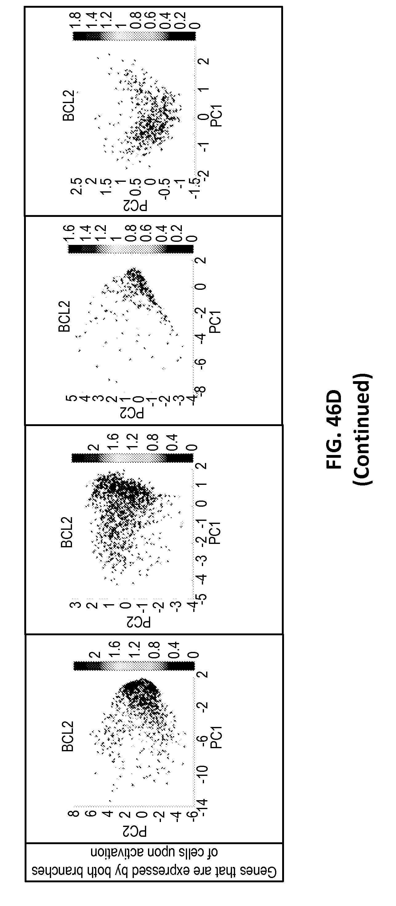
D00095

D00096

D00097

D00098

D00099

D00100

D00101

D00102

D00103

D00104

D00105
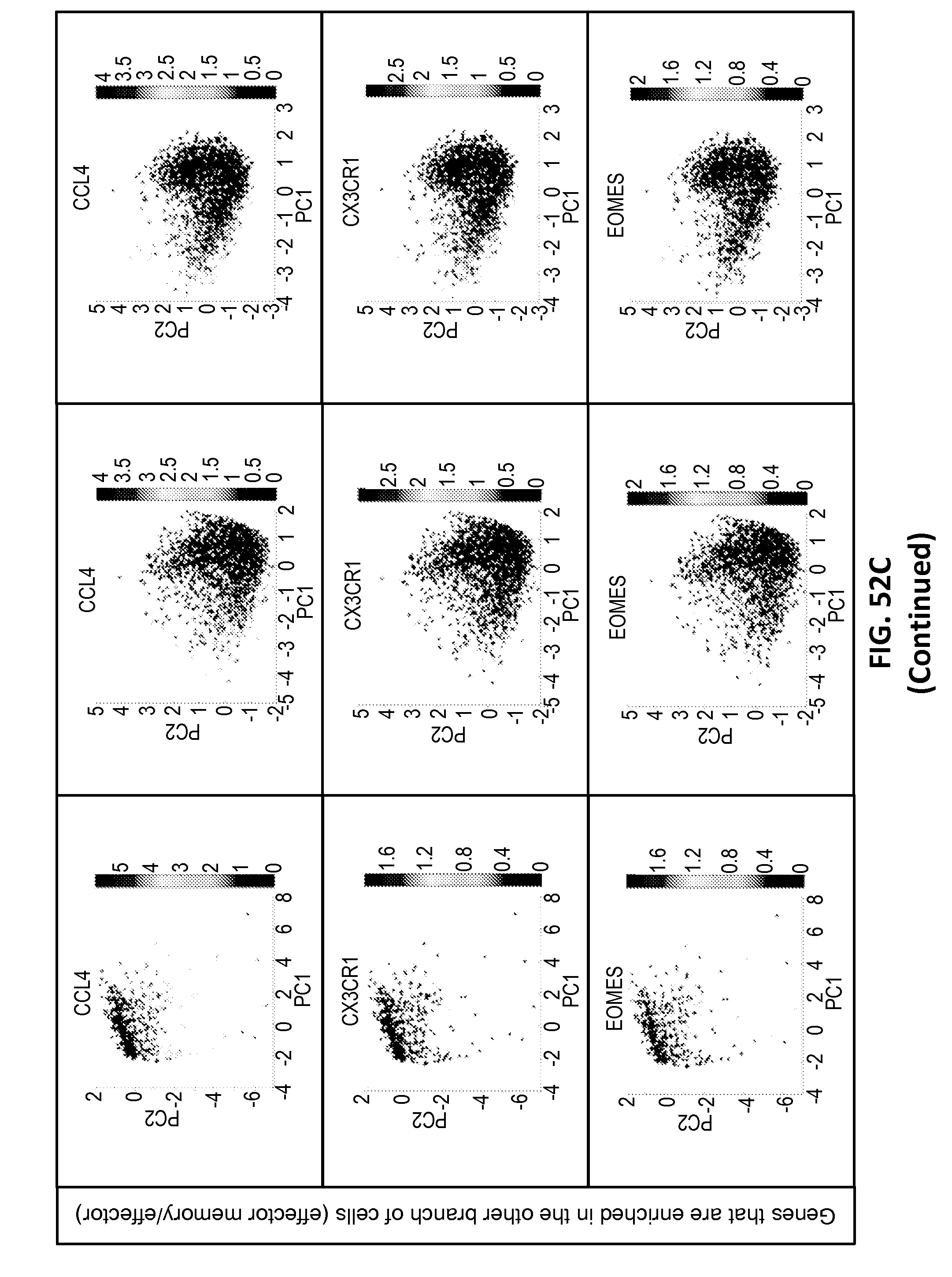
D00106

D00107

D00108

D00109

D00110

D00111

D00112

D00113

D00114

D00115

D00116
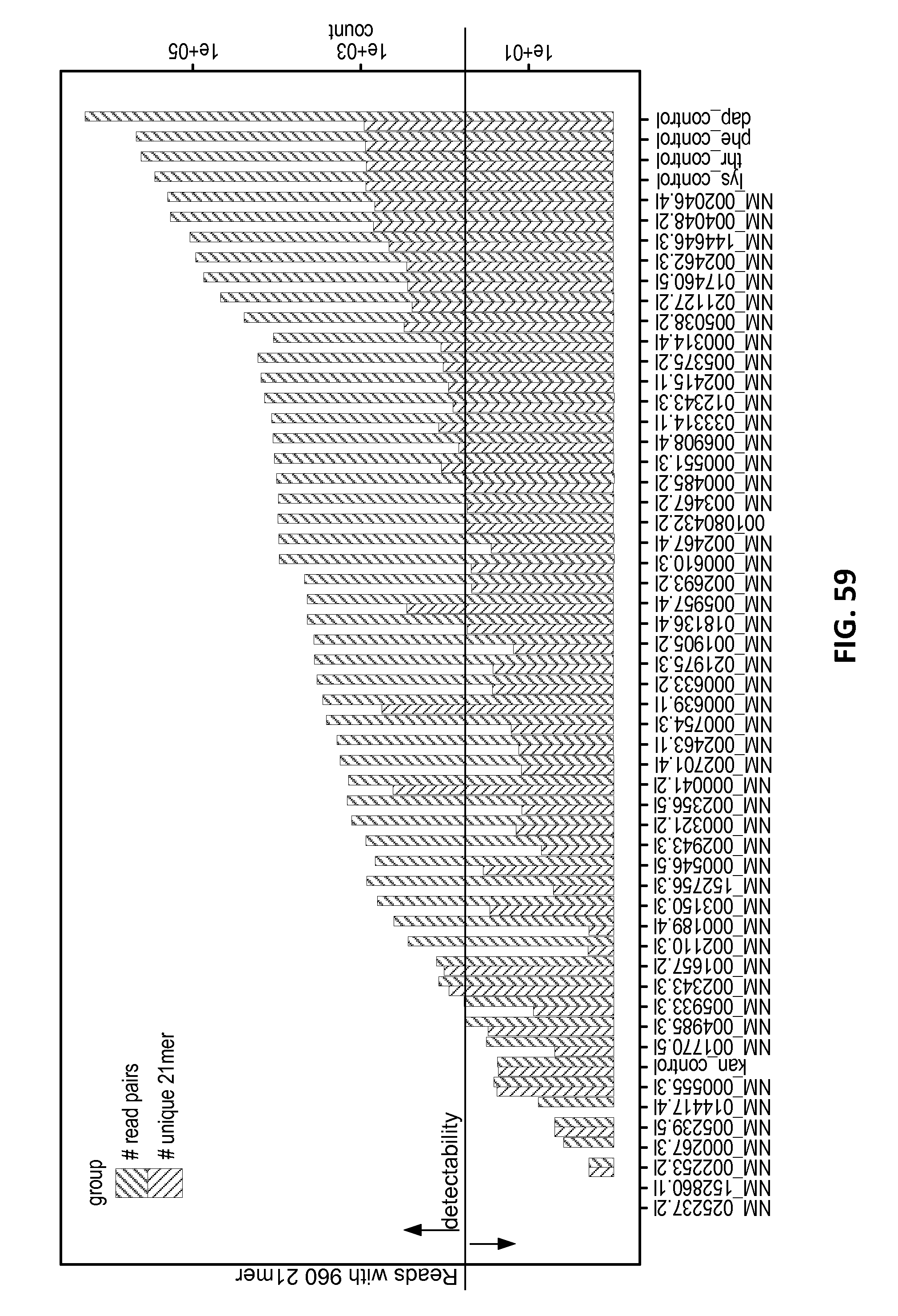
D00117

D00118

D00119

D00120

D00121

D00122

D00123

D00124

D00125

D00126

D00127

D00128

D00129

D00130

D00131

D00132

D00133

D00134

D00135

D00136

S00001
XML
uspto.report is an independent third-party trademark research tool that is not affiliated, endorsed, or sponsored by the United States Patent and Trademark Office (USPTO) or any other governmental organization. The information provided by uspto.report is based on publicly available data at the time of writing and is intended for informational purposes only.
While we strive to provide accurate and up-to-date information, we do not guarantee the accuracy, completeness, reliability, or suitability of the information displayed on this site. The use of this site is at your own risk. Any reliance you place on such information is therefore strictly at your own risk.
All official trademark data, including owner information, should be verified by visiting the official USPTO website at www.uspto.gov. This site is not intended to replace professional legal advice and should not be used as a substitute for consulting with a legal professional who is knowledgeable about trademark law.