Mycobacteriophages Capable Of Delivering Auto-luminescent Elements And Uses Thereof
ZHANG; Tianyu ; et al.
U.S. patent application number 16/124252 was filed with the patent office on 2019-02-07 for mycobacteriophages capable of delivering auto-luminescent elements and uses thereof. The applicant listed for this patent is Guangzhou Institutes of Biomedicine and Health, Chinese Academy of Sciences. Invention is credited to Zhiyong LIU, Tianyu ZHANG.
| Application Number | 20190040445 16/124252 |
| Document ID | / |
| Family ID | 56332631 |
| Filed Date | 2019-02-07 |


View All Diagrams
| United States Patent Application | 20190040445 |
| Kind Code | A1 |
| ZHANG; Tianyu ; et al. | February 7, 2019 |
MYCOBACTERIOPHAGES CAPABLE OF DELIVERING AUTO-LUMINESCENT ELEMENTS AND USES THEREOF
Abstract
Disclosed are a mycobacteriophage capable of delivering an auto-luminescent element and use thereof. The mycobacteriophage includes luxCDABE genes for auto-luminescence of a host bacterium. The auto-luminescent element is located on a transposon, and can be randomly inserted into the host genome with the transposon. The mycobacteriophage can be used for rapid detection of a live host bacterium in a sample and detection of sensitivity of the host bacterium to a drug or drug combination.
| Inventors: | ZHANG; Tianyu; (Guangzhou, CN) ; LIU; Zhiyong; (Guangzhou, CN) | ||||||||||
| Applicant: |
|
||||||||||
|---|---|---|---|---|---|---|---|---|---|---|---|
| Family ID: | 56332631 | ||||||||||
| Appl. No.: | 16/124252 | ||||||||||
| Filed: | September 7, 2018 |
Related U.S. Patent Documents
| Application Number | Filing Date | Patent Number | ||
|---|---|---|---|---|
| PCT/CN2016/081202 | May 6, 2016 | |||
| 16124252 | ||||
| Current U.S. Class: | 1/1 |
| Current CPC Class: | C12Q 1/04 20130101; C12N 15/74 20130101; C12N 15/1037 20130101; C12Q 1/18 20130101; C12Q 1/025 20130101; C12Q 1/689 20130101; C12N 15/70 20130101; C12Q 1/70 20130101; G01N 2333/35 20130101; C12N 15/1034 20130101; C12N 15/73 20130101 |
| International Class: | C12Q 1/18 20060101 C12Q001/18; C12N 15/73 20060101 C12N015/73; C12Q 1/02 20060101 C12Q001/02; C12N 15/74 20060101 C12N015/74; C12Q 1/70 20060101 C12Q001/70; C12N 15/10 20060101 C12N015/10 |
Foreign Application Data
| Date | Code | Application Number |
|---|---|---|
| Mar 7, 2016 | CN | 201610127984.9 |
Claims
1. A phasmid enabling auto-luminescence of a host bacterium, wherein the phasmid comprises: a DNA fragment enabling auto-luminescence of the host bacterium which comprises a LuxCDABE gene for luminescence.
2. The phasmid of claim 1, wherein the DNA fragment further comprises a transposon system, a resistance gene and/or a promoter.
3. The phasmid of claim 2, wherein the transposon system is selected from Himarl mariner family or TnA family.
4. The phasmid of claim 2, wherein the resistance gene is selected from at least one of hygromycin resistant gene Hyg, kanamycin resistant gene Kan, thiostrepton resistant gene Tsr and apramycin resistant gene Apr.
5. The phasmid of claim 2, wherein the promoter is selected from at least one of Mop, G13, Hsp60 and A37 promoters.
6. The phasmid of claim 2, wherein the transposon system comprises a transposase gene and inverted repeat sequences; the resistance gene and the LuxCDABE gene are ligated together to form a LuxCDABE gene-resistance gene fragment; and a promoter is ligated at both ends of the LuxCDABE gene-resistance gene fragment to form a promoter-LuxCDABE gene-resistance gene-promoter fragment; wherein the promoter allows for normal expression of a gene upstream of or downstream of a site in the host genome to which the DNA fragment is transposed; and each end of the promoter-LuxCDABE gene-resistance gene-promoter fragment is ligated with each of two inverted repeat sequences in the transposon system to transpose the promoter-LuxCDABE gene-resistance gene-promoter fragment to the host genome by transposase.
7. The phasmid of claim 1, wherein the DNA fragment further comprises a replication origin capable of replication in Escherichia coli.
8. The phasmid of claim 7, wherein the replication origin is oriE.
9. The phasmid of claim 2, wherein the DNA fragment further comprises a replication origin capable of replication in Escherichia coli, and the replication origin is oriE.
10. The phasmid of claim 4, wherein the DNA fragment further comprises a replication origin capable of replication in Escherichia coli, and the replication origin is oriE.
11. The phasmid of claim 1, wherein the DNA fragment further comprises a recombination site capable of homologous recombination with a phasmid backbone, which allows the DNA fragment to be ligated to the phasmid backbone.
12. The phasmid of claim 12, wherein the phasmid backbone comprises a phAE159 backbone.
13. The phasmid of claim 13, wherein the DNA fragment is located between two recombination sites of the phAE159 backbone.
14. The phasmid of claim 2, wherein the DNA fragment further comprises a recombination site capable of homologous recombination with a phasmid backbone, which allows the DNA fragment to be ligated to the phasmid backbone; and the phasmid backbone comprises a phAE159 backbone.
15. The phasmid of claim 1, wherein the DNA fragment is represented by SEQ ID NO: 1.
16. A phage capable of delivering an auto-luminescent element, wherein the phage comprises the phasmid of claim 1.
17. A use of the phage of claim 17 in detecting a host bacterium and/or sensitivity of a host bacterium to a drug or drug combination.
18. The use of claim 18, wherein the host bacterium is mycobacterium.
19. The use of claim 19, wherein the mycobacterium is selected from at least one of Mycobacterium smegmatis, Mycobacterium tuberculosis, Mycobacterium marinum and bacille Calmette-Guerin.
Description
CROSS-REFERENCE TO RELATED APPLICATIONS
[0001] This application is a continuation of International Patent Application No. PCT/CN2016/081202, filed on May 6, 2016, which claims the benefit of priority from Chinese Application No. 201610127984.9, filed on Mar. 7, 2016. The content of the aforementioned applications, including any intervening amendments thereto, are incorporated herein by reference.
TECHNICAL FIELD
[0002] The present invention relates to mycobacteriophages capable of delivering a set of auto-luminescent elements and uses thereof.
BACKGROUND OF THE PRESENT INVENTION
[0003] Mycobacterium is a type of organism that is slender and slightly curved, sometimes with branches or filamentum. Mycobacterium is a genus of Actinobacteria in taxonomy. Actinobacteria that are pathogenic to humans may or may not contain mycolic acids, and Mycobacterium contains mycolic acids. Such genus with no flagella or spores generally does not produce endotoxin or exotoxin, and its pathogenicity is related to the composition of bacteria. Mycobacterium causes chronic diseases accompanied by granulomas. A variety of mycobacteria mainly fall into three types, Mycobacterium tuberculosis complex, Mycobacterium leprae and non-tuberculous Mycobacterium (NTM). NTM can be divided into four groups according to colony pigmentation and growth rate. Group IV are bacteria of fast growing which grows fast at 25-45.degree. C., and colonies are visible within 5-7 days of culture. The colonies are usually rough and some can produce pigments. Mycobacterium smegmatis (M. smegmatis) is one of them but does not produce pigments.
[0004] A variety of mycobacteria can cause diseases, and Mycobacterium tuberculosis (Mtb) is a pathogen causing tuberculosis. Mtb can invade various organs of the human body, and in particular, lungs. Tuberculosis remains an important infectious disease bringing about the largest number of deaths caused by a single pathogen. There are about 10 million new cases of tuberculosis each year worldwide (9.6 million and 10.4 million in 2014 and in 2016, respectively), and the death toll is about 1.8 million. In recent years, the emergence of drug-resistant tuberculosis has made the treatment of tuberculosis more difficult. Mtb grows very slowly. Generally it takes 3-5 weeks for Mtb to form visible colonies on plates. Therefore, the routine diagnosis of tuberculosis by culturing live Mtb is very time consuming, which takes about 4-8 weeks. Similarly, test on Mtb's sensitivity to a drug also takes a long time. It takes another 4-8 weeks to obtain the drug sensitivity results upon the growth of bacteria. In recent years, BD's MGIT 960 system has been most commonly used in clinical laboratories, shortening the time of diagnosis to 5 to 45 days and drug susceptibility testing to 8-12 days, which is still relatively long. The instrument and tubes for detection are large in size that it not only takes up much space, but also is expensive, and is not suitable for detecting many concentrations of a drug in high-throughput pattern. Therefore, it would be a great improvement to the treatment of tuberculosis if live Mtb in the clinical samples and their phenotypic sensitivity of various drugs in several concentrations are detected in a more rapid, convenient and economic way with improved specificity and accuracy.
[0005] The applicant has placed a set of auto-luminescent elements, LuxCDABE genes onto a mycobacterial integrating vector, and inserted it into the genome of Mtb, BCG or M. smegmatis. Several novel, selectable marker-free auto-luminescent mycobacteria that have lost the resistance marker gene were obtained (Patent No. ZL 201210183007.2); and the related article has been published.sup.[1]. Previous research has indicated that the auto-luminescence system can distinguish between dead and live bacteria, and the luminescence intensity of the bacteria is highly correlated with the number of bacteria. The constructed auto-luminescent bacteria can be used for in vitro drug screening, and can even be conveniently used for the detection of drugs and vaccines in vivo in mice, with surprising effect.sup.[2].
[0006] However, the delivery of such luminescent element is inefficient, and requires specialized equipments such as those for electrotransformation, as well as a large amount of Mtb of competent cells. Even if the transformation is successful, the luminescent bacteria require a growth time of about 4 weeks for growing up and another 1 to 2 weeks for the next step, such as drug sensitivity test. Therefore, it is impossible to rapidly detect live mycobacteria in a sample and drug sensitivity of wild Mtb (including laboratorial strains and clinically acquired strains) with the ability to deliver auto-luminescent elements to Mtb and other mycobacteria in a quick, simple and timely manner. To this end, it is desirable to carry a reporter element into mycobacteria such as Mtb through a phage.
[0007] Phage is a type of virus that is exclusively hosted by bacteria. Like other viruses, phages have their genetic material wrapped in a protein shell. Most phages also have a "tail" that is used to "inject" genetic material into the host. Mycobacteriophage can specifically recognize mycobacterium and use them as hosts. Phasmid is constructed from a plasmid vector and a phage vector, combining a replication origin of plasmid and phage elements. Thus, its replication can be carried out as a normal double-stranded plasmid molecule in Escherichia coli cells to generate double-stranded DNA. When the phasmid enters the host bacterium (for example, mycobacterium), the DNA and capsid protein of the progeny phages can be synthesized in the host bacterium. Under certain conditions, some of the phages are packaged into phage particles and are then released by lysing host cells, for example, temperature-sensitive auto-luminescent reporter phages (ARPs). ARPs are packaged into phage particles and are released by lysing the host cells at about 30.degree. C.; and the phages are not formed at 37.degree. C. After ARP enters the host, the host can produce the enzymes and substrates required for the luminescent reaction to emit luminescence.
[0008] Furthermore, genetic manipulation of Mtb is difficult in basic research. Phage carrying a highly efficient transposon has been a powerful tool for efficiently disrupting the Mtb genes and obtaining a genetically disrupted mutant strain.sup.[3]. Attempts have been made to use such phage-based transposon system to disrupt selectable marker-free auto-luminescent strains. However we found that a large part of the transposon was inserted into the vector carrying the auto-luminescent elements which inserted into its genome but not disrupted genes of the host. This introduce difficulty to the construction of the auto-luminescent Mtb transposon mutants. Therefore, it is believed that by placing the auto-luminescent element on the transposon, along with insertion into the genome of wild-type Mtb, the transposon-inserted mutants can be auto-luminescent, and the insertion sites are assured to be in the genome.
[0009] Conventional bacteriological diagnosis of Mtb have many disadvantages, such as low sensitivity, complicated operation, long duration, many influencing factors, and difficulty in standardization due to its intrinsic slow growth rate. As a result, the bacteriological diagnosis cannot fully meet the demand of clinical diagnosis. Methods of diagnosing mycobacteria/Mtb in samples and their advantages and disadvantages are shown in Table 1. Methods available for detecting clinical sensitivity of mycobacteria/Mtb to drug and their advantages and disadvantages are shown in Table 2.
TABLE-US-00001 TABLE 1 Methods of diagnosing mycobacteria/Mtb in sputum samples and advantages and disadvantages thereof Diagnostic methods Advantages Disadvantages Comments Acid-fast Rapid, Has a low sensitivity (>10,000 most widely used in StainTest.sup.[4] economical, bacteria/mL, detection limit); diagnosing mycobacterial simple and unable to distinguish between infection in clinical visible. dead and live bacteria; only applications; a significant mycobacteria and even other monitoring index for bacteria can be identified; high chemotherapeutic effect. probability of missed diagnosis. Lowenstein- 100 Has a moderate sensitivity, a Gold standard of TB Jenden bacteria/mL, low positive rate, easy (tuberculosis) diagnosis, Culture.sup.[5] detection limit. contamination, slow (more widely used in clinical than 3 weeks, generally 4-8 applications, a significant weeks); and a large workload, monitoring index for and is difficult to standardize. chemotherapeutic effect. Rapid Automatic Susceptible to contamination, Culture.sup.[6] microbiological slow (generally 5-45 days); culture system, instruments and reagents are sensitive (about expensive. 10% higher than Lowenstein- Jenden Culture; at least 9 days to detect live bacteria. Flow Fluorescein The flow cytometer is Difficult for promotion. Cytometry diacetate (FDA) expensive with difficult Assay .sup.[7] can bind to live operation, and low specificity bacteria; for Mtb. objective, accurate, sensitive and rapid. Bio-luminescence Rapid and Specificity and sensitivity need great potential. Assay.sup.[8] visible to be improved. PCR.sup.[9] Rapid and Susceptible to contamination, Still not widely used simple, if no and has a high false-positive over many years. sequencing is rate; unable to distinguish required; and is between dead and live bacteria, also very and complicated sample sensitive. preparation and operation. GenXpert.sup.[10] Sensitive and The equipments are very Extremely expensive convenient to expensive, with a high demand equipments (currently in operate, and for operator; unable to international capable of rapid distinguish between dead and promotion). diagnosis of live bacteria. drug resistance to RIF. Bioluminescence Sensitive. Expensive, time consuming, Has already been an Quantitative and requires DNA extraction auxiliary examination PCR.sup.[11] for a judgment curve; item widely used in high demand for operator, and hospitals in China, its cannot distinguish between result can work as one Mtb and NTM very well. of the indexes for diagnosis of bacteriological negative TB; developed products include kits from DAAN GENE Inc. Gene Probe RNA A low copy number of mRNA Quality and quantity of Assay.sup.[12] amplification; and is easily degraded; the extracted RNA are adopting a A high copy number of rRNA important issues for mediated with overstability RNA amplification amplification method. Furthermore, method and RNA expression of the hybridization bacteria from protection RIF-treated patients may technique; be affected. capable of rapid detection of live bacteria; requires only one tube during process to avoids cross contamination. Antibody- Rapid and Has a low sensitivity Relatively weak detecting simple. antigenicity of Mtb and Protein Chip the presence of Assay.sup.[13] intergeneric and interspecies common antigenic determinants, as well as the inadequately illustrated correlation between humoral immunity and TB. Loop- Sensitive, Requires lysed bacteria and Developed corporations mediated simpler than DNA enrichment, complicated include EIKEN (China) Isothermal RT-PCR procedure. Co., Ltd. Amplification (Lamp).sup.[14] Phage Simple, rapid, Has a higher detection rate First reported by Wilson amplified and sensitive than bacterium culture method, et al. on Nature biologically (about 100 CFU unable to identify NTM; hard Medicine in 1997; the (PhaB) per reaction, to be carried out, many false detection takes about assay.sup.[15] close to PCR), positive; the infection time 18-24 hours; capable of needs to be accurately experimental results specific mastered; from multiple detection of live the temperature for preparing companies, institutes or bacteria, and is soft-agar medium must be organizations (including suitable for strictly controlled; developed one of the participants promotion in products include FAST plaue Yaoju Tan) indicated primary TB kit of BIOTEC (UK) Inc. considerable effect of hospitals. such assay in live bacteria diagnosis, and drug sensitivity analysis, and its patents are expired. Luciferase Simple, rapid The culture medium is First reported by Jacobs reporter and specific susceptible to contamination; et. al on Science in phage detection of live expensive substrate fluorescein 1993, few clinical trials assay.sup.[16] bacteria. is required to be added in, and of such method have the ability of the substrate of been reported in China, penetrating the cell wall is and its patents are poor; expired. the level of an another substrate ATP is greatly affected by the metabolic state of the bacteria; has low sensitivity. GFP- Simple, rapid, GFP is an auto-fluorescent Few clinical trials of expressing sensitive and protein, the GFP expressing such method have been phage specific. phage can identify dead, live reported in China. assay.sup.[17, 18] and uncultured bacteria indistinguishably, and emit luminescence, as a result the physiological state of bacteria cannot be distinguished; difficult for detection.
TABLE-US-00002 TABLE 2 Methods of detecting drug sensitivity of mycobacteria/Mtb and advantages and disadvantages thereof Diagnostic methods Advantages Disadvantages Comments Absolute Classical and Time consuming. A method for drug concentration reliable. sensitivity test which has method been adopted for more than 30 years in China. Proportion Most typical and Time consuming. A standard method for method reliable. drug sensitivity test recommended by Resistance Surveillance Project of WHO, the method was proposed by Prof. Jacques Grosset, former president of TB research. Turbidimetric // Stopped of use due to its resistance complexity. assay DNA chip Simple, rapid Requires the preparation of Remains in the stage of assay and sensitive, target gene fragment by PCR experimental and is suitable beforehand, and has development, and is for mass clinical complicated experimental expensive. initial drug procedure. resistance screening for Mtb strains. GenXpert Rapid and Extremely expensive, and Recent studies have simple. mainly detects RIF indicated that such sensitive-related genes technique has not improved the cure rate of TB in developing countries Bioluminescence Rapid (result The ATP extraction is assay can be obtained complicated, and the (detection of in 3-7 days with detection is expensive. ATP content good in live reproducibility). bacteria) Luciferase Simple, rapid The culture medium is As mentioned above, few Reporter and specific in susceptible to contamination; clinical trials of such Phage detecting live expensive substrate method have been Assay [16] bacteria fluorescein is required to be reported in China added in, and the ability of the substrate of penetrating the cell wall is poor; the level of an another substrate ATP is greatly affected by the metabolic state of the bacteria; has low sensitivity. Flow Fluorescein The flow cytometer is Difficult for promotion Cytometry diacetate (FDA) expensive, with relevant Assay [7] can bind to live techniques uneasy to master, bacteria; such its specificity for Mtb is low, method is and is susceptible to objective, contamination of accurate, miscellaneous bacteria sensitive and rapid (only takes one day at fastest speed)
[0010] Mycobacteriophage can be used for rapid diagnosis and detection of sensitivity of the host to a drug, such as Mtb due to its host specificity to mycobacteria. Considering the application of ARP in diagnosis and detection of drug sensitivity, the methods for phage-based detection and products related (close) to the present invention are shown in Tables 1 and 2.
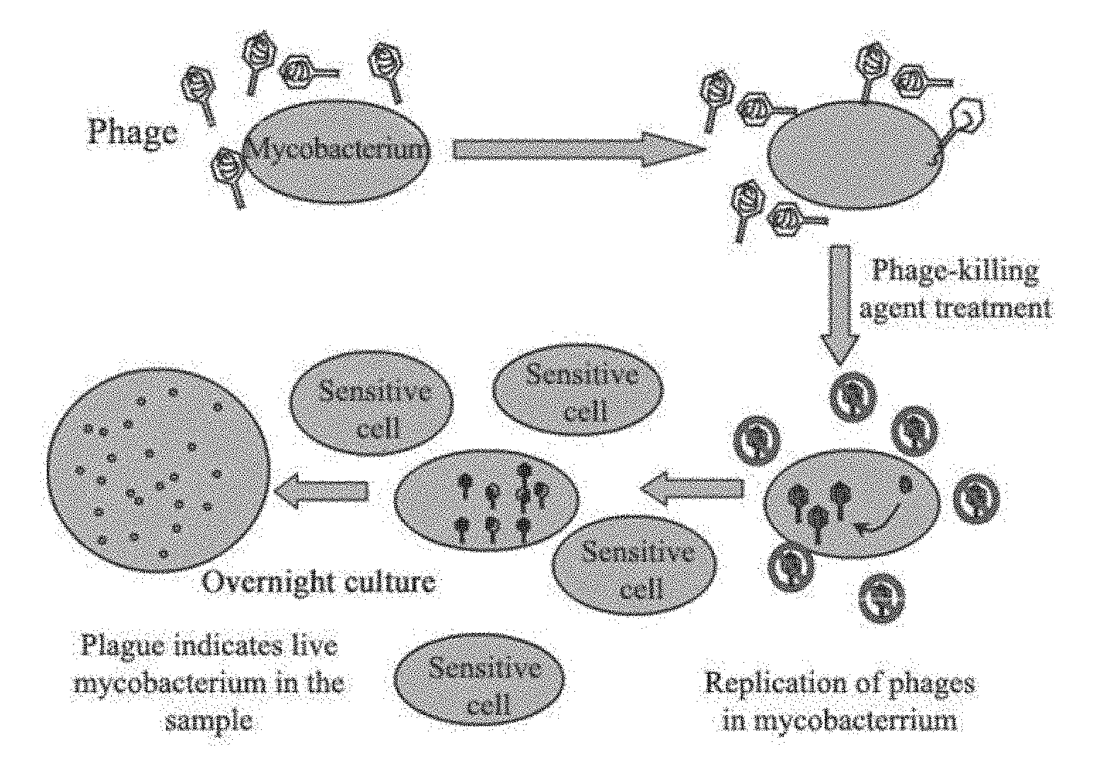
[0011] The main phage-based detection methods include: Phage amplified biologically assay (PhaB assay).sup.[15], luciferase reporter phage assay (LRP assay).sup.[16] and GFP-expressing phage detection assay (EGFP-phage assay).sup.[17, 18]. The PhaB assay adopts wild D29 mycobacteriophage, of which the principle is shown in FIG. 1. In this assay, D29 mycobacteriophage is used to infect live mycobacteria and kills the phages outside Mtb with a phage-killing agent, while the phages inside Mtb remain alive; and then fast growing mycobacterium cells (e.g. M. smegmatis) are introduced for a co-culture of about 18 hours for phage amplification. D29 is amplified in a large amount inside the infected bacteria, leading to dissolution of the bacteria and plaques production. Finally, the initial amount of Mtb is estimated by measuring the number of plaques. Compared to the conventional acid-fast staining method, although the phage infection requires at least 18-24 hours to identify the presence of Mtb, the optimal detection sensitivity is 100 bacteria/reaction under ideal conditions, which is close to the PCR method. Clinically, Yaoju Tan et al. have compared and analyzed 3168 sputum samples and found that the Mtb in sputum samples may be detected using PhaB assay, with the positive rate consistent with the smear method and Lowenstein-Jenden culture method.sup.[19]. It has also been reported that this method can also be used for rapid drug sensitivity test (24-72 hours), and the consistency is up to 93% as compared to the traditional plate method.sup.[20]. This method is more common in clinical verification and publications, yet it is still an alternative with few clinical applications because of complicated operation, great chance of false positives, susceptibility to contamination and sensitivity to be improved.
[0012] The LRP assay and EGFP-phage assay both use the constructed recombinant reporter phage to detect the presence of live phage and drug sensitivity of live bacteria (as shown in FIG. 2). The LRP assay has been published in 1993, even earlier than the PhaB assay reported in 1997. However, The LRP assay has not been developed as a product and have been reported in China and abroad in few clinical applications, owing to the addition of an expensive fluorescein substrate, the weak ability of the substrate to penetrate the cell wall of mycobacteria, unclear relationship between luminescence value and the number of live bacteria and a uncertain accuracy of detection. It was first reported in 2009 that EGFP-phage was constructed and used for rapid diagnosis and drug sensitivity test. The detection method mainly detects the EGFP protein produced after EGFP-phage's infection of live bacteria by bioluminescence microscopy or flow cytometry, while the detection sensitivity is low due to the limitations of the detection method. Moreover, operation of the method is complicated and Mtb may be easily exposed to air, causing the environment to be contaminated by Mtb. However, bioluminescence can still be detected even if formalin is used to fix and kill Mtb, thus preventing Mtb from contaminating the environment. It takes 4 hours under laboratory conditions to detect Mtb in a sample at the fastest speed. To test drug sensitivity, it takes 16 hours for rifampicin and streptomycin at the fastest speed. As for isoniazid, in order to detect the sensitivity of Mtb to INH, Mtb bacteria needs to be pretreated as follows: culturing Mtb for 24 hours followed by another 16 hours of culture. Flow cytometry is more expensive and however the results are similar. Moreover, Mtb itself has a certain background bioluminescence, which also has an impact on experimental results.
[0013] As previously mentioned, the main methods of diagnosing live Mtb and detecting sensitivity of clinical bacterial strains to the drug are based on the BD MGIT960 system for detecting the bacterial growth and the aforementioned phage-based methods.
[0014] The BD MGIT960 system is used as a main method in clinical practice. The main disadvantages of the system include: 1) slow, requiring 5-45 days for diagnosis, and another 9-12 days for sensitivity detection; 2) expensive, not only for the culture and the detection equipment, but for the culture medium and the experimental supplies; specifically, requiring a special medium and a 70-RMB (CNY) tube for detecting a single concentration of a single drug; 3) complicated operation, being difficult and time-consuming; and 4) occupying a large space for its large size.
[0015] The PhaB method has the following disadvantages: 1) complicated operation, uneasy to master: the phage infection time needs to be accurately mastered, otherwise, incomplete infection or the lysing of live cells after infection will occur, reducing the sensitivity; and difficult operational procedures are required, including preparing culture plate; 2) occupying a large space due to a large size of the device; and 3) higher cost of diagnosis and drug sensitivity test as compared to the present invention.
[0016] As for the LRP method, a related product has been developed even till its patents expired. This could be because the reaction requires an expensive fluorescein substrate with a weaker ability to penetrate the mycobacterial cell wall. Other disadvantages include unclear relationship between luminescence value and the amount of live bacteria an uncertain detection accuracy.
[0017] The EGFP-phage method has the following disadvantages: 1) flow cytometer is expensive to purchase and use; flourescence microscope is also expensive; the flourescence of green fluorescent protein need to be detected with the help of excitation light; flourescence can also be observed in dead cells, thus interfering with the experimental results; and Mtb itself has a certain background flourescence; 2) the detection sensitivity is low due to the limitations of the method and tool; 3) the operation is complicated and difficult to master compared to the present invention; and 4) the detection efficiency is low.
[0018] The aforementioned method of constructing artificial recombinant phages (FIG. 2) has serious problems, that is, when using .lamda. phage for in vitro packaging, the DNA packaging range is limited, and therefore it is difficult to construct large-fragment DNA into the recombinant phage by this method. For example, the Fluc and egfp genes are only about 1 kb and the auto-luminescent elements are about 6 kb, which cannot be packaged in vitro using .lamda. phage (we have tried following the same protocol, and the results have proven that such a protocol does not work).
[0019] The current recombinant mycobacteriophage is a recombinant phage recombined either with a reporter gene (such as the small reporter gene Fluc and egfp mentioned above) or a highly efficient transposon (MycoMarT7, etc..sup.[3, 21]), yet there are currently no reports indicating that the long luxCDABE gene set can be inserted in a highly efficient transposon and thereby recombined into a mycobacteriophage.
[0020] In addition to the above diagnostic techniques, gene disruption is often required in the basic research of Mtb application, such as studies on gene functions, and the knock-out and disruption of virulence-related genes. Meanwhile, if the mutant strain is auto-luminescent, it will undoubtedly provide significant improvement in the efficiency of research and development. However, there is currently no such a tool.
SUMMARY OF THE PRESENT INVENTION
[0021] In order to solve the above problems, the applicant has, through extensive efforts and experimental research, succeeded in constructing artificial phasmids and a phage (ARP) capable of delivering a set of auto-luminescent elements. The phasmid constructed has the following characteristics: the auto-luminescent element carried is present on a highly efficient transposon, which can be randomly inserted into the host genome in an efficient manner, disrupting the host genes on the one hand, and enabling auto-luminescence of the host bacteria on the other hand. Therefore it can be convenient to efficiently study the function of the disrupted host gene by utilizing the feature of auto-luminescence.
[0022] An objective of the present invention is to provide a phage capable of enabling auto-luminescence of host bacteria. Meanwhile, ARP can be used to rapidly prepare auto-luminescent host bacteria (e.g. Mtb), such that such that the auto-luminescent bacteria can be convenient for drug screening, evaluation, vaccine screening and etc, as well as for in vivo studies in small animals, which in this case can be performed conveniently by detecting luminescence values after the infection of the small animals.
[0023] Another objective of the present invention is to provide a phage capable of delivering the auto-luminescent element set for diagnosis of live mycobacterium.
[0024] Yet another objective of the present invention is to provide a use of the phage in detecting a bacterium and/or sensitivity of a bacterium to a drug.
[0025] A phasmid enabling auto-luminescence of a host bacterium includes a DNA fragment including a LuxCDABE gene enabling auto-luminescence of the host bacterium.
[0026] Further, the DNA fragment includes a transposon system, a resistance gene and/or a promoter.
[0027] Further, the transposon system transposes the DNA fragment to a host genome.
[0028] Further, the transposon system is selected from Himarl mariner family or TnA family.
[0029] Further, the resistance gene is selected from at least one of hygromycin resistant gene Hyg, kanamycin resistant gene Kan, thiostrepton resistant gene Tsr and apramycin resistant gene Apr.
[0030] Further, the promoter allows for normal expression of a gene upstream of or downstream of a site in the host genome to which the DNA fragment is transposed.
[0031] Further, the promoter is selected from at least one of Mop, G13, Hsp60 and A37 promoters.
[0032] Further, the transposon system comprises a transposase gene and inverted repeat sequences. The resistance gene and the LuxCDABE gene are ligated together to form a LuxCDABE gene-resistance gene fragment. A promoter is ligated at both ends of the LuxCDABE gene-resistance gene fragment to form a promoter-LuxCDABE gene-resistance gene-promoter fragment. The promoter allows for normal expression of a gene upstream of or downstream of a site in the host genome to which the DNA fragment is transposed; and
[0033] each end of the promoter-LuxCDABE gene-resistance gene-promoter fragment is ligated with each of two inverted repeat sequences in the transposon system to transpose the promoter-LuxCDABE gene-resistance gene-promoter fragment to the host genome by transposase.
[0034] Further, the DNA fragment includes a replication origin capable of replication in Escherichia coli, which allows the DNA fragment to replicate in a large amount in a plasmid or phasmid.
[0035] Further, the replication origin is oriE.
[0036] Further, the DNA fragment further includes a recombination site capable of homologous recombination with a phasmid backbone, which allows the DNA fragment to be ligated to the phasmid backbone.
[0037] Further, the phasmid backbone comprises a phAE159 backbone.
[0038] Further, the DNA fragment is represented by SEQ ID NO: 1.
[0039] Further, the phasmid includes a phAE159 backbone, and the DNA fragment is located between two recombination sites of the phAE159 backbone.
[0040] In the present invention, a method of preparing the phasmid includes:
[0041] digesting a phasmid backbone into a linear sequence to allow the phasmid to be recombined with a target fragment containing the LuxCDABE gene;
[0042] mixing the digested phasmid backbone with the target fragment containing the LuxCDABE gene and transforming Escherichia coli competent cells with the mixed digested phasmid backbone and the target fragment for recombination and amplification; and
[0043] extracting the plasmid from positive recombinant Escherichia coli to obtain the phasmid.
[0044] Further, the method includes digesting a phasmid backbone phAE159 with restriction endonuclease PacI, and recovering a large-fragment product after digestion; mixing the recovered product with a base fragment according to SEQ ID NO: 1, and transforming Escherichia coli competent cells with the mixed product and the base fragment; and extracting the plasmid from a positive recombinant Escherichia coli to obtain the phasmid.
[0045] Further, a method of preparing the base fragment according to SEQ ID NO: 1 includes:
[0046] 1) construction of a pUCF2 plasmid:
[0047] artificially synthesizing a F0 fragment of SEQ ID NO: 2; ligating the F0 fragment to a pUC19 vector to obtain a pUCF0 plasmid; digesting the pUCF0 plasmid with restriction endonuclease XbaI, and recovering a fragment of about 2.8 kb; ligating the fragment digested by XbaI and containing hygromycin resistant gene Hyg with the two recovered fragments to obtain a pUCF2 plasmid of about 3.9 kb;
[0048] 2) construction of a pUCF3 plasmid:
[0049] artificially synthesizing a F1 fragment according to SEQ ID NO: 3; ligating the F0 fragment to a pUC19 vector to obtain a pUCF1 plasmid; digesting the pUCF1 plasmid with restriction endonucleases KpnI and EcoRI and recovering a fragment of about 2.7 kb; digesting the pUCF2 plasmid with restriction endonucleases KpnI and EcoRI; recovering a fragment of about 1.3 kb, and ligating the two recovered fragments to obtain a pUCF3 plasmid of about 4 kb;
[0050] 3) construction of a pUCRLlux2P plasmid:
[0051] digesting the pUCF3 plasmid with restriction endonucleases KpnI and EcoRI, and recovering a fragment of about 3.9 kb; digesting a pluxOK plasmid with restriction endonucleases KpnI and EcoRI, and recovering a fragment of about 6.3 kb; ligating the two recovered fragments to obtain a pUCRLlux2P plasmid of about 10.2 kb;
[0052] 4) construction of a pYUBT plasmid:
[0053] amplifying a transposase trans gene sequence from a MycoMarT7 phage, adding a NcoI restriction site to a 5' end of the trans gene, adding a SpeI restriction site to a 3' end of the trans gene, and digesting the amplified product with NcoI and SpeI and recovering a fragment of about 1 kb; digesting a pYUB854 plasmid with restriction endonucleases NcoI and SpeI, and recovering a fragment of about 3.8 kb; ligating the two recovered fragments to obtain a pYUBT plasmid;
[0054] 5) construction of a pYUOK plasmid:
[0055] digesting a pUCRLlux2P plasmid with restriction endonucleases NcoI and SpeI, and recovering a fragment of about 7.6 kb; digesting the pYUBT plasmid with restriction endonucleases NcoI and XbaI, and recovering a fragment of about 2.9 kb; ligating the two recovered fragments to obtain a pYUOK plasmid of about 10.5 kb;
[0056] 6) construction of a p159LART plasmid:
[0057] synthesizing a base fragment according to SEQ ID NO: 4, ligating the base fragment to a pUC19 vector to obtain a p159LR plasmid, digesting the p159LR plasmid with NheI and recovering a fragment; amplifying a Apr gene fragment with a NheI restriction site at both ends, digesting the fragment with restriction endonuclease NheI and recovering a fragment; ligating the two recovered fragments to obtain a p159LART plasmid of about 4.8 kb;
[0058] 7) construction of a pYUOKLART plasmid:
[0059] digesting the pYUOK plasmid with restriction endonuclease PacI, and recovering a fragment of about 10.5 kb; digesting the p159LART plasmid with restriction endonuclease PacI, recovering a fragment of about 2 kb, and ligating the two recovered fragments to obtain a pYUOKLART plasmid of about 12.5 kb; and 8) production of a base fragment according to SEQ ID NO: 1
[0060] digesting the pYUOKLART plasmid with restriction endonuclease SmaI, recovering a large-fragment product after digestion to obtain the base fragment according to SEQ ID NO: 1.
[0061] A phage capable of delivering an auto-luminescent element includes the phasmid of the present application.
[0062] A use of the phage in detecting a host bacterium and/or drug sensitivity of a host bacterium is provided.
[0063] Further, the host bacterium is Mycobacterium.
[0064] Further, the mycobacterium includes Mtb, Mycobacterium smegmatis, Mycobacterium marinum and Mycobacterium Bacille Calmette-Guerin (BCG).
[0065] A method of detecting a host bacterium with the phage includes the following steps:
[0066] 1) adding a phage-containing liquid to a sample to be tested;
[0067] 2) culturing the sample at 36.5-42.degree. C. for 35 minutes or more, and detecting bioluminescence using a luminometer;
[0068] 3) if there is a significant bioluminescence compared to a negative control group without ARP, indicating the sample contains a large amount of live mycobacteria; if the bioluminescence intensity is lower than 120% of the bioluminescence intensity of the negative control, adding a phage-killing reagent SK to kill the phage whose genome has not entered the mycobacterium; wherein the SK reagent is selected from at least one of ammonium sulfate, ferrous sulfate, ammonia sulfate and ammonium ferrous sulfate;
[0069] 4) adding a neutralizing agent SN to neutralize the excess SK reagent, adding an indicator bacterium, and culturing the sample at 20-32.degree. C. for 12 hours or more; wherein the neutralizing agent SN is selected from at least one of MgSO4, potassium dichromate, CaCl.sub.2 and MnCl.sub.2; and
[0070] 5) detecting luminescence of the sample added with the indicator bacterium with a luminometer; wherein the luminescence indicates the presence of live mycobacteria in the sample; non-luminescence indicates the absence of live mycobacteria in the sample.
[0071] Further, the indicator bacterium is a host of the phage, and the phage proliferates and lyses cells in the host.
[0072] A method of detecting sensitivity of a host bacterium to a drug with the phage includes the following steps:
[0073] 1) mixing the phage with the host bacterium, and adding a certain concentration of a drug to be tested;
[0074] 2) culturing the mixed phage and the host bacterium at 36.5-42.degree. C. for at least 2 hours, and detecting luminescence using a luminometer; and
[0075] 3) if there is significant difference between the luminescence of the tested sample and the luminescence of a sample with no drug, indicating the host bacteria is sensitive to the drug of the concentration; if there is no significant difference, indicating the host bacteria is resistant to the drug at the concentration.
[0076] The present invention has the following beneficial effects:
[0077] 1) The present invention relates to the concepts of synthetic biology, where a novel artificial mycobacteriophage (ARP) was successfully constructed by utilizing synthetic DNA, and approaches including genetic engineering and molecular biology. ARP is a temperature sensitive phage. It can lyse the host under a temperature below 30.degree. C., while it cannot lyse the host at 37-42.degree. C. By delivering auto-luminescent gene elements to the host, ARP can make its host mycobacterium express its corresponding proteins (enzymes). These enzymes can utilize the metabolites of live host bacteria to circulate the generation of substrates required for the luminescence reaction, as well as the enzymes required for the reaction. Therefore, the live host bacteria may be luminescent without addition of any substrate. The auto-luminescent element carried by the ARP of the present invention is present on a highly efficient transposon, along with which the element can be randomly inserted into the host genome in an efficient manner, disrupting the host genes on the one hand, and enabling the auto-luminescence of the host bacteria with disrupted genes on the other hand. It is important that the ARP can rapidly diagnose live host bacteria in a sample and rapidly test the drug sensitivity of a host strain. Such diagnosis and detection are about 8-40 days faster than the most popular BD MGIT 960 system on the market, and are simpler, more intuitive and more economical. The ARP has certain preference for its host in mycobacteria, and Mtb is one of the most important host bacteria.
[0078] 2) The phage constructed by the present invention can transform a mycobacterium and enable the auto-luminescence of the transformed mycobacterium after incubation at a temperature. The mycobacterium includes, but is not limited to Mycobacterium smegmatis, Mtb and BCG.
[0079] 3) According to the characteristics of the host bacterium, the present invention can prepare a host bacterium-specific phage capable of delivering the auto-luminescent elements. The auto-luminescent mycobacterium is obtained after the phage is transduced into the mycobacterium. Since the mycobacteriophage has host specificity (specific to mycobacterium), the phage can be used for rapid detection of a live mycobacterium.
[0080] 4) The present invention provides a p1590K phasmid for constructing the phasmid of the auto-luminescent bacterium. The phasmid includes a phAE159 phage backbone, a mycobacterial strong promoter (Hsp60); a gene required for luminescence (LuxCDABE); a hygromycin resistanct gene (Hyg); a transposase gene (trans); inverted repeat sequences IR-L and IR-R; and Mop and G13 promoters. The luminescent mycobacterium, obtained by the transformation of the host with the phasmid or the phage produced from the phasmid, is an auto-luminescent mycobacterium. The phasmid or the phage is convenient for detection with high accuracy, and can be used for detection of sensitivity of a mycobacterium to a drug.
[0081] 5) The present invention has skipped a packaging process of .lamda. phage. Early studies by the applicant adopted the packaging process of .lamda. phage, yet the applicant failed to obtain the p1590K phasmid despite of repeated a large amount of experimental studies, and this cannot succeed in theory. Since such packaging process of .lamda. phage was skipped, the present invention was not limited to the size of the .lamda. phage packaging, and achieved good experimental results. The p1590K phasmid was successfully constructed with a size capable of more than 57 kb, while the size of the largest vector that can be obtained by the existing .lamda. phage packaging does not exceed 52 kb.
[0082] 6) Even if the auto-luminescent elements are not integrated into the genome, the host bacteria can still be auto-luminescent. The ARP obtained by the invention may efficiently introduce the carried auto-luminescent element into Mtb and BCG, to allow the live Mtb to be luminescent (dead bacteria are not luminescent), thereby rapidly diagnosing the live host bacteria in a sample and quickly testing the sensitivity of the host bacteria to a drug. In order to increase the sensitivity of the diagnosis, a bacteriophage-killing agent (neutralizer) and an indicator bacterium (Mycobacterium. smegmatis MC.sup.2 155; Msm) amplifying the signal are used. The reaction is carried out in the same tube that is inexpensive, with a strong luminescence signal detectable over a long period of time.
[0083] 7) The artificial phage provided by the present invention can be used in the following applications: i) efficient preparation of an auto-luminescent mycobacterium transposon mutant strain, which facilitates the studies of gene functions; ii) simple, economical and rapid diagnosis of live Mtb in a sample, with the specificity of the phage used to assist in the identification of bacterial species; and iii) simple, economical and rapid drug susceptibility testing (DST).
[0084] 8) By using the auto-luminescent phage obtained by transforming the host bacteria with the phasmid of the present invention, dead and live bacteria can be distinguished by detecting luminescence, which is suitable for various experiments or tests.
[0085] 9) The auto-luminescent mycobacterium prepared through the transduction of ARP of the present invention can be used for in vitro drug activity test, and does not require any substrate. The lights from the same sample can be continuously with a high sensitivity and a high reproducibility.
BRIEF DESCRIPTION OF THE DRAWINGS
[0086] FIG. 1 is a schematic diagram of the Phage Amplified Biologically method (PhaB method)(BIOTEC, U.K); wherein the infection time needs to be accurately controlled, otherwise the cells may be incompletely infected or lysed after infection, resulting in a reduced sensitivity; and the shortest time to complete infection is 18-24 hours.
[0087] FIG. 2 is a schematic diagram of the construction of recombinant reporter phage; where the construction of EGFP-phage is shown for exemplary illustration: the reporter gene is an enhanced green fluorescent protein gene (egfp)[17,18]; the reporter gene for the recombinant reporter phage of the LRP method is the firefly luciferase gene (Fluc); and the difference between the two methods is reporter genes.
[0088] FIG. 3 is a flow chart showing the construction of a pUCRLlux2P plasmid.
[0089] FIG. 4 is a flow chart showing the construction of a pYUOK plasmid.
[0090] FIG. 5 is a flow chart showing the construction of a pYUOKLART plasmid.
[0091] FIG. 6 is a flow chart showing the construction of a p1590K phasmid.
[0092] FIG. 7 is a flow chart of a rapid detection of a host mycobacterium in a sample with ARP phages.
[0093] FIG. 8 shows the bioluminescence intensity of Mtb strain under laboratory which is detected with ARP of the present invention at different time of culture.
[0094] FIG. 9 shows the results of clinical samples which are detected with the ARPs of the present invention, where the horizontal line shows the critical value for distinguishing between a negative sample and a positive sample (RLU=80, i.e., the bioluminescence intensity is 120% of that of a negative control); Con is a negative sputum smear as a control; and P1-P9 are positive sputum smears.
[0095] FIG. 10 shows the results of samples containing Mtb of different titers which are detected with the ARPs of the present invention; Con is a negative control and does not contain Mtb; "Undiluted" refers to an undiluted sample; 10.sup.-n refers to a dilution of 10 to the power of n, and when diluted to 10.sup.-8, the number of Mtb in each sample does not exceed 10; the horizontal line shows the critical value for distinguishing between a negative sample and a positive sample (i.e., the bioluminescence intensity is 120% of that of the negative control); and the "19 h" and "24 h" are the time of culture at 30.degree. C. after an indicator bacterium is added.
[0096] FIG. 11 is a schematic diagram of a rapid detection of sensitivity of a host mycobacterium to a drug with the ARPs of the present invention.
[0097] FIG. 12 shows the results of sensitivity of Mtb to a drug which is detected with the ARPs of the present invention, where the "Con" is a DMSO control without any drugs, rifampicin (RIF, 2 .mu.g/mL), isoniazid (INH, 1 .mu.g/mL), ethambutol (EMB, 5 .mu.g/mL), linezolid (LZD, 1 .mu.g/mL), oxazolidinone (PNU, 0.5 .mu.g/mL); "n.s." represents that no statistical difference was found between the EMB group and the control (p>0.05); "*" represents that statistical difference was found between a test group and the control (p<0.05).
[0098] FIG. 13 is a schematic diagram of detection of live mycobacteria in samples and sensitivity of the mycobacteria to rifampicin (RIF), where S1-S3 are sample groups, and C1-C3 are control groups.
DETAILED DESCRIPTION OF THE PRESENT INVENTION
[0099] A phasmid enabling auto-luminescence of a host bacterium includes a DNA fragment enabling auto-luminescence of the host bacterium; and the DNA fragment includes a LuxCDABE gene cluster for luminescence.
[0100] Preferably, the DNA fragment further includes a transposon system, a resistance gene and/or a promoter.
[0101] Preferably, the transposon system transposes the DNA fragment to a host genome.
[0102] Preferably, the transposon system is selected from Himarl mariner family or TnA family.
[0103] Preferably, the resistance gene is selected from at least one of hygromycin resistant gene Hyg, kanamycin resistant gene Kan, thiostrepton resistant gene Tsr and apramycin resistant gene Apr.
[0104] Preferably, the promoter allows for normal expression of a gene upstream of or downstream of a site in the host genome to which the DNA fragment is transposed.
[0105] Preferably, the promoter is selected from at least one of Mop, G13, Hsp60 and A37 promoters.
[0106] Preferably, the transposon system includes a transposase gene and inverted repeat sequences. The resistance gene and the LuxCDABE gene are ligated together to form a LuxCDABE gene-resistance gene fragment. A promoter is ligated at both ends of the LuxCDABE gene-resistance gene fragment to form a promoter-LuxCDABE gene-resistance gene-promoter fragment. The promoter allows for normal expression of a gene upstream of or downstream of a site in the host genome to which the DNA fragment is transposed.
[0107] Each end of the promoter-LuxCDABE gene-resistance gene-promoter fragment is ligated with each of two inverted repeat sequences in the transposon system to transpose the promoter-LuxCDABE gene-resistance gene-promoter fragment to the host genome by transposase.
[0108] Preferably, the DNA fragment capable of making a host bacterium auto-luminescent further includes a replication origin capable of replication in Escherichia coli, which allows the DNA fragment to replicate in a large amount in a plasmid or phasmid.
[0109] Preferably, the replication origin is oriE.
[0110] Preferably, the DNA fragment further includes a recombination site capable of homologous recombination with a phasmid backbone, which allows the DNA fragment to be ligated to the phasmid backbone.
[0111] Preferably, the phasmid backbone comprises a phAE159 backbone.
[0112] Preferably, the DNA fragment is represented by SEQ ID NO: 1.
[0113] Preferably, the phasmid further includes a phAE159 backbone, and the DNA fragment is located between two recombination sites of the phAE159 backbone.
[0114] A method of preparing the phasmid enabling auto-luminescence of a host bacterium includes:
[0115] digesting a phasmid backbone into a linear sequence to allow the phasmid to be recombined with a target fragment containing the LuxCDABE genes;
[0116] mixing the digested phasmid backbone with the target fragment containing the LuxCDABE genes and transforming Escherichia coli competent cells with the mixed digested phasmid backbone and the target fragment for recombination and amplification; and
[0117] extracting the plasmid from positive recombinant Escherichia coli to obtain the phasmid.
[0118] Preferably, the method includes:
[0119] digesting a phasmid backbone phAE159 with restriction endonuclease PacI, and recovering a large-fragment product after digestion;
[0120] mixing the recovered product with a base fragment according to SEQ ID NO: 1, and transforming Escherichia coli competent cells with the mixed product and the base fragment; and
[0121] extracting the plasmid from a positive recombinant Escherichia coli to obtain the phasmid.
[0122] Preferably, a method of preparing the base fragment according to SEQ ID NO: 1 includes the following steps:
[0123] 1) construction of a pUCF2 plasmid
[0124] artificially synthesizing a F0 fragment of SEQ ID NO: 2; ligating the F0 fragment to a pUC19 vector to obtain a pUCF0 plasmid; digesting the pUCF0 plasmid with restriction endonuclease XbaI, and recovering a fragment of about 2.8 kb; ligating the fragment digested by XbaI and containing hygromycin resistant gene Hyg with the two recovered fragments to obtain a pUCF2 plasmid of about 3.9 kb;
[0125] 2) construction of a pUCF3 plasmid
[0126] artificially synthesizing a F1 fragment according to SEQ ID NO: 3; ligating the F0 fragment to a pUC19 vector to obtain a pUCF1 plasmid; digesting the pUCF1 plasmid with restriction endonucleases KpnI and EcoRI and recovering a fragment of about 2.7 kb; digesting the pUCF2 plasmid with restriction endonucleases KpnI and EcoRI; recovering a fragment of about 1.3 kb, and ligating the two recovered fragments to obtain a pUCF3 plasmid of about 4 kb;
[0127] 3) construction of a pUCRLlux2P plasmid
[0128] digesting the pUCF3 plasmid with restriction endonucleases KpnI and EcoRI, and recovering a fragment of about 3.9 kb; digesting a pluxOK plasmid with restriction endonucleases KpnI and EcoRI, and recovering a fragment of about 6.3 kb; ligating the two recovered fragments to obtain a pUCRLlux2P plasmid of about 10.2 kb;
[0129] 4) construction of a pYUBT plasmid
[0130] amplifying a transposase trans gene sequence from a MycoMarT7 phage, adding a NcoI restriction site to a 5' end of the trans gene, adding a SpeI restriction site to a 3' end of the trans gene, and digesting the amplified product with NcoI and SpeI and recovering a fragment of about 1 kb; digesting a pYUB854 plasmid with restriction endonucleases NcoI and SpeI, and recovering a fragment of about 3.8 kb; ligating the two recovered fragments to obtain a pYUBT plasmid;
[0131] 5) construction of a pYUOK plasmid
[0132] digesting a pUCRLlux2P plasmid with restriction endonucleases NcoI and SpeI, and recovering a fragment of about 7.6 kb; digesting the pYUBT plasmid with restriction endonucleases NcoI and XbaI, and recovering a fragment of about 2.9 kb; ligating the two recovered fragments to obtain a pYUOK plasmid of about 10.5 kb;
[0133] 6) construction of a p159LART plasmid
[0134] synthesizing a base fragment according to SEQ ID NO: 4, ligating the base fragment to a pUC19 vector to obtain a p159LR plasmid, digesting the p159LR plasmid with NheI and recovering a fragment; amplifying a Apr gene fragment with a NheI restriction site at both ends, digesting the fragment with restriction endonuclease NheI and recovering a fragment; ligating the two recovered fragments to obtain a p159LART plasmid of about 4.8 kb;
[0135] 7) construction of a pYUOKLART plasmid
[0136] digesting the pYUOK plasmid with restriction endonuclease PacI, and recovering a fragment of about 10.5 kb; digesting the p159LART plasmid with restriction endonuclease PacI, recovering a fragment of about 2 kb, and ligating the two recovered fragments to obtain a pYUOKLART plasmid of about 12.5 kb; and
[0137] 8) production of a base fragment according to SEQ ID NO: 1
[0138] digesting the pYUOKLART plasmid with restriction endonuclease SmaI, recovering a large-fragment product after digestion to obtain the base fragment according to SEQ ID NO: 1.
[0139] A phage capable of delivering an auto-luminescent element includes any one of the aforesaid phasmid.
[0140] A use of the phage in detecting a host bacterium and/or drug sensitivity of a host bacterium is provided.
[0141] Preferably, the host bacterium is mycobacterium.
[0142] Preferably, the mycobacterium includes Mycobacterium smegmatis, Mtb, Mycobacterium marinum and BCG.
[0143] A method of detecting a host bacterium with the aforesaid phage includes the following steps:
[0144] 1) adding a phage-containing liquid to a sample to be tested;
[0145] 2) culturing the sample at 36.5-42.degree. C. for 35 minutes or more, and detecting luminescence using a luminometer;
[0146] 3) if there is a significant bioluminescence compared to a negative control group without ARP, indicating the sample contains a large amount of live mycobacteria; if the bioluminescence intensity is lower than 120% of the bioluminescence intensity of the negative control, adding a phage-killing reagent SK to kill the phage whose genome has not entered the mycobacterium; wherein the SK reagent is selected from at least one of ammonium sulfate, ferrous sulfate, ammonia sulfate and ammonium ferrous sulfate;
[0147] 4) adding a neutralizing agent SN to neutralize the excess SK reagent, adding an indicator bacterium, and culturing the sample at 20-32.degree. C. for 12 hours or more; wherein the neutralizing agent SN is selected from at least one of MgSO.sub.4, potassium dichromate, CaCl.sub.2) and MnCl.sub.2; and
[0148] 5) detecting luminescence of the sample added with the indicator bacterium with a luminometer; wherein the luminescence indicates the presence of live mycobacterium in the sample; non-luminescence indicates the absence of live mycobacterium in the sample.
[0149] Preferably, the indicator bacterium is a host of the phage, and the phage proliferates and lyses cells in the host.
[0150] Preferably, a culture temperature in step 2) is 36.5-37.5.degree. C.
[0151] Preferably, a culture temperature in step 4) is 29-31.degree. C.
[0152] A method of detecting the drug sensitivity of a host bacterium with the phage, wherein the method includes the following steps:
[0153] 1) mixing the phage with the host bacterium, and adding a certain concentration of a drug to be tested;
[0154] 2) culturing the mixed phage and the host bacterium at 36.5-42.degree. C. for at least 2 hours, and detecting luminescence using a luminometer; and
[0155] 3) if there is significant difference between the luminescence of the tested sample and the luminescence of a sample with no drug, indicating the host bacteria is sensitive to the drug of the concentration; if there is no significant difference, indicating the host bacteria is resistant to the drug at the concentration.
[0156] The present invention will be further described below in conjunction with specific embodiments, but is not limited thereto.
[0157] In the embodiments as will described blow, the experimental techniques of molecular biology include PCR amplification, plasmid extraction, plasmid transformation, DNA fragment ligation, restriction enzyme digestion, gel electrophoresis, etc., each of which follows a conventional protocol. See Sambrook J, Russell D W, Janssen K, Argentine J. Molecular Cloning: A Laboratory Manual (Third Edition) (Translated by Huang Peitang et al., Beijing: Science Press, 2002).
[0158] DNA polymerase, dNTP and relevant reagents for PCR reaction were purchased from Beijing TransGene Biotech Co., Ltd. E. coli competent cells DH5a were purchased from Guangzhou Dongsheng Biotech Co., Ltd.; product number: C1042. DNA ligation reactions were performed using Takara Biotech's T4DNA Ligation Kit; product model: D6020A. Plasmid Mini Kit (P1001), Large Plasmid Extraction Kit (P1151-02), Gel DNA Recovery Kit (D2111) and PCR Product Recovery Kit (D2120/D2121) were purchased from Guangzhou Megan Biotech Company. Restriction enzymes were purchased from Takara Biotech Co., Ltd. Ampicillin and apramycin antibiotics were purchased from Guangzhou Whiga Biotech Co., Ltd. Hygromycin B was purchased from Roche Company.
Example 1 Construction of p1590K Phasmid
[0159] Referring to FIGS. 3-6, a p1590K phasmid includes a phAE159 phage backbone; a mycobacterial strong promoter (Hsp60); an enzyme gene for luminescence (LuxCDABE); a hygromycin resistant gene (Hyg); a transposase gene (trans); inverted repeat sequences IR-L and IR-R; and Mop and G13 promoters. Functions of these elements are described below.
[0160] Hsp60: a strong mycobacterial promoter that promotes strong expression of subsequent genes.
[0161] LuxCDABE: an enzyme gene for luminescence, the expression of which allows for auto-luminescence of the host bacteria (see Halkila K, Maksimow M, Karp M, Virta M (2002) Reporter genes lucFF, luxCDABE, gfp, and dsred have different characteristics in whole-cell bacterial sensors. Analytical biochemistry 301: 235-242).
[0162] Hygromycin resistant gene (Hyg): a selection marker for the screening for the desired strain. Upon expression of the hygromycin resistant gene (Hyg) in mycobacterium and E. coli, the host becomes resistant to hygromycin, that is, may grow in a medium containing hygromycin antibiotics. Hyg is a typical drug for resistance screening with a Hyg concentration of 50 .mu.g/mL for mycobacterium and a Hyg concentration 200 .mu.g/mL for E. coli.
[0163] Transposase gene (trans): a gene encoding an enzyme that performs transposition.
[0164] Inverted repeat sequences (IR-L and IR-R): located at both ends of the transposon as its components.
[0165] Promoters (Mop and G13): mainly used for ensuring the normal expression of a gene upstream of or downstream of a site in the host genome to which the DNA fragment is transposed.
[0166] Specifically, processes for construction of plasmid are as follows.
[0167] 1. Construction of pUCF2 plasmid
[0168] A starting plasmid pUCF0 was obtained by ligating a synthetic F0 fragment (shown as SEQ ID NO: 2; Shanghai Generay Biotech Co., Ltd) with a pUC19 vector (FIG. 3 shows a map where the F0 fragment includes a G13 promoter, an inverted repeat sequence IR-R, etc.). The pUCF0 plasmid was digested with restriction endonuclease XbaI, and a fragment of about 2.8 kb was recovered with a gel DNA recovery kit. A pTYdHm plasmid was constructed by this experiment (the map is shown in FIG. 3, and the specific method can be found in Piuri, M., W.R.J.J., and, G.F.H. Fluoromycobacteriophages for Rapid, Specific, and Sensitive Antibiotic Susceptibility Testing of Mycobacterium tuberculosis. PLoS One. 2009, 4, e487). The pTYdHm plasmid was digested with restriction endonuclease XbaI, and a fragment of about 1 kb containing Hyg was recovered. The two recovered fragments were ligated to obtain a pUCF2 plasmid of about 3.9 kb, and E. coli competent cells DH5a were transformed with the pUCF2 plasmid. Positive clones were screened using a LB solid plate with resistance to hygromycin. A monoclone was picked and cultured in a LB liquid medium. Then the plasmid was extracted. Direction of Hyg insertion was identified using restriction endonucleasse Sall and PstI. Two fragments of 800 bp and 3 kb can be digested from the pUCF2 plasmid which was correctly constructed.
[0169] 2. Construction of pUCF3 plasmid
[0170] A starting plasmid pUCF1 was obtained by ligating a synthetic F1 fragment (shown as SEQ ID NO: 3; Shanghai Generay Biotech Co., Ltd) with the pUC19 vector (FIG. 3 shows a map where the F1 fragment includes a Mop promoter, an inverted repeat sequence IR-L, etc.). The pUCF1 plasmid was digested with restriction endonuclease KpnI and restriction endonuclease EcoRI, and a fragment of about 2.7 kb was recovered with a gel DNA recovery kit. The pUCF2 plasmid was digested with restriction endonuclease KpnI and EcoRI, and a fragment of about 1.3 kb was recovered with a gel DNA recovery kit. The two fragments were ligated to obtain a pUCF3 plasmid of about 4 kb, and the E. coli competent cells DH5a were transformed with the pUCF3 plasmid. Positive clones were screened using the LB solid plate with resistance to hygromycin. A monoclone was picked and cultured in the LB liquid medium. Then the plasmid was extracted and then identified through an enzyme digestion. The pUCF3 plasmid which was correctly constructed was used for the next step.
[0171] 3. Construction of pUCRLlux2P plasmid
[0172] As shown in FIG. 3, the pUCF3 plasmid was digested with restriction endonucleases KpnI and XhoI, and a fragment of about 3.9 kb was recovered. A pluxOK plasmid (presented by Prof. Eric Nuermberger of Johns Hopkins University, US, and the specific method can be found in Forti F, Mauri V, Deho G, Ghisotti D. Isolation of conditional expression mutants in Mycobacterium tuberculosis by transposon mutagenesis. Tuberculosis. 2011; 91(6): 569-578.) was digested with restriction endonucleases KpnI and XhoI, and a functional fragment KpnI-Hsp60-luxCDABE-XhoI of about 6.3 kb was recovered. The two recovered fragments were ligated to obtain a pUCRLlux2P plasmid of about 10.2 kb, and the E. coli competent cells DH5a were transformed with the pUCRLlux2P plasmid. Positive clones were screened using the LB solid plate with resistance to hygromycin. A monoclone was picked and cultured in the LB liquid medium. Then the plasmid was extracted and then identified through an enzyme digestion. The pUCRLlux2P plasmid which was correctly constructed was used for the next step.
[0173] 4. Construction of pYUBT plasmid
[0174] 1) Primers ZZf (shown as SEQ ID NO: 5) and ZZr (shown as SEQ ID NO: 6) were employed to amplify a trans gene sequence (shown as SEQ ID NO: 9) from a MycoMarT7 phage. During the amplification, a NcoI restriction site and a SpeI restriction site were introduced to 5' end and 3' end of the trans gene, respectively. The amplified product was digested with NcoI and SpeI overnight and then the digested product was recovered.
[0175] 2) The pUCF2 plasmid was digested with restriction endonucleases NcoI and SpeI, and a fragment of about 2.7 kb was recovered. The two recovered fragments were ligated to obtain a pUCF4 plasmid (the map is shown in FIG. 4) of 3.7 kb. Then the pUCF4 plasmid was sequenced.
[0176] 3) The pUCF4 plasmid which was correctly sequenced through trans gene was digested with restriction endonucleases NcoI and SpeI, and a fragment of about 1 kb was recovered (shown in FIG. 4).
[0177] 4) A pYUB854 plasmid was digested with restriction endonucleases NcoI and SpeI, and a fragment of about 3.8 kb was recovered (shown in FIG. 4).
[0178] 5) The two fragments obtained in steps 3) and 4) were ligated to obtain a pYUBT plasmid of about 4.8 kb (shown in FIG. 4). The E. coli competent cells DH5a were transformed with the pYUBT plasmid. Positive clones were screened using the LB solid plate with resistance to hygromycin. A monoclone was picked and cultured in the LB liquid medium. Then the plasmid was extracted and then identified through an enzyme digestion. The pYUBT plasmid which was correctly constructed was used for the next step.
[0179] 5. Construction of pYUOK plasmid
[0180] The pUCRLlux2P plasmid was digested with restriction endonucleases NcoI and SpeI, and a fragment of about 7.6 kb was recovered. The pYUBT plasmid was digested with restriction endonucleases NcoI and XbaI, and a fragment of about 2.9 kb was recovered. Restriction endonucleases XbaI and SpeI are isocaudamers. The two fragments were ligated to obtain a pYUOK plasmid of about 10.5 kb. The E. coli competent cells DH5a were transformed with the pYUOK plasmid. Positive clones were screened using the LB solid plate with resistance to hygromycin. A monoclone was picked and cultured in the LB liquid medium. Then the plasmid was extracted to be identified through an enzyme digestion. The pYUOK plasmid which was correctly constructed was used for the next step.
[0181] 6. Construction of p159LART plasmid
[0182] A base fragment (shown as SEQ ID NO: 4) containing two homologous recombination fragments 159L and 159R of a phAE159 plasmid was synthesized. The base fragment was ligated with a pUC19 vector to obtain a p159LR plasmid (the map is shown in FIG. 5). The p159LR plasmid was digested with restriction endonuclease NheI and the digested product was recovered. Primers AprF (SEQ ID NO: 7) and AprR (SEQ ID NO: 8) were used to amplify a Apr gene fragment from a pMH94A plasmid (prepared by our laboratory). NheI restriction sites were added to both ends of the Apr gene during the amplification. PCR product was recovered and then digested with restriction endonuclease NheI overnight. The digested product was directly recovered with a PCR product recovery kit. The two fragments were ligated to obtain a p159LART plasmid (shown in FIG. 5) of about 4.8 kb. The E. coli competent cells DH5.alpha. were transformed with the p159LART plasmid. Positive clones were screened using the LB solid plate with resistance to apramycin. A monoclone was picked and cultured in the LB liquid medium. Then the plasmid was extracted to be identified through an enzyme digestion. The p159LART plasmid which was correctly constructed was used for the next step.
[0183] 7. Construction of pYUOKLART plasmid
[0184] As shown in FIG. 5, the pYUOK plasmid was digested with restriction endonuclease Pad, and a fragment of about 10.5 kb was recovered. The p159LART plasmid was digested with restriction endonuclease Pad, and a fragment of about 2 kb was recovered. The two functional fragments were ligated to obtain a pYUOKLART plasmid of about 12.6 kb. The E. coli competent cells DH5.alpha. were transformed with the pYUOKLART plasmid. Positive clones were screened using the LB solid plate with resistance to apramycin. A monoclone was picked and cultured in the LB liquid medium. Then the plasmid was extracted and then identified through an enzyme digestion. The pYUOKLART plasmid which was correctly constructed was used for the next step.
[0185] 8. Construction of p1590K phasmid
[0186] As shown in FIG. 6, a phAE159 phasmid (presented by Howard Hughes Medical Institute, USA) was digested with restriction endonuclease PacI, and the digested product was recovered with ethanol precipitation. The pYUOKLART plasmid was digested with restriction endonuclease SmaI, and the digested product was recovered with the ethanol precipitation. E. coli competent cells BJ5183 were transformed with a mixture of the two products to obtain a p1590K phasmid of 57 kb. Positive clones were screened using the LB solid plate with resistance to hygromycin and then the luminescence of the positive clone was detected. A monoclone was picked and cultured in the LB liquid medium, and the luminescence of bacterial suspension was detected. Then the phasmid was extracted from the luminescent bacterial suspension and then identified through an enzyme digestion. Two fragments of about 10.5 kb and 2.1 kb respectively can be digested from the correctly constructed p1590K phasmid.
Example 2 Preparation of a Phage Capable of Delivering Auto-Luminescent Elements
[0187] 7H9 medium and Tween 80 involved in this embodiment were both purchased from Guangzhou Huaqisheng Biotech Co., Ltd. Agar was purchased from Guangzhou Kanglong Biotech Co., Ltd. Biorad electroporator (Biorad GeneP.mu.Lser Xcell) and electroporation cuvette were purchased from Biorad Company.
[0188] Materials
[0189] 1) Mycobacterium smegmatis MC.sup.2155 (Msm) was provided by China General Microbiological Culture Collection Center (CGMCC), preservation No: 1.2621.
[0190] 2) Top agar: 0.6% agar.
[0191] 3) MP buffer: 50 mL of 1 M Tris-HCL (pH 7.5), 8.766 g of NaCl (final concentration of 150 mM), 2.46 g of magnesium sulfate heptahydrate (final concentration of 10 mM) and 0.222 g of anhydrous calcium chloride (final concentration of 2 mM) were added to distilled water to a final volume of 1 L followed by a filtration to remove the bacteria.
[0192] Specifically, the processes are described in detail below.
[0193] Method for preparing the phage by electrotransformation
[0194] 1) 10 .mu.l of concentrated P1590K plasmid and 200 .mu.l of Msm competent cells were added to a labeled electroporation cuvette (0.2 cm), and mixed gently and thoroughly. The cuvette was then placed on ice for 10 minutes. The moisture on the cuvette needed to be wiped off before inserting it into the electroporator.
[0195] 2) Electrotransformation was performed using the electroporator with a pulse wave of 2.5 KV voltage, 1000 S2 resistance and 25 .mu.F capacitance. The pulse time of the negative bacteria control without plasmid should be between 19-21 seconds, at which the bacteria to be transformed were in a good state.
[0196] 3) 2 mL of 7H9 medium (without Tween 80) was added into the electroporation solution, and then was transferred to a 50 mL centrifuge tube followed by an incubation in an incubator at 37.degree. C. for 3 hours.
[0197] 4) After the incubation, the system was centrifuged to obtain a precipitation and a supernatant which was discarded. The precipitation was resuspended with 150 .mu.l of 7H9 medium (without Tween 80). 10 or 100 .mu.l of the resuspended bacteria suspension was introduced to 3.5 mL of top agar cooled to 42.degree. C., which then was applied on a solid LB medium plate. The plate was incubated in an incubator at 30.degree. C. and plaques appeared after 48 hours, while some larger plaques appeared earlier than 48 hours.
[0198] 5) 2 mL of MP buffer was added to each plate and the plate was shaken at 4.degree. C. for several hours. Liquid from plates was filtered with a 0.4 .mu.m filter to produce a filtrate and the filtrate containing phages was stored at 4.degree. C.
Example 3 Method of Transposing DNA BY a Phage into a Mycobacterium
[0199] 1) Mycobacterium was inoculated into a conical flask containing 50 mL of 7H9 liquid medium (containing 0.1% Tween 80), and then incubated under shaking at 37.degree. C. to produce a bacterial suspend with OD.sub.600 of 0.8-1.0.
[0200] 2) 45 mL of the bacterial suspension was washed with an equal volume of MP buffer. The washed bacterial suspension was centrifuged at 6000 rpm for 5 minutes to produce precipitation and supernatant which was discarded.
[0201] 3) 5 mL of MP buffer was used to resuspend the precipitation, 200 .mu.l of which was taken as a control.
[0202] 4) About 10.sup.9 phages prepared above were added to the resuspension (or MP buffer as a control), and was incubated at 37.degree. C. for 4-12 hours; wherein the volume of the phage was preferably less than 2 mL.
[0203] 5) After the incubation, the sample was applied on a surface of 7H11 solid medium with resistance to hygromycin and was incubated at 37.degree. C. Luminescent colonies on the medium were observed within about 3 days of incubation (mycobacterium of slow growth may require 3-4 weeks). Thus, the auto-luminescent colonies of mycobacterium mutated by transposon insertion were obtained.
Example 4 Application of the Phage in Detecting Mtb
[0204] Method: a schematic diagram of detecting live mycobacterium (i.e., Mtb) in a sample was illustrated in FIG. 7.
[0205] 1) A liquid containing a phage (ARP) was added to a sample to be detected.
[0206] 2) A mixture of the sample and the liquid containing the phage was cultured at 37.degree. C. for more than 35 minutes, and luminescence of the mixture was detected with a luminometer. The culture may be carried out at 36.5-42.degree. C.
[0207] 3) If significant bioluminescence was detected, the presence of a large amount of live mycobacteria in the sample was indicated; and
[0208] if the bioluminescence intensity is lower than 120% of the bioluminescence intensity of the negative control, a phage-killing reagent SK was added to kill the phage failing to transpose its genome into mycobacterium. The SK reagent is selected from at least one of ammonium sulfate, ferrous sulfate, ammonia sulfate and ammonium ferrous sulfate, and these reagents were all purchased from Sigma Company.
[0209] 4) A neutralizing agent SN was then added to neutralize the excess SK reagent, and an indicator bacterium (Msm) was added. The sample was incubated at 30.degree. C. (may be 20-32.degree. C.) for more than 12 hours. During the incubation, the phages inside mycobacterium of the sample lysed host cells, After being released, the phages can infect the indicator bacterium in a large amount and circulate the process of entering and lysing the host. When enough phages have entered enough indicator bacteria, the sample would become luminescent, indicating the presence of live mycobacterium in the sample.
[0210] The neutralizing agent SN was selected from at least one of MgSO.sub.4, potassium dichromate, CaCl.sub.2) and MnC.sub.l2 (Sigma Company).
[0211] 5) The luminescence of the sample with the indicator bacterium was detected with a luminometer. If the sample was luminescent, the presence of live mycobacterium in the sample was demonstrated; and if the sample was not luminescent, the absence of live mycobacterium in the sample was demonstrated.
[0212] 1. According to the method described above, the phages of the present invention were adopted to detect a sample containing more than 10.sup.8 Mtb. A liquid containing the phages was added to the sample to be detected and followed by a culture at 37.degree. C. for 35 min. After the culture, a significant intensity of luminescence was detected with the luminometer when compared with a control, which indicated that the detection method of the present invention is fast and easy to operate. Furthermore, the luminescence intensity became stronger as the culture time extended (shown in FIG. 8).
[0213] 2. According to the method described above, the phages of the present invention were adopted to detect 9 positive clinical sputum samples (P1 to P9) and the results were shown in FIG. 9. According to the results, a liquid containing the phages was added to the samples to be detected and then cultured at 37.degree. C. for 2 h. After the culture, samples P1, P4, P5 and P8 showed as positive and 4 hours later, sample P2 also showed as positive. However, a content of Mtb in other positive sputum samples was too small to be detected effectively in a short time (within 4 hours).
[0214] 3. In order to further study a sensitivity of the above detection method, Mtb samples diluted with different times were detected. When diluted to 10.sup.-8, the amount of Mtb in each sample was not more than 10. The details referred to the method of Example 4, and after the phages were added the culture was carried out at 37.degree. C. for 4 hours. The results were shown in FIG. 10. Less than 10 mycobacteria were detected in the samples after about 24 hours (the "24 hours" mentioned herein referred to the time after the addition of the indicator bacterium).
[0215] In summary, it can be seen that if there was a large amount of Mtb in the sample, Mtb can be quickly detected within 4 hours. If the amount of Mtb is small in the sample, less than 10 mycobacteria can be detected in the sample after about 28 hours.
Example 5 Application of the Phage in Detecting Sensitivity of a Host Bacterial Strain to a Drug
[0216] Method: a schematic diagram of detecting sensitivity of a host bacterial strain to a drug was illustrated in FIG. 11.
[0217] 1) A phage (ARP) prepared by the present invent was mixed with target live bacteria (i.e., Mtb) contained in a 1.5 mL transparent tube (or a 96-well plate), and a drug of a concentration was added into the transparent tube.
[0218] 2) They were cultured at 37.degree. C. for at least 2 hours (the culture temperature may be 36.5-42.degree. C.). After the culture, the luminescence was detected with a luminometer.
[0219] 3) If there is a significant difference between the luminescence of the tested sample and that of a sample with no drug, the host bacteria is sensitive to the drug of the concentration; if there is no significant difference, the host bacteria is resistant to the drug at the concentration.
[0220] According to the method as described above, the sensitivity of Mtb to each of the following drugs was detected: rifampicin (RIF, 2 .mu.g/mL), isoniazid (INH, 1 .mu.g/mL), ethambutol (EMB, 5 .mu.g/mL), linezolid (LZD, 1 .mu.g/mL) and oxazolidinone (PNU, 0.5 .mu.g/mL), and the results were shown in FIG. 12. As shown in FIG. 12, after a culture at 37.degree. C. for at least 4 hours, significant decrease of bioluminescence intensity was observed in the groups treated by rifampicin (RIF, 2 .mu.g/mL), isoniazid (INH, 1 .mu.g/mL), linezolid (LZD, 1 .mu.g/mL) and oxazolidinone (PNU, 0.5 .mu.g/mL) when compared to a control. Such results demonstrated sensitivities of Mtb to rifampicin (RIF, 2 .mu.g/mL), isoniazid (INH, 1 .mu.g/mL), linezolid (LZD, 1 .mu.g/mL) and oxazolidinone (PNU, 0.5 .mu.g/mL). A significant decrease in bioluminescence was also observed in a group treated by ethambutol (EMB, 5 .mu.g/mL) after a culture for 8 hours, which indicated a sensitivity of Mtb to ethambutol (EMB, 5 .mu.g/mL). FIG. 13 illustrates a process for detecting live mycobacteria in samples and sensitivity of the mycobacteria to RIF. RIF should be preserved at low temperature due to p-nitrobenzoic acid (PNB). As references, C1 emitted light, and C2-C3 did not emit light. Through such operation, the presence of live mycobacteria, and further live Mycobacterium tuberculosis in the samples, may be determined. Whether the bacteria was sensitive to RIF can be determined through the luminescence of PNB tube.
[0221] In summary, the method of the present invention can rapidly detect a sensitivity of a host bacterium (i.e., Mtb) to various drugs within 4-8 hours.
[0222] The above embodiments are preferred embodiments of the present invention, but the invention is not limited thereto. Any equivalent changes, modifications, substitutions, combinations, and simplifications that are made without departing from the spirit and principles of the invention are intended to fall within the scope of the present invention.
REFERENCE LIST
[0223] 1. Yang F, Njire M, Liu J, Wu T, Wang B, Liu T, Cao Y, Liu Z, Wan J, Tu Z, Tan Y, Tan S, Zhang T*. Engineering more stable, selectable marker-free autoluminescent mycobacteria by one step. PLoS One. 2015. 10:e0119341. [0224] 2. Zhang T, Li S Y, Nuermberger E L*. Autoluminescent Mycobacterium tuberculosis for rapid, real-time, non-invasive assessment of drug and vaccine efficacy. PLoS One. 2012. 7:e29774. [0225] 3. Lamichhane G, Zignol M, Blades N J, Geiman D E, Dougherty A, Grosset J, Broman K W, Bishai W R. A postgenomic method for predicting essential genes at subsaturation levels of mutagenesis: Application to Mycobacterium tuberculosis. Proc Natl Acad Sci USA. 2003; 100(12):7213-7218. [0226] 4. World Health Organisation: Geneva S. Laboratory services in tuberculosis control. World Health Organization. 1998. WHO/TB/98.258 http://apps.whoint/iris/bitstream/10665/65942/10661/WHO_TB_10698.10258_%1- 0628part10661%10629.pdf. [0227] 5. Robledo J A, Mejia G I, Morcillo N, Chacon L, Camacho M, Luna J, Zurita J, Bodon A, Velasco M, Palomino J C, Martin A, Portaels F. Evaluation of a rapid culture method for tuberculosis diagnosis: a Latin American multi-center study. Int J Tuberc Lung Dis. 2006. 10 (6): 613-619. [0228] 6. Heifets L, Linder T, Sanchez T, Spencer D, Brennan J. Two liquid medium systems, Mycobacteria Growth Indicator Tube and MB Redox tube, for Mycobacterium tuberculosis isolation from sputum specimens. J Clin Microbiol. 2000. 38 (3): 1227-1230. [0229] 7. Chang S C, Rihana A, Bahrman S, Gruden C L, Khijniak A I, Skerlos S J, Adriaens P. Flow cytometric detection and quantification of mycobacteria in metalworking fluids. Int Biodeterior Biodegradation. 2004. 54 (2-3): 105-112. [0230] 8. McCann J R, McDonough J A, Pavelka M S, Braunstein M. Beta-lactamase can function as a reporter of bacterial protein export during Mycobacterium tuberculosis infection of host cells. Microbiol SGM 2007. 153: 3350-3359. [0231] 9. Hatta M, Sultan A R, Tandirogang N, Masjudi, Yadi. Detection and identification of mycobacteria in sputum from suspected tuberculosis patients. BMC Res Notes. 2010. 3: 72-72. [0232] 10. Helb D, Jones M, Story E, Boehme C, Wallace E, Ho K, Kop J, Owens M R, Rodgers R, Banada P, Safi H, Blakemore R, Lan N T N, Jones-Lopez E C, Levi M, Burday M, Ayakaka I, Mugerwa R D, McMillan B, Winn-Deen E, Christel L, Dailey P, Perkins M D, Persing D H, Alland D. Rapid detection of Mycobacterium tuberculosis and rifampin resistance by use of on-demand, near-patient technology. J Clin Microbiol. 2010. 48 (1): 229-237. [0233] 11. Therese K L, Gayathri R, Dhanurekha L, Sridhar R, Meenakshi N, Madhavan H N, Manoj S E, Vinayagam A K. Detection of Mycobacterium tuberculosis directly from sputum specimens & phenotypic drug resistance pattern of M. tuberculosis isolates from suspected tuberculosis patients in Chennai. Indian J Med Sci. 2012. 135 (5): 778-782. [0234] 12. Garg S K, Tiwari R P, Tiwari D, Singh R, Malhotra D, Ramnani V K, Prasad G, Chandra R, Fraziano M, Colizzi V, Bisen P S. Diagnosis of tuberculosis: available technologies, limitations, and possibilities. J Clin Lab Anal. 2003. 17 (5): 155-163. [0235] 13. Hsieh S C, Chang C C, Lu C C, Wei C F, Lin C S, Lai H C, Lin C W. Rapid identification of Mycobacterium tuberculosis infection by a new array format-based surface plasmon resonance method. Nanoscale Res Lett. 2012. 7 (1): 180. [0236] 14. Xia Yu, Jing Liang, Yifeng Ma, Yuhong Fu, Hairong Huang. Preliminary evaluation of rapid detection of Mycobacterium tuberculosis in sputum specimens by loop-mediated isothermal amplification. Chinese Journal of Laboratory Diagnosis. 2013. 17(5): 846-849. [0237] 15. Wilson S M, AlSuwaidi Z, McNerney R, Porter J, Drobniewski F. Evaluation of a new rapid bacteriophage-based method for the drug susceptibility testing of Mycobacterium tuberculosis. Nat Med 1997. 3 (4): 465-468. [0238] 16. Jacobs W R, Barletta R G, Udani R, Chan J, Kalkut G, Sosne G, Kieser T, Sarkis G J, Hatfull G F, Bloom B R. Rapid assessment of drug susceptibilities of Mycobacterium-tuberculosis by means of luciferase reporter phages. Science. 1993. 260 (5109): 819-822. [0239] 17. Piuri, M., W.R.J.J., and, G.F.H. Fluoromycobacteriophages for Rapid, Specific, and Sensitive Antibiotic Susceptibility Testing of Mycobacterium tuberculosis. PLoS One. 2009, 4, e4870. [0240] 18. Rondon L, Piuri M, Jacobs W R Jr., de Waard J, Hatfull G F, Takiff H E. Evaluation of fluoromycobacteriophages for detecting drug resistance in Mycobacterium tuberculosis. J Clin Microbiol. 2011. 49 (5): 1838-1842. [0241] 19. Yaoju Tan, Xingshan Cai, Xingyi Wu, Shaojia Kuang, Shouyong Tan*. Clinical application of phage bioassay for detection of Mycobacterium tuberculosis in sputum. Chinese Journal of Laboratory Medicine. 2011. 34 (5): 448-450. [0242] 20. de la Iglesia A I, Stella E J, Morbidoni H R. Comparison of the performances of two in-house rapid methods for antitubercular drug susceptibility testing. Antimicrob Agents Chemother. 2009. 53 (2): 808-810. [0243] 21. Forti F, Mauri V, Deho G, Ghisotti D. Isolation of conditional expression mutants in Mycobacterium tuberculosis by transposon mutagenesis. Tuberculosis. 2011; 91(6): 569-578.
Sequence CWU 1
1
9111648DNAArtificial Sequencesyntheticmisc_feature(1031)..(1033)n
is a, c, g, or tmisc_feature(1446)..(1448)n is a, c, g, or
tmisc_feature(7270)..(7272)n is a, c, g, or t 1gctagcccgg
cctgccgccg atccccgaca ccgacgacgt gtgcaaggcc cttggcatca 60aggcccgcac
gacgctgcac aacgtgcttt accgtcaccg tgacgaaatg atcgcgggcg
120gatgggacgc cgccgcaggc acattcacgc gcgaggccgt tgtgcggctg
tgcctgctgc 180tgcgcgccac cacctcgcgc aaggcggccg aagtcgccga
ggcggtcggc gcccgcgatc 240gcgtgatcaa gttcaacgcc agcaaggtgc
cgcacattcg gcgctgccag gcgttgatag 300acaaggcatt cggccttgct
gagcgcgtgc gcgacgaaga tcccgccgag gtgtggcacg 360acctcaatca
gatggacgcc tacacgctgc agggcatcac cgtggccctg gcggcgatgg
420tcgacctcga ctcggcgacc ggcggtgtga cgcagtggct tagctcgctg
gccccgtcta 480agcggcaccc cggcaagggc aacggcggcg ccgcgagcgg
tttggcccgg ctggtgccga 540cacccgatga ggcgcagggc atcccgctgg
gcaagatcct gatcaggcgc cttaattaag 600atcctttagt gagggttaat
tgcggccgcg aattcttgaa gacgaaaggg cctcgtgata 660cgcctatttt
tataggttaa tgtcatgaaa tatttgctta tacaatcttc ctgtttttgg
720ggcttttctg attatcaacc ggggtaaatc aatctaaagt atatatgagt
aaacttggtc 780tgacagttac caatgcttaa tcagtgaggc acctatctca
gcgatctgtc tatttcgttc 840atccatagtt gcctgactcc cagtccgtaa
tacgactcac ttaaggcctt gactagaggg 900taccagagct cacctaggta
tctagttaac aggttggctg ataagtcccc ggtctggatc 960cgaattgtga
gcgctcacaa ttccacacat tatacgagcc ggaagcataa agtgtcaagc
1020ctggggtacc nnntctagag gtgaccacaa cgacgcgccc gctttgatcg
gggacgtctg 1080cggccgacca tttacgggtc ttgttgtcgt tggcggtcat
gggccgaaca tactcacccg 1140gatcggaggg ccgaggacaa ggtcgaacga
ggggcatgac ccggtgcggg gcttcttgca 1200ctcggcatag gcgagtgcta
agaataacgt tggcactcgc gaccggtgag tcgtaggtcg 1260ggacggtgag
gccaggcccg tcgtcgcagc gagtggcagc gaggacaact tgagccgtcc
1320gtcgcgggca ctgcgcccgg ccagcgtaag tagcggggtt gccgtcaccc
ggtgaccccc 1380ggtttcatcc ccgatcctca cttcggagga atcacttcgc
aatggccaag acaattgcgg 1440gatccnnnga attcaggctt ggaggatacg
tatgactaaa aaaatttcat tcattattaa 1500cggccaggtt gaaatctttc
ccgaaagtga tgatttagtg caatccatta attttggtga 1560taatagtgtt
tacctgccaa tattgaatga ctctcatgta aaaaacatta ttgattgtaa
1620tggaaataac gaattacggt tgcataacat tgtcaatttt ctctatacgg
tagggcaaag 1680atggaaaaat gaagaatact caagacgcag gacatacatt
cgtgacttaa aaaaatatat 1740gggatattca gaagaaatgg ctaagctaga
ggccaattgg atatctatga ttttatgttc 1800taaaggcggc ctttatgatg
ttgtagaaaa tgaacttggt tctcgccata tcatggatga 1860atggctacct
caggatgaaa gttatgttcg ggcttttccg aaaggtaaat ctgtacatct
1920gttggcaggt aatgttccat tatctgggat catgtctata ttacgcgcaa
ttttaactaa 1980gaatcagtgt attataaaaa catcgtcaac cgatcctttt
accgctaatg cattagcgtt 2040aagttttatt gatgtagacc ctaatcatcc
gataacgcgc tctttatctg ttatatattg 2100gccccaccaa ggtgatacat
cactcgcaaa agaaattatg cgacatgcgg atgttattgt 2160cgcttgggga
gggccagatg cgattaattg ggcggtagag catgcgccat cttatgctga
2220tgtgattaaa tttggttcta aaaagagtct ttgcattatc gataatcctg
ttgatttgac 2280gtccgcagcg acaggtgcgg ctcatgatgt ttgtttttac
gatcagcgag cttgtttttc 2340tgcccaaaac atatattaca tgggaaatca
ttatgaggaa tttaagttag cgttgataga 2400aaaacttaat ctatatgcgc
atatattacc gaatgccaaa aaagattttg atgaaaaggc 2460ggcctattct
ttagttcaaa aagaaagctt gtttgctgga ttaaaagtag aggtggatat
2520tcatcaacgt tggatgatta ttgagtcaaa tgcaggtgtg gaatttaatc
aaccacttgg 2580cagatgtgtg taccttcatc acgtcgataa tattgagcaa
atattgcctt atgttcaaaa 2640aaataagacg caaaccatat ctatttttcc
ttgggagtca tcatttaaat atcgagatgc 2700gttagcatta aaaggtgcgg
aaaggattgt agaagcagga atgaataaca tatttcgagt 2760tggtggatct
catgacggaa tgagaccgtt gcaacgatta gtgacatata tttctcatga
2820aaggccatct aactatacgg ctaaggatgt tgcggttgaa atagaacaga
ctcgattcct 2880ggaagaagat aagttccttg tatttgtccc ataataggta
aaagtatgga aaatgaatca 2940aaatataaaa ccatcgacca cgttatttgt
gttgaaggaa ataaaaaaat tcatgtttgg 3000gaaacgctgc cagaagaaaa
cagcccaaag agaaagaatg ccattattat tgcgtctggt 3060tttgcccgca
ggatggatca ttttgctggt ctggcggaat atttatcgcg gaatggattt
3120catgtgatcc gctatgattc gcttcaccac gttggattga gttcagggac
aattgatgaa 3180tttacaatgt ctataggaaa gcagagcttg ttagcagtgg
ttgattggtt aactacacga 3240aaaataaata acttcggtat gttggcttca
agcttatctg cgcggatagc ttatgcaagc 3300ctatctgaaa tcaatgcttc
gtttttaatc accgcagtcg gtgttgttaa cttaagatat 3360tctcttgaaa
gagctttagg gtttgattat ctcagtctac ccattaatga attgccggat
3420aatctagatt ttgaaggcca taaattgggt gctgaagtct ttgcgagaga
ttgtcttgat 3480tttggttggg aagatttagc ttctacaatt aataacatga
tgtatcttga tataccgttt 3540attgctttta ctgcaaataa cgataattgg
gtcaagcaag atgaagttat cacattgtta 3600tcaaatattc gtagtaatcg
atgcaagata tattctttgt taggaagttc gcatgacttg 3660agtgaaaatt
tagtggtcct gcgcaatttt tatcaatcgg ttacgaaagc cgctatcgcg
3720atggataatg atcatctgga tattgatgtt gatattactg aaccgtcatt
tgaacattta 3780actattgcga cagtcaatga acgccgaatg agaattgaga
ttgaaaatca agcaatttct 3840ctgtcttaaa atctattgag atattctatc
actcaaatag caatataagg actctctatg 3900aaatttggaa actttttgct
tacataccaa cctccccaat tttctcaaac agaggtaatg 3960aaacgtttgg
ttaaattagg tcgcatctct gaggagtgtg gttttgatac cgtatggtta
4020ctggagcatc atttcacgga gtttggtttg cttggtaacc cttatgtcgc
tgctgcatat 4080ttacttggcg cgactaaaaa attgaatgta ggaactgccg
ctattgttct tcccacagcc 4140catccagtac gccaacttga agatgtgaat
ttattggatc aaatgtcaaa aggacgattt 4200cggtttggta tttgccgagg
gctttacaac aaggactttc gcgtattcgg cacagatatg 4260aataacagtc
gcgccttagc ggaatgctgg tacgggctga taaagaatgg catgacagag
4320ggatatatgg aagctgataa tgaacatatc aagttccata aggtaaaagt
aaaccccgcg 4380gcgtatagca gaggtggcgc accggtttat gtggtggctg
aatcagcttc gacgactgag 4440tgggctgctc aatttggcct accgatgata
ttaagttgga ttataaatac taacgaaaag 4500aaagcacaac ttgagcttta
taatgaagtg gctcaagaat atgggcacga tattcataat 4560atcgaccatt
gcttatcata tataacatct gtagatcatg actcaattaa agcgaaagag
4620atttgccgga aatttctggg gcattggtat gattcttatg tgaatgctac
gactattttt 4680gatgattcag accaaacaag aggttatgat ttcaataaag
ggcagtggcg tgactttgta 4740ttaaaaggac ataaagatac taatcgccgt
attgattaca gttacgaaat caatcccgtg 4800ggaacgccgc aggaatgtat
tgacataatt caaaaagaca ttgatgctac aggaatatca 4860aatatttgtt
gtggatttga agctaatgga acagtagacg aaattattgc ttccatgaag
4920ctcttccagt ctgatgtcat gccatttctt aaagaaaaac aacgttcgct
attatattag 4980ctaaggagaa agaaatgaaa tttggattgt tcttccttaa
cttcatcaat tcaacaactg 5040ttcaagaaca aagtatagtt cgcatgcagg
aaataacgga gtatgttgat aagttgaatt 5100ttgaacagat tttagtgtat
gaaaatcatt tttcagataa tggtgttgtc ggcgctcctc 5160tgactgtttc
tggttttctg ctcggtttaa cagagaaaat taaaattggt tcattaaatc
5220acatcattac aactcatcat cctgtcgcca tagcggagga agcttgctta
ttggatcagt 5280taagtgaagg gagatttatt ttagggttta gtgattgcga
aaaaaaagat gaaatgcatt 5340tttttaatcg cccggttgaa tatcaacagc
aactatttga agagtgttat gaaatcatta 5400acgatgcttt aacaacaggc
tattgtaatc cagataacga tttttatagc ttccctaaaa 5460tatctgtaaa
tccccatgct tatacgccag gcggacctcg gaaatatgta acagcaacca
5520gtcatcatat tgttgagtgg gcggccaaaa aaggtattcc tctcatcttt
aagtgggatg 5580attctaatga tgttagatat gaatatgctg aaagatataa
agccgttgcg gataaatatg 5640acgttgacct atcagagata gaccatcagt
taatgatatt agttaactat aacgaagata 5700gtaataaagc taaacaagag
acgcgtgcat ttattagtga ttatgttctt gaaatgcacc 5760ctaatgaaaa
tttcgaaaat aaacttgaag aaataattgc agaaaacgct gtcggaaatt
5820atacggagtg tataactgcg gctaagttgg caattgaaaa gtgtggtgcg
aaaagtgtat 5880tgctgtcctt tgaaccaatg aatgatttga tgagccaaaa
aaatgtaatc aatattgttg 5940atgataatat taagaagtac cacatggaat
atacctaata gatttcgagt tgcagcgagg 6000cggcaagtga acgaatcccc
aggagcatag ataactatgt gactggggtg agtgaaagca 6060gccaacaaag
cagcagcttg aaagatgaag ggtataaaag agtatgacag cagtgctgcc
6120atactttcta atattatctt gaggagtaaa acaggtatga cttcatatgt
tgataaacaa 6180gaaattacag caagctcaga aattgatgat ttgatttttt
cgagcgatcc attagtgtgg 6240tcttacgacg agcaggaaaa aatcagaaag
aaacttgtgc ttgatgcatt tcgtaatcat 6300tataaacatt gtcgagaata
tcgtcactac tgtcaggcac acaaagtaga tgacaatatt 6360acggaaattg
atgacatacc tgtattccca acatcggttt ttaagtttac tcgcttatta
6420acttctcagg aaaacgagat tgaaagttgg tttaccagta gcggcacgaa
tggtttaaaa 6480agtcaggtgg cgcgtgacag attaagtatt gagagactct
taggctctgt gagttatggc 6540atgaaatatg ttggtagttg gtttgatcat
caaatagaat tagtcaattt gggaccagat 6600agatttaatg ctcataatat
ttggtttaaa tatgttatga gtttggtgga attgttatat 6660cctacgacat
ttaccgtaac agaagaacga atagattttg ttaaaacatt gaatagtctt
6720gaacgaataa aaaatcaagg gaaagatctt tgtcttattg gttcgccata
ctttatttat 6780ttactctgcc attatatgaa agataaaaaa atctcatttt
ctggagataa aagcctttat 6840atcataaccg gaggcggctg gaaaagttac
gaaaaagaat ctctgaaacg tgatgatttc 6900aatcatcttt tatttgatac
tttcaatctc agtgatatta gtcagatccg agatatattt 6960aatcaagttg
aactcaacac ttgtttcttt gaggatgaaa tgcagcgtaa acatgttccg
7020ccgtgggtat atgcgcgagc gcttgatcct gaaacgttga aacctgtacc
tgatggaacg 7080ccggggttga tgagttatat ggatgcgtca gcaaccagtt
atccagcatt tattgttacc 7140gatgatgtcg ggataattag cagagaatat
ggtaagtatc ccggcgtgct cgttgaaatt 7200ttacgtcgcg tcaatacgag
gacgcagaaa gggtgtgctt taagcttaac cgaagcgttt 7260gatagttgan
nnctcgagtc tagaccggcc gtgcggaatt aagccggccc gtaccctgtg
7320aatagaggtc cgctgtgaca caagaatccc tgttacttct cgaccgtatt
gattcggatg 7380attcctacgc gagcctgcgg aacgaccagg agttctggga
gccgctggcc cgccgagccc 7440tggaggagct cgggctgccg gtgccgccgg
tgctgcgggt gcccggcgag agcaccaacc 7500ccgtactggt cggcgagccc
ggcccggtga tcaagctgtt cggcgagcac tggtgcggtc 7560cggagagcct
cgcgtcggag tcggaggcgt acgcggtcct ggcggacgcc ccggttccgg
7620tgccccgcct cctcggccgc ggcgagctgc ggcccggcac cggagcctgg
ccgtggccct 7680acctggtgat gagccggatg accggcacca cctggcggtc
cgcgatggac ggcacgaccg 7740accggaacgc gctgctcgcc ctggcccgcg
aactcggccg ggtgctcgga cggctgcaca 7800gggtgccgct gaccgggaac
accgtgctca ccccccattc cgaggtcttc ccggaactgc 7860tgcgggaacg
ccgcgcggcg accgtcgagg accaccgcgg gtggggctac ctctcgcccc
7920ggctgctgga ccgcctggag gactggctgc cggacgtgga cacgctgctg
gccggccgcg 7980aaccccggtt cgtccacggc gacctgcacg ggaccaacat
cttcgtggac ctggccgcga 8040ccgaggtcac cgggatcgtc gacttcaccg
acgtctatgc gggagactcc cgctacagcc 8100tggtgcaact gcatctcaac
gccttccggg gcgaccgcga gatcctggcc gcgctgctcg 8160acggggcgca
gtggaagcgg accgaggact tcgcccgcga actgctcgcc ttcaccttcc
8220tgcacgactt cgaggtgttc gaggagaccc cgctggatct ctccggcttc
accgatccgg 8280aggaactggc gcagttcctc tgggggccgc cggacaccgc
ccccggcgcc tgatctagat 8340tgcagaactt tcatgaatta ggccttgctg
cgcccaggct ccagtagtag aaatggagtc 8400acggcagaga ccggggactt
atcagccaac ctgttaccgt ccagtctggc aggccggaac 8460atcggtcagc
agataggctt taccagtaag aaggagatat accatggatg ttgataaaat
8520actgttttct gaagggaaaa aatacggtgg aagcaaaaac ttggcttgat
aatgagtttc 8580cggactctgc cccagggaaa tcaacaataa ttgattggta
tgcaaaattc aagcgtggtg 8640aaatgagcac ggaggacggt gaacgcagtg
gacgcccgaa agaggtggtt accgacgaaa 8700acatcaaaaa aatccacaaa
atgattttga atgaccgtaa aatgaagttg atcgagatag 8760cagaggcctt
aaagatatca aaggaacgtg ttggtcatat cattcatcaa tatttggata
8820tgcggaagct ctgtgcaaaa tgggtgccgc gcgagctcac atttgaccaa
aaacaacgac 8880gtgttgatga ttctaagcgg tgtttgcagc tgttaactcg
taatacaccc gagtttttcc 8940gtcgatatgt gacaatggat gaaacatggc
tccatcacta cactcctgag tccaatcgac 9000agtcggctga gtggacagcg
accggtgaac cgtctccgaa gcgtggaaag actcaaaagt 9060ccgctggcaa
agtaatggcc tctgtttttt gggatgcgca tggaataatt tttatcgatt
9120atcttgagaa gggaaaaacc atcaacagtg actattatat ggcgttattg
gagcgtttga 9180aggtcgaaat cgcggcaaaa cggccccaca tgaagaagaa
aaaagtgttg ttccaccaag 9240acaacgcacc gtgccacaag tcattgagaa
cgatggcaaa aattcatgaa ttgggcttcg 9300aattgcttcc ccacccaccg
tattctccag atctggcccc cagcgacttt ttcttgttct 9360cagacctcaa
aaggatgctc gcagggaaaa aatttggctg caatgaagag gtgatcgccg
9420aaactgaggc ctattttgag gcaaaaccga aggagtacta ccaaaatggt
atcaaaaaat 9480tggaaggtcg ttataatcgt tgtatcgctc ttgaagggaa
ctatgttgaa taaactagtg 9540agtcgtatta ccatgaccaa aatcccttaa
cgtgagtttt cgttcgactg agcgtcagac 9600cccgtagaaa atatcaaagg
atcttcttga gatccttttt ttctgcgcgt aatctgctgc 9660ttgcaaacaa
aaaaaccacc gctaccagcg gtggtttgtt tgccggatca agagctacca
9720actctttttc cgaaggtaac tggcttcagc agagcgcaga taccaaatac
tgtccttcta 9780gtgtagccgt agttaggcca ccacttcaag aactctgtag
caccgcctac atacctcgct 9840ctgctaatcc tgttaccagt ggctgctgcc
agtggcgata agtcgtgtct taccgggttg 9900gactcaagac gatagttacc
ggataaggcg cagcggtcgg gctgaacggg gggttcgtgc 9960acacagccca
gcttggagcg aacgacctac accgaactga gatacctaca gcgtgagcta
10020tgagaaagcg ccacgcttcc cgaagggaga aaggcggaca ggtatccggt
aagcggcagg 10080gtcggaacag gagagcgcac gagggagctt ccagggggaa
acgcctggta tctttatagt 10140cctgtcgggt ttcgccacct ctgacttgag
cgtcgatttt tgtgatgctc gtcagggggg 10200cggagcctat ggaaaaacgc
cagcaacgcg gcctttttac ggttcctggc cttttgctgg 10260ccttttgctg
gccttttgct cacatgttct ttcctgcgtt atcccctgat tctgtggata
10320accgtattac cgcctttgag tgagctgata ccgctcgccg cagccgaacg
accgagcgca 10380gcgagtcagt gagcgaggaa gcggaagagc gctgacttcc
gcgtttccag actttacgaa 10440acacggaaac cgaagaccat tcatgttgtt
gctcaggtcg cagacgtttt gcagcagcag 10500tcgcttcacg ttcgctcgcg
tatcggtgat tcattctgct aaccagtaag gcaaccccgc 10560cagcctagcc
gggtcctcaa cgacaggagc acgatcatgc gcacccgtca gatccagaca
10620tgataagata cattgatgag tttggacaaa ccacaactag aatgcagtga
aaaaaatgct 10680ttatttgtga aatttgtgat gctattgctt tatttgtaac
cattataagc tgcaataaac 10740aagttcgcga attgataatt attatcattt
gcgggtcctt tccggcgatc cgccttgtta 10800cggggcggcg acctcgcggg
ttttcgctat ttatgaaaat tttccggttt aaggcgtttc 10860cgttcttctt
cgtcataact taatgttttt atttaaaata ccctctgaaa agaaaggaaa
10920cgacaggtgc tgaaagcgag ctttttggcc tctgtcgttt cctttctctg
tttttgtccg 10980tggaatgaac aatggaagtc aacaaaaagc agagcttatc
gatgataagc ggtcaaacat 11040gagaattcgc ggccgcataa tacgactcac
tatagggatc ttaattaagg atcgcgaagg 11100cgacgcagcg cgtgcagcag
cggcagatcg ggctgctgac gacgcagcgc gagattatcg 11160acgaccagct
cgccgacgcg gtgcgcaagc gcaacgaggc cagcggcctg attgcgcagg
11220ctttgggcat gttgaacgct caacagtgag cactcggcat gaccgttatc
gctgaataca 11280tctaggcgca tagacatagc agcgtgcctc accacaactg
ccccggcgac gactgcggtc 11340gttgcgaggc gcgcattgcg gcgatcgagt
acgagcgcga ggtcgcgcac gacgattacc 11400cgcagttcta cgacggcacc
tagagccccg cgggcgctcg cgcgggaatc cacaacgggc 11460gcaaatgatc
acgaaggaaa cacaggaaca catgagcaac gattcgtacg gattcctcgc
11520aggcggcggc ccggcgtcgg gcaagttcaa ggcccacggc gacaccgtcg
gcggcccgat 11580cgtcgtcgag ccctcgcagc agcagcagac caacatggac
aacaagccgc tgacctggga 11640cgacggca 116482210DNAArtificial
Sequencesynthetic 2aagcttacta gtggtaccct gcagctcgag tctagattgc
agaactttca tgaattaggc 60cttgctgcgc ccaggctcca gtagtagaaa tggagtcacg
gcagagaccg gggacttatc 120agccaacctg ttaccgtcca gtctggcagg
ccggaacatc ggtcagcaga taggctttac 180cagtaagaag gagatatacc
atgggaattc 2103151DNAArtificial Sequencesynthetic 3aagcttacta
gttaacaggt tggctgataa gtccccggtc tggatccgaa ttgtgagcgc 60tcacaattcc
acacattata cgagccggaa gcataaagtg tcaagcctgg ggtacctgac
120gacaggaaga gttgtgtacc catgggaatt c 15141167DNAArtificial
Sequencesynthetic 4ttaattaagg atcgcgaagg cgacgcagcg cgtgcagcag
cggcagatcg ggctgctgac 60gacgcagcgc gagattatcg acgaccagct cgccgacgcg
gtgcgcaagc gcaacgaggc 120cagcggcctg attgcgcagg ctttgggcat
gttgaacgct caacagtgag cactcggcat 180gaccgttatc gctgaataca
tctaggcgca tagacatagc agcgtgcctc accacaactg 240ccccggcgac
gactgcggtc gttgcgaggc gcgcattgcg gcgatcgagt acgagcgcga
300ggtcgcgcac gacgattacc cgcagttcta cgacggcacc tagagccccg
cgggcgctcg 360cgcgggaatc cacaacgggc gcaaatgatc acgaaggaaa
cacaggaaca catgagcaac 420gattcgtacg gattcctcgc aggcggcggc
ccggcgtcgg gcaagttcaa ggcccacggc 480gacaccgtcg gcggcccgat
cgtcgtcgag ccctcgcagc agcagcagac caacatggac 540aacaagccgc
tgacctggga cgacggcagc tagcccggcc tgccgccgat ccccgacacc
600gacgacgtgt gcaaggccct tggcatcaag gcccgcacga cgctgcacaa
cgtgctttac 660cgtcaccgtg acgaaatgat cgcgggcgga tgggacgccg
ccgcaggcac attcacgcgc 720gaggccgttg tgcggctgtg cctgctgctg
cgcgccacca cctcgcgcaa ggcggccgaa 780gtcgccgagg cggtcggcgc
ccgcgatcgc gtgatcaagt tcaacgccag caaggtgccg 840cacattcggc
gctgccaggc gttgatagac aaggcattcg gccttgctga gcgcgtgcgc
900gacgaagatc ccgccgaggt gtggcacgac ctcaatcaga tggacgccta
cacgctgcag 960ggcatcaccg tggccctggc ggcgatggtc gacctcgact
cggcgaccgg cggtgtgacg 1020cagtggctta gctcgctggc cccgtctaag
cggcaccccg gcaagggcaa cggcggcgcc 1080gcgagcggtt tggcccggct
ggtgccgaca cccgatgagg cgcagggcat cccgctgggc 1140aagatcctga
tcaggcgcct taattaa 1167531DNAArtificial Sequencesynthetic
5tgtccatgga tgttgataaa atactgtttt c 31629DNAArtificial
Sequencesynthetic 6cctactagtt tattcaacat agttccctt
29749DNAArtificial Sequencesynthetic 7ctagctagcc ccgggagatc
tgttaacgat atccaccacc gactatttg 49849DNAArtificial
Sequencesynthetic 8ctagctagcc ccggggttaa cagatctgat atcagctcag
ccaatcgac 4991026DNAArtificial Sequencesynthetic 9atgttgataa
aatactgttt tctgaaggga aaaaatacgg tggaagcaaa aacttggctt 60gataatgagt
ttccggactc tgccccaggg aaatcaacaa taattgattg gtatgcaaaa
120ttcaagcgtg gtgaaatgag cacggaggac ggtgaacgca gtggacgccc
gaaagaggtg 180gttaccgacg aaaacatcaa aaaaatccac aaaatgattt
tgaatgaccg taaaatgaag 240ttgatcgaga tagcagaggc cttaaagata
tcaaaggaac gtgttggtca tatcattcat 300caatatttgg atatgcggaa
gctctgtgca aaatgggtgc cgcgcgagct cacatttgac 360caaaaacaac
gacgtgttga tgattctaag cggtgtttgc agctgttaac tcgtaataca
420cccgagtttt tccgtcgata tgtgacaatg gatgaaacat ggctccatca
ctacactcct 480gagtccaatc gacagtcggc tgagtggaca gcgaccggtg
aaccgtctcc gaagcgtgga 540aagactcaaa agtccgctgg caaagtaatg
gcctctgttt tttgggatgc gcatggaata 600atttttatcg attatcttga
gaagggaaaa accatcaaca gtgactatta tatggcgtta 660ttggagcgtt
tgaaggtcga aatcgcggca aaacggcccc acatgaagaa gaaaaaagtg
720ttgttccacc aagacaacgc accgtgccac aagtcattga gaacgatggc
aaaaattcat 780gaattgggct tcgaattgct tccccaccca ccgtattctc
cagatctggc ccccagcgac 840tttttcttgt tctcagacct caaaaggatg
ctcgcaggga aaaaatttgg ctgcaatgaa 900gaggtgatcg ccgaaactga
ggcctatttt gaggcaaaac cgaaggagta ctaccaaaat 960ggtatcaaaa
aattggaagg tcgttataat cgttgtatcg ctcttgaagg gaactatgtt
1020gaataa
1026
References
D00000
D00001
D00002
D00003
D00004
D00005
D00006
D00007
D00008
D00009
D00010
D00011
D00012

D00013

S00001
XML
uspto.report is an independent third-party trademark research tool that is not affiliated, endorsed, or sponsored by the United States Patent and Trademark Office (USPTO) or any other governmental organization. The information provided by uspto.report is based on publicly available data at the time of writing and is intended for informational purposes only.
While we strive to provide accurate and up-to-date information, we do not guarantee the accuracy, completeness, reliability, or suitability of the information displayed on this site. The use of this site is at your own risk. Any reliance you place on such information is therefore strictly at your own risk.
All official trademark data, including owner information, should be verified by visiting the official USPTO website at www.uspto.gov. This site is not intended to replace professional legal advice and should not be used as a substitute for consulting with a legal professional who is knowledgeable about trademark law.