Crispr/cas9 Complex For Introducing A Functional Polypeptide Into Cells Of Blood Cell Lineage
Townes; Tim ; et al.
U.S. patent application number 15/737134 was filed with the patent office on 2019-01-31 for crispr/cas9 complex for introducing a functional polypeptide into cells of blood cell lineage. The applicant listed for this patent is THE UAB RESEARCH FOUNDATION. Invention is credited to Chia-Wei Chang, Lei Ding, Tim Townes.
| Application Number | 20190032089 15/737134 |
| Document ID | / |
| Family ID | 57546413 |
| Filed Date | 2019-01-31 |


View All Diagrams
| United States Patent Application | 20190032089 |
| Kind Code | A1 |
| Townes; Tim ; et al. | January 31, 2019 |
CRISPR/CAS9 COMPLEX FOR INTRODUCING A FUNCTIONAL POLYPEPTIDE INTO CELLS OF BLOOD CELL LINEAGE
Abstract
Provided herein are CRIS-PR/Cas9 complexes and methods of using same.
| Inventors: | Townes; Tim; (Birmingham, AL) ; Ding; Lei; (Vestavia, AL) ; Chang; Chia-Wei; (San Diego, CA) | ||||||||||
| Applicant: |
|
||||||||||
|---|---|---|---|---|---|---|---|---|---|---|---|
| Family ID: | 57546413 | ||||||||||
| Appl. No.: | 15/737134 | ||||||||||
| Filed: | June 17, 2016 | ||||||||||
| PCT Filed: | June 17, 2016 | ||||||||||
| PCT NO: | PCT/US2016/038125 | ||||||||||
| 371 Date: | December 15, 2017 |
Related U.S. Patent Documents
| Application Number | Filing Date | Patent Number | ||
|---|---|---|---|---|
| 62181145 | Jun 17, 2015 | |||
| Current U.S. Class: | 1/1 |
| Current CPC Class: | C12N 9/22 20130101; A61P 7/06 20180101; C12N 2740/16043 20130101; C07K 2319/00 20130101; C12N 15/111 20130101; C12N 5/0636 20130101; C12N 5/0696 20130101; C12Y 301/00 20130101; C12N 2310/20 20170501; C07K 2319/21 20130101; C07K 2319/60 20130101; C12N 15/907 20130101; C12N 2506/45 20130101; C12N 2800/80 20130101; C07K 2319/10 20130101; C12N 15/11 20130101 |
| International Class: | C12N 15/90 20060101 C12N015/90; C12N 5/074 20060101 C12N005/074; C12N 15/11 20060101 C12N015/11; C12N 9/22 20060101 C12N009/22; C12N 5/0783 20060101 C12N005/0783; A61P 7/06 20060101 A61P007/06 |
Claims
1. A method of making tumor-specific T-cell precursor cells comprising introducing into a population of T-cell precursor cells a complex comprising: a. a guide (gRNA) comprising a first nucleotide sequence that hybridizes to a target DNA in the genome of the T cell precursor cells and a second nucleotide sequence that interacts with a site-directed nuclease; b. a recombinant site-directed nuclease operably linked to a supercharged protein, wherein the site-directed nuclease comprises an RNA-binding portion that interacts with the second nucleotide sequence of the gRNA and wherein the site-directed nuclease specifically binds and cleaves the target DNA to create a double stranded break; and c. a donor nucleic acid sequence comprising a third nucleotide sequence that encodes a chimeric antigen receptor (CAR) and a fourth nucleotide sequence that hybridizes to a genomic sequence flanking the double stranded break in the target DNA, wherein the complex is introduced into the T-cell precursor cells under conditions that allow homology-directed repair (HDR) and integration of the third nucleotide sequence into the target DNA to form modified T-cell precursor cells that express the CAR.
2. The method of claim 1, wherein the cells are selected from the group consisting of hematopoietic stem cells or pluripotent stem cells.
3. The method of claim 2, wherein the pluripotent stem cells are induced pluripotent stem cells.
4. The method of claim 1, wherein the supercharged protein is operably linked to the amino-terminus or the carboxy-terminus of the nuclease.
5. The method of claim 1, wherein the recombinant site-directed nuclease operably linked to a supercharged protein further comprises a trans-activating transcriptional activator (TAT) peptide operably linked to the amino-terminus of the site-directed nuclease.
6. (canceled)
7. (canceled)
8. (canceled)
9. (canceled)
10. The method of any of claims 1 9 claim 1, wherein the nuclease is Cas9.
11. The method of claim 1, wherein the molar ratio of gRNA to site-directed nuclease operably linked to a supercharged protein to ssODN is from about 1:1:1 to about 1.5:1:1.
12. The method of claim 1, wherein the ratio of homology-directed repair to nonhomologous end joining in the population of T-cell precursor cells is at least about 0.5.
13. The method of claim 1, wherein the complex is introduced into the cells by nucleoporation.
14. The method of claim 1, wherein at least 5% of the population of T-cell precursor cells are modified by HDR to form modified T-cell precursor cells that express the CAR.
15. The method of claim 1, further comprising isolating the modified T-cell precursor cells.
16. The method of claim 15, further comprising culturing the modified T-cell precursor cells.
17. (canceled)
18. The method of claim16, further comprising culturing the modified T-cell precursor cells under conditions that promote differentiation of the modified T-cell precursor cells into T cells that express the CAR.
19. A method of treating cancer in a subject comprising transplanting into the subject cells obtained by the method of claim 1.
20. The method of claim 19, wherein the transplantation is autologous.
21. A complex for making tumor-specific T-cell precursor cells comprising: a. a guide (gRNA) comprising a first nucleotide sequence that hybridizes to a target DNA in the genome of the T cell precursor cells and a second nucleotide sequence that interacts with a site-directed nuclease; b. a recombinant site-directed nuclease operably linked to a supercharged protein, wherein the site-directed nuclease comprises an RNA-binding portion that interacts with the second nucleotide sequence of the gRNA and wherein the site-directed nuclease specifically binds and cleaves the target DNA to create a double stranded break; and c. a donor nucleic acid sequence comprising a third nucleotide sequence that encodes a chimeric antigen receptor (CAR) and a fourth nucleotide sequence that hybridizes to a genomic sequence flanking the double stranded break in the target DNA.
22. The complex of claim 21 wherein the supercharged protein is operably linked to the amino-terminus or the carboxy-terminus of the nuclease.
23. The complex of claim 21, wherein the recombinant site-directed nuclease operably linked to a supercharged protein further comprises a trans-activating transcriptional activator (TAT) peptide operably linked to the amino-terminus of the site-directed nuclease.
24. (canceled)
25. (canceled)
26. (canceled)
27. (canceled)
28. The complex of claim 21, wherein the nuclease is Cas9.
29. The complex of claim 21, wherein the molar ratio of gRNA to site-directed nuclease operably linked to a supercharged protein to ssODN is from about 1:1:1 to about 1.5:1:1.
Description
[0001] This application claims the benefit of U.S. Provisional Application No. 62/181,145, filed Jun. 17, 2015, which is hereby incorporated in its entirety by this reference.
BACKGROUND
[0002] Clustered regularly interspaced short palindromic repeats (CRISPR)-associated (Cas) systems (CRISPR-Cas9 systems) are used for gene editing at desired genomic sites in mammalian cells. In CRISPR-Cas9 systems, a Cas9 nuclease is targeted to a genomic site by complexing with a guide RNA that hybridizes to a target site in the genome. This results in a double-strand break that initiates either non-homologous end-joining (NHEJ) or homology-directed repair (HDR) of genomic DNA via a double-strand or single-strand DNA repair template. However, repair of a genomic site via HDR is inefficient.
SUMMARY
[0003] Provided herein are complexes and methods for introducing a functional polypeptide into cells of blood lineage. Provided herein is a method of making tumor-specific T-cell precursor cells the method comprises introducing into a population of T-cell precursor cells a complex comprising: (a) a guide (gRNA) comprising a first nucleotide sequence that hybridizes to a target DNA in the genome of the T cell precursor cells and a second nucleotide sequence that interacts with a site-directed nuclease; (b) a recombinant site-directed nuclease operably linked to a supercharged protein, wherein the site-directed nuclease comprises an RNA-binding portion that interacts with the second nucleotide sequence of the gRNA and wherein the site-directed nuclease specifically binds and cleaves the target DNA to create a double stranded break; and (c) a donor nucleic sequence comprising a third nucleotide sequence that encodes a chimeric antigen receptor (CAR) and a fourth nucleotide sequence that hybridizes to a genomic sequence flanking the double stranded break in the target DNA, wherein the complex is introduced into the T-cell precursor cells under conditions that allow homology-directed repair (HDR) and integration of the third nucleotide sequence into the target DNA to form modified T-cell precursor cells that express the CAR. The method further provides for a high rate of cell survival in the modified T-cell precursor cells. Also provided are complexes for making tumor-specific T-cell precursor cells.
DESCRIPTION OF THE FIGURES
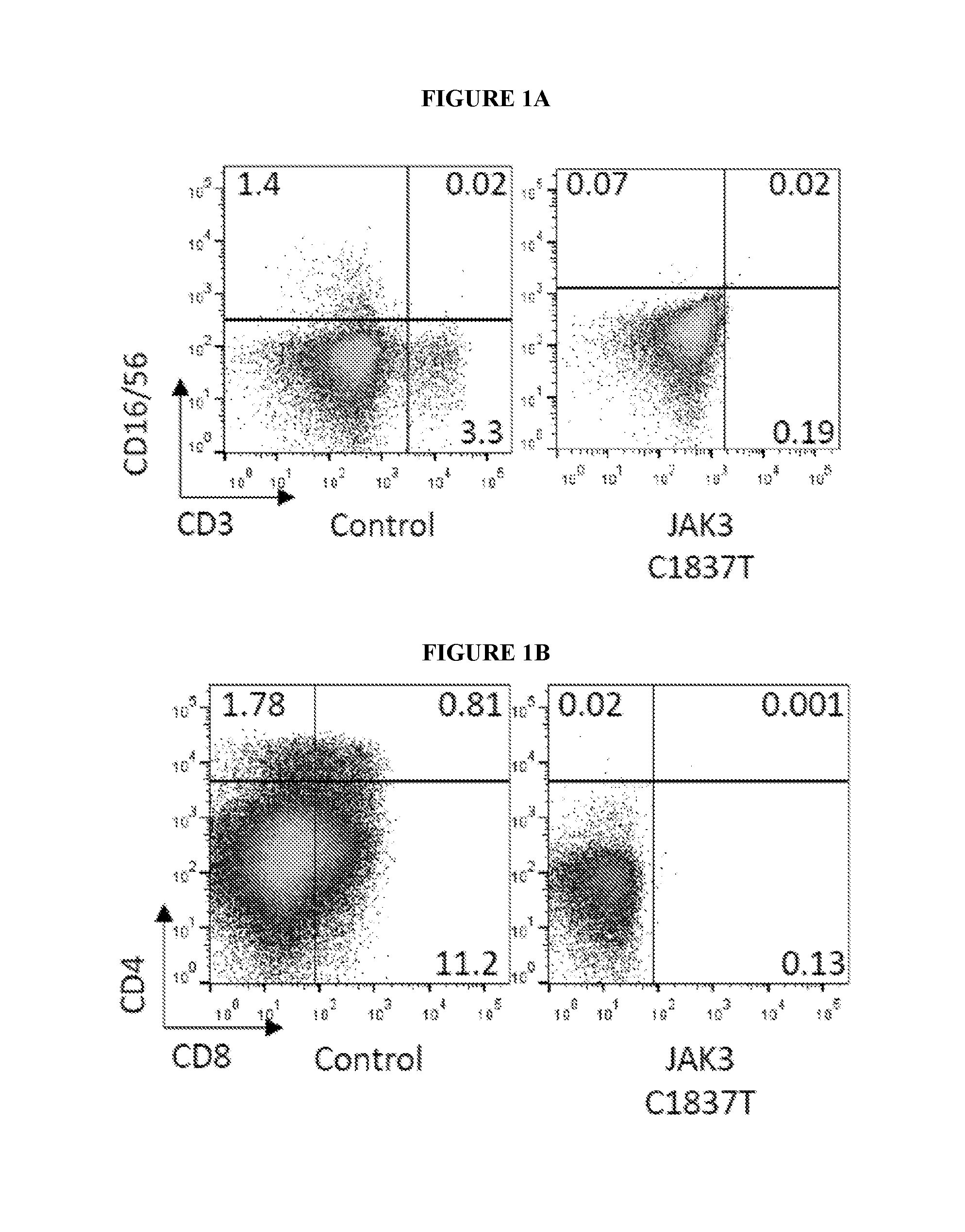
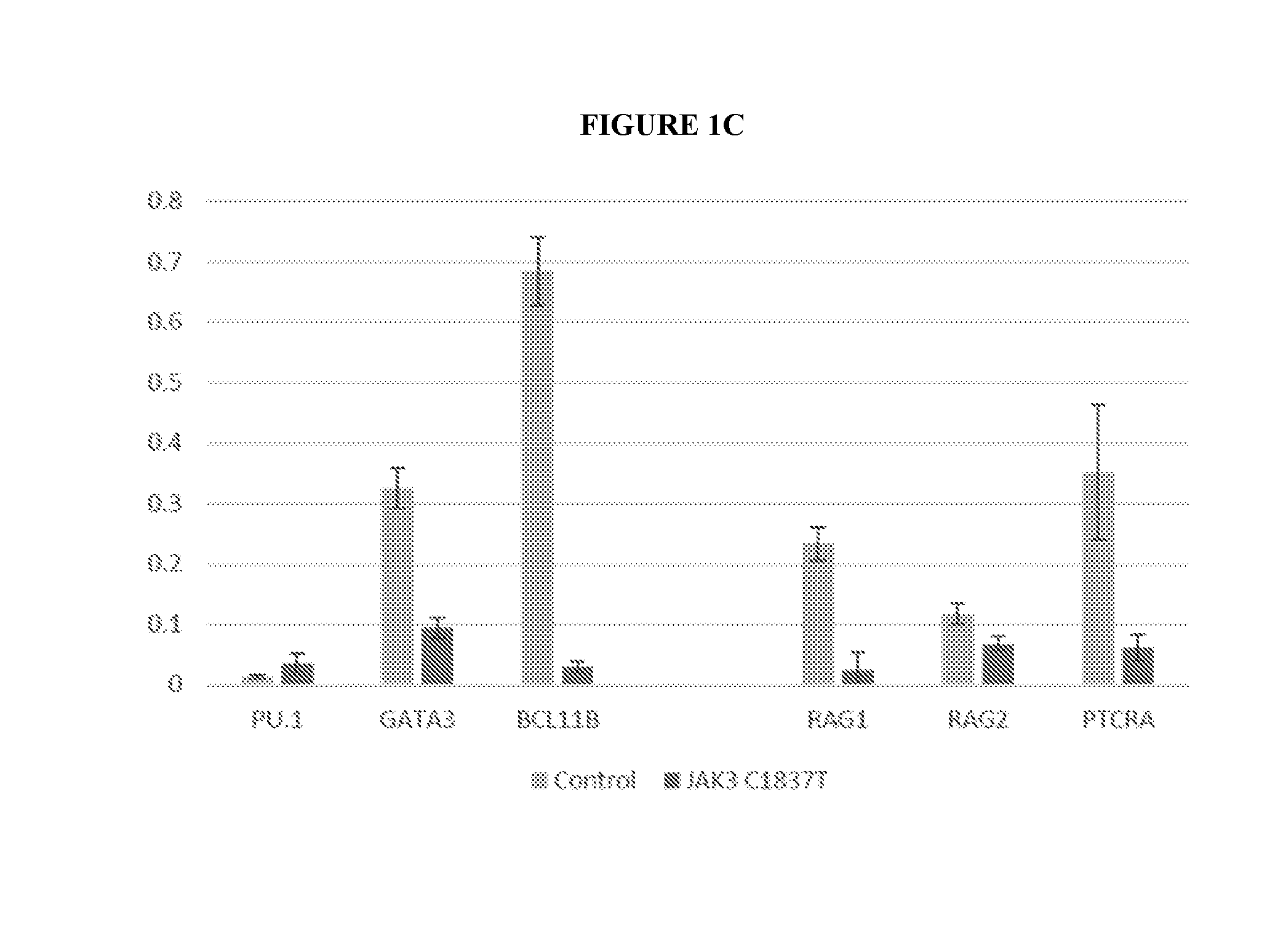
[0004] FIGS. 1A-1C show that in vitro differentiation of JAK3 C1837 T patient iPSCs recapitulates SCID phenotypes. FIGS. 1A and 1B show flow cytometry of iPSC-derived T cells. JAK3 WT iPSCs (Control) and JAK3- deficient iPSCs (JAK3 C1837T) were differentiated into CD34+ cells on OP9 stromal cells and, subsequently, into T cells on OP9-DL4 monolayers. T-cell differentiation from JAK3-deficient iPSCs was absent compared to controls; no CD3+ T cells or CD3-CD16+CD56+ NK cells were observed (FIG. 1A), and no CD4+CD8+ double positive (DP), CD4+ single positive (SP), or CD8+ single positive (SP) T cells were detected (FIG. 1B). FIG. 1C shows the results of RT-qPCR assays for transcripts of key genes that regulate early events during specification of the T cell lineage. RNA levels are shown relative to GAPDH expression.
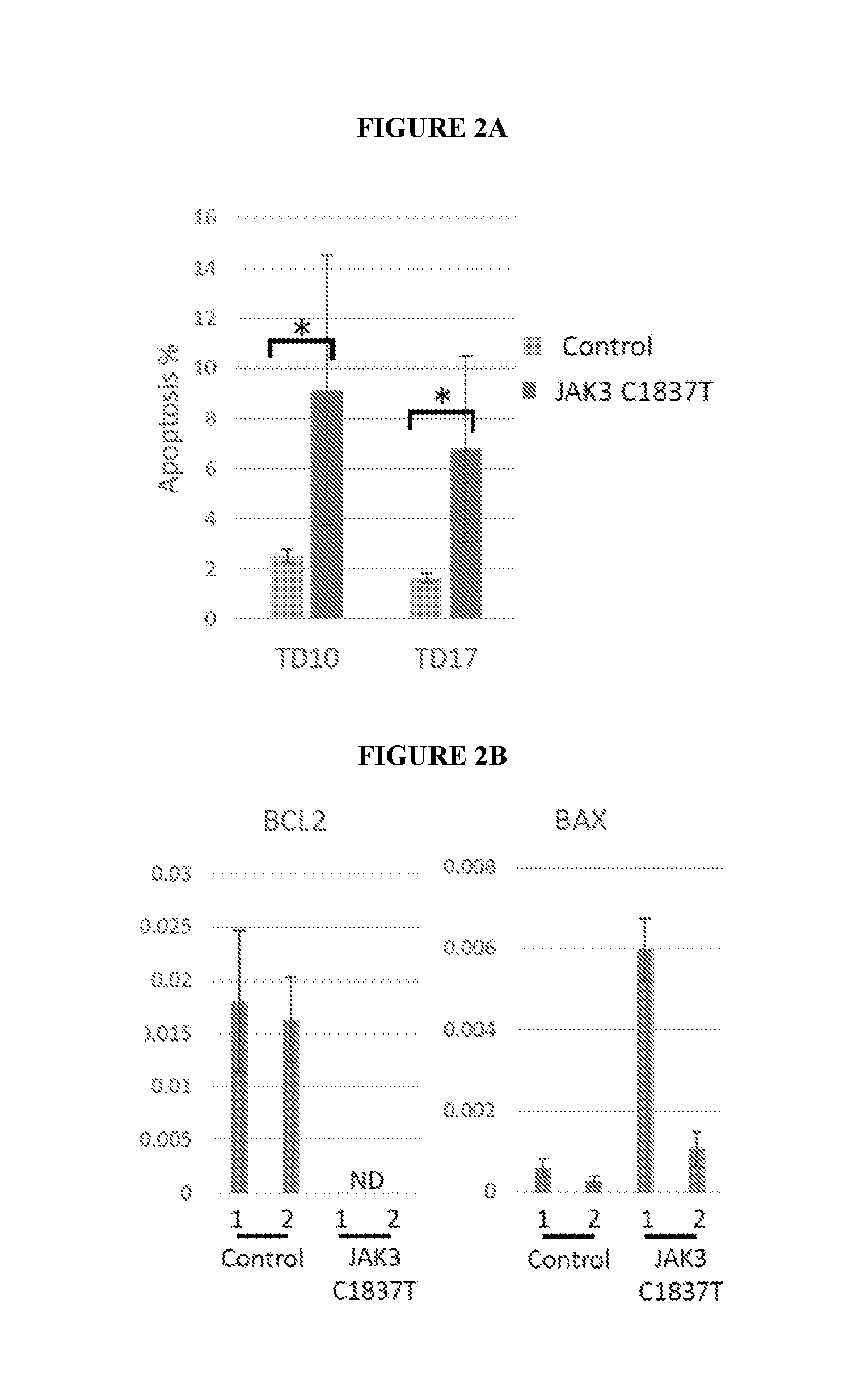
[0005] FIGS. 2A-2C show that BCL2 partially rescues T cell developmental defects in JAK3-deficient, in-vitro derived cells. FIG. 2A shows apoptosis of JAK3-deficient, iPSC-derived T cells compared to JAK3 WT controls. Annexin V-positive cells were analyzed at T cell induction day 10 (TD10) and 17 (TD17). Four independent experiments were performed with control JAK3 WT cells (Control) and 5 independent experiments were performed with JAK3-deficient cells (JAK3 C1837 T). *P <0.005. FIG. 2B shows the results of RT-qPCR assays for anti-apoptotic BCL2 and proapoptotic BAX expression in two lines (1 and 2) from JAK3 WT (Control) and JAK3-deficient cells (JAK3 C1837T). ND, not determined (due to insignificant JAK3 qPCR signal). RNA levels are shown relative to GAPDH expression. FIG. 2C shows flow cytometry of JAK3-deficient iPSCderived T cells transduced with BCL2-2 A-GFP lentivirus to assess effects on NK (CD16+56+) and T cell (CD3+) development and DP (CD4+CD8+) to SP (CD4+or CD8+) T cell maturation.
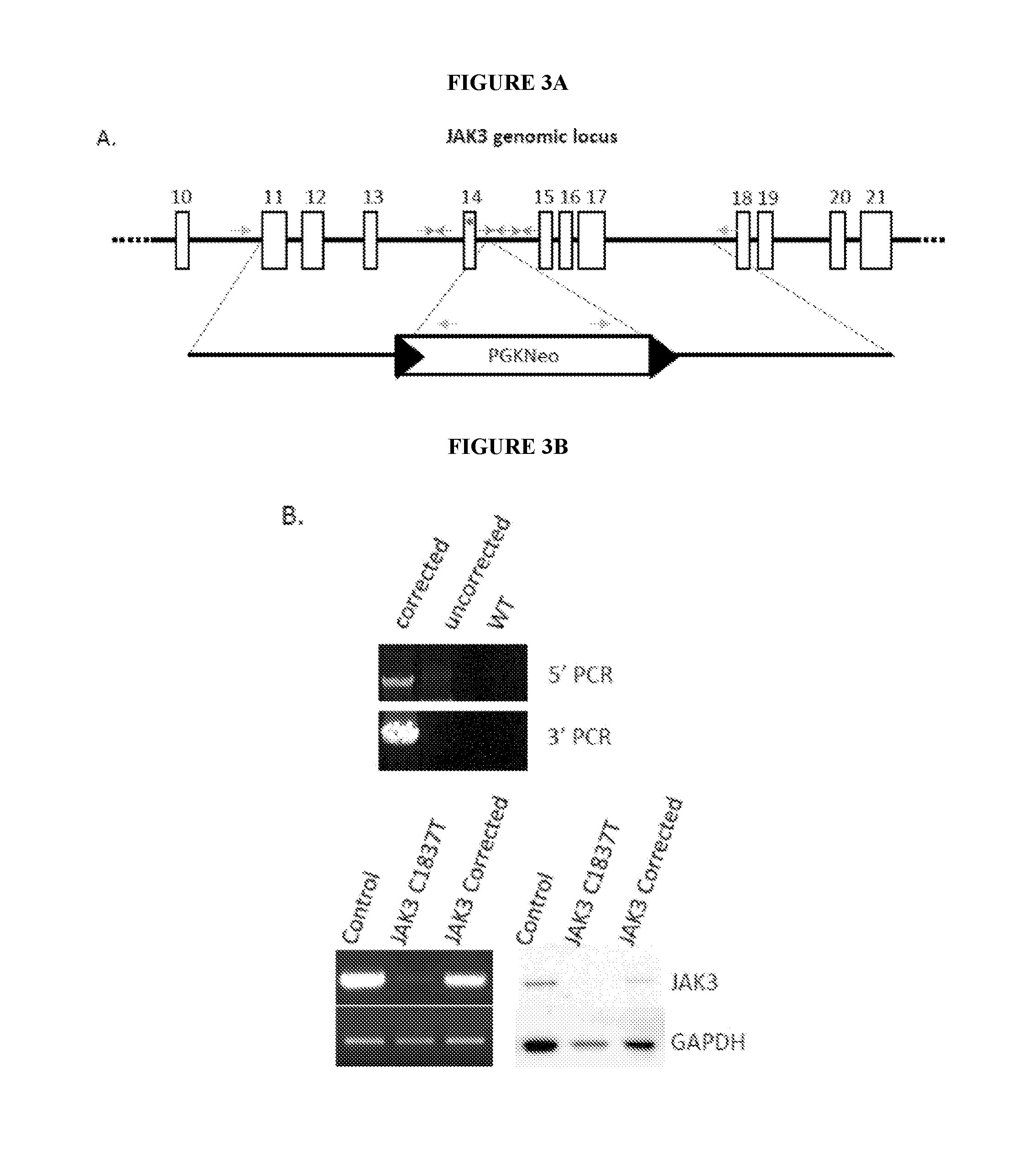
[0006] FIGS. 3A-3D show that CRISPR/Cas9 enhanced correction of the JAK3 C1837 T mutation in patient-specific iPSCs. FIG. 3A depicts the strategy for genome modification using CRISPR/Cas9 to induce double-strand breaks in the JAK3 locus and a template for homology directed repair. Top line, structure of the JAK3 gene. Open boxes, exons. Asterisk, C1837 T mutation. Arrows, guide RNAs. FIG. 3B, top, shows PCR analysis demonstrating homologous recombination; primers for 5' and 3' analysis are indicated. (Lower Left) RT-PCR analysis demonstrating JAK3 mRNA expression in JAK3 WT (Control), JAK3-deficient (JAK3 C1837 T), and corrected (JAK3 Corrected) T cells. (Lower Right) Western Blot analysis demonstrating JAK3 protein expression in JAK3 WT (Control), JAK3-deficient (JAK3 C1837 T), and corrected (JAK3 Corrected) T cells. FIG. 3C provides a summary of targeting efficiencies of guide RNAs. (FIG. 3D) Sanger sequencing of the PCR amplicons from parental JAK3 iPSCs (Left), heterozygous corrected (Middle) and homozygous corrected iPSCs (Right). The two heterozygous clones were corrected with gRNA2+wild type Cas9, and the homozygous clone was corrected with gRNA1+gRNA2+nickase Cas9 (D10A).
[0007] FIGS. 4A-4C show in vitro differentiation of JAK3 corrected patient iPSCs produces T cells with phenotypic and functional characteristics of mature T cells. FIG. 4A shows the expression of T cell developmental markers in JAK3 WT (Control, n=3), JAK3-deficient (JAK3 C1837 T, n=5) and JAK3-corrected (JAK3 Corrected, n=6) T cells. Cells were stained with indicated antibodies and analyzed by flow cytometry at T cell induction Day 14, 21, 28 and 35 (TD 14, 21, 28 and 35). FIG. 4B shows T cell receptor (TCR) V.beta. analysis of JAK3-corrected T cells. A highly diverse repertoire of TCR V.beta. is represented in T cells derived from corrected SCID patient iPSCs. FIG. 4C shows flow cytometry demonstrating T cell activation in JAK3-corrected T cells. T cells derived from JAK3 WT (Control) and JAK3-corrected iPSCs were stimulated with anti-CD3/28 beads for 3 days before analysis of activation markers CD25 and CD69. The data were gated on CD3+ populations.
[0008] FIGS. 5A-5C show in vitro generation of CD34+HSCs from hiPSCs by co-culture with human bone marrow stromal cells (hMSC). Human iPSCs were cultured on hMSCs for 18 days before analysis for hematopoietic markers, CD34 and CD43 (Figure A). CD34+ cells were purified on beads and differentiated into T cells (Figure B), erythroid and myeloid cells (Figure C). To generate T cells, purified CD34+ cells were plated on OP9-DL4 cells for 3 to 4 weeks. For the CFC assay to generate myeloid and erythroid cells, purified CD34+ cells were plated in MethoCult H4434 Classic medium according to the manufacturer's protocol. These data demonstrate that hiPSC can be efficiently differentiated into multipotent HSC after co-culture on hMSC.
[0009] FIG. 6A-6C show in vitro generation of T cells by culturing hiPSC derived CD34+ cells with hMSC-DL4. To generate CD7+ T progenitor cells, hiPSC derived CD34+ cells were co-cultured on hMSC-DL4 for 3 to 4 weeks (FIG. 6A). When CD7+ cells from FIG. 6A were purified on magnetic beads and co-cultured on OP9-DL4, fully mature CD4+/CD8+/CD3+/TCR-.alpha..beta.+ cells were produced in 10 days or less (Figures B and C). These data demonstrate that hiPSC can be efficiently differentiated into CD7+ lymphoid progenitors after co-culture on hMSC-DL4.
[0010] FIG. 7 shows in vitro generation of .gamma..delta. T cells from hiPSC. Human iPSC were transduced with a lentiviral vector carrying a pre-rearranged human V .gamma..delta.1 cDNA linked with a 2 A-GFP cDNA fragment. After co-culture with OP9 for 18 days, hiPSC derived CD34+ cells were purified on magnetic beads. These cells were subsequently plated on OP9-DL4 cells for T cell differentiation. Cells were harvested at Day 32 and T cell surface markers were analyzed by FACS. The GFP+ population represents V.delta.1-2 A-GFP lentiviral transduced cells. A high percentage of these GFP positive cells expressed V.delta.1 (66%). A low percentage of GFP negative cells expressed V.delta.1 (1%). These results demonstrate that V.delta. T cells expressing recombinant T Cell Receptors (TCR) can be efficiently produced from genetically modified iPSC. Production of V.delta. T cells expressing recombinant T Cell Receptors (TCR) specific for tumor antigens provides a powerful cellular therapy for many types of cancer.
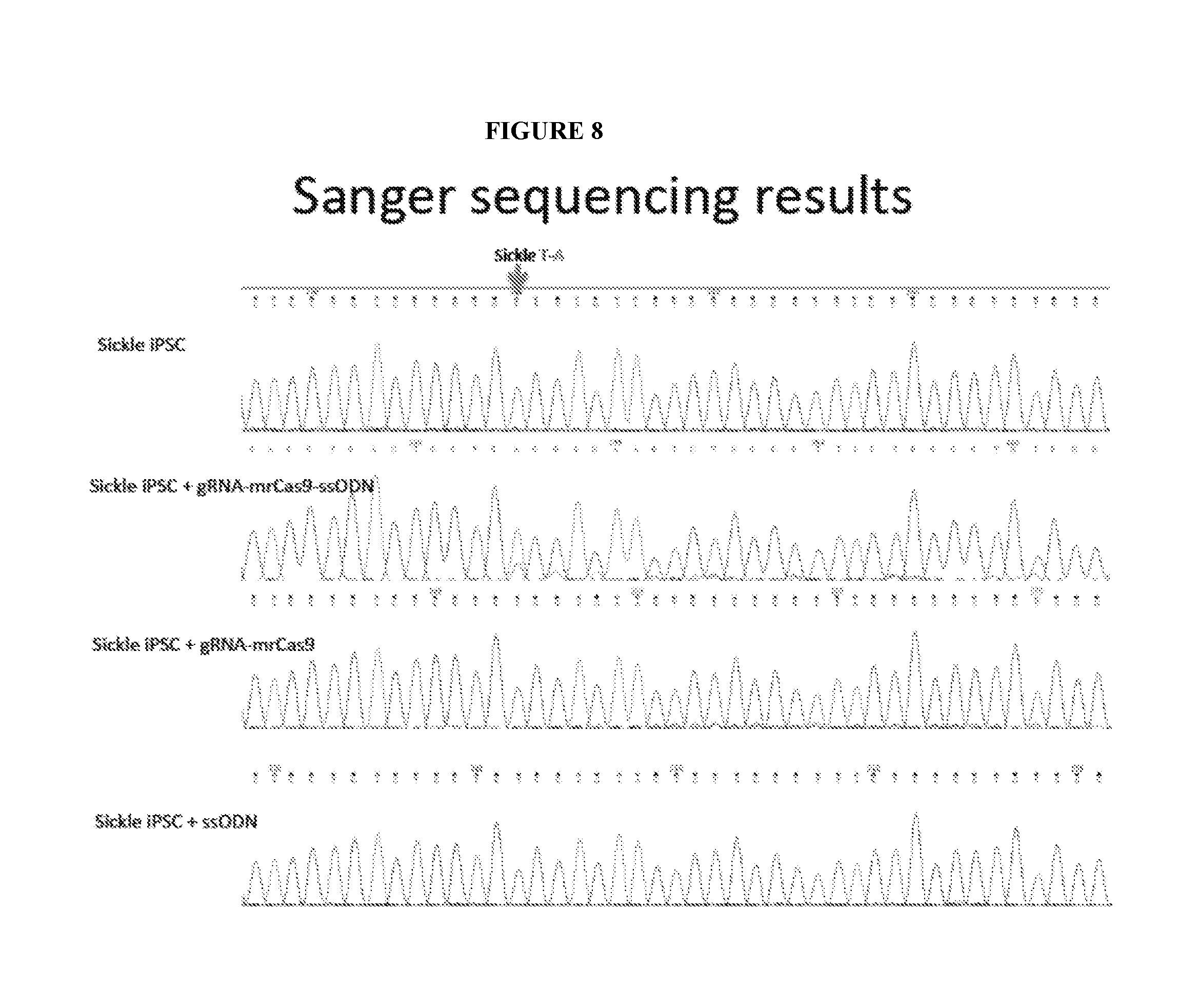
[0011] FIG. 8 shows that a correction complex including guide RNA, a modified Cas9 and a single stranded oligonucleotide donor sequence (ssODN) can correct a sickle cell mutation. The complex was introduced into sickle iPSC by nucleoporation, and 2 days later genomic DNA was analyzed by digital PCR (ddPCR) and sequenced. Over 65% of the cells contained at least one corrected gene. The results were confirmed as follows. Two days after introduction of the correction complex, the cells were plated in culture dishes, and 43 individual iPSC colonies were isolated. Genomic DNA was isolated from these colonies and the beta-globin gene was sequenced. Sixty-five percent of the colonies contained at least one corrected beta-globin gene (S corrected to A).
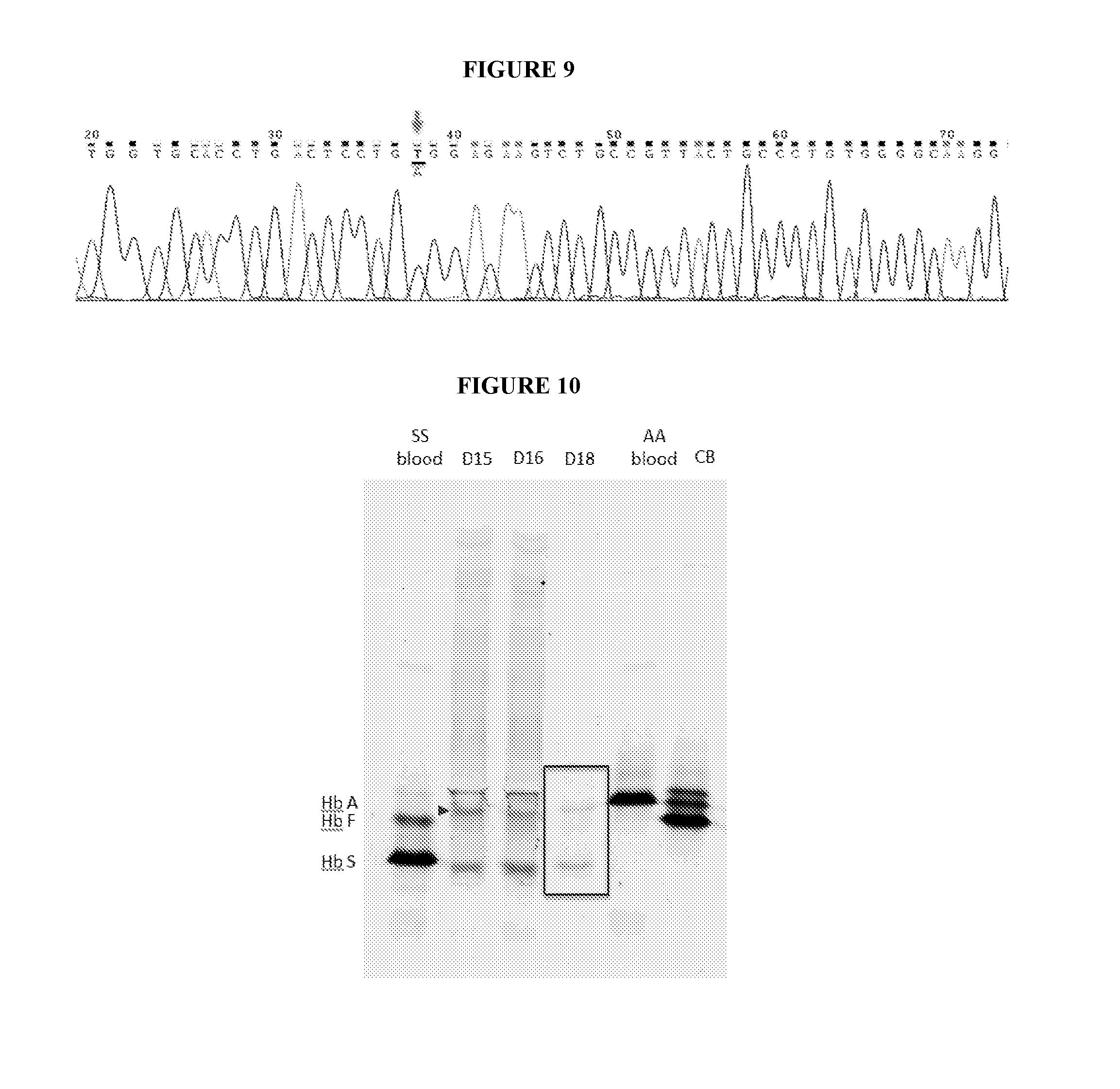
[0012] FIG. 9 shows that introduction of a sickle cell correction complex (gRNA-modified recombinant Cas9-ssODN) into patient primary bone marrow CD34+ cells can correct a sickle cell mutation. After twelve days of in vitro differentiation, DNA was analyzed by digital PCR (ddPCR) and sequenced. Approximately equal amounts of betaA and betaS mRNA were observed.
[0013] FIG. 10 is an isoelectric focusing (IEF) gel of in vitro differentiated red blood cells from the corrected sickle patient CD34+ cells of FIG. 9, showing an HbA (normal hemoglobin) to HbS (hemoglobin with sickle cell mutation) ratio of about 1:3, which is sufficient to inhibit sickling and treat sickle cell anemia.
DETAILED DESCRIPTION
[0014] Provided herein are CRISPR/Cas9 complexes for genomic modification of cells. Methods of using the complexes provided herein result in increased efficiency of modification, an increased cell survival ratio and/or an increased ratio of HDR to NHEJ in the cells. These complexes and methods can be used for therapeutic purposes, for example, to correct a mutation in cells, wherein the mutation is associated with a disease or disorder.
[0015] Provided herein is a complex for correcting a mutation in the genome of a cell comprising (a) a guide RNA (gRNA) comprising a first nucleotide sequence that hybridizes to a target DNA in the genome of a cell, wherein the target DNA comprises a mutation, and a second nucleotide sequence that interacts with a site-directed nuclease; (b) a recombinant site-directed nuclease operably linked to a supercharged protein, wherein the site-directed nuclease comprises an RNA-binding portion that interacts with the second nucleotide sequence of the guide RNA and wherein the site-directed nuclease specifically binds and cleaves the target DNA to create a double stranded break; and (c) a single-stranded donor oligonucleotide (ssODN) that hybridizes to a genomic sequence flanking the double stranded break in the target DNA and integrates into the target DNA to correct a mutation in the target DNA. It is understood that the complex comprising a guide RNA (gRNA), a recombinant site-directed nuclease and a donor nucleotide described herein does not occur in nature. The complex, however, provides the elements necessary with the required configuration and stoichiometry to efficiently and effectively modify cells. The gRNA molecule binds to the site-directed nuclease and targets the nuclease to a specific location within the target DNA. A gRNA comprises a first nucleotide sequence that hybridizes to a target DNA in the genome of a cell, wherein the target DNA comprises a mutation, and a second nucleotide sequence that interacts with a site-directed nuclease. The complexes described herein can comprise one or two separate gRNAs. Therefore, the term guide RNA includes both a single guide RNA and a double guide RNA. An example of a guide sequence that can be used to correct a mutation associated with sickle cell anemia is set forth herein as TAACGGCAGACTTCTCCAC (SEQ ID NO: 1). An example of a guide sequence comprising a stem loop for Cas9 binding is provided herein as GTAACGGCAGACTTCTCCACGTTTTAGAGCTAGAAATAGCAAGTTAAAATAAGG CTAGTCCGTTATCAACTTGAAAAAGTGGCACCGAGTCGGTGCTTTTTTT (SEQ ID NO: 2). It is noted that the 5'G of SEQ ID NO: 2 was added by T7 during in vitro transcription.
[0016] In the complexes described herein, the recombinant site-directed nuclease can be an RNA-guided site-directed nuclease, for example, a Cas protein from any bacterial species or a functional fragment thereof. For example, the Cas protein can be a Cas9 protein or a functional fragment thereof. As used herein, the term "Cas9" means a Cas9 protein or a fragment thereof present in any bacterial species that encodes a Type II CRISPR/Cas9 system. See, for example, Makarova et al. Nature Reviews, Microbiology, 9:467-477 (2011), including supplemental information, hereby incorporated by reference in its entirety. For example, the Cas9 protein or a fragment thereof can be from Streptococcus pyogenes. Full-length Cas9 is an endonuclease that includes a recognition domain and two nuclease domains (HNH and RuvC, respectively). In the amino acid sequence, HNH is linearly continuous, whereas RuvC is separated into three regions, one left of the recognition domain, and the other two right of the recognition domain flanking the HNH domain. Cas9 from Streptococcus pyogenes is targeted to a genomic site in a cell by interacting with a guide RNA that hybridizes to a 20-nucleotide DNA sequence that immediately precedes an NGG motif recognized by Cas9. This results in a double-strand break that is repaired via HDR by a donor nucleotide, for example, a ssODN or a double stranded DNA construct that hybridizes to a genomic sequence flanking the double stranded break in the target DNA and integrates into the target DNA to correct a mutation in the target DNA.
[0017] In the complexes provided herein, the molar ratio of gRNA to site-directed nuclease operably linked to a supercharged protein to ssODN can be from about 1:1:0.2 to about 1.5:1:2.0. For example, the molar ratio of gRNA to site-directed nuclease operably linked to a supercharged protein to ssODN can be about 1:1:1, 1.1:1:1, 1:1:1.15, 1:1:1.25, 1:1:1.30; 1:1:1.35; 1:1:1.40; 1:1:1.50, 1.2:1:1, 1.3:1:1. 1.4:1:1, 1.5:1:1, 1.5:1:1.15, 1.5:1:1.25, 1.5:1:1.35; 1.5:1:1.40, 1.5:1:1.45; 1.5:1:1.50; 1.5:1:1.55; 1.5:1:1.60; 1.5:1:1.65; 1.5:1:1.70; 1.5:1:1.75; 1.5:1:1.80; 1.5:1:1.85; 1.5:1:1.90; 1.5:1:1.95; 1.5:1: 2.0 or any ratio in between these ratios. Complexes having these molar ratios can be used in any of the methods described herein. Methods for preparing a complex prior to introducing the complex into a cell or a population of cells are set forth in the Examples.
[0018] As used herein, a supercharged protein can be a superpositively charged protein that has an overall positive charge that is greater than its corresponding unmodified protein. For example, the superpositively charged protein can be a superpositively charged green fluorescent protein (GFP) that has an overall positive charge from about +5 to about +40. For example, the overall positive charge can be about +5, +6, +7, +8, +9, +10, +11, +12, +13, +14, +15, +16, +17, +18, +19, +20, +21, +22, +23, +24, +25, +26, +27, +28, +29, +30, +31, +32, +33, +34, +35, +36, +37, +38, +39 or +40.
[0019] The supercharged protein can be operably linked to the amino-terminus or the carboxy-terminus of the nuclease. It is also contemplated that the supercharged protein can be associated with the nuclease, without necessarily being covalently linked to the nuclease. An example of a supercharged protein is a superpositively charged GFP, for example, +36 GFP. +36 GFP can be operably linked to the amino or carboxy- terminus of Cas9 or a functional fragment thereof. See, for example, McNaughton et al., "Mammalian cell penetration, siRNA transfection, and DNA transfection by supercharged proteins," PNAS 106 (15): 6111-6116. An example of a polypeptide comprising +36 GFP operably linked to the carboxy-terminus of Cas9 is provided herein as SEQ ID NO: 3.
[0020] The nuclease can also be operably linked to a supercharged protein and one or more positively charged peptides, for example, one or more transactivating transcriptional activator (TAT) peptide can be linked to the amino-terminus or the carboxy terminus of the nuclease. For example, and not to be limiting, a superpositively charged protein can be operably linked to the carboxy-terminus of the nuclease and one or more TAT peptides (for example, 1.times. TAT, 2.times. TAT, 3.times. TAT, 4.times. TAT, etc.) can be operably linked to the amino-terminus of the nuclease. An example of polypeptide comprising a TAT peptide operably linked to the amino-terminus of the nuclease and a superpositively charged GFP operably linked to the carboxy-terminus of the nuclease is provided herein as SEQ ID NO: 4. Polypeptide sequences that are at least about 75% identical to SEQ ID NO: 3 or SEQ ID NO: 4 are also provided. For example, polypeptide sequences that are at least about 75%, 80%, 85%, 90%, 95%, 99% or any percentage in between are also provided.
[0021] The nuclease can also be operably linked to a supercharged protein and one or more negatively charged peptides, for example, a negatively charged peptide of about 10 to about 25 amino acids in length, for example, SEQ ID NO: 50, can be operably linked to the carboxy-terminus of the site-directed nuclease. For example,and not to be limiting, a superpositively charged protein can be operably linked to the carboxy-terminus of the nuclease and a negatively charged peptide can be operably linked to the carboxy-terminus of the superpositively charged protein.
[0022] As used throughout, recombination is a process of exchange of genetic information between two polynucleotides. Homology-directed repair (HDR) refers to DNA repair that takes place, for example, during repair of double-strand breaks in cells. This process requires nucleotide sequence homology and uses a donor molecule, for example, a single stranded or a double stranded nucleotide sequence as a template for repair of a target genomic sequence, i.e., the genomic sequence with the double-strand break, and leads to the transfer of genetic information from the donor to the target genomic sequence. Homology-directed repair can result in a modification of the sequence of the target genomic sequence. For example, HDR can result in an insertion, a deletion or a mutation in the target genomic sequence. Part or all of the sequence of the donor polynucleotide can be incorporated into the target DNA. It is also contemplated that the donor polynucleotide, a portion of the donor polynucleotide, a copy of the donor polynucleotide, or a portion of a copy of the donor polynucleotide integrates into the target DNA.
[0023] As used throughout, by non-homologous end joining (NHEJ) is meant the repair of double-strand breaks in DNA by direct ligation of the break ends to one another without the need for a homologous template (in contrast to homology-directed repair, which requires a homologous sequence to guide repair).
[0024] The complexes and methods provided herein can be used to correct any mutation in a target DNA by HDR. For example, and not to be limiting, the complexes can be used to replace an incorrect nucleotide sequence with a correct nucleotide sequence (e.g., to restore function to a target polynucleotide sequence that is impaired due to a loss of function mutation, i.e., a SNP) at a specific site in the genome. These mutations can be associated with an autoimmune disorder, a genetic disease, a blood disorder, a T cell disorder, a monogenic disorder, cancer, a neurodegenerative disease, a cardiovascular disease or an infectious disease, to name a few. For example, and not to be limiting, the complexes and methods provided herein can be used to correct a mutation associated with sickle cell disease (i.e., a mutation in a hemoglobin gene, for example, a GAG to GTG mutation at codon 6 of the beta-globin gene that results in a glutamic acid to valine substitution), severe combined immunodeficiency (SCID) (for example, a mutation in JAK3), beta thalassemia or Wiskott-Aldrich Syndrome.
[0025] Correction of single mutations or multiple mutations can be performed with one or more complexes. The complexes and methods provided herein can also be used to insert sequences into a specific site in the genome to correct a deletion, as opposed to making a correction or a substitution. The complexes and methods provided herein can also be used to insert a nucleotide sequence that encodes an a functional polypeptide into a specific site in the genome of the cell, in order to express the functional polypeptide in the cell. The functional polypeptide can be a polypeptide that is endogenous (i.e., normally expressed by the cell) or exogenous to the cell (i.e. not normally expressed by the cell). For example, chimeric antigen receptor (CAR) sequences can be inserted into the genome of a T cell precursor in order to generate cancer specific T cells for the treatment of cancer. In another example, the complexes and methods provided herein can be used to inhibit the activity of a gene at a specific site in the genome of the cell. For example, the complexes and methods provided herein can be used to insert sequences into the CXCR4 or CCRS receptor to treat or prevent HIV infection.
[0026] The complexes provided herein can modify or alter target DNA with surprisingly high efficiency as compared to conventional CRISPR/Cas systems. The efficiency of alteration in a population of cells can be at least about 5%, 10%, 15%, 20%, 25%, 30%, 35%, 40%, 45%, 50%, 55%, 60%, 65%, 70%, 75% or 80% or higher or any percentage in between these percentages. The efficiency of alteration can also be greater than or equal to about 80%. Therefore, also provided herein are populations of cells, wherein at least 5%, 10%, 15%, 20%, 25%, 30%, 35%, 40%, 45%, 50%, 55%, 60%, 65%, 70%, 75% or 80% or higher or any percentage in between are altered. For example, a mutation associated with sickle cell disease or another disorder has been corrected. If a population of cells comprising a mutation associated with sickle cell disease is contacted with a CRISPR/Cas complex described herein and the mutation is corrected in about 5% of the cells, the efficiency of modification or alteration is about 5%. Optionally, a population of cells wherein the mutation associated with sickle cell disease is corrected in about 30% of the cells, including, for example, 27%, 28% and 29% is sufficient to treat sickle cell disease, upon transplantation in a subject with sickle cell disease. Optionally, a mutation associated with sickle cell disease is corrected in about 40%, 50%, 60%, 70%, 80%, 90% or higher or any percentage in between, of the cells in the population.
[0027] In addition to altering the target DNA with high efficiency, the complexes provided herein can also increase the ratio of HDR to NHEJ in a population of cells contacted with the complex. The HDR/NHEJ ratio can be from about 10 to about 0.5. For example, the HDR/NHEJ ratio can be about 10, 9, 8, 7, 6, 5, 4, 3, 2, 1, 0.5 or less or any ratio in between these ratios. In addition to high efficiency of correction and high rate of HDR to NHEJ, the cell survival rate for corrected cells can be at least about 50%, 60%, 70%, 80%, 90% or higher and any percentage in between.
[0028] Any cell(s) can be modified or derived using the complexes described herein. Introduction of the complex into the cells can be cell cycle dependent or cell cycle independent. Methods of synchronizing cells to increase the proportion of cells in a particular phase, for example, the S-phase , are known in the art. See, for example, Takahashi et al. "Efficient introduction of a gene into hematopoietic cells in S-phase by electroporation," Exp. Hematol. 19 (5):343-346 (1991). Depending on the type of cell to be modified, one of skill in the art can readily determine if cell cycle synchronization is necessary.
[0029] The cell(s) can be a eukaryotic cell, for example, a mammalian cell. The cell can also be prokaryotic or a plant cell. The cell can be a human cell. The cell can be a germ cell, a somatic cell, a stem cell, a precursor cell or a progenitor cell. The precursor cell can be, for example, a pluripotent stem cell or a multipotent stem cell, like a hematopoietic stem cell. As used throughout, pluripotent cells include induced pluripotent stem cells. Methods of making induced pluripotent stem cells and known in the art and described in the Examples. The cell can also be CD34+ cell, optionally derived from an induced pluripotent stem cell. The CD34+ cell can be selected from the group consisting of a primary CD34+ hematopoietic progenitor cell, a CD34+ peripheral blood cell, a CD34+ cord blood cell and a CD34+ bone marrow cell. The cell can also be a primary cell, for example, a primary CD34+ hematopoietic progenitor cell. The cells are cells that are not cancer cells, cells that are not tumor cells or cells that are not transformed cells. Cells can be screened before or after correction for evidence of undesirable genetic characteristics. Further provided is a cell comprising any of the complexes described herein. The cell can be in vitro, ex vivo or in vivo.
[0030] Further provided is a method of site-specific modification of a target DNA in a population of cells comprising introducing into the cells any of the complexes described herein, wherein the complex is introduced into the cells under conditions that allow homology-directed repair (HDR) and integration of a donor nucleotide, for example, a ssODN or double stranded nucleotide sequence into the target DNA. The complex can be introduced into the cell via nucleoporation. Methods for nucleoporation are known in the art. See, for example, Maasho et al. "Efficient gene transfer into the human natural killer cell line, NKL, using the amaxa nucleofection system," Journal of Immunological Methods 284 (1-2): 133-140 (2004); and Aluigi et al. "Nucleofection is an efficient non-viral transduction technique for human bone marrow derived mesenchymal stem cells," Stem Cells 24 (2): 454-461 (2006)), both of which are incorporated herein in their entireties by this reference.
[0031] In some of the methods provided herein, the donor nucleotide, for example, a ssODN or a double stranded nucleotide sequence integrates into a target DNA and corrects a mutation in the target DNA. In the methods provided herein the ratio of HDR to NHEJ in a population of cells is increased relative to other CRISPR-Cas9 delivery methods. The HDR/NHEJ ratio can be from about 10 to about 0.5. For example, the HDR/NHEJ ratio can be about 10, 9, 8, 7, 6, 5, 4, 3, 2, 1, 0.5 or less or any ratio in between these ratios. In the methods provided herein, the efficiency of alteration by HDR can be at least about 5%, 10%, 15%, 20%, 25%, 30%, 35%, 40%, 45%, 50%, 55%, 60%, 65%, 70%, 75%, 80% or greater or any percentage in between these percentages. The efficiency of alteration by HDR can also be greater than or equal to about 80%. For example, if a population of cells comprising a mutation associated with sickle cell anemia is contacted with a CRISPR/Cas complex described herein and the mutation is corrected in about 5% of the cells, the efficiency of alteration by HDR is about 5%. The population of cells can be obtained from the subject having a disorder such that at least about 5%, 10%, 15%, 20%, 25%, 30%, 35%, 40%, 45%, 50%, 55%, 60%, 65%, 70%, 75% or 80% or greater or any percentage in between these percentages, of the cells undergo HDR to correct a mutation associated with the disorder. In some cases greater than 80% of the cells from the subject will undergo HDR to correct a mutation associated with the disorder. In the methods described herein, between about 50% and 99% of the cells survive after introduction of the complex. For example, great than about 50%, 60%, 70%, 80%, 90%, 95%, 99% or any percentage in between these percentages, of corrected cells survive after introduction of the complex.
[0032] Further provided is a method of treating a disease associated with a mutation in the genomic sequence encoding hemoglobin in a subject comprising: (a) introducing into a population of cells obtained from the subject a complex comprising (1) a guide RNA (gRNA) comprising a first nucleotide sequence that hybridizes to a target DNA in the genome of a cell, wherein the target DNA is a hemoglobin gene that comprises a mutation, and a second nucleotide sequence that interacts with a site-directed nuclease; (2) a recombinant site-directed nuclease operably linked to a supercharged protein, wherein the site-directed nuclease comprises an RNA-binding portion that interacts with the second nucleotide sequence of the guide RNA and wherein the site-directed nuclease specifically binds and cleaves the target DNA to create a double stranded break; and (3) a single-stranded donor oligonucleotide (ssODN) that hybridizes to a genomic sequence flanking the double stranded break in the target DNA and integrates into the target DNA to correct the mutation in hemoglobin gene; and (b) transplanting the corrected cells into the subject.
[0033] In the methods for treating a disease associated with a mutation in the genomic sequence encoding hemoglobin in a subject, for example, sickle cell anemia, the subject with sickle cell anemia can optionally be a transfusion dependent subject or a subject with at least one silent infarction. The subject can also be less than about twelve months, eleven months, ten months, nine months, eight months, seven months, six months, five months, four months, three months, two months, or one month in age. As infants are routinely screen for sickle cell disease, infants can be treated before symptoms of the disease manifest. The methods provided herein can further comprise diagnosing a subject with a disorder, for example, sickle cell disease.
[0034] As set forth above, cells can be obtained from the subject with the disease or from a related donor. For example, bone marrow cells can be obtained or harvested from the subject. Bone marrow harvesting involves collecting stem cells with a needle placed into the soft center of the bone, the marrow. Bone marrow can be harvested for example, from the hip bones or sternum of the subject. From about 500 ml to about 1 liter of bone marrow can be obtained from the subject.
[0035] In any of the methods provided herein the cell(s) can be a eukaryotic cell, for example, a human cell. The cell can be a germ cell, a stem cell, a precursor cell. The precursor cell can be, for example, a pluripotent stem cell or a hematopoietic stem cell. As used throughout, pluripotent cells include induced pluripotent stem cells. Methods of making induced pluripotent stem cells and known in the art and described in the Examples. The cell can also be CD34+ cell. The CD34+ cell can be selected from the group consisting of a primary CD34+ hematopoietic progenitor cell, a CD34+ peripheral blood cell, a CD34+ cord blood cell and a CD34+ bone marrow cell. The cell can also be a primary cell, for example, a primary CD34+ hematopoietic progenitor cell. The cells are that are not cancer cells, cells that are not tumor cells or cells that are not transformed cells. The cell can be in vitro or ex vivo. The cells can also be in a pharmaceutically acceptable composition.
[0036] The methods provided herein can further comprise culturing the cells corrected with HDR. For example, the cells can be cultured under conditions for expansion or under conditions that promote differentiation of the corrected cells into T-cells. For example, and not to be limiting, using the methods provided herein, after a mutation has been corrected in induced pluripotent stem cells via HDR, the corrected cells can be co-cultured with human bone marrow stromal cells to generate CD34+ cells. The CD34+ cells can then be cultured under conditions that differentiate the CD34+ cells into T cells.
[0037] The methods provided herein can further comprise screening the corrected cells for the proper correction, other mutations, or NEJ prior to transplantation. Optionally cells can be screened to detect cells with one or more corrections.
[0038] In the methods provided herein, the cells can be transplanted into the subject after modification, for example, after correction of a mutation by HDR. The cells can be transplanted into the subject with or without differentiation. For example, modified hematopoietic stem cells (HSCs) can be administered in a bone marrow transplant, wherein the HSCs are allowed to differentiate and mature in vivo in a subject Alternatively, the modified cells can be differentiated into a desired population of cells prior to transplantation.
[0039] As used herein, transplanting, introducing or administering cells to a subject refers to the placement of cells into a subject. For example, the cells described herein comprising a target DNA sequence corrected or modified according to the methods described herein can be transplanted into a subject, by an appropriate route which results in at least partial localization of the transplanted cells at a desired site. The cells can be implanted directly to the desired site, or alternatively can be administered by any appropriate route which results in delivery to a desired location in the subject where at least a portion of the implanted cells remain viable. For example, the cells can be administered systemically, via intravenous infusion. The period of viability of the cells after administration to a subject can be as short as a few hours, e. g. twenty-four hours, to a few days, to as long as several years.
[0040] For ex vivo methods, cells can be autologous cells, i.e., a cell or cells taken from a subject who is in need of modification of a target DNA in the cell or cells (i.e., the donor and recipient are the same individual). As described herein, the modification can be, for example correction of a mutation, insertion of a sequence that inhibits activity of a protein or insertion of a sequence that increases expression of a protein, for example, insertion of a sequence encoding a chimeric antigen receptor that can be used to target cancer cells. Autologous cells can be used to avoid immunological reactions that can result in rejection of the cells. In other words, when using autologous cells, the donor and recipient are the same subject. Alternatively, the cells can be heterologous, e.g., taken from a donor, preferably a related donor. The second subject can be of the same or different species. Typically, when the cells come from a donor, they will be from a donor who is sufficiently immunologically compatible with the recipient to reduce the chances of transplant rejection, and/or to reduce the need for immunosuppressive therapy. The cells can also be obtained from a xenogeneic source, i.e., a non-human mammal that has been genetically engineered to be sufficiently immunologically compatible with the recipient, or the recipient's species. Any of the methods of treating a disorder described herein can further comprise administering one or more immunosuppressants to the subject.
[0041] In the methods involving transplantation, a subject optionally undergoes myeloablative therapy prior to transplantation of any of the cells described herein. The myeloablative therapy can include administering one or more doses of chemotherapy, radiation therapy, or both, that results in severe or complete depletion of healthy bone marrow cells. In another example, the subject can undergo submyeloablative therapy that includes administering one or more doses of chemotherapy, radiation therapy, or both, that depletes a portion of the healthy bone marrow cells. The cells can also be transplanted into subjects that have undergone nonablative chemotherapy. For example, the cells can be transplanted into a subject that has been treated with Busulfan, Fludarabine and/or Treosulfan.
[0042] In the methods involving transplantation, an effective dose or amount of corrected cells is administered to the subject. The terms effective amount and effective dosage are used interchangeably. The term effective amount is defined as any amount necessary to produce a desired physiologic response. In some methods, about 1.times.10.sup.6 to about 7.times.10.sup.6 corrected cells/kg can be administered, but this amount can vary depending on the associated disorder.
[0043] The percentage of corrected cells that Effective amounts and schedules for administering the cells may be determined empirically, and making such determinations is within the skill in the art. The dosage ranges for administration are those large enough to produce the desired effect (e.g., treatment of a disease, for example, sickle cell anemia). The dosage should not be so large as to cause substantial adverse side effects, such as unwanted cross-reactions, anaphylactic reactions, and the like. Generally, the dosage will vary with the age, condition, sex, type of disease, the extent of the disease or disorder, route of administration, or whether other drugs are included in the regimen, and can be determined by one of skill in the art. The dosage can be adjusted by the individual physician in the event of any contraindications. Dosages can vary, and the agent can be administered in one or more dose administrations daily, for one or multiple days as needed.
[0044] As used throughout, a subject can be a vertebrate, more specifically a mammal (e.g., a human, horse, cat, dog, cow, pig, sheep, goat, mouse, rabbit, rat, and guinea pig). The term does not denote a particular age or sex. Thus, adult and newborn subjects, whether male or female, are intended to be covered. As used herein, patient or subject may be used interchangeably and can refer to a subject with or at risk of developing a disorder. The term patient or subject includes human and veterinary subjects.
[0045] As used herein the terms treatment, treat, or treating refers to a method of reducing one or more of the effects of the disorder or one or more symptoms of the disorder, for example, sickle cell disease, by eliciting an immune response in the subject. Thus in the disclosed method, treatment can refer to a 10%, 20%, 30%, 40%, 50%, 60%, 70%, 80%, 90%, or 100% reduction in the severity of sickle cell disease and other disorders. For example, a method for treating sickle cell disease is considered to be a treatment if there is a 10% reduction in one or more symptoms of the infection in a subject as compared to a control. Thus the reduction can be a 10%, 20%, 30%, 40%, 50%, 60%, 70%, 80%, 90%, 100%, or any percent reduction in between 10% and 100% as compared to native or control levels. It is understood that treatment does not necessarily refer to a cure or complete ablation of the disorder or symptoms of the disorder.
[0046] Also provided is a method of correcting a mutation associated with a T-cell disorder comprising introducing into a population of cells obtained from a subject with the T-cell disorder a complex comprising: (a) a guide RNA (gRNA) comprising a first nucleotide sequence that hybridizes to a target DNA in the genome of a cell, wherein the target DNA comprises the mutation associated with the T-cell disorder, and a second nucleotide sequence that interacts with a site-directed nuclease; (b) a recombinant site-directed nuclease operably linked to a supercharged protein, wherein the site-directed nuclease comprises an RNA-binding portion that interacts with the second nucleotide sequence of the gRNA and wherein the site-directed nuclease specifically binds and cleaves the target DNA that comprises the mutation associated with the T-cell disorder to create a double stranded break in the target DNA; and (c) a single stranded donor oligonucleotide (ssODN) comprising a third nucleotide sequence that hybridizes to a genomic sequence flanking the double stranded break in the target DNA and that integrates into the target DNA to correct the mutation associated with the T-cell disorder, wherein the complex is introduced into the cell under conditions that allow homology-directed repair (HDR) to correct the mutation associated with the T-cell disorder.
[0047] In the methods provided herein, the target DNA comprising a mutation associated with a T-cell disorder can be a target DNA that encodes a protein associated with T-lymphocyte development. For example, the target DNA can encode JAK3. Such corrected cells can be used, for example, in the treatment of SCID.
[0048] In addition to correcting mutations in the genome of a cell, the complexes and methods provided herein can also be used to insert functional polypeptides at specific sites in the genome of a cell, such that the polypeptide is expressed by the cell. The polypeptide can be expressed in the cell or on the cell surface.
[0049] Also provided is a method of making tumor-specific T-cell precursor cells comprising introducing into a population of T-cell precursor cells a complex comprising: (a) a guide (gRNA) comprising a first nucleotide sequence that hybridizes to a target DNA in the genome of the T cell precursor cells and a second nucleotide sequence that interacts with a site-directed nuclease; (b) a recombinant site-directed nuclease operably linked to a supercharged protein, wherein the site-directed nuclease comprises an RNA-binding portion that interacts with the second nucleotide sequence of the gRNA and wherein the site-directed nuclease specifically binds and cleaves the target DNA to create a double stranded break; and (c) donor nucleotide sequence comprising a third nucleotide sequence that encodes a chimeric antigen receptor (CAR) and a fourth nucleotide sequence that hybridizes to a genomic sequence flanking the double stranded break in the target DNA, wherein the complex is introduced into the T-cell precursor cells under conditions that allow homology-directed repair (HDR) and integration of the third nucleotide sequence into the target DNA to form modified T-cell precursor cells that express the CAR.
[0050] The T cell precursor cells can be obtained from a subject with cancer. As set forth above, the HDR/NHEJ ratio can be from about 10 to about 0.5. For example, the HDR/NHEJ ratio can be about 10, 9, 8, 7, 6, 5, 4, 3, 2, 1, 0.5 or any ratio in between these ratios. In the methods provided herein, the efficiency of alteration by HDR can be at least about 5%, 10%, 15%, 20%, 25%, 30%, 35%, 40%, 45%, 50%, 55%, 60%, 65%, 70%, 75%, 80% or any percentage in between these percentages. The efficiency of alteration by HDR can also be greater than or equal to about 80%. For example, when using the methods described herein, if a nucleotide sequence encoding an functional polypeptide, for example, a nucleotide sequence that encodes a CAR, is inserted in about 5% of the cells, the efficiency of alteration by HDR is about 5%. The population of cells can be obtained from the subject that has cancer such that at least about 5%, 10%, 15%, 20%, 25%, 30%, 35%, 40%, 45%, 50%, 55%, 60%, 65%, 70%, 75% or 80% or any percentage in between these percentages, of the cells undergo HDR to insert a nucleotide sequence that encodes a chimeric antigen receptor (CAR) and form cells that express the CAR. In some cases greater than 80% of the cells from the subject will undergo HDR to correct a mutation associated with the disorder.
[0051] The modified T-cell precursor cells that express the CAR can be transplanted into a subject with cancer. As used herein, cancer is a disease characterized by the rapid and uncontrolled growth of aberrant cells. Cancer cells can spread locally or through the bloodstream and lymphatic system to other parts of the body. Examples of cancers include but are not limited to, breast cancer, prostate cancer, ovarian cancer, cervical cancer, skin cancer, pancreatic cancer, colorectal cancer, renal cancer, liver cancer, brain cancer, lymphoma, leukemia, lung cancer and the like. The modified T-cell precursor cells that express the CAR exhibit anti-tumor immunity when the antigen binding domain binds to its corresponding antigen.
[0052] Disclosed are materials, compositions, and components that can be used for, can be used in conjunction with, can be used in preparation for, or are products of the disclosed methods and compositions. These and other materials are disclosed herein, and it is understood that when combinations, subsets, interactions, groups, etc. of these materials are disclosed that while specific reference of each various individual and collective combinations and permutations of these compounds may not be explicitly disclosed, each is specifically contemplated and described herein. For example, if a method is disclosed and discussed and a number of modifications that can be made to a number of molecules including the method are discussed, each and every combination and permutation of the method, and the modifications that are possible are specifically contemplated unless specifically indicated to the contrary. Likewise, any subset or combination of these is also specifically contemplated and disclosed. This concept applies to all aspects of this disclosure including, but not limited to, steps in methods using the disclosed compositions. Thus, if there are a variety of additional steps that can be performed, it is understood that each of these additional steps can be performed with any specific method steps or combination of method steps of the disclosed methods, and that each such combination or subset of combinations is specifically contemplated and should be considered disclosed.
[0053] Publications cited herein and the material for which they are cited are hereby specifically incorporated by reference in their entireties.
EXAMPLES
Example 1
Correction of SCID by CRISPR/Cas9 Enhanced Gene Replacement
[0054] Mutations of the Janus family kinase JAK3 gene cause severe combined immunodeficiency (SCID). JAK3 deficiency in humans is characterized by the absence of circulating T cells and natural killer (NK) cells with normal numbers of poorly functioning B cells (T-B+NK-). As shown herein, using SCID patient-specific induced pluripotent stem cells (iPSCs) and a T cell in vitro differentiation system, a complete block in early T cell development of JAK3-deficient cells was demonstrated. Correction of the novel JAK3 mutation by CRISPR/Cas9 enhanced gene replacement restores normal T cell development, including the production of mature T-cell populations with a broad T Cell Receptor (TCR) repertoire. Whole genome sequencing of corrected cells demonstrated no CRISPR/Cas9 off-target modifications. Thus, provided herein is a novel approach for the study of human lymphopoiesis and a method for gene replacement therapy in humans with immunodeficiencies.
[0055] Allogeneic hematopoietic stem cell (HSC) transplantation is currently the only established therapy for SCID; however, delayed immune recovery and risk of graft-vs-host disease present significant risks. Treatment by retroviral-based gene therapy has been successfully demonstrated for X-linked SCID. However, severe adverse effects of insertional mutagenesis have been observed with retroviral gene therapy. Self-inactivating lentiviral vectors have been used effectively in recent clinical trials, but long-term follow-up is needed to thoroughly address safety concerns.
[0056] Provided herein is an alternative therapeutic strategy in which patient-specific induced pluripotent stem cells (iPSCs) are derived, and disease-causing mutations are corrected by gene replacement using a CRISPR-Cas9 complex. These corrected iPSCs could optionally be differentiated into hematopoietic progenitors for transplantation into patients to treat the disease (Hanna et al., "Treatment of sickle cell anemia mouse model with iPS cells generated from autologous skin," Science 318: 1920-1923 (2007)). As shown herein, differentiation of JAK3-deficient human T cells is blocked at an early developmental stage. Also demonstrated is that correction of the human JAK3 mutation by CRISPR/Cas9 enhanced gene replacement restores the differentiation potential of early T cell progenitors. These corrected progenitors are capable of producing NK cells and mature T cell populations expressing a broad repertoire of T-cell antigen receptors (TCR). These studies establish a powerful system for determining the mechanism of immunodeficiency in human SCID patients and for testing pharmacological and genetic therapies for the disorder.
Patient Information
[0057] The male patient was enrolled in an Institutional Review Board-approved study in accordance with the Declaration of Helsinki. The family history was negative for immune deficiencies. For the first 8 months of age he had poor weight gain, diarrhea, and recurrent bronchiolitis requiring frequent hospitalization. He was admitted to the hospital at 8 months of age with severe respiratory distress and oral thrush. Bronchoscopy with bronchial alveolar lavage demonstrated bacterial (pseudomonas, H flu, S. pneumonia) and viral organisms (respiratory syncytial virus). Immunologic evaluations demonstrated severe hypogammaglobulinemia, with an IgE<3, IgA<4, IgG=29, IgM=26. Immune phenotyping of peripheral blood demonstrated complete absence of CD3+ T cells and NK cells, though B cells were present (absolute B cell count=875). Mitogen studies demonstrated a complete lack of response to concanavalin A, poke weed mitogen and phytohemagglutinin A. The diagnosis of SCID was confirmed by genetic testing, with a homozygous C>T nucleotide substitution in exon 14 of the JAK3 gene, resulting in the replacement of an arginine codon
[0058] (CGA) with a stop codon (TGA) at amino acid position 613. This is the first report linking this JAK3 variant (r5149316157) to a clinical case of SCID. The patient underwent a reduced intensity conditioning matched unrelated bone marrow transplant, and is doing well now two years off therapy with complete immune reconstitution.
Human iPSC Reprogramming and Characterization
[0059] For iPSC induction, 5.times.10.sup.4 primary keratinocytes were seeded into one well of a 6-well plate. On the following day, keratinocytes were transduced with 1 mL of virus supernatant and 1 mL of human keratinocyte medium containing polybrene at a final concentration of 4 .mu.g/mL. The keratinocytes were spinfected at 800.times. g for 45 minutes (day 1). The transduction procedure was repeated again the next day. On day 3, cells were changed to fresh human keratinocyte medium and cultured for two more days. On day 5, the keratinocytes were trypsinized and transferred to a 10 cm dish pre-seeded with mitomycin C-treated murine embryonic fibroblasts (MEFs) and cultured in human keratinocyte medium. On day 7, cells were changed to human ES medium and continuously cultured in the same dish for 3-4 weeks. ES medium was changed daily. Potential iPSC colonies were visible after 2-3 weeks. These colonies were individually picked and expanded on MEFs for analysis. To remove the integrated lentiviral and polycistronic sequences, iPSCs were infected with a Cre-expressing adenovirus (rAd-Cre-IE). Individual colonies were picked and Cre-mediated removal of foxed sequences was verified by PCR using the primers gctaattcactcccaaagaagacaag (SEQ ID NO: 5) and cttcagcaagccgagtcctg (SEQ ID NO: 6).
Generation of CD34+ Cells and T Cells with OP9 Co-Culture
[0060] The procedure was described previously (Chang et al., "Broad T-cell receptor repertoire in T-lymphocytes derived from human induced pluripotent stem cells," PloS one 9, e97335 (2014)). This method was used with the following modifications. Cultures of hiPSCs in one well of a 6 well plate were treated as described by Ohnuki et al (Ohnuki M, "Generation and characterization of human induced pluripotent stem cells. Curr Protoc Stem Cell Biol Chapter 4: Unit 4 A 2 (2009)) with CTK solution to make small cell clumps. Cell clumps were then transferred to a 10 cm plate that was pre-seeded with 2-day old OP9 cells in .alpha.-MEM-based medium containing 10% FBS, 1.times. penicillin/streptomycin and 100 .mu.M mono-thioglycerol. The medium was changed every other day, and cells were cultured for 18 days without splitting. After 18 days of co-culture, cells were harvested by treating with dissociation solution (0.15% collagenase IV and 0.015% hyaluronidase in .alpha.-MEM medium) for about 30 minutes and followed by 0.25% trypsin for another 30 minutes. CD34+ cells were then purified on anti-CD34+ magnetic beads (MicroBead Kit; Miltenyi Biotec, Bergisch Gladbach, Germany). For T cell differentiation, these CD34+ cells were plated onto OP9-DL4 cells and cultured with .alpha.-MEM medium containing 20% FBS, 5 ng/mL hFlt3-L, 5 ng/mL hIL-7, and 10 ng/mL hSCF. The medium was changed every other day, and cells were transferred to new OP9-DL4 plates every 4 days.
T Cell Stimulation
[0061] In vitro derived T cells from hiPSCs were stimulated by incubation with CD3/28 beads (Invitrogen, Carlsbad, Calif.) according to the manufacturers' protocol for 3 days prior to analysis by flow cytometry, as previously described (Chang et al., 2014).
Flow Cytometry
[0062] Cells were harvested and washed before analysis with an LSRFortessa cell analyzer (BD Bioscience, San Jose, Calif.). For cell surface staining, propidium iodide (PI, Sigma-Aldrich, St. Louis, Mo.) was used to exclude dead cells. For apoptosis assay, harvested cells were first stained with cell surface antibodies for 30 min. After washing once with 1.times. PBS, the cells were resuspended in 100 .mu.L of Annexin Binding Buffer (Invitrogen, Carlsbad, Calif.) containing Annexin V-647 (Invitrogen, Carlsbad, Calif.) and PI and incubated for 15 min before adding 400 .mu.L of Annexin Binding Buffer with PI. Antibodies were obtained from BD Biosciences unless otherwise indicated: CD3 (Percp-Cy5-5, clone UCHT1), CD4 (PE-Cy7, clone SK3), CD7 (APC, BV510, clone M-T701), CD8 (APC-Cy7, clone SK1), CD16 (PE, clone B73.1), CD25 (FITC, clone 2 A3), CD34 (PE-Cy7, clone WM59), CD43 (PE, clone 1 G10), CD56- PE (clone MY31), CD69 (FITC, clone L78), NKG2 D-PE (clone 1 D11), TCR-.alpha..beta. (FITC, PE, clone T10 B9.1 A-31), TCR-V.delta.1-FITC (Fisher Scientific, Pittsburgh, Pa., Clone TS8.2), TCR-V.delta.2-PE (clone B6), TCRV.gamma.9-FITC (clone B3), TNF-.alpha.-PE-Cy7 (clone MAB11), Beta Mark TCR Repertoire Kit (Beckman Coulter, Atlanta, Ga.).
Vector Construction
[0063] The polycistronic OSKM vector was previously described (Chang et al.,"Polycistronic lentiviral vector for "hit and run" reprogramming of adult skin fibroblasts to induced pluripotent stem cells," Stem cells 27: 1042-1049 (2009)). The Lenti-hDL4-mCherry plasmid was constructed by cloning a PCR-amplified human DL4 cDNA (Open Biosystems, LaFayette, Colo.), an IRES fragment (Open Biosystems) and mCherry cDNA into a lentiviral vector (pDL171) which contains the EF1.alpha. promoter. PCR reactions were performed using PrimeStar polymerase (Takara, Mountain View).
[0064] To construct CRISPR plasmids, gRNA oligos were designed and introduced into pX330 and pX335 plasmids following the Zhang lab protocol (Addgene, Cambridge, Mass.). To construct the JAK3 repair plasmid, wild type human genomic DNA was PCR amplified using JAK3 primer sets (5' arm: gtcgacgtcgacgctcagtgaagctgaagtattccttctgcttcacagggcgaccactac (SEQ ID NO: 7) and atttaaatcctcccctcgaacccttaccaaactcctatgcatactacag (SEQ ID NO:8); 3' arm: ttaattaattaattagcattttaggttcaggttgtgagaacactagaagagaacaagtca (SEQ ID NO: 9) and gtatacgtatacgcatacctggagaggggacaaggtcttgagatgcgagggt (SEQ ID NO: 10). After digesting with enzymes (5' arm: SalI and SwaI; 3' arm: PacI and BstZ17 I), the PCR products were cloned into a plasmid containing a LoxP-PGK-Neo-LoxP fragment. All of the oligos used in this study were synthesized by Integrated DNA Technologies (IDT, Coralville, Iowa). To construct the BCL2 lentiviral plasmid, a primer set (forward: agccaccttaattaagccaccatggcgcacgctgggagaacggggtacgata (SEQ ID NO: 11) and reverse: taacagagagaagttcgtggctccggatcccttgtggcccagataggcacccagggtgat (SEQ ID NO: 12)) was used to amplify the human BCL2 cDNA (Open Biosystems) fragment. The product was linked with GFP through a 2 A sequence by PCR and cloned into the pDL171 vector. gRNA-F1 caccGTG AGA TAC AGA TAC AGA CA (SEQ ID NO: 13) gRNA-R1 aaacTGT CTG TAT CTG TAT CTC AC (SEQ ID NO: 14) gRNA-F2 caccgAAT GAT TTG CCT GGA ATG CC (SEQ ID NO: 14) gRNA-R2 aaacGGC ATT CCA GGC AAA TCA TTc (SEQ ID NO: 15) gRNA-F3 caccgCAG CCT AGG CAA AGG CCT GC (SEQ ID NO: 16) gRNA-R3 aaacGCA GGC CTT TGC CTA GGC TGc (SEQ ID NO: 17) gRNA-F4 caccgTGC CAA CAG AAC TGC CTG AT (SEQ ID NO: 18) gRNA-R4 aaacATC AGG CAG TTC TGT TGG Cac (SEQ ID NO: 19) gRNA-F5 caccGAC CAG GGT GCA AGT GTG GA (SEQ ID NO: 20) gRNA-R5 aaacTCC ACA CTT GCA CCC TGG TC (SEQ ID NO: 21) gRNA-F6 caccGCT CCT CAG CCT GGC ATT CA (SEQ ID NO: 22) gRNA-R6 aaacTGA ATG CCA GGC TGA GGA GC (SEQ ID NO: 23)
Cell Culture
[0065] IPSCs were cultured on mitomycin C-treated MEFs derived from E14.5 CF-1 embryos in ES cell media consisting of DMEM F-12 supplemented with 1.times. non-essential amino acids, 1.times. penicillin-streptomycin, 1.times. L-glutamine (all from Mediatech, Corning, N.Y.), 20% KnockOut Serum Replacement (Invitrogen), 2-.beta.ME (Sigma) and 5-10 ng/mL bFGF (Invitrogen). Human primary keratinocytes were cultured in DermaLife K Medium Complete Kit (LifeLine Cell Technology, Frederick, Md.). OP9 cells were purchased from ATCC and grown in .alpha.-MEM medium with 20% FBS and penicillin-streptomycin. OP9-DL4 cells were established by transducing OP9 cells with a lentivirus containing hDL4 and mCherry.
Virus Production
[0066] For preparation of lentivirus, 10 .mu.g of the lentiviral vector, 2.5 .mu.g of the envelope plasmid (pMDG), and 7.5 .mu.g of the packaging plasmid (pCMBVdR8.9.1) were co-transfected into 5.times.106 293 T cells by Fugene 6 (Roche, Nutley, N.J. or Promega, Madison, Wis.). Virus-containing supernatant was collected 2 days after transfection and passed through a 0.45 .mu.m filter.
Gene Targeting
[0067] IPSCs were treated with 0.25% trypsin for 5 minutes to generate single cell suspensions. After washing twice with 1.times. PBS, 1 to 2 million cells were mixed with 5 .mu.g of JAK3 repair plasmid and 5 .mu.g of pX330-JAK3 or pX335-JAK3 plasmids for Nucleofection (Human Stem Cell Nucleofector Kit, program A-023, Lonza, Alpharetta, Ga.) and plating onto MEFs. Two to four days later, hES medium containing 30 .mu.g/mL of G418 was added to the plates to select for drug resistant colonies. The colonies were picked 3 to 4 weeks later and expanded for genomic DNA extraction. For PCR genotyping, a 5' primer set (tgctaaagcgcatgctccagact (SEQ ID NO: 24) and gtcttcatctcagggtcggct (SEQ ID NO: 25) and a 3' primer set (cctctctgtgcattatggcag (SEQ ID NO: 26) and gccttctatcgccttcttg (SEQ ID NO: 27)) were used. To remove the Neo selection marker, hiPSCs were infected with a Cre-expressing adenovirus (rAd-Cre-IE).
RT-PCR
[0068] Total RNA was isolated from in-vitro derived cells with Trizol reagent (Invitrogen, Carlsbad, Calif.). cDNA was synthesized with 0.5 to 2 .mu.g of total RNA using Superscript First-strand Synthesis System (Invitrogen) according to the manufacturer's instructions. SYBR Green PCR Master Mix (Life Technologies, Carlsbad, Calif.) was used for qPCR according to the manufacturer's instructions. Primer sets used for qPCR are GAPDH (F: actcctccacctttgacgct (SEQ ID NO: 28), R: tcccctcttcaagggtctacatg (SEQ ID NO: 29)); PU.1 (F: gtgcaaaatggaagggtttc (SEQ ID NO: 30), R: ggagctccgtgaagttgttc (SEQ ID NO: 31)); GATA3 (F: tgtttcctttcactggccaca (SEQ ID NO: 32), R: aacggcaactggtgaacggta (SEQ ID NO: 33)); BCL11 B (F: ggcgatgccagaatagatgccg (SEQ ID NO: 34), R: ccaggccacttggctcctctatctccaga (SEQ ID NO: 35)); RAG1 (F: ccttactgttgagactgcaatatcc (SEQ ID NO: 36), R: ctgaagtcccagtatatacttcacac (SEQ ID NO: 37)); RAG2 (F: cccagaagcagtaataatcatcgag (SEQ ID NO: 38), R: atgtgggatgtagtagatcttgc (SEQ ID NO: 39)); pTa (F: gggtcttacctcagcagttac (SEQ ID NO: 40), R: cctcacacagtgtgacgcag (SEQ ID NO: 41)); BCL2 (F: gactgagtacctgaaccggc (SEQ ID NO: 42), R: gggccaaactgagcagagtc (SEQ ID NO: 43)); BAX (F: aagaccagggtggttgggac (SEQ ID NO: 44), R: gtaagaaaaatgcccacgtc (SEQ ID NO: 45)); and JAK3 (F: agtcagacgtctggagcttc (SEQ ID NO: 46), R: gtgagcagtgaaggcatgagtc (SEQ ID NO: 47)). All values were normalized relative to GAPDH expression.
Whole Genome Sequencing and Analysis
[0069] DNA from iPSCs was sheared using a Covaris S2 Focused-ultrasonicator: 130 .mu.L samples in microTUBEs were subjected to two 40-second cycles of 10% Duty Cycle, Intensity of 4, and 200 Cycles per Burst in Frequency Sweeping Mode. DNA Chip (DNA 1000 Kit; Agilent Technologies, Santa Clara, Calif.) analysis using an Agilent 2100 Bioanalyzer indicated an average fragment size of 400 bp. Library preparation was performed using an NEBNext Ultra DNA Library Prep Kit for Illumina (NEB #E7370), and the final library concentration was determined by qPCR using a KAPA Illumina Library Quantification Kit (KK4835; KAPA Biosystems, Wilmington, Ma.) and an Applied Biosystems ViiA 7 Real-Time PCR System (Life Technologies). Sequencing clusters were produced on the flow cell using an Illumina TruSeq PE Cluster Kit v313 cBot--HS (PE-401-3001) and an Illumina cBot. WGS was performed using an Illumina TruSeq SBS Kit v3--HS--200 cycles (FC-401-3001) and an Illumina HiSeq 2500 upgrade to generate 2.times.100 single-index paired-end reads for bioinformatic analysis. Probable off-target sites were identified by aligning the CRISPR/Cas9 guide sequences to the hg19 reference genome using EMBOSS fuzznuc software (v6.6.0.0) (Rice et al., "EMBOSS: the European Molecular Biology Open Software Suite," Trends in Genetics: TIG 16: 276-277 (2000)) and allowing for a maximum of three mismatches; 1193 sites were predicted for the first guide sequence (GTGAGATACAGATACAGACA) (SEQ ID NO: 48) and 257 sites for the second guide sequence (AATGATTTGCCTGGAATGCC) (SEQ ID NO: 49). All of the reads from the WGS for each sample were mapped to the hg19 reference genome using the BWA (v0.7.5 a) mem algorithm (Li and Durbin, "Fast and accurate long-read alignment with Burrows-Wheeler transform," Bioinformatics 26: 589-595 (2010)) and duplicate reads were removed using Picard-tools (v1.100) (http://picard.sourceforge.net). Local realignment and base quality re-calibration were performed using GATK (v2.7-2) (McKenna et al., "The Genome Analysis Toolkit: a MapReduce framework for analyzing next-generation DNA sequencing data," Genome research 20: 1297-1303 (2010)). Both SNVs and indels were called using the GATK HaplotypeCaller. Additionally, SNVs and indels were separately re-calibrated as described in GATK Best Practices and quality filters were applied. The variants from the reference genome that were common to all four iPSC samples were excluded from CRISPR/Cas9 off-target analysis. The non-excluded variants were screened using Bedtools (v2.17.0) (Quinlan and Hall, "BEDTools: a flexible suite of utilities for comparing genomic features," Bioinformatics 26: 841-842 (2010)) to determine if they fell within the probable off-target sites. The analysis shows that none of these variants reside in the off-target sites and suggests these mutations were randomly accumulated. All of the functional variants (excluded and non-excluded) with a low allele frequency (<1%, dbSNP 138) were then annotated using the ANNOVAR software package and screened for known associations with diseases in HGMD and ClinVar (v20140902); additionally, all of the hits with a high CADD score (CADD>=20) were also screened for associations with complex diseases using the GWAS Catalog and COSMIC (v70). No validated disease-associated variants were identified in the databases queried. Of particular interest, the JAK3 C1837T (p.R613X) mutation was also not validated to associate with a disease, though the SNP (r5149316157) is predicted to be significantly deleterius, with a GERP score of 3.85 and a CADD score (CADD phred-like score) of 38. Therefore, the JAK3 C1837 T variant was associated for the first time with a clinical case of SCID.
Accession Codes
[0070] The WGS data can be accessed at the NCBI SRA database with the accession number SRP056149.
JAK3-Deficient Human T Cells Express Low Levels of BCL2 and Die at an Early Developmental Stage
[0071] IPSCs were generated from skin keratinocytes (Chang et al., 2009) of a SCID patient homozygous for a C>T nucleotide substitution in exon 14 of the JAK3 gene. This mutation replaces a CGA codon (arginine at 613) with a TGA stop codon (p.R613X). As described above, the four-month-old patient presented with a T-B+NK-clinical phenotype. To determine whether this SCID phenotype can be recapitulated in vitro, differentiation of patient-specific iPSCs to T lymphocytes using a two-step OP9 and OP9-DL4 system (Chang et al., 2014) was attempted. JAK3-deficient iPSCs grew at a rate comparable to control iPSCs derived from healthy donors, and these iPSCs efficiently differentiated into CD34+ hematopoietic progenitors (HPs) on OP9 stromal cell monolayers. However, when the JAK3-deficient, iPSC-derived CD34+ HPs were plated on OP9-DL4 stromal monolayers, T-cell differentiation was absent compared to controls (FIG. 1). No CD3+ T cells or CD3-CD16+CD56+ NK cells were observed (FIG. 1A), and no CD4+CD8+ double positive (DP), CD4+ single positive (SP), or CD8+ single positive (SP) T cells were detected (FIG. 1B). Jak3 knockout (KO) mice have a small thymus due to a block in thymocyte differentiation at the CD4-CD8-double negative (DN) stage prior to productive TCR rearrangement. To further understand the developmental defects resulting from a JAK3 mutation in humans, T lineage commitment and maturation of JAK3-deficient cells compared to normal JAK3 WT controls was assayed. IPSC-derived CD34+ cells were plated on OP9-DL4 monolayers, and cells were harvested and analyzed for lymphocyte markers at T-cell induction day (TD) 14, 21, 28 and 35 (FIG. 4A). In normal controls, 1.2.times.107 CD7+ cells (84% of cells counted in the lymphoid gate) were generated at TD14 from 1-2.times.106 CD34+ cells. T cell markers CD4, CD8, CD3 and TCR .alpha..beta. were sequentially detected upon T cell maturation. At TD35, more than 50% of the population was CD8 SP cells. In JAK3-deficient cells, only 4.5.times.104 CD7+ cells (38.9% of cells counted in lymphoid gate) were generated at TD14 from 1-2.times.10.sup.6 CD34+ cells. The number of CD7+ cells decreased during extended culture and T cell markers CD3, CD4, CD8 and TCR .alpha..beta. were not significantly expressed. During the transition through early T cell progenitors (ETPs), the CD4-CD8-(DN) to CD4+ CD8+ (DP) stages are directed by precise activation and repression of specific transcription factors. In control cells, the silencing of PU.1 and induction of GATA3 and BCL11 B (FIG. 1C) suggest that these cells proceed to the onset of T lineage commitment (DN2 to DN3) followed by TCR rearrangement. In contrast, in JAK3-deficient cells PU.1 accumulates and GATA3 and BCL11 B levels are reduced (FIG. 1C). These data suggest that human JAK3-deficient cells arrest before or at the DN2 stage, which is similar to the stage at which T cells die in Jak3 KO mice. Interestingly, human JAK3-deficient cells may express sufficient RAG1, RAG2 and PTCRA (FIG. 1C) to perform TCR rearrangement, but the cells do not survive long enough to proceed to this important developmental stage. These profound defects in lymphocyte development of JAK3-deficient cells can be explained by the absence of IL-7 signaling which plays an important role in lymphoid progenitor survival and differentiation. IL-7/ JAK3 signaling maintains thymocyte homeostasis by regulating the BCL2 family of apoptotic regulators. Thymocytes and peripheral T cells from Jak3 KO mice have a high apoptotic index in part through selectively elevating BAX, a pro-apoptotic factor, and by reducing expression of BCL2, an anti-apoptotic factor. Similarly, in these studies, an increase in apoptosis of in vitro-derived human JAK3-deficient cells compared to controls at TD10 (9% to 2.2%) and TD17 (7% to 1.9%) (FIG. 2A). Consistent with this phenotype, BAX levels were increased and BCL2 levels were reduced in JAK3-deficient cells compared to controls (FIG. 2B). Forced expression of Bc12 rescues T, but not B or NK cell development in .gamma.c-deficient mice (Kondo et al., Immunity 7: 155-162 (1997)). Transplantation of Jak3 KO mice with Bcl2-expressing Jak3 KO bone marrow cells also improves peripheral T cell numbers (Wen et al., Molecular and cellular biology 21: 678-689 (2001)). To determine whether overexpression of BCL2 will rescue T cell developmental defects of human JAK3-deficient cells, in vitro-derived, JAK3-deficient CD34+ cells were transduced with a lentivirus containing a BCL2-2 A-GFP polycistron driven by EF1a promoter. After transduction, CD34+ cells were plated on OP9-DL4 monolayers and assayed for NK and T cell markers at TD 28. No CD3-CD16+CD56+ NK cells were found in GFP-(JAK3-; BCL2 low) or GFP+ cells (JAK3-; BCL2+) (FIG. 2C). These findings suggest that BCL2 released the blockage at the DN stage in JAK3-deficient cells. Interestingly, a second developmental arrest was evident at the DP stage; no further differentiation of CD8+CD4+ DP positive cells was observed in GFP+ cells (FIG. 2C). In summary, the studies described above demonstrate that human SCID phenotypes can be recapitulated in vitro with patient-derived iPSCs. JAK3 deficiency results in proliferative defects in DN thymocytes. Forced expression of BCL2 enhances survival of DN cells, which further differentiate into DP thymocytes. Nevertheless, DP thymocytes fail to mature to SP T cells, and this defect may result from the absence of IL7/ JAK3 signaling.
Correction of the JAK3 Deficiency in SCID hiPSCs by CRISPR/Cas9 Enhanced Gene Replacement
[0072] To determine whether normal T cell development can be restored in JAK3-deficient SCID patient cells, the JAK3 mutation was corrected in iPSCs by CRISPR/Cas9 enhanced gene replacement. Six guide RNAs within introns upstream and downstream of exon 14 were designed to target wtCas9 or nCas9 near the C1837 T mutation, and a correction template was used for gene replacement (FIG. 3A). IPSCs were nucleofected with two plasmids expressing the D10 A Cas9 nickase and paired guide RNAs or a single plasmid expressing wild-type Cas9 and a single guide RNA. Cells were grown in medium containing G418 for 2 weeks post nucleofection. Individual colonies were picked, expanded, and genotyped by PCR (FIG. 3B Top). The efficiency of CRISPR/Cas9-mediated JAK3 gene correction is shown in FIG. 3C. Three clones from WT Cas9+gRNA #1, 3 clones from WT Cas9+gRNA #2 and 6 clones from Cas9 nickase +paired gRNAs #1 and #2 were further verified by Sanger sequencing. In 12 sequenced clones, 2 homozygous corrected clones (1 clone from Cas9 nickase +paired gRNA #1 and #2, and 1 clone from WT Cas9+gRNA #1) and 10 heterozygous corrected clones were identified (FIG. 3D). Restoration of JAK3 gene expression was demonstrated by RT-PCR (JAK3 mRNA) (FIG. 3B; lower left panel) and western blot (JAK3 protein) (FIG. 3B; lower right).
Specificity of CRISPR/Cas9 Directed JAK3 Correction
[0073] The potential for off-target, CRISPR/Cas9 directed genome modifications raises some concerns about the use of this approach for therapy in humans. In cancer cell lines, relatively high levels of off-target mutagenesis by Cas9-gRNAs have been described. To determine the specificity of CRISPR/Cas9 directed JAK3 correction in human SCID iPSCs, Whole genome sequencing was performed before and after gene replacement. The genomes of two heterozygous and one homozygous corrected clones were sequenced. The two heterozygous clones were corrected with gRNA #2+wild type Cas9, and the homozygous clone was corrected with gRNA #1+gRNA #2+nickase Cas9 (D10A). The 20-base CRISPR guide sequences were mapped to the human reference genome, allowing up to 3 mismatches in order to identify possible off-target sites. These sites were then analyzed for variations in the iPSC samples following CRISPR/Cas9 directed gene replacement. WGS analysis of one homozygous and two heterozygous corrected iPSC lines demonstrated that no mutations (SNVs nor indels) were introduced into the predicted off-target sites, suggesting a strong specificity for the CRISPR/Cas9 directed gene replacement.
Restoration of T Cell Development after CRISPR/Cas9 Directed JAK3 Correction
[0074] To determine whether T cell development is restored after JAK3 gene correction, T cell lineage commitment and maturation were assayed. T cell differentiation sequentially passes through intermediates observed in vivo: CD34+CD7+ T/NK committed stage; CD7+CD4+CD8-immature, SP stage; CD4+CD8+ DP stage; and finally, CD3+CD8+ TCR.alpha..beta. mature stage. Mature T cells are polyclonal, proliferate, and secrete cytokines in response to mitogens. Therefore, JAK3 corrected hiPSCs were differentiated into hematopoietic progenitors on OP9 monolayers, and CD34+ cells were positively selected on anti-CD34 magnetic beads. These cells were plated on OP9-DL4 monolayers, and nonadherent cells were analyzed for lymphocyte markers at TD14, 21, 28 and 35 (FIG. 4). Similar to control cells, 1-2.times.106 CD34+ JAK3 corrected cells differentiated into 4.7.times.106 CD7+ cells (91% of cells counted in lymphoid gate) at TD14. After further differentiation to TD21, TD28 and TD35, T cell maturation markers CD3, CD4, CD8 and TCR .alpha..beta. were abundantly observed (FIG. 4A). To determine whether TCR rearrangement is reestablished in JAK3-corrected T cells, TCR V.beta. typing was performed by flow cytometry and summarized in FIG. 4B. JAK3-corrected T cells expressed all the V.beta. segments that we tested (19 of 25); therefore, a broad TCR repertoire was restored. Finally, the integrity of the TCR signaling pathway, a surrogate of T cell function, in JAK3-corrected T cells, was examined by measuring cell surface activation markers following anti-CD3/ CD28 stimulation. On Day 3 post-stimulation, the percentage of CD3+CD25+CD69+ T cells increased from 0.68% to 59.7% in JAK3-corrected T cells similar to the increase observed in control cells (0.01% to 37.6%) (FIG. 4C). These data and results described above demonstrate that correction of the JAK3 C1837 T (p.R613X) mutation by CRISPR/Cas9 enhanced gene replacement in an in vitro iPSC model system restores normal T cell development with the capacity to produce functional, mature T cell populations with a broad TCR repertoire.
[0075] In humans, the phenotype of lymphocytes in the peripheral blood of SCID patients has been well described, but studies on critical steps of lymphoid commitment and thymocyte development have been difficult to perform. Access to bone marrow and thymocyte samples from untreated patients with SCID is challenging since these conditions are rare and infants typically present with life-threatening infections requiring urgent HSC transplantation to survive. The strategy described herein for studying human SCID bypasses these restrictions; large numbers of hematopoietic progenitors can be produced from patient specific iPSCs in vitro, and the mechanisms responsible for immunodeficiency can be precisely determined. Demonstrated herein is that T cell development in human JAK3-deficient SCID is completely blocked before or at the CD4-CD8-(DN2) stage. Interestingly, forced expression of BCL2 enhances survival of DN cells, which further differentiate into DP thymocytes. However, DP thymocytes fail to mature to SP T cells, and this defect may result from the absence of IL7/ JAK3 signaling. It is also demonstrated that correction of the human JAK3 mutation by CRISPR/Cas9 enhanced gene replacement restores the differentiation potential of early T cell progenitors. Corrected progenitors are capable of producing NK cells and mature T cell populations expressing a broad TCR repertoire. Whole-genome sequencing analysis of one homozygous and two heterozygous corrected iPSC lines demonstrates that no mutations (SNVs nor indels) are introduced into the predicted off-target sites, suggesting a strong specificity for the CRISPR/Cas9 directed gene replacement.
[0076] In the methods described herein, CD34+ HSCs can be generated from hiPSCs by co-culturing with human bone marrow stromal stem (hMSC) cells (See FIG. 5). The HSCs produced by this method from patient-specific iPSC after gene correction/modification could be transplanted back into the patient to treat diseases such as sickle cell disease (SCD), SCID or cancer. In the methods described herein, T cells can be generated by culturing hiPSC derived CD34+ cells by co-culturing the hiPSC derived CD34+ cells with hMSC-DL4 (See FIG. 6). HSCs produced by this method from patient-specific iPSC after correction/modification could be transplanted back into the patient to treat diseases. The T cells can comprise .gamma..delta. T cells. As shown in FIG. 7, .gamma..delta. T cells expressing recombinant T cell receptor (TCR) can be efficiently produced from genetically modified iPSC. Production of .gamma..delta. T cells expressing TCR specific for tumor antigens provide a cellular therapy for cancer.
Example 2
Correction of a Mutation Associated with Sickle Cell Anemia by CRISPR/Cas9 Enhanced Gene Replacement
Vector Construction
[0077] The human codon optimized S. pyogenes Cas9 with both N-terminal and C-terminal nuclear localization sequences (nls-Cas9-nls) were PCR cloned from px330 vector (Addgene ID: 42230) into a modified pET-28 b (EMD Biosciences) vector with a His.sub.6-SUMO tag at the N-terminus. A gene block cassette containing a short linker peptide followed by a supercharged GFP with a net charge of +36 and a 23 amino acid influenza virus hemagglutinin HA-2 variant peptide INF7 (GLFEAIEGFIENGWEGMIDGWYG)(SEQ ID NO: 50) was codon optimized for E. coli and synthesized (IDT DNA) and cloned to fuse with the C-terminus of the nls-Cas9-nls. An HIV-TAT peptide (YGRKKRRQRRRPPQ))(SEQ ID NO: 51) coding sequence was also synthesized (IDT DNA) and cloned to fuse with the N-terminus of the nls-Cas9-nls.
Protein Overexpression and Purification
[0078] The pET-SUMO-scCas9 plasmid was transformed into E. coli strain Rosetta.TM. 2 (DE3) cells (EMD Millipore, Billerica, Mass.) in LB medium. The cells were grown at 37.degree. C. until the optical density reached 0.6 at 600 nm. Induction of protein overexpression was achieved by adding 0.5 mM isopropyl-1-thio-.beta.-D-galactopyranoside (IPTG) and culturing overnight at 18.degree. C. in a shaker. The harvested cells were re-suspended in Ni-binding buffer (20 mM Tris-HCl pH 8.0, 1.5 M NaCl, 25 mM imidazole and 0.2 mM TCEP) and lysed by Emulsiflex C3 high pressure homogenizer (Avestin). Polyethyleneimine (PEI) with final concentration of 0.4% was added into the cleared lysate to precipitate the nucleic acids. The proteins in the supernatant after centrifugation was then precipitated by ammonium sulfate to remove the PEI and re-dissolved in the Ni-binding buffer. The proteins were first purified by a HisTrap nickel affinity column (GE Healthcare) followed by overnight digestion with SUMO protease Ulp1 at 4.degree. C. The cleaved His-SUMO tag was then removed via a second HisTrap column. The flow though containing the scCas9 protein was diluted to reach the final NaCl concentration of 0.5 M and purified on a HiTrap Heparin column (GE Healthcare) by gradient elution with buffer containing 20 mM Tris-HCl pH 8.0, 2.0 M NaCl, and 0.2 mM TCEP. The eluted scCas9 protein was further purified by a size exclusion column Superdex 200 16/600 (GE Healthcare) in gel filtration buffer (20 mM Tris-HCl pH 8.0, 0.5 M NaC1, and 0.2 mM TCEP), sterilized by passing through a 0.22 .mu.m filter and concentrated by an Amicon Centrifugal Unit (EMD Millipore) with 100 kDa cutoff. The concentrated protein was quantified by UV spectrophotometer and flash frozen in liquid nitrogen.
Guide RNA Preparation
[0079] Template DNA for sgRNA transcription was generated by PCR with primer set adding a T7 promoter and a polyA sequences. sgRNA was in vitro transcribed by T7 RNA polymerase using T7 Ribomax Express System (Promega, Madison, Wis.) according to the manufacturer's manual. The transcribed RNA was purified by phenol: chloroform extraction, ethanol precipitation and followed by column purification with MEGAclear.TM. Transcription Clean-Up Kit (Ambion, Austin, Texas). The purified gRNA was quantified by UV spectrophotometer and stored in -80.degree. C. freezer.
Single-Stranded DNA Donors
[0080] Single-stranded DNA (ssODN) donors were synthesized by IDT DNA as shown below.
TABLE-US-00001 Single-stranded Donor DNAs for HBB sickle correction HBB-T2- ATCCACGTTCACCTTGCCCCACAGGGCAGTAACGGCAGAC ssODN TTCTCCtCAGGAGTCAGGTGCACCATGGTGTCTGTTTGAG GTTGCTAGTGA (SEQ ID NO: 52) HBB-T2- CTTCATCCACGTTCACCTTGCCCCACAGGGCAGTAACGGC ssODN- AGAtTTtTCCtCAGGAGTCAGGTGCACCATGGTGTCTGTT wobble TGAGGTTGCTAGTGA (SEQ ID NO: 53)
Cell Culture
[0081] Human sickle patient iPSC were derived from skin fibroblasts and were maintained on Matrigel (BD) in mTeSR.TM.1 medium (Stem Cell Technologies, Vancouver, Calif.) with penicillin/streptomycin.
scCas9-sgRNA-ssODN Complex Preparation and Nucleofection 1/10 volume of 10.times. PBS was added into sgRNA to reach 1.times. final concentration. The sgRNA was annealed on PCR thermo cycler with slow decreasing of temperature from 95.degree. C. to 4.degree. C. After annealing, scCas9 protein was added into the sgRNA with a 1:1.5 protein-to-RNA molar ratio and mixed quickly by tapping the tube until all the transient precipitation was gone. The mixture was incubated in room temperature for 10 minutes in dark. Then, 1 molar ratio amount of ssODN was added into the mixture and incubated for additional 10 minutes in dark to form the scCas9-sgRNA-ssODN complex.
[0082] One day before nucleofection, cells were detached by Accutase (Stem Cell Technologies) and 1.times.10.sup.6 cells /well cells were seeded on a 6-well plate with 10 .mu.M Rock inhibitor (Y-27632) (EMD Millipore). For each experiment, 5.times.10.sup.5 hsIPSCs were resuspended as single cells in 100.mu.1 supplemented Human Stem Cell Nucleofector Solution 1 (Lonza) and scCas9-sgRNA-ssODN complex was then mixed with the cell solution. The cells were nucleofected with program A-023 using a Nucleofector II device (Lonza, Basel, Switzerland). The efficiency of HBB genome correction was analyzed by ddPCR two days post nucleofection.
Detection of Sickle Correction by ddPCR
[0083] The cells nucleofected with the scCas9-sgRNA-ssODN complex were lysed by prepGEM Tissue DNA extraction reagent (ZyGEM, Hamilton, NZ) following manufacturer's manual and 1:3 diluted with water. In a 22 .mu.l ddPCR reaction, 11 .mu.l 2.times.ddPCR mix (Bio-rad) was mixed with 1 ul each of 5 .mu.M allele-specific FAM or VIC Taqman probes set forth below, 0.2 .mu.l each of a 100 .mu.M forward and reverse primer, and 8.6 .mu.l diluted genomic DNA. Droplets were generated by QX200 Droplet Generator (Bio-rad, Hercules, Calif.) according to the manufacturer's manual. The reaction mix was then transferred into a 96-well PCR plate and the PCR was performed on a standard thermal cycler (Bio-rad). The program for PCR was: Step 1: 95.degree. C. 10 min; Step 2: 95.degree. C. 30 s; Step 3: 55.degree. C. 1 min; repeat steps 2-3 for 39 times; Step 4: 98.degree. C. 10 min; Step 5: 8.degree. C. hold. After PCR was done, the plate was then analyzed by QX200 Droplet Reader (Bio-rad).
TABLE-US-00002 T7-sgRNA transcription template primers T7-T2-F TAATACGACTCACTATAGGGTAACGGCAGACTTCTCCAC (SEQ ID NO: 54) T7- AAAAAGCACCGACTCGGTGCC (SEQ ID NO: 55) polyA-R
TABLE-US-00003 Taqman Probes: HBB-wb- FAM-TCCTGaGGAaAAaT-MGB (SEQ ID NO: 56) FAM-TM HBB-wt- FAM-TGACTCCTGAGGAGAA-MGB (SEQ ID NO: 57) FAM-TM HBB-sk- VIC-ACTCCTGTGGAGAAG-MGB (SEQ ID NO: 58) VIC-TM
TABLE-US-00004 ddPCR Primers: R196 HBB-TaqM-f2 CAGAGCCATCTATTGCTTACATTTG (SEQ ID NO: 59) R197 HBB-TaqM-r1 GGCCTCACCACCAACTTCAT (SEQ ID NO: 60)
[0084] As set forth above, a complex that includes a guide RNA (gRNA), modified recombinant Cas9 protein (mrCas9) and a single-stranded oligodeoxyribonucleotide (ssODN) can be introduced into human stem cells or derivatives thereof to correct a single base mutation that causes disease. Table 1 and FIG. 8 illustrate results from the introduction of a sickle cell correction complex (gRNA-mrCas9-ssODN) into induced Pluripotent Stem Cells (iPSC) derived from skin cells of a sickle cell patient. IPSCs were derived as described in Example 1. The correction complex was introduced into sickle iPSC by nucleoporation and 2 days later genomic DNA was analyzed by digital PCR, using the primers set forth above, and sequenced. Over 65% of the cells contained at least one corrected gene. One corrected gene is sufficient to cure the disease. The results were confirmed as follows. Two days after introduction of the correction complex, the cells were plated in culture dishes, and 43 individual iPSC colonies were isolated. Genomic DNA was isolated from these colonies and the beta-globin gene was sequenced. Sixty-five percent of the colonies contained at least one corrected beta-globin gene (S corrected to A).
TABLE-US-00005 TABLE 1 gRNA-mrCas9-ssODN Pooled ddPCR result (2-day) 68.6% Total colonies picked after 2 weeks 48 Mixed colonies 5 Total single colonies 43 A/A 14 32.6% A/S 4 9.3% S/S 3 7.0% A/indel 10 23.3% S/indel 6 14.0% Indel/indel 6 14.0% Clones with at least 1 allele corrected 28 65.1% Clones with indels 22 51.2% Clones with genome modification 40 93.0% Total number of alleles 86 Total "A" alleles (corrected) 42 48.8% Total "S" alleles uncorrected 16 18.6% Total "indel" alleles 28 32.6% A:(A + S) *comparable to ddPCR result 42/58 = 72.4% HR:NHEJ (A:indel) ratio 1.50
[0085] Similar studies were performed with patient primary bone marrow CD34+ cells. The protocol was as follows. Bone marrow was obtained from a sickle patient by an IRB approved protocol. CD34+ cells were purified on a Miltenyi anti-CD34+ beads (Miltenyi, Bergisch Gladbach, Germany). The cells were nucleoporated with the complex prepared as described above. After nucleoporation, the cells plated in methycult and BFU-E, CFU-E and CFU-GEMM colonies were picked after two weeks and analyzed for corrected alleles. Table 2 and FIG. 9 illustrate results from the introduction of a sickle cell correction complex (gRNA-mrCas9-ssODN) into patient primary bone marrow CD34+ cells. After twelve days of in vitro differentiation, DNA was analyzed by digital PCR (ddPCR) and sequenced. Approximately equal amounts of betaA and betaS mRNA were observed (See FIG. 9). Immediately after nucleoporation, some of the cells were culture in erythroid differentiation medium for up to eightenn days and enucleated red blood cells were analyzed for HbA. An isoelectric focusing (IEF) gel of in vitro differentiated red blood cells from the corrected sickle patient CD34+ cells showed an HbA (normal hemoglobin) to HbS (hemoglobin with sickle cell mutation) ratio of about 1:3, which is sufficient to inhibit sickling and treat the disease (See FIG. 10).
TABLE-US-00006 TABLE 2 Complex for nucleofection Cas9wt-36GFP-T2-ssODN Nucleofection Program P4 DN-100 BFU-E/CFU-E/GEMM colonies 21/23/7 picked on D10 and D15 Total colonies* 51 A/A 2 4% A/S 4 8% S/S 19 37% A/indel 5 10% S/indel 15 29% Indel/indel 6 12% Clones with at least 1 allele corrected 11 22% Clones with indels 24 47% Clones with genome modification 29 57% Total number of alleles 102 Total "A" alleles (corrected) 13 13% Total "5" alleles (uncorrected) 57 56% Total "indel" alleles 32 31% A:(A + S) *comparable to ddPCR result 13/70 = 18.6% HR:NHEJ (A:indel) ratio 0.41
TABLE-US-00007 Sequences SEQ ID NO: 1 TAACGGCAGACTTCTCCAC SEQ ID NO: 2 GTAACGGCAGACTTCTCCACGTTTTAGAGCTAGAAATAGCAAGTTAAAATAAGG CTAGTCCGTTATCAACTTGAAAAAGTGGCACCGAGTCGGTGCTTTTTTT SEQ ID NO: 3 Cas9-supercharged GFP construct mdykdhdgdykdhdidykddddkmapkkkrkvgihgvpaadkkysigldigtnsvgwavitdeykvpskkfkvl- gntdrh sikknligallfdsgetaeatrlkrtarrrytrrknricylqeifsnemakvddsffhrleesflveedkkher- hpifgnivdevayhek yptiyhlrkklvdstdkadlrliylalahmikfrghfliegdlnpdnsdvdklfiqlvqtynqlfeenpinasg- vdakailsarisksrrl enliaqlpgekknglfgnlialslgltpnfksnfdlaedaklqlskdtydddldnllaqigdqyadlflaaknl- sdaillsdilrvnteitk aplsasmikrydehhqdltllkalvrqqlpekykeiffdqskngyagyidggasqeefykfikpilekmdgtee- llvklnredllrk qrtfdngsiphqihlgelhailrrqedfypflkdnrekiekiltfripyyvgplargnsrfawmtrkseetitp- wnfeevvdkgasaqs fiermtnfdknlpnekvlpkhsllyeyftvyneltkvkyvtegmrkpaflsgeqkkaivdllfktnrkvtvkql- kedyfkkiecfds veisgvedrfnaslgtyhdllkiikdkdfldneenediledivltltlfedremieerlktyahlfddkvmkql- krrrytgwgrlsrkli ngirdkqsgktildflksdgfanrnfmqlihddsltfkediqkaqvsgqgdslhehianlagspaikkgilqtv- kvvdelvkvmgr hkpeniviemarenqttqkgqknsrermkrieegikelgsqilkehpventqlqneklylyylqngrdmyvdqe- ldinrlsdydv dhivpqsflkddsidnkvltrsdknrgksdnvpseevvkkmknywrqllnaklitqrkfdnltkaergglseld- kagfikrqlvetr qitkhvaqildsrmntkydendklirevkvitlksklvsdfrkdfqfykvreinnyhhandaylnavvgtalik- kypklesefvyg dykvydvrkmiakseqeigkatakyffysnimnffkteitlangeirkrplietngetgeivwdkgrdfatvrk- vlsmpqvnivkk tevqtggfskesilpkrnsdkliarkkdwdpkkyggfdsptvaysylvvakvekgkskklksvkellgitimer- ssfeknpidfle akgykevkkdliiklpkyslfelengrkrmlasagelqkgnelalpskyvnflylashyeklkgspedneqkql- fveqhkhyldei ieqisefskrviladanldkvlsaynkhrdkpireqaeniihlftltnlgapaafkyfdttidrkrytstkevl- datlihqsitglyetridls qlggdkrpaatkkagqakkkkgsgsngssgsaskgerlfrgkvpilvelkgdvnghkfsvrgkgkgdatrgklt- lkficttgklpv pwptlvttltygvqcfsrypkhmkrhdffksampkgyvqertisfkkdgkyktraevkfegrtlvnriklkgrd- fkekgnilghkl rynfnshkvyitadkrkngikakfkirhnvkdgsvqladhyqqntpigrgpvllprnhylstrsklskdpkekr- dhmvllefvtaa gikhgrderyk SEQ ID NO: 4 TAT-Cas9-supercharged GFP construct ygrkkrrqrrrppqaggsmdykdhdgdykdhdidykddddkmapkkkrkvgihgvpaadkkysigldigtnsvg- wavitd eykvpskkfkvlgntdrhsikknligallfdsgetaeatrlkrtarrrytrrknricylqeifsnemakvddsf- fhrleesflveedkkh erhpifgnivdevayhekyptiyhlrkklvdstdkadlrliylalahmikfrghfliegdlnpdnsdvdklfiq- lvqtynqlfeenpin asgvdakailsarlsksrrlenliaqlpgekknglfgnlialslgltpnfksnfdlaedaklqlskdtydddld- nllaqigdqyadlflaa knlsdaillsdilrvnteitkaplsasmikrydehhqdltllkalvrqqlpekykeiffdqskngyagyidgga- sqeefykfikpilek mdgteellvklnredllrkqrtfdngsiphqihlgelhailrrqedfypflkdnrekiekiltfripyyvgpla- rgnsrfawmtrkseet itpwnfeevvdkgasaqsfiermtnfdknlpnekvlpkhsllyeyftvyneltkvkyvtegmrkpaflsgeqkk- aivdllfktnrk vtvkqlkedyfkkiecfdsveisgvedrfnaslgtyhdllkiikdkdfldneenediledivltltlfedremi- eerlktyahlfddkv mkqlkrrrytgwgrlsrklingirdkqsgktildflksdgfanrnfmqlihddsltfkediqkaqvsgqgdslh- ehianlagspaikk gilqtvkvvdelvkvmgrhkpeniviemarenqttqkgqknsrermkrieegikelgsqilkehpventqlqne- klylyylqngr dmyvdqeldinrlsdydvdhivpqsflkddsidnkvltrsdknrgksdnvpseevvkkmknywrqllnaklitq- rkfdnltkaer gglseldkagfikrqlvetrqitkhvaqildsrmntkydendklirevkvitlksklvsdfrkdfqfykvrein- nyhhahdaylnavv gtalikkypklesefvygdykvydvrkmiakseqeigkatakyffysnimnffkteitlangeirkrplietng- etgeivwdkgrdf atvrkvlsmpqvnivkktevqtggfskesilpkrnsdkliarkkdwdpkkyggfdsptvaysylvvakvekgks- kklksvkell gitimerssfeknpidfleakgykevkkdliiklpkyslfelengrkrmlasagelqkgnelalpskyvnflyl- ashyeklkgsped neqkqlfveqhkhyldeiieqisefskrviladanldkvlsaynkhrdkpireqaeniihlftltnlgapaafk- yfdttidrkrytstke vldatlihqsitglyetridlsqlggdkrpaatkkagqakkkkgsgsngssgsaskgerlfrgkvpilvelkgd- vnghkfsvrgkgk gdatrgkltlkficttgklpvpwptlvttltygvqcfsrypkhmkrhdffksampkgyvqertisfkkdgkykt- raevkfegrtlvnr iklkgrdfkekgnilghklrynfnshkvyitadkrkngikakfkirhnvkdgsvqladhyqqntpigrgpvllp- rnhylstrsklsk dpkekrdhmvllefvtaagikhgrderykggsggsvdglfeaiegfiengwegmidgwyg
Sequence CWU 1
1
60119DNAArtificial sequenceSynthetic construct 1taacggcaga
cttctccac 192103DNAArtificial sequenceSynthetic construct
2gtaacggcag acttctccac gttttagagc tagaaatagc aagttaaaat aaggctagtc
60cgttatcaac ttgaaaaagt ggcaccgagt cggtgctttt ttt
10331671PRTArtificial sequenceSynthetic construct 3Met Asp Tyr Lys
Asp His Asp Gly Asp Tyr Lys Asp His Asp Ile Asp 1 5 10 15 Tyr Lys
Asp Asp Asp Asp Lys Met Ala Pro Lys Lys Lys Arg Lys Val 20 25 30
Gly Ile His Gly Val Pro Ala Ala Asp Lys Lys Tyr Ser Ile Gly Leu 35
40 45 Asp Ile Gly Thr Asn Ser Val Gly Trp Ala Val Ile Thr Asp Glu
Tyr 50 55 60 Lys Val Pro Ser Lys Lys Phe Lys Val Leu Gly Asn Thr
Asp Arg His 65 70 75 80 Ser Ile Lys Lys Asn Leu Ile Gly Ala Leu Leu
Phe Asp Ser Gly Glu 85 90 95 Thr Ala Glu Ala Thr Arg Leu Lys Arg
Thr Ala Arg Arg Arg Tyr Thr 100 105 110 Arg Arg Lys Asn Arg Ile Cys
Tyr Leu Gln Glu Ile Phe Ser Asn Glu 115 120 125 Met Ala Lys Val Asp
Asp Ser Phe Phe His Arg Leu Glu Glu Ser Phe 130 135 140 Leu Val Glu
Glu Asp Lys Lys His Glu Arg His Pro Ile Phe Gly Asn 145 150 155 160
Ile Val Asp Glu Val Ala Tyr His Glu Lys Tyr Pro Thr Ile Tyr His 165
170 175 Leu Arg Lys Lys Leu Val Asp Ser Thr Asp Lys Ala Asp Leu Arg
Leu 180 185 190 Ile Tyr Leu Ala Leu Ala His Met Ile Lys Phe Arg Gly
His Phe Leu 195 200 205 Ile Glu Gly Asp Leu Asn Pro Asp Asn Ser Asp
Val Asp Lys Leu Phe 210 215 220 Ile Gln Leu Val Gln Thr Tyr Asn Gln
Leu Phe Glu Glu Asn Pro Ile 225 230 235 240 Asn Ala Ser Gly Val Asp
Ala Lys Ala Ile Leu Ser Ala Arg Leu Ser 245 250 255 Lys Ser Arg Arg
Leu Glu Asn Leu Ile Ala Gln Leu Pro Gly Glu Lys 260 265 270 Lys Asn
Gly Leu Phe Gly Asn Leu Ile Ala Leu Ser Leu Gly Leu Thr 275 280 285
Pro Asn Phe Lys Ser Asn Phe Asp Leu Ala Glu Asp Ala Lys Leu Gln 290
295 300 Leu Ser Lys Asp Thr Tyr Asp Asp Asp Leu Asp Asn Leu Leu Ala
Gln 305 310 315 320 Ile Gly Asp Gln Tyr Ala Asp Leu Phe Leu Ala Ala
Lys Asn Leu Ser 325 330 335 Asp Ala Ile Leu Leu Ser Asp Ile Leu Arg
Val Asn Thr Glu Ile Thr 340 345 350 Lys Ala Pro Leu Ser Ala Ser Met
Ile Lys Arg Tyr Asp Glu His His 355 360 365 Gln Asp Leu Thr Leu Leu
Lys Ala Leu Val Arg Gln Gln Leu Pro Glu 370 375 380 Lys Tyr Lys Glu
Ile Phe Phe Asp Gln Ser Lys Asn Gly Tyr Ala Gly 385 390 395 400 Tyr
Ile Asp Gly Gly Ala Ser Gln Glu Glu Phe Tyr Lys Phe Ile Lys 405 410
415 Pro Ile Leu Glu Lys Met Asp Gly Thr Glu Glu Leu Leu Val Lys Leu
420 425 430 Asn Arg Glu Asp Leu Leu Arg Lys Gln Arg Thr Phe Asp Asn
Gly Ser 435 440 445 Ile Pro His Gln Ile His Leu Gly Glu Leu His Ala
Ile Leu Arg Arg 450 455 460 Gln Glu Asp Phe Tyr Pro Phe Leu Lys Asp
Asn Arg Glu Lys Ile Glu 465 470 475 480 Lys Ile Leu Thr Phe Arg Ile
Pro Tyr Tyr Val Gly Pro Leu Ala Arg 485 490 495 Gly Asn Ser Arg Phe
Ala Trp Met Thr Arg Lys Ser Glu Glu Thr Ile 500 505 510 Thr Pro Trp
Asn Phe Glu Glu Val Val Asp Lys Gly Ala Ser Ala Gln 515 520 525 Ser
Phe Ile Glu Arg Met Thr Asn Phe Asp Lys Asn Leu Pro Asn Glu 530 535
540 Lys Val Leu Pro Lys His Ser Leu Leu Tyr Glu Tyr Phe Thr Val Tyr
545 550 555 560 Asn Glu Leu Thr Lys Val Lys Tyr Val Thr Glu Gly Met
Arg Lys Pro 565 570 575 Ala Phe Leu Ser Gly Glu Gln Lys Lys Ala Ile
Val Asp Leu Leu Phe 580 585 590 Lys Thr Asn Arg Lys Val Thr Val Lys
Gln Leu Lys Glu Asp Tyr Phe 595 600 605 Lys Lys Ile Glu Cys Phe Asp
Ser Val Glu Ile Ser Gly Val Glu Asp 610 615 620 Arg Phe Asn Ala Ser
Leu Gly Thr Tyr His Asp Leu Leu Lys Ile Ile 625 630 635 640 Lys Asp
Lys Asp Phe Leu Asp Asn Glu Glu Asn Glu Asp Ile Leu Glu 645 650 655
Asp Ile Val Leu Thr Leu Thr Leu Phe Glu Asp Arg Glu Met Ile Glu 660
665 670 Glu Arg Leu Lys Thr Tyr Ala His Leu Phe Asp Asp Lys Val Met
Lys 675 680 685 Gln Leu Lys Arg Arg Arg Tyr Thr Gly Trp Gly Arg Leu
Ser Arg Lys 690 695 700 Leu Ile Asn Gly Ile Arg Asp Lys Gln Ser Gly
Lys Thr Ile Leu Asp 705 710 715 720 Phe Leu Lys Ser Asp Gly Phe Ala
Asn Arg Asn Phe Met Gln Leu Ile 725 730 735 His Asp Asp Ser Leu Thr
Phe Lys Glu Asp Ile Gln Lys Ala Gln Val 740 745 750 Ser Gly Gln Gly
Asp Ser Leu His Glu His Ile Ala Asn Leu Ala Gly 755 760 765 Ser Pro
Ala Ile Lys Lys Gly Ile Leu Gln Thr Val Lys Val Val Asp 770 775 780
Glu Leu Val Lys Val Met Gly Arg His Lys Pro Glu Asn Ile Val Ile 785
790 795 800 Glu Met Ala Arg Glu Asn Gln Thr Thr Gln Lys Gly Gln Lys
Asn Ser 805 810 815 Arg Glu Arg Met Lys Arg Ile Glu Glu Gly Ile Lys
Glu Leu Gly Ser 820 825 830 Gln Ile Leu Lys Glu His Pro Val Glu Asn
Thr Gln Leu Gln Asn Glu 835 840 845 Lys Leu Tyr Leu Tyr Tyr Leu Gln
Asn Gly Arg Asp Met Tyr Val Asp 850 855 860 Gln Glu Leu Asp Ile Asn
Arg Leu Ser Asp Tyr Asp Val Asp His Ile 865 870 875 880 Val Pro Gln
Ser Phe Leu Lys Asp Asp Ser Ile Asp Asn Lys Val Leu 885 890 895 Thr
Arg Ser Asp Lys Asn Arg Gly Lys Ser Asp Asn Val Pro Ser Glu 900 905
910 Glu Val Val Lys Lys Met Lys Asn Tyr Trp Arg Gln Leu Leu Asn Ala
915 920 925 Lys Leu Ile Thr Gln Arg Lys Phe Asp Asn Leu Thr Lys Ala
Glu Arg 930 935 940 Gly Gly Leu Ser Glu Leu Asp Lys Ala Gly Phe Ile
Lys Arg Gln Leu 945 950 955 960 Val Glu Thr Arg Gln Ile Thr Lys His
Val Ala Gln Ile Leu Asp Ser 965 970 975 Arg Met Asn Thr Lys Tyr Asp
Glu Asn Asp Lys Leu Ile Arg Glu Val 980 985 990 Lys Val Ile Thr Leu
Lys Ser Lys Leu Val Ser Asp Phe Arg Lys Asp 995 1000 1005 Phe Gln
Phe Tyr Lys Val Arg Glu Ile Asn Asn Tyr His His Ala 1010 1015 1020
His Asp Ala Tyr Leu Asn Ala Val Val Gly Thr Ala Leu Ile Lys 1025
1030 1035 Lys Tyr Pro Lys Leu Glu Ser Glu Phe Val Tyr Gly Asp Tyr
Lys 1040 1045 1050 Val Tyr Asp Val Arg Lys Met Ile Ala Lys Ser Glu
Gln Glu Ile 1055 1060 1065 Gly Lys Ala Thr Ala Lys Tyr Phe Phe Tyr
Ser Asn Ile Met Asn 1070 1075 1080 Phe Phe Lys Thr Glu Ile Thr Leu
Ala Asn Gly Glu Ile Arg Lys 1085 1090 1095 Arg Pro Leu Ile Glu Thr
Asn Gly Glu Thr Gly Glu Ile Val Trp 1100 1105 1110 Asp Lys Gly Arg
Asp Phe Ala Thr Val Arg Lys Val Leu Ser Met 1115 1120 1125 Pro Gln
Val Asn Ile Val Lys Lys Thr Glu Val Gln Thr Gly Gly 1130 1135 1140
Phe Ser Lys Glu Ser Ile Leu Pro Lys Arg Asn Ser Asp Lys Leu 1145
1150 1155 Ile Ala Arg Lys Lys Asp Trp Asp Pro Lys Lys Tyr Gly Gly
Phe 1160 1165 1170 Asp Ser Pro Thr Val Ala Tyr Ser Val Leu Val Val
Ala Lys Val 1175 1180 1185 Glu Lys Gly Lys Ser Lys Lys Leu Lys Ser
Val Lys Glu Leu Leu 1190 1195 1200 Gly Ile Thr Ile Met Glu Arg Ser
Ser Phe Glu Lys Asn Pro Ile 1205 1210 1215 Asp Phe Leu Glu Ala Lys
Gly Tyr Lys Glu Val Lys Lys Asp Leu 1220 1225 1230 Ile Ile Lys Leu
Pro Lys Tyr Ser Leu Phe Glu Leu Glu Asn Gly 1235 1240 1245 Arg Lys
Arg Met Leu Ala Ser Ala Gly Glu Leu Gln Lys Gly Asn 1250 1255 1260
Glu Leu Ala Leu Pro Ser Lys Tyr Val Asn Phe Leu Tyr Leu Ala 1265
1270 1275 Ser His Tyr Glu Lys Leu Lys Gly Ser Pro Glu Asp Asn Glu
Gln 1280 1285 1290 Lys Gln Leu Phe Val Glu Gln His Lys His Tyr Leu
Asp Glu Ile 1295 1300 1305 Ile Glu Gln Ile Ser Glu Phe Ser Lys Arg
Val Ile Leu Ala Asp 1310 1315 1320 Ala Asn Leu Asp Lys Val Leu Ser
Ala Tyr Asn Lys His Arg Asp 1325 1330 1335 Lys Pro Ile Arg Glu Gln
Ala Glu Asn Ile Ile His Leu Phe Thr 1340 1345 1350 Leu Thr Asn Leu
Gly Ala Pro Ala Ala Phe Lys Tyr Phe Asp Thr 1355 1360 1365 Thr Ile
Asp Arg Lys Arg Tyr Thr Ser Thr Lys Glu Val Leu Asp 1370 1375 1380
Ala Thr Leu Ile His Gln Ser Ile Thr Gly Leu Tyr Glu Thr Arg 1385
1390 1395 Ile Asp Leu Ser Gln Leu Gly Gly Asp Lys Arg Pro Ala Ala
Thr 1400 1405 1410 Lys Lys Ala Gly Gln Ala Lys Lys Lys Lys Gly Ser
Gly Ser Asn 1415 1420 1425 Gly Ser Ser Gly Ser Ala Ser Lys Gly Glu
Arg Leu Phe Arg Gly 1430 1435 1440 Lys Val Pro Ile Leu Val Glu Leu
Lys Gly Asp Val Asn Gly His 1445 1450 1455 Lys Phe Ser Val Arg Gly
Lys Gly Lys Gly Asp Ala Thr Arg Gly 1460 1465 1470 Lys Leu Thr Leu
Lys Phe Ile Cys Thr Thr Gly Lys Leu Pro Val 1475 1480 1485 Pro Trp
Pro Thr Leu Val Thr Thr Leu Thr Tyr Gly Val Gln Cys 1490 1495 1500
Phe Ser Arg Tyr Pro Lys His Met Lys Arg His Asp Phe Phe Lys 1505
1510 1515 Ser Ala Met Pro Lys Gly Tyr Val Gln Glu Arg Thr Ile Ser
Phe 1520 1525 1530 Lys Lys Asp Gly Lys Tyr Lys Thr Arg Ala Glu Val
Lys Phe Glu 1535 1540 1545 Gly Arg Thr Leu Val Asn Arg Ile Lys Leu
Lys Gly Arg Asp Phe 1550 1555 1560 Lys Glu Lys Gly Asn Ile Leu Gly
His Lys Leu Arg Tyr Asn Phe 1565 1570 1575 Asn Ser His Lys Val Tyr
Ile Thr Ala Asp Lys Arg Lys Asn Gly 1580 1585 1590 Ile Lys Ala Lys
Phe Lys Ile Arg His Asn Val Lys Asp Gly Ser 1595 1600 1605 Val Gln
Leu Ala Asp His Tyr Gln Gln Asn Thr Pro Ile Gly Arg 1610 1615 1620
Gly Pro Val Leu Leu Pro Arg Asn His Tyr Leu Ser Thr Arg Ser 1625
1630 1635 Lys Leu Ser Lys Asp Pro Lys Glu Lys Arg Asp His Met Val
Leu 1640 1645 1650 Leu Glu Phe Val Thr Ala Ala Gly Ile Lys His Gly
Arg Asp Glu 1655 1660 1665 Arg Tyr Lys 1670 41720PRTArtificial
sequenceSynthetic construct 4Tyr Gly Arg Lys Lys Arg Arg Gln Arg
Arg Arg Pro Pro Gln Ala Gly 1 5 10 15 Gly Ser Met Asp Tyr Lys Asp
His Asp Gly Asp Tyr Lys Asp His Asp 20 25 30 Ile Asp Tyr Lys Asp
Asp Asp Asp Lys Met Ala Pro Lys Lys Lys Arg 35 40 45 Lys Val Gly
Ile His Gly Val Pro Ala Ala Asp Lys Lys Tyr Ser Ile 50 55 60 Gly
Leu Asp Ile Gly Thr Asn Ser Val Gly Trp Ala Val Ile Thr Asp 65 70
75 80 Glu Tyr Lys Val Pro Ser Lys Lys Phe Lys Val Leu Gly Asn Thr
Asp 85 90 95 Arg His Ser Ile Lys Lys Asn Leu Ile Gly Ala Leu Leu
Phe Asp Ser 100 105 110 Gly Glu Thr Ala Glu Ala Thr Arg Leu Lys Arg
Thr Ala Arg Arg Arg 115 120 125 Tyr Thr Arg Arg Lys Asn Arg Ile Cys
Tyr Leu Gln Glu Ile Phe Ser 130 135 140 Asn Glu Met Ala Lys Val Asp
Asp Ser Phe Phe His Arg Leu Glu Glu 145 150 155 160 Ser Phe Leu Val
Glu Glu Asp Lys Lys His Glu Arg His Pro Ile Phe 165 170 175 Gly Asn
Ile Val Asp Glu Val Ala Tyr His Glu Lys Tyr Pro Thr Ile 180 185 190
Tyr His Leu Arg Lys Lys Leu Val Asp Ser Thr Asp Lys Ala Asp Leu 195
200 205 Arg Leu Ile Tyr Leu Ala Leu Ala His Met Ile Lys Phe Arg Gly
His 210 215 220 Phe Leu Ile Glu Gly Asp Leu Asn Pro Asp Asn Ser Asp
Val Asp Lys 225 230 235 240 Leu Phe Ile Gln Leu Val Gln Thr Tyr Asn
Gln Leu Phe Glu Glu Asn 245 250 255 Pro Ile Asn Ala Ser Gly Val Asp
Ala Lys Ala Ile Leu Ser Ala Arg 260 265 270 Leu Ser Lys Ser Arg Arg
Leu Glu Asn Leu Ile Ala Gln Leu Pro Gly 275 280 285 Glu Lys Lys Asn
Gly Leu Phe Gly Asn Leu Ile Ala Leu Ser Leu Gly 290 295 300 Leu Thr
Pro Asn Phe Lys Ser Asn Phe Asp Leu Ala Glu Asp Ala Lys 305 310 315
320 Leu Gln Leu Ser Lys Asp Thr Tyr Asp Asp Asp Leu Asp Asn Leu Leu
325 330 335 Ala Gln Ile Gly Asp Gln Tyr Ala Asp Leu Phe Leu Ala Ala
Lys Asn 340 345 350 Leu Ser Asp Ala Ile Leu Leu Ser Asp Ile Leu Arg
Val Asn Thr Glu 355 360 365 Ile Thr Lys Ala Pro Leu Ser Ala Ser Met
Ile Lys Arg Tyr Asp Glu 370 375 380 His His Gln Asp Leu Thr Leu Leu
Lys Ala Leu Val Arg Gln Gln Leu 385 390 395 400 Pro Glu Lys Tyr Lys
Glu Ile Phe Phe Asp Gln Ser Lys Asn Gly Tyr 405 410 415 Ala Gly Tyr
Ile Asp Gly Gly Ala Ser Gln Glu Glu Phe Tyr Lys Phe 420 425 430 Ile
Lys Pro Ile Leu Glu Lys Met Asp Gly Thr Glu Glu Leu Leu Val 435 440
445 Lys Leu Asn Arg Glu Asp Leu Leu Arg Lys Gln Arg Thr Phe Asp Asn
450 455 460 Gly Ser Ile Pro His Gln Ile His Leu Gly Glu Leu His Ala
Ile Leu 465 470 475 480 Arg Arg Gln Glu Asp Phe Tyr Pro Phe Leu Lys
Asp Asn Arg Glu Lys 485 490 495 Ile Glu Lys Ile Leu Thr Phe Arg Ile
Pro Tyr Tyr Val Gly Pro Leu 500 505 510 Ala Arg Gly Asn Ser Arg Phe
Ala Trp Met Thr Arg Lys Ser Glu Glu 515 520 525 Thr Ile Thr Pro Trp
Asn Phe Glu Glu Val Val Asp Lys Gly Ala Ser 530 535 540 Ala Gln Ser
Phe
Ile Glu Arg Met Thr Asn Phe Asp Lys Asn Leu Pro 545 550 555 560 Asn
Glu Lys Val Leu Pro Lys His Ser Leu Leu Tyr Glu Tyr Phe Thr 565 570
575 Val Tyr Asn Glu Leu Thr Lys Val Lys Tyr Val Thr Glu Gly Met Arg
580 585 590 Lys Pro Ala Phe Leu Ser Gly Glu Gln Lys Lys Ala Ile Val
Asp Leu 595 600 605 Leu Phe Lys Thr Asn Arg Lys Val Thr Val Lys Gln
Leu Lys Glu Asp 610 615 620 Tyr Phe Lys Lys Ile Glu Cys Phe Asp Ser
Val Glu Ile Ser Gly Val 625 630 635 640 Glu Asp Arg Phe Asn Ala Ser
Leu Gly Thr Tyr His Asp Leu Leu Lys 645 650 655 Ile Ile Lys Asp Lys
Asp Phe Leu Asp Asn Glu Glu Asn Glu Asp Ile 660 665 670 Leu Glu Asp
Ile Val Leu Thr Leu Thr Leu Phe Glu Asp Arg Glu Met 675 680 685 Ile
Glu Glu Arg Leu Lys Thr Tyr Ala His Leu Phe Asp Asp Lys Val 690 695
700 Met Lys Gln Leu Lys Arg Arg Arg Tyr Thr Gly Trp Gly Arg Leu Ser
705 710 715 720 Arg Lys Leu Ile Asn Gly Ile Arg Asp Lys Gln Ser Gly
Lys Thr Ile 725 730 735 Leu Asp Phe Leu Lys Ser Asp Gly Phe Ala Asn
Arg Asn Phe Met Gln 740 745 750 Leu Ile His Asp Asp Ser Leu Thr Phe
Lys Glu Asp Ile Gln Lys Ala 755 760 765 Gln Val Ser Gly Gln Gly Asp
Ser Leu His Glu His Ile Ala Asn Leu 770 775 780 Ala Gly Ser Pro Ala
Ile Lys Lys Gly Ile Leu Gln Thr Val Lys Val 785 790 795 800 Val Asp
Glu Leu Val Lys Val Met Gly Arg His Lys Pro Glu Asn Ile 805 810 815
Val Ile Glu Met Ala Arg Glu Asn Gln Thr Thr Gln Lys Gly Gln Lys 820
825 830 Asn Ser Arg Glu Arg Met Lys Arg Ile Glu Glu Gly Ile Lys Glu
Leu 835 840 845 Gly Ser Gln Ile Leu Lys Glu His Pro Val Glu Asn Thr
Gln Leu Gln 850 855 860 Asn Glu Lys Leu Tyr Leu Tyr Tyr Leu Gln Asn
Gly Arg Asp Met Tyr 865 870 875 880 Val Asp Gln Glu Leu Asp Ile Asn
Arg Leu Ser Asp Tyr Asp Val Asp 885 890 895 His Ile Val Pro Gln Ser
Phe Leu Lys Asp Asp Ser Ile Asp Asn Lys 900 905 910 Val Leu Thr Arg
Ser Asp Lys Asn Arg Gly Lys Ser Asp Asn Val Pro 915 920 925 Ser Glu
Glu Val Val Lys Lys Met Lys Asn Tyr Trp Arg Gln Leu Leu 930 935 940
Asn Ala Lys Leu Ile Thr Gln Arg Lys Phe Asp Asn Leu Thr Lys Ala 945
950 955 960 Glu Arg Gly Gly Leu Ser Glu Leu Asp Lys Ala Gly Phe Ile
Lys Arg 965 970 975 Gln Leu Val Glu Thr Arg Gln Ile Thr Lys His Val
Ala Gln Ile Leu 980 985 990 Asp Ser Arg Met Asn Thr Lys Tyr Asp Glu
Asn Asp Lys Leu Ile Arg 995 1000 1005 Glu Val Lys Val Ile Thr Leu
Lys Ser Lys Leu Val Ser Asp Phe 1010 1015 1020 Arg Lys Asp Phe Gln
Phe Tyr Lys Val Arg Glu Ile Asn Asn Tyr 1025 1030 1035 His His Ala
His Asp Ala Tyr Leu Asn Ala Val Val Gly Thr Ala 1040 1045 1050 Leu
Ile Lys Lys Tyr Pro Lys Leu Glu Ser Glu Phe Val Tyr Gly 1055 1060
1065 Asp Tyr Lys Val Tyr Asp Val Arg Lys Met Ile Ala Lys Ser Glu
1070 1075 1080 Gln Glu Ile Gly Lys Ala Thr Ala Lys Tyr Phe Phe Tyr
Ser Asn 1085 1090 1095 Ile Met Asn Phe Phe Lys Thr Glu Ile Thr Leu
Ala Asn Gly Glu 1100 1105 1110 Ile Arg Lys Arg Pro Leu Ile Glu Thr
Asn Gly Glu Thr Gly Glu 1115 1120 1125 Ile Val Trp Asp Lys Gly Arg
Asp Phe Ala Thr Val Arg Lys Val 1130 1135 1140 Leu Ser Met Pro Gln
Val Asn Ile Val Lys Lys Thr Glu Val Gln 1145 1150 1155 Thr Gly Gly
Phe Ser Lys Glu Ser Ile Leu Pro Lys Arg Asn Ser 1160 1165 1170 Asp
Lys Leu Ile Ala Arg Lys Lys Asp Trp Asp Pro Lys Lys Tyr 1175 1180
1185 Gly Gly Phe Asp Ser Pro Thr Val Ala Tyr Ser Val Leu Val Val
1190 1195 1200 Ala Lys Val Glu Lys Gly Lys Ser Lys Lys Leu Lys Ser
Val Lys 1205 1210 1215 Glu Leu Leu Gly Ile Thr Ile Met Glu Arg Ser
Ser Phe Glu Lys 1220 1225 1230 Asn Pro Ile Asp Phe Leu Glu Ala Lys
Gly Tyr Lys Glu Val Lys 1235 1240 1245 Lys Asp Leu Ile Ile Lys Leu
Pro Lys Tyr Ser Leu Phe Glu Leu 1250 1255 1260 Glu Asn Gly Arg Lys
Arg Met Leu Ala Ser Ala Gly Glu Leu Gln 1265 1270 1275 Lys Gly Asn
Glu Leu Ala Leu Pro Ser Lys Tyr Val Asn Phe Leu 1280 1285 1290 Tyr
Leu Ala Ser His Tyr Glu Lys Leu Lys Gly Ser Pro Glu Asp 1295 1300
1305 Asn Glu Gln Lys Gln Leu Phe Val Glu Gln His Lys His Tyr Leu
1310 1315 1320 Asp Glu Ile Ile Glu Gln Ile Ser Glu Phe Ser Lys Arg
Val Ile 1325 1330 1335 Leu Ala Asp Ala Asn Leu Asp Lys Val Leu Ser
Ala Tyr Asn Lys 1340 1345 1350 His Arg Asp Lys Pro Ile Arg Glu Gln
Ala Glu Asn Ile Ile His 1355 1360 1365 Leu Phe Thr Leu Thr Asn Leu
Gly Ala Pro Ala Ala Phe Lys Tyr 1370 1375 1380 Phe Asp Thr Thr Ile
Asp Arg Lys Arg Tyr Thr Ser Thr Lys Glu 1385 1390 1395 Val Leu Asp
Ala Thr Leu Ile His Gln Ser Ile Thr Gly Leu Tyr 1400 1405 1410 Glu
Thr Arg Ile Asp Leu Ser Gln Leu Gly Gly Asp Lys Arg Pro 1415 1420
1425 Ala Ala Thr Lys Lys Ala Gly Gln Ala Lys Lys Lys Lys Gly Ser
1430 1435 1440 Gly Ser Asn Gly Ser Ser Gly Ser Ala Ser Lys Gly Glu
Arg Leu 1445 1450 1455 Phe Arg Gly Lys Val Pro Ile Leu Val Glu Leu
Lys Gly Asp Val 1460 1465 1470 Asn Gly His Lys Phe Ser Val Arg Gly
Lys Gly Lys Gly Asp Ala 1475 1480 1485 Thr Arg Gly Lys Leu Thr Leu
Lys Phe Ile Cys Thr Thr Gly Lys 1490 1495 1500 Leu Pro Val Pro Trp
Pro Thr Leu Val Thr Thr Leu Thr Tyr Gly 1505 1510 1515 Val Gln Cys
Phe Ser Arg Tyr Pro Lys His Met Lys Arg His Asp 1520 1525 1530 Phe
Phe Lys Ser Ala Met Pro Lys Gly Tyr Val Gln Glu Arg Thr 1535 1540
1545 Ile Ser Phe Lys Lys Asp Gly Lys Tyr Lys Thr Arg Ala Glu Val
1550 1555 1560 Lys Phe Glu Gly Arg Thr Leu Val Asn Arg Ile Lys Leu
Lys Gly 1565 1570 1575 Arg Asp Phe Lys Glu Lys Gly Asn Ile Leu Gly
His Lys Leu Arg 1580 1585 1590 Tyr Asn Phe Asn Ser His Lys Val Tyr
Ile Thr Ala Asp Lys Arg 1595 1600 1605 Lys Asn Gly Ile Lys Ala Lys
Phe Lys Ile Arg His Asn Val Lys 1610 1615 1620 Asp Gly Ser Val Gln
Leu Ala Asp His Tyr Gln Gln Asn Thr Pro 1625 1630 1635 Ile Gly Arg
Gly Pro Val Leu Leu Pro Arg Asn His Tyr Leu Ser 1640 1645 1650 Thr
Arg Ser Lys Leu Ser Lys Asp Pro Lys Glu Lys Arg Asp His 1655 1660
1665 Met Val Leu Leu Glu Phe Val Thr Ala Ala Gly Ile Lys His Gly
1670 1675 1680 Arg Asp Glu Arg Tyr Lys Gly Gly Ser Gly Gly Ser Val
Asp Gly 1685 1690 1695 Leu Phe Glu Ala Ile Glu Gly Phe Ile Glu Asn
Gly Trp Glu Gly 1700 1705 1710 Met Ile Asp Gly Trp Tyr Gly 1715
1720 526DNAArtificial sequenceSynthetic construct 5gctaattcac
tcccaaagaa gacaag 26620DNAArtificial sequenceSynthetic construct
6cttcagcaag ccgagtcctg 20760DNAArtificial sequenceSynthetic
construct 7gtcgacgtcg acgctcagtg aagctgaagt attccttctg cttcacaggg
cgaccactac 60849DNAArtificial sequenceSynthetic construct
8atttaaatcc tcccctcgaa cccttaccaa actcctatgc atactacag
49960DNAArtificial sequenceSynthetic construct 9ttaattaatt
aattagcatt ttaggttcag gttgtgagaa cactagaaga gaacaagtca
601052DNAArtificial sequenceSynthetic construct 10gtatacgtat
acgcatacct ggagagggga caaggtcttg agatgcgagg gt 521152DNAArtificial
sequenceSynthetic construct 11agccacctta attaagccac catggcgcac
gctgggagaa cggggtacga ta 521260DNAArtificial sequenceSynthetic
construct 12taacagagag aagttcgtgg ctccggatcc cttgtggccc agataggcac
ccagggtgat 601324DNAArtificial sequenceSynthetic construct
13caccgtgaga tacagataca gaca 241424DNAARtificial sequenceSynthetic
construct 14aaactgtctg tatctgtatc tcac 241525DNAArtificial
sequenceSynthetic construct 15aaacggcatt ccaggcaaat cattc
251625DNAArtificial sequenceSynthetic construct 16caccgcagcc
taggcaaagg cctgc 251725DNAArtificial sequenceSynthetic construct
17aaacgcaggc ctttgcctag gctgc 251825DNAArtificial sequenceSynthetic
construct 18caccgtgcca acagaactgc ctgat 251925DNAArtificial
sequenceSynthetic construct 19aaacatcagg cagttctgtt ggcac
252024DNAArtificial sequenceSynthetic construct 20caccgaccag
ggtgcaagtg tgga 242124DNAArtificial sequenceSynthetic construct
21aaactccaca cttgcaccct ggtc 242224DNAArtificial sequenceSynthetic
construct 22caccgctcct cagcctggca ttca 242324DNAArtificial
sequenceSynthetic construct 23aaactgaatg ccaggctgag gagc
242423DNAArtificial sequenceSynthetic construct 24tgctaaagcg
catgctccag act 232521DNAArtificial sequenceSynthetic construct
25gtcttcatct cagggtcggc t 212621DNAArtificial sequenceSynthetic
construct 26cctctctgtg cattatggca g 212719DNAArtificial
sequenceSynthetic construct 27gccttctatc gccttcttg
192820DNAArtificial sequenceSynthetic construct 28actcctccac
ctttgacgct 202923DNAArtificial sequenceSynthetic construct
29tcccctcttc aagggtctac atg 233020DNAArtificial sequenceSynthetic
construct 30gtgcaaaatg gaagggtttc 203120DNAArtificial
sequenceSynthetic construct 31ggagctccgt gaagttgttc
203221DNAArtificial sequenceSynthetic construct 32tgtttccttt
cactggccac a 213321DNAArtificial sequenceSynthetic construct
33aacggcaact ggtgaacggt a 213422DNAArtificial sequenceSynthetic
construct 34ggcgatgcca gaatagatgc cg 223529DNAArtificial
sequenceSynthetic construct 35ccaggccact tggctcctct atctccaga
293625DNAArtificial sequenceSynthetic construct 36ccttactgtt
gagactgcaa tatcc 253726DNAArtificial sequenceSynthetic construct
37ctgaagtccc agtatatact tcacac 263825DNAArtificial
sequenceSynthetic construct 38cccagaagca gtaataatca tcgag
253923DNAArtificial sequenceSynthetic construct 39atgtgggatg
tagtagatct tgc 234021DNAArtificial sequenceSynthetic construct
40gggtcttacc tcagcagtta c 214120DNAArtificial sequencesynthetic
construct 41cctcacacag tgtgacgcag 204220DNAArtificial
sequenceSynthetic construct 42gactgagtac ctgaaccggc
204320DNAArtificial sequenceSynthetic construct 43gggccaaact
gagcagagtc 204420DNAArtificial sequenceSynthetic construct
44aagaccaggg tggttgggac 204520DNAArtificial sequenceSynthetic
construct 45gtaagaaaaa tgcccacgtc 204620DNAArtificial
sequenceSynthetic construct 46agtcagacgt ctggagcttc
204722DNAArtificial sequenceSynthetic construct 47gtgagcagtg
aaggcatgag tc 224820DNAArtificial sequenceSynthetic construct
48gtgagataca gatacagaca 204920DNAArtificial sequenceSynthetic
construct 49aatgatttgc ctggaatgcc 205023PRTArtificial
sequenceSynthetic construct 50Gly Leu Phe Glu Ala Ile Glu Gly Phe
Ile Glu Asn Gly Trp Glu Gly 1 5 10 15 Met Ile Asp Gly Trp Tyr Gly
20 5114PRTArtificial sequenceSynthetic construct 51Tyr Gly Arg Lys
Lys Arg Arg Gln Arg Arg Arg Pro Pro Gln 1 5 10 5291DNAArtificial
sequenceSynthetic construct 52atccacgttc accttgcccc acagggcagt
aacggcagac ttctcctcag gagtcaggtg 60caccatggtg tctgtttgag gttgctagtg
a 915395DNAArtificial sequenceSynthetic construct 53cttcatccac
gttcaccttg ccccacaggg cagtaacggc agatttttcc tcaggagtca 60ggtgcaccat
ggtgtctgtt tgaggttgct agtga 955439DNAArtificial sequenceSynthetic
construct 54taatacgact cactataggg taacggcaga cttctccac
395521DNAArtificial sequenceSynthetic construct 55aaaaagcacc
gactcggtgc c 215614DNAArtificial sequenceSynthetic construct
56tcctgaggaa aaat 145716DNAArtificial sequenceSynthetic construct
57tgactcctga ggagaa 165815DNAArtificial sequenceSynthetic construct
58actcctgtgg agaag 155925DNAArtificial sequenceSynthetic construct
59cagagccatc tattgcttac atttg 256020DNAArtificial sequenceSynthetic
construct 60ggcctcacca ccaacttcat 20
References
D00001
D00002
D00003
D00004
D00005
D00006
D00007
D00008
D00009
D00010
D00011
D00012
S00001
XML
uspto.report is an independent third-party trademark research tool that is not affiliated, endorsed, or sponsored by the United States Patent and Trademark Office (USPTO) or any other governmental organization. The information provided by uspto.report is based on publicly available data at the time of writing and is intended for informational purposes only.
While we strive to provide accurate and up-to-date information, we do not guarantee the accuracy, completeness, reliability, or suitability of the information displayed on this site. The use of this site is at your own risk. Any reliance you place on such information is therefore strictly at your own risk.
All official trademark data, including owner information, should be verified by visiting the official USPTO website at www.uspto.gov. This site is not intended to replace professional legal advice and should not be used as a substitute for consulting with a legal professional who is knowledgeable about trademark law.