Methods For Universal Target Capture
Dykes; Colin
U.S. patent application number 15/905305 was filed with the patent office on 2019-01-31 for methods for universal target capture. The applicant listed for this patent is DNAE Group Holdings Limited. Invention is credited to Colin Dykes.
| Application Number | 20190032042 15/905305 |
| Document ID | / |
| Family ID | 50931341 |
| Filed Date | 2019-01-31 |


| United States Patent Application | 20190032042 |
| Kind Code | A1 |
| Dykes; Colin | January 31, 2019 |
METHODS FOR UNIVERSAL TARGET CAPTURE
Abstract
The invention generally relates to methods for universal target capture.
| Inventors: | Dykes; Colin; (Albuquerque, NM) | ||||||||||
| Applicant: |
|
||||||||||
|---|---|---|---|---|---|---|---|---|---|---|---|
| Family ID: | 50931341 | ||||||||||
| Appl. No.: | 15/905305 | ||||||||||
| Filed: | February 26, 2018 |
Related U.S. Patent Documents
| Application Number | Filing Date | Patent Number | ||
|---|---|---|---|---|
| 15254537 | Sep 1, 2016 | 9902949 | ||
| 15905305 | ||||
| 14107335 | Dec 16, 2013 | 9434940 | ||
| 15254537 | ||||
| 61739567 | Dec 19, 2012 | |||
| Current U.S. Class: | 1/1 |
| Current CPC Class: | C12Q 1/04 20130101; C12N 15/1037 20130101; G01N 33/54333 20130101; G01N 2333/005 20130101; G01N 33/6845 20130101 |
| International Class: | C12N 15/10 20060101 C12N015/10; G01N 33/543 20060101 G01N033/543; G01N 33/68 20060101 G01N033/68; C12Q 1/04 20060101 C12Q001/04 |
Claims
1. A method of isolating a microorganism in a sample, the method comprising: obtaining a sample suspected of containing at least one microorganism; introducing to the sample an agent that causes the microorganism to display an element; exposing the sample to a capture moiety that binds the element, thereby forming a capture moiety/microorganism complex; and isolating the capture moiety/microorganism complex from the sample.
2. The method of claim 1, wherein said sample comprises two or more microorganisms and the agent causes both microorganisms to display the same element.
3. The method of claim 1, wherein the agent is a bacteriophage.
4. The method of claim 3, wherein the element is a cell surface protein.
5. The method of claim 4, wherein the capture moiety is an antibody.
6. The method of claim 3, wherein said bacteriophage contains a biotinylation domain.
7. The method of claim 6, wherein the capture moiety comprises streptavidin.
8. The method of claim 1, wherein the microorganism is a bacteria.
9. The method of claim 1, wherein the microorganism is a fungus.
10. The method of claim 1, wherein the microorganism is present in the sample at as low as 1 CFU/mL.
11. The method of claim 1, wherein the capture moiety is bound to a magnetic particle.
12. The method of claim 11, wherein said isolating step applying a magnetic field.
13. A method of isolating a plurality of targets in a sample, the method comprising: obtaining a sample suspected of containing more than one target; introducing to the sample an agent that binds to more than one target in the sample; exposing the sample to a capture moiety that binds the agent, thereby forming capture moiety/target complexes; and isolating the capture moiety/target complexes from the sample.
Description
RELATED APPLICATION
[0001] The present application claims the benefit of and priority to U.S. provisional patent application Ser. No. 61/739,567 filed Dec. 19, 2012, the content of which is incorporated by reference herein in its entirety.
FIELD OF THE INVENTION
[0002] The invention generally relates to methods for universal target capture.
BACKGROUND
[0003] Blood-borne pathogens are a significant healthcare problem. A delayed or improper diagnosis of bacterial infection can result in sepsis--a serious and sometimes deadly inflammatory response. Sepsis is among the ten leading causes of death in the United States. See Martin, et al., 2003, NEJM, 348:1546-1554. Exports report that sepsis causes more deaths per year than prostate cancer, breast cancer and HIV/AIDS combined and that sepsis is the most important cause of death in intensive care units.
[0004] Early detection of bacterial infections is the key to preventing the onset of sepsis. Traditional methods of detecting blood-borne infections include lab cultures that require days to complete. Other molecular methods of detecting bacteria require the bacteria to first be captured and the DNA isolated from the captured bacteria. Capturing the bacteria typically requires prior knowledge about the bacteria that is sought to be detected. That information is not always available, particularly when a patient comes to a healthcare facility having an unknown infection.
SUMMARY
[0005] The invention provides universal target capture methods. Methods of the invention involve introducing an agent to a sample that causes an unknown target in the sample to display a universally recognized element. Accordingly, any capture moiety that is able to bind to the element can be used to isolate the target from the sample, regardless of the type of target in the sample. Methods of the invention are particularly useful for isolating unknown microorganisms from a sample by causing the microorganisms to display a certain protein that is recognized by an antibody, so that the antibody can be used to capture the microorganism in the sample.
[0006] The invention provides methods and devices for isolating a target from a sample by introducing to the sample a binding element that is displayed on the target. The invention is especially useful in samples comprising multiple different targets, in which case the binding element is incorporated into all targets and presents a common binding element for isolation of all targets in the sample. In a preferred embodiment, the target is a pathogen or a plurality of pathogens. For example, if the pathogen is a bacterium, methods of the invention contemplate the introduction of a vector (e.g., a virus) to induce the expression of an antigen in the pathogen(s) for binding by, e.g., an antibody in order to facilitate isolation of the pathogen(s). Viruses capable of acting on a range of target pathogens allows a broad range of unknown pathogens to be discovered, detected, and/or isolated. A virus, a cocktail of different viruses, other agents such as plasmids or bacterial ghosts, or combinations thereof are useful to induce common attachment or expression of binding elements. The use of a virus or vector that attaches to a broad range of pathogens, causing display of a common binding element is one example of implementation of the invention. Thus, methods are provided that are useful for the early detection of bacterial infections with high sensitivity, even when pathogen identity is unknown. Methods of the invention allow early detection and characterization of an infection (e.g., a bacterial infection), thus reducing the incidence and impact of negative effects of the infection (e.g. sepsis).
[0007] In certain aspects, the invention provides methods of isolating pathogen in which a virus or preparation of viruses are introduced to a sample suspected of containing a pathogen. The virus or viral preparation is used to cause the pathogens to each present a binding element. Pathogens are then separated from the sample using a binding partner that is specific for the binding element. In one embodiment, the viral preparation contains one or more vectors or viruses such as a bacteriophage including, for example, a broad-host-range bacteriophage, a functionalized bacteriophage such as a biotinylated bacteriophage, a phasmid, a phagemid, a plasmid, or a bacterial ghost.
[0008] In one example, a bacteriophage transfects a bacterial pathogen and cause it to express a cell-surface antigen. Where phage transfection induces the pathogen to express a cell-surface antigen, the target-specific binding partner may include an antibody. In some embodiments, a bacteriophage displays a peptide on its capsid. Where the displayed peptide is an antigen, the target-specific binding partner may include an antibody. In certain embodiments, the peptide displayed on the bacteriophage capsid is biotinylated and the target-specific binding partner comprises avidin or streptavidin.
[0009] Target-specific binding elements include any member of a binding pair known in the art and capable of being expressed in a pathogen. Non-limiting examples include antibodies, biotin, avidin, streptavidin, carbohydrates, lectins, hormones, digoxigenin, anti-digoxigenin, and others. The binding partner may further include a solid substrate such as a bead, a surface, or a magnetic bead.
[0010] The binding element displayed on the target binds to the binding partner, for example, through antibody-antigen binding or through streptavidin-biotin binding. The binding partner can then be separated from the sample by exploiting the properties of a solid substrate. Where the solid substrate includes a fixed surface, such as a wall within a microfluidic channel or beads in a column, the sample can be washed out. Where the solid substrate includes magnetic beads, magnetic separation techniques may be used. For example, a magnet can be used to capture target/particle complexes and the excess sample washed away.
[0011] In other embodiments, a common binding partner is attached to pathogen in a sample via a binding element that is common to the pathogen class to be detected. Attachment chemistries are known in the art and depend upon the nature of the pathogen to be detected.
[0012] Methods of the invention are not limited to pathogen detection and can be used to detect any target in a sample. For example, methods of the invention are useful for detection of environmental targets in samples obtained, e.g., from soil, water and other elements in the environment. Common binding partners are introduced as described above or can be introduced by chemical attachment via a linker or other binding moiety. Methods of the invention are also useful for detection of biohazards in samples obtained in the environment or in samples suspected of containing biohazards introduced by humans (e.g., anthrax and the like).
[0013] Methods of the invention may be employed to separate two or more unknown pathogens from a sample when those pathogens are initially present in low concentrations. In some embodiments, one or both of the pathogens is present at a concentration beneath 1 CFU/mL of sample prior to separation. The separation may be performed using lab techniques such as affinity purification. In some embodiments, the separation is performed using a fluidic chip. Once separated, the pathogens may be cultured detected using a technique such as NMR, PCR, mass spectrometry, impedance measurement, visual detection, or other means.
BRIEF DESCRIPTION OF THE DRAWINGS
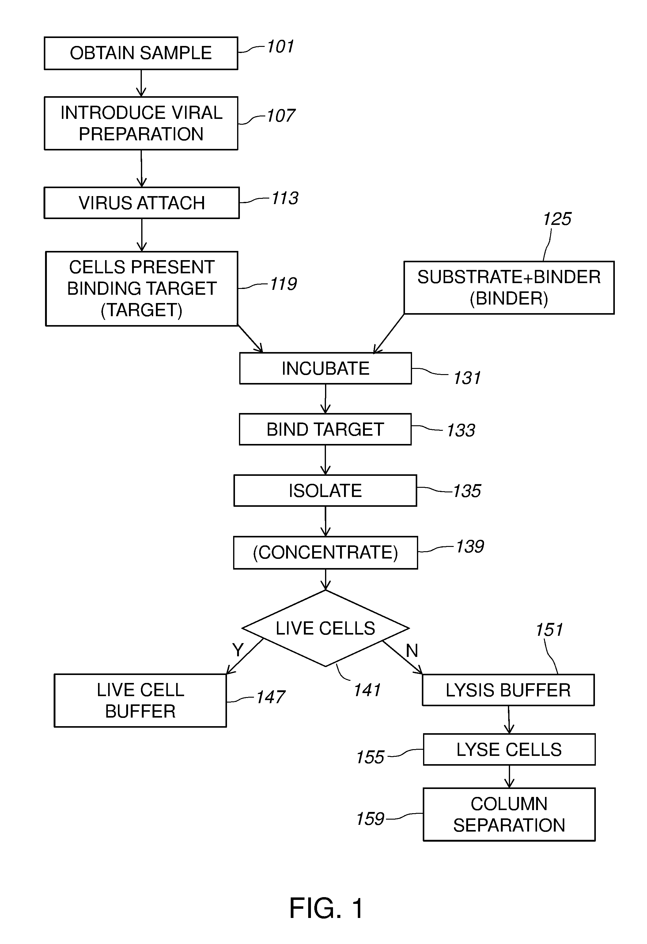
[0014] FIG. 1 gives a diagram of methods for isolating different pathogens.
[0015] FIG. 2 illustrates inducing the expression of a common antigen by a viral preparation.
[0016] FIG. 3 shows using a viral preparation to biotinylate different unknown pathogens.
[0017] FIG. 4 illustrates phage display of a common antigen by different unknown pathogens.
[0018] FIG. 5 illustrates a fluidic cartridge according to certain embodiments.
[0019] FIG. 6 gives a perspective view of the cartridge shown in FIG. 5.
[0020] FIG. 7 gives a schematic diagram of the cartridge shown of FIG. 5.
DETAILED DESCRIPTION
[0021] The invention generally relates to methods and devices for extracting one or more targets from a sample by causing targets in the sample to present a common binding element. The target can then be isolated using a binding partner that specifically binds to the binding element; such as an antibody binding to an antigen or streptavidin binding to biotin, and others known in the art. In one embodiment, the binding element is attached to a substrate, enabling the sample to be washed, leaving behind the target to be detected. In a preferred embodiment, the targets are pathogens or environmental hazards.
[0022] Methods of the invention can involve using a magnetic particle as a substrate attached to a binding partner, thereby forming a target/magnetic particle complex. Methods of the invention can further involve applying a magnetic field to capture the target/magnetic complex on a surface and wash away the sample. The invention further provides devices for extracting the pathogens. Devices of the invention can involve fluidic channels and chambers having macrofluidic dimension, microfluidic dimensions, or both.
[0023] FIG. 1 provides an exemplary diagram of certain methods for isolating different pathogens. A sample that contains two or more different pathogens is obtained 101. Methods of the invention may be used to isolate or extract different unknown pathogens from any heterogeneous sample. For example, the sample may include a bodily tissue or fluid. In particular embodiments, the sample is a bodily fluid. A bodily fluid refers to a liquid material derived from, for example, a human or other mammal. Such bodily fluids include, without limit, mucus, blood, plasma, serum, serum derivatives, bile, phlegm, saliva, sweat, amniotic fluid, mammary fluid, urine, sputum, and cerebrospinal fluid (CSF), such as lumbar or ventricular CSF. A bodily fluid may also be a fine needle aspirate. A bodily fluid may also be media containing cells or biological material.
[0024] Methods of the invention are suitable for use with any type of sample including one or more different pathogens including unknown pathogens. The target pathogen refers to the substance in the sample that will be captured and isolated by methods of the invention. Other targets may be captured by methods and devices of the invention. The target may be bacteria, fungi, a protein, a cell (such as a cancer cell, a white blood cell, a virally infected cell, or a circulating fetal cell), a virus, a nucleic acid (e.g., DNA or RNA), a receptor, a ligand, a hormone, a drug, a chemical substance, or any molecule known in the art.
[0025] In certain aspects, the target pathogen is a bacterium or two or more different bacteria. Methods of devices of the invention can be used to isolate or extract known bacteria, unknown bacteria, or a combination thereof. Both gram positive bacteria, gram negative bacteria, archaea, or eukaryotes can be isolated using the methods disclosed herein. Specific genera of pathogens that may be sought and assayed for using the disclosed methods include Alphaproteobacteria, Bacillus, Betaproteobacteria, Bifidobacterium, Borrelia, Campylobacter, Candida, Citrobacter, Clostridium, Enterobacter, Enterococcus, Escherichia, Flavobacterium, Fusobacterium, Gammaproteobacteria, Klebsiella, Kluyvera, Lactobacillus, Legionella, Leuconostoc, Listeria, Micrococcus, Mycobacterium, Neisseriaceae, Pediococcus, Pneumococcus, Porphyromonas, Prevotella, Propionibacterium, Proteus, Rhodospirillum, Rickettsia, Saccharomyces, Salmonella, Serratia, Shigella, Sphaerotilus, Staphylococcus, Streptococcus, Thermoanaerobacter, Thermoproteus, Vibrio, and Yersinia.
[0026] Organisms that can be assayed for with methods and devices of the invention include Acinetobacter calcoaceticus, Aeromonas hydrophilia, Bacillus anthracis, Bacillus subtilis, Candida albicans, Citrobacter freundii, E. coli, Enterobacter aerogenes, Enterobacter cloacae, Enterococcus faecium, Fusobacterium nucleatum, Klebsiella oxytoca, Klebsiella pneumoniae, Klebsiella travesanii, Kluyvera ascorbata, Lactobacillus casei, Lactobacillus cellobiosus, Lactobacillus fermentum, Lactobacillus helveticus, Lactobacillus paracasei, Lactobacillus salivarius, Listeria monocytogenes, Micrococcus luteus, Pediococcus parvulus, Porphyromonas gingivalis, Prevotella intermedia, Propionibacterium freudenreichii, Proteus vulgaris, Pseudomonas aeruginosa, Rhodospirillum rubrum, Rickettsia conorii, Saccharomyces cerevsiae, Salmonella agona, Salmonella enteritidis, Salmonella heidelberg, Salmonella infantis, Salmonella minnesota, Salmonella montevideo, Salmonella ohio, Salmonella typhimurium, Serratia marcescens, Shigella flexneri, Sphaerotilus natans, Staphylococcus aureus, Staphylococcus epidermis, Staphylococcus xylosus, Streptococcus faecalis, Streptococcus pyrogenes, Thermoproteus tenax, Vibrio cholerae, and Yersinia pseudotuberculosis. Any combination of organisms may be the target of methods and devices of the invention and the target of an assay will vary depending on the suspected pathogen or pathogens to be isolated.
[0027] In particular embodiments, the sample is a blood sample. Methods of the invention allow for different unknown pathogens in a blood sample to be isolated and detected at a level as low as or even lower than 1 CFU/ml. Blood may be collected in a container, such as the blood collection tube sold under the trademark VACUTAINER by BD (Franklin Lakes, N.J.). In certain embodiments, a solution is added that prevents or reduces aggregation of endogenous aggregating factors, such as heparin in the case of blood. A particular advantage of the methods described herein is the ability to capture and isolate different unknown pathogens directly from blood samples at low concentrations that are characteristic of clinical samples (as low as 1 CFU/ml of bacteria in a blood sample).
[0028] As shown in FIG. 1, once the sample is obtained 101, a viral preparation is introduced 107 into the sample. The viral preparation includes at least one virus that attaches 113 to the pathogens. As a result, the pathogens present 119 a binding element.
[0029] Any viral preparation may be used that causes different unknown pathogens to present a binding element. In some embodiments, the viral preparation contains a cocktail of bacteriophage. The cocktail contains different phage viruses that infect different bacteria. In certain embodiments, a broad-host-range phage is used that can infect different bacteria.
[0030] Phages are viruses that can be used as a transduction vector to introduce genetic material into the different pathogens and induce expression of, for example, a common antigen. Phage can be used as transduction vectors to introduce exogenous (non-viral) genetic material into the pathogens. See U.S. Pat. No. 7,148,054 for discussion. During lytic infection with a phage, DNA is packaged into phage heads as phage particles are formed. Promiscuous high-transducing (HT) mutants of P22 which efficiently package DNA with little sequence specificity are known.
[0031] As diagrammed in FIG. 1, infection involves attachment 113 of the phage particle to a target cell. When transduction is successful, the exogenous DNA is received in the target cell and expressed 119. For example, RecA-mediated homologous recombination following injection of the donor fragment can result in the inheritance of donor traits.
[0032] Any suitable phage or combination of phages can be used in methods of the invention. Examples of transducing phage vectors include bacteriophages P1 and P22 of E. coli and S. typhimurium, respectively. Other phages are suitable for use with methods of the invention. For example, the bacteriophage Mu can be used to insert DNA into a target pathogen genome for expression 119. See Groisman, 1991, In vivo genetic engineering with bacteriophage Mu, Methods in Enzymology 202:180-212. Further, hybrid phages such as Mud-P22 may be used. Mud-P22 is a hybrid combining features of phage Mu and P22. Mud-P22 can be inserted at essentially any desired site on the Salmonella chromosome. See Crain, 2007, Mud-P22, Methods Enzymology 421:249-59. Other exemplary phages (and their targets) include T4 (E. coli), T5 (E. coli), .lamda. phage (E. coli), T7 phage (E. coli), G4 (E. coli), P1 (E. coli), .PHI.6 (Pseudomonas), Thermoproteus tenax virus 1 (Thermoproteus tenax), M13 (E. coli), MS2 (E. coli), Q.beta. (E. coli), .PHI.X174 (E. coli), 129 (Bacillus), PZA (Bacillus), 115 (Bacillus), BS32 (Bacillus), B103 (Bacillus), M2Y (M2) (Bacillus), Nf (Bacillus), GA-1 (Bacillus), FWLBc1 (Bacillus), FWLBc2 (Bacillus), FWLLm3 (Listeria), B4 (Propionibacterium).
[0033] Phages are discussed in Chopin, et al., 2002, J Bact 184(7):2030-2033; Muramatsu et al., 1991, Two generalized transducing phages in Vibrio parahaemolyticus and Vibrio alginolyticus, Microbiol Immunol 35(12): 1073-1084; Regue et al., 1991, A generalized transducing bacteriophage for Serratia marcescens, Res Microbiol 42(1):23-27; Kiesel et al., 1993, Phage Acm1-mediated transduction in the facultatively methanol-utilizing Acetobacter methanolicus MB 58/4, J. Gen Virol 74(9):1741-1745; Zhang, et al., 2012, Food Microbiol 31(1):133-36; U.S. Pat. No. 7,732,150; U.S. Pub. 2009/0246752; U.S. Pub. 2009/0047254; and U.S. Pub. 2004/0156831, the contents of each of which are incorporated by reference.
[0034] Exemplary phages are further discussed in Welker, 1988, Transduction in Bacillus stearothermophilus, J. Bacteriol, 176(11):3354-3359; Darzins et al., 1989, Mini-D3112 bacteriophage transposable elements for genetic analysis of Pseudomonas aeruginosa, J. Bacteriol 171(7):3909-3916; Blahova et al., 1994, Transduction or imipenem resistance by the phage F-116 from a nosocomial strain of Pseudomonas aeruginosa isolated in Slovakia, Acta Virol 38(5):247-250; Weiss et al., 1994, Isolation and characterization of a generalized transducing phage for Xanthomonas campestris pv. campestris, J. Bacteriol 176(11): 3354-3359; Schicklmaier et al., 1995, Frequency of generalized transducing phages in natural isolates of the Salmonella typhimurium complex, Appl Environ Microbiol 61(4): 61(4): 1637-1640; Humphrey et al., 1997, Purification and characterization of VSH-1, a generalized transducing bacteriophage of Serpulina hyodysenteriae, J Bacteriol 179(2):323-329; Willi et a., 1997, Transduction of antibiotic resistance markers among Actinobacillus actinomycetemcomitans strains by temperate bacteriophages Aa phi 23, Cell Mol Life Sci 53(11-12):904-910; Nedelmann et al., 1998, Generalized transduction for genetic linkage analysis and transfer of transposon insertions in different Staphylococcus epidermidis strains, Zentiviralalbl Bakteriol 287(1-2):85-92; Int. Pat. Application Pub. WO 2003/035889; U.S. Pat. No. 8,071,337 and U.S. Pat. No. 7,951,579.
[0035] Any suitable phage virus or viruses, including any of those mentioned herein, can be included in the viral preparation. A viral preparation according to the invention is a composition containing a vector or viral material with the ability to interact with one or more different targets. A broad-host-range can be provided by including a cocktail of phages (e.g., two or more of any phage such as those mentioned herein), a phage that infects a range of hosts, or a combination thereof.
[0036] Any suitable broad-host-range phage can be used. For example, phage SN-1, SN-2, SN-X, SN-T, BHR1, BHR2, BHR3, BHR4, BHR5, PRD1, KVP40, PY100, PRD1, PVP-SE1, or a combination thereof may be included in a viral preparation. Broad host range phages are discussed in Miller et al., 2003, Complete genome sequence of the broad-host-range vibriophage KVP40: comparative genomics of a T4-related bacteriophage; J Bact 185(17):5220-5233; Beumer, 2005, A broad-host-range, generalized transducing phage SN-T acquires 16S rRNA genes from different genera of bacteria, Appl Env Microb 71(12):8301-8304; Green et al., 1985, Isolation and preliminary characterization of lytic and lysogenic phages with wide host range within the streptomycetes, J. Gen Microbiol 131(9):2459-2465; Jensen et al., 1998, Prevalence of broad-host-range lytic bacteriophages of Sphaerotilus natans, Escherichia coli, and Pseudomonas aeruginosa, Appl Environ Microbiol 64(2):575-580; Bamford et al., 1995, Bacteriophage PRD1: a broad host range dsDNA tectivirus with an internal membrane, Adv Virus Res 45:281-319; Schwudke, et al., 2008, Broad-host-range Yersinia phage PY100: genome sequence, proteome analysis of virions, and DNA packaging strategy, J Bact 190(1):332-342; Olsen et al., 1974, Characteristics of PRD1, a plasmid-dependent broad host range DNA bacteriophage, J Viriol 14(3):689-699; Santos et al., 2011, Genomic and proteomic characterization of the broad host range Salmonella phage PVP-SE1: creation of a new phage genus, J Viriol 85(21):11265-73; Sillankorva et al., Efficacy of a broad host range lytic bacteriophage against E. coli adhered to urothelium, Curr Microbiol 62(4):1128-1132; Evans et al, 2010, Characterization of a broad-host-range flagellum-dependent phage that mediates high-efficiency generalized transduction in, and between, Serratia and Pantoea, Microbiol 156:240-247; Garbe et al., 2010, Characterization of JG024, a Pseudomonas aeruginosa PB1-like broad-host range phage under simulated infection conditions, BMC Microbiol 10:301; Schwarzer et al., 2012, A multivalent adsorption apparatus explains the broad host range of phage phi92: a comprehensive genomic and structural analysis, J Viriol JVI.00801-12; U.S. Pub. 2012/0168372; U.S. Pub. 2012/0128652; and U.S. Pub. 2011/0064699, the contents of which are incorporated by reference.
[0037] A broad-host-range viral preparation can be provided by including more than one phage in the composition. For example, if each included phage is specific for one species, the phage cocktail will have the ability to infect multiple species. Any number of different phages (e.g., 3, 5, tens, hundreds) may be included in a phage cocktail. Those phages may themselves be narrow or broad in host range. Phage cocktails are discussed in Kelly et al., 2011, Development of a broad-host-range phage cocktail for biocontrol, Bioengineered Bugs 2:1, 31-37; Int. Application Pub. WO 02/07742; U.S. Pat. No. 6,121,036; U.S. Pub. 2009/0047254; and U.S. Pub. 2005/0032036, the contents of which are hereby incorporated by reference.
[0038] Other vectors may be used to cause different unknown pathogens to present a common binding element. Suitable vectors include plasmids, phagemids, and bacterial ghosts. In some embodiments, a plasmid is included, optionally enveloped in a lipid layer (e.g., a liposome, micelle, or reverse micelle), bi-layer, or a protein coat. Suitable plasmids are known in the art and include Col E1, RSF1030, clo DF13, R6K, F, R1, and Ent P 307.
[0039] In certain embodiments, one or more of the pathogens are targeted using a bacterial ghost. Bacterial ghosts are bacterial cell envelopes devoid of cytoplasmic content. A bacterial ghost can be included that exhibits one or more cell surface moieties that bind to any number of cell-surface targets on one or a range of bacteria. Further, the bacterial ghosts may each include the known common binding element (e.g., common antigen, biotinylated protein, streptavidin, etc.). Bacterial ghosts may be preferred since they can carry multiple surface moieties due to their size and are non-pathogenic themselves due to inactivity and can be stored (e.g., on-chip) for long periods of time. Bacterial ghosts are discussed in Lubitz et al., 2009, Applications of bacterial ghosts in biomedicine, Adv Exp Med Biol 655:159-70; Langemann et al., 2010, The bacterial ghost platform system: production and applications, Bioeng Bugs 1(5):326-36; Tabrizi et al., 2004, Bacterial ghosts-biological particles as delivery systems for antigens, nucleic acids, and drugs, Curr Opp Biotechnol 15:530-537; U.S. Pat. No. 7,067,639; U.S. Pat. No. 6,896,887; U.S. Pub. 2012/0040829; and U.S. Pub. 2010/0203082, the contents of which are incorporated by reference for all purposes.
[0040] Preferably, the viral preparation is introduced 107 with a high concentration of phage or vector so that viruses attach 113 to all target pathogens. Viral preparations can further include additional components to facilitate transduction by, for example, contributing to the competency of the target cells to receive extrinsic genetic material or to inhibit infection or lysis by sources other than the phage vector or vectors used. Preparations may include one or more salt, buffer, chelator, ion, or combination thereof, in any suitable concentration. Infectious, non-transducing phage can be inhibited by ion chelators (e.g., citrate or EGTA). In some embodiments, bacterial nutrients are added to the viral preparation to encourage cell vitality. For example, trypticase soy or a sugar can be included.
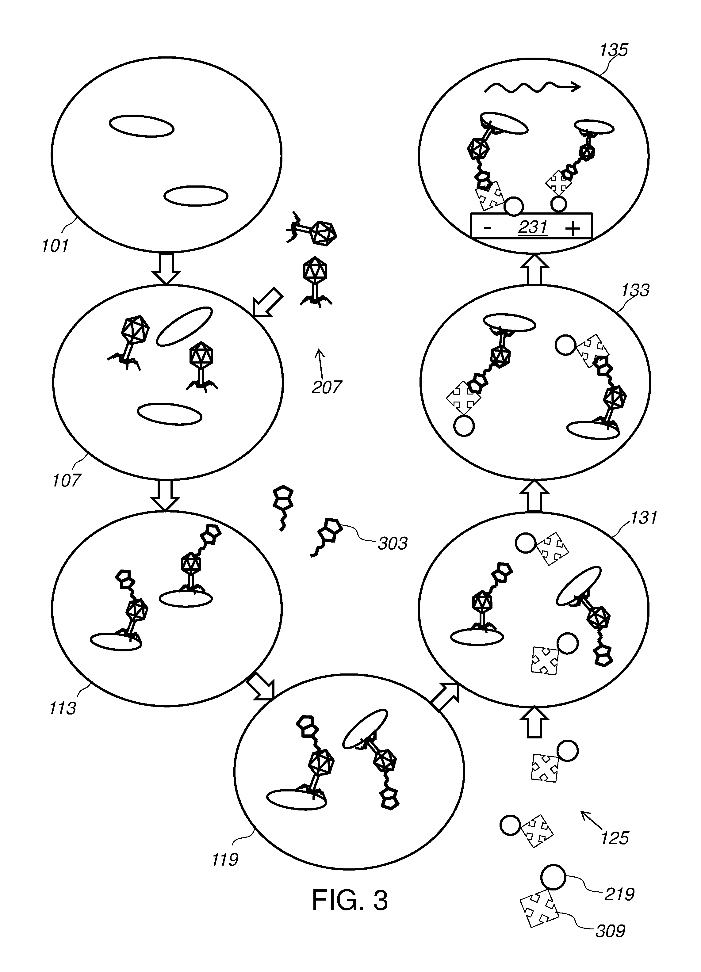
[0041] As shown in FIG. 1, a binding partner, optionally bound to a substrate, is obtained 125 and introduced into the sample. The sample with the binding partner is incubated 131 so that the binding partner can bind 133 to the binding element on the pathogens. In some embodiments, the virus or vector is used to cause different pathogens to present different binding elements. The pathogens may then be extracted using different binding partners. For example, phages can be used that cause some bacteria to express antigen A, some to express antigen B, and so on. Substrate 219, such as magnetic beads, can then be used that include anti-A, anti-B, etc., antibodies bound to the surface. The different binding partners may be mixed on each substrate, or separated (e.g., some beads specific for antigen A and some for antigen B). This can facilitate downstream sorting (e.g., where magnetic meads have different magnetic strengths or other sortable properties). Once the pathogens are bound to the binding partner, the pathogens can be isolated 135 from the remainder of the sample. Methods are provided to optionally concentrate 139 the isolated pathogens. Isolation and concentration are discussed below.
[0042] The isolated pathogens may be handled according to whether isolated live cells or another product is intended 141. For live cells, the pathogens may be introduced 147 into a live cell buffer. Any other suitable detection or isolation may be performed. For example, where a product from the cells is desired, a lysis buffer may be introduced 151 to lyse 155 the cells. The lysate may be purified, extracted, separated, or similar. In some embodiments, DNA is extracted by separation 159 on a column.
[0043] Methods of the invention include using a viral preparation to cause the target pathogens to present 119 a binding element. Any suitable method of associating a pathogen with a binding element may be used with the invention. Suitable methods include expression of a cell-surface protein, phage display of a protein, or any other method of exposing a protein. A protein can be a binding element such as antigen or a protein can be further functionalized to present a binding element. For example, a protein can include a target for biotinylation and can be biotinylated such that biotin is a binding element and streptavidin can be used as a binding partner.
[0044] FIG. 2 illustrates the use of a viral preparation 207 to induce the expression of a common antigen 211 according to certain embodiments. As shown in FIG. 2, a sample containing unknown pathogens 201 is obtained 101 and viral preparation 207 is introduced 107. Viruses attach 113 to pathogens 201 and introduce exogenous DNA into those cells.
[0045] Phage viruses, such as any suitable one of those discussed herein, can be engineered to contain a gene for a common antigen 211 such as a cell surface protein. The phage or phages from viral preparation 207 inject the gene into pathogens 201. Pathogens 201 then express the gene causing them to present 119 antigen 211 on their cell surface. Engineering a gene into a phage vector is discussed in Green and Sambrook, 2012, Molecular Cloning: A Laboratory Manual 4Ed, Cold Spring Harbor Laboratory Press (Cold Spring Harbor, N.Y.), 2,000 pages and Primrose and Twyman, 2006, Principles of Gene Manipulation and Genomics, 7th Edition, Wiley-Blackwell (Malden, Mass.) 672 pages. Induction of antigen expression is discussed in Prisco and Berardinis, 2012, Filamentous bacteriophage Fd as an antigen delivery system in vaccination, Int J. Mol. Sci. 13:5179-5194 and Gahan et al., 2009, Bacterial antigen expression is an important component in inducing an immune response to orally administered Salmonella-delivered DNA vaccines, PLoS One 4(6):e6062.
[0046] Antibody 221 may be obtained for use as the binding partner 125. As shown in FIG. 2, antibody 221 is bound to substrate 219. Substrate 219 can be any suitable substrate known in the art. In some embodiments, substrate 219 is the surface of a fluidic channel or other vessel. In some embodiments, substrate 219 is a biomolecule, such as a oligonucleotide, a protein, an aptamer, biotin, or some other such moiety that itself, in turn, has affinity for another material. For example, substrate 219 can be a DNA or RNA sequence, optionally with one or more locked nucleic acid (LNA) bases therein, and can be captured by exposure to a complementary sequence, such as a primer or oligo fixed on a glass slide or within a well in a microtitre plate. In certain embodiments, substrate 219 is a magnetic bead.
[0047] With continued reference to FIG. 2, the binding partner (e.g., antibody 221) is introduced into the vessel containing cells 201 expressing cell-surface antigen 211 and incubated 131. Incubation of antibody-bead complexes with cells 201 leads to binding 133 of substrate 219 to cells 201. Cells 201 can then be isolated 135 from the sample. Where substrate 219 is a surface of a vessel or channel, or a surface of beads packed into a column or otherwise held in place, cells 201 can be isolated 135 by using a wash solution to wash away excess sample. Where substrate 219 is a magnetic bead, a magnet 231 can then be used to isolate 135 cells 201 from the remainder of the sample.
[0048] Magnetic materials may be bound to target entities and used to separate such entities through the use of magnet fields and gradients. Magnetic materials and separations are known in the art and have been previously described, for example, in Murphy, 2011, Janeway's Immunobiology 8 Ed, Garland Science (New York, N.Y.), 888 pages, the contents of which are incorporated by reference herein. Methods of producing suitable magnetic particles are known in the art. See for example U.S. Pat. No. 5,597,531; U.S. Pat. No. 4,230,685; U.S. Pat. No. 4,677,055; U.S. Pat. No. 4,695,393; U.S. Pat. No. 5,695,946; U.S. Pat. No. 4,018,886; U.S. Pat. No. 4,267,234; U.S. Pat. No. 4,452,773; U.S. Pat. No. 4,554,088; U.S. Pat. No. 4,659,678; U.S. Pat. No. 5,186,827; U.S. Pat. No. 4,795,698, the contents of each of which are incorporated by reference.
[0049] Any type of magnetic particle may be used for substrate 219 in accordance with the invention. Methods of the invention include use of diamagnetic materials, paramagnetic materials, superparamagnetic materials, ferromagnetic materials, or a combination thereof (independently or in combination). Diamagnetic materials are slightly repelled by a magnetic field and the material does not retain the magnetic properties when the external field is removed. Paramagnetic materials (e.g., aluminum or platinum) are slightly attracted by a magnetic field and the material does not retain the magnetic properties when the external field is removed. Superparamagnetic materials are much more susceptible to magnetization than paramagnetic materials. See Gittleman et al., 1974, Superparamagnetism and relaxation effects in granular Ni--SiO.sub.2 and Ni--Al.sub.2O.sub.3 films, Phys Rev B 9:3891-3897. Ferromagnetic materials (e.g., iron or nickel) exhibit a strong attraction to magnetic fields and are able to retain their magnetic properties after the external field has been removed.
[0050] Further, magnetic properties of substrate 219 may depend on the size of the particles of substrate. Some ferromagnetic materials, e.g., magnetic iron oxide, may be characterized as superparamagnetic when provided in crystals of about 30 nm or less in diameter. Larger crystals of ferromagnetic materials, by contrast, retain permanent magnet characteristics after exposure to a magnetic field and tend to aggregate thereafter due to strong particle-particle interaction. In certain embodiments, the particles are superparamagnetic particles. In certain embodiments, the magnetic particle is an iron containing magnetic particle. In other embodiments, the magnetic particle includes iron oxide or iron platinum.
[0051] In some embodiments, the magnetic particles for substrate 219 include a portion of superparamagnetic particles by weight (e.g., 20%, 40%, 60%, or 80%). Particles 219 can have a diameter between about 100 nm and about 1,000 nm. In a particular embodiment, substrate 219 includes superparamagnetic particles of diameter between about 100 nm and about 250 nm. See discussion in U.S. Pub. 2011/0262989, the contents of which are incorporated by reference.
[0052] In certain embodiments, the particles are particles (e.g., nanoparticles) that incorporate magnetic materials, or magnetic materials that have been functionalized, or other configurations as are known in the art. In certain embodiments, nanoparticles may be used that include a polymer material that incorporates magnetic material(s), such as nanometal material(s). When those nanometal material(s) or crystal(s), such as Fe.sub.3O.sub.4, are superparamagnetic, they may provide advantageous properties, such as being capable of being magnetized by an external magnetic field, and demagnetized when the external magnetic field has been removed. This may be advantageous for facilitating sample transport into and away from an area where the sample is being processed without undue particle aggregation.
[0053] One or more or many different nanometal(s) may be employed, such as Fe.sub.3O.sub.4, FePt, or Fe, in a core-shell configuration to provide stability, and/or various others as may be known in the art. In many applications, it may be advantageous to have a nanometal having as high a saturated moment per volume as possible, as this may maximize gradient related forces, and/or may enhance a signal associated with the presence of the particles. It may also be advantageous to have the volumetric loading in a particle be as high as possible, for the same or similar reason(s). In order to maximize the moment provided by a magnetizable nanometal, a certain saturation field may be provided. For example, for Fe.sub.3O.sub.4 superparamagnetic particles, this field may be on the order of about 0.3 tesla (T).
[0054] The size of the nanometal-containing particle may be optimized for a particular application, for example, maximizing moment loaded upon a target, maximizing the number of particles 219 on a target 201 with an acceptable detectability, maximizing desired force-induced motion, and/or maximizing the difference in attached moment between the labeled target and non-specifically bound targets or particle aggregates or individual particles. While maximizing is referenced by example above, other optimizations or alterations are contemplated, such as minimizing or otherwise desirably affecting conditions.
[0055] In an exemplary embodiment, a polymer particle containing 80 wt % Fe.sub.3O.sub.4 superparamagnetic particles, or for example, 90 wt % or higher superparamagnetic particles, is produced by encapsulating superparamagnetic particles with a polymer coating to produce a particle having a diameter of about 250 nm.
[0056] Each set of magnetic particles 219 has a binding partner 221 that allows for each set to specifically bind 133 the target 201 of interest in the sample. The binding partner may be any molecule known in the art and will depend on the target to be captured and isolated. Exemplary binding partners include nucleic acids, proteins, ligands, antibodies, aptamers, and receptors.
[0057] In particular embodiments, the binding partner 221 is an antibody, such as an antibody that binds a particular antigen 211 (see, e.g., FIG. 2). General methodologies for antibody production, including criteria to be considered when choosing an animal for the production of antisera, are described in Kontermann, 2010, Antibody Engineering Volume 1 2Ed, Springer-Verlag (Berlin Heidelberg) 800 pages, and Harlow, 1988, Antibodies: A Laboratory Manual, Cold Spring Harbor Laboratory Press (Cold Spring Harbor, N.Y.) 726 pages. For example, an animal of suitable size such as goats, dogs, sheep, mice, or camels are immunized by administration of an amount of immunogen, such the target bacteria, effective to produce an immune response. An exemplary protocol is as follows. The animal is injected with 100 milligrams of antigen resuspended in adjuvant, for example Freund's complete adjuvant, dependent on the size of the animal, followed three weeks later with a subcutaneous injection of 100 micrograms to 100 milligrams of immunogen with adjuvant. Additional doses are administered until a suitable titer of antibody in the animal's blood is achieved. Exemplary titers include a titer of at least about 1:5000 or a titer of 1:100,000 or more, i.e., the dilution having a detectable activity. The antibodies are purified, for example, by affinity purification on columns containing protein G resin or target-specific affinity resin.
[0058] The technique of in vitro immunization of human lymphocytes is used to generate monoclonal antibodies. Such techniques are known in the art. See, e.g., Mulder et al., 1993, Characterization of two human monoclonal antibodies reactive with HLA-B12 and HLA-B60, respectively, raised by in vitro secondary immunization of peripheral blood lymphocytes, Hum. Immunol 36(3):186-192; Stauber et al., 1993, Rapid generation of monoclonal antibody-secreting hybridomas against African horse sickness virus by in vitro immunization and the fusion/cloning technique, J Immunol Methods 161(2):157-168; and Venkateswaran, et al., 1992, Production of anti-fibroblast growth factor receptor monoclonal antibodies by in vitro immunization, Hybridoma, 11(6):729-739. These techniques can be used to produce antigen-reactive monoclonal antibodies, including antigen-specific IgG, and IgM monoclonal antibodies.
[0059] Any antibody or fragment thereof having affinity and specific for the target of interest may be used for binding partner 221 within the scope of the invention provided herein.
[0060] Methods for attaching the binding partner 221 to the magnetic particle 219, including the coating of particles with antibodies, are known in the art. See for example Kontermann, 2010, Antibody Engineering Volume 1 2Ed, Springer-Verlag (Berlin Heidelberg) 800 pages; Harlow, 1988, Antibodies: A Laboratory Manual, Cold Spring Harbor Laboratory Press (Cold Spring Harbor, N.Y.) 726 pages; and Stanley, 2002, Essentials in Immunology and Serology, Delmar Cengage (Independence, Ky.) 560 pages. Such methodology can easily be modified by one of skill in the art to bind other types of binding partners to the magnetic particles. In addition, certain types of magnetic particles coated with a functional moiety are commercially available from Sigma-Aldrich (St. Louis, Mo.).
[0061] In some embodiments, more than one antibody 221 is used to create sets of magnetic particles. Since each set of particles 219 may be conjugated with antibodies 221 having different specificities for one or more antigen 211 coded in the genome of the phage or phages, compositions and concentrations may be optimized for detection of multiple unknown pathogens in the sample. In certain embodiments, all of the sets are provided at the same concentration. Alternatively, the sets are provided at different concentrations. For example, compositions may be designed such that sets that bind gram positive bacteria (e.g., antibody 221 to antigen 211 in genome of phage that targets Gram+cells 201) are added to the sample at a concentration of 2.times.10.sup.9 particles per/ml, while sets that bind gram negative bacteria (e.g., antibody to antigen in genome of phage specific to Gram -) are added to the sample at a concentration of 4.times.10.sup.9 particles per/ml. Compositions used with methods of the invention are not affected by antibody cross-reactivity. However, in certain embodiments, sets are specifically designed such that there is no cross-reactivity between different antibodies and different sets.
[0062] To facilitate downstream detection of pathogens 201, one or more labels may be added to antibody 221, particle 219, or both. For example, a label may be added during preparation of antibody/bead complexes. Any detectable label may be used with compositions of the invention, such as fluorescent labels, radiolabels, enzymatic labels, and others. The detectable label may be directly or indirectly detectable. In certain embodiments, the exact label may be selected based, at least in part, on the particular type of detection method used. Exemplary detection methods include radioactive detection, optical absorbance detection (e.g., UV-visible absorbance detection, optical emission detection, e.g., fluorescence, phosphorescence, chemiluminescence), or Raman scattering. Preferred labels include optically-detectable labels, such as fluorescent labels. Examples of fluorescent labels include without limit acridine and derivatives; BODIPY; Brilliant Yellow; coumarin and derivatives; DAB ITC; eosin and derivatives; erythrosin and derivatives; isothiocyanate; ethidium; fluorescein and derivatives; FAM; DTAF; QFITC, (XRITC); Malachite Green isothiocyanate; Phenol Red; pyrene and derivatives; Reactive Red 4 (Brilliant Red 3B-A sold under the trademark CIBACRON); rhodamine and derivatives; Texas Red; TAMRA; TRITC; riboflavin; Atto dyes; La Jolta Blue; phthalo cyanine; and naphthalo cyanine. Preferred labels include cyanine-3 and cyanine-5. Labels other than fluorescent labels are contemplated by the invention, including other optically-detectable labels. Methods of linking fluorescent labels to magnetic particles or antibodies are known in the art. Suitable labels and methods of their use are described in U.S. Pub. 2011/0262926, the contents of which are hereby incorporated by reference.
[0063] As shown in FIG. 2, the magnetic particles described above are then introduced 125 to the sample in order to bind 133 to the unknown pathogens 201. For example, a blood sample may be mixed with the described magnetic particles to generate a mixture that is allowed to incubate 131 such that the compositions bind to at least one pathogen 201 in the blood sample. The mixture is allowed to incubate 131 for a sufficient time to allow for the composition to bind to the pathogen in the blood. The process of binding the composition to the pathogen associates a magnetic moment with the pathogen, and thus allows the pathogen to be manipulated through forces generated by magnetic fields upon the attached magnetic moment.
[0064] In general, time for incubation 131 will depend on the desired degree of binding between the pathogen and the compositions of the invention (e.g., the amount of moment that would be desirably attached to the pathogen), the amount of moment per target, the amount of time of mixing, the type of mixing, the reagents present to promote the binding and the binding chemistry system that is being employed. Incubation time can be anywhere from about 5 seconds to a few days. Exemplary incubation times range from about 10 seconds to about 2 hours. Binding occurs over a wide range of temperatures, generally between about 5.degree. C. and about 65.degree. C., e.g., between about 15.degree. C. and about 40.degree. C.
[0065] In certain embodiments, a buffer solution is added to the sample along with the compositions of the invention. An exemplary buffer includes Tris(hydroxymethyl)-aminomethane hydrochloride (Tris HCl) at a concentration of about 75 mM. It has been found that the buffer composition, mixing parameters (speed, type of mixing, such as rotation, shaking etc., and temperature) influence binding. It is important to maintain osmolality of the final solution (e.g., blood+buffer) to maintain high label efficiency. In certain embodiments, buffers used in methods of the invention are designed to prevent lysis of blood cells, facilitate efficient binding of targets with magnetic particles and to reduce formation of particle aggregates. It has been found that the buffer solution containing 300 mM NaCl, 75 mM Tris-HCl pH 8.0 and 0.1% Tween 20 meets these design goals.
[0066] Without being limited by any particular theory or mechanism of action, it is believed that sodium chloride is mainly responsible for maintaining osmolality of the solution and for the reduction of non-specific binding of magnetic particle through ionic interaction. Tris HCl is frequently used in biology to maintain pH of a solution. It has been found that 75 mM concentration is beneficial and sufficient for high binding efficiency. Likewise, Tween 20 is widely used as a mild detergent to decrease nonspecific attachment due to hydrophobic interactions. Various assays use Tween 20 at concentrations ranging from 0.01% to 1%. The 0.1% concentration appears to be optimal for the efficient labeling of bacteria, while maintaining the integrity of blood cells.
[0067] Additional compounds can be used to modulate the capture efficiency by blocking or reducing non-specific interaction with blood components and either magnetic particles or pathogens. For example, chelating compounds, such as EDTA or EGTA, can be used to prevent or minimize interactions that are sensitive to the presence of Ca.sup.2+ or Mg.sup.2+ ions.
[0068] One can also use a static mixer or other mixing device that provides efficient mixing of viscous samples at high flow rates to achieve high binding efficiency while reducing time required for the binding step. In one embodiment, the sample is mixed with binding buffer in ratio of, or about, 1:1, using a mixing interface connector. The diluted sample then flows through a mixing interface connector where it is mixed with target-specific nanoparticles. Additional mixing interface connectors providing mixing of sample and antigen-specific nanoparticles can be attached downstream to improve binding efficiency. The combined flow rate of the labeled sample is selected such that it is compatible with downstream processing.
[0069] With reference to FIG. 2, after binding 133 of particles 219 to pathogens 201 in the sample to form pathogen/magnetic particle complexes, a magnetic field 231 may be applied to the mixture to capture or isolate 135 the complexes on a surface. Components of the mixture that are not bound to magnetic particles will not be affected by the magnetic field and will remain free in the mixture. Methods and apparatuses for separating 135 target/magnetic particle complexes from other components of a mixture are known in the art. For example, a steel mesh may be coupled to a magnet, a linear channel or channels may be configured with adjacent magnets, or quadrapole magnets with annular flow may be used. Other methods and apparatuses for separating target/magnetic particle complexes from other components of a mixture are shown in U.S. Pat. No. 7,699,979; U.S. Pat. No. 6,551,843; U.S. Pat. No. 5,622,831; U.S. Pat. No. 6,514,415; U.S. Pat. No. 5,695,946; U.S. Pat. No. 5,186,827; U.S. Pat. No. 5,541,072; U.S. Pat. No. 5,466,574; and U.S. Pat. No. 6,623,983, the contents of each of which are incorporated by reference.
[0070] In certain embodiments, the magnetic capture is achieved at high efficiency by utilizing a flow-through capture cell with a number of strong rare earth bar magnets 231 placed perpendicular to the flow of the sample. When using a flow chamber with flow path cross-section 0.5 mm.times.20 mm (h.times.w) and 7 bar NdFeB magnets, the flow rate could be as high as 5 mL/min or more, while achieving capture efficiency close to 100%.
[0071] The foregoing illustrative embodiment is described generally in terms of the induction of the expression of a common antigen 211 by one or more different unknown pathogen 211, as shown in FIG. 2. The invention generally provides methods of using a vector or viral preparation 207 to cause unknown pathogens 201 to present a common binding element.
[0072] FIG. 3, for example, illustrates an alternative embodiment of a method of using a viral preparation 207 to cause different unknown pathogens to present 119 a common binding element 303. As shown in FIG. 3, viral preparation 207 includes at least one phage.
[0073] In certain embodiments, a bacteriophage is used that expresses a biotin binding domain (aka, a biotinylation peptide) on at least one capsid protein. The phage contains the genetic information to display a biotinylation domain and be biotinylated. In some embodiments, the phage displays a biotinylation domain when introduced 107 into the sample. When the phage attach to cells 201, those cells are then biotinylated. In certain embodiments, the phage infects the target cells and progeny virions produced from the infection will have been biotinylated by the target cells' 201 biotin ligase protein (BLP). Biotinylation of phage with biotinylation peptides on their capsids is discussed in Edgar et al., 2006, High-sensitivity bacterial detection using biotin-tagged phage and quantum-dot nanocomplexes, PNAS 103(13):4841-4945 and Gervias et al., 2007, Immobilization of biotinylated bacteriophages on biosensor surfaces, Sensors and Actuators B 125:615-621, each of which are incorporated by reference. Methods of labeling targets, e.g., including biotinylation of phage, are discussed in U.S. Pat. No. 7,517,643; U.S. Pat. No. 6,342,588; and U.S. Pub. 2011/0086338, the contents and methods of each are incorporated by reference in their entirety for all purposes. In some embodiments, phage capsid is streptavidin or avidin-linked and substrate 219 is biotinylated, as discussed in U.S. Pat. No. 6,740,492, the contents of each are incorporated by reference in their entirety for all purposes.
[0074] With continued reference to FIG. 3, cells 201 present 119 biotin 303 as a common binding element. Binding partner 125 is introduced. Here, binding partner 125 includes streptavidin 309 coated beads 219. Biotinylation of phage is further discussed in Smelyanski and Gershoni, 2011, Site directed biotinylation of filamentous phage structural proteins, Virol J 8:495; U.S. Pat. No. 5,994,519; and U.S. Pub. 2001/0019820, the contents and methods of each are incorporated by reference in their entirety for all purposes.
[0075] Biotinylated target cells 201 are incubated 131 with streptavidin-coated beads 219. The binding partner binds 133 to the target, after which the target 201 can be isolated 135. For example, where beads 219 are magnetic, a magnet 231 can be used to hold cells 201 in a fluidic channel while the sample is washed away. With reference back to FIG. 1, where live cells are intended 139, the sample can be replaced with a maintenance buffer 147. In an alternative embodiment, cells 201 can be lysed 155, optionally using a lysis buffer 151. In some embodiments, a component of the cells 201 is extracted. For example, DNA may optionally be extracted by column separation 159.
[0076] While discussed above in the illustrative embodiments involving biotinylated phage, streptavidin-linked phage, or expression of a common antigen after transduction by a phage, methods of the invention include any suitable method or phenomenon to cause different unknown pathogens 201 to exhibit a common binding element through the introduction of a viral preparation 207. In some embodiments, phage display of an antigen is employed.
[0077] FIG. 4 illustrates phage display of a common antigen by different unknown pathogens 201. In phage display, a gene for antigen 411 is ligated into the phage genome, for example, within the gene encoding one of the coat proteins, or capsid proteins. Multiple cloning sites may be used to ensure in-frame cloning and proper expression of the protein product. The viral preparation 207 including one or more phage engineered for phage display of a common antigen 411 is then introduced 107 into the sample containing unknown pathogens 201. A binding partner is used (e.g., linked to a substrate 219 such as a well in a microtitre plate, a surface of a fluidic channel, beads in a column, or magnetic beads) that binds to antigen 411. After the phage attach 119 to pathogens 201, they display antigen 411. Phage display is discussed in Haq, 2012, Bacteriophages and their implications on future biotechnology, a review, Virol J 9:9; U.S. Pat. No. 8,227,242 (e.g., phage display without helper phage); U.S. Pat. No. 8,216,797; U.S. Pat. No. 7,238,669; U.S. Pat. No. 6,740,492; U.S. Pub. 2010/0240579 (detection of unknown target by phage display); and U.S. Pub. 2006/0063149, the contents of which are incorporated by reference.
[0078] In some embodiments, phage display antigen 411 when introduced 107 into the sample. In certain embodiments, the genetically-engineered phage is introduced and infects pathogens 201 and generates progeny virions that express antigen 411. Viral preparation 207 may include a bacterial growth medium to allow low concentrations (e.g., single cells) of target to grow and be infected and propagate the phage. Moreover, a combination of the foregoing may occur in that phage may exhibit antigen 411 upon introduction 107 into the sample, and may infect and propagate, generating progeny.
[0079] While any suitable substrate 219 may be used, and any suitable binder (e.g., aptamers, co-factors, proteins, etc.), in certain embodiments, magnetic beads linked to antibody 221 are used. This binding partner 125 is introduced into the infected sample and allowed to incubate 131.
[0080] FIG. 3 shows that incubation 131 leads to the binding 133 of binding partner 125 to antigen 411 on cells 201. After cells 201 are bound to magnetic beads 219 via antibody 221/antigen 411 interaction, the cells 201 may be isolated 135 from the sample by magnetic separation. Further, additional steps may be performed to optimize the isolation or extraction of cells 201 from the sample. Additional steps optionally include washing with one or more solutions, further concentrating 139 captured cells (e.g., using a magnetic concentrator), introduction of additional buffers such as, for example, cell maintenance buffers or lysis buffers, other suitable processes known in the art, or a combination thereof.
[0081] Other methods of using a binding partner to capture a target are operable with methods of the invention. For example, where pathogens 201 are intracellular parasites, a known binding element may be associated with the host cells. A binding partner may be used that would not be able to target an unknown pathogen directly but instead bind to the host (if the host is eukaryotic, instead of a phage viral preparation, a binding partner specific to a eukaryotic protein may be employed). See, e.g., Drancourt et al., 1992, Diagnosis of Mediterranean spotted fever by indirect immunofluorescence of Rickettsia conorii in circulating endothelial cells isolated with monoclonal antibody-coated immunomagnetic beads, J Inf Dis 166:660-3, incorporated by reference. Optionally, the membrane of the eukaryotic host can be ruptured (e.g., with detergent, a virus or particle, differential osmolality, etc.,) and the infectious agent targeted by methods herein. In some embodiments, methods of the invention may be used in combination with, or in sequence with, alternative methodologies that would only operate, for example, where one or more target is known. If a target is known, it may exhibit a known binding element that can be captured with a binding partner. See, e.g., Fu et al., Rapid detection of E. coli O157:H7 by immunomagnetic separation and real-time PCR, incorporated by reference.
[0082] The process of magnetic separation 135 described above (e.g., with respect to FIG. 3) produces efficient capture of different unknown pathogens 201 and the removal of all or majority of the remaining components in the mixture. However, it is still possible that a relatively small amount of non-specific analytes are unintentionally captured along with the target/magnetic particle complexes. It may be desired to remove these non-specific analytes. Accordingly, the surface may be washed with a wash solution that reduces particle aggregation, thereby isolating target/magnetic particle complexes from the non-specific target entities.
[0083] Any wash solution that does not disrupt interaction between the binding partner of the magnetic particle and the target may be used. The wash solution should also not disrupt the capture of the target/particle complexes on the intended surface. Exemplary solutions include heparin, Tris-HCl, phosphate buffered saline (PBS), Tris-borate-EDTA (TBE), Tris-acetate-EDTA (TAE), Tris-cacodylate, HEPES, or similar. One or more wash cycle may be performed. For embodiments in which the sample includes blood, heparin may be used to inhibit clotting. The bound targets are washed with heparin-containing buffer 1-3 times to remove blood components and to reduce formation of aggregates. Methods for these steps are described in U.S. Pat. No. 7,776,580; U.S. Pub. 2011/0263833; U.S. Pub. 2011/0262989; U.S. Pub. 2011/0262933; U.S. Pub. 2011/0262927; U.S. Pub. 2011/0262893; and U.S. Pub. 2010/0092956, the contents of which are incorporated by reference for all purposes.
[0084] Once the different unknown pathogens 201 have been isolated 135, they may be preserved as living cells or they may be lysed and the lysate, or a component thereof, may be detected, extracted, isolated, quantified, or used.
[0085] Where live cells are intended 141, a live cell buffer may be introduced 147, for example, in a step that includes flushing away any wash buffers, other solutions or reagents, or remaining components of the sample.
[0086] Once the target/magnetic particles have been captured, the target is then lysed 155. In certain embodiments, lysis of the target occurs without separating the particles from the target prior to the lysis step. Conducting the lysis without the pre-separation step allows more efficacious collection of analytes contained within the target. For instance, if the analyte of interest is a bacterially-derived nucleic acid, some analyte may be lost when bacteria separated from the magnetic particles are inadvertently lost. With the disclosed methods, the bacteria are still bound to the particles and captured on a surface as lysis occurs. Accordingly, there is a concentrated sample to work with during the lysis step. In addition, the disclosed methods allow for recovery of a specific analyte in a relatively small collection volume. This is especially useful when the analyte of interest is a nucleic acid or something that is similarly present in only very small quantities. For example, one could begin with a blood sample of 2 ml and use the described methods to concentrate a desired pathogen from the blood onto an appropriate surface. With the pathogen captured, one could decant the blood, add 0.3 ml of a suitable buffer, and subsequently perform the lysis step.
[0087] Lysis 155 can be performed using any means known in the art. For example, lysis could be performed using 151 a lysis buffer (see, e.g., FIG. 1). Any type of lysis buffer is suitable for use with the disclosed methods; selection of the specific buffer may depend on the subsequent analysis of the cell lysate. Buffer selection is within the general skill of the art and can be determined empirically. Generally, lysis buffers contain Tris-HCL, EDTA, EGTA, SDS, deoxycholate, Triton X, and/or NP-40. In some cases the buffer may also contain NaCl (e.g., 150 mM). In certain aspects, the lysis buffer is a chaotropic solution.
[0088] Lysis 155 can also be achieved through, or assisted by, sonication. In this method, the captured target/magnetic particle complexes are exposed to ultrasonic waves to achieve lysis of any target (bacteria, cells, virus, fungi, etc.) associated with the magnetic particles so that any analytes of interest contained therein are released. In some embodiments, the analyte of interest can include a nucleic acid.
[0089] The methods described herein can be used in accordance with any sonication device, which are well-known in the art. In certain embodiments, the sonication device is the VCX 750 Sonicator sold under the trademark VIBRA-CELL (sonicator, commercially available from Sonics and Materials, Inc.). Generally, the probe of the sonicator is placed into the liquid containing the targets to be lysed. Electrical energy from a power source is transmitted to a piezoelectric transducer within the sonicator converter, where it is changed to mechanical vibrations. The longitudinal vibrations from the converter are intensified by the probe, creating pressure waves in the liquid. These in turn produce microscopic bubbles, which expand during the negative pressure excursion and implode violently during the positive excursion. This phenomenon, referred to as cavitation, creates millions of shock waves and releases high levels of energy into the liquid, thereby lysing the target. In another embodiment, the sonication transducer may be brought in contact with a chamber holding captured complexes by way of a structural interface. The sonication transducer vibrates the structural interface such that lysis is achieved. In either method, the appropriate intensity and period of sonication can be determined empirically by those skilled in the art.
[0090] The lysate containing the contents of the lysed target can then be eluted. In certain aspects, the lysate contains nucleic acids of interest associated with a particular bacteria present in the starting sample. The lysate is removed from the magnetic particles and the analytes contained therein can be analyzed. Analytes may include, without limitation, nucleic acids, proteins, organelles, and other components found within the target of interest.
[0091] The analyte may be analyzed by a multitude of existing technologies, such as NMR, Polymerase Chain Reaction (PCR), sequencing, mass spectrometry, fluorescent labeling and visualization using microscopic observation, fluorescent in situ hybridization (FISH), growth-based antibiotic sensitivity tests, and variety of other methods that may be conducted with purified target without significant contamination from other sample components. Analysis using NMR is described in U.S. Pub. 2011/0262925, herein incorporated by reference in its entirety. In some embodiments, the different unknown pathogens 201 are analyzed in order to identify genes they express. For example, captured bacteria 201 are lysed without first separating the bacteria from the magnetic particles. The lysate is then eluted from the magnetic particles and DNA contained within the lysate/eluate is bound to DNA extraction resin. After washing of the resin, the bacterial DNA is eluted and used in quantitative RT-PCR to detect the presence of a specific species, and/or, subclasses of bacteria.
[0092] Detection of bacteria of interest can be performed by use of nucleic acid probes following procedures which are known in the art. Suitable procedures for detection of bacteria using nucleic acid probes are described in U.S. Pat. No. 7,943,346; U.S. Pat. No. 5,620,847; U.S. Pat. No. 5,569,586; U.S. Pat. No. 5,541,308; U.S. Pat. No. 5,401,631; U.S. Pat. No. 5,089,386; and U.S. Pat. No. 5,055,394, each of which is hereby incorporated by reference.
[0093] A suitable nucleic acid probe assay generally includes sample treatment and lysis, hybridization with selected probe(s), hybrid capture, and detection. Lysis 155 of the bacteria is necessary to release the nucleic acid for the probes. In accordance with the invention, a means for lysing the bacteria is introduced while the bacteria are still bound to the magnetic particle and the resulting complexes have been captured on a surface. There is no separation of the bacteria from the magnetic particles prior to lysis. In some embodiments, the nucleic acid target molecules are released by treatment with any of a number of lysis agents, including alkali (such as NaOH), guanidine salts (such as guanidine thiocyanate), enzymes (such as lysozyme, mutanolysin and proteinase K), and detergents. In other embodiments, sonication is used to lyse the cells. Lysis of the bacteria, therefore, releases both DNA and RNA, particularly ribosomal RNA and chromosomal DNA both of which can be utilized as the target molecules with appropriate selection of a suitable probe. Use of rRNA as the target molecule(s), may be advantageous because rRNAs constitute a significant component of cellular mass, thereby providing an abundance of target molecules. The use of rRNA probes also enhances specificity for the bacteria of interest, that is, positive detection without undesirable cross-reactivity which can lead to false positives or false detection.
[0094] Hybridization includes addition of the specific nucleic acid probes. In general, hybridization is the procedure by which two partially or completely complementary nucleic acids are combined, under defined reaction conditions, in an anti-parallel fashion to form specific and stable hydrogen bonds. The selection or stringency of the hybridization/reaction conditions is defined by the length and base composition of the probe/target duplex, as well as by the level and geometry of mis-pairing between the two nucleic acid strands. Stringency is also governed by such reaction parameters as temperature, types and concentrations of denaturing agents present and the type and concentration of ionic species present in the hybridization solution.
[0095] The hybridization phase of the nucleic acid probe assay is performed with a single selected probe or with a combination of two, three or more probes. Probes are selected having sequences which are homologous to unique nucleic acid sequences of the target organism. In general, a first capture probe is utilized to capture formed hybrid molecules. The hybrid molecule is then detected by use of antibody reaction or by use of a second detector probe which may be labeled with a radioisotope (such as phosphorus-32) or a fluorescent label (such as fluorescein) or chemiluminescent label.
[0096] Detection of bacteria of interest can also be performed by use of PCR techniques. A suitable PCR technique is described, for example, in Verhoef et al. (WO 92/08805). Bacterially-derived nucleic acids isolated from the lysate can be used as templates for the PCR reaction. PCR can also include the use of reverse-transcriptase PCR (RT-PCR), in which RNA isolated from the target is reverse transcribed into its DNA complement (i.e., cDNA) using the enzyme reverse transcriptase, and the resulting cDNA is amplified using PCR. RT-PCR is described in detail in U.S. Pub. 2011/0071033, incorporated by reference herein in its entirety.
[0097] In certain embodiments, differential gene expression associated with the target can also be identified, or confirmed using a microarray technique. In this method, polynucleotide sequences of interest (including cDNAs and oligonucleotides) are plated, or arrayed, on a microchip substrate. The arrayed sequences are then hybridized with specific DNA probes from cells or tissues of interest. Methods for making microarrays and determining gene product expression (e.g., RNA or protein) are shown in U.S. Pub. 2006/0195269, the content of which is incorporated by reference herein in its entirety.
[0098] In particular aspects, nucleic acids isolated from the target can be sequences to generate a plurality of sequence reads, thereby identifying a pathogen according to a known genetic sequence. Sequencing may be by any method known in the art. DNA sequencing techniques include classic dideoxy sequencing reactions (Sanger method) using labeled terminators or primers and gel separation in slab or capillary, sequencing by synthesis using reversibly terminated labeled nucleotides, pyrosequencing, 454 sequencing, Illumina/Solexa sequencing, allele specific hybridization to a library of labeled oligonucleotide probes, sequencing by synthesis using allele specific hybridization to a library of labeled clones that is followed by ligation, real time monitoring of the incorporation of labeled nucleotides during a polymerization step, polony sequencing, and SOLiD sequencing. Sequencing of separated molecules has more recently been demonstrated by sequential or single extension reactions using polymerases or ligases as well as by single or sequential differential hybridizations with libraries of probes. Methods of sequencing are discussed in U.S. Pat. No. 8,209,130 and U.S. Pub. 2011/0071033, the content of which is incorporated by reference herein in its entirety.
[0099] In certain embodiments, methods of the invention are useful for direct detection of unknown bacteria 201 from blood. Such a process is described here. Sample is collected in sodium heparin tube by venipuncture, acceptable sample volume is about 1 mL to 10 mL. Sample is diluted with binding buffer and superparamagnetic particles having binding partners are added to the sample, followed by incubation on a shaking incubator at 37.degree. C. for about 30 min to 120 min. Alternative mixing methods can also be used. In a particular embodiment, sample is pumped through a static mixer, such that reaction buffer and magnetic particles are added to the sample as the sample is pumped through the mixer. This process allows for efficient integration of all components into a single fluidic part, avoids moving parts and separate incubation vessels and reduces incubation time.
[0100] Capture of the labeled targets allows for the removal of blood components and reduction of sample volume from 30 mL to 5 mL. The capture is performed in a variety of magnet/flow configurations. In certain embodiments, methods include capture in a fluidic cartridge or other flow-through device at flow rate of 5 mL/min, resulting in total capture time of 6 min.
[0101] After capture, the captured target is washed with wash buffer including heparin to remove blood components and free particles. The composition of the wash buffer is optimized to reduce aggregation of free particles, while maintaining the integrity of the particle/target complexes.
[0102] After the wash step, the still captured targets are sonicated in order to lyse the cells. The bacterial cells are not separated from the magnetic particles prior to sonication. While the appropriate settings for sonication can be determined empirically, a sonicator can be used for a duration of 3 minutes to achieve effective lysis. The resulting lysate can then be eluted and the expression profile of any nucleic acid found in the lysate can be determined.
[0103] Devices for conducting the above methods are also provided. Any suitable device or system may be used. In general, the devices comprise an input channel, an output channel, a chamber, a magnetic assembly, and a lysing device. The input channel and output channel are in fluid communication with the chamber and the magnetic assembly is adapted to capture a magnetic particle inputted into the input channel onto a surface of the chamber. The lysing device is adapted to lyse a target bound to the magnetic particle. In one illustrative embodiment, the device is a fluidic cartridge.
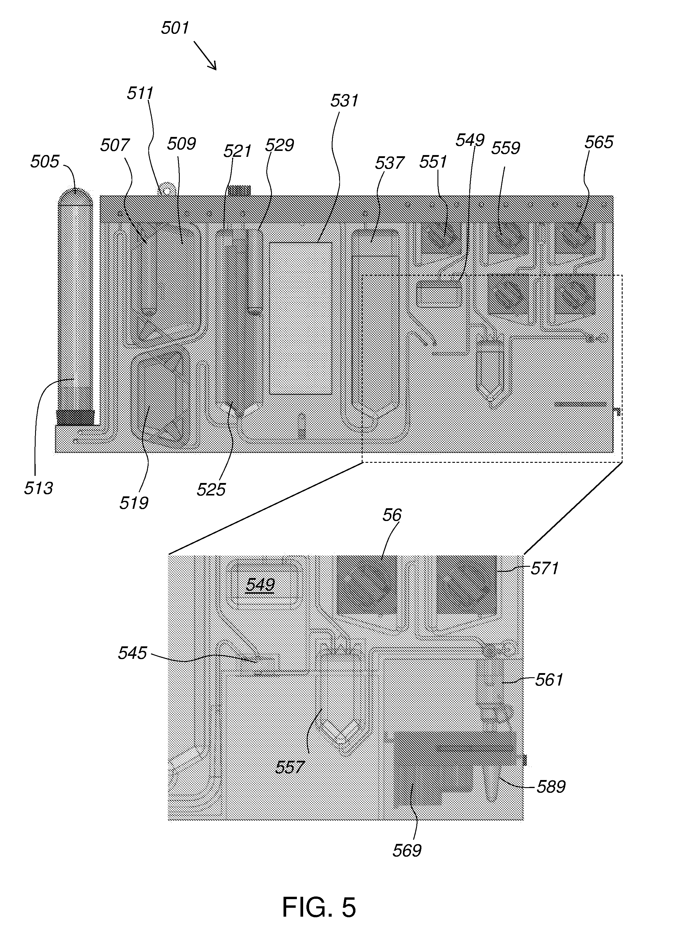
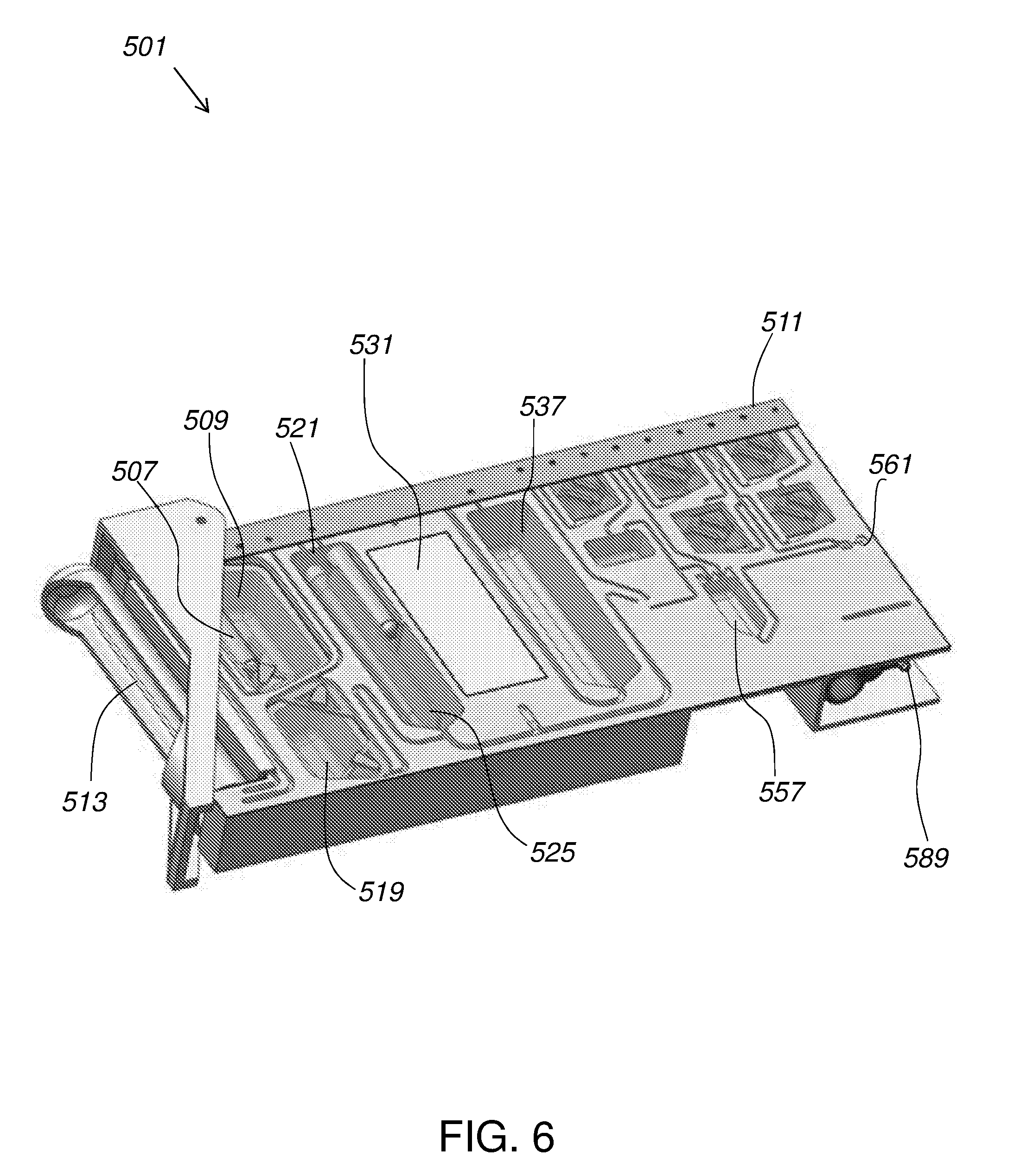
[0104] FIG. 5 illustrates a fluidic cartridge 501 according to certain embodiments. Fluidic cartridge 501 is provided to operate with blood collection tube 505 (e.g., a vacutainer). Cartridge 501 includes long needle 513 that penetrates into an interior of tube 505 when tube 505 is inserted thereon. Magnetic beads are stored in magnetic bead ampule 507 shown disposed within bead buffer 509. Pneumatic interface 511 provides pressure to flow sample, solutions, and buffers through channels of cartridge 501 and may optionally be used to crush ampules such as magnetic bead ampule 507 (and plant pathogen ampule 529). When operation is begun and tube 505 containing a sample is inserted, magnetic bead ampule 507 is crushed, introducing beads 219 to bead buffer 509. Beads 219 and buffer 509 may be mixed, for example, using agitation provided by air from pneumatic interface 511, to produce a bead mixture.
[0105] Air from pneumatic interface 511 forces blood from tube 505 into mixing chamber 521. The bead mixture is forced through long needle 513 to rinse the inside of tube 505. The bead mixture is then sent to mixing chamber 521 to mix with the blood. This step may be repeated, sending more bead mixture from bead buffer 509, through tube 505, to mixing chamber 521, until tube 505 is evacuated of target and a desired proportion (e.g., 2:1) of bead mixture to sample is present in mixing chamber 521.
[0106] Mixing paddle 525 is used to mixing the contents of chamber 521, agitating the blood and beads. Air pressure from pneumatic interface 511 then forces the mixture into magnetic trap 531. Optionally, the mixture can be pushed back, from magnetic trap 531 to mixing chamber 521, and the cycle repeated any number times until mixing chamber 521 is satisfactorily evacuated of target pathogens 201 and the bead-bound target pathogens 201 as well as the other components of the sample are in magnetic trap 431.
[0107] Then, magnetic trap 431 is evacuated of the other components of the sample, leaving magnetic particles 219 bound to magnets therein. The waste fluid is pushed back through mixing chamber 521 and discarded. Wash buffer 519 is then forced into magnetic trap 531, filling it. The wash buffer can then be pushed back to mixing chamber 521 to ensure good washing of beads 219. Wash buffer is sent back into magnetic trap 531 and held there.
[0108] Magnets can then be moved away from magnetic trap 531, allowing beads 219 to re-suspend in wash buffer 519 within trap 531. At this point, target pathogens 201 have been extracted from the original sample and held in wash buffer 519. Wash buffer 519 can then be pushed through magnetic concentrator 545 to waste chamber 549.
[0109] Turning now to the inset portion of FIG. 5, magnetic concentrator 545 can be seen to be in fluid communication with magnetic trap 531. As wash buffer 519 flows through concentrator 545, beads 219 are captured in concentrator 545 while the remainder of buffer 519 is passed on to waste chamber 549. Original pathogens 201 are thus concentrated in concentrator 545.
[0110] The contents of concentrator 545 may be processed according to whether live cells or an extracted cellular component is intended. If live cells are intended, the magnet is removed from magnetic concentrator 545 and buffer 551 (here, a live cell buffer) is introduced through concentrator 545 and used to flush the cells into output vial 589.
[0111] If, for example, extracted DNA is intended, buffer 551 is a lysis buffer and is introduced into magnetic concentrator 545. The magnet is removed from concentrator 545 and a sonicator may be applied to a wall of the chamber of concentrator 545. Optionally, beads may be included for bead-bashing to aid in lysis. The sonicator or other lysis means is activated and the target cells 201 are lysed. In certain embodiments, a probe of a sonicator extends into the chamber, where it delivers vibrations into the liquid medium surrounding the captured complexes. In other embodiments, the sonication transducer is brought in contact with the chamber by way of a structural interface. For example, the structural interface may constitute the floor or the ceiling of the chamber. The sonication transducer vibrates the structural interface such that lysis of the targets captured in the chamber is achieved.
[0112] Lysate is then pushed into pre-column mixer 557. DNA binding buffer 559 is added to pre-column mixer 557 (optionally agitated by bubbling air). The contents of pre-column mixer 557 is then forced through the DNA extraction column 561 and the elutant is discarded as waste. DNA extraction may be completed using washes from first column wash 565, second column wash 569, and water 571. Air from pneumatic interface 511 can be forced through DNA extraction column 561 to remove volatile organic compounds. Water 571 can be used to rinse the purified DNA (optionally including the use of a de-binding buffer or a modulator of stringency) into output vial 589.
[0113] Coordination of the on-cartridge steps can be supported by an operations device, such as a bench-top electro-mechanical device. The timing of pneumatic injections, the breaking of ampules, and the piercing of reagent reservoirs can be coordinated manually, or by a computer program or mechanical system. Cartridge 501 can include any suitable materials, shape, or dimensions. For example, in some embodiments, fluids are handled in macrofluidic environments up until entered into magnetic concentrator 545 and are handled according to microfluidic principles thereafter. In general, microfluidic may refer to sub-microliters volumes.
[0114] FIG. 6 gives a perspective view of an exemplary cartridge according to certain embodiments. Methods for manufacturing and operating fluidic systems are known and discussed in Fredrickson and Zan, 2004, Macro-to-micro interfaces for microfluidic devices, Lab Chip 4(6):526-33; U.S. Pat. No. 8,105,783; U.S. Pat. No. 7,785,869; U.S. Pat. No. 7,745,207; U.S. Pat. No. 7,553,647; U.S. Pub. 2009/0227005; U.S. Pub. 2008/0241000; and U.S. Pub. 2008/0003564, the contents of each of which are incorporated by reference in their entirety for all purposes.
[0115] FIG. 7 gives a schematic diagram of elements and steps of the invention as discussed above with respect to FIG. 5.
[0116] While discussed above as magnetic beads, substrate 219 for capture of pathogens 201 may be the floor of a chamber such as, for example, trap chamber 531. In other embodiments, the capture surface is the ceiling. The captured pathogens 201 can there be washed to remove non-specific analytes and unbound entities.
[0117] In certain embodiments, fluidic cartridge 501 is designed to be disposable. In this instance, once pathogens 201 have been lysed and the eluate collected, cartridge 501 can be discarded. In this manner, each cartridge is intended for a single use, and the potential for cross-contaminating pathogens due to repeated use is eliminated.
[0118] The cartridge can be prepared from any material in the art suitable for containing liquids and withstanding the rigors of sonication. In certain aspects, the entire cartridge is made of plastic or an acrylic plastic polymer. In other aspects, the bottom or top cover of the cartridge can be prepared from a film such as the biaxially-oriented polyethylene terephthalate (BoPET) sold under the trademark MYLAR while the rest of the cartridge is made of plastic or an acrylic plastic polymer. In certain embodiments, the BoPET film constitutes the aforementioned structural interface in contact with the sonicator transducer.
[0119] In certain embodiments, the cartridge is connected to a fluidic device or system configured to flow liquids into and out of the cartridge. The fluidic system can comprise a pump that delivers liquid, air, gas, beads, reagents, electricity or other signals, light, power or a combination thereof to the cartridge. The fluidic system can have a first tubing line connected to the inlet of the input channel, for flowing liquid into the cartridge. The other end of the first tubing line is connected to the pump. A second tubing line for fluid leaving the cartridge is connected to the outlet of the output channel. The other end of the second tubing line may also be connected to the pump or to a container for collecting exiting fluid. The fluidic system facilitates the delivery of the target/magnetic particle complexes into the chamber, as well as any washing of captured complexes. The collection of lysate is also facilitated by the fluidic system.
[0120] Devices of the invention may also include a detection module. The detection module is a component in which molecules, cells, or other particles are to be detected, identified, measured or interrogated on the basis of at least one predetermined characteristic. The molecules, cells, or other particles can be examined one at a time, and the characteristic is detected or measured optically, for example, by testing for the presence or amount of a label. In some aspects, the detection module is in connection with one or more detection apparatuses. The detection apparatuses can be optical or electrical detectors or combinations thereof. Examples of suitable detection apparatuses include optical waveguides, microscopes, diodes, light stimulating devices (e.g., lasers), photomultiplier tubes, and processors (e.g., computers and software), and combinations thereof, which cooperate to detect a signal representative of a characteristic, marker, probe, label, or reporter, and to determine and direct a measurement or sorting action. However, other detection techniques can be used as well.
[0121] Devices of the invention may include a computer component. For example, a user interface may be provided with input/output mechanisms (monitor, keyboard, touchscreen, etc.) coupled to a memory and a processor (e.g., a silicone chip and a solid-state or magnetic hard drive). Input/output mechanisms may be included for data, such as a USB port, Ethernet port, or Wi-Fi card or a hardware connection to a detection module, fluidic chip and pneumatic interface, or both.
[0122] In certain aspects, the detection module is in fluid connection with the fluidic cartridge. For example, the outlet of the fluidic cartridge may be connected to the detection module by means of a tubing line. In this manner, lysate leaving the cartridge can enter the detection module wherein the contents of the lysate can be detected and analyzed.
INCORPORATION BY REFERENCE
[0123] References and citations to other documents, such as patents, patent applications, patent publications, journals, books, papers, web contents, have been made throughout this disclosure. All such documents are hereby incorporated herein by reference in their entirety for all purposes.
EQUIVALENTS
[0124] Various modifications of the invention and many further embodiments thereof, in addition to those shown and described herein, will become apparent to those skilled in the art from the full contents of this document, including references to the scientific and patent literature cited herein. The subject matter herein contains important information, exemplification and guidance that can be adapted to the practice of this invention in its various embodiments and equivalents thereof.
* * * * *
D00001
D00002
D00003
D00004
D00005
D00006
D00007
XML
uspto.report is an independent third-party trademark research tool that is not affiliated, endorsed, or sponsored by the United States Patent and Trademark Office (USPTO) or any other governmental organization. The information provided by uspto.report is based on publicly available data at the time of writing and is intended for informational purposes only.
While we strive to provide accurate and up-to-date information, we do not guarantee the accuracy, completeness, reliability, or suitability of the information displayed on this site. The use of this site is at your own risk. Any reliance you place on such information is therefore strictly at your own risk.
All official trademark data, including owner information, should be verified by visiting the official USPTO website at www.uspto.gov. This site is not intended to replace professional legal advice and should not be used as a substitute for consulting with a legal professional who is knowledgeable about trademark law.