Nuclear Receptor Set Domain Containing Protein 2 Transition State And Uses Thereof
Schramm; Vern L. ; et al.
U.S. patent application number 16/060470 was filed with the patent office on 2019-01-24 for nuclear receptor set domain containing protein 2 transition state and uses thereof. This patent application is currently assigned to ALBERT EINSTEIN COLLEGE OF MEDICINE, INC.. The applicant listed for this patent is ALBERT EINSTEIN COLLEGE OF MEDICINE, INC.. Invention is credited to Myles B. Poulin, Vern L. Schramm.
| Application Number | 20190026440 16/060470 |
| Document ID | / |
| Family ID | 59057508 |
| Filed Date | 2019-01-24 |


View All Diagrams
| United States Patent Application | 20190026440 |
| Kind Code | A1 |
| Schramm; Vern L. ; et al. | January 24, 2019 |
NUCLEAR RECEPTOR SET DOMAIN CONTAINING PROTEIN 2 TRANSITION STATE AND USES THEREOF
Abstract
Methods and systems for obtaining inhibitors of Nuclear receptor SET Domain containing protein 2 (NSD2) are disclosed where the methods involve designing compounds that resemble the NSD2 transition state.
| Inventors: | Schramm; Vern L.; (New Rochelle, NY) ; Poulin; Myles B.; (Bronx, NY) | ||||||||||
| Applicant: |
|
||||||||||
|---|---|---|---|---|---|---|---|---|---|---|---|
| Assignee: | ALBERT EINSTEIN COLLEGE OF
MEDICINE, INC. Bronx NY |
||||||||||
| Family ID: | 59057508 | ||||||||||
| Appl. No.: | 16/060470 | ||||||||||
| Filed: | December 14, 2016 | ||||||||||
| PCT Filed: | December 14, 2016 | ||||||||||
| PCT NO: | PCT/US16/66514 | ||||||||||
| 371 Date: | June 8, 2018 |
Related U.S. Patent Documents
| Application Number | Filing Date | Patent Number | ||
|---|---|---|---|---|
| 62268677 | Dec 17, 2015 | |||
| Current U.S. Class: | 1/1 |
| Current CPC Class: | G01N 2500/00 20130101; G06F 16/00 20190101; G16B 15/00 20190201; G16B 5/00 20190201; G16C 20/60 20190201; C40B 20/08 20130101; G16C 20/50 20190201; G16B 40/00 20190201; A61K 38/00 20130101; G16B 35/00 20190201 |
| International Class: | G06F 19/00 20060101 G06F019/00; G06F 17/30 20060101 G06F017/30; G06F 19/12 20060101 G06F019/12; G06F 19/24 20060101 G06F019/24 |
Goverment Interests
STATEMENT OF GOVERNMENT SUPPORT
[0002] This invention was made with government support under grant number GM041916 awarded by the National Institutes of Health. The government has certain rights in the invention.
Claims
1. A system comprising a non-transitory computer-readable medium coupled to one or more data processing apparatus having instructions stored thereon which, when executed by the one or more data processing apparatus, cause the one or more data processing apparatus to perform a method comprising: (i) obtaining kinetic isotope effects on human Nuclear receptor SET Domain containing protein 2 (NSD2)-catalyzed methylation of histone H3 lysine 36 to obtain the NSD2 transition state structure, wherein the NSD2 transition state comprises the structure (ii) obtaining the molecular electrostatic potential at the van der Waals surface computed from the wave function of the NSD2 transition state and the geometric atomic volume of the NSD2 transition state; and (iiia) identifying from a library of compounds a chemically stable compound that resembles the molecular electrostatic potential at the van der Waals surface computed from the wave function of the NSD2 transition state and the geometric atomic volume of the NSD2 transition state, or (iiib) designing a chemically stable compound that resembles the molecular electrostatic potential at the van der Waals surface computed from the wave function of the NSD2 transition state and the geometric atomic volume of the NSD2 transition state; wherein the chemically stable compound that resembles the molecular electrostatic potential at the van der Waals surface computed from the wave function of the NSD2 transition state and the geometric atomic volume of the NSD2 transition state is a putative inhibitor of NSD2.
2. (canceled)
3. A computer implemented method performed using a system comprising a non-transitory computer-readable medium coupled to one or more data processing apparatus having instructions stored thereon, the method comprising: (i) obtaining kinetic isotope effects on human Nuclear receptor SET Domain containing protein 2 (NSD2)-catalyzed methylation of histone H3 lysine 36 to obtain the NSD2 transition state structure, wherein the NSD2 transition state comprises the structure (ii) obtaining the molecular electrostatic potential at the van der Waals surface computed from the wave function of the NSD2 transition state and the geometric atomic volume of the NSD2 transition state; and (iiia) identifying from a library of compounds a chemically stable compound that resembles the molecular electrostatic potential at the van der Waals surface computed from the wave function of the NSD2 transition state and the geometric atomic volume of the NSD2 transition state, or (iiib) designing a chemically stable compound that resembles the molecular electrostatic potential at the van der Waals surface computed from the wave function of the NSD2 transition state and the geometric atomic volume of the NSD2 transition state; wherein the chemically stable compound that resembles the molecular electrostatic potential at the van der Waals surface computed from the wave function of the NSD2 transition state and the geometric atomic volume of the NSD2 transition state is a putative inhibitor of NSD2.
4-8. (canceled)
9. The system of claim 1, which further comprises synthesizing the compound.
10. The system of claim 1, which further comprises testing the compound for inhibitory activity to NSD2.
11. (canceled)
12. A method of screening for an inhibitor of human Nuclear receptor SET Domain containing protein 2 (NSD2), the method comprising the steps of: (i) measuring kinetic isotope effects on the NSD2-catalyzed methylation of histone H3 lysine 36 to obtain the NSD2 transition state structure, wherein the NSD2 transition state comprises the structure (ii) determining the molecular electrostatic potential at the van der Waals surface computed from the wave function of the NSD2 transition state and the geometric atomic volume of the NSD2 transition state; (iiia) obtaining a chemically stable compound that resembles the molecular electrostatic potential at the van der Waals surface computed from the wave function of the NSD2 transition state and the geometric atomic volume of the NSD2 transition state, or (iiib) using a computer to design a chemically stable compound that resembles the molecular electrostatic potential at the van der Waals surface computed from the wave function of the NSD2 transition state and the geometric atomic volume of the NSD2 transition state, and synthesizing the compound; and (iv) testing the compound for inhibitory activity to NSD2 by determining if the compound inhibits NSD2-catalyzed methylation of histone H3 lysine 36, wherein a compound that inhibits NSD2-catalyzed methylation of histone H3 lysine 36 is an inhibitor of NSD2.
13. A method of inhibiting NSD2 comprising obtaining a NSD2 inhibitor by the system of claim 1, and contacting NSD2 with the compound.
14. A method of treating a subject having a cancer comprising obtaining a NSD2 inhibitor by using the system of claim 1, and administering the compound to the subject in an amount effective to inhibit NSD2.
15. The method of claim 14, wherein the cancer is multiple myeloma, neuroblastoma, glioblastoma, prostate cancer or breast cancer.
16. The system of claim 1, wherein the transition state is a S.sub.N2 transition state where a positive charge is distributed between a leaving group, a transferring group and a nucleophile.
17. The system of claim 1, wherein the transition state has a C--N distance of 1.8 .ANG. and a C--S distance of 2.6 .ANG., or the transition state has a C--N distance of 2.10 .ANG. and a C--S distance of 2.53 .ANG..
18. (canceled)
Description
CROSS REFERENCE TO RELATED APPLICATIONS
[0001] This application is a U.S. national stage entry under 35 U.S.C. .sctn. 371 of PCT International Patent Application No. PCT/US2016/066514, filed Dec. 14, 2016, which claims the benefit of U.S. Provisional Patent Application No. 62/268,677, filed Dec. 17, 2015, the contents of each of which are incorporated herein by reference into the subject application.
FIELD OF THE INVENTION
[0003] The invention relates to systems and methods for obtaining inhibitors of Nuclear receptor SET Domain containing protein 2 (NSD2) by designing compounds that resemble the charge and geometry of the NSD2 transition state.
BACKGROUND OF THE INVENTION
[0004] Throughout this application various publications are referred to in parentheses. Full citations for these references may be found at the end of the specification before the claims. The disclosures of these publications are hereby incorporated by reference in their entireties into the subject application to more fully describe the art to which the subject application pertains.
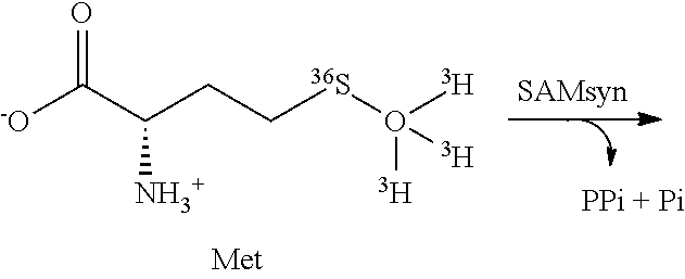
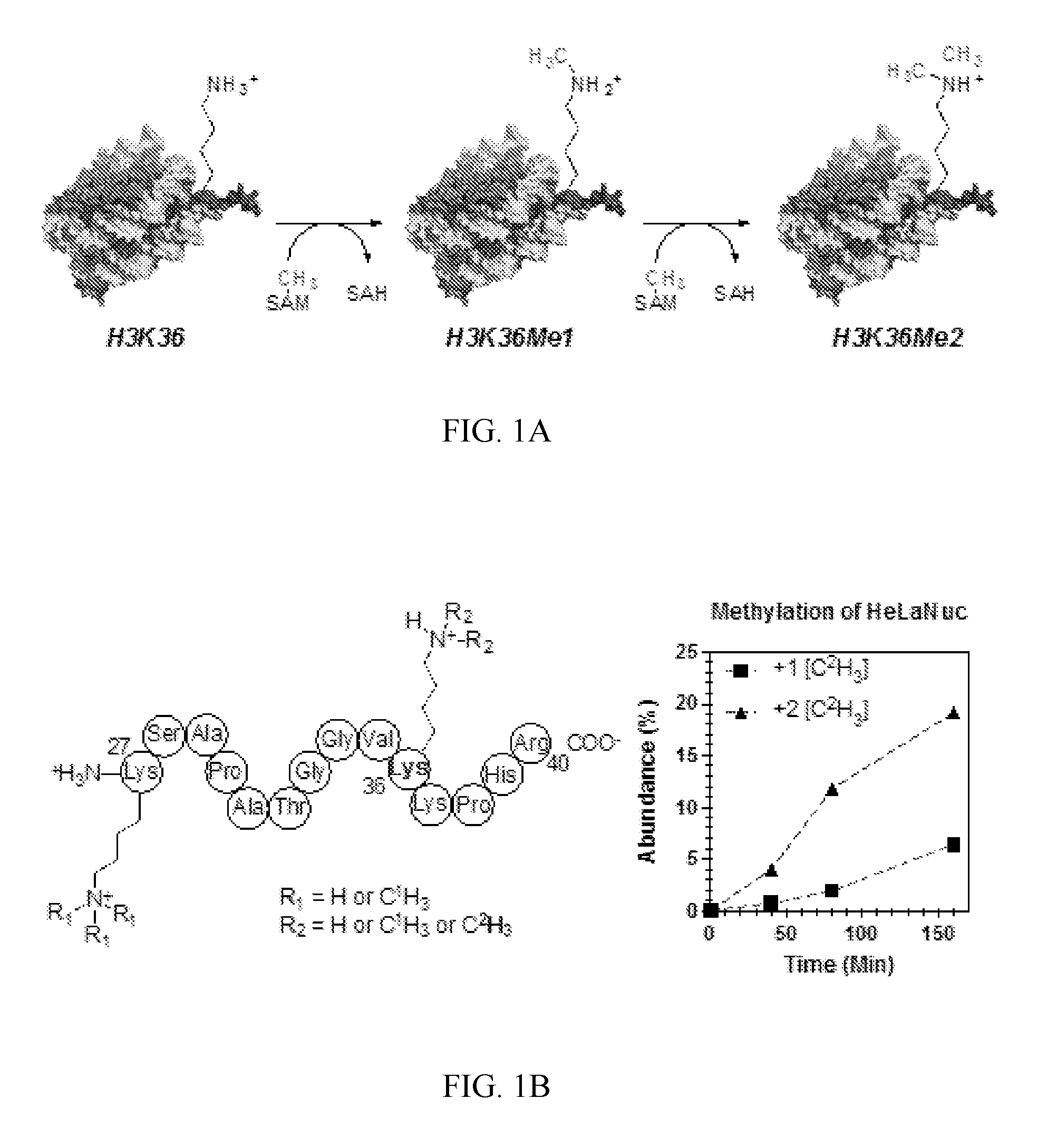
[0005] Histone lysine methylation is an essential posttranslational modification for transcriptional regulation, DNA damage response, and chromatin regulation (1, 2). Methyl groups (Me, CH.sub.3) are installed on lysine residues by protein lysine methyltransferase enzymes (PKMTs), the majority of which, in humans, contain a catalytic SET domain (3). The conserved SET domain catalyzes the transfer of between one to three CH.sub.3 from S-adenosyl-L-methionine (SAM) onto the .epsilon.-amino group of lysine residues (FIG. 1) producing mono-, di-, or trimethyl lysine derivatives (KMe1, KMe2, and KMe3 respectively) (3), in a reaction that involves first deprotonation of the lysine, and finally transfer of the methyl group (4, 5).
[0006] Histone lysine Me marks can signal either transcriptional activation or repression depending on which lysine residue is methylated and the number of transferred Me groups (2). For example, histone H3 lysine 27 trimethylation is a signal for transcriptional repression (6), where histone H3 lysine 4 and histone H3 lysine 36 Me marks are found in actively transcribed loci (7). As a result, mis-regulation of PKMT expression is often associated with cancer development and other disease states (8). Deletion of the gene encoding the histone H3K36 dimethyltransferase enzyme nuclear receptor binding SET domain protein 2 (NSD2, also known as WHSC1 or MMSET), specifically, is present in the developmental disorder Wolf Hirschhorn syndrome (9). By contrast, NSD2 overexpression, as a result of a t (4, 14) chromosomal translocation (10), is present in 15% of multiple myeloma cases and has been identified in other cancers (11-13). NSD2 catalyzes the mono- and dimethylation of histone H3K36, in vivo (11), although other CH.sub.3-transfer specificities have also been reported in studies using histone protein or histone tail peptide as substrate analogs (14-16). Studies using isolated or recombinant nucleosome as physiologically relevant substrates detected H3K36Me1 and H3K36Me2 as the exclusive products. Thus the nature of the substrate can influence NSD2 specificity (11, 17). Substrate specificity is also influenced by the presence of a C-terminal basic post-SET extension found in NSD family methyltransferases. NSD2 mutants lacking this basic post-SET extension are unable to recognize nucleosome as substrate (18). The H3K36Me2 marks introduced by NSD2 are normally concentrated in the 5' end of actively transcribed genes (7), and overexpression results in global increases in H3K36Me2 throughout gene bodies resulting in aberrant transcription of multiple oncogenes (19). Histone methylation is a reversible posttranslational modification, thus inhibiting the catalytic activity of NSD2 is an attractive strategy for the treatment of multiple myeloma and other cancers.
[0007] Designing analogues that mimic the geometry and charge distribution of transition states (TSs) of enzyme catalyzed reactions is a powerful approach for enzyme inhibition (20-21, 59-63); however, this requires a detailed model of the enzyme TS. TS models for a number of PKMT enzymes, including human SET8 (22) and SETT/9 (4, 23) based on QM/MM calculation of the enzyme chemistry, show substantial variability in the predicted TS geometry of PKMT. However, these models have not been verified experimentally. Detailed information about TS structure can be obtained from the measurement of kinetic isotope effects (KIEs) (24), which result from changes in the bond vibrational environment for atoms of the reactants free in solution and at the TS (24). The measurement of multiple KIEs combined with quantum chemical calculations allow for interrogation of TS structure (20, 21, 25). The present invention uses the NSD2 TS to address the need for new inhibitors for NSD2, particularly ones that will be effective in cancer therapy.
SUMMARY OF THE INVENTION
[0008] The invention provides methods of obtaining inhibitors of human Nuclear receptor SET Domain containing protein 2 (NSD2) comprising designing a chemically stable compound that resembles the charge and geometry of the NSD2 transition state.
[0009] The invention also provides systems for obtaining a putative inhibitor of a human Nuclear receptor SET Domain containing protein 2 (NSD2) comprising one or more data processing apparatus and a computer-readable medium coupled to the one or more data processing apparatus having instructions stored thereon that are configured to perform a method comprising designing a chemically stable compound that resembles the charge and geometry of the NSD2 transition state, wherein the compound is a putative inhibitor of NSD2.
[0010] The invention further provides methods for screening for a compound that is an inhibitor of human Nuclear receptor SET Domain containing protein 2 (NSD2), the method comprising the steps of:
[0011] (i) determining the molecular electrostatic potential at the van der Waals surface computed from the wave function of a NSD2 transition state and the geometric atomic volume of the NSD2 transition state, wherein the NSD2 transition state comprises the structure
[0012] (ii) designing a chemically stable compound that resembles the molecular electrostatic potential at the van der Waals surface computed from the wave function of the NSD2 transition state and the geometric atomic volume of the NSD2 transition state;
[0013] (iii) synthesizing the compound; and
[0014] (iv) testing the compound for inhibitory activity to NSD2.
[0015] The invention further provides methods of screening for an inhibitor of human Nuclear receptor SET Domain containing protein 2 (NSD2), the method comprising the steps of:
[0016] (i) measuring kinetic isotope effects on the NSD2-catalyzed methylation of histone H3 lysine 36 to obtain the NSD2 transition state structure,
[0017] wherein the NSD2 transition state comprises the structure
[0018] (ii) determining the molecular electrostatic potential at the van der Waals surface computed from the wave function of the NSD2 transition state and the geometric atomic volume of the NSD2 transition state;
[0019] (iii) obtaining a chemically stable compound that resembles the molecular electrostatic potential at the van der Waals surface computed from the wave function of the NSD2 transition state and the geometric atomic volume of the NSD2 transition state; and
[0020] (iv) testing the compound for inhibitory activity to NSD2 by determining if the compound inhibits NSD2-catalyzed methylation of histone H3 lysine 36,
[0021] wherein a compound that inhibits NSD2-catalyzed methylation of histone H3 lysine 36 is an inhibitor of NSD2.
[0022] The invention also provides systems comprising a non-transitory computer-readable medium coupled to one or more data processing apparatus having instructions stored thereon which, when executed by the one or more data processing apparatus, cause the one or more data processing apparatus to perform a method comprising:
[0023] (i) obtaining kinetic isotope effects on human Nuclear receptor SET Domain containing protein 2 (NSD2)-catalyzed methylation of histone H3 lysine 36 to obtain the NSD2 transition state structure,
[0024] wherein the NSD2 transition state comprises the structure
[0025] (ii) determining the molecular electrostatic potential at the van der Waals surface computed from the wave function of the NSD2 transition state and the geometric atomic volume of the NSD2 transition state; and
[0026] (iii) identifying from a library of compounds a chemically stable compound that resembles the molecular electrostatic potential at the van der Waals surface computed from the wave function of the NSD2 transition state and the geometric atomic volume of the NSD2 transition state;
[0027] wherein the chemically stable compound that resembles the molecular electrostatic potential at the van der Waals surface computed from the wave function of the NSD2 transition state and the geometric atomic volume of the NSD2 transition state is a putative inhibitor of NSD2.
[0028] The invention also provides systems comprising a non-transitory computer-readable medium coupled to one or more data processing apparatus having instructions stored thereon which, when executed by the one or more data processing apparatus, cause the one or more data processing apparatus to perform a method comprising:
[0029] (i) determining the molecular electrostatic potential at the van der Waals surface computed from the wave function of a human Nuclear receptor SET Domain containing protein 2 (NSD2) transition state and the geometric atomic volume of the NSD2 transition state, wherein the NSD2 transition state comprises the structure
and
[0030] (ii) designing a chemically stable compound that resembles the molecular electrostatic potential at the van der Waals surface computed from the wave function of the NSD2 transition state and the geometric atomic volume of the NSD2 transition state;
[0031] wherein a chemically stable compound that resembles the molecular electrostatic potential at the van der Waals surface computed from the wave function of the NSD2 transition state and the geometric atomic volume of the NSD2 transition state is a putative inhibitor of NSD2.
[0032] The invention also provides computer implemented methods performed using a system comprising a non-transitory computer-readable medium coupled to one or more data processing apparatus having instructions stored thereon, the methods comprising:
[0033] (i) obtaining kinetic isotope effects on human Nuclear receptor SET Domain containing protein 2 (NSD2)-catalyzed methylation of histone H3 lysine 36 to obtain the NSD2 transition state structure,
[0034] wherein the NSD2 transition state comprises the structure
[0035] (ii) determining the molecular electrostatic potential at the van der Waals surface computed from the wave function of the NSD2 transition state and the geometric atomic volume of the NSD2 transition state; and
[0036] (iii) identifying from a library of compounds a chemically stable compound that resembles the molecular electrostatic potential at the van der Waals surface computed from the wave function of the NSD2 transition state and the geometric atomic volume of the NSD2 transition state;
[0037] wherein the chemically stable compound that resembles the molecular electrostatic potential at the van der Waals surface computed from the wave function of the NSD2 transition state and the geometric atomic volume of the NSD2 transition state is a putative inhibitor of NSD2.
[0038] The invention also provides computer implemented methods performed using a system comprising a non-transitory computer-readable medium coupled to one or more data processing apparatus having instructions stored thereon, the methods comprising:
[0039] (i) determining the molecular electrostatic potential at the van der Waals surface computed from the wave function of a human Nuclear receptor SET Domain containing protein 2 (NSD2) transition state and the geometric atomic volume of the NSD2 transition state, wherein the NSD2 transition state comprises the structure
and
[0040] (ii) designing a chemically stable compound that resembles the molecular electrostatic potential at the van der Waals surface computed from the wave function of the NSD2 transition state and the geometric atomic volume of the NSD2 transition state;
[0041] wherein a chemically stable compound that resembles the molecular electrostatic potential at the van der Waals surface computed from the wave function of the NSD2 transition state and the geometric atomic volume of the NSD2 transition state is a putative inhibitor of NSD2.
BRIEF DESCRIPTION OF THE DRAWINGS
[0042] FIG. 1A-1B. NSD2 catalyzed methylation of histone H3 lysine 36 (H3 K36). (A) The general reaction catalyzed by NSD2 showing both mono- and dimethylation of H3K36. (B) Product distribution for methylation of HeLa cell mononucleosomes (HeLaNuc) with [Me-.sup.2H.sub.3]SAM. Products are shown for the histone H3 (K27-R40) peptide resulting from Arg-C digestion. NSD2 displays a preference for dimethylation H3 K36.
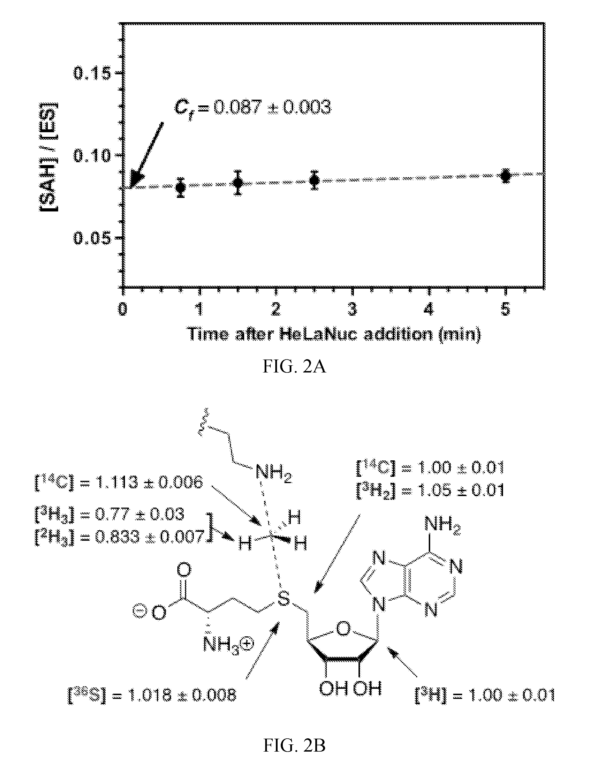
[0043] FIG. 2A-2B. Determination of intrinsic KIEs correcting for forward commitment factor (C.sub.f). (A) Measurement of C.sub.f by isotope trapping for the methylation of HeLa cell nucleosome H3 K36 by NSD2. (B) Intrinsic KIE values by atom position after correction for C.sub.f. Errors are reported as the standard deviation of at least six replicates from two independent experiments.
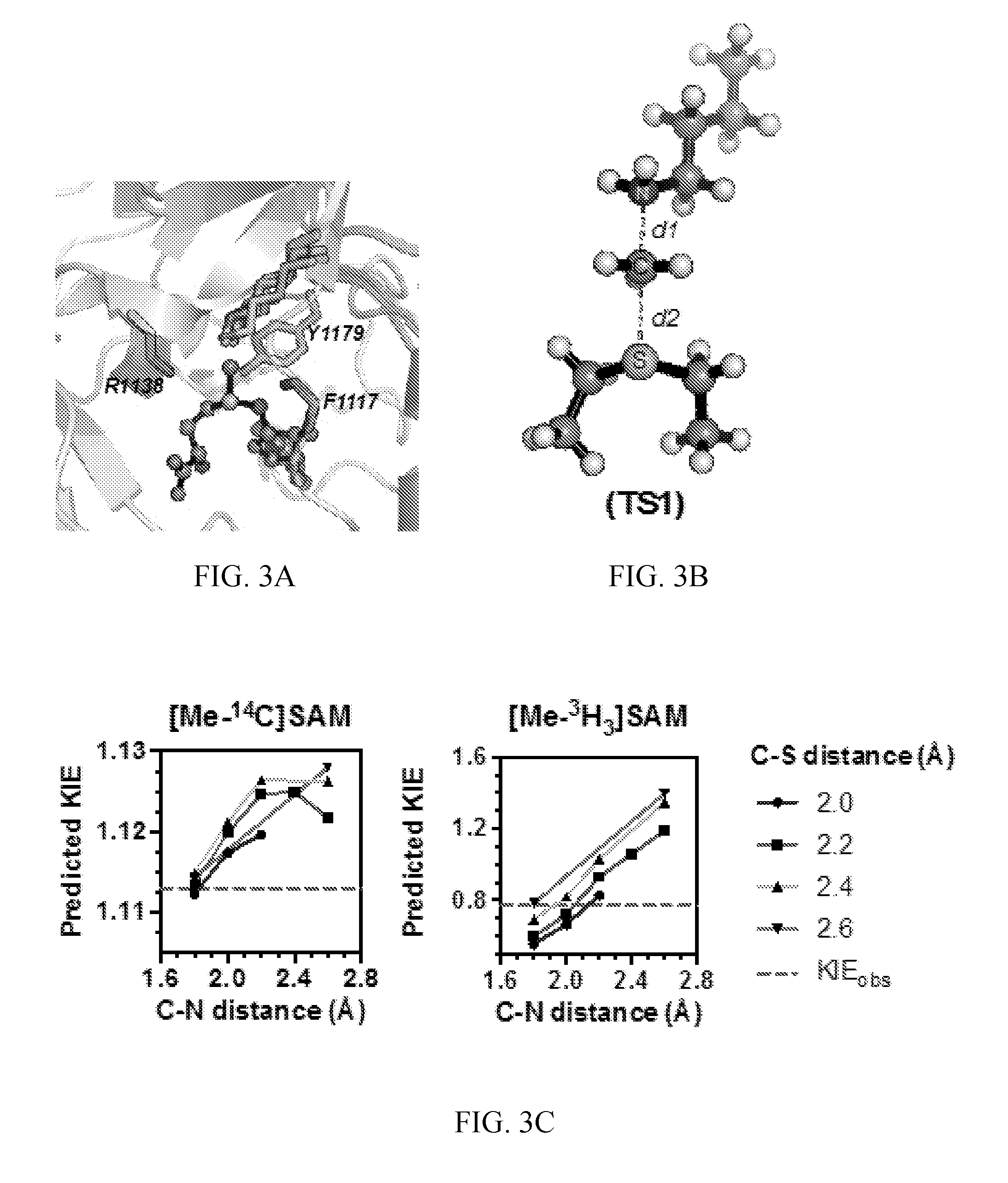
[0044] FIG. 3A-3C. Theoretical transition state model for NSD2 methylation of H3K36. (A) A prediction of the lysine substrate geometry from the structure NSD1 overlaid with the with peptide substrates of SETT/9 (PDB ID 1XQH, 2F69) SET8 (PDB ID 3F9W, 3F9Y) and PIM5 (PDB 1PEG). (B) A simplified TS model for the NSD2 methyltransfer reaction derived from the SAM and lysine geometry. (C) KIEs predicted for TS1 at different fixed C--N and C--S distances.
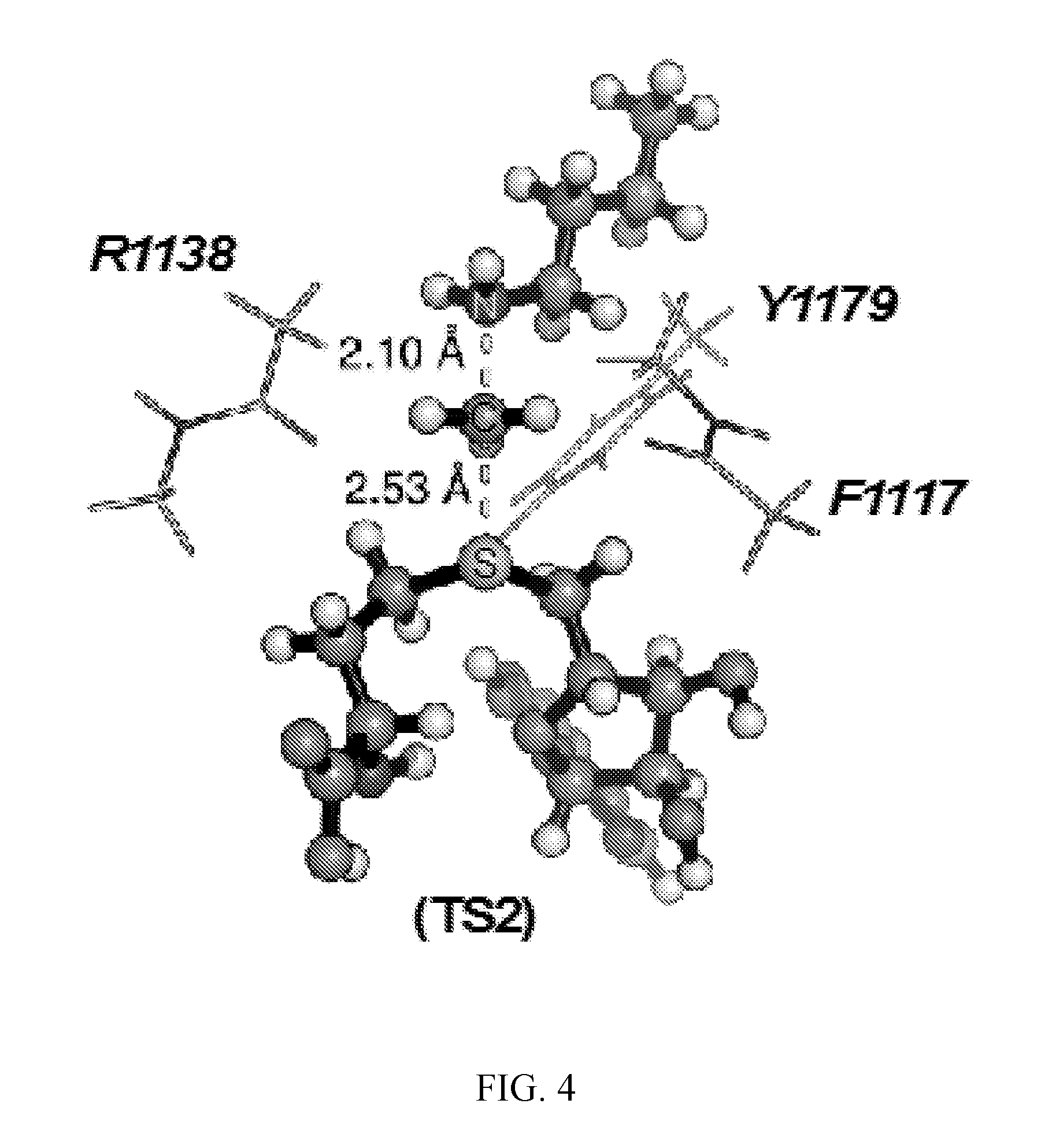
[0045] FIG. 4. Geometry and electrostatic potential surface of the NSD2 TS structure. TS structure for H3 K36 methylation catalyzed by NSD2 with active site amino acids F1117, M1140 and Y1179 (TS2).
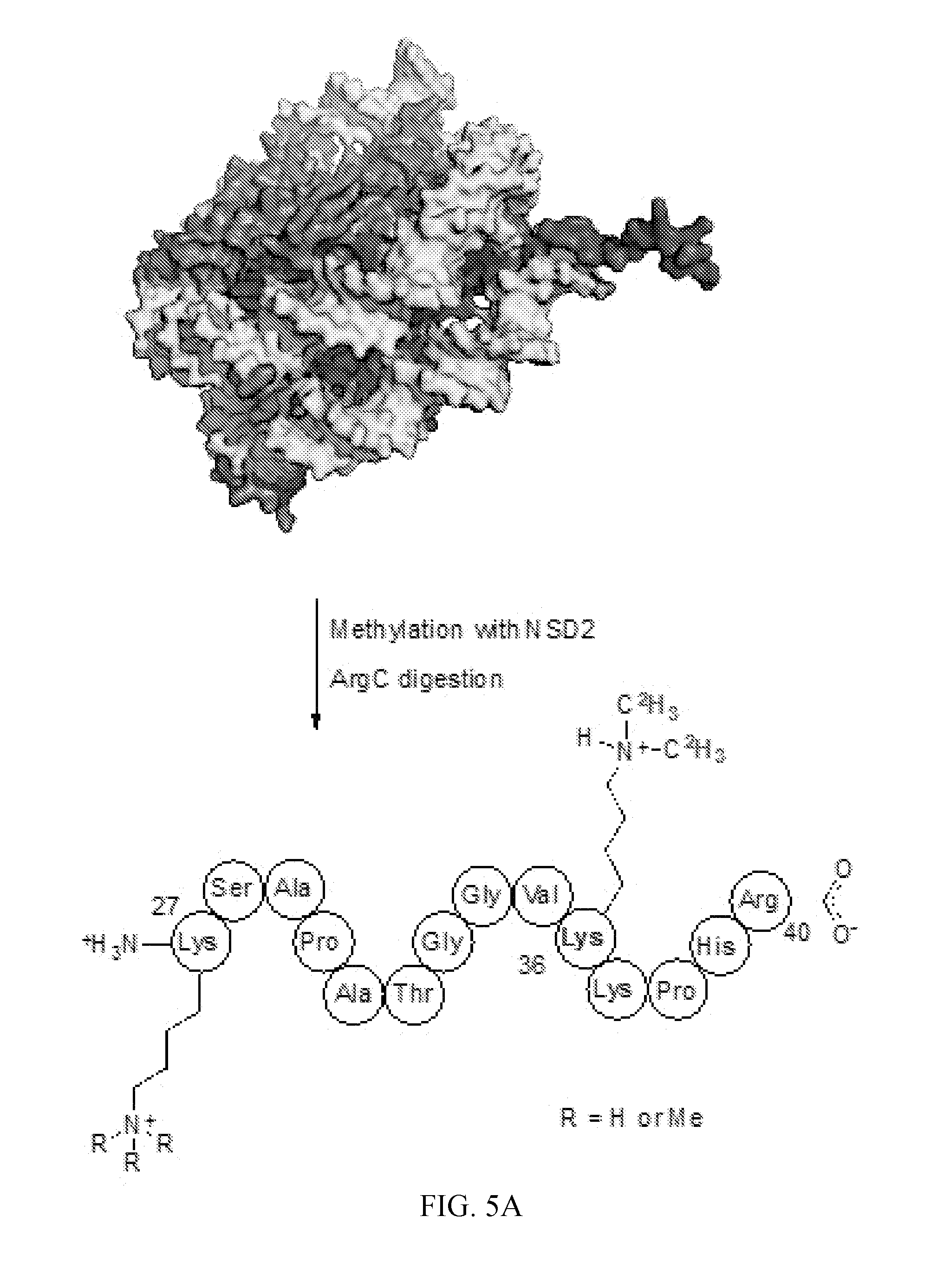
[0046] FIG. 5A-5B. Analysis of products resulting from the methylation of HeLaNuc catalyzed by NSD2. (A) A diagram showing the H3 peptide containing Lys27-Arg40 resulting from ArgC digestion of HeLaNuc that was methylated by NSD2 in vitro. (B) LC-MS analysis of the products resulting from methylation of HeLaNuc showed only mono- and dimethylation of H3K36 for starting peptides containing between 0-3 pre-existing methyl marks. H3K36 methylation was confirmed by LC-MS/MS, data not shown.
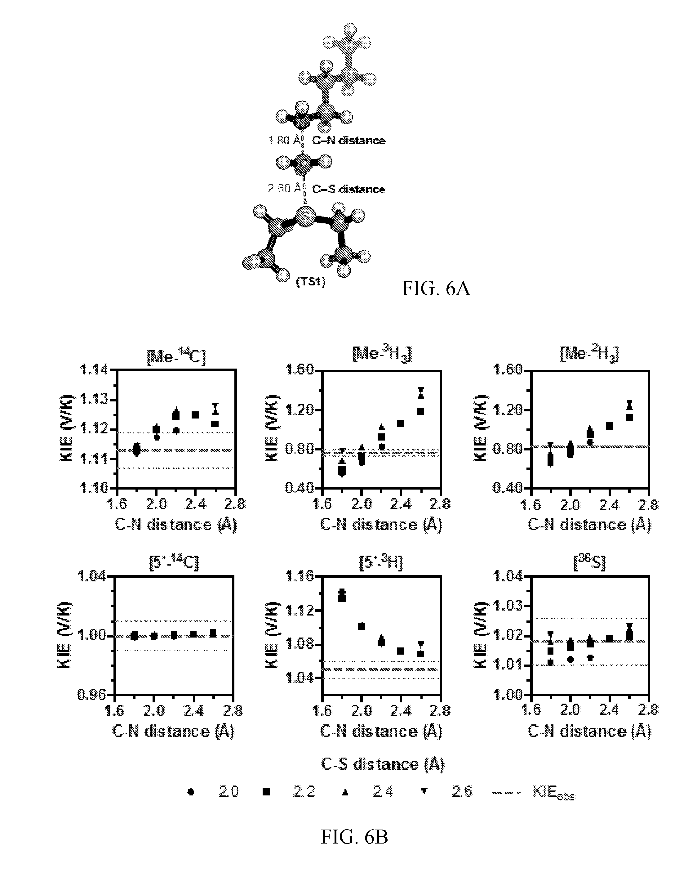
[0047] FIG. 6A-6B. Theoretical structure of the TS for NSD2 methylation of H3 K36. (A) A simplified TS model calculated for the NSD2 methyltransfer reaction using M062x/6-31G*. (B) Relationships between TS geometry and predicted KIEs. KIEs calculated using the program ISOEFF98 are plotted at varying C--N and C--S distances. Observed KIEs are shown as a dashed line with the standard deviation shown as dotted lines.
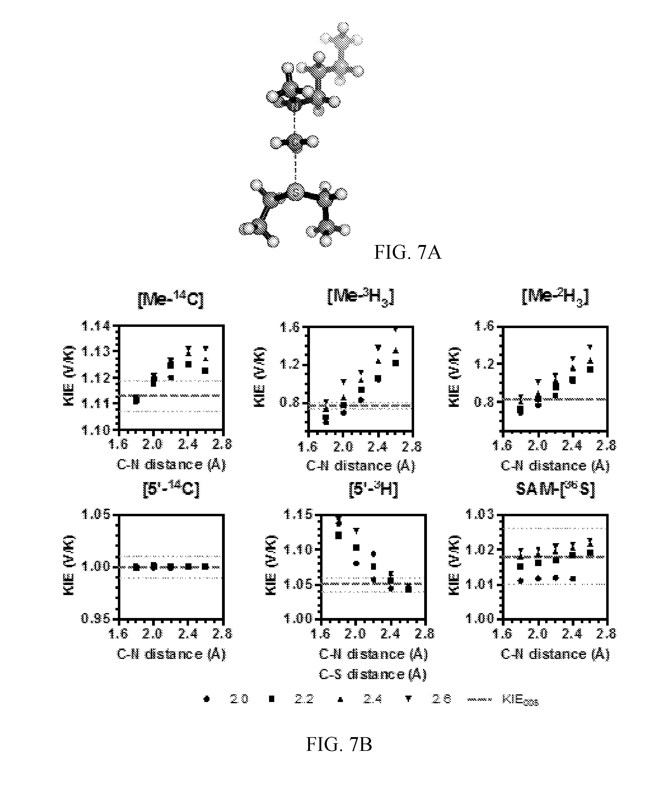
[0048] FIG. 7A-7B. Theoretical structure of the TS for NSD2 methylation of H3 K36Mel. (A) A simplified TS model calculated for the NSD2 methyltransfer reaction using M062x/6-31G*. (B) Relationships between TS geometry and predicted KIEs. KIEs calculated using the program ISOEFF98 are plotted at varying C--N and C--S distances. Observed KIEs are shown as a dashed line with the standard deviation shown as dotted lines.
[0049] FIG. 8. Effect of MeC--S--C5'-C4' dihedral angle on predicted 5'-.sup.3H.sub.2 KIEs. KIEs were calculated for TS1 where the MeC--S--C5'-C4' dihedral angle is rotated from equilibrium.
[0050] FIG. 9. Predicted Me-.sup.14C KIEs including contributions from QM tunneling. Predicted KIEs were calculated using the program ISOEFF98 for TS1 with fixed C--S and C--N distances and including a correction for QM tunneling. The intrinsic Me-.sup.14C KIE is shown as a dashed line.
DETAILED DESCRIPTION OF THE INVENTION
[0051] The invention provides a method of obtaining a putative inhibitor of human Nuclear receptor SET Domain containing protein 2 (NSD2), the method comprising using a computer to design a chemically stable compound that resembles the charge and geometry of the NSD2 transition state, wherein the compound is a putative inhibitor of NSD2.
[0052] The invention also provides a system for obtaining a putative inhibitor of human Nuclear receptor SET Domain containing protein 2 (NSD2) comprising one or more data processing apparatus and a computer-readable medium coupled to the one or more data processing apparatus having instructions stored thereon that are configured to perform a method comprising designing a chemically stable compound that resembles the charge and geometry of the NSD2 transition state, wherein the compound is a putative inhibitor of NSD2.
[0053] The method can include the steps of:
[0054] (i) determining the molecular electrostatic potential at the van der Waals surface computed from the wave function of a NSD2 transition state and the geometric atomic volume of the NSD2 transition state, and
[0055] (ii) designing a chemically stable compound that resembles the molecular electrostatic potential at the van der Waals surface computed from the wave function of the NSD2 transition state and the geometric atomic volume of the NSD2 transition state, wherein the compound is a putative inhibitor of NSD2.
[0056] As disclosed herein, the NSD2 transition state structure can comprise
[0057] Preferably, the transition state is a S.sub.N2 transition state where a positive charge is distributed between a leaving group, a transferring group and a nucleophile. Preferably, the transition state has a C--N distance of 1.8 .ANG. and a C--S distance of 2.6 .ANG., or the transition state has a C--N distance of 2.10 .ANG. and a C--S distance of 2.53 .ANG..
[0058] The invention further provides a method for screening for a compound that is an inhibitor of human Nuclear receptor SET Domain containing protein 2 (NSD2), the method comprising the steps of:
[0059] (i) determining the molecular electrostatic potential at the van der Waals surface computed from the wave function of a NSD2 transition state and the geometric atomic volume of the NSD2 transition state, wherein the HIV-1 protease transition state comprises the structure
[0060] (ii) using a computer to design a chemically stable compound that resembles the molecular electrostatic potential at the van der Waals surface computed from the wave function of the NSD2 transition state and the geometric atomic volume of the NSD2 transition state;
[0061] (iii) synthesizing the compound; and
[0062] (iv) testing the compound for inhibitory activity to HIV-1 protease.
[0063] The invention further provides a method of screening for an inhibitor of human Nuclear receptor SET Domain containing protein 2 (NSD2), the method comprising the steps of:
[0064] (i) measuring kinetic isotope effects on the NSD2-catalyzed methylation of histone H3 lysine 36 to obtain the NSD2 transition state structure,
[0065] wherein the NSD2 transition state comprises the structure
[0066] (ii) determining the molecular electrostatic potential at the van der Waals surface computed from the wave function of the NSD2 transition state and the geometric atomic volume of the NSD2 transition state;
[0067] (iii) obtaining a chemically stable compound that resembles the molecular electrostatic potential at the van der Waals surface computed from the wave function of the NSD2 transition state and the geometric atomic volume of the NSD2 transition state; and
[0068] (iv) testing the compound for inhibitory activity to NSD2 by determining if the compound inhibits NSD2-catalyzed methylation of histone H3 lysine 36,
[0069] wherein a compound that inhibits NSD2-catalyzed methylation of histone H3 lysine 36 is an inhibitor of NSD2.
[0070] The invention also provides a system comprising a non-transitory computer-readable medium coupled to one or more data processing apparatus having instructions stored thereon which, when executed by the one or more data processing apparatus, cause the one or more data processing apparatus to perform a method comprising:
[0071] (i) obtaining kinetic isotope effects on human Nuclear receptor SET Domain containing protein 2 (NSD2)-catalyzed methylation of histone H3 lysine 36 to obtain the NSD2 transition state structure,
[0072] wherein the NSD2 transition state comprises the structure
[0073] (ii) obtaining the molecular electrostatic potential at the van der Waals surface computed from the wave function of the NSD2 transition state and the geometric atomic volume of the NSD2 transition state; and
[0074] (iii) identifying from a library of compounds a chemically stable compound that resembles the molecular electrostatic potential at the van der Waals surface computed from the wave function of the NSD2 transition state and the geometric atomic volume of the NSD2 transition state;
[0075] wherein the chemically stable compound that resembles the molecular electrostatic potential at the van der Waals surface computed from the wave function of the NSD2 transition state and the geometric atomic volume of the NSD2 transition state is a putative inhibitor of NSD2.
[0076] The invention also provides a system comprising a non-transitory computer-readable medium coupled to one or more data processing apparatus having instructions stored thereon which, when executed by the one or more data processing apparatus, cause the one or more data processing apparatus to perform a method comprising:
[0077] (i) obtaining the molecular electrostatic potential at the van der Waals surface computed from the wave function of a human Nuclear receptor SET Domain containing protein 2 (NSD2) transition state and the geometric atomic volume of the NSD2 transition state, wherein the NSD2 transition state comprises the structure
and
[0078] (ii) designing a chemically stable compound that resembles the molecular electrostatic potential at the van der Waals surface computed from the wave function of the NSD2 transition state and the geometric atomic volume of the NSD2 transition state;
[0079] wherein a chemically stable compound that resembles the molecular electrostatic potential at the van der Waals surface computed from the wave function of the NSD2 transition state and the geometric atomic volume of the NSD2 transition state is a putative inhibitor of NSD2.
[0080] The invention also provides a computer implemented method performed using a system comprising a non-transitory computer-readable medium coupled to one or more data processing apparatus having instructions stored thereon, the method comprising:
[0081] (i) obtaining kinetic isotope effects on human Nuclear receptor SET Domain containing protein 2 (NSD2)-catalyzed methylation of histone H3 lysine 36 to obtain the NSD2 transition state structure,
[0082] wherein the NSD2 transition state comprises the structure
[0083] (ii) obtaining the molecular electrostatic potential at the van der Waals surface computed from the wave function of the NSD2 transition state and the geometric atomic volume of the NSD2 transition state; and
[0084] (iii) identifying from a library of compounds a chemically stable compound that resembles the molecular electrostatic potential at the van der Waals surface computed from the wave function of the NSD2 transition state and the geometric atomic volume of the NSD2 transition state;
[0085] wherein the chemically stable compound that resembles the molecular electrostatic potential at the van der Waals surface computed from the wave function of the NSD2 transition state and the geometric atomic volume of the NSD2 transition state is a putative inhibitor of NSD2.
[0086] The invention also provides a computer implemented method performed using a system comprising a non-transitory computer-readable medium coupled to one or more data processing apparatus having instructions stored thereon, the method comprising:
[0087] (i) determining the molecular electrostatic potential at the van der Waals surface computed from the wave function of a human Nuclear receptor SET Domain containing protein 2 (NSD2) transition state and the geometric atomic volume of the NSD2 transition state, wherein the NSD2 transition state comprises the structure
and
[0088] (ii) designing a chemically stable compound that resembles the molecular electrostatic potential at the van der Waals surface computed from the wave function of the NSD2 transition state and the geometric atomic volume of the NSD2 transition state;
[0089] wherein a chemically stable compound that resembles the molecular electrostatic potential at the van der Waals surface computed from the wave function of the NSD2 transition state and the geometric atomic volume of the NSD2 transition state is a putative inhibitor of NSD2.
[0090] The methods can also comprise synthesizing the putative inhibitor compound and/or testing the compound for inhibitory activity to NSD2.
[0091] The invention also provides methods of inhibiting NSD2 comprising obtaining a NSD2 inhibitor by any of the methods disclosed herein or by using any of the systems disclosed herein, and contacting NSD2 with the compound.
[0092] The invention further provides methods of treating a subject having a cancer comprising obtaining a NSD2 inhibitor by any of the methods disclosed herein or by using any of the systems disclosed herein, and administering the compound to the subject in an amount effective to inhibit NSD2. The subjects can have different types of cancers, including but not limited to, a multiple myeloma, a neuroblastoma, a glioblastoma, prostate cancer and/or breast cancer.
[0093] The invention still further provides compounds obtained by any of the methods disclosed herein or by using any of the systems disclosed herein.
[0094] As used herein, a compound resembles the NSD2 transition state molecular electrostatic potential at the van der Waals surface computed from the wave function of the transition state and the geometric atomic volume if that compound has an S.sub.e and S.sub.g.gtoreq.0.5, where S.sub.e and S.sub.g are determined as in Formulas (1) and (2) on page 8831 of Bagdassarian, Schramm and Schwartz, 1996 (58).
[0095] Page 8831 of Bagdassarian et al. 1996 (58) sets forth in part "[a] molecule can be compared to another either geometrically or electrostatically, but ideally, a similarity measure will contain a mixture of both. Consider first the measure
S e = i = 1 n A j = 1 nB i A j B exp ( - .alpha. r ij 2 ) i = 1 n A j = 1 n A i A j A exp ( - .alpha. r ij 2 ) i = 1 n B j = 1 nB i B j B exp ( - .alpha. r ij 2 ) ( 1 ) ##EQU00001##
[0096] where .sub.i.sup.A is the electrostatic potential at surface point i of molecule A, .sub.j.sup.B defines point j of molecule B, and in the numerator r.sub.ij.sup.2 is the spatial distance squared between point i on A and j on B. nA and nB refer to the number of surface points on each molecule. The double summation is therefore over all possible interactions between points on the two molecules, and .alpha. is the length scale for the interaction between i and j. The numerator compares A to B for a particular orientation of molecule B relative to molecule A. The denominator serves as a normalization factor for the comparison of A to itself and for B to itself. Here, r.sub.ij.sup.2 refers to the distance between i and j on the same molecule. The distance between points is squared to decrease computation time. Consider also a second, purely geometrical measure:
S g = i = 1 n A j = 1 nB exp ( - .alpha. r ij 2 ) i = 1 n A j = 1 n A exp ( - .alpha. r ij 2 ) i = 1 n B j = 1 nB exp ( - .alpha. r ij 2 ) . " ( 2 ) ##EQU00002##
[0097] The invention provides methods and systems that provide a technical solution to enable obtaining inhibitors for NSD2, particularly ones that will be effective in cancer therapy. The disclosed methods enhance the performance of the system in obtaining the inhibitors.
[0098] This invention will be better understood from the Experimental Details that follow. However, one skilled in the art will readily appreciate that the specific methods and results discussed are merely illustrative of the invention as described more fully in the claims that follow thereafter.
EXPERIMENTAL DETAILS
Overview
[0099] Nuclear receptor SET domain containing protein 2 (NSD2) catalyzes the methylation of histone H3 lysine 36 (H3K36). It is overexpressed in human multiple myeloma and several other human cancers. Despite the relevance of NSD2 to cancer, there have been no potent selective inhibitors of this enzyme reported. Here, a combination of kinetic isotope effect measurements and quantum chemical modeling was used to provide sub-.ANG. details of the transition state structure for NSD2 enzymatic activity. Kinetic isotope effects were measured for the methylation of isolated HeLa cell nucleosomes by NSD2. NSD2 preferentially catalyzes the dimethylation of H3K36 along with a reduced preference for H3K36 monomethylation. Primary [Me-.sup.14C] and [.sup.36S] and secondary [Me-.sup.3H.sub.3], [Me-.sup.2H.sub.3], [5'-.sup.14C] and [5'-.sup.3H.sub.2] kinetic isotope effects were measured for the methylation of
[0100] H3K36 using specifically labeled S-adenosyl-L-methionine. The intrinsic kinetic isotope effects were used as boundary constraints for quantum mechanical calculations for the NSD2 transition state. The experimental and calculated kinetic isotope effects are consistent with an S.sub.N2 chemical mechanism with methyl transfer as the first irreversible chemical step in the reaction mechanism. The transition state is a late, asymmetric nucleophilic displacement with bond separation of the leaving group more advanced (2.53 .ANG.) and bond-making to the nucleophile (2.10 .ANG.) more advanced at the transition state. The transition state structure can be represented in a molecular electrostatic potential map to guide the design of inhibitors that mimic the transition state geometry and charge.
Materials and Methods
[0101] Materials. L-[Me-.sup.14C]Methionine and L-[Me-.sup.3H.sub.3]methionine were purchased from PerkinElmer, Inc. D[1-.sup.3H]Ribose, D[6-.sup.3H.sub.2]glucose, D[6-.sup.14C]glucose, and [8-.sup.14C]adenine were purchased from American Radiolabeled Chemicals, Inc. L-[Me-.sup.2H.sub.3]methionine was purchased from Cambridge Isotope Laboratories, Inc. Hexokinase pyruvate kinase, myokinase, phosphoriboisomerase, glucose-6-phosphate dehydrogenase, glutamic acid dehydrogenase and 6-phosphogluconic acid dehydrogenase were purchased from Sigma-Aldrich. Adenine phosphoribosyltransferase, phosphoribosyl-.alpha.-1-pyrophosphate synthetase, and ribokinase and SAM synthetase (SAMsyn) were prepared as previously described (51-53). HeLa cell nucleosome (HeLaNuc) were extracted and purified as primarily mono- and di-nucleosome from cultured HeLa cells as previously described (54). Other chemicals and reagents were obtained from commercial sources and used without further purification. Human NSD2 SET domain (amino acid residues 980-1365) was expressed recombinantly and purified, and contained no bound SAM or SAH.
[0102] Human NSD2 has the amino acid sequence (NCBI Accession No. 096028, SEQ ID NO:1):
TABLE-US-00001 1 mefsikqspl svqsvvkcik mkqapeilgs angktpscev nrecsvflsk aqlssslqeg 61 vmqkfnghda lpfipadklk dltsrvfnge pgandaklrf esqemkgigt ppnttpikng 121 speiklkitk tymngkplfe ssicgdsaad vsqseengqk penkarrnrk rsikydslle 181 qglveaalvs kisspsdkki pakkescpnt grdkdhllky nvgdlvwskv sgypwwpcmv 241 sadpllhsyt klkgqkksar qyhvqffgda perawifeks lvafegegqf eklcqesakq 301 aptkaekikl lkpisgklra qwemgivqae eaasmsveer kakftflyvg dqlhlnpqva 361 keagiaaesl gemaessgvs eeaaenpksv reecipmkrr rraklcssae tleshpdigk 421 stpqktaead prrgvgsppg rkkttvsmpr srkgdaasqf lvfcqkhrde vvaehpdasg 481 eeieellrsq wsllsekqra ryntkfalva pvqaeedsgn vngkkrnhtk riqdptedae 541 aedtprkrlr tdkhslrkrd titdktarts sykameaass lksqaatknl sdackplkkr 601 nrastaassa lgfsksssps asltenevsd spgdepsesp yesadetqte vsvsskkser 661 gvtakkeyvc qlcekpgsll lcegpccgaf hlaclglsrr pegrftcsec asgihscfvc 721 kesktdvkrc vvtqcgkfyh eacvkkyplt vfesrgfrcp lhscvschas npsnprpskg 781 kmmrcvrcpv ayhsgdacla agcsviasns iictahftar kgkrhhahvn vswcfvcskg 841 gsllccescp aafhpdclni empdgswfcn dcragkklhf qdiiwvklgn yrwwpaevch 901 pknvppniqk mkheigefpv fffgskdyyw thqarvfpym egdrgsryqg vrgigrvfkn 961 alqeaearfr eiklqreare tqeserkppp ykhikvnkpy gkvqiytadi seipkcnckp 1021 tdenpcgfds eclnrmlmfe chpqvcpage fcqnqcftkr qypetkiikt dgkgwglvak 1081 rdirkgefvn eyvgelidee ecmarikhah endithfyml tidkdriida gpkgnysrfm 1141 nhscqpncet lkwtvngdtr vglfavcdip agteltfnyn ldclgnektv crcgasncsg 1201 flgdrpktst tlsseekgkk tkkktrrrra kgegkrqsed ecfrcgdggq lvlcdrkfct 1261 kayhlsclgl gkrpfgkwec pwhhcdvcgk pstsfchlcp nsfckehqdg tafsctpdgr 1321 syccehdlga asvrstktek pppepgkpkg krrrrrgwrr vtegk.
[0103] Methylation of HeLa cell nucleosome. NSD2 reaction mixtures containing 1 .mu.M HeLaNuc, 100 nM NSD2, and 20 .mu.M [Me-.sup.2H.sub.3]-SAM in 50 mM Tris-HCl (pH 9.0) with 5 mM MgCl.sub.2, 30 mM NaCl, 4 mM dithiothreitol, and 5% glycerol were monitored by LC-MS to track newly installed methyl marks.
[0104] Synthesis of isotope labeled SAM [1'-.sup.3H]-, [5'-.sup.3H.sub.2]-, and [5'-.sup.14C]-labeled ATPs were prepared as described previously (51). [8-.sup.14C]-ATP was synthesized enzymatically in an analogous manner using [8-.sup.14C]-labeled adenine (FIG. 5). [.sup.36S]-methionine was prepared chemoenzymatically from [.sup.36S]-sulfur as previously described (55). [1'-.sup.3H]-, [5-.sup.3H.sub.2]-, [5'-.sup.14C]-, [8'-.sup.14C]-, [Me-.sup.3H.sub.3]-, [Me-.sup.14C]-, [Me-.sup.2H.sub.3]-, [Me-.sup.2H.sub.3, 1'-.sup.3H]-, and [.sup.36S, 8-.sup.14C]-labeled SAM were prepared using E. coli SAMSyn from the corresponding labeled ATPs or methionines (Table 1).
[0105] Measurement of KIEs and forward commitment. KIEs on V/K were measured using the competitive radiolabel approach (25, 51). SAM labeled at the atomic position of interest was mixed with an appropriate remote-labeled SAM bearing the light isotope at the position of interest shown in Table 2. KIEs were calculated from the change in isotope ratio in the remaining SAM substrate after 15-45% of the SAM substrate. Forward commitment (C.sub.f) was measured using the isotope trapping method (28).
[0106] KIE measurements. KIEs were measured under competitive radiolabel conditions where the light substrate contained a remote 1'-.sup.3H or 8-.sup.14C to track the light isotope at the position of interest. For .sup.36S and Me-.sup.2H.sub.3 KIEs the heavy substrate also contained a remote radiolabel to track the stable heavy isotope at the position of interest in the same way. The heavy and light substrates were mixed with a counts-per-minute (cpm) ratio for .sup.3H:.sup.14C of approximately 1:1. Typical reactions contained 2.5 .mu.M SAM (total of heavy, light and unlabeled), 1.5 .mu.M HeLaNuc, and 10 nM NSD2 in 500 .mu.L 50 mM Tris-HCl (pH 9.0) with 5 mM MgCl.sub.2, 30 mM NaCl, 4 mM dithiothreitol, and 5% glycerol. A 250 .mu.L portion of each mixture was removed to measure the isotope ratio of the unreacted substrate and immediately quenched by addition of 10 mM H.sub.2SO.sub.4. NSD2 was added to the remaining samples and allowed to proceed until 15-45% of the SAM was consumed (f=0.15-0.45). Reactions were quenched as above and frozen over dry ice to prevent SAM degradation before purification. Control reactions containing no HeLa nucleosome were run in parallel to control for SAM degradation during the incubation and purification procedure. SAM and SAH were isolated from each sample by analytical reversed-phase HPLC employing method B, where SAM and SAH elute at 5.5 and 15.5 min, respectively. The collected SAM and SAH fractions were dried by centrifugation under vacuum, dissolved in 500 .mu.L of water and mixed with 10 mL of Ultima Gold scintillation fluid (PerkinElmer).
[0107] Samples were counted for radioactivity in a Tricarb 2910 TR scintillation counter (PerkinElmer) in dual-channel fashion where the .sup.3H signal appears only in channel 1 and the .sup.14C signal appears in both channels. The cpm per channel was determined from the average of 10 cycles of scintillation counting at 10 min per sample. The ratio of .sup.14C cpm in channel 1:2 (r) was determined for standards containing only .sup.14C-labeled SAM purified in an identical manner to the KIE reactions. The .sup.3H and .sup.14C signals were calculated by Eq. 2 and Eq. 3, respectively,
.sup.3H=CPM.sub.channel1-(r.times.CPM.sub.channel2) [2]
.sup.14C=CPM.sub.channel2.times.(1+r) [3].
[0108] Experimental KIEs were calculated from the change of isotope ratio of the heavy to light isotopes of unreacted SAM initially (R.sub.0) and at f=0.15-0.45 (R.sub.f). The KIEs were extrapolated to 0% conversion using Eq. 4,
V/K=ln(1-f)/ln[(1-f).times.(R.sub.f/R.sub.0)] [4]
where f is the fraction of conversion determined from the ratio of cpm from the remote label in the product SAH to the total cpm of remote label in both SAM and SAH. Each reported KIE is the average of at least six replicates measured from two independent experiments.
[0109] Measurement of Forward Commitment Factor. The forward commitment factor (C.sub.f) for SAM was measured by isotope trapping (28). NSD2 (10 .mu.M) was incubated for 5 min at 25.degree. C. with [1'-.sup.3H] SAM (40 .mu.M) in 50 mM Tris-HC1 (pH 9.0) with 5 mM MgCl.sub.2, and 4 mM dithiothreitol allowing formation of an equilibrium enzyme-SAM complex (EA). A chase solution (480 .mu.L; 1 mM unlabeled SAM, 2 .mu.M HeLaNuc, 50 mM Tris-HCl (pH 9.0) with 5 mM MgCl.sub.2, 4 mM dithiothreitol, 30 mM NaCl, and 5% glycerol) was rapidly mixed with 20 .mu.L of the EA complex solution. Four 125 .mu.L aliquots were removed and quenched with 10 mM H.sub.2SO.sub.4 at the indicated times in FIG. 3A. SAM and SAH were isolated for each time point as described for KIE measurement. Control reactions containing no HeLaNuc in the chase solution were used to correct for background levels of SAM breakdown. The ratio radiolabeled SAH produced to the initial concentration of EA complex was plotted as a function of time and extrapolated back to time zero. C.sub.f was calculated using Eq. 5,
C.sub.f=Y/(1-Y) [5]
where Y is the ratio of labeled SAH produced to initial ES complex at time zero. Intrinsic KIEs were obtained by correcting the observed KIEs (V/K) for forward commitment by Eq.1.
[0110] Computational methods. The TS for methyl transfer catalyzed by NSD2 was studied by density functional theory in m062X/6-31G* as implemented in Gaussian 09 (39). Starting coordinates for the TS were derived from an overlay of the crystal structures of NSD1 (PDB: 3OOI) (35), SET7/9 (PDB: 2F69 and 1XQH) (36), SET8 (PDB: 3F9Y and 3F9W) (FIG. 3) (37). Initial coordinates for TS1 were located at a first order saddle point and expanded to include d1 of 1.8, 2.0, 2.4 and 2.6 .ANG. and d2 of 2.0, 2.2, 2.4 and 2.6 .ANG. (FIG. 6). The same calculations were performed to locate the TS for the second methylation reaction (FIG. 7). Coordinates for the low energy conformation of SAM in the PubChem 3D database was used for the SAM ground states (GS) (56). SAM GS conformations were calculated at the same level of theory including water as an implicit solvent. The GS were located at local minima and contained no imaginary frequencies. A final TS model, including 40 atoms from active site residues derived from the crystal structure of NSD1 (3OOI) (35) was calculated at the m062X/6-31Gd* level of theory (TS2). These residues are conserved between NSD1 and NSD2. TS2 contains a single imaginary frequency corresponding to the methyl group transfer. KIEs for each TS structure were calculated from the scaled vibrational frequencies using ISOEFF98 with the appropriate GS structures.
[0111] Natural bond orbital (NBO) analysis was performed for TS2 and the SAM (FIG. 4) using the same level of theory. Electrostatic potential maps for TS2 and bound SAM were visualized in Gaussview 5.0 using potential and density cubes generated from the NBO calculations (isovalue=0.04).
[0112] Synthesis of Isotopically labeled ATP. [5'-.sup.3H]ATP and [5'-.sup.14C]ATP were enzymatically synthesized and purified as previously described from [6'-.sup.3H]glucose and [6'-.sup.14C]glucose, respectively (51). Each reaction contained 0.5 mM glucose (combination of hot and cold), 2 mM adenine, 25 .mu.M ATP, 20 mM MgCl.sub.2, 5 mM NADP, 20 mM PEP, 50 mM KCl, 20 mM GlyGly, 100 mM KH.sub.2PO.sub.4 (pH 7.4), 1 U mL.sup.-1 of HK, 5 U mL.sup.-1 of MK, 1 U mL.sup.-1 of G-6PD, 1 U mL.sup.-1 of 6-PGD, 5 U mL.sup.-1 of PRI, 10 U mL.sup.-1 of PK, 5 U mL.sup.-1 PRPPase, and 5 U mL.sup.-1 of APRTase.
[0113] The synthesis of [1'-.sup.3H]ATP used [1'-.sup.3H]ribose as the labeled precursors. Each reaction contained 0.8 mM ribose (combination of hot and cold), 2 mM adenine, 0.1 mM ATP, 20 mM PEP, 10 mM MgCl.sub.2, 100 mM KH.sub.2PO.sub.4 (pH 7.4), with 5 U mL.sup.-1 of RK, 2 U mL.sup.-1 of MK, 10 U mL.sup.-1 of PK, 5 U mL.sup.-1 PRPPase, and 2 U mL.sup.-1 of ARPTase.
[0114] [8-.sup.14C]ATP was prepared from [8-.sup.14C]adenine enzymatically. Reactions consisted of 1 mM adenine (total of hot and cold), 2.4 mM PRPP, 0.1 mM ATP, 20 mM PEP, 10 mM MgCl.sub.2, 100 mM KH.sub.2PO.sub.4 (pH 7.4), with 2 U mL.sup.-1 of MK, 20 U mL.sup.-1 PK, and 2 U mL.sup.-1 of APRTase.
[0115] Synthesis and purification of isotope labeled SAM. Labeled SAM were prepared using E. coli SAMSyn from the corresponding labeled ATPs or methionines (Table 1). Typical reactions contained 1 mM methionine, 1 mM ATP, and 800 nM MAT in 500 ,.mu.L of 20 mM Tris-HCl, pH 8.0 containing 25 mM MgSO.sub.4, 50 mM K.sub.250.sub.4, and 8% .beta.-mercaptoethanol, and were incubated at 37.degree. C. for 2-3 hours. For radiolabeled methionine 50 .mu.Ci of labeled methionine was diluted to 1 mM using cold methionine. Radiolabeled ATPs were diluted to 1 mM with cold ATP in a similar manner. Labeled SAMs were purified by reverse-phase high performance liquid chromatography (HPLC) using water with 0.1% formic acid for 4 min followed by a linear gradient of 0-45% acetonitrile over 6 min and holding at 45% acetonitrile for 10 min before re-equilibrating (HPLC method A). SAM elutes at 3 min under these conditions. The isolated SAMs were dried by centrifugation under vacuum and further purified by reverse-phase HPLC using 50 mM ammonium formate pH 4 for 4 min followed by a linear gradient of 0-30% acetonitrile in the same buffer over 8 min holding at 30% acetonitrile for 8 min (HPLC method B). All labeled SAMs co-elute with authentic SAM. Using this method labeled SAMs were isolated as a single isomer (S,S) in greater than 95% purity.
[0116] Expression and Purification of apoWSET(980-1365). Full-length (fl) human NSD2 cDNA was PCR amplified from the cDNA library of a human breast cancer cell line (using Kozak-adapted 5' gene-specific primer and 3' gene-specific primer) and the PCR product was cloned into pENTR/TEV/D-TOPO vector. NSD2(980-1365) was generated from NSD2(full length) cDNA with Tev sequence at 5 prime end and put into pDONR221. An LR reaction was performed between pDEST8HisGSTv2 vector and pDONR221-TevNSD2(980-1365) construct to generate a Baculovirus expression construct pDESTHisGSTv2-TevNSD2(980-1365). Baculovirus expressing HisGST-TevNSD2(980-1365) was generated by transforming the construct into DH10Bac cells via a Bac-to-Bac system. The virus was amplified and protein was expressed in SF9 cells. All protein purification steps were carried out at 4.degree. C. HisGST-TevNSD2(980-1365) was released from baculoviral infected SF9 cells by sonication and captured by batch adsorption onto Glutathione Sepharose 4B (GE Healthcare) from clarified cell lysate supernatant. NSD2(980-1365) was released by overnight on column cleavage with tobacco etch virus protease (TEV 6His-protease S216V). Cofactor S-adenosylmethionine (SAM) was removed from NSD2(980-1365) by capture onto Mono S in 2 M Urea at pH=7. After extensive washing with 1.5 M Urea, MMSET(980-1365) was eluted with an increasing linear salt and decreasing Urea gradients. The Mono S pool was concentrated and further fractionated on Superdex 200. Removal of SAM was confirmed by heat precipitation of NSD2(980-1365) for 20 min at 55.degree. C. and the SAM concentration was measured by absorbance at 260 nm in the supernatant (modified method from (57)). After unfolding and refolding the activity of apo NSD2(980-1365) demonstrated equivalent specific activity as the initial SAM-bound NSD2(980-1365) against HeLaNuc.
[0117] LC-MS analysis of HeLaNuc products. Reactions of NSD2 (100 nM) with HeLaNuc (2 .mu.M) and [Me-.sup.2H.sub.3]SAM (20 .mu.M) in 500 .mu.L 50 mM Tris-HC1 (pH 9.0) with 5 mM MgCl.sub.2, 30 mM NaCl, 4 mM dithiothreitol, and 5% glycerol were allowed to proceed for 0, 40, 80 and 160 min and stopped with 0.5% formic acid. A 10 .mu.L volume of each sample, containing approximately 1.2 .mu.g of H3 protein, was removed and diluted with 20 .mu.L of buffer containing 100 mM ammonium bicarbonate and 5 mM calcium acetate. 5 .mu.L of 45 mM DTT solution was added to each sample followed by 2.5 .mu.L (125 ng) of ArgC (Protea Biosciences) solution. The samples were allowed to digest at 37.degree. C. over 2.5 h and then stopped by adding 1 .mu.L of formic acid to each sample. Duplicate samples were prepared for each time point and analyzed by LC-MS on an LTQ-Orbitrap Velos Pro equipped with an Agilent 1100 nano-HPLC system. Methylated products were observed for the peptide containing amino acids 27-40 of histone H3. The approximate distribution of each product was calculated on the basis of their ion abundance.
Results and Discussion
[0118] NSD2 di-methylates histone H3 at lysine 36 of HeLa cell nucleosomes. The substrate and product specificity of NSD2 is dependent on both the nature of the substrate and the NSD2 construct used (17, 18). The product distribution was analyzed using extracted HeLaNuc as a mimic of the native substrate and a NSD2 construct containing the C-terminal SET domain and basic post-SET extensions (residues 980-1365). HeLaNuc contain a heterogeneous mixture of preexisting methyl marks (26). The installation of new methyl marks was tracked using [Me-.sup.2H.sub.3]-SAM and the products monitored by LC-MS after protease digestion (FIG. 1). Under these conditions, only mono- and dimethylation of histone H3K36 were observed regardless of the pre-existing methylation state present in the parent nucleosome, consistent with previous reports of wild type NSD2 specificity (FIG. 5) (11, 17). No trimethylated H3K36 was observed, consistent with reports that SETD2 is the only known human H3 K36 trimethyltransferase (27). These results indicate the specificity of the NSD2 construct was not altered by truncation of the N-terminal domains. These results indicate that the N-terminal domains of NSD2 do not directly affect the enzyme chemistry and are unlikely to alter the TS structure.
[0119] Measurement of intrinsic KIEs and forward commitment. KIEs for methylation of HeLaNuc histone H3K36 by NSD2 were measured using a competitive radiolabel approach. Under these conditions the observed KIEs on V.sub.max/K.sub.m (V/K) include contributions from all isotopically sensitive steps up to and including the first irreversible reaction, which for PKMT is generally accepted to be CH.sub.3-transfer (5). Thus any events preceding CH.sub.3-transfer, including substrate binding and lysine deprotonation, can lead to an observed forward commitment (C.sub.f), which would lower the magnitude of measured KIEs. C.sub.f is a measure of the distribution of enzyme bound substrate that proceeds to form product rather than equilibrate with free substrate. Using the isotope trapping method (28), a C.sub.f of 0.087.+-.0.003 was measured for the reaction of SAM with NSD2.sub.SET (FIG. 2A). This C.sub.f is relatively low indicating that events preceding CH.sub.3-transfer are not significantly rate limiting, and Eq. 1 can be used to obtain intrinsic KIEs (k) required for TS analysis, where (V/K) is the observed KIE,
(V/K)=(k=C.sub.f)/(1+C.sub.f) [1].
[0120] KIEs measured for the atomic positions surrounding the methyltransfer reaction coordinate are summarized in Table 2 and FIG. 2B. Each KIE was determined under competitive conditions using a `heavy` substrate, bearing a .sup.3H or .sup.14C at the position of interest, and a `light` substrate with a remote radiolabel reporting on the corresponding light isotope at the position of interest. For [Me-.sup.2H.sub.3] and [.sup.36S] the heavy substrates also contained a remote [1'-.sup.3H] or [8-.sup.14C] label. KIEs were determined from the change of isotope ratio of the unreacted SAM after 15-45% had been consumed. Correcting for C.sub.f gave an intrinsic primary [Me-.sup.14C] KIE of 1.113.+-.0.006, primary [.sup.36S] KIE of 1.018.+-.0.008 and secondary [Me-.sup.3H.sub.3], [.sup.5'-.sup.14C], and [5'-.sup.3H.sub.2] KIEs of 0.77.+-.0.03, 1.00.+-.0.01, and 1.05.+-.0.01, respectively.
[0121] A large primary .sup.14C KIE is consistent with an S.sub.N2 mechanism where the CH.sub.3-transfer is largely rate limiting. The magnitude of the primary KIE of the transferring methyl-group for S.sub.N2 reaction mechanisms is proportional to the symmetry of the TS structure, with the largest isotope effects expected when bond order to the nucleophile and leaving group are equal (29). A large inverse .alpha.-secondary .sup.3H KIE for the methyl group hydrogen of 0.77.+-.0.03 suggests the hydrogen vibrational modes are constrained at the TS relative to the ground state. To confirm the magnitude of this isotope effect, the .alpha.-secondary [Me-.sup.2H.sub.3] KIE was measured as well. The intrinsic [Me-.sup.2H.sub.3] KIE of 0.833.+-.0.007 is in good agreement with the [Me-.sup.3H.sub.3] isotope effect given the difference in the reduced mass of each isotope (30). Such large inverse .alpha.-secondary KIEs have previously been observed for the methylation of catechol substrates by catechol O-methyltransferase (COMT) (31, 32). In that case the large inverse KIE was attributed to compression of the methyl donor-acceptor distance at the transition state (31). However, QM/MM calculations suggest that such compression is not required to explain the observed inverse KIEs (33, 34).
[0122] Computational models of the NSD2 TS. The TS structure for methylation of H3K36 by NSD2 was modeled using the intrinsic KIEs as constraints. An input for TS analysis was derived from the conformation of SAM in the crystal structure of NSD1 (PDB: 3OOI) (35) as no structure of NSD2 was available. The catalytic SET domain of NSD1 and NSD2 are highly homologous containing 79% sequence identity. To model the lysine position, the SET domain of NSD1 was overlaid with those of SET7/9, SET8 and PIMS bound to their corresponding peptide substrates (FIG. 3A) (36-38). The average lysine geometry was used to generate a simplified TS model (TS1 in FIG. 3B).
[0123] The TS geometry for NSD2 can be described by a combination of the C--N and C--S distances, d1 and d2, shown in FIG. 3B. A series of TS structures with fixed d1 and d2 distances was calculated at the m062x/6-31G* level of theory, as implemented in Gaussian 09 (39). No other constraints on the transition state geometry or the S--C--N bond angle were imposed. Theoretical isotope effects for each geometry were predicted from the scaled vibrational frequencies using the program ISOEFF98 (FIG. 3C, FIG. 6) (40). Since NSD2 catalyzes both the mono and di-methylation of H3K36, the observed KIEs could arise from the TS for either the first or second methylation reaction or a combination of the two. The same calculations were performed using a TS model for the second methylation reaction (FIG. 7). TS models for both the first and second methylation reaction catalyzed by NSD2 had highly similar predicted KIEs for all geometries tested, so for the sake of simplicity the discussion is focused on the TS for the first methylation. In general, the predicted Me-.sup.14C KIEs were larger (>1.12) for TS geometries with more symmetrical bond order to both the sulfur leaving group and nitrogen nucleophile, with a later, asymmetric TS matching the observed KIE of 1.113 (FIG. 3C). A number of TS geometries with compressed donor-acceptor distances predict inverse Me-.sup.3H.sub.3 KIEs but gave poor agreement with the Me-.sup.14C ME. Instead, the predicted Me-.sup.3H.sub.3 KIEs matched most optimally to TS geometries with greater bond order to the lysine nitrogen. Predicted .sup.36S KIEs are also consistent for a product-like TS geometry with substantial loss of bond order to the transferring CH.sub.3 group at the TS. The closest match to all intrinsic KIEs was observed with d1 and d2 fixed at 1.8 .ANG. and 2.6 .ANG., respectively, corresponding to a late product-like TS geometry; however, no TS geometry was able to reproduce the observed 5'-.sup.3H.sub.2 KIE (FIG. 6, 7). This KIE is highly dependent on the bound SAM geometry and was found to vary substantially as the result of rotation around the C5'--S bond in the simplified TS model (FIG. 8).
[0124] Previous work indicates quantum mechanical (QM) tunneling can contribute to the magnitude of heavy atom KIEs (41, 42). To assess the possibility that QM tunneling contributes to the observed Me-.sup.14C KIE, predicted Me-.sup.14C KIEs were calculated while including a correction for carbon tunneling. For the present system, the magnitude of predicted KIEs including tunneling are larger than the intrinsic value for all TS geometries tested (FIG. 9) and, as a result, a tunneling correction was not included in the final TS predictions.
[0125] Secondary hydrogen KIEs can also be influenced by geometric and electronic constraints imposed by the enzyme that are not reproduced using a simplified TS model. Previous QM/MM studies of COMT have shown that increasing the number of atoms included in the QM region impacts the predicted donor-acceptor geometry in the bound complex of COMT, SAM and catechol (43). Thus, it is possible that including atoms from amino acids from NSD2 active site residues in the QM calculation will influence the predicted KIEs. Here, the backbone carbonyls of R1138 and F1117 and side chain of Y1179, whose positions are conserved amongst SET domain containing PKMT, were included in the TS calculation (FIG. 4) (44). The corresponding residues in SET7/9 are proposed to interact with the hydrogen atoms on the transferring methyl group (44, 45). Including these interactions in the model, a match was observed to all intrinsic KIEs with d2 fixed at 2.53 .ANG. and d1 at 2.10 .ANG. (TS2 in FIG. 4). The predicted [Me-.sup.3H.sub.3], [Me-.sup.2H.sub.3] and [5'-.sup.3H.sub.2] KIEs were more inverse, or less normal, than those observed for a simplified TS model (TS1) with the same geometry indicating the Me-H.sub.3 and 5'-H.sub.2 bond vibrational modes are more constrained in the presence of these active site interactions. However, there is no evidence that the donor-acceptor distance is compressed in the transition state model and the intrinsic KIEs for NSD2 are consistent with product-like TS models with a shorter bond to the nucleophilic nitrogen. The total donor-acceptor distance in this model is in good agreement with previous QM/MM calculation for related PKMT that predict a range of possible TS geometries (22, 23, 46-49). Compared to the TS geometry calculated for other human PKMT, the TS for NSD2 is more product-like, having a more dissociated C--S bond. This may allow for the design of NSD2 specific inhibitors that mimic this product-like TS geometry.
[0126] TS bond order and charge distribution. The total bond order to both the leaving group and nucleophile in an S.sub.N2 reaction can vary between 0, for extremely loose TS, up to 2 for highly compact TS geometry. By comparison, TS2 has calculated bond orders of 0.382 and 0.482 for the C--S and C--N bonds, respectively. The sum of these bond orders is consistent with a loose donor-acceptor distance in the NSD2 TS structure. The electrostatic potential and natural bond orbital (NBO) charge distribution in TS2 was compared to that of SAM and the methyltransferase inhibitor sinefungin. A positive charge is predominantly localized on the sulfur atom of SAM in GS2, as indicated by the NBO charge of 0.930 for the sulfur atom compared to 0.024 for the Me group carbon and hydrogen. In TS2 the positive charge on sulfur is reduced to 0.398 with a corresponding increase for the Me group to 0.326, consistent with an S.sub.N2 TS where the positive charge is distributed between the leaving group, transferring group and nucleophile. The same distribution is observed for the electrostatic potential, where a positive electrostatic potential is localized on sulfur in SAM and distributed between the sulfur, methyl carbon and lysine nitrogen in TS2. Analogues of sinefungin have been reported as selective inhibitors of SETD2, and proposed to mimic the TS of this enzyme (50). However, the electrostatic potential distribution for sinefungin appears to more closely mimic the charge localization of SAM than TS2. It is likely that chemically stable analogues of the NSD2 TS will require that a positive charge be distributed between at least two atoms and located at least 2.5 .ANG. from a neutral atom mimicking the leaving group sulfur.
CONCLUSIONS
[0127] Epigenetic control by methylation of histones is essential in development. Loss of regulation of methylation pathways is involved in developmental disorders and oncogenesis. Despite interest in NSD2, there have been no selective inhibitors reported. Analogues designed to mimic the NSD2 transition state structure are potential enzyme inhibitors. A combination of experimental kinetic isotope effects and quantum chemistry was used to define the sub-A details of reaction chemistry at the transition state of NSD2. Electrostatic potential maps of reactants and transition states provide a high-resolution map of reaction chemistry and a blueprint for design of transition state analogues for this mechanism of epigenetic regulation.
[0128] NSD2 predominantly catalyzes the dimethylation of HeLaNuc on H3K36 in vitro, consistent with the activity of full length NSD2 (11, 17). Using a combination of experimental KIE measurements and DFT calculations, a TS structure was determined for the NSD2 SET domain catalyzed methylation of histone H3K36. The intrinsic KIEs are consistent with an S.sub.N2 reaction mechanism where methyl group transfer is significantly rate limiting. All of the intrinsic KIEs are consistent with a product like TS geometry with a longer leaving group distance and shorter nucleophile distances of 2.53 .ANG. and 2.10 .ANG., respectively. A comparison of electrostatic potential for the predicted TS structure and SAM GS indicate the positive charge on SAM is distributed between the leaving group sulfur, the transferring methyl group and the nitrogen nucleophile at the TS. This geometry and charge distribution can now guide future transition state analogue design for inhibitors of NSD2.
TABLE-US-00002 TABLE 1 Synthesis of isotopically labeled SAM from labeled ATP and Met. ##STR00001## ##STR00002## ##STR00003## Labeled SAM product ATP substrate Met substrate [Me--.sup.14C] -- [Me--.sup.14C] [Me--.sup.3H.sub.3] -- [Me--.sup.3H.sub.3] [Me--.sup.2H.sub.3, 1'-.sup.3H] [1'-.sup.3H] [Me--.sup.2H.sub.3] [Me--.sup.2H.sub.3] -- [Me--.sup.2H.sub.3] [5'-.sup.14C] [5'-.sup.14C] -- [5'-.sup.3H.sub.2] [5'-.sup.3H.sub.2] -- [.sup.36S, 8-.sup.14C] [8-.sup.14C] [.sup.36S] [8-.sup.14C] [8-.sup.14C] -- [1'-.sup.3H] [1'-.sup.3H] -- -- denotes unlabeled ATP or Met was used
TABLE-US-00003 TABLE 2 Summary of experimental and predicted KIEs for H3 K36 methylation catalyzed by NSD2. SAM substrates observed predicted KIEs heavy light (V/K)KIEs.sup.a intrinsic KlEs.sup.b (TS1).sup.c (TS2) [Me--.sup.14C] [1'-.sup.3H] 1.105 .+-. 0.004 1.113 .+-. 0.006 1.114 1.118 [Me--.sup.3H.sub.3] [8-.sup.14C] 0.79 .+-. 0.03 0.77 .+-. 0.03 0.783 0.768 [Me--.sup.2H.sub.3, [8-.sup.14C] 0.845 .+-. 0.006 0.833 .+-. 0.007 0.839 0.826 1'-.sup.3H] [5'-.sup.14C] [1'-.sup.3H] 0.995 .+-. 0.009 1.00 .+-. 0.01 1.001 1.004 [5'-.sup.3H.sub.2] [8-.sup.14C] 1.05 .+-. 0.01 1.05 .+-. 0.01 1.137 1.057 [.sup.36S, 8-.sup.14C] [1'-.sup.3H] 1.017 .+-. 0.007 1.018 .+-. 0.008 1.020 1.022 [1'-.sup.3H] [8-.sup.14C] 1.00 .+-. 0.01 1.00 .+-. 0.01 .sup.aObserved KIE are reported as the mean .+-. standard deviation from at least six replicates from two independent experiments. .sup.bIntrinsic KIEs are calculated from observed (V/K)KIEs by correcting for a C.sub.f = 0.087 .+-. 0.003. .sup.cPredicted KIEs for TS1 (FIG. 3B)best matching intrinsic KIEs with d1 = 1.8 .ANG. and d2 = 2.6 .ANG..
REFERENCES
[0129] 1. Kouzarides T (2007) Chromatin modifications and their function. Cell 128(4):693-705.
[0130] 2. Jenuwein T & Allis CD (2001) Translating the Histone Code. Science 293(5532):1074-1080.
[0131] 3. Dillon S C, Zhang X, Trievel R C, & Cheng X (2005) The SET-domain protein superfamily: protein lysine methyltransferases. Genome Biol. 6(8):227.
[0132] 4. Zhang X & Bruice T C (2008) Enzymatic mechanism and product specificity of SET-domain protein lysine methyltransferases. Proc. Natl. Acad. Sci. U S. A. 105(15):5728-5732.
[0133] 5. Kipp D R, Quinn C M, & Fortin P D (2013) Enzyme-dependent lysine deprotonation in EZH2 catalysis. Biochemistry 52(39):6866-6878.
[0134] 6. Cao R, et al. (2002) Role of histone H3 lysine 27 methylation in Polycomb-group silencing. Science 298(5595):1039-1043.
[0135] 7. Wozniak G G & Strahl B D (2014) Hitting the `mark`: interpreting lysine methylation in the context of active transcription. Biochim. Biophys. Acta 1839(12):1353-1361.
[0136] 8. Schneider R, Bannister A J, & Kouzarides T (2002) Unsafe SETs: histone lysine methyltransferases and cancer. Trends Biochem. Sci. 27(8):396-402.
[0137] 9. Stec I, et al. (1998) WHSC1, a 90 kb SET domain-containing gene, expressed in early development and homologous to a Drosophila dysmorphy gene maps in the Wolf-Hirschhorn syndrome critical region and is fused to IgH in t(4;14) multiple myeloma. Hum. Mol. Genet. 7(7):1071-1082.
[0138] 10. Chesi M, et al. (1998) The t(4;14) translocation in myeloma dysregulates both FGFR3 and a novel gene, MMSET, resulting in IgH/MMSET hybrid transcripts. Blood 92(9):3025-3034.
[0139] 11. Kuo A J, et al. (2011) NSD2 links dimethylation of histone H3 at lysine 36 to oncogenic programming. Mol. Cell 44(4): 609-620.
[0140] 12. Morishita M & di Luccio E (2011) Cancers and the NSD family of histone lysine methyltransferases. Biochimica Et Biophysica Acta-Reviews on Cancer 1816(2): 158-163.
[0141] 13. Hudlebusch H R, et al. (2011) MMSET Is Highly Expressed and Associated with Aggressiveness in Neuroblastoma. Cancer Res. 71(12):4226-4235.
[0142] 14. Morishita M, Mevius D, & di Luccio E (2014) In vitro histone lysine methylation by NSD1, NSD2/MMSET/WHSC1, and NSD3/WHSC1L. BMC Struct. Biol. 14(1):25.
[0143] 15. Nimura K, et al. (2009) A histone H3 lysine 36 trimethyltransferase links Nkx2-5 to Wolf-Hirschhorn syndrome. Nature 460(7252):287-291.
[0144] 16. Pei H, et al. (2011) MMSET regulates histone H4K20 methylation and 53BP1 accumulation at DNA damage sites. Nature 470(7332):124-128.
[0145] 17. Li Y, et al. (2009) The Target of the NSD Family of Histone Lysine Methyltransferases Depends on the Nature of the Substrate. J. Biol. Chem. 284(49):34283-34295.
[0146] 18. Allali-Hassani A, et al. (2014) A Basic Post-SET Extension of NSDs Is Essential for Nucleosome Binding In Vitro. J. Biomol. Screen. 19(6):928-935.
[0147] 19. Kuo A J, et al. (2011) NSD2 Links Dimethylation of Histone H3 at Lysine 36 to Oncogenic Programming. Mol. Cell 44(4):609-620.
[0148] 20. Schramm V L (2007) Enzymatic transition state theory and transition state analogue design. J. Biol. Chem. 282(39):28297-28300.
[0149] 21. Schramm V L (2011) Enzymatic Transition States, Transition-State Analogs, Dynamics, Thermodynamics, and Lifetimes. Annual Review of Biochemistry, Vol 80 80:703-732.
[0150] 22. Zhang X & Bruice T C (2008) Product specificity and mechanism of protein lysine methyltransferases: insights from the histone lysine methyltransferase SET8. Biochemistry 47(25):6671-6677.
[0151] 23. Zhang X & Bruice T C (2007) Histone lysine methyltransferase SET7/9: formation of a water channel precedes each methyl transfer. Biochemistry 46(51):14838-14844.
[0152] 24. Cleland W W (1995) Isotope effects: determination of enzyme transition state structure. Methods Enzymol. 249:341-373.
[0153] 25. Schramm V L (1999) Enzymatic transition-state analysis and transition-state analogs. Methods Enzymol. 308:301-355.
[0154] 26. Young N L, et al. (2009) High Throughput Characterization of Combinatorial Histone Codes. Mol. Cell. Proteomics 8(10):2266-2284.
[0155] 27. Wagner E J & Carpenter P B (2012) Understanding the language of Lys36 methylation at histone H3. Nat. Rev. Mol. Cell Biol. 13(2):115-126.
[0156] 28. Rose I A (1980) The isotope trapping method: desorption rates of productive E.S complexes. Methods Enzymol. 64:47-59.
[0157] 29. Westaway K C (2006) Using kinetic isotope effects to determine the structure of the transition states of SN2 reactions. Adv. Phys. Org. Chem., ed Richard J P (Academic Press), Vol Volume 41, pp 217-273.
[0158] 30. Swain C G, Stivers E C, Reuwer J F, & Schaad L J (1958) Use of Hydrogen Isotope Effects to Identify the Attacking Nucleophile in the Enolization of Ketones Catalyzed by Acetic Acid1-3. J. Am. Chem. Soc. 80(21):5885-5893.
[0159] 31. Hegazi M, F., Borchardt R, T., & Schowen R, L. (1979) alpha-Deuterium and Carbon-13 Isotope Effects for Methyl Transfer Catalyzed by Catechol O-Methyltransferase. SN2--Like transition State. J. Am. Chem. Soc. 101(15):4359-4365.
[0160] 32. Zhang J & Klinman J P (2011) Enzymatic methyl transfer: role of an active site residue in generating active site compaction that correlates with catalytic efficiency. J. Am. Chem. Soc. 133(43):17134-17137.
[0161] 33. Ruggiero G D, Williams I H, Roca M, Moliner V, & Tunon I (2004) QM/MM determination of kinetic isotope effects for COMT-catalyzed methyl transfer does not support compression hypothesis. J. Am. Chem. Soc. 126(28):8634-8635.
[0162] 34. Lameira J, Bora R P, Chu Z T, & Warshel A (2015) Methyltransferases do not work by compression, cratic, or desolvation effects, but by electrostatic preorganization. Proteins 83(2):318-330.
[0163] 35. Qiao Q, et al. (2011) The structure of NSD1 reveals an autoregulatory mechanism underlying histone H3K36 methylation. J. Biol. Chem. 286(10):8361-8368.
[0164] 36. Couture J F, Collazo E, Hauk G, & Trievel R C (2006) Structural basis for the methylation site specificity of SET7/9. Nat. Struct. Mol. Biol. 13(2):140-146.
[0165] 37. Couture J F, Dirk L M, Brunzelle J S, Houtz R L, & Trievel R C (2008) Structural origins for the product specificity of SET domain protein methyltransferases. Proc. Natl. Acad. Sci. U S. A. 105(52):20659-20664.
[0166] 38. Zhang X, et al. (2003) Structural basis for the product specificity of histone lysine methyltransferases. Mol. Cell 12(1): 177-185.
[0167] 39. Frisch M J, et al. (2009) Gaussian 09 (Gaussian, Inc., Wallingford, Conn., USA).
[0168] 40. Anisimov V & Paneth P (1999) ISOEFF98. A program for studies of isotope effects using Hessian modifications. J. Math. Chem. 26(1-3):75-86.
[0169] 41. Gonzalez-James O M, et al. (2010) Experimental evidence for heavy-atom tunneling in the ring-opening of cyclopropylcarbinyl radical from intramolecular 12C/13C kinetic isotope effects. J. Am. Chem. Soc. 132(36):12548-12549.
[0170] 42. Vetticatt M J & Singleton D A (2012) Isotope effects and heavy-atom tunneling in the
[0171] Roush allylboration of aldehydes. Org. Lett. 14(9):2370-2373.
[0172] 43. Zhang J, Kulik H J, Martinez T J, & Klinman J P (2015) Mediation of donor-acceptor distance in an enzymatic methyl transfer reaction. Proc. Natl. Acad. Sci. U S. A. 112(26):7954-7959.
[0173] 44. Horowitz S, et al. (2013) Conservation and functional importance of carbon-oxygen hydrogen bonding in AdoMet-dependent methyltransferases. J. Am. Chem. Soc. 135(41):15536-15548.
[0174] 45. Horowitz S, Yesselman J D, Al-Hashimi H M, & Trievel R C (2011) Direct evidence for methyl group coordination by carbon-oxygen hydrogen bonds in the lysine methyltransferase SET7/9. J. Biol. Chem. 286(21):18658-18663.
[0175] 46. Hu P & Zhang Y (2006) Catalytic Mechanism and Product Specificity of the Histone Lysine Methyltransferase SET7/9: An ab Initio QM/MM-FE Study with Multiple Initial Structures. J. Am. Chem. Soc. 128(4):1272-1278.
[0176] 47. Zhang X & Bruice T C (2007) A quantum mechanics/molecular mechanics study of the catalytic mechanism and product specificity of viral histone lysine methyltransferase. Biochemistry 46(34):9743-9751.
[0177] 48. Zhang X & Bruice T C (2007) Catalytic mechanism and product specificity of rubisco large subunit methyltransferase: QM/MM and MD investigations. Biochemistry 46(18):5505-5514.
[0178] 49. Zhang X & Bruice T C (2008) Mechanism of product specificity of AdoMet methylation catalyzed by lysine methyltransferases: transcriptional factor p53 methylation by histone lysine methyltransferase SET7/9. Biochemistry 47(9):2743-2748.
[0179] 50. Zheng W, et al. (2012) Sinefungin Derivatives as Inhibitors and Structure Probes of Protein Lysine Methyltransferase SETD2. J. Am. Chem. Soc. 134(43):18004-18014.
[0180] 51. Parkin D W, Leung H B, & Schramm V L (1984) Synthesis of nucleotides with specific radiolabels in ribose. Primary 14C and secondary 3H kinetic isotope effects on acid-catalyzed glycosidic bond hydrolysis of AMP, dAMP, and inosine. J. Biol. Chem. 259(15):9411-9417.
[0181] 52. Singh V, Lee J E, N nez S, Howell P L, & Schramm V L (2005) Transition State Structure of 5' -Methylthioadenosine/S-Adenosylhomocysteine Nucleosidase from Escherichia coli and Its Similarity to Transition State Analogues\. Biochemistry 44(35):11647-11659.
[0182] 53. Markham G D, Hafner E W, Tabor C W, & Tabor H (1980) S-Adenosylmethionine synthetase from Escherichia coli. I Biol. Chem. 255(19):9082-9092.
[0183] 54. Jiang Y, et al. (2011) Methyltransferases prefer monomer over core-trimmed nucleosomes as in vitro substrates. Anal. Biochem. 415(1):84-86.
[0184] 55. Poulin M B, Du Q, & Schramm V L (2015) Chemoenzymatic synthesis of 36S isotopologues of methionine and S-adenosyl-L-methionine. J. Org. Chem. 80(10):5344-5347.
[0185] 56. Kim S, Bolton E E, & Bryant S H (2013) PubChem3D: conformer ensemble accuracy. J. cheminformatics 5(1): 1.
[0186] 57. Forneris F, Binda C, Vanoni M A, Mattevi A, & Battaglioli E (2005) Histone demethylation catalysed by LSD1 is a flavin-dependent oxidative process. FEBS Lett. 579(10):2203-2207.
[0187] 58. Bagdassarian, C. K., Schramm, V. L., and Schwartz, S. D. (1996) Molecular electrostatic potential analysis for enzymatic substrates, competitive inhibitors and transition-state inhibitors, J. Am. Chem. Soc. 118, 8825-8836.
[0188] 59. U.S. Pat. No. 8,541,567 B2, issued Sep. 24, 2013, Schramm, Transition state structure of 5'-methylthioadenosine/s-adenosylhomocysteine nucleosidases.
[0189] 60. U.S. Patent Application Publication No. 2010/0062995, published Mar. 11, 2010, Schramm, Transition state structure of human 5'methylthioadenosine phosphorylase.
[0190] 61. U.S. Patent Application Publication No. 2011/0301104, published Dec. 8, 2011, Schramm, Transition state structure of orotate phosphoribosyl transferases and uses thereof.
[0191] 62. U.S. Patent Application Publication No. 2007/0275988, published Nov. 29, 2007, Schramm, Transition state structure and inhibitors of thymidine phosphorylases.
[0192] 63. PCT International Publication No. WO 2013/126370 A1, published Aug. 29, 2013, Albert Einstein College of Medicine of Yeshiva University, HIV-1 protease transition state and uses thereof.
Sequence CWU 1
1
111365PRTHomo sapiens 1Met Glu Phe Ser Ile Lys Gln Ser Pro Leu Ser
Val Gln Ser Val Val 1 5 10 15 Lys Cys Ile Lys Met Lys Gln Ala Pro
Glu Ile Leu Gly Ser Ala Asn 20 25 30 Gly Lys Thr Pro Ser Cys Glu
Val Asn Arg Glu Cys Ser Val Phe Leu 35 40 45 Ser Lys Ala Gln Leu
Ser Ser Ser Leu Gln Glu Gly Val Met Gln Lys 50 55 60 Phe Asn Gly
His Asp Ala Leu Pro Phe Ile Pro Ala Asp Lys Leu Lys 65 70 75 80 Asp
Leu Thr Ser Arg Val Phe Asn Gly Glu Pro Gly Ala His Asp Ala 85 90
95 Lys Leu Arg Phe Glu Ser Gln Glu Met Lys Gly Ile Gly Thr Pro Pro
100 105 110 Asn Thr Thr Pro Ile Lys Asn Gly Ser Pro Glu Ile Lys Leu
Lys Ile 115 120 125 Thr Lys Thr Tyr Met Asn Gly Lys Pro Leu Phe Glu
Ser Ser Ile Cys 130 135 140 Gly Asp Ser Ala Ala Asp Val Ser Gln Ser
Glu Glu Asn Gly Gln Lys 145 150 155 160 Pro Glu Asn Lys Ala Arg Arg
Asn Arg Lys Arg Ser Ile Lys Tyr Asp 165 170 175 Ser Leu Leu Glu Gln
Gly Leu Val Glu Ala Ala Leu Val Ser Lys Ile 180 185 190 Ser Ser Pro
Ser Asp Lys Lys Ile Pro Ala Lys Lys Glu Ser Cys Pro 195 200 205 Asn
Thr Gly Arg Asp Lys Asp His Leu Leu Lys Tyr Asn Val Gly Asp 210 215
220 Leu Val Trp Ser Lys Val Ser Gly Tyr Pro Trp Trp Pro Cys Met Val
225 230 235 240 Ser Ala Asp Pro Leu Leu His Ser Tyr Thr Lys Leu Lys
Gly Gln Lys 245 250 255 Lys Ser Ala Arg Gln Tyr His Val Gln Phe Phe
Gly Asp Ala Pro Glu 260 265 270 Arg Ala Trp Ile Phe Glu Lys Ser Leu
Val Ala Phe Glu Gly Glu Gly 275 280 285 Gln Phe Glu Lys Leu Cys Gln
Glu Ser Ala Lys Gln Ala Pro Thr Lys 290 295 300 Ala Glu Lys Ile Lys
Leu Leu Lys Pro Ile Ser Gly Lys Leu Arg Ala 305 310 315 320 Gln Trp
Glu Met Gly Ile Val Gln Ala Glu Glu Ala Ala Ser Met Ser 325 330 335
Val Glu Glu Arg Lys Ala Lys Phe Thr Phe Leu Tyr Val Gly Asp Gln 340
345 350 Leu His Leu Asn Pro Gln Val Ala Lys Glu Ala Gly Ile Ala Ala
Glu 355 360 365 Ser Leu Gly Glu Met Ala Glu Ser Ser Gly Val Ser Glu
Glu Ala Ala 370 375 380 Glu Asn Pro Lys Ser Val Arg Glu Glu Cys Ile
Pro Met Lys Arg Arg 385 390 395 400 Arg Arg Ala Lys Leu Cys Ser Ser
Ala Glu Thr Leu Glu Ser His Pro 405 410 415 Asp Ile Gly Lys Ser Thr
Pro Gln Lys Thr Ala Glu Ala Asp Pro Arg 420 425 430 Arg Gly Val Gly
Ser Pro Pro Gly Arg Lys Lys Thr Thr Val Ser Met 435 440 445 Pro Arg
Ser Arg Lys Gly Asp Ala Ala Ser Gln Phe Leu Val Phe Cys 450 455 460
Gln Lys His Arg Asp Glu Val Val Ala Glu His Pro Asp Ala Ser Gly 465
470 475 480 Glu Glu Ile Glu Glu Leu Leu Arg Ser Gln Trp Ser Leu Leu
Ser Glu 485 490 495 Lys Gln Arg Ala Arg Tyr Asn Thr Lys Phe Ala Leu
Val Ala Pro Val 500 505 510 Gln Ala Glu Glu Asp Ser Gly Asn Val Asn
Gly Lys Lys Arg Asn His 515 520 525 Thr Lys Arg Ile Gln Asp Pro Thr
Glu Asp Ala Glu Ala Glu Asp Thr 530 535 540 Pro Arg Lys Arg Leu Arg
Thr Asp Lys His Ser Leu Arg Lys Arg Asp 545 550 555 560 Thr Ile Thr
Asp Lys Thr Ala Arg Thr Ser Ser Tyr Lys Ala Met Glu 565 570 575 Ala
Ala Ser Ser Leu Lys Ser Gln Ala Ala Thr Lys Asn Leu Ser Asp 580 585
590 Ala Cys Lys Pro Leu Lys Lys Arg Asn Arg Ala Ser Thr Ala Ala Ser
595 600 605 Ser Ala Leu Gly Phe Ser Lys Ser Ser Ser Pro Ser Ala Ser
Leu Thr 610 615 620 Glu Asn Glu Val Ser Asp Ser Pro Gly Asp Glu Pro
Ser Glu Ser Pro 625 630 635 640 Tyr Glu Ser Ala Asp Glu Thr Gln Thr
Glu Val Ser Val Ser Ser Lys 645 650 655 Lys Ser Glu Arg Gly Val Thr
Ala Lys Lys Glu Tyr Val Cys Gln Leu 660 665 670 Cys Glu Lys Pro Gly
Ser Leu Leu Leu Cys Glu Gly Pro Cys Cys Gly 675 680 685 Ala Phe His
Leu Ala Cys Leu Gly Leu Ser Arg Arg Pro Glu Gly Arg 690 695 700 Phe
Thr Cys Ser Glu Cys Ala Ser Gly Ile His Ser Cys Phe Val Cys 705 710
715 720 Lys Glu Ser Lys Thr Asp Val Lys Arg Cys Val Val Thr Gln Cys
Gly 725 730 735 Lys Phe Tyr His Glu Ala Cys Val Lys Lys Tyr Pro Leu
Thr Val Phe 740 745 750 Glu Ser Arg Gly Phe Arg Cys Pro Leu His Ser
Cys Val Ser Cys His 755 760 765 Ala Ser Asn Pro Ser Asn Pro Arg Pro
Ser Lys Gly Lys Met Met Arg 770 775 780 Cys Val Arg Cys Pro Val Ala
Tyr His Ser Gly Asp Ala Cys Leu Ala 785 790 795 800 Ala Gly Cys Ser
Val Ile Ala Ser Asn Ser Ile Ile Cys Thr Ala His 805 810 815 Phe Thr
Ala Arg Lys Gly Lys Arg His His Ala His Val Asn Val Ser 820 825 830
Trp Cys Phe Val Cys Ser Lys Gly Gly Ser Leu Leu Cys Cys Glu Ser 835
840 845 Cys Pro Ala Ala Phe His Pro Asp Cys Leu Asn Ile Glu Met Pro
Asp 850 855 860 Gly Ser Trp Phe Cys Asn Asp Cys Arg Ala Gly Lys Lys
Leu His Phe 865 870 875 880 Gln Asp Ile Ile Trp Val Lys Leu Gly Asn
Tyr Arg Trp Trp Pro Ala 885 890 895 Glu Val Cys His Pro Lys Asn Val
Pro Pro Asn Ile Gln Lys Met Lys 900 905 910 His Glu Ile Gly Glu Phe
Pro Val Phe Phe Phe Gly Ser Lys Asp Tyr 915 920 925 Tyr Trp Thr His
Gln Ala Arg Val Phe Pro Tyr Met Glu Gly Asp Arg 930 935 940 Gly Ser
Arg Tyr Gln Gly Val Arg Gly Ile Gly Arg Val Phe Lys Asn 945 950 955
960 Ala Leu Gln Glu Ala Glu Ala Arg Phe Arg Glu Ile Lys Leu Gln Arg
965 970 975 Glu Ala Arg Glu Thr Gln Glu Ser Glu Arg Lys Pro Pro Pro
Tyr Lys 980 985 990 His Ile Lys Val Asn Lys Pro Tyr Gly Lys Val Gln
Ile Tyr Thr Ala 995 1000 1005 Asp Ile Ser Glu Ile Pro Lys Cys Asn
Cys Lys Pro Thr Asp Glu 1010 1015 1020 Asn Pro Cys Gly Phe Asp Ser
Glu Cys Leu Asn Arg Met Leu Met 1025 1030 1035 Phe Glu Cys His Pro
Gln Val Cys Pro Ala Gly Glu Phe Cys Gln 1040 1045 1050 Asn Gln Cys
Phe Thr Lys Arg Gln Tyr Pro Glu Thr Lys Ile Ile 1055 1060 1065 Lys
Thr Asp Gly Lys Gly Trp Gly Leu Val Ala Lys Arg Asp Ile 1070 1075
1080 Arg Lys Gly Glu Phe Val Asn Glu Tyr Val Gly Glu Leu Ile Asp
1085 1090 1095 Glu Glu Glu Cys Met Ala Arg Ile Lys His Ala His Glu
Asn Asp 1100 1105 1110 Ile Thr His Phe Tyr Met Leu Thr Ile Asp Lys
Asp Arg Ile Ile 1115 1120 1125 Asp Ala Gly Pro Lys Gly Asn Tyr Ser
Arg Phe Met Asn His Ser 1130 1135 1140 Cys Gln Pro Asn Cys Glu Thr
Leu Lys Trp Thr Val Asn Gly Asp 1145 1150 1155 Thr Arg Val Gly Leu
Phe Ala Val Cys Asp Ile Pro Ala Gly Thr 1160 1165 1170 Glu Leu Thr
Phe Asn Tyr Asn Leu Asp Cys Leu Gly Asn Glu Lys 1175 1180 1185 Thr
Val Cys Arg Cys Gly Ala Ser Asn Cys Ser Gly Phe Leu Gly 1190 1195
1200 Asp Arg Pro Lys Thr Ser Thr Thr Leu Ser Ser Glu Glu Lys Gly
1205 1210 1215 Lys Lys Thr Lys Lys Lys Thr Arg Arg Arg Arg Ala Lys
Gly Glu 1220 1225 1230 Gly Lys Arg Gln Ser Glu Asp Glu Cys Phe Arg
Cys Gly Asp Gly 1235 1240 1245 Gly Gln Leu Val Leu Cys Asp Arg Lys
Phe Cys Thr Lys Ala Tyr 1250 1255 1260 His Leu Ser Cys Leu Gly Leu
Gly Lys Arg Pro Phe Gly Lys Trp 1265 1270 1275 Glu Cys Pro Trp His
His Cys Asp Val Cys Gly Lys Pro Ser Thr 1280 1285 1290 Ser Phe Cys
His Leu Cys Pro Asn Ser Phe Cys Lys Glu His Gln 1295 1300 1305 Asp
Gly Thr Ala Phe Ser Cys Thr Pro Asp Gly Arg Ser Tyr Cys 1310 1315
1320 Cys Glu His Asp Leu Gly Ala Ala Ser Val Arg Ser Thr Lys Thr
1325 1330 1335 Glu Lys Pro Pro Pro Glu Pro Gly Lys Pro Lys Gly Lys
Arg Arg 1340 1345 1350 Arg Arg Arg Gly Trp Arg Arg Val Thr Glu Gly
Lys 1355 1360 1365
D00000
D00001
D00002
D00003
D00004
D00005
D00006
D00007
D00008
D00009

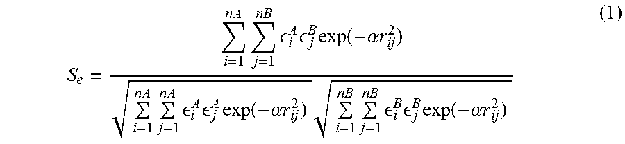
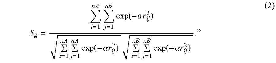
P00001

P00002

P00003

P00004
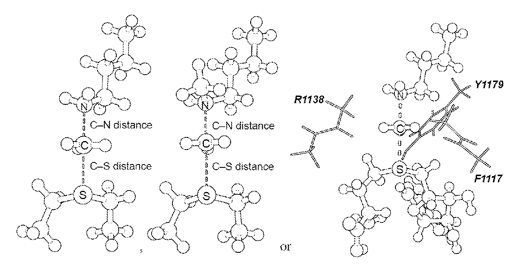
P00005

P00006
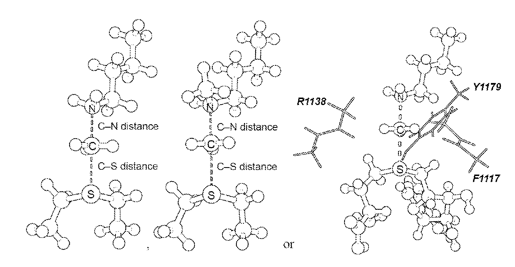
P00007
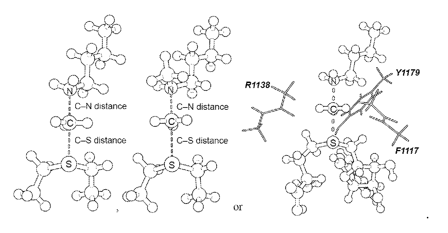
P00008

P00009
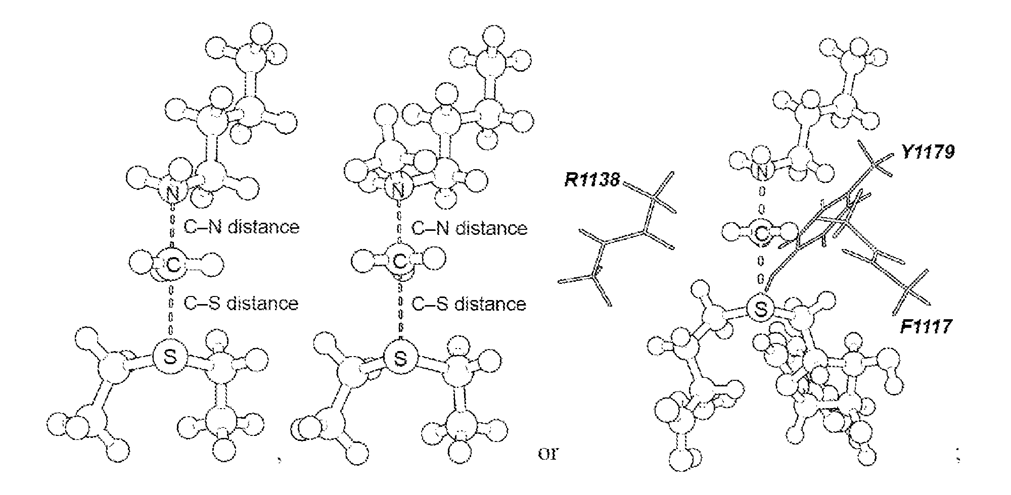
P00010

P00011
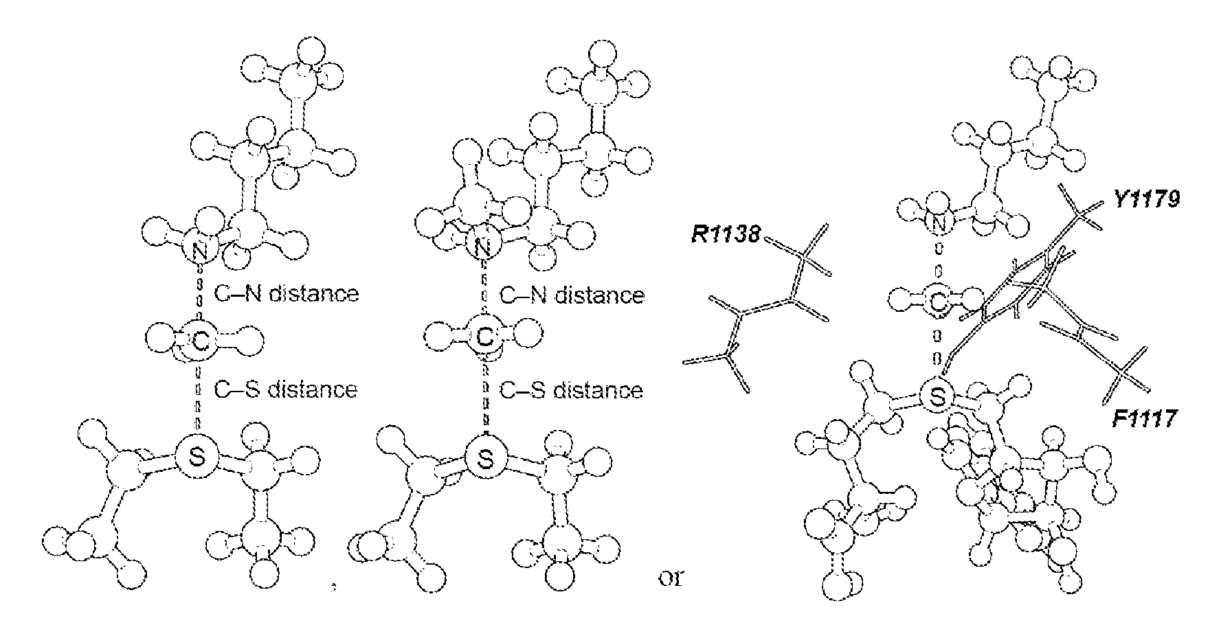
P00012
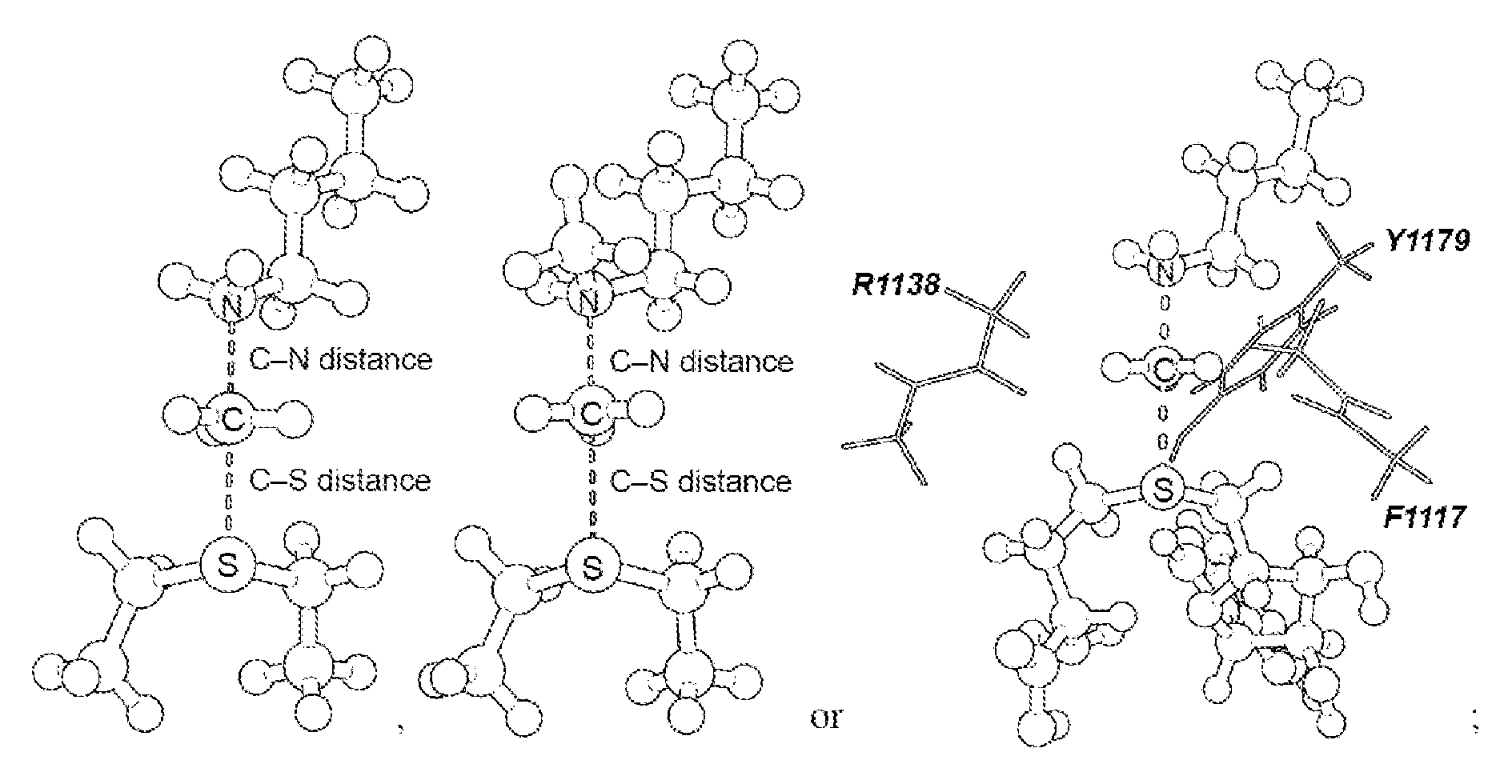
P00013
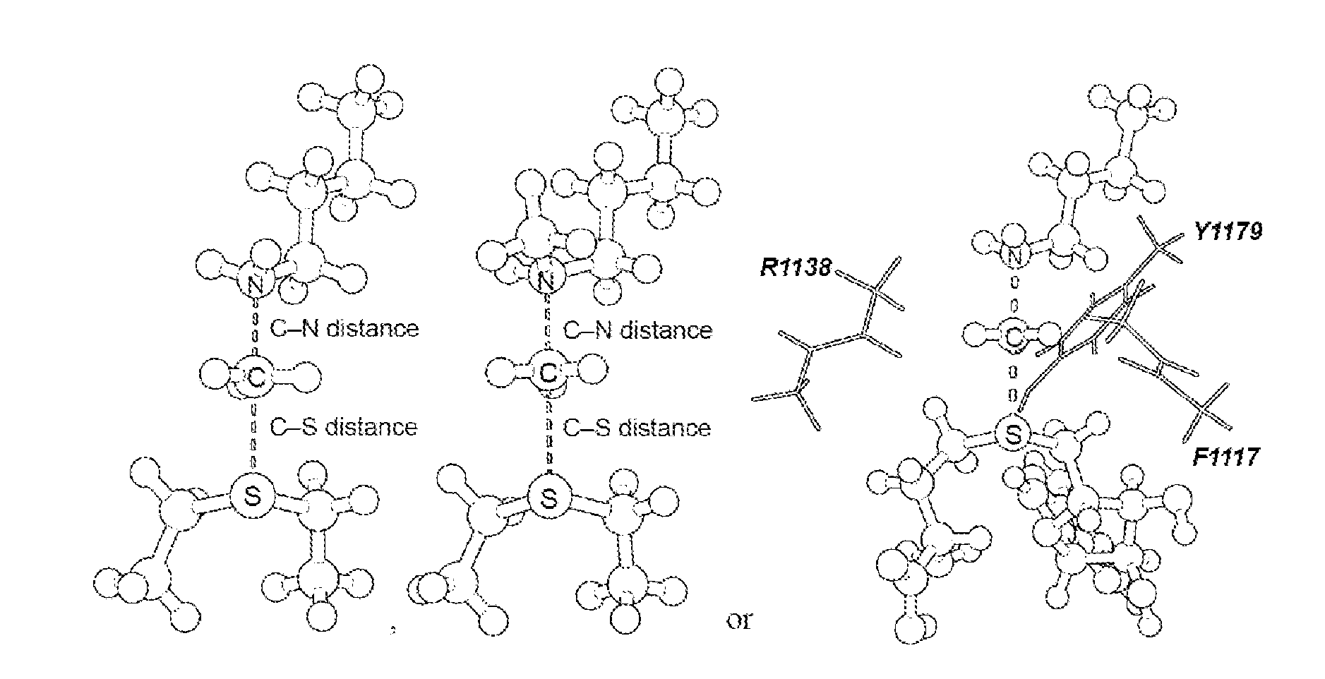
P00999
S00001
XML
uspto.report is an independent third-party trademark research tool that is not affiliated, endorsed, or sponsored by the United States Patent and Trademark Office (USPTO) or any other governmental organization. The information provided by uspto.report is based on publicly available data at the time of writing and is intended for informational purposes only.
While we strive to provide accurate and up-to-date information, we do not guarantee the accuracy, completeness, reliability, or suitability of the information displayed on this site. The use of this site is at your own risk. Any reliance you place on such information is therefore strictly at your own risk.
All official trademark data, including owner information, should be verified by visiting the official USPTO website at www.uspto.gov. This site is not intended to replace professional legal advice and should not be used as a substitute for consulting with a legal professional who is knowledgeable about trademark law.