Compositions For In Situ Labeling Of Bacterial Cell Walls With Fluorophores And Methods Of Use Thereof
Van Nieuwenhze; Michael S. ; et al.
U.S. patent application number 16/048000 was filed with the patent office on 2019-01-24 for compositions for in situ labeling of bacterial cell walls with fluorophores and methods of use thereof. The applicant listed for this patent is Indiana University and Technology Corporation. Invention is credited to Pamela Brown, Yves Brun, Edward Hall, Velocity Hughes, Erkin Kuru, Srinivas Tekkam, Michael S. Van Nieuwenhze.
| Application Number | 20190024135 16/048000 |
| Document ID | / |
| Family ID | 48191043 |
| Filed Date | 2019-01-24 |


View All Diagrams
| United States Patent Application | 20190024135 |
| Kind Code | A1 |
| Van Nieuwenhze; Michael S. ; et al. | January 24, 2019 |
COMPOSITIONS FOR IN SITU LABELING OF BACTERIAL CELL WALLS WITH FLUOROPHORES AND METHODS OF USE THEREOF
Abstract
Disclosed herein are compositions for assessing peptidoglycan biosynthesis in bacteria, for identifying bacteria, and for screening for bacterial cell wall-acting and/or cell wall-disrupting agents via modified D-amino acids and methods of use thereof. Also disclosed are live bacteria having one or more modified D-amino acids as described herein incorporated into peptidoglycan of a bacterial cell wall.
| Inventors: | Van Nieuwenhze; Michael S.; (Bloomington, IN) ; Hall; Edward; (Bloomington, IN) ; Kuru; Erkin; (Bloomington, IN) ; Brown; Pamela; (Columbia, MO) ; Tekkam; Srinivas; (Bloomington, IN) ; Hughes; Velocity; (Bloomington, IN) ; Brun; Yves; (Bloomington, IN) | ||||||||||
| Applicant: |
|
||||||||||
|---|---|---|---|---|---|---|---|---|---|---|---|
| Family ID: | 48191043 | ||||||||||
| Appl. No.: | 16/048000 | ||||||||||
| Filed: | July 27, 2018 |
Related U.S. Patent Documents
| Application Number | Filing Date | Patent Number | ||
|---|---|---|---|---|
| 14395815 | Oct 20, 2014 | |||
| PCT/US13/37504 | Apr 21, 2013 | |||
| 16048000 | ||||
| 61718048 | Oct 24, 2012 | |||
| 61636640 | Apr 21, 2012 | |||
| Current U.S. Class: | 1/1 |
| Current CPC Class: | C12Q 1/025 20130101; C07C 271/20 20130101; C07K 9/003 20130101; C12Q 1/18 20130101; C07D 311/74 20130101; C12Q 1/045 20130101 |
| International Class: | C12Q 1/04 20060101 C12Q001/04; C12Q 1/02 20060101 C12Q001/02; C12Q 1/18 20060101 C12Q001/18; C07D 311/74 20060101 C07D311/74; C07C 271/20 20060101 C07C271/20; C07K 9/00 20060101 C07K009/00 |
Goverment Interests
STATEMENT REGARDING FEDERALLY SPONSORED RESEARCH OR DEVELOPMENT
[0002] This invention was made with government support under AI059327 and GM051986 awarded by the National Institutes of Health. The government has certain rights in the invention.
Claims
1. An isolated, modified amino acid comprising a D-amino acid covalently attached to a fluorescent label.
2. The isolated, modified amino acid of claim 1, wherein the isolated, modified amino acid is selected from the group consisting of Cy.sub.3BADA, AF.sub.647ADA, AF.sub.350ADA, BADA, FADA, HADA, NADA, TADA, YADA, FDL, HDL, NDL, TDL, EDA, or ETDA.
3. The isolated, modified amino acid of claim 1, wherein the D-amino acid is selected from the group consisting of 3-amino-D-Ala, D-Ala, D-Asp, D-Cys, D-Glu and D-Lys.
4. A muramylpentapeptide precursor unit comprising an N-acetyl muramic acid (NAM) moiety having a stem peptide of three to five amino acids, wherein one or more of the amino acids in the stem peptide comprises a modified amino acid of claim 1 and optionally an additional modified amino acid, wherein the additional modified amino acid comprises a clickable D-amino acid.
5. A peptidoglycan unit comprising the muramylpentapeptide precursor unit of claim 4 covalently linked to an N-acetyl glucosamine (NAG) moiety.
6. A live bacterial organism comprising a bacterium having a modified cell wall comprising modified peptidoglycan containing at least one modified amino acid according to claim 1, and optionally at least one additional modified amino acid, wherein the at least one additional amino acid comprises a clickable D-amino acid.
7. A method of assessing bacterial cell wall synthesis in real time, the method comprising: providing live bacteria with a first amount of at least one modified amino acid of claim 1, and optionally a second amount of at least one additional modified amino acid comprising a clickable D-amino acid, under conditions sufficient for bacterial cell wall synthesis, wherein the bacteria covalently incorporate the at least one modified amino acid and optionally the at least one additional modified amino acid into a stem peptide of peptidoglycan of the bacterial cell wall.
8. The method of claim 7, wherein the first amount and second amount comprise a first concentration and a second concentration, respectively, wherein the first and second concentrations range from about and including 0.10 .mu.M to about and including 1 mM.
9. The method of claim 7, further comprising detecting the at least one modified amino acid, and optionally the at least one additional modified amino acid incorporated into the stem peptide.
10. The method of claim 7, wherein the bacteria are Gram-positive bacteria or Gram-negative bacteria.
11. A method of screening for a putative cell wall-acting agent, the method comprising: co-contacting bacteria with an effective amount of an agent and an amount of at least one modified amino acid of claim 1, and optionally an amount of at least one additional modified amino acid comprising a clickable D-amino acid, under conditions sufficient to permit ongoing peptidoglycan biosynthesis in a bacterial cell wall, wherein the agent comprises a cell wall-acting agent if the agent interferes with ongoing peptidoglycan biosynthesis in the bacterial cell wall.
12. The method of claim 11, further comprising: detecting one or more D-amino acids incorporated in the bacterial cell wall.
13. The method of claim 12, further comprising: comparing the amount and/or identity of incorporated D-amino acids in the bacterial cell wall resulting from contacting the bacteria with the agent with the corresponding amount and/or identity of incorporated D-amino acids in a bacterial cell wall resulting from contacting the bacteria with a known cell wall-acting agent.
14. A method of screening for a putative cell wall-disrupting agent, the method comprising: contacting modified bacteria with an amount of an agent, wherein the agent comprises a cell wall-disrupting agent if the agent weakens integrity of peptidoglycan in an existing bacterial cell wall, and wherein the modified bacteria have a modified cell wall containing modified peptidoglycan having at least one stem peptide containing at least one modified amino acid of claim 1, and optionally at least one additional modified amino acid comprising a clickable D-amino acid.
15. The method of claim 14, further comprising: detecting one or more D-amino acids disrupted in the bacterial cell wall.
16. The method of claim 15, further comprising: comparing the amount and/or identity of disrupted D-amino acids in the bacterial cell wall resulting from contacting the bacteria with the agent with the corresponding amount and/or identity of disrupted D-amino acids in a bacterial cell wall resulting from contacting the bacteria with a known cell wall-disrupting agent.
17. The method of claim 11, wherein the bacteria are Gram-positive bacteria or Gram-negative bacteria.
18. A method of identifying bacteria, the method comprising: contacting live bacteria with an amount of at least one modified amino acid of claim 1, and optionally an amount of at least one additional modified amino acid comprising a clickable D-amino acid, under conditions sufficient for ongoing bacterial cell wall synthesis, wherein the bacteria covalently incorporate into peptidoglycan of a bacterial cell wall the at least one modified amino acid, and optionally the at least one additional modified amino acid, wherein each of the least one modified amino acid and optionally the at least one additional modified amino acid comprises spectrally distinct fluorescent label; and visualizing the spectrally distinct fluorescent labels to determine an incorporation pattern of the at least one modified amino acid, and optionally the at least one additional modified amino acid, wherein the incorporation pattern identifies the bacteria.
19. A kit for incorporating labeled D-amino acids into live bacteria, the kit comprising: at least one modified amino acid of claim 1; and a positive bacterial control and optionally a negative bacterial control, wherein the positive bacterial control has at least one modified amino acid of claim 1 incorporated into a stem peptide of peptidoglycan of the bacterial cell wall, wherein the optional negative bacterial control, if included, does not have the modified amino acid of claim 1 incorporated into a stem peptide of peptidoglycan of the bacterial cell wall.
20. The kit of claim 17, further comprising at least one clickable D-amino acid.
Description
CROSS-REFERENCE TO RELATED APPLICATIONS
[0001] The present application is a continuation of U.S. patent application Ser. No. 14/395,815, filed Oct. 20, 2014, which is the National Stage of International Application No. PCT/US13/37504, filed Apr. 21, 2013, and entitled "COMPOSITIONS FOR IN SITU LABELING OF BACTERIAL CELL WALLS WITH FLUOROPHORES AND METHODS OF USE THEREOF," which claims benefit of priority under 35 U.S.C. 119 to U.S. provisional patent application Ser. No. 61/636,640, filed Apr. 21, 2012, and entitled "COMPOSITIONS FOR COVALENTLY LABELING BACTERIAL CELL WALLS WITH FLUOROPHORES AND METHODS OF USE," and U.S. provisional patent application Ser. No. 61/718,048, filed Oct. 24, 2012, and entitled "COMPOSITIONS FOR IN SITU LABELING OF BACTERIAL CELL WALLS WITH FLUOROPHORES AND METHODS OF USE THEREOF," the contents of both which are herein incorporated by reference in their entireties.
FIELD OF THE INVENTION
[0003] The invention relates generally to microbiology, and more particularly to compositions and methods for assessing cell wall synthesis in bacteria, for identifying bacteria, and for screening for cell wall-acting/-disrupting agents.
BACKGROUND
[0004] Bacterial growth is controlled by the domain-specific peptidoglycan (PG) cell wall, a rigid and essential structure composed of glycan strands cross-linked by D-amino acid (DAA)-containing short peptides, whose biosynthesis machinery is a target for antibiotics.
[0005] Despite the importance of PG, knowledge of its dynamics has been severely hampered by lack of a strategy for direct imaging of sites of PG biosynthesis in live cells. Significant limitations of current labeling methods, such as toxic effects and poor membrane permeability of the probes, have limited their applicability to only a small set of bacterial species. Moreover, these methods are labor-intensive and their sensitivity suffers from their indirect and multiple-step nature.
[0006] Methods relying on fluorescently labeled antibiotics to study bacterial cell wall synthesis and to discover new antibiotics to which bacteria remain susceptible have had a profound impact on the field. The current methods, however, have at least two inherent limitations. First, antibiotic concentration needs to be carefully controlled to avoid damage to the cell. Second, because these agents bind to specific sites on cell surfaces, they only will appear at sites of active PG biosynthesis.
[0007] For the foregoing reasons, there is a need for additional compositions and methods for assessing PG biosynthesis in bacteria and for discovering bacterial cell wall-acting/-disrupting agents that can be used to treat infections caused by multidrug-resistant bacteria.
BRIEF SUMMARY
[0008] In a first respect, a modified amino acid is disclosed that includes a D-amino acid covalently attached to a fluorescent label.
[0009] In a second respect, a muramylpentapeptide precursor unit is disclosed that includes an N-acetyl muramic acid (NAM) moiety having a stem peptide of three to five amino acids. One or more of the amino acids in the stem peptide includes a modified amino acid that includes a D-amino acid covalently attached to a fluorescent label and optionally an additional modified amino acid. The additional modified amino acid includes a clickable D-amino acid.
[0010] In a third respect, a peptidoglycan unit is disclosed that includes a muramylpentapeptide precursor unit as described above in the second respect that is covalently linked to an N-acetyl glucosamine (NAG) moiety.
[0011] In a fourth respect, a method of assessing bacterial cell wall synthesis in real time is described. The method includes the step of providing live bacteria with a first amount of at least one modified amino acid comprising a D-amino acid covalently attached to a fluorescent label, and optionally a second amount of at least one additional modified amino acid comprising a clickable D-amino acid, under conditions sufficient for bacterial cell wall synthesis. The bacteria covalently incorporate the at least one modified amino acid and optionally the at least one additional modified amino acid into a stem peptide of peptidoglycan of the bacterial cell wall.
[0012] In a fifth respect, a method of screening for a putative cell wall-acting agent is disclosed. The method includes the step of co-contacting bacteria with an effective amount of an agent and an amount of at least one modified amino acid comprising a D-amino acid covalently attached to a fluorescent label, and optionally an amount at least one additional modified amino acid comprising a clickable D-amino acid, under conditions sufficient to permit ongoing peptidoglycan biosynthesis in a bacterial cell wall. The agent comprises a cell wall-acting agent if the agent interferes with ongoing peptidoglycan biosynthesis in the bacterial cell wall.
[0013] In a sixth respect, a method of screening for a putative cell wall-disrupting agent is disclosed. The method includes the step contacting modified bacteria with an amount of an agent. The agent is a cell wall-disrupting agent if the agent weakens integrity of peptidoglycan in an existing bacterial cell wall. In this method, the modified bacteria have a modified cell wall containing modified peptidoglycan having at least one stem peptide containing at least one modified amino acid comprising a D-amino acid covalently attached to a fluorescent label, and optionally at least one additional modified amino acid comprising a clickable D-amino acid.
[0014] In a seventh respect, a method of identifying bacteria is disclosed. The method includes two steps. The first step includes contacting live bacteria with an amount of at least one modified amino acid comprising a D-amino acid covalently attached to a fluorescent label, and optionally an amount of at least one additional modified amino acid comprising a clickable D-amino acid, under conditions sufficient for ongoing bacterial cell wall synthesis. The bacteria covalently incorporate into peptidoglycan of a bacterial cell wall the at least one modified amino acid, and optionally the at least one additional modified amino acid. Each of the least one modified amino acid and optionally the at least one additional modified amino acid comprises a spectrally distinct fluorescent label. The second step includes visualizing the spectrally distinct fluorescent labels to determine an incorporation pattern of the at least one modified amino acid, and optionally the at least one additional modified amino acid, wherein the incorporation pattern identifies the bacteria.
[0015] In an eighth respect, a kit for incorporating labeled D-amino acids into live bacteria is disclosed. The kit includes at least one modified amino acid comprising a D-amino acid covalently attached to a fluorescent label and a positive bacterial control. The kit can include an optional negative bacterial control. The positive bacterial control has at least one modified amino acid comprising a D-amino acid covalently attached to a fluorescent label incorporated into a stem peptide of peptidoglycan of the bacterial cell wall. The optional negative bacterial control, if included, does not have the modified amino acid comprising a D-amino acid covalently attached to a fluorescent label incorporated into a stem peptide of peptidoglycan of the bacterial cell wall.
[0016] These and other features, objects and advantages of the present invention will become better understood from the description that follows. In the description, reference is made to the accompanying drawings, which form a part hereof and in which there is shown by way of illustration, not limitation, embodiments of the invention.
BRIEF DESCRIPTION OF THE DRAWINGS
[0017] The features, objects and advantages other than those set forth above will become more readily apparent when consideration is given to the detailed description below. Such detailed description makes reference to the following drawings.
[0018] FIG. 1 shows the three general stages of PG biosynthesis and general structures of the NAM and NAG units of PG.
[0019] FIG. 2 shows exemplary D-Ala-based FDAAs. D-NBD and D-HCC (based on (R)-diaminopropionic acid) emit in the green and blue regions, respectively.
[0020] FIG. 3 shows results of control experiments in which the cell walls of Agrobacterium tumefaciens ("A. tumefaciens" top row), Bacillus subtilis ("B. subtilis" middle row) and Escherichia coli ("E. coli" bottom row) were fluorescently labeled with fluorescent D-Ala (D-HCC) or fluorescent L-Ala (L-HCC). An exemplary structure for D-Ala (D-HCC) also is shown in the bottom row.
[0021] FIG. 4 shows results of a pulse chase experiment with a fluorescent D-Ala in B. subtilis (top row) or A. tumefaciens (bottom row).
[0022] FIG. 5 shows results of a short pulse experiment with fluorescent D-Ala in B. subtilis.
[0023] FIG. 6 shows results of a fluorescent D-Ala derivative in a dual-labeling format.
[0024] FIG. 7 shows exemplary structures for FDAAs, such as HCC--OH-labeled 3-amino-D-Ala (HADA), NBD-Cl-labeled 3-amino-D-Ala (NADA), F-labeled D-Lys (FDL) and T-labeled D-Lys (TDL), as well as exemplary structures for CDAAs, such as EDA and ADA.
[0025] FIG. 8 shows that long labeling pulses with HADA uniformly label PG in live Escherichia coli (left), Bacillus subtilis (center) and Agrobacterium tumefaciens (left). The FDAA fluorescence was retained in isolated sacculi, which also stained with a NAG-specific wheat germ agglutinin (WAG) lectin conjugated to Alexa Fluor.RTM. 594 (red). Scale bars, 2 .mu.m.
[0026] FIGS. 9A-D show FDAA incorporation into the stem peptide of the PG unit.
[0027] FIG. 9A shows a schematic representing the muramylpentapeptide precursor as incorporated into a nascent PG unit and a modified D-amino acid (FDAA).
[0028] FIG. 9B shows HPLC detection of modified muropeptides in E. coli incubated with HADA, HALA and NADA, or NALA. Samples were monitored using a dual wavelength UV monitor set for general muropeptide detection and for FDAA-specific wavelengths. Peaks HEC-1 (panel (i)) and NEC-1 (panel (ii)) correspond to the HADA- or NADA-modified muropeptides in E. coli that were further characterized by electrospray ionization MS/MS (ESI-MS/MS).
[0029] FIG. 9C shows percentage of FDAA incorporation into the total muropeptides varies among bacteria as revealed by HPLC analysis.
[0030] FIG. 9D shows a schematic representing MS/MS analyses of FDAAs exclusively incorporated into the 5th position in B. subtilis (panel (i)) and the 4th position of muropeptides in E. coli and A. tumefaciens (panel (ii)).
[0031] FIGS. 10A-F show FDAAs label diverse bacterial growth patterns. Arrows in the triple labeling panels indicate the sequence of labeling. White scale bars, 2 .mu.m, red scale bars, 1 .mu.m.
[0032] FIG. 10A shows time-lapse microscopy of HADA-labeled E. coli and B. subtilis .DELTA.dacA cells imaged during growth on LB agarose pads.
[0033] FIG. 10B shows super-resolution microscopy of E. coli after short pulses with HADA (panels (i) and (ii)).
[0034] FIG. 10C show super-resolution microscopy of A. tumefaciens after short pulses with HADA.
[0035] FIG. 10D shows super-resolution microscopy of S. aureus after a short pulse with HADA. Autofluorescence is shown in red.
[0036] FIG. 10E shows triple labeling of A. tumefaciens with HADA (blue), EDA (clicked with red sulfo-Cy3-azide) and NADA (green).
[0037] FIG. 10F shows triple labeling of S. venezuelae with NADA (green), TDL (red) and HADA (blue).
[0038] FIG. 11 shows that short pulses of HADA label distinct modes of growth in diverse bacteria. Strains were labeled for .about.2%-8% of the doubling time: E. coli (30 seconds), A. tumefaciens (2 minutes), B. subtilis .DELTA.dacA (30 seconds), S. aureus (2 minutes), L. lactis (2 minutes), S. pneumoniae (4 minutes), C. crescentus (5 minutes), Synechocystis sp. PCC 6803 (1 hour), S. venezuelae (2 minutes), B. conglomeratum (8 minutes), B. phytofirmans (20 minutes), V. Spinosum (10 minutes). Scale bars, 2 .mu.m.
[0039] FIG. 12 shows a schematic for sequentially incorporating distinct FDAAs, such as NADA, TDL and HADA, into newly synthesized PG in live bacteria.
[0040] While the present invention is amenable to various modifications and alternative forms, exemplary embodiments thereof are shown by way of example in the drawings and are herein described in detail. It should be understood, however, that the description of exemplary embodiments is not intended to limit the invention to the particular forms disclosed, but on the contrary, the intention is to cover all modifications, equivalents and alternatives falling within the spirit and scope of the invention as defined by the embodiments above and the claims below. Reference should therefore be made to the embodiments and claims herein for interpreting the scope of the invention.
DETAILED DESCRIPTION
[0041] The compositions and methods now will be described more fully hereinafter with reference to the accompanying drawings, in which some, but not all permutations and variations of embodiments of the invention are shown. Indeed, the invention may be embodied in many different forms and should not be construed as limited to the embodiments set forth herein. These embodiments are provided in sufficient written detail to describe and enable one skilled in the art to make and use the invention, along with disclosure of the best mode for practicing the invention, as defined by the claims and equivalents thereof.
[0042] Likewise, many modifications and other embodiments of the compositions and methods described herein will come to mind to one of skill in the art to which the invention pertains having the benefit of the teachings presented in the foregoing descriptions and the associated drawings. Therefore, it is to be understood that the invention is not to be limited to the specific embodiments disclosed and that modifications and other embodiments are intended to be included within the scope of the appended claims. Although specific terms are employed herein, they are used in a generic and descriptive sense only and not for purposes of limitation.
[0043] Unless defined otherwise, all technical and scientific terms used herein have the same meaning as commonly understood by one of skill in the art to which the invention pertains. Although any methods and materials similar to or equivalent to those described herein can be used in the practice or testing of the present invention, the preferred methods and materials are described herein.
[0044] Moreover, reference to an element by the indefinite article "a" or "an" does not exclude the possibility that more than one element is present, unless the context clearly requires that there be one and only one element. The indefinite article "a" or "an" thus usually means "at least one."
[0045] As used herein, "about" means within a statistically meaningful range of a value or values such as a stated concentration, length, molecular weight, pH, sequence identity, time frame, temperature or volume. Such a value or range can be within an order of magnitude, typically within 20%, more typically within 10%, and even more typically within 5% of a given value or range. The allowable variation encompassed by "about" will depend upon the particular system under study, and can be readily appreciated by one of skill in the art.
Overview
[0046] Previous efforts to label PG in live bacteria principally have relied upon cell wall-active antibiotics (e.g., vancomycin, ramoplanin) modified with fluorophores or cell wall precursors/substrates covalently modified with fluorescent reporter groups. The compositions and methods described herein, however, take advantage of mechanisms for incorporating labeled DAAs into the stem peptides displayed on a bacterial cell wall surface.
[0047] The work described herein demonstrates how to make derivatized DAA having a suitable label, such as an appropriate fluorophore and how such derivatized compounds can be visualized in live cells by fluorescence microscopy following the incorporation of the derivatized compounds into PG and thus the cell wall. In the range of physiologically relevant concentrations, the incorporated FDAAs do not appear to be toxic to bacteria. Unlike previous methods that employ covalently modified cell wall precursors, the methods described herein do not appear to adversely affect cell morphology. In addition, the methods described herein enable pulse-chase experiments that cannot be easily executed in the presence of fluorescently-modified cell wall active drugs. Because the disclosed derivatized compounds have low or minimal toxicity to live cells, they are ideal markers to evaluate and screen microbiostatic or microbiotoxic compounds that do adversely affect microorganism growth and viability, such as studies directed to development of novel antibiotics.
[0048] Studies disclosed herein demonstrate that the compositions and methods are applicable to a wide array of Gram-positive and Gram-negative bacteria and provides significant utility for probing PG biosynthesis, cell wall morphogenesis and the response of the PG biosynthetic machinery to cell wall-active agents and/or cell wall-disrupting agents. The present disclosure therefore provides compositions and methods for studying bacterial cell wall PG biosynthesis and for discovering bacterial cell wall-acting and/or cell wall-disrupting agents.
Compositions
[0049] Fluorescent D-Amino Acids (FDAAs)
[0050] Compositions of the invention include labeled D-amino acids (DAAs), especially fluorescent D-amino acids (FDAAs). As used herein, "amino acid" or "amino acid residue" are used interchangeably to mean a molecule containing a first, or alpha, carbon attached to an amine group, a carboxylic acid group and a side-chain that is specific to each amino acid. A natural amino acid can include conventional elements such as carbon, hydrogen, oxygen, nitrogen and sulfur. An amino acid may be a naturally occurring amino acid or artificially-created unnaturally occurring amino acid. Preferably, the amino acid is naturally occurring, and, unless otherwise limited, may encompass known analogues/synthetics of natural amino acids that can function in a similar manner as naturally occurring amino acids. With the exception of glycine, the natural amino acids all contain at least one chiral carbon atom. These amino acids therefore exist as pairs of stereoisomers (D- and L-isomers). Of particular interest herein are D-isomers or D-amino acids, particularly D-Ala, D-Asp, D-Cys, D-Glu and D-Lys, which are frequently found in the stem peptide of the PG unit.
[0051] It is well known in the art that amino acids within the same conservative group typically can substitute for one another without substantially affecting the function of a protein. For the purpose of the present disclosure, such conservative groups are set forth in Table 1 and are based preferably on shared properties, as readily appreciated to those skilled in the art. See also, Alberts et al., "Small molecules, energy, and biosynthesis" 56-57 In: Molecular Biology of the Cell (Garland Publishing Inc. 3.sup.rd ed. 1994).
TABLE-US-00001 TABLE 1 Amino Acids and Their Conservative Substitutions. Preferred Side Chain Side Chain Hydropathy Conservative Residue Polarity pH Index Substitutions Ala (A) Non-polar Neutral 1.8 Ser Arg (R) Polar Basic (strongly) -4.5 Lys, Gln Asn (N) Polar Neutral -3.5 Gln, His Asp (D) Polar Acidic -3.5 Glu Cys (C) Non-polar Neutral 2.5 Ser Gln (Q) Polar Neutral -3.5 Asn, Lys Glu (E) Polar Acidic -3.5 Asp Gly (G) Non-polar Neutral -0.4 Pro His (H) Polar Basic (weakly) -3.2 Asn, Gln Ile (I) Non-polar Neutral 4.5 Leu, Val Leu (L) Non-polar Neutral 3.8 Ile, Val Lys (K) Polar Basic -3.9 Arg, Gln Met (M) Non-polar Neutral 1.9 Leu, Ile Phe (F) Non-polar Neutral 2.8 Met, Leu, Tyr Pro (P) Non-polar Neutral -1.6 Gly Ser (S) Polar Neutral -0.8 Thr Thr (T) Polar Neutral -0.7 Ser Trp (W) Non-polar Neutral -0.9 Tyr Tyr (Y) Polar Neutral -1.3 Trp, Phe Val (V) Non-polar Neutral 4.2 Ile, Leu
[0052] The following six groups each contain amino acids that are typical but not necessarily exclusive conservative substitutions for one another: 1) Alanine (A), Serine (S) and Threonine (T); 2) Aspartic acid (D) and Glutamic acid (E); 3) Asparagine (N) and Glutamine (Q); 4) Arginine (R) and Lysine (K); 5) Isoleucine (I), Leucine (L), Methionine (M), and Valine (V); and 6) Phenylalanine (F), Tyrosine (Y) and Tryptophan (W).
[0053] Examples of suitable labels for the DAAs include, but are not limited to, radiolabels, biotin (which may be detected by avidin or streptavidin conjugated to peroxidase), lanthanides, alkaline phosphatase and fluorescent labels (e.g., coumarins, fluoresceins, cyanines, bodipy dyes, green fluorescent protein, quantum dots rhodamine, especially the Alexa Fluor.RTM. family of fluorescent dyes available from Invitrogen/Molecular Probes). Other labels amenable for use in the modified D-amino acids disclosed herein include metals and isotopic labels.
[0054] Labeling of DAAs can be carried out by covalently attaching the label to a free amine group, such as free amine groups present on the side-chain that is specific to each amino acid. If the side chain lacks a free amine group, one of skill in the art understands how to add such groups, as is the case of adding such a group to D-Ala to obtain 3-amino-D-Ala. Some labels can be detected by using a labeled counter suitable for the detection of the label in question. In the Examples below, 7-hydroxycoumarin 3-carboxylic acid (HCC--OH), 7-nitrobenzofurazan (NBD), 4-chloro-7-nitrobenzofurazan (NBD-Cl), fluorescein (F) and carboxytetramethylrhodamine (T) were covalently attached to DAAs as labels.
[0055] Other coupling chemistries are known in the art that can be used for introducing labels into amino acids having functional groups other than an amine. Such amino acids include a functional alcohol group (e.g., serine and tyrosine), thiol group (e.g., cysteine), or carbonyl or carboxylate group (e.g., aspartate and glutamate). Such functional groups can be derivatized or reacted with suitably modified, activated coupling agents having labels of the types disclosed herein.
[0056] An example of a FDAA includes HADA, which is a HCC--OH-labeled 3-amino-D-Ala. Another example of a FDAA includes NADA, which is a NBD-Cl-labeled 3-amino-D-Ala. Another example includes FDL, which is a F-labeled D-Lys. Another example is TDL, which is a T-labeled D-Lys. Another example includes HDL, which is a HCC--OH-labeled D-Lys. Another example includes NDL, which is a NBD-Cl-labeled D-Lys. Another example includes FADA, which is a F-labeled 3-amino-D-Ala. Another example includes TADA, which is a T-labeled 3-amino-D-Ala. Other FDAAs can include a D-Glu having its side chain modified to include a free amine group linked to any of the fluorescent labels above (e.g., HADG, NADG, FADG and TADG).
[0057] See, e.g., FIG. 7, for other examples of preferred labels and modified FDAAs.
[0058] Methods of fluorescently labeling and detecting amino acids are well known in the art. See, Braun & Dittrich (2010) Beilstein J. Org. Chem. 6:69; Katritzky & Narindoshvili (2009) Org. Biomol. Chem. 7:627-634; Merkel et al. (2010) Chembiochem. 11:305-314; Cava et al. (2011) Cell Mol. Life Sci. 68:817-831; Lam et al. (2009) Science 325:1552-1555; Cava et al. (2011) EMBO J. 30:3442-3453; and Lupoli et al. (2011) J. Am. Chem. Soc. 133:10748-10751.
[0059] Clickable D-Amino Acids (CDAA's)
[0060] Compositions of the invention also include clickable D-amino acids (CDAAs). The CDAAs have a DAA backbone that includes, for example, an alkyne or azide functional group present on the side-chain that is specific to each amino acid that can be captured in situ by a labeled, detecting agent carrying a conjugate functional group via click-chemistry. Functional groups in a DAA backbone that can be targeted by the labeled, detecting agent include, but are not limited to, primary amines, carboxyls, sulfhydryls, carbohydrates and carboxylic acids.
[0061] One of skill in the art is familiar with "click" chemistry, which utilizes chemical cross-linking agents to add functional groups to molecules. See, Kolb et al. (2001) Angew. Chem. Int. Ed. 40:2004-2021; and Evans (2007) Aust. J. Chem. 60:384-395.
[0062] Cross-linking and enrichment strategies for separating a cross-linking reaction from enrichment steps have been developed based on bioorthogonal chemistries including the azide-alkyne "click" cycloaddition and Staudinger ligation using alkyne- or azide-labeled cross-linking agents (e.g., fluorescent labels). Azides and alkynes are not naturally found in proteins, peptides, nucleic acids or glycans; therefore, these moieties can be engineered onto the DAAs and labeled, detecting agent to generate azide-containing molecules and alkyne-containing molecules that are reactive with one another. As such, the orthogonality of azides and alkynes to biological processes (e.g., competing reactions) is a significant advantage of these methods. Moreover, "click" cycloadditions can be performed under aqueous conditions, allowing enrichment by conjugation of an appropriate affinity or labeling tag. See, generally, Rostovtsev et al. (2002) Angew. Chem. Int. Ed. 41:2596-2599; Tornoe et al. (2002) J. Org. Chem. 67:3057-3064; Baskin et al. (2007) Proc. Natl. Acad. Sci. USA 104:16793-16797; Saxon et al. (2000) Science 287:2007-2010; Chowdhury et al. (2009) Anal. Chem. 81:5524-5532; Trnka & Burlingame (2010) Mol. Cell. Proteomics 9:2306-2317; Nessen et al. (2009) J. Proteome Res. 8:3702-3711; Vellucci et al. (2010) J. Am. Soc. Mass Spectrom. 21:1432-1445; and Jewett & Bertozzi (2010) Chem. Soc. Rev. 39:1272-1279. See also, Int'l Patent Application Publication No. WO 2012/006603. As used herein, "click chemistry" and "clickable" therefore mean a reaction between azide-containing molecules and alkyne-containing molecules to yield a covalent product-1,5-disubstituted 1,2,3-triazole. The reaction can be a copper(I)-catalyzed alkyne azide cycloaddition (CuAAC) or, in cases where copper toxicity may be an issue, can be a copper(I)-free-catalyzed alkyne azide cycloaddition.
[0063] Examples of functional groups for use on azide-containing molecules and alkyne-containing molecules include, but are not limited to, hexynyl groups, pentynyl groups, heptynyl groups, azido-propyl groups, azido-butyl groups and azido-pentyl groups. When the functional group of alkyne-containing molecules (e.g., DAAs) is an alkynyl group (e.g., hexynyl, pentynyl or heptynyl), the functional group of azide-containing molecules (e.g., labeled, detecting agents) has the corresponding clickable azido group. Likewise, when the functional group of azide-containing molecules (e.g., DAAs) is an azide group (e.g., azido-propyl, azido-butyl, or azido-pentyl), the functional group of alkyne-containing molecules (e.g., labeled, detecting agents) has the corresponding clickable alkynyl group.
[0064] As such, various labeling designs for detecting agents are known including, but not limited to, biotinylated agents, isotope-coded agents, fluorophore-labeled agents, mass-tag-labeled agents and chromophore-labeled agents. It is also known that the addition of functional groups can cause the cross-linker to become very bulky or less cell-permeable, and thus not very effective for in vivo and/or in situ cross-linking. To reduce the total size of the cross-linker, separation of the cross-linking step from conjugation of affinity tags can be one effective strategy. See, Trester-Zedlitz et al. (2003) J. Am. Chem. Soc. 125:2416-2425; Tang et al. (2005) Anal. Chem. 77:311; Kang et al. (2009) Rapid Commun. Mass Spectrom. 23:1719-1726; Chu et al. (2006) J. Am. Chem. Soc. 128:10362-10636; Muller et al. (2001) Anal. Chem. 73:1927-1934; Collins et al. (2003) Bioorg. Med. Chem. Lett. 13:4023-4026; Petrotchenko et al. (2005) Mol. Cell. Proteomics 4:1167-1179; Wine et al. (2002) Anal. Chem. 74:1939-1945; Sinz et al. (2001) Biochemistry 40:7903-7913; and Sinz & Wang (2004) Anal. Biochem. 331:27-32.
[0065] In some instances, the detecting agents, whether having an alkynyl or azido functional group, can be fluorophore-labeled. Examples of fluorophores include, but are not limited to, Alexa Fluor.RTM. dyes, BODIPY.RTM. dyes, fluorescein, Oregon Green.RTM. 488 and Oregon Green.RTM. 514 dyes, Rhodamine Green and Rhodamine Green-X dyes, eosin, tetramethylrhodamine, Lissamine Rhodamine B and Rhodamine Red-X dyes, X-Rhodamine, Texas Red.RTM. and Texas Red.RTM.-X dyes, naphthofluorescein, Carboxyrhodamine 6G, QSY dyes: fluorescence quenchers, nonfluorescent malachite green, coumarin derivatives, Pacific Orange dye, cascade blue and other pyrene derivatives, cascade yellow and other pyridyloxazole derivatives, naphthalenes (e.g., dansyl chloride), dapoxyl dye, bimane, 1-dimethylamine-N(2-azido-ethyl) naphthalene-5-sulfonamide, 6-(6-amino-2-(2-azidoethyl)1,3-dioxo-1H-benzo(de)-2(3H)isoquinoline, 6-(6-amino-2-(2-propinyl)1,3-dioxo-1H-benzo(de)-2(3H)isoquinoline, 8-(4-azidoethyloxyphenyl)-2,6-diethyl-1,3,5,7-tetramethyl-4,4-difluoro-4-- bora-3a,4a-diaza-s-indacene, 8-(4-propynyloxyphenyl)-2,6-diethyl-1,3,5,7-tetramethyl-4,4-difluoro-4-bo- ra-3a,4a-diaza-s-indacene, 1-(3-azido-propoxy)-7-methylamino-phenoxazin-3-one, 1-(2-propynyl)-7-methylamino-phenoxazin-3-one, N-(5-(3-azidopropylamino)-9H-benzo(a)-phenoxazin-9-ylidene)-N-methyl-meth- anaminium chloride, N-(5-(3-propynyl-amino)-9H-benzo(a)-phenoxazin-9-ylidene)-N-methyl-methan- aminium chloride, (9-(3-azido-propoxy)-7-piperidin-1-yl-phenoxazin-3-ylidene)-dimethyl-ammo- nium perchlorate. See also, Kele et al. (2009) Org. Biomol. Chem. 7:3486-3490; Nagy et al. (2010) Chem. Asian J. 5:773-777; Filnov et al. (2011) Nat. Biotechnol. 29:757-761; Subach et al. (2011) Nature Methods 8:771-777; Yang et al. (2011) J. Am. Chem. Soc. 133:9964-9967; Zin (2011) Nature Methods 8:726-728; and "Fluorophores and their amine-reactive derivatives," Chapter 1 and "Click Chemistry and other functional group modifications," Chapter 3 in The Molecular Probes.RTM. Handbook (available on the World Wide Web at invitrogen.com/site/us/en/home/References/Molecular-Probes-The-Handbook.h- tml). A variety of clickable fluorophores are commercially available from, for example Sigma Aldrich, Active Motif Chromeon and Invitrogen/Molecular Probes. In the examples below, CDAAs were clicked with red sulfo-Cy3-azide.
[0066] An example of a CDAA includes EDA. Another example of a CDAA includes ADA. See, e.g., FIG. 7.
[0067] Fluorescent Muramylpentapeptide Precursor Units (FMPUs)
[0068] Compositions of the invention also include fluorescent muramylpentapeptide precursor units (FMPUs) having an NAM moiety with a peptide chain of three to five amino acids in which one or more of the amino acids in the stem peptide are FDAAs and/or CDAAs as described herein. See, e.g., FIGS. 9A and 9D.
[0069] Fluorescent Peptidoglycan Units (FPGUs)
[0070] Compositions of the invention also include fluorescent peptidoglycan units (FPGUs). The FPGUs have a FMPU as described herein linked to a NAG moiety. See, e.g., FIGS. 9A and 9D.
[0071] Bacterial Cells Having Fluorescent D-Amino Acids
[0072] Compositions of the invention also include live bacteria having FDAAs, CDAAs, FMPUs and/or FPGUs as described herein incorporated into PG in a cell wall.
[0073] While Gram-positive bacteria tend to have a thicker PG layer, it is intended that the bacteria can be Gram-positive bacteria or Gram-negative bacteria. Examples of suitable Gram-positive bacteria include, but are not limited to, Actinomyces spp., Bacillus spp., Brachybacterium spp., Clostridium spp., Corynebacterium spp., Diplococcus spp., Enterococcus spp., Lactococcus spp., Listeria spp., Nocardia spp., Propionibacterium spp., Staphylococcus spp., Streptococcus spp. Streptomyces spp. In the examples below, live B. subtilis, B. conglomeratum, L. lactis, S. aureus, S. pneumoniae, S. venezuelae, were grown in the presence of FDAAs and/or CDAAs.
[0074] Examples of suitable Gram-negative bacteria include, but are not limited to, Acinetobacter spp., Agrobacterium spp., Bordetella spp., Borrelia spp., Brucella spp., Burkholderia spp., Campylobacter spp., Caulobacter spp., Chlamydia spp., Enterobacter spp., Escherichia spp., Helicobacter spp., Hemophilus spp., Klebsiella spp., Legionella spp., Neisseria spp., Proteus spp., Pseudomonas spp, Salmonella spp., Shigella spp., Synechocystis spp., Verrucomicrobia spp., Vibrio spp. and Yersina spp. In the examples below, live A. tumefaciens, B. phytofirmans, C. crescentus, E. coli, Synechocystis sp. PCC 6803 and V. spinosum were grown in the presence of FDAAs and/or CDAAs.
Kits
[0075] Compositions of the invention also include kits having one or more FDAA, CDAA, FMPU and/or FPGU as described herein and optionally one or more labeled detecting agents (if CDAAs are included in the kits) for use in in situ labeling/probing of PG during biosynthesis, as well as for screening for bacterial cell wall-acting and/or cell wall-disrupting agents. The kits also can include additional reagents such as unlabeled DAAs, unlabeled L-amino acids (LAAs) and/or labeled LAAs. The kits also can include positive and/or negative bacterial controls, where the controls have unlabeled DAAs, CDAAs and LAAs or labeled DAAs and LAAs incorporated into PG in a cell wall.
[0076] As used herein, "kit" means any manufacture (e.g., a package or a container) having, for example, at least one FDAA and/or CDAA and a positive and/or negative control. The kit may be promoted, distributed, or sold as a unit for performing any of the methods described herein.
[0077] Though not necessarily required, kits preferably include instructions, procedures and/or directions that guide users or ones skilled in the art how to use the agents, reagents, and/or other components for their intended purpose. For example, kits can include a package insert describing procedures for carrying out any one of the methods described herein or analytical information for correlating the level of expression measured in live bacteria. Likewise, the package insert can include representative images of positive or negative samples with low or high levels of incorporation as compared to an appropriate control. The kits can be promoted, distributed or sold as units for performing the methods described below.
[0078] The kits also can include a receptacle or other means for holding a sample to be evaluated for FDAA and/or CDAA incorporation, and means for determining the presence and/or quantity of FDAA and/or CDAA incorporation in live bacteria.
[0079] The kits also can include at least one buffer. Examples of buffers include, but are not limited to, cell isolation buffers, fixation buffers, lysis buffers, permeabilization buffers, sonication buffers, separation buffers, stabilization buffers and wash buffers. Though not limited, buffers include strong acids in combination with weak bases, strong bases in combination with weak acids, a combination of weak acids and bases, or even a small or low concentration (e.g., within the range from about 0.1 mM to about 10 mM) of an acid or base, in the absence of a conventional conjugate base or acid, respectively; typically, however another component of the mixture may provide such conjugate acid or base function. Examples of acids and bases, both in terms of ionization/dissociation strength (i.e., strong or weak) and type (i.e., inorganic or organic), are well known in the art.
[0080] Any or all of the kit components can be provided within containers that protect them from the external environment, such as in sealed containers.
Methods
[0081] Methods include assessing bacterial cell wall biosynthesis (and PG recycling) in real time. As shown in FIG. 1, bacterial cell wall biosynthesis typically involves three steps: translocation, transglycosylation and transpeptidation. In the translocation and transglycosylation steps, carbohydrate backbone is formed by polymerization via glycosidic bond formation between the C(4)-hydroxyl of a membrane-bound lipid II intermediate and the anomeric center of a membrane-bound glycan strand. Bacterial transpeptidases mediate crosslinking of the resulting elongated glycan strand. The cross-link is installed via attack of an amino group, either from the Lys residue itself or from a short peptide chain appended to the Lys residue, onto the penultimate D-Ala residue of an adjacent pentapeptide strand and results in cleavage of the terminal D-Ala residue. This rigid macromolecular structure, essential to both Gram-negative and Gram-positive bacteria, enables bacterial cells to resist lysis and, subsequently, cell death resulting from high internal osmotic pressure.
[0082] These methods typically begin by providing live Gram-positive or Gram-negative bacteria with FDAAs and/or CDAAs as described herein under conditions where the bacteria can covalently incorporate the FDAAs and/or CDAAs into PG of a bacterial cell wall. The FDAAs and/or CDAAs can be provided to organisms preferably within a given range of concentrations, for example, from about 0.1 .mu.M to about 1 mM, as well as in any whole integer or fractional integer concentration thereof within this preferred range. The FDAAs and/or CDAAs can also be provided to organisms at preferred concentrations, for example, at about 0.1 .mu.M and about 1 mM. Other ranges are also possible besides this preferred range and fall within the scope of this disclosure, the specific identification of which depends upon the particular biological organism or system under study, as well as upon the nature of the FDAAs and/or CDAAs used, their physiochemical properties and uptake by the particular biological organism or system under study, as well as the experimental set-up and purpose of the study at hand, as one of skill in the art would understand.
[0083] Determination of the optimal concentration (or amount) of FDAAs and/or CDAAs and the preferred ranges thereof for a particular organism is the subject of routine experimentation well within the purview of those skilled in the art. A typical route to ascertaining the optimal concentrations and preferred ranges of the FDAAs and/or CDAAs described herein is to perform a dose response experiment, wherein the parallel populations of a given organism are contacted with different concentrations (or amounts) of a given FDAA and/or CDAA, and the extent of incorporation of the compound(s) is assessed by biochemical assay (e.g., extent of compound labeling in PG fractions) and/or by visualization methods (e.g., fluorescence microscopy). Other approaches to selecting the optimal concentration (of amount) of FDAAs and/or CDAAs and the preferred ranges thereof for a particular organism are viable as well, as one skilled in the art would readily appreciate based upon this disclosure.
[0084] The methods also can include detecting the FDAAs and/or CDAAs in the bacterial cell wall to verify that they have been incorporated. The FDAAs and/or CDAAs (after being clicked) can be detected via fluorescence microscopy and other methods, depending upon the type of label or reporter used.
Screening Methodologies
[0085] The cell wall biosynthetic pathway is unique to bacterial cells; therefore, agents that inhibit steps within this pathway are anticipated to show selective toxicity toward bacterial cells. As such, methods of the invention also can include screening for putative cell wall-acting or cell wall-disrupting agents. As used herein, "cell wall-acting" means an ability of an agent to interfere with PG biosynthesis in a bacterial cell wall, especially at the transglycosylation step, as this step takes place on the outer leaflet of the cell membrane so cellular penetration is not a prerequisite for the agent to manifest its biological activity. As used herein, "cell wall-disrupting" means an ability of an agent to disrupt or weaken the integrity of PG in an existing bacterial cell wall.
[0086] The methods can begin by contacting bacteria with a putative cell wall-acting agent or putative cell wall-disrupting agent, where the agent is cell wall-acting if the agent interferes with ongoing peptidoglycan biosynthesis in a bacterial cell wall or is cell wall-disrupting if the agent weakens integrity of peptidoglycan in an existing bacterial cell wall. When screening for putative cell wall-acting agents, the bacteria can be co-contacted with FDAAs and/or CDAAs as described herein simultaneously with the putative agent. When screening for putative cell wall-disrupting agents, the bacteria can have FDAAs and/or CDAAs as described herein covalently incorporated into PG of the cell wall prior to being contacted with the putative agent.
[0087] The methods also can include detecting whether the FDAAs and/or CDAAs have been incorporated in the bacterial cell wall or whether the FDAAs and/or CDAAs remain in the bacterial cell wall. As noted above, the FDAAs and/or CDAAs (after being clicked) can be detected via fluorescence microscopy and other methods, depending upon the type of label or reporter used. The pattern and/or location of FDAAs and/or CDAAs incorporation can be used to identify the bacteria (see, e.g., FIGS. 10-12).
[0088] The methods also can include comparing the results from the putative cell wall-acting agent or cell wall-disrupting agent with a known cell wall-acting agent or known cell wall-disrupting agent.
[0089] The compounds of the present disclosure have utility for identifying bacteria. As demonstrated in the Examples set forth herein, certain bacterial species display unique specificity for incorporating certain D-amino acids in PG and the bacteria cell wall. Thus, the use of the disclosed modified D-amino acids of the present disclosure enable identification of bacterial species by virtue of the pattern of labeling observed in the bacteria as a result of incorporation of the modified D-amino acids into PG of the bacterial cell wall.
EXAMPLES
[0090] The invention will be more fully understood upon consideration of the following non-limiting examples, which are offered for purposes of illustration, not limitation.
Example 1
[0091] Control experiments were carried out in A. tumefaciens, B. subtilis and E. coli with fluorescent D-Ala (D-HCC) and fluorescent L-Ala (L-HCC). For example, experiments in A. tumefaciens revealed that only D-HCC was incorporated into the cell wall. This observation was true for all strains tested. Likewise, experiments with B. subtilis revealed predominant labeling at the septum, a result consistent with this being the site of active cell wall synthesis. Subsequent isolation of peptidoglycan from these cells also revealed that isolated sacculi retained the fluorescent label (FIG. 3).
[0092] In addition, experiments with a B. subtilis dacA mutant (DacA is a D,D-carboxypeptidase that cleaves the terminal D-Ala from the peptide stem) resulted in uniform labeling of the cell wall, which suggested the dominant mode of labeling in B. subtilis is at the terminal position of the peptide stem.
[0093] A series of pulse-chase experiments were performed in which exponentially growing cells were diluted and treated with one of the D-Ala probes (250 .mu.M-500 .mu.M), The cells were incubated until saturation, washed, and placed on LB-containing agar pads and imaged at 5-minute intervals for 12-18 hours (FIG. 4).
[0094] An experiment in B. subtilis revealed that fluorescence persisted at the cell poles, an observation that is consistent with the notion that there is not active cell wall synthesis taking place in these regions (FIG. 4). Interestingly, the labeling pattern observed with A. tumefaciens provides supporting evidence for a mode of growth that involves budding as no signal dilution from the mother cell is observed as recently shown.
[0095] More significantly, short exposures to FDAA derivatives have proven to be optimal for imaging the sites of active cell wall biosynthesis. For example, when a culture of exponentially growing cells is contacted with either D-NBD or D-HCC, and the cells were pulsed for 2%-8% of their usual generation time and immediately fixed, sites of active synthesis were clearly visible in evolutionarily distinct bacteria such as E. coli, B. subtilis, A. tumefaciens, L. lactis, M. conglomeratus, C. crescentus and S. aureus. Significantly, with experiments conducted in B. subtilis, these short pulses result in a staining pattern that appears to be consistent with the helical pattern that has been observed with fluorescently modified vancomycin/ramoplanin (FIG. 5).
[0096] Finally, experiments were performed with the labeled D-Ala derivative in a dual-labeling format. For example, A. tumefaciens cells were incubated for a period of 4 minutes in 500 .mu.M D-NBD, followed by washing and incubation for 4 minutes in 500 .mu.M D-HCC. The excess dye was removed and the cells were pelleted and fixed. The fluorescence micrographs reveal distinct patterns of growth, in terms of polar growth and septal synthesis, based on the age of the daughter cell (FIG. 6).
Example 2
[0097] Methods
[0098] Synthesis of Fluorescent D-Amino Amino Acids (FDAAs)
[0099] HADA/HALA: To a flame-dried flask, 7-hydroxycoumarin-3-carboxylic acid (HCC) was added in anhydrous DMF (14.5 mL, 0.1 M) under an atmosphere of argon. Carbonyldiimidazole (236 mg, 1.455 mmol) was added in one portion and stirred at room temperature (RT) for 2 hours.
[0100] Boc-D-2,3-diaminopropionic acid (for HADA) or Boc-L-2,3-diaminopropionic acid (for HALA) (297 mg, 1.455 mmol) was added in one portion and the reaction mixture was allowed to stir at RT overnight (17 hours). The majority of the solvent was removed in vacuo, and the product was diluted with EtOAc (100 ml) and washed with 1 N HCl (50 ml) and water (100 ml). The water layers were combined and back-extracted with EtOAc (50 ml) to prevent loss of product due to an emulsion. The organic layers were combined, washed with brine (50 ml), dried over Na.sub.2SO.sub.4, filtered, and the solvent was removed in vacuo. Without further purification the crude product was treated with trifluoroacetic acid/dichloromethane (50:50, 10 ml) for 30 minutes at RT, and the solvent was removed in vacuo. The product was purified via reverse-phase HPLC with 10%-90% MeCN/H.sub.2O. The pure fractions were concentrated in vacuo, and the product was redissolved in 1 N HCl/MeCN and lyophilized to yield the desired product as a pale yellow solid (297 mg, 62% for HADA and 277 mg, 58% for HALA). [.alpha.].sup.20.sub.D-21.8.degree. (c 2.2, DMSO-d6); HRMS-ESI-TOF m/z calc'd for C.sub.13H.sub.12O.sub.6N.sub.2 ([M+H].sup.+): 293.0774, Found 293.0774; HPLC: tR=5.96 min (10-90% MeCN/H.sub.2O over 10 minutes); .sup.1H NMR (400 MHz, DMSO-d6) .delta.=2.46 (s, 1H), 3.69-3.77 (m, 1H), 3.79-3.87 (m, 1H), 4.07 (t, J=5.6 Hz, 1H), 6.85 (d, J=2.0 Hz, 1H), 6.89 (dd, J=2.0, 8.4 Hz, 1H), 7.89 (d, J=8.8 Hz, 1H), 8.43 (br s, 3H), 8.76 (s, 1H), 8.86 (t, J=6.2 Hz, 1H), 11.34 (br s, 1H); .sup.13C NMR (100 MHz, DMSO-d6): .delta.=52.1, 102.3, 111.3, 113.5, 115.0, 132.5, 148.8, 156.8, 161.2, 163.1, 164.6, 169.7; the signal for one carbon was overlapping with the solvent peak.
[0101] NADA/NALA: Boc-D-2,3-diaminopropionic acid (for NADA) or Boc-L-2,3-diaminopropionic acid (for NALA) (100 mg, 0.49 mmol) and sodium bicarbonate (123 mg, 1.47 mmol) were dissolved in water (1.8 ml) and heated to 55.degree. C. in water bath. A solution of 4-chloro-7-nitrobenzofurazan (NBD, 108 mg, 0.539 mmol) in methanol (8.5 ml) was added dropwise over 10 minutes. Care was taken at all times to avoid excessive exposure to light during the reaction and workup. The reaction was allowed to stir at 55.degree. C. for 1 hour. The solvent was removed in vacuo and acidified with 1 N HCl. The aqueous mixture was extracted with dichloromethane (50 ml per extraction.times.3 extractions) and the organic extracts were washed with brine (50 ml), dried over Na.sub.2SO.sub.4, filtered, and the solvent was removed in vacuo. Without further purification the crude product was treated with 4 N HCl/dioxane (10 ml) for 1 hour at RT, and the solvent was removed in vacuo. The product was purified via reverse-phase HPLC with 20%-90% MeCN/H.sub.2O. The pure fractions were concentrated in vacuo, and the product was redissolved in 1 N HCl/MeCN and lyophilized to yield the desired product as a bright orange solid (105 mg, 71% for both NADA and NALA). [.alpha.].sup.20.sub.D=-32.degree. (c 1.1, DMSO-d6) HRMS-ESI-TOF m/z calc'd for C.sub.9H.sub.9O.sub.5N.sub.5 ([M+H].sup.+): 268.0682, Found 268.0680; HPLC: tR=5.02 minutes (20-90% MeCN/H.sub.2O over 10 minutes). .sup.1H NMR (400 MHz, DMSO-d6): .delta.=4.06 (m, 2H), 4.29 (m, 1H), 6.61 (d, J=8.0 Hz, 1H), 8.56 (d, J=8.0 Hz, 1H), 8.66 (br s, 3H), 9.32 (br s, 1H); .sup.13C NMR (100 MHz, DMSO-d6): .delta.=43.4, 51.5, 100.5, 122.5, 138.1, 144.4, 144.9, 145.2, 169.2.
[0102] FDL: To a flame dried flask was added Na-Boc-D-Lys-OH (19.3 mg, 0.078 mmol) and fluorescein isothiocyanate (25 mg, 0.065 mmol) in dry DMF (0.65 ml). The reaction was stirred under argon at room temperature for 4 hours. The solvent was removed in vacuo. The residue was redissolved in ethyl acetate (10 ml), washed with 1 N HCl (10 ml) and brine (10 ml), and dried over anhydrous sodium sulfate. The solvent was again removed in vacuo, and the crude product was treated with trifluoroacetic acid/dichloromethane (1:1) for 0.5 hours. The acid was removed in vacuo, and the product was purified by reverse phase HPLC with 30%-45% MeCN/H.sub.2O. The pure fractions were lyophilized to yield the product as a dark yellow solid (25.0 mg, 72%). [.alpha.].sup.20.sub.D=-7.1.degree. (c 0.72, MeOH-d4); HMRS-ESI-TOF m/z calc'd for C.sub.27H.sub.26N.sub.3O.sub.7S ([M+H]+): 536.1492, Found 536.1470; HPLC: tR=5.08 min (30%-45% MeCN/H.sub.2O over 10 minutes); .sup.1H NMR (400 MHz, MeOD-d4): .delta.=1.50-1.63 (m, 2H), 1.78 (quintet, J=7.0 Hz, 2H), 1.90-1.99 (m, 1H), 2.00-2.10 (m, 1H), 3.66 (br s, 2H), 4.00 (t, J=6.3 Hz, 1H), 6.59 (dd, J=8.8 Hz, 2H), 6.73 (s, 2H), 6.74 (d, J=8.90 Hz, 2H), 7.18 (d, J=8.2 Hz, 1H), 7.76 (d, J=8.2 Hz, 1H), 8.17 (s, 1H); .sup.13C NMR (100 MHz, MeOD-d4): .delta.=23.3, 29.5, 31.3, 45.0, 53.9, 103.5, 111.9, 114.1, 120.5, 126.1, 129.2, 130.5, 131.8, 142.5, 154.6, 162.1, 171.0, 171.9, 182.9.
[0103] TDL: To a flame dried flask was added N.alpha.-Boc-D-Lys-OH (3.3 mg, 0.0134 mmol), 5-(and 6-) carboxytetramethylrhodamine succinimidyl ester (5 mg, 0.0095 mmol), and diisopropylethylamine (2.5 .mu.l, 0.0143 mmol) in dry DMF (0.2 ml). The reaction was stirred under argon at room temperature overnight. The solvent was removed in vacuo, and the crude mixture was treated with trifluoroacetic acid/dichloromethane (1:1) for 0.5 hours. The reaction was dried in vacuo, and purified by reverse-phase HPLC with 20%-40% MeCN/H.sub.2O. The pure fractions were lyophilized to yield the product as a deep red solid (4.6 mg, 61%). [.alpha.].sup.20.sub.D=-210.degree. (c 0.20, MeOH-d4); HRMS-ESI-TOF m/z calc'd for C.sub.13H.sub.35N.sub.4O.sub.6 ([M+H].sup.+): 599.2557, Found 599.2559; HPLC: tR=7.86 and 9.06 minutes (2 isomers isolated results from mixed isomer starting material) (20%-40% MeCN/H.sub.2O over 10 minutes); .sup.1H NMR (400 MHz, MeOD-d4): .delta.=1.25-1.35 (m, 2H), 1.46-1.62 (m, 2H), 1.68 (quintet, J=7.2 Hz, 2H), 1.75-2.05 (m, 2H), 3.41 (t, J=7 Hz, 1H), 3.92 (t, J=Hz, 1H), 7.01 (d, J=2 Hz, 2H), 7.05 (dd, J=2.0 Hz, 9.4 Hz, 2H), 7.13 (d, J=9.4 Hz, 2H), 7.81 (d, J=1.5 Hz, 1H), 8.19 (dd, J=1.5 Hz, 8.6 Hz, 1H), 8.39 (d, J=8.6 Hz, 1H).
[0104] Spectral Characteristics of FDAAs
[0105] Excitation and emission spectra of FDAAs (500 .mu.M in 100 mM Tris pH 7.0) were determined in black 96-well polystyrene plates (Corning) using top-read function of a Spectra Max M2 plate reader. The excitation and emission spectra were measured in separate runs within a range of 200 nm and with increments of 1 nm.
[0106] Click Chemistry
[0107] EDA and ELA, and Sulfo-Cy3-Azide were gifts from Boaopharma and Lumiprobe, respectively. AZA and "clickable" Alexa 488 Fluors were purchased from Iris Biotech GmbH and Invitrogen, respectively. The Cu(I) catalyzed click chemistry was performed using the chemicals supplied by Invitrogen following their standard protocol once the cells had been fixed with EtOH (70% v/v) and permeabilized with methanol (100% v/v).
[0108] Growth Conditions
[0109] Strain characteristics and growth conditions are described in Table 2.
TABLE-US-00002 TABLE 2 Strains, their predicted PG chemotypes and conditions for growth and labeling. PG [HADA] Temperature Species Strain Source Gram Chemotype.sup.[6] Media (mM)* (.degree. C.) Aeration Bacillus subtilis PY76 Daniel Kearns + A1.gamma.b.sup.[7] LB.sup.[8] N/A 37 Y IUB Bacillus subtilis PY76, Daniel Kearns + A1.gamma.b LB 1 37 Y dacA::cam IUB Bacillus subtilis CU1065 John Helmann + A1.gamma.b LB N/A 37 Y CU Bacillus subtilis HB0048- John Helmann + A1.gamma.b LB N/A 37 Y CU1065, CU dltA::spc Brachybacterium IUB culture + A4.gamma. LB 1 30 Y conglomeratum collection Lactococcus lactis IUB culture + A4.alpha. LB 1 37 Y collection Staphylococcus IUB culture + A3.alpha. LB 1 37 N aureus collection Streptococcus IU1945 Malcolm + A1.alpha. & A3.alpha..sup.[5] BHI.sup.[9] 0.5 37 N pneumoniae Winkler IUB Streptomyces Justin Nodwell + A3.gamma. LB 0.5 30 Y venezuelae MU Agrobacterium Yves Brun - A1.gamma. LB 1 26 Y tumefaciens IUB Burkholderia Ann Hirsch - A1.gamma. LB 0.5 30 Y phytofirmans UCLA Caulobacter YB5630- Yves Brun - A1.gamma. PYE.sup.[10] 0.5 30 Y crescentus CB15 rsa IUB hfsA Escherichia coli MG1655 Patricia Foster - A1.gamma. LB 1 37 Y IUB Synechocystis sp. David Kehoe - A1.gamma. BG- 1 26 CO.sub.2 + PCC 6803 IUB 11.sup.[11] artificial sunlight Verrucomicrobium Naomi Ward - A1.gamma. VM.sup.[12] 0.25 30 Y spinosum UW Key: IUB = Indiana University Bloomington CU = Cornell University MU = McMaster University UCLA = University of California, Los Angeles UW = University of Wyoming *Short pulse concentrations. By default, a normalized 1% (v/v) DMSO concentration was used for all labeling experiments.
[0110] For any experiment involving FDAAs and/or CDAAs, DMSO was added to the growth media to a final concentration of 1% to help solubilize the FDAAs and/or CDAAs. Presence of 1% DMSO did not affect labeling or growth in bacteria tested. When necessary, chloramphenicol or spectinomycin was added to the growth media at 5 .mu.g/ml or 100 .mu.g/ml, respectively. Strains were maintained on plates containing growth media with 1.5% agar.
[0111] Growth Curves
[0112] For growth curves, exponentially growing E. coli, A. tumefaciens and A. tumefaciens and B. subtilis .DELTA.dacA were diluted to OD.sub.600 0.05 into wells of polystyrene 24-well plates (Falcon) containing 750 .mu.l LB with 1% DMSO or 1% DMSO+FDAAs (250 .mu.M-1 mM). The absorbance at 600 nm was read every 5 minutes for 18 hours in a BIO-TEK Synergy HT Plate Reader (30.degree. C., static).
[0113] Short Labeling Pulses and Fluorescence Microscopy
[0114] For short labeling pulses, exponentially growing cells were screened for the minimum concentration of FDAAs or CDAAs (250 .mu.M-1 mM) and minimum amount of exposure duration to identify the optimal conditions for each bacterium, as shown in FIG. 11 and Table 2. To image growth patterns in different species, exponentially growing cells (OD.sub.600.about.0.3) were labeled, fixed, washed, "clicked" if appropriate, and imaged.
[0115] For most strains, excess dye was removed by washing the cells three to four times with 1 ml 1.times.PBS (NaCl 8 g/L, KCl 0.2 g/L, Na.sub.2HPO.sub.4-2H.sub.2O 1.78 g/L, KH.sub.2PO.sub.4 0.27 g/L, pH 7.4) and pelleting for 2-5 minutes at 10,000-16,000.times.g in a microfuge. When required, cells were fixed with EtOH (70%, ice-cold, 20-minute incubation). Exceptions included S. pneumoniae (1% gluteraldehyde, 20 minutes incubation) and B. subtilis (cold PBS treatment).
[0116] For dual labeling, the same procedure was followed with the addition of a second round of a short labeling pulse using the second FDAA prior to fixation. Cells were washed before, between and after each FDAA treatment with pre-warmed medium in order ensure similar labeling conditions. For triple labeling, a third round of labeling involving CDAAs was added. For dual and triple labeling of A. tumefaciens, the incubation times with each label were 5 minutes and 7 minutes, respectively.
[0117] Phase and fluorescence microscopy was performed with a Nikon.RTM. 90i Fluorescence Microscope equipped with a Plan Apo 100.times./1.40 Oil Ph3 DM Objective and a Chroma 83700 triple filter cube with corresponding excitation and emission filters (DAPI for HADA/HALA; FITC for NADA/NALA; and Alexa Fluor.RTM. 488s and Texas Red.RTM. for Sulfo-Cy3 or WGA-594). All images were captured using NIS software from Nikon.RTM. and a Photometrics Cascade 1K cooled charge-coupled device camera, and were processed and analyzed using ImageJ. When a comparison was made, cultures were treated in exactly the same manner and the same parameters were applied for collecting and post processing of the microscopy data.
[0118] Long Labeling Pulses and Time-Lapse Microscopy
[0119] Exponentially growing cells were diluted to OD.sub.600 0.05 in media containing half of the optimal FDAA concentration used for short labeling pulses and were grown until late exponential phase. The cells were fixed, washed and then imaged using the Nikon.RTM. 90i as described previously. For time-lapse microscopy the cells were washed with media and mounted onto LB+1% (w/v) agarose pads on 25-mm by 75-mm glass slides, sealed with 1:1:1 mixture of vasoline, lanolin and paraffin and imaged with intervals of 4 minutes (B. subtilis .DELTA.dacA), 5 minutes (E. coli) or 10 minutes (A. tumefaciens) using a Nikon.RTM. Ti-E Inverted Fluorescence Microscope equipped with a Plan Apo 60.times./1.40 Oil Ph3 DM Objective and a CFP/YFP filter cube and an Andor DU885 EMCCD Camera using CFP settings for detection of HADA.
[0120] Super-Resolution Microscopy
[0121] Structured illumination microscopy was performed using a DeltaVision.RTM. OMX Imaging System equipped with an Olympus.RTM. UPlanSApo 100.times./1.40 Oil PSF Objective and a Photometrics Cascade II EMCCD Camera. The samples were excited with a laser at 405 nm and the emission was detected through a 419-465 emission filter.
[0122] Environmental Samples
[0123] HADA (500 .mu.M+1% DMSO) and/or NADA (500 .mu.M+1% DMSO) was added to either a 1.5 ml saliva sample from a 26-year old male or to a 1.5 ml concentrated (.about.10 times) fresh water sample collected from Indiana University and incubated for 2 hours (HADA) at 37.degree. C. or for 2 hours (HADA) and 2 hours (NADA) at 26.degree. C., respectively. The samples were then fixed, washed and imaged.
[0124] Live-Dead Staining
[0125] E. coli cells were labeled with HADA in 0.1% DMSO. Cells were subsequently stained using the LIVE-DEAD BacLight Kit (Invitrogen) according to the manufacturer's standard protocol.
[0126] Sacculi Purification
[0127] Sacculi from cells were purified as described in Litzinger et al. (2010) J. Bacteriol. 192:3122-3143 with following modifications. Exponentially growing cells were diluted to OD.sub.600 0.05 in 10 ml LB containing half of the optimum FDAA concentration+1% (v/v) DMSO and grown to late exponential phase. After aliquots were taken for whole cell imaging, cells were collected by centrifugation at 25,000.times.g for 15 minutes at RT and resuspended in 0.8 ml water. The suspension was added to boiling sodium dodecyl sulfate (SDS, 5% w/v) drop-wise and incubated with stirring for 30 minutes. SDS insoluble material was collected by ultracentrifugation at 39,000.times.g for 10 minutes at 30.degree. C. and was resuspended in 1 ml water and boiled again in SDS (4% w/v) with stirring for 30 minutes. Samples were then washed four times in 1.5 ml water and resuspended in 1 ml 10 mM Tris-HCl pH 7.0+10 mM NaCl+0.32 M immidazole+.alpha.-amylase (100 .mu.m/ml)+DNase I (50 .mu.g/ml)+MgSO.sub.4 (1 mM) and incubated for 2 hours at 37.degree. C. Samples were pelleted and resuspended in 0.05 M Tris-HCl pH 7.8+1.4 mg/ml pronase (type XXV from Streptomyces griseus) and incubated 2 hours at 60.degree. C. Samples were again pelleted and resuspended in 1 ml water and boiled in SDS (1% w/v) with stirring for 30 min. The sacculus preparations were washed a final time and resuspended in a minimal amount of water. When needed, sacculi were further stained with Wheat Germ Agglutinin, Alexa Fluor.RTM. 594 Conjugate (WGA-594, 15 .mu.g/ml).
[0128] HPLC Analysis of PG and Muropeptide Identification
[0129] PG from FDAA labeled cells was purified by the boiling SDS extraction method and muramidase digestion treatment (Cellosyl) as previously described in Brown et al. (2012) Proc. Natl. Acad. Sci. USA 109:1697-1701. Solubilized muropeptide mixtures were then either directly injected into the HPLC system (native or non-reduced samples) or subjected to BH.sub.4Na reduction as described in Brown et al. (2012), supra. Muropeptides were analyzed using a binary-pump Waters.RTM. HPLC System (Waters Corporation) fitted with a reverse phase RP18 Aeris.RTM. Peptide Column (250 mm.times.4.6 mm; 3.6 .mu.m particle size) (Phenomenex) and a dual wavelength absorbance detector. Elution conditions were: flow rate 1 ml/min; temperature 35.degree. C.; 3 minutes isocratic elution in 50 mM sodium phosphate, pH 4.35 followed by a 57 minute linear gradient to 75 mM, sodium phosphate, pH 4.95 in 15% (v/v) methanol (90 mM sodium phosphate, pH 5.2 in 30% (v/v) methanol for B. subtilis analyses), and 10 minute isocratic elution under the gradient final conditions. Elution was monitored setting one channel to 204 nm and the second to a wavelength appropriate for detection of corresponding FDAA. Muropeptides of interest were collected following HPLC separation; vacuum dried, and subjected to MALDI-mass spectrometry and electrospray ionization MS/MS as described in Brown et al. (2012), supra.
[0130] Results
[0131] Growth of the phylogenetically diverse model species E. coli, A. tumefaciens and B. subtilis in the presence of FDAAs for as little as one generation resulted in strong peripheral and septal labeling of entire cell populations (FIG. 8) without affecting growth rate. Neither of the FLAAs prepared from 3-amino-L-alanine resulted in significant labeling, indicating that labeling is specific to the D-enantiomers. The labeling was exclusive to viable cells treated with the FDAAs and was not the result of non-specific interaction of FDAAs with the PG. Additionally, incorporation did not occur into teichoic acids for B. subtilis as indicated by identical labeling of wild-type and a .DELTA.dltA mutant that does not D-alanylate its teichoic acids.
[0132] Retained fluorescence on the purified sacculi (FIG. 8) demonstrated that the labeling of PG by the FDAAs was covalent. HPLC analyses of muropeptides isolated from labeled cells (FIGS. 9B-C) revealed that 0.2%-2.8% of total muropeptides were modified (FIG. 9C), which is sufficient for detection in various experiments while avoiding possible toxicity issues that could result from abundant incorporation. Significantly, the FDAA-specific peaks, which were absent in samples treated with FLAAs, could be distinguished from unlabeled muropeptides at FDAA-specific absorption wavelengths (FIG. 9B). MS/MS analyses of FDAA-modified muropeptides in B. subtilis indicated that FDAAs were exclusively incorporated in the fifth position of the stem peptide (FIG. 9D). Interestingly, in a .DELTA.dacA mutant of B. subtilis, the fraction of labeled muropeptides and the fluorescent signal were substantially higher than in wild-type B. subtilis, which is likely due to the D,D-carboxypeptidase activity of DacA. In contrast, the detectable incorporation was solely at the fourth position in E. coli and A tumefaciens (FIG. 9D). These results are in agreement with the known sites of incorporation of various natural DAAs in these species and suggest that, similar to DAAs, FDAAs incorporate mainly through periplasmic exchange reactions with the muropeptides catalyzed either by D,D-transpeptidases (e.g., in B. subtilis) or by L,D-transpeptidases (e.g., in E. coli and A. tumefaciens). This DAA-like behavior together with the ease of fluorescent detection make FDAAs a novel alternative to radioactive probes for studying in vitro and in vivo activities of PG synthesis enzymes.
[0133] Pulse-chase experiments with HADA allowed the real-time tracking of new PG incorporation during growth via time-lapse microscopy. In E. coli and B. subtilis .DELTA.dacA (FIG. 10A), the polar caps retained the HADA signal, but the signal from the lateral walls dispersed as the cells grew, in agreement with previous reports of cell wall growth along the length of the lateral walls.
[0134] Strikingly, short labeling times (2%-8% of doubling time) using E. coli and B. subtilis .DELTA.dacA with HADA resulted in preferential localization of the signal at the septal plane of predivisional cells and in punctuate patterns on the lateral walls of elongating cells (FIG. 11). Super-resolution microscopy of E. coli revealed reticulated hoop-like patterns of HADA labeling around the lateral wall (FIG. 10B), supportive of bursts of PG incorporation in the side-walls. This ability of FDAAs to resolve insertion of new PG provides the first direct detection of zones of PG synthesis in a structured rather than a random pattern in E. coli, consistent with recent results following the movement of the cell wall elongation machinery. Short labeling times with A. tumefaciens, whose growth occurs predominantly from a single pole and the site of cell division while the mother cell remains inert, resulted in polar and septal labeling. Super-resolution fluorescence microscopy of labeled cells further enhanced the spatial resolution of the site of active PG synthesis (FIG. 10C).
[0135] PG labeling in three evolutionarily distant species suggested that FDAAs could specifically label the active site of PG synthesis across the entire bacterial domain. When species representing diverse phyla and modes of growth were briefly incubated with FDAAs, we observed strong labeling at the sites of cell division in actively dividing cells (FIG. 11). This septal probe incorporation was the sole mode in Synechocystis sp. PCC 6803, L. lactis and S. aureus. Super-resolution microscopy of S. aureus further highlighted the different stages of these constricting septal rings (FIG. 10D). Labeling of S. pneumoniae occurred in single or split equatorial rings depending on the length of the cell, with peripheral labeling between the split rings (FIG. 11). Labeling of S. venezuelae was predominantly apical, with some weak labeling of vegetative septa and lateral walls suggestive of a low but continuous lateral PG synthesis (FIG. 11). In C. crescentus, labeling occurred at the sites of septal elongation, lateral elongation, and stalk synthesis (FIG. 11). B. phytofirmans exhibited polar and mid-cell PG synthesis, B. conglomeratum exhibited prominent peripheral PG synthesis in addition to seemingly alternating perpendicular division planes, and V. spinosum exhibited strong peripheral PG synthesis and asymmetric septal labeling (FIG. 11).
[0136] The efficient label incorporation in all the bacteria indicates that FDAAs, therefore DAAs, incorporation is common to the bacterial domain and that FDAAs can thus be used to analyze natural bacterial populations, providing a convenient and quick standard to measure bacterial activity and to probe the diversity of growth modes in complex microbiomes. Indeed, labeling times with FDAAs as short as 2 hours revealed diverse modes of growth in saliva and freshwater samples in situ, but did not label dead cells as suggested by the strong correlation with Live-Dead staining.
[0137] Encouraged by the efficiency of FDAAs, additional and differently functionalized unnatural DAAs were prepared and used. Following a similar approach, a brighter and more versatile core fluorophore, fluorescein (emission maximum .about.515 nm, green), and its analogue, carboxytetramethylrhodamine (TAMRA, emission maximum .about.565 nm, red) were derivatized and linked with D-Lys to separate the bulky fluorophore from the DAA backbone, generating FDL and TDL (see, for example, structures in FIG. 7). Incubation of both E. coli and B. subtilis with FDL showed patterns similar to NADA, although labeling of B. subtilis was stronger than E. coli. In contrast, the larger TDL did not label E. coli cells, but labeling of B. subtilis was prominent and showed patterns similar to other FDAAs.
[0138] CDAAs, namely ethynyl-D-alanine (EDA) or azido-D-alanine (ADA) (FIG. 7), that can be specifically captured by any molecule carrying the conjugate functional group via click-chemistry also were used. Similar to FDAAs, these bioorthogonal DAAs, but not the L-enantiomer control ELA, labeled both E. coli and B. subtilis when captured by commercially available azido/alkyne fluorophores.
[0139] Furthermore, custom DAAs containing different colored fluorophores can be used sequentially to enable "virtual time-lapse microscopy." Since addition of each new probe indicates the location and extent of PG synthetic activity during the respective labeling periods, this approach provides a chronological account of shifts in PG synthesis of individual cells over time. Examples of such serial labeling, including a combination with click chemistry, were performed in Gram-negative A. tumefaciens (FIG. 10E) and in Gram-positive S. venezuelae (FIG. 10F).
[0140] In view of the foregoing, disclosed herein are compositions for and methods of covalently labeling PG in live bacterial cells. This method works very efficiently in both Gram-positive and Gram-negative organisms (Gram-negative organisms represent a liability for approaches using fluorescently modified vancomycin/ramoplanin), and the probe substrates do not appear to be toxic to cells and show no adverse effects on cell morphology, even at concentrations as high as 1 mM. The probes rapidly label sites of active peptidoglycan biosynthesis and can be used in time-lapse and dual labeling experiments.
[0141] Thus, compositions and methods have been described herein for in situ labeling/probing of PG synthesis in bacteria with fluorescent D-amino acids (FDAAs), as well as for screening for bacterial cell wall-acting and/or cell wall-disrupting agents. The FDAAs are based upon D-amino acids (DAAs) derivatized to covalently include a small fluorophore. As such, the FDAAs can be directly incorporated into bacterial cell walls during PG biosynthesis, as occurs at sites of cell division in actively dividing cells.
[0142] The compositions include FDAAs. The FDAAs have a DAA covalently attached to a fluorophore such as 7-hydroxycoumarin 3-carboxylic acid (HCC--OH), 7-nitrobenzofurazan (NBD), 4-chloro-7-nitrobenzofurazan (NBD-Cl), fluorescein (F) or carboxytetramethylrhodamine (T). The DAA can be any of the twenty known, standard amino acids, such as D-Ala, D-Asp, D-Cys, D-Glu or D-Lys.
[0143] The compositions also include clickable DAAs (CDAAs). The CDAAs have a DAA backbone including an alkyne or azide functional group that can be captured by any labeled detecting agent carrying a conjugate functional group via click-chemistry, where the label can be a fluorescent molecule.
[0144] The compositions also include fluorescent muramylpentapeptide precursor units (FMPUs). The FMPUs have an N-acetyl muramic acid (NAM) moiety with a stem peptide of three to five amino acids in which one or more of the amino acids in the stem peptides are FDAAs and/or CDAAs as described herein.
[0145] The compositions also include fluorescent PG units (FPGUs). The FPGUs have a FMPU as described herein linked to an N-acetyl glucosamine (NAG) moiety.
[0146] The compositions also include live bacteria having one or more FDAA, CDAA, FMPU and/or FPGU as described herein incorporated into PG in a cell wall.
[0147] The compositions also include kits having one or more FDAA, CDAA, FMPU and/or FPGU as described herein and optionally one or more labeled, detecting agents for use in in situ labeling/probing of PG synthesis, as well as for screening for bacterial cell wall-acting and/or cell wall-disrupting agents. The kits also can include additional reagents such as unlabeled DAAs, unlabeled L-amino acids (LAAs), fluorescent LAAs (FLAAs) and/or clickable LAAs (CLAAs). The kits also can include positive and/or negative bacterial controls, where the bacterial controls have unlabeled DAAs, CDAAs, LAAs and CLAAs and/or labeled DAAs, CDAAs, LAAs and CLAAs incorporated into PG in a cell wall.
[0148] In view of the foregoing, the methods have been disclosed herein that include assessing bacterial cell wall synthesis in real time by providing live bacteria with one or more FDAA, CDAA, FMPU and/or FPGU as described herein, where the bacteria covalently incorporate the one or more FDAA, CDAA, FMPU and/or FPGU into PG of a bacterial cell wall. The one or more FDAA, CDAA, FMPU and/or FPGU can be provided to live bacteria together or sequentially during cell wall synthesis.
[0149] The methods also include screening for putative cell wall-acting or cell wall-disrupting agents by contacting bacteria with a putative cell wall-acting agent or putative cell wall-disrupting agent, where the agent is cell wall-acting if the agent interferes with ongoing PG biosynthesis in a bacterial cell wall or is cell wall-disrupting if the agent weakens integrity of PG in an existing bacterial cell wall. When screening for putative cell wall-acting agents, the bacteria can be provided with one or more FDAA, CDAA, FMPU and/or FPGU as described herein simultaneously with the putative agent. When screening for putative cell wall-disrupting agents, the bacteria can have one or more FDAA, CDAA, FMPU and/or FPGU as described herein covalently incorporated into PG of the cell wall prior to being contacted with the putative agent.
[0150] The methods also include identifying a bacteria by providing live, unknown bacteria with one or more FDAA, CDAA, FMPU and/or FPGU as described herein under conditions sufficient for bacterial cell wall synthesis, where the bacteria covalently incorporate one or more FDAA, CDAA, FMPU and/or FPGU into a cell wall, and where each of the one or more FDAA, CDAA, FMPU and/or FPGU includes a distinct fluorophore. The methods also include observing an incorporation pattern of the one or more FDAA, CDAA, FMPU and/or FPGU, where the incorporation pattern identifies the bacteria. Such methods are amenable for use in screening platforms to identify novel compounds having bacteriostatic or bacteriotoxic properties.
[0151] In the methods, the bacteria can be Gram-positive or Gram-negative bacteria.
[0152] The methods also include detecting one or more FDAA, CDAA, FMPU and/or FPGU as described herein that have been incorporated in the bacterial cell wall or that have been disrupted from the bacterial cell wall by, for example, fluorescence microscopy and other methods, depending upon the label used.
[0153] The methods also can include comparing the results of the putative agent with a known cell wall-acting agent or with a known cell wall-disrupting agent.
[0154] The compositions and methods described herein therefore find use in the study of bacterial cell wall biosynthesis and in the discovery of bacterial cell wall-acting and/or cell wall-disrupting agents. Advantageously, the FDAAs, CDAAs, FMPUs and/or FPGUs as described herein simultaneously are non-toxic and can be tunable to label sites of active PG biosynthesis, enabling fine spatiotemporal tracking of cell wall dynamics in phylogenetically and morphologically diverse bacteria.
[0155] All of the patents, patent applications, patent application publications and other publications recited herein are hereby incorporated by reference as if set forth in their entirety.
[0156] The present invention has been described in connection with what are presently considered to be the most practical and preferred embodiments. However, the invention has been presented by way of illustration and is not intended to be limited to the disclosed embodiments. Accordingly, one of skill in the art will realize that the invention is intended to encompass all modifications and alternative arrangements within the spirit and scope of the invention as set forth in the appended claims.
REFERENCES
[0157] Typas et al. (2012) Nat. Rev. Microbiol. 10:123-36. [0158] van Dam et al. (2009) Chembiochem 10:617-24. [0159] Daniel & Errington (2003) Cell 113:767-76. [0160] Tiyanont et al. (2006) Proc Natl Acad Sci USA 103:11033-38. [0161] Olrichs et al. (2011) Chembiochem 12:1124-33. [0162] Sadamoto et al. (2004) J. Am. Chem. Soc. 126:3755-61. [0163] de Pedro et al. (1997) J. Bacteriol. 179:2823-34. [0164] Lam et al. (2009) Science 325:1552-55. [0165] Cava et al. (2011) EMBO J. 30:3442-53. [0166] Lupoli et al. (2011) J. Am. Chem. Soc. 133:10748-51. [0167] Brown et al. (2012) Proc. Natl. Acad. Sci. USA 109:1697-1701. [0168] Mobley et al. (1984) J. Bacteriol. 158:169-79. [0169] Furchtgott et al. (2011) Mol. Microbiol. 81:340-353. [0170] Dominguez-Escobar et al. (2011) Science 333:225-28. [0171] Garner et al. (2011) Science 333:222-225. [0172] Rippka & Herdman (1985) Ann. Inst. Pasteur/Microbiol. 136:33-39. [0173] Zapun et al. (2008) FEMS Microbiol. Rev. 32:345-360. [0174] Flardh (2003) Curr. Opin. Microbiol. 6:564-571. [0175] Aaron et al. (2007) Mol. Microbiol. 64:938-952. [0176] Young (2006) Microbiol. Mol. Biol. Rev. 70:660-703. [0177] Lavis & Raines (2008) ACS Chem. Biol. 3:142-155. [0178] Decad & Nikaido (1976) J. Bacteriol. 128:325-336. [0179] Prescher & Bertozzi (2005) Nat. Chem. Biol. 1:13-21. [0180] Neumann et al. (2010) Nature 464:441-44. [0181] Wang et al. (2001) Science 292:498-500. [0182] Schneider et al. (2012) Nat. Meth. 9:671-675. [0183] Litzinger et al. (2010) J. Bacteriol. 192:3132-3143. [0184] Brown et al. (2012) Proc. Natl. Acad. Sci. USA 109:1697-1701. [0185] Cava et al. (2011) Embo. J. 30:3442-3453. [0186] Lupoli et al. (2011) J. Am. Chem. Soc. 133:10748-10751. [0187] Schleifer & Kandler (1972) Bacteriological Reviews 36:407-477. [0188] Atrih et al. (1999) J. Bacteriol. 181:3956-3966. [0189] Filipe et al. (2011) J. Biol. Chem. 276:39618-39628. [0190] Sham et al. (2011) Proc. Natl. Acad. Sci. USA 108:E1061-1069. [0191] Shapiro & Agabian-Keshishian (1970) Proc. Natl. Acad. Sci. USA 67:200-203. [0192] Stanier et al. (1971) Bacteriol. Rev. 35:171-205. [0193] Staley & Mandel (1973) Intl. J. System. Bacteriol. 23:271-273.
* * * * *
D00001
D00002
D00003
D00004
D00005
D00006
D00007
D00008
D00009
D00010
D00011
D00012
D00013
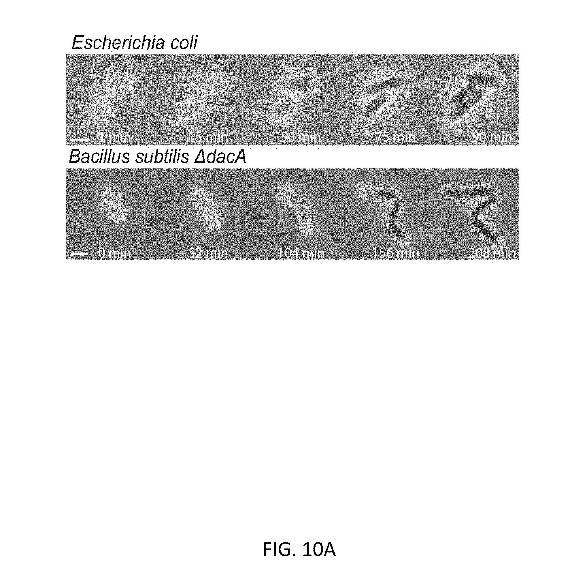
D00014

D00015

D00016
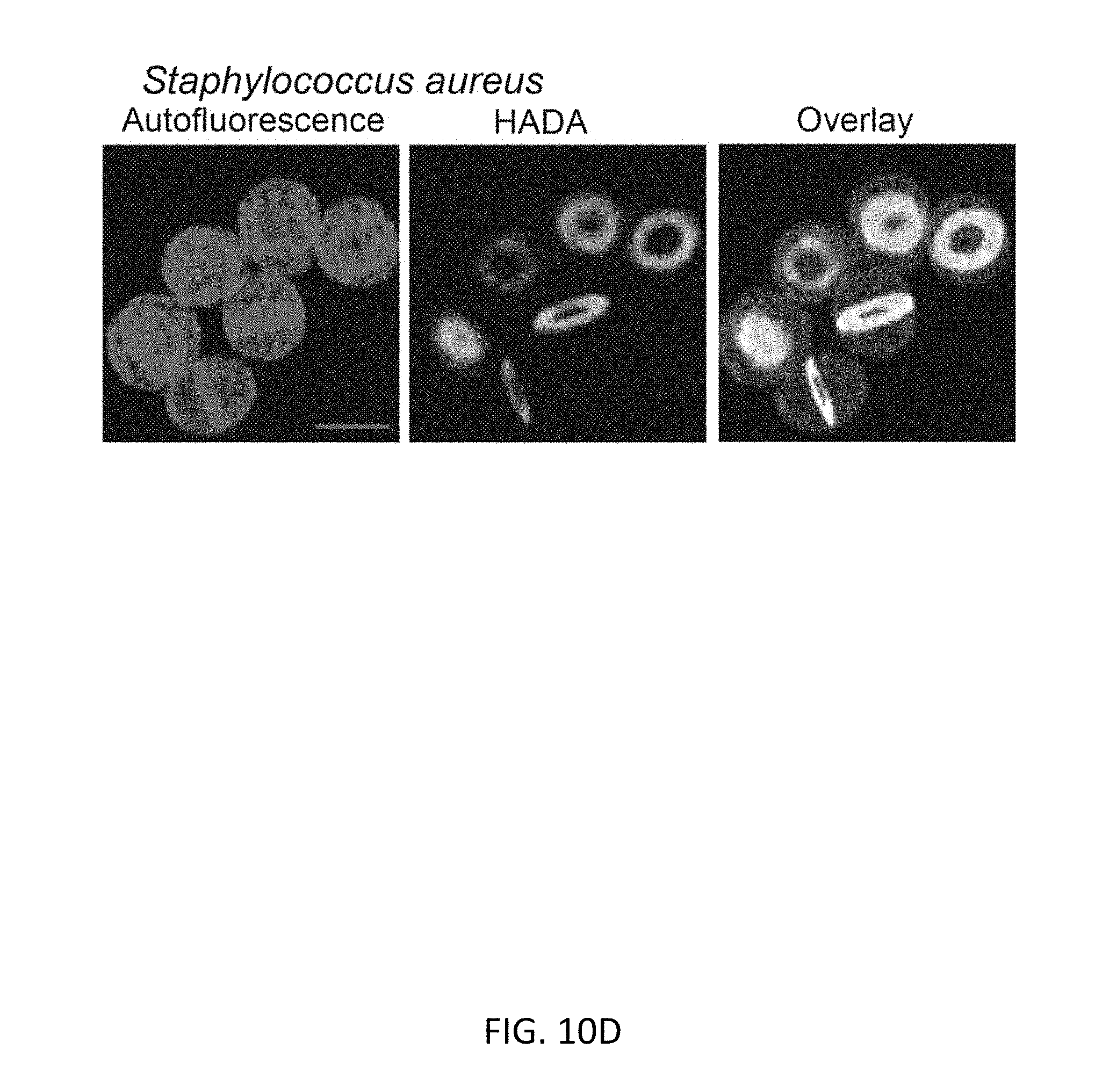
D00017

D00018

D00019

D00020

P00001

XML
uspto.report is an independent third-party trademark research tool that is not affiliated, endorsed, or sponsored by the United States Patent and Trademark Office (USPTO) or any other governmental organization. The information provided by uspto.report is based on publicly available data at the time of writing and is intended for informational purposes only.
While we strive to provide accurate and up-to-date information, we do not guarantee the accuracy, completeness, reliability, or suitability of the information displayed on this site. The use of this site is at your own risk. Any reliance you place on such information is therefore strictly at your own risk.
All official trademark data, including owner information, should be verified by visiting the official USPTO website at www.uspto.gov. This site is not intended to replace professional legal advice and should not be used as a substitute for consulting with a legal professional who is knowledgeable about trademark law.