Methods And Apparatus For Analyte Detection Using Fluorinated Phthalocyanines
Wael; Karolien De ; et al.
U.S. patent application number 16/034705 was filed with the patent office on 2019-01-17 for methods and apparatus for analyte detection using fluorinated phthalocyanines. This patent application is currently assigned to Seton Hall University. The applicant listed for this patent is AXES Research Group, Department of Chemistry, University of Antwerp,, Seton Hall University. Invention is credited to Erik N. Carrion, Sergiu M. Gorun, Stanislav Trashin, Karolien De Wael.
| Application Number | 20190017955 16/034705 |
| Document ID | / |
| Family ID | 62948106 |
| Filed Date | 2019-01-17 |


View All Diagrams
| United States Patent Application | 20190017955 |
| Kind Code | A1 |
| Wael; Karolien De ; et al. | January 17, 2019 |
METHODS AND APPARATUS FOR ANALYTE DETECTION USING FLUORINATED PHTHALOCYANINES
Abstract
Methods and apparatus for detecting an analyte using fluorinated phthalocyanines is disclosed herein. A method for detecting an analyte includes illuminating an analyte solution which contacts an electrode comprising a conductive material having a photosensitizer coupled thereto to generate a reactive oxygen species, wherein the photosensitizer is a fluorinated pthalocyanine having a metal or a non-metal center, oxidizing an analyte present in the analyte solution with the reactive oxygen species to form an oxidized analyte, and detecting a current resulting from the reduction of the oxidized analyte at the electrode.
| Inventors: | Wael; Karolien De; (Sint-Pauwels, BE) ; Gorun; Sergiu M.; (New York, NY) ; Trashin; Stanislav; (Antwerp, BE) ; Carrion; Erik N.; (West Orange, NJ) | ||||||||||
| Applicant: |
|
||||||||||
|---|---|---|---|---|---|---|---|---|---|---|---|
| Assignee: | Seton Hall University South Orange NJ AXES Research Group, Department of Chemistry, University of Antwerp, Antwerp |
||||||||||
| Family ID: | 62948106 | ||||||||||
| Appl. No.: | 16/034705 | ||||||||||
| Filed: | July 13, 2018 |
Related U.S. Patent Documents
| Application Number | Filing Date | Patent Number | ||
|---|---|---|---|---|
| 62532118 | Jul 13, 2017 | |||
| Current U.S. Class: | 1/1 |
| Current CPC Class: | G01N 27/305 20130101; C07F 3/06 20130101 |
| International Class: | G01N 27/30 20060101 G01N027/30; C07F 3/06 20060101 C07F003/06 |
Claims
1. An electrode, comprising: a conductive material; and a photosensitizer coupled to the conductive material, wherein the photosensitizer is a fluorinated phthalocyanine having a metal or a non-metal center.
2. The electrode of claim 1, further comprising: a support particle having the photosensitizer disposed on surfaces thereof.
3. The electrode of claim 1, wherein the fluorinated phthalocyanine having the metal or the non-metal center has the following chemical formula: F.sub.xC.sub.yPcM wherein M is the metal or the non-metal center, Pc is phthalocyanine, x ranges from 1 to 64, and y ranges from 0 to 64.
4. The electrode of claim 1, wherein M is selected from the group consisting of Zn.sup.2+, Mg.sup.2+, low-spin Fe.sup.2+, Ru.sup.2+, Pt.sup.2+, Al.sup.3+, Sc.sup.3+, Zr.sup.4+, Ti.sup.4+, Sb.sup.5+, P, Si, and H.
5. The electrode of claim 1, wherein the photosensitizer is represented by the following formula: ##STR00006##
6. The electrode of claim 5, where the support particles are selected from the group consisting of TiO.sub.2, SiO.sub.2, Al.sub.2O.sub.3, ZnO), FeO, Fe.sub.2O.sub.3, Fe.sub.3O.sub.4, ZrO.sub.2, and mixtures thereof.
7. The electrode of claim 1, further comprising: a second conductive material, the second conductive material electrically insulated from the conductive material.
8. An electrochemical cell, comprising: the electrode of claim 1; and a counter electrode.
9. A method of detecting an analyte, comprising: illuminating an analyte solution which contacts an electrode comprising a conductive material having a photosensitizer coupled thereto to generate a reactive oxygen species, wherein the photosensitizer is a fluorinated pthalocyanine having a metal or a non-metal center; oxidizing an analyte present in the analyte solution with the reactive oxygen species to form an oxidized analyte; and detecting a current resulting from the reduction of the oxidized analyte at the electrode.
10. The method of claim 9, wherein the electrode further comprises a support particle having the photosensitizer disposed on surfaces thereof.
11. The method of claim 9, wherein the fluorinated phthalocyanine having the metal center has the following chemical formula: F.sub.xC.sub.yPcM wherein M is the metal or the non-metal center, Pc is phthalocyanine, x ranges from 1 to 64, and y ranges from 0 to 64.
12. The method of claim 9, wherein M is selected from the group consisting of Zn.sup.2+, Mg.sup.2+, low-spin Fe.sup.2+, Ru.sup.2+, Pt.sup.2+, Al.sup.3+, Sc.sup.3+, Zr.sup.4+, Ti.sup.4+, Sb.sup.5+, P, Si, and H. M is selected from the group consisting of Zn.sup.2+, Mg.sup.2+, low-spin Fe.sup.2+, Ru.sup.2+, Pt.sup.2+, Al.sup.3+, Sc.sup.3+, Zr.sup.4+, Ti.sup.4+, Sb.sup.5+, P, Si, and H.
13. The method of claim 9, wherein the photosensitizer is represented by the following formula: ##STR00007##
14. The method of claim 9, where the support particles are selected from the group consisting of TiO.sub.2, SiO.sub.2, Al.sub.2O.sub.3, ZnO), FeO, Fe.sub.2O.sub.3, Fe.sub.3O.sub.4, ZrO.sub.2, and mixtures thereof.
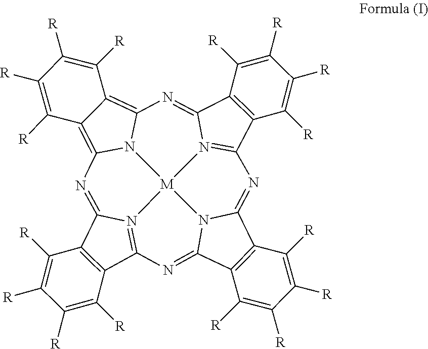
15. The method of claim 9, wherein the analyte solution includes an organic component.
16. The method of claim 15, wherein the organic component is ethanol.
17. The method of claim 9, wherein the analyte includes a phenol.
18. The method of claim 17, wherein the phenol is selected from the group consisting of hydroquinone, amoxicillin, cefadroxil, catechol, bisphenol A, 2-chlorphenol, 3-chlorphenol, 4-chlorphenol, polychlorinated phenols, 2-nitrophenol, 3-nitrophenol, 4-nitrophenol, aniline, 2-aminophenol, 3-aminophenol, 4-aminophenol, 2-amino-4-chlorophenol, 4-cyanophenol, 4-methylphenol, 3,3',5,5'-tetramethylbenzidine, and mixtures thereof.
19. The method of claim 9, wherein the electrode includes a second conductive material, the second conductive material electrically insulated from the from the conductive material, and wherein the analyte solution is in the form of a drop that contacts the conductive material and the second conductive material.
20. The method of claim 9, wherein the electrode is part of an electrochemical cell, the electrochemical cell containing the analyte solution and including a second electrode, wherein the electrode and the second electrode are partially immersed in the analyte solution.
Description
CROSS-REFERENCE TO RELATED APPLICATIONS
[0001] This application claims priority to U.S. Provisional Application No. 62/532,118, filed on Jul. 13, 2017, which is incorporated herein by reference.
FIELD OF THE INVENTION
[0002] The present disclosure relates generally to the field of analyte detection.
BACKGROUND
[0003] The use of enzymes for chemical analysis is well documented. Horseradish peroxidase (HRP), a typical example, has been employed either as a selective catalyst to transform an analyte into an easily detectable product or as an enzymatic label in immunosorbent assays and related techniques. (Koide K, et al., Nat. Commun. 7, 10691 (2016); Lundqvist H, et al., Free Radical Biol. Med. 20, 785-792 (1996); Lakshmipriya T, et al., PLoS One 11, e0151153 (2016); Lequin R M, Clin. Chem. 51, 2415-2418 (2005); Dill K, et al., Lab Chip 6, 1052-1055 (2006)) The advantage of enzymes is their catalytic signal amplification, which translates into high sensitivity and low limits of detection (LOD). Enzyme based reagents, in combination with a cost efficient and straightforward electrochemical detection method could lead to portable, selective and sensitive sensors not unlike the widely used personal glucose meters. (Nemiroski A, et al., Proc. Natl. Acad. Sci. U.S.A. 111, 11984-11989 (2014); Wang J, Biosens. Bioelectron. 21, 1887-1892 (2006); Heller A, et al., Chem. Rev. 108, 2482-2505 (2008))
[0004] Several research groups have advanced over last three decades the electrochemical detection of phenols at HRP modified electrodes as well as the use of HRP and alkaline phosphatase (ALP) as enzyme labels in electrochemical immunoassays. (Yang S, et al., Electrochim. Acta 52, 200-205 (2006); Imabayashi S-i, et al., Electroanalysis 13, 408-412 (2001); Munteanu F D, et al., Anal. Chem. 70, 2596-2600 (1998); Glavan A C, et al., Anal. Chem. 86, 11999-12007 (2014); Rossier J S, et al., Lab Chip 1, 153-157 (2001); Kokkinos C, et al., TrAC, Trends Anal. Chem. 79, 88-105 (2016)) However, despite these efforts and commercial interests, progress in commercialization of electrochemical biosensors remains slow. (Wolfbeis O S, Angew. Chem., Int. Ed. 52, 9864-9865 (2013); Bahadir E B, et al., Anal. Biochem. 478, 107-120 (2015); Kissinger P T, Biosens. Bioelectron. 20, 2512-2516 (2005)). The reasons include the complexity of fabrication of sensing elements, the thermal and chemical instability of enzymes during fabrication, sterilization and storage, the reproducibility of enzyme activity and immobilization procedures, as well as challenges related to field-use of reagents, for example unstable hydrogen peroxide (H.sub.2O.sub.2). (Park S, et al., Anal. Chim. Acta 556, 46-57 (2006)) Thus, sensitive yet robust, renewable reagents and simple, reliable detection approaches are needed.
[0005] Using light to activate the chemical conversion of an analyte has been recently introduced in the field of electrochemical analysis. (Gao Z, et al., Nucleic Acids Res. 33, e123 (2005); Wang G-L, et al., Electrochem. Commun. 41, 47-50 (2014)) Chronoamperometry using disposable, screen-printed electrodes (SPE) yields a detection platform similar to that of glucose meters. In the case of photo-electrochemical sensors an analytical signal (photocurrent) is triggered by light and thus it can be cleanly distinguished from background by simply switching the light off. Advantageously, relatively stable reagents could be photoactivated to start a measurement. For example, an illuminated semiconductor can become a strong electron acceptor, capable to oxidize an organic compound and exchange electrons with an electrode. (Ma W G, et al., Anal. Chem. 87, 4844-4850 (2015))
[0006] Current methods of detection using photoactivation include either enzymes or semiconductors, the former being unstable, having a limited pH window for use, and needing non-renewable hydrogen peroxide, while the latter needing light in the ultraviolet (UV) region. There is a need for photoanalytical methods that use light in the infrared (IR) and the visible region.
BRIEF SUMMARY
[0007] One aspect of the present disclosure relates to an electrode including a conductive material, and a photosensitizer coupled to the conductive material, wherein the photosensitizer is a fluorinated phthalocyanine having a metal or a non-metal center. In an embodiment, the electrode may include a support particle having the photosensitizer disposed on surfaces thereof.
[0008] Another aspect of the disclosure relates to a method of detecting an analyte including illuminating an analyte solution which contacts an electrode comprising a conductive material having a photosensitizer coupled thereto to generate a reactive oxygen species, wherein the photosensitizer is a fluorinated pthalocyanine having a metal or a non-metal center, oxidizing an analyte present in the analyte solution with the reactive oxygen species to form an oxidized analyte; and detecting a current resulting from the reduction of the oxidized analyte. In an embodiment, the electrode may further comprise a support particle having the photosensitizer disposed on surfaces thereof.
[0009] Another aspect of the disclosure relates to a method of detecting a biomolecule including exposing an electrode having a first oligonucleotide disposed thereon to a photosensitizer coupled to a second oligonucleotide, wherein the first and second oligonucleotide are complimentary and wherein the photosensitizer is coupled to the electrode through complimentary coupling of the first and second oligonucelotides, illuminating the photosensitizer to generate a reactive oxygen species, and detecting a current resulting from reduction of the reactive oxygen species at the electrode.
BRIEF DESCRIPTION OF DRAWINGS
[0010] FIG. 1A is a schematic view of electrochemical detection of an analyte using an enzyme to catalyze the formation of an oxidized product.
[0011] FIG. 1B is a schematic view of electrochemical detection of an analyte using a peroxidase to catalyze the formation of an oxidized product.
[0012] FIG. 1C is a schematic view electrochemical detection of an analyte using a phenol oxidase to catalyze the formation of an oxidized product.
[0013] FIG. 1D is a schematic view of electrochemical detection of an analyte using a photosensitizer to catalyze the formation of an oxidized product.
[0014] FIG. 1E is a schematic view of electrochemical detection of a phenol using a photosensitizer to catalyze the formation of a benzoquinone (BQ), which is an oxidized product of the phenol.
[0015] FIG. 2A is a schematic view of an electrode in accordance with an embodiment of the disclosure.
[0016] FIG. 2B is a schematic view of an electrode in accordance with an embodiment of the disclosure.
[0017] FIG. 2C is a schematic view of the electrode of FIG. 2B having a drop containing an analyte disposed thereon and under illumination of a light source in accordance with an embodiment of the disclosure.
[0018] FIG. 2D is a schematic view of an electrochemical cell including the electrode of FIG. 2 A in accordance with an embodiment of the disclosure.
[0019] FIG. 3 is a diffuse reflectance UV-Vis absorption spectrum of TiO.sub.2--F64C24PcZn in comparison to a UV-Vis absorption spectrum of 10 .mu.g/ml F.sub.64C.sub.24PcZn in ethanol.
[0020] FIG. 4A is an amperometric response of SPEITiO.sub.2--F64C24PcZn in a blank buffer and 10 .mu.M HQ.
[0021] FIG. 4B is an amperometric response of F.sub.64C.sub.24PcZn, SPEI F.sub.64C.sub.24PcZn in a blank buffer and 20 .mu.M HQ.
[0022] FIG. 5 is linear sweep voltammetry traces for SPE, SPEITiO.sub.2, SPEITiO.sub.2--F.sub.64C.sub.24PcZn, and SPEI F.sub.64C.sub.24PcZn, respectively in the absence (solid trace) and presence (dashed trace) of 10 .mu.M HQ.
[0023] FIG. 6 is a plot of photocurrent dependence on the concentration of an analyte in accordance with some embodiments of the disclosure.
[0024] FIG. 7A is linear sweep voltammetry traces for SPEISiO.sub.2--F.sub.64C.sub.24PcZn in the absence (solid trace) and presence (dashed trace) of 10 .mu.M HQ.
[0025] FIG. 7B is an amperometric response of SPEISiO.sub.2--F.sub.64C.sub.24PcZn as a function of concentration of an analyte in accordance with some embodiments of the disclosure.
[0026] FIG. 8A a plot of photocurrent dependence on the concentration F.sub.64C.sub.24PcZn in the absence of an analyte in accordance with some embodiments of the disclosure.
[0027] FIG. 8B is a plot of photocurrent dependence on the concentration F.sub.64C.sub.24PcZn in the presence of an analyte in accordance with some embodiments of the disclosure.
[0028] FIG. 8C a plot of photocurrent dependence on the concentration TiO.sub.2--F64C24PcZn in the absence of an analyte in accordance with some embodiments of the disclosure.
[0029] FIG. 8D is a plot of photocurrent dependence on the concentration TiO.sub.2--F64C24PcZn in the presence of an analyte in accordance with some embodiments of the disclosure.
[0030] FIG. 9 is an amperometric response of TiO.sub.2--F.sub.64C.sub.24PcZn in the presence of an analyte as a function of laser power in accordance with some embodiments of the disclosure.
[0031] FIG. 10 is a plot of photocurrent as a function of laser power in accordance with some embodiments of the disclosure.
[0032] FIG. 11 is linear sweep voltammetry traces for SPEITiO.sub.2--F.sub.64C.sub.24PcZn in the absence (solid trace) and presence (dashed trace) of 10 .mu.M Amoxicillin in accordance with some embodiments of the disclosure.
[0033] FIG. 12A is an amperometric response of SPEITiO.sub.2--F.sub.64C.sub.24PcZn in a blank buffer and in amoxicillin in accordance with some embodiments of the disclosure.
[0034] FIG. 12B is a calibration curve for photocurrent response of Amoxicillin in accordance with some embodiments of the disclosure.
[0035] FIG. 13A is an amperometric response of HQ as a function of concentration in accordance with some embodiments of the disclosure.
[0036] FIG. 13B is a plot of photocurrent as a function of HQ concentration.
[0037] FIG. 14 is an amperometric response of SPEITiO.sub.2--F.sub.64C.sub.24PcZn in the presence of buffer and then analytes in accordance with some embodiments of the disclosure.
[0038] FIG. 15 is an amperometric response of SPEITiO.sub.2--HRP in a blank buffer, amoxicillin, and HQ solutions.
[0039] FIG. 16 is an amperometric response of SPEITiO.sub.2--HRP in HQ solution.
[0040] FIG. 17A is an amperometric response of complementary and non-complementary oligonucleotides labeled with a photosensitizer.
[0041] FIG. 17B is an amperometric response of the complementary and non-complementary oligonucleotides labeled with the photosensitizer in FIG. 17A, averaged over four measurements, each at a separate electrode.
[0042] FIG. 18 is an ampometric response of an oligonucleotide in accordance with some embodiments of the disclosure.
DETAILED DESCRIPTION
[0043] The following detailed description of methods and apparatus for analyte detection using fluorinated phthalocyanines refers to the accompanying drawings that illustrate exemplary embodiments consistent with these methods and apparatus. Other embodiments are possible, and modifications may be made to the embodiments within the spirit and scope of the methods and systems presented herein. Therefore, the following detailed description is not meant to limit the devices described herein. Rather, the scope of these devices is defined by the appended claims.
[0044] As used herein, SPEIR.sub.16PcM is a short hand notation to indicate that an R.sub.16PcM, which is a functionalized phthalocyanine (Pc), is deposited on a screen printed electrode (SPE). As used herein, SPEIM.sub.xO.sub.y--R.sub.16PcM is a short hand notation to indicate that as supported functionalized phthalocyanine, where an R.sub.16PcM is supported on a support particle (M.sub.xO.sub.y), is deposited on an SPE. Generally, the notation SPEIcatalyst is a short hand notation for a catalyst, i.e., an element that either directly or indirectly catalyzes oxidation of an analyte, deposited on the SPE. Exemplary catalyst, as discussed herein, are photosensitizers, supported photosensitizers, enzymes, and the like.
Mechanism
[0045] FIGS. 1A-1E illustrates conceptual parallel between the enzymatic and photosensitizer electrode mechanisms. Specifically, an oxidant, e.g., H.sub.2O.sub.2 or O.sub.2, interacts with a catalyst, e.g., an enzyme, such as HRP, a peroxidase, or phenol oxidase, or a photosensitizer, yielding highly reactive intermediates, e.g., ferryl heme or .sup.1O.sub.2. These intermediates, in turn, rapidly oxidize an analyte, for example a phenol, such as a hydroquinone (HQ), or another appropriate electron donor in the case of HRP. (Rodriguez-Lopez J N, et al., J. Am. Chem. Soc. 123, 11838-11847 (2001)) The intermediate, in the case of HRP, is the iron-oxo core, a classical electron plus proton abstractor, which oxidizes HQ to yield water and an oxidized product, e.g., a benzoquinone (BQ). The oxidized product is electrochemically reduced at a polarized electrode to regenerate the analyte, e.g. reduced product, thus completing a catalytic redox cycle (FIG. 1A-1C). The redox schemes dependency on electron donors, for example phenols, makes them useful for their quantification. (Yang S, et al., Electrochim. Acta 52, 200-205 (2006); Imabayashi S-i, et al., Electroanalysis 13, 408-412 (2001); Munteanu F D, et al., Anal. Chem. 70, 2596-2600 (1998)) The proposed enzymatic-like approach relies on catalyzed photo-transformation of dissolved, aerobic .sup.3O.sub.2 in the reactive, short-lived .sup.1O.sub.2. (FIGS. 1D-1E) In this schem, a photosensitizer catalyzes the formation of a reactive species, e.g., .sup.1O.sub.2 (singlet oxygen) from .sup.3O.sub.2 (triplet oxygen), and singlet oxygen reacts with the analyte to form an oxidized product which is then electrochemically reduced at a polarized electrode to regenerate the analyte, e.g., reduced product. Advantageously, this approach does not require H.sub.2O.sub.2, unlike some enzyme-based techniques.
Fluorine Containing Phthalocyanine
[0046] A fluorine containing phthalocyanine may be represented by Formula (I):
##STR00001##
[0047] The formula (I) may be written as R.sub.16PcM, where Pc represents the phthalocyanine moiety. Each R can be independently selected from the group consisting of fluorine (F), a fluorocarbon containing from 1 to 18 carbon atoms, a fluorine-containing group, a non-fluorine containing group, and hydrogen (H). A non-fluorine containing group can include a functional group directly attached to the phthalocyanine macrocycle. Non-fluorine containing groups can include a carboxylic acid group, an aldehyde group, an amino group, a hydrazine group, an alkyne, alkene or a diene group, an azido group, an isocyanate group, an acyl halide, a hydroxide, a thiol, a nitro, a phosphate group, an amide, a halogen, such as chlorine, sulfonate, phenyl, or groups that are known in the art to act as substituents on aromatic compounds. At least one R includes a fluorine (F). In one embodiment, a fluorine containing phthalocyanine includes 8 R that are fluorine (F) and 8 R that are --C(CF.sub.3).sub.2F. In one embodiment, a fluorine phthalocyanine includes 6 R that are fluorine (F), 3 R that are hydrogen (H), 1 R that is --O-- (C.sub.6H.sub.4)--COOH, and 6 R that are --C(CF.sub.3).sub.2F.
[0048] M may be a metal, a non-metal, or at least one of a metal or non-metal in complex or covalently bonded to at least one axial ligand.
[0049] The fluorine containing phthalocyanine may be in the form of a salt. Pharmaceutically acceptable salts refers to the relatively non-toxic, inorganic and organic acid addition salts of the Pcs. These salts can be prepared in situ during the final isolation and purification of the photosensitizer(s), or by separately reacting a purified photosensitizer(s) in its free base form with a suitable organic or inorganic acid, and isolating the formed salt. Exemplary salts include the hydrobromide, hydrochloride, sulfate, bisulfate, phosphate, nitrate, acetate, pyruvate, valerate, oleate, palmitate, stearate, laurate, benzoate, lactate, phosphate, tosylate, citrate, maleate, fumarate, succinate, tartrate, naphthylate, mesylate, glucoheptonate, lactobionate, and laurylsulphonate salts, and the like. (See, for example, Berge et al. (1977) "Pharmaceutical Salts", J. Pharm. Sci. 66:1-19).
[0050] Fluorocarbons can include fluoroalkyl (i.e., perfluoroalkyl), fluoroalkylcylic, fluoroalkylbicyclic, and fluoroaryl. It will be obvious to those skilled in the art that other fluorocarbons having 1 to 18 carbon atoms can be used. The alkyl group of the fluoroalkyl may be methyl, ethyl, propyl, butyl, and cycloalkyl. The aryl group of the fluoraryl may be phenyl, benzyl, phenol, naphthyl, bi-aryl, and trityl. The alkylcyclic group of the fluoroalkylcyclic may be cyclopropyl, cyclobutyl, cyclopentyl and cyclohexyl. The alkylbicyclic group of the fluoroalkylbicyclic may be di-cyclobutyl, di cyclopentyl and di-cyclohexyl.
[0051] Fluorine-containing groups can include at least fluorine. Exemplary fluorine-containing groups can include fluoroheteroalkyl, fluoroheteroaryl, fluoroheterocyclic, and fluoroheterobicyclic. One or more of the substituent sites on the following groups can be substituted with fluorine. The heteroalkyl group may be methylamino, dimethylamino, ethylamino, diethylamino, propylamino, butylamino, alkoxy, alkoxyalkyl, alkylsulhydryl, haloalkyl and phosphoryl groups. The alkoxy may be methoxy, levulinyl, carboxy, ethoxy, and propoxy. The alkoxy group may be O(CH.sub.2)q-R, where q=2-4 and R is --NH.sub.2, OCH.sub.3, or --OCH.sub.2CH.sub.3. The alkoxyalkyl group may be methoxyethyl, and ethoxyethyl. The haloalkyl group may be --CF.sub.3, --CBr.sub.3, --CCl.sub.3 and --CI.sub.3. The heteroaryl group of the fluoroheteroaryl may be a trityl group or a carboxybenzyloxy group. The trityl group may be trityl-R, where R is OC(CH.sub.3)3, OCH.sub.3, or --OCH.sub.2CH.sub.3. The carboxybenzyloxy group may be selected from the group consisting of CO-aryl-R, where R is a halogen (--Cl, --F, --Br, I, alkyl or alkoxyalky (--OC(CH.sub.3).sub.3, OCH.sub.3, or --OCH.sub.2CH.sub.3).The heterocyclic group of the fluoroheterocyclic may be pyrimidinyl, pyrrolo, pyridinyl, oxazolinyl, aza-oxazolinyl, thio-oxazolinyl, thiophenyl, furyl, or imidazolyl. The heterobicyclic group of the fluoroheterobicyclic may be purinyl, steroyl, indoyl and quinolyl.
[0052] M can be a metal or non-metal. Exemplary metals can be M.sup.2+ or M(II), such as Zn.sup.2+, Mg.sup.2+, Fe.sup.2+, Ru.sup.2+, Pt.sup.2+, M.sup.3+ or M(III), such as Al.sup.3+, or Sc.sup.3+, M.sup.4+ or M(IV), such as Zr.sup.4+ or Ti.sup.4+, or M.sup.5+ or M(V), such as Sb.sup.5+. Exemplary non-metals can include P, Si, and H, where two H would be present on the phthalocyanine. The metal or non-metal can be in complex with, or covalently bound to at least one axial ligand. In some embodiments, the metal or non-metal can be in complex with or covalently bound to up to two axial ligands. The metal can be coordinated to a phthalocyanine moiety, for example, such as depicted in Formula I.
[0053] Each axial ligand can be any atom or group of atoms, similar or different that can coordinate M. It should be understood that an axial ligand may not be present in a certain M, and that more than one axial ligand may be present in a certain M. Each axial ligand may be independently selected, and may include H, alkylamino, alkylthio, alkoxy, alkylseleno, alkylsulfonyl, C(S)NHC.sub.6H.sub.11O.sub.5, OC(O)CH.sub.3, OC(O), CS, CO, CSc, OH, O (oxo) and an alkyl group having from 1 to 12 carbon atoms, or (CH.sub.2).sub.nN((CH).sub.o(CH.sub.3)).sub.2, wherein n is an integer from 1 to 12; and o is an integer from 1 to 11, or a pharmaceutically acceptable salt thereof (to achieve a neutral charge).
[0054] In some embodiments, M may be represented by (G).sub..alpha.Y[(OSi(CH.sub.3).sub.2(CH.sub.2).sub.bN.sub.c(R').sub.d(R'- ').sub.e).sub.fX.sub.g].sub.p, wherein a is 0 or 1, b is an integer from 2 to 12, c is 0 or 1, d is an integer from 0 to 3, e is an integer from 0 to 2, f is 1 or 2, g is 0 or 1, and p is 1 or 2. Y may be selected from Si, Al, Ga, Ge, and Sn. R' may be selected from H, CH.sub.3, C.sub.2H.sub.5, C.sub.4H.sub.9, C.sub.4H.sub.8NH, C.sub.4H.sub.8N, C.sub.4H.sub.8NCH.sub.3, C.sub.4H.sub.8S, C.sub.4H.sub.8O, C.sub.4H.sub.8Se, OC(O)CH.sub.3, OC(O), CS, CO, CSe, OH, C.sub.4H.sub.8N(CH.sub.2).sub.3CH.sub.3, (CH.sub.2).sub.2N(CH.sub.3).sub.2, an alkyl group having from 1 to 12 carbon atoms, and (CH.sub.2).sub.nN((CH.sub.2).sub.o(CH.sub.3)).sub.2, wherein n is an integer from 1 to 12; and o is an integer from 1 to 11. R'' may be selected from H, SO.sub.2CH.sub.3, (CH.sub.2).sub.2N(CH.sub.3).sub.2, (CH.sub.2).sub.11CH.sub.3, C(S)NHC.sub.6H.sub.11O.sub.5, an alkyl group having from 1 to 12 carbon atoms, and (CH.sub.2).sub.mN((CH.sub.2).sub.o(CH.sub.3)).sub.2, wherein n is an integer from 1 to 12; and o is an integer from 1 to 11. G may be selected from OH and CH.sub.3. X may be selected from I--, F--, Cl--, or Br--.
[0055] M may include at least one metal, at least one non-metal, or a combination of a metal and a non-metal. Exemplary M include AlOSi(CH.sub.3).sub.2(CH.sub.2).sub.3N(CH.sub.3).sub.2, AlOSi(CH.sub.3).sub.2(CH.sub.2).sub.3N(CH.sub.3).sub.3.sup.+I.sup.-, CH.sub.3SiOSi(CH.sub.3).sub.2(CH.sub.2).sub.3N(CH.sub.3).sub.2, HOSiOSi(CH.sub.3).sub.2(CH.sub.2).sub.3N(CH.sub.3).sub.2, HOSiOSi(CH.sub.3).sub.2(CH.sub.2).sub.3N(CH.sub.3).sub.3.sup.+I.sup.-, Si[OSi(CH.sub.3).sub.2(CH.sub.2).sub.3.sup.+I.sup.-].sub.2, Si[OSi(CH.sub.3).sub.2(CH.sub.2).sub.4NH.sub.2].sub.2, Si[OSi(CH.sub.3).sub.2(CH.sub.2).sub.4NHSO.sub.2CH.sub.3].sub.2, HOSiOSi(CH.sub.3).sub.2(CH.sub.2).sub.4NHSO.sub.2CH.sub.3, HOSiOSi(CH.sub.3).sub.2(CH.sub.2).sub.3N(CH.sub.2CH.sub.3)(CH.sub.2).sub.- 2N(CH.sub.3).sub.2, Si[OSi(CH.sub.3).sub.2(CH.sub.2).sub.4 NHCSNHC.sub.6H.sub.11O.sub.5].sub.2, Si[OSi(CH.sub.3).sub.2(CH.sub.2).sub.3N(CH.sub.3).sub.2].sub.2, HOSiOSi(CH.sub.3).sub.2(CH.sub.2).sub.3OCOCH.sub.3, HOSiOSi(CH.sub.3).sub.2(CH.sub.2).sub.3OH, Si[OSi(CH.sub.3).sub.2(CH.sub.2).sub.3N(CH.sub.2CH.sub.3)(CH.sub.2).sub.2- N(CH.sub.3).sub.2].sub.2, HOSiOSi(CH.sub.3).sub.2(CH.sub.2).sub.3NC.sub.4H.sub.8O, AlOSi(CH.sub.3).sub.2(CH.sub.2).sub.3N.sup.+(CH.sub.3).sub.2(CH.sub.2).su- b.11CH.sub.3I.sup.-, HOSiOSi(CH.sub.3).sub.2(CH.sub.2).sub.8N(CH.sub.3).sub.2, Si[OSi(CH.sub.3).sub.2(CH.sub.2).sub.3NC.sub.4H.sub.8O].sub.2, HOSiOSi(CH.sub.3).sub.2(CH.sub.2).sub.3NC.sub.4H.sub.8S, HOSiOSi(CH.sub.3).sub.2(CH.sub.2).sub.3N(CH.sub.2).sub.3(CH.sub.3).sub.2, HOSiOSi(CH.sub.3).sub.2(CH.sub.2).sub.3NCS, HOSiOSi(CH.sub.3).sub.2(CH.sub.2).sub.3N[(CH.sub.2).sub.3N(CH.sub.3).sub.- 2].sub.2, HOSiOSi(CH.sub.3).sub.2(CH.sub.2).sub.3NC.sub.4H.sub.8NCH.sub.3, Si[OSi(CH.sub.3).sub.2(CH.sub.2).sub.3NC.sub.4H.sub.8NCH.sub.3].sub.2, HOSiOSi(CH.sub.3).sub.2(CH.sub.2).sub.3NC.sub.4H.sub.8N(CH.sub.2).sub.3CH- .sub.3, Si[OSi(CH.sub.3).sub.2(CH.sub.2).sub.3NC.sub.4H.sub.8NH].sub.2, or pharmaceutically acceptable salts thereof.
[0056] M can include HOSiOSi(CH.sub.3).sub.2(CH.sub.2).sub.3N(CH.sub.3).sub.2, HOSiOSi(CH.sub.3).sub.2(CH.sub.2).sub.3N(CH.sub.2CH.sub.3)(CH.sub.2).sub.- 2N(CH.sub.3).sub.2, or HOSiOSi(CH.sub.3).sub.2(CH.sub.2).sub.3N(CH.sub.3).sub.2, HOSiOSi(CH.sub.3).sub.2(CH.sub.2).sub.3N(CH.sub.2CH.sub.3)(CH.sub.2).sub.- 2N(CH.sub.3).sub.2. In one embodiment, M is HOSiOSi(CH.sub.3).sub.2(CH.sub.2).sub.3N(CH.sub.3).sub.2.
[0057] M can be two protons, e.g. two H+.
[0058] Additional groups, such as anionic groups, may be linked to M to ensure electrical neutrality. Exemplary anionic groups can include halogens or oxo groups. For example, if M is Si.sup.4+, two Cl.sup.- bonded to the Si, or an oxo group, or two hydroxyl groups ensures electrical neutrality.
[0059] In one embodiment, a fluorine containing phthalocyanine of formula (I) can be presented by the following chemical structure:
##STR00002##
[0060] F.sub.48C.sub.25O.sub.3H.sub.8PcZn was obtained as described in Carrion E N, et al., Inorg. Chem. 2017; 56: 7210-7216.
[0061] In one embodiment, a fluorine containing phthalocyanine can be represent by the following chemical formula (II):
F.sub.xC.sub.yPcM (II)
[0062] Where Pc is the phthalocyanine moiety and M represents any of the various embodiments of M discussed herein in regards formula (I). x ranges from 1 to 64 and y ranges from 0 to 64. In one embodiment, a fluorine containing phthalocyanine of formula (II) is represented by the following chemical structure:
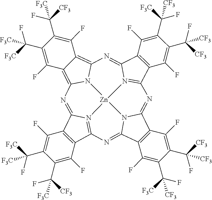
##STR00003##
[0063] The above chemical structure is F.sub.64C.sub.24PcZn, which produces .sup.1O.sub.2 in a relatively high quantum yield. (Minnes R, et al., Photochem. Photobiol. 82, 593-599 (2006); Bench B A, et al., Angew. Chem., Int. Ed. 114, 773-776 (2002); Beveridge A C, et al., J. Phys. Chem. A 107, 5138-5143 (2003)) Unlike the protio phthalocyanine, H.sub.16PcZn, F.sub.64C.sub.24PcZn is stable in the presence of .sup.1O.sub.2 and other reactive species. (Gerdes R, et al., Dalton Trans., 1098-1100 (2009); Loas A, et al., Dalton Trans. 40, 5162-5165 (2011)) Unlike F.sub.16PcZn, F.sub.64C.sub.24PcZn has bulky i-C.sub.3F.sub.7 groups that effectively protect this molecule from aggregation and facilitate its dissolution in organic solvents such as ethanol. Site isolation is an important feature since aggregation, exhibited by the fluorinated F.sub.16PcZn diminishes the .sup.1O.sub.2 yield due to the shortening of the excited states lifetimes and/or inefficient intersystem crossing. In addition, the aromatic fluorines of F.sub.16PcZn are subject to nucleophilic attack, including from some ROS species thus rendering this molecule less viable for .sup.1O.sub.2 photocatalysis. (Leznoff C C, et al., Chem. Commun. (Cambridge, U.K.), 338-339 (2004)) In contrast, attempts to detect nucleophilic substitutions at the 8 .alpha. C--F positions of F.sub.64C.sub.24PcZn have not succeeded thus far, due at least in part to the steric hindrance afforded by the eight adjacent .beta. bulky i-C.sub.3F.sub.7 groups. Thus, F.sub.64C.sub.24PcZn can have sustained .sup.1O.sub.2 production.
[0064] In summary, the F.sub.64Pc scaffold affords chemically and thermally stable yet photoreactive complexes features that led to its choice. (Gerdes R, et al., Dalton Trans., 1098-1100 (2009); Loas A, et al., Dalton Trans. 40, 5162-5165 (2011)) Furthermore, the need for solid-state catalysts using the F.sub.64Pc scaffold led to the exploration of its support on materials, including TiO.sub.2. (Gorun S M, et al., U.S. Pat. No. 9,260,630) F.sub.64C.sub.24PcZn was deposited in 0.5 to 3 wt % on TiO.sub.2, a carrier matrix with a 21 nm primary particle size and 35-65 m.sup.2 g.sup.-1 surface area. The diffuse reflectance UV-Vis spectrum of the hybrid material, TiO.sub.2--F.sub.64C.sub.24PcZn, exhibited the characteristic Q-band of phthalocyanines in the 600-700 nm range and absorbance below 400 nm, attributed to a combination of Soret bands and the intrinsic absorbance of TiO.sub.2 (FIG. 3). (Moons H, et al., Dalton Trans. 43, 14942-14948 (2014); Marin M L, et al., Chem. Rev. 112, 1710-1750 (2012)) The Q-band is relatively broad, but centered at the same position as that of F.sub.64C.sub.24PcZnin solution. The 655 nm wavelength of a common diode laser pointer essentially matches the Q band of TiO.sub.2--F.sub.64C.sub.24PcZn, a material that was shown to produce .sup.1O.sub.2 using visible and filtered red light. (see, for example, U.S. Pat. No. 9,260,630) Thus, a photoelectrochemical response of F.sub.64C.sub.24PcZn-TiO.sub.2 deposited on screen printed electrodes (SPE), abbreviated SPEITiO.sub.2--F.sub.64C.sub.24PcZn should be absent in the absence of red-light exposure, but could be measurable under illumination. Importantly, dark measurements automatically reveal the actual baseline position, leading to an efficient and straightforward way for baseline correction even in the presence of analytes.
Support Particles
[0065] The application of fluorine containing phthalocyanine onto support particles (e.g., metal oxides and/or other oxides) results in the formation of a supported photosensitizer. The supported photosensitizer may by represented by the short hand notation of `support particle--R.sub.16PcM`, or a similar variant as used herein. In some embodiments, the supported photosensitizer may exhibit bonds not present within the support particle or the fluorine containing phthalocyanine alone. For example, the supported photosensitizer may exhibit the reactivity of the fluorine containing phthalocyanine and that of the support particle, if any.
[0066] A variety of phases of oxides can be used, in various degrees of dispersion and particle size. For example, when the oxide includes TiO.sub.2, the support particles can contain TiO.sub.2 in large extent in an anatase crystalline form. For example, about 95% or more (by volume) of the titanium oxide particles can be in the anatase crystalline form.
[0067] The support particles can be in the form of microparticles and/or nanoparticles. As such, the support particles can have a size of about 10 nm to about 100 .mu.m. In one embodiment, the support particles are nanoparticles having an average size of about 10 nm to about 150 nm, or about 10 nm to about 100 nm.
[0068] Without wishing to be bound by any particular theory, it is believed that the presence of carbon-fluorine bonds in the phthalocyanine allows for van der Waals interaction between the halogenated phthalocyanine and the support particles (particularly when oxygen atoms are present, such as in oxides). In addition, it is also believed that an oxygen atom of the oxide support particles, which may have a slightly negative charge, interacts with the metal or non-metal M of the fluorine containing phthalocyanine. Furthermore, when the fluorine containing phthalocyanine contains 2 protons instead of a central metal or non-metal, hydrogen bonding may link the protons to the surface of the supporting particle.
[0069] The support particles are, in one embodiment, formed from oxides, including but not limited to, silicon oxides (e.g., SiO.sub.2), metal oxides (e.g., titanium oxides (e.g., TiO.sub.2), aluminum oxide (e.g., Al.sub.2O.sub.3), zinc oxides (e.g., ZnO), iron oxides (e.g., FeO, Fe.sub.2O.sub.3, Fe.sub.3O.sub.4), zirconium oxides (e.g., ZrO.sub.2), lanthanides oxides, etc.), or mixtures thereof. Other inert materials may be included in the support particles, either in addition to an oxide or in the alternative of an oxide. For example, the support particles may include carbon black, sulfides, carbonates, etc.
[0070] In one embodiment, the support particles include titanium oxide, which results in a combined activity of the titanium oxide and the fluorine containing phthalocyanine. Conversely, the use of an inert material in the support particles, such as silicon dioxide, can result in a composition in which only the phthalocyanine plays a photocatalytic role. Thus, the superior photocatalytic properties of the fluorine containing phthalocyanine manifest themselves in the presence of supports, either inert or reactive.
[0071] The supported photosensitizers can be manufactured by loading support particle with the fluorine containing phthalocyanine. In some embodiments, support particles are added to a solution containing a solvent and the fluorine containing phthalocyanine and subsequently the solvent is evaporated to yield the supported photosensitizer. In certain embodiments, the support particles can be loaded with the fluorine containing phthalocyanine at a concentration of about 0.1% to about 10% by weight of the total weight of the supported photosensitizer.
Electrode and Electrochemical Cell
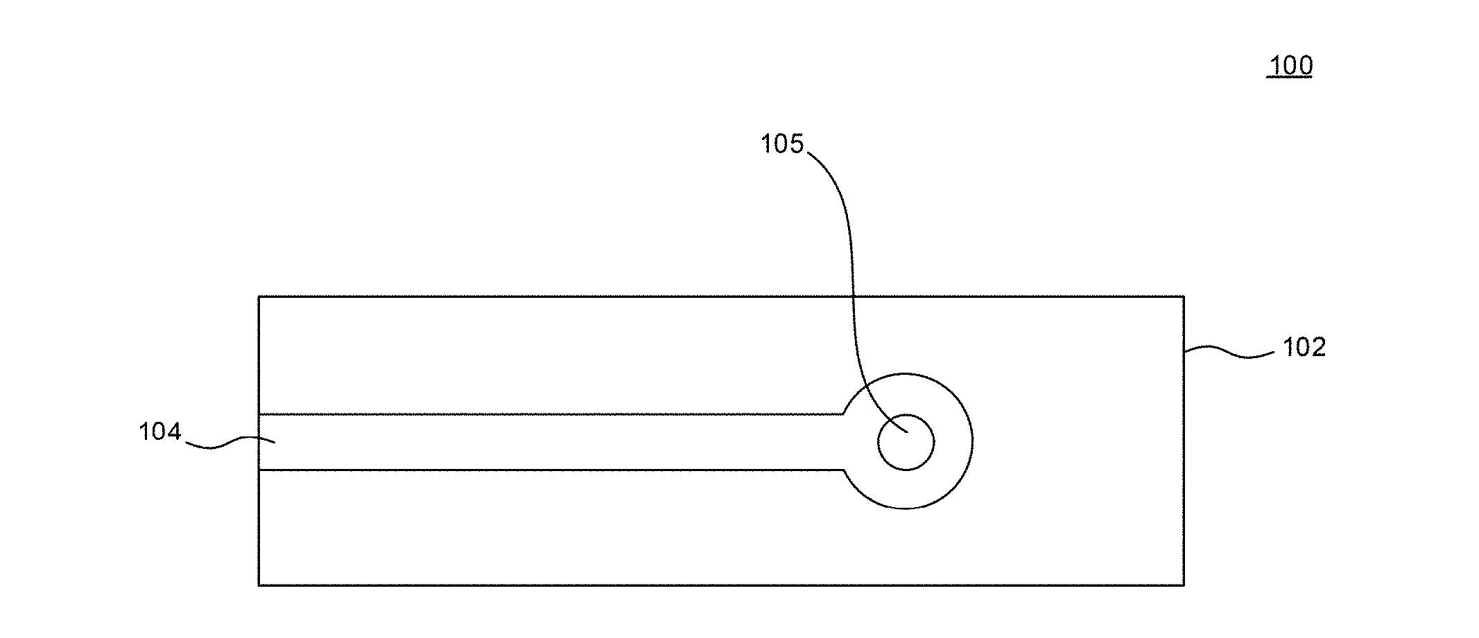
[0072] FIG. 2A illustrates a schematic view of an electrode 100 in accordance with an embodiment of the disclosure. The electrode 100 includes a substrate 102. The substrate 102 can be an electrically insulating material. Overlying or partially embedded in the substrate 102 can be a conductive material 104. The conductive material 104 can be any suitable conductive materials, such as carbon, gold, platinum, silver, copper and the like. A photosensitizer 105, such as a fluorine containing phthalocyanine, which can be unsupported or supported on a support particle, contacts the conductive material 104. The electrode 100 can be used in an electrochemical cell as discussed herein.
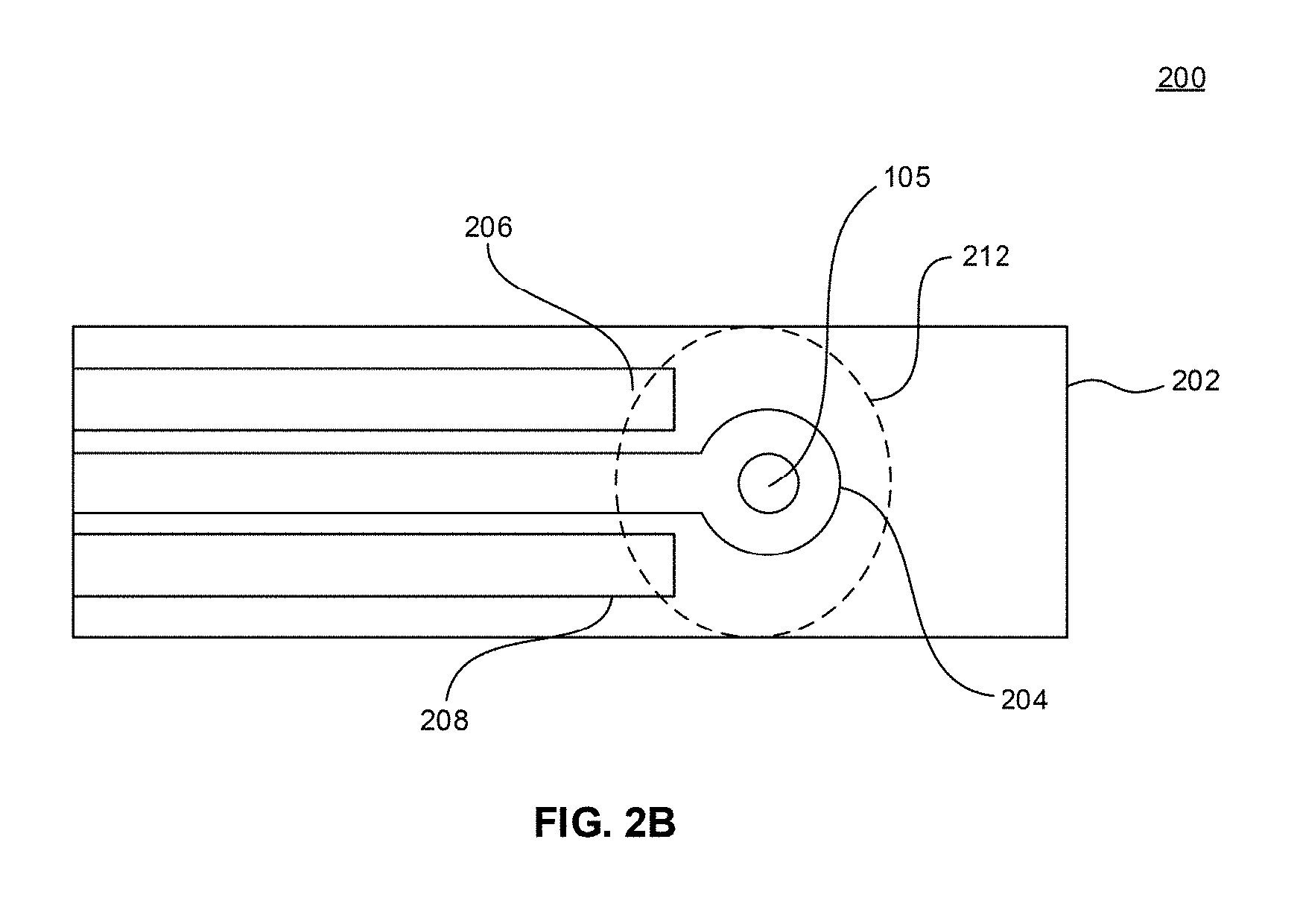
[0073] FIG. 2B illustrates a schematic view of an electrode 200 in accordance with an embodiment of the disclosure. The electrode 200 includes a substrate 202. The substrate 202 can be an electrically insulating material. Overlying or at least partially embedded in the substrate 202 can be a first conductive material 204, a second conductive material 206, and a third conductive material 208. The conductive materials 204, 206, 208 are electrically insulated from each other by the substrate 202 or by another electrically insulating material. One of the conductive materials can be working electrode, another of the conductive materials can be a counter electrode, and another of the conductive materials can be a reference electrode. Exemplary conductive materials include graphite, carbon paste, and the like.
[0074] The photosensitizer 105 can be deposited on at least one of the conductive materials 204, 206, 208. Optionally, the photosensitizer can be disposed on all of the conductive materials 204, 206, 208.
[0075] FIG. 2C illustrates operation of the electrode 200 in accordance with an embodiment of the disclosure. In operation, the electrode 200 may be connected to a voltage source 210 to provide a potential difference between the working electrode and the counter electrode. A drop 212 of an analyte solution can be deposited such that it contacts all of the conductive materials 204, 206, 208. A voltage may be applied from the voltage source to determine a baseline current between the working electrode and the counter electrode. A light source 214 may then be turned on to illuminate the photosensitizer 105. The photosensitizer 105 catalyzes the formation of reactive species, such as singlet oxygen (.sup.1O.sub.2). The singlet oxygen oxidizes the analyte in the drop 212 at the working electrode. The oxidized analyte is then reduced at the working electrode. This oxidation/reduction process results in a photo-induced current which can be current monitored by an ammeter or another device that measures current. The electrode 200 is akin to a glucose monitoring device in the sense that a drop of blood is placed on a test strip that includes working and counter electrodes to measure a level of glucose in the blood.
[0076] FIG. 2D illustrates an electrochemical cell 300 in accordance with an embodiment of the disclosure. In one embodiment, the electrochemical cell 300 includes the electrode 100, a counter electrode 302, and a reference electrode 304. The electrodes 100, 302, and 304 are immersed in an analyte solution. In an embodiment, the electrochemical can be open to atmosphere, or alternatively, can be sealed as illustrated in FIG. 2D. The sealed cell 300 can include a conduit 306 for providing an atmosphere to the interior of the cell 300 which includes the analyte solution. The operation of the cell 300 is similar to that described for electrode 200. The voltage source 210 is connected to the electrode 100 and counter electrode 302 to provide a potential difference between these electrodes. A baseline current can be measured prior to turn on the light source 214 (not illustrated in FIG. 2D). In the sealed cell 300, the conduit 306 provides air or oxygen which is necessary to generate the reactive species. The light source 214 is used to illuminate the electrode 100 to activate the photosensitizer 105 to catalyze the formation a reactive species, such as singlet oxygen, which oxidizes the analyte. The oxidized analyte is reduced at the working electrode 104. The oxidation and reduction of the analyte results in a photo-induced current that can be monitored. Alternatively, the electrode 200 could also be used in the electrochemical 300 (not illustrated in FIG. 2D). In such an embodiment one conductive material (which contacts the photosensitizer 105) of the electrode 200 would be electrically connected to the voltage source 210.
[0077] The conduit 306 can be used to provide other gases, such as an inert gas, e.g., nitrogen (N.sub.2). When an inert gas is provided, the formation of a reactive species by the photosensitizer 105 generally is limited or does not occur due the absence of oxygen which is the source needed to generate the reactive species.
[0078] The analyte solution includes an analyte. The analyte may be a phenol, such as hydroquinone, amoxicillin, cefadroxil and other antibiotics containing a phenolic group, catechol, bisphenol A, 2-chlorphenol, 3-chlorphenol, 4-chlorphenol, polychlorinated phenols, 2-nitrophenol, 3-nitrophenol, 4-nitrophenol, aniline, 2-aminophenol, 3-aminophenol, 4-aminophenol, 2-amino-4-chlorophenol, 4-cyanophenol, 4-methylphenol, and 3,3',5,5'-tetramethylbenzidine. The analyte can be formed in another appropriate reaction that is used to detect a non-phenolic compound. For example, alkaline phosphatase transforms 4-nitrophenyl phosphate into 4-nitrophenol which is then detected photoelectrochemically using methods described herein while 4-nitrophenyl phosphate is not detected because its phenolic group is blocked by the phosphate. As such, the method described herein can be used to detect alkaline phosphatase, for example in immunoassays, by detecting an analyte that it produces, e.g., 4-nitrophenol. Alkaline phosphatase is widely used as a label for antibodies.
[0079] The analyte solution can include the analyte disposed in a buffer solution. The buffer solution can be an aqueous based solution, and may include an organic salt and/or an inorganic salt, and/or a phosphate-containing compound to maintain a pH of about 7 to about 14. In some embodiments, the pH ranges from about 9 to about 13. In one embodiment, the pH ranges from about 10 to about 12. Exemplary organic or inorganic salts can include potassium chloride (KCl), sodium chloride (NaCl), acetate, citrate, tris (2-Amino-2-(hydroxymethyl)propane-1,3-diol), glycine, borate. Exemplary phosphate-containing compounds can include disodium hydrogen phosphate. The analyte solution can include organic components, for example, which can be considered contaminants when detecting an analyte in an aqueous environment. The organic materials may be included in amount of about 0.1 vol % to about 10 vol %, based on the total volume of the buffer solution. In some embodiments, the organic components can be included in amounts ranging from about 1 vol % to 5 vol %, or from about 5 vol % to about 10 vol %. Exemplary organic components can include ethanol, acetone, acetonitrile, acetic acid, surfactants, and combinations thereof.
[0080] In an alternative embodiment, the photosensitizers can be used as a label for biomolecules. As illustrated in FIGS. 8A, 8C the photocurrent is proportional to the amount of the photosensitizer either being directly deposited on the working electrode or dispersed on carrier particles, while the photocurrent is negligible in the absence of the photosensitizer (FIGS. 5A, 5B). The possibility to employ photosensitizers coupled to biomolecules was illustrated by a model detection scheme (not illustrated in FIGS. 2A-2D) using complementary oligonucleotides. A first oligonucleotide is attached to a conductive material of an electrode (e.g., a working electrode) and the second oligonucleotide is coupled to a photosensitizer generating singlet oxygen. Next, a solution containing the second oligonucleotide coupled to the photosensitizer is placed on the surface of the conductive material and left for a period of time, e.g., up to about 2 hours, for a recognition reaction between the first and second oligonucleotides. Upon excitation with light, the photosensitizer catalyzes the formation of a reactive oxygen species, and it is the reactive oxygen species that is reduced at the electrode to produce a photocurrent. The photocurrent indicates the presence of the second oligonucleotide which has attached to the complimentary first nucleotide present at the electrode. If the second oligonucleotide was not localized to the electrode by attachment to the first oligonucleotide, detection of photocurrent using the photosensitizer is weak or not possible.
[0081] The use of enzymes for chemical analysis is reaching maturity. Photosensitizers generating .sup.1O.sub.2 have been explored and exploited in the fields of organic synthesis, medical photodynamic therapy and others, but not yet in the field of chemical sensors. (Weijer R, et al., J. Photochem. Photobiol., C 23, 103-131 (2015); Ghogare A A, et al., Chem. Rev. 116, 9994-10034 (2016)) The proposed analogy between the photo-catalytic and enzymatic detection schemes, FIG. 1, might be the basis for new directions of exploration of .sup.1O.sub.2-generating photo-catalytic materials as chemical sensors.
[0082] The high reactivity of .sup.1O.sub.2 and its ROS daughters, however, limits the use of photocatalytic materials due to photosensitizers degradation and thus low turnover numbers. (Ghogare A A, et al., Chem. Rev. 116, 9994-10034 (2016)) This is not surprising considering that C--H bonds containing photosensitizers are being attacked by the .sup.1O.sub.2 and ROS they produce. Moreover, the oxidation of analytes may also generate reactive species, including radicals, an additional source of sensor degradation.
[0083] The photosensitizer photo-degradation is mitigated by using a fully fluorinated Zn phthalocyanine complex, F.sub.64C.sub.24PcZn, an efficient yet stable .sup.1O.sub.2 generator in solution and in the solid state. (Gerdes R, et al., Dalton Trans., 1098-1100 (2009); Loas A, et al., Dalton Trans. 40, 5162-5165 (2011)) The manufacture of electrodes containing F.sub.64C.sub.24PcZn revealed that robust, ROS and redox processes inactivation-resistant sensors can be produced. The bulky F.sub.64C.sub.24Pc organic scaffold, complexed by a closed-shell metal ion like Zn.sup.2+ precludes aggregation which can cause deactivation of the photosensitizer, exhibits reversible electron addition and resistance to radical, electrophilic and nucleophilic attacks. (Loas A, et al., Dalton Trans. 40, 5162-5165 (2011))
[0084] Electronic transfers to/from the sensor are thus feasible while the interactions of electrodes with analyte species occur without baseline interference. The latter process is possible since only light switching triggers the appearance of analyte products, the dark photocurrents being obtained from a chemical composition-invariable environment. Compared with enzymatic detection, significantly higher signal-to-noise ratios are generally observed, while selectivity based on the type of chemical oxidation, represents an additional bonus.
[0085] The proposed strategy compares favorably with HRP-based detection for a series of phenols, including pharmaceuticals bearing the phenol functionality. The analyte chosen as an example, amoxicillin, is the most used antibiotic, but also contaminates hospital trash and urban wastewaters and is therefore a marker in environmental management and pollution control. (Versporten A, et al., J. Antimicrob. Chemother. 66, vi13-vi23 (2011); Richardson S D, et al., Anal. Chem. 88, 546-582 (2016)) Amoxicillin and other phenols' selective detection suggests that the direct, ene reactivity of .sup.1O.sub.2 operates, as noted previously for phenol and dominates the selective sensing process. (Li C, et al., J. Phys. Chem. A 104, 5998-6002 (2000)) The shuttle of electrons between a site of .sup.1O.sub.2 production/reactivity and an electrode occurs for both SPEITiO.sub.2--F.sub.64C.sub.24PcZn and SPEIF.sub.64C.sub.24PcZn electrodes that detect redox-active products, but the nano-dispersed TiO.sub.2--F.sub.64C.sub.24PcZnis more effective due to its high surface area and spatial distribution of F.sub.64C.sub.24PcZn in the large bulk volume of the support. The sensing efficiency can be understood and tuned using classical catalysis principles.
[0086] Catalyst robustness, structural modifications favoring single-site isolation, loading degree, light intensity, dark baseline corrections, surface area and particle sizes of supports, etc., could be used to optimize performance. The kinetics of the .sup.1O.sub.2 mediated oxidation of an analyte could be further tuned by modifying the reaction conditions such as temperature and pH, as shown for phenols. The large variation of pH, an important parameter for optimizing sensors' sensitivity and selectivity, is unavailable in the enzymatic detection scheme.
[0087] Molecular photosensitizers could also be used as labels for biorecognition/affinity assays even if somewhat unstable, such as Pheophorbide A since the signal decay takes minutes while detection occur seconds after the catalyst is photoactivated. Life-time of .sup.1O.sub.2 suggests a diffusion distance of about 200 nm and thus the label itself does not need to contact an electrode to produce an analytical response. Application of this detection scheme, however, is rather limited by the single-use type of the sensors due to the response decay in a sequence of repeatable measurements. While electroactive compounds (e.g. ferrocene) could still be useful as labels in case of short stranded DNA, they cannot provide an efficient and generic solution for affinity sensors due to the rapid charge transfer rate--distance decay that result in poor signals for biomolecules, whose typical size is a few nanometers (Khoshtariya D E, et al., J. Am. Chem. Soc. 125, 7704-7714 (2003); Schoukroun-Barnes L R, et al., Langmuir 31, 6563-6569 (2015)).
[0088] As noted above, the redox cycling afforded by HQ results in remedial signal amplifications due to electrons shuttling between the electrode and the .sup.1O.sub.2 producing photo-catalyst, suggesting opportunities for electrochemical microarrays and washing-free immunosensors. (Dutta G, et al., Anal. Chem. 86, 4589-4595 (2014)) In conclusion, robust, perfluorinated molecular photosensitizers, resistant chemically yet reactive, have been shown as proof-of-principle efficient enzymes mimics for electrochemical (bio)sensing applications, while favorably enhancing the useful feature of the enzymatic detection mechanism, namely the catalytic formation of an easily detectable product and redox cycling. A Type II photosensitizer generates photocurrents using air oxygen without the need to add any supplementary reagents.
[0089] The perfluorination of the sensitizer insures its functional resilience as well as long term stability vis-a-vis the reactive oxygen and other species it may generate. The use of catalytic photosensitizers instead of enzymes for analytical sensing offers several advantages, including: chemical and thermal stability; on-off control of sensing by on-off light switching; facile dark baseline monitoring and adjustments; invariable structural and reactivity properties of photocatalytic, well-defined metal complexes; comparative simplicity and low preparation price; facile chemical modification and functionalization, attachment to biomolecules and surfaces.
[0090] The bioinspired molecules, subject to chemical modifications, coupled to biomolecules may allow conjugates to function in the same way as fluorescent dye- and enzyme-labeled (e.g. HRP-labeled) reagents, but affording an additional, on-demand reactivity controlled by simply switching the light. An enhanced degree of flexibility in the design of biorecognition elements and the functionalization of sensor surfaces is envisioned. The present example of a bioinspired strategy of replacing an enzyme with synthetic components for analytical purposes could be useful for developing applications ranging from chemistry to biology and environmental monitoring.
EXAMPLES
[0091] Although the examples below are directed to particular embodiments, it is to be understood that these embodiments are merely illustrative of the principles and applications of the present disclosure. It is therefore to be understood that numerous modifications may be made to the illustrative embodiments and that other arrangements may be devised without departing from the spirit and scope of the present disclosure as defined by the appended claims.
Synthesis of Perfluorophthalocyanine Zn (F.sub.64C.sub.24PcZn)
[0092] Perfluorophthalocyanine Zn (F.sub.64C.sub.24PcZn) used in the examples herein was synthesized and characterized as described earlier. (Bench B A, et al. Angew. Chem., Int. Ed. 114, 773-776 (2002)) Briefly, perfluoro-(4,5-bis-isopropyl)phthalonitrile was prepared from perfluorophthalonitrile and perfluoropropene and reacted with Zn acetate. The obtained product was purified by chromatographically and recrystalized twice from acetone. The .sup.19F NMR and UV-Vis spectra were identical with the literature data.
Synthesis of TiO.sub.2--F.sub.64C.sub.24PcZn, SiO.sub.2--F.sub.64C.sub.24PcZn TiO.sub.2--HRP
[0093] Titanium dioxide (TiO.sub.2) (Aeroxide.RTM. P25) and silicon dioxide (SiO.sub.2) (AEROSIL.RTM. OX 50) were obtained from Evonik Inorganic Materials (USA).
[0094] Prior to impregnation with F.sub.64C.sub.24PcZn, SiO.sub.2 and TiO.sub.2 were dried at 100.degree. C. for 2 hours. After dissolving F.sub.64C.sub.24PcZn in about 10 ml of absolute ethanol either SiO.sub.2 or TiO.sub.2 were added to this solution. The ethanol was evaporated under vacuum and the impregnated SiO.sub.2 or TiO.sub.2 were dried at 100.degree. C. for 6 hours. Loading amounts of F.sub.64C.sub.24PcZn were confirmed spectrophotometrically by back extracting (Soxhlet extraction) F.sub.64C.sub.24PcZn from the impregnated materials with acetone until the oxide appeared white and no F.sub.64C.sub.24PcZn was observed via diffuse reflectance UV-Vis spectroscopy. TiO.sub.2 or SiO.sub.2 containing about 3 wt % F.sub.64C.sub.24PcZn was used in all experiments, if not mentioned otherwise.
[0095] For the impregnation of TiO.sub.2 with HRP, TiO.sub.2 was suspended in a solution of about 0.125 millimolar (mM) HRP in about 10 mM (4-(2-hydroxyethyl)-1-piperazineethanesulfonic acid (HEPES) buffer (pH=7) and the mixture was agitated overnight on a rotatory shaker. The suspension was centrifuged and the pellet washed three times with 10 mM HEPES buffer (pH=7) and dried at room temperature for about 8 hours. The resulting powder was refrigerated prior to use. A loading of about 1.06 .mu.mol g.sup.-1 (about 4.6 wt %) was calculated from the concentration of HRP in the supernatant collected after adsorption. The supernatants of washing solutions did not contain any noticeable amount of HRP.
Oligonucleotides
[0096] Oligonucleotides were obtained from Eurogentec (Belgium). Their structures and purity were confirmed by mass-spectrometry. Sequences of the oligonucleotides were as follows: probe 1 (complementary): 5'-HS--(CH.sub.2).sub.6-tagcttatcagactgatgttga-3; probe 2 (non-complementary): 5'-HS--(CH.sub.2).sub.6-tagcttatgtgtaccctgtcag-3'; oligonucleotide labeled by Pheophorbide A: 5'-Pheo-tcaacatcagtctga-3'.
Analytes and Other Materials
[0097] Amoxicillin of 99.4% purity was obtained from TCI Europe (Belgium), HQ of 99.9% purity was purchased from Acros Organics (Belgium). Other phenolic compounds were 98% purity or better and obtained from different suppliers. L-Ascorbic acid of 99.5% purity was obtained from Sigma-Aldrich (Belgium). Ultrapure water was used for all experiments.
[0098] Buffer solutions used in the Examples were aqueous solutions (ultrapure water) including about 0.1 M KCl and about 0.01 to 0.02 M KH.sub.2PO.sub.4 to maintain a pH of about 7. For experiments with a higher pH, such as pH of about 12, the same buffer solution is used, except the pH is adjusted higher by addition of KOH. A `blank buffer solution` is the buffer solution in the absence of an analyte. An `analyte solution` as discussed in the Examples is an analyte in a buffer solution.
Electrodes
[0099] Screen-printed carbon electrodes (SPE) model DRP-110 were purchased from DropSens (Asturias, Spain). Graphite disk electrodes were made of spectroscopic pure graphite rods (SPI supplies, West Chester, Pa., USA) by insulating its sidewall by an epoxy resin. Gold disk electrodes MF-2014 were purchased from BASi (West Lafayette, Ind., USA).
[0100] SPEI F.sub.64C.sub.24PcZn was obtained by depositing a 5 .mu.l drop of 0.3 mg/ml of F.sub.64C.sub.24PcZn solution in ethanol on an SPE and letting the solvent evaporate. SPEITiO.sub.2--F.sub.64C.sub.24PcZn were manufactured by adding a 5 .mu.l drop of an aqueous suspension containing 10 mg/ml TiO.sub.2--F.sub.64C.sub.24PcZn on the working electrode surface of SPEs and allowing the water to evaporate completely at room temperature.
Equipment
[0101] The electrochemical measurements were conducted using a .mu.Autolab III (Metrohm-Autolab BV) instrument. Data for calibration curves were obtained using PalmSens3 (PalmSens BV) instrument. UV-Vis-diffuse reflectance (UV-Vis-DR) spectra were measured using an Evolution 500 double-beam spectrophotometer equipped with RSA-UC-40 DR-UV integrated sphere (Thermo Electron Corporation, USA) or a Cary 5000 instrument (Agilent, USA). A diode laser pointer operating at 655 nm (Roithner Lasertechnik, Austria) was adjustable up to about 30 mW power using a light power meter. A power supply was programmed to switch on and off the light beam at given time intervals.
Electrochemical Measurements
[0102] Measurements with SPE (e.g., as illustrated in FIG. 2C) were performed in a drops of 80 .mu.l volume. A saturated calomel electrode (SCE, Radiometer, Denmark) was used as an external reference electrode whenever necessary. The quasi-reference electrode of SPEs had the potential of +0.04 V versus SCE in the measuring buffer (e.g., blank buffer solution). All potentials in the examples are given versus SCE.
[0103] To study the effect of oxygen, a tightly closed three electrode cell (e.g., as illustrated in FIG. 2D) was used with SPE, SCE, and a glassy carbon rod as the working, reference and counter electrodes, respectively. The beam of the diode laser was directed to the working electrode surface through the glass wall of the cell. Some, constant power loss was noted due to the glass wall absorption. When ethanol was used in the analyte solution performed in the electrochemical cell under air (not insulated, no nitrogen purging) to avoid effects related to possible dependence of the SPE reference electrode on the composition of the measuring buffer and simplify interpretation of the results.
Example 1
Absorption Characteristics of TiO.sub.2--F.sub.64C.sub.24PcZn
[0104] The diffuse reflectance UV-Vis absorption spectrum of TiO.sub.2--F.sub.64C.sub.24PcZn powder was measured and is shown in FIG. 3 (solid line). The spectrum exhibited the characteristic Q-band of phthalocyanines in the 600-700 nm range and absorbance below 400 nm was attributed to a combination of Soret bands and the intrinsic absorbance of TiO.sub.2. (Moons H, et al., Dalton Trans. 43, 14942-14948 (2014); Marin M L, et al., Chem. Rev. 112, 1710-1750 (2012)) The Q-band is relatively broad, but centered at the same position as that of a 10 .mu.g/ml solution of F.sub.64C.sub.24PcZn in ethanol (dotted line in FIG. 3). The 655 nm wavelength of a common diode laser pointer (visible spectrum, red light) overlaps the Q band of TiO.sub.2--F.sub.64C.sub.24PcZn, a material that was shown to produce .sup.1O.sub.2 using visible and filtered red light. (see, for example, U.S. Pat. No. 9,260,630)
Example 2
Oxygen and Electron-Based Photocatalysis
[0105] The role of O.sub.2 for substrate detection using the photodynamic effect observed for F.sub.64C.sub.24PcZn was confirmed by comparing photocurrents obtained in an air-saturated buffer with those obtained under an inert N.sub.2 atmosphere in an electrochemical cell using hydroquinone as an analyte as shown in FIG. 4A for SPEI TiO.sub.2--F.sub.64C.sub.24PcZn and in FIG. 4B for SPEI F.sub.64C.sub.24PcZn. (Bench B A, et al., Angew. Chem., Int. Ed. 114, 773-776 (2002); Beveridge A C, et al., J. Phys. Chem. A 107, 5138-5143 (2003); Minnes R, et al., Photochem. Photobiol. 82, 593-599 (2006); Schlothauer J C, et al., J. Biomed. Opt. 17, 115005 (2012)) Under illumination for about 50 second periods with 655 nm light, the photocurrents were suppressed upon N.sub.2 purges, whether HQ was present or not, but recovered completely upon admission of air, consistent with a Type II mechanism and the high .sup.1O.sub.2 quantum yield (.PHI..sub..DELTA..about.0.6 in acetone, methanol) previously reported for F.sub.64C.sub.24PcZn. (Minnes R, et al., Photochem. Photobiol. 82, 593-599 (2006); Bench B A, et al., Angew. Chem., Int. Ed. 114, 773-776 (2002); Beveridge A C, et al., J. Phys. Chem. A 107, 5138-5143 (2003); Schlothauer J C, et al., J. Biomed. Opt. 17, 115005 (2012)) For SPEIF.sub.64C.sub.24PcZn (FIG. 4B), the anaerobic photocurrent (under N.sub.2 atmosphere) exhibited a sign reversal in the presence of HQ (see time between about 3000 sec to 3500 sec in FIG. 4B) which suggested a direct electron transfer from HQ to F.sub.64C.sub.24PcZn in agreement with a recent report on the direct photoreduction of F.sub.64C.sub.24PcZn by an electron donor in strictly oxygen-free conditions. (Moons H, et al., Dalton Trans. 43, 14942-14948 (2014)) In other words the photo-oxidation of HQ, which is atom-based (.sup.1O.sub.2) in the presence of air, shifts to electron-based in O.sub.2 absence: the SPE accepts electrons from the reduced, anionic F.sub.64C.sub.24PcZn thereby regenerating continuously the original SPEI F.sub.64C.sub.24PcZn. This mechanism is specific to SPEI F.sub.64C.sub.24PcZn since the lack of electronic connection between the SPE surface and F.sub.64C.sub.24PcZn in SPEITiO.sub.2--F.sub.64C.sub.24PcZn precludes the appearance of a photocurrent in the absence of O.sub.2 (as shown in FIG. 4A between about 1300 to about 2250 seconds).
Example 3
Signal Dependency on Electrodes and Applied Potentials
[0106] Variations in photocurrents function of the applied potential were investigated by linear sweep voltammetry under light-chopped illumination for SPE, SPEITiO.sub.2, SPEIF.sub.64C.sub.24PcZn and SPEITiO.sub.2--F.sub.64C.sub.24PcZn for a buffer solution (solid lines) and HQ (dotted lines) as shown in FIG. 5. The samples were subjected to alternating illumination and dark periods of 50 s each using 655 nm wavelength light.
[0107] SPE and the SPEITiO.sub.2 showed no photocurrent response either in the blank buffer solution (solid line) or using HQ as an analyte (dotted line). In contrast, a photocurrent response was observed for SPEIF.sub.64C.sub.24PcZn and SPEITiO.sub.2--F.sub.64C.sub.24PcZn due to the formation of .sup.1O.sub.2 and its subsequent reduction at the electrode surface. Higher values of the photocurrent are noted in the presence of HQ due to its redox cycling (see mechanism in FIG. 1D-1E). At similar F.sub.64C.sub.24PcZn loadings and in the absence of HQ the response of SPEIF.sub.64C.sub.24PcZn is higher relative to that of SPEITiO.sub.2--F.sub.64C.sub.24PcZn (FIG. 5C-5D, solid traces). Moreover, when 10 .mu.M HQ is added (FIGS. 5C-5D, dotted traces), the SPEITiO.sub.2--F.sub.64C.sub.24PcZn photocurrent increases 20-fold, while the response of SPEIF.sub.64C.sub.24PcZn is unchanged.
[0108] More detailed amperometric measurements at higher HQ concentrations revealed that SPEIF.sub.64C.sub.24PcZn is 50-fold less sensitive to HQ relative to SPEITiO.sub.2--F.sub.64C.sub.24PcZn (FIG. 6). These measurements were conducted under an applied potential of about -0.1V using a red diode laser at about 655 nm and about 30 mW. The error bars in FIG. 6 represent a standard deviation based on four measurements. These observations could be rationalized considering where the photocurrent trigger (.sup.1O.sub.2) is produced, its lifetime in water, and the time it takes to diffuse through water to the SPE. .sup.1O.sub.2 has a limited lifetime in water of about 3.5 .mu.s. (Wilkinson F, et al., J. Phys. Chem. Ref. Data 24, 663 (1995)) The diffusion coefficient of O.sub.2 in water is about 2.10.sup.-5 cm.sup.2 s.sup.-1. Thus, .sup.1O.sub.2 can diffuse about 200 nm during its lifetime of about 3.5 .mu.s, which is commensurate with the thickness of an F.sub.64C.sub.24PcZn layer in SPEI F.sub.64C.sub.24PcZn. In contrast, the TiO.sub.2--F.sub.64C.sub.24PcZn layer of SPEITiO.sub.2--F.sub.64C.sub.24PcZn is thicker such that a portion of the F.sub.64C.sub.24PcZn is outside the .sup.1O.sub.2 diffusion radius and thus the .sup.1O.sub.2 produced decays before reaching the electrode, leading to a lower value of the photocurrent. However, once HQ is present the more stable but redox active BQ occupies a much larger diffusion volume and thus all F.sub.64C.sub.24PcZn of SPEITiO.sub.2--F.sub.64C.sub.24PcZn (same loading as SPEI F.sub.64C.sub.24PcZn) contributes now to the photocurrent. The significantly higher activity of SPEITiO.sub.2--F.sub.64C.sub.24PcZn vs. SPEI F.sub.64C.sub.24PcZn is due to the highly dispersed state of F.sub.64C.sub.24PcZn supported on nano-size TiO.sub.2, as opposed to it being present as a thin film, e.g., SPEI F.sub.64C.sub.24PcZn.
[0109] The photoinactivity of semiconducting TiO.sub.2 under low-energy, 655 nm illumination was verified by testing the similar SPEISiO.sub.2--F.sub.64C.sub.24Zn electrode prepared from SiO.sub.2. SPEISiO.sub.2--F.sub.64C.sub.24PcZn exhibited roughly the same activity (FIG. 7A-7B) as SPEITiO.sub.2--F.sub.64C.sub.24PcZn, thus verifying TiO.sub.2 being exclusively a supporting matrix. (Gorun S M, et al., U.S. Pat. No. 9,260,630) FIG. 7A is a linear sweep voltammetry trace for SPEISiO.sub.2--F.sub.64C.sub.24PcZn in the presence of a buffer solution (solid line) and HQ (dotted line). FIG. 7B is an amperometry measurement at a constant potential of -0.1 V at concentrations of HQ ranging from 0 to about 50 .mu.M.
[0110] Photocurrents in the buffer solution and HQ depended quasi-linearly on the photosensitizer loading for SPEIF.sub.64C.sub.24PcZn and eventually plateaus, and linear for SPEITiO.sub.2--F.sub.64C.sub.24PcZn (FIG. 8A-8D), as expected for heterogenous catalytic processes. FIG. 8A-8D measure photocurrent as a function of an amount of F.sub.64C.sub.24PcZn. Electrodes with F.sub.64PcZn were prepared by drying 5 .mu.l of 0.01-0.3 mg ml.sup.-1F.sub.64PcZn prepared in water from 3 mg ml.sup.-1 stock solution in ethanol. Electrodes with TiO.sub.2--F.sub.64PcZn were prepared from 10 mg ml.sup.-1 aqueous suspension of TiO.sub.2 containing 0.5-3 wt % F.sub.64PcZn. The photocurrent was measured at a constant potential of -0.1 V vs SCE using a diode laser at 655 nm and 30 mW. The error bars in FIGS. 8A-8D represent a standard deviation of four measurements. A higher linearity is obtained for the matrix supported F.sub.64C.sub.24PcZn electrodes, consistent with a load-independent reactivity-type imparted by a non-aggregating, single-site catalyst condensed as thin films or supported on oxidic materials. This observation validates the anticipated, beneficial role of steric hindrance in insuring maximum photophysical activity. The photocurrent plateau observed for SPEIF.sub.64C.sub.24PcZn suggests that thicker F.sub.64C.sub.24PcZn layers provide diminishing electron transfer ability, again consistent with steric site-isolation and lack of intermolecular electron coupling, as additionally evidenced by the EPR of magnetically isolated, isostructural F.sub.64PcCu. (Moons H, et al., Inorg. Chem. 49, 8779-8789 (2010))
Example 4
Photocurrents Dependency on Light Sources
[0111] Amperometry measurements for SPEITiO.sub.2--F.sub.64C.sub.24PcZn were recorded in the presence of 10 .mu.M HQ in a buffer solution containing 0.1 M KCl, 0.02 M KH.sub.2PO.sub.4, pH=7 at an applied potential of -0.1 V. The sample volume was about 80 .mu.l, and photocurrent was generated using a diode laser at 655 nm at powers ranging from about 0.5 to about 50 mW (FIG. 9). The photocurrent increased at a steep rate up to about 20 mW (FIG. 10) but tended to plateaus the power approached 50 mW. The error bars in FIG. 10 represent the standard deviation of 3 measurements. For comparison (FIG. 9), illuminations with two typical red LED lamps (GaAlAs, 660 nm) are given. LED 1, L-7113SRC-DV, 2100 mcd, of 5 mm in diameter; and LED 2, L-793SRC-E, 3000 mcd, 8 mm in diameter. LED 1 and LED 2 provide a response equivalent to laser powers of 1.2 and 1.7 mW or 15.6 and 22.4% of the sensitivity at about 30 mW, respectively. The positive response using LED light sources indicates that it is a viable alternative to a laser light source.
Example 5
Detection of Phenolic Analytes
[0112] HRP was reported to be a promising reagent for the detection of phenols through their oxidation to redox active derivatives. (Yang S, et al., Electrochim. Acta 52, 200-205 (2006); Imabayashi S-i, et al., Electroanalysis 13, 408-412 (2001); Munteanu F D, et al., Anal. Chem. 70, 2596-2600 (1998); Korkut S, et al., Talanta 76, 1147-1152 (2008); Yang S, et al., Sens. Actuators, B 114, 774-780 (2006)). A Type II photosensitizer is now used for the same purpose, therefore, a detection mechanism is proposed (FIG. 1D-1E) This proposal is consistent with the reported pathway for photocatalytic degradation of phenol and phenolic derivatives, including pollutants and pharmaceuticals. (Piwowar K, et al., Appl. Surf. Sci. 359, 426-431 (2015); Li C, et al., J. Phys. Chem. A 104, 5998-6002 (2000); Briviba K, et al., Chem. Res. Toxicol. 6, 548-553 (1993); Gryglik D, et al., J. Hazard. Mater. 146, 502-507 (2007); Lemp E, et al., J. Photochem. Photobiol., A 168, 91-96 (2004); Diez-Mato E, et al., Appl. Catal., B 160-161, 445-455 (2014))
[0113] Amoxicillin, a .beta.-lactam antibiotic containing a phenolic moiety was used to test the efficiency of the proposed detection strategy. Linear sweep voltammetry (LSV) traces recorded for TiO.sub.2-F.sub.64C.sub.24PcZn electrode in the absence (blank buffer solution, solid line) and presence (analyte solution, dotted line) of 10 .mu.M amoxicillin (FIG. 11). The results are shown for scans range from about 0.24 to about -0.21 V with a scan rate of about 0.25 mV s.sup.-1. Similar to HQ, amoxicillin triggers a potential-dependent rise (FIG. 11, dotted line) of the photocurrent, which remains constant beyond a potential of around -0.1 V vs. SCE. By analogy with HQ, a .sup.1O.sub.2-mediated photo-oxidation of amoxicillin yielding a redox active product may occur, followed by the reduction of this product at the electrode. This mechanistic proposal is based on the reported reaction of .sup.1O.sub.2 with the 4-hydroxyphenyl moiety of amoxicillin via a [2+2] cycloaddition which yields an unstable 1,4-endoperoxide that, in turn decays to the corresponding p-peroxyquinol. (Li C, et al., J. Phys. Chem. A 104, 5998-6002 (2000); Garcia N A, J. Photochem. Photobiol., B 22, 185-196 (1994)) A minor product, a 3,4-dihydroxyphenyl derivative can be additionally formed, similar to the oxidation of tyrosine by .sup.1O.sub.2. (Grosvenor A J, et al., Amino Acids 39, 285-296 (2010))
[0114] FIG. 12A is a plot of current at constant voltage of about -0.1V for an analyte solution containing amoxicillin ranging in concentration from 0 to about 15 .mu.M. Sample volume of the drop was about 80 .mu.l. A diode laser at 655 nm and about 30 mW was used as a light source. FIG. 12B is a plot of the photocurrent as function of the concentration of amoxicillin. Data for FIG. 12B were presented as mean (.+-.s.d.) of four consecutive measurements. FIG. 12B reveals that the sensitivity was about 0.14 A M.sup.-1 cm.sup.-2 in the low concentration range, corresponding to the limit of detection (LOD) about 22 nM, calculated from the 3 s.d. value of the background signal (s.d. =0.13 nA, n=16). The photocurrents for HQ were about three times higher compared to amoxicillin possibly because of the slower photo-oxidation kinetics for amoxicillin compared to HQ at the same conditions (FIG. 13A and 13B). The sensitivity for HQ was about 0.41 A M.sup.-1 cm.sup.-2 with a LOD of about 12 nM.
[0115] Notably, the LOD values for both amoxicillin and HQ were about two orders of magnitude lower compared to a recently reported system using iron phthalocyanine designed for dopamine detection, and one-two orders of magnitude lower compared to HRP-modified electrodes used for the detection of phenolic compounds. (Neto S Y, et al., Electrochem. Commun. 62, 1-4 (2016); Yang S, et al., Electrochim. Acta 52, 200-205 (2006); Imabayashi S-i, et al., Electroanalysis 13, 408-412 (2001); Korkut S, et al., Talanta 76, 1147-1152 (2008); Yang S, et al., Sens. Actuators, B 114, 774-780 (2006)) Similar, favorable results are noted following a comparison with previously published phenolic analytes, as well as with amoxicillin determinations using different electrochemical methods.
Example 6
Sensitivity of Detection for Phenols
[0116] Selectivity of the detection methods was evaluated for several different phenols. Ampicillin, which lacks the aromatic hydroxyl group of amoxicillin, as well as benzylpenicillin, nafcillin, and 6-aminopenicillanic acid produced no noticeable photocurrents even in concentrations as high as 100 .mu.M (Table 1 and FIG. 14). Several phenols, were evaluated next (Table 2). Setting hydroquinone to 100, the relative sensitivities are: 4-aminophenol, 113; 2-chlorophenol, 44; bisphenol A, 39; amoxicillin, 32. Surprisingly, only a minor response was observed for 4-methylphenol, 1. Phenol, bisphenol A, 4-nitrophenol, and 2-chlorophenol have longer response times, 50-100s, in comparison to HQ, 10 s, likely due to their slow reactions with .sup.1O.sub.2..sup.45 Reaction rates increase with the pH, (Scully F E, et al. Chemosphere 16, 681-694 (1987)) a change to pH 12 results in both a fast response, 5 s, as well as a fivefold increase in sensitivity, as demonstrated for phenol and bisphenol A (Table 2).
[0117] FIG. 14 illustrates amperometry measurements using SPEITiO.sub.2--F.sub.64PcZn at a constant potential of -0.1 V in a blank buffer solution and in an analyte solution of 100 .mu.M amoxicillin in comparison to three different analytes, i.e., penicillins (amplicillin, nafcilin, and benzylpenicillin) and 6-aminopenicillanic acid (6-APA) at 100 .mu.M analyte concentration. The light source was a red diode laser at about 655 nm, and about 30 mW. The amplitude of the photocurrent responses to 100 .mu.M concentrations of three different penicillins and the 6-APA penicillin precursor in comparison to amoxicillin are shown in Table 1. Data were presented as mean (.+-. s.d.) of four consecutive measurements.
TABLE-US-00001 TABLE 1 Compound Photocurrent response .+-. s.d. (nA) Amoxicillin 1387 .+-. 23 Ampicillin 1.2 .+-. 0.8 Benzylpenicillin -1.0 .+-. 0.8 Nafcillin 6.8 .+-. 3.7 6-aminopenicillanic acid (6-APA) 7.6 .+-. 2.9
Example 7
Comparison of F.sub.64C.sub.24PcZn and HRP Performances
[0118] A direct comparison between F.sub.64C.sub.24PcZn and HRP modified electrodes provides more insights into the proposed method. To our knowledge, the detection of amoxicillin using an enzymatic biosensor has not been reported. Thus, a SPEITiO.sub.2--HRP electrode, analogous with SPEITiO.sub.2--F.sub.64C.sub.24PcZn was constructed. The HRP-based electrode detected HQ with sensitivity similar to that of TiO.sub.2--F.sub.64C.sub.24PcZn (FIG. 15), but it did not detect amoxicillin, substantiating the advantages of catalytic, .sup.1O.sub.2-mediated detection strategy.
[0119] The favorable sensitivity reported below (Table 2) is dependent on the signal-to-noise ratio of the detection method. The photocurrent of the SPEITiO.sub.2--HRP was observed to fluctuate upon the addition of H.sub.2O.sub.2 when low concentrations of HQ were measured (FIG. 16), consistent with known H.sub.2O.sub.2 problems. (Lindgren A, et al., Anal. Chim. Acta 347, 51-62 (1997)) Mitigation attempts by disconnecting and reconnecting the electrode exacerbated the baseline instability. A second remedy was tried by maintaining the reaction volume strictly constant. Hence, HQ was introduced by removing 20 .mu.l from the total volume (100 .mu.l) and returning the same volume of HQ solution also containing H.sub.2O.sub.2. Thus, measurements were performed continuously without switching off the cell or affecting noticeably the concentration of H.sub.2O.sub.2. The addition of 0.1 .mu.M HQ after 20 min of the baseline stabilization elicited no clear response (FIG. 16). The lowest concentration detected was 0.2 .mu.M although the sensitivity of the electrode, 0.39 A M.sup.-1 cm.sup.-2 was similar to that of SPEITiO.sub.2--F.sub.64C.sub.24PcZn. The extrapolation of the baseline beyond the time the sample is introduced, however, is problematic rendering the LOD values uncertain.
[0120] In this respect, a distinct LOD advantage was obtained since switching off the light reveals the baseline current under the experimental measuring conditions, thereby affording a simple, straightforward correction if necessary. Moreover, while the photoelectrochemical and HRP-based approaches reveal similar selectivity patterns (Table 2), the phthalocyanine, unlike HRP, is stable at elevated pH values. A higher pH favors the reaction of phenols with .sup.1O.sub.2, and might be encountered when measuring analytes in basic wastewaters. In general, enzymes function only within narrow pH ranges, a condition which is less restrictive in the case of chemically robust photosensitizers. (Scully F E, et al., Chemosphere 16, 681-694 (1987))
TABLE-US-00002 TABLE 2 SPE|TiO.sub.2- SPE|TiO.sub.2--F.sub.64C.sub.24PcZn HRP Hydroquinone 100 100 Catechol 12 38 Phenol 16 35 Bisphenol A 39 0.9 2-Chlorophenol 44 10 3-Nitrophenol 1.3 0.3 4-Nitrophenol 13 0.7 2-Aminophenol 10 26 3-Aminophenol 17 7.5 4-Aminophenol 113 70 2-Amino-4-chlorophenol 19 96 3-Cyanophenol 5.3 No response 4-Cyanophenol 6.5 No response 4-Methylphenol 1.2 0.7 Amoxicillin 32 No response 1,1'-Ferrocene-dimethanol 6.3 -- L-Ascorbic acid No response -- 10x L-Ascorbic acid 1.0 -- 100x L-Ascorbic acid 7.4 -- Hydroquinone + 98 -- L-Ascorbic acid (1:1) Hydroquinone + 80 -- L-Ascorbic acid (1:10) Hydroquinone + 51 -- L-Ascorbic acid (1:100) HQ (pH 12) No response -- Catechol (pH 12) No response -- Phenol (pH 12) 86 -- Bisphenol A (pH 12) 213 --
[0121] Relative responses of SPEITiO.sub.2--F.sub.64C.sub.24PcZn and SPEITiO.sub.2--HRP modified electrodes to phenolic compounds present in 10 .mu.M concentration are shown in Table 2. The various analytes presented in Table 2 are prepared and measured in the same manner described above for the analytes in Table 1 when SPEITiO.sub.2--F.sub.64C.sub.24PcZn is used as the electrode. In contrast, HRP requires hydrogen peroxide (H.sub.2O.sub.2) for catalysis. Accordingly, the buffer solution, used to measure responses of the analytes when SPEITiO.sub.2--HRP is used as an electrode, further included 1 mM H.sub.2O.sub.2. A blank buffer solution for HRP included 1 mM H.sub.2O.sub.2. Hydroquinone was assigned a response factor of 100 for both SPEITiO.sub.2--F.sub.64C.sub.24PcZn and SPEITiO.sub.2--HRP. 1,1'-Ferrocene dimethanol is included for comparison as a one-electron donor while ascorbic acid, which oxidizes irreversibly, is included as a singlet oxygen quencher.
[0122] In contrast to phenols, ascorbic acid is an example of a compound that can rapidly but irreversibly react with singlet oxygen. (Bodannes R S, et al., Febs Letters 105, 195-196 (1979)) The lack of the redox cycling limits its photocurrent response. Indeed, the sensitivity of the proposed electrode to HQ is three orders of magnitude higher compared to that for ascorbic acid, as evidenced by the appearance of only a minor photocurrent response even when the concentration of ascorbic acid exceeds 100 .mu.M (Table 2). Notably, ascorbic acid does not suppress the response of HQ at a 1:1 molar ratio. The photocurrent intensity is halved only if ascorbic acid is present in 100 fold excess. Applications for the detection of phenols in the presence of ascorbic acid, for example in fruits and beverages are thus possible since the phenols are always present in excess. (Vinson J A, et al., Journal of Agricultural and Food Chemistry 49, 5315-5321 (2001))
Example 8
Photosensitizers as Labels of Biomolecules
[0123] The photosensitizers can be used as labels for biomolecules. As illustrated in FIGS. 8A, 8C, the photocurrent is proportional to the amount of the photosensitizer either being directly deposited on the working electrode or dispersed on support particles, while the photocurrent is negligible in the absence of the photosensitizer (FIGS. 5A, 5B). The possibility to use photosensitizers coupled to biomolecules was illustrated by a model detection scheme consisted of two complementary oligonucleotides. One oligonucleotide is attached to a conductive material of the working electrode and the second oligonucleotide is coupled to a photosensitizer generating singlet oxygen. A commercially available oligonucleotide of 15 bases labeled by Pheophorbide A, a chlorophyll-derived, well-known photogenerator of singlet oxygen apoporphyrin was used, consistent with its F.sub.64C.sub.24PcZn-related ex vivo reactivity. (DeRosa M, et al., Coord. Chem. Rev. 233-234, 351-371 (2002); Fernandez J M, et al., J. Photochem. Photobiol., B 37, 131-140 (1997); Schlothauer J C, et al., J. Biomed. Opt. 17, 115005 (2012)) The oligonucleotide sequence was chosen to be complementary with microRNA-21, an important marker for several types of cancer and cardiovascular diseases. (Jazbutyte V, et al., Current Drug Targets 11, 926-935 (2010)) The electrodes were modified by a DNA-probe with the complementary sequence, while a non-complementary DNA-probe of the same length was used as a blank control.
[0124] Oligonucleotides were obtained from Eurogentec (Belgium). Their structures and purity were confirmed by mass-spectrometry. Sequences of the oligonucleotides were as follows: probe 1 (complementary): 5'-HS--(CH.sub.2).sub.6-tagcttatcagactgatgttga-3; probe 2 (non-complementary): 5'-HS-(CH.sub.2).sub.6-tagcttatgtgtaccctgtcag-3'; oligonucleotide labeled by Pheophorbide A: 5'-Pheo-tcaacatcagtctga-3'.
[0125] Immobilization and hybridization buffers contained 500 mM KCl, 50 mM MgCl.sub.2, 10 mM tris pH 7.5. The washing buffer had the same composition, but additionally contained 0.05% w/v tween 20. The measuring buffer contained 0.1 M KCl, 10 mM KH.sub.2PO.sub.4 and had pH=7.0.
[0126] Prior to their modification the gold disks electrodes (1.6 mm in diameter, obtained from BASi, West Lafayette, USA) were polished using diamond paste containing solids of 3, 1 and 0.25 .mu.m particle size dispersed in an alcohol-based lubricant (DP-lubricant Struers, Ballerup, Denmark). Additional polishing was performed with an aqueous 0.05 .mu.m particle size .gamma.-alumina slurry (SPI Supplies, West Chester, Pa., USA). Next, the electrodes were placed in 0.5 M H.sub.2SO.sub.4 and subjected to a cyclic potential sweep from 0.2 to 1.45 V versus SCE with a sweep rate of 0.1 V s.sup.-1 until a steady-state voltammogram was obtained.
[0127] For the electrodes modification a thiolated DNA-probe (0.1 mM stock solution in ultrapure water) was mixed in 1:1 ratio with a freshly prepared 10 mM tris(2-carboxyethyl)phosphine (TCEP) in the immobilization buffer. After 1 hour incubation at room temperature, the mixture was diluted 50 times with the immobilization buffer containing 0.5 .mu.M 6-mercaptohexanol (MH). The concentration of the DNA-probe and MH in the final solutions were 1 .mu.M and 0.5 .mu.M, respectively. A 20 .mu.l drop was placed on freshly prepared gold disk electrodes and left overnight at room temperature in a tightly closed chamber that prevented water evaporation. The electrodes were subsequently washed five times with immobilization buffer and incubated for 2 hours in 1 mM MH immobilization buffer solution.
[0128] Electrodes were washed five times with copious amounts of hybridization buffer and placed in the hybridization buffer solution of the labeled oligonucleotide for two hours, followed by immersion in a beaker with washing buffer solution and stirred magnetically at 350 rpm for 5 min. Photocurrents were measured in a three-electrode cell at a constant potential of -0.05 V versus SCE using a red light laser (about 655 nm, about 30 mW). If not used immediately, the electrodes were kept in the measuring buffer for maximum about 10 min.
[0129] As shown in FIG. 17A, the complementary oligonucleotide elicited a clear photocurrent response, but only a very small photocurrent was observed for the non-complementary sequence. The photocurrents, reproducible within a series of four electrodes (FIG. 17B) had an average value of 316 pA (s.d. =11 pA, n=4), a number that exceeded .about.12 times the 25 pA (s.d. =13 pA, n=4) average photocurrent in blanks. The signals, however, are time-dependent, dropping by 40% after four sequential illuminations of 30 s (FIG. 18). The decay was likely due to the oxidative DNA cleavage and, possibly the photodegradation of the photosensitizer. Note that the dark photocurrent remained constant for 40 minutes after the electrode incubation in the measuring buffer (FIG. 18). Thus, the signal decay was likely not due to the dissociation of the labeled oligonucleotide from the electrode surface. Nevertheless, the decay of the photocurrent, which reached its maximum after five seconds of illumination is not critical for the one-time detectability of the complementary oligonucleotide due to this fast response of the sensor.
Example 9
Detection of Analyte in a Polluted Environment
[0130] Example 9 detects HQ in a water environment polluted with organic materials, which is modeled a 10% ethanol solution. Hydrocarbons have limited solubility in water. Thus, 10% of an organic component, such as ethanol, is rather high.
[0131] The supported photosensitizers (Table 3) were prepared by suspending the support particle in an acetone solution of the fluorine containing phthalocyanine of a concentration sufficient to result in about 3% w/w deposition of the fluorine containing phthalocyanine onto the support particle upon the removal of the solvent via evaporation.
[0132] The SPE--supported photosensitizers (Table 3) were manufactured by adding a 7 .mu.L drop of an aqueous suspension containing 10 mg mL.sup.-1 supported photosensitizer on the working electrode surface of SPEs and allowing the water to evaporate completely at room temperature. Measurements were performed and an electrochemical cell containing 20 ml solution that consisted of 0.1 M KCl, 10 mM KH.sub.2PO.sub.4 (pH=7) and 10% ethanol in water. The electrochemical set-up was the same as that described in Examples 2 under air atmosphere and with no nitrogen purge.
[0133] Measurements of the photocurrent for HQ as a function of its concentration reveal that in the absence of ethanol the sensitivity was 0.41 A M.sup.-1 cm .sup.-2, corresponding to the limit of detection (LOD) 12 nM.
[0134] The UV-Vis spectroscopic examination of the solutions containing 10% ethanol indicated the absence of leached Pc from the electrode.
[0135] Photocurrent responses in nanoAmperes (nA), shown as value.+-.standard deviation, were obtained using the supported photosensitizers described below in Table 3 (SPE) and Table 4 (graphite electrode) disposed on an electrode using an analyte solution having 10 .mu.M HQ and another analyte solution include 10 .mu.M HQ and 10vol % ethanol. The photocurrent response in the presence of 10% ethanol ranged from 85 to 112% for the SPE and from 71 to 81% for the graphite electrode to the response in pure water for the same electrode. The comparison of the photocurrent responses for these supported photosensitizers with TiO.sub.2--F.sub.64C.sub.24PcZn, which is generally used in most of the examples herein, showed that the these supported photosensitizers resulted in nanomolar LOD, as low as 18 nM. It should be noted, however, that the electrode fabrication parameters were not optimized and thus improved electrode preparation may result even lower LOD, possibly by at least one order of magnitude.
[0136] These responses demonstrate the utility of the supported photosensitizers for electrochemical detections even in the presence of a water-miscible organic solvent, such as ethanol.
TABLE-US-00003 TABLE 3 Photocurrent response .+-. s.d. (n = 4), nA 10 .mu.M HQ + 10% 10 .mu.M HQ ethanol SiO.sub.2--F.sub.64C.sub.24PcZn 75.6 .+-. 3.2 (100%) 64.1 .+-. 1.8 (84.8%) SiO.sub.2--F.sub.48C.sub.25O.sub.3H.sub.8PcZn 204.3 .+-. 1.7 (100%) 226.8 .+-. 4.0 (111%) Al.sub.2O.sub.3--F.sub.64C.sub.24PcZn 174.3 .+-. 4.9 (100%) 173.5 .+-. 2.1 (99.6%) Al.sub.2O.sub.3--F.sub.48C.sub.25O.sub.3H.sub.8PcZn 221.8 .+-. 4.6 (100%) 249.3 .+-. 6.4 (112%) TiO.sub.2--F.sub.64C.sub.24PcZn 440.5 .+-. 7.7 (100%) n/a % shows the residual activity from the value in water for the same electrode. Potential applied, -0.1 V vs SCE; red laser, 655 nm, 30 mW; electrolyte, 0.1M KCl + 0.01M KH.sub.2PO.sub.4 pH = 7.
[0137] A second type of carbon electrode made of a graphite rod has also been used as an example of conducting surface that is resistant to solvents in general in contrast to SPEs made of carbon paste that can be partially dissolved by organic solvents. The same supported photosensitizers materials as described above for SPE electrodes have also been deposited and photocurrents measured in aqueous solutions containing 10 .mu.M hydroquinone, both in the absence of ethanol and in its presence in 10% concentration. Compared to the value obtained in water, the sensitivity in 10 vol % ethanol ranged from -71-81% for all four of the supported photosensitizers (Table 4).
TABLE-US-00004 TABLE 4 Photocurrent response .+-. s.d. (n = 4), nA 10 .mu.M HQ + 10% 10 .mu.M HQ ethanol SiO.sub.2--F64C24PcZn 272.9 .+-. 0.4 (100%) 221.2 .+-. 1.8 (81%) SiO.sub.2--F.sub.48C.sub.25O.sub.3H.sub.8PcZn 472.8 .+-. 0.4 (100%) 334.9 .+-. 5.8 (71%) Al.sub.2O.sub.3--F.sub.64C.sub.24PcZn 406.0 .+-. 10.5 (100%) 313.0 .+-. 5.7 (77%) Al.sub.2O.sub.3--F.sub.48C.sub.25O.sub.3H.sub.8PcZn 342.2 .+-. 7.3 (100%) 255.5 .+-. 11.0 (75%) TiO.sub.2--F.sub.64C.sub.24PcZn at SPE 440.5 .+-. 7.7 (100%) n/a % shows the residual activity from the value in water for the same electrode. Potential applied, -0.1 V vs SCE; red laser, 655 nm, 30 mW; electrolyte, 0.1M KCl + 0.01M KH.sub.2PO.sub.4 pH = 7.
[0138] To summarize, the present disclosure describes an electrode including a conductive material, and a photosensitizer coupled to the conductive material, wherein the photosensitizer is a fluorinated phthalocyanine having a metal or a non-metal center; and/or
[0139] the electrode may include a support particle having the photosensitizer disposed on surfaces thereof; and/or
[0140] the fluorinated phthalocyanine having the metal or the non-metal center has the following chemical formula, F.sub.xC.sub.yPcM, wherein M is the metal or the non-metal center, Pc is phthalocyanine, x ranges from 1 to 64, and y ranges from 0 to 64; and/or
[0141] M is selected from the group consisting of Zn.sup.2+, Mg.sup.2+, low-spin Fe.sup.2+, Ru.sup.2+, Pt.sup.2+, Al.sup.3+, Sc.sup.3+, Zr.sup.4+, Ti.sup.4+, Sb.sup.5+, P, Si, and H; and/or the photosensitizer is represented by the following formula:
##STR00004##
and/or
[0142] the support particles are selected from the group consisting of TiO.sub.2, SiO.sub.2, Al.sub.2O.sub.3, ZnO), FeO, Fe.sub.2O.sub.3, Fe.sub.3O.sub.4, ZrO.sub.2, and mixtures thereof; and/or
[0143] the electrode may further comprises a second conductive material, the second conductive material electrically insulated from the conductive material; and/or
[0144] the electrode may be included in an electrochemical cell, the electrochemical cell including the electrode and a counter electrode.
[0145] Also described herein is a method of detecting an analyte including illuminating an analyte solution which contacts an electrode comprising a conductive material having a photosensitizer coupled thereto to generate a reactive oxygen species, wherein the photosensitizer is a fluorinated pthalocyanine having a metal or a non-metal center, oxidizing an analyte present in the analyte solution with the reactive oxygen species to form an oxidized analyte; and detecting a current resulting from the reduction of the oxidized analyte at the electrode; and/or
[0146] the electrode may further comprises a support particle having the photosensitizer disposed on surfaces thereof; and/or
[0147] the fluorinated phthalocyanine having the metal center has the following chemical formula, F.sub.xC.sub.yPcM, wherein M is the metal or the non-metal center, Pc is phthalocyanine, x ranges from 1 to 64, and y ranges from 0 to 64; and/or
[0148] M is selected from the group consisting of Zn.sup.2+, Mg.sup.2+, low-spin Fe.sup.2+, Ru.sup.2+, Pt.sup.2+, Al.sup.3+, SC.sup.3+, Zr.sup.4+, Ti.sup.4+, Sb.sup.5+, P, Si, and H. M is selected from the group consisting of Zn.sup.2+, Mg.sup.2+, low-spin Fe.sup.2+, Ru .sup.2+, Pt.sup.2+, Al.sup.3+, Sc.sup.3+, Zr.sup.4+, Ti.sup.4+, Sb.sup.5+, P, Si, and H; and/or
[0149] the photosensitizer is represented by the following formula:
##STR00005##
and/or
[0150] the support particles are selected from the group consisting of TiO.sub.2, SiO.sub.2, Al.sub.2O.sub.3, ZnO), FeO, Fe.sub.2O.sub.3, Fe.sub.3O.sub.4, ZrO.sub.2, and mixtures thereof; and/or
[0151] the analyte solution includes an organic component; and/or
[0152] the organic component is ethanol; and/or
[0153] the organic component is selected from the group including ethanol, acetone, acetonitrile, acetic acid, surfactants, and combinations thereof; and/or
[0154] the organic component is present in an amount of about 0.1 vol % to about 10 vol % based on the total amount of the analyte solution; and/or
[0155] the analyte includes a phenol; and/or
[0156] the phenol is selected from the group consisting of hydroquinone, amoxicillin, cefadroxil, catechol, bisphenol A, 2-chlorphenol, 3-chlorphenol, 4-chlorphenol, polychlorinated phenols, 2-nitrophenol, 3-nitrophenol, 4-nitrophenol, aniline, 2-aminophenol, 3-aminophenol, 4-aminophenol, 2-amino-4-chlorophenol, 4-cyanophenol, 4-methylphenol, 3,3',5,5'-tetramethylbenzidine, and mixtures thereof; and/or
[0157] the electrode includes a second conductive material, the second conductive material electrically insulated from the from the conductive material, and wherein the analyte solution is in the form of a drop that contacts the conductive material and the second conductive material; and/or
[0158] the electrode is part of an electrochemical cell, the electrochemical cell containing the analyte solution and including a second electrode, wherein the electrode and the second electrode are partially immersed in the analyte solution.
[0159] Also described herein is a method of detecting a biomolecule including exposing an electrode having a first oligonucleotide disposed thereon to a photosensitizer coupled to a second oligonucleotide, wherein the first and second oligonucleotide are complimentary and wherein the photosensitizer is coupled to the electrode through complimentary coupling of the first and second oligonucelotides, illuminating the photosensitizer to generate a reactive oxygen species, and detecting a current resulting from reduction of the reactive oxygen species at the electrode.
[0160] Although the disclosure herein has been described with reference to particular embodiments, it is to be understood that these embodiments are merely illustrative of the principles and applications of the present disclosure. It is therefore to be understood that numerous modifications may be made to the illustrative embodiments and that other arrangements may be devised without departing from the spirit and scope of the present disclosure as defined by the appended claims.
* * * * *
D00000
D00001
D00002
D00003
D00004
D00005
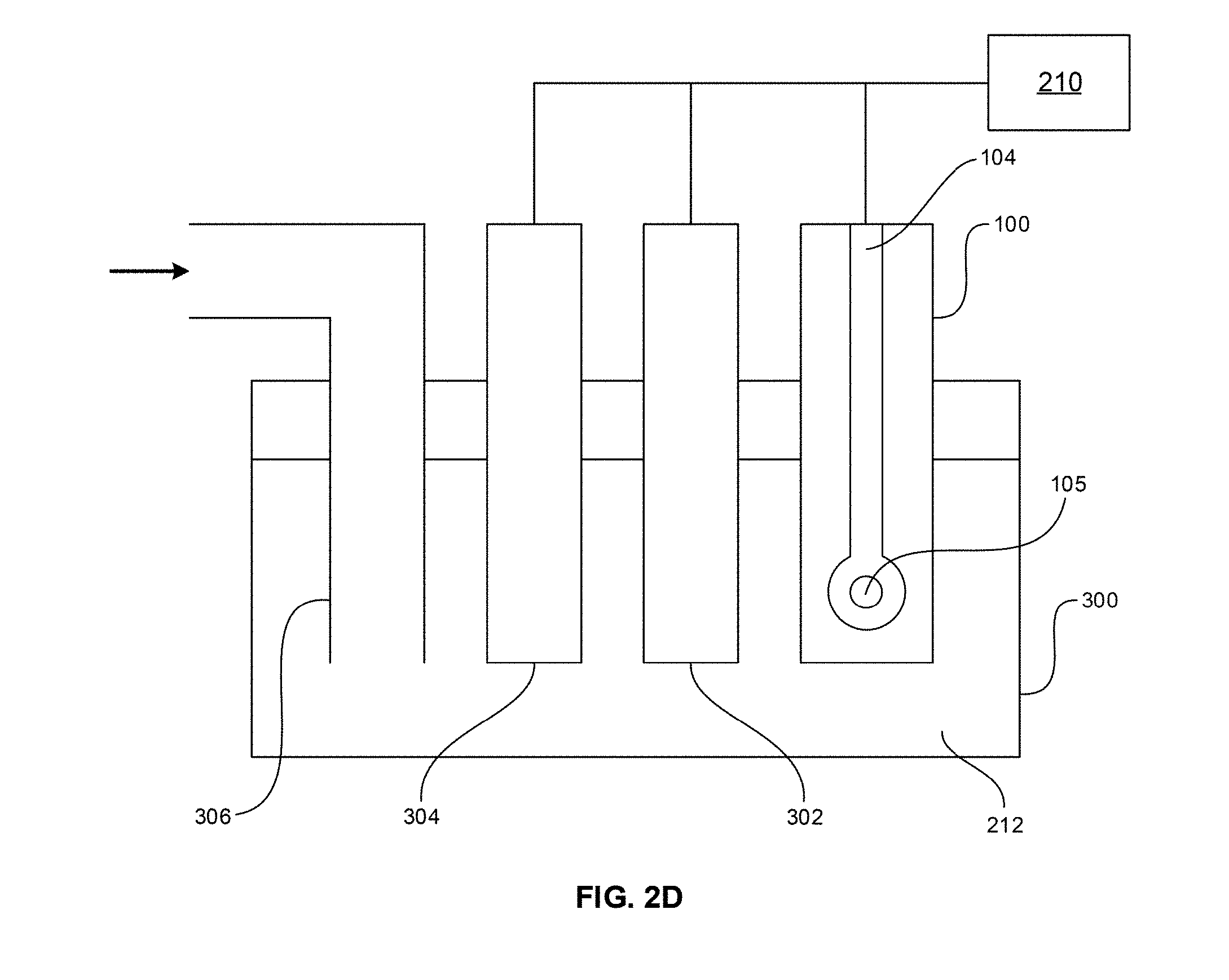
D00006

D00007
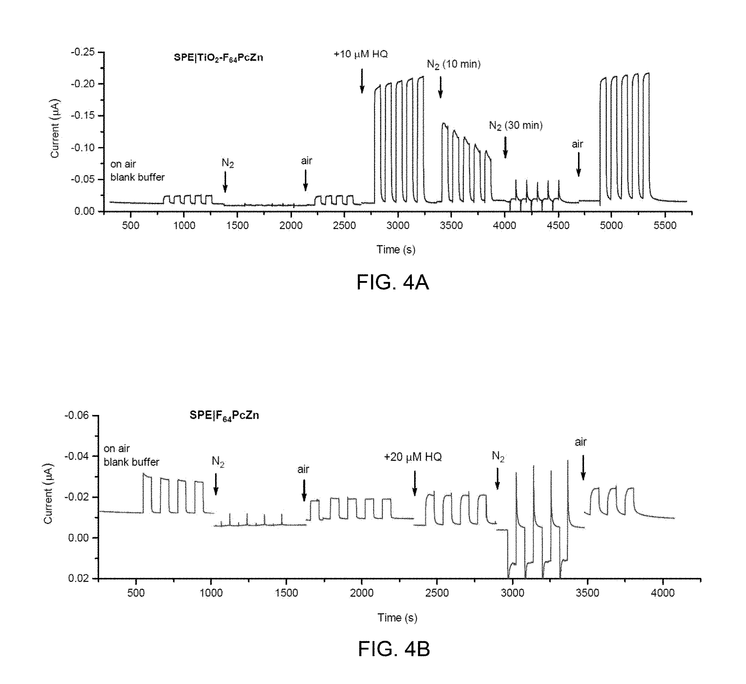
D00008

D00009

D00010

D00011

D00012
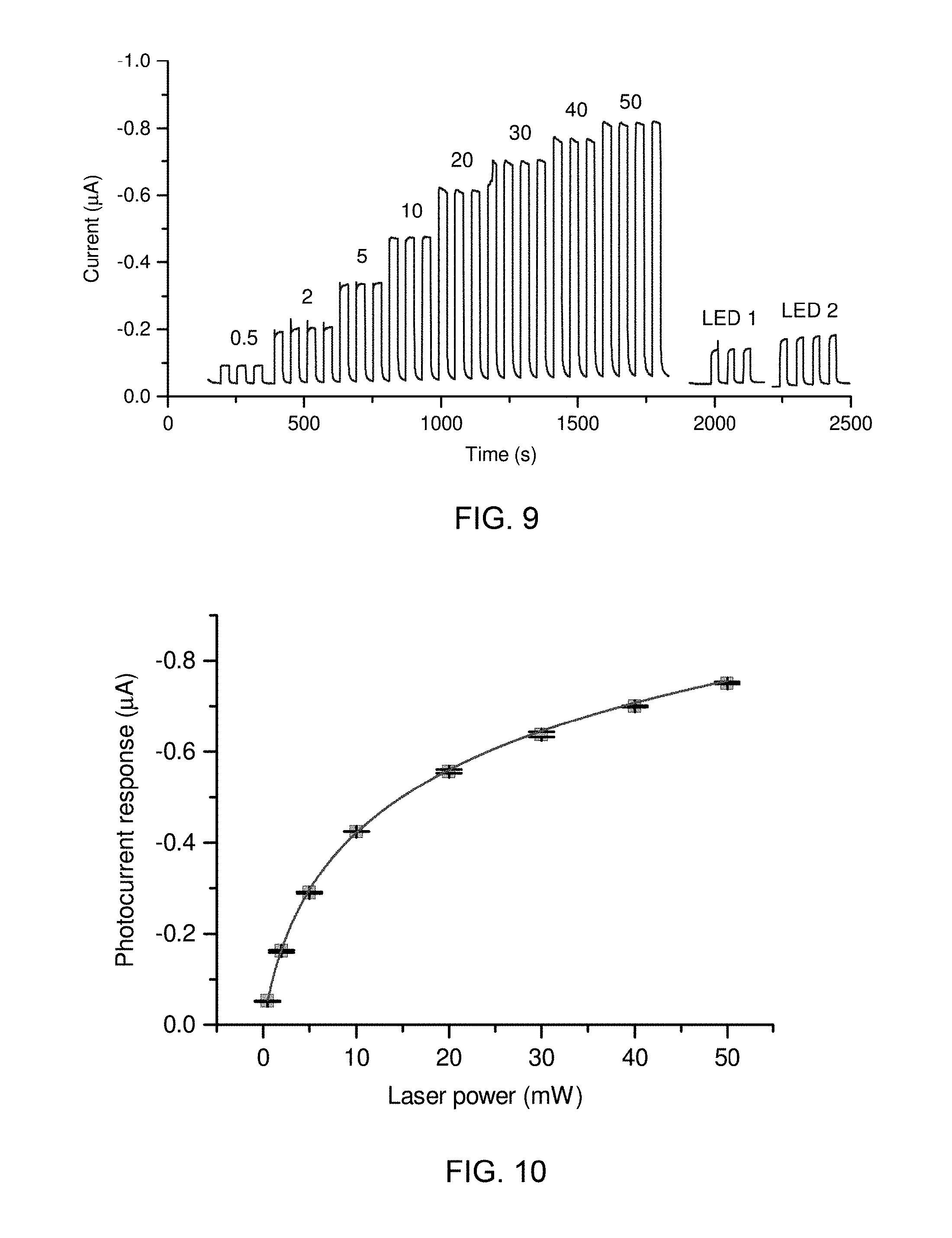
D00013
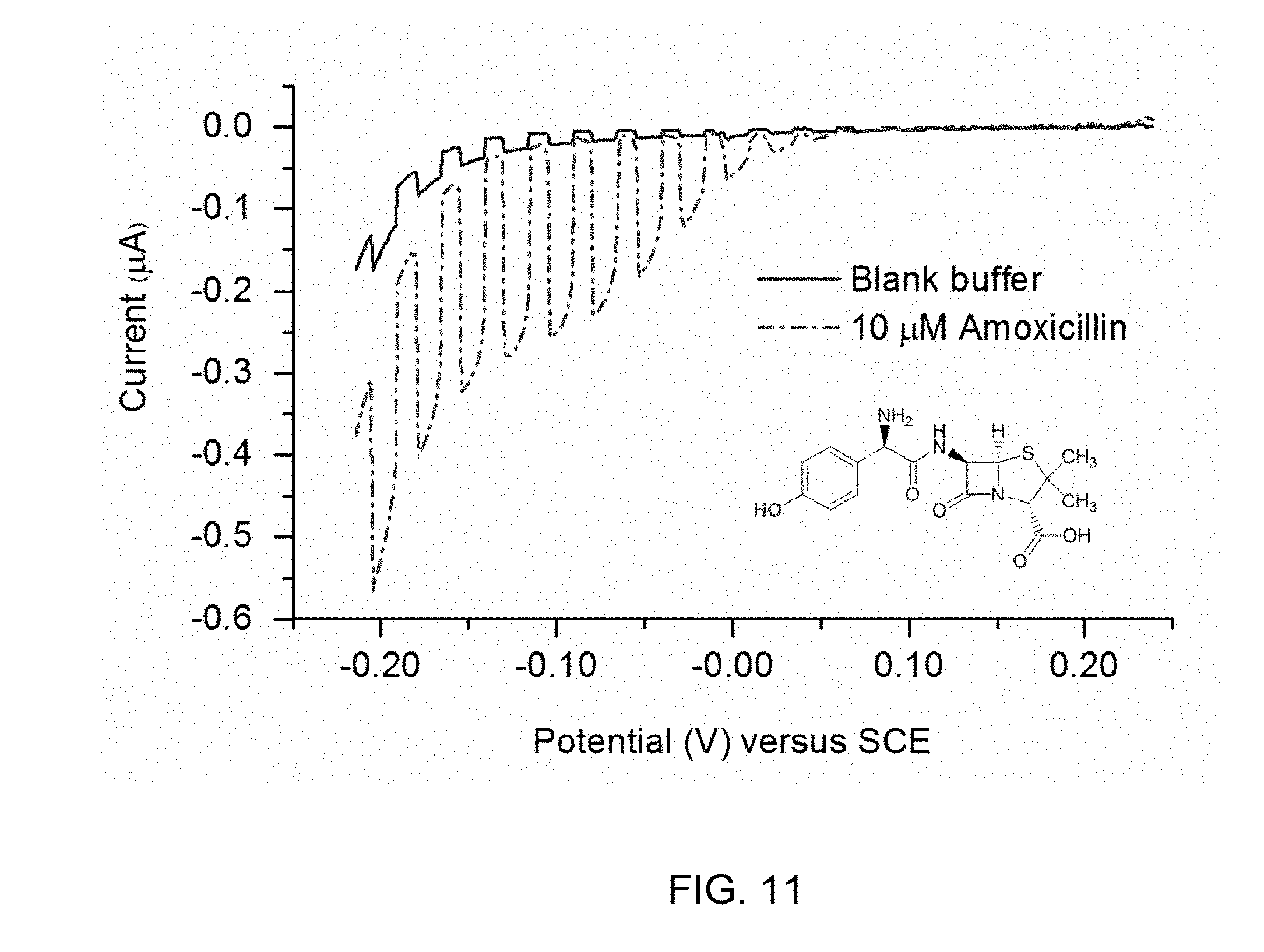
D00014

D00015

D00016

D00017
D00018
D00019

D00020
XML
uspto.report is an independent third-party trademark research tool that is not affiliated, endorsed, or sponsored by the United States Patent and Trademark Office (USPTO) or any other governmental organization. The information provided by uspto.report is based on publicly available data at the time of writing and is intended for informational purposes only.
While we strive to provide accurate and up-to-date information, we do not guarantee the accuracy, completeness, reliability, or suitability of the information displayed on this site. The use of this site is at your own risk. Any reliance you place on such information is therefore strictly at your own risk.
All official trademark data, including owner information, should be verified by visiting the official USPTO website at www.uspto.gov. This site is not intended to replace professional legal advice and should not be used as a substitute for consulting with a legal professional who is knowledgeable about trademark law.