Antibodies Binding Lag-3 And Uses Thereof
KANG; Xiaoqiang ; et al.
U.S. patent application number 16/033465 was filed with the patent office on 2019-01-17 for antibodies binding lag-3 and uses thereof. The applicant listed for this patent is Nanjing Leads Biolabs Co., Ltd.. Invention is credited to Xiao HUANG, Xiaoqiang KANG, Shoupeng LAI, Lijun ZHOU.
| Application Number | 20190016800 16/033465 |
| Document ID | / |
| Family ID | 65000052 |
| Filed Date | 2019-01-17 |


| United States Patent Application | 20190016800 |
| Kind Code | A1 |
| KANG; Xiaoqiang ; et al. | January 17, 2019 |
ANTIBODIES BINDING LAG-3 AND USES THEREOF
Abstract
The present invention provides an isolated monoclonal antibody that specifically binds LAG-3. A nucleic acid molecule encoding the antibody, an expression vector, a host cell and a method for expressing the antibody are also provided. The present invention further provides an immunoconjugate, a bispecific molecule and a pharmaceutical composition comprising the antibody, as well as a diagnostic and treatment method using an anti-LAG-3 antibody of the invention.
| Inventors: | KANG; Xiaoqiang; (Plainsboro, NJ) ; LAI; Shoupeng; (Germantown, MD) ; HUANG; Xiao; (Nanjing, CN) ; ZHOU; Lijun; (Beijing, CN) | ||||||||||
| Applicant: |
|
||||||||||
|---|---|---|---|---|---|---|---|---|---|---|---|
| Family ID: | 65000052 | ||||||||||
| Appl. No.: | 16/033465 | ||||||||||
| Filed: | July 12, 2018 |
Related U.S. Patent Documents
| Application Number | Filing Date | Patent Number | ||
|---|---|---|---|---|
| 62531892 | Jul 13, 2017 | |||
| Current U.S. Class: | 1/1 |
| Current CPC Class: | C07K 2317/75 20130101; C07K 2317/21 20130101; C07K 2317/622 20130101; A61K 2039/505 20130101; C07K 16/2818 20130101; C07K 16/2803 20130101; A61K 38/20 20130101; A61K 39/3955 20130101; C07K 2317/76 20130101; A61P 35/00 20180101; C07K 2317/24 20130101; A61K 38/2013 20130101; C07K 16/2827 20130101; C07K 16/2878 20130101; C07K 2317/33 20130101; C07K 2317/77 20130101; C07K 2317/92 20130101 |
| International Class: | C07K 16/28 20060101 C07K016/28; A61K 38/20 20060101 A61K038/20; A61K 39/395 20060101 A61K039/395; A61P 35/00 20060101 A61P035/00 |
Claims
1. An isolated monoclonal antibody, or an antigen-binding portion thereof, comprising a heavy chain variable region, wherein the heavy chain variable region comprises a CDR1 region comprising an amino acid sequence of SEQ ID NO:2, a CDR2 region comprising an amino acid sequence of SEQ ID NO:4, and a CDR3 region comprising an amino acid sequence of SEQ ID NO:6.
2. An isolated monoclonal antibody, or an antigen-binding portion thereof, comprising a heavy chain variable region comprising an amino acid sequence of SEQ ID NO:32.
3. The antibody, or the antigen-binding portion thereof, of claim 1, further comprising a light chain variable region, wherein the light chain variable region comprises a CDR1 region comprising an amino acid sequence of SEQ ID NO:8, a CDR2 region comprising an amino acid sequence of SEQ ID NO: 10, and a CDR3 region comprising an amino acid sequence of SEQ ID NO:12.
4. The antibody, or the antigen-binding portion thereof, of claim 1, further comprising a light chain variable region comprising an amino acid sequence of SEQ ID NO:34.
5. An isolated monoclonal antibody, or an antigen-binding portion thereof, of claim 1, comprising a heavy chain comprising an amino acid sequence of SEQ ID NO:36, and a light chain comprising an amino acid sequence of SEQ ID NO:38.
6. An isolated monoclonal antibody, or an antigen-binding portion thereof, of claim 1, comprising an amino acid sequence of SEQ ID NO:30.
7. The antibody, or the antigen-binding portion thereof, of claim 1, which exhibits one or a combination of the following properties: (a) binding to human LAG-3; (b) binding to monkey LAG-3; (c) lack of binding to mouse LAG-3; (d) binding to LAG-3 at a domain histocompatibility (MHC) class II bind to; (e) inhibits binding of LAG-3 to major histocompatibility (MHC) class II molecules; (f) inhibits binding of LAG-3 to LSECtin; (g) stimulates an immune response; and (h) stimulates an antigen-specific T cell response.
8. The antibody, or the antigen-binding portion thereof, of claim 1, which is an IgG1, IgG2 or IgG4 isotype.
9. The antibody, or antigen-binding portion thereof, of claim 1, which is a human, humanized, or chimeric antibody.
10. A composition comprising the antibody, or antigen-binding portion thereof, of claim 1, and a pharmaceutically acceptable carrier.
11. The composition of claim 10, further comprising an anti-cancer agent.
12. A method of treating tumor or viral infection in a subject comprising administering to the subject the antibody, or antigen-binding portion thereof, of claim 1.
13. The method of claim 12, wherein at least one additional immunostimulatory antibody is further administered to the subject.
14. The method of claim 13, wherein the immunostimulatory antibody is an anti-PD-1 antibody, an anti-PD-L1 antibody or an anti-CTLA-4 antibody.
15. The method of claim 12, wherein, a cytokine is further administered to the subject.
16. The method of claim 15, wherein, the cytokine is IL-2 or IL-21.
17. The method of claim 12, wherein, a costimulatory antibody is further administered to the subject.
18. The method of claim 17, wherein, the costimulatory antibody is an anti-CD137 or an anti-GITR antibody.
Description
BACKGROUND OF THE INVENTION
[0001] Therapeutic antibodies are one of the fastest growing segments of the pharmaceutical industry, especially monoclonal antibodies targeting certain disease-related cellular proteins.
[0002] One such target protein is lymphocyte-activation gene 3, also known as LAG3 (CD223), a protein encoded by the LAG3 gene in humans. LAG3 is a CD4-like protein, which like CD4, binds to MHC class II molecules, and functionally falls in the negative co-stimulatory group (inhibitory co-receptors) [Crawford A, et al., EJ. Curr Opin Immunol. 21:179-86(2009)], and is involved in the decline/suppression of T cell responses.
[0003] In-depth analysis showed that LAG-3 negatively regulates homeostasis, cellular proliferation and activation of T cells [Workman C J, et al., Eur J Immunol 33:970-9 (2003)]. Preclinical studies using antibody to block LAG-3 for cancer treatment show enhanced activation of antigen-specific T cells at the tumor site and disruption of tumor growth [Grosso J F, et al., J Clin Invest 117:3383-92 (2007)]. Furthermore, dual anti-LAG-3/anti-PD-1 antibody treatment cured most mice of established tumors that were largely resistant to single antibody treatment. [Woo S R, et al., Cancer Res; 72: 917-27 (2011)].
[0004] Although monoclonal antibodies binding to LAG-3 are known (e.g. US 2011/0150892 and US 2014/0093511), there is a need for additional monoclonal antibodies with enhanced binding affinity and other desirable pharmaceutical characteristics.
SUMMARY OF THE INVENTION
[0005] The present invention provides an isolated monoclonal antibody, for example, a human monoclonal antibody, that binds to LAG-3 (e.g., the human LAG-3, and monkey LAG-3) and has increased affinity compared to existing anti-LAG-3 antibodies (e.g., BMS-986016 developed by Bristol-Myers Squibb).
[0006] The antibody of the invention can be used for a variety of applications, including detection of the LAG-3 protein and stimulation of antigen-specific T cell responses in tumor-bearing or virus-bearing subjects.
[0007] Accordingly, in one aspect, the invention pertains to an isolated monoclonal antibody (e.g., a human antibody), or an antigen-binding portion thereof, having a heavy chain variable region that comprises a CDR1 region comprising an amino acid sequence of SEQ ID NO:2, a CDR2 region comprising an amino acid sequence of SEQ ID NO:4, and a CDR3 region comprising an amino acid sequence of SEQ ID NO:6. In one embodiment, the amino acid sequence of SEQ ID NO:2, 4 and 6 may be encoded by the nucleic acid sequence of SEQ ID NO:1, 3 and 5, respectively.
[0008] In one aspect, an isolated monoclonal antibody (e.g., a human antibody), or an antigen-binding portion thereof, of the present invention comprises a heavy chain variable region comprising the amino acid sequence of SEQ ID NO:32, which may be encoded by the nucleic acid sequence of SEQ ID NO: 31.
[0009] The monoclonal antibody or an antigen-binding portion thereof of the present invention in one embodiment comprises a light chain variable region that comprises a CDR1 region comprising an amino acid sequence of SEQ ID NO:8, a CDR2 region comprising an amino acid sequence of SEQ ID NO:10, and a CDR3 region comprising an amino acid sequence of SEQ ID NO:12. In one embodiment, the amino acid sequence of SEQ ID NO:8, 10 and 12 may be encoded by the nucleic acid sequence of SEQ ID NO:7, 9 and 11, respectively.
[0010] In one aspect, an isolated monoclonal antibody (e.g., a human antibody), or an antigen-binding portion thereof, of the present invention comprises a light chain variable region comprising the amino acid sequence of SEQ ID NO:34, which may be encoded by the nucleic acid sequence of SEQ ID NO: 33.
[0011] In one embodiment, the antibody, or the antigen-binding portion thereof, comprises the heavy chain variable region comprising the amino acid sequence of SEQ ID NO:32 and the light chain variable region comprising the amino acid sequence of SEQ ID NO:34.
[0012] In one embodiment, the antibody of the present invention comprises four framework regions in the heavy chain variable region having the amino acid sequences of SEQ ID NOs: 14, 16, 18 and 20, and four framework regions in the light chain variable region having the amino acid sequences of SEQ ID NOs:22, 24, 26 and 28. In one embodiment, the amino acid sequences of SEQ ID NOs: 14, 16, 18 and 20 may be encoded by the nucleic acid sequences of SEQ ID Nos:13, 15, 17 and 19, respectively. In one embodiment, the amino acid sequences of SEQ ID NOs:22, 24, 26 and 28 may be encoded by the nucleic acid sequences of SEQ ID Nos:21, 23, 25 and 27, respectively.
[0013] In one embodiment, the antibody of the present invention comprises a heavy chain having the amino acid sequence of SEQ ID NO:36, and a light chain having the amino acid sequence of SEQ ID NO: 38, which two may be encoded by the nucleic acid sequences of SEQ ID Nos:35 and 37, respectively. In one embodiment, the antibody of the present invention comprises two heavy chains each having the amino acid sequence of SEQ ID NO:36, and two light chains each having the amino acid sequence of SEQ ID NO: 38. Further, the antibody of the present invention comprises an amino acid sequence of SEQ ID NO:30, which may be encoded by the nucleic acid sequence of SEQ ID No.: 29.
[0014] In another embodiment, the antibody stimulates an antigen-specific T cell response, such as interferon gamma (IFN.gamma.) and or interferon-2 (IL-2) production in an antigen-specific T cell response. In other embodiments, the antibody stimulates an immune response, such as an anti-tumor response (e.g., inhibition of tumor growth in an in vivo tumor graft model) or an autoimmune response (e.g., development of diabetes in NOD mice).
[0015] In another embodiment, the antibody binds to an epitope of human LAG-3, blocking the interaction of LAG-3 with MHC class II or LSECtin.
[0016] The antibody of the invention can be a full-length antibody, for example, of an IgG1, IgG2 or IgG4 isotype, optionally with a serine to proline mutation in the heavy chain constant region hinge region (at a position corresponding to position 241 as described in Angal et al. (1993) Mol. Immunol. 30:105-108), such that inter-heavy chain disulfide bridge heterogeneity is reduced or abolished. In one aspect, the constant region isotype is IgG4 with a mutation at amino acid residues 220, e.g., S220P. Alternatively, the antibody can be an antibody fragment, such as a Fab, Fab' or Fab'2 fragment, or a single chain antibody.
[0017] In another aspect of the invention, the antibody or an antigen-binding portion thereof is part of an immunoconjugate which comprises a therapeutic agent, e.g., a cytotoxin or a radioactive isotope, linked to the antibody. In another aspect, the antibody is part of a bispecific molecule which comprises a second functional moiety (e.g., a second antibody) having a different binding specificity from said antibody, or the antigen binding portion thereof. In another aspect, the antibody or an antigen binding portions thereof (e.g. a scFv, see below) can be made into part of a chimeric antigen receptor (CAR) or an engineered T cell receptor (TCR) as part of an adoptive T cell immunotherapy strategy.
[0018] A composition comprising an antibody, or an antigen-binding portion thereof, an immunoconjugate or a bispecific molecule of the invention, optionally formulated in a pharmaceutically acceptable carrier, is also provided.
[0019] A nucleic acid molecule encoding the antibody, or the antigen-binding portion (e.g., variable regions and/or CDRs) thereof, of the invention is also provided, as well as an expression vector comprising the nucleic acid and a host cell comprising the expression vector. A method for preparing an anti-LAG-3 antibody using the host cell comprising the expression vector is also provided, and comprises steps of (i) expressing the antibody in the host cell and (ii) isolating the antibody from the host cell.
[0020] In another aspect, the invention provides a method for stimulating an immune response in a subject using the anti-LAG-3 antibody of the invention. In one embodiment, the method involves stimulating an antigen-specific T cell response by contacting T cells with the antibody of the invention. In a preferred embodiment, Interferon gamma (IFN.gamma.) and or interferon-2 (IL-2) production by the antigen-specific T cell is stimulated. In another embodiment, the subject is a tumor-bearing subject and an immune response against the tumor is stimulated. In another embodiment, the subject is a virus-bearing subject and an immune response against the virus is stimulated.
[0021] In yet another embodiment, the invention provides a method for inhibiting growth of tumor cells in a subject, comprising administering to the subject an antibody, or an antigen-binding portion thereof, of the invention. In still another embodiment, the invention provides a method for treating viral infection in a subject, comprising administering to the subject an antibody, or an antigen-binding portion thereof, of the invention. In another embodiment, the method comprises administering a composition, a bispecific, or an immunoconjugate of the invention.
[0022] In yet another embodiment, the invention provides a method for stimulating an immune response in a subject comprising administering to the subject an antibody, or an antigen-binding portion thereof, of the invention and at least one additional immunostimulatory antibody, such as an anti-PD-1 antibody, an anti-PD-L1 antibody and/or an anti-CTLA-4 antibody, such that an immune response is stimulated in the subject, for example to inhibit tumor growth or to stimulate an anti-viral response. In one embodiment, the additional immunostimulatory antibody is an anti-PD-1 antibody. In another embodiment, the additional immunostimulatory agent is an anti-PD-L1 antibody. In yet another embodiment, the additional immunostimulatory agent is an anti-CTLA-4 antibody. In yet another embodiment, an antibody, or an antigen-binding portion thereof, of the invention is administered with a cytokine (e.g., IL-2 and/or IL-21), or a costimulatory antibody (e.g., an anti-CD137 and/or anti-GITR antibody). The antibodies can be, for example, human, chimeric or humanized antibodies.
[0023] In another aspect, the invention provides an anti-LAG-3 antibody and a composition of the invention for use in the foregoing methods, or for the manufacture of a medicament for use in the foregoing methods (e.g., for treatment).
[0024] Other features and advantages of the instant disclosure will be apparent from the following detailed description and examples, which should not be construed as limiting. The contents of all references, Genbank entries, patents and published patent applications cited throughout this application are expressly incorporated herein by reference.
BRIEF DESCRIPTION OF THE DRAWINGS
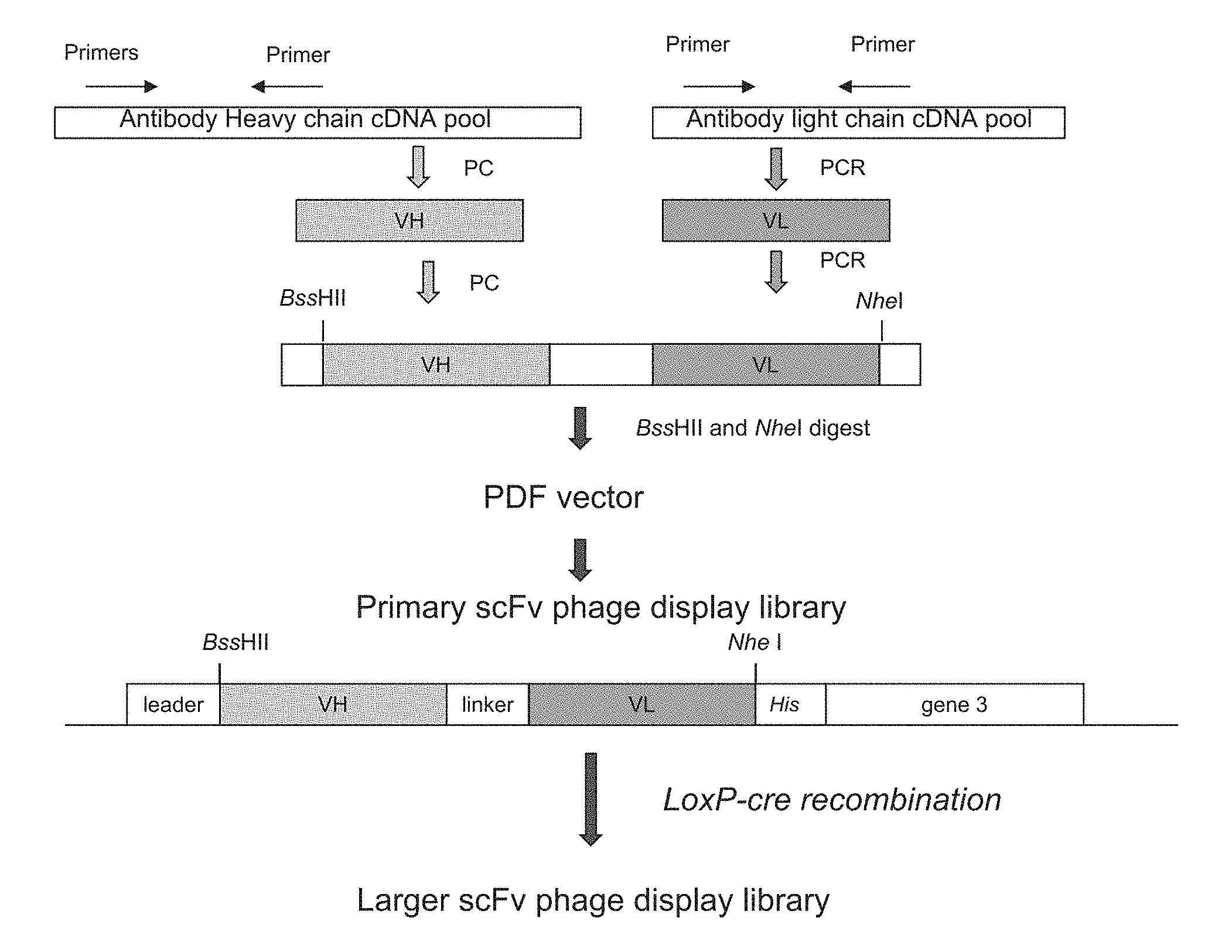
[0025] FIG. 1 is a flow chart showing PCR amplification and construction of single chain Fv (ScFv) phage display library.
[0026] FIG. 2 is a graph showing the binding activity of anti-LAG-3 antibody 2#, 8#, 13# and LAG3.5 to human LAG-3 recombinant protein in an ELISA assay.
[0027] FIG. 3 is a graph showing the binding activity of anti-LAG-3 antibody 2#, 8#, 13# and 14# to domain 1-2 of human LAG-3 recombinant protein in an ELISA assay.
[0028] FIG. 4 is a graph showing internalization of anti-LAG-3 antibody 2# and LAG3.5 on Jurkat-LAG3 cells.
[0029] FIG. 5 is a graph showing the binding activity of anti-LAG-3 antibody 2#, 6#, 8#, 13# and 14# to mouse LAG-3 recombinant protein in an ELISA assay.
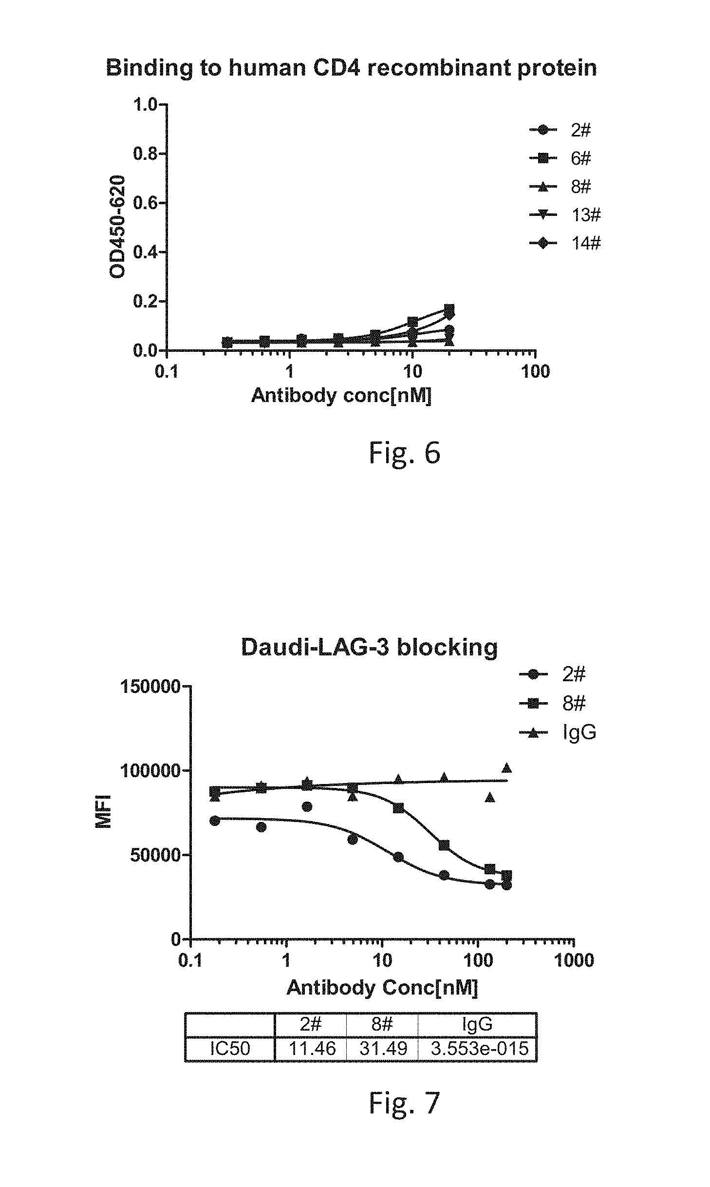
[0030] FIG. 6 is a graph showing the binding activity of anti-LAG-3 antibody 2#, 6#, 8#, 13# and 14# to human CD4 recombinant protein in an ELISA assay.
[0031] FIG. 7 is a graph showing blocking effect of anti-LAG-3 antibody 2#, 8#, and IgG on interaction of MHC class II molecule with LAG-3.
[0032] FIG. 8 is a graph showing the blocking effect of anti-LAG-3 antibody 2#, 6#, and IgG on interaction of LSECtin with LAG-3.
[0033] FIG. 9 are graphs showing the binding activity of anti-LAG-3 antibody 2#, 8# and 13# to LAG-3 expressed on surface of activated human T cells.
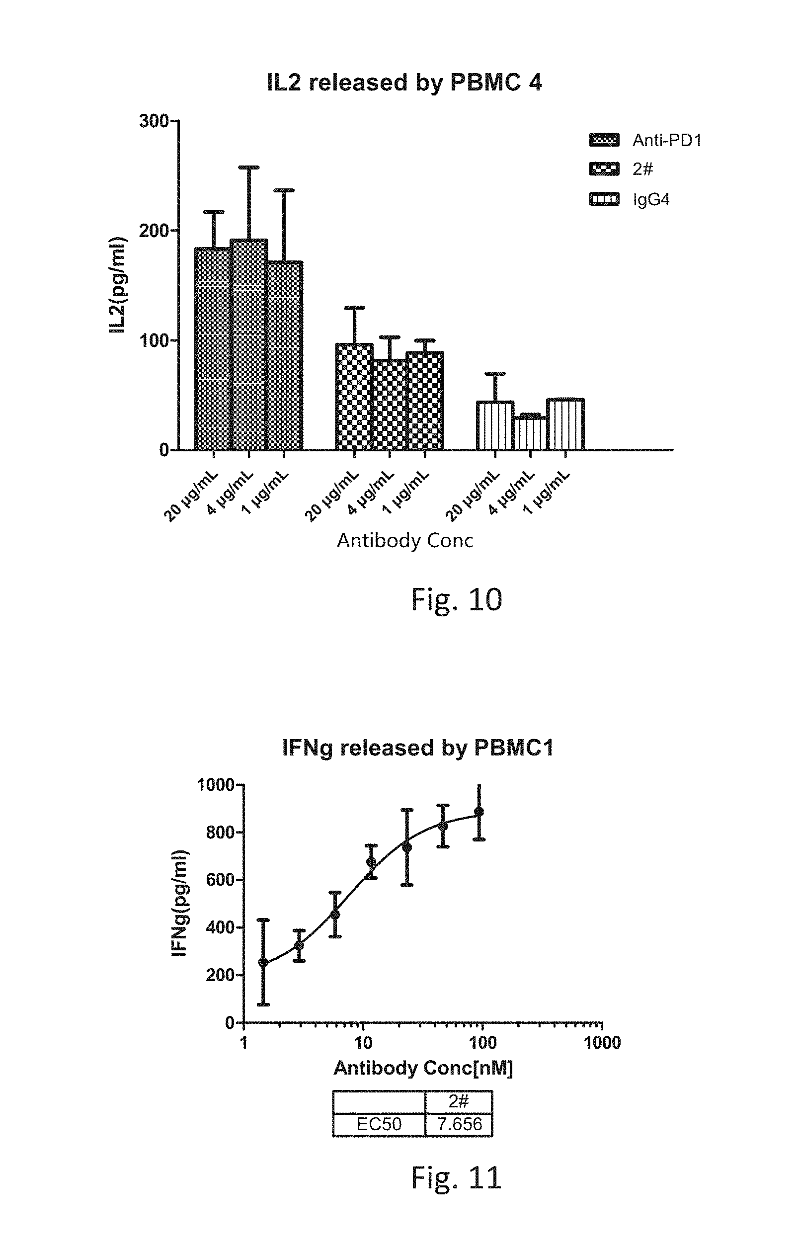
[0034] FIG. 10 is a graph showing the IL-2 levels released by human T cells cultured with anti-PD1 antibody, anti-LAG-3 antibody 2# or IgG4.
[0035] FIG. 11 is a graph showing the IFNg released by human T cells cultured with anti-LAG-3 antibody 2#.
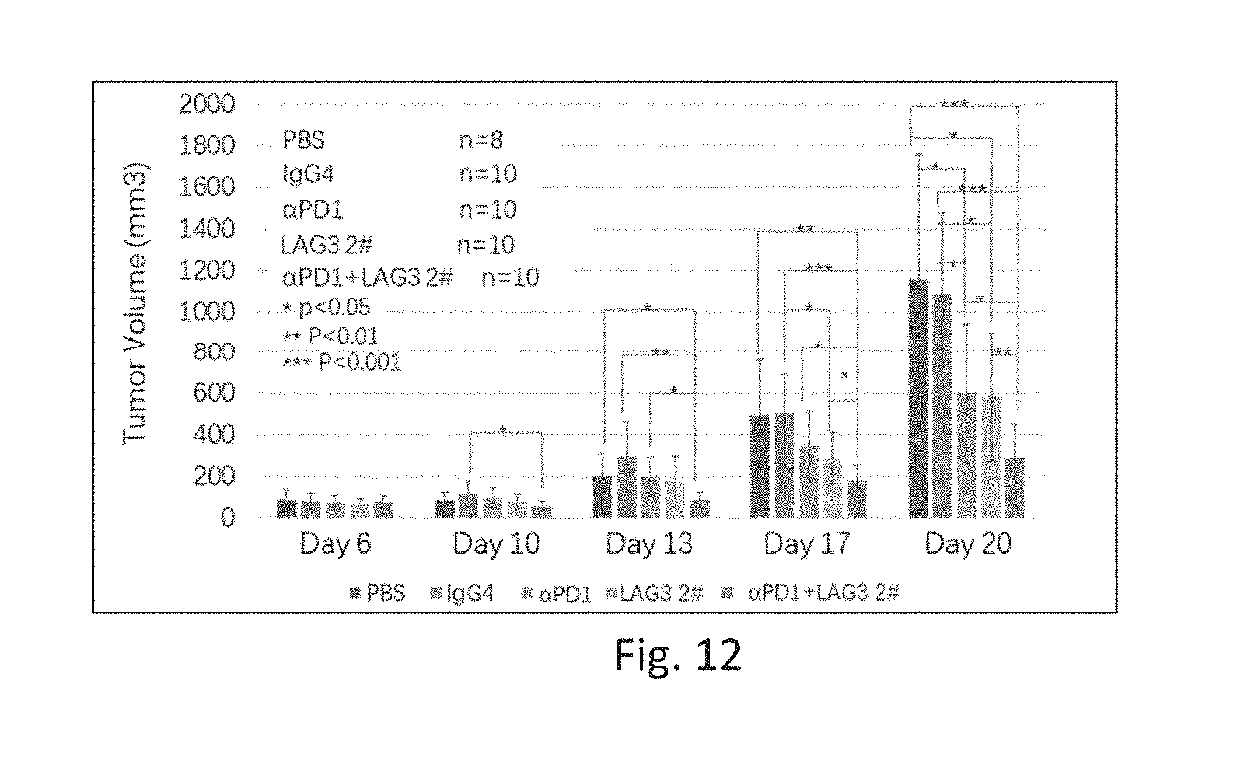
[0036] FIG. 12 is a graph showing anti-tumor effect of anti-LAG-3 antibody 2# and/or an anti-PD1 antibody.
DETAILED DESCRIPTION OF THE INVENTION
[0037] In order that the present disclosure may be more readily understood, certain terms are first defined. Additional definitions are set forth throughout the detailed description.
[0038] The term "LAG-3" refers to Lymphocyte Activation Gene-3. The term "LAG-3" comprises variants, isoforms, homologs, orthologs and paralogs. For example, an antibody specific for a human LAG-3 protein may, in certain cases, cross-reacts with a LAG-3 protein from a species other than human. In other embodiments, an antibody specific for a human LAG-3 protein may be completely specific for the human LAG-3 protein and exhibit no cross-reactivity to other species or of other types, or may cross-react with LAG-3 from certain other species but not all other species (e.g., cross-react with monkey LAG-3 but not mouse LAG-3).
[0039] The term "human LAG-3" refers to human sequence of LAG-3, such as the complete amino acid sequence of human LAG-3 having Genbank Accession No. NP 002277 (SEQ ID NO: 39). The term "mouse LAG-3" refers to mouse sequence LAG-3, such as the complete amino acid sequence of mouse LAG-3 having Genbank Accession No. NP_032505. LAG-3 is also known in the art as, for example, CD223. The human LAG-3 sequence may differ from human LAG-3 of Genbank Accession No. NP 002277 by having, e.g., conserved mutations or mutations in non-conserved regions and the LAG-3 has substantially the same biological function as the human LAG-3 of Genbank Accession No. NP_002277. For example, a biological function of human LAG-3 is having an epitope in the extracellular domain of LAG-3 that is specifically bound by an antibody of the instant disclosure or a biological function of human LAG-3 is binding to MHC Class II molecules.
[0040] The term "immune response" refers to the action of, for example, lymphocytes, antigen presenting cells, phagocytic cells, granulocytes, and soluble macromolecules produced by the above cells or the liver (including antibodies, cytokines, and complement) that results in selective damage to, destruction of, or elimination from the human body of invading pathogens, cells or tissues infected with pathogens, cancerous cells, or, in cases of autoimmunity or pathological inflammation, normal human cells or tissues.
[0041] An "antigen-specific T cell response" refers to responses by a T cell that result from stimulation of the T cell with the antigen for which the T cell is specific. Non-limiting examples of responses by a T cell upon antigen-specific stimulation include proliferation and cytokine production (e.g., IL-2 production).
[0042] The term "antibody" as referred to herein includes whole antibodies and any antigen binding fragment (i.e., "antigen-binding portion") or single chains thereof. Whole antibodies are glycoproteins comprising at least two heavy (H) chains and two light (L) chains inter-connected by disulfide bonds. Each heavy chain is comprised of a heavy chain variable region (abbreviated herein as V.sub.H) and a heavy chain constant region (abbreviated herein as C.sub.H). The heavy chain constant region is comprised of three domains, C.sub.H1, C.sub.H2 and C.sub.H3. Each light chain is comprised of a light chain variable region (abbreviated herein as V.sub.L) and a light chain constant region (abbreviated herein as C.sub.L). The light chain constant region is comprised of one domain, C.sub.L. The V.sub.H and V.sub.L regions can be further subdivided into regions of hypervariability, termed complementarity determining regions (CDR), interspersed with regions that are more conserved, termed framework regions (FR). Each V.sub.H and V.sub.L is composed of three CDRs and four FRs, arranged from amino-terminus to carboxy-terminus in the following order: FR1, CDR1, FR2, CDR2, FR3, CDR3, FR4. The variable regions of the heavy and light chains contain a binding domain that interacts with an antigen. The constant regions of the antibodies can mediate the binding of the immunoglobulin to host tissues or factors, including various cells of the immune system (e.g., effector cells) and the first component (C1q) of the classical complement system.
[0043] The term "antigen-binding portion" of an antibody (or simply "antibody portion"), as used herein, refers to one or more fragments of an antibody that retain the ability to specifically bind to an antigen (e.g., a LAG-3 protein). It has been shown that the antigen-binding function of an antibody can be performed by fragments of a full-length antibody. Examples of binding fragments encompassed within the term "antigen-binding portion" of an antibody include (i) a Fab fragment, a monovalent fragment consisting of the V.sub.L V.sub.H, C.sub.L and C.sub.H1 domains; (ii) a F(ab').sub.2 fragment, a bivalent fragment comprising two Fab fragments linked by a disulfide bridge at the hinge region; (iii) a Fd fragment consisting of the V.sub.H and C.sub.H1 domains; (iv) a Fv fragment consisting of the V.sub.L and V.sub.H domains of a single arm; (v) a bi-Fv fragment consisting of two Fc fragments, (vi) a dAb fragment (Ward et al., (1989) Nature 341:544-546), which consists of a V.sub.H domain; (vii) an isolated complementarity determining region (CDR); and (viii) a nanobody, a heavy chain variable region containing a single variable domain and two constant domains. Furthermore, although the two domains of the Fv fragment, V.sub.L and V.sub.H, are coded for by separate genes, they can be joined, using recombinant methods, by a synthetic linker that enables them to be made as a single protein chain in which the V.sub.L and V.sub.H regions pair to form monovalent molecules (known as single chain Fv (scFv); see e.g., Bird et al. (1988) Science 242:423-426; and Huston et al. (1988) Proc. Natl. Acad. Sci. USA 85:5879-5883). Such single chain antibodies are also intended to be encompassed within the term "antigen-binding portion" of an antibody. These antibody fragments are obtained using conventional techniques known to those with skill in the art, and the fragments are screened for utility in the same manner as are intact antibodies.
[0044] An "isolated antibody", as used herein, is intended to refer to an antibody that is substantially free of other antibodies having different antigenic specificities (e.g., an isolated antibody that specifically binds a LAG-3 protein is substantially free of antibodies that specifically bind antigens other than LAG-3 proteins). An isolated antibody that specifically binds a human LAG-3 protein may, however, have cross-reactivity to other antigens, such as LAG-3 proteins from other species. Moreover, an isolated antibody can be substantially free of other cellular material and/or chemicals.
[0045] The terms "monoclonal antibody" or "monoclonal antibody composition" as used herein refer to a preparation of antibody molecules of single molecular composition. A monoclonal antibody composition displays a single binding specificity and affinity for a particular epitope.
[0046] The term "human antibody", as used herein, is intended to include antibodies having variable regions in which both the framework and CDR regions are derived from human germline immunoglobulin sequences. Furthermore, if the antibody contains a constant region, the constant region also is derived from human germline immunoglobulin sequences. The human antibodies of the invention can include amino acid residues not encoded by human germline immunoglobulin sequences (e.g., mutations introduced by random or site-specific mutagenesis in vitro or by somatic mutation in vivo). However, the term "human antibody", as used herein, is not intended to include antibodies in which CDR sequences derived from the germline of another mammalian species, such as a mouse, have been grafted onto human framework sequences.
[0047] The term "human monoclonal antibody" refers to antibodies displaying a single binding specificity, which have variable regions in which both the framework and CDR regions are derived from human germline immunoglobulin sequences. In one embodiment, the human monoclonal antibodies are produced by a hybridoma which includes a B cell obtained from a transgenic nonhuman animal, e.g., a transgenic mouse, having a genome comprising a human heavy chain transgene and a light chain transgene fused to an immortalized cell.
[0048] The term "recombinant human antibody", as used herein, includes all human antibodies that are prepared, expressed, created or isolated by recombinant means, such as (a) antibodies isolated from an animal (e.g., a mouse) that is transgenic or transchromosomal for human immunoglobulin genes or a hybridoma prepared therefrom (described further below), (b) antibodies isolated from a host cell transformed to express the human antibody, e.g., from a transfectoma, (c) antibodies isolated from a recombinant, combinatorial human antibody library, and (d) antibodies prepared, expressed, created or isolated by any other means that involve splicing of human immunoglobulin gene sequences to other DNA sequences. Such recombinant human antibodies have variable regions in which the framework and CDR regions are derived from human germline immunoglobulin sequences. In certain embodiments, however, such recombinant human antibodies can be subjected to in vitro mutagenesis (or, when an animal transgenic for human Ig sequences is used, in vivo somatic mutagenesis) and thus the amino acid sequences of the V.sub.L and V.sub.H, regions of the recombinant antibodies are sequences that, while derived from and related to human germline V.sub.L and V.sub.H, sequences, may not naturally exist within the human antibody germline repertoire in vivo.
[0049] The term "isotype" refers to the antibody class (e.g., IgM or IgG1) that is encoded by the heavy chain constant region genes.
[0050] The phrases "an antibody recognizing an antigen" and "an antibody specific for an antigen" are used interchangeably herein with the term "an antibody which binds specifically to an antigen.
[0051] The term "human antibody derivatives" refers to any modified form of the human antibody, e.g., a conjugate of the antibody and another agent or antibody.
[0052] The term "humanized antibody" is intended to refer to antibodies in which CDR sequences derived from the germline of another mammalian species, such as a mouse, have been grafted onto human framework sequences. Additional framework region modifications can be made within the human framework sequences.
[0053] The term "chimeric antibody" is intended to refer to antibodies in which the variable region sequences are derived from one species and the constant region sequences are derived from another species, such as an antibody in which the variable region sequences are derived from a mouse antibody and the constant region sequences are derived from a human antibody.
[0054] As used herein, an antibody that "specifically binds to human LAG-3" is intended to refer to an antibody that binds to human LAG-3 protein (and possibly a LAG-3 protein from one or more non-human species) but does not substantially bind to non-LAG-3 proteins. Preferably, the antibody binds to a human LAG-3 protein with "high affinity", namely with a K.sub.D of 1.times.10.sup.-7 M or less, more preferably 1.times.10.sup.-8 M or less, more preferably 5.times.10.sup.-9 M or less, more preferably 1.times.10.sup.-9 M or less.
[0055] The term "does not substantially bind" to a protein or cells, as used herein, means does not bind or does not bind with a high affinity to the protein or cells, i.e. binds to the protein or cells with a K.sub.D of 1.times.10.sup.-6 M or more, more preferably 1.times.10.sup.-5 M or more, more preferably 1.times.10.sup.-4 M or more, more preferably 1.times.10.sup.-3 M or more, even more preferably 1.times.10.sup.-2 M or more.
[0056] The term "K.sub.assoc" or "K.sub.a", as used herein, is intended to refer to the association rate of a particular antibody-antigen interaction, whereas the term "K.sub.dis" or "K.sub.d," as used herein, is intended to refer to the dissociation rate of a particular antibody-antigen interaction. The term "K.sub.D," as used herein, is intended to refer to the dissociation constant, which is obtained from the ratio of K.sub.d to K.sub.a (i.e., K.sub.d/K.sub.a) and is expressed as a molar concentration (M). K.sub.D values for antibodies can be determined using methods well established in the art. A preferred method for determining the K.sub.D of an antibody is by using surface plasmon resonance, preferably using a biosensor system such as a Biacore.TM. system.
[0057] The term "high affinity" for an IgG antibody refers to an antibody having a K.sub.D of 1.times.10.sup.-6 M or less, more preferably 5.times.10.sup.-8 M or less, even more preferably 1.times.10.sup.-8 M or less, even more preferably 5.times.10.sup.-9 M or less and even more preferably 1.times.10.sup.-9 M or less for a target antigen. However, "high affinity" binding can vary for other antibody isotypes. For example, "high affinity" binding for an IgM isotype refers to an antibody having a K.sub.D of 10.sup.-6 M or less, more preferably 10.sup.-7 M or less, even more preferably 10.sup.-8 M or less.
[0058] The term "EC.sub.50", also known as half maximal effective concentration, refers to the concentration of an antibody which induces a response halfway between the baseline and maximum after a specified exposure time.
[0059] The term "subject" includes any human or nonhuman animal. The term "nonhuman animal" includes all vertebrates, e.g., mammals and non-mammals, such as non-human primates, sheep, dogs, cats, cows, horses, chickens, amphibians, and reptiles, although mammals are preferred, such as non-human primates, sheep, dogs, cats, cows and horses.
[0060] Various aspects of the invention are described in further detail in the following subsections.
[0061] Anti-LAG-Antibodies Having Advantageous Functional Properties
[0062] Antibodies of the invention specifically bind to human LAG-3 with better binding capacity compared to previously described anti-LAG-3 antibodies, particularly compared to BMS-BMS986016.
[0063] Antibodies of the invention preferably bind to human LAG-3 protein with a K.sub.D of 1.times.10.sup.-9 M or less, more preferably with a K.sub.D of 5.times.10.sup.-10 M or less.
[0064] Antibodies of the invention preferably bind to human LAG-3 proteins with EC.sub.50 of 0.2 nM or less.
[0065] Antibodies of the invention bind to the first two N-terminal domains of human LAG-3, i.e., the same domains MHC Class II binds to. The binding of LAG-3 to MHC Class II can be inhibited by antibodies of the invention. The antibodies of the invention can also block interaction of LAG-3 with LSECtin, a protein also know as CLEC4G (C-type lectin superfamily 4, member G) which was found to promote tumor progression when expressed on melanoma cells [F Xu, et al., Cancer Research. 74(13). April 2014].
[0066] Additional functional properties include cross-reactivity with LAG-3 from other species such as cynomolgus monkey and rhesus monkey. The antibodies of the invention do not substantially bind to mouse LAG-3. Preferably, an antibody of the invention binds to human LAG-3 with high affinity.
[0067] Other functional properties include the ability of the antibody to stimulate an immune response, such as an antigen-specific T cell response. This can be tested, for example, by assessing the ability of the antibody to stimulate interleukin-2 (IL-2) production in an antigen-specific T cell response. In certain embodiments, the antibody binds to human LAG-3 and stimulates an antigen-specific T cell response. In other embodiments, the antibody binds to human LAG-3 but does not stimulate an antigen-specific T cell response. Other means for evaluating the capacity of the antibody to stimulate an immune response include testing its ability to inhibit tumor growth, such as in an in vivo tumor graft model or the ability to stimulate an autoimmune response, such as the ability to promote the development of an autoimmune disease in an autoimmune model, e.g., the ability to promote the development of diabetes in the NOD mouse model. The antibodies of the invention can inhibit tumor growth, especially when administered with an anti-PD1 antibody.
[0068] Preferred antibodies of the invention are human monoclonal antibodies. Additionally or alternatively, the antibodies can be, for example, chimeric or humanized monoclonal antibodies.
[0069] Monoclonal Anti-LAG-3 Antibody
[0070] A preferred antibody of the invention is the human monoclonal antibody, anti-LAG-3 antibody 2#, structurally and chemically characterized as described below and in the following Examples. The V.sub.H amino acid sequence of anti-LAG-3 antibody 2#, is shown in SEQ ID NO: 32. The V.sub.L amino acid sequence of anti-LAG-3 antibody 2# is shown in SEQ ID NO: 34. Further, the heavy chain and light chain amino acid sequences of anti-LAG-3 antibody 2# are set forth in SEQ ID NO: 36 and SEQ ID NO: 38, respectively, and the full-length amino acid sequence of the anti-LAG-3 antibody 2# is set forth in SEQ ID NO: 30.
[0071] The V.sub.H and V.sub.L sequences (or CDR sequences) of other anti-LAG-3 antibodies which bind to human LAG-3 can be "mixed and matched" with the V.sub.H and V.sub.L sequences (or CDR sequences) of anti-LAG-3 antibody 2#. Preferably, when V.sub.H and V.sub.L chains (or the CDRs within such chains) are mixed and matched, a V.sub.H sequence from a particular V.sub.H/V.sub.L pairing is replaced with a structurally similar V.sub.H sequence. Likewise, preferably a V.sub.L sequence from a particular V.sub.H/V.sub.L pairing is replaced with a structurally similar V.sub.L sequence.
[0072] Accordingly, in one embodiment, an antibody of the invention, or an antigen binding portion thereof, comprises:
[0073] (a) a heavy chain variable region comprising amino acid sequence SEQ ID NO: 32 (i.e., the V.sub.H of anti-LAG-3 antibody 2#); and
[0074] (b) a light chain variable region comprising amino acid sequence SEQ ID NO: 34 (i.e., the V.sub.L of anti-LAG-3 antibody 2#) or the V.sub.L of another anti-LAG3 antibody (i.e., which differs from anti-LAG-3 antibody 2#), wherein the antibody specifically binds human LAG-3.
[0075] In another embodiment, an antibody of the invention, or an antigen binding portion thereof, comprises:
[0076] (a) the CDR1, CDR2, and CDR3 regions of the heavy chain variable region comprising amino acid sequence SEQ ID NO: 32 (i.e., the CDR sequences of anti-LAG-3 antibody 2#, SEQ ID NOs:2, 4, and 6, respectively); and
[0077] (b) the CDR1, CDR2, and CDR3 regions of the light chain variable region comprising amino acid sequence SEQ ID NO: 34 (i.e., the CDR sequences of anti-LAG-3 antibody 2#, SEQ ID NOs:8, 10, and 12, respectively) or the CDRs of another anti-LAG3 antibody (i.e., which differs from anti-LAG-3 antibody 2#), wherein the antibody specifically binds human LAG-3.
[0078] In yet another embodiment, the antibody, or antigen binding portion thereof, includes the heavy chain variable CDR2 region of anti-LAG-3 antibody 2# combined with CDRs of other antibodies which bind human LAG-3, e.g., CDR1 and/or CDR3 from the heavy chain variable region, and/or CDR1, CDR2, and/or CDR3 from the light chain variable region of a different anti-LAG-3 antibody.
[0079] In addition, it is well known in the art that the CDR3 domain, independently from the CDR1 and/or CDR2 domain(s), alone can determine the binding specificity of an antibody for a cognate antigen and that multiple antibodies can predictably be generated having the same binding specificity based on a common CDR3 sequence. See, e.g., Klimka et al., British J. of Cancer 83(2):252-260 (2000); Beiboer et al., J. Mol. Biol. 296:833-849 (2000); Rader et al., Proc. Natl. Acad. Sci. U.S.A. 95:8910-8915 (1998); Barbas et al., J. Am. Chem. Soc. 116:2161-2162 (1994); Barbas et al., Proc. Natl. Acad. Sci. U.S.A. 92:2529-2533 (1995); Ditzel et al., J. Immunol. 157:739-749 (1996); Berezov et al., BIAjournal 8:Scientific Review 8 (2001); Igarashi et al., J. Biochem (Tokyo) 117:452-7 (1995); Bourgeois et al., J. Virol 72:807-10 (1998); Levi et al., Proc. Natl. Acad. Sci. U.S.A. 90:4374-8 (1993); Polymenis and Stoller, J. Immunol. 152:5218-5329 (1994) and Xu and Davis, Immunity 13:37-45 (2000). See also, U.S. Pat. Nos. 6,951,646; 6,914,128; 6,090,382; 6,818,216; 6,156,313; 6,827,925; 5,833,943; 5,762,905 and 5,760,185. Each of these references is hereby incorporated by reference in its entirety.
[0080] Accordingly, in another embodiment, antibodies of the invention comprise the CDR2 of the heavy chain variable region of anti-LAG-3 antibody 2# (SEQ ID NO:4) and at least the CDR3 of the heavy and/or light chain variable region of anti-LAG-3 antibody 2# (SEQ ID NOs:6 and/or 12), or the CDR3 of the heavy and/or light chain variable region of another LAG-3 antibody, wherein the antibody is capable of specifically binding to human LAG-3. These antibodies preferably (a) compete for binding with LAG-3; (b) retain the functional characteristics; (c) bind to the same epitope; and/or (d) have a similar binding affinity as anti-LAG-3 antibody 2#. In yet another embodiment, the antibodies further may comprise the CDR2 of the light chain variable region of anti-LAG-3 antibody 2# (SEQ ID NO: 10), or the CDR2 of the light chain variable region of another LAG-3 antibody, wherein the antibody is capable of specifically binding to human LAG-3. In another embodiment, the antibodies of the invention further may include the CDR1 of the heavy and/or light chain variable region of anti-LAG-3 antibody 2# (SEQ ID NOs: 2 and/or 8), or the CDR1 of the heavy and/or light chain variable region of another LAG-3 antibody, wherein the antibody is capable of specifically binding to human LAG-3.
[0081] Conservative Modifications
[0082] In another embodiment, an antibody of the invention comprise a heavy and/or light chain variable region sequences of CDR1, CDR2 and CDR3 sequences which differ from those of anti-LAG-3 antibody 2# by one or more conservative modifications. It is understood in the art that certain conservative sequence modification can be made which do not remove antigen binding. See, e.g., Brummell et al. (1993) Biochem 32:1180-8; de Wildt et al. (1997) Prot. Eng. 10:835-41; Komissarov et al. (1997) J. Biol. Chem. 272:26864-26870; Hall et al. (1992) J. Immunol. 149:1605-12; Kelley and O'Connell (1993) Biochem. 32:6862-35; Adib-Conquy et al. (1998) Int. Immunol. 10:341-6 and Beers et al. (2000) Clin. Can. Res. 6:2835-43.
[0083] Accordingly, in one embodiment, the antibody comprises a heavy chain variable region comprising CDR1, CDR2, and CDR3 sequences and/or a light chain variable region comprising CDR1, CDR2, and CDR3 sequences, wherein:
[0084] (a) the heavy chain variable region CDR1 sequence comprises SEQ ID NO:2, and/or conservative modifications thereof; and/or
[0085] (b) the heavy chain variable region CDR3 sequence comprises SEQ ID NO:6, and conservative modifications thereof; and/or
[0086] (c) the light chain variable region CDR1, and/or CDR2, and/or CDR3 sequences comprise SEQ ID NO:8, and/or, SEQ ID NO:10, and/or SEQ ID NO:12, and/or conservative modifications thereof; and
[0087] (d) the antibody specifically binds human LAG-3.
[0088] The antibody of the present invention possesses one or more of the following functional properties described above, such as high affinity binding to human and monkey LAG-3, lack of binding to mouse LAG-3, the ability to inhibit binding of LAG-3 to MHC Class II or LSECtin, the ability to stimulate antigen-specific T cell responses, and/or the ability to inhibit tumor growth.
[0089] In various embodiments, the antibody can be, for example, a human, humanized or chimeric antibody.
[0090] As used herein, the term "conservative sequence modifications" is intended to refer to amino acid modifications that do not significantly affect or alter the binding characteristics of the antibody containing the amino acid sequence. Such conservative modifications include amino acid substitutions, additions and deletions. Modifications can be introduced into an antibody of the invention by standard techniques known in the art, such as site-directed mutagenesis and PCR-mediated mutagenesis. Conservative amino acid substitutions are ones in which the amino acid residue is replaced with an amino acid residue having a similar side chain. Families of amino acid residues having similar side chains have been defined in the art. These families include amino acids with basic side chains (e.g., lysine, arginine, histidine), acidic side chains (e.g., aspartic acid, glutamic acid), uncharged polar side chains (e.g., glycine, asparagine, glutamine, serine, threonine, tyrosine, cysteine, tryptophan), nonpolar side chains (e.g., alanine, valine, leucine, isoleucine, proline, phenylalanine, methionine), beta-branched side chains (e.g., threonine, valine, isoleucine) and aromatic side chains (e.g., tyrosine, phenylalanine, tryptophan, histidine). Thus, one or more amino acid residues within the CDR regions of an antibody of the invention can be replaced with other amino acid residues from the same side chain family and the altered antibody can be tested for retained function (i.e., the functions set forth above) using the functional assays described herein.
[0091] Engineered and Modified Antibodies
[0092] Antibodies of the invention can be prepared using an antibody having one or more of the V.sub.H/V.sub.L sequences of anti-LAG-3 antibody 2# as starting material to engineer a modified antibody. An antibody can be engineered by modifying one or more residues within one or both variable regions (i.e., V.sub.H and/or V.sub.L), for example within one or more CDR regions and/or within one or more framework regions. Additionally or alternatively, an antibody can be engineered by modifying residues within the constant region(s), for example to alter the effector function(s) of the antibody.
[0093] In certain embodiments, CDR grafting can be used to engineer variable regions of antibodies. Antibodies interact with target antigens predominantly through amino acid residues that are located in the six heavy and light chain complementarity determining regions (CDRs). For this reason, the amino acid sequences within CDRs are more diverse between individual antibodies than sequences outside of CDRs. Because CDR sequences are responsible for most antibody-antigen interactions, it is possible to express recombinant antibodies that mimic the properties of specific naturally occurring antibodies by constructing expression vectors that include CDR sequences from the specific naturally occurring antibody grafted onto framework sequences from a different antibody with different properties (see, e.g., Riechmann et al. (1998) Nature 332:323-327; Jones et al. (1986) Nature 321:522-525; Queen et al. (1989) Proc. Natl. Acad. See. U.S.A. 86:10029-10033; U.S. Pat. Nos. 5,225,539; 5,530,101; 5,585,089; 5,693,762 and 6,180,370).
[0094] Accordingly, another embodiment of the invention pertains to an isolated monoclonal antibody, or antigen binding portion thereof, comprising a heavy chain variable region comprising CDR1, CDR2, and CDR3 sequences comprising SEQ ID NOs: 2, 4, 6, respectively, and/or a light chain variable region comprising CDR1, CDR2, and CDR3 sequences comprising SEQ ID NOs: 8, 10, 12, respectively. While these antibodies contain the V.sub.H and V.sub.L CDR sequences of monoclonal antibody 2#, they can contain different framework sequences.
[0095] Such framework sequences can be obtained from public DNA databases or published references that include germline antibody gene sequences. For example, germline DNA sequences for human heavy and light chain variable region genes can be found in the "VBase" human germline sequence database (available on the Internet at www.mrc-cpe.cam.ac.uk/vbase), as well as in Kabat et al. (1991), cited supra; Tomlinson et al. (1992) "The Repertoire of Human Germline V.sub.H Sequences Reveals about Fifty Groups of V.sub.H Segments with Different Hypervariable Loops" J. Mol. Biol. 227:776-798; and Cox et al. (1994) "A Directory of Human Germ-line V.sub.H Segments Reveals a Strong Bias in their Usage" Eur. J. Immunol. 24:827-836; the contents of each of which are expressly incorporated herein by reference. As another example, the germline DNA sequences for human heavy and light chain variable region genes can be found in the Genbank database. For example, the following heavy chain germline sequences found in the HCo7 HuMAb mouse are available in the accompanying Genbank Accession Nos.: 1-69 (N_0010109, NT_024637 & BC070333), 3-33 (NG_0010109 & NT_024637) and 3-7 (NG_0010109 & NT_024637). As another example, the following heavy chain germline sequences found in the HCo12 HuMAb mouse are available in the accompanying Genbank Accession Nos.: 1-69 (NG_0010109, NT_024637 & BC070333), 5-51 (NG_0010109 & NT_024637), 4-34 (NG_0010109 & NT_024637), 3-30.3 (CAJ556644) & 3-23 (AJ406678).
[0096] Antibody protein sequences are compared against a compiled protein sequence database using one of the sequence similarity searching methods called the Gapped BLAST (Altschul et al. (1997), supra), which is well known to those skilled in the art.
[0097] Preferred framework sequences for use in the antibodies of the invention are those that are structurally similar to the framework sequences used by antibodies of the invention, e.g., the four framework regions in the heavy chain variable region having the amino acid sequences of SEQ ID NOs: 14, 16, 18 and 20, and the four framework regions in the light chain variable region having the amino acid sequences of SEQ ID NOs:22, 24, 26 and 28. The V.sub.H CDR1, CDR2, and CDR3 sequences can be grafted onto framework regions that have the identical sequence as that found in the germline immunoglobulin gene from which the framework sequence derive, or the CDR sequences can be grafted onto framework regions that contain one or more mutations as compared to the germline sequences. For example, it has been found that in certain instances it is beneficial to mutate residues within the framework regions to maintain or enhance the antigen binding ability of the antibody (see e.g., U.S. Pat. Nos. 5,530,101; 5,585,089; 5,693,762 and 6,180,370).
[0098] Another type of variable region modification is to mutate amino acid residues within the V.sub.H and/or V.sub.L CDR1, CDR2 and/or CDR3 regions to thereby improve one or more binding properties (e.g., affinity) of the antibody of interest. Site-directed mutagenesis or PCR-mediated mutagenesis can be performed to introduce the mutation(s) and the effect on antibody binding, or other functional property of interest, can be evaluated in in vitro or in vivo assays as described herein and provided in the Examples. Preferably conservative modifications (as known in the art) are introduced. The mutations can be amino acid substitutions, additions or deletions, but are preferably substitutions. Moreover, typically no more than one, two, three, four or five residues within a CDR region are altered.
[0099] Accordingly, in another embodiment, the invention provides isolated anti-LAG-3 monoclonal antibodies, or antigen binding portions thereof, comprising a heavy chain variable region comprising: (a) a V.sub.H CDR1 region comprising SEQ ID NO: 2, or an amino acid sequence having one, two, three, four or five amino acid substitutions, deletions or additions as compared to SEQ ID NO: 2; (b) a V.sub.H CDR2 region comprising SEQ ID NO:4, or an amino acid sequence having one, two, three, four or five amino acid substitutions, deletions or additions as compared to SEQ ID NO:4; (c) a V.sub.H CDR3 region comprising SEQ ID NO:6, or an amino acid sequence having one, two, three, four or five amino acid substitutions, deletions or additions as compared to SEQ ID NO:6; (d) a V.sub.L CDR1 region comprising SEQ ID NO:8, or an amino acid sequence having one, two, three, four or five amino acid substitutions, deletions or additions as compared to SEQ ID NO:8; (e) a V.sub.L CDR2 region comprising SEQ ID NO:10, or an amino acid sequence having one, two, three, four or five amino acid substitutions, deletions or additions as compared to SEQ ID NO:10; and (f) a V.sub.L CDR3 region comprising SEQ ID NO:12, or an amino acid sequence having one, two, three, four or five amino acid substitutions, deletions or additions as compared to SEQ ID NO:12.
[0100] Engineered antibodies of the invention include those in which modifications have been made to framework residues within V.sub.H and/or V.sub.L, e.g. to improve the properties of the antibody. Typically such framework modifications are made to decrease the immunogenicity of the antibody. For example, one approach is to "backmutate" one or more framework residues to the corresponding germline sequence. More specifically, an antibody that has undergone somatic mutation can contain framework residues that differ from the germline sequence from which the antibody is derived. Such residues can be identified by comparing the antibody framework sequences to the germline sequences from which the antibody is derived.
[0101] Another type of framework modification involves mutating one or more residues within the framework region, or even within one or more CDR regions, to remove T cell epitopes to thereby reduce the potential immunogenicity of the antibody. This approach is also referred to as "deimmunization" and is described in further detail in U.S. Patent Publication No. 20030153043.
[0102] In addition or as an alternative to modifications made within the framework or CDR regions, antibodies of the invention can be engineered to include modifications within the Fc region, typically to alter one or more functional properties of the antibody, such as serum half-life, complement fixation, Fc receptor binding, and/or antigen-dependent cellular cytotoxicity. Furthermore, an antibody of the invention can be chemically modified (e.g., one or more chemical moieties can be attached to the antibody) or be modified to alter its glycosylation, again to alter one or more functional properties of the antibody. Each of these embodiments is described in further detail below. The numbering of residues in the Fc region is that of the EU index of Kabat.
[0103] In a preferred embodiment, the antibody is an IgG4 isotype antibody comprising a Serine to Proline mutation at a position corresponding to position 241 as described in Angal et al. (1993) Mol. Immunol. 30:105-108 in the hinge region of the heavy chain constant region. This mutation has been reported to abolish the heterogeneity of inter-heavy chain disulfide bridges in the hinge region (Angal et al. supra; position 241 is based on the Kabat numbering system).
[0104] In one embodiment, the hinge region of CH1 is modified such that the number of cysteine residues in the hinge region is altered, e.g., increased or decreased. This approach is described further in U.S. Pat. No. 5,677,425. The number of cysteine residues in the hinge region of CH1 is altered to, for example, facilitate assembly of the light and heavy chains or to increase or decrease the stability of the antibody.
[0105] In another embodiment, the Fc hinge region of an antibody is mutated to decrease the biological half-life of the antibody. More specifically, one or more amino acid mutations are introduced into the CH2-CH3 domain interface region of the Fc-hinge fragment such that the antibody has impaired Staphylococcyl protein A (SpA) binding relative to native Fc-hinge domain SpA binding. This approach is described in further detail in U.S. Pat. No. 6,165,745. In still another embodiment, the glycosylation of an antibody is modified. For example, an aglycoslated antibody can be made (i.e., the antibody lacks glycosylation). Glycosylation can be altered to, for example, increase the affinity of the antibody for antigen. Such carbohydrate modifications can be accomplished by, for example, altering one or more sites of glycosylation within the antibody sequence. For example, one or more amino acid substitutions can be made that result in elimination of one or more variable region framework glycosylation sites to thereby eliminate glycosylation at that site. Such aglycosylation may increase the affinity of the antibody for antigen. See, e.g., U.S. Pat. Nos. 5,714,350 and 6,350,861.
[0106] Additionally or alternatively, an antibody can be made that has an altered type of glycosylation, such as a hypofucosylated antibody having reduced amounts of fucosyl residues or an antibody having increased bisecting GlcNac structures. Such altered glycosylation patterns have been demonstrated to increase the ADCC ability of antibodies. Such carbohydrate modifications can be accomplished by, for example, expressing the antibody in a host cell with altered glycosylation machinery. Cells with altered glycosylation machinery have been described in the art and can be used as host cells in which to express recombinant antibodies of the invention to thereby produce an antibody with altered glycosylation. For example, the cell lines Ms704, Ms705, and Ms709 lack the fucosyltransferase gene, FUT8 (.alpha.(1,6)-fucosyltransferase), such that antibodies expressed in the Ms704, Ms705, and Ms709 cell lines lack fucose on their carbohydrates. The Ms704, Ms705, and Ms709 FUT8.sup.-/- cell lines were created by the targeted disruption of the FUT8 gene in CHO/DG44 cells using two replacement vectors (see U.S. Patent Publication No. 20040110704 and Yamane-Ohnuki et al. (2004) Biotechnol Bioeng 87:614-22). As another example, EP 1,176,195 describes a cell line with a functionally disrupted FUT8 gene, which encodes a fucosyl transferase, such that antibodies expressed in such a cell line exhibit hypofucosylation by reducing or eliminating the .alpha.-1,6 bond-related enzyme. EP 1,176,195 also describes cell lines which have a low enzyme activity for adding fucose to the N-acetylglucosamine that binds to the Fc region of the antibody or does not have the enzyme activity, for example the rat myeloma cell line YB2/0 (ATCC CRL 1662). PCT Publication WO 03/035835 describes a variant CHO cell line, Lec13 cells, with reduced ability to attach fucose to Asn(297)-linked carbohydrates, also resulting in hypofucosylation of antibodies expressed in that host cell (see also Shields et al. (2002) J. Biol. Chem. 277:26733-26740). Antibodies with a modified glycosylation profile can also be produced in chicken eggs, as described in PCT Publication WO 06/089231. Alternatively, antibodies with a modified glycosylation profile can be produced in plant cells, such as Lemna. Methods for production of antibodies in a plant system are disclosed in the U.S. patent application corresponding to Alston & Bird LLP attorney docket No. 040989/314911, filed on Aug. 11, 2006. PCT Publication WO 99/54342 describes cell lines engineered to express glycoprotein-modifying glycosyl transferases (e.g., .beta.(1,4)-N-acetylglucosaminyltransferase III (GnTIII)) such that antibodies expressed in the engineered cell lines exhibit increased bisecting GlcNac structures which results in increased ADCC activity of the antibodies (see also Umana et al. (1999) Nat. Biotech. 17:176-180). Alternatively, the fucose residues of the antibody can be cleaved off using a fucosidase enzyme; e.g., the fucosidase .alpha.-L-fucosidase removes fucosyl residues from antibodies (Tarentino et al. (1975) Biochem. 14:5516-23).
[0107] Another modification of the antibodies herein that is contemplated by this disclosure is pegylation. An antibody can be pegylated to, for example, increase the biological (e.g., serum) half-life of the antibody. To pegylate an antibody, the antibody, or fragment thereof, typically is reacted with polyethylene glycol (PEG), such as a reactive ester or aldehyde derivative of PEG, under conditions in which one or more PEG groups become attached to the antibody or antibody fragment. Preferably, the pegylation is carried out via an acylation reaction or an alkylation reaction with a reactive PEG molecule (or an analogous reactive water-soluble polymer). As used herein, the term "polyethylene glycol" is intended to encompass any of the forms of PEG that have been used to derivatize other proteins, such as mono (C1-C10) alkoxy- or aryloxy-polyethylene glycol or polyethylene glycol-maleimide. In certain embodiments, the antibody to be pegylated is an aglycosylated antibody. Methods for pegylating proteins are known in the art and can be applied to the antibodies of the invention. See, e.g., EPO 154 316 and EP 0 401 384.
[0108] Antibody Physical Properties
[0109] Antibodies of the invention can be characterized by their various physical properties, to detect and/or differentiate different classes thereof.
[0110] For example, antibodies can contain one or more glycosylation sites in either the light or heavy chain variable region. Such glycosylation sites may result in increased immunogenicity of the antibody or an alteration of the pK of the antibody due to altered antigen binding (Marshall et al (1972) Annu Rev Biochem 41:673-702; Gala and Morrison (2004) J Immunol 172:5489-94; Wallick et al (1988) J Exp Med 168:1099-109; Spiro (2002) Glycobiology 12:43R-56R; Parekh et al (1985) Nature 316:452-7; Mimura et al. (2000) Mol Immunol 37:697-706). Glycosylation has been known to occur at motifs containing an N-X-S/T sequence. In some instances, it is preferred to have an anti-LAG-3 antibody that does not contain variable region glycosylation. This can be achieved either by selecting antibodies that do not contain the glycosylation motif in the variable region or by mutating residues within the glycosylation region.
[0111] In a preferred embodiment, the antibodies do not contain asparagine isomerism sites. The deamidation of asparagine may occur on N-G or D-G sequences and result in the creation of an isoaspartic acid residue that introduces a kink into the polypeptide chain and decreases its stability (isoaspartic acid effect).
[0112] Each antibody will have a unique isoelectric point (pI), which generally falls in the pH range between 6 and 9.5. The pI for an IgG1 antibody typically falls within the pH range of 7-9.5 and the pI for an IgG4 antibody typically falls within the pH range of 6-8. There is speculation that antibodies with a pI outside the normal range may have some unfolding and instability under in vivo conditions. Thus, it is preferred to have an anti-LAG-3 antibody that contains a pI value that falls in the normal range. This can be achieved either by selecting antibodies with a pI in the normal range or by mutating charged surface residues.
[0113] Nucleic Acid Molecules Encoding Antibodies of the Invention
[0114] In another aspect, the invention provides nucleic acid molecules that encode heavy and/or light chain variable regions, or CDRs, of the antibodies of the invention. The nucleic acids can be present in whole cells, in a cell lysate, or in a partially purified or substantially pure form. A nucleic acid is "isolated" or "rendered substantially pure" when purified away from other cellular components or other contaminants, e.g., other cellular nucleic acids or proteins, by standard techniques. A nucleic acid of the invention can be, e.g., DNA or RNA and may or may not contain intronic sequences. In a preferred embodiment, the nucleic acid is a cDNA molecule.
[0115] Nucleic acids of the invention can be obtained using standard molecular biology techniques. For antibodies expressed by hybridomas (e.g., hybridomas prepared from transgenic mice carrying human immunoglobulin genes as described further below), cDNAs encoding the light and heavy chains of the antibody made by the hybridoma can be obtained by standard PCR amplification or cDNA cloning techniques. For antibodies obtained from an immunoglobulin gene library (e.g., using phage display techniques), a nucleic acid encoding such antibodies can be recovered from the gene library.
[0116] Preferred nucleic acids molecules of the invention include those encoding the V.sub.H and V.sub.L (SEQ ID NOs:31 and 33, respectively) or the CDRs (SEQ ID Nos: 1, 3, 5, 7, 9 and 11, respectivelt) sequences of LAG-3 monoclonal antibody. Once DNA fragments encoding V.sub.H and V.sub.L segments are obtained, these DNA fragments can be further manipulated by standard recombinant DNA techniques, for example to convert the variable region genes to full-length antibody chain genes, to Fab fragment genes or to a scFv gene. In these manipulations, a V.sub.L- or V.sub.H-encoding DNA fragment is operatively linked to another DNA fragment encoding another protein, such as an antibody constant region or a flexible linker. The term "operatively linked", as used in this context, is intended to mean that the two DNA fragments are joined such that the amino acid sequences encoded by the two DNA fragments remain in-frame.
[0117] The isolated DNA encoding the V.sub.H region can be converted to a full-length heavy chain gene by operatively linking the V.sub.H-encoding DNA to another DNA molecule encoding heavy chain constant regions (C.sub.H1, C.sub.H2 and C.sub.H3). The sequences of human heavy chain constant region genes are known in the art (see e.g., Kabat et al. (1991), supra) and DNA fragments encompassing these regions can be obtained by standard PCR amplification. The heavy chain constant region can be an IgG, IgG2, IgG3, IgG4, IgA, IgE, IgM or IgD constant region, but most preferably is an IgG1 or IgG4 constant region. For a Fab fragment heavy chain gene, the V.sub.H-encoding DNA can be operatively linked to another DNA molecule encoding only the heavy chain C.sub.H1 constant region.
[0118] The isolated DNA encoding the V.sub.L region can be converted to a full-length light chain gene (as well as a Fab light chain gene) by operatively linking the V.sub.L-encoding DNA to another DNA molecule encoding the light chain constant region, C.sub.L. The sequences of human light chain constant region genes are known in the art (see e.g., Kabat et al., supra) and DNA fragments encompassing these regions can be obtained by standard PCR amplification. In preferred embodiments, the light chain constant region can be a kappa or lambda constant region.
[0119] To create a scFv gene, the V.sub.H- and V.sub.L-encoding DNA fragments are operatively linked to another fragment encoding a flexible linker, e.g., encoding the amino acid sequence (Gly.sub.4-Ser).sub.3, such that the V.sub.H and V.sub.L sequences can be expressed as a contiguous single-chain protein, with the V.sub.L and V.sub.H regions joined by the flexible linker (see e.g., Bird et al. (1988) Science 242:423-426; Huston et al. (1988) Proc. Natl. Acad. Sci. USA 85:5879-5883; McCafferty et al., (1990) Nature 348:552-554).
[0120] Production of Monoclonal Antibodies of the Invention
[0121] Monoclonal antibodies (mAbs) of the present invention can be produced using the well-known somatic cell hybridization (hybridoma) technique of Kohler and Milstein (1975) Nature 256: 495. Other embodiments for producing monoclonal antibodies include viral or oncogenic transformation of B lymphocytes and phage display techniques. Chimeric or humanized antibodies are also well known in the art. See e.g., U.S. Pat. Nos. 4,816,567; 5,225,539; 5,530,101; 5,585,089; 5,693,762 and 6,180,370, the contents of which are specifically incorporated herein by reference in their entirety.
[0122] In a preferred embodiment, the antibodies of the invention are human monoclonal antibodies. Such human monoclonal antibodies directed against human LAG-3 can be generated using transgenic or transchromosomic mice carrying parts of the human immune system rather than the mouse system. These transgenic and transchromosomic mice include mice referred to herein as the HuMAb Mouse.TM. and KM Mouse.TM., respectively, and are collectively referred to herein as "human Ig mice."
[0123] The HuMAb Mouse.TM. (Medarex.TM., Inc.) contains human immunoglobulin gene miniloci that encode unrearranged human heavy (.mu. and .gamma.) and .kappa. light chain immunoglobulin sequences, together with targeted mutations that inactivate the endogenous .mu. and .kappa. chain loci (see e.g., Lonberg et al. (1994) Nature 368(6474): 856-859). Accordingly, the mice exhibit reduced expression of mouse IgM or .kappa., and in response to immunization, the introduced human heavy and light chain transgenes undergo class switching and somatic mutation to generate high affinity human IgG.kappa. monoclonal antibodies (Lonberg et al. (1994), supra; reviewed in Lonberg (1994) Handbook of Experimental Pharmacology 113:49-101; Lonberg, N. and Huszar, D. (1995) Intern. Rev. Immunol. 13: 65-93, and Harding and Lonberg (1995) Ann. N.Y. Acad. Sci. 764:536-546). Preparation and use of the HuMAb Mouse.TM., and the genomic modifications carried by such mice, is further described in Taylor et al. (1992) Nucleic Acids Research 20:6287-6295; Chen et al. (1993) International Immunology 5: 647-656; Tuaillon et al. (1993) Proc. Natl. Acad. Sci. USA 90:3720-3724; Choi et al. (1993) Nature Genetics 4:117-123; Chen et al. (1993) EMBO J. 12: 821-830; Tuaillon et al. (1994) J. Immunol. 152:2912-2920; Taylor et al. (1994) International Immunology 6: 579-591; and Fishwild et al. (1996) Nature Biotechnology 14: 845-851, the contents of all of which are hereby specifically incorporated by reference in their entirety. See further, U.S. Pat. Nos. 5,545,806; 5,569,825; 5,625,126; 5,633,425; 5,789,650; 5,877,397; 5,661,016; 5,814,318; 5,874,299; 5,770,429; and 5,545,807; PCT Publication Nos. WO 92/03918; WO 93/12227; WO 94/25585; WO 97/13852; WO 98/24884; WO 99/45962 and WO 01/14424, the contents of which are incorporated herein by reference in their entirety.
[0124] In another embodiment, human antibodies of the invention can be raised using a mouse that carries human immunoglobulin sequences on transgenes and transchomosomes, such as a mouse that carries a human heavy chain transgene and a human light chain transchromosome. This mouse is referred to herein as a "KM Mouse.TM.," and is described in detail in PCT Publication WO 02/43478. A modified form of this mouse, which further comprises a homozygous disruption of the endogenous Fc.gamma.RIIB receptor gene, is also described in PCT Publication WO 02/43478 and referred to herein as a "KM/FCGR2D mouse." In addition, mice with either the HCo7 or HCo12 heavy chain transgenes or both can be used.
[0125] Additional transgenic animal embodiments include the Xenomouse (Abgenix, Inc., U.S. Pat. Nos. 5,939,598; 6,075,181; 6,114,598; 6,150,584 and 6,162,963). Further embodiments include "TC mice" (Tomizuka et al. (2000) Proc. Natl. Acad. Sci. USA 97:722-727) and cows carrying human heavy and light chain transchromosomes (Kuroiwa et al. (2002) Nature Biotechnology 20:889-894; PCT Publication WO 02/092812). The contents of these patents and publications are specifically incorporated herein by reference in their entirety.
[0126] In one embodiment, human monoclonal antibodies of the invention are prepared using phage display methods for screening libraries of human immunoglobulin genes. See, e.g. U.S. Pat. Nos. 5,223,409; 5,403,484; 5,571,698; 5,427,908; 5,580,717; 5,969,108; 6,172,197; 5,885,793; 6,521,404; 6,544,731; 6,555,313; 6,582,915; and 6,593,081, the contents of which are incorporated herein by reference in their entirety.
[0127] Human monoclonal antibodies of the invention can also be prepared using SCID mice into which human immune cells have been reconstituted such that a human antibody response can be generated upon immunization. See, e.g., U.S. Pat. Nos. 5,476,996 and 5,698,767, the contents of which are incorporated herein by reference in their entirety.
[0128] In another embodiment, human anti-LAG-3 antibodies are prepared using phage display where the phages comprise nucleic acids encoding antibodies generated in transgenic animals previously immunized with LAG-3. In a preferred embodiment, the transgenic animal is a HuMab, KM, or Kirin mouse. See, e.g. U.S. Pat. No. 6,794,132, the contents of which are incorporated herein by reference in its entirety.
[0129] Immunization of Human Ig Mice
[0130] In one embodiment of the invention, human Ig mice are immunized with a purified or enriched preparation of a LAG-3 antigen, recombinant LAG-3 protein, or cells expressing a LAG-3 protein. See, e.g., Lonberg et al. (1994), supra; Fishwild et al. (1996), supra; PCT Publications WO 98/24884 or WO 01/14424, the contents of which are incorporated herein by reference in their entirety. In a preferred embodiment, 6-16 week old mice are immunized with 5-50 .mu.g of LAG-3 protein. Alternatively, a portion of LAG-3 fused to a non-LAG-3 polypeptide is used.
[0131] In one embodiment, the transgenic mice are immunized intraperitoneally (IP) or intravenously (IV) with LAG-3 antigen in complete Freund's adjuvant, followed by subsequent IP or IV immunizations with antigen in incomplete Freund's adjuvant. In other embodiments, adjuvants other than Freund's or whole cells in the absence of adjuvant are used. The plasma can be screened by ELISA and cells from mice with sufficient titers of anti-LAG-3 human immunoglobulin can be used for fusions.
[0132] Generation of Hybridomas Producing Human Monoclonal Antibodies of the Invention
[0133] To generate hybridomas producing human monoclonal antibodies of the invention, splenocytes and/or lymph node cells from immunized mice can be isolated and fused to an appropriate immortalized cell line, such as a mouse myeloma cell line. The resulting hybridomas can be screened for the production of antigen-specific antibodies. Generation of hybridomas is well-known in the art. See, e.g., Harlow and Lane (1988) Antibodies, A Laboratory Manual, Cold Spring Harbor Publications, New York.
[0134] Generation of Transfectomas Producing Monoclonal Antibodies of the Invention
[0135] Antibodies of the invention also can be produced in a host cell transfectoma using, for example, a combination of recombinant DNA techniques and gene transfection methods as is well known in the art (e.g., Morrison, S. (1985) Science 229:1202). In one embodiment, DNA encoding partial or full-length light and heavy chains obtained by standard molecular biology techniques is inserted into one or more expression vectors such that the genes are operatively linked to transcriptional and translational regulatory sequences. In this context, the term "operatively linked" is intended to mean that an antibody gene is ligated into a vector such that transcriptional and translational control sequences within the vector serve their intended function of regulating the transcription and translation of the antibody gene.
[0136] The term "regulatory sequence" is intended to include promoters, enhancers and other expression control elements (e.g., polyadenylation signals) that control the transcription or translation of the antibody chain genes. Such regulatory sequences are described, e.g., in Goeddel (Gene Expression Technology. Methods in Enzymology 185, Academic Press, San Diego, Calif. (1990)). Preferred regulatory sequences for mammalian host cell expression include viral elements that direct high levels of protein expression in mammalian cells, such as promoters and/or enhancers derived from cytomegalovirus (CMV), Simian Virus 40 (SV40), adenovirus, (e.g., the adenovirus major late promoter (AdMLP) and polyoma. Alternatively, nonviral regulatory sequences can be used, such as the ubiquitin promoter or .beta.-globin promoter. Still further, regulatory elements composed of sequences from different sources, such as the SR.alpha. promoter system, which contains sequences from the SV40 early promoter and the long terminal repeat of human T cell leukemia virus type 1 (Takebe et al. (1988) Mol. Cell. Biol. 8:466-472). The expression vector and expression control sequences are chosen to be compatible with the expression host cell used.
[0137] The antibody light chain gene and the antibody heavy chain gene can be inserted into the same or separate expression vectors. In preferred embodiments, the variable regions are used to create full-length antibody genes of any antibody isotype by inserting them into expression vectors already encoding heavy chain constant and light chain constant regions of the desired isotype such that the V.sub.H segment is operatively linked to the C.sub.H segment(s) within the vector and the V.sub.L segment is operatively linked to the C.sub.L segment within the vector. Additionally or alternatively, the recombinant expression vector can encode a signal peptide that facilitates secretion of the antibody chain from a host cell. The antibody chain gene can be cloned into the vector such that the signal peptide is linked in-frame to the amino terminus of the antibody chain gene. The signal peptide can be an immunoglobulin signal peptide or a heterologous signal peptide (i.e., a signal peptide from a non-immunoglobulin protein).
[0138] In addition to the antibody chain genes and regulatory sequences, the recombinant expression vectors of the invention can carry additional sequences, such as sequences that regulate replication of the vector in host cells (e.g., origins of replication) and selectable marker genes. The selectable marker gene facilitates selection of host cells into which the vector has been introduced (see, e.g., U.S. Pat. Nos. 4,399,216; 4,634,665 and 5,179,017). For example, typically the selectable marker gene confers resistance to drugs, such as G418, hygromycin or methotrexate, on a host cell into which the vector has been introduced. Preferred selectable marker genes include the dihydrofolate reductase (DHFR) gene (for use in dhfr-host cells with methotrexate selection/amplification) and the neo gene (for G418 selection).
[0139] For expression of the light and heavy chains, the expression vector(s) encoding the heavy and light chains is transfected into a host cell by standard techniques. The various forms of the term "transfection" are intended to encompass a wide variety of techniques commonly used for the introduction of exogenous DNA into a prokaryotic or eukaryotic host cell, e.g., electroporation, calcium-phosphate precipitation, DEAE-dextran transfection and the like. Although it is theoretically possible to express the antibodies of the invention in either prokaryotic or eukaryotic host cells, expression of antibodies in eukaryotic cells, and most preferably mammalian host cells, is the most preferred because such eukaryotic cells, and in particular mammalian cells, are more likely than prokaryotic cells to assemble and secrete a properly folded and immunologically active antibody.
[0140] Preferred mammalian host cells for expressing the recombinant antibodies of the invention include Chinese Hamster Ovary (CHO cells) (including dhfr.sup.- CHO cells, described in Urlaub and Chasin, (1980) Proc. Natl. Acad. Sci. USA 77:4216-4220, used with a DHFR selectable marker, e.g., as described in R. J. Kaufman and P. A. Sharp (1982) J. Mol. Biol. 159:601-621), NSO myeloma cells, COS cells and SP2 cells. In particular, for use with NSO myeloma cells, another preferred expression system is the GS gene expression system disclosed in WO 87/04462, WO 89/01036 and EP 338,841. When recombinant expression vectors encoding antibody genes are introduced into mammalian host cells, the antibodies are produced by culturing the host cells for a period of time sufficient to allow for expression of the antibody in the host cells or, more preferably, secretion of the antibody into the culture medium in which the host cells are grown. Antibodies can be recovered from the culture medium using standard protein purification methods.
[0141] Immunoconjugates
[0142] Antibodies of the invention can be conjugated to a therapeutic agent to form an immunoconjugate such as an antibody-drug conjugate (ADC). Suitable therapeutic agents include antimetabolites, alkylating agents, DNA minor groove binders, DNA intercalators, DNA crosslinkers, histone deacetylase inhibitors, nuclear export inhibitors, proteasome inhibitors, topoisomerase I or II inhibitors, heat shock protein inhibitors, tyrosine kinase inhibitors, antibiotics, and anti-mitotic agents. In the ADC, the antibody and therapeutic agent preferably are conjugated via a linker cleavable such as a peptidyl, disulfide, or hydrazone linker. More preferably, the linker is a peptidyl linker such as Val-Cit, Ala-Val, Val-Ala-Val, Lys-Lys, Pro-Val-Gly-Val-Val, Ala-Asn-Val, Val-Leu-Lys, Ala-Ala-Asn, Cit-Cit, Val-Lys, Lys, Cit, Ser, or Glu. The ADCs can be prepared as described in U.S. Pat. Nos. 7,087,600; 6,989,452; and 7,129,261; PCT Publications WO 02/096910; WO 07/038,658; WO 07/051,081; WO 07/059,404; WO 08/083,312; and WO 08/103,693; U.S. Patent Publications 20060024317; 20060004081; and 20060247295; the disclosures of which are incorporated herein by reference.
[0143] Bispecific Molecules
[0144] In another aspect, the present disclosure features bispecific molecules comprising one or more antibodies of the invention linked to at least one other functional molecule, e.g., another peptide or protein (e.g., another antibody or ligand for a receptor) to generate a bispecific molecule that binds to at least two different binding sites or target molecules, such as a bispecific molecule that binds to LAG-3 and TIM3, or alternatively LAG-3 and PD1, or LAG-3 and PD-L1. Thus, as used herein, "bispecific molecule" includes molecules that have three or more specificities.
[0145] In an embodiment, a bispecific molecule has, in addition to an anti-Fc binding specificity and an anti-LAG-3 binding specificity, a third specificity. The third specificity can be for an anti-enhancement factor (EF), e.g., a molecule that binds to a surface protein involved in cytotoxic activity and thereby increases the immune response against the target cell. For example, the anti-enhancement factor can bind a cytotoxic T-cell (e.g. via CD2, CD3, CD8, CD28, CD4, CD40, or ICAM-1) or other immune cell, resulting in an increased immune response against the target cell.
[0146] Bispecific molecules can come in many different formats and sizes. At one end of the size spectrum, a bispecific molecule retains the traditional antibody format, except that, instead of having two binding arms of identical specificity, it has two binding arms each having a different specificity. At the other extreme are bispecific molecules consisting of two single-chain antibody fragments (scFv's) linked by a peptide chain, a so-called Bs(scFv).sub.2 construct. Intermediate-sized bispecific molecules include two different F(ab) fragments linked by a peptidyl linker. Bispecific molecules of these and other formats can be prepared by genetic engineering, somatic hybridization, or chemical methods. See, e.g., Kufer et al, cited supra; Cao and Suresh, Bioconjugate Chemistry, 9 (6), 635-644 (1998); and van Spriel et al., Immunology Today, 21 (8), 391-397 (2000), and the references cited therein.
[0147] Pharmaceutical Compositions
[0148] In another aspect, the present disclosure provides a pharmaceutical composition comprising one or more antibodies of the present invention formulated together with a pharmaceutically acceptable carrier. The composition may optionally contain one or more additional pharmaceutically active ingredients, such as another antibody or a drug. The pharmaceutical compositions of the invention also can be administered in a combination therapy with, for example, another immunostimulatory agent, anti-cancer agent, an anti-viral agent, or a vaccine, such that the anti-LAG-3 antibody enhances the immune response against the vaccine.
[0149] The pharmaceutical composition can comprise any number of excipients. Excipients that can be used include carriers, surface active agents, thickening or emulsifying agents, solid binders, dispersion or suspension aids, solubilizers, colorants, flavoring agents, coatings, disintegrating agents, lubricants, sweeteners, preservatives, isotonic agents, and combinations thereof. The selection and use of suitable excipients is taught in Gennaro, ed., Remington: The Science and Practice of Pharmacy, 20th Ed. (Lippincott Williams & Wilkins 2003), the disclosure of which is incorporated herein by reference.
[0150] Preferably, the pharmaceutical composition is suitable for intravenous, intramuscular, subcutaneous, parenteral, spinal or epidermal administration (e.g., by injection or infusion). Depending on the route of administration, the active compound can be coated in a material to protect it from the action of acids and other natural conditions that may inactivate it. The phrase "parenteral administration" as used herein means modes of administration other than enteral and topical administration, usually by injection, and includes, without limitation, intravenous, intramuscular, intraarterial, intrathecal, intracapsular, intraorbital, intracardiac, intradermal, intraperitoneal, transtracheal, subcutaneous, subcuticular, intraarticular, subcapsular, subarachnoid, intraspinal, epidural and intrasternal injection and infusion. Alternatively, an antibody of the invention can be administered via a non-parenteral route, such as a topical, epidermal or mucosal route of administration, e.g., intranasally, orally, vaginally, rectally, sublingually or topically.
[0151] Pharmaceutical compositions can be in the form of sterile aqueous solutions or dispersions. They can also be formulated in a microemulsion, liposome, or other ordered structure suitable to high drug concentration.
[0152] The amount of active ingredient which can be combined with a carrier material to produce a single dosage form will vary depending upon the subject being treated and the particular mode of administration and will generally be that amount of the composition which produces a therapeutic effect. Generally, out of one hundred percent, this amount will range from about 0.01% to about ninety-nine percent of active ingredient, preferably from about 0.1% to about 70%, most preferably from about 1% to about 30% of active ingredient in combination with a pharmaceutically acceptable carrier.
[0153] Dosage regimens are adjusted to provide the optimum desired response (e.g., a therapeutic response). For example, a single bolus can be administered, several divided doses can be administered over time or the dose can be proportionally reduced or increased as indicated by the exigencies of the therapeutic situation. It is especially advantageous to formulate parenteral compositions in dosage unit form for ease of administration and uniformity of dosage. Dosage unit form as used herein refers to physically discrete units suited as unitary dosages for the subjects to be treated; each unit contains a predetermined quantity of active compound calculated to produce the desired therapeutic effect in association with the required pharmaceutical carrier. Alternatively, antibody can be administered as a sustained release formulation, in which case less frequent administration is required.
[0154] For administration of the antibody, the dosage ranges from about 0.0001 to 100 mg/kg, and more usually 0.01 to 5 mg/kg, of the host body weight. For example dosages can be 0.3 mg/kg body weight, 1 mg/kg body weight, 3 mg/kg body weight, 5 mg/kg body weight or 10 mg/kg body weight or within the range of 1-10 mg/kg. An exemplary treatment regime entails administration once per week, once every two weeks, once every three weeks, once every four weeks, once a month, once every 3 months or once every three to 6 months. Preferred dosage regimens for an anti-LAG-3 antibody of the invention include 1 mg/kg body weight or 3 mg/kg body weight via intravenous administration, with the antibody being given using one of the following dosing schedules: (i) every four weeks for six dosages, then every three months; (ii) every three weeks; (iii) 3 mg/kg body weight once followed by 1 mg/kg body weight every three weeks. In some methods, dosage is adjusted to achieve a plasma antibody concentration of about 1-1000 .mu.g/ml and in some methods about 25-300 .mu.g/ml.
[0155] A "therapeutically effective dosage" of an anti-LAG-3 antibody of the invention preferably results in a decrease in severity of disease symptoms, an increase in frequency and duration of disease symptom-free periods, or a prevention of impairment or disability due to the disease affliction. For example, for the treatment of tumor-bearing subjects, a "therapeutically effective dosage" preferably inhibits tumor growth by at least about 20%, more preferably by at least about 40%, even more preferably by at least about 60%, and still more preferably by at least about 80% relative to untreated subjects. A therapeutically effective amount of a therapeutic compound can decrease tumor size, or otherwise ameliorate symptoms in a subject, which is typically a human or can be another mammal.
[0156] The pharmaceutical composition can be a controlled release formulation, including implants, transdermal patches, and microencapsulated delivery systems. Biodegradable, biocompatible polymers can be used, such as ethylene vinyl acetate, polyanhydrides, polyglycolic acid, colLAGen, polyorthoesters, and polylactic acid. See, e.g., Sustained and Controlled Release Drug Delivery Systems, J. R. Robinson, ed., Marcel Dekker, Inc., New York, 1978.
[0157] Therapeutic compositions can be administered via medical devices such as (1) needleless hypodermic injection devices (e.g., U.S. Pat. Nos. 5,399,163; 5,383,851; 5,312,335; 5,064,413; 4,941,880; 4,790,824; and 4,596,556); (2) micro-infusion pumps (U.S. Pat. No. 4,487,603); (3) transdermal devices (U.S. Pat. No. 4,486,194); (4) infusion apparati (U.S. Pat. Nos. 4,447,233 and 4,447,224); and (5) osmotic devices (U.S. Pat. Nos. 4,439,196 and 4,475,196); the disclosures of which are incorporated herein by reference.
[0158] In certain embodiments, the human monoclonal antibodies of the invention can be formulated to ensure proper distribution in vivo. For example, to ensure that the therapeutic antibodies of the invention cross the blood-brain barrier, they can be formulated in liposomes, which may additionally comprise targeting moieties to enhance selective transport to specific cells or organs. See, e.g. U.S. Pat. Nos. 4,522,811; 5,374,548; 5,416,016; and 5,399,331; V. V. Ranade (1989) J. Clin. Pharmacol. 29:685; Umezawa et al., (1988) Biochem. Biophys. Res. Commun. 153:1038; Bloeman et al. (1995) FEBS Lett. 357:140; M. Owais et al. (1995) Antimicrob. Agents Chemother. 39:180; Briscoe et al. (1995) Am. J. Physiol. 1233:134; Schreier et al. (1994) J. Biol. Chem. 269:9090; Keinanen and Laukkanen (1994) FEBS Lett. 346:123; and Killion and Fidler (1994) Immunomethods 4:273.
[0159] Uses and Methods of the Invention
[0160] Antibodies (compositions, bispecifics, and immunoconjugates) of the present invention have numerous in vitro and in vivo utilities involving, for example, detection of LAG-3 or enhancement of immune responses by blockade of LAG-3. In a preferred embodiment, the antibodies are human antibodies. Such antibodies can be administered to cells in culture, in vitro or ex vivo, or to human subjects, e.g., in vivo, to enhance immunity in a variety of situations. Accordingly, in one aspect, the invention provides a method of modifying an immune response in a subject comprising administering to the subject the antibody, or antigen-binding portion thereof, of the invention such that the immune response in the subject is modified. Preferably, the response is enhanced, stimulated or up-regulated.
[0161] Preferred subjects include human patients in need of enhancement of an immune response. The methods are particularly suitable for treating human patients having a disorder that can be treated by augmenting an immune response (e.g., the T-cell mediated immune response). In a particular embodiment, the methods are particularly suitable for treatment of cancer in vivo. To achieve antigen-specific enhancement of immunity, the anti-LAG-3 antibodies can be administered together with an antigen of interest or the antigen may already be present in the subject to be treated (e.g., a tumor-bearing or virus-bearing subject). When antibodies to LAG-3 are administered together with another agent, the two can be administered in either order or simultaneously.
[0162] The invention further provides methods for detecting the presence of human LAG-3 antigen in a sample, or measuring the amount of human LAG-3 antigen, comprising contacting the sample, and a control sample, with a human monoclonal antibody, or an antigen binding portion thereof, which specifically binds to human LAG-3, under conditions that allow for formation of a complex between the antibody or portion thereof and human LAG-3. The formation of a complex is then detected, wherein a difference complex formation between the sample compared to the control sample is indicative the presence of human LAG-3 antigen in the sample. Moreover, the anti-LAG-3 antibodies of the invention can be used to purify human LAG-3 via immunoaffinity purification.
[0163] Given the ability of anti-LAG-3 antibodies of the invention to inhibit the binding of LAG-3 to MHC Class II/LSECtin and to stimulate antigen-specific T cell responses, the invention also provides in vitro and in vivo methods of using the antibodies to stimulate, enhance or upregulate antigen-specific T cell responses. For example, the invention provides a method of stimulating an antigen-specific T cell response comprising contacting said T cell with an antibody of the invention, such that an antigen-specific T cell response is stimulated. Any suitable indicator of an antigen-specific T cell response can be used to measure the antigen-specific T cell response.
[0164] Non-limiting examples of such suitable indicators include increased T cell proliferation in the presence of the antibody and/or increase cytokine production in the presence of the antibody. In a preferred embodiment, interleukin-2 production by the antigen-specific T cell is stimulated.
[0165] The invention also provides method for stimulating an immune response (e.g., an antigen-specific T cell response) in a subject comprising administering an antibody of the invention to the subject such that an immune response (e.g., an antigen-specific T cell response) in the subject is stimulated. In a preferred embodiment, the subject is a tumor-bearing subject and an immune response against the tumor is stimulated. In another preferred embodiment, the subject is a virus-bearing subject and an immune response against the virus is stimulated.
[0166] In another embodiment, the invention provides methods for inhibiting growth of tumor cells in a subject comprising administering to the subject an antibody of the invention such that growth of the tumor is inhibited in the subject. In yet another embodiment, the invention provides methods for treating a viral infection in a subject comprising administering to the subject an antibody of the invention such that the viral infection is treated in the subject.
[0167] These and other methods of the invention are discussed in further detail below.
[0168] Cancer
[0169] Blockade of LAG-3 by antibodies can enhance the immune response to cancerous cells in the patient. In one aspect, the present invention relates to treatment of a subject in vivo using an anti-LAG-3 antibody such that growth of cancerous tumors is inhibited. An anti-LAG-3 antibody can be used alone to inhibit the growth of cancerous tumors. Alternatively, an anti-LAG-3 antibody can be used in conjunction with other immunogenic agents, standard cancer treatments, or other antibodies, as described below.
[0170] Accordingly, in one embodiment, the invention provides a method of inhibiting growth of tumor cells in a subject, comprising administering to the subject a therapeutically effective amount of an anti-LAG-3 antibody, or antigen-binding portion thereof. Preferably, the antibody is a human anti-LAG-3 antibody (such as any of the human anti-human LAG-3 antibodies described herein). Additionally or alternatively, the antibody can be a chimeric or humanized anti-LAG-3 antibody.
[0171] Preferred cancers whose growth may be inhibited using the antibodies of the invention include cancers typically responsive to immunotherapy. Non-limiting examples of preferred cancers for treatment include melanoma (e.g., metastatic malignant melanoma), renal cancer (e.g. clear cell carcinoma), prostate cancer (e.g. hormone refractory prostate adenocarcinoma), breast cancer, colon cancer and lung cancer (e.g. non-small cell lung cancer). Additionally, the invention includes refractory or recurrent malignancies whose growth may be inhibited using the antibodies of the invention.
[0172] Examples of other cancers that can be treated using the methods of the invention include bone cancer, pancreatic cancer, skin cancer, cancer of the head or neck, cutaneous or intraocular malignant melanoma, uterine cancer, ovarian cancer, rectal cancer, cancer of the anal region, stomach cancer, testicular cancer, carcinoma of the fallopian tubes, carcinoma of the endometrium, carcinoma of the cervix, carcinoma of the vagina, carcinoma of the vulva, Hodgkin's Disease, non-Hodgkin's lymphoma, cancer of the esophagus, cancer of the small intestine, cancer of the endocrine system, cancer of the thyroid gland, cancer of the parathyroid gland, cancer of the adrenal gland, sarcoma of soft tissue, cancer of the urethra, cancer of the penis, chronic or acute leukemias including acute myeloid leukemia, chronic myeloid leukemia, acute lymphoblastic leukemia, chronic lymphocytic leukemia, solid tumors of childhood, lymphocytic lymphoma, cancer of the bladder, cancer of the kidney or ureter, carcinoma of the renal pelvis, neoplasm of the central nervous system (CNS), primary CNS lymphoma, tumor angiogenesis, spinal axis tumor, brain stem glioma, pituitary adenoma, Kaposi's sarcoma, epidermoid cancer, squamous cell cancer, T-cell lymphoma, environmentally induced cancers including those induced by asbestos, and combinations of said cancers. The present invention is also useful for treatment of metastatic cancers, especially metastatic cancers that express PD-L1 (Iwai et al. (2005) Int. Immunol. 17:133-144).
[0173] Optionally, antibodies to LAG-3 can be combined with an immunogenic agent, such as cancerous cells, purified tumor antigens (including recombinant proteins, peptides, and carbohydrate molecules), cells, and cells transfected with genes encoding immune stimulating cytokines (He et al (2004) J. Immunol. 173:4919-28). Non-limiting examples of tumor vaccines that can be used include peptides of melanoma antigens, such as peptides of gp100, MAGE antigens, Trp-2, MART1 and/or tyrosinase, or tumor cells transfected to express the cytokine GM-CSF (discussed further below).
[0174] In humans, some tumors have been shown to be immunogenic such as melanomas. By raising the threshold of T cell activation by LAG-3 blockade, the tumor responses in the host can be activated.
[0175] LAG-3 blockade is likely to be more effective when combined with a vaccination protocol. Many experimental strategies for vaccination against tumors have been devised (see Rosenberg, S., 2000, Development of Cancer Vaccines, ASCO Educational Book Spring: 60-62; Logothetis, C., 2000, ASCO Educational Book Spring: 300-302; Khayat, D. 2000, ASCO Educational Book Spring: 414-428; Foon, K. 2000, ASCO Educational Book Spring: 730-738; see also Restifo, N. and Sznol, M., Cancer Vaccines, Ch. 61, pp. 3023-3043 in DeVita et al. (eds.), 1997, Cancer: Principles and Practice of Oncology, Fifth Edition). In one of these strategies, a vaccine is prepared using autologous or allogeneic tumor cells. These cellular vaccines have been shown to be most effective when the tumor cells are transduced to express GM-CSF. GM-CSF has been shown to be a potent activator of antigen presentation for tumor vaccination (Dranoff et al. (1993) Proc. Natl. Acad. Sci. U.S.A. 90: 3539-43).
[0176] The study of gene expression and large scale gene expression patterns in various tumors has led to the definition of so called tumor specific antigens (Rosenberg, S A (1999) Immunity 10: 281-7). In many cases, these tumor specific antigens are differentiation antigens expressed in the tumors and in the cell from which the tumor arose, for example melanocyte antigens gp100, MAGE antigens, and Trp-2. More importantly, many of these antigens can be shown to be the targets of tumor specific T cells found in the host. LAG-3 blockade can be used in conjunction with a collection of recombinant proteins and/or peptides expressed in a tumor in order to generate an immune response to these proteins. These proteins are normally viewed by the immune system as self antigens and are therefore tolerant to them. The tumor antigen can include the protein telomerase, which is required for the synthesis of telomeres of chromosomes and which is expressed in more than 85% of human cancers and in only a limited number of somatic tissues (Kim et al. (1994) Science 266: 2011-2013). These somatic tissues may be protected from immune attack by various means. Tumor antigen can also be "neo-antigens" expressed in cancer cells because of somatic mutations that alter protein sequence or create fusion proteins between two unrelated sequences (i.e., bcr-abl in the Philadelphia chromosome), or idiotype from B cell tumors.
[0177] Other tumor vaccines can include the proteins from viruses implicated in human cancers such a Human Papilloma Viruses (HPV), Hepatitis Viruses (HBV and HCV) and Kaposi's Herpes Sarcoma Virus (KHSV). Another form of tumor specific antigen which can be used in conjunction with LAG-3 blockade is purified heat shock proteins (HSP) isolated from the tumor tissue itself. These heat shock proteins contain fragments of proteins from the tumor cells and these HSPs are highly efficient at delivery to antigen presenting cells for eliciting tumor immunity (Suot & Srivastava (1995) Science 269:1585-1588; Tamura et al. (1997) Science 278:117-120).
[0178] Dendritic cells (DC) are potent antigen presenting cells that can be used to prime antigen-specific responses. DCs can be produced ex vivo and loaded with various protein and peptide antigens as well as tumor cell extracts (Nestle et al. (1998) Nature Medicine 4: 328-332). DCs can also be transduced by genetic means to express these tumor antigens as well. DCs have also been fused directly to tumor cells for the purposes of immunization (Kugler et al. (2000) Nature Medicine 6:332-336). As a method of vaccination, DC immunization can be effectively combined with LAG-3 blockade to activate more potent anti-tumor responses.
[0179] LAG-3 blockade can also be combined with standard cancer treatments. LAG-3 blockade can be effectively combined with chemotherapeutic regimes. In these instances, it may be possible to reduce the dose of chemotherapeutic reagent administered (Mokyr et al. (1998) Cancer Research 58: 5301-5304). An example of such a combination is an anti-LAG-3 antibody in combination with decarbazine for the treatment of melanoma. Another example of such a combination is an anti-LAG-3 antibody in combination with interleukin-2 (IL-2) for the treatment of melanoma. The scientific rationale behind the combined use of LAG-3 blockade and chemotherapy is that cell death, that is a consequence of the cytotoxic action of most chemotherapeutic compounds, should result in increased levels of tumor antigen in the antigen presentation pathway. Other combination therapies that may result in synergy with LAG-3 blockade through cell death are radiation, surgery, and hormone deprivation. Each of these protocols creates a source of tumor antigen in the host. Angiogenesis inhibitors can also be combined with LAG-3 blockade. Inhibition of angiogenesis leads to tumor cell death which may feed tumor antigen into host antigen presentation pathways.
[0180] LAG-3 blocking antibodies can also be used in combination with bispecific antibodies that target Fc.alpha. or Fc.gamma. receptor-expressing effectors cells to tumor cells (see, e.g., U.S. Pat. Nos. 5,922,845 and 5,837,243). Bispecific antibodies can be used to target two separate antigens. For example, anti-Fc receptor/anti tumor antigen (e.g., Her-2/neu) bispecific antibodies have been used to target macrophages to sites of tumor. This targeting may more effectively activate tumor specific responses. The T cell arm of these responses would be augmented by the use of LAG-3 blockade. Alternatively, antigen may be delivered directly to DCs by the use of bispecific antibodies which bind to tumor antigen and a dendritic cell specific cell surface marker.
[0181] Tumors evade host immune surveillance by a large variety of mechanisms. Many of these mechanisms may be overcome by the inactivation of proteins which are expressed by the tumors and which are immunosuppressive. These include among others TGF-.beta. (Kehrl et al. (1986) J. Exp. Med. 163: 1037-1050), IL-10 (Howard & O'Garra (1992) Immunology Today 13: 198-200), and Fas ligand (Hahne et al. (1996) Science 274: 1363-1365). Antibodies to each of these entities can be used in combination with anti-LAG-3 to counteract the effects of the immunosuppressive agent and favor tumor immune responses by the host.
[0182] Other antibodies which activate host immune responsiveness can be used in combination with anti-LAG-3. These include molecules on the surface of dendritic cells which activate DC function and antigen presentation. Anti-CD40 antibodies are able to substitute effectively for T cell helper activity (Ridge et al. (1998) Nature 393: 474-478) and can be used in conjunction with LAG-3 antibodies (Ito et al. (2000) Immunobiology 201 (5) 527-40). Activating antibodies to T cell costimulatory molecules such as CTLA-4 (e.g., U.S. Pat. No. 5,811,097), OX-40 (Weinberg et al. (2000) Immunol 164: 2160-2169), 4-1BB (Melero et al. (1997) Nature Medicine 3: 682-685 (1997), and ICOS (Hutloff et al. (1999) Nature 397: 262-266) may also provide for increased levels of T cell activation.
[0183] Bone marrow transplantation is currently being used to treat a variety of tumors of hematopoietic origin. While graft versus host disease is a consequence of this treatment, therapeutic benefit may be obtained from graft vs. tumor responses. LAG-3 blockade can be used to increase the effectiveness of the donor engrafted tumor specific T cells.
[0184] There are also several experimental treatment protocols that involve ex vivo activation and expansion of antigen specific T cells and adoptive transfer of these cells into recipients in order to stimulate antigen-specific T cells against tumor (Greenberg & Riddell (1999) Science 285: 546-51). These methods can also be used to activate T cell responses to infectious agents such as CMV. Ex vivo activation in the presence of anti-LAG-3 antibodies can increase the frequency and activity of the adoptively transferred T cells.
[0185] Infectious Diseases
[0186] Other methods of the invention are used to treat patients that have been exposed to particular toxins or pathogens. Accordingly, another aspect of the invention provides a method of treating an infectious disease in a subject comprising administering to the subject an anti-LAG-3 antibody, or antigen-binding portion thereof, such that the subject is treated for the infectious disease. Preferably, the antibody is a human anti-human LAG-3 antibody (such as any of the human anti-LAG-3 antibodies described herein). Additionally or alternatively, the antibody can be a chimeric or humanized antibody.
[0187] Similar to its application to tumors as discussed above, antibody mediated LAG-3 blockade can be used alone, or as an adjuvant, in combination with vaccines, to stimulate the immune response to pathogens, toxins, and self-antigens. Examples of pathogens for which this therapeutic approach can be particularly useful, include pathogens for which there is currently no effective vaccine, or pathogens for which conventional vaccines are less than completely effective. These include, but are not limited to HIV, Hepatitis (A, B, & C), Influenza, Herpes, Giardia, Malaria, Leishmania, Staphylococcus aureus, Pseudomonas aeruginosa. LAG-3 blockade is particularly useful against established infections by agents such as HIV that present altered antigens over the course of the infections. These novel epitopes are recognized as foreign at the time of anti-human LAG-3 administration, thus provoking a strong T cell response that is not dampened by negative signals through LAG-3.
[0188] Some examples of pathogenic viruses causing infections treatable by methods of the invention include HIV, hepatitis (A, B, or C), herpes virus (e.g., VZV, HSV-1, HAV-6, HSV-II, and CMV, Epstein Barr virus), adenovirus, influenza virus, flaviviruses, echovirus, rhinovirus, coxsackie virus, coronavirus, respiratory syncytial virus, mumps virus, rotavirus, measles virus, rubella virus, parvovirus, vaccinia virus, HTLV virus, dengue virus, papillomavirus, molluscum virus, poliovirus, rabies virus, JC virus and arboviral encephalitis virus.
[0189] Some examples of pathogenic bacteria causing infections treatable by methods of the invention include chlamydia, rickettsial bacteria, mycobacteria, staphylococci, streptococci, pneumonococci, meningococci and gonococci, klebsiella, proteus, serratia, pseudomonas, legionella, diphtheria, salmonella, bacilli, cholera, tetanus, botulism, anthrax, pLAGue, leptospirosis, and Lymes disease bacteria.
[0190] Some examples of pathogenic fungi causing infections treatable by methods of the invention include Candida (albicans, krusei, glabrata, tropicalis, etc.), Cryptococcus neoformans, Aspergillus (fumigatus, niger, etc.), Genus Mucorales (mucor, absidia, rhizopus), Sporothrix schenkii, Blastomyces dermatitidis, Paracoccidioides brasiliensis, Coccidioides immitis and Histoplasma capsulatum.
[0191] Some examples of pathogenic parasites causing infections treatable by methods of the invention include Entamoeba histolytica, Balantidium coli, Naegleria fowleri, Acanthamoeba sp., Giardia lambia, Cryptosporidium sp., Pneumocystis carinii, Plasmodium vivax, Babesia microti, Trypanosoma brucei, Trypanosoma cruzi, Leishmania donovani, Toxoplasma gondii, Nippostrongylus brasiliensis.
[0192] In all of the above methods, LAG-3 blockade can be combined with other forms of immunotherapy such as cytokine treatment (e.g., interferons, GM-CSF, G-CSF, IL-2), or bispecific antibody therapy, which provides for enhanced presentation of tumor antigens (see, e.g., Holliger (1993) Proc. Natl. Acad. Sci. USA 90:6444-6448; Poljak (1994) Structure 2:1121-1123).
[0193] Autoimmune Reactions
[0194] Anti-LAG-3 antibodies may provoke and amplify autoimmune responses. Indeed, induction of anti-tumor responses using tumor cell and peptide vaccines reveals that many anti-tumor responses involve anti-self reactivities (van Elsas et al. (2001) J. Exp. Med. 194:481-489; Overwijk, et al. (1999) Proc. Natl. Acad. Sci. U.S.A. 96: 2982-2987; Hurwitz, (2000) supra; Rosenberg & White (1996) J. Immunother Emphasis Tumor Immunol 19 (1): 81-4). Therefore, it is possible to consider using anti-LAG-3 blockade in conjunction with various self proteins in order to devise vaccination protocols to efficiently generate immune responses against these self proteins for disease treatment. For example, Alzheimer's disease involves inappropriate accumulation of A.sub..beta. peptide in amyloid deposits in the brain; antibody responses against amyloid are able to clear these amyloid deposits (Schenk et al., (1999) Nature 400: 173-177).
[0195] Other self proteins can also be used as targets such as IgE for the treatment of allergy and asthma, and TNF.alpha. for rheumatoid arthritis. Finally, antibody responses to various hormones may be induced by the use of anti-LAG-3 antibody. Neutralizing antibody responses to reproductive hormones can be used for contraception. Neutralizing antibody response to hormones and other soluble factors that are required for the growth of particular tumors can also be considered as possible vaccination targets.
[0196] Analogous methods as described above for the use of anti-LAG-3 antibody can be used for induction of therapeutic autoimmune responses to treat patients having an inappropriate accumulation of other self-antigens, such as amyloid deposits, including AP in Alzheimer's disease, cytokines such as TNF.alpha., and IgE.
[0197] Vaccines
[0198] Anti-LAG-3 antibodies can be used to stimulate antigen-specific immune responses by coadministration of an anti-LAG-3 antibody with an antigen of interest (e.g., a vaccine).
[0199] Accordingly, in another aspect the invention provides a method of enhancing an immune response to an antigen in a subject, comprising administering to the subject: (i) the antigen; and (ii) an anti-LAG-3 antibody, or antigen-binding portion thereof, such that an immune response to the antigen in the subject is enhanced. Preferably, the antibody is a human anti-human LAG-3 antibody (such as any of the human anti-LAG-3 antibodies described herein). Additionally or alternatively, the antibody can be a chimeric or humanized antibody. The antigen can be, for example, a tumor antigen, a viral antigen, a bacterial antigen or an antigen from a pathogen. Non-limiting examples of such antigens include those discussed in the sections above, such as the tumor antigens (or tumor vaccines) discussed above, or antigens from the viruses, bacteria or other pathogens described above.
[0200] Suitable routes of administering the antibody compositions (e.g., human monoclonal antibodies, multispecific and bispecific molecules and immunoconjugates) of the invention in vivo and in vitro are well known in the art and can be selected by those of ordinary skill. For example, the antibody compositions can be administered by injection (e.g., intravenous or subcutaneous). Suitable dosages of the molecules used will depend on the age and weight of the subject and the concentration and/or formulation of the antibody composition.
[0201] As previously described, human anti-LAG-3 antibodies of the invention can be co-administered with one or other more therapeutic agents, e.g., a cytotoxic agent, a radiotoxic agent or an immunosuppressive agent. The antibody can be linked to the agent (as an immuno-complex) or can be administered separate from the agent. In the latter case (separate administration), the antibody can be administered before, after or concurrently with the agent or can be co-administered with other known therapies, e.g., an anti-cancer therapy, e.g., radiation. Such therapeutic agents include, among others, anti-neoplastic agents such as doxorubicin (adriamycin), cisplatin bleomycin sulfate, carmustine, chlorambucil, dacarbazine and cyclophosphamide hydroxyurea which, by themselves, are only effective at levels which are toxic or subtoxic to a patient. Cisplatin is intravenously administered as a 100 mg/ml dose once every four weeks and adriamycin is intravenously administered as a 60-75 mg/ml dose once every 21 days. Co-administration of the human anti-LAG-3 antibodies, or antigen binding fragments thereof, of the present invention with chemotherapeutic agents provides two anti-cancer agents which operate via different mechanisms which yield a cytotoxic effect to human tumor cells. Such co-administration can solve problems due to development of resistance to drugs or a change in the antigenicity of the tumor cells which would render them unreactive with the antibody.
[0202] Also within the scope of the present invention are kits comprising the antibody compositions of the invention (e.g., human antibodies, bispecific or multispecific molecules, or immunoconjugates) and instructions for use. The kit can further contain at least one additional reagent, or one or more additional human antibodies of the invention (e.g., a human antibody having a complementary activity which binds to an epitope in LAG-3 antigen distinct from the first human antibody). Kits typically include a label indicating the intended use of the contents of the kit. The term label includes any writing, or recorded material supplied on or with the kit, or which otherwise accompanies the kit.
[0203] Combination Therapy
[0204] In another aspect, the invention provides methods of combination therapy in which an anti-LAG-3 antibody (or antigen-binding portion thereof) of the present invention is coadministered with one or more additional antibodies that are effective in stimulating immune responses to thereby further enhance, stimulate or upregulate immune responses in a subject. In one embodiment, the invention provides a method for stimulating an immune response in a subject comprising administering to the subject an anti-LAG-3 antibody and one or more additional immunostimulatory antibodies, such as an anti-PD-1 antibody, an anti-PD-L1 antibody and/or an anti-CTLA-4 antibody, such that an immune response is stimulated in the subject, for example to inhibit tumor growth or to stimulate an anti-viral response. In another embodiment, the subject is administered an anti-LAG-3 antibody and an anti-PD-1 antibody. In still another embodiment, the subject is administered an anti-LAG-3 antibody and an anti-PD-L1 antibody. In yet another embodiment, the subject is administered an anti-LAG-3 antibody and an anti-CTLA-4 antibody. In one embodiment, the anti-LAG-3 antibody is a human antibody, such as an antibody of the disclosure. Alternatively, the anti-LAG-3 antibody can be, for example, a chimeric or humanized antibody (e.g., prepared from a mouse anti-LAG-3 mAb). In another embodiment, the at least one additional immunostimulatory antibody (e.g., anti-PD-1, anti-PD-L1 and/or anti-CTLA-4 antibody) is a human antibody. Alternatively, the at least one additional immunostimulatory antibody can be, for example, a chimeric or humanized antibody (e.g., prepared from a mouse anti-PD-1, anti-PD-L1 and/or anti-CTLA-4 antibody).
[0205] In another embodiment, the invention provides a method for treating a hyperproliferative disease (e.g., cancer), comprising administering a LAG-3 antibody and a CTLA-4 antibody to a subject. In further embodiments, the anti-LAG-3 antibody is administered at a subtherapeutic dose, the anti-CTLA-4 antibody is administered at a subtherapeutic dose, or both are administered at a subtherapeutic dose. In another embodiment, the present invention provides a method for altering an adverse event associated with treatment of a hyperproliferative disease with an immunostimulatory agent, comprising administering an anti-LAG-3 antibody and a subtherapeutic dose of anti-CTLA-4 antibody to a subject. In certain embodiments, the subject is human. In other embodiments, the anti-CTLA-4 antibody is human sequence monoclonal antibody 10D1 (described in PCT Publication WO 01/14424) and the anti-LAG-3 antibody is human sequence monoclonal antibody, such as anti-LAG-3 antibody 2# described herein. Other anti-CTLA-4 antibodies encompassed by the methods of the present invention include, for example, those disclosed in: WO 98/42752; WO 00/37504; U.S. Pat. No. 6,207,156; Hurwitz et al. (1998) Proc. Natl. Acad. Sci. USA 95(17):10067-10071; Camacho et al. (2004) J. Clin. Oncology 22(145): Abstract No. 2505 (antibody CP-675206); and Mokyr et al. (1998) Cancer Res. 58:5301-5304. In certain embodiments, the anti-CTLA-4 antibody binds to human CTLA-4 with a K.sub.D of 5.times.10.sup.-8 M or less, binds to human CTLA-4 with a K.sub.D of 1.times.10.sup.-8 M or less, binds to human CTLA-4 with a K.sub.D of 5.times.10.sup.-9 M or less, or binds to human CTLA-4 with a K.sub.D of between 1.times.10.sup.-8 M and 1.times.10.sup.-10 M or less.
[0206] In another embodiment, the present invention provides a method for treating a hyperproliferative disease (e.g., cancer), comprising administering a LAG-3 antibody and a PD-1 antibody to a subject. In further embodiments, the anti-LAG-3 antibody is administered at a subtherapeutic dose, the anti-PD-1 antibody is administered at a subtherapeutic dose, or both are administered at a subtherapeutic dose. In another embodiment, the present invention provides a method for altering an adverse event associated with treatment of a hyperproliferative disease with an immunostimulatory agent, comprising administering an anti-LAG-3 antibody and a subtherapeutic dose of anti-PD-1 antibody to a subject. In certain embodiments, the subject is human. In certain embodiments, the anti-PD-1 antibody is a human sequence monoclonal antibody and the anti-LAG-3 antibody is human sequence monoclonal antibody. Examples of human sequence anti-PD-1 antibodies include 17D8, 2D3, 4H1, 5C4 and 4A11, which are described in PCT Publication WO 06/121168. Other anti-PD-1 antibodies include, e.g., lambrolizumab (WO2008/156712), and AMP514 (WO2010/027423, WO2010/027827, WO2010/027828, WO2010/098788). In certain embodiments, the anti-PD-1 antibody binds to human PD-1 with a K.sub.D of 5.times.10.sup.-8 M or less, binds to human PD-1 with a K.sub.D of 1.times.10.sup.-8 M or less, binds to human PD-1 with a K.sub.D of 5.times.10.sup.-8 M or less, or binds to human PD-1 with a K.sub.D of between 1.times.10.sup.-8 M and 1.times.10.sup.-10 M or less.
[0207] In another embodiment, the present invention provides a method for treating a hyperproliferative disease (e.g., cancer), comprising administering a LAG-3 antibody and a PD-L1 antibody to a subject. In further embodiments, the anti-LAG-3 antibody is administered at a subtherapeutic dose, the anti-PD-L1 antibody is administered at a subtherapeutic dose, or both are administered at a subtherapeutic dose. In another embodiment, the present invention provides a method for altering an adverse event associated with treatment of a hyperproliferative disease with an immunostimulatory agent, comprising administering an anti-LAG-3 antibody and a subtherapeutic dose of anti-PD-L1 antibody to a subject. In certain embodiments, the subject is human. In other embodiments, the anti-PD-L1 antibody is a human sequence monoclonal antibody and the anti-LAG-3 antibody is human sequence monoclonal antibody. Examples of human sequence anti-PD-L1 antibodies include 3G10, 12A4, 10A5, 5F8, 10H10, 1B12, 7H1, 11E6, 12B7 and 13G4, which are described in PCT Publication WO 07/005,874. Other anti-PD-L1 antibodies include, e.g., MPDL3280A (RG7446) (WO2010/077634), MED14736 (WO2011/066389), and MDX1105 (WO2007/005874). In certain embodiments, the anti-PD-L1 antibody binds to human PD-L1 with a K.sub.D of 5.times.10.sup.-8 M or less, binds to human PD-L1 with a K.sub.D of 1.times.10.sup.-8 M or less, binds to human PD-L1 with a K D of 5.times.10.sup.-9 M or less, or binds to human PD-L1 with a K.sub.D of between 1.times.10.sup.-8 and 1.times.10.sup.-10 M or less.
[0208] Blockade of LAG-3 and one or more second target antigens such as CTLA-4 and/or PD-1 and/or PD-L1 by antibodies can enhance the immune response to cancerous cells in the patient. Cancers whose growth may be inhibited using the antibodies of the instant disclosure include cancers typically responsive to immunotherapy. Representative examples of cancers for treatment with the combination therapy of the instant disclosure include those cancers specifically listed above in the discussion of monotherapy with anti-LAG-3 antibodies.
[0209] In certain embodiments, the combination of therapeutic antibodies discussed herein can be administered concurrently as a single composition in a pharmaceutically acceptable carrier, or concurrently as separate compositions with each antibody in a pharmaceutically acceptable carrier. In another embodiment, the combination of therapeutic antibodies can be administered sequentially. For example, an anti-CTLA-4 antibody and an anti-LAG-3 antibody can be administered sequentially, such as anti-CTLA-4 antibody being administered first and anti-LAG-3 antibody second, or anti-LAG-3 antibody being administered first and anti-CTLA-4 antibody second. Additionally or alternatively, an anti-PD-1 antibody and an anti-LAG-3 antibody can be administered sequentially, such as anti-PD-1 antibody being administered first and anti-LAG-3 antibody second, or anti-LAG-3 antibody being administered first and anti-PD-1 antibody second. Additionally or alternatively, an anti-PD-L1 antibody and an anti-LAG-3 antibody can be administered sequentially, such as anti-PD-L1 antibody being administered first and anti-LAG-3 antibody second, or anti-LAG-3 antibody being administered first and anti-PD-L1 antibody second.
[0210] Furthermore, if more than one dose of the combination therapy is administered sequentially, the order of the sequential administration can be reversed or kept in the same order at each time point of administration, sequential administrations can be combined with concurrent administrations, or any combination thereof. For example, the first administration of a combination anti-CTLA-4 antibody and anti-LAG-3 antibody can be concurrent, the second administration can be sequential with anti-CTLA-4 first and anti-LAG-3 second, and the third administration can be sequential with anti-LAG-3 first and anti-CTLA-4 second, etc. Additionally or alternatively, the first administration of a combination anti-PD-1 antibody and anti-LAG-3 antibody can be concurrent, the second administration can be sequential with anti-PD-1 first and anti-LAG-3 second, and the third administration can be sequential with anti-LAG-3 first and anti-PD-1 second, etc. Additionally or alternatively, the first administration of a combination anti-PD-L1 antibody and anti-LAG-3 antibody can be concurrent, the second administration can be sequential with anti-PD-L1 first and anti-LAG-3 second, and the third administration can be sequential with anti-LAG-3 first and anti-PD-L1 second, etc. Another representative dosing scheme can involve a first administration that is sequential with anti-LAG-3 first and anti-CTLA-4 (and/or anti-PD-1 and/or anti-PD-L1) second, and subsequent administrations may be concurrent.
[0211] Optionally, the combination of anti-LAG-3 and one or more additional antibodies (e.g., anti-CTLA-4 and/or anti-PD-1 and/or anti-PD-L1 antibodies) can be further combined with an immunogenic agent, such as cancerous cells, purified tumor antigens (including recombinant proteins, peptides, and carbohydrate molecules), cells, and cells transfected with genes encoding immune stimulating cytokines (He et al. (2004) J. Immunol. 173:4919-28). Non-limiting examples of tumor vaccines that can be used include peptides of melanoma antigens, such as peptides of gp100, MAGE antigens, Trp-2, MART1 and/or tyrosinase, or tumor cells transfected to express the cytokine GM-CSF (discussed further below). A combined LAG-3 and CTLA-4 and/or PD-1 and/or PD-L1 blockade can be further combined with a vaccination protocol, such as any of the vaccination protocols discussed in detail above with respect to monotherapy with anti-LAG-3 antibodies.
[0212] A combined LAG-3 and CTLA-4 and/or PD-1 and/or PD-L1 blockade can also be further combined with standard cancer treatments. For example, a combined LAG-3 and CTLA-4 and/or PD-1 and/or PD-L1 blockade can be effectively combined with chemotherapeutic regimes. In these instances, it is possible to reduce the dose of other chemotherapeutic reagent administered with the combination of the instant disclosure (Mokyr et al. (1998) Cancer Research 58: 5301-5304). An example of such a combination is a combination of anti-LAG-3 and anti-CTLA-4 antibodies and/or anti-PD-1 antibodies and/or anti-PD-L1 antibodies further in combination with decarbazine for the treatment of melanoma. Another example is a combination of anti-LAG-3 and anti-CTLA-4 antibodies and/or anti-PD-1 antibodies and/or anti-PD-L1 antibodies further in combination with interleukin-2 (IL-2) for the treatment of melanoma. The scientific rationale behind the combined use of LAG-3 and CTLA-4 and/or PD-1 and/or PD-L1 blockade with chemotherapy is that cell death, which is a consequence of the cytotoxic action of most chemotherapeutic compounds, should result in increased levels of tumor antigen in the antigen presentation pathway. Other combination therapies that may result in synergy with a combined LAG-3 and CTLA-4 and/or PD-1 and/or PD-L1 blockade through cell death include radiation, surgery, or hormone deprivation. Each of these protocols creates a source of tumor antigen in the host. Angiogenesis inhibitors can also be combined with a combined LAG-3 and CTLA-4 and/or PD-1 and/or PD-L blockade. Inhibition of angiogenesis leads to tumor cell death, which can be a source of tumor antigen fed into host antigen presentation pathways.
[0213] A combination of LAG-3 and CTLA-4 and/or PD-1 and/or PD-L1 blocking antibodies can also be used in combination with bispecific antibodies that target Fc.alpha. or Fc.gamma. receptor-expressing effector cells to tumor cells (see, e.g., U.S. Pat. Nos. 5,922,845 and 5,837,243). Bispecific antibodies can be used to target two separate antigens. The T cell arm of these responses would be augmented by the use of a combined LAG-3 and CTLA-4 and/or PD-1 and/or PD-L1 blockade.
[0214] In another example, a combination of anti-LAG-3 and anti-CTLA-4 and/or anti-PD-1 antibodies and/or anti-PD-L1 antibodies can be used in conjunction with anti-neoplastic antibodies, such as Rituxan.TM. (rituximab), Herceptin.TM. (trastuzumab), Bexxar.TM. (tositumomab), Zevalin.TM. (ibritumomab), Campath.TM. (alemtuzumab), Lymphocide.TM. (eprtuzumab), Avastin.TM. (bevacizumab), and Tarceva.TM. (erlotinib), and the like. By way of example and not wishing to be bound by theory, treatment with an anti-cancer antibody or an anti-cancer antibody conjugated to a toxin can lead to cancer cell death (e.g., tumor cells) which would potentiate an immune response mediated by CTLA-4, PD-1, PD-L1 or LAG-3. In an exemplary embodiment, a treatment of a hyperproliferative disease (e.g., a cancer tumor) can include an anti-cancer antibody in combination with anti-LAG-3 and anti-CTLA-4 and/or anti-PD-1 and/or anti-PD-L1 antibodies, concurrently or sequentially or any combination thereof, which can potentiate an anti-tumor immune response by the host.
[0215] Tumors evade host immune surveillance by a large variety of mechanisms. Many of these mechanisms may be overcome by the inactivation of proteins, which are expressed by the tumors and which are immunosuppressive. These include, among others, TGF-.beta. (Kehrl et al. (1986) J. Exp. Med. 163: 1037-1050), IL-10 (Howard & O'Garra (1992) Immunology Today 13: 198-200), and Fas ligand (Hahne et al. (1996) Science 274: 1363-1365). In another example, antibodies to each of these entities can be further combined with an anti-LAG-3 and anti-CTLA-4 and/or anti-PD-1 and/or anti-PD-L1 antibody combination to counteract the effects of immunosuppressive agents and favor anti-tumor immune responses by the host.
[0216] Other antibodies that can be used to activate host immune responsiveness can be further used in combination with an anti-LAG-3 and anti-CTLA-4 and/or anti-PD-1 and/or anti-PD-L1 antibody combination. These include molecules on the surface of dendritic cells that activate DC function and antigen presentation. Anti-CD40 antibodies (Ridge et al., supra) can be used in conjunction with an anti-LAG-3 and anti-CTLA-4 and/or anti-PD-1 and/or anti-PD-L1 combination (Ito et al., supra). Other activating antibodies to T cell costimulatory molecules (Weinberg et al., supra, Melero et al. supra, Hutloff et al., supra) may also provide for increased levels of T cell activation.
[0217] As discussed above, bone marrow transplantation is currently being used to treat a variety of tumors of hematopoietic origin. A combined LAG-3 and CTLA-4 and/or PD-1 and/or PD-L1 blockade can be used to increase the effectiveness of the donor engrafted tumor specific T cells.
[0218] Several experimental treatment protocols involve ex vivo activation and expansion of antigen specific T cells and adoptive transfer of these cells into recipients in order to antigen-specific T cells against tumor (Greenberg & Riddell, supra). These methods can also be used to activate T cell responses to infectious agents such as CMV. Ex vivo activation in the presence of anti-LAG-3 and anti-CTLA-4 and/or anti-PD-1 and/or anti-PD-L1 antibodies can be expected to increase the frequency and activity of the adoptively transferred T cells.
[0219] In certain embodiments, the present invention provides a method for altering an adverse event associated with treatment of a hyperproliferative disease (e.g., cancer) with an immunostimulatory agent, comprising administering an anti-LAG-3 antibody and a subtherapeutic dose of anti-CTLA-4 and/or anti-PD-1 and/or anti-PD-L1 antibody to a subject. For example, the methods of the present invention provide for a method of reducing the incidence of immunostimulatory therapeutic antibody-induced colitis or diarrhea by administering a non-absorbable steroid to the patient. Because any patient who will receive an immunostimulatory therapeutic antibody is at risk for developing colitis or diarrhea induced by such an antibody, this entire patient population is suitable for therapy according to the methods of the present invention. Although steroids have been administered to treat inflammatory bowel disease (IBD) and prevent exacerbations of IBD, they have not been used to prevent (decrease the incidence of) IBD in patients who have not been diagnosed with IBD. The significant side effects associated with steroids, even non-absorbable steroids, have discouraged prophylactic use.
[0220] In further embodiments, a combination LAG-3 and CTLA-4 and/or PD-1 and/or PD-L1 blockade (i.e., immunostimulatory therapeutic antibodies anti-LAG-3 and anti-CTLA-4 and/or anti-PD-1 antibodies and/or anti-PD-L1 antibodies) can be further combined with the use of any non-absorbable steroid. As used herein, a "non-absorbable steroid" is a glucocorticoid that exhibits extensive first pass metabolism such that, following metabolism in the liver, the bioavailability of the steroid is low, i.e., less than about 20%. In one embodiment of the invention, the non-absorbable steroid is budesonide. Budesonide is a locally-acting glucocorticosteroid, which is extensively metabolized, primarily by the liver, following oral administration. ENTOCORT EC.TM. (Astra-Zeneca) is a pH- and time-dependent oral formulation of budesonide developed to optimize drug delivery to the ileum and throughout the colon. ENTOCORT EC.TM. is approved in the U.S. for the treatment of mild to moderate Crohn's disease involving the ileum and/or ascending colon. The usual oral dosage of ENTOCORT EC.TM. for the treatment of Crohn's disease is 6 to 9 mg/day. ENTOCORT EC.TM. is released in the intestines before being absorbed and retained in the gut mucosa. Once it passes through the gut mucosa target tissue, ENTOCORT EC.TM. is extensively metabolized by the cytochrome P450 system in the liver to metabolites with negligible glucocorticoid activity. Therefore, the bioavailability is low (about 10%). The low bioavailability of budesonide results in an improved therapeutic ratio compared to other glucocorticoids with less extensive first-pass metabolism. Budesonide results in fewer adverse effects, including less hypothalamic-pituitary suppression, than systemically-acting corticosteroids. However, chronic administration of ENTOCORT EC.TM. can result in systemic glucocorticoid effects such as hypercorticism and adrenal suppression. See PDR 58.sup.th ed. 2004; 608-610.
[0221] In still further embodiments, a combination LAG-3 and CTLA-4 and/or PD-1 and/or PD-L1 blockade (i.e., immunostimulatory therapeutic antibodies anti-LAG-3 and anti-CTLA-4 and/or anti-PD-1 and/or anti-PD-L1 antibodies) in conjunction with a non-absorbable steroid can be further combined with a salicylate. Salicylates include 5-ASA agents such as, for example: sulfasalazine (AZULFIDINE.TM., Pharmacia & UpJohn); olsalazine (DIPENTUM.TM., Pharmacia & UpJohn); balsalazide (COLAZAL.TM., Salix Pharmaceuticals, Inc.); and mesalamine (ASACOL.TM., Procter & Gamble Pharmaceuticals; PENTASA.TM., Shire US; CANASA.TM., Axcan Scandipharm, Inc.; ROWASA.TM., Solvay).
[0222] In accordance with the methods of the present invention, a salicylate administered in combination with anti-LAG-3 and anti-CTLA-4 and/or anti-PD-1 and/or anti-PD-L1 antibodies and a non-absorbable steroid can includes any overlapping or sequential administration of the salicylate and the non-absorbable steroid for the purpose of decreasing the incidence of colitis induced by the immunostimulatory antibodies. Thus, for example, methods for reducing the incidence of colitis induced by the immunostimulatory antibodies according to the present invention encompass administering a salicylate and a non-absorbable concurrently or sequentially (e.g., a salicylate is administered 6 hours after a non-absorbable steroid), or any combination thereof. Further, according to the present invention, a salicylate and a non-absorbable steroid can be administered by the same route (e.g., both are administered orally) or by different routes (e.g., a salicylate is administered orally and a non-absorbable steroid is administered rectally), which may differ from the route(s) used to administer the anti-LAG-3 and anti-CTLA-4 and/or anti-PD- and/or anti-PD-L1 antibodies.
[0223] The present disclosure is further illustrated by the following examples, which should not be construed as further limiting. The contents of all figures and all references, Genbank sequences, patents and published patent applications cited throughout this application are expressly incorporated herein by reference. In particular, the disclosures of PCT publications WO 09/045,957, WO 09/073,533, WO 09/073,546, and WO 09/054,863 are expressly incorporated herein by reference.
EXAMPLES
Example 1 Phage Panning and Screening
[0224] An antibody single chain phage display library was created by cloning a repertoire of light chain variable regions (VL) and heavy chain variable regions (VH), as shown in FIG. 1. The heavy and light chain repertoires were created by PCR amplification from human lymphocytes mainly collected from peripheral and newborn umbilical cord blood. The VL repertoire and VH repertoire were mixed and underwent PCR with overlapping primers. The final format of the antibody was a single chain Fv (scFv) with VH and VL fragments joined by a flexible linker peptide (SGGSTITSYNVYYTKLSSSGT (SEQ ID NO: 40)). The primary library was further enlarged by the LoxP-cre system.
[0225] Selection of phage particles displaying specific scFv fragments was performed on Immuno 96 MicroWell.TM. Plates (Nunc, Denmark). First, 50 .mu.g/ml of LAG3 recombinant protein (Cat#LA3-5222, Acrobiosystems) in phosphate-buffered saline (PBS) was coated on the plates overnight at 4.degree. C. Following blocking with 2% (w/v) milk powder in PBS (2% MPBS), a library containing about 10.sup.11 phage particles were added and the plate was incubated for 2 hours at room temperature (RT; 25-28.degree. C.). Non-bound phages were eliminated by washing 10-20 times with PBS containing 0.1% Tween 20 (PBS-T), followed by 10-20 times washing with PBS. The bound phages were eluted by incubation with 50 .mu.l of 1 .mu.g/l trypsin for 10 min, followed by 50 .mu.l of 50 mM glycine-HCl pH 2.0 (immediately neutralized with 50 .mu.l of 200 mM Na.sub.2HPO.sub.4, pH7.5 after 10 min). Eluted phages were used to infect exponentially growing E. coli TG1 cells by incubating for 30 min at 37.degree. C. Infected cells were spread on TYE plate containing ampicillin (100 .mu.g/mL) and glucose (1% w/v), and then the plate was incubated overnight at 37.degree. C. Individual phage-infected colonies were picked and grown for production of phagemid particles in 96-well plates. The culture was rescued using either MI3KO7 or KMI3 helper phage. Rescued phage particles were used to initiate subsequent rounds of selection using similar conditions. Three rounds of selection were performed for LAG3 protein.
[0226] To test LAG3 binding in enzyme-linked immunosorbent assay (ELISA), individual clones from last panning were picked and grown at 37.degree. C. and rescued with M13K07 helper phage. The amplified phage preparation was blocked with 5% nonfat milk in PBS at 37.degree. C. for 1 h and added to 96-well microplates (Nunc) coated with LAG3 (Cat#LA3-5222, Acrobiosystems) (0.5 .mu.g/ml). After incubation at 37.degree. C. for another hour, the plates were washed three times with PBST and incubated with a mouse horseradish peroxidase (HRP) conjugated anti-M13 phage antibody (Amersham). After careful washing, 3,30,5,50-tetramethylbenzydine (TMB, Sigma) was added as substrate. The color reaction was measured at 450 nm with a Thermo multiskan ELISA reader (MA, USA).
[0227] From the third round of screening, 300 phages were picked up and tested for human LAG-3 binding, and 29 clones were found to be able to bind to human LAG-3 (Cat#LA3-5222, Acrobiosystems) specifically.
[0228] Out of the 29 clones, 16 clones were confirmed to bind to human LAG-3 specifically in a further testing. These 16 clones were re-numbered as clone 1-16 and sequenced, from which 5 unique sequences were identified including clone 2#, 6#, 8#, 13#, and 14# (i.e., anti-LAG-3 antibody 2#, 6#, 8#, 13# and 14#).
[0229] The amino acid sequences of anti-LAG3 antibody 6#, 8#, 13# and 14# are set forth in SEQ ID NOs.: 42, 44, 46 and 48, respectively, which may be encoded by nucleic acid sequences of SEQ ID NOs.: 41, 43, 45 and 47, respectively.
TABLE-US-00001 Amino acid sequence of Clone 6 (SEQ ID NO: 42) QVQLVQSGGGVVQPGRSLRLPCAASGFTFSSYGMHWVRQAPGKGLEWVAA ISYDGSNKYYADSVKGRFTISRDNSKNTLYLQMNSLRAEDTAVYYCAREG SYYLEGIDYWGQGTLVTVSS (heavy chain variable region) ASTKGPSVFPLAPCSRSTSESTAALGCLVKDYFPEPVTVSWNSGALTSGV HTFPAVLQSSGLYSLSSVVTVPSSSLGTKTYTCNVDHKPSNTKVDKRVES KYGPPCPPCPAPEFLGGPSVFLFPPKPKDTLMISRTPEVTCVVVDVSQED PEVQFNWYVDGVEVHNAKTKPREEQFNSTYRVVSVLTVLHQDWLNGKEYK CKVSNKGLPSSIEKTISKAKGQPREPQVYTLPPSQEEMTKNQVSLTCLVK GFYPSDIAVEWESNGQPENNYKTTPPVLDSDGSFFLYSRLTVDKSRWQEG NVFSCSVMHEALHNHYTQKSLSLSLGK (heavy chain constant region) QSVLTQPPSVSEAPRQRVTISCSGSSSNIGDNAVNWYQQLPGKAPTLLIY YDDLLPSGVPDRFSGSKSGTSASLATSGLQSEDEAEYYCAAWDDSLKGYV FGTGTKLTVLG (light chain variable region) QPKAAPSVTLFPPSSEELQANKATLVCLISDFYPGAVTVAWKADSSPVKA GVETTTPSKQSNNKYAASSYLSLTPEQWKSHRSYSCQVTHEGSTVEKTVA PTECS (light chain constant region) Amino acid sequence of Clone 8 (SEQ ID NO: 44) QVQLQESGGGVVRPGGSLRLSCAASGFTFDDYGMSWVRQAPGKGLEWVSG INWSGGSTYYADSVKGRSTISRDNSKNTLYLQMNSLRAEDTAVYYCATGG YWGQGTLVTVSS (heavy chain variable region) ASTKGPSVFPLAPCSRSTSESTAALGCLVKDYFPEPVTVSWNSGALTSGV HTFPAVLQSSGLYSLSSVVTVPSSSLGTKTYTCNVDHKPSNTKVDKRVES KYGPPCPPCPAPEFLGGPSVFLFPPKPKDTLMISRTPEVTCVVVDVSQED PEVQFNWYVDGVEVHNAKTKPREEQFNSTYRVVSVLTVLHQDWLNGKEYK CKVSNKGLPSSIEKTISKAKGQPREPQVYTLPPSQEEMTKNQVSLTCLVK GFYPSDIAVEWESNGQPENNYKTTPPVLDSDGSFFLYSRLTVDKSRWQEG NVFSCSVMHEALHNHYTQKSLSLSLGK (heavy chain constant region) SYELTQPPSVSVSPGQTASITCSGDKLGDKYTSWYQQKPGQSPLLVIYQS TKRPSGIPERFSGSNSGDTATLTISGTQPMDEADYYCQAWDSSTAVFGGG TKLTVLG (light chain variable region) QPKAAPSVTLFPPSSEELQANKATLVCLISDFYPGAVTVAWKADSSPVKA GVETTTPSKQSNNKYAASSYLSLTPEQWKSHRSYSCQVTHEGSTVEKTVA PTECS (light chain constant region) Amino acid sequence of Clone 13 (SEQ ID NO: 46) QVQLQESGGGVVRPGGSLRLSCAASGFTFDDYGMSWVRQAPGKGLEWVSG INWSGGSTYYADSVKGRSTISRDNSKNTLYLQMNSLRAEDTAVYYCATGG YWGQGTLVTVSS (heavy chain variable region) ASTKGPSVFPLAPCSRSTSESTAALGCLVKDYFPEPVTVSWNSGALTSGV HTFPAVLQSSGLYSLSSVVTVPSSSLGTKTYTCNVDHKPSNTKVDKRVES KYGPPCPPCPAPEFLGGPSVFLFPPKPKDTLMISRTPEVTCVVVDVSQED PEVQFNWYVDGVEVHNAKTKPREEQFNSTYRVVSVLTVLHQDWLNGKEYK CKVSNKGLPSSIEKTISKAKGQPREPQVYTLPPSQEEMTKNQVSLTCLVK GFYPSDIAVEWESNGQPENNYKTTPPVLDSDGSFFLYSRLTVDKSRWQEG NVFSCSVMHEALHNHYTQKSLSLSLGK (heavy chain constant region) QSVLTQPPSVSGAPGQRVTISCTGSSSNIGAGYDVHWYQQLPGTAPKLLI YGNSNRPSGVPDRFSGSKSGTSASLAITGLQAEDEADYYCQSYDSSLSVV FGGGTKLTVLG (light chain variable region) QPKAAPSVTLFPPSSEELQANKATLVCLISDFYPGAVTVAWKADSSPVKA GVETTTPSKQSNNKYAASSYLSLTPEQWKSHRSYSCQVTHEGSTVEKTVA PTECS (light chain constant region) Amino acid sequence of Clone 14 (SEQ ID NO: 48) EVQLVQSGGGLVQPGRSLRLSCTASGFTFGDYAMHWVRQAPGKGLEWVSG ISWNSGSIGYADSVKGRFTISRDNAKNSLYLQMNSLRAEDTAVYYCAKFR SSSWYDYFDSWGQGTLVTVSS (heavy chain variable region) ASTKGPSVFPLAPCSRSTSESTAALGCLVKDYFPEPVTVSWNSGALTSGV HTFPAVLQSSGLYSLSSVVTVPSSSLGTKTYTCNVDEIKPSNTKVDKRVE SKYGPPCPPCPAPEFLGGPSVFLFPPKPKDTLMISRTPEVTCVVVDVSQE DPEVQFNWYVDGVEVHNAKTKPREEQFNSTYRVVSVLTVLHQDWLNGKEY KCKVSNKGLPSSIEKTISKAKGQPREPQVYTLPPSQEEMTKNQVSLTCLV KGFYPSDIAVEWESNGQPENNYKTTPPVLDSDOSFFLYSRLTVDKSRWQE GNVFSCSVMHEALHNHYTQKSLSLSLGK (heavy chain constant region) QSVLTQPPSASGTPGQRVTISCSGSSSNIGSNTVNWYQQLPGTAPKLLIY SNNQRPSGVPDRFSGSKSGTSASLATSGLQSEDEADYYCAAWDDSLNGWV FGGGTKLTVLG (light chain variable region) QPKAAPSVTLFPPSSEELQANKATLVCLISDFYPGAVTVAWKADSSPVKA GVETTTPSKQSNNKYAASSYLSLTPEQWKSHRSYSCQVTHEGSTVEKTVA PTECS (light chain constant region)
Example 2 Expression and Purification of Full-Length Antibodies
[0230] Method to produce full-length human IgG1 antibody from scFv was established. Genes encoding the VH and VL regions of anti-LAG3 antibodies were sequentially inserted into the expression vector pIgG containing the gene for hIgG1 Heavy chain constant region and kappa light constant region. For the expression of soluble antibodies in mammalian cells, recombinant pIgG was transiently transfected into human 293T cells with lipofactamine. Transfected cells were maintained in 293 SFM at 37.degree. C. for 8 days. During this time, the medium was changed twice and culture supernatants were collected. Full-length antibodies secreted into the medium were purified with Protein A affinity chromatography (Pharmacia). Purified antibodies were concentrated to 1 mg/ml, sterile filtered, and characterized by SDS-PAGE, ELISA and isothermal titration calorimetry (ITC).
Example 3 Physical and Chemical Analysis
[0231] For the 5 clones, the integrity of heavy and light chains and also integrity of antibody were confirmed by reducing SDS-PAGE and non-reducing SDS-PAGE, respectively.
[0232] Clone #2 was further tested in Size Exclusion Chromatography. In particular, 20 .mu.g of sample was injected on a TSK G3000SWXL column using 100 mM sodium phosphate+100 mM Na.sub.2SO.sub.4, pH 7.0, as running buffer. The run time was 30 min. All measurements were performed on Agilent 1220 HPLC. Data was analyzed using OpenLAB software. Main peak of the anti-LAG3 antibody 2# is above 95% in SEC, suggesting high purity and integrity of the purified antibody.
Example 4 Anti-LAG-3 Antibodies Bound to Human LAG-3 Specifically
[0233] An ELISA assay was used for determination of the relative binding activity of antibodies to recombinant human LAG-3.
[0234] Human LAG-3 protein (Cat#LA3-5222, Acrobiosystems) was immobilized onto 96-well plates by incubation overnight at 4.degree. C. The plates were then blocked by incubation with 1% BSA in PBS for one hour at 37.degree. C. After blocking, the plates were washed three times with PBST (PBS containing 0.05% Tween20). Serially diluted anti-LAG-3 antibodies (Clone 2#, 8#, 13#, and LAG3.5 (BMS-986016, developed by Bristol-Myers Squibb)) were prepared in binding buffer (PBS containing 0.05% Tween20 and 0.5% BSA) and incubated with the immobilized proteins for one hour at 37.degree. C. After binding, the plates were washed three times with PBST, incubated for one hour at 37.degree. C. with peroxidase-labeled donkey anti-human IgG (Jackson Immuno Research) diluted 1/15,000 in binding buffer, washed again, developed with TMB and stopped with 1 M H.sub.2SO.sub.4.
[0235] The EC.sub.50 and representative binding curves for the clones binding to human LAG-3 were shown in FIG. 2.
[0236] The result indicated that all antibodies bound to human LAG-3 specifically, with clone 2# showing the highest binding capacity.
Example 5 Anti-LAG-3 Antibodies Bound to the Domain 1-2 of Human LAG-3
[0237] An ELISA assay was used for determination of the relative binding activity of antibodies to domain 1-2 of human LAG-3.
[0238] Recombinant LAG-3 domain 1-2 (Amino acid 1-262, SEQ ID NO.:49) was fused to human IgG1 Fc domain and transiently expressed in ExpiCHO system (Thermofisher), the supernatants were harvested and purified by protein A (GE healthcare). Recombinant LAG-3 domain 1-2 was immobilized onto 96-well plates by incubation overnight at 4.degree. C. The plates were then blocked by incubation with 1% BSA in PBS for one hour at 37.degree. C. After blocking, the plates were washed three times with PBST (PBS containing 0.05% Tween20). Serially diluted anti-LAG-3 antibodies (Clone 2#, 8#, 13#, and 14#) were prepared in binding buffer (PBS containing 0.05% Tween20 and 0.5% BSA) and incubated in plates with the immobilized protein for one hour at 37.degree. C. After incubation, the plates were washed three times with PBST, incubated for one hour at 37.degree. C. with peroxidase-labeled Goat anti-human F(ab').sub.2 (JacksonImmunoResearch) diluted 1/10,000 in binding buffer, washed again, developed with TMB and stopped with 1M H.sub.2SO.sub.4.
[0239] The EC.sub.50 and representative binding curves for these clones binding to domain 1-2 of human LAG3 were shown in FIG. 3.
[0240] The result suggested that clone 2#, 8#, and 13# can bind to domain 1-2 of LAG-3 while clone 14# cannot bind to domain 1-2.
Example 6 Affinity of Anti-LAG-3 Antibodies to Human LAG-3
[0241] The kinetic binding activity of anti-LAG-3 clones to human LAG-3 (Cat#LA3-5222, Acrobiosystems) was measured by surface plasmon resonance using a Biacore T200 system (Biacore, GE Healthcare).
[0242] Approximately 6800 RU of Anti-Human IgG (Fc) antibody (GE Catalog # BR-1008-39) was immobilized via amine coupling chemistry onto a CM5 sensor chip. Antibodies (Clone 2#, 6#, 8#, 13#, and 14#) were injected over the surface of the immobilized goat anti-human IgG antibody. HBS-EP+ buffer was used as the running buffer. Varying concentrations of human LAG-3 protein, ranging from 6.25 nM to 200 nM, were injected over the antibody surfaces. Following each injection cycle, the CM5 chip surface was regenerated using injection of 3M magnesium chloride solution. Background subtraction binding sensorgrams were used for analyzing the rate of association Ka and dissociation Kd, and the equilibrium dissociation constant K.sub.D. The resulting data sets were fitted with a 1:1 Langmuir Binding Model using the Biacore T200 evaluation software.
[0243] Table 1 below summarized the affinities of the anti-LAG3 antibodies to recombinant human LAG-3.
TABLE-US-00002 TABLE 1 Affinities of anti-LAG-3 antibodies to recombinant human LAG-3 Clone# K.sub.a (M.sup.-1S.sup.-1) K.sub.d (S.sup.-1) K.sub.D(M) Clone 2# 1.782E+6 6.200E-4 3.480E-10 Clone 6# 2.456E+4 9.427E-4 3.839E-8 Clone 8# 1.429E+5 2.976E-4 2.082E-9 Clone 13# 9.218E+4 0.04484 4.864E-7 Clone 14# 3.194E+4 8.918E-4 2.792E-8
[0244] The results showed that Clone 2# had the highest affinity to recombinant human LAG-3.
Example 7 Internalization of Anti-LAG-3 Antibodies on Jurkat-LAG-3 Cells
[0245] Anti-LAG-3 antibodies were tested for the ability to be internalized on Jurkat-LAG-3 cells.
[0246] Jurkat-LAG3 cells transfected with human LAG3 gene and thus stably expressing human LAG-3 were incubated with anti-LAG-3 antibodies (LAG3 2# and LAG3.5(BMS)) in duplicates for 1 hour at 4.degree. C. The cells were washed once, divided into 2 groups, one of which incubated at 37.degree. C. and the other incubated at 4.degree. C. After 2 hours, the binding was detected using a FITC conjugated AffinityPure Donkey Anti-human (H+L) IgG (Jackson Immuno Research) secondary reagent incubated at 4.degree. C. for 30 min followed by washing once. After that, cells were resuspended in PBS buffer. Analysis of human LAG-3 binding was carried out with the BD Accuri C5 flow cytometer (BD Bioscience).
[0247] As shown in FIG. 4, the anti-LAG-3 antibody 2# was internalized on Jurkat-LAG-3 cells.
Example 8 Binding Affinity of Anti-LAG3 Antibodies to Human, Cynomolgus Monkey and Rhesus Monkey LAG-3 Protein
[0248] The kinetic binding activity of anti-LAG-3 antibody 2# to human LAG-3 protein (Cat#LA3-5222, Acrobiosystems), cynomolgus monkey LAG-3 protein (Cat#LA3-C52A0, Acrobiosystems) and recombinant rhesus monkey LAG-3 protein was measured by ForteBio Octet RED 96 (Fortebio), respectively. The recombinant rhesus monkey LAG-3 proteins were prepared by fusing Amino acids 1-450 of XM_001108923.3 (SEQ ID NO.: 50) to human IgG1 Fc domain, transiently expressing the proteins in ExpiCHO system (Thermofisher), collecting the supernatants and purifying the proteins by protein A(GE healthcare).
[0249] The biotin labeled anti-LAG-3 antibody 2# and LAG3.5 were binding to pre-equilibrated streptavidin (SA) bio-sensors. Varying concentrations of human LAG-3, cynomolgus monkey LAG-3 and rhesus monkey LAG-3 protein, ranging from 3.125 nM to 100 nM were binding to the antibody. The data sets were fitted with a 1:1 Binding Model using Octet software.
[0250] Table 2 summarized the affinities for the anti-human LAG3 antibody 2# and LAG3.5 to human, cynomolgus monkey and rhesus monkey LAG-3 protein.
TABLE-US-00003 TABLE 2 Affinities of anti-LAG-3 antibodies to recombinant human LAG-3 Antibody Antigen K.sub.a(M.sup.-1S.sup.-1) K.sub.d(S.sup.-1) K.sub.D(M) Anti-LAG-3 Cynomolgus monkey LAG-3 4.57E+5 5.80E-4 1.27E-9 antibody 2# Rhesus monkey LAG-3 5.54E+5 6.76E-4 1.22E-9 Human LAG-3 2.80E+05 3.65E-05 1.30E-10 LAG3.5 Cynomolgus monkey LAG-3 5.10E+05 1.13E-03 2.22E-09 Rhesus monkey LAG-3 2.02E+05 1.02E-03 5.07E-09 Human LAG-3 2.90E+05 5.35E-04 1.85E-09
[0251] The anti-LAG-3 antibody 2# had lower K.sub.D value than LAG3.5 in binding to human LAG-3.
Example 9 Anti-LAG-3 Antibodies Did not Cross React with Mouse LAG-3
[0252] An ELISA assay was used for determination of the relative binding activity of antibodies to mouse LAG-3.
[0253] Mouse LAG-3 (Acrobiosystems) was immobilized onto 96-well plates by incubation overnight at 4.degree. C. Nonspecific binding sites were blocked by incubation with 1% BSA in PBS for one hour at 37.degree. C. After blocking, the plates were washed three times with PBST (PBS containing 0.05% Tween20). Serially diluted anti-LAG-3 antibodies (Clone 2#, 6#, 8#, 13#, and 14#) were prepared in binding buffer (PBS containing 0.05% Tween20 and 0.5% BSA) and incubated with the immobilized proteins for one hour at 37.degree. C. After binding, the plates were washed three times with PBST, incubated for one hour at 37.degree. C. with peroxidase-labeled donkey anti-human IgG (JacksonImmunoResearch) diluted 1/15,000 in binding buffer, washed again, developed with TMB and stopped with 1M H.sub.2SO.sub.4.
[0254] Representative binding curves for these clones binding mouse LAG-3 were shown in FIG. 5.
[0255] The result suggested that that no clone cross-reacted with mouse LAG-3.
Example 10 Anti-LAG-3 Antibodies do not Cross React with Human CD4
[0256] Since CD4 bound to MHC class II molecule, an ELISA assay was used for determination of the relative binding activity of anti-LAG-3 antibodies to human CD4.
[0257] CD4 (Sino Biological) was immobilized onto 96-well plates by incubation overnight at 4.degree. C. Nonspecific binding sites were blocked by incubation with 1% BSA in PBS for one hour at 37.degree. C. After blocking, the plates were washed three times with PBST (PBS containing 0.05% Tween20). Serially diluted anti-LAG-3 antibodies (Clone 2#, 6#, 8#, 13#, and 14#) were prepared in binding buffer (PBS containing 0.05% Tween20 and 0.5% BSA) and incubated with the immobilized protein for one hour at 37.degree. C. After binding, the plates were washed three times with PBST, incubated for one hour at 37.degree. C. with peroxidase-labeled donkey anti-human IgG (JacksonImmunoResearch) diluted 1/15,000 in binding buffer, washed again, and developed with TMB and stopped with 1M H.sub.2SO.sub.4.
[0258] Representative binding curves for clones binding human CD4 were shown in FIG. 6.
[0259] The result suggested that these clones did not bind to human CD4.
Example 11 Anti-LAG-3 Antibodies Blocked Interaction of MHC Class II with LAG-3
[0260] To assess the ability of the anti-LAG-3 antibodies to inhibit human LAG-3 binding to MHC Class II molecules, an in vitro binding assay was performed in which a LAG-3 fusion protein, comprising human LAG-3 extracellular domain fused to mouse Fc (SinoBiological, hLAG-3-mFc), was reacted with Daudi cells, which expressed human MHC Class II molecules.
[0261] To test antibody inhibition in this assay, anti-LAG3 antibodies (Clone 2#, and 8#) were serially diluted in PBS buffer with 0.5% BSA and to these serial dilutions was added with hLAG-3-mFc fusion protein respectively. This mixture was incubated for 20 minutes at room temperature and then applied to 2.times.10.sup.5 Daudi cells. The resultant mixture was incubated at 4.degree. C. for 30 min. The cells were pelleted (3 minutes, 400.times.g), washed once using PBS buffer with 0.5% BSA and re-pelleted. The binding of hLAG-3-mFc to the Daudi cells was detected using an R-PE-conjugated AffiniPure Goat Anti-Human IgG, Fc.gamma. Fragment Specific (Jackson ImmunoResearch) secondary reagent. After that, cells were washed twice as described above, and resuspended in PBS buffer. Analysis of LAG-3-mFc binding was carried out with the BD Accuri C5 flow cytometer (BD Bioscience).
[0262] The IC.sub.50 values and representative curves for blocking the MHC class II and LAG-3 interaction were shown in FIG. 7.
[0263] It can be seen that these clones blocked interaction between MHC class II molecule and LAG-3, with clone 2# showing better effect.
Example 12 Anti-LAG-3 Antibodies Blocked Interaction of LAG-3 with LSECtin
[0264] To assess the ability of the anti-LAG-3 antibodies to inhibit human LAG-3 binding to human LSECtin, an ELISA blocking assay was performed.
[0265] Human LAG-3 (Acrobiosystems) was immobilized onto 96-well plates by incubation overnight at 4.degree. C. Nonspecific binding sites were blocked by incubation with 1% BSA in PBS for one hour at 37.degree. C. After blocking, the plates were washed three times with PBST (PBS containing 0.05% Tween20). Serially diluted anti-LAG-3 antibodies and human IgG control were prepared in binding buffer (PBS containing 0.05% Tween20 and 0.5% BSA) and mixed with biotin labeled LSECtin (Acrobiosystems), added into plates for one hour at 37.degree. C. After binding, the plates were washed three times with PBST, incubated for 30 min at RT with streptavidin-HRP (R&D Systems). After that, the plates were washed again, developed with TMB and stopped with 1M H.sub.2SO.sub.4.
[0266] The absorbance at 450 nm-620 nm was determined. Representative binding curves for these antibodies were shown in FIG. 8.
[0267] The result indicated anti-LAG-3 antibody 2# blocked interaction between human LAG-3 and LSECtin.
Example 13 Anti LAG-3 Antibodies Bound to Cell Surface LAG-3 Expressed by Human T Cells
[0268] Anti-LAG3 antibodies (Clone 2#, 8#, 13#) were tested for the ability of binding to human LAG-3 expressed on activated human T cells.
[0269] Primary T cells were isolated from peripheral blood mononuclear cells with magnetic beads and cultured in tissue culture plates coated with anti-CD3 antibody (OKT3, Biolegend). Anti-LAG-3 antibodies (Clone 2#, 8#, 13#) and negative control IgG4 were added to cells and the mixture was incubated at 4.degree. C. for 30 minutes. The cells were washed twice. The binding activity of the anti-LAG-3 antibodies to LAG-3 expressed on T cells was detected using an R-PE-conjugated AffiniPure Goat Anti-Human IgG, Fc.gamma. Fragment Specific (Jackson ImmunoResearch) secondary reagent, with the mixture incubated at 4.degree. C. for 30 minutes followed by washing twice. Then, cells were resuspended in PBS buffer. Analysis of LAG-3 binding was carried out with the BD Accuri C5 flow cytometer (BD Bioscience).
[0270] Representative curves for these clones binding to LAG-3 expressed by human T cells were shown in FIG. 9.
Example 14 Anti-LAG-3 Antibodies Induced Human T Cell to Release IL-2
[0271] The functional activity of the anti-LAG3 antibody (Clone 2#) on primary T cells was assessed compared to an anti-PD1 antibody (nivolumab, BMS) and IgG4 (Biolegend), using human PBMC cultures stimulated by the superantigen SEB.
[0272] Human PBMCs from healthy donors were stimulated with SEB for 24 hours. Anti-LAG3 antibody 2#, Anti-PD1 antibody and IgG4 were added into medium, respectively. The IL2 level in supernatant was detected by ELISA after 3 days.
[0273] The IL2 levels were shown in FIG. 10.
Example 15 Anti LAG-3 Antibody Induced Human T Cell to Release IFNg in a Dose Dependent Manner
[0274] The functional activity of the Anti-LAG3 antibody 2# on primary T cells was assessed using human PBMCs. Human PBMCs from healthy donors were cultured in tissue culture plate coated with anti-CD3 antibody (OKT3, Biolegend) for 24 hours. Anti-LAG3 antibody 2# was serially diluted and added into medium. The IFNg in supernatants was detected by ELISA after 3 days.
[0275] The IFNg level released by PBMC was shown in FIG. 11.
Example 16 Pharmacokinetic of Anti-LAG-3 Antibodies in Cynomolgous Monkeys
[0276] Pharmacokinetic profile of Anti-LAG3 antibody 2# in cynomolgous monkeys was evaluated. Procedures involving the care and use of animals in the study were reviewed and approved. Four naive cynomolgus monkeys of Chinese origin were used. In the study, Anti-LAG3 antibody 2# was injected intravenously into animals at a dose of 3 mg/kg or 10 mg/kg. Blood samples were obtained at various timepoints between 0 and 672 hours (0-28 days). All samples were processed to plasma, stored frozen at -70.about.-86.degree. C. until analyzed. The concentration of Anti-LAG3 antibody 2# present in the serum was determined.
[0277] Table 3 showed the pharmacokinetic properties as determined above.
TABLE-US-00004 TABLE 3 Summary of pharmacokinetic properties of Anti-LAG3 antibody 2# AUC.sub.last Cl.sub.--.sub.obs T1/2 (h * .mu.g/ AUC.sub.INF.sub.--.sub.obs V.sub.Z.sub.--.sub.obs (mL/h/ Dose (h) mL) (h * .mu.g/mL) (mL/kg) kg) 3 mg/kg N 2 2 2 2 2 Mean 275.31 9013.81 11364.48 109.27 0.27 STD 13.87 2327.64 2970.21 33.84 0.07 10 mg/kg N 2 2 2 2 2 Mean 335.31 26601.72 34144.90 148.28 0.31 STD 36.90 9867.22 12139.57 37.13 0.11 AUC.sub.last(Area under the plasma level time curve from t = 0 to last measurable plasma drug concentration at time t), AUC.sub.INF.sub.--.sub.obs (Area under concentration-time curve 0-.infin.), V.sub.z.sub.--.sub.obs (The volume of distribution), Cl.sub.--.sub.obs(Clearance).
Example 17 In Vivo Efficacy of Anti-LAG3 Antibodies Against MC38-OVA Tumors
[0278] The in vivo efficacy of the anti-LAG3 antibodies alone or in combination with anti-mouse PD-1 antibodies was studied in a MC38-OVA tumor model.
[0279] For the experiments herein, humanized mice B6.129-Lag3.sup.tm1(hLAG3)Smoc expressing the extracellular portion of human LAG3 were purchased from Shanghai Model Organism.
[0280] Fifty B6.129-Lag3.sup.tm1(hLAG3)Smoc mice were subcutaneously implanted with 5.times.10.sup.5 MC38-OVA cells on day 0 and randomized into five treatment groups, N=8 in the PBS group, and N=10 each in IgG4 isotype control, LAG3 2#, anti-mPD1(rat IgG2a anti-mouse PD-1 antibody, clone RPMI-14, BioXCell, Catalog # BE0089), and LAG3 2#+anti-mPD1 combination treatment groups. On days 3, 7, 10, 14 and 17, mice were administered with LAG3 2# (10 mg/kg), anti-mPD1 (10 mg/kg), isotype control antibody (20 mg/kg) or LAG3 2# (10 mg/kg)+anti-mPD1 (10 mg/kg) by intraperitoneal injection. Tumor volumes were monitored by caliper measurement twice per week during the experiment (20 days).
[0281] Anti-LAG3 antibody 2# and anti-mPD1 monotherapy resulted in tumor growth inhibition compared to PBS or IgG4 isotype control group, and the combination of Anti-LAG3 antibody 2# and Anti-mPD1 resulted in improved efficacy including reduced tumor growth, as shown in FIG. 12.
Sequence CWU 1
1
50115DNAHomo sapiens 1gattacggca tgtct 1525PRTHomo sapiens 2Asp Tyr
Gly Met Ser 1 5 351DNAHomo sapiens 3ggcatcaact ggtctggcgg
ctccacatac tatgctgaca gcgtgaaggg c 51417PRTHomo sapiens 4Gly Ile
Asn Trp Ser Gly Gly Ser Thr Tyr Tyr Ala Asp Ser Val Lys 1 5 10 15
Gly 59DNAHomo sapiens 5ggaggatat 963PRTHomo sapiens 6Gly Gly Tyr 1
733DNAHomo sapiens 7cgggcaagtc agagcattag cagctattta aat
33811PRTHomo sapiens 8Arg Ala Ser Gln Ser Ile Ser Ser Tyr Leu Asn 1
5 10 921DNAHomo sapiens 9gctgcatcca gtttgcaagg t 21107PRTHomo
sapiens 10Ala Ala Ser Ser Leu Gln Gly 1 5 1127DNAHomo sapiens
11caacagagtt acagtacccc gtacact 27129PRTHomo sapiens 12Gln Gln Ser
Tyr Ser Thr Pro Tyr Thr 1 5 1390DNAHomo sapiens 13caggtgcagc
tgcaggagtc tggaggagga gtggtgagac caggaggatc cctgcgcctg 60agctgcgccg
cttctggctt cacctttgac 901430PRTHomo sapiens 14Gln Val Gln Leu Gln
Glu Ser Gly Gly Gly Val Val Arg Pro Gly Gly 1 5 10 15 Ser Leu Arg
Leu Ser Cys Ala Ala Ser Gly Phe Thr Phe Asp 20 25 30 1542DNAHomo
sapiens 15tgggtgaggc aggctcctgg caagggactg gagtgggtgt cc
421614PRTHomo sapiens 16Trp Val Arg Gln Ala Pro Gly Lys Gly Leu Glu
Trp Val Ser 1 5 10 1796DNAHomo sapiens 17aggagcacca tctctcggga
taactccaag aatacactgt acctgcagat gaatagcctg 60agagccgagg acaccgccgt
gtactattgt gctaca 961832PRTHomo sapiens 18Arg Ser Thr Ile Ser Arg
Asp Asn Ser Lys Asn Thr Leu Tyr Leu Gln 1 5 10 15 Met Asn Ser Leu
Arg Ala Glu Asp Thr Ala Val Tyr Tyr Cys Ala Thr 20 25 30
1933DNAHomo sapiens 19tggggacagg gcaccctggt gacagtgtcc agc
332011PRTHomo sapiens 20Trp Gly Gln Gly Thr Leu Val Thr Val Ser Ser
1 5 10 2169DNAHomo sapiens 21gacatccggt tgacccagtc cccatcctcc
ctgtctgcat ctgtaggaga cagagtcacc 60attacttgc 692223PRTHomo sapiens
22Asp Ile Arg Leu Thr Gln Ser Pro Ser Ser Leu Ser Ala Ser Val Gly 1
5 10 15 Asp Arg Val Thr Ile Thr Cys 20 2345DNAHomo sapiens
23tggtatcagc agaaaccagg gaaagcccct aagctcctga tctat 452415PRTHomo
sapiens 24Trp Tyr Gln Gln Lys Pro Gly Lys Ala Pro Lys Leu Leu Ile
Tyr 1 5 10 15 2596DNAHomo sapiens 25ggggtcccat caaggttcag
tggcagtgga tctgggacag atttcactct caccatcagc 60agtctgcaac ctgaagattc
tgcaacttac tactgt 962632PRTHomo sapiens 26Gly Val Pro Ser Arg Phe
Ser Gly Ser Gly Ser Gly Thr Asp Phe Thr 1 5 10 15 Leu Thr Ile Ser
Ser Leu Gln Pro Glu Asp Ser Ala Thr Tyr Tyr Cys 20 25 30
2733DNAHomo sapiens 27tttggccagg ggaccaagct caccgtccta ggt
332811PRTHomo sapiens 28Phe Gly Gln Gly Thr Lys Leu Thr Val Leu Gly
1 5 10 291965DNAHomo sapiens 29caggtgcagc tgcaggagtc tggaggagga
gtggtgagac caggaggatc cctgcgcctg 60agctgcgccg cttctggctt cacctttgac
gattacggca tgtcttgggt gaggcaggct 120cctggcaagg gactggagtg
ggtgtccggc atcaactggt ctggcggctc cacatactat 180gctgacagcg
tgaagggcag gagcaccatc tctcgggata actccaagaa tacactgtac
240ctgcagatga atagcctgag agccgaggac accgccgtgt actattgtgc
tacaggagga 300tattggggac agggcaccct ggtgacagtg tccagcgcta
gcacaaaggg accttccgtg 360ttcccactgg ccccctgctc cagaagcaca
tctgagtcca ccgccgctct gggctgtctg 420gtgaaggact acttccctga
gccagtgacc gtgtcctgga acagcggcgc cctgacatcc 480ggagtgcaca
cctttcccgc cgtgctccag tccagcggac tgtacagcct gtcttccgtg
540gtgacagtgc ccagctcttc cctgggcacc aagacatata cctgcaacgt
ggaccataag 600cctagcaata ccaaggtgga taagagggtg gagtctaagt
acggaccacc ttgcccacca 660tgtccagctc ctgagtttct gggaggacca
tccgtgttcc tgtttcctcc aaagcctaag 720gacaccctga tgatctctcg
gacacctgag gtgacctgcg tggtggtgga cgtgtcccag 780gaggaccccg
aggtgcagtt caactggtac gtggatggcg tggaggtgca caatgctaag
840accaagccaa gagaggagca gtttaatagc acataccgcg tggtgtctgt
gctgaccgtg 900ctgcatcagg attggctgaa cggcaaggag tataagtgca
aggtgagcaa taagggcctg 960cccagctcta tcgagaagac aatctctaag
gctaagggac agcctcgcga gccacaggtg 1020tacaccctgc ccccttccca
ggaggagatg acaaagaacc aggtgagcct gacctgtctg 1080gtgaagggct
tctatccatc tgacatcgct gtggagtggg agtccaacgg ccagcccgag
1140aacaattaca agaccacacc acccgtgctg gactctgatg gctccttctt
tctgtattcc 1200aggctgacag tggataagag ccggtggcag gagggcaacg
tgtttagctg ctctgtgatg 1260cacgaggctc tgcacaatca ttatacccag
aagtccctga gcctgtctct gggcaaggac 1320atccggttga cccagtcccc
atcctccctg tctgcatctg taggagacag agtcaccatt 1380acttgccggg
caagtcagag cattagcagc tatttaaatt ggtatcagca gaaaccaggg
1440aaagccccta agctcctgat ctatgctgca tccagtttgc aaggtggggt
cccatcaagg 1500ttcagtggca gtggatctgg gacagatttc actctcacca
tcagcagtct gcaacctgaa 1560gattctgcaa cttactactg tcaacagagt
tacagtaccc cgtacacttt tggccagggg 1620accaagctca ccgtcctagg
tcgtacggtg gctgcaccat ctgtcttcat cttcccgcca 1680tctgatgagc
agttgaaatc tggaactgcc tctgttgtgt gcctgctgaa taacttctac
1740cccagagaag ccaaagtgca gtggaaggtg gacaacgccc tgcagagcgg
aaacagccag 1800gaaagcgtga cagagcagga ttccaaggat tccacataca
gcctgagcag cacactgaca 1860ctgtccaagg ccgactacga gaagcacaag
gtgtacgcct gcgaagtgac acaccaggga 1920ctgtcctccc ctgtgacaaa
gagcttcaac agaggagaat gctga 196530654PRTHomo sapiens 30Gln Val Gln
Leu Gln Glu Ser Gly Gly Gly Val Val Arg Pro Gly Gly 1 5 10 15 Ser
Leu Arg Leu Ser Cys Ala Ala Ser Gly Phe Thr Phe Asp Asp Tyr 20 25
30 Gly Met Ser Trp Val Arg Gln Ala Pro Gly Lys Gly Leu Glu Trp Val
35 40 45 Ser Gly Ile Asn Trp Ser Gly Gly Ser Thr Tyr Tyr Ala Asp
Ser Val 50 55 60 Lys Gly Arg Ser Thr Ile Ser Arg Asp Asn Ser Lys
Asn Thr Leu Tyr 65 70 75 80 Leu Gln Met Asn Ser Leu Arg Ala Glu Asp
Thr Ala Val Tyr Tyr Cys 85 90 95 Ala Thr Gly Gly Tyr Trp Gly Gln
Gly Thr Leu Val Thr Val Ser Ser 100 105 110 Ala Ser Thr Lys Gly Pro
Ser Val Phe Pro Leu Ala Pro Cys Ser Arg 115 120 125 Ser Thr Ser Glu
Ser Thr Ala Ala Leu Gly Cys Leu Val Lys Asp Tyr 130 135 140 Phe Pro
Glu Pro Val Thr Val Ser Trp Asn Ser Gly Ala Leu Thr Ser 145 150 155
160 Gly Val His Thr Phe Pro Ala Val Leu Gln Ser Ser Gly Leu Tyr Ser
165 170 175 Leu Ser Ser Val Val Thr Val Pro Ser Ser Ser Leu Gly Thr
Lys Thr 180 185 190 Tyr Thr Cys Asn Val Asp His Lys Pro Ser Asn Thr
Lys Val Asp Lys 195 200 205 Arg Val Glu Ser Lys Tyr Gly Pro Pro Cys
Pro Pro Cys Pro Ala Pro 210 215 220 Glu Phe Leu Gly Gly Pro Ser Val
Phe Leu Phe Pro Pro Lys Pro Lys 225 230 235 240 Asp Thr Leu Met Ile
Ser Arg Thr Pro Glu Val Thr Cys Val Val Val 245 250 255 Asp Val Ser
Gln Glu Asp Pro Glu Val Gln Phe Asn Trp Tyr Val Asp 260 265 270 Gly
Val Glu Val His Asn Ala Lys Thr Lys Pro Arg Glu Glu Gln Phe 275 280
285 Asn Ser Thr Tyr Arg Val Val Ser Val Leu Thr Val Leu His Gln Asp
290 295 300 Trp Leu Asn Gly Lys Glu Tyr Lys Cys Lys Val Ser Asn Lys
Gly Leu 305 310 315 320 Pro Ser Ser Ile Glu Lys Thr Ile Ser Lys Ala
Lys Gly Gln Pro Arg 325 330 335 Glu Pro Gln Val Tyr Thr Leu Pro Pro
Ser Gln Glu Glu Met Thr Lys 340 345 350 Asn Gln Val Ser Leu Thr Cys
Leu Val Lys Gly Phe Tyr Pro Ser Asp 355 360 365 Ile Ala Val Glu Trp
Glu Ser Asn Gly Gln Pro Glu Asn Asn Tyr Lys 370 375 380 Thr Thr Pro
Pro Val Leu Asp Ser Asp Gly Ser Phe Phe Leu Tyr Ser 385 390 395 400
Arg Leu Thr Val Asp Lys Ser Arg Trp Gln Glu Gly Asn Val Phe Ser 405
410 415 Cys Ser Val Met His Glu Ala Leu His Asn His Tyr Thr Gln Lys
Ser 420 425 430 Leu Ser Leu Ser Leu Gly Lys Asp Ile Arg Leu Thr Gln
Ser Pro Ser 435 440 445 Ser Leu Ser Ala Ser Val Gly Asp Arg Val Thr
Ile Thr Cys Arg Ala 450 455 460 Ser Gln Ser Ile Ser Ser Tyr Leu Asn
Trp Tyr Gln Gln Lys Pro Gly 465 470 475 480 Lys Ala Pro Lys Leu Leu
Ile Tyr Ala Ala Ser Ser Leu Gln Gly Gly 485 490 495 Val Pro Ser Arg
Phe Ser Gly Ser Gly Ser Gly Thr Asp Phe Thr Leu 500 505 510 Thr Ile
Ser Ser Leu Gln Pro Glu Asp Ser Ala Thr Tyr Tyr Cys Gln 515 520 525
Gln Ser Tyr Ser Thr Pro Tyr Thr Phe Gly Gln Gly Thr Lys Leu Thr 530
535 540 Val Leu Gly Arg Thr Val Ala Ala Pro Ser Val Phe Ile Phe Pro
Pro 545 550 555 560 Ser Asp Glu Gln Leu Lys Ser Gly Thr Ala Ser Val
Val Cys Leu Leu 565 570 575 Asn Asn Phe Tyr Pro Arg Glu Ala Lys Val
Gln Trp Lys Val Asp Asn 580 585 590 Ala Leu Gln Ser Gly Asn Ser Gln
Glu Ser Val Thr Glu Gln Asp Ser 595 600 605 Lys Asp Ser Thr Tyr Ser
Leu Ser Ser Thr Leu Thr Leu Ser Lys Ala 610 615 620 Asp Tyr Glu Lys
His Lys Val Tyr Ala Cys Glu Val Thr His Gln Gly 625 630 635 640 Leu
Ser Ser Pro Val Thr Lys Ser Phe Asn Arg Gly Glu Cys 645 650
31336DNAHomo sapiens 31caggtgcagc tgcaggagtc tggaggagga gtggtgagac
caggaggatc cctgcgcctg 60agctgcgccg cttctggctt cacctttgac gattacggca
tgtcttgggt gaggcaggct 120cctggcaagg gactggagtg ggtgtccggc
atcaactggt ctggcggctc cacatactat 180gctgacagcg tgaagggcag
gagcaccatc tctcgggata actccaagaa tacactgtac 240ctgcagatga
atagcctgag agccgaggac accgccgtgt actattgtgc tacaggagga
300tattggggac agggcaccct ggtgacagtg tccagc 33632112PRTHomo sapiens
32Gln Val Gln Leu Gln Glu Ser Gly Gly Gly Val Val Arg Pro Gly Gly 1
5 10 15 Ser Leu Arg Leu Ser Cys Ala Ala Ser Gly Phe Thr Phe Asp Asp
Tyr 20 25 30 Gly Met Ser Trp Val Arg Gln Ala Pro Gly Lys Gly Leu
Glu Trp Val 35 40 45 Ser Gly Ile Asn Trp Ser Gly Gly Ser Thr Tyr
Tyr Ala Asp Ser Val 50 55 60 Lys Gly Arg Ser Thr Ile Ser Arg Asp
Asn Ser Lys Asn Thr Leu Tyr 65 70 75 80 Leu Gln Met Asn Ser Leu Arg
Ala Glu Asp Thr Ala Val Tyr Tyr Cys 85 90 95 Ala Thr Gly Gly Tyr
Trp Gly Gln Gly Thr Leu Val Thr Val Ser Ser 100 105 110
33324DNAHomo sapiens 33gacatccggt tgacccagtc cccatcctcc ctgtctgcat
ctgtaggaga cagagtcacc 60attacttgcc gggcaagtca gagcattagc agctatttaa
attggtatca gcagaaacca 120gggaaagccc ctaagctcct gatctatgct
gcatccagtt tgcaaggtgg ggtcccatca 180aggttcagtg gcagtggatc
tgggacagat ttcactctca ccatcagcag tctgcaacct 240gaagattctg
caacttacta ctgtcaacag agttacagta ccccgtacac ttttggccag
300gggaccaagc tcaccgtcct aggt 32434108PRTHomo sapiens 34Asp Ile Arg
Leu Thr Gln Ser Pro Ser Ser Leu Ser Ala Ser Val Gly 1 5 10 15 Asp
Arg Val Thr Ile Thr Cys Arg Ala Ser Gln Ser Ile Ser Ser Tyr 20 25
30 Leu Asn Trp Tyr Gln Gln Lys Pro Gly Lys Ala Pro Lys Leu Leu Ile
35 40 45 Tyr Ala Ala Ser Ser Leu Gln Gly Gly Val Pro Ser Arg Phe
Ser Gly 50 55 60 Ser Gly Ser Gly Thr Asp Phe Thr Leu Thr Ile Ser
Ser Leu Gln Pro 65 70 75 80 Glu Asp Ser Ala Thr Tyr Tyr Cys Gln Gln
Ser Tyr Ser Thr Pro Tyr 85 90 95 Thr Phe Gly Gln Gly Thr Lys Leu
Thr Val Leu Gly 100 105 351320DNAHomo sapiens 35caggtgcagc
tgcaggagtc tggaggagga gtggtgagac caggaggatc cctgcgcctg 60agctgcgccg
cttctggctt cacctttgac gattacggca tgtcttgggt gaggcaggct
120cctggcaagg gactggagtg ggtgtccggc atcaactggt ctggcggctc
cacatactat 180gctgacagcg tgaagggcag gagcaccatc tctcgggata
actccaagaa tacactgtac 240ctgcagatga atagcctgag agccgaggac
accgccgtgt actattgtgc tacaggagga 300tattggggac agggcaccct
ggtgacagtg tccagcgcta gcacaaaggg accttccgtg 360ttcccactgg
ccccctgctc cagaagcaca tctgagtcca ccgccgctct gggctgtctg
420gtgaaggact acttccctga gccagtgacc gtgtcctgga acagcggcgc
cctgacatcc 480ggagtgcaca cctttcccgc cgtgctccag tccagcggac
tgtacagcct gtcttccgtg 540gtgacagtgc ccagctcttc cctgggcacc
aagacatata cctgcaacgt ggaccataag 600cctagcaata ccaaggtgga
taagagggtg gagtctaagt acggaccacc ttgcccacca 660tgtccagctc
ctgagtttct gggaggacca tccgtgttcc tgtttcctcc aaagcctaag
720gacaccctga tgatctctcg gacacctgag gtgacctgcg tggtggtgga
cgtgtcccag 780gaggaccccg aggtgcagtt caactggtac gtggatggcg
tggaggtgca caatgctaag 840accaagccaa gagaggagca gtttaatagc
acataccgcg tggtgtctgt gctgaccgtg 900ctgcatcagg attggctgaa
cggcaaggag tataagtgca aggtgagcaa taagggcctg 960cccagctcta
tcgagaagac aatctctaag gctaagggac agcctcgcga gccacaggtg
1020tacaccctgc ccccttccca ggaggagatg acaaagaacc aggtgagcct
gacctgtctg 1080gtgaagggct tctatccatc tgacatcgct gtggagtggg
agtccaacgg ccagcccgag 1140aacaattaca agaccacacc acccgtgctg
gactctgatg gctccttctt tctgtattcc 1200aggctgacag tggataagag
ccggtggcag gagggcaacg tgtttagctg ctctgtgatg 1260cacgaggctc
tgcacaatca ttatacccag aagtccctga gcctgtctct gggcaagtga
132036439PRTHomo sapiens 36Gln Val Gln Leu Gln Glu Ser Gly Gly Gly
Val Val Arg Pro Gly Gly 1 5 10 15 Ser Leu Arg Leu Ser Cys Ala Ala
Ser Gly Phe Thr Phe Asp Asp Tyr 20 25 30 Gly Met Ser Trp Val Arg
Gln Ala Pro Gly Lys Gly Leu Glu Trp Val 35 40 45 Ser Gly Ile Asn
Trp Ser Gly Gly Ser Thr Tyr Tyr Ala Asp Ser Val 50 55 60 Lys Gly
Arg Ser Thr Ile Ser Arg Asp Asn Ser Lys Asn Thr Leu Tyr 65 70 75 80
Leu Gln Met Asn Ser Leu Arg Ala Glu Asp Thr Ala Val Tyr Tyr Cys 85
90 95 Ala Thr Gly Gly Tyr Trp Gly Gln Gly Thr Leu Val Thr Val Ser
Ser 100 105 110 Ala Ser Thr Lys Gly Pro Ser Val Phe Pro Leu Ala Pro
Cys Ser Arg 115 120 125 Ser Thr Ser Glu Ser Thr Ala Ala Leu Gly Cys
Leu Val Lys Asp Tyr 130 135 140 Phe Pro Glu Pro Val Thr Val Ser Trp
Asn Ser Gly Ala Leu Thr Ser 145 150 155 160 Gly Val His Thr Phe Pro
Ala Val Leu Gln Ser Ser Gly Leu Tyr Ser 165 170 175 Leu Ser Ser Val
Val Thr Val Pro Ser Ser Ser Leu Gly Thr Lys Thr 180 185 190 Tyr Thr
Cys Asn Val Asp His Lys Pro Ser Asn Thr Lys Val Asp Lys 195 200 205
Arg Val Glu Ser Lys Tyr Gly Pro Pro Cys Pro Pro Cys Pro Ala Pro 210
215 220 Glu Phe Leu Gly Gly Pro Ser Val Phe Leu Phe Pro Pro Lys Pro
Lys 225 230 235 240 Asp Thr Leu Met Ile Ser Arg Thr Pro Glu Val Thr
Cys Val Val Val 245 250 255 Asp Val Ser Gln Glu Asp Pro Glu Val Gln
Phe Asn Trp Tyr Val Asp 260 265 270 Gly Val Glu Val His Asn Ala Lys
Thr Lys Pro Arg Glu Glu Gln Phe 275 280 285 Asn Ser Thr Tyr Arg Val
Val Ser Val Leu Thr Val Leu His Gln Asp 290 295 300 Trp Leu Asn Gly
Lys Glu Tyr Lys Cys Lys Val Ser Asn Lys Gly Leu 305 310 315 320 Pro
Ser Ser Ile Glu Lys Thr Ile Ser Lys Ala Lys Gly Gln Pro Arg
325 330 335 Glu Pro Gln Val Tyr Thr Leu Pro Pro Ser Gln Glu Glu Met
Thr Lys 340 345 350 Asn Gln Val Ser Leu Thr Cys Leu Val Lys Gly Phe
Tyr Pro Ser Asp 355 360 365 Ile Ala Val Glu Trp Glu Ser Asn Gly Gln
Pro Glu Asn Asn Tyr Lys 370 375 380 Thr Thr Pro Pro Val Leu Asp Ser
Asp Gly Ser Phe Phe Leu Tyr Ser 385 390 395 400 Arg Leu Thr Val Asp
Lys Ser Arg Trp Gln Glu Gly Asn Val Phe Ser 405 410 415 Cys Ser Val
Met His Glu Ala Leu His Asn His Tyr Thr Gln Lys Ser 420 425 430 Leu
Ser Leu Ser Leu Gly Lys 435 37648DNAHomo sapiens 37gacatccggt
tgacccagtc cccatcctcc ctgtctgcat ctgtaggaga cagagtcacc 60attacttgcc
gggcaagtca gagcattagc agctatttaa attggtatca gcagaaacca
120gggaaagccc ctaagctcct gatctatgct gcatccagtt tgcaaggtgg
ggtcccatca 180aggttcagtg gcagtggatc tgggacagat ttcactctca
ccatcagcag tctgcaacct 240gaagattctg caacttacta ctgtcaacag
agttacagta ccccgtacac ttttggccag 300gggaccaagc tcaccgtcct
aggtcgtacg gtggctgcac catctgtctt catcttcccg 360ccatctgatg
agcagttgaa atctggaact gcctctgttg tgtgcctgct gaataacttc
420taccccagag aagccaaagt gcagtggaag gtggacaacg ccctgcagag
cggaaacagc 480caggaaagcg tgacagagca ggattccaag gattccacat
acagcctgag cagcacactg 540acactgtcca aggccgacta cgagaagcac
aaggtgtacg cctgcgaagt gacacaccag 600ggactgtcct cccctgtgac
aaagagcttc aacagaggag aatgctga 64838215PRTHomo sapiens 38Asp Ile
Arg Leu Thr Gln Ser Pro Ser Ser Leu Ser Ala Ser Val Gly 1 5 10 15
Asp Arg Val Thr Ile Thr Cys Arg Ala Ser Gln Ser Ile Ser Ser Tyr 20
25 30 Leu Asn Trp Tyr Gln Gln Lys Pro Gly Lys Ala Pro Lys Leu Leu
Ile 35 40 45 Tyr Ala Ala Ser Ser Leu Gln Gly Gly Val Pro Ser Arg
Phe Ser Gly 50 55 60 Ser Gly Ser Gly Thr Asp Phe Thr Leu Thr Ile
Ser Ser Leu Gln Pro 65 70 75 80 Glu Asp Ser Ala Thr Tyr Tyr Cys Gln
Gln Ser Tyr Ser Thr Pro Tyr 85 90 95 Thr Phe Gly Gln Gly Thr Lys
Leu Thr Val Leu Gly Arg Thr Val Ala 100 105 110 Ala Pro Ser Val Phe
Ile Phe Pro Pro Ser Asp Glu Gln Leu Lys Ser 115 120 125 Gly Thr Ala
Ser Val Val Cys Leu Leu Asn Asn Phe Tyr Pro Arg Glu 130 135 140 Ala
Lys Val Gln Trp Lys Val Asp Asn Ala Leu Gln Ser Gly Asn Ser 145 150
155 160 Gln Glu Ser Val Thr Glu Gln Asp Ser Lys Asp Ser Thr Tyr Ser
Leu 165 170 175 Ser Ser Thr Leu Thr Leu Ser Lys Ala Asp Tyr Glu Lys
His Lys Val 180 185 190 Tyr Ala Cys Glu Val Thr His Gln Gly Leu Ser
Ser Pro Val Thr Lys 195 200 205 Ser Phe Asn Arg Gly Glu Cys 210 215
39525PRTHomo sapiens 39Met Trp Glu Ala Gln Phe Leu Gly Leu Leu Phe
Leu Gln Pro Leu Trp 1 5 10 15 Val Ala Pro Val Lys Pro Leu Gln Pro
Gly Ala Glu Val Pro Val Val 20 25 30 Trp Ala Gln Glu Gly Ala Pro
Ala Gln Leu Pro Cys Ser Pro Thr Ile 35 40 45 Pro Leu Gln Asp Leu
Ser Leu Leu Arg Arg Ala Gly Val Thr Trp Gln 50 55 60 His Gln Pro
Asp Ser Gly Pro Pro Ala Ala Ala Pro Gly His Pro Leu 65 70 75 80 Ala
Pro Gly Pro His Pro Ala Ala Pro Ser Ser Trp Gly Pro Arg Pro 85 90
95 Arg Arg Tyr Thr Val Leu Ser Val Gly Pro Gly Gly Leu Arg Ser Gly
100 105 110 Arg Leu Pro Leu Gln Pro Arg Val Gln Leu Asp Glu Arg Gly
Arg Gln 115 120 125 Arg Gly Asp Phe Ser Leu Trp Leu Arg Pro Ala Arg
Arg Ala Asp Ala 130 135 140 Gly Glu Tyr Arg Ala Ala Val His Leu Arg
Asp Arg Ala Leu Ser Cys 145 150 155 160 Arg Leu Arg Leu Arg Leu Gly
Gln Ala Ser Met Thr Ala Ser Pro Pro 165 170 175 Gly Ser Leu Arg Ala
Ser Asp Trp Val Ile Leu Asn Cys Ser Phe Ser 180 185 190 Arg Pro Asp
Arg Pro Ala Ser Val His Trp Phe Arg Asn Arg Gly Gln 195 200 205 Gly
Arg Val Pro Val Arg Glu Ser Pro His His His Leu Ala Glu Ser 210 215
220 Phe Leu Phe Leu Pro Gln Val Ser Pro Met Asp Ser Gly Pro Trp Gly
225 230 235 240 Cys Ile Leu Thr Tyr Arg Asp Gly Phe Asn Val Ser Ile
Met Tyr Asn 245 250 255 Leu Thr Val Leu Gly Leu Glu Pro Pro Thr Pro
Leu Thr Val Tyr Ala 260 265 270 Gly Ala Gly Ser Arg Val Gly Leu Pro
Cys Arg Leu Pro Ala Gly Val 275 280 285 Gly Thr Arg Ser Phe Leu Thr
Ala Lys Trp Thr Pro Pro Gly Gly Gly 290 295 300 Pro Asp Leu Leu Val
Thr Gly Asp Asn Gly Asp Phe Thr Leu Arg Leu 305 310 315 320 Glu Asp
Val Ser Gln Ala Gln Ala Gly Thr Tyr Thr Cys His Ile His 325 330 335
Leu Gln Glu Gln Gln Leu Asn Ala Thr Val Thr Leu Ala Ile Ile Thr 340
345 350 Val Thr Pro Lys Ser Phe Gly Ser Pro Gly Ser Leu Gly Lys Leu
Leu 355 360 365 Cys Glu Val Thr Pro Val Ser Gly Gln Glu Arg Phe Val
Trp Ser Ser 370 375 380 Leu Asp Thr Pro Ser Gln Arg Ser Phe Ser Gly
Pro Trp Leu Glu Ala 385 390 395 400 Gln Glu Ala Gln Leu Leu Ser Gln
Pro Trp Gln Cys Gln Leu Tyr Gln 405 410 415 Gly Glu Arg Leu Leu Gly
Ala Ala Val Tyr Phe Thr Glu Leu Ser Ser 420 425 430 Pro Gly Ala Gln
Arg Ser Gly Arg Ala Pro Gly Ala Leu Pro Ala Gly 435 440 445 His Leu
Leu Leu Phe Leu Thr Leu Gly Val Leu Ser Leu Leu Leu Leu 450 455 460
Val Thr Gly Ala Phe Gly Phe His Leu Trp Arg Arg Gln Trp Arg Pro 465
470 475 480 Arg Arg Phe Ser Ala Leu Glu Gln Gly Ile His Pro Pro Gln
Ala Gln 485 490 495 Ser Lys Ile Glu Glu Leu Glu Gln Glu Pro Glu Pro
Glu Pro Glu Pro 500 505 510 Glu Pro Glu Pro Glu Pro Glu Pro Glu Pro
Glu Gln Leu 515 520 525 4021PRTArtificial sequenceLinker 40Ser Gly
Gly Ser Thr Ile Thr Ser Tyr Asn Val Tyr Tyr Thr Lys Leu 1 5 10 15
Ser Ser Ser Gly Thr 20 411989DNAHomo sapiens 41caggtccagc
tggtgcagtc tgggggaggc gtggtccagc ctgggaggtc cctgagactc 60ccctgtgcag
cctctggatt caccttcagt agctatggca tgcactgggt ccgccaggct
120ccaggcaagg ggctggagtg ggtggcagct atatcatatg atggaagcaa
taaatactac 180gcagactccg tgaagggccg attcaccatc tccagagaca
attccaagaa cacgctgtat 240ctgcaaatga acagcctgag agctgaggac
acggctgtgt attactgtgc gagagagggg 300agctactatt tggagggtat
tgactactgg ggccagggaa ccctggtcac cgtctcttca 360gctagcacaa
agggaccttc cgtgttccca ctggccccct gctccagaag cacatctgag
420tccaccgccg ctctgggctg tctggtgaag gactacttcc ctgagccagt
gaccgtgtcc 480tggaacagcg gcgccctgac atccggagtg cacacctttc
ccgccgtgct ccagtccagc 540ggactgtaca gcctgtcttc cgtggtgaca
gtgcccagct cttccctggg caccaagaca 600tatacctgca acgtggacca
taagcctagc aataccaagg tggataagag ggtggagtct 660aagtacggac
caccttgccc accatgtcca gctcctgagt ttctgggagg accatccgtg
720ttcctgtttc ctccaaagcc taaggacacc ctgatgatct ctcggacacc
tgaggtgacc 780tgcgtggtgg tggacgtgtc ccaggaggac cccgaggtgc
agttcaactg gtacgtggat 840ggcgtggagg tgcacaatgc taagaccaag
ccaagagagg agcagtttaa tagcacatac 900cgcgtggtgt ctgtgctgac
cgtgctgcat caggattggc tgaacggcaa ggagtataag 960tgcaaggtga
gcaataaggg cctgcccagc tctatcgaga agacaatctc taaggctaag
1020ggacagcctc gcgagccaca ggtgtacacc ctgccccctt cccaggagga
gatgacaaag 1080aaccaggtga gcctgacctg tctggtgaag ggcttctatc
catctgacat cgctgtggag 1140tgggagtcca acggccagcc cgagaacaat
tacaagacca caccacccgt gctggactct 1200gatggctcct tctttctgta
ttccaggctg acagtggata agagccggtg gcaggagggc 1260aacgtgttta
gctgctctgt gatgcacgag gctctgcaca atcattatac ccagaagtcc
1320ctgagcctgt ctctgggcaa gcagtctgtg ctgacgcagc caccctcggt
gtctgaagcc 1380cccaggcaga gggtcaccat ctcctgttct ggaagcagtt
ccaacatcgg agataatgct 1440gtaaactggt accagcagct cccaggaaag
gctcccacac tcctcatcta ttatgatgat 1500ctgctgccct caggggtccc
tgaccgattc tctggctcca agtctggcac ctcagcctcc 1560ctggccatca
gcgggctcca gtctgaggat gaggctgaat attactgcgc agcatgggat
1620gacagcctga aaggttatgt cttcggaacg gggaccaagc tcaccgtcct
aggtcagccc 1680aaggccgccc cctccgtgac actgttccct ccctcctccg
aggagctgca ggccaacaag 1740gccaccctgg tgtgcctgat ctccgacttc
taccctggcg ccgtgaccgt ggcttggaag 1800gctgactcct cccctgtgaa
ggccggcgtg gagacaacca ccccctccaa gcagtccaac 1860aacaagtacg
ccgcctcctc ctacctgtcc ctgacccctg agcagtggaa gagccacagg
1920tcctactcct gccaggtgac ccacgagggc tccaccgtgg agaagaccgt
ggcccctacc 1980gagtgctcc 198942663PRTHomo sapiens 42Gln Val Gln Leu
Val Gln Ser Gly Gly Gly Val Val Gln Pro Gly Arg 1 5 10 15 Ser Leu
Arg Leu Pro Cys Ala Ala Ser Gly Phe Thr Phe Ser Ser Tyr 20 25 30
Gly Met His Trp Val Arg Gln Ala Pro Gly Lys Gly Leu Glu Trp Val 35
40 45 Ala Ala Ile Ser Tyr Asp Gly Ser Asn Lys Tyr Tyr Ala Asp Ser
Val 50 55 60 Lys Gly Arg Phe Thr Ile Ser Arg Asp Asn Ser Lys Asn
Thr Leu Tyr 65 70 75 80 Leu Gln Met Asn Ser Leu Arg Ala Glu Asp Thr
Ala Val Tyr Tyr Cys 85 90 95 Ala Arg Glu Gly Ser Tyr Tyr Leu Glu
Gly Ile Asp Tyr Trp Gly Gln 100 105 110 Gly Thr Leu Val Thr Val Ser
Ser Ala Ser Thr Lys Gly Pro Ser Val 115 120 125 Phe Pro Leu Ala Pro
Cys Ser Arg Ser Thr Ser Glu Ser Thr Ala Ala 130 135 140 Leu Gly Cys
Leu Val Lys Asp Tyr Phe Pro Glu Pro Val Thr Val Ser 145 150 155 160
Trp Asn Ser Gly Ala Leu Thr Ser Gly Val His Thr Phe Pro Ala Val 165
170 175 Leu Gln Ser Ser Gly Leu Tyr Ser Leu Ser Ser Val Val Thr Val
Pro 180 185 190 Ser Ser Ser Leu Gly Thr Lys Thr Tyr Thr Cys Asn Val
Asp His Lys 195 200 205 Pro Ser Asn Thr Lys Val Asp Lys Arg Val Glu
Ser Lys Tyr Gly Pro 210 215 220 Pro Cys Pro Pro Cys Pro Ala Pro Glu
Phe Leu Gly Gly Pro Ser Val 225 230 235 240 Phe Leu Phe Pro Pro Lys
Pro Lys Asp Thr Leu Met Ile Ser Arg Thr 245 250 255 Pro Glu Val Thr
Cys Val Val Val Asp Val Ser Gln Glu Asp Pro Glu 260 265 270 Val Gln
Phe Asn Trp Tyr Val Asp Gly Val Glu Val His Asn Ala Lys 275 280 285
Thr Lys Pro Arg Glu Glu Gln Phe Asn Ser Thr Tyr Arg Val Val Ser 290
295 300 Val Leu Thr Val Leu His Gln Asp Trp Leu Asn Gly Lys Glu Tyr
Lys 305 310 315 320 Cys Lys Val Ser Asn Lys Gly Leu Pro Ser Ser Ile
Glu Lys Thr Ile 325 330 335 Ser Lys Ala Lys Gly Gln Pro Arg Glu Pro
Gln Val Tyr Thr Leu Pro 340 345 350 Pro Ser Gln Glu Glu Met Thr Lys
Asn Gln Val Ser Leu Thr Cys Leu 355 360 365 Val Lys Gly Phe Tyr Pro
Ser Asp Ile Ala Val Glu Trp Glu Ser Asn 370 375 380 Gly Gln Pro Glu
Asn Asn Tyr Lys Thr Thr Pro Pro Val Leu Asp Ser 385 390 395 400 Asp
Gly Ser Phe Phe Leu Tyr Ser Arg Leu Thr Val Asp Lys Ser Arg 405 410
415 Trp Gln Glu Gly Asn Val Phe Ser Cys Ser Val Met His Glu Ala Leu
420 425 430 His Asn His Tyr Thr Gln Lys Ser Leu Ser Leu Ser Leu Gly
Lys Gln 435 440 445 Ser Val Leu Thr Gln Pro Pro Ser Val Ser Glu Ala
Pro Arg Gln Arg 450 455 460 Val Thr Ile Ser Cys Ser Gly Ser Ser Ser
Asn Ile Gly Asp Asn Ala 465 470 475 480 Val Asn Trp Tyr Gln Gln Leu
Pro Gly Lys Ala Pro Thr Leu Leu Ile 485 490 495 Tyr Tyr Asp Asp Leu
Leu Pro Ser Gly Val Pro Asp Arg Phe Ser Gly 500 505 510 Ser Lys Ser
Gly Thr Ser Ala Ser Leu Ala Ile Ser Gly Leu Gln Ser 515 520 525 Glu
Asp Glu Ala Glu Tyr Tyr Cys Ala Ala Trp Asp Asp Ser Leu Lys 530 535
540 Gly Tyr Val Phe Gly Thr Gly Thr Lys Leu Thr Val Leu Gly Gln Pro
545 550 555 560 Lys Ala Ala Pro Ser Val Thr Leu Phe Pro Pro Ser Ser
Glu Glu Leu 565 570 575 Gln Ala Asn Lys Ala Thr Leu Val Cys Leu Ile
Ser Asp Phe Tyr Pro 580 585 590 Gly Ala Val Thr Val Ala Trp Lys Ala
Asp Ser Ser Pro Val Lys Ala 595 600 605 Gly Val Glu Thr Thr Thr Pro
Ser Lys Gln Ser Asn Asn Lys Tyr Ala 610 615 620 Ala Ser Ser Tyr Leu
Ser Leu Thr Pro Glu Gln Trp Lys Ser His Arg 625 630 635 640 Ser Tyr
Ser Cys Gln Val Thr His Glu Gly Ser Thr Val Glu Lys Thr 645 650 655
Val Ala Pro Thr Glu Cys Ser 660 431953DNAHomo sapiens 43caggtgcagc
tgcaggagtc tgggggaggt gtggtacggc ctggggggtc cctgagactc 60tcctgtgcag
cctctggatt cacctttgat gattatggca tgagctgggt ccgccaagct
120ccagggaagg ggctggagtg ggtctctggt attaattgga gcggtggtag
cacatactac 180gcagactccg tgaagggccg atccaccatc tccagagaca
attccaagaa cacgctgtat 240cttcaaatga acagcctgag agccgaggac
acggccgtgt attactgtgc gacaggaggc 300tactggggcc agggaaccct
ggtcactgtc tcttcagcta gcacaaaggg accttccgtg 360ttcccactgg
ccccctgctc cagaagcaca tctgagtcca ccgccgctct gggctgtctg
420gtgaaggact acttccctga gccagtgacc gtgtcctgga acagcggcgc
cctgacatcc 480ggagtgcaca cctttcccgc cgtgctccag tccagcggac
tgtacagcct gtcttccgtg 540gtgacagtgc ccagctcttc cctgggcacc
aagacatata cctgcaacgt ggaccataag 600cctagcaata ccaaggtgga
taagagggtg gagtctaagt acggaccacc ttgcccacca 660tgtccagctc
ctgagtttct gggaggacca tccgtgttcc tgtttcctcc aaagcctaag
720gacaccctga tgatctctcg gacacctgag gtgacctgcg tggtggtgga
cgtgtcccag 780gaggaccccg aggtgcagtt caactggtac gtggatggcg
tggaggtgca caatgctaag 840accaagccaa gagaggagca gtttaatagc
acataccgcg tggtgtctgt gctgaccgtg 900ctgcatcagg attggctgaa
cggcaaggag tataagtgca aggtgagcaa taagggcctg 960cccagctcta
tcgagaagac aatctctaag gctaagggac agcctcgcga gccacaggtg
1020tacaccctgc ccccttccca ggaggagatg acaaagaacc aggtgagcct
gacctgtctg 1080gtgaagggct tctatccatc tgacatcgct gtggagtggg
agtccaacgg ccagcccgag 1140aacaattaca agaccacacc acccgtgctg
gactctgatg gctccttctt tctgtattcc 1200aggctgacag tggataagag
ccggtggcag gagggcaacg tgtttagctg ctctgtgatg 1260cacgaggctc
tgcacaatca ttatacccag aagtccctga gcctgtctct gggcaagtcc
1320tatgagctga ctcagccacc ctcagtgtcc gtgtccccag gacagacagc
cagcatcacc 1380tgctctggag ataaattggg ggataaatat acttcctggt
atcaacagaa gccaggccag 1440tcccctctac tggtcatcta tcaatctacc
aagcggccct caggaatccc tgagcgattc 1500tctggctcca actctgggga
cacagccact ctgaccatca gcgggaccca gcctatggat 1560gaggctgact
attactgtca ggcgtgggac agcagtactg cggtgttcgg cggagggacc
1620aagctgaccg tcctaggtca gcccaaggcc gccccctccg tgacactgtt
ccctccctcc 1680tccgaggagc tgcaggccaa caaggccacc ctggtgtgcc
tgatctccga cttctaccct 1740ggcgccgtga ccgtggcttg gaaggctgac
tcctcccctg tgaaggccgg cgtggagaca 1800accaccccct ccaagcagtc
caacaacaag tacgccgcct cctcctacct gtccctgacc 1860cctgagcagt
ggaagagcca caggtcctac tcctgccagg tgacccacga gggctccacc
1920gtggagaaga ccgtggcccc taccgagtgc tcc 195344651PRTHomo sapiens
44Gln Val Gln Leu Gln Glu Ser Gly Gly Gly Val Val Arg Pro Gly Gly 1
5 10 15 Ser Leu Arg Leu Ser Cys Ala Ala Ser Gly Phe Thr Phe Asp Asp
Tyr 20 25
30 Gly Met Ser Trp Val Arg Gln Ala Pro Gly Lys Gly Leu Glu Trp Val
35 40 45 Ser Gly Ile Asn Trp Ser Gly Gly Ser Thr Tyr Tyr Ala Asp
Ser Val 50 55 60 Lys Gly Arg Ser Thr Ile Ser Arg Asp Asn Ser Lys
Asn Thr Leu Tyr 65 70 75 80 Leu Gln Met Asn Ser Leu Arg Ala Glu Asp
Thr Ala Val Tyr Tyr Cys 85 90 95 Ala Thr Gly Gly Tyr Trp Gly Gln
Gly Thr Leu Val Thr Val Ser Ser 100 105 110 Ala Ser Thr Lys Gly Pro
Ser Val Phe Pro Leu Ala Pro Cys Ser Arg 115 120 125 Ser Thr Ser Glu
Ser Thr Ala Ala Leu Gly Cys Leu Val Lys Asp Tyr 130 135 140 Phe Pro
Glu Pro Val Thr Val Ser Trp Asn Ser Gly Ala Leu Thr Ser 145 150 155
160 Gly Val His Thr Phe Pro Ala Val Leu Gln Ser Ser Gly Leu Tyr Ser
165 170 175 Leu Ser Ser Val Val Thr Val Pro Ser Ser Ser Leu Gly Thr
Lys Thr 180 185 190 Tyr Thr Cys Asn Val Asp His Lys Pro Ser Asn Thr
Lys Val Asp Lys 195 200 205 Arg Val Glu Ser Lys Tyr Gly Pro Pro Cys
Pro Pro Cys Pro Ala Pro 210 215 220 Glu Phe Leu Gly Gly Pro Ser Val
Phe Leu Phe Pro Pro Lys Pro Lys 225 230 235 240 Asp Thr Leu Met Ile
Ser Arg Thr Pro Glu Val Thr Cys Val Val Val 245 250 255 Asp Val Ser
Gln Glu Asp Pro Glu Val Gln Phe Asn Trp Tyr Val Asp 260 265 270 Gly
Val Glu Val His Asn Ala Lys Thr Lys Pro Arg Glu Glu Gln Phe 275 280
285 Asn Ser Thr Tyr Arg Val Val Ser Val Leu Thr Val Leu His Gln Asp
290 295 300 Trp Leu Asn Gly Lys Glu Tyr Lys Cys Lys Val Ser Asn Lys
Gly Leu 305 310 315 320 Pro Ser Ser Ile Glu Lys Thr Ile Ser Lys Ala
Lys Gly Gln Pro Arg 325 330 335 Glu Pro Gln Val Tyr Thr Leu Pro Pro
Ser Gln Glu Glu Met Thr Lys 340 345 350 Asn Gln Val Ser Leu Thr Cys
Leu Val Lys Gly Phe Tyr Pro Ser Asp 355 360 365 Ile Ala Val Glu Trp
Glu Ser Asn Gly Gln Pro Glu Asn Asn Tyr Lys 370 375 380 Thr Thr Pro
Pro Val Leu Asp Ser Asp Gly Ser Phe Phe Leu Tyr Ser 385 390 395 400
Arg Leu Thr Val Asp Lys Ser Arg Trp Gln Glu Gly Asn Val Phe Ser 405
410 415 Cys Ser Val Met His Glu Ala Leu His Asn His Tyr Thr Gln Lys
Ser 420 425 430 Leu Ser Leu Ser Leu Gly Lys Ser Tyr Glu Leu Thr Gln
Pro Pro Ser 435 440 445 Val Ser Val Ser Pro Gly Gln Thr Ala Ser Ile
Thr Cys Ser Gly Asp 450 455 460 Lys Leu Gly Asp Lys Tyr Thr Ser Trp
Tyr Gln Gln Lys Pro Gly Gln 465 470 475 480 Ser Pro Leu Leu Val Ile
Tyr Gln Ser Thr Lys Arg Pro Ser Gly Ile 485 490 495 Pro Glu Arg Phe
Ser Gly Ser Asn Ser Gly Asp Thr Ala Thr Leu Thr 500 505 510 Ile Ser
Gly Thr Gln Pro Met Asp Glu Ala Asp Tyr Tyr Cys Gln Ala 515 520 525
Trp Asp Ser Ser Thr Ala Val Phe Gly Gly Gly Thr Lys Leu Thr Val 530
535 540 Leu Gly Gln Pro Lys Ala Ala Pro Ser Val Thr Leu Phe Pro Pro
Ser 545 550 555 560 Ser Glu Glu Leu Gln Ala Asn Lys Ala Thr Leu Val
Cys Leu Ile Ser 565 570 575 Asp Phe Tyr Pro Gly Ala Val Thr Val Ala
Trp Lys Ala Asp Ser Ser 580 585 590 Pro Val Lys Ala Gly Val Glu Thr
Thr Thr Pro Ser Lys Gln Ser Asn 595 600 605 Asn Lys Tyr Ala Ala Ser
Ser Tyr Leu Ser Leu Thr Pro Glu Gln Trp 610 615 620 Lys Ser His Arg
Ser Tyr Ser Cys Gln Val Thr His Glu Gly Ser Thr 625 630 635 640 Val
Glu Lys Thr Val Ala Pro Thr Glu Cys Ser 645 650 451965DNAHomo
sapiens 45caggtgcagc tgcaggagtc tgggggaggt gtggtacggc ctggggggtc
cctgagactc 60tcctgtgcag cctctggatt cacctttgat gattatggca tgagctgggt
ccgccaagct 120ccagggaagg ggctggagtg ggtctctggt attaattgga
gcggtggtag cacatactac 180gcagactccg tgaagggccg atccaccatc
tccagagaca attccaagaa cacgctgtat 240cttcaaatga acagcctgag
agccgaggac acggccgtgt attactgtgc gacaggaggc 300tactggggcc
agggaaccct ggtcactgtc tcttcagcta gcacaaaggg accttccgtg
360ttcccactgg ccccctgctc cagaagcaca tctgagtcca ccgccgctct
gggctgtctg 420gtgaaggact acttccctga gccagtgacc gtgtcctgga
acagcggcgc cctgacatcc 480ggagtgcaca cctttcccgc cgtgctccag
tccagcggac tgtacagcct gtcttccgtg 540gtgacagtgc ccagctcttc
cctgggcacc aagacatata cctgcaacgt ggaccataag 600cctagcaata
ccaaggtgga taagagggtg gagtctaagt acggaccacc ttgcccacca
660tgtccagctc ctgagtttct gggaggacca tccgtgttcc tgtttcctcc
aaagcctaag 720gacaccctga tgatctctcg gacacctgag gtgacctgcg
tggtggtgga cgtgtcccag 780gaggaccccg aggtgcagtt caactggtac
gtggatggcg tggaggtgca caatgctaag 840accaagccaa gagaggagca
gtttaatagc acataccgcg tggtgtctgt gctgaccgtg 900ctgcatcagg
attggctgaa cggcaaggag tataagtgca aggtgagcaa taagggcctg
960cccagctcta tcgagaagac aatctctaag gctaagggac agcctcgcga
gccacaggtg 1020tacaccctgc ccccttccca ggaggagatg acaaagaacc
aggtgagcct gacctgtctg 1080gtgaagggct tctatccatc tgacatcgct
gtggagtggg agtccaacgg ccagcccgag 1140aacaattaca agaccacacc
acccgtgctg gactctgatg gctccttctt tctgtattcc 1200aggctgacag
tggataagag ccggtggcag gagggcaacg tgtttagctg ctctgtgatg
1260cacgaggctc tgcacaatca ttatacccag aagtccctga gcctgtctct
gggcaagcag 1320tctgtgctga ctcagccgcc ctcagtgtct ggggccccag
ggcagagggt caccatctcc 1380tgcactggga gcagctccaa catcggggca
ggttatgatg tacactggta ccagcagctt 1440ccaggaacag cccccaaact
cctcatctat ggtaacagca atcggccctc aggggtccct 1500gaccgattct
ctggctccaa gtctggcacc tcagcctccc tggccatcac tgggctccag
1560gctgaggatg aggctgatta ttactgccag tcctatgaca gcagcctgag
tgtggtattc 1620ggcggaggga ccaagctcac cgtcctaggt cagcccaagg
ccgccccctc cgtgacactg 1680ttccctccct cctccgagga gctgcaggcc
aacaaggcca ccctggtgtg cctgatctcc 1740gacttctacc ctggcgccgt
gaccgtggct tggaaggctg actcctcccc tgtgaaggcc 1800ggcgtggaga
caaccacccc ctccaagcag tccaacaaca agtacgccgc ctcctcctac
1860ctgtccctga cccctgagca gtggaagagc cacaggtcct actcctgcca
ggtgacccac 1920gagggctcca ccgtggagaa gaccgtggcc cctaccgagt gctcc
196546655PRTHomo sapiens 46Gln Val Gln Leu Gln Glu Ser Gly Gly Gly
Val Val Arg Pro Gly Gly 1 5 10 15 Ser Leu Arg Leu Ser Cys Ala Ala
Ser Gly Phe Thr Phe Asp Asp Tyr 20 25 30 Gly Met Ser Trp Val Arg
Gln Ala Pro Gly Lys Gly Leu Glu Trp Val 35 40 45 Ser Gly Ile Asn
Trp Ser Gly Gly Ser Thr Tyr Tyr Ala Asp Ser Val 50 55 60 Lys Gly
Arg Ser Thr Ile Ser Arg Asp Asn Ser Lys Asn Thr Leu Tyr 65 70 75 80
Leu Gln Met Asn Ser Leu Arg Ala Glu Asp Thr Ala Val Tyr Tyr Cys 85
90 95 Ala Thr Gly Gly Tyr Trp Gly Gln Gly Thr Leu Val Thr Val Ser
Ser 100 105 110 Ala Ser Thr Lys Gly Pro Ser Val Phe Pro Leu Ala Pro
Cys Ser Arg 115 120 125 Ser Thr Ser Glu Ser Thr Ala Ala Leu Gly Cys
Leu Val Lys Asp Tyr 130 135 140 Phe Pro Glu Pro Val Thr Val Ser Trp
Asn Ser Gly Ala Leu Thr Ser 145 150 155 160 Gly Val His Thr Phe Pro
Ala Val Leu Gln Ser Ser Gly Leu Tyr Ser 165 170 175 Leu Ser Ser Val
Val Thr Val Pro Ser Ser Ser Leu Gly Thr Lys Thr 180 185 190 Tyr Thr
Cys Asn Val Asp His Lys Pro Ser Asn Thr Lys Val Asp Lys 195 200 205
Arg Val Glu Ser Lys Tyr Gly Pro Pro Cys Pro Pro Cys Pro Ala Pro 210
215 220 Glu Phe Leu Gly Gly Pro Ser Val Phe Leu Phe Pro Pro Lys Pro
Lys 225 230 235 240 Asp Thr Leu Met Ile Ser Arg Thr Pro Glu Val Thr
Cys Val Val Val 245 250 255 Asp Val Ser Gln Glu Asp Pro Glu Val Gln
Phe Asn Trp Tyr Val Asp 260 265 270 Gly Val Glu Val His Asn Ala Lys
Thr Lys Pro Arg Glu Glu Gln Phe 275 280 285 Asn Ser Thr Tyr Arg Val
Val Ser Val Leu Thr Val Leu His Gln Asp 290 295 300 Trp Leu Asn Gly
Lys Glu Tyr Lys Cys Lys Val Ser Asn Lys Gly Leu 305 310 315 320 Pro
Ser Ser Ile Glu Lys Thr Ile Ser Lys Ala Lys Gly Gln Pro Arg 325 330
335 Glu Pro Gln Val Tyr Thr Leu Pro Pro Ser Gln Glu Glu Met Thr Lys
340 345 350 Asn Gln Val Ser Leu Thr Cys Leu Val Lys Gly Phe Tyr Pro
Ser Asp 355 360 365 Ile Ala Val Glu Trp Glu Ser Asn Gly Gln Pro Glu
Asn Asn Tyr Lys 370 375 380 Thr Thr Pro Pro Val Leu Asp Ser Asp Gly
Ser Phe Phe Leu Tyr Ser 385 390 395 400 Arg Leu Thr Val Asp Lys Ser
Arg Trp Gln Glu Gly Asn Val Phe Ser 405 410 415 Cys Ser Val Met His
Glu Ala Leu His Asn His Tyr Thr Gln Lys Ser 420 425 430 Leu Ser Leu
Ser Leu Gly Lys Gln Ser Val Leu Thr Gln Pro Pro Ser 435 440 445 Val
Ser Gly Ala Pro Gly Gln Arg Val Thr Ile Ser Cys Thr Gly Ser 450 455
460 Ser Ser Asn Ile Gly Ala Gly Tyr Asp Val His Trp Tyr Gln Gln Leu
465 470 475 480 Pro Gly Thr Ala Pro Lys Leu Leu Ile Tyr Gly Asn Ser
Asn Arg Pro 485 490 495 Ser Gly Val Pro Asp Arg Phe Ser Gly Ser Lys
Ser Gly Thr Ser Ala 500 505 510 Ser Leu Ala Ile Thr Gly Leu Gln Ala
Glu Asp Glu Ala Asp Tyr Tyr 515 520 525 Cys Gln Ser Tyr Asp Ser Ser
Leu Ser Val Val Phe Gly Gly Gly Thr 530 535 540 Lys Leu Thr Val Leu
Gly Gln Pro Lys Ala Ala Pro Ser Val Thr Leu 545 550 555 560 Phe Pro
Pro Ser Ser Glu Glu Leu Gln Ala Asn Lys Ala Thr Leu Val 565 570 575
Cys Leu Ile Ser Asp Phe Tyr Pro Gly Ala Val Thr Val Ala Trp Lys 580
585 590 Ala Asp Ser Ser Pro Val Lys Ala Gly Val Glu Thr Thr Thr Pro
Ser 595 600 605 Lys Gln Ser Asn Asn Lys Tyr Ala Ala Ser Ser Tyr Leu
Ser Leu Thr 610 615 620 Pro Glu Gln Trp Lys Ser His Arg Ser Tyr Ser
Cys Gln Val Thr His 625 630 635 640 Glu Gly Ser Thr Val Glu Lys Thr
Val Ala Pro Thr Glu Cys Ser 645 650 655 471992DNAHomo sapiens
47gaggtgcagc tggtgcagtc tgggggaggc ttggtacagc cagggcggtc cctgagactc
60tcctgtacag cttctggatt cacctttggt gattatgcca tgcactgggt ccggcaagct
120ccagggaagg gcctggagtg ggtctcaggt attagttgga atagtggtag
cataggctat 180gcggactctg tgaagggccg attcaccatc tccagagaca
acgccaagaa ctcactgtat 240ctgcaaatga acagcctgag agccgaggac
acggccgtat attactgtgc gaaatttaga 300agcagcagct ggtacgacta
ctttgactcc tggggccagg gaaccctggt caccgtctct 360tcagctagca
caaagggacc ttccgtgttc ccactggccc cctgctccag aagcacatct
420gagtccaccg ccgctctggg ctgtctggtg aaggactact tccctgagcc
agtgaccgtg 480tcctggaaca gcggcgccct gacatccgga gtgcacacct
ttcccgccgt gctccagtcc 540agcggactgt acagcctgtc ttccgtggtg
acagtgccca gctcttccct gggcaccaag 600acatatacct gcaacgtgga
ccataagcct agcaatacca aggtggataa gagggtggag 660tctaagtacg
gaccaccttg cccaccatgt ccagctcctg agtttctggg aggaccatcc
720gtgttcctgt ttcctccaaa gcctaaggac accctgatga tctctcggac
acctgaggtg 780acctgcgtgg tggtggacgt gtcccaggag gaccccgagg
tgcagttcaa ctggtacgtg 840gatggcgtgg aggtgcacaa tgctaagacc
aagccaagag aggagcagtt taatagcaca 900taccgcgtgg tgtctgtgct
gaccgtgctg catcaggatt ggctgaacgg caaggagtat 960aagtgcaagg
tgagcaataa gggcctgccc agctctatcg agaagacaat ctctaaggct
1020aagggacagc ctcgcgagcc acaggtgtac accctgcccc cttcccagga
ggagatgaca 1080aagaaccagg tgagcctgac ctgtctggtg aagggcttct
atccatctga catcgctgtg 1140gagtgggagt ccaacggcca gcccgagaac
aattacaaga ccacaccacc cgtgctggac 1200tctgatggct ccttctttct
gtattccagg ctgacagtgg ataagagccg gtggcaggag 1260ggcaacgtgt
ttagctgctc tgtgatgcac gaggctctgc acaatcatta tacccagaag
1320tccctgagcc tgtctctggg caagcagtct gtgttgacgc agccaccctc
agcgtctggg 1380acccccgggc agagggtcac catctcttgt tctggaagca
gctccaacat cggaagtaat 1440actgtaaact ggtaccagca gctcccagga
acggccccca aactcctcat ctatagtaat 1500aatcagcggc cctcaggggt
ccctgaccga ttctctggct ccaagtctgg cacctcagcc 1560tccctggcca
tcagtgggct ccagtctgag gatgaggctg attattactg tgcagcatgg
1620gatgacagcc tgaatggttg ggtgttcggc ggagggacca agctcaccgt
cctaggtcag 1680cccaaggccg ccccctccgt gacactgttc cctccctcct
ccgaggagct gcaggccaac 1740aaggccaccc tggtgtgcct gatctccgac
ttctaccctg gcgccgtgac cgtggcttgg 1800aaggctgact cctcccctgt
gaaggccggc gtggagacaa ccaccccctc caagcagtcc 1860aacaacaagt
acgccgcctc ctcctacctg tccctgaccc ctgagcagtg gaagagccac
1920aggtcctact cctgccaggt gacccacgag ggctccaccg tggagaagac
cgtggcccct 1980accgagtgct cc 199248664PRTHomo sapiens 48Glu Val Gln
Leu Val Gln Ser Gly Gly Gly Leu Val Gln Pro Gly Arg 1 5 10 15 Ser
Leu Arg Leu Ser Cys Thr Ala Ser Gly Phe Thr Phe Gly Asp Tyr 20 25
30 Ala Met His Trp Val Arg Gln Ala Pro Gly Lys Gly Leu Glu Trp Val
35 40 45 Ser Gly Ile Ser Trp Asn Ser Gly Ser Ile Gly Tyr Ala Asp
Ser Val 50 55 60 Lys Gly Arg Phe Thr Ile Ser Arg Asp Asn Ala Lys
Asn Ser Leu Tyr 65 70 75 80 Leu Gln Met Asn Ser Leu Arg Ala Glu Asp
Thr Ala Val Tyr Tyr Cys 85 90 95 Ala Lys Phe Arg Ser Ser Ser Trp
Tyr Asp Tyr Phe Asp Ser Trp Gly 100 105 110 Gln Gly Thr Leu Val Thr
Val Ser Ser Ala Ser Thr Lys Gly Pro Ser 115 120 125 Val Phe Pro Leu
Ala Pro Cys Ser Arg Ser Thr Ser Glu Ser Thr Ala 130 135 140 Ala Leu
Gly Cys Leu Val Lys Asp Tyr Phe Pro Glu Pro Val Thr Val 145 150 155
160 Ser Trp Asn Ser Gly Ala Leu Thr Ser Gly Val His Thr Phe Pro Ala
165 170 175 Val Leu Gln Ser Ser Gly Leu Tyr Ser Leu Ser Ser Val Val
Thr Val 180 185 190 Pro Ser Ser Ser Leu Gly Thr Lys Thr Tyr Thr Cys
Asn Val Asp His 195 200 205 Lys Pro Ser Asn Thr Lys Val Asp Lys Arg
Val Glu Ser Lys Tyr Gly 210 215 220 Pro Pro Cys Pro Pro Cys Pro Ala
Pro Glu Phe Leu Gly Gly Pro Ser 225 230 235 240 Val Phe Leu Phe Pro
Pro Lys Pro Lys Asp Thr Leu Met Ile Ser Arg 245 250 255 Thr Pro Glu
Val Thr Cys Val Val Val Asp Val Ser Gln Glu Asp Pro 260 265 270 Glu
Val Gln Phe Asn Trp Tyr Val Asp Gly Val Glu Val His Asn Ala 275 280
285 Lys Thr Lys Pro Arg Glu Glu Gln Phe Asn Ser Thr Tyr Arg Val Val
290 295 300 Ser Val Leu Thr Val Leu His Gln Asp Trp Leu Asn Gly Lys
Glu Tyr 305 310 315 320 Lys Cys Lys Val Ser Asn Lys Gly Leu Pro Ser
Ser Ile Glu Lys Thr 325 330 335 Ile Ser Lys Ala Lys Gly Gln Pro Arg
Glu Pro Gln Val Tyr Thr Leu 340 345 350 Pro Pro Ser Gln Glu Glu Met
Thr Lys Asn Gln Val Ser Leu Thr Cys 355 360 365 Leu Val Lys Gly Phe
Tyr Pro Ser Asp Ile Ala Val Glu Trp Glu Ser 370 375 380 Asn Gly Gln
Pro Glu Asn Asn Tyr Lys Thr Thr Pro Pro Val Leu Asp 385 390 395 400
Ser Asp Gly Ser Phe
Phe Leu Tyr Ser Arg Leu Thr Val Asp Lys Ser 405 410 415 Arg Trp Gln
Glu Gly Asn Val Phe Ser Cys Ser Val Met His Glu Ala 420 425 430 Leu
His Asn His Tyr Thr Gln Lys Ser Leu Ser Leu Ser Leu Gly Lys 435 440
445 Gln Ser Val Leu Thr Gln Pro Pro Ser Ala Ser Gly Thr Pro Gly Gln
450 455 460 Arg Val Thr Ile Ser Cys Ser Gly Ser Ser Ser Asn Ile Gly
Ser Asn 465 470 475 480 Thr Val Asn Trp Tyr Gln Gln Leu Pro Gly Thr
Ala Pro Lys Leu Leu 485 490 495 Ile Tyr Ser Asn Asn Gln Arg Pro Ser
Gly Val Pro Asp Arg Phe Ser 500 505 510 Gly Ser Lys Ser Gly Thr Ser
Ala Ser Leu Ala Ile Ser Gly Leu Gln 515 520 525 Ser Glu Asp Glu Ala
Asp Tyr Tyr Cys Ala Ala Trp Asp Asp Ser Leu 530 535 540 Asn Gly Trp
Val Phe Gly Gly Gly Thr Lys Leu Thr Val Leu Gly Gln 545 550 555 560
Pro Lys Ala Ala Pro Ser Val Thr Leu Phe Pro Pro Ser Ser Glu Glu 565
570 575 Leu Gln Ala Asn Lys Ala Thr Leu Val Cys Leu Ile Ser Asp Phe
Tyr 580 585 590 Pro Gly Ala Val Thr Val Ala Trp Lys Ala Asp Ser Ser
Pro Val Lys 595 600 605 Ala Gly Val Glu Thr Thr Thr Pro Ser Lys Gln
Ser Asn Asn Lys Tyr 610 615 620 Ala Ala Ser Ser Tyr Leu Ser Leu Thr
Pro Glu Gln Trp Lys Ser His 625 630 635 640 Arg Ser Tyr Ser Cys Gln
Val Thr His Glu Gly Ser Thr Val Glu Lys 645 650 655 Thr Val Ala Pro
Thr Glu Cys Ser 660 49525PRTHomo sapiens 49Met Trp Glu Ala Gln Phe
Leu Gly Leu Leu Phe Leu Gln Pro Leu Trp 1 5 10 15 Val Ala Pro Val
Lys Pro Leu Gln Pro Gly Ala Glu Val Pro Val Val 20 25 30 Trp Ala
Gln Glu Gly Ala Pro Ala Gln Leu Pro Cys Ser Pro Thr Ile 35 40 45
Pro Leu Gln Asp Leu Ser Leu Leu Arg Arg Ala Gly Val Thr Trp Gln 50
55 60 His Gln Pro Asp Ser Gly Pro Pro Ala Ala Ala Pro Gly His Pro
Leu 65 70 75 80 Ala Pro Gly Pro His Pro Ala Ala Pro Ser Ser Trp Gly
Pro Arg Pro 85 90 95 Arg Arg Tyr Thr Val Leu Ser Val Gly Pro Gly
Gly Leu Arg Ser Gly 100 105 110 Arg Leu Pro Leu Gln Pro Arg Val Gln
Leu Asp Glu Arg Gly Arg Gln 115 120 125 Arg Gly Asp Phe Ser Leu Trp
Leu Arg Pro Ala Arg Arg Ala Asp Ala 130 135 140 Gly Glu Tyr Arg Ala
Ala Val His Leu Arg Asp Arg Ala Leu Ser Cys 145 150 155 160 Arg Leu
Arg Leu Arg Leu Gly Gln Ala Ser Met Thr Ala Ser Pro Pro 165 170 175
Gly Ser Leu Arg Ala Ser Asp Trp Val Ile Leu Asn Cys Ser Phe Ser 180
185 190 Arg Pro Asp Arg Pro Ala Ser Val His Trp Phe Arg Asn Arg Gly
Gln 195 200 205 Gly Arg Val Pro Val Arg Glu Ser Pro His His His Leu
Ala Glu Ser 210 215 220 Phe Leu Phe Leu Pro Gln Val Ser Pro Met Asp
Ser Gly Pro Trp Gly 225 230 235 240 Cys Ile Leu Thr Tyr Arg Asp Gly
Phe Asn Val Ser Ile Met Tyr Asn 245 250 255 Leu Thr Val Leu Gly Leu
Glu Pro Pro Thr Pro Leu Thr Val Tyr Ala 260 265 270 Gly Ala Gly Ser
Arg Val Gly Leu Pro Cys Arg Leu Pro Ala Gly Val 275 280 285 Gly Thr
Arg Ser Phe Leu Thr Ala Lys Trp Thr Pro Pro Gly Gly Gly 290 295 300
Pro Asp Leu Leu Val Thr Gly Asp Asn Gly Asp Phe Thr Leu Arg Leu 305
310 315 320 Glu Asp Val Ser Gln Ala Gln Ala Gly Thr Tyr Thr Cys His
Ile His 325 330 335 Leu Gln Glu Gln Gln Leu Asn Ala Thr Val Thr Leu
Ala Ile Ile Thr 340 345 350 Val Thr Pro Lys Ser Phe Gly Ser Pro Gly
Ser Leu Gly Lys Leu Leu 355 360 365 Cys Glu Val Thr Pro Val Ser Gly
Gln Glu Arg Phe Val Trp Ser Ser 370 375 380 Leu Asp Thr Pro Ser Gln
Arg Ser Phe Ser Gly Pro Trp Leu Glu Ala 385 390 395 400 Gln Glu Ala
Gln Leu Leu Ser Gln Pro Trp Gln Cys Gln Leu Tyr Gln 405 410 415 Gly
Glu Arg Leu Leu Gly Ala Ala Val Tyr Phe Thr Glu Leu Ser Ser 420 425
430 Pro Gly Ala Gln Arg Ser Gly Arg Ala Pro Gly Ala Leu Pro Ala Gly
435 440 445 His Leu Leu Leu Phe Leu Ile Leu Gly Val Leu Ser Leu Leu
Leu Leu 450 455 460 Val Thr Gly Ala Phe Gly Phe His Leu Trp Arg Arg
Gln Trp Arg Pro 465 470 475 480 Arg Arg Phe Ser Ala Leu Glu Gln Gly
Ile His Pro Pro Gln Ala Gln 485 490 495 Ser Lys Ile Glu Glu Leu Glu
Gln Glu Pro Glu Pro Glu Pro Glu Pro 500 505 510 Glu Pro Glu Pro Glu
Pro Glu Pro Glu Pro Glu Gln Leu 515 520 525 50450PRTMacaca mulatta
50Met Trp Glu Ala Gln Phe Leu Gly Leu Leu Phe Leu Gln Pro Leu Trp 1
5 10 15 Val Ala Pro Val Lys Pro Pro Gln Pro Gly Ala Glu Ile Ser Val
Val 20 25 30 Trp Ala Gln Glu Gly Ala Pro Ala Gln Leu Pro Cys Ser
Pro Thr Ile 35 40 45 Pro Leu Gln Asp Leu Ser Leu Leu Arg Arg Ala
Gly Val Thr Trp Gln 50 55 60 His Gln Pro Asp Ser Gly Pro Pro Ala
Pro Ala Pro Gly His Pro Pro 65 70 75 80 Ala Pro Gly His Arg Pro Ala
Ala Pro Tyr Ser Trp Gly Pro Arg Pro 85 90 95 Arg Arg Tyr Thr Val
Leu Ser Val Gly Pro Gly Gly Leu Arg Ser Gly 100 105 110 Arg Leu Pro
Leu Gln Pro Arg Val Gln Leu Asp Glu Arg Gly Arg Gln 115 120 125 Arg
Gly Asp Phe Ser Leu Trp Leu Arg Pro Ala Arg Arg Ala Asp Ala 130 135
140 Gly Glu Tyr Arg Ala Thr Val His Leu Arg Asp Arg Ala Leu Ser Cys
145 150 155 160 Arg Leu Arg Leu Arg Val Gly Gln Ala Ser Met Thr Ala
Ser Pro Pro 165 170 175 Gly Ser Leu Arg Thr Ser Asp Trp Val Ile Leu
Asn Cys Ser Phe Ser 180 185 190 Arg Pro Asp Arg Pro Ala Ser Val His
Trp Phe Arg Ser Arg Gly Gln 195 200 205 Gly Arg Val Pro Val Gln Gly
Ser Pro His His His Leu Ala Glu Ser 210 215 220 Phe Leu Phe Leu Pro
His Val Gly Pro Met Asp Ser Gly Leu Trp Gly 225 230 235 240 Cys Ile
Leu Thr Tyr Arg Asp Gly Phe Asn Val Ser Ile Met Tyr Asn 245 250 255
Leu Thr Val Leu Gly Leu Glu Pro Ala Thr Pro Leu Thr Val Tyr Ala 260
265 270 Gly Ala Gly Ser Arg Val Glu Leu Pro Cys Arg Leu Pro Pro Ala
Val 275 280 285 Gly Thr Gln Ser Phe Leu Thr Ala Lys Trp Ala Pro Pro
Gly Gly Gly 290 295 300 Pro Asp Leu Leu Val Ala Gly Asp Asn Gly Asp
Phe Thr Leu Arg Leu 305 310 315 320 Glu Asp Val Ser Gln Ala Gln Ala
Gly Thr Tyr Ile Cys His Ile Arg 325 330 335 Leu Gln Gly Gln Gln Leu
Asn Ala Thr Val Thr Leu Ala Ile Ile Thr 340 345 350 Val Thr Pro Lys
Ser Phe Gly Ser Pro Gly Ser Leu Gly Lys Leu Leu 355 360 365 Cys Glu
Val Thr Pro Ala Ser Gly Gln Glu His Phe Val Trp Ser Pro 370 375 380
Leu Asn Thr Pro Ser Gln Arg Ser Phe Ser Gly Pro Trp Leu Glu Ala 385
390 395 400 Gln Glu Ala Gln Leu Leu Ser Gln Pro Trp Gln Cys Gln Leu
His Gln 405 410 415 Gly Glu Thr Leu Leu Gly Ala Ala Val Tyr Phe Thr
Glu Leu Ser Ser 420 425 430 Pro Gly Ala Gln Arg Ser Gly Arg Ala Pro
Gly Ala Leu Arg Ala Gly 435 440 445 His Leu 450
References
D00000
D00001
D00002
D00003
D00004
D00005
D00006
D00007
S00001
XML
uspto.report is an independent third-party trademark research tool that is not affiliated, endorsed, or sponsored by the United States Patent and Trademark Office (USPTO) or any other governmental organization. The information provided by uspto.report is based on publicly available data at the time of writing and is intended for informational purposes only.
While we strive to provide accurate and up-to-date information, we do not guarantee the accuracy, completeness, reliability, or suitability of the information displayed on this site. The use of this site is at your own risk. Any reliance you place on such information is therefore strictly at your own risk.
All official trademark data, including owner information, should be verified by visiting the official USPTO website at www.uspto.gov. This site is not intended to replace professional legal advice and should not be used as a substitute for consulting with a legal professional who is knowledgeable about trademark law.