TARGETED HETERODIMERIC Fc FUSION PROTEINS CONTAINING IL-15 IL-15alpha AND ANTIGEN BINDING DOMAINS
Bernett; Matthew J. ; et al.
U.S. patent application number 16/025963 was filed with the patent office on 2019-01-17 for targeted heterodimeric fc fusion proteins containing il-15 il-15alpha and antigen binding domains. The applicant listed for this patent is Xencor, Inc.. Invention is credited to Matthew J. Bernett, John Desjarlais, Juan Diaz, Suzanne Schubbert, Rajat Varma.
| Application Number | 20190016778 16/025963 |
| Document ID | / |
| Family ID | 63143358 |
| Filed Date | 2019-01-17 |


View All Diagrams
| United States Patent Application | 20190016778 |
| Kind Code | A1 |
| Bernett; Matthew J. ; et al. | January 17, 2019 |
TARGETED HETERODIMERIC Fc FUSION PROTEINS CONTAINING IL-15 IL-15alpha AND ANTIGEN BINDING DOMAINS
Abstract
The present invention is directed to a novel targeted heterodimeric Fc fusion proteins comprising an IL-15/IL-15R.alpha. Fc fusion protein and an antigen binding domain Fc fusion proteins. In some instances, the antigen binding domain binds to CD8, NKG2A, or NKG2D.
| Inventors: | Bernett; Matthew J.; (Monrovia, CA) ; Desjarlais; John; (Pasadena, CA) ; Varma; Rajat; (Monrovia, CA) ; Schubbert; Suzanne; (Monrovia, CA) ; Diaz; Juan; (Anaheim Hills, CA) | ||||||||||
| Applicant: |
|
||||||||||
|---|---|---|---|---|---|---|---|---|---|---|---|
| Family ID: | 63143358 | ||||||||||
| Appl. No.: | 16/025963 | ||||||||||
| Filed: | July 2, 2018 |
Related U.S. Patent Documents
| Application Number | Filing Date | Patent Number | ||
|---|---|---|---|---|
| 62527898 | Jun 30, 2017 | |||
| Current U.S. Class: | 1/1 |
| Current CPC Class: | A61K 38/1774 20130101; C07K 16/2803 20130101; C07K 2319/32 20130101; C12N 15/63 20130101; C07K 2317/526 20130101; C07K 2317/55 20130101; A61K 2039/505 20130101; C07K 2317/94 20130101; C07K 2319/00 20130101; C07K 2319/30 20130101; C12N 15/62 20130101; C07K 16/2815 20130101; A61K 38/2086 20130101; C07K 2317/524 20130101; C07K 14/5443 20130101; C07K 14/7155 20130101; C07K 2317/24 20130101; C07K 14/70535 20130101; C07K 2317/21 20130101; C07K 2317/74 20130101; C07K 2317/33 20130101; A61P 35/00 20180101; C07K 2317/92 20130101 |
| International Class: | C07K 14/735 20060101 C07K014/735; C12N 15/63 20060101 C12N015/63; A61P 35/00 20060101 A61P035/00; C07K 14/54 20060101 C07K014/54 |
Claims
1. A bifunctional heterodimeric protein comprising: a) an IL-15/IL-15R.alpha. fusion protein comprising an IL-15R.alpha. protein, an IL-15 protein, and a first Fc domain, wherein said IL-15R.alpha. protein is covalently attached to the N-terminus of said IL-15 protein using a first domain linker and said IL-15 protein is covalently attached to the N-terminus of said first Fc domain using a second domain linker, or wherein said IL-15 protein is covalently attached to the N-terminus of said IL-15R.alpha. protein using a first domain linker and said IL-15R.alpha. protein is covalently attached to the N-terminus of said first Fc domain using a second domain linker; and b) an antigen binding domain monomer comprising a heavy chain comprising a VH-CH1-hinge-CH2-CH3 monomer, wherein VH is a variable heavy chain and CH2-CH3 is a second Fc domain, and a light chain comprising a variable light chain (VL) and a light constant domain (CL), wherein said first and said second Fc domains have a set of amino acid substitutions selected from the group consisting of S267K/L368D/K370S:S267K/S364K/E357Q; S364K/E357Q:L368D/K370S; L368D/K370S:S364K; L368E/K370S:S364K; T411T/K360E/Q362E:D401K; L368D/K370S:S364K/E357L and K370S:S364K/E357Q, according to EU numbering, and wherein said antigen binding domain monomer binds an antigen selected from the group consisting of human CD8, human NKG2A, and human NKG2D.
2. The bifunctional heterodimeric protein according to claim 1, wherein said first and/or said second Fc domains have an additional set of amino acid substitutions comprising Q295E/N384D/Q418EN421D, according to EU numbering.
3. The bifunctional heterodimeric protein according to claim 1, wherein said first and/or said second Fc domains have an additional set of amino acid substitutions consisting of G236R/L328R, E233P/L234V/L235A/G236del/S239K, E233P/L234V/L235A/G236del/S267K, E233P/L234V/L235A/G236del/S239K/A327G, E233P/L234V/L235A/G236del/S267K/A327G and E233P/L234V/L235A/G236del, according to EU numbering.
4. The bifunctional heterodimeric protein according to claim 1, wherein said IL-15 protein has an amino acid sequence of SEQ ID NO: 1 (full-length human IL-15) or SEQ ID NO:2 (mature human IL-15), and said IL-15R.alpha. protein has an amino acid sequence of SEQ ID NO: 3 (full-length human IL-15R.alpha.) or SEQ ID NO:4 (sushi domain of human IL-15R.alpha.).
5. The bifunctional heterodimeric protein according to claim 1, wherein said IL-15 protein and said IL-15R.alpha. protein have a set of amino acid substitutions selected from the group consisting of E87C:D96/P97/C98; E87C: D96/C97/A98; V49C:540C; L52C:540C; E89C:K34C; Q48C:G38C; E53C:L42C; C42S: A37C; and L45C:A37C, respectively.
6. The bifunctional heterodimeric protein according to claim 1, wherein said IL-15 protein has one or more amino acid substitutions selected from the group consisting of N4D, D61N, N65D, and Q108E.
7. The bifunctional heterodimeric protein according to claim 1, wherein said IL-15 protein comprises the amino acid substitutions N4D/N65D or D30N/E64Q/N65D.
8. The bifunctional heterodimeric protein according to claim 1, wherein said bifunctional heterodimeric protein is XENP24114, XENP24115, XENP24116, XENP24531, XENP24532, XENP24533, XENP24534, XENP24736, XENP24917, XENP24918, XENP24919, XENP26223, XENP26224, XENP26227, XENP26229, XENP26585, XENP27145, and XENP27146.
9.-10. (canceled)
11. A nucleic acid composition comprising: a) a first nucleic acid encoding said IL-15/IL-15R.alpha. fusion protein of claim 1; and b) a second nucleic acid encoding said antigen binding domain monomer of claim 1.
12. An expression vector composition comprising: a) a first expression vector comprising said first nucleic acid of claim 11; and b) a second expression vector comprising said second nucleic acid of claim 11.
13. A host cell comprising said expression vector composition of claim 12.
14. A method of making a bifunctional heterodimeric protein according to claim 1 comprising culturing the host cell of claim 13 under conditions wherein said bifunctional heterodimeric protein is expressed, and recovering said heterodimeric protein.
15.-52. (canceled)
53. A bifunctional heterodimeric protein selected from the group consisting of XENP24114, XENP24115, XENP24116, XENP24531, XENP24532, XENP24533, XENP24534, XENP24736, XENP24917, XENP24918, XENP24919, XENP26223, XENP26224, XENP26227, XENP26229, XENP26585, XENP27145, and XENP27146.
54. (canceled)
55. An expression vector composition comprising one or more expression vectors each comprising a nucleic acid such that the one or more expression vectors encode a bifunctional heterodimeric protein selected from the group consisting of XENP24114, XENP24115, XENP24116, XENP24531, XENP24532, XENP24533, XENP24534, XENP24736, XENP24917, XENP24918, XENP24919, XENP26223, XENP26224, XENP26227, XENP26229, XENP26585, XENP27145, and XENP27146.
56. (canceled)
57. A host cell comprising the expression vector composition of claim 55.
58. A method of producing the bifunctional heterodimeric protein of claim 53 comprising (a) culturing the host cell of claim 57 under suitable conditions wherein said bifunctional heterodimeric protein is expressed, and (b) recovering said protein.
59. A method of treating cancer in a patient in need thereof comprising administering a therapeutically effective amount of the bifunctional heterodimeric protein of claim 53 to said patient.
Description
CROSS-REFERENCE TO RELATED APPLICATIONS
[0001] This application claims priority under 35 U.S.C. .sctn. 119(e) to U.S. Provisional Application No. 62/527,898, filed Jun. 30, 2017, which is expressly incorporated herein by reference in its entirety, with particular reference to the figures, legends, and claims therein.
SEQUENCE LISTING
[0002] The instant application contains a Sequence Listing which has been submitted electronically in ASCII format and is hereby incorporated by reference in its entirety. Said ASCII copy, created on Sep. 13, 2018, is named 067461-5209-WO_SL.txt and is 1,336,476 bytes in size.
BACKGROUND OF THE INVENTION
[0003] IL-2 and IL-15 function in aiding the proliferation and differentiation of B cells, T cells, and NK cells. Both cytokines exert their cell signaling function through binding to a trimeric complex consisting of two shared receptors, the common gamma chain (.gamma.c; CD132) and IL-2 receptor B-chain (IL-2R.beta.; CD122), as well as an alpha chain receptor unique to each cytokine: IL-2 receptor alpha (IL-2R.alpha.; CD25) or IL-15 receptor alpha (IL-15R.alpha.; CD215). Both cytokines are considered as potentially valuable therapeutics in oncology and IL-2 has been approved for use in patients with metastatic renal-cell carcinoma and malignant melanoma. Currently, there are no approved uses of recombinant IL-15, although several clinical trials are ongoing.
[0004] IL-2 preferentially proliferates T cells that display the high affinity receptor complex (i.e. IL-2R.alpha./.beta./.gamma. complex). Because regulatory T cells (Tregs; CD4+CD25.sup.highFoxp3+) constitutively express IL-2R.alpha. (CD25), T cell proliferation by IL-2 is skewed in favor of Tregs which suppresses the immune response and is therefore unfavorable for oncology treatment. This imbalance has led to the concept of high dose IL-2; however, this approach creates additional problems because of IL-2 mediated toxicities such as vascular leak syndrome.
[0005] In contrast, IL-15 is primarily presented as a membrane-bound heterodimeric complex with IL-15R.alpha. on monocytes and dendritic cells, and its effects are realized through trans-presentation of the IL-15/IL-15R.alpha. complex to the intermediate affinity receptor complex (i.e., IL-2R.beta./.gamma. complex), which are found for example on NK cells and CD8+ T cells. However, while the IL-15/IL-15R.alpha. complex does not skew in favor of Tregs, the complex still contributes to Treg proliferation which as discussed above is unfavorable for oncology treatment. Therefore, there remains an unmet need in oncology treatment for therapeutic strategies which skew in favor of CD8+ T cell proliferation and activation. Furthermore, a high CD8/CD4 T cell ratio in TILs is generally considered a good prognostic marker for tumor therapy. Stimulation and proliferation of CD4 effector T cells is also thought to contribute to greater amounts of cytokine release compared to CD8 effectors, and lessening this effect could make IL-15 treatment safer with less side effects. The present invention addresses this need by providing novel IL-15 targeted (e.g., bifunctional) proteins which steer IL-15 preferentially towards CD8+ T cells.
BRIEF SUMMARY OF THE INVENTION
[0006] The present invention provides bifunctional heterodimeric Fc fusion proteins that contain an IL-15/IL-15R.alpha. complex and one or more antigen binding domains that bind to one or more antigens such as human CD8, human NKG2A, and human NKG2D. As used herein, the terms "bifunctional" and "targeted" can be used interchangeably.
[0007] In one aspect, provided herein is a bifunctional heterodimeric protein comprising
[0008] a) an IL-15/IL-15R.alpha. fusion protein comprising an IL-15R.alpha. protein, an IL-15 protein, and a first Fc domain,
[0009] wherein the IL-15R.alpha. protein is covalently attached to the N-terminus of the IL-15 protein using a first domain linker and the IL-15 protein is covalently attached to the N-terminus of the first Fc domain using a second domain linker, or
[0010] wherein the IL-15 protein is covalently attached to the N-terminus of the IL-15R.alpha. protein using a first domain linker and the IL-15R.alpha. protein is covalently attached to the N-terminus of the first Fc domain using a second domain linker; and
[0011] b) an antigen binding domain monomer comprising a heavy chain comprising a VH-CH1-hinge-CH2-CH3 monomer, wherein VH is a variable heavy chain and CH2-CH3 is a second Fc domain, and a light chain comprising a variable light chain (VL) and a light constant domain (CL),
[0012] wherein the first and the second Fc domains have a set of amino acid substitutions selected from the group consisting of L368D/K370S S364K/E357Q; S364K/E357Q:L368D/K370S; L368D/K370S:S364K; L368E/K370S:S364K; T411T/K360E/Q362E:D401K; L368D/K370S:S364K/E357L and K370S:S364K/E357Q, according to EU numbering, and wherein the antigen binding domain monomer binds an antigen selected from the group consisting of human CD8, human NKG2A, and human NKG2D.
[0013] In some embodiments, the first and/or the second Fc domains have an additional set of amino acid substitutions comprising Q295E/N384D/Q418E/N421D, according to EU numbering. In some embodiments, the first and/or the second Fc domains have an additional set of amino acid substitutions consisting of G236R/L328R, E233P/L234V/L235A/G236del/S239K, E233P/L234V/L235A/G236del/S267K, E233P/L234V/L235A/G236del/S239K/A327G, E233P/L234V/L235A/G236del/S267K/A327G and E233P/L234V/L235A/G236del, according to EU numbering. In various embodiments, the IL-15 protein has an amino acid sequence of SEQ ID NO: 1 (full-length human IL-15) or SEQ ID NO:2 (mature human IL-15), and the IL-15R.alpha. protein has an amino acid sequence of SEQ ID NO: 3 (full-length human IL-15R.alpha.) or SEQ ID NO:4 (sushi domain of human IL-15R.alpha.). In various instances, the IL-15 protein and the IL-15R.alpha. protein have a set of amino acid substitutions selected from the group consisting of E87C:D96/P97/C98; E87C:D96/C97/A98; V49C:540C; L52C:540C; E89C:K34C; Q48C:G38C; E53C:L42C; C42S:A37C; and L45C:A37C, respectively. The IL-15 protein may have one or more amino acid substitutions selected from the group consisting of N4D, D61N, N65D, and Q108E. In some cases, the IL-15 protein comprises the amino acid substitutions N4D/N65D or D30N/E64Q/N65D. The bifunctional heterodimeric protein may be XENP24114, XENP24115, XENP24116, XENP24531, XENP24532, XENP24533, XENP24534, XENP24736, XENP24917, XENP24918, XENP24919, XENP26223, XENP26224, XENP26227, XENP26229, XENP26585, XENP27145, or XENP27146.
[0014] In some embodiments, the heterodimeric protein described above can further comprise an antigen binding domain covalently attached to the N-terminus of said IL-15 protein or IL-15R.alpha. protein using a domain linker, wherein said antigen binding domain comprises a second variable heavy chain domain and a second variable light chain domain and does not include an Fc domain. The bifunctional heterodimeric protein may be XENP24548.
[0015] In some embodiments, provided herein is a nucleic acid composition comprising a first nucleic acid encoding the IL-15/IL-15R.alpha. fusion protein set forth above, a second nucleic acid encoding the antigen binding domain monomer set forth above, and optionally, a third nucleic acid encoding the antigen binding domain that is covalently attached to the N-terminus of the IL-15 protein or IL-15R.alpha. protein using a domain linker. In some embodiments, provided herein is an expression vector composition comprising: a first expression vector comprising the first nucleic acid, a second expression vector comprising the second nucleic acid, and optionally, a third expression vector comprising the third nucleic acid. In some embodiments, a host cell comprises the expression vector composition. Also provided herein is a method of making any one of the bifunctional heterodimeric protein. The method comprises culturing the host cell set forth above under conditions wherein the bifunctional heterodimeric protein is expressed, and recovering said heterodimeric protein.
[0016] In yet another aspect, provided herein is a bifunctional heterodimeric protein comprising: [0017] a) a fusion protein comprising a first protein domain and a first Fc domain, wherein the first protein domain is covalently attached to the N-terminus of the first Fc domain using a domain linker; [0018] b) a second protein domain noncovalently attached to the first protein domain; and [0019] c) an antigen binding domain monomer comprising a heavy chain comprising a VH-CH1-hinge-CH2-CH3 monomer, wherein VH is a variable heavy chain and CH2-CH3 is a second Fc domain, and a light chain comprising a variable light chain and a light constant domain;
[0020] wherein the first and the second Fc domains have a set of amino acid substitutions selected from the group consisting of L368D/K370S:S364K/E357Q; S364K/E357Q:L368D/K370S; L368D/K370S:S364K; L368E/K370S:S364K; T411T/K360E/Q362E:D401K; L368D/K370S:S364K/E357L and K370S:S364K/E357Q, according to EU numbering,
[0021] wherein the first protein domain comprises an IL-15R.alpha. protein and the second protein domain comprises an IL-15 protein, or the first protein domain comprises an IL-15 protein and the second protein domain comprises an IL-15R.alpha. protein, and wherein the antigen binding domain monomer binds an antigen selected from the group consisting of human CD8, human NKG2A, and human NKG2D.
[0022] In some embodiments, the IL-15R.alpha. protein comprises a cysteine residue and the IL-15 protein comprises a cysteine residue, thereby forming a disulfide bond.
[0023] In some embodiments, the first and/or the second Fc domains have an additional set of amino acid substitutions comprising Q295E/N384D/Q418E/N421D, according to EU numbering. In some embodiments, the first and/or the second Fc domains have an additional set of amino acid substitutions consisting of G236R/L328R, E233P/L234V/L235A/G236del/S239K, E233P/L234V/L235A/G236del/S267K, E233P/L234V/L235A/G236del/S239K/A327G, E233P/L234V/L235A/G236del/S267K/A327G and E233P/L234V/L235A/G236del, according to EU numbering. In various embodiments, the IL-15 protein has an amino acid sequence of SEQ ID NO: 1 (full-length human IL-15) or SEQ ID NO:2 (mature human IL-15), and the IL-15R.alpha. protein has an amino acid sequence of SEQ ID NO: 3 (full-length human IL-15R.alpha.) or SEQ ID NO:4 (sushi domain of human IL-15R.alpha.). In various instances, the IL-15 protein and the IL-15R.alpha. protein have a set of amino acid substitutions selected from the group consisting of E87C:D96/P97/C98; E87C:D96/C97/A98; V49C:540C; L52C:540C; E89C:K34C; Q48C:G38C; E53C:L42C; C42S:A37C; and L45C:A37C, respectively. The IL-15 protein may have one or more amino acid substitutions selected from the group consisting of N4D, D61N, N65D, and Q108E. In some cases, the IL-15 protein comprises the amino acid substitutions N4D/N65D or D30N/E64Q/N65D. The bifunctional heterodimeric protein may be XENP25137.
[0024] In some embodiments, the heterodimeric protein described above can further comprise an antigen binding domain covalently attached to the N-terminus of said IL-15 protein or IL-15R.alpha. protein using one or more domain linker, wherein said antigen binding domain comprises a second variable heavy chain domain and a second variable light chain domain and does not include an Fc domain.
[0025] In some embodiments, provided herein is a nucleic acid composition comprising a first nucleic acid encoding the fusion protein set forth above, a second nucleic acid encoding the second protein domain set forth above, a third nucleic acid encoding the antigen binding domain set forth above, and optionally, a fourth nucleic acid encoding the antigen binding domain that is covalently attached to the N-terminus of the IL-15 protein and/or IL-15R.alpha. protein using one or more domain linkers. In some embodiments, provided herein is an expression vector composition comprising: a first expression vector comprising the first nucleic acid, a second expression vector comprising the second nucleic acid, a third expression vector comprising the third nucleic acid, and optionally a fourth expression vector comprising the fourth nucleic acid. In some embodiments, a host cell comprises the expression vector composition. Also provided herein is a method of making any one of the bifunctional heterodimeric protein. The method comprises culturing the host cell set forth above under conditions wherein the bifunctional heterodimeric protein is expressed, and recovering said heterodimeric protein.
[0026] In yet another aspect, provided herein is a bifunctional heterodimeric protein comprising:
[0027] a) a first antigen binding domain monomer comprising a first heavy chain comprising a first VH-CH1-hinge-CH2-CH3 monomer, wherein VH is a first variable heavy chain and CH2-CH3 is a first Fc domain, and a first light chain comprising a first variable light chain and a first light constant domain; [0028] b) a second antigen binding domain monomer comprising a second heavy chain comprising a second VH-CH1-hinge-CH2-CH3 monomer, wherein VH is a second variable heavy chain and CH2-CH3 is a second Fc domain, a second light chain comprising a second variable light chain and a second light constant domain, and a first protein domain that is covalently attached to the C-terminus of the second Fc domain using a first domain linker; and [0029] c) a second protein domain is attached or noncovalently attached to the first protein domain of the second antigen binding domain monomer,
[0030] wherein the first protein domain is an IL-15R.alpha. protein and the second protein domain is an IL-15R.alpha. protein, or the first protein domain is an IL-15 protein and the second protein domain is an IL-15R.alpha. protein,
[0031] wherein the first and the second Fc domains have a set of amino acid substitutions selected from the group consisting of L368D/K370S:S364K/E357Q; S364K/E357Q:L368D/K370S; L368D/K370S:S364K; L368E/K370S:S364K; T411T/K360E/Q362E:D401K; L368D/K370S:S364K/E357L and K370S:S364K/E357Q, according to EU numbering, and
[0032] wherein the first antigen binding domain monomer and the second antigen binding domain monomer binds an antigen selected from the group consisting of human CD8, human NKG2A, and human NKG2D.
[0033] In some embodiments, the first and/or the second Fc domains have an additional set of amino acid substitutions comprising Q295E/N384D/Q418E/N421D, according to EU numbering. In some embodiments, the first and/or the second Fc domains have an additional set of amino acid substitutions consisting of G236R/L328R, E233P/L234V/L235A/G236del/S239K, E233P/L234V/L235A/G236del/S267K, E233P/L234V/L235A/G236del/S239K/A327G, E233P/L234V/L235A/G236del/S267K/A327G and E233P/L234V/L235A/G236del, according to EU numbering. In various embodiments, the IL-15 protein has an amino acid sequence of SEQ ID NO: 1 (full-length human IL-15) or SEQ ID NO:2 (mature human IL-15), and the IL-15R.alpha. protein has an amino acid sequence of SEQ ID NO: 3 (full-length human IL-15R.alpha.) or SEQ ID NO:4 (sushi domain of human IL-15R.alpha.). In various instances, the IL-15 protein and the IL-15R.alpha. protein have a set of amino acid substitutions selected from the group consisting of E87C:D96/P97/C98; E87C:D96/C97/A98; V49C:540C; L52C:540C; E89C:K34C; Q48C:G38C; E53C:L42C; C42S:A37C; and L45C:A37C, respectively. The IL-15 protein may have one or more amino acid substitutions selected from the group consisting of N4D, D61N, N65D, and Q108E. In some cases, the IL-15 protein comprises the amino acid substitutions N4D/N65D or D30N/E64Q/N65D. The bifunctional heterodimeric protein may be XENP24546.
[0034] In some embodiments, provided herein is a nucleic acid composition comprising a first nucleic acid encoding the first antigen binding domain monomer set forth above, a second nucleic acid encoding the second antigen binding domain monomer set forth above, and a third nucleic acid encoding the second protein domain set forth above. In some embodiments, provided herein is an expression vector composition comprising: a first expression vector comprising the first nucleic acid, a second expression vector comprising the second nucleic acid, and a third expression vector comprising the third nucleic acid. In some embodiments, a host cell comprises the expression vector composition. Also provided herein is a method of making any one of the bifunctional heterodimeric protein. The method comprises culturing the host cell set forth above under conditions wherein the bifunctional heterodimeric protein is expressed, and recovering said heterodimeric protein.
[0035] In one aspect, provided herein is a bifunctional heterodimeric protein comprising [0036] a) an IL-15 fusion protein comprising a IL-15 protein, a first antigen binding domain, and a first Fc domain, [0037] wherein the first antigen binding domain is covalently attached to the N-terminus of the IL-15 protein using a first domain linker, the IL-15 protein is covalently attached to the N-terminus of the first Fc domain using a second domain linker, and the antigen binding domain comprises a first variable heavy chain domain and a first variable light chain domain and does not include an Fc domain; and [0038] b) an IL-15R.alpha. fusion protein comprising a IL-15R.alpha. protein, a second antigen binding domain, and a second Fc domain, [0039] wherein the second antigen binding domain is covalently attached to the N-terminus of the IL-15R.alpha. protein using a third domain linker, the IL-15R.alpha. protein is covalently attached to the N-terminus of the second Fc domain using a fourth domain linker, and the second antigen binding domain comprises a second variable heavy chain domain and a second variable light chain domain and does not include an Fc domain; [0040] wherein the first and the second Fc domains have a set of amino acid substitutions selected from the group consisting of S267K/L368D/K370S:S267K/S364K/E357Q; S364K/E357Q:L368D/K370S; L368D/K370S:S364K; L368E/K370S:S364K; T411T/K360E/Q362E:D401K; L368D/K370S:S364K/E357L and K370S:S364K/E357Q, according to EU numbering, and [0041] wherein the first antigen binding domain and the second antigen binding domain bind an antigen selected from the group consisting of human CD8, human NKG2A, and human NKG2D.
[0042] In some embodiments, the first and/or the second Fc domains have an additional set of amino acid substitutions comprising Q295E/N384D/Q418E/N421D, according to EU numbering. In some embodiments, the first and/or the second Fc domains have an additional set of amino acid substitutions consisting of G236R/L328R, E233P/L234V/L235A/G236del/S239K, E233P/L234V/L235A/G236del/S267K, E233P/L234V/L235A/G236del/S239K/A327G, E233P/L234V/L235A/G236del/S267K/A327G and E233P/L234V/L235A/G236del, according to EU numbering. In various embodiments, the IL-15 protein has an amino acid sequence of SEQ ID NO: 1 (full-length human IL-15) or SEQ ID NO:2 (mature human IL-15), and the IL-15R.alpha. protein has an amino acid sequence of SEQ ID NO: 3 (full-length human IL-15R.alpha.) or SEQ ID NO:4 (sushi domain of human IL-15R.alpha.). In various instances, the IL-15 protein and the IL-15R.alpha. protein have a set of amino acid substitutions selected from the group consisting of E87C:D96/P97/C98; E87C:D96/C97/A98; V49C:540C; L52C:540C; E89C:K34C; Q48C:G38C; E53C:L42C; C42S:A37C; and L45C:A37C, respectively. The IL-15 protein may have one or more amino acid substitutions selected from the group consisting of N4D, D61N, N65D, and Q108E. In some cases, the IL-15 protein comprises the amino acid substitutions N4D/N65D or D30N/E64Q/N65D. The bifunctional heterodimeric protein may be XENP24547.
[0043] In some embodiments, provided herein is a nucleic acid composition comprising a first nucleic acid encoding the IL-15 fusion protein set forth above, and a second nucleic acid encoding the IL-15R.alpha. fusion protein set forth above. In some embodiments, provided herein is an expression vector composition comprising: a first expression vector comprising the first nucleic acid and a second expression vector comprising the second nucleic acid. In some embodiments, a host cell comprises the expression vector composition. Also provided herein is a method of making any one of the bifunctional heterodimeric protein. The method comprises culturing the host cell set forth above under conditions wherein the bifunctional heterodimeric protein is expressed, and recovering said heterodimeric protein.
[0044] In another aspect, the present invention provides a bifunctional heterodimeric protein selected from the group consisting of XENP24114, XENP24115, XENP24116, XENP24531, XENP24532, XENP24533, XENP24534, XENP24543, XENP24546, XENP24547, XENP24548, XENP24736, XENP24917, XENP24918, XENP24919, XENP25137, XENP26223, XENP26224, XENP26227, XENP26229, XENP26585, XENP27145, and XENP27146.
[0045] In one aspect, the present invention provides a nucleic acid composition comprising one or more nucleic acids encoding a bifunctional heterodimeric protein selected from the group consisting of XENP24114, XENP24115, XENP24116, XENP24531, XENP24532, XENP24533, XENP24534, XENP24543, XENP24546, XENP24547, XENP24548, XENP24736, XENP24917, XENP24918, XENP24919, XENP25137, XENP26223, XENP26224, XENP26227, XENP26229, XENP26585, XENP27145, and XENP27146.
[0046] In yet another aspect, the present invention provides an expression vector composition comprising one or more expression vectors each comprising a nucleic acid such that the one or more expression vectors encode a bifunctional heterodimeric protein selected from the group consisting of XENP24114, XENP24115, XENP24116, XENP24531, XENP24532, XENP24533, XENP24534, XENP24543, XENP24546, XENP24547, XENP24548, XENP24736, XENP24917, XENP24918, XENP24919, XENP25137, XENP26223, XENP26224, XENP26227, XENP26229, XENP26585, XENP27145, and XENP27146.
[0047] Also provided is a host cell comprising any one of the nucleic acid composition described herein or a host cell comprising any one of the expression vector composition described herein.
[0048] In one aspect, the present invention provides a method or producing any of the bifunctional heterodimeric protein described herein. The method comprises (a) culturing the host cell described herein under suitable conditions wherein said bifunctional heterodimeric protein is expressed, and (b) recovering said protein.
[0049] In one aspect, the present invention provides a method of treating cancer in a patient in need thereof comprising administering a therapeutically effective amount of any one of the bifunctional heterodimeric proteins described herein to the patient.
[0050] Additional IL-15/IL-15R.alpha. heterodimeric Fc fusion proteins are described in detail in, for example, in U.S. Ser. No. 62/684,143, filed Jun. 12, 2018, U.S. Ser. No. 62/659,563, filed Apr. 18, 2018, U.S. Ser. No. 62/408,655, filed Oct. 14, 2016, U.S. Ser. No. 62/416,087, filed Nov. 1, 2016, U.S. Ser. No. 62/443,465, filed Jan. 6, 2017, U.S. Ser. No. 62/477,926, filed Mar. 28, 2017, U.S. patent application Ser. No. 15/785,401, filed on Oct. 16, 2017, and PCT International Application No. PCT/US2017/056829, filed on Oct. 16, 2017, which are expressly incorporated by reference in their entirety, with particular reference to the figures, legends, and claims therein.
[0051] Other aspects of the invention are provided herein.
BRIEF DESCRIPTION OF THE DRAWINGS
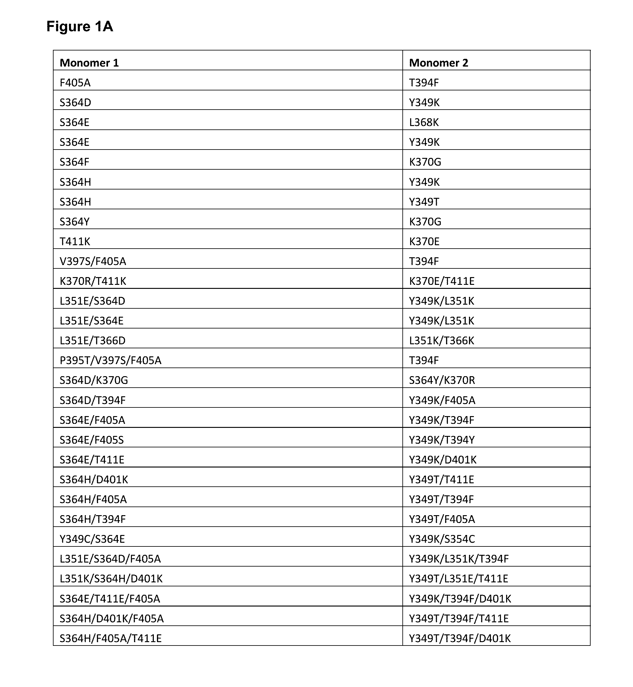
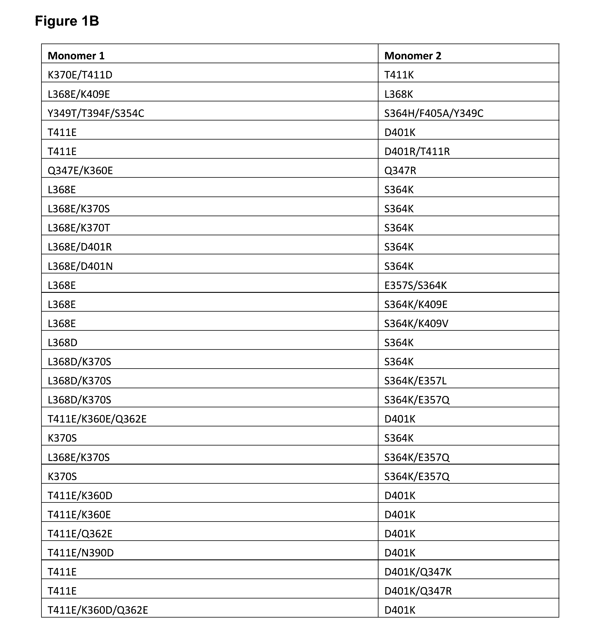
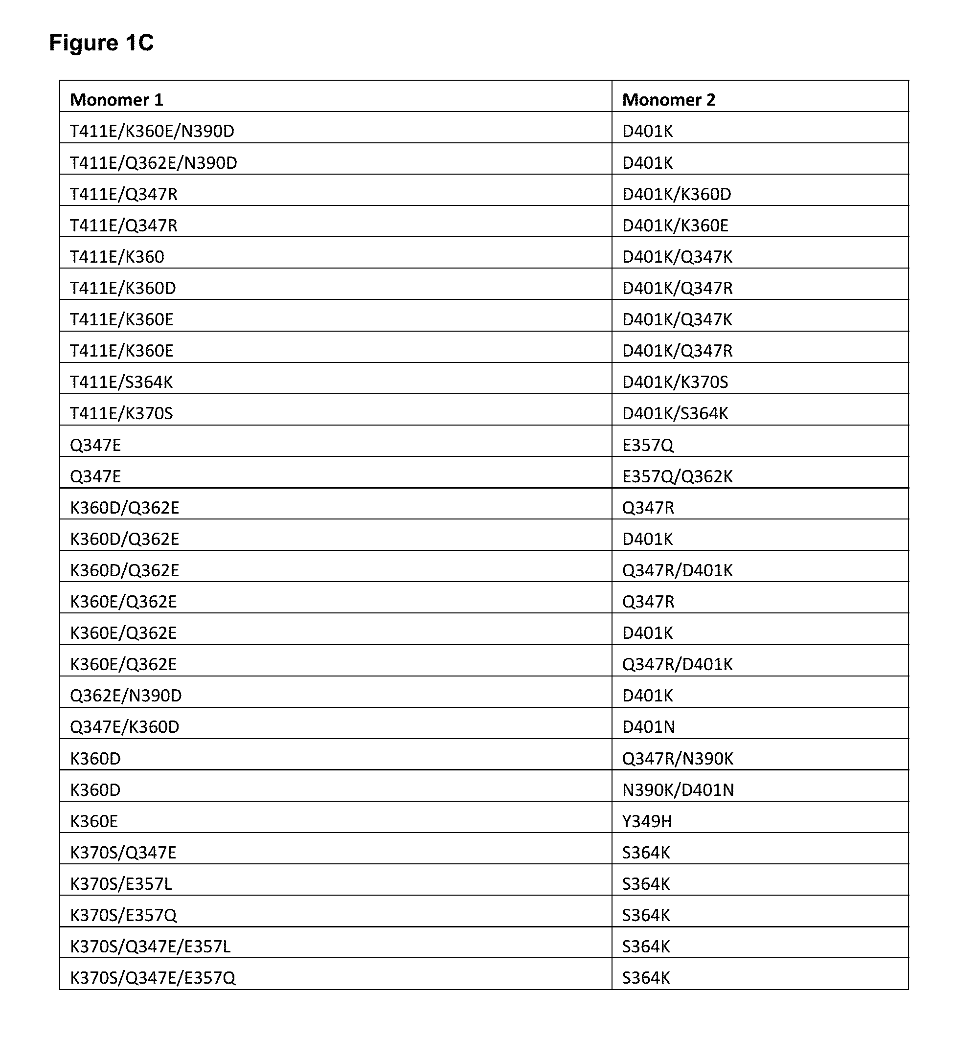
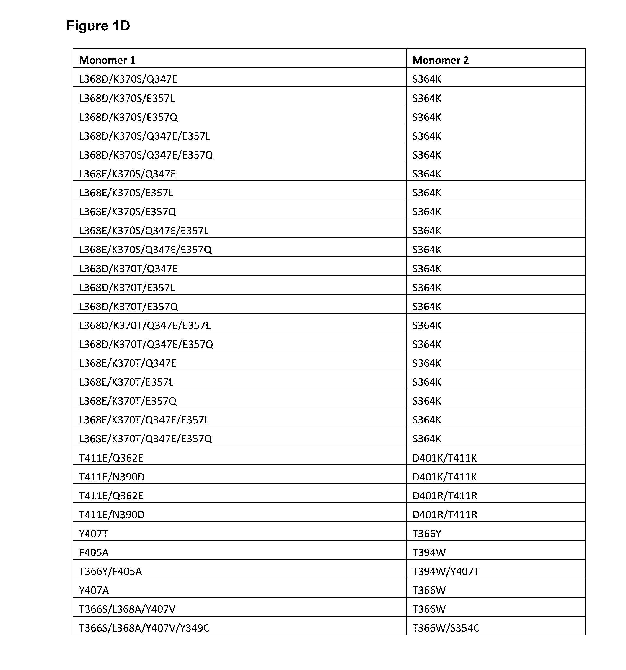
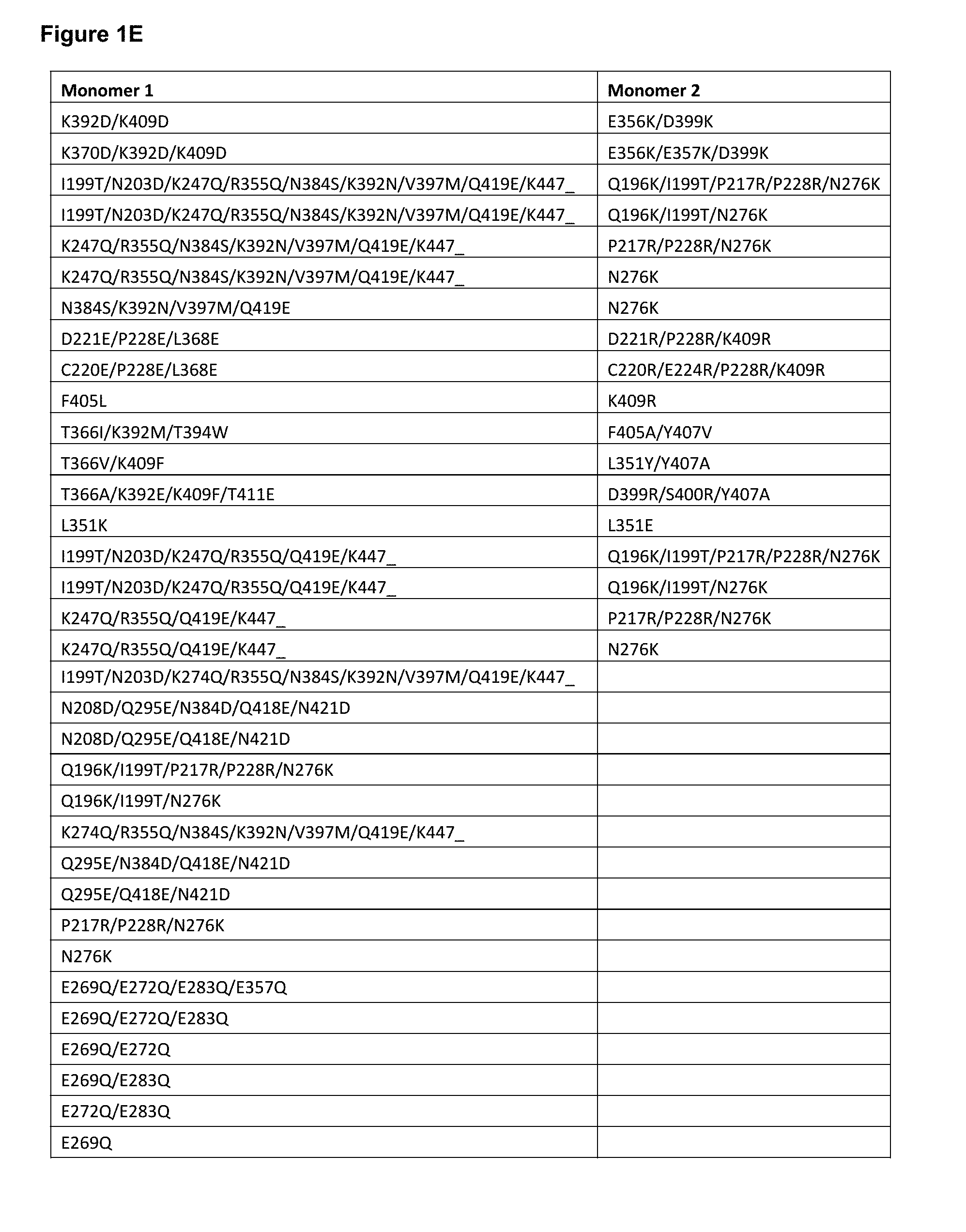
[0052] FIGS. 1A-1E depict useful pairs of Fc heterodimerization variant sets (including skew and pI variants). There are variants for which there are no corresponding "monomer 2" variants; these are pI variants which can be used alone on either monomer.
[0053] FIG. 2 depicts a list of isosteric variant antibody constant regions and their respective substitutions. pI_(-) indicates lower pI variants, while pI_(+) indicates higher pI variants. These can be optionally and independently combined with other heterodimerization variants of the inventions (and other variant types as well, as outlined herein.)
[0054] FIG. 3 depicts useful ablation variants that ablate Fc.gamma.R binding (sometimes referred to as "knock outs" or "KO" variants). Generally, ablation variants are found on both monomers, although in some cases they may be on only one monomer.
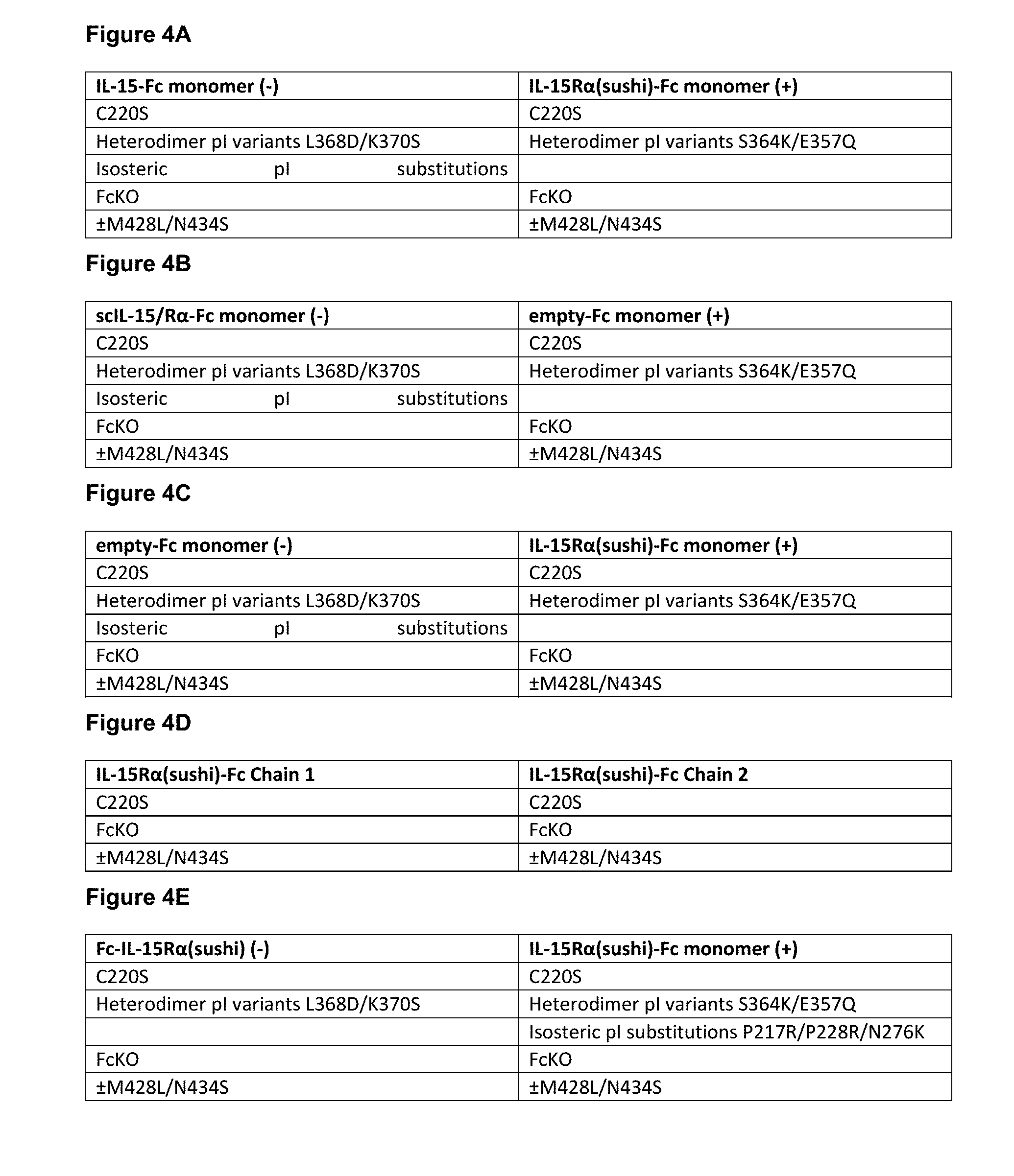
[0055] FIGS. 4A-4E show useful embodiments of "non-cytokine" components of the IL-15/R.alpha.-Fc fusion proteins of the invention.
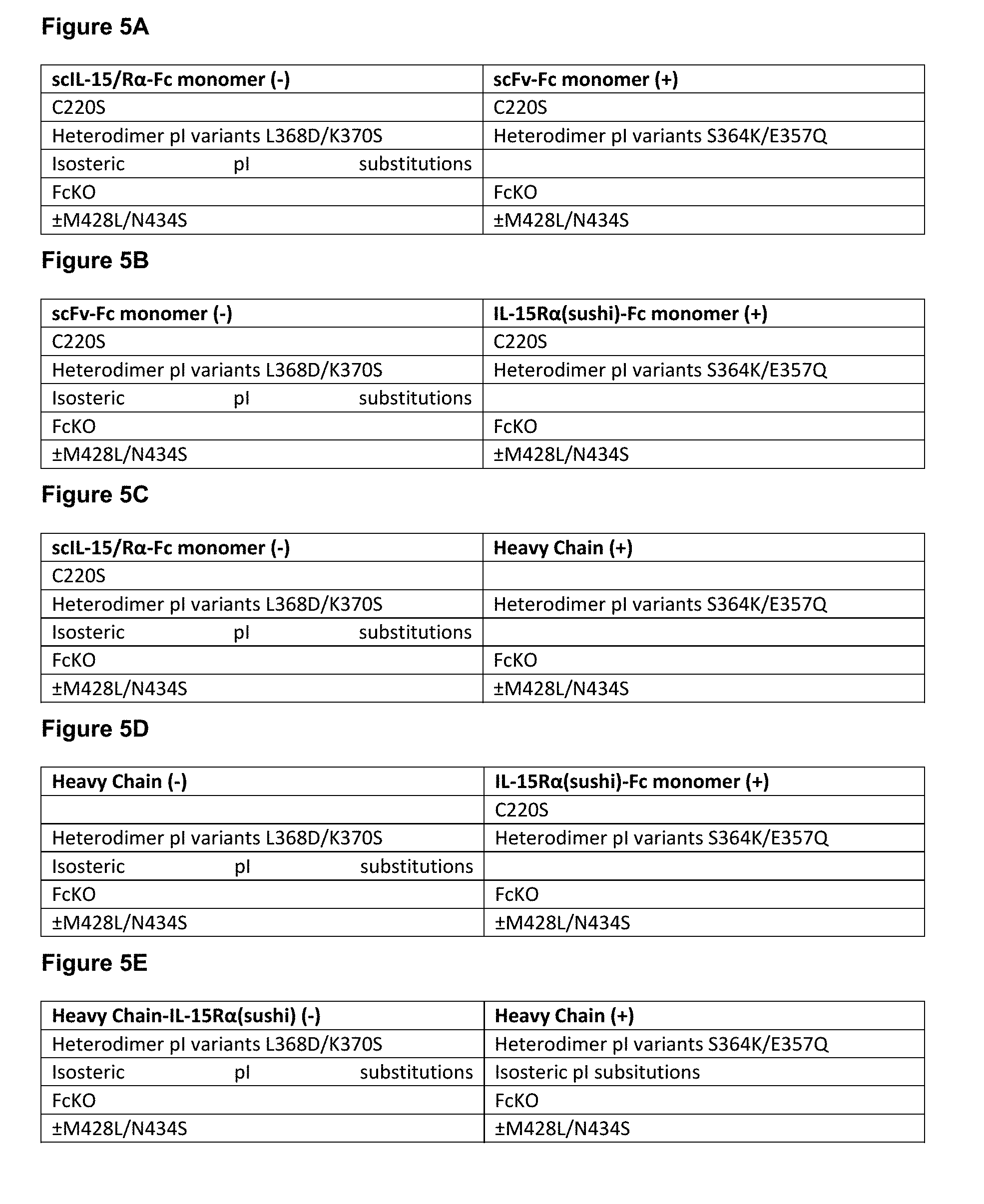
[0056] FIGS. 5A-5F show particularly useful embodiments of "non-cytokine"/"non-Fv" components of the CD8-targeted, NKG2A-targeted, and NKG2D-targeted IL-15/R.alpha.-Fc fusion proteins of the invention.
[0057] FIG. 6 depicts a number of exemplary variable length linkers for use in IL-15/Ra-Fc fusion proteins. In some embodiments, these linkers find use linking the C-terminus of IL-15 and/or IL-15R.alpha.(sushi) to the N-terminus of the Fc region. In some embodiments, these linkers find use fusing IL-15 to the IL-15R.alpha.(sushi).
[0058] FIG. 7 depict a number of charged scFv linkers that find use in increasing or decreasing the pI of heterodimeric antibodies that utilize one or more scFv as a component. The (+H) positive linker finds particular use herein. A single prior art scFv linker with single charge is referenced as "Whitlow", from Whitlow et al., Protein Engineering 6(8):989-995 (1993). It should be noted that this linker was used for reducing aggregation and enhancing proteolytic stability in scFvs.
[0059] FIGS. 8A-8D shows the sequences of several useful IL-15/R.alpha.-Fc format backbones based on human IgG1, without the cytokine sequences (e.g. the I1-15 and/or IL-15R.alpha.(sushi)). It is important to note that these backbones can also find use in certain embodiments of CD8-targeted IL-15/R.alpha.-Fc fusion proteins. Backbone 1 is based on human IgG1 (356E/358M allotype), and includes C220S on both chain, the S364K/E357Q:L368D/K370S skew variants, the Q295E/N384D/Q418E/N421D pI variants on the chain with L368D/K370S skew variants and the E233P/L234V/L235A/G236del/S267K ablation variants on both chains. Backbone 2 is based on human IgG1 (356E/358M allotype), and includes C220S on both chain, the S364K:L368D/K370S skew variants, the Q295E/N384D/Q418E/N421D pI variants on the chain with L368D/K370S skew variants and the E233P/L234V/L235A/G236del/S267K ablation variants on both chains. Backbone 3 is based on human IgG1 (356E/358M allotype), and includes C220S on both chain, the S364K:L368E/K370S skew variants, the Q295E/N384D/Q418E/N421D pI variants on the chain with L368E/K370S skew variants and the E233P/L234V/L235A/G236del/S267K ablation variants on both chains. Backbone 4 is based on human IgG1 (356E/358M allotype), and includes C220S on both chain, the D401K:K360E/Q362E/T411E skew variants, the Q295E/N384D/Q418E/N421D pI variants on the chain with K360E/Q362E/T411E skew variants and the E233P/L234V/L235A/G236del/S267K ablation variants on both chains. Backbone 5 is based on human IgG1 (356D/358L allotype), and includes C220S on both chain, the S364K/E357Q:L368D/K370S skew variants, the Q295E/N384D/Q418E/N421D pI variants on the chain with L368D/K370S skew variants and the E233P/L234V/L235A/G236del/S267K ablation variants on both chains. Backbone 6 is based on human IgG1 (356E/358M allotype), and includes C220S on both chain, the S364K/E357Q:L368D/K370S skew variants, Q295E/N384D/Q418E/N421D pI variants on the chain with L368D/K370S skew variants and the E233P/L234V/L235A/G236del/S267K ablation variants on both chains, as well as an N297A variant on both chains. Backbone 7 is identical to 6 except the mutation is N297S. Alternative formats for backbones 6 and 7 can exclude the ablation variants E233P/L234V/L235A/G236del/S267K in both chains. Backbone 8 is based on human IgG4, and includes the S364K/E357Q:L368D/K370S skew variants, the Q295E/N384D/Q418E/N421D pI variants on the chain with L368D/K370S skew variants, as well as a S228P (EU numbering, this is S241P in Kabat) variant on both chains that ablates Fab arm exchange as is known in the art. Backbone 9 is based on human IgG2, and includes the S364K/E357Q:L368D/K370S skew variants, the Q295E/N384D/Q418E/N421D pI variants on the chain with L368D/K370S skew variants. Backbone 10 is based on human IgG2, and includes the S364K/E357Q:L368D/K370S skew variants, the Q295E/N384D/Q418E/N421D pI variants on the chain with L368D/K370S skew variants as well as a S267K variant on both chains. Backbone 11 is identical to backbone 1, except it includes M428L/N434S Xtend mutations. Backbone 12 is based on human IgG1 (356E/358M allotype), and includes C220S on both identical chain, the E233P/L234V/L235A/G236del/S267K ablation variants on both identical chains. Backbone 13 is based on human IgG1 (356E/358M allotype), and includes C220S on both chain, the S364K/E357Q:L368D/K370S skew variants, the P217R/P229R/N276K pI variants on the chain with S364K/E357Q skew variants and the E233P/L234V/L235A/G236del/S267K ablation variants on both chains.
[0060] As will be appreciated by those in the art and outlined below, these sequences can be used with any IL-15 and IL-15R.alpha.(sushi) pairs outlined herein, including but not limited to IL-15/R.alpha.-heteroFc, ncIL-15/R.alpha., and scIL-15/R.alpha., as schematically depicted in FIGS. 16A-16G and 30A-30D. Additionally, any IL-15 and/or IL-15R.alpha.(sushi) variants can be incorporated into these FIGS. 8A-8D backbones in any combination.
[0061] Included within each of these backbones are sequences that are 90, 95, 98 and 99% identical (as defined herein) to the recited sequences, and/or contain from 1, 2, 3, 4, 5, 6, 7, 8, 9 or 10 additional amino acid substitutions (as compared to the "parent" of the Figure, which, as will be appreciated by those in the art, already contain a number of amino acid modifications as compared to the parental human IgG1 (or IgG2 or IgG4, depending on the backbone). That is, the recited backbones may contain additional amino acid modifications (generally amino acid substitutions) in addition to the skew, pI and ablation variants contained within the backbones of this figure.
[0062] FIG. 9 shows the sequences of several useful CD8-targeted IL-15/R.alpha.-Fc fusion format backbones based on human IgG1, without the cytokine sequences (e.g. the I1-15 and/or IL-15R.alpha.(sushi)) or VH, and further excluding light chain backbones which are depicted in FIG. 10. Backbone 1 is based on human IgG1 (356E/358M allotype), and includes the S364K/E357Q:L368D/K370S skew variants, C220S and the Q295E/N384D/Q418E/N421D pI variants on the chain with L368D/K370S skew variants and the E233P/L234V/L235A/G236del/S267K ablation variants on both chains. Backbone 2 is based on human IgG1 (356E/358M allotype), and includes the S364K/E357Q: L368D/K370S skew variants, the N208D/Q295E/N384D/Q418E/N421D pI variants on the chain with L368D/K370S skew variants, C220S in the chain with S364K/E357Q variants, and the E233P/L234V/L235A/G236del/S267K ablation variants on both chains. Backbone 3 is based on human IgG1 (356E/358M allotype), and includes the S364K/E357Q: L368D/K370S skew variants, the N208D/Q295E/N384D/Q418E/N421D pI variants on the chains with L368D/K370S skew variants, the Q196K/I199T/P217R/P228R/N276K pI variants on the chains with S364K/E357Q variants, and the E233P/L234V/L235A/G236del/S267K ablation variants on both chains.
[0063] In certain embodiments, these sequences can be of the 356D/358L allotype. In other embodiments, these sequences can include either the N297A or N297S substitutions. In some other embodiments, these sequences can include the M428L/N434S Xtend mutations. In yet other embodiments, these sequences can instead be based on human IgG4, and include a S228P (EU numbering, this is S241P in Kabat) variant on both chains that ablates Fab arm exchange as is known in the art. In yet further embodiments, these sequences can instead be based on human IgG2. Further, these sequences may instead utilize the other skew variants, pI variants, and ablation variants depicted in FIGS. 1A-1E, 2, and 3.
[0064] As will be appreciated by those in the art and outlined below, these sequences can be used with any IL-15 and IL-15R.alpha.(sushi) pairs outlined herein, including but not limited to scIL-15/R.alpha., ncIL-15/R.alpha., and dsIL-15R.alpha., as schematically depicted in FIG. 70. Further as will be appreciated by those in the art and outlined below, any IL-15 and/or IL-15R.alpha.(sushi) variants can be incorporated in these backbones. Furthermore, as will be appreciated by those in the art and outlined below, these sequences can be used with any VH and VL pairs outlined herein, including either a scFv or a Fab.
[0065] Included within each of these backbones are sequences that are 90, 95, 98 and 99% identical (as defined herein) to the recited sequences, and/or contain from 1, 2, 3, 4, 5, 6, 7, 8, 9 or 10 additional amino acid substitutions (as compared to the "parent" of the Figure, which, as will be appreciated by those in the art, already contain a number of amino acid modifications as compared to the parental human IgG1 (or IgG2 or IgG4, depending on the backbone). That is, the recited backbones may contain additional amino acid modifications (generally amino acid substitutions) in addition to the skew, pI and ablation variants contained within the backbones of this figure. It should also be noted that the backbones depicted herein are also suitable for use in the NKG2A-targeted and NKG2D-targeted IL-15/R.alpha.-Fc fusion proteins of the invention.
[0066] FIG. 10 depicts the "non-Fv" backbone of light chains (i.e. constant light chain) which find use in CD8-targeted, NKG2A-targeted, and NKG2D-targeted IL-15/R.alpha.-Fc fusion proteins of the invention.
[0067] FIG. 11 depicts the sequences for XENP15074, an anti-RSV mAb based on the variable regions of motavizumab (Numax.RTM.), which is a control used in a number of examples described herein. The CDRs are underlined. As noted herein and is true for every sequence herein containing CDRs, the exact identification of the CDR locations may be slightly different depending on the numbering used as is shown in Table 2, and thus included herein are not only the CDRs that are underlined but also CDRs included within the VH and VL domains using other numbering systems. Furthermore, as for all the sequences in the Figures, these VH and VL sequences can be used either in a scFv format or in a Fab format.
[0068] FIG. 12 depicts the sequences for XENP16432, an anti-PD-1 mAb based on the variable regions of nivolumab (Opdivo.RTM.). The CDRs are underlined. As noted herein and is true for every sequence herein containing CDRs, the exact identification of the CDR locations may be slightly different depending on the numbering used as is shown in Table 2, and thus included herein are not only the CDRs that are underlined but also CDRs included within the VH and VL domains using other numbering systems. Furthermore, as for all the sequences in the Figures, these VH and VL sequences can be used either in a scFv format or in a Fab format.
[0069] FIG. 13 depicts the sequences for XENP26007, an "RSV-targeted" IL-15/R.alpha.-Fc fusion used as control in many of the examples described herein. The CDRs are in bold. As noted herein and is true for every sequence herein containing CDRs, the exact identification of the CDR locations may be slightly different depending on the numbering used as is shown in Table 2, and thus included herein are not only the CDRs that are underlined but also CDRs included within the VH and VL domains using other numbering systems. IL-15 and IL-15R.alpha.(sushi) are underlined, linkers are double underlined (although as will be appreciated by those in the art, the linkers can be replaced by other linkers, some of which are depicted in FIGS. 6 and 7), and slashes (/) indicate the border(s) between IL-15, IL-15R.alpha., linkers, variable regions, and constant/Fc regions.
[0070] FIG. 14 depicts the structure of IL-15 in complex with its receptors IL-15R.alpha. (CD215), IL-15R.beta. (CD122), and the common gamma chain (CD132).
[0071] FIG. 15A-15B depicts the sequences for IL-15 and its receptors.
[0072] FIGS. 16A-16G depict several formats for the IL-15/R.alpha.-Fc fusion proteins of the present invention. IL-15R.alpha. Heterodimeric Fc fusion or "IL-15/R.alpha.-heteroFc" (FIG. 16A) comprises IL-15 recombinantly fused to one side of a heterodimeric Fc and IL-15R.alpha.(sushi) recombinantly fused to the other side of a heterodimeric Fc. The IL-15 and IL-15R.alpha.(sushi) may have a variable length Gly-Ser linker between the C-terminus and the N-terminus of the Fc region. Single-chain IL-15/R.alpha.-Fc fusion or "scIL-15/R.alpha.-Fc" (FIG. 16B) comprises IL-15R.alpha.(sushi) fused to IL-15 by a variable length linker (termed a "single-chain" IL-15/IL-15R.alpha.(sushi) complex or "scIL-15/R.alpha.") which is then fused to the N-terminus of a heterodimeric Fc-region, with the other side of the molecule being "Fc-only" or "empty Fc". Non-covalent IL-15/R.alpha.-Fc or "ncIL-15/R.alpha.-Fc" (FIG. 16C) comprises IL-15R.alpha.(sushi) fused to a heterodimeric Fc region, while IL-15 is transfected separately so that a non-covalent IL-15/R.alpha. complex is formed, with the other side of the molecule being "Fc-only" or "empty Fc". Bivalent non-covalent IL-15/R.alpha.-Fc fusion or "bivalent ncIL-15/R.alpha.-Fc" (FIG. 16D) comprises IL-15R.alpha.(sushi) fused to the N-terminus of a homodimeric Fc region, while IL-15 is transfected separately so that a non-covalent IL-15/R.alpha. complex is formed. Bivalent single-chain IL-15/R.alpha.-Fc fusion or "bivalent scIL-15/R.alpha.-Fc" (FIG. 16E) comprises IL-15 fused to IL-15R.alpha.(sushi) by a variable length linker (termed a "single-chain" IL-15/IL-15R.alpha.(sushi) complex or "scIL-15/R.alpha.") which is then fused to the N-terminus of a homodimeric Fc-region. Fc-non-covalent IL-15/R.alpha. fusion or "Fc-ncIL-15/R.alpha." (FIG. 16F) comprises IL-15R.alpha.(sushi) fused to the C-terminus of a heterodimeric Fc region, while IL-15 is transfected separately so that a non-covalent IL-15/R.alpha. complex is formed, with the other side of the molecule being "Fc-only" or "empty Fc". Fc-single-chain IL-15/R.alpha. fusion or "Fc-scIL-15/R.alpha." (FIG. 16G) comprises IL-15 fused to IL-15R.alpha.(sushi) by a variable length linker (termed a "single-chain" IL-15/IL-15R.alpha.(sushi) complex or "scIL-15/R.alpha.") which is then fused to the C-terminus of a heterodimeric Fc region, with the other side of the molecule being "Fc-only" or "empty Fc".
[0073] FIG. 17 depicts sequences of XENP20818 and XENP21475, illustrative IL-15/R.alpha.-Fc fusion proteins of the "IL-15/R.alpha.-heteroFc" format. IL-15 and IL-15R.alpha.(sushi) are underlined, linkers are double underlined (although as will be appreciated by those in the art, the linkers can be replaced by other linkers, some of which are depicted in FIG. 6), and slashes (/) indicate the border(s) between IL-15, IL-15R.alpha., linkers, and Fc regions.
[0074] FIG. 18 depicts sequences of XENP21478 and XENP21993, illustrative IL-15/R.alpha.-Fc fusion protein of the "scIL-15/R.alpha.-Fc" format. IL-15 and IL-15R.alpha.(sushi) are underlined, linkers are double underlined (although as will be appreciated by those in the art, the linkers can be replaced by other linkers, some of which are depicted in FIG. 6), and slashes (/) indicate the border(s) between IL-15, IL-15R.alpha., linkers, and Fc regions.
[0075] FIGS. 19A-19B depicts sequences of XENP21479, XENP22366 and XENP24348, illustrative IL-15/R.alpha.-Fc fusion proteins of the "ncIL-15/R.alpha.-Fc" format. IL-15 and IL-15R.alpha.(sushi) are underlined, linkers are double underlined (although as will be appreciated by those in the art, the linkers can be replaced by other linkers, some of which are depicted in FIG. 6), and slashes (/) indicate the border(s) between IL-15, IL-15R.alpha., linkers, and Fc regions.
[0076] FIG. 20 depicts sequences of XENP21978, an illustrative IL-15/R.alpha.-Fc fusion protein of the "bivalent ncIL-15/R.alpha.-Fc" format. IL-15 and IL-15R.alpha.(sushi) are underlined, linkers are double underlined (although as will be appreciated by those in the art, the linkers can be replaced by other linkers, some of which are depicted in FIG. 6), and slashes (/) indicate the border(s) between IL-15, IL-15R.alpha., linkers, and Fc regions.
[0077] FIG. 21 depicts sequences of an illustrative IL-15/R.alpha.-Fc fusion protein of the "bivalent scIL-15/R.alpha.-Fc" format. IL-15 and IL-15R.alpha.(sushi) are underlined, linkers are double underlined (although as will be appreciated by those in the art, the linkers can be replaced by other linkers, some of which are depicted in FIG. 6), and slashes (/) indicate the border(s) between IL-15, IL-15R.alpha., linkers, and Fc regions.
[0078] FIG. 22 depicts sequences of XENP22637 and XENP22638, illustrative IL-15/R.alpha.-Fc fusion proteins of the "Fc-ncIL-15/R.alpha." format. IL-15 and IL-15R.alpha.(sushi) are underlined, linkers are double underlined (although as will be appreciated by those in the art, the linkers can be replaced by other linkers, some of which are depicted in FIG. 6), and slashes (/) indicate the border(s) between IL-15, IL-15R.alpha., linkers, and Fc regions.
[0079] FIG. 23 depicts sequences of an illustrative IL-15/R.alpha.-Fc fusion protein of the "Fc-scIL-15/R.alpha." format. IL-15 and IL-15R.alpha.(sushi) are underlined, linkers are double underlined (although as will be appreciated by those in the art, the linkers can be replaced by other linkers, some of which are depicted in FIG. 6), and slashes (/) indicate the border(s) between IL-15, IL-15R.alpha., linkers, and Fc regions.
[0080] FIGS. 24A-24C depict the induction of (FIG. 24A) NK (CD56+/CD16+) cells, (FIG. 24B) CD4+ T cells, and (FIG. 24C CD8+ T cells proliferation by illustrative IL-15/R.alpha.-Fc fusion proteins of Format A with different linker lengths based on Ki67 expression as measured by FACS.
[0081] FIGS. 25A-25C depict the induction of (FIG. 25A) NK (CD56+/CD16+) cells, (FIG. 25B) CD4+ T cells, and (FIG. 25C) CD8+ T cells proliferation by illustrative IL-15/R.alpha.-Fc fusion proteins of scIL-15/R.alpha.-Fc format (XENP21478) and ncIL-15/R.alpha.-Fc format (XENP21479) based on Ki67 expression as measured by FACS.
[0082] FIG. 26 depicts a structural model of the IL-15/R.alpha. heterodimer showing locations of engineered disulfide bond pairs.
[0083] FIG. 27 depicts sequences for illustrative IL-15R.alpha.(sushi) variants engineered with additional residues at the C-terminus to serve as a scaffold for engineering cysteine residues.
[0084] FIG. 28 depicts sequences for illustrative IL-15 variants engineered with cysteines in order to form covalent disulfide bonds with IL-15R.alpha.(sushi) variants engineered with cysteines.
[0085] FIG. 29 depicts sequences for illustrative IL-15R.alpha.(sushi) variants engineered with cysteines in order to form covalent disulfide bonds with IL-15 variants engineered with cysteines.
[0086] FIGS. 30A-30D depict additional formats for the IL-15/R.alpha.-Fc fusion proteins of the present invention with engineered disulfide bonds. Disulfide-bonded IL-15/R.alpha. heterodimeric Fc fusion or "dsIL-15/R.alpha.-heteroFc" (FIG. 30A) is the same as "IL-15/R.alpha.-heteroFc", but wherein IL-15R.alpha.(sushi) and IL-15 are further covalently linked as a result of engineered cysteines. Disulfide-bonded IL-15/R.alpha. Fc fusion or "dsIL-15/R.alpha.-Fc" (FIG. 30B) is the same as "ncIL-15/R.alpha.-Fc", but wherein IL-15R.alpha.(sushi) and IL-15 are further covalently linked as a result of engineered cysteines. Bivalent disulfide-bonded IL-15/R.alpha.-Fc or "bivalent dsIL-15/R.alpha.-Fc" (FIG. 30C) is the same as "bivalent ncIL-15/R.alpha.-Fc", but wherein IL-15R.alpha.(sushi) and IL-15 are further covalently linked as a result of engineered cysteines. Fc-disulfide-bonded IL-15/R.alpha. fusion or "Fc-dsIL-15/R.alpha." (FIG. 30D) is the same as "Fc-ncIL-15/R.alpha.", but wherein IL-15R.alpha.(sushi) and IL-15 are further covalently linked as a result of engineered cysteines.
[0087] FIG. 31A-31B depict sequences of XENP22013, XENP22014, XENP22015, and XENP22017, illustrative IL-15/R.alpha.-Fc fusion protein of the "dsIL-15/R.alpha.-heteroFc" format. IL-15 and IL-15R.alpha.(sushi) are underlined, linkers are double underlined (although as will be appreciated by those in the art, the linkers can be replaced by other linkers, some of which are depicted in FIG. 83), and slashes (/) indicate the border(s) between IL-15, IL-15R.alpha., linkers, and Fc regions.
[0088] FIGS. 32A-32B depict sequences of XENP22357, XENP22358, XENP22359, XENP22684, and XENP22361, illustrative IL-15/R.alpha.-Fc fusion proteins of the "dsIL-15/R.alpha.-Fc" format. IL-15 and IL-15R.alpha.(sushi) are underlined, linkers are double underlined (although as will be appreciated by those in the art, the linkers can be replaced by other linkers, some of which are depicted in FIG. 83), and slashes (/) indicate the border(s) between IL-15, IL-15R.alpha., linkers, and Fc regions.
[0089] FIG. 33 depicts sequences of XENP22634, XENP22635, XENP22636 and XENP22687, illustrative IL-15/R.alpha.-Fc fusion proteins of the "bivalent dsIL-15/R.alpha.-Fc" format. IL-15 and IL-15R.alpha.(sushi) are underlined, linkers are double underlined (although as will be appreciated by those in the art, the linkers can be replaced by other linkers, some of which are depicted in FIG. 83), and slashes (/) indicate the border(s) between IL-15, IL-15R.alpha., linkers, and Fc regions.
[0090] FIG. 34 depicts sequences of XENP22639 and XENP22640, illustrative IL-15/R.alpha.-Fc fusion proteins of the "Fc-dsIL-15/R.alpha." format. IL-15 and IL-15R.alpha.(sushi) are underlined, linkers are double underlined (although as will be appreciated by those in the art, the linkers can be replaced by other linkers, some of which are depicted in FIG. 83), and slashes (/) indicate the border(s) between IL-15, IL-15R.alpha., linkers, and Fc regions.
[0091] FIG. 35 depicts the purity and homogeneity of illustrative IL-15/R.alpha.-Fc fusion proteins with and without engineered disulfide bonds as determined by CEF.
[0092] FIGS. 36A-36C depicts the induction of (FIG. 36A) NK (CD56+/CD16+) cell, (FIG. 36B) CD8+ T cell, and (FIG. 36C) CD4+ T cell proliferation by illustrative IL-15/R.alpha.-Fc fusion proteins with and without engineered disulfide bonds based on Ki67 expression as measured by FACS.
[0093] FIG. 37 depicts the structure of IL-15 complexed with IL-15R.alpha., IL-2R.beta., and common gamma chain. Locations of substitutions designed to reduce potency are shown.
[0094] FIGS. 38A-38C depict sequences for illustrative IL-15 variants engineered for reduced potency. Included within each of these variant IL-15 sequences are sequences that are 90, 95, 98 and 99% identical (as defined herein) to the recited sequences, and/or contain from 1, 2, 3, 4, 5, 6, 7, 8, 9 or 10 additional amino acid substitutions. In a non-limiting example, the recited sequences may contain additional amino acid modifications such as those contributing to formation of covalent disulfide bonds as described in Example 3B.
[0095] FIGS. 39A-39E depict sequences of XENP22821, XENP22822, XENP23343, XENP23554, XENP23557, XENP23561, XENP24018, XENP24019, XENP24045, XENP24051, XENP24052, and XENP24306, illustrative IL-15/R.alpha.-Fc fusion proteins of the "IL-15/R.alpha.-heteroFc" format engineered for reduced potency. IL-15 and IL-15R.alpha.(sushi) are underlined, linkers are double underlined (although as will be appreciated by those in the art, the linkers can be replaced by other linkers, some of which are depicted in FIG. 83), and slashes (/) indicate the border(s) between IL-15, IL-15R.alpha., linkers, and Fc regions.
[0096] FIGS. 40A-40d depicts sequences of XENP24013, XENP24014, XENP24015, XENP24050, XENP24294, XENP24475, XENP24476, XENP24478, XENP24479, and XENP24481, illustrative IL-15/R.alpha.-Fc fusion proteins of the "scIL-15/R.alpha.-Fc" format engineered for reduced potency. IL-15 and IL-15R.alpha.(sushi) are underlined, linkers are double underlined (although as will be appreciated by those in the art, the linkers can be replaced by other linkers, some of which are depicted in FIG. 83), and slashes (/) indicate the border(s) between IL-15, IL-15R.alpha., linkers, and Fc regions.
[0097] FIGS. 41A-41B depict sequences of XENP24349, XENP24890, and XENP25138, illustrative IL-15/R.alpha.-Fc fusion proteins of the "ncIL-15/R.alpha.-Fc" format engineered for reduced potency. IL-15 and IL-15R.alpha.(sushi) are underlined, linkers are double underlined (although as will be appreciated by those in the art, the linkers can be replaced by other linkers, some of which are depicted in FIG. 83), and slashes (/) indicate the border(s) between IL-15, IL-15R.alpha., linkers, and Fc regions.
[0098] FIG. 42 depicts sequences of XENP22801 and XENP22802, illustrative ncIL-15/Ra heterodimers engineered for reduced potency. It is important to note that these sequences were generated using polyhistidine (Hisx6 or HHHHHH (SEQ ID NO: 5)) C-terminal tags at the C-terminus of IL-15R.alpha.(sushi).
[0099] FIG. 43 depicts sequences of XENP24342, an illustrative IL-15/R.alpha.-Fc fusion protein of the "bivalent ncIL-15/R.alpha.-Fc" format engineered for reduced potency. IL-15 and IL-15R.alpha.(sushi) are underlined, linkers are double underlined (although as will be appreciated by those in the art, the linkers can be replaced by other linkers, some of which are depicted in FIG. 83), and slashes (/) indicate the border(s) between IL-15, IL-15R.alpha., linkers, and Fc regions.
[0100] FIG. 44 depicts sequences of XENP23472 and XENP23473, illustrative IL-15/R.alpha.-Fc fusion proteins of the "dsIL-15/R.alpha.-Fc" format engineered for reduced potency. IL-15 and IL-15R.alpha.(sushi) are underlined, linkers are double underlined (although as will be appreciated by those in the art, the linkers can be replaced by other linkers, some of which are depicted in FIG. 83), and slashes (/) indicate the border(s) between IL-15, IL-15R.alpha., linkers, and Fc regions.
[0101] FIGS. 45A-45C depict the induction of A) NK cell, B) CD8.sup.+ (CD45RA-) T cell, and C) CD4+(CD45RA-) T cell proliferation by variant IL-15/R.alpha.-Fc fusion proteins based on Ki67 expression as measured by FACS.
[0102] FIG. 46 depicts EC50 for induction of NK and CD8+ T cells proliferation by variant IL-15/R.alpha.-Fc fusion proteins, and fold reduction in EC50 relative to XENP20818.
[0103] FIGS. 47A-47D depict cell proliferation in human PBMCs incubated for four days with the indicated variant IL-15/R.alpha.-Fc fusion proteins. FIGS. 47A-47C show the percentage of proliferating NK cells (CD3-CD16+) (FIG. 47A), CD8+ T cells (CD3+CD8+CD45RA-) (FIG. 47B) and CD4+ T cells (CD3+CD4+CD45RA-) (FIG. 47C). FIG. 47D shows the fold change in EC50 of various IL15/IL15R.alpha. Fc heterodimers relative to control (XENP20818).
[0104] FIGS. 48A-48B depict CD69 and CD25 expression before (FIG. 55A) and after (FIG. 55B) incubation of human PBMCs with XENP22821.
[0105] FIGS. 49A-49D depict cell proliferation in human PBMCs incubated for three days with the indicated variant IL-15/R.alpha.-Fc fusion proteins. FIGS. 54A-C show the percentage of proliferating CD8.sup.+ (CD45RA-) T cells (FIG. 49A), CD4+(CD45RA-) T cells (FIG. 49B), .gamma..delta. T cells (FIG. 49C), and NK cells (FIG. 49D).
[0106] FIGS. 50A-50C depicts the percentage of Ki67 expression on (FIG. 50A) CD8+ T cells, (FIG. 50B) CD4+ T cells, and (FIG. 50C) NK cells following treatment with additional IL-15/R.alpha. variants.
[0107] FIGS. 51A-51E depict the percentage of Ki67 expression on (FIG. 51A) CD8+(CD45RA-) T cells, (FIG. 51B) CD4+(CD45RA-) T cells, (FIG. 51C) .gamma..delta. T cells, (FIG. 51D) NK (CD16+CD8.alpha.-) cells, and (FIG. 51E) NK (CD56+CD8.alpha.-) cells following treatment with IL-15/R.alpha. variants.
[0108] FIGS. 52A-52E depict the percentage of Ki67 expression on (FIG. 52A) CD8+(CD45RA-) T cells, (FIG. 52B) CD4+(CD45RA-) T cells, (FIG. 52C) .gamma..delta. T cells, (FIG. 52D) NK (CD16+CD8.alpha.-) cells, and (FIG. 52E) NK (CD56+CD8.alpha.-) cells following treatment with IL-15/R.alpha. variants.
[0109] FIGS. 53A-53D depicts the percentage of Ki67 expression on (FIG. 53A) CD8+ T cells, (FIG. 53B) CD4+ T cells, (FIG. 53C) .gamma..delta. T cells and (FIG. 53D) NK (CD16+) cells following treatment with additional IL-15/R.alpha. variants.
[0110] FIG. 54A-54D depicts the percentage of Ki67 expression on (FIG. 54A) CD8+ T cells, (FIG. 54B) CD4+ T cells, (FIG. 54C) .gamma..delta. T cells and (FIG. 54D) NK (CD16+) cells following treatment with additional IL-15/R.alpha. variants.
[0111] FIG. 55 depicts IV-TV Dose PK of various IL-15/R.alpha. Fc fusion proteins or controls in C57BL/6 mice at 0.1 mg/kg single dose.
[0112] FIG. 56 depicts the correlation of half-life vs NK cell potency following treatment with IL-15/R.alpha.-Fc fusion proteins engineered for lower potency.
[0113] FIGS. 57A-57K depict several formats for the X-targeted IL-15/R.alpha.-Fc fusion proteins of the present invention. X may be, but is not limited to, CD8, NKG2A, and NKG2D. The "scIL-15/R.alpha..times.scFv" format (FIG. 57A) comprises IL-15R.alpha.(sushi) fused to IL-15 by a variable length linker (termed "scIL-15/R.alpha.") which is then fused to the N-terminus of a heterodimeric Fc-region, with an scFv fused to the other side of the heterodimeric Fc. The "scFv.times.ncIL-15/R.alpha." format (FIG. 57B) comprises an scFv fused to the N-terminus of a heterodimeric Fc-region, with IL-15R.alpha.(sushi) fused to the other side of the heterodimeric Fc, while IL-15 is transfected separately so that a non-covalent IL-15/R.alpha. complex is formed. The "scFv.times.dsIL-15/R.alpha." format (FIG. 57C) is the same as the "scFv x ncIL-15/R.alpha." format, but wherein IL-15R.alpha.(sushi) and IL-15 are covalently linked as a result of engineered cysteines. The "scIL-15/R.alpha..times.Fab" format (FIG. 57D) comprises IL-15R.alpha.(sushi) fused to IL-15 by a variable length linker (termed "scIL-15/R.alpha.") which is then fused to the N-terminus of a heterodimeric Fc-region, with a variable heavy chain (VH) fused to the other side of the heterodimeric Fc, while a corresponding light chain is transfected separately so as to form a Fab with the VH. The "ncIL-15/R.alpha..times.Fab" format (FIG. 57E) comprises a VH fused to the N-terminus of a heterodimeric Fc-region, with IL-15R.alpha.(sushi) fused to the other side of the heterodimeric Fc, while a corresponding light chain is transfected separately so as to form a Fab with the VH, and while IL-15 is transfected separately so that a non-covalent IL-15/R.alpha. complex is formed. The "dsIL-15/R.alpha..times.Fab" format (FIG. 57F) is the same as the "ncIL-15/R.alpha..times.Fab" format, but wherein IL-15R.alpha.(sushi) and IL-15 are covalently linked as a result of engineered cysteines. The "mAb-scIL-15/R.alpha." format (FIG. 57G) comprises VH fused to the N-terminus of a first and a second heterodimeric Fc, with IL-15 is fused to IL-15R.alpha.(sushi) which is then further fused to the C-terminus of one of the heterodimeric Fc-region, while corresponding light chains are transfected separately so as to form a Fabs with the VHs. The "mAb-ncIL-15/R.alpha." format (FIG. 57H) comprises VH fused to the N-terminus of a first and a second heterodimeric Fc, with IL-15R.alpha.(sushi) fused to the C-terminus of one of the heterodimeric Fc-region, while corresponding light chains are transfected separately so as to form a Fabs with the VHs, and while and while IL-15 is transfected separately so that a non-covalent IL-15/R.alpha. complex is formed. The "mAb-dsIL-15/R.alpha." format (FIG. 57I) is the same as the "mAb-ncIL-15/R.alpha." format, but wherein IL-15R.alpha.(sushi) and IL-15 are covalently linked as a result of engineered cysteines. The "central-IL-15/R.alpha." format (FIG. 57J) comprises a VH recombinantly fused to the N-terminus of IL-15 which is then further fused to one side of a heterodimeric Fc and a VH recombinantly fused to the N-terminus of IL-15R.alpha.(sushi) which is then further fused to the other side of the heterodimeric Fc, while corresponding light chains are transfected separately so as to form a Fabs with the VHs. The "central-scIL-15/R.alpha." format (FIG. 57K) comprises a VH fused to the N-terminus of IL-15R.alpha.(sushi) which is fused to IL-15 which is then further fused to one side of a heterodimeric Fc and a VH fused to the other side of the heterodimeric Fc, while corresponding light chains are transfected separately so as to form a Fabs with the VHs.
[0114] FIG. 58 depicts the sequences for illustrative anti-NKG2A mAbs based on monalizumab (as disclosed in U.S. Pat. No. 8,901,283, issued Dec. 2, 2014) as chimeric mAb (XENP24542) and as humanized mAb (XENP24542). The CDRs are underlined. As noted herein and is true for every sequence herein containing CDRs, the exact identification of the CDR locations may be slightly different depending on the numbering used as is shown in Table 2, and thus included herein are not only the CDRs that are underlined but also CDRs included within the VH and VL domains using other numbering systems. Furthermore, as for all the sequences in the Figures, these VH and VL sequences can be used either in a scFv format or in a Fab format.
[0115] FIG. 59 depicts the sequences for illustrative anti-NKG2D mAbs based on MS (disclosed in U.S. Pat. No. 7,879,985, issued Feb. 1, 2011). The CDRs are underlined. As noted herein and is true for every sequence herein containing CDRs, the exact identification of the CDR locations may be slightly different depending on the numbering used as is shown in Table 2, and thus included herein are not only the CDRs that are underlined but also CDRs included within the VH and VL domains using other numbering systems. Furthermore, as for all the sequences in the Figures, these VH and VL sequences can be used either in a scFv format or in a Fab format.
[0116] FIGS. 60A-60B depict the sequences for XENP24531, XENP24532, and XENP27146, illustrative NKG2A-targeted IL-15/R.alpha.-Fc fusions of the scIL-15/R.alpha..times.Fab format. The CDRs are in bold. As noted herein and is true for every sequence herein containing CDRs, the exact identification of the CDR locations may be slightly different depending on the numbering used as is shown in Table 2, and thus included herein are not only the CDRs that are underlined but also CDRs included within the VH and VL domains using other numbering systems. IL-15 and IL-15R.alpha.(sushi) are underlined, linkers are double underlined (although as will be appreciated by those in the art, the linkers can be replaced by other linkers, some of which are depicted in FIGS. 6 and 7), and slashes (/) indicate the border(s) between IL-15, IL-15R.alpha., linkers, variable regions, and constant/Fc regions.
[0117] FIG. 61A-61B depicts the sequences for XENP24533, XENP24534, and XENP27145, illustrative NKG2D-targeted IL-15/R.alpha.-Fc fusions of the scIL-15/R.alpha..times.Fab format. The CDRs are in bold. As noted herein and is true for every sequence herein containing CDRs, the exact identification of the CDR locations may be slightly different depending on the numbering used as is shown in Table 2, and thus included herein are not only the CDRs that are underlined but also CDRs included within the VH and VL domains using other numbering systems. IL-15 and IL-15R.alpha.(sushi) are underlined, linkers are double underlined (although as will be appreciated by those in the art, the linkers can be replaced by other linkers, some of which are depicted in FIGS. 6 and 7), and slashes (/) indicate the border(s) between IL-15, IL-15R.alpha., linkers, variable regions, and constant/Fc regions.
[0118] FIGS. 62A-62C depict the percentage of Ki67 expression on (FIG. 62A) CD4+ T cells, (FIG. 62B) CD8+ T cells and (FIG. 62C) NK cells following treatment with NKG2A-targeted reduced potency IL-15/R.alpha.-Fc fusions (and control scIL-15/R.alpha.-Fc).
[0119] FIGS. 63A-63C depict percentage of (FIG. 63A) CD8+CD45RA- T cells, (FIG. 63B) CD4+CD45RA- T cells, and (FIG. 63C) CD16+NK cells expressing Ki-67, a protein strictly associated with cell proliferation, in human PBMCs treated with the indicated test articles.
[0120] FIGS. 64A-64D depict STAT5 phosphorylation on (FIG. 64A) CD8+CD45RA- CD25- T cells, (FIG. 64B) CD4+CD45RA-CD25- T cells, (FIG. 64C) Treg (CD25+FoxP3+), and (FIG. 64D) CD56+NK cells in human PBMCs treated with the indicated test articles.
[0121] FIGS. 65A-65B depict the sequences for illustrative CD8 binding molecules based on humanized mAb (as previously described in U.S. Pat. No. 7,657,380, issued Feb. 2, 2010) formatted as chimeric mAb (XENP15076), humanized mAb (15251), humanized Fab (XENP23647), and humanized one-arm mAb (XENP24317). The CDRs are underlined. As noted herein and is true for every sequence herein containing CDRs, the exact identification of the CDR locations may be slightly different depending on the numbering used as is shown in Table 2, and thus included herein are not only the CDRs that are underlined but also CDRs included within the VH and VL domains using other numbering systems. Furthermore, as for all the sequences in the Figures, these VH and VL sequences can be used either in a scFv format or in a Fab format.
[0122] FIGS. 66A-66B depict illustrative CD8-targeted IL-15/R.alpha.-Fc fusions in the scIL-15/R.alpha..times.Fab format. The CDRs are in bold. As noted herein and is true for every sequence herein containing CDRs, the exact identification of the CDR locations may be slightly different depending on the numbering used as is shown in Table X, and thus included herein are not only the CDRs that are underlined but also CDRs included within the VH and VL domains using other numbering systems. IL-15 and IL-15R.alpha.(sushi) are underlined, linkers are double underlined (although as will be appreciated by those in the art, the linkers can be replaced by other linkers, some of which are depicted in FIGS. 6 and 7), and slashes (/) indicate the border(s) between IL-15, IL-15R.alpha., linkers, variable regions, and constant/Fc regions.
[0123] FIGS. 67A-67C depict the percentage of Ki67 expression on (FIG. 67A) CD8+ T cells, (FIG. 67B) CD4+ T cells and (FIG. 67C) NK cells following treatment with an CD9-targeted IL-15/R.alpha.-Fc fusion (and controls anti-CD8 mAb and scIL-15/R.alpha.-Fc).
[0124] FIGS. 68A-68C depicts the percentage of Ki67 expression on (FIG. 68A) CD8+ T cells, (FIG. 68B) CD4+ T cells and (FIG. 68C) NK cells following treatment with a CD8-targeted reduced potency IL-15/R.alpha.-Fc fusion.
[0125] FIGS. 69A-69B depict the percentage of Ki67 expression on rapamycin enriched CD4+ T cells from (FIG. 69A) Donor 21 and (FIG. 69B) Donor 23 following treatment with CD8-targeted IL-15(N65D)/R.alpha.-Fc fusion as well as controls.
[0126] FIG. 70A-70B depicts CD4+ cell count for Tregs enriched from (FIG. 70A) Donor 21 and FIG. 70A) Donor 23 following treatment with CD8-targeted IL-15(N65D)/R.alpha.-Fc fusion as well as controls.
[0127] FIGS. 71A-71B depict CD25 MFI on rapamycin enriched CD4+ T cells from (FIG. 71A) Donor 21 and (FIG. 71B) Donor 23 following treatment with CD8-targeted IL-15(N65D)/R.alpha.-Fc fusion as well as controls.
[0128] FIGS. 72A-72C depict the percentage of proliferating (FIG. 72A) CD8 responder T cell, (FIG. 72B) CD4 responder T cell, and (FIG. 72C) NK cells following treatment of PBMCs with CD8-targeted IL-15/R.alpha.-Fc fusions in the presence of Tregs.
[0129] FIG. 73 depicts Treg count following treatment of PBMCs with CD8-targeted IL-15/R.alpha.-Fc fusion in the presence of different amount of Tregs.
[0130] FIGS. 74A-74C depicts the percentage of proliferating (FIG. 74A) CD8 memory T cell and (FIG. 74B) CD4 responder T cell and (FIG. 74C) Treg count following treatment of PBMCs with CD8-targeted IL-15/R.alpha.-Fc fusions and controls in the presence of Tregs (1:2 Treg:PBMC ratio).
[0131] FIG. 75 depicts Treg count following treatment with CD8-targeted IL-15/R.alpha.-Fc fusion and controls in the absence of PBMCs.
[0132] FIGS. 76A-76D depicts (FIG. 76A) CD4+ T cell events, (FIG. 76B) CD8+ T cell events, (FIG. 76C) the correlation between CD8+ T cell and CD4+ T cell events and (FIG. 76D) CD8+ T cell/CD4+ T cell ratio in whole blood of huPBMC engrafted mice on Day 4 following treatment with a CD8-targeted reduced potency IL-15/R.alpha.-Fc fusion and IL-15/R.alpha.-Fc fusion variants.
[0133] FIGS. 77A-77D depict (FIG. 77A) CD4+ T cell events, (FIG. 77B) CD8+ T cell events, (FIG. 77C) the correlation between CD8+ T cell and CD4+ T cell events, and (FIG. 77D) CD8+ T cell/CD4+ T cell ratio in whole blood of huPBMC engrafted mice on Day 7 following treatment with a CD8-targeted reduced potency IL-15/R.alpha.-Fc fusion and IL-15/R.alpha.-Fc fusion variants.
[0134] FIGS. 78A-78D depict (FIG. 78A) CD4+ T cell events, (FIG. 78B) CD8+ T cell events, (FIG. 78C) the correlation between CD8+ T cell and CD4+ T cell events and (FIG. 78D) CD8+ T cell/CD4+ T cell ratio in spleen of huPBMC engrafted mice on Day 8 following treatment with a CD8-targeted reduced potency IL-15/R.alpha.-Fc fusion and IL-15/R.alpha.-Fc fusion variants.
[0135] FIGS. 79A-79B depict illustrative sequences for CD8-targeted IL-15/R.alpha.-Fc fusions in alternative formats (as depicted in FIG. 57. The CDRs are in bold. As noted herein and is true for every sequence herein containing CDRs, the exact identification of the CDR locations may be slightly different depending on the numbering used as is shown in Table 2, and thus included herein are not only the CDRs that are underlined but also CDRs included within the VH and VL domains using other numbering systems. IL-15 and IL-15R.alpha.(sushi) are underlined, linkers are double underlined (although as will be appreciated by those in the art, the linkers can be replaced by other linkers, some of which are depicted in FIGS. 6 and 7), and slashes (/) indicate the border(s) between IL-15, IL-15R.alpha., linkers, variable regions, and constant/Fc regions.
[0136] FIGS. 80A-80F depict the percentage of Ki67 expression on CD4+ T cells, CD8+ T cells, and NK cells following treatment with alternative format CD8-targeted IL-15/R.alpha.-Fc fusions.
[0137] FIG. 81 depicts phage derived anti-CD8 antibody sequences. The CDRs are underlined. As noted herein and is true for every sequence herein containing CDRs, the exact identification of the CDR locations may be slightly different depending on the numbering used as is shown in Table 2, and thus included herein are not only the CDRs that are underlined but also CDRs included within the VH and VL domains using other numbering systems. Furthermore, as for all the sequences in the Figures, these VH and VL sequences can be used either in a scFv format or in a Fab format.
[0138] FIG. 82 depicts binding of exemplary phage hits reformatted as one-armed Fab-Fc antibodies to CD4.sup.+ and CD8.sup.+ T cells.
[0139] FIGS. 83A-83C diagram the binding of CD8 and TCR on CD8+ T cells to pMHCI on a target cell.
[0140] FIG. 84 depicts fraction of binding by HLA2:01 restricted MHC tetramer specific for pp65 (NLVPMVATV) peptide (SEQ ID NO: 6) to T cells specific for HLA2:01 restricted pp65 (NLVPMVATV) peptide (SEQ ID NO: 6) following pre-incubation with anti-CD8 antibodies relative to control (no pre-incubation with anti-CD8 antibody).
[0141] FIG. 85 depicts IFN.gamma. release by T cells specific for HLA2:01 restricted pp65 (NLVPMVATV) peptide (SEQ ID NO: 6) (pre-incubated with various anti-CD8 antibodies) following incubation with T2 cells loaded with HLA-A2*0201 restricted CMV pp65 (NLVPMVATV) peptide (SEQ ID NO: 6) or NY-ESO-1 peptide.
[0142] FIG. 86 depicts the correlation between IFN.gamma. release and tetramer binding by T cells.
[0143] FIG. 87 depicts sequences for XENP24736, an illustrative CD8-targeted IL-15/R.alpha.-Fc fusion with anti-CD8 Fab arm based on phage-derived 1C11B3. The CDRs are in bold. As noted herein and is true for every sequence herein containing CDRs, the exact identification of the CDR locations may be slightly different depending on the numbering used as is shown in Table 2, and thus included herein are not only the CDRs that are underlined but also CDRs included within the VH and VL domains using other numbering systems. IL-15 and IL-15R.alpha.(sushi) are underlined, linkers are double underlined (although as will be appreciated by those in the art, the linkers can be replaced by other linkers, some of which are depicted in FIGS. 6 and 7.), and slashes (/) indicate the border(s) between IL-15, IL-15R.alpha., linkers, variable regions, and constant/Fc regions.
[0144] FIG. 88A-88B depicts percentage of (FIG. 88A) CD8+CD45RA- T cells and (FIG. 88B) CD4+CD45RA- T cells expressing Ki67 in human PBMCs treated with indicated test articles.
[0145] FIG. 89 depicts OKT8 variable regions, murine or humanized as indicated. The CDRs are underlined. As noted herein and is true for every sequence herein containing CDRs, the exact identification of the CDR locations may be slightly different depending on the numbering used as is shown in Table 2, and thus included herein are not only the CDRs that are underlined but also CDRs included within the VH and VL domains using other numbering systems. Furthermore, as for all the sequences in the Figures, these VH and VL sequences can be used either in a scFv format or in a Fab format.
[0146] FIG. 90 depicts the sequences for XENP15075, a humanized anti-OKT8 mAb. The CDRs are underlined. As noted herein and is true for every sequence herein containing CDRs, the exact identification of the CDR locations may be slightly different depending on the numbering used as is shown in Table 1, and thus included herein are not only the CDRs that are underlined but also CDRs included within the VH and VL domains using other numbering systems. Furthermore, as for all the sequences in the Figures, these VH and VL sequences can be used either in a scFv format or in a Fab format.
[0147] FIG. 91 depicts an illustrative one-arm anti-CD8 mAb with Fab arms based on humanized OKT8 variable regions as depicted in FIG. 89. The CDRs are underlined. As noted herein and is true for every sequence herein containing CDRs, the exact identification of the CDR locations may be slightly different depending on the numbering used as is shown in Table 2, and thus included herein are not only the CDRs that are underlined but also CDRs included within the VH and VL domains using other numbering systems. Furthermore, as for all the sequences in the Figures, these VH and VL sequences can be used either in a scFv format or in a Fab format.
[0148] FIGS. 92A-92C depict illustrative CD8-targeted IL-15/R.alpha.-Fc fusions with anti-CD8 Fab arms based on murine or humanized OKT8 variable regions as depicted in FIG. 89. The CDRs are in bold. As noted herein and is true for every sequence herein containing CDRs, the exact identification of the CDR locations may be slightly different depending on the numbering used as is shown in Table 2, and thus included herein are not only the CDRs that are underlined but also CDRs included within the VH and VL domains using other numbering systems. IL-15 and IL-15R.alpha.(sushi) are underlined, linkers are double underlined (although as will be appreciated by those in the art, the linkers can be replaced by other linkers, some of which are depicted in FIGS. 6 and 7), and slashes (/) indicate the border(s) between IL-15, IL-15R.alpha., linkers, variable regions, and constant/Fc regions.
[0149] FIGS. 93A-93B depict percentage of (FIG. 93A) CD8.sup.+CD45RA.sup.- T cells and (FIG. 93B) CD4.sup.+CD45RA.sup.- T cells expressing Ki67 in human PBMCs treated with CD8-targeted IL-15/R.alpha.-Fc fusions with humanized OKT8 binding domain.
[0150] FIGS. 94A-94C depict (FIG. 94A) CD8.sup.+CD45RA.sup.- T cell counts, (FIG. 94B) CD4.sup.+CD45RA.sup.- T cell counts, and (FIG. 94C) CD8.sup.+/CD4.sup.+ T cell ratio in blood of human PBMC-engrafted NSG-mice on Day 7.
[0151] FIGS. 95A-95B depicts variant variable heavy regions based on OKT8_H2 (Humanized Variable Heavy V2) as depicted in FIG. 89 and variant variable light regions based on OKT8_H1 (Humanized Variable Light V1) as depicted in FIG. 89. Each of the variable heavy regions depicted herein may be combined with any of the variable light regions depicted in this Figure as well as those depicted in 89. Each of the variable light regions depicted herein may be combined with any of the variable heavy regions depicted in this Figure as well as those depicted in FIG. 89. The CDRs are underlined. As noted herein and is true for every sequence herein containing CDRs, the exact identification of the CDR locations may be slightly different depending on the numbering used as is shown in Table 2, and thus included herein are not only the CDRs that are underlined but also CDRs included within the VH and VL domains using other numbering systems. Furthermore, as for all the sequences in the Figures, these VH and VL sequences can be used either in a scFv format or in a Fab format.
[0152] FIG. 96 depicts the dissociation constant (K.sub.D), dissociation rate (k.sub.d), and association rate (k.sub.a) of illustrative cyno CD8 affinity engineered OKT8_H2L1 for human and cyno CD8. The molecules depicted here are one-arm mAbs using having an empty-Fc and a Fab, wherein the Fab arms comprise variable regions as depicted in FIGS. 89 and 95. For example, the Fab arm of XENP26009 has OKT8_H2.152 Variable Heavy and OKT8_L1.103 Variable Light.
[0153] FIG. 97A-97B depicts illustrative CD8-targeted IL-15/R.alpha.-Fc fusions with anti-CD8 Fab arms based on cyno-affinity engineered (HuCy) OKT8 variable regions as depicted in FIG. 95. The CDRs are in bold. As noted herein and is true for every sequence herein containing CDRs, the exact identification of the CDR locations may be slightly different depending on the numbering used as is shown in Table 2, and thus included herein are not only the CDRs that are underlined but also CDRs included within the VH and VL domains using other numbering systems. IL-15 and IL-15R.alpha.(sushi) are underlined, linkers are double underlined (although as will be appreciated by those in the art, the linkers can be replaced by other linkers, some of which are depicted in FIGS. 6 and 7), and slashes (/) indicate the border(s) between IL-15, IL-15R.alpha., linkers, variable regions, and constant/Fc regions.
[0154] FIGS. 98A-98B depict percentage of (FIG. 98A) CD8.sup.+CD45RA.sup.- T cells and (FIG. 98B) CD4.sup.+CD45RA.sup.- T cells expressing Ki67 in human PBMCs treated with CD8-targeted IL-15/R.alpha.-Fc fusions with cyno affinity-engineered humanized OKT8 binding domains.
[0155] FIGS. 99A-99B depict STAT5 phosphorylation on (FIG. 99A) CD8.sup.+CD45RA.sup.- T cells and (FIG. 99B) CD4.sup.+CD45RA.sup.- T cells in human PBMCs treated with CD8-targeted IL-15/R.alpha.-Fc fusions with cyno affinity-engineered humanized OKT8 binding domains.
[0156] FIGS. 100A-100D depict (FIG. 100A) CD45.sup.+ cell count, (FIG. 100B) CD4.sup.+ T cell count, (FIG. 100C) CD8+ T cell count, and (FIG. 100D) CD8+/CD4.sup.+ T cell ratio in blood of human PBMC-engrafted NSG mice on Day 7 after dosing with CD8-targeted IL-15/R.alpha.-Fc fusions with cyno affinity-engineered humanized OKT8 binding domains.
[0157] FIGS. 101A-101B depict percentage of (FIG. 101A) CD8.sup.+CD45RA.sup.- T cells and (FIG. 101B) CD4.sup.+CD45RA.sup.- T cells expressing Ki67 in cynomolgus PBMCs treated with CD8-targeted IL-15/R.alpha.-Fc fusions with cyno affinity-engineered humanized OKT8 binding domains.
[0158] FIG. 102 depicts an illustrative CD8-targeted IL-15/R.alpha.-Fc fusion with Xtend Fc. The CDRs are in bold. As noted herein and is true for every sequence herein containing CDRs, the exact identification of the CDR locations may be slightly different depending on the numbering used as is shown in Table 2, and thus included herein are not only the CDRs that are underlined but also CDRs included within the VII and VL domains using other numbering systems. IL-15 and IL-15R.alpha.(sushi) are underlined, linkers are double underlined (although as will be appreciated by those in the art, the linkers can be replaced by other linkers, some of which are depicted in FIGS. 6 and 7), and slashes (/) indicate the border(s) between IL-15, IL-15R.alpha., linkers, variable regions, and constant/Fc regions.
[0159] FIGS. 103A-103F depict STAT5 phosphorylation on (FIG. 103A) CD4+CD45RA- T cell, (FIG. 103B) CD4+CD45RA+ T cell, (FIG. 103C) CD8+CD45RA- T cell, (FIG. 103D) CD8+CD45RA+ T cell, (FIG. 103E) Tregs, and (FIG. 103F) CD56- NK cells, over time by various concentrations of recombinant IL-15, WT IL-15/R.alpha.-Fc (XENP20818), and illustrative CD8-targeted IL-15/R.alpha.-Fc fusion (XENP26585).
[0160] FIGS. 104A-104B depict fold change over time in (FIG. 104A) CD8+ T cell and (FIG. 104B) CD4+ T cell counts in cynomolgus peripheral blood following dosing with XENP24050 or XENP26223.
[0161] FIGS. 105A-105B depicts percentage of (FIG. 105A) CD8+ T cell and (FIG. 105B) CD4+ T cell expressing Ki67 in cynomolgus peripheral blood following dosing with XENP24050 or XENP26223.
[0162] FIG. 106 depicts percentage of CD8.sup.+CD45RA.sup.- T cells expressing Ki67 in cynomolgus lymph nodes following dosing with XENP24050 or XENP26223.
[0163] FIGS. 107A-107C depict (FIG. 107A) CD8+ T cell counts, (FIG. 107B) CD4+ T cell counts, and C) CD8+/CD4+ T cell ratio following dosing with one-arm reduced potency IL-15/R.alpha.-Fc Fusion with Xtend Fc (XENP24294) and CD8-targeted IL-15/R.alpha.-Fc fusion with Xtend Fc (XENP26585).
[0164] FIGS. 108A-108B depict percentage of CD8+ T cells positive for Ki67 in (FIG. 108A) Group 1 (purified T cells incubated with parental MCF-7 tumor cells and indicated test articles) and (FIG. 108B) Group 2 (purified T cells incubated with pp65-expressing MCF-7 tumor cells and indicated test articles).
[0165] FIGS. 109A-109B depict percentage of CD8+ T cells positive for IFN.gamma. in (FIG. 109A) Group 1 (purified T cells incubated with parental MCF-7 tumor cells and indicated test articles) and (FIG. 109B) Group 2 (purified T cells incubated with pp65-expressing MCF-7 tumor cells and indicated test articles).
[0166] FIGS. 110A-110-B depict percentage of CD8+ T cells positive for Ki67 and IFN.gamma. in (FIG. 110A) Group 1 (purified T cells incubated with parental MCF-7 tumor cells and indicated test articles) and (FIG. 110B) Group 2 (purified T cells incubated with pp65-expressing MCF-7 tumor cells and indicated test articles).
[0167] FIGS. 111A-111B depict percentage of CD4.sup.+ T cells positive for Ki67 in (FIG. 111A) Group 1 (purified T cells incubated with parental MCF-7 tumor cells and indicated test articles) and (FIG. 111B) Group 2 (purified T cells incubated with pp65-expressing MCF-7 tumor cells and indicated test articles).
[0168] FIGS. 112A-112B depict percentage of CD4+ T cells positive for IFN.gamma. in (FIG. 112A) Group 1 (purified T cells incubated with parental MCF-7 tumor cells and indicated test articles) and (FIG. 112B) Group 2 (purified T cells incubated with pp65-expressing MCF-7 tumor cells and indicated test articles).
[0169] FIGS. 113A-113B depict percentage of CD4+ T cells positive for Ki67 and IFN.gamma. in (FIG. 113A) Group 1 (purified T cells incubated with parental MCF-7 tumor cells and indicated test articles) and (FIG. 113B) Group 2 (purified T cells incubated with pp65-expressing MCF-7 tumor cells and indicated test articles).
[0170] FIGS. 114A-114B depict remaining target cells [FIG. 114A: parental MCF-7 tumor cells; FIG. 114B: pp65-expressing MCF-7 tumor cells] following incubation with purified T cells and indicated test articles.
[0171] FIGS. 115A-115B depict (FIG. 115A) the mean tumor volume and (FIG. 115B) change in tumor volume in NSG mice engrafted with pp65-expressing MCF-7 cells, following engraftment with pp65 reactive huPBMC and treatment with indicated test articles.
[0172] FIGS. 116A-116E depict (FIG. 116A) CD45.sup.+ cell, (FIG. 116B) CD4.sup.+ T cell, (FIG. 116C) CD8.sup.+ T cell, and (FIG. 116D) NK cell counts as well as (FIG. 116E) CD8.sup.+/CD4.sup.+ T cell ratio in the whole blood of NSG mice engrafted with pp65-expressing MCF-7 cells following engraftment with pp65 reactive huPBMC and treatment with indicated test articles.
DETAILED DESCRIPTION OF THE INVENTION
I. Overview
[0173] The bifunctional heterodimeric Fc fusion proteins of the invention are useful to treat a variety of types of cancers. As will be appreciated by those in the art, recombinantly produced IL-15/IL-15R.alpha. heterodimers can potently activate T cell. However, their short-half life hinders favorable dosing. The targeted IL-15/IL-15R.alpha..times.antigen binding domain heterodimeric Fc fusion proteins of the invention can lead to T cell activation, which in turn can result in greater immune response to cancerous cells and thus more effective treatment. Such targeted heterodimeric Fc fusion proteins can therefore be expected to find utility for treatment of a wide variety of tumor types.
[0174] As discussed below, there are a variety of ways that T cell activation can be measured. Functional effects of the bispecific checkpoint antibodies on NK and T-cells can be assessed in vitro (and in some cases in vivo, as described more fully below) by measuring changes in the following parameters: proliferation, cytokine release and cell-surface makers. For NK cells, increases in cell proliferation, cytotoxicity (ability to kill target cells as measured by increases in CD107a, granzyme, and perforin expression, or by directly measuring target cells killing), cytokine production (e.g., IFN-.gamma. and TNF), and cell surface receptor expression (e.g., CD25) is indicative of immune modulation, e.g. enhanced killing of cancer cells. For T-cells, increases in proliferation, increases in expression of cell surface markers of activation (e.g., CD25, CD69, CD137, and PD1), cytotoxicity (ability to kill target cells), and cytokine production (e.g., IL-2, IL-4, IL-6, IFN-.gamma., TNF-.alpha., IL-10, IL-17A) are indicative of immune modulation, e.g. enhanced killing of cancer cells. Accordingly, assessment of treatment can be done using assays that evaluate one or more of the following: (i) increases in immune response, (ii) increases in activation of .alpha..beta. and/or .gamma..delta. T cells, (iii) increases in cytotoxic T cell activity, (iv) increases in NK and/or NKT cell activity, (v) alleviation of .alpha..beta. and/or .gamma..delta. T-cell suppression, (vi) increases in pro-inflammatory cytokine secretion, (vii) increases in IL-2 secretion; (viii) increases in interferon-.gamma. production, (ix) increases in Th1 response, (x) decreases in Th2 response, (xi) decreases in cell number and/or activity of at least one of regulatory T cells and cells (xii) increases of tumor immune infiltrates.
[0175] In some embodiments, the invention provides the use of bifunctional IL-15/IL-15R.alpha..times.antigen binding domain heterodimeric Fc fusion proteins to perform one or more of the following in a subject in need thereof: (a) upregulating pro-inflammatory cytokines; (b) increasing T-cell proliferation, expansion or tumor infiltration; (c) increasing interferon-.gamma., TNF-.alpha. and other cytokine production by T-cells; (d) increasing IL-2 secretion; (e) stimulating antibody responses; (0 inhibiting cancer cell growth; (g) promoting antigenic specific T cell immunity; (h) promoting CD4+ and/or CD8+ T cell activation; (i) alleviating T-cell suppression; (j) promoting NK cell activity; (k) promoting apoptosis or lysis of cancer cells; and/or (l) cytotoxic or cytostatic effect on cancer cell.
[0176] The bifunctional heterodimeric Fc fusion proteins constructs are based on the self-assembling nature of the two Fc domains of the heavy chains of antibodies, e.g. two "monomers" that assemble into a "dimer". Heterodimeric Fc fusion proteins are made by altering the amino acid sequence of each monomer as more fully discussed below. Thus, the present invention is generally directed to the creation of heterodimeric Fc fusion proteins, which can co-engage antigens in several ways, relying on amino acid variants in the constant regions that are different on each chain to promote heterodimeric formation and/or allow for ease of purification of heterodimers over the homodimers.
[0177] A major obstacle in the formation of the bifunctional heterodimeric Fc fusion proteins is the difficulty in purifying the heterodimeric fusion proteins away from the homodimeric fusion proteins and/or biasing the formation of the heterodimer over the formation of the homodimers. To solve this issue, there are a number of mechanisms that can be used to generate the heterodimers of the present invention. In addition, as will be appreciated by those in the art, these mechanisms can be combined to ensure high heterodimerization. Thus, amino acid variants that lead to the production of heterodimeric antibodies are referred to as "heterodimerization variants". As discussed below, heterodimerization variants can include steric variants (e.g., the "knobs and holes" or "skew" variants described below and the "charge pairs" variants described below) as well as "pI variants", which allows purification of homodimers away from heterodimers.
[0178] One mechanism, generally referred to in the art as "knobs and holes" ("KIH") or sometimes herein as "skew" variants, referring to amino acid engineering that creates steric and/or electrostatic influences to favor heterodimeric formation and disfavor homodimeric formation can also optionally be used, as described in Ridgway et al., Protein Engineering 9(7):617 (1996); Atwell et al., J. Mol. Biol. 1997 270:26; U.S. Pat. No. 8,216,805, U.S. Publication No. 2012/0149876, all of which are hereby incorporated by reference in their entirety. The Figures identify a number of "monomer A--monomer B" pairs that include "knobs and holes" amino acid substitutions. In addition, as described in Merchant et al., Nature Biotech. 16:677 (1998), these "knobs and hole" mutations can be combined with disulfide bonds to skew formation to heterodimerization. Of use in the present invention are T366S/L368A/Y407V paired with T366W, as well as this variant with a bridging disulfide, T366S/L368A/Y407V/Y349C paired with T366W/S354C, particularly in combination with other heterodimerization variants including pI variants as outlined below.
[0179] An additional mechanism that finds use in the generation of heterodimeric antibodies is sometimes referred to as "electrostatic steering" or "charge pairs" as described in Gunasekaran et al., J. Biol. Chem. 285(25):19637 (2010), hereby incorporated by reference in its entirety. This is sometimes referred to herein as "charge pairs". In this embodiment, electrostatics are used to skew the formation towards heterodimerization. As those in the art will appreciate, these may also have an effect on pI, and thus on purification, and thus could in some cases also be considered pI variants. However, as these were generated to force heterodimerization and were not used as purification tools, they are classified as "steric variants". These include, but are not limited to, D221E/P228E/L368E paired with D221R/P228R/K409R (e.g. these are "monomer corresponding sets) and C220E/P228E/368E paired with C220R/E224R/P228R/K409R and others shown in the Figures.
[0180] In the present invention, in some embodiments, pI variants are used to alter the pI of one or both of the monomers and thus allowing the isoelectric separation of A-A, A-B and B-B dimeric proteins.
[0181] In the present invention, there are several basic mechanisms that can lead to ease of purifying heterodimeric proteins; one relies on the use of pI variants, such that each monomer has a different pI, thus allowing the isoelectric purification of A-A, A-B and B-B dimeric proteins. Alternatively, some scaffold formats, such as the "triple F" format, also allows separation on the basis of size. As is further outlined below, it is also possible to "skew" the formation of heterodimers over homodimers. Thus, a combination of steric heterodimerization variants and pI or charge pair variants find particular use in the invention.
[0182] In the present invention that utilizes pI as a separation mechanism to allow the purification of heterodimeric proteins, amino acid variants can be introduced into one or both of the monomer polypeptides; that is, the pI of one of the monomers (referred to herein for simplicity as "monomer A") can be engineered away from monomer B, or both monomer A and B can be changed, with the pI of monomer A increasing and the pI of monomer B decreasing. As is outlined more fully below, the pI changes of either or both monomers can be done by removing or adding a charged residue (e.g., a neutral amino acid is replaced by a positively or negatively charged amino acid residue, e.g., glycine to glutamic acid), changing a charged residue from positive or negative to the opposite charge (e.g. aspartic acid to lysine) or changing a charged residue to a neutral residue (e.g., loss of a charge; lysine to serine). A number of these variants are shown in the Figures. In addition, suitable pI variants for use in the creation of heterodimeric antibodies herein are those that are isotypic, e.g., importing pI variants from different IgG isotypes such that pI is changed without introducing significant immunogenicity; see FIG. 29 from U.S. Publication No. 20140288275, hereby incorporated by reference in its entirety.
[0183] Accordingly, in this embodiment of the present invention provides for creating a sufficient change in pI in at least one of the monomers such that heterodimers can be separated from homodimers. As will be appreciated by those in the art, and as discussed further below, this can be done by using a "wild type" heavy chain constant region and a variant region that has been engineered to either increase or decrease its pI (wt A:B+ or wt A:B-), or by increasing one region and decreasing the other region (A+:B- or A-:B+).
[0184] Thus, in general, a component of some embodiments of the present invention are amino acid variants in the constant regions of antibodies that are directed to altering the isoelectric point (pI) of at least one, if not both, of the monomers of a dimeric protein to form "pI heterodimers" (when the protein is an antibody, these are referred to as "pI antibodies") by incorporating amino acid substitutions ("pI variants" or "pI substitutions") into one or both of the monomers. As shown herein, the separation of the heterodimers from the two homodimers can be accomplished if the pIs of the two monomers differ by as little as 0.1 pH unit, with 0.2, 0.3, 0.4 and 0.5 or greater all finding use in the present invention.
[0185] As will be appreciated by those in the art, the number of pI variants to be included on each or both monomer(s) to get good separation will depend in part on the starting pI of the scFv and Fab of interest. That is, to determine which monomer to engineer or in which "direction" (e.g. more positive or more negative), the Fv sequences of the two target antigens are calculated and a decision is made from there. As is known in the art, different Fvs will have different starting pIs which are exploited in the present invention. In general, as outlined herein, the pIs are engineered to result in a total pI difference of each monomer of at least about 0.1 logs, with 0.2 to 0.5 being preferred as outlined herein.
[0186] Furthermore, as will be appreciated by those in the art and outlined herein, in some cases (depending on the format) heterodimers can be separated from homodimers on the basis of size (e.g. molecular weight). As shown in FIGS. 57A-57K, some formats result in heterodimers with different sizes.
[0187] In addition, as depicted in FIGS. 57A-57K, it will be recognized that it is possible that some antigens are bound bivalently (e.g., two antigen binding sites to a single antigen). As will be appreciated, any combination of Fab and scFvs can be utilized to achieve the desired result and combinations.
[0188] In the case where pI variants are used to achieve purified heterodimers over homodimers, by using the constant region(s) of the heavy chain(s), a more modular approach to designing and purifying multispecific proteins, including antibodies, is provided. Thus, in some embodiments, heterodimerization variants (including skew and purification heterodimerization variants) are not included in the variable regions, such that each individual antibody must be engineered. In addition, in some embodiments, the possibility of immunogenicity resulting from the pI variants is significantly reduced by importing pI variants from different IgG isotypes such that pI is changed without introducing significant immunogenicity. Thus, an additional problem to be solved is the elucidation of low pI constant domains with high human sequence content, e.g., the minimization or avoidance of non-human residues at any particular position.
[0189] A side benefit that can occur with this pI engineering is also the extension of serum half-life and increased FcRn binding. That is, as described in U.S. Pat. No. 8,637,641 (incorporated by reference in its entirety), lowering the pI of antibody constant domains (including those found in antibodies and Fc fusions) can lead to longer serum retention in vivo. These pI variants for increased serum half life also facilitate pI changes for purification.
[0190] In addition, it should be noted that the pI variants of the heterodimerization variants give an additional benefit for the analytics and quality control process of bispecific antibodies, as the ability to either eliminate, minimize and distinguish when homodimers are present is significant. Similarly, the ability to reliably test the reproducibility of the heterodimeric protein production is important.
[0191] As will be appreciated by those in the art and discussed more fully below, the heterodimeric fusion proteins of the present invention can take on a wide variety of configurations, as are generally depicted in FIGS. 57A-57K. Some figures depict "single ended" configurations, where there is one type of specificity on one "arm" of the molecule and a different specificity on the other "arm". Other figures depict "dual ended" configurations, where there is at least one type of specificity at the "top" of the molecule and one or more different specificities at the "bottom" of the molecule. The present invention is directed to novel immunoglobulin compositions that engage an antigen and contain an Il-15/IL-15R.alpha. complex.
[0192] Furthermore, as outlined herein, additional amino acid variants may be introduced into the bifunctional heterodimeric Fc fusion proteins of the invention, to add additional functionalities. For example, amino acid changes within the Fc region can be added (either to one monomer or both) to facilitate increased ADCC or CDC (e.g. altered binding to Fc.gamma. receptors) as well as to increase binding to FcRn and/or increase serum half-life of the resulting molecules. As is further described herein and as will be appreciated by those in the art, any and all of the variants outlined herein can be optionally and independently combined with other variants.
[0193] Similarly, another category of functional variants are "Fc.gamma. ablation variants" or "Fc knock out (FcKO or KO) variants. In these embodiments, for some therapeutic applications, it is desirable to reduce or remove the normal binding of the Fc domain to one or more or all of the Fc.gamma. receptors (e.g. Fc.gamma.R1, Fc.gamma.RIIa, Fc.gamma.RIIb, Fc.gamma.RIIIa, etc.) to avoid additional mechanisms of action. That is, for example, it is generally desirable to ablate Fc.gamma.RIIIa binding to eliminate or significantly reduce ADCC activity. Suitable ablation variants are shown in FIG. 29.
II. Nomenclature
[0194] The bispecific antibodies of the invention are listed in several different formats. Each polypeptide is given a unique "XENP" number, although as will be appreciated in the art, a longer sequence might contain a shorter one. Some molecules have three polypeptides, so the XENP number, with the components, is used as a name. Thus, the molecule XENP24116, which is in bottle opener format, comprises three sequences, generally referred to as "XENP24116_human IL15R.alpha. (sushi domain)_(GGGGS)5_human IL15 (N65D; single chain)-Fc", "XENP24116_51.1 [CD8]_H1L1 Fab-Fc heavy chain" and "XENP24116_51.1 [CD8]_H1L1 Fab-Fc light chain" or equivalents, although one of skill in the art would be able to identify these easily through sequence alignment. These XENP numbers are in the sequence listing as well as identifiers, and used in the Figures. In addition, one molecule, comprising the three components, gives rise to multiple sequence identifiers. For example, the listing of the Fab monomer has the full length sequence, the variable heavy sequence and the three CDRs of the variable heavy sequence; the light chain has a full length sequence, a variable light sequence and the three CDRs of the variable light sequence; and the scFv-Fc domain has a full length sequence, an scFv sequence, a variable light sequence, 3 light CDRs, a scFv linker, a variable heavy sequence and 3 heavy CDRs; note that all molecules herein with a scFv domain use a single charged scFv linker (+H), although others can be used. In addition, the naming nomenclature of particular variable domains uses a "Hx.xx_Ly.yy" type of format, with the numbers being unique identifiers to particular variable chain sequences. Thus, the variable domain of the Fab side of XENP26229 is "OKT8_H2.166 L1.103", which indicates that the variable heavy domain H2.166 was combined with the light domain L1.103. In the case that these sequences are used as scFvs, the designation "OKT8_H2.166_L1.103", indicates that the variable heavy domain H2.166 was combined with the light domain L1.103 and is in the vh-linker-vl orientation, from N- to C-terminus. This molecule with the identical sequence of the heavy and light variable domains but in the reverse order would be named "OKT8_L1.103_H2.166". Similarly, different constructs may "mix and match" the heavy and light chains as will be evident from the sequence listing and the Figures.
III. Definitions
[0195] In order that the application may be more completely understood, several definitions are set forth below. Such definitions are meant to encompass grammatical equivalents.
[0196] By "ablation" herein is meant a decrease or removal of binding and/or activity. Thus for example, "ablating Fc.gamma.R binding" means the Fc region amino acid variant has less than 50% starting binding as compared to an Fc region not containing the specific variant, with less than 70-80-90-95-98% loss of binding being preferred, and in general, with the binding being below the level of detectable binding in a Biacore assay. Of particular use in the ablation of Fc.gamma.R binding are those shown in FIG. 29. However, unless otherwise noted, the Fc monomers of the invention retain binding to the FcRn.
[0197] By "ADCC" or "antibody dependent cell-mediated cytotoxicity" as used herein is meant the cell-mediated reaction wherein nonspecific cytotoxic cells that express Fc.gamma.Rs recognize bound antibody on a target cell and subsequently cause lysis of the target cell. ADCC is correlated with binding to Fc.gamma.RIIIa; increased binding to Fc.gamma.RIIIa leads to an increase in ADCC activity. As is discussed herein, many embodiments of the invention ablate ADCC activity entirely.
[0198] By "ADCP" or antibody dependent cell-mediated phagocytosis as used herein is meant the cell-mediated reaction wherein nonspecific cytotoxic cells that express Fc.gamma.Rs recognize bound antibody on a target cell and subsequently cause phagocytosis of the target cell.
[0199] By "modification" herein is meant an amino acid substitution, insertion, and/or deletion in a polypeptide sequence or an alteration to a moiety chemically linked to a protein. For example, a modification may be an altered carbohydrate or PEG structure attached to a protein. By "amino acid modification" herein is meant an amino acid substitution, insertion, and/or deletion in a polypeptide sequence. For clarity, unless otherwise noted, the amino acid modification is always to an amino acid coded for by DNA, e.g., the 20 amino acids that have codons in DNA and RNA.
[0200] By "amino acid substitution" or "substitution" herein is meant the replacement of an amino acid at a particular position in a parent polypeptide sequence with a different amino acid. In particular, in some embodiments, the substitution is to an amino acid that is not naturally occurring at the particular position, either not naturally occurring within the organism or in any organism. For example, the substitution E272Y or 272Y refers to a variant polypeptide, in this case an Fc variant, in which the glutamic acid at position 272 is replaced with tyrosine. For clarity, a protein which has been engineered to change the nucleic acid coding sequence, but not to change the starting amino acid (for example exchanging CGG (encoding arginine) to CGA (still encoding arginine) to increase host organism expression levels) is not an "amino acid substitution"; that is, despite the creation of a new gene encoding the same protein, if the protein has the same amino acid at the particular position that it started with, it is not an amino acid substitution. Substitutions can include naturally occurring amino acids and, in some cases, synthetic amino acids. Examples include U.S. Pat. No. 6,586,207; WO 98/48032; WO 03/073238; US2004-0214988A1; WO 05/35727A2; WO 05/74524A2; J. W. Chin et al., (2002), Journal of the American Chemical Society 124:9026-9027; J. W. Chin, & P. G. Schultz, (2002), ChemBioChem 11:1135-1137; J. W. Chin, et al., (2002), PICAS United States of America 99:11020-11024; and, L. Wang, & P. G. Schultz, (2002), Chem. 1-10, all entirely incorporated by reference.
[0201] By "amino acid insertion" or "insertion" as used herein is meant the addition of an amino acid residue or sequence at a particular position in a parent polypeptide sequence. For example, -233E designates an insertion of glutamic acid after position 233 and before position 234. Additionally, -233ADE or A233ADE designates an insertion of AlaAspGlu after position 233 and before position 234.
[0202] By "amino acid deletion" or "deletion" as used herein is meant the removal of an amino acid residue or sequence at a particular position in a parent polypeptide sequence. For example, E233-, E233#, E233( ) or E233del designates a deletion of glutamic acid at position 233. Additionally, EDA233- or EDA233# designates a deletion of the sequence GluAspAla that begins at position 233.
[0203] By "variant protein," "protein variant," or "variant" as used herein is meant a protein that differs from that of a parent protein by virtue of at least one modification. The protein variant may refer to the protein itself, a composition comprising the protein, the amino acid sequence that encodes it, or the DNA sequence that encodes it. Preferably, the protein variant has at least one amino acid modification compared to the parent protein, e.g. from about one to about seventy amino acid modifications, and preferably from about one to about five amino acid modifications compared to the parent. The modification can be an addition, deletion, or substitution.
[0204] As described below, in some embodiments the parent polypeptide, for example an Fc parent polypeptide, is a human wild type sequence, such as the heavy constant domain or Fc region from IgG1, IgG2, IgG3 or IgG4, although human sequences with variants can also serve as "parent polypeptides", for example the IgG1/2 hybrid of U.S. Publication No. 2006/0134105 can be included. The protein variant sequence herein will preferably possess at least about 80% identity with a parent protein sequence, and most preferably at least about 90% identity, more preferably at least about 95-98-99% identity.
[0205] Accordingly, by "antibody variant" or "variant antibody" as used herein is meant an antibody that differs from a parent antibody by virtue of at least one amino acid modification, "IgG variant" or "variant IgG" as used herein is meant an antibody that differs from a parent IgG (again, in many cases, from a human IgG sequence) by virtue of at least one amino acid modification, and by "immunoglobulin variant" or "variant immunoglobulin" as used herein is meant an immunoglobulin sequence that differs from that of a parent immunoglobulin sequence by virtue of at least one amino acid modification. "Fc variant" or "variant Fc" as used herein is meant a protein comprising an amino acid modification in an Fc domain as compared to an Fc domain of human IgG1, IgG2 or IgG4.
[0206] The Fc variants of the present invention are defined according to the amino acid modifications that compose them. Thus, for example, N434S or 434S is an Fc variant with the substitution for serine at position 434 relative to the parent Fc polypeptide, wherein the numbering is according to the EU index. Likewise, M428L/N434S defines an Fc variant with the substitutions M428L and N434S relative to the parent Fc polypeptide. The identity of the WT amino acid may be unspecified, in which case the aforementioned variant is referred to as 428L/434S. It is noted that the order in which substitutions are provided is arbitrary, that is to say that, for example, N434S/M428L is the same Fc variant as M428L/N434S, and so on. For all positions discussed in the present invention that relate to antibodies or derivatives and fragments thereof, unless otherwise noted, amino acid position numbering is according to the EU index. The EU index or EU index as in Kabat or EU numbering scheme refers to the numbering of the EU antibody. Kabat et al. collected numerous primary sequences of the variable regions of heavy chains and light chains. Based on the degree of conservation of the sequences, they classified individual primary sequences into the CDR and the framework and made a list thereof (see SEQUENCES OF IMMUNOLOGICAL INTEREST, 5th edition, NIH publication, No. 91-3242, E. A. Kabat et al., entirely incorporated by reference). See also Edelman et al., 1969, Proc Natl Acad Sci USA 63:78-85, hereby entirely incorporated by reference. The modification can be an addition, deletion, or substitution.
[0207] As used herein, by "protein" is meant at least two covalently attached amino acids, which includes proteins, polypeptides, oligopeptides and peptides. In addition, polypeptides that make up the antibodies of the invention may include synthetic derivatization of one or more side chains or termini, glycosylation, PEGylation, circular permutation, cyclization, linkers to other molecules, fusion to proteins or protein domains, and addition of peptide tags or labels. When a biologically functional molecule comprises two or more proteins, each protein may be referred to as a "monomer" or as a "subunit" or as a "domain"; and the biologically functional molecule may be referred to as a "complex".
[0208] By "residue" as used herein is meant a position in a protein and its associated amino acid identity. For example, asparagine 297 (also referred to as Asn297 or N297) is a residue at position 297 in the human antibody IgG1.
[0209] By "Fab" or "Fab region" as used herein is meant the polypeptide that comprises the VH, CH1, VL, and CL immunoglobulin domains, generally on two different polypeptide chains (e.g. VH-CH1 on one chain and VL-CL on the other). Fab may refer to this region in isolation, or this region in the context of a bispecific antibody of the invention. In the context of a Fab, the Fab comprises an Fv region in addition to the CH1 and CL domains.
[0210] By "Fv" or "Fv fragment" or "Fv region" as used herein is meant a polypeptide that comprises the VL and VH domains of an ABD. Fv regions can be formatted as both Fabs (as discussed above, generally two different polypeptides that also include the constant regions as outlined above) and scFvs, where the vl and vh domains are combined (generally with a linker as discussed herein) to form an scFv.
[0211] By "single chain Fv" or "scFv" herein is meant a variable heavy domain covalently attached to a variable light domain, generally using a scFv linker as discussed herein, to form a scFv or scFv domain. A scFv domain can be in either orientation from N- to C-terminus (vh-linker-vl or vl-linker-vh). In the sequences depicted in the sequence listing and in the figures, the order of the vh and vl domain is indicated in the name, e.g. H.X_L.Y means N- to C-terminal is vh-linker-vl, and L.Y_H.X is vl-linker-vh.
[0212] By "IgG subclass modification" or "isotype modification" as used herein is meant an amino acid modification that converts one amino acid of one IgG isotype to the corresponding amino acid in a different, aligned IgG isotype. For example, because IgG1 comprises a tyrosine and IgG2 a phenylalanine at EU position 296, a F296Y substitution in IgG2 is considered an IgG subclass modification.
[0213] By "non-naturally occurring modification" as used herein is meant an amino acid modification that is not isotypic. For example, because none of the IgGs comprise a serine at position 434, the substitution 434S in IgG1, IgG2, IgG3, or IgG4 (or hybrids thereof) is considered a non-naturally occurring modification.
[0214] By "amino acid" and "amino acid identity" as used herein is meant one of the 20 naturally occurring amino acids that are coded for by DNA and RNA.
[0215] By "effector function" as used herein is meant a biochemical event that results from the interaction of an antibody Fc region with an Fc receptor or ligand. Effector functions include but are not limited to ADCC, ADCP, and CDC.
[0216] By "IgG Fc ligand" or "Fc ligand" as used herein is meant a molecule, preferably a polypeptide, from any organism that binds to the Fc region of an IgG antibody to form an Fc/Fc ligand complex. Fc ligands include but are not limited to Fc.gamma.RIs, Fc.gamma.RIIs, Fc.gamma.RIIIs, FcRn, C1q, C3, mannan binding lectin, mannose receptor, staphylococcal protein A, streptococcal protein G, and viral Fc.gamma.R. Fc ligands also include Fc receptor homologs (FcRH), which are a family of Fc receptors that are homologous to the Fc.gamma.Rs (Davis et al., 2002, Immunological Reviews 190:123-136, entirely incorporated by reference). Fc ligands may include undiscovered molecules that bind Fc. Particular IgG Fc ligands are FcRn and Fc gamma receptors.
[0217] By "Fc gamma receptor", "Fc.gamma.R" or "FcgammaR" as used herein is meant any member of the family of proteins that bind the IgG antibody Fc region and is encoded by an Fc.gamma.R gene. In humans this family includes but is not limited to Fc.gamma.RI (CD64), including isoforms Fc.gamma.RIa, Fc.gamma.RIb, and Fc.gamma.RIc; Fc.gamma.RII (CD32), including isoforms Fc.gamma.RIIa (including allotypes H131 and R131), Fc.gamma.RIIb (including Fc.gamma.RIIb-1 and Fc.gamma.RIIb-2), and Fc.gamma.RIIc; and Fc.gamma.RIII (CD16), including isoforms Fc.gamma.RIIIa (including allotypes V158 and F158) and Fc.gamma.RIIIb (including allotypes Fc.gamma.RIIb-NA1 and Fc.gamma.RIIb-NA2) (Jefferis et al., 2002, Immunol Lett 82:57-65, entirely incorporated by reference), as well as any undiscovered human Fc.gamma.Rs or Fc.gamma.R isoforms or allotypes. A n Fc.gamma.R may be from any organism, including but not limited to humans, mice, rats, rabbits, and monkeys. Mouse Fc.gamma.Rs include but are not limited to Fc.gamma.RI (CD64), Fc.gamma.RII (CD32), Fc.gamma.RIII (CD16), and Fc.gamma.RIII-2 (CD16-2), as well as any undiscovered mouse Fc.gamma.Rs or Fc.gamma.R isoforms or allotypes.
[0218] By "FcRn" or "neonatal Fc receptor" as used herein is meant a protein that binds the IgG antibody Fc region and is encoded at least in part by an FcRn gene. The FcRn may be from any organism, including but not limited to humans, mice, rats, rabbits, and monkeys. As is known in the art, the functional FcRn protein comprises two polypeptides, often referred to as the heavy chain and light chain. The light chain is beta-2-microglobulin (.beta.2-microglobulin) and the heavy chain is encoded by the FcRn gene. Unless otherwise noted herein, FcRn or an FcRn protein refers to the complex of FcRn heavy chain with 132-microglobulin. A variety of Fc variants can be used to increase binding to the FcRn, and in some cases, to increase serum half-life. An "FcRn variant" is one that increases binding to the FcRn, and suitable FcRn variants are shown below. In general, unless otherwise noted, the Fc monomers of the invention retain binding to the FcRn (and, as noted below, can include amino acid variants to increase binding to the FcRn.
[0219] By "parent polypeptide" as used herein is meant a starting polypeptide that is subsequently modified to generate a variant. The parent polypeptide may be a naturally occurring polypeptide, or a variant or engineered version of a naturally occurring polypeptide. Parent polypeptide may refer to the polypeptide itself, compositions that comprise the parent polypeptide, or the amino acid sequence that encodes it. Accordingly, by "parent antigen binding domain" as used herein is meant an unmodified antigen binding domain polypeptide that is modified to generate a variant, by "parent immunoglobulin" as used herein is meant an unmodified immunoglobulin polypeptide that is modified to generate a variant, and by "parent antibody" as used herein is meant an unmodified antibody that is modified to generate a variant antibody. It should be noted that "parent antibody" includes known commercial, recombinantly produced antibodies as outlined below.
[0220] By "Fc" or "Fc region" or "Fc domain" as used herein is meant the polypeptide comprising the CH2-CH3 domains of an IgG molecule, and in some cases, inclusive of the hinge. In EU numbering for human IgG1, the CH2-CH3 domain comprises amino acids 231 to 447, and the hinge is 216 to 230. Thus the definition of "Fc domain" includes both amino acids 231-447 (CH2-CH3) or 216-447 (hinge-CH2-CH3), or fragments thereof. An "Fc fragment" in this context may contain fewer amino acids from either or both of the N- and C-termini but still retains the ability to form a dimer with another Fc domain or Fc fragment as can be detected using standard methods, generally based on size (e.g. non-denaturing chromatography, size exclusion chromatography, etc.). Human IgG Fc domains are of particular use in the present invention, and can be the Fc domain from human IgG1, IgG2 or IgG4By "Fc" or "Fc region" or "Fc domain" as used herein is meant the polypeptide comprising the constant region of an antibody, in some instances, excluding all or a portion of the first constant region immunoglobulin domain (e.g., CH1) or a portion thereof, and in some cases, further excluding all or a portion of the hinge. Thus, an Fc can refer to the last two constant region immunoglobulin domains (e.g., CH2 and CH3) of IgA, IgD, and IgG, the last three constant region immunoglobulin domains of IgE and IgM, and optionally, all or a portion of the flexible hinge N-terminal to these domains. For IgA and IgM, Fc may include the J chain. For IgG, the Fc domain comprises immunoglobulin domains CH2 and CH3 (C.gamma.2 and C.gamma.3), and optionally all or a portion of the hinge region between CH1 (C.gamma.1) and CH2 (C.gamma.2). Although the boundaries of the Fc region may vary, the human IgG heavy chain Fc region is usually defined to include residues E216, C226, or A231 to its carboxyl-terminus, wherein the numbering is according to the EU index as in Kabat. In some embodiments, as is more fully described below, amino acid modifications are made to the Fc region, for example to alter binding to one or more Fc.gamma.R or to the FcRn.
[0221] As will be appreciated by those in the art, the exact numbering and placement of the heavy constant region domains can be different among different numbering systems. A useful comparison of heavy constant region numbering according to EU and Kabat is as below, see Edelman et al., 1969, Proc Natl Acad Sci USA 63:78-85 and Kabat et al., 1991, Sequences of Proteins of Immunological Interest, 5th Ed., United States Public Health Service, National Institutes of Health, Bethesda, entirely incorporated by reference.
TABLE-US-00001 TABLE 1 EU Numbering Kabat Numbering CH1 118-215 114-223 Hinge 216-230 226-243 CH2 231-340 244-360 CH3 341-447 361-478
[0222] A "variant Fc domain" contains amino acid modifications as compared to a parental Fc domain. Thus, a "variant human IgG1 Fc domain" is one that contains amino acid modifications (generally amino acid substitutions, although in the case of ablation variants, amino acid deletions are included) as compared to the human IgG1 Fc domain. In general, variant Fc domains have at least about 80, 85, 90, 95, 97, 98 or 99 percent identity to the corresponding parental human IgG Fc domain (using the identity algorithms discussed below, with one embodiment utilizing the BLAST algorithm as is known in the art, using default parameters). Alternatively, the variant Fc domains can have from 1, 2, 3, 4, 5, 6, 7, 8, 9, 10, 11,12, 13, 14, 15, 16, 11, 12, 13, 14, 15, 16, 17, 18, 19 or 20 amino acid modifications as compared to the parental Fc domain. Additionally, as discussed herein, the variant Fc domains herein still retain the ability to form a dimer with another Fc domain as measured using known techniques as described herein, such as non-denaturing gel electrophoresis.
[0223] By "heavy constant region" herein is meant the CH1-hinge-CH2-CH3 portion of an antibody (or fragments thereof), excluding the variable heavy domain; in EU numbering of human IgG1, such as amino acids 118-447. By "heavy chain constant region fragment" herein is meant a heavy chain constant region that contains fewer amino acids from either or both of the N- and C-termini but still retains the ability to form a dimer with another heavy chain constant region.
[0224] By "fusion protein" as used herein is meant covalent joining of at least two proteins. Fusion proteins may comprise artificial sequences, e.g. a domain linker, as described herein. By "Fc fusion protein" or "immunoadhesin" herein is meant a protein comprising an Fc region, generally linked (optionally through a domain linker, as described herein) to one or more different proteins, such as to IL-15 and/or IL-15R, as described herein. In some instances, two Fc fusion proteins can form a homodimeric Fc fusion protein or a heterodimeric Fc fusion protein with the latter being preferred. In some cases, one monomer of the heterodimeric Fc fusion protein comprises an Fc domain alone (e.g., an empty Fc domain) and the other monomer is a Fc fusion, comprising a variant Fc domain and a protein domain, such as a receptor, ligand or other binding partner.
[0225] By "position" as used herein is meant a location in the sequence of a protein. Positions may be numbered sequentially, or according to an established format, for example the EU index for antibody numbering.
[0226] By "strandedness" in the context of the monomers of the heterodimeric proteins of the invention herein is meant that, similar to the two strands of DNA that "match", heterodimerization variants are incorporated into each monomer so as to preserve, create, and/or enhance the ability to "match" to form heterodimers. For example, if some pI variants are engineered into monomer A (e.g., making the pI higher) then steric variants that are "charge pairs" that can be utilized as well do not interfere with the pI variants, e.g., the charge variants that make a pI higher are put on the same "strand" or "monomer" to preserve both functionalities. Similarly, for "skew" variants that come in pairs of a set as more fully outlined below, the skilled artisan will consider pI in deciding into which strand or monomer that incorporates one set of the pair will go, such that pI separation is maximized using the pI of the skews as well.
[0227] By "target antigen" as used herein is meant the molecule that is bound specifically by the variable region of a given antigen binding domain or antibody. A target antigen may be a protein, carbohydrate, lipid, or other chemical compound. A wide number of suitable target antigens are described below.
[0228] By "target cell" as used herein is meant a cell that expresses a target antigen.
[0229] By "host cell" in the context of producing a bispecific antibody according to the invention herein is meant a cell that contains the exogeneous nucleic acids encoding the components of the bispecific antibody and is capable of expressing the bispecific antibody under suitable conditions. Suitable host cells are discussed below.
[0230] By "variable region" as used herein is meant the region of an immunoglobulin that comprises one or more Ig domains substantially encoded by any of the V.kappa., V.lamda., and/or VH genes that make up the kappa, lambda, and heavy chain immunoglobulin genetic loci respectively.
[0231] By "wild type or WT" herein is meant an amino acid sequence or a nucleotide sequence that is found in nature, including allelic variations. A WT protein has an amino acid sequence or a nucleotide sequence that has not been intentionally modified.
[0232] "Percent (%) amino acid sequence identity" with respect to a protein sequence is defined as the percentage of amino acid residues in a candidate sequence that are identical with the amino acid residues in the specific (parental) sequence, after aligning the sequences and introducing gaps, if necessary, to achieve the maximum percent sequence identity, and not considering any conservative substitutions as part of the sequence identity. Alignment for purposes of determining percent amino acid sequence identity can be achieved in various ways that are within the skill in the art, for instance, using publicly available computer software such as BLAST, BLAST-2, ALIGN or Megalign (DNASTAR) software. Those skilled in the art can determine appropriate parameters for measuring alignment, including any algorithms needed to achieve maximal alignment over the full length of the sequences being compared. One particular program is the ALIGN-2 program outlined at paragraphs [0279] to [0280] of U.S. Pub. No. 20160244525, hereby incorporated by reference.
[0233] The invention provides a number of antigen binding domains that have sequence identity to human antibody domains. Sequence identity between two similar sequences (e.g., antibody variable domains) can be measured by algorithms such as that of Smith, T. F. & Waterman, M. S. (1981) "Comparison Of Biosequences," Adv. Appl. Math. 2:482 [local homology algorithm]; Needleman, S. B. & Wunsch, C D. (1970) "A General Method Applicable To The Search For Similarities In The Amino Acid Sequence Of Two Proteins," J. Mol. Biol. 48:443 [homology alignment algorithm], Pearson, W. R. & Lipman, D. J. (1988) "Improved Tools For Biological Sequence Comparison," Proc. Natl. Acad. Sci. (U.S.A.) 85:2444 [search for similarity method]; or Altschul, S. F. et al, (1990) "Basic Local Alignment Search Tool," J. Mol. Biol. 215:403-10, the "BLAST" algorithm, see https://blast.ncbi.nlm.nih.gov/Blast.cgi. When using any of the aforementioned algorithms, the default parameters (for Window length, gap penally, etc) are used. In one embodiment, sequence identity is done using the BLAST algorithm, using default parameter.
[0234] The degree of identity between an amino acid sequence of the present invention ("invention sequence") and the parental amino acid sequence is calculated as the number of exact matches in an alignment of the two sequences, divided by the length of the "invention sequence," or the length of the parental sequence, whichever is the shortest. The result is expressed in percent identity.
[0235] In some embodiments, two or more amino acid sequences are at least 50%, 60%, 70%, 80%, or 90% identical. In some embodiments, two or more amino acid sequences are at least 95%, 97%, 98%, 99%, or even 100% identical.
[0236] The heterodimeric Fc fusion proteins of the present invention are generally isolated or recombinant. "Isolated," when used to describe the various polypeptides disclosed herein, means a polypeptide that has been identified and separated and/or recovered from a cell or cell culture from which it was expressed. Ordinarily, an isolated polypeptide will be prepared by at least one purification step. An "isolated heterodimeric Fc fusion protein," refers to a protein which is substantially free of other proteins from a cell culture such as host cell proteins. "Recombinant" means the antibodies are generated using recombinant nucleic acid techniques in exogeneous host cells, and they can be isolated as well
[0237] By "antigen binding domain" or "ABD" herein is meant the part of an antigen binding molecule which confers its binding specificity to an antigenic determinant.
[0238] As used herein, the term "antigen binding molecule" refers in its broadest sense to any molecule that specifically binds to an antigenic determinant. An antigen binding molecule may be a protein, carbohydrate, lipid, or other chemical compound. Examples of antigen binding molecules are immunoglobulin and derivatives or fragments thereof, e.g., Fab and scFv. Additional examples of antigen binding molecules are receptors and ligands.
[0239] The strength, or affinity, of specific binding can be expressed in terms of dissociation constant (KD) of the interaction, wherein a smaller KD represents greater affinity and a larger KD represents lower affinity. Binding properties can be determined by methods well known in the art such as bio-layer interferometry and surface plasmon resonance based methods. One such method entails measuring the rates of antigen-binding site/antigen or receptor/ligand complex association and dissociation, wherein rates depend on the concentration of the complex partners, the affinity of the interaction, and geometric parameters that equally influence the rate in both directions. Thus, both the association rate (ka) and the dissociation rate (kd) can be determined, and the ratio of kd/ka is equal to the dissociation constant KD (See Nature 361:186-187 (1993) and Davies et al. (1990) Annual Rev Biochem 59:439-473).
[0240] Specific binding for a particular molecule or an epitope can be exhibited, for example, by an antigen binding molecule having a KD for an antigen or epitope of at least about 10.sup.-4 M, at least about 10.sup.-5M, at least about 10.sup.-6 M, at least about 10.sup.-7 M, at least about 10.sup.-8M, at least about 10.sup.-9M, alternatively at least about 10.sup.-10 M, at least about 10.sup.-11 M, at least about 10.sup.-12M, or greater. Typically, an antigen binding molecule that specifically binds an antigen will have a KD that is 20-, 50-, 100-, 500-, 1000-, 5,000-, 10,000- or more times greater for a control molecule relative to the antigen or epitope.
[0241] Also, specific binding for a particular molecule or an epitope can be exhibited, for example, by an antigen binding molecule having a ka or association rate for an antigen or epitope of at least 20-, 50-, 100-, 500-, 1000-, 5,000-, 10,000- or more times greater for the epitope relative to a control.
[0242] By "epitope" is herein meant a determinant that interacts with a specific antigen binding domain, for example variable region of an antibody molecule, known as a paratope. Epitopes are groupings of molecules such as amino acids or sugar side chains and usually have specific structural characteristics, as well as specific charge characteristics. A single molecule may have more than one epitope. The epitope may comprise amino acid residues directly involved in the binding (also called immunodominant component of the epitope) and other amino acid residues, which are not directly involved in the binding, such as amino acid residues which are effectively blocked by the specifically antigen binding peptide; in other words, the amino acid residue is within the footprint of the specifically antigen binding peptide. Epitopes may be either conformational or linear. A conformational epitope is produced by spatially juxtaposed amino acids from different segments of the linear polypeptide chain. A linear epitope is one produced by adjacent amino acid residues in a polypeptide chain. Conformational and nonconformational epitopes may be distinguished in that the binding to the former but not the latter is lost in the presence of denaturing solvents. An epitope typically includes at least 3, and more usually, at least 5 or 8-10 amino acids in a unique spatial conformation. Antigen binding molecules that recognize the same epitope can be verified in a simple immunoassay showing the ability of one antigen binding molecule to block the binding of another antigen binding molecule to a target antigen, for example "binning." As outlined below, the invention not only includes the enumerated antigen binding molecules and antigen binding domains herein, but those that compete for binding with the epitopes bound by the enumerated antigen binding molecules or antigen binding domains.
[0243] By "fused" or "covalently linked" is herein meant that the components (e.g., an IL-15 protein and an Fc domain) are linked by peptide bonds, either directly or via domain linkers, outlined herein.
[0244] As used herein, the term "single-chain" refers to a molecule comprising amino acid monomers linearly linked by peptide bonds. In certain embodiments, a single chain IL-15/IL-15R.alpha. complex or "scIL-15/R.alpha.", i.e. the IL-15 and the IL-15 R.alpha. sushi domain are fused to form a single peptide chain. In some embodiments, the C-terminus of IL-15 is connected to the N-terminus of the IL-15R.alpha. sushi domain. In some embodiments, the monomers are linked directly. In other embodiments, the momoners are linked via domain linkers, outlined herein.
[0245] "Specific binding" or "specifically binds to" or is "specific for" a particular antigen or an epitope means binding that is measurably different from a non-specific interaction. Specific binding can be measured, for example, by determining binding of a molecule compared to binding of a control molecule, which generally is a molecule of similar structure that does not have binding activity. For example, specific binding can be determined by competition with a control molecule that is similar to the target.
[0246] Specific binding for a particular antigen or an epitope can be exhibited, for example, by an antibody having a KD for an antigen or epitope of at least about 10.sup.-4 M, at least about 10.sup.-5M, at least about 10.sup.-6M, at least about 10.sup.-7 M, at least about 10.sup.-8 M, at least about 10.sup.-9 M, alternatively at least about 10.sup.-10 M, at least about 10.sup.-11 M, at least about 10.sup.-12 M, or greater, where KD refers to a dissociation rate of a particular antibody-antigen interaction. Typically, an antibody that specifically binds an antigen will have a KD that is 20-, 50-, 100-, 500-, 1000-, 5,000-, 10,000- or more times greater for a control molecule relative to the antigen or epitope.
[0247] Also, specific binding for a particular antigen or an epitope can be exhibited, for example, by an antibody having a KA or Ka for an antigen or epitope of at least 20-, 50-, 100-, 500-, 1000-, 5,000-, 10,000- or more times greater for the epitope relative to a control, where KA or Ka refers to an association rate of a particular antibody-antigen interaction. Binding affinity is generally measured using a Biacore SPR, or BLI assay.
[0248] Before the invention is further described, it is to be understood that this invention is not limited to particular embodiments described, and as such may, of course, vary. It is also to be understood that the terminology used herein is for the purpose of describing particular embodiments only, and is not intended to be limiting, since the scope of the present invention will be limited only by the appended claims.
IV. Targeted IL-15/IL-15R.alpha..times.Antigen Binding Domain Heterodimeric Fc Fusion Proteins
[0249] Provided herein are heterodimeric fusion proteins that can bind to an antigen and can complex with the common gamma chain (.gamma.c; CD132) and the IL-2 receptor .beta.-chain (IL-2R.beta.; CD122). The heterodimeric fusion proteins can contain an IL-15/IL-15R.alpha.-Fc fusion protein and an antigen binding domain. The IL-15/IL-15R.alpha.-Fc fusion protein can include as IL-15 protein covalently attached to an IL-15R.alpha., and an Fc domain. Optionally, the IL-15 protein and IL-15R.alpha. protein are noncovalently attached. The antigen binding domain specifically binds to a target antigen, such as, but not limited to, CD8, NKG2A, and NKG2D.
[0250] The Fc domains can be derived from IgG Fc domains, e.g., IgG1, IgG2, IgG3 or IgG4 Fc domains, with IgG1 Fc domains finding particular use in the invention. The following describes Fc domains that are useful for IL-15/IL-15R.alpha. Fc fusion monomers and antigen binding domain of the bifunctional heterodimer proteins of the present invention.
[0251] The carboxy-terminal portion of each chain defines a constant region primarily responsible for effector function Kabat et al. collected numerous primary sequences of the variable regions of heavy chains and light chains. Based on the degree of conservation of the sequences, they classified individual primary sequences into the CDRs and the framework and made a list thereof (see SEQUENCES OF IMMUNOLOGICAL INTEREST, 5th edition, NIH publication, No. 91-3242, E. A. Kabat et al., entirely incorporated by reference). Throughout the present specification, the Kabat numbering system is generally used when referring to a residue in the variable domain (approximately, residues 1-107 of the light chain variable region and residues 1-113 of the heavy chain variable region) and the EU numbering system for Fc regions (e.g., Kabat et al., supra (1991)).
[0252] In the IgG subclass of immunoglobulins, there are several immunoglobulin domains in the heavy chain. By "immunoglobulin (Ig) domain" herein is meant a region of an immunoglobulin having a distinct tertiary structure. Of interest in the present invention are the heavy chain domains, including, the constant heavy (CH) domains and the hinge domains. In the context of IgG antibodies, the IgG isotypes each have three CH regions. Accordingly, "CH" domains in the context of IgG are as follows: "CH1" refers to positions 118-215 according to the EU index as in Kabat. "Hinge" refers to positions 216-230 according to the EU index as in Kabat. "CH2" refers to positions 231-340 according to the EU index as in Kabat, and "CH3" refers to positions 341-447 according to the EU index as in Kabat. As shown in Table 1, the exact numbering and placement of the heavy chain domains can be different among different numbering systems. As shown herein and described below, the pI variants can be in one or more of the CH regions, as well as the hinge region, discussed below.
[0253] Another type of Ig domain of the heavy chain is the hinge region. By "hinge" or "hinge region" or "antibody hinge region" or "immunoglobulin hinge region" herein is meant the flexible polypeptide comprising the amino acids between the first and second heavy chain constant domains of an antibody. Structurally, the IgG CH1 domain ends at EU position 215, and the IgG CH2 domain begins at residue EU position 231. Thus for IgG the antibody hinge is herein defined to include positions 216 (E216 in IgG1) to 230 (P230 in IgG1), wherein the numbering is according to the EU index as in Kabat. In some embodiments, for example in the context of an Fc region, the hinge is included, generally referring to positions 216-230. As noted herein, pI variants can be made in the hinge region as well.
[0254] Thus, the present invention provides different antibody domains, e.g, different Fc domains. As described herein and known in the art, the heterodimeric proteins of the invention comprise different domains, which can be overlapping as well. These domains include, but are not limited to, the Fc domain, the CH1 domain, the CH2 domain, the CH3 domain, the hinge domain, and the heavy constant domain (CH1-hinge-Fc domain or CH1-hinge-CH2-CH3).
[0255] Thus, the "Fc domain" includes the -CH2-CH3 domain, and optionally a hinge domain. In some embodiments, the Fc domain also includes a portion of the CH1 domain. In some of the embodiments herein, when a protein fragment, e.g., IL-15 or IL-15R.alpha. is attached to an Fc domain, it is the C-terminus of the IL-15 or IL-15R.alpha. construct that is attached to all or part of the hinge of the Fc domain; for example, it is generally attached to the sequence EPKS (SEQ ID NO: 7) which is the beginning of the hinge. In other embodiments, when a protein fragment, e.g., IL-15 or IL-15R.alpha., is attached to an Fc domain, it is the N-terminus of the IL-15 or IL-15R.alpha. construct that is attached to the C-terminus of the CH3 domain of the Fc domain.
[0256] In some of the constructs and sequences outlined herein of an Fc domain protein, the C-terminus of the IL-15 or IL-15R.alpha. protein fragment is attached to the N-terminus of a domain linker, the C-terminus of which is attached to the N-terminus of a constant Fc domain (N-IL-15 or IL-15R.alpha. protein fragment-linker-Fc domain-C) although that can be switched (N-Fc domain-linker-IL-15 or IL-15R.alpha. protein fragment-C). In other constructs and sequence outlined herein, C-terminus of a first protein fragment is attached to the N-terminus of a second protein fragment, optionally via a domain linker, the C-terminus of the second protein fragment is attached to the N-terminus of a constant Fc domain, optionally via a domain linker. In yet other constructs and sequences outlined herein, a constant Fc domain that is not attached to a first protein fragment or a second protein fragment is provided. A heterodimer Fc fusion protein can contain two or more of the exemplary monomeric Fc domain proteins described herein.
[0257] In some embodiments, the linker is a "domain linker", used to link any two domains as outlined herein together, some of which are depicted in FIG. 6. While any suitable linker can be used, many embodiments utilize a glycine-serine polymer, including for example (GS)n, (GSGGS)n (SEQ ID NO: 8), (GGGGS)n (SEQ ID NO: 9), and (GGGS)n (SEQ ID NO: 10), where n is an integer of at least one (and generally from 1 to 2 to 3 to 4 to 5) as well as any peptide sequence that allows for recombinant attachment of the two domains with sufficient length and flexibility to allow each domain to retain its biological function. In some cases, and with attention being paid to "strandedness", as outlined below, the linker is a charged domain linker. Accordingly, in some embodiments the present invention provides heterodimeric Fc fusion proteins that rely on the use of two different heavy chain variant Fc sequences, that will self-assemble to form a heterodimeric Fc domain fusion polypeptide.
[0258] In one embodiment, heterodimeric Fc fusion proteins contain at least two constant domains which can be engineered to produce heterodimers, such as pI engineering. Other Fc domains that can be used include fragments that contain one or more of the CH1, CH2, CH3, and hinge domains of the invention that have been pI engineered. In particular, the formats depicted in FIGS. 57A-57K are heterodimeric Fc fusion proteins, meaning that the protein has two associated Fc sequences self-assembled into a heterodimeric Fc domain and at least one protein fragment (e.g., 1, 2 or more protein fragments). In some cases, a first protein fragment is linked to a first Fc sequence and a second protein fragment is linked to a second Fc sequence. In some cases, the heterodimeric Fc fusion protein contains a first protein fragment linked to a second protein fragment which is linked to a first Fc sequence, and a second Fc sequence that is not linked to either the first or second protein fragments.
[0259] The present invention is directed to novel constructs to provide heterodimeric Fc fusion proteins that allow binding to one or more binding partners, ligands or receptors. The heterodimeric Fc fusion constructs are based on the self-assembling nature of the two Fc domains of the heavy chains of antibodies, e.g., two "monomers" that assemble into a "dimer". Heterodimeric Fc fusions are made by altering the amino acid sequence of each monomer as more fully discussed below. Thus, the present invention is generally directed to the creation of heterodimeric Fc fusion proteins which can co-engage binding partner(s) or ligand(s) or receptor(s) in several ways, relying on amino acid variants in the constant regions that are different on each chain to promote heterodimeric formation and/or allow for ease of purification of heterodimers over the homodimers.
[0260] There are a number of mechanisms that can be used to generate the heterodimers of the present invention. In addition, as will be appreciated by those in the art, these mechanisms can be combined to ensure high heterodimerization. Thus, amino acid variants that lead to the production of heterodimers are referred to as "heterodimerization variants". As discussed below, heterodimerization variants can include steric variants (e.g. the "knobs and holes" or "skew" variants described below and the "charge pairs" variants described below) as well as "pI variants", which allows purification of homodimers away from heterodimers. As is generally described in WO2014/145806, hereby incorporated by reference in its entirety and specifically as below for the discussion of "heterodimerization variants", useful mechanisms for heterodimerization include "knobs and holes" ("KIH"; sometimes described herein herein as "skew" variants (see discussion in WO2014/145806), "electrostatic steering" or "charge pairs" as described in WO2014/145806, pI variants as described in WO2014/145806, and general additional Fc variants as outlined in WO2014/145806 and below.
[0261] In the present invention, there are several basic mechanisms that can lead to ease of purifying heterodimeric proteins; one relies on the use of pI variants, such that each monomer, and subsequently each dimeric species, has a different pI, thus allowing the isoelectric purification of A-A, A-B and B-B dimeric proteins. Alternatively, some formats also allow separation on the basis of size. As is further outlined below, it is also possible to "skew" the formation of heterodimers over homodimers. Thus, a combination of steric heterodimerization variants and pI or charge pair variants find particular use in the invention.
[0262] In general, embodiments of particular use in the present invention rely on sets of variants that include skew variants, that encourage heterodimerization formation over homodimerization formation, coupled with pI variants, which increase the pI difference between the two monomers and each dimeric species.
[0263] Additionally, as more fully outlined below, depending on the format of the heterodimer Fc fusion protein, pI variants can be either contained within the constant and/or Fc domains of a monomer, or domain linkers can be used. That is, the invention provides pI variants that are on one or both of the monomers, and/or charged domain linkers as well. In addition, additional amino acid engineering for alternative functionalities may also confer pI changes, such as Fc, FcRn and KO variants.
[0264] In the present invention that utilizes pI as a separation mechanism to allow the purification of heterodimeric proteins, amino acid variants can be introduced into one or both of the monomer polypeptides; that is, the pI of one of the monomers (referred to herein for simplicity as "monomer A") can be engineered away from monomer B, or both monomer A and B can be changed, with the pI of monomer A increasing and the pI of monomer B decreasing. As discussed, the pI changes of either or both monomers can be done by removing or adding a charged residue (e.g., a neutral amino acid is replaced by a positively or negatively charged amino acid residue, e.g., glutamine to glutamic acid), changing a charged residue from positive or negative to the opposite charge (e.g. aspartic acid to lysine) or changing a charged residue to a neutral residue (e.g., loss of a charge; lysine to serine.). A number of these variants are shown in the Figures.
[0265] Accordingly, this embodiment of the present invention provides for creating a sufficient change in pI in at least one of the monomers such that heterodimers can be separated from homodimers. As will be appreciated by those in the art, and as discussed further below, this can be done by using a "wild type" heavy chain constant region and a variant region that has been engineered to either increase or decrease its pI (wt A:B+ or wt A:B-), or by increasing one region and decreasing the other region (A+:B- or A-:B+).
[0266] Thus, in general, a component of some embodiments of the present invention are amino acid variants in the constant regions that are directed to altering the isoelectric point (pI) of at least one, if not both, of the monomers of a dimeric protein by incorporating amino acid substitutions ("pI variants" or "pI substitutions") into one or both of the monomers. The separation of the heterodimers from the two homodimers can be accomplished if the pIs of the two monomers differ by as little as 0.1 pH unit, with 0.2, 0.3, 0.4 and 0.5 or greater all finding use in the present invention.
[0267] As will be appreciated by those in the art, the number of pI variants to be included on each or both monomer(s) to get good separation will depend in part on the starting pI of the components. That is, to determine which monomer to engineer or in which "direction" (e.g., more positive or more negative), the sequences of the Fc domains, and in some cases, the protein domain(s) linked to the Fc domain are calculated and a decision is made from there. As is known in the art, different Fc domains and/or protein domains will have different starting pIs which are exploited in the present invention. In general, as outlined herein, the pIs are engineered to result in a total pI difference of each monomer of at least about 0.1 logs, with 0.2 to 0.5 being preferred as outlined herein.
[0268] Furthermore, as will be appreciated by those in the art and outlined herein, in some embodiments, heterodimers can be separated from homodimers on the basis of size. As shown in the Figures, for example, several of the formats allow separation of heterodimers and homodimers on the basis of size.
[0269] In the case where pI variants are used to achieve heterodimerization, by using the constant region(s) of Fc domains(s), a more modular approach to designing and purifying heterodimeric Fc fusion proteins is provided. Thus, in some embodiments, heterodimerization variants (including skew and purification heterodimerization variants) must be engineered. In addition, in some embodiments, the possibility of immunogenicity resulting from the pI variants is significantly reduced by importing pI variants from different IgG isotypes such that pI is changed without introducing significant immunogenicity. Thus, an additional problem to be solved is the elucidation of low pI constant domains with high human sequence content, e.g. the minimization or avoidance of non-human residues at any particular position.
[0270] A side benefit that can occur with this pI engineering is also the extension of serum half-life and increased FcRn binding. That is, as described in U.S. Ser. No. 13/194,904 (incorporated by reference in its entirety), lowering the pI of antibody constant domains (including those found in antibodies and Fc fusions) can lead to longer serum retention in vivo. These pI variants for increased serum half life also facilitate pI changes for purification.
[0271] In addition, it should be noted that the pI variants of the heterodimerization variants give an additional benefit for the analytics and quality control process of Fc fusion proteins, as the ability to either eliminate, minimize and distinguish when homodimers are present is significant. Similarly, the ability to reliably test the reproducibility of the heterodimeric Fc fusion protein production is important.
[0272] A. Heterodimerization Variants
[0273] The present invention provides heterodimeric proteins, including heterodimeric Fc fusion proteins in a variety of formats, which utilize heterodimeric variants to allow for heterodimer formation and/or purification away from homodimers. The heterodimeric fusion constructs are based on the self-assembling nature of the two Fc domains, e.g., two "monomers" that assemble into a "dimer".
[0274] There are a number of suitable pairs of sets of heterodimerization skew variants. These variants come in "pairs" of "sets". That is, one set of the pair is incorporated into the first monomer and the other set of the pair is incorporated into the second monomer. It should be noted that these sets do not necessarily behave as "knobs in holes" variants, with a one-to-one correspondence between a residue on one monomer and a residue on the other; that is, these pairs of sets form an interface between the two monomers that encourages heterodimer formation and discourages homodimer formation, allowing the percentage of heterodimers that spontaneously form under biological conditions to be over 90%, rather than the expected 50% (25% homodimer A/A:50% heterodimer A/B:25% homodimer B/B).
[0275] B. Steric Variants
[0276] In some embodiments, the formation of heterodimers can be facilitated by the addition of steric variants. That is, by changing amino acids in each heavy chain, different heavy chains are more likely to associate to form the heterodimeric structure than to form homodimers with the same Fc amino acid sequences. Suitable steric variants are included in in the FIG. 29 of U.S. Ser. No. 15/141,350, all of which is hereby incorporated by reference in its entirety, as well as in FIGS. 1A-1E.
[0277] One mechanism is generally referred to in the art as "knobs and holes", referring to amino acid engineering that creates steric influences to favor heterodimeric formation and disfavor homodimeric formation, as described in U.S. Ser. No. 61/596,846, Ridgway et al., Protein Engineering 9(7):617 (1996); Atwell et al., J. Mol. Biol. 1997 270:26; U.S. Pat. No. 8,216,805, all of which are hereby incorporated by reference in their entirety. The Figures identify a number of "monomer A--monomer B" pairs that rely on "knobs and holes". In addition, as described in Merchant et al., Nature Biotech. 16:677 (1998), these "knobs and hole" mutations can be combined with disulfide bonds to skew formation to heterodimerization.
[0278] An additional mechanism that finds use in the generation of heterodimers is sometimes referred to as "electrostatic steering" as described in Gunasekaran et al., J. Biol. Chem. 285(25):19637 (2010), hereby incorporated by reference in its entirety. This is sometimes referred to herein as "charge pairs". In this embodiment, electrostatics are used to skew the formation towards heterodimerization. As those in the art will appreciate, these may also have have an effect on pI, and thus on purification, and thus could in some cases also be considered pI variants. However, as these were generated to force heterodimerization and were not used as purification tools, they are classified as "steric variants". These include, but are not limited to, D221E/P228E/L368E paired with D221R/P228R/K409R (e.g., these are "monomer corresponding sets) and C220E/P228E/368E paired with C220R/E224R/P228R/K409R.
[0279] Additional monomer A and monomer B variants can be combined with other variants, optionally and independently in any amount, such as pI variants outlined herein or other steric variants that are shown in FIG. 37 of U.S. 2012/0149876, all of which are incorporated expressly by reference herein.
[0280] In some embodiments, the steric variants outlined herein can be optionally and independently incorporated with any pI variant (or other variants such as Fc variants, FcRn variants, etc.) into one or both monomers, and can be independently and optionally included or excluded from the proteins of the invention.
[0281] A list of suitable skew variants is found in FIGS. 1A-1E. Of particular use in many embodiments are the pairs of sets including, but not limited to, S364K/E357Q:L368D/K370S; L368D/K370S:S364K; L368E/K370S:S364K; T411T/K360E/Q362E:D401K; L368D/K370S:S364K/E357L; K370S:S364K/E357Q and T366S/L368A/Y407V:T366W (optionally including a bridging disulfide, T366S/L368A/Y407V/Y349C:T366W/S354C). In terms of nomenclature, the pair "S364K/E357Q:L368D/K370S" means that one of the monomers has the double variant set S364K/E357Q and the other has the double variant set L368D/K370S; as above, the "strandedness" of these pairs depends on the starting pI.
[0282] C. pI (Isoelectric Point) Variants for Heterodimers
[0283] In general, as will be appreciated by those in the art, there are two general categories of pI variants: those that increase the pI of the protein (basic changes) and those that decrease the pI of the protein (acidic changes). As described herein, all combinations of these variants can be used: one monomer may be wild type, or a variant that does not display a significantly different pI from wild-type, and the other can be either more basic or more acidic. Alternatively, each monomer may be changed, one to more basic and one to more acidic.
[0284] Preferred combinations of pI variants are shown in FIG. 30 of U.S. Ser. No. 15/141,350, all of which are herein incorporated by reference in its entirety. As outlined herein and shown in the figures, these changes are shown relative to IgG1, but all isotypes can be altered this way, as well as isotype hybrids. In the case where the heavy chain constant domain is from IgG2-4, R133E and R133Q can also be used.
[0285] In one embodiment, a preferred combination of pI variants has one monomer comprising 208D/295E/384D/418E/421D variants (N208D/Q295E/N384D/Q418E/N421D when relative to human IgG1) if one of the Fc monomers includes a CH1 domain. In some instances, the second monomer comprising a positively charged domain linker, including (GKPGS).sub.4 (SEQ ID NO: 31). In some cases, the first monomer includes a CH1 domain, including position 208. Accordingly, in constructs that do not include a CH1 domain (for example for heterodimeric Fc fusion proteins that do not utilize a CH1 domain on one of the domains), a preferred negative pI variant Fc set includes 295E/384D/418E/421D variants (Q295E/N384D/Q418E/N421D when relative to human IgG1).
[0286] In some embodiments, mutations are made in the hinge domain of the Fc domain, including positions 216, 217, 218, 219, 220, 221, 222, 223, 224, 225, 226, 227, 228, 229, and 230. Thus, pI mutations and particularly substitutions can be made in one or more of positions 216-230, with 1, 2, 3, 4 or 5 mutations finding use in the present invention. Again, all possible combinations are contemplated, alone or with other pI variants in other domains.
[0287] Specific substitutions that find use in lowering the pI of hinge domains include, but are not limited to, a deletion at position 221, a non-native valine or threonine at position 222, a deletion at position 223, a non-native glutamic acid at position 224, a deletion at position 225, a deletion at position 235 and a deletion or a non-native alanine at position 236. In some cases, only pI substitutions are done in the hinge domain, and in others, these substitution(s) are added to other pI variants in other domains in any combination.
[0288] In some embodiments, mutations can be made in the CH2 region, including positions 233, 234, 235, 236, 274, 296, 300, 309, 320, 322, 326, 327, 334 and 339. It should be noted that changes in 233-236 can be made to increase effector function (along with 327A) in the IgG2 backbone. Again, all possible combinations of these 14 positions can be made; e.g., a pI antibody may have 1, 2, 3, 4, 5, 6, 7, 8, 9 or 10 CH2 pI substitutions.
[0289] Specific substitutions that find use in lowering the pI of CH2 domains include, but are not limited to, a non-native glutamine or glutamic acid at position 274, a non-native phenylalanine at position 296, a non native phenylalanine at position 300, a non-native valine at position 309, a non-native glutamic acid at position 320, a non-native glutamic acid at position 322, a non-native glutamic acid at position 326, a non-native glycine at position 327, a non-native glutamic acid at position 334, a non native threonine at position 339, and all possible combinations within CH2 and with other domains.
[0290] In this embodiment, the mutations can be independently and optionally selected from position 355, 359, 362, 384, 389,392, 397, 418, 419, 444 and 447. Specific substitutions that find use in lowering the pI of CH3 domains include, but are not limited to, a non native glutamine or glutamic acid at position 355, a non-native serine at position 384, a non-native asparagine or glutamic acid at position 392, a non-native methionine at position 397, a non native glutamic acid at position 419, a non native glutamic acid at position 359, a non native glutamic acid at position 362, a non native glutamic acid at position 389, a non native glutamic acid at position 418, a non native glutamic acid at position 444, and a deletion or non-native aspartic acid at position 447. Exemplary embodiments of pI variants are provided in FIG. 2.
[0291] D. Isotypic Variants
[0292] In addition, many embodiments of the invention rely on the "importation" of pI amino acids at particular positions from one IgG isotype into another, thus reducing or eliminating the possibility of unwanted immunogenicity being introduced into the variants. A number of these are shown in FIG. 21 of U.S. Publ. App. No. 2014/0370013, hereby incorporated by reference. That is, IgG1 is a common isotype for therapeutic antibodies for a variety of reasons, including high effector function. However, the heavy constant region of IgG1 has a higher pI than that of IgG2 (8.10 versus 7.31). By introducing IgG2 residues at particular positions into the IgG1 backbone, the pI of the resulting monomer is lowered (or increased) and additionally exhibits longer serum half-life. For example, IgG1 has a glycine (pI 5.97) at position 137, and IgG2 has a glutamic acid (pI 3.22); importing the glutamic acid will affect the pI of the resulting protein. As is described below, a number of amino acid substitutions are generally required to significantly affect the pI of the variant Fc fusion protein. However, it should be noted as discussed below that even changes in IgG2 molecules allow for increased serum half-life.
[0293] In other embodiments, non-isotypic amino acid changes are made, either to reduce the overall charge state of the resulting protein (e.g., by changing a higher pI amino acid to a lower pI amino acid), or to allow accommodations in structure for stability, etc. as is more further described below.
[0294] In addition, by pI engineering both the heavy and light constant domains, significant changes in each monomer of the heterodimer can be seen. As discussed herein, having the pIs of the two monomers differ by at least 0.5 can allow separation by ion exchange chromatography or isoelectric focusing, or other methods sensitive to isoelectric point.
[0295] E. Calculating pI
[0296] The pI of each monomer can depend on the pI of the variant heavy chain constant domain and the pI of the total monomer, including the variant heavy chain constant domain and the fusion partner. Thus, in some embodiments, the change in pI is calculated on the basis of the variant heavy chain constant domain, using the chart in the FIG. 19 of US Publ. App. No. 2014/0370013. As discussed herein, which monomer to engineer is generally decided by the inherent pI of each monomer.
[0297] F pI Variants that Also Confer Better FcRn In Vivo Binding
[0298] In the case where the pI variant decreases the pI of the monomer, they can have the added benefit of improving serum retention in vivo.
[0299] Although still under examination, Fc regions are believed to have longer half-lives in vivo, because binding to FcRn at pH 6 in an endosome sequesters the Fc (Ghetie and Ward, 1997 Immunol Today. 18(12): 592-598, entirely incorporated by reference). The endosomal compartment then recycles the Fc to the cell surface. Once the compartment opens to the extracellular space, the higher pH, .about.7.4, induces the release of Fc back into the blood. In mice, Dall'Acqua et al. showed that Fc mutants with increased FcRn binding at pH 6 and pH 7.4 actually had reduced serum concentrations and the same half-life as wild-type Fc (Dail'Acqua et al. 2002, J. Immunol. 169:5171-5180, entirely incorporated by reference). The increased affinity of Fc for FcRn at pH 7.4 is thought to forbid the release of the Fc back into the blood. Therefore, the Fc mutations that will increase Fc's half-life in vivo will ideally increase FcRn binding at the lower pH while still allowing release of Fc at higher pH. The amino acid histidine changes its charge state in the pH range of 6.0 to 7.4. Therefore, it is not surprising to find His residues at important positions in the Fc/FcRn complex.
[0300] G. Additional Fc Variants for Additional Functionality
[0301] In addition to pI amino acid variants, there are a number of useful Fc amino acid modification that can be made for a variety of reasons, including, but not limited to, altering binding to one or more Fc.gamma.R, altered binding to FcRn, etc.
[0302] Accordingly, the proteins of the invention can include amino acid modifications, including the heterodimerization variants outlined herein, which includes the pI variants and steric variants. Each set of variants can be independently and optionally included or excluded from any particular heterodimeric protein.
[0303] H. Fc.gamma.R Variants
[0304] Accordingly, there are a number of useful Fc substitutions that can be made to alter binding to one or more of the Fc.gamma. receptors. Substitutions that result in increased binding as well as decreased binding can be useful. For example, it is known that increased binding to Fc.gamma.RIIIa results in increased ADCC (antibody dependent cell-mediated cytotoxicity; the cell-mediated reaction wherein nonspecific cytotoxic cells that express Fc.gamma.Rs recognize bound antibody on a target cell and subsequently cause lysis of the target cell). Similarly, decreased binding to Fc.gamma.RIIb (an inhibitory receptor) can be beneficial as well in some circumstances. Amino acid substitutions that find use in the present invention include those listed in U.S. Ser. No. 11/124,620 (particularly FIG. 41), Ser. Nos. 11/174,287, 11/396,495, 11/538,406, all of which are expressly incorporated herein by reference in their entirety and specifically for the variants disclosed therein. Particular variants that find use include, but are not limited to, 236A, 239D, 239E, 332E, 332D, 239D/332E, 267D, 267E, 328F, 267E/328F, 236A/332E, 239D/332E/330Y, 239D, 332E/330L, 243A, 243L, 264A, 264V and 299T.
[0305] In addition, amino acid substitutions that increase affinity for Fc.gamma.RIIc can also be included in the Fc domain variants outlined herein. The substitutions described in, for example, U.S. Ser. Nos. 11/124,620 and 14/578,305 are useful.
[0306] In addition, there are additional Fc substitutions that find use in increased binding to the FcRn and increased serum half-life, as specifically disclosed in U.S. Ser. No. 12/341,769, hereby incorporated by reference in its entirety, including, but not limited to, 434S, 434A, 428L, 308F, 259I, 428L/434S, 259I/308F, 436I/428L, 436I or V/434S, 436V/428L and 259I/308F/428L.
[0307] I. Ablation Variants
[0308] Similarly, another category of functional variants are "Fc.gamma.R ablation variants" or "Fc knock out (FcKO or KO)" variants. In these embodiments, for some therapeutic applications, it is desirable to reduce or remove the normal binding of the Fc domain to one or more or all of the Fc.gamma. receptors (e.g., Fc.gamma.R1, Fc.gamma.RIIa, Fc.gamma.RIIb, Fc.gamma.RIIIa, etc.) to avoid additional mechanisms of action. That is, for example, in many embodiments, particularly in the use of bifunctional immunomodulatory proteins, it is desirable to ablate Fc.gamma.RIIIa binding to eliminate or significantly reduce ADCC activity such that one of the Fc domains comprises one or more Fc.gamma. receptor ablation variants. These ablation variants are depicted in FIG. 31 of U.S. Ser. No. 15/141,350, all of which are herein incorporated by reference in its entirety, and each can be independently and optionally included or excluded, with preferred aspects utilizing ablation variants selected from the group consisting of G236R/L328R, E233P/L234V/L235A/G236del/S239K, E233P/L234V/L235A/G236del/S267K, E233P/L234V/L235A/G236del/S239K/A327G, E233P/L234V/L235A/G236del/S267K/A327G and E233P/L234V/L235A/G236del, according to the EU index. It should be noted that the ablation variants referenced herein ablate Fc.gamma.R binding but generally not FcRn binding.
[0309] J. Combination of Heterodimeric and Fc Variants
[0310] As will be appreciated by those in the art, all of the recited heterodimerization variants (including skew and/or pI variants) can be optionally and independently combined in any way, as long as they retain their "strandedness" or "monomer partition". In addition, all of these variants can be combined into any of the heterodimerization formats.
[0311] In the case of pI variants, while embodiments finding particular use are shown in the Figures, other combinations can be generated, following the basic rule of altering the pI difference between two monomers to facilitate purification.
[0312] In addition, any of the heterodimerization variants, skew and pI, may also be independently and optionally combined with Fc ablation variants, Fc variants, FcRn variants, as generally outlined herein.
[0313] In addition, a monomeric Fc domain can comprise a set of amino acid substitutions that includes C220S/S267K/L368D/K370S or C220S/S267K/S364K/E357Q.
[0314] In addition, the heterodimeric Fc fusion proteins can comprise skew variants (e.g., a set of amino acid substitutions as shown in FIGS. 1A-1C of U.S. Ser. No. 15/141,350, all of which are herein incorporated by reference in its entirety), with particularly useful skew variants being selected from the group consisting of S364K/E357Q:L368D/K370S; L368D/K370S:S364K; L368E/K370S:S364K; T411T/K360E/Q362E:D401K; L368D/K370S:S364K/E357L; K370S:S364K/E357Q; T366S/L368A/Y407V:T366W; and T366S/L368A/Y407V:T366W (optionally including a bridging disulfide, T366S/L368A/Y407V/Y349C:T366W/S354C), optionally ablation variants, optionally charged domain linkers; and optionally pI variants.
[0315] In some embodiments, the Fc domain comprises one or more amino acid substitutions selected from the group consisting of: 236R, S239D, S239E, F243L, M252Y, V259I, S267D, S267E, S67K, S298A, V308F, L328F, L328R, 330L, I332D, I332E, M428L, N434A, N434S, 236R/L328R, S239D/I332E, 236R/L328F, V259I/V308F, S267E/L328F, M428L/N43S, Y436I/M428L, N436V/M428L, V436I/N434S, Y436V/N434S, 5239D/I332E/330L, M252Y/S54T/T256E, V259I/V308F/M428L, E233P/L234V/L235A/G236_/S239K, E233P/L234V/L235A/G236_/S239K/A327G, E233P/L234V/L235A/G236_/S267K/A327G, E233P/L234V/L235A/G236_, and E233P/L234V/L235A/G236_/S267K according to EU index.
[0316] In one embodiment, a particular combination of skew and pI variants that finds use in the present invention is T366S/L368A/Y407V:T366W (optionally including a bridging disulfide, T366S/L368A/Y407V/Y349C:T366W/S354C) with one monomer comprising Q295E/N384D/Q418E/N481D and the other a positively charged domain linker. As will be appreciated in the art, the "knobs in holes" variants do not change pI, and thus can be used on either monomer.
V. IL-15/IL-15R.alpha.-Fc Fusion Monomers
[0317] The bifunctional heterodimeric fusion proteins of the present invention include an IL-15/IL-15 receptor alpha (IL-15R.alpha.)-Fc fusion monomer. In some cases, the IL-15 and IL-15 receptor alpha (IL-15R.alpha.) protein domains are in different orientations. Exemplary embodiments of IL-15/IL-15R.alpha.-Fc fusion monomers are provided in the Figures including but not limited to FIGS. 4A-4E, 5A-5D, and 8A-8D.
[0318] In some embodiments, the human IL-15 protein has the amino acid sequence set forth in NCBI Ref. Seq. No. NP_000576.1 or SEQ ID NO:1. In some cases, the coding sequence of human IL-15 is set forth in NCBI Ref. Seq. No. NM_000585. An exemplary IL-15 protein of the Fc fusion heterodimeric protein outlined herein can have the amino acid sequence of SEQ ID NO:2 or amino acids 49-162 of SEQ ID NO:1. In some embodiments, the IL-15 protein has at least 90%, e.g., 90%, 91%, 92%, 93%, 94%, 95%, 96%, 97%, 98%, 99%, or more sequence identity to SEQ ID NO:2. In some embodiments, the IL-15 protein has the amino acid sequence set forth in SEQ ID NO:2 and the amino acid substitution N72D. In other embodiments, the IL-15 protein has the amino acid sequence of SEQ ID NO:2 and one or more amino acid substitutions selected from the group consisting of C42S, L45C, Q48C, V49C, L52C, E53C, E87C, and E89C. Optionally, the IL-15 protein also has an N72D substitution. The IL-15 protein of the Fc fusion protein can have 1, 2, 3, 4, 5, 6, 7, 8 or 9 amino acid substitutions. In some embodiments, the human IL-15 protein of the Fc fusion protein has the amino acid substitution N4D. In some embodiments, the human IL-15 protein of the Fc fusion protein has the amino acid substitution N65D. In some embodiments, the human IL-15 protein of the Fc fusion protein has amino acid substitutions N4D/N65D. In some embodiments, the human IL-15 protein of the Fc fusion protein has amino acid substitutions D30N/E64Q/N65D.
[0319] In some embodiments, the human IL-15 protein of the Fc fusion protein is identical to the amino acid sequence of SEQ ID NO:2. In some cases, the human IL-15 protein has no amino acid substitutions.
[0320] The amino acid substitution(s) may be isosteric substitutions at the IL-15:IL-2.beta. and IL-15:common gamma chain interface. In some embodiments, the human IL-15 protein has one or more amino acid substitutions selected from the group consisting of N1D, N4D, D8N, D30N, D61N, E64Q, N65D, Q108E, and any combination thereof. In some embodiments, the IL-15 protein has the amino acid substitution Q108E. In some cases, the IL-15 protein has 1, 2, 3, 4, 5, 6, 7, 8, or more amino acid substitutions. The IL-15 protein can have a N1D, N4D, D8N, D30N, D61N, E64Q, N65D, or Q108E substitution. In some embodiments, the amino acid substitution can include N1D/D61N, N1D/E64Q, N4D/D61N, N4D/E64Q, D8N/D61N, D8N/E64Q, D61N/E64Q, E64Q/Q108E, N1D/N4D/D8N, D61N/E64Q/N65D, N1D/D61N/E64Q, N1D/D61N/E64Q/Q108E, or N4D/D61N/E64Q/Q108E. In some embodiments, the IL-15 protein has the amino acid substitution N4D. In certain embodiments, the IL-15 protein has the amino acid substitution N65D. In some embodiments, the IL-15 protein has the amino acid substitutions N4D/N65D. In some embodiments, the IL-15 protein has the amino acid substitutions D30N/E64Q/N65D.
[0321] In some embodiments, the human IL-15 receptor alpha (IL-15R.alpha.) protein has the amino acid sequence set forth in NCBI Ref. Seq. No. NP_002180.1 or SEQ ID NO:3. In some cases, the coding sequence of human IL-15R.alpha. is set forth in NCBI Ref. Seq. No. NM_002189.3. An exemplary the IL-15R.alpha. protein of the Fc fusion heterodimeric protein outlined herein can comprise or consist of the sushi domain of SEQ ID NO:3 (e.g., amino acids 31-95 of SEQ ID NO:3), or in other words, the amino acid sequence of SEQ ID NO:4. In some embodiments, the IL-15R.alpha. protein has the amino acid sequence of SEQ ID NO:4 and an amino acid insertion selected from the group consisting of D96, P97, A98, D96/P97, D96/C97, D96/P97/A98, D96/P97/C98, and D96/C97/A98, wherein the amino acid position is relative to full-length human IL-15R.alpha. protein or SEQ ID NO:3. For instance, amino acid(s) such as D (e.g., Asp), P (e.g., Pro), A (e.g., Ala), DP (e.g., Asp-Pro), DC (e.g., Asp-Cys), DPA (e.g., Asp-Pro-Ala), DPC (e.g., Asp-Pro-Cys), or DCA (e.g., Asp-Cys-Ala) can be added to the C-terminus of the IL-15R.alpha. protein of SEQ ID NO:4. In some embodiments, the IL-15R.alpha. protein has the amino acid sequence of SEQ ID NO:4 and one or more amino acid substitutions selected from the group consisting of K34C, A37C, G38C, 540C, and L42C, wherein the amino acid position is relative to SEQ ID NO:4. The IL-15R.alpha. protein can have 1, 2, 3, 4, 5, 6, 7, 8 or more amino acid mutations (e.g., substitutions, insertions and/or deletions).
TABLE-US-00002 SEQ ID NO: 1 is MRISKPHLRSISIQCYLCLLLNSHFLTEAGIHVFILGCFSAGLPKTEANW VNVISDLKKIEDLIQSMHIDATLYTESDVHPSCKVTAMKCFLLELQVISL ESGDASIHDTVENLIILANNSLSSNGNVTESGCKECEELEEKNIKEFLQS FVHIVQMFINTS. SEQ ID NO: 2 is NWVNVISDLKKIEDLIQSMHIDATLYTESDVHPSCKVTAMKCFLLELQVI SLESGDASIHDTVENLIILANNSLSSNGNVTESGCKECEELEEKNIKEFL QSFVHIVQMFINTS. SEQ ID NO: 3 is MAPRRARGCRTLGLPALLLLLLLRPPATRGITCPPPMSVEHADIWVKSYS LYSRERYICNSGFKRKAGTSSLTECVLNKATNVAHWTTPSLKCIRDPALV HQRPAPPSTVTTAGVTPQPESLSPSGKEPAASSPSSNNTAATTAAIVPGS QLMPSKSPSTGTTEISSHESSHGTPSQTTAKNWELTASASHQPPGVYPQG HSDTTVAISTSTVLLCGLSAVSLLACYLKSRQTPPLASVEMEAMEALPVT WGTSSRDEDLENCSHHL. SEQ ID NO: 4 is ITCPPPMSVEHADIWVKSYSLYSRERYICNSGFKRKAGTSSLTECVLNKA TNVAHWTTPSLKCIR.
[0322] In some embodiments, an IL-15 protein is attached to the N-terminus of an Fc domain, and an IL-15R.alpha. protein is attached to the N-terminus of the IL-15 protein. In other embodiments, an IL-15R.alpha. protein is attached to the N-terminus of an Fc domain and the IL-15R.alpha. protein is non-covalently attached to an IL-15 protein. In yet other embodiments, an IL-15R.alpha. protein is attached to the C-terminus of an Fc domain and the IL-15R.alpha. protein is non-covalently attached to an IL-15 protein.
[0323] In some embodiments, the IL-15 protein and IL-15R.alpha. protein are attached together via a linker. Optionally, the proteins are not attached via a linker. In other embodiments, the IL-15 protein and IL-15R.alpha. protein are noncovalently attached. In some embodiments, the IL-15 protein is attached to an Fc domain via a linker. In other embodiments, the IL-15R.alpha. protein is attached to an Fc domain via a linker. Optionally, a linker is not used to attach the IL-15 protein or IL-15R.alpha. protein to the Fc domain.
[0324] In some instances, the immune checkpoint ABD is covalently attached to the N-terminus of an Fc domain via a linker, such as a domain linker.
[0325] In some embodiments, the linker is a "domain linker", used to link any two domains as outlined herein together. While any suitable linker can be used, many embodiments utilize a glycine-serine polymer, including for example (GS)n, (GSGGS)n (SEQ ID NO: 8), (GGGGS)n (SEQ ID NO: 9), and (GGGS)n (SEQ ID NO: 10), where n is an integer of at least 1 (and generally from 1 to 2 to 3 to 4 to 5) as well as any peptide sequence that allows for recombinant attachment of the two domains with sufficient length and flexibility to allow each domain to retain its biological function. In some cases, and with attention being paid to "strandedness", as outlined below, charged domain linkers can be used as discussed herein and shown in FIGS. 6 and 7.
VI. Antigen Binding Domain Monomers
[0326] Therapeutic strategies focused on CD8.sup.+ T cell proliferation and activation may provide great promise in the clinic for the treatment of cancer. Cancer can be considered as an inability of the patient to recognize and eliminate cancerous cells. In many instances, these transformed (e.g., cancerous) cells counteract immunosurveillance. There are natural control mechanisms that limit T-cell activation in the body to prevent unrestrained T-cell activity, which can be exploited by cancerous cells to evade or suppress the immune response. Restoring the capacity of immune effector cells-especially T cells-to recognize and eliminate cancer is the goal of immunotherapy. The field of immuno-oncology, sometimes referred to as "immunotherapy" is rapidly evolving, with several recent approvals of T cell checkpoint inhibitory antibodies such as Yervoy.RTM., Keytruda.RTM. and Opdivo.RTM.. It is generally understood that a variety of immunomodulatory signals, both costimulatory and coinhibitory, can be used to orchestrate an optimal antigen-specific immune response.
[0327] The present invention relates to the generation of bifunctional heterodimeric proteins that bind to immune cells such as CD8+ T cells or NK cells and/or cells expressing IL-2R.beta. and the common gamma chain (.gamma.c; CD132). The bifunctional heterodimeric protein can include an antigen binding monomer of any useful antibody format that can bind to an immune antigen or immune cell. In some embodiments, the antigen binding monomer includes a Fab or a scFv linked to an Fc domain.
[0328] Exemplary embodiments of such an antigen binding monomer are provided in FIG. 66A (chains 2 and 3 of XENP24114), FIG. 66A (chains 2 and 3 of XENP24115), FIG. 66B (chains 2 and 3 of XENP24116), FIG. 60A (chains 2 and 3 of XENP24532), FIG. 61A (chains 2 and 3 of XENP24533), FIG. 61A (chains 2 and 3 of XENP24534), FIG. 79A (chains 2 and 3 of XENP24543), FIG. 79A (chains 2 and 3 of XENP24546), FIG. 79B (chains 2 and 3 of XENP24547), and FIG. 79B (chains 2 and 3 of XENP24548).
A. Target Antigens
[0329] The targeted heterodimeric proteins of the present invention have at least one antigen binding domain (ABD) that binds to a target antigen fused to an Fc domain, and an IL-15/IL-15R.alpha. protein domain fused in a different Fc domain. Suitable target antigens include human (and sometimes cyno) CD8, NKG2A, and NKG2B. In some embodiments, two different ABDs that bind to two different target antigens ("target pairs") are present, in either bivalent, bifunctional formats or trivalent, bifunctional formats. Accordingly, suitable bifunctional ABDs bind CD8 and NKG2A, CD8 and NKG2D, or NKG2A and NKG2D. In yet other embodiments, the bifunctional heterodimeric proteins have two different antigen binding domains (ABDs) that bind to the same target antigens ("target pairs"), in either bivalent, bifunctional formats or trivalent, bifunctional formats, and an IL-15/IL-15R.alpha. protein domain fused to one of the Fc domains of the protein.
[0330] The ABD can be in a variety of formats, such as in a Fab format or in an scFv format. Exemplary ABDs for use in the present invention are disclosed in U.S. 62/353,511, the contents are hereby incorporated in its entirety for all purposes.
[0331] In some embodiments, one of the ABDs binds CD8. Suitable ABDs that bind CD8 are shown in FIGS. 65A-65F as bivalent antibodies (XENP15076 and XENP15251); Fab (XENP23647); one-armed Fab-Fc antibody (XENP24317); FIG. 81 as bivalent antibody (XENP24025); one-armed Fab-Fc antibody (XENP24321); FIG. 89 (as murine and humanized variable heavy and variable light domains); FIG. 90 as a bivalent antibody (XENP15075); FIG. 91 as a one-armed Fab-Fc antibody (XENP24920); and FIG. 95 (as variant humanized variable heavy and variable light domains. As will be appreciated by those in the art, suitable ABDs can comprise a set of 6 CDRs as depicted in these Figures, as they are underlined. In some embodiments, the monomer containing an ABD that binds CD8 can act as a non-blocking anti-CD8 arm.
[0332] In some embodiments, one of the ABDs binds NKG2A. Suitable ABDs that bind NKG2A are shown in FIG. 58 as a bivalent antibody (XENP24541 and XENP24542) and Fab (XENP24542). As will be appreciated by those in the art, suitable ABDs can comprise a set of 6 CDRs as depicted in these Figures, as they are underlined.
[0333] In some embodiments, one of the ABDs binds NKG2D. Suitable ABDs that bind NKG2D are shown in FIG. 59 (XENP24365). As will be appreciated by those in the art, suitable ABDs can comprise a set of 6 CDRs as depicted in this Figure, as they are underlined.
[0334] In addition, the antibodies of the invention include those that bind to either the same epitope as the antigen binding domains outlined herein, or compete for binding with the antigen binding domains outlined herein. In some embodiments, the bifunctional checkpoint antibody can contain one of the ABDs outlined herein and a second ABD that competes for binding with one of the ABDs outlined herein. In some embodiments both ABDs compete for binding with the corresponding ABD outlined herein. Binding competition is generally determined using Biacore assays as outlined herein.
[0335] As will be appreciated by those in the art and discussed more fully below, the heterodimeric fusion proteins of the present invention can take on a wide variety of configurations, as are generally depicted in FIG. 1 of U.S. 62/353,511. Some figures depict "single ended" configurations, where there is one type of specificity on one "arm" of the molecule and a different specificity on the other "arm". Other figures depict "dual ended" configurations, where there is at least one type of specificity at the "top" of the molecule and one or more different specificities at the "bottom" of the molecule. See, FIGS. 57A-57K. Thus, the present invention is directed to novel immunoglobulin compositions that co-engage an immune antigen and an IL-15/IL-15R.alpha. binding partner.
[0336] As will be appreciated by those in the art, the heterodimeric formats of the invention can have different valencies as well as be bifunctional. That is, heterodimeric antibodies of the invention can be bivalent and bifunctional, wherein one target antigen is bound by one binding domain and the other target antigen is bound by a second binding domain. The heterodimeric antibodies can also be trivalent and bifunctional, wherein the first antigen is bound by two binding domains and the second antigen by a second binding domain.
B. Antibodies
[0337] As is discussed below, the term "antibody" is used generally. Antibodies that find use in the present invention can take on a number of formats as described herein, including traditional antibodies as well as antibody derivatives, fragments and mimetics, described herein and depicted in the figures. In some embodiments, the present invention provides antibody fusion proteins containing an antigen binding domain and an Fc domain. In some embodiments, the antibody fusion protein forms a bifunctional heterodimeric protein with an IL-15/IL-15R.alpha. Fc fusion protein described herein. Exemplary embodiments of such bifunctional heterodimeric proteins include, but are not limited to, XENP24114, XENP24115, XENP24116, XENP24531, XENP24532, XENP24533, XENP24534, XENP24543, XENP24546, XENP24547,and XENP24548. Exemplary formats of such bifunctional heterodimeric proteins include, but are not limited to, those depicted in FIGS. 57A-57K.
[0338] Traditional antibody structural units typically comprise a tetramer. Each tetramer is typically composed of two identical pairs of polypeptide chains, each pair having one "light" (typically having a molecular weight of about 25 kDa) and one "heavy" chain (typically having a molecular weight of about 50-70 kDa). Human light chains are classified as kappa and lambda light chains. The present invention is directed to antibodies or antibody fragments (antibody monomers) that generally are based on the IgG class, which has several subclasses, including, but not limited to IgG1, IgG2, IgG3, and IgG4. In general, IgG1, IgG2 and IgG4 are used more frequently than IgG3. It should be noted that IgG1 has different allotypes with polymorphisms at 356 (D or E) and 358 (L or M). The sequences depicted herein use the 356D/358M allotype, however the other allotype is included herein. That is, any sequence inclusive of an IgG1 Fc domain included herein can have 356E/358L replacing the 356D/358M allotype.
[0339] In addition, many of the sequences herein have at least one the cysteines at position 220 replaced by a serine; generally this is the on the "scFv monomer" side for most of the sequences depicted herein, although it can also be on the "Fab monomer" side, or both, to reduce disulfide formation. Specifically included within the sequences herein are one or both of these cysteines replaced (C220S).
[0340] Thus, "isotype" as used herein is meant any of the subclasses of immunoglobulins defined by the chemical and antigenic characteristics of their constant regions. It should be understood that therapeutic antibodies can also comprise hybrids of isotypes and/or subclasses. For example, as shown in U.S. Publ. Appl. No. 2009/0163699, incorporated by reference, the present invention covers pI engineering of IgG1/G2 hybrids.
[0341] The hypervariable region generally encompasses amino acid residues from about amino acid residues 24-34 (LCDR1; "L" denotes light chain), 50-56 (LCDR2) and 89-97 (LCDR3) in the light chain variable region and around about 31-35B (HCDR1; "H" denotes heavy chain), 50-65 (HCDR2), and 95-102 (HCDR3) in the heavy chain variable region; Kabat et al., SEQUENCES OF PROTEINS OF IMMUNOLOGICAL INTEREST, 5th Ed. Public Health Service, National Institutes of Health, Bethesda, Md. (1991) and/or those residues forming a hypervariable loop (e.g. residues 26-32 (LCDR1), 50-52 (LCDR2) and 91-96 (LCDR3) in the light chain variable region and 26-32 (HCDR1), 53-55 (HCDR2) and 96-101 (HCDR3) in the heavy chain variable region; Chothia and Lesk (1987) J. Mol. Biol. 196:901-917. Specific CDRs of the invention are described below.
[0342] As will be appreciated by those in the art, the exact numbering and placement of the CDRs can be different among different numbering systems. However, it should be understood that the disclosure of a variable heavy and/or variable light sequence includes the disclosure of the associated (inherent) CDRs. Accordingly, the disclosure of each variable heavy region is a disclosure of the vhCDRs (e.g. vhCDR1, vhCDR2 and vhCDR3) and the disclosure of each variable light region is a disclosure of the vhCDRs (e.g. vlCDR1, vlCDR2 and vlCDR3). A useful comparison of CDR numbering is as below, see Lafranc et al., Dev. Comp. Immunol. 27(1):55-77 (2003).
TABLE-US-00003 TABLE 2 Kabat + Chothia IMGT Kabat AbM Chothia Contact Xencor vhCDR1 26-35 27-38 31-35 26-35 26-32 30-35 27-35 vhCDR2 50-65 56-65 50-65 50-58 52-56 47-58 54-61 vhCDR3 95-102 105-117 95-102 95-102 95-102 93-101 103-116 vlCDR1 24-34 27-38 24-34 24-34 24-34 30-36 27-38 vlCDR2 50-56 56-65 50-56 50-56 50-56 46-55 56-62 vlCDR3 89-97 105-117 89-97 89-97 89-97 89-96 97-105
[0343] Throughout the present specification, the Kabat numbering system is generally used when referring to a residue in the variable domain (approximately, residues 1-107 of the light chain variable region and residues 1-113 of the heavy chain variable region) and the EU numbering system for Fc regions (e.g, Kabat et al., supra (1991)).
[0344] Another type of Ig domain of the heavy chain is the hinge region. By "hinge" or "hinge region" or "antibody hinge region" or "hinge domain" herein is meant the flexible polypeptide comprising the amino acids between the first and second constant domains of an antibody. Structurally, the IgG CH1 domain ends at EU position 215, and the IgG CH2 domain begins at residue EU position 231. Thus for IgG the antibody hinge is herein defined to include positions 216 (E216 in IgG1) to 230 (P230 in IgG1), wherein the numbering is according to the EU index as in Kabat. In some cases, a "hinge fragment" is used, which contains fewer amino acids at either or both of the N- and C-termini of the hinge domain. As noted herein, pI variants can be made in the hinge region as well.
[0345] The light chain generally comprises two domains, the variable light domain (containing the light chain CDRs and together with the variable heavy domains forming the Fv region), and a constant light chain region (often referred to as CL or C.kappa.).
[0346] Another region of interest for additional substitutions, outlined above, is the Fc region.
[0347] The present invention provides a large number of different CDR sets. In this case, a "full CDR set" comprises the three variable light and three variable heavy CDRs, e.g., a vlCDR1, vlCDR2, vlCDR3, vhCDR1, vhCDR2 and vhCDR3. These can be part of a larger variable light or variable heavy domain, respectfully. In addition, as more fully outlined herein, the variable heavy and variable light domains can be on separate polypeptide chains, when a heavy and light chain is used (for example when Fabs are used), or on a single polypeptide chain in the case of scFv sequences.
[0348] The CDRs contribute to the formation of the antigen-binding, or more specifically, epitope binding site of antibodies. "Epitope" refers to a determinant that interacts with a specific antigen binding site in the variable region of an antibody molecule known as a paratope. Epitopes are groupings of molecules such as amino acids or sugar side chains and usually have specific structural characteristics, as well as specific charge characteristics. A single antigen may have more than one epitope.
[0349] The epitope may comprise amino acid residues directly involved in the binding (also called immunodominant component of the epitope) and other amino acid residues, which are not directly involved in the binding, such as amino acid residues which are effectively blocked by the specifically antigen binding peptide; in other words, the amino acid residue is within the footprint of the specifically antigen binding peptide.
[0350] Epitopes may be either conformational or linear. A conformational epitope is produced by spatially juxtaposed amino acids from different segments of the linear polypeptide chain. A linear epitope is one produced by adjacent amino acid residues in a polypeptide chain. Conformational and nonconformational epitopes may be distinguished in that the binding to the former but not the latter is lost in the presence of denaturing solvents.
[0351] An epitope typically includes at least 3, and more usually, at least 5 or 8-10 amino acids in a unique spatial conformation. Antibodies that recognize the same epitope can be verified in a simple immunoassay showing the ability of one antibody to block the binding of another antibody to a target antigen, for example "binning." As outlined below, the invention not only includes the enumerated antigen binding domains and antibodies herein, but those that compete for binding with the epitopes bound by the enumerated antigen binding domains.
[0352] Thus, the present invention provides different antibody domains. As described herein and known in the art, the heterodimeric antibodies of the invention comprise different domains within the heavy and light chains, which can be overlapping as well. These domains include, but are not limited to, the Fc domain, the CH1 domain, the CH2 domain, the CH3 domain, the hinge domain, the heavy constant domain (CH1-hinge-Fc domain or CH1-hinge-CH2-CH3), the variable heavy domain, the variable light domain, the light constant domain, Fab domains and scFv domains.
[0353] Thus, the "Fc domain" includes the -CH2-CH3 domain, and optionally a hinge domain (-H-CH2-CH3). In the embodiments herein, when a scFv is attached to an Fc domain, it is the C-terminus of the scFv construct that is attached to all or part of the hinge of the Fc domain; for example, it is generally attached to the sequence EPKS (SEQ ID NO: 7) which is the beginning of the hinge. The heavy chain comprises a variable heavy domain and a constant domain, which includes a CH1-optional hinge-Fc domain comprising a CH2-CH3. The light chain comprises a variable light chain and the light constant domain. A scFv comprises a variable heavy chain, an scFv linker, and a variable light domain. In most of the constructs and sequences outlined herein, the C-terminus of the variable heavy chain is attached to the N-terminus of the scFv linker, the C-terminus of which is attached to the N-terminus of a variable light chain (N-vh-linker-vl-C) although that can be switched (N-vl-linker-vh-C).
[0354] Some embodiments of the invention comprise at least one scFv domain, which, while not naturally occurring, generally includes a variable heavy domain and a variable light domain, linked together by a scFv linker. As outlined herein, while the scFv domain is generally from N- to C-terminus oriented as vh-scFv linker-vl, this can be reversed for any of the scFv domains (or those constructed using vh and vl sequences from Fabs), to vl-scFv linker-vh, with optional linkers at one or both ends depending on the format (see generally FIGS. 57A-57K.
[0355] As shown herein, there are a number of suitable linkers (for use as either domain linkers or scFv linkers) that can be used to covalently attach the recited domains, including traditional peptide bonds, generated by recombinant techniques. In some embodiments, the linker peptide may predominantly include the following amino acid residues: Gly, Ser, Ala, or Thr. The linker peptide should have a length that is adequate to link two molecules in such a way that they assume the correct conformation relative to one another so that they retain the desired activity. In one embodiment, the linker is from about 1 to 50 amino acids in length, preferably about 1 to 30 amino acids in length. In one embodiment, linkers of 1 to 20 amino acids in length may be used, with from about 5 to about 10 amino acids finding use in some embodiments. Useful linkers include glycine-serine polymers, including for example (GS)n, (GSGGS)n (SEQ ID NO: 8), (GGGGS)n (SEQ ID NO: 9), and (GGGS)n (SEQ ID NO: 10), where n is an integer of at least one (and generally from 3 to 4), glycine-alanine polymers, alanine-serine polymers, and other flexible linkers. Illustrative domain linkers are depicted in FIG. 6. Alternatively, a variety of nonproteinaceous polymers, including but not limited to polyethylene glycol (PEG), polypropylene glycol, polyoxyalkylenes, or copolymers of polyethylene glycol and polypropylene glycol, may find use as linkers.
[0356] Other linker sequences may include any sequence of any length of CL/CH1 domain but not all residues of CL/CH1 domain; for example, the first 5-12 amino acid residues of the CL/CH1 domains. Linkers can be derived from immunoglobulin light chain, for example C.kappa. or C.lamda.. Linkers can be derived from immunoglobulin heavy chains of any isotype, including for example C.gamma.1, C.gamma.2, C.gamma.3, C.gamma.4, C.alpha.1, C.alpha.2, C.delta., C.epsilon., and C.mu.. Linker sequences may also be derived from other proteins such as Ig-like proteins (e.g., TCR, FcR, KIR), hinge region-derived sequences, and other natural sequences from other proteins.
[0357] In some embodiments, the linker is a "domain linker", used to link any two domains as outlined herein together. For example, there may be a domain linker that attaches the C-terminus of the CH1 domain of the Fab to the N-terminus of the scFv, with another optional domain linker attaching the C-terminus of the scFv to the CH2 domain (although in many embodiments the hinge is used as this domain linker). While any suitable linker can be used, many embodiments utilize a glycine-serine polymer as the domain linker, including for example (GS)n, (GSGGS)n (SEQ ID NO: 8), (GGGGS)n (SEQ ID NO: 9), and (GGGS)n (SEQ ID NO: 10), where n is an integer of at least one (and generally from 3 to 4 to 5) as well as any peptide sequence that allows for recombinant attachment of the two domains with sufficient length and flexibility to allow each domain to retain its biological function. In some cases, and with attention being paid to "strandedness", as outlined below, charged domain linkers, as used in some embodiments of scFv linkers can be used.
[0358] In some embodiments, the linker is a scFv linker, used to covalently attach the vh and vl domains as discussed herein. Accordingly, the present invention further provides charged scFv linkers, to facilitate the separation in pI between a first and a second monomer. That is, by incorporating a charged scFv linker, either positive or negative (or both, in the case of scaffolds that use scFvs on different monomers), this allows the monomer comprising the charged linker to alter the pI without making further changes in the Fc domains. These charged linkers can be substituted into any scFv containing standard linkers. Again, as will be appreciated by those in the art, charged scFv linkers are used on the correct "strand" or monomer, according to the desired changes in pI. For example, as discussed herein, to make triple F format heterodimeric antibody, the original pI of the Fv region for each of the desired antigen binding domains are calculated, and one is chosen to make an scFv, and depending on the pI, either positive or negative linkers are chosen.
[0359] Charged domain linkers can also be used to increase the pI separation of the monomers of the invention as well can be used in any embodiment herein where a linker is utilized. In particular, the formats depicted in FIGS. 57A-57K comprise antigen binding proteins, usually referred to as "heterodimeric Fc fusion proteins", meaning that the protein has at least two associated Fc sequences self-assembled into a heterodimeric Fc domain and at least one or more Fv regions, whether as Fabs or as scFvs.
C. Chimeric and Humanized Antibodies
[0360] In some embodiments, the antibodies herein can be derived from a mixture from different species, e.g., a chimeric antibody and/or a humanized antibody. In general, both "chimeric antibodies" and "humanized antibodies" refer to antibodies that combine regions from more than one species. For example, "chimeric antibodies" traditionally comprise variable region(s) from a mouse (or rat, in some cases) and the constant region(s) from a human. "Humanized antibodies" generally refer to non-human antibodies that have had the variable-domain framework regions swapped for sequences found in human antibodies. Generally, in a humanized antibody, the entire antibody, except the CDRs, is encoded by a polynucleotide of human origin or is identical to such an antibody except within its CDRs. The CDRs, some or all of which are encoded by nucleic acids originating in a non-human organism, are grafted into the beta-sheet framework of a human antibody variable region to create an antibody, the specificity of which is determined by the engrafted CDRs. The creation of such antibodies is described in, e.g., WO 92/11018, Jones, 1986, Nature 321:522-525, Verhoeyen et al., 1988, Science 239:1534-1536, all entirely incorporated by reference. "Backmutation" of selected acceptor framework residues to the corresponding donor residues is often required to regain affinity that is lost in the initial grafted construct (U.S. Pat. No. 5,530,101; U.S. Pat. No. 5,585,089; U.S. Pat. No. 5,693,761; U.S. Pat. No. 5,693,762; U.S. Pat. No. 6,180,370; U.S. Pat. No. 5,859,205; U.S. Pat. No. 5,821,337; U.S. Pat. No. 6,054,297; U.S. Pat. No. 6,407,213, all entirely incorporated by reference). The humanized antibody optimally also will comprise at least a portion of an immunoglobulin constant region, typically that of a human immunoglobulin, and thus will typically comprise a human Fc region Humanized antibodies can also be generated using mice with a genetically engineered immune system. Roque et al., 2004, Biotechnol. Prog. 20:639-654, entirely incorporated by reference. A variety of techniques and methods for humanizing and reshaping non-human antibodies are well known in the art (See Tsurushita & Vasquez, 2004, Humanization of Monoclonal Antibodies, Molecular Biology of B Cells, 533-545, Elsevier Science (USA), and references cited therein, all entirely incorporated by reference). Humanization methods include but are not limited to methods described in Jones et al., 1986, Nature 321:522-525; Riechmann et al., 1988; Nature 332:323-329; Verhoeyen et al., 1988, Science, 239:1534-1536; Queen et al., 1989, Proc Natl Acad Sci, USA 86:10029-33; He et al., 1998, J. Immunol. 160: 1029-1035; Carter et al., 1992, Proc Natl Acad Sci USA 89:4285-9, Presta et al., 1997, Cancer Res. 57(20):4593-9; Gorman et al., 1991, Proc. Natl. Acad. Sci. USA 88:4181-4185; O'Connor et al., 1998, Protein Eng 11:321-8, all entirely incorporated by reference. Humanization or other methods of reducing the immunogenicity of nonhuman antibody variable regions may include resurfacing methods, as described for example in Roguska et al., 1994, Proc. Natl. Acad. Sci. USA 91:969-973, entirely incorporated by reference. In certain embodiments, the antibodies of the invention comprise a heavy chain variable region from a particular germline heavy chain immunoglobulin gene and/or a light chain variable region from a particular germline light chain immunoglobulin gene. For example, such antibodies may comprise or consist of a human antibody comprising heavy or light chain variable regions that are "the product of" or "derived from" a particular germline sequence. A human antibody that is "the product of" or "derived from" a human germline immunoglobulin sequence can be identified as such by comparing the amino acid sequence of the human antibody to the amino acid sequences of human germline immunoglobulins and selecting the human germline immunoglobulin sequence that is closest in sequence (i.e., greatest % identity) to the sequence of the human antibody. A human antibody that is "the product of" or "derived from" a particular human germline immunoglobulin sequence may contain amino acid differences as compared to the germline sequence, due to, for example, naturally-occurring somatic mutations or intentional introduction of site-directed mutation. However, a humanized antibody typically is at least 90% identical in amino acids sequence to an amino acid sequence encoded by a human germline immunoglobulin gene and contains amino acid residues that identify the antibody as being derived from human sequences when compared to the germline immunoglobulin amino acid sequences of other species (e.g., murine germline sequences). In certain cases, a humanized antibody may be at least 95, 96, 97, 98 or 99%, or even at least 96%, 97%, 98%, or 99% identical in amino acid sequence to the amino acid sequence encoded by the germline immunoglobulin gene. Typically, a humanized antibody derived from a particular human germline sequence will display no more than 10-20 amino acid differences from the amino acid sequence encoded by the human germline immunoglobulin gene (prior to the introduction of any skew, pI and ablation variants herein; that is, the number of variants is generally low, prior to the introduction of the variants of the invention). In certain cases, the humanized antibody may display no more than 5, or even no more than 4, 3, 2, or 1 amino acid difference from the amino acid sequence encoded by the germline immunoglobulin gene (again, prior to the introduction of any skew, pI and ablation variants herein; that is, the number of variants is generally low, prior to the introduction of the variants of the invention). In one embodiment, the parent antibody has been affinity matured, as is known in the art. Structure-based methods may be employed for humanization and affinity maturation, for example as described in U.S. Ser. No. 11/004,590. Selection based methods may be employed to humanize and/or affinity mature antibody variable regions, including but not limited to methods described in Wu et al., 1999, J. Mol. Biol. 294:151-162; Baca et al., 1997, J. Biol. Chem. 272(16):10678-10684; Rosok et al., 1996, J. Biol. Chem. 271(37): 22611-22618; Rader et al., 1998, Proc. Natl. Acad. Sci. USA 95: 8910-8915; Krauss et al., 2003, Protein Engineering 16(10):753-759, all entirely incorporated by reference. Other humanization methods may involve the grafting of only parts of the CDRs, including but not limited to methods described in U.S. Ser. No. 09/810,510; Tan et al., 2002, J. Immunol. 169:1119-1125; De Pascalis et al., 2002, J. Immunol. 169:3076-3084, all entirely incorporated by reference.
VII. Useful Embodiments of the Invention
[0361] As shown in FIGS. 57A-57K, there are a number of useful formats of the bispecific heterodimeric fusion proteins of the invention. In general, the heterodimeric fusion proteins of the invention have three functional components: an IL-15/IL-15R.alpha.(sushi) component, an antigen binding domain component (e.g. an anti-CD8 component, an anti-NKG2A component, and an anti-NKG2D component), and an Fc component, each of which can take different forms as outlined herein and each of which can be combined with the other components in any configuration.
[0362] The first and the second Fc domains can have a set of amino acid substitutions selected from the group consisting of a) S267K/L368D/K370S:S267K/S364K/E357Q; b) S364K/E357Q:L368D/K370S; c) L368D/K370S:S364K; d) L368E/K370S:S364K; e) T411T/K360E/Q362E:D401K; f) L368D/K370S:S364K/E357L and g) K370S:S364K/E357Q, according to EU numbering.
[0363] In some embodiments, the first and/or the second Fc domains have an additional set of amino acid substitutions comprising Q295E/N384D/Q418E/N421D, according to EU numbering.
[0364] Optionally, the first and/or the second Fc domains have an additional set of amino acid substitutions consisting of G236R/L328R, E233P/L234V/L235A/G236del/S239K, E233P/L234V/L235A/G236del/S267K, E233P/L234V/L235A/G236del/S239K/A327G, E233P/L234V/L235A/G236del/S267K/A327G and E233P/L234V/L235A/G236del, according to EU numbering.
[0365] Optionally, the first and/or second Fc domains have 428L/434S variants for half life extension.
scIL-15/R.alpha..times.scFv
[0366] One embodiment is shown in FIG. 57A, and comprises two monomers. The first monomer comprises, from N- to C-terminus, the sushi domain-domain linker-IL-15-domain linker-CH2-CH3, and the second monomer comprises vh-scFv linker-vl-hinge-CH2-CH3 or vl-scFv linker-vh-hinge-CH2-CH3, although in either orientation a domain linker can be substituted for the hinge. This is generally referred to as "scIL-15/R.alpha..times.scFv", with the "sc" standing for "single chain" referring to the attachment of the IL-15 and sushi domain using a covalent linker.
[0367] Referring to FIG. 57A, the scIL-15/R.alpha..times.scFv format comprises IL-15R.alpha.(sushi) fused to IL-15 by a variable length linker (termed "scIL-15/R.alpha.") which is then fused to the N-terminus of a heterodimeric Fc-region, with an scFv fused to the other side of the heterodimeric Fc. An illustrative embodiment of such a heterodimeric protein can be XENP25137.
[0368] In the scIL-15/R.alpha..times.scFv format, one preferred embodiment utilizes the skew variant pair S364K/E357Q:L368D/K370S
[0369] In the scIL-15/R.alpha..times.scFv format, one preferred embodiment utilizes the skew variants S364K/E357Q (on the scFv-Fc monomer) and L368D/K370S (on the IL-15 complex monomer), the pI variants Q295E/N384D/Q418E/N421D (on the IL-15 complex side), the ablation variants E233P/L234V/L235A/G236/S267K on both monomers, and optionally the 428L/434S variants on both sides.
[0370] In the scIL-15/R.alpha..times.scFv format, one preferred embodiment utilizes an anti-CD8 ABD and has the sequence of human_IL15R.alpha.(Sushi)_Fc(216)_IgG1_pI(-) Isosteric_A_C220S/PVA_/S267K/L368D/K370 S IL-15R.alpha.(sushi)-Fc chain (chain 1), IL15 D30N/E64Q/N65D (chain 2), OKT8[CD8]_H2_IgG1_PVA_/S267K/S364K/E357Q heavy chain (chain 3), and OKT8[CD8]_L1 light chain (chain 4), as shown in FIG. 92C.
[0371] One preferred embodiment utilizes a scIL-15/R.alpha. complex having a sequence of human_IL15R.alpha.(Sushi)_Fc(216)_IgG1_pI(-) Isosteric_A_C220S/PVA_/S267K/L368D/K370S IL-15R.alpha.(sushi)-Fc chain (single chain), as shown in FIG. 92C.
[0372] In some embodiments, the heterodimeric protein comprises:) a first monomer comprising, from N-to C-terminal: i) an IL-15 sushi domain; ii) a first domain linker; iii) a variant IL-15 domain; iv) a second domain linker; v) a first variant Fc domain comprising CH2-CH3; and b) a second monomer comprising, from N-to C-terminal: i) a scFv domain; ii) a third domain linker; iii) a second variant Fc domain comprising CH2-CH3; wherein the scFv domain comprises a first variable heavy domain, an scFv linker and a first variable light domain, and the scFv domain binds human CD8, NKG2A, or NKG2D.
scFv.times.ncIL-15/R.alpha.
[0373] This embodiment is shown in FIG. 57B, and comprises three monomers. The first monomer comprises, from N- to C-terminus, the sushi domain-domain linker-CH2-CH3, and the second monomer comprises vh-scFv linker-vl-hinge-CH2-CH3 or vl-scFv linker-vh-hinge-CH2-CH3, although in either orientation a domain linker can be substituted for the hinge. The third monomer is the IL-15 domain. This is generally referred to as "ncIL-15/R.alpha..times.scFv" or "scFv.times.ncIL-15/R.alpha." with the "nc" standing for "non-covalent" referring to the self-assembling non-covalent attachment of the IL-15 and sushi domain.
[0374] Referring to FIG. 57B, the scFv.times.ncIL-15/R.alpha. format comprises an scFv fused to the N-terminus of a heterodimeric Fc-region, with IL-15R.alpha.(sushi) fused to the other side of the heterodimeric Fc, while IL-15 is transfected separately so that a non-covalent IL-15/R.alpha. complex is formed.
[0375] In the ncIL-15/R.alpha..times.scFv format, one preferred embodiment utilizes the skew variant pair S364K/E357Q:L368D/K370S.
[0376] In the ncIL-15/R.alpha..times.scFv format, one preferred embodiment utilizes the skew variants S364K/E357Q (on the scFv-Fc monomer) and L368D/K370S (on the IL-15 complex monomer), the pI variants Q295E/N384D/Q418E/N421D (on the IL-15 complex side), the ablation variants E233P/L234V/L235A/G236/S267K on both monomers, and optionally the 428L/434S variants on both sides.
[0377] In some embodiments, the heterodimeric protein comprises:) a first monomer comprising, from N-to C-terminus: i) an IL-15 sushi domain; ii) a first domain linker; iii) a first variant Fc domain comprising CH2-CH3; b) a second monomer comprising, from N-to C-terminus: i) a scFv domain; ii) a second domain linker; iii) a second variant Fc domain comprising CH2-CH3; wherein said scFv domain comprises a first variable heavy domain, an scFv linker and a first variable light domain; and c) a third monomer comprising a variant IL-15 domain, wherein said scFv domain binds human CD8, NKG2A, or NKG2D.
scFv.times.dsIL-15/R.alpha.
[0378] This embodiment is shown in FIG. 57C, and comprises three monomers. The first monomer comprises, from N- to C-terminus, the sushi domain-domain linker-CH2-CH3, wherein the sushi domain has an engineered cysteine residue and the second monomer comprises vh-scFv linker-vl-hinge-CH2-CH3 or vl-scFv linker-vh-hinge-CH2-CH3, although in either orientation a domain linker can be substituted for the hinge. The third monomer is the IL-15 domain, also engineered to have a cysteine variant amino acid, thus allowing a disulfide bridge to form between the sushi domain and the IL-15 domain. This is generally referred to as "scFv.times.dsIL-15/R.alpha." or dsIL-15/R.alpha..times.scFv, with the "ds" standing for "disulfide".
[0379] Referring to FIG. 57C, the scFv.times.dsIL-15/R.alpha. format comprises an scFv fused to the N-terminus of a heterodimeric Fc-region, with IL-15R.alpha.(sushi) fused to the other side of the heterodimeric Fc, while IL-15 is transfected separately so that a covalent IL-15/R.alpha. complex is formed as a result of engineered cysteines.
[0380] In the scFv.times.dsIL-15/R.alpha. format, one preferred embodiment utilizes the skew variant pair S364K/E357Q:L368D/K370S.
[0381] In the scFv.times.dsIL-15/R.alpha. format, one preferred embodiment utilizes the skew variants S364K/E357Q (on the scFv-Fc monomer) and L368D/K370S (on the IL-15 complex monomer), the pI variants Q295E/N384D/Q418E/N421D (on the IL-15 complex side), the ablation variants E233P/L234V/L235A/G236/S267K on both monomers, and optionally the 428L/434S variants on both sides.
[0382] In some embodiments, the heterodimeric protein comprises: a) a first monomer comprising, from N-to C-terminus: i) a variant IL-15 sushi domain with a cysteine residue; ii) a first domain linker; iii) a first variant Fc domain comprising CH2-CH3; b) a second monomer comprising, from N-to C-terminus: i) a scFv domain; ii) a second domain linker; iii) a second variant Fc domain comprising CH2-CH3; wherein the scFv domain comprises a first variable heavy domain, an scFv linker and a first variable light domain; and c) a third monomer comprising a variant IL-15 domain comprising a cysteine residue; wherein the variant IL-15 sushi domain and the variant IL-15 domain form a disulfide bond and the scFv domain binds human CD8, NKG2A, or NKG2D.
scIL-15/R.alpha..times.Fab
[0383] This embodiment is shown in FIG. 57D, and comprises three monomers. The first monomer comprises, from N- to C-terminus, the sushi domain-domain linker-IL-15-domain linker-CH2-CH3 and the second monomer comprises a heavy chain, VH-CH1-hinge-CH2-CH3. The third monomer is a light chain, VL-CL. This is generally referred to as "scIL-15/R.alpha..times.Fab", with the "sc" standing for "single chain". Preferred combinations of variants for this embodiment are found in FIGS. 60A, 60B, 61A, 61B, 66A, 66B, 87, 92A, 92B, 92C, 97A, 97B, and 102.
[0384] Referring to FIG. 57D, the scIL-15/R.alpha..times.Fab (or Fab.times.scIL-15/R.alpha.) format comprises IL-15R.alpha.(sushi) fused to IL-15 by a variable length linker (termed "scIL-15/R.alpha.") which is then fused to the N-terminus of a heterodimeric Fc-region, with a variable heavy chain (VH) fused to the other side of the heterodimeric Fc, while a corresponding light chain is transfected separately so as to form a Fab with the VH. Illustrative embodiments of such heterodimeric proteins can be XENP24114, XENP24115, XENP24116, XENP24736, XENP24917, XENP, 24918, XENP24919, XENP25137, XENP26223, XENP26224, XENP26227, XENP26229, XENP26585, XENP24531, XENP24532, XENP24533, XENP24534, XENP27145, XENP27145, and XENP27146.
[0385] In the scIL-15/R.alpha..times.Fab format, one preferred embodiment utilizes the anti-CD8 ABD having the sequence depicted in FIG. 102. In some embodiments, the I1-15 complex of the scIL-15/R.alpha..times.Fab utilizes the sequence depicted in FIG. 102. In some cases, the heterodimeric protein is XENP26585.
[0386] In some embodiments, the scIL-15/R.alpha..times.anti-CD8 heterodimeric protein utilizes the anti-CD8 ABD having the sequence depicted in FIG. 97A. In some embodiments, the IL-15 complex of protein utilizes the sequence depicted in FIG. 97A. In some cases, the heterodimeric protein is XENP26223.
[0387] In other embodiments, the scIL-15/R.alpha..times.anti-CD8 heterodimeric protein utilizes the anti-CD8 ABD having the sequence depicted in FIG. 97A. In some embodiments, the IL-15 complex of protein utilizes the sequence depicted in FIG. 97A. In some cases, the heterodimeric protein is XENP26224.
[0388] In various embodiments, the scIL-15/R.alpha..times.anti-CD8 heterodimeric protein utilizes the anti-CD8 ABD having the sequence depicted in FIG. 97B. In some embodiments, the IL-15 complex of protein utilizes the sequence depicted in FIG. 97B. In some cases, the heterodimeric protein is XENP26227.
[0389] In certain embodiments, the scIL-15/R.alpha..times.anti-CD8 heterodimeric protein utilizes the anti-CD8 ABD having the sequence depicted in FIG. 97B. In some embodiments, the IL-15 complex of protein utilizes the sequence depicted in FIG. 97B. In some cases, the heterodimeric protein is XENP26229.
[0390] In some embodiments, the scIL-15/R.alpha..times.anti-CD8 heterodimeric protein utilizes the anti-CD8 ABD having the sequence depicted in FIG. 92A. In some embodiments, the IL-15 complex of protein utilizes the sequence depicted in FIG. 92A. In some cases, the heterodimeric protein is XENP24917.
[0391] In other embodiments, the scIL-15/R.alpha..times.anti-CD8 heterodimeric protein utilizes the anti-CD8 ABD having the sequence depicted in FIG. 92B. In some embodiments, the IL-15 complex of protein utilizes the sequence depicted in FIG. 92B. In some cases, the heterodimeric protein is XENP24918.
[0392] In some embodiments, the scIL-15/R.alpha..times.anti-CD8 heterodimeric protein utilizes the anti-CD8 ABD having the sequence depicted in FIG. 92B. In some embodiments, the IL-15 complex of protein utilizes the sequence depicted in FIG. 92B. In some cases, the heterodimeric protein is XENP24919.
[0393] In one embodiment, the scIL-15/R.alpha..times.anti-CD8 heterodimeric protein utilizes the anti-CD8 ABD having the sequence depicted in FIG. 87. In some embodiments, the IL-15 complex of protein utilizes the sequence depicted in FIG. 87. In some cases, the heterodimeric protein is XENP24736.
[0394] In another embodiment, the scIL-15/R.alpha..times.anti-CD8 heterodimeric protein utilizes the anti-CD8 ABD having the sequence depicted in FIG. 66A. In some embodiments, the IL-15 complex of protein utilizes the sequence depicted in FIG. 66A. In some cases, the heterodimeric protein is XENP24114.
[0395] In yet another embodiment, the scIL-15/R.alpha..times.anti-CD8 heterodimeric protein utilizes the anti-CD8 ABD having the sequence depicted in FIG. 66A. In some embodiments, the IL-15 complex of protein utilizes the sequence depicted in FIG. 66A. In some cases, the heterodimeric protein is XENP24115.
[0396] In another embodiment, the scIL-15/R.alpha..times.anti-CD8 heterodimeric protein utilizes the anti-CD8 ABD having the sequence depicted in FIG. 66A. In some embodiments, the IL-15 complex of protein utilizes the sequence depicted in FIG. 66A. In some cases, the heterodimeric protein is XENP24116.
[0397] In the scIL-15/R.alpha..times.Fab format, one preferred embodiment utilizes the anti-NKG2D ABD having the sequence depicted in FIG. 61A. In some embodiments, the I1-15 complex of the scIL-15/R.alpha..times.Fab utilizes the sequence depicted in FIG. 61A. In some cases, the heterodimeric protein is XENP24533.
[0398] In the scIL-15/R.alpha..times.Fab format, one preferred embodiment utilizes the anti-NKG2D ABD having the sequence depicted in FIG. 61A. In some embodiments, the I1-15 complex of the scIL-15/R.alpha..times.Fab utilizes the sequence depicted in FIG. 61A. In some cases, the heterodimeric protein is XENP24534.
[0399] In the scIL-15/R.alpha..times.Fab format, one preferred embodiment utilizes the anti-NKG2D ABD having the sequence depicted in FIG. 61B. In some embodiments, the I1-15 complex of the scIL-15/R.alpha..times.Fab utilizes the sequence depicted in FIG. 61B. In some cases, the heterodimeric protein is XENP24535.
[0400] In the scIL-15/R.alpha..times.Fab format, one preferred embodiment utilizes the anti-NKG2A ABD having the sequence depicted in FIG. 60A. In some embodiments, the I1-15 complex of the scIL-15/R.alpha..times.Fab utilizes the sequence depicted in FIG. 60A. In some cases, the heterodimeric protein is XENP24531.
[0401] In the scIL-15/R.alpha..times.Fab format, one preferred embodiment utilizes the anti-NKG2A ABD having the sequence depicted in FIG. 60A. In some embodiments, the I1-15 complex of the scIL-15/R.alpha..times.Fab utilizes the sequence depicted in FIG. 60A. In some cases, the heterodimeric protein is XENP24532.
[0402] In another embodiment, the heterodimeric protein utilizes the anti-NKG2A ABD having the sequence depicted in FIG. 60B. In some embodiments, the I1-15 complex of the scIL-15/R.alpha..times.Fab utilizes the sequence depicted in FIG. 60B. In some cases, the heterodimeric protein is XENP27146.
[0403] In the scIL-15/R.alpha..times.Fab format, one preferred embodiment utilizes the skew variant pair S364K/E357Q:L368D/K370S.
[0404] In the scIL-15/R.alpha..times.Fab format, one preferred embodiment utilizes the skew variants S364K/E357Q (on the anti-CD8 monomer, the anti-NKG2A monomer, or the anti-NKG2D monomer) and L368D/K370S (on the IL-15 complex monomer), the pI variants Q295E/N384D/Q418E/N421D (on the IL-15 complex side), the ablation variants E233P/L234V/L235A/G236/S267K on both monomers, and optionally the 428L/434S variants on both sides.
[0405] In some embodiments, the heterodimeric protein comprises: a) a first monomer comprising, from N-to C-terminus: i) an IL-15 sushi domain; ii) a first domain linker; iii) a variant IL-15 domain; iv) a second domain linker; v) a first variant Fc domain comprising CH2-CH3; and b) a second monomer comprising a heavy chain comprising VH-CH1-hinge-CH2-CH3, wherein the CH2-CH3 is a second variant Fc domain; and c) a light chain comprising VL-CL; wherein the VH and VL form an antigen binding domain that binds human CD8, NKG2A, or NKG2D.
Fab.times.ncIL-15/R.alpha.
[0406] This embodiment is shown in FIG. 57E, and comprises four monomers. The first monomer comprises, from N- to C-terminus, the sushi domain-domain linker-CH2-CH3, and the second monomer comprises a heavy chain, VH-CH1-hinge-CH2-CH3. The third monomer is the IL-15 domain. The fourth monomer is a light chain, VL-CL This is generally referred to as "Fab.times.ncIL-15/R.alpha.", with the "nc" standing for "non-covalent" referring to the self-assembling non-covalent attachment of the IL-15 and sushi domain. A preferred combination of variants for this embodiment are found in FIG. 92C.
[0407] Referring to FIG. 57E, the Fab.times.ncIL-15/R.alpha. (or ncIL-15/R.alpha..times.Fab) format comprises a VH fused to the N-terminus of a heterodimeric Fc-region, with IL-15R.alpha.(sushi) fused to the other side of the heterodimeric Fc, while a corresponding light chain is transfected separately so as to form a Fab with the VH, and while IL-15 is transfected separately so that a non-covalent IL-15/R.alpha. complex is formed. An illustrative embodiment of such a heterodimeric protein can be XENP25137.
[0408] In the Fab.times.ncIL-15/R.alpha. format, one preferred embodiment utilizes the skew variant pair S364K/E357Q:L368D/K370S.
[0409] In the Fab.times.ncIL-15/R.alpha. format, one preferred embodiment utilizes the skew variants S364K/E357Q (on the scFv-Fc monomer) and L368D/K370S (on the IL-15 complex monomer), the pI variants Q295E/N384D/Q418E/N421D (on the IL-15 complex side), the ablation variants E233P/L234V/L235A/G236/S267K on both monomers, and optionally the 428L/434S variants on both sides.
[0410] A preferred embodiment utilizes an anti-CD8 ABD and has the sequence of human_IL15R.alpha.(Sushi)_Fc(216)_IgG1_pI(-)_Isosteric_A_C220S/PVA_/S267K- /L368D/K370S IL-15R.alpha.(sushi)-Fc chain (chain 1), IL15_D30N/E64Q/N65D (chain 2), OKT8[CD8]_H2_IgG1_PVA_/S267K/S364K/E357Q heavy chain (chain 3), and OKT8[CD8]_L1 light chain (chain 4), as shown in FIG. 92C.
[0411] In the Fab.times.ncIL-15/R.alpha. format, one preferred embodiment utilizes a ncIL-15/R.alpha. complex having a sequence of human_IL15R.alpha.(Sushi)_Fc(216)_IgG1_pI(-) Isosteric_A_C220S/PVA_/S267K/L368D/K370S IL-15R.alpha.(sushi)-Fc chain (chain 1) and IL15_D30N/E64Q/N65D (chain 2), as shown in FIG. 92C.
[0412] In some embodiments, the heterodimeric protein comprises: a) a first monomer comprising a heavy chain comprising VH-CH1-hinge-CH2-CH3, wherein said CH2-CH3 is a first variant Fc domain; b) a second monomer comprising, from N-to C-terminus; i) an IL-15 sushi domain; ii) a first domain linker; iii) a second variant Fc domain comprising CH2-CH3; c) a third monomer comprising a variant IL-15 domain; and d) a fourth monomer comprising a light chain comprising VL-CL; wherein the VH and VL form an antigen binding domain that binds human CD8, NKG2A, or NKG2D.
[0413] Fab.times.dsIL-15/R.alpha.
[0414] This embodiment is shown in FIG. 57F, and comprises four monomers. The first monomer comprises, from N- to C-terminus, the sushi domain-domain linker-CH2-CH3, wherein the sushi domain has been engineered to contain a cysteine residue, and the second monomer comprises a heavy chain, VH-CH1-hinge-CH2-CH3. The third monomer is the IL-15 domain, also engineered to have a cysteine residue, such that a disulfide bridge is formed under native cellular conditions. The fourth monomer is a light chain, VL-CL. This is generally referred to as "Fab.times.dsIL-15/R.alpha.", with the "ds" standing for "disulfide" referring to the self-assembling non-covalent attachment of the IL-15 and sushi domain.
[0415] Referring to FIG. 57F, the Fab.times.dsIL-15/R.alpha. (or dsIL-15/R.alpha..times.Fab) format comprises a VH fused to the N-terminus of a heterodimeric Fc-region, with IL-15R.alpha.(sushi) fused to the other side of the heterodimeric Fc, while a corresponding light chain is transfected separately so as to form a Fab with the VH, and while IL-15 is transfected separately so that a covalent IL-15/R.alpha. complex is formed as a result of engineered cysteines.
[0416] In the Fab.times.dsIL-15/R.alpha. format, one preferred embodiment utilizes the skew variant pair S364K/E357Q:L368D/K370S.
[0417] In the Fab.times.dsIL-15/R.alpha. format, one preferred embodiment utilizes the skew variants S364K/E357Q (on the scFv-Fc monomer) and L368D/K370S (on the IL-15 complex monomer), the pI variants Q295E/N384D/Q418E/N421D (on the IL-15 complex side), the ablation variants E233P/L234V/L235A/G236_/S267K on both monomers, and optionally the 428L/434S variants on both sides.
[0418] In some embodiments, the heterodimeric protein comprises: a) a first monomer comprising a heavy chain comprising VH-CH1-hinge-CH2-CH3, wherein said CH2-CH3 is a first variant Fc domain; b) a second monomer comprising, from N-to C-terminus: i) a variant IL-15 sushi domain with a cysteine residue; ii) a first domain linker; and iii) a second variant Fc domain comprising CH2-CH3; c) a third monomer comprising a variant IL-15 domain comprising a cysteine residue; and d) a fourth monomer comprising a light chain comprising VL-CL; and wherein the variant IL-15 sushi domain and the variant IL-15 domain form a disulfide bond, and the VH and VL form an antigen binding domain that binds human CD8, NKG2A, or NKG2D.
mAb-scIL-15/R.alpha.
[0419] This embodiment is shown in FIG. 57G, and comprises three monomers (although the fusion protein is a tetramer). The first monomer comprises a heavy chain, VH-CH1-hinge-CH2-CH3. The second monomer comprises a heavy chain with a scIL-15 complex, VH-CH1-hinge-CH2-CH3-domain linker-sushi domain-domain linker-IL-15. The third (and fourth) monomer are light chains, VL-CL. This is generally referred to as "mAb-scIL-15/R.alpha.", with the "sc" standing for "single chain". Preferred combinations of variants for this embodiment are found in FIG. 79A.
[0420] Referring to FIG. 57G, the mAb-scIL-15/R.alpha. format comprises VH fused to the N-terminus of a first and a second heterodimeric Fc, with IL-15 is fused to IL-15R.alpha.(sushi) which is then further fused to the C-terminus of one of the heterodimeric Fc-region, while corresponding light chains are transfected separately so as to form a Fabs with the VHs. An illustrative embodiment of such a heterodimeric protein can be XENP24546.
[0421] In the mAb-scIL-15/R.alpha. format, one preferred embodiment utilizes the skew variant pair S364K/E357Q:L368D/K370S.
[0422] A preferred embodiment utilizes an anti-CD8 ABD and has the sequence of 51.1[CD8]_H1-Fc-IL15R.alpha.(sushi)_(GGGGS)5-IL15(single-chain), 51.1[CD8]_H1L1 Fab-Fc heavy chain (chain 2), and 51.1[CD8]_H1L1 light chain (chain 3), as shown in FIG. 79A.
[0423] In the mAb-scIL-15/R.alpha. format, one preferred embodiment utilizes a scIL-15/R.alpha. complex sequence of 51.1 [CD8]_H1-Fc-IL15R.alpha.(sushi)_(GGGGS)5-IL15 (single-chain), as shown in FIG. 79A.
[0424] In some embodiments, the heterodimeric protein comprises: a) a first monomer comprising a heavy chain comprising VH-CH1-hinge-CH2-CH3, wherein said CH2-CH3 is a first variant Fc domain; b) a second monomer comprising VH-CH1-hinge-CH2-CH3-domain linker-IL-15 sushi domain-domain linker-IL-15 variant, wherein the CH2-CH3 is a second variant Fc domain; and c) a third monomer comprising a light chain comprising VL-CL; wherein the VH and VL domains bind human CD8, NKG2A, or NKG2D.
mAb-ncIL-15/R.alpha.
[0425] This embodiment is shown in FIG. 57H, and comprises four monomers (although the heterodimeric fusion protein is a pentamer). The first monomer comprises a heavy chain, VH-CH1-hinge-CH2-CH3. The second monomer comprises a heavy chain with an IL-15R.alpha.(sushi) domain, VH-CH1-hinge-CH2-CH3-domain linker-sushi domain. The third monomer is an IL-15 domain. The fourth (and fifth) monomer are light chains, VL-CL. This is generally referred to as "mAb-ncIL-15/R.alpha.", with the "nc" standing for "non-covalent". Preferred combinations of variants for this embodiment are found in FIG. 79A.
[0426] Referring to FIG. 57H, the mAb-ncIL-15/R.alpha. format comprises VH fused to the N-terminus of a first and a second heterodimeric Fc, with IL-15R.alpha.(sushi) fused to the C-terminus of one of the heterodimeric Fc-region, while corresponding light chains are transfected separately so as to form a Fabs with the VHs, and while IL-15 is transfected separately so that a non-covalent IL-15/R.alpha. complex is formed. An illustrative embodiment of such a heterodimeric protein can be XENP24543.
[0427] In the mAb-ncIL-15/R.alpha. format, one preferred embodiment utilizes the skew variant pair S364K/E357Q:L368D/K370S.
[0428] A preferred embodiment utilizes an anti-CD8 ABD and has the sequence of 51.1[CD8]_H1L1_Fab-Fc-Heavy Chain-IL15R.alpha.(sushi) (chain 1), 51.1[CD8]_H1L1_Fab-Fc heavy chain (chain 2), 51.1[CD8]_H1L1_Fab-Fc light chain (chain 3), and IL15(N65D) (chain 4), as shown in FIG. 79A.
[0429] In the mAb-ncIL-15/R.alpha. format, one preferred embodiment utilizes a ncIL-15/R.alpha. complex sequence of 51.1[CD8]_H1L1_Fab-Fc-Heavy Chain-IL15R.alpha.(sushi) (chain 1) and IL15(N65D) (chain 4), as shown in FIG. 79A.
[0430] In some embodiments, the heterodimeric protein comprises: a) a first monomer comprising a heavy chain comprising VH-CH1-hinge-CH2-CH3, wherein the CH2-CH3 is a first variant Fc domain; b) a second monomer comprising VH-CH1-hinge-CH2-CH3-domain linker-IL-15 sushi domain, wherein the CH2-CH3 is a second variant Fc domain; c) a third monomer comprising a variant IL-15 domain; and d) a fourth monomer comprising a light chain comprising VL-CL; wherein the VH and VL domains bind human CD8, NKG2A, or NKG2D.
mAb-dsIL-15/R.alpha.
[0431] This embodiment is shown in FIG. 57I, and comprises four monomers (although the heterodimeric fusion protein is a pentamer). The first monomer comprises a heavy chain, VH-CH1-hinge-CH2-CH3. The second monomer comprises a heavy chain with an IL-15R.alpha.(sushi) domain: VH-CH1-hinge-CH2-CH3-domain linker-sushi domain, where the sushi domain has been engineered to contain a cysteine residue. The third monomer is an IL-15 domain, which has been engineered to contain a cysteine residue, such that the IL-15 complex is formed under physiological conditions. The fourth (and fifth) monomer are light chains, VL-CL. This is generally referred to as "mAb-dsIL-15/R.alpha.", with the "ds" standing for "disulfide".
[0432] Referring to FIG. 57I, the mAb-dsIL-15/R.alpha. format comprises VH fused to the N-terminus of a first and a second heterodimeric Fc, with IL-15R.alpha.(sushi) fused to the C-terminus of one of the heterodimeric Fc-region, while corresponding light chains are transfected separately so as to form a Fabs with the VHs, and while and while IL-15 is transfected separately so that a covalently linked IL-15/R.alpha. complex is formed as a result of engineered cysteines.
[0433] In the mAb-dsIL-15/R.alpha. format, one preferred embodiment utilizes the skew variant pair S364K/E357Q:L368D/K370S.
[0434] In some embodiments, the heterodimeric protein comprises: a) a first monomer comprising a heavy chain comprising VH-CH1-hinge-CH2-CH3, wherein the CH2-CH3 is a first variant Fc domain; b) a second monomer comprising VH-CH1-hinge-CH2-CH3-domain linker-IL-15 sushi domain, wherein said variant IL-15 sushi domain comprises a cysteine residue and wherein the CH2-CH3 is a second variant Fc domain; c) a third monomer comprising a variant IL-15 domain comprising a cysteine residue; and d) a fourth monomer comprising a light chain comprising VL-CL; wherein the variant IL-15 sushi domain and the variant IL-15 domain form a disulfide bond and the VH and VL domains bind human CD8, NKG2A, or NKG2D.
Central-IL-15/R.alpha.
[0435] This embodiment is shown in FIG. 57J, and comprises four monomers forming a tetramer. The first monomer comprises a VH-CH1-[optional domain linker]-IL-15-[optional domain linker]-CH2-CH3, with the second optional domain linker sometimes being the hinge domain. The second monomer comprises a VH-CH1-[optional domain linker]-sushi domain-[optional domain linker]-CH2-CH3, with the second optional domain linker sometimes being the hinge domain. The third (and fourth) monomers are light chains, VL-CL. This is generally referred to as "Central-IL-15/R.alpha.". Preferred combinations of variants for this embodiment are found in FIG. 79B.
[0436] Referring to FIG. 57J, the central-IL-15/R.alpha. format comprises a VH recombinantly fused to the N-terminus of IL-15 which is then further fused to one side of a heterodimeric Fc and a VH recombinantly fused to the N-terminus of IL-15R.alpha.(sushi) which is then further fused to the other side of the heterodimeric Fc, while corresponding light chains are transfected separately so as to form a Fabs with the VHs. An illustrative embodiment of such a heterodimeric protein can be XENP24547.
[0437] In the central-IL-15/R.alpha. format, one preferred embodiment utilizes the skew variant pair S364K/E357Q:L368D/K370S.
[0438] A preferred embodiment utilizes an anti-CD8 ABD and has the sequence of 51.1[CD8]_H1-IL15(N4D/N65D)-Fc (chain 1), 51.1[CD8]_H1-IL15R.alpha.(sushi)-Fc (chain 2), 51.1[CD8]_H1L1 light chain (chain 3), as shown in FIG. 79B.
[0439] In the central-IL-15/R.alpha. format, one preferred embodiment utilizes a IL-15/R.alpha. complex sequence of 51.1[CD8]_H1-IL15(N4D/N65D)-Fc (chain 1) and 51.1[CD8]_H1-IL15R.alpha.(sushi)-Fc, as shown in FIG. 79B.
[0440] In some embodiments, the heterodimeric protein comprises: a) a first monomer comprising, from N- to C-terminal, a VH-CH1-domain linker-variant IL-15 domain-domain linker-CH2-CH3, wherein said CH2-CH3 is a first variant Fc domain; b) a second monomer comprising, from N- to C-terminal, a VH-CH1-domain linker-variant IL-15 sushi domain-domain linker-CH2-CH3, wherein said CH2-CH3 is a first variant Fc domain; and c) a third monomer comprising a light chain comprising VL-CL; wherein the VH and the VL form an antigen binding domain that binds human CD8, NKG2A, or NKG2D.
Central-scIL-15/R.alpha.
[0441] This embodiment is shown in FIG. 57K, and comprises four monomers forming a tetramer. The first monomer comprises a VH-CH1-[optional domain linker]-sushi domain-domain linker-IL-15-[optional domain linker]-CH2-CH3, with the second optional domain linker sometimes being the hinge domain. The second monomer comprises a VH-CH1-hinge-CH2-CH3. The third (and fourth) monomers are light chains, VL-CL. This is generally referred to as "Central-scIL-15/R.alpha.", with the "sc" standing for "single chain". Preferred combinations of variants for this embodiment are found in FIG. 79B.
[0442] Referring to FIG. 57K, the central-scIL-15/R.alpha. format comprises a VH fused to the N-terminus of IL-15R.alpha.(sushi) which is fused to IL-15 which is then further fused to one side of a heterodimeric Fc and a VH fused to the other side of the heterodimeric Fc, while corresponding light chains are transfected separately so as to form a Fabs with the VHs. An illustrative embodiment of such a heterodimeric protein can be XENP24548.
[0443] In the central-scIL-15/R.alpha. format, one preferred embodiment utilizes the skew variant pair S364K/E357Q:L368D/K370S.
[0444] A preferred embodiment utilizes an anti-CD8 ABD and has the sequence of 51.1[CD8]_H1-IL15R.alpha.(sushi)_(GGGGS)5-IL15-Fc (chain 1), 51.1[CD8]_H1L1_Fab-Fc heavy chain (chain 2), 51.1[CD8]_H1L1 light chain (chain 3), as shown in FIG. 79B.
[0445] In the central-scIL-15/R.alpha. format, one preferred embodiment utilizes scIL-15 complex sequence of 51.1[CD8]_H1-IL15R.alpha.(sushi)_(GGGGS)5-IL15-Fc (chain 1) as shown in FIG. 79B.
[0446] In some embodiments, the heterodimeric protein comprises: a) a first monomer comprising from N-to C-terminus, VH-CH1-domain linker-IL-15 sushi domain-domain linker-variant IL-15 domain-domain linker-CH2-CH3, wherein said CH2-CH3 is a first variant Fc domain; b) a second monomer comprising a heavy chain comprising VH-CH1-hinge-CH2-CH3, wherein the CH2-CH3 is a second variant Fc domain; and c) a third monomer comprising a light chain comprising VL-CL; wherein the VH and the VL form an antigen binding domain that binds human CD8, NKG2A, or NKG2D.
[0447] In some embodiments, the antigen binding domain specifically binds human CD8 (such as the CD8 alpha chain). The human CD8 alpha chain polypeptide sequence is set forth in, e.g., Uniprot Accession No. P01732. One of skill in the art will appreciate that CD8 includes all isoform variants. In some instances, the anti-CD8 ABD of the heterodimeric protein of the present invention has the sequences of 51.1 [CD8]_H1L1_Fab--Fc heavy chain and 51.1 [CD8]_H1L1 light chain, as shown in FIGS. 66A and 66B. In some instances, the anti-CD8 ABD of the heterodimeric protein of the present invention has the sequences of XENP24025: 1C11B3 [CD8]_H1L1 heavy chain (chain 1) and 1C11B3 [CD8]_H1L1 light chain (chain 2), or XENP24321: 1C11B3 [CD8]_H1L1_Fab--Fc heavy chain (chain 1), 1C11B3 [CD8]_H1L1 light chain (chain 2), and empty Fc (chain 3), as shown in FIG. 81. In other instances, the anti-CD8 ABD of the heterodimeric protein has the sequences of XENP15075 (OKT8_H1L1_IgG1_PVA_/S267K), as shown in FIG. 90. In particular instances, the anti-CD8 ABD of the heterodimeric protein has the sequences of XENP24920: OKT8[CD8]_H2_IgG1_PVA_/S267K/S364K/E357Q heavy chain (chain 2) and OKT8[CD8]_L1 light chain (chain 3), as shown in FIG. 91. The anti-CD8 ABD of the heterodimeric protein has the sequences of XENP26223: OKT8[CD8]_H2.157 IgG1_PVA_/S267K/S364K/E357Q heavy chain (chain 2) and OKT8[CD8]_L1.103 light chain (chain 3), as shown in FIG. 97A. In some instances, the anti-CD8 ABD of the heterodimeric protein has the sequences of XENP26585: OKT8[CD8]_H2.157_IgG1_PVA_/S267K/S364K/E357Q/M428L/N434S heavy chain (chain 2) and OKT8[CD8]_L1.103 light chain (chain 3), as shown in FIG. 102.
[0448] In some embodiments, the invention provides a heterodimeric Fc fusion protein comprising one or more ABDs that bind CD8 and an IL-15/IL-15R.alpha. fusion protein, and can be any format shown in FIGS. 57A-57F. In one embodiment, a heterodimeric Fc fusion protein comprising two antigen binding domains that bind to CD8 and an an IL-15/IL-15R.alpha. fusion protein, and can be any format shown in FIGS. 57G-57K.
[0449] In some embodiments, the IL-15/IL-15R.alpha..times.anti-CD8 heterodimeric fusion protein comprises Fc domains with skew variants, with particularly useful skew variants being selected from the group consisting of S364K/E357Q:L368D/K370S; L368D/K370W:S364K; L368E/L370S:S364K; T411T/K360E/Q362E:D401K; L368D/K370S:S364K/E357L; K370S:S364K/E357Q; T366S/L368A/Y407V:T366W; and T366W/L368A/Y407V/Y349C:T366W/S354C, according to EU numbering.
[0450] In other embodiments, the IL-15/IL-15R.alpha..times.anti-CD8 heterodimeric fusion protein comprises Fc domains with pI variants, with particularly useful pI variants being N208D/Q295E/N384D/Q418E/N421D, according to EU numbering.
[0451] In another embodiment, the IL-15/IL-15R.alpha..times.anti-CD8 heterodimeric fusion protein comprises Fc domains with ablation variants, with particularly useful ablation variants being E233P/L234V/L235A/G236del/S267K. In yet another embodiment, the IL-15/IL-15R.alpha..times.anti-CD8 heterodimeric fusion protein comprises Fc domains with FcRn variants, with particularly useful FcRn variants being 428L/434S, according to EU numbering.
[0452] Exemplary embodiments of the IL-15/IL-15R.alpha..times.anti-CD8 heterodimeric fusion protein can include any of XENP24114, XENP24115, XENP24116, XENP24543, XENP24546, XENP24547, XENP24548, XENP24736, XENP24917, XENP24918, XENP24919, XENP25137, XENP26223, XENP26224, XENP26227, XENP26229, and XENP26585. Useful formats are represented in FIGS. 66A, 66B, 79A, 79B, 87, 92A, 92B, 92C, 97A, and 97B.
[0453] In other embodiments, the antigen binding domain specifically binds human NKG2A. The human NKG2A polypeptide sequence is set forth in, e.g., Uniprot Accession No. P26715. One of skill in the art will appreciate that NKG2A includes all isoform variants. In some instances, the anti-NKG2A ABD of the heterodimeric protein of the present invention has the sequences of monalizumab [NKG2A]_H1L1 Fab--Fc heavy chain and monalizumab [NKG2A]_H1L1 light chain, as shown in FIGS. 60A and 60B. In some embodiments, the invention provides a bifunctional heterodimeric Fc fusion protein comprising an ABD that binds to NKG2A and an IL-15/IL-15R.alpha. fusion protein, and can be any format shown in FIGS. 57A-57F. In one embodiment, a bifunctional heterodimeric Fc fusion protein comprising two antigen binding domains that bind to NKG2A and an IL-15/IL-15R.alpha. fusion protein, and can be any format shown in FIGS. 57G-57K. Exemplary embodiments of the IL-15/IL-15R.alpha..times.anti-NKG2A heterodimeric fusion protein (e.g., a scIL-15/R.alpha..times.anti-NKG2A Fab format) can include any of XENP24531, XENP24532, and XENP27146.
[0454] In another embodiment, the antigen binding domain specifically binds human NKG2D. The human NKG2D polypeptide sequence is set forth in, e.g., Uniprot Accession No. P26718. One of skill in the art will appreciate that NKG2D includes all isoform variants. In some instances, the anti-NKG2D ABD of the heterodimeric protein of the present invention has the sequences of MS [NKG2D]_H0L0 Fab-Fc heavy chain and MS [NKG2D]_H0L0 light chain, as shown in FIGS. 61A and 61B. In some embodiments, the invention provides a bifunctional heterodimeric Fc fusion protein comprising an ABD that binds to NKG2D and an IL-15/IL-15R.alpha. fusion protein, and can be any format shown in FIGS. 57A-57F. In one embodiment, a bifunctional heterodimeric Fc fusion protein comprising two antigen binding domains that bind to NKG2D and an an IL-15/IL-15R.alpha. fusion protein, and can be any format shown in FIGS. 57G-57K. Exemplary embodiments of the IL-15/IL-15R.alpha..times.anti-NKG2D heterodimeric fusion protein (e.g., a scIL-15/R.alpha..times.anti-NKG2D Fab format) can include any of XENP24533, XENP24534, and XENP27145.
[0455] In some embodiments, the first and the second Fc domains can have a set of amino acid substitutions selected from the group consisting of [S364K/E357Q: L368D/K370S]; [S364K/E357Q:L368E/K370S]; [L368D/K370W: S364K]; [L368D/K370S:S364K]; [L368E/L370S:S364K]; [L368E/K370S:S364K]; [T411T/K360E/Q362E:D401K]; [L368D/K370S:S364K/E357L]; [K370S:S364K/E357Q]; [T366S/L368A/Y407V:T366W]; [T366S/L368A/Y407V/Y394C:T366W/S354C]; and [T366W/L368A/Y407V/Y349C:T366W/S354C], according to EU numbering. In some embodiments, the first and/or the second Fc domains have an additional set of amino acid substitutions comprising N208D/Q295E/N384D/Q418E/N421D, according to EU numbering. In other instances, the first and/or the second Fc domains have an additional set of amino acid substitutions consisting of G236R/L328R, E233P/L234V/L235A/G236del/S239K, E233P/L234V/L235A/G236del/S267K, E233P/L234V/L235A/G236del/S239K/A327G, E233P/L234V/L235A/G236del/S267K/A327G and E233P/L234V/L235A/G236del, according to EU numbering. In some instances, the first and/or the second Fc domains have an additional set of amino acid substitutions consisting of 428L/434S (e.g., M428L/N434S), according to EU numbering.
[0456] In other aspects, provided herein is a bifunctional heterodimeric protein selected from the group consisting of XENP24114, XENP24115, XENP24116, XENP24531, XENP24532, XENP24533, XENP24534, XENP24543, XENP24546, XENP24547, XENP24548, XENP24736, XENP24917, XENP24918, XENP24919, XENP25137, XENP26223, XENP26224, XENP26227, XENP26229, XENP26585, XENP27145, and XENP27146.
[0457] Nucleic acids, expression vectors and host cells are all provided as well, in addition to methods of making these proteins and treating patients with them.
[0458] In one aspect, the heterodimeric protein described herein comprises (a) an IL-15/IL-15R.alpha. fusion protein comprising an IL-15R.alpha. protein, an IL-15 protein, and a first Fc domain, wherein the IL-15R.alpha. protein is covalently attached to the N-terminus of the IL-15 protein using a first domain linker and the IL-15 protein is covalently attached to the N-terminus of the first Fc domain using a second domain linker, or wherein the IL-15 protein is covalently attached to the N-terminus of the IL-15R.alpha. protein using a first domain linker and the IL-15R.alpha. protein is covalently attached to the N-terminus of the first Fc domain using a second domain linker; and (b) an antigen binding domain monomer comprising a heavy chain comprising a VH-CH1-hinge-CH2-CH3 monomer, wherein VH is a variable heavy chain and CH2-CH3 is a second Fc domain, and a light chain comprising a variable light chain and a light constant domain (e.g., VL-CL); wherein the first and the second Fc domains have a set of amino acid substitutions selected from the group consisting of S267K/L368D/K370S:S267K/S364K/E357Q; S364K/E357Q:L368D/K370S; L368D/K370S:S364K; L368E/K370S:S364K; T411T/K360E/Q362E:D401K; L368D/K370S:S364K/E357L and K370S:S364K/E357Q, according to EU numbering. In some embodiments, the first and/or second Fc domains have an additional set of amino acid substitutions comprising Q295E/N384D/Q418E/N421D, according to EU numbering. In particular embodiments, the first and/or the second Fc domains have an additional set of amino acid substitutions consisting of G236R/L328R, E233P/L234V/L235A/G236del/S239K, E233P/L234V/L235A/G236del/S267K, E233P/L234V/L235A/G236del/S239K/A327G, E233P/L234V/L235A/G236del/S267K/A327G and E233P/L234V/L235A/G236del, according to EU numbering. The IL-15 protein may have an amino acid sequence selected from the group consisting of SEQ ID NO:1 (full-length human IL-15) and SEQ ID NO:2 (truncated human IL-15), and the IL-15R.alpha. protein has an amino acid sequence selected from the group consisting of SEQ ID NO:3 (full-length human IL-15R.alpha.) and SEQ ID NO:4 (sushi domain of human IL-15R.alpha.). In some embodiments, the IL-15 protein and the IL-15R.alpha. protein have a set of amino acid substitutions selected from the group consisting of E87C: D96/P97/C98; E87C:D96/C97/A98; V49C:540C; L52C:540C; E89C:K34C; Q48C: G38C; E53C:L42C; C42S:A37C; and L45C:A37C, respectively. The IL-15 protein may have one or more amino acid substitutions selected from the group consisting of D61N, N65D, and Q108E. In some embodiments, the IL-15 protein has the amino acid substitution D61N, N65D, or Q108E. In other embodiments, the IL-15 protein has the amino acid substitutions of D61N/N65D, D61N/Q108E, or N65D/Q108E. In particular embodiments, the IL-15 protein has the amino acid substitutions of D61N/N65D/Q108E. In some embodiments the antigen binding domain monomer binds to an antigen selected from the group consisting of CD8, NKG2A, and NKG2D. In other words, the antigen binding domain monomer may bind CD8. The antigen binding domain monomer may bind NKG2A. In some cases, the antigen binding domain monomer may bind NKG2D. In some embodiments, the antigen binding domain monomer is a Fab. The heterodimeric protein may be referred to herein as a "scIL-15/R.alpha.(sushi).times.Fab". In some embodiments, the heterodimeric protein is XENP24114, XENP24115, XENP24116, XENP24531, XENP24532, XENP24533, or those depicted in FIG. 66A, FIG. 66B, FIG. 60A, or FIG. 61A. In other embodiments, the heterodimeric protein described above also includes an antigen binding domain covalently attached to the N-terminus of said IL-15 protein or IL-15R.alpha. protein using a domain linker, wherein said antigen binding domain comprises a second variable heavy chain domain and a second variable light chain domain and does not include an Fc domain. Such a heterodimeric protein can be XENP24548 or those depicted in FIG. 79B.
[0459] In another aspect, the bifunctional heterodimeric protein described herein comprises (a) a fusion protein comprising a first protein domain and a first Fc domain, wherein the first protein domain is covalently attached to the N-terminus of the first Fc domain using a domain linker; (b) a second protein domain noncovalently attached to the first protein domain; and (c) an antigen binding domain monomer comprising a heavy chain comprising a VH-CH1-hinge-CH2-CH3 monomer, wherein VH is a variable heavy chain and CH2-CH3 is a second Fc domain, and a light chain comprising a variable light chain and a light constant domain (e.g., VL-CL). The first and the second Fc domains can have a set of amino acid substitutions selected from the group consisting of S267K/L368D/K37S:S267K/S364K/E357Q; S364K/E357Q:L368D/K370S; L368D/K370S: S364K; L368E/K370S:S364K; T411T/K360E/Q362E:D401K; L368D/K37S:S364K/E357L and K370S:S364K/E357Q, according to EU numbering, and the first protein domain comprises an IL-15R.alpha. protein and the second protein domain comprises an IL-15 protein, or the first protein domain comprises an IL-15 protein and the second protein domain comprises an IL-15 R.alpha. protein. In some embodiments, the first and/or said second Fc domains have an additional set of amino acid substitutions comprising Q295E/N384D/Q418E/N421D, according to EU numbering. In certain embodiments, the first and/or the second Fc domains have an additional set of amino acid substitutions consisting of G236R/L328R, E233P/L234V/L235A/G236del/S239K, E233P/L234V/L235A/G236del/S267K, E233P/L234V/L235A/G236del/S239K/A327G, E233P/L234V/L235A/G236del/S267K/A327G and E233P/L234V/L235A/G236del, according to EU numbering. The IL-15 protein can have an amino acid sequence selected from the group consisting of SEQ ID NO:1 (full-length human IL-15) and SEQ ID NO:2 (truncated human IL-15), and the IL-15R.alpha. protein can have an amino acid sequence selected from the group consisting of SEQ ID NO:3 (full-length human IL-15R.alpha.) and SEQ ID NO:4 (sushi domain of human IL-15R.alpha.). In various embodiments, the IL-15 protein and the IL-15R.alpha. protein have a set of amino acid substitutions selected from the group consisting of E87C:D96/P97/C98; E87C:D96/C97/A98; V49C:540C; L52C:540C; E89C:K34C; Q48C:G38C; E53C:L42C; C42S:A37C; and L45C:A37C, respectively. The IL-15 protein may have one or more amino acid substitutions selected from the group consisting of D61N, N65D, and Q108E. In some embodiments, the IL-15 protein has the amino acid substitution D61N, N65D, or Q108E. In other embodiments, the IL-15 protein has the amino acid substitutions of D61N/N65D, D61N/Q108E, or N65D/Q108E. In particular embodiments, the IL-15 protein has the amino acid substitutions of D61N/N65D/Q108E. In some embodiments the antigen binding domain monomer binds to an antigen selected from the group consisting of CD8, NKG2A, and NKG2D. In other words, the antigen binding domain monomer may bind CD8. The antigen binding domain monomer may bind NKG2A. In some cases, the antigen binding domain monomer may bind NKG2D. In some embodiments, the antigen binding domain monomer is a Fab. Such a heterodimeric protein may be referred to herein as a "ncIL-15/R.alpha..times.Fab". In some instance, the heterodimeric protein can be XENP25137 or those depicted in FIG. 57E and FIG. 92C. In certain embodiments, the antigen binding domain monomer is an scFv, and not a Fab. Such a heterodimeric protein may be referred to herein as a "ncIL-15/R.alpha..times.scFv" and is depicted in FIG. 57B.
[0460] In other embodiments, the heterodimeric protein comprises an antigen binding domain covalently attached to the N-terminus of the IL-15 protein and/or IL-15R.alpha. protein using one or more domain linkers, wherein the antigen binding domain comprises a second variable heavy chain domain and a second variable light chain domain and does not include an Fc domain. Such as a heterodimer protein represented in FIGS. 1I, 1L, or 1K of U.S. 62/527,898 filed Jun. 30, 2017.
[0461] In another aspect, the heterodimeric protein described herein comprises (a) a first antigen binding domain monomer comprising a first heavy chain comprising a first VH-CH1-hinge-CH2-CH3 monomer, wherein VH is a first variable heavy chain and CH2-CH3 is a first Fc domain, and a first light chain comprising a first variable light chain and a first light constant domain; b) a second antigen binding domain monomer comprising a second heavy chain comprising a second VH-CH1-hinge-CH2-CH3 monomer, wherein VH is a second variable heavy chain and CH2-CH3 is a second Fc domain, a second light chain comprising a second variable light chain and a second light constant domain, and a first protein domain that is covalently attached to the C-terminus of the second Fc domain using a first domain linker; and c) a second protein domain is attached or noncovalently attached to the first protein domain of the second antigen binding domain monomer; wherein the first and the second Fc domains have a set of amino acid substitutions selected from the group consisting of S267K/L368D/K370S:S267K/S364K/E357Q; S364K/E357Q:L368D/K370S; L368D/K370S:S364K; L368E/K370S:S364K; T411T/K360E/Q362E:D401K; L368D/K370S:S364K/E357L and K370S:S364K/E357Q, according to EU numbering. The first and/or the second Fc domains may have an additional set of amino acid substitutions comprising Q295E/N384D/Q418E/N421D, according to EU numbering. The first and/or the second Fc domains may have an additional set of amino acid substitutions consisting of G236R/L328R, E233P/L234V/L235A/G236del/S239K, E233P/L234V/L235A/G236del/S267K, E233P/L234V/L235A/G236del/S239K/A327G, E233P/L234V/L235A/G236del/S267K/A327G and E233P/L234V/L235A/G236del, according to EU numbering. The IL-15 protein can have an amino acid sequence selected from the group consisting of SEQ ID NO:1 (full-length human IL-15) and SEQ ID NO:2 (truncated human IL-15), and the IL-15R.alpha. protein can have an amino acid sequence selected from the group consisting of SEQ ID NO:3 (full-length human IL-15R.alpha.) and SEQ ID NO:4 (sushi domain of human IL-15R.alpha.). In various embodiments, the IL-15 protein and the IL-15R.alpha. protein have a set of amino acid substitutions selected from the group consisting of E87C:D96/P97/C98; E87C:D96/C97/A98; V49C:540C; L52C:540C; E89C:K34C; Q48C:G38C; E53C:L42C; C42S:A37C; and L45C:A37C, respectively. The IL-15 protein may have one or more amino acid substitutions selected from the group consisting of D61N, N65D, and Q108E. In some embodiments, the IL-15 protein has the amino acid substitution D61N, N65D, or Q108E. In other embodiments, the IL-15 protein has the amino acid substitutions of D61N/N65D, D61N/Q108E, or N65D/Q108E. In particular embodiments, the IL-15 protein has the amino acid substitutions of D61N/N65D/Q108E. In preferred embodiments, the IL-15 protein comprises the amino acid substitutions N4D/N65D or D30N/E64Q/N65D. In preferred embodiments, the IL-15 protein comprises the amino acid substitutions N4D/N65D or D30N/E64Q/N65D. In some embodiments the first antigen binding domain monomer and the second antigen binding domain monomer binds to an antigen selected from the group consisting of CD8, NKG2A, and NKG2D. In other embodiments the first antigen binding domain monomer or the second antigen binding domain monomer binds to an antigen selected from the group consisting of CD8, NKG2A, and NKG2D. In other words, the antigen binding domain monomer(s) may bind CD8. The antigen binding domain monomer(s) may bind NKG2A. In some cases, the antigen binding domain monomer(s) may bind NKG2D. In some embodiments, the first and second antigen binding domain monomers form a monoclonal antibody (mAb). Such a heterodimeric protein may be referred to herein as a "mAb-scIL-15/R.alpha.". In some instance, the heterodimeric protein can be XENP25137 or those depicted in FIG. 57G and FIG. 79A.
[0462] In another aspect, the heterodimeric protein described herein comprises (a) an IL-15 fusion protein comprising an IL-15 protein, a first antigen binding domain, and a first Fc domain, wherein the first antigen binding domain is covalently attached to the N-terminus of the IL-15 protein using a first domain linker, the IL-15 protein is covalently attached to the N-terminus of the first Fc domain using a second domain linker, and the antigen binding domain comprises a first variable heavy chain domain and a first variable light chain domain and does not include an Fc domain; (b) an IL-15R.alpha. fusion protein comprising an IL-15R.alpha. protein, a second antigen binding domain, and a second Fc domain, wherein a second antigen binding domain is covalently attached to the N-terminus of the IL-15 R.alpha. protein using a third domain linker, the IL-15 R.alpha. protein is covalently attached to the N-terminus of the second Fc domain using a fourth domain linker, and the second antigen binding domain comprises a second variable heavy chain domain and a second variable light chain domain and does not include an Fc domain; wherein the first and the second Fc domains have a set of amino acid substitutions selected from the group consisting of S267K/L368D/K370S:S267K/S364K/E357Q; S364K/E357Q:L368D/K370S; L368D/K370S:S364K; L368E/K370S:S364K; T411T/K360E/Q362E:D401K; L368D/K370S:S364K/E357L and K370S:S364K/E357Q, according to EU numbering. The first and/or the second Fc domains may have an additional set of amino acid substitutions comprising Q295E/N384D/Q418E/N421D, according to EU numbering. The first and/or the second Fc domains may have an additional set of amino acid substitutions consisting of G236R/L328R, E233P/L234V/L235A/G236del/S239K, E233P/L234V/L235A/G236del/S267K, E233P/L234V/L235A/G236del/S239K/A327G, E233P/L234V/L235A/G236del/S267K/A327G and E233P/L234V/L235A/G236del, according to EU numbering. The IL-15 protein can have an amino acid sequence selected from the group consisting of SEQ ID NO:1 (full-length human IL-15) and SEQ ID NO:2 (truncated human IL-15), and the IL-15R.alpha. protein can have an amino acid sequence selected from the group consisting of SEQ ID NO:3 (full-length human IL-15R.alpha.) and SEQ ID NO:4 (sushi domain of human IL-15R.alpha.). In various embodiments, the IL-15 protein and the IL-15R.alpha. protein have a set of amino acid substitutions selected from the group consisting of E87C:D96/P97/C98; E87C:D96/C97/A98; V49C: S40C; L52C: S40C; E89C:K34C; Q48C:G38C; E53C:L42C; C42S:A37C; and L45C:A37C, respectively. The IL-15 protein may have one or more amino acid substitutions selected from the group consisting of D61N, N65D, and Q108E. In some embodiments, the IL-15 protein has the amino acid substitution D61N, N65D, or Q108E. In other embodiments, the IL-15 protein has the amino acid substitutions of D61N/N65D, D61N/Q108E, or N65D/Q108E. In particular embodiments, the IL-15 protein has the amino acid substitutions of D61N/N65D/Q108E. In preferred embodiments, the IL-15 protein comprises the amino acid substitutions N4D/N65D or D30N/E64Q/N65D. In some embodiments the first antigen binding domain monomer and the second antigen binding domain monomer binds to an antigen selected from the group consisting of CD8, NKG2A, and NKG2D. In other embodiments the first antigen binding domain monomer or the second antigen binding domain monomer binds to an antigen selected from the group consisting of CD8, NKG2A, and NKG2D. In other words, the antigen binding domain monomer(s) may bind CD8. The antigen binding domain monomer(s) may bind NKG2A. In some cases, the antigen binding domain monomer(s) may bind NKG2D. In some embodiments, the heterodimeric protein is XENP24547 or as represented in FIG. 57J and FIG. 79B.
[0463] Also provided herein is a nucleic acid composition comprising one or more nucleic acids encoding any one of the bifunctional heterodimeric proteins described herein. In another aspect, the invention provides an expression vector composition comprising one or more expression vectors, each vector comprising a nucleic acid such that the one or more expression vectors encode any one of the heterodimeric proteins described herein. In other aspects, host cell comprising any one of the nucleic acid compositions or expression vector compositions is provided. In another aspect, the invention provides a method of producing any one of the heterodimeric protein described herein. The method comprises (a) culturing such a host cell under suitable conditions wherein the heterodimeric protein is expressed, and (b) recovering the heterodimeric protein. In yet another aspect, the invention provides a method of treating cancer in a patient, e.g, a human patient comprising administering a therapeutically effective amount of any one of the heterodimeric protein disclosed herein to the patient. In some instances, provided herein is a method of treating cancer in a patient in need thereof
VIII. Nucleic Acids of the Invention
[0464] The invention further provides nucleic acid compositions encoding the bifunctional IL-15/IL-15R.alpha..times.antigen binding domain heterodimeric fusion protein of the invention.
[0465] As will be appreciated by those in the art, the nucleic acid compositions will depend on the format of the bifunctional heterodimeric fusion protein. Thus, for example, when the format requires three amino acid sequences, three nucleic acid sequences can be incorporated into one or more expression vectors for expression. Similarly for some formats, only two nucleic acids are needed; again, they can be put into one or two expression vectors.
[0466] As is known in the art, the nucleic acids encoding the components of the invention can be incorporated into expression vectors as is known in the art, and depending on the host cells used to produce the bifunctional heterodimeric fusion proteins of the invention. Generally the nucleic acids are operably linked to any number of regulatory elements (promoters, origin of replication, selectable markers, ribosomal binding sites, inducers, etc.). The expression vectors can be extra-chromosomal or integrating vectors.
[0467] The nucleic acids and/or expression vectors of the invention are then transformed into any number of different types of host cells as is well known in the art, including mammalian, bacterial, yeast, insect and/or fungal cells, with mammalian cells (e.g., CHO cells), finding use in many embodiments.
[0468] In some embodiments, nucleic acids encoding each monomer, as applicable depending on the format, are each contained within a single expression vector, generally under different or the same promoter controls. In embodiments of particular use in the present invention, each of these two or three nucleic acids is contained on a different expression vector. As shown herein and in U.S. Provisional Application No. 62/025,931, U.S. Patent Application Publication No. 2015/0307629, and International Patent Publication No. WO 2015/149077 (all hereby incorporated by reference), different vector rations can be used to drive heterodimer formation. That is, surprisingly, while the proteins comprise a first monomer: second monomer:light chains (such as an embodiment that has three polypeptides comprising a heterodimeric antibody) in a 1:1:2 ratio, these are not the ratios that produce the best results.
[0469] The bifunctional heterodimeric fusion proteins of the invention are made by culturing host cells comprising the expression vector(s) as is well known in the art. Once produced, traditional fusion protein or antibody purification steps are done, including an ion exchange chromotography step. As discussed herein, having the pIs of the two monomers differ by at least 0.5 can allow separation by ion exchange chromatography or isoelectric focusing, or other methods sensitive to isoelectric point. That is, the inclusion of pI substitutions that alter the isoelectric point (pI) of each monomer so that each monomer has a different pI and the heterodimer also has a distinct pI, thus facilitating isoelectric purification of the heterodimer (e.g., anionic exchange chromatography, cationic exchange chromatography). These substitutions also aid in the determination and monitoring of any contaminating homodimers post-purification (e.g., IEF gels, cIEF, and analytical IEX columns).
IX. Biological and Biochemical Functionality of Bifunctional IL-15/IL-15R.alpha..times.Antigen Binding Domain Heterodimeric Fusion Proteins
[0470] Generally the bifunctional IL-15/IL-15R.alpha..times.antigen binding domain heterodimeric fusion proteins of the invention are administered to patients with cancer, and efficacy is assessed, in a number of ways as described herein. Thus, while standard assays of efficacy can be run, such as cancer load, size of tumor, evaluation of presence or extent of metastasis, etc., immuno-oncology treatments can be assessed on the basis of immune status evaluations as well. This can be done in a number of ways, including both in vitro and in vivo assays. For example, evaluation of changes in immune status (e.g., presence of ICOS+ CD4+ T cells following ipilimumab treatment) along with other measurements such as tumor burden, size, invasiveness, lymph node (LN) involvement, metastasis, etc. can be done. Thus, any or all of the following can be evaluated: the inhibitory effects of PVRIG on CD4+ T cell activation or proliferation, CD8+ T (CTL) cell activation or proliferation, CD8+ T cell-mediated cytotoxic activity and/or CTL mediated cell depletion, NK cell activity and NK mediated cell depletion, the potentiating effects of checkpoints on Treg cell differentiation and proliferation and Treg- or myeloid derived suppressor cell (MDSC)-mediated immunosuppression or immune tolerance, and/or the effects of the checkpoints on proinflammatory cytokine production by immune cells, e.g., IL-2, IFN-.gamma. or TNF-.alpha. production by T or other immune cells.
[0471] In some embodiments, assessment of treatment is done by evaluating immune cell proliferation, using for example, CFSE dilution method, Ki67 intracellular staining of immune effector cells, and .sup.3H-thymidine incorporation method.
[0472] In some embodiments, assessment of treatment is done by evaluating the increase in gene expression or increased protein levels of activation-associated markers, including one or more of: CD25, CD69, CD137, ICOS, PD1, GITR, OX40, and cell degranulation measured by surface expression of CD107A.
[0473] In general, gene expression assays are performed as is known in the art.
[0474] In general, protein expression measurements are also similarly performed as is known in the art.
[0475] In some embodiments, assessment of treatment is performed by assessing cytotoxic activity measured by target cell viability detection via estimating numerous cell parameters such as enzyme activity (including protease activity), cell membrane permeability, cell adherence, ATP production, co-enzyme production, and nucleotide uptake activity. Specific examples of these assays include, but are not limited to, Trypan Blue or PI staining, .sup.51Cr or .sup.35S release method, LDH activity, MTT and/or WST assays, calcein-AM assay, luminescent based assay, and others.
[0476] In some embodiments, assessment of treatment is done by assessing T cell activity measured by cytokine production, measure either intracellularly in culture supernatant using cytokines including, but not limited to, IFN.gamma., TNF.alpha., GM-CSF, IL2, IL6, IL4, IL5, IL10, IL13 using well known techniques.
[0477] Accordingly, assessment of treatment can be done using assays that evaluate one or more of the following: (i) increases in immune response, (ii) increases in activation of .alpha..beta. and/or .gamma..delta. T cells, (iii) increases in cytotoxic T cell activity, (iv) increases in NK and/or NKT cell activity, (v) alleviation of .alpha..beta. and/or .gamma..delta. T-cell suppression, (vi) increases in pro-inflammatory cytokine secretion, (vii) increases in IL-2 secretion; (viii) increases in interferon-.gamma. production, (ix) increases in Th1 response, (x) decreases in Th2 response, (xi) decreases or eliminates cell number and/or activity of at least one of regulatory T cells (Tregs).
[0478] A. Assays to Measure Efficacy and Potency
[0479] In some embodiments, T cell activation is assessed using a Mixed Lymphocyte Reaction (MLR) assay as is known in the art. An increase in activity indicates immunostimulatory activity. Appropriate increases in activity are outlined below.
[0480] In one embodiment, the signaling pathway assay measures increases or decreases in immune response as measured for an example by phosphorylation or de-phosphorylation of different factors, or by measuring other post translational modifications. An increase in activity indicates immunostimulatory activity. Appropriate increases in activity are outlined below.
[0481] In one embodiment, the signaling pathway assay measures increases or decreases in activation of .alpha..beta. and/or .gamma..delta. T cells as measured for an example by cytokine secretion or by proliferation or by changes in expression of activation markers like for an example CD137, CD107a, PD1, etc. An increase in activity indicates immunostimulatory activity. Appropriate increases in activity are outlined below.
[0482] In one embodiment, the signaling pathway assay measures increases or decreases in cytotoxic T cell activity as measured for an example by direct killing of target cells like for an example cancer cells or by cytokine secretion or by proliferation or by changes in expression of activation markers like for an example CD137, CD107a, PD1, etc. An increase in activity indicates immunostimulatory activity. Appropriate increases in activity are outlined below.
[0483] In one embodiment, the signaling pathway assay measures increases or decreases in NK and/or NKT cell activity as measured for an example by direct killing of target cells like for an example cancer cells or by cytokine secretion or by changes in expression of activation markers like for an example CD107a, etc. An increase in activity indicates immunostimulatory activity. Appropriate increases in activity are outlined below.
[0484] In one embodiment, the signaling pathway assay measures increases or decreases in .alpha..beta. and/or .gamma..delta. T-cell suppression, as measured for an example by cytokine secretion or by proliferation or by changes in expression of activation markers like for an example CD137, CD107a, PD1, etc. An increase in activity indicates immunostimulatory activity. Appropriate increases in activity are outlined below.
[0485] In one embodiment, the signaling pathway assay measures increases or decreases in pro-inflammatory cytokine secretion as measured for example by ELISA or by Luminex or by Multiplex bead based methods or by intracellular staining and FACS analysis or by Alispot etc. An increase in activity indicates immunostimulatory activity. Appropriate increases in activity are outlined below.
[0486] In one embodiment, the signaling pathway assay measures increases or decreases in IL-2 secretion as measured for example by ELISA or by Luminex or by multiplex bead based methods or by intracellular staining and FACS analysis or by Alispot etc. An increase in activity indicates immunostimulatory activity. Appropriate increases in activity are outlined below.
[0487] In one embodiment, the signaling pathway assay measures increases or decreases in interferon-.gamma. production as measured for example by ELISA or by Luminex or by multiplex bead based methods or by intracellular staining and FACS analysis or by Alispot etc. An increase in activity indicates immunostimulatory activity. Appropriate increases in activity are outlined below.
[0488] In one embodiment, the signaling pathway assay measures increases or decreases in Th1 response as measured for an example by cytokine secretion or by changes in expression of activation markers. An increase in activity indicates immunostimulatory activity. Appropriate increases in activity are outlined below.
[0489] In one embodiment, the signaling pathway assay measures increases or decreases in Th2 response as measured for an example by cytokine secretion or by changes in expression of activation markers. An increase in activity indicates immunostimulatory activity. Appropriate increases in activity are outlined below.
[0490] In one embodiment, the signaling pathway assay measures increases or decreases cell number and/or activity of at least one of regulatory T cells (Tregs), as measured for example by flow cytometry or by IHC. A decrease in response indicates immunostimulatory activity. Appropriate decreases are the same as for increases, outlined below.
[0491] In one embodiment, the signaling pathway assay measures increases or decreases in M2 macrophages cell numbers, as measured for example by flow cytometry or by IHC. A decrease in response indicates immunostimulatory activity. Appropriate decreases are the same as for increases, outlined below.
[0492] In one embodiment, the signaling pathway assay measures increases or decreases in M2 macrophage pro-tumorigenic activity, as measured for an example by cytokine secretion or by changes in expression of activation markers. A decrease in response indicates immunostimulatory activity. Appropriate decreases are the same as for increases, outlined below.
[0493] In one embodiment, the signaling pathway assay measures increases or decreases in N2 neutrophils increase, as measured for example by flow cytometry or by IHC. A decrease in response indicates immunostimulatory activity. Appropriate decreases are the same as for increases, outlined below.
[0494] In one embodiment, the signaling pathway assay measures increases or decreases in N2 neutrophils pro-tumorigenic activity, as measured for an example by cytokine secretion or by changes in expression of activation markers. A decrease in response indicates immunostimulatory activity. Appropriate decreases are the same as for increases, outlined below.
[0495] In one embodiment, the signaling pathway assay measures increases or decreases in inhibition of T cell activation, as measured for an example by cytokine secretion or by proliferation or by changes in expression of activation markers like for an example CD137, CD107a, PD1, etc. An increase in activity indicates immunostimulatory activity. Appropriate increases in activity are outlined below.
[0496] In one embodiment, the signaling pathway assay measures increases or decreases in inhibition of CTL activation as measured for an example by direct killing of target cells like for an example cancer cells or by cytokine secretion or by proliferation or by changes in expression of activation markers like for an example CD137, CD107a, PD1, etc. An increase in activity indicates immunostimulatory activity. Appropriate increases in activity are outlined below.
[0497] In one embodiment, the signaling pathway assay measures increases or decreases in .alpha..beta. and/or .gamma..delta. T cell exhaustion as measured for an example by changes in expression of activation markers. A decrease in response indicates immunostimulatory activity. Appropriate decreases are the same as for increases, outlined below.
[0498] In one embodiment, the signaling pathway assay measures increases or decreases .alpha..beta. and/or .gamma..delta. T cell response as measured for an example by cytokine secretion or by proliferation or by changes in expression of activation markers like for an example CD137, CD107a, PD1, etc. An increase in activity indicates immunostimulatory activity. Appropriate increases in activity are outlined below.
[0499] In one embodiment, the signaling pathway assay measures increases or decreases in stimulation of antigen-specific memory responses as measured for an example by cytokine secretion or by proliferation or by changes in expression of activation markers like for an example CD45RA, CCR7, etc. An increase in activity indicates immunostimulatory activity. Appropriate increases in activity are outlined below.
[0500] In one embodiment, the signaling pathway assay measures increases or decreases in apoptosis or lysis of cancer cells as measured for an example by cytotoxicity assays such as for an example MTT, .sup.15Cr release, calcein AM, or by flow cytometry based assays like for an example CFSE dilution or propidium iodide staining, etc. An increase in activity indicates immunostimulatory activity. Appropriate increases in activity are outlined below.
[0501] In one embodiment, the signaling pathway assay measures increases or decreases in stimulation of cytotoxic or cytostatic effect on cancer cells as measured for an example by cytotoxicity assays such as for an example MTT, .sup.15Cr release, calcein AM, or by flow cytometry based assays like for an example CFSE dilution or propidium iodide staining etc. An increase in activity indicates immunostimulatory activity. Appropriate increases in activity are outlined below.
[0502] In one embodiment, the signaling pathway assay measures increases or decreases direct killing of cancer cells as measured for an example by cytotoxicity assays such as for an example MTT, .sup.15Cr release, calcein AM, or by flow cytometry based assays like for an example CFSE dilution or propidium iodide staining etc. An increase in activity indicates immunostimulatory activity. Appropriate increases in activity are outlined below.
[0503] In one embodiment, the signaling pathway assay measures increases or decreases Th17 activity as measured for an example by cytokine secretion or by proliferation or by changes in expression of activation markers. An increase in activity indicates immunostimulatory activity. Appropriate increases in activity are outlined below.
[0504] In one embodiment, the signaling pathway assay measures increases or decreases in induction of complement dependent cytotoxicity and/or antibody dependent cell-mediated cytotoxicity, as measured for an example by cytotoxicity assays such as for an example MTT, .sup.15Cr release, calcein AM, or by flow cytometry based assays like for an example CFSE dilution or propidium iodide staining etc. An increase in activity indicates immunostimulatory activity. Appropriate increases in activity are outlined below.
[0505] In one embodiment, T cell activation is measured for an example by direct killing of target cells like for an example cancer cells or by cytokine secretion or by proliferation or by changes in expression of activation markers like for an example CD137, CD107a, PD1, etc. For T-cells, increases in proliferation, cell surface markers of activation (e.g. CD25, CD69, CD137, PD1), cytotoxicity (ability to kill target cells), and cytokine production (e.g. IL-2, IL-4, IL-6, IFN.gamma., TNF-a, IL-10, IL-17A) would be indicative of immune modulation that would be consistent with enhanced killing of cancer cells.
[0506] In one embodiment, NK cell activation is measured for example by direct killing of target cells like for an example cancer cells or by cytokine secretion or by changes in expression of activation markers like for an example CD107a, etc. For NK cells, increases in proliferation, cytotoxicity (ability to kill target cells and increases CD107a, granzyme, and perforin expression), cytokine production (e.g., IFN.gamma. and TNF), and cell surface receptor expression (e.g., CD25) would be indicative of immune modulation that would be consistent with enhanced killing of cancer cells.
[0507] In one embodiment, .gamma..delta. T cell activation is measured for example by cytokine secretion or by proliferation or by changes in expression of activation markers.
[0508] In one embodiment, Th1 cell activation is measured for example by cytokine secretion or by changes in expression of activation markers.
[0509] Appropriate increases in activity or response (or decreases, as appropriate as outlined above), are increases of 10%, 20%, 30%, 40%, 50%, 60%, 70%, 80%, 90%, 95% or 98 to 99% percent over the signal in either a reference sample or in control samples, for example test samples that do not contain an IL-15/IL-15R.alpha..times.antigen binding domain heterodimeric fusion protein of the invention. Similarly, increases of at least one-, two-, three-, four- or five-fold as compared to reference or control samples show efficacy.
X. Treatments
[0510] Once made, the compositions of the invention find use in a number of oncology applications, by treating cancer, generally by promoting T cell activation (e.g., T cells are no longer suppressed) with the binding of the heterodimeric Fc fusion proteins of the invention.
[0511] Accordingly, the bifunctional heterodimeric compositions of the invention find use in the treatment of these cancers.
[0512] A. Bifunctional Heterodimeric Protein Compositions for In Vivo Administration
[0513] In some embodiments, bifunctional heterodimeric proteins of the present invention are co-administered with a separate antibody. Co-administration can be performed simultaneously or sequentially, as will be appreciated by those in the art.
[0514] Formulations of antibodies used in accordance with the present invention are prepared for storage by mixing an antibody having the desired degree of purity with optional pharmaceutically acceptable carriers, excipients or stabilizers (as generally outlined in Remington's Pharmaceutical Sciences 16th edition, Osol, A. Ed. [1980]), in the form of lyophilized formulations or aqueous solutions. Acceptable carriers, buffers, excipients, or stabilizers are nontoxic to recipients at the dosages and concentrations employed, and include buffers such as phosphate, citrate, and other organic acids; antioxidants including ascorbic acid and methionine; preservatives (such as octadecyldimethylbenzyl ammonium chloride; hexamethonium chloride; benzalkonium chloride, benzethonium chloride; phenol, butyl or benzyl alcohol; alkyl parabens such as methyl or propyl paraben; catechol; resorcinol; cyclohexanol; 3-pentanol; and m-cresol); low molecular weight (less than about 10 residues) polypeptides; proteins, such as serum albumin, gelatin, or immunoglobulins; hydrophilic polymers such as polyvinylpyrrolidone; amino acids such as glycine, glutamine, asparagine, histidine, arginine, or lysine; monosaccharides, disaccharides, and other carbohydrates including glucose, mannose, or dextrins; chelating agents such as EDTA; sugars such as sucrose, mannitol, trehalose or sorbitol; salt-forming counter-ions such as sodium; metal complexes (e.g., Zn-protein complexes); and/or non-ionic surfactants such as TWEEN.TM., PLURONICS.TM. or polyethylene glycol (PEG).
[0515] B. Administrative Modalities
[0516] The bifunctional heterodimeric proteins of the invention can be administered with a chemotherapeutic agent to a subject, in accord with known methods, such as intravenous administration as a bolus or by continuous infusion over a period of time. Administration of the bifunction heterodimer protein and a chemotherapeutic agent can be performed simultaneously or sequentially, as will be appreciated by those in the art
[0517] C. Treatment Modalities
[0518] In the methods of the invention, therapy is used to provide a positive therapeutic response with respect to a disease or condition. By "positive therapeutic response" is intended an improvement in the disease or condition, and/or an improvement in the symptoms associated with the disease or condition. For example, a positive therapeutic response would refer to one or more of the following improvements in the disease: (1) a reduction in the number of neoplastic cells; (2) an increase in neoplastic cell death; (3) inhibition of neoplastic cell survival; (5) inhibition (i.e., slowing to some extent, preferably halting) of tumor growth; (6) an increased patient survival rate; and (7) some relief from one or more symptoms associated with the disease or condition.
[0519] Positive therapeutic responses in any given disease or condition can be determined by standardized response criteria specific to that disease or condition. Tumor response can be assessed for changes in tumor morphology (i.e., overall tumor burden, tumor size, and the like) using screening techniques such as magnetic resonance imaging (MRI) scan, x-radiographic imaging, computed tomographic (CT) scan, bone scan imaging, endoscopy, and tumor biopsy sampling including bone marrow aspiration (BMA) and counting of tumor cells in the circulation.
[0520] In addition to these positive therapeutic responses, the subject undergoing therapy may experience the beneficial effect of an improvement in the symptoms associated with the disease.
[0521] Treatment according to the present invention includes a "therapeutically effective amount" of the medicaments used. A "therapeutically effective amount" refers to an amount effective, at dosages and for periods of time necessary, to achieve a desired therapeutic result.
[0522] A therapeutically effective amount may vary according to factors such as the disease state, age, sex, and weight of the individual, and the ability of the medicaments to elicit a desired response in the individual. A therapeutically effective amount is also one in which any toxic or detrimental effects of the bifunctional heterodimeric protein, antigen binding domain, or portions thereof are outweighed by the therapeutically beneficial effects.
[0523] A "therapeutically effective amount" for tumor therapy may also be measured by its ability to stabilize the progression of disease. The ability of a compound to inhibit cancer may be evaluated in an animal model system predictive of efficacy in human tumors.
[0524] Alternatively, this property of a composition may be evaluated by examining the ability of the compound or composition to inhibit cell growth or to induce apoptosis by in vitro assays known to the skilled practitioner. A therapeutically effective amount of a therapeutic compound or composition may decrease tumor size, or otherwise ameliorate symptoms in a subject. One of ordinary skill in the art would be able to determine such amounts based on such factors as the subject's size, the severity of the subject's symptoms, and the particular composition or route of administration selected.
[0525] Dosage regimens are adjusted to provide the optimum desired response (e.g., a therapeutic response). For example, a single bolus may be administered, several divided doses may be administered over time or the dose may be proportionally reduced or increased as indicated by the exigencies of the therapeutic situation. Parenteral compositions may be formulated in dosage unit form for ease of administration and uniformity of dosage. Dosage unit form as used herein refers to physically discrete units suited as unitary dosages for the subjects to be treated; each unit contains a predetermined quantity of active compound calculated to produce the desired therapeutic effect in association with the required pharmaceutical carrier.
[0526] The specification for the dosage unit forms of the present invention are dictated by and directly dependent on (a) the unique characteristics of the active compound and the particular therapeutic effect to be achieved, and (b) the limitations inherent in the art of compounding such an active compound for the treatment of sensitivity in individuals.
[0527] The efficient dosages and the dosage regimens for the bifunctional heterodimeric proteins used in the present invention depend on the disease or condition to be treated and may be determined by the persons skilled in the art.
[0528] All cited references are herein expressly incorporated by reference in their entirety.
[0529] Whereas particular embodiments of the invention have been described above for purposes of illustration, it will be appreciated by those skilled in the art that numerous variations of the details may be made without departing from the invention as described in the appended claims.
EXAMPLES
[0530] Examples are provided below to illustrate the present invention. These examples are not meant to constrain the present invention to any particular application or theory of operation. For all constant region positions discussed in the present invention, numbering is according to the EU index as in Kabat (Kabat et al., 1991, Sequences of Proteins of Immunological Interest, 5th Ed., United States Public Health Service, National Institutes of Health, Bethesda, entirely incorporated by reference). Those skilled in the art of antibodies will appreciate that this convention consists of nonsequential numbering in specific regions of an immunoglobulin sequence, enabling a normalized reference to conserved positions in immunoglobulin families. Accordingly, the positions of any given immunoglobulin as defined by the EU index will not necessarily correspond to its sequential sequence.
[0531] General and specific scientific techniques are outlined in U.S. Patent Application Publication Nos. 2015/0307629, and 2014/0288275 and International Patent Publication No. WO2014/145806, all of which are expressly incorporated by reference in their entirety and particularly for the techniques outlined therein.
Example 1: IL-15/IL-15R.alpha. Fc Fusion Proteins
[0532] 1A: Engineering IL-15/R.alpha.-Fc Fusion Proteins
[0533] In order to address the short half-life of IL-15/IL-15R.alpha. heterodimers, the IL-15/IL-15R.alpha.(sushi) complex was generated as a Fc fusion (hereon referred to as IL-15/R.alpha.-Fc fusion proteins) with the goal of facilitating production and promoting FcRn-mediated recycling of the complex and prolonging half-life.
[0534] Plasmids coding for IL-15 or IL-15R.alpha. sushi domain were constructed by standard gene synthesis, followed by subcloning into a pTT5 expression vector containing Fc fusion partners (e.g., constant regions as depicted in FIG. 8). Cartoon schematics of illustrative IL-15/R.alpha.-Fc fusion protein formats are depicted in FIGS. 16A-16G.
[0535] Illustrative proteins of the IL-15/R.alpha.-heteroFc format (FIG. 16A) include XENP20818 and XENP21475, sequences for which are depicted in FIG. 17. Illustrative proteins of the scIL-15/R.alpha.-Fc format (FIG. 16B) include XENP21478 and XENP21993, sequences for which are depicted in FIG. 18. Illustrative proteins of the ncIL-15/R.alpha.-Fc format (FIG. 16C) include XENP21479, XENP22366, and XENP24348 sequences for which are depicted in FIG. 19. An illustrative protein of the bivalent ncIL-15/R.alpha.-Fc format (FIG. 16D) is XENP21978, sequences for which are depicted in FIG. 20. Sequences for an illustrative protein of the bivalent scIL-15/R.alpha.-Fc format (FIG. 16E) are depicted in FIG. 21. Illustrative proteins of the Fc-ncIL-15/R.alpha. format (FIG. 16F) are XENP22637 and XENP22638, sequences for which are depicted in FIG. 22. Sequences for an illustrative protein of the Fc-scIL-15/R.alpha. format (FIG. 16G) are depicted in FIG. 23.
[0536] Proteins were produced by transient transfection in HEK293E cells and were purified by a two-step purification process comprising protein A chromatography (GE Healthcare) and anion exchange chromatography (HiTrapQ 5 mL column with a 5-40% gradient of 50 mM Tris pH 8.5 and 50 mM Tris pH 8.5 with 1 M NaCl).
[0537] IL-15/R.alpha.-Fc fusion proteins in the various formats as described above were tested in a cell proliferation assay. Human PBMCs were treated with the test articles at the indicated concentrations. 4 days after treatment, the PBMCs were stained with anti-CD8-FITC (RPA-T8), anti-CD4-PerCP/Cy5.5 (OKT4), anti-CD27-PE (M-T271), anti-CD56-BV421 (5.1H11), anti-CD16-BV421 (3G8), and anti-CD45RA-BV605 (Hi100) to gate for the following cell types: CD4+ T cells, CD8+ T cells, and NK cells (CD56+/CD16+). Ki67 is a protein strictly associated with cell proliferation, and staining for intracellular Ki67 was performed using anti-Ki67-APC (Ki-67) and Foxp3/Transcription Factor Staining Buffer Set (Thermo Fisher Scientific, Waltham, Mass.). The percentage of Ki67 on the above cell types was measured using FACS (depicted in FIGS. 24A-24C and 25A-25C). The various IL-15/R.alpha.-Fc fusion proteins induced strong proliferation of CD8+ T cells and NK cells. Notably, differences in proliferative activity were dependent on the linker length on the IL-15-Fc side. In particular, constructs having no linker (hinge only), including XENP21471, XENP21474, and XENP21475, demonstrated weaker proliferative activity.
[0538] 1B: IL-15/R.alpha.-Fc Fusion Proteins with Engineered Disulfide Bonds
[0539] To further improve stability and prolong the half-life of IL-15/R.alpha.-Fc fusion proteins, disulfide bonds were engineered into the IL-15/R.alpha. interface. By examining the crystal structure of the IL-15/R.alpha. complex, as well as by modeling using Molecular Operating Environment (MOE; Chemical Computing Group, Montreal, Quebec, Canada) software, it was predicted that residues at the IL-15/R.alpha. interface may be substituted with cysteine in order to form covalent disulfide bonds, as depicted in FIG. 26. Additionally, up to three amino acids following the sushi domain in IL-15R.alpha. were added to the C-terminus of IL-15R.alpha.(sushi) as a scaffold for engineering cysteines (illustrative sequences for which are depicted in FIG. 27). Sequences for illustrative IL-15 and IL-15R.alpha.(sushi) variants engineered with cysteines are respectively depicted in FIGS. 28 and 29.
[0540] Plasmids coding for IL-15 or IL-15R.alpha.(sushi) were constructed by standard gene synthesis, followed by subcloning into a pTT5 expression vector containing Fc fusion partners (e.g., constant regions as depicted in FIG. 8). Residues identified as described above were substituted with cysteines by standard mutagenesis techniques. Cartoon schematics of IL-15/R.alpha.-Fc fusion proteins with engineered disulfide bonds are depicted in FIGS. 30A-30D.
[0541] Illustrative proteins of the dsIL-15/R.alpha.-heteroFc format (FIG. 30A) include XENP22013, XENP22014, XENP22015, and XENP22017, sequences for which are depicted in FIG. 31. Illustrative proteins of the dsIL-15/R.alpha.-Fc format (FIG. 30B) include XENP22357, XENP22358, XENP22359, XENP22684, and XENP22361, sequences for which are depicted in FIG. 32. Illustrative protein of the bivalent dsIL-15/R.alpha.-Fc format (FIG. 30C) include XENP22634, XENP22635, XENP22636 and XENP22687, sequences for which are depicted in FIG. 33. Illustrative proteins of the Fc-dsIL-15/R.alpha. format (FIG. 30D) include XENP22639 and XENP22640, sequences for which are depicted in FIG. 34.
[0542] Proteins were produced by transient transfection in HEK293E cells and were purified by a two-step purification process comprising protein A chromatography (GE Healthcare) and anion exchange chromatography (HiTrapQ 5 mL column with a 5-40% gradient of 50 mM Tris pH 8.5 and 50 mM Tris pH 8.5 with 1 M NaCl).
[0543] After the proteins were purified, they were characterized by capillary isoelectric focusing (CEF) for purity and homogeneity. CEF was performed using LabChip GXII Touch HT (PerkinElmer, Waltham, Mass.) using Protein Express Assay LabChip and Protein Express Assay Reagent Kit carried out using the manufacturer's instructions. Samples were run in duplicate, one under reducing (with dithiothreitol) and the other under non-reducing conditions. Many of the disulfide bonds were correctly formed as indicated by denaturing non-reducing CEF, where the larger molecular weight of the covalent complex can be seen when compared to the controls without engineered disulfide bonds (FIG. 35).
[0544] The proteins were then tested in a cell proliferation assay. IL-15/R.alpha.-Fc fusion proteins (with or without engineered disulfide bonds) or controls were incubated with PBMCs for 4 days. Following incubation, PBMCs were stained with anti-CD4-PerCP/Cy5.5 (RPA-T4), anti-CD8-FITC (RPA-T8), anti-CD45RA-BV510 (HI100), anti-CD16-BV421 (3G8), anti-CD56-BV421 (HCD56), anti-CD27-PE (O323), and anti-Ki67-APC (Ki-67) to mark various cell populations and analyzed by FACS. Proliferation of NK cells, CD4+ T cells, and CD8+ T cells as indicated by Ki67 expression are depicted in FIGS. 36A-36C. Each of the IL-15/R.alpha.-Fc fusion proteins and the IL-15 control induced strong proliferation of NK cells, CD8+ T cells, and CD4+ T cells.
[0545] 1C: IL-15/R.alpha.-Fc Fusion Proteins Engineered for Lower Potency and Increased PK and Half-Life
[0546] In order to further improve PK and prolong half-life, it was reasoned that decreasing the potency of IL-15 would decrease the antigen sink, and thus, increase the half-life. By examining the crystal structure of the IL-15:IL-2R.beta. and IL-15:common gamma chain interfaces, as well as by modeling using MOE software, it was predicted that residues at these interfaces may be substituted in order to reduce potency. FIG. 37 depicts a structural model of the IL-15:receptor complexes showing locations of the predicted residues where isosteric substitutions (in order to reduce the risk of immunogenicity) were engineered. Sequences for illustrative IL-15 variants designed for reduced potency are depicted in FIG. 3.
[0547] Plasmids coding for IL-15 or IL-15R.alpha.(sushi) were constructed by standard gene synthesis, followed by subcloning into a pTT5 expression vector containing Fc fusion partners (e.g., constant regions as depicted in FIGS. 8A-8D). Substitutions identified as described above were incorporated by standard mutagenesis techniques. Sequences for illustrative IL-15/R.alpha.-Fc fusion proteins of the "IL-15/R.alpha.-heteroFc" format engineered for reduced potency are depicted in FIGS. 39A-39E. Sequences for illustrative IL-15/R.alpha.-Fc fusion proteins of the "scIL-15/R.alpha.-Fc" format engineered for reduced potency are depicted in FIGS. 40A-40D. Sequences for illustrative IL-15/R.alpha.-Fc fusion proteins of the "ncIL-15/R.alpha.-Fc" format engineered for reduced potency are depicted in FIGS. 41A-41B. Sequences for illustrative ncIL-15/R.alpha. heterodimers engineered for reduced potency are depicted in FIG. 42. Sequences for an illustrative IL-15/R.alpha.-Fc fusion protein of the "bivalent ncIL-15/R.alpha.-Fc" format engineered for reduced potency are depicted in FIG. 43. Sequences for illustrative IL-15/R.alpha.-Fc fusion proteins of the "dsIL-15/R.alpha.-Fc" format engineered for reduced potency are depicted in FIG. 44.
[0548] Proteins were produced by transient transfection in HEK293E cells and were purified by a two-step purification process comprising protein A chromatography (GE Healthcare) and anion exchange chromatography (HiTrapQ 5 mL column with a 5-40% gradient of 50 mM Tris pH 8.5 and 50 mM Tris pH 8.5 with 1 M NaCl).
[0549] 1C(a): In Vitro Activity of Variant IL-15/R.alpha.-Fc Fusion Proteins Engineered for Decreased Potency
[0550] The variant IL-15/R.alpha.-Fc fusion proteins were tested in a number of cell proliferation assays.
[0551] In a first cell proliferation assay, IL-15/R.alpha.-Fc fusion proteins (with or without engineered substitutions) or control were incubated with PBMCs for 4 days. Following incubation, PBMCs were stained with anti-CD4-Evolve605 (SK-3), anti-CD8-PerCP/Cy5.5 (RPA-T8), anti-CD45RA-APC/Cy7 (HI100), anti-CD16-eFluor450 (CB16), anti-CD56-eFluor450 (TULY56), anti-CD3-FITC (OKT3), and anti-Ki67-APC (Ki-67) to mark various cell populations and analyzed by FACS. Proliferation of NK cells, CD8.sup.+ T cells, and CD4+ T cells as indicated by Ki67 expression are depicted in FIGS. 45-46. Most of the IL-15/R.alpha.-Fc fusion proteins induced proliferation of each cell population; however, activity varied depending on the particular engineered substitutions.
[0552] In a second cell proliferation assay, IL-15/R.alpha.-Fc fusion proteins (with or without engineered substitutions) were incubated with PBMCs for 3 days. Following incubation, PBMCs were stained with anti-CD3-FITC (OKT3), anti-CD4-Evolve604 (SK-3), anti-CD8-PerCP/Cy5.5 (RPA-T8), anti-CD16-eFluor450 (CB16), anti-CD56-eFluor450 (TULY56), anti-CD27-PE (O323), anti-CD45RA-APC/Cy7 (HI100) and anti-Ki67-APC (20Raj1) antibodies to mark various cell populations. Lymphocytes were first gated on the basis of side scatter (SSC) and forward scatter (FSC). Lymphocytes were then gated based on CD3 expression. Cells negative for CD3 expression were further gated based on CD16 expression to identify NK cells (CD16+). CD3+ T cells were further gated based on CD4 and CD8 expression to identify CD4+ T cells, CD8+ T cells, and .gamma..delta. T cells (CD3+CD4-CD8-). The CD4+ and CD8+ T cells were gated for CD45RA expression. Finally, the proliferation of the various cell populations was determined based on percentage Ki67 expression, and the data are shown in FIGS. 47A-D. NK and CD8+ T cells are more sensitive than CD4+ T cells to IL-15/R.alpha.-Fc fusion proteins, and as above, proliferative activity varied depending on the particular engineered substitutions. FIG. 47D shows the fold change in EC50 of various IL-15/R.alpha.-Fc fusion proteins relative to control XENP20818. FIGS. 48A and 48B further depict the activation of lymphocytes following treatment with IL-15/R.alpha.-Fc fusion proteins by gating for the expression of CD69 and CD25 (T cell activation markers) before and after incubation of PBMCs with XENP22821.
[0553] In a third experiment, additional variant IL-15/R.alpha.-Fc fusion proteins were incubated with human PBMCs for 3 days at 37.degree. C. Following incubation, PBMCs were stained with anti-CD3-FITC (OKT3), anti-CD4-SB600 (SK-3), anti-CD8-PerCP/Cy5.5 (RPA-T8), anti-CD45RA-APC/Cy7 (HI100), anti-CD16-eFluor450 (CB16), anti-CD25-PE (M-A251), and anti-Ki67-APC (Ki-67) to mark various cell populations and analyzed by FACS. Proliferation of CD8+ (CD45RA-) T cells, CD4+(CD45RA-) T cells, .gamma..delta. T cells, and NK cells as indicated by Ki67 expression are depicted in FIGS. 49A-49D.
[0554] In a fourth experiment, human PBMCs were incubated with the additional IL-15/R.alpha.-Fc variants at the indicated concentrations for 3 days. Following incubation, PBMCs were stained with anti-CD3-FITC (OKT3), anti-CD4 (SB600), anti-CD8-PerCP/Cy5.5 (RPA-T8), anti-CD16-eFluor450 (CB16), anti-CD25-PE (M-A251), anti-CD45RA-APC/Cy7 (HI100), and anti-Ki67-APC (Ki67) and analyzed by FACS. Percentage of Ki67 on CD8.sup.+ T cells, CD4.sup.+ T cells and NK cells following treatment are depicted in FIG. 50.
[0555] In a fifth experiment, variant IL-15/R.alpha.-Fc fusion proteins were incubated with human PBMCs for 3 days at 37.degree. C. Following incubation, cells were stained with anti-CD3-PE (OKT3), anti-CD4-FITC (RPA-T4), anti-CD8.alpha.-BV510 (SK1), anti-CD8.beta.-APC (2ST8.5H7), anti-CD16-BV421 (3G8), anti-CD25-PerCP/Cy5.5 (M-A251), anti-CD45RA- APC/Cy7 (HI100), anti-CD56-BV605 (NCAM16.2), and anti-Ki67-PE/Cy7 (Ki-67) and analyzed by FACS. Percentage of Ki67 on CD8+ T cells, CD4+ T cells, .gamma..delta. T cells, and NK cells are depicted in FIGS. 51A-51E.
[0556] In a sixth experiment, variant IL-15/R.alpha.-Fc fusion proteins were incubated with human PBMCs for 3 days at 37.degree. C. Following incubation, cells were stained with anti-CD3-PE (OKT3), anti-CD4-FITC (RPA-T4), anti-CD8.alpha.-BV510 (SK1), anti-CD8.beta.-APC (SIDI8BEE), anti-CD16-BV421 (3G8), anti-CD25-PerCP/Cy5.5 (M-A251), anti-CD45RA- APC/Cy7 (HI100), anti-CD56-BV605 (NCAM16.2), and anti-Ki67-PE/Cy7 (Ki-67) and analyzed by FACS. Percentage of Ki67 on CD8+ T cells, CD4+ T cells, .gamma..delta. T cells, and NK cells are depicted in FIGS. 52A-52E.
[0557] In a seventh experiment, variant IL-15/R.alpha.-Fc fusion proteins were incubated with human PBMCs at the indicated concentrations for 3 days at 37.degree. C. Following incubation, PBMCs were stained with anti-CD3-PE (OKT3), anti-CD4-FITC (RPA-T4), anti-CD8-APC (RPA-T8), anti-CD16-BV605 (3G8), anti-CD25-PerCP/Cy5.5 (M-A251), anti-CD45RA- APC/Fire750 (HI100) and anti-Ki67-PE/Cy7 (Ki-67) and analyzed by FACS. Percentage Ki67 on CD8+ T cells, CD4+ T cells, .gamma..delta. T cells and NK (CD16+) cells are depicted in FIGS. 53A-D. The data show that the ncIL-15/R.alpha.-Fc fusion protein XENP21479 is the most potent inducer of CD8+ T cell, CD4+ T cell, NK (CD16+) cell, and .gamma..delta. T cell proliferation. Each of the scIL-15/R.alpha.-Fc fusion proteins were less potent than XENP21479 in inducing proliferation, but differences were dependent on both the linker length, as well as the particular engineered substitutions.
[0558] In an eighth experiment, variant IL-15/R.alpha.-Fc fusion proteins were incubated with human PBMCs at the indicated concentrations for 3 days at 37.degree. C. Following incubation, PBMCs were stained with anti-CD3-PE (OKT3), anti-CD4-FITC (RPA-T4), anti-CD8-APC (RPA-T8), anti-CD16-BV605 (3G8), anti-CD25-PerCP/Cy5.5 (M-A251), anti-CD45RA- APC/Fire750 (HI100) and anti-Ki67-PE/Cy7 (Ki-67) and analyzed by FACS. Percentage Ki67 on CD8+ T cells, CD4.sup.+ T cells, .gamma..delta. T cells and NK (CD16+) cells are respectively depicted in FIGS. 54A-D. As above, the data show that the ncIL-15/R.alpha.-Fc fusion protein XENP21479 is the most potent inducer of CD8+ T cell, CD4+ T cell, NK (CD16+) cell, and .gamma..delta. T cell proliferation. Notably, introduction of Q108E substitution into the ncIL-15/R.alpha.-Fc format (XENP24349) drastically reduces its proliferative activity in comparison to wildtype (XENP21479).
[0559] 1C(b): PK of IL-15/R.alpha.-Fc Fusion Proteins Engineered for Reduced Potency
[0560] In order to investigate if IL-15/R.alpha.-Fc fusion proteins engineered for reduced potency had improved half-life and PK, these variants were examined in a PK study in C57BL/6 mice. Two cohorts of mice (5 mice per test article per cohort) were dosed with 0.1 mg/kg of the indicated test articles via IV-TV on Day 0. Serum was collected 60 minutes after dosing and then on Days 2, 4, and 7 for Cohort 1 and Days 1, 3, and 8 for Cohort 2. Serum levels of IL-15/R.alpha.-Fc fusion proteins were determined using anti-IL-15 and anti-IL-15R.alpha. antibodies in a sandwich ELISA. The results are depicted in FIG. 55. FIG. 56 depicts the correlation between potency and half-life of the test articles. As predicted, variants with reduced potency demonstrated substantially longer half-life. Notably, half-life was improved up to almost 9 days (see XENP22821 and XENP22822), as compared to 0.5 days for the wild-type control XENP20818.
Example 2: Engineering NKG2A and NKG2D-Targeted IL-15/R.alpha.-Fc Fusions
[0561] One strategy by which tumors escape immune elimination is through downregulation of MHC class I in order to avoid recognition by T cells (Garrido, F et al., 2016). As a backup, NK cells can recognize cancer cells in the absence of MHC I, and in fact, may be sensitized the downregulation of MHC class I by tumor cells (Zamai, L et al., 2007). However, cancer patients have been found with reduced NK cell counts (Levy, E M et al., 2011). Accordingly, NKG2A and NKG2D-targeted constructs were generated with the aim to not only skew the IL-15/R.alpha.-Fc fusions away from Tregs, but to also selectively target and expand NK cells.
[0562] 2A: Engineering NKG2A and NKG2D-Targeted IL-15/R.alpha.-Fc Fusions
[0563] The VH and VL sequences of monalizumab (as disclosed in U.S. Pat. No. 8,901,283, issued Dec. 2, 2014) was humanized and engineered in the Fab format for use as a component of proof of concept NKG2A-targeted IL-15/R.alpha.-Fc fusions. The sequences for monalizumab (chimeric and humanized) in bivalent format are depicted in FIG. 58 as XENP24541 and XENP24542.
[0564] The VH and VL sequences of an anti-NKG2D (as disclosed in U.S. Pat. No. 7,879,985, issued Feb. 1, 2011) was engineered in the Fab format for use as a component of prototype NKG2D-targeted IL-15/R.alpha.-Fc fusion. The sequence in bivalent mAb format is depicted in FIG. 59 as XENP24365.
[0565] NKG2A and NKG2D-targeted IL-15/R.alpha.-Fc fusions were generated in the scIL-15/R.alpha..times.Fab format as depicted in FIG. 57D, which comprises a VH fused to the N-terminus of a heterodimeric Fc-region, with the other side comprising IL-15R.alpha.(sushi) fused to IL-15 by a variable length linker (termed "scIL-15/R.alpha.") fused to the N-terminus of the other side of the heterodimeric Fc-region, while a corresponding light chain is transfected separately as to form a Fab with the VH. Sequences for illustrative NKG2A-targeted IL-15/R.alpha.-Fc fusions XENP24531, XENP24532 and XENP27146 are depicted in FIG. 60, and sequences for illustrative NKG2D-targeted IL-15/R.alpha.-Fc fusions XENP24533, XENP24534, and XENP27145 are depicted in FIGS. 61A-61B.
[0566] Plasmids coding for the VH and VL sequences as described above, IL-15 and IL-15R.alpha.(sushi) domains, light chain constant region, and heterodimeric constant regions (as depicted in FIG. 9) were constructed by Gibson assembly. Proteins were produced by transient transfection in HEK293E cells and were purified by a two-step purification process comprising Protein A chromatography followed by ion exchange chromatography.
[0567] 2B: Activity of NKG2A-Targeted IL-15/R.alpha.-Fc Fusions with IL-15 Potency Variants
[0568] A one-arm scIL-15/R.alpha. Fc fusion (XENP21993), NKG2A-targeted reduced potency scIL-15(N65D)/R.alpha. (XENP24531), and NKG2A-targeted reduced potency scIL-15(Q108E)/R.alpha. (XENP24532) which have anti-NKG2A Fab arms based on monalizumab were tested in a cell proliferation assay.
[0569] Human PBMCs were treated with the test articles at the indicated concentration. 3 days after treatment, the PBMCs were analyzed by FACS. Percentage of Ki67 on CD4+ T cells, CD8+ T cells, and NK cells are depicted in FIGS. 62A-C for each test article. The data show that in comparison to XENP21993, XENP24531 and XENP24532 demonstrated decreased potency in proliferating CD4+ T cells, CD8+ T cells, and NK cells. Notably, XENP24532 demonstrated decreased efficacy in proliferating CD4+ T cells while maintaining efficacy in proliferating CD8+ T cells, in comparison to XENP24531. This suggests that even with the same NKG2A-targeting Fab arm, the potency of the scIL-15/R.alpha. side impacts the potency in expanding various cell types.
[0570] 2C: NKG2A and NKG2D-Targeted Reduced-Potency IL-15/R.alpha.-Fc Fusions Show Selective Proliferation of NK Cells
[0571] Human PBMCs were treated with the test articles at the indicated concentrations. 3 days after treatment, the PBMCs were first stained with anti-CD3-PerCP/Cy5.5 (OKT3), anti-CD4-BV786 (RPA-T4), anti-CD8-PE/Cy7 (SIDI8BEE), anti-CD16-BV421 (3G8), anti-CD56-BV605, and anti-CD45RA-APC/Cy7 (HI100). Cells were washed again and stained with anti-FoxP3-AF488 (259D) and anti-Ki67-APC using eBioscience.TM. Foxp3/Transcription Factor Staining Buffer Set (Thermo Fisher Scientific, Waltham, Mass.). Lymphocytes were first identified by gating on the basis of SSC and FSC. The lymphocytes were then gated based on CD3 expression to identify NK cells (CD3-CD16+) and CD3+ T cells. The CD3+ T cells were then gated based on CD4 and CD8 to identify CD4+ and CD8+ T cells. CD4+ and CD8+ memory T cell subpopulations were then identified by further gating based on CD45RA. Finally, percentage of Ki67, a protein strictly associated with cell proliferation, on CD4+ T cells (CD3+CD4+CD45RA-), CD8+ T cells (CD3+CD8+CD45RA-), and NK cells was determined (depicted respectively in FIGS. 63A-C). The data show that the control "RSV-targeted" reduced-potency one-arm scIL-15(N4D/N65D)/R.alpha.-Fc XENP26007 has significantly reduced potency in proliferation of CD8+ and CD4+ T cells as well as NK cells, in comparison to XENP20818 (WT IL-15/R.alpha.-Fc). Targeting with anti-NKG2A or anti-NKG2D Fab arms (as in XENP27145 and XENP27146) selectively induces proliferation of NK cells with no increase in potency on proliferating CD8+ and CD4+ T cells.
[0572] Following binding of cytokines to their receptors, Janus kinases (JAKs) associated with the receptors phosphorylate STAT proteins which then translocate into the nucleus to regulate further downstream processes. Therefore, phosphorylation of STAT proteins (in particular, STAT5, which include STAT5a and STAT5b) is one of the earliest signaling events triggered by IL-15 binding to its receptors.
[0573] Accordingly, in another experiment, induction of STAT5 phosphorylation was investigated on various lymphocyte populations by the NKG2A and NKG2D-targeted IL-15/R.alpha.-Fc fusions. Human PBMCs were incubated with the following test articles at the indicated concentrations for 15 minutes at 37.degree. C.: XENP20818 (WT IL-15/R.alpha.-Fc), XENP24050, XENP27145, and XENP27146. To gate for various cell populations following incubation, PBMCs were stained with anti-CD3-BUV395 (UCHT1), anti-CD4-BV605 (RPA-T4), and anti-CD8-Alexa700 (SK1) for 30-45 minutes at room temperature. Cells were washed and incubated with pre-chilled (-20.degree. C.) 90% methanol for 20-60 minutes. After methanol incubation, cells were washed again and stained with anti-CD25-BV421 (M-A251), anti-CD45RA-BV510 (HI100), anti-FoxP3-AF488 (249D), anti-CD56-PE, and anti-pSTAT5-Alexa647 (pY687) to mark various cell populations and STAT5 phosphorylation. Data depicting induction of STAT5 phosphorylation on CD8+ T cell, CD4+ T cell, Treg, and NK cell populations are depicted in FIG. 64. Consistent with the above data depicting Ki67 expression, the data here show that XENP26007 has significantly reduced potency in inducing STAT5 phosphorylation on CD8+ and CD4+ T cells as well as Tregs and NK cells in comparison to XENP20818, and targeting with anti-NKG2A or anti-NKG2D Fabs (as in XENP27145 and XENP27146) selectively targets NK cells while showing no preferred targeting of CD8+ T cells, CD4+ T cells, or Tregs.
Example 3: CD8-Targeted IL-15/R.alpha.-Fc Fusion
[0574] 3A: Engineering CD8-Targeted IL-15/R.alpha.-Fc Fusions
[0575] The parental variable region of a murine anti-CD8 antibody (depicted in FIG. 65 as XENP15076) was humanized (as previously described in U.S. Pat. No. 7,657,380, issued Feb. 2, 2010) and engineered in the Fab format for use as component of prototype CD8-targeted IL-15/R.alpha.-Fc fusion. The sequences for the humanized anti-CD8 is depicted in FIG. 65 as a bivalent antibody (XENP15251) and Fab (XENP23647), as well as a humanized variant as a one-arm mAb (XENP24317).
[0576] CD8-targeted IL-15/R.alpha.-Fc fusions were generated in the scIL-15/R.alpha..times.Fab format as depicted in FIG. 57D, which comprises IL-15R.alpha.(sushi) fused to IL-15 by a variable length linker (termed "scIL-15/R.alpha.") which is then fused to the N-terminus of a heterodimeric Fc-region, with a variable heavy chain (VH) fused to the other side of the heterodimeric Fc, while a corresponding light chain is transfected separately so as to form a Fab with the VH. Illustrative CD8-targeted IL-15/R.alpha.-Fc fusions in this format include XENP24114, XENP24115, and XENP24116, sequences for which are depicted in FIG. 66.
[0577] CD8-targeted IL-15/R.alpha.-Fc fusions were also generated in the following formats: the Fab.times.ncIL-15/R.alpha. format as depicted in FIG. 57E, which comprises a VH fused to the N-terminus of a heterodimeric Fc-region, with IL-15R.alpha.(sushi) fused to the other side of the heterodimeric Fc, while a corresponding light chain is transfected separately so as to form a Fab with the VH, and while IL-15 is transfected separately so that a non-covalent IL-15/R.alpha. complex is formed; the mAb-scIL-15/R.alpha. format as depicted in FIG. 57G, which comprises VH fused to the N-terminus of a first and a second heterodimeric Fc, with IL-15 is fused to IL-15R.alpha.(sushi) which is then further fused to the C-terminus of one of the heterodimeric Fc-region, while corresponding light chains are transfected separately so as to form a Fabs with the VHs; the mAb-ncIL-15/R.alpha. format as depicted in FIG. 57H, which comprises VH fused to the N-terminus of a first and a second heterodimeric Fc, with IL-15R.alpha.(sushi) fused to the C-terminus of one of the heterodimeric Fc-region, while corresponding light chains are transfected separately so as to form a Fabs with the VHs, and while and while IL-15 is transfected separately so that a non-covalent IL-15/R.alpha. complex is formed; the central-IL-15/R.alpha. as depicted in FIG. 57J, which comprises a VH recombinantly fused to the N-terminus of IL-15 which is then further fused to one side of a heterodimeric Fc and a VH recombinantly fused to the N-terminus of IL-15R.alpha.(sushi) which is then further fused to the other side of the heterodimeric Fc, while corresponding light chains are transfected separately so as to form a Fabs with the VHs; and the central-scIL-15/R.alpha. format as depicted in FIG. 57K, which comprises a VH fused to the N-terminus of IL-15R.alpha.(sushi) which is fused to IL-15 which is then further fused to one side of a heterodimeric Fc and a VH fused to the other side of the heterodimeric Fc, while corresponding light chains are transfected separately so as to form a Fabs with the VHs. Illustrative sequences for CD8-targeted IL-15/R.alpha.-Fc fusions of these alternative formats are depicted in FIG. 79.
[0578] Plasmids coding for the VH and VL sequences as described above, IL-15 and IL-15R.alpha.(sushi) domains, light chain constant region, and heterodimeric constant regions (as depicted in FIG. 9) were constructed by Gibson assembly. Proteins were produced by transient transfection in HEK293E cells and were purified by a two-step purification process comprising Protein A chromatography followed by ion exchange chromatography.
[0579] 3B: Induction of Cell Proliferation by a CD8-Targeted IL-15 Fc Fusion Prototype
[0580] A bivalent anti-CD8 antibody (XENP15251), one-arm scIL-15/R.alpha.-Fc fusion (XENP21993), CD8-targeted scIL-15/R.alpha.-Fc (XENP24114) which has an anti-CD8 Fab arm based on XENP15251 were tested in a cell proliferation assay.
[0581] Human PBMCs were treated with the test articles at the indicated concentrations. 3 days after treatment, the PBMCs were stained with anti-CD3-PE (OKT3), anti-CD4-FITC (RPA-T4), anti-CD8-APC (RPA-T8), anti-CD16-BV605 (3G8), anti-CD45RA-APC/Fire750 (HI100) and anti-Ki67-PE/Cy7 (Ki-67) and analyzed by FACS. Lymphocytes were first identified by gating on the basis of SSC and FSC. The lymphocytes were then gated based on CD3 expression to identify NK cells (CD3-CD16+) and CD3+ T cells. The CD3+ T cells were then gated based on CD4 and CD8 to identify CD4+, CD8+ and .gamma..delta. T cells (CD3+CD4-CD8-). CD4+ and CD8+ memory T cell subpopulations were then identified by further gating based on CD45RA. Finally, percentage of Ki67, a protein strictly associated with cell proliferation, on CD4+ T cells (CD3+CD4+CD45RA-), CD8+ T cells (CD3+CD8+CD45RA-), and NK cells was determined (depicted respectively in FIGS. 67A-C). The data show that the CD8-targeted IL-15/R.alpha.-Fc fusion was more potent at inducing CD8.sup.+ T cell proliferation than the one-armed IL-15/R.alpha.-Fc fusion. Notably, the CD8-targeted IL-15/R.alpha.-Fc fusion was less potent at inducing CD4+ T cell proliferation than the one-armed IL-15/R.alpha.-Fc fusion, due to weakened IL-15 activity resulting from inclusion of a Fab arm.
[0582] 3C: Induction of Cell Proliferation by a CD8-Targeted Reduced-Potency IL-15 Fc Fusion Prototype
[0583] One-arm scIL-15/R.alpha. Fc fusion (XENP21993), one-arm reduced potency scIL-15/R.alpha. Fc fusion (XENP24014) and CD8-targeted reduced potency scIL-15/R.alpha.-Fc fusions (XENP24116) which has an anti-CD8 Fab arm based on XENP15251 were tested in a cell proliferation assay. Human PBMCs were treated with the test articles at the indicated concentration. 3 days after treatment, the PBMCs were analyzed by FACS. Percentage of Ki67 on CD4+ T cells, CD8+ T cells, and NK cells are depicted in FIGS. 68A-68C.
[0584] The data show that in comparison to XENP21993, XENP24014 demonstrated decreased potency in proliferating NK cells, CD4+ T cells, and CD8+ T cells. In comparison to XENP24014, XENP24116 demonstrated increased potency in proliferating CD8+ T cells (comparable to XENP21993), reduced potency on CD4+ T cells, and similar potency on NK cells.
[0585] 3D: Effect of CD8-Targeted Reduced-Potency IL-15 Fc Fusion Prototype on Tregs
[0586] The effect of CD8-targeted reduced potency scIL-15(N65D)/R.alpha.-Fc (XENP24116), as well as one-arm reduced potency scIL-15(N65D)/R.alpha. Fc fusion (XENP24014), reduced potency IL-15(Q108E)/R.alpha.-Fc heterodimer (XENP22822) and recombinant human IL-15 (rhIL-15), on Treg proliferation (as indicated by percentage Ki67 expression on CD4.sup.+ T cells) was investigated.
[0587] In vitro rapamycin expanded Tregs were used to investigate the effect of CD8-targeted IL-15/R.alpha.-Fc fusions. It has been previously reported that rapamycin promotes proliferation of CD4+CD25+FOXP3+ T regs in vitro, and resulting expanded Tregs suppress CD4+ and CD8+ T cell proliferation (see, for example, Battaglia et al. (2006) Rapamycin promotes expansion of functional CD4+CD25+FOXP3+ regulatory T cells of both healthy subjects and type 1 diabetic patients. J Immunol. 177(12) 8338-8347; and Strauss et al. (2007) Selective survival of naturally occurring human CD4+CD25+Foxp3+ regulatory T cells cultured with rapamycin. J Immunol. 178(1) 320-329). Accordingly, CD4+ T cells were enriched from human PBMCs from two donors (Donor 21 and 23) by negative selection using EasySep.TM. Human CD4+ T Cell Enrichment Kit (STEMCELL Technologies, Vancouver, Canada). Treg were expanded using Dynabeads.TM. Human Treg Expander (Thermo Fisher Scientific, Waltham, Mass.) in RPMI1640+10% fetal bovine serum+0.1 .mu.g/ml rapamycin+500 U/ml IL-2 for 1-4 days. Tregs were transferred to T75 flasks coated with 0.5 .mu.g/ml anti-CD3 (OKT3, Biolegend, San Diego, Calif.) and cultured with RPMI1640+10% fetal bovine serum+0.1 .mu.g/ml rapamycin+100 U/ml IL-2+0.5 .mu.g/ml anti-CD28 mAb. Experiments were performed at least 8 days after initial enrichment of CD4+ T cells from PBMCs. 1.5.times.10.sup.5 rapamycin cultured Tregs were incubated with indicated concentration of the indicated test articles for 4 days on anti-CD3 coated plates (0.5 .mu.g/mL OKT3) at 37.degree. C. On day 4, cells were analyzed by FACS. Percentage of Ki67 on CD4+ T are depicted in FIG. 69A-69B respectively for donors 21 and 23. CD4+ cell counts are depicted in FIG. 70A-B respectively for donors 21 and 23. CD25 MFI are depicted in FIGS. 71A-B respectively for donors 21 and 23.
[0588] The data show that recombinant human IL-15 induces the most robust Treg proliferation. While XENP22822 and XENP24014 induce less Treg proliferation compared to rhIL-15, the CD8-targeted reduced potency scIL-15(N65D)/R.alpha.-Fc fusion XENP24116 is the weakest inducer of Treg proliferation.
[0589] 3E: Induction of CD8+ T Cell Proliferation by CD8-Targeted Reduced-Potency IL-15 Fc Fusion in a Suppression Assay
[0590] The effect of CD8-targeted reduced potency scIL-15(N61D)/R.alpha.-Fc fusion (XENP24115), as well as one-arm reduced potency scIL-15(N61D)/R.alpha. Fc fusion (XENP24013), on CD8+ responder T cell, CD4+ responder T cell, and NK cell proliferation in the presence of Tregs was investigated.
[0591] CFSE labeled PBMCs were incubated with 500 ng/mL of indicated test articles and indicated concentration of Tag-it Violet labeled Tregs (expanded as described in Example 2C) on anti-CD3 coated plates (OKT3; 100 ng/mL). After 4 days incubation at 37.degree. C., cells were analyzed by FACS, and proliferation was measured by CFSE. The data is depicted in FIG. 72A-72C respectively for percentage proliferating CD8 T cell, CD4 T cell and NK cell. FIG. 73 depicts the Treg counts in a similar experiment using different Tregs:PBMC ratios.
[0592] The data show that the CD8-targeted IL-15/R.alpha.-Fc fusions increases CD8+ responder T cell proliferation, and more Tregs were needed to suppress proliferation. The data also show that neither XENP24115 nor XENP24013 affected CD4+ responder T cell proliferation; however, both induced NK cell proliferation. Notably, FIG. 73 shows that while the CD8-targeted IL-15/R.alpha.-Fc fusion still induces proliferation of Tregs (in comparison to control with no treatment), the induction is substantially less than that resulting from treatment with one-arm scIL-15/R.alpha.-Fc.
[0593] In a further experiment, the dose response for proliferation of CD8+ memory T cell, CD4+ memory T cell and Tregs following treatment with CD8-targeted IL-15/R.alpha.-Fc fusions in the presence of Tregs was investigated. 1.times.105 CFSE labeled PBMCs and 5.times.104 Tag-it Violet labeled Tregs (expanded as described in Example 3D; 1:2 Treg:PBMC ratio) were incubated with indicated concentrations of indicated test articles including anti-RSV bivalent mAb (XENP15074) as control for 4 days on anti-CD3 coated plates (OKT3; 100 ng/mL). Proliferation was measured by CFSE or Tag-it Violet dilution. The data is depicted in FIG. 74A-74C, respectively for CD8+ memory T cells, CD4+ memory T cells and Tregs. Finally, the dose response for proliferation of Tregs in the absence of PBMCs following treatment with CD8-targeted IL-15/R.alpha.-Fc fusion was investigated. 1.times.105 Tag-it Violet labeled Tregs were incubated with indicated concentrations of indicated test articles for 4 days on anti-CD3 coated plages (OKT3; 100 ng/mL). Cell counts as depicted in FIG. 75 was measured by Tag-it Violet dilution.
[0594] The data show that the CD8-targeted IL-15/R.alpha.-Fc fusion induced greater proliferation of CD8+ memory T cells than the one-arm scIL-15/R.alpha.-Fc at all concentrations tested. Notably, the CD8-targeted IL-15/R.alpha.-Fc fusion induces less proliferation of CD4+ memory T cells and Tregs than the one-arm scIL-15/R.alpha.-Fc.
[0595] 3F: Activity of a Prototype CD8-Targeted Reduced-Potency IL-15 Fc Fusion in a GVHD Model
[0596] CD8-targeted reduced potency IL-15/R.alpha.-Fc fusion (XENP24116) and additional reduced potency IL-15 variants were evaluated in a Graft-versus-Host Disease (GVHD) model conducted in female NSG (NOD-SCID-gamma) immunodeficient mice. When the NSG mice were injected with human PBMCs, the human PBMCs developed an autoimmune response against mouse cells. Treatment of NSG mice injected with human PBMCs followed with CD8-targeted IL-15/R.alpha.-Fc fusion and IL-15 variants enhance proliferation of the engrafted T cells.
[0597] 10 million human PBMCs were engrafted into NSG mice via IV-OSP on Day -7 followed by dosing with the indicated test articles (0.3 mg/kg) on Day 0. Whole blood was collected on Days 4 and 7, and mice were sacrificed on Day 11 for their spleens to measure CD4+ and CD8+ T cell counts using FACS. FIG. 76A-76D respectively depict CD4+ T cell events, CD8+ T cell events, correlation between CD8+ T cell and CD4+ T cell events and CD8+ T cell/CD4+ T cell ratio in whole blood on Day 4. FIG. 77A-77D respectively depict CD4+ T cell events, CD8 T cell events, correlation between CD8+ T cell and CD4+ T cell events and CD8+ T cell/CD4+ T cell ratio in whole blood on Day 7. FIG. 78A-78D respectively depict CD4+ T cell events, CD8 T cell events, correlation between CD8+ T cell and CD4+ T cell events and CD8+ T cell/CD4+ T cell ratio in spleen on Day 8. Each point represents one female NSG mouse. The data show that XENP24116 selectively expands CD8+ T cells in comparison to the IL-15 variants which expand both CD4+ and CD8+ T cells.
[0598] 3G: Alternative Format CD8-Targeted IL-15 Fc Fusions
[0599] A number of alternative format CD8-targeted IL-15/R.alpha.-Fc fusions as depicted in FIG. 57 (cartoon) and FIG. 79 (sequences) were tested in a cell proliferation assay. Human PBMCs were treated with XENP24114, XENP24116, XENP24546, XENP24543, XENP24547, and XENP24548. 3 days after treatment, the PBMCs were analyzed by FACS. Percentage of Ki67 on CD4+ T cells, CD8+ T cells, and NK cells are depicted in FIG. 80.
Example 4: CD8-Targeted IL-15/R.alpha.-Fc Fusion (Non-Blocking CD8 Binding Domain)
[0600] 4A: Phage Display Library and Screening of CD8 Binders
[0601] Recombinant human CD8a and cyno CD8.alpha. were generated in-house for phage panning. Plasmids coding for the antigens were constructed by Gibson assembly in a pTT5 vector. After transient transfection of HEK293E cells, the secreted antigens were purified via Protein A affinity chromatography.
[0602] In-house de novo phage libraries were built displaying Fab variants on phage coat protein pIII, and were panned in 4 rounds. Prior to the first round and after each round, phage were added to log-phase XL1-Blue cells (Agilent, Wilmington, Del.) and incubated overnight at 37.degree. C., 250 rpm. Fab clones were sequenced for their VH and VL identity, from which plasmids were constructed by Gibson assembly and subcloned into a pTT5 expression vector containing the coding sequence for the IgG1 constant regions. DNA was transfected in HEK293E for expression and resulting bivalent mAbs were purified from the supernatant using protein A affinity chromatography. The amino acid sequence for exemplary clone 1C11B3 is depicted in FIG. 81 bivalent mAb (XENP24025) and one-arm mAb (XENP24321).
[0603] 4B: Phage Display Library and Screening of CD8 Binders
[0604] Phage clone 1C11B3 reformatted as one-arm Fab-Fc antibody (respectively XENP24321) was tested for binding to CD4+ and CD8+ T cells. A one-arm Fab-Fc antibody (XENP24317; sequences depicted in FIG. 65) based on a variant of XENP15251 was used as control.
[0605] T cells purified from human PBMCs using EasySep.TM. Human T Cell Isolation Kit (STEMCELL Technologies, Vancouver, Canada) were incubated with the test articles at the indicated concentrations for 1 hour on ice. Cells were centrifuged to remove excess amounts of test articles, resuspended with anti-CD3-FITC (HIT3a), anti-CD4-PE (OKT4) and a secondary antibody conjugated with APC, and incubated for 45 minutes on ice. Cells were washed twice, resuspended with staining buffer and analyzed with FACS. FIG. 82 depicts the MFI on CD4+ and CD8+ T cells. The data showed that XENP24321 binding to CD8+ T cells was superior to that of XENP24317.
[0606] 4B: Identifying an Anti-CD8 mAb that does not Block CD8 Interaction with pMHCI
[0607] Tumor cells present major histocompatibility complex class I molecules (MHCI) which display peptide fragments recognized by the TCR (specific for the peptide) and CD8 on CD8+ T cells (as depicted in FIG. 83A). The binding of CD8 to pMHCI triggers proliferation and activation of the T cells. An anti-CD8 antibody may bind an epitope on CD8 which positions it so that it blocks binding of pMHCI by CD8 thereby preventing activation of the CD8+ T cell (FIG. 83B). In order to preserve activation, it is necessary to use an anti-CD8 arm in the CD8-targeted IL-15/R.alpha.-Fc fusion which does not block the CD8-MHCI interaction.
[0608] 4C(a): MHC Tetramer Assay
[0609] An MHC tetramer assay was used to investigate whether the anti-CD8 mAb clones described above blocked CD8 interaction with pMHCI. .about.200 k T cells specific for HLA-A2*0201 restricted CMV pp65 (NLVPMVATV) peptide (SEQ ID NO: 6) (Donor153 from Astarte Biologics, Bothell, Wash.) were pre-incubated with indicated test articles at the indicated concentrations on ice for 30 minutes. A control sample incubated without anti-CD8 antibody was also used. Following incubation, the samples were stained with iTAg Tetramer/PE-HLA2:01 CMV pp65 (NLVPMVATV) (SEQ ID NO: 6) (MBL, Woburn, Mass.), anti-CD3-BUV395 (UCTH-1) and anti-CD4-APC/Fire750 (OKT4) for 1.5 hour and analyzed by FACS. The cells were first gated based on CD3 and CD4 expression to identify CD8+ cells. Binding of the MHC tetramer on the CD3+ cells was measured as PE MFI. The data is depicted in FIG. 84 as fraction of binding relative to the control sample.
[0610] The data show that pre-incubation with commercial OKT8 mAb (Thermo Fisher Scientific, Waltham, Mass. and in-housed produced as XENP15075) enabled MHC tetramer binding comparable to that of the control sample, while pre-incubation with other commercial mAbs SK-1 (BioLegend, San Diego, Calif.), 32-M4 (Santa Cruz Biotechnology, Dallas, Tex.) and DK25 (Dako, Carpinteria, Calif.) decreased binding by 15-80% which is consistent with results reported by Clement et al. Pre-incubation with XENP15251 decreased MHC tetramer binding by over 50%. Notably, pre-incubation with XENP24025 (1C11B3) decreased MHC tetramer binding by .about.4% suggesting that clone 1C11B3 is non-blocking.
[0611] 4C(b): Cytokine Release Assay
[0612] As described above, binding of TCR and CD8 on CD8+ T cells to pMHCI activates the T cell leading to cytokine release. Therefore, it was also investigated whether the anti-CD8 mAb clones blocked CD8 interaction with pMHCI in a cytokine release assay.
[0613] T2 cells were loaded with 50 ng/ml HLA-A2*0201 restricted CMV pp65 (NLVPMVATV) peptide (SEQ ID NO: 6) overnight at room temperature. As a negative control, T2 cells were loaded with NY-ESO-1 peptide. After overnight loading, the T2 cells were treated with Mitomycin-C(Sigma-Aldrich, St. Louis, Mo.) for 30 minutes at 37.degree. C. 50 k T cells specific for HLA-A2*0201 restricted CMV pp65 (NLVPMVATV) peptide (SEQ ID NO: 6) were pre-incubated with 100 .mu.g/ml of the indicated test articles (in duplicates). 10 k peptide loaded T2 cells were then added to the samples and incubated for 18 hours at 37.degree. C. Controls without anti-CD8 pre-incubation were as follows: A) T2 cells loaded with pp65 peptide incubated with CMV specific T cells, B) T2 cells loaded with NY-ESO-1 peptide incubated with CMV specific T cells, C) unloaded T2 cells incubated with CMV specific T cells, and D) CMV specific T cells alone. Supernatant were collected and analyzed with IFN.gamma. MSD assay (Meso Scale Discovery, Rockville, Md.).
[0614] The data as depicted in FIG. 85 show that IFN.gamma. release by CMV specific T cells pre-incubated with OKT8 and XENP24025 was comparable to IFN.gamma. release by CMV specific T cells in the absence of anti-CD8 antibody pre-incubation, while a decrease in IFN.gamma. release was observed in CMV specific T cells pre-incubated with commercial antibodies SK-1, 32-M4 and DK25 as well as XENP15251. Furthermore, IFN.gamma. release by CMV specific T cells pre-incubated with XENP24025 was comparable to release by CMV specific T cells in the absence of anti-CD8 antibody pre-incubation. FIG. 86 depicts the correlation between IFN.gamma. release and tetramer binding MFI.
[0615] 4D: CD8-Targeted IL-15/R.alpha.-Fc with 1C11B3 Selectively Induces Proliferation of CD8.sup.+ T Cells Over CD4.sup.+ T Cells
[0616] Human PBMCs were treated with XENP24736 (CD8-targeted reduced potency IL-15(N4D/N65D)/R.alpha.-Fc with 1C11B3; sequences depicted in FIG. 87), XENP24050 (one-arm reduced potency IL-15(N4D/N65D)/R.alpha.-Fc), XENP24321 (one-arm anti-CD8 mAb based on 1C11B3), and XENP20818 (WT IL-15/R.alpha.-Fc) at the indicated concentrations for 3 days at 37.degree. C. Next, the PBMCs were stained with anti-CD3-PE (OKT3), anti-CD4-FITC (RPA-T4), anti-CD8.alpha.-BV510 (RPAT8 or SK1), anti-CD8.beta.-eF660 (SIDI8BEE), anti-CD16-BV421 (3G8), anti-CD25-PerCP/Cy5.5 (M-A251), anti-CD45RA- APC/Fire750 (HI100), and anti-CD56-BV605 (5.1H1) for 45 minutes on ice. Following staining with the afore panel, cells were stained with anti-Ki67-PE/Cy7 (Ki-67) for 30 minutes at room temperature and analyzed by FACS for various cell populations and their expression of Ki67. Data depicting percentage of CD8+CD45RA- and CD4+CD45RA- T cells expressing Ki67 are depicted in FIG. 88. The data show that in comparison to one-arm reduced-potency IL15/R.alpha.-Fc XENP24050, CD8-targeting with 1C11B3 enhances proliferation of CD8+ T cells.
Example 5: CD8-Targeted IL-15 Fc Fusion (OKT8-Based)
[0617] 5A: Humanization of OKT8
[0618] Prior art anti-CD8 antibody OKT8 (variable region sequence depicted in FIG. 89 as OKT8_H0.1 and OKT8_L0.1) was engineered in the context of a Fab for use in CD8-targeting IL-15 molecules. OKT8 was humanized using string content optimization (see, e.g., U.S. Pat. No. 7,657,380, issued Feb. 2, 2010). Variable heavy and light chain sequences for illustrative humanized OKT8 clones are depicted in FIG. 89. As above, CD8-targeted IL-15/R.alpha.-Fc fusions were produced using Gibson-constructed plasmids coding for the VH and VL sequences as described above along with coding sequence for heterodimeric constant regions (as depicted in FIG. 9). Proteins were produced by transient transfection in HEK293E cells and were purified by a two-step purification process comprising Protein A chromatography followed by ion exchange chromatography. Sequences for illustrative CD8-targeted IL-15/R.alpha.-Fc fusions with anti-CD8 Fab arms based on murine or humanized OKT8 variable regions are depicted in FIG. 92.
[0619] 5a(a): OKT-8 Based CD8-Targeted IL-15/R.alpha.-Fc Fusions Selectively Proliferate CD8.sup.+ T Cells Over CD4.sup.+ T Cells
[0620] Human PBMCs were treated with CD8-targeted reduced-potency IL-15/R.alpha.-Fc (N4D/N65D double mutant and D30N/E64Q/N65D triple mutant) with CD8 binding domains based on murine OKT8 or two versions of humanized OKT8 (H1L1 and H2L1) at the indicated concentrations for 3 days at 37.degree. C. Following treatment, the PBMCs were stained with anti-CD3-PE (OKT3), anti-CD4-FITC (RPA-T4), anti-CD8.alpha.-BV510 (RPAT8 or SK1), anti-CD8.beta.-eF660 (SIDI8BEE), anti-CD16-BV421 (3G8), anti-CD25-PerCP/Cy5.5 (M-A251), anti-CD45RA-APC/Cy7 (HI100), and anti-CD56-BV605 (NCAM16.2) for 45 minutes on ice. Following staining with the panel, cells were stained with anti-Ki67-PE/Cy7 (Ki-67) for 30 minutes at room temperature and analyzed by FACS for various cell populations and their expression of Ki67. Data depicting percentage of CD8+CD45RA- and CD4+CD45RA- T cells expressing Ki67 are depicted in FIG. 93. The data show that each of the CD8-targeted IL-15/R.alpha.-Fc fusions were selective for CD8+ T cells over CD4+ T cells in comparison to control XENP20818. Unexpectedly, XENP24917 (OKT8_H1L1) was less potent in inducing proliferation of CD8+ T cells in comparison to XENP24919 (murine OKT8). Notably, XENP24918 (which had alternate humanized OKT8_H2L1) had restored potency similar to that of XENP24917. In addition, while XENP25137 which had reduced potency IL-15/R.alpha.-Fc with triple mutant and OKT H2L1 anti-CD8 Fab arm was more potent than XENP24918 in induction of CD8+ T cell proliferation, XENP25137 was also more potent than XENP24918 in induction of CD4+ T cell proliferation.
[0621] 5A(b): OKT8-Based CD8-Targeted IL-15/R.alpha.-Fc Fusion Proliferates T Cells and Enhances Cytokine Secretion In Vivo in PBMC-Engrafted NSG Mice
[0622] 10 million human PBMCs were engrafted into NSG mice via IV-OSP on Day -8 followed by dosing with the indicated test articles at the indicated concentrations on Day 0. FIG. 94A-B respectively depict CD8+ and CD4+ T cell counts on Day 7. The data show that the CD8-targeted IL-15/R.alpha.-Fc fusion selectively proliferates CD8+ T cells over CD4+ T cells, as indicated by the CD8.sup.+/CD4+ T cell ratio. Notably, the data show that the CD8+ T cell selectivity is due to targeting of the IL-15/R.alpha.-Fc fusion rather than a combination of effect from IL-15/R.alpha.-Fc fusion and anti-CD8 (as indicated by the combination of XENP24050 and XENP24920).
[0623] 5B: Engineering OKT8 for Cynomolgus CD8 Affinity
[0624] For ease of clinical development, it is useful to assess various parameters of the CD8-targeted IL-15/R.alpha.-Fc fusion proteins such as pharmacodynamics, pharmacokinetics, and toxicity in cynomolgus monkeys. However, one exemplary humanized OKT8 variant (H2L1) only had 200 nM KD affinity for cynomolgus CD8 in comparison to 12 nM KD affinity for human CD8. Accordingly, a library of variants based on OKT8_H2L1 (referred to hereon as HuCy OKT8) were engineered to have similar affinity to both human and cyno CD8. The library was constructed by site-directed mutagenesis (QuikChange, Stratagene, Cedar Creek, Tx.) or standard gene synthesis. Sequences for variant heavy variable regions and variant light variable regions are depicted in FIG. 95. One-arm mAbs based on the variant variable regions were generated with a heavy variable region attached to a heterodimeric Fc region and with the other side of the molecule being "Fc-only", and a light variable region attached to a constant light region. Illustrative sequences for such one-arm mAbs are depicted in FIG. 91.
[0625] Affinity of the one-arm mAbs based on HuCy OKT8 variants for human and cynomolgus CD8 were assessed on Octet, a BioLayer Interferometry (BLI)-based method. Experimental steps for Octet generally included the following: Immobilization (capture of ligand to a biosensor); Association (dipping of ligand-coated biosensors into wells containing serial dilutions of the analyte); and Dissociation (returning of biosensors to well containing buffer) in order to determine the affinity of the test articles. A reference well containing buffer alone was also included in the method for background correction during data processing. In particular, human or cynomolgus CD8 was captured and dipped in multiple concentrations of the OKT8 variants. The resulting dissociation constant (K.sub.D), association rate (k.sub.a), and dissociation rate (1(d) are depicted in FIG. 96. The data show that while a number of variants had improved affinity for cynomolgus CD8 in comparison to OKT H2L1, only several of the variants had similar affinity for both human and cynomolgus CD8.
[0626] 5C: CD8-Targeted IL-15/R.alpha.-Fc Fusions (HuCy OKT8) are Active in Proliferation of Human T Cells
[0627] CD8-targeted IL-15/R.alpha.-Fc fusions with anti-CD8 Fab arms based on HuCy OKT8 variable regions were produced as generally described in Example 5A, sequences for illustrative molecules are depicted in FIG. 97 and FIG. 102.
[0628] 5C(a): CD8-Targeted IL-15/R.alpha.-Fc Fusions with HuCy OKT8 are Active In Vitro
[0629] Human PBMCs were treated with CD8-targeted reduced potency IL-15/R.alpha.-Fc with illustrative HuCy OKT8 binding domains and one-arm reduced potency IL-15/R.alpha.-Fc XENP24050 (as control) at the indicated concentrations for 3 days at 37.degree. C. Following treatment, the PBMCs were stained with anti-CD3-PE (OKT3), anti-CD4-FITC (RPA-T4), anti-CD8.alpha.-BV510 (SK1), anti-CD8.beta.-PE/Cy7 (SIDI8BEE), anti-CD16-BV421 (3G8), anti-CD25-PerCP/Cy5.5 (M-A251), anti-CD45RA-APC/Cy7 (HI100), and anti-CD56-BV605 (NCAM16.2) for 45 minutes on ice. Following staining with the panel, cells were stained with anti-Ki67-PE/Cy7 (Ki-67) for 30 minutes at room temperature and analyzed by FACS for various cell populations and their expression of Ki67. Data depicting percentage of CD8+CD45RA- and CD4+CD45RA- T cells expressing Ki67 are depicted in FIG. 98. The data show that each of the CD8-targeted molecules (including those with HuCy OKT8 binding domains) were more potent at inducing proliferation of CD8+ T cells than XENP24050.
[0630] In another experiment, fresh PBMCs were incubated with the indicated test articles at the indicated concentrations for 15 minutes. Following incubation, PBMCs were stained with anti-CD3-BUV395 (UCHT1), anti-CD4-BV605 (RPA-T4), and anti-CD8-Alexa700 (SK1) for 30-45 minutes at room temperature. Cells were washed and incubated with pre-chilled (-20.degree. C.) 90% methanol for 20-60 minutes. After methanol incubation, cells were washed again and stained with anti-CD25-BV421 (M-A251), anti-CD45RA-BV510 (HI100), anti-FOXP3-Alexa488 (259D), anti-CD56-PE, and anti-pSTAT5-Alexa647 (pY694) at room temperature for 1 hour to mark various cell populations and STAT5 phosphorylation. Lymphocytes were first gated on the basis of side scatter (SSC) and forward scatter (FSC). CD4+ T cells were then gated based on CD3 and CD4 expression. Subpopulations of CD4+ T cells were further gated based on CD45RA expression, as well as FoxP3 and CD25 expression for Tregs. CD8+ T cells were gated based on CD3 and CD8 expression, and subpopulations were further gated based on CD45RA expression. Data depicting STAT5 phosphorylation on CD8+CD45RA- and CD4+CD45RA- T cells are shown in FIG. 99. Consistent with the data above depicting percentage of cells expressing Ki67, the HuCy OKT8-based CD8-targeted IL-15/R.alpha.-Fc fusions are selective for CD8+ T cells over CD4+ T cells.
[0631] 5C(b): CD8-Targeted IL-15/R.alpha.-Fc Fusions (HuCy OKT8) Selectively Expand CD8+ T Cells In Vivo in PBMC Engrafted NSG Mice
[0632] 10 million human PBMCs were engrafted into NSG mice via IV-OSP on Day -8 followed by dosing with the indicated test articles at the indicated concentrations on Day 0. FIG. 100 depicts the CD45+ cell, CD8+ T cell, and CD4+ T cell counts (as well as CD8.sup.+/CD4+ T cell ratio) in mice blood on Day 7. The data show that CD8-targeted IL-15/R.alpha.-Fc fusions with HuCy OKT8 had similar activity in expanding CD45+ cells and T cells as CD8-targeted IL-15/R.alpha.-Fc with OKT8_H2L1. Importantly, the CD8-targeted IL-15/R.alpha.-Fc fusions with HuCy OKT8 retained selective expansion of CD8+ T cells over CD4+ T cells.
[0633] 5D: OKT8-Based CD8-Targeted IL-15/R.alpha.-Fc Fusions are Active in Proliferation of Cynomolgus T Cells
[0634] Next, it was investigated whether the above CD8-targeted IL-15/R.alpha.-Fc fusion with HuCy OKT8 binding domains were able to proliferate cynomolgus monkey lymphocytes. Cyno PBMCs were incubated with the indicated test articles for 4 days. Following incubation, PBMCs were stained with anti-CD3-PE (OKT3), anti-CD4-FITC (RPA-T4), anti-CD8.alpha.-BV510 (SK1), anti-CD8.beta.-PE/Cy7 (SIDI8BEE), anti-CD16-BV421 (3G8), anti-CD25-PerCP/Cy5.5 (M-A251), anti-CD45RA-APC/Cy7 (HI100), and anti-CD56-BV605 (NCAM16.2) for 45 minutes on ice. Following staining with the panel, cells were stained with anti-Ki67-APC (Ki-67) for 30 minutes at room temperature and analyzed by FACS for various cell populations and their expression of Ki67. Data depicting percentage of CD8+CD45RA- and CD4+CD45RA- T cells expressing Ki67 are depicted in FIG. 101. The data show that each of the CD8-targeted IL-15/R.alpha.-Fc fusions was able to proliferate cynomolgus T cells. Consistent with the data depicted in Example 5A(a), the CD8-targeted molecules were selective for CD8+ T cells over CD4+ T cells.
Example 6: CD8-Targeted IL-15/R.alpha.-Fc Fusions are Selective for CD8+ T Cells Over CD4+ T Cells and Tregs
[0635] 6A: CD8-Targeted IL-15/R.alpha.-Fc Fusions Selectively Expand CD8+ T Cells Over CD4+ T Cells and Tregs In Vitro
[0636] Human PBMCs were incubated with indicated test articles for 2, 5, 10, 15, 30, 60, 180, and 360 minutes at 37.degree. C. The timing was achieved by adding the test articles at different times so that all the reactions were stopped simultaneously. Following incubation, PBMCs were stained with anti-CD3-BUV395 (UCHT1), anti-CD4-BV605 (RPA-T4), and anti-CD8-Alexa700 (SK1) for 30-45 minutes at room temperature. Cells were washed and incubated with pre-chilled (-20.degree. C.) 90% methanol for 20-60 minutes. After methanol incubation, cells were washed again and stained with anti-CD25-BV510 (M-A251), anti-CD45RA-BV510 (HI100), anti-FOXP3-Alexa488 (259D), and anti-pSTAT5-Alexa647 to mark various cell populations and STAT5 phosphorylation. Lymphocytes were first gated on the basis of side scatter (SSC) and forward scatter (FSC). CD4+ T cells were then gated based on CD3 and CD4 expression. Subpopulations of CD4+ T cells were further gated based on CD45RA expression, as well as FoxP3 and CD25 expression for Tregs. CD8+ T cells were gated based on CD3 and CD8 expression, and subpopulations were further gated based on CD45RA expression. Data depicting STAT5 phosphorylation on the various populations are depicted in FIG. 103. The data show that CD8-targeted IL-15/R.alpha.-Fc fusions activated CD8+ T cells while avoiding CD4+ T cells, including Tregs, demonstrating that the CD8-targeted molecules were selective for CD8+ T cells in comparison to WT IL-15 as well as IL-15/R.alpha.-Fc fusions. Notably, much higher concentrations of the CD8-targeted IL-15/R.alpha.-Fc (XENP26585) stimulated baseline levels of STAT5 phosphorylation in CD4+ T cells and Tregs in comparison to recombinant IL-15 and WT IL-15/R.alpha.-Fc fusions XENP20818.
[0637] 6B: CD8-Targeted IL-15/R.alpha.-Fc Fusions Selectively Expand CD8.sup.+ T Cells Over CD4.sup.+ T Cells in Cynomolgus Monkeys
[0638] Next, the in vivo effect of the CD8-targeted IL-15/R.alpha.-Fc fusions was investigated in expanding T cells in cynomolgus monkeys. Cynomolgus (3 animals per group) were dosed with the indicated test articles on Day 0 and monitored for 3 weeks. Data depicting fold change in CD8+ T cell and CD4+ T cell are depicted in FIG. 104. Data depicting the percentage of CD4+CD45RA- and CD8+CD45RA- T cells in peripheral blood positive for Ki67 expression are depicted in FIG. 105. Data depicting the percentage of Ki67 on CD8+CD45RA- T cells in lymph nodes are depicted in FIG. 106. Consistent with in vitro data on expansion of cynomolgus PBMCs as depicted in Example 5D as well as in vivo data on expansion of human PBMCs in PBMC-engrafted mice, the data here show that the CD8-targeted IL-15/R.alpha.-Fc fusions selectively expand CD8+ T cells over CD4+ T cells
Example 7: CD8-Targeted IL-15/R.alpha.-Fc Fusions Demonstrate Enhanced Pharmacodynamics
[0639] In a follow-on study in cynomolgus monkeys, one-arm scIL-15(N4D/N65D)/R.alpha.-Fc with Xtend (XENP24294; sequences depicted in FIG. 40) and CD8-targeted IL-15(N4D/N65D)/R.alpha.-Fc with Xtend (XENP26585; sequences depicted in FIG. 102) were investigated. Cynomolgus (3 per group) were dosed with the test articles on Days 1 and 16, and blood was drawn over time to investigate T cell expansion. Data depicting CD8+ T cell and CD4+ T cell counts, as well as CD8+/CD4+ T cell ratio, are depicted in FIG. 107. Consistent with the study depicted in Example 6B, the CD8-targeted IL-15/R.alpha.-Fc fusion selectively expanded CD8+ T cells over CD4+ T cells, enabling an enhanced CD8+/CD4+ T cell ratio. Notably, the CD8-targeted IL-15/R.alpha.-Fc fusion had improved pharmacodynamics over the one-arm molecule as indicated by the longer duration of CD8+ T cell expansion.
Example 8: CD8-targeted IL-15 Fc Fusions Enhances Allogeneic Anti-Tumor Activity of CD8+ T Cells (In Vitro)
[0640] T cells were purified from human PBMCs (CMV+HLA-A0201) using EasySep.TM. Human T Cell Enrichment Kit (STEMCELL Technologies, Vancouver, Canada) according to the manufacturer's instructions. Purified T cells were incubated with CFSE-labeled parental MCF-7 tumor cells (designated in this Example as Group 1) or CFSE-labeled pp65-expressing MCF-7 tumor cells (designated in this Example as Group 1) at a 19:1 E:T ratio and the indicated test articles for 4 days. At Day 3, Brefeldin A (BioLegend, San Diego, Calif.) and anti-CD107a-PerCP/Cy5.5 (LAMP-1) were added to the cells. Following incubation, cells were incubated with Zombie Aqua.TM. Fixable Viability Kit (BioLegend, San Diego, Calif.) for 30 minutes at room temperature. Cells were washed and stained with anti-CD4-APC/eFluor780 (RPA-T4), anti-CD8b-PE/Cy7 (SIDI8BEE), anti-CD25-PE (M-A251), and anti-CD69-BV605 (FN50) for 1 hour on ice. Cells were washed again and stained with anti-IFN.gamma.-BV421 (4S.B3) and anti-Ki67-APC using eBioscience.TM. Foxp3/Transcription Factor Staining Buffer Set (Thermo Fisher Scientific, Waltham, Mass.). Cells were analyzed by flow cytometry for various cell populations. Target cells were identified based on CFSE staining, and dead target cells were identified based on Zombie staining. Effector cells (CFSE-) were gated based on CD4 and CD8 expression.
[0641] Ki67 is a protein strictly associated with protein proliferation, while production of IFN.gamma. by T cells indicates cytolytic activity. FIGS. 108A-108B respectively depict IFN.gamma.+ fractions in CD8+ T cells in the two groups. FIGS. 109A-109B respectively depict Ki-67+ fractions of CD8+ T cells in the two groups. FIGS. 110A-110B respectively depict Ki-67.sup.+/IFN.gamma.+ fractions of CD8+ T cells in the two groups. FIGS. 111A-111B respectively depict IFN.gamma.+ fractions in CD4+ T cells in the two groups. FIGS. 112A-112B respectively depict Ki-67+ fractions of CD4+ T cells in the two groups. FIGS. 113A-113B respectively depict Ki-67+/IFN.gamma.+ fractions of CD4+ T cells in the two groups. FIGS. 114A-114B respectively depict remaining target cells (either pp65-tranduced MCF-7 or parental MCF-7) in the two groups. Overall, the data show that the CD8-targeted IL-15/R.alpha.-Fc fusions of the invention not only enhance allogeneic killing of tumor cells, but also that the CD8-targeted IL-15/R.alpha.-Fc fusions selectively expand CD8+ T cells over CD4+ T cells.
Example 9: CD8-Targeted IL-15/R.alpha.-Fc Fusions Enhance Allogeneic Anti-Tumor Effect of T Cells In Vivo and Combine Synergistically with Checkpoint Blockade
[0642] Next, the in vivo anti-tumor effect of the CD8-targeted IL-15/R.alpha.-Fc fusion proteins of the invention was investigated, as well as whether they were suitable for stacking with checkpoint blockade. Checkpoint blockade antibody used was XENP16432 (a bivalent anti-PD-1 mAb based on nivolumab with ablated effector function; sequence depicted in FIG. 12). NOD SCID gamma (NSG) mice (10 per group) were engrafted intradermally with 3.times.106 pp65-expressing MCF-7 cells in the rear flank on Day -14. On Day 0, mice were engrafted intraperitoneally with 5.times.106 human PBMCs from an HLA matched CMV+donor that screened positive for T cell pp65 reactivity (or PBS for control mice). Mice were treated weekly with the indicated test articles or PBS (for control mice) for 4 weeks (4 total doses). Tumor volumes were monitored by caliper measurements, data for which are shown (days post 1st dose) in FIGS. 115A-115B. Blood was drawn on Day 7, 12, 19, and 26 and analyzed by flow cytometry to count various lymphocyte populations as depicted in FIGS. 116A-116E.
[0643] The preceding merely illustrates the principles of the invention. It will be appreciated that those skilled in the art will be able to devise various arrangements which, although not explicitly described or shown herein, embody the principles of the invention and are included within its spirit and scope. Furthermore, all examples and conditional language recited herein are principally intended to aid the reader in understanding the principles of the invention being without limitation to such specifically recited examples and conditions. Moreover, all statements herein reciting principles, aspects, and embodiments of the invention as well as specific examples thereof, are intended to encompass both structural and functional equivalents thereof. Additionally, it is intended that such equivalents include both currently known equivalents and equivalents developed in the future, i.e., any elements developed that perform the same function, regardless of structure. The scope of the present invention, therefore, is not intended to be limited to the example embodiments shown and described herein. Rather, the scope and spirit of present invention is embodied by the appended claims. The examples set forth above are provided to give those of ordinary skill in the art a complete disclosure and description of how to make and use the embodiments of the compositions, systems and methods of the invention, and are not intended to limit the scope of what the inventors regard as their invention. Modifications of the above-described modes for carrying out the invention that are obvious to persons of skill in the art are intended to be within the scope of the following claims. All patents and publications mentioned in the specification are indicative of the levels of skill of those skilled in the art to which the invention pertains. All references cited in this disclosure are incorporated by reference to the same extent as if each reference had been incorporated by reference in its entirety individually.
[0644] All headings and section designations are used for clarity and reference purposes only and are not to be considered limiting in any way. For example, those of skill in the art will appreciate the usefulness of combining various aspects from different headings and sections as appropriate according to the spirit and scope of the invention described herein.
[0645] All references cited herein are hereby incorporated by reference herein in their entireties and for all purposes to the same extent as if each individual publication or patent or patent application was specifically and individually indicated to be incorporated by reference in its entirety for all purposes.
[0646] Many modifications and variations of this application can be made without departing from its spirit and scope, as will be apparent to those skilled in the art. The specific embodiments and examples described herein are offered by way of example only, and the application is to be limited only by the terms of the appended claims, along with the full scope of equivalents to which the claims are entitled.
Sequence CWU 0 SQTB SEQUENCE LISTING The patent application
contains a lengthy "Sequence Listing" section. A copy of the
"Sequence Listing" is available in electronic form from the USPTO
web site
(http://seqdata.uspto.gov/?pageRequest=docDetail&DocID=US20190016778A1).
An electronic copy of the "Sequence Listing" will also be available
from the USPTO upon request and payment of the fee set forth in 37
CFR 1.19(b)(3).
0 SQTB SEQUENCE LISTING The patent application contains a lengthy
"Sequence Listing" section. A copy of the "Sequence Listing" is
available in electronic form from the USPTO web site
(http://seqdata.uspto.gov/?pageRequest=docDetail&DocID=US20190016778A1).
An electronic copy of the "Sequence Listing" will also be available
from the USPTO upon request and payment of the fee set forth in 37
CFR 1.19(b)(3).
References
D00001
D00002
D00003
D00004
D00005
D00006
D00007
D00008
D00009
D00010
D00011
D00012
D00013

D00014

D00015
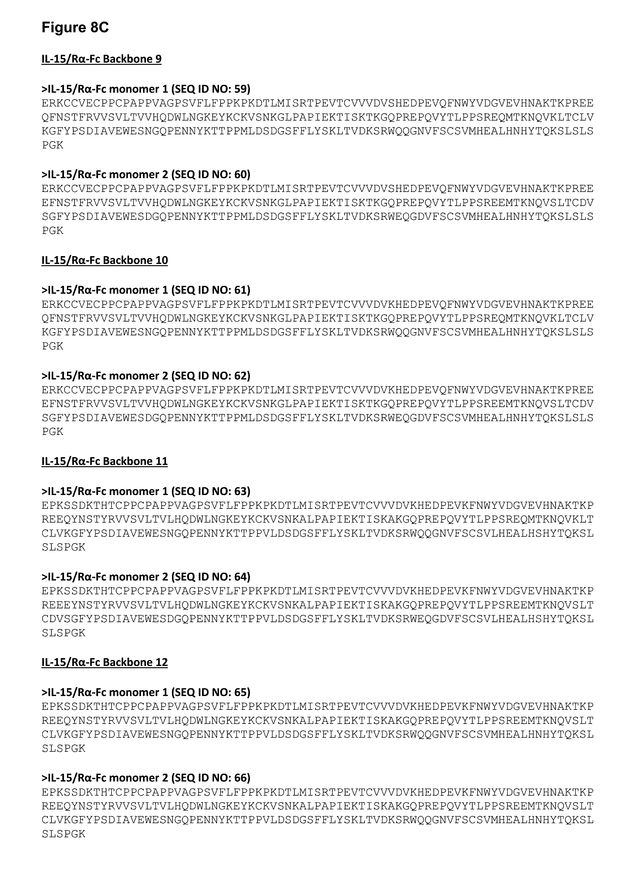
D00016

D00017
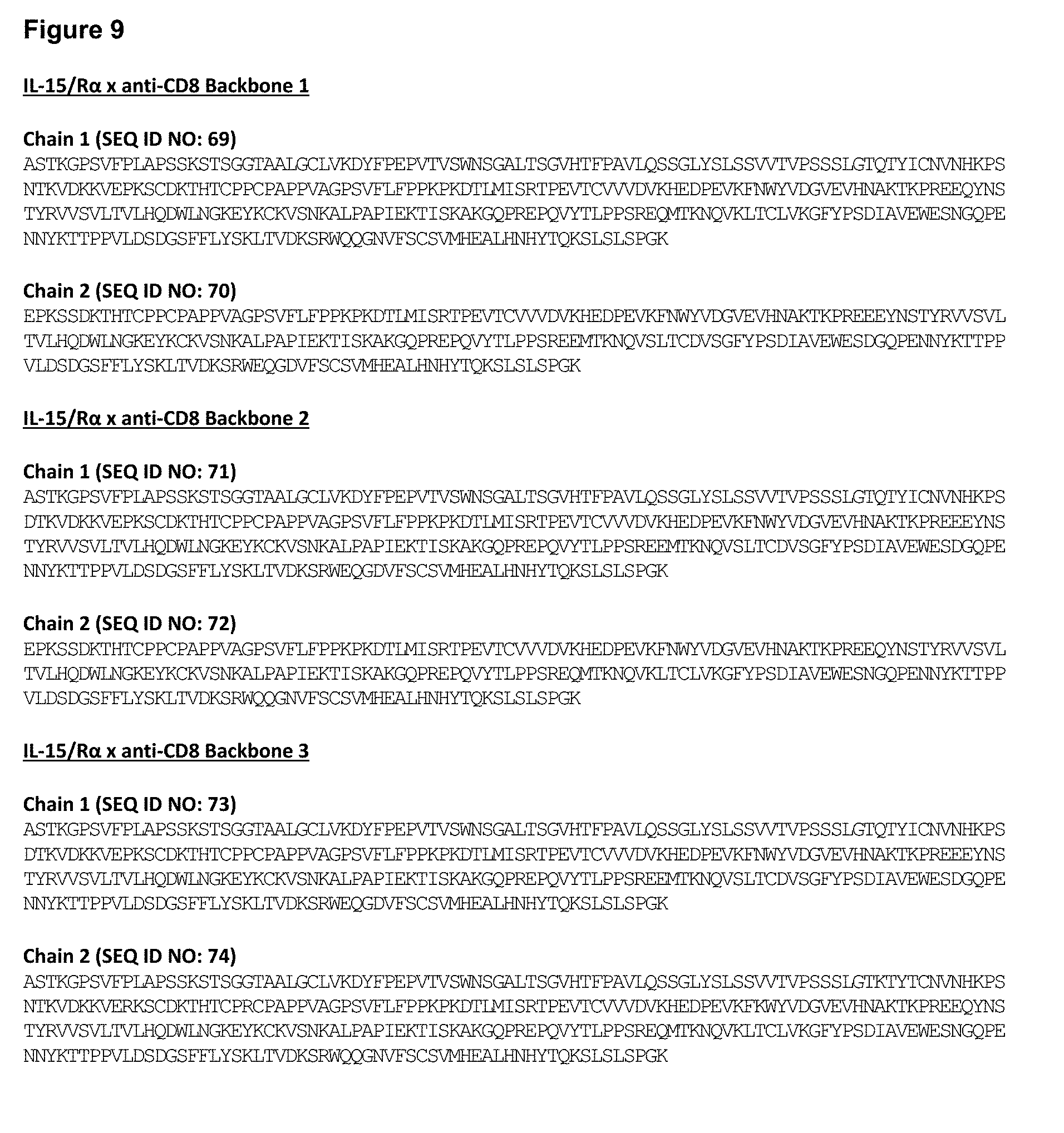
D00018

D00019
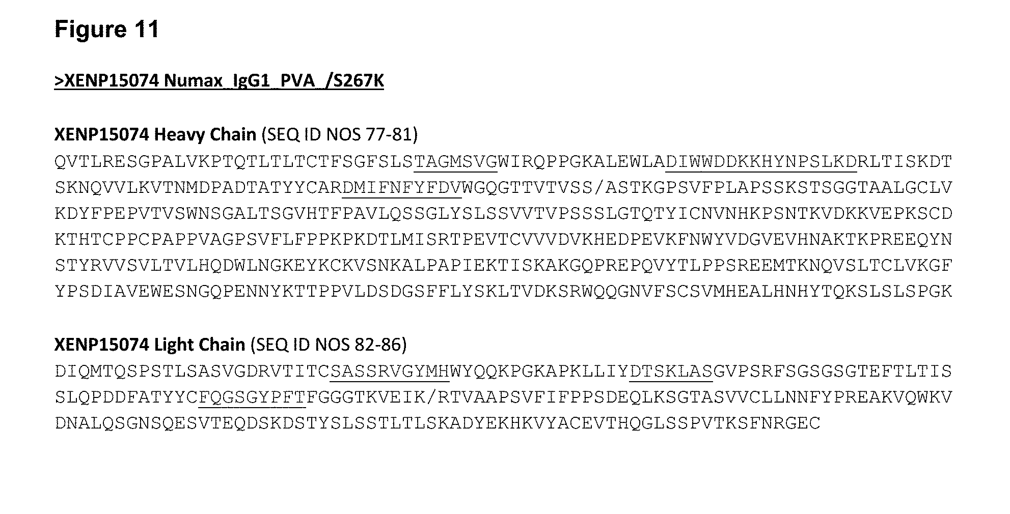
D00020

D00021

D00022
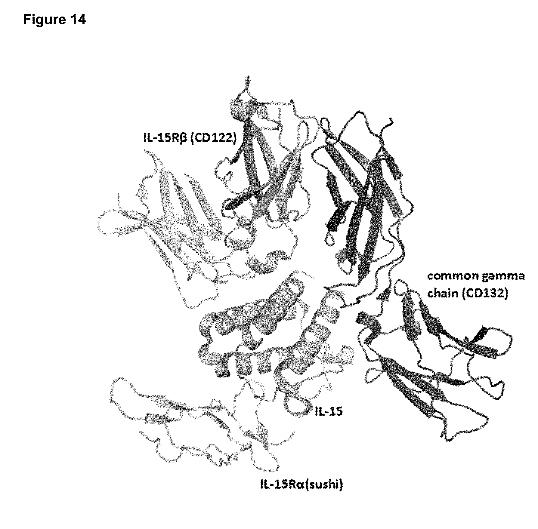
D00023

D00024

D00025

D00026
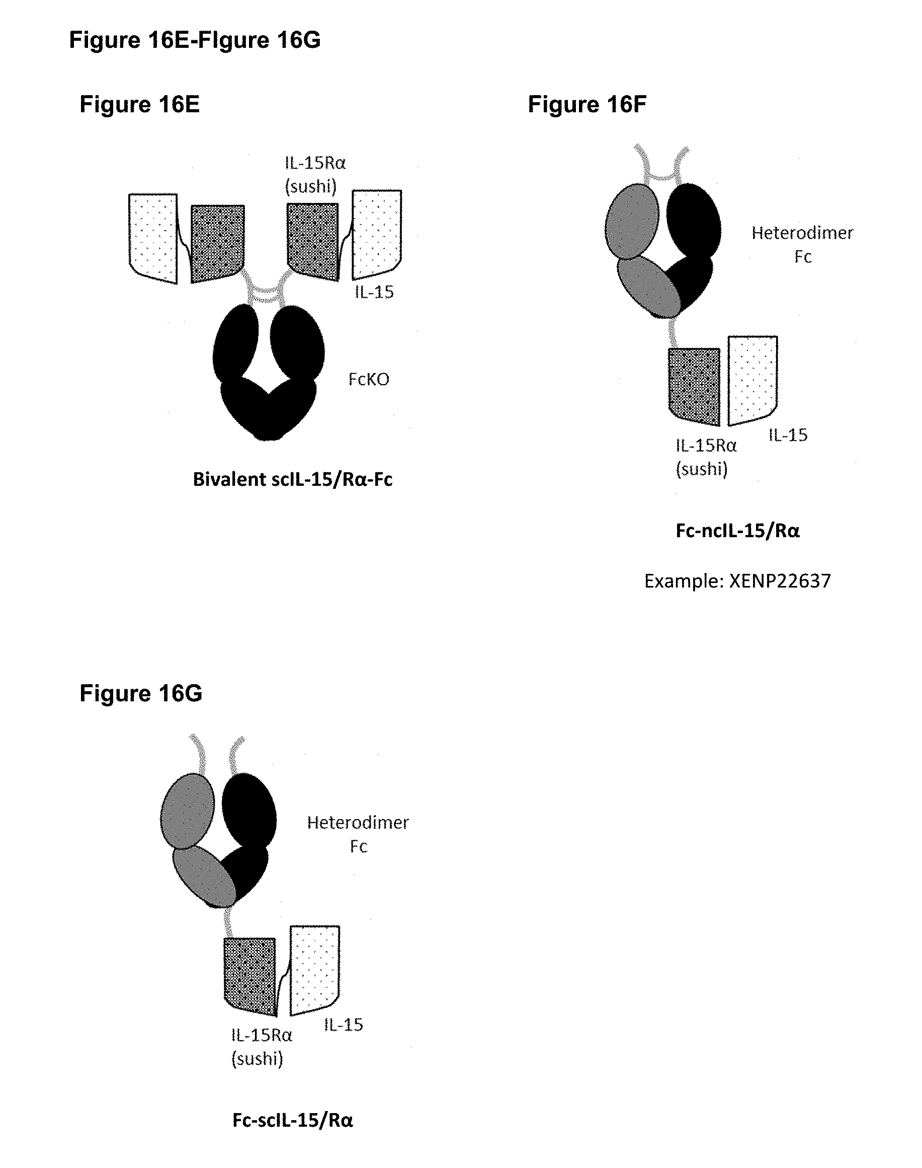
D00027
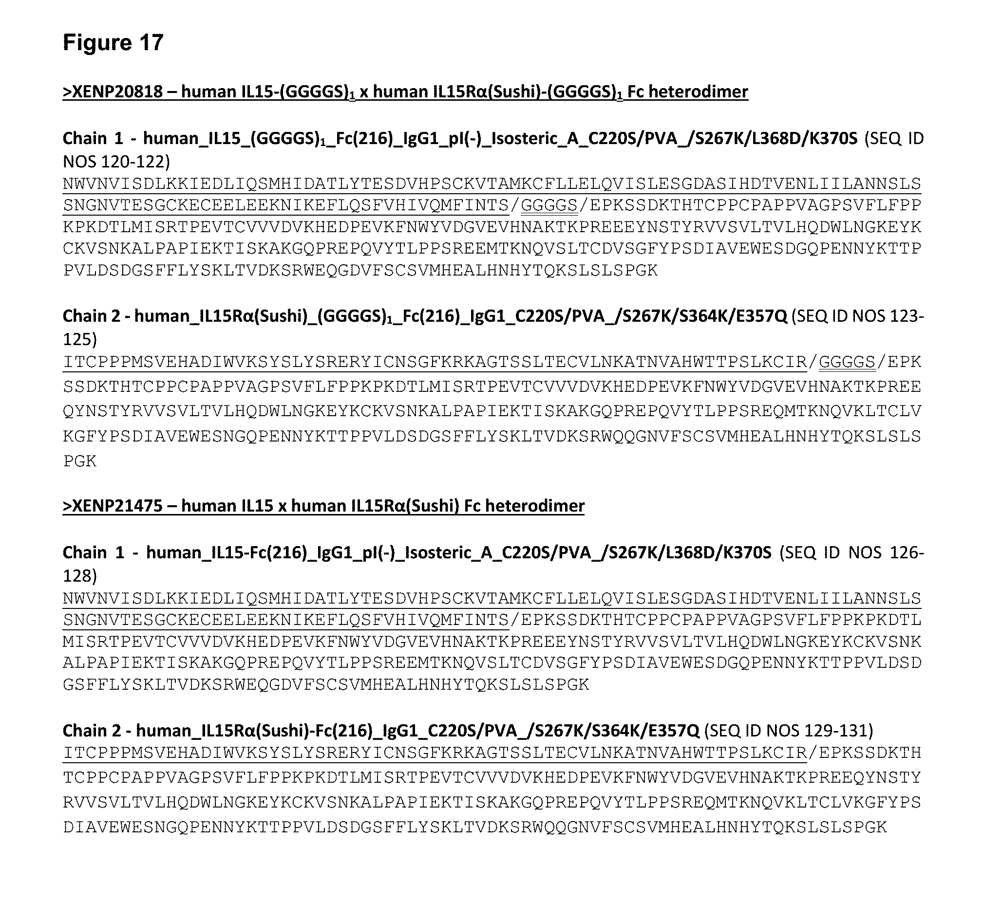
D00028

D00029
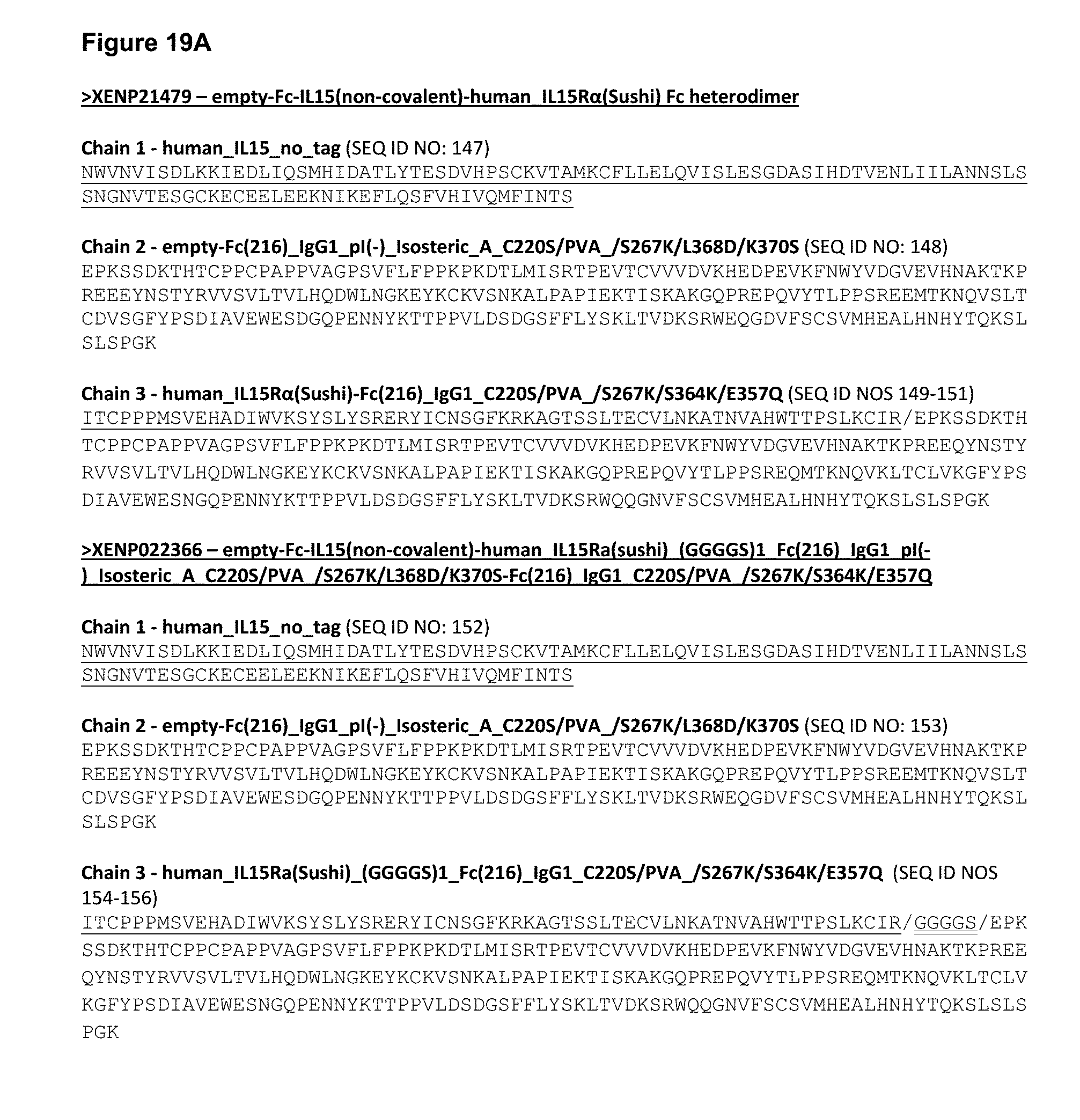
D00030
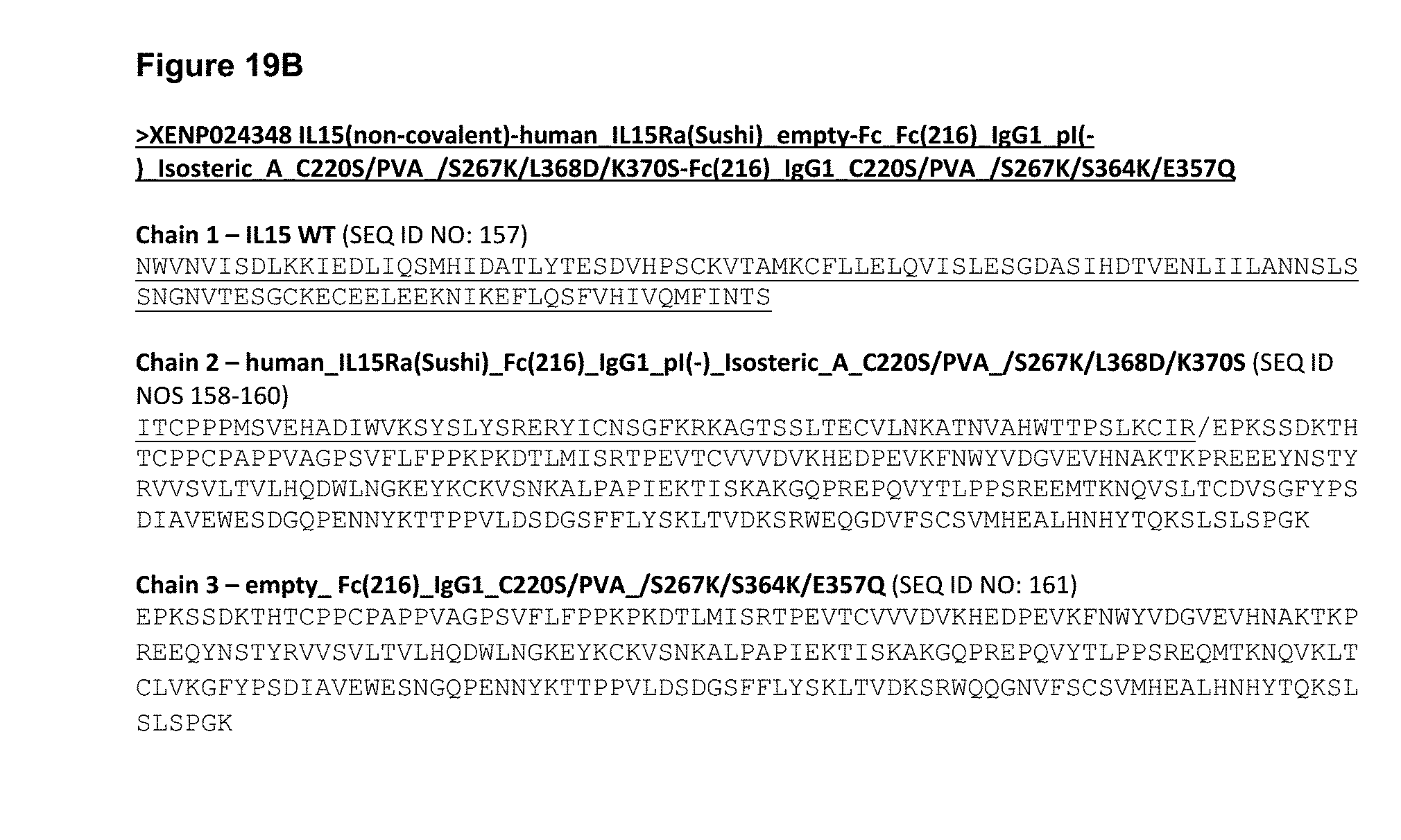
D00031

D00032
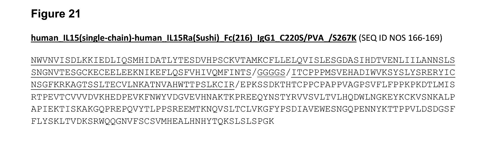
D00033

D00034

D00035
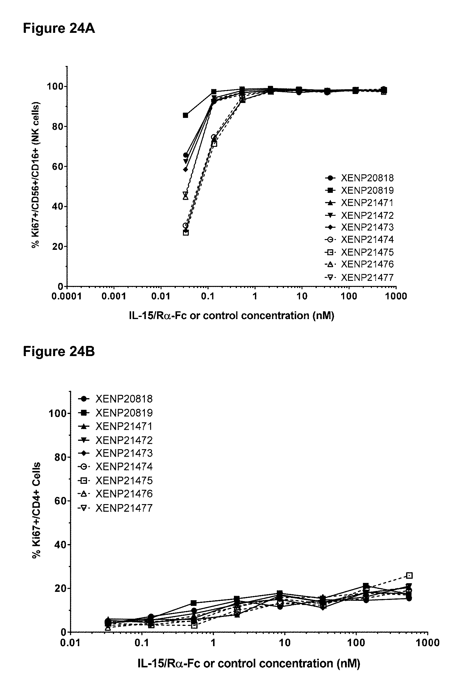
D00036

D00037
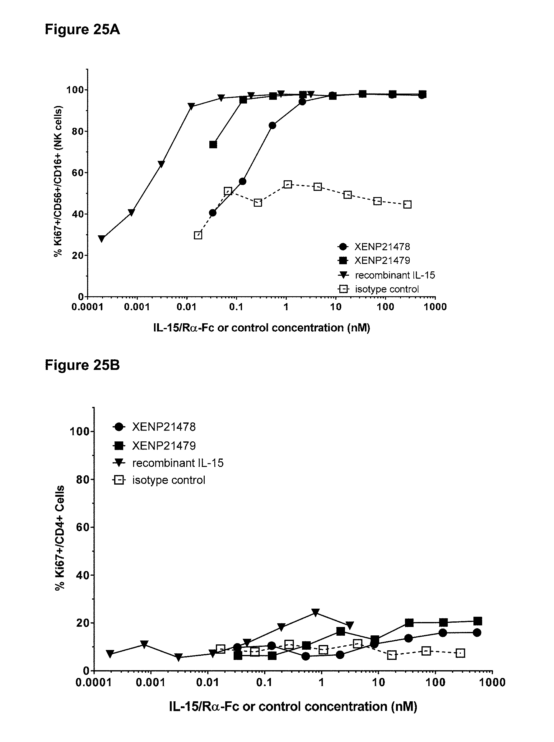
D00038

D00039
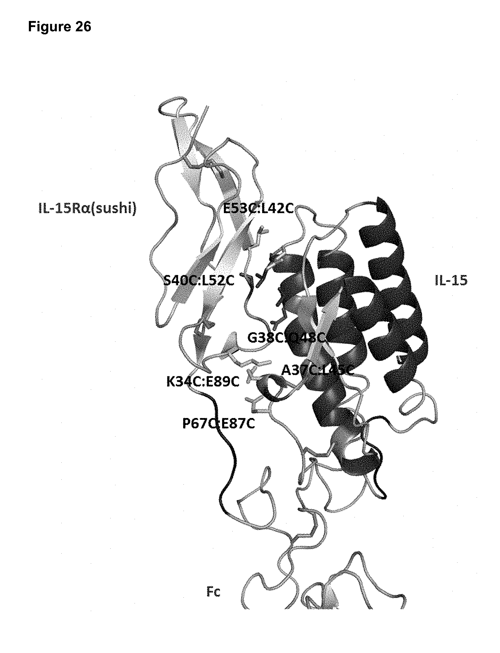
D00040
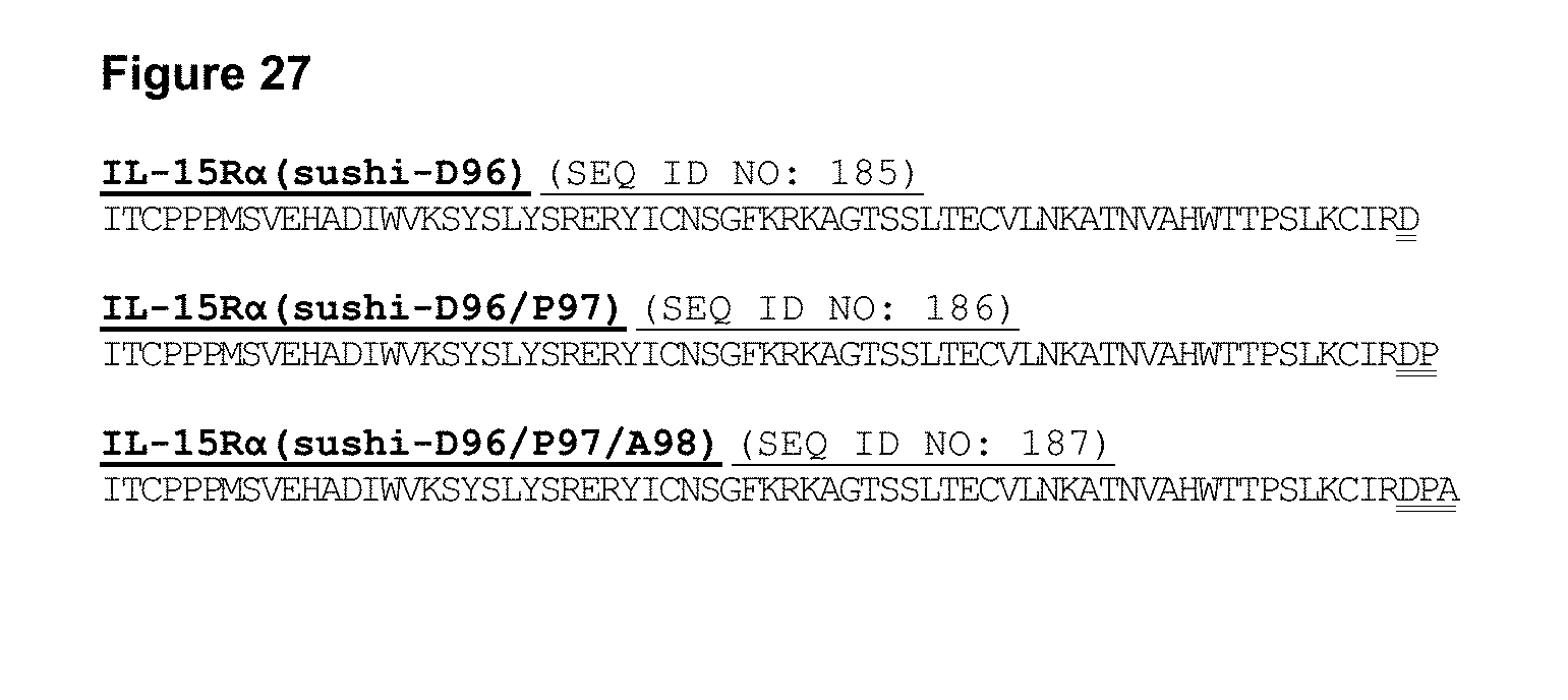
D00041

D00042
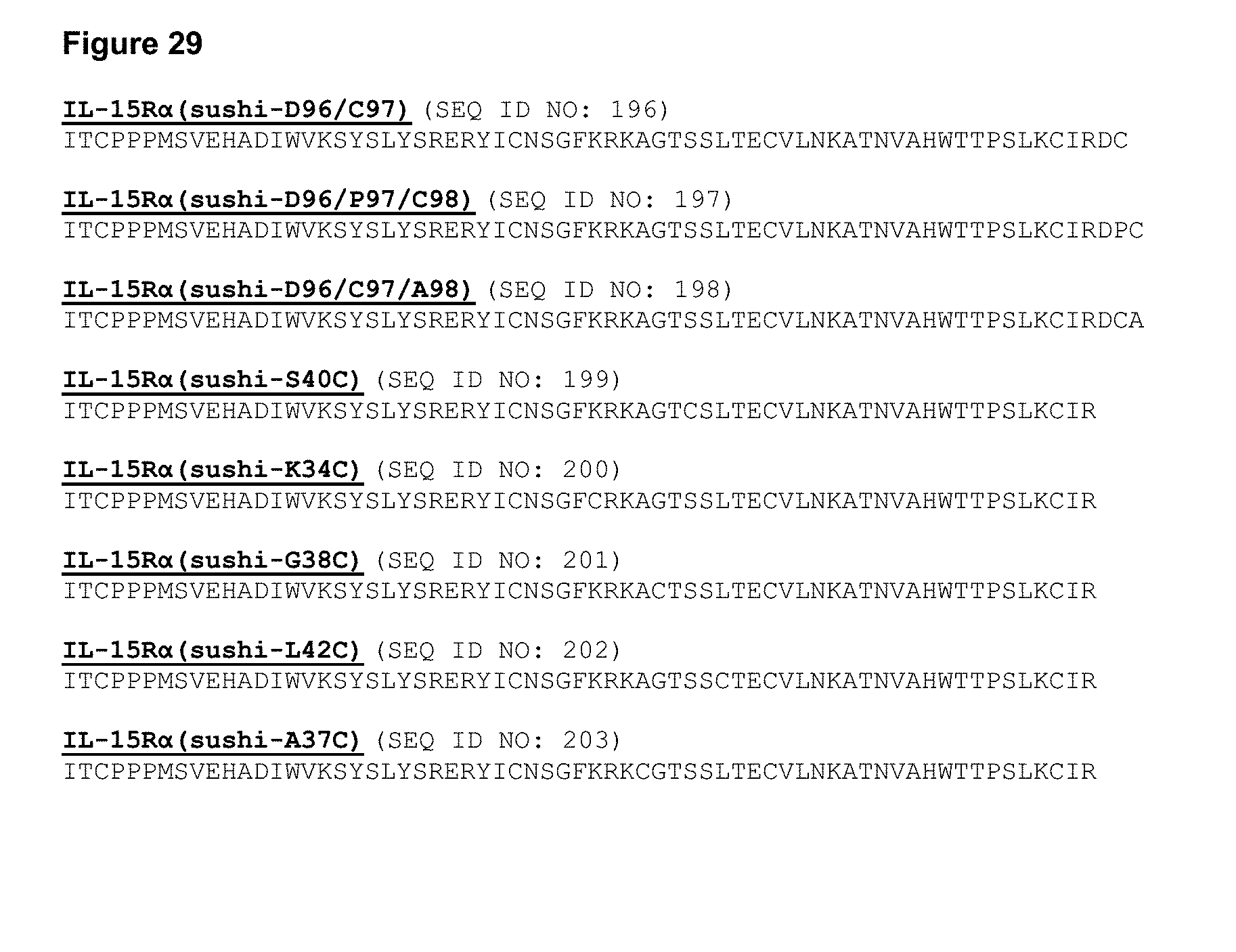
D00043

D00044
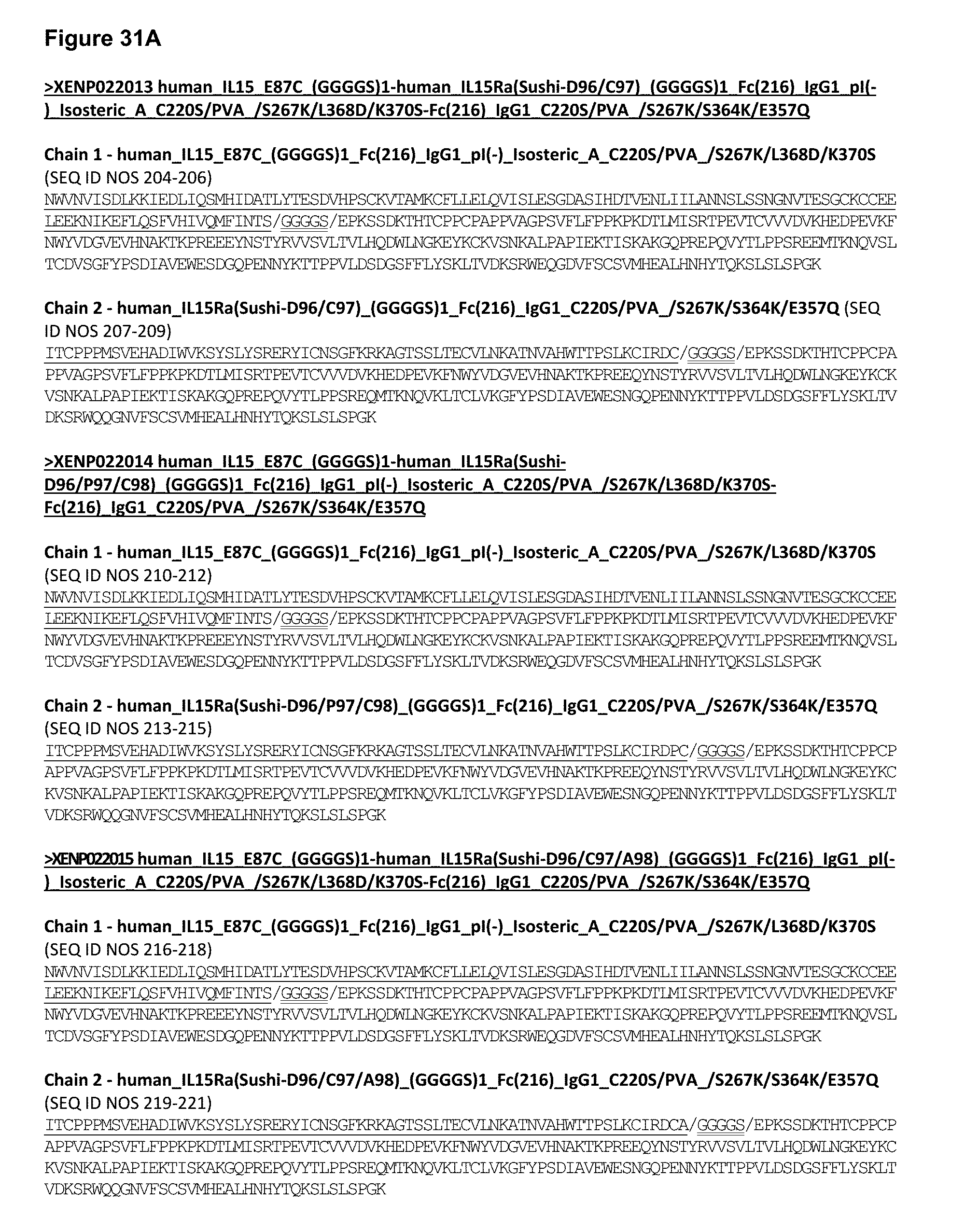
D00045
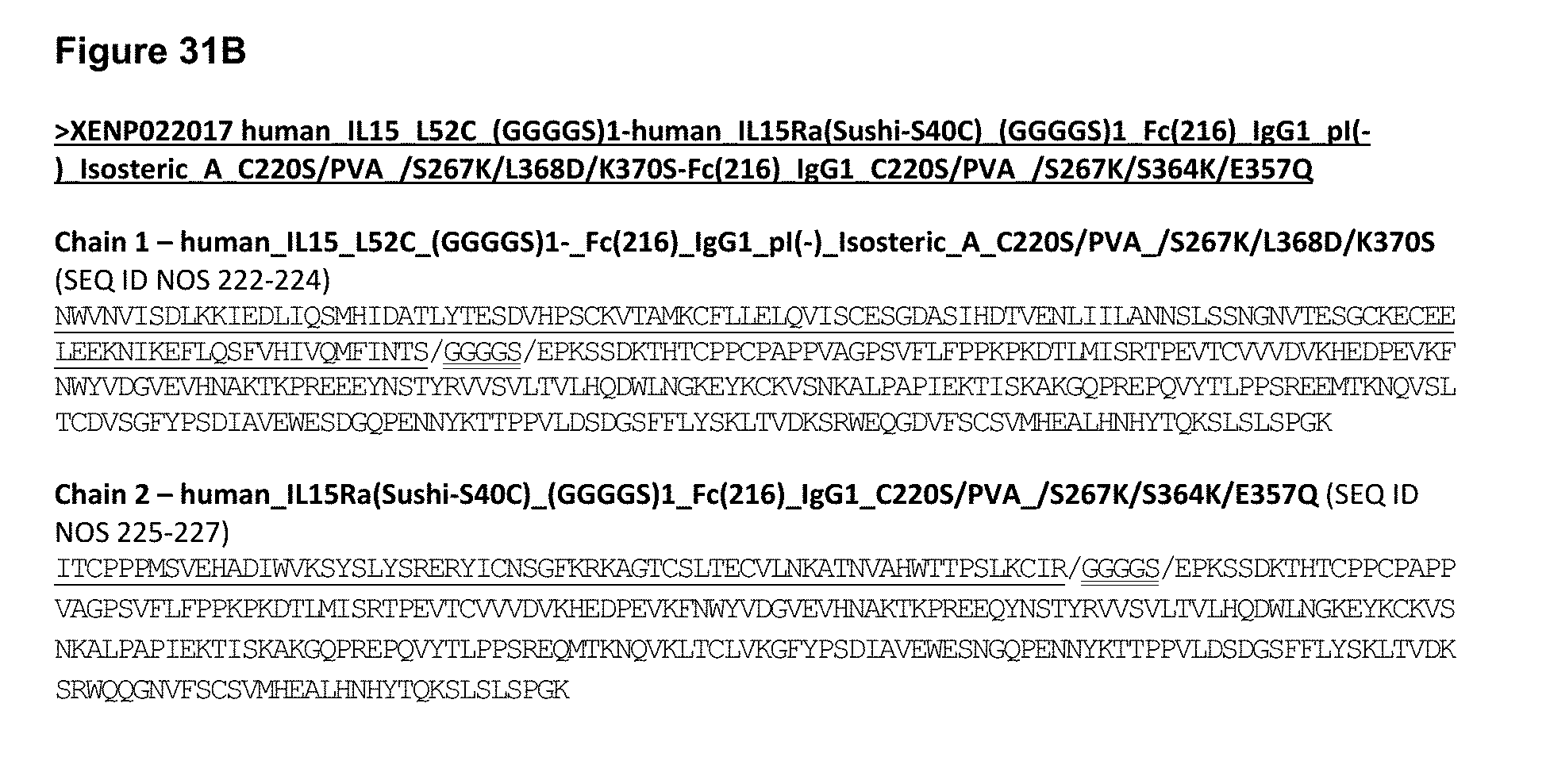
D00046
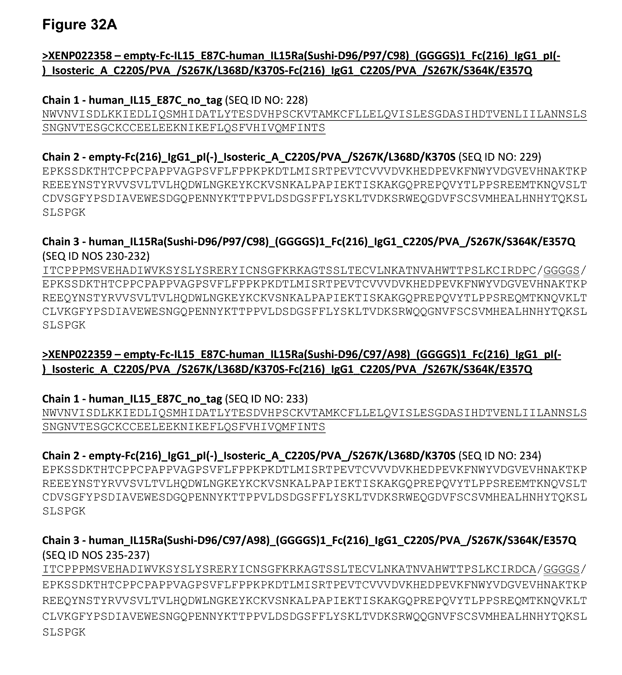
D00047
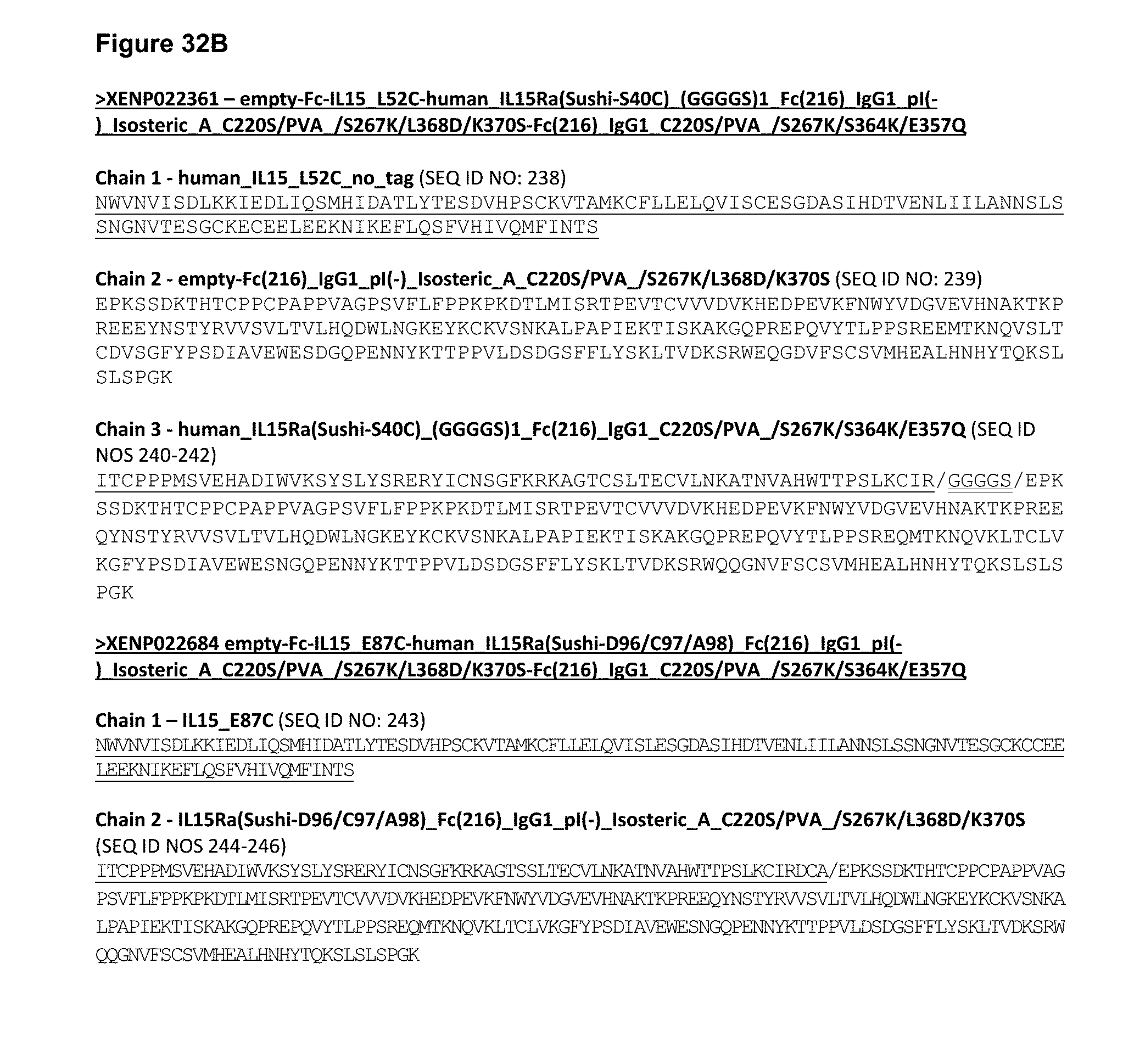
D00048

D00049

D00050
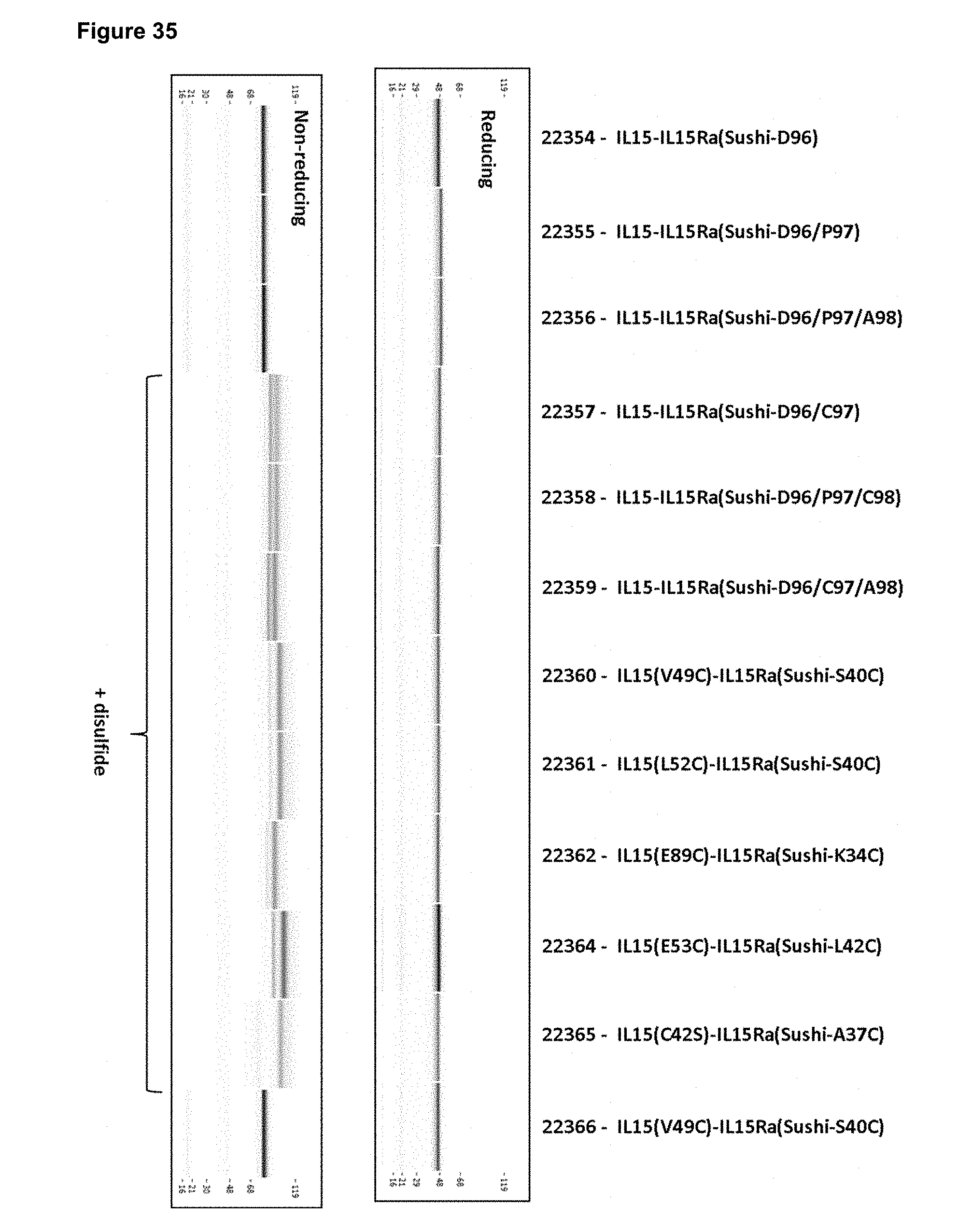
D00051
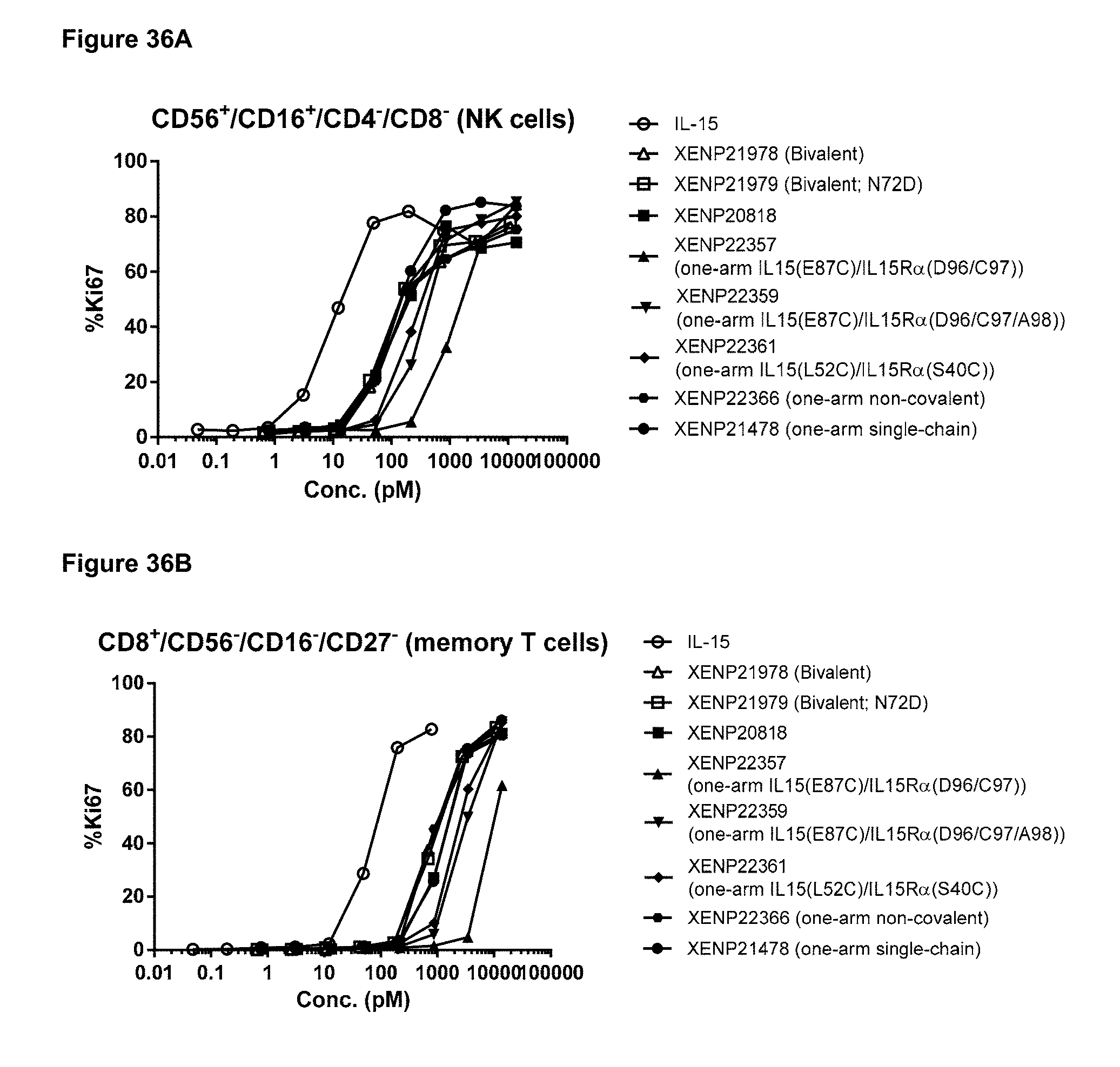
D00052
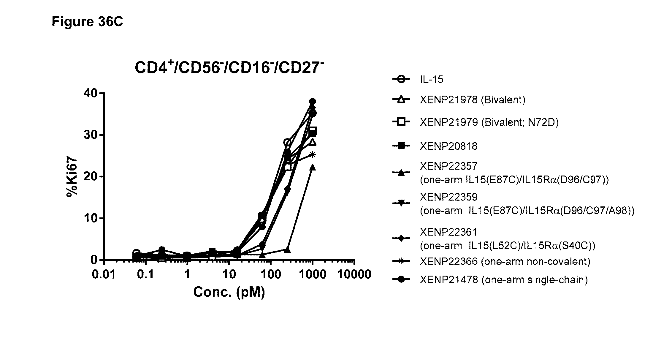
D00053
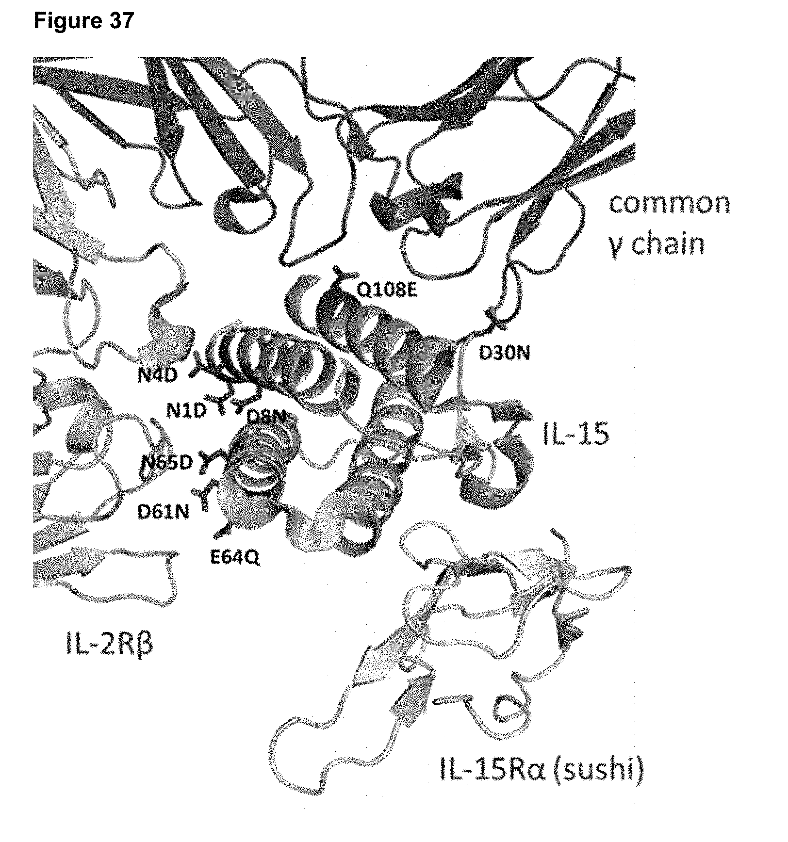
D00054

D00055
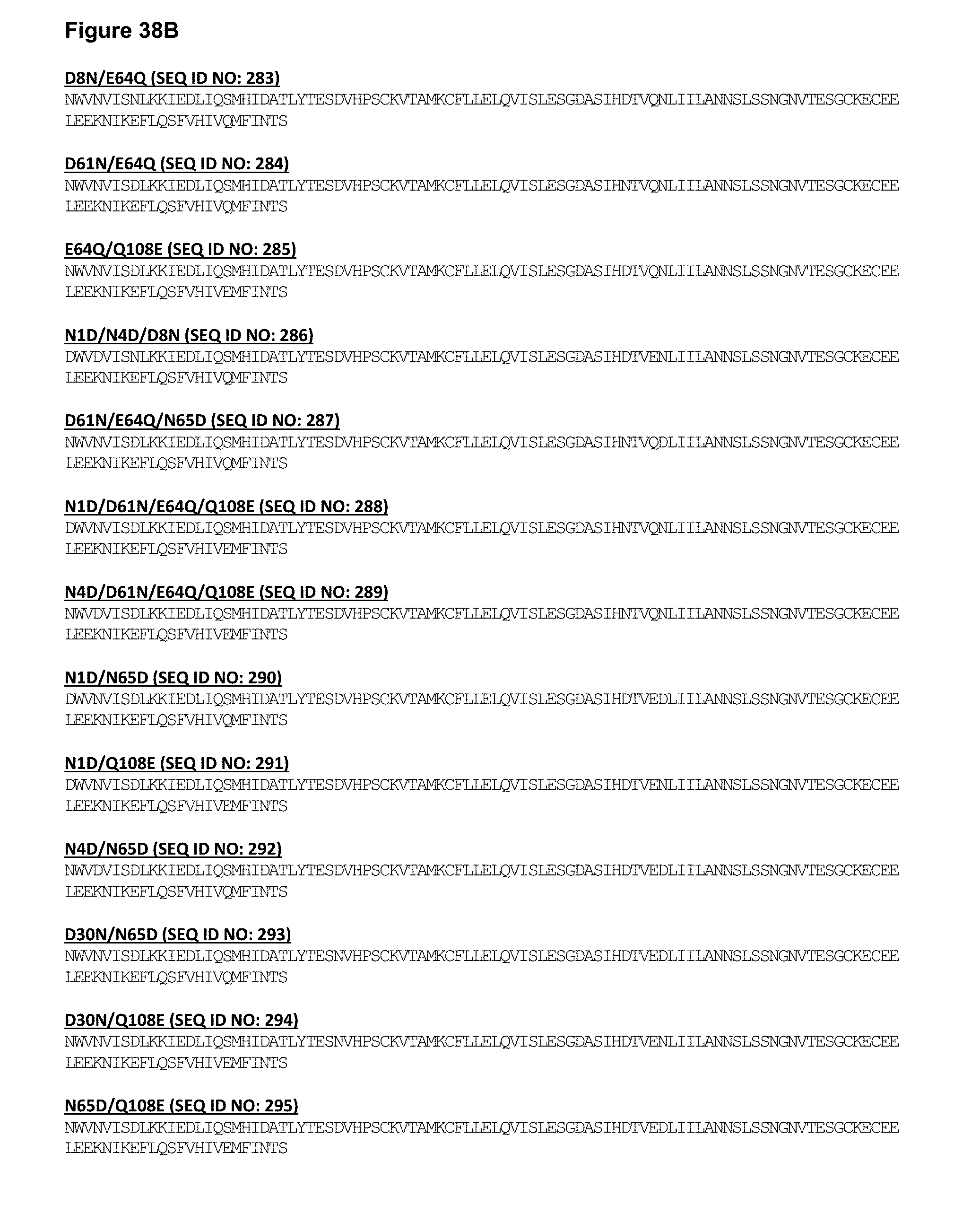
D00056
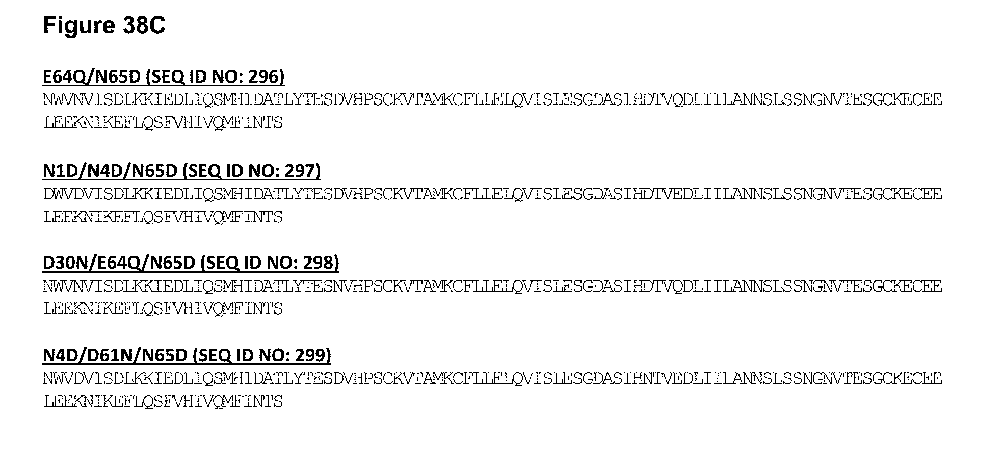
D00057

D00058

D00059

D00060
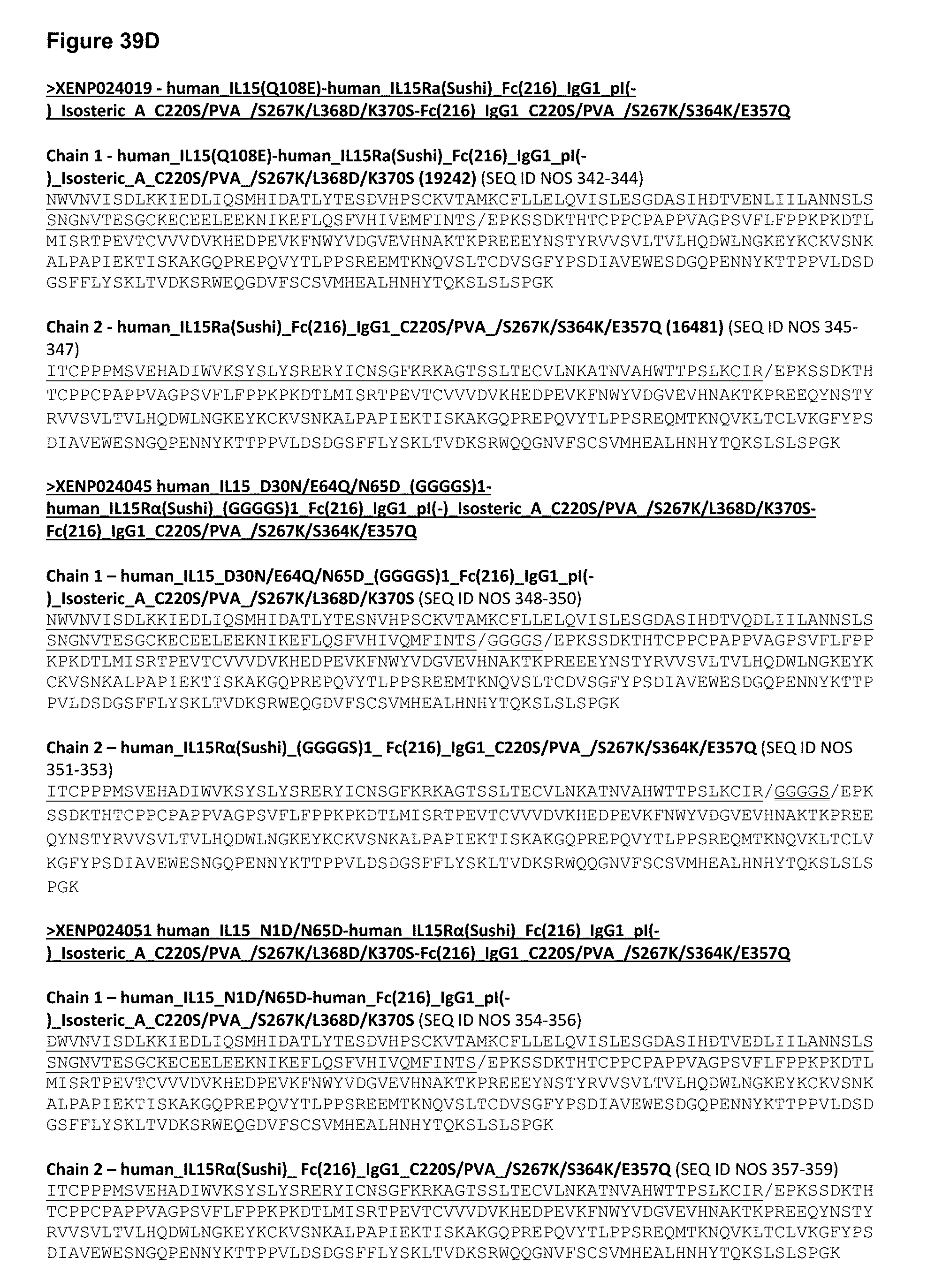
D00061
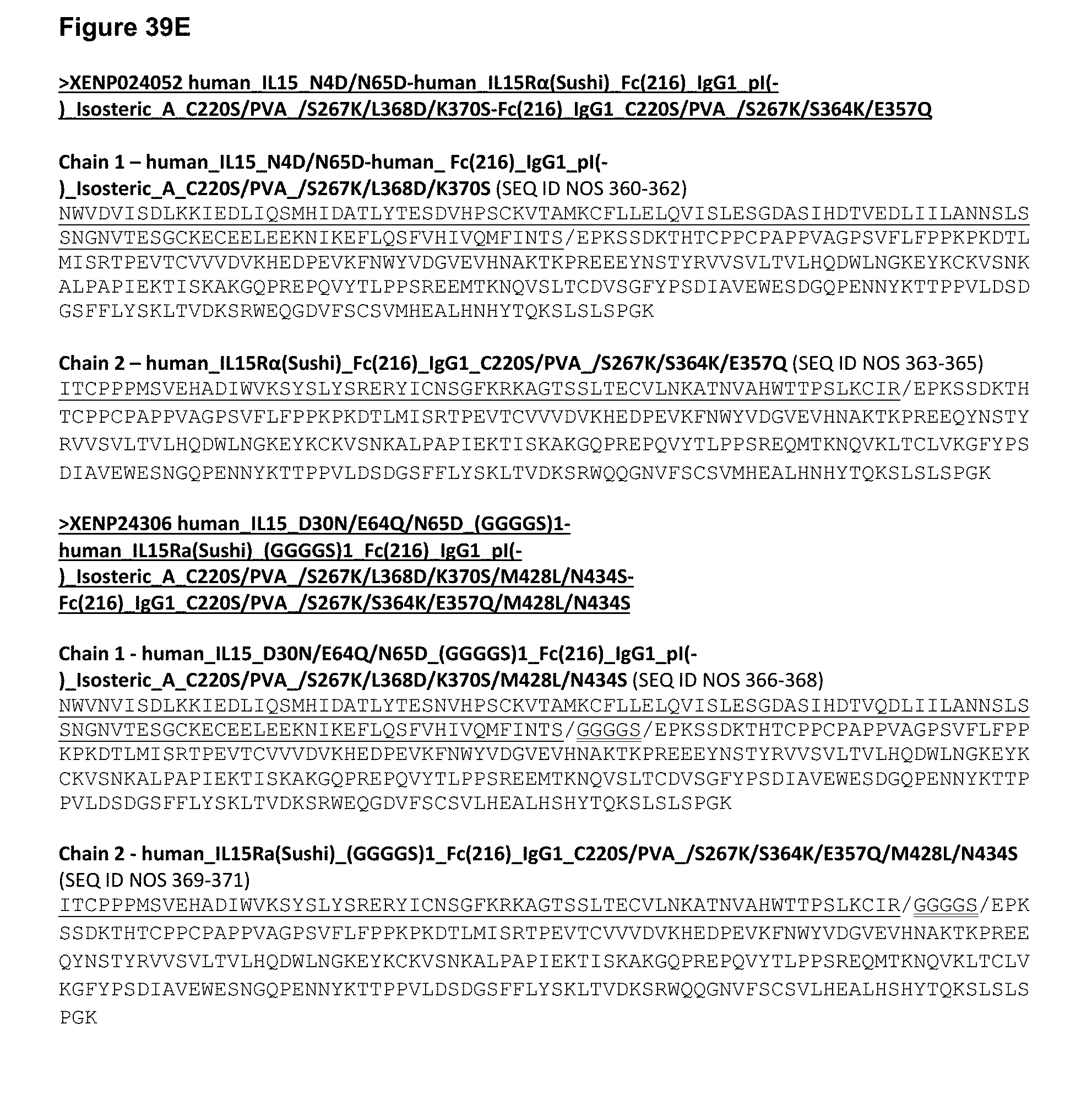
D00062
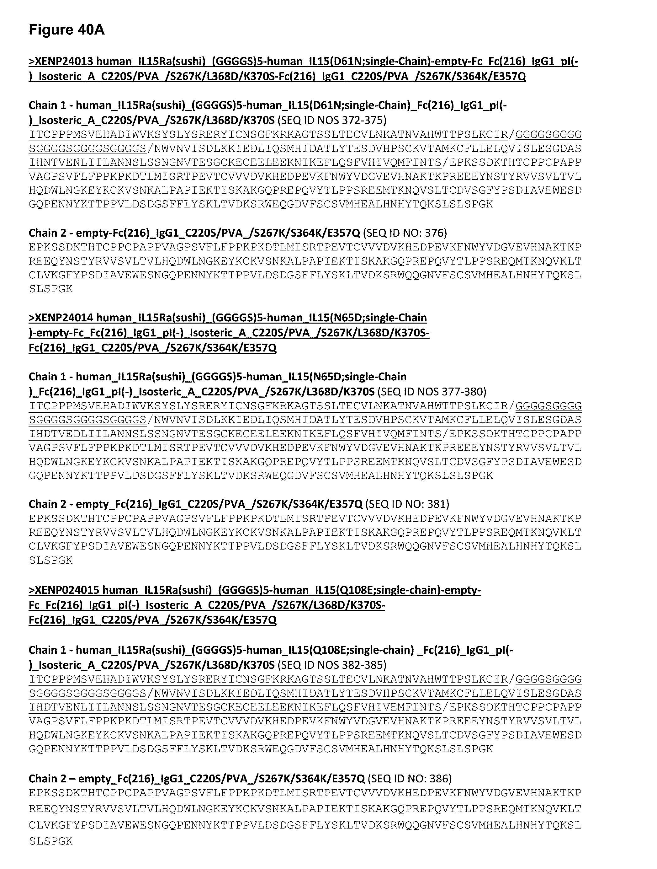
D00063
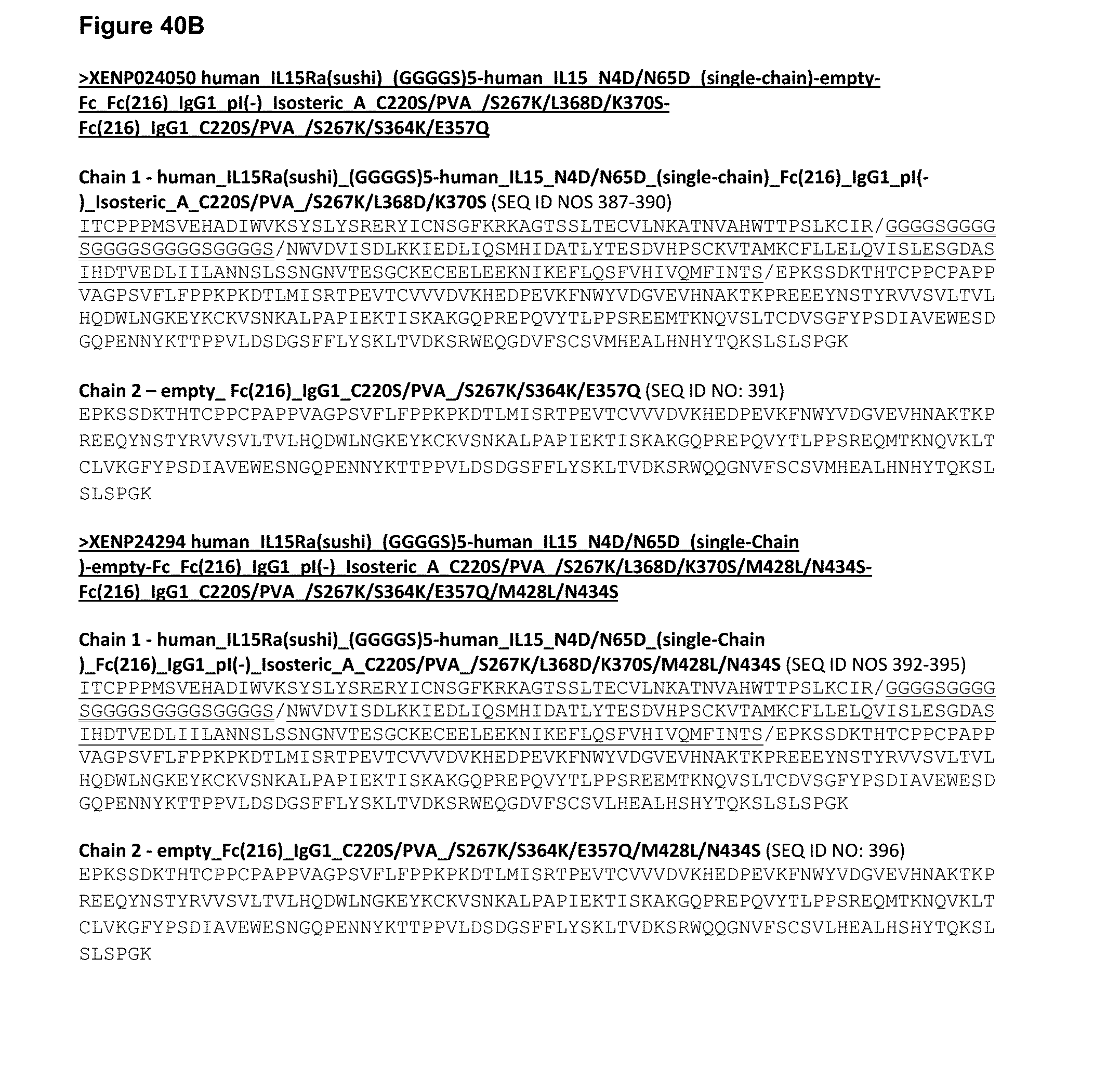
D00064
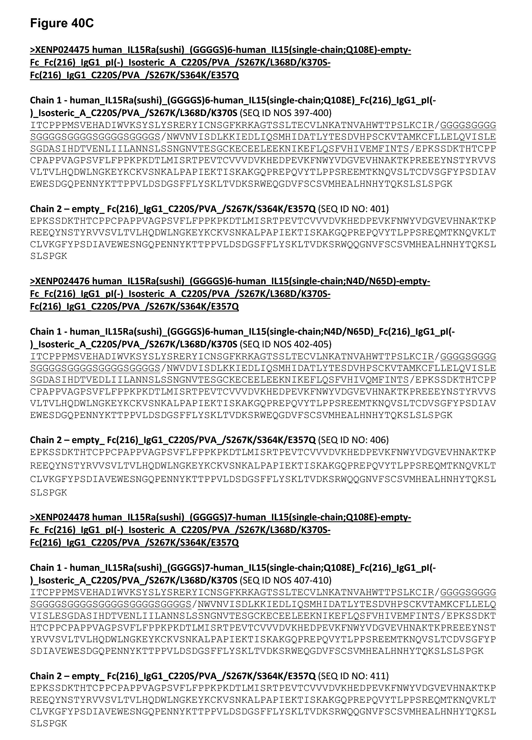
D00065
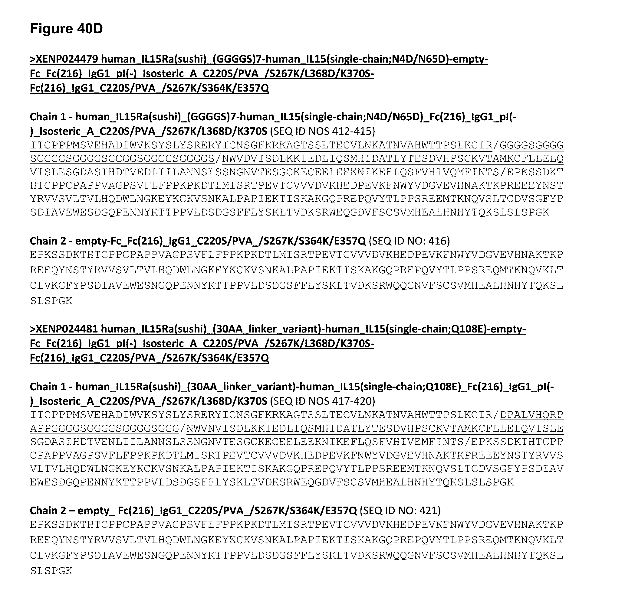
D00066
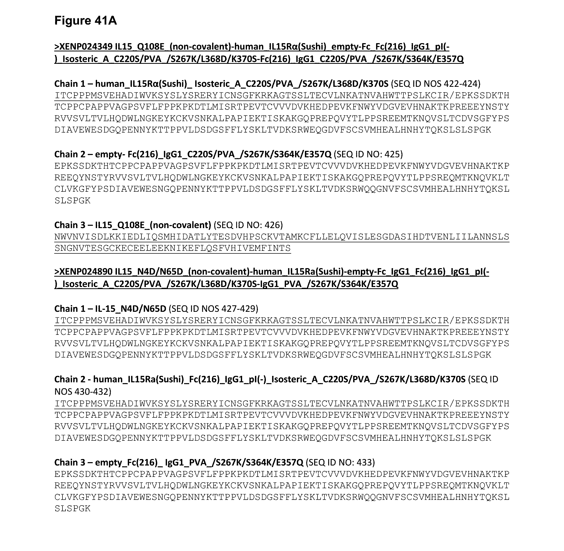
D00067
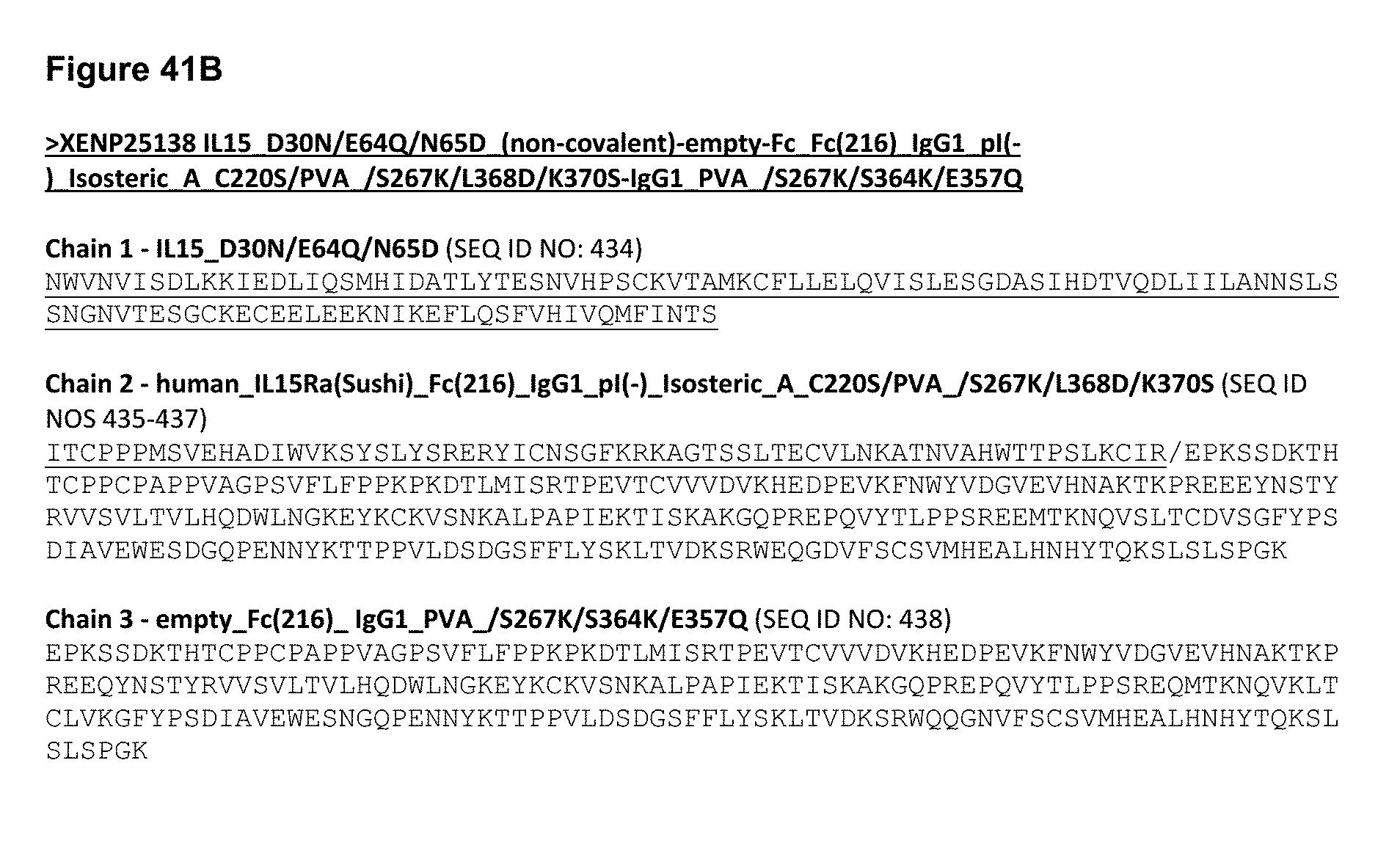
D00068

D00069
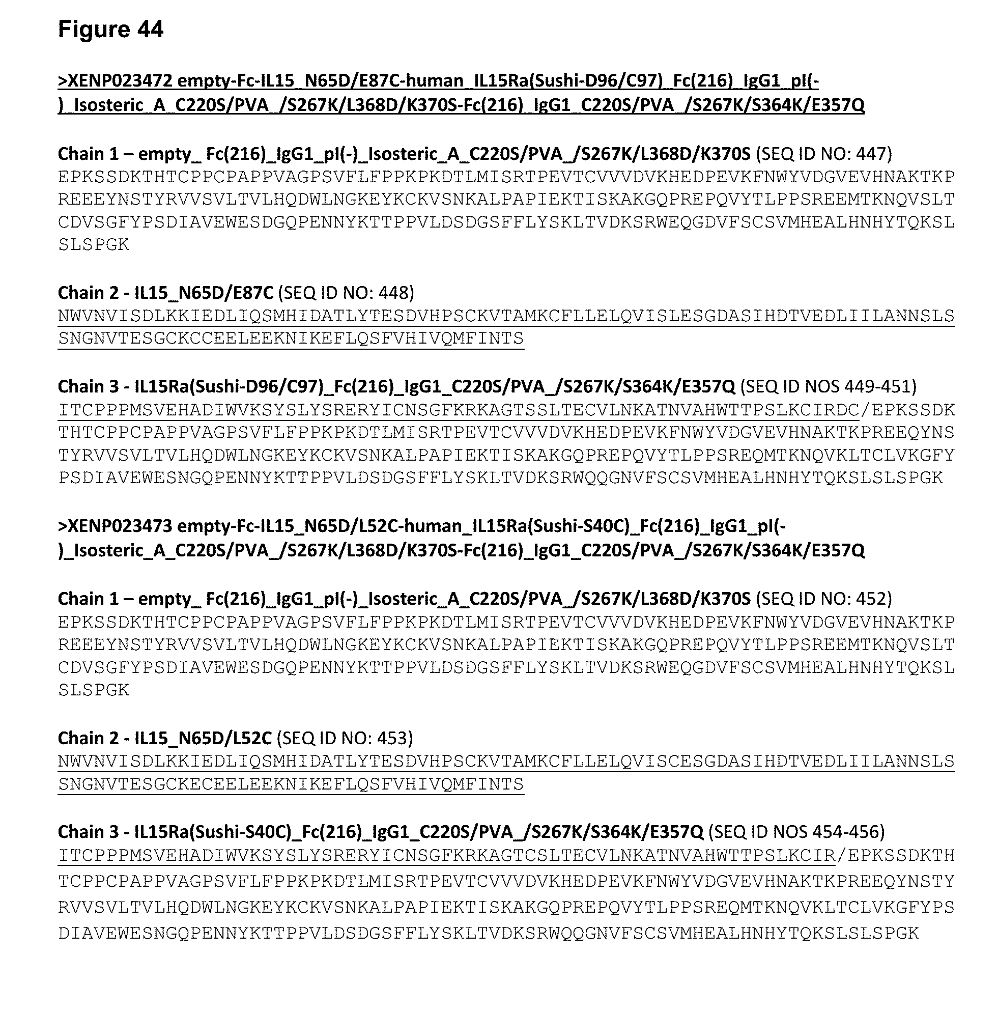
D00070
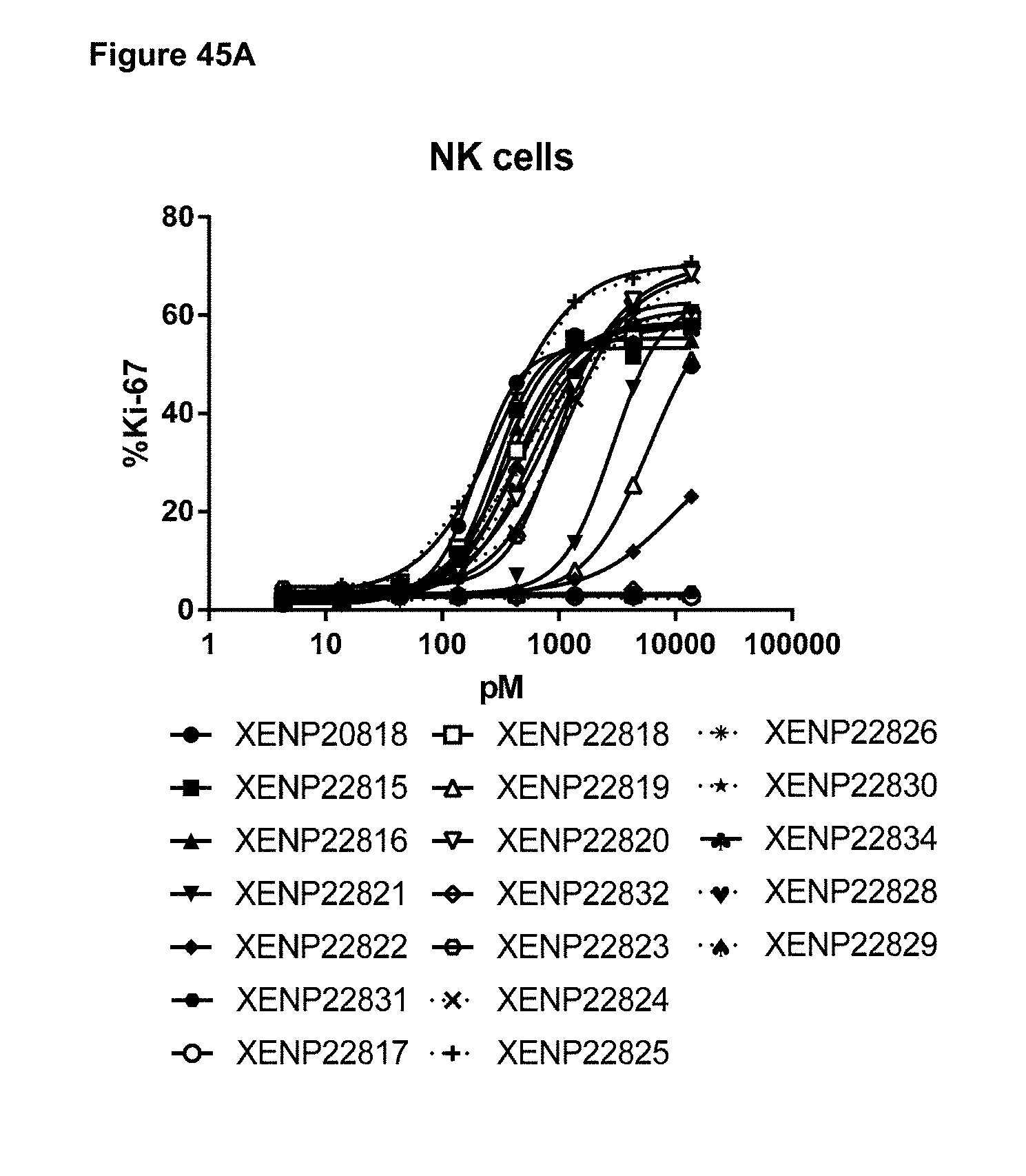
D00071

D00072

D00073
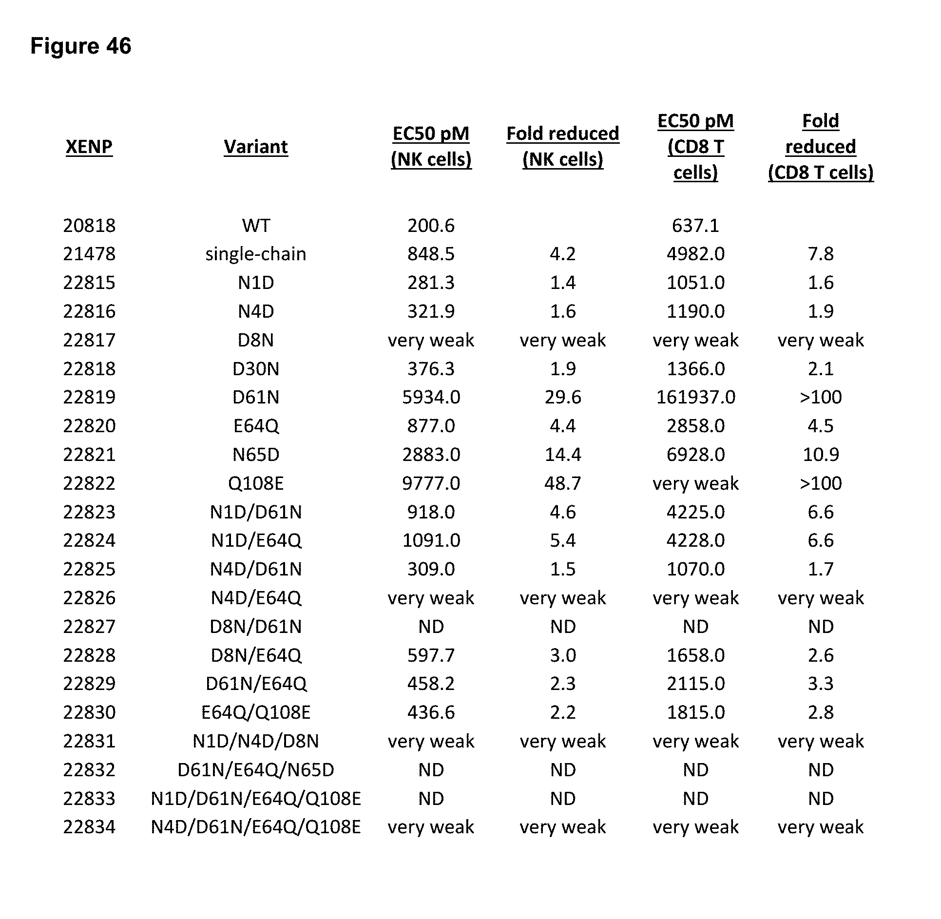
D00074

D00075

D00076
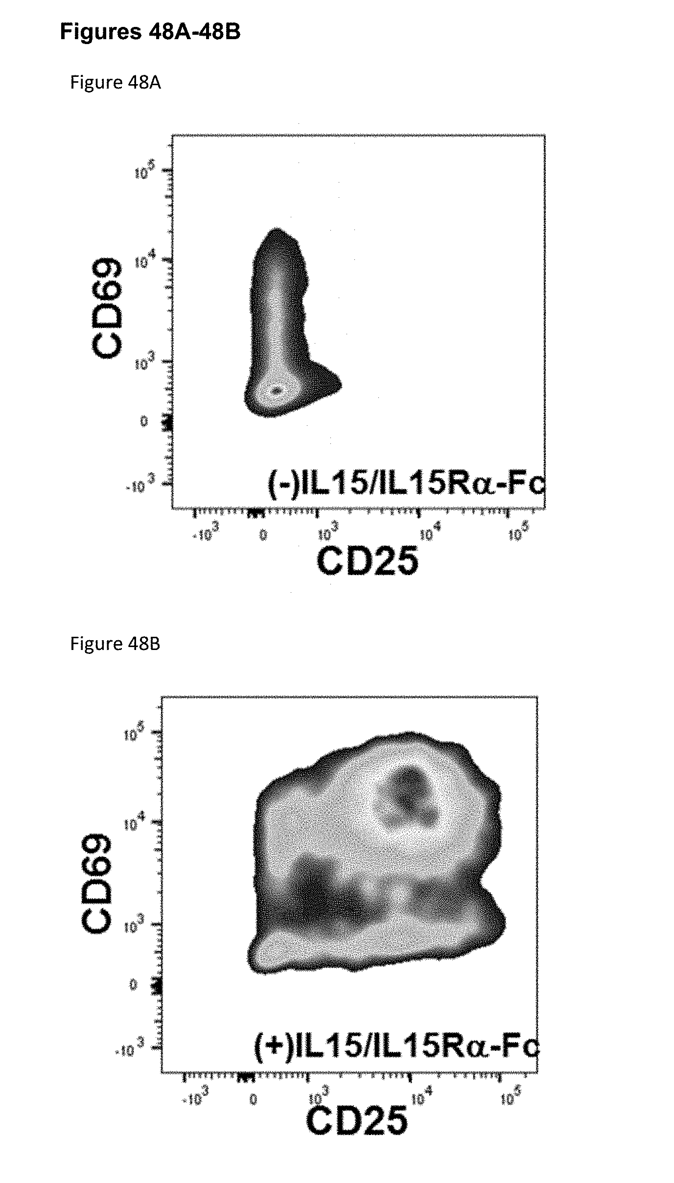
D00077

D00078

D00079
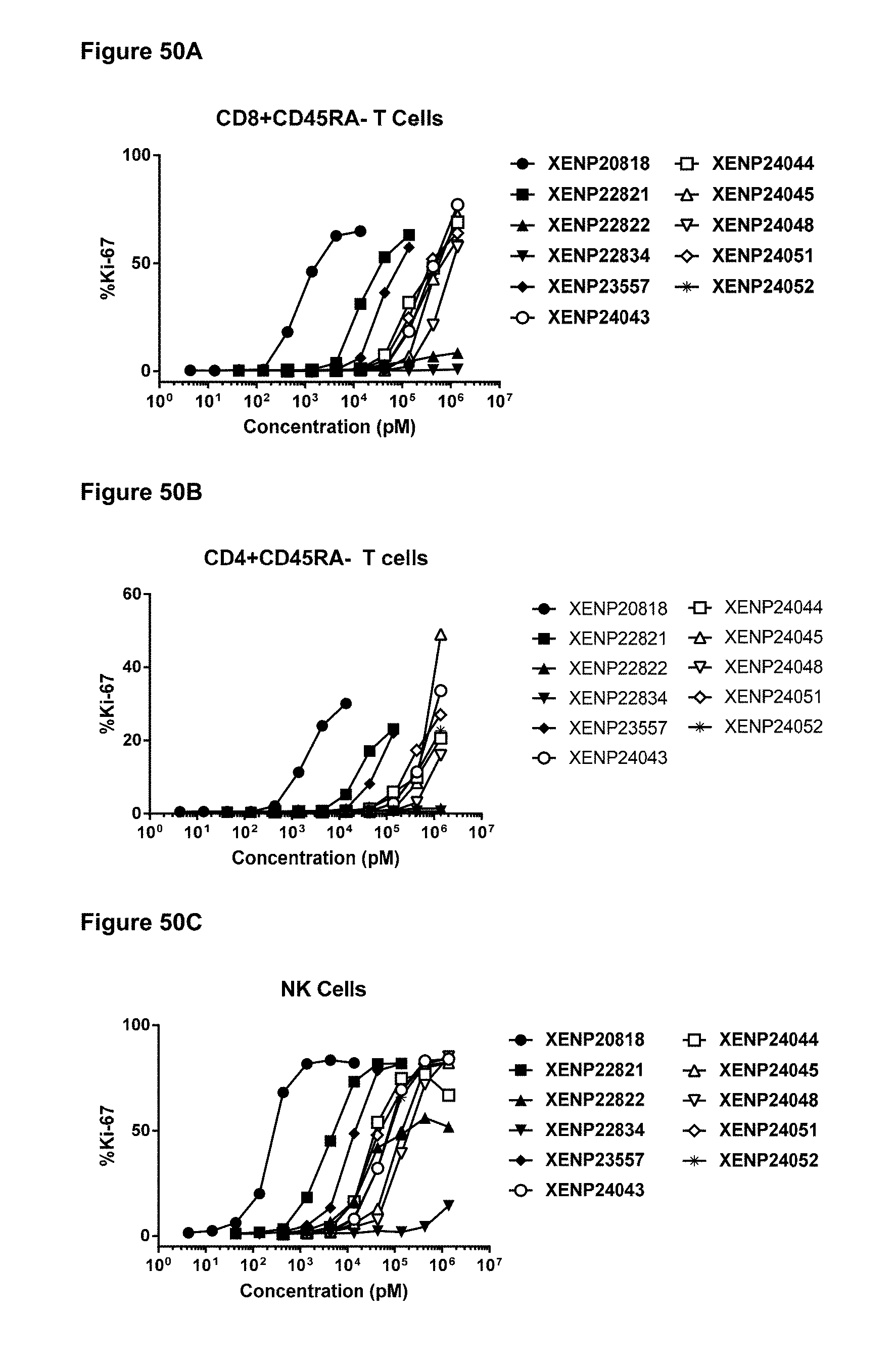
D00080

D00081
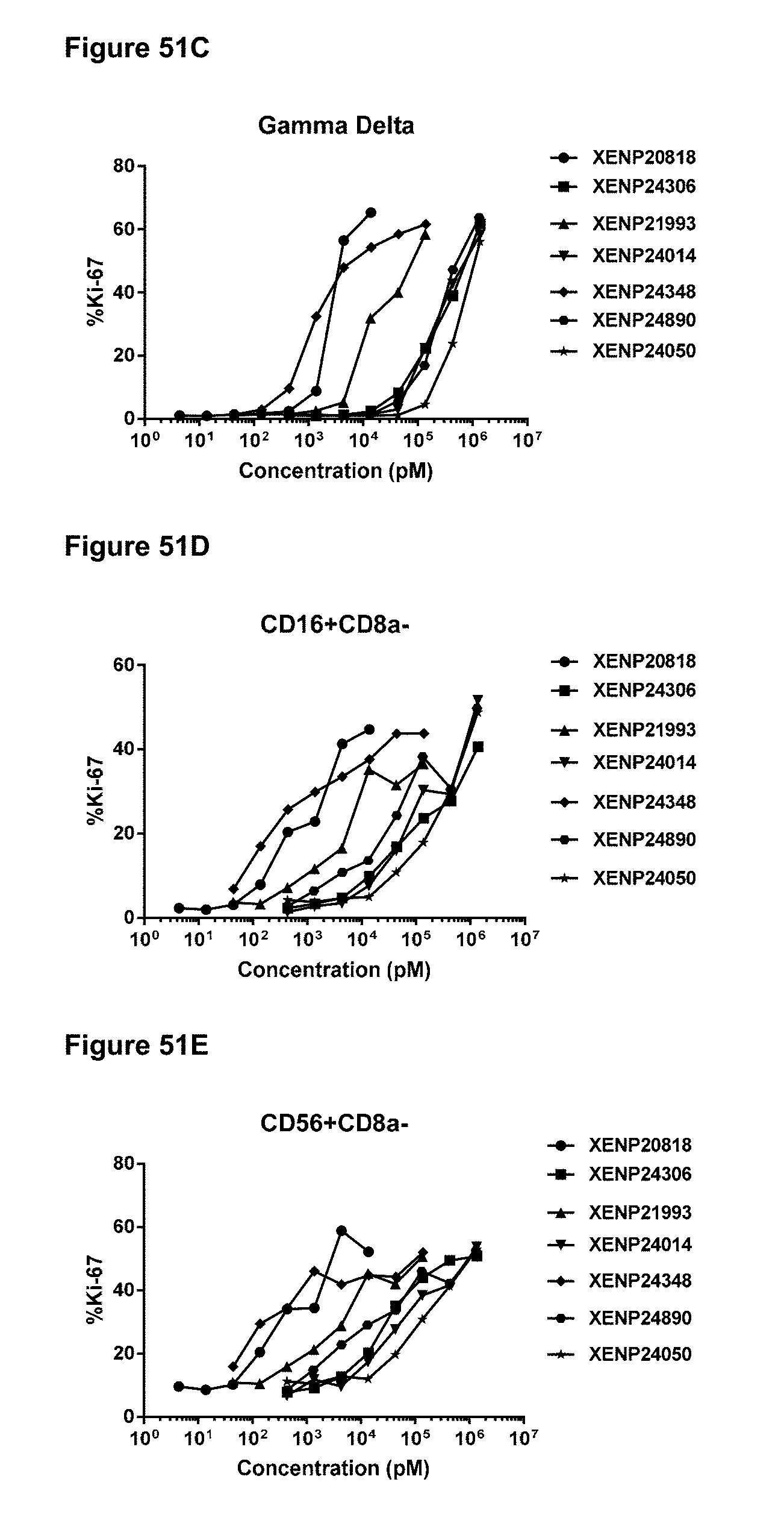
D00082
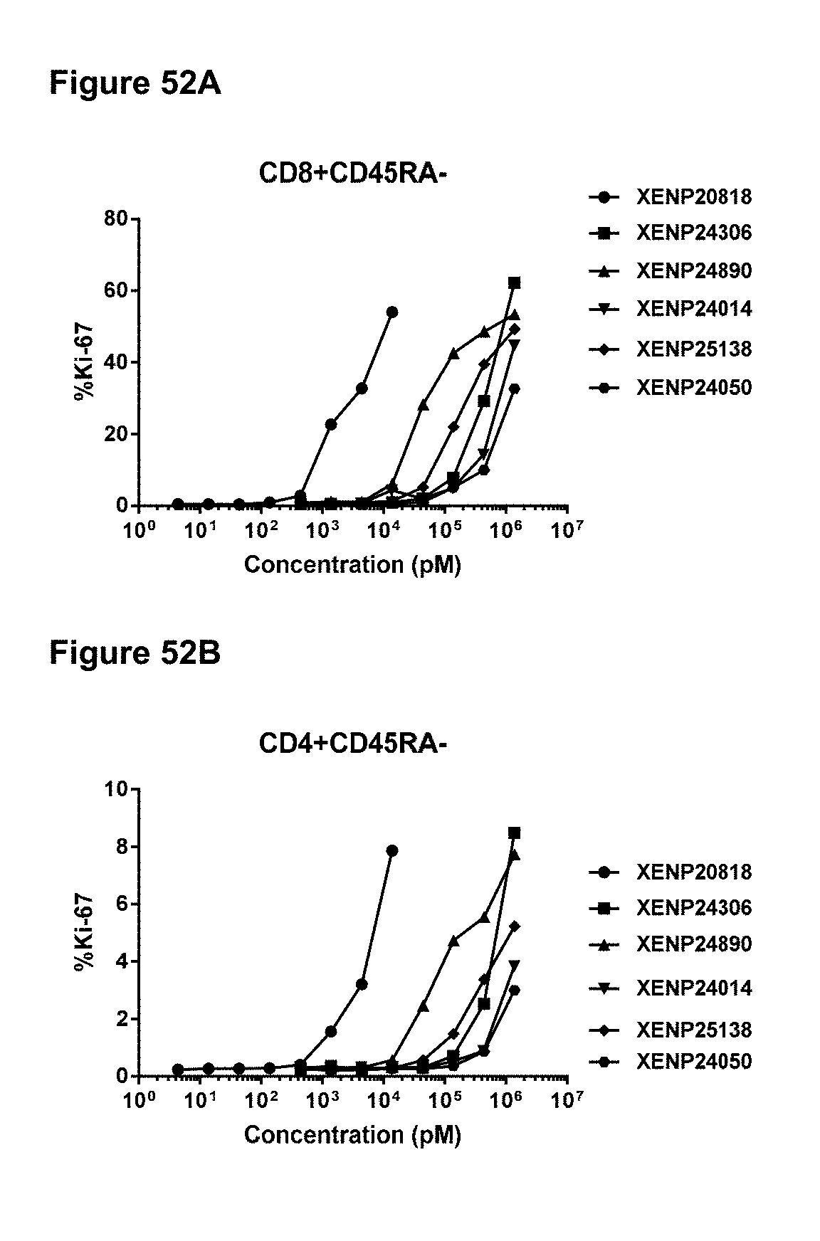
D00083
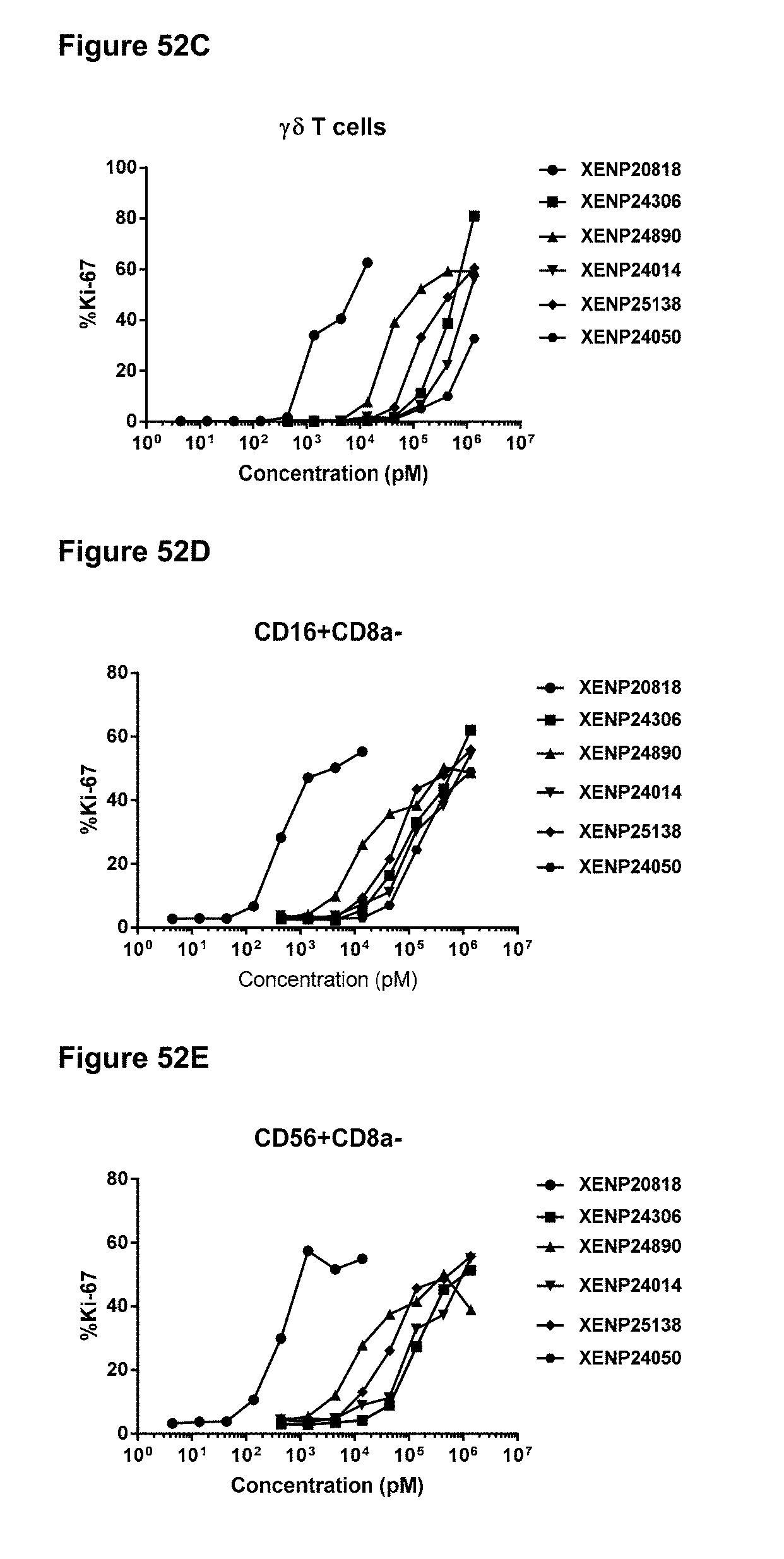
D00084
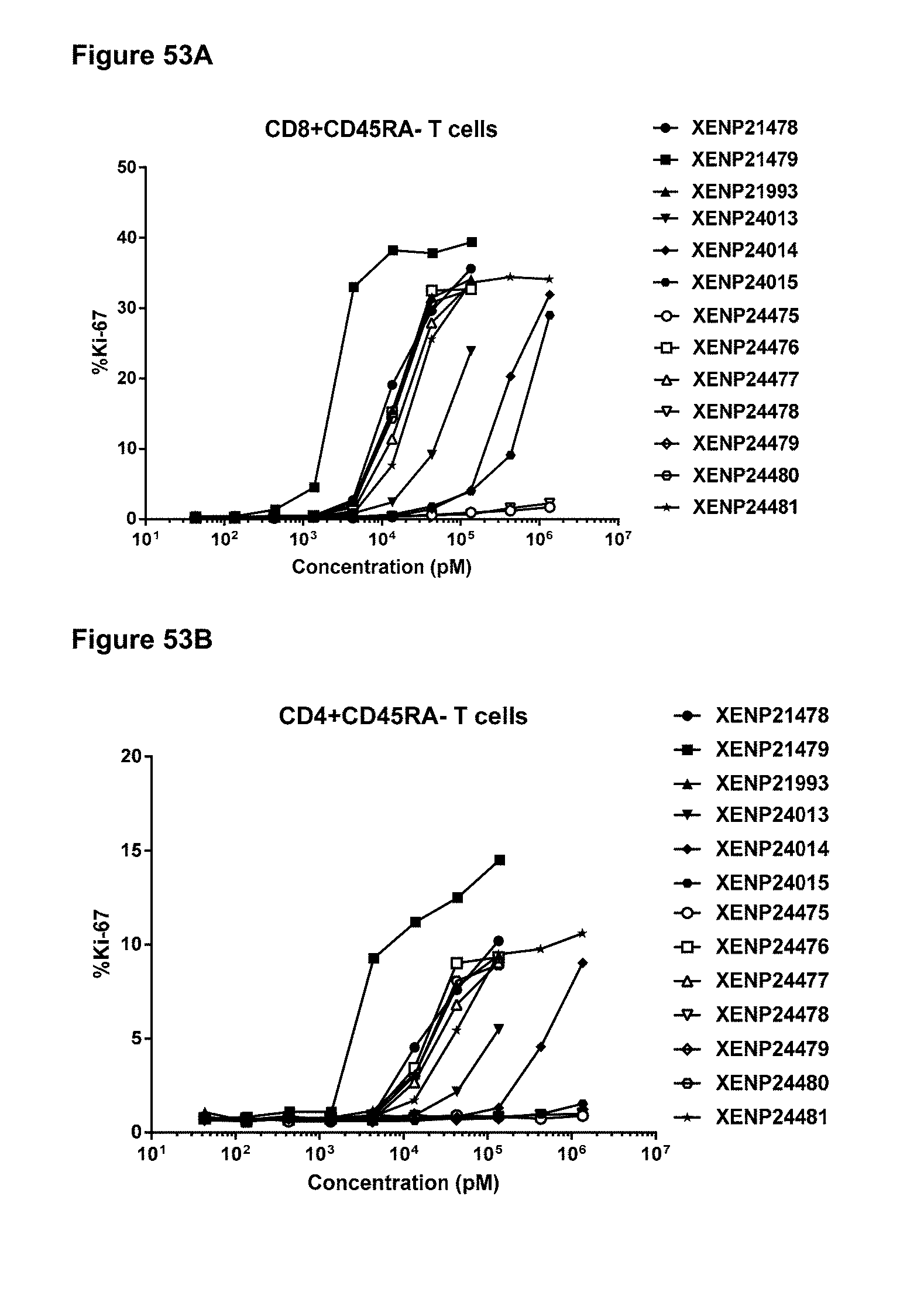
D00085
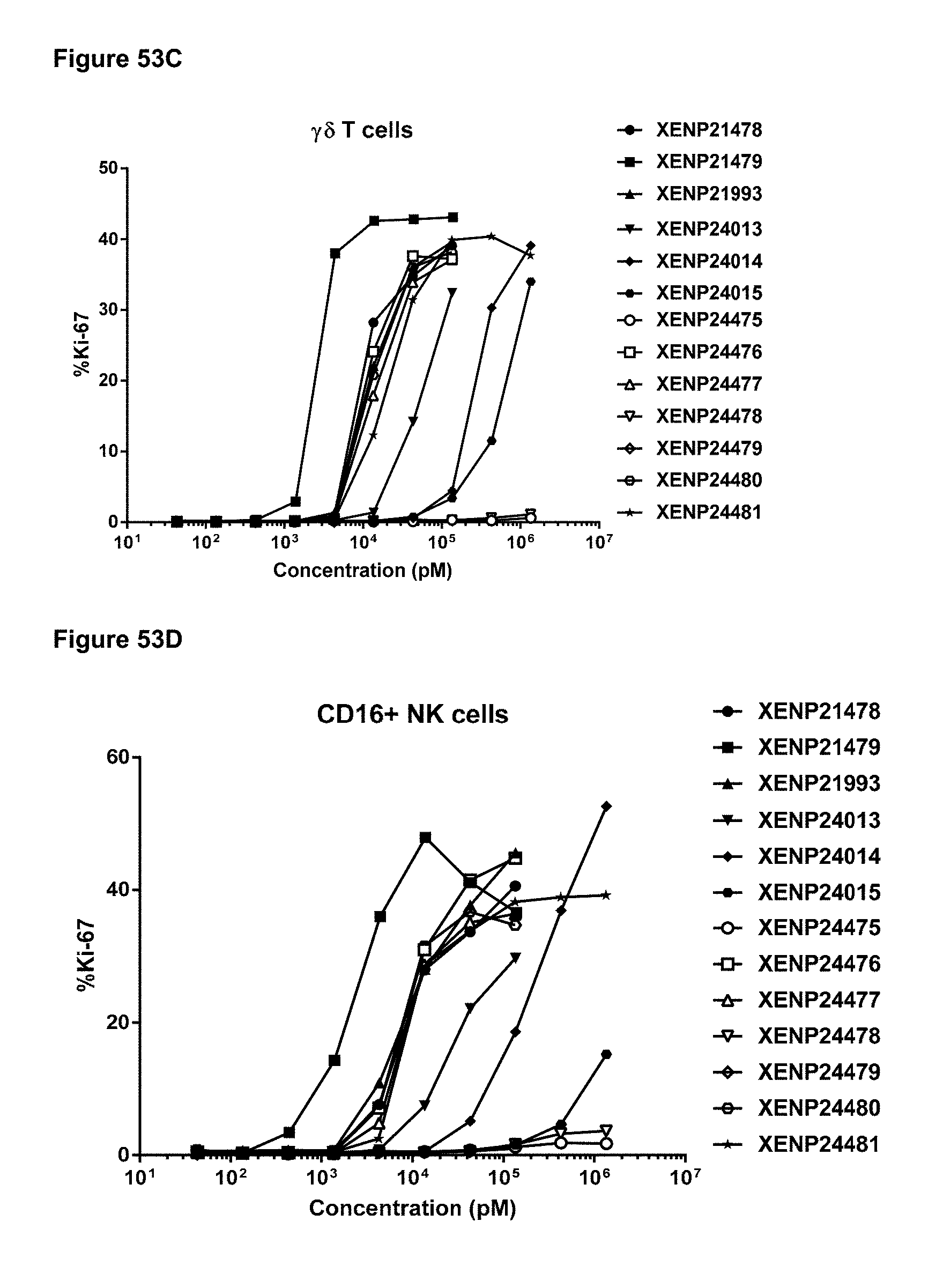
D00086
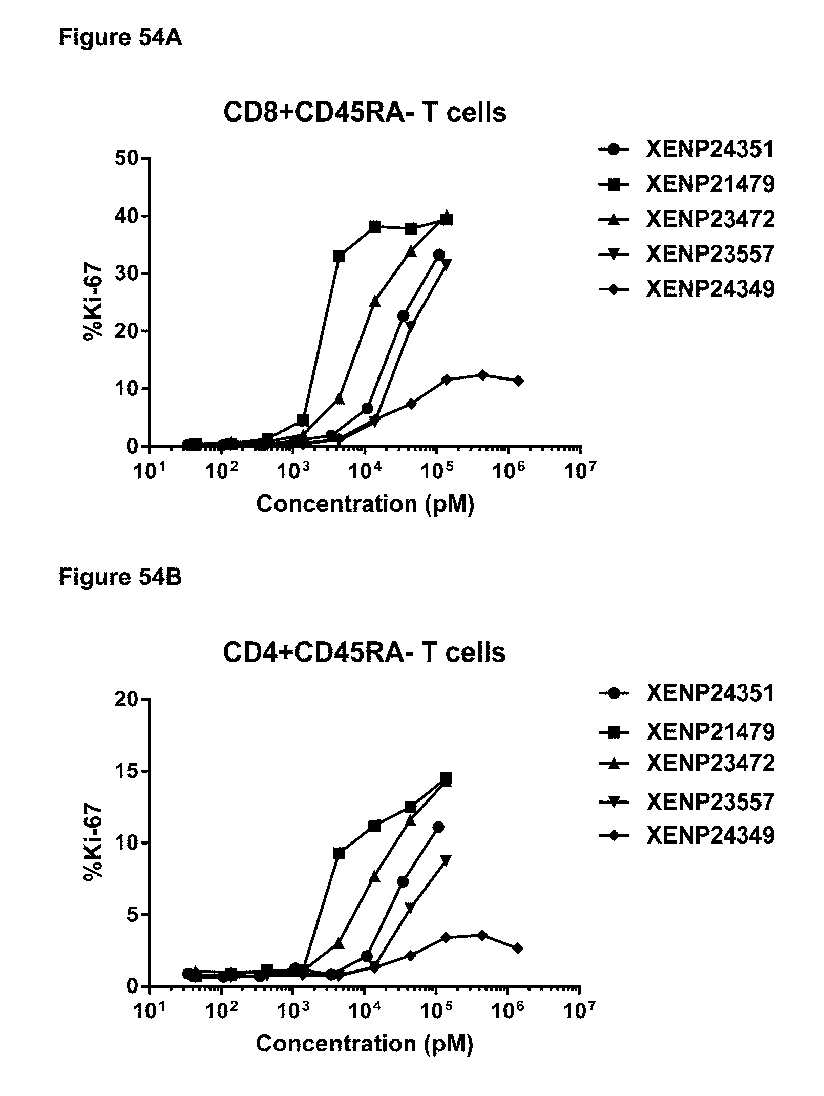
D00087
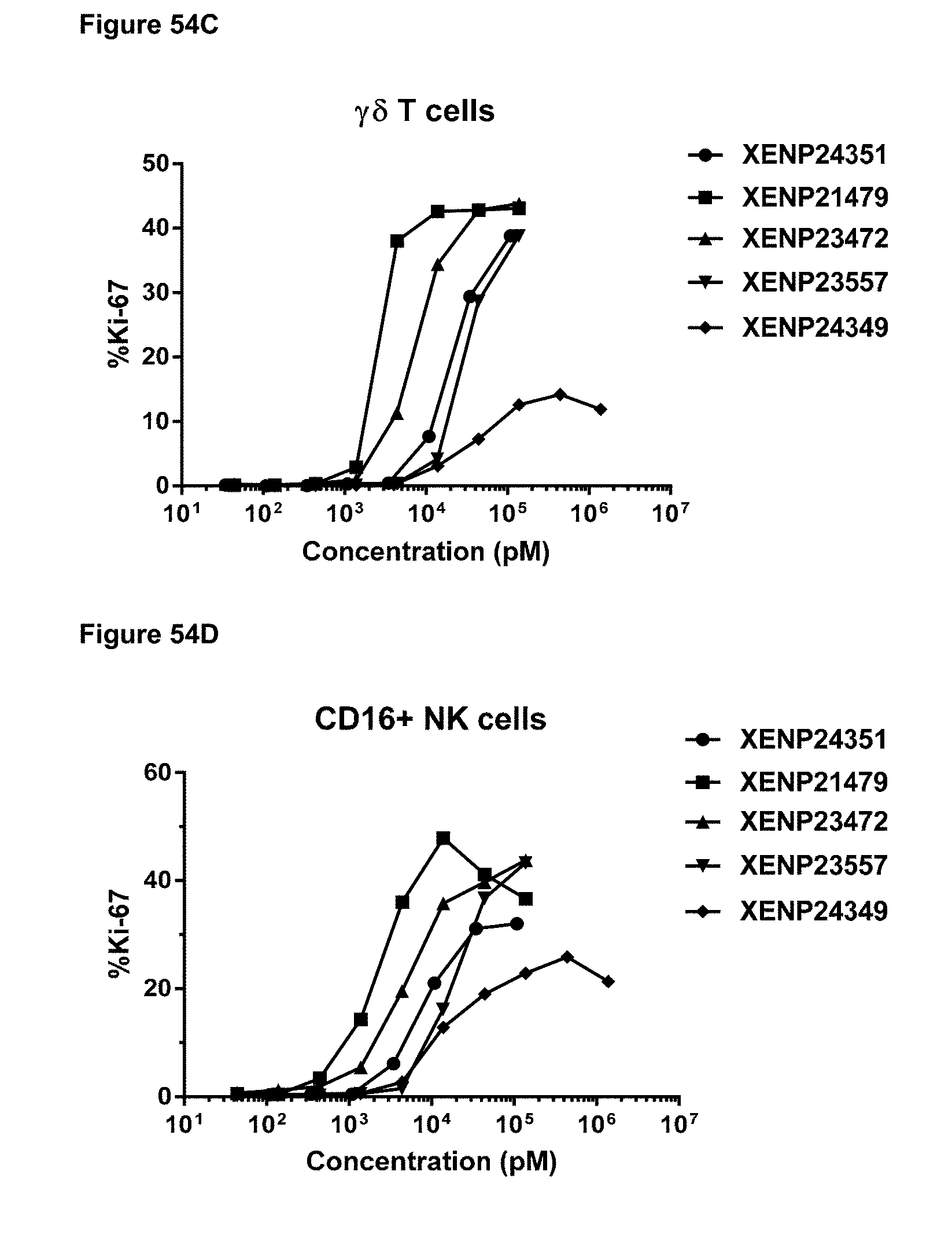
D00088
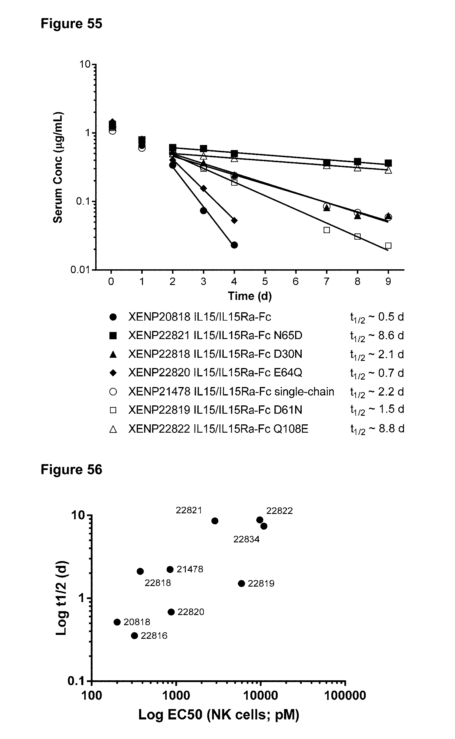
D00089

D00090

D00091
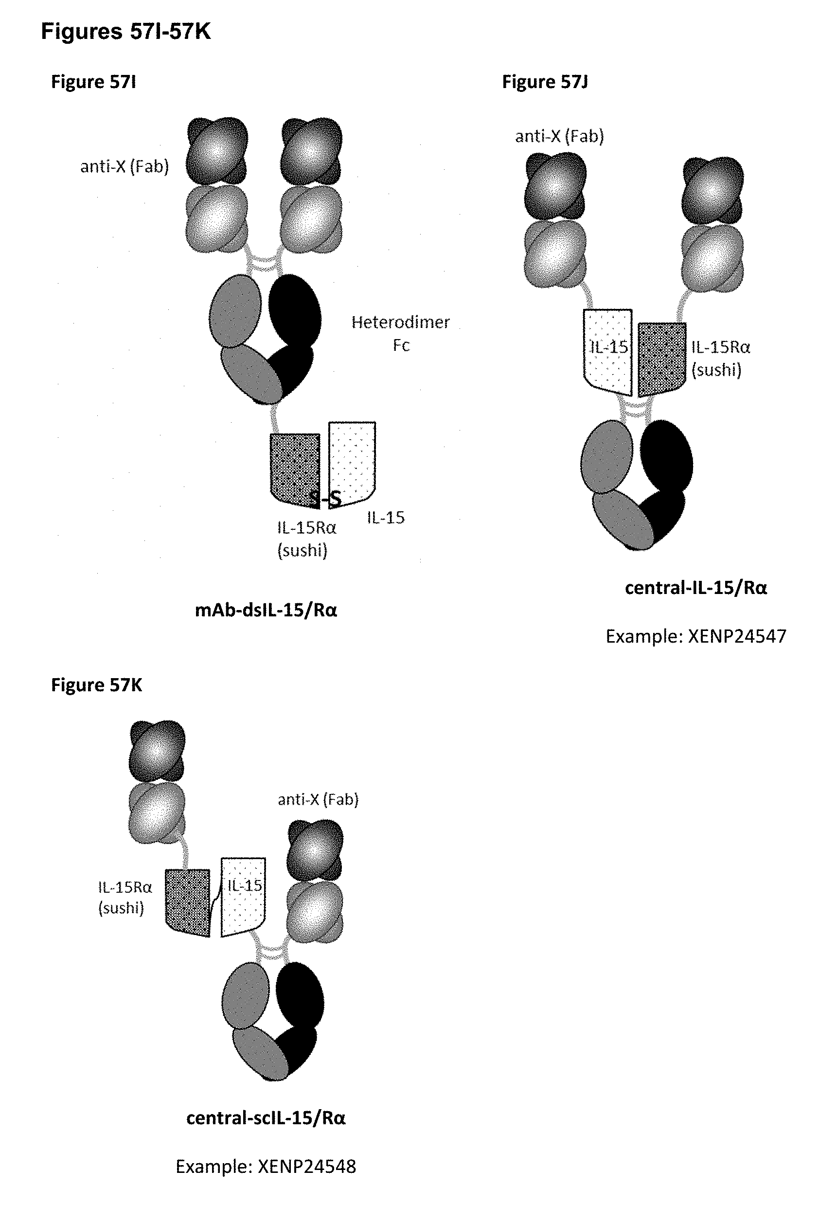
D00092
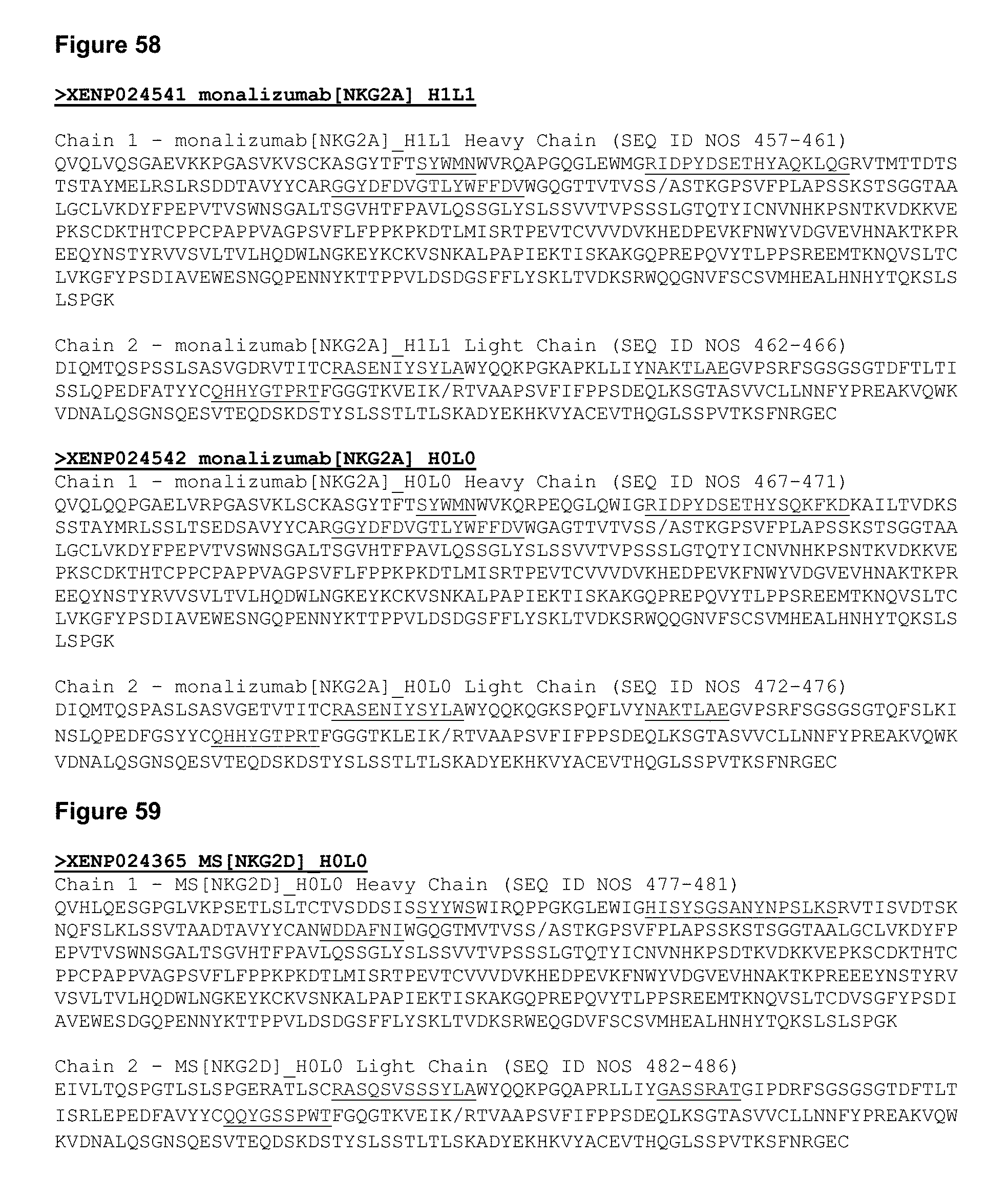
D00093
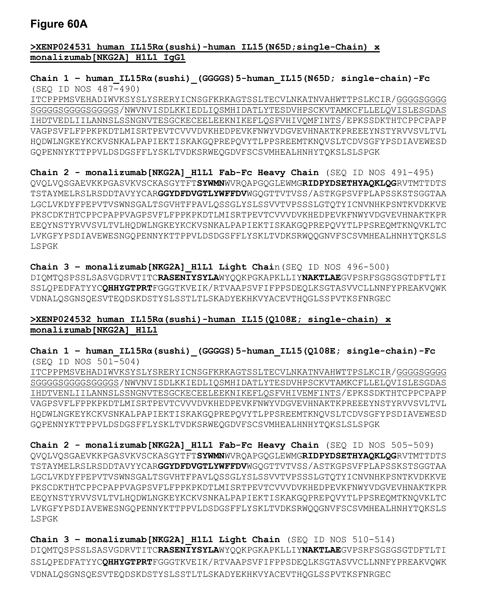
D00094

D00095
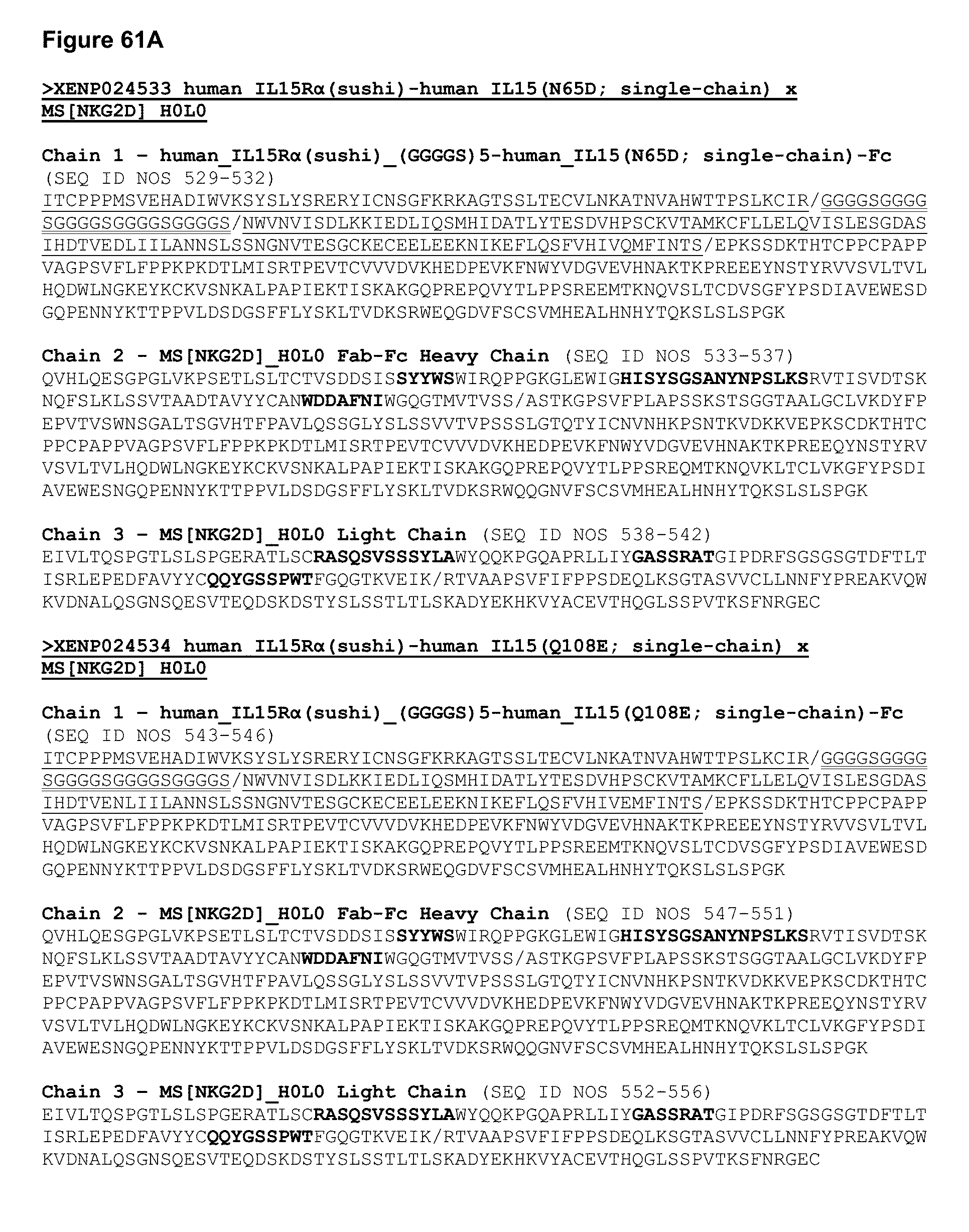
D00096

D00097
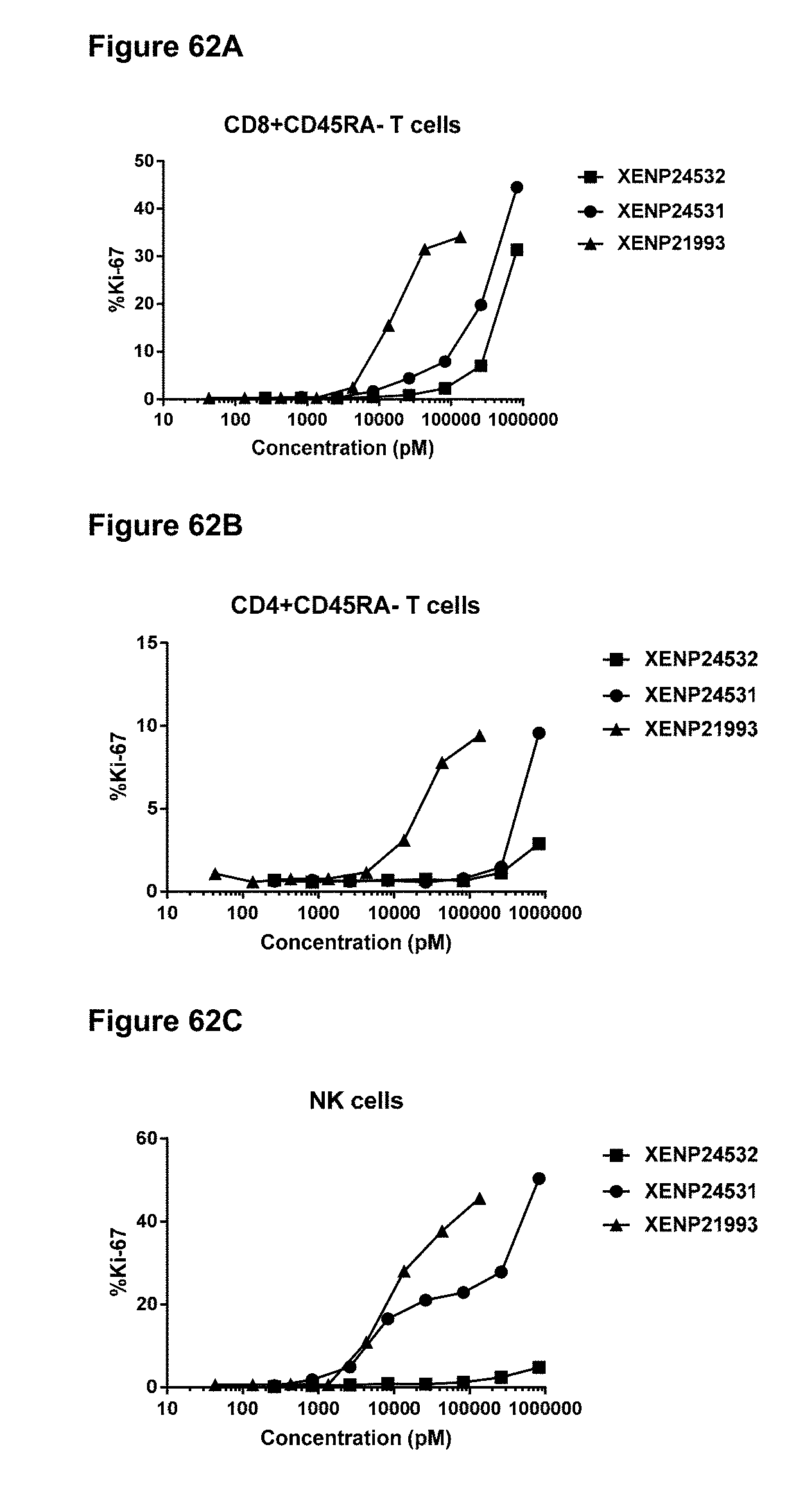
D00098

D00099

D00100

D00101
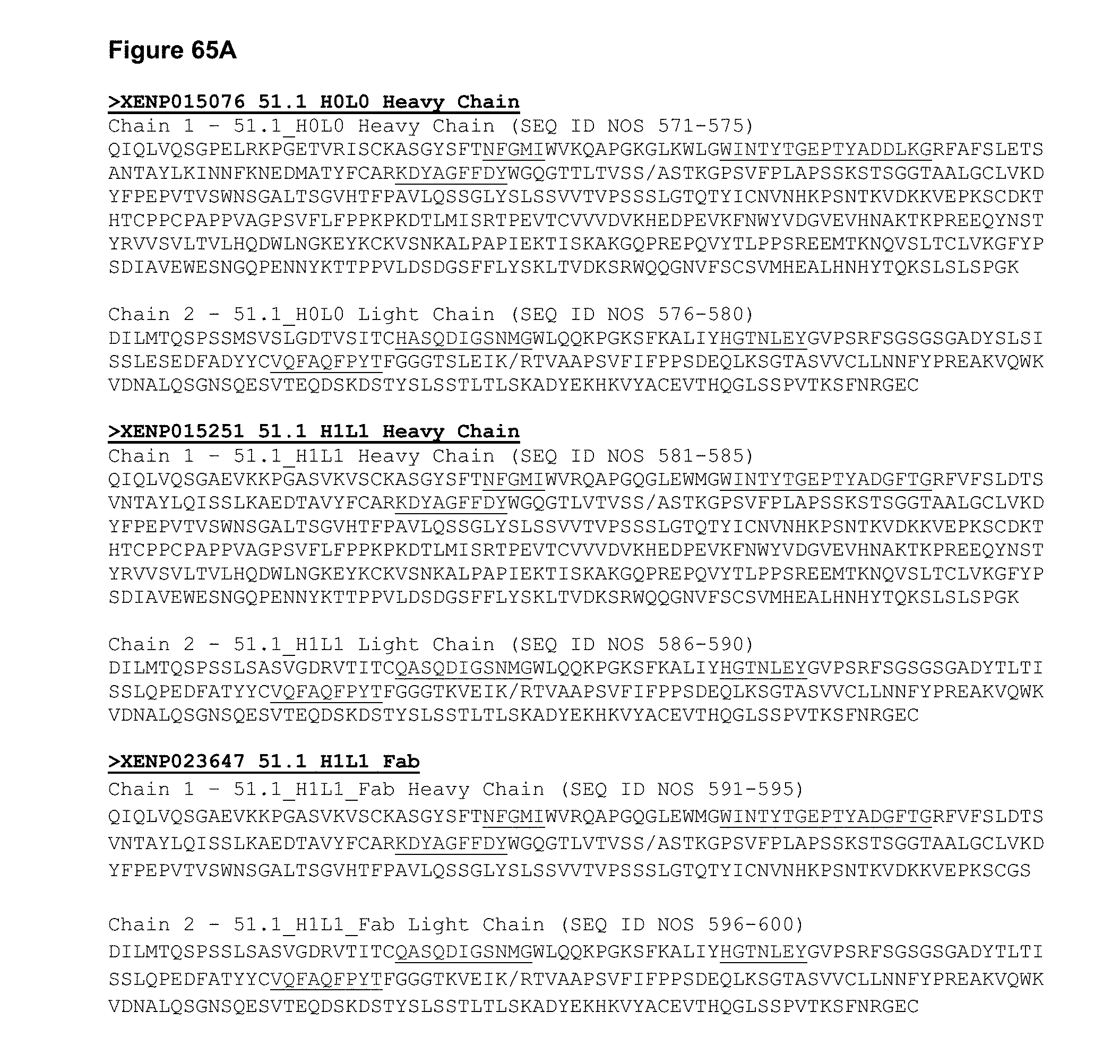
D00102
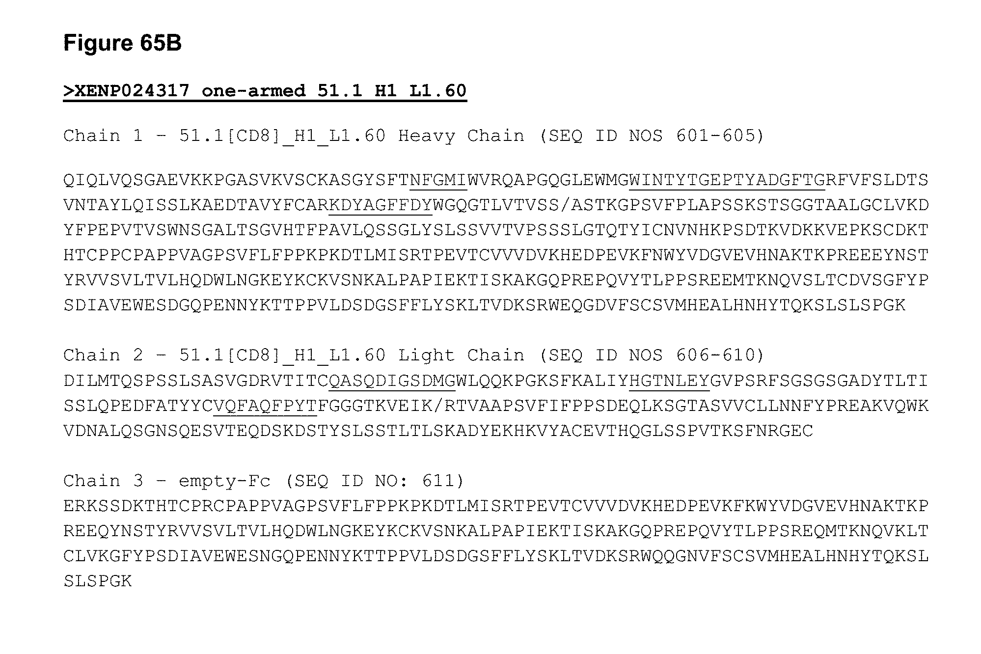
D00103
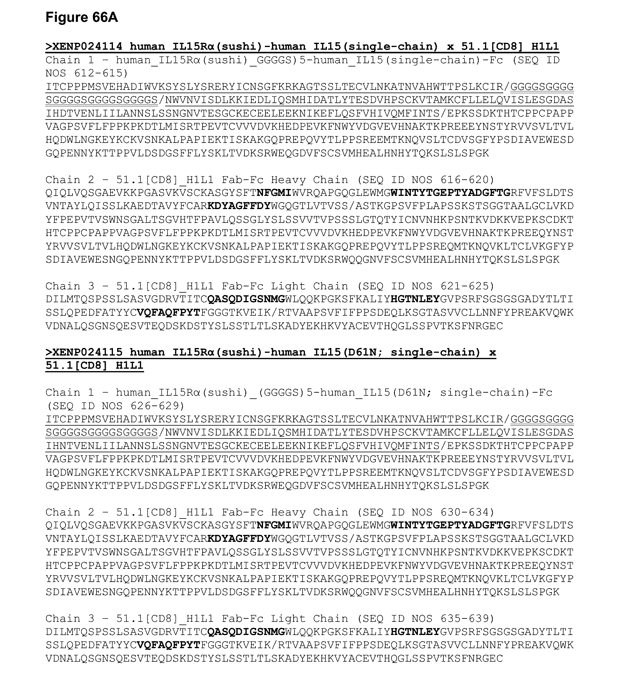
D00104
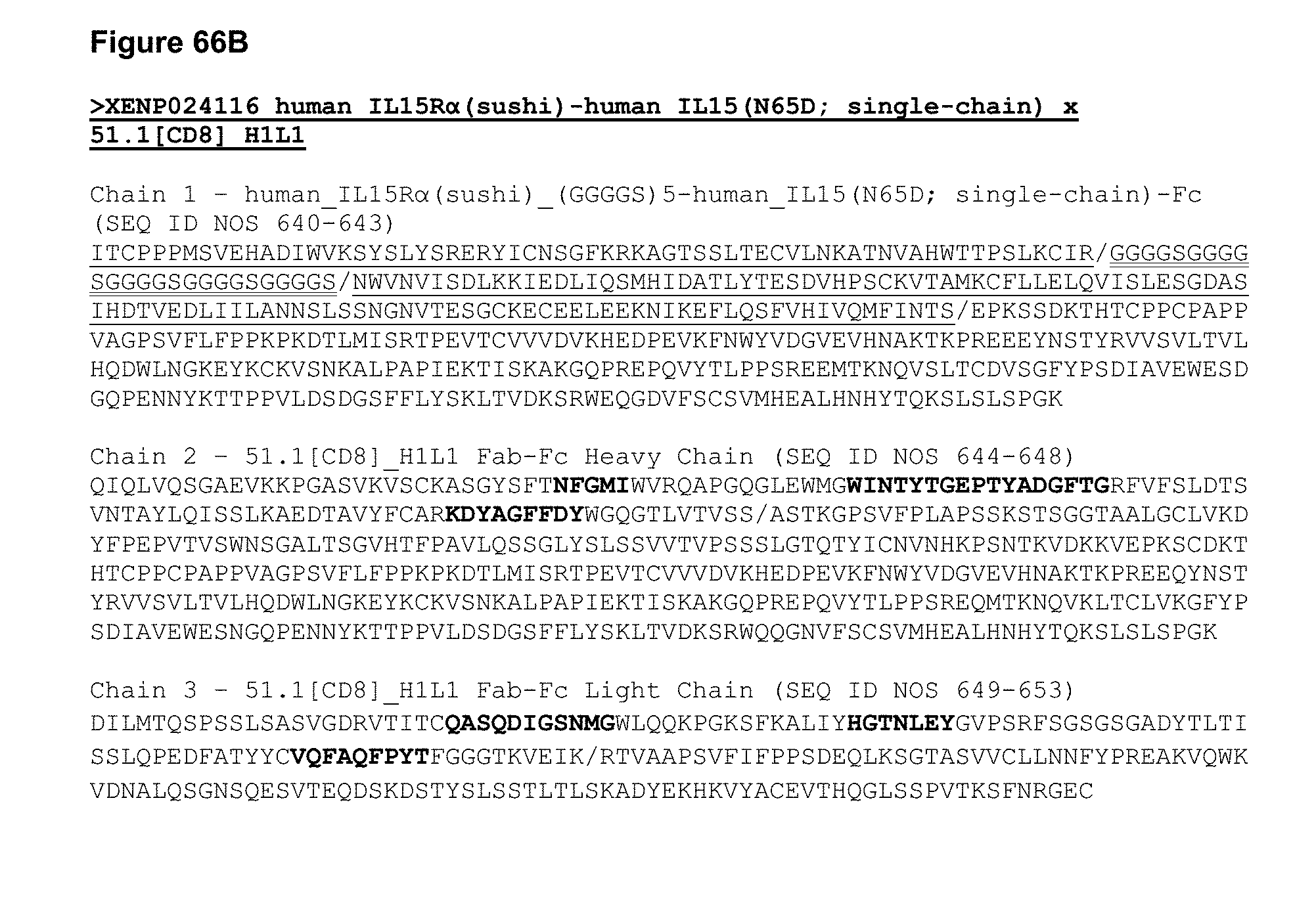
D00105

D00106

D00107
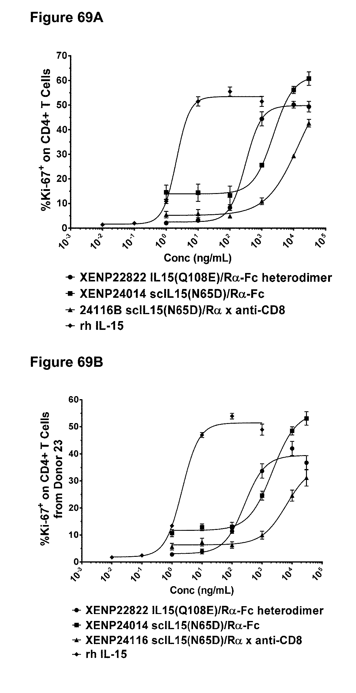
D00108

D00109

D00110

D00111

D00112

D00113
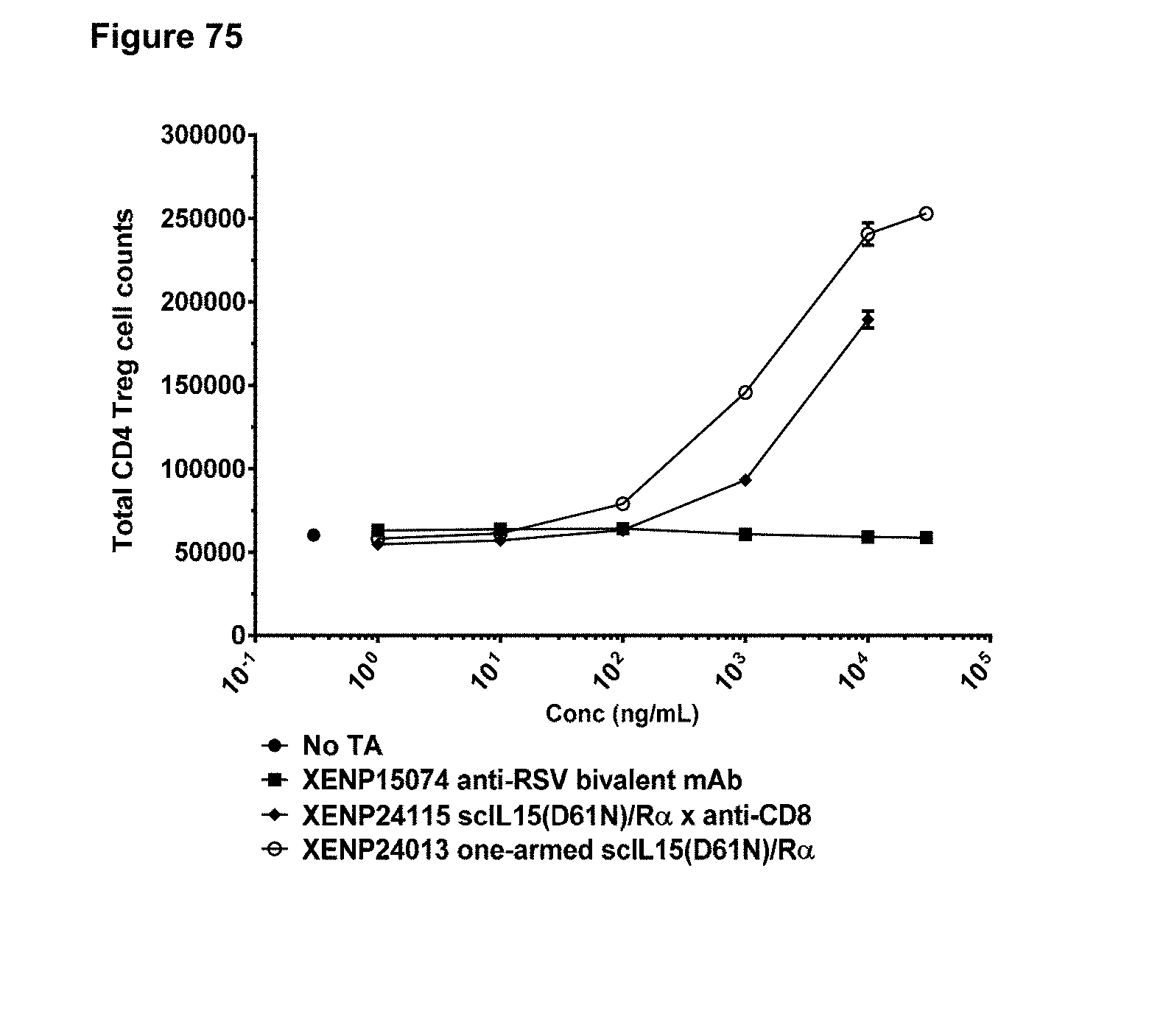
D00114

D00115

D00116

D00117
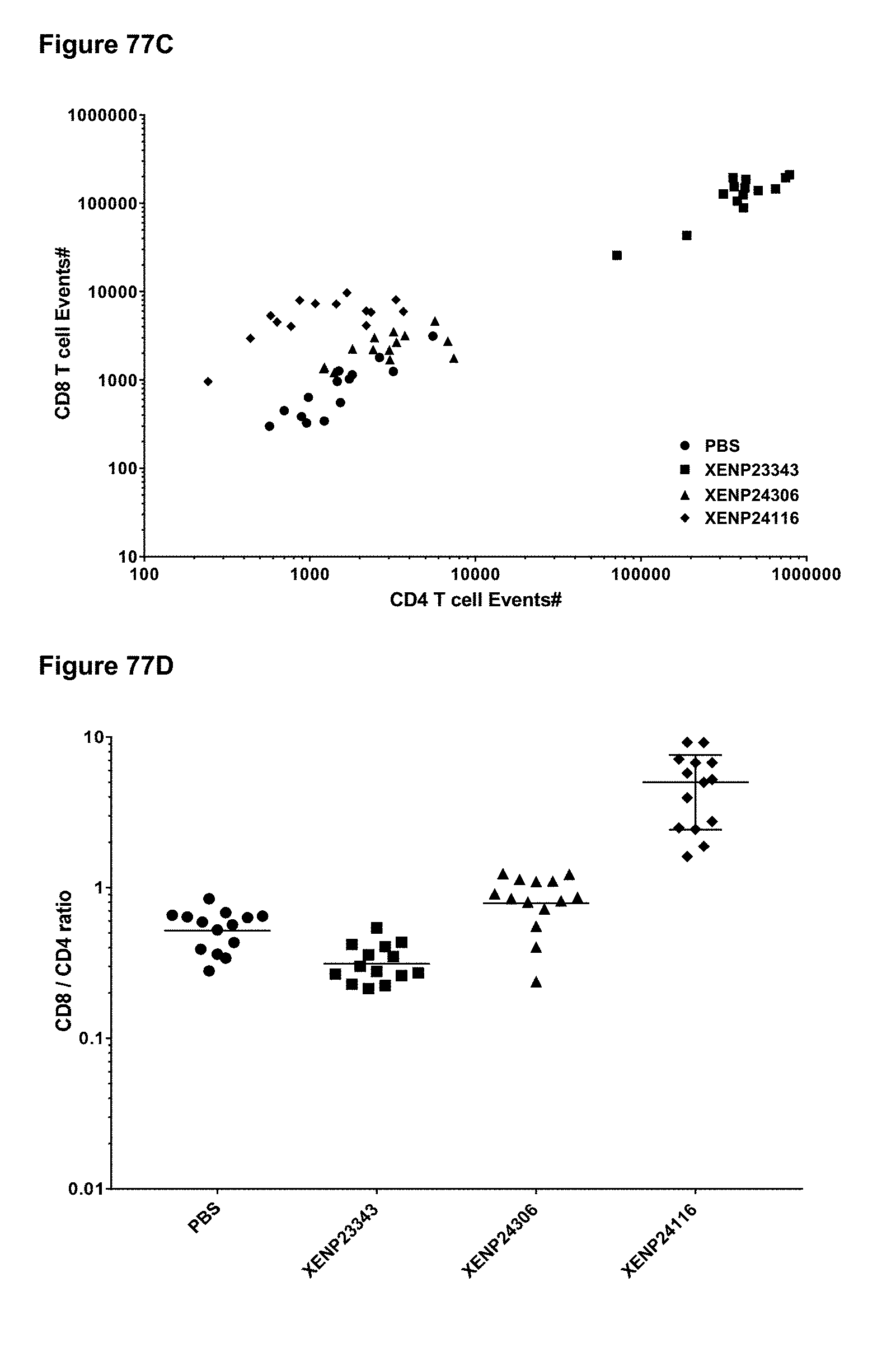
D00118

D00119
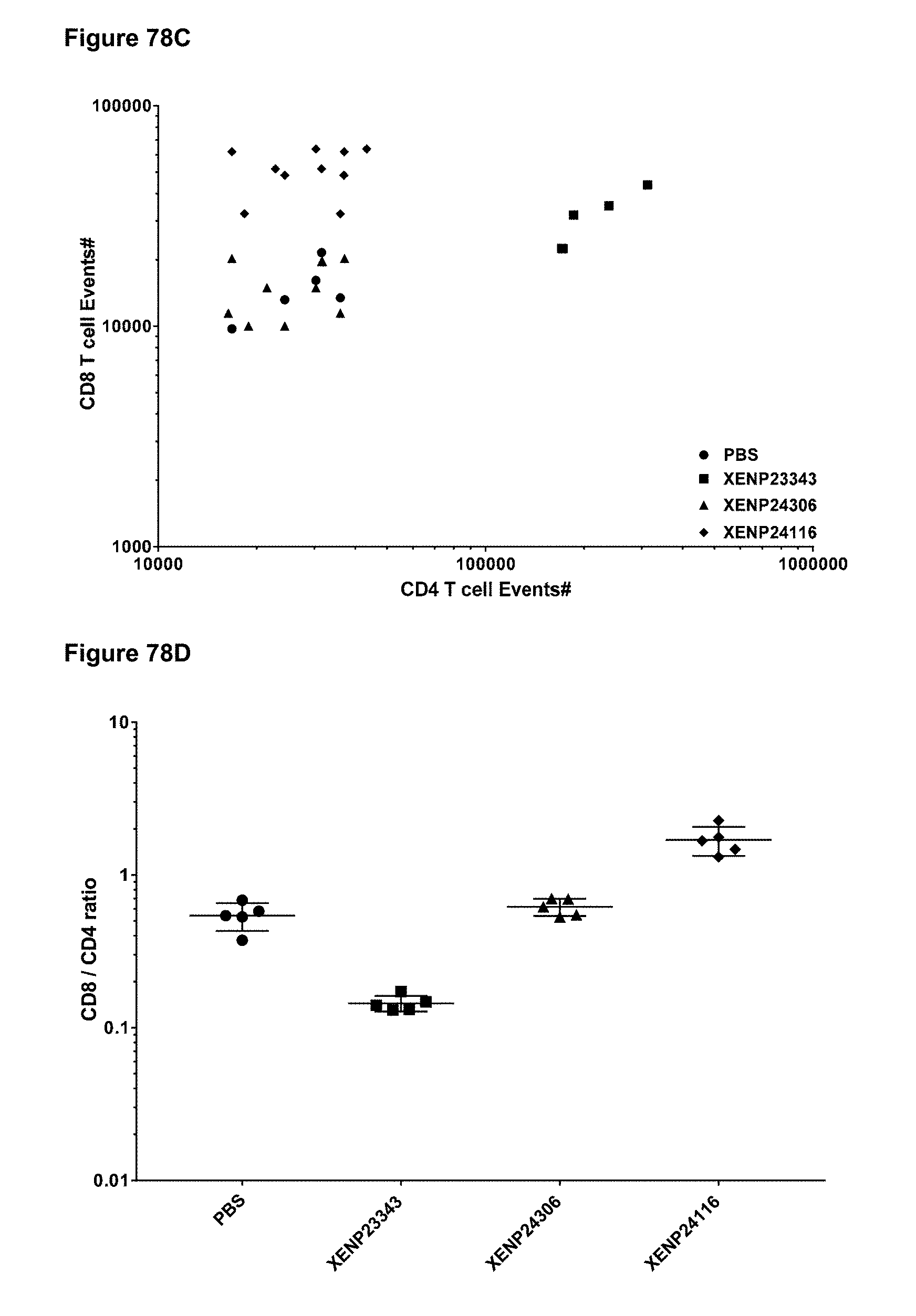
D00120
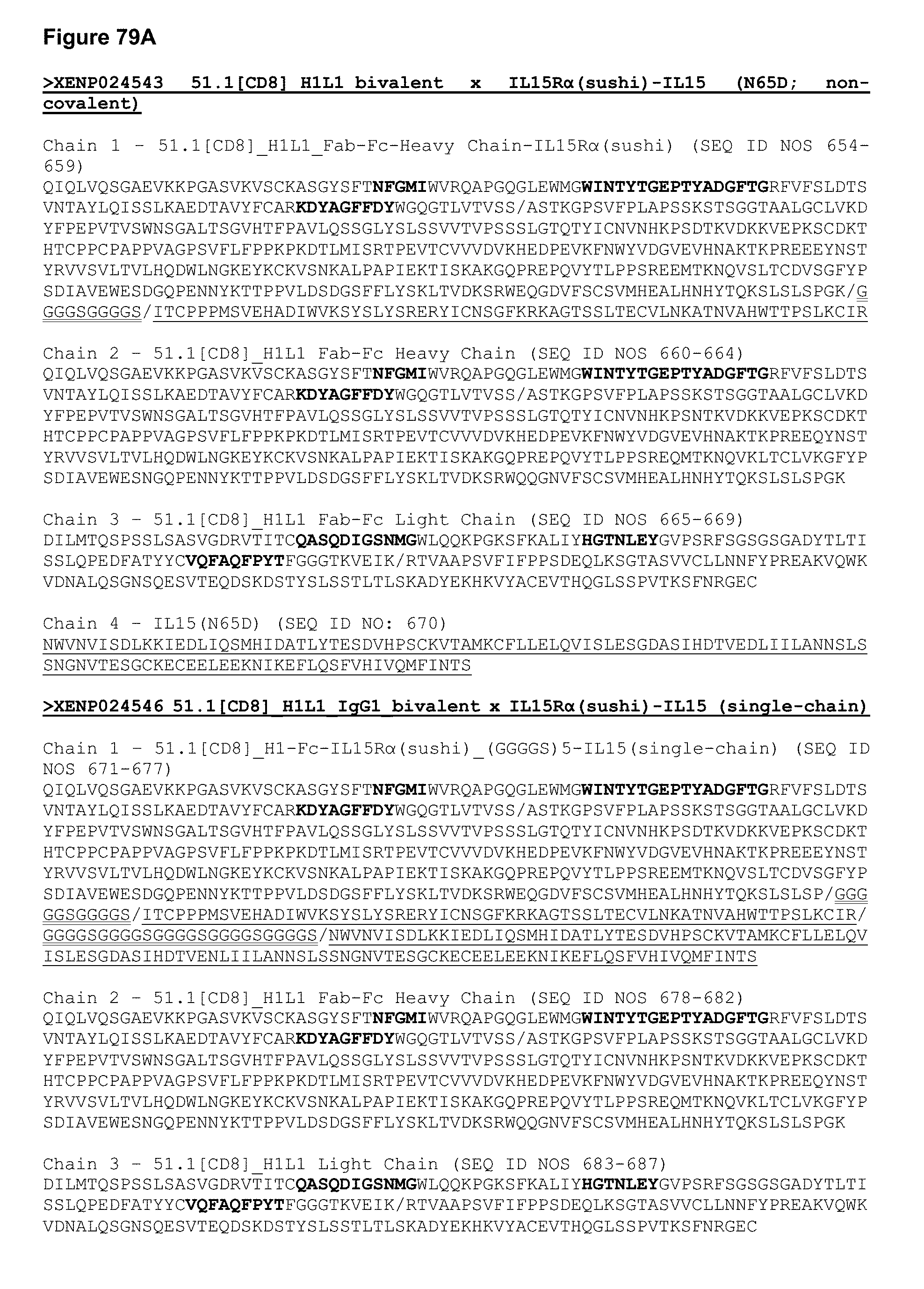
D00121
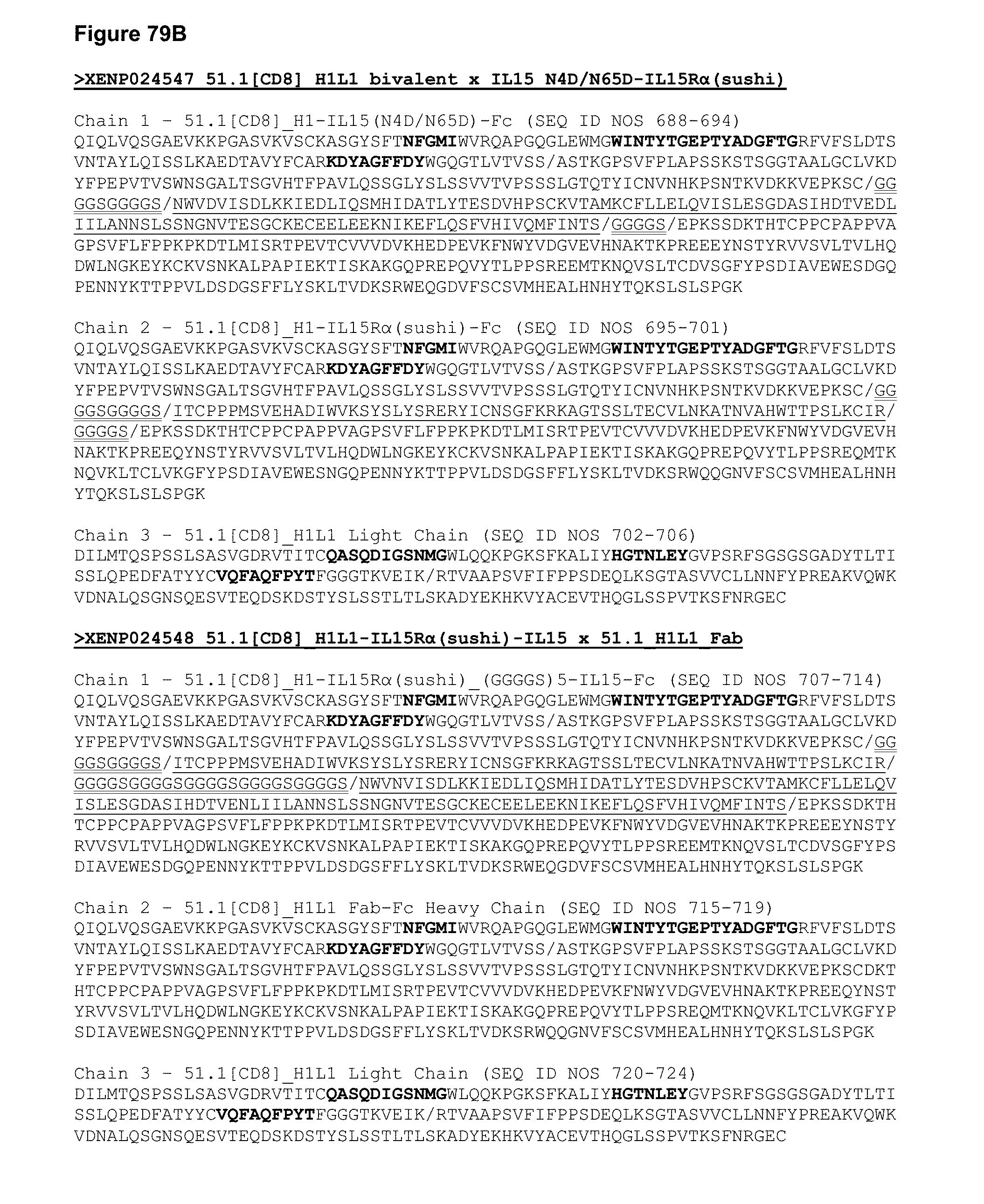
D00122
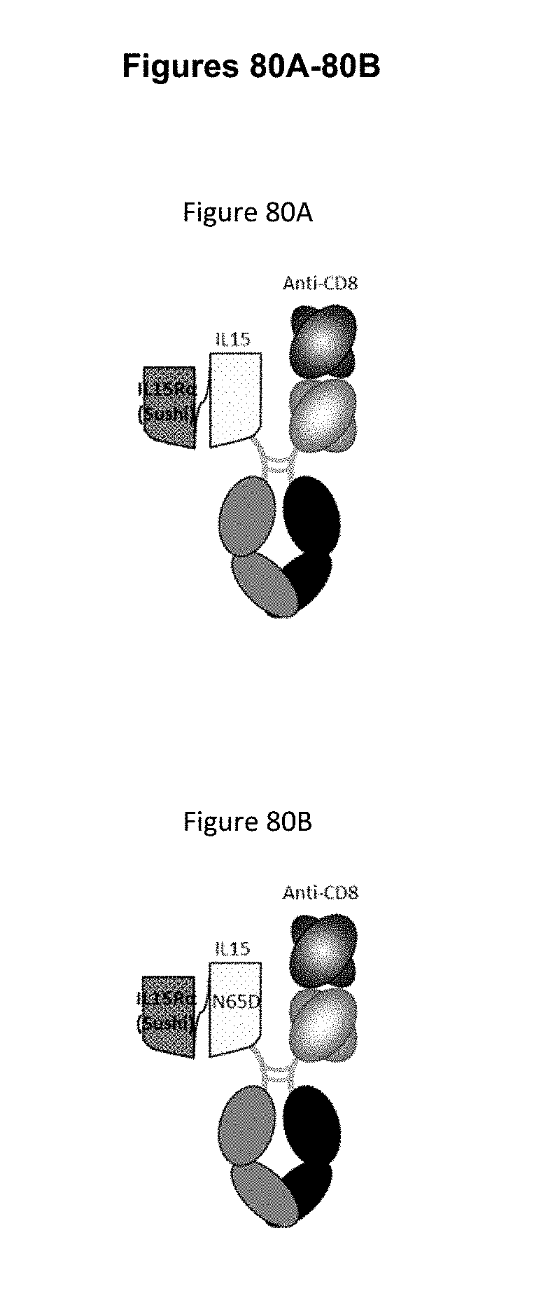
D00123
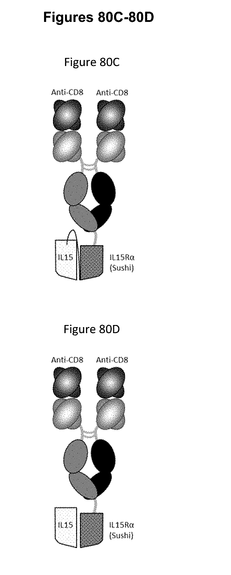
D00124

D00125

D00126
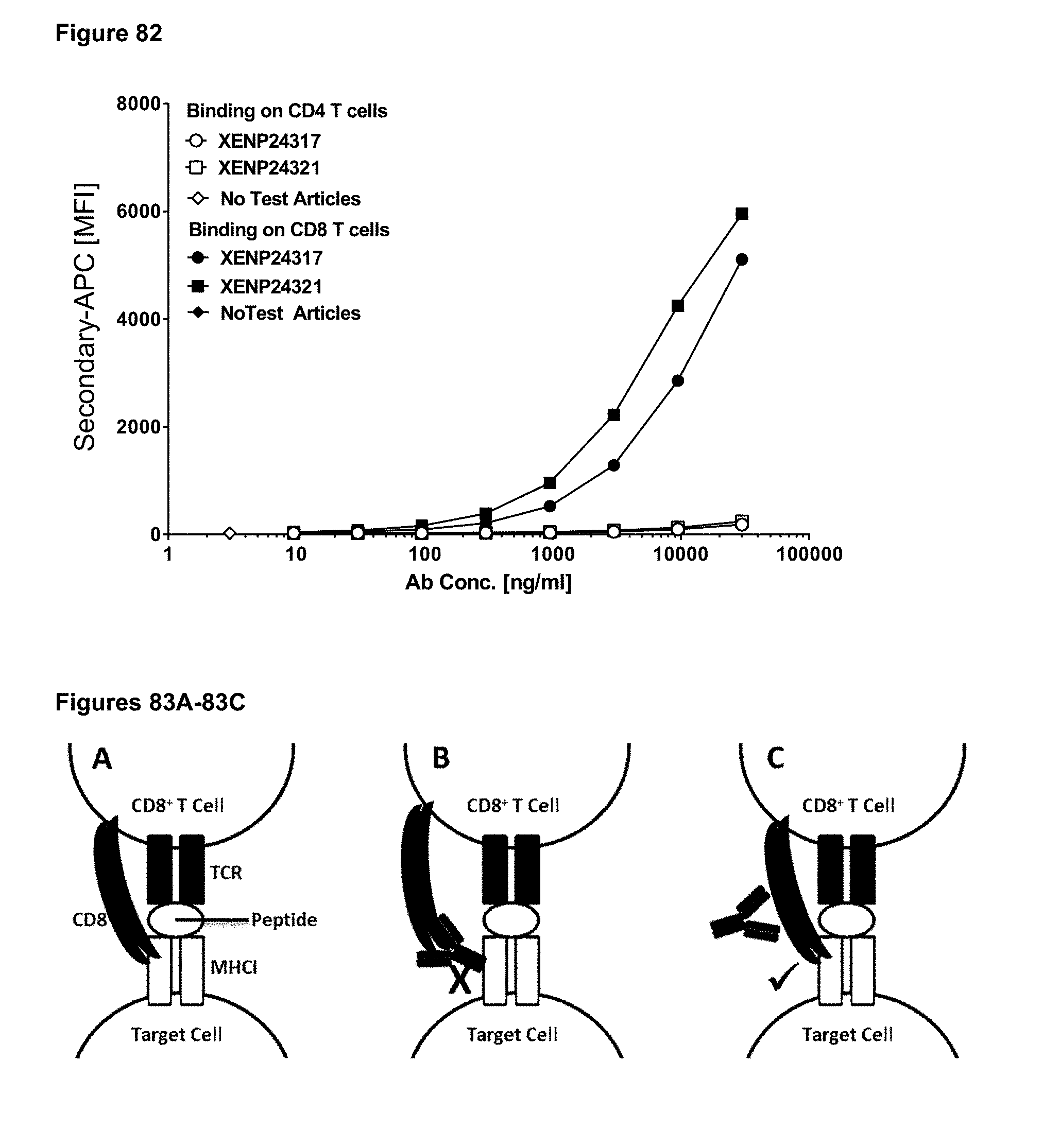
D00127
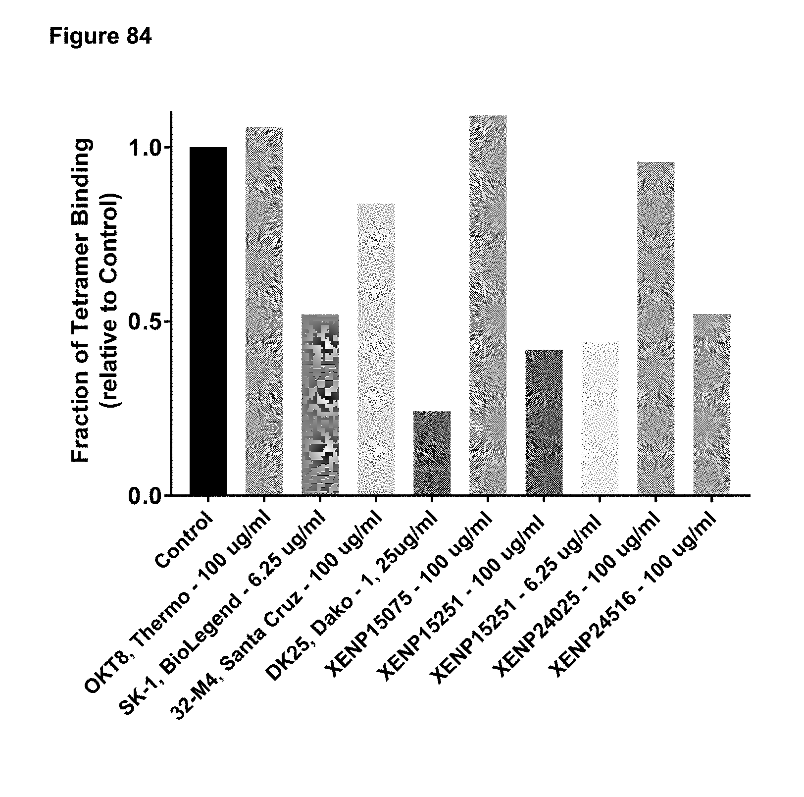
D00128

D00129

D00130
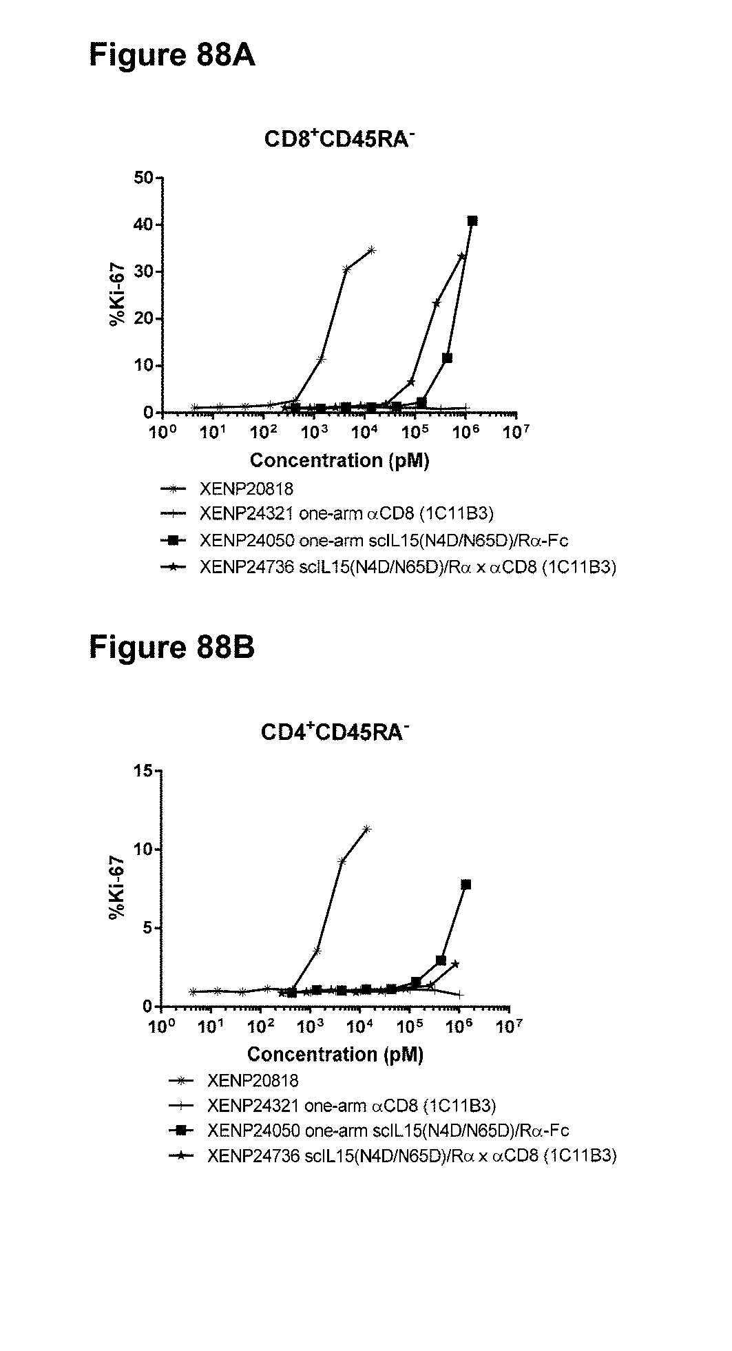
D00131

D00132
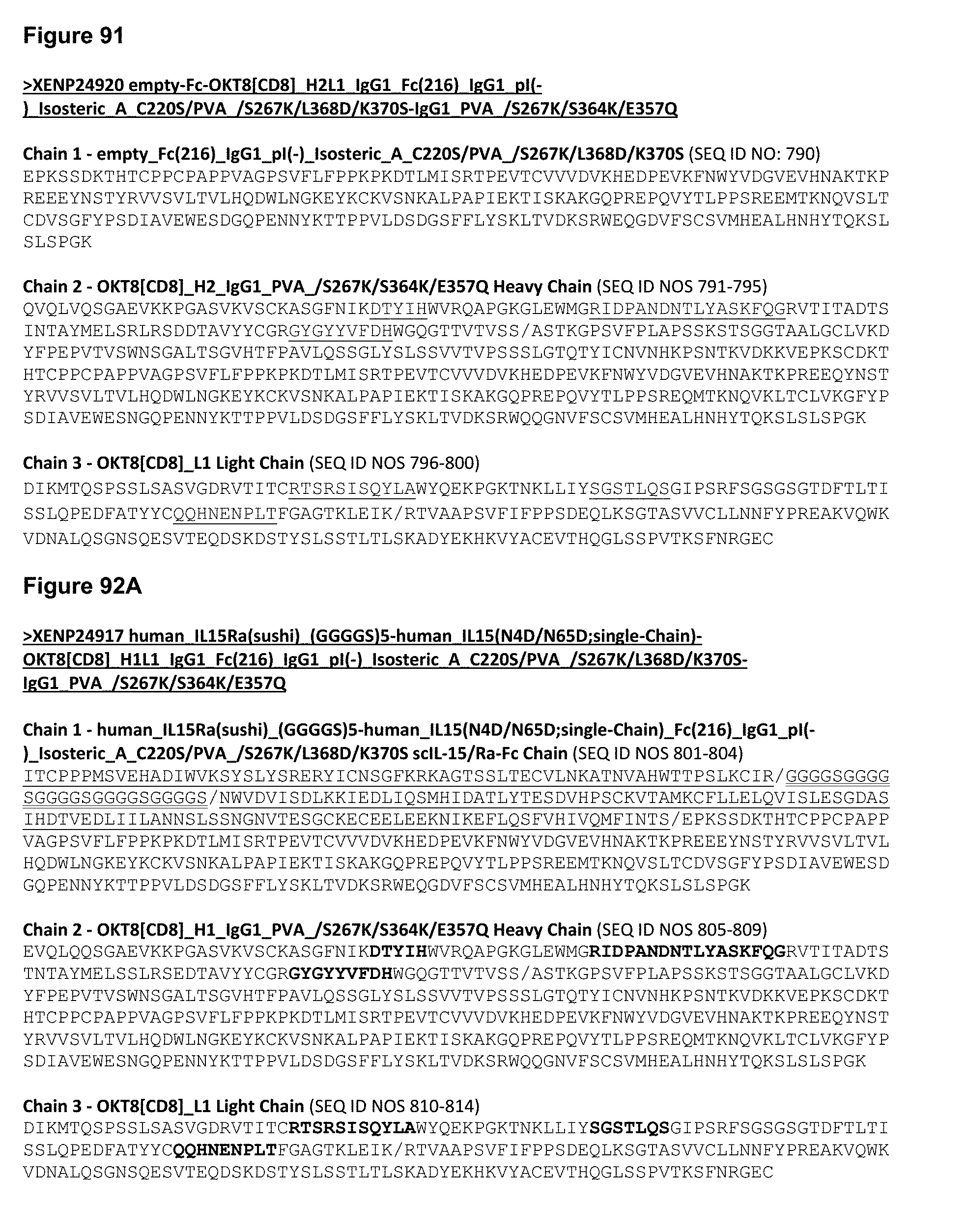
D00133
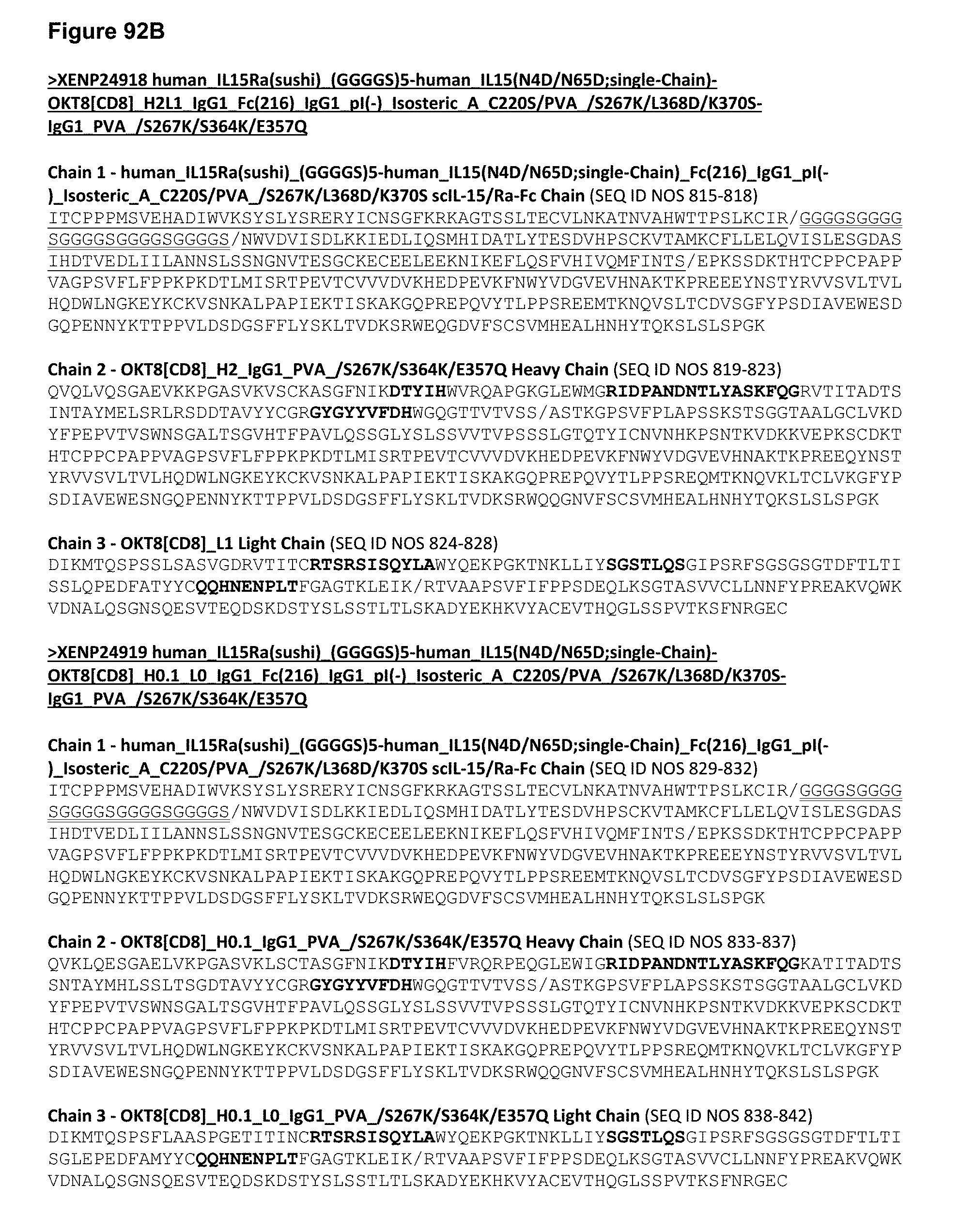
D00134

D00135

D00136
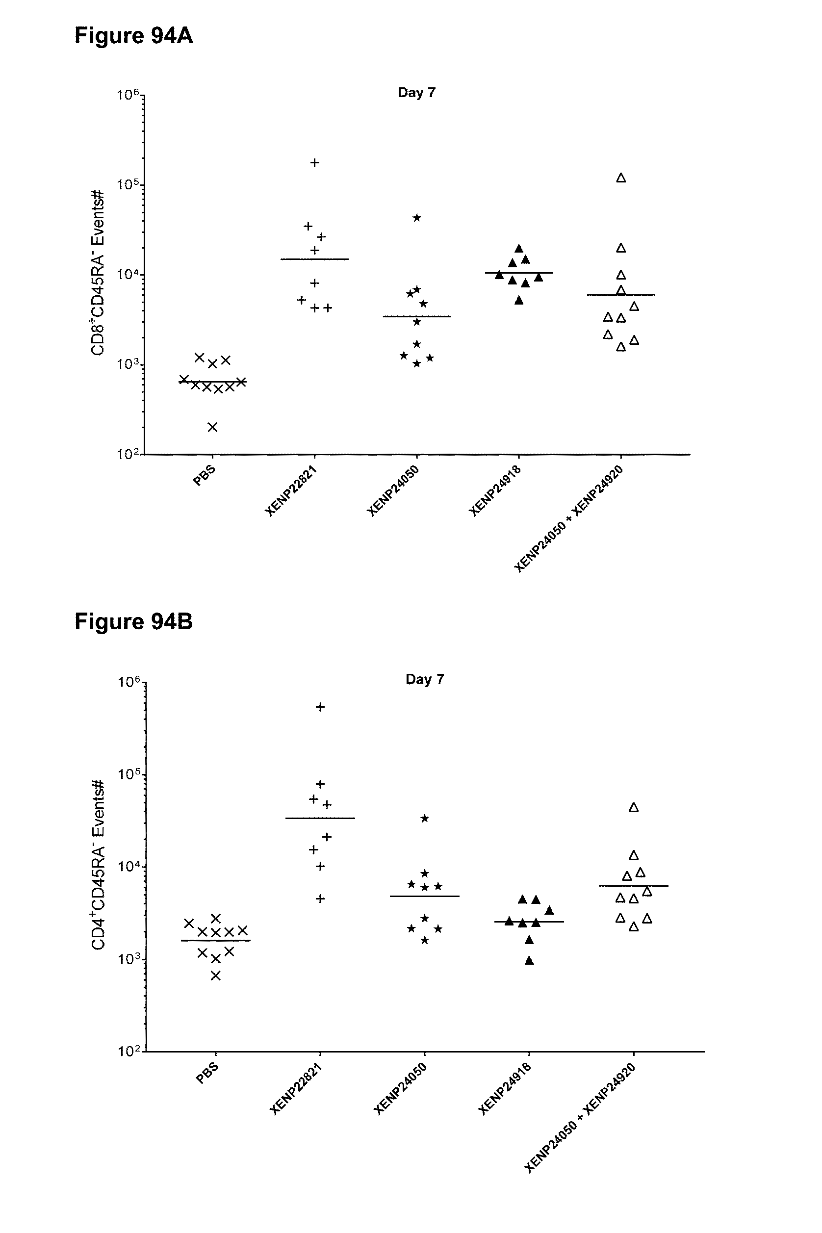
D00137

D00138

D00139

D00140

D00141

D00142

D00143
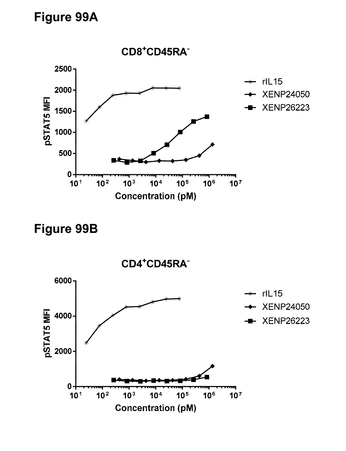
D00144

D00145

D00146

D00147

D00148

D00149
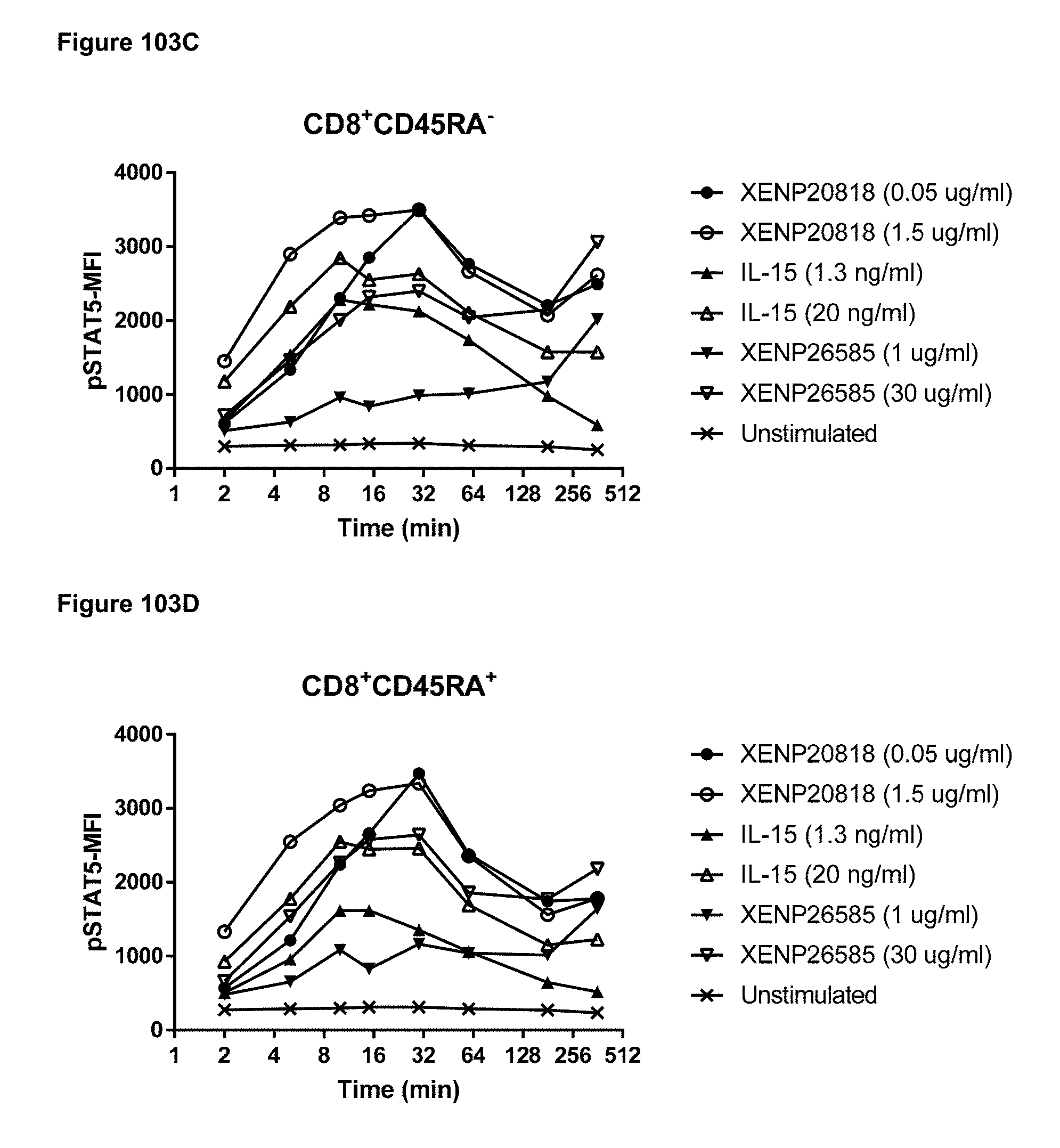
D00150

D00151

D00152
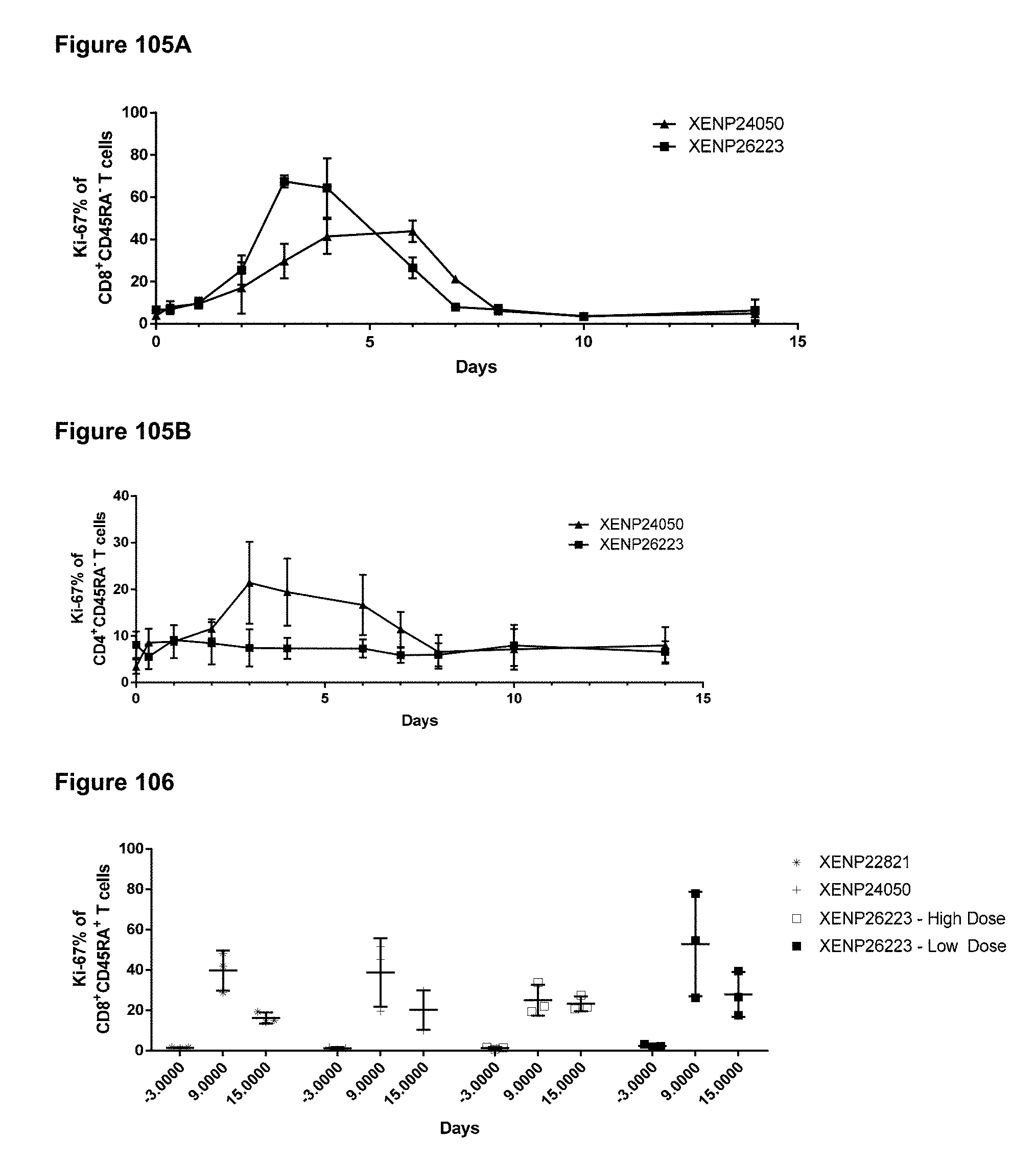
D00153
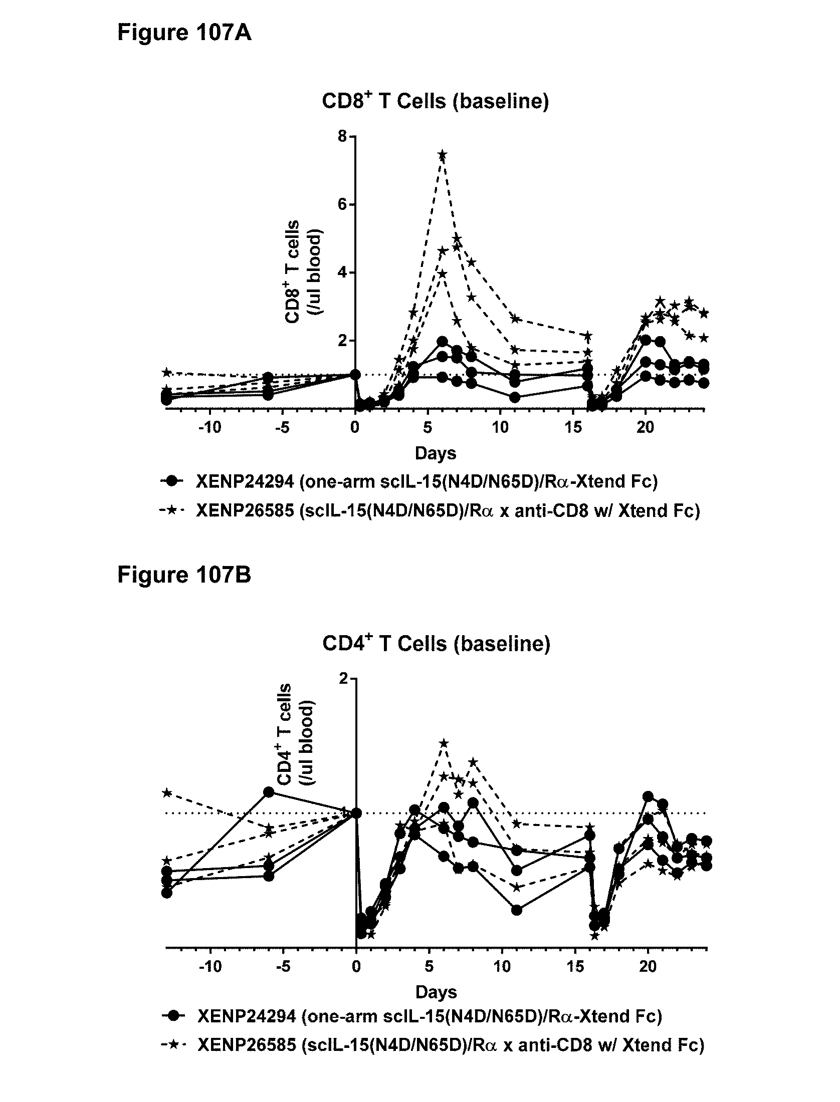
D00154
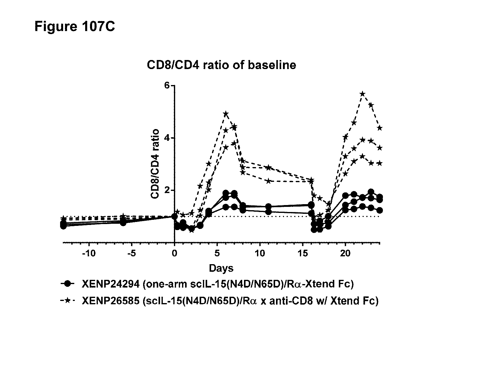
D00155
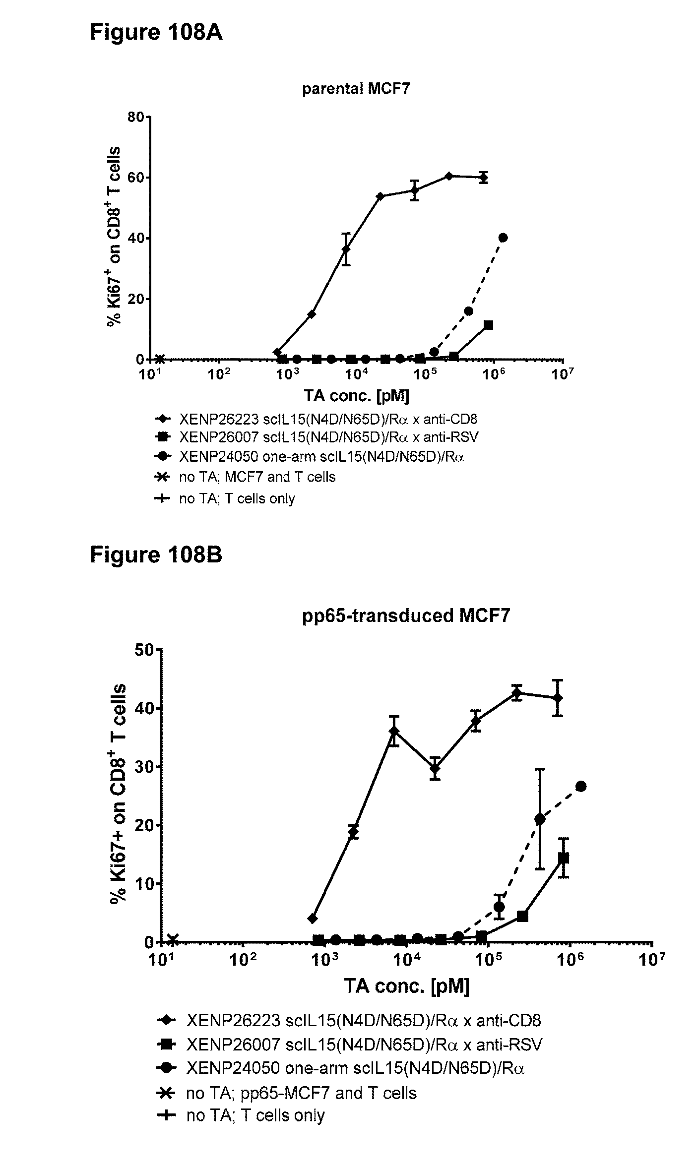
D00156
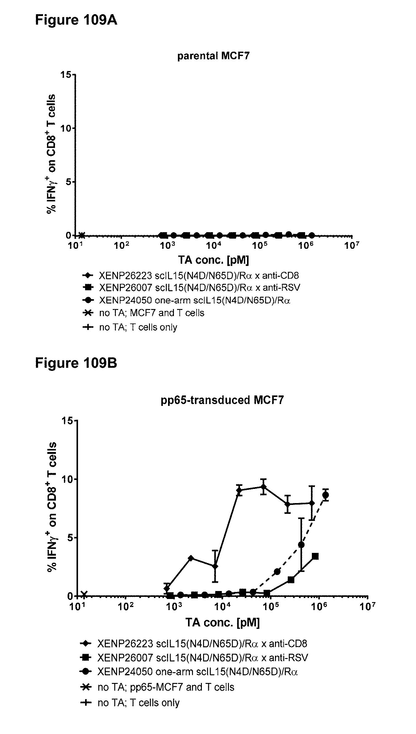
D00157
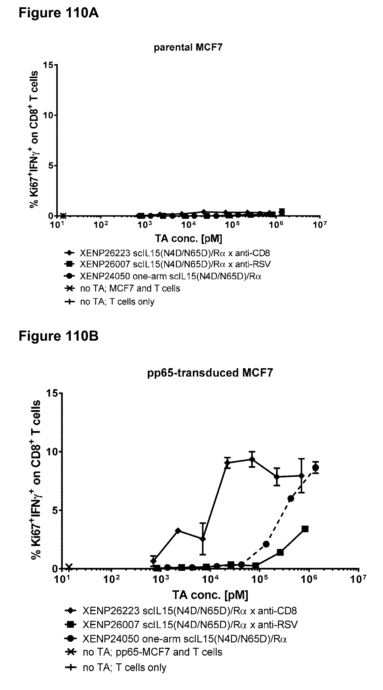
D00158
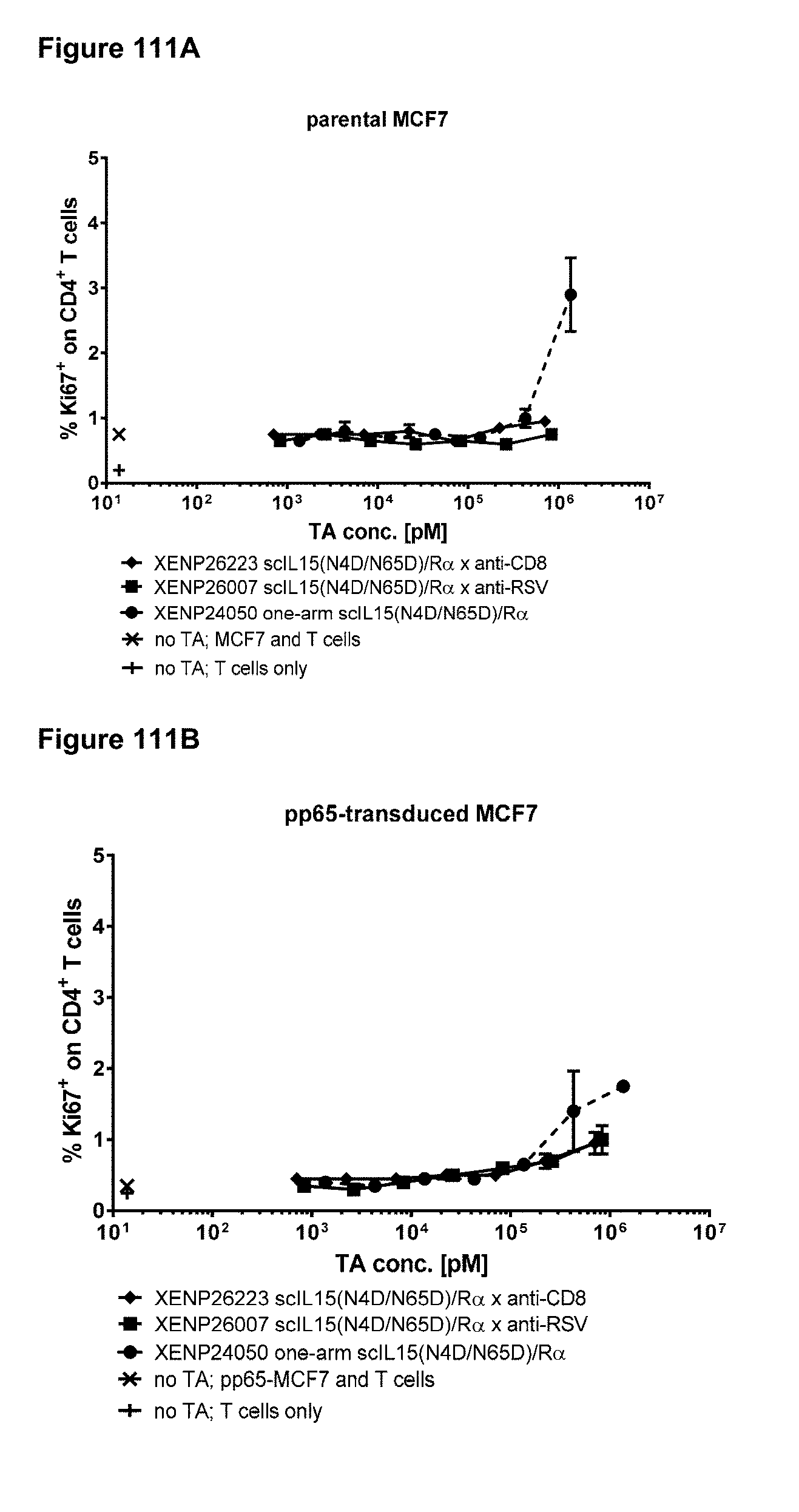
D00159

D00160
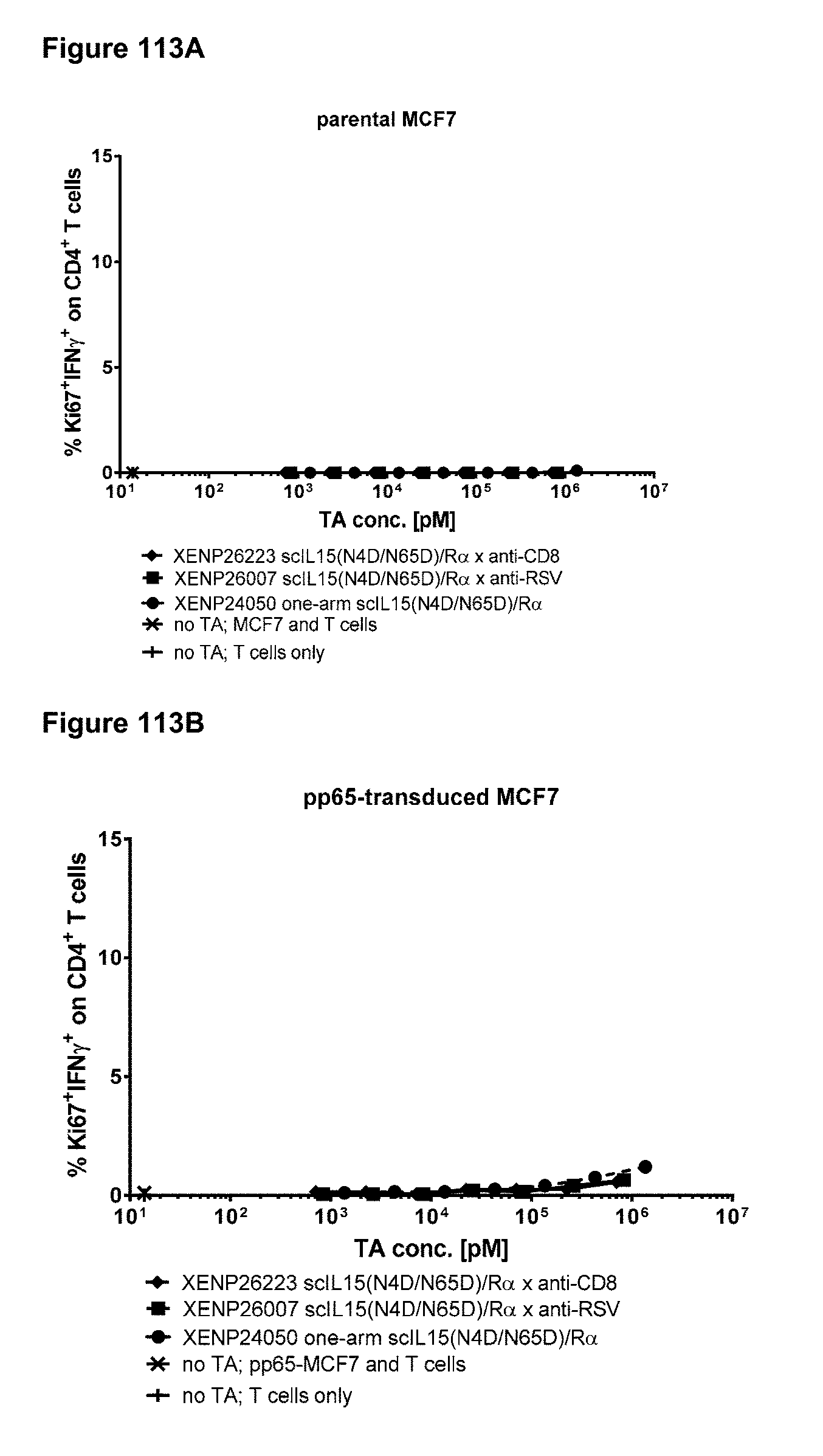
D00161

D00162

D00163

D00164
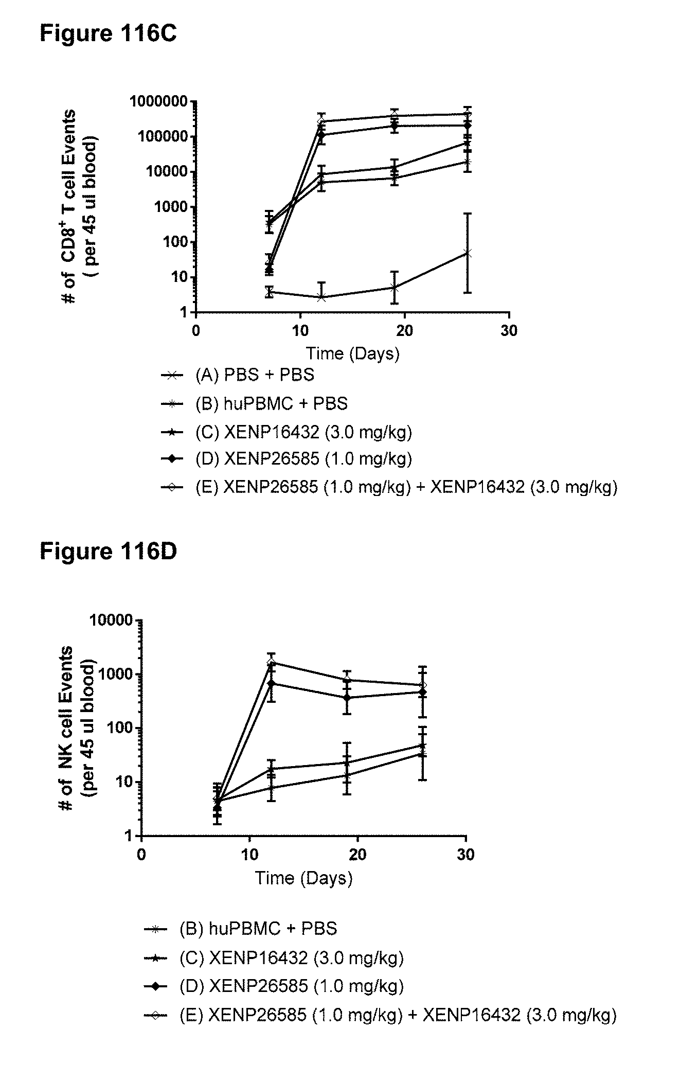
D00165
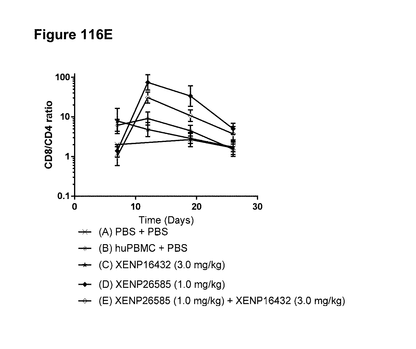
XML
uspto.report is an independent third-party trademark research tool that is not affiliated, endorsed, or sponsored by the United States Patent and Trademark Office (USPTO) or any other governmental organization. The information provided by uspto.report is based on publicly available data at the time of writing and is intended for informational purposes only.
While we strive to provide accurate and up-to-date information, we do not guarantee the accuracy, completeness, reliability, or suitability of the information displayed on this site. The use of this site is at your own risk. Any reliance you place on such information is therefore strictly at your own risk.
All official trademark data, including owner information, should be verified by visiting the official USPTO website at www.uspto.gov. This site is not intended to replace professional legal advice and should not be used as a substitute for consulting with a legal professional who is knowledgeable about trademark law.