Method For Ex Vivo Treating Blood Or Plasma
TSAI; Pei-Yi ; et al.
U.S. patent application number 16/137925 was filed with the patent office on 2019-01-10 for method for ex vivo treating blood or plasma. This patent application is currently assigned to INDUSTRIAL TECHNOLOGY RESEARCH INSTITUTE. The applicant listed for this patent is INDUSTRIAL TECHNOLOGY RESEARCH INSTITUTE. Invention is credited to Chih-Hung CHEN, Chu-Huang CHEN, Chih-Chieh HUANG, Liang-Yin KE, Yi-Hung LIN, Hsin-Hsin SHEN, Pei-Yi TSAI.
| Application Number | 20190010483 16/137925 |
| Document ID | / |
| Family ID | 57995320 |
| Filed Date | 2019-01-10 |


View All Diagrams
| United States Patent Application | 20190010483 |
| Kind Code | A1 |
| TSAI; Pei-Yi ; et al. | January 10, 2019 |
METHOD FOR EX VIVO TREATING BLOOD OR PLASMA
Abstract
A method for ex vivo treating blood or plasma is provided. The method includes (a) ex vivo contacting a blood or plasma with an enzyme composition to react the enzyme composition with the blood or plasma, wherein the enzyme composition is capable of eliminating electronegative low-density lipoprotein from the blood or plasma by the activity of the enzyme composition, and the enzyme composition is selected from a group consisting of: a first enzyme for eliminating a glycan residue of an electronegative low-density lipoprotein (LDL); a second enzyme for eliminating ceramide carried by a electronegative low-density lipoprotein (LDL); and a combination thereof; and (b) terminating contact between the blood or plasma and the enzyme composition to terminate the reaction of the enzyme composition with the blood or plasma.
| Inventors: | TSAI; Pei-Yi; (Hsinchu City, TW) ; CHEN; Chih-Hung; (Tainan City, TW) ; LIN; Yi-Hung; (Zhubei City, TW) ; HUANG; Chih-Chieh; (Zhunan Township, TW) ; SHEN; Hsin-Hsin; (Zhudong Township, TW) ; KE; Liang-Yin; (Kaohsiung City, TW) ; CHEN; Chu-Huang; (Taichung City, TW) | ||||||||||
| Applicant: |
|
||||||||||
|---|---|---|---|---|---|---|---|---|---|---|---|
| Assignee: | INDUSTRIAL TECHNOLOGY RESEARCH
INSTITUTE Hsinchu TW |
||||||||||
| Family ID: | 57995320 | ||||||||||
| Appl. No.: | 16/137925 | ||||||||||
| Filed: | September 21, 2018 |
Related U.S. Patent Documents
| Application Number | Filing Date | Patent Number | ||
|---|---|---|---|---|
| 14984938 | Dec 30, 2015 | |||
| 16137925 | ||||
| Current U.S. Class: | 1/1 |
| Current CPC Class: | A61M 2202/08 20130101; C12N 11/12 20130101; C12N 11/10 20130101; C12M 45/09 20130101; C12N 11/14 20130101; A61M 1/3486 20140204 |
| International Class: | C12N 11/12 20060101 C12N011/12; C12N 11/14 20060101 C12N011/14; C12N 11/10 20060101 C12N011/10; A61M 1/34 20060101 A61M001/34; C12M 1/00 20060101 C12M001/00 |
Foreign Application Data
| Date | Code | Application Number |
|---|---|---|
| Aug 11, 2015 | TW | 104126050 |
| Nov 23, 2015 | CN | 201510815421.4 |
Claims
1. A method for ex vivo treating blood or plasma, comprising: (a) ex vivo contacting a blood or plasma with an enzyme composition to react the enzyme composition with the blood or plasma, wherein the enzyme composition is capable of eliminating electronegative low-density lipoprotein from the blood or plasma by the activity of the enzyme composition, and the enzyme composition is selected from a group consisting of: a first enzyme for eliminating a glycan residue of an electronegative low-density lipoprotein (LDL); a second enzyme for eliminating ceramide carried by a electronegative low-density lipoprotein (LDL); and a combination thereof; and (b) terminating contact between the blood or plasma and the enzyme composition to terminate the reaction of the enzyme composition with the blood or plasma.
2. The method for ex vivo treating blood or plasma as claimed in claim 1, wherein the step (a) is performed for about 0.25-8 hours.
3. The method for ex vivo treating blood or plasma as claimed in claim 1, wherein the step (a) is performed at about 4-40.degree. C.
4. The method for ex vivo treating blood or plasma as claimed in claim 1, wherein the step (a) is performed at about pH 5-10.
5. The method for ex vivo treating blood or plasma as claimed in claim 1, wherein the first enzyme is sialidase or glycosidase.
6. The method for ex vivo treating blood or plasma as claimed in claim 5, wherein the sialidase is selected from a group consisting of: neuraminidase 1 (NEU1), neuraminidase 2 (NEU2), neuraminidase 3 (NEU3), neuraminidase 4 (NEU4) and O-sialidase bioengineered from human genome, one of the foregoing enzymes obtained through gene transformation, expression and purification, and sialidase from a virus or bacterium (alias, acetylneuraminyl hydrolase).
7. The method for ex vivo treating blood or plasma as claimed in claim 5, wherein the glycosidase is selected from a group consisting of: alpha- and beta-glucosidase bioengineered from human or animal genome, maltase-glucoamylase and sucrase-isomaltase, one of the foregoing enzymes obtained through gene transformation, expression and purification, and N-glycosidase F (PNGase F) and glucosidase from a virus or bacterium.
8. The method for ex vivo treating blood or plasma as claimed in claim wherein the second enzyme is ceramidase.
9. The method for ex vivo treating blood or plasma as claimed in claim 8, wherein the ceramidase is selected from a group consisting of: N-acylsphingosine amidohydrolase 1, N-acylsphingosine amidohydrolase 2, N-acylsphingosine amidohydrolase 2B, N-acylsphingosine amidohydrolase 2C, N-acylethanolamine acid amidase, alkaline ceramidase 1, alkaline ceramidase 2 and alkaline ceramidase 3.
10. The method for ex vivo treating blood or plasma as claimed in claim 1, wherein the enzyme composition is the first enzyme.
11. The method for ex vivo treating blood or plasma as claimed in claim 10, wherein the first enzyme is neuraminidase 2.
12. The method for ex vivo treating blood or plasma as claimed in claim 1, wherein the enzyme composition is the second enzyme.
13. The method for ex vivo treating blood or plasma as claimed in claim 12, wherein the second enzyme is N-acylsphingosine amidohydrolase 2.
14. The method for ex vivo treating blood or plasma as claimed in claim 1, wherein the enzyme composition is the combination of the first enzyme and the second enzyme.
15. The method for ex vivo treating blood or plasma as claimed in claim 14, wherein the first enzyme is neuraminidase 2, and the second enzyme is N-acylsphingosine amidohydrolase 2.
16. The method for ex vivo treating blood or plasma as claimed in claim 1, wherein the enzyme composition is immobilized on the substrate.
17. The method for ex vivo treating blood or plasma as claimed in claim 16, the substrate comprises silica gel, cellulose, diethylaminoethyl cellulose, chitosan, polystyrene, polysulfone, polyethersulfone, resin or polysaccharide.
18. The method for ex vivo treating blood or plasma as claimed in claim 16, the substrate has a particle structure or a hollow-tube structure.
Description
CROSS REFERENCE TO RELATED APPLICATION
[0001] This application is a Divisional of pending U.S. patent application Ser. No. 14/984,938, filed on Dec. 30, 2015 and entitled "BIOCHEMISTRY REACTIVE MATERIAL AND DEVICE FOR ELIMINATING ELECTRONEGATIVE LOW-DENSITY LIPOPROTEIN (LDL) AND METHOD FOR TREATING BLOOD OR PLASMA EX VIVO TO ELIMINATE ELECTRONEGATIVE LOW-DENSITY LIPOPROTEIN THEREIN" which is based on, and claims priority from, Taiwan Application Serial Number 104126050, filed on Aug. 11, 2015, and China Application Serial Number 201510815421.4, filed on Nov. 23, 2015, the disclosure of which are hereby incorporated by reference herein in its entirety.
INCORPORATION BY REFERENCE OF SEQUENCE LISTING
[0002] A sequence listing submitted as a text file via EFS-Web is incorporated herein by reference. The text file containing the sequence listing is named "0965-A24497-D1US_Seq_Listing.txt"; its date of creation was Sep. 5, 2018; and its size is 11,524 bytes.
TECHNICAL FIELD
[0003] The technical field relates to a biochemistry reactive material and device for eliminating electronegative low-density lipoprotein (LDL) and a method for treating blood or plasma ex vivo to eliminate electronegative low-density lipoprotein therein
BACKGROUND
[0004] Low-density lipoprotein (LDL) is a kind of lipoprotein that is a product of lipoprotein lipase action. Lipoproteins play a role in lipid transportation. It has long been known that the level of cholesterol carried by low-density lipoprotein is associated with the occurrence and presence of cardiovascular diseases.
[0005] At present, in the medical field, plasma LDL cholesterol (LDL-C) is still used as an indicator for estimating cardiovascular diseases. However, the low-density lipoprotein level in the plasma of patients with acute myocardial infarction has no tendency to increase.
[0006] Due to external factors, such as excess oxidation pressure, etc., low-density lipoprotein will be post-translation modified, and presents higher electronegativity to become electronegative LDL or L5.
[0007] Electronegative LDL (L5) electronegative low-density lipoprotein is a major factor for causing cardiovascular disease. L5 is almost undetectable in a normal human body. In addition, it has been in vitro and in vivo verified that L5 will damages vascular endothelial cells and activate monocytes and platelets, and result in systemic inflammation, atherosclerosis and myocardial infraction.
[0008] Therefore, a novel material, device and/or method for eliminating an electronegative low-density lipoprotein is/are needed.
SUMMARY
[0009] The present disclosure further provides a method for ex vivo treating blood or plasma, comprising (a) ex vivo contacting a blood or plasma with an enzyme composition to react the enzyme composition with the blood or plasma, wherein the enzyme composition is capable of eliminating low-density lipoprotein. The enzyme composition is selected from a group consisting of a first enzyme, a second enzyme, and a combination thereof. The first enzyme is for eliminating a glycan residue of an electronegative LDL. The second enzyme is for eliminating ceramide carried by an electronegative low-density lipoprotein. The method also comprises (b) terminating the contact between the blood or plasma and the enzyme composition to terminate the reaction of the enzyme composition with the blood or plasma.
[0010] A detailed description is given in the following embodiments with reference to the accompanying drawings.
BRIEF DESCRIPTION OF DRAWINGS
[0011] The present invention can be more fully understood by reading the subsequent detailed description and examples with references made to the accompanying drawings, wherein:
[0012] FIG. 1A is a schematic cross-sectional view of a biochemistry reactive device of one embodiment of the present disclosure;
[0013] FIG. 1B is a schematic cross-sectional view of a biochemistry reactive device of another embodiment of the present disclosure;
[0014] FIG. 1C is a schematic cross-sectional view of a biochemistry reactive device of another embodiment of the present disclosure;
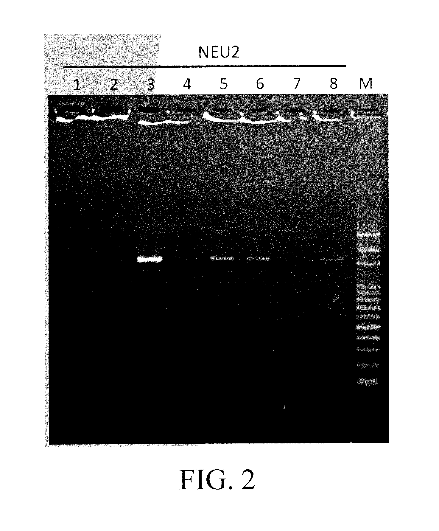
[0015] FIG. 2 shows the transformation result for NEU2;
[0016] FIG. 3 shows the transformation result for ASAH2;
[0017] FIG. 4 shows the result of gene transfection of NEU4/ASAH2 confirmed by western blot;
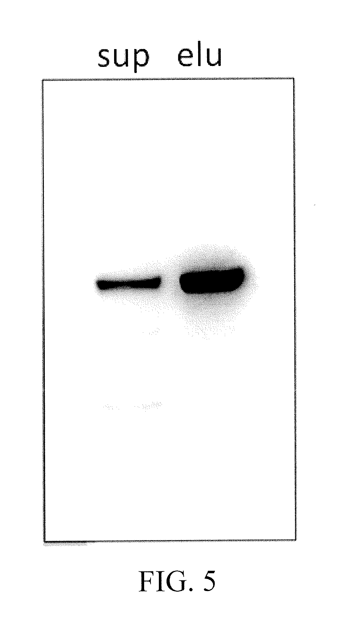
[0018] FIG. 5 shows the result for NEU2 purification;
[0019] FIG. 6 shows the result for ASAH2 purification;
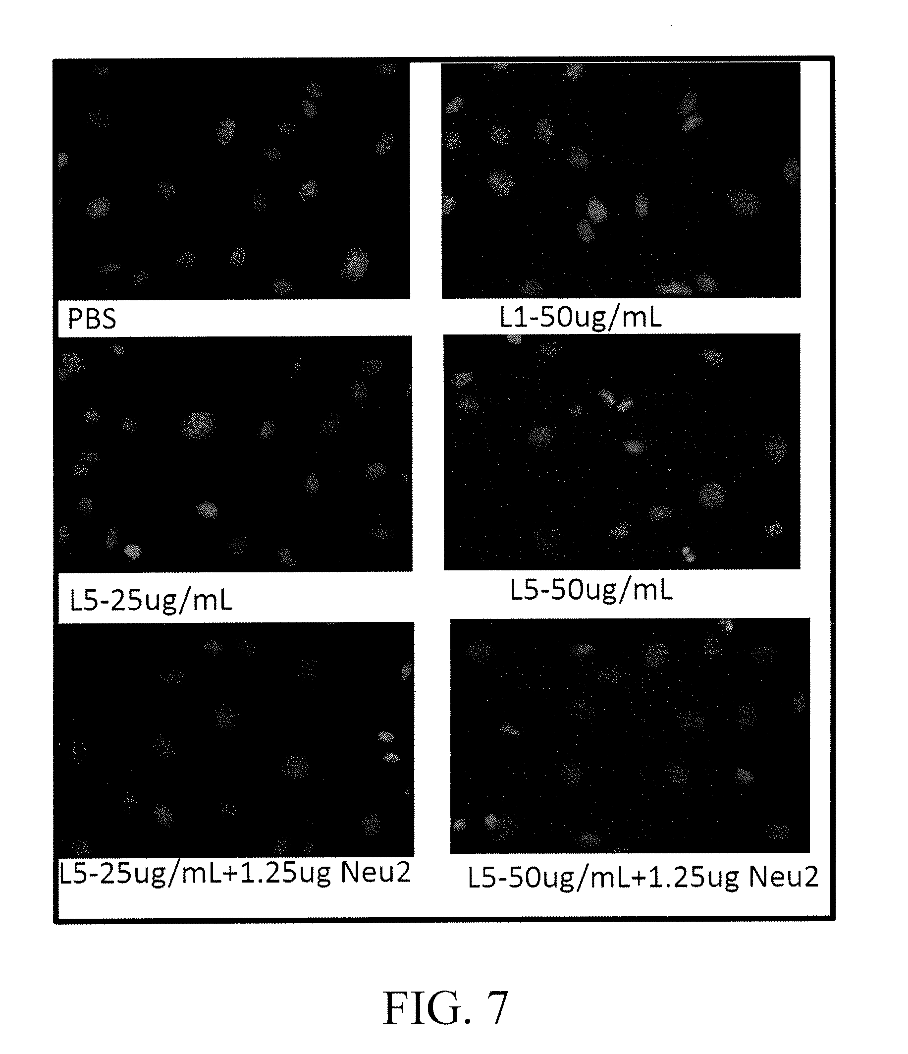
[0020] FIG. 7 shows apoptosis of endothelial cells of blood vessel co-cultured with electronegative low-density lipoprotein (electronegative LDL) L5 (25 .mu.g/mL; 50 .mu.g/mL) and L5 (1.25 .mu.g) treated by the mrnobilized-NEU2 filled device for 2 hours (treatment temperature 37.degree. C., pH 7.4) for 24 hours, respectively;
[0021] FIG. 8 shows results of performing quantitative analysis to the LDL samples without treatment and treated without enzyme at 37.degree. C. for 2 hours or treated with NEU2 for 2 hours (treatment temperature 37.degree. C. pH 7.4) to determine the content of L5 therein;
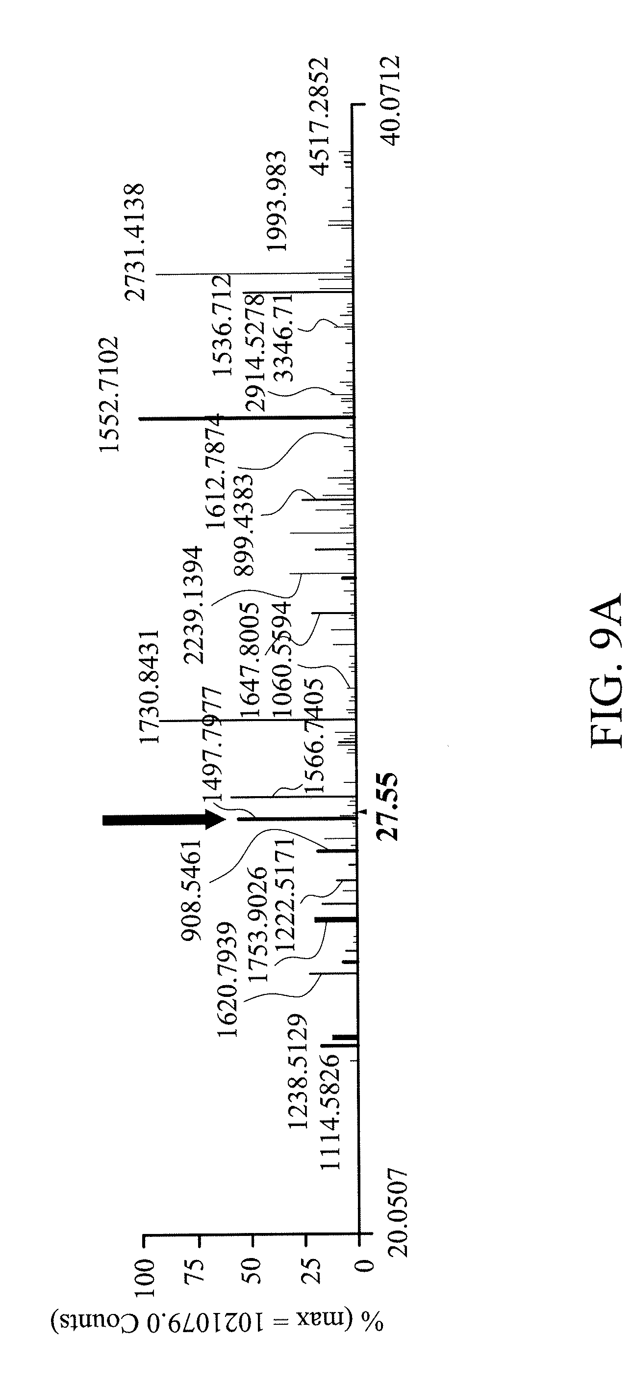
[0022] FIGS. 9A, 9B and 9C show that performing a mass spectrometry analysis on L5 can detect L5 specific glycosylation of apoE lipoprotein;
[0023] FIGS. 10A1-2 and 10B1-2 show that the mass spectrometry analysis result of L5 treated with NEU2, wherein apoE specific glycan residues have been removed;
[0024] FIGS. 11A and 11B1-4 show NEU2, NEU4 immobilized on different material both are capable of effectively eliminating glycosylation on lipoproteins; Sequences of LDL which are most commonly glycosylated comprise: 1. (R)IGQDGISTSATTNLK(C) (SEQ ID NO. 3) of apoB100; 2. (K)VLVDHFGYTK(D) (SEQ ID NO. 4) of apoB100; 3. (K)GVISIPR(L) (SEQ ID NO. 5) of apoB100; 4. (K)SGSSTASWIQNVDTKYQIR(I) (SEQ ID NO. 6) of apoB100; 5. (K)AKPALEDLRQGLLPVLESFK(V) (SEQ ID NO. 7) of apoB100. Furthermore, ITRI-A-01(NEU2), ITRI-CD-01(NEU2), ITRI-Si-Nu-01(NEU4) all are capable of effectively eliminating glycan residues on apoB;
[0025] FIG. 12 shows ceramide contents of L5 and L5 treated with ASAH2 for 24 hours;
[0026] FIG. 13 shows ceramide contents of L5 and L5 treated with ASAH2 for 24 hours;
[0027] FIG. 14 shows ceramide contents of L5 and L5 treated with ASAH2 in the presence or absence of a buffer (200 mM Tris-HCl pH 8.4, 1.5 M NaCl, 25 mM CaCl.sub.2) for 2 or 24 hours;
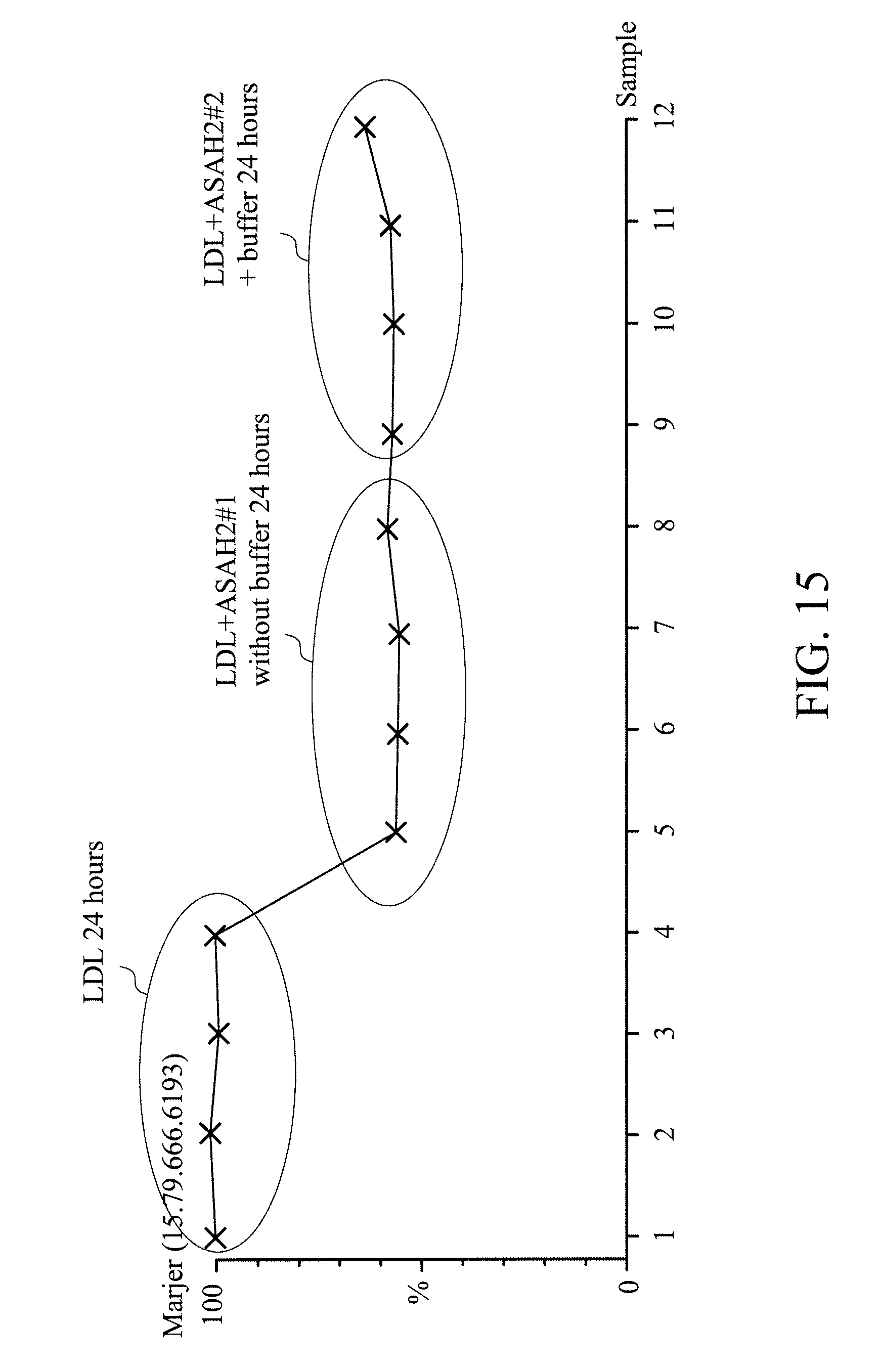
[0028] FIG. 15 shows ceramide contents of L5 and L5 treated with ASAH2 in the presence or absence of a buffer (200 mM Tris-HCl pH 8.4, 1.5 M NaCl, 25 mM CaCl.sub.2) for 24 hours;
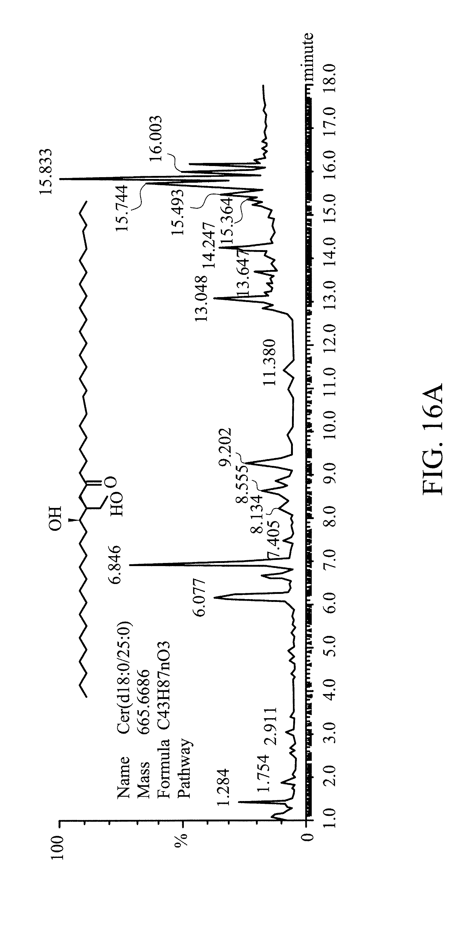
[0029] FIG. 16A shows the result of quantitative analysis for lipid constituents of L5 and L5 treated with ASAH2 for 24 hours mass spectrometry;
[0030] FIG. 16B shows ceramide contents of L5 and L5 treated with ASAH2 in the presence of a buffer (200 mM Tris-HCl pH 8.4, 1.5 M NaCl, 25 mM CaCl2)) for 2 hours;
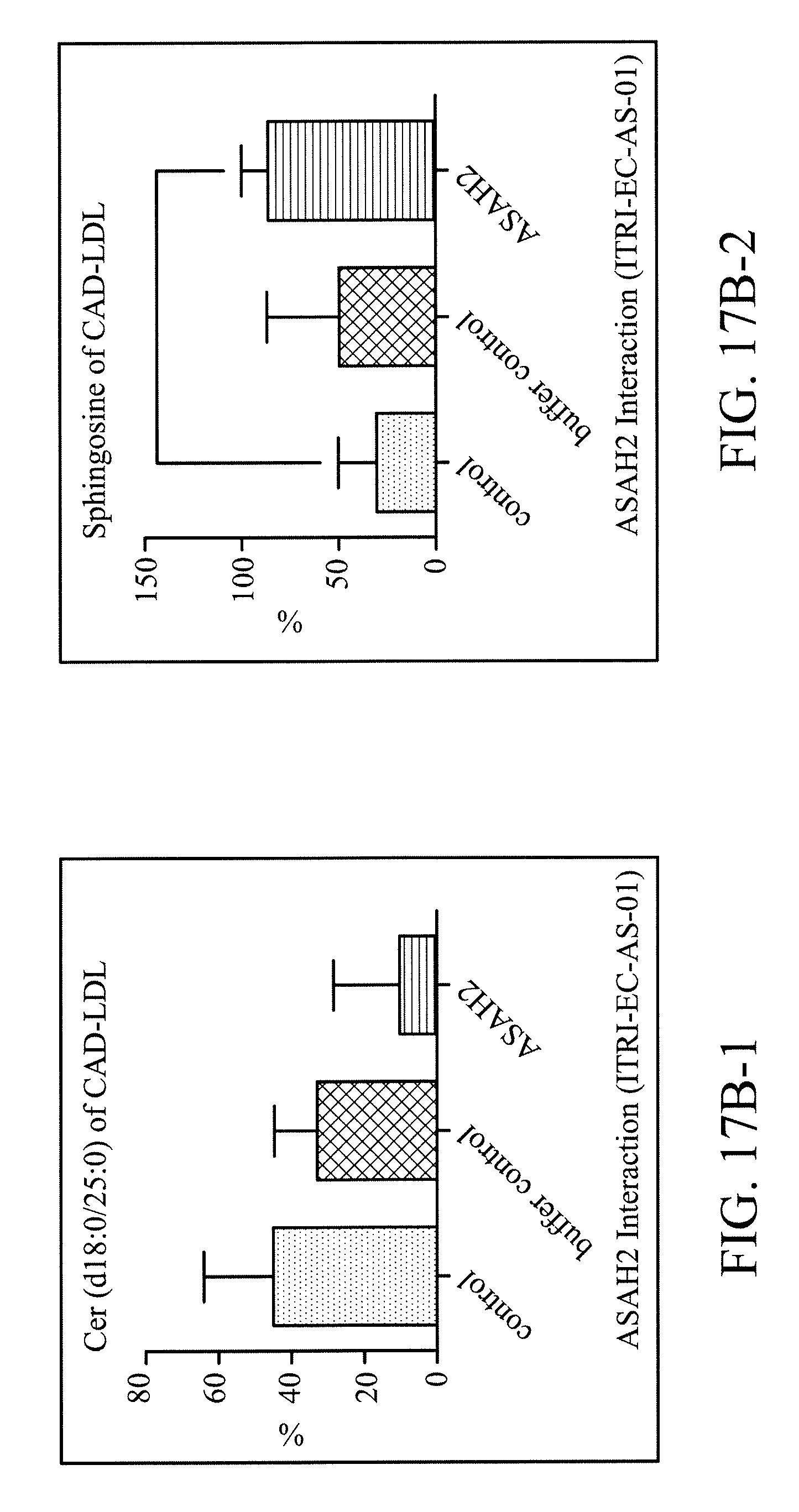
[0031] FIGS. 17A and 17B1-2 show that immobilized ASAH2 is capable of effectively eliminating ceramide and increasing a product, sphingosine; One of the most common ceramides of L5 is Cer (d18:0/25:0), and after it has been catalyzed by ASAH2, a product, sphingosine, is produced. The experimental results show that immobilized ASAH2 (ITRI-EC-AS-01) is capable of reducing Cer (d18:0/25:0) contained by the LDL sample and increasing the product sphingosine.
DETAILED DESCRIPTION
[0032] In the following detailed description, for purposes of explanation, numerous specific details are set forth in order to provide a thorough understanding of the disclosed embodiments. It will be apparent, however, that one or more embodiments may be practiced without these specific details. In other instances, well-known structures and devices are shown schematically in order to simplify the drawing.
[0033] In one embodiment of the present disclosure, the present disclosure provides a biochemistry reactive material which is capable of eliminating electronegative low-density lipoprotein (electronegative LDL). Examples of the electronegative low-density lipoprotein mentioned above may comprise, but are not limited to, low-density lipoproteins L1, L2, L3, L4, L5, etc. In one embodiment, the electronegative low-density lipoprotein mentioned above may be low-density lipoprotein L5. Moreover, L5 is the most electronegative and most harmful low-density lipoprotein.
[0034] The biochemistry reactive material of the present disclosure may comprise, but is not limited to, a substrate and an enzyme composition, wherein the enzyme composition is immobilized on the substrate.
[0035] Examples of suitable substrate may comprise silica gel, cellulose, diethylaminoethyl cellulose (DEAE cellulose), chitosan, polystyrene, polysulfone, polyethersulfone, acrylate resin, polysaccharide, etc., but they are not limited thereto. The substrate may have a particle structure or a hollow-tube structure, etc. In one embodiment, the substrate may be a cellulose bead. In another embodiment, the substrate may be a chitosan bead. Moreover, the substrate may be a cellulose hollow fiber, a polysulfone hollow fiber, epoxy acrylic resin or a polyethersulfone hollow fiber, etc.
[0036] The preceding enzyme composition may comprise a first enzyme for eliminating a glycan residue of an electronegative LDL, a second enzyme for eliminating ceramide carried by an electronegative LDL, or a combination thereof, but it is not limited thereto. Source organisms of the first enzyme and the second enzyme mentioned above have no particular limitation. In one embodiment, the first enzyme and the second enzyme are bioengineered enzymes from human genome and also possibly from animal genome.
[0037] The preceding first enzyme may be sialidase or glycosidase.
[0038] The sialidase may comprise neuraminidase 1 (NEU1), neuraminidase 2 (NEU2), neuraminidase 3 (NEU3), neuraminidase 4 (NEU4) and O-sialidase bioengineered from human genome, one of the foregoing enzymes obtained through gene transformation, expression and purification, sialidase from a virus or bacterium (alias, acetylneuraminyl hydrolase), etc., but it is not limited thereto.
[0039] Examples of the glycosidase may comprise alpha- and beta-glucosidase bioengineered from human or animal genome, maltase-glucoamylase and sucrase-isomaltase, one of the foregoing enzymes obtained through gene transformation, expression and purification, N-glycosidase F (PNGase F) and glucosidase from a virus, a bacterium or other organism, etc., but they are not limited thereto.
[0040] Furthermore, the second enzyme may be ceramidase.
[0041] The ceramidase may comprise N-acylsphingosine amidohydrolase 1 (ASAH1), N-acylsphingosine amidohydrolase 2 (ASAH2), N-acylsphingosine amidohydrolase 2B (ASAH2B), N-acylsphingosine amidohydrolase 2C (ASAH2C), N-acylethanolamine acid amidase, alkaline ceramidase 1, alkaline ceramidase 2, alkaline ceramidase 3, but it is not limited thereto.
[0042] In one embodiment, the enzyme composition in the biochemistry reactive material of the present disclosure is the first enzyme. In this embodiment, the first enzyme mentioned above may be sialidase, but it is not limited thereto.
[0043] In another embodiment, the enzyme composition in the biochemistry reactive material of the present disclosure is the second enzyme. In this embodiment, the second enzyme mentioned above may be N-acylsphingosine amidohydrolase 2, but it is not limited thereto.
[0044] In another embodiment, the enzyme composition in the biochemistry reactive material of the present disclosure is a combination of the first enzyme and the second enzyme. In this embodiment, the first enzyme mentioned above may be sialidase, but it is not limited thereto, and the second enzyme mentioned above may be N-acylsphingosine amidohydrolase 2, but it is not limited thereto.
[0045] In another embodiment of the present disclosure, the present disclosure provides a biochemistry reactive device, and the device can be used for eliminating electronegative low-density lipoprotein in a liquid sample.
[0046] Examples of the foregoing liquid sample may comprise an aqueous solution, a buffer, blood, plasma, etc., but they are not limited thereto.
[0047] Examples of the foregoing electronegative low-density lipoprotein may comprise electronegative low-density lipoprotein L1, L2, L3, L4 and/or L5, etc., but they are not limited thereto. In one embodiment, the electronegative low-density lipoprotein mentioned above may be electronegative low-density lipoprotein L5.
[0048] A cross-sectional view of a structure of the biochemistry reactive device of the present disclosure is shown in FIG. 1.
[0049] Refer to FIG. 1A. The preceding biochemistry reactive device of the present disclosure 100 may comprise a biochemistry reactive material 101 and a container 103 for containing the biochemistry reactive material 101. The container 103 has at least one inlet 105 and at least one outlet 107. The foregoing liquid sample enters into the biochemistry reactive device 100 from the inlet 105, and flows through the biochemistry reactive material 101 to react with the biochemistry reactive material 101, and then flows out through the outlet 107.
[0050] The biochemistry reactive material 101 may comprise, but is not limited to a substrate and an enzyme composition, wherein the enzyme composition is immobilized on the substrate.
[0051] The substrate mentioned above may comprise, but is not limited to, silica gel, cellulose, diethylaminoethyl cellulose, chitosan, polystyrene, polysulfone, polyethersulfone, acrylate resin, polysaccharide, etc. The substrate may have a particle structure or a hollow-tube structure, etc., but it is not limited thereto.
[0052] The enzyme composition may comprise, but is not limited to, a first enzyme for eliminating a glycan residue of an electronegative LDL, a second enzyme for eliminating ceramide carried by an electronegative LDL or a combination thereof. Source organisms of the first enzyme and the second enzyme mentioned above have no particular limitation. In one embodiment, the first enzyme and the second enzyme are human.
[0053] The preceding first enzyme may be sialidase or glycosidase.
[0054] The sialidase may comprise, but is not limited to, neuraminidase 1 (NEU1), neuraminidase 2 (NEU2), neuraminidase 3 (NEU3), neuraminidase 4 (NEU4) and O-sialidase bioengineered from human genome, one of the foregoing enzymes obtained through gene transformation, expression and purification, sialidase from a virus, a bacterium or other organism, etc.
[0055] The glycosidase may comprise, but is not limited to, alpha- and beta-glucosidase bioengineered from human or animal genome, maltase-glucoamylase and sucrase-isomaltase, one of the foregoing enzymes obtained through gene transformation, expression and purification, N-glycosidase F (PNGase F) and glucosidase from a virus, a bacterium or other organism, etc.
[0056] In addition, the second enzyme may be ceramidase. The ceramidase may comprise, but is not limited to, N-acylsphingosine amidohydrolase 1 (ASAH1), N-acylsphingosine amidohydrolase 2 (ASAH2), N-acylsphingosine amidohydrolase 2B (ASAH2B), N-acylsphingosine amidohydrolase 2C (ASAH2C), N-acylethanolamine acid amidase, alkaline ceramidase 1, alkaline ceramidase 2, alkaline ceramidase 3.
[0057] In one embodiment, the enzyme composition in the biochemistry reactive material 101 mentioned above is the first enzyme. In this embodiment, the first enzyme mentioned above may be sialidase, but it is not limited thereto.
[0058] In another embodiment, the enzyme composition in the biochemistry reactive material 101 mentioned above is the second enzyme. En this embodiment, the second enzyme mentioned above may be N-acylsphingosine amidohydrolase 2, but it is not limited thereto.
[0059] In another embodiment, the enzyme composition in the biochemistry reactive material 101 mentioned above is a combination of the first enzyme and the second enzyme. In this embodiment, the first enzyme mentioned above may be sialidase, but it is not limited thereto, and the second enzyme mentioned above may be N-acylsphingosine amidohydrolase 2, but it is not limited thereto.
[0060] Furthermore, a material of the container 103 of the biochemistry reactive device 100 of the present disclosure may comprise glass, acrylic, polypropylene, polyethylene, stainless steel, titanium alloy, etc., but it is not limited thereto. In one embodiment, a material of the container 103 of the biochemistry reactive device 100 of the present disclosure may be polypropylene. In addition, a shape of the container 103 has no particular limitation, and in one embodiment, the container 103 is a hollow column.
[0061] In one embodiment, as shown in FIG. 1B, the biochemistry reactive device 100 of the present disclosure may further comprise a filtering material 109 configured in the container 103 behind the at least one inlet 105 and at least one outlet 107. Moreover, the pore size of the filtering material mentioned above is smaller than the biochemistry reactive material 101 to prevent the biochemistry reactive material 101 leaking from the at least one inlet 105 and/or least one outlet 107, but it can allow the liquid sample to pass through. The filtering material 109 mentioned above comprises filter paper, glass, acrylic, polypropylene, polyethylene, etc., but it is not limited thereto. In this embodiment, the substrate of the biochemistry reactive material 101 may have a particle structure or a hollow-tube structure. In one specific embodiment, the substrate of the biochemistry reactive material 101 has a particle structure, and in this specific embodiment, the substrate of the biochemistry reactive material 101 may be a cellulose bead or a chitosan bead, but it is not limited thereto.
[0062] When the substrate of the biochemistry reactive material 101 is a hollow-tube structure, polyurethane (PU) can be used to package the device without using the filtering material 109.
[0063] In one embodiment, the container 103 may be a hollow column, and two ends of the hollow column of the container 103 have a first inlet 105.sub.1 of the inlet mentioned above and a first outlet 107.sub.1 of the outlet mentioned above, respectively. In this embodiment, the substrate of the biochemistry reactive material 101 may have a particle structure or a hollow-tube structure.
[0064] In another embodiment, as shown in FIG. 1C, the container 103 may be a hollow column, and two ends of the hollow column of the container 103 have a first inlet 105.sub.1 of the inlet mentioned above and a first outlet 107.sub.1 of the at least outlet mentioned above, respectively, and a second inlet 105.sub.2 of the inlet and a second outlet 107.sub.2 of the outlet are located at a side wall of the hollow column. In this embodiment, the liquid sample can enter into the biochemistry reactive device 100 from the first inlet 105.sub.1 of the container 103, and flows through the biochemistry reactive material 101, and then flows out through the first outlet 107.sub.1. Moreover, a second liquid which can be water, a dialysis solution or a salt-containing aqueous solution enters into the biochemistry reactive device 100 from the first inlet 105.sub.2, and flows through the biochemistry reactive material 101, and then flows out through the first outlet 107.sub.2. The second liquid can bring a by-product out after the reaction or dialysis.
[0065] In this embodiment, the substrate of the biochemistry reactive material 101 may have a particle structure or a hollow-tube structure. In one specific embodiment, the substrate of the biochemistry reactive material 101 has a hollow-tube structure, and in this specific embodiment, the substrate of the biochemistry reactive material 101 may be cellulose hollow fiber, but it is not limited thereto.
[0066] In another embodiment of the present disclosure, the present disclosure provides a method for ex vivo treating blood or plasma. By the method for ex vivo treating blood or plasma, an electronegative low-density lipoprotein in blood or plasma can be eliminated. The foregoing electronegative low-density lipoprotein may comprise, but is not limited to, electronegative low-density lipoprotein L1, L2, L3, L4 and/or L5, etc. In one embodiment, the electronegative low-density lipoprotein mentioned above is electronegative low-density lipoprotein L5.
[0067] The method for ex vivo treating blood or plasma may comprise the following steps, but it is not limited thereto.
[0068] First, a blood or plasma ex vivo contacts with an enzyme composition to react the enzyme composition with the blood or plasma, wherein the enzyme composition is capable of eliminating electronegative low-density lipoprotein.
[0069] The preceding enzyme composition may comprise a first enzyme for eliminating a glycan residue of an electronegative LDL, a second enzyme for eliminating ceramide carried by an electronegative LDL or a combination thereof, but it is not limited thereto. Source organisms of the first enzyme and the second enzyme mentioned above have no particular limitation. In one embodiment, the first enzyme and the second enzyme are human.
[0070] The preceding first enzyme may be sialidase or glycosidase.
[0071] The sialidase may comprise neuraminidase 1 (NEU1), neuraminidase 2 (NEU2), neuraminidase 3 (NEU3), neuraminidase 4 (NEU4) and O-sialidase from a human, or one of the foregoing enzymes obtained through gene transformation, expression and purification, sialidase from a virus, a bacterium or other organisms, etc., but it is not limited thereto.
[0072] Examples of the glycosidase may comprise alpha- and beta-glucosidase bioengineered from human animal genome, maltase-glucoamylase and sucrase-isomaltase, one of the foregoing enzymes obtained through gene transformation, expression and purification, N-glycosidase F (PNGase F) and glucosidase from a virus, a bacterium or other organism, etc., but they are not limited thereto.
[0073] Furthermore, the second enzyme mentioned above may be ceramidase.
[0074] The ceramidase may comprise N-acylsphingosine amidohydrolase 1 (ASAH1), N-acylsphingosine amidohydrolase 2 (ASAH2), N-acylsphingosine amidohydrolase 2B (ASAH2B), N-acylsphingosine amidohydrolase 2C (ASAH2C), N-acylethanolamine acid amidase, alkaline ceramidase 1, alkaline ceramidase 2, alkaline ceramidase 3, but it is not limited thereto.
[0075] In one embodiment, the enzyme composition used in the method for ex vivo treating blood or plasma of the present disclosure is the first enzyme. In this embodiment, the first enzyme mentioned above may be sialidase, but it is not limited thereto.
[0076] In another embodiment, the enzyme composition used in the method for ex vivo treating blood or plasma of the present disclosure is the second enzyme. In this embodiment, the second enzyme mentioned above may be N-acylsphingosine amidohydrolase 2, but it is not limited thereto.
[0077] In another embodiment, the enzyme composition used in the method for ex vivo treating blood or plasma of the present disclosure is a combination of the first enzyme and the second enzyme. In this embodiment, the first enzyme mentioned above may be sialidase, but it is not limited thereto, and the second enzyme mentioned above may be N-acylsphingosine amidohydrolase 2, but it is not limited thereto.
[0078] Furthermore, in one embodiment, the enzyme composition used in the method for ex vivo treating blood or plasma of the present disclosure can be immobilized on a substrate. Examples of the substrate may comprise silica gel, cellulose, diethylaminoethyl cellulose, chitosan, polystyrene, polysulfone, polyethersulfone, resin, polysaccharide, but they are not limited thereto. Moreover, the substrate may have a particle structure or a hollow-tube structure.
[0079] In the method for ex vivo treating blood or plasma of the present disclosure, time for ex vivo contacting the blood or plasma with the enzyme composition may be about 0.25-8 hours. In one embodiment, time for ex vivo contacting the blood or plasma with the enzyme composition may be about 2 hours.
[0080] Furthermore, in the method for ex vivo treating blood or plasma of the present disclosure, temperature for ex vivo contacting the blood or plasma with the enzyme composition may be about 4-40.degree. C. In one embodiment, temperature for ex vivo contacting the blood or plasma with the enzyme composition may be about 37.degree. C.
[0081] In addition, in the method for ex vivo treating blood or plasma of the present disclosure, the blood or plasma may ex vivo contact with the enzyme composition at about pH 5-10. In one embodiment, the blood or plasma may ex vivo contact with the enzyme composition at about pH 7.4.
[0082] Afterward, contact between the blood or plasma and the enzyme composition is terminated to terminate the reaction of the enzyme composition with the blood or plasma.
[0083] A manner for terminating the contact between the blood or plasma and the enzyme composition has no particular limitation, for example, for terminating the contact between the blood or plasma and the enzyme composition, the blood or plasma can be separated from the enzyme composition, or the enzyme composition can be deactivated, etc.
EXAMPLES
Example 1
[0084] A. Methods
[0085] 1. Obtainment of Electronegative Low-Density Lipoprotein (Electronegative LDL)
[0086] (1) Purifications for Electronegative Low-Density Lipoprotein
[0087] Blood samples to be used for LDL isolation were obtained from subjects. After the initial screening, blood samples were removed from the subjects with precaution against coagulation and ex vivo oxidation. The plasma was treated with Complete Protease
[0088] Inhibitor Cocktail (Roche; Cat. No. 05056489001; 1 tablet/100 mL) to prevent protein degradation.
[0089] Lipoprotein Preparation from a Human
[0090] The plasma was overlaid with 2 mL Milli-Q water and spun at 20,000 rpm for 2 hours. The upper white fraction and chylomicrons were removed, and the remnant layer which contains VLDL, IDL, LDL and HDL was saved for a series of isolation steps.
[0091] To progressively separate VLDL (d=0.93-1.006), IDL (d=1.006-1.019), LDL (1.019-1.063 g/dL) and HDL (1.063-1.210 g/dL) from one another, the remnant sample was sequentially adjusted to d=1.006, d=1.019, d=1.063, d=1.210, respectively, by adding potassium bromide, and then the remnant samples sequentially adjusted to d=1.006, d=1.019 and d=1.063 were centrifuged at 45,000 rpm for 24 hours at 4.degree. C., and the remnant sample sequentially adjusted to d=1.210 was centrifuged at 45,000 rpm for 48 hours at 4.degree. C. After centrifugation at each isolation step, IDL was discarded while VLDL, LDL and HDL were collected. Isolated VLDL, LDL and HDL samples were treated with 5 mM EDTA and nitrogen to avoid ex vivo oxidation. After that, VLDL, LDL and HDL samples were dialyzed against buffer A (20 M, pH 8.0, 0.5 M EDTA) for 24 hours (.times.3 times) to remove excessive potassium bromide, and were filtrated through 0.22-.mu.m filter (Sartorius; Minisart.RTM.) to sterilize the samples.
[0092] (2) LDL Subfractions
[0093] Approximately 30 mg of LDL material was injected onto a UnoQ12 anion-exchange column (BioRad) by using the AKTA fast-protein liquid chromatography (FPLC) pump (GE Healthcare Life Sciences, Pittsburgh, Pa.). LDL was eluted according to electronegativity by the use of a multistep gradient of buffer B (1 mol/L NaCl in buffer A) at a flow rate of 2 mL/minute. In short, samples were equilibrated with buffer A for 10 minutes, followed by being linearly increased to 15% buffer B in 10 minutes (fraction 1), linearly increased to 20% buffer B in 30 minutes (fraction 2, 3), kept at 20% buffer B for 10 minutes (fraction 4) and linearly increased to 100% buffer B in 20 minutes (fraction 5). Lastly, the effluents were monitored at 280 nm.
[0094] (3) Purification of Fractionated LDL
[0095] Based on the gradient profile, each of the LDL fractions were pooled. The volume of each subfraction was constant. Dilution of LDL during chromatography depended on the injection volume. The respective fractions were concentrated with Centriprep.RTM. filters (YM-30; EMD Millipore Corp., Billerica, Mass.), dialyzed against buffer A (20 M, pH8.0, 0.5 M EDTA) for 24 hours (3 days) and sterilized by passing through 0.22-.mu.m filters (Sartorius; Minisart.RTM.). The isolated fractions were quantified at their protein concentrations by the Lowry method and then stored at 4.degree. C.
[0096] 2. Screening of NEU2 or NEU4
[0097] (1) Transformation (Gene Cloning for pCMV6 Vector with NEU2 and NEU4 Genes)
[0098] NEU2 (neuraminidase 2) and NEU4 (neuraminidase 4) were purchased from Origene, RC219858 and RC203948. Genes were amplified by ECOSTM 101 DH5.alpha. Competent Cells (Yeastern, FYE608) according to the manufacturer's directions.
[0099] In short, 1 vial of competent cells with 5 .mu.L plasmid was vortexed for 1 second and then incubated on ice for 5 minutes. After 45 second heat-shock at 42.degree. C., the mixture was plate on LB agar with Kanamycin.
[0100] Colonies were checked with PCR by VP1.5 and XL39 primers. Procedures of the PCR comprises: 95.degree. C. for 1 minute for pre-PCR denaturation; 2 cycles of 95.degree. C. for 10 seconds, 62.degree. C. for 20 seconds, 72.degree. C. for 4 minutes; 2 cycles of 95.degree. C. for 10 seconds, 60.degree. C. for 20 seconds, 72.degree. C. for 4 minutes; 2 cycles of 95.degree. C. for 10 seconds, 58.degree. C. for 20 seconds, 72.degree. C. for 4 minutes; 15 cycles of 95.degree. C. for 10 seconds, 56.degree. C. for 20 seconds, 72.degree. C. for 4 minutes; 72.degree. C. for 10 minutes for post-PCR incubation and holding on 4.degree. C.
[0101] (2) Plasmid Extraction
[0102] After confirming the insertion of transformed colonies, transformed cells were plate-out into 5 ml LB broth with 25 mg/ml kanamycin, and then incubated at 37.degree. C. overnight.
[0103] Plasmid DNA was extracted according to the protocol of Plasmid Miniprep Plus Purification Kit (GeneMark, DP01P). In short, the bacteria were centrifuged for 1 minute at 14,000.times.g, and the media was removed. The pellet was re-suspended in 200 .mu.L Solution I by pipetting, then 200 .mu.L Solution II was added therein and mixed by inverting the tube. 200 .mu.L Solution III was added to the tube and mixed by inverting the tube 5 times. The lysate was centrifuged at top speed for 5 minutes and a compact white pellet formed along the side of the tube. The spin column was inserted into a collection tube, and the clear lysate was moved to spin column and spun at top speed for 1 minute. The flow-through was discarded, and 500 .mu.L Endotoxin Removal Wash Solution was loaded to the spin column and kept for 2 minutes to equilibrate the membrane, then spun at top speed for 1 minute. The filtrate was discarded, and 700 .mu.L Washing Solution was added to the spin column and spun at top speed for 1 minute, and then this step was repeated. The filtrate was discarded and the spin column was centrifuged for 5 minutes at top speed to remove residual traces of ethanol. The spin column was transferred into a new tube and 35 .mu.L H.sub.2O was added to the spin column and kept for 1-2 minutes and the tube was centrifuged at top speed for 2 minutes to elute the DNA. The DNA quantified by microplate spectrophotometer (Epoch, BioTek).
[0104] (3) Transfection on HEK Cells and Protein Purification
[0105] One day before transfection, 1.25*10.sup.5 HEK293T cells were placed in 500 .mu.L DMEM medium in 24-well plate. For each well of cells to be transfected, 1 .mu.g of DNA was diluted in 100 .mu.L serum-free medium, and 1.5 .mu.L of Lipofectamine 2000 Transfection Reagent (Invitrogen) was add thereto and mixed gently and incubated for 30 minutes at room temperature. After incubation, the complex was added to each well containing cells and mixed gently. The cells were incubated at 37.degree. C. in a CO.sub.2 incubator for 20 hours. The transfected cells were lysed by RIPA which containing protease inhibitor to prepare to purify the proteins.
[0106] In short, 80 .mu.L ANTI-FLAG M2 Magnetic Beads (Sigma-Aldrich) were equilibrated for one-well cell lysate purification. After protein-resin binding at 4.degree. C. overnight, the bound FLAG fusion protein was eluted by competitive elution with 150 .mu.g/ml 3.times.FLAG peptide for 2 times, the eluate was collected, and the protein checked by western blot.
[0107] 3. Efficacy Test for NEU2 or NEU4
[0108] (1) Protein Quantification
[0109] Pierce BCA Protein Assay Kit (Thermo) was used for protein quantification according to the manufacturer's directions.
[0110] In short, 25 .mu.L serial diluted BSA standard and 5 .mu.L sample in 20 .mu.L sample diluent were pipetted into a 96-well microplate. To prepare BCA working reagent, 50 parts of BCA Reagent A was mixed with 1 part of BCA Reagent B and placed on ice until use. 200 .mu.L of the BCA working reagent was added to each well and mixed thoroughly, and the plate was covered and incubated at 37.degree. C. for 30 minutes. The absorbance at 562 nm was measured by spectrophotometer (Epoch, BioTek).
[0111] (2) Apoptosis Measurements
[0112] Endothelial cells were used after 3 or 4 passages and maintained in DMEM (Invitrogen.TM., Thermo Fisher Scientific) containing 10% FBS. During treatment, FBS was reduced to 5% in DMEM. 1.times.10.sup.4 cells were seeded in 96-well plate for 24 hours for subconfluent cultures, and the cultured cells were exposed to PBS (lipoprotein-free, negative control) or graded (25, 50, and 100 .mu.g/mL) LDL subfractions, unfractionated normolipidemic LDL, and LDL/L1/L5 incubated with sialidase for 24 hours. Apoptosis was assessed with visualization by a Zeiss Axiovert 200 fluorescence microscope and filters to capture digital images based on Hoechst 33342, propidium iodide (red), and calcein AM (green) staining of nuclear, apoptotic DNA membrane integrity and cytoplasm respectively according to the protocol of the manufacturer (Invitrogen.TM., Thermo Fisher Scientific).
[0113] (3) LC/MS.sup.E Analysis for Protein Composition
[0114] LDL subfractions were quantified the protein contents by use of quantitative proteomics techniques utilizing serially coupled liquid chromatography data-independent parallel-fragmentation mass spectrometry (LC/MS.sup.E). Such analysis has been shown to be highly quantitative with respect to both relative and/or absolute (when incorporating spiked internal peptide standards in the data collection/analysis procedures) protein abundance in complex protein mixtures. Quantitative analysis was performed essentially as previously described (PMCID: PMC3816395; Pure Appl Chem. 2011; 83(9): 10.1351/PAC-CON-10-12-07. Chemical composition-oriented receptor selectivity of L5, a naturally occurring atherogenic low-density lipoprotein), except on a Waters nanoACQUITY UPLC System and Xevo.RTM. G2-XS QTof mass spectrometer (Waters Corporation, MA, USA).
[0115] In brief, total proteins isolated from each LDL subfraction were first digested with trypsin, and the resulting tryptic peptides were chromatographically separated on a Nano-Acquity separations module (Waters Corporation, MA, USA) incorporating a 50 fmol-on-column tryptic digest of yeast alcohol dehydrogenase as the internally spiked protein quantification standard. Peptide elution will be executed through a 75 im.times.25 cm BEH C-18 column under gradient conditions at a flow rate of 300 nL/minute over 30 minutes at 35.degree. C. The mobile phase was composed of acetonitrile as the organic modifier and formic acid (0.1% v/v) for molecule protonation. Mass spectrometry was performed on a Xevo.RTM. G2-XS QTof instrument equipped with a nano-electrospray ionization interface and operated in the data-independent collection mode (MSE). Parallel ion fragmentation was programmed to switch between low (4 eV) and high (15-45 eV) energies in the collision cell, and data was collected from 50 to 2000 m/z utilizing glu-fibrinopeptide B as the separate data channel lock mass calibrant. Data was processed with ProteinLynx GlobalServer v2.4 (Waters). Deisotoped results were searched for protein association from the Uniprot (www.uniprot.org) human protein database.
[0116] 4. Screening of ASAH2
[0117] (1) Transformation (Gene Cloning for pCMV6 Vector with ASAH2 Genes):
[0118] ASAH2 (N-acylsphingosine amidohydrolase 2) was purchased from Origene, RC203706. Genes were amplified by ECOSTM 101 DH5.alpha. Competent Cells (Yeastern, FYE608) according to the manufacturer's directions.
[0119] In short, 1 vial of competent cells with 5 .mu.L plasmid was vortexed for 1 second and then incubated on ice for 5 minutes. After 45 second heat-shock at 42.degree. C., the mixture was plate on LB agar with Kanamycin.
[0120] Colonies were checked with PCR by VP1.5 and XL39 primers. Procedures of the PCR comprise: 95.degree. C. for 1 minute for pre-PCR denaturation; 2 cycles of 95.degree. C. for 10 seconds, 62.degree. C. for 20 seconds, 72.degree. C. for 4 minutes; 2 cycles of 95.degree. C. for 10 seconds, 60.degree. C. for 20 seconds, 72.degree. C. for 4 minutes; 2 cycles of 95.degree. C. for 10 seconds, 58.degree. C. for 20 seconds, 72.degree. C. for 4 minutes; 15 cycles of 95.degree. C. for 10 seconds, 56.degree. C. for 20 seconds, 72.degree. C. for 4 minutes; 72.degree. C. for 10 minutes for post-PCR incubation and holding on 4.degree. C.
[0121] (2) Plasmid Extraction
[0122] After confirming the insertion of transformed colonies, transformed cells were plate-out into 5 ml LB broth with 25 mg/ml kanamycin, and then incubated at 37.degree. C. overnight.
[0123] Plasmid DNA was extracted according to the protocol of Plasmid Miniprep Plus Purification Kit (GeneMark, DP01P). In short, the bacteria were centrifuged for 1 minute at 14,000.times.g, and the media was removed. The pellet was re-suspended in 200 .mu.L Solution I by pipetting, then 200 .mu.L Solution II was added therein and mixed by inverting the tube. 200 .mu.L Solution III was added to the tube and mixed by inverting the tube 5 times. The lysate was centrifuged at top speed for 5 minutes and a compact white pellet formed along the side of the tube. The spin column was inserted into a collection tube, and the clear lysate was removed to spin column and spun at top speed for 1 minute. The flow-through was discarded, and 500 .mu.L Endotoxin Removal Wash Solution was loaded to the spin column and kept for 2 minutes to equilibrate the membrane, then spun at top speed for 1 minute. The filtrate was discarded, and 700 .mu.L Washing Solution was added to the spin column and spun at top speed for 1 minute, and then this step was repeated. The filtrate was discarded and the spin column was centrifuged for 5 minutes at top speed to remove residual traces of ethanol. The spin column was transferred into a new tube and 35 .mu.L H2O was added to the spin column and kept for 1-2 minutes and the tube was centrifuged at top speed for 2 minutes to elute the DNA. The DNA quantified by microplate spectrophotometer (Epoch, BioTek).
[0124] (3) Transfection on HEK Cells and Protein Purification
[0125] One day before transfection, 1.25*10.sup.5 HEK293T cells were placed in 500 .mu.L DMEM medium in 24-well plate. For each well of cells to be transfected, 1 .mu.g of DNA was diluted in 100 .mu.L serum-free medium, and 1.5 .mu.L of Lipofectamine 2000 Transfection Reagent (Invitrogen) was add thereto and mixed gently and incubated for 30 minutes at room temperature. After incubation, the complex was added to each well containing cells and mixed gently. The cells were incubated at 37.degree. C. in a CO.sub.2 incubator for 20 hours. The transfected cells were lysed by RIPA which containing protease inhibitor to prepare to purify the proteins.
[0126] In short, 80 .mu.L ANTI-FLAG M2 Magnetic Beads (Sigma-Aldrich) were equilibrated for one-well cell lysate purification.
[0127] After protein-resin binding at 4.degree. C. overnight, the bound FLAG fusion protein was eluted by competitive elution with 150 .mu.g/ml 3.times.FLAG peptide for 2 times, the eluate was collected, and the protein checked by western blot.
[0128] 5. Efficacy Test for ASAH2
[0129] (1) Protein Quantification
[0130] Pierce BCA Protein Assay Kit (Thermo) was used for protein quantification according to the manufacturer's directions.
[0131] In short, 25 .mu.L serial diluted BSA standard and 5 .mu.L sample in 20 .mu.L sample diluent were pipetted into a 96-well microplate. To prepare BCA working reagent, 50 parts of BCA Reagent A was mixed with 1 part of BCA Reagent B and placed on ice until use. 200 .mu.L of the BCA working reagent was added to each well and mixed thoroughly, and the plate was covered and incubated at 37.degree. C. for 30 minutes. The absorbance at 562 nm was measured by spectrophotometer (Epoch, BioTek).
[0132] (2) Lipid Extraction
[0133] 30 .mu.g LDL/L1/L5 were incubated with 5 .mu.g ASAH2 in ASAH2 buffer (200 mM Tris-HCl at pH 8.4, 1.5 M NaCl, 25 mM CaCl.sub.2) at 37.degree. C. After 2 or 24 hours incubation, samples were transferred to a glass tube. I mL H.sub.2O, 2.5 mL methanol and 1.25 mL CHCl.sub.3 were added to samples, and vortexed for 15 seconds. Then, additional 0.9 mL H.sub.2O and 1.25 mL CHCl.sub.3 were applied to samples, vortexed for 15 seconds, and centrifuged at 3000 rpm for 10 minutes. Bottom layer organic solvents were transferred to a 2.0 mL glass tube using a glass syringe. Each sample was flushed with nitrogen until dry pallets, and dissolved with 0.25 mL sample solution (isopropanol/acetonitrile/H.sub.2O=2:1:1).
[0134] (3) LC/MS.sup.E Analysis for Lipid Composition
[0135] Total lipids, phospholipids, neutral lipids and free fatty acid from each subfractions of LDL were quantified the lipid contents by use of liquid chromatography data-independent parallel-fragmentation mass spectrometry (LC/MS.sup.E). Quantitative analysis was performed essentially as previously described.
[0136] In brief, lipids were chromatographically separated on a ACQUITY UPLC System (Waters Corporation, MA, USA) incorporating a CSH.TM. 1.7 .mu.m, 2.1 mm.times.10 cm C-18 column under gradient conditions at a flow rate of 400 .mu.L/minute over 18 minutes at 55.degree. C. The mobile phase A will be composed of 10 mM NH4HCO.sub.2 in ACN/H.sub.2O (60/40) and 0.1% formic acid (0.1% v/v), mobile phase B will be composed of 10 mM NH.sub.4HCO.sub.2 in IPA/ACN (90/10) and 0.1% formic acid (0.1% v/v) for molecule protonation. Mass spectrometry was performed on a Xevo.RTM. G2-XS QT of instrument equipped with an electrospray ionization interface and operated in the data-independent collection mode (MSE). Parallel ion fragmentation was programmed to switch between low (4 eV) and high (35-55 eV) energies in the collision cell, and data was collected from 50 to 1600 m/z utilizing leucin as the separate data channel lock mass calibrant. Data was processed with MarkerLynx (Waters).
[0137] B. Results
[0138] 1. Transformation
[0139] (1) NEU2
[0140] Transformation result for NEU2 is shown in FIG. 2.
[0141] According to FIG. 2, it is known that NEU2 transformations for colonies 3, 5 and 6 (see lane 3, 5 and 6, respectively) were successful. Therefore, colonies 3, 5 and 6 were selected to be amplified, and plasmid of NEU2 was stocked.
[0142] (2) ASAH2
[0143] Transformation result for ASAH2 is shown in FIG. 3.
[0144] Colony 7 was selected to be amplified, and plasmid of ASAH2 was stocked.
[0145] 2. Transfection
[0146] Transfection of NEU4/ASAH2 genes was confirmed by western blot, and the result is shown in FIG. 4.
[0147] Conditions for the gene transfection are shown in the following:
[0148] HEK293T 1.25.times.10.sup.5 cells in 24 well
[0149] Plasmid: NEU4 and ASAH2
[0150] DNA amount: 1 .mu.g
[0151] Transfected by Lipofectamine
[0152] SDS-PAGE: using 5 .mu.l sample
[0153] Primary antibody: anti-DDK (1:2000)
[0154] 3. Protein Purification
[0155] (1) NEU2 Purification
[0156] The result for NEU2 purification is shown in FIG. 5. FIG. 5 shows that NEU2 was indeed purified. The amino acid sequence of NEU2 is shown as SEQ ID NO. 1.
[0157] (2) ASAH2 Purification
[0158] The result for ASAH2 purification is shown in FIG. 6. FIG. 6 shows that ASAH2 was indeed purified (extract 1 and extract 2 are proteins obtained from different batches). The amino acid sequence of ASAH2 is shown as SEQ ID NO. 2.
Example 2: Enzyme Immobilization
[0159] Method 1
[0160] 0.4454 g heat-activated silica gel was placed in 7 mL CHCl.sub.3, and APTS was added therein by a weight of 1/5 weight of heat-activated silica gel to form a mixture. After stirring at room temperature for 24 hours, the mixture was filtered. The obtained solid was drained in vacuum at 50.degree. C. After the solid was drained, 5% glutaraldehyde (phosphate Buffer, pH=8, 1.times.TBS) was added to the solid, and stirred for 21 hours to form a solution. The solid in the solution was filtered out and washed with water and a solid substance was obtained. NEU2 ( 1/100-10000 wt %) was added to the solid substance and diluted with phosphate buffer, 1.times.TBS pH=8 to a volume of 2 mL, and reacted with the solid substance at room temperature for 24 hours. Finally, the solid substance was filtered out and washed with phosphate buffer (pH=8) and an enzyme immobilized product (ITRI-Siw-Nu-01) was obtained.
[0161] Method 2
[0162] 3-glycidoxypropyltrimethoxysilane was added to heat-activated silica gel in toluene by a weight of 1/5 weight of heat-activated silica gel, refluxed for 20 hours, and then filtered. The obtained solid was washed with acetone and then drained in vacuum. NEU2 ( 1/100-10000 wt %) was added to the solid and stirred in phosphate buffer for 2 hours and 15 minutes, and then the solid was filtered out. The solid was washed with deionized water and a buffer (pH 8) to obtain an enzyme immobilized product.
[0163] Method 3
[0164] 1 g cellulose beads in 15 mL water were adjusted to about pH 11 by a NaOH solution, and then 1 g cyanogen bromide was added therein at room temperature. After about 30 minutes, the cellulose beads were washed in deionized water and a phosphate buffer (pH 8) in order. NEU2 in a phosphate buffer was added to the cellulose beads by a weight ratio of 1/600, and stirred overnight. After that, the cellulose beads were washed in a phosphate buffer (pH 8) to obtain an enzyme immobilized product.
[0165] Method 4
[0166] 0.5 g cellulose beads in 1.5 mL water were refluxed in 10 mL toluene, and then cellulose beads were filtered out and washed with acetone and a phosphate buffer (pH 8). After that, the cellulose beads were added to 5% (w/v) glutaraldehyde (phosphate buffer, pH 8) and stirred at room temperature for 21 hours. Afterward, the cellulose beads were filtered out, and washed in a phosphate buffer (pH 8) to obtain glutaraldehyde-activated-cellulose beads. NEU2 in a phosphate buffer was added to the cellulose beads by a weight ratio of 2/1000, and stirred overnight. After that, the cellulose beads were washed in a phosphate buffer (pH 8) to obtain an enzyme immobilized product.
[0167] Method 5
[0168] Hypogel.RTM. 200NH.sub.2 were added to 5% (w/v) glutaraldehyde (phosphate buffer, pH 8) and stirred at room temperature for 21 hours. Afterward, the solid substance was filtered out, and washed with a phosphate buffer (pH 8) to obtain glutaraldehyde-activated gel. NEU2 (1/10000 wt %) was diluted with a phosphate buffer (pH=8) to a volume of 15 mL, and mixed with 1.13 g of the glutaraldehyde-activated gel at room temperature for 20 hours. Finally, the solid was filtered out and washed in a phosphate buffer (pH=8) to obtain an enzyme immobilized product.
[0169] Method 6
[0170] 1 g diethylaminoethyl cellulose (DEAE cellulose) was washed in water, suspended in an NaOH solution (1 M aqueous solution), stirred for 10 minutes, and then filtered out and washed in water. The obtained solid substance was suspended in 10 mL dioxane to form a suspension. 2 g cyanuric chloride and 10 mL toluene were added to the suspension and stirred for 30 minutes and then the solid therein was filtered out. The solid was washed with dioxane, water and acetone in order and dried under reduced pressure to form an activated solid support. After that, NEU2 1/10000 (wt %) was added to the activated solid support and stirred for 18 hours. Afterward, the activated solid support was filtered out and washed in water to obtain an enzyme immobilized product.
[0171] Method 7
[0172] 0.5 g chitosan beads were added to 10 mL 0.5% glutaraldehyde, and stirred at room temperature for 1 hour, and then washed with water, continuously and thoroughly to form activated beads. After that, the activated beads were reacted with NEU2 1/3500 (wt %) at room temperature for 2 hours, filtered out and then washed with deionized water to obtain an enzyme immobilized product.
[0173] Method 8
[0174] 1 g cellulose hollow fiber, as per the procedures in Method 4, was activated by APTS and glutaraldehyde, and then reacted with NEU2 1/10000 (wt %) in phosphate buffer (pH=8), stirred overnight, and washed with a phosphate buffer (pH=8) to obtain an enzyme immobilized product.
[0175] Method 9
[0176] 1 g cellulose hollow fiber, as per the procedures in Method 3, was activated by cyanogen bromide, and then reacted with NEU2 in phosphate buffer (pH=8), stirred overnight, washed with a phosphate buffer (pH=8) to obtain an enzyme immobilized product.
[0177] Method 10
[0178] ECR-8204F epoxy-acrylate resin was washed in deionized water, reacted with ASAH2 1/10000 (wt %), adjusted to a volume of 2 mL with a 0.2 M sodium phosphate buffer, and then stirred for 24 hours. After that, epoxy-acrylate resin was filtered out and washed in deionized water and 2M phosphate buffer (pH=8) to obtain about 52 mg of enzyme immobilized product (ITRI-EC-AS-01).
[0179] Method 11
[0180] Iontosorb MT200 cellulose beads were washed in deionized water. Next, the cellulose beads were washed with 3:7 water/dioxane, 7:3 water/dioxane, 100% dioxane in order. After that, dioxane was added to the cellulose beads, and CDI was added therein by a weight of 1/3 weight of cellulose beads, and stirred for about 0.5-1 hour to form a solution. The dioxane in the solution was removed under reduced pressure, and then NEU 2 was immediately added therein and stirred for about 2 hours and 15 minutes. After the reaction, the cellulose beads in the solution were filtered out and were washed in a buffer (pH=6.5) to obtain a wet product about 0.2 g (ITRI-CD-01).
[0181] Method 12
[0182] 0.5 .mu.g NEU2 was added to 2% w/v alginate aqueous solution to form a mixture solution. Next, the mixture solution was dropped into a stirring 2% CaCl.sub.2 (w/v) aqueous solution by a syringe needle. After that, the CaCl.sub.2 aqueous solution was continuously stirred for 30 minutes, and then particles formed in the CaCl.sub.2 aqueous solution were filtered out and washed in deionized water to obtain a wet product (ITRI-A-01).
Example 3
[0183] Efficacy of Immobilized-NEU2 Filled Device
[0184] NEU2 was immobilized by Method 2 in Example 2, and then the immobilized NEU2 was filled into a tube to form a biochemistry reactive device (immobilized-NEU2 filled device) shown in FIG. 1B.
[0185] (1) Determination of Apoptosis
[0186] Endothelial cells of blood vessel were co-cultured with electronegative low-density lipoprotein (electronegative LDL) L5 (25 .mu.g/mL; 50 .mu.g/mL) and L5 (1.25 .mu.g) which was treated by the mobilized-NEU2 filled device for 2 hours (treatment temperature 37.degree. C., pH 7.4) for 24 hours, respectively. After that, apoptosis of the endothelial cells was determined, and the results are shown in FIG. 7.
[0187] According to FIG. 7, it is known that 25 .mu.g/mL L5 results in apoptosis to about 15% endothelial cells and 50 .mu.g/mL L5 results in apoptosis to about 30% endothelial cells while after the treatment of 1.25 .mu.g NEU2, apoptosis effect of L5 to endothelial cells is reduced.
[0188] (2) Quantitative Analysis for Electronegative Low-Density Lipoprotein (Electronegative LDL)
[0189] LDL samples were obtained from a heart disease patient. Quantitative analysis for L5 was performed on the LDL samples without treatment and those treated without enzyme at 37.degree. C. for 2 hours or treated with NEU2 for 2 hours (treatment temperature 37.degree. C. pH 7.4) to determine the content of L5 in the samples mentioned above. The results are shown in FIG. 8.
[0190] According to FIG. 8, it is known that after being treated with NEU2 enzyme for 2 hours, L5 content of the LDL sample was decreased from 12.4% to 8.48%.
[0191] (3) Mass Spectrometry
[0192] Mass spectrometry analysis was performed on L5 and L5 treated with NEU2 for 2 hours (treatment temperature 37.degree. C., pH 7.4). The results are shown in FIGS. 9A, 9B and 9C.
[0193] It has been known that the feature of L5 is that serine and threonine of apolipoprotein E (apoE) are usually glycosylated.
[0194] Refer to FIGS. 9A and 9B. Molecular weight 1497 indicates non-toxic LDL. Molecular weight of LDL with one glycosyl molecule is 1700, molecular weight of LDL with two glycosyl molecules is 1884, and molecular weight of LDL with three glycosyl molecules is 2154. FIG. 9C shows that the amino acid sequence of apolipoprotein E is glycosylated, and that results in the charge-to-mass ratio of the original peptide chain being increased from 1497.8009 to 1700.8868, 1884.9021 and 2154.0300.
[0195] FIGS. 10A1-2 show that there is no molecule with a charge-to-mass ratio of 1700, 1884 or 2154 that is detected for L5 treated by the immobilized-NEU2 filled device for 2 hours, and that indicates that there is no glycosylation on serine and threonine of apolipoprotein E, i.e., the glycan residues of LDL have been removed.
[0196] Similarly, FIGS. 10B1-2 show that for L5 treated by the immobilized-NEU2 filled device for 2 hours, there is no glycosylation on other sites of apolipoprotein E, and that indicates that the glycan residues of LDL have been removed.
Example 4
[0197] Efficacy of ASAH2
[0198] (1) LC/MS.sup.E Analysis for L5 Treated with ASAH2 for 24 Hours
[0199] L5 was treated with ASAH2 for 24 hours. LC/MS.sup.E analysis was performed on L5 without treatment and L5 with the preceding treatment to determine the ceramide content in the L5 samples mentioned above (for the detailed experimental methods, please see "5. Efficacy test for ASAH2" in "A. Method" of Example 1).
[0200] The results for LC/MS.sup.E analysis are shown in Table 1 (the four values shown in each group were obtained from determining the same sample four times). Conversion was performed to signal of each sample in Table 1 to obtain ceramide content percentage of each sample (the highest signal of the L5 without treatment was set as 100%), and the results are shown in FIG. 12. ASAH2#1 and ASAH2#2 shown in Table 1 and FIG. 12 are ASAH2 obtained from different batches.
TABLE-US-00001 TABLE 1 LC/MS.sup.E analysis results for L5 without treatment and L5 treated with ASAH2 for 24 hours 24 hour baseline ASAH2#1 treatment ASAH2#2 treatment Signal 459.6464 295.3353 202.1154 443.4776 236.9632 177.1598 449.8201 230.7273 173.031 451.5772 249.0337 175.7823 Mean 451.1303 253.0149 182.0221 Standard 6.658431 29.21906 13.50503 Deviation Decrease 43.9 59.7
24-hour baseline represents ceramide content of L5 without treatment.
[0201] According to Table 1 and FIG. 12, it is known that after L5 was treated with ASAH2 for 24 hours, the ceramide content of L5 decreased significantly.
[0202] (2) LC/MS.sup.E Analysis for L5 Treated with ASAH2 for 24 Hours (for the Detailed Experimental Methods, Please See "5. Efficacy Test for ASAH2" in "A. Method" of Example 1)
[0203] L5 was treated with ASAH2 for 24 hours. LC/MS.sup.E analysis was performed on L5 without treatment and L5 with the preceding treatment to determine the ceramide content in the L5 samples mentioned above.
[0204] The results for LC/MS.sup.E analysis are shown in Table 2 (the four values shown in each group were obtained from determining the same sample four times). Conversion was performed to signal of each sample in Table 2 to obtain ceramide content percentage of each sample (the highest signal of the L5 without treatment was set as 100%), and the results are shown in FIG. 13. ASAH2#1 and ASAH2#2 shown in Table 2 and FIG. 13 are ASAH2 obtained from different batches.
TABLE-US-00002 TABLE 2 LC/MS.sup.E analysis results for L5 without treatment and L5 treated with ASAH2 for 24 hours 24 hour baseline ASAH2#1 treatment ASAH2#2 treatment Signal 2008.465 1827.823 1638.186 2007.321 1747.422 1627.067 1946.985 1725.032 1622.848 1989.728 1688.382 1616.651 Mean 1988.125 1747.165 1626.188 Standard 28.73594 59.02271 9.070668 Deviation Decrease 12.11997 18.20495
24 hour baseline represents ceramide content of L5 without treatment.
[0205] According to Table 2 and FIG. 13, it is known that after L5 was treated with ASAH2 for 24 hours, the ceramide content of L5 decreased significantly.
[0206] (3) LC/MS.sup.E Analysis for L5 Treated with ASAH2 in the Presence or Absence of a Buffer for 2 or 24 Hours
[0207] In the presence or absence of a buffer (200 mM Tris-HCl pH 8.4, 1.5 M NaCl, 25 mM CaCl.sub.2), L5 was treated with ASAH2 for 2 or 24 hours. LC/MS.sup.E analysis was performed on L5 without treatment and L5 with the preceding treatment to determine the ceramide content in the L5 samples mentioned above (for the detailed experimental methods, please see "5. Efficacy test for ASAH2" in "A. Method" of Example 1 except the part of mixing with the buffer or not).
[0208] Conversion was performed to signal of each sample to obtain the ceramide content percentage of each sample (the highest signal of the L5 without treatment and kept for 2 hours was set as 100%), and the results are shown in FIG. 14. ASAH2#1 and ASAH2#2 shown in FIG. 14 are ASAH2 obtained from different batches. In FIG. 14, LDL baseline represents ceramide content of L5 without treatment and kept for 0 hour; LDL 2 hours represents ceramide content of L5 without treatment and kept for 2 hour; LDL 24 hours represents ceramide content of L5 without treatment and kept for 24 hour.
[0209] According to FIG. 14, it is known that, in the presence of a buffer, after L5 was treated with ASAH2 for 2 hours, the ceramide content of L5 decreased significantly. Moreover, in the presence or absence of a buffer, after L5 was treated with ASAH2 for 24 hours, the ceramide content of L5 both decreased significantly.
[0210] (4) LC/MS.sup.E Analysis for L5 Treated with ASAH2 in the Presence or Absence of a Buffer for 24 Hours
[0211] In the presence or absence of a buffer (200 mM Tris-HCl pH 8.4, 1.5 M NaCl, 25 mM CaCl.sub.2), L5 was treated with ASAH2 for 24 hours. LC/MS.sup.E analysis was performed on L5 without treatment and L5 with the preceding treatment to determine the ceramide content in the L5 samples mentioned above (for the detailed experimental methods, please see "5. Efficacy test for ASAH2" in "A. Method" of Example 1 except the part of mixing with the buffer or not).
[0212] The results for LC/MS.sup.E analysis are shown in Table 3 (the four values shown in each group were obtained from determining the same sample four times). Conversion was performed to signal of each sample in Table 3 to obtain ceramide content percentage of each sample (the highest signal of the L5 without treatment was set as 100%), and the results are shown in FIG. 15. ASAH2#1 and ASAH2#2 shown in Table 3 and FIG. 15 are ASAH2 obtained from different batches.
TABLE-US-00003 TABLE 3 LC/MS.sup.E analysis results for L5 without treatment and L5 treated with ASAH2 in the presence or absence of a buffer for 24 hours 24 hour baseline ASAH2#1 treatment ASAH2#2 treatment Signal 217.36 122.40 121.87 220.16 122.08 121.65 214.80 117.11 123.68 215.96 121.81 136.51 Mean 217.07 120.85 125.93 Standard 2.31 2.51 7.11 Deviation Decrease 44.33 41.99
24 hour baseline represents ceramide content of L5 without treatment.
[0213] According to Table 3 and FIG. 15, it is known that in the presence or absence of a buffer, after L5 was treated with ASAH2 for 24 hours, the ceramide content of L5 both decreased significantly.
[0214] (5) LC/MS.sup.E Analysis for L5 Treated with ASAH2 for 24 Hours
[0215] Quantitative analysis for lipid constituents was performed on L5 and L5 treated with ASAH2 for 24 hours by mass spectrometry, and the ceramide contents of the L5 samples mentioned above were compared. The results are shown in FIG. 16A.
[0216] (6) LC/MS.sup.E Analysis for L5 Treated with ASAH2 in the Presence of a Buffer for 2 Hours
[0217] In the presence of a buffer (200 mM Tris-HCl pH 8.4, 1.5 M NaCl, 25 mM CaCl.sub.2), L5 was treated with ASAH2 for 2 hours. LC/MS.sup.E analysis was performed on L5 without treatment and L5 with the preceding treatment to determine the ceramide content in the L5 samples mentioned above (for the detailed experimental methods, please see "5. Efficacy test for ASAH2" in "A. Method" of Example 1 except the part of mixing with the buffer or not). The results for LC/MSE analysis are shown in Table 4 (the four values shown in each group were obtained from determining the same sample four times). Conversion was performed to signal of each sample in Table 4 to obtain ceramide content percentage of each sample (the highest signal of the L5 without treatment was set as 100%), and the results are shown in FIG. 16B.
TABLE-US-00004 TABLE 4 LC/MS.sup.E analysis results for L5 without treatment and L5 treated with ASAH2 in the presence or absence of a buffer for 2 hours Name of sample Signal L5 0 hour 529.0532 L5 0 hour 498.5066 L5 0 hour 478.2745 L5 0 hour 432.8346 L5 + ASAH 2 hours 266.8874 L5 + ASAH 2 hours 276.2790 L5 + ASAH 2 hour 282.9767 L5 + ASAH 2 hour 283.6284
[0218] According to Table 4 and FIG. 16B, it is known that in the presence of a buffer, after L5 was treated with ASAH2 for 2 hours, the ceramide content of L5 decreased significantly.
[0219] It will be apparent to those skilled in the art that various modifications and variations can be made to the disclosed embodiments. It is intended that the specification and examples be considered as exemplary only, with the true scope of the disclosure being indicated by the following claims and their equivalents.
Sequence CWU 1
1
71380PRTHomo sapiens 1Met Ala Ser Leu Pro Val Leu Gln Lys Glu Ser
Val Phe Gln Ser Gly 1 5 10 15 Ala His Ala Tyr Arg Ile Pro Ala Leu
Leu Tyr Leu Pro Gly Gln Gln 20 25 30 Ser Leu Leu Ala Phe Ala Glu
Gln Arg Ala Ser Lys Lys Asp Glu His 35 40 45 Ala Glu Leu Ile Val
Leu Arg Arg Gly Asp Tyr Asp Ala Pro Thr His 50 55 60 Gln Val Gln
Trp Gln Ala Gln Glu Val Val Ala Gln Ala Arg Leu Asp 65 70 75 80 Gly
His Arg Ser Met Asn Pro Cys Pro Leu Tyr Asp Ala Gln Thr Gly 85 90
95 Thr Leu Phe Leu Phe Phe Ile Ala Ile Pro Gly Gln Val Thr Glu Gln
100 105 110 Gln Gln Leu Gln Thr Arg Ala Asn Val Thr Arg Leu Cys Gln
Val Thr 115 120 125 Ser Thr Asp His Gly Arg Thr Trp Ser Ser Pro Arg
Asp Leu Thr Asp 130 135 140 Ala Ala Ile Gly Pro Ala Tyr Arg Glu Trp
Ser Thr Phe Ala Val Gly 145 150 155 160 Pro Gly His Cys Leu Gln Leu
His Asp Arg Ala Arg Ser Leu Val Val 165 170 175 Pro Ala Tyr Ala Tyr
Arg Lys Leu His Pro Ile Gln Arg Pro Ile Pro 180 185 190 Ser Ala Phe
Cys Phe Leu Ser His Asp His Gly Arg Thr Trp Ala Arg 195 200 205 Gly
His Phe Val Ala Gln Asp Thr Leu Glu Cys Gln Val Ala Glu Val 210 215
220 Glu Thr Gly Glu Gln Arg Val Val Thr Leu Asn Ala Arg Ser His Leu
225 230 235 240 Arg Ala Arg Val Gln Ala Gln Ser Thr Asn Asp Gly Leu
Asp Phe Gln 245 250 255 Glu Ser Gln Leu Val Lys Lys Leu Val Glu Pro
Pro Pro Gln Gly Cys 260 265 270 Gln Gly Ser Val Ile Ser Phe Pro Ser
Pro Arg Ser Gly Pro Gly Ser 275 280 285 Pro Ala Gln Trp Leu Leu Tyr
Thr His Pro Thr His Ser Trp Gln Arg 290 295 300 Ala Asp Leu Gly Ala
Tyr Leu Asn Pro Arg Pro Pro Ala Pro Glu Ala 305 310 315 320 Trp Ser
Glu Pro Val Leu Leu Ala Lys Gly Ser Cys Ala Tyr Ser Asp 325 330 335
Leu Gln Ser Met Gly Thr Gly Pro Asp Gly Ser Pro Leu Phe Gly Cys 340
345 350 Leu Tyr Glu Ala Asn Asp Tyr Glu Glu Ile Val Phe Leu Met Phe
Thr 355 360 365 Leu Lys Gln Ala Phe Pro Ala Glu Tyr Leu Pro Gln 370
375 380 2780PRTHomo sapiens 2Met Ala Lys Arg Thr Phe Ser Asn Leu
Glu Thr Phe Leu Ile Phe Leu 1 5 10 15 Leu Val Met Met Ser Ala Ile
Thr Val Ala Leu Leu Ser Leu Leu Phe 20 25 30 Ile Thr Ser Gly Thr
Ile Glu Asn His Lys Asp Leu Gly Gly His Phe 35 40 45 Phe Ser Thr
Thr Gln Ser Pro Pro Ala Thr Gln Gly Ser Thr Ala Ala 50 55 60 Gln
Arg Ser Thr Ala Thr Gln His Ser Thr Ala Thr Gln Ser Ser Thr 65 70
75 80 Ala Thr Gln Thr Ser Pro Val Pro Leu Thr Pro Glu Ser Pro Leu
Phe 85 90 95 Gln Asn Phe Ser Gly Tyr His Ile Gly Val Gly Arg Ala
Asp Cys Thr 100 105 110 Gly Gln Val Ala Asp Ile Asn Leu Met Gly Tyr
Gly Lys Ser Gly Gln 115 120 125 Asn Ala Gln Gly Ile Leu Thr Arg Leu
Tyr Ser Arg Ala Phe Ile Met 130 135 140 Ala Glu Pro Asp Gly Ser Asn
Arg Thr Val Phe Val Ser Ile Asp Ile 145 150 155 160 Gly Met Val Ser
Gln Arg Leu Arg Leu Glu Val Leu Asn Arg Leu Gln 165 170 175 Ser Lys
Tyr Gly Ser Leu Tyr Arg Arg Asp Asn Val Ile Leu Ser Gly 180 185 190
Thr His Thr His Ser Gly Pro Ala Gly Tyr Phe Gln Tyr Thr Val Phe 195
200 205 Val Ile Ala Ser Glu Gly Phe Ser Asn Gln Thr Phe Gln His Met
Val 210 215 220 Thr Gly Ile Leu Lys Ser Ile Asp Ile Ala His Thr Asn
Met Lys Pro 225 230 235 240 Gly Lys Ile Phe Ile Asn Lys Gly Asn Val
Asp Gly Val Gln Ile Asn 245 250 255 Arg Ser Pro Tyr Ser Tyr Leu Gln
Asn Pro Gln Ser Glu Arg Ala Arg 260 265 270 Tyr Ser Ser Asn Thr Asp
Lys Glu Met Ile Val Leu Lys Met Val Asp 275 280 285 Leu Asn Gly Asp
Asp Leu Gly Leu Ile Ser Trp Phe Ala Ile His Pro 290 295 300 Val Ser
Met Asn Asn Ser Asn His Leu Val Asn Ser Asp Asn Val Gly 305 310 315
320 Tyr Ala Ser Tyr Leu Leu Glu Gln Glu Lys Asn Lys Gly Tyr Leu Pro
325 330 335 Gly Gln Gly Pro Phe Val Ala Ala Phe Ala Ser Ser Asn Leu
Gly Asp 340 345 350 Val Ser Pro Asn Ile Leu Gly Pro Arg Cys Ile Asn
Thr Gly Glu Ser 355 360 365 Cys Asp Asn Ala Asn Ser Thr Cys Pro Ile
Gly Gly Pro Ser Met Cys 370 375 380 Ile Ala Lys Gly Pro Gly Gln Asp
Met Phe Asp Ser Thr Gln Ile Ile 385 390 395 400 Gly Arg Ala Met Tyr
Gln Arg Ala Lys Glu Leu Tyr Ala Ser Ala Ser 405 410 415 Gln Glu Val
Thr Gly Pro Leu Ala Ser Ala His Gln Trp Val Asp Met 420 425 430 Thr
Asp Val Thr Val Trp Leu Asn Ser Thr His Ala Ser Lys Thr Cys 435 440
445 Lys Pro Ala Leu Gly Tyr Ser Phe Ala Ala Gly Thr Ile Asp Gly Val
450 455 460 Gly Gly Leu Asn Phe Thr Gln Gly Lys Thr Glu Gly Asp Pro
Phe Trp 465 470 475 480 Asp Thr Ile Arg Asp Gln Ile Leu Gly Lys Pro
Ser Glu Glu Ile Lys 485 490 495 Glu Cys His Lys Pro Lys Pro Ile Leu
Leu His Thr Gly Glu Leu Ser 500 505 510 Lys Pro His Pro Trp His Pro
Asp Ile Val Asp Val Gln Ile Ile Thr 515 520 525 Leu Gly Ser Leu Ala
Ile Thr Ala Ile Pro Gly Glu Phe Thr Thr Met 530 535 540 Ser Gly Arg
Arg Leu Arg Glu Ala Val Gln Ala Glu Phe Ala Ser His 545 550 555 560
Gly Met Gln Asn Met Thr Val Val Ile Ser Gly Leu Cys Asn Val Tyr 565
570 575 Thr His Tyr Ile Thr Thr Tyr Glu Glu Tyr Gln Ala Gln Arg Tyr
Glu 580 585 590 Ala Ala Ser Thr Ile Tyr Gly Pro His Thr Leu Ser Ala
Tyr Ile Gln 595 600 605 Leu Phe Arg Asn Leu Ala Lys Ala Ile Ala Thr
Asp Thr Val Ala Asn 610 615 620 Leu Ser Arg Gly Pro Glu Pro Pro Phe
Phe Lys Gln Leu Ile Val Pro 625 630 635 640 Leu Ile Pro Ser Ile Val
Asp Arg Ala Pro Lys Gly Arg Thr Phe Gly 645 650 655 Asp Val Leu Gln
Pro Ala Lys Pro Glu Tyr Arg Val Gly Glu Val Ala 660 665 670 Glu Val
Ile Phe Val Gly Ala Asn Pro Lys Asn Ser Val Gln Asn Gln 675 680 685
Thr His Gln Thr Phe Leu Thr Val Glu Lys Tyr Glu Ala Thr Ser Thr 690
695 700 Ser Trp Gln Ile Val Cys Asn Asp Ala Ser Trp Glu Thr Arg Phe
Tyr 705 710 715 720 Trp His Lys Gly Leu Leu Gly Leu Ser Asn Ala Thr
Val Glu Trp His 725 730 735 Ile Pro Asp Thr Ala Gln Pro Gly Ile Tyr
Arg Ile Arg Tyr Phe Gly 740 745 750 His Asn Arg Lys Gln Asp Ile Leu
Lys Pro Ala Val Ile Leu Ser Phe 755 760 765 Glu Gly Thr Ser Pro Ala
Phe Glu Val Val Thr Ile 770 775 780317PRTHomo sapiens 3Arg Ile Gly
Gln Asp Gly Ile Ser Thr Ser Ala Thr Thr Asn Leu Lys 1 5 10 15 Cys
412PRTHomo sapiens 4Lys Val Leu Val Asp His Phe Gly Tyr Thr Lys Asp
1 5 10 59PRTHomo sapiens 5Lys Gly Val Ile Ser Ile Pro Arg Leu 1 5
621PRTHomo sapiens 6Lys Ser Gly Ser Ser Thr Ala Ser Trp Ile Gln Asn
Val Asp Thr Lys 1 5 10 15 Tyr Gln Ile Arg Ile 20 722PRTHomo sapiens
7Lys Ala Lys Pro Ala Leu Glu Asp Leu Arg Gln Gly Leu Leu Pro Val 1
5 10 15 Leu Glu Ser Phe Lys Val 20
D00001
D00002
D00003
D00004
D00005
D00006
D00007
D00008
D00009
D00010
D00011
D00012
D00013

D00014

D00015

D00016

D00017

D00018

D00019

D00020

D00021

D00022
D00023
D00024

D00025

D00026
S00001
XML
uspto.report is an independent third-party trademark research tool that is not affiliated, endorsed, or sponsored by the United States Patent and Trademark Office (USPTO) or any other governmental organization. The information provided by uspto.report is based on publicly available data at the time of writing and is intended for informational purposes only.
While we strive to provide accurate and up-to-date information, we do not guarantee the accuracy, completeness, reliability, or suitability of the information displayed on this site. The use of this site is at your own risk. Any reliance you place on such information is therefore strictly at your own risk.
All official trademark data, including owner information, should be verified by visiting the official USPTO website at www.uspto.gov. This site is not intended to replace professional legal advice and should not be used as a substitute for consulting with a legal professional who is knowledgeable about trademark law.