Light-stimulated Release Of Cargo From Oligonucleotides
HAN; Xue ; et al.
U.S. patent application number 16/081526 was filed with the patent office on 2019-01-10 for light-stimulated release of cargo from oligonucleotides. This patent application is currently assigned to Trustees of Boston University. The applicant listed for this patent is Trustees of Boston University. Invention is credited to Susie S. CHA, Xue HAN, Richie E. KOHMAN.
| Application Number | 20190008963 16/081526 |
| Document ID | / |
| Family ID | 59743229 |
| Filed Date | 2019-01-10 |


View All Diagrams
| United States Patent Application | 20190008963 |
| Kind Code | A1 |
| HAN; Xue ; et al. | January 10, 2019 |
LIGHT-STIMULATED RELEASE OF CARGO FROM OLIGONUCLEOTIDES
Abstract
The invention provides oligonucleotide conjugates including a photolabile crosslinker attached to a cargo moiety, e.g., a therapeutic or diagnostic agent. The invention further provides reagents useful in the preparation of such conjugates and methods of their use.
| Inventors: | HAN; Xue; (Brookline, MA) ; KOHMAN; Richie E.; (Cambridge, MA) ; CHA; Susie S.; (Boston, MA) | ||||||||||
| Applicant: |
|
||||||||||
|---|---|---|---|---|---|---|---|---|---|---|---|
| Assignee: | Trustees of Boston
University Boston MA Trustees of Boston University Boston MA |
||||||||||
| Family ID: | 59743229 | ||||||||||
| Appl. No.: | 16/081526 | ||||||||||
| Filed: | March 1, 2017 | ||||||||||
| PCT Filed: | March 1, 2017 | ||||||||||
| PCT NO: | PCT/US17/20167 | ||||||||||
| 371 Date: | August 31, 2018 |
Related U.S. Patent Documents
| Application Number | Filing Date | Patent Number | ||
|---|---|---|---|---|
| 62301840 | Mar 1, 2016 | |||
| Current U.S. Class: | 1/1 |
| Current CPC Class: | C07H 21/00 20130101; A61K 41/0042 20130101; A61K 47/542 20170801; A61N 2005/0661 20130101; C07C 247/10 20130101; A61K 47/549 20170801; A61N 5/062 20130101 |
| International Class: | A61K 41/00 20060101 A61K041/00; C07H 21/00 20060101 C07H021/00; C07C 247/10 20060101 C07C247/10; A61K 47/54 20060101 A61K047/54; A61N 5/06 20060101 A61N005/06 |
Goverment Interests
STATEMENT REGARDING FEDERALLY SPONSORED RESEARCH
[0001] This invention was made with Government Support under Contract No. NS082126 awarded by the National Institutes of Health. The Government has certain rights in the invention.
Claims
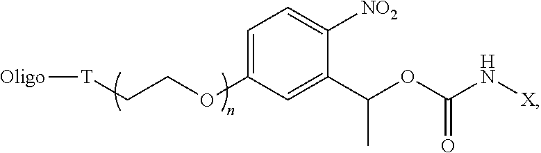
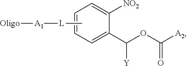
1. An oligonucleotide conjugate of the formula: ##STR00013## wherein Oligo is an oligonucleotide of 2-1000 nucleotides in length, A.sub.1 is the residue of a conjugation reaction, A.sub.2 is an amine reactive leaving group or --NHX, L is an optional linker, Y is H or C1-10 alkyl, and X is a cargo moiety.
2. The conjugate of claim 1, wherein L is present and amido, C1-10 alkylene, or C1-20 polyalkeneoxide.
3. The conjugate of claim 1, wherein L is present and C2-C20 polyethylene glycol.
4. The conjugate of claim 1, wherein A.sub.1 is triazolyl, disulfide, cyclohexenyl, amido, thioamido, acetal, ketal, or sulfonamido.
5. The conjugate of claim 1, wherein Y is C1-10 alkyl.
6. The conjugate of claim 1, wherein Y is methyl.
7. The conjugate of claim 1, wherein A.sub.2 is the amine reactive group.
8. The conjugate of claim 7, wherein the amine reactive group is p-nitrophenoxyl, N-hydroxysuccinimidyl, halide, pentafluorophenoxyl, or imidazolyl.
9. The conjugate of claim 1, wherein A.sub.2 is --NHX.
10. The conjugate of claim 9, wherein X is a therapeutic or diagnostic agent.
11. The conjugate of claim 1, having the formula: ##STR00014## wherein Oligo is an oligonucleotide of 2-100 nucleotides in length, T is a triazolyl linker formed from the reaction of an azide with an alkyne, X is a cargo moiety, and n is an integer from 1-10.
12. A crosslinker of the formula: ##STR00015## wherein A.sub.3 is a conjugating moiety, A.sub.4 is an amine reactive leaving group, L is an optional linker, and Y is H or C1-10 alkyl.
13. The crosslinker of claim 12, wherein L is present and C1-11 alkylene or C1-20 polyalkeneoxide.
14. The crosslinker of claim 12, wherein L is present and C2-C20 polyethylene glycol.
15. The crosslinker of claim 12, wherein A.sub.3 is azido, alkynyl, alkenyl, thiol, halide, boronic acid, hydroxyl, carboxyl, formyl, or ketone.
16. The crosslinker of claim 12, wherein Y is C1-11 alkyl.
17. The crosslinker of claim 12, wherein Y is methyl.
18. The crosslinker of claim 12, wherein A.sub.4 is p-nitrophenoxyl, N-hydroxysuccinimidyl, halide, pentafluorophenol, or imidazolyl.
19. The crosslinker of claim 12, having the formula: ##STR00016## where n is an integer from 1-10.
20. An oligonucleotide conjugate comprising an oligonucleotide of 2-1000 nucleotides in length, conjugated to a therapeutic or diagnostic agent by a photolabile linker, wherein upon suitable illumination, the therapeutic or diagnostic agent dissociates from the oligonucleotide.
21. A DNA construct comprising a three-dimensional DNA cage structure for housing a cargo moiety, wherein the cargo moiety is attached to the cage structure via the oligonucleotide conjugate of any of one of claims 1-11.
22. A method of delivering a cargo moiety, the method comprising providing a conjugate of any one of claims 1-11 and 20 and irradiating the conjugate with light to release the cargo moiety.
23. The method of claim 22, wherein the conjugate is internalized within a cell prior to irradiation.
24. The method of claim 22, wherein the cargo is a therapeutic or diagnostic agent.
25. A pharmaceutical composition comprising a conjugate of any one of claims 1-11 and 20 and a pharmaceutically acceptable carrier.
Description
BACKGROUND OF THE INVENTION
[0002] A general platform that could release a vast number of bioactive molecules with light would be of great interest to the neuroscience community. Currently neuroscientists buy caged neurotransmitters from commercial sources, but the number of compounds available is small relative to the amount of compounds that would like to be explored. Accordingly, there is a need for new crosslinking strategies in neuroscience and other fields.
SUMMARY OF THE INVENTION
[0003] The invention provides an oligonucleotide conjugate including an oligonucleotide, e.g., of 2-1000 nucleotides in length, such as 2-200 or 2-100 nucleotides in length, conjugated to a therapeutic or diagnostic agent by a photolabile linker, wherein upon suitable illumination, the therapeutic or diagnostic agent dissociates from the oligonucleotide.
[0004] In one aspect, the invention provides an oligonucleotide conjugate of the formula:
##STR00001##
wherein Oligo is an oligonucleotide, e.g., of 2-1000 nucleotides in length, such as 2-200 or 2-100 nucleotides in length, A.sub.1 is the residue of a conjugation reaction, A.sub.2 is an amine reactive leaving group or --NHX, L is an optional linker, Y is H or C1-10 alkyl, and X is a cargo moiety. In certain embodiments, L is present and is amido (--NHC(O)--), C1-10 alkylene or C1-20 polyalkeneoxide, e.g., C2-C20 polyethylene glycol or --O--CH.sub.2--; A.sub.1 is triazolyl, disulfide, cyclohexenyl, amido, thioamido (--NHC(S)--), acetal, ketal, or sulfonamide (--NHSO.sub.2--); and/or Y is C1-10 alkyl, e.g., methyl. In embodiments wherein A.sub.2 is the amine reactive group, examples of such groups include p-nitrophenoxyl, N-hydroxysuccinimidyl, halide, pentafluorophenoxyl, and imidazolyl. In embodiments wherein A.sub.2 is --NHX, X may be a therapeutic or diagnostic agent, e.g., as described herein. In particular embodiments, A.sub.1 and L are para to the nitro group.
[0005] In specific embodiments, the conjugate has the formula:
##STR00002##
wherein Oligo is an oligonucleotide of 2-100 nucleotides in length, T is a triazolyl linker formed from the reaction of an azide with an alkyne, X is a cargo moiety, and n is an integer from 1-10. In other embodiments, this formula is specifically excluded.
[0006] The invention further features a crosslinker of the formula:
##STR00003##
wherein A.sub.3 is a conjugating moiety, A.sub.4 is an amine reactive leaving group, L is an optional linker, and Y is H or C1-10 alkyl. In certain embodiments, L is present and is amido, C1-10 alkylene or C1-20 polyalkeneoxide, e.g., C2-C20 polyethylene glycol or --O--CH.sub.2--; A.sub.3 is azido, alkynyl, alkenyl, thiol, halide, boronic acid, hydroxyl, carboxyl, formyl, or ketone; Y is C1-11 alkyl, e.g., methyl; and/or A.sub.4 is p-nitrophenoxyl, N-hydroxysuccinimidyl, halide, pentafluorophenoxyl, or imidazolyl. In particular embodiments, A.sub.3 and L are para to the nitro group.
[0007] In certain embodiments, the crosslinker has the formula:
##STR00004##
where n is an integer from 1-10. In other embodiments, this formula is specifically excluded.
[0008] In a further aspect, the invention provides a DNA construct including a three-dimensional DNA nanostructure, e.g., cage structure, for housing a cargo moiety, wherein the cargo moiety is attached to the cage structure via an oligonucleotide conjugate of the invention.
[0009] The invention also provides a method of delivering a cargo moiety by providing a conjugate of the invention and irradiating the conjugate with light to release the cargo moiety, e.g., a therapeutic or diagnostic agent, e.g., as described herein. The conjugate is, for example, internalized within a cell prior to irradiation.
[0010] In another aspect, the invention provides a pharmaceutical composition including an oligonucleotide conjugate of the invention and a pharmaceutically acceptable carrier.
BRIEF DESCRIPTION OF THE DRAWINGS
[0011] FIGS. 1A-1E. Design and creation of light-triggered, cargo-releasing nanocages. (1A) Scheme of the chemical activation of a cargo molecule with the photolabile crosslinker and an oligonucleotide. (1B) Depiction of the DNA nanostructure formation. The solid cylinders represent DNA helices as shown by the insert. (1C) Agarose gel electrophoresis showing the high folding yield of the crude DNA nanocage sample. Lane L contains the 1-kb ladder, lane m13 contains the single stranded DNA starting material, and lane cage contains the crude reaction mixture. (1D) TEM images of DNA nanocages. Scale bars are 200 and 25 nm respectively. (1E) Schematic depiction of the encapsulation of cargo, the photo-cleavage reaction, and subsequent cargo release.
[0012] FIGS. 2A-2C. Light-triggered release of small molecules from nanocages. (2A) Photolysis data showing increased irradiation duration results in an increase in the cleavage of Oregon Green/oligonucleotides conjugate. (2B) Schematic depiction of the dye uncaging experiment. DNA nanostructures remain in the microdialysis chamber while small dyes are able to diffuse out. (2C) Absorption spectra of a duel dye tagged nanocage before (yellow curve) and after light irradiation (blue curve).
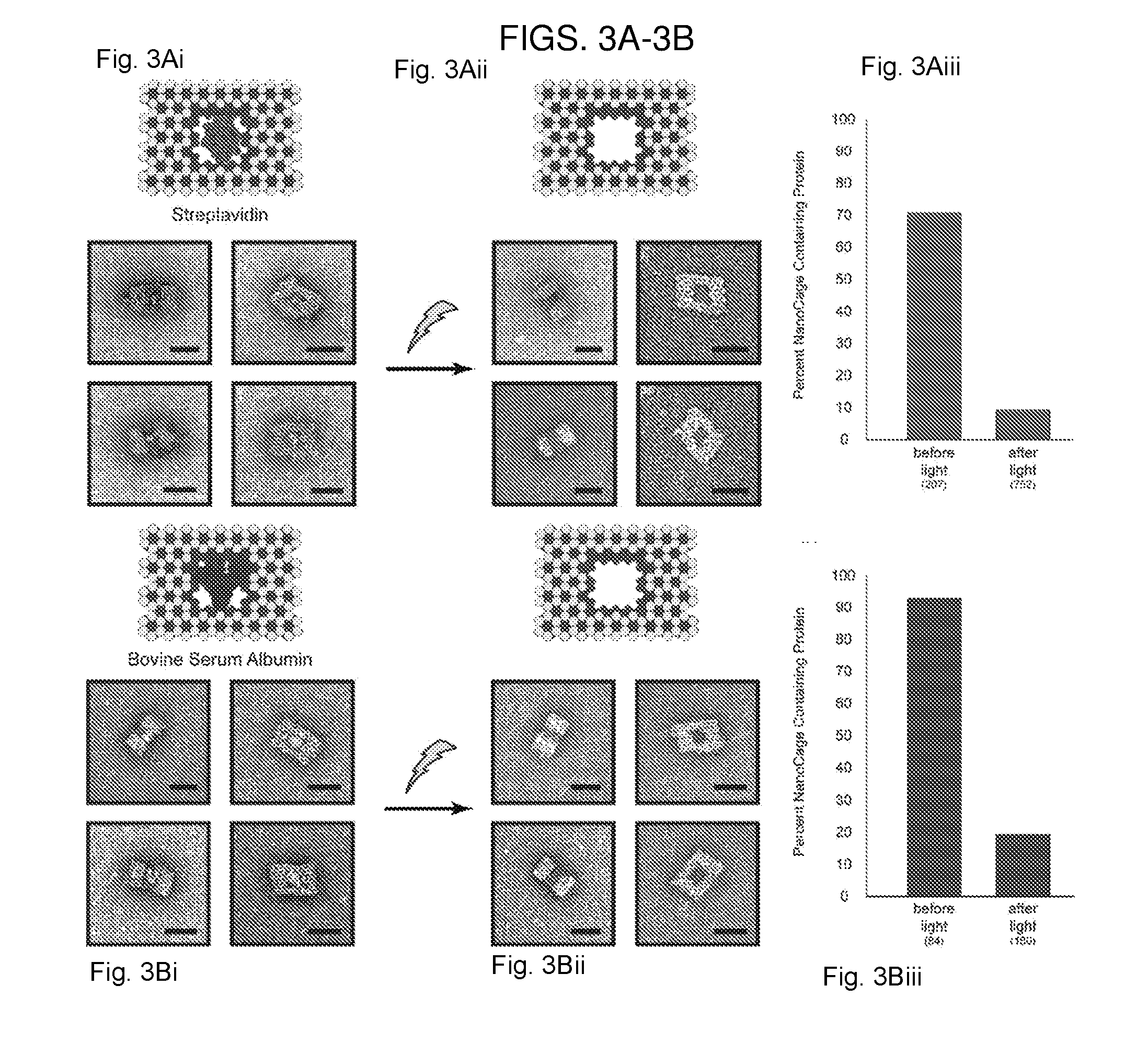
[0013] FIGS. 3A-3B. Light-triggered release of proteins, (3A) Streptavidin and (3B) Bovine Serum Albumin from nanocages. (3Ai, 3Bi) Schematic depictions of the DNA nanocages with and without proteins (3Aii, 3Bii) TEM images of nanocaged proteins before (left) and after (middle) irradiation with light. Scale bars are 25 nm. (3Aiii, 3Biii) Graphs showing percentage of nanocages containing protein as determined by TEM image counting before and after light are shown on the right. Numbers in parenthesis indicate the number of particles counted per condition.
[0014] FIGS. 4A-4D. Light-triggered release of glutamate from DNA nanocages. (4A) Schematic depiction of glutamate release from DNA nanocage using UV light at 240-400 nm and the subsequent activation of neurons by the freed glutamate. (4B, 4C) Temporal derivative of the normalized fluorescence intensity indicating calcium concentration changes in the control group, neurons illuminated in the absence of nanocages (4B, N=124 neurons), and in the uncaging group, neurons illuminated in the presence of nanocages (4C, N=185 neurons). (4D) Normalized fluorescence intensity indicating intracellular calcium activities of responsive cells in the uncaged group, aligned to light onset. Thick blue line indicates the mean, shaded gray indicates standard deviation, and red dots indicate the onset time (N=30 neurons).
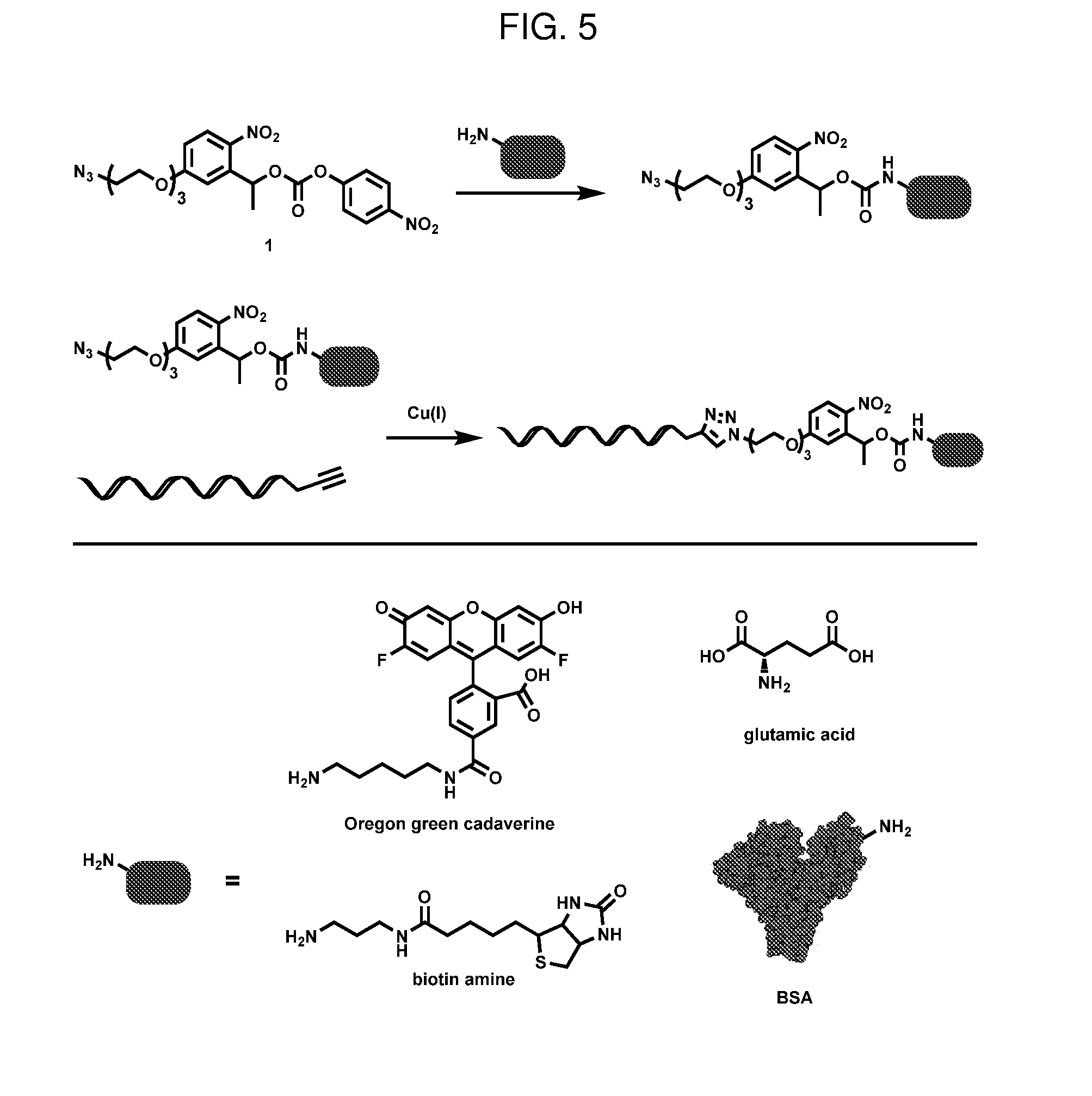
[0015] FIG. 5. General scheme for conjugation
[0016] FIG. 6. HPLC traces of Oregon Green cadaverine photolysis experiment. Increased irradiation duration results in an increase in the cleavage of Oregon Green/oligonucleotides conjugate. A wavelength of 490 nm was used to monitor traces.
[0017] FIG. 7. TEM images of unmodified DNA nanocage. Scale bars equal 100 nm (top three images) and 50 nm (remaining six images).
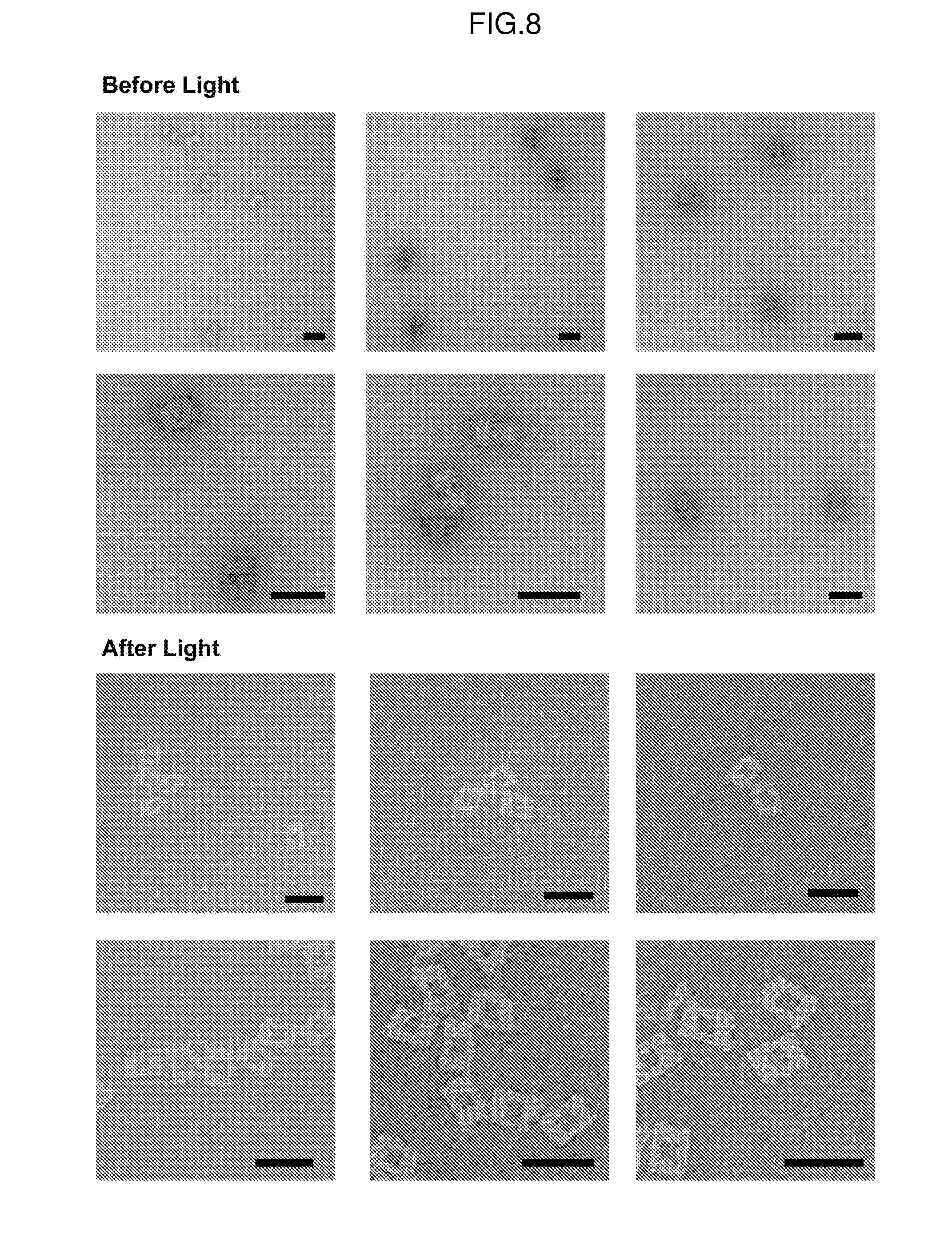
[0018] FIG. 8. TEM images of streptavidin containing DNA nanocages before (top) and after (bottom) light irradiation. Scale bars equal 50 nm.
[0019] FIG. 9. TEM images of bovine serum albumin containing DNA nanocages before (top) and after (bottom) light irradiation. Scale bars equal 50 nm.
[0020] FIG. 10A: Structure of glutamate-linker-oligonucleotide complex.
[0021] FIG. 10B: Normalized fluorescence intensity of the calcium indicator Fluo-4 in two neurons in the presence of glutamate-linker-oligonucleotide shown in 10A.
DETAILED DESCRIPTION OF THE INVENTION
[0022] In general, the invention provides oligonucleotide conjugates containing cargo moieties linked by a photolabile crosslinker. Illumination of the conjugate results in the release of the cargo moiety, which may be a therapeutic or diagnostic agent. This novel molecular uncaging technique offers a general approach for precisely releasing a large variety of bioactive molecules, allowing investigation into their mechanism of action, or finely tuned delivery with high temporal precision for broad biomedical and materials applications.
[0023] There is no other technology currently available that can accomplish what this invention does. The delivery platform described here is capable of releasing a large variety of bioactive molecules in a general fashion. There currently are commercially available caged neurotransmitters for a small number of molecules but not for the myriad of compounds that neuroscientists are interested in investigating. Additionally, there are many chemical crosslinkers commercially available but none that are photolabile and release the attached molecules in a chemically unaltered (and thus bioactive) form.
Conjugates
[0024] Conjugates of the invention feature an oligonucleotide conjugated to a cargo moiety (or molecule) via a photolabile crosslinker. The conjugates may also include the oligonucleotide conjugated to the crosslinker, which is capable of reaction with the cargo moiety. In one embodiments, the conjugate has the formula:
##STR00005##
wherein Oligo is an oligonucleotide of 2-1000 nucleotides in length, e.g., 2-200 or 2-100 nucleotides in length, A.sub.1 is the residue of a conjugation reaction, A.sub.2 is an amine reactive leaving group or --NHX, L is an optional linker, Y is H or C1-10 alkyl, e.g., methyl, and X is a cargo moiety, as described herein. Preferred linkers include amido, C1-10 alkylene, or C1-20 polyalkeneoxide, such as ethylene glycol or --O--CH.sub.2--. Suitable conjugation reactions include an azide-alkyne Huisgen cycloaddition (e.g., a copper(I)-catalyzed azide-alkyne cycloaddition (CuAAC) or a strain-promoted azide-alkyne cycloaddition (SPAAC)), amide or thioamide bond formation, a pericyclic reaction, a Diels-Alder reaction, sulfonamide bond formation, alcohol or phenol alkylation, a condensation reaction, disulfide bond formation, and a nucleophilic substitution. An exemplary A.sub.1 is triazolyl, disulfide, cyclohexenyl, amido, thioamido, acetal, ketal, or sulfonamido. For A.sub.2, any suitable amine reactive leaving group, such as p-nitrophenoxyl, N-hydroxysuccinimidyl, halide, pentafluorophenol, or imidazolyl, may be employed. In certain embodiments, A.sub.1 and L are para to the nitro group. In one embodiment, the conjugate has the formula:
##STR00006##
wherein Oligo is an oligonucleotide of 2-100 nucleotides in length, T is a triazolyl linker formed from the reaction of an azide with an alkyne, X is a cargo moiety, and n is an integer from 1-10.
Oligonucleotides
[0025] Any oligonucleotide, e.g., of 2 to 1000 nucleotides in length (such as 2 to 200 or 2 to 100 nucleotides in length), may be employed in the present invention. The term encompasses, for example, deoxyribonucleic acid (DNA), ribonucleic acid (RNA), hybrids thereof, and mixtures thereof. Oligonucleotides are typically linked in a nucleic acid by phosphodiester bonds, although the term also encompasses nucleic acid analogs having other types of linkages or backbones (e.g., phosphoramide, phosphorothioate, phosphorodithioate, O-methylphosphoroamidate, morpholino, locked nucleic acid (LNA), glycerol nucleic acid (GNA), threose nucleic acid (TNA), and peptide nucleic acid (PNA) linkages or backbones, among others). The oligonucleotides may be single-stranded, double-stranded, or contain portions of both single-stranded and double-stranded sequence. A nucleic acid can contain any combination of deoxyribonucleotides and ribonucleotides, as well as any combination of bases, including, for example, adenine, thymine, cytosine, guanine, uracil, and modified or non-canonical bases (including, e.g., hypoxanthine, xanthine, 7-methylguanine, 5,6-dihydrouracil, 5-methylcytosine, and 5-hydroxymethylcytosine). In addition, oligonucleotides may form a specific structure, e.g., as in an aptamer, or be part of a larger structure, e.g., a DNA nanostructure. Oligonucleotides can be modified as is known in the art to includes moieties that participate in conjugation reactions as described herein.
[0026] Recent innovations in DNA nanofabrication allow the creation of intricately shaped nanostructures ideally suited for many biological applications. To advance the use of DNA nanotechnology for the controlled release of bioactive molecules, we report a general strategy that uses light to liberate encapsulated cargoes from DNA nanostructures with high spatiotemporal precision. Suitable DNA nanostructures include cage structures with interior voids where a cargo moiety can be attached.
Cargo Moieties
[0027] Cargo moieties may be any suitable agent for controlled release from a conjugate of the invention. Examples include therapeutic and diagnostic agents, such as peptides, proteins, carbohydrates, other oligonucleotides, small molecules (e.g., neurotransmitters, vitamins, ligands, amino acids, and drugs), contrast agents, and dyes. Typically, the cargo moiety will be attached to the crosslinker by an amine group naturally present in the cargo moiety. For example, proteins typically have free amine groups available for conjugation. Alternatively, an amine group may be introduced into the cargo moiety by methods known in the art. Other attachments, however, are encompassed by the invention.
Photolabile Crosslinkers
[0028] Photolabile crosslinkers are known in the art. A preferred crosslinker employs an o-nitrobenzyl moiety.
[0029] Crosslinkers can be conjugated to the oligonucleotide and to the cargo moiety by orthogonal chemistries, i.e., different reactions that result in attachment of the oligonucleotide at a designated end of the crosslinker and the cargo moiety at the other end. In some instances, the crosslinkers can be conjugated to the oligonucleotide or cargo moiety by an azide-alkyne Huisgen cycloaddition (e.g., a copper(I)-catalyzed azide-alkyne cycloaddition (CuAAC) or a strain-promoted azide-alkyne cycloaddition (SPAAC)), amide or thioamide bond formation, a pericyclic reaction, a Diels-Alder reaction, sulfonamide bond formation, alcohol or phenol alkylation, a condensation reaction, disulfide bond formation, and a nucleophilic substitution. Preferably, the cargo moiety is attached to the crosslinker via an amine group in the cargo moiety.
[0030] In one embodiment, the crosslinker has the following formula:
##STR00007##
wherein A.sub.3 is a conjugating moiety, A.sub.4 is an amine reactive leaving group, L is an optional linker, and Y is H or C1-10 alkyl, e.g., methyl. Preferred linkers include amido, C1-10 alkylene, or C1-20 polyalkeneoxide, such as ethylene glycol or --O--CH.sub.2--. Suitable conjugating moieties for A.sub.3 include those capable of participating in the conjugation reactions discussed above, e.g., an azide, an alkyne (e.g., cyclooctyne), a diene, a dienophile, a thiol, an alkene, a halide, a boronic acid, hydroxyl, carboxyl, formyl, or ketone. For A.sub.4, any suitable amine reactive leaving group, such as p-nitrophenoxyl, N-hydroxysuccinimidyl, halide, pentafluorophenol, or imidazolyl, may be employed. In certain embodiments, A.sub.3 and L are para to the nitro group. An exemplary crosslinker has the formula:
##STR00008##
where n is an integer from 1-10.
Methods of Use
[0031] The conjugates of the invention may be employed to deliver cargo moieties to locations of interest. Once delivery, the cargo moiety can be released from the conjugate via illumination with the appropriate wavelength of light. The wavelength and duration of the illumination can be determined by one of skill in the art. Without being bound by theory, the conjugates can increase the stability of the cargo moiety by protecting the cargo from degradation or elimination. The conjugate can also prevent the cargo moiety from being active until release. The conjugates may be deployed to deliver the cargo moiety in vivo or ex vivo. The conjugate can be delivery topically, systemically, or locally within the body of a subject. Methods for illuminating cells, tissues, and portions of subjects are known in the art.
[0032] In a related aspect, the invention provides pharmaceutical compositions include a conjugate of the invention, which may or may not be part of a nanostructure, and a pharmaceutically acceptable carrier. Suitable carriers are known in the art and include water for injection, physiological saline, and buffers.
EXAMPLES
Example 1--DNA Nanostructure
[0033] Rapid advances in structural DNA nanotechnology allow the creation of intricately shaped nanostructures that can be functionalized with a high degree of control at precise locations..sup.1-4 For example, DNA origami can be reliably and efficiently self-assembled by folding large, single stranded DNA with a set of specifically designed short oligonucleotide strands..sup.5 This technique affords a tremendous amount of control over the size and shape of the nanostructure whose designs can now be assisted by well-developed software tools..sup.6-8 Molecularly programed, static.sup.9-11 or dynamic.sup.12-14 DNA architectures hold promise for applications in areas such as cell biology,.sup.15 NMR spectroscopy,.sup.16 super resolution microscopy,.sup.17 and nanotherapeutics,.sup.18 many of which would be advanced if DNA nanostructures were capable of releasing bound cargos at precise times.
[0034] Attempts to obtain controlled release from DNA origami nanostructures have thus far utilized two approaches through either non-covalent or covalent attachment of the cargo to origami. For example, the chemotherapy drug doxorubicin has been found to be able to non-covalently bind to DNA nanostructures through interactions with the DNA helices..sup.19,20 By controlling the DNA origami structure configuration, it was shown that doxorubicin release from the nanostructures could elicit a cytotoxic response in regular and drug-resistant cancer cells. Non-covalent attachment strategies however critically depend on a chemical's ability to intercalate into DNA helices. This binding mechanism cannot be generalized to most chemicals, and the binding sites within an origami cannot be easily controlled spatially. Direct covalent attachment of cargo to DNA origami nanostructures can overcome most of these limitations. To covalently attach a cargo to DNA helices, short DNA strands can be designed to protrude at specific locations on the surface of the nanostructures, which can then bind to a variety of different chemical moieties including inorganic nanoparticles,.sup.21,22 proteins,.sup.23 antibodies,.sup.18 and fluorophores..sup.24 Placement of cleavable linkages within these DNA strands permits the release of the bound cargo in a highly controllable fashion. However, such strategies often leave a chemical remnant, the chemical group that connects the cargo to DNA strands.sup.25 on the molecules being released, which may compromise their native biological function, limiting this approach to applications where the bioactivity of the cargo is important. Here, we demonstrate a novel and general method which releases chemically unaltered cargoes using brief pulses of light that can be broadly applied to a large variety of molecules.
[0035] We designed a novel, photolabile linker to append cargo molecules into the cavities of DNA nanostructures, so that light irradiation-induced breakage of the linker would allow the molecules to diffuse away from the protective cavity (FIGS. 1A-1E). This photolabile crosslinker possesses an o-nitrobenzyl (o-NB) motif for photo-cleavage, an azido group for attachment to alkyne functionalized oligonucleotides, and an activated carbonate group for attachment to cargo molecules possessing a free amino functional group (FIG. 1A). The linker is designed to release cargo upon photo cleavage in its original state with no chemical remnants remaining attached. Given that most peptides, proteins, and bioactive compounds contain exposed amino residues, the crosslinker design is broadly applicable to attach many molecules to DNA nanostructures, beyond the examples described here.
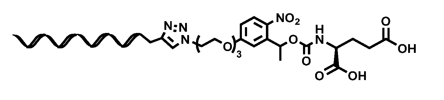
[0036] We first synthesized this photolabile crosslinker using conventional organic synthesis techniques. Gram scale product was easily produced from inexpensive, commercially available starting materials (Scheme 1). This photolabile crosslinker was then reacted with cargo molecules including glutamate, bovine serum albumin (BSA), and biotin amine, and subsequently conjugated to oligonucleotides allowing the cargo to be incorporated into pre-assembled DNA origami through DNA base pairing (FIG. 5).
[0037] In parallel, we computationally designed a multi-layered, brick-like nanocage structure with a well-defined cavity in its center, similar to those previously reported..sup.6,20,26 The nanocage contains 14 addressable, single-stranded DNA extensions in its cavity, which are complementary to those presented on the activated cargo (FIG. 1B-D). Nanostructures were then self-assembled in a single step by slowly cooling a heated mixture of the DNA components. Analysis of the assembly by agarose gel electrophoresis showed a single, dominant product band that migrated faster relative to the single stranded DNA starting material (m13 DNA), consistent with that generally observed for multi-layered DNA origami structures (FIG. 1C)..sup.9,21 Further examination with transmission electron microscopy (TEM) revealed properly assembled structures with the desired shape and a clearly visible central cavity (FIG. 1D). The short single-stranded DNA extensions however were too small to be resolved using TEM. Purification of fully formed nanostructures from excess oligonucleotides or subsequent cargo molecules was accomplished using polyethylene glycol precipitation..sup.27
[0038] Fully assembled and purified DNA nanocages were then incubated with the activated cargo to attach them to the interior of the nanocage cavity. When positioned inside of the nanostructure, the cargo is protected from the exterior environment and unable to bind to its native sites of action. Release from the cage was then achieved with light irradiation which cleaved the photolabile bonds within the crosslinker (FIG. 1E).
[0039] To first validate the photo-cleavage of our crosslinkers, we used it to conjugate an oligonucleotide to the small fluorescent molecule Oregon Green cadaverine (OG). We irradiated the compound with a low-power light source over time and quantified the degree of separation of OG from the oligonucleotide using HPLC (FIG. 2A). We found that an increasing duration of light exposure led to a larger fraction of free OG dye. After 11 seconds of low-powered light irradiation, 50% of OG was released. Nearly complete cleavage was achieved after 40 seconds of exposure, consistent with the time course for the cleavage of the o-NB motif within the crosslinker..sup.28
[0040] We then loaded the activated OG into the cavities of the nanostructures by incubating the OG/DNA conjugate with pre-assembled nanocages. To quantify loading efficiency, we incorporated a non-labile dye (Alexa Fluor 647N, AF647) for comparison by attaching it to a region on the nanostructure distal to the cavity (FIG. 2B). UV absorbance spectra analysis of the product showed two distinct absorption peaks centered around 500 nm and 647 nm, corresponding to the two dyes used (FIG. 2C, yellow trace). The ratio of the dye concentrations for OG versus AF647 was 7.4 to 1, suggesting that about half of the 14 DNA extensions on each cage designed to bind OG were bound, which is likely a representative loading capacity for small molecules of similar size.
[0041] To measure the efficiency of the light-induced release of OG from the nanocages, we irradiated the structures with a low powered lamp for 60 seconds, and then analyzed the absorbance spectra of the reaction solution after extensive sample dialysis of released free OG (FIG. 2B). We observed that the peak absorption at 500 nm corresponding to the photolabile OG dye was completely absent after irradiation, whereas the 647 nm absorption peak corresponding to the non-labile AF647 remained (FIG. 2C, blue trace). Together, these results demonstrate that our uncaging strategy can successfully release small molecular cargo from the DNA nanostructure upon brief low energy light irradiation.
[0042] We then explored the possibility of releasing large proteins from the nanocages, using bovine serum albumin (BSA) and streptavidin as examples that can be easily observed and analyzed using TEM. BSA was directly caged through the reaction of our crosslinker with the surface amino groups on the protein. Streptavidin was indirectly caged by attaching biotin-amine to the nanocage cavity and then subsequently mixing with the protein. TEM analysis of nanostructures at different orientations revealed clearly visible BSA and streptavidin proteins within the cavity of the DNA cage (FIGS. 3Ai-3Biii). None were seen tethered to the cage exterior. The number of DNA nanostructures with and without proteins was determined via particle counting of TEM images, and a loading efficiency of 93% for BSA and 71% for streptavidin was observed. After low power light irradiation for 60 seconds, we found only 19% of nanocages contained BSA, and 9% cages contained streptavidin, which corresponds to uncaging efficiencies of 79% for BSA and 87% for streptavidin. Together, these results demonstrate that full sized proteins can be effectively encapsulated and uncaged with high efficiency.
[0043] To demonstrate that molecules released from the DNA nanocages retain their bioactivity, we tested uncaging of the small molecule glutamic acid, an excitatory neurotransmitter which has been shown to be successfully uncaged in numerous instances (FIG. 4A)..sup.29,31 The bioactivity of the released glutamate from the nanocages was measured by glutamate mediated calcium changes in cultured neurons using real-time fluorescence imaging. Primary hippocampal neuron cultures were incubated with the intracellular calcium dye Fluo-4 and the glutamate-containing DNA nanocages. Before light illumination, little basal calcium activity was observed in the 9 days old cultures, consistent with the general activity patterns observed in neuron cultures of this age (FIGS. 4B and 4C)..sup.32 Immediately following a 1 ms light pulse illumination (240-400 nm), we observed an increase in intracellular calcium levels in 16.22% (N=185 neurons, analyzed in 2 tests) (FIG. 4C). Activated cells exhibited heterogeneity in response amplitude with activation onsets ranged from 509 ms to 18.19 s after the light pulse, which could be due to difference in diffusion time from the releasing site to the cell surface, the concentration of released glutamic acid on a given cell, and intrinsic variability of cellular calcium responses (FIG. 4D). The fact that light irradiation was delivered for 1 ms suggests that uncaging can be performed with millisecond temporal resolution. In the absence of the DNA nanocages, no cells exhibited a change in calcium levels upon light illumination (N=124 neurons, analyzed in 2 tests) (FIG. 4B). Together, these results demonstrate that DNA nanocages can be used to release functional bioactive molecules with millisecond temporal precision.
[0044] In conclusion, we describe a novel strategy to encapsulate bioactive molecules inside DNA nanostructures and release them using pulses of light. This strategy is realized through tagging DNA origami with a novel photolabile crosslinker that can be broadly used to encapsulate a large variety of molecules. With this crosslinker, a single, general chemical reaction scheme can be used to attach chemicals of interest to DNA origami through reacting with amino groups which are present on many biologically relevant compounds. This technique allows the release of cargo in its unaltered, bioactive state in contrast to existing labile conjugation chemistries, which often leave behind a chemical remnant that may interfere with the natural bioactivity of the cargo. This strategy was shown to be effective for a range of molecular sizes, from small molecules to full-sized proteins. Our nanocage design offers a high degree of addressability and customization, and versions could be created that accommodate a larger variety of cargo molecules or cocktails of molecules in precise stoichiometries by controlling the shape and dimensions of the nanostructures as well as the sequences of the strands protruding from the cavity. While light controlled uncaging techniques have been successful in releasing small molecules that rely on small, photochemical blocking chemical groups, our nanocaging platform could be easily designed to release many previously un-cagable compounds and accelerate progress in understanding chemical receptor binding or controlled release of therapeutics.
REFERENCES
[0045] (1) Seeman, N. C. Nature 2003, 421, 427. [0046] (2) Seeman, N. C. Ann. Rev. Biochem. 2010, 79, 65. [0047] (3) Zhang, F.; Nangreave, J.; Liu, Y.; Yan, H. J. Am. Chem. Soc. 2014, 136, 11198. [0048] (4) Jones, M. R.; Seeman, N. C.; Mirkin, C. A. Science 2015, 347. [0049] (5) Rothemund, P. W. K. Nature 2006, 440, 297. [0050] (6) Douglas, S. M.; Marblestone, A. H.; Teerapittayanon, S.; Vazquez, A.; Church, G. M.; Shih, W. M. Nucleic Acids Res. 2009, 37, 5001. [0051] (7) Kim, D.-N.; Kilchherr, F.; Dietz, H.; Bathe, M. Nucleic Acids Res. 2012, 40, 2862. [0052] (8) Pan, K.; Kim, D.-N.; Zhang, F.; Adendorff, M. R.; Yan, H.; Bathe, M. Nat. Commun. 2014, 5. [0053] (9) Douglas, S. M.; Dietz, H.; Liedl, T.; Hogberg, B.; Graf, F.; Shih, W. M. Nature 2009, 459, 414. [0054] (10) Han, D.; Pal, S.; Yang, Y.; Jiang, S.; Nangreave, J.; Liu, Y.; Yan, H. Science 2013, 339, 1412. [0055] (11) Benson, E.; Mohammed, A.; Gardell, J.; Masich, S.; Czeizler, E.; Orponen, P.; Hogberg, B. Nature 2015, 523, 441. [0056] (12) Bath, J.; Turberfield, A. J. Nat. Nano. 2007, 2, 275. [0057] (13) Kohman, R. E.; Han, X. Chem. Commun. 2015, 51, 5747. [0058] (14) Gerling, T.; Wagenbauer, K. F.; Neuner, A. M.; Dietz, H. Science 2015, 347, 1446. [0059] (15) Shaw, A.; Lundin, V.; Petrova, E.; Fordos, F.; Benson, E.; AI-Amin, A.; Herland, A.; Blokzijl, A.; Hogberg, B.; Teixeira, A. I. Nat. Meth. 2014, 11, 841. [0060] (16) Douglas, S. M.; Chou, J. J.; Shih, W. M. Proc. Natl. Acad. Sci. 2007, 104, 6644. [0061] (17) Jungmann, R.; Avendano, M. S.; Woehrstein, J. B.; Dai, M.; Shih, W. M.; Yin, P. Nat. Meth. 2014, 11, 313. [0062] (18) Douglas, S. M.; Bachelet, I.; Church, G. M. Science 2012, 335, 831. [0063] (19) Jiang, Q.; Song, C.; Nangreave, J.; Liu, X.; Lin, L.; Qiu, D.; Wang, Z.-G.; Zou, G.; Liang, X.; Yan, H.; Ding, B. J. Am. Chem. Soc. 2012, 134, 13396. [0064] (20) Zhao, Y.-X.; Shaw, A.; Zeng, X.; Benson, E.; Nystrom, A. M.; Hogberg, B. ACS Nano 2012, 6, 8684. [0065] (21) Zhao, Z.; Jacovetty, E. L.; Liu, Y.; Yan, H. Angew. Chem. Int. Ed. 2011, 50, 2041. [0066] (22) Schreiber, R.; Do, J.; Roller, E.-M.; Zhang, T.; Schuller, V. J.; Nickels, P. C.; Feldmann, J.; Liedl, T. Nat. Nano 2014, 9, 74. [0067] (23) Rinker, S.; Ke, Y.; Liu, Y.; Chhabra, R.; Yan, H. Nat. Nano 2008, 3, 418. [0068] (24) Dutta, P. K.; Varghese, R.; Nangreave, J.; Lin, S.; Yan, H.; Liu, Y. J. Am. Chem. Soc. 2011, 133, 11985. [0069] (25) Voigt, N. V.; Torring, T.; Rotaru, A.; Jacobsen, M. F.; Ravnsbaek, J. B.; Subramani, R.; Mamdouh, W.; Kjems, J.; Mokhir, A.; Besenbacher, F.; Gothelf, K. V. Nat. Nano 2010, 5, 200. [0070] (26) Sun, W.; Boulais, E.; Hakobyan, Y.; Wang, W. L.; Guan, A.; Bathe, M.; Yin, P. Science 2014, 346. [0071] (27) Stahl, E.; Martin, T. G.; Praetorius, F.; Dietz, H. Angew. Chem. Int. Ed. 2014, 53, 12735. [0072] (28) Holmes, C. P. J. Org. Chem. 1997, 62, 2370. [0073] (29) Matsuzaki, M.; Ellis-Davies, G. C. R.; Nemoto, T.; Miyashita, Y.; Iino, M.; Kasai, H. Nat. Neuro. 2001, 4, 1086. [0074] (30) Fino, E.; Araya, R.; Peterka, D. S.; Salierno, M.; Etchenique, R.; Yuste, R. Frontiers in Neural Circuits 2009, 3. [0075] (31) Olson, J. P.; Kwon, H.-B.; Takasaki, K. T.; Chiu, C. Q.; Higley, M. J.; Sabatini, B. L.; Ellis-Davies, G. C. R. J. Am. Chem. Soc. 2013, 135, 5954. [0076] (32) Soriano, J.; Rodriguez Martinez, M.; Tlusty, T.; Moses, E. Proc. Natl. Acad. Sci. 2008, 105, 13758.
Experimental Section
Crosslinker 1 Synthesis (Scheme 1):
[0077] Reactions were monitored by TLC using glass-backed silica gel 60 F254 plates. Flash chromatography was performed in a quartz column with a fluorescent indicator (green 254 nm) added to the silica gel. TLC bands were visualized by UV. Solvent ratios used as elutants are reported in v/v. The purity of the final products was obtained through .sup.1H NMR and .sup.13C NMR.
[0078] .sup.1H NMR data were obtained on a 500 MHz Varian VMNRS spectrophotometer at the Chemical Instrumentation Center at Boston University. Chemical shifts are reported in parts per million (ppm) and coupling constants were reported in Hertz (Hz). .sup.1H NMR spectra obtained in CDCl.sub.3 were referenced to 7.26 ppm and those obtained in DMSO-d6 were referenced to 2.50 ppm. ESIMS data were collected on an Agilent Single-Quad LC/MSD VL instrument at the Chemical Instrumentation Center at Boston University.
[0079] The following compounds were synthesized according to literature procedures: 5-hydroxy-2-nitroacetophenone (S1).sup.1 and ethylene glycol 2-azidoethyl ether tosylate (S2).sup.2.
##STR00009##
##STR00010##
1-(5-(2-(2-(2-azidoethoxy)ethoxy)ethoxy)-2-nitrophenyl)ethan-1-one (S3)
[0080] To a solution of 5-hydroxy-2-nitroacetophenone (2.46 g, 13.6 mmol) and ethylene glycol 2-azidoethyl ether tosylate (4.36 g, 13.2 mmol) in DMF (15 mL) was added potassium carbonate (3.77 g, 27.3 mmol), and the suspension was heated to 75'C. After 18 hours, the solution was concentrated in vacuo and partitioned between CH.sub.2Cl.sub.2 (40 mL) and NaHCO.sub.3 (20 mL). The organic layer was washed with NaHCO.sub.3 (3.times.10 mL), dried over Na.sub.2SO.sub.4, filtered, and concentrated in vacuo to produce 4.30 g (96% crude) of S3 as a dark brown oil that was taken on without further purification: .sup.1H NMR (500 MHz, DMSO-d6) .delta. 8.14 (d, J=9.1 Hz, 1H), 7.21 (dd, J=2.8 Hz, 9.1 Hz, 1H), 7.19 (d, J=2.8 Hz, 1H), 4.29 (m, 2H), 3.79 (m, 2H), 3.59 (m, 6H), 3.38 (m, 2H), 2.53 (s, 3H).
##STR00011##
1-(5-(2-(2-(2-azidoethoxy)ethoxy)ethoxy)-2-nitrophenyl)ethan-1-ol (S4)
[0081] To a solution of S3 (4.12 g, 12.2 mmol) in MeOH (30 mL) stirring in an ice bath was added sodium borohydride (723 mg, 18.7 mmol) in portions. After 2 hours, the solution was concentrated in vacuo and partitioned between CH.sub.2Cl.sub.2 (30 mL) and brine (20 mL). The organic layer was washed with NaHCO.sub.3 (3.times.10 mL), dried over Na.sub.2SO.sub.4, filtered, concentrated in vacuo, and purified via flash chromatography (2:1 ethyl acetate:petroleum ether) to afford 3.63 g (88%) of S4 as a yellow oil: .sup.1H NMR (500 MHz, CDCl.sub.3) .delta. 8.03 (d, J=9.1 Hz, 1H), 7.37 (d, J=2.8 Hz, 1H), 6.88 (dd, J=2.8 Hz, 9.1 Hz, 1H), 5.55 (dq, J=4.0 Hz, 6.3 Hz, 1H), 4.24 (m, 2H), 3.90 (m, 2H), 3.74 (m, 2H), 3.68 (m, 4H), 3.38 (m, 2H), 2.40 (d, J=4.0 Hz, 1H), 1.54 (d, J=6.3 Hz, 3H).
##STR00012##
[0082] Crosslinker (1).
[0083] To a solution of S4 (1.88 g, 5.51 mmol) and 4-nitrophenyl chloroformate (1.65 g, 7.86 mmol) in CH.sub.2Cl.sub.2 (21 mL) was added triethylamine (1.50 mL, 10.8 mmol). After stirring for 24 hours, CH.sub.2Cl.sub.2 (30 mL) was added and the solution was washed with NaHCO.sub.3 (20 mL), dried over Na.sub.2SO.sub.4, filtered, concentrated in vacuo, and purified via flash chromatography (gradient from 1:3 to 2:3 ethyl acetate:petroleum ether) to afford 2.16 g (78%) of S4 as a tan oil: .sup.1H NMR (500 MHz, CDCl.sub.3) .delta. 8.26 (d, J=9.3 Hz, 2H), 8.12 (d, J=9.1 Hz, 1H), 7.37 (d, J=9.3 Hz, 2H), 7.24 (d, J=2.7 Hz, 1H), 6.95 (dd, J=2.7 Hz, 9.1 Hz, 1H), 6.53 (quart, J=6.3 Hz, 1H), 4.25 (m, 2H), 3.93 (m, 2H), 3.76 (m, 2H), 3.69 (m, 4H), 3.39 (m, 2H), 1.76 (d, J=6.2 Hz, 3H).
General Bioconjugate Protocol (FIG. 5):
[0084] Bioactive, amino-group containing compounds were first reacted in slight excess with Crosslinker 1 in organic solvents such as methylene chloride or dimethylformamide and trimethylamine. In cases where the starting material was insoluble, a dimethylsulfoxide/aqueous buffer mixture was used.
[0085] Carbonate intermediates were subsequently reacted with an alkyne functionalized oligonucleotide via the copper catalyzed azide alkyne cycloaddition (CuAAC) reaction using published procedures..sup.3 In brief, equal volumes of alkyne functionalized oligonucleotide (410 uM in PBS) and activated carbonate (1 mM in DMSO) were mixed. A solution of copper sulfate (10 equivalents, 20 mM in water) and tris(3-hydroxypropyltriazoylmethyl)amine (THPTA) (50 equivalents, 50 mM in water) were separately mixed together and added to the reaction mixture. Lastly, a solution of sodium ascorbate (120 equivalents, 100 mM) was added and the reaction was stirred overnight. The reaction was subsequently purified via HPLC (TSKgel OligoDNA RP column, Tosoh Bioscience) using a gradient from 1:19 to 3:2 acetonitrile: 100 mM ammonium acetate over 30 minutes.
[0086] Glutamic Acid Conjugate.
[0087] To a solution of crosslinker 1 (322 mg, 637 .mu.mol) and L-glutamic acid di-tert-butyl ester hydrochloride (217 mg, 734 .mu.mol) in CH.sub.2Cl.sub.2 (6 mL) was added triethylamine (570 .mu.L, 4.09 mmol). After stirring for 48 hours, CH.sub.2Cl.sub.2 (25 mL) was added and the solution was washed with NaHCO.sub.3 (2.times.15 mL). The combined aqueous layers were washed with CH.sub.2Cl.sub.2 (2.times.10 mL). The combined organic layers were dried over Na.sub.2SO.sub.4, filtered, concentrated in vacuo, and purified via flash chromatography (1:2 ethyl acetate:petroleum ether) to afford 383 mg of a yellow oil. To a solution of this intermediate in CH.sub.2Cl.sub.2 (5 mL) was added trifluoroacetic acid (700 .mu.L, 9.15 mmol). The solution was concentrated in vacuo and purified via flash chromatography (5% MeOH in CH.sub.2Cl.sub.2) to afford 152 mg (47% over 2 steps) of product: .sup.1H NMR (500 MHz, DMSO-d6) .delta. 8.1 (dd, J=2.0 Hz, J=9.0 Hz, 1H), 7.8 (d, J=8 Hz, 1H), 7.2 (dd, J=2.8 Hz, J=17.3 Hz, 1H), 7.1 (m, 1H), 6.1 (m, 1H), 4.3 (m, 2H), 3.9 (m, 1H), 3.8 (quart, J=3.2 Hz, 2H), 3.6 (m, 4H), 3.4 (m, 2H), 2.3-2.2 (m, 2H), 2.0-1.7 (m, 2H), 1.5 (t, J=6.8 Hz, 2H), 1.4 (d, J=8.5 Hz, 3H). ESI-LRMS m/z 512.1 (M-) Product molecular weight=513.46. Azido intermediate was reacted with the oligo-alkyne using the general bioconjugate protocol and purified via HPLC to afford a solution of the product.
[0088] Biotin Bioconjugate.
[0089] To a solution of crosslinker 1 (103 mg, 204 .mu.mol) and biotin-amine (100 mg, 234 .mu.mol) in CH.sub.2Cl.sub.2 (1 mL) and DMF (1 mL) was added triethylamine (150 .mu.L, 1.08 mmol). After stirring for 2 hours the solvent was removed with a stream of air. CH.sub.2Cl.sub.2 (25 mL) was added and the solution was washed with NaHCO.sub.3 (15 mL). The aqueous layer was washed with CH.sub.2Cl.sub.2 (10 mL). The combined organic layers were dried over Na.sub.2SO.sub.4, filtered, concentrated in vacuo, and purified via flash chromatography (gradient 2% to 10% MeOH in CH.sub.2Cl.sub.2) to afford 109 mg (80%) of a solid: .sup.1H NMR (500 MHz, DMSO-d6) .delta. 8.1 (d, J=9.0 Hz, 1H), 7.7 (t, J=5.8 Hz, 1H), 7.4 (t, J=5.8 Hz, 1H), 7.1 (m, 2H), 6.4 (s, 1H), 6.3 (s, 1H), 6.1 (quart, J=6.5 Hz, 1H), 4.2 (m, 3H), 4.21 (m, 1H), 3.8 (t, J=4.5 Hz, 2H), 3.6 (m, 4H), 3.4 (t, J=5.0 Hz, 2H), 3.1 (m, 2H), 3.0 (m, 2H), 3.0-2.8 (m, 2H), 2.6 (d, J=12.5 Hz, 1H), 2.0 (t, J=7.5 Hz, 2H), 1.5 (m, 5H), 1.6-1.2 (m, 8H). Azido intermediate (128 .mu.L, 2 mM in DMSO, 256 nmol) was reacted with the oligo-alkyne (120 .mu.L, 410 .mu.M in 1.times.PBS, 49.2 nmol) using the general bioconjugate protocol. The product was isolated by ethanol precipitation and purified via HPLC to afford 11.3 nmol as a 100 .mu.L, 113 .mu.M solution of the product.
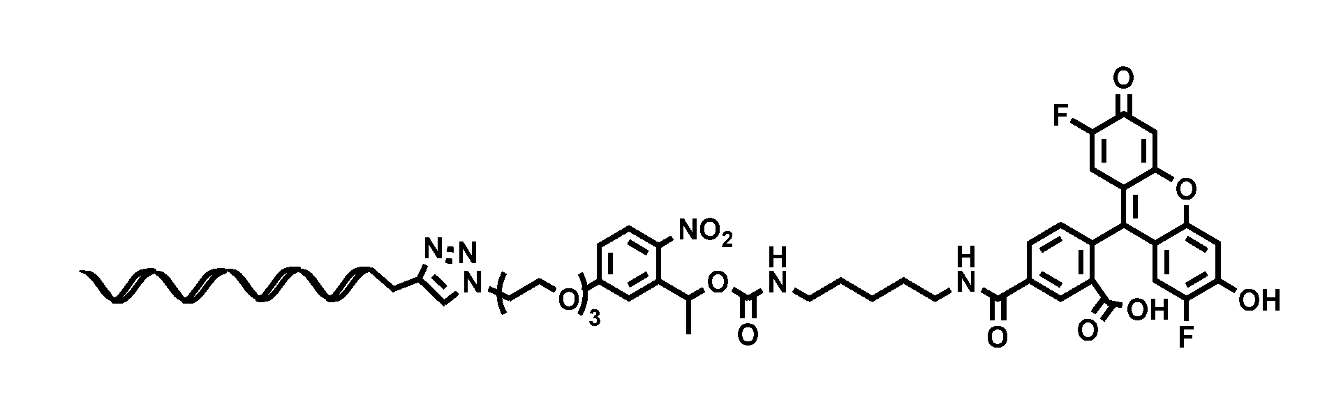
[0090] Oregon Green Conjugate.
[0091] To a solution of crosslinker 1 (1.25 mg, 2.52 .mu.mol) and Oregon Green cadaverine (1.21 mg, 2.39 .mu.mol) in DMF (300 .mu.L) and water (20 .mu.L) was added triethylamine (20 .mu.L, C). After stirring overnight the solution was purified via HPLC to afford 2.39 mg an orange solid: ESI-LRMS m/z 863.2 (M+) Product molecular weight=862.80. Azido intermediate (150 .mu.L, 1 mM in DMSO, 150 nmol) was reacted with the oligo-alkyne (150 .mu.L, 410 .mu.M in 1.times.PBS, 61.5 nmol) using the general bioconjugate protocol and purified via HPLC to afford 38 nmol as a 200 .mu.L, 190 .mu.M solution (62%) of the product.
[0092] BSA Conjugate.
[0093] A solution of BSA (200 .mu.L, 500 .mu.M in 1.times.PBS, 100 nmol) and crosslinker 1 (20 .mu.L, 5 mM in DMSO, 1000 nmol) in 80 .mu.L DMSO was mixed overnight. The reaction was centrifuged at 17000 rcf for 5 minutes to pellet insoluble materials. The supernatant was dialyzed in 1.times.PBS against a 25 kDa cutoff to afford 300 .mu.L (333 .mu.M) of product. Azido intermediate (50 .mu.L, 333 .mu.M in PBS, 16.7 nmol) was reacted with the oligo-alkyne (200 .mu.L, 410 .mu.M in 1.times.PBS, 82 nmol) using the general bioconjugate protocol and purified using Amicon spin filters (3 spins with 30 kDa cutoff tube and 3 spins with 50 kDa cutoff tube) against buffer (5 mM Tris, 1 mM EDTA, and 16 mM MgCl.sub.2) to produce 50 uL of product solution.
[0094] Design and Assembly of DNA Nanostructures.
[0095] Nanostructures were designed using caDNAno..sup.4 Single stranded M13mp18 bacteriophage DNA was prepared as described previously..sup.5 All oligonucleotides were purchased from Integrated DNA Technologies (IDT) and used with no additional purification. Creation of nanostructures was performed by first heating a solution containing a final concentration of 40 nM m13 scaffold DNA and 200 nM of each staple in a folding buffer containing 5 mM Tris, 1 mM EDTA, and 20 mM MgCl.sub.2 to 80% C, followed by cooling from 80.degree. C. to 60.degree. C. over 80 minutes, and then from 60% C to 24.degree. C. over 48 hours. Removal of excess staple strands was accomplished by three rounds of precipitation with polyethylene glycol solutions..sup.6 Pellets were re-dissolved in 5 mM Tris, 1 mM EDTA, and 16 mM MgCl.sub.2. FIG. 7 shows a TEM image of unmodified nanocages.
[0096] Cavity Functionalization.
[0097] Nanostructures were mixed with 70 equivalents of oligo bioconjugates (5 equivalents per handle, with 14 handles in the cavity interior) and incubated overnight at 40.degree. C. and subsequently purified by at least two rounds of PEG precipitation.sup.6.
[0098] Gel Electrophoreals.
[0099] Reaction solutions were electrophoresed on 1.5% agarose gels containing 0.5.times.TBE, supplemented with 10 mM MgCl.sub.2. DNA dye SybrSafe was mixed with gel solutions before loading onto the gel. The gel box was submerged in an ice water bath to prevent excessive heating.
[0100] TEM Sample Preparation and Imaging.
[0101] TEM samples were prepared by placing 3 .mu.L of sample solution onto a carbon coated grid (FCF400-Cu, Electron Microscopy Sciences) which was previously charged using a plasma etcher (30 seconds of irradiation). After 2 minutes, the solution was wicked away from the grid with filter paper (Whatman 50 hardened). The grid was immediately treated with stain for 30 seconds and excess solution was wicked away. The remaining solution on the grid was evaporated at room temperate prior to imaging. TEM images were acquired with an FEI Tecnai Spirit Transmission Electron Microscope operated at 80 kV. Saturated uranyl formate (in ddH.sub.2O prepared freshly before usage) was used for protein caging experiments and 2% uranyl acetate (diluted with ddH.sub.2O from 4%, Electron Microscopy Sciences) was used for all other samples.
[0102] Kinetics of o-NB Cleavage.
[0103] Samples of the Oregon Green cadaverine oligonucleotide bioconjugate were irradiated with a handheld UV lamp (UVM-57, 6 W, 302 nm) for varying lengths of time and analyzed using HPLC (TSKgel OligoDNA RP column, Tosoh Bioscience) using a gradient from 1:19 to 3:2 acetonitrile:100 mM ammonium acetate over 30 minutes. Irradiation durations used were 5, 10, 15, 20, 25, 30, 35, 40, and 60 seconds. A UV detector monitoring at 490 nm was used to collect traces containing Oregon Green. The degree of o-NB cleavage was obtained by comparing the areas under the peaks corresponding to the Oregon Green-oligo conjugate starting material with the released Oregon Green cadaverine (FIG. 6).
[0104] 2 Dye Labeling Experiment.
[0105] The general cavity functionalization protocol was followed but with two different oligos. 5 oligos per binding site were used. The cavity contained 14 binding sites for the activated Oregon green oligo, whereas 1 binding site on the unfolded loop was available for the Alexa Fluor 647 oligo. Reactions were incubated overnight at 40.degree. C. and subsequently purified by at least two rounds of PEG precipitation.sup.6. The final solution was analyzed using the UV setting of a Nanodrop 2000. The ratio of the dyes was obtained by comparing the concentrations of each dye in solution as calculated with Beer's law.
[0106] Oregon Green Cadaverine Uncaging.
[0107] 25 .mu.L 2 dye labeled nanostructures was irradiated with a handheld UV lamp (UVM-57, 6 W, 302 nm) for 60 seconds. The solution was placed in half of a microdialysis chamber and dialyzed against 2 .mu.L of buffer (5 mM Tris, 1 mM EDTA, and 16 mM MgCl.sub.2) overnight. The resulting solution was analyzed using the UV setting of a Nanodrop 2000.
[0108] Protein Uncaging.
[0109] Protein containing nanocages were created following the general cavity functionalization protocol. 5 equivalents per oligo handle were used. For BSA encapsulation, the BSA/oligo bioconjugate was used. For streptavidin, the nanocage was first modified with the biotin-amine/oligo bioconjugate and subsequently with streptavidin (5 equivalents per oligo handle). Each round of modification was purified using two rounds of PEG precipitation.sup.6. Uncaging experiments were performed by irradiating a PCR tube containing 5 uL of a 0.5 nM solution of protein-containing nanocage for 60 seconds with a handheld UV lamp (UVM-57, 6 W, 302 nm). Samples were heated at 40.degree. C. for 30 seconds and then imaged by TEM (FIGS. 8 and 0). The extent of uncaging was analyzed using particle counting of the TEM images. The entirety of each TEM image was analyzed to avoid bias.
[0110] Glutamate Uncaging.
[0111] Glutamate containing nanocages were created following the general cavity functionalization protocol. 5 equivalents of activated glutamate/oligo handle were used. Reactions were incubated overnight at 40.degree. C. and subsequently purified by three rounds of PEG precipitation.sup.6. For cell testing, the structures were PEG precipitated and dissolved in a modified Tyrode buffer (25 mM HEPES, 119 mM NaCl, 5 mM KCl, 2 mM CaCl.sub.2, 10 mM MgCl.sub.2, pH 7.4), at a final concentration of 180 nM. 9 days old primary rat hippocampal neurons were prepared on 12 mm diameter glass coverslides. The calcium dye Fluo-4 AM (life technologies) was dissolved in DMSO to yield a stock concentration of 2.3 mM. Neurons were loaded for 30 min in Fluo-4 AM at 2.3 uM, diluted in the modified Tyrode buffer at room temperature. Neurons were then rinsed three times with Tyrode buffer, and incubated at 37 C for another 30 min to allow complete de-esterification of intracellular AM esters. Glass coverslides were fractured into smaller pieces (approximately 1 mm.sup.2) with a pointed tungsten-carbide glass cutter to limit the use of nanocage reagents. The buffer was wicked off the surface of the fractured glass and replaced with 2 uL glutamate containing nanostructures in Tyrode. Neurons were then placed under a custom microscope with a 10.times. objective, equipped with a 5 W LED (LZ1-00B200, 460 nm; LedEngin, San Jose Calif.) for excitation, an excitation filter (HQ 470/50), a dichroic mirror (FF506-Di02), an emission filter (FF01-536/40), and imaged with a Hamamatsu camera (C11440-42U) at 20 Hzn After baseline activity was collected for 5 s, the flash lamp (JML-C2, Rapp OptoElectronic GmbH, Hamburg, Germany) was triggered to deliver a light pulse for 1 ms at 240-400 nm, and the calcium activities of neurons were measured for another 25 s.
[0112] Calcium Signal Processing.
[0113] All analysis was conducted with MATLAB (MathWorks, Massachusetts, US). Individual neurons were manually identified, and the mean fluorescence intensity averaged for all pixels within each neuron was then further processed to represent individual neuron calcium changes. Due to the saturation effects of the high intensity uncaging flash light that lasted for 6 frames (300 ms) following the light illumination, the fluorescence intensities of these 6 frames were removed and replaced by a linear fit connecting the end values that were not affected by the flash. The fluorescence of each neuron was first baseline subtracted using its linear fit for the 5 second baseline period, and then normalized by the standard deviation of the baseline and smoothed using a built-in function, Smooth, with a moving average filter with the span of 25 frames. The temporal derivative of the signal was calculated and smoothed using the moving average filter with the span of 6 frames. To screen for activated cells, we first calculated the root mean squared error (RMSE) for each 5 second intervals throughout the 30 second recording sessions, and thus 6 RMSE values were calculated. We then used the maximum RMSE of these 6 values to represent the RMSE of each neuron, and obtained the 95% confidence interval of the RMSE for the control group. We then calculated the threshold value for the instantaneous temporal derivative that would correspond to the 95% confidence interval of the RMSE. Cells were deemed as activated when their temporal derivative exceeds the threshold. To determine the onset of calcium responses, we calculated the z score of the fluorescence trace of the activated cells. Onset threshold were set as the first time point of 10 consecutive points in which the temporal derivative values had a z score bigger than 3.
TABLE-US-00001 Staple List: SEQ ID NO SEQUENCE Center staples-those which do not come in contact with the edges of the nanostructure 1. GGATTAGCAATATAAAAAGCG 2. AATCGTCATAAATATTCAGAATTTG 3. CAATAGAAAGGGCGACATTAACTGT 4. CGAAAGAAGGCTTTGAGGAGCACAG 5. GCCCGAAATTGCATTGGAAGTGCGA 6. TACCTTTTTACATTACAAACATACC 7. ATTATCAGGAATTATCATCGTTGCCTTA 8. CAGACGAGCATTGAAGAACCAATGAAAC 9. GAAAAAGAAATCCAATCGCAGCCAGGTT 10. GCATGATCAAGAAAATTGAGTAAAATAG 11. GCTATAATGCAGTACGGATTTGGGCAAT 12. GGGTTATACCTACCATATCAGAAGTTTG 13. TCCGCGACATCGCCACCTTATAGGACGT 14. TTTGTCAGGCAACAACGTAGAGCAACTG 15. ATACGTTTTAGCGAACCGAACGCCTACGCAT 16. TTTCCTTGAAAACAGTCAATAGTGAAGAGTGTAAC 17. TAAATCATCCAGTTTGGAAGCGCCAGGGAGCTGATTAT 18. CCCTCAAGACGGAATAGGTGTAAAAGAAGGCACCAGTAA 19. GATTATACATTAAAAATACAACGAACCGTCTATCAATCA 20. AAAAGCGGTTACCAGAAGGAAAGCAGATACCGAAGTTATCCC 21. AAGTTTACCAGACGCAAAAGAAGTTTGTGCAGACGGTCGAAA 22. AATTGGGCTTAGAAACATCAGTGAAAATCAAGACAAGACAAT 23. ACAAATGAATAACAGCTGCTTGCTACCAGTCGCGATTTCTTT 24. ACTAAGGAATAACTAATGTTGAGAAATATATATACATTAATA 25. ATGAAAATAAGGTAACCCACAAGACAATGAAATAGCTGAATT 26. CATCGTATAGCACCATTACCAAGCCAGCCCGACTTAATACCC 27. CCAGCTACAAGTCTTTCCATAATGGGATAGGTGCATCTGCAC 28. CTGTTTACTCAACAGTAGGGCAACAGTATAAAGCCGAAAACT 29. GATGCCAGAGGGGGAATACTGCGGAAGCACGGTGTATCATAA 30. GCAAATCACATCATTACCGCGCAATAGATAAGTCCCACGCGC 31. GCGCGAAATTTGACCCCCAGCAAAAAGGCTCCAAAAGGTTGA 32. GGCTTAAAATTTATCAAAATCATAATCCTGATTCCAGATATT 33. TGGGATAAAACACTCATCTCATGATACCGATAGCATAATTTT 34. TTTAATCATAACCGACCGGTAAAGGGCATTTAAACCAAATCA 35. TTTTCAAACAAGACAAAGAACAAAACAGAAATAAAAGAAAAT 36. ATACGAGCCGGGAGTGAGAGGTGAGCACGCTGGAAATTGTACA 37. AATTCATGAGTACAAACGCCTGGCGGGCAACTGGTTTTGCGGTTT 38. ATATCAATCAAAAAACATTCGCGTCCAATAGAGCTTTCATAGCAA 39. CCACCACCACCGGAATCCAAAAAGGGTCTTTACCCTGATCCATAA 40. GAATACACTAACGCCAAATCATAACCCTCTTTGATAAAATACCAA 41. TATTTGCAGAAGATAAAACAGCTCGAACGAACCACTTGCATGCCC 42. TTCTGCCGCCTCCCTCAGCCACCACCCTCATTCAAAGCAGAGGAA 43. AAATCTCGCGTAACGATCTAAAGACAGCTGAGTTTCGTCACCCTAA 44. ACCGTCAAAAATCACCAAGCAATAAAGCAAACATTTAGCTATGCTG 45. CGCCACCCAGGAGGTTGCTCCTTTTGATAATTGCTCATCCAAATTC 46. CTATCTTAGCCGAACATGAGAGTCTGGAAAACTAGCAACCCGATCA 47. TGTTTCCAACCTGTCTCACATAATATCACCAGCAGTTGAATATACC 48. AATAAATACAAACAACCGATTGAGATTAAAGGTGAAGATAATAGTTATT 49. ACAGTTTGAGGCACTCCAGCCTCCCGACTTGTTGCTATTTTGCCATTAA 50. AGAGAGCCGCTTGCCTTTAGCGTAAGATAGCAGCACCGATTATTCGGAG 51. CCGACAATATTCGGTATTAAAATCGGCAAAATCCCCCCAGCATCAGCAG 52. GTAATATATTTGGTTTGTTAATTGATTTAGGTGAACAATGTAGAAAGAT 53. TTGGTGAGAAGCTACAGCAGCATCCCACGCTGGTTTGCCTTCACCAATT 54. CAAGAAAAATCTACTACAGGTTTGCTTCTTAAAAGTTTGACACAACTCGTCCTAAA Right edge staples-those which come in contact with the right edge (when visualized in cadnano) of the nanostructure; helix ends contain TT overhangs to prevent nanostructure aggregation 55. TTGACTACCTTTTTAACCTCC 56. TTATGAACGGTCCCGGTTGATT 57. TTTCATTACCCATAAGGCTTTT 58. TTAACCACCAGAGCCCGAGATT 59. TTGGGCGCATCCGACAGTATTT 60. TTCCACGCATAAGTTAAAGGTT 61. TTGCTCATTATTTCGAGGTGTT 62. TTTTAAATATGCATATAACATT 63. TTCGGCCTCAGGCTTCTGGTTT 64. TTTAATCAGAAATATTTAAATT 65. TTAATTTCTTATTTCTGTATTT 66. TTTAATTTCAATAAGAACTGTT 67. TTAAATCGGTTTGCGGGAGATT 68. TTTAAGACGCTGAACAAAGATT 69. TTGCCCTGACGGAGATGGTTTT 70. TTAACGGAACAAACCATCGCTT 71. TTGTTGATTCCACCGGATATTT 72. TTAGCCTTTATCATATATTTTT 73. TTGGATGGCTTCAACATGTTTT 74. TTACGGCCAGTAGGATCCCCGGGTT 75. TTGGGATTTTGAGTACAAACTACTT 76. TTTGTACCAACTCAGAGCATAAAGCTTT 77. CGAGTAACCGTCACGTTGGTGTAGATTT 78. AACACATTATGTTAATAAAACGAACTTT 79. TACAATCGTAGCAAACAAGAGAATCGTT 80. TTACAACATGTTCAGAGAACAAGCAATT 81. TTGAAGCAAAGCGGAGACTTCAAATATT 82. TTTTGTATCGGTTTACAAAGTACAACTT 83. TTTTTGAAAGAGGACTGGCTGACCTTTT 84. TTGGACTCCAACGTATCAGATGATGGTT 85. TTGCCGTTTTTATTTTGTTAAATCAGTT 86. TTTAGGGTTGAGTGTTGTAAAGAATAAGG 87. TTTCAACCGTTCTATTTTGAGAGATCTT 88. TTCTAAAACATCGCCTTCTGAATAATTT 89. TTTTTCCAGACGTTAGGAGCCTTTAATT 90. TTGGAGATTTGTATCCTGCTCCATGTTT 91. TTCATCAAGAGTAACTATTATAGTCATT 92. TTAAATGCTGATGCCCTGTTTAGTATTT 93. TTTACTTAGCCGGAGAACTGACCAACTT 94. TTTTCGCAAATGGTGCGCGAGCTGAATT 95. TTAAGGTGGCATCATAAATCATACAGTT 96. TTTCGCGTTTTAATACTCCAACAGGTTT 97. TTTAGCCAGCTTTCTCGGATTCTCCGTT 98. TTCATATGCGTTATCGACGACAATAATT 99. TTCAATTCATCAATATAGGTCTGAGATT 100. TTGGAAGGGTTAGAATAACTATATGTTT 101. TTACGTTATTAATTTGTAAATCGTCGTT 102. TTGGTGGTTCCGAATCCTTTGCCCGATT 103. TTCTCATTTTTTAACTGGCCTTCCTGTT 104. TTAAGATTTAGTTTGACCATTAGATACATTT 105. AGCTTTTGCGGAGAAGATAGCGATAGCTTAGATTT 106. TTCCGCTTTTGCGGGATCTGCAGGGACCGATATGAC 107. TTAACGCCTGTAGCATTCCACAGTTTTGTCGTCTT 108. TTGCCGGAAACCAGGCCACGGCACCGAAGATCGGGA 109. TTTTGTAAACGTTAATATTAAGCAAAAGCCCCTATG 110. TTTACCGAGCTCGAATTCGTAGAACTGATAGCCTT 111. TTTAAATGCAATGCCTGAGAACCCTTTCAACGATAC 112. TTCAAGGCGATTAAGTGTGCAGGGGGATGTGCTGTT 113. TTCATTCAGGCTGCGCAACCAAAGCGCCATTCGCTT 114. TTGTTTTCCCAGTCACGCACTGGGTAACGCCAGGTT 115. TACCCCTGTACAAGGATTACACCATCAATATGATATTT 116. TTCGCCAGCTGGCGAAGAAACCTCTTCGCTATTATT 117. TTGCGATCGGTGCGGGCGTCAACTGTTGGGAAGGTT 118. TGGCAGGTCGACTCTAGGCCAAGCCAGACGTTGTAAAACGTT 119. TTTGGGAACAAACGGCGGATTGACCGGACGAGTAACAAATAG 120. TTTACAAAGGCTATCAGGTCATTGCCAAGAGAGGGATTTATC
121. TTGCAAGGCAAAGAATTAGCAAAATTAGGCATTAAGAAGAGC 122. TTCAGGATTAGAGAGTACCTTTAATTGACAGACCGGAAGCAATCGA 123. TTCACTAACTTTCATGAGGCTGTCACCCGGCGAAAATCCTGTTTGATTT 124. TCTTTCATTCCAACTAATGTAGCTAGAGCTTAAGAGGTCATTTTTGCTT 125. CCTGATTCAAAGGGCGAAATGGGCAAGAGTCCACTATTAAAGAACGTTT 126. TTCTATTAATTAATTTTCCCTTAGAACAAATAACCAGAAAGAGCTTGCG 127. TGAACACATATCAGAGAGAAATAAAGGTCATAAAGATTCAAAAGGGTGAGAAAGTT Left edge staples-those which come in contact with the left edge (when visualized in cadnano) of the nanostructure; helix ends contain TT overhangs to prevent nanostructure aggregation 128. TTACCACATTCTACGAGGCATT 129. TTATTAAACGGACCTAAAACTT 130. TTAGGAGGTTTAGTACCGCCA 131. TTGTAGCAACGTAGAAAGGATT 132. TTTTCTAAGAAATAACATAAAAATT 133. TTGCATTTTCGGTCATAATCAAATT 134. TTACCAACGCTTTACAAAATAAATT 135. TTCTAACAACTTGAGGATTTAGATT 136. TTCAGTGAGACCCTGAGAGAGTTTT 137. TTAATCAACAGTTGTTAGGAGCATT 138. TTCAGCCATATTATCCCTTTTTATT 139. TTCTTAAATCAATTTTTTGTTTATT 140. TTCCGCCTGCAAAAATCTAAAGCTT 141. TTAGTACATAAATCTTTAGGAATTT 142. TTGCAGCAAGCGGTGGAACGAGGTT 143. TTACAGAATCAAGTCACCCTCAGTT 144. TTAGACTGGATTCGGAACCTATTTT 145. TTTTGACGGAATAATCAGTAGCGTT 146. TTCCGCCGCCATTGGCCTTGATATT 147. TTTAGTAAGAGATAAGTGCCGTCTT 148. TTCAATAATAACGGGAGCCATTTTT 149. TTCAAATGCTTGCCTTGAGTAACTT 150. TTGAAACGTCACCACCACCAGAGTT 151. TTAACCGCCACGAAAGCGCAGTCTT 152. TTTTACGCAGTATGAGGTAAATATT 153. TTTAGCGAGAGCTCAAGAGAAGGTT 154. TTGGGTAATTGGAAACGCAAAGATT 155. TTACAACGCCAAGTAATAAGAGATT 156. TTCAGGGAAGCGCAAAAGTCAGATT 157. TTGAAAGAGGCATCACCGTACTCTT 158. TTCCAGCGCCAGCGTTTTCATCGTT 159. TTAGTATTAGACTTGAAGTTTCCTT 160. TTCACCACGGAATATATGGTTTATT 161. TTTCCTAATTTGTCTTTCCTTATTT 162. TTGCGCGGGGAGAGTCTTTTCACTT 163. TTAGAAAAGTAACCGAGGAAACGTT 164. TTCAAAATTAATTTAATGGAAACTT 165. TTTAACGTCAGGGAGAAACAATATT 166. TTATTCATTTCAATTCAAGAAAATT 167. TTGGGAATTAGTTAGCAAGGCCGTT 168. TTACGTCAAAAATGTAAGCCCAATT 169. TTGCCTGGGGTGTTGCGCTCACTTT 170. TTATCACCGGATTTTGATGATACTT 171. TTACAACTAAACTCAGAACCGCCTT 172. TTTCTGACCTAAAATAAGGCGTTTT 173. TTTAATAAGAGTAAGACTCCTTATT 174. TTACGGATTCGCCTCAGAGGCGAATTTT 175. TTGCCCGCTTTCCAGAATCGGCCAACTT 176. TTATCACCTTGCTGGGTCAGTTGGCATT 177. GCCTATTAGCGTCCTAATAGTAAAATGTTTTT 178. GGTCAGTTAAACAGTTCATTGAATCCCCCTTT 179. GAGACTCGCTTTTGACGATAAAAACCAAAATT 180. TTTTATCCGCTCACAATTCCACACAATGTCATAGC 181. TTATTCTGAAACATGAAAGTATTAAGCAACCCCCT 182. TTTTCACAAACAAATAAATCCTCATTACGGCAGGT 183. TTAGGAGTGTACTGGTAATAAGTTTTCAATGTCAT 184. TTATTAGGATTAGCGGGGTTTTGCTCGTGTAGGCT 185. TTAGTGCCCGTATAAACAGTTAATGCAGATAACGG 186. TTTGTAACACCCTCATAGTTTCAGGGATAGCAAGCCTT 187. TTCAATAGGAACCCATGTACCCAGCGGACGAATAACTAC 188. GATAATATCTAAAGGAACATTAATGTCGGGATGTGTGAAATTGTT 189. CAGCCCATGAAATAAGAAACGAGATTAGCGGGAGGTTTTGAAGCTT 190. AATCCAGGCCTAATTTGCCAGAACGAGCTTTTATCCTGAATCTTTT 191. AATCGCGGATTGCTCAAATGAACAGTGCGCGGTCAGTATTAACATT 192. TTACCCTCAGAGCCACCACCCTCATTTACAAGAACCGCCACCGGAA 193. TTTCTGAATTTACCGTTCCAGTAAGCTAGAAAAGCCAGAATGCCTC 194. TTGAGAGGGTTGATATAAGTATAGCCTTTTAGTACCAGGCGGCAAC 195. TGAAATATCTAACCTCATAATTGCGCCTAATAAGCATAAAGTGTAAATT 196. ATAGGAGAATATTTTACAGAGAGACGCGAGGGAAGGCTTATCCGGTATT 197. CTACCTGAACTTAGACGATCGGCTACGAGCAGAAAAATAATATCCCATT 198. TTAAATAAGAATAAACACCGGTACATCGATGAATACGTAGATTTTCAGGTTTT 199. TTATATAAAGTACCGACAAAAGTGTGATAATTTAATTAGTTAATTTCATCTTT 200. TTCATTCCAAGAACGGGTATTTCGAGCCACATGTAAGAATCGCCATATTTATT Cavity staples-those which protrude into the cavity of the nanostructure exposing AAAAAAAAAAAAAAA (SEQ ID NO: 217) handles 201. AACATTTTTAGTAATGTGTAGGATGAAAAAAAAAAAAAAA 202. CGAATAGATAGTGAGTGTTTGAATGAAAAAAAAAAAAAAA 203. CTAATAGAGCCTGATGAATAACAATGAAAAAAAAAAAAAAA 204. ACATGGCACCAGAGTCTTTTCATAGCCCGAAAAAAAAAAAAAAA 205. GACTTTTACGTAATTTCATCAGCAGATAGAAAAAAAAAAAAAAA 206. GGGAACCACGAGGCAGTAAATCATTGTGGAAAAAAAAAAAAAAA 207. ACTCATCGCTAATGCAGAAGATAATTCTCAAAAAAAAAAAAAAA 208. GTCCAGAACAAATTCTTACATATTACTACAAAAAAAAAAAAAAA 209. TAAATTTTTCATCGTAGGACAGTACCGCGAAAAAAAAAAAAAAA 210. ACGAGTAGCGAACGAGAATGACTTCGTAACAGAAAAAAAAAAAAAAA 211. GCGTATTCCAGCTGTTGAGGACTCAATCGCAAAAGGTTACAAGAAAAAAAAAAAAAAA 212. ATCAAAAACCAGGCGCATAGGCAGATGATGCTCATCCAGAACCAAAAAAAAAAAAAAA 213. CCTTATTGCTCAGACTGTAGCAAGACAAAAATTCAAGTTTATGAAAAAAAAAAAAAAA 214. TTGCCGGAGACAGTCAAATCAGATTGTATTTGTTAAAATTACGAAAAAAAAAAAAAAA Cavity binding oligo 215. /5Hexynyl/TTTTTTTTTTTTTTT Loop binding oligo 216. /5Alex647N/TGAGTAGAAGAACTCAAACTATCGGCCTTGCTGGTAATAT
REFERENCES
[0114] 1 Griffin, D. R. & Kasko, A. M. Photodegradable Macromers and Hydrogels for Live Cell Encapsulation and Release. J. Am. Chem. Soc. 134, 13103-13107(2012). [0115] 2 Deng, L., Norberg, O., Uppalapati, S., Yan, M. & Ramstrom, O. Stereoselective synthesis of light-activatable perfluorophenylazide-conjugated carbohydrates for glycoarray fabrication and evaluation of structural effects on protein binding by SPR imaging. Org. & Biomol. Chem. 9, 3188-3198 (2011). [0116] 3 Hong, V., Presolski, S. I., Ma, C. & Finn, M. G. Analysis and Optimization of Copper-Catalyzed Azide-Akyne Cycloaddition for Bioconjugation. Angew. Chem. Int. Ed. 48, 9879-9883 (2009). [0117] 4 Douglas, S. M. et al. Rapid prototyping of 3D DNA-origami shapes with caDNAno. Nucleic Acids Res. 37, 5001-5006 (2009). [0118] 5 Sambrook, J. Molecular Cloning: A Laboratory Manual. 3rd ed. edn, (Cold Spring Harbor Laboratory Press, 2001). [0119] 6 Stahl, E., Martin, T. G., Praetorius, F. & Dietz, H. Facile and Scalable Preparation of Pure and Dense DNA Origami Solutions. Angew. Chem. Int. Ed. 53, 12735-12740 (2014).
Example 2--Oligonucleotide
[0120] FIG. 10 A shows the structure of glutamate-linker-oligonucleotide conjugate, prepared as in Example 1. The oligonucleotide is SEQ ID NO: 215.
[0121] FIG. 10B shows normalized fluorescence intensity of the calcium indicator Fluo-4 in two neurons in the presence of glutamate-linker-oligonucleotide. One second of light irradiation initiated cleavage of the linker and release of glutamate, which caused an increase in intracellular calcium. The two traces indicate calcium signals from two different neurons. Neurons were prepared as in Example 1. Imaging was performed with a wide-field Olympus IX81 inverted microscope. 15 seconds after the start of the recording, DAPI filter cube was used to illuminate neurons with long wave UV (centered around 365 nm) for 1 second, before returning to imaging of Fluo-4. During the exposure of UV light, the florescent intensity of Fluo-4 was not recorded.
[0122] Other embodiments are in the claims.
* * * * *


D00000

D00001

D00002

D00003
D00004

D00005
D00006

D00007

D00008
D00009

D00010

P00001
P00002

P00003
XML
uspto.report is an independent third-party trademark research tool that is not affiliated, endorsed, or sponsored by the United States Patent and Trademark Office (USPTO) or any other governmental organization. The information provided by uspto.report is based on publicly available data at the time of writing and is intended for informational purposes only.
While we strive to provide accurate and up-to-date information, we do not guarantee the accuracy, completeness, reliability, or suitability of the information displayed on this site. The use of this site is at your own risk. Any reliance you place on such information is therefore strictly at your own risk.
All official trademark data, including owner information, should be verified by visiting the official USPTO website at www.uspto.gov. This site is not intended to replace professional legal advice and should not be used as a substitute for consulting with a legal professional who is knowledgeable about trademark law.