Gdf-15 As A Diagnostic Marker For Melanoma
WISCHHUSEN; Jorg ; et al.
U.S. patent application number 15/765187 was filed with the patent office on 2019-01-03 for gdf-15 as a diagnostic marker for melanoma. The applicant listed for this patent is JULIUS-MAXIMILIANS-UNIVERSITAT WURZBURG. Invention is credited to Tina SCHAFER, Benjamin WEIDE, Jorg WISCHHUSEN.
| Application Number | 20190004047 15/765187 |
| Document ID | / |
| Family ID | 54606073 |
| Filed Date | 2019-01-03 |


View All Diagrams
| United States Patent Application | 20190004047 |
| Kind Code | A1 |
| WISCHHUSEN; Jorg ; et al. | January 3, 2019 |
GDF-15 AS A DIAGNOSTIC MARKER FOR MELANOMA
Abstract
The present invention relates to methods for predicting the probability of survival of a human melanoma cancer patient based on levels of human GDF-15, and to apparatuses and kits which can be used in these methods.
| Inventors: | WISCHHUSEN; Jorg; (Wurzburg, DE) ; SCHAFER; Tina; (Wurzburg, DE) ; WEIDE; Benjamin; (Tubingen, DE) | ||||||||||
| Applicant: |
|
||||||||||
|---|---|---|---|---|---|---|---|---|---|---|---|
| Family ID: | 54606073 | ||||||||||
| Appl. No.: | 15/765187 | ||||||||||
| Filed: | September 30, 2016 | ||||||||||
| PCT Filed: | September 30, 2016 | ||||||||||
| PCT NO: | PCT/EP2016/073521 | ||||||||||
| 371 Date: | March 30, 2018 |
| Current U.S. Class: | 1/1 |
| Current CPC Class: | G01N 2333/475 20130101; G01N 33/5743 20130101; G01N 2333/495 20130101; G01N 2800/52 20130101 |
| International Class: | G01N 33/574 20060101 G01N033/574 |
Foreign Application Data
| Date | Code | Application Number |
|---|---|---|
| Oct 2, 2015 | GB | 1517528.4 |
Claims
1-54. (canceled)
55. A method for treating melanoma in a patient in need thereof, the method comprising the steps of: (a) determining the level of hGDF-15 in a blood sample obtained from a patient who is receiving or has received treatment for melanoma; and (b) if the level of hGDF-15 in the blood sample is more than 1.1 ng/ml, administering an adjuvant therapy to the patient and/or placing the patient under an intensified surveillance protocol.
56. The method of claim 55, wherein the adjuvant therapy is administered if the level of hGDF-15 in the blood sample is between 1.1 ng/ml and 2.2 ng/ml.
57. The method of claim 55, wherein the adjuvant therapy is administered if the level of hGDF-15 in the blood sample is between 1.2 ng/ml and 2.0 ng/ml.
58. The method of claim 55, wherein the adjuvant therapy is administered if the level of hGDF-15 in the blood sample is between 1.3 ng/ml and 1.8 ng/ml.
59. The method of claim 55, wherein the adjuvant therapy is administered if the level of hGDF-15 in the blood sample is more than 1.5 ng/ml.
60. The method of claim 55, wherein the adjuvant therapy is administered if the level of hGDF-15 in the blood sample is between 3.3 ng/ml and 4.3 ng/ml.
61. The method of claim 55, wherein the adjuvant therapy is administered if the level of hGDF-15 in the blood sample is between 3.6 ng/ml and 4.0 ng/ml.
62. The method of claim 55, wherein the adjuvant therapy is administered if the level of hGDF-15 in the blood sample is more than 3.8 ng/ml.
63. The method of claim 55, wherein the adjuvant therapy comprises BRAF/MEK inhibitors, or an immunotherapy, optionally ipilimumab.
64. The method of claim 55, wherein the patient is a stage III or a stage IV melanoma patient.
65. The method according to claim 64, wherein the melanoma patient is a tumor-free stage III patient or an unresectable stage IV melanoma patient.
66. The method of claim 55, wherein the human blood sample is a human serum sample.
67. The method of claim 55, wherein step (a) comprises determining the level of hGDF-15 by using one or more antibodies capable of binding to hGDF-15 or an antigen-binding portion thereof.
68. The method according to claim 67, wherein one or more of the antibodies or an antigen-binding portion thereof binds to a conformational or discontinuous epitope on hGDF-15, optionally wherein the conformational or discontinuous epitope is comprised by the amino acid sequences of SEQ ID No: 25 and SEQ ID No: 26.
69. The method according to claim 67, wherein one or more of the antibodies or an antigen-binding portion thereof comprises a heavy chain variable domain which comprises: a CDR1 region comprising the amino acid sequence of SEQ ID NO: 3, a CDR2 region comprising the amino acid sequence of SEQ ID NO: 4, and a CDR3 region comprising the amino acid sequence of SEQ ID NO: 5, and wherein the antibody or antigen-binding portion thereof comprises a light chain variable domain which comprises: a CDR1 region comprising the amino acid sequence of SEQ ID NO: 6, a CDR2 region comprising the amino acid sequence ser-ala-ser, and a CDR3 region comprising the amino acid sequence of SEQ ID NO: 7.
70. The method of claim 67, wherein the level of hGDF-15 in the human blood sample is determined by an enzyme linked immunosorbent assay.
71. The method of claim 55, wherein step (a) further comprises determining the level of S100B in said human blood sample, and wherein the adjuvant therapy is administered to the patient if the level of S100B is determined to be above a threshold level and the level of hGDF-15 is more than 1.1 ng/ml.
72. The method of claim 55, wherein step (a) further comprises determining the level of LDH in said human blood sample, and wherein the adjuvant therapy is administered to the patient if the level of LDH is determined to be above a threshold level and the level of hGDF-15 is more than 1.1 ng/ml.
73. The method of claim 55, wherein step (a) further comprises determining the level of S100B and LDH in said human blood sample, and wherein the adjuvant therapy is administered to the patient if the level of S100B is determined to be above a threshold level, the level of LDH is determined to be above a threshold level, and the level of hGDF-15 is more than 1.1 ng/ml.
74. A method for treating melanoma in a patent in need thereof, the method comprising the steps of: (a) selecting a patient that is receiving or has received treatment for melanoma and has a blood level of hGDF-15 that is more than 1.1 ng/ml; and (b) administering an adjuvant therapy to the patient and/or placing the patient under an intensified surveillance protocol.
Description
CROSS-REFERENCE TO RELATED APPLICATIONS
[0001] This application is a 35 U.S.C. .sctn. 371 filing of International Patent Application No. PCT/EP2016/073521, filed Sep. 30, 2016, which claims priority to Great Britain Patent Application No. 1517528.4, filed Oct. 2, 2015. The entire disclosures of each of the aforementioned applications are incorporated herein by reference in their entirety.
FIELD OF THE INVENTION
[0002] The present invention relates to methods for predicting the probability of survival of a human melanoma cancer patient, and to apparatuses and kits which can be used in these methods.
BACKGROUND
[0003] The serum level of lactate dehydrogenase (sLDH) is the most widely used prognostic biomarker in melanoma and has been incorporated in the AJCC staging system for melanoma patients with distant metastases since 2001 (Balch, C M et al., J Clin Oncol/19/3635-48. 2001). sLDH had been identified as an independent prognostic marker for patients with unresectable disease by different research groups (Sirott, M N et al., Cancer/72/3091-8. 1993; Eton, O et al., J Clin Oncol/16/1103-11. 1998; Manola, J et al., J Clin Oncol/18/3782-93. 2000). Results from a comprehensive meta-analysis based on a large pool of clinical studies (31,857 patients with various solid tumors) confirmed the consistent effect of elevated LDH on OS (HR=1.48, 95% CI=1.43 to 1.53) across all disease subgroups and stages, with particular relevance for metastatic tumors. While the exact mechanism underlying tumor promotion by LDH remains unknown and may also be related to hypoxia and metabolic reprogramming via a Warburg effect, LDH also reflects high tumor burden (Zhang, J., Yao, Y.-H., Li, B.-G., Yang, Q., Zhang, P.-Y., and Wang, H.-T. (2015). Prognostic value of pretreatment serum lactate dehydrogenase level in patients with solid tumors: a systematic review and meta-analysis. Scientific Reports 5, 9800). Still, there is a need for improved prognostic biomarkers for melanoma patients.
[0004] Serum concentrations of S100B (sS100B) are widely used mainly in Europe to screen patients without evidence of disease to detect recurrences early (Pflugfelder, A et al., J Dtsch Dermatol Ges/11 Suppl 6/1-116, 1-26. 2013). A meta-analysis by Mocellin et al. summarized the evidence on the suitability of sS100B to predict patients' survival. Twenty-two series enrolling 3393 patients comprising all stages were included in this analysis. Serum S100B positivity was associated with significantly poorer survival in melanoma patients of all stages especially in the subgroup of stage I to III patients independent from other prognostic factors (Mocellin, S et al., Int J Cancer/123/2370-6. 2008). In prior studies, it was demonstrated that sS100B and sLDH had independent impact on prognosis of patients with distant metastases and the combined analysis of both markers might be used to select patients for complete metastasectomy (Weide, B et al., PLoS One/8/e81624. 2013; Weide, B et al., Br J Cancer/107/422-8. 2012). However, despite this large evidence, no worldwide consensus exists on its implementation in the routine clinical setting in melanoma patients.
[0005] Growth and Differentiation Factor-15 (GDF-15, also known as Macrophage Inhibitor Cytokine-1 (MIC-1), Placental TGF-.beta. (PTGF.beta.), Placental Bone Morphogenetic Protein (PLAB), Nonsteroidal Anti-inflammatory Drug-Activated Gene (NAG1) or Prostate-Derived Factor (PDF) is over-expressed in tumor cells of several types of solid cancers (Mimeault, M et al., Br J Cancer/108/1079-91. 2013; Bock, A J et al., Int J Gynecol Cancer/20/1448-55. 2010; Zhang, L et al., Oral Oncol/45/627-32. 2009). GDF-15 is induced by a number of tumor suppressor pathways including p53, GSK-33, and EGR-1 (Wang, X et al., Biochem Pharmacol/85/597-606. 2013) and there is also evidence that GDF-15 itself can exert tumor suppressive effects, as shown in nude mouse xenograft models (Martinez, J M et al., J Pharmacol Exp Ther/318/899-906. 2006; Eling, T E et al., J Biochem Mol Biol/39/649-55. 2006) and in transgenic mice (Baek, S J et al., Mol Pharmacol/59/901-8. 2001). With regard to tumor cells both pro- and anti-apoptotic effects have been described for GDF-15 (Mimeault, M et al., Br J Cancer/108/1079-91. 2013; Baek, S J et al., Mol Pharmacol/59/901-8. 2001; Zimmers, T A et al., J Cancer Res Clin Oncol/136/571-6. 2010; Jones, M F et al., Cell Death Differ/2015) and a multitude of possible signaling pathways has been suggested (Mimeault, M and Batra S K, J Cell Physiol/224/626-35. 2010). Further complexity was added recently when the unprocessed pro-protein was shown to go into the nucleus where it altered TGF-beta dependent SMAD signaling and thereby transcription patterns (Min, K W et al., Oncogene/2015). In vivo, constitutive GDF-15 overexpression reduced tumor formation but increased metastasis in an animal model for prostate cancer (Husaini, Y et al., PLoS One/7/e43833. 2012). GDF-15 was further shown to induce cancer cachexia (Johnen, H et al., Nat Med/13/1333-40. 2007). Similarly, patent applications WO 2005/099746 and WO 2009/021293 relate to an anti-human-GDF-15 antibody (Mab26) capable of antagonizing effects of human GDF-15 (hGDF-15) on tumor-induced weight loss in vivo in mice. WO 2014/049087 and PCT/EP2015/056654 relate to monoclonal antibodies to hGDF-15 and medical uses thereof.
[0006] Clinically, a high GDF-15 serum level (sGDF-15) was found to correlate with the presence of bone metastases and poor prognosis in prostate cancer (Selander, K S et al., Cancer Epidemiol Biomarkers Prev/16/532-7. 2007). In colorectal cancer, patients with high plasma levels showed shorter time to recurrence and reduced overall survival (Wallin, U et al., Br J Cancer/104/1619-27. 2011). The allelic H6D polymorphism in the GDF-15 gene was further identified as independent predictor of metastasis at the time of diagnosis (Brown, D A, Clin Cancer Res/9/2642-50. 2003). The association between high sGDF-15 and poor outcome was further shown for thyroid, pancreatic, gastric, ovarian and other cancers (Mimeault, M and Batra S K, J Cell Physiol/224/626-35. 2010; Bauskin, A R et al., Cancer Res/66/4983-6. 2006; Brown, D A et al., Clin Cancer Res/15/6658-64. 2009; Blanco-Calvo, M et al., Future Oncol/10/1187-202. 2014; Staff, A C et al., Gynecol Oncol/118/237-43. 2010). Similar findings have also been reported for the level of GDF-15 tissue expression as assessed by immunohistochemistry (Wallin, U et al., Br J Cancer/104/1619-27. 2011). In melanoma, GDF-15 expression was found to increase from benign nevi over primary melanoma to melanoma metastases (Mauerer, A et al., Exp Dermatol/20/502-7. 2011; Boyle, G M et al., J Invest Dermatol/129/383-91. 2009). Serum concentrations of GDF-15 were indicative for metastasis in patients with uveal melanoma (Suesskind, D et al., Graefes Arch Clin Exp Ophthalmol/250/887-95. 2012) and correlated with stage in patients with cutaneous melanoma (Kluger, H M et al., Clin Cancer Res/17/2417-25. 2011). Riker et al. compared gene expression in melanoma metastasis and primary tumor, and identified GDF-15 as the only soluble factor among the top 5 genes correlating with metastasis (Riker, A I et al., BMC Med Genomics/1/13. 2008). Boyle et al. (Boyle, G M et al., J Invest Dermatol/129/383-91. 2009) found by immunohistochemistry that 15 of 22 primary melanomas expressed low levels of GDF-15 whereas 16 of 16 melanoma metastasis showed strong expression. Furthermore, knock-down of GDF-15 in three melanoma cell lines results in decreased tumorigenicity in the same study. Before that, Talantov et al. (Talantov, D et al., Clin Cancer Res/11/7234-42. 2005) had already identified GDF-15 in melanoma metastases, but not in nevi and normal lymph nodes. Similar findings were reported in a study of Mauerer et al. who found GDF-15 to be preferentially expressed in metastatic tumors and in some primary melanomas, but not in melanocytic nevi (Mauerer, A et al., Exp Dermatol/20/502-7. 2011). However, a direct role of GDF-15 in metastasis has only been shown in prostate cancer where constitutive overexpression of GDF-15 slowed cancer development but increased metastases (Husaini, Y et al., PLoS One/7/e43833. 2012). Clinical relevance of GDF-15 serum levels in melanoma patients was reported in two studies. GDF-15 serum concentrations were associated with metastasis in a cohort of 188 patients with metastatic (n=170) or non-metastatic (n=18) uveal melanoma (Suesskind, D et al., Graefes Arch Clin Exp Ophthalmol/250/887-95. 2012). Finally, Kluger et al. reported a correlation between plasma levels of GDF-15 and stage in 216 patients with cutaneous melanoma (Kluger, H M et al., Clin Cancer Res/17/2417-25. 2011). In contrast to these findings, however, some studies have suggested an anti-tumorigenic role of GDF-15 (see, for instance, Liu T et al: "Macrophage inhibitory cytokine 1 reduces cell adhesion and induces apoptosis in prostate cancer cells." Cancer Res., vol. 63, no. 16, 1 Aug. 2003, pp. 5034-5040).
[0007] Thus, from the above-mentioned studies, and in view of the complex functional role of human GDF-15 (hGDF-15) in various cancers and its different effects on primary tumors and metastases in prostate cancer, it remained, however, unknown whether hGDF-15 could be used as a prognostic clinical marker for patient survival in melanoma.
[0008] Thus, there is a need in the art for prognostic biomarkers for melanoma, and in particular for improved prognostic biomarkers in melanoma, and for methods which allow to predict patient survival in melanoma more reliably.
DESCRIPTION OF THE INVENTION
[0009] The present invention meets the above needs and solves the above problems in the art by providing the embodiments described below:
[0010] In particular, in order to solve the above problems, the present inventors set out to investigate the impact of serum GDF-15 levels on overall survival (OS) of melanoma patients. In the course of these studies, the present inventors have surprisingly found that the probability of survival in melanoma patients significantly decreases with increasing hGDF-15 levels in the patient sera and vice versa. For instance, the inventors have shown that a high serum level of hGDF-15 is a potent biomarker for poor overall survival in tumor-free stage III and unresectable stage IV melanoma patients.
[0011] Thus, according to the invention, the probability of survival in melanoma patients inversely correlates with hGDF-15 levels.
[0012] Moreover, in the studies of the inventors, Cox regression analysis revealed that the knowledge of hGDF-15 serum levels adds independent prognostic information, e.g. if considered in combination with the M-category, and is superior to the established biomarker LDH in patients with distant metastasis.
[0013] Therefore, according to the invention, hGDF-15 levels can be used as a biomarker for the prediction of survival. This biomarker is advantageous, e.g. because it has a prognostic value that is independent of known biomarkers such as LDH. This means that if hGDF-15 levels are used for the prediction of melanoma patient survival according to the invention, they may, in a preferred aspect of the invention, be combined with additional biomarkers.
[0014] According to the invention, the combination of hGDF-15 levels as a biomarker with additional biomarkers such as LDH or S100B provides an improved prediction of survival, which is improved even when compared to the use of hGDF-15 levels alone.
[0015] Additionally, the use of hGDF-15 level as a biomarker is also advantageous because it allows to provide a prediction of survival that includes sub-groups of melanoma patients such as macroscopically tumor-free stage III patients, for which S100B represents the only available predictive and diagnostic marker.
[0016] Thus, the present invention provides improved means to predict survival of melanoma patients by providing the preferred embodiments described below: [0017] 1. A method for predicting the probability of survival of a human melanoma patient, wherein the method comprises the steps of: [0018] a) determining the level of hGDF-15 in a human blood sample obtained from said patient; and [0019] b) predicting said probability of survival based on the determined level of hGDF-15 in said human blood sample; wherein a decreased level of hGDF-15 in said human blood sample indicates an increased probability of survival. [0020] 2. The method according to item 1, wherein step b) comprises comparing said level of hGDF-15 determined in step a) with a hGDF-15 threshold level, wherein said probability is predicted based on the comparison of said level of hGDF-15 determined in step a) with said hGDF-15 threshold level; and wherein a level of hGDF-15 in said human blood sample which is decreased compared to said hGDF-15 threshold level indicates that the probability of survival is increased compared to a probability of survival at or above said hGDF-15 threshold level. [0021] 3. The method according to item 1 or 2, wherein the human blood sample is a human serum sample. [0022] 4. The method according to item 3, wherein the hGDF-15 threshold level is a level selected from the range of between 1.1 ng/ml and 2.2 ng/ml, wherein the hGDF-15 threshold level is preferably a hGDF-15 level selected from the range of between 1.2 ng/ml and 2.0 ng/ml, wherein the hGDF-15 hGDF-15 threshold level is more preferably a hGDF-15 level selected from the range of between 1.3 ng/ml and 1.8 ng/ml, and wherein the hGDF-15 threshold level is still more preferably a hGDF-15 level selected from the range of between 1.4 ng/ml and 1.6 ng/ml. [0023] 5. The method according to item 4, wherein the hGDF-15 threshold level is 1.5 ng/ml. [0024] 6. The method according to item 3, wherein the hGDF-15 threshold level is a level selected from the range of between 3.3 ng/ml and 4.3 ng/ml, wherein the hGDF-15 threshold level is preferably a level selected from the range of between 3.6 ng/ml and 4.0 ng/ml, and wherein the hGDF-15 threshold level is more preferably 3.8. [0025] 7. The method according to any one of the preceding items, [0026] wherein step a) further comprises determining the level of lactate dehydrogenase in said human blood sample, and [0027] wherein in step b), said probability of survival is also predicted based on the determined level of lactate dehydrogenase in said human blood sample; and wherein a decreased level of lactate dehydrogenase in said human blood sample indicates an increased probability of survival. [0028] 8. The method according to item 7, wherein step b) comprises comparing said level of lactate dehydrogenase determined in step a) with a lactate dehydrogenase threshold level, wherein said probability is also predicted based on the comparison of said level of lactate dehydrogenase determined in step a) with said lactate dehydrogenase threshold level; and wherein a level of lactate dehydrogenase in said human blood sample which is decreased compared to said lactate dehydrogenase threshold level or is at said lactate dehydrogenase threshold level indicates that the probability of survival is increased compared to a probability of survival above said lactate dehydrogenase threshold level. [0029] 9. The method according to any one of the preceding items, [0030] wherein step a) further comprises determining the level of S100B in said human blood sample, and [0031] wherein in step b), said probability of survival is also predicted based on the determined level of S100B in said human blood sample; and wherein a decreased level of S100B in said human blood sample indicates an increased probability of survival. [0032] 10. The method according to item 9, wherein step b) comprises comparing said level of S100B determined in step a) with a S100B threshold level, wherein said probability is predicted based on the comparison of said level of S100B determined in step a) with said S100B threshold level; and wherein a level of S100B in said human blood sample which is decreased compared to said S100B threshold level or is at said S100B threshold level indicates that the probability of survival is increased compared to a probability of survival above said S100B threshold level. [0033] 11. The method according to any of the preceding items, wherein in step b), said probability of survival is also predicted based on the age of said patient; and wherein an increased age indicates a decreased probability of survival. [0034] 12. The method according to item 11, wherein step b) comprises comparing said age of said patient to a threshold age, wherein said probability is predicted based on the comparison of said age of said patient with said threshold age; and wherein an age of said patient which is equal to or increased compared to said threshold age indicates that the probability of survival is decreased compared to a probability of survival below said threshold age. [0035] 13. The method according to item 12, wherein said threshold age is selected from the range of 60 to 65 years. [0036] 14. The method according to item 13, wherein said threshold age is 63 years. [0037] 15. The method according to any one of the preceding items, wherein in step b), said probability of survival is also predicted based on metastasis; and wherein the presence of metastases in visceral organs other than lung indicates that the probability of survival is decreased as compared to the probability of survival when metastases are absent from visceral organs other than lung. [0038] 16. The method according to any one of the preceding items, wherein the human melanoma patient is not a tumor-free melanoma stage IV patient. [0039] 17. The method according to any one of the preceding items, wherein the human melanoma patient is a tumor-free stage III melanoma patient or an unresectable stage IV melanoma patient. [0040] 18. The method according to any one of items 1-16, wherein the human melanoma patient is a stage IV melanoma patient. [0041] 19. The method according to any one of items 1-16, wherein the human melanoma patient is a stage III melanoma patient. [0042] 20. The method according to item 17, wherein the human melanoma patient is a tumor-free stage III melanoma patient. [0043] 21. The method according to item 17, wherein the human melanoma patient is an unresectable stage IV melanoma patient. [0044] 22. The method according to any of the preceding items, wherein step a) comprises determining the level of hGDF-15 by using one or more antibodies capable of binding to hGDF-15 or an antigen-binding portion thereof. [0045] 23. The method according to item 22, wherein the one or more antibodies capable of binding to hGDF-15 or the antigen-binding portion thereof form a complex with hGDF-15. [0046] 24. The method according to item 22 or 23, wherein the one or more antibodies comprise at least one polyclonal antibody. [0047] 25. The method according to item 22, 23 or 24, wherein the one or more antibodies or the antigen-binding portion comprise at least one monoclonal antibody or an antigen-binding portion thereof. [0048] 26. The method according to item 25, wherein the binding is binding to a conformational or discontinuous epitope on hGDF-15, and wherein the conformational or discontinuous epitope is comprised by the amino acid sequences of SEQ ID No: 25 and SEQ ID No: 26. [0049] 27. The method according to item 25 or 26, wherein the antibody or antigen-binding portion thereof comprises a heavy chain variable domain which comprises a CDR1 region comprising the amino acid sequence of SEQ ID NO: 3, a CDR2 region comprising the amino acid sequence of SEQ ID NO: 4 and a CDR3 region comprising the amino acid sequence of SEQ ID NO: 5, and wherein the antibody or antigen-binding portion thereof comprises a light chain variable domain which comprises a CDR1 region comprising the amino acid sequence of SEQ ID NO: 6, a CDR2 region comprising the amino acid sequence ser-ala-ser and a CDR3 region comprising the amino acid sequence of SEQ ID NO: 7. [0050] 28. The method according to any one of claims 1 to 27, wherein in step a), the level of hGDF-15 is determined by capturing hGDF-15 with an antibody or antigen-binding fragment thereof according to any one of claims 25 to 27 and by detecting hGDF-15 with a polyclonal antibody, or by detecting hGDF-15 with a monoclonal antibody or antigen-binding fragment thereof which binds to a different epitope than the antibody which captures hGDF-15. 29. The method according to any one of the preceding items, wherein the method is an in vitro method. [0051] 30. The method according to any one of the preceding items, wherein in step a), the level of hGDF-15 in the human blood sample is determined by an enzyme linked immunosorbent assay. [0052] 31. The method according to any one of items 1-29, wherein in step a), the level of hGDF-15 in the human blood sample is determined by an electrochemiluminescence assay. [0053] 32. The method according to item 31, wherein the electrochemiluminescence assay is a sandwich detection method comprising a step of forming a detection complex between [0054] (A) streptavidin-coated beads; [0055] (B) a biotinylated first antibody or antigen-binding portion thereof capable of binding to hGDF-15; [0056] (C) hGDF-15 from the sample; and [0057] (D) a ruthenium complex-labelled second antibody or antigen-binding portion thereof capable of binding to hGDF-15; [0058] wherein said detection complex has the structure (A)-(B)-(C)-(D), and wherein the biotinylated first antibody or antigen-binding portion thereof binds to a first hGDF-15 epitope and the ruthenium complex-labelled second antibody or antigen-binding portion thereof binds to a second hGDF-15 epitope which is different from said first hGDF-15 epitope, [0059] wherein the method further comprises a step of detecting the detection complex by measuring electrochemiluminescence, [0060] and wherein the level of hGDF-15 in the human blood sample is determined based on the electrochemiluminescence measurement. [0061] 33. An apparatus configured to perform the method according to any one of items 1-32. [0062] 34. The apparatus according to item 25, wherein the apparatus is an electrochemiluminescence analyzer configured to perform the method according to item 31 or item 32. [0063] 35. A detection kit comprising: [0064] (i) streptavidin-coated beads; [0065] (ii) a biotinylated first antibody or antigen-binding portion thereof capable of binding to hGDF-15; [0066] (iii) recombinant hGDF-15, preferably in form of a buffered solution comprising recombinant hGDF-15, the buffered solution having a pH in the range of 4 to 7; [0067] (iv) a ruthenium complex-labelled second antibody or antigen-binding portion thereof capable of binding to hGDF-15; and optionally [0068] (v) instructions for use, preferably instructions for use in a method according to items 1-32; and preferably [0069] (vi) a container containing said recombinant hGDF-15, wherein the surface of the container which is in contact with recombinant hGDF-15 is coated with a non-adhesive material. [0070] wherein the biotinylated first antibody or antigen-binding portion thereof is capable of binding to a first hGDF-15 epitope and the ruthenium complex-labelled second antibody or antigen-binding portion thereof is capable of binding to a second hGDF-15 epitope which is different from said first hGDF-15 epitope. [0071] 36. The detection kit according to item 35, wherein one of the first antibody or antigen-binding portion thereof capable of binding to hGDF-15 and second antibody or antigen-binding portion thereof capable of binding to hGDF-15 is an antibody or antigen-binding portion thereof according to any one of items 26 to 28. [0072] 37. Use of a detection kit of any one of items 35 to 36 in an in vitro method for the prediction of the probability of survival of a human melanoma patient.
BRIEF DESCRIPTION OF THE DRAWINGS
[0073] FIGS. 1A-1C: Overall survival of distinct melanoma patient populations according to GDF-15 serum levels. Kaplan-Meier curves are shown for overall survival of 468 tumor-free stage III (FIG. 1A), 206 unresectable stage IV (FIG. 1B) and 87 tumor-free stage IV (FIG. 1C) patients with either normal (<1.5 ng/mL) or elevated (.gtoreq.1.5 ng/mL) GDF-15 levels. Censoring is indicated by vertical lines; p-values were calculated by log rank statistics. In FIGS. 1A and 1B, the upper curves are those for normal hGDF-15 levels, and the lower curves are those for elevated hGDF-15 levels.
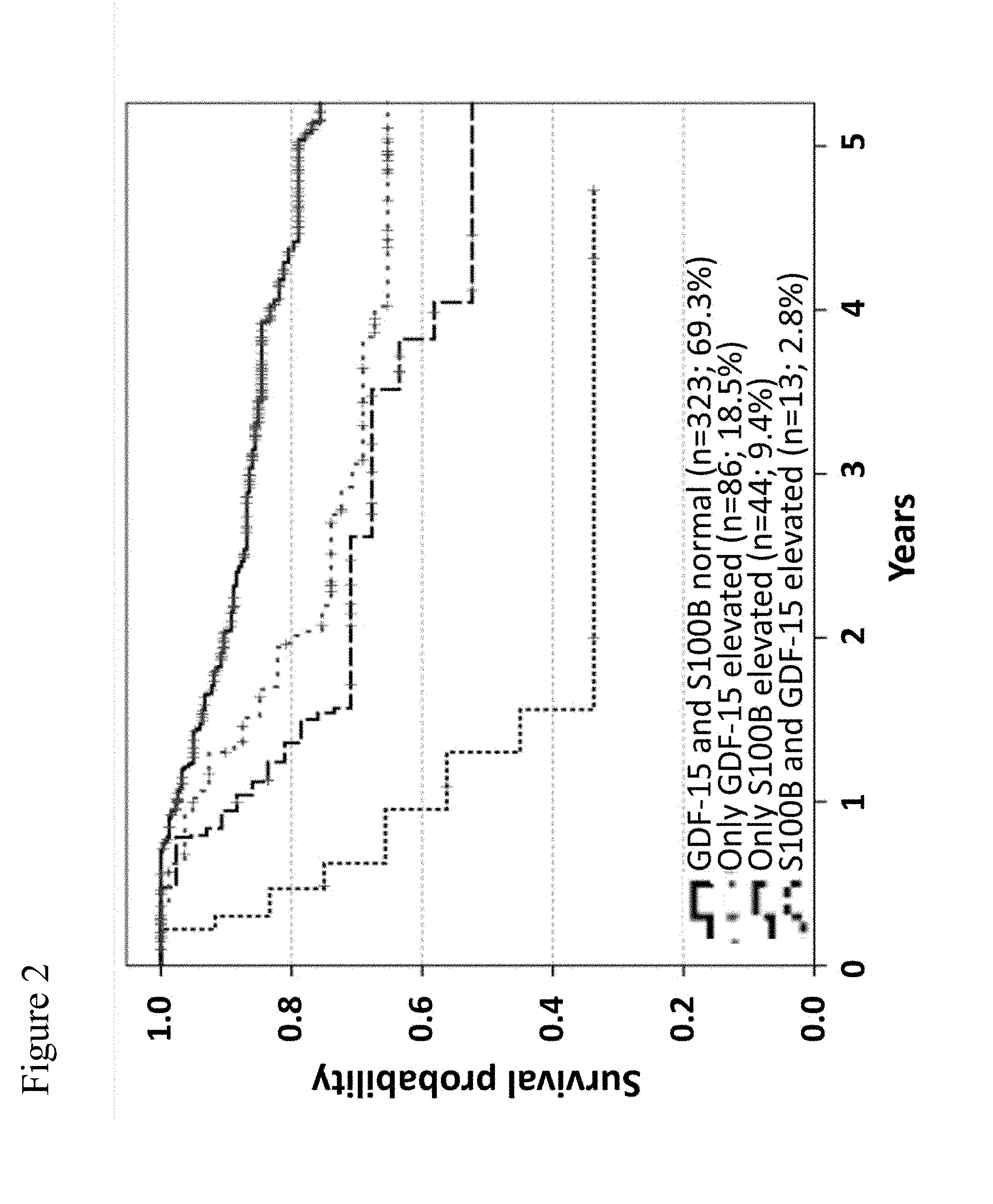
[0074] FIG. 2: Combinations of S100B and GDF-15 serum levels and their correlation with overall survival of stage III patients. Using a Cox regression model, S100B and hGDF-15 levels were shown to independently predict prognosis of tumor-free stage III patients. Kaplan-Meier curves of overall survival for patients with different biomarker combinations are presented for 466 patients. Censoring is indicated by vertical lines. In FIG. 2, the highest curve is the curve for normal hGDF-15 levels and normal S100B levels, the 2.sup.nd highest curve is the curve for elevated hGDF-15 levels, the 2.sup.nd lowest curve is the curve for elevated S100B levels, and the lowest curve is the one for elevated hGDF-15 levels and elevated S100B levels.
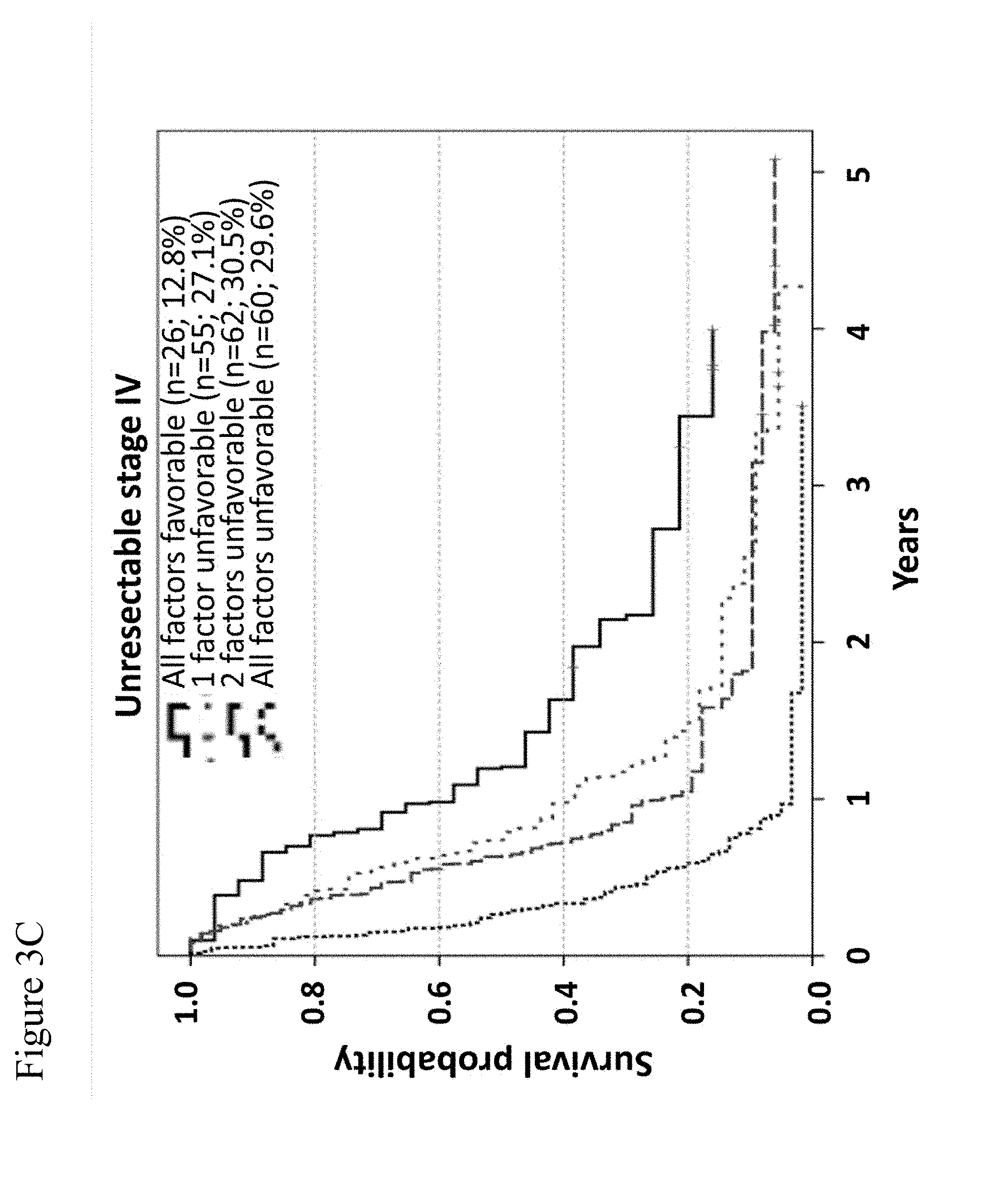
[0075] FIGS. 3A-3C: Overall survival of unresectable stage IV patients according to combinations of serum levels of LDH and GDF-15, and the pattern of distant metastasis. The independent prognostic impact of GDF-15 serum levels on overall survival is illustrated for M-categories M1a/b (FIG. 3A) as well as for M1c patients (FIG. 3B). Broken lines indicate all patients of the given M-category. Continuous lines represent sub-groups with low (blue) or high (red) GDF-15 levels, respectively. Differences in OS between patients with high or low GDF-15 levels were significant for M1a/b and for M1c patients (log-rank p-values 0.047 and 0.003, respectively). In (FIG. 3C), overall survival is displayed according to the number of unfavorable values considering all 3 independent prognostic factors according to model 1 of Cox regression analysis (LDH levels, pattern of visceral metastasis, and GDF-15 levels). The order of curves (i.e. the order from the highest curve to the lowest curve) in the legend contained in panels (A) to (C) of the figure reflects the order of curves in the respective panels.
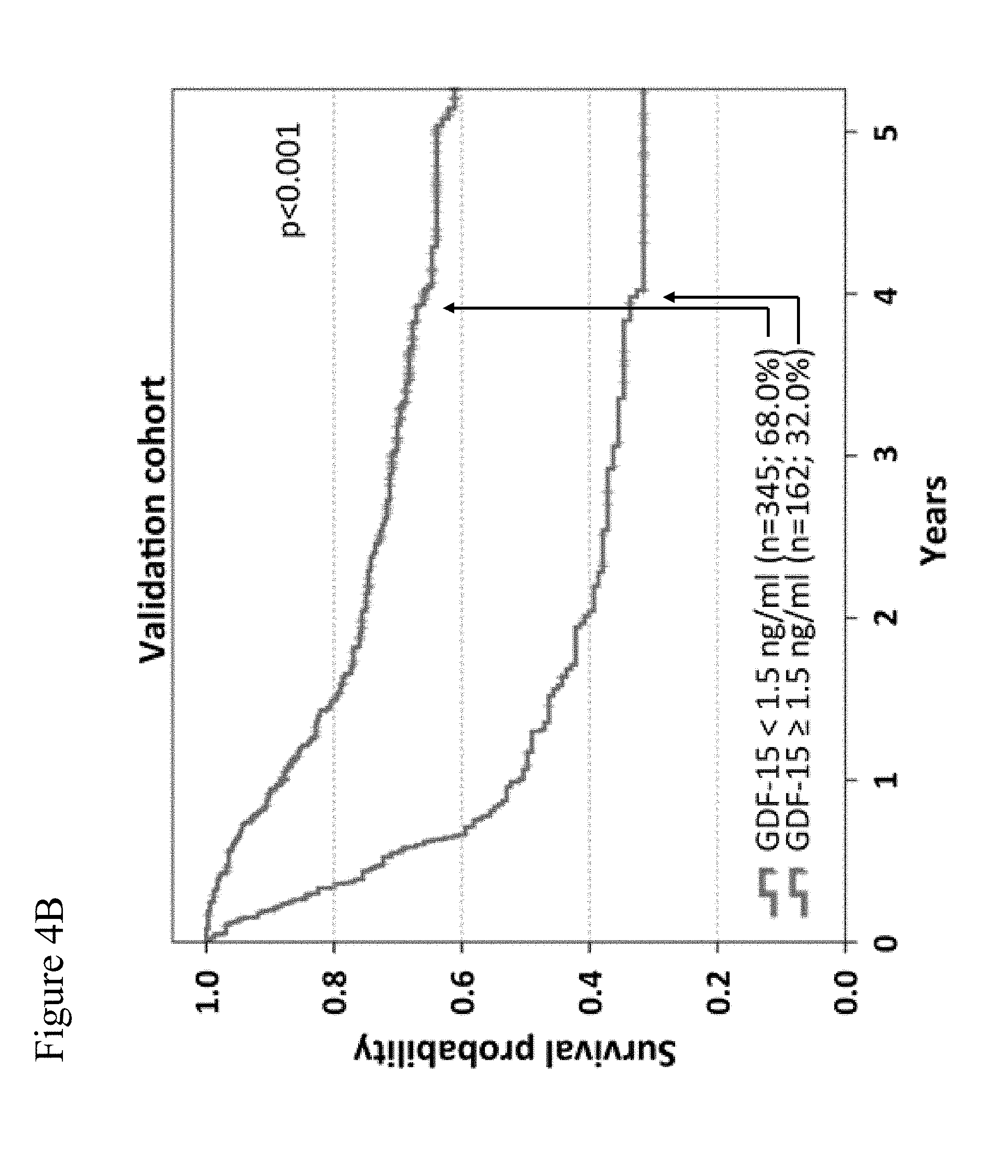
[0076] FIGS. 4A-4B: Overall survival correlates with GDF-15 serum levels in melanoma patients. 762 patients were randomly assigned to two cohorts. In the identification cohort (254 patients), different cut-off values were tested by Kaplan-Meier analysis and log rank tests to obtain the best possible discrimination between patients with high and low GDF-15 serum levels. Overall survival of patients of the identification cohort according to the optimized cut-off point (<1.5 ng/mL vs. .gtoreq.1.5 ng/mL) is shown in (FIG. 4A). Differences in overall survival were confirmed in 508 patients of the validation cohort (FIG. 4B). Censoring is indicated by vertical lines; p-values were calculated by log rank statistics. In FIGS. 4A and 4B, the upper curves are those for normal hGDF-15 levels, and the lower curves are those for elevated hGDF-15 levels.
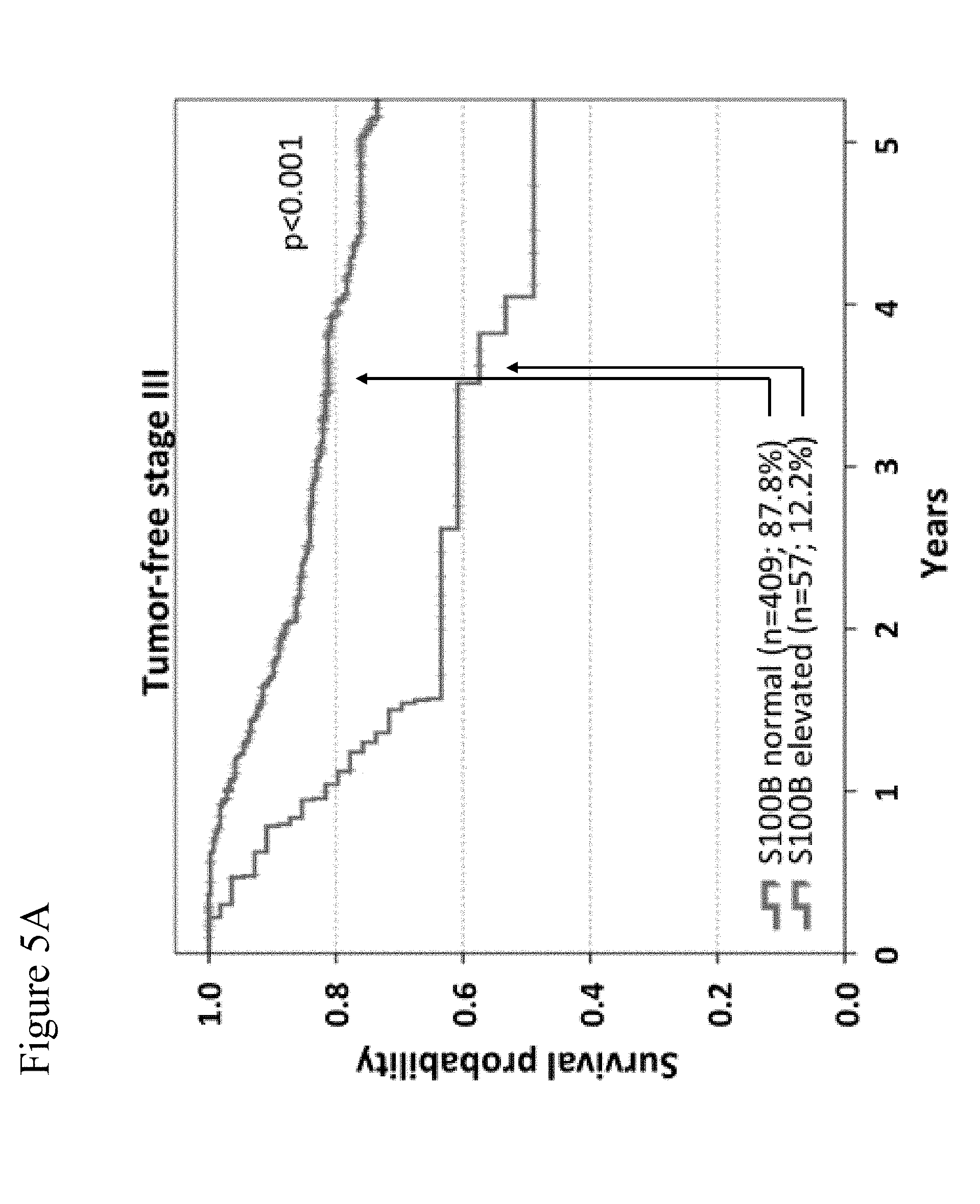
[0077] FIG. 5A-5C: Overall survival according to S100B serum levels. Kaplan-Meier curves are shown for overall survival of 466 tumor-free stage III (FIG. 5A), 193 unresectable stage IV (FIG. 5B) and 83 tumor-free stage IV (FIG. 5C) patients. Patients were categorized based on S100B serum levels (normal vs. elevated). Censoring is indicated by vertical lines; p-values were calculated by log rank statistics. In FIGS. 5A to 5C, the upper curves are those for normal S100B levels, and the lower curves are those for elevated S100B levels.
[0078] FIG. 6: Overall survival of stage III patients according to the number of unfavorable values considering serum levels of GDF-15, S100B, age, and sub-stage. Model 2 of Cox regression analysis (Table 2) revealed an independent negative prognostic impact for GDF-15 levels >1.5 ng/mL, for elevated S100B levels, for age <63 years, and for sub-stage IIIC. Patients were now stratified according to the number of unfavorable values among those four factors. The resulting Kaplan-Meier curves of overall survival are presented and censoring is indicated by vertical lines. The highest curve is the curve, wherein all factors are favorable. The 2.sup.nd highest curve is the curve, wherein one factor is unfavorable. The 3.sup.rd highest curve is the curve, wherein two factors are unfavorable. The 2.sup.nd lowest curve is the curve, wherein three factors are unfavorable. The lowest curve is the curve, wherein all factors are unfavorable.
[0079] FIG. 7: Overall survival of unresectable stage IV patients according to the number of unfavorable values considering serum levels of GDF-15, S100B, the pattern of distant metastasis, and age. Model 2 of Cox regression analysis (Table 3) revealed an independent negative prognostic impact for GDF-15 levels >1.5 ng/mL, for elevated S100B levels, for the metastatic involvement of visceral organs other than lung, and for age of 63 years or older. Patients were thus stratified according to the number of unfavorable factors. The resulting Kaplan-Meier curves for overall survival are shown and censoring is indicated by vertical lines. The highest curve is the curve, wherein all factors are favorable. The 2.sup.nd highest curve is the curve, wherein one factor is unfavorable. The 3.sup.rd highest curve is the curve, wherein two factors are unfavorable. The 2.sup.nd lowest curve is the curve, wherein three factors are unfavorable. The lowest curve is the curve, wherein all factors are unfavorable.
[0080] FIGS. 8A-8B: Overall survival subsequent to serum sampling of stage III patients according to combinations of different factors. A nomogram (FIG. 8A) was developed for tumor-free stage III patients using the nomogram function of R considering the relative impact of single independent factors according to multivariate analysis (sGDF-15, sS100B, pattern of loco-regional metastasis). A risk score ranging between 0 and 266 points was calculated for 466 stage III patients with complete data. In (FIG. 8B), Kaplan-Meier curves of overall survival subsequent to serum sampling is displayed for different risk score categories. Censoring is indicated by vertical lines.
[0081] FIGS. 9A-9D: Overall survival subsequent to serum sampling of unresectable stage IV patients according to combinations of different factors. GDF-15 serum levels have independent impact on overall survival of unresectable stage IV patients in addition to the M category. This is illustrated by the significant differences in OS according to sGDF-15 in both, M1a/b patients (FIG. 9A), and M1c patients (FIG. 9B). A nomogram (FIG. 9C) was developed for unresectable stage IV patients using the nomogram function of R considering the relative impact of single independent factors according to multivariate analysis (sGDF-15, sS100B, CNS involvement, and number of involved distant sites). A risk score ranging between 0 and 334 points was calculated for 193 unresectable stage IV patients with complete data. In (FIG. 9D), Kaplan-Meier curves of overall survival subsequent to serum sampling is displayed for different risk score categories. Censoring is indicated by vertical lines.
[0082] FIGS. 10A-10C: Correlations of sGDF-15 with stage/disease status and sLDH. Serum GDF-15 levels are shown for tumor-free stage III (n=468), tumor-free stage IV patients (n=87) and unresectable stage IV patients (n=206) (FIG. 10A). In unresectable stage IV patients, sGDF-15 is presented for according to the number of distant metastases (FIG. 10B) or stratified according to sLDH (FIG. 10C). Red bars indicate median levels of GDF-15; ** p<0.01; *** p<0.001 using Mann Whitney tests.
[0083] FIGS. 11A-11B: Overall survival subsequent to serum sampling correlates with GDF-15 serum levels in melanoma patients. 761 patients were randomly assigned to two cohorts. In the identification cohort (254 patients), different cut-off values were tested by Kaplan-Meier analysis and log rank tests to obtain the best possible discrimination between patients with high and low GDF-15 serum levels. Overall survival subsequent to serum sampling of patients of the identification cohort according to the optimized cut-off point (<1.5 ng/mL vs. .gtoreq.1.5 ng/mL) is shown in (FIG. 11A). Differences in overall survival subsequent to serum sampling were confirmed in 507 patients of the validation cohort (FIG. 11B). Censoring is indicated by vertical lines; p-values were calculated by log rank statistics.
[0084] FIGS. 12A-12I: Association of sGDF-15, sS100B and sLDH with OS according to time-point of serum sampling in tumor-free stage III patients. Overall survival of tumor-free stage III patients according to sGDF-15 (left), sS100B (middle) and sLDH (right) according to the time point of last recurrence before serum sampling. Patients were categorized as being tumor-free for less than 6 months (FIGS. 12A-12C), for between 6 months and 2 years (FIGS. 12D-12F) or for more than 2 years (FIGS. 12G-12I) since detection of last metastasis. Censoring is indicated by vertical lines; p-values were calculated by log rank statistics.
[0085] FIGS. 13A-13I: Association of sGDF-15, sS100B and sLDH with OS according to time-point of serum sampling in unresectable stage IV patients. Overall survival of unresectable stage IV patients according to sGDF-15 (left), S100B (middle) and LDH (right) according to the time span since diagnosis of stage IV disease. The first distant metastasis was detected within 6 months (FIGS. 13A-13C), between 6 months and 2 years (FIGS. 13D-13F) and more than 2 years (FIGS. 13G-13I) before serum sampling. Censoring is indicated by vertical lines; p-values were calculated by log rank statistics.
[0086] FIGS. 14A-14C: Overall survival subsequent to serum sampling according to S100B serum levels. Kaplan-Meier curves are shown for overall survival subsequent to serum sampling of 466 tumor-free stage III (FIG. 14A), 83 tumor-free stage IV (FIG. 14B) and 193 unresectable stage IV (FIG. 14C) patients. Patients were categorized based on S100B serum levels (normal vs. elevated). Censoring is indicated by vertical lines; p-values were calculated by log rank statistics.
DETAILED DESCRIPTION OF THE INVENTION
Definitions and General Techniques
[0087] Unless otherwise defined below, the terms used in the present invention shall be understood in accordance with their common meaning known to the person skilled in the art.
[0088] The term "antibody" as used herein refers to any functional antibody that is capable of specific binding to the antigen of interest, as generally outlined in chapter 7 of Paul, W. E. (Ed.): Fundamental Immunology 2nd Ed. Raven Press, Ltd., New York 1989, which is incorporated herein by reference. Without particular limitation, the term "antibody" encompasses antibodies from any appropriate source species, including chicken and mammalian such as mouse, goat, non-human primate and human. Preferably, the antibody is a humanized antibody. The antibody is preferably a monoclonal antibody which can be prepared by methods well-known in the art. The term "antibody" encompasses an IgG-1, -2, -3, or -4, IgE, IgA, IgM, or IgD isotype antibody. The term "antibody" encompasses monomeric antibodies (such as IgD, IgE, IgG) or oligomeric antibodies (such as IgA or IgM). The term "antibody" also encompasses--without particular limitations--isolated antibodies and modified antibodies such as genetically engineered antibodies, e.g. chimeric antibodies.
[0089] The nomenclature of the domains of antibodies follows the terms as known in the art. Each monomer of an antibody comprises two heavy chains and two light chains, as generally known in the art. Of these, each heavy and light chain comprises a variable domain (termed V.sub.H for the heavy chain and V.sub.L for the light chain) which is important for antigen binding. These heavy and light chain variable domains comprise (in an N-terminal to C-terminal order) the regions FR1, CDR1, FR2, CDR2, FR3, CDR3, and FR4 (FR, framework region; CDR, complementarity determining region which is also known as hypervariable region). The identification and assignment of the above-mentioned antibody regions within the antibody sequence is generally in accordance with Kabat et al. (Sequences of proteins of immunological interest, U.S. Dept. of Health and Human Services, Public Health Service, National Institutes of Health, Bethesda, Md. 1983), or Chothia et al. (Conformations of immunoglobulin hypervariable regions. Nature. 1989 Dec. 21-28; 342(6252):877-83), or may be performed by using the IMGT/V-QUEST software described in Giudicelli et al. (IMGT/V-QUEST, an integrated software program for immunoglobulin and T cell receptor V-J and V-D-J rearrangement analysis. Nucleic Acids Res. 2004 Jul. 1; 32 (Web Server issue):W435-40), which is incorporated herein by reference. Preferably, the antibody regions indicated above are identified and assigned by using the IMGT/V-QUEST software.
[0090] A "monoclonal antibody" is an antibody from an essentially homogenous population of antibodies, wherein the antibodies are substantially identical in sequence (i.e. identical except for minor fraction of antibodies containing naturally occurring sequence modifications such as amino acid modifications at their N- and C-termini). Unlike polyclonal antibodies which contain a mixture of different antibodies directed to either a single epitope or to numerous different epitopes, monoclonal antibodies are directed to the same epitope and are therefore highly specific. The term "monoclonal antibody" includes (but is not limited to) antibodies which are obtained from a monoclonal cell population derived from a single cell clone, as for instance the antibodies generated by the hybridoma method described in Kohler and Milstein (Nature, 1975 Aug. 7; 256(5517):495-7) or Harlow and Lane ("Antibodies: A Laboratory Manual" Cold Spring Harbor Laboratory Press, Cold Spring Harbor, N.Y. 1988). A monoclonal antibody may also be obtained from other suitable methods, including phage display techniques such as those described in Clackson et al. (Nature. 1991 Aug. 15; 352(6336):624-8) or Marks et al. (J Mol Biol. 1991 Dec. 5; 222(3):581-97). A monoclonal antibody may be an antibody that has been optimized for antigen-binding properties such as decreased Kd values, optimized association and dissociation kinetics by methods known in the art. For instance, Kd values may be optimized by display methods including phage display, resulting in affinity-matured monoclonal antibodies. The term "monoclonal antibody" is not limited to antibody sequences from particular species of origin or from one single species of origin. Thus, the meaning of the term "monoclonal antibody" encompasses chimeric monoclonal antibodies such as humanized monoclonal antibodies.
[0091] "Humanized antibodies" are antibodies which contain human sequences and a minor portion of non-human sequences which confer binding specificity to an antigen of interest (e.g. human GDF-15). Typically, humanized antibodies are generated by replacing hypervariable region sequences from a human acceptor antibody by hypervariable region sequences from a non-human donor antibody (e.g. a mouse, rabbit, rat donor antibody) that binds to an antigen of interest (e.g. human GDF-15). In some cases, framework region sequences of the acceptor antibody may also be replaced by the corresponding sequences of the donor antibody. In addition to the sequences derived from the donor and acceptor antibodies, a "humanized antibody" may either contain other (additional or substitute) residues or sequences or not. Such other residues or sequences may serve to further improve antibody properties such as binding properties (e.g. to decrease Kd values) and/or immunogenic properties (e.g. to decrease antigenicity in humans). Non-limiting examples for methods to generate humanized antibodies are known in the art, e.g. from Riechmann et al. (Nature. 1988 Mar. 24; 332(6162):323-7) or Jones et al. (Nature. 1986 May 29-Jun. 4; 321(6069):522-5).
[0092] The term "human antibody" relates to an antibody containing human variable and constant domain sequences. This definition encompasses antibodies having human sequences bearing single amino acid substitutions or modifications which may serve to further improve antibody properties such as binding properties (e.g. to decrease Kd values) and/or immunogenic properties (e.g. to decrease antigenicity in humans). The term "human antibody" excludes humanized antibodies where a portion of non-human sequences confers binding specificity to an antigen of interest.
[0093] An "antigen-binding portion" of an antibody as used herein refers to a portion of an antibody that retains the capability of the antibody to specifically bind to the antigen (e.g. hGDF-15, PD-1, PD-L1 or CTLA4). This capability can, for instance, be determined by determining the capability of the antigen-binding portion to compete with the antibody for specific binding to the antigen by methods known in the art. The antigen-binding portion may contain one or more fragments of the antibody. Without particular limitation, the antigen-binding portion can be produced by any suitable method known in the art, including recombinant DNA methods and preparation by chemical or enzymatic fragmentation of antibodies. Antigen-binding portions may be Fab fragments, F(ab') fragments, F(ab').sub.2 fragments, single chain antibodies (scFv), single-domain antibodies, diabodies or any other portion(s) of the antibody that retain the capability of the antibody to specifically bind to the antigen.
[0094] An "antibody" (e.g. a monoclonal antibody) or an "antigen-binding portion" may have been derivatized or be linked to a different molecule. For example, molecules that may be linked to the antibody are other proteins (e.g. other antibodies), a molecular label (e.g. a fluorescent, luminescent, colored or radioactive molecule), a pharmaceutical and/or a toxic agent. The antibody or antigen-binding portion may be linked directly (e.g. in form of a fusion between two proteins), or via a linker molecule (e.g. any suitable type of chemical linker known in the art).
[0095] As used herein, the terms "binding" or "bind" refer to specific binding to the antigen of interest (e.g. human GDF-15). Preferably, the Kd value is less than 100 nM, more preferably less than 50 nM, still more preferably less than 10 nM, still more preferably less than 5 nM and most preferably less than 2 nM.
[0096] The term "epitope" as used herein refers to a small portion of an antigen that forms the binding site for an antibody.
[0097] In the context of the present invention, for the purposes of characterizing the binding properties of antibodies, binding or competitive binding of antibodies or their antigen-binding portions to the antigen of interest (e.g. human GDF-15) is preferably measured by using surface plasmon resonance measurements as a reference standard assay, as described below.
[0098] The terms "K.sub.D" or "K.sub.D value" relate to the equilibrium dissociation constant as known in the art. In the context of the present invention, these terms relate to the equilibrium dissociation constant of an antibody with respect to a particular antigen of interest (e.g. human GDF-15) The equilibrium dissociation constant is a measure of the propensity of a complex (e.g. an antigen-antibody complex) to reversibly dissociate into its components (e.g. the antigen and the antibody). For the antibodies according to the invention, K.sub.D values (such as those for the antigen human GDF-15) are preferably determined by using surface plasmon resonance measurements as described below.
[0099] An "isolated antibody" as used herein is an antibody that has been identified and separated from the majority of components (by weight) of its source environment, e.g. from the components of a hybridoma cell culture or a different cell culture that was used for its production (e.g. producer cells such as CHO cells that recombinantly express the antibody). The separation is performed such that it sufficiently removes components that may otherwise interfere with the suitability of the antibody for the desired applications (e.g. with a therapeutic use of the anti-human GDF-15 antibody according to the invention). Methods for preparing isolated antibodies are known in the art and include Protein A chromatography, anion exchange chromatography, cation exchange chromatography, virus retentive filtration and ultrafiltration. Preferably, the isolated antibody preparation is at least 70% pure (w/w), more preferably at least 80% pure (w/w), still more preferably at least 90% pure (w/w), still more preferably at least 95% pure (w/w), and most preferably at least 99% pure (w/w), as measured by using the Lowry protein assay.
[0100] A "diabody" as used herein is a small bivalent antigen-binding antibody portion which comprises a heavy chain variable domain linked to a light chain variable domain on the same polypeptide chain linked by a peptide linker that is too short to allow pairing between the two domains on the same chain. This results in pairing with the complementary domains of another chain and in the assembly of a dimeric molecule with two antigen binding sites. Diabodies may be bivalent and monospecific (such as diabodies with two antigen binding sites for human GDF-15), or may be bivalent and bispecific (e.g. diabodies with two antigen binding sites, one being a binding site for human GDF-15, and the other one being a binding site for a different antigen). A detailed description of diabodies can be found in Holliger P et al. (""Diabodies": small bivalent and bispecific antibody fragments." Proc Natl Acad Sci USA. 1993 Jul. 15; 90(14):6444-8.).
[0101] A "single-domain antibody" (which is also referred to as "Nanobody.TM.") as used herein is an antibody fragment consisting of a single monomeric variable antibody domain. Structures of and methods for producing single-domain antibodies are known from the art, e.g. from Holt L J et al. ("Domain antibodies: proteins for therapy." Trends Biotechnol. 2003 November; 21(11):484-90), Saerens D et al. ("Single-domain antibodies as building blocks for novel therapeutics." Curr Opin Pharmacol. 2008 October; 8(5):600-8. Epub 2008 Aug. 22), and Arbabi Ghahroudi M et al. ("Selection and identification of single domain antibody fragments from camel heavy-chain antibodies." FEBS Lett. 1997 Sep. 15; 414(3):521-6.).
[0102] The terms "significant", "significantly", etc. as used herein refer to a statistically significant difference between values as assessed by appropriate methods known in the art, and as assessed by the methods referred to herein.
[0103] In accordance with the present invention, each occurrence of the term "comprising" may optionally be substituted with the term "consisting of".
[0104] The terms "cancer" and "cancer cell" is used herein in accordance with their common meaning in the art (see for instance Weinberg R. et al.: The Biology of Cancer. Garland Science: New York 2006. 850p., which is incorporated herein by reference in its entirety).
[0105] The cancers, for which a prediction of a clinical outcome, in particular a prediction of patient survival according to the present invention is provided, is melanoma. As used herein, the term "melanoma" is used in accordance with its general meaning known in the art. Melanomas are classified according to the AJCC staging system for melanoma patients with distant metastases since 2001 (Balch, C M et al., J Clin Oncol/19/3635-48. 2001). The melanoma stages referred to herein refer to this staging system. In a preferred aspect of the present invention in accordance with all of the embodiments of the present invention, the melanoma is not a uveal melanoma.
[0106] The melanoma patients, for which a prediction of survival according to the invention is provided, may be subject to a treatment of the melanoma. As used herein, terms such as "treatment of cancer" or "treating cancer" or "treatment of melanoma" or "treating melanoma" refer to a therapeutic treatment. As used herein, such treatments do not only include treatments of the melanoma itself but also palliative treatments. Such palliative treatments are known in the art and include, for instance, treatments which only improve the symptoms of the melanoma disease.
[0107] As referred to herein, a treatment of cancer can be a first-line therapy, a second-line therapy or a third-line therapy or a therapy that is beyond third-line therapy. The meaning of these terms is known in the art and in accordance with the terminology that is commonly used by the US National Cancer Institute.
[0108] A treatment of cancer does not exclude that additional or secondary therapeutic benefits also occur in patients. For example, an additional or secondary benefit may be an influence on cancer-induced weight loss.
[0109] As referred to herein, a "tumor-free" melanoma patient is a patient in which no primary tumor and no metastasis can be detected according to clinical standard methods known in the art. This, however, does not exclude that tumors (or micrometastases) exist in the patient, which are below the detection limit, or that tumor cells exist, which may form a new tumor.
Blood Samples:
[0110] As referred to herein, the term "blood sample" includes, without limitation, whole blood, serum and plasma samples. It also includes other sample types such as blood fractions other than serum and plasma. Such samples and fractions are known in the art.
[0111] Blood samples which are used for the methods according to the invention can be any types of blood samples which contain hGDF-15. Suitable types of blood samples containing hGDF-15 are known in the art and include serum and plasma samples. Alternatively, further types of blood samples which contain hGDF-15 can also be readily identified by the skilled person, e.g. by measuring whether hGDF-15 is contained in these samples, and which levels of hGDF-15 are contained in these samples, by using the methods disclosed herein.
Clinical Outcomes:
[0112] According to the invention, levels of hGDF-15 in human blood samples can be used to predict the probability of survival of a human melanoma patient.
[0113] Survival of patient groups can be analysed by methods known in the art, e.g. by Kaplan-Meier curves.
[0114] Appropriate time periods for the assessment of survival are known in the art and will be chosen by the skilled person with due regard to factors such as the respective stage of the melanoma.
[0115] For example, survival, preferably short-term survival, may, for instance, be predicted for time points of 6 months, 12 months and/or 18 months after the time point when the prediction was made. Alternatively, survival, preferably long-term survival, may, for instance, be assessed at a time point of 2 years, 3 years, 5 years and/or 10 years after the time point when the prediction was made.
Predicting the Probability of a Positive Clinical Outcome According to the Invention
[0116] For predicting the probability of a positive clinical outcome (e.g. survival) according to the invention, e.g. based on hGDF-15 levels, the methods for predicting, which are defined above in the preferred embodiments, are preferably used.
[0117] In order to practice the methods of the invention, statistical methods known in the art can be employed.
[0118] For instance, overall survival can be analyzed by Cox regression analysis.
[0119] Preferred statistical methods, which can be used according to the invention to generate statistical models of patient data from clinical studies, are disclosed in Example 1. It is understood that the statistical methods disclosed in Example 1 are not limited to the particular features of Example 1 such as the melanoma stage, the particular threshold levels chosen and the particular statistical values obtained in the Example. Rather, these methods disclosed in Example 1 can generally be used in connection with any embodiment of the present invention.
hGDF-15 Levels
[0120] In an advantageous aspect of the invention, there is an inverse relationship between hGDF-15 levels and the probability of a positive clinical outcome, in particular the probability of survival, in human melanoma patients. Thus, according to the invention, a decreased level of hGDF-15 indicates an increased probability of survival in human melanoma patients.
[0121] Thus, as used herein, terms such as "wherein a decreased level of hGDF-15 in said human blood sample indicates an increased probability of survival" mean that the level of hGDF-15 in said human blood sample and the probability of survival follow an inverse relationship. Thus, the higher the level of hGDF-15 in said human blood sample is, the lower is the probability of survival.
[0122] For instance, in connection with the methods for predicting according to the invention defined herein, hGDF-15 threshold levels can be used.
[0123] According to the invention, the inverse relationship between hGDF-15 levels and the probability of survival applies to any threshold value, and hence the invention is not limited to particular threshold values.
[0124] Preferable hGDF-15 threshold levels are hGDF-15 serum levels as defined above in the preferred embodiments.
[0125] Alternatively, hGDF-15 threshold levels according to the present invention can be obtained, and/or further adjusted, by using the above-mentioned statistical methods, e.g. the methods of Example 1.
[0126] An hGDF-15 threshold level may be a single hGDF-15 threshold level. The invention also encompasses the use of more than one hGDF-15 threshold level, e.g. 2, 3, 4, 5, 6, 7, 8, 9, 10 or more hGDF-15 threshold levels.
[0127] For each single hGDF-15 threshold level of the one or more hGDF-15 threshold levels, a corresponding probability of survival can be predicted at a given time point.
[0128] hGDF-15 levels in blood samples can be measured by any methods known in the art. For instance, a preferred method of measuring hGDF-15 levels in blood samples including serum levels is a measurement of hGDF-15 levels by Enzyme-Linked Immunosorbent Assay (ELISA) by using antibodies to hGDF-15. Such ELISA methods are exemplified in Example 1, but can also include bead-based methods like the Luminex technology and others. Alternatively, hGDF-15 levels in blood samples including serum levels may be determined by known electrochemiluminesence immunoassays using antibodies to hGDF-15. For instance, the Roche Elecsys.RTM. technology can be used for such electrochemiluminesence immunoassays. Other possible methods would include antibody-based detection from bodily fluids after separation of proteins in an electrical field.
[0129] The median hGDF-15 serum level of healthy human control individuals is <0.8 ng/ml. The expected range is between 0.2 ng/ml and 1.2 ng/ml in healthy human controls (Reference: Tanno T et al.: "Growth differentiation factor 15 in erythroid health and disease." Curr Opin Hematol. 2010 May; 17(3): 184-190.).
[0130] According to the invention, preferable hGDF-15 threshold levels are hGDF-15 serum levels as defined above in the preferred embodiments.
[0131] It is understood that for these hGDF-15 serum levels, and based on the disclosure of the invention provided herein, corresponding hGDF-15 levels in other blood samples can be routinely obtained by the skilled person (e.g. by comparing the relative level of hGDF-15 in serum with the respective level in other blood samples). Thus, the present invention also encompasses preferred hGDF-15 levels in plasma, whole blood and other blood samples, which correspond to each of the preferred hGDF-15 serum levels and ranges indicated above.
Lactate Dehydrogenase Levels
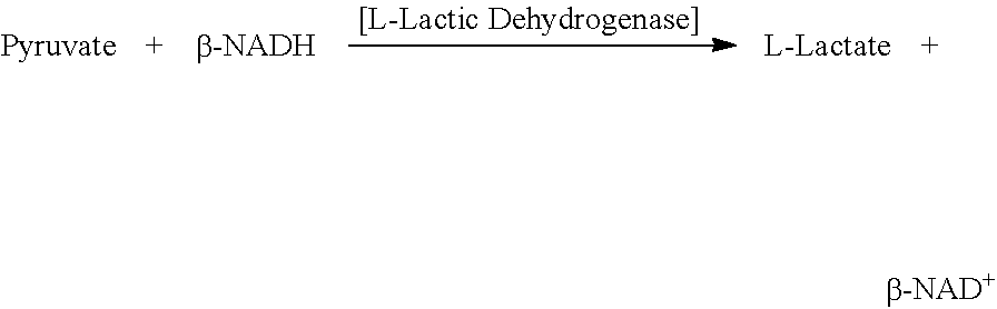
[0132] Lactate dehydrogenase levels in blood samples can be measured by any methods known in the art Lactate dehydrogenase (LDH) levels are typically measured in enzymatic units (U). One unit will reduce 1.0 .mu.mole of pyruvate to L-lactate per minute at pH 7.5 at 37.degree. C.
##STR00001##
[0133] Lactate and NAD+ are converted to pyruvate and NADH by the action of LDH. NADH strongly absorbs light at 340 nm, whereas NAD+ does not. The rate of increase in absorbance at 340 nm is directly proportional to the LDH activity in the sample. Thus, LDH units are preferably determined by measuring absorbance at 340 nm. Various clinically accepted diagnostic tests are available for the measurement of LDH levels. In accordance with the present invention, tests which can be applied to melanoma will be selected based on known clinical standards. Isoform-specific tests for LDH can be performed according to methods known in the art.
[0134] In a further advantageous aspect of the invention, there is also an inverse relationship between lactate dehydrogenase (LDH) levels and the probability of a positive clinical outcome, in particular the probability of survival, in human melanoma patients. Thus, in an embodiment according to the invention, a decreased level of lactate dehydrogenase indicates an increased probability of survival in melanoma patients.
[0135] Thus, as used herein, terms such as "wherein a decreased level of lactate dehydrogenase in said human blood sample indicates an increased probability of survival" mean that the level of lactate dehydrogenase in said human blood sample and the probability of survival follow an inverse relationship. Thus, the higher the level of lactate dehydrogenase in said human blood sample is, the lower is the probability of survival.
[0136] For instance, in connection with the methods for predicting according to the invention defined herein, lactate dehydrogenase threshold levels can be used.
[0137] According to the invention, the inverse relationship between lactate dehydrogenase levels and the probability of survival applies to any threshold value, and hence the invention is not limited to particular threshold values.
[0138] Alternatively, lactate dehydrogenase threshold levels according to the present invention can be obtained, and/or further adjusted, by using the above-mentioned statistical methods, e.g. the methods of Example 1.
[0139] A lactate dehydrogenase threshold level may be a single lactate dehydrogenase threshold level. The invention also encompasses the use of more than one lactate dehydrogenase threshold level, e.g. 2, 3, 4, 5, 6, 7, 8, 9, 10 or more lactate dehydrogenase threshold levels.
[0140] For each single lactate dehydrogenase threshold level of the one or more lactate dehydrogenase threshold levels, a corresponding probability of survival can be predicted.
[0141] According to the invention, preferable lactate dehydrogenase threshold levels are lactate dehydrogenase serum levels as defined above in the preferred embodiments.
[0142] In a very preferred embodiment, the lactate dehydrogenase threshold level is a clinically accepted threshold level which distinguishes between normal and elevated LDH levels in patients. Such very preferred clinically accepted threshold levels are known in the art, and will be chosen by the skilled person with regard to the particular specifications of the LDH test.
[0143] It is understood that for these lactate dehydrogenase serum levels, and based on the disclosure of the invention provided herein, corresponding lactate dehydrogenase levels in other blood samples can be routinely obtained by the skilled person (e.g. by comparing the relative level of lactate dehydrogenase in serum with the respective level in other blood samples). Thus, the present invention also encompasses preferred lactate dehydrogenase levels in plasma, whole blood and other blood samples, which correspond to each of the preferred lactate dehydrogenase serum levels and ranges indicated above.
S100B Levels
[0144] In a further advantageous aspect of the invention, there is also an inverse relationship between S100B levels and the probability of a positive clinical outcome, in particular the probability of survival, in human melanoma patients. Thus, in an embodiment according to the invention, a decreased level of S100B indicates an increased probability of survival in melanoma patients.
[0145] Thus, as used herein, terms such as "wherein a decreased level of S100B in said human blood sample indicates an increased probability of survival" mean that the level of S100B in said human blood sample and the probability of survival follow an inverse relationship. Thus, the higher the level of S100B in said human blood sample is, the lower is the probability of survival.
[0146] For instance, in connection with the methods for predicting according to the invention defined herein, S100B threshold levels can be used.
[0147] According to the invention, the inverse relationship between S100B levels and the probability of survival applies to any threshold value, and hence the invention is not limited to particular threshold values.
[0148] S100B threshold levels according to the present invention can, for instance, be obtained, and/or further adjusted, by using the above-mentioned statistical methods, e.g. the methods of Example 1.
[0149] An S100B threshold level may be a single S100B threshold level. The invention also encompasses the use of more than one S100B threshold level, e.g. 2, 3, 4, 5, 6, 7, 8, 9, 10 or more S100B threshold levels.
[0150] In a very preferred embodiment, the S100B threshold level is a clinically accepted threshold level which distinguishes between normal and elevated S100B levels in patients. Such very preferred clinically accepted threshold levels are known in the art, and will be chosen by the skilled person with regard to the particular specifications of the S100B test.
[0151] For each single S100B threshold level of the one or more S100B threshold levels, a corresponding probability of survival can be predicted.
[0152] S100B levels in blood samples can be measured by any methods known in the art. Such methods include antibody-based assays. A preferred method of measuring S100B levels in blood samples a measurement of S100B serum levels by electrochemoluminescence assays, e.g. by using an Elecsys S100 electrochemiluminescence immunoassay. Further non-limiting examples of methods to measure S100B levels are given in Goncalves et al.: "Biological and methodological features of the measurement of S100B, a putative marker of brain injury." Clinical Biochemistry 41 (2008) 755-763).
Antibodies Capable of Binding to hGDF-15 which can be Used in Accordance with the Invention
[0153] The methods, apparatuses and kits of the invention may use one or more antibodies capable of binding to hGDF-15 or an antigen-binding portion thereof, as defined above.
[0154] It was previously shown that human GDF-15 protein can be advantageously targeted by a monoclonal antibody (WO2014/049087, which is incorporated herein by reference in its entirety), and that such antibody has advantageous properties including a high binding affinity to human GDF-15, as demonstrated by an equilibrium dissociation constant of about 790 pM for recombinant human GDF-15 (see Reference Example 1). Thus, in a preferred embodiment, the invention uses an antibody capable of binding to hGDF-15, or an antigen-binding portion thereof. Preferably, the antibody is a monoclonal antibody capable of binding to hGDF-15, or an antigen-binding portion thereof.
[0155] Thus, in a more preferred embodiment, the antibody capable of binding to hGDF-15 or antigen-binding portion thereof in accordance with the invention is a monoclonal antibody capable of binding to human GDF-15, or an antigen-binding portion thereof, wherein the heavy chain variable domain comprises a CDR3 region comprising the amino acid sequence of SEQ ID NO: 5 or an amino acid sequence at least 90% identical thereto, and wherein the light chain variable domain comprises a CDR3 region comprising the amino acid sequence of SEQ ID NO: 7 or an amino acid sequence at least 85% identical thereto. In this embodiment, preferably, the antibody or antigen-binding portion thereof comprises a heavy chain variable domain which comprises a CDR1 region comprising the amino acid sequence of SEQ ID NO: 3 and a CDR2 region comprising the amino acid sequence of SEQ ID NO: 4, and the antibody or antigen-binding portion thereof comprises a light chain variable domain which comprises a CDR1 region comprising the amino acid sequence of SEQ ID NO: 6, and a CDR2 region comprising the amino acid sequence ser-ala-ser.
[0156] Thus, in a still more preferred embodiment, the antibody capable of binding to hGDF-15 or antigen-binding portion thereof in accordance with the invention is a monoclonal antibody capable of binding to human GDF-15, or an antigen-binding portion thereof, wherein the antibody or antigen-binding portion thereof comprises a heavy chain variable domain which comprises a CDR1 region comprising the amino acid sequence of SEQ ID NO: 3, a CDR2 region comprising the amino acid sequence of SEQ ID NO: 4 and a CDR3 region comprising the amino acid sequence of SEQ ID NO: 5, and wherein the antibody or antigen-binding portion thereof comprises a light chain variable domain which comprises a CDR1 region comprising the amino acid sequence of SEQ ID NO: 6, a CDR2 region comprising the amino acid sequence ser-ala-ser and a CDR3 region comprising the amino acid sequence of SEQ ID NO: 7.
[0157] In another embodiment in accordance with the above embodiments of the monoclonal antibody capable of binding to human GDF-15, or an antigen-binding portion thereof, the heavy chain variable domain comprises a region comprising an FR1, a CDR1, an FR2, a CDR2 and an FR3 region and comprising the amino acid sequence of SEQ ID NO: 1 or a sequence 85%, 90%, 91%, 92%, 93%, 94%, 95%, 96%, 97%, 98% or 99% identical thereto, and the light chain variable domain comprises a region comprising an FR1, a CDR1, an FR2, a CDR2 and an FR3 region and comprising the amino acid sequence of SEQ ID NO: 2 or a sequence 85%, 90%, 91%, 92%, 93%, 94%, 95%, 96%, 97%, 98% or 99% identical thereto.
[0158] In another embodiment in accordance with the above embodiments of the monoclonal antibody capable of binding to human GDF-15, or an antigen-binding portion thereof, the heavy chain variable domain comprises a CDR1 region comprising the amino acid sequence of SEQ ID NO: 3 and a CDR2 region comprising the amino acid sequence of SEQ ID NO: 4, and the light chain variable domain comprises a CDR1 region comprising the amino acid sequence of SEQ ID NO: 6 and a CDR2 region comprising the amino acid sequence of SEQ ID NO: 7. In a preferred aspect of this embodiment, the antibody may have CDR3 sequences as defined in any of the embodiments of the invention described above.
[0159] In another embodiment in accordance with the monoclonal antibody capable of binding to human GDF-15, or an antigen-binding portion thereof, the antigen-binding portion is a single-domain antibody (also referred to as "Nanobody.TM."). In one aspect of this embodiment, the single-domain antibody comprises the CDR1, CDR2, and CDR3 amino acid sequences of SEQ ID NO: 3, SEQ ID NO: 4, and SEQ ID NO: 5, respectively. In another aspect of this embodiment, the single-domain antibody comprises the CDR1, CDR2, and CDR3 amino acid sequences of SEQ ID NO: 6, ser-ala-ser, and SEQ ID NO: 7, respectively. In a preferred aspect of this embodiment, the single-domain antibody is a humanized antibody.
[0160] Preferably, the antibodies capable of binding to human GDF-15 or the antigen-binding portions thereof have an equilibrium dissociation constant for human GDF-15 that is equal to or less than 100 nM, less than 20 nM, preferably less than 10 nM, more preferably less than 5 nM and most preferably between 0.1 nM and 2 nM.
[0161] In another embodiment in accordance with the above embodiments of the monoclonal antibody capable of binding to human GDF-15, or an antigen-binding portion thereof, the antibody capable of binding to human GDF-15 or the antigen-binding portion thereof binds to the same human GDF-15 epitope as the antibody to human GDF-15 obtainable from the cell line B1-23 deposited with the Deutsche Sammlung fur Mikroorganismen und Zellkulturen GmbH (DMSZ) under the accession No. DSM ACC3142. As described herein, antibody binding to human GDF-15 in accordance with the present invention is preferably assessed by surface plasmon resonance measurements as a reference standard method, in accordance with the procedures described in Reference Example 1. Binding to the same epitope on human GDF-15 can be assessed similarly by surface plasmon resonance competitive binding experiments of the antibody to human GDF-15 obtainable from the cell line B1-23 and the antibody that is expected to bind to the same human GDF-15 epitope as the antibody to human GDF-15 obtainable from the cell line B1-23.
[0162] In a very preferred embodiment, the antibody capable of binding to human GDF-15 or the antigen-binding portion thereof is a monoclonal antibody capable of binding to human GDF-15, or an antigen-binding portion thereof, wherein the binding is binding to a conformational or discontinuous epitope on human GDF-15 comprised by the amino acid sequences of SEQ ID No: 25 and SEQ ID No: 26. In a preferred aspect of this embodiment, the antibody or antigen-binding portion thereof is an antibody or antigen-binding portion thereof as defined by the sequences of any one of the above embodiments.
[0163] In a further embodiment in accordance with the above embodiments, antibodies including the antibody capable of binding to human GDF-15 or the antigen-binding portion thereof can be modified, e.g. by a tag or a label.
[0164] A tag can, for instance, be a biotin tag or an amino acid tag. Non-limiting examples of such acid tag tags include Polyhistidin (His-) tags, FLAG-tag, Hemagglutinin (HA) tag, glycoprotein D (gD) tag, and c-myc tag. Tags may be used for various purposes. For instance, tags may be used to assist purification of the antibody capable of binding to human GDF-15 or the antigen-binding portion thereof. Preferably, such tags are present at the C-terminus or N-terminus of the antibody capable of binding to human GDF-15 or the antigen-binding portion thereof.
[0165] As used herein, the term "label" relates to any molecule or group of molecules which can facilitate detection of the antibody. For instance, labels may be enzymatic such as horseradish peroxidase (HRP), alkaline phosphatase (AP) or glucose oxidase. Enzymatically labelled antibodies may, for instance, be employed in enzyme-linked immunosorbent assays. Labels may also be radioactive isotopes, DNA sequences (which may, for instance, be used to detect the antibodies by polymerase chain reaction (PCR)), fluorogenic reporters and electrochemiluminescent groups (e.g. ruthenium complexes). As an alternative to labelling, antibodies used according to the invention, in particular an antibody capable of binding to human GDF-15 or the antigen-binding portion thereof, can be detected directly, e.g. by surface plasmon resonance measurements.
Methods and Techniques
[0166] Generally, unless otherwise defined herein, the methods used in the present invention (e.g. cloning methods or methods relating to antibodies) are performed in accordance with procedures known in the art, e.g. the procedures described in Sambrook et al. ("Molecular Cloning: A Laboratory Manual.", 2.sup.nd Ed., Cold Spring Harbor Laboratory Press, Cold Spring Harbor, N.Y. 1989), Ausubel et al. ("Current Protocols in Molecular Biology." Greene Publishing Associates and Wiley Interscience; New York 1992), and Harlow and Lane ("Antibodies: A Laboratory Manual" Cold Spring Harbor Laboratory Press, Cold Spring Harbor, N.Y. 1988), all of which are incorporated herein by reference.
[0167] Binding of antibodies to their respective target proteins can be assessed by methods known in the art. The binding of monoclonal antibodies to their respective targets is preferably assessed by surface plasmon resonance measurements. These measurements are preferably carried out by using a Biorad ProteOn XPR36 system and Biorad GLC sensor chips, as exemplified for anti-human GDF-15 mAb-B1-23 in Reference Example 1.
[0168] Sequence Alignments of sequences according to the invention are performed by using the BLAST algorithm (see Altschul et al. (1990) "Basic local alignment search tool." Journal of Molecular Biology 215. p. 403-410; Altschul et al.: (1997) Gapped BLAST and PSI-BLAST: a new generation of protein database search programs. Nucleic Acids Res. 25:3389-3402, all of which are incorporated herein by reference). Preferably, the following parameters are used: Max target sequences 10; Word size 3; BLOSUM 62 matrix; gap costs: existence 11, extension 1; conditional compositional score matrix adjustment. Thus, when used in connection with sequences, terms such as "identity" or "identical" refer to the identity value obtained by using the BLAST algorithm.
[0169] Monoclonal antibodies according to the invention can be produced by any method known in the art, including but not limited to the methods referred to in Siegel D L ("Recombinant monoclonal antibody technology." Transfus Clin Biol. 2002 January; 9(1):15-22, which is incorporated herein by reference). In one embodiment, an antibody according to the invention is produced by the hybridoma cell line B1-23 deposited with the Deutsche Sammlung fir Mikroorganismen und Zellkulturen GmbH (DSMZ) at InhoffenstraAe 7B, 38124 Braunschweig, Germany, under the accession No. DSM ACC3142 under the Budapest treaty. The deposit was filed on Sep. 29, 2011.
[0170] Levels of human GDF-15 (hGDF-15) can be measured by any method known in the art, including measurements of hGDF-15 protein levels by methods including (but not limited to) mass spectrometry for proteins or peptides derived from human GDF-15, Western Blotting using antibodies specific to human GDF-15, strip tests using antibodies specific to human GDF-15, or immunocytochemistry using antibodies specific to human GDF-15. A preferred method of measuring hGDF-15 serum levels is a measurement of hGDF-15 serum levels by Enzyme-Linked Immunosorbent Assay (ELISA) by using antibodies to GDF-15. Such ELISA methods are exemplified in Example 1. Alternatively, hGDF-15 serum levels may be determined by known electrochemiluminesence immunoassays using antibodies to hGDF-15. For instance, the Roche Elecsys.RTM. technology can be used for such electrochemiluminesence immunoassays.
Apparatuses of the Invention
[0171] The invention also relates to the apparatuses defined above.
[0172] An apparatus of the invention can be any apparatus which is configured to perform the methods of the invention.
[0173] As used herein, the term "configured to perform" means that the apparatus us specifically configured for the recited method steps. For instance, an apparatus configured to perform a method which uses a particular threshold level will be specifically configured to use that particular threshold.
[0174] In a preferred embodiment, the apparatus is an electrochemiluminescence analyzer such as Cobas.RTM. analyzer. In this embodiment, if LDH is measured, this may, for instance, be measured on an additional apparatus, which is not an electrochemiluminescence analyzer, and which is configured to perform LDH measurements such as enzymatic tests. Thus, in a preferred aspect of this embodiment, the electrochemiluminescence analyzer of the invention is configured to perform the methods of the invention except for the measurements of LDH levels.
Kits of the Invention
[0175] The invention also relates to the kits defined above.
[0176] The recombinant hGDF-15 contained in the kits may be present in a form which can conveniently be used for calibration purposes. For instance, it may be present in the form of stock solutions which cover several concentrations in the range of 0 to 15 ng/ml, e.g. at least one concentration in the range of 0-1 ng/ml, at least one concentration in the range of 1-3 ng/ml, at least one concentration in the range of 3-6 ng/ml, and preferably at least one further concentration in the range of 6-10 ng/ml, and more preferably further comprising at least one further concentration in the range of 10-15 ng/ml.
TABLE-US-00001 Sequences The amino acid sequences referred to in the present application are as follows (in an N-termi- nal to C-terminal order; represented in the one- letter amino acid code): SEQ ID No: 1 (Region of the Heavy Chain Variable Domain comprising an FR1, a CDR1, an FR2, a CDR2 and an FR3 region from the Polypeptide Sequence of monoclonal anti-human GDF-15 mAb-B1-23): QVKLQQSGPGILQSSQTLSLTCSFSGFSLSTSGMGVSWIRQPSGKGLEWL AHIYWDDDKRYNPTLKSRLTISKDPSRNQVFLKITSVDTADTATYYC SEQ ID No: 2 (Region of the Light Chain Variable Domain comprising an FR1, a CDR1, an FR2, a CDR2 and an FR3 region from the Polypeptide Sequence of monoclonal anti-human GDF-15 mAb-B1-23): DIVLTQSPKFMSTSVGDRVSVTCKASQNVGTNVAWFLQKPGQSPKALIYS ASYRYSGVPDRFTGSGSGTDFTLTISNVQSEDLAEYFC SEQ ID No: 3 (Heavy Chain CDR1 Region Peptide Sequence of monoclonal anti-human GDF-15 mAb-B1- 23): GFSLSTSGMG SEQ ID No: 4 (Heavy Chain CDR2 Region Peptide Sequence of monoclonal anti-human GDF-15 mAb-B1- 23): IYWDDDK SEQ ID No: 5 (Heavy Chain CDR3 Region Peptide Sequence of monoclonal anti-human GDF-15 mAb-B1- 23): ARSSYGAMDY SEQ ID No: 6 (Light Chain CDR1 Region Peptide Sequence of monoclonal anti-human GDF-15 mAb-B1- 23): QNVGTN Light Chain CDR2 Region Peptide Sequence of mono- clonal anti-human GDF-15 mAb-B1-23: SAS SEQ ID No: 7 (Light Chain CDR3 Region Peptide Sequence of monoclonal anti-human GDF-15 mAb-B1- 23): QQYNNFPYT SEQ ID No: 8 (recombinant mature human GDF-15 protein): GSARNGDHCPLGPGRCCRLHTVRASLEDLGWADWVLSPREVQVTMCIGAC PSQFRAANMHAQIKTSLHRLKPDTVPAPCCVPASYNPMVLIQKTDTGVSL QTYDDLLAKDCHCI SEQ ID No: 9 (human GDF-15 precursor protein): MPGQELRTVNGSQMLLVLLVLSWLPHGGALSLAEASRASFPGPSELHSED SRFRELRKRYEDLLTRLRANQSWEDSNTDLVPAPAVRILTPEVRLGSGGH LHLRISRAALPEGLPEASRLHRALFRLSPTASRSWDVTRPLRRQLSLARP QAPALHLRLSPPPSQSDQLLAESSSARPQLELHLRPQAARGRRRARARNG DHCPLGPGRCCRLHTVRASLEDLGWADWVLSPREVQVTMCIGACPSQFRA ANMHAQIKTSLHRLKPDTVPAPCCVPASYNPMVLIQKTDTGVSLQTYDDL LAKDCHCI SEQ ID No: 10 (human GDF-15 precursor protein + N- terminal and C-terminal GSGS linker): GSGSGSGMPGQELRTVNGSQMLLVLLVLSWLPHGGALSLAEASRASFPGP SELHSEDSRFRELRKRYEDLLTRLRANQSWEDSNTDLVPAPAVRILTPEV RLGSGGHLHLRISRAALPEGLPEASRLHRALFRLSPTASRSWDVTRPLRR QLSLARPQAPALHLRLSPPPSQSDQLLAESSSARPQLELHLRPQAARGRR RARARNGDHCPLGPGRCCRLHTVRASLEDLGWADWVLSPREVQVTMCIGA CPSQFRAANMHAQIKTSLHRLKPDTVPAPCCVPASYNPMVLIQKTDTGVS LQTYDDLLAKDCHCIGSGSGSG SEQ ID No: 11 (Flag peptide): DYKDDDDKGG SEQ ID No: 12 (HA peptide): YPYDVPDYAG SEQ ID No: 13 (peptide derived from human GDF-15): ELHLRPQAARGRR SEQ ID No: 14 (peptide derived from human GDF-15): LHLRPQAARGRRR SEQ ID No: 15 (peptide derived from human GDF-15): HLRPQAARGRRRA SEQ ID No: 16 (peptide derived from human GDF-15): LRPQAARGRRRAR SEQ ID No: 17 (peptide derived from human GDF-15): RPQAARGRRRARA SEQ ID No: 18 (peptide derived from human GDF-15): PQAARGRRRARAR SEQ ID No: 19 (peptide derived from human GDF-15): QAARGRRRARARN SEQ ID No: 20 (peptide derived from human GDF-15): MHAQIKTSLHRLK SEQ ID No: 25 (GDF-15 peptide comprising part of the GDF-15 Epitope that binds to B1-23): EVQVTMCIGACPSQFR SEQ ID No: 26 (GDF-15 peptide comprising part of the GDF-15 Epitope that binds to B1-23): TDTGVSLQTYDDLLAKDCHCI The nucleic acid sequences referred to in the present application are as follows (in a 5' to 3' order; represented in accordance with the standard nucleic acid code): SEQ ID No: 21 (DNA nucleotide sequence encoding the amino acid sequence defined in SEQ ID No: 1): CAAGTGAAGCTGCAGCAGTCAGGCCCTGGGATATTGCAGTCCTCCCAGAC CCTCAGTCTGACTTGTTCTTTCTCTGGGTTTTCACTGAGTACTTCTGGTA TGGGTGTGAGCTGGATTCGTCAGCCTTCAGGAAAGGGTCTGGAGTGGCTG GCACACATTTACTGGGATGATGACAAGCGCTATAACCCAACCCTGAAGAG CCGGCTCACAATCTCCAAGGATCCCTCCAGAAACCAGGTATTCCTCAAGA TCACCAGTGTGGACACTGCAGATACTGCCACATACTACTGT SEQ ID No: 22 (DNA nucleotide sequence encoding the amino acid sequence defined in SEQ ID No: 2): GACATTGTGCTCACCCAGTCTCCAAAATTCATGTCCACATCAGTAGGAGA CAGGGTCAGCGTCACCTGCAAGGCCAGTCAGAATGTGGGTACTAATGTGG CCTGGTTTCTACAGAAACCAGGGCAATCTCCTAAAGCACTTATTTACTCG GCATCCTACCGGTACAGTGGAGTCCCTGATCGCTTCACAGGCAGTGGATC TGGGACAGATTTCACTCTCACCATCAGCAACGTGCAGTCTGAAGACTTGG CAGAGTATTTCTGT SEQ ID No: 23 (DNA nucleotide sequence encoding the amino acid sequence defined in SEQ ID No: 5): GCTCGAAGTTCCTACGGGGCAATGGACTAC SEQ ID No: 24 (DNA nucleotide sequence encoding the amino acid sequence defined in SEQ ID No: 7): CAGCAATATAACAACTTTCCGTACACG
EXAMPLES
[0177] Reference Examples 1 to 3 exemplify an antibody to hGDF-15, which can be used in the methods, kits, and in the apparatuses according to the invention. This hGDF-15 antibody is a monoclonal antibody which is known from WO 2014/049087, which is incorporated herein by reference in its entirety.
Reference Example 1: Generation and Characterization of the GDF-15 Antibody B1-23
[0178] The antibody B1-23 was generated in a GDF-15 knock out mouse. Recombinant human GDF-15 (SEQ ID No: 8) was used as the immunogen.
[0179] The hybridoma cell line B1-23 producing mAb-B1-23 was deposited by the Julius-Maximilians-Universitat Wurzburg, Sanderring 2, 97070 Wurzburg, Germany, with the Deutsche Sammlung fijr Mikroorganismen und Zellkulturen GmbH (DMSZ) at InhoffenstraAe 7B, 38124 Braunschweig, Germany, under the accession No. DSM ACC3142, in accordance with the Budapest Treaty. The deposit was filed on Sep. 29, 2011.
[0180] By means of a commercially available test strip system, B1-23 was identified as an IgG2a (kappa chain) isotype. Using surface plasmon resonance measurements, the dissociation constant (Kd) was determined as follows:
[0181] Binding of the monoclonal anti-human-GDF-15 antibody anti-human GDF-15 mAb-B1-23 according to the invention was measured by employing surface plasmon resonance measurements using a Biorad ProteOn XPR36 system and Biorad GLC sensor chips:
[0182] For preparing the biosensors recombinant mature human GDF-15 protein was immobilized on flow cells 1 and 2. On one flow cell recombinant GDF-15 derived from Baculvirus-transfected insect cells (HighFive insect cells) and on the other recombinant protein derived from expression in E. coli was used. The GLC sensor chip was activated using Sulfo-NHS (N-Hydroxysulfosuccinimide) and EDC (1-Ethyl-3-[3-dimethylaminopropyl]carbodiimide hydrochloride) (Biorad ProteOn Amine Coupling Kit) according to the manufacturer's recommendation, the sensor surface was subsequently loaded with the proteins up to a density of about 600 RU (1 Ru=1 pg mm.sup.-2). The non-reacted coupling groups were then quenched by perfusion with 1M ethanolamine pH 8.5 and the biosensor was equilibrated by perfusing the chip with running buffer (10M HEPES, 150 mM NaCl, 3.4 mM EDTA, 0.005% Tween-20, pH 7.4, referred to as HBS150). As controls two flow cells were used, one empty with no protein coupled and one coupled with an non-physiological protein partner (human Interleukin-5), which was immobilized using the same coupling chemistry and the same coupling density. For interaction measurements anti-human GDF-15 mAb-B1-23 was dissolved in HBS150 and used in six different concentrations as analyte (concentration: 0.4, 0.8, 3, 12, 49 und 98 nM). The analyte was perfused over the biosensor using the one-shot kinetics setup to avoid intermittent regeneration, all measurements were performed at 25.degree. C. and using a flow rate of 100 .mu.l min.sup.-1. For processing the bulk face effect and unspecific binding to the sensor matrix was removed by subtracting the SPR data of the empty flow cell (flow cell 3) from all other SPR data. The resulting sensogram was analyzed using the software ProteOn Manager version 3.0. For analysis of the binding kinetics a 1:1 Langmuir-type interaction was assumed. For the association rate constant a value of 5.4.+-.0.06.times.10.sup.5 M.sup.-1s.sup.-1 (k.sub.on) and for the dissociation rate constant a value of 4.3.+-.0.03.times.10.sup.-4 s.sup.-1 (k.sub.off) could be determined (values are for the interaction of anti-human GDF-15 mAb-B1-23 with GDF-15 derived from insect cell expression). The equilibrium dissociation constant was calculated using the equation K.sub.D=k.sub.off/k.sub.on to yield a value of about 790 pM. Affinity values for the interaction of GDF-15 derived from E. coli expression and the anti-human GDF-15 mAb-B1-23 differ by less than a factor of 2, rate constants for GDF-15 derived from insect cells and E. coli deviate by about 45% and are thus within the accuracy of SPR measurements and likely do not reflect a real difference in affinity. Under the conditions used the anti-human GDF-15 mAb-B1-23 shows no binding to human interleukin-5 and thus confirms the specificity of the interaction data and the anti-human GDF-15 mAb-B1-23.
[0183] The amino acid sequence of recombinant human GDF-15 (as expressed in Baculovirus-transfected insect cells) is:
TABLE-US-00002 (SEQ ID No: 8) GSARNGDHCP LGPGRCCRLH TVRASLEDLG WADWVLSPRE VQVTMCIGAC PSQFRAANMH AQIKTSLHRL KPDTVPAPCC VPASYNPMVL IQKTDTGVSL QTYDDLLAKD CHCI
[0184] Thus, using surface plasmon resonance measurements, the dissociation constant (Kd) of 790 pM was determined. As a comparison: the therapeutically used antibody Rituximab has a significantly lower affinity (Kd=8 nM).
[0185] It was previously shown that mAb B1-23 inhibits cancer cell proliferation in vitro, and that mAb B1-23 inhibits growth of tumors in vivo (WO2014/049087).
Reference Example 2: mAb B1-23 Recognizes a Conformational or a Discontinuous Epitope of Human GDF-15
[0186] Epitope Mapping: Monoclonal Mouse Antibody GDF-15 Against 13Mer Linear Peptides Derived from GDF-15
TABLE-US-00003 Antigen: GDF-15: (SEQ ID No: 10) GSGSGSGMPGQELRTVNGSQMLLVLLVLSWLPHGGALSLAEASRASFPGP SELHSEDSRFRELRKRYEDLLTRLRANQSWEDSNTDLVPAPAVRILTPEV RLGSGGHLHLRISRAALPEGLPEASRLHRALFRLSPTASRSWDVTRPLRR QLSLARPQAPALHLRLSPPPSQSDQLLAESSSARPQLELHLRPQAARGRR RARARNGDHCPLGPGRCCRLHTVRASLEDLGWADWVLSPREVQVTMCIGA CPSQFRAANMHAQIKTSLHRLKPDTVPAPCCVPASYNPMVLIQKTDTGVS LQTYDDLLAKDCHCIGSGSGSG (322 amino acids with linker)
[0187] The protein sequence was translated into 13mer peptides with a shift of one amino acid. The C- and N-termini were elongated by a neutral GSGS linker to avoid truncated peptides (bold letters).
Control Peptides:
[0188] Flag: DYKDDDDKGG (SEQ ID No:13), 78 spots; HA: YPYDVPDYAG (SEQ ID No:14), 78 spots (each array copy)
Peptide Chip Identifier:
[0189] 000264_01 (10/90, Ala2Asp linker)
Staining Conditions:
[0190] Standard buffer: PBS, pH 7.4.+-.0.05% Tween 20 Blocking buffer: Rockland blocking buffer MB-070 Incubation buffer: Standard buffer with 10% Rockland blocking buffer MB-070 Primary sample: Monoclonal mouse antibody GDF-15 (1 .mu.g/.mu.l): Staining in incubation buffer for 16 h at 4.degree. C. at a dilution of 1:100 and slight shaking at 500 rpm Secondary antibody: Goat anti-mouse IgG (H+L) IRDye680, staining in incubation buffer with a dilution of 1:5000 for 30 min at room temperature (RT) Control antibodies: Monoclonal anti-HA (12CA5)-LL-Atto 680 (1:1000), monoclonal anti-FLAG(M2)-FluoProbes752 (1:1000); staining in incubation buffer for 1 h at RT
Scanner:
Odyssey Imaging System, LI-COR Biosciences
[0191] Settings: offset: 1 mm; resolution: 21 .mu.m; intensity green/red: 7/7
Results:
[0192] After 30 min pre-swelling in standard buffer and 30 min in blocking buffer, the peptide array with 10, 12 and 15mer B7H3-derived linear peptides was incubated with secondary goat anti-mouse IgG (H+L) IRDye680 antibody only at a dilution of 1:5000 for 1 h at room temperature to analyze background interactions of the secondary antibody. The PEPperCHIP.RTM. was washed 2.times.1 min with standard buffer, rinsed with dist. water and dried in a stream of air. Read-out was done with Odyssey Imaging System at a resolution of 21 .mu.m and green/red intensities of 7/7: We observed a weak interaction of arginine-rich peptides (ELHLRPQAARGRR (SEQ ID No:15), LHLRPQAARGRRR (SEQ ID No:16), HLRPQAARGRRRA (SEQ ID No:17), LRPQAARGRRRAR (SEQ ID No:18), RPQAARGRRRARA (SEQ ID No:19), PQAARGRRRARAR (SEQ ID No:20) and QAARGRRRARARN (SEQ ID No:21)) that are known as frequent binders, and with the basic peptide MHAQIKTSLHRLK (SEQ ID No:22) due to ionic interactions with the charged antibody dye.
[0193] After pre-swelling for 10 min in standard buffer, the peptide microarray was incubated overnight at 4.degree. C. with monoclonal mouse antibody GDF-15 at a dilution of 1:100. Repeated washing in standard buffer (2.times.1 min) was followed by incubation for 30 min with the secondary antibody at a dilution of 1:5000 at room temperature. After 2.times.10 sec. washing in standard buffer and short rinsing with dist. water, the PEPperCHIP.RTM. was dried in a stream of air. Read-out was done with Odyssey Imaging System at a resolution of 21 .mu.m and green/red intensities of 7/7 before and after staining of control peptides by anti-HA and anti-FLAG(M2) antibodies.
[0194] It was shown that none of the linear 13mer peptides derived from GDF-15 interacted with monoclonal mouse antibody GDF-15 even at overregulated intensities. Staining of Flag and HA control peptides that frame the array, however, gave rise to good and homogeneous spot intensities.
Summary:
[0195] The Epitope Mapping of monoclonal mouse GDF-15 antibody against GDF-15 did not reveal any linear epitope with the 13mer peptides derived from the antigen. According to this finding it is very likely that monoclonal mouse antibody GDF-15 recognizes a conformational or a discontinuous epitope with low affinity of partial epitopes. Due to the obvious absence of any GDF-15 signal above the background staining of the secondary antibody only, quantification of spot intensities with PepSlide.RTM. Analyzer and subsequent peptide annotation were omitted.
Reference Example 3: Structural Identification of Peptide Ligand Epitopes by Mass Spectrometric Epitope Excision and Epitope Extraction
[0196] The epitope of recombinant human GDF-15 which binds to the antibody B1-23 was identified by means of the epitope excision method and epitope extraction method (Suckau et al. Proc Natl Acad Sci USA. 1990 December; 87(24): 9848-9852; R. Stefanescu et al., Eur. J. Mass Spectrom. 13, 69-75 (2007)).
[0197] For preparation of the antibody column, the antibody B1-23 was added to NHS-activated 6-aminohexanoic acid coupled sepharose. The sepharose-coupled antibody B1-23 was then loaded into a 0.8 ml microcolumn and washed with blocking and washing buffers.
Epitope Extraction Experiment:
[0198] Recombinant human GDF-15 was digested with trypsin for 2 h at 37.degree. C. (in solution), resulting in different peptides, according to the trypsin cleavage sites in the protein. After complete digestion, the peptides were loaded on the affinity column containing the immobilized antibody B1-23. Unbound as well as potentially bound peptides of GDF-15 were used for mass spectrometry analysis. An identification of peptides by means of mass spectrometry was not possible. This was a further indicator that the binding region of GDF-15 in the immune complex B1-23 comprises a discontinuous or conformational epitope. In case of a continuous linear epitope, the digested peptides should bind its interaction partner, unless there was a trypsin cleavage site in the epitope peptide. A discontinuous or conformational epitope could be confirmed by the epitope excision method described in the following part.
Epitope Excision Experiment:
[0199] The immobilized antibody B1-23 on the affinity column was then incubated with recombinant GDF-15 for 2 h. The formed immune complex on the affinity column was then incubated with trypsin for 2 h at 37.degree. C. The cleavage resulted in different peptides derived from the recombinant GDF-15. The immobilized antibody itself is proteolytically stable. The resulting peptides of the digested GDF-15 protein, which were shielded by the antibody and thus protected from proteolytic cleavage, were eluted under acidic conditions (TFA, pH2), collected and identified by mass spectrometry.
[0200] The epitope excision method using MS/MS identification resulted in the following peptides:
TABLE-US-00004 Position in Peptide sequence Mass Ion/Charge EVQVTMCIGACPSQFR 40-55 1769.91 590.50(3+) (SEQ ID No: 25) TDTGVSLQTYDDLLAKDCHCI 94-114 2310.96 771: 33(3+) (SEQ ID No: 26)
[0201] The part of human GDF-15, which binds the antibody B1-23, comprises a discontinuous or conformational epitope. Mass spectrometry identified 2 peptides in the GDF-15 protein, which are responsible for the formation of the immune complex. These peptides are restricted to the positions 40-55 (EVQVTMCIGACPSQFR) and 94-114 (TDTGVSLQTYDDLLAKDCHCI) in the GDF-15 amino acid sequence. Thus, these two peptides comprise an epitope of the GDF-15 protein that binds to the antibody B1-23.
[0202] The present invention is illustrated by the following non-limiting Examples:
Example 1
Patients, Materials and Methods
Patients
[0203] Patients from the Department of Dermatology, University of Tubingen, Germany, with histologically confirmed melanoma were identified in the Central Malignant Melanoma Registry (CMMR) database which prospectively records patients from more than 60 dermatological centers in Germany. 761 patients, with (a) archived serum samples taken between January 2008 and February 2012, (b) available follow-up data, and (c) history or presence of loco regional or distant metastasis at the time point of blood draw were selected. The aims and methods of data collection by the CMMR have previously been published in detail (Lasithiotakis, K G et al., Cancer/107/1331-9. 2006). Data obtained for each patient included age, gender, the date of the last follow-up, and the date and cause of death, if applicable. All patients had given written informed consent to have clinical data recorded by the CMMR registry. The institutional ethics committee Tubingen has approved the study (ethic vote 125/2015B02). Age, the pattern of distant metastasis (stage IV patients only), sub-stage (IIIA vs. IIIB vs. IIIC; stage III patients only) according to the AJCC classification (Balch, C M et al., J Clin Oncol/27/6199-206. 2009), serum LDH and serum S100B (Elecsys S100 electrochemiluminescence immunoassay; Roche Diagnostics AG, Rotkreuz, Switzerland) were evaluated at the time of serum sampling. hGDF-15 serum concentrations were quantified in duplicates using a commercial ELISA kit according to the manufacturer's instructions (R&D systems, Wiesbaden, Germany):
Analysis of hGDF-15 Serum Levels by Enzyme-Linked Immunosorbent Assay (ELISA):
[0204] Human GDF-15 serum levels were measured by Enzyme-Linked Immunosorbent Assay (ELISA).
Buffers and Reagents:
[0205] Blocking solution: 1% BSA (fraction V pH 7.0, PAA) in PBS [0206] Wash solution: PBS-Tween (0.05%) [0207] Standard: human GDF-15 (stock concentration 120 .mu.g/ml, from R&D Systems) [0208] Capture antibody: Human GDF-15 MAb (Clone 147627) from R&D Systems, Mouse IgG2B (catalog [0209] #MAB957, from R&D Systems, stock concentration 360 .mu.g/ml) [0210] Detection antibody: Human GDF-15 Biotinylated Affinity Purified PAb, Goat IgG (catalog #BAF940, [0211] from R&D Systems, stock concentration 9 .mu.l/ml) [0212] Streptavidin-HRP (Catalog #DY998, from R&D Systems) [0213] Substrate solution: 10 ml 0.1 M NaOAc pH6.0+100 .mu.l TMB+2 .mu.l H.sub.2O.sub.2 [0214] Stop solution: 1 M H.sub.2SO.sub.4
Analysis Procedure:
1. Plate Preparation:
[0214] [0215] a. The capture antibody was diluted to the working concentration of 2 .mu.g/ml in PBS. A 96-well microplate (Nunc Maxisorp.RTM.) was immediately coated with 50 .mu.l per well of the diluted capture antibody excluding the outer rows (A and H). Rows A and H were filled with buffer to prevent evaporation of the samples during the experiment. The plate was gently tapped to ensure that the bottom of each well was thoroughly covered. The plate was placed in a humid chamber and incubated overnight at room temperature (RT). [0216] b. Each well was aspirated and washed three times with PBS-Tween (0.05%). [0217] c. 150 .mu.l of blocking solution was added to each well, followed by incubation at RT for 1 hour. [0218] d. Each well was aspirated and washed three times with PBS-Tween (0.05%).
2. Assay Procedure:
[0218] [0219] a. Standards were prepared. GDF-15 was diluted in buffered blocking solution to a final concentration of 1 ng/ml (4.17 .mu.l GDF+496 .mu.l buffered blocking solution). 1:2 serial dilutions were made. [0220] b. Duplicate samples 1:20 (6 .mu.l+114 .mu.l buffered blocking solution) were prepared. [0221] c. 50 .mu.l of diluted samples or standards were added per well, followed by incubation for 1 hour at RT.
TABLE-US-00005 [0221] 1 2 3 4 5 6 7 8 9 10 11 12 A 0 0 0 0 0 0 0 0 0 0 0 0 B s1 s2 . . . s12 C s1 s2 . . . s12 D s13 s14 . . . s24 E s13 s14 . . . s24 F St and ard dil uti on s G se rial H 0 0 0 0 0 0 0 0 0 0 0 0
[0222] a. Each well was aspirated and washed three times with PBS-Tween (0.05%). [0223] b. The detection antibody was diluted to a final concentration of 50 ng/ml (56 .mu.l+10 ml buffered blocking solution). 50 .mu.l of the diluted detection antibody was added to each well, followed by incubation for 1 hour at RT. [0224] c. Each well was aspirated and washed three times with PBS-Tween (0.05%). [0225] d. Streptavidin-HRP was diluted 1:200 (50 .mu.l+10 ml blocking buffer). 50 .mu.L of the working dilution of Streptavidin-HRP was added to each well, followed by incubation for 20 min at RT. [0226] e. Each well was aspirated and washed three times with PBS-Tween (0.05%). [0227] f. The substrate solution was prepared. 50 .mu.L of substrate solution was added to each well, followed by incubation for 20 min at RT. [0228] g. 50 .mu.L of stop solution was added to each well. [0229] h. The optical density of each well was determined immediately, using a microplate reader set to 450 nm.
3. Calculation of GDF-15 Serum Titer:
[0229] [0230] a. Each sample/GDF-15 standard dilution was applied in duplicate. To determine GDF-15 titer, the average of the duplicates was calculated and the background (sample without GDF-15) subtracted. [0231] b. To create a standard curve, values from the linear range were plotted on an X-Y-diagram (X axis: GDF-15 concentration, Y axis: OD450), and a linear curve fit was applied. GDF-15 serum titer of the test samples was calculated by interpolating from the OD450 values of the standard dilutions with known concentration. [0232] c. To calculate the final GDF-15 concentration of the samples, the distinct dilution factor was considered. Samples yielding OD values below or above the standard range were re-analyzed at appropriate dilutions.
Statistical Analysis
[0233] Follow-up time for survival analysis was defined from the date of blood sampling to the last follow-up or death. Cumulative survival probabilities according to Kaplan-Meier were calculated together with 95% confidence intervals (CIs) and compared using two-sided log-rank test statistics. For the analysis of OS, patients who were alive at the last follow-up were censored while patients who had died were considered an "event". To analyze the impact of sGDF-15 on OS, patients were randomly assigned to two cohorts using a 1:2 ratio (identification and validation cohort, respectively). In the identification cohort different cut-off points were applied to categorize patients according to sGDF-15 into two balanced groups comprising .gtoreq.25% of patients each. Differences in OS between patients with high vs. low sGDF-15 were analyzed for each cut-off point and the one resulting in the lowest log rank p-value was selected, similarly to optimization algorithms published earlier (Camp, R L et al., Clin Cancer Res/10/7252-9. 2004). The optimal cut-off point as defined in the identification cohort was thereafter analyzed in 507 patients of validation cohort.
[0234] Cox proportional hazard regression analysis was used to calculate the relative effect considering additional prognostic factors in the entire patient cohort. Age was dichotomized according to the median age of patients. Serum S100B levels and sLDH were categorized as elevated vs. normal according to cut-off values as used in clinical routine (upper limit of normal 0.10 .mu.g/l and 250 U/l, respectively). Patients with missing values were excluded from regression analysis. Results of the models were described by means of hazard ratios; p-values were based on the Wald test. All statistical analyses were carried out using the SPSS Version 22 (IBM SPSS, Chicago, Ill., USA).
Results
Patients
[0235] Patients' characteristics are shown in Table 1. A total of 761 melanoma patients (52.0% male) was analyzed. The median age was 63 years. The median follow-up for patients who died was 10.3 months and 45.3 months for patients who were censored.
[0236] Stage IV patients (n=293) were assigned to the M-categories M1c (n=206; 70.3%), M1b (n=51; 17.4%), or M1a (n=36; 12.3%) based on the site of distant metastases and on serum LDH (sLDH) (Balch, C M et al., J Clin Oncol/27/6199-206. 2009). The median survival estimate according to Kaplan Meier was 10.7 months. Survival probabilities were 46.4% at 1-year, 33.3% at 2-years, and 29.3% at 3-years.
[0237] A total of 468 stage III patients was included. Sub-stage was IIIA in 15.6%, IIIB in 37.2%, and IIIC in 47.2% of 422 patients with complete data for classification. Survival probabilities were 94.9% at 1-year, 85.0% at 2-years, and 72.8% at 5-years, respectively.
[0238] The median hGDF-15 serum concentration was 1.0 ng/mL considering all 761 patients (0.9 ng/mL for stage III vs. 1.5 ng/mL for stage IV patients). Mean sGDF-15 was 2.6 (1.1 ng/mL for stage III vs. 4.8 ng/mL for stage IV patients; p<0.001).
Overall Survival According to hGDF-15 Levels
[0239] Thirteen different cut-off points ranging from 0.7 ng/mL to 1.9 ng/mL at increments of 0.1 ng/mL were found to categorize patients of the identification cohort (n=254) according to sGDF-15 into two balanced groups (the smaller group had to comprise at least 25% of patients). The difference in prognosis was largest comparing 86 patients (33.9%) with hGDF-15 levels .gtoreq.1.5 ng/mL and poor OS to 168 (66.1%) patients with lower levels and favorable OS (p<0.001; FIG. 4A). The difference in OS applying this cut-off point for sGDF-15 was thereafter confirmed in the validation cohort (n=508; p<0.001; FIG. 4B).
[0240] This inverse correlation between sGDF-15 and OS was observed in tumor-free stage III patients and in unresectable stage IV patients (FIGS. 1A, 1B) but not in tumor-free stage IV patients (FIG. 1C) considering all patients (both cohorts combined).
[0241] Considering stage III patients of both cohorts, the 1-, 2- and 5 year survival probability was 96.1%, 87.8% and 75.7% for those with sGDF-15 below 1.5 ng/mL (n=369, 78.8% of all stage III patients) but only 90.4%, 74.2% and 61.5% for patients with higher sGDF-15 (n=99, 21.2%) (Table 2).
[0242] For patients of both cohorts with unresectable distant metastases and high hGDF-15 levels the probability to survive one year after analysis was only 14.3%, but 45.0% for patients with low sGDF-15. Similarly, the 2-year and 5-year survival was 6.3% and 2.6% compared to 19.9% and 5.2%, respectively. Median survival was 5.7 months versus 11.0 months for unresectable stage IV patients with high and low sGDF-15, respectively (Table 3).
TABLE-US-00006 TABLE 1 Patient characteristics Stage IV Stage IV Stage III tumor-free unresectable Total (n = 468) (n = 87) (n = 206) (n = 761) Factor Category N (%) N (%) N (%) N (%) Gender Male 228 48.7 47 54.0 121 58.7 396 52.0 Female 240 51.3 40 46.0 85 41.3 365 48.0 Age .ltoreq.50 years 120 25.6 16 18.4 57 27.7 193 25.4 51-60 years 82 17.5 16 18.4 56 27.2 154 20.2 61-70 years 117 25.0 28 32.2 38 18.4 183 24.0 .gtoreq.71 years 149 31.8 27 31.0 55 26.7 231 30.4 Median age 64 years 66 years 59 years 63 years Stage IIIA 66 15.6 66 9.2 (AJCC 2009) IIIB 157 37.2 157 22.0 IIIC 199 47.2 199 27.8 Stage III - 46 46 unknown sub stage IV, M1a 23 26.4 13 6.3 36 5.0 IV, M1b 21 24.1 30 14.6 51 7.1 IV, M1c 43 49.4 163 79.1 206 28.8 S100B Normal 409 87.8 74 89.2 65 33.7 548 73.9 Elevated 57 12.2 9 10.8 128 66.3 194 26.1 Unknown 2 4 13 19 LDH Normal 439 94.2 81 97.6 116 57.1 636 84.6 Elevated 27 5.8 2 2.4 87 42.9 116 15.4 Unknown 2 4 3 9 Visceral Soft tissue only 23 26.4 22 10.7 45 5.9 involvement Lung 21 24.1 36 17.5 57 7.5 Other organs 43 49.4 148 71.8 191 25.1 None (stage III) 468 468 61.5 AJCC: American Joint Committee on Cancer; LDH: lactate dehydrogenase
TABLE-US-00007 TABLE 2 Overall survival in tumor-free stage III patients Multivariable analysis Model 1 Model 2 Univariable analysis (n = 415) (n = 415) Log- Wald Wald 1-year survival 2-year survival 5-year survival rank test test rate [95% CI] rate[95% CI] rate[95% CI] p- Hazard p- Hazard p- Factor Total Categories n % (%) (%) (%) value ratio value ratio value sLDH 466 Normal 439 94.2 95.3 [93; 97] 85.2 [82; 89] 73.4 [68; 78] 0.391 1 1 Elevated 27 5.8 91.7 [81; 103] 82.7 [67; 98] 63.6 [41; 87] 0.9 0.892 1.2 0.634 sS100B 466 Normal 409 87.8 96.9 [95; 99] 88.1 [85; 91] 76.1 [71; 81] <0.001 1 1 Elevated 57 12.2 81.7 [71; 92] 63.5 [50; 77] 48.9 [32; 65] 3.2 <0.001 3.5 <0.001 Gender 468 Male 228 48.7 95.0 [92; 98] 85.8 [81; 91] 72.1 [65; 79] 0.894 Not 1 Female 240 51.3 94.8 [92; 98] 84.1 [79; 89] 73.6 [67; 80] considered 1.0 0.893 Stage 422 IIIA 66 15.6 96.9 [93; 101] 95.2 [90; 101] 79.7 [68; 91] 0.176 1 1 IIIB 157 37.2 96.0 [93; 99] 86.4 [81; 92] 71.9 [63; 81] IIIC 199 47.2 92.2 [88; 96] 79.8 [74; 86] 69.0 [61; 77] 1.3 0.156 1.6 0.030 Age 468 <63 years 222 47.4 94.8 [92; 98] 85.3 [80; 90] 68.0 [60; 76] 0.196 Not 1.6 0.030 .gtoreq.65 years 246 52.6 94.9 [92; 98] 84.7 [80; 89] 77.3 [71; 83] considered 1 sGDF- 468 <1.5 ng/mL 369 78.8 96.1 [94; 98] 87.8 [84; 91] 75.7 [70; 81] 0.001 1 1 15 .gtoreq.1.5 ng/mL 99 21.2 90.4 [84; 96] 74.2 [65; 83] 61.5 [51; 72] 2.3 <0.001 3.7 <0.001 CI: confidence interval
TABLE-US-00008 TABLE 3 Overall survival in unresectable stage IV patients Univariable analysis Median 1-year survival 2-year survival 5-year survival survival rate [95% CI] rate [95% CI] rate [95% CI] Factor Total Categories n % (months) (%) (%) (%) sLDH 203 Normal 116 57.1 9.2 36.2 [27; 45] 16.3 [10; 23] 0.0 Elevated 87 42.9 4.0 12.6 [06; 20] 5.7 [01; 11] 4.6 sS100B 193 Normal 65 33.7 10.2 46.2 [34; 58] 26.1 [15; 37] 4.8 Elevated 128 66.3 5.2 12.5 [07; 18] 3.1 [00; 06] 1.6 Gender 206 Male 121 58.7 7.4 27.3 [19; 35] 10.7 [05; 16] 3.7 Female 85 41.3 7.6 24.7 [16; 34] 12.9 [06; 20] 3.5 Pattern 206 Soft- 58 28.2 10.0 43.1 [30; 56] 24.0 [13; 35] 10.6 of distant tissue/lung metastasis Other 148 71.8 6.1 19.6 [13; 26] 6.8 [03; 11] 1.8 visceral Age 206 <63 years 118 57.3 7.7 32.2 [24; 41] 16.9 [10; 24] 4.3 .gtoreq.63 years 88 42.7 6.6 18.2 [10; 26] 4.5 [00; 09] 2.3 sGDF-15 206 <1.5 ng/mL 80 38.8 11 45.0 [34; 56] 19.9 [11; 29] 5.2 .gtoreq.1.5 ng/mL 126 61.2 5.7 14.3 [08; 20] 6.3 [02; 11] 2.6 Multivariable analysis Model Model 2 Univariable analysis (n = 203) (n = 193) Log- Wald Wald 5-year survival rank test test rate [95% CI] p- Hazard p- Hazard p- Factor Total Categories (%) value ratio value ratio value sLDH 203 Normal [00; 00] <0.001 1 1 Elevated [00; 09] 1.6 0.002 1.1 0.442 sS100B 193 Normal [00; 13] <0.001 Not 1 Elevated [00; 04] considered 1.8 0.003 Gender 206 Male [00; 09] 0.961 Not 1 Female [00; 08] considered 1.0 0.954 Pattern 206 Soft- [02; 19] <0.001 1 1 of distant tissue/lung metastasis Other [00; 05] 1.8 <0.001 1.7 0.005 visceral Age 206 <63 years [00; 11] 0.006 Not 0.7 0.044 .gtoreq.63 years [00; 05] considered 1 sGDF-15 206 <1.5 ng/mL [00; 13] <0.001 1 1 .gtoreq.1.5 ng/mL [00; 06] 1.7 <0.001 1.7 0.002 CI: confidence interval; n.r.: not reached
TABLE-US-00009 TABLE 4 Overall survival of tumor-free stage IV patients (both cohorts combined) Median survival Log- time 1-year survival 2-year survival 5-year survival rank Variable Total Categories n % (months) rate [95% CI] (%) rate [95% CI] (%) rate [95% CI] (%) p-value LDH 83 Normal 81 97.6 n.d 95.1 [90; 100] 86.4 [79; 94] 68.6 [57; 80] 0.471 Elevated 2 2.4 n.d n.d n.d n.d n.d n.d n.d S100B 83 Normal 74 89.2 n.r. 95.9 [91; 100] 89.2 [82; 96] 73.2 [62; 85] 0.008 Elevated 9 10.8 43.9 88.9 [68; 109] 66.7 [36; 97] 53.3 [19; 87] Gender 87 Male 47 54.0 n.r. 97.7 [94; 102] 85.1 [75; 95] 68.2 [53; 83] 0.749 Female 40 46.0 n.r. 92.5 [84; 101] 87.5 [77; 98] 71.2 [56; 87] Pattern of 87 Soft- 44 50.6 n.r. 97.7 [93; 102] 86.4 [76; 97] 72.5 [58; 87] 0.822 visceral tissue/Lung metastasis Other visceral 43 49.4 n.r. 93.0 [85; 101] 85.9 [75; 96] 67.2 [51; 83] Age 87 <63 years 36 41.4 n.r. 94.4 [87; 102] 80.4 [67; 93] 61.7 [45; 78] 0.066 .gtoreq.63 years 51 58.6 n.r. 96.1 [91; 101] 90.2 [82; 98] 75.8 [62; 89] GDF-15 87 <1.5 ng/mL 64 73.6 n.r. 93.8 [88; 100] 85.9 [77; 94] 72 1 [60; 84] 0.651 .gtoreq.1.5 ng/mL 23 26.4 n.r. 100 [100; 100] 87.0 [73; 101] 62.5 [39; 86] LDH: lactate dehydrogenase; CI: confidence interval; n.r.: not reached; n.d.: not determinable
The Relative Prognostic Impact of sGDF-15 in Stage III Patients
[0243] Cox regression analyses of the entire cohort of stage III patients were performed to determine the relative impact of sGDF-15 compared to other prognostic factors (Table 3). In the first model the three biomarkers sGDF-15, sS100B, and sLDH were included to allow for direct comparison. Results were adjusted for sub-stage, as univariate analysis had revealed a trend towards better OS for sub-stages IIIA/B versus IIIC (p=0.088). In addition to elevated sGDF-15 (HR 2.3; p<0.001), elevated sS100B was strongly associated with poor OS (FIG. 5A) and had independent negative impact on prognosis (HR 3.2; p<0.001) in multivariable analysis. One year survival rates were highest with 97.4% for patients with favorable results in both biomarkers in strong contrast to 56.2% for those with both markers elevated. The survival probabilities after one year of patients with either elevated sS100B or high sGDF-15 were 88.4% or 95.1%, respectively. In the second model age and gender were additionally considered. Here, stage IIIC and age>63 years had independent negative impact on prognosis in addition to sGDF15 and sS100B. The number of unfavorable values considering those 4 factors was strongly associated with survival (FIGS. 5A-5C). As expected for stage III melanoma, sLDH showed no correlation with outcome in neither model.
The Relative Impact of hGDF-15 Levels in Stage IV Patients
[0244] In stage IV patients without evidence of disease at the time point of blood sampling (n=87), no prognostic relevance was observed for sGDF-15. Neither the pattern of distant metastasis, nor sLDH were associated with OS (Table 4). Instead, sS100B was the only prognostic factor in this patient population (FIG. 5C). Looking at 203 thoroughly characterized stage IV melanoma patients with unresectable tumor burden, we applied Cox regression analysis to investigate the relative prognostic impact of sGDF-15 compared to other factors. In the first model, sGDF-15 was compared to the pattern of distant metastases and sLDH, which are both considered as prognostic factors in the AJCC classification (Table 3). Like in stage III melanoma, sGDF-15 had a strong independent impact on OS (HR 1.7; p<0.001) in conjunction with the pattern of distant metastases (HR 1.8; p<0.001) and sLDH (HR 1.6; p=0.002). The independent impact of sGDF-15 levels was evident both in M1a/b (FIG. 3A) and in M1c patients (FIG. 3B). The number of unfavorable values considering the three independent factors sLDH, sGDF-15 and the pattern of distant metastasis was strongly associated with OS (FIG. 3C). Thereby, 47% of patients fell into a newly identified subgroup with an extremely poor (3.3%) probability to survive 1 year. The multivariate model 2 considered all analyzed variables (Table 3). Here, sS100B replaced sLDH as significant prognostic parameter and age had additional independent impact. Stratification according to the number of unfavorable factors considering sS100B, the M-category, hGDF-15, and age allowed identification of an 8% sub-group of patients with favorable prognosis and 1-year OS of 81.3%. In contrast, 16% of patients showing unfavorable values in all 4 independent factors had the poorest prognosis with a 1-year OS of 3.2% (FIG. 7).
Example 2: Alternative Evaluation of the Patient Samples Described in Example 1
[0245] As an alternative Example in accordance with the invention, the same patient samples, which were already described in Example 1, were evaluated in an alternative manner, as described in the following:
Patients, Materials and Methods:
Patients
[0246] Patients from the Department of Dermatology, Tubingen, Germany, with histologically confirmed melanoma were identified in the Central Malignant Melanoma Registry (CMMR) database (Lasithiotakis et al., 2006). 761 patients, with (a) archived serum samples taken between January 2008 and February 2012, (b) available follow-up data, and either (c) history of loco-regional or (d) history or presence of distant metastasis at the time point of blood draw were selected. Serum used for analysis of sGDF-15 was sampled during routine blood draws for analysis of sS100B stage was defined according to the AJCC classification (Balch et al., 2009), serum LDH and serum S100B (Elecsys S100 electrochemiluminescence immunoassay; Roche Diagnostics, Rotkreuz, Switzerland) were categorized as elevated vs. normal according to cut-off values used in clinical routine (upper limits of normal 0.10 .mu.g/l and 250 U/l, respectively). Distant soft tissue/lymph nodes, lung, brain, liver, bone, and other visceral organs were considered for the calculation of the number of involved distant sites. Thus the number could be between 1 and 6 for each stage IV patient. GDF-15 serum concentrations were quantified in duplicates using a commercial ELISA kit according to the manufacturer's instructions (R&D systems, Wiesbaden, Germany).
[0247] All patients had given written informed consent to have clinical data recorded by the CMMR registry. The institutional ethics committee Tubingen has approved the study (ethic vote 125/2015B02).
Statistical Analysis
[0248] Follow-up time was defined from the date of blood sampling to the last follow-up or death. Survival probabilities according to Kaplan-Meier were calculated together with 95% confidence intervals and compared using two-sided log-rank tests. Patients who were either alive at the last follow-up or died from reasons other than melanoma were censored. Patients were randomly assigned to two cohorts using a 1:2 ratio. In the identification cohort, differences in OS between patients with high vs. low sGDF-15 were analyzed for cut-off points which yield two balanced groups comprising .gtoreq.25% of patients each. Then, the cut-off point resulting in the lowest log rank p-value was selected, similar to optimization algorithms published earlier (Camp et al., 2004) and thereafter analyzed in the validation cohort.
[0249] Cox regression analysis was used excluding patients with missing values. Results of the multivariable models were described by means of HRs; p-values were based on the Wald test. Combination models were developed using the nomogram function in the survival package for R. Differences in sGDF-15 according to prior systemic treatments were analyzed by Mann-Whitney U Testing. All statistical analyses were carried out using SPSS Version 22 (IBM SPSS, Chicago, Ill., USA) and R 3.2.1 (R Foundation for Statistical Computing, Vienna Austria).
Results:
Patients
[0250] Patients' characteristics are shown in Table 5. A total of 761 melanoma patients was analyzed. The median follow-up for patients who died was 10.3 months and 45.3 months for patients who were alive at the time point of last follow-up.
[0251] Stage IV patients (n=293) were assigned to the M-categories M1c (n=206; 70.3%), M1b (n=51; 17.4%), or M1a (n=36; 12.3%). The median survival estimate according to Kaplan Meier was 10.7 months. Survival probabilities were 46.4% at 1 year, 33.3% at 2 years, and 29.3% at 3 years. Assessment for stage IV patients was within 12 weeks in 84 (28.7%), or within 12 months after first occurrence of distant metastasis in 96 (32.7%), or at later time points in 113 patients (38.6%). At the respective time-point 87 patients (29.7%) had no evidence of disease while 206 (70.3%) had unresectable tumor.
[0252] A total of 468 stage III patients was included. Sub-stage was IIIA in 15.6%, IIIB in 37.2%, and IIIC in 47.2% of 422 patients with complete data for classification. Survival probabilities were 94.9% at 1-year, 85.0% at 2-years, and 72.8% at 5-years, respectively. The time point of assessment was within 12 weeks for 55 patients (11.8%), within 12 months after first occurrence of loco regional metastasis for 100 (21.4%), or later for 313 patients (66.9%). None of the stage III patients had evidence of disease at the respective time point.
GDF-15 Serum Levels According to Stage, Tumor Burden and Prior Treatments
[0253] Median sGDF-15 was 1.0 ng/mL considering all 761 patients (0.9 ng/mL for stage III vs. 1.5 ng/mL for stage IV patients). Stage IV patients with clinical or radiologic evidence of tumor had higher median sGDF-15 (2.1 ng/mL) than tumor-free stage IV or tumor-free stage III patients (both 0.9 ng/mL; FIG. 10A). Among tumor-free stage IV patients, median sGDF-15 was not different between 13 patients who had ongoing complete responses after systemic treatments and 74 patients who were tumor-free after metastasectomy of distant metastases (both 0.9 ng/mL). sGDF-15 correlated with sLDH and the number of involved distant sites in unresectable stage IV patients (FIGS. 10B and 10C). In general, median sGDF-15 was not different in patients who had received systemic treatment within the last 4 weeks or any time before blood sampling (Table 8). A separate analysis about the impact of pre-treatment with chemotherapy, ipilimumab, other immunotherapy, BRAF/MEK inhibitors, or other systemic treatments showed lower sGDF-15 after BRAF/MEK inhibitors and a trend towards higher levels after ipilimumab in unresectable stage IV patients. No significant impact of prior systemic treatments was observed in tumor-free stage IV patients. A small but significant difference in sGDF-15 was observed comparing tumor-free stage III patients who had prior adjuvant treatment with Interferon-.alpha. to those without (0.8 ng/mL vs. 0.9 ng/mL; Table 8).
Overall Survival According to GDF-15 Levels
[0254] Thirteen different cut-off points of sGDF-15 ranging from 0.7 ng/mL to 1.9 ng/mL were tested in the identification cohort (n=254). The most significant difference in prognosis was observed when 86 patients (33.9%) with sGDF-15 .gtoreq.1.5 ng/mL and poor OS were compared to 168 (66.1%) patients with lower levels and favorable OS (p<0.001; FIG. 11A). The difference in OS using this cut-off point was thereafter confirmed in the validation cohort (n=507; p<0.001; FIG. 11B). A comparison of patient characteristics between the identification and the validation cohorts is provided in Table 9.
[0255] This inverse correlation between sGDF-15 and OS was observed in tumor-free stage III patients and in unresectable stage IV patients (FIGS. 1A, 1B) but not in tumor-free stage IV patients (FIG. 1C) considering patients of both cohorts.
[0256] Among stage III patients, the 1-, 2- and 5-years OS probability was 96.1%, 87.8% and 75.7% for those with sGDF-15 <1.5 ng/mL but only 90.4%, 74.2% and 61.5% for patients with higher sGDF-15 (Table 6 and Table 10). The association with OS was significant for patients who had been tumor-free for up to 6 months before serum sampling, or for 6 to 24 months. No difference in OS was observed for patients, who had been tumor-free for more than 24 months (FIGS. 12A-12I).
[0257] For patients with unresectable distant metastases and sGDF-15 .gtoreq.1.5 ng/mL the 1-year OS probability was only 14.3%, but 45.0% for those with low sGDF-15. Similarly, the 2-year and 5-year survival was 6.3% and 2.6% compared to 19.9% and 5.2%, respectively (Table 7 and Table 11). The association with OS was significant for patients whose assessment was within 6 months and between 6 and 24 months after first diagnosis of distant metastasis but not for those, who had been in stage IV for more than 24 months (FIGS. 13A-13I).
The Relative Prognostic Impact of sGDF-15 in Stage III Patients
[0258] Cox regression analysis of all tumor-free stage III patients was performed to determine the relative impact of sGDF-15 compared to other prognostic factors. The hazard ratio (HR) was 2.2 (p<0.001) for patients with sGDF15 .gtoreq.1.5 ng/mL when adjusted for the sub-stage according to American Joint Committee on Cancer (Table 6; model 1). In model 2, which considered a broad spectrum of factors, elevated sS100B was strongly associated with poor OS (FIG. 14A) and had independent negative impact on OS (HR 4.0; p<0.001) in addition to elevated sGDF-15 (HR 2.7; p<0.001) and the pattern of loco-regional metastasis (HR=4.1; p<0.001 for combined lymph-node and intransit/satellite involvement, HR=2.4; p=0.002 for lymph-node involvement only; Table 6). To obtain an individual risk score, a nomogram accounting for the relative impact of these three factors was developed (FIG. 8A). Two years OS was 96.1% for patients without lymph node involvement, normal sS100B, and sGDF-15 <1.5 ng/mL (risk score 0), but only 40.2% for those with a risk score >175 (FIG. 8B). No significant associations with OS were observed for age, gender, sLDH, sub-stage, ulceration, or tumor thickness. OS was not different between patients who received prior adjuvant systemic treatments compared to those without (Table 6). A similar impact of sGDF-15 on OS was observed, if the analysis was limited to stage III patients of the validation cohort (Table 12).
The Relative Impact of GDF-15 Levels in Stage IV Patients
[0259] sGDF-15 had independent impact on OS among the entire cohort of stage IV patients (n=293). As expected, a prominent impact of the disease status at the time-point of serum sampling was observed (unresectable disease HR=8.6; p<0.001 vs. tumor-free; Table 13). Thus, unresectable stage IV patients and those which were tumor-free after metastasectomy or complete responses upon prior systemic treatments were analyzed separately.
[0260] In tumor-free stage IV patients (n=87), no impact on OS was observed for sGDF-15 (Table 14). Instead, increased sS100B (FIG. 14B), .gtoreq.2 involved distant sites, and no prior systemic treatments were associated with poor OS in univariate and multivariate analysis. None of 13 patients with ongoing complete responses following systemic treatments died during follow-up. If the analysis was limited to the subgroup of patients who were tumor-free after complete metastasectomy the same factors remained independently associated with OS (Table 15).
[0261] Looking at 206 unresectable stage IV patients (Table 7), elevated sGDF-15 had a strong independent negative impact on OS (HR 1.9; p<0.001) in addition to the M category (HR 1.6; p<0.001 for M1c). The association of sGDF-15 with OS was evident both in M1a/b (FIG. 9A) and in M1c patients (FIG. 9B). In more detailed multivariable model 2, elevated sGDF-15, elevated sS100B (FIG. 14C), CNS involvement, and .gtoreq.4 involved distant sites were independently associated with poorer OS (Table 7). Strong differences in OS were observed according to the nomogram-based risk score accounting for the relative impact of these four factors. 31.1% of patients with a risk score <100 had a 1-year OS of 48.3%. In contrast, none of 21.2% of patients who had a risk score .gtoreq.250 survived the first year after serum sampling (FIGS. 9C, 9D). Despite being associated with OS in univariate analysis, sLDH and the pattern of distant metastasis had no additional impact on OS when considered together with the other factors. OS of patients who received prior systemic treatment was not different compared to those without (Table 7) and a similar independent impact of sGDF-15 on OS was observed, if the analysis was limited to unresectable patients who were treatment-naive (Table 16), or to those of the validation cohort only (Table 17). In patients with CNS-involvement GDF-15, sLDH and sS100B were associated with OS in univariate analysis but not independent factors when analyzed in combination (Table 18).
TABLE-US-00010 TABLE 5 Patient characteristics Stage IV Stage IV Stage III tumor-free unresectable Total (n = 468) (n = 87) (n = 206) (n = 761) Factor Category N (%) N (%) N (%) N (%) Gender Male 228 48.7 47 54.0 121 58.7 396 52.0 Female 240 51.3 40 46.0 85 41.3 365 48.0 Age .ltoreq.50 years 120 25.6 16 18.4 57 27.7 193 25.4 51-60 years 82 17.5 16 18.4 56 27.2 154 20.2 61-70 years 117 25.0 28 32.2 38 18.4 183 24.0 .gtoreq.71 years 149 31.8 27 31.0 55 26.7 231 30.4 Median age 64 years 66 years 59 years 63 years Stage IIIA 66 15.6 66 9.2 (AJCC IIIB 157 37.2 157 22.0 2009) IIIC 199 47.2 199 27.8 Stage III - unknown 46 46 sub-stage IV, M1a 23 26.4 13 6.3 36 5.0 IV, M1b 21 24.1 30 14.6 51 7.1 IV, M1c 43 49.4 163 79.1 206 28.8 sS100B Normal 409 87.8 74 89.2 65 33.7 548 73.9 Elevated 57 12.2 9 10.8 128 66.3 194 26.1 Unknown 2 4 13 19 sLDH Normal 439 94.2 81 97.6 116 57.1 636 84.6 Elevated 27 5.8 2 2.4 87 42.9 116 15.4 Unknown 2 4 3 9 Visceral Soft tissue only 23 26.4 22 10.7 45 5.9 involvement Lung 21 24.1 36 17.5 57 7.5 Other organs 43 49.4 148 71.8 191 25.1 None (stage III) 468 468 61.5 Prior Interferon-.alpha. (adjuvant) 228 48.7 35 32.4 67 32.5 330 37.6 systemic Chemotherapy 6 1.3 18 16.7 119 57.8 141 16.1 treatments Ipilimumab 5 4.6 11 5.3 16 1.8 BRAF/MEK inhibitors 16 7.8 16 1.8 Immunotherapy other 5 1.1 17 15.7 34 16.5 56 6.4 than ipilimumab Other 1 0.2 2 1.9 6 2.9 9 1.0 None 232 49.6 31 28.7 47 22.8 310 35.3 Ulceration Yes 156 38.8 22 36.1 54 44.3 232 39.7 No 246 61.2 39 63.9 68 55.7 353 60.3 Unknown 66 26 84 176 Pattern of Only satellite/intransit 131 28.5 8 13.6 28 20.1 167 23.0 locoregional Only lymph nodes 252 54.8 30 50.8 64 46.0 346 47.7 metastasis Both 77 16.7 21 35.6 47 33.8 145 20.0 Distant 19 49 68 9.4 metastasis only Unknown 8 9 18 35 Breslow's .ltoreq.1.00 mm 48 12.5 1 12.7 20 17.2 75 13.5 tumor 1.01-2.00 mm 123 32.0 18 32.7 25 21.6 166 29.9 thickness 2.01-4.00 mm 136 35.4 16 29.1 41 35.3 193 34.8 >4.00 mm 11 20.1 14 25.5 30 25.9 121 21.8 Unknown 84 32 90 206 CNS Yes 14 16.1 77 37.4 91 12.0 involvement No 73 83.9 129 62.6 202 26.5 Number of 1 50 57.5 50 24.3 100 13.1 involved 2 24 27.6 53 25.7 77 10.1 distant 3 8 9.2 51 24.8 59 7.8 sites .gtoreq.4 5 5.7 52 25.2 57 7.5 Abbreviations: AJCC, American Joint Committee on Cancer; CNS, central nervous system; LDH, lactate dehydrogenase; sLDH, serum level of lactate dehydrogenase; sS100B, S100B in serum.
TABLE-US-00011 TABLE 6 Overall survival subsequent to serum sampling in tumor-free stage III patients Muitivariable analysis Model 1 Model 2 Univariable analysis (n = 417) (n = 374) 1-year 2-year 5-year Log- Wald Wald Total survival survival survival rank Hazard test Hazard test Factor (n = 468) Categories n % rate (%)* rate (%)* rate (%)* p-value ratio p-value ratio p-value sLDH 466 Normal 439 94.2 95.3 85.2 73.4 0.391 Not 1 Elevated 27 5.8 91.7 82.7 63.6 considered 1.2 0.698 sS100B 466 Normal 409 87.8 96.9 88.1 76.1 <0.001 Not 1 Elevated 57 12.2 81.7 63.5 48.9 considered 4.0 <0.001 Gender 468 Male 228 48.7 95.0 85.8 72.1 0.894 Not 1 Female 240 51.3 94.8 84.1 73.6 considered 1.2 0.468 Stage 422 IIIA 66 15.6 96.9 95.2 79.7 0.176 1 Not IIIB 157 37.2 96.0 86.4 71.9 1.3 0.460 considered IIIC 199 47.2 92.2 79.8 69.0 1.6 0.152 Age 468 .ltoreq.50 years 120 25.6 96.4 87.8 71.2 0.093 Not 1 51-60 years 62 17.5 95.1 84.5 66.1 considered 1.3 0.487 61-70 years 117 25.0 96.5 88.0 81.8 1.2 0.557 .gtoreq.71 years 149 31.8 92.4 80.6 70.7 1.3 0.490 sGDF-15 468 <1.5 ng/mL 369 78.8 96.1 87.8 75.7 0.001 1 1 .gtoreq.1.5 ng/mL 99 21.2 90.4 74.2 61.5 2.2 <0.001 3.3 <0.001 Ulceration 402 No 246 61.2 94.0 87.6 75.1 0.221 Not 1 Yes 156 36.8 94.1 81.3 68.6 considered 1.2 0.471 Pattern of 460 Only 131 28.5 96.8 90.8 80.1 <0.001 Not 1 locoregional satellite/ considered metastasis intransit Only lymph nodes 252 54.8 95.1 87.8 76.9 1.7 0.127 Both 77 16.7 91.8 72.3 53.1 4.0 <0.001 Breslow's 384 .ltoreq.1.00 mm 48 12.5 93.7 86.7 71.3 0.396 Not 1 tumor 1.01-2.00 mm 123 32.0 94.1 89.4 77.5 considered 1.7 0.143 thickness 2.01-4.00 mm 136 35.4 93.1 83.8 73.4 1.7 0.155 >4.00 mm 77 20.1 95.9 78.1 66.7 1.5 0.336 Prior 468 Yes 236 50.4 95.6 85.4 73.6 0.821 Not 1 adjuvant No 232 49.6 93.1 84.6 72.1 considered 1.0 0.849 systemic treatment Abbreviations: sGDF-15, serum levels of growth and differentiation factor 15; sLDH, serum level of lactate dehydrogenase; sS100B, S100B in serum. *The 95% confidence intervals are presented in Table 10.
TABLE-US-00012 TABLE 7 Overall survival subsequent to serum sampling in unresectable stage IV patients Multivariable analysis Model 1 Model 2 Univariable analysis (n = 203) (n = 193) 1-year 2-year 5-year Log- Wald Wald Total survival survival survival rank Hazard test Hazard test Factor (n = 206) Categories n % rate (%)* rate (%)* rate (%)* p-value ratio p-value ratio p-value sLDH 203 Normal 116 57.1 36.2 16.3 0.0 <0.001 Not 1 Elevated 87 42.9 12.6 5.7 4.6 considered 1.3 0.202 sS100B 193 Normal 65 33.7 46.2 26.1 4.8 <0.001 Not 1 Elevated 128 66.3 12.5 3.1 1.6 considered 1.9 0.003 Gender 206 Male 121 58.7 27.3 10.7 3.7 0.961 Not 1 Female 85 41.3 24.7 12.9 3.5 considered 1.0 0.846 Pattern of 206 Soft-tissue/ 58 28.2 43.1 24.0 10.6 <0.001 Not 1 distant lung considered metastasis Other 148 71.8 19.6 6.8 1.8 1.0 0.915 visceral Age 206 .ltoreq.50 years 57 27.7 31.6 21.1 0.0 0.010 Not 1 51-60 years 56 27.2 33.9 4.5 10.2 considered 1.2 0.392 61-70 years 38 18.4 10.5 0.0 0.0 1.5 0.103 .gtoreq.71 years 55 26.7 23.6 7.3 3.6 1.3 0.232 sGDF-15 206 <1.5 ng/mL 80 38.8 45.0 19.9 5.2 <0.001 1 1 .gtoreq.1.5 ng/mL 126 61.2 14.3 6.3 2.6 1.9 <0.001 1.5 0.036 M-category 206 M1a/b 43 20.9 46.5 25.4 9.1 0.001 1 Not M1c 163 79.1 20.9 8.0 2.9 1.6 <0.001 considered CNS 206 No 129 62.6 31.0 15.5 4.9 <0.001 Not 1 involvement Yes 77 37.4 18.2 5.2 2.6 considered 1.6 0.013 Number of 206 1 50 24.3 44.0 20.0 4.6 <0.001 Not 1 involved 2 53 25.7 34.0 17.0 10.6 considered 1.1 0.592 distant 3 51 24.8 21.6 7.8 2.0 1.5 0.154 sites .gtoreq.4 52 25.2 5.8 1.9 1.9 1.9 0.035 Prior 206 Yes 134 65.0 25.4 12.6 6.3 0.703 Not 1 systemic No 72 35.0 27.8 9.7 0.0 considered 1.1 0.693 treatment Abbreviations: CNS, central nervous system; sGDF-15, serum levels of growth and differentiation factor 15; sLDH, serum level of lactate dehydrogenase; sS100B, S100B in serum. *The 95% confidence intervals are presented in Table 11.
TABLE-US-00013 TABLE 8 GDF-15 serum levels according to systemic treatments applied Within 4 weeks before blood draw Any time before blood draw % of Median % of Median n patients sGDF-15 p-value n patients sGDF-15 p-value Stage IV Any systemic Yes 98 47.6 1.6 0.579 Yes 134 65.0 1.8 0.336 unresectable treatment No 108 52.4 1.7 No 72 35.0 1.4 (n = 206) Chemotherapy Yes.sup.1 76 36.9 1.8 0.329 Yes 119 57.8 1.9 0.104 No 129 62.6 1.5 No 87 42.2 1.3 Ipilimumab Yes 5 2.4 7.4 0.058 Yes 11 5.3 4.9 0.122 No 201 97.6 1.6 No 195 94.7 1.6 BRAF/MEK Yes 15 7.3 0.7 0.010 Yes 16 7.8 1.0 0.035 inhibition No 191 92.7 1.8 No 190 92.2 1.8 Immunotherapy other Yes.sup.2 13 6.3 1.5 0.845 Yes 35 17.0 1.7 0.677 than ipilimumab No 191 92.7 1.7 No 171 83.0 1.6 Other Yes 2 1.0 1.6 0.863 Yes 6 2.9 3.1 0.300 No 204 99.0 1.7 No 200 97.1 1.6 Stage IV Any systemic Yes 3 3.4 2.1 0.418 Yes 33 37.9 1.3 0.075 tumor-free treatment No 84 96.6 0.9 No 54 62.1 0.8 (n = 87) Chemotherapy Yes 2 2.3 1.8 0.820 Yes 18 20.7 1.1 0.297 No 85 97.7 0.9 No 69 79.3 0.9 Ipilimumab Yes 0 0 Yes 5 5.7 1.3 0.693 No 87 100 0.9 No 82 94.3 0.9 BRAF/MEK Yes 0 0 Yes 0 0 inhibition No 87 100 0.9 No 87 100 0.9 Immunotherapy other Yes 2 2.3 2.6 0.069 Yes 17 19.5 1.3 0.151 than ipilimumab No 85 97.7 0.9 No 70 80.5 0.8 Other Yes 0 0 Yes 2 2.3 1.0 0.947 No 87 100 0.9 No 85 97.7 0.9 Stage III Any systemic Yes 65 13.9 0.9 0.998 Yes 236 50.4 0.8 0.029 (n = 468) adjuvant treatment No 403 86.1 0.9 No 232 49.6 0.9 Interferon-.alpha. Yes.sup.3 61 13.2 0.9 0.387 Yes 228 48.7 0.8 0.023 (adjuvant) No 402 86.8 0.9 No 240 51.3 0.9 .sup.1data not available in one patient, .sup.2not available in two patients; .sup.3not available in five patients
TABLE-US-00014 TABLE 9 Patient characteristics Identification Validation Cohort Cohort Total (n = 254) (n = 507) (n = 761) Factor Category N (%) N (%) N (%) Gender Male 133 52.4 263 51.9 396 52.0 Female 121 47.6 244 48.1 365 48.0 Age .ltoreq.50 years 67 26.4 126 24.9 193 25.4 51-60 years 57 22.4 97 19.1 154 20.2 61-70 years 52 20.5 131 25.8 183 24.0 .gtoreq.71 years 78 30.7 153 30.2 231 30.4 Median age 61 years 64 years 63 years Stage IIIA 23 9.8 43 8.9 66 9.2 (AJCC IIIB 47 20.1 110 22.9 157 22.0 2009) IIIC 58 24.8 141 29.3 199 27.8 Stage III - unknown 20 26 46 sub-stage IV, M1a 15 6.4 21 4.4 36 5.0 IV, M1b 24 10.3 27 5.6 51 7.1 IV, M1c 67 28.6 139 28.9 206 28.8 sS100B Normal 196 79.7 352 71.0 548 73.9 Elevated 50 20.3 144 29.0 194 26.1 Unknown 8 11 19 sLDH Normal 209 83.3 427 85.2 636 84.6 Elevated 42 16.7 74 14.8 116 15.4 Unknown 3 6 9 Visceral Soft tissue only 16 15.1 29 15.5 45 5.9 involvement Lung 26 24.5 31 16.6 57 7.5 Other organs 64 60.4 127 67.9 191 25.1 None (stage III) 148 320 468 61.5 Prior systemic Interferon-.alpha. (adjuvant) 104 34.9 226 38.8 330 37.5 treatments Chemotherapy 47 15.8 96 16.5 143 16.2 Ipilimumab 6 2.0 10 1.7 16 1.8 BRAF/MEK inhibitors 7 2.3 9 1.5 16 1.8 Immunotherapy other 25 8.4 32 5.5 57 6.5 than ipilimumab Other 3 1.0 6 1.0 9 1.0 None 106 35.6 204 35.0 310 35.2 Clinical Stage III tumor-free 148 58.3 320 63.1 468 61.5 situation Stage IV tumor-free 34 13.4 53 10.5 87 11.4 Stage IV unresectable 72 28.3 134 26.4 206 27.1 Ulceration Yes 87 45.3 145 36.9 232 39.7 No 105 54.7 248 63.1 353 60.3 Unknown 62 114 176 Pattern of Only satellite/ 56 22.0 111 21.9 167 21.9 locoregional intransit metastasis Only lymph nodes 114 44.9 232 45.8 346 45.5 Both 48 18.9 97 19.1 145 19.1 Distant metastasis only 24 9.4 44 8.7 68 8.9 Unknown 12 4.7 23 4.5 35 4.6 Breslow's .ltoreq.1.00 mm 21 11.5 54 14.5 75 13.5 tumor 1.01-2.00 mm 52 28.6 114 30.6 166 29.9 thickness 2.01-4.00 mm 65 35.7 128 34.3 193 34.8 >4.00 mm 44 24.2 77 20.6 121 21.8 Unknown 72 134 206 CNS Yes 34 32.1 57 30.5 91 31.1 involvement No 72 67.9 130 69.5 202 68.9 (Stage IV only) Number of 1 41 38.7 59 31.6 100 34.1 involved 2 24 22.6 53 28.3 77 26.3 distant sites 3 19 17.9 40 21.4 59 20.1 (Stage IV only) .gtoreq.4 22 20.8 35 18.7 57 19.5
TABLE-US-00015 TABLE 10 Overall survival subsequent to serum sampling in tumor-free stage III patients Univariable analysis Median Log- Total survival 1-year survival 2-year survival 5-year survival rank Factor (n = 468) Categories n % (months) rate [95% CI] (%) rate [95% CI] (%) rate [95% CI] (%) p-value sLDH 466 Normal 439 94.2 n.r. 95.3 [93; 97] 85.2 [82; 89] 73.4 [68; 78] 0.391 Elevated 27 5.8 n.r. 91.7 [81; 100] 82.7 [67; 98] 63.6 [41; 87] sS100B 466 Normal 409 87.8 n.r. 96.9 [95; 99] 88.1 [85; 91] 76.1 [71; 81] <0.001 Elevated 57 12.2 n.r. 81.7 [71; 92] 63.5 [50; 77] 48.9 [32; 65] Gender 468 Male 228 48.7 n.r. 95.0 [92; 98] 85.8 [81; 91] 72.1 [65; 79] 0.894 Female 240 51.3 n.r. 94.8 [92; 98] 84.1 [79; 89] 73.6 [67; 80] Stage 422 IIIA 66 15.6 n.r. 96.9 [93; 100] 95.2 [90; 100] 79.7 [68; 91] 0.176 IIIB 157 37.2 n.r. 96.0 [93; 99] 86.4 [81; 92] 71.9 [63; 81] IIIC 199 47.2 n.r. 92.2 [88; 96] 79.8 [74; 86] 69.0 [61; 77] Age 468 .ltoreq.50 years 120 25.6 n.r. 96.4 .sup. [93; 100 87.8 [82; 94] 71.2 [61; 81] 0.093 51-60 years 82 17.5 n.r. 95.1 [90; 100] 84.5 [76; 93] 66.1 [53; 79] 61-70 years 117 25.0 n.r. 96.5 [93; 100] 88.0 [82; 94] 81.8 [74; 90] .gtoreq.71 years 149 31.8 n.r. 92.4 [88; 97] 80.6 [74; 87] 70.7 [62; 79] sGDF-15 468 <1.5 ng/mL 369 78.8 n.r. 96.1 [94; 98] 87.8 [84; 91] 75.7 [70; 81] 0.001 .gtoreq.1.5 ng/mL 99 21.2 n.r. 90.4 [84; 96] 74.2 [65; 83] 61.5 [51; 72] Ulceration 402 No 246 61.2 n.r. 94.0 [91; 97] 87.6 [83; 92] 75.1 [60; 78] 0.221 Yes 156 38.8 n.r. 94.1 [90; 98] 81.3 [75; 88] 68.6 [69; 812] Pattern of 460 Only 131 28.5 n.r. 96.8 [94; 100] 90.8 [86; 96] 80.1 [72; 88] <0.001 locoregional satellite/ metastasis intransit Only lymph 252 54.8 n.r. 95.1 [92; 98] 87.8 [84; 92] 76.9 [71; 83] nodes Both 77 16.7 65.2 91.8 [86; 98] 72.3 [62; 83] 53.1 [38; 68] Breslow's 384 .ltoreq.1.00 mm 48 12.5 n.r. 93.7 [87; 100] 86.7 [77; 97] 71.3 [56; 86] 0.396 tumor 1.01-2.00 mm 123 32.0 n.r. 94.1 [90; 98] 89.4 [84; 95] 77.5 [69; 87] thickness 2.01-4.00 mm 136 35.4 n.r. 93.1 [89; 98] 83.8 [77; 90] 73.4 [65; 82] .gtoreq.4.00 mm 77 20.1 n.r. 95.9 [91; 100] 78.1 [68; 88] 66.7 [55; 79] Prior 468 Yes 236 50.4 n.r. 95.6 [94; 99] 85.4 [81; 90] 73.6 [67; 80] 0.821 adjuvant No 232 49.6 n.r. 93.1 [90; 96] 84.6 [80; 90] 72.1 [65; 79] systemic treatment CI: confidence interval; n.r.: not reached.
TABLE-US-00016 TABLE 11 Overall survival subsequent to serum sampling in unresectable stage IV patients Univariable analysis Median Log- Total survival 1-year survival 2-year survival 5-year survival rank Factor (n = 206) Categories n % (months) rate [95% CI] (%) rate [95% CI] (%) rate [95% CI] (%) p-value sLDH 203 Normal 116 57.1 9.2 36.2 [27; 45] 16.3 [10; 23] 0.0 [0; 0] <0.001 Elevated 87 42.9 4.0 12.6 [6; 20] 5.7 [1; 11] 4.6 [0; 9] sS100B 193 Normal 65 33.7 10.2 46.2 [34; 58] 26.1 [15; 37] 4.8 [0; 13] <0.001 Elevated 128 66.3 5.2 12.5 [7; 18] 3.1 [0; 6] 1.6 [0; 4] Gender 206 Male 121 58.7 7.4 27.3 [19; 35] 10.7 [5; 16] 3.7 [0; 9] 0.961 Female 85 41.3 7.6 24.7 [16; 34] 12.9 [6; 20] 3.5 [0; 8] Pattern of 206 Soft-tissue/ 58 28.2 10.0 43.1 [30; 56] 24.0 [13; 35] 10.6 [2; 19] <0.001 distant lung metastasis Other 148 71.8 6.1 19.6 [13; 26] 6.8 [3; 11] 1.8 [0; 5] visceral Age 206 .ltoreq.50 years 57 27.7 7.6 31.6 [20; 44] 21.1 [11; 32] 0.0 [0; 0] 0.010 51-60 years 56 27.2 8.5 33.9 [22; 46] 4.5 [2; 24] 10.2 [2; 18] 61-70 years 38 18.4 6.6 10.5 [1; 20] 0.0 [0; 0] 0.0 [0; 0] .gtoreq.71 years 55 26.7 7.0 23.6 [12; 35] 7.3 [0; 14] 3.6 [0; 9] sGDF-15 206 <1.5 ng/mL 80 38.8 11 45.0 [34; 56] 19.9 [11; 29] 5.2 [0; 13] <0.001 .gtoreq.1.5 ng/mL 126 61.2 5.7 14.3 [8; 20] 6.3 [2; 11] 2.6 [0; 6] M-category 206 M1a/b 43 20.9 11.6 46.5 [32; 61] 25.4 [12; 38] 9.1 [0; 18] 0.001 M1c 163 79.1 6.6 20.9 [15; 27] 8.0 [4; 12] 2.9 [0; 6] CNS 206 No 129 62.6 8.5 31.0 [23; 39] 15.5 [9; 22] 4.9 [0; 10] <0.001 involvement Yes 77 37.4 4.7 18.2 [10; 27] 5.2 [0; 10] 2.6 [0; 6] Number of 206 1 50 24.3 9.2 44.0 [30; 58] 20.0 [9; 31] 4.6 [0; 12] <0.001 involved 2 53 25.7 7.8 34.0 [21; 47] 17.0 [7; 27] 10.6 [2; 19] distant 3 51 24.8 6.4 21.6 [10; 33] 7.8 [1; 15] 2.0 [0; 6] sites .gtoreq.4 52 25.2 4.0 5.8 [0; 12] 1.9 [0; 6] 1.9 [0; 6] Prior 206 Yes 134 65.0 6.8 25.4 [18; 33] 12.6 [7; 18] 6.3 [2; 11] 0.703 systemic No 72 35.0 8.6 27.8 [17; 38] 9.7 [3; 17] 0.0 [0; 0] treatment CI: confidence interval.
TABLE-US-00017 TABLE 12 Overall survival of tumor-free stage III patients in the validation cohort Multivariable analysis Model 1 Model 2 Univariable analysis (n = 294) (n = 263) Log- Wald Wald Total 1-year survival 5-year survival rank Hazard test Hazard test Factor (n = 320) Categories n % rate [95% CI] (%) rate [95% CI] (%) p-value ratio p-value ratio p-value sLDH 318 Normal 300 94.3 95.5 [93; 98] 73.6 [68; 80] 0.439 Not 1 Elevated 18 5.7 94.1 [83; 100] 56.8 [25; 89] considered 1.0 0.980 sS100B 318 Normal 274 86.2 97.7 [96; 100] 76.7 [71; 83] <0.001 Not 1 Elevated 44 13.8 80.7 [69; 93] 47.2 [28; 66] considered 4.3 <0.001 Gender 320 Male 160 50.0 94.8 [92; 98] 71.4 [63; 80] 0.907 Not 1 Female 160 50.0 95.4 [92; 99] 74.2 [66; 82] considered 1.5 0.218 Stage 294 IIIA 43 14.6 97.7 [93; 100] 88.8 [76; 100] 0.087 1 Not IIIB 110 37.4 96.2 [93; 100] 67.2 [56; 79] 2.8 0.053 considered IIIC 141 48.0 92.6 [88; 97] 70.0 [61; 79] 2.9 0.044 Age 320 .ltoreq.50 years 77 24.1 97.2 [93; 100] 77.0 [66; 88] 0.465 Not 1 51-60 years 52 16.3 96.2 [91; 100] 63.6 [46; 81] considered 1.3 0.544 61-70 years 85 26.6 95.1 [90; 100] 78.2 [69; 88] 1.2 0.665 .gtoreq.71 years 106 33.1 93.2 [88; 98] 69.0 [58; 80] 1.3 0.615 sGDF-15 320 <1.5 ng/mL 252 78.8 95.9 [93; 98] 76.2 [70; 83] 0.014 1 1 .gtoreq.1.5 ng/mL 68 21.3 92.1 [86; 99] 59.9 [46; 73] 2.1 0.005 2.8 0.005 Ulceration 282 No 182 64.5 94.3 [91; 98] 74.1 [66; 82] 0.514 Not 1 Yes 100 35.5 94.8 [91; 99] 69.8 [58; 81] considered 1.4 0.295 Pattern of 318 Only 90 28.3 97.7 [95; 100] 80.0 [70; 90] <0.001 Not 1 locoregional satellite/ considered metastasis intransit Only lymph nodes 172 54.1 95.2 [92; 98] 75.1 [67; 83] 2.0 0.091 Both 56 17.6 90.5 [83; 98] 53.2 [38; 69] 5.3 <0.001 Breslow's 269 .ltoreq.1.00 mm 38 14.1 94.7 [88; 100] 63.6 [44; 83] 0.532 Not 1 tumor 1.01-2.00 mm 87 32.3 94.0 [89; 99] 79.1 [68; 90] considered 2.2 0.078 thickness 2.01-4.00 mm 93 34.6 92.2 [87; 98] 72.1 [61; 93] 1.9 0.173 >4.00 mm 51 19.0 97.9 [94; 100] 71.7 [58; 68] 1.7 0.299 Prior 320 Yes 161 50.3 96.9 [94; 100] 74.7 [67; 82] 0.777 Not 1 adjuvant No 159 49.7 93.3 [90; 97] 71.2 [63; 80] considered 1.1 0.640 systemic treatment LDH: lactate dehydrogenase, CI: confidence interval.
TABLE-US-00018 TABLE 13 Overall survival subsequent to serum sampling in all stage IV patients Multivariable analysis Model 1 Model 2 Univariable analysis (n = 293) (n = 276) Log- Wald Wald Total 1-year survival 2-year survival rank Hazard test Hazard test Factor (n = 293) Categories n % rate [95% CI] (%) rate [95% CI] (%) p-value ratio p-value ratio p-value sLDH 286 Normal 197 68.9 60.4 [54; 67] 45.1 [38; 52] <0.001 Not 1 Elevated 89 31.1 14.6 [7; 22] 7.5 [2; 13] considered 1.3 0.195 sS100B 276 Normal 139 50.4 72.7 [65; 80] 60.4 [52; 69] <0.001 Not 1 Elevated 137 49.6 17.5 [11; 24] 7.0 [3; 11] considered 2.0 0.001 Gender 293 Male 168 57.3 47.0 [40; 55] 31.3 [24; 38] 0.525 Not 1 Female 125 42.7 46.4 [38; 55] 36.7 [28; 45] considered 1.0 0.993 Disease 293 tumor-free 87 29.7 95.4 [91; 100] 86.2 [79; 93] <0.001 Not 1 status unresectable 206 70.3 26.2 [20; 32] 11.6 [7; 16] considered 8.6 <0.001 Pattern of 293 Soft-tissue/ 102 34.8 66.7 [58; 76] 51.9 [42; 62] <0.001 Not 1 distant lung considered metastasis Other 191 65.2 36.1 [30; 43] 24.4 [18; 31] 1.1 0.692 visceral Age 293 .ltoreq.50 years 73 24.9 46.6 [35; 58] 36.9 [26; 48] 0.684 Not 1 51-60 years 72 24.6 45.8 [34; 57] 26.2 [16; 36] considered 1.2 0.481 61-70 years 66 22.5 48.5 [36; 61] 37.8 [26; 50] 1.4 0.143 .gtoreq.71 years 82 28.0 46.3 [36; 57] 34.0 [24; 44] 1.2 0.334 sGDF-15 293 <1.5 ng/mL 144 49.1 69.4 [62; 77] 49.2 [41; 57] <0.001 1 1 .gtoreq.1.5 ng/mL 149 50.9 27.5 [20; 35] 18.6 [12; 25] 2.3 <0.001 1.5 0.017 M-category 293 M1a/b 87 29.7 72.4 [63; 82] 56.2 [46; 67] <0.001 1 Not M1c 206 70.3 35.9 [29; 43] 24.1 [18; 30] 2.1 <0.001 considered CNS 293 No 202 68.9 54.5 [48; 61] 41.4 [35; 48] <0.001 Not 1 involvement Yes 91 31.1 29.7 [20; 39] 16.5 [9; 24] considered 1.7 0.003 Number of 293 1 100 34.1 70.0 [61; 79] 54.8 [46; 65] <0.001 Not 1 involved 2 77 26.3 53.2 [42; 64] 36.4 [26; 47] considered 1.5 0.053 distant 3 59 20.1 30.5 [19; 42] 16.6 [7; 26] 1.9 0.018 sites .gtoreq.4 57 19.5 14.0 [5; 23] 10.5 [3; 19] 2.2 0.005 Prior 293 No 126 43.0 55.6 [47; 64] 39.4 [31; 48] 0.042 Not 1 systemic Yes 167 57.0 40.1 [33; 48] 29.9 [23; 37] considered 1.3 0.059 treatment LDH: lactate dehydrogenase, CI: confidence interval.
TABLE-US-00019 TABLE 14 Overall survival subsequent to serum sampling of tumor-free stage IV patients Multivariable analysis Model 1 Model 2 Univariable analysis (n = 87) (n = 83) Log- Wald Wald Total 1-year survival 2-year survival rank Hazard test Hazard test Factor (n = 87) Categories n % rate [95% CI] (%) rate [95% CI] (%) p-value ratio p-value ratio p-value sLDH 83 Normal 81 97.6 95.1 [90; 100] 86.4 [79; 94] 0.471 Not Not Elevated 2 2.4 n.d n.d n.d n.d considered considered sS100B 83 Normal 74 89.2 95.9 [91; 100] 89.2 [82; 96] 0.008 Not 1 Elevated 9 10.8 88.9 [68; 100] 66.7 [36; 97] considered 4.0 0.015 Gender 87 Male 47 54.0 97.7 [94; 100] 85.1 [75; 95] 0.749 Not 1 Female 40 46.0 92.5 [84; 100] 87.5 [77; 98] considered 1.3 0.663 Pattern of 87 Soft-tissue/ 44 50.6 97.7 [93; 100] 86.4 [76; 97] 0.822 Not 1 distant lung considered metastasis Other 43 49.4 93.0 [85; 100] 85.9 [75; 96] 1.1 0.938 visceral Age 87 .ltoreq.50 years 16 18.4 100.0 93.8 [82; 100] 0.528 Not 1 51-60 years 16 18.4 87.5 [71; 100] 68.8 [46; 92] considered 1.2 0.827 61-70 years 28 32.2 100.0 89.3 [78; 100] 1.2 0.831 .gtoreq.71 years 27 31.0 92.6 [83; 100] 88.9 [77; 100] 1.2 0.813 sGDF-15 87 <1.5 ng/mL 64 73.6 93.8 [88; 100] 85.9 [77; 94] 0.651 1 1 .gtoreq.1.5 ng/mL 23 26.4 100 [100; 100] 87.0 [73; 100] 1.2 0.651 1.0 0.969 M-category 87 M1a/b 44 50.6 97.7 [93; 100] 86.4 [76; 97] 0.822 1 Not M1c 43 49.4 93.0 [85; 100] 85.9 [76; 96] 1.1 0.821 considered CNS 87 No 73 83.9 95.9 [91; 100] 87.6 [80; 95] 0.428 Not 1 involvement Yes 14 16.1 92.9 [79; 100] 78.6 [57; 100] considered 1.2 0.790 Number of 87 1 50 57.5 96.0 [91; 100] 90.0 [82; 98] 0.013 Not 1 involved .gtoreq.2 37 42.5 94.6 [87; 100] 81.0 [68; 94] considered 4.6 0.004 distant sites Prior 87 Yes 33 37.9 97.0 [91; 100] 97.0 [91; 100] 0.003 Not 1 0.001 systemic No 54 62.1 92.6 [86; 100] 79.5 [69; 90] considered 8.9 treatment LDH: lactate dehydrogenase; CI: confidence interval; n.d.: not done.
TABLE-US-00020 TABLE 15 Overall survival of stage IV patients who were tumor-free after complete metastasectomy Multivariable analysis Model 1 Model 2 Univariable analysis (n = 74) (n = 71) Log- Wald Wald Total 1-year survival 2-year survival rank Hazard test Hazard test Factor (n = 74) Categories n % rate [95% CI] (%) rate [95% CI] (%) p-value ratio p-value ratio p-value sLDH 71 Normal 70 98.6 94.3 [89; 100] 84.2 [76; 93] 0.679 Not Not Elevated 1 1.4 n.d n.d n.d n.d considered considered sS100B 71 Normal 63 88.7 95.2 [90; 100] 87.3 [79; 96] 0.008 Not 1 Elevated 8 11.3 87.5 [65; 100] 62.5 [29; 96] considered 3.6 0.023 Gender 74 Male 39 52.7 97.4 [93; 100] 82.0 [70; 94] 0.614 Not 1 Female 35 47.3 91.4 [82; 100] 85.7 [74; 97] considered 1.1 0.893 Pattern of 74 Soft-tissue/ 39 52.7 97.4 [93; 100] 84.6 [73; 96] 0.563 Not 1 distant lung considered metastasis Other 35 47.3 91.4 [82; 100] 82.7 [70; 95] 1.2 0.809 visceral Age 74 .ltoreq.50 years 15 20.3 100 93.3 [81; 100] 0.624 Not 1 51-60 years 15 20.3 86.7 [70; 100] 66.7 [43; 91] considered 1.7 0.405 61-70 years 23 31.1 100 87.0 [73; 100] 1.2 0.810 .gtoreq.71 years 21 28.4 90.5 [78; 100] 85.7 [71; 100] 1.1 0.906 sGDF-15 74 <1.5 ng/mL 56 75.7 92.9 [86; 100] 83.9 [74; 94] 0.442 1 1 .gtoreq.1.5 ng/mL 18 24.3 100 83.3 [66; 100] 1.4 0.443 1.0 1.0 M- 74 M1a/b 39 52.7 97.4 [93; 100] 84.6 [73; 96] 0.563 1 Not category M1c 35 47.3 91.4 [82; 100] 82.7 [70; 95] 1.3 0.562 considered CNS 74 No 60 81.1 95.0 [90; 100] 84.9 [76; 94] 0.738 Not 1 involvement Yes 14 18.9 92.9 [79; 100] 78.6 [57; 100] considered 1.1 0.849 Number of 74 1 45 60.8 95.6 [90; 100] 88.9 [80; 98] 0.003 Not 1 involved .gtoreq.2 29 39.2 93.1 [94; 100] 75.7 [60; 91] considered 3.4 0.16 distant sites LDH: lactate dehydrogenase, CI: confidence interval; n.d.: not done.
TABLE-US-00021 TABLE 16 Overall survival of treatment naive, unresectable stage IV patients Multivariable analysis Model 1 Model 2 Univariable analysis (n = 72) (n = 66) Log- Wald Wald Total 1-year survival 2-year survival rank Hazard test Hazard test Factor (n = 72) Categories n % rate [95% CI] (%) rate [95% CI] (%) p-value ratio p-value ratio p-value sLDH 69 Normal 44 63.8 31.8 [18; 46] 11.4 [2; 21] 0.135 Not 1 Elevated 25 36.2 20.0 [4; 36] 8.0 [0; 19] considered 1.7 0.160 sS100B 66 Normal 26 39.4 38.5 [20; 57] 19.2 .sup. [4; 365 0.055 Not 1 Elevated 40 60.6 17.5 [6; 29] 5.0 [0; 12] considered 1.2 0.658 Gender 72 Male 43 59.7 27.9 [15; 47] 11.6 [2; 21] 0.407 Not 1 Female 29 40.3 27.6 [11; 44] 6.9 [0; 16] considered 2.2 0.010 Pattern of 72 Soft-tissue/ 26 36.1 26.9 [10; 44] 11.5 [0; 24] 0.715 Not 1 distant lung considered metastasis Other 46 63.9 28.3 [15; 41] 8.7 [1; 17] 1.7 0.179 visceral Age 72 .ltoreq.50 years 20 27.8 40.0 [19; 62] 25.0 [6; 44] 0.059 Not 1 51-60 years 16 22.2 37.5 [14; 61] 6.3 [0; 18] considered 1.3 0.512 61-70 years 11 15.3 9.1 [0; 26] 0.0 [0; 0] 1.7 0.265 .gtoreq.71 years 25 34.7 20.0 [4; 36] 4.0 [0; 12] 1.5 0.315 sGDF-15 72 <1.5 ng/mL 33 45.8 45.5 [29; 63] 15.2 [3; 27] 0.006 1 1 .gtoreq.1.5 ng/mL 39 54.2 12.8 [2; 23] 5.1 [0; 12] 4.2 0.012 2.2 0.015 M- 72 M1a/b 18 25.0 33.3 [12; 55] 11.1 [0; 26] 0.348 1 Not category M1c 54 75.0 25.9 [14; 38] 9.3 [2; 17] 1.1 0.759 considered CNS 72 No 51 70.8 27.5 [15; 40] 11.8 [3; 21] 0.047 Not 1 involvement Yes 21 29.2 28.6 [9; 48] 4.8 [0; 14] considered 2.2 0.022 Number of 72 1 31 43.1 41.9 [25; 60] 19.4 [5; 33] 0.008 Not 1 involved 2 16 22.2 31.3 [9; 54] 0.0 [0; 0] considered 2.3 0.049 distant 3 12 16.7 16.7 [0; 38] 8.3 [0; 24] 2.7 0.033 sites .gtoreq.4 13 18.1 0.0 [0; 0] 0.0 [0; 0] 3.5 0.018 LDH: lactate dehydrogenase, CI: confidence interval.
TABLE-US-00022 TABLE 17 Overall survival of unresectable stage IV patients in the validation cohort Multivariable analysis Model 1 Model 2 Univariable analysis (n = 134) (n = 128) Log- Wald Wald Total 1-year survival 2-year survival rank Hazard test Hazard test Factor (n = 134) Categories n % rate [95% CI] (%) rate [95% CI] (%) p-value ratio p-value ratio p-value sLDH 133 Normal 79 59.4 32.9 [23; 43] 12.7 [5; 20] 0.001 Not 1 Elevated 54 40.6 11.1 [3; 20] 5.6 [0; 12] considered 1.3 0.180 sS100B 128 Normal 34 26.6 47.1 [30; 64] 23.5 [9; 38] <0.001 Not 1 Elevated 94 73.4 12.8 [6; 20] 3.2 [0; 7] considered 2.0 0.003 Gender 134 Male 72 53.7 22.2 [13; 32] 8.3 [2; 15] 0.756 Not 1 Female 62 46.3 25.8 [15; 37] 11.3 [3; 19] considered 1.0 0.912 Pattern of 134 Soft-tissue/ 33 24.6 36.4 [20; 53] 18.2 [5; 31] 0.014 Not 1 distant lung considered metastasis Other 101 75.4 19.8 [12; 28] 6.9 [2; 12] 1.3 0.377 visceral Age 134 .ltoreq.50 years 37 27.6 27.0 [13; 41] 21.6 [8; 35] 0.022 Not 1 51-60 years 34 25.4 35.3 [19; 51] 8.8 [0; 18] considered 1.5 0.156 61-70 years 30 22.4 10.0 [0; 21] 0.0 [0; 0] 1.3 0.295 .gtoreq.71 years 33 24.6 21.2 [7; 35] 6.1 [0; 14] 1.4 0.261 sGDF-15 134 <1.5 ng/mL 51 38.1 41.2 [28; 55] 13.7 [4; 23] 0.001 1 1 .gtoreq.1.5 ng/mL 83 61.9 13.3 [6; 21] 7.2 [2; 13] 1.8 0.002 1.8 0.018 M-category 134 M1a/b 21 15.7 42.9 [22; 64] 19.0 [2; 36] 0.034 1 Not M1c 113 84.3 20.4 [13; 28] 8.0 [3; 13] 1.6 0.066 considered CNS 134 No 88 65.7 26.1 [17; 35] 11.4 [5; 18] 0.023 Not 1 involvement Yes 46 34.3 19.6 [8; 31] 6.5 [0; 14] considered 1.6 0.038 Number of 134 1 28 20.9 42.9 [25; 61] 17.9 [4; 32] 0.024 Not 1 involved 2 38 28.4 26.3 [12; 40] 10.5 [1; 20] considered 1.3 0.451 distant 3 34 25.4 20.6 [7; 34] 8.8 [0; 18] 1.3 0.543 sites .gtoreq.4 34 25.4 8.8 [0; 18] 2.9 [0; 9] 1.9 0.110 Prior 134 Yes 88 65.7 27.3 [18; 37] 12.5 [6; 19] 0.820 Not 1 systemic No 46 34.3 21.7 [10; 34] 4.3 [0; 10] considered 1.1 0.516 treatment LDH: lactate dehydrogenase, CI: confidence interval.
TABLE-US-00023 TABLE 18 Overall survival of unresectable stage IV patients with CNS involvement Multivariable analysis Model 1 Univariable analysis (n = 73) Log- Wald Total 1-year survival 2-year survival rank Hazard test Factor (n = 77) Categories n % rate [95% CI] (%) rate [95% CI] (%) p-value ratio p-value sLDH 76 Normal 37 48.7 32.4 [17; 48] 10.8 [1; 21] <0.001 1 Elevated 39 51.3 2.6 [0; 8] 0.0 [0; 0] 1.8 0.062 sS100B 73 Normal 18 24.7 38.9 [16; 61] 16.7 [0; 34] 0.001 1 Elevated 55 75.3 5.5 [0; 12] 0.0 [0; 0] 1.9 0.187 Gender 77 Male 47 61.0 21.3 [10; 33] 6.4 .sup. [0; 124 0.893 1 Female 30 39.0 13.3 [1; 26] 3.3 [0; 10] 1.2 0.517 Age 77 .ltoreq.50 years 23 29.9 26.1 [8; 44] 13.0 [0; 27] 0.711 1 51-60 years 24 31.2 12.5 [0; 26] 4.2 [0; 12] 1.0 0.953 61-70 years 16 20.8 12.5 [0; 29] 0.0 [0; 0] 1.2 0.581 .gtoreq.71 years 14 18.2 21.4 [0; 43] 0.0 [0; 0] 1.2 0.599 sGDF-15 77 <1.5 ng/mL 27 35.1 37.0 [19; 55] 11.1 [0; 23] 0.001 1 .gtoreq.1.5 ng/mL 50 64.9 8.0 [1; 16] 2.0 [0; 6] 1.5 0.365 Number of 77 1 5 6.5 40.0 [0; 83] 0.0 [0; 0] 0.265 1 involved 2 15 19.5 33.3 [9; 57] 13.3 [0; 31] 2.1 0.326 distant 3 25 32.5 16.0 [2; 30] 4.0 [0; 12] 1.3 0.683 sites .gtoreq.4 32 41.6 9.4 [0; 20] 3.1 [0; 9] 1.5 0.663 Prior 77 Yes 56 72.7 14.3 [5; 24] 5.4 [0; 11] 0.929 1 systemic No 21 27.3 28.6 [9; 48] 4.8 [0; 14] 1.3 0.480 treatment Multivariable analysis Model 2 Model 3 Model 4 (n = 73) (n = 76) (n = 73) Wald Wald Wald Total Hazard test Hazard test Hazard test Factor (n = 77) Categories ratio p-value ratio p-value ratio p-value sLDH 76 Normal Not 1 Not Elevated considered 3.1 <0.001 considered sS100B 73 Normal Not Not 1 Elevated considered considered 3.2 0.001 Gender 77 Male 1 1 1 Female 1.1 0.726 1.0 0.966 1.3 0.310 Age 77 .ltoreq.50 years 1 1 1 51-60 years 1.1 0.778 1.2 0.654 1.1 0.869 61-70 years 1.2 0.525 1.1 0.692 1.3 0.422 .gtoreq.71 years 1.3 0.519 1.1 0.812 1.5 0.317 sGDF-15 77 <1.5 ng/mL 1 Not Not .gtoreq.1.5 ng/mL 2.3 0.006 considered considered Number of 77 1 1 1 1 involved 2 1.2 0.799 2.2 0.208 2.9 0.116 distant 3 1.6 0.385 1.6 0.634 1.5 0.528 sites .gtoreq.4 1.4 0.525 1.3 0.700 1.7 0.438 Prior 77 Yes 1 1 1 systemic No 1.4 0.306 1.3 0.331 1.0 0.940 treatment LDH: lactate dehydrogenase, CI: confidence interval.
SUMMARY
[0262] In the current study, the inventors surprisingly found that the serum concentration of hGDF-15 is a powerful prognostic biomarker for patients with metastatic melanoma.
[0263] For instance, the inventors found that hGDF-15 serum concentrations above 1.5 ng/mL most strongly predicted poor overall survival in a cohort of 761 patients with metastatic melanoma.
[0264] In tumor-free stage III patients, no world-wide accepted prognostic biomarkers are used in daily clinical routine. Estimation of the individual prognosis is mainly based on clinical and histopathological characteristics considered for the definition of the sub-stages IIIA, IIIB, or IIIC, respectively (Balch, C M et al., J Clin Oncol/27/6199-206. 2009). Serum LDH does not harbor prognostic information in tumor-free patients after surgery of loco-regional metastases (Wevers, K P et al., Ann Surg Oncol/20/2772-9. 2013). Serum levels of S100B are only analyzed for early detection of recurrences mainly in Europe (Pflugfelder, A et al., J Dtsch Dermatol Ges/11/563-602. 2013), despite a large body of evidence of its prognostic impact in melanoma patients (Mocellin, S et al., Int J Cancer/123/2370-6. 2008). In the current study, the inventors surprisingly found that sGDF15 and sS100B are both independent prognostic markers for these patients and are greatly superior to the clinical sub-stage for the identification of patients who will likely die from the disease.
[0265] The analysis of sGDF-15 alone allowed to identify 21% of all tumor-free stage III patients with high serum concentrations, who had a 2-fold increased risk to die within three years after blood draw compared to patients with low levels (33% vs 16%, respectively). The combined consideration of sGDF-15 and sS100B increased the proportion of patients at risk from 21% (sGDF-15 elevated irrespective of sS100B) to 31% (either one or both biomarkers elevated) and further enlarged the difference in OS between biomarker categories. In detail, the risk to die within 3 years with normal sS100B and low sGDF-15 was only 14% compared to 33% for patients with at least one biomarker elevated. The blood draw was taken at times without clinical or radiological evidence of disease in these patients thereby especially the combined analysis of both biomarkers may allow to identify patients which might profit from more intense surveillance or adjuvant therapies.
[0266] Thus, according to the invention, the use of hGDF-15 as a biomarker for the prediction of survival, e.g. in combination with S100B as a further biomarker, is highly advantageous even for sub-groups of melanoma patients, for which no reliable prognosis of survival has yet been available.
[0267] In unresectable stage IV melanoma patients, the pattern of visceral metastasis and sLDH are regularly used to classify patients into prognostically different M-categories M1a, M1b, or M1c (Balch, C M et al., J Clin Oncol/27/6199-206. 2009). In the present study, the consideration of sGDF-15 in combination with these two established prognostic factors significantly improved the estimation of prognosis for the individual patient (HR 1.7; p<0.001; pattern of visceral metastasis: HR 1.8; p<0.001; sLDH: HR 1.6; p=0.002) and allowed the identification of a relevant subgroup (comprising 30% of all patients with unresectable distant metastasis) with an extremely poor probability to survive 1 year (3.3%). In contrast, the worst biomarker category without consideration of sGDF-15 (visceral metastases other than lung and elevated sLDH; 35% of all unresectable stage IV patients) indicated a 1-year survival estimate of 8.3%. The additional consideration of sGDF-15 added prognostic information for M1a/b as well as for M1c patients. The gain in prognostic information based on the consideration of sGDF-15 is valuable for patient counseling and stratification within clinical trials, and might impact the individual risk/benefit assessment for therapeutic decisions. Considering the availability (and emergence) of various therapeutic options for advanced melanoma and the inevitable trade-off between efficacy and side effects, enhanced prognosis prediction most likely becomes instrumental for the further guidance of individualized therapy.
[0268] In conclusion, according to the invention, sGDF-15 is a powerful prognostic biomarker in patients with melanoma such as metastatic melanoma.
[0269] In tumor-free stage III patients the consideration of sGDF-15 alone or in combination with sS100B allows to identify individuals with increased risk to die from disease who might profit from more intense patient surveillance or adjuvant treatments. In patients with unresectable stage IV melanoma sGDF-15, sLDH and the pattern of visceral metastasis are independent prognostic factors. The combined consideration of these three factors improves the individual estimate of prognosis compared to the M-category alone and may influence individualized treatment decisions.
INDUSTRIAL APPLICABILITY
[0270] The apparatuses and the kits according to the present invention may be industrially manufactured and sold as products for the itemed prediction methods, in accordance with known standards for the manufacture of diagnostic products. Accordingly, the present invention is industrially applicable.
REFERENCES
[0271] Arbabi Ghahroudi M et al.: "Selection and identification of single domain antibody fragments from camel heavy-chain antibodies." FEBS Lett. 1997 Sep. 15; 414(3):521-6. [0272] Ausubel et al.: "Current Protocols in Molecular Biology." Greene Publishing Associates and Wiley Interscience; New York 1992. [0273] Baek, S J; Okazaki, R; Lee, S H; Martinez, J; Kim, J S; Yamaguchi, K; Mishina, Y; Martin, D W; Shoieb, A; McEntee, M F; Eling, T E. Nonsteroidal anti-inflammatory drug-activated gene-1 over expression in transgenic mice suppresses intestinal neoplasia. Gastroenterology/131/1553-60. 2006 [0274] Baek, S J; Kim, K S; Nixon, J B; Wilson, L C; Eling, T E. Cyclooxygenase inhibitors regulate the expression of a TGF-beta superfamily member that has proapoptotic and antitumorigenic activities. Mol Pharmacol/59/901-8. 2001 [0275] Balch, C M; Gershenwald, J E; Soong, S J; Thompson, J F; Atkins, M B; Byrd, D R; Buzaid, A C; Cochran, A J; Coit, D G; Ding, S; Eggermont, A M; Flaherty, K T; Gimotty, P A; Kirkwood, J M; McMasters, K M; Mihm, M C, Jr.; Morton, D L; Ross, M I; Sober, A J; Sondak, V K. Final version of 2009 AJCC melanoma staging and classification. J Clin Oncol/27/6199-206. 2009 [0276] Balch, C M; Buzaid, A C; Soong, S J; Atkins, M B; Cascinelli, N; Coit, D G; Fleming, I D; Gershenwald, J E; Houghton, A, Jr.; Kirkwood, J M; McMasters, K M; Mihm, M F; Morton, D L; Reintgen, D S; Ross, M I; Sober, A; Thompson, J A; Thompson, J F. Final version of the American Joint Committee on Cancer staging system for cutaneous melanoma. J Clin Oncol/19/3635-48. 2001 [0277] Bauskin, A R; Brown, D A; Kuffner, T; Johnen, H; Luo, X W; Hunter, M; Breit, S N. Role of macrophage inhibitory cytokine-1 in tumorigenesis and diagnosis of cancer. Cancer Res/66/4983-6. 2006 [0278] Blanco-Calvo, M; Tarrio, N; Reboredo, M; Haz-Conde, M; Garcia, J; Quindos, M; Figueroa, A; Anton-Aparicio, L; Calvo, L; Valladares-Ayerbes, M. Circulating levels of GDF15, MMP7 and miR-200c as a poor prognostic signature in gastric cancer. Future Oncol/10/1187-202. 2014 [0279] Bock, A J; Stavnes, H T; Kempf, T; Trope, C G; Berner, A; Davidson, B; Staff, A C. Expression and clinical role of growth differentiation factor-15 in ovarian carcinoma effusions. Int J Gynecol Cancer/20/1448-55. 2010 [0280] Boyle, G M; Pedley, J; Martyn, A C; Banducci, K J; Strutton, G M; Brown, D A; Breit, S N; Parsons, P G. Macrophage inhibitory cytokine-1 is overexpressed in malignant melanoma and is associated with tumorigenicity. J Invest Dermatol/129/383-91. 2009 [0281] Brown, D A; Lindmark, F; Stattin, P; Baiter, K; Adami, H O; Zheng, S L; Xu, J; Isaacs, W B; Gronberg, H; Breit, S N; Wiklund, F E. Macrophage inhibitory cytokine 1: a new prognostic marker in prostate cancer. Clin Cancer Res/15/6658-64. 2009 [0282] Brown, D A; Ward, R L; Buckhaults, P; Liu, T; Romans, K E; Hawkins, N J; Bauskin, A R; Kinzler, K W; Vogelstein, B; Breit, S N. MIC-1 serum level and genotype: associations with progress and prognosis of colorectal carcinoma. Clin Cancer Res/9/2642-50. 2003 [0283] Camp, R L; Dolled-Filhart, M; Rimm, D L. X-tile: a new bio-informatics tool for biomarker assessment and outcome-based cut-point optimization. Clin Cancer Res/10/7252-9. 2004 [0284] Chothia C et al.: Conformations of immunoglobulin hypervariable regions. Nature. 1989 Dec. 21-28; 342(6252):877-83. [0285] Clackson T et al.: "Making antibody fragments using phage display libraries." Nature. 1991 Aug. 15; 352(6336):624-8. [0286] Eisenhauer et al.: New response evaluation criteria in solid tumours: revised RECIST guideline (version 1.1). Eur. J. Cancer. 45, No. 2, January 2009, pp 228-47. [0287] Eling, T E; Baek, S J; Shim, M; Lee, C H. NSAID activated gene (NAG-1), a modulator of tumorigenesis. J Biochem Mol Biol/39/649-55. 2006 [0288] Eton, O; Legha, S S; Moon, T E; Buzaid, A C; Papadopoulos, N E; Plager, C; Burgess, A M; Bedikian, A Y; Ring, S; Dong, Q; Glassman, A B; Balch, C M; Benjamin, R S. Prognostic factors for survival of patients treated systemically for disseminated melanoma. J Clin Oncol/16/1103-11. 1998 [0289] Giudicelli V et al.: IMGT/V-QUEST, an integrated software program for immunoglobulin and T cell receptor V-J and V-D-J rearrangement analysis. Nucleic Acids Res. 2004 Jul. 1; 32 (Web Server issue):W435-40. [0290] Goncalves et al.: "Biological and methodological features of the measurement of S100B, a putative marker of brain injury." Clinical Biochemistry 41 (2008) 755-763). [0291] Harlow and Lane: "Antibodies: A Laboratory Manual" Cold Spring Harbor Laboratory Press, Cold Spring Harbor, N.Y. 1988. [0292] Holliger P et al.: ""Diabodies": small bivalent and bispecific antibody fragments." Proc Natl Acad Sci USA. 1993 Jul. 15; 90(14):6444-8. [0293] Holt L J et al.: "Domain antibodies: proteins for therapy." Trends Biotechnol. 2003 November; 21(11):484-90. [0294] Huh, S J; Chung, C Y; Sharma, A; Robertson, G P. Macrophage inhibitory cytokine-1 regulates melanoma vascular development. Am J Pathol/176/2948-57. 2010 [0295] Husaini, Y; Qiu, M R; Lockwood, G P; Luo, X W; Shang, P; Kuffner, T; Tsai, V W; Jiang, L; Russell, P J; Brown, D A; Breit, S N. Macrophage inhibitory cytokine-1 (MIC-1/GDF15) slows cancer development but increases metastases in TRAMP prostate cancer prone mice. PLoS One/7/e43833. 2012 [0296] Johnen, H; Lin, S; Kuffner, T; Brown, D A; Tsai, V W; Bauskin, A R; Wu, L; Pankhurst, G; Jiang, L; Junankar, S; Hunter, M; Fairlie, W D; Lee, N J; Enriquez, R F; Baldock, P A; Corey, E; Apple, F S; Murakami, M M; Lin, E J; Wang, C; During, M J; Sainsbury, A; Herzog, H; Breit, S N. Tumor-induced anorexia and weight loss are mediated by the TGF-beta superfamily cytokine MIC-1. Nat Med/13/1333-40. 2007 [0297] Jones P T et al.: "Replacing the complementarity-determining regions in a human antibody with those from a mouse." Nature. 1986 May 29-Jun. 4; 321(6069):522-5. [0298] Jones, M F; Ling Li, X; Subramanian, M; Shabalina, S A; Hara, T; Zhu, Y; Huang, J; Yang, Y; Wakefield, L M; Prasanth, K V; Lal, A. Growth differentiation factor-15 encodes a novel microRNA 3189 that functions as a potent regulator of cell death. Cell Death Differ//2015 [0299] Kabat et al.: Sequences of proteins of immunological interest, U.S. Dept. of Health and Human Services, Public Health Service, National Institutes of Health, Bethesda, Md. 1983. [0300] Kluger, H M; Hoyt, K; Bacchiocchi, A; Mayer, T; Kirsch, J; Kluger, Y; Sznol, M; Ariyan, S; Molinaro, A; Halaban, R. Plasma markers for identifying patients with metastatic melanoma. Clin Cancer Res/17/2417-25. 2011 [0301] Kohler G and Milstein C: "Continuous cultures of fused cells secreting antibody of predefined specificity." Nature. 1975 Aug. 7; 256(5517):495-7. [0302] Lasithiotakis, K G; Leiter, U; Gorkievicz, R; Eigentler, T; Breuninger, H; Metzler, G; Strobel, W; Garbe, C. The incidence and mortality of cutaneous melanoma in Southern Germany: trends by anatomic site and pathologic characteristics, 1976 to 2003. Cancer/107/1331-9. 2006. [0303] Liu T et al: "Macrophage inhibitory cytokine 1 reduces cell adhesion and induces apoptosis in prostate cancer cells." Cancer Res., vol. 63, no. 16, 1 Aug. 2003, pp. 5034-5040. [0304] Ma J, Tang X, Sun W W, Liu Y, Tan Y R, Ma H L, et al. (2016) Mutant GDF15 presents a poor prognostic outcome for patients with oral squamous cell carcinoma. Oncotarget 7:2113-22. [0305] Manola, J; Atkins, M; Ibrahim, J; Kirkwood, J. Prognostic factors in metastatic melanoma: a pooled analysis of Eastern Cooperative Oncology Group trials. J Clin Oncol/18/3782-93. 2000 [0306] Marks J D et al.: "By-passing immunization. Human antibodies from V-gene libraries displayed on phage." J Mol Biol. 1991 Dec. 5; 222(3):581-97. [0307] Martinez, J M; Sali, T; Okazaki, R; Anna, C; Hollingshead, M; Hose, C; Monks, A; Walker, N J; Baek, S J; Eling, T E. Drug-induced expression of nonsteroidal anti-inflammatory drug-activated gene/macrophage inhibitory cytokine-1/prostate-derived factor, a putative tumor suppressor, inhibits tumor growth. J Pharmacol Exp Ther/318/899-906. 2006 [0308] Mauerer, A; Roesch, A; Hafner, C; Stempfl, T; Wild, P; Meyer, S; Landthaler, M; Vogt, T. Identification of new genes associated with melanoma. Exp Dermatol/20/502-7. 2011 [0309] Mimeault, M; Johansson, S L; Batra, S K. Marked improvement of cytotoxic effects induced by docetaxel on highly metastatic and androgen-independent prostate cancer cells by downregulating macrophage inhibitory cytokine-1. Br J Cancer/108/1079-91. 2013 [0310] Mimeault, M; Batra, S K. Divergent molecular mechanisms underlying the pleiotropic functions of macrophage inhibitory cytokine-1 in cancer. J Cell Physiol/224/626-35. 2010 [0311] Min, K W; Liggett, J L; Silva, G; Wu, W W; Wang, R; Shen, R F; Eling, T E; Baek, S J. NAG-1/GDF15 accumulates in the nucleus and modulates transcriptional regulation of the Smad pathway. Oncogene//2015 [0312] Mocellin, S; Zavagno, G; Nitti, D. The prognostic value of serum S100B in patients with cutaneous melanoma: a meta-analysis. Int J Cancer/123/2370-6. 2008 [0313] Paul, W. E. (Ed.): "Fundamental Immunology" 2nd Ed. Raven Press, Ltd., New York 1989. [0314] Pflugfelder, A; Kochs, C; Blum, A; Capellaro, M; Czeschik, C; Dettenborn, T; Dill, D; Dippel, E; Eigentler, T; Feyer, P; Follmann, M; Frerich, B; Ganten, M K; Gartner, J; Gutzmer, R; Hassel, J; Hauschild, A; Hohenberger, P; Hubner, J; Kaatz, M; Kleeberg, U R; Kolbl, O; Kortmann, R D; Krause-Bergmann, A; Kurschat, P; Leiter, U; Link, H; Loquai, C; Loser, C; Mackensen, A; Meier, F; Mohr, P; Mohrle, M; Nashan, D; Reske, S; Rose, C; Sander, C; Satzger, I; Schiller, M; Schlemmer, H P; Strittmatter, G; Sunderkotter, C; Swoboda, L; Trefzer, U; Voltz, R; Vordermark, D; Weichenthal, M; Werner, A; Wesselmann, S; Weyergraf, A J; Wick, W; Garbe, C; Schadendorf, D. Malignant melanoma S3-guideline "diagnosis, therapy and follow-up of melanoma". J Dtsch Dermatol Ges/11 Suppl 6/1-116, 1-26. 2013 [0315] Pflugfelder, A; Kochs, C; Blum, A; Capellaro, M; Czeschik, C; Dettenborn, T; Dill, D; Dippel, E; Eigentler, T; Feyer, P; Follmann, M; Frerich, B; Ganten, M K; Gartner, J; Gutzmer, R; Hassel, J; Hauschild, A; Hohenberger, P; Hubner, J; Kaatz, M; Kleeberg, U R; Kolbl, O; Kortmann, R D; Krause-Bergmann, A; Kurschat, P; Leiter, U; Link, H; Loquai, C; Loser, C; Mackensen, A; Meier, F; Mohr, P; Mohrle, M; Nashan, D; Reske, S; Rose, C; Sander, C; Satzger, I; Schiller, M; Schlemmer, H P; Strittmatter, G; Sunderkotter, C; Swoboda, L; Trefzer, U; Voltz, R; Vordermark, D; Weichenthal, M; Werner, A; Wesselmann, S; Weyergraf, A J; Wick, W; Garbe, C; Schadendorf, D. S3-guideline "diagnosis, therapy and follow-up of melanoma"--short version. J Dtsch Dermatol Ges/11/563-602. 2013 [0316] Remington's Pharmaceutical Sciences, Ed. A R Gennaro, 20th edition, 2000, Williams & Wilkins, P A, USA. [0317] Riechmann L et al.: "Reshaping human antibodies for therapy." Nature. 1988 Mar. 24; 332(6162):323-7. [0318] Riker, A I; Enkemann, S A; Fodstad, O; Liu, S; Ren, S; Morris, C; Xi, Y; Howell, P; Metge, B; Samant, R S; Shevde, L A; Li, W; Eschrich, S; Daud, A; Ju, J; Matta, J. The gene expression profiles of primary and metastatic melanoma yields a transition point of tumor progression and metastasis. BMC Med Genomics/1/13. 2008 [0319] Roth, P; Junker, M; Tritschler, I; Mittelbronn, M; Dombrowski, Y; Breit, S N; Tabatabai, G; Wick, W; Weller, M; Wischhusen, J. GDF-15 contributes to proliferation and immune escape of malignant gliomas. Clin Cancer Res/16/3851-9. 2010 [0320] Saerens D et al.: "Single-domain antibodies as building blocks for novel therapeutics." Curr Opin Pharmacol. 2008 October; 8(5):600-8. Epub 2008 Aug. 22. [0321] Sambrook et al.: "Molecular Cloning: A Laboratory Manual.", 2nd Ed., Cold Spring Harbor Laboratory Press, Cold Spring Harbor, N.Y. 1989. [0322] Selander, K S; Brown, D A; Sequeiros, G B; Hunter, M; Desmond, R; Parpala, T; Risteli, J; Breit, S N; Jukkola-Vuorinen, A. Serum macrophage inhibitory cytokine-1 concentrations correlate with the presence of prostate cancer bone metastases. Cancer Epidemiol Biomarkers Prev/16/532-7. 2007 [0323] Siegel D L: "Recombinant monoclonal antibody technology." Transfus Clin Biol. 2002 January; 9(1):15-22. [0324] Sirott, M N; Bajorin, D F; Wong, G Y; Tao, Y; Chapman, P B; Templeton, M A; Houghton, A N. Prognostic factors in patients with metastatic malignant melanoma. A multivariate analysis. Cancer/72/3091-8. 1993 [0325] Staff, A C; Bock, A J; Becker, C; Kempf, T; Wollert, K C; Davidson, B. Growth differentiation factor-15 as a prognostic biomarker in ovarian cancer. Gynecol Oncol/118/237-43. 2010 [0326] Stefanescu R. et al., Eur. J. Mass Spectrom. 13, 69-75 (2007) [0327] Suckau et al. Proc Natl Acad Sci USA. 1990 December; 87(24): 9848-9852. [0328] Suesskind, D; Schatz, A; Schnichels, S; Coupland, S E; Lake, S L; Wissinger, B; Bartz-Schmidt, K U; Henke-Fahle, S. GDF-15: a novel serum marker for metastases in uveal melanoma patients. Graefes Arch Clin Exp Ophthalmol/250/887-95. 2012 [0329] Talantov, D; Mazumder, A; Yu, J X; Briggs, T; Jiang, Y; Backus, J; Atkins, D; Wang, Y. Novel genes associated with malignant melanoma but not benign melanocytic lesions. Clin Cancer Res/11/7234-42. 2005 [0330] Wallin, U; Glimelius, B; Jirstrom, K; Darmanis, S; Nong, R Y; Ponten, F; Johansson, C; Pahlman, L; Birgisson, H. Growth differentiation factor 15: a prognostic marker for recurrence in colorectal cancer. Br J Cancer/104/1619-27. 2011 [0331] Wang, X; Baek, S J; Eling, T E. The diverse roles of nonsteroidal anti-inflammatory drug activated gene (NAG-1/GDF15) in cancer. Biochem Pharmacol/85/597-606. 2013 [0332] Weide, B; Richter, S; Buttner, P; Leiter, U; Forschner, A; Bauer, J; Held, L; Eigentler, T K; Meier, F; Garbe, C. Serum S100B, lactate dehydrogenase and brain metastasis are prognostic factors in patients with distant melanoma metastasis and systemic therapy. PLoS One/8/e81624. 2013 [0333] Weide, B; Elsasser, M; Buttner, P; Pflugfelder, A; Leiter, U; Eigentler, T K; Bauer, J; Witte, M; Meier, F; Garbe, C. Serum markers lactate dehydrogenase and S100B predict independently disease outcome in melanoma patients with distant metastasis. Br J Cancer/107/422-8. 2012 [0334] Weinberg R. et al.: The Biology of Cancer. Garland Science: New York 2006. 850p. [0335] Wevers, K P; Kruijff, S; Speijers, M J; Bastiaannet, E; Muller Kobold, A C; Hoekstra, H J. S-100B: a stronger prognostic biomarker than LDH in stage IIIB-C melanoma. Ann Surg Oncol/20/2772-9. 2013 [0336] Zhang, J., Yao, Y.-H., Li, B.-G., Yang, Q., Zhang, P.-Y., and Wang, H.-T. (2015). Prognostic value of pretreatment serum lactate dehydrogenase level in patients with solid tumors: a systematic review and meta-analysis. Scientific Reports 5, 9800. [0337] Zhang, L; Yang, X; Pan, H Y; Zhou, X J; Li, J; Chen, W T; Zhong, L P; Zhang, Z Y. Expression of growth differentiation factor 15 is positively correlated with histopathological malignant grade and in vitro cell proliferation in oral squamous cell carcinoma. Oral Oncol/45/627-32. 2009
[0338] Zhou, Z; Li, W; Song, Y; Wang, L; Zhang, K; Yang, J; Zhang, W; Su, H; Zhang, Y. Growth differentiation factor-15 suppresses maturation and function of dendritic cells and inhibits tumor-specific immune response. PLoS One/8/e78618. 2013 [0339] Zimmers, T A; Gutierrez, J C; Koniaris, L G. Loss of GDF-15 abolishes sulindac chemoprevention in the ApcMin/+ mouse model of intestinal cancer. J Cancer Res Clin Oncol/136/571-6. 2010 [0340] WO 2005/099746 [0341] WO 2009/021293 [0342] WO 2014/049087 [0343] PCT/EP2015/056654
Sequence CWU 1
1
26197PRTMus musculus 1Gln Val Lys Leu Gln Gln Ser Gly Pro Gly Ile
Leu Gln Ser Ser Gln 1 5 10 15 Thr Leu Ser Leu Thr Cys Ser Phe Ser
Gly Phe Ser Leu Ser Thr Ser 20 25 30 Gly Met Gly Val Ser Trp Ile
Arg Gln Pro Ser Gly Lys Gly Leu Glu 35 40 45 Trp Leu Ala His Ile
Tyr Trp Asp Asp Asp Lys Arg Tyr Asn Pro Thr 50 55 60 Leu Lys Ser
Arg Leu Thr Ile Ser Lys Asp Pro Ser Arg Asn Gln Val 65 70 75 80 Phe
Leu Lys Ile Thr Ser Val Asp Thr Ala Asp Thr Ala Thr Tyr Tyr 85 90
95 Cys 288PRTMus musculus 2Asp Ile Val Leu Thr Gln Ser Pro Lys Phe
Met Ser Thr Ser Val Gly 1 5 10 15 Asp Arg Val Ser Val Thr Cys Lys
Ala Ser Gln Asn Val Gly Thr Asn 20 25 30 Val Ala Trp Phe Leu Gln
Lys Pro Gly Gln Ser Pro Lys Ala Leu Ile 35 40 45 Tyr Ser Ala Ser
Tyr Arg Tyr Ser Gly Val Pro Asp Arg Phe Thr Gly 50 55 60 Ser Gly
Ser Gly Thr Asp Phe Thr Leu Thr Ile Ser Asn Val Gln Ser 65 70 75 80
Glu Asp Leu Ala Glu Tyr Phe Cys 85 310PRTMus musculus 3Gly Phe Ser
Leu Ser Thr Ser Gly Met Gly 1 5 10 47PRTMus musculus 4Ile Tyr Trp
Asp Asp Asp Lys 1 5 510PRTMus musculus 5Ala Arg Ser Ser Tyr Gly Ala
Met Asp Tyr 1 5 10 66PRTMus musculus 6Gln Asn Val Gly Thr Asn 1 5
79PRTMus musculus 7Gln Gln Tyr Asn Asn Phe Pro Tyr Thr 1 5
8114PRTArtificial Sequencerecombinant mature human GDF-15 protein
8Gly Ser Ala Arg Asn Gly Asp His Cys Pro Leu Gly Pro Gly Arg Cys 1
5 10 15 Cys Arg Leu His Thr Val Arg Ala Ser Leu Glu Asp Leu Gly Trp
Ala 20 25 30 Asp Trp Val Leu Ser Pro Arg Glu Val Gln Val Thr Met
Cys Ile Gly 35 40 45 Ala Cys Pro Ser Gln Phe Arg Ala Ala Asn Met
His Ala Gln Ile Lys 50 55 60 Thr Ser Leu His Arg Leu Lys Pro Asp
Thr Val Pro Ala Pro Cys Cys 65 70 75 80 Val Pro Ala Ser Tyr Asn Pro
Met Val Leu Ile Gln Lys Thr Asp Thr 85 90 95 Gly Val Ser Leu Gln
Thr Tyr Asp Asp Leu Leu Ala Lys Asp Cys His 100 105 110 Cys Ile
9308PRTHomo sapiens 9Met Pro Gly Gln Glu Leu Arg Thr Val Asn Gly
Ser Gln Met Leu Leu 1 5 10 15 Val Leu Leu Val Leu Ser Trp Leu Pro
His Gly Gly Ala Leu Ser Leu 20 25 30 Ala Glu Ala Ser Arg Ala Ser
Phe Pro Gly Pro Ser Glu Leu His Ser 35 40 45 Glu Asp Ser Arg Phe
Arg Glu Leu Arg Lys Arg Tyr Glu Asp Leu Leu 50 55 60 Thr Arg Leu
Arg Ala Asn Gln Ser Trp Glu Asp Ser Asn Thr Asp Leu 65 70 75 80 Val
Pro Ala Pro Ala Val Arg Ile Leu Thr Pro Glu Val Arg Leu Gly 85 90
95 Ser Gly Gly His Leu His Leu Arg Ile Ser Arg Ala Ala Leu Pro Glu
100 105 110 Gly Leu Pro Glu Ala Ser Arg Leu His Arg Ala Leu Phe Arg
Leu Ser 115 120 125 Pro Thr Ala Ser Arg Ser Trp Asp Val Thr Arg Pro
Leu Arg Arg Gln 130 135 140 Leu Ser Leu Ala Arg Pro Gln Ala Pro Ala
Leu His Leu Arg Leu Ser 145 150 155 160 Pro Pro Pro Ser Gln Ser Asp
Gln Leu Leu Ala Glu Ser Ser Ser Ala 165 170 175 Arg Pro Gln Leu Glu
Leu His Leu Arg Pro Gln Ala Ala Arg Gly Arg 180 185 190 Arg Arg Ala
Arg Ala Arg Asn Gly Asp His Cys Pro Leu Gly Pro Gly 195 200 205 Arg
Cys Cys Arg Leu His Thr Val Arg Ala Ser Leu Glu Asp Leu Gly 210 215
220 Trp Ala Asp Trp Val Leu Ser Pro Arg Glu Val Gln Val Thr Met Cys
225 230 235 240 Ile Gly Ala Cys Pro Ser Gln Phe Arg Ala Ala Asn Met
His Ala Gln 245 250 255 Ile Lys Thr Ser Leu His Arg Leu Lys Pro Asp
Thr Val Pro Ala Pro 260 265 270 Cys Cys Val Pro Ala Ser Tyr Asn Pro
Met Val Leu Ile Gln Lys Thr 275 280 285 Asp Thr Gly Val Ser Leu Gln
Thr Tyr Asp Asp Leu Leu Ala Lys Asp 290 295 300 Cys His Cys Ile 305
10322PRTArtificial Sequencehuman GDF-15 precursor protein +
N-terminal and C-terminal GSGS linker 10Gly Ser Gly Ser Gly Ser Gly
Met Pro Gly Gln Glu Leu Arg Thr Val 1 5 10 15 Asn Gly Ser Gln Met
Leu Leu Val Leu Leu Val Leu Ser Trp Leu Pro 20 25 30 His Gly Gly
Ala Leu Ser Leu Ala Glu Ala Ser Arg Ala Ser Phe Pro 35 40 45 Gly
Pro Ser Glu Leu His Ser Glu Asp Ser Arg Phe Arg Glu Leu Arg 50 55
60 Lys Arg Tyr Glu Asp Leu Leu Thr Arg Leu Arg Ala Asn Gln Ser Trp
65 70 75 80 Glu Asp Ser Asn Thr Asp Leu Val Pro Ala Pro Ala Val Arg
Ile Leu 85 90 95 Thr Pro Glu Val Arg Leu Gly Ser Gly Gly His Leu
His Leu Arg Ile 100 105 110 Ser Arg Ala Ala Leu Pro Glu Gly Leu Pro
Glu Ala Ser Arg Leu His 115 120 125 Arg Ala Leu Phe Arg Leu Ser Pro
Thr Ala Ser Arg Ser Trp Asp Val 130 135 140 Thr Arg Pro Leu Arg Arg
Gln Leu Ser Leu Ala Arg Pro Gln Ala Pro 145 150 155 160 Ala Leu His
Leu Arg Leu Ser Pro Pro Pro Ser Gln Ser Asp Gln Leu 165 170 175 Leu
Ala Glu Ser Ser Ser Ala Arg Pro Gln Leu Glu Leu His Leu Arg 180 185
190 Pro Gln Ala Ala Arg Gly Arg Arg Arg Ala Arg Ala Arg Asn Gly Asp
195 200 205 His Cys Pro Leu Gly Pro Gly Arg Cys Cys Arg Leu His Thr
Val Arg 210 215 220 Ala Ser Leu Glu Asp Leu Gly Trp Ala Asp Trp Val
Leu Ser Pro Arg 225 230 235 240 Glu Val Gln Val Thr Met Cys Ile Gly
Ala Cys Pro Ser Gln Phe Arg 245 250 255 Ala Ala Asn Met His Ala Gln
Ile Lys Thr Ser Leu His Arg Leu Lys 260 265 270 Pro Asp Thr Val Pro
Ala Pro Cys Cys Val Pro Ala Ser Tyr Asn Pro 275 280 285 Met Val Leu
Ile Gln Lys Thr Asp Thr Gly Val Ser Leu Gln Thr Tyr 290 295 300 Asp
Asp Leu Leu Ala Lys Asp Cys His Cys Ile Gly Ser Gly Ser Gly 305 310
315 320 Ser Gly 1110PRTArtificial SequenceFlag peptide 11Asp Tyr
Lys Asp Asp Asp Asp Lys Gly Gly 1 5 10 1210PRTArtificial SequenceHA
peptide 12Tyr Pro Tyr Asp Val Pro Asp Tyr Ala Gly 1 5 10
1313PRTArtificial Sequencepeptide derived from human GDF-15 13Glu
Leu His Leu Arg Pro Gln Ala Ala Arg Gly Arg Arg 1 5 10
1413PRTArtificial Sequencepeptide derived from human GDF-15 14Leu
His Leu Arg Pro Gln Ala Ala Arg Gly Arg Arg Arg 1 5 10
1513PRTArtificial Sequencepeptide derived from human GDF-15 15His
Leu Arg Pro Gln Ala Ala Arg Gly Arg Arg Arg Ala 1 5 10
1613PRTArtificial Sequencepeptide derived from human GDF-15 16Leu
Arg Pro Gln Ala Ala Arg Gly Arg Arg Arg Ala Arg 1 5 10
1713PRTArtificial Sequencepeptide derived from human GDF-15 17Arg
Pro Gln Ala Ala Arg Gly Arg Arg Arg Ala Arg Ala 1 5 10
1813PRTArtificial Sequencepeptide derived from human GDF-15 18Pro
Gln Ala Ala Arg Gly Arg Arg Arg Ala Arg Ala Arg 1 5 10
1913PRTArtificial Sequencepeptide derived from human GDF-15 19Gln
Ala Ala Arg Gly Arg Arg Arg Ala Arg Ala Arg Asn 1 5 10
2013PRTArtificial Sequencepeptide derived from human GDF-15 20Met
His Ala Gln Ile Lys Thr Ser Leu His Arg Leu Lys 1 5 10 21291DNAMus
musculus 21caagtgaagc tgcagcagtc aggccctggg atattgcagt cctcccagac
cctcagtctg 60acttgttctt tctctgggtt ttcactgagt acttctggta tgggtgtgag
ctggattcgt 120cagccttcag gaaagggtct ggagtggctg gcacacattt
actgggatga tgacaagcgc 180tataacccaa ccctgaagag ccggctcaca
atctccaagg atccctccag aaaccaggta 240ttcctcaaga tcaccagtgt
ggacactgca gatactgcca catactactg t 29122264DNAMus musculus
22gacattgtgc tcacccagtc tccaaaattc atgtccacat cagtaggaga cagggtcagc
60gtcacctgca aggccagtca gaatgtgggt actaatgtgg cctggtttct acagaaacca
120gggcaatctc ctaaagcact tatttactcg gcatcctacc ggtacagtgg
agtccctgat 180cgcttcacag gcagtggatc tgggacagat ttcactctca
ccatcagcaa cgtgcagtct 240gaagacttgg cagagtattt ctgt 2642330DNAMus
musculus 23gctcgaagtt cctacggggc aatggactac 302427DNAMus musculus
24cagcaatata acaactttcc gtacacg 272516PRTHomo sapiens 25Glu Val Gln
Val Thr Met Cys Ile Gly Ala Cys Pro Ser Gln Phe Arg 1 5 10 15
2621PRTHomo sapiens 26Thr Asp Thr Gly Val Ser Leu Gln Thr Tyr Asp
Asp Leu Leu Ala Lys 1 5 10 15 Asp Cys His Cys Ile 20
D00000
D00001
D00002
D00003
D00004
D00005
D00006
D00007
D00008
D00009
D00010
D00011

D00012
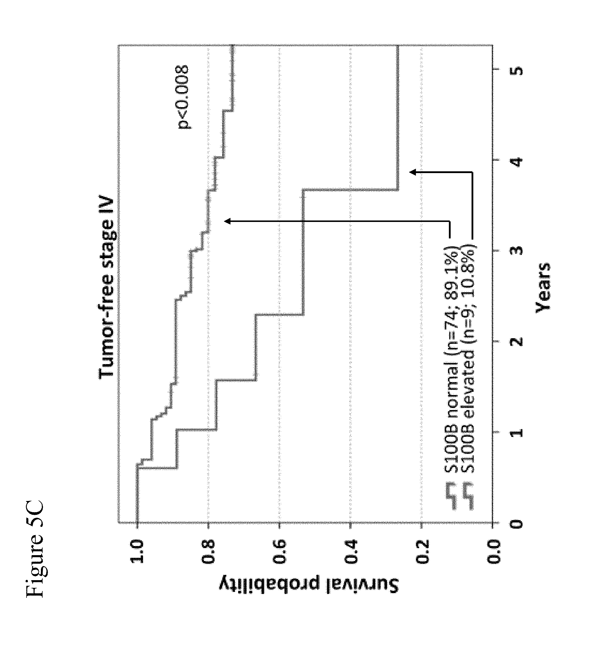
D00013
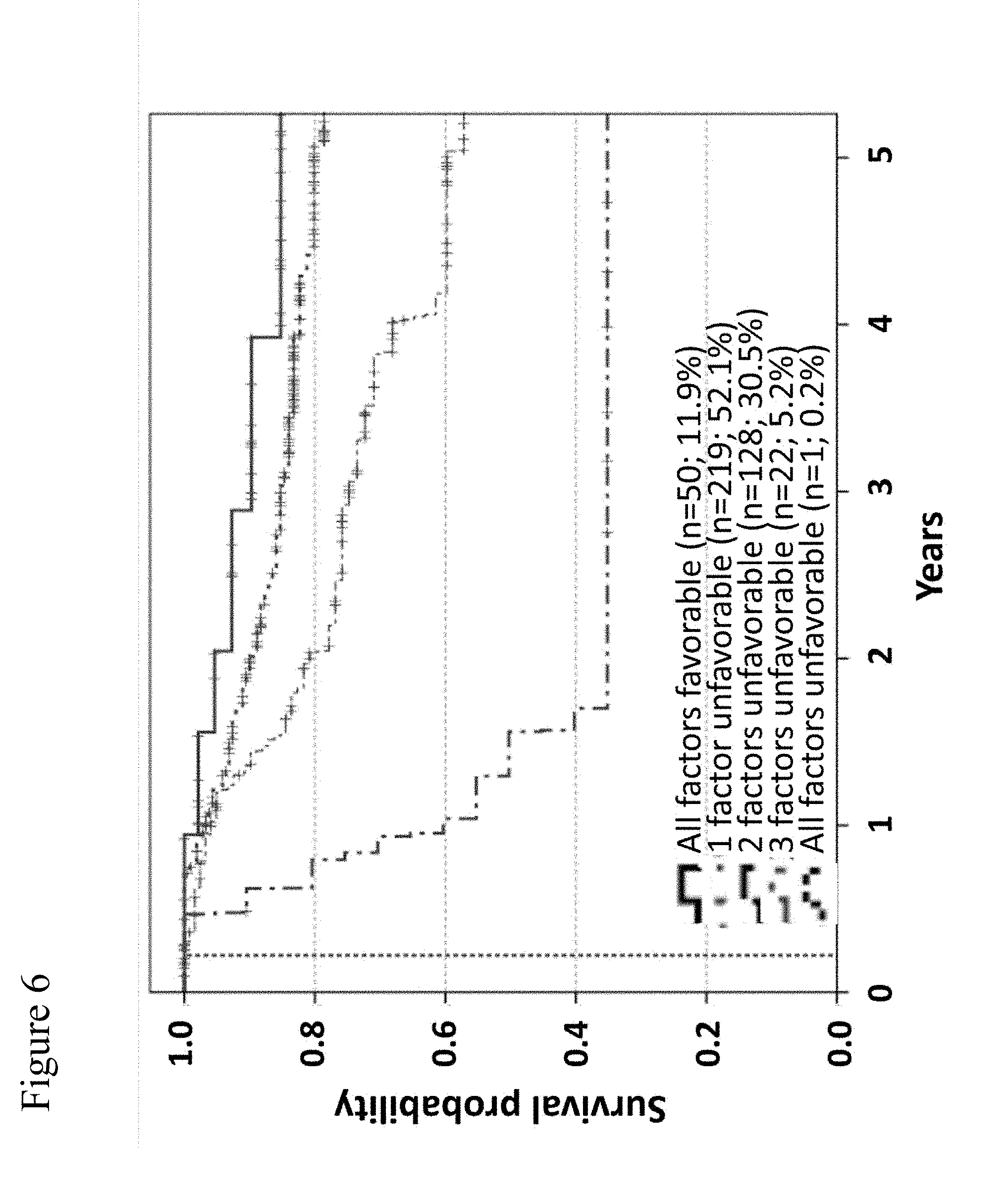
D00014

D00015
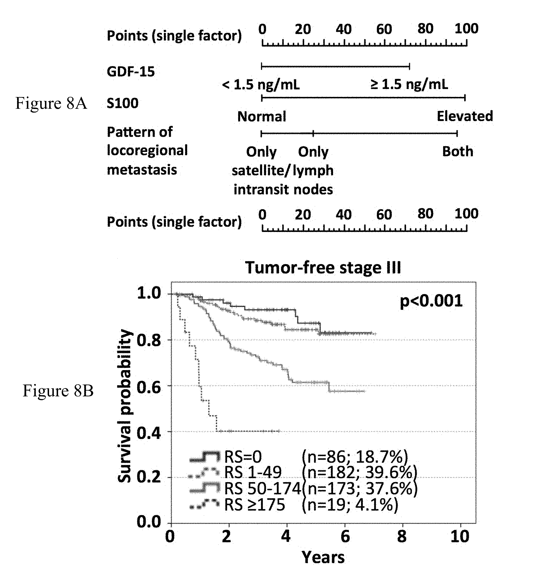
D00016

D00017
D00018
D00019

D00020
D00021

D00022
D00023

D00024

D00025

S00001
XML
uspto.report is an independent third-party trademark research tool that is not affiliated, endorsed, or sponsored by the United States Patent and Trademark Office (USPTO) or any other governmental organization. The information provided by uspto.report is based on publicly available data at the time of writing and is intended for informational purposes only.
While we strive to provide accurate and up-to-date information, we do not guarantee the accuracy, completeness, reliability, or suitability of the information displayed on this site. The use of this site is at your own risk. Any reliance you place on such information is therefore strictly at your own risk.
All official trademark data, including owner information, should be verified by visiting the official USPTO website at www.uspto.gov. This site is not intended to replace professional legal advice and should not be used as a substitute for consulting with a legal professional who is knowledgeable about trademark law.