Anti-myostatin Antibodies And Methods Of Use
RUIKE; Yoshinao ; et al.
U.S. patent application number 16/065192 was filed with the patent office on 2019-01-03 for anti-myostatin antibodies and methods of use. This patent application is currently assigned to Chugai Seiyaku Kabushiki Kaisha. The applicant listed for this patent is CHUGAI SEIYAKU KABUSHIKI KAISHA. Invention is credited to Taichi KURAMOCHI, Yoshinao RUIKE.
| Application Number | 20190002548 16/065192 |
| Document ID | / |
| Family ID | 59089522 |
| Filed Date | 2019-01-03 |

| United States Patent Application | 20190002548 |
| Kind Code | A1 |
| RUIKE; Yoshinao ; et al. | January 3, 2019 |
ANTI-MYOSTATIN ANTIBODIES AND METHODS OF USE
Abstract
The invention provides anti-myostatin antibodies and methods of using the same. In some embodiments, an isolated anti-myostatin antibody of the present invention binds to mature myostatin, and uptake of the antibody into cells is enhanced when complexed with the antigen. The invention also provides isolated nucleic acids encoding an anti-myostatin antibody of the present invention. The invention also provides host cells comprising a nucleic acid of the present invention. The invention also provides a method of producing an antibody comprising culturing a host cell of the present invention so that the antibody is produced. Anti-myostatin antibodies of the present invention may be for use as a medicament. Anti-myostatin antibodies of the present invention may be for use in treating a muscle wasting disease. Anti-myostatin antibodies of the present invention may be for use in increasing mass of muscle tissue. Anti-myostatin antibodies of the present invention may be for use in increasing strength of muscle tissue.
| Inventors: | RUIKE; Yoshinao; (Singapore, SG) ; KURAMOCHI; Taichi; (Singapore, SG) | ||||||||||
| Applicant: |
|
||||||||||
|---|---|---|---|---|---|---|---|---|---|---|---|
| Assignee: | Chugai Seiyaku Kabushiki
Kaisha Tokyo JP |
||||||||||
| Family ID: | 59089522 | ||||||||||
| Appl. No.: | 16/065192 | ||||||||||
| Filed: | December 22, 2016 | ||||||||||
| PCT Filed: | December 22, 2016 | ||||||||||
| PCT NO: | PCT/JP2016/088302 | ||||||||||
| 371 Date: | June 22, 2018 |
| Current U.S. Class: | 1/1 |
| Current CPC Class: | C07K 2317/21 20130101; C07K 2317/567 20130101; C07K 2317/52 20130101; A61P 21/00 20180101; C07K 2317/56 20130101; C07K 16/22 20130101; A61K 2039/505 20130101; C07K 2317/77 20130101; C07K 2317/76 20130101; C07K 2317/92 20130101; C07K 2317/94 20130101; C07K 2317/24 20130101 |
| International Class: | C07K 16/22 20060101 C07K016/22; A61P 21/00 20060101 A61P021/00 |
Foreign Application Data
| Date | Code | Application Number |
|---|---|---|
| Dec 25, 2015 | JP | 2015-253346 |
Claims
1. An isolated antibody that binds to mature myostatin, wherein uptake of the antibody into cells is enhanced when complexed with an antigen.
2. The antibody of claim 1, wherein the uptake is caused by the interaction between Fc region of the antibody and Fc gamma R on the cells.
3. The antibody of claim 1 or 2, which shows at least 2.5-fold higher uptake compared with a reference antibody which is identical to the antibody except that Fc region of the reference antibody has no Fc gamma R-binding activity.
4. The antibody of any one of claims 1 to 3, which has an inhibitory activity against myostatin.
5. The antibody of any one of claims 1 to 4, wherein the antibody binds to the same epitope as an antibody described in Table 2 or 3, or competes for binding to myostatin with an antibody described in Table 2 or 3.
6. The antibody of any one of claims 1 to 5, which binds to mature myostatin with higher affinity at neutral pH than at acidic pH.
7. The antibody of any one of claims 1 to 6, which is a monoclonal antibody.
8. The antibody of any one of claims 1 to 7, which is a human, humanized, or chimeric antibody.
9. The antibody of any one of claims 1 to 8, which is an antibody fragment that binds to myostatin.
10. The antibody of any one of claims 1 to 9, wherein the antibody comprises (a) HVR-H3 comprising the amino acid sequence GX.sub.1DNFGYSYX.sub.2DFNL, wherein X.sub.1 is G or H, X.sub.2 is I or H (SEQ ID NO: 86), (b) HVR-L3 comprising the amino acid sequence QTYDGISX.sub.1YGVA, wherein X.sub.1 is S or H (SEQ ID NO: 88), and (c) HVR-H2 comprising the amino acid sequence IINIX.sub.1GX.sub.2TYYASWAX.sub.3G, wherein X.sub.1 is S or E, X.sub.2 is S or E, X.sub.3 is K or E (SEQ ID NO: 85).
11. The antibody of any one of claims 1 to 9, wherein the antibody comprises (a) HVR-H1 comprising the amino acid sequence X.sub.1YVX.sub.2G, wherein X.sub.1 is N or H, X.sub.2 is M or K (SEQ ID NO: 84), (b) HVR-H2 comprising the amino acid sequence IINIX.sub.1GX.sub.2TYYASWAX.sub.3G, wherein X.sub.1 is S or E, X.sub.2 is S or E, X.sub.3 is K or E (SEQ ID NO: 85), and (c) HVR-H3 comprising the amino acid sequence GX.sub.1DNFGYSYX.sub.2DFNL, wherein X.sub.1 is G or H, X.sub.2 is I or H (SEQ ID NO: 86).
12. The antibody of claim 11, further comprising (a) HVR-L1 comprising the amino acid sequence QASX.sub.1SIX.sub.2X.sub.3X.sub.4LS, wherein X.sub.1 is Q or E, X.sub.2 is S or H, X.sub.3 is N or H, X.sub.4 is E or D (SEQ ID NO: 87); (b) HVR-L2 comprising the amino acid sequence LASTLAS (SEQ ID NO: 81); and (c) HVR-L3 comprising the amino acid sequence QTYDGISX.sub.1YGVA, wherein X.sub.1 is S or H (SEQ ID NO: 88).
13. The antibody of any one of claims 1 to 9, comprising (a) HVR-L1 comprising the amino acid sequence QASX.sub.1SIX.sub.2X.sub.3X.sub.4LS, wherein X.sub.1 is Q or E, X.sub.2 is S or H, X.sub.3 is N or H, X.sub.4 is E or D (SEQ ID NO: 87); (b) HVR-L2 comprising the amino acid sequence LASTLAS (SEQ ID NO: 81); and (c) HVR-L3 comprising the amino acid sequence QTYDGISX.sub.1YGVA, wherein X.sub.1 is S or H (SEQ ID NO: 88).
14. The antibody of claim 11, further comprising a heavy chain variable domain framework FR1 comprising the amino acid sequence of SEQ ID NO: 89; FR2 comprising the amino acid sequence of SEQ ID NO: 90; FR3 comprising the amino acid sequence of SEQ ID NO: 91; and FR4 comprising the amino acid sequence of SEQ ID NO: 92.
15. The antibody of claim 13, further comprising a light chain variable domain framework FR1 comprising the amino acid sequence of SEQ ID NO: 93; FR2 comprising the amino acid sequence of SEQ ID NO: 94; FR3 comprising the amino acid sequence of SEQ ID NO: 95; and FR4 comprising the amino acid sequence of SEQ ID NO: 96.
16. The antibody of any one of claims 1 to 9, comprising (a) a VH sequence having at least 95% sequence identity to the amino acid sequence of any one of SEQ ID NOs: 48-51; (b) a VL sequence having at least 95% sequence identity to the amino acid sequence of any one of SEQ ID NOs: 52-55; or (c) a VH sequence as in (a) and a VL sequence as in (b).
17. The antibody of claim 16, comprising a VH sequence of any one of SEQ ID NOs: 48-51.
18. The antibody of claim 16, comprising a VL sequence of any one of SEQ ID NOs: 52-55.
19. An antibody comprising a VH sequence of any one of SEQ ID NOs: 48-51 and a VL sequence of any one of SEQ ID NOs: 52-55.
20. The antibody of any one of claims 1 to 19, which is a full length IgG antibody.
21. Isolated nucleic acid encoding the antibody of any one of claims 1 to 20.
22. A host cell comprising the nucleic acid of claim 21.
23. A method of producing an antibody comprising culturing the host cell of claim 22 so that the antibody is produced.
24. A pharmaceutical formulation comprising the antibody of any one of claims 1 to 20 and a pharmaceutically acceptable carrier.
25. The antibody of any one of claims 1 to 20 for use as a medicament.
26. The antibody of any one of claims 1 to 20 for use in treating a muscle wasting disease.
27. The antibody of any one of claims 1 to 20 for use in increasing mass of muscle tissue.
28. The antibody of any one of claims 1 to 20 for use in increasing strength of muscle tissue.
29. Use of the antibody of any one of claims 1 to 20 in the manufacture of a medicament for treatment of a muscle wasting disease.
30. Use of the antibody of any one of claims 1 to 20 in the manufacture of a medicament for increasing mass of muscle tissue.
31. Use of the antibody of any one of claims 1 to 20 in the manufacture of a medicament for increasing strength of muscle tissue.
32. A method of treating an individual having a muscle wasting disease comprising administering to the individual an effective amount of the antibody of any one of claims 1 to 20.
33. A method of increasing mass of muscle tissue in an individual comprising administering to the individual an effective amount of the antibody of any one of claims 1 to 20 to increase mass of muscle tissue.
34. A method of increasing strength of muscle tissue in an individual comprising administering to the individual an effective amount of the antibody of any one of claims 1 to 20 to increase strength of muscle tissue.
Description
TECHNICAL FIELD
[0001] The present invention relates to anti-myostatin antibodies and methods of using the same.
BACKGROUND ART
[0002] Myostatin, also referred to as growth differentiation factor-8 (GDF-8), is a secreted protein and is a member of the transforming growth factor-beta (TGF-beta) superfamily of proteins. Members of this superfamily possess growth-regulatory and morphogenetic properties (See, e.g., NPL 1, NPL 2, and PTL 1). Myostatin is expressed primarily in the developing and adult skeletal muscle and functions as a negative regulator of muscle growth. Systemic overexpression of myostatin in adult mice leads to muscle wasting (See, e.g., NPL 3) while, conversely, a myostatin knockout mouse is characterized by hypertrophy and hyperplasia of the skeletal muscle resulting in two- to threefold greater muscle mass than their wild type littermates (See, e.g., NPL 4).
[0003] Like other members of the TGF-beta family, myostatin is synthesized as a large precursor protein containing an N-terminal propeptide domain, and a C-terminal domain considered as the active molecule (See, e.g., NPL 5; PTL 2). Two molecules of myostatin precursor are covalently linked via a single disulfide bond present in the C-terminal growth factor domain. Active mature myostatin (disulfide-bonded homodimer consisting of the C-terminal growth factor domain) is liberated from myostatin precursor through multiple steps of proteolytic processing. In the first step of the myostatin activation pathway, a peptide bond between the N-terminal propeptide domain and the C-terminal growth factor domain, Arg266-Asp267, is cleaved by a furin-type proprotein convertase in both chains of the homodimeric precursor. But the resulting two propeptides and one mature myostatin (disulfide-bonded homodimer consisting of the growth factor domains) remain associated, forming a noncovalent inactive complex, that is latent myostatin. Mature myostatin can then be liberated from latent myostatin through degradation of the propeptide. Members of the bone morphogenetic protein 1 (BMP-1) family of metalloproteinases cleave a single peptide bond within the propeptide, Arg98-Asp99, with concomitant release of the mature myostatin (See, e.g., NPL 6). Moreover, the latent myostatin can be activated in vitro by dissociating the complex with either acid or heat treatment as well (See, e.g., NPL 7).
[0004] Myostatin exerts its effects through a transmembrane serine/threonine kinase heterotetramer receptor family, activation of which enhances receptor transphosphorylation, leading to the stimulation of serine/threonine kinase activity. It has been shown that the myostatin pathway involves an active myostatin dimer binding to the activin receptor type IIB (ActRIIB) with high affinity, which then recruits and activates the transphosphorylation of the low affinity receptor, the activin-like kinase 4 (ALK4) or activin-like kinase 5 (ALK5). It has also been shown that the proteins Smad 2 and Smad 3 are subsequently activated and form complexes with Smad 4, which are then translocated to the nucleus for the activation of target gene transcription. It has been demonstrated that ActRIIB is able to mediate the influence of myostatin in vivo, as expression of a dominant negative form of ActRIIB in mice mimics myostatin gene knockout (See, e.g., NPL 8).
[0005] A number of disorders or conditions are associated with muscle wasting (i.e., loss of or functional impairment of muscle tissue), such as muscular dystrophy (MD; including Duchenne muscular dystrophy), amyotrophic lateral sclerosis (ALS), muscle atrophy, organ atrophy, frailty, congestive obstructive pulmonary disease (COPD), sarcopenia, and cachexia resulting from cancer or other disorders, as well as renal disease, cardiac failure or disease, and liver disease. Patients will benefit from an increase in muscle mass and/or muscle strength; however, there are presently limited treatments available for these disorders. Thus, due to its role as a negative regulator of skeletal muscle growth, myostatin becomes a desirable target for therapeutic or prophylactic intervention for such disorders or conditions, or for monitoring the progression of such disorders or conditions. In particular, agents that inhibit the activity of myostatin may be therapeutically beneficial.
[0006] Inhibition of myostatin expression leads to both muscle hypertrophy and hyperplasia (NPL 4). Myostatin negatively regulates muscle regeneration after injury and lack of myostatin in myostatin null mice results in accelerated muscle regeneration (See, e.g., NPL 9). Anti-myostatin (GDF-8) antibodies described in, e.g., PTL 3, PTL 4, PTL 5, PTL 6, PTL 7, PTL 8, PTL 9, PTL 10, and PTL 11 have been shown to bind to myostatin and inhibit myostatin activity in vitro and in vivo, including myostatin activity associated with the negative regulation of skeletal muscle mass. Myostatin-neutralizing antibodies increase body weight, skeletal muscle mass, and muscle size and strength in the skeletal muscle of wild type mice (See, e.g., NPL 10) and the mdx mice, a model for muscular dystrophy (See, e.g., NPL 11; NPL 12). However, there is a further need for improvements in efficacy and convenience of agents that bind myostatin and antagonize its activity in the art.
CITATION LIST
Patent Literature
[0007] [PTL 1] U.S. Pat. No. 5,827,733 [0008] [PTL 2] WO 94/021681 [0009] [PTL 3] U.S. Pat. No. 6,096,506 [0010] [PTL 4] WO 2004/037861 [0011] [PTL 5] U.S. Pat. No. 7,320,789 [0012] [PTL 6] U.S. Pat. No. 7,807,159 [0013] [PTL 7] U.S. Pat. No. 7,888,486 [0014] [PTL 8] WO 2005/094446 [0015] [PTL 9] U.S. Pat. No. 7,632,499 [0016] [PTL 10] WO 2010/070094 [0017] [PTL 11] U.S. Pat. No. 8,415,459
Non Patent Literature
[0017] [0018] [NPL 1] Kingsley et al (1994) Genes Dev 8(2): 133-146 [0019] [NPL 2] Hoodless et al (1998) Curr Top Microbiol Immunol 228: 235-272 [0020] [NPL 3] Zimmers et al (2002) Science 296(5572): 1486-1488 [0021] [NPL 4] McPherron et al (1997) Nature 387(6628): 83-90 [0022] [NPL 5] McPherron and Lee (1997) Proc Natl Acad Sci USA 94(23): 12457-12461 [0023] [NPL 6] Szlama et al (2013) FEBS J 280(16): 3822-3839 [0024] [NPL 7] Lee (2008) PloS One 3(2): e1628 [0025] [NPL 8] Lee and McPherron (2001) Proc Natl Acad Sci USA 98(16): 9306-9311 [0026] [NPL 9] McCroskery et al (2005) J Cell Sci 118(15): 3531-3541 [0027] [NPL 10] Whittemore et al (2003) Biochem Biophys Res Commun 300(4): 965-971 [0028] [NPL 11] Bogdanovich et al (2002) Nature 420(6914): 418-421 [0029] [NPL 12] Wagner et al (2002) Ann Neurol 52(6): 832-836
SUMMARY OF INVENTION
Technical Problem
[0030] An objective of the invention is to provide anti-myostatin antibodies and methods of using the same.
Solution to Problem
[0031] The invention provides anti-myostatin antibodies and methods of using the same.
[0032] In some embodiments, an isolated anti-myostatin antibody of the present invention binds to mature myostatin. In some embodiments, uptake of an isolated anti-myostatin antibody of the present invention into cells is enhanced when complexed with an antigen. In further embodiments, the uptake is caused by the interaction between Fc region of the antibody and Fc gamma R on the cells. In further embodiments, the antibody shows at least 2.5-fold higher uptake compared with a reference antibody which is identical to the antibody except that Fc region of the reference antibody has no Fc gamma R-binding activity. In some embodiments, an isolated anti-myostatin antibody of the present invention has an inhibitory activity against myostatin. In some embodiments, an isolated anti-myostatin antibody of the present invention binds to the same epitope as an antibody described in Table 2 or 3. In some embodiments, an isolated anti-myostatin antibody of the present invention competes for binding to myostatin with an antibody described in Table 2 or 3. In some embodiments, an isolated anti-myostatin antibody of the present invention binds to mature myostatin with higher affinity at neutral pH than at acidic pH.
[0033] In some embodiments, an isolated anti-myostatin antibody of the present invention is a monoclonal antibody. In some embodiments, an isolated anti-myostatin antibody of the present invention is a human, humanized, or chimeric antibody. In some embodiments, an isolated anti-myostatin antibody of the present invention is an antibody fragment that binds to myostatin. In some embodiments, an isolated anti-myostatin antibody of the present invention is a full length IgG antibody.
[0034] In some embodiments, an isolated anti-myostatin antibody of the present invention comprises (a) HVR-H3 comprising the amino acid sequence GX.sub.1DNFGYSYX.sub.2DFNL, wherein X.sub.1 is G or H, X.sub.2 is I or H (SEQ ID NO: 86), (b) HVR-L3 comprising the amino acid sequence QTYDGISX.sub.1YGVA, wherein X.sub.1 is S or H (SEQ ID NO: 88), and (c) HVR-H2 comprising the amino acid sequence IINIX.sub.1GX.sub.2TYYASWAX.sub.3G, wherein X.sub.1 is S or E, X.sub.2 is S or E, X.sub.3 is K or E (SEQ ID NO: 85).
[0035] In some embodiments, an isolated anti-myostatin antibody of the present invention comprises (a) HVR-H1 comprising the amino acid sequence X.sub.1YVX.sub.2G, wherein X.sub.1 is N or H, X.sub.2 is M or K (SEQ ID NO: 84), (b) HVR-H2 comprising the amino acid sequence IINIX.sub.1GX.sub.2TYYASWAX.sub.3G, wherein X.sub.1 is S or E, X.sub.2 is S or E, X.sub.3 is K or E (SEQ ID NO: 85), and (c) HVR-H3 comprising the amino acid sequence GX.sub.1DNFGYSYX.sub.2DFNL, wherein X.sub.1 is G or H, X.sub.2 is I or H (SEQ ID NO: 86). In further embodiments, the antibody comprises (a) HVR-L1 comprising the amino acid sequence QASX.sub.1SIX.sub.2X.sub.3X.sub.4LS, wherein X.sub.1 is Q or E, X.sub.2 is S or H, X.sub.3 is N or H, X.sub.4 is E or D (SEQ ID NO: 87); (b) HVR-L2 comprising the amino acid sequence LASTLAS (SEQ ID NO: 81); and (c) HVR-L3 comprising the amino acid sequence QTYDGISX.sub.1YGVA, wherein X.sub.1 is S or H (SEQ ID NO: 88).
[0036] In some embodiments, an isolated anti-myostatin antibody of the present invention comprises (a) HVR-L1 comprising the amino acid sequence QASX.sub.1SIX.sub.2X.sub.3X.sub.4LS, wherein X.sub.1 is Q or E, X.sub.2 is S or H, X.sub.3 is N or H, X.sub.4 is E or D (SEQ ID NO: 87); (b) HVR-L2 comprising the amino acid sequence LASTLAS (SEQ ID NO: 81); and (c) HVR-L3 comprising the amino acid sequence QTYDGISX.sub.1YGVA, wherein X.sub.1 is S or H (SEQ ID NO: 88).
[0037] In some embodiments, an isolated anti-myostatin antibody of the present invention comprises a heavy chain variable domain framework FR1 comprising the amino acid sequence of SEQ ID NO: 89; FR2 comprising the amino acid sequence of SEQ ID NO: 90; FR3 comprising the amino acid sequence of SEQ ID NO: 91; and FR4 comprising the amino acid sequence of SEQ ID NO: 92. In some embodiments, an isolated anti-myostatin antibody of the present invention comprises a light chain variable domain framework FR1 comprising the amino acid sequence of SEQ ID NO: 93; FR2 comprising the amino acid sequence of SEQ ID NO: 94; FR3 comprising the amino acid sequence of SEQ ID NO: 95; and FR4 comprising the amino acid sequence of SEQ ID NO: 96.
[0038] In some embodiments, an isolated anti-myostatin antibody of the present invention comprises (a) a VH sequence having at least 95% sequence identity to the amino acid sequence of any one of SEQ ID NOs: 48-51; (b) a VL sequence having at least 95% sequence identity to the amino acid sequence of any one of SEQ ID NOs: 52-55; or (c) a VH sequence as in (a) and a VL sequence as in (b). In further embodiments, the antibody comprises a VH sequence of any one of SEQ ID NOs: 48-51. In further embodiments, the antibody comprises a VL sequence of any one of SEQ ID NOs: 52-55.
[0039] The invention provides an antibody comprising a VH sequence of any one of SEQ ID NOs: 48-51 and a VL sequence of any one of SEQ ID NOs: 52-55.
[0040] The invention also provides isolated nucleic acids encoding an anti-myostatin antibody of the present invention. The invention also provides host cells comprising a nucleic acid of the present invention. The invention also provides a method of producing an antibody comprising culturing a host cell of the present invention so that the antibody is produced.
[0041] The invention also provides a pharmaceutical formulation comprising an anti-myostatin antibody of the present invention and a pharmaceutically acceptable carrier.
[0042] Anti-myostatin antibodies of the present invention may be for use as a medicament. Anti-myostatin antibodies of the present invention may be for use in treating a muscle wasting disease. Anti-myostatin antibodies of the present invention may be for use in increasing mass of muscle tissue. Anti-myostatin antibodies of the present invention may be for use in increasing strength of muscle tissue.
[0043] Anti-myostatin antibodies of the present invention may be used in the manufacture of a medicament. In some embodiments, the medicament is for treatment of a muscle wasting disease. In some embodiments, the medicament is for increasing mass of muscle tissue. In some embodiments, the medicament is for increasing strength of muscle tissue.
[0044] The invention also provides a method of treating an individual having a muscle wasting disease. In some embodiments, the method comprises administering to the individual an effective amount of an anti-myostatin antibody of the present invention. The invention also provides a method of increasing mass of muscle tissue in an individual. In some embodiments, the method comprises administering to the individual an effective amount of an anti-myostatin antibody of the present invention to increase mass of muscle tissue. The invention also provides a method of increasing strength of muscle tissue in an individual. In some embodiments, the method comprises administering to the individual an effective amount of an anti-myostatin antibody of the present invention to increase strength of muscle tissue.
BRIEF DESCRIPTION OF DRAWINGS
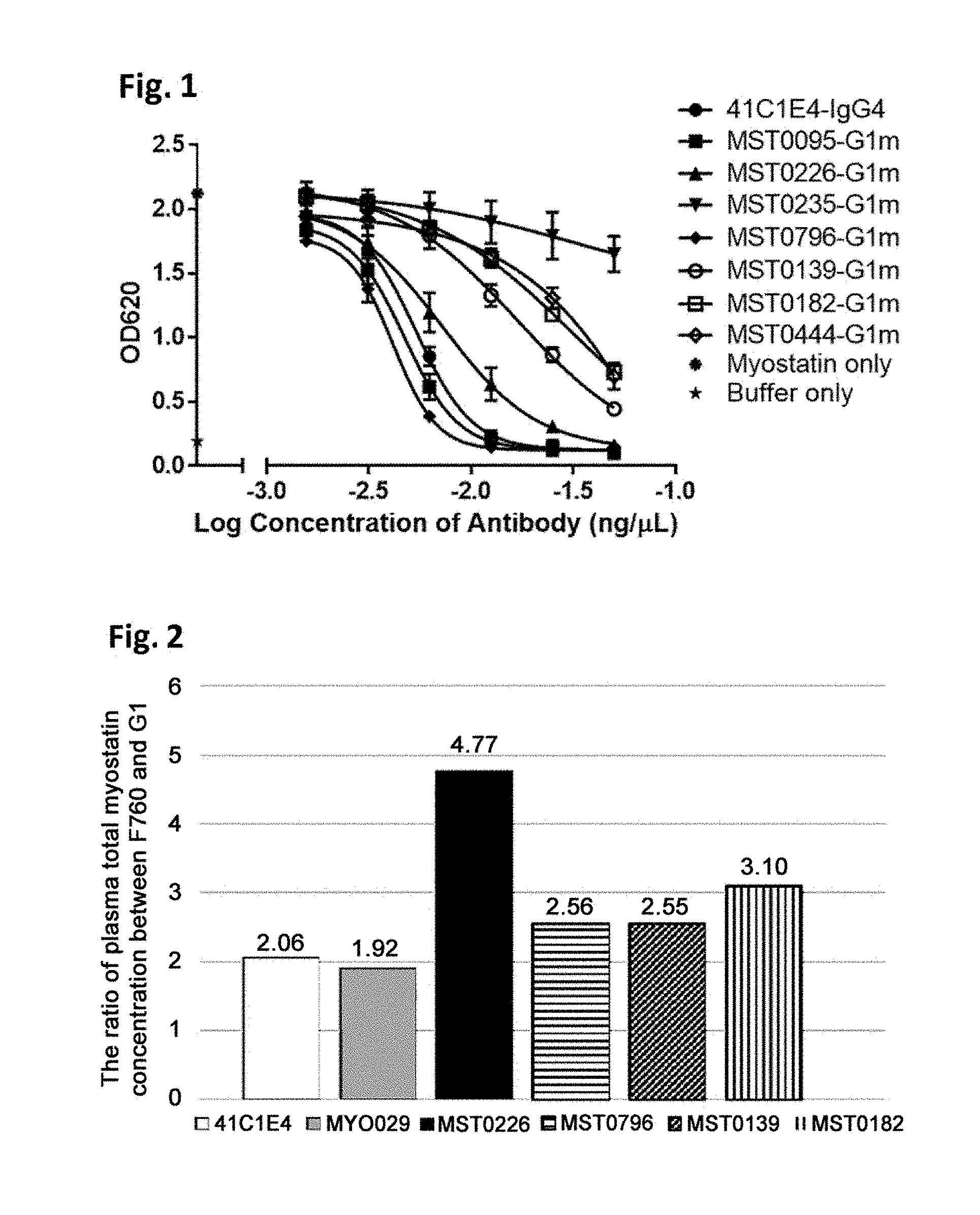
[0045] FIG. 1 illustrates inhibition of myostatin activity by anti-mature myostatin antibodies, as described in Example 3. The activity of myostatin was measured using HEK Blue Assay in the presence of an anti-mature myostatin antibody 41C1E4, MST0095-G1m, MST0226-G1m, MST0235-G1m, MST0796-G1m, MST0139-G1m, MST0182-G1m, or MST0444-G1m at various concentrations.
[0046] FIG. 2 illustrates comparison of antigen clearance from plasma among anti-mature myostatin antibodies in vivo, as described in Example 4. For each of anti-mature myostatin antibodies MST0226, MST0796, MST0139, MST0182, 41C1E4 and MYO029, two types of modified antibodies were generated, one of which has an Fc region with Fc gamma R binding activity (G1) and the other of which has an Fc region without Fc gamma R binding activity (F760). Each of the antibodies was administered in mice together with recombinant mature myostatin, and the resulting concentration of total myostatin in plasma was measured. The extent of the antigen clearance was evaluated by calculating the ratio of (plasma total myostatin concentration measured when the antibody having F760-type Fc region was administered)/(plasma total myostatin concentration measured when the antibody having G1-type Fc region was administered). In this assay, a higher value of the ratio means that the antibody has a higher ability to be taken up into cells with its antigen (mature myostatin) through the interaction of the Fc region of the antibody and Fc gamma R on the cells, which results in enhanced antigen clearance from plasma.
[0047] FIG. 3 illustrates in vivo efficacy of anti-mature myostatin antibodies on muscle mass, as described in Example 5. Each of the anti-mature myostatin antibodies 41C1E4, MST0226-G1m and MST0796-G1m was administered in mice, and lean body mass (LBM) was measured on day 0, 4, 7 and 14. * indicates p<0.05, ** indicates p<0.01, and *** indicates p<0.001 in comparison to the PBS group by Dunnett's test.
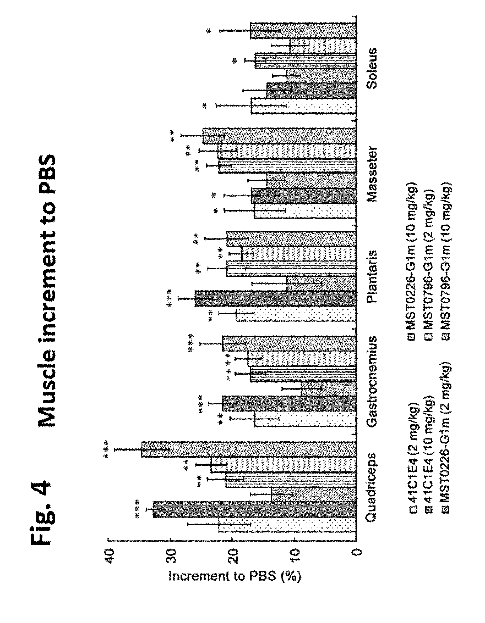
[0048] FIG. 4 illustrates in vivo efficacy of anti-mature myostatin antibodies on muscle mass, as described in Example 5. Each of the anti-mature myostatin antibodies 41C1E4, MST0226-G1m and MST0796-G1m was administered in mice, and weight of quadriceps, gastrocnemius, plantaris, masseter, and soleus muscles was measured. The vertical axis shows a percent increment of muscle weight compared to the PBS group. * indicates p<0.05, ** indicates p<0.01, and *** indicates p<0.001 in comparison to the PBS group by Dunnett's test.
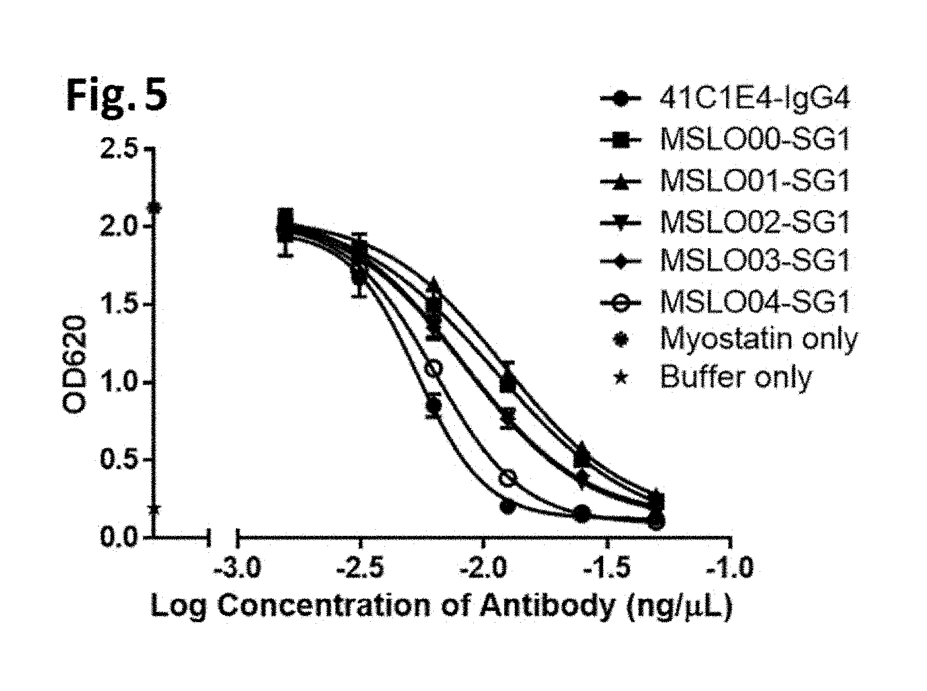
[0049] FIG. 5 illustrates inhibition of myostatin activity by anti-mature myostatin antibodies, as described in Example 6. The activity of myostatin was measured using HEK Blue Assay in the presence of an anti-mature myostatin antibody 41C1E4, MSLO00-SG1, MSLO01-SG1, MSLO02-SG1, MSLO03-SG1, or MSLO04-SG1 at various concentrations.
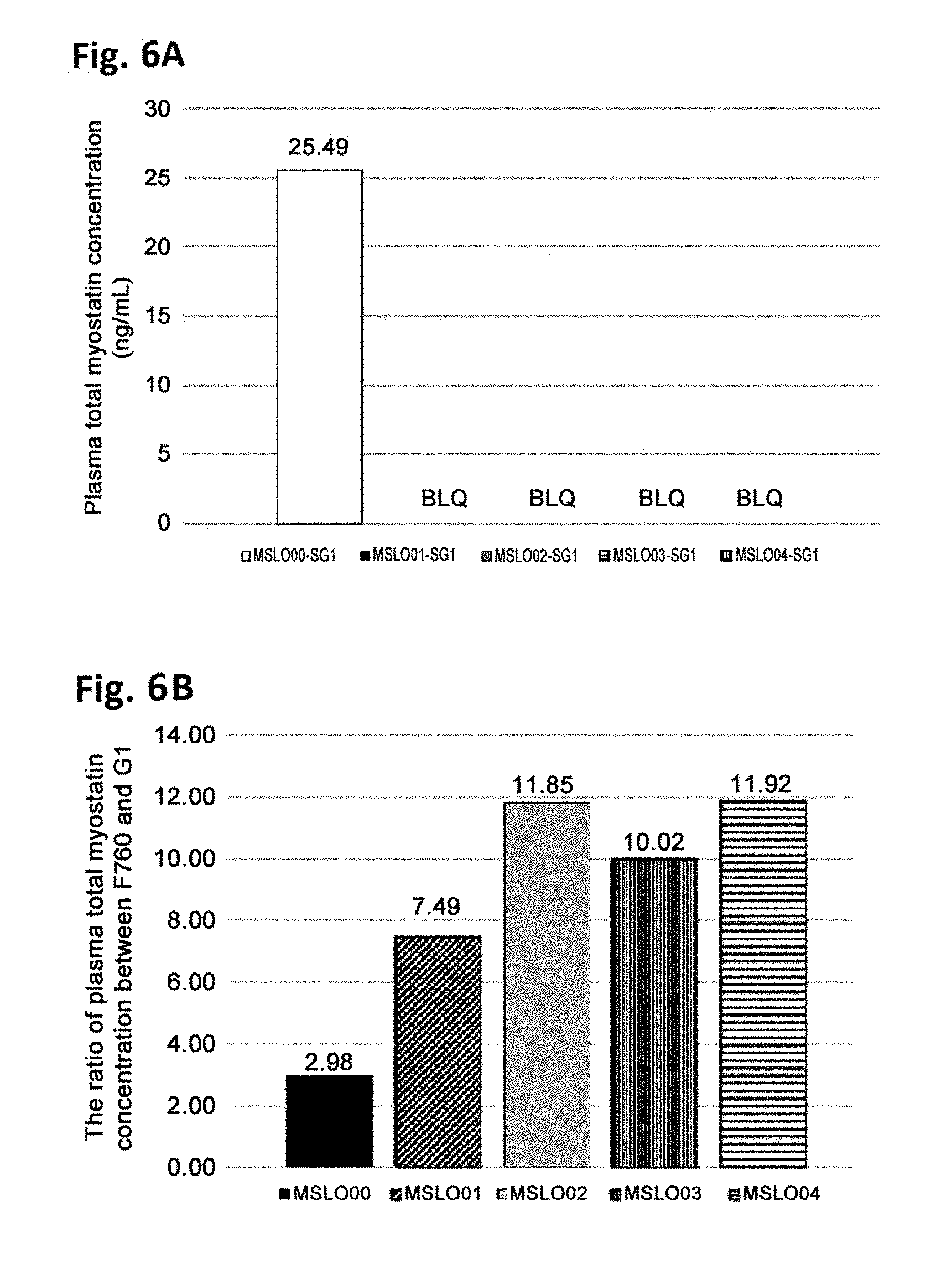
[0050] FIGS. 6A and 6B illustrate comparison of antigen clearance from plasma among anti-mature myostatin antibodies in vivo, as described in Example 7. For each of anti-mature myostatin antibodies MSLO00-SG1, MSLO01-SG1, MSLO02-SG1, MSLO03-SG1 and MSLO04-SG1, two types of modified antibodies were generated, one of which has an Fc region with Fc gamma R binding activity (G1) and the other of which has an Fc region without Fc gamma R binding activity (F760). Each of the antibodies was administered in mice together with recombinant mature myostatin, and the resulting concentration of total myostatin in plasma was measured. FIG. 6A illustrates plasma total myostation concentrations measured when the antibody having G1-type Fc region was administered. BLQ (below the limit of quantitation) indicates that the measured concentration was below the lower limit of quantitation in the myostatin concentration measurement assay. FIG. 6B illustrates ratios of (plasma total myostatin concentration measured when the antibody having F760-type Fc region was administered)/(plasma total myostatin concentration measured when the antibody having G1-type Fc region was administered). In this assay, a higher value of the ratio means that the antibody has a higher ability to be taken up into cells with its antigen (mature myostatin) through the interaction of the Fc region of the antibody and Fc gamma R on the cells, which results in enhanced antigen clearance from plasma.
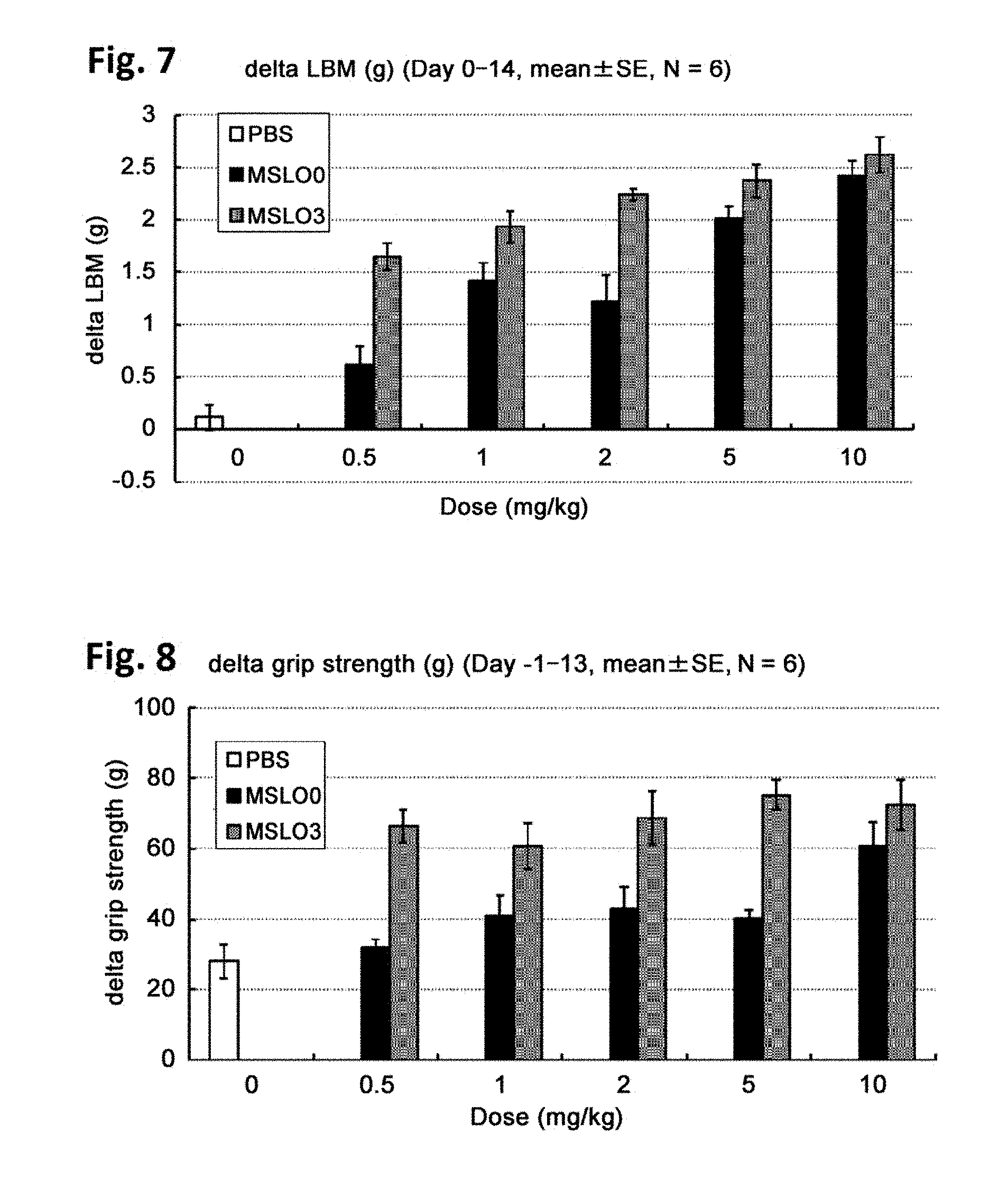
[0051] FIG. 7 illustrates in vivo efficacy of anti-mature myostatin antibodies on muscle mass, as described in Example 8. Each of the anti-mature myostatin antibodies MSLO00-SG1 and MSLO03-SG1 was administered at doses of 0.5 mg/kg, 1 mg/kg, 2 mg/kg, 5 mg/kg and 10 mg/kg in mice, and lean body mass (LBM) was measured. The vertical axis shows an increment of LBM between day 0 and day 14.
[0052] FIG. 8 illustrates in vivo efficacy of anti-mature myostatin antibodies on grip strength, as described in Example 8. Each of the anti-mature myostatin antibodies MSLO00-SG1 and MSLO03-SG1 was administered at doses of 0.5 mg/kg, 1 mg/kg, 2 mg/kg, 5 mg/kg and 10 mg/kg in mice, and grip strength of the mice was measured. The vertical axis shows an increment of grip strength between day -1 and day 13.
DESCRIPTION OF EMBODIMENTS
[0053] The techniques and procedures described or referenced herein are generally well understood and commonly employed using conventional methodology by those skilled in the art, such as, for example, the widely utilized methodologies described in Sambrook et al., Molecular Cloning: A Laboratory Manual 3d edition (2001) Cold Spring Harbor Laboratory Press, Cold Spring Harbor, N.Y.; Current Protocols in Molecular Biology (F. M. Ausubel, et al. eds., (2003)); the series Methods in Enzymology (Academic Press, Inc.): PCR 2: A Practical Approach (M. J. MacPherson, B. D. Hames and G. R. Taylor eds. (1995)), Harlow and Lane, eds. (1988) Antibodies, A Laboratory Manual, and Animal Cell Culture (R. I. Freshney, ed. (1987)); Oligonucleotide Synthesis (M. J. Gait, ed., 1984); Methods in Molecular Biology, Humana Press; Cell Biology: A Laboratory Notebook (J. E. Cellis, ed., 1998) Academic Press; Animal Cell Culture (R. I. Freshney), ed., 1987); Introduction to Cell and Tissue Culture (J. P. Mather and P. E. Roberts, 1998) Plenum Press; Cell and Tissue Culture: Laboratory Procedures (A. Doyle, J. B. Griffiths, and D. G. Newell, eds., 1993-8) J. Wiley and Sons; Handbook of Experimental Immunology (D. M. Weir and C. C. Blackwell, eds.); Gene Transfer Vectors for Mammalian Cells (J. M. Miller and M. P. Calos, eds., 1987); PCR: The Polymerase Chain Reaction, (Mullis et al., eds., 1994); Current Protocols in Immunology (J. E. Coligan et al., eds., 1991); Short Protocols in Molecular Biology (Wiley and Sons, 1999); Immunobiology (C. A. Janeway and P. Travers, 1997); Antibodies (P. Finch, 1997); Antibodies: A Practical Approach (D. Catty., ed., IRL Press, 1988-1989); Monoclonal Antibodies: A Practical Approach (P. Shepherd and C. Dean, eds., Oxford University Press, 2000); Using Antibodies: A Laboratory Manual (E. Harlow and D. Lane, Cold Spring Harbor Laboratory Press, 1999); The Antibodies (M. Zanetti and J. D. Capra, eds., Harwood Academic Publishers, 1995); and Cancer: Principles and Practice of Oncology (V. T. DeVita et al., eds., J. B. Lippincott Company, 1993).
I. DEFINITIONS
[0054] Unless defined otherwise, technical and scientific terms used herein have the same meaning as commonly understood by one of ordinary skill in the art to which this invention belongs. Singleton et al., Dictionary of Microbiology and Molecular Biology 2nd ed., J. Wiley & Sons (New York, N.Y. 1994), and March, Advanced Organic Chemistry Reactions, Mechanisms and Structure 4th ed., John Wiley & Sons (New York, N.Y. 1992), provide one skilled in the art with a general guide to many of the terms used in the present application. All references cited herein, including patent applications and publications, are incorporated by reference in their entirety.
[0055] For purposes of interpreting this specification, the following definitions will apply and whenever appropriate, terms used in the singular will also include the plural and vice versa. It is to be understood that the terminology used herein is for the purpose of describing particular embodiments only, and is not intended to be limiting. In the event that any definition set forth below conflicts with any document incorporated herein by reference, the definition set forth below shall control.
[0056] An "acceptor human framework" for the purposes herein is a framework comprising the amino acid sequence of a light chain variable domain (VL) framework or a heavy chain variable domain (VH) framework derived from a human immunoglobulin framework or a human consensus framework, as defined below. An acceptor human framework "derived from" a human immunoglobulin framework or a human consensus framework may comprise the same amino acid sequence thereof, or it may contain amino acid sequence changes. In some embodiments, the number of amino acid changes are 10 or less, 9 or less, 8 or less, 7 or less, 6 or less, 5 or less, 4 or less, 3 or less, or 2 or less. In some embodiments, the VL acceptor human framework is identical in sequence to the VL human immunoglobulin framework sequence or human consensus framework sequence.
[0057] "Affinity" refers to the strength of the sum total of noncovalent interactions between a single binding site of a molecule (e.g., an antibody) and its binding partner (e.g., an antigen). Unless indicated otherwise, as used herein, "binding affinity" refers to intrinsic binding affinity which reflects a 1:1 interaction between members of a binding pair (e.g., antibody and antigen). The affinity of a molecule X for its partner Y can generally be represented by the dissociation constant (Kd). Affinity can be measured by common methods known in the art, including those described herein. Specific illustrative and exemplary embodiments for measuring binding affinity are described in the following.
[0058] An "affinity matured" antibody refers to an antibody with one or more alterations in one or more hypervariable regions (HVRs), compared to a parent antibody which does not possess such alterations, such alterations resulting in an improvement in the affinity of the antibody for antigen.
[0059] The terms "anti-myostatin antibody" and "an antibody that binds to myostatin" refer to an antibody that is capable of binding myostatin with sufficient affinity such that the antibody is useful as a diagnostic and/or therapeutic agent in targeting myostatin. In one embodiment, the extent of binding of an anti-myostatin antibody to an unrelated, non-myostatin protein is less than about 10% of the binding of the antibody to myostatin as measured, e.g., by a radioimmunoassay (RIA). In certain embodiments, an antibody that binds to myostatin has a dissociation constant (Kd) of 1 micro M or less, 100 nM or less, 10 nM or less, 1 nM or less, 0.1 nM or less, 0.01 nM or less, or 0.001 nM or less (e.g. 10.sup.-8 M or less, e.g. from 10.sup.-8M to 10.sup.-13 M, e.g., from 10.sup.-9M to 10.sup.-13 M). In certain embodiments, an anti myostatin antibody binds to an epitope of myostatin that is conserved among myostatin from different species.
[0060] The term "antibody" herein is used in the broadest sense and encompasses various antibody structures, including but not limited to monoclonal antibodies, polyclonal antibodies, multispecific antibodies (e.g., bispecific antibodies), and antibody fragments so long as they exhibit the desired antigen-binding activity.
[0061] An "antibody fragment" refers to a molecule other than an intact antibody that comprises a portion of an intact antibody that binds the antigen to which the intact antibody binds. Examples of antibody fragments include but are not limited to Fv, Fab, Fab', Fab'-SH, F(ab').sub.2; diabodies; linear antibodies; single-chain antibody molecules (e.g. scFv); and multispecific antibodies formed from antibody fragments.
[0062] An "antibody that binds to the same epitope" as a reference antibody refers to an antibody that blocks binding of the reference antibody to its antigen in a competition assay, and/or conversely, the reference antibody blocks binding of the antibody to its antigen in a competition assay. An exemplary competition assay is provided herein.
[0063] The term "chimeric" antibody refers to an antibody in which a portion of the heavy and/or light chain is derived from a particular source or species, while the remainder of the heavy and/or light chain is derived from a different source or species.
[0064] The "class" of an antibody refers to the type of constant domain or constant region possessed by its heavy chain. There are five major classes of antibodies: IgA, IgD, IgE, IgG, and IgM, and several of these may be further divided into subclasses (isotypes), e.g., IgG.sub.1, IgG.sub.2, IgG.sub.3, IgG.sub.4, IgA.sub.1, and IgA.sub.2. The heavy chain constant domains that correspond to the different classes of immunoglobulins are called alpha, delta, epsilon, gamma, and mu, respectively.
[0065] The term "cytotoxic agent" as used herein refers to a substance that inhibits or prevents a cellular function and/or causes cell death or destruction. Cytotoxic agents include, but are not limited to, radioactive isotopes (e.g., At.sup.211, I.sup.131, I.sup.125, Y.sup.90, Re.sup.186, Re.sup.188, Sm.sup.153, Bi.sup.212, P.sup.32, Pb.sup.212 and radioactive isotopes of Lu); chemotherapeutic agents or drugs (e.g., methotrexate, adriamycin, vinca alkaloids (vincristine, vinblastine, etoposide), doxorubicin, melphalan, mitomycin C, chlorambucil, daunorubicin or other intercalating agents); growth inhibitory agents; enzymes and fragments thereof such as nucleolytic enzymes; antibiotics; toxins such as small molecule toxins or enzymatically active toxins of bacterial, fungal, plant or animal origin, including fragments and/or variants thereof; and the various antitumor or anticancer agents disclosed below.
[0066] "Effector functions" refer to those biological activities attributable to the Fc region of an antibody, which vary with the antibody isotype. Examples of antibody effector functions include: C1q binding and complement dependent cytotoxicity (CDC); Fc receptor binding; antibody-dependent cell-mediated cytotoxicity (ADCC); phagocytosis; down regulation of cell surface receptors (e.g. B cell receptor); and B cell activation.
[0067] An "effective amount" of an agent, e.g., a pharmaceutical formulation, refers to an amount effective, at dosages and for periods of time necessary, to achieve the desired therapeutic or prophylactic result.
[0068] The term "epitope" includes any determinant capable of being bound by an antibody. An epitope is a region of an antigen that is bound by an antibody that targets that antigen, and includes specific amino acids that directly contact the antibody. Epitope determinants can include chemically active surface groupings of molecules such as amino acids, sugar side chains, phosphoryl or sulfonyl groups, and can have specific three dimensional structural characteristics, and/or specific charge characteristics. Generally, antibodies specific for a particular target antigen will preferentially recognize an epitope on the target antigen in a complex mixture of proteins and/or macromolecules.
[0069] "Fc receptor" or "FcR" describes a receptor that binds to the Fc region of an antibody. In some embodiments, an FcR is a native human FcR. In some embodiments, an FcR is one which binds an IgG antibody (a gamma receptor) and includes receptors of the Fc gamma RI, Fc gamma RII, and Fc gamma RIII subclasses, including allelic variants and alternatively spliced forms of those receptors. Fc gamma RII receptors include Fc gamma RIIA (an "activating receptor") and Fc gamma RIIB (an "inhibiting receptor"), which have similar amino acid sequences that differ primarily in the cytoplasmic domains thereof. Activating receptor Fc gamma RIIA contains an immunoreceptor tyrosine-based activation motif (ITAM) in its cytoplasmic domain. Inhibiting receptor Fc gamma RIIB contains an immunoreceptor tyrosine-based inhibition motif (ITIM) in its cytoplasmic domain. (see, e.g., Daeron, Annu. Rev. Immunol. 15:203-234 (1997)). FcRs are reviewed, for example, in Ravetch and Kinet, Annu. Rev. Immunol 9:457-92 (1991); Capel et al., Immunomethods 4:25-34 (1994); and de Haas et al., J. Lab. Clin. Med. 126:330-41 (1995). Other FcRs, including those to be identified in the future, are encompassed by the term "FcR" herein.
[0070] The term "Fc receptor" or "FcR" also includes the neonatal receptor, FcRn, which is responsible for the transfer of maternal IgGs to the fetus (Guyer et al., J. Immunol. 117:587 (1976) and Kim et al., J. Immunol. 24:249 (1994)) and regulation of homeostasis of immunoglobulins. Methods of measuring binding to FcRn are known (see, e.g., Ghetie and Ward., Immunol. Today 18(12):592-598 (1997); Ghetie et al., Nature Biotechnology, 15(7):637-640 (1997); Hinton et al., J. Biol. Chem. 279(8):6213-6216 (2004); WO 2004/92219 (Hinton et al.). Binding to human FcRn in vivo and serum half life of human FcRn high affinity binding polypeptides can be assayed, e.g., in transgenic mice or transfected human cell lines expressing human FcRn, or in primates to which the polypeptides with a variant Fc region are administered. WO 2000/42072 (Presta) describes antibody variants with improved or diminished binding to FcRs. See also, e.g., Shields et al. J. Biol. Chem. 9(2):6591-6604 (2001).
[0071] The term "Fc region" herein is used to define a C-terminal region of an immunoglobulin heavy chain that contains at least a portion of the constant region. The term includes native sequence Fc regions and variant Fc regions. In one embodiment, a human IgG heavy chain Fc region extends from Cys226, or from Pro230, to the carboxyl-terminus of the heavy chain. However, the C-terminal lysine (Lys447) of the Fc region may or may not be present. Unless otherwise specified herein, numbering of amino acid residues in the Fc region or constant region is according to the EU numbering system, also called the EU index, as described in Kabat et al., Sequences of Proteins of Immunological Interest, 5th Ed. Public Health Service, National Institutes of Health, Bethesda, Md., 1991.
[0072] "Framework" or "FR" refers to variable domain residues other than hypervariable region (HVR) residues. The FR of a variable domain generally consists of four FR domains: FR1, FR2, FR3, and FR4. Accordingly, the HVR and FR sequences generally appear in the following sequence in VH (or VL): FR1-H1(L1)-FR2-H2(L2)-FR3-H3 (L3)-FR4.
[0073] The terms "full length antibody," "intact antibody," and "whole antibody" are used herein interchangeably to refer to an antibody having a structure substantially similar to a native antibody structure or having heavy chains that contain an Fc region as defined herein.
[0074] The terms "host cell," "host cell line," and "host cell culture" are used interchangeably and refer to cells into which exogenous nucleic acid has been introduced, including the progeny of such cells. Host cells include "transformants" and "transformed cells," which include the primary transformed cell and progeny derived therefrom without regard to the number of passages. Progeny may not be completely identical in nucleic acid content to a parent cell, but may contain mutations. Mutant progeny that have the same function or biological activity as screened or selected for in the originally transformed cell are included herein.
[0075] A "human antibody" is one which possesses an amino acid sequence which corresponds to that of an antibody produced by a human or a human cell or derived from a non-human source that utilizes human antibody repertoires or other human antibody-encoding sequences. This definition of a human antibody specifically excludes a humanized antibody comprising non-human antigen-binding residues.
[0076] A "human consensus framework" is a framework which represents the most commonly occurring amino acid residues in a selection of human immunoglobulin VL or VH framework sequences. Generally, the selection of human immunoglobulin VL or VH sequences is from a subgroup of variable domain sequences. Generally, the subgroup of sequences is a subgroup as in Kabat et al., Sequences of Proteins of Immunological Interest, Fifth Edition, NIH Publication 91-3242, Bethesda Md. (1991), vols. 1-3. In one embodiment, for the VL, the subgroup is subgroup kappa I as in Kabat et al., supra. In one embodiment, for the VH, the subgroup is subgroup III as in Kabat et al., supra.
[0077] A "humanized" antibody refers to a chimeric antibody comprising amino acid residues from non-human HVRs and amino acid residues from human FRs. In certain embodiments, a humanized antibody will comprise substantially all of at least one, and typically two, variable domains, in which all or substantially all of the HVRs (e.g., CDRs) correspond to those of a non-human antibody, and all or substantially all of the FRs correspond to those of a human antibody. A humanized antibody optionally may comprise at least a portion of an antibody constant region derived from a human antibody. A "humanized form" of an antibody, e.g., a non-human antibody, refers to an antibody that has undergone humanization.
[0078] The term "hypervariable region" or "HVR" as used herein refers to each of the regions of an antibody variable domain which are hypervariable in sequence ("complementarity determining regions" or "CDRs") and/or form structurally defined loops ("hypervariable loops") and/or contain the antigen-contacting residues ("antigen contacts"). Generally, antibodies comprise six HVRs: three in the VH (H1, H2, H3), and three in the VL (L1, L2, L3). Exemplary HVRs herein include: [0079] (a) hypervariable loops occurring at amino acid residues 26-32 (L1), 50-52 (L2), 91-96 (L3), 26-32 (H1), 53-55 (H2), and 96-101 (H3) (Chothia and Lesk, J. Mol. Biol. 196:901-917 (1987)); [0080] (b) CDRs occurring at amino acid residues 24-34 (L1), 50-56 (L2), 89-97 (L3), 31-35b (H1), 50-65 (H2), and 95-102 (H3) (Kabat et al., Sequences of Proteins of Immunological Interest, 5th Ed. Public Health Service, National Institutes of Health, Bethesda, Md. (1991)); [0081] (c) antigen contacts occurring at amino acid residues 27c-36 (L1), 46-55 (L2), 89-96 (L3), 30-35b (H1), 47-58 (H2), and 93-101 (H3) (MacCallum et al. J. Mol. Biol. 262: 732-745 (1996)); and [0082] (d) combinations of (a), (b), and/or (c), including HVR amino acid residues 46-56 (L2), 47-56 (L2), 48-56 (L2), 49-56 (L2), 26-35 (H1), 26-35b (H1), 49-65 (H2), 93-102 (H3), and 94-102 (H3).
[0083] Unless otherwise indicated, HVR residues and other residues in the variable domain (e.g., FR residues) are numbered herein according to Kabat et al., supra.
[0084] An "immunoconjugate" is an antibody conjugated to one or more heterologous molecule(s), including but not limited to a cytotoxic agent.
[0085] An "individual" or "subject" is a mammal. Mammals include, but are not limited to, domesticated animals (e.g., cows, sheep, cats, dogs, and horses), primates (e.g., humans and non-human primates such as monkeys), rabbits, and rodents (e.g., mice and rats). In certain embodiments, the individual or subject is a human.
[0086] An "isolated" antibody is one which has been separated from a component of its natural environment. In some embodiments, an antibody is purified to greater than 95% or 99% purity as determined by, for example, electrophoretic (e.g., SDS-PAGE, isoelectric focusing (IEF), capillary electrophoresis) or chromatographic (e.g., ion exchange or reverse phase HPLC). For review of methods for assessment of antibody purity, see, e.g., Flatman et al., J. Chromatogr. B 848:79-87 (2007).
[0087] An "isolated" nucleic acid refers to a nucleic acid molecule that has been separated from a component of its natural environment. An isolated nucleic acid includes a nucleic acid molecule contained in cells that ordinarily contain the nucleic acid molecule, but the nucleic acid molecule is present extrachromosomally or at a chromosomal location that is different from its natural chromosomal location.
[0088] "Isolated nucleic acid encoding an anti-myostatin antibody" refers to one or more nucleic acid molecules encoding antibody heavy and light chains (or fragments thereof), including such nucleic acid molecule(s) in a single vector or separate vectors, and such nucleic acid molecule(s) present at one or more locations in a host cell.
[0089] The term "monoclonal antibody" as used herein refers to an antibody obtained from a population of substantially homogeneous antibodies, i.e., the individual antibodies comprising the population are identical and/or bind the same epitope, except for possible variant antibodies, e.g., containing naturally occurring mutations or arising during production of a monoclonal antibody preparation, such variants generally being present in minor amounts. In contrast to polyclonal antibody preparations, which typically include different antibodies directed against different determinants (epitopes), each monoclonal antibody of a monoclonal antibody preparation is directed against a single determinant on an antigen. Thus, the modifier "monoclonal" indicates the character of the antibody as being obtained from a substantially homogeneous population of antibodies, and is not to be construed as requiring production of the antibody by any particular method. For example, the monoclonal antibodies to be used in accordance with the present invention may be made by a variety of techniques, including but not limited to the hybridoma method, recombinant DNA methods, phage-display methods, and methods utilizing transgenic animals containing all or part of the human immunoglobulin loci, such methods and other exemplary methods for making monoclonal antibodies being described herein.
[0090] A "naked antibody" refers to an antibody that is not conjugated to a heterologous moiety (e.g., a cytotoxic moiety) or radiolabel. The naked antibody may be present in a pharmaceutical formulation.
[0091] "Native antibodies" refer to naturally occurring immunoglobulin molecules with varying structures. For example, native IgG antibodies are heterotetrameric glycoproteins of about 150,000 daltons, composed of two identical light chains and two identical heavy chains that are disulfide-bonded. From N- to C-terminus, each heavy chain has a variable region (VH), also called a variable heavy domain or a heavy chain variable domain, followed by three constant domains (CH1, CH2, and CH3). Similarly, from N- to C-terminus, each light chain has a variable region (VL), also called a variable light domain or a light chain variable domain, followed by a constant light (CL) domain. The light chain of an antibody may be assigned to one of two types, called kappa (kappa) and lambda (lambda), based on the amino acid sequence of its constant domain.
[0092] The term "package insert" is used to refer to instructions customarily included in commercial packages of therapeutic products, that contain information about the indications, usage, dosage, administration, combination therapy, contraindications and/or warnings concerning the use of such therapeutic products.
[0093] "Percent (%) amino acid sequence identity" with respect to a reference polypeptide sequence is defined as the percentage of amino acid residues in a candidate sequence that are identical with the amino acid residues in the reference polypeptide sequence, after aligning the sequences and introducing gaps, if necessary, to achieve the maximum percent sequence identity, and not considering any conservative substitutions as part of the sequence identity. Alignment for purposes of determining percent amino acid sequence identity can be achieved in various ways that are within the skill in the art, for instance, using publicly available computer software such as BLAST, BLAST-2, ALIGN or Megalign (DNASTAR) software. Those skilled in the art can determine appropriate parameters for aligning sequences, including any algorithms needed to achieve maximal alignment over the full length of the sequences being compared. For purposes herein, however, % amino acid sequence identity values are generated using the sequence comparison computer program ALIGN-2. The ALIGN-2 sequence comparison computer program was authored by Genentech, Inc., and the source code has been filed with user documentation in the U.S. Copyright Office, Washington D.C., 20559, where it is registered under U.S. Copyright Registration No. TXU510087. The ALIGN-2 program is publicly available from Genentech, Inc., South San Francisco, Calif., or may be compiled from the source code. The ALIGN-2 program should be compiled for use on a UNIX operating system, including digital UNIX V4.0D. All sequence comparison parameters are set by the ALIGN-2 program and do not vary.
[0094] In situations where ALIGN-2 is employed for amino acid sequence comparisons, the % amino acid sequence identity of a given amino acid sequence A to, with, or against a given amino acid sequence B (which can alternatively be phrased as a given amino acid sequence A that has or comprises a certain % amino acid sequence identity to, with, or against a given amino acid sequence B) is calculated as follows: 100 times the fraction X/Y [0095] where X is the number of amino acid residues scored as identical matches by the sequence alignment program ALIGN-2 in that program's alignment of A and B, and where Y is the total number of amino acid residues in B. It will be appreciated that where the length of amino acid sequence A is not equal to the length of amino acid sequence B, the % amino acid sequence identity of A to B will not equal the % amino acid sequence identity of B to A. [0096] Unless specifically stated otherwise, all % amino acid sequence identity values used herein are obtained as described in the immediately preceding paragraph using the ALIGN-2 computer program.
[0097] The term "pharmaceutical formulation" refers to a preparation which is in such form as to permit the biological activity of an active ingredient contained therein to be effective, and which contains no additional components which are unacceptably toxic to a subject to which the formulation would be administered.
[0098] A "pharmaceutically acceptable carrier" refers to an ingredient in a pharmaceutical formulation, other than an active ingredient, which is nontoxic to a subject. A pharmaceutically acceptable carrier includes, but is not limited to, a buffer, excipient, stabilizer, or preservative.
[0099] The term "myostatin", as used herein, refers to any native myostatin from any vertebrate source, including mammals such as primates (e.g. humans) and rodents (e.g., mice and rats), unless otherwise indicated. The term encompasses "full-length", unprocessed myostatin as well as any form of myostatin that results from processing in the cell. The term also encompasses naturally occurring variants of myostatin, e.g., splice variants or allelic variants. The amino acid sequence of an exemplary human myostatin (promyostatin) is shown in SEQ ID NO: 1. The amino acid sequence of an exemplary C-terminal growth factor domain of human myostatin is shown in SEQ ID NO: 2. The amino acid sequence of an exemplary N-terminal propeptide domain of human myostatin is shown in SEQ ID NO: 97 or 100. Active mature myostatin is a disulfide-bonded homodimer consisting of two C-terminal growth factor domains. Inactive latent myostatin is a noncovalently-associated complex of two propeptides and the mature myostatin. The amino acid sequence of an exemplary cynomolgus monkey and murine myostatin (promyostatin) are shown in SEQ ID NO: 3 and 5, respectively. The amino acid sequence of an exemplary C-terminal growth factor domain of cynomolgus monkey and murine myostatin are shown in SEQ ID NO: 4 and 6, respectively. The amino acid sequence of an exemplary N-terminal propeptide domain of cynomolgus monkey and murine myostatin are shown in SEQ ID NO: 98 or 101, and 99 or 102, respectively. Amino acid residues 1-24 of SEQ ID NOs: 1, 3, 5, 100, 101, and 102 correspond to a signal sequence that is removed during processing in the cell and is thus missing from the exemplary amino acid sequence shown in SEQ ID NOs: 97, 98, and 99.
[0100] As used herein, "treatment" (and grammatical variations thereof such as "treat" or "treating") refers to clinical intervention in an attempt to alter the natural course of the individual being treated, and can be performed either for prophylaxis or during the course of clinical pathology. Desirable effects of treatment include, but are not limited to, preventing occurrence or recurrence of disease, alleviation of symptoms, diminishment of any direct or indirect pathological consequences of the disease, preventing metastasis, decreasing the rate of disease progression, amelioration or palliation of the disease state, and remission or improved prognosis. In some embodiments, antibodies of the invention are used to delay development of a disease or to slow the progression of a disease.
[0101] The term "variable region" or "variable domain" refers to the domain of an antibody heavy or light chain that is involved in binding the antibody to antigen. The variable domains of the heavy chain and light chain (VH and VL, respectively) of a native antibody generally have similar structures, with each domain comprising four conserved framework regions (FRs) and three hypervariable regions (HVRs). (See, e.g., Kindt et al. Kuby Immunology, 6.sup.th ed., W.H. Freeman and Co., page 91 (2007).) A single VH or VL domain may be sufficient to confer antigen-binding specificity. Furthermore, antibodies that bind a particular antigen may be isolated using a VH or VL domain from an antibody that binds the antigen to screen a library of complementary VL or VH domains, respectively. See, e.g., Portolano et al., J. Immunol. 150:880-887 (1993); Clarkson et al., Nature 352:624-628 (1991).
[0102] The term "vector," as used herein, refers to a nucleic acid molecule capable of propagating another nucleic acid to which it is linked. The term includes the vector as a self-replicating nucleic acid structure as well as the vector incorporated into the genome of a host cell into which it has been introduced. Certain vectors are capable of directing the expression of nucleic acids to which they are operatively linked. Such vectors are referred to herein as "expression vectors".
II. COMPOSITIONS AND METHODS
[0103] In one aspect, the invention is based, in part, on anti-myostatin antibodies and uses thereof. In certain embodiments, antibodies that bind to myostatin are provided. Antibodies of the invention are useful, e.g., for the diagnosis or treatment of a muscle wasting disease.
A. Exemplary Anti-Myostatin Antibodies
[0104] In one aspect, the invention provides isolated antibodies that bind to myostatin. In certain embodiments, an anti-myostatin antibody of the present invention binds to mature myostatin. Mature myostatin is a disulfide-bonded homodimer of a polypeptide having an amino acid sequence of, for example, SEQ ID NO: 2 in human, SEQ ID NO: 4 in cynomolgus monkey, and SEQ ID NO: 6 in mouse. In some embodiments, an anti-myostatin antibody of the present invention forms a complex with the antigen, myostatin (also described herein as an antigen-antibody complex or an immune complex). In a further embodiment, the antigen-antibody complex comprises at least two antibody molecules of the present invention. In a further embodiment, the antigen-antibody complex comprises at least two antigen molecules. In a further embodiment, the antigen-antibody complex comprises at least two myostatin mature form molecules.
[0105] In some embodiments, an anti-myostatin antibody of the present invention is taken up into cells. In another embodiments, an antigen-antibody complex formed by an anti-myostatin antibody of the present invention is taken up into cells. In further embodiments, uptake of an anti-myostatin antibody of the present invention into cells is enhanced when the antibody forms a complex with the antigen. In further embodiments, uptake of the antibody is enhanced when the antibody forms a complex with the antigen compared with when the antibody does not form a complex with the antigen. Enhanced uptake of an antigen-antibody complex into cells can lead to enhanced antigen clearance from plasma when the antibody is administered in a subject. In another embodiment, clearance of the antigen from plasma is enhanced when an anti-myostatin antibody of the present invention is administered in a subject.
[0106] In some embodiments, an anti-myostatin antibody of the present invention is taken up into cells through the interaction between an Fc region of the antibody and an Fc receptor on the surface of the cells. In certain embodiment, the Fc region of an anti-myostatin antibody of the present invention has an Fc receptor-binding activity. In further embodiments, the Fc receptor can be Fc gamma receptor (Fc gamma R), which includes, for example, Fc gamma RI including isoforms Fc gamma RIa, Fc gamma Rib, and Fc gamma RIc; Fc gamma RII including isoforms Fc gamma RIIa (including allotypes H131 (type H) and R131 (type R)), Fc gamma RIIb (including Fc gamma RIIb-1 and Fc gamma RIIb-2), and Fc gamma RIIc; and Fc gamma RIII including isoforms Fc gamma RIIIa (including allotypes V158 and F158), and Fc gamma RIIIb (including allotypes Fc gamma RIIIb-NA1 and Fc gamma RIIIb-NA2).
[0107] In another embodiment, an anti-myostatin antibody of the present invention shows higher uptake into cells when compared with an antibody which is identical to the anti-myostatin antibody except that the Fc region has no Fc gamma R-binding activity. In further embodiment, an anti-myostatin antibody of the present invention shows at least 2.1, 2.2, 2.3, 2.4, 2.5, 2.6, 2.7, 2.8, 2.9, 3.0, 3.1, 3.2, 3.3, 3.4, 3.5, 3.6, 3.7, 3.8, 3.9, 4.0, 4.1, 4.2, 4.3, 4.4, 4.5, 4.6, 4.7, 4.8, 4.9, 5.0, 6.0, 7.0, 8.0, 9.0, 10, 20, 50, 100, 200, 500, or 1000 fold higher uptake into cells when compared with an antibody which is identical to the anti-myostatin antibody except that the Fc region has no Fc gamma R-binding activity. In another embodiment, when compared between two antibodies both of which are constructed by modifying an anti-myostatin antibody of the present invention, one of which is an antibody having an Fc region with Fc gamma R binding activity and the other of which is an antibody having an Fc region without Fc gamma R binding activity, the former antibody shows higher uptake into cells than the latter antibody. In certain embodiments, a modified antibody having a heavy chain constant region of G1m (SEQ ID NO: 7) or SG1 (SEQ ID NO: 64) can be used as an antibody having an Fc region with Fc gamma R binding activity. In certain embodiments, a modified antibody having a heavy chain constant region of F760 (SEQ ID NO: 68) can be used as an antibody having an Fc region without Fc gamma R binding activity. In further embodiments, the former antibody shows at least 2.1, 2.2, 2.3, 2.4, 2.5, 2.6, 2.7, 2.8, 2.9, 3.0, 3.1, 3.2, 3.3, 3.4, 3.5, 3.6, 3.7, 3.8, 3.9, 4.0, 4.1, 4.2, 4.3, 4.4, 4.5, 4.6, 4.7, 4.8, 4.9, 5.0, 6.0, 7.0, 8.0, 9.0, 10, 20, 50, 100, 200, 500, or 1000 fold higher uptake into cells than the latter antibody.
[0108] In another aspect, the invention provides anti-myostatin antibodies that exhibit pH-dependent binding characteristics. As used herein, the expression "pH-dependent binding" means that the antibody exhibits "reduced binding to myostatin at acidic pH as compared to its binding at neutral pH" (for purposes of the present disclosure, both expressions may be used interchangeably). For example, antibodies "with pH-dependent binding characteristics" include antibodies that bind to myostatin with higher affinity at neutral pH than at acidic pH. In certain embodiments, the antibodies of the present invention bind to myostatin with at least 2, 3, 5, 10, 15, 20, 25, 30, 35, 40, 45, 50, 55, 60, 65, 70, 75, 80, 85, 90, 95, 100, 200, 400, 1000, 10000, or more times higher affinity at neutral pH than at acidic pH.
[0109] When an antigen is a soluble protein, the binding of an antibody to the antigen can result in an extended half-life of the antigen in plasma (i.e., reduced clearance of the antigen from plasma), since the antibody can have a longer half-life in plasma than the antigen itself and may serve as a carrier for the antigen. This is due to the recycling of the antigen-antibody complex by FcRn through the endosomal pathway in cell (Roopenian and Akilesh (2007) Nat Rev Immunol 7(9): 715-725). However, an antibody with pH-dependent binding characteristics, which binds to its antigen in neutral extracellular environment while releasing the antigen into acidic endosomal compartments following its entry into cells, is expected to have superior properties in terms of antigen neutralization and clearance relative to its counterpart that binds in a pH-independent manner (Igawa et al (2010) Nature Biotechnol 28(11); 1203-1207; Devanaboyina et al (2013) mAbs 5(6): 851-859; International Patent Application Publication No: WO 2009/125825).
[0110] The "affinity" of an antibody for myostatin, for purposes of the present disclosure, is expressed in terms of the KD of the antibody. The KD of an antibody refers to the equilibrium dissociation constant of an antibody-antigen interaction. The greater the KD value is for an antibody binding to its antigen, the weaker its binding affinity is for that particular antigen. Accordingly, as used herein, the expression "higher affinity at neutral pH than at acidic pH" (or the equivalent expression "pH-dependent binding") means that the KD of the antibody binding to myostatin at acidic pH is greater than the KD of the antibody binding to myostatin at neutral pH. For example, in the context of the present invention, an antibody is considered to bind to myostatin with higher affinity at neutral pH than at acidic pH if the KD of the antibody binding to myostatin at acidic pH is at least 2 times greater than the KD of the antibody binding to myostatin at neutral pH. Thus, the present invention includes antibodies that bind to myostatin at acidic pH with a KD that is at least 2, 3, 5, 10, 15, 20, 25, 30, 35, 40, 45, 50, 55, 60, 65, 70, 75, 80, 85, 90, 95, 100, 200, 400, 1000, 10000, or more times greater than the KD of the antibody binding to myostatin at neutral pH. In another embodiment, the KD value of the antibody at neutral pH can be 10.sup.-7M, 10.sup.-8 M, 10.sup.-9M, 10.sup.-10 M, 10.sup.-11M, 10.sup.-12 M, or less. In another embodiment, the KD value of the antibody at acidic pH can be 10.sup.-9 M, 10.sup.-8 M, 10.sup.-7 M, 10.sup.-6 M, or greater.
[0111] The binding properties of an antibody for a particular antigen may also be expressed in terms of the kd of the antibody. The kd of an antibody refers to the dissociation rate constant of the antibody with respect to a particular antigen and is expressed in terms of reciprocal seconds (i.e., sec.sup.-1). An increase in kd value signifies weaker binding of an antibody to its antigen. The present invention therefore includes antibodies that bind to myostatin with a higher kd value at acidic pH than at neutral pH. The present invention includes antibodies that bind to myostatin at acidic pH with a kd that is at least 2, 3, 5, 10, 15, 20, 25, 30, 35, 40, 45, 50, 55, 60, 65, 70, 75, 80, 85, 90, 95, 100, 200, 400, 1000, 10000, or more times greater than the kd of the antibody binding to myostatin at neutral pH. In another embodiment, the kd value of the antibody at neutral pH can be 10' 1/s, 10.sup.-3 1/s, 10.sup.4 1/s, 10.sup.-5 1/s, 10.sup.-6 1/s, or less. In another embodiment, the kd value of the antibody at acidic pH can be 10.sup.-3 1/s, 10' 1/s, 10.sup.4 1/s, or greater.
[0112] In certain instances, a "reduced binding to myostatin at acidic pH as compared to its binding at neutral pH" is expressed in terms of the ratio of the KD value of the antibody binding to myostatin at acidic pH to the KD value of the antibody binding to myostatin at neutral pH (or vice versa). For example, an antibody may be regarded as exhibiting "reduced binding to myostatin at acidic pH as compared to its binding at neutral pH", for purposes of the present invention, if the antibody exhibits an acidic/neutral KD ratio of 2 or greater. In certain exemplary embodiments, the acidic/neutral KD ratio for an antibody of the present invention can be 2, 3, 5, 10, 15, 20, 25, 30, 35, 40, 45, 50, 55, 60, 65, 70, 75, 80, 85, 90, 95, 100, 200, 400, 1000, 10000, or greater. In another embodiment, the KD value of the antibody at neutral pH can be 10.sup.-7 M, 10.sup.-8 M, 10.sup.-9m, 10.sup.-10 M, 10.sup.-11 M, 10.sup.-12 M, or less. In another embodiment, the KD value of the antibody at acidic pH can be 10.sup.-9M, 10.sup.-8 M, 10.sup.-7 M, 10.sup.-6 M, or greater.
[0113] In certain instances, a "reduced binding to myostatin at acidic pH as compared to its binding at neutral pH" is expressed in terms of the ratio of the kd value of the antibody binding to myostatin at acidic pH to the kd value of the antibody binding to myostatin at neutral pH (or vice versa). For example, an antibody may be regarded as exhibiting "reduced binding to myostatin at acidic pH as compared to its binding at neutral pH", for purposes of the present invention, if the antibody exhibits an acidic/neutral kd ratio of 2 or greater. In certain exemplary embodiments, the acidic/neutral kd ratio for an antibody of the present invention can be 2, 3, 5, 10, 15, 20, 25, 30, 35, 40, 45, 50, 55, 60, 65, 70, 75, 80, 85, 90, 95, 100, 200, 400, 1000, 10000, or greater. In another embodiment, the kd value of the antibody at neutral pH can be 10.sup.-2 1/s, 10.sup.-3 1/s, 10.sup.-4 1/s, 10.sup.-5 1/s, 10.sup.-6 1/s, or less. In another embodiment, the kd value of the antibody at acidic pH can be 10.sup.-31/s, 10.sup.-2 1/s, 10.sup.-1 1/s, or greater.
[0114] As used herein, the expression "acidic pH" means a pH of 4.0 to 6.5. The expression "acidic pH" includes pH values of 4.0, 4.1, 4.2, 4.3, 4.4, 4.5, 4.6, 4.7, 4.8, 4.9, 5.0, 5.1, 5.2, 5.3, 5.4, 5.5, 5.6, 5.7, 5.8, 5.9, 6.0, 6.1, 6.2, 6.3, 6.4, and 6.5. In particular aspects, the "acidic pH" is 5.8.
[0115] As used herein, the expression "neutral pH" means a pH of 6.7 to about 10.0. The expression "neutral pH" includes pH values of 6.7, 6.8, 6.9, 7.0, 7.1, 7.2, 7.3, 7.4, 7.5, 7.6, 7.7, 7.8, 7.9, 8.0, 8.1, 8.2, 8.3, 8.4, 8.5, 8.6, 8.7, 8.8, 8.9, 9.0, 9.1, 9.2, 9.3, 9.4, 9.5, 9.6, 9.7, 9.8, 9.9, and 10.0. In particular aspects, the "neutral pH" is 7.4.
[0116] KD values, and kd values, as expressed herein, may be determined using a surface plasmon resonance-based biosensor to characterize antibody-antigen interactions. (See, e.g., Example 6, herein). KD values, and kd values can be determined at 25 degrees C. or 37 degrees C.
[0117] An anti-myostatin antibody of the present invention forms a large immune complex with antigen (myostatin). In this invention, a "large" immune complex (i.e. antigen-antibody complex) means an immune complex containing two or more antibody molecules and two or more antigen molecules. Myostatin can form a large immune complex when being bound by an appropriate antibody. Without being bound by a particular theory, this is possible because myostatin (including mature myostatin) exists as a homodimer containing two myostatin molecules (for example, human, cynomolgus monkey and mouse mature myostatin exists as a homodimer of a polypeptide comprising an amino acid sequence of SEQ ID NO: 2, SEQ ID NO: 4, and SEQ ID NO: 6, respectively). Two molecules of an anti-myostatin antibody of the present invention may bind one each to the two myostatin molecules in the homodimer. Furthermore, because an antibody such as IgG is also a homodimer (or a heterotetramer) having two antigen binding sites, one antibody molecule may bind to two antigen molecules which may be in a single homodimer or in separate homodimers. As such, multiple myostatin molecules and multiple antibody molecules can be included in an immune complex formed by myostatin and an anti-myostatin antibody. A large immune complex containing two or more antibody molecules can bind to Fc receptors on a cell surface more strongly than an immune complex containing only one antibody molecule, because multiple interactions (avidity) between multiple Fc regions and Fc receptors caused by the former, large immune complex is larger than a single interaction (affinity) caused by the latter immune complex. Thus, such a large immune complex that can strongly bind to Fc receptors due to avidity effect through the multiple Fc regions in the complex could be efficiently taken up into cells expressing Fc receptors. In one embodiment, an anti-myostatin antibody of the present invention has two antigen-binding domains such as Fab, each of which binds to the same epitope on a myostatin molecule. In another embodiment, an anti-myostatin antibody of the present invention has two antigen-binding domains binding to different epitopes on a myostatin molecule, much like a bispecific antibody.
[0118] Furthermore, an antibody with pH-dependent binding characteristics is thought to have superior properties in terms of antigen neutralization and clearance relative to its counterpart that binds in a pH-independent manner (Igawa et al (2010) Nature Biotechnol 28(11); 1203-1207; Devanaboyina et al (2013) mAbs 5(6): 851-859; International Patent Application Publication No: WO 2009/125825). Therefore, an antibody having both properties mentioned above, that is, an antibody which forms a large immune complex containing two or more antibody molecules and which binds to an antigen in a pH-dependent manner, is expected to have even more superior properties for highly accelerated elimination of antigens from plasma (International Patent Application Publication No: WO 2013/081143).
[0119] In some embodiments, an anti-myostatin antibody of the present invention has an inhibitory activity against myostatin. In another embodiment, an anti-myostatin antibody of the present invention blocks myostatin signaling through myostatin receptor such as activin receptor type IIB (ActRIIB).
[0120] In certain embodiments, an anti-myostatin antibody of the present invention binds to myostatin from more than one species. In further embodiments, the anti-myostatin antibody binds to myostatin from a human and non-human animal. In further embodiments, the anti-myostatin antibody binds to myostatin from human, mouse, and monkey (e.g. cynomolgus, rhesus macaque, marmoset, chimpanzee, or baboon).
[0121] In one aspect, the invention provides an anti-myostatin antibody comprising at least one, two, three, four, five, or six HVRs selected from (a) HVR-H1 comprising the amino acid sequence of any one of SEQ ID NOs: 70-71, 84; (b) HVR-H2 comprising the amino acid sequence of any one of SEQ ID NOs: 72-74, 85; (c) HVR-H3 comprising the amino acid sequence of any one of SEQ ID NOs: 75-76, 86; (d) HVR-L1 comprising the amino acid sequence of any one of SEQ ID NOs: 77-80, 87; (e) HVR-L2 comprising the amino acid sequence of SEQ ID NO: 81; and (f) HVR-L3 comprising the amino acid sequence of any one of SEQ ID NOs: 82-83, 88.
[0122] In one aspect, the invention provides an antibody comprising at least one, at least two, or all three VH HVR sequences selected from (a) HVR-H1 comprising the amino acid sequence of any one of SEQ ID NOs: 70-71, 84; (b) HVR-H2 comprising the amino acid sequence of any one of SEQ ID NOs: 72-74, 85; and (c) HVR-H3 comprising the amino acid sequence of any one of SEQ ID NOs: 75-76, 86. In one embodiment, the antibody comprises HVR-H3 comprising the amino acid sequence of any one of SEQ ID NOs: 75-76, 86. In another embodiment, the antibody comprises HVR-H3 comprising the amino acid sequence of any one of SEQ ID NOs: 75-76, 86 and HVR-L3 comprising the amino acid sequence of any one of SEQ ID NOs: 82-83, 88. In a further embodiment, the antibody comprises HVR-H3 comprising the amino acid sequence of any one of SEQ ID NOs: 75-76, 86, HVR-L3 comprising the amino acid sequence of any one of SEQ ID NOs: 82-83, 88, and HVR-H2 comprising the amino acid sequence of any one of SEQ ID NOs: 72-74, 85. In a further embodiment, the antibody comprises (a) HVR-H1 comprising the amino acid sequence of any one of SEQ ID NOs: 70-71, 84; (b) HVR-H2 comprising the amino acid sequence of any one of SEQ ID NOs: 72-74, 85; and (c) HVR-H3 comprising the amino acid sequence of any one of SEQ ID NOs: 75-76, 86.
[0123] In another aspect, the invention provides an antibody comprising at least one, at least two, or all three VL HVR sequences selected from (a) HVR-L1 comprising the amino acid sequence of any one of SEQ ID NOs: 77-80, 87; (b) HVR-L2 comprising the amino acid sequence of SEQ ID NO: 81; and (c) HVR-L3 comprising the amino acid sequence of any one of SEQ ID NOs: 82-83, 88. In one embodiment, the antibody comprises (a) HVR-L1 comprising the amino acid sequence of any one of SEQ ID NOs: 77-80, 87; (b) HVR-L2 comprising the amino acid sequence of SEQ ID NO: 81; and (c) HVR-L3 comprising the amino acid sequence of any one of SEQ ID NOs: 82-83, 88.
[0124] In another aspect, an antibody of the invention comprises (a) a VH domain comprising at least one, at least two, or all three VH HVR sequences selected from (i) HVR-H1 comprising the amino acid sequence of any one of SEQ ID NOs: 70-71, 84, (ii) HVR-H2 comprising the amino acid sequence of any one of SEQ ID NOs: 72-74, 85, and (iii) HVR-H3 comprising the amino acid sequence of any one of SEQ ID NOs: 75-76, 86; and (b) a VL domain comprising at least one, at least two, or all three VL HVR sequences selected from (i) HVR-L1 comprising the amino acid sequence of any one of SEQ ID NOs: 77-80, 87, (ii) HVR-L2 comprising the amino acid sequence of SEQ ID NO: 81, and (iii) HVR-L3 comprising the amino acid sequence of any one of SEQ ID NOs: 82-83, 88.
[0125] In another aspect, the invention provides an antibody comprising (a) HVR-H1 comprising the amino acid sequence of any one of SEQ ID NOs: 70-71, 84; (b) HVR-H2 comprising the amino acid sequence of any one of SEQ ID NOs: 72-74, 85; (c) HVR-H3 comprising the amino acid sequence of any one of SEQ ID NOs: 75-76, 86; (d) HVR-L1 comprising the amino acid sequence of any one of SEQ ID NOs: 77-80, 87; (e) HVR-L2 comprising the amino acid sequence of SEQ ID NO: 81; and (f) HVR-L3 comprising the amino acid sequence of any one of SEQ ID NOs: 82-83, 88.
[0126] In certain embodiments, any one or more amino acids of an anti-myostatin antibody as provided above are substituted at the following HVR positions: [0127] in HVR-H1 (SEQ ID NO: 70): positions 1, and 4 [0128] in HVR-H2 (SEQ ID NO: 72): positions 5, 7, and 15 [0129] in HVR-H3 (SEQ ID NO: 75): positions 2, and 10 [0130] in HVR-L1 (SEQ ID NO: 77): positions 4, 7, 8, and 9 [0131] in HVR-L3 (SEQ ID NO: 82): position 8.
[0132] In certain embodiments, the substitutions are conservative substitutions, as provided herein. In certain embodiments, any one or more of the following substitutions may be made in any combination: [0133] in HVR-H1 (SEQ ID NO: 70): N1H; M4K [0134] in HVR-H2 (SEQ ID NO: 72): S5E; S7E; K15E [0135] in HVR-H3 (SEQ ID NO: 75): G2H; I10H [0136] in HVR-L1 (SEQ ID NO: 77): Q4E; S7H; N8H; E9D [0137] in HVR-L3 (SEQ ID NO: 82): S8H.
[0138] All possible combinations of the above substitutions are encompassed by the consensus sequences of SEQ ID NOs: 84, 85, 86, 87, and 88 for HVR-H1, HVR-H2, HVR-H3, HVR-L1, and HVR-L3, respectively.
[0139] In any of the above embodiments, an anti-myostatin antibody is humanized. In one embodiment, an anti-myostatin antibody comprises HVRs as in any of the above embodiments, and further comprises an acceptor human framework, e.g. a human immunoglobulin framework or a human consensus framework. In another embodiment, an anti-myostatin antibody comprises HVRs as in any of the above embodiments, and further comprises a VH or VL comprising an FR sequence. In a further embodiment, the anti-myostatin antibody comprises the following heavy chain or light chain variable domain FR sequences: For the heavy chain variable domain, FR1 comprises the amino acid sequence of SEQ ID NO: 89, FR2 comprises the amino acid sequence of SEQ ID NO: 90, FR3 comprises the amino acid sequence of SEQ ID NO: 91, FR4 comprises the amino acid sequence of SEQ ID NO: 92. For the light chain variable domain, FR1 comprises the amino acid sequence of SEQ ID NO: 93, FR2 comprises the amino acid sequence of SEQ ID NO: 94, FR3 comprises the amino acid sequence of SEQ ID NO: 95, FR4 comprises the amino acid sequence of SEQ ID NO: 96.
[0140] In another aspect, an anti-myostatin antibody comprises a heavy chain variable domain (VH) sequence having at least 90%, 91%, 92%, 93%, 94%, 95%, 96%, 97%, 98%, 99%, or 100% sequence identity to the amino acid sequence of any one of SEQ ID NOs: 48-51. In certain embodiments, a VH sequence having at least 90%, 91%, 92%, 93%, 94%, 95%, 96%, 97%, 98%, or 99% identity contains substitutions (e.g., conservative substitutions), insertions, or deletions relative to the reference sequence (such as the amino acid sequence of any one of SEQ ID NOs: 48-51), but an anti-myostatin antibody comprising that sequence retains the ability to bind to myostatin. In certain embodiments, a total of 1 to 10 amino acids have been substituted, inserted and/or deleted in any one of SEQ ID NOs: 48-51. In certain embodiments, substitutions, insertions, or deletions occur in regions outside the HVRs (i.e., in the FRs). Optionally, the anti-myostatin antibody comprises the VH sequence in any one of SEQ ID NOs: 48-51, including post-translational modifications of that sequence. In a particular embodiment, the VH comprises one, two or three HVRs selected from: (a) HVR-H1 comprising the amino acid sequence of any one of SEQ ID NOs: 70-71, 84, (b) HVR-H2 comprising the amino acid sequence of any one of SEQ ID NOs: 72-74, 85, and (c) HVR-H3 comprising the amino acid sequence of any one of SEQ ID NOs: 75-76, 86.
[0141] In another aspect, an anti-myostatin antibody is provided, wherein the antibody comprises a light chain variable domain (VL) having at least 90%, 91%, 92%, 93%, 94%, 95%, 96%, 97%, 98%, 99%, or 100% sequence identity to the amino acid sequence of any one of SEQ ID NOs: 52-55. In certain embodiments, a VL sequence having at least 90%, 91%, 92%, 93%, 94%, 95%, 96%, 97%, 98%, or 99% identity contains substitutions (e.g., conservative substitutions), insertions, or deletions relative to the reference sequence (such as the amino acid sequence of any one of SEQ ID NOs: 52-55), but an anti-myostatin antibody comprising that sequence retains the ability to bind to myostatin. In certain embodiments, a total of 1 to 10 amino acids have been substituted, inserted and/or deleted in any one of SEQ ID NOs: 52-55. In certain embodiments, the substitutions, insertions, or deletions occur in regions outside the HVRs (i.e., in the FRs). Optionally, the anti-myostatin antibody comprises the VL sequence in any one of SEQ ID NOs: 52-55, including post-translational modifications of that sequence. In a particular embodiment, the VL comprises one, two or three HVRs selected from (a) HVR-L1 comprising the amino acid sequence of any one of SEQ ID NOs: 77-80, 87; (b) HVR-L2 comprising the amino acid sequence of SEQ ID NO: 81; and (c) HVR-L3 comprising the amino acid sequence of any one of SEQ ID NOs: 82-83, 88.
[0142] In another aspect, an anti-myostatin antibody is provided, wherein the antibody comprises a VH as in any of the embodiments provided above, and a VL as in any of the embodiments provided above. In one embodiment, the antibody comprises the VH and VL sequences in any one of SEQ ID NOs: 48-51 and any one of SEQ ID NOs: 52-55, respectively, including post-translational modifications of those sequences.
[0143] In certain embodiments, an anti-myostatin antibody of the present invention comprises a VH as in any of the embodiments provided above and a heavy chain constant region comprising the amino acid sequence of any one of SEQ ID NOs: 7, 64, and 68. In certain embodiments, an anti-myostatin antibody of the present invention comprises a VL as in any of the embodiments provided above and a light chain constant region comprising the amino acid sequence of any one of SEQ ID NOs: 9 and 65.
[0144] In one aspect, the invention provides an anti-myostatin antibody described in Table 2 or Table 3.
[0145] In a further aspect, the invention provides an antibody that binds to the same epitope as an anti-myostatin antibody provided herein. For example, in certain embodiments, an antibody is provided that binds to the same epitope as an antibody described in Table 2 or 3. In certain embodiments, an antibody is provided that binds to the same epitope as any one of the antibodies selected from the group of consisting of: MST0226, MST0796, MST0139, MST0182, MSLO00, MSLO01, MSLO02, MSLO03, and MSLO04 described in Example 2 or 6. In another aspect, the invention provides an antibody that competes for binding myostatin with an anti-myostatin antibody provided herein. For example, in certain embodiments, an antibody is provided that competes for binding myostatin with an antibody described in Table 2 or 3. In certain embodiments, an antibody is provided that competes for binding myostatin with any one of the antibodies selected from the group of consisting of: MST0226, MST0796, MST0139, MST0182, MSLO00, MSLO01, MSLO02, MSLO03, and MSLO04 described in Example 2 or 6. It is expected that the epitopes bound by the antibodies described above are located in conformationally appropriate positions to form a large antigen-antibody complex when bound by the antibodies. Therefore, not only the antibodies described above but also antibodies that bind to the same epitopes as them or antibodies that compete for binding myostatin with them would be useful in the present invention.
[0146] In a further aspect of the invention, an anti-myostatin antibody according to any of the above embodiments is a monoclonal antibody, including a chimeric, humanized or human antibody. In one embodiment, an anti-myostatin antibody is an antibody fragment, e.g., a Fv, Fab, Fab', scFv, diabody, or F(ab').sub.2 fragment. In another embodiment, the antibody is a full length IgG antibody, e.g., an intact IgG1 or IgG4 antibody or other antibody class or isotype as defined herein.
[0147] In a further aspect, an anti-myostatin antibody according to any of the above embodiments may incorporate any of the features, singly or in combination, as described in Sections 1-7 below.
1. Antibody Affinity
[0148] In certain embodiments, an antibody provided herein has a dissociation constant (Kd) of 1 micro M or less, 100 nM or less, 10 nM or less, 1 nM or less, 0.1 nM or less, 0.01 nM or less, or 0.001 nM or less (e.g. 10.sup.-8 M or less, e.g. from 10.sup.-8M to 10.sup.-13M, e.g., from 10.sup.-9M to 10.sup.-13 M).
[0149] In one embodiment, Kd is measured by a radiolabeled antigen binding assay (RIA). In one embodiment, an RIA is performed with the Fab version of an antibody of interest and its antigen. For example, solution binding affinity of Fabs for antigen is measured by equilibrating Fab with a minimal concentration of (.sup.125I)-labeled antigen in the presence of a titration series of unlabeled antigen, then capturing bound antigen with an anti-Fab antibody-coated plate (see, e.g., Chen et al., J. Mol. Biol. 293:865-881(1999)). To establish conditions for the assay, MICROTITER (registered trademark) multi-well plates (Thermo Scientific) are coated overnight with 5 micro g/ml of a capturing anti-Fab antibody (Cappel Labs) in 50 mM sodium carbonate (pH 9.6), and subsequently blocked with 2% (w/v) bovine serum albumin in PBS for two to five hours at room temperature (approximately 23 degrees C.). In a non-adsorbent plate (Nunc #269620), 100 pM or 26 pM [.sup.125I]-antigen are mixed with serial dilutions of a Fab of interest (e.g., consistent with assessment of the anti-VEGF antibody, Fab-12, in Presta et al., Cancer Res. 57:4593-4599 (1997)). The Fab of interest is then incubated overnight; however, the incubation may continue for a longer period (e.g., about 65 hours) to ensure that equilibrium is reached. Thereafter, the mixtures are transferred to the capture plate for incubation at room temperature (e.g., for one hour). The solution is then removed and the plate washed eight times with 0.1% polysorbate 20 (TWEEN-20 (registered trademark)) in PBS. When the plates have dried, 150 micro 1/well of scintillant (MICROSCINT-20 .TM.; Packard) is added, and the plates are counted on a TOPCOUNT.TM. gamma counter (Packard) for ten minutes. Concentrations of each Fab that give less than or equal to 20% of maximal binding are chosen for use in competitive binding assays.
[0150] According to another embodiment, Kd is measured using a BIACORE (registered trademark) surface plasmon resonance assay. For example, an assay using a BIACORE (registered trademark)-2000 or a BIACORE (registered trademark)-3000 (BIAcore, Inc., Piscataway, N.J.) is performed at 25 degrees C. with immobilized antigen CMS chips at .about.10 response units (RU). In one embodiment, carboxymethylated dextran biosensor chips (CMS, BIACORE, Inc.) are activated with N-ethyl-N'-(3-dimethylaminopropyl)-carbodiimide hydrochloride (EDC) and N-hydroxysuccinimide (NHS) according to the supplier's instructions. Antigen is diluted with 10 mM sodium acetate, pH 4.8, to 5 micro g/ml (.about.0.2 micro M) before injection at a flow rate of 5 micro 1/minute to achieve approximately 10 response units (RU) of coupled protein. Following the injection of antigen, 1 M ethanolamine is injected to block unreacted groups. For kinetics measurements, two-fold serial dilutions of Fab (0.78 nM to 500 nM) are injected in PBS with 0.05% polysorbate 20 (TWEEN-20.TM.) surfactant (PBST) at 25 degrees C. at a flow rate of approximately 25 micro 1/min. Association rates (k.sub.on) and dissociation rates (k.sub.off) are calculated using a simple one-to-one Langmuir binding model (BIACORE (registered trademark) Evaluation Software version 3.2) by simultaneously fitting the association and dissociation sensorgrams. The equilibrium dissociation constant (Kd) is calculated as the ratio k.sub.off/k.sub.on. See, e.g., Chen et al., J. Mol. Biol. 293:865-881 (1999). If the on-rate exceeds 10.sup.6 M.sup.-1 s.sup.-1 by the surface plasmon resonance assay above, then the on-rate can be determined by using a fluorescent quenching technique that measures the increase or decrease in fluorescence emission intensity (excitation=295 nm; emission=340 nm, 16 nm band-pass) at 25 degrees C. of a 20 nM anti-antigen antibody (Fab form) in PBS, pH 7.2, in the presence of increasing concentrations of antigen as measured in a spectrometer, such as a stop-flow equipped spectrophotometer (Aviv Instruments) or a 8000-series SLM-AMINCO.TM. spectrophotometer (ThermoSpectronic) with a stirred cuvette.
2. Antibody Fragments
[0151] In certain embodiments, an antibody provided herein is an antibody fragment. Antibody fragments include, but are not limited to, Fab, Fab', Fab'-SH, F(ab').sub.2, Fv, and scFv fragments, and other fragments described below. For a review of certain antibody fragments, see Hudson et al. Nat. Med. 9:129-134 (2003). For a review of scFv fragments, see, e.g., Pluckthun, in The Pharmacology of Monoclonal Antibodies, vol. 113, Rosenburg and Moore eds., (Springer-Verlag, New York), pp. 269-315 (1994); see also WO 93/16185; and U.S. Pat. Nos. 5,571,894 and 5,587,458. For discussion of Fab and F(ab').sub.2 fragments comprising salvage receptor binding epitope residues and having increased in vivo half-life, see U.S. Pat. No. 5,869,046.
[0152] Diabodies are antibody fragments with two antigen-binding sites that may be bivalent or bispecific. See, for example, EP 404,097; WO 1993/01161; Hudson et al., Nat. Med. 9:129-134 (2003); and Hollinger et al., Proc. Natl. Acad. Sci. USA 90: 6444-6448 (1993). Triabodies and tetrabodies are also described in Hudson et al., Nat. Med. 9:129-134 (2003).
[0153] Single-domain antibodies are antibody fragments comprising all or a portion of the heavy chain variable domain or all or a portion of the light chain variable domain of an antibody. In certain embodiments, a single-domain antibody is a human single-domain antibody (Domantis, Inc., Waltham, Mass.; see, e.g., U.S. Pat. No. 6,248,516 B1).
[0154] Antibody fragments can be made by various techniques, including but not limited to proteolytic digestion of an intact antibody as well as production by recombinant host cells (e.g. E. coli or phage), as described herein.
3. Chimeric and Humanized Antibodies
[0155] In certain embodiments, an antibody provided herein is a chimeric antibody. Certain chimeric antibodies are described, e.g., in U.S. Pat. No. 4,816,567; and Morrison et al., Proc. Natl. Acad. Sci. USA, 81:6851-6855 (1984)). In one example, a chimeric antibody comprises a non-human variable region (e.g., a variable region derived from a mouse, rat, hamster, rabbit, or non-human primate, such as a monkey) and a human constant region. In a further example, a chimeric antibody is a "class switched" antibody in which the class or subclass has been changed from that of the parent antibody. Chimeric antibodies include antigen-binding fragments thereof.
[0156] In certain embodiments, a chimeric antibody is a humanized antibody. Typically, a non-human antibody is humanized to reduce immunogenicity to humans, while retaining the specificity and affinity of the parental non-human antibody. Generally, a humanized antibody comprises one or more variable domains in which HVRs, e.g., CDRs, (or portions thereof) are derived from a non-human antibody, and FRs (or portions thereof) are derived from human antibody sequences. A humanized antibody optionally will also comprise at least a portion of a human constant region. In some embodiments, some FR residues in a humanized antibody are substituted with corresponding residues from a non-human antibody (e.g., the antibody from which the HVR residues are derived), e.g., to restore or improve antibody specificity or affinity.
[0157] Humanized antibodies and methods of making them are reviewed, e.g., in Almagro and Fransson, Front. Biosci. 13:1619-1633 (2008), and are further described, e.g., in Riechmann et al., Nature 332:323-329 (1988); Queen et al., Proc. Nat'l Acad. Sci. USA 86:10029-10033 (1989); U.S. Pat. Nos. 5,821,337, 7,527,791, 6,982,321, and 7,087,409; Kashmiri et al., Methods 36:25-34 (2005) (describing specificity determining region (SDR) grafting); Padlan, Mol. Immunol. 28:489-498 (1991) (describing "resurfacing"); Dall'Acqua et al., Methods 36:43-60 (2005) (describing "FR shuffling"); and Osbourn et al., Methods 36:61-68 (2005) and Klimka et al., Br. J. Cancer, 83:252-260 (2000) (describing the "guided selection" approach to FR shuffling).
[0158] Human framework regions that may be used for humanization include but are not limited to: framework regions selected using the "best-fit" method (see, e.g., Sims et al. J. Immunol. 151:2296 (1993)); framework regions derived from the consensus sequence of human antibodies of a particular subgroup of light or heavy chain variable regions (see, e.g., Carter et al. Proc. Natl. Acad. Sci. USA, 89:4285 (1992); and Presta et al. J. Immunol., 151:2623 (1993)); human mature (somatically mutated) framework regions or human germline framework regions (see, e.g., Almagro and Fransson, Front. Biosci. 13:1619-1633 (2008)); and framework regions derived from screening FR libraries (see, e.g., Baca et al., J. Biol. Chem. 272:10678-10684 (1997) and Rosok et al., J. Biol. Chem. 271:22611-22618 (1996)).
4. Human Antibodies
[0159] In certain embodiments, an antibody provided herein is a human antibody. Human antibodies can be produced using various techniques known in the art. Human antibodies are described generally in van Dijk and van de Winkel, Curr. Opin. Pharmacol. 5: 368-74 (2001) and Lonberg, Curr. Opin. Immunol. 20:450-459 (2008).
[0160] Human antibodies may be prepared by administering an immunogen to a transgenic animal that has been modified to produce intact human antibodies or intact antibodies with human variable regions in response to antigenic challenge. Such animals typically contain all or a portion of the human immunoglobulin loci, which replace the endogenous immunoglobulin loci, or which are present extrachromosomally or integrated randomly into the animal's chromosomes. In such transgenic mice, the endogenous immunoglobulin loci have generally been inactivated. For review of methods for obtaining human antibodies from transgenic animals, see Lonberg, Nat. Biotech. 23:1117-1125 (2005). See also, e.g., U.S. Pat. Nos. 6,075,181 and 6,150,584 describing XENOMOUSE.TM. technology; U.S. Pat. No. 5,770,429 describing HUMAB (registered trademark) technology; U.S. Pat. No. 7,041,870 describing K-M MOUSE (registered trademark) technology, and U.S. Patent Application Publication No. US 2007/0061900, describing VELOCIMOUSE (registered trademark) technology). Human variable regions from intact antibodies generated by such animals may be further modified, e.g., by combining with a different human constant region.
[0161] Human antibodies can also be made by hybridoma-based methods. Human myeloma and mouse-human heteromyeloma cell lines for the production of human monoclonal antibodies have been described. (See, e.g., Kozbor J. Immunol., 133: 3001 (1984); Brodeur et al., Monoclonal Antibody Production Techniques and Applications, pp. 51-63 (Marcel Dekker, Inc., New York, 1987); and Boerner et al., J. Immunol., 147: 86 (1991).) Human antibodies generated via human B-cell hybridoma technology are also described in Li et al., Proc. Natl. Acad. Sci. USA, 103:3557-3562 (2006). Additional methods include those described, for example, in U.S. Pat. No. 7,189,826 (describing production of monoclonal human IgM antibodies from hybridoma cell lines) and Ni, Xiandai Mianyixue, 26(4):265-268 (2006) (describing human-human hybridomas). Human hybridoma technology (Trioma technology) is also described in Vollmers and Brandlein, Histology and Histopathology, 20(3):927-937 (2005) and Vollmers and Brandlein, Methods and Findings in Experimental and Clinical Pharmacology, 27(3):185-91 (2005).
[0162] Human antibodies may also be generated by isolating Fv clone variable domain sequences selected from human-derived phage display libraries. Such variable domain sequences may then be combined with a desired human constant domain. Techniques for selecting human antibodies from antibody libraries are described below.
5. Library-Derived Antibodies
[0163] Antibodies of the invention may be isolated by screening combinatorial libraries for antibodies with the desired activity or activities. For example, a variety of methods are known in the art for generating phage display libraries and screening such libraries for antibodies possessing the desired binding characteristics. Such methods are reviewed, e.g., in Hoogenboom et al. in Methods in Molecular Biology 178:1-37 (O'Brien et al., ed., Human Press, Totowa, N.J., 2001) and further described, e.g., in the McCafferty et al., Nature 348:552-554; Clackson et al., Nature 352: 624-628 (1991); Marks et al., J. Mol. Biol. 222: 581-597 (1992); Marks and Bradbury, in Methods in Molecular Biology 248:161-175 (Lo, ed., Human Press, Totowa, N.J., 2003); Sidhu et al., J. Mol. Biol. 338(2): 299-310 (2004); Lee et al., J. Mol. Biol. 340(5): 1073-1093 (2004); Fellouse, Proc. Natl. Acad. Sci. USA 101(34): 12467-12472 (2004); and Lee et al., J. Immunol. Methods 284(1-2): 119-132(2004).
[0164] In certain phage display methods, repertoires of VH and VL genes are separately cloned by polymerase chain reaction (PCR) and recombined randomly in phage libraries, which can then be screened for antigen-binding phage as described in Winter et al., Ann. Rev. Immunol., 12: 433-455 (1994). Phage typically display antibody fragments, either as single-chain Fv (scFv) fragments or as Fab fragments. Libraries from immunized sources provide high-affinity antibodies to the immunogen without the requirement of constructing hybridomas. Alternatively, the naive repertoire can be cloned (e.g., from human) to provide a single source of antibodies to a wide range of non-self and also self antigens without any immunization as described by Griffiths et al., EMBO J, 12: 725-734 (1993). Finally, naive libraries can also be made synthetically by cloning unrearranged V-gene segments from stem cells, and using PCR primers containing random sequence to encode the highly variable CDR3 regions and to accomplish rearrangement in vitro, as described by Hoogenboom and Winter, J. Mol. Biol., 227: 381-388 (1992). Patent publications describing human antibody phage libraries include, for example: U.S. Pat. No. 5,750,373, and US Patent Publication Nos. 2005/0079574, 2005/0119455, 2005/0266000, 2007/0117126, 2007/0160598, 2007/0237764, 2007/0292936, and 2009/0002360.
[0165] Antibodies or antibody fragments isolated from human antibody libraries are considered human antibodies or human antibody fragments herein.
6. Multispecific Antibodies
[0166] In certain embodiments, an antibody provided herein is a multispecific antibody, e.g. a bispecific antibody. Multispecific antibodies are monoclonal antibodies that have binding specificities for at least two different sites. In certain embodiments, one of the binding specificities is for myostatin and the other is for any other antigen. In certain embodiments, bispecific antibodies may bind to two different epitopes of myostatin. Bispecific antibodies may also be used to localize cytotoxic agents to cells which express myostatin. Bispecific antibodies can be prepared as full length antibodies or antibody fragments.
[0167] Techniques for making multispecific antibodies include, but are not limited to, recombinant co-expression of two immunoglobulin heavy chain-light chain pairs having different specificities (see Milstein and Cuello, Nature 305: 537 (1983)), WO 93/08829, and Traunecker et al., EMBO J. 10: 3655 (1991)), and "knob-in-hole" engineering (see, e.g., U.S. Pat. No. 5,731,168). Multi-specific antibodies may also be made by engineering electrostatic steering effects for making antibody Fc-heterodimeric molecules (WO 2009/089004A1); cross-linking two or more antibodies or fragments (see, e.g., U.S. Pat. No. 4,676,980, and Brennan et al., Science, 229: 81 (1985)); using leucine zippers to produce bi-specific antibodies (see, e.g., Kostelny et al., J. Immunol., 148(5):1547-1553 (1992)); using "diabody" technology for making bispecific antibody fragments (see, e.g., Hollinger et al., Proc. Natl. Acad. Sci. USA, 90:6444-6448 (1993)); and using single-chain Fv (sFv) dimers (see, e.g. Gruber et al., J. Immunol., 152:5368 (1994)); and preparing trispecific antibodies as described, e.g., in Tutt et al. J. Immunol. 147: 60 (1991).
[0168] Engineered antibodies with three or more functional antigen binding sites, including "Octopus antibodies," are also included herein (see, e.g. US 2006/0025576A1).
[0169] The antibody or fragment herein also includes a "Dual Acting FAb" or "DAF" comprising an antigen binding site that binds to myostatin as well as another, different antigen (see, US 2008/0069820, for example).
7. Antibody Variants
[0170] In certain embodiments, amino acid sequence variants of the antibodies provided herein are contemplated. For example, it may be desirable to improve the binding affinity and/or other biological properties of the antibody. Amino acid sequence variants of an antibody may be prepared by introducing appropriate modifications into the nucleotide sequence encoding the antibody, or by peptide synthesis. Such modifications include, for example, deletions from, and/or insertions into and/or substitutions of residues within the amino acid sequences of the antibody. Any combination of deletion, insertion, and substitution can be made to arrive at the final construct, provided that the final construct possesses the desired characteristics, e.g., antigen-binding.
a) Substitution, Insertion, and Deletion Variants
[0171] In certain embodiments, antibody variants having one or more amino acid substitutions are provided. Sites of interest for substitutional mutagenesis include the HVRs and FRs. Conservative substitutions are shown in Table 1 under the heading of "preferred substitutions." More substantial changes are provided in Table 1 under the heading of "exemplary substitutions," and as further described below in reference to amino acid side chain classes. Amino acid substitutions may be introduced into an antibody of interest and the products screened for a desired activity, e.g., retained/improved antigen binding, decreased immunogenicity, or improved ADCC or CDC.
TABLE-US-00001 TABLE 1 Original Preferred Residue Exemplary Substitutions Substitutions Ala (A) Val; Leu; Ile Val Arg (R) Lys; Gln; Asn Lys Asn (N) Gln; His; Asp, Lys; Arg Gln Asp (D) Glu; Asn Glu Cys (C) Ser; Ala Ser Gln (Q) Asn; Glu Asn Glu (E) Asp; Gln Asp Gly (G) Ala Ala His (H) Asn; Gln; Lys; Arg Arg Ile (I) Leu; Val; Met; Ala; Phe; Norleucine Leu Leu (L) Norleucine; Ile; Val; Met; Ala; Phe Ile Lys (K) Arg; Gln; Asn Arg Met (M) Leu; Phe; Ile Leu Phe (F) Trp; Leu; Val; Ile; Ala; Tyr Tyr Pro (P) Ala Ala Ser (S) Thr Thr Thr (T) Val; Ser Ser Trp (W) Tyr; Phe Tyr Tyr (Y) Trp; Phe; Thr; Ser Phe Val (V) Ile; Leu; Met; Phe; Ala; Norleucine Leu
[0172] Amino acids may be grouped according to common side-chain properties: [0173] (1) hydrophobic: Norleucine, Met, Ala, Val, Leu, Ile; [0174] (2) neutral hydrophilic: Cys, Ser, Thr, Asn, Gln; [0175] (3) acidic: Asp, Glu; [0176] (4) basic: His, Lys, Arg; [0177] (5) residues that influence chain orientation: Gly, Pro; [0178] (6) aromatic: Trp, Tyr, Phe.
[0179] Non-conservative substitutions will entail exchanging a member of one of these classes for another class.
[0180] One type of substitutional variant involves substituting one or more hypervariable region residues of a parent antibody (e.g. a humanized or human antibody). Generally, the resulting variant(s) selected for further study will have modifications (e.g., improvements) in certain biological properties (e.g., increased affinity, reduced immunogenicity) relative to the parent antibody and/or will have substantially retained certain biological properties of the parent antibody. An exemplary substitutional variant is an affinity matured antibody, which may be conveniently generated, e.g., using phage display-based affinity maturation techniques such as those described herein. Briefly, one or more HVR residues are mutated and the variant antibodies displayed on phage and screened for a particular biological activity (e.g. binding affinity).
[0181] Alterations (e.g., substitutions) may be made in HVRs, e.g., to improve antibody affinity. Such alterations may be made in HVR "hotspots," i.e., residues encoded by codons that undergo mutation at high frequency during the somatic maturation process (see, e.g., Chowdhury, Methods Mol. Biol. 207:179-196 (2008)), and/or residues that contact antigen, with the resulting variant VH or VL being tested for binding affinity. Affinity maturation by constructing and reselecting from secondary libraries has been described, e.g., in Hoogenboom et al. in Methods in Molecular Biology 178:1-37 (O'Brien et al., ed., Human Press, Totowa, N.J., (2001).) In some embodiments of affinity maturation, diversity is introduced into the variable genes chosen for maturation by any of a variety of methods (e.g., error-prone PCR, chain shuffling, or oligonucleotide-directed mutagenesis). A secondary library is then created. The library is then screened to identify any antibody variants with the desired affinity. Another method to introduce diversity involves HVR-directed approaches, in which several HVR residues (e.g., 4-6 residues at a time) are randomized. HVR residues involved in antigen binding may be specifically identified, e.g., using alanine scanning mutagenesis or modeling. CDR-H3 and CDR-L3 in particular are often targeted.
[0182] In certain embodiments, substitutions, insertions, or deletions may occur within one or more HVRs so long as such alterations do not substantially reduce the ability of the antibody to bind antigen. For example, conservative alterations (e.g., conservative substitutions as provided herein) that do not substantially reduce binding affinity may be made in HVRs. Such alterations may, for example, be outside of antigen contacting residues in the HVRs. In certain embodiments of the variant VH and VL sequences provided above, each HVR either is unaltered, or contains no more than one, two or three amino acid substitutions.
[0183] A useful method for identification of residues or regions of an antibody that may be targeted for mutagenesis is called "alanine scanning mutagenesis" as described by Cunningham and Wells (1989) Science, 244:1081-1085. In this method, a residue or group of target residues (e.g., charged residues such as arg, asp, his, lys, and glu) are identified and replaced by a neutral or negatively charged amino acid (e.g., alanine or polyalanine) to determine whether the interaction of the antibody with antigen is affected. Further substitutions may be introduced at the amino acid locations demonstrating functional sensitivity to the initial substitutions. Alternatively, or additionally, a crystal structure of an antigen-antibody complex may be analyzed to identify contact points between the antibody and antigen. Such contact residues and neighboring residues may be targeted or eliminated as candidates for substitution. Variants may be screened to determine whether they contain the desired properties.
[0184] Amino acid sequence insertions include amino- and/or carboxyl-terminal fusions ranging in length from one residue to polypeptides containing a hundred or more residues, as well as intrasequence insertions of single or multiple amino acid residues. Examples of terminal insertions include an antibody with an N-terminal methionyl residue. Other insertional variants of the antibody molecule include the fusion to the N- or C-terminus of the antibody to an enzyme (e.g. for ADEPT) or a polypeptide which increases the serum half-life of the antibody.
b) Glycosylation Variants
[0185] In certain embodiments, an antibody provided herein is altered to increase or decrease the extent to which the antibody is glycosylated. Addition or deletion of glycosylation sites to an antibody may be conveniently accomplished by altering the amino acid sequence such that one or more glycosylation sites is created or removed.
[0186] Where the antibody comprises an Fc region, the carbohydrate attached thereto may be altered. Native antibodies produced by mammalian cells typically comprise a branched, biantennary oligosaccharide that is generally attached by an N-linkage to Asn297 of the CH2 domain of the Fc region. See, e.g., Wright et al. TIBTECH 15:26-32 (1997). The oligosaccharide may include various carbohydrates, e.g., mannose, N-acetyl glucosamine (GlcNAc), galactose, and sialic acid, as well as a fucose attached to a GlcNAc in the "stem" of the biantennary oligosaccharide structure. In some embodiments, modifications of the oligosaccharide in an antibody of the invention may be made in order to create antibody variants with certain improved properties.
[0187] In one embodiment, antibody variants are provided having a carbohydrate structure that lacks fucose attached (directly or indirectly) to an Fc region. For example, the amount of fucose in such antibody may be from 1% to 80%, from 1% to 65%, from 5% to 65% or from 20% to 40%. The amount of fucose is determined by calculating the average amount of fucose within the sugar chain at Asn297, relative to the sum of all glycostructures attached to Asn 297 (e. g. complex, hybrid and high mannose structures) as measured by MALDI-TOF mass spectrometry, as described in WO 2008/077546, for example. Asn297 refers to the asparagine residue located at about position 297 in the Fc region (Eu numbering of Fc region residues); however, Asn297 may also be located about +/-3 amino acids upstream or downstream of position 297, i.e., between positions 294 and 300, due to minor sequence variations in antibodies. Such fucosylation variants may have improved ADCC function. See, e.g., US Patent Publication Nos. US 2003/0157108 (Presta, L.); US 2004/0093621 (Kyowa Hakko Kogyo Co., Ltd). Examples of publications related to "defucosylated" or "fucose-deficient" antibody variants include: US 2003/0157108; WO 2000/61739; WO 2001/29246; US 2003/0115614; US 2002/0164328; US 2004/0093621; US 2004/0132140; US 2004/0110704; US 2004/0110282; US 2004/0109865; WO 2003/085119; WO 2003/084570; WO 2005/035586; WO 2005/035778; WO2005/053742; WO2002/031140; Okazaki et al. J. Mol. Biol. 336:1239-1249 (2004); Yamane-Ohnuki et al. Biotech. Bioeng. 87: 614 (2004). Examples of cell lines capable of producing defucosylated antibodies include Lec13 CHO cells deficient in protein fucosylation (Ripka et al. Arch. Biochem. Biophys. 249:533-545 (1986); US Pat Appl No US 2003/0157108 A1, Presta, L; and WO 2004/056312 A1, Adams et al., especially at Example 11), and knockout cell lines, such as alpha-1,6-fucosyltransferase gene, FUT8, knockout CHO cells (see, e.g., Yamane-Ohnuki et al. Biotech. Bioeng. 87: 614 (2004); Kanda, Y. et al., Biotechnol. Bioeng., 94(4):680-688 (2006); and WO2003/085107).
[0188] Antibodies variants are further provided with bisected oligosaccharides, e.g., in which a biantennary oligosaccharide attached to the Fc region of the antibody is bisected by GlcNAc. Such antibody variants may have reduced fucosylation and/or improved ADCC function. Examples of such antibody variants are described, e.g., in WO 2003/011878 (Jean-Mairet et al.); U.S. Pat. No. 6,602,684 (Umana et al.); and US 2005/0123546 (Umana et al.). Antibody variants with at least one galactose residue in the oligosaccharide attached to the Fc region are also provided. Such antibody variants may have improved CDC function. Such antibody variants are described, e.g., in WO 1997/30087 (Patel et al.); WO 1998/58964 (Raju, S.); and WO 1999/22764 (Raju, S.).
c) Fc Region Variants
[0189] In certain embodiments, one or more amino acid modifications may be introduced into the Fc region of an antibody provided herein, thereby generating an Fc region variant. The Fc region variant may comprise a human Fc region sequence (e.g., a human IgG1, IgG2, IgG3 or IgG4 Fc region) comprising an amino acid modification (e.g. a substitution) at one or more amino acid positions.
[0190] In certain embodiments, the invention contemplates an antibody variant that possesses some but not all effector functions, which make it a desirable candidate for applications in which the half life of the antibody in vivo is important yet certain effector functions (such as complement and ADCC) are unnecessary or deleterious. In vitro and/or in vivo cytotoxicity assays can be conducted to confirm the reduction/depletion of CDC and/or ADCC activities. For example, Fc receptor (FcR) binding assays can be conducted to ensure that the antibody lacks Fc gamma R binding (hence likely lacking ADCC activity), but retains FcRn binding ability. The primary cells for mediating ADCC, NK cells, express Fc gamma RIII only, whereas monocytes express Fc gamma RI, Fc gamma RII and Fc gamma RIII. FcR expression on hematopoietic cells is summarized in Table 3 on page 464 of Ravetch and Kinet, Annu. Rev. Immunol. 9:457-492 (1991). Non-limiting examples of in vitro assays to assess ADCC activity of a molecule of interest is described in U.S. Pat. No. 5,500,362 (see, e.g. Hellstrom, I. et al. Proc. Nat'l Acad. Sci. USA 83:7059-7063 (1986)) and Hellstrom, I et al., Proc. Nat'l Acad. Sci. USA 82:1499-1502 (1985); U.S. Pat. No. 5,821,337 (see Bruggemann, M. et al., J. Exp. Med. 166:1351-1361 (1987)). Alternatively, non-radioactive assays methods may be employed (see, for example, ACTI.TM. non-radioactive cytotoxicity assay for flow cytometry (CellTechnology, Inc. Mountain View, Calif.); and CytoTox 96 (registered trademark) non-radioactive cytotoxicity assay (Promega, Madison, Wis.)). Useful effector cells for such assays include peripheral blood mononuclear cells (PBMC) and Natural Killer (NK) cells. Alternatively, or additionally, ADCC activity of the molecule of interest may be assessed in vivo, e.g., in an animal model such as that disclosed in Clynes et al. Proc. Nat'l Acad. Sci. USA 95:652-656 (1998). C1q binding assays may also be carried out to confirm that the antibody is unable to bind C1q and hence lacks CDC activity. See, e.g., C1q and C3c binding ELISA in WO 2006/029879 and WO 2005/100402. To assess complement activation, a CDC assay may be performed (see, for example, Gazzano-Santoro et al., J. Immunol. Methods 202:163 (1996); Cragg, M. S. et al., Blood 101:1045-1052 (2003); and Cragg, M. S. and M. J. Glennie, Blood 103:2738-2743 (2004)). FcRn binding and in vivo clearance/half life determinations can also be performed using methods known in the art (see, e.g., Petkova, S. B. et al., Int'l. Immunol. 18(12):1759-1769 (2006)).
[0191] Antibodies with reduced effector function include those with substitution of one or more of Fc region residues 238, 265, 269, 270, 297, 327 and 329 (U.S. Pat. No. 6,737,056). Such Fc mutants include Fc mutants with substitutions at two or more of amino acid positions 265, 269, 270, 297 and 327, including the so-called "DANA" Fc mutant with substitution of residues 265 and 297 to alanine (U.S. Pat. No. 7,332,581).
[0192] Certain antibody variants with improved or diminished binding to FcRs are described. (See, e.g., U.S. Pat. No. 6,737,056; WO 2004/056312, and Shields et al., J. Biol. Chem. 9(2): 6591-6604 (2001).)
[0193] In certain embodiments, an antibody variant comprises an Fc region with one or more amino acid substitutions which improve ADCC, e.g., substitutions at positions 298, 333, and/or 334 of the Fc region (EU numbering of residues).
[0194] In some embodiments, alterations are made in the Fc region that result in altered (i.e., either improved or diminished) C1q binding and/or Complement Dependent Cytotoxicity (CDC), e.g., as described in U.S. Pat. No. 6,194,551, WO 99/51642, and Idusogie et al. J. Immunol. 164: 4178-4184 (2000).
[0195] Antibodies with increased half lives and improved binding to the neonatal Fc receptor (FcRn), which is responsible for the transfer of maternal IgGs to the fetus (Guyer et al., J. Immunol. 117:587 (1976) and Kim et al., J. Immunol. 24:249 (1994)), are described in US2005/0014934A1 (Hinton et al.). Those antibodies comprise an Fc region with one or more substitutions therein which improve binding of the Fc region to FcRn. Such Fc variants include those with substitutions at one or more of Fc region residues: 238, 256, 265, 272, 286, 303, 305, 307, 311, 312, 317, 340, 356, 360, 362, 376, 378, 380, 382, 413, 424 or 434, e.g., substitution of Fc region residue 434 (U.S. Pat. No. 7,371,826).
[0196] See also Duncan & Winter, Nature 322:738-40 (1988); U.S. Pat. No. 5,648,260; U.S. Pat. No. 5,624,821; and WO 94/29351 concerning other examples of Fc region variants.
d) Cysteine Engineered Antibody Variants
[0197] In certain embodiments, it may be desirable to create cysteine engineered antibodies, e.g., "thioMAbs," in which one or more residues of an antibody are substituted with cysteine residues. In particular embodiments, the substituted residues occur at accessible sites of the antibody. By substituting those residues with cysteine, reactive thiol groups are thereby positioned at accessible sites of the antibody and may be used to conjugate the antibody to other moieties, such as drug moieties or linker-drug moieties, to create an immunoconjugate, as described further herein. In certain embodiments, any one or more of the following residues may be substituted with cysteine: V205 (Kabat numbering) of the light chain; A118 (EU numbering) of the heavy chain; and 5400 (EU numbering) of the heavy chain Fc region. Cysteine engineered antibodies may be generated as described, e.g., in U.S. Pat. No. 7,521,541.
e) Antibody Derivatives
[0198] In certain embodiments, an antibody provided herein may be further modified to contain additional nonproteinaceous moieties that are known in the art and readily available. The moieties suitable for derivatization of the antibody include but are not limited to water soluble polymers. Non-limiting examples of water soluble polymers include, but are not limited to, polyethylene glycol (PEG), copolymers of ethylene glycol/propylene glycol, carboxymethylcellulose, dextran, polyvinyl alcohol, polyvinyl pyrrolidone, poly-1, 3-dioxolane, poly-1,3,6-trioxane, ethylene/maleic anhydride copolymer, polyaminoacids (either homopolymers or random copolymers), and dextran or poly(n-vinyl pyrrolidone)polyethylene glycol, polypropylene glycol homopolymers, polypropylene oxide/ethylene oxide co-polymers, polyoxyethylated polyols (e.g., glycerol), polyvinyl alcohol, and mixtures thereof. Polyethylene glycol propionaldehyde may have advantages in manufacturing due to its stability in water. The polymer may be of any molecular weight, and may be branched or unbranched. The number of polymers attached to the antibody may vary, and if more than one polymer are attached, they can be the same or different molecules. In general, the number and/or type of polymers used for derivatization can be determined based on considerations including, but not limited to, the particular properties or functions of the antibody to be improved, whether the antibody derivative will be used in a therapy under defined conditions, etc.
[0199] In another embodiment, conjugates of an antibody and nonproteinaceous moiety that may be selectively heated by exposure to radiation are provided. In one embodiment, the nonproteinaceous moiety is a carbon nanotube (Kam et al., Proc. Natl. Acad. Sci. USA 102: 11600-11605 (2005)). The radiation may be of any wavelength, and includes, but is not limited to, wavelengths that do not harm ordinary cells, but which heat the nonproteinaceous moiety to a temperature at which cells proximal to the antibody-nonproteinaceous moiety are killed.
B. Recombinant Methods and Compositions
[0200] Antibodies may be produced using recombinant methods and compositions, e.g., as described in U.S. Pat. No. 4,816,567. In one embodiment, isolated nucleic acid encoding an anti-myostatin antibody described herein is provided. Such nucleic acid may encode an amino acid sequence comprising the VL and/or an amino acid sequence comprising the VH of the antibody (e.g., the light and/or heavy chains of the antibody). In a further embodiment, one or more vectors (e.g., expression vectors) comprising such nucleic acid are provided. In a further embodiment, a host cell comprising such nucleic acid is provided. In one such embodiment, a host cell comprises (e.g., has been transformed with): (1) a vector comprising a nucleic acid that encodes an amino acid sequence comprising the VL of the antibody and an amino acid sequence comprising the VH of the antibody, or (2) a first vector comprising a nucleic acid that encodes an amino acid sequence comprising the VL of the antibody and a second vector comprising a nucleic acid that encodes an amino acid sequence comprising the VH of the antibody. In one embodiment, the host cell is eukaryotic, e.g. a Chinese Hamster Ovary (CHO) cell or lymphoid cell (e.g., a Y0, NS0, and Sp20 cell). In one embodiment, a method of making an anti-myostatin antibody is provided, wherein the method comprises culturing a host cell comprising a nucleic acid encoding the antibody, as provided above, under conditions suitable for expression of the antibody, and optionally recovering the antibody from the host cell (or host cell culture medium).
[0201] For recombinant production of an anti-myostatin antibody, nucleic acid encoding an antibody, e.g., as described above, is isolated and inserted into one or more vectors for further cloning and/or expression in a host cell. Such nucleic acid may be readily isolated and sequenced using conventional procedures (e.g., by using oligonucleotide probes that are capable of binding specifically to genes encoding the heavy and light chains of the antibody).
[0202] Suitable host cells for cloning or expression of antibody-encoding vectors include prokaryotic or eukaryotic cells described herein. For example, antibodies may be produced in bacteria, in particular when glycosylation and Fc effector function are not needed. For expression of antibody fragments and polypeptides in bacteria, see, e.g., U.S. Pat. Nos. 5,648,237, 5,789,199, and 5,840,523. (See also Charlton, Methods in Molecular Biology, Vol. 248 (B. K. C. Lo, ed., Humana Press, Totowa, N.J., 2003), pp. 245-254, describing expression of antibody fragments in E. coli.) After expression, the antibody may be isolated from the bacterial cell paste in a soluble fraction and can be further purified.
[0203] In addition to prokaryotes, eukaryotic microbes such as filamentous fungi or yeast are suitable cloning or expression hosts for antibody-encoding vectors, including fungi and yeast strains whose glycosylation pathways have been "humanized," resulting in the production of an antibody with a partially or fully human glycosylation pattern. See Gerngross, Nat. Biotech. 22:1409-1414 (2004), and Li et al., Nat. Biotech. 24:210-215 (2006).
[0204] Suitable host cells for the expression of glycosylated antibody are also derived from multicellular organisms (invertebrates and vertebrates). Examples of invertebrate cells include plant and insect cells. Numerous baculoviral strains have been identified which may be used in conjunction with insect cells, particularly for transfection of Spodoptera frugiperda cells.
[0205] Plant cell cultures can also be utilized as hosts. See, e.g., U.S. Pat. Nos. 5,959,177, 6,040,498, 6,420,548, 7,125,978, and 6,417,429 (describing PLANTIBODIES.TM. technology for producing antibodies in transgenic plants).
[0206] Vertebrate cells may also be used as hosts. For example, mammalian cell lines that are adapted to grow in suspension may be useful. Other examples of useful mammalian host cell lines are monkey kidney CV1 line transformed by SV40 (COS-7); human embryonic kidney line (293 or 293 cells as described, e.g., in Graham et al., J. Gen Virol. 36:59 (1977)); baby hamster kidney cells (BHK); mouse sertoli cells (TM4 cells as described, e.g., in Mather, Biol. Reprod. 23:243-251 (1980)); monkey kidney cells (CV1); African green monkey kidney cells (VERO-76); human cervical carcinoma cells (HELA); canine kidney cells (MDCK; buffalo rat liver cells (BRL 3A); human lung cells (W138); human liver cells (Hep G2); mouse mammary tumor (MMT 060562); TRI cells, as described, e.g., in Mather et al., Annals N.Y. Acad. Sci. 383:44-68 (1982); MRC 5 cells; and FS4 cells. Other useful mammalian host cell lines include Chinese hamster ovary (CHO) cells, including DHFR.sup.- CHO cells (Urlaub et al., Proc. Natl. Acad. Sci. USA 77:4216 (1980)); and myeloma cell lines such as Y0, NS0 and Sp2/0. For a review of certain mammalian host cell lines suitable for antibody production, see, e.g., Yazaki and Wu, Methods in Molecular Biology, Vol. 248 (B. K. C. Lo, ed., Humana Press, Totowa, N.J.), pp. 255-268 (2003).
[0207] Antibodies with pH-dependent characteristics may be obtained by using screening methods and/or mutagenesis methods e.g., as described in WO 2009/125825. The screening methods may comprise any process by which an antibody having pH-dependent binding characteristics is identified within a population of antibodies specific for a particular antigen. In certain embodiments, the screening methods may comprise measuring one or more binding parameters (e.g., KD or kd) of individual antibodies within an initial population of antibodies both at acidic and neutral pH. The binding parameters of the antibodies may be measured using, e.g., surface plasmon resonance, or any other analytic method that allows for the quantitative or qualitative assessment of the binding characteristics of an antibody to a particular antigen. In certain embodiments, the screening methods may comprise identifying an antibody that binds to an antigen with an acidic/neutral KD ratio of 2 or greater. Alternatively, the screening methods may comprise identifying an antibody that binds to an antigen with an acidic/neutral kd ratio of 2 or greater.
[0208] In another embodiment, the mutagenesis methods may comprise incorporating a deletion, substitution, or addition of an amino acid within the heavy and/or light chain of the antibody to enhance the pH-dependent binding of the antibody to an antigen. In certain embodiments, the mutagenesis may be carried out within one or more variable domains of the antibody, e.g., within one or more HVRs (e.g., CDRs). For example, the mutagenesis may comprise substituting an amino acid within one or more HVRs (e.g., CDRs) of the antibody with another amino acid. In certain embodiments, the mutagenesis may comprise substituting one or more amino acids in at least one HVR (e.g., CDR) of the antibody with a histidine. In certain embodiments, "enhanced pH-dependent binding" means that the mutated version of the antibody exhibits a greater acidic/neutral KD ratio, or a greater acidic/neutral kd ratio, than the original "parent" (i.e., the less pH-dependent) version of the antibody prior to mutagenesis. In certain embodiments, the mutated version of the antibody has an acidic/neutral KD ratio of 2 or greater. Alternatively, the mutated version of the antibody has an acidic/neutral kd ratio of 2 or greater.
C. Assays
[0209] Anti-myostatin antibodies provided herein may be identified, screened for, or characterized for their physical/chemical properties and/or biological activities by various assays known in the art.
1. Binding Assays and Other Assays
[0210] In one aspect, an antibody of the invention is tested for its antigen binding activity, e.g., by known methods such as ELISA, Western blot, BIAcore, etc.
[0211] In another aspect, competition assays may be used to identify an antibody that competes for binding to myostatin with any anti-myostatin antibody described herein. In certain embodiments, when such a competing antibody is present in excess, it blocks (e.g., reduces) the binding of a reference antibody to myostatin by at least 10%, 15%, 20%, 25%, 30%, 35%, 40%, 45%, 50%, 55%, 60%, 65%, 70%, 75% or more. In some instances, binding is inhibited by at least 80%, 85%, 90%, 95%, or more. In certain embodiments, such a competing antibody binds to the same epitope (e.g., a linear or a conformational epitope) that is bound by an anti-myostatin antibody described herein (e.g., an anti-myostatin antibody described in Table 2 or 3). Detailed exemplary methods for mapping an epitope to which an antibody binds are provided in Morris (1996) "Epitope Mapping Protocols," in Methods in Molecular Biology vol. 66 (Humana Press, Totowa, N.J.).
[0212] In an exemplary competition assay, immobilized myostatin is incubated in a solution comprising a first labeled antibody that binds to myostatin and a second unlabeled antibody that is being tested for its ability to compete with the first antibody for binding to myostatin. The second antibody may be present in a hybridoma supernatant. As a control, immobilized myostatin is incubated in a solution comprising the first labeled antibody but not the second unlabeled antibody. After incubation under conditions permissive for binding of the first antibody to myostatin, excess unbound antibody is removed, and the amount of label associated with immobilized myostatin is measured. If the amount of label associated with immobilized myostatin is substantially reduced in the test sample relative to the control sample, then that indicates that the second antibody is competing with the first antibody for binding to myostatin. See Harlow and Lane (1988) Antibodies: A Laboratory Manual ch. 14 (Cold Spring Harbor Laboratory, Cold Spring Harbor, N.Y.).
[0213] In another aspect, an antibody that binds to the same epitope as an anti-myostatin antibody provided herein or that competes for binding myostatin with an anti-myostatin antibody provided herein may be identified using sandwich assays. Sandwich assays involve the use of two antibodies, each capable of binding to a different immunogenic portion, or epitope, of the protein to be detected. In a sandwich assay, the test sample analyte is bound by a first antibody which is immobilized on a solid support, and thereafter a second antibody binds to the analyte, thus forming an insoluble three part complex. See David & Greene, U.S. Pat. No. 4,376,110. The second antibody may itself be labeled with a detectable moiety (direct sandwich assays) or may be measured using an anti-immunoglobulin antibody that is labeled with a detectable moiety (indirect sandwich assay). For example, one type of sandwich assay is an ELISA assay, in which case the detectable moiety is an enzyme. An antibody which simultaneously binds to myostatin with an anti-myostatin antibody provided herein can be determined to be an antibody that binds to a different epitope from the anti-myostatin antibody. Therefore, an antibody which does not simultaneously bind to myostatin with an anti-myostatin antibody provided herein can be determined to be an antibody that binds to the same epitope as the anti-myostatin antibody or that competes for binding myostatin with the anti-myostatin antibody.
[0214] In one aspect, the binding activity of an Fc region of an antibody towards an Fc receptor (e.g., Fc gamma R) can be measured by the Amplified Luminescent Proximity Homogeneous Assay (ALPHA), the BIACORE method which utilizes the surface plasmon resonance (SPR) phenomena, or such, in addition to ELISA or fluorescence activated cell sorting (FACS) (Proc Natl Acad Sci USA (2006) 103(11): 4005-4010). For example, in the BIACORE method, Fc receptors are subjected to interaction as an analyte with an antibody comprising an Fc region immobilized or captured onto the sensor chips using Protein A, Protein L, Protein A/G, Protein G, anti-lamda chain antibodies, anti-kappa chain antibodies, antigenic peptides, antigenic proteins, or such.
2. Activity Assays
[0215] In one aspect, assays are provided for identifying anti-myostatin antibodies having biological activity. Biological activity may include, e.g., an inhibitory activity against myostatin. Antibodies having such biological activity in vivo and/or in vitro are also provided.
[0216] In certain embodiments, an antibody of the invention is tested for such biological activity.
[0217] In certain embodiments, whether a test antibody has an inhibitory activity against myostatin is determined by detecting mature myostatin activity, for example, the activity of binding to a myostatin receptor, or the activity of mediating signal transduction in a cell expressing a myostatin receptor. Cells useful for such an assay can be those that express an endogenous myostatin receptor, for example, L6 myocytes, or can be those that are genetically modified, transiently or stably, to express a transgene encoding a myostatin receptor, for example, an activin receptor such as an activin type II receptor (See, for example, Thies et al (2001) Growth Factors 18(4): 251-259). Binding of myostatin to a myostatin receptor can be detected by using a receptor binding assay. Myostatin mediated signal transduction can be detected at any level in the signal transduction pathway, for example, by examining phosphorylation of a Smad polypeptide, examining expression of a myostatin regulated gene including a reporter gene, or measuring proliferation of a myostatin-dependent cell. Where a decreased mature myostatin activity is detected in the presence of (or following contact with) the test antibody, the test antibody is identified as an antibody that has an inhibitory activity against myostatin.
[0218] Inhibition of myostatin activity can also be detected and/or measured using the methods set forth and exemplified in the working examples. Using assays of these or other suitable types, test antibodies can be screened for those capable of inhibiting myostatin activity. In certain embodiments, inhibition of myostatin activity includes at least a 5%, 10%, 15%, 20%, 25%, 30%, 35%, or 40% or greater decrease of myostatin activity in the assay as compared to a negative control under similar conditions. In some embodiments, it refers to the inhibition of myostatin activity of at least 45%, 50%, 55%, 60%, 65%, 70%, 75%, 80%, 85%, 90%, or 95% or greater.
[0219] In certain embodiments, whether a test antibody is taken up into cells can be determined by cell imaging analysis. A fluorescence-labeled antibody is contacted with cells expressing an Fc receptor (e.g., Fc gamma R) in the absence and presence of antigen, and the resulting fluorescence intensity of the cells is measured using an image analyser. Cells useful for such an assay can be those that express an endogenous Fc receptor, or can be those that are genetically modified, transiently or stably, to express a transgene encoding an Fc receptor. Where an increased fluorescence intensity is detected in the presence of the antigen compared with in the absence of the antigen, it is determined that uptake of the test antibody into cells is enhanced when the test antibody is complexed with the antigen.
[0220] In another embodiment, uptake of an antibody into cells can be evaluated, for example by detecting formation of an immune complex (such as a "large" immune complex defined above) in vitro. In certain embodiments, formation of an immune complex is detected by a method such as size exclusion (gel filtration) chromatography, ultracentrifugation, light scattering, electron microscope, or mass spectrometry (Mol Immunol (2002) 39: 77-84, Mol Immunol (2009) 47: 357-364). These methods make use of the property that the size of an immune complex containing two or more antibodies is larger than that of an immune complex containing one antibody. Where a large difference is observed between the molecular sizes detected in the presence of the antigen and in the absence of the antigen, it is determined that uptake of the antibody into cells is enhanced when complexed with its antigen. In another embodiments, formation of an immune complex is detected by a binding assay to an Fc receptor (e.g., Fc gamma R) using such as ELISA, FACS, or SPR (surface plasmon resonance assay; for example, using Biacore) (J Biol Chem (2001) 276 (9): 6591-6604; J Immunol Methods (1982) 50: 109-114; J Immunol (2010) 184 (4): 1968-1976; mAbs (2009) 1(5): 491-504). These methods make use of the property that an immune complex containing two or more antibodies can bind to an Fc receptor more strongly than an immune complex containing only one antibody. Where a large difference is observed between the binding signals detected in the presence of the antigen and in the absence of the antigen, it is determined that uptake of the antibody into cells is enhanced when it is complexed with its antigen.
[0221] In another embodiment, uptake of an antibody into cells can be evaluated, for example by administering a test antibody to an animal (e.g., a mouse) and measuring the clearance of the antigen from plasma. Where an accelerated elimination of antigens from plasma is observed in a test antibody-administered animal compared to in a reference antibody-administered animal, it is determined that uptake of the test antibody into cells is enhanced when complexed with its antigen. As described above, an antibody which forms an immune complex containing two or more antibodies (and/or two or more antigens) is expected to accelerate the elimination of antigens from plasma. In certain embodiments, an antibody which does not form a large immune complex containing two or more antibodies can be used as a reference antibody. In certain embodiments, the difference between the two antibodies can be evaluated using a ratio of the plasma antigen concentration. For example, a large value of the ratio of (plasma antigen concentration measured in a reference antibody-administered animal)/(plasma antigen concentration measured in a test antibody-administered animal) indicates that the test antibody can accelerate elimination of antigens from plasma compared to the reference antibody and/or that uptake of the test antibody into cells is enhanced as compared to the reference antibody. In certain embodiments, such a large value of the ratio of (plasma antigen concentration measured in a reference antibody-administered animal)/(plasma antigen concentration measured in a test antibody-administered animal) can be at least 2.1, 2.2, 2.3, 2.4, 2.5, 2.6, 2.7, 2.8, 2.9, 3.0, 3.1, 3.2, 3.3, 3.4, 3.5, 3.6, 3.7, 3.8, 3.9, 4.0, 4.1, 4.2, 4.3, 4.4, 4.5, 4.6, 4.7, 4.8, 4.9, 5.0, 6.0, 7.0, 8.0, 9.0, 10, 20, 50, 100, 200, 500, or 1000.
[0222] In another embodiment, uptake of an antibody into cells can be evaluated, for example by administering a test antibody to an animal (e.g., a mouse) and measuring the clearance of antigen from plasma. As described above, uptake of an immune complex into cells is expected to be caused through the interaction between an Fc region of the antibody and an Fc receptor (e.g., Fc gamma R) on the cells. Therefore, the extent of the cellular uptake of one test antibody can be evaluated by comparing antigen clearance from plasma caused by the test antibody and that caused by a reference antibody, the reference antibody being identical with the test antibody except that it has an Fc region with no Fc receptor (e.g., Fc gamma R) binding activity. In a certain embodiment, the extent of the cellular uptake of one test antibody can be evaluated by making two modified antibodies, one of which has an Fc region with Fc receptor (e.g., Fc gamma R) binding activity and the other of which has an Fc region without Fc receptor (e.g., Fc gamma R) binding activity, and comparing antigen clearance from plasma caused by the two antibodies. The difference in the antibody clearance reflects how large amounts of the test antibody complexed with its antigen are taken up into cells and cleared from plasma through an Fc receptor (e.g., Fc gamma R), and it is determined that the larger the difference is, the higher the uptake of the test antibody into cells is when it is complexed with its antigen. In certain embodiments, a modified antibody having a heavy chain constant region of G1m (SEQ ID NO: 7) or SG1 (SEQ ID NO: 64) can be used as an antibody which has an Fc region with Fc gamma R binding activity. In certain embodiments, a modified antibody having a heavy chain constant region of F760 (SEQ ID NO: 68) can be used as an antibody which has an Fc region without Fc gamma R binding activity. In certain embodiments, the difference between the two antibodies can be evaluated using a ratio of the plasma antigen concentration. For example, a large value of the ratio of (plasma antigen concentration measured in an animal to which the antibody without Fc gamma R binding activity is administered)/(plasma antigen concentration measured in an animal to which the antibody with Fc gamma R binding activity is administered) indicates that the uptake of the test antibody into cells is high. In certain embodiments, such a large value of the ratio of (plasma antigen concentration measured in an animal to which the antibody without Fc gamma R binding activity is administered)/(plasma antigen concentration measured in an animal to which the antibody with Fc gamma R binding activity is administered) can be at least 2.1, 2.2, 2.3, 2.4, 2.5, 2.6, 2.7, 2.8, 2.9, 3.0, 3.1, 3.2, 3.3, 3.4, 3.5, 3.6, 3.7, 3.8, 3.9, 4.0, 4.1, 4.2, 4.3, 4.4, 4.5, 4.6, 4.7, 4.8, 4.9, 5.0, 6.0, 7.0, 8.0, 9.0, 10, 20, 50, 100, 200, 500, or 1000.
[0223] In those in vivo tests, an antibody can be administered via intradermal, intravenous, intravitreal, subcutaneous, intraperitoneal, parenteral or intramuscular injection. For example, an antibody can be administered via intravenous injection, as exemplified in Example 4. In certain embodiments, an antigen can be externally administered to an animal in addition to an antibody, either by co-injection with an antibody or by a separate steady-state infusion. For example, an antigen can be co-injected with an antibody, as exemplified in Example 4. In certain embodiments, plasma antigen concentration can be measured as free antigen concentration in plasma which means the concentration of the antigen not bound by an antibody in plasma, or total antigen concentration in plasma which means the sum of the concentrations of the antibody-bound antigen and the non-antibody-bound antigen in plasma (Pharm Res. 2006 January; 23 (1): 95-103). For example, plasma antigen concentration can be measured as total antigen concentration in plasma, as exemplified in Example 4. In certain embodiments, plasma antigen concentration can be measured at 15 minutes, 1 hour, 2 hours, 4 hours, 8 hours, 12 hours, 1 day, 2 days, 3 days, 4 days, 7 days, 14 days, 28 days, 56 days, or 84 days after antibody administration. For example, plasma antigen concentration can be measured at 7 days after antibody administration, as exemplified in Example 4.
[0224] In certain embodiments, an anti-myostatin antibody of the present invention can be obtained using assays as described above for evaluating cellular uptake of an antibody into cells. For example, such an antibody can be obtained by preparing a group of anti-myostatin antibodies, performing assays as described above on the antibodies, and selecting an antibody whose uptake into cells is determined to be high when complexed with its antigen. In further embodiments, antibodies obtained by immunizing animals against myostatin or obtained by screening antibody libraries against myostatin can be used as a group of anti-myostatin antibodies.
[0225] In other embodiments, an anti-myostatin antibody of the present invention can be obtained using competition assays described above for identifying an antibody that competes for binding to myostatin. For example, such an antibody can be obtained by preparing a group of anti-myostatin antibodies, performing competition assays as described above on the antibodies, and selecting an antibody which competes for binding to myostatin with an anti-myostatin antibody described herein. Alternatively, an antibody which competes for binding to myostatin with an anti-myostatin antibody described in Table 2 or 3 can be selected. Alternatively, an antibody which competes for binding to myostatin with any one of the antibodies selected from the group of consisting of: MST0226, MST0796, MST0139, MST0182, MSLO00, MSLO01, MSLO02, MSLO03, and MSLO04 described in Examples 2 or 6 can be selected. In further embodiments, antibodies obtained by immunizing animals against myostatin or obtained by screening antibody libraries against myostatin can be used as a group of anti-myostatin antibodies.
D. Immunoconjugates
[0226] The invention also provides immunoconjugates comprising an anti-myostatin antibody herein conjugated to one or more cytotoxic agents, such as chemotherapeutic agents or drugs, growth inhibitory agents, toxins (e.g., protein toxins, enzymatically active toxins of bacterial, fungal, plant, or animal origin, or fragments thereof), or radioactive isotopes.
[0227] In one embodiment, an immunoconjugate is an antibody-drug conjugate (ADC) in which an antibody is conjugated to one or more drugs, including but not limited to a maytansinoid (see U.S. Pat. Nos. 5,208,020, 5,416,064 and European Patent EP 0 425 235 B1); an auristatin such as monomethylauristatin drug moieties DE and DF (MMAE and MMAF) (see U.S. Pat. Nos. 5,635,483 and 5,780,588, and 7,498,298); a dolastatin; a calicheamicin or derivative thereof (see U.S. Pat. Nos. 5,712,374, 5,714,586, 5,739,116, 5,767,285, 5,770,701, 5,770,710, 5,773,001, and 5,877,296; Hinman et al., Cancer Res. 53:3336-3342 (1993); and Lode et al., Cancer Res. 58:2925-2928 (1998)); an anthracycline such as daunomycin or doxorubicin (see Kratz et al., Current Med. Chem. 13:477-523 (2006); Jeffrey et al., Bioorganic & Med. Chem. Letters 16:358-362 (2006); Torgov et al., Bioconj. Chem. 16:717-721 (2005); Nagy et al., Proc. Natl. Acad. Sci. USA 97:829-834 (2000); Dubowchik et al., Bioorg. & Med. Chem. Letters 12:1529-1532 (2002); King et al., J. Med. Chem. 45:4336-4343 (2002); and U.S. Pat. No. 6,630,579); methotrexate; vindesine; a taxane such as docetaxel, paclitaxel, larotaxel, tesetaxel, and ortataxel; a trichothecene; and CC1065.
[0228] In another embodiment, an immunoconjugate comprises an antibody as described herein conjugated to an enzymatically active toxin or fragment thereof, including but not limited to diphtheria A chain, nonbinding active fragments of diphtheria toxin, exotoxin A chain (from Pseudomonas aeruginosa), ricin A chain, abrin A chain, modeccin A chain, alpha-sarcin, Aleurites fordii proteins, dianthin proteins, Phytolaca americana proteins (PAPI, PAPII, and PAP-S), momordica charantia inhibitor, curcin, crotin, sapaonaria officinalis inhibitor, gelonin, mitogellin, restrictocin, phenomycin, enomycin, and the tricothecenes.
[0229] In another embodiment, an immunoconjugate comprises an antibody as described herein conjugated to a radioactive atom to form a radioconjugate. A variety of radioactive isotopes are available for the production of radioconjugates. Examples include At.sup.211, I.sup.131, I.sup.125, Y.sup.90, Re.sup.186, Re.sup.188, Sm.sup.153, Bi.sup.212, P.sup.32, Pb.sup.212 and radioactive isotopes of Lu. When the radioconjugate is used for detection, it may comprise a radioactive atom for scintigraphic studies, for example tc99m or 1123, or a spin label for nuclear magnetic resonance (NMR) imaging (also known as magnetic resonance imaging, mri), such as iodine-123 again, iodine-131, indium-111, fluorine-19, carbon-13, nitrogen-15, oxygen-17, gadolinium, manganese or iron.
[0230] Conjugates of an antibody and cytotoxic agent may be made using a variety of bifunctional protein coupling agents such as N-succinimidyl-3-(2-pyridyldithio) propionate (SPDP), succinimidyl-4-(N-maleimidomethyl) cyclohexane-1-carboxylate (SMCC), iminothiolane (IT), bifunctional derivatives of imidoesters (such as dimethyl adipimidate HCl), active esters (such as disuccinimidyl suberate), aldehydes (such as glutaraldehyde), bis-azido compounds (such as bis (p-azidobenzoyl) hexanediamine), bis-diazonium derivatives (such as bis-(p-diazoniumbenzoyl)-ethylenediamine), diisocyanates (such as toluene 2,6-diisocyanate), and bis-active fluorine compounds (such as 1,5-difluoro-2,4-dinitrobenzene). For example, a ricin immunotoxin can be prepared as described in Vitetta et al., Science 238:1098 (1987). Carbon-14-labeled 1-isothiocyanatobenzyl-3-methyldiethylene triaminepentaacetic acid (MX-DTPA) is an exemplary chelating agent for conjugation of radionucleotide to the antibody. See WO94/11026. The linker may be a "cleavable linker" facilitating release of a cytotoxic drug in the cell. For example, an acid-labile linker, peptidase-sensitive linker, photolabile linker, dimethyl linker or disulfide-containing linker (Chari et al., Cancer Res. 52:127-131 (1992); U.S. Pat. No. 5,208,020) may be used.
[0231] The immunoconjugates or ADCs herein expressly contemplate, but are not limited to such conjugates prepared with cross-linker reagents including, but not limited to, BMPS, EMCS, GMBS, HBVS, LC-SMCC, MBS, MPBH, SBAP, SIA, SIAB, SMCC, SMPB, SMPH, sulfo-EMCS, sulfo-GMBS, sulfo-KMUS, sulfo-MBS, sulfo-SIAB, sulfo-SMCC, and sulfo-SMPB, and SVSB (succinimidyl-(4-vinylsulfone)benzoate) which are commercially available (e.g., from Pierce Biotechnology, Inc., Rockford, Ill., U. S. A).
E. Methods and Compositions for Diagnostics and Detection
[0232] In certain embodiments, any of the anti-myostatin antibodies provided herein is useful for detecting the presence of myostatin in a biological sample. The term "detecting" as used herein encompasses quantitative or qualitative detection. In certain embodiments, a biological sample comprises a cell or tissue, such as serum, whole blood, plasma, biopsy sample, tissue sample, cell suspension, saliva, sputum, oral fluid, cerebrospinal fluid, amniotic fluid, ascites fluid, milk, colostrum, mammary gland secretion, lymph, urine, sweat, lacrimal fluid, gastric fluid, synovial fluid, peritoneal fluid, ocular lens fluid or mucus.
[0233] In one embodiment, an anti-myostatin antibody for use in a method of diagnosis or detection is provided. In a further aspect, a method of detecting the presence of myostatin in a biological sample is provided. In certain embodiments, the method comprises contacting the biological sample with an anti-myostatin antibody as described herein under conditions permissive for binding of the anti-myostatin antibody to myostatin, and detecting whether a complex is formed between the anti-myostatin antibody and myostatin. Such method may be an in vitro or in vivo method. In one embodiment, an anti-myostatin antibody is used to select subjects eligible for therapy with an anti-myostatin antibody, e.g. where myostatin is a biomarker for selection of patients.
[0234] Exemplary disorders that may be diagnosed using an antibody of the invention include muscular dystrophy (MD; including Duchenne muscular dystrophy), amyotrophic lateral sclerosis (ALS), muscle atrophy, organ atrophy, carpal tunnel syndrome, frailty, congestive obstructive pulmonary disease (COPD), sarcopenia, cachexia, muscle wasting syndromes, HIV-induced muscle wasting, type 2 diabetes, impaired glucose tolerance, metabolic syndrome (including syndrome X), insulin resistance (including resistance induced by trauma, e.g., bums or nitrogen imbalance), adipose tissue disorders (e.g., obesity, dyslipidemia, nonalcoholic fatty liver disease, etc.), osteoporosis, osteopenia, osteoarthritis, and metabolic bone disorders (including low bone mass, premature gonadal failure, androgen suppression, vitamin D deficiency, secondary hyperparathyroidism, nutritional deficiencies, and anorexia nervosa).
[0235] In certain embodiments, labeled anti-myostatin antibodies are provided. Labels include, but are not limited to, labels or moieties that are detected directly (such as fluorescent, chromophoric, electron-dense, chemiluminescent, and radioactive labels), as well as moieties, such as enzymes or ligands, that are detected indirectly, e.g., through an enzymatic reaction or molecular interaction. Exemplary labels include, but are not limited to, the radioisotopes .sup.32P, .sup.14C, .sup.125I, .sup.3H, and .sup.131I, fluorophores such as rare earth chelates or fluorescein and its derivatives, rhodamine and its derivatives, dansyl, umbelliferone, luceriferases, e.g., firefly luciferase and bacterial luciferase (U.S. Pat. No. 4,737,456), luciferin, 2,3-dihydrophthalazinediones, horseradish peroxidase (HRP), alkaline phosphatase, beta-galactosidase, glucoamylase, lysozyme, saccharide oxidases, e.g., glucose oxidase, galactose oxidase, and glucose-6-phosphate dehydrogenase, heterocyclic oxidases such as uricase and xanthine oxidase, coupled with an enzyme that employs hydrogen peroxide to oxidize a dye precursor such as HRP, lactoperoxidase, or microperoxidase, biotin/avidin, spin labels, bacteriophage labels, stable free radicals, and the like.
F. Pharmaceutical Formulations
[0236] Pharmaceutical formulations of an anti-myostatin antibody as described herein are prepared by mixing such antibody having the desired degree of purity with one or more optional pharmaceutically acceptable carriers (Remington's Pharmaceutical Sciences 16th edition, Osol, A. Ed. (1980)), in the form of lyophilized formulations or aqueous solutions. Pharmaceutically acceptable carriers are generally nontoxic to recipients at the dosages and concentrations employed, and include, but are not limited to: buffers such as phosphate, citrate, and other organic acids; antioxidants including ascorbic acid and methionine; preservatives (such as octadecyldimethylbenzyl ammonium chloride; hexamethonium chloride; benzalkonium chloride; benzethonium chloride; phenol, butyl or benzyl alcohol; alkyl parabens such as methyl or propyl paraben; catechol; resorcinol; cyclohexanol; 3-pentanol; and m-cresol); low molecular weight (less than about 10 residues) polypeptides; proteins, such as serum albumin, gelatin, or immunoglobulins; hydrophilic polymers such as polyvinylpyrrolidone; amino acids such as glycine, glutamine, asparagine, histidine, arginine, or lysine; monosaccharides, disaccharides, and other carbohydrates including glucose, mannose, or dextrins; chelating agents such as EDTA; sugars such as sucrose, mannitol, trehalose or sorbitol; salt-forming counter-ions such as sodium; metal complexes (e.g. Zn-protein complexes); and/or non-ionic surfactants such as polyethylene glycol (PEG). Exemplary pharmaceutically acceptable carriers herein further include insterstitial drug dispersion agents such as soluble neutral-active hyaluronidase glycoproteins (sHASEGP), for example, human soluble PH-20 hyaluronidase glycoproteins, such as rHuPH20 (HYLENEX (registered trademark), Baxter International, Inc.). Certain exemplary sHASEGPs and methods of use, including rHuPH20, are described in US Patent Publication Nos. 2005/0260186 and 2006/0104968. In one aspect, a sHASEGP is combined with one or more additional glycosaminoglycanases such as chondroitinases.
[0237] Exemplary lyophilized antibody formulations are described in U.S. Pat. No. 6,267,958. Aqueous antibody formulations include those described in U.S. Pat. No. 6,171,586 and WO2006/044908, the latter formulations including a histidine-acetate buffer.
[0238] The formulation herein may also contain more than one active ingredients as necessary for the particular indication being treated, preferably those with complementary activities that do not adversely affect each other. Such active ingredients are suitably present in combination in amounts that are effective for the purpose intended.
[0239] Active ingredients may be entrapped in microcapsules prepared, for example, by coacervation techniques or by interfacial polymerization, for example, hydroxymethylcellulose or gelatin-microcapsules and poly-(methylmethacylate) microcapsules, respectively, in colloidal drug delivery systems (for example, liposomes, albumin microspheres, microemulsions, nano-particles and nanocapsules) or in macroemulsions. Such techniques are disclosed in Remington's Pharmaceutical Sciences 16th edition, Osol, A. Ed. (1980).
[0240] Sustained-release preparations may be prepared. Suitable examples of sustained-release preparations include semipermeable matrices of solid hydrophobic polymers containing the antibody, which matrices are in the form of shaped articles, e.g. films, or microcapsules.
[0241] The formulations to be used for in vivo administration are generally sterile. Sterility may be readily accomplished, e.g., by filtration through sterile filtration membranes.
G. Therapeutic Methods and Compositions
[0242] Any of the anti-myostatin antibodies provided herein may be used in therapeutic methods.
[0243] In one aspect, an anti-myostatin antibody for use as a medicament is provided. In further aspects, an anti-myostatin antibody for use in treating a muscle wasting disease is provided. In certain embodiments, an anti-myostatin antibody for use in a method of treatment is provided. In certain embodiments, the invention provides an anti-myostatin antibody for use in a method of treating an individual having a muscle wasting disease comprising administering to the individual an effective amount of the anti-myostatin antibody. In one such embodiment, the method further comprises administering to the individual an effective amount of at least one additional therapeutic agent. In further embodiments, the invention provides an anti-myostatin antibody for use in increasing mass of muscle tissue. In certain embodiments, the invention provides an anti-myostatin antibody for use in a method of increasing mass of muscle tissue in an individual comprising administering to the individual an effective amount of the anti-myostatin antibody to increase mass of muscle tissue. In further embodiments, the invention provides an anti-myostatin antibody for use in increasing strength of muscle tissue. In certain embodiments, the invention provides an anti-myostatin antibody for use in a method of increasing strength of muscle tissue in an individual comprising administering to the individual an effective amount of the anti-myostatin antibody to increase strength of muscle tissue. An "individual" according to any of the above embodiments is preferably a human.
[0244] In further embodiments, the invention provides an anti-myostatin antibody for use in enhancing the clearance of myostatin from plasma. In certain embodiments, the invention provides an anti-myostatin antibody for use in a method of enhancing the clearance of myostatin from plasma in an individual comprising administering to the individual an effective amount of the anti-myostatin antibody to enhance the clearance of myostatin from plasma. In one embodiment, an anti-myostatin antibody which forms a large immune complex containing two or more antibody molecules enhances the clearance of myostatin from plasma, compared to an anty-myostatin antibody which does not form such a large immune complex. In another embodiment, an anti-myostatin antibody with pH-dependent binding characteristics enhances the clearance of myostatin from plasma, compared to an anti-myostatin antibody which does not have pH-dependent binding characteristics. In a further embodiment, an anti-myostatin antibody with a pH-dependent binding characteristic between binding at pH5.8 and pH7.4 enhances the clearance of myostatin from plasma, compared to an anti-myostatin antibody which does not have pH-dependent binding characteristics. In a further embodiment, an anti-myostatin antibody having both properties, that is, an antibody which forms a large immune complex containing two or more antibody molecules and which has pH-dependent binding characteristics, enhances the clearance of myostatin from plasma. An "individual" according to any of the above embodiments is preferably a human.
[0245] In a further aspect, the invention provides the use of an anti-myostatin antibody in the manufacture or preparation of a medicament. In one embodiment, the medicament is for treatment of a muscle wasting disease. In a further embodiment, the medicament is for use in a method of treating a muscle wasting disease comprising administering to an individual having a muscle wasting disease an effective amount of the medicament. In one such embodiment, the method further comprises administering to the individual an effective amount of at least one additional therapeutic agent. In a further embodiment, the medicament is for increasing mass of muscle tissue. In a further embodiment, the medicament is for use in a method of increasing mass of muscle tissue in an individual comprising administering to the individual an effective amount of the medicament to increase mass of muscle tissue. In a further embodiment, the medicament is for increasing strength of muscle tissue. In a further embodiment, the medicament is for use in a method of increasing strength of muscle tissue in an individual comprising administering to the individual an effective amount of the medicament to increase strength of muscle tissue. An "individual" according to any of the above embodiments may be a human.
[0246] In a further embodiment, the medicament is for enhancing the clearance of myostatin from plasma. In a further embodiment, the medicament is for use in a method of enhancing the clearance of myostatin from plasma in an individual comprising administering to the individual an effective amount of the medicament to enhance the clearance of myostatin from plasma. An "individual" according to any of the above embodiments is preferably a human.
[0247] In a further aspect, the invention provides a method for treating a muscle wasting disease. In one embodiment, the method comprises administering to an individual having such a muscle wasting disease an effective amount of an anti-myostatin antibody. In one such embodiment, the method further comprises administering to the individual an effective amount of at least one additional therapeutic agent. An "individual" according to any of the above embodiments may be a human.
[0248] In a further aspect, the invention provides a method for increasing mass of muscle tissue in an individual. In one embodiment, the method comprises administering to the individual an effective amount of an anti-myostatin antibody to increase mass of muscle tissue. In one embodiment, an "individual" is a human.
[0249] In a further aspect, the invention provides a method for increasing strength of muscle tissue in an individual. In one embodiment, the method comprises administering to the individual an effective amount of an anti-myostatin antibody to increase strength of muscle tissue. In one embodiment, an "individual" is a human.
[0250] In a further embodiment, the invention provides a method for enhancing the clearance of myostatin from plasma in an individual. In one embodiment, the method comprises administering to the individual an effective amount of an anti-myostatin antibody to enhance the clearance of myostatin from plasma. In one embodiment, an "individual" is a human.
[0251] In a further aspect, the invention provides pharmaceutical formulations comprising any of the anti-myostatin antibodies provided herein, e.g., for use in any of the above therapeutic methods. In one embodiment, a pharmaceutical formulation comprises any of the anti-myostatin antibodies provided herein and a pharmaceutically acceptable carrier. In another embodiment, a pharmaceutical formulation comprises any of the anti-myostatin antibodies provided herein and at least one additional therapeutic agent.
[0252] In a further aspect, the pharmaceutical formulation is for treatment of a muscle wasting disease. In a further embodiment, the pharmaceutical formulation is for increasing mass of muscle tissue. In a further embodiment, the pharmaceutical formulation is for increasing strength of muscle tissue. In a further embodiment, the pharmaceutical formulation is for enhancing the clearance of myostatin from plasma. In one embodiment, the pharmaceutical formulation is administered to an individual having a muscle wasting disease. An "individual" according to any of the above embodiments is preferably a human.
[0253] It is believed that an anti-myostatin antibody which can form a large immune ecomplex, for example an immune complex containing two or more antibodies and two or more antigens, can be taken up into cells efficiently, and such enhanced uptake of an immune complex into cells can lead to enhanced antigen clearance from plasma, compared to a conventional anti-myostatin antibody which does not form a large immune complex. An anti-myostatin antibody which additionally has pH-dependent antigen binding characteristics would be able to further enhance antigen clearance from plasma, since such an antibody can bind to the antigen in neutral extracellular environment and release it into acidic endosomal compartments following the uptake of the antigen-antibody complex into cells.
[0254] In a further aspect, the invention provides methods for preparing a medicament or a pharmaceutical formulation, comprising mixing any of the anti-myostatin antibodies provided herein with a pharmaceutically acceptable carrier, e.g. for use in any of the above therapeutic methods. In one embodiment, the methods for preparing a medicament or a pharmaceutical formulation further comprise adding at least one additional therapeutic agent to the medicament or pharmaceutical formulation.
[0255] In certain embodiments, a muscle wasting disease is selected from the group consisting of muscular dystrophy (MD; including Duchenne muscular dystrophy), amyotrophic lateral sclerosis (ALS), muscle atrophy, organ atrophy, carpal tunnel syndrome, frailty, congestive obstructive pulmonary disease (COPD), sarcopenia, cachexia, muscle wasting syndromes, HIV-induced muscle wasting, type 2 diabetes, impaired glucose tolerance, metabolic syndrome (including syndrome X), insulin resistance (including resistance induced by trauma, e.g., bums or nitrogen imbalance), adipose tissue disorders (e.g., obesity, dyslipidemia, nonalcoholic fatty liver disease, etc.), osteoporosis, osteopenia, osteoarthritis, and metabolic bone disorders (including low bone mass, premature gonadal failure, androgen suppression, vitamin D deficiency, secondary hyperparathyroidism, nutritional deficiencies, and anorexia nervosa).
[0256] Antibodies of the invention can be used either alone or in combination with other agents in a therapy. For instance, an antibody of the invention may be co-administered with at least one additional therapeutic agent.
[0257] Such combination therapies noted above encompass combined administration (where two or more therapeutic agents are included in the same or separate formulations), and separate administration, in which case, administration of the antibody of the invention can occur prior to, simultaneously, and/or following, administration of the additional therapeutic agent or agents. In one embodiment, administration of the anti-myostatin antibody and administration of an additional therapeutic agent occur within about one month, or within about one, two or three weeks, or within about one, two, three, four, five, or six days, of each other.
[0258] An antibody of the invention (and any additional therapeutic agent) can be administered by any suitable means, including parenteral, intrapulmonary, and intranasal, and, if desired for local treatment, intralesional administration. Parenteral infusions include intramuscular, intravenous, intraarterial, intraperitoneal, or subcutaneous administration. Dosing can be by any suitable route, e.g. by injections, such as intravenous or subcutaneous injections, depending in part on whether the administration is brief or chronic. Various dosing schedules including but not limited to single or multiple administrations over various time-points, bolus administration, and pulse infusion are contemplated herein.
[0259] Antibodies of the invention would be formulated, dosed, and administered in a fashion consistent with good medical practice. Factors for consideration in this context include the particular disorder being treated, the particular mammal being treated, the clinical condition of the individual patient, the cause of the disorder, the site of delivery of the agent, the method of administration, the scheduling of administration, and other factors known to medical practitioners. The antibody need not be, but is optionally formulated with one or more agents currently used to prevent or treat the disorder in question. The effective amount of such other agents depends on the amount of antibody present in the formulation, the type of disorder or treatment, and other factors discussed above. These are generally used in the same dosages and with administration routes as described herein, or about from 1 to 99% of the dosages described herein, or in any dosage and by any route that is empirically/clinically determined to be appropriate.
[0260] For the prevention or treatment of disease, the appropriate dosage of an antibody of the invention (when used alone or in combination with one or more other additional therapeutic agents) will depend on the type of disease to be treated, the type of antibody, the severity and course of the disease, whether the antibody is administered for preventive or therapeutic purposes, previous therapy, the patient's clinical history and response to the antibody, and the discretion of the attending physician. The antibody is suitably administered to the patient at one time or over a series of treatments. Depending on the type and severity of the disease, about 1 micro g/kg to 15 mg/kg (e.g. 0.1 mg/kg-10 mg/kg) of antibody can be an initial candidate dosage for administration to the patient, whether, for example, by one or more separate administrations, or by continuous infusion. One typical daily dosage might range from about 1 micro g/kg to 100 mg/kg or more, depending on the factors mentioned above. For repeated administrations over several days or longer, depending on the condition, the treatment would generally be sustained until a desired suppression of disease symptoms occurs. One exemplary dosage of the antibody would be in the range from about 0.05 mg/kg to about 10 mg/kg. Thus, one or more doses of about 0.5 mg/kg, 2.0 mg/kg, 4.0 mg/kg or 10 mg/kg (or any combination thereof) may be administered to the patient. Such doses may be administered intermittently, e.g. every week or every three weeks (e.g. such that the patient receives from about two to about twenty, or e.g. about six doses of the antibody). An initial higher loading dose, followed by one or more lower doses may be administered. The progress of this therapy is easily monitored by conventional techniques and assays.
[0261] It is understood that any of the above formulations or therapeutic methods may be carried out using an immunoconjugate of the invention in place of or in addition to an anti-myostatin antibody.
H. Articles of Manufacture
[0262] In another aspect of the invention, an article of manufacture containing materials useful for the treatment, prevention and/or diagnosis of the disorders described above is provided. The article of manufacture comprises a container and a label or package insert on or associated with the container. Suitable containers include, for example, bottles, vials, syringes, IV solution bags, etc. The containers may be formed from a variety of materials such as glass or plastic. The container holds a composition which is by itself or combined with another composition effective for treating, preventing and/or diagnosing the condition and may have a sterile access port (for example the container may be an intravenous solution bag or a vial having a stopper pierceable by a hypodermic injection needle). At least one active agent in the composition is an antibody of the invention. The label or package insert indicates that the composition is used for treating the condition of choice. Moreover, the article of manufacture may comprise (a) a first container with a composition contained therein, wherein the composition comprises an antibody of the invention; and (b) a second container with a composition contained therein, wherein the composition comprises a further cytotoxic or otherwise therapeutic agent. The article of manufacture in this embodiment of the invention may further comprise a package insert indicating that the compositions can be used to treat a particular condition. Alternatively, or additionally, the article of manufacture may further comprise a second (or third) container comprising a pharmaceutically-acceptable buffer, such as bacteriostatic water for injection (BWFI), phosphate-buffered saline, Ringer's solution and dextrose solution. It may further include other materials desirable from a commercial and user standpoint, including other buffers, diluents, filters, needles, and syringes.
[0263] It is understood that any of the above articles of manufacture may include an immunoconjugate of the invention in place of or in addition to an anti-myostatin antibody.
III. EXAMPLES
[0264] The following are examples of methods and compositions of the invention. It is understood that various other embodiments may be practiced, given the general description provided above.
Example 1
Expression and Purification of Human, Cynomolgus Monkey, and Mouse Myostatin Mature Form
[0265] Human latent myostatin (also described herein as myostatin latent form) (SEQ ID NO:1) was expressed transiently using FreeStyle293-F cell line (Thermo Fisher, Carlsbad, Calif., USA). Conditioned media containing expressed human myostatin latent form was acidified to pH 6.8 and diluted with 1/2 vol of milliQ water, followed by application to a Q-sepharose FF anion exchange column (GE healthcare, Uppsala, Sweden). The flow-through fraction was adjusted to pH 5.0 and applied to a SP-sepharose HP cation exchange column (GE healthcare, Uppsala, Sweden), and then eluted with a NaCl gradient. Fractions containing the human myostatin latent form were collected and subsequently subjected to a Superdex 200 gel filtration column (GE healthcare, Uppsala, Sweden) equilibrated with 1.times.PBS. Fractions containing the human myostatin latent form were then pooled and stored at -80 degrees C.
[0266] Human mature myostatin (also described herein as myostatin mature form) (SEQ ID NO: 2) was purified from the purified latent form. The latent form was acidified by addition of 0.1% trifluoroacetic acid (TFA) and applied to a Vydac 214TP C4 reverse phase column (Grace, Deerfield, Ill., USA) and eluted with a TFA/CH3CN gradient. Fractions containing mature myostatin were pooled, dried and stored at -80 degrees C. To reconstitute, mature myostatin was dissolved in 4 mM HCl.
[0267] Expression and purification of myostatin latent and mature form from cynomolgus monkey (cynomolgus or cyno) (SEQ ID NOs: 3 and 4, respectively) and mouse (SEQ ID NOs: 5 and 6, respectively) were all done exactly the same way as the human counterpart.
[0268] The sequence homology of myostatin mature form among human, cynomolgus monkey and mouse are 100% identical, therefore in all the necessary experiments, regardless of species, SEQ ID NO: 2 was used as myostatin mature form (recombinant mature myostatin).
Example 2
Identification of Anti-Mature Myostatin Antibody
[0269] Anti-mature myostatin antibodies were prepared, selected, and assayed as follows.
[0270] Twelve to sixteen week old NZW rabbits were immunized intradermally with human mature myostatin, human latent myostatin or mature myostatin conjugated with KLH (50-100 micro g/dose/rabbit). This dose was repeated 4-5 times. One week after the final immunization, the spleen and blood from immunized rabbit was collected. Antigen-specific B-cells were stained with labelled antigen, sorted with FCM cell sorter (FACS aria III, BD), and plated in 96-well plates at one cell/well density together with 25,000 cells/well of EL4 cells (European Collection of Cell Cultures) and with rabbit T-cell conditioned medium diluted 20 times, and were cultured for 7-12 days. EL4 cells were treated with mitomycin C (Sigma) for 2 hours and washed 3 times in advance. The rabbit T-cell conditioned medium was prepared by culturing rabbit thymocytes in RPMI-1640 containing Phytohemagglutinin-M (Roche), phorbol 12-myristate 13-acetate (Sigma) and 2% FBS. After cultivation, B-cell culture supernatants were collected for further analysis and pellets were cryopreserved.
[0271] ELISA assay was used to test specificity of antibodies in B-cell culture supernatant. Streptavidin (GeneScript) was coated onto a 384-well MAXISorp (Nunc) at 50 nM in PBS for 1 hour at room temperature. Plates were then blocked with Blocking One (Nacalai Tesque) diluted 5 times. Human latent myostatin or mature myostatin was labelled with NHS-PEG4-Biotin (PIERCE) and was added to the blocked ELISA plates, incubated for 1 hour, and washed with Tris-buffered saline with 0.05% Tween-20 (TBS-T). B-cell culture supernatants were added to the ELISA plates, incubated for 1 hour, and washed with TBS-T. Binding was detected by goat anti-rabbit IgG-horseradish peroxidase (BETHYL) followed by the addition of ABTS (KPL).
[0272] A total of 28,547 of B-cell lines were screened for binding to mature myostatin and/or human latent myostatin and 1154 lines were selected and designated MST0001-0254, 0288-0629, 0633-0676, 0760-0909, 0911-0931, and 1120-1462. RNA was purified from corresponding cell pellets by using ZR-96 Quick-RNA kits (ZYMO RESEARCH).
[0273] The DNA of their variable regions of the heavy and light chain were amplified by reverse transcription PCR and cloned into expression vectors with the heavy chain constant region G1m sequence (SEQ ID NO: 8 (the amino acid sequence is shown in SEQ ID NO: 7)) and with the light chain constant region k0MTC (SEQ ID NO: 10 (the amino acid sequence is shown in SEQ ID NO: 9)) or k0MC sequence (SEQ ID NO: 11 (the amino acid sequence is shown in SEQ ID NO: 9)), respectively. Recombinant antibodies were expressed transiently using the FreeStyle FS293-F cells and 293fectin (Life technologies), according to the manufacturer's instructions. Culture supernatant or recombinant antibodies were used for screening. Recombinant antibodies were purified with protein A (GE Healthcare) and eluted in D-PBS, TBS (Tris-buffered saline), or His buffer (20 mM Histidine, 150 mM NaCl, pH6.0). Size exclusion chromatography was further conducted to remove high molecular weight and/or low molecular weight component, if necessary. Several recombinant antibodies of which sequences are shown in Table 2 were selected for further experiments below.
TABLE-US-00002 TABLE 2 Anti-mature myostatin antibodies and their DNA and amino acid sequences (shown as SEQ ID NOs) Variable region Constant region Heavy Light Heavy Light Antibody name Abbreviation DNA Protein DNA Protein DNA Protein DNA Protein MS_MST0095Hf- MST0095-G1m or MST0095-G1 30 12 39 21 8 7 10 9 G1m/MST0095Lf-kOMTC MS_MST0226Hc- MST0226-G1m or MST0226-G1 31 13 40 22 8 7 10 9 G1m/MST0226La-kOMTC MS_MST0235Hc- MST0235-G1m, MST0235-G1, or 32 14 41 23 8 7 10 9 G1m/MST0235Lc-kOMTC MST0235Hc-G1m MS_MST0796Hg- MST0796-G1m or MST0796-G1 33 15 42 24 8 7 10 9 G1m/MST0796La-kOMTC MS_MST0139Ha- MST0139-G1m, MST0139-G1, or 34 16 43 25 8 7 10 9 G1m/MST0139Lc-kOMTC MST0139Ha-G1m MS_MST0182Hc- MST0182-G1m, MST0182-G1, or 35 17 44 26 8 7 10 9 G1m/MST0182La-kOMTC MST0182Hc-G1m MS_MST0711Ha- MST0711-G1m, MST0711-G1, or 36 18 45 27 8 7 10 9 G1m/MST0711Lb-kOMTC MST0711Ha-G1m MS_MST0250Hc- MST0250-G1m, MST0250-G1, or 37 19 46 28 8 7 10 9 G1m/MST0250Ld-kOMTC MST0250Hc-G1m MS_MST0444Hb- MST0444-G1m, MST0444-G1, or 38 20 47 29 8 7 10 9 G1m/MST0444La-kOMTC MST0444Hb-G1m
Example 3
Characterization of Anti-Mature Myostatin Antibody (HEK Blue Assay (BMP-1 Activation))
[0274] Reporter gene assay was used to assess the biological activity of active myostatin in vitro. HEK-Blue.TM. TGF-beta cells (Invivogen), which express a Smad3/4-binding elements (SBE)-inducible SEAP (Secreted embryonic alkaline phosphatase) reporter genes, allow the detection of bioactive myostatin by monitoring the activation of the activin type 1 and type 2 receptors. Myostatin mature form stimulates the production of SEAP into cell supernatant by activating Smad3/4 signal through the binding to its receptor. The quantity of SEAP secreted is then assessed using QUANTIBlue.TM. (Invivogen).
[0275] HEK-Blue.TM. TGF-beta cells were maintained in DMEM medium (Gibco) supplemented with 10% fetal bovine serum, 50 micro g/mL streptomycin, 50 U/mL penicillin, 100 micro g/mL Normocin.TM., 30 micro g/mL of Blasticidin, 200 micro g/mL of HygroGold.TM. and 100 micro g/mL of Zeocin.TM.. During functional assay, cells were changed to assay medium (DMEM with 0.1% bovine serum albumin, streptomycin, penicillin and Normocin.TM.) and seeded to 96-well plate. Recombinant mature myostatin and anti-mature myostatin antibody were incubated at 37 degrees C. for 30 minutes. The sample mixtures were transferred to cells. After 20-hour incubation, cell supernatant was mixed with QUANTIBlue.TM. and the optical density at 620 nm was measured in a colorimetric plate reader.
[0276] 41C1E4 and MY0029 were used as positive controls. Both 41C1E4 and MY029 are anti-mature myostatin antibodies and their sequences are described in U.S. Pat. No. 7,632,499 and WO2004037861, respectively.
[0277] As shown as FIG. 1, all anti-mature myostatin antibodies inhibited the secretion of SEAP. This indicates that the antibodies block the binding of mature myostatin to its receptor.
Example 4
[0278] Comparison of Plasma Total Myostatin Concentration Between Antibodies with Fc Gamma R Binding and with Abolished Fc Gamma R Binding in Mice
In Vivo Test Using C.B-17 Scid Mice
[0279] The kinetics of total exogenous and endogenous myostatin was assessed in vivo upon administration of an anti-mature myostatin antibody and recombinant mature myostatin in C.B-17 scid mice (In Vivos, Singapore). An anti-mature myostatin antibody (0.6 mg/ml) and recombinant mature myostatin (0.05 mg/ml) was administered at a single dose of 10 ml/kg into the caudal vein. Blood was collected at 7 days after administration. The collected blood was centrifuged immediately at 14,000 rpm in 4 degrees C. for 10 minutes to separate the plasma. The separated plasma was stored at or below -80 degrees C. until measurement. The anti-mature myostatin antibodies used are those prepared based on MST0226, MST0796, MST0139, MST0182, 41C1E4 and MY0029 which are described above. To assess the effects of Fc gamma R binding on myostatin accumulation, two types of modified anti-mature myostatin antibodies were generated, one having an Fc region with Fc gamma R binding activity and the other having an Fc region without Fc gamma R binding activity (also described herein as silent Fc). Heavy chain constant regions G1m (amino acid sequence SEQ ID NO: 7, nucleotide sequence SEQ ID NO: 8) and SG1 (amino acid sequence SEQ ID NO: 64, nucleotide sequence SEQ ID NO: 66) described herein include an Fc region with Fc gamma R binding activity, and F760 (amino acid sequence SEQ ID NO: 68, nucleotide sequence SEQ ID NO: 69) include an Fc region without Fc gamma R binding activity. Binding affinity of G1m and SG1 against human Fc gamma Rs are comparable to that of natural human IgG1. On the other hand, binding affinity of F760 is abolished by amino acid modification in the Fc region.
Measurement of Total Myostatin Concentration in Plasma by Electrochemiluminescence (ECL)
[0280] The concentration of total myostatin in mouse plasma was measured by ECL. Anti-mature myostatin antibody-immobilized plates were prepared by dispensing anti-mature myostatin antibody RK35 (as described in WO2009058346) onto a MULTI-ARRAY 96-well plate (Meso Scale Discovery) and incubated overnight at 4 degrees C. Mature myostatin calibration curve samples and mouse plasma samples diluted 40-fold or more were prepared. The samples were mixed in an acidic solution (0.2 M Glycine-HCl, pH 2.5) to dissociate mature myostatin from its binding protein (such as pro-peptide). Subsequently, the samples were added onto an anti-mature myostatin antibody-immobilized plate, and allowed to bind for 1 hour at room temperature before washing. Next, SULFO TAG labelled anti-mature myostatin antibody RK22 (as described in WO2009058346) was added and incubated for 1 hour at room temperature before washing. Read Buffer T (.times.4) (Meso Scale Discovery) was immediately added to the plate and signal was detected by SECTOR Imager 2400 (Meso Scale Discovery). The mature myostatin concentration was calculated based on the response of the calibration curve using the analytical software SOFTmax PRO (Molecular Devices). The ratio of total myostatin concentration in plasma at day 7 between F760 and G1 after intravenous administration measured by this method is shown in FIG. 2, as ratios of (plasma total myostatin concentration measured when the antibody having F760-type Fc region was administered)/(plasma total myostatin concentration measured when the antibody having G1-type Fc region was administered).
Effect of Fc Gamma R Binding on Myostatin Accumulation In Vivo
[0281] A 2.06 fold difference of plasma total myostatin concentration was observed between the 41C1E4-F760-administered group and the 41C1E4-G1-administered group, and a 1.92 fold difference of plasma total myostatin concentration was observed between the MY0029-F760-administered group and the MY0029-G1-administered group. In contrast, a 4.77, 2.56, 2.55, and 3.10 fold-difference of plasma total myostatin concentration was observed between MST0226-F760 and MST0226-G1, between MST0796-F760 and MST0796-G1, between MST0139-F760 and MST0139-G1, and between MST0182-F760 and MST0182-G1, respectively. Since mature myostatin is a dimeric protein, anti-mature myostatin antibodies are expected to form a multimeric, large immune complex which contains two or more Fc regions. Moreover, optimal size and form of immune complex can accelerate the uptake of immune complex into cell via Fc gamma R. Although 41C1E4 and MY0029 showed only 2 fold difference of plasma total myostatin concentration between their F760-type form and their G1-type form, MST0226, MST0796, MST0139 and MST0182 showed more than 2.5 fold difference of plasma total myostatin concentration between their F760-type form and their G1-type form. The result suggests that MST0226, MST0796, MST0139 and MST0182 have potential for faster uptake of immune complex compared to 41C1E4 and MY0029.
Example 5
In Vivo Efficacy of Anti-Mature Myostatin Antibody on Muscle Mass
[0282] The in vivo efficacy of anti-mature myostatin antibodies 41C1E4 (as described in U.S. Pat. No. 7,632,499), MST0226-G1m, and MST0796-G1m was evaluated in mice. 41C1E4 was used as positive control in this study. To avoid potential immunomodulation due to mouse anti-human antibody response, in vivo studies were performed in immune-deficient Severe Combined Immunodeficient (SCID) mice. Five-week-old SCID (C.B-17 SCID) mice (Charles River Laboratories Japan, Inc. (Kanagawa, JAPAN)) were given intravenous administration of a monoclonal antibody at 2 mg/kg or 10 mg/kg, or vehicle (PBS) once per week for two weeks. On day 0, 4, 7 and 14, full body lean mass was assessed by nuclear magnetic resonance (NMR) (the minispec LF-50, Bruker Bio Spin (Kanagawa, JAPAN)). The animals were euthanized on day 14, and the gastrocnemius, quadriceps, plantaris, masseter, and soleus muscles were dissected and weighed. Each isolated muscle weight in antibody treatment group was standardized by the isolated muscle weight in the PBS treatment group. Statistical significance was determined by ANOVA, a Student's t-test and a Dunnett's test with JMP 9 software (SAS, Inc.). A p value of less than 0.05 was considered significant. The results are shown in FIGS. 3 and 4. Both antibodies MST0226-G1m and MST0796-G1m increased lean body mass measured by NMR and isolated muscle weight, compared with the PBS treatment group. This indicates that MST0226-G1m and MST0796-G1m have an ability to increase muscle in mice.
Example 6
[0283] Generation of Humanized and pH-Dependent Anti-Mature Myostatin Antibody
[0284] Humanization was carried out on MST0226-G1m to generate a humanized antibody, MSLO00-SG1. The polynucleotides encoding the heavy and light chains were synthesized by GenScript Inc. and were cloned into expression vectors (See Table 3 for amino acid sequences and nucleotide sequences). MSLO00-SG1 was transiently expressed in FS293 cells and HEK Blue Assay was carried out as described above. As shown in FIG. 5, MSLO00-SG1 showed comparable inhibition activity to MST0226-G1m, hence, humanization was successfully completed.
[0285] To generate pH-dependent anti-mature myostatin antibodies, comprehensive mutagenesis was conducted on all CDRs of MSLO00-SG1. Each amino acid in the CDRs was individually substituted with any of 18 other amino acids except cysteine. Mutated variants were transiently expressed and evaluated by Biacore assay as described below.
[0286] pH dependent binding of MST0226 variants to human mature myostatin were determined at 37 degrees C. using Biacore T200 instrument (GE Healthcare). Biotinylated mature myostatin was immobilized onto streptavidin sensor chip (GE Healthcare). In order to assess pH dependent binding of MST0226 variants to mature myostatin, 100 nM of antibodies were injected over mature myostatin sensor surface at pH 7.4 (20 mM ACES, 150 mM NaCl, 1.2 mM CaCl.sub.2), 0.05% Tween 20, 0.005% NaN.sub.3), followed by dissociation at pH 7.4 and an additional dissociation phase at pH5.8. This is to assess the pH-dependent dissociation of antibody/antigen complexes formed at pH 7.4. The dissociation rate (kd) at both pH 7.4 and pH 5.8 buffer was determined by processing and fitting data using Scrubber 2.0 (BioLogic Software) curve fitting software. The ratio of (kd at pH5.8)/(kd at pH 7.4) gives indication of pH dependent binding, e.g. ratio>1 indicates pH dependent binding. The sensor surface was regenerated each cycle with 10 mM Glycine-HCl, pH 1.7.
[0287] After several cycles of mutagenesis and selections, four pH dependent variants: MSLO01-SG1, MSLO02-SG1, MSLO03-SG1, and MSLO04-SG1 were successfully generated. Amino acid and nucleotide sequences of the four variants are shown in Table 3. Amino acid sequences of their hypervariable regions (HVRs) are shown in Table 4. Results of Biacore assay and HEK Blue Assay are shown in Table 5 and FIG. 5. These pH dependent variants showed pH dependency under acidic condition with ratio of (kd at pH5.8)/(kd at pH 7.4)>17. As shown in FIG. 5, the pH dependent variants showed comparable or even stronger inhibition activity in HEK Blue Assay to MSLO00-SG1 (non-pH dependent antibody).
TABLE-US-00003 TABLE 3 MST0226 variants and their DNA and amino acid sequences (shown as SEQ ID NOs) Variable region Constant region Heavy Light Heavy Light Antibody name Abbreviation DNA Protein DNA Protein DNA Protein DNA Protein MS_M22601H-SG1/M22608L-SK1 MSLO00-SG1 56 48 60 52 66 64 67 65 MS_M22601H1020-SG1/M22608L0744-SK1 MSLO01-SG1 57 49 61 53 66 64 67 65 MS_M22601H1080-SG1/M22608L0837-SK1 MSLO02-SG1 58 50 62 54 66 64 67 65 MS_M22601H1082-SG1/M22608L0837-SK1 MSLO03-SG1 59 51 62 54 66 64 67 65 MS_M22601H1080-SG1/M22608L0846-SK1 MSLO04-SG1 58 50 63 55 66 64 67 65
TABLE-US-00004 TABLE 4 Hypervariable region (HVR) amino acid sequences MST0226 variants (shown as SEQ ID NOs) SEQ ID NO: Antibody name Abbreviation HVR-H1 HVR-H2 HVR-H3 HVR-L1 HVR-L2 HVR-L3 MS_M22601H-SG1/M22608L-SK1 MSLO00-SG1 70 72 75 77 81 82 MS_M22601H1020-SG1/M22608L0744-SK1 MSLO01-SG1 71 72 76 78 81 83 MS_M22601H1080-SG1/M22608L0837-SK1 MSLO02-SG1 71 73 76 79 81 83 MS_M22601H1082-SG1/M22608L0837-SK1 MSLO03-SG1 71 74 76 79 81 83 MS_M22601H1080-SG1/M22608L0846-SK1 MSLO04-SG1 71 73 76 80 81 83
TABLE-US-00005 TABLE 5 Kinetics parameters of MST0226 variants kd (s.sup.-1) Ratio of (kd pH Ab name pH 5.8 pH 7.4 5.8)/(kd pH 7.4) LO00-SG1 6.04E-05 7.82E-05 0.8 LO01-SG1 2.28E-03 1.31E-04 17.4 LO02-SG1 2.54E-03 9.70E-05 26.2 LO03-SG1 3.21E-03 1.20E-04 26.8 LO04-SG1 1.09E-03 4.19E-05 25.9
Example 7
Effect of pH Dependent Mature Myostatin Binding Against Plasma Total Myostatin Concentration in Mice
In Vivo Test Using C.B-17 Scid Mice
[0288] The kinetics of total exogenous and endogenous myostatin was assessed in vivo upon administration of an anti-mature myostatin antibody and recombinant mature myostatin in C.B-17 scid mice (In Vivos, Singapore) as described in Example 4. The anti-mature myostatin antibodies used are MSLO00-SG1, MSLO01-SG1, MSLO02-SG1, MSLO03-SG1, and MSLO04-SG1 which are described above. The four pH-dependent variants (MSLO01-SG1, MSLO02-SG1, MSLO03-SG1, and MSLO04-SG1) were compared with the non pH-dependent antibody MSLO00-SG1.
Measurement of Total Myostatin Concentration in Plasma by Electrochemiluminescence (ECL)
[0289] The concentration of total myostatin in mouse plasma was measured by ECL as described in Example 4. The lower limit of quantitation of assay was 2.44 ng/mL. When quantitative value was below the lower limit of quantitation, it was described as "BLQ" (below the limit of quantitation). The plasma total myostatin concentration at day 7 after intravenous administration of an antibody as measured by this method is shown in FIG. 6A. The ratio of total myostatin concentration in plasma at day 7 between F760 and G1 after intravenous administration measured by this method is shown in FIG. 6B. When quantitative value was below the lower limit of quantitation, 2.44 ng/ml was used to calculate the ratio of total myostatin concentration.
Effect of pH Dependent Binding to Myostatin Accumulation In Vivo
[0290] After administration of MSLO00-SG1, plasma total myostatin concentration at day 7 showed 25.49 ng/mL. In contrast, after administration of MSLO01-SG1, MSLO02-SG1, MSLO03-SG1, or MSLO04-SG1, plasma total myostatin concentration at day 7 showed BLQ. Administration of the pH-dependent anti-mature myostatin antibodies reduced total myostatin concentration over 10 fold compared to the non pH-dependent anti-mature myostatin antibody (FIG. 6A). A 2.98 fold difference of plasma total myostatin concentration was observed between the MSLO00-F760-administered group and the MSLO00-SG1-administered group, while a 7.49, 11.85, 10.02, and 11.92 fold difference of plasma total myostatin concentration was observed between MSLO01-F760 and MSLO01-SG1, between MSLO02-F760 and MSLO02-SG1, between MSLO03-F760 and =MSLO003-SG1, and between MSLO04-F760 and MSLO04-SG1, respectively. Administration of the pH-dependent anti-mature myostatin antibodies accelerated Fc gamma R-mediated elimination of mature myostatin from plasma, compared to the non pH-dependent anti-mature myostatin antibody (FIG. 6B).
Example 8
[0291] In Vivo Efficacy of pH-Dependent Anti-Mature Myostatin Antibody
[0292] The in vivo efficacy of MSLO00-SG1 (non-pH dependent antibody) and MSLO03-SG1 (pH dependent antibody) were evaluated in mice as described in Example 5. Grip strength was measured with a grip strength test meter (e.g., GPM-100B, MELQUEST Ltd., (Toyama, JAPAN)). In this study, both antibodies were administered at different dose from 0.5 mg/kg to 10 mg/kg. Lean body mass was measured as an index of muscle increment, and grip strength was measured as an index of muscle function. The results are shown in FIGS. 7 and 8. Both antibodies increased lean body mass and grip strength dose dependently. When the increment of lean body mass and the improvement of grip strength of MSLO00-SG1 and MSLO03-SG1 were compared at the same dose, MSLO03-SG1 showed the superior efficacy in muscle increment and muscle function to MSLO00-SG1. This indicates that the reduction of mature myostatin concentration by pH dependent antibody leads to the increase in muscle mass and the improvement in muscle function at lower dose.
[0293] Although the foregoing invention has been described in some detail by way of illustration and example for purposes of clarity of understanding, the descriptions and examples should not be construed as limiting the scope of the invention. The disclosures of all patent and scientific literature cited herein are expressly incorporated in their entirety by reference.
Sequence CWU 1
1
1021375PRTHomo sapiens 1Met Gln Lys Leu Gln Leu Cys Val Tyr Ile Tyr
Leu Phe Met Leu Ile 1 5 10 15 Val Ala Gly Pro Val Asp Leu Asn Glu
Asn Ser Glu Gln Lys Glu Asn 20 25 30 Val Glu Lys Glu Gly Leu Cys
Asn Ala Cys Thr Trp Arg Gln Asn Thr 35 40 45 Lys Ser Ser Arg Ile
Glu Ala Ile Lys Ile Gln Ile Leu Ser Lys Leu 50 55 60 Arg Leu Glu
Thr Ala Pro Asn Ile Ser Lys Asp Val Ile Arg Gln Leu 65 70 75 80 Leu
Pro Lys Ala Pro Pro Leu Arg Glu Leu Ile Asp Gln Tyr Asp Val 85 90
95 Gln Arg Asp Asp Ser Ser Asp Gly Ser Leu Glu Asp Asp Asp Tyr His
100 105 110 Ala Thr Thr Glu Thr Ile Ile Thr Met Pro Thr Glu Ser Asp
Phe Leu 115 120 125 Met Gln Val Asp Gly Lys Pro Lys Cys Cys Phe Phe
Lys Phe Ser Ser 130 135 140 Lys Ile Gln Tyr Asn Lys Val Val Lys Ala
Gln Leu Trp Ile Tyr Leu 145 150 155 160 Arg Pro Val Glu Thr Pro Thr
Thr Val Phe Val Gln Ile Leu Arg Leu 165 170 175 Ile Lys Pro Met Lys
Asp Gly Thr Arg Tyr Thr Gly Ile Arg Ser Leu 180 185 190 Lys Leu Asp
Met Asn Pro Gly Thr Gly Ile Trp Gln Ser Ile Asp Val 195 200 205 Lys
Thr Val Leu Gln Asn Trp Leu Lys Gln Pro Glu Ser Asn Leu Gly 210 215
220 Ile Glu Ile Lys Ala Leu Asp Glu Asn Gly His Asp Leu Ala Val Thr
225 230 235 240 Phe Pro Gly Pro Gly Glu Asp Gly Leu Asn Pro Phe Leu
Glu Val Lys 245 250 255 Val Thr Asp Thr Pro Lys Arg Ser Arg Arg Asp
Phe Gly Leu Asp Cys 260 265 270 Asp Glu His Ser Thr Glu Ser Arg Cys
Cys Arg Tyr Pro Leu Thr Val 275 280 285 Asp Phe Glu Ala Phe Gly Trp
Asp Trp Ile Ile Ala Pro Lys Arg Tyr 290 295 300 Lys Ala Asn Tyr Cys
Ser Gly Glu Cys Glu Phe Val Phe Leu Gln Lys 305 310 315 320 Tyr Pro
His Thr His Leu Val His Gln Ala Asn Pro Arg Gly Ser Ala 325 330 335
Gly Pro Cys Cys Thr Pro Thr Lys Met Ser Pro Ile Asn Met Leu Tyr 340
345 350 Phe Asn Gly Lys Glu Gln Ile Ile Tyr Gly Lys Ile Pro Ala Met
Val 355 360 365 Val Asp Arg Cys Gly Cys Ser 370 375 2109PRTHomo
sapiens 2Asp Phe Gly Leu Asp Cys Asp Glu His Ser Thr Glu Ser Arg
Cys Cys 1 5 10 15 Arg Tyr Pro Leu Thr Val Asp Phe Glu Ala Phe Gly
Trp Asp Trp Ile 20 25 30 Ile Ala Pro Lys Arg Tyr Lys Ala Asn Tyr
Cys Ser Gly Glu Cys Glu 35 40 45 Phe Val Phe Leu Gln Lys Tyr Pro
His Thr His Leu Val His Gln Ala 50 55 60 Asn Pro Arg Gly Ser Ala
Gly Pro Cys Cys Thr Pro Thr Lys Met Ser 65 70 75 80 Pro Ile Asn Met
Leu Tyr Phe Asn Gly Lys Glu Gln Ile Ile Tyr Gly 85 90 95 Lys Ile
Pro Ala Met Val Val Asp Arg Cys Gly Cys Ser 100 105 3375PRTMacaca
fascicularis 3Met Gln Lys Leu Gln Leu Cys Val Tyr Ile Tyr Leu Phe
Met Leu Ile 1 5 10 15 Val Ala Gly Pro Val Asp Leu Asn Glu Asn Ser
Glu Gln Lys Glu Asn 20 25 30 Val Glu Lys Glu Gly Leu Cys Asn Ala
Cys Thr Trp Arg Gln Asn Thr 35 40 45 Lys Ser Ser Arg Ile Glu Ala
Ile Lys Ile Gln Ile Leu Ser Lys Leu 50 55 60 Arg Leu Glu Thr Ala
Pro Asn Ile Ser Lys Asp Ala Ile Arg Gln Leu 65 70 75 80 Leu Pro Lys
Ala Pro Pro Leu Arg Glu Leu Ile Asp Gln Tyr Asp Val 85 90 95 Gln
Arg Asp Asp Ser Ser Asp Gly Ser Leu Glu Asp Asp Asp Tyr His 100 105
110 Ala Thr Thr Glu Thr Ile Ile Thr Met Pro Thr Glu Ser Asp Phe Leu
115 120 125 Met Gln Val Asp Gly Lys Pro Lys Cys Cys Phe Phe Lys Phe
Ser Ser 130 135 140 Lys Ile Gln Tyr Asn Lys Val Val Lys Ala Gln Leu
Trp Ile Tyr Leu 145 150 155 160 Arg Pro Val Glu Thr Pro Thr Thr Val
Phe Val Gln Ile Leu Arg Leu 165 170 175 Ile Lys Pro Met Lys Asp Gly
Thr Arg Tyr Thr Gly Ile Arg Ser Leu 180 185 190 Lys Leu Asp Met Asn
Pro Gly Thr Gly Ile Trp Gln Ser Ile Asp Val 195 200 205 Lys Thr Val
Leu Gln Asn Trp Leu Lys Gln Pro Glu Ser Asn Leu Gly 210 215 220 Ile
Glu Ile Lys Ala Leu Asp Glu Asn Gly His Asp Leu Ala Val Thr 225 230
235 240 Phe Pro Gly Pro Gly Glu Asp Gly Leu Asn Pro Phe Leu Glu Val
Lys 245 250 255 Val Thr Asp Thr Pro Lys Arg Ser Arg Arg Asp Phe Gly
Leu Asp Cys 260 265 270 Asp Glu His Ser Thr Glu Ser Arg Cys Cys Arg
Tyr Pro Leu Thr Val 275 280 285 Asp Phe Glu Ala Phe Gly Trp Asp Trp
Ile Ile Ala Pro Lys Arg Tyr 290 295 300 Lys Ala Asn Tyr Cys Ser Gly
Glu Cys Glu Phe Val Phe Leu Gln Lys 305 310 315 320 Tyr Pro His Thr
His Leu Val His Gln Ala Asn Pro Arg Gly Ser Ala 325 330 335 Gly Pro
Cys Cys Thr Pro Thr Lys Met Ser Pro Ile Asn Met Leu Tyr 340 345 350
Phe Asn Gly Lys Glu Gln Ile Ile Tyr Gly Lys Ile Pro Ala Met Val 355
360 365 Val Asp Arg Cys Gly Cys Ser 370 375 4109PRTMacaca
fascicularis 4Asp Phe Gly Leu Asp Cys Asp Glu His Ser Thr Glu Ser
Arg Cys Cys 1 5 10 15 Arg Tyr Pro Leu Thr Val Asp Phe Glu Ala Phe
Gly Trp Asp Trp Ile 20 25 30 Ile Ala Pro Lys Arg Tyr Lys Ala Asn
Tyr Cys Ser Gly Glu Cys Glu 35 40 45 Phe Val Phe Leu Gln Lys Tyr
Pro His Thr His Leu Val His Gln Ala 50 55 60 Asn Pro Arg Gly Ser
Ala Gly Pro Cys Cys Thr Pro Thr Lys Met Ser 65 70 75 80 Pro Ile Asn
Met Leu Tyr Phe Asn Gly Lys Glu Gln Ile Ile Tyr Gly 85 90 95 Lys
Ile Pro Ala Met Val Val Asp Arg Cys Gly Cys Ser 100 105 5376PRTMus
musculus 5Met Met Gln Lys Leu Gln Met Tyr Val Tyr Ile Tyr Leu Phe
Met Leu 1 5 10 15 Ile Ala Ala Gly Pro Val Asp Leu Asn Glu Gly Ser
Glu Arg Glu Glu 20 25 30 Asn Val Glu Lys Glu Gly Leu Cys Asn Ala
Cys Ala Trp Arg Gln Asn 35 40 45 Thr Arg Tyr Ser Arg Ile Glu Ala
Ile Lys Ile Gln Ile Leu Ser Lys 50 55 60 Leu Arg Leu Glu Thr Ala
Pro Asn Ile Ser Lys Asp Ala Ile Arg Gln 65 70 75 80 Leu Leu Pro Arg
Ala Pro Pro Leu Arg Glu Leu Ile Asp Gln Tyr Asp 85 90 95 Val Gln
Arg Asp Asp Ser Ser Asp Gly Ser Leu Glu Asp Asp Asp Tyr 100 105 110
His Ala Thr Thr Glu Thr Ile Ile Thr Met Pro Thr Glu Ser Asp Phe 115
120 125 Leu Met Gln Ala Asp Gly Lys Pro Lys Cys Cys Phe Phe Lys Phe
Ser 130 135 140 Ser Lys Ile Gln Tyr Asn Lys Val Val Lys Ala Gln Leu
Trp Ile Tyr 145 150 155 160 Leu Arg Pro Val Lys Thr Pro Thr Thr Val
Phe Val Gln Ile Leu Arg 165 170 175 Leu Ile Lys Pro Met Lys Asp Gly
Thr Arg Tyr Thr Gly Ile Arg Ser 180 185 190 Leu Lys Leu Asp Met Ser
Pro Gly Thr Gly Ile Trp Gln Ser Ile Asp 195 200 205 Val Lys Thr Val
Leu Gln Asn Trp Leu Lys Gln Pro Glu Ser Asn Leu 210 215 220 Gly Ile
Glu Ile Lys Ala Leu Asp Glu Asn Gly His Asp Leu Ala Val 225 230 235
240 Thr Phe Pro Gly Pro Gly Glu Asp Gly Leu Asn Pro Phe Leu Glu Val
245 250 255 Lys Val Thr Asp Thr Pro Lys Arg Ser Arg Arg Asp Phe Gly
Leu Asp 260 265 270 Cys Asp Glu His Ser Thr Glu Ser Arg Cys Cys Arg
Tyr Pro Leu Thr 275 280 285 Val Asp Phe Glu Ala Phe Gly Trp Asp Trp
Ile Ile Ala Pro Lys Arg 290 295 300 Tyr Lys Ala Asn Tyr Cys Ser Gly
Glu Cys Glu Phe Val Phe Leu Gln 305 310 315 320 Lys Tyr Pro His Thr
His Leu Val His Gln Ala Asn Pro Arg Gly Ser 325 330 335 Ala Gly Pro
Cys Cys Thr Pro Thr Lys Met Ser Pro Ile Asn Met Leu 340 345 350 Tyr
Phe Asn Gly Lys Glu Gln Ile Ile Tyr Gly Lys Ile Pro Ala Met 355 360
365 Val Val Asp Arg Cys Gly Cys Ser 370 375 6109PRTMus musculus
6Asp Phe Gly Leu Asp Cys Asp Glu His Ser Thr Glu Ser Arg Cys Cys 1
5 10 15 Arg Tyr Pro Leu Thr Val Asp Phe Glu Ala Phe Gly Trp Asp Trp
Ile 20 25 30 Ile Ala Pro Lys Arg Tyr Lys Ala Asn Tyr Cys Ser Gly
Glu Cys Glu 35 40 45 Phe Val Phe Leu Gln Lys Tyr Pro His Thr His
Leu Val His Gln Ala 50 55 60 Asn Pro Arg Gly Ser Ala Gly Pro Cys
Cys Thr Pro Thr Lys Met Ser 65 70 75 80 Pro Ile Asn Met Leu Tyr Phe
Asn Gly Lys Glu Gln Ile Ile Tyr Gly 85 90 95 Lys Ile Pro Ala Met
Val Val Asp Arg Cys Gly Cys Ser 100 105 7328PRTArtificial
sequenceAn artificially synthesized sequence 7Ala Ser Thr Lys Gly
Pro Ser Val Phe Pro Leu Ala Pro Ser Ser Lys 1 5 10 15 Ser Thr Ser
Gly Gly Thr Ala Ala Leu Gly Cys Leu Val Lys Asp Tyr 20 25 30 Phe
Pro Glu Pro Val Thr Val Ser Trp Asn Ser Gly Ala Leu Thr Ser 35 40
45 Gly Val His Thr Phe Pro Ala Val Leu Gln Ser Ser Gly Leu Tyr Ser
50 55 60 Leu Ser Ser Val Val Thr Val Pro Ser Ser Ser Leu Gly Thr
Gln Thr 65 70 75 80 Tyr Ile Cys Asn Val Asn His Lys Pro Ser Asn Thr
Lys Val Asp Lys 85 90 95 Lys Val Glu Pro Lys Ser Cys Asp Lys Thr
His Thr Cys Pro Pro Cys 100 105 110 Pro Ala Pro Glu Leu Leu Gly Gly
Pro Ser Val Phe Leu Phe Pro Pro 115 120 125 Lys Pro Lys Asp Thr Leu
Met Ile Ser Arg Thr Pro Glu Val Thr Cys 130 135 140 Val Val Val Asp
Val Ser His Glu Asp Pro Glu Val Lys Phe Asn Trp 145 150 155 160 Tyr
Val Asp Gly Val Glu Val His Asn Ala Lys Thr Lys Pro Arg Glu 165 170
175 Glu Gln Tyr Asn Ser Thr Tyr Arg Val Val Ser Val Leu Thr Val Leu
180 185 190 His Gln Asp Trp Leu Asn Gly Lys Glu Tyr Lys Cys Lys Val
Ser Asn 195 200 205 Lys Ala Leu Pro Ala Pro Ile Glu Lys Thr Ile Ser
Lys Ala Lys Gly 210 215 220 Gln Pro Arg Glu Pro Gln Val Tyr Thr Leu
Pro Pro Ser Arg Glu Glu 225 230 235 240 Met Thr Lys Asn Gln Val Ser
Leu Thr Cys Leu Val Lys Gly Phe Tyr 245 250 255 Pro Ser Asp Ile Ala
Val Glu Trp Glu Ser Asn Gly Gln Pro Glu Asn 260 265 270 Asn Tyr Lys
Thr Thr Pro Pro Val Leu Asp Ser Asp Gly Ser Phe Phe 275 280 285 Leu
Tyr Ser Lys Leu Thr Val Asp Lys Ser Arg Trp Gln Gln Gly Asn 290 295
300 Val Phe Ser Cys Ser Val Met His Glu Ala Leu His Asn His Tyr Thr
305 310 315 320 Gln Lys Ser Leu Ser Leu Ser Pro 325
8987DNAArtificial sequenceAn artificially synthesized sequence
8gcttccacca agggcccatc ggtcttcccc ctggcaccct cctccaagtc cacctctggg
60ggcacagcgg ccctgggctg cctggtcaag gactacttcc ccgaaccggt gacggtgtcg
120tggaactcag gcgccctgac ctccggcgtg cacaccttcc cggctgtcct
acagtcctca 180ggactctact ccctctcctc cgtggtgacc gtgccctcct
cgtccttggg cacccagacc 240tacatctgca acgtgaatca caagccctcc
aacaccaagg tggacaagaa agttgagccc 300aaatcttgtg acaaaactca
cacatgccca ccgtgcccag cacctgaact cctgggggga 360ccgtcagtct
tcctcttccc cccaaaaccc aaggacaccc tcatgatctc ccggacccct
420gaggtcacat gcgtggtggt ggacgtgtcc cacgaagacc ctgaggtcaa
gttcaactgg 480tacgtggacg gcgtggaggt gcataatgcc aagacaaagc
cgcgggagga gcagtacaac 540tccacgtacc gtgtggtctc cgtcctcacc
gtcctgcacc aggactggct gaatggcaag 600gagtacaagt gcaaggtctc
caacaaagcc ctcccagccc ccatcgagaa aaccatctcc 660aaagccaaag
ggcagccccg agaaccacag gtgtacaccc tgcccccatc ccgggaggaa
720atgaccaaga accaggtctc cctgacctgc ctggtcaaag gcttctatcc
ctccgacatc 780gccgtggagt gggagtccaa tgggcagccg gagaacaact
acaagaccac gcctcccgtg 840ctggactccg acggctcctt cttcctctac
tccaagctca ccgtggacaa gtccaggtgg 900cagcagggga acgtcttctc
atgctccgtg atgcatgagg ctctgcacaa ccactacacg 960cagaagtcgc
tctccctgtc tccgtag 9879107PRTArtificial sequenceAn artificially
synthesized sequence 9Arg Thr Val Ala Ala Pro Ser Val Phe Ile Phe
Pro Pro Ser Asp Glu 1 5 10 15 Gln Leu Lys Ser Gly Thr Ala Ser Val
Val Cys Leu Leu Asn Asn Phe 20 25 30 Tyr Pro Arg Glu Ala Lys Val
Gln Trp Lys Val Asp Asn Ala Leu Gln 35 40 45 Ser Gly Asn Ser Gln
Glu Ser Val Thr Glu Gln Asp Ser Lys Asp Cys 50 55 60 Thr Tyr Ser
Leu Ser Ser Thr Leu Thr Leu Ser Lys Ala Asp Tyr Glu 65 70 75 80 Lys
His Lys Val Tyr Ala Cys Glu Val Thr His Gln Gly Leu Ser Ser 85 90
95 Pro Val Thr Lys Ser Phe Asn Arg Gly Glu Cys 100 105
10324DNAArtificial sequenceAn artificially synthesized sequence
10cgtacggtgg ctgcaccatc tgtcttcatc ttcccgccat ctgatgagca gttgaaatct
60ggaactgcct ctgttgtgtg cctgctgaat aacttctatc ccagagaggc caaagtacag
120tggaaggtgg ataacgccct ccaatcgggt aactcccagg agagtgtcac
agagcaggac 180tccaaggact gcacctactc cctctcctcc accctgacgc
tgtccaaagc agactacgag 240aaacacaaag tctacgcctg cgaagtcacc
catcagggcc tgtcctcgcc cgtcacaaag 300tccttcaaca ggggagagtg ttga
32411324DNAArtificial sequenceAn artificially synthesized sequence
11cgtacggtgg ctgcaccatc tgtcttcatc ttcccgccat ctgatgagca gttgaaatct
60ggaactgcct ctgttgtgtg cctgctgaat aacttctatc ccagagaggc caaagtacag
120tggaaggtgg ataacgccct ccaatcgggt aactcccagg agagtgtcac
agagcaggac 180agtaaggact gcacctacag tctcagtagt accctgacgc
tgtccaaagc agactacgag 240aaacacaaag tctacgcctg cgaagtcacc
catcagggcc tgtcctcgcc cgtcacaaag 300tccttcaaca ggggagagtg ttga
32412109PRTArtificial sequenceAn artificially synthesized sequence
12Gln Ser Val Glu Glu Ser Gly Gly Arg Leu Val Thr Pro Gly Thr Pro 1
5 10 15 Leu Thr Leu Thr Cys Thr Ala Ser Gly Phe Ser Leu Ser Ser Tyr
Tyr 20 25 30 Met Ser Trp Val Arg Gln Ala Pro Gly Lys Gly Leu Glu
Trp Ile Gly 35 40 45 Ile Ile Ser Ser Arg Gly Ser Thr Tyr Tyr Ala
Ser Trp Ala Lys Gly 50 55 60 Arg Phe Thr Ile Ser Lys Thr Ser Thr
Thr Val Asp Leu Lys Ile Thr 65 70
75 80 Ser Pro Thr Thr Glu Asp Thr Ala Thr Tyr Phe Cys Ala Arg Gly
Val 85 90 95 Val Leu Trp Gly Pro Gly Thr Leu Val Thr Val Ser Ser
100 105 13119PRTArtificial sequenceAn artificially synthesized
sequence 13Gln Ser Val Glu Glu Ser Gly Gly Arg Leu Val Thr Pro Gly
Gly Ser 1 5 10 15 Leu Thr Leu Thr Cys Thr Val Ser Gly Phe Ser Leu
Ser Asn Tyr Val 20 25 30 Met Gly Trp Val Arg Gln Ala Pro Gly Lys
Gly Leu Glu Trp Ile Gly 35 40 45 Ile Ile Asn Ile Ser Gly Ser Thr
Tyr Tyr Ala Ser Trp Ala Lys Gly 50 55 60 Arg Phe Thr Ile Ser Arg
Thr Ser Thr Thr Val Asp Leu Lys Met Thr 65 70 75 80 Ser Leu Thr Thr
Glu Asp Thr Ala Thr Tyr Phe Cys Ala Arg Gly Gly 85 90 95 Asp Asn
Phe Gly Tyr Ser Tyr Ile Asp Phe Asn Leu Trp Gly Pro Gly 100 105 110
Thr Leu Val Thr Val Ser Ser 115 14123PRTArtificial sequenceAn
artificially synthesized sequence 14Gln Ser Leu Glu Glu Ser Gly Gly
Arg Leu Val Thr Pro Gly Gly Ser 1 5 10 15 Leu Thr Leu Thr Cys Thr
Val Ser Gly Ile Asp Leu Ser Ser Asn Val 20 25 30 Met Gly Trp Val
Arg Gln Ala Pro Gly Lys Gly Leu Glu Tyr Ile Gly 35 40 45 Ile Ile
Ile Tyr Gly Val Thr Thr Tyr Tyr Ala Thr Tyr Tyr Ala Ser 50 55 60
Trp Ala Lys Gly Arg Phe Thr Ile Ser Lys Thr Ser Ser Thr Thr Val 65
70 75 80 Asp Leu Lys Met Thr Ser Pro Thr Thr Glu Asp Thr Ala Thr
Tyr Phe 85 90 95 Cys Ala Arg Gly Ser Asp Ser Tyr Gly Tyr Ala Tyr
Thr Phe Thr Leu 100 105 110 Trp Gly Pro Gly Thr Leu Val Thr Val Ser
Ser 115 120 15118PRTArtificial sequenceAn artificially synthesized
sequence 15Gln Ser Leu Glu Glu Ser Gly Gly Arg Leu Val Thr Pro Gly
Thr Pro 1 5 10 15 Leu Thr Leu Thr Cys Thr Val Ser Gly Ile Asp Leu
Ser Ser Tyr Ala 20 25 30 Met Ser Trp Val Arg Gln Ala Pro Gly Lys
Gly Leu Glu Trp Ile Gly 35 40 45 Tyr Ile Tyr Ala Gly Asn Gly Val
Thr Tyr Tyr Ala Asn Trp Ala Lys 50 55 60 Gly Arg Phe Thr Ile Ser
Lys Thr Ser Ser Thr Thr Val Asp Leu Lys 65 70 75 80 Met Thr Ser Leu
Thr Ser Glu Asp Thr Ala Thr Tyr Phe Cys Gly Arg 85 90 95 Asp Leu
Tyr Asn Ser Asp Trp Gly Leu Ala Leu Trp Gly Pro Gly Thr 100 105 110
Leu Val Thr Val Ser Ser 115 16121PRTArtificial sequenceAn
artificially synthesized sequence 16Gln Ser Leu Glu Glu Ser Gly Gly
Arg Leu Val Thr Pro Gly Gly Ser 1 5 10 15 Leu Thr Leu Thr Cys Thr
Val Ser Gly Ile Asp Leu Ser Gly Tyr Ser 20 25 30 Met Gly Trp Val
Arg Gln Ala Pro Gly Lys Gly Leu Glu Tyr Ile Gly 35 40 45 Ile Ile
Ala Ser His Gly Asn Thr Tyr Tyr Ala Ser Trp Ala Lys Gly 50 55 60
Arg Phe Thr Ile Ser Lys Thr Ser Ser Thr Thr Val Asp Leu Lys Met 65
70 75 80 Thr Ser Leu Thr Thr Glu Asp Thr Ala Thr Tyr Phe Cys Gly
Gly Asp 85 90 95 Val Ser Asp Ser Gly Asp Tyr Pro Tyr Tyr Gly Met
Asp Pro Trp Gly 100 105 110 Pro Gly Thr Leu Val Thr Val Ser Ser 115
120 17122PRTArtificial sequenceAn artificially synthesized sequence
17Gln Ser Val Glu Glu Ser Gly Gly Arg Leu Val Thr Pro Gly Thr Pro 1
5 10 15 Leu Thr Leu Thr Cys Thr Val Ser Arg Ile Asp Leu Ser Phe Tyr
Ala 20 25 30 Met Gly Trp Val Arg Arg Ala Pro Gly Lys Gly Leu Glu
Tyr Ile Gly 35 40 45 Ile Ile Ser Ser Gly Gly Ser Thr Tyr Tyr Ala
Ser Trp Ala Lys Gly 50 55 60 Arg Phe Thr Ile Ser Lys Thr Ser Thr
Thr Val Asp Leu Lys Ile Thr 65 70 75 80 Ser Pro Thr Thr Glu Asp Thr
Ala Thr Tyr Phe Cys Ala Arg Gly Tyr 85 90 95 Asp Ser Gly Gly Asp
Tyr Ala Arg Leu Tyr Tyr Gly Met Asp Leu Trp 100 105 110 Gly Pro Gly
Thr Leu Val Thr Val Ser Ser 115 120 18121PRTArtificial sequenceAn
artificially synthesized sequence 18Gln Ser Leu Glu Glu Ser Gly Gly
Asp Leu Val Lys Pro Gly Ala Ser 1 5 10 15 Leu Thr Leu Thr Cys Thr
Ala Ser Gly Phe Ser Phe Ser Ser Ser Tyr 20 25 30 Tyr Met Tyr Trp
Val Arg Gln Ala Pro Gly Lys Gly Leu Glu Trp Ile 35 40 45 Ala Cys
Ile His Ala Gly Ser Ser Gly Ser Thr Tyr Tyr Ala Ser Trp 50 55 60
Ala Lys Gly Arg Phe Thr Ile Ser Lys Thr Ser Ser Thr Thr Val Thr 65
70 75 80 Leu Gln Met Thr Ser Leu Thr Ala Ala Asp Thr Ala Thr Tyr
Phe Cys 85 90 95 Ala Arg Gly Gly Ser Tyr Gly Tyr Pro Asp Tyr His
Ser Leu Trp Gly 100 105 110 Pro Gly Thr Leu Val Thr Val Ser Ser 115
120 19117PRTArtificial sequenceAn artificially synthesized sequence
19Gln Ser Val Glu Glu Ser Gly Gly Arg Leu Val Thr Pro Gly Thr Pro 1
5 10 15 Leu Thr Leu Thr Cys Thr Val Ser Gly Phe Ser Leu Ser Ser Tyr
Ser 20 25 30 Met Thr Trp Val Arg Gln Ala Pro Gly Lys Gly Leu Glu
Trp Ile Gly 35 40 45 Val Ile Ser Tyr Gly Gly Val Thr Trp Tyr Ala
Ser Trp Ala Lys Gly 50 55 60 Arg Phe Thr Ile Ser Lys Thr Ser Thr
Thr Val Asp Leu Lys Ile Thr 65 70 75 80 Ser Pro Thr Thr Glu Asp Thr
Ala Thr Tyr Phe Cys Ala Arg Ala Tyr 85 90 95 Gly Gly Val Ser Ile
His Ser Phe Asn Leu Trp Gly Pro Gly Thr Leu 100 105 110 Val Thr Val
Ser Ser 115 20119PRTArtificial sequenceAn artificially synthesized
sequence 20Gln Ser Val Glu Glu Ser Gly Gly Arg Leu Val Thr Pro Gly
Thr Pro 1 5 10 15 Leu Thr Leu Thr Cys Thr Ala Ser Gly Phe Ser Phe
Ser Ser Tyr Ala 20 25 30 Met Ser Trp Val Arg Gln Ala Pro Gly Lys
Gly Leu Glu Trp Ile Gly 35 40 45 Ile Ile Tyr Gly Tyr Ser Gly Ser
Thr Trp Tyr Ala Ser Trp Val Lys 50 55 60 Gly Arg Phe Thr Ile Ser
Lys Thr Ser Thr Thr Val Asp Leu Lys Met 65 70 75 80 Thr Ser Leu Thr
Thr Glu Asp Thr Ala Thr Tyr Phe Cys Ala Arg Gly 85 90 95 Tyr Ala
Ala Gly Thr Ile Ala Asp Gly Phe His Pro Trp Gly Pro Gly 100 105 110
Thr Leu Val Thr Val Ser Ser 115 21107PRTArtificial sequenceAn
artificially synthesized sequence 21Ala Leu Val Met Thr Gln Thr Pro
Ala Ser Val Ser Ala Ala Val Gly 1 5 10 15 Gly Thr Val Thr Ile Asn
Cys Gln Ala Ser Glu Ser Ile Tyr Ser Gly 20 25 30 Leu Ala Trp Tyr
Gln Gln Lys Pro Gly Gln Pro Pro Lys Leu Leu Ile 35 40 45 Tyr Ser
Ala Ser Thr Leu Ala Ser Gly Val Pro Ser Arg Phe Arg Gly 50 55 60
Ser Gly Ser Gly Thr Glu Tyr Thr Leu Thr Ile Asn Asp Leu Glu Cys 65
70 75 80 Ala Asp Ala Ala Thr Tyr Tyr Cys Gln Ala Tyr Asp Ser Thr
Asn Trp 85 90 95 Thr Phe Gly Gly Gly Thr Glu Val Val Val Lys 100
105 22110PRTArtificial sequenceAn artificially synthesized sequence
22Ala Val Val Met Thr Gln Thr Pro Ala Ser Val Ser Glu Pro Val Gly 1
5 10 15 Gly Thr Val Thr Ile Lys Cys Gln Ala Ser Gln Ser Ile Ser Asn
Glu 20 25 30 Leu Ser Trp Tyr Arg Gln Lys Pro Gly Gln Pro Pro Lys
Leu Leu Ile 35 40 45 Tyr Leu Ala Ser Thr Leu Ala Ser Gly Val Pro
Ser Arg Phe Lys Gly 50 55 60 Ser Gly Ser Gly Thr Asp Phe Thr Leu
Thr Ile Ser Asp Leu Glu Cys 65 70 75 80 Ala Asp Ala Ala Thr Tyr Tyr
Cys Gln Thr Tyr Asp Gly Ile Ser Ser 85 90 95 Tyr Gly Val Ala Phe
Gly Gly Gly Thr Lys Val Val Val Glu 100 105 110 23111PRTArtificial
sequenceAn artificially synthesized sequence 23Ala Val Val Leu Thr
Gln Thr Ala Ser Pro Val Ser Ala Pro Val Gly 1 5 10 15 Gly Thr Val
Thr Ile Lys Cys Gln Ala Ser Gln Ser Ile Ser Arg Tyr 20 25 30 Leu
Ala Trp Tyr Gln Gln Lys Pro Gly Gln Pro Pro Lys Leu Leu Ile 35 40
45 Tyr Arg Ala Ser Thr Leu Ala Ser Gly Val Ser Ser Arg Phe Lys Gly
50 55 60 Ser Arg Ser Gly Thr Glu Phe Thr Leu Thr Ile Ser Asp Leu
Glu Cys 65 70 75 80 Ala Asp Ala Ala Thr Tyr Tyr Cys Gln Asn Tyr Tyr
Asp Gly Ser Thr 85 90 95 Ser Gly Asp Ser Thr Phe Gly Gly Gly Thr
Arg Val Val Val Glu 100 105 110 24110PRTArtificial sequenceAn
artificially synthesized sequence 24Asp Val Val Met Thr Gln Thr Pro
Ala Ser Val Ser Glu Pro Val Gly 1 5 10 15 Gly Thr Val Thr Ile Lys
Cys Gln Ala Ser Gln Asn Ile Tyr Thr Asn 20 25 30 Leu Ala Trp Tyr
Gln Gln Lys Ser Gly Gln Arg Pro Lys Leu Leu Ile 35 40 45 Tyr Ala
Ala Ser Asn Leu Ala Ser Gly Val Pro Ser Arg Phe Ser Gly 50 55 60
Ser Gly Ser Gly Thr Glu Tyr Thr Leu Thr Ile Ser Asp Leu Glu Cys 65
70 75 80 Ala Asp Ala Ala Thr Tyr Tyr Cys Gln Asn Asn Tyr Gly Ile
Ser Ser 85 90 95 Gly Gly Ala Thr Phe Gly Gly Gly Thr Thr Val Val
Val Glu 100 105 110 25106PRTArtificial sequenceAn artificially
synthesized sequence 25Glu Val Val Met Thr Gln Thr Pro Ser Ser Val
Ser Glu Pro Val Gly 1 5 10 15 Gly Thr Val Thr Ile Lys Cys Gln Ala
Ser Glu Asn Ile Tyr Ser Tyr 20 25 30 Leu Ser Trp Tyr Gln Gln Lys
Pro Gly Gln Pro Pro Lys Leu Leu Ile 35 40 45 Tyr Gln Ala Ser Thr
Leu Ala Ser Gly Val Ser Ser Arg Phe Lys Gly 50 55 60 Ser Gly Ser
Gly Thr Gln Phe Thr Leu Thr Ile Ser Asp Leu Glu Cys 65 70 75 80 Ala
Asp Ala Ala Thr Tyr Tyr Cys Gln Ser Asn Tyr His Gly Ser Ala 85 90
95 Phe Gly Gly Gly Thr Thr Val Val Val Glu 100 105
26111PRTArtificial sequenceAn artificially synthesized sequence
26Asp Val Val Met Thr Gln Thr Pro Ser Ser Lys Ser Ala Ala Val Gly 1
5 10 15 Asp Thr Val Thr Ile Lys Cys Gln Ala Ser Gln Ser Ile Tyr Ser
Tyr 20 25 30 Leu Ser Trp Tyr Gln Gln Lys Pro Gly Gln Pro Pro Lys
Leu Leu Ile 35 40 45 Tyr Gln Ala Ser Thr Leu Glu Ser Gly Val Pro
Ser Arg Phe Lys Gly 50 55 60 Ser Gly Ser Gly Thr Gln Phe Thr Leu
Thr Ile Ser Asp Leu Glu Cys 65 70 75 80 Ala Asp Ala Ala Thr Tyr Tyr
Cys Gln Asn Asn Tyr Asp Ser Ser Ser 85 90 95 Asn Gly Phe Tyr Thr
Phe Gly Gly Gly Thr Thr Val Val Val Glu 100 105 110
27106PRTArtificial sequenceAn artificially synthesized sequence
27Ala Phe Glu Leu Thr Gln Thr Pro Ser Ser Val Glu Ala Ala Val Gly 1
5 10 15 Gly Thr Val Thr Ile Lys Cys Gln Ala Ser Gln Ser Ile Tyr Ser
Tyr 20 25 30 Leu Ala Trp Tyr Gln Gln Thr Pro Gly Gln Pro Pro Lys
Leu Leu Ile 35 40 45 Tyr Glu Ala Ser Lys Leu Ala Ser Gly Val Pro
Ser Arg Phe Ser Gly 50 55 60 Ser Gly Ser Gly Thr Glu Phe Thr Leu
Thr Ile Ser Asp Leu Glu Cys 65 70 75 80 Ala Asp Ala Ala Thr Tyr Tyr
Cys Gln Thr Tyr Tyr Asp Gly Ser Thr 85 90 95 Phe Gly Gly Gly Thr
Lys Val Val Val Glu 100 105 28113PRTArtificial sequenceAn
artificially synthesized sequence 28Ala Gln Val Leu Thr Gln Thr Pro
Ser Ser Val Ser Ala Ala Val Gly 1 5 10 15 Gly Thr Val Thr Ile Asn
Cys Gln Ala Ser Gln Ser Leu Tyr Asn Asn 20 25 30 Lys Asn Leu Ala
Trp Tyr Gln Gln Lys Pro Gly Gln Pro Pro Lys Leu 35 40 45 Leu Ile
Tyr Tyr Ala Ser Thr Leu Ala Ser Gly Val Ser Ser Arg Phe 50 55 60
Lys Gly Ser Gly Ser Gly Thr Gln Phe Thr Leu Thr Ile Ser Gly Val 65
70 75 80 Gln Cys Asp Asp Ala Ala Thr Tyr Tyr Cys Gln Gly Glu Phe
Ser Cys 85 90 95 Ser Ser Ala Asp Cys Ile Ala Phe Gly Gly Gly Thr
Thr Val Val Val 100 105 110 Glu 29110PRTArtificial sequenceAn
artificially synthesized sequence 29Ala Leu Val Met Thr Gln Thr Pro
Ala Ser Val Ser Ala Ala Val Gly 1 5 10 15 Gly Thr Val Thr Ile Asn
Cys Gln Ala Ser Gln Ser Ile Gly Ser Ser 20 25 30 Leu Ala Trp Tyr
Gln Gln Lys Pro Gly Gln Arg Pro Lys Leu Leu Ile 35 40 45 Tyr Arg
Ala Ser Thr Leu Ala Ser Gly Val Ser Ser Arg Phe Lys Gly 50 55 60
Ser Gly Ser Gly Thr Gln Phe Thr Leu Thr Ile Ser Gly Val Glu Cys 65
70 75 80 Asp Asp Ala Ala Thr Tyr Tyr Cys Gln Gln Asp Tyr Thr Ser
Ser Asn 85 90 95 Val Asp Asn Thr Phe Gly Gly Gly Thr Lys Val Val
Val Glu 100 105 110 30327DNAArtificial sequenceAn artificially
synthesized sequence 30cagtcggtgg aggagtccgg gggtcgcctg gtcacgcctg
ggacacccct gacactcacc 60tgcacagcct ctggattctc cctcagtagc tactacatga
gctgggtccg acaggctcca 120gggaaggggc tggaatggat cggaatcatt
agtagtcgtg gtagcacata ctacgcgagc 180tgggcgaaag gccgattcac
catctccaaa acctcgacca cggtggatct gaaaatcacc 240agtccgacaa
ccgaggacac ggccacctat ttttgtgcca gaggtgttgt cttgtggggc
300ccaggcaccc tggtcaccgt ctcctcc 32731357DNAArtificial sequenceAn
artificially synthesized sequence 31cagtcggtgg aggagtccgg
gggtcgcctg gtaacgcctg gaggatccct gacactcacc 60tgcacagtct ctggattctc
cctcagtaac tatgtaatgg gctgggtccg ccaggctcca 120gggaaggggc
tggaatggat cggaatcatt aatattagtg gtagcacata ctacgcgagc
180tgggcaaaag gccgattcac catctccaga acctcgacca cggtggatct
gaaaatgacc 240agtctgacaa ccgaggacac ggccacctat ttctgtgcca
gagggggcga taattttggt 300tatagttata ttgactttaa tttgtggggc
ccaggcaccc tggtcaccgt ctcctcc 35732369DNAArtificial sequenceAn
artificially synthesized sequence 32cagtcgctgg aggagtccgg
gggtcgcctg gtaacgcctg gaggatccct gacactcacc 60tgcacagtct ctggaatcga
cctcagtagt aatgtaatgg gctgggtccg ccaggctcca 120gggaaggggc
tggaatacat cggaatcatt atttatggtg ttaccacata ctacgccaca
180tactacgcga gctgggcgaa
aggccgattc accatctcca aaacctcgtc gaccacggtg 240gatctgaaaa
tgaccagtcc gacaaccgag gacacggcca cctatttctg tgccagaggg
300agcgatagtt atggttatgc ttatactttt accttgtggg gcccaggcac
cctggtcacc 360gtctcctcc 36933354DNAArtificial sequenceAn
artificially synthesized sequence 33cagtcgctgg aggagtccgg
gggtcgcctg gtcacgcctg ggacacccct gacactcacc 60tgcacagtct ctggaatcga
cctcagtagt tatgcaatga gttgggtccg ccaggctcca 120gggaaggggc
tggagtggat cggatacatt tatgctggta atggtgtcac atactacgcg
180aactgggcga aaggccgatt caccatctcc aaaacctcgt cgaccacggt
ggatctgaaa 240atgaccagtc tgacaagtga ggacacggcc acctatttct
gtggcagaga tctttataat 300agtgattggg gattggcctt gtggggccca
ggcaccctgg tcaccgtctc ctcc 35434363DNAArtificial sequenceAn
artificially synthesized sequence 34cagtcgctgg aggagtccgg
gggtcgcctg gtaacgcctg gaggatccct gacactcacc 60tgcacagtct ctggaatcga
cctcagtggc tattcaatgg gctgggtccg ccaggctcca 120gggaaggggc
tggaatacat cggaatcatt gctagtcatg gtaacacata ctacgcgagc
180tgggcgaaag gccgattcac catctccaaa acctcgtcga ccacggtgga
tctgaaaatg 240accagtctga caaccgagga cacggccacc tatttctgtg
gtggcgatgt tagtgatagt 300ggtgattatc cctactacgg catggacccc
tggggcccag ggaccctcgt caccgtctcc 360tcc 36335366DNAArtificial
sequenceAn artificially synthesized sequence 35cagtcggtgg
aggagtccgg gggtcgcctg gtcacgcctg ggacacccct gacactcacc 60tgcacagtct
ctagaatcga cctcagtttc tatgcaatgg gctgggtccg ccgggctcca
120gggaaggggc tggaatacat cggaatcatt agtagtggtg gtagcacata
ctacgcgagc 180tgggcgaaag gccgattcac catctccaaa acctcgacca
cggtggatct gaaaatcacc 240agtccgacaa ccgaggacac ggccacctat
ttctgtgcca gaggttacga tagtggtggt 300gattatgctc gactttacta
cggcatggac ctctggggcc cagggaccct cgtcaccgtc 360tcctcc
36636363DNAArtificial sequenceAn artificially synthesized sequence
36cagtcgttgg aggagtccgg gggagacctg gtcaagcctg gggcatccct gacactcacc
60tgcacagcct ctggattctc cttcagtagc agctactaca tgtactgggt ccgccaggct
120ccagggaagg ggctggagtg gatcgcatgc attcatgctg gtagtagtgg
tagcacttac 180tacgcgagct gggcgaaagg ccgattcacc atctccaaaa
cctcgtcgac cacggtgact 240ctgcaaatga ccagtctgac agccgcggac
acggccacct atttctgtgc gagagggggt 300agttatggtt atcctgatta
tcatagctta tggggcccag gcaccctggt caccgtctcc 360tcc
36337351DNAArtificial sequenceAn artificially synthesized sequence
37cagtcggtgg aggagtccgg gggtcgcctg gtcacgcctg ggacacccct gacactcacc
60tgcacagtct ctggattctc cctcagtagc tattcaatga cctgggtccg ccaggctcca
120gggaaggggc tggaatggat cggagtcatt agttatggtg gtgttacatg
gtacgcgagc 180tgggcgaaag gccgattcac catctccaaa acctcgacca
cggtggatct gaaaatcacc 240agtccgacaa ccgaggacac ggccacttat
ttctgtgcca gagcgtatgg tggtgttagt 300attcatagct ttaatttgtg
gggcccaggc accctggtca ccgtctcctc c 35138357DNAArtificial sequenceAn
artificially synthesized sequence 38cagtcggtgg aggagtccgg
gggtcgcctg gtcacgcctg ggacacccct gacactcacc 60tgcacagcct ctggattctc
cttcagtagc tatgcaatga gctgggtccg ccaggctcca 120gggaaggggc
tggaatggat cggaatcatt tatggttata gtggtagtac atggtacgcg
180agctgggtga aaggccgatt caccatctcc aaaacctcga ccacggtgga
tctgaaaatg 240accagtctga caaccgagga cacggccacc tatttctgtg
ccagaggata tgctgctggt 300actattgcag atggttttca tccctggggc
ccaggcaccc tggtcaccgt ctcctcc 35739321DNAArtificial sequenceAn
artificially synthesized sequence 39gcccttgtga tgacccagac
tccagcctcc gtgtctgcag ctgtgggagg cacagtcacc 60atcaattgcc aggccagtga
gagcatttat agtggtttgg cctggtatca gcagaaacca 120gggcagcctc
ccaagctcct gatctattct gcatccactc tggcatctgg ggtcccatcg
180cggttcagag gcagtggatc tgggacagag tacactctca ccatcaacga
cctggagtgt 240gccgatgctg ccacttacta ctgtcaagcc tatgatagta
ctaattggac tttcggcgga 300gggaccgagg tggtggtcaa a
32140330DNAArtificial sequenceAn artificially synthesized sequence
40gctgttgtga tgacccagac tccagcctcc gtgtctgaac ctgtgggagg cacagtcacc
60atcaagtgcc aggccagtca gagcattagc aatgaattat cctggtatcg ccagaaacca
120gggcagcctc ccaagctcct gatctatctt gcatctacgc tggcatctgg
ggtcccatcg 180cggttcaaag gcagtggatc tgggacagac ttcactctca
ccatcagcga cctggagtgt 240gccgatgctg ccacttacta ctgtcaaacc
tatgatggta ttagtagtta tggtgttgct 300ttcggcggag ggaccaaggt
ggtcgtcgaa 33041333DNAArtificial sequenceAn artificially
synthesized sequence 41gccgtcgtgc tgacccagac tgcatccccc gtgtctgcac
ctgtgggagg cacagtcacc 60atcaagtgcc aggccagtca gagcattagt agatacttag
cctggtatca gcagaaacca 120gggcagcctc ccaagctcct gatctacagg
gcatccactc tggcgtctgg ggtctcatcg 180cggttcaaag gcagtagatc
tgggacagag ttcactctca ccatcagcga cctggagtgt 240gccgatgctg
ccacttacta ctgtcaaaat tattatgatg gtagtactag tggtgatagt
300actttcggcg gagggaccag ggtggtcgtc gag 33342330DNAArtificial
sequenceAn artificially synthesized sequence 42gatgttgtga
tgacccagac tccagcctcc gtgtctgaac ctgtgggagg cacagtcacc 60atcaagtgcc
aggccagtca gaatatttac accaatttag cctggtatca gcagaaatca
120gggcagcgtc ccaagctcct gatctatgct gcatccaatc tggcatctgg
ggtcccatcg 180cggttcagtg gcagtggatc tgggacagag tacactctca
ccatcagcga cctggagtgt 240gccgatgctg ccacttacta ctgtcaaaac
aattatggta ttagtagtgg tggtgctact 300ttcggcggag ggaccacggt
ggtcgtcgaa 33043316DNAArtificial sequenceAn artificially
synthesized sequence 43gaagtagtga tgacccagac tccatcctcc gtgtctgaac
ctgtgggagg cacagtcacc 60atcaagtgcc aggccagtga gaacatttac agctacttat
cctggtatca gcagaaacca 120gggcagcctc ccaagctcct gatctaccag
gcatccactc tggcatctgg ggtctcatcg 180cggttcaaag gcagtggatc
tgggacacag ttcactctca ccatcagcga cctggagtgt 240gccgatgctg
ccacttacta ctgtcaaagc aattatcatg gtagtgcttt cggcggaggg
300accacggtgg tcgtcg 31644333DNAArtificial sequenceAn artificially
synthesized sequence 44gatgttgtga tgacccagac tccatcctcc aagtctgcag
ctgtgggaga cacagtcacc 60atcaagtgcc aggccagtca gagcatttac agctacttat
cctggtatca gcagaaacca 120gggcagcctc ccaagctcct aatctaccag
gcatccactc tggaatctgg ggtcccatcg 180cggttcaaag gcagtggatc
tgggacacag ttcactctca ccatcagcga cctggagtgt 240gccgatgctg
ccacttacta ctgtcaaaat aattatgata gtagtagtaa tggtttttat
300actttcggcg gagggaccac ggtggtcgtc gaa 33345318DNAArtificial
sequenceAn artificially synthesized sequence 45gcattcgaat
tgacccagac tccatcctcc gtggaggcag ctgtgggagg cacagtcacc 60atcaagtgcc
aggccagtca gagcatttac agctacttgg cctggtatca gcagacacca
120gggcagcctc ccaagctcct gatctatgaa gcatccaaac tggcctctgg
ggtcccatcg 180cggttcagcg gcagtggatc tgggacagag ttcactctca
ccatcagcga cctggagtgt 240gccgatgctg ccacttacta ctgtcaaacc
tattatgatg gtagcacttt cggcggaggg 300accaaggtgg tcgtcgaa
31846339DNAArtificial sequenceAn artificially synthesized sequence
46gcccaagtgc tgacccagac tccatcctcc gtgtctgcag ctgtgggagg cacagtcacc
60atcaactgcc aggccagtca gagtctttat aataacaaaa atttagcctg gtatcagcag
120aaaccagggc agcctcccaa gctcctgatc tattatgcat ccactctggc
atctggggtc 180tcatcgcggt tcaaaggcag tggatctggg acacagttca
ctctcaccat cagcggcgtg 240cagtgtgacg atgctgccac ttactactgt
caaggcgaat ttagttgtag tagtgctgat 300tgtattgctt tcggcggagg
gaccacggtg gtcgtcgaa 33947330DNAArtificial sequenceAn artificially
synthesized sequence 47gcccttgtga tgacccagac tccagcctcc gtgtctgcag
ctgtgggagg cacagtcacc 60atcaattgcc aggccagtca gagcattggt agtagcttag
cctggtatca acagaaacca 120gggcagcgtc ccaagctcct gatctacagg
gcatccactc tggcatctgg ggtctcatcg 180cggttcaaag gcagtggatc
tgggacacag ttcactctca ccatcagcgg cgtggagtgt 240gacgatgctg
ccacttacta ctgtcaacag gattatacta gtagtaatgt tgataatact
300ttcggcggag ggaccaaggt ggtcgtcgaa 33048122PRTArtificial
sequenceAn artificially synthesized sequence 48Glu Val Gln Leu Val
Glu Ser Gly Gly Gly Leu Val Gln Pro Gly Gly 1 5 10 15 Ser Leu Arg
Leu Ser Cys Ala Val Ser Gly Phe Thr Leu Ser Asn Tyr 20 25 30 Val
Met Gly Trp Val Arg Gln Ala Pro Gly Lys Gly Leu Glu Trp Ile 35 40
45 Gly Ile Ile Asn Ile Ser Gly Ser Thr Tyr Tyr Ala Ser Trp Ala Lys
50 55 60 Gly Arg Phe Thr Ile Ser Arg Asp Asn Ser Lys Asn Thr Val
Tyr Leu 65 70 75 80 Gln Met Asn Ser Leu Arg Ala Glu Asp Thr Ala Val
Tyr Phe Cys Ala 85 90 95 Arg Gly Gly Asp Asn Phe Gly Tyr Ser Tyr
Ile Asp Phe Asn Leu Trp 100 105 110 Gly Gln Gly Thr Leu Val Thr Val
Ser Ser 115 120 49122PRTArtificial sequenceAn artificially
synthesized sequence 49Glu Val Gln Leu Val Glu Ser Gly Gly Gly Leu
Val Gln Pro Gly Gly 1 5 10 15 Ser Leu Arg Leu Ser Cys Ala Val Ser
Gly Phe Thr Leu Ser His Tyr 20 25 30 Val Lys Gly Trp Val Arg Gln
Ala Pro Gly Lys Gly Leu Glu Trp Ile 35 40 45 Gly Ile Ile Asn Ile
Ser Gly Ser Thr Tyr Tyr Ala Ser Trp Ala Lys 50 55 60 Gly Arg Phe
Thr Ile Ser Arg Asp Asn Ser Lys Asn Thr Val Tyr Leu 65 70 75 80 Gln
Met Asn Ser Leu Arg Ala Glu Asp Thr Ala Val Tyr Phe Cys Ala 85 90
95 Arg Gly His Asp Asn Phe Gly Tyr Ser Tyr His Asp Phe Asn Leu Trp
100 105 110 Gly Gln Gly Thr Leu Val Thr Val Ser Ser 115 120
50122PRTArtificial sequenceAn artificially synthesized sequence
50Glu Val Gln Leu Val Glu Ser Gly Gly Gly Leu Val Gln Pro Gly Gly 1
5 10 15 Ser Leu Arg Leu Ser Cys Ala Val Ser Gly Phe Thr Leu Ser His
Tyr 20 25 30 Val Lys Gly Trp Val Arg Gln Ala Pro Gly Lys Gly Leu
Glu Trp Ile 35 40 45 Gly Ile Ile Asn Ile Glu Gly Ser Thr Tyr Tyr
Ala Ser Trp Ala Glu 50 55 60 Gly Arg Phe Thr Ile Ser Arg Asp Asn
Ser Lys Asn Thr Val Tyr Leu 65 70 75 80 Gln Met Asn Ser Leu Arg Ala
Glu Asp Thr Ala Val Tyr Phe Cys Ala 85 90 95 Arg Gly His Asp Asn
Phe Gly Tyr Ser Tyr His Asp Phe Asn Leu Trp 100 105 110 Gly Gln Gly
Thr Leu Val Thr Val Ser Ser 115 120 51122PRTArtificial sequenceAn
artificially synthesized sequence 51Glu Val Gln Leu Val Glu Ser Gly
Gly Gly Leu Val Gln Pro Gly Gly 1 5 10 15 Ser Leu Arg Leu Ser Cys
Ala Val Ser Gly Phe Thr Leu Ser His Tyr 20 25 30 Val Lys Gly Trp
Val Arg Gln Ala Pro Gly Lys Gly Leu Glu Trp Ile 35 40 45 Gly Ile
Ile Asn Ile Glu Gly Glu Thr Tyr Tyr Ala Ser Trp Ala Lys 50 55 60
Gly Arg Phe Thr Ile Ser Arg Asp Asn Ser Lys Asn Thr Val Tyr Leu 65
70 75 80 Gln Met Asn Ser Leu Arg Ala Glu Asp Thr Ala Val Tyr Phe
Cys Ala 85 90 95 Arg Gly His Asp Asn Phe Gly Tyr Ser Tyr His Asp
Phe Asn Leu Trp 100 105 110 Gly Gln Gly Thr Leu Val Thr Val Ser Ser
115 120 52110PRTArtificial sequenceAn artificially synthesized
sequence 52Ala Val Val Met Thr Gln Ser Pro Ala Thr Leu Ser Leu Ser
Pro Gly 1 5 10 15 Glu Arg Val Thr Leu Ser Cys Gln Ala Ser Gln Ser
Ile Ser Asn Glu 20 25 30 Leu Ser Trp Tyr Gln Gln Lys Pro Gly Gln
Pro Pro Lys Leu Leu Ile 35 40 45 Tyr Leu Ala Ser Thr Leu Ala Ser
Gly Val Pro Ser Arg Phe Lys Gly 50 55 60 Ser Gly Ser Gly Thr Asp
Phe Thr Leu Thr Ile Ser Ser Leu Gln Pro 65 70 75 80 Glu Asp Ala Ala
Thr Tyr Tyr Cys Gln Thr Tyr Asp Gly Ile Ser Ser 85 90 95 Tyr Gly
Val Ala Phe Gly Gln Gly Thr Lys Val Glu Ile Lys 100 105 110
53110PRTArtificial sequenceAn artificially synthesized sequence
53Ala Val Val Met Thr Gln Ser Pro Ala Thr Leu Ser Leu Ser Pro Gly 1
5 10 15 Glu Arg Val Thr Leu Ser Cys Gln Ala Ser Gln Ser Ile His His
Glu 20 25 30 Leu Ser Trp Tyr Gln Gln Lys Pro Gly Gln Pro Pro Lys
Leu Leu Ile 35 40 45 Tyr Leu Ala Ser Thr Leu Ala Ser Gly Val Pro
Ser Arg Phe Lys Gly 50 55 60 Ser Gly Ser Gly Thr Asp Phe Thr Leu
Thr Ile Ser Ser Leu Gln Pro 65 70 75 80 Glu Asp Ala Ala Thr Tyr Tyr
Cys Gln Thr Tyr Asp Gly Ile Ser His 85 90 95 Tyr Gly Val Ala Phe
Gly Gln Gly Thr Lys Val Glu Ile Lys 100 105 110 54110PRTArtificial
sequenceAn artificially synthesized sequence 54Ala Val Val Met Thr
Gln Ser Pro Ala Thr Leu Ser Leu Ser Pro Gly 1 5 10 15 Glu Arg Val
Thr Leu Ser Cys Gln Ala Ser Glu Ser Ile His His Glu 20 25 30 Leu
Ser Trp Tyr Gln Gln Lys Pro Gly Gln Pro Pro Lys Leu Leu Ile 35 40
45 Tyr Leu Ala Ser Thr Leu Ala Ser Gly Val Pro Ser Arg Phe Lys Gly
50 55 60 Ser Gly Ser Gly Thr Asp Phe Thr Leu Thr Ile Ser Ser Leu
Gln Pro 65 70 75 80 Glu Asp Ala Ala Thr Tyr Tyr Cys Gln Thr Tyr Asp
Gly Ile Ser His 85 90 95 Tyr Gly Val Ala Phe Gly Gln Gly Thr Lys
Val Glu Ile Lys 100 105 110 55110PRTArtificial sequenceAn
artificially synthesized sequence 55Ala Val Val Met Thr Gln Ser Pro
Ala Thr Leu Ser Leu Ser Pro Gly 1 5 10 15 Glu Arg Val Thr Leu Ser
Cys Gln Ala Ser Gln Ser Ile His His Asp 20 25 30 Leu Ser Trp Tyr
Gln Gln Lys Pro Gly Gln Pro Pro Lys Leu Leu Ile 35 40 45 Tyr Leu
Ala Ser Thr Leu Ala Ser Gly Val Pro Ser Arg Phe Lys Gly 50 55 60
Ser Gly Ser Gly Thr Asp Phe Thr Leu Thr Ile Ser Ser Leu Gln Pro 65
70 75 80 Glu Asp Ala Ala Thr Tyr Tyr Cys Gln Thr Tyr Asp Gly Ile
Ser His 85 90 95 Tyr Gly Val Ala Phe Gly Gln Gly Thr Lys Val Glu
Ile Lys 100 105 110 56366DNAArtificial sequenceAn artificially
synthesized sequence 56gaagtgcagc tggtcgagag cggggggggg ctggtgcagc
caggaggatc actgcgactg 60agctgcgctg tctccgggtt cacactgtcc aactacgtga
tgggatgggt cagacaggca 120cctgggaagg gactggagtg gatcggaatc
attaatatct ctggcagtac ttactatgcc 180tcttgggcta agggccggtt
cactatctct agagacaaca gtaaaaatac cgtgtacctg 240cagatgaact
cactgagggc agaagatacc gccgtgtatt tttgtgcccg cggaggcgac
300aacttcgggt acagctatat tgattttaat ctgtggggcc aggggaccct
ggtgacagtc 360agctcc 36657366DNAArtificial sequenceAn artificially
synthesized sequence 57gaagtgcagc tggtcgagag cggggggggg ctggtgcagc
caggaggatc actgcgactg 60agctgcgctg tctccgggtt cacactgtcc cactacgtga
agggatgggt cagacaggca 120cctgggaagg gactggagtg gatcggaatc
attaatatct ctggcagtac ttactatgcc 180tcttgggcta agggccggtt
cactatctct agagacaaca gtaaaaatac cgtgtacctg 240cagatgaact
cactgagggc agaagatacc gccgtgtatt tttgtgcccg cggacacgac
300aacttcgggt acagctatca tgattttaat ctgtggggcc aggggaccct
ggtgacagtc 360agctcc 36658366DNAArtificial sequenceAn artificially
synthesized sequence 58gaagtgcagc tggtcgagag cggggggggg ctggtgcagc
caggaggatc actgcgactg 60agctgcgctg tctccgggtt cacactgtcc cactacgtga
agggatgggt cagacaggca 120cctgggaagg gactggagtg gatcggaatc
attaatatcg agggcagtac ttactatgcc 180tcttgggctg agggccggtt
cactatctct agagacaaca gtaaaaatac cgtgtacctg 240cagatgaact
cactgagggc agaagatacc gccgtgtatt tttgtgcccg cggacacgac
300aacttcgggt acagctatca tgattttaat ctgtggggcc aggggaccct
ggtgacagtc 360agctcc 36659366DNAArtificial sequenceAn artificially
synthesized sequence 59gaagtgcagc tggtcgagag cggggggggg ctggtgcagc
caggaggatc actgcgactg 60agctgcgctg tctccgggtt cacactgtcc cactacgtga
agggatgggt cagacaggca 120cctgggaagg gactggagtg gatcggaatc
attaatatcg agggcgagac ttactatgcc 180tcttgggcta agggccggtt
cactatctct agagacaaca gtaaaaatac cgtgtacctg 240cagatgaact
cactgagggc agaagatacc gccgtgtatt tttgtgcccg cggacacgac
300aacttcgggt acagctatca tgattttaat ctgtggggcc aggggaccct
ggtgacagtc 360agctcc 36660330DNAArtificial sequenceAn artificially
synthesized sequence 60gccgtcgtga tgactcagag ccctgctaca ctgtcactga
gccctggaga gcgagttact 60ctgagctgcc aggcatccca gtctatcagt aacgaactgt
cctggtatca gcagaagcca 120gggcagcccc ctaaactgct gatctacctg
gcatctaccc tggccagtgg cgtgccctct 180agattcaaag gcagcgggtc
cggaacagac tttaccctga caatcagctc cctgcagcca 240gaggacgcag
ctacttacta ttgtcagacc tacgatggaa tctctagtta tggagtggca
300ttcggacagg ggaccaaggt cgagatcaag 33061330DNAArtificial
sequenceAn artificially synthesized sequence 61gccgtcgtga
tgactcagag ccctgctaca ctgtcactga gccctggaga gcgagttact 60ctgagctgcc
aggcatccca gtctatccat cacgaactgt cctggtatca gcagaagcca
120gggcagcccc ctaaactgct gatctacctg gcatctaccc tggccagtgg
cgtgccctct 180agattcaaag gcagcgggtc cggaacagac tttaccctga
caatcagctc cctgcagcca 240gaggacgcag ctacttacta ttgtcagacc
tacgatggaa tctctcatta tggagtggca 300ttcggacagg ggaccaaggt
cgagatcaag 33062330DNAArtificial sequenceAn artificially
synthesized sequence 62gccgtcgtga tgactcagag ccctgctaca ctgtcactga
gccctggaga gcgagttact 60ctgagctgcc aggcatccga gtctatccat cacgaactgt
cctggtatca gcagaagcca 120gggcagcccc ctaaactgct gatctacctg
gcatctaccc tggccagtgg cgtgccctct 180agattcaaag gcagcgggtc
cggaacagac tttaccctga caatcagctc cctgcagcca 240gaggacgcag
ctacttacta ttgtcagacc tacgatggaa tctctcatta tggagtggca
300ttcggacagg ggaccaaggt cgagatcaag 33063330DNAArtificial
sequenceAn artificially synthesized sequence 63gccgtcgtga
tgactcagag ccctgctaca ctgtcactga gccctggaga gcgagttact 60ctgagctgcc
aggcatccca gtctatccat cacgacctgt cctggtatca gcagaagcca
120gggcagcccc ctaaactgct gatctacctg gcatctaccc tggccagtgg
cgtgccctct 180agattcaaag gcagcgggtc cggaacagac tttaccctga
caatcagctc cctgcagcca 240gaggacgcag ctacttacta ttgtcagacc
tacgatggaa tctctcatta tggagtggca 300ttcggacagg ggaccaaggt
cgagatcaag 33064328PRTArtificial sequenceAn artificially
synthesized sequence 64Ala Ser Thr Lys Gly Pro Ser Val Phe Pro Leu
Ala Pro Ser Ser Lys 1 5 10 15 Ser Thr Ser Gly Gly Thr Ala Ala Leu
Gly Cys Leu Val Lys Asp Tyr 20 25 30 Phe Pro Glu Pro Val Thr Val
Ser Trp Asn Ser Gly Ala Leu Thr Ser 35 40 45 Gly Val His Thr Phe
Pro Ala Val Leu Gln Ser Ser Gly Leu Tyr Ser 50 55 60 Leu Ser Ser
Val Val Thr Val Pro Ser Ser Ser Leu Gly Thr Gln Thr 65 70 75 80 Tyr
Ile Cys Asn Val Asn His Lys Pro Ser Asn Thr Lys Val Asp Lys 85 90
95 Lys Val Glu Pro Lys Ser Cys Asp Lys Thr His Thr Cys Pro Pro Cys
100 105 110 Pro Ala Pro Glu Leu Leu Gly Gly Pro Ser Val Phe Leu Phe
Pro Pro 115 120 125 Lys Pro Lys Asp Thr Leu Met Ile Ser Arg Thr Pro
Glu Val Thr Cys 130 135 140 Val Val Val Asp Val Ser His Glu Asp Pro
Glu Val Lys Phe Asn Trp 145 150 155 160 Tyr Val Asp Gly Val Glu Val
His Asn Ala Lys Thr Lys Pro Arg Glu 165 170 175 Glu Gln Tyr Asn Ser
Thr Tyr Arg Val Val Ser Val Leu Thr Val Leu 180 185 190 His Gln Asp
Trp Leu Asn Gly Lys Glu Tyr Lys Cys Lys Val Ser Asn 195 200 205 Lys
Ala Leu Pro Ala Pro Ile Glu Lys Thr Ile Ser Lys Ala Lys Gly 210 215
220 Gln Pro Arg Glu Pro Gln Val Tyr Thr Leu Pro Pro Ser Arg Glu Glu
225 230 235 240 Met Thr Lys Asn Gln Val Ser Leu Thr Cys Leu Val Lys
Gly Phe Tyr 245 250 255 Pro Ser Asp Ile Ala Val Glu Trp Glu Ser Asn
Gly Gln Pro Glu Asn 260 265 270 Asn Tyr Lys Thr Thr Pro Pro Val Leu
Asp Ser Asp Gly Ser Phe Phe 275 280 285 Leu Tyr Ser Lys Leu Thr Val
Asp Lys Ser Arg Trp Gln Gln Gly Asn 290 295 300 Val Phe Ser Cys Ser
Val Met His Glu Ala Leu His Asn His Tyr Thr 305 310 315 320 Gln Lys
Ser Leu Ser Leu Ser Pro 325 65107PRTArtificial sequenceAn
artificially synthesized sequence 65Arg Thr Val Ala Ala Pro Ser Val
Phe Ile Phe Pro Pro Ser Asp Glu 1 5 10 15 Gln Leu Lys Ser Gly Thr
Ala Ser Val Val Cys Leu Leu Asn Asn Phe 20 25 30 Tyr Pro Arg Glu
Ala Lys Val Gln Trp Lys Val Asp Asn Ala Leu Gln 35 40 45 Ser Gly
Asn Ser Gln Glu Ser Val Thr Glu Gln Asp Ser Lys Asp Ser 50 55 60
Thr Tyr Ser Leu Ser Ser Thr Leu Thr Leu Ser Lys Ala Asp Tyr Glu 65
70 75 80 Lys His Lys Val Tyr Ala Cys Glu Val Thr His Gln Gly Leu
Ser Ser 85 90 95 Pro Val Thr Lys Ser Phe Asn Arg Gly Glu Cys 100
105 66987DNAArtificial sequenceAn artificially synthesized sequence
66gcttccacca agggcccatc ggtcttcccc ctggcaccct cctccaagtc gacctctggg
60ggcacagcgg ccctgggctg cctggtcaag gactacttcc ccgaaccggt gacggtgtcg
120tggaactcag gcgccctgac cagtggcgtg cacaccttcc cggctgtcct
acagtcctca 180ggactctact ccctcagtag tgtggtgacc gtgccctcca
gtagtttggg cacccagacc 240tacatctgca acgtgaatca caagcccagt
aacaccaagg tggacaagaa agttgagccc 300aaatcttgtg acaaaactca
cacatgccca ccgtgcccag cacctgagct cctgggggga 360ccgtcagtct
tcctcttccc cccaaaaccc aaggacaccc tcatgatctc ccggacccct
420gaggtcacat gcgtggtggt ggacgtgagt cacgaagacc ctgaggtcaa
gttcaactgg 480tacgtggacg gcgtggaggt gcataatgcc aagacaaagc
cgcgggagga gcagtacaac 540agtacgtacc gtgtggtcag tgtcctcacc
gtcctgcacc aggactggct gaatggcaag 600gagtacaagt gcaaggtctc
caacaaagcc ctcccagccc ccatcgagaa aaccatctcc 660aaagccaaag
ggcagccccg agaaccacag gtgtacaccc tgcccccatc ccgggaggag
720atgaccaaga accaggtcag tctgacctgc ctggtcaaag gcttctatcc
cagtgacatc 780gccgtggagt gggagagtaa tgggcagccg gagaacaact
acaagaccac gcctcccgtg 840ctggactccg acggctcctt cttcctctac
agtaagctca ccgtggacaa gagtaggtgg 900cagcagggga acgtcttctc
atgctccgtg atgcatgagg ctctgcacaa ccactacacg 960cagaagagtc
tctccctgtc tccgtga 98767324DNAArtificial sequenceAn artificially
synthesized sequence 67cgtacggtgg ctgcaccatc tgtcttcatc ttcccgccat
ctgatgagca gttgaaatct 60ggtaccgcct ctgttgtgtg cctgctgaat aacttctatc
ccagagaggc caaagtacag 120tggaaggtgg ataacgccct ccaatcgggt
aactcccagg agagtgtcac agagcaggac 180agtaaggaca gtacctacag
tctcagtagt accctgacgc tgtccaaagc agactacgag 240aaacacaaag
tctacgcctg cgaagtcacc catcagggcc tgtcctcgcc cgtcacaaag
300tccttcaaca ggggagagtg ttga 32468328PRTArtificial sequenceAn
artificially synthesized sequence 68Ala Ser Thr Lys Gly Pro Ser Val
Phe Pro Leu Ala Pro Ser Ser Lys 1 5 10 15 Ser Thr Ser Gly Gly Thr
Ala Ala Leu Gly Cys Leu Val Lys Asp Tyr 20 25 30 Phe Pro Glu Pro
Val Thr Val Ser Trp Asn Ser Gly Ala Leu Thr Ser 35 40 45 Gly Val
His Thr Phe Pro Ala Val Leu Gln Ser Ser Gly Leu Tyr Ser 50 55 60
Leu Ser Ser Val Val Thr Val Pro Ser Ser Ser Leu Gly Thr Gln Thr 65
70 75 80 Tyr Ile Cys Asn Val Asn His Lys Pro Ser Asn Thr Lys Val
Asp Lys 85 90 95 Lys Val Glu Pro Lys Ser Cys Asp Lys Thr His Thr
Cys Pro Pro Cys 100 105 110 Pro Ala Pro Glu Leu Arg Gly Gly Pro Lys
Val Phe Leu Phe Pro Pro 115 120 125 Lys Pro Lys Asp Thr Leu Met Ile
Ser Arg Thr Pro Glu Val Thr Cys 130 135 140 Val Val Val Asp Val Ser
His Glu Asp Pro Glu Val Lys Phe Asn Trp 145 150 155 160 Tyr Val Asp
Gly Val Glu Val His Asn Ala Lys Thr Lys Pro Arg Glu 165 170 175 Glu
Gln Tyr Asn Ser Thr Tyr Arg Val Val Ser Val Leu Thr Val Leu 180 185
190 His Gln Asp Trp Leu Asn Gly Lys Glu Tyr Lys Cys Lys Val Ser Asn
195 200 205 Lys Ala Leu Pro Ala Pro Ile Glu Lys Thr Ile Ser Lys Ala
Lys Gly 210 215 220 Gln Pro Arg Glu Pro Gln Val Tyr Thr Leu Pro Pro
Ser Arg Asp Glu 225 230 235 240 Leu Thr Lys Asn Gln Val Ser Leu Thr
Cys Leu Val Lys Gly Phe Tyr 245 250 255 Pro Ser Asp Ile Ala Val Glu
Trp Glu Ser Asn Gly Gln Pro Glu Asn 260 265 270 Asn Tyr Lys Thr Thr
Pro Pro Val Leu Asp Ser Asp Gly Ser Phe Phe 275 280 285 Leu Tyr Ser
Lys Leu Thr Val Asp Lys Ser Arg Trp Gln Gln Gly Asn 290 295 300 Val
Phe Ser Cys Ser Val Met His Glu Ala Leu His Asn His Tyr Thr 305 310
315 320 Gln Lys Ser Leu Ser Leu Ser Pro 325 69984DNAArtificial
sequenceAn artificially synthesized sequence 69gctagcacca
agggcccatc ggtcttcccc ctggcaccct cctccaagag cacctctggg 60ggcacagcgg
ccctgggctg cctggtcaag gactacttcc ccgaaccggt gacggtgtcg
120tggaactcag gcgccctgac cagcggcgtg cacaccttcc cggctgtcct
acagtcctca 180ggactctact ccctcagcag cgtggtgacc gtgccctcca
gcagcttggg cacccagacc 240tacatctgca acgtgaatca caagcccagc
aacaccaagg tggacaagaa agttgagccc 300aaatcttgtg acaaaactca
cacatgccca ccgtgcccag cacctgaact ccggggggga 360ccgaaagtct
tcctcttccc cccaaaaccc aaggacaccc tcatgatctc ccggacccct
420gaggtcacat gcgtggtggt ggacgtgagc cacgaagacc ctgaggtcaa
gttcaactgg 480tacgtggacg gcgtggaggt gcataatgcc aagacaaagc
cgcgggagga gcagtacaac 540agcacgtacc gtgtggtcag cgtcctcacc
gtcctgcacc aggactggct gaatggcaag 600gagtacaagt gcaaggtctc
caacaaagcc ctcccagccc ccatcgagaa aaccatctcc 660aaagccaaag
ggcagccccg agaaccacag gtgtacaccc tgcccccatc ccgggatgag
720ctgaccaaga accaggtcag cctgacctgc ctggtcaaag gcttctatcc
cagcgacatc 780gccgtggagt gggagagcaa tgggcagccg gagaacaact
acaagaccac gcctcccgtg 840ctggactccg acggctcctt cttcctctac
agcaagctca ccgtggacaa gagcaggtgg 900cagcagggga acgtcttctc
atgctccgtg atgcatgagg ctctgcacaa ccactacacg 960cagaagagcc
tctccctgtc tccg 984705PRTArtificial sequenceAn artificially
synthesized sequence 70Asn Tyr Val Met Gly 1 5 715PRTArtificial
sequenceAn artificially synthesized sequence 71His Tyr Val Lys Gly
1 5 7216PRTArtificial sequenceAn artificially synthesized sequence
72Ile Ile Asn Ile Ser Gly Ser Thr Tyr Tyr Ala Ser Trp Ala Lys Gly 1
5 10 15 7316PRTArtificial sequenceAn artificially synthesized
sequence 73Ile Ile Asn Ile Glu Gly Ser Thr Tyr Tyr Ala Ser Trp Ala
Glu Gly 1 5 10 15 7416PRTArtificial sequenceAn artificially
synthesized sequence 74Ile Ile Asn Ile Glu Gly Glu Thr Tyr Tyr Ala
Ser Trp Ala Lys Gly 1 5 10 15 7514PRTArtificial sequenceAn
artificially synthesized sequence 75Gly Gly Asp Asn Phe Gly Tyr Ser
Tyr Ile Asp Phe Asn Leu 1 5 10 7614PRTArtificial sequenceAn
artificially synthesized sequence 76Gly His Asp Asn Phe Gly Tyr Ser
Tyr His Asp Phe Asn Leu 1 5 10 7711PRTArtificial sequenceAn
artificially synthesized sequence 77Gln Ala Ser Gln Ser Ile Ser Asn
Glu Leu Ser 1 5 10 7811PRTArtificial sequenceAn artificially
synthesized sequence 78Gln Ala Ser Gln Ser Ile His His Glu Leu Ser
1 5 10 7911PRTArtificial sequenceAn artificially synthesized
sequence 79Gln Ala Ser Glu Ser Ile His His Glu Leu Ser 1 5 10
8011PRTArtificial sequenceAn artificially synthesized sequence
80Gln Ala Ser Gln Ser Ile His His Asp Leu Ser 1 5 10
817PRTArtificial sequenceAn artificially synthesized sequence 81Leu
Ala Ser Thr Leu Ala Ser 1 5 8212PRTArtificial sequenceAn
artificially synthesized sequence 82Gln Thr Tyr Asp Gly Ile Ser Ser
Tyr Gly Val Ala 1 5 10 8312PRTArtificial sequenceAn artificially
synthesized sequence 83Gln Thr Tyr Asp Gly Ile Ser His Tyr Gly Val
Ala 1 5 10 845PRTArtificial sequenceAn artificially synthesized
sequencemisc_feature(1)..(1)Xaa is N or Hmisc_feature(4)..(4)Xaa is
M or K 84Xaa Tyr Val Xaa Gly 1 5 8516PRTArtificial sequenceAn
artificially synthesized sequencemisc_feature(5)..(5)Xaa is S or
Emisc_feature(7)..(7)Xaa is S or Emisc_feature(15)..(15)Xaa is K or
E 85Ile Ile Asn Ile Xaa Gly Xaa Thr Tyr Tyr Ala Ser Trp Ala Xaa Gly
1 5 10 15 8614PRTArtificial sequenceAn artificially synthesized
sequencemisc_feature(2)..(2)Xaa is G or Hmisc_feature(10)..(10)Xaa
is I or H 86Gly Xaa Asp Asn Phe Gly Tyr Ser Tyr Xaa Asp Phe Asn Leu
1 5 10 8711PRTArtificial sequenceAn artificially synthesized
sequencemisc_feature(4)..(4)Xaa is Q or Emisc_feature(7)..(7)Xaa is
S or Hmisc_feature(8)..(8)Xaa is N or Hmisc_feature(9)..(9)Xaa is E
or D 87Gln Ala Ser Xaa Ser Ile Xaa Xaa Xaa Leu Ser 1 5 10
8812PRTArtificial sequenceAn artificially synthesized
sequencemisc_feature(8)..(8)Xaa is S or H 88Gln Thr Tyr Asp Gly Ile
Ser Xaa Tyr Gly Val Ala 1 5 10 8930PRTArtificial sequenceAn
artificially synthesized sequence 89Glu Val Gln Leu Val Glu Ser Gly
Gly Gly Leu Val Gln Pro Gly Gly 1 5 10 15 Ser Leu Arg Leu Ser Cys
Ala Val Ser Gly Phe Thr Leu Ser 20 25 30 9014PRTArtificial
sequenceAn artificially synthesized sequence 90Trp Val Arg Gln Ala
Pro Gly Lys Gly Leu Glu Trp Ile Gly 1 5 10 9132PRTArtificial
sequenceAn artificially synthesized sequence 91Arg Phe Thr Ile Ser
Arg Asp Asn Ser Lys Asn Thr Val Tyr Leu Gln 1 5 10 15 Met Asn Ser
Leu Arg Ala Glu Asp Thr Ala Val Tyr Phe Cys Ala Arg 20 25 30
9211PRTArtificial sequenceAn artificially synthesized sequence
92Trp Gly Gln Gly Thr Leu Val Thr Val Ser Ser 1 5 10
9323PRTArtificial sequenceAn artificially synthesized sequence
93Ala Val Val Met Thr Gln Ser Pro Ala Thr Leu Ser Leu Ser Pro Gly 1
5 10 15 Glu Arg Val Thr Leu Ser Cys 20 9415PRTArtificial sequenceAn
artificially synthesized sequence 94Trp Tyr Gln Gln Lys Pro Gly Gln
Pro Pro Lys Leu Leu Ile Tyr 1 5 10 15 9532PRTArtificial sequenceAn
artificially synthesized sequence 95Gly Val Pro Ser Arg Phe Lys Gly
Ser Gly Ser Gly Thr Asp Phe Thr 1 5 10 15 Leu Thr Ile Ser Ser Leu
Gln Pro Glu Asp Ala Ala Thr Tyr Tyr Cys 20 25 30 9610PRTArtificial
sequenceAn artificially synthesized sequence 96Phe Gly Gln Gly Thr
Lys Val Glu Ile Lys 1 5 10 97242PRTHomo sapiens 97Glu Asn Ser Glu
Gln Lys Glu Asn Val Glu Lys Glu Gly Leu Cys Asn 1 5 10 15 Ala Cys
Thr Trp Arg Gln Asn Thr Lys Ser Ser Arg Ile Glu Ala Ile 20 25 30
Lys Ile Gln Ile Leu Ser Lys Leu Arg Leu Glu Thr Ala Pro Asn Ile 35
40 45 Ser Lys Asp Val Ile Arg Gln Leu Leu Pro Lys Ala Pro Pro Leu
Arg 50 55 60 Glu Leu Ile Asp Gln Tyr Asp Val Gln Arg Asp Asp Ser
Ser Asp Gly 65 70 75 80 Ser Leu Glu Asp Asp Asp Tyr His Ala Thr Thr
Glu Thr Ile Ile Thr 85 90 95 Met Pro Thr Glu Ser Asp Phe Leu Met
Gln Val Asp Gly Lys Pro Lys 100 105 110 Cys Cys Phe Phe Lys Phe Ser
Ser Lys Ile Gln Tyr Asn Lys Val Val 115 120 125 Lys Ala Gln Leu Trp
Ile Tyr Leu Arg Pro Val Glu Thr Pro Thr Thr 130 135 140 Val Phe Val
Gln Ile Leu Arg Leu Ile Lys Pro Met Lys Asp Gly Thr 145 150 155 160
Arg Tyr Thr Gly Ile Arg Ser Leu Lys Leu Asp
Met Asn Pro Gly Thr 165 170 175 Gly Ile Trp Gln Ser Ile Asp Val Lys
Thr Val Leu Gln Asn Trp Leu 180 185 190 Lys Gln Pro Glu Ser Asn Leu
Gly Ile Glu Ile Lys Ala Leu Asp Glu 195 200 205 Asn Gly His Asp Leu
Ala Val Thr Phe Pro Gly Pro Gly Glu Asp Gly 210 215 220 Leu Asn Pro
Phe Leu Glu Val Lys Val Thr Asp Thr Pro Lys Arg Ser 225 230 235 240
Arg Arg 98242PRTMacaca fascicularis 98Glu Asn Ser Glu Gln Lys Glu
Asn Val Glu Lys Glu Gly Leu Cys Asn 1 5 10 15 Ala Cys Thr Trp Arg
Gln Asn Thr Lys Ser Ser Arg Ile Glu Ala Ile 20 25 30 Lys Ile Gln
Ile Leu Ser Lys Leu Arg Leu Glu Thr Ala Pro Asn Ile 35 40 45 Ser
Lys Asp Ala Ile Arg Gln Leu Leu Pro Lys Ala Pro Pro Leu Arg 50 55
60 Glu Leu Ile Asp Gln Tyr Asp Val Gln Arg Asp Asp Ser Ser Asp Gly
65 70 75 80 Ser Leu Glu Asp Asp Asp Tyr His Ala Thr Thr Glu Thr Ile
Ile Thr 85 90 95 Met Pro Thr Glu Ser Asp Phe Leu Met Gln Val Asp
Gly Lys Pro Lys 100 105 110 Cys Cys Phe Phe Lys Phe Ser Ser Lys Ile
Gln Tyr Asn Lys Val Val 115 120 125 Lys Ala Gln Leu Trp Ile Tyr Leu
Arg Pro Val Glu Thr Pro Thr Thr 130 135 140 Val Phe Val Gln Ile Leu
Arg Leu Ile Lys Pro Met Lys Asp Gly Thr 145 150 155 160 Arg Tyr Thr
Gly Ile Arg Ser Leu Lys Leu Asp Met Asn Pro Gly Thr 165 170 175 Gly
Ile Trp Gln Ser Ile Asp Val Lys Thr Val Leu Gln Asn Trp Leu 180 185
190 Lys Gln Pro Glu Ser Asn Leu Gly Ile Glu Ile Lys Ala Leu Asp Glu
195 200 205 Asn Gly His Asp Leu Ala Val Thr Phe Pro Gly Pro Gly Glu
Asp Gly 210 215 220 Leu Asn Pro Phe Leu Glu Val Lys Val Thr Asp Thr
Pro Lys Arg Ser 225 230 235 240 Arg Arg 99242PRTMus musculus 99Glu
Gly Ser Glu Arg Glu Glu Asn Val Glu Lys Glu Gly Leu Cys Asn 1 5 10
15 Ala Cys Ala Trp Arg Gln Asn Thr Arg Tyr Ser Arg Ile Glu Ala Ile
20 25 30 Lys Ile Gln Ile Leu Ser Lys Leu Arg Leu Glu Thr Ala Pro
Asn Ile 35 40 45 Ser Lys Asp Ala Ile Arg Gln Leu Leu Pro Arg Ala
Pro Pro Leu Arg 50 55 60 Glu Leu Ile Asp Gln Tyr Asp Val Gln Arg
Asp Asp Ser Ser Asp Gly 65 70 75 80 Ser Leu Glu Asp Asp Asp Tyr His
Ala Thr Thr Glu Thr Ile Ile Thr 85 90 95 Met Pro Thr Glu Ser Asp
Phe Leu Met Gln Ala Asp Gly Lys Pro Lys 100 105 110 Cys Cys Phe Phe
Lys Phe Ser Ser Lys Ile Gln Tyr Asn Lys Val Val 115 120 125 Lys Ala
Gln Leu Trp Ile Tyr Leu Arg Pro Val Lys Thr Pro Thr Thr 130 135 140
Val Phe Val Gln Ile Leu Arg Leu Ile Lys Pro Met Lys Asp Gly Thr 145
150 155 160 Arg Tyr Thr Gly Ile Arg Ser Leu Lys Leu Asp Met Ser Pro
Gly Thr 165 170 175 Gly Ile Trp Gln Ser Ile Asp Val Lys Thr Val Leu
Gln Asn Trp Leu 180 185 190 Lys Gln Pro Glu Ser Asn Leu Gly Ile Glu
Ile Lys Ala Leu Asp Glu 195 200 205 Asn Gly His Asp Leu Ala Val Thr
Phe Pro Gly Pro Gly Glu Asp Gly 210 215 220 Leu Asn Pro Phe Leu Glu
Val Lys Val Thr Asp Thr Pro Lys Arg Ser 225 230 235 240 Arg Arg
100266PRTHomo sapiens 100Met Gln Lys Leu Gln Leu Cys Val Tyr Ile
Tyr Leu Phe Met Leu Ile 1 5 10 15 Val Ala Gly Pro Val Asp Leu Asn
Glu Asn Ser Glu Gln Lys Glu Asn 20 25 30 Val Glu Lys Glu Gly Leu
Cys Asn Ala Cys Thr Trp Arg Gln Asn Thr 35 40 45 Lys Ser Ser Arg
Ile Glu Ala Ile Lys Ile Gln Ile Leu Ser Lys Leu 50 55 60 Arg Leu
Glu Thr Ala Pro Asn Ile Ser Lys Asp Val Ile Arg Gln Leu 65 70 75 80
Leu Pro Lys Ala Pro Pro Leu Arg Glu Leu Ile Asp Gln Tyr Asp Val 85
90 95 Gln Arg Asp Asp Ser Ser Asp Gly Ser Leu Glu Asp Asp Asp Tyr
His 100 105 110 Ala Thr Thr Glu Thr Ile Ile Thr Met Pro Thr Glu Ser
Asp Phe Leu 115 120 125 Met Gln Val Asp Gly Lys Pro Lys Cys Cys Phe
Phe Lys Phe Ser Ser 130 135 140 Lys Ile Gln Tyr Asn Lys Val Val Lys
Ala Gln Leu Trp Ile Tyr Leu 145 150 155 160 Arg Pro Val Glu Thr Pro
Thr Thr Val Phe Val Gln Ile Leu Arg Leu 165 170 175 Ile Lys Pro Met
Lys Asp Gly Thr Arg Tyr Thr Gly Ile Arg Ser Leu 180 185 190 Lys Leu
Asp Met Asn Pro Gly Thr Gly Ile Trp Gln Ser Ile Asp Val 195 200 205
Lys Thr Val Leu Gln Asn Trp Leu Lys Gln Pro Glu Ser Asn Leu Gly 210
215 220 Ile Glu Ile Lys Ala Leu Asp Glu Asn Gly His Asp Leu Ala Val
Thr 225 230 235 240 Phe Pro Gly Pro Gly Glu Asp Gly Leu Asn Pro Phe
Leu Glu Val Lys 245 250 255 Val Thr Asp Thr Pro Lys Arg Ser Arg Arg
260 265 101266PRTMacaca fascicularis 101Met Gln Lys Leu Gln Leu Cys
Val Tyr Ile Tyr Leu Phe Met Leu Ile 1 5 10 15 Val Ala Gly Pro Val
Asp Leu Asn Glu Asn Ser Glu Gln Lys Glu Asn 20 25 30 Val Glu Lys
Glu Gly Leu Cys Asn Ala Cys Thr Trp Arg Gln Asn Thr 35 40 45 Lys
Ser Ser Arg Ile Glu Ala Ile Lys Ile Gln Ile Leu Ser Lys Leu 50 55
60 Arg Leu Glu Thr Ala Pro Asn Ile Ser Lys Asp Ala Ile Arg Gln Leu
65 70 75 80 Leu Pro Lys Ala Pro Pro Leu Arg Glu Leu Ile Asp Gln Tyr
Asp Val 85 90 95 Gln Arg Asp Asp Ser Ser Asp Gly Ser Leu Glu Asp
Asp Asp Tyr His 100 105 110 Ala Thr Thr Glu Thr Ile Ile Thr Met Pro
Thr Glu Ser Asp Phe Leu 115 120 125 Met Gln Val Asp Gly Lys Pro Lys
Cys Cys Phe Phe Lys Phe Ser Ser 130 135 140 Lys Ile Gln Tyr Asn Lys
Val Val Lys Ala Gln Leu Trp Ile Tyr Leu 145 150 155 160 Arg Pro Val
Glu Thr Pro Thr Thr Val Phe Val Gln Ile Leu Arg Leu 165 170 175 Ile
Lys Pro Met Lys Asp Gly Thr Arg Tyr Thr Gly Ile Arg Ser Leu 180 185
190 Lys Leu Asp Met Asn Pro Gly Thr Gly Ile Trp Gln Ser Ile Asp Val
195 200 205 Lys Thr Val Leu Gln Asn Trp Leu Lys Gln Pro Glu Ser Asn
Leu Gly 210 215 220 Ile Glu Ile Lys Ala Leu Asp Glu Asn Gly His Asp
Leu Ala Val Thr 225 230 235 240 Phe Pro Gly Pro Gly Glu Asp Gly Leu
Asn Pro Phe Leu Glu Val Lys 245 250 255 Val Thr Asp Thr Pro Lys Arg
Ser Arg Arg 260 265 102267PRTMus musculus 102Met Met Gln Lys Leu
Gln Met Tyr Val Tyr Ile Tyr Leu Phe Met Leu 1 5 10 15 Ile Ala Ala
Gly Pro Val Asp Leu Asn Glu Gly Ser Glu Arg Glu Glu 20 25 30 Asn
Val Glu Lys Glu Gly Leu Cys Asn Ala Cys Ala Trp Arg Gln Asn 35 40
45 Thr Arg Tyr Ser Arg Ile Glu Ala Ile Lys Ile Gln Ile Leu Ser Lys
50 55 60 Leu Arg Leu Glu Thr Ala Pro Asn Ile Ser Lys Asp Ala Ile
Arg Gln 65 70 75 80 Leu Leu Pro Arg Ala Pro Pro Leu Arg Glu Leu Ile
Asp Gln Tyr Asp 85 90 95 Val Gln Arg Asp Asp Ser Ser Asp Gly Ser
Leu Glu Asp Asp Asp Tyr 100 105 110 His Ala Thr Thr Glu Thr Ile Ile
Thr Met Pro Thr Glu Ser Asp Phe 115 120 125 Leu Met Gln Ala Asp Gly
Lys Pro Lys Cys Cys Phe Phe Lys Phe Ser 130 135 140 Ser Lys Ile Gln
Tyr Asn Lys Val Val Lys Ala Gln Leu Trp Ile Tyr 145 150 155 160 Leu
Arg Pro Val Lys Thr Pro Thr Thr Val Phe Val Gln Ile Leu Arg 165 170
175 Leu Ile Lys Pro Met Lys Asp Gly Thr Arg Tyr Thr Gly Ile Arg Ser
180 185 190 Leu Lys Leu Asp Met Ser Pro Gly Thr Gly Ile Trp Gln Ser
Ile Asp 195 200 205 Val Lys Thr Val Leu Gln Asn Trp Leu Lys Gln Pro
Glu Ser Asn Leu 210 215 220 Gly Ile Glu Ile Lys Ala Leu Asp Glu Asn
Gly His Asp Leu Ala Val 225 230 235 240 Thr Phe Pro Gly Pro Gly Glu
Asp Gly Leu Asn Pro Phe Leu Glu Val 245 250 255 Lys Val Thr Asp Thr
Pro Lys Arg Ser Arg Arg 260 265
D00001
D00002
D00003
D00004
D00005
D00006
S00001
XML
uspto.report is an independent third-party trademark research tool that is not affiliated, endorsed, or sponsored by the United States Patent and Trademark Office (USPTO) or any other governmental organization. The information provided by uspto.report is based on publicly available data at the time of writing and is intended for informational purposes only.
While we strive to provide accurate and up-to-date information, we do not guarantee the accuracy, completeness, reliability, or suitability of the information displayed on this site. The use of this site is at your own risk. Any reliance you place on such information is therefore strictly at your own risk.
All official trademark data, including owner information, should be verified by visiting the official USPTO website at www.uspto.gov. This site is not intended to replace professional legal advice and should not be used as a substitute for consulting with a legal professional who is knowledgeable about trademark law.