Multi-modal Stimulation For Treating Tremor
Rosenbluth; Kathryn H. ; et al.
U.S. patent application number 16/020876 was filed with the patent office on 2019-01-03 for multi-modal stimulation for treating tremor. The applicant listed for this patent is Cala Health, Inc.. Invention is credited to Tahel Altman, Scott Lee Delp, Samuel Richard Hamner, John Paderi, Vijaykumar Rajasekhar, Kathryn H. Rosenbluth, Erika Kristine Ross.
| Application Number | 20190001129 16/020876 |
| Document ID | / |
| Family ID | 64735171 |
| Filed Date | 2019-01-03 |


View All Diagrams
| United States Patent Application | 20190001129 |
| Kind Code | A1 |
| Rosenbluth; Kathryn H. ; et al. | January 3, 2019 |
MULTI-MODAL STIMULATION FOR TREATING TREMOR
Abstract
A peripheral nerve stimulator to stimulate a peripheral nerve to treat essential tremor and other movement disorders, as well as overactive bladder, cardiac dysfunction and neurotransmitter dysfunction is provided. The peripheral nerve stimulator can be a noninvasive surface stimulator to provide multi-modal optimized therapy. Stimulation can be vibrational, electromechanical, thermal, radiant, electrical, magnetic, electromagnetic, light, mechanical, chemical, thermal, ultrasonic, radiofrequency (RF), acoustic, infrared, ultraviolet, x-ray, and/or microwave. Stimulation can be delivered using an open loop system and/or a closed loop system with feedback. Stimulation can be to one site or multiple sites.
| Inventors: | Rosenbluth; Kathryn H.; (San Francisco, CA) ; Delp; Scott Lee; (Stanford, CA) ; Paderi; John; (San Francisco, CA) ; Rajasekhar; Vijaykumar; (San Francisco, CA) ; Altman; Tahel; (San Francisco, CA) ; Hamner; Samuel Richard; (San Francisco, CA) ; Ross; Erika Kristine; (San Mateo, CA) | ||||||||||
| Applicant: |
|
||||||||||
|---|---|---|---|---|---|---|---|---|---|---|---|
| Family ID: | 64735171 | ||||||||||
| Appl. No.: | 16/020876 | ||||||||||
| Filed: | June 27, 2018 |
Related U.S. Patent Documents
| Application Number | Filing Date | Patent Number | ||
|---|---|---|---|---|
| 15277946 | Sep 27, 2016 | |||
| 16020876 | ||||
| 14805385 | Jul 21, 2015 | 9452287 | ||
| 15277946 | ||||
| PCT/US2014/012388 | Jan 21, 2014 | |||
| 14805385 | ||||
| 61754945 | Jan 21, 2013 | |||
| 61786549 | Mar 15, 2013 | |||
| 61815919 | Apr 25, 2013 | |||
| 61822215 | May 10, 2013 | |||
| 61857248 | Jul 23, 2013 | |||
| Current U.S. Class: | 1/1 |
| Current CPC Class: | A61N 1/0476 20130101; A61N 1/0492 20130101; A61N 1/08 20130101; A61N 1/18 20130101; A61N 1/025 20130101; A61N 1/36067 20130101; A61N 1/0456 20130101; A61N 1/36014 20130101 |
| International Class: | A61N 1/36 20060101 A61N001/36; A61N 1/18 20060101 A61N001/18; A61N 1/08 20060101 A61N001/08; A61N 1/02 20060101 A61N001/02; A61N 1/04 20060101 A61N001/04 |
Claims
1. A method of treating tremor in a subject, the method comprising: applying a first stimulation from a first actuator to a first location on a body of the subject, wherein the first stimulation comprises electrical stimulation, wherein the first actuator comprises an electrode, and wherein the first location is a wrist of the body; applying a second stimulation from a second actuator to a second location on the body, wherein the second stimulation comprises vibratory stimulation, wherein the second location is the wrist of the body, the second location spaced from the first location by a distance, and wherein the first actuator and the second actuator are coupled to the wrist using a flexible cuff, wherein at least one of applying the first stimulation or applying the second stimulation is responsive to a controller in a smart device and based on a sensed amplitude of the tremor, wherein, after applying the first stimulation and applying the second stimulation, the amplitude of the tremor is reduced.
2. The method of claim 1, further comprising applying a third stimulation from a third actuator to a third location on the body, the third stimulation comprising magnetic stimulation, electromagnetic stimulation, chemical stimulation, thermal stimulation, ultrasonic stimulation, radiofrequency stimulation, light stimulation, or microwave stimulation.
3. A method of treating tremor in a subject, the method comprising: applying a first stimulation from a first actuator to a first location on a body of the subject, wherein the first stimulation comprises electrical stimulation or vibratory stimulation; and applying a second stimulation from a second actuator to a second location on the body, wherein the second stimulation comprises vibratory stimulation, wherein at least one of applying the first stimulation or applying the second stimulation is responsive to a controller in a smart device and based on a sensed amplitude of the tremor; wherein, after applying the first stimulation and applying the second stimulation, the amplitude of the tremor is reduced.
4. The method of claim 3, wherein the second stimulation comprises electrical stimulation or vibratory stimulation, the second stimulation different than the first stimulation.
5. The method of claim 3, wherein the second stimulation comprises electrical stimulation, magnetic stimulation, chemical stimulation, thermal stimulation, vibrational stimulation, ultrasonic stimulation, radiofrequency stimulation, or microwave stimulation.
6. The method of claim 5, wherein the second stimulation is different than the first stimulation.
7. The method of claim 3, further comprising applying a third stimulation from a third actuator to a third location on the body, the third stimulation comprising electrical stimulation, magnetic stimulation, chemical stimulation, thermal stimulation, vibrational stimulation, ultrasonic stimulation, radiofrequency stimulation, or microwave stimulation.
8. The method of claim 7, wherein the second stimulation is different than the first stimulation, and wherein the third stimulation is different than the second stimulation.
9. The method of claim 3, wherein the first location comprises a wrist of the body and the second location comprises the wrist of the body.
10. The method of claim 3, wherein the first location comprises an arm of the body and the second location comprises a leg of the body.
11. The method of claim 3, wherein the first location is a left arm or leg of the body and second location is a right arm or leg of the body to provide bilateral stimulation.
12. The method of claim 3, wherein the first location comprises an wrist of the body and the second location comprises the ankle of the body.
13. The method of claim 3, wherein the first location comprises a wrist of the body and the second location comprises an ankle of the body.
14. A method of treating tremor in a subject, the method comprising: applying a first stimulation from a first actuator to a first location on a body of the subject; and applying a second stimulation from a second actuator to a second location on the body, wherein at least one of applying the first stimulation or applying the second stimulation is responsive to a controller in a smart device and based on a sensed amplitude of the tremor, wherein, after applying the first stimulation and applying the second stimulation, the amplitude of the tremor is reduced. the first stimulation comprising at least one of electrical stimulation, vibrational stimulation, thermal stimulation, or chemical stimulation; and applying a second stimulation to a second peripheral body part different than the first peripheral body part, the second stimulation comprising at least one of electrical stimulation, vibrational stimulation, thermal stimulation, or chemical stimulation, the second stimulation different than the first stimulation.
15. The method of claim 14, wherein the first stimulation comprises electrical stimulation, magnetic stimulation, chemical stimulation, thermal stimulation, vibrational stimulation, ultrasonic stimulation, radiofrequency stimulation, or microwave stimulation.
16. The method of claim 15, wherein the second stimulation comprises electrical stimulation, magnetic stimulation, chemical stimulation, thermal stimulation, vibrational stimulation, ultrasonic stimulation, radiofrequency stimulation, or microwave stimulation.
17. The method of claim 16, wherein the second stimulation is different than the first stimulation.
18. The method of claim 14, further comprising applying a third stimulation from a third actuator to a third location on the body, the third stimulation comprising electrical stimulation, magnetic stimulation, chemical stimulation, thermal stimulation, vibrational stimulation, ultrasonic stimulation, radiofrequency stimulation, or microwave stimulation.
19. The method of claim 18, wherein the second stimulation is different than the first stimulation, and wherein the third stimulation is different than the second stimulation.
20. The method of claim 14, wherein the first location comprises a wrist of the body and the second location comprises the wrist of the body.
21.-27. (canceled)
Description
INCORPORATION BY REFERENCE
[0001] This application is a continuation-in-part of U.S. patent application Ser. No. 15/277,946, filed Sep. 27, 2016, which is a continuation of U.S. patent application Ser. No. 14/805,385, filed Jul. 21, 2015, now U.S. Pat. No. 9,452,287, which is a continuation of International Patent Application No. PCT/US2014/012388, filed Jan. 21, 2014, which claims priority benefit of each of U.S. Provisional Patent Application No. 61/754,945, filed Jan. 21, 2013, U.S. Provisional Patent Application No. 61/786,549, filed Mar. 15, 2013, U.S. Provisional Patent Application No. 61/815,919, filed Apr. 25, 2013, U.S. Provisional Patent Application No. 61/822,215, filed May 10, 2013, and U.S. Provisional Patent Application No. 61/857,248, filed Jul. 23, 2013; each of which is herein incorporated by reference in its entirety for all purposes, including under 37 C.F.R. .sctn. 1.57. All publications and patent applications mentioned in this specification are herein incorporated by reference to the same extent as if each individual publication or patent application was specifically and individually indicated to be incorporated by reference in its entirety for all purposes.
BACKGROUND
[0002] Essential tremor (ET) is the most common movement disorder, affecting an estimated 10 million patients in the U.S., with growing numbers due to the aging population. The prevalence of ET rises with age, increasing from 6.3% of the population over 65 years old, to above 20% in the population over 95 years old. ET is characterized by an involuntary oscillatory movement, typically between 4-12 Hz. It (ET) can produce oscillations in the voice and unwanted movements of the head and limbs. Tremor in the hands and forearm is especially prevalent and problematic because it makes it difficult to write, type, eat, and drink. Unlike Parkinson's tremor, which exists at rest, essential tremor is postural and kinetic, meaning tremor is induced by holding a limb against gravity or during movement, respectively.
[0003] Disability with ET is variable, and ranges from embarrassment to the inability to live independently when tasks such as writing and self-feeding are not possible due to the uncontrolled movements of the hand and arm. Despite the high prevalence and high disability in many patients with ET, there are insufficient treatment options to address tremor.
[0004] The drugs used to treat tremor (e.g., propanolol and primidone) have been found to be effective in reducing tremor amplitude by only 50% in only 60% of patients. These drugs have side effects that can be severe and are not tolerated by many patients with ET. An alternative treatment is surgical implantation of a stimulator within the brain using deep brain stimulation (DBS), which can be effective in reducing tremor amplitude by 90%, but is a highly invasive surgical procedure that carries significant risks and cannot be tolerated by many ET patients. Thus, there is a great need for alternative treatments for ET patients that reduce tremors without the side effects of drugs and without the risks of brain surgery.
[0005] Tremor is also a significant problem for patients with orthostatic tremor, multiple sclerosis and Parkinson's Disease. A variety of neurological disorders include tremor such as stroke, alcoholism, alcohol withdrawal, peripheral neuropathy, Wilson's disease, Creutzfeldt-Jacob disease, Guillain-Barre syndrome and fragile X syndrome, as well as brain tumors, low blood sugar, hyperthyroidism, hypoparathyroidism, insulinoma, normal aging, and traumatic brain injury. Stuttering or stammering may also be a form of tremor. The underlying etiology of tremor in these conditions may differ from ET; however, treatment options for some of these conditions are also limited and alternative treatments are needed.
SUMMARY
[0006] ET is thought to be caused by abnormalities in the circuit dynamics associated with movement production and control. Previous work has shown that these circuit dynamics may be temporarily altered by cooling, topical analgesics and vibration. Previous work reported that electrical stimulation using transcutaneous electrical nerve stimulation (TENS) did not improve tremor (Munhoz 2003). It was therefore surprising to discover in our clinical study that circuit dynamics associated with ET can be altered by peripheral nerve simulation resulting in a substantial reduction in the tremor of individuals with ET.
[0007] Several embodiments include a novel peripheral stimulation device to send signals along the sensory nerves to the central nervous system in order to modify the abnormal network dynamics. Over time, this stimulation normalizes the neural firing in the abnormal network and reduces tremor. While DBS stimulates the brain directly, our peripheral stimulation influences the abnormal brain circuit dynamics by sending signals along the sensory nerves that connect the periphery to the brain. This approach is non-invasive and expected to avoid DBS's surgical risks and associated problems with cognitive, declarative and spatial memory dysarthria, ataxia or gait disturbances. The peripheral nerve stimulation may effectively treat tremors by dephasing, overriding or obscuring the abnormal brain circuit dynamics. Overriding, obscuring or training the brain to ignore the abnormal brain circuit dynamics follows on hypotheses for the mechanisms of traditional DBS.
[0008] Perhaps the technology most closely related to our approach is transcutaneous electrical nerve stimulation (TENS). High-frequency TENS (50 to 250 Hz) is commonly used to treat pain, with the hypothesis that excitation of large, myelinated peripheral proprioceptive fibers (A-beta) blocks incoming pain signals. While the inconsistent clinical results achieved using TENS for pain control have led many to question its use for treatment of pain, it is well documented that surface electrical stimulation excites A-beta neurons. A-beta neurons communicate proprioceptive sensory information into the same brain circuits that are abnormal in diseases including ET and Parkinson's disease. Without being limited by any proposed mechanism of action, this has led us to propose that neurostimulation could be used to excite A-beta nerves and thereby improve tremor. This proposal is particularly surprising because a previous study by Munhoz et al. failed to find any significant improvement in any of the tremor parameters tested after application of TENS. See Munhoz et al., Acute Effect of Transcutaneous Electrical Nerve Stimulation on Tremor, Movement Disorders, 18(2), 191-194 (2003).
[0009] Several embodiments disclosed herein relate to systems, devices, and methods for treating tremor, and more specifically relate to system, devices, and methods for treating tremor by stimulation of a peripheral nerve.
[0010] In some embodiments, a method of reducing tremor in a patient is provided. The method includes placing a first peripheral nerve effector at a first location relative to a first peripheral nerve; delivering a first stimulus to the first peripheral nerve through the first peripheral nerve effector; and reducing the tremor amplitude by modifying the patient's neural network dynamics.
[0011] In some embodiments, the placing step comprises placing the first peripheral nerve effector on the patient's skin and the first stimulus is an electrical stimulus applied to a skin surface. In some embodiments, the first stimulus has an amplitude from about 0.1 mA to 10 mA or higher (e.g., 15 mA) and a frequency from about 10 to 5000 Hz or higher. In some embodiments, the first stimulus has an amplitude that is less than about 15, 14, 13, 12, 11, 10, 9, 8, 7, 6, 5, 4, 3, 2 or 1 mA (e.g., between about 0.1 mA and about 1 mA, between about 0.1 mA and about 2 mA, between about 0.1 mA and about 3 mA, between about 0.1 mA and about 4 mA, between about 0.1 mA and about 5 mA, between about 0.1 mA and about 6 mA, between about 0.1 mA and about 7 mA, between about 0.1 mA and about 8 mA, between about 0.1 mA and about 9 mA, between about 0.1 mA and about 10 mA, between about 0.1 mA and about 11 mA, between about 0.1 mA and about 12 mA, between about 0.1 mA and about 13 mA, between about 0.1 mA and about 14 mA, between about 0.1 mA and about 15 mA, or other ranges between such values). In some embodiments, the first stimulus has a frequency between about 10 Hz and about 20 kHz (e.g., about 10 Hz, about 20 Hz, about 30 Hz, about 40 Hz, about 50 Hz, about 60 Hz, about 100 Hz, about 250 Hz, about 500 Hz, about 1000 Hz, about 2500 Hz, about 5000 Hz, about 10 kHz, about 15 kHz, about 20 kHz, and ranges between such values).
[0012] In some embodiments, the placing step comprises implanting the first peripheral nerve effector in the patient and the first stimulus is an electrical stimulus. In some embodiments, the implanting step comprises injecting the first peripheral nerve effector in the patient. In some embodiments, the first stimulus has an amplitude less than about 3 mA and a frequency from about 10 to 5000 Hz. In some embodiments, the first stimulus has an amplitude that is less than about 5, 4, 3, 2 or 1 mA (e.g., between about 0.1 mA and about 1 mA, between about 0.1 mA and about 2 mA, between about 0.1 mA and about 3 mA, between about 0.1 mA and about 4 mA, between about 0.1 mA and about 5 mA, or other ranges between such values). In some embodiments, the first stimulus has a frequency between about 10 Hz and about 20 kHz (e.g., about 10 Hz, about 20 Hz, about 30 Hz, about 40 Hz, about 50 Hz, about 60 Hz, about 100 Hz, about 250 Hz, about 500 Hz, about 1000 Hz, about 2500 Hz, about 5000 Hz, about 10 kHz, about 15 kHz, about 20 kHz, and ranges between such values).
[0013] In some embodiments, the peripheral nerve effector includes a power source. In some embodiments, the method further includes powering the first peripheral nerve effector wirelessly through an externally located power source.
[0014] In some embodiments, the first stimulus comprises vibrotactile. In some embodiments, the first stimulus comprises chemical. In some embodiments, a first stimulus comprises mechanical, vibrational, electromechanical, thermal, radiant, electrical, magnetic, electromagnetic, light, acoustic, ultrasonic (e.g., focused ultrasound), chemical, infrared, radiofrequency (RF), ultraviolet, x-ray, or microwave. In some embodiments, a second or third stimulus is the same as the first stimulus, but at a different location on the body. In other embodiments, a second or third stimulus is different from the first stimulus, and at the same or different location on the body. For example, a first stimulus is applied to the wrist and a second, different stimulus is applied at a different location. Alternatively, a first stimulus is applied to a first location (e.g., the wrist) and a second, different stimulus is applied to same first location. The same or different nerves can be stimulated on the first location. For example, different nerves (or multiple location points) in one region are stimulated in some embodiments. In several embodiments, stimulation or other neuromodulation can be applied within the body (e.g., partial or full implantation of a device, oral delivery, etc.) instead of or in addition to on the skin surface.
[0015] Using a multi-modal approach (whether such approach includes multiple locations, multiple types of the same stimulation, or multiple stimuli, etc., or combinations thereof) is beneficial in some embodiments by reducing habituation, improving efficacy, improving specificity of preferentially stimulated nerve fibers, providing synergistic effects with other non-energy based therapies (e.g., pharmacotherapies), improving efficiency of stimulation to affect a neural circuit, and/or reducing the amount or duration of at least one of the stimulus, etc. For example, treatment on the wrist and a second location (e.g., ankle, ear, finger, etc.) may result in a more rapid reduction in tremor, a longer period of tremor reduction, delivery of an overall reduced amount of stimulation, increased patient compliance, etc. In another example, using different points in the same region may offer similar beneficial outcomes (e.g., different points on the wrist). In one embodiment, the different points in the same region are within about 5 mm, 25 mm, 50 mm, 100 mm and 200 mm of each other (e.g., on a wrist or ankle). In other embodiments, the different points in the same region are within about 0.25 feet-4 feet of each other (e.g. on a leg).
[0016] Essential tremor is treated using a multi-modal approach in many embodiments. In some embodiments, dystonia, Parkinson's, and other movement disorders are treated using a multi-modal approach. In several embodiments, hand, leg, head, neck, and/or voice tremor are treated in the same individual. In other embodiments, patients with either hand tremor or leg tremor are treated.
[0017] In some embodiments, a multi-modal approach offers a 10-75% reduction in the time it takes to reach efficacy (e.g., a reduction in tremor) or a 10-75% increase in the length of therapeutic effect as compared to a single mode approach or no treatment. For example, in some embodiments, a multiple modal approach may reduce tremor during the stimulation and for a period of 30 minutes, 1-2 hours and 6 hours, or longer, post stimulation. Stimulation is provided one, two, three, four, five, or more times per day in some embodiments. A multi-modal optimized approach in which feedback is used is provided in several embodiments.
[0018] In some embodiments, the method further includes sensing motion of the patient's extremity using a measurement unit to generate motion data; and determining tremor information from the motion data. In some embodiments, delivery comprises delivering the first stimulus based on the tremor (or other) information. In some embodiments, the information (e.g., tremor information) comprises a maximum deviation from a resting position for the patient's extremity. In some embodiments, the information (e.g., tremor information) comprises a resting position for the patient's extremity. In some embodiments, the tremor information comprises tremor frequency, phase, and amplitude.
[0019] In some embodiments, delivering the first stimulus comprises delivering a plurality of bursts of stimulation having a variable temporal delay between the bursts of stimulation.
[0020] In some embodiments, the method further includes placing a second peripheral nerve effector at a second location relative to a second peripheral nerve; and delivering a second stimulus to the second peripheral nerve through the second peripheral nerve effector. According to several embodiments, the effector is placed on two or more points on the same general region (e.g., the wrist) or on two or more locations (e.g., the wrist and the ankle).
[0021] In some embodiments, the method further includes determining a period of the patient's tremor or other dysfunction, wherein delivering the second stimulus comprises offsetting delivery of the second stimulus from the delivery of the first stimulus by a predetermined fraction or multiple of a period of the tremor or other dysfunction. In some embodiments, the method further includes dephasing the synchronicity of a neural network in the patient's brain. The second stimulus, in some embodiments is provided in the identical location as the first stimulus or in a different location. The second stimulus may be the same as the first stimulus (both being electrical or mechanical for example) or different. The second stimulus may be the same as the first stimulus, but with different parameters (e.g., different amplitudes, duration, frequency, etc.).
[0022] In some embodiments, the first location and second location are located on adjacent fingers. In some embodiments, the first peripheral nerve and the second peripheral nerve are adjacent nerves. In some embodiments, the first peripheral nerve is the median nerve and the second peripheral nerve is the ulnar or radial nerve. In some embodiments, the first peripheral nerve and the second peripheral nerve are somatotopically adjacent.
[0023] In some embodiments, the first stimulus has an amplitude that is below a sensory threshold. In some embodiments, the first stimulus is greater than 15 Hz. In some embodiments, the first peripheral nerve carries proprioceptive information from the patient's extremity. In some embodiments, the method further includes determining a duration of efficacy of the first stimulus on reducing the tremor amplitude; and delivering a second stimulus before the expiration of the duration of efficacy.
[0024] In some embodiments, determining the duration of effect comprises analyzing multiple stimuli applications applied over a predetermined period of time. The stimuli may be applied in sequence (e.g., serially or one after another) and/or in at least partially overlapping (e.g., parallel).
[0025] In some embodiments, determining the duration of efficacy further comprises determining an activity profile for the patient. In some embodiments, determining the duration of efficacy further comprises determining a profile of the tremor or other dysfunction. In some embodiments, the activity profile includes data regarding caffeine and alcohol consumption. In some embodiments, the method further includes placing a conduction pathway enhancer over the first peripheral nerve. In some embodiments, the conduction pathway enhancer is a conductive tattoo. In some embodiments, the conduction pathway enhancer comprises one or more conductive strips.
[0026] In some embodiments, the first location is selected from the group consisting of a wrist, a forearm, a carpel tunnel, a finger, and an upper arm.
[0027] In some embodiments, a system for treating tremor or other dysfunction in a patient is provided. The device can include a decision unit; and an interface unit adapted to deliver electrical stimuli to a peripheral nerve, the interface unit comprising a first peripheral nerve effector in communication with the decision unit, the first peripheral nerve effector comprising at least one electrode; wherein the decision unit comprises a processor and a memory storing instructions that, when executed by the processor, cause the decision unit to: deliver a first electrical stimulus to a first peripheral nerve through the first peripheral nerve effector, the electrical stimulus configured by the controller to reduce tremor or other dysfunction in the patient's extremity by modifying the patient's neural network dynamics.
[0028] In some embodiments, the first electrical stimulus has an amplitude less than about 10 mA or higher (e.g., 15 mA) and a frequency from about 10 to 5000 Hz. In some embodiments, the amplitude is less than about 15, 14, 13, 12, 11, 10, 9, 8, 7, 6, 5, 4, 3, 2, or 1 mA (e.g., between about 0.1 mA and about 1 mA, between about 0.1 mA and about 2 mA, between about 0.1 mA and about 3 mA, between about 0.1 mA and about 4 mA, between about 0.1 mA and about 5 mA, between about 0.1 mA and about 6 mA, between about 0.1 mA and about 7 mA, between about 0.1 mA and about 8 mA, between about 0.1 mA and about 9 mA, between about 0.1 mA and about 10 mA, between about 0.1 mA and about 11 mA, between about 0.1 mA and about 12 mA, between about 0.1 mA and about 13 mA, between about 0.1 mA and about 14 mA, between about 0.1 mA and about 15 mA, or other ranges between such values). In some embodiments, the first electrical stimulus has a frequency between about 10 Hz and about 20 kHz (e.g., about 10 Hz, about 20 Hz, about 30 Hz, about 40 Hz, about 50 Hz, about 60 Hz, about 100 Hz, about 250 Hz, about 500 Hz, about 1000 Hz, about 2500 Hz, about 5000 Hz, about 10 kHz, about 15 kHz, about 20 kHz, and ranges between such values).
[0029] In some embodiments, the interface unit further comprises a second peripheral nerve effector in communication with the decision unit, the second peripheral nerve effector comprising at least one electrode, wherein the memory storing instructions that, when executed by the processor, further cause the decision unit to deliver a second electrical stimulus to a second peripheral nerve in the patient's extremity through the second peripheral nerve effector.
[0030] In some embodiments, the instructions, when executed by the processor, cause the decision unit to deliver the second electrical stimulus offset in time from the first electrical stimulus by a predetermined fraction or multiple a period of the tremor or other dysfunction.
[0031] In some embodiments, the first peripheral nerve effector is adapted to be placed on a first finger and the second peripheral nerve effector is adapted to be placed on a second finger.
[0032] In some embodiments, the first peripheral nerve effector comprises a plurality of electrodes arranged in linear array. In some embodiments, the plurality of electrodes are spaced about 1 to 100 mm apart. In some embodiments, the first peripheral nerve effector comprises a plurality of electrodes arranged in a two dimensional array. In some embodiments, the memory storing instructions that, when executed by the processor, further cause the decision unit to select a subset of the plurality of electrodes based on a position of first peripheral nerve effector on the patient's extremity, wherein the selection of the subset of the plurality of electrodes occurs each time the first peripheral nerve effector is positioned or repositioned on the extremity. In some embodiments, the plurality of electrodes are spaced about 1 to 100 mm apart along a first axis and about 1 to 100 mm apart along a second axis perpendicular to the first axis. In some embodiments, some of the electrodes are adjacent to each other to form a strip. In some embodiments, the spacing can be less than about 100, 90, 80, 70, 60, 50, 40, 30, 20, 10, 5, 4, 3, 2, or 1 mm (e.g., between about 0.1 mm and about 1 mm, between about 1 mm and about 2 mm, between about 1 mm and about 3 mm, between about 1 mm and about 4 mm, between about 1 mm and about 5 mm, between about 1 mm and about 10 mm, between about 1 mm and about 20 mm, between about 1 mm and about 30 mm, between about 1 mm and about 40 mm, between about 1 mm and about 50 mm, between about 1 mm and about 60 mm, between about 1 mm and about 70 mm, between about 1 mm and about 80 mm, between about 1 mm and 90 mm, between about 1 mm and about 100 mm, or other ranges between such values).
[0033] In some embodiments, the system further includes a measurement unit, wherein the memory storing instructions that, when executed by the processor, further cause the decision unit to: measure the movement of the patient's extremity using the measurement unit to generate motion data; and determine a tremor frequency and magnitude (or other parameters) based on an analysis of the motion data.
[0034] In some embodiments, the analysis of the motion data comprises a frequency analysis of the spectral power of the movement data. In some embodiments, the frequency analysis is restricted to between about 4 to 12 Hz. In some embodiments, the frequency analysis is restricted to approximately the expected frequency range of the tremor or tremors of concern. In some embodiments, the analysis of the motion data is done on a predetermined length of time of the motion data. In some embodiments, the decision unit is further adapted to determine tremor phase information based on the motion data and to deliver the first electrical stimulus based on the tremor phase information. In some embodiments, the tremor phase information comprises peak tremor deviation, the decision unit being further adapted to deliver the first electrical stimulus at a time corresponding to the peak tremor deviation.
[0035] In some embodiments, for tremor and other indications, the memory storing instructions that, when executed by the processor, further cause the decision unit to deliver the first electrical stimulus as a plurality of bursts of electrical stimulation having a variable temporal delay between the bursts of electrical stimulation. In some embodiments, the memory storing instructions that, when executed by the processor, further cause the decision unit to set parameters of the first electrical stimulus based on the determined tremor frequency. In some embodiments, the memory storing instructions that, when executed by the processor, further cause the decision unit to set parameters of the first electrical stimulus based on the determined tremor magnitude. In some embodiments, the memory storing instructions that, when executed by the processor, further cause the decision unit to compare the determined tremor magnitude (or other data) with a predetermined threshold; and wherein the first electrical stimulus is delivered when the determined tremor magnitude (or other data) exceeds a predetermined threshold. In some embodiments, the electrode is adapted to deliver the first electrical stimulus through the patient's skin. In some embodiments, the electrode is adapted to be implanted and deliver the electrical. In some embodiments, the decision unit comprises a user interface adapted to accept input from a user to adjust a parameter of the first electrical stimulus. In some embodiments, the memory further stores a library of one or more predetermined stimulation protocols. In some embodiments, the interface unit is integrated with the decision unit. In some embodiments, the interface unit and the decision unit are separate from each other and have separate housings. In some embodiments, the decision unit is configured to wirelessly provide power to, or communicate with, the interface unit. In some embodiments, the system further includes a measurement unit located in the decision unit. In some embodiments, the system further includes a measurement unit located in the interface unit. In some embodiments, the decision unit is a computing device selected from the group consisting of a smartphone, tablet and laptop. In some embodiments, the system further includes a server in communication with the computing device, the server configured to receive from the computing device motion data along with a history of the electrical stimuli delivered to the patient. In some embodiments, the server is programmed to: add the received motion data and the history of the electrical stimuli delivered to the patient to a database storing data from a plurality of patients. In some embodiments, the server is programmed to: compare the received motion data and the history of the electrical stimuli delivered to the patient to the data stored in the database; determine a modified electrical stimulus protocol based on the comparison of the received motion data and the history of the electrical stimuli delivered to the patient to the data stored in the database; and transmit the modified electrical stimulus protocol to the computing device. In some embodiments, the electronics are flexible and are disposed on a flexible substrate, which can be a sleeve, pad, band, or other housing.
[0036] In some embodiments, a system for monitoring tremor or other dysfunction in a patient's extremity is provided. The system can include an interface unit having an inertial motion unit for capturing motion data, a power source and a wireless transmitter and receiver, the interface unit adapted to be worn on the patient's extremity; and a processing unit in communication with the interface unit, the processing unit configured to receive the motion data from the interface unit. In one embodiment, the processing unit is programmed to determine a tremor signature and profile over a predetermined period of time based on an analysis of the motion data. In another embodiment, the processing unit is programmed to determine a neurological or movement signature and profile over a predetermined period of time based on an analysis of data. In another embodiment, the processing unit is disposed in the device or on a remote processor communicating with the device wirelessly and is programmed to analyze predetermined features of the neurological or movement data.
[0037] In several embodiments, a multi-modal approach is based on monitoring, wherein a second mode is provided (e.g., same or different stimulus at a second body location, different stimulus at the same or different body location, etc.) based on feedback received after the first mode is activated. In some embodiments, the second mode can be a different type of energy (e.g., thermal, mechanical, chemical, etc.) or the same type of energy with different stimulation parameters (e.g., frequency, amplitude, pulse width, pulse spacing, phase, waveform shape, waveform symmetry, duration, duty cycle, on/off time, bursting, etc.). In some embodiments, a device is provided that adjusts stimulation modalities to enhance efficacy based on prior responses by the subject and/or predetermined characteristics or features of the neurological or movement data measured from the sensors. In some embodiments, one or more sensors are provided to adjust the parameters of the second (or third or more) mode. This is beneficial in some embodiments to reduce the time it takes to achieve a therapeutic effect (e.g., by at least 10%, 25%, 50% or more, or overlapping ranges therein) or lengthens the therapeutic effect (by e.g., by at least 10%, 20%, 40% or more, or overlapping ranges therein), or improve the overall benefit (e.g., larger reduction in hand tremor levels).
[0038] In some embodiments, the processing unit is a mobile phone. In some embodiments, the system further includes a server in communication with the mobile phone, the server configured to receive motion data from the mobile phone. In some embodiments, the processing unit is further programmed to compare the tremor magnitude or other data with a predetermined threshold. In some embodiments, the processing unit is further programmed to generate an alert when the tremor magnitude or other factor exceeds the predetermined threshold. In some embodiments, the predetermined threshold is adjustable by the patient. In some embodiments, the processing unit is programmed to prompt the patient to enter activity data, the activity data including a description of the activity and a time the activity occurred. In some embodiments, the processing unit is programmed to correlate the activity data with the determined tremor frequency and magnitude. In some embodiments, the activity data comprises consumption of caffeine or alcohol. In some embodiments, the activity data comprises consumption of a drug.
[0039] Different modes of stimulation or multi-modal stimulation could include different locations of stimulation. Different modes of stimulation or multi-modal stimulation could include different types of energy, as well as energy modalities in combination with non-energy based therapies (e.g., pharmacotherapies). Different modes of stimulation or multi-modal stimulation could include different stimulation parameters for the same type of energy (e.g., frequency, amplitude, pulse width, pulse spacing, phase, waveform shape, waveform symmetry, duration, duty cycle, on/off time, and/or bursting). Different modes of stimulation or multi-modal stimulation could include different types of stimulation to selectively or preferentially stimulate distinct fiber types and/or nerve types, including afferent and efferent stimulation). Different modes of stimulation or multi-modal stimulation could include multiple stimulation targets or parameters to preferentially affect one of the limbs of the autonomic nervous system. Different modes of stimulation or multi-modal stimulation could include multiple durations. Different modes of stimulation or multi-modal stimulation could include application of different patterns to an array of devices (e.g., a linear array of pairs of electrodes). Different modes of stimulation or multi-modal stimulation could include combinations of two or more of the above different modes. In some embodiments, a multi-modal system can include a plurality of stimulators that communicate with each other wirelessly and provided a synchronized, patterned stimulation. In some embodiments, multiple stimulators may be in connection with multiple effectors to stimulate multiple nerves simultaneously. In one embodiment, a system can include a stimulator on the wrist to target median nerve and a stimulator in the ear to target the auricular branch of the vagus nerve.
[0040] In some embodiments, a system for treating a condition (including but not limited to tremor) is provided comprising a first stimulation actuator configured to apply a first stimulation mode to a first peripheral nerve (e.g., proprioceptor, afferent, A-fiber, B-fiber, C-fiber, etc.), a second stimulation actuator configured to apply a second stimulation mode to a second peripheral nerve (e.g., proprioceptor, afferent, A-fiber, B-fiber, C-fiber, etc.), and a controls module in communication with the first stimulation actuator and the second stimulation actuator. The second stimulation mode is different than the first stimulation mode in one embodiment. In another embodiment, the two modes are the same (e.g., the same location, both electrical stimulation, both excitatory, both inhibitory, etc.). Three, four or more modes may be used in some embodiments. In addition to tremor, overactive bladder and cardiac dysfunction are treated. Psychiatric disorders (e.g., with neurotransmission dysfunction) are treated in one embodiment.
[0041] In some embodiments, at least one of the first stimulation actuator or the second stimulation actuator may comprise an electrical actuator (such as means for delivering an electrical stimulation, including for example an element, device, mechanism, component, portion (e.g., electrode), affector, or the like). The electrical portion may be transcutaneous. The electrical portion may be subcutaneous. The first stimulation actuator may comprise a first electrical portion. The second stimulation actuator may comprise a second electrical portion.
[0042] The first stimulation mode may comprise a first value of a parameter. The second stimulation mode may comprise a second value of the parameter different than the first value of the parameter.
[0043] The parameter may comprise at least one of stimulation frequency, amplitude, pulse width, pulse spacing, phase, waveform shape, waveform symmetry, duration, duty cycle, on/off time, or bursting. The parameter may comprise stimulation continuousness. The first stimulation mode may comprise burst. The second stimulation mode may comprise continuous. The parameter may comprise stimulation frequency. The first stimulation mode may comprise between 10 Hz and 30 Hz (e.g., 20 Hz). The second stimulation mode may comprise between 30 Hz and 50 Hz (e.g., 40 Hz). The first stimulation mode may comprise between 100 Hz and 200 Hz (e.g., 150 Hz). The second stimulation mode may comprise between 50 Hz and 150 Hz (e.g., 100 Hz). In some embodiments, the first and second stimulation may burst on/off in an alternating pattern at a frequency between 4-12 Hz, for example in a burst mode; e.g., 10 Hz). The parameter may comprise a stimulation waveform. The first stimulation mode may comprise a first stimulation waveform. The second stimulation mode may comprise a second stimulation waveform different than the first stimulation waveform.
[0044] At least one of the first stimulation actuator or the second stimulation actuator may comprise a thermal device, component, actuator, or portion that increases and/or decreases internal temperature of surrounding tissue (such as means for heating and/or cooling (e.g., resistive heaters, piezoelectric cooler, fluid based temperature control) including for example an element, device, mechanism, component, portion, affector, or the like). The thermal device may be configured to apply a cooling effect. The thermal device may be configured to apply a heating effect. Temperature sensors may also be included and provide communication to the processor or controls module, with or without feedback that affects the cooling or heating.
[0045] At least one of the first stimulation actuator or the second stimulation actuator may comprise a vibrational or mechanical actuating actuator (such as means for vibrating, generating and transferring vibration to for example the skin, (e.g., vibrator, sonic systems, solenoids, offset motor) including for example an element, device, mechanism, component or portion affector, or the like). In some embodiments, a mechanical actuator configured to apply mechanical energy that is not vibrational may be activated to apply pressure at a specific location, such as an acupressure point. In some embodiments, a peg with a rounded end or other shaped element may be driven by a motor or solenoid to protrude into skin from a band worn around a body part to apply pressure to a nerve (e.g., the median nerve for a wrist band). In some embodiments, a band worn around a body part may be driven by a motor or solenoid to increase tension or tighten the band to apply pressure to a nerve (e.g., the median nerve for a wrist band). At least one of the first stimulation actuator or the second stimulation actuator may comprise a magnetic actuator (such as means for generating a magnetic field, (e.g., a magnet, an electromagnet) including for example an element, device, mechanism, component, portion, affector, or the like). At least one of the first stimulation actuator or the second stimulation actuator may comprise a chemical (e.g., pharmacological therapy or lidocaine). At least one of the first stimulation actuator or the second stimulation actuator may comprise an ultrasonic (e.g., focused ultrasound) actuator (such as means for generating ultrasonic energy (e.g., a transducer, piezoelectric element, coupling fluid) including for example an element, device, mechanism, component, portion, affector, or the like). At least one of the first stimulation actuator or the second stimulation actuator may comprise a microwave actuator (such as means for generating microwave energy (e.g., a microwave generator) including for example an element, device, mechanism, component or portion affector, or the like). In some embodiments, at least one of the first stimulation actuator or the second stimulation actuator comprises an electromagnetic actuator for generating electromagnetic energy, waves, fields, etc. In addition to a second stimulation actuator, a third, fourth or additional stimulation actuator is used in some embodiments.
[0046] The first stimulation actuator may be configured to be positioned on a wrist of the subject. The second stimulation actuator may be configured to be positioned on a finger of the subject. The second stimulation actuator may be configured to be positioned on an ankle of the subject. The first location may comprise an arm of the body and the second location may comprise a leg of the body. The first location may be a left arm or leg of the body and second location may be a right arm or leg of the body to provide bilateral stimulation.
[0047] The system may further comprise a sensor. The controls module may be configured to initiate at least one of the first stimulation mode or the second stimulation mode upon detection of an event.
[0048] In some embodiments, a system for treating tremor in a subject may comprise a first stimulation actuator and a controls module in communication with the first stimulation actuator. The first stimulation actuator is configured to apply a first stimulation mode comprising a first value of a parameter to a first peripheral nerve (e.g., proprioceptor, afferent, A-fiber, B-fiber, C-fiber, etc.), and a second stimulation mode comprising a second value of the parameter different than the first value of the parameter.
[0049] The first stimulation actuator may comprise an electrode (e.g., 1-6, or more electrodes). The electrode may be transcutaneous or subcutaneous.
[0050] The parameter may comprise at least one of stimulation frequency, amplitude, pulse width, pulse spacing, phase, waveform shape, waveform symmetry, duration, duty cycle, on/off time, or bursting. The parameter may comprise stimulation continuousness. The first stimulation mode may comprise burst. The second stimulation mode may comprise continuous. The parameter may comprise stimulation frequency. The first stimulation mode may comprise between 10 Hz and 30 Hz (e.g., 20 Hz). The second stimulation mode may comprise 30 Hz to 50 Hz (e.g., 40 Hz). The first stimulation mode may comprise between 100 Hz and 200 Hz (e.g., 150 Hz). The second stimulation mode may comprise between 50 Hz and 150 Hz (e.g., 100 Hz). In some embodiments, the first and second stimulation may burst on/off in an alternating pattern at a frequency between 4-12 Hz, for example in a burst mode; e.g., 10 Hz). The parameter may comprise stimulation waveform. The first stimulation mode may comprise a first stimulation waveform. The second stimulation mode may comprise a second stimulation waveform different than the first stimulation waveform.
[0051] The system may further comprise a second simulation actuator. The second stimulation actuator may comprise an electrode. The second stimulation actuator may comprise a thermal actuator. The stimulation actuator may comprise a mechanical, vibrational, electromechanical, thermal, radiant, electrical, magnetic, electromagnetic, light, acoustic, chemical, ultrasonic (e.g., focused ultrasound), infrared, radiofrequency (RF), ultraviolet, x-ray, or microwave actuator. Sensors are optionally included, and may alter neuromodulation of the various actuators.
[0052] The first stimulation actuator may be configured to be positioned on a wrist of the subject. The second stimulation actuator may be configured to be positioned on a finger or ankle of the subject. Circumferential stimulation is provided in one embodiment (e.g., at various points around the wrist, finger, ankle, etc.) using for example a band, cuff, etc. The foot, ankle, knee, thigh, back, sacral region, lumbar region, ear, head, and/or neck, and/or locations to target nerves, including, tibial, saphenous, sacral, peroneal, sural, and/or vagus may be beneficial to treat overactive bladder in some embodiments.
[0053] In some embodiments, a system and method of treating tremor or other indication in a subject comprises applying a first stimulation to a first peripheral body part and applying a second stimulation to a second peripheral body part different than the first peripheral body part. The first stimulation comprises at least one of electrical stimulation, vibrational stimulation, thermal stimulation, or chemical stimulation. The second stimulation comprises at least one of electrical stimulation, vibrational stimulation, thermal stimulation, or chemical stimulation. The second stimulation is different than the first stimulation.
[0054] In some embodiments, a system and method of treating tremor or other indication in a subject comprises applying a first stimulation to a first peripheral nerve (e.g., proprioceptor, afferent, A-fiber, B-fiber, C-fiber, etc.) and applying a second stimulation to a second peripheral nerve (e.g., proprioceptor, afferent, A-fiber, B-fiber, C-fiber, etc.). The first stimulation comprises at least one of electrical stimulation, vibrational stimulation, thermal stimulation, or chemical stimulation. The second stimulation comprises at least one of electrical stimulation, vibrational stimulation, thermal stimulation, or chemical stimulation. The second stimulation is different than the first stimulation in one embodiment, but can be the same in other embodiments. In some embodiments, the first stimulation comprises at least one of electrical stimulation, magnetic stimulation, chemical stimulation, thermal stimulation, vibrational stimulation, ultrasound stimulation (e.g., focused ultrasound), radiofrequency stimulation, or microwave stimulation and the second, or additional, stimulation comprises at least one of electrical stimulation, magnetic stimulation, chemical stimulation, thermal stimulation, vibrational stimulation, ultrasonic stimulation (e.g., focused ultrasound), radiofrequency stimulation, or microwave stimulation.
[0055] In some embodiments, the first and second (and optionally third, fourth or more) stimuli comprise electrical stimulation. The first stimulation may comprise a first value of a parameter. The second (and any additional) stimulation may comprise a second value of the parameter different than the first value of the parameter. The parameter may comprise at least one of stimulation frequency, amplitude, pulse width, pulse spacing, phase, waveform shape, waveform symmetry, duration, duty cycle, on/off time, or bursting. The parameter may comprise stimulation continuousness. The first stimulation mode may comprise burst. The second stimulation mode may comprise continuous. The parameter may comprise stimulation frequency. The first stimulation mode may comprise between 10 Hz and 30 Hz (e.g., 20 Hz). The second stimulation mode may comprise between 30 Hz and 50 Hz (e.g. 40 Hz). In one embodiment, the first stimulation is less than 40 Hz and the second stimulation is 40 Hz or higher. In another embodiment, the first stimulation is less than 20 Hz and the second stimulation is 20 Hz or higher. The first stimulation mode may comprise between 100 Hz and 200 Hz (e.g., 150 Hz). The second stimulation mode may comprise between 50 Hz and 150 Hz (e.g., 100 Hz). In some embodiments, the first and second stimulation may burst on/off in an alternating pattern at a frequency between 4-12 Hz, for example in a burst mode; e.g., 10 Hz). The parameter may comprise stimulation waveform. The first stimulation mode may comprise a first stimulation waveform. The second stimulation mode may comprise a second stimulation waveform different than the first stimulation waveform.
[0056] In some embodiments, the first and second (and optionally third, fourth or more) stimuli comprise chemical stimulation. The first stimulation may comprise a first neuromodulating chemical. The second (and any additional) stimulation may comprise a second neuromodulating chemical different than the first neuromodulating chemical.
[0057] In some embodiments, the first and second (and optionally third, fourth or more) stimuli comprise mechanical stimulation. In one embodiment, the stimuli have different vibration durations and/or frequencies. In another embodiment, a mechanical actuator is activated to apply controlled pressure to a specific location, such as a target nerve or acupressure point. The mechanical actuator can comprise a linear actuator or a rotational actuator that displaces tissue in the proximity of a nerve to apply pressure to activate proprioceptors in a target region or in a target nerve.
[0058] In some embodiments, the first and second (and optionally third, fourth or more) stimuli comprise different stimuli at different points in the same region. For example, electrical stimulation at a certain frequency, duration and/or amplitude is applied at a first point and electrical stimulation at a different frequency, duration and/or amplitude is applied at a second point. The two points may be on the wrist at different places, and may stimulate the same or different nerves. The stimuli may be simultaneous and/or sequential, or overlapping. The stimuli may be patterned across a plurality of electrodes arranged linearly or circumferentially in a band or over skin interface. A tri-modal approach is also used in one embodiment, in which, for example, three points on a wrist are stimulated. Instead of or in addition to the wrist, the treatment points (whether two, three, or more points) may be on an ankle, knee, thigh, upper arm, finger, toe, ear, chest, back, shoulder, head, neck, etc.
[0059] In one embodiment, both electrical and mechanical (e.g., vibration) are provided sequentially and/or simultaneously in a multi-modal dual approach. In one embodiment, the dual stimulation is provided at the same location (e.g., same point on the wrist). In another embodiment, the dual stimulation is provided in the same region but at different points (e.g., different points on the wrist). In yet another embodiment, the dual stimulation is provided at different regions (e.g., the wrist and the ankle). Third, fourth or additional stimuli are provided in some embodiments.
[0060] In some embodiments, a first stimulation is applied to a first location, a second stimulation is applied to a second location, and optionally a third stimulation is applied to a third location, and a fourth stimulation is applied to a fourth location. In one embodiment, one or more locations are different than the others.
[0061] In one embodiment, a first, second, third or additional location is selected from a wrist, a finger, toe, ear, ankle, knee, thigh, upper arm, back, chest, hand, foot, head, neck, etc. Cuffs or bands may be used, and may be flexible to accommodate a subject. Patches may also be used. 1-12 (or more) electrodes may be used in some embodiments (such as 2, 4, 6, 8, 10 and ranges therein). The electrodes can be in an array (e.g., a linear array).
[0062] In some embodiments, a method of treating tremor in a subject comprises applying a first stimulation from a first actuator to a first location on a body of the subject and applying a second stimulation from a second actuator. The first stimulation comprises electrical stimulation. The first actuator comprises an electrode. The second stimulation comprises vibratory stimulation. The first actuator and the second actuator are coupled to one of the arm, wrist, leg, knee, or ankle using a flexible cuff. At least one of applying the first stimulation or applying the second stimulation is responsive to a controller in a smart device and based on a sensed and predetermined characteristic of the disease. After applying the first stimulation and applying the second stimulation, the symptom of the disease is reduced.
[0063] Applying the second stimulation from the second actuator may be to a second location on the body. The second location may be spaced from the first location by a distance.
[0064] In some embodiments, systems and methods can involve afferent (sensory) stimulation in combination with motor (efferent) stimulation of nerves, muscles, or both, including but not limited to functional electrical stimulation. Functional electrical stimulation (FES) can activate tremorogenic muscles out-of-phase to attenuate tremor in some cases. In other embodiments, tremor can be mechanically dampened with a tool such as a gyroscope (e.g., rotating eccentric mass) in combination with nerve stimulation with an energy or other modality as described elsewhere herein. In some embodiments, a controller can receive real-time or near real-time feedback from one or more sensors configured to measure a parameter of the patient (e.g., tremor amplitude), and apply FES and/or mechanical dampening when the tremor amplitude is measured to be above a predetermined threshold level.
[0065] We have invented a peripheral nerve stimulation device and method that effectively reduces tremors without the side effects of drugs and without the risks of brain surgery. Our approach is safe, and in some embodiments non-invasive, and effective in reducing tremor. In some embodiments, the device may work by altering the neural circuit dynamics associated with essential tremor, Parkinson's tremor, and other tremors. The device is simple to use, comfortable, and adjustable to achieve the best therapy for each patient. The multi-modality devices and methods disclosed herein can also be utilized in some embodiments for a variety of other non-limiting indications. Such indications can include, but are not limited to cardiac dysfunction, (e.g., dysrhythmias such as atrial fibrillation, atrial flutter, ventricular tachycardia, and others), and abnormal blood pressure (hypertension and hypotension). Other non-limiting indications include urinary and/or gastrointestinal dysfunction (including overactive bladder, nocturia, and/or stress and urge incontinence) as well as fecal incontinence. Psychiatric conditions with a neurological component (such as neurotransmitter dysfunction) may be treated in some embodiments. Migraine may also be treated.
[0066] In one embodiment, overactive bladder is treated in a multi-modal manner using a first electrical stimulus and a second vibratory stimulus at one of the following locations, including foot, ankle, knee, thigh, back, sacral region, lumbar region, ear, head, and/or neck, and/or locations to target nerves, including, tibial, saphenous, sacral, peroneal, sural, and/or vagus. In some embodiments, electrical stimulus is provided at two or more different locations.
[0067] In one embodiment, cardiac dysfunction is treated in a multi-modal manner using a first electrical stimulus and a second vibratory stimulus at one of the following locations, including wrist, arm, fingers, shoulder, neck, head, transcranial, ear, in-ear, tragus, cymba concha, back, or chest, and/or locations to target nerves, including median, radial, ulnar, vagus, auricular vagus, or trigeminal, median, radial, ulnar, peroneal, saphenous, tibial, and/or other nerves or meridians accessible on the limbs. In some embodiments, electrical stimulus is provided at two or more different locations.
[0068] In one embodiment, neurotransmitter dysfunction (e.g., in depression, anxiety and other psychiatric conditions) is treated in a multi-modal manner using a first electrical stimulus and a second vibratory stimulus at one of the following locations, including wrist, arm, fingers, shoulder, neck, head, transcranial, ear, in-ear, tragus, cymba concha, back, or chest, and/or locations to target nerves, including median, radial, ulnar, vagus, auricular vagus, or trigeminal, median, radial, ulnar, peroneal, saphenous, tibial, and/or other nerves or meridians accessible on the limbs. In some embodiments, electrical stimulus is provided at two or more different locations.
[0069] For indications such as overactive bladder, cardiac dysfunction, psychiatric conditions and other indications, electrical and vibration modulation are nonlimiting examples of a multi-modal approach. Other modulation modalities include but are not limited to magnetic, chemical (pharmacological), thermal, ultrasonic (e.g., focused ultrasound), sonic, radiofrequency, and microwave. A multi-modal approach also includes using the same modality at different locations on the body or at different points in the same region according to several embodiments. A multi-modal approach also includes using the same modality with different parameters at the same or different locations on the body or same or different points in the same region, and/or different stimulation patterns.
[0070] In several embodiments, when two or more devices are used in different locations, the devices may be in communication with one another, either in communication via a wired connection or in wireless communication via standard wireless communication protocols, such as RF, WiFi, Bluetooth, cellular, or Zigbee. In another embodiment, multiple devices are in communication to synchronize stimulation across a plurality of devices in the same or different locations. Feedback from one sensor may be used to adjust stimulation of a device at a different location.
[0071] In some embodiments, a wearable device for treating tremor comprises a processing unit, a first peripheral nerve effector comprising at least one source of stimulation configured to be positioned to modulate a first peripheral nerve pathway, a second peripheral nerve effector comprising at least one source of stimulation configured to be positioned to modulate a second peripheral nerve pathway, and at least one sensor configured to measure a characteristic of a disease state. The processing unit comprises a controller and memory for storing instructions that when executed cause the device to apply a first stimulation from a first actuator to a first location on a body and apply a second stimulation from a second actuator. The first stimulation comprises electrical stimulation or vibratory stimulation. The second stimulation may comprise a different type of stimulation than the first stimulation. The second stimulation may comprise a same type of stimulation as the first stimulation and a different stimulation parameter comprising at least one of frequency, amplitude, pulse width, pulse spacing, phase, waveform shape, waveform symmetry, duty cycle, on/off time, burst pattern, or duration of stimulation. At least one of applying the first stimulation or applying the second stimulation is responsive to the controller and based on a sensed characteristics of the disease state. After applying the first stimulation and applying the second stimulation, the characteristic of the disease state is reduced.
[0072] The second actuator may be configured to be applied to a second location on the body spaced from the first location by a distance. The first stimulation can be configured for afferent nerve stimulation, and the second stimulation can be configured for functional electrical stimulation.
BRIEF DESCRIPTION OF THE DRAWINGS
[0073] The novel features of several embodiments are set forth with particularity in the claims and the detailed description, which sets forth illustrative (non-limiting) embodiments, in which the principles described herein are utilized, and the accompanying drawings of which:
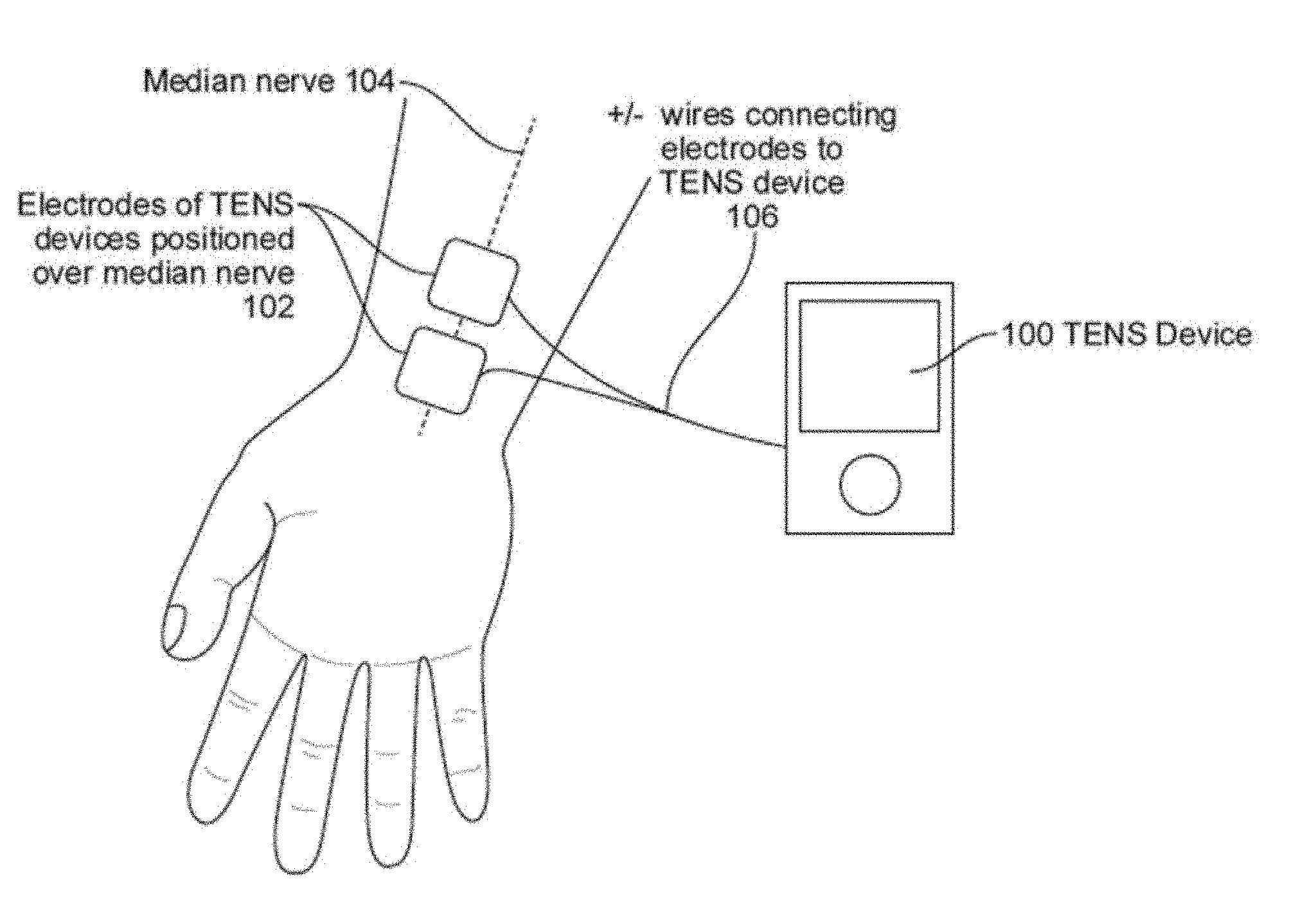
[0074] FIG. 1 illustrates one embodiment of delivering stimulation to the median nerve found to reduce tremor.
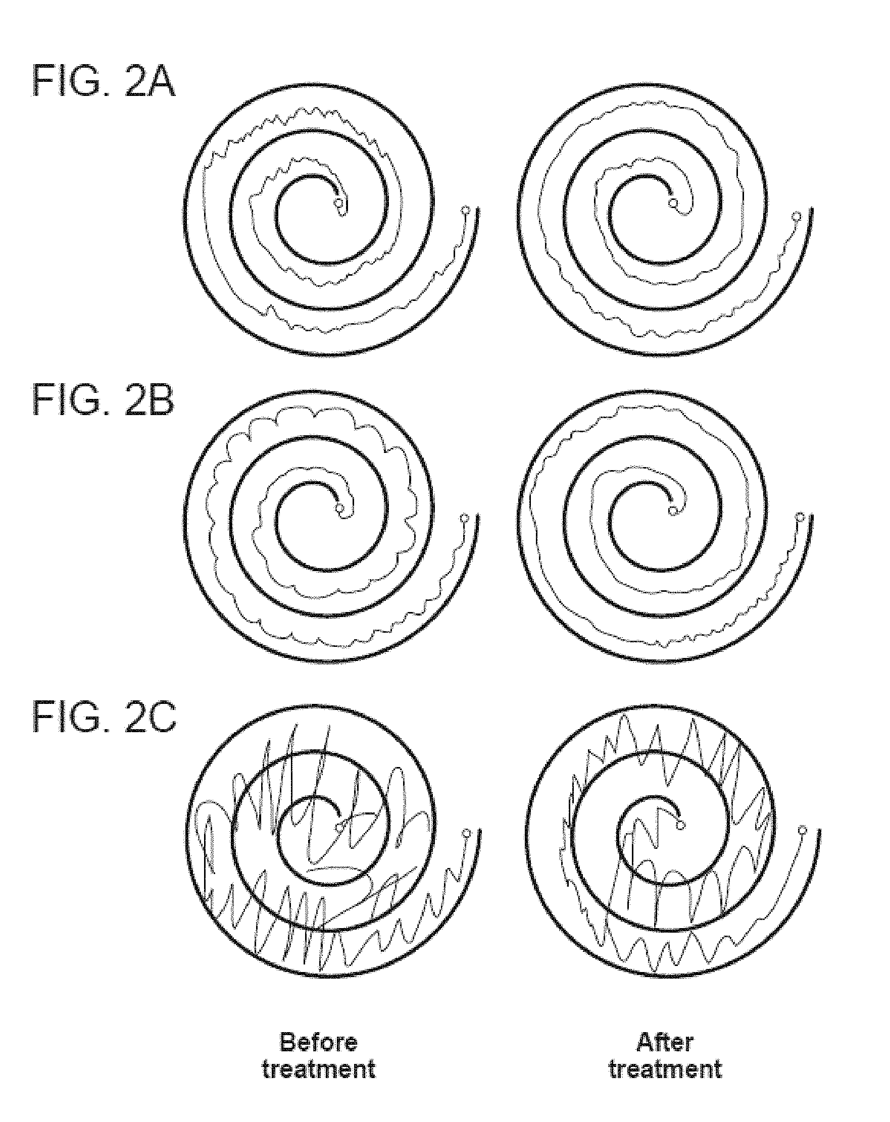
[0075] FIGS. 2A-2C illustrate treatment effect of an embodiment of peripheral nerve stimulation in a mild (FIG. 2A), moderate (FIG. 2B), and severe (FIG. 2C) ET patient. It presents results of a clinical study in which a patient with essential tremor reduced tremor amplitude by the configuration of stimulation at 150 Hz frequency, 300 .mu.s, and for 40 minutes of stimulation on-time. The tremor reduction, shown by comparing the ET patient's ability to draw a spiral, was observed immediately after the stimulation was turned off.
[0076] FIGS. 3A-3C illustrate wrist flexion-extension calculated from gyroscopic data in subject B from FIGS. 2A-2C. FIG. 3A shows the tremor before treatment; FIG. 3B shows the reduction in tremor immediately after treatment; FIG. 3C shows that the tremor reduction is maintained twenty minutes after the treatment.
[0077] FIG. 4 illustrates an example of ineffective treatment in a moderate ET patient.
[0078] FIG. 5A illustrates various positions on a patient where the tremor altering system can be located.
[0079] FIGS. 6A and 6B illustrate the major nerves innervating the hand and their distal branches.
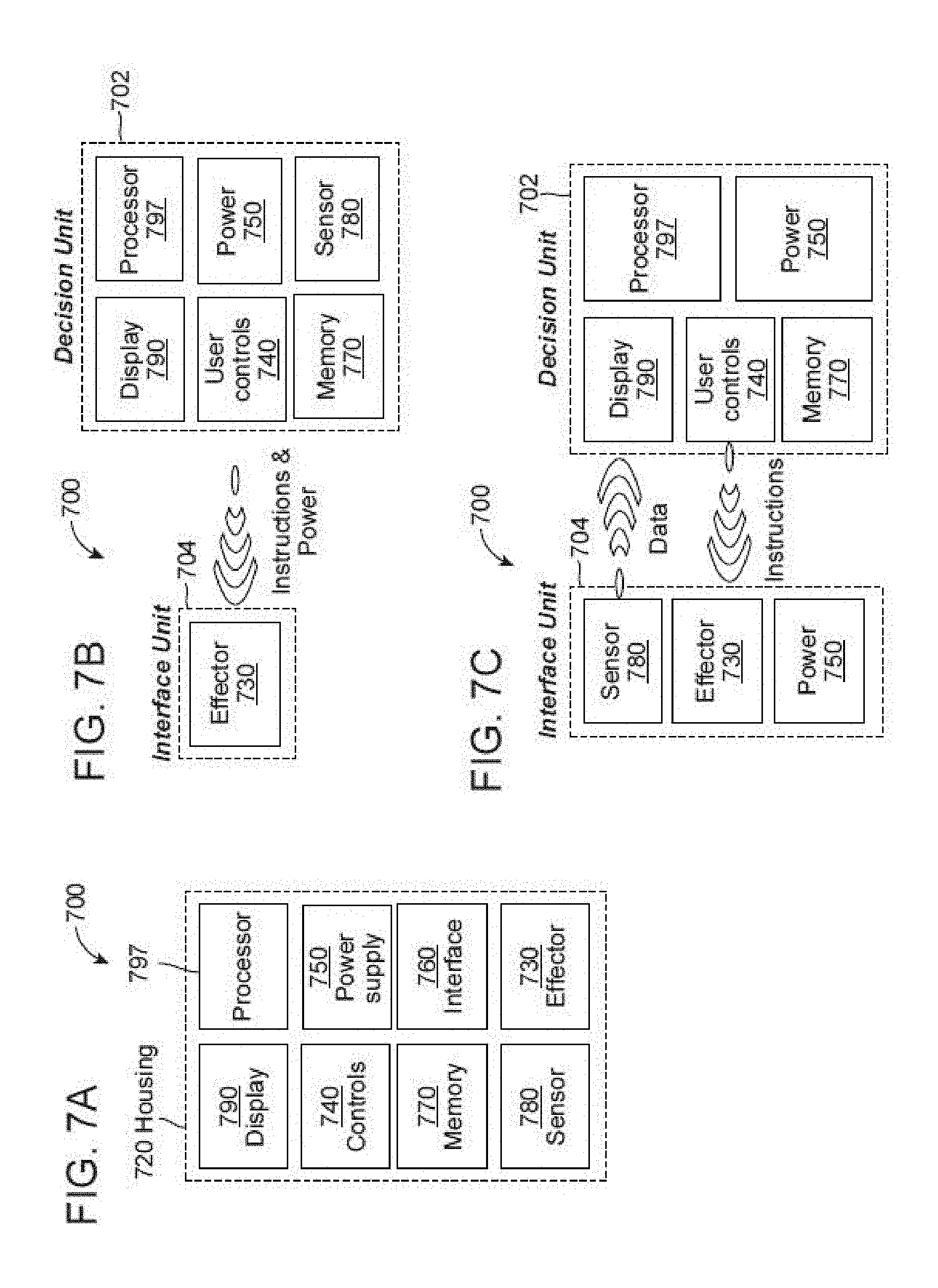
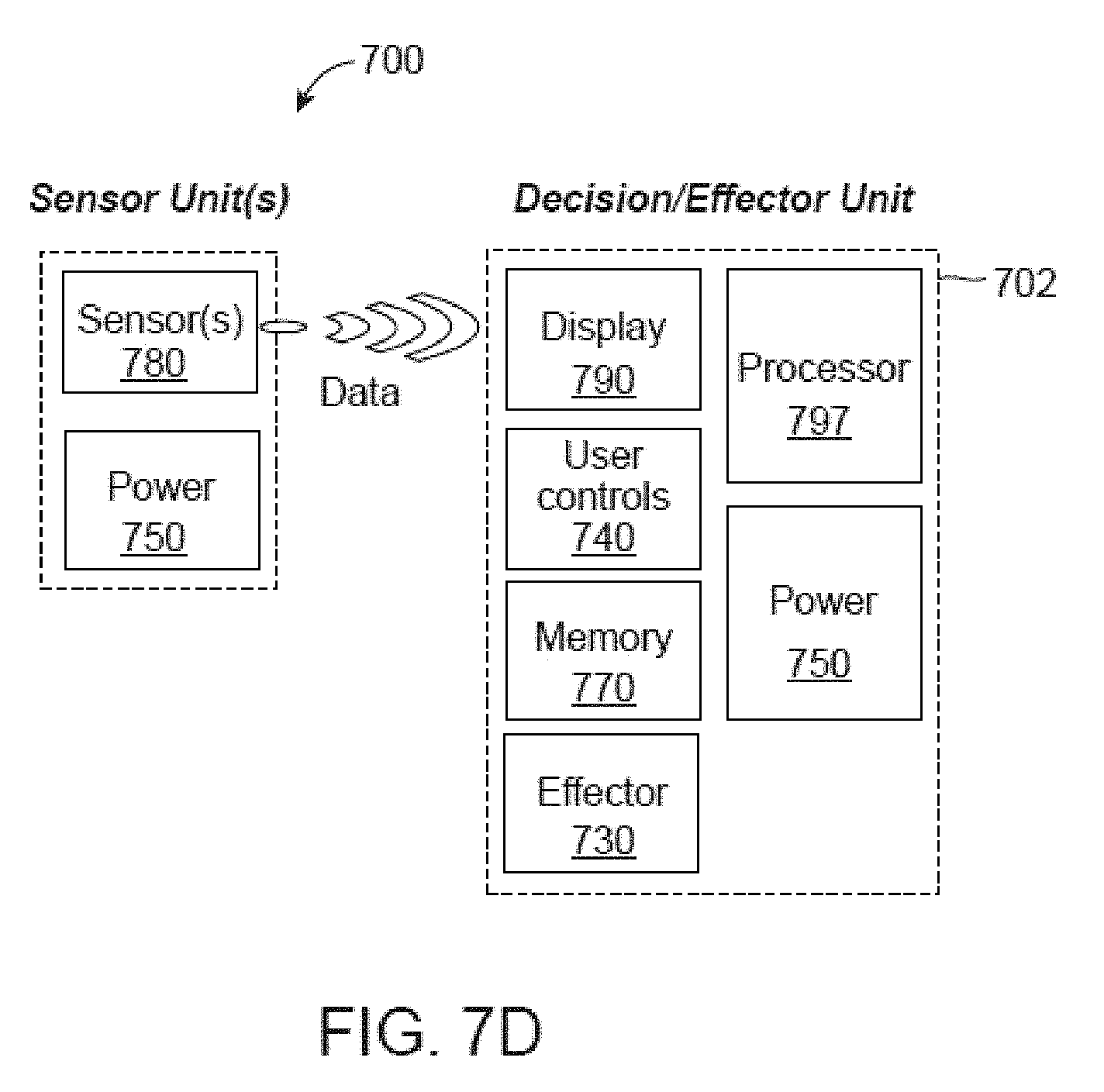
[0080] FIGS. 7A-7D are block diagrams illustrating various embodiments of a tremor altering system.
[0081] FIG. 8A illustrates an embodiment of an electrode pair used to excite nerves in different fingers, in which both electrodes are positioned on the finger.
[0082] FIG. 8B illustrates a means of exciting nerves in different fingers, in which the second electrode is positioned at the wrist.
[0083] FIG. 8C illustrates an embodiment of the placement of electrodes on the wrist to target different underlying nerves.
[0084] FIGS. 8D and 8E illustrate various stimulation sites.
[0085] FIG. 9A is a diagram showing an embodiment of an excitation scheme to dephase the brain regions receiving sensory input from two fingers.
[0086] FIG. 9B is a diagram showing an embodiment of an excitation scheme to dephase the brain regions receiving sensory input from four fingers.
[0087] FIGS. 10A-10C illustrate an embodiment where the position of the hand may determine the optimal stimulation duty cycle and timing.
[0088] FIG. 11 illustrates an embodiment of variable stimulation that changes frequency over time.
[0089] FIG. 12 is a drawing showing an embodiment where the stimulator is chemical and two neuromodulating chemicals can be mixed to provide tailored chemical stimulation.
[0090] FIGS. 13A and 13B illustrate various forms of user controls.
[0091] FIGS. 14A-14M illustrate various non-invasive or invasive embodiments of the tremor altering system. FIG. 14E is a drawing showing an embodiment in which the stimulator is mechanical. FIG. 14H illustrates an embodiment of a device having a form factor of a wrist watch. FIG. 14I illustrates the back of the device shown in FIG. 14H, showing the electrodes which are the interface with the user. FIGS. 14J and 14K illustrate an embodiment of a disposable electrode interface that snaps into place of the wrist watch form factor of the device housing. FIG. 14L illustrates an embodiment of a self-aligning snap feature that allows the disposable electrode interface to snap into the housing of the device in a wrist watch form factor. FIG. 14M is a drawing showing the potential placement of electrodes along the spine in an embodiment of the device where the effector is electrical.
[0092] FIGS. 15A-15C illustrate various embodiments of an array of electrodes.
[0093] FIGS. 16A-16D illustrate various embodiments of conductive ink tattoos.
[0094] FIGS. 17A-17B is a diagram showing an embodiment of the positioning of an accelerometer on the hand or wrist for measuring the patient's activity and tremor.
[0095] FIGS. 18A and 18B illustrate an example of spectral analysis of gyroscopic motion data for a patient with a tremor centered at 6.5 Hz.
[0096] FIG. 19 illustrates the correlation of postural tremor with kinetic tremor.
[0097] FIG. 20 illustrates an embodiment of a stimulation device that can record and transmit data, such as the tremor characteristics and stimulation history, to a data portal device, such as a smartphone, that transmits the data to a cloud-based server.
[0098] FIGS. 21A-21D are flowcharts showing the monitoring, integration, analysis and display of data used to inform the users or improve the stimulation.
[0099] FIG. 22 is a flowchart showing the feedback logic.
[0100] FIG. 23 is a drawing showing an embodiment where the stimulator is an electrode implanted at least partially subdermally.
[0101] FIGS. 24A-24D illustrate various embodiments of implantable devices and skin surface devices allowing wireless power and control.
[0102] FIGS. 25A-25F illustrate various geometries of electrodes for implanted electrical stimulation.
[0103] FIGS. 26A-26B illustrate two preferred embodiments of the controls module that is used to interact with the device. A control system for the tremor device utilizes feedback to modify the stimulation. It is a closed loop in which the stimulation is adjusted based on measurement of the activity and tremor.
[0104] FIG. 27A illustrates an embodiment of a system that can be configured to stimulate multiple dermatomes in a timed manner.
[0105] FIG. 27B illustrates an embodiment of electrode alignments for selective or preferential activation of nerve fibers.
[0106] The figures identified above can be combined with other figures and descriptions provided herein.
DETAILED DESCRIPTION
[0107] As used herein, the terms "stimulating" and "stimulator" generally refer to delivery of a signal, stimulus, or impulse to neural tissue of the targeted region. The effect of such stimulation on neuronal activity is termed "modulation"; however, for simplicity, the terms "stimulating" and "modulating", and variants thereof, are sometimes used interchangeably herein. The effect of delivery of the signal to the neural tissue may be excitatory or inhibitory and may potentiate acute and/or long-term changes in neuronal activity. For example, the effect of "stimulating" or "modulating" a neural tissue may comprise one or more of the following effects: (a) depolarizing the neurons such that the neurons fire action potentials, (b) hyperpolarizing the neurons to inhibit action potentials, (c) depleting neurons ion stores to inhibit firing action potentials (d) altering with proprioceptive input, (e) influencing muscle contractions, (f) affecting changes in neurotransmitter release or uptake, or (g) inhibiting firing. "Proprioception" refers to one's sensation of the relative position of one's own body parts or the effort being employed to move one's body part. Proprioception may otherwise be referred to as somatosensory, kinesthetic or haptic sensation. A "proprioceptor" is a receptor providing proprioceptive information to the nervous system and includes stretch receptors in muscles, joints, ligaments, and tendons as well as receptors for pressure, temperature, light and sound. An "effector" is the mechanism by which the device modulates the target nerve. For example, the "effector" may be electrical stimulation of the nerve or mechanical stimulation of proprioceptors.
[0108] "Electrical stimulation" refers to the application of electrical signals to the soft-tissue and nerves of the targeted area. "Vibrotactile stimulation" refers to excitation of the proprioceptors, as by application of a biomechanical load to the soft-tissue and nerves of the targeted area. Applying "thermal stimulation" refers to induced cooling or heating of the targeted area. Applying "chemical stimulation" refers to delivery of either chemical, drug or pharmaceutical agents capable of stimulating neuronal activity in a nerve or in neural tissue exposed to such agent. This includes local anesthetic agents that affect neurotransmitter release or uptake in neurons, electrically excitable cells that process and transmit information through electrical and chemical signals. The "cloud" refers to a network of computers communication using real-time protocols such as the internet to analyze, display and interact with data across distributed devices.
Device Location
[0109] The device stimulates the sensory nerves in order to modify the abnormal network dynamics. Over time, this stimulation normalizes the neural firing in the abnormal network and reduces tremor. Preferentially, the stimulated nerve is a nerve that carries sensory proprioceptive information from the limb affected by the tremor. The nerve may be modulated directly, such as by electrical stimulation anywhere along or adjacent to a nerve carrying proprioceptive information. In some embodiments, target nerve may be modulated indirectly, such as by excitation of the proprioceptors that stimulate the target nerve. FIG. 5A shows access points to nerves carrying proprioceptive information from a limb or vocal cords or larynx. These access points can include, but are not limited to, the fingers 510 including one or more fingers and/or the thumb, the hand 520, the wrist 530, the lower arm or forearm 540, the elbow 550, the upper arm 560, the shoulder 570, the spine 580 or the neck 590, foot (including one or more toes for example), ankle, lower leg or calf, knee, and/or upper leg or thigh. These access points may be used for direct stimulation in some embodiments. In other embodiments, these access points are used for indirect stimulation. Both indirect and direct stimulation are provided in several embodiments. Two, three or more access points are provided in some embodiments.
[0110] Nerves affecting proprioception can include, for example, the median, ulnar, radial, or other nerves in the hand, arm, and spinal area, or along muscle or within joints. These regions target to the nerves may include the brachial plexus, medial nerves, radial nerves, and ulnar, dermal, or joint space nerves. These regions may also target the musculature including muscles of the shoulder, muscles of the arm, and muscles of the forearm, hand, or fingers. Muscles of the shoulder may include, by non-limiting example, the deltoid, teres major and supraspinatus. Muscles of the arm may include the coracobrachialis and triceps brachii. Muscles of the forearm may include the extensor carpi radialis longus, abductor pollicis longus, extensor carpi ulnaris, and flexor carpi ulnaris. In some embodiments, one, two, three or more of these regions are stimulated (directly and/or indirectly).
[0111] Some examples of device locations or treatment sites that may be used in combination include two or more of wrist, hand, finger, forearm, upper arm, elbow, shoulder, arm, ankle, foot, toe, calf, lower leg, thigh, upper leg, knee, leg, upper body appendage, upper body, lower body appendage, lower body, spine, neck, head, or a portion of any of these. A plurality of sites may be combined with a plurality of modalities and/or a plurality of modes, each modality having a plurality of modes or one or more modalities having different modes. For example, a first stimulation modality and/or mode can be applied to the wrist 530 and a second stimulation modality and/or mode can be applied to the finger 510. For another example, a first stimulation modality and/or mode can be applied to the wrist 530 and a second stimulation modality and/or mode can be applied to the ankle. For yet another example, a first stimulation modality and/or mode can be applied to the arm and a second stimulation modality and/or mode can be applied to the leg. For still another example, a first stimulation modality and/or mode can be applied to the upper body and a second stimulation modality and/or mode can be applied to the lower body. The stimulators at the various sites can be implanted (e.g., subcutaneous) and/or transcutaneous.
[0112] In some embodiments, a vibrational or haptic motor is disposed in the device and located adjacent to a target region or nerve such that the vibrational energy is directed toward a target nerve. For example, in a wrist worn device, the vibrational motor can be disposed in the band such that it is located on the palmar side of the wrist directly adjacent to the median nerve. The motion of the vibrational motor can be oriented such that it is orthogonal and/or oblique to the target nerve or the surface of the skin. The motion of the motor could be, for example, linear or rotational with an eccentric mass.
[0113] In some embodiments, a hydrogel or water-based component (e.g., a hydrogel patch that has lidocaine or another chemical suspended in the gel, an electrode that stimulates via electrical energy) can be embedded with a chemical or pharmacological agent, such as lidocaine. The electrode can be placed on the surface of the skin, and the chemical is released at a predetermined rate proportional to the concentration of the chemical in the electrode. The electrode may or may not provide electrical stimulation in combination with the chemical suspended in the hydrogel. In some embodiments, an agent can be placed on the skin and electrical energy can synergistically both stimulate one, two, or more target regions or nerves as well as assist in driving the one, two or more agents through the skin (e.g., via iontophoresis). In some embodiments, iontophoresis devices can be modified to facilitate both enhanced skin penetration of the agent as well as electrical stimulation via the electrodes (e.g., in some cases alternating between a first stimulation mode to facilitate iontophoresis, and a second stimulation mode to facilitate nerve stimulation). The modes could involve different frequencies, amplitudes, waveforms, and/or other parameters as disclosed elsewhere herein.
[0114] In some embodiments, thermal energy is increased or decreased in the surrounding tissue in contact with the device. Thermal energy can be increased in tissue contacting the device by a thermoelectric heating element (for example, a battery disposed in the device with a resistive element). The heat can be transferred to specific target locations in the band, for example, that have conductive elements that transfer the thermal energy to tissue in contact with the conductive elements. Thermal energy can be decreased in tissue contacting the device by a thermoelectric cooling element (for example, a Peltier device or solid state refrigerator) or by circulating a coolant or cooling liquid through a part of the device in contact with the target tissue area (for example, in a wrist worn device the band may be disposed with tubing to circulate a cooling liquid through the band). In some embodiments, the device may have a pump to circulated the coolant. The device may have heat transfer elements, such as a heat sink or electrically powered fan, to transfer the thermal energy from the surrounding tissue to the surrounding air or environment (e.g., an area on the device not in contact with tissue).
[0115] In a preferred location in several embodiments, the device interfaces with the dermal surface of the tremulous upper extremities of the user and applies neuromodulatory signals to the nerve bundles selected from the group consisting of the brachial plexus, medial nerves, radial nerves, and ulnar nerves or the excitable structures in the musculature of the upper extremities on the skin or within a joint. Neuromodulatory signals are provided to one, two, or three or more nerve bundles in some embodiments.
[0116] Where stimulation or other neuromodulatory signals are provided to two, three or more locations, the signals are provided simultaneously, sequentially, or overlapping according to several embodiments.
[0117] Proprioceptors can be found for example in muscles, tendons, joints, skin, and the inner ear. Criteria defining candidate nerves for direct modulation include the location of the tremor to be reduced and the proximity of the nerve to the skin's surface, high density of proprioceptive fibers, and distance from excitable pain receptors or muscles. The median nerve targeted at the wrist and the ulnar nerve targeted at the elbow rank high by these criteria. Criteria defining candidate location for indirect proprioceptive modulation include the density and type of proprioceptors. Pacinian corpuscles provide information about touch; Muscle spindles provide information about changes in muscle length by triggering action potentials in the muscle spindle afferent nerve when mechanically-gated ion channels open due to muscle stretching; Golgi tendon organs provide information about muscle tension. These structures may also be stimulated to alter circuit dynamics and reduce tremor.
[0118] The device targets the specific nerves that synapse on the abnormal brain circuit. This synapse may be either direct, or through multiple relay synapses. FIGS. 6A and 6B show a set of representative nerves that transmit proprioceptive information into the olivo-cerebello network, a network that is abnormal in ET. These nerves include the 610 distal branches and main branches of the 620 median nerve and 630 ulnar nerve, as well as the 640 distal branches and main branches of the 650 radial nerve. In preferred embodiments, this device targets the nerves inputting proprioceptive information from the hand, wrist and forearm.
[0119] In another embodiment, the combination of any parts described here within, may be used to affect the nerves associated with voice tremor, including but not limited to branches of the vagus nerve such as the superior laryngeal nerve or the recurrent laryngeal nerve.
Device Components: Various Embodiments
[0120] FIGS. 7A-7D are conceptual diagrams illustrating some embodiments of a tremor altering system 700. System 700 includes a housing 720, one or more effectors 730, one or more controls 740 in electrical communication with the effector 730, and one or more power sources 750. The housing 720 can, in some embodiments, include an interface 760. The interface facilitates the coupling of the effector to the patient. For example, the interface can provide a physical, electrical, chemical, thermal or magnetic connection between the device and the patient's nerve. The housing 720 can also, in some embodiments, include a sensor 780 to detect the tremor, memory 770, display 790, and processor 797. The device in this embodiment may include a processor 797 coupled to the effector which could perform computations and control of other components. The device may also include a digital library stored on the processor 797 or memory 770 which could contain preloaded modulation protocols. The device could include a controls module 740 that communicates with the processor 797 and could be used by the user to control stimulation parameters. The controls allow the user to adjust the operation of the device. For example, the controls can be configured to turn the device on, turn the device off, adjust a parameter of the effector, such as the intensity. The device may include a sensor 780 connected to the processor 797 which may detect information of predefined parameters and transmits said parameter information to the processor 797. The device may include a data storage unit 770 connected to the sensor 780 and processor 797; and a power supply 750 may be connected to the processor.
[0121] The device may further contain a display or indicators 790 to communicate with the user and report on the status of the device. Indicators are preferably a light-emitting diode (LED) or some visual indicator in some embodiments but can be an audio indicator. The information may include the battery power or the stimulation status.
[0122] The device might lack or not have an effector 730. It may be a diagnostic non-therapeutic device. In a preferred embodiment, the interface unit 704 would be worn on the tremoring limb to track the tremor over time. Providing feedback to the user of the device can make them aware of their tremor and allow monitoring over time. Even without therapeutic stimulation this biofeedback can help some individuals reduce their tremor. The device might lack or not have a sensor 780. It may be a therapeutic non-diagnostic device.
[0123] In order to make the device small and simple, many of these components could be housed in a separate unit. Processing, controlling and possibly sensing may be done remotely in a decision unit 702, making the interface unit 704 that provides the therapeutic contact with the patient compact, simple, and flexible for a variety of applications (FIGS. 7B-7D). This decision unit 702 may be a new device designed for this application, or it may be integrated into an existing technology such as a smartphone. This would allow the system to be robust handheld form-factor with a reduced cost and size.
[0124] In an embodiment shown in FIG. 7B, the interface unit 704 is an implant; the effector 730 provides electrical stimulation of the nerves; the instruction set and power are transmitted wirelessly from an external device. The implanted interface unit 704 may be powered with an on-board battery. The implanted interface unit 704 may contain a sensor 780 for direct detection of the tremor or neuromuscular activity detected by electroneurography (ENG) or electromyography (EMG).
[0125] In an embodiment shown in FIG. 7C, the interface unit 704 is worn on the surface of the body; the effector 730 provides electrical stimulation of the underlying nerves or vibrotactile stimulation of nearby proprioceptors. The sensor 780 could include motion sensors including accelerometers, gyroscopes and magnetometers.
[0126] In an embodiment shown in FIG. 7D, one or more sensor units 780, sensing motion, temperature, etc. may be worn at different locations in the body. The effector 730 and decision unit 702 are a separate entity worn at a different location on the body than the sensors 780. This is useful if stimulation of a nerve occurs in a location where tremor is not as easily or accurately measured. For instance, a stimulation device 700 placed on the underside of the wrist for reducing hand tremor is highly effective. However, measuring tremor of the hand from the wrist using accelerometer or gyroscopes could prove more difficult; a sensor unit placed separately on the palm or backside of the hand in a glove or worn as a ring on one of the digits would show greater sensitivity towards hand tremor since it is located beyond wrist joint.
Effectors: General
[0127] The effector may function to modulate the neural tissue in the upper extremity region at which stimulation is directed. For example, the effector can modify neuronal signals in the nerves and/or modify the flow or content of proprioceptive information. The effectors may be delivered transcutaneously or subcutaneously. One or more effectors can be used to influence the nerves. In some embodiments, the effector can be excitatory to the nerve. In other embodiments, the effector can be inhibitory to the nerve. In some embodiments, the system can be used to excite the nerve during some portions of the treatment and inhibit the nerve during other portions of the treatment.
Effector: Electrical Stimulation
[0128] In some embodiments, the effector may be an electrical stimulator. Electrical effectors can include an electrode, an electrode pair, an array of electrodes or any device capable of delivering an electrical stimulation to a desired location. Electrical stimulation may be transcutaneous or subcutaneous. For example, transcutaneous electrical stimulation may be achieved with electrodes placed on the surface of the skin while subcutaneous electrical stimulation may be achieved with an implanted electrode positioned close to a nerve.
[0129] The stimulation parameters may be adjusted automatically, or controlled by the user. The stimulation parameters may include on/off, time duration, intensity, pulse rate, pulse width, waveform shape, and the ramp of pulse on and off. In one preferred embodiment the pulse rate may be approximately 50 to 5000 Hz, and a preferred frequency of about 50 Hz to 300 Hz, or 150 Hz. For example, the frequency may be between about 10 Hz and about 20 kHz (e.g., about 10 Hz, about 20 Hz, about 30 Hz, about 40 Hz, about 50 Hz, about 60 Hz, about 100 Hz, about 250 Hz, about 500 Hz, about 1000 Hz, about 2500 Hz, about 5000 Hz, about 10 kHz, about 15 kHz, about 20 kHz, and ranges between such values). A preferred pulse width may range from 50 to 500 .mu.s (micro-seconds), and a preferred pulse width may be approximately 300 .mu.s (e.g., about 50 .mu.s, about 100 .mu.s, about 150 .mu.s, about 200 .mu.s, about 250 .mu.s, about 300 .mu.s, about 350 .mu.s, about 400 .mu.s, about 450 .mu.s, about 500 .mu.s, and ranges between such values). The intensity or amplitude of the electrical stimulation may vary from 0 mA to 500 mA, and a preferred current may be approximately 1 mA to 6 mA (e.g., about 0 mA, about 0.1 mA, about 1 mA, about 6 mA, about 10 mA, about 20 mA, about 30 mA, about 40 mA, about 50 mA, about 100 mA, about 200 mA, about 300 mA, about 400 mA, about 500 mA, and ranges between such values). Certain preferred settings are derived from the clinical study described above that provided a valuable reduction in tremor sustained for a time period. Electrical stimulation can be adjusted in different patients and with different methods of electrical stimulation. These preferred settings are non-limiting examples. The increment of intensity adjustment may be 0.1 mA to 1.0 mA (e.g. 0.1-3 mA, 3-6 mA, 6-10 mA, and overlapping ranges therein). In one preferred embodiment, the stimulation may last for approximately 10 minutes to 1 hour (e.g. 10-20 minutes, 20-40 minutes, 40-60 minutes, and overlapping ranges therein).
[0130] In one preferred embodiment, the electrodes may be in contact with the user at the surface of the skin above one or more nerve(s) that may include the medial, radial, and ulnar nerves. The electrode may be in the configuration where there is an electrode pair, in which one electrode is proximal (closer to the elbow) and another is distal (closer to the hand). The electrodes may be in communication with the opposing electrode. The electrode pair may have a polarity of positive or negative charge in which electrical current passes.
[0131] The effector may include two electrodes, each with positive or negative polarity, or an electrode array may include multiple electrode pairs, where each pair is independently programmed or programmed dependently in relation to the other pairs of electrodes. As an example, the program can allow cyclic stimulation of different nerves at different times, such as ulnar, then median, then radial, or any combination thereof.
[0132] Electrical stimulation may be designed to suppress tremors by interfering with proprioceptive input, inducing compensatory muscle contractions, or by a combination of both methods. The electrodes may be substituted by any equivalent material capable of conducting electrical signals through the stimulator interface with the dermal surface of the upper extremity. The electrodes may be attached to a control unit 740 which could apply electrical stimulation via the electrodes to the soft tissue and nerves in the region where the electrode are placed and the region immediately surrounding. In another variation of the embodiment, several electrodes can be placed to a combination of targeted regions.
[0133] A function generator connected to and controlled by the processor may function to modulate electrical stimulation parameters. The function generator is preferably an arbitrary waveform generator that uses direct digital synthesis techniques to generate any waveform that can be described by a table of amplitudes. The parameters are selected from a group including but not limited to frequency, intensity, pulse width or pulse duration, and overall duration. The outputs preferably have a power limit set by the maximum output voltage. In a preferred embodiment, the digitally stored protocols cycle through various stimulation parameters to prevent patient acclimation. Variation of electrical stimulation is achieved by the function generator.
[0134] In some embodiments, the electrical stimulus comprises a combination of one or more single frequencies and one or more sweep frequencies (e.g., continuously changing from a lower frequency to a higher frequency). For example, a first single frequency can be applied for a first duration and a first sweep frequency can be applied for a second duration. The second duration may be after (e.g., immediately after, after a pause duration) the first duration. The second duration may at least partially overlap the first duration. Continuing the example, a second single frequency different than the second frequency can be applied for a third duration. The third duration may be after (e.g., immediately after, after a pause duration) the second duration. The third duration may at least partially overlap the second duration. Continuing the example, a second sweep frequency different than the first sweep frequency (e.g., having a different low frequency, high frequency, and/or rate of frequency change) can be applied for a fourth duration. The fourth duration may be after (e.g., immediately after, after a pause duration) the third duration. The fourth duration may at least partially overlap the third duration. The example may continue for additional durations of single frequencies and/or sweep frequencies. The different frequencies can be applied to the same vibratory stimulator or different electrical stimulators (e.g., on the same body part, on different body parts). Electrical stimulus applied to different body parts may meet at a point between the body parts and may form a standing wave. In some embodiments, the meeting point may be the target body part (e.g., elbow and finger meeting at the wrist). Amplitudes of the sweeps could be enveloped by other waveforms, such as sinusoid or Gaussian curve, or the parameters could be changed randomly or pseudo-randomly. Discussions of different frequencies above can also apply to other parameters such as frequency, amplitude, pulse width, pulse spacing, phase, waveform shape, waveform symmetry, duration, duty cycle, on/off time, or bursting, combinations thereof, and the like.
Optimizing Stimulation: Dephasing
[0135] In a preferred embodiment, the stimulation is designed to dephase synchronicity in the brain. The concept of dephasing the abnormal circuit follows on recent work showing neural retraining reduces the network's propensity to fall into an abnormal rhythm. Interestingly, movement disorders are often associated with abnormal periodic synchronous firing in brain circuits. In Parkinson's disease, this circuit is in the basal ganglia. In ET, it is the olivo-cerebellar circuit. These anomalous oscillations are thought to drive the tremor, as supported by numerous studies showing that the tremor observed in the hand and forearm muscles is synched with pathological rhythmic discharges in the brain. Recent DBS studies have shown that low-voltage phase-shifted bursts on adjacent pairs of electrodes (called Coordinated Reset) can reduce synchronization in abnormal brain networks and that this reduces Parkinsonian tremors. The application of Coordinated Reset theory to treat tinnitus supports the concept of using synaptic excitation to retrain neural networks.
[0136] The device disclosed herein offers several advantages over high-frequency TENS stimulation, including using lower power (leading to extended battery life, less discomfort from motor recruitment and contraction, less discomfort from sensory excitation), less suppression of firing in activity in adjacent nerves (by depletion or other mechanisms), and maintaining longer-lasting effects such that the device only need be used intermittently to train or maintain training of the neural circuit dynamics. The device stimulates sets of nerves in such a way that it targets neural subpopulations to reduce synchronization of the population. For example, this may be achieved by stimulating different fingers on the hand.
[0137] FIG. 8A is a diagram showing a preferred embodiment of the device, in which anode 810 and cathode 820 electrode pairs on the fingers are used to excite the branches of the proprioceptive nerves (the median, radial and ulnar nerves) in each finger. This arrangement of anode 810 being distal and cathode 820 being proximal is designed to induce a nerve pulse traveling towards the brain. A unique stimulation pattern on each finger can send a unique signal to a specific subpopulation of neurons in the brain because of the somatotopic organization of the brain, in which signals from different adjacent or nearby body parts synapse at nearby locations in the brain. Stimulation using multiple unique patterns is an example of a multi-modal stimulation in which multiple modes (having at least one different parameter) of the same kind of stimulation are used. In some embodiments, the anode 810 and cathode 820 positions may be reversed to inhibit the passage of sensory impulses towards the brain (antidromic collision). The stimulation of multiple fingers is an example of a multi-modal stimulation in which multiple sites of the same kind of stimulation are used. In combination with unique stimulation patterns, such stimulation is an example of a multi-modal stimulation in which multiple sites and multiple modes of the same kind of stimulation are used. Different kinds of stimulation are also provided in several embodiments (e.g., electrical stimulation on one or more fingers and vibrotactile stimulation on one or more other fingers).
[0138] FIG. 8B shows an arrangement in which electrodes 830 are positioned on the fingers and a second electrode 840 is positioned on the wrist. In one embodiment, there is only a 830 single electrode on the finger and the 840 second electrode is positioned on the wrist. The fingers represent only one possible set of targets and different locations may similarly be used target adjacent subpopulations of neurons. In some embodiments, electrodes on one or more fingers may comprise an anode and a cathode, for example as described with respect to FIG. 8B and an anode and a cathode may be positioned on the wrist. The stimulation of one or more fingers and the wrist is another example of a multi-modal stimulation in which multiple sites are used. In some embodiments, electrodes on one or more fingers may be transcutaneous and electrodes on the wrist may be subcutaneous. The positioning of electrodes at different levels of the skin is an example of a multi-modal stimulation. Multimodal treatment in some embodiments is provided on one of the fingers (including the thumb) on one hand and additionally one of the fingers (including the thumb) on the other hand.
[0139] In the embodiment shown in FIG. 8C, the electrodes are positioned on different locations 850, 860, 870 on the wrist to target the median nerves (proximate the location 850), the ulnar nerves (proximate the location 860), and the radian nerves (proximate the location 870). It will be appreciated by one skilled in the art that the input may be positioned on other locations or branches of the nerves that input into the abnormal brain circuit. The location may be on the same or opposite side of the limb with tremors. The location may be on the surface of the skin, crossing the skin, and/or implanted.
[0140] FIG. 8D illustrates various stimulation sites which can be subjected to stimulation that is delayed or offset by a predetermined fraction or multiple of the tremor period, T, as shown for example in FIGS. 9A and 9B. The sites are proximate the medal nerve and the ulnar nerve. FIG. 8E illustrates a stimulation site proximate the radial nerve. The stimulation of a first side of the wrist (e.g., FIG. 8D) and a second side of the wrist (e.g., FIG. 8E) is another example of a multi-modal stimulation in which multiple sites are used.
[0141] The device uses stimulation schemes designed to dephase, override or obscure the abnormal network. FIG. 9A is a conceptual diagram showing a sample excitation scheme to dephase brain regions receiving sensory input from two sites. For example, the two sites could be two of the fingers shown in FIGS. 8A-8E. The stimulation at the second site is delayed after stimulation of the first site by time T/2, where T is the period of the native tremor. For example, if the tremor is at 8 Hz and the period T is 125 ms, the stimulation of the second site would be delayed by 62.5 ms. The stimulation is designed to reset the phase of the neuron, which may be implemented using high frequency stimulation (above 100 Hz) or a DC pulse.
[0142] FIG. 9B is a conceptual diagram showing a sample excitation scheme to dephase brain regions receiving sensory input from four sites, with subsequent sites delayed by T/4. In another embodiment, the stimulation at different locations is variable in parameters other than timing such as frequency or pulse width, or a combination of these. These variations are similarly designed to retrain the brain by dephasing, overriding or obscuring the abnormal network dynamics. In yet another embodiment, the stimulation may occur at a single location but vary in parameters over time. For example, it may vary in frequency every few seconds or turn on and off. In yet another embodiment, the stimulation is constant and at a single location. In preferred embodiments of these, the location is at the median nerve close to the wrist.
Optimizing Stimulation: Sub-Sensory
[0143] Stimulating at intensities below the sensory threshold will avoid the discomfort (tingling, numbness, pain) that can be associated with peripheral nerve stimulation. Because the exact electrode position, size and surface contact have a large effect on the stimulation level and the anatomical structures that receive the stimulation, the sensory threshold may needed to be calibrated for each patient and even for each session. This calibration may be done by the user manually setting the stimulation parameters or otherwise indicating their sensory threshold. Another possible mechanism is for the device to automatically sweep through a range of stimulation parameters and the patient chooses the most comfortable set of parameter values. Another possible mechanism is for the patient to choose from among a set of previously chosen parameter values that provided effective and comfortable stimulation. In some embodiments, the electrode pad can include a topical analgesic, such as Lidocaine, to reduce the discomfort from stimulation, thereby increasing the sensory threshold tolerated by the patient. In some embodiments, the topical analgesic can be delivered using a controlled release formation to provide pain relief for the duration the electrode pad is to be worn, which can be days, weeks or months. Such a method may provide more comfort or greater therapeutic effect, due to greater stimulation intensity and/or synergistic effects with the topical analgesic, which can reduce tremor in some patients.
Optimizing Stimulation: High Frequency
[0144] Alternatively or additionally, the stimulation waveform may be very high frequency, typically in the kHz and above, such that the stimulation is not felt by the user, or it is felt very little. Very high frequency stimulation is thought to make a conduction blockade. However, prior to the blockade there is an onset response including a strong depolarization of the nerve. To effectively implement very high frequency stimulation without causing discomfort for the patient, it would be preferable to eliminate this onset response. This can be done by cooling the nerve during the initial stimulation. Motor nerves are generally excited by stimulation at about 15 Hz and below, while sensory nerves are generally excited by stimulation at about 50 Hz and above. In some embodiments, it may be desirable to specifically stimulate above the 15 Hz threshold of motor neuron stimulation to avoid causing muscle contraction.
Optimizing Stimulation: Triggered
[0145] Alternatively or additionally, triggering the stimulation to the phase of the tremor can improve effectiveness. The goal of such stimulation is to break the rhythmic entrainment of motor units. More effective treatment may permit stimulating at lower levels to achieve similar therapeutic benefits with less discomfort. Essential tremor is essentially a problem of feedback in a resonant circuit. Stimulation timed off-phase from the tremor may reduce the tremor by altering the circuit dynamics, for example by shifting the gains on the feedback loop.
[0146] As shown in FIG. 10B, bursts of high-frequency stimulation may be timed to occur when the wrist is at its maximum flexion or extension (FIG. 10A). In example (FIG. 10C), the bursts have been shifted to a random phase. The position of the hand (FIG. 10A) may determine the optimal stimulation duty cycle and timing, such as (FIG. 10B) stimulating off-resonance with the maximum tremor deviation or (FIG. 10C) using bursts of variable temporal delays to avoid resonance with the tremor.
[0147] In some embodiments, a first stimulation may comprise burst stimulation (e.g., stimulation with a specified on period and off period) and a second stimulation may comprise continuous stimulation. The first stimulation may be before, during, and/or after the second stimulation. The first stimulation may be applied by a same electrode or set of electrodes as the second stimulation or by a different electrode or set of electrodes than the second stimulation. In some embodiments, burst stimulation may be applied to a first location on the body and continuous stimulation may be applied to a second location (either simultaneously or sequentially).
[0148] Alternatively or additionally, the stimulation may be chaotic or variable. The goal of chaotic, random, or variable stimulation is to inhibit or prevent habituation and reduce resonance in the circuit. For example, this may be implemented by varying the stimulation frequency over time and/or by superimposing higher and lower frequency components, as illustrated in FIG. 11.
[0149] Alternatively or additionally, the stimulation may be high frequency alternating current. This has been shown to block action potentials as they transmit along axons and could adjust circuit dynamics.
[0150] In some embodiments, the stimulation parameters as described above can be cycled according to a predetermined order to determine the optimal stimulation parameter. In some embodiments, the effectiveness of the stimulation parameters can be monitored over time to determine whether a particular set of stimulation parameters is losing effectiveness. In some embodiments, when the effectiveness of a particular set of stimulation parameters has been reduced by a predetermined amount, the stimulation parameters can be altered or cycled according to a predetermined order. For example, if stimulation is being triggered to the phase of the tremor, the stimulation can be delivered with random or variable temporal delays, or if the stimulation was using a set amplitude and/or frequency, the stimulation can be changed to a chaotic, random or variable modality to prevent or disrupt habituation. In some embodiments, the random or variable type stimulation parameters can be utilized according to a predetermined routine, such as daily for a predetermined number of hours, or weekly for a predetermined number of days, or at some other predetermined interval including time of day.
Effector: Vibrotactile Stimulation
[0151] The effector may be mechanical excitation of the proprioceptors by means including vibrotactile or haptic sensation. The mechanical stimulation might include force, vibration and/or motion. The effector induces action potentials in the target nerves by exciting the Golgi tendon organs (GTOs) or Pacinian corpuscles. Mechanical effectors can include, for example, small motors; piezoelectrics; one or more vibrotactile units comprised of a mass and an effector to move the mass such that a vibratory stimulus is applied to the body; an eccentric mass mounted on a shaft such that a vibratory stimulus is produced when the shaft is rotated; an ultrasonic motor; a magnetorheological fluid (MRF) effector or electroactive polymer (EAP) effector; and/or a speaker (e.g., a haptic speaker, a piezoelectric speaker, an electroactive polymeric transducer, a magnetic coil speaker).
[0152] The vibratory stimulus can in some embodiments be 250 Hz, corresponding to the optimal sensitivity of the Pacinian corpuscles (also known as lamellar corpuscles). The Pacinian corpuscles are the nerve endings in the skin that sense touch and vibration. Deformation of the corpuscle opens pressure-sensitive sodium ion channels to cause action potentials. In one embodiment, the vibration may be below 50 Hz to excite the Meissner's corpuscles (also called tactile corpuscles) in the fingers that are sensitive to light touch. In some embodiments, the vibratory stimulus can be, for example, at least about, about, or no more than about 10 Hz, 20 Hz, 30 Hz, 40 Hz, 50 Hz, 60 Hz, 70 Hz, 80 Hz, 90 Hz, 100 Hz, 110 Hz, 120 Hz, 130 Hz, 140 Hz, 150 Hz, 160 Hz, 170 Hz, 180 Hz, 190 Hz, 200 Hz, 210 Hz, 220 Hz, 230 Hz, 240 Hz, 250 Hz, 260 Hz, 270 Hz, 280 Hz, 290 Hz, 300 Hz, 350 Hz, 400 Hz, 450 Hz, 500 Hz, or more or less, or ranges including any two of the foregoing parameters.
[0153] In some embodiments, the vibratory stimulus comprises a combination of one or more single frequencies. For example, a first single frequency can be applied for a first duration and a second single frequency can be applied for a second duration. The second duration may be after (e.g., immediately after, after a pause duration) the first duration. The second duration may at least partially overlap the first duration. Continuing the example, a third single frequency different than the second frequency can be applied for a third duration. The third duration may be after (e.g., immediately after, after a pause duration) the second duration. The third duration may at least partially overlap the second duration. The third duration may at least partially overlap the first duration. The third duration may at least partially overlap the first duration and the second duration. The example may continue for additional durations of single frequencies. The different frequencies can be applied to the same vibratory stimulator or different vibratory stimulators (e.g., on the same body part, on different body parts). Vibratory stimulus applied to different body parts may meet at a point between the body parts and may form a standing wave. In some embodiments, the meeting point may be the target body part (e.g., elbow and finger meeting at the wrist).
[0154] In some embodiments, the vibratory stimulus comprises a combination of one or more single frequencies and one or more sweep frequencies (e.g., continuously changing from a lower frequency to a higher frequency). For example, a first single frequency can be applied for a first duration and a first sweep frequency can be applied for a second duration. The second duration may be after (e.g., immediately after, after a pause duration) the first duration. The second duration may at least partially overlap the first duration. Continuing the example, a second single frequency different than the second frequency can be applied for a third duration. The third duration may be after (e.g., immediately after, after a pause duration) the second duration. The third duration may at least partially overlap the second duration. Continuing the example, a second sweep frequency different than the first sweep frequency (e.g., having a different low frequency, high frequency, and/or rate of frequency change) can be applied for a fourth duration. The fourth duration may be after (e.g., immediately after, after a pause duration) the third duration. The fourth duration may at least partially overlap the third duration. The example may continue for additional durations of single frequencies and/or sweep frequencies. The different frequencies can be applied to the same vibratory stimulator or different vibratory stimulators (e.g., on the same body part, on different body parts). Vibratory stimulus applied to different body parts may meet at a point between the body parts and may form a standing wave. In some embodiments, the meeting point may be the target body part (e.g., elbow and finger meeting at the wrist). Amplitudes of the sweeps could be enveloped by other waveforms, such as sinusoid or Gaussian curves, or the parameters could be changed randomly or pseudo-randomly.
[0155] Discussions of different frequencies above can also apply to other parameters such as frequency, amplitude, pulse width, pulse spacing, phase, waveform shape, waveform symmetry, duration, duty cycle, on/off time, or bursting, combinations thereof, and the like. Discussions of vibratory stimuli above also apply, in several applicable embodiments, to other modalities and combinations thereof.
[0156] In some embodiments, a first location on the body is electrically stimulated while a second location is provided with vibratory stimulus (e.g., a wrist and an ankle, a wrist and an ear, an ankle and an ear, an arm and a leg, etc.). In instead of a second location, or in addition to a second location, both electrical and vibratory stimuli are provided at the first location (simultaneously or sequentially). As an example, a band (or other device) is used for the wrist that provides both electrical and vibratory stimuli and a band (or other device) is used for the ear, ankle, leg, arm, etc. that provides either electrical and vibratory stimuli. In one embodiment, both locations provide electrical and vibratory stimuli. A third, fourth, or additional location may also be stimulated. Although stimulation is disclosed in several embodiments, neuromodulation that includes stimulation or inhibition is contemplated in multiple embodiments.
[0157] This mechanical-type (e.g., vibratory) stimulator may function to reduce tremor through several methods. One method may be to transmit proprioceptive signals to the brain that obscure or modify the driving proprioceptive signal transmitted from the tremulous muscles. Another method may be impedance control. Joint impedance may altering co-contracting muscles through transcutaneous neurostimulation, affecting the stiffness of muscles and consequently muscle contractions. Another method may be the generation of compensatory muscle contractions, through neurostimulation, that oppose the tremulous contractions. The stimulator is preferably affixed firmly against the dermal surface, for example through an elastic or Velcro band (or other adjustable, adaptable straps), in some embodiments.
Effectors: Chemical, Thermal & Other
[0158] The examples herein have primarily described the stimulation as electrical or vibrotactile. However, stimulation may alternately be achieved using other effectors that may offer significant benefit in terms of patient comfort, portability, safety or cost.
[0159] In another variation of the embodiment, the effector may be a neuromodulating chemical that either raises or lowers neurons firing thresholds. The chemical used in some embodiments may be a topical anesthetic including, but not limited to the "caine" family. The "caine" family of anesthetics may include but are not limited to benzocaine, bupivacaine, butacaine, carbisocaine, chloroprocaine, ciprocaine, dibucaine, etidocaine, heptacaine, levobupivacaine, lidocaine, lidocaine hydrochloride, mepivacaine, mesocaine, prilocaine, procaine, propanocaine, ropivacaine, and tetracaine. Other chemical families may include those of menthol family, or alpha-hydroxy sanshool from Szechuan peppercorn, or capsaicin, all of which are known to influence peripheral sensory nerves.
[0160] FIG. 12 shows a chemical stimulator that may deliver chemical stimulus transdermally through a patch and/or by microinjection. The preloaded protocols may preferably be predetermined compositions of the one or more chemicals. The topical anesthetics in several embodiments may be known for other indications. The recommended doses for simulation may have been tested and approved for treatment of other indications. For example, the topical anesthetic lidocaine may be administered at 2-10% by weight. Lidocaine may be administered in conjunction with other anesthetics. As seen in FIG. 12, two neuromodulating chemicals 1202, 1204 are mixed to provide a tailored composition. The chemical stimulator may be administered as a composition comprising lidocaine 2.5 wt % as a first chemical 1202 and prilocaine 2.5 wt % as a second chemical 1204. The chemical stimulator could be administered as a composition comprising lidocaine 0.1-5 wt % as a first chemical 1202 and prilocaine 0.1-5 wt % as a second chemical 1204.
[0161] The chemical stimulator may comprise alpha hydroxy sanshool from Szechuan peppercorn. The alpha hydroxy sanshool may be contained in an excipient or carrier. The excipient may include gels, creams, oils, or other liquid. If the method of delivery is a transdermal patch, the formulation of the chemical agent may preferably be a cream or gel. The composition can be selected by the user through a control module (e.g., the control module 740 of FIG. 7). If the method of delivery is microinjection, the formulation may preferably be a solution.
[0162] In some embodiments, the effector can be a temperature or thermal effector (e.g., the temperature effector 732 of FIG. 7) that induces cooling and/or heating. The effector may modulate neuronal firing by directly cooling the nerve or indirectly by cooling adjacent muscle, skin or other component of the arm. A temperature effector can include, for example, piezoelectrics (e.g., Peltier cooling tiles), circulating fluid, compressed expandable gas, cooled or warmed solid material or evaporative material. One example of a cooling effector can be as disclosed in U.S. Publication No. 2010/0107657, which is incorporated herein by reference. Heating and/or cooling may be applied as a patch that adheres to the dermal surface, by attachment to affix the stimulator to the dermal surface, such as an armband, or by an implant.
[0163] In an embodiment with a thermal stimulator, the preloaded protocols may preferably be predetermined temperatures of stimulation and associated durations of stimulation. Preferably, a preloaded protocol may call for thermal cooling for the duration of 15 minutes and cooling temperatures in the range of 15-25.degree. C. (e.g., about 15.degree. C., about 17.5.degree. C., about 20.degree. C., about 22.5.degree. C., about 25.degree. C., ranges between such values, and the like). The duration of stimulation may be preprogrammed to (but is not limited to) about 5 minutes to about 30 minutes (e.g., about 5 min, about 10 min, about 15 min, about 20 min, about 25 min, about 30 min, ranges between such values, and the like). The maximum length of stimulation should be well tolerated by the user and not cause any muscular or neurological damage. Temperature sensors may function to detect the effective cooling temperature in an embodiment where the stimulator is a thermal stimulator. Effective cooling or heating temperature may be the temperature felt by the user, and this is not necessarily the same as the applied temperature. If the temperature sensors determine that the effective temperature reaches a threshold, which may range from 5 degrees C. greater or less than the applied temperature for a particular protocol, the processor 797 (from FIG. 7) may modify said protocol to cool or heat more than originally programmed to compensate for the discrepancy between effective and intended cooling.
[0164] In some embodiments, the effector can be a phased array ultrasonic (e.g., focused ultrasound) effector. For example, a phased array ultrasonic effector may comprise a plurality of ultrasonic transducer elements. The elements may each have a width and a thickness. The thickness may be related to the width (e.g., thickness being a fraction (e.g., 1/2, 1/3, 1/4, 1/5, 1/10, ranges between such values, etc.) or multiple (e.g., 2.times., 3.times., 4.times., 5.times., 10.times., ranges between such values, etc.) of the width). The elements may each have a width and a space between the elements may be related to the width (e.g., the same as the width, half the width, twice the width). Spacing between the elements may be adjustable. In some embodiments, the elements have a width between about 0.5 mm and about 2 mm and a spacing between about 0.1 mm and about 2 mm. The elements may be arranged in a one-dimensional array or a two-dimensional array. The elements may be cuboid, rectangular, cylindrical, prismatic, pyramidal, or any appropriate shape. Ultrasonic signals may be, for example, between about 20 kHz and about 2 GHz or more (e.g., about 20 kHz, about 50 kHz, about 100 kHz, about 500 kHz, about 1 MHz, about 1.5 MHz, about 2 MHz, ranges between such values, and the like). At least one of the elements may transmit a different frequency. Each of the elements may transmit a different frequency. Each of the elements may transmit a same frequency. In some embodiments, a dosage level applied by an ultrasonic effector is between about 0 W/cm.sup.2 and about 2 W/cm.sup.2 (e.g., about 0 W/cm.sup.2, about 0.1 W/cm.sup.2, about 0.25 W/cm.sup.2, about 0.5 W/cm.sup.2, about 1 W/cm.sup.2, about 1.5 W/cm.sup.2, about 2 W/cm.sup.2, ranges between such values, etc.). One, some, or all of the ultrasonic transducer elements may be diverging, focused, scattered, flat, etc. In some embodiments, transducer elements may be arranged in a way to focus energy (e.g., energy from different elements results in constructive interference) at a location below the surface of the skin that is in proximity to a target nerve or region of tissue.
[0165] The ultrasonic transducer elements may be arranged on a printed circuit board. The ultrasonic transducer elements may be arranged on a flex circuit. A flex circuit may comprise, for example ultrasonic transducer elements and one or more other types of effectors. Certain circuit boards, flex circuits, etc. may include other combinations of effectors.
[0166] In some embodiments, the effector can be a magnet or a plurality of magnets. For example, a first magnet having a first polarity may be applied to a first site and a first magnet having a second polarity may be applied to a second site. The first polarity may be the same as the second polarity (e.g., positive-positive, negative-negative). The first polarity may be different than the second polarity (e.g., positive-negative, negative-positive). The first site may be proximate the second site (e.g., within about 10 cm, within about 8 cm, within about 6 cm, within about 5 cm, within about 4 cm, within about 3 cm, within about 2 cm, within about 1 cm, or closer). The first magnet may comprise the same material as the second magnet. The first magnet may comprise a different material than the second magnet. The first magnet may have the same dimensions (e.g., thickness, length, width diameter) as the second magnet. The first magnet may have at least one dimension (e.g., thickness, length, width diameter) different than the second magnet. The first magnet may have the same mass as the second magnet. The first magnet may have a different mass than the second magnet.
[0167] A magnet may comprise an electromagnet comprising a material that only becomes magnetic upon the application of a current to a wire wrapped around the magnet to create a magnetic field that can multiply the magnetic field strength of the material. In embodiments comprising an electromagnet, the magnetic effector can be turned on and off without moving the effector.
[0168] In some embodiments, magnetic energy can be applied using mini-TMS coils that are orthogonally oriented to the target nerve to deliver a burst of electromagnetic energy in proximity of the target nerve. A transcranial magnetic stimulation (TMS) coil comprises a toroidal shape containing multiple windings of a wire. Upon application of a current to the wire, a magnetic field is formed orthogonal to the plane of the toroid. The current can be altered over time, for example when connected to a stimulator. The magnetic field causes electric current to flow in the region of the body adjacent to the TMS. A mini-TMS could be a smaller version of a TMS configured to penetrate tissue, for example as opposed to the skull.
[0169] Several embodiments may alternatively apply other effectors including acoustic (using ultrasonic excitation for exciting sensory nerves at the fingertips), vibratory, tactile, luminescent (e.g., light exposure in optogenetically modified nerves), magnetically (e.g., by rapidly switching RF fields) or a combination of mechanisms. Some examples of modalities that may be used in combination include two, three or more of the following: electrical stimulation, magnetic stimulation, chemical stimulation, thermal stimulation (heat and/or cold), mechanical (e.g., vibrotactile) stimulation, focused ultrasonic (focused, high and/or low intensity, phased array), radiofrequency stimulation, or microwave stimulation. Such modalities may be used in the same region or on different regions. In some embodiments, different modalities are used to treat tremor or effect other neuromodulation for a single region, even though two or more body regions are modulated. For example, the wrist and the forearm may be modulated to synergistically reduce hand tremor. In other embodiments, different modalities are used to treat tremor or effect other neuromodulation for multiple regions. For example, the wrist and the ankle may be modulated to reduce both hand and leg tremor. In addition to tremor, other indications are treated using the devices described herein, including but not limited to: cardiac dysfunction, (e.g., dysrhythmias such as atrial fibrillation, atrial flutter, ventricular tachycardia, and others), abnormal blood pressure (hypertension and hypotension), urinary and/or gastrointestinal dysfunction (including overactive bladder, nocturia, and/or stress and urge incontinence) as well as fecal incontinence, and psychiatric conditions with a neurological component (such as neurotransmitter dysfunction).
[0170] In some embodiments, vibrational stimulation is used in combination with electrical stimulation. In some embodiments, vibration and electrical stimulation are in the same location or in different locations. In some embodiments, vibrational stimulation is used to preferentially stimulate proprioceptors (e.g., A-fibers) and electrical stimulation is used to preferentially stimulate a different type of sensory fiber, such as pain or touch sensory fibers (e.g., C-fibers), or vice-versa.
[0171] In some embodiments, chemical stimulation is used in combination with electrical stimulation. In some embodiments, chemical and electrical stimulation are in the same location or in different locations. In some embodiments, electrical stimulation is used to preferentially stimulate proprioceptors (e.g., A-fibers) and chemical stimulation is used to preferentially stimulate a different type of sensory fiber, such as pain or touch sensory fibers (e.g., C-fibers), or vice-versa.
[0172] Events to change modes could include biomarkers of the disease state (e.g., patient report symptoms, activity levels, tremor motion characteristics, nerve integrity). Modes could be controlled by a prescribing physician. Modes could be controlled by the end-user or patient. Modes could be preprogrammed for specific times of day. Modes could be controlled by measures of autonomic activity, including HRV, GSR, MSNA, etc.
[0173] Some examples of modes that may be used in combination include two or more different stimulation parameters (e.g., frequency, amplitude, pulse width, pulse spacing, phase, waveform shape, waveform symmetry, duration, duty cycle, on/off time, bursting, etc.). A plurality of modes may be combined with a plurality of modalities, each modality having a plurality of modes or one or more modalities having different modes. A first stimulation can comprise burst stimulation and a second stimulation can comprise continuous stimulation. A first stimulation can comprise a first frequency (e.g., about 20 Hz) and a second stimulation can comprise a second frequency (e.g., about 40 Hz). The first stimulation mode may comprise between 100 Hz and 200 Hz (e.g., 150 Hz). The second stimulation mode may comprise between 50 Hz and 150 Hz (e.g., 100 Hz). In some embodiments, the first and second stimulation may burst on/off in an alternating pattern at a frequency between 4-12 Hz, for example in a burst mode; e.g., 10 Hz). A first stimulation can comprise a first waveform and a second stimulation can comprise a second waveform different than the first waveform. In some embodiments, different stimulation parameters are used in the same region at the same nerve bundle, in the same region but at different nerve bundles (e.g., different points), and/or at different locations.
Form Factors: General Wearable Stimulator
[0174] Referring to FIG. 14A-14E, the system 700 from FIG. 7 can be non-invasive, fully implantable, or partially implantable. For example, a non-invasive embodiment can include a non-invasive housing such as a sleeve 1400, or a patch 1410, or a glove. In such non-invasive embodiments, the interface of the housing is in communication with an external part of the patient. In some embodiments, one or more of the system components can be implanted 1420. For example, an effector and/or at least a portion of the housing interface can be implanted in the patient at a point of contact while the power source is external to the patient.
[0175] A non-invasive system housing can facilitate in maintaining the interface and/or effector in close proximity to the patient. The sleeve can cover a long stretch of arm or be a narrow band. The sleeve can cover at least a portion of the circumference of any part of a limb or the sleeve may cover the full circumference of any part of the limb. The function of the sleeve may be to maintain the position of the external device relative to the implant. The purpose of maintaining the position may include achieving good power transfer, reliable communication or other purposes.
[0176] The housing may be made of any material suitable to achieve the desired properties. For example, the housing material may be flexible and/or stretchable material, polymer or fabric. The housing can include fasteners such as Velcro, laces, toggles and/or ties to secure the device to the patient. The housing can include multiple layers and/or pockets configured to hold various components of the system as disclosed herein.
[0177] The system may be positioned by the patient with or without the assistance of a caregiver. In some embodiments, the system may have assistive mechanisms to position it on the arm, such as pressure-responsive snaps and/or self-aligning magnets. In some embodiments, such as sleeve 1400, the system may be slipped on (similar to a sports sleeve) over the end of a limb or wrapped around the arm or self-wrapped around the arm (similar to a snap-band).
[0178] In some embodiments, the housing may be in the form of a patch 1410. For example, a housing patch 1410 can be secured to the patient's skin using a removable or degradable adhesive. The patch may be worn for a variety of times, including but not limited to patches worn only during the period of stimulation and patches left in place for several days, weeks, or months. The patch may also be attached mechanically, chemically, or electrically. Such embodiments include but are not limited to staples, strings, or magnets that secure the patch in a desired place.
[0179] In some embodiments, the non-invasive system can include an interface, which is in communication with the patient, but where the housing is not attached to the patient. For example, the system can be an external device with which the patient interacts. For example, the housing might be an open or closed tube-like structure in which the patient can place a limb. As illustrated in FIG. 14D, another example includes an external device that resembles a pad 1430 or support structure, such as a wrist pad or support, over which a patient can place at least a portion of a limb.
[0180] In one embodiment, the housing 1450 may have the configuration of a wristwatch as shown in FIG. 14H-14L worn on the wrist or arm of the user. The housing 1450 may contain an interface 1452 separated, partially separated, or connected to the housing, and which may interact with the user. The interface 1452 may connect to the housing 1450 and be disposable after use for a period of time. The electrodes 1454 of the interface may be arranged in strips and may be arranged in anode/cathode pairs. Other electrode configurations as described herein may also be used. The period of time may be after a single use, or after multiple uses over the period of minutes, hours, days, weeks, or months. The interface itself may be the entire portion that is the wristband or may be a portion of the wristband or be attached to the wristband. The wristband itself can be part of the interface or be part of the housing, or both. In one example, the wristband with or without the interface may snap around the wrist, by including a feature of elastic material that is slightly curved so that when moved, the wristband wraps into a circular shape around the wrist. In another example, there is a temperature sensitive material, like nitinol, that has shape memory, so that when the device comes into contact with skin, the wristband with or without the interface may change shape to wrap around the patient's wrist. In another example, the wristband with or without the interface has one or more metal wires inside or outside the wristband that retains a new shape when moved to allow the user to place the device on the wrist and add force to shape the wristband onto the user's unique anatomy. In another example, the wristband with or without the interface wraps partially or completely around the wrist. This wrap may be in the same axis, or may be a spiral wrap.
[0181] The disposable or non-disposable interface may be connected to the housing in a number of different ways, including but not limited to snapping features, velcro, pressfit, magnets, temperature, adhesive, that may or may not include self-aligning features. The connection may be in one or more multiple dimensions or axes. As an example, FIGS. 14J-14L show one potential embodiment where there is a self-aligning piece, that can be a magnet, that connects the interface to the body in 3 dimensions. The circular shape of the aligning piece may allow the first dimensional alignment in one plane. The bar shape portion of the aligning piece, which can be offset from the circular feature of the aligning piece, may align the interface in the proper axis. The overall shape of the aligning piece can align the interface in the final dimension, which in this particular example of embodiment is the depth. The housing can have a matching feature of this shape for which the connection can connect to. It is possible that the connection feature can be reversed and the aligning piece be placed on the housing, and the matching feature of shape be placed on the interface. These connections of the aligning piece can possibly have or not have magnets on one, both or none of the housing or interface components.
[0182] In some embodiments, the external device may be an object not worn on the body. For example, it may have the form factor of a cellphone and the patient would carry the device around in their pocket, bag, hand or other ways that cellphones are transported and supported, such as on a tabletop. It may be designed to sit on a furniture surface in the location where the patient wants their tremor controlled, such as at the dining room table, in the kitchen, or in their dressing room.
[0183] As shown in FIG. 14M, another preferred embodiment may comprise a stimulation device with one or more electrodes 1460 applied along the spine. The stimulation device may function to stimulate the release of neurotransmitters and reduce tremor through neuromodulation of the nerves located along the spine. Stimulation may affect the release and uptake of neurotransmitters, thereby affecting the nerves innervating the tremulous regions. The electrodes are preferably placed on the dermal surface at the cervical spine roots, preferably from C1 to C8 but most preferably between C5 and C8. The electrodes are preferably patch electrodes. The operating unit is preferably affixable to the user and the leads connecting the electrodes to the operating unit are preferably magnetized for easy connection. The operating unit may be connected to and controlled by the processor. Since the electrodes are preferably placed along the spine (back side of the user), a detached and portable controls module may be more convenient for a user to operate.
[0184] In some embodiments, neurotransmitters (such as dopamine, serotonin, GABA, etc.) are increased or decreased via the neuromodulation devices described herein (e.g., electrical, vibration, ultrasonic (e.g., focused ultrasound), radiofrequency, etc.) to treat tremor, overactive bladder, cardiac dysfunction, depression, anxiety, migraine, and other disorders. Although the spine may be one location, other locations include the wrist, hand (including fingers), upper arm, head (including scalp, ear, face, temple), leg, foot (including toes), and other locations. Two, three or more locations may be used to produce a synergistic therapeutic result.
[0185] In one embodiment the electrodes may be placed on either side of the spine around C2 to C8 region of the neck and shoulders. The electrodes may be placed approximately 100 cm to 1 cm away from the spine, and may be placed 200 cm to 5 cm apart from each other. The stimulation parameters may include a phase duration of between 500 and 30 .mu.seconds, which may preferably be 300-60 .mu.seconds (micro-seconds). The pulse rate may range from 10 Hz to 5000 Hz or higher (e.g., 20 kHz), and the preferable range may be 50 Hz to 200 Hz, or 150 Hz. The cycle time may be continuous, or may range from 5 seconds to 1 hour. The preferable cycle time may be approximately 5 seconds to 20 seconds, or 10 seconds. The duration of electrical stimulation may range from 5 minutes to 24 hours per day. The preferable range may include 30 minutes to 60 minutes repeated approximately 10 times per day, or the preferable range may be approximately 40 minutes to 1 hours per day and repeated once per week to once every day. The amplitude (which may be used interchangeably with intensity) may range from 0.1 mA to 200 mA, and a preferable range may include 1 mA to 10 mA. The length of time the user may use the device before having an effect on the user's tremor may be one day to one month, or may preferably range from 2 days to 4 days.
Form Factors: For Electrical Stimulation
[0186] Conventional TENS devices are often difficult to position, bulky and uncomfortable. The innovations below are solutions to make it easy to quickly apply, adjust a simulator to control ET and to enable patients to use it discretely and comfortably.
[0187] With a conventional TENS device, it is difficult to properly size and position the sticker electrodes to optimally target the desired nerve. Smaller electrodes increase the current density at the target nerve, but with smaller pads it is more likely they will miss the nerve, and higher current density from smaller electrodes can cause discomfort. Larger pads are easier to position, but need more power and are more likely to unintentionally stimulate adjacent tissues. The following innovations resolve these challenges and achieve consistent, effective, comfortable, and safe stimulation.
[0188] Instead of using only a single electrode as the cathode and a single electrode as the anode, the device may contain an array of electrodes 1500, as illustrated in FIG. 15A-15C. Although the electrodes are shown individually on the patient's skin for the sake of clarity, in practice the array of electrodes can be integrated into a sleeve, flexible pad or substrate, or other form factor as described herein. An appropriate combination of electrodes would be selected each time the device is repositioned or based off the detected stimulation needs. The stimulation may use single electrodes as the anode and cathode, or may use a combination of electrodes to shape the simulation field. The electrode selection may be automatic based on feedback from sensors in the device (see below). In some embodiments, electrode selection may be done manually by the user. For example, the user may cycle through the electrode combinations until they find the combination that provides optimal tremor reduction or achieves a surrogate for the correct placement such as tingling in the 1st (index) and 2nd finger as occurs with median nerve sensory stimulation. FIG. 15A illustrates a two dimensional array of discrete electrodes 1500. In several embodiments, some of the electrodes can be combined into linear rows, such that the two dimensional array is formed from a plurality of rows of electrodes. FIG. 15B illustrates a linear array of electrodes 1500 which can be worn as bands, as shown, or patches, pads, sleeves, and the like. FIG. 15C illustrates a housing 1502 that can be used to hold the array of electrodes 1500.
[0189] In some embodiments, electrical stimulation from a poorly positioned electrode may be redirected to the target nerve by modifying the conduction pathway between the electrode and the target nerve. For example, a conduction pathway enhancer 1600, which can be made from a conductive material, can be placed on the patient's skin, embedded into the skin, implanted, or a combination of the above, in order to enhance the conduction of the electrical stimulus from the electrode 1602 to the target nerve 1604, as illustrated in FIGS. 16A-16D. The conduction pathway enhancer may be placed over the nerve and/or across the nerve. For example, in one embodiments, a tattoo of conductive ink may direct off-target stimulation towards the median nerve. A tattoo more conductive than adjacent structures (e.g., blood vessels, nerves) will provide the path of least resistance and redirect the current. To place or position the conductive tattoo, the target nerve is first positively identified. Then the conductive tattoo is placed over the target nerve. As illustrated in FIGS. 16A-16D, the conductive tattoo may include a plurality of conductive stripes that cross the nerve. In some embodiments, the stripes can be parallel to each other and cross the nerve transversely. In other embodiments, the stripes can be formed into a star or cross hatch pattern with a center located over the nerve. In other embodiments, a stripe can also be placed over and parallel to the nerve (not shown).
[0190] For user adoption, a wearable device should be discrete and comfortable. In the preferred embodiment shown in FIGS. 14B and 14F, for example, the effector is electrical and the skin patch has a single electrode or plurality of electrodes electronics printed onto a flexible substrate in a predetermined pattern to make a "second-skin", similar to a bandaid. For optimal comfort and surface adhesion, the mechanical characteristics such as the elasticity and stiffness should be matched to the skin. The circuitry and wiring for surface electrical stimulation may be printed or etched into a flexible material such that the device conforms to the body or to tissue within the body. For example, it may be copper printed on a flexible substrate such as plastic.
[0191] In another embodiment as illustrated in FIG. 14G, the device may be positioned on the surface of the body but containing a transcutaneous penetrating elements 1470 to improve influence on the nerves. These elements may be microneedles, used for improvement of stimulation and/or drug delivery. In some embodiments, the transcutaneous penetrating elements can form a microelectrode array that is placed on the skin surface and penetrates through the skin. The microelectrode array can function like microneedles, and can both improve signal transmission from the electrode to the nerve and to improve the permeability of the skin to improve topical drug delivery.
Sensors: Types of Sensors
[0192] The device or system may include sensors. Sensors for monitoring the tremor may include a combination of single or multi-axis accelerometers, gyroscopes, inclinometers (to measure and correct for changes in the gravity field resulting from slow changes in the device's orientation), magnetometers; fiber optic electrogoniometers, optical tracking or electromagnetic tracking; electromyography (EMG) to detect firing of tremoring muscle; electroneurogram (ENG) signals; cortical recordings by techniques such as electroencephalography (EEG) or direct nerve recordings on an implant in close proximity to the nerve. FIGS. 17A-17B show representative positions of motion sensors on the 1710 hand or 1720 wrist. Other tracking locations may include the fingers or other body parts.
[0193] The data from these tremor sensors is used measure the patient's current and historical tremor characteristics such as the amplitude, frequency and phase. These sensors may also be used to determine activities, such as to distinguish involuntary movements (e.g., tremor) from voluntary movements (e.g., drinking, writing) or the presence and absence of the tremor relative to the time of day or other detected activities such as sleep/wake cycles.
[0194] The device may also include sensors to provide performance and usage data, including when the device was worn (e.g., from temperature sensors), the device's location (e.g., from GPS), battery level, or video recording. In another embodiment, the sensor is a temperature sensor to measure the temperature of a cooled limb. In another embodiment, the sensor includes video recording. In another embodiment, sensors from existing hardware such as a smartphone are used. For example, the tremor may be measured using the accelerometers on a smartphone or engaging the patient in a tremor-inducing writing task by analyzing a line traced on a smartphone screen.
Sensors: Algorithms to Extract Tremors
[0195] Algorithms will be used to extract information about tremors from the stream of data provided by the sensors in several embodiments. The tremor may be identified based off its time-domain signal, frequency-domain signal, amplitude, or firing pattern (e.g., bursts, spikes). For example, in FIG. 18A-18B, the frequency analysis of the spectral power of gyroscopic motion data indicates that the tremor is centered at approximately 6.5 Hz (see the maximum power in the lower plot).
[0196] Motion data can be taken as each raw sensor channel or by fusing the raw signals of multiple sensors. As one example, multi-axis accelerometer data can be combined into a single numerical value for analysis. The algorithm will extract motion data in the 4 to 12 Hz range to remove motions that are not attributable to the tremor. This may be done using any combination of notch filters, low pass filters, weighted-frequency Fourier linear combiners, or wavelet filters. As each patient has a dominant tremor frequency, this range may be narrowed based on specific knowledge of the patient's tremor or tremor history. For example, for a patient with a 6 Hz tremor an analysis algorithm may extract only motion data in the 5 to 7 Hz range. If a patient is known to have a tremor that flexes and extends the wrist by a maximum of 5-degrees then an analysis algorithm would determine that a measured motion of 45-degree wrist flexion is likely due to intentional gross movement rather than tremor. In some embodiments, the algorithm will sample the motion data by identifying time periods likely to correspond to postural holds or kinetic fine motor tasks.
[0197] Once the appropriate motion data has been extracted, the algorithm will analyze key characteristics of the tremor including the amplitude, center frequency, frequency spread, amplitude, phase, and spectral power.
[0198] Sensor fusion techniques can also be used to analyze different aspects of the tremor. For example, a multi-axis accelerometer and gyroscope attached to the backside of the hand could be combined to reduce noise and drift and determine an accurate orientation of the hand in space. If a second pair of multi-axis accelerometer and gyroscope were also used on the wrist, the joint angle and position of the wrist could be determined during the tremor. This could isolate what excitations of which nerves are causing damping of the different muscle groups controlling the tremor.
[0199] ET patients have two components of their tremor. Kinetic tremors are present during intentional movement and have a major impact on quality of life because they impact a person's ability to accomplish daily tasks like drinking, eating writing and dressing. Postural tremors are present during static positions held against gravity. They can be embarrassing, though are less impactful on quality of life. Postural tremors typically present earlier in the disease course and are thought to drive kinetic tremors. Both components are typically in the range of 4 to 12 Hz, with older patients experiencing lower frequency tremors.
[0200] Detecting postural and kinetic tremors is more challenging than detecting resting tremors. Resting tremors are present in other movement disorders including Parkinson's disease and can be easily identified by analyzing tremors present only while the limb is at rest. Extracting kinetic tremors from motion data is challenging because it is necessary to separate the motion due to tremor from the motion due to the task.
[0201] Identifying postural tremors may be easier than kinetic tremors since accelerometer/gyroscopic data during kinetic tasks are corrupted by the motion involved in the task. It is thought that postural tremors may drive the kinetic tremors because people often have postural tremors earlier in life than kinetic tremors and they are about the same frequency. The correlation of postural and kinetic tremors we discovered in our clinical study, as illustrated in FIG. 19, supports this theory of using postural tremor data to analyze or treat kinetic tremors.
[0202] Customized treatment based on feedback and/or algorithms are provided in several embodiments.
Sensors: Data Storage & Usage
[0203] As shown in FIG. 20, the stimulation device 2000 can contain hardware, software and firmware to record and transmit data such as the tremor characteristics, stimulation history, performance, usage and/or control of the device to a data portal device 2002, such as a smartphone, cell phone, tablet computer, laptop computer, desktop computer or other electronic device using a wireless communication protocol, such as Bluetooth.
[0204] Data recorded using the device used the ET patients can be stored on a smartphone that transmits it to a cloud-based database/server 2004, or the device used by the ET patients may directly transmit data to a cloud-based database/server 2004, enabling many activities including tracking tremors, optimizing stimulation, sharing with caregivers and physicians, and building community. The data may provide information to the controller, real-time feedback to the patient, caregivers and/or clinicians, or may store the data to provide historical data to the patient, caregivers and clinicians. The data stored on the cloud 2004 can be viewed on multiple platforms 2006 by multiple users 2008. In addition, the data on the cloud 2004 can be pooled and analyzed by a computing device 2010.
[0205] Patients are generally monitored for tremor every few months, or perhaps annually, when they visit their physician. This monitoring is typically highly subjective. Further, tremor severity can be dramatically affected by many factors, including sleep patterns, emotional status, previous physical activity, caffeine intake, food, medications etc.
[0206] Such infrequent and inaccurate monitoring limits the ability of patients, their caregivers and physicians to understand the severity and progression of a patient's ET and the effects of various treatments and behaviors. These factors can interact with the effects of the stimulation provided by the device, and it can be difficult to detect these interactions. These interactions could be identified to optimize the therapy and help patients better understand how their behavior affects their tremor.
[0207] In one embodiment shown in FIG. 21A, the tremor is 2100 monitored using sensors that may be IMUS, electrodes, or any of the other sensors previously discussed. The monitoring may be continuous or during discrete time periods. The data from these sensors is 2110 analyzed to identify changes in the tremor characteristics (amplitude, frequency, etc.) over time. The results are recorded and 2120 displayed to the user. The 2110 analysis and/or 2120 display may be done on the stimulation device itself or by communicating either the raw or analyzed data to a secondary device such as a smartphone or computer.
[0208] In another embodiment, 2101 behavioral data may also be collected such that the analysis may examine the relationship between the tremor history and the user's behaviors. Behavioral data may include consumption of caffeine, alcohol, medications and anxiety levels. The system can then alert the patient of the interactions between the behaviors and the tremor.
[0209] In another embodiment in which the device is therapeutic (e.g., if it has an effector), the 2102 stimulation history may be collected such that the analysis may examine the relationship between the stimulation history and the tremor characteristics.
[0210] The embodiment shown in FIG. 21B adds a 2140 upload to the cloud. The order of 2140 upload and 2110 analysis may be swapped such that the analysis is done on-board prior to upload (not shown). Use of the cloud enables the results to be 2120 displayed to the user on a variety of networked devices including smartphone, tablets, laptops and desktop computers; to other users such as 2150 physicians or caregivers; or for 2160 pooled analysis across multiple patients.
[0211] FIG. 21C shows some of the potential uses of the pooled data, including 2170 connecting patients to similar patients based on features such as their tremor characteristics, geography, age and gender or 2180 improving the stimulation algorithms.
[0212] FIG. 21D shows how the data monitoring and analysis shown in FIGS. 21A-C may be used in a closed loop to adjust the stimulation parameters. In this way, the algorithms detect interactions between the variables to optimize the therapy.
[0213] The device may contain closed-loop control of the stimulation to adaptively respond to detected tremor or activity levels. The device enables sensation of tremor through an activity sensor, data logging and systematic adjustment of the stimulation parameters to achieve an optimal tremor reduction. FIG. 26A is a control diagram showing the basic components of this detection and response system. The 2650 target defines the intended profile. For example, in ET patient this profile may be absence of tremor and in a PD patient this profile may be absence of tremor or rigidity. The 2670 error between the 2650 target and 2660 detection is fed into the 2680 controller, which modifies the 2690 output. The 2680 controller may include a processor and memory. In addition to the error and measurements, the 2680 controller algorithms may also input the history of measurements, stimulation and activity into its algorithms. The output 2690 modifies the stimulation. If the effector is electrical, this may include modifying the waveform, frequency, phase, location, pulse width, pulse spacing, phase, waveform shape, waveform symmetry, duration, duty cycle, on/off time, bursting, and/or amplitude of the stimulation. In the preferred embodiment (FIG. 15), the device contains an array of small electrodes and the output modifies the selection of which electrodes to use as the anode and cathode. The effect of the modifications are then 2660 detected by the measurement device and the process repeats. The 2660 detection and/or 2690 output modification may occur continuously in real-time, with periodic delays between predefined times (e.g., hourly or daily), or in response to a user-generated signal such as a pre-defined sequence of movements or a button press. In some embodiments, the controller may alert the patient to manually modify the stimulation parameters. This closed loop may be used for automatic self-calibration.
[0214] FIG. 26B illustrates a control diagram showing the basic components of this detection and response system, which is similar to the description shown in FIG. 26A, but now with internally and externally located components.
[0215] The control could also take into account other patterns in behavior, more like a feed-forward controller 2640. For example, typical patterns in eating times could cause the effector to fire more actively at particular times to reduce tremor for those activities. Also, the person could indicate in a schedule, based upon their activities for the day if they would like increased treatment at certain periods of time, for example if they had a speech or other anxiety causing events. This type of information could also be obtained and learned over time by the control unit. Other data such as sleep, food intake, particularly alcohol and caffeine consumption, exercise history, emotional status, particularly levels of anxiety, and medication usage collected through other mobile technologies and applications, such as Azumio, Jawbone, Fitbit, etc., which may be integrated into the cloud-based patient database, as illustrated in FIGS. 20 and 21. The user can be prompted to enter such data, such as taking a photo of a meal to determine food uptake using an imaging processing application. The database will combine discrete events (e.g., time and amount of caffeine intake) with time series data (e.g., tremor measurements). Algorithms will examine the relationship between patient behaviors, stimulation, and tremor. These will optimize stimulation and alert the patient of the behaviors that influence tremor. This will allow for individually optimized treatment for tremor and feed forward into the system.
[0216] In some embodiments, the user may be prompted at predetermined times by the device or cell phone to perform a specific task, which may be tailored to the type of tremor afflicting the patient, such as holding the arm out in a specific posture for ET, or placing the arm in a rest position for Parkinson's. During this time, the sensors can record the tremors. In some embodiments, the patient may additionally or alternatively be instructed to consume caffeine or to record the time period that has elapsed since they last consumed caffeine. This data may be used to determine how caffeine affects tremor, the efficacy of the treatment protocol and stimulation parameters, the duration of the effectiveness, and the like. In some embodiments, the patient can be prompted at a predetermined amount of time after stimulation, such as 10, 20, 30, and/or 60 minutes after stimulation. The time can be adjusted depending on measured duration of the tremor reduction following stimulation.
[0217] The device will have on-board data logging and may transmit this information to an external data portal device, such as a smartphone or internet-enabled charge & sync station. This transmission may be wireless or direct. The external device will have greater storage capacity and allow transmission to a database in the cloud. The external device may analyze this data on-board and present information on a screen or using indicators such as LED lights, or the data may be shown on the stimulation device itself.
[0218] The data in the cloud will be viewable on multiple platforms including smartphones, tablets and computers. The data will be viewable by multiple people including the user, his or her physicians, caregivers or family members. This will provide a much more comprehensive picture of a patient's tremor and enable optimization of treatment. In some embodiments, users viewing the data can also add comments and notes to the data, which can be tagged with the identity of the user making the comment or note, and the time the comment or note was made. In some embodiments, the ability to make annotations can be restricted to the health care providers, such as the patient's physician, and the patient.
[0219] In some embodiments, access to the data is restricted to the health care providers and the patient. Access can be limited by requiring users to set up a secure username and password to access the data. In some embodiments, the patient can also provide others, such as family and friends, with access to the data.
Algorithms for Optimization
[0220] Our data indicate that stimulation using a TENS device is highly effective in some patients, somewhat effective in other patients, and ineffective in others. However, optimization of the simulation parameters (simulation intensity (e.g., amplitude), frequency, phase, waveform (e.g., shape, symmetry), duty cycle, phasing, pulse width, pulse spacing, duration, on/off time, bursting, etc.) enables the device to achieve the greatest tremor reduction with the most comfort in each patient and allows the device to adjust over time in response to changes in the circuit dynamics, device positioning, patient state, etc. FIG. 22 shows a decision algorithm/controller for device.
[0221] In one embodiment, the optimization algorithm starts by initializing one or more parameters 2200, which may include stimulus amplitude, expected frequency, on-time duration, off-time duration, and expected stimulation effect delay time. Next, a sensor detects 2202 and records tremor characteristics, including tremor amplitude, frequency, phase, and other characteristics described herein. The detected tremor characteristics 2202 are compared with desired target tremor characteristics 2204, which may be no tremor or a reduced tremor. The comparison step 2206 can determine the error or difference between the detected tremor characteristics and the target tremor characteristics, and determine whether tremor or reduced tremor is present 2208, or in other words, whether the detected tremor meets or exceeds the target conditions. If no tremor is detected, or more generally, if a predetermined target tremor condition is not exceeded, then the algorithm loops back to the detection step 2202. If a tremor is detected, or more generally, if a predetermined target tremor condition is exceeded, then stimulation can be turned on 2210. Once the stimulation has exceeded the set on-time duration 2212, then the stimulation is turned off 2214, and the algorithm proceeds back to the detection step 2202. While stimulation is on, the device can upload the recorded data 2218 to the cloud or another device for further processing. Once the stimulation has been turned off 2214, the algorithm can monitor the off-time duration 2216, and can continue to upload data 2218 once the off-time duration has elapsed. In some embodiments, data can be uploaded even before the off-time has elapsed. User-reported events 2220, which can include caffeine or alcohol intake, feelings of anxiety, and other events that may affect tremor, can also be entered into the system and sent to the cloud. The data can be processed by a controller 2222 which can optimize the stimulation parameters using various algorithms, including machine learning algorithms. Once the parameters are optimized, the new stimulation parameters are set 2224. A report 2226 can also be sent to the patient that can highlight or correlate various behaviors identified in the user-reported events with measured tremors.
[0222] In one embodiment, the stimulation algorithm is designed to optimize the therapeutic "on"-time. The optimization algorithm may find the best solution for outputs including but not limited to controlling tremor during specific tasks, at specific times of day, in specific location or simply to optimize the overall daily minimization of tremor. The algorithm may be self-calibrating to adjust stimulation parameters including but not limited to the frequency, amplitude, pulse width, electrode selection for cathode and anode and/or timing of turning the stimulation on and off. The algorithm may respond user input or may be entirely pre-programmed. The algorithm may be a learning algorithm to tailor the stimulation over time to adjust in real-time to a patient's tremor or patient-defined needs. The stimulation may be triggered on or off in response to inputs including but not limited to user input (e.g., turning the device on or and off), timing since previous use, time of day, detection of tremor (e.g., by accelerometers), electrical recordings, or algorithms based on the previously described or other inputs. As an example, the user can use voice activation to turn the device off to utilize the therapeutic window (e.g., the time of tremor reduction after stimulation is turned off) to provide a time interval of steadiness needed for intentional movements. In another example, the user bites down or uses the tongue muscle detected by an external device placed inside or outside the oral cavity, which will signal to turn off the stimulation and allow the user steadiness of the arm to enable execution of intention actions with steadiness. In some embodiments, the system and algorithm can detect the type of tremor, such as differentiating between a postural tremor and kinetic tremor, based on an analysis of the tremor parameters and the measured activity of the patient. In some embodiments, the stimulation parameters may be determined in part based on the type of tremor detected.
[0223] In some embodiments, the system can be controlled by an event trigger. Event triggers can include defined movements, temperature, voice activation, GPS location, or based on data received by a sensor or any combination thereof. For example, the device can be turned on or off during an intentional movement, such as, before a tremor has started or ended respectively. In another example, the device is turned on or off when a specified temperature is reached. The system may act to achieve a desired tremor suppression profile. For example, the control may activate the device during a period of desired tremor suppression; prior to a period of desired tremor suppression, with effects lasting beyond the use of the device; and/or in response to detection of the tremor.
[0224] According to several embodiments, a system may use sensors to determine whether a modality, combination of modalities, or modality settings are effective and adjust one or more modality parameters to improve response. For example, if a wearable device comprises an electric effector and a vibrational effector, but the setting are not effective (e.g., due to tolerance build-up), a parameter (e.g., one or more of frequency, amplitude, pulse width, duty cycle, phase, waveform shape, waveform symmetry, pulse spacing, duration, on/off time, bursting, etc.) of the electric stimulation may be modified and/or a parameter (e.g., one or more of frequency, amplitude, pulse width, duty cycle, waveform shape, phase, waveform symmetry, pulse spacing, duration, on/off time, bursting, etc.) of the vibrational stimulation may be modified. In some embodiments, a modality may stop being used. In some embodiments, a new modality may be added (e.g., in place of a stopped modality or additive to an existing modality or existing modalities).
Optimization Based on Community Data
[0225] At present, the time course of tremors is poorly understood. While creating a database for a single patient will improve our ability to reduce tremor in that patient, combining individual patient data into a database that includes recordings from many patients enables more powerful statistical methods to be applied to identify optimal stimulation parameters. In some embodiments, data from patients suffering from the same type of tremor can be combined. In some embodiments, the tremor data from each patient can include searchable and sortable metadata that allow the collection of data in the database to be sorted, searched and/or reorganized on demand. The metadata can include type of tremor (tremor amplitude, tremor frequency, temporal presence of tremor etc.), name, age, race, sex, location, time, food and drink consumption (particularly for caffeine and alcohol), activity history (exercise, sleep etc.), medications, past treatments, and current treatments.
[0226] The systems described above with respect to FIGS. 20 and 21 can be adapted to data from many patients going into a database, and the algorithms can operate on the massive set of data.
Community Building
[0227] Individuals with ET feel isolated by the disability associated with their tremor. As a result, they are highly motivated to meet other people with ET. There is an active and growing set of support groups that organize meetings and enable patients with ET talk about their issues and discuss possible solutions. Attending these meetings can be challenging because some patients with ET have difficulty driving. Also, the individuals within a particular physical location who attend a support group may have symptoms that are different from each other, and they lack the ability to identify other patients that are most like each other.
[0228] Algorithms can help individuals find members of the ET community that have similar profiles. For example, algorithms can characterize patients based their age, tremor severity, tremor features, success with treatment, treatment type, medication type, location (based on address or GPS), and other characteristics. This will help them communicate with each other and to share information from the central community website that is customized to a particular individual with ET or a caregiver. For example, system can identify patients within a geographical location or identify other patients within a predetermined distance from a particular patient. Patients may have the option of joining an online ET community and making their location searchable on the system. The system may identify to a patient existing ET community support groups within a predetermined distance.
Other Processor, Library, Data Storage
[0229] The processor 797, as illustrated in FIGS. 7A-7D for example, may function to operate on data, perform computations, and control other components of the tremor reduction device. It may preferably be a microprocessor with peripherals or a microcontroller. For example, the processor could receive input from the user via the controls module 740 and could control the execution of stimulation as selected by the user. In another embodiment, the processor 797 could execute predefined stimulation protocols selected by the user. These stimulation protocols could be found in the digital library of stimulation protocols 798, which may be loaded in the processor 797 or stored in external memory, like an EEPROM, SD card, etc. The processor 797 can also receive information from the sensors 780 and process that information on board and adjust the stimulation accordingly. The selection of the processor is determined by the degree of signal processing it needs to do and the number and type of peripherals it needs to control. Communication with peripherals can be executed by any of the well-known standards such as USB, UART, SPI, I2C/TWI, for example. The processor may also communicate wirelessly with other device components using Bluetooth, Wifi, etc. The processor may be on board the device, or the tremor data be transmitted via a wireless link between the processing unit and the stimulation unit.
[0230] In an embodiment with an electrical stimulator 730, the preloaded protocols 798 may be electrical stimulation or a sequence of electrical stimulations. Electrical stimulation or electrical signal refers to an electrical pulse or pattern of electrical pulses. The electrical stimulation may include parameters such as pulse frequency, amplitude, phase, pulse width, or time duration, duty cycle, waveform shape, waveform symmetry, pulse spacing, on/off time, or bursting of electrical stimulation. These parameters may be predefined or controlled by the user.
[0231] The data storage unit 770 may function to store operational statistics about the device and usage statistics about the device, preferably in NAND flash memory. NAND flash memory is a data storage device that is non-volatile, which does not require power to retain the stored information, and can be electrically erased and rewritten to. In some cases, it may be beneficial to have this memory be removable in the form of a micro-SD card.
Power
[0232] The effector may be electrically coupled to one or more power sources, as illustrated in FIGS. 7A-7D for example. The power source 750 functions to power the device. The power source 750 may be connected to the processor 797 and provide energy for the processor to run. The power source may preferably be rechargeable and detachable as this allows the device to be reused. The power source may preferably be a battery. Several different combinations of chemicals are commonly used, including lead-acid, nickel cadmium (NiCd), nickel metal hydride (NiMH), lithium ion (Li-ion), and lithium ion polymer (Li-ion polymer). Methods of recharging the battery are preferably attaching to a wall socket or other powered device, solar power, radio frequency, and electrochemical. In some embodiments, the source of power is ultracapacitors. Ultracapacitors may be divided into three different families--double-layer capacitors, pseudocapacitors, and hybrid capacitors. Ultracapacitors may preferably be made with nanoporous material including activated charcoal, graphene, carbon nanotubes, carbide-derived carbons, carbon aerogel, solid activated carbon, tunable nanoporous carbon, and mineral-based carbon. Ultracapacitors provide an advantage of faster charging than batteries as well as tolerance of more charge and discharge cycles. Batteries and ultracapacitors could be used in conjunction as the tolerance of ultracapacitors to a large number of charge-discharge cycles makes them well suited for parallel connections with batteries and may improve battery performance in terms of power density. In some embodiments, the power source may harness energy from the body. In some embodiments the power can be harnessed by kinetic motion, by thermal energy, and/or by sound. The power source may in some embodiments include a plug to an external source, such as a general appliance. Two, three or more power sources may be provided for a single device. Low-profile and lightweight devices are used in several embodiments to increase patient compliance. Water-resistant or waterproof devices are provided in some embodiments.
[0233] In one embodiment, a special charging station or dongle could be used to recharge the device. The benefit of the special charging station is that it could also facilitate the upload of data from the device to the web via Wifi or another communication protocol.
Implants
[0234] In some embodiments, at least a portion of the system is implantable. An implanted stimulator may offer greater control and comfort than surface stimulation because it is located closer to the nerve and avoids exciting cutaneous afferents.
[0235] The method of stimulating peripheral nerves to control hand tremors introduces specific requirements for an appropriate implanted stimulator. First, the implant should be small to minimize the invasiveness of the procedure used to position the implant and make it appropriate for implantation. Second, because the stimulation may be responsive to the detected tremor or user input, the implant should be capable of receiving communication from an external device. Third, the device should tolerate variability in the positioning of the external device.
[0236] Any number of the system components disclosed herein can be implanted. In some embodiments, the housing, interface, effector and power source are implanted and the controller is external to the patient. In such embodiments, the controller, may be, for example, in wireless communication with the effector. In other embodiments, the power source is external to the patient.
[0237] The device may be implanted subcutaneously, partially implanted, or may be transcutaneous (passing through the skin), may be on the surface of the skin or may not be in contact with the body. It may be an assembly of these devices, such as a surface component that communicates with or powers an implanted component. If it is implanted, the device may be implanted in or around nerves, muscle, bone, ligaments or other tissues.
[0238] In one embodiment, the implant is positioned in or near the carpal tunnel to influence the nerves passing through the carpal tunnel. In another embodiment, the implant is positioned on or near the median nerve in the upper arm between the biceps. In another embodiment, the implant is positioned on or near the median, radial or ulnar nerve in the forearm or wrist. In another embodiment, the implant is positioned on or near the brachial plexus to influence the proprioceptive nerves passing from the arm toward the central nervous system.
[0239] The implanted portions may be placed or delivered intravascularly to affect nerves in the area within range of the implant's effect. In one example, a device is placed in or through the subclavian artery or vein to affect the nerves of the brachial plexus.
[0240] As shown in FIG. 23, a preferred embodiment of a controllable device for a user to reduce essential tremor comprises electrodes 2310 made from biocompatible materials implanted at least partially sub-dermally to stimulate targeted nerves; an external operating unit 2320, which contains a user controls interface, connected by leads to the implanted electrode 2310. The device may contain further elements which may include a processor 797 that performs computations and controls other components; a processor controlled function generator; a digital library 799 stored on the processor or memory which contains preloaded modulation protocols; a sensor 780 connected to or in communication with the processor 797 which detects predefined parameters and transmits that parameter information to the processor; a data storage unit 770 connected to the sensor and processor; and a power supply 750.
[0241] In this embodiment, the implanted electrodes 2310 may function to provide direct electrical stimulation to the targeted nerves. Since the electrodes are implanted at least partially into the body and will remain an extended period of time (preferably several years), the electrodes may be made of material that has suitable electrical properties and is biocompatible. The electrode 2310 material is preferably selected from a group including silicones, PTFE, parylene, polyimide, polyesterimide, platinum, ceramic, and gold, or of natural materials such as collagen or hyaluronic acid. The electrodes 2310 can be of varying shape and size but importantly contact the nerves of interest. Electrode shapes include planar shanks, simple uniform microwires, and probes that taper to a thin tip from a wider base. The electrode may have a proximal end and a distal end. The distal end, may contact the nerves, and be adapted to deliver neural stimulation pulses to the selected nerves. The proximal end of the lead may be adapted to connect to the external operating unit run by a processor 797.
[0242] In a variation of the embodiment, there may be multiple leads connected to different nerve bundles. In another variation, there may be wireless communication with the implant as shown in FIGS. 24A-24D. The implant 2400, which can be a microelectrode or microstimulator, can be inserted proximate the nerve using needle insertion. The needle 2402 can be inserted into the patient next to or near the target nerve 2404, and then the implant can be ejected from the needle. The implant 2400 can be in communication with, transfer and receive data with, and be powered by an externally located device 2406, such as a decision unit described herein.
[0243] In one embodiment, the Interface may be an implanted nerve cuff. The cuff may either fully or partially encircle the nerve. The cuff may be attached to the nerve by means of closing butterfly arm electrodes. In another embodiment, the Interface may be a nerve abutment. The abutment may be in close proximity to the nerve or may lie along the nerve. The function of the cuff may be to provide good contact or close proximity between the device and the nerve. In another embodiment, the Interface may be anchored on the nerve or sheath around the nerve. For example, the device may be wrapped around, tied to, clamped to, tethered with small barbs to or chemically fused to the nerve or nerve sheath. The function of the cuff, coil, abutment or anchor is to provide good contact or close proximity between the device and the nerve. Some of these embodiments are depicted in FIGS. 25A-25F. Cuffs or bands may be used on the wrist, fingers, ankle, leg, arm, ear, and other locations.
[0244] For example, FIGS. 25A-25C illustrate an embodiment of a coil electrode interface, which can be a multi-coil electrode, as shown, or a single coil electrode. In some embodiments, the coil electrode 2500 can be made of a shape memory material, such as nitinol, and can have a relaxed, straight configuration before insertion and implantation, and a coiled configuration after exposure to body temperature. FIGS. 25D and 25E illustrate embodiments of butterfly cuff type electrodes 2510, which may at least partially encircle the nerve. As in other embodiments, the interface can include single or multiple electrodes, and can be fabricated from a shape memory material to have an open configuration during delivery and a closed configuration wrapped around the nerve after implantation. FIG. 25F illustrates an embodiment of an interface having a linear array of electrodes 2520 that can abut against and lie along the nerve.
[0245] The method of inserting the implant may involve local or general anesthesia. The implant may be delivered through one or more punctures in the skin, such as a needle or suture, or it may be an open incision made in the skin to access the target area, or it could include both methods. In one embodiment, the device may be implanted by threading all or part of the device around the nerve and or surrounding tissue, such as blood vessels or tendons.
[0246] In one embodiment, the implant may include two electrodes positioned along a vascular pathway. The pathway may be along the palmar arch and the electrodes may be positioned in the brachial and axillary arteries. The fluid column between the electrodes may carry electricity and stimulate adjacent nerves. The electrodes may be either internal to the vascular pathway, like a stent, or external to the vascular pathway similar to a vascular wrap. In one embodiment, the device may be an implant capable of two-way communication with an external device. The embodiment may contain memory. The external "listener" device may also be a power source. The implant could communicate information such as its power reserves or usage history to the "listener". In another embodiment, the device is an implant capable of sensing activity on the nerve or adjacent nerves and reporting this information to the listener.
[0247] In another embodiment, the device or devices used to place the device may use ultrasound for guidance. Ultrasound may be used to measure proximity to blood vessels, nerves or other tissues, or to characterize the type and location of adjacent tissues.
[0248] In another embodiment, the electrodes for stimulation may be injected as a liquid. In another embodiment, the electrodes may be flexible and delivered in a viscous medium like hyaluronic acid. In another embodiment, the electrodes may be made of nitinol that takes its shape at 37 degrees Celsius. This would permit injecting or inserting the electrodes in one configuration, such as an elongated configuration to fit in a needle, and then would take their form when warmed to body temperature. Some of these examples are depicted in FIG. 25.
[0249] The implant may contain the necessary components for uni-directional or bi-directional communication between the implant, an external power transmission, a communication system, and/or electronics to store programmable stimulation parameters. The device may contain a wireless micromodule that receives command and power signals by radiofrequency inductive coupling from an external antenna. If the effector is electrical, the incoming communication channel may include information including the stimulation frequency, delay, pulse width and on/off intervals.
[0250] Transcutaneous charging or powering reduces the implant size by eliminating the need for a large power source (e.g., battery) and eliminates the need to replace the power source with repeat surgeries. An external component may be used to wirelessly power the internal component, such as by radiofrequency (RF) power transfer. For example, the external device may emit RF power that the internal component receives with a resonant coil. The power may be transmitted at a variety of wavelengths, including but not limited the radiofrequency and microwave spectrums, which range from 3 kHz to 300 GHz. In some embodiments, the internal device may contain a battery. The external device may be worn or carried on the body, or it may be in the nearby surroundings such as on a nearby table or wall. It may be portable or fixed. The device may contain a capacitive energy storage module electrode that stimulates when it discharges. The electronics may be significantly simplified if the powering itself drives the stimulation profile. The capacitor blocks direct current while allowing alternating current to pass. When the capacitor reaches its dielectric breakdown, it discharges and releases a stimulation pulse.
[0251] The implant may also sense the tremor directly, such as by using electroneurography (ENG) or electromyography (EMG) signals or an accelerometer or a combination of the above. In this case, the implant may include multiple electrodes since microelectrodes and macroelectrodes are preferable for sensing and stimulating, respectively. The device may also include an outgoing communication channel to communicate the detected events.
Additional Embodiments for Multi-Modal Treatment of Urinary and/or Gastrointestinal Dysfunction
[0252] In some embodiments, multi-modal approaches can involve restoring balance to autonomic (sympathetic and parasympathetic) nervous system activity, including but not limited to reducing sympathetic and/or parasympathetic nervous system activation relating to neural bladder circuits. Some embodiments can utilize any of the multi-modal approaches disclosed herein, and can be used or modified for use with systems and methods disclosed for treatment of bladder disorders in PCT Pub. WO 2017/132067 to Wong et al., which is hereby incorporated by reference in its entirety.
[0253] In some embodiments, disclosed herein are multi-modal peripheral nerve stimulators to improve conditions including but not limited to urinary and/or gastrointestinal dysfunction. The stimulation can target one, two, three, or more nerves associated with bladder function. The nerves can include, for example, the tibial nerve or posterior tibial nerve, which can branch into the medial and lateral plantar nerve branches, and the calcaneal nerves. The saphenous nerve is the cutaneous branch of the femoral nerve. Other nerves include, for example, the pudendal nerve, pelvic nerve, dorsal genital nerve, external anal sphincter nerve, and the dorsal genital nerve, for example. In some embodiments, the tibial (e.g., posterior tibial) nerve can be stimulated transcutaneously in a manner similar to percutaneous tibial nerve stimulation but noninvasively and in a more prolonged manner. In some embodiments, systems and methods include only transcutaneous elements without any implanted and/or percutaneous components. In some embodiments, the nerve(s) to be stimulated are lower extremity peripheral afferent nerves only, and are not spinal nerves.
[0254] Not to be limited by theory, voluntary control of the bladder can be mediated in large part by the autonomic nervous system (ANS). The ANS maintains a balance which can be important to the normal functioning of the body's organs. For instance, the hypogastric nerve (sympathetic) and pelvic nerve (parasympathetic) both carry information about bladder fullness to the brain, and also work together to enable the relaxation-contraction mechanism that controls micturition.
[0255] Activation of the pontine micturition center (PMC) results in parasympathetic activation of the bladder. This in turn contracts muscles in the bladder and relaxes muscles in the urethra. Micturition commands cease when CNS structures including the periaqueductal gray (PAG) receive signals that the bladder is no longer full.
[0256] Inappropriate activation and inhibition of the parasympathetic and sympathetic systems can result in a sense of bladder fullness, urgency, sensory discomfort, and/or involuntary voiding. Peripheral stimulation that affects the activity of autonomic nerves can be used to modulate or interrupt micturition reflex circuits to correct abnormal bladder functioning. This modulation can be achieved by, for example, multi-modal stimulation of the saphenous nerve, tibial nerve, or a combination of the two. In some embodiments, systems and methods use multi-modal stimulation schemes designed to dephase, override or obscure the abnormal networks. In some embodiments, systems and methods use multi-modal stimulation schemes designed to restore balance of sympathetic and parasympathetic activity of the micturition reflex loop. Advantageously, certain embodiments utilize multi-modal transcutaneous afferent stimulation of one, two, or more peripheral nerves to modulate a brain or spinal pathway associated with bladder function, and/or an organ or target remote from the site(s) of stimulation.
[0257] In some embodiments, systems and methods involve multi-modal stimulation parameters including frequency and spatial selectivity on the surface of the distal limb to selectively or preferentially modulate and balance the sympathetic and parasympathetic system.
[0258] Not to be limited by theory, stimulation of a first target nerve, such as the saphenous nerve can provide sympathetic modulation of the bladder circuit. Specifically, stimulation tuned to excite large myelinated fibers in a target nerve, e.g., the saphenous nerve can provide somatic afferent input to the lumbar plexus, mediating the sympathetic input to the bladder circuitry via the hypogastric nerve. Sympathetic nerves relax the detrusor muscle of the bladder by releasing norepinephrine, activating the .beta. adrenergic receptors, and contract the intrinsic urethral sphincter, by activating the .alpha.-adrenergic receptors. Relaxing the bladder and contracting the intrinsic sphincters can give comfort during the filling and storage phases of the bladder cycle. Stimulation of a second target nerve, e.g., the tibial nerve can provide parasympathetic modulation of the bladder circuit. Specifically, stimulation tuned to excite large myelinated fibers in the tibial nerve provides somatic afferent input to sacral plexus, the sacral micturition center, mediating parasympathetic input to the bladder circuitry via the pelvic nerves via release of cholinergic transmitters. There may also be input from the somatic efferents of the pelvic floor to the external urethral sphincter and modulates the afferent sensation of bladder fullness. Due to widely connected and circuit-based mechanisms of these circuits, all mechanisms described above can in some embodiments modulate the central cortical and pontine micturition centers which coordinate and time signals.
[0259] The system may run on a selection of pre-specified programs that vary stimulation parameters and target one or more nerves individually or in combination to improve symptoms of overactive bladder in a specific patient, e.g. whether their challenge is primarily daytime urinary urgency, nighttime waking (nocturia), or incontinence and/or gastrointestinal dysfunction. Alternatively, the system may be closed loop on a number of parameters including: the subject's symptomatic history, including night waking events, or manually entered urination indicated on board the device or a secondary device; direct detection of sympathetic and parasympathetic tone in the bladder or general circuitry, including HRV and galvanic skin response; and/or closed-loop based on previous usage of a device.
[0260] In some embodiments, multi-modal nerve stimulation can be synergistically combined with one, two, or more pharmacologic therapies for overactive bladder, including but not limited to an anti-cholinergic (e.g., oxybutynin, tolterodine, trospium, darifenacin, solifenancin, and/or fesoterodine), a beta-3 adrenergic (e.g., mirabegron), an anti-spasmodic (e.g., flavoxate), and/or an anti-depressant (e.g., a tricyclic antidepressant such as desipramine or imipramine), a hormone (such as an estrogen and/or progesterone), or botulinum toxin.
[0261] In some embodiments, the effector can be excitatory to the nerve. In other embodiments, the effector can be inhibitory to the nerve. In some embodiments, the system can be used to excite the nerve during some portions of the treatment and inhibit the nerve during other portions of the treatment.
[0262] In some embodiments, waveforms including those described herein can be modified over time in order to minimize certain effects, such as habituation. One way of decreasing habituation is to modify the frequency, pulse width, amplitude, duty cycle, phase, waveform shape, waveform symmetry, pulse spacing, duration, on/off time, or bursting pattern of the stimulation. For instance, randomizing or pseudo-randomizing parameters such as, for example, the frequency or pulse width can reduce habituation. Using a Gaussian distribution for randomization can be effective in some cases, and used in such waveforms as stochastic waveforms. Another way of reducing habituation is to the lower the frequency below a certain threshold, such as, for example, no more than about 60 Hz, 55 Hz, 50 Hz, 45 Hz, or 40 Hz, in which humans tend not to habituate.
[0263] Varying other parameters such as amplitude can be a way to improve waveform comfort. For example, the amplitude of the stimulation can be adjusted based on the threshold necessary to produce strong sensory perception and paresthesia without eliciting motor contraction. Excitation of muscles can lead to unpleasant cramping sensations in some embodiments. This amplitude could also be modulated throughout a session to be the appropriate, comfortable value depending a person's position or motion.
[0264] The stimulation waveforms described herein can be applied continuously to target nerves such as the tibial and/or saphenous nerves, for example, or can be provided in a manner that is adaptive in applying stimulation of various durations or by adjusting properties of the stimulation waveform, including but not limited to amplitude, frequency, pulse width, duty cycle, phase, waveform shape, waveform symmetry, pulse spacing, duration, on/off time, and bursting, in response to different inputs in the system. In some embodiments, the system could include closed loop control, using one or more signals measured by the device or feedback input into the device by the patient or physician to modulate the stimulation to improve efficacy. The signals or input could include, for example, any number of the following: sensors on-board the device or connected in the digital ecosystem; evaluation of autonomic function, reflex loop integrity, or excitability using heart rate variability, measuring muscle sympathetic nerve activity (MSNA), and/or measuring h-reflex by sending a stimulation signal and measure response with EMG. In some embodiments, the signals or input can also include sleep sensor sets, including but not limited to accelerometers, gyroscopes, infrared based motion sensors, and/or pressure sensors under a mattress, to measure night time motion as a measure of nocturia events. For example, patients may wear a stimulator while sleeping and therapy can be triggered by night time restlessness, which is an indicator of an upcoming nocturia event. A motion sensor set (e.g., accelerometer, IR based motion sensor, etc.) can measure rapid back and forth movement of legs typically seen when someone has a sense of urgency. An EEG headband could be used to measure different sleep states. Patient and/or physician input can provide feedback on the effectiveness of and/or satisfaction with the therapy into the device or into another connected device. Also, usage of the stimulation device can be tracked; and specific stimulation modes (e.g., a specified set of stimulation parameters) can be changed based on symptoms presented by the patient or outcomes of the therapy. Multiple modes can be triggered serially and/or simultaneously, or overlapping.
[0265] In some embodiments, a stimulator can be part of a system with sensors to assess the state of sleep and modulate stimulation based on the wearer's sleep state. Sensors could include motion sensors (e.g., body worn accelerometers and gyroscopes, or wireless motion tracking via video or infrared), temperature sensors to measure body temperature, pressure sensor under the mattress to measure movement, heart rate sensors to measure HRV, other sensors to measure sympathetic and parasympathetic activity, and/or EEG sensors to measure brain activity to assess the wearer's sleep state. For example, if nocturia events occur during slow wave sleep when parasympathetic activity can be elevated, stimulation parameters are modulated to affect parasympathetic activity, and vice-versa for sympathetic activity.
[0266] In some embodiments, multi-modal stimulation can be a first stimulation frequency can be provided for short term benefit, and a second stimulation frequency different (e.g., higher or lower) from the first stimulation frequency can be provided for long-term benefit. For example, 10 Hz stimulation can provide a short term benefit and 20 Hz stimulation can provide a long term benefit in some cases. As one example, 10 Hz stimulation can be provided in an initial period with the therapy (e.g., 3 weeks) for acute therapy, then 20 Hz stimulation can be provided for long term maintenance or condition therapy, or vice versa depending on the desired clinical result. In some embodiments, particular sympathetic and/or parasympathetic nervous system targets and circuits can be specifically targeted to modulate upward or downward sympathetic and/or parasympathetic nervous system activity depending on the patient's underlying autonomic nervous system activity. Utilization of data and/or sensors directly or indirectly measuring sympathetic and/or parasympathetic nervous system activity as disclosed, for example, elsewhere herein can be utilized as closed loop feedback inputs into a hardware and/or software controller to modify stimulation parameters, including on a real-time basis.
[0267] In some embodiments, the therapy (e.g., stimulation) can be applied for about, at least about, or no more than about 5 minutes, 10 minutes, 15 minutes, 30 minutes, 45 minutes, 1 hour, 2 hours, 3 hours, 4 hours, 5 hours, 6 hours, or more a day. In some embodiments, the patient is treated nocturnally, such as during sleep, and/or during waking hours. The treatment can be repeated 1, 2, 3, 4, 5, or more times daily or weekly, every other day, every third day, weekly, or other interval depending on the desired clinical result.
[0268] In some embodiments, the responsiveness could be dependent on different times of day. For instance, the patient or physician (or algorithm) could pre-schedule different episodic treatment sessions throughout the day and the device could provide treatment stimulation at those different times of day. In one example, treatments are applied at regular or irregular intervals during the day at a frequency related to the typical amount of voiding. In the treatment of nocturia, stimulation could be timed to periodic intervals during a person's sleep. In some embodiments, stimulation schemes are applied to restore autonomic dysregulation based on natural diurnal patterns of sympathetic or parasympathetic activity. Treatment could also occur at irregular intervals that are human-entered or predicted by machine learning from previous days' voiding incidents. In some embodiments, a first frequency (e.g., 10 Hz or 20 Hz) therapy can be applied in the morning for acute day time relief, and a second different higher or lower frequency (e.g., 20 Hz or 10 Hz) therapy can be provided before bed for longer night time relief.
[0269] In some embodiments, the responsiveness could be dependent on activity. For instance in nocturia, a motion sensor such as an accelerometer or gyroscope could sense if a person is stirring, which could indicate a desired potential voiding. During that time, the device could turn on to provide appropriate stimulation. In some embodiments, the device could turn off once voiding is complete.
[0270] In some embodiments, the responsiveness of stimulation could be dependent on one, two, or more sensors housed in the device to collect, store, and analyze biological measures about the wearer including, but not limited to, motion (e.g., accelerometers, gyroscopes, magnetometer, bend sensors), ground reaction force or foot pressure (e.g., force sensors or pressure insoles), muscle activity (e.g., EMG), cardiovascular measures (e.g., heart rate, HRV), skin conductance (e.g., skin conductance response, galvanic skin response), respiratory rate, skin temperature, and sleep state (e.g., awake, light sleep, deep sleep, REM). Using standard statistical analysis techniques, such as a logistical regression or a Naive Bayesian classifier, these biological measures can be analyzed to assess the wearer's activity state, such as sedentary versus active, level of stress and/or bladder fluid volume, and the like, which in turn, can serve as a predictor for increases in urinary and/or gastrointestinal urgency.
[0271] Sympathetic and parasympathetic activity can be measured through several methods, including microneurography (MSNA), catecholamine tests, heart rate, HRV, or galvanic skin response. HRV can provide a quick and effective approximation of autonomic activity in the body. HRV can be determined by analyzing the time intervals between heartbeats, also known as RR intervals. Heart rate can be accurately captured, for example, through recording devices such as chest straps or finger sensors. The differences between successive RR intervals can provide a picture of one's heart health and autonomic activity. Generally speaking, healthier hearts have more variability between successive RR-intervals. This interbeat data can also be used to denote an individual's sympathetic and parasympathetic activity levels. Through frequency-domain analysis, heartbeat frequencies can be separated into distinct bands. High-frequency signals (.about.0.15-0.4 Hz) can almost exclusively reflect parasympathetic activity, and low-frequency signals (.about.0.04-0.15 Hz) can represent a mixture of sympathetic and parasympathetic activity. Therefore, taking the ratio of high frequency (HF) to low frequency (LF) signals can yield an approximation of one's sympathetic tone. In some embodiments, HRV can be analyzed, for example, under time-domain, geometric domain methods in addition to frequency domain methods. In some embodiments, increased heart rate variability can signify increased parasympathetic response and/or decreased sympathetic response. Decreased heart rate variability can signify decreased parasympathetic response and/or increased sympathetic response. In some embodiments, a system can sense an increase or decrease in HRV of about or more than about 5%, 10%, 15%, 20%, 25%, 30%, 35%, 40%, 45%, 50%, 75%, 100%, or more over a baseline value (or target desired HRV value) and institute a change in one, two, or more stimulation modality parameters accordingly. In some embodiments, the one, two, or more stimulation modalities can be configured to modulate, such as increase or decrease stimulation to one or more nerves (e.g., peripheral nerves) associated with the sympathetic and/or parasympathetic nervous system, and a response to therapy can be confirmed by sensing an increase or decrease in parasympathetic or sympathetic tone, including but not limited to increase or decrease in HRV, changes in high frequency content of HRV, and changes in the ratio of high frequency and low frequency content of HRV. In some embodiments, balance of parasympathetic and sympathetic activity of the bladder reflex loop can be assessed with frequency analysis of heart rate variability measured with pulsed plethysmography with an LED light source and optical sensor disposed in the device that measures fluctuations in light level due to blood flow that target one of the major blood vessels around the knee, which could include one or more of the following, femoral, popliteal, tibial, posterior tibial, anterior tibial, and/or descending genicular arteries or veins.
[0272] In some embodiments, a system or method for non-invasively measuring eye muscle movement and/or the blink reflex can be utilized as a biomarker (e.g., a biological marker that can be used to inform diagnosis of a disease state) of diagnosis of overactive bladder or other conditions, monitoring of the progression or efficacy of therapy of overactive bladder or other conditions, and/or be utilized as feedback parameters regarding closed-loop adjustment of therapy. Not to be limited by theory, the centers involved in the control of micturition, such as the medial and lateral regions of the pontine micturition center are in the reticular formation of pontine tegmentum and in close anatomical proximity to regions that control eye muscle movement and/or for coordinating the blink reflex. Thus, such biomarkers can be used to gauge functions that are either integrated in, or mediated by the pontine structures. For example, an increased blink latency time (such as about or at least about 1%, 2%, 3%, 4%, 5%, 6%, 7%, 8%, 9%, 10%, 15%, 20%, 25%, or more) compared to reference values can be in some cases associated with overactive bladder symptoms. In some embodiments, a patient's baseline eye muscle movement parameter such as blink time can be compared with parameters/times of the patient while on or after therapy for comparison. Some embodiments could involve, for example: video eye tracking or blinking (such as via a camera, including a web camera, tablet or smartphone camera, or a wearable device that includes a camera, e.g., headwear such as a cap, glasses such as a modified Google Glass, and the like); electro-oculography eye muscle recording based on the dipole of the eye; EMG of muscles on the head that control blinking, such as the orbicularis oculi and levator palpebrae superioris muscle; a system to induce a blink, such as a bright light or puff of air into the eye, and then measuring time to blink using a camera or EMG; and/or measurement of the blink reflex with an active stimulation signal, such as applied transcutaneously to the supraorbital nerve and recording of nerve activity of the orbicularis oculi.
[0273] In some embodiments, any form of stimulation as disclosed herein can be utilized to apply stimulation to one, two, or more acupuncture points. In some embodiments, the acupuncture points to be stimulated could include any one, two, three, four, five, six, seven, eight, nine, ten, or any other number of the following: BL18 (Ganshu), BL23 (Shenshu), BL27 (Xiaochangshu); BL28 (Pangguangshu); BL32 (Ciliao); BL33 (Zhongliao); BL53 (Baohuang); CV2 (Qugu); CV3 (Zhongji); CV4 (Guanyuan); CV5 (Shinen); CV6 (Qihai); GB34 (Yanglingquan); KI7 (Fuliu); KI10 (Yingu); LR1 (Dadun); LR2 (Xingjian); LR8 (Quan); N-BW-38 (Xiajiaoshu); SP6 (Sanyinjiao); SP9 (Yinlingquan); and/or ST28 (Shuidao). In some embodiments, the points to be stimulated include BL18, BL23, BL28, and CV2. In some embodiments, the points to be stimulated include ST28, SP6, BL23, BL28, BL32, BL33, BL53, CV3, and N-BW-38. In some embodiments, the points to be stimulated include SP6, BL23, BL27, BL28, BL33, and CV4. In some embodiments, the points to be stimulated include SP9, LR1, LR2, CV4, and CV6. In some embodiments, the points to be stimulated include SP6, SP9, BL23, CV3, and CV6. In some embodiments, the points to be stimulated include SP9 and GB34. In some embodiments, the points to be stimulated include SP9, KI7, KI10, and LR8. In some embodiments, the point to be stimulated is either CV5 alone or BL39 alone, or a combination thereof. Other permutations of stimulation points are also possible, depending on the desired clinical result.
[0274] A large source of error in optical measurements of heart rate is motion artifacts due to relative motion between the optical sensor and the blood vessel being measures. In some embodiments, the optical heart rate sensor has an adhesive on the side of housing that contacts the wearer's skin to reduce relative motion between the sensor and the target blood vessel.
[0275] In some embodiments, one, two, or more additional sensors are disposed in the device, including electrical sensors in contact with the wearer's skin to measure cardiac activity or pressure sensors to measure changes in blood vessels, to be used in combination with an optical sensor to improve the fidelity of heart rate measurement.
[0276] In some embodiments, the system and device have memory and a processor to extract RR intervals from sensor data, calculate variability of RR intervals, transform data into frequency domain, and calculate high frequency signals, low frequency signals, and the ratio of the high frequency and low frequency signals.
[0277] In some embodiments, the heart rate sensor can store collected data for specified time period to gather adequate data for heart rate variability calculation. Specified time period can range in some cases from 1-60 seconds, and may extend to 10 minutes or more.
[0278] In some embodiments, electrodermal activity, also known as galvanic skin response or skin conductance response, for example, can be measured using sensors, such as electrodes. Galvanic skin response is the change of the electrical resistance of the skin caused by emotional stress, and measurable with, e.g., a sensitive galvanometer. Not to be limited by theory, skin resistance varies with the state of sweat glands in the skin. Sweating is controlled by the sympathetic nervous system, and skin conductance can be an indication of psychological or physiological arousal. If the sympathetic nervous system is highly aroused, then sweat gland activity also increases, which in turn increases skin conductance. In this way, skin conductance can be a measure of emotional and sympathetic responses, which can be measured, and the feedback data can be sent to the controller, which will in turn modulate stimulation to, for example, decrease sympathetic nervous system activity. Other non-limiting parameters associated with sympathetic and/or parasympathetic nervous system activity that can be sensed include, for example, sweating during particular times of the day and/or night, sleep states as detected, for example, by an EEG headband (to determine when sympathetic and/or parasympathetic activity is particularly high or low, and potentially correlating a sleep state such as stage 1, 2, 3, 4, or REM with nocturia), and/or motion. In some embodiments, a diagnostic and/or combination diagnostic/stimulation device can be configured to measure a person's heart rate and galvanic skin response for improved estimation of the person's autonomic activity. In some embodiments, a wearable device, such as a wrist-worn device can include both electrodermal activity (EDA) sensors and optical heart rate sensors. This combination of data can in some embodiments advantageously and synergistically provide improved estimation of sympathetic and parasympathetic activity than a single measure alone. In some embodiments, the system can include multiple sensors to measure electrodermal activity in conjunction with heart rate and HRV. Data from the multiple sensors can be analyzed by a hardware or software processor and combined to provide a more accurate estimation of sympathetic and/or parasympathetic activity. In some embodiments, the EDA and HR sensors can be disposed in a wrist-worn device that communicates via a wired or wireless connection to the stimulator or to send data to a centralized remote server (e.g., the cloud). Stimulation parameters, nerve target locations (e.g., tibial and/or saphenous nerves for example) or dosing regimen (e.g., duration or frequency of stimulation sessions) could be adjusted based on estimations of sympathetic and/or parasympathetic activity. Adjustments could be made in real-time, or in subsequent stimulation sessions. In some embodiments, stimulation frequency can be adjusted to either increase or decrease autonomic activity modulated by a single specific nerve, or multiple nerves. For example, in some embodiments, relatively low frequency stimulation of a target nerve (e.g., below a threshold value, e.g., about 5 Hz) can potentially inhibit the nerve and thus decreases sympathetic activity, while higher frequency stimulation (e.g., above a threshold value, e.g., about 5 Hz) can potentially excite the nerve and thus increases sympathetic activity. The same effect can occur with the same or other target nerves to regulate parasympathetic activity. In other words, in some embodiments, relatively low frequency stimulation of the target nerve (e.g., below a threshold value, e.g., about 5 Hz) can potentially inhibit the nerve and thus decreases parasympathetic activity, while higher frequency stimulation (e.g., above a threshold value, e.g., about 5 Hz) can potentially excite the nerve and thus increases parasympathetic activity. Not to be limited by theory, depending on the stimulation parameters for example, in some cases stimulating the target nerve can increase or decrease either sympathetic activity, parasympathetic activity, or both. In some embodiments, stimulation of the saphenous nerve can affect sympathetic activity, and stimulation of the tibial nerve can affect parasympathetic activity.
[0279] The multi-modal device could also be responsive to number of episodes of symptoms, including overactive bladder. If more episodes occur in one day, treatment can be increased by increasing the amplitude of the stimulation, duration of the stimulation, or number of treatment sessions, for example.
[0280] The number of episodes of symptoms such as overactive bladder could be detected in various ways to control the stimulation applied by system and devices. In some embodiments, the patient can enter events related to symptoms of overactive bladder, including but not limited to bladder voiding events, urgency event, or incontinence events on a mobile device. In some embodiments, location services on the device, such as GPS, can detect when the person has entered a building or bathroom.
[0281] In some embodiments, disclosed herein are multi-modal wearable systems and methods that can utilize transcutaneous sensory stimulation in the form of a burst pattern, e.g., a theta burst pattern to improve the symptoms of overactive bladder and a variety of other conditions, including but not limited to those disclosed herein (e.g., tremor and other movement disorders, hypertension, cardiac dysrhythmias, and inflammatory bowel diseases (e.g., Crohn's)). Noninvasive peripheral nerve theta burst stimulation may be effective in driving cortical or spinal plasticity to reduce symptoms and improve an individual's quality of life.
[0282] In some embodiments, the multi-modal stimulation involves patterns of electromagnetic stimulation of peripheral nerves. The patterned stimulation could be a bursting stimulation, such as an on/off pattern that repeats at regular intervals (e.g., on for 10 ms, off for 20 ms, etc.), or non-burst patterned stimulation that can be more complex in some embodiments, such as a stochastic pattern or a sinusoidal envelope for example. The electromagnetic stimulation could include, for example, electrical energy, mechanical energy (e.g., vibration), magnetic energy, ultrasonic energy (e.g., focused ultrasound), radiofrequency energy, thermal energy, light energy (such as infrared or ultraviolet energy for example), and/or microwave energy, or combinations thereof. In some embodiments, the stimulation is limited to only electrical energy or electrical and mechanical energy (e.g., no magnetic or other types of energy are applied). The peripheral stimulation could include transcutaneous, percutaneous, and/or implanted stimulation.
[0283] In some embodiments, the multi-modal stimulation involves non-invasive transcutaneous patterned or burst stimulation of peripheral nerves, including afferent and/or efferent nerves. Not to be limited by theory, but burst stimulation of peripheral nerves can unexpectedly result in one or more of the following compared with conventional or continuous stimulation: greater efficacy; greater plasticity; increased tolerance or tolerability; reduced effects of habituation; increased comfort; and/or reduced treatment time required to achieve the same beneficial effects. Burst stimulation of peripheral nerves, including afferent nerves, can in some cases deliver a more efficacious therapy by remotely accelerating plasticity of one or more central nervous system (e.g., brain and/or spinal cord) circuits, in other words creating plasticity in neural circuits for a period of time that is far longer than the duration of the stimulation session, such as, for example, about or at least about 6 hours, 12 hours, 24 hours, 2 days, 3 days, 4 days, 5 days, 6 days, 7 days, 2 weeks, 3 weeks, 1 month, 2 months, 3 months, 4 months, 5 months, 6 months, 9 months, 12 months, 18 months, 24 months, 36 months, or even longer. Peripheral stimulation in some cases can be more convenient and comfortable for the user than central stimulation (e.g., transcranial stimulation and/or spinal stimulation) and can be more suitable for home and ambulatory use.
[0284] In some embodiments, the burst stimulation includes theta burst stimulation. Theta burst stimulation (TBS) is a patterned form of repetitive stimulation that uses high frequency pulses separated by varying inter-burst intervals. Originally used for the induction of long term potentiation in hippocampal learning and memory research, theta burst stimulation in the form of repetitive magnetic stimulation (rTMS) has been demonstrated to noninvasively induce plasticity in humans in the motor, sensory and visual cortex. Depending on various parameters including the duration and continuity of stimulation, a long term potentiation or depression (LTP/LTD) like effect can be observed which are surrogate measures of synaptic efficacy. The number of sessions and the spacing interval between individual sessions of stimulation can also have an effect on the duration of the induced response. The level of muscle relaxation before or during stimulation can also affect the resulting direction or amplitude of plasticity induction suggesting that homeostatic mechanisms are in place that adjust the threshold for plasticity depending on prior synaptic activity. The effective modulation of nervous system plasticity demonstrated with theta burst stimulation can have great potential for the treatment of various neurologic disorders, and can have an effect on other central neural circuits.
[0285] In some embodiments, theta burst stimulation can take the form of intermittent theta burst stimulation (iTBS), continuous theta burst stimulation (cTBS), and intermediate theta burst stimulation (imTBS). The burst pattern (or a combination of two or more burst patterns) can be selected depending on the desired clinical result. In some cases, cTBS can be inhibitory, iTBS can be excitatory, and imTBS can be neither excitatory nor inhibitory, but this may be varied depending on the parameters. In some embodiments, inhibitory stimulation of a first nerve (e.g., the saphenous or tibial nerves) can be used alone or in combination with excitatory stimulation of a second nerve (e.g., the saphenous or tibial nerves), such as to restore or improve sympathetic and parasympathetic balance. In some embodiments, inhibitory or excitatory stimulation of a nerve can be controlled by adjusting frequency or pulse width of the stimulation waveform.
[0286] In some embodiments, each burst can include a plurality of stimuli, such as about or at least about 2, 3, 4, 5, 6, 7, 8, 9, 10, 11, 12, 13, 14, 15, 16, 17, 18, 19, 20, 30, 40, 50, 60, 70, 80, 90, 100, or more stimuli. Each burst can have the same, or a variable number of stimuli.
[0287] In some embodiments, the intraburst frequency could be about or at least about 10 Hz, 20 Hz, 30 Hz, 40 Hz, 50 Hz, 100 Hz, 250 Hz, 500 Hz, 1 kHz, or more. In some embodiments, intraburst frequency could vary between about 10 Hz and about 20 kHz. Intraburst frequency can also be varied in a random or pseudorandom fashion during the burst to reduce habituation and/or increase comfort. In other embodiments, the intraburst frequency can be between about 10 Hz and about 250 Hz, between about 50 Hz and about 150 Hz, between about 10 Hz and about 100 Hz, between about 100 Hz and about 150 Hz, between about 50 Hz and about 250 Hz, or between about 50 Hz to about 1000 Hz, in order to maximize tremor reduction, improve comfort, reduce habituation, and/or reduce power consumption of the stimulator device.
[0288] In some embodiments, the interburst frequency can be between about 1 Hz to about 20 Hz, such as between about 4 Hz (250 ms between the start of each burst) and about 12 Hz (83 ms), such as between about 4 Hz (250 ms) and about 8 Hz (142 ms) which is generally accepted as the theta band frequency, including about 5 Hz (200 ms), or in some embodiments between about 3.5 Hz and about 7.5 Hz, or between about 6 Hz and about 10 Hz.
[0289] In some embodiments, the inter-session frequency can be between about 1 minute and about 12 hours, such as between about 5 minutes and about 120 minutes, between about 5 minutes and about 60 minutes, between about 10 minutes and about 30 minutes, about 5, 10, 15, 20, 25, 30, 35, 40, 45, 50, 55, 60, 75, 90, 120, 180, 240, 300, 360, 420, 480, 540, 600, 660, or 720 minutes, or ranges incorporating any two of the aforementioned values.
[0290] In some embodiments, a repetitive patterned stimulation known as quadripulse stimulation could be used, which includes four pulses at a short interval frequency (interstimulus interval of 1.5 ms) repeated at about 0.2 Hz for a period of time, such as about 30 minutes. Quadripulse stimulation has been shown to induce prolonged plasticity. Variation of the intraburst frequency using this paradigm can influence the direction of induced plasticity. These repetitive small pulses could be anywhere between 2-10 pulses or more.
[0291] Other burst patterns other than theta burst stimulation can also be used, instead or in addition. Some non-limiting examples include delta (0-4 Hz), alpha (8-12 Hz), beta (12-30 Hz), and gamma (30-100 Hz) inter-burst frequencies. In some embodiments, peripheral burst stimulation can include a sinusoidal, square, rectangular, triangular, sawtooth, or other waveform.
[0292] In some embodiments, disclosed herein is a multi-modal method of treating urinary and/or gastrointestinal symptoms in a patient with dual stimulation of a saphenous nerve and a posterior tibial nerve. The method can include, in some embodiments, any number of the following: positioning a first peripheral nerve effector on the patient's skin to stimulate the saphenous nerve of the patient; positioning a second peripheral nerve effector on the patient's skin to stimulate the posterior tibial nerve of the patient; delivering a first nerve stimulation signal transcutaneously to the saphenous nerve through the first peripheral nerve effector; delivering a second nerve stimulation signal transcutaneously to the tibial nerve through the second peripheral nerve effector; receiving an input relating to autonomic nervous system activity of the patient; and modifying at least one brain or spinal cord autonomic feedback loop relating to bladder function based on the input to balance parasympathetic and sympathetic nervous system activity of the patient. In some embodiments, the method does not utilize any implantable components, and only involves transcutaneous stimulation. The first peripheral nerve effector and the second peripheral nerve effector can be both positioned proximate the knee of the patient. The first stimulation signal can be different from the second stimulation signal, including but not limited to a different type of energy, stimulation parameter, burst pattern, waveform shape, etc. The first stimulation signal can have a first frequency different from a second frequency of the second stimulation signal. The first stimulation signal can have an amplitude different from the second stimulation signal. The first or second frequency can be, for example, from about 10 Hz to about 20 Hz. The first or second frequency can be, for example, from about 5 Hz to about 30 Hz. Receiving an input relating to autonomic nervous system activity of the patient can include any number of the following: receiving data from a sensor that measures autonomic nervous system activity of the patient; receiving data from a sensor that measures heart rate variability of the patient; receiving heart rate variability data from an optical sensor measuring blood flow characteristics and disposed proximate a vessel proximate a knee of the patient; receiving data from a sensor that measures galvanic skin response of the patient; receiving data relating to urinary and/or gastrointestinal symptoms of the patient; and/or receiving data relating to nocturia episodes of the patient.
[0293] Also disclosed herein is a multi-modal wearable device for dual stimulation of the saphenous nerve and posterior tibial nerve and for treating urinary and/or gastrointestinal symptoms in a patient. The device can include, in some embodiments, any number of the following features: a controller; a first peripheral nerve effector configured to be positioned to transcutaneously modulate the saphenous nerve; a second peripheral nerve effector configured to be positioned to transcutaneously modulate the posterior tibial nerve; and at least one biomedical sensor or data input source configured to provide feedback information. The controller can include a processor and a memory for receiving the feedback information from the sensor that, when executed by the processor, cause the device to adjust one or more parameters of a first stimulus and a second stimulus based at least in part on the feedback information; and/or deliver the first stimulus to the saphenous nerve through the first peripheral nerve effector and deliver the second stimulus to the posterior tibial nerve through the second peripheral nerve effector to reduce urinary and/or gastrointestinal symptoms by modifying a brain or spinal cord autonomic feedback loop relating to bladder function and balancing sympathetic nerve and parasympathetic nerve activity. In some embodiments, the device is not configured for implantation within the patient. The feedback information can include real-time feedback information. The first stimulus can have a frequency of, for example, between about 10 Hz and about 20 Hz. The second stimulus can have a frequency of, for example, between about 5 Hz and about 30 Hz. The feedback information can include autonomic nervous system activity of the patient. The feedback information can include heart rate variability. The feedback information can also include information relating to nocturia events of the patient. The feedback information can also include information relating to patient sleep state.
Additional Embodiments for Multi-Modal Treatment of Cardiac Dysfunction
[0294] In some embodiments, multi-modal approaches can involve restoring balance to autonomic (sympathetic and parasympathetic) nervous system activity, including but not limited to reducing sympathetic and/or parasympathetic nervous system activation relating to neural circuits affecting blood pressure as well as cardiac dysrhythmias. Some embodiments can utilize any of the multi-modal approaches disclosed herein, and can be used or modified for use with systems and methods disclosed for treatment of cardiac disorders in PCT Pub. WO 2018/039458 to Hamner et al., which is hereby incorporated by reference in its entirety.
[0295] Some cardiac diseases, such as hypertension and cardiac dysrhythmia, can be driven by an imbalance of autonomic activity; that is an imbalance of sympathetic and parasympathetic activity within the autonomic nervous system. This imbalance can arise from overactivity or underactivity of the sympathetic and/or parasympathetic limbs of the autonomic nervous system. Multi-modal stimulation that affects the autonomic nervous system including systems and methods as disclosed herein can provide therapeutic benefit by restoring balance to the autonomic nervous system, thus reducing the burden of symptoms associated with these cardiac diseases.
[0296] Autonomic nerve activity has been shown as an important trigger for cardiac dysrhythmia. Human skin is well innervated with autonomic nerves and stimulation of nerve or meridian points as disclosed herein can potentially help in treatment of cardiac dysrhythmia. For example, afferent nerves in the periphery or distal limbs, including but not limited to median nerve, are connected by neural circuits to the arcuate nucleus of the hypothalamus. Not to be limited by theory, modulation of the arcuate nucleus reduces elevated sympathetic outflow via either or both of the following pathways: descending input into the neuroendocrine or hormonal system from the pituitary gland and descending input via the ventrolateral peri-acqueductal grey in the midbrain and the nucleus raphe pallidus in the medulla to the rostral ventrolateral medulla (RVLM). This pathway may be via the cholinergic mu-receptors.
[0297] Alternatively or in addition, stimulation of peripheral cutaneous fibers in the arm, leg, neck, or tragus may modulate activity of the stellate ganglion at the level of C8-T1 of the spinal cord to reduce elevated sympathetic outflow and/or increase vagal tone via the carotid sinus nerve. Peripheral nerves that can be modulated include the musculocutaneous nerve (innervated at C5-C7), the radial nerve (innervated at C5-T1), the median nerve (innervated at C5-T1), the ulnar nerve (innervated at C8-T1), and the medial cutaneous nerves (innervated at C8-T1). The medulla oblongata is operably connected to the vagus nerve, which has parasympathetic effects in, for example, the SA and AV nodes of the heart. The cervical ganglia are paravertebral ganglia of the sympathetic nervous system. Preganglionic nerves from the thoracic spinal cord can enter into the cervical ganglions and synapse with its postganglionic fibers or nerves. The cervical ganglion has three paravertebral ganglia: superior cervical ganglion adjacent to C2 & C3; postganglionic axon projects to target: (heart, head, neck) via a pathway adjacent the carotid arteries; middle cervical ganglion (smallest)--adjacent to C6; targeting the heart and the neck; and the inferior cervical ganglion. The inferior ganglion may be fused with the first thoracic ganglion to form a single structure, the stellate ganglion--adjacent to C7; targeting the heart, lower neck, arm, posterior cranial arteries. Nerves emerging from cervical sympathetic ganglia contribute to the cardiac plexus, for example. The stellate ganglion (or cervicothoracic ganglion) is a sympathetic ganglion formed by the fusion of the inferior cervical ganglion and the first thoracic ganglion. Emerging from the thoracic ganglia are thoracic splanchnic nerves (the cardiopulmonary, the greater, lesser, and least splanchnic nerves) that help provide sympathetic innervation to abdominal structures.
[0298] Alternatively or in addition, and not to be limited by theory, multi-modal stimulation can invoke a neurohormonal response by myofascial or cutaneous stimulation of acupressure points in the upper and lower extremities, such as Ht7, Pc6, Gb34, Sp6, Ki6, etc. Neurohormonal responses can include changes (increase or decrease) in production of norepinephrine, epinephrine, acetylcholine, and/or inflammatory cytokines. Inflammatory cytokines can include interleukin, high-mobility group-box 1 protein, and/or tumor necrosis factor alpha. Neurohormonal response can also be invoked by afferent and/or efferent nerve stimulation of median, radial, ulnar, or vagus nerve, cutaneous nerves or sympathetic nerves. In one embodiment, one or more of norepinephrine, epinephrine, acetylcholine, and/or inflammatory cytokines are reduced post treatment with the devices disclosed herein by at least about 5%, 10-20%, 20-40%, 40-60% or more (including overlapping ranges therein) compared to pre-treatment.
[0299] Alternatively or in addition, and not to be limited by theory, antidromic stimulation of autonomic or visceral efferent nerve fibers in the arm, leg, neck, or tragus may modulate sympathetic outflow and/or modulate vagal tone. Specifically, sympathetic efferents can be specifically stimulated by targeting c-fibers in the periphery of the body.
[0300] Alternatively or in addition, and not to be limited by theory, multi-modal stimulation of peripheral nerves, either somatic, autonomic, afferent, and/or efferent, can reduce sporadic electrical activity of the pulmonary veins, which trigger and maintain cardiac dysrhythmias.
[0301] Some embodiments are related to a multi-modal device and system that provides peripheral nerve stimulation, targeting individual nerves. Some embodiments involve a device and system that allows customization and optimization of treatment to an individual. In particular, the device can be configured for multi-modal stimulation of the median, radial, ulnar, peroneal, saphenous, tibial and/or other nerves or meridians accessible on the limbs for treating cardiac dysrhythmia, including but not limited to atrial fibrillation (such as chronic or paroxysmal atrial fibrillation) and other arrhythmias, and/or reducing cardiac dyssynchrony and/or hypertension. Other non-limiting examples of arrhythmias that can be treated using systems and methods as disclosed herein can include, for example, long QT syndrome, torsades de pointes, premature atrial contractions, wandering atrial pacemaker, multifocal atrial tachycardia, atrial flutter, supraventricular tachycardia (including PSVT), AV nodal reentrant tachycardia, junctional rhythm, junctional tachycardia, premature junctional complex, premature ventricular contractions, accelerated idioventricular rhythm, monomorphic ventricular tachycardia, polymorphic ventricular tachycardia, and ventricular fibrillation. Targeting those specific nerves and utilizing appropriately customized stimulation results in more effective therapy (e.g., reduced arrhythmia episodes such as fibrillations or fibrillation episodes and/or shorter duration of fibrillation episodes; reduced palpitations/sensation of arrhythmias; improved rate control of arrhythmias such as a decrease in heart rate of about or at least about 10%, 20%, 30%, 40%, or more compared to pre-treatment (with or without cessation of the arrhythmia); prevention or reduction in the rate of embolic events such as stroke associated with atrial fibrillation; and/or modulation, e.g., decreasing systolic, diastolic, and/or mean blood pressure). In some embodiments, therapy can prevent or reduce the recurrence rate of fibrillation in people with persistent atrial fibrillation (AF) after pharmaco- or electro-cardioversion; or the number and duration of fibrillation episodes in paroxysmal AF patients, including but not limited to reducing the number of arrhythmia recurrent episodes after an ablation procedure. In some embodiments, therapy can reduce or eliminate the number, dose, and/or frequency of medications that a patient may need to take for their underlying arrhythmia, advantageously reducing side effects/potential toxicities. In some embodiments, therapy can have an unexpectedly synergistic effect when combined with one, two, or more pharmacologic agents such as a rate-control agent (e.g., a beta-blocker such as for example atenolol, metoprolol, propranolol, carvedilol; a calcium-channel blocker such as for example nifedipine, amlodipine, diltiazem, or verapamil; or a cardiac glycoside such as digoxin) and/or an anti-arrhythmic agent (e.g., quinidine, procainamide, disopyramide, lidocaine, mexiletine, flecainide, propafenone, sotalol, ibutilide, dofetilide, amiodarone or dronedarone). In some embodiments, a cardiac glycoside such as digoxin can be administered orally, intravenously, or another route along with peripheral nerve stimulation protocols such as described herein for an unexpected synergistically beneficial effect in treating cardiac arrhythmias, cardiac dyssynchrony, and/or hypertension. Not to be limited by theory, digitalis glycosides and cardiac glycosides, sometimes referred to as digoxin or deacetyllanatoside C, can modulate arterial baroreflex mechanisms in humans. A diminishing of the barorecptor reflex can lead to continuous and excessive sympathetic activity, which in turn can lead to an increase in heart rate, blood pressure, and the initiation and maintenance of cardiac dysrhythmias. Abnormal baroreceptor function can be related to elevated activation of the sodium-potassium ATPase pump; digitalis glycosides and cardiac glycosides act to decrease this elevated activation, which leads to increased sensitivity of the baroreceptors, including sensitivity to stimulation. Thus, multi-modal stimulation of peripheral nerves that modulates the baroreceptors, e.g., median, radial, ulnar or cutaneous fibers of the arm, can have an unexpectedly synergistic effect with digitalis glycosides and cardiac glycosides to inhibit elevated sympathetic activity; the glycosides increase sensitivity of the baroreceptor reflex, and stimulation activates the baroreceptor reflex. This synergistic effect can be advantageous by reducing the required dosage of the glycosides to treat cardiac dysfunction, such as hypertension or cardiac dysrhythmias, as the therapeutic index of digoxin is very narrow and severe toxic effects may occur at plasma concentrations only twice the therapeutic plasma concentration range. In some embodiments, the dose of cardiac glycoside, such as digoxin, administered to the patient can be much less than conventionally prescribed, such as about or less than about 3, 2.8, 2.6, 2.4, 2.2, 2.0, 1.8, 1.6, 1.4, 1.2, 1.0, 0.8, 0.6, 0.4 or 0.2 mcg/kg per day. In some embodiments, the dose of cardiac glycoside can be titrated to a blood level that is less than therapeutic, for example, about or less than about 1.5, 1.4, 1.3, 1.2, 1.1, 1.0, 0.9, 0.8, 0.7, 0.6, 0.5, 0.4, 0.3, 0.2, or 0.1 ng/ml. In some embodiments, the digoxin is provided in single-dose forms of administration (e.g., a tablet) of about or less than about 250 mcg, 125 mcg, 62.5 mcg, 31.25 mcg, 16 mcg, 8 mcg, 4 mcg, 2 mcg, 1 mcg, or less.
[0302] In some embodiments, disclosed herein are multi-modal device systems and methods for stimulating a plurality of nerves for the treatment of cardiac dysfunction. Stimulation of 2, 3, or more nerves or dermatomes, such as the median, median cutaneous, radial, and/or ulnar nerve could be used for the treatment of conditions such as cardiac dysrhythmia. Dual nerve stimulation can in some cases synergistically increase the effectiveness of therapy by an effect at the brachial plexus, the proximal location where individual nerves converge near the spinal cord. For example, in one embodiment, the devices disclosed herein are used to stimulate two nerves (including but not limited to the median, radial, ulnar, or median cutaneous nerve) located at a distance from the brachial plexus at two different times, wherein, ultimately, the brachial plexus is stimulated by both signals from two or more nerves substantially simultaneously (e.g., less than about 2 ms, 1 ms, 0.5 ms, 0.4 ms, 0.3 ms, 0.2 ms, 0.1 ms, 0.09 ms, 0.08 ms, 0.07 ms, 0.06 ms, 0.05 ms, 0.04 ms, 0.03 ms, 0.02 ms, 0.01 ms, or less), but could be higher in some cases. In one embodiment, the two nerves are offset (in terms of timing of stimulation) by 0.1-3.0 ms. In one embodiment, two, three, four or more nerves located at a distance from a target (including but not limited to the brachial plexus) are stimulated at different times in order to hit the target at substantially the same time. In some embodiments, the system can be configured to independently control stimulation of a first target nerve (including stimulation parameters such as frequency and others listed herein) and a second target nerve respectively. In other words, the first target nerve and the second target nerve can be stimulated with either the same or different parameters, and can be stimulated simultaneously or in alternating or other fashion. In some embodiments, the stimulation systems can include a plurality of independent stimulation circuits, or a common circuit with a controller configured to switch stimulation parameters for one, two, or more nerves.
[0303] Embodiments of the invention can include a device and system and method to measure and collect biological data (e.g., heart rate, heart rate variability, ECG, galvanic skin response, temperature, and blood pressure), analyze the data as to interpret how these measures may influence cardiac rhythm and/or blood pressure, and provide peripheral nerve stimulation that targets one or more individual nerves, such as the median, ulnar, and/or radial nerve, to treat or prevent cardiac dysrhythmias, reduce cardiac dyssynchrony, and/or reduce blood pressure, where the stimulation applied may or may not be modified based on the measured data.
[0304] In some embodiments, multi-modal systems and methods can include a monitor unit which can be a wearable monitor having a housing with a user interface. The housing can use a plurality of sensors to collect, store, and analyze biological measures about the wearer including, but not limited to, blood pressure, motion (e.g., accelerometers, gyroscopes, magnetometer, bend sensors), muscle activity (e.g., EMG using electrodes), cardiovascular rhythm measures (e.g., heart rate, heart rate variability, or ventricular and/or atrial dyssynchrony using electrodes to measure ECG, heart rhythm abnormalities), skin conductance (e.g., skin conductance response, galvanic skin response, using electrodes), respiratory rate, skin temperature, pupil diameter, and sleep state (e.g., awake, light sleep, deep sleep, REM). Heart rhythm measures can be recorded with optical, electrical, and/or accelerometery-based sensors. In particular, studies have shown that increased stress levels can increase blood pressure. Activities such as exercise, can also affect cardiac rate and/or rhythm, and/or affect blood pressure--measuring accelerometry (motion), heart rate, etc. could help identify these activities and normalize the measurements by similar activities. Additionally, hypertension has been correlated with heart failure--measuring ventricle dyssynchrony with ECG sensors could help identify the effectiveness of the stimulation to chronically reduce hypertension. Thus, using standard statistical analysis, machine learning, deep learning, or big data techniques--such as a logistical regression or Naive Bayes classifier, these biological measures can be analyzed to assess a person's state, such as level of stress, which in turn, can serve as a predictor for increases in cardiac dysrhythmia, cardiac dyssynchrony, and/or blood pressure. In some embodiments, the device can provide stimulation based on measurements of one or more biological measures, a determination of a person's state, and/or a prediction of cardiac dysrhythmia, cardiac dyssynchrony, and/or a change in blood pressure.
[0305] In some embodiments, the responsiveness could be dependent on activity. For instance in arrhythmias that may be exacerbated with activity, a motion sensor such as an accelerometer or gyroscope could sense if a person is exercising, for example. During that time, the device could turn on to provide appropriate stimulation. In some embodiments, the device could turn off once the activity is complete. In some embodiments, the sensors could activate stimulation during periods of no activity (e.g., when the subject is sleeping).
[0306] In some embodiments, the responsiveness of stimulation could be dependent on one, two, or more sensors housed in the device to collect, store, and analyze biological measures about the wearer including, but not limited to, motion (e.g., accelerometers, gyroscopes, magnetometer, bend sensors), ground reaction force or foot pressure (e.g., force sensors or pressure insoles), muscle activity (e.g., EMG), cardiovascular measures (e.g., heart rate, heart rate variability (HRV), photoplethysmography (PPG), or ventricular and/or atrial dyssynchrony using electrodes to measure ECG and/or heart rhythm abnormalities), skin conductance (e.g., skin conductance response, galvanic skin response), respiratory rate, skin temperature, pupil diameter, and sleep state (e.g., awake, light sleep, deep sleep, REM). Using standard statistical analysis, machine learning, deep learning, or big data techniques, such as a logistical regression or a Naive Bayesian classifier, these biological measures can be analyzed to assess the wearer's activity state, such as sedentary versus active, level of stress and the like, which in turn, can serve as a predictor for changes in blood pressure, cardiac arrhythmias, or cardiac dyssynchrony.
[0307] Sympathetic and parasympathetic activity can be measured through several methods, including microneurography (MSNA), catecholamine tests, heart rate, HRV, or galvanic skin response as described elsewhere herein.
[0308] HRV measurements in subjects with cardiovascular disease can be significantly different compared to controls. Through frequency-domain analysis, heartbeat frequencies can be separated into distinct bands. High-frequency signals (between about 0.15 Hz and about 0.4 Hz) can almost exclusively reflect parasympathetic activity, and low-frequency signals (between about 0.04 Hz and about 0.15 Hz) can represent a mixture of sympathetic and parasympathetic activity. In some embodiments, taking the ratio of high frequency (HF) to low frequency (LF) signals yields an approximation of one's sympathetic tone. Very low frequency (VLF) signals (between about 0.004 Hz and about 0.040 Hz) can also be evaluated to assess parasympathetic activity. The total power of HRV in the frequency domain can also be evaluated to assess autonomic activity.
[0309] Sympathetic and parasympathetic functions can also be evaluated, for example, by analyzing mean normal-to-normal intervals, e.g., all intervals between adjacent QRS complexes of measured cardiac rhythm, including the number of interval differences of successive NN intervals greater than 50 milliseconds; square root of the mean squared differences of successive NN intervals, and standard deviation of the NN intervals.
[0310] In some embodiments, sympathetic activity can also be assessed using more traditional techniques, such as measuring blood pressure changes before release and before starting a hand grip exercise, or measuring blood pressure changes before and after immersing the hand in a bath of cold water (e.g., cold pressor test). Parasympathetic activity can be assessed by measuring heart rate response during deep breathing, or heart rate response to standing from lying or seated position (orthostatics), or by changing the orientation of a person's body using, for example, a tilt table. Both sympathetic and parasympathetic activity can be assessed during the Valsalva maneuver (e.g., blowing into a mercury manometer and maintaining a pressure of about or at least about 40 mmHg), or orthostatic heart rate response (e.g., to standing from lying or seated position).
[0311] In some embodiments, one, two, or more additional sensors are disposed in the device, including electrical and/or accelerometer sensors in contact with the wearer's skin to measure cardiac activity or pressure sensors to measure changes in blood vessels, to be used in combination with an optical sensor to improve the fidelity of heart rate measurement.
[0312] In some embodiments, the system and device have memory and a processor to extract RR intervals from sensor data, calculate variability of RR intervals, transform data into frequency domain, and calculate high frequency signals, low frequency signals, and the ratio of the high frequency and low frequency signals. In some embodiments, the system could store cardiac events, such as arrhythmias, tachycardias, bradycardia, etc.
[0313] In some embodiments, the heart rate sensor can store collected data for specified time period to gather adequate date for heart rate variability calculation. Specified time period can range in some cases from 1-60 seconds, and may extend to 10 minutes or more.
[0314] In some embodiments, electrodermal activity, also known as galvanic skin response or skin conductance response, for example, can be measured as disclosed elsewhere herein. In some embodiments, significant changes in sympathetic and/or parasympathetic activity can be used to predict the onset of a ventricular and/or atrial dyssynchrony or heart rhythm abnormalities, and the device can start stimulation to prevent or reduce the duration of the dyssynchrony event. Adjustments could be made in real-time, or in subsequent stimulation sessions. In some embodiments, stimulation frequency can be adjusted to either increase or decrease autonomic activity modulated by a single specific nerve, or multiple nerves. For example, in some embodiments, relatively low frequency stimulation of a target nerve (e.g., below a threshold value, e.g., about 5 Hz) can potentially inhibit the nerve and thus decreases sympathetic activity, while higher frequency stimulation (e.g., above a threshold value, e.g., about 5 Hz) can potentially excite the nerve and thus increases sympathetic activity. Additionally, pulse width of the stimulation waveform can be adjusted to recruit more or less of a specific fiber type, including cutaneous fibers, which can inhibit sympathetic activity. The same effect can occur with the same or other target nerves to regulate parasympathetic activity. In other words, in some embodiments, relatively low frequency stimulation of the target nerve (e.g., below a threshold value, e.g., about 5 Hz) can potentially inhibit the nerve and thus decreases parasympathetic activity, while higher frequency stimulation (e.g., above a threshold value, e.g., about 5 Hz) can potentially excite the nerve and thus increases parasympathetic activity. Not to be limited by theory, depending on the stimulation parameters for example, in some cases stimulating the target nerve can increase or decrease either sympathetic activity, parasympathetic activity, or both. In some embodiments, stimulation of the saphenous nerve can affect sympathetic activity, and stimulation of the tibial nerve can affect parasympathetic activity.
[0315] Not to be limited by theory, some arrhythmias including atrial fibrillation can be triggered by simultaneous discharge of vagal and sympathetic activity, which leads to an imbalance of both arms of the autonomic nervous system. In some embodiments, systems and methods can include assessment of sympathovagal balance using measurements of heart rate variability, galvanic skin response, and arrhythmias, e.g., atrial fibrillation events to determine likelihood of response to peripheral stimulation. For example, a device could be worn on the wrist that combines sensors to measure heart rate, such as optical based sensors, and/or galvanic skin response to assess the sympathovagal balance and detect arrhythmia, e.g., atrial fibrillation events, and a stimulation device. The device could measure HRV and/or GSR and detects atrial fibrillation events over a specified period of time, such as 1-3 days, or 1 week, to adjust stimulation parameters (e.g., stimulation frequency, alternating frequency, duration of stimulation, stimulation time of day, pulse width, amplitude, duty cycle, phase, waveform shape, waveform symmetry, pulse spacing, on/off time, bursting) based on an assessment of sympathovagal balance and detection of arrhythmic events. In some embodiments, stimulation of one, two, or more nerves in the upper and/or lower extremity can be combined with stimulation of the auricular branch of the vagus nerve, such as by way of the tragus, to modulate vagal activity and restore balance of the autonomic nervous system.
[0316] In some embodiments, a multi-modal system can include a plurality of stimulators that communicate with each other wirelessly and provided a synchronized, patterned stimulation. In some embodiments, multiple stimulators may be in connection with multiple effectors to stimulate multiple nerves simultaneously. In one embodiment, a system can include a stimulator on the wrist to target median nerve and a stimulator in the ear to target the auricular branch of the vagus nerve. Each stimulator in the system can communicate with each other via a wired or wireless connection. Multiple stimulators can provide synchronized stimulation to the multiple nerves. Stimulation may be, for example, burst, offset, or alternating between the multiple nerves.
[0317] The device could also be responsive to number of episodes of symptoms, including chest pain, dyspnea, lightheadedness, and/or palpitations signifying the presence of arrhythmias, cardiac dyssynchrony, and/or abnormal blood pressure in some cases. If more episodes occur in one day, treatment can be increased by increasing the amplitude of the stimulation, duration of the stimulation, or number of treatment sessions, for example.
[0318] The number of episodes of symptoms could be detected in various ways to control the stimulation applied by system and devices. In some embodiments, the patient can enter events related to cardiac symptoms, including but not limited to chest pain, dyspnea, lightheadedness, and/or palpitations events on a mobile device.
[0319] One embodiment of the system could centrally store biological measures from multiple wearers on a server system (e.g., the cloud), along with other relevant demographic data about each user, including age, weight, height, gender, ethnicity, etc. Data collected from multiple wearers can be analyzed using standard statistical analysis, machine learning, deep learning, or big data techniques, such as a logistic regression or Naive Bayes classifier (or other classifiers), to improve prediction of cardiac dysrhythmia, cardiac dyssynchrony, blood pressure or blood pressure changes by determining correlations between biological measures and other recorded events and cardiac dysrhythmia, cardiac dyssynchrony, and/or increased blood pressure. These correlations can be used to set parameters of the stimulation waveform applied by the therapy unit, determine best time to apply stimulation therapy, and/or adapt the stimulation waveform applied by the therapy unit in real time.
[0320] In some embodiments, the wearable monitor can have a visual, auditory, tactile (e.g., squeezing band), or vibrotactile cues to notify the wearer of key events based on analysis of biological measures, including, but not limited to, prediction of cardiac dysrhythmia, cardiac dyssynchrony, blood pressure or increased blood pressure, and/or increase in stress level, heart rate, heart rate variability, or other parameters. The cuing system could also notify the wearer of other predetermined events or reminders set by the wearer. Cuing system is used to communicate information to the wearer, such as the presence of an arrhythmia such as atrial fibrillation, high blood pressure or other predetermined events, in a more discreet, personalized way, without drawing attention from others in social situations.
[0321] In some embodiments, the form of the wearable monitor and/or therapy unit could be a wrist band or watch, a ring, a glove, an arm sleeve or arm band or cuff, knee band, sock, leg sleeve or cuff, an ear piece/headphone, head band, a necklace or neck band, or a compliant patch that conforms to multiple locations on the body.
[0322] In some embodiments, specific fiber types within a nerve or nerves can be selectively or preferentially activated (e.g., create action potentials in such specific fiber types) to restore autonomic balance by specifically modulating sympathetic and parasympathetic limbs of the autonomic nervous system (e.g., selectively or preferentially only one, or more than one of A-alpha, A-beta, A-delta, B, and/or C fibers). In some embodiments, systems and methods do not stimulate or substantially stimulate A-alpha, A-beta, A-delta, B fibers, or C fibers.
[0323] Not to be limited by theory, stimulation of superficial and/or cutaneous afferent and/or efferent nerves can prevent arrhythmias by inhibiting the nucleus of the solitary tract and vagal nuclei, inhibiting the aortic depressor nerve, and thereby the parasympathetic cardiac input; stimulation of deep afferent and/or efferent nerves can prevent arrhythmias by exciting the arcuate nucleus-ventral periaqueductal gray-nuclei raphe pathway, inhibiting the rostral ventrolateral medulla (rVLM) and thereby the sympathetic cardiac input. Superficial fibers are finer (e.g., smaller diameter) afferents that relay sensory information to the superficial dorsal horn, which is a distinct region of the dorsal horn and spinal gray matter; deep fibers are thicker (e.g., larger diameter) afferents that relay sensory information to the deep dorsal horn.
[0324] Some embodiments can include preferential stimulation of cutaneous fibers (e.g., A-alpha, A-beta, A-delta, and/or C) fibers to inhibit sympathetic activity of via the stellate ganglion. Stimulation of select cutaneous fibers at the wrist can carry sensory information by way of the medial cutaneous nerve and the medial cord of the brachial plexus, which innervates the spinal cord at the level of C8-T1; stimulation in turn modulates cardiac sympathetic activity by way of the stellate or cervicothoracic ganglion, which are a collection of sympathetic nerves at the level of C7-T1. In some embodiments, peripheral nerve effectors can be positioned, e.g., on the patient's skin such as on the medial side of the forearm as to stimulate the median cutaneous nerve but not stimulate or not substantially stimulate the median, radial, or ulnar nerves, or at least stimulate the medial cutaneous nerve preferentially. In some embodiments, the lateral cutaneous nerve and/or musculocutaneous nerve, or specific fibers thereof can be preferentially or specifically stimulated. In some embodiments, only a single type of nerve fiber is activated, while other types are not activated. For example, in one embodiment, only A-alpha fibers are activated but B fibers are not activated. In one embodiment, 1-5 types of fibers are activated, while leaving one or more fiber types inactivated (or functionally unstimulated). In some embodiments, inactivated fibers do not fire or carry an action potential. In some embodiments, one or more of A-alpha, A-beta, A-delta, B fibers, or C fibers are activated, or not activated. In some embodiments, one or more fibers is preferentially activated, such that a greater number or fraction of one or more fiber types of a particular peripheral nerve is stimulated with respect to other fibers of that peripheral nerve and/or other peripheral nerves proximate the target peripheral nerve. In some embodiments, more than about 50%, 60%, 70%, 80%, 90%, 95%, or substantially all fibers of one or more fiber types of a nerve is activated, while less than about 50%, 40%, 30%, 20%, 10%, 5%, or less of another fiber type is activated, such that there is preferential activation of one or more fiber types with respect to one or more different fiber types of the same nerve and/or other peripheral nerves proximate the target peripheral nerve.
[0325] Selective or preferential activation of various nerve fiber types can be accomplished in various ways. In some embodiments, stimulation parameters such as pulse width of a biphasic square wave can be controlled to selectively or preferentially activate specific fiber types (e.g., without activating other fiber types). For example, pulse widths of about 50-100 .mu.s can selectively or preferentially stimulate larger A-alpha fibers; pulse widths of about 150-200 .mu.s can selectively or preferentially stimulate smaller A-delta fibers; and pulse widths of about 300-400 .mu.s can selectively or preferentially stimulate even smaller C fibers.
[0326] In some embodiments, frequency of a sine wave pattern can be controlled to selectively or preferentially activate specific fiber types. For example, frequencies of about 2000 Hz, about 250 Hz, and about 5 Hz can selectively or preferentially activate A-beta, A-delta and C afferent fibers, respectively.
[0327] In some embodiments, a device can include peripheral nerve effectors configured to selectively or preferentially stimulate superficial nerve fibers (e.g., fibers closer to the surface of the skin) by aligning the effectors along the length of the nerve axon.
[0328] Some embodiments can involve one or more multi-modal stimulation patterns (e.g., bursting, pulse patterns, random, pseudo-random, or noise) selected to improve the efficiency and efficacy of stimulation. In some embodiments, stimulation can be provided in a bursting pattern where the bursting can either be rhythmic (e.g., at regular intervals) or pseudorandom. In some embodiments, a stimulation waveform can be provided that combines infraslow stimulation frequency (0.01-0.1 Hz) with a higher frequency stimulation (1-200 Hz), or lower frequency (1-200 Hz) with very high frequencies (1000-10 kHz).
[0329] In some embodiments, disclosed herein are wearable systems and methods that can utilize transcutaneous sensory stimulation in the form of a burst pattern, e.g., a theta burst pattern to improve cardiac dysrhythmias, cardiac dyssynchrony, hypertension, and/or a variety of other conditions, including but not limited to those disclosed herein. Noninvasive peripheral nerve theta burst stimulation may be effective in some cases in driving cortical or spinal plasticity more efficiently than continuous stimulation to reduce symptoms and improve an individual's quality of life. Additional details regarding non-limiting bursting parameters are disclosed elsewhere herein.
[0330] In some embodiments, the multi-modal stimulation involves non-invasive transcutaneous patterned or burst stimulation of peripheral nerves, including afferent and/or efferent nerves, as noted elsewhere herein.
[0331] Not to be limited by theory, alternating bursting stimulation on the medial, radial, and/or ulnar nerves can prevent arrhythmias by having a synergistic effect that increases input to stellate ganglion via the brachial plexus to inhibit sympathetic activity or modulate vagal tone via the carotid sinus nerve.
[0332] In some embodiments, median, radial, and/or ulnar stimulation can be combined for a synergistic effect at the brachial plexus. The median, radial, and ulnar nerves innervate different levels of the spinal cord at the brachial plexus, with pathways that proceed to different target locations and organs. Some embodiments can provide timed stimulation, either simultaneously or with a delay, to the median, radial, and/or ulnar nerves to control targeting within the brachial plexus to provide a synergistic effect of neural activation at the brachial plexus, which leads to the stellate ganglia and the sympathetic chain. This synergistic effect can provide an advantage of greater therapeutic benefit with less discomfort and less current (e.g., less power for longer battery life). Timing of the stimulation may be simultaneous, or with a delay to account for differences in conduction velocities for the different nerves such that the signals reach the brachial plexus at the same time. Not to be limited by theory, but simultaneous or near simultaneous activation of the brachial plexus can enhance stimulation through the pathway to the stellate ganglia, and increase the effect (e.g., inhibition) of the sympathetic nervous system. For example, the average conduction velocities of sensory nerves of radial, median, and ulnar nerves are about 51 m/s, 60 m/s, and 63 m/s respectively. Based on variation in nerve length from the wrist to the brachial plexus from 1st percentile female to 99th percentile male, this would require a delay in stimulation between the median and radial nerves of about 1.3 to about 1.7 milliseconds, between median and ulnar of about 0.3 and about 0.4 ms, and between radial and ulnar of about 1.6 ms and about 2.1 ms. In some embodiments the delay in stimulation between a first nerve and a second nerve can be between about 0.3 ms and about 1.7 ms, or between about 0.2 ms and about 2.0 ms, between about 1.2 ms and about 2.1 ms, or between about 1 ms and about 2 ms. Lower threshold stimulation on the median, radial, and/or ulnar nerves in combination can advantageously require lower threshold stimulation on the individual nerves with a resultant synergistic effect at the brachial plexus. In some embodiments, a system could include a nerve conduction velocity measurement by applying a stimulation source on a distal portion of the nerve(s) and a measurement electrode on a proximal portion of the nerve(s) to measure an individual's nerve conduction velocities and modify the timed delay based on the individualized measurements.
[0333] In some embodiments, a system could include a peripheral nerve effector configuration to stimulate nerves (e.g., radial, median, and/or ulnar) in an alternating pattern that could be rhythmic or pseudorandom. For rhythmic alternating patterns, the alternating frequency can be in a range from 1-100 Hz, which can improve efficiency of therapy by promoting plasticity of corticospinal circuits. In some embodiments, a device embodiment could include an effector configuration to alternate stimulation of nerves (e.g., radial, median, and/or ulnar) and adjust stimulation parameters (e.g., stimulation frequency, alternating frequency, duration of stimulation, stimulation time of day, pulse width, amplitude, duty cycle, phase, waveform shape, waveform symmetry, pulse spacing, on/off time, or bursting) based on an assessment of autonomic balance, for example, by measuring heart rate variability (HRV) and analyzing sympathovagal balance as a the ratio of absolute low frequency (LF) to absolute high frequency (HF) power, or LF/HF of measured HRV as noted elsewhere herein.
[0334] In some embodiments, disclosed herein are multi-modal methods for treating cardiac arrhythmias or hypertension. The methods can include any number of the following: positioning a first peripheral nerve effector on a patient's skin on an upper extremity of the patient to stimulate a first peripheral nerve selected from the group consisting of one of a median nerve, radial nerve, and ulnar nerve of the patient; positioning a second peripheral nerve effector on a tragus of an ear or auricular vagus (e.g., via the cymba concha) of the patient to stimulate a second peripheral nerve associated with a parasympathetic nervous pathway of the patient; delivering a first nerve stimulation signal to the first peripheral nerve effector to stimulate the first peripheral nerve sufficient to modify at least one brain or spinal cord autonomic feedback loop relating to the cardiac arrhythmia or hypertension; and delivering a second nerve stimulation signal to the second peripheral nerve effector to stimulate the second peripheral nerve sufficient to modify at least one brain or spinal cord autonomic feedback loop relating to the cardiac arrhythmia or hypertension. The second nerve stimulation signal may have the same or different stimulation parameters as the first nerve stimulation signal. The first nerve stimulation signal and the second nerve stimulation signal can be configured to balance parasympathetic and sympathetic nervous system activity of the patient. The method can also include monitoring sympathetic and parasympathetic activity in the patient. The method can also include adjusting the first nerve stimulation signal upon identifying abnormal sympathetic activity in the patient. The method can also include adjusting the second nerve stimulation signal upon identifying abnormal parasympathetic activity in the patient.
[0335] Also disclosed herein in some embodiments is a multi-modal wearable system for treating cardiac arrhythmias or hypertension. The system can include any number of the following features, or others disclosed elsewhere in the specification. The system can include a first peripheral nerve effector configured to be positioned on a patient's skin on an extremity of the patient; a second peripheral nerve effector configured to be positioned on a tragus of an ear or auricular vagus (e.g., via the cymba concha) of the patient; and/or at least one biomedical sensor or data input source configured to provide feedback information. The controller can be configured to generate a first nerve stimulation signal to the first peripheral nerve effector to stimulate a first peripheral nerve sufficient to modify at least one brain or spinal cord autonomic feedback loop relating to the cardiac arrhythmia or hypertension. The controller can also be configured to generate a second nerve stimulation signal to the second peripheral nerve effector to stimulate a second peripheral nerve associated with a parasympathetic nervous pathway of the patient to modify at least one brain or spinal cord autonomic feedback loop relating to the cardiac arrhythmia or hypertension. The controller can also be configured to adjust the first nerve stimulation signal and the second nerve stimulation signal to balance parasympathetic and sympathetic nervous system activity of the patient. The controller can be configured to adjust the first nerve stimulation signal upon identifying abnormal sympathetic and/or parasympathetic activity in the patient.
[0336] Also disclosed herein is a multi-modal method for treating cardiac arrhythmias or hypertension. The method can include any number of assessing at least one of sympathetic and parasympathetic activity of a subject and determining the presence of abnormal sympathetic or parasympathetic activity in the subject; stimulating a first nerve associated operably connected to the brachial plexus sufficient to have a therapeutic effect on cardiac arrhythmias or hypertension if abnormal sympathetic activity is present; and stimulating the tragus of the ear or auricular vagus (e.g., via the cymba concha) sufficient to have a therapeutic effect on cardiac arrhythmias or hypertension if abnormal parasympathetic activity is present. Stimulation can be in some cases only transcutaneous stimulation, can include exciting or inhibiting nerve activity of the first nerve. Stimulating can involve both the first nerve and the tragus of the ear or auricular vagus (e.g., via the cymba concha) if both abnormal sympathetic activity and abnormal parasympathetic activity are present. Assessing at least one of sympathetic and parasympathetic activity of a subject comprises measuring HRV in the subject, such as with a wrist-worn device, and also include measuring heart rate and/or electrodermal activity. The first nerve can be, for example, the median, radial, ulnar, median cutaneous, lateral cutaneous, or other nerves as discussed herein. Adjustments can include types of energy, stimulation parameters (e.g., frequency, amplitude, pulse width, pulse spacing, phase, waveform shape, waveform symmetry, duration, duty cycle, on/off time, bursting, etc.), time of day stimulation is applied, etc.
[0337] Also disclosed herein are multi-modal methods of treating cardiac arrhythmias or hypertension, that can involve stimulating a first peripheral nerve; assessing at least one of sympathetic and parasympathetic activity of a subject and determining abnormal sympathetic or parasympathetic activity in the subject; and adjusting the stimulation based upon assessing the at least one of sympathetic and parasympathetic activity. Adjusting the stimulation can include identifying abnormal sympathetic or parasympathetic activity in the patient, and adjusting the frequency of stimulation of the first nerve, and/or discontinuing stimulation of the first nerve; and initiating stimulation of a second nerve.
[0338] In several embodiments, the embodiments described herein that include multiple peripheral nerve stimulation to promote sympathovagal balance with at least one peripheral nerve modulating the sympathetic nervous system and at least one peripheral nerve modulating the parasympathetic nervous system can advantageously have the ability to selectively or preferentially modulate either sympathetic and/or parasympathetic arms of the autonomic nervous system in response to detected sympathetic and/or parasympathetic overactivity.
[0339] For essential tremor and other movement disorders, cardiac dysfunction, overactive bladder, gastrointestinal diseases, inflammatory bowel diseases, psychiatric disorders and other indications, A first stimulator may comprise an electrical stimulator and a second stimulator may comprise a magnetic stimulator. A first stimulator may comprise an electrical stimulator and a second stimulator may comprise a chemical stimulator. A first stimulator may comprise an electrical stimulator and a second stimulator may comprise a thermal stimulator. A first stimulator may comprise an electrical stimulator and a second stimulator may comprise a mechanical stimulator. A first stimulator may comprise an electrical stimulator and a second stimulator may comprise an ultrasonic stimulator (e.g., focused ultrasound). A first stimulator may comprise an electrical stimulator and a second stimulator may comprise a radiofrequency stimulator. A first stimulator may comprise an electrical stimulator and a second stimulator may comprise a microwave stimulator. A first stimulator may comprise a magnetic stimulator and a second stimulator may comprise a chemical stimulator. A first stimulator may comprise a magnetic stimulator and a second stimulator may comprise a thermal stimulator. A first stimulator may comprise a magnetic stimulator and a second stimulator may comprise a mechanical stimulator. A first stimulator may comprise a magnetic stimulator and a second stimulator may comprise an ultrasonic stimulator, e.g., focused ultrasonic stimulator (e.g., focused ultrasound). A first stimulator may comprise a magnetic stimulator and a second stimulator may comprise a radiofrequency stimulator. A first stimulator may comprise a magnetic stimulator and a second stimulator may comprise a microwave stimulator. A first stimulator may comprise a chemical stimulator and a second stimulator may comprise a thermal stimulator. A first stimulator may comprise a chemical stimulator and a second stimulator may comprise a mechanical stimulator. A first stimulator may comprise a chemical stimulator and a second stimulator may comprise a focused ultrasonic stimulator (e.g., focused ultrasound). A first stimulator may comprise a chemical stimulator and a second stimulator may comprise a radiofrequency stimulator. A first stimulator may comprise a chemical stimulator and a second stimulator may comprise a microwave stimulator. A first stimulator may comprise a thermal stimulator and a second stimulator may comprise a mechanical stimulator. A first stimulator may comprise a thermal stimulator and a second stimulator may comprise a focused ultrasonic stimulator (e.g., focused ultrasound). A first stimulator may comprise a thermal stimulator and a second stimulator may comprise a radiofrequency stimulator. A first stimulator may comprise a thermal stimulator and a second stimulator may comprise a microwave stimulator. A first stimulator may comprise a mechanical stimulator and a second stimulator may comprise a focused ultrasonic stimulator (e.g., focused ultrasound). A first stimulator may comprise a mechanical stimulator and a second stimulator may comprise a radiofrequency stimulator. A first stimulator may comprise a mechanical stimulator and a second stimulator may comprise a microwave stimulator. A first stimulator may comprise a focused ultrasonic stimulator (e.g., focused ultrasound) and a second stimulator may comprise a radiofrequency stimulator. A first stimulator may comprise an ultrasonic (e.g., focused ultrasound) stimulator and a second stimulator may comprise a microwave stimulator. A first stimulator may comprise a radiofrequency stimulator and a second stimulator may comprise a microwave stimulator.
[0340] FIG. 27A illustrates a system that can be configured to stimulates multiple dermatomes in a timed manner with an array of electrodes embedded in a sleeve across the arm by stimulating adjacent pairs of electrodes at regular intervals such that specific points along the nerve are stimulated. Electrodes may be arranged in a linear array, for example to provide spatially patterned stimulation. Multi-modal stimulation may include application of different patterns to an array of electrodes (e.g., a linear array of pairs of electrodes). Dermatomes in the arm that carry sensory information that can be stimulated, include, for example, C5 (lateral aspect of the upper extremity at and above the elbow); C6 (the forearm and radial side of the hand); C7 (the middle finger); C8 (the skin over the little finger and the medial aspect of each hand); T1 (the medial side of the forearm); and T2 (the medial and upper aspect of the arm and the axillary region).
[0341] Some embodiments can involve stimulation patterns (e.g., bursting, pulse patterns, random, pseudo-random, or noise) selected to improve the efficiency and/or efficacy of stimulation. In some embodiments, for example as illustrated schematically in FIG. 27A, an array of electrodes can be aligned along the axon of the nerve and adjacent pairs of electrodes can be stimulated at regular intervals such that specific points along the nerve are stimulated at a velocity of, for example, between about 1 cm/s and about 10 cm/s (e.g., about 1 cm/s, about 2 cm/s, about 3 cm/s, about 4 cm/s, about 5 cm/s, about 6 cm/s, about 7 cm/s, about 8 cm/s, about 9 cm/s, about 10 cm/s, and ranges between such values). In some embodiments, stimulation can be provided in a bursting pattern where the bursting can either be rhythmic (e.g., at regular intervals) or pseudorandom. In some embodiments, a stimulation waveform can be provided that combines infraslow stimulation frequency (0.01-0.1 Hz) with a higher frequency stimulation (1-200 Hz), or lower frequency (1-200 Hz) with very high frequencies (1000-10 kHz).
[0342] Transverse electrode configuration could provide interferential stimulation at a location below the surface of the skin. Multiple modes could be used to create different interferential patterns. Interference could be constructive or destructive. Each electrode pair could have the same stimulation parameters, or different parameters. For example, different stimulation frequencies could create deconstructive interference that stimulates at new beat frequency (e.g., the difference between the two different frequencies).
[0343] In some embodiments, the electrode pairs can be spaced on the limb, as shown in FIG. 27B, such that the stimulation waveforms combine at a specific crossing point to target deep fibers in the limb by creating an interferential pattern of stimulation with a frequency that is the difference between the frequencies of the two waveforms, e.g., between about 2 Hz and about 20 kHz (e.g., about 2 Hz, about 4 Hz, about 6 Hz, about 8 Hz, about 10 Hz, about 12 Hz, about 15 Hz, about 20 Hz, about 30 Hz, about 40 Hz, about 50 Hz, about 60 Hz, about 100 Hz, about 250 Hz, about 500 Hz, about 1000 Hz, about 2500 Hz, about 5 kHz, about 10 kHz, about 15 kHz, about 20 kHz, and ranges between such values). Higher an lower frequencies are also possible.
EXAMPLES
[0344] The following examples are illustrative only and not intended to be limiting.
[0345] We evaluated the method of using peripheral nerve stimulation to alter the circuit dynamics associated with ET in a clinical study. A device 100 that delivers transcutaneous electrical nerve simulation (TENS) using surface electrodes 102 positioned on the palmar side of the wrist was used to stimulate the median nerve 104 with square waves at a frequency of 150 Hz with a pulse width of 300 microseconds for 40 minutes, as illustrated in FIG. 1. Wires 106 were used in this embodiment to connect the device 100 to the electrodes 102. It was surprising to discover that the tremor was reduced because previous work reported that peripheral nerve stimulation using TENS did not improve tremor.
[0346] This electrical stimulation effectively reduced the tremor in subjects with tremors ranging in severity from mild to severe. Kinetic tremors were evaluated using a widely used measure of kinetic tremor: the Archimedes Spiral drawing task of the Fahn Tolosa Marin test. Postural tremors were evaluated by measuring the angular velocity of gyroscopes worn on the back on the hand.
[0347] Three patients, represented as subject A, B, and C in FIG. 2, show spirals drawn by subjects with mild, moderate and severe ET before and after stimulation. The postural tremor reductions were 70%, 78% and 92%, respectively, in the subjects with mild, moderate and severe tremor. Postural tremor could also be reduced with electrical stimulation, and this effect was maintained up to 45 minutes after the end of treatment. FIGS. 3A-3C shows the effect on wrist flexion-extension as determined from gyroscopic data in subject B from FIG. 2 as a representative example. Fifteen minutes of treatment reduced the tremor amplitude from 0.9 degrees (FIG. 3A) to 0.2 degrees (FIG. 3B). This reduction in tremor amplitude was maintained through 40 minutes of treatment. A measurement taken 20 minutes after treatment showed the tremor amplitude continued to be reduced and was maintained at 0.2 degrees (FIG. 3C). The tremor reduction was variable between subjects. Some subjects did not respond to therapy, as shown in FIG. 4.
[0348] Great therapeutic results were achieved by reducing the tremor in subjects with ET through the application of electrical stimulation. The stimulation was able to reduce tremor during the treatment, immediately after the treatment, and up to twenty minutes after treatment. To enable chronic use and allow patients with ET to integrate the treatment into their lives, the system is made convenient to use and effective over a long duration in many embodiments. The innovations and devices described herein are used achieve this goal in several embodiments.
[0349] In another example, a multi-modal approach will be used, which, in some embodiments, will use algorithmic learning and/or feedback. At least two stimuli will be selected from the following group: vibrotactile, chemical, mechanical, thermal, electrical, ultrasound (e.g., ultrasonic, focused ultrasound), RF and microwave and applied to the same or different location on or within the body. For example, a first stimulus will be applied to the wrist and a second, different stimulus will be applied at a different location (ankle, finger, ear, leg, arm, etc.). Alternatively, a first stimulus will be applied to a first location (e.g., the wrist) and a second, different stimulus will be applied to same first location. In another example, using different points in the same region may also offer synergy (e.g., different points on the wrist). The same or different nerves can be stimulated on the first location. For example, different nerves (or multiple location points) in one region will be stimulated in some embodiments.
[0350] Various embodiments of various disease altering devices and methods, including but not limited to tremor altering devices and methods for using it have been disclosed above. These various embodiments may be used alone or in combination, and various changes to individual features of the embodiments may be altered, without departing from the scope of the invention. For example, the order of various method steps may in some instances be changed, and/or one or more optional features may be added to or eliminated from a described device. Therefore, the description of the embodiments provided above should not be interpreted as unduly limiting the scope of the invention as it is set forth in the claims.
[0351] Certain features that are described in this specification in the context of separate embodiments also can be implemented in combination in a single embodiment. Conversely, various features that are described in the context of a single embodiment also can be implemented in multiple embodiments separately or in any suitable subcombination. Moreover, although features may be described above as acting in certain combinations and even initially claimed as such, one or more features from a claimed combination can in some cases be excised from the combination, and the claimed combination may be directed to a sub combination or variation of a subcombination.
[0352] The foregoing description and examples has been set forth to illustrate the disclosure according to various embodiments and are not intended as being unduly limiting. The headings provided herein are for organizational purposes only and should not be used to limit embodiments. Each of the disclosed aspects and examples of the present disclosure may be considered individually or in combination with other aspects, examples, and variations of the disclosure. In addition, unless otherwise specified, none of the steps of the methods of the present disclosure are confined to any particular order of performance. References cited herein are incorporated by reference in their entirety. The description of an embodiment as "preferred" does not limit the use or scope of alternative embodiments.
[0353] While the methods and devices described herein may be susceptible to various modifications and alternative forms, specific examples thereof have been shown in the drawings and are herein described in detail. It should be understood, however, that the embodiments disclosed should cover modifications, equivalents, and alternatives falling within the spirit and scope of the various embodiments described herein and the appended claims.
[0354] Depending on the embodiment, one or more acts, events, or functions of any of the algorithms, methods, or processes described herein can be performed in a different sequence, can be added, merged, or left out altogether (e.g., not all described acts or events are necessary for the practice of the algorithm). In some examples, acts or events can be performed concurrently, e.g., through multi-threaded processing, interrupt processing, or multiple processors or processor cores or on other parallel architectures, rather than sequentially.
[0355] The use of sequential, or time-ordered language, such as "then," "next," "after," "subsequently," and the like, unless specifically stated otherwise, or otherwise understood within the context as used, is generally intended to facilitate the flow of the text and is not intended to limit the sequence of operations performed.
[0356] The various illustrative logical blocks, modules, processes, methods, and algorithms described in connection with the embodiments disclosed herein can be implemented as electronic hardware, computer software, or combinations of both. To clearly illustrate this interchangeability of hardware and software, various illustrative components, blocks, modules, operations, and steps have been described above generally in terms of their functionality. Whether such functionality is implemented as hardware or software depends upon the particular application and design constraints imposed on the overall system. The described functionality can be implemented in varying ways for each particular application, but such implementation decisions should not be interpreted as causing a departure from the scope of the disclosure.
[0357] The various illustrative logical blocks and modules described in connection with the embodiments disclosed herein can be implemented or performed by a machine, such as a general purpose processor, a digital signal processor (DSP), an application specific integrated circuit (ASIC), a field programmable gate array (FPGA) or other programmable logic device, discrete gate or transistor logic, discrete hardware components, or any combination thereof designed to perform the functions described herein. A general purpose processor can be a microprocessor, but in the alternative, the processor can be a controller, microcontroller, or state machine, combinations of the same, or the like. A processor can also be implemented as a combination of computing devices, e.g., a combination of a DSP and a microprocessor, a plurality of microprocessors, one or more microprocessors in conjunction with a DSP core, or any other such configuration.
[0358] The blocks, operations, or steps of a method, process, or algorithm described in connection with the embodiments disclosed herein can be embodied directly in hardware, in a software module executed by a processor, or in a combination of the two. A software module can reside in RAM memory, flash memory, ROM memory, EPROM memory, EEPROM memory, registers, hard disk, a removable disk, an optical disc (e.g., CD-ROM or DVD), or any other form of volatile or non-volatile computer-readable storage medium known in the art. A storage medium can be coupled to the processor such that the processor can read information from, and write information to, the storage medium. In the alternative, the storage medium can be integral to the processor. The processor and the storage medium can reside in an ASIC. The ASIC can reside in a user terminal. In the alternative, the processor and the storage medium can reside as discrete components in a user terminal.
[0359] Conditional language used herein, such as, among others, "can," "might," "may," "e.g.," and the like, unless specifically stated otherwise, or otherwise understood within the context as used, is generally intended to convey that some examples include, while other examples do not include, certain features, elements, and/or states. Thus, such conditional language is not generally intended to imply that features, elements, blocks, and/or states are in any way required for one or more examples or that one or more examples necessarily include logic for deciding, with or without author input or prompting, whether these features, elements and/or states are included or are to be performed in any particular embodiment.
[0360] The methods disclosed herein may include certain actions taken by a practitioner; however, the methods can also include any third-party instruction of those actions, either expressly or by implication. For example, actions such as "positioning an electrode" include "instructing positioning of an electrode."
[0361] The ranges disclosed herein also encompass any and all overlap, sub-ranges, and combinations thereof. Language such as "up to," "at least," "greater than," "less than," "between," and the like includes the number recited. Numbers preceded by a term such as "about" or "approximately" include the recited numbers and should be interpreted based on the circumstances (e.g., as accurate as reasonably possible under the circumstances, for example .+-.5%, .+-.10%, .+-.15%, etc.). For example, "about 1 hour" includes "1 hour." Phrases preceded by a term such as "substantially" include the recited phrase and should be interpreted based on the circumstances (e.g., as much as reasonably possible under the circumstances). For example, "substantially perpendicular" includes "perpendicular." Unless stated otherwise, all measurements are at standard conditions including temperature and pressure. The phrase "at least one of" is intended to require at least one item from the subsequent listing, not one type of each item from each item in the subsequent listing. For example, "at least one of A, B, and C" can include A, B, C, A and B, A and C, B and C, or A, B, and C.
* * * * *
D00000
D00001
D00002
D00003
D00004
D00005
D00006
D00007
D00008
D00009
D00010
D00011
D00012
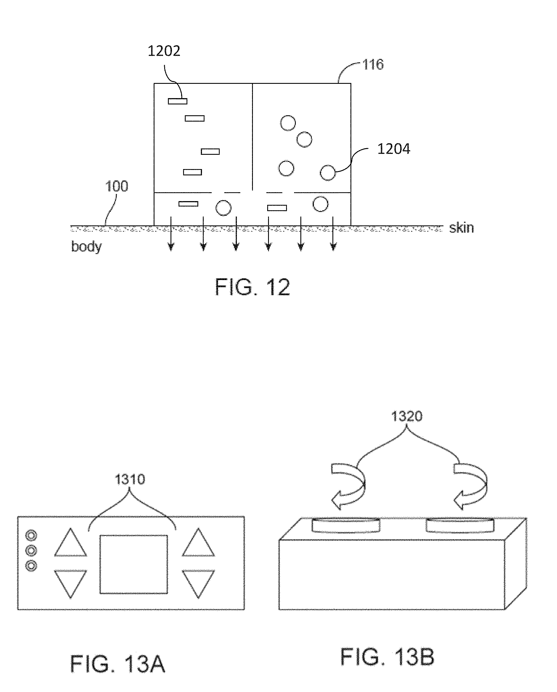
D00013

D00014
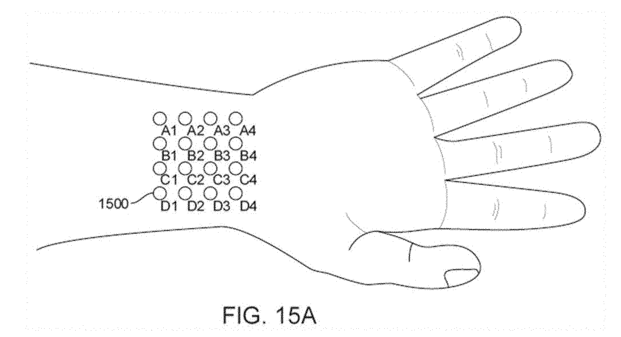
D00015

D00016

D00017

D00018

D00019
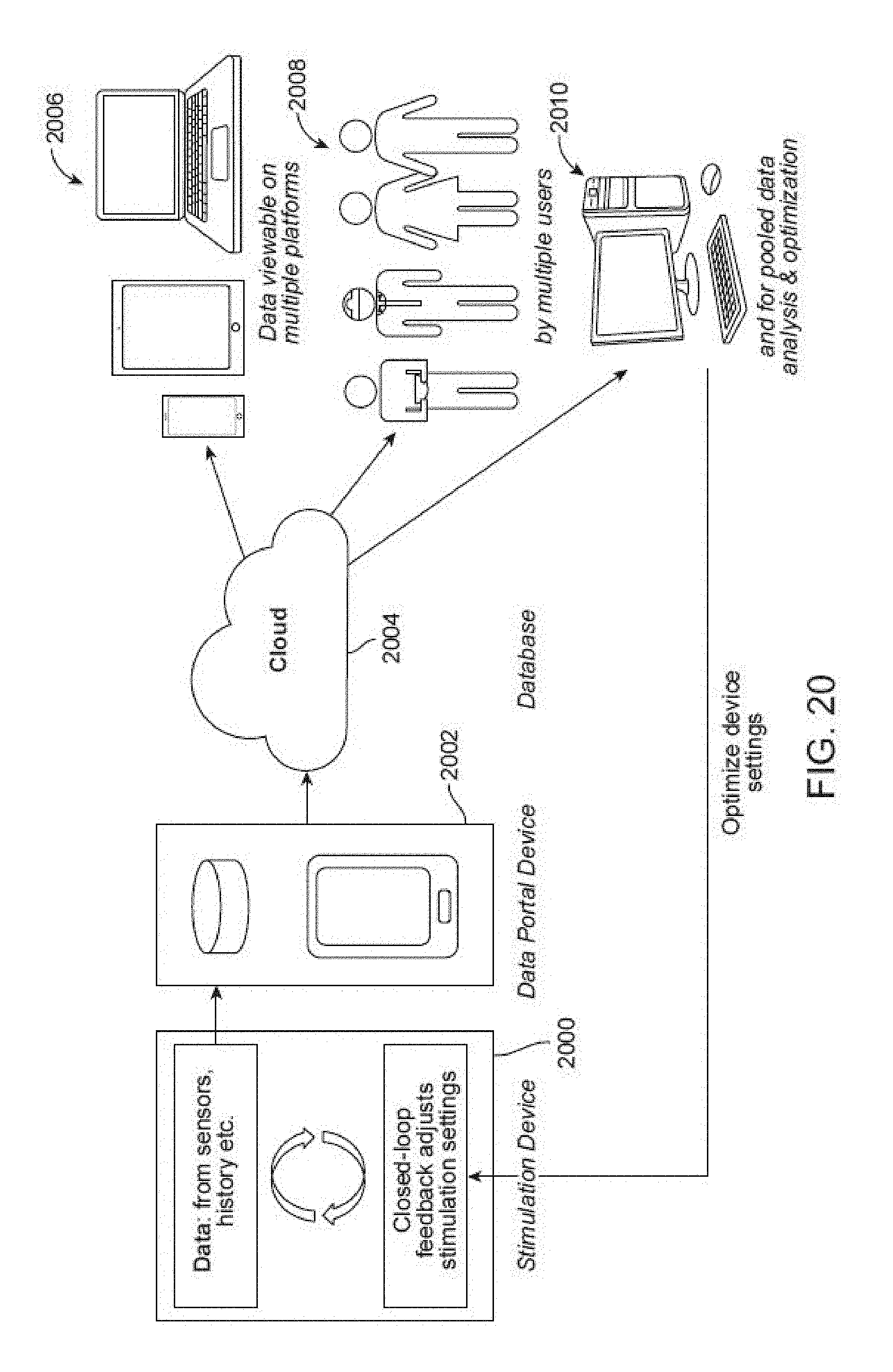
D00020
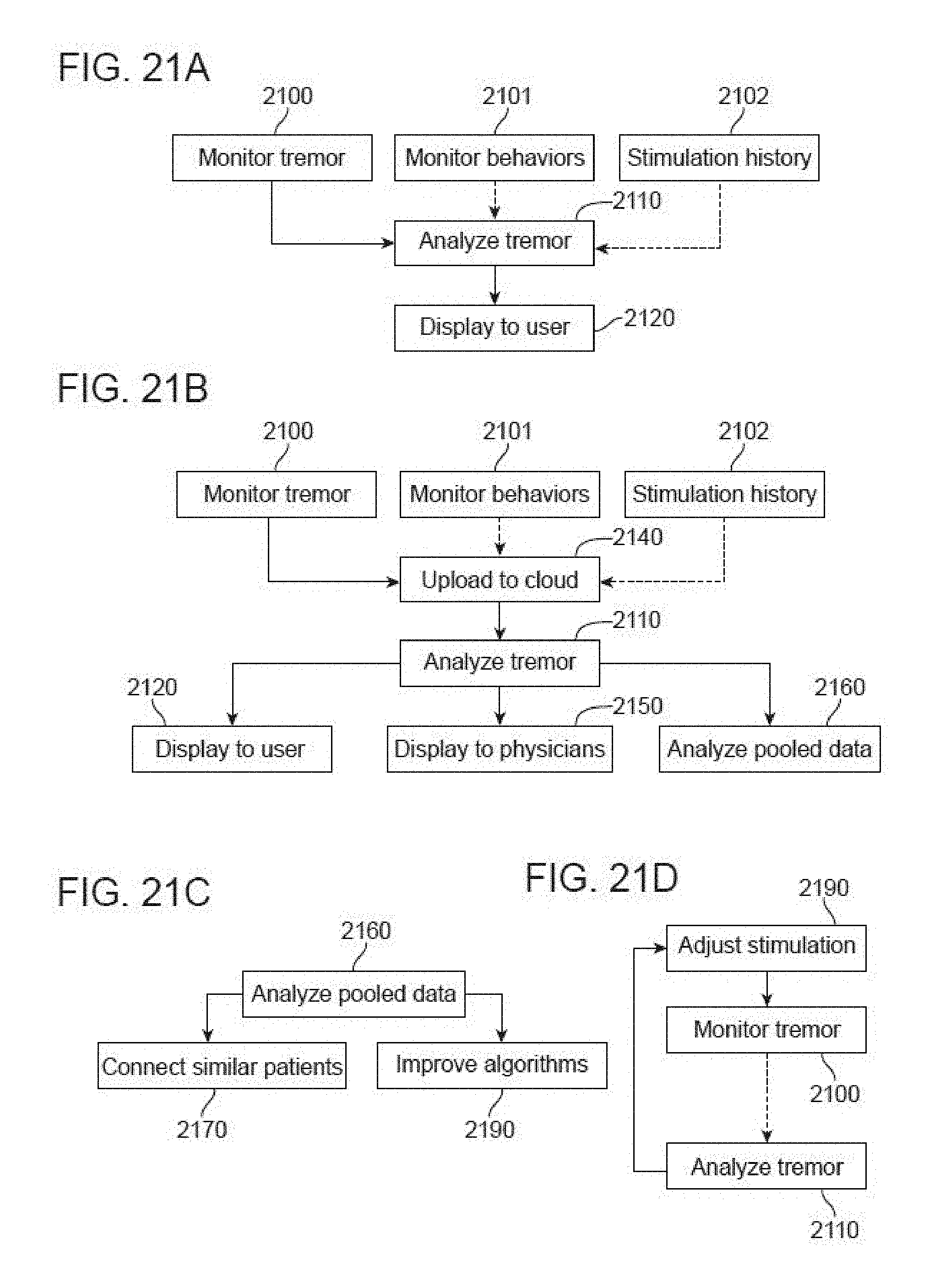
D00021
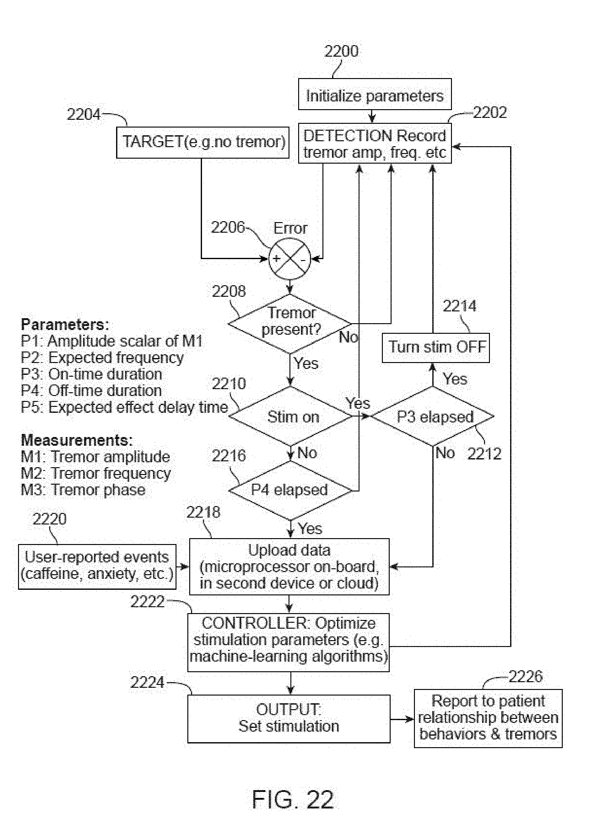
D00022

D00023
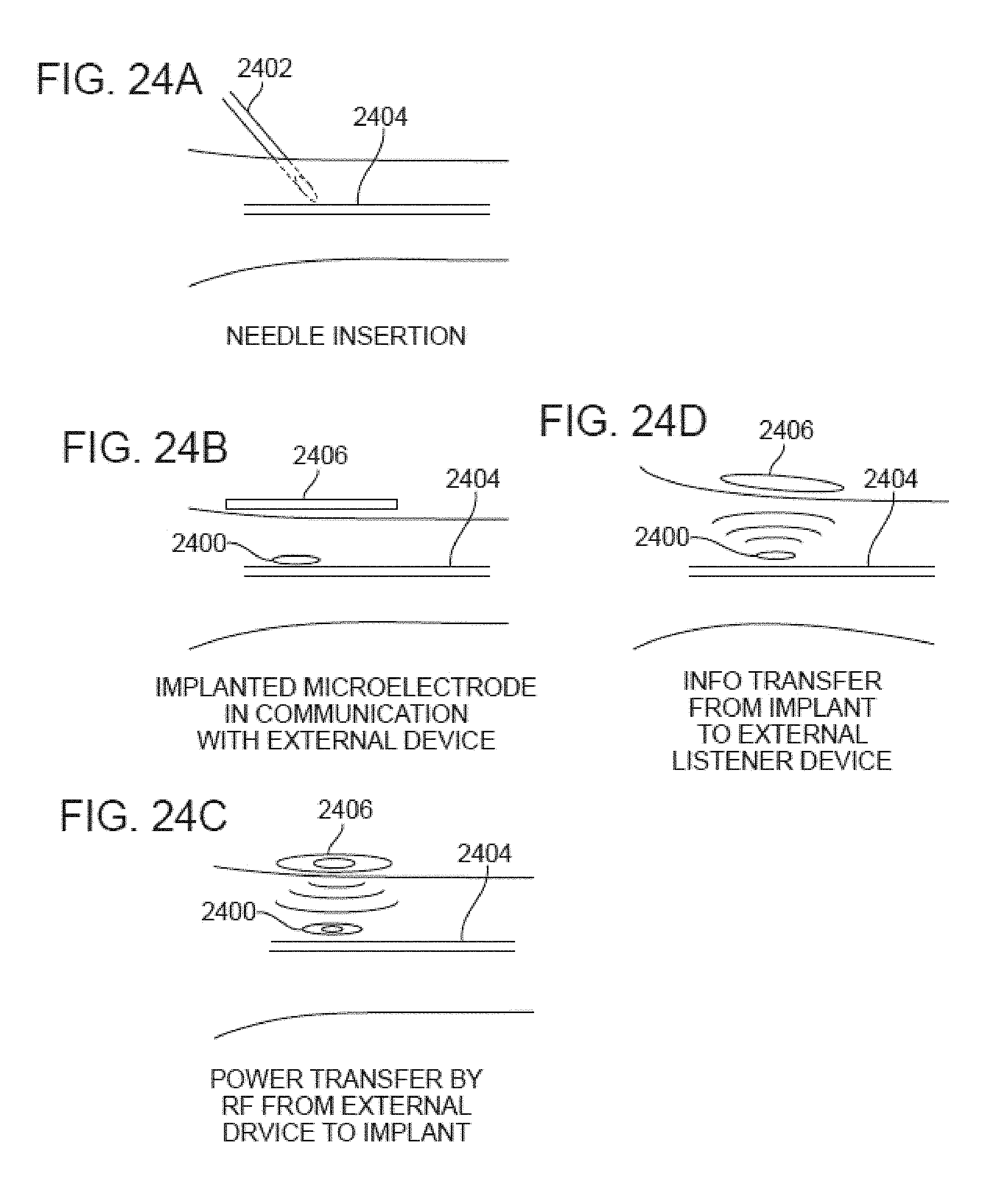
D00024
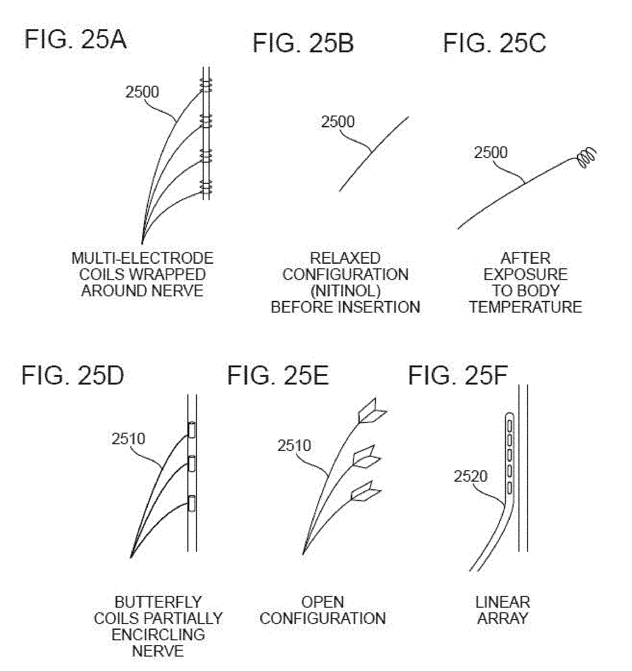
D00025
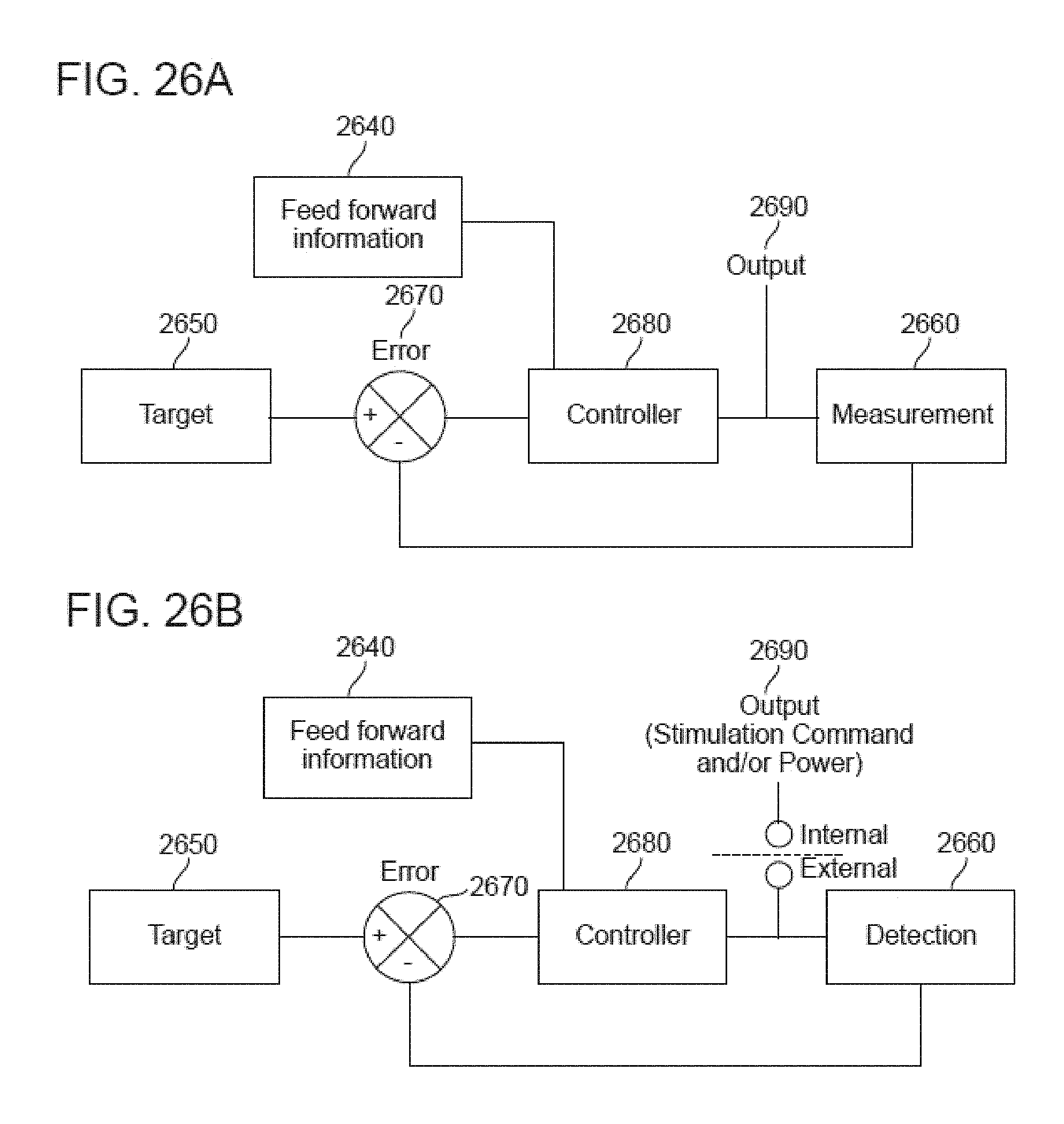
D00026
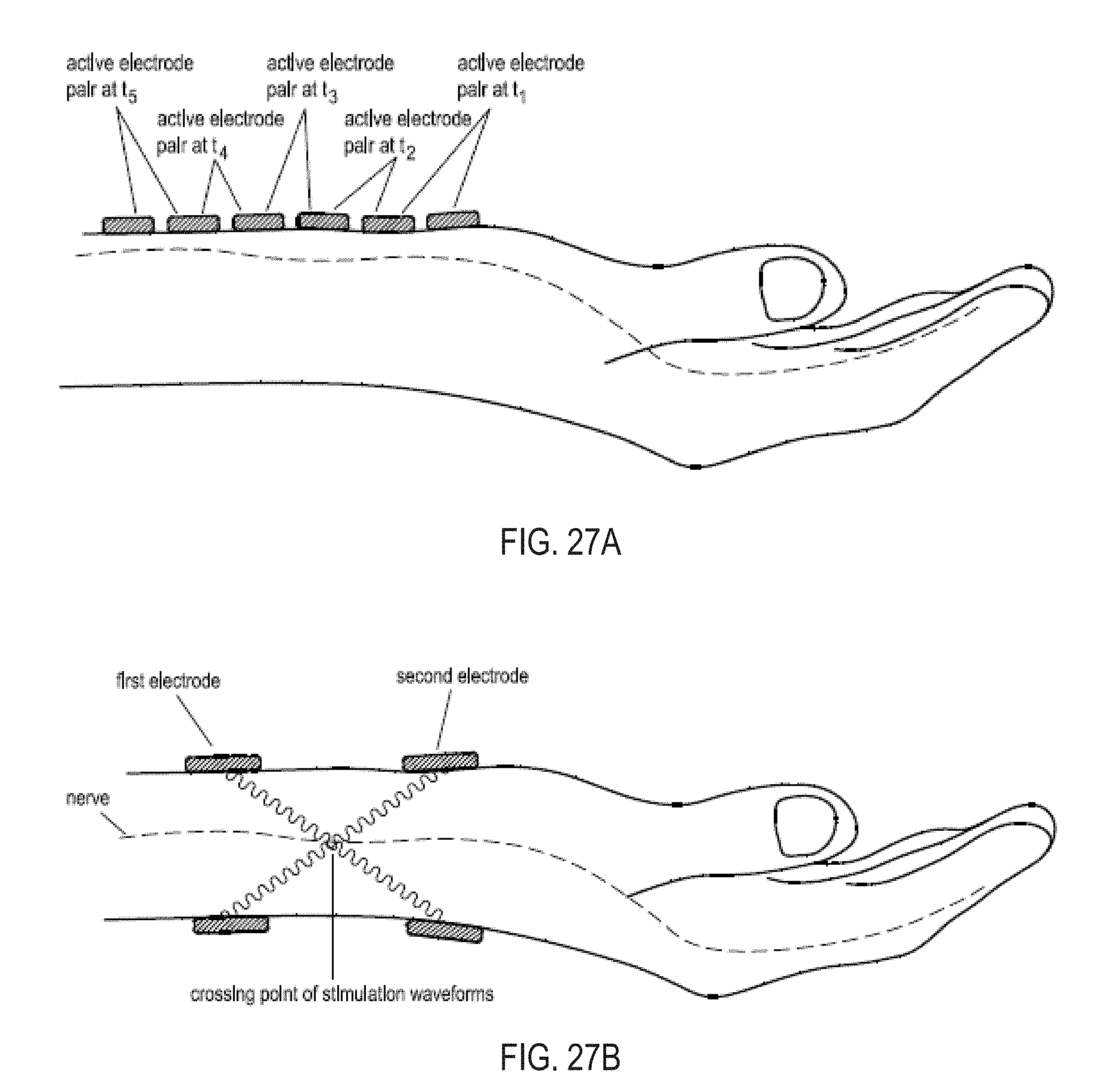
XML
uspto.report is an independent third-party trademark research tool that is not affiliated, endorsed, or sponsored by the United States Patent and Trademark Office (USPTO) or any other governmental organization. The information provided by uspto.report is based on publicly available data at the time of writing and is intended for informational purposes only.
While we strive to provide accurate and up-to-date information, we do not guarantee the accuracy, completeness, reliability, or suitability of the information displayed on this site. The use of this site is at your own risk. Any reliance you place on such information is therefore strictly at your own risk.
All official trademark data, including owner information, should be verified by visiting the official USPTO website at www.uspto.gov. This site is not intended to replace professional legal advice and should not be used as a substitute for consulting with a legal professional who is knowledgeable about trademark law.