Antitumor Immunity Enhancing Composition Containing Adenovirus Simultaneously Expressing Il-12 And Shvegf
YUN; Chae Ok ; et al.
U.S. patent application number 15/756120 was filed with the patent office on 2018-12-27 for antitumor immunity enhancing composition containing adenovirus simultaneously expressing il-12 and shvegf. The applicant listed for this patent is INDUSTRY-UNIVERSITY COORPERATION FOUNDATION HANYANG UNIVERSITY. Invention is credited to Hyo Min AHN, Chae Ok YUN.
| Application Number | 20180369417 15/756120 |
| Document ID | / |
| Family ID | 58187941 |
| Filed Date | 2018-12-27 |

View All Diagrams
| United States Patent Application | 20180369417 |
| Kind Code | A1 |
| YUN; Chae Ok ; et al. | December 27, 2018 |
ANTITUMOR IMMUNITY ENHANCING COMPOSITION CONTAINING ADENOVIRUS SIMULTANEOUSLY EXPRESSING IL-12 AND SHVEGF
Abstract
The present invention relates to an oncolytic adenovirus simultaneously expressing interleukin-12 and shVEGF, and an antitumor immunity enhancing composition and an anticancer effect promoting composition each containing the same. The present inventors verified that the simultaneous occurrence of VEGF inhibition and IL-12 expression induced the recovery of an immune function and the promotion of an anticancer effect in an immunological mouse melanoma or kidney cancer model. Especially, the applicability of a gene carrier simultaneously expressing IL-2 and shVEGF in the cancer gene therapy was first established by disclosing that the increased anticancer effect is involved in an increase in anticancer immunity, an increase in TH 1 cytokine, and the prevention of tumor induced thymic atrophy.
| Inventors: | YUN; Chae Ok; (Seoul, KR) ; AHN; Hyo Min; (Incheon, KR) | ||||||||||
| Applicant: |
|
||||||||||
|---|---|---|---|---|---|---|---|---|---|---|---|
| Family ID: | 58187941 | ||||||||||
| Appl. No.: | 15/756120 | ||||||||||
| Filed: | August 31, 2016 | ||||||||||
| PCT Filed: | August 31, 2016 | ||||||||||
| PCT NO: | PCT/KR2016/009717 | ||||||||||
| 371 Date: | February 28, 2018 |
| Current U.S. Class: | 1/1 |
| Current CPC Class: | A61K 48/0058 20130101; A61P 35/00 20180101; A61K 48/0025 20130101; A61K 38/1866 20130101; A61K 48/00 20130101; A61K 39/235 20130101; A61K 38/20 20130101; A61K 48/0075 20130101 |
| International Class: | A61K 48/00 20060101 A61K048/00; A61K 39/235 20060101 A61K039/235; A61P 35/00 20060101 A61P035/00 |
Foreign Application Data
| Date | Code | Application Number |
|---|---|---|
| Sep 1, 2015 | KR | 10-2015-0123644 |
Claims
1. A recombinant adenovirus, comprising: (i) an interleukin (IL-12) gene, and (ii) a small hairpin RNA (shRNA) gene which is complementary to VEGF mRNA and suppresses VEGF gene expression.
2. The recombinant adenovirus of claim 1, wherein the IL-12 and shRNA genes are inserted into E1 and E3 regions of the adenovirus, respectively.
3. The recombinant adenovirus of claim 1, wherein the recombinant adenovirus has an E1A region, but does not have an E1B region, which is deleted.
4. The recombinant adenovirus of claim 1, wherein the IL-12 gene includes an IL-12A (p35) gene sequence, an internal ribosome entry site (IRES) sequence and an IL-12B (p40) gene sequence.
5. A method for treating cancer, comprising: Administering a composition comprising (a) a therapeutically effective amount of a recombinant adenovirus comprising (i) an interleukin (IL-12) gene and (ii) a small hairpin RNA (shRNA) gene which is complementary to VEGF mRNA and suppresses VEGF gene expression; and (b) a pharmaceutically acceptable carrier to a subject.
6.-13. (canceled)
14. The method for treating cancer of claim 5, wherein the IL-12 and shRNA genes are inserted into E1 and E3 regions of the adenovirus, respectively.
15. The method for treating cancer of claim 5, wherein the recombinant adenovirus has an E1A region, but does not have an E1B region, which is deleted.
16. The method for treating cancer of claim 5, wherein the IL-12 gene includes an IL-12A (p35) gene sequence, an internal ribosome entry site (IRES) sequence and an IL-12B (p40) gene sequence.
17. The method for treating cancer of claim 5, wherein the cancer is gastric cancer, lung cancer, breast cancer, ovarian cancer, liver cancer, bronchial cancer, nasopharyngeal cancer, laryngeal cancer, pancreatic cancer, bladder cancer, colorectal cancer, colon cancer, cervical cancer, brain cancer, prostate cancer, bone cancer, head and neck cancer, skin cancer, kidney cancer, polyploid carcinoma, thyroid cancer, parathyroid cancer or ureter cancer.
18. A method for treating tumor-induced thymic atrophy, comprising: administering a composition comprising (a) a therapeutically effective amount of a recombinant adenovirus comprising (i) an interleukin (IL-12) gene, and (ii) a small hairpin RNA (shRNA) gene which is complementary to VEGF mRNA, and suppresses VEGF gene expression; and (b) a pharmaceutically acceptable carrier to a subject.
19. The method for treating tumor-induced thymic atrophy of claim 18, wherein the IL-12 and shRNA genes are inserted into E1 and E3 regions of the adenovirus, respectively.
20. The method for treating tumor-induced thymic atrophy of claim 18, wherein the recombinant adenovirus has an E1A region, but does not have an E1B region, which is deleted.
21. The method for treating tumor-induced thymic atrophy of claim 18, wherein the IL-12 gene includes an IL-12A (p35) gene sequence, an internal ribosome entry site (IRES) sequence and an IL-12B (p40) gene sequence.
Description
TECHNICAL FIELD
[0001] The present invention relates to a composition for enhancing antitumor immunity including adenovirus co-expressing IL-12 and shVEGF.
BACKGROUND ART
[0002] Immunological dysfunction in cancer patients has been well known [1], and it is also clinically apparent that tumors effectively avoid anticancer immune responses. While recent anticancer treatment mainly focuses on surgical operations, chemotherapy and radiation therapy, these are all accompanied by side effects [2, 3]. To overcome such barriers and increase the efficiency of anticancer treatment, research on immunotherapy is actively progressing. In the last decades, because of new findings in basic immunology and molecular discoveries of tumor antigens, research on immunotherapies for preventing and treating cancer have been actively conducted [4]. Tumor cells express a great quantity of abnormal antigens by themselves, and these antigens cause an eradication response through immune surveillance. Even if the immune system in the body actively acts, the tumor cells can avoid immune surveillance [5, 6]. It was identified that such a phenomenon is mediated by various factors produced by tumor cells. Tumor tissues can produce immunosuppressive molecules such as a vascular endothelial growth factor (VEGF), tumor growth factor (TGF)-.beta. and interleukin (IL)-10 [7, 8]. It has also been reported that infiltration of regulatory T cells occurs in immunosuppressed tumors [9-11]. These factors that are overexpressed in tumor tissue induce an immunosuppressive microenvironment and immune tolerance in tumors. As a result, overcoming immune surveillance is a pivotal strategy in immunotherapy.
[0003] IL-12 is one of the most effective and promising anticancer cytokines. IL-12 is a heterodimeric protein including 40 kDa and 35 kDa subunits connected by a disulfide bond, and produced by activated macrophages, monocytes, dendritic cells and active B lymphocytes. IL-12 may enhance T-helper 1 (Th1) cell immunity [12] and the cytotoxicity of cytotoxic T-lymphocytes, and suppress angiogenesis. Generally, IL-12 activates IFN-.gamma. production of T cells and natural killer (NK) cells [13]. Local expression of IL-12 allows tumor cells to sensitively response to T-cell mediated cytotoxicity, resulting in tumor growth inhibition and the establishment of systemic immunity. However, a disadvantage of the clinical application of IL-12 is that cytokine-associated toxicity that limits the acceptable dose for a patient when administered may appear in the entire body [14-17]. In addition, overall downregulation of the immune effect may result in repeated administration of IL-12. In a patient's serum, IL-12 polarization from Th1 to Th2 immunity occurs due to an increase in IL-10 expression and a decrease in IFN-.gamma. and TNF-.beta. expression, and for this reason, IL-12 is repeatedly administered [18,19]. These clinical results show the limitations of IL-12 as a single therapeutic for cancer treatment.
[0004] VEGF is a signal protein produced by cells, stimulating vasculogenesis and angiogenesis [20]. Particularly, VEGF affects T cell precursors in bone marrow and thus inhibits the proliferation and maturation of T cells and dendritic cells [21]. In addition, VEGF plays an important role in angiogenesis and represents a side effect called metastasis [22]. Therefore, VEGF is a stimulating factor of tumor growth, and also acts as an inhibitory factor in anticancer immunity, and it is expected that VEGF downregulation caused by VEGF shRNA will restore immune responses and increase anticancer effects. These studies show that the therapeutic mechanism mediated by VEGF shRNA, similar to that by IL-12, is associated with enhancement in CTL or dendritic cell-mediated immune responses.
[0005] Therefore, the inventors suggest a method for more effectively increasing antitumor immunity using a combination of the above-mentioned immunotherapies.
[0006] A variety of papers and patent documents are referred throughout the specification, and their citations are indicated. The disclosures of the cited papers and patent documents are incorporated herein by reference in their entireties to more clearly describe the standard of the field of the art to which the present invention belongs and the scope of the present invention.
DISCLOSURE
Technical Problem
[0007] The inventors had attempted to develop immunotherapy for overcoming the avoidance of immune surveillance of tumors using an immunosuppressive molecule produced in tumor tissue. As a result, the inventors constructed an adenovirus co-expressing IL-12 and shVEGF and developed a composition for improving immunity and a composition for improving an anticancer effect, which include the adenovirus. In addition, the inventors identified that a therapeutic effect can be enhanced by applying such an adenovirus (RdB/IL12/shVEGF) co-expressing IL-12 and shVEGF to cancer therapy, and thus completed the present invention.
[0008] Therefore, the present invention is directed to providing an adenovirus co-expressing IL-12 and shVEGF and a composition for enhancing antitumor immunity, which includes the adenovirus.
[0009] The present invention is also directed to providing the use of an adenovirus co-expressing IL-12 and shVEGF for enhancing antitumor immunity.
[0010] The present invention is also directed to providing a method for enhancing antitumor immunity using an adenovirus co-expressing IL-12 and shVEGF.
[0011] The present invention is also directed to providing an anticancer composition including an adenovirus co-expressing IL-12 and shVEGF, the use thereof for treating cancer, and a method for treating cancer, which includes administering the composition to a subject.
[0012] The present invention is also directed to providing a composition for preventing or treating tumor-induced thymic atrophy, which includes an adenovirus co-expressing IL-12 and shVEGF, the use thereof for treating tumor-induced thymic atrophy, and a method for treating tumor-induced thymic atrophy, which includes administering the composition to a subject.
[0013] Other objects and advantages of the present invention will become more apparent by the detailed description of the invention, claims and drawings as follows.
Technical Solution
[0014] According to an aspect of the present invention, the present invention provides a recombinant adenovirus, which includes (i) an IL-12 gene having at least one of a sequence of SEQ ID NO: 1 and a sequence of SEQ ID NO: 2, and (ii) a small hairpin RNA (shRNA) gene which is complementary to VEGF mRNA having a sequence of SEQ ID NO: 3, and suppresses VEGF gene expression, or a composition for enhancing antitumor immunity, which includes (a) a therapeutically effective amount of the recombinant adenovirus; and (b) a pharmaceutically acceptable carrier.
[0015] Upon conducting the research to develop immunotherapy to overcome the avoidance of immune surveillance of tumors using an immunosuppressive molecule produced in tumor tissue, the inventors constructed an adenovirus co-expressing IL-12 and shVEGF, and developed a composition for enhancing antitumor immunity and a composition for enhancing an anticancer effect. The inventors identified that a therapeutic effect can be improved by applying such an adenovirus co-expressing IL-12 and shVEGF (RdB/IL12/shVEGF) to gene therapy for cancer.
[0016] Cancer immunogenic therapy has been developed to be a more promising approach over the last few decades. However, to avoid the immune surveillance system, a tumor also has a variety of different strategies. To overcome such a barrier and enhance an anticancer immunity effect, a system (RdB/shVEGF) co-expressing IL-12 (RdB/IL12) and VEGF shRNA and an oncolytic co-expression adenovirus (RdB/IL12/shVEGF) system thereof were constructed as proper therapeutic adjuvants for restoring immunosuppression and increasing anticancer immunity. IL-12 induces the differentiation of Th1 cells and activates cytotoxic T lymphocytes and the cytotoxicity of NK cells, thereby increasing anticancer immunity. shVEGF is very effective for inhibiting VEGF expression, and thus stimulates the proliferation and maturation of T cells and dendritic cells. Such effects further amplify the functionality of IL-12. However, there is no research on the therapeutic effect caused by the co-expression of IL-2 and shVEGF in tumors. Therefore, the inventors first studied an effect of immunogenic therapy of injecting an adenovirus (RdB/IL12/shVEGF) co-expressing IL-2 and shVEGF into a tumor. In a B16-F10 murine melanoma model, the injection of RdB/IL12/shVEGF into a tumor greatly improves an anticancer effect. While IL-12 and IFN-.gamma. expression levels were considerably increased in the RdB/IL12/shVEGF-treated group, a VEGF expression level was decreased. In addition, a ratio of T-helper type 1/2 cytokines was increased in splenocyte-and-B16-F10 co-cultured supernatants. The cytotoxicity of cancer-specific immune cells in RdB/IL12/shVEGF-injected mice was increased. Like the above-described result, in situ delivery of RdB/IL12/shVEGF results in a phenomenon of massive infiltration of CD4.sup.+ T cells, CD8.sup.+ T cells, NK cells and CD86.sup.+ APCs into tissues surrounding the necrotic region of a tumor. Importantly, the adenovirus co-expressing IL-12 and shVEGF prevents tumor-induced thymic atrophy, is associated with a reduction in cell death, and increases cell proliferation in the thymus. An oncolytic adenovirus co-expressing IL-12 and shVEGF can be used as an adjuvant that increases an anticancer effect, and enhances antitumor immunity.
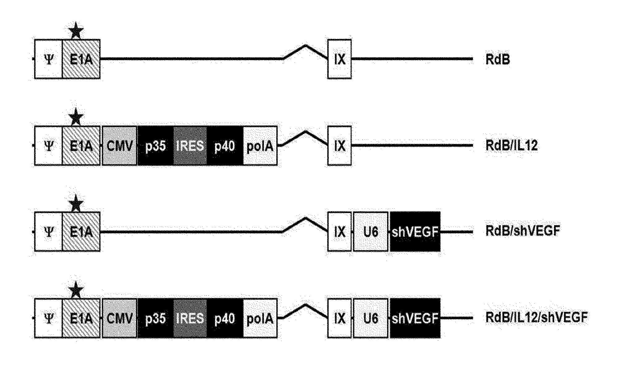
[0017] The recombinant adenovirus of the present invention includes both of an IL-12 gene sequence and a vascular endothelial growth factor small hairpin RNA (shVEGF) gene sequence. The IL-12 gene sequence includes a nucleotide sequence represented by at least one of SEQ ID NO: 1 and SEQ ID NO: 2, and the shVEGF sequence includes a sequence complementary to a nucleotide sequence represented by SEQ ID NO: 3. More specifically, the IL-12 gene sequence includes at least one of an IL-12A (p35) gene sequence and an IL-12B (p40) gene sequence, and according to an exemplary embodiment of the present invention, the IL-12 gene sequence includes an IL-12A (p35) gene sequence, an internal ribosome entry site (IRES) sequence and an IL-12B (p40) gene sequence (refer to FIG. 1A, RdB/IL12 and RdB/IL12/shVEGF).
[0018] It should be interpreted that the IL-12 gene sequence used in the present invention also includes a gene sequence exhibiting substantial identity or substantial similarity to the sequence of at least one of SEQ ID NO: 1 and SEQ ID NO: 2. The substantial identity refers to a sequence having at least 80% homology, more preferably 90% homology, and most preferably 95% homology, when a random sequence is aligned to correspond as much as possible with the sequence of the present invention, and the aligned sequences are analyzed using an algorithm conventionally used in the art. The substantial similarity encompasses all of the changes in an IL-12 gene sequence, for example, deletion or insertion of one or more bases, which do not affect the object of the present invention of minimized homologous recombination with a recombinant adenovirus vector. Therefore, the IL-12 gene sequence of the present invention is not limited to the sequence of SEQ ID NO: 1 or SEQ ID NO: 2 shown above, and all the examples of the IL-12 gene sequences should be interpreted to be included in the scope of the present invention as long as the sequences do not substantially affect the activity of the final product desired in the present invention.
[0019] In the present invention, small hairpin RNA or short hairpin RNA (shRNA) is an artificial RNA molecule having a hairpin structure, and is used to inhibit target gene expression through RNA interference. shRNA is usually delivered into cells via a plasmid, bacterial or virus vector. shRNA has an advantage of relatively low degradation and turnover ratios.
[0020] The term "shRNA molecule" used herein refers to a nucleic acid molecule that can mediate RNA interference or gene silencing. The shVEGF molecule used in the present invention includes a sequence complementary to VEGF mRNA, and the term "complementarity" used herein encompasses 100% complementarity, and a degree of incomplete complementarity sufficient to inhibit VEGF gene expression through an RNA interference mechanism, for example, preferably 90% complementarity, more preferably, 98% complementarity, and most preferably, 100% complementarity. In the specification, to represent 100% complementarity, it is specifically described as "completely complementary."
[0021] According to an exemplary embodiment of the present invention, the shRNA sequence included in the adenovirus of the present invention includes the sequence of SEQ ID NO: 3 (GenBank accession number gi: 70608153) or a sequence complementary to a part thereof, and preferably, a sequence complementary to the sequence of nucleotides 92 to 112 of an VEGF-A mRNA sequence, represented by the sequence of SEQ ID NO: 3. According to a specific exemplary embodiment of the present invention, the shRNA sequence is a sequence of SEQ ID NO: 4 and a sequence of SEQ ID NO: 5. Meanwhile, the sequence of SEQ ID NO: 3 has 88% homology with a sequence of SEQ ID NO: 6, and more specifically, 88% homology with the sequence of nucleotides 17 to 438 of the sequence of SEQ ID NO: 6.
[0022] Meanwhile, the recombinant adenovirus of the present invention includes an active E1A gene and a non-activated E1B 19 gene, an E1B 55 gene or an E1B 19/E1B 55 gene. In the specification, the term "non-activated" used in relation to a gene means that a normal protein encoded by the gene is not properly functioning since gene transcription and/or translation are/is not normally performed. For example, the non-activated E1B 19 gene is a gene that does not produce an active E1B 19 kDa protein due to a mutation (substitution, addition, partial deletion or entire deletion) occurring in the gene. E1B 19 deletion may result in an increase in necrosis ability, and the deletion of the E1B 55 gene results in tumor cell specificity (refer to Patent Application No. 2002-23760). The term "deletion" used in relation to a viral genomic sequence in the specification refers to partial deletion as well as complete deletion of a corresponding sequence.
[0023] According to an exemplary embodiment of the present invention, the recombinant adenovirus includes an E1A region, and E1B regions, containing E1B 19 and 55 kDa (.DELTA.E1B), are deleted, and an E3 region (.DELTA.E3) is deleted. The recombinant adenovirus including the E1A gene has a replicable characteristic. The IL-12 gene and shVEGF are inserted into the E1 and E3 region-deleted adenovirus, respectively (refer to FIG. 1A). However, the E1A region has a variation in which residue 45, Glu, is substituted with Gly in a nucleotide sequence encoding an Rb binding site located in the E1A gene sequence and a variation in which the sequence of amino acids 121 to 127 is entirely substituted with Gly.
[0024] The recombinant adenovirus used in the present invention includes a promoter operable in animal cells, preferably, mammalian cells. The proper promoter for the present invention includes a promoter derived from a mammalian virus and a promoter derived from the genome of mammalian cells, for example, a cytomegalovirus (CMV) promoter, an U6 promoter, a H1 promoter, a murine leukemia virus (MLV) long terminal repeat (LTR) promoter, an adenovirus early promoter, an adenovirus late promoter, a vaccina virus 7.5K promoter, an SV40 promoter, a HSV tk promoter, an RSV promoter, an EF1 .alpha. promoter, a methallothionin promoter, a .beta.-actin promoter, a human IL-2 gene promoter, a human IFN gene promoter, a human IL-4 gene promoter, a human lymphotoxin gene promoter, a human GM-CSF gene promoter, a human phosphoglycerate kinase (PGK) promoter, a mouse phosphoglycerate kinase (PGK) promoter and a survivin promoter, but the present invention is not limited thereto. Most preferably, the promoter in the present invention is a CMV promoter.
[0025] In the recombinant adenovirus used in the present invention, the IL-12 gene sequence and the shVEGF sequence are operably linked to a promoter. In the specification, the term "operably linked" refers to a functional binding between a nucleic acid expression-regulatory sequence (e.g., an array of a promoter, a signal sequence, and a transcription regulating factor-binding site) and a different nucleic acid sequence, and therefore, the regulatory sequence regulates the transcription and/or translation of the different nucleic acid sequence.
[0026] The recombinant adenovirus of the present invention may further include an antibiotic-resistant gene and a reporter gene (e.g., green fluorescence protein (GFP), luciferase and .beta.-glucuronidase) as selective markers. The antibiotic-resistant gene includes an antibiotic-resistant gene conventionally used in the art, for example, a gene resistant to ampicillin, gentamycin, carbenicillin, chloramphenicol, streptomycin, kanamycin, geneticin, neomycin and tetracycline, and preferably, is a neomycin-resistant gene. The selective marker may be expressed even in an expression system connected by a separate promoter or an internal ribosome entry site (IRES), and the IRES that can be used in the present invention is a regulatory sequence that is found in RNAs of some types of viruses and cells (McBratney et. al. Current Opinion in Cell Biology 5:961(1993)).
[0027] As to be described in an example described below, a recombinant adenovirus of the present invention, which co-expresses IL-12 and VEGF-specific shRNA, interrupts an immune surveillance avoiding mechanism of tumors by increasing IL-12 expression and suppressing VEGF expression. It has been reported that the shift from Th1 to Th2 cytokine expression has been shown in the development of various mouse and human malignant tumors, and the composition of the present invention increases a Th1/Th2 cytokine ratio by inhibiting such a Th2 immuno-deviation phenomenon (refer to FIGS. 4A to 4C).
[0028] In another aspect of the present invention, the present invention provides an anticancer composition which includes (a) (i) IL-12 gene including at least one of a sequence of SEQ ID NO: 1 and a sequence of SEQ ID NO:2, and (ii) a therapeutically effective amount of a recombinant adenovirus including a small hairpin RNA (shRNA) gene which is complementary to vascular endothelial growth factor (VEGF) mRNA of a sequence of SEQ ID NO: 3, and inhibits VEGF gene expression; and (b) a pharmaceutically acceptable carrier.
[0029] The anticancer composition of the present invention uses a recombinant adenovirus included in the above-described composition for enhancing antitumor immunity, and common details between these will be omitted to avoid excessive complexity in the specification.
[0030] According to an exemplary embodiment of the present invention, the cancer includes gastric cancer, lung cancer, breast cancer, ovarian cancer, liver cancer, bronchial cancer, nasopharyngeal cancer, laryngeal cancer, pancreatic cancer, bladder cancer, colorectal cancer, colon cancer, cervical cancer, brain cancer (e.g., glioma), prostate cancer, bone cancer, head and neck cancer, skin cancer (e.g., melanoma), kidney cancer, polyploid carcinoma, thyroid cancer, parathyroid cancer and ureteral cancer, but the present invention is not limited thereto.
[0031] In still another aspect of the present invention, the present invention provides a composition for preventing or treating tumor-induced thymic atrophy, which includes (a) (i) IL-12 gene including at least one of a sequence of SEQ ID NO: 1 and a sequence of SEQ ID NO:2, and (ii) a therapeutically effective amount of a recombinant adenovirus including a small hairpin RNA (shRNA) gene which is complementary to vascular endothelial growth factor (VEGF) mRNA of a sequence of SEQ ID NO: 3, and inhibits VEGF gene expression; and (b) a pharmaceutically acceptable carrier.
[0032] The composition for preventing or treating tumor-induced thymic atrophy uses a recombinant adenovirus including in the above-described composition for enhancing antitumor immunity, and common details between these will be omitted to avoid excessive complexity in the specification.
[0033] Thymic atrophy has been usually observed in tumor-bearing mice, and results in a reduction in host immunity with respect to tumors. The composition of the present invention co-expresses IL-12 and shVEGF in tumors, thereby preventing or inhibiting thymic atrophy caused by tumor development (refer to FIGS. 7A to 7G).
[0034] All of the compositions of the present invention described above include a pharmaceutically acceptable carrier. The pharmaceutically acceptable carrier used herein, conventionally used in formulation, includes lactose, dextrose, sucrose, sorbitol, mannitol, starch, acacia gum, calcium phosphate, alginate, gelatin, calcium silicate, microcrystalline cellulose, polyvinylpyrrolidone, cellulose, water, syrup, methyl cellulose, methylhydroxybenzoate, propylhydroxybenzoate, talc, magnesium stearate and mineral oil, but the present invention is not limited thereto. The pharmaceutical composition of the present invention may further include a lubricant, a wetting agent, a sweetening agent, a flavoring agent, an emulsifier, a suspension, a preservative, etc., other than the above-mentioned components. Suitable pharmaceutically acceptable carriers and agents are described in Remington's Pharmaceutical Sciences (19th ed., 1995) in detail.
[0035] The composition of the present invention may be administered orally or parenterally, for example, by intravenously injection, subcutaneous injection, intramuscular injection, intraperitoneal injection or transdermal administration, and the composition of the present invention is preferably administered parenterally. According to an exemplary embodiment of the present invention, preferably, the composition for enhancing antitumor immunity of the present invention is directly intratumorally administered.
[0036] A suitable dosage of the pharmaceutical composition may be prescribed in various ways according to parameters such as a formation method, an administration method, a patient's age, body weight and sex, the severity of a disease symptom, a diet, administration time, administration route, excretion speed and reaction sensitivity. A daily dose of the pharmaceutical composition of the present invention is, for example, 0.001 to 1000 mg/kg. However, an actual dosage of the active ingredient may be determined by considering various related factors such as an amount of target tissue cells to be differentiated and proliferated, an administration route, a patient's weight, age and sex, and therefore, the dosage does not limit the scope of the present invention in any way.
Advantageous Effects
[0037] Characteristics and advantages of the present invention are summarized as follows:
[0038] (a) The present invention relates to an oncolytic adenovirus co-expressing IL-12 and shVEGF, and a composition for enhancing antitumor immunity and a composition for enhancing an anticancer effect, which include the same.
[0039] (b) The inventors found that, when VEGF suppression and IL-12 expression are co-expressed in immunocompetent mouse melanoma models, immune functions are restored and an anticancer effect is enhanced.
[0040] (c) Particularly, the inventors identified that such an increase in anticancer effect is associated with an increase in anticancer immunity, an increase in Th1 cytokines and prevention of tumor-induced thymic atrophy, and thus first identified the applicability of a gene delivery system co-expressing IL-12 and shVEGF in cancer gene therapy.
DESCRIPTION OF DRAWINGS
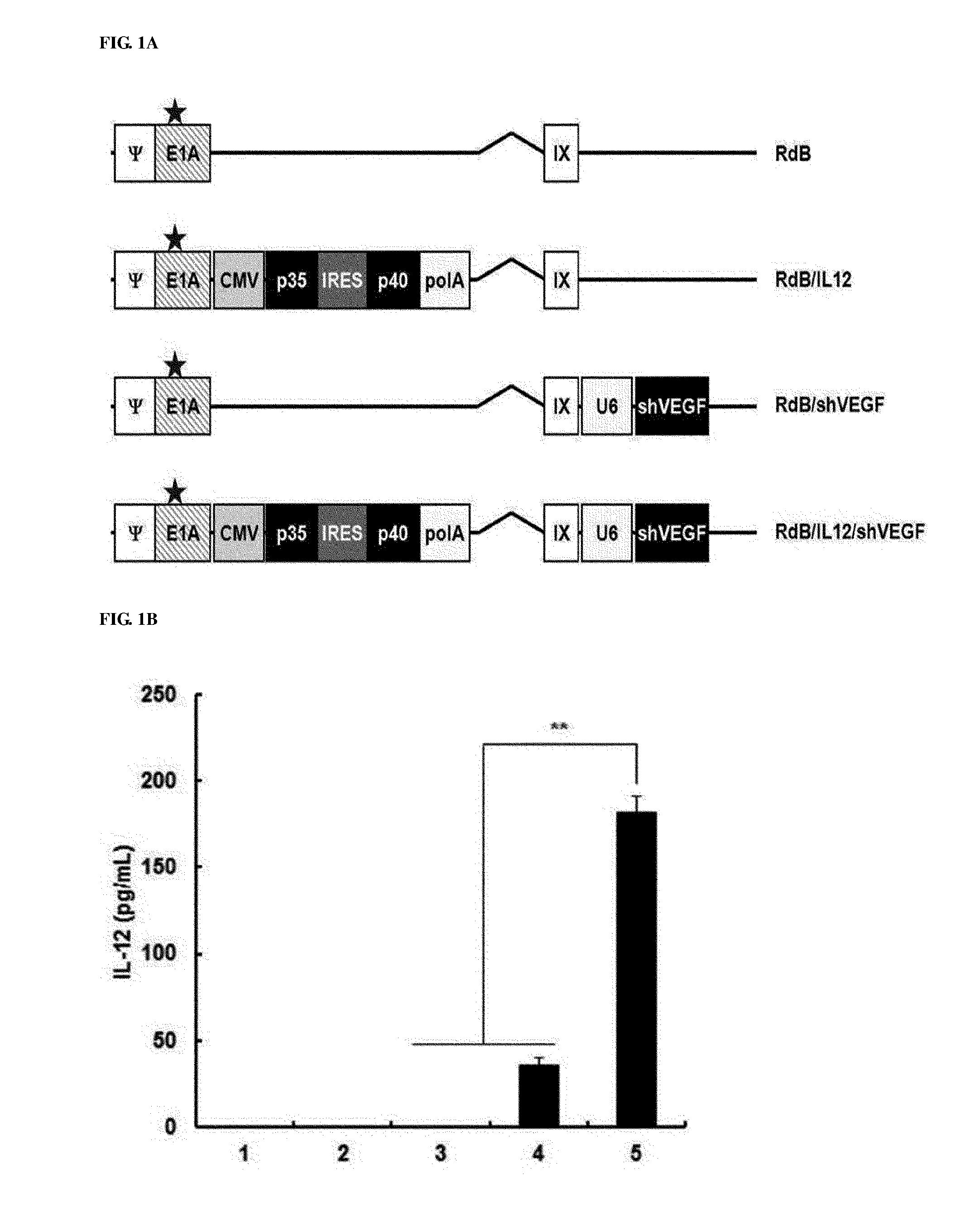
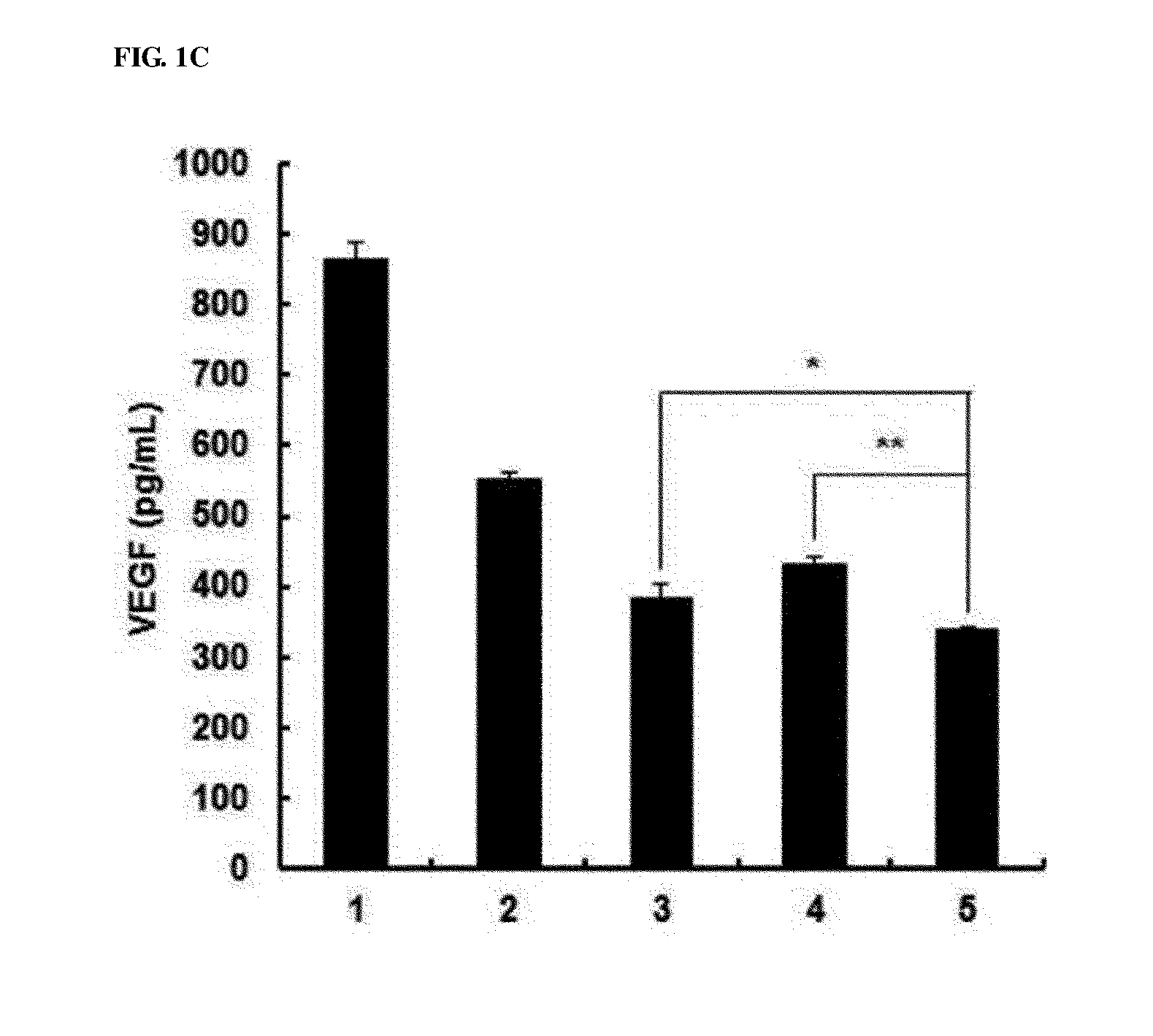
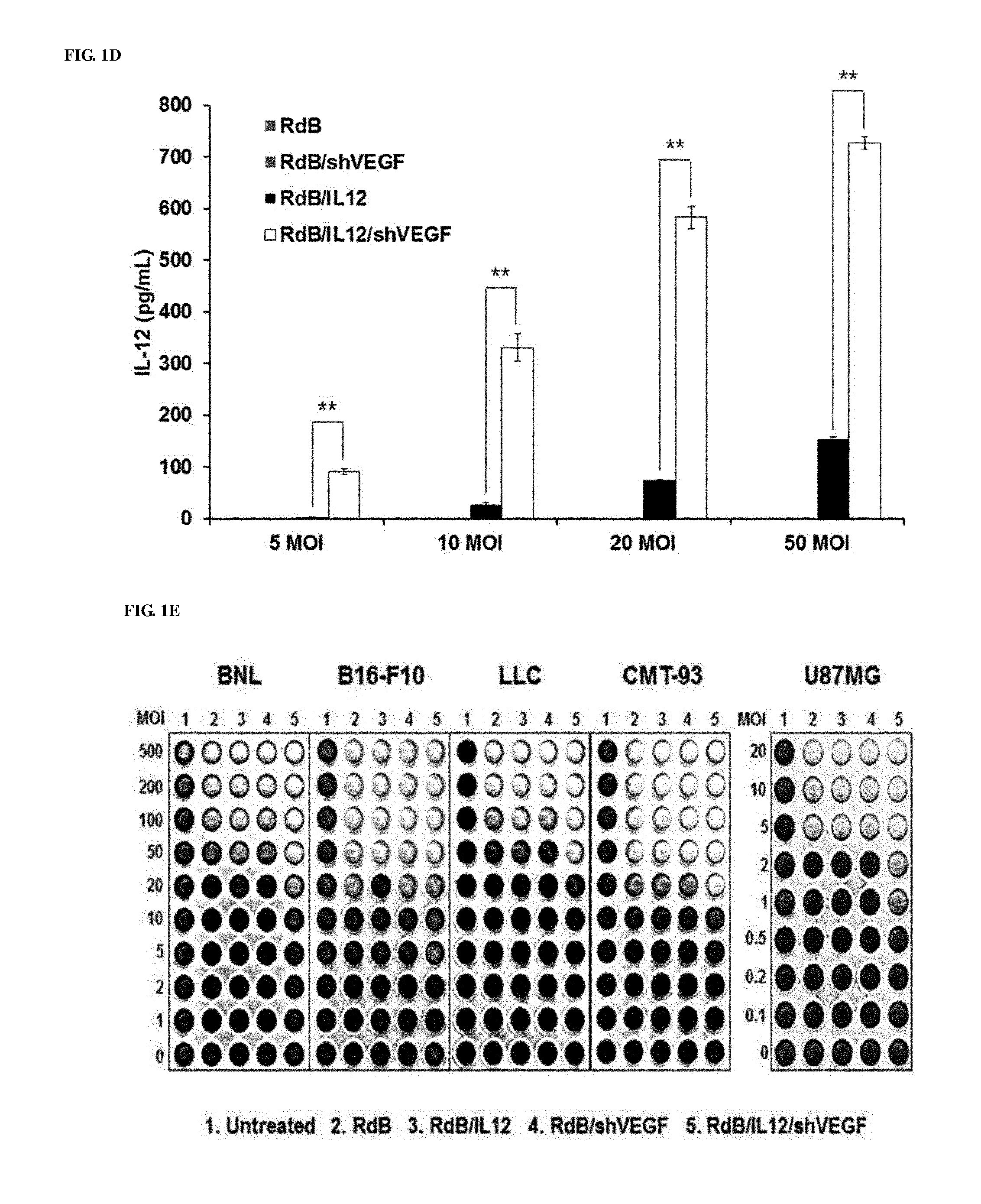
[0041] FIGS. 1A to 1E show the characteristics of oncolytic adenoviruses co-expressing IL-12 and VEGF shRNA. FIG. 1A shows schematic diagrams of genomic structures of adenovirus RdB and RdB/IL12/shVEGF. RdB includes mutated E1A (open star--a mutant of Rb protein-binding site), E1B 19, 55 kDa (.DELTA.E1B) and E3 (.DELTA.E3) regions are deleted; murine IL-12 and murine shVEGF were inserted into the E1 and E3 regions of the adenovirus genome. B16-F10 cells were infected with RdB, RdB/IL12, RdB/shVEGF or RdB/IL12/shVEGF, and IL-12 (FIG. 1B) and VEGF (FIG. 1C) expression levels were detected. Forty eight hours after infection, a cell culture supernatant was collected, and IL-12 and VEGF levels were quantified using an ELISA kit. Referring to FIG. 1D, by the same method as described above, IL-12 levels according to MOIs (5, 10, 20, 50 MOI) were quantified. FIG. 1E represents the cytopathic effects of the oncolytic adenovirus expressing IL-12 and/or shVEGF in murine cancer cell lines (BNL, B16-F10, LLC and CMT-93) and a human cancer cell line (U87MG). Data of triplicate experiments is expressed as the mean.+-.standard deviation (SD), and similar results were obtained from at least three independent experiments. MOI, multiplicity of infection. 1. PBS 2. RdB 3. RdB/IL12 4. RdB/shVEGF 5. RdB/IL12/shVEGF.
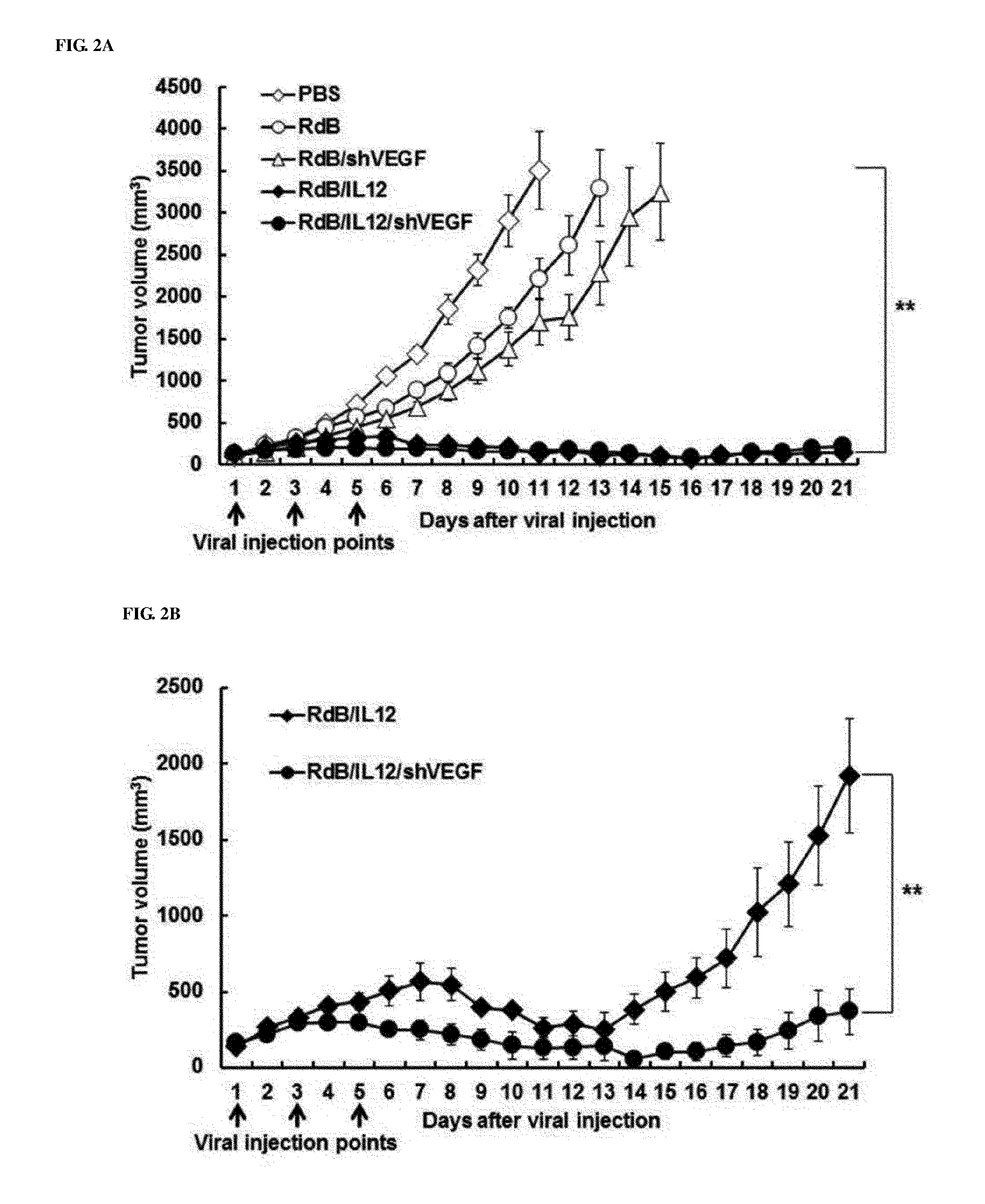
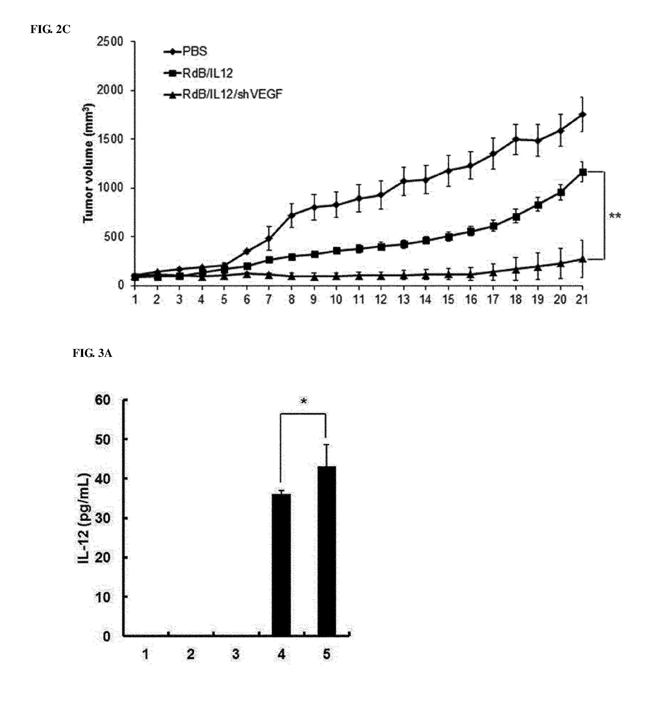
[0042] FIGS. 2A to 2C show the anticancer effects of adenoviruses in cancer-bearing mice. FIGS. 2A and 2B show the anticancer effects in melanoma mice treated with PBS (open diamonds), RdB (open circles), RdB/shVEGF (open triangles), RdB/IL12 (filled diamonds) or RdB/IL12/shVEGF (filled circles), and FIG. 2A to 2C show the anticancer effect in kidney cancer mice treated with PBS (open diamonds), RdB/IL12 (filled diamonds) or RdB/IL12/shVEGF (filled circles). Adenoviruses at a dose of 3.times.10.sup.9 VP (FIG. 2A; initial tumor volume: 80-100 mm.sup.3) or 6.times.10.sup.8 VP; and RdB/IL12 and RdB/IL12/shVEGF (FIG. 2B; initial tumor volume: 80-100 mm.sup.3, FIG. 2C; initial tumor volume: 100 mm.sup.3) adenoviruses were administered into tumors of C57BL/6 mice on day 1, day 3 and day 5. Tumor volume was monitored and recorded every day until the end of the study. Arrows represent injection time of adenoviruses. Values is expressed as the mean.+-.SD (n=8). **, P<0.01.
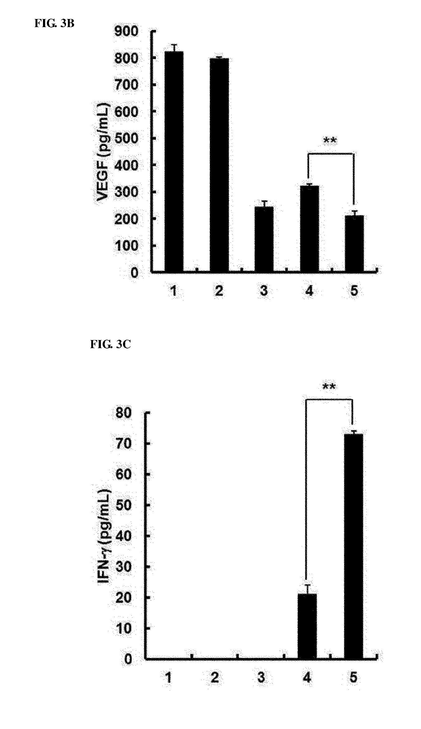
[0043] FIGS. 3A to 3C show in vivo intratumoral expression levels of IL-12, VEGF and IFN-.gamma.. Seven days after initial viral injection, tumor tissues were collected, and IL-12 (FIG. 3A), VEGF (FIG. 3B) and IFN-.gamma. (FIG. 3C) levels were measured by ELISA. Experiments were repeated three times, and data is expressed as the mean.+-.SD. *, P<0.05 or **, P<0.01; 1. PBS 2. RdB 3. RdB/shVEGF 4. RdB/IL12 5. RdB/IL12/shVEGF.
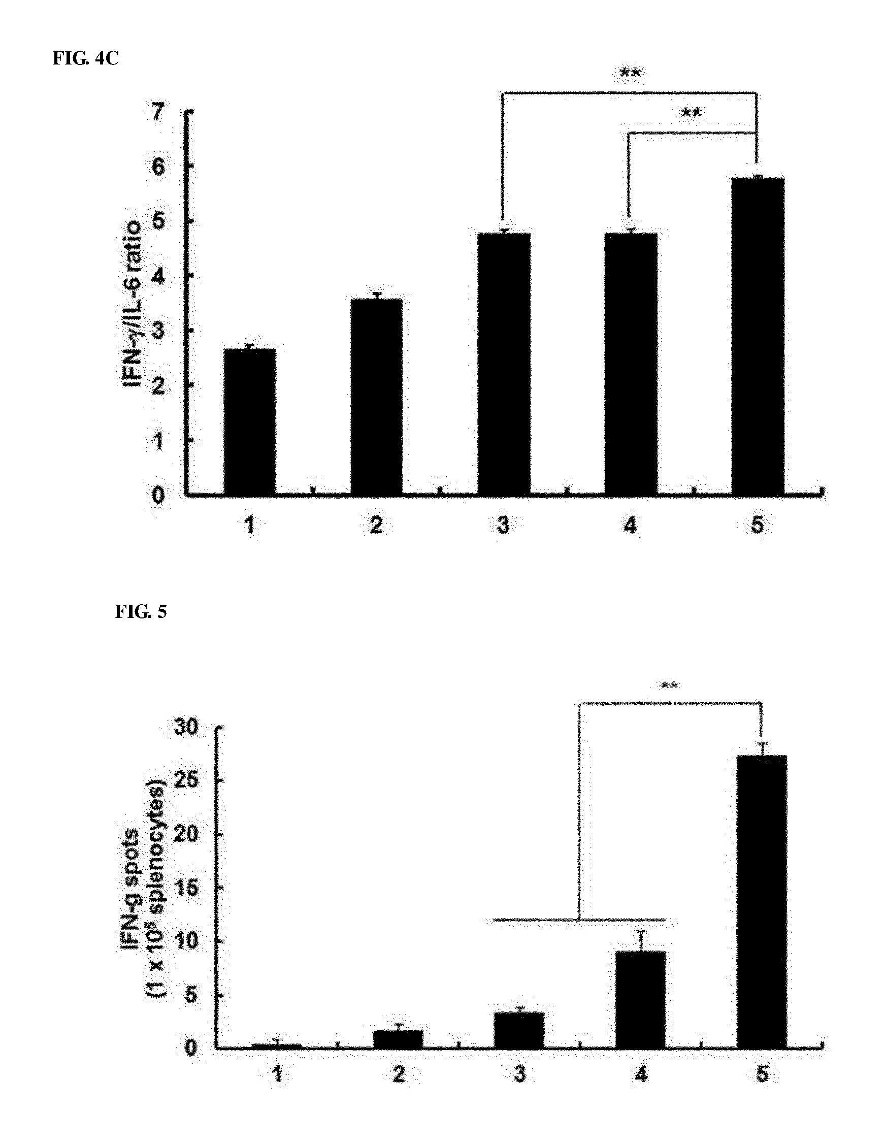
[0044] FIGS. 4A to 4C show that Th1/Th2 cytokine ratios were increased in splenocyte and B16-F10 co-cultured supernatants. On day 7 after the final viral injection, splenocytes were collected and cultured for 3 days with irradiated B16-F10 cancer cells in the presence of recombinant human IL-2. Th1/Th2/Th17 CBA was analyzed to measure the Th1/Th2 cytokine ratios of the supernatants. (FIG. 4A) IFN-.gamma. value, (FIG. 4B) IL-6 value (FIG. 4C) IFN-.gamma./IL-6 ratios. After triplicate experiments, data is expressed as the mean.+-.SD. **P<0.01; 1. PBS 2. RdB 3. RdB/shVEGF 4. RdB/IL12 5. RdB/IL12/shVEGF.
[0045] FIG. 5 shows the result of measuring tumor-specific immunity. On day 7 after the final viral injection, splenocytes were collected from mice treated with PBS-, RdB- or cytokine-expressing adenoviruses, and cultured for 3 days with irradiated B16-F10 cells. Subsequently, the IFN-.gamma. ELISPOT assay was carried out, and the number of spots was measured at a concentration of 1.times.10.sup.5 IFN-.gamma. ELISPOT. After triplicate experiments, each value is expressed as the mean spot number .+-.SD. **, P<0.01; 1. PBS 2. RdB 3. RdB/shVEGF 4. RdB/IL12 5. RdB/IL12/shVEGF.
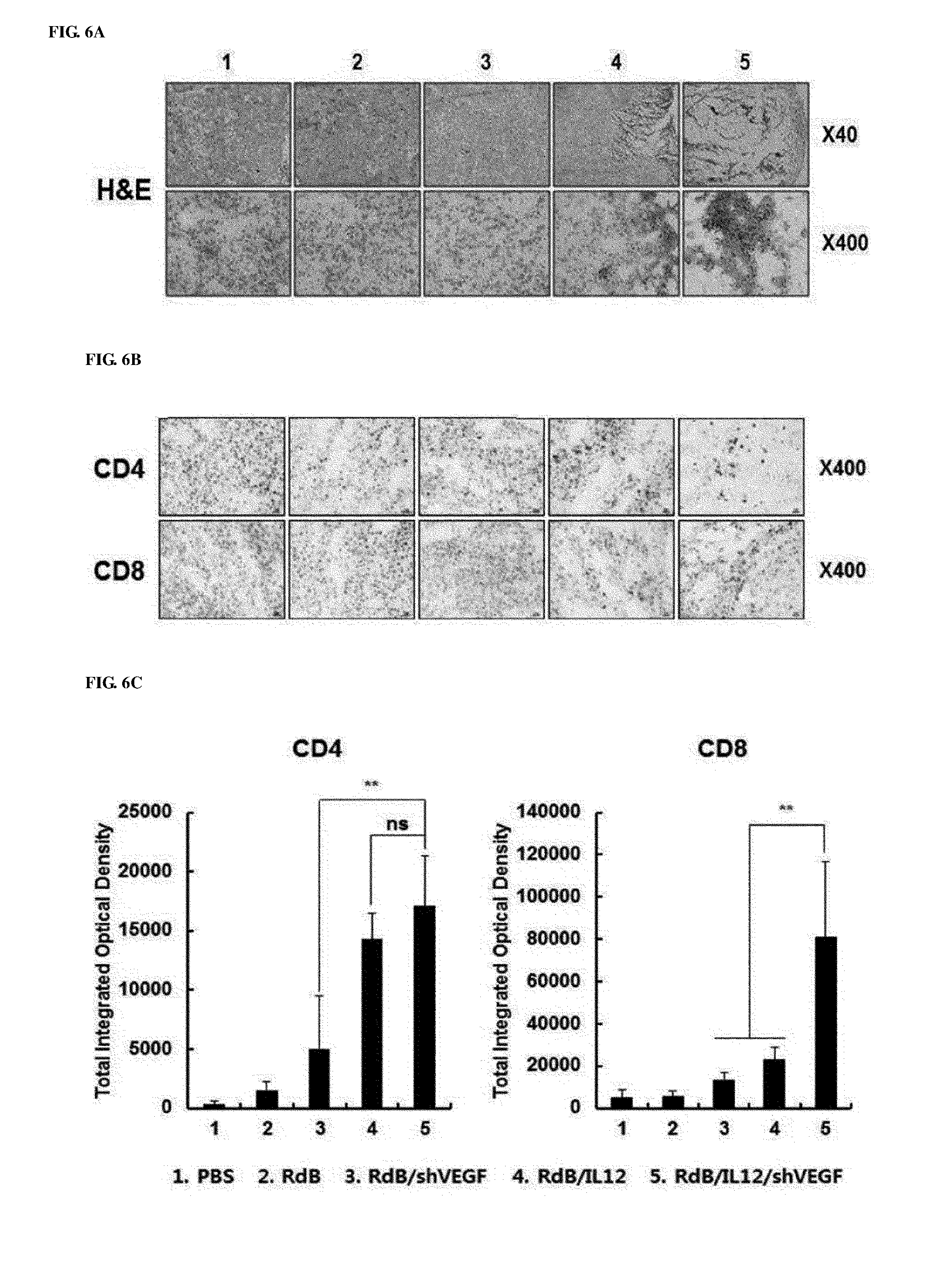
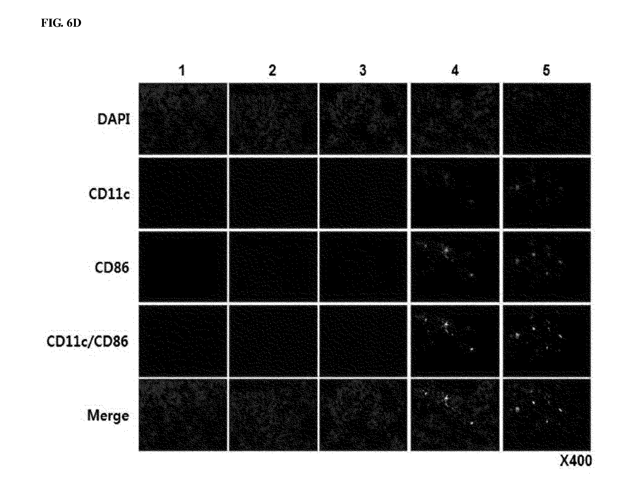
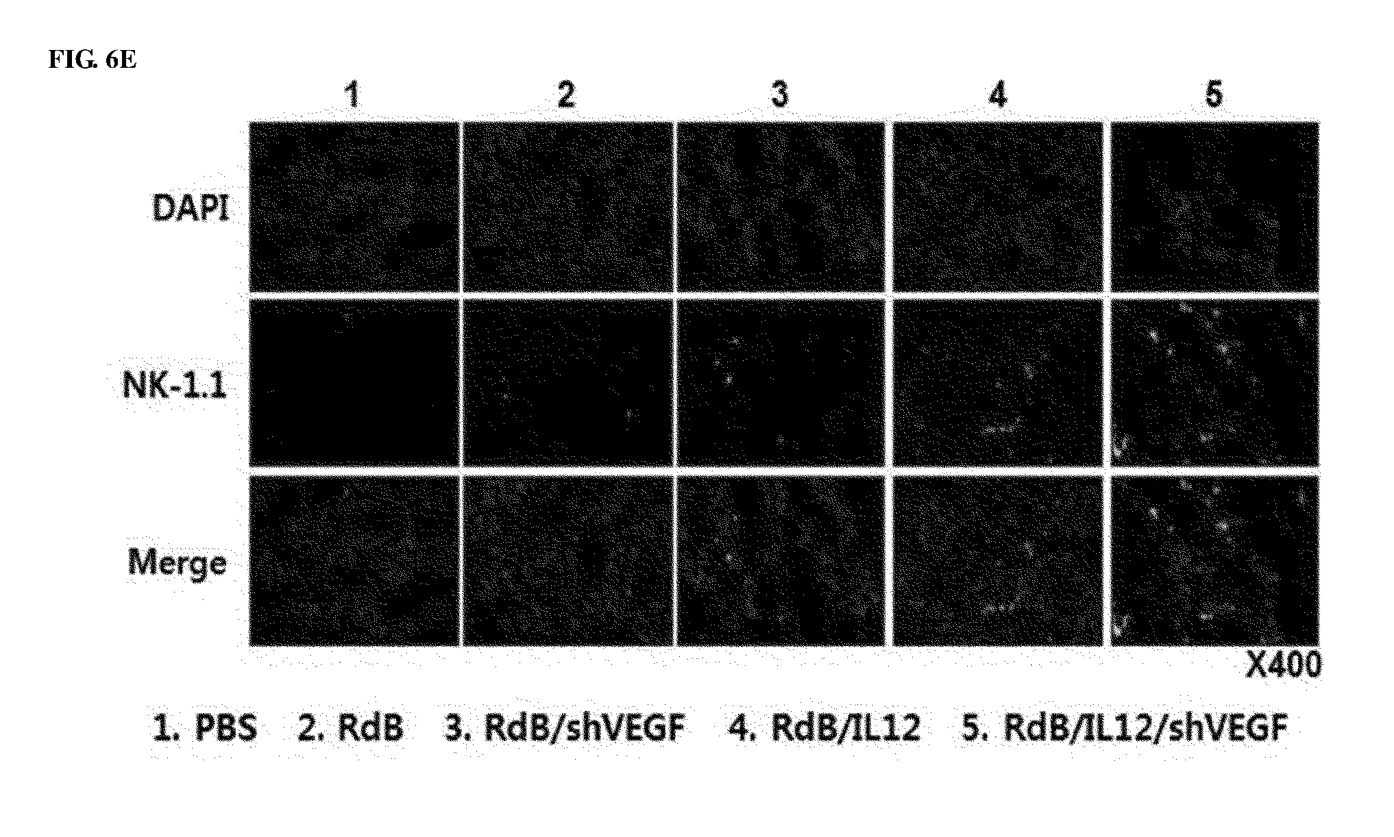
[0046] FIGS. 6A to 6E show the results of histological and immunohistochemical analyses for murine tumor fragments treated with adenoviruses. Adenoviruses were treated on days 1, 3 and 5, and tumors were collected on day 12, followed by histological analysis. FIG. 6A shows cryosections of tumor tissue were subjected to H&E staining. Magnification: 40.times. and 400.times.. FIG. 6B shows that cryosections of tumor tissue were stained with an anti-CD4 or anti-CD8 monoclonal antibody. Magnification: 400.times.. FIG. 6C shows increases in CD4+T and CD8+T expression that were semi-quantitatively measured using MetaMorph.RTM. image analysis software (**p<0.01). FIG. 6D shows cryosections of tumor tissue that were stained with anti-CD11c, anti-CD86 monoclonal antibodies, and DAPI. Magnification: 400.times.. FIG. 6E shows cryosections of tumor tissue that were stained with an anti-NK1.1 monoclonal antibody and DAPI. Magnification: 400.times.. 1. PBS 2. RdB 3. RdB/shVEGF 4. RdB/IL12 5. RdB/IL12/shVEGF.
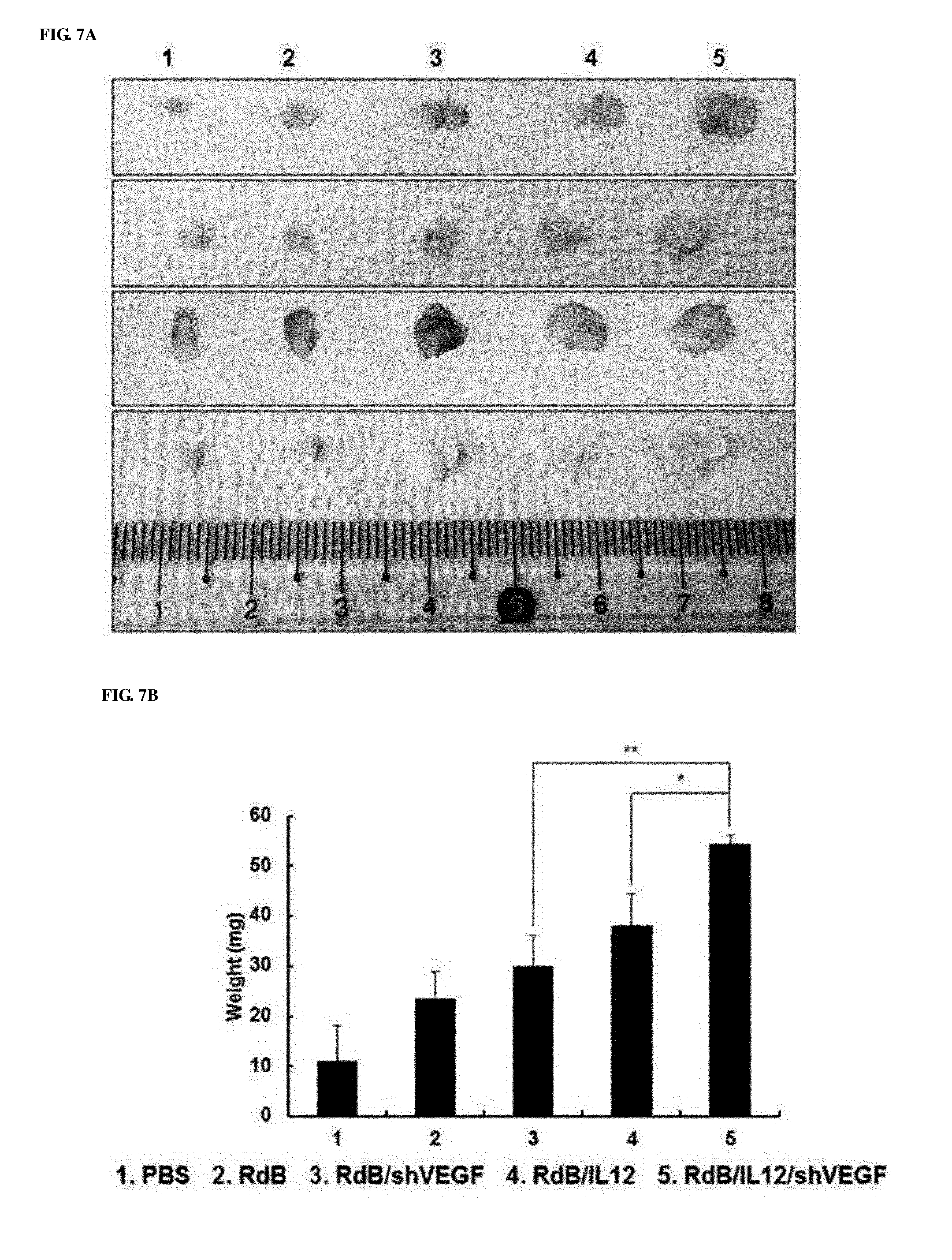
[0047] FIGS. 7A to 7G show the effect of preventing thymic atrophy of the RdB/IL12/shVEGF adenovirus. FIG. 7A shows images of the entire thymus of mice treated with PBS, RdB, RdB/IL12, RdB/shVEGF or RdB/IL12/shVEGF. FIG. 7B shows the weight of thymuses in mice treated with oncolytic adenoviruses. Data is expressed as the mean.+-.SD (*P<0.05, **P<0.01). FIG. 7C shows sections from paraffin blocks that were stained with H&E. FIG. 7D shows the apoptosis in thymus tissue that was detected by TUNEL assay. FIG. 7E. shows the degree of proliferation of thymocytes as evaluated by proliferating cell nuclear antigen staining. FIGS. 7F and 7G show the results of FIGS. 7D and 7E that were semi-quantitatively measured using MetaMorph.RTM. image analysis software (**p<0.01). 1. PBS 2. RdB 3. RdB/shVEGF 4. RdB/IL12 5. RdB/IL12/shVEGF.
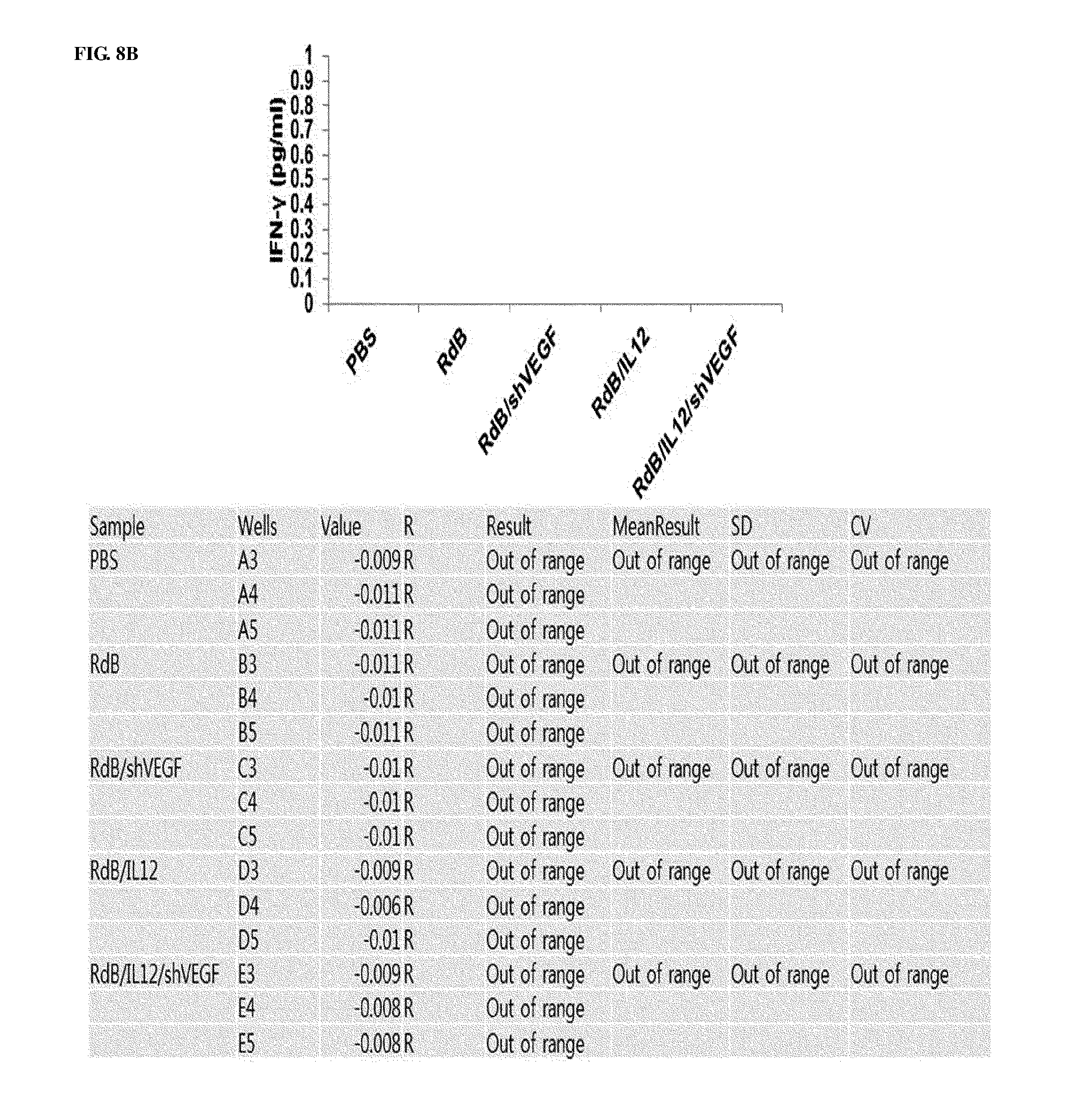
[0048] FIGS. 8A and 8B show the results obtained by ELISA assay following isolation of murine sera to confirm contents of IL-12 and IFN-.gamma. remaining in mice on day 7 after final treatment of the mice with recombinant adenoviruses. FIG. 7A shows IL-12 contents in murine serum treated with PBS, RdB, RdB/shVEGF, RdB/IL12 or RdB/IL12/shVEGF. FIG. 7B shows IFN-.gamma. contents in murine serum treated with PBS, RdB, RdB/shVEGF, RdB/IL12, or RdB/IL12/shVEGF.
MODES OF THE INVENTION
[0049] Hereinafter, the present invention will be described in further detail with respect to examples. These examples are only provided to more fully describe the present invention, and it is obvious to those of ordinary skill in the art that the scope of the present invention is not limited to these examples according to the scope of the present invention.
EXAMPLES
Experimental Materials and Experimental Methods
[0050] (1) Cell Lines and Cell Culture
[0051] Dulbecco's modified Eagle's medium (DMEM; Gibco BRL, Grand Island, N.Y.) or Roswell Park Memorial Institute medium (RPMI; Gibco BRL), supplemented with 10% fetal bovine serum (FBS; Gibco BRL), L-glutamine (2 mmol/L), penicillin (100 IU/mL), and streptomycin (50 mg/mL), was used as a cell culture medium. HEK293 (human embryonic kidney cell line expressing an adenovirus E1 region), U87MG (human glioma cell line), BNL (murine liver cancer cell line), B16-F10 (murine melanoma cell line), LLC (murine lung cancer cell line), CMT-93 (murine polyploid carcinoma cell line), and Renca (murine renal adenocarcinoma cell line) were purchased from the American Type Culture Collection (ATCC; Manassas, Va.). All cell lines were cultured at 37.degree. C. in a 5% CO.sub.2 humid environment, and subjected to a mycoplasma negative test using Hoeschst dye, cell culture and polymerase chain reaction (PCR). Escherichia coli was cultured at 37.degree. C. in Luria Bertani medium.
[0052] (2) Animal Tests
[0053] Six- to eight-week-old male C57BL/6 mice were purchased from Charles River Laboratories International, Inc. (Wilmington, Mass.), and maintained in a laminar air-flow cabinet under a pathogen-free condition. All tests were approved by the Association for Assessment and Accreditation of Laboratory Animal Care (AAALAC), and conducted according to the guidelines established by the Hanyang University Institutional Animal Care and Use Committee.
[0054] (3) Manufacture of Oncolytic Adenovirus Co-Expressing IL-12 and shVEGF
[0055] To manufacture adenoviruses co-expressing IL-12 and shVEGF in E1 and E3 regions, first, a shVEGF-expressing E3 shuttle vector was constructed. Full-length murine shVEGF complementary DNA was cloned by RT-PCR using total RNA obtained from bone marrow-derived active dendritic cells.
[0056] shVEGF complementary DNA (nucleotides 53-982 of National Center for Biotechnology Information L15435) was manufactured using the following primer set: sense primer (5'-gatcccggaaggagagcagaagtcccatgttcaagagacatgggacttctgctctcctt tttttttggaaa-3'), antisense primer (5'-tttccaaaaaaa aaggagagcagaagtcccatgtctcttgaacatgggacttctgctctccttccgggatc-3'). The PCR product was digested with BamHI/HindIII, and cloned in a BamHI/HindIII-treated pSP72 E3/CMV-poly A adenovirus E3 shuttle vector [29], thereby manufacturing a pSP72 E3/shVEGF E3 shuttle vector. For homologous recombination, the pSP72 E3/shVEGF was co-transfected with an adenovirus total vector pdEl into Escherichia coli BJ5183, thereby manufacturing a pdEl/shVEGF adenovirus plasmid. The structure of the recombinant vector was confirmed by treatment of restriction enzymes and PCR analysis.
[0057] To construct an adenovirus E1 shuttle vector expressing IL-12, a murine IL-12 gene was cut out from pCA14/IL12 [30] and subcloned in a pXC1RdB E1 shuttle vector [23], thereby manufacturing a pXC1RdB/IL12 E1 shuttle vector. For homologous recombination, the pXC1RdB/IL12 E1 shuttle vector and pdEl/shVEGF were co-transfected into Escherichia coli BJ5183, thereby manufacturing a pRdB/IL12/shVEGF adenovirus vector (FIG. 1A).
[0058] All viruses were produced using HEK293 cells, and purification, titration and quality analysis of the adenoviruses were carried out as described in the prior art [31, 32].
[0059] (4) Enzyme-Linked Immunosorbent Assay for IL-12 and VEGF Expression
[0060] After 3.times.10.sup.5 B16-F10 melanoma cells were inoculated into a 6-well plate, and infected with 50 MOI of RdB, RdB/IL12, RdB/shVEGF, or RdB/IL12/shVEGF adenoviruses. Forty eight hours after the infection, a supernatant was collected, and subjected to measurement of IL-12 and shVEGF expression levels using an ELISA kit (IL-12 ELISA kit: Endogen, Woburn, Mass.; VEGF ELISA kit: R&D Systems, Minneapolis, Minn.).
[0061] (5) Cytopathic Effect Assay
[0062] Cytopathic effect values are associated with a degree of virus replication, and able to be used to measure whether IL-12 and shVEGF expression affects the replicability of adenoviruses. Cells were inoculated into a 24-well plate to reach 30 to 80% confluence, and infected with 1-500 MOI of RdB, RdB/IL12, RdB/shVEGF or RdB/IL12/shVEGF adenoviruses. An apoptotic effect of viruses was visually monitored using a microscope. When virus-infected cells were completely dissolved at low MOI, dead cells were removed, and stained with 0.5% crystal violet in 50% methanol.
[0063] (6) In Vivo Antitumor Effect
[0064] B16-F10 cells (5.times.10.sup.5) or RENCA cells were injected subcutaneously into the right abdomen of 6- to 7-week-old male C57BL/6 mice. When the tumor volume reached approximately 100 mm.sup.3, the mice were divided into groups with similar tumor sizes, different doses of a total volume of 50 .mu.l PBS-diluted adenoviruses (RdB or RdB/shVEGF at 3.times.10.sup.9 VP; RdB/IL-12 or RdB/IL-12/shVEGF at 6.times.10.sup.8 VP) were administered intratumorally three times every other day. Tumor growth was monitored everyday by measuring a perpendicular tumor diameter using a caliper. Tumor volume was calculated using the following formula: volume=0.523L(W).sup.2, in which L is a length and W is a width.
[0065] (7) Cytokine Quantification and Th1/Th2/Th17 Profiles in Tumor Tissue
[0066] Seven days after the final viral injection, tumor tissues were collected from an adenovirus-treated mouse group. The tumor tissues were homogenized in RIPA buffer (Elipis Biotech, Taejeon, South Korea) to which a proteinase inhibitor cocktail (Sigma, St. Louis, Mo.) was added. Following high speed centrifugation of the homogenate for 10 minutes, a total protein level was measured using a BSA ELISA kit (Pierce, Rockford, Ill.). Levels of IL-12, VEGF, and IFN-.gamma. were measured by ELISA kits (IL-12 ELISA kit: Endogen; VEGF ELISA kit: R&D; IFN-.gamma. ELISA kit: Endogen). Each experiment was conducted three times by group. Th1/Th2/Th17 type cytokine expression profiles in the tumor tissues were measured using a Th1/Th2/Th17 CBA kit (BD Biosciences Pharmingen, San Diego, Calif.). ELISA and CBA results were normalized with respect to a total protein concentration, and the results are shown as "pg/total protein (mg)" values.
[0067] (8) IFN-.gamma. Enzyme-Linked Immune Spot (ELISPOT) Assay in Splenocytes
[0068] On day 7 after the final viral injection, spleens were collected aseptically from mice, and unicellular splenocytes were prepared [30]. The splenocytes were cultured with irradiated B16-F10 (5,000 rad) tumor cells for 3 days in the presence of recombinant human IL-2 (100 Uml-1; R&D Systems). Subsequently, IFN-.gamma.ELISPOT assay was carried out [30]. Spots were measured using a computer-based immunospot system (AID Elispot Reader System, version 3.4; Autoimmun Diagnostika GmbH, Strassberg, Germany).
[0069] (9) Histological and Immunohistochemical Analyses
[0070] Tumor tissues or thymus were fixed in 10% neutral buffered formalin, and a paraffin block was manufactured and then cut into 4-mm sections. The sections were stained with H&E, and observed using an optical microscope. To detect lymphocytes and dendritic cells, tumor tissues were frozen in an OCT compound (Sakura Finetec, Torrance, Calif.), and cut into 10-mm sections. The cryosections were reacted with rat anti-mouse CD4 monoclonal antibody (BD Biosciences Pharmingen), rat anti-mouse CD8 monoclonal antibody (BD Biosciences Pharmingen), mouse anti-mouse NK-1.1 monoclonal antibody (Biolegend, San Diego, Calif.), or rat anti-mouse CD86 monoclonal antibody (BD Biosciences Pharmingen) as a primary antibody, and reacted with horseradish peroxidase-conjugated goat anti-rat IgG (BD Biosciences Pharmingen) or horseradish peroxidase-conjugated goat anti-mouse IgG (Southern Biotech, Birmingham, Ala.) as a secondary antibody. Diaminobenzidine/hydrogen peroxidase (DAKO, Copenhagen, Denmark) was used as a chromogenic substrate. All sides were counterstained with Meyer's hematoxylin. To detect proliferated cells, the slide was deparaffinated with xylene, and dehydrated by treatment of an alcohol. Intrinsic peroxidase activity was inhibited using 3% hydrogen peroxide. An anti-proliferating cell nuclear antigen monoclonal antibody PC10 (DAKO) was bound to the secondary antibody Real/Envision (DAKO). A mouse serum was added to the complex of the first and second antibodies to minimize the interaction between isolated second antibodies and murine immunoglobulins found in the tissue sections. Subsequently, the sections were reacted with a complex of the first antibody and the second antibody, followed by the reaction with streptavidin-peroxidase. Diaminobenzidine/hydrogen peroxidase was used as a chromogenic substrate. Expression levels of CD4, CD8, NK-1.1, CD86, and PCNA were semi-quantitatively analyzed using MetaMorph.RTM. image analysis software (Universal Image Corp., Buckinghamshire, UK). Results were expressed as the mean optical density of five different digital images.
[0071] (10) Extraction of Thymuses
[0072] B16-F10 murine melanoma cells were suspended in a 100 ml Hank's balanced salt solution, and then injected into the abdomen of 6- to 8-week-old male C57BL/6 mice. When the volume of the tumor reached 80 to 100 mm.sup.3 (8-10 days after cell implantation), the mice were treated with adenoviruses (RdB or RdB/shVEGF at 1.times.10.sup.10 VP/tumor cell-suspended PBS at 50 .mu.l; RdB/IL-12 or RdB/IL-12/shVEGF at 5.times.10.sup.9 VP/tumor cell-suspended PBS at 50 .mu.l) every other day for three times. On day 7 after the final viral injection, all thymus tissues were extracted and immediately added to RPMI 1640 medium, and each thymus was weighed using an electronic scale (Ohaus Corp., Florham Park, N.J.).
[0073] (11) Terminal Deoxynucleotidyl Transferase dUTP Nick End Labeling (TUNEL) Assay
[0074] An apoptotic thymocyte population was observed by TUNEL assay [33]. The apoptotic cells were visually identified in five randomly selected regions, and photographed at magnifications of 100.times. and 400.times.. The expression level of the apoptotic thymocytes was semi-quantitatively analyzed using MetaMorph.RTM. image analysis software. Results are expressed as the mean optical density of five different digital images.
[0075] (12) Statistical Analysis
[0076] All data is expressed as the mean.+-.SD. Statistical comparisons were made using Stat View software (Abacus Concepts, Inc., Berkeley, Calif.) and the Mann-Whitney test (non-parametric rank sum test). P values less than 0.05 were considered statistically significant (*, P<0.05; **, P<0.01).
Experimental Results
[0077] (1) Oncolytic Adenovirus-Mediated IL-12 and shVEGF Expression
[0078] To generate oncolytic adenoviruses effectively inhibiting the expression of long-term VEGF, an oncolytic adenovirus co-expressing IL-12 and shVEGF was manufactured by inserting murine IL-12 (p35, IRES, and p40) and shVEGF genes into the E1 and E3 regions of the adenovirus (RdB) [23] (FIG. 1A). In RdB/IL12/shVEGF, the level of IL-12 expression was measured, and to examine whether VEGF-specific shRNA inhibits VEGF expression, B16-F10 melanoma cells were infected with the adenoviruses at a multiplicity of infection (MOI) of 50 using RdB, RdB/IL12, RdB/shVEGF, or RdB/IL12/shVEGF. 48 hours after infection, the IL-12 or VEGF level in a supernatant was measured by ELISA. As shown in FIGS. 1B and 1C, the level of IL-12 secreted from cells infected with 50 MOI of RdB/IL12/shVEGF was 181.4.+-.10.0 pg/mL, but was hardly detected in RdB- or RdB/shVEGF-infected cells, and only detected at 35.6.+-.4.0 pg/mL in RdB/IL12-infected cells. In addition, the level of VEGF expression in RdB/IL12/shVEGF (MOI of 50)-infected cells was 338.0.+-.6.1 pg/mL, and 552.1.+-.10.3 pg/mL, 431.1.+-.11.2 pg/mL, and 384.6.+-.19.4 pg/mL in RdB-, RdB/IL12-, and RdB/shVEGF-infected cells, respectively. Such results, compared with RdB/IL12 or RdB/shVEGF, show that RdB/IL12/shVEGF was increased IL-12 secretion 5-fold, and decreased VEGF secretion 1.2-fold. In addition, as a result of measuring changes in IL-12 secretion according to MOIs (5, 10, 20, 50 MOIs), like the above result, a considerable increase in IL-12 secretion in RdB/IL12/shVEGF was dependent on the concentration of the viruses, but IL-12 secretion was hardly detected in RdB- or RdB/shVEGF-infected cells. In the case of RdB/IL12, IL-2 was detected, but there was a considerable difference when RdB/IL12/shVEGF was introduced (FIG. 1D).
[0079] To examine whether IL-12, shVEGF, or IL-12/shVEGF expression affects virus replication and oncolytic ability, in mouse cell lines (BNL, B16-F10, LLC, and CMT-93) with different histological types and a human cell line (U87MG), the ability to induce cytopathic effects of RdB/IL12, RdB/shVEGF, and RdB/IL12/shVEGF was measured. The human U87MG cell line exhibited higher sensitivity to adenovirus infection, than the mouse cells, and the human cell line was used for in vitro experiments. Cells were infected with RdB (oncolytic adenovirus species), RdB/IL12, RdB/shVEGF, or RdB/IL12/shVEGF, and then stained with crystal violet to observe cell lysis. As shown in FIG. 1E, the cytopathic effects of RdB/IL12, RdB/shVEGF, or RdB/IL12/shVEGF were equal or higher than RdB, and the high expression of IL-12 and shVEGF did not reduce the replicability and oncolytic ability of adenoviruses.
[0080] (2) Confirmation of Anticancer Effect in Cancer-Bearing Mouse Models
[0081] When compared with a single-use effect of RdB/IL-12 or RdB/shVEGF, to examine the therapeutic efficacy of RdB/IL12/shVEGF in vivo, first, B16-F10 melanoma in C57BL/6 mice were injected with RdB, RdB/IL12, RdB/shVEGF, or RdB/IL12/shVEGF. When the tumor volume averaged 80 to 100 mm.sup.3, virus treatment was started, and was performed total three times every other day. A PBS solution containing 3.times.10.sup.9 virus particles (VPs) was treated. Tumors in PBS control mice were rapidly grown to become huge tumors, and the average tumor volume until 5 days after the final treatment was 3,000 mm.sup.3 or more (FIG. 2A). Meanwhile, the RdB-, RdB/IL12-, RdB/shVEGF-, or RdB/IL12/shVEGF-treated group was considerably reduced in tumor volume, and the average tumor volume was 1747.4.+-.127.2 mm.sup.3, 207.7.+-.35.2 mm.sup.3, 1381.2.+-.194.7 mm.sup.3, or 157.3.+-.49.0 mm.sup.3, respectively. The tumor growth inhibition rates, compared with the PBS control, were 40% (RdB), 93% (RdB/IL12), 53% (RdB/shVEGF), and 95% (RdB/IL12/shVEGF). Five of 8 mice in RdB/IL12- and RdB/IL12/shVEGF-treated groups showed complete remission, whereas none of mice in other mouse groups (PBS, RdB, and RdB/shVEGF) showed complete remission. However, all IL-12-expressing adenoviruses (RdB/IL12 and RdB/IL12/shVEGF) showed considerable antitumor effects, whereas there was no considerable difference in the tumor inhibitory effect between RdB/IL12- and RdB/IL12/shVEGF-treated mouse groups. Therefore, the inventors measured the antitumor effects of RdB/IL12 and RdB/IL12/shVEGF at a 5-fold lower dose than the previous experiment using 6.times.10.sup.8 VP (FIG. 2B). On day 15 after the first administration of 6.times.10.sup.8 VP, tumors had regrown to a volume of 502.5.+-.128.9 mm.sup.3 in the RdB/IL12-treated group, whereas tumor growth was still inhibited and thus the tumor volume was 106.1.+-.52.9 mm.sup.3 in RdB/IL12/shVEGF treated groups, indicating that, compared with the RdB/IL12 group, there was an excellent tumor inhibitory effect (**, P<0.01). In addition, 6.times.10.sup.8 VP of RdB, RdB/IL12, RdB/IL12/shVEGF were introduced into RENCA kidney cancer cells of C57BL/6 mice, and their anticancer effects were examined. As described above, it can be seen that the PBS control mice showed the generation of huge tumors due to rapid growth of tumors on about day 7 or 8, and the RdB/IL12/shVEGF-related mouse group also showed a considerably excellent tumor growth inhibitory effect (FIG. 2C), which had a significant difference, when compared to RdB/IL12-treated group (**, P<0.01).
[0082] Such results infer that, in B16-F10 murine melanoma or RENCA kidney cancer models, RdB/IL12/shVEGF has a superior antitumor activity compared to the RdB/IL12 or RdB/shVEGF group.
[0083] (3) Increased Local Expression of IL-12, VEGF, and IFN-.gamma. in Tumor Tissues
[0084] To measure the levels of IL-12 and VEGF produced in RdB/IL12, RdB/shVEGF, or RdB/IL12/shVEGF-treated mice, tumor tissues were harvested 7 days after final viral injection. As shown in FIG. 3A, IL-12 expression was not observed in tumors of PBS-, RdB-, and RdB/shVEGF-treated groups. However, high concentrations of IL-12 were shown in tumors in RdB/IL12 or RdB/IL12/shVEGF-treated groups (36.0.+-.1.0 pg/mL and 43.0.+-.6.0 pg/mL, respectively; *, P<0.05). In addition, PBS, compared with the PBS-, RdB-, or RdB/IL12-treated group (823.2.+-.26.2, 796.3.+-.6.7, and 320.6.+-.8.0 pg/mL, respectively; **, P<0.01), tumors in the RdB/shVEGF- or RdB/IL12/shVEGF-treated group exhibited significantly lower VEGF levels (242.5.+-.22.5 pg/mL and 212.0.+-.15.7 pg/mL, respectively) (FIG. 3B). Such results showed that, compared with when using only an RdB/IL12 or RdB/shVEGF composition, in treatment using a mixed composition, IL-12 expression was further increased and VEGF expression was further decreased.
[0085] IL-12-induced IFN-.gamma. attenuated VEGF levels in vivo [24]. Therefore, the levels of IFN-.gamma. expression in tumor tissues were measured. As shown in FIG. 3C, the RdB/IL12- or RdB/IL12/shVEGF-treated group showed high IFN-.gamma. levels, and particularly, the RdB/IL12/shVEGF-cotreated group showed considerably higher expression levels than the RdB/IL12 only-administered group (21.0.+-.3.0 pg/mL: RdB/IL12, 73.0.+-.1.0 pg/mL: RdB/IL12/shVEGF, **, P<0.01), whereas IFN-.gamma. expression was not detected in the PBS-, RdB-, and RdB/shVEGF-treated groups. That is, tumors in the RdB/IL12/shVEGF-treated group showed considerably higher IL-12 and IFN-.gamma. levels, and a lower VEGF level than other groups.
[0086] (4) Increase in Th1/Th2 Cytokine Ratio
[0087] The shift from Th1 to Th2 cytokine expression has been reported to be shown in the growth of malignant tumors in various mice and a human, and a correlation between the cytokine level and cancer therapeutic efficacy has also been reported [25, 26]. Therefore, the inventors investigated whether RdB/IL12/shVEGF, which allows Th1 cytokine (e.g., IFN-.gamma.) expression to be considerably increased, could shift a Th2 immune response to a Th1 immune response in splenocytes. Splenocytes of mice were harvested 7 days after final viral injection, and cultured with tumor cells in irradiated B16-F10 mice for 3 days in the presence of recombinant human IL-2. Afterward, the cultured supernatant was analyzed using a Th1/Th2/Th17 cytometric bead assay (CBA) kit. The Th1/Th2 cytokine ratio was calculated from the IFN-.gamma./IL-6 ratio. As shown in FIGS. 4A to 4C, RdB/IL12/shVEGF-treated splenocytes of B16-F10 mice exhibited the highest IFN-.gamma./IL-6 ratio among the PBS-, RdB-, RdB/IL12- and RdB/shVEGF-treated groups (**, P<0.01). IL-2, IL-4, IL-10, and IL-17 were not detected. Such results showed that RdB/IL12/shVEGF suppressed Th2 cytokines and increased Th1 cytokines, resulting in deviation of T cell responses in Type 1 pattern.
[0088] (5) Occurrence of Tumor-Specific Immune Responses
[0089] Compared with oncolytic adenoviruses expressing one of IL-12 and shVEGF, RdB/IL12/shVEGF creates a tumor environment advantageous for inducing tumor-specific immune responses. Therefore, the inventors measured the number of immune cells expressing IFN-.gamma., which is the cytokine secreted from activated T cells, and an in vivo experiment to determine whether RdB/IL12/shVEGF increases antitumor immune responses was carried out. Splenocytes of mice were obtained 7 days after final viral injection, and cultured with tumor cells of irradiated B16-F10 mice for 24 hours in the presence of recombinant human IL-2. Afterward, IFN-.gamma.-secreted immune cells were measured in splenocytes using an IFN-.gamma. ELISPOT kit. As shown in FIG. 5, the frequency of IFN-.gamma.-secreted immune cells in the RdB/IL12/shVEGF-injected mice was considerably higher than cells in the RdB/IL12- or RdB/shVEGF-treated mice. Particularly, the number of IFN-.gamma.-secreted immune cells (27.3.+-.1.1) isolated from 1.times.10.sup.5 cells of the RdB/IL12/shVEGF-treated group was much higher than those of the PBS-, RdB-, RdB/IL12-, and RdB/IL12/shVEGF-treated groups (0.3.+-.0.5, 1.6.+-.0.5, 9.0.+-.2.0, and 3.3.+-.0.5, **, P<0.01). These results indicate that the RdB/IL12/shVEGF-treated group exhibited higher tumor-specific immune responses than the RdB/IL12 or RdB/shVEGF-treated group.
[0090] (6) Increased Infiltration of CD4+ T Cells, CD8+ T Cells and Dendritic Cells into RdB/IL12/shVEGF-Treated Tumors
[0091] To examine the histological characteristics of tumor tissues shown after RdB/IL12/shVEGF treatment, tumor tissues were stained with hematoxylin and eosin (H&E) for analysis. Histological analysis showed increased tumor necrosis in tumor tissues of the RdB/IL12/shVEGF-administered mice, compared with the RdB/IL12 and RdB/shVEGF-treated groups. However, almost all tumor cells died in the RdB/IL12/shVEGF-treated group. Immune cells infiltrated into the tumor tissues were identified by immunohistochemical analysis using CD4-, CD8-, CD11c, and CD86-specific antibodies. When compared with the RdB/IL12- or RdB/shVEGF-treated group, in the RdB/IL12/shVEGF-treated group, high frequencies of CD4+T, CD8+T, CD86, and CD11c were observed in tumors in the central and boundary regions (FIGS. 6A, 6B, 6D, and 6E). The increased CD4+T and CD8+T expression was semi-quantitatively measured using MetaMorph.RTM. image analysis software (FIG. 6C).
[0092] These results indicated that the co-expression of IL-12 and shVEGF in tumor tissues could induce strong activation and recruitment of T cells as well as dendritic cells. However, IL-12- and shVEGF-single expression are not effective for recruitment of T cells and dendritic cells to tumor tissues. As a result, the strongest anticancer immune responses are shown in tumor tissues due to intratumoral injection of adenoviruses co-expressing IL-12 and shVEGF.
[0093] (7) Prevention of Tumor-Induced Thymic Atrophy by RdB/IL12/shVEGF Treatment
[0094] Thymic atrophy is generally observed in tumor-bearing mice, and induces the suppression of host immunity against a tumor [27, 28]. In addition, even long-term exposure to recombinant VEGF induced thymic atrophy in vivo [21]. As a result, the prevention of thymic atrophy is important in overcoming tumor-induced immunosuppression. Therefore, the inventors measured changes in thymus volume and weight of adenovirus-treated mice. As seen from FIG. 7A, thymic atrophy was not observed in RdB/IL12/shVEGF-treated mice, whereas thymic atrophy was observed in PBS and RdB-treated mice, and on day 7 after final viral injection, the weight of thymuses of tumor-bearing mice was considerably reduced. However, in RdB/IL12- or RdB/shVEGF-treated mouse group, thymic atrophy became a little alleviated, and its effect was insignificant. However, the RdB/IL12/shVEGF-treated mouse group had much heavier thymus weights (FIG. 7B), which were inversely proportional to tumor volumes. Such results indicated that thymic atrophy was induced by tumor growth, and could be prevented by co-expressing IL-12 and shVEGF in tumors.
[0095] Subsequently, to examine a change in thymuses of oncolytic adenovirus-treated mice, histological and immunohistochemical staining was performed on thymic tissues. Normal morphology was observed in the thymic tissues, and this result showed that, on day 7 after final injection of RdB/IL12-, RdB/shVEGF-, and RdB/IL12/shVEGF adenoviruses, the cortex and medulla regions in mice were clearly separated (FIG. 7C). However, it could be seen that the thymic structure in mice was abnormally disrupted on day 7 after final injection of PBS or RdB. TUNEL analysis results showed that, compared with the RdB/IL12-, RdB/shVEGF-, or RdB/IL12/shVEGF-treated mice, abundant apoptosis was observed in thymic tissues in PBS- or RdB-treated mice (FIG. 7D). In addition, it was proven that the RdB/IL12/shVEGF-treated mice showed positive in proliferating cell nuclear antigen (PCNA) staining, indicating active proliferation of thymic cells in thymuses (FIG. 7E). The data were semi-quantitatively measured using MetaMorph.RTM. image analysis software (**p<0.01) (FIGS. 7F and 7G). These results indicated that RdB/IL12/shVEGF induces cell proliferation in thymuses and inhibits apoptosis, thereby preventing thymic atrophy in tumor-bearing mice.
[0096] (8) Examination of Systemic Toxicity Using Animal Models
[0097] To examine systemic toxicity caused by treatment of recombinant adenoviruses (PBS, RdB, RdB/shVEGF, RdB/IL12, or RdB/IL12/shVEGF), specifically, 7 days after final treatment of the recombinant adenoviruses, the contents of IL-12 and IFN-.gamma. remaining in mouse serum were quantified using an ELISA kit. As a result, in all sera of the RdB/shVEGF-, RdB/IL12-, or RdB/IL12/shVEGF-treated mice, as well as PBS- or RdB-treated mice, neither IL-12 nor IFN-.gamma. was detected at all, indicating that the recombinant adenoviruses according to the present invention did not cause systemic toxicity.
REFERENCES
[0098] 1. Kavanaugh, D. Y. and D. P. Carbone, Immunologic dysfunction in cancer. Hematol Oncol Clin North Am, 1996. 10(4): p. 927-51. [0099] 2. Reuther, T., et al., Osteoradionecrosis of the jaws as a side effect of radiotherapy of head and neck tumor patients--a report of a thirty-year retrospective review. Int J Oral Maxillofac Surg, 2003. 32(3): p. 289-95. [0100] 3. Lindley, C., et al., Perception of chemotherapy side effects cancer versus noncancer patients. Cancer Pract, 1999. 7(2): p. 59-65. [0101] 4. KruC., T. Greten, and F. Korangy, Immune based therapies in cancer. 2007. [0102] 5. Gabrilovich, D. I., et al., Production of vascular endothelial growth factor by human tumors inhibits the functional maturation of dendritic cells. Nat Med, 1996. 2(10): p. 1096-103. [0103] 6. Dunn, G. P., L. J. Old, and R. D. Schreiber, The immunobiology of cancer immunosurveillance and immunoediting. Immunity, 2004. 21(2): p. 137-48. [0104] 7. Huang, B., et al., Toll-like receptors on tumor cells facilitate evasion of immune surveillance. Cancer Res, 2005. 65(12): p. 5009-14. [0105] 8. Chen, Q., et al., Production of IL-10 by melanoma cells: examination of its role in immunosuppression mediated by melanoma. Int J Cancer, 1994. 56(5): p. 755-60. [0106] 9. Ormandy, L. A., et al., Increased populations of regulatory T cells in peripheral blood of patients with hepatocellular carcinoma. Cancer Res, 2005. 65(6): p. 2457-64. [0107] 10. Zou, W., Regulatory T cells, tumor immunity and immunotherapy. Nat Rev Immunol, 2006. 6(4): p. 295-307. [0108] 11. Kusmartsev, S. and D. I. Gabrilovich, Role of immature myeloid cells in mechanisms of immune evasion in cancer. Cancer Immunol Immunother, 2006. 55(3): p. 237-45. [0109] 12. Aste-Amezaga, M., et al., Cooperation of natural killer cell stimulatory factor/interleukin-12 with other stimuli in the induction of cytokines and cytotoxic cell-associated molecules in human T and NK cells. Cell Immunol, 1994. 156(2): p. 480-92. [0110] 13. Trinchieri, G., Interleukin-12 and the regulation of innate resistance and adaptive immunity. Nat Rev Immunol, 2003. 3(2): p. 133-46. [0111] 14. Robertson, M. J., et al., Interleukin 12 immunotherapy after autologous stem cell transplantation for hematological malignancies. Clin Cancer Res, 2002. 8(11): p. 3383-93. [0112] 15. Younes, A., et al., Phase II clinical trial of interleukin-12 in patients with relapsed and refractory non-Hodgkin's lymphoma and Hodgkin's disease. Clin Cancer Res, 2004. 10(16): p. 5432-8. [0113] 16. Atkins, M. B., et al., Phase I evaluation of intravenous recombinant human interleukin 12 in patients with advanced malignancies. Clin Cancer Res, 1997. 3(3): p. 409-17. [0114] 17. Robertson, M. J., et al., Immunological effects of interleukin 12 administered by bolus intravenous injection to patients with cancer. Clin Cancer Res, 1999. 5(1): p. 9-16. [0115] 18. Haicheur, N., et al., Cytokines and soluble cytokine receptor induction after IL-12 administration in cancer patients. Clin Exp Immunol, 2000. 119(1): p. 28-37. [0116] 19. Portielje, J. E., et al., Repeated administrations of interleukin (IL)-12 are associated with persistently elevated plasma levels of IL-10 and declining IFN-gamma, tumor necrosis factor-alpha, IL-6, and IL-8 responses. Clin Cancer Res, 2003. 9(1): p. 76-83. [0117] 20. Neufeld, G., et al., Vascular endothelial growth factor (VEGF) and its receptors. Faseb j, 1999. 13(1): p. 9-22. [0118] 21. Ohm, J. E., et al., VEGF inhibits T-cell development and may contribute to tumor-induced immune suppression. Blood, 2003. 101(12): p. 4878-86. [0119] 22. Su, J. L., et al., The VEGF-C/Flt-4 axis promotes invasion and metastasis of cancer cells. Cancer Cell, 2006. 9(3): p. 209-23. [0120] 23. Kim, J., et al., EIA- and EIB-Double mutant replicating adenovirus elicits enhanced oncolytic and antitumor effects. Hum Gene Ther, 2007. 18(9): p. 773-86. [0121] 24. Dias, S., R. Boyd, and F. Balkwill, IL-12 regulates VEGF and MMPs in a murine breast cancer model. Int J Cancer, 1998. 78(3): p. 361-5. [0122] 25. Smyth, M. J., et al., Cytokines in cancer immunity and immunotherapy Immunol Rev, 2004. 202: p. 275-93. [0123] 26. Gadducci, A., et al., Serum tumor markers in the management of ovarian, endometrial and cervical cancer. Biomed Pharmacother, 2004. 58(1): p. 24-38. [0124] 27. Carrio, R. and D. M. Lopez, Impaired thymopoiesis occurring during the thymic involution of tumor-bearing mice is associated with a down-regulation of the antiapoptotic proteins Bcl-XL and A1. Int J Mol Med, 2009. 23(1): p. 89-98. [0125] 28. Fu, Y., et al., Thymic involution and thymocyte phenotypic alterations induced by murine mammary adenocarcinomas. J Immunol, 1989. 143(12): p. 4300-7. [0126] 29. Yun, C. O., et al., ADP-overexpressing adenovirus elicits enhanced cytopathic effect by induction of apoptosis. Cancer Gene Ther, 2005. 12(1): p. 61-71. [0127] 30. Lee, Y. S., et al., Enhanced antitumor effect of oncolytic adenovirus expressing interleukin-12 and B7-1 in an immunocompetent murine model. Clin Cancer Res, 2006. 12(19): p. 5859-68. [0128] 31. Zhang, S. N., et al., Optimizing DC vaccination by combination with oncolytic adenovirus co-expressing IL-12 and GM-CSF. Mol Ther, 2011. 19(8): p. 1558-68. [0129] 32. Choi, K. J., et al., Strengthening of antitumor immune memory and prevention of thymic atrophy mediated by adenovirus expressing IL-12 and GM-CSF. Gene Ther, 2012. 19(7): p. 711-23. [0130] 33. Yoo, J. Y., et al., Short hairpin RNA-expressing oncolytic adenovirus-mediated inhibition of IL-8: effects on antiangiogenesis and tumor growth inhibition. Gene Ther, 2008. 15(9): p. 635-51.
Sequence CWU 1
1
61648DNAMus musculus 1atgtgtcaat cacgctacct cctctttttg gccacccttg
ccctcctaaa ccacctcagt 60ttggccaggg tcattccagt ctctggacct gccaggtgtc
ttagccagtc ccgaaacctg 120ctgaagacca cagatgacat ggtgaagacg
gccagagaaa aactgaaaca ttattcctgc 180actgctgaag acatcgatca
tgaagacatc acacgggacc aaaccagcac attgaagacc 240tgtttaccac
tggaactaca caagaacgag agttgcctgg ctactagaga gacttcttcc
300acaacaagag ggagctgcct gcccccacag aagacgtctt tgatgatgac
cctgtgcctt 360ggtagcatct atgaggactt gaagatgtac cagacagagt
tccaggccat caacgcagca 420cttcagaatc acaaccatca gcagatcatt
ctagacaagg gcatgctggt ggccatcgat 480gagctgatgc agtctctgaa
tcataatggc gagactctgc gccagaaacc tcctgtggga 540gaagcagacc
cttacagagt gaaaatgaag ctctgcatcc tgcttcacgc cttcagcacc
600cgcgtcgtga ccatcaacag ggtgatgggc tatctgagct ccgcctga
64821008DNAMus musculus 2atgtgtcctc agaagctaac catctcctgg
tttgccatcg ttttgctggt gtctccactc 60atggccatgt gggagctgga gaaagacgtt
tatgttgtag aggtggactg gactcccgat 120gcccctggag aaacagtgaa
cctcacctgt gacacgcctg aagaagatga catcacctgg 180acctcagacc
agagacatgg agtcataggc tctggaaaga ccctgaccat cactgtcaaa
240gagtttctag atgctggcca gtacacctgc cacaaaggag gcgagactct
gagccactca 300catctgctgc tccacaagaa ggaaaatgga atttggtcca
ctgaaatttt aaaaaatttc 360aaaaacaaga ctttcctgaa gtgtgaagca
ccaaattact ccggacggtt cacgtgctca 420tggctggtgc aaagaaacat
ggacttgaag ttcaacatca agagcagtag cagttcccct 480gactctcggg
cagtgacatg tggaatggcg tctctgtctg cagagaaggt cacactggac
540caaagggact atgagaagta ttcagtgtcc tgccaggagg atgtcacctg
cccaactgcc 600gaggagaccc tgcccattga actggcgttg gaagcacggc
agcagaataa atatgagaac 660tacagcacca gcttcttcat cagggacatc
atcaaaccag acccgcccaa gaacttgcag 720atgaagcctt tgaagaactc
acaggtggag gtcagctggg agtaccctga ctcctggagc 780actccccatt
cctacttctc cctcaagttc tttgttcgaa tccagcgcaa gaaagaaaag
840atgaaggaga cagaggaggg gtgtaaccag aaaggtgcgt tcctcgtaga
gaagacatct 900accgaagtcc aatgcaaagg cgggaatgtc tgcgtgcaag
ctcaggatcg ctattacaat 960tcctcatgca gcaagtgggc atgtgttccc
tgcagggtcc gatcctag 10083441DNAMus musculus 3atgaactttc tgctctcttg
ggtgcactgg accctggctt tactgctgta cctccaccat 60gccaagtggt cccaggctgc
acccacgaca gaaggagagc agaagtccca tgaagtgatc 120aagttcatgg
atgtctacca gcgaagctac tgccgtccga ttgagaccct ggtggacatc
180ttccaggagt accccgacga gatagagtac atcttcaagc cgtcctgtgt
gccgctgatg 240cgctgtgcag gctgctgtaa cgatgaagcc ctggagtgcg
tgcccacgtc agagagcaac 300atcaccatgc agatcatgcg gatcaaacct
caccaaagcc agcacatagg agagatgagc 360ttcctacagc acagcagatg
tgaatgcaga ccaaagaaag acagaacaaa gccagaaaaa 420tgtgacaagc
caaggcggtg a 441471DNAArtificial SequenceshVEGF sense 4gatcccggaa
ggagagcaga agtcccatgt tcaagagaca tgggacttct gctctccttt 60ttttttggaa
a 71571DNAArtificial SequenceshVEGF antisense 5tttccaaaaa
aaaaggagag cagaagtccc atgtctcttg aacatgggac ttctgctctc 60cttccgggat
c 716640DNAHomo sapiens 6tcgggcctcc gaaaccatga actttctgct
gtcttgggtg cattggagcc ttgccttgct 60gctctacctc caccatgcca agtggtccca
ggctgcaccc atggcagaag gaggggggca 120gaatcatcac gaagtggtga
agttcatgga tgtctatcag cgcagctact gccatccaat 180cgagaccctg
gtggacatct tccaggagta ccctgatgag atcgagtaca tcttcaagcc
240atcctgtgtg cccctgatgc gatgcggggg ctgctgcaat gacgagggcc
tggagtgtgt 300gcccactgag gagtccaaca tcaccatgca gattatgcgg
atcaaacctc accaaggcca 360gcacatagga gagatgagct tcctacagca
caacaaatgt gaatgcagac caaagaaaga 420tagagcaaga caagaaaatc
cctgtgggcc ttgctcagag cggagaaagc atttgtttgt 480acaagatccg
cagacgtgta aatgttcctg caaaaacaca gactcgcgtt gcaaggcgag
540gcagcttgag ttaaacgaac gtacttgcag atgtgacaag ccgaggcggt
gagccgggca 600ggaggaagga gcctccctca gggtttcggg aaccagatct 640
D00000
D00001
D00002
D00003
D00004
D00005
D00006
D00007
D00008
D00009
D00010
D00011
D00012
D00013
D00014
D00015
D00016
D00017
S00001
XML
uspto.report is an independent third-party trademark research tool that is not affiliated, endorsed, or sponsored by the United States Patent and Trademark Office (USPTO) or any other governmental organization. The information provided by uspto.report is based on publicly available data at the time of writing and is intended for informational purposes only.
While we strive to provide accurate and up-to-date information, we do not guarantee the accuracy, completeness, reliability, or suitability of the information displayed on this site. The use of this site is at your own risk. Any reliance you place on such information is therefore strictly at your own risk.
All official trademark data, including owner information, should be verified by visiting the official USPTO website at www.uspto.gov. This site is not intended to replace professional legal advice and should not be used as a substitute for consulting with a legal professional who is knowledgeable about trademark law.