Hepatocytes With High Regenerative Capacity For Liver Repair
Font-Burgada; Joan ; et al.
U.S. patent application number 15/571680 was filed with the patent office on 2018-12-27 for hepatocytes with high regenerative capacity for liver repair. The applicant listed for this patent is THE REGENTS OF THE UNIVERSITY OF CALIFORNIA. Invention is credited to Joan Font-Burgada, Michael Karin.
| Application Number | 20180369291 15/571680 |
| Document ID | / |
| Family ID | 57218603 |
| Filed Date | 2018-12-27 |


View All Diagrams
| United States Patent Application | 20180369291 |
| Kind Code | A1 |
| Font-Burgada; Joan ; et al. | December 27, 2018 |
HEPATOCYTES WITH HIGH REGENERATIVE CAPACITY FOR LIVER REPAIR
Abstract
The invention provides purified mammalian hybrid hepatocyte (HybHP) cells, compositions comprising HybHP cells, methods for purifying HybHP cells, methods for in vitro culture of HybHP cells, and methods for using HybHP cells to repopulate and/or treat the liver of a subject in need thereof.
| Inventors: | Font-Burgada; Joan; (San Diego, CA) ; Karin; Michael; (La Jolla, CA) | ||||||||||
| Applicant: |
|
||||||||||
|---|---|---|---|---|---|---|---|---|---|---|---|
| Family ID: | 57218603 | ||||||||||
| Appl. No.: | 15/571680 | ||||||||||
| Filed: | May 3, 2016 | ||||||||||
| PCT Filed: | May 3, 2016 | ||||||||||
| PCT NO: | PCT/US16/30520 | ||||||||||
| 371 Date: | November 3, 2017 |
Related U.S. Patent Documents
| Application Number | Filing Date | Patent Number | ||
|---|---|---|---|---|
| 62156592 | May 4, 2015 | |||
| Current U.S. Class: | 1/1 |
| Current CPC Class: | C12N 5/067 20130101; G01N 2800/08 20130101; C07K 16/28 20130101; C07K 16/303 20130101; A61K 35/407 20130101; G01N 33/567 20130101 |
| International Class: | A61K 35/407 20060101 A61K035/407; C12N 5/071 20060101 C12N005/071 |
Goverment Interests
GOVERNMENT INTEREST
[0002] This invention was made with government support under Grant Numbers CA118165, CA155120, and ES010337 awarded by The National Institutes of Health (NIH). The government has certain rights in the invention.
Claims
1-4. (canceled)
5. A method for purifying a hybrid hepatocyte (HybHP) cell from a mammalian liver, said method comprising, a) preparing a single-cell suspension from said liver, b) combining said single-cell suspension with i) at least one first antibody that specifically binds to a first protein marker of liver ductal (DC) cells, and ii) at least one second antibody that specifically binds to a second protein marker of conventional hepatocyte (cHP) cells, wherein said combining is under conditions for specific binding of said at least one first antibody to said first protein marker, and of said at least one second antibody to said second protein, and wherein said specific binding produces a first composition that comprises a first antibody-HybHP cell-second antibody conjugate, and c) isolating said first antibody-HybHP cell-second antibody conjugate from said single-cell suspension, thereby producing a second composition that comprises a purified HybHP cell.
6. The method of claim 5, wherein said second protein marker of cHP cells is overexpressed in said HybHP cells compared to cHP cells.
7. The method of claim 5, wherein said second protein marker of cHP cells is underexpressed in said HybHP cells compared to cHP cells.
8. A mammalian first antibody-HybHP cell-second antibody conjugate isolated by the method of claim 5.
9. A composition comprising the mammalian first antibody-HybHP cell-second antibody conjugate isolated by the method of claim 5.
10. A method for purifying a hybrid hepatocyte (HybHP) cell from a mammalian liver, said method comprising, a) preparing a single-cell suspension from said liver, b) substantially removing ductal cells from said single-cell suspension to obtain a first population of cells that contains conventional hepatocyte (cHP) cells and HybHP cells, c) combining said first population of cells with at least one first antibody that specifically binds to a first protein marker of liver ductal (DC) cells, wherein said combining is under conditions for specific binding of said at least one first antibody to said first protein marker, and wherein said specific binding produces a first composition that comprises a first-antibody-HybHP cell conjugate, and d) isolating said first antibody-HybHP cell conjugate from said first population of cells, thereby producing a second population of cells that comprises a purified HybHP cell.
11. A mammalian first antibody-HybHP cell conjugate isolated by the method of claim 10.
12. A composition comprising the mammalian first antibody-HybHP cell conjugate isolated by the method of claim 10.
13-14. (canceled)
15. A method for repopulating the liver of a mammalian host subject in need thereof, comprising transplanting purified hybrid hepatocyte (HybHP) cells into said host subject to produce a treated subject that comprises said HybHP cells, wherein said transplanting is under conditions for repopulating the liver of said host subject.
16. The method of claim 15, wherein said HybHP cell is specifically bound to a first antibody to produce a first antibody-HybHP cell conjugate, and wherein said first antibody specifically binds to a first protein marker of liver ductal (DC) cells.
17. The method of claim 16, wherein said HybHP cell in said first antibody-HybHP cell conjugate is specifically bound to a second antibody to produce a first antibody-HybHP cell-second antibody conjugate, wherein said second antibody specifically binds to a second protein marker of conventional hepatocyte (cHP) cells.
18. The method of claim 15, wherein said repopulating is at least 20%.
19. The method of claim 15, wherein said host subject has liver damage.
20. The method of claim 19, wherein said repopulating treats said liver damage in said treated subject.
21. The method of claim 15, wherein said purified HybHP cells are obtained from a mammalian donor subject, and wherein said host subject and said donor subject are different individuals.
22. The method of claim 15, wherein said purified HybHP cells are obtained from a mammalian donor subject, and wherein said host subject and said donor subject are the same individual.
23. The method of claim 15, wherein said repopulating does not produce hepatocellular carcinoma (HCC).
24. The method of claim 15, wherein said purified HybHP cells are propagated in vitro prior to said transplanting.
25-27. (canceled)
Description
[0001] This application claims priority to co-pending U.S. provisional Application Ser. No. 62/156,592, filed on May 4, 2015, herein incorporated by reference.
BACKGROUND OF THE INVENTION
[0003] Liver damage, and in particular, chronic liver disease, remains the leading cause of liver transplantation, a highly expensive medical procedure and a prominent cause of morbidity and mortality. Cell transplantation has been proposed as an alternative to liver transplantation but the ideal donor cell type remains to be determined. Liver stem cells, cultured ductal cells and their derivatives were suggested (Huch et al., 2013; Huch et al., 2015; Miyajima et al., 2014). Fetal liver progenitor cells also hold promise for transplantation therapy, but concerns were raised about their safety and methods of isolation (Kisseleva et al., 2010). There have been several reports of conversion of induced Pluripotent Stem Cells (iPSC) into hepatocytes, but the repopulation potential of such cells and functional recovery are inferior to those of conventional hepatocytes (Sekiya and Suzuki, 2011; Si-Tayeb et al., 2009; Yu et al., 2013; Zhu et al., 2014). Furthermore, iPSCs are tumorigenic and the tumorigenic potential of iPSC-generated hepatocytes has not been conclusively ruled out.
[0004] Adult mammalian tissues rely on diverse mechanisms to maintain homeostasis. Dedicated stem cell compartments that sustain normal turnover exist in highly proliferative tissues, such as the skin and intestine (Blanpain and Fuchs, 2014; Clevers, 2013). In tissues considered quiescent, such as the liver, pancreas or kidney, the presence of stem cells and specialized niches has been long debated and remains controversial. Furthermore, during toxic and physical injuries to which these tissues are highly susceptible, regenerative strategies and restorative mechanisms vary extensively and may involve dormant stem cells, transdifferentiation, metaplasia, or simple proliferation of mature cells (Cheung and Rando, 2013; Slack, 2007; Zhou and Melton, 2008). Even though liver parenchymal cells turnover very slowly, the liver displays high capacity for regeneration upon acute or chronic damage, capable of restoring 70% tissue loss within a few weeks without compromising overall viability (Fausto et al., 2006; Michalopoulos, 2007). Given its many vital functions, especially the detoxification of harmful chemical compounds and toxic metabolites, the liver's ability to maintain constant mass is critical for its function and organismal survival. Whereas during moderate and acute injuries, liver parenchymal cells, i.e., differentiated hepatocytes, reenter the cell cycle, proliferate and replenish the lost tissue, bipotential hepatobilliary progenitor cells (aka oval cells) were suggested as the main source of new hepatocytes and bile duct cells (cholangiocytes) under conditions that interfere with hepatocyte proliferation or cause their exhaustion (Alison et al., 2009; Boulter et al., 2013; Duncan et al., 2009; Itoh and Miyajima, 2014). Oval cells were postulated to serve as facultative stem cells residing in a specialized niche at the junction of bile canaliculi and ducts, the canal of Hering (Miyajima et al., 2014). Recent lineage tracing experiments, however, demonstrated that oval cells contribute minimally to liver parenchymal restoration after injury (Espanol-Suner et al., 2012; Malato et al., 2011; Rodrigo-Torres et al., 2014; Schaub et al., 2014; Tarlow et al., 2014a; Yanger et al., 2014), implying that mature hepatocytes are responsible for tissue restitution after liver injury, even though a recent report suggested that a population of ductal Lgr5.sup.+ stem cells lead to hepatocyte formation in mice (Huch et al., 2013) and man (Huch et al., 2015) after in vitro propagation.
[0005] While maintaining liver mass and function, compensatory proliferation has a key role in liver carcinogenesis (Karin, 2006; Kuraishy et al., 2011). Genetic manipulations that increase hepatocyte death after toxic injury, such as ablation of IKK.beta., (Maeda et al., 2005) IKK.gamma./NEMO (Luedde et al., 2007) or p38.alpha. (Hui et al., 2007; Sakurai et al., 2008) potentiate HCC development by enhancing compensatory hepatocyte proliferation. In fact, compensatory proliferation is the major tumor promoting mechanism that expands initiated hepatocytes not only in carcinogen-exposed livers but also during chronic liver diseases, such as non-alcoholic steatohepatitis (NASH), which progress to HCC (Nakagawa et al., 2014b). For most cancer types, the cell of origin is still unknown, fostering intense debates whether cancer arises from adult stem cells, transient amplifying cells or terminally differentiated cells that undergo reprogramming. A recent study suggested a high correlation between the lifetime risk of particular cancers with the number of cell divisions in the corresponding stem cell compartment (Tomasetti and Vogelstein, 2015). This observation implies that most cancers arise from actively dividing adult stem cells. Given the established link between tissue injury, inflammation and cancer (Kuraishy et al., 2011), it seems reasonable to assume that also in liver, the cells with the highest replicative potential are the ones that give rise to cancer. Correspondingly, oval cells were suggested as the most likely HCC progenitors (Sell and Leffert, 2008) and we had identified a population of HCC progenitor cells (HcPC) induced by diethylnitrosamine (DEN), that resemble oval cells in their transcriptomic profile (He et al., 2013). However, given the dependence of DEN metabolic activation on Cyp2E1, an enzyme that is only expressed in fully differentiated zone 3/pericentral hepatocytes (Kang et al., 2007), we suggested that HcPC are not derived from oval cells (He et al., 2013). Nonetheless, it is still formally possible that oval cells may give rise to NASH-induced HCC, a condition where oval cell expansion has been described (Richardson et al., 2007).
[0006] Unresectable HCC and end-stage liver diseases, such as decompensated cirrhosis, can only be treated by liver transplantation, but the availability of appropriate donor livers is limited. It is therefore necessary to find alternatives for liver transplantation. One possibility is transplantation of adult liver stem cells, but despite extensive research, the existence and identity of such cells remained elusive (Miyajima et al., 2014). The safety of donor cells is another issue, given the possible link to HCC development. It was recently shown that human ductal cells can be expanded and differentiated in vitro to transplantable hepatocytes (Huch et al., 2015; Schmelzer et al., 2007), but additional solutions to the problem should also be pursued.
[0007] The prior art has attempted to repair diseased liver using several approaches. For example, in one approach, hepatocytes are derived from patient induced Pluripotent Stem Cells (iPSCs). The procedure however, does not generate a fully functional hepatocyte and therefore the functional repair of diseased livers is not optimal. For example, transplantation of 1 million iPSCs resulted in only a 2% repopulation after 9 months (Zhu et al., Nature (2014) 508(7494):93-7).
[0008] In another approach, hepatocytes are derived by expanding ductal cells (oval cells, liver progenitor cells, Igr5+ or Epcam+ liver cells), which are the only liver cells that can be grown and expanded in vitro, in contrast to hepatocytes, which cannot be cultured in vitro. Expanded ductal cells can be differentiated to hepatocyte-like cells. However, the problem with this strategy is again the failure to obtain fully differentiated hepatocytes. Consequently, when expanded ductal cells are transplanted into subjects with liver disease, they fail to fully prevent liver failure. For example, 500,000-800,000 ductal cells reached 1% of repopulation after 2-3 month and none of the animals survived (Huch et al., Nature (2013) 494(7436):247-50; Huch et al., Cell (2015) 160:299-312).
[0009] Thus, there remains a need for compositions and methods for identifying hepatocyte cells that may be isolated and propagated ex vivo, and that differentiate and transdifferentiate, after transplantation in vivo, into fully functional hepatocytes and other liver cells. Preferably, these transplanted hepatocyte cells are not tumorigenic.
SUMMARY OF THE INVENTION
[0010] The invention provides the discovery of the existence of hybrid hepatocyte (HybHP) cells. Thus, the invention provides purified mammalian hybrid hepatocyte (HybHP) cells, compositions comprising HybHP cells, methods for purifying HybHP cells, methods for in vitro culture of HybHP cells, and methods for using HybHP cells to repopulate and/or treat the liver of a subject in need thereof.
[0011] In one embodiment, the invention provides a purified mammalian hybrid hepatocyte (HybHP) cell that expresses at least one first protein marker of liver ductal (DC) cells and expresses at least one second protein marker of conventional hepatocyte (cHP) cells. In one embodiment, the HybHP cell is specifically bound to a first antibody to produce a first antibody-HybHP cell conjugate, and wherein the first antibody specifically binds to a first protein marker of liver ductal (DC) cells. In another embodiment, the HybHP cell in the first antibody-HybHP cell conjugate is specifically bound to a second antibody to produce a first antibody-HybHP cell-second antibody conjugate, wherein the second antibody specifically binds to a second protein marker of conventional hepatocyte (cHP) cells.
[0012] The invention also provides a composition comprising one or more of a purified HybHP cell described herein, and/or one or more of a first antibody-HybHP cell conjugate described herein, and/or one or more of a first antibody-HybHP cell-second antibody conjugate described herein.
[0013] The invention additionally provides a method for purifying a hybrid hepatocyte (HybHP) cell from a mammalian liver, the method comprising, a) preparing a single-cell suspension from the liver, b) combining the single-cell suspension with i) at least one first antibody that specifically binds to a first protein marker of liver ductal (DC) cells, and ii) at least one second antibody that specifically binds to a second protein marker of conventional hepatocyte (cHP) cells, wherein the combining is under conditions for specific binding of the at least one first antibody to the first protein marker, and of the at least one second antibody to the second protein, and wherein the specific binding produces a first composition that comprises a first antibody-HybHP cell-second antibody conjugate, and c) isolating the first antibody-HybHP cell-second antibody conjugate from the single-cell suspension, thereby producing a second composition that comprises a purified HybHP cell. In one embodiment, the second protein marker of cHP cells is overexpressed in the HybHP cells compared to cHP cells. In another embodiment, the second protein marker of cHP cells is underexpressed in the HybHP cells compared to cHP cells.
[0014] Also provided by the invention is a mammalian first antibody-HybHP cell-second antibody conjugate isolated by any one or more of the methods described herein. The invention also provides a composition comprising any one or more of the mammalian first antibody-HybHP cell-second antibody conjugate isolated by any one or more of the methods described herein.
[0015] The invention additionally provides a method for purifying a hybrid hepatocyte (HybHP) cell from a mammalian liver, the method comprising, a) preparing a single-cell suspension from the liver, b) substantially removing ductal cells from the single-cell suspension to obtain a first population of cells that contains conventional hepatocyte (cHP) cells and HybHP cells, c) combining the first population of cells with at least one first antibody that specifically binds to a first protein marker of liver ductal (DC) cells, wherein the combining is under conditions for specific binding of the at least one first antibody to the first protein marker, and wherein the specific binding produces a first composition that comprises a first-antibody-HybHP cell conjugate, and d) isolating the first antibody-HybHP cell conjugate from the first population of cells, thereby producing a second population of cells that comprises a purified HybHP cell.
[0016] The invention further provides a mammalian first antibody-HybHP cell conjugate isolated by any one or more of the methods described herein. Also provides is a composition comprising any one or more of the mammalian first antibody-HybHP cell conjugate isolated by any one or more of the methods described herein.
[0017] The invention further provides a method for propagating mammalian hybrid hepatocyte (HybHP) cells in vitro, comprising a) combining purified mammalian HybHP cells with culture medium that is suitable for in vitro growth of liver ductal (DC) cells to produce a first culture composition, and b) incubating the first culture composition in vitro under conditions for growth of liver DC cells, thereby propagating the HybHP cells. In one embodiment, the combining step comprises introducing the purified HybHP cells into a three-dimensional matrix.
[0018] The invention also provides a method for repopulating the liver of a mammalian host subject in need thereof, comprising transplanting purified hybrid hepatocyte (HybHP) cells into the host subject to produce a treated subject that comprises the HybHP cells, wherein the transplanting is under conditions for repopulating the liver of the host subject. In one embodiment, the treated subject comprises a higher number of the HybHP cells than the host subject. In another embodiment, the HybHP cell is specifically bound to a first antibody to produce a first antibody-HybHP cell conjugate, and wherein the first antibody specifically binds to a first protein marker of liver ductal (DC) cells. In a further embodiment, the HybHP cell in the first antibody-HybHP cell conjugate is specifically bound to a second antibody to produce a first antibody-HybHP cell-second antibody conjugate, wherein the second antibody specifically binds to a second protein marker of conventional hepatocyte (cHP) cells. In one particular embodiment, repopulating is at least 20%. In another embodiment, the host subject has liver damage. In another embodiment, repopulating treats the liver damage in the treated subject. In a further embodiment, the purified HybHP cells are obtained from a mammalian donor subject, and wherein the host subject and the donor subject are different individuals. In a particular embodiment, the purified HybHP cells are obtained from a mammalian donor subject, and wherein the host subject and the donor subject are the same individual. In another embodiment, repopulating does not produce hepatocellular carcinoma (HCC). In a particular embodiment, the purified HybHP cells are propagated in vitro prior to the transplanting. In a particular embodiment, the method further comprises the step of detecting the presence of the transplanted HybHP cells and/or their progeny (e.g., cHP cells and/or DC cells) in the treated subject.
[0019] The invention further provides a method for treating liver damage in a mammalian host subject in need thereof, comprising transplanting a therapeutically effective amount of purified hybrid hepatocyte (HybHP) cells into the host subject to produce a treated subject that comprises the HybHP cells, wherein the transplanting is under conditions for repopulating the liver of the host subject, thereby treating the liver damage. In one embodiment, the treated subject comprises a higher number of the HybHP cells than the host subject. In another embodiment, the HybHP cell is specifically bound to a first antibody to produce a first antibody-HybHP cell conjugate, and wherein the first antibody specifically binds to a first protein marker of liver ductal (DC) cells. In a further embodiment, the HybHP cell in the first antibody-HybHP cell conjugate is specifically bound to a second antibody to produce a first antibody-HybHP cell-second antibody conjugate, wherein the second antibody specifically binds to a second protein marker of conventional hepatocyte (cHP) cells. In a particular embodiment, the method further comprises the step of detecting the presence of the transplanted HybHP cells and/or their progeny (e.g., cHP cells and/or DC cells) in the treated subject.
[0020] In one embodiment, the invention provides a method for isolating a hybrid hepatocyte (HybHP) cell comprising enriching a cell suspension of liver cells for a cell that express at least one gene that is specific to liver ductal (DC) cells, and at least one gene that is specific to conventional hepatocyte (cHC) cells. For example, FIG. 5 shows FACS separation of conventional hepatocytes (cHP), bile duct cells (BD) and Hybrid Hepatocytes (HybHP) from a liver collagenase digest after exclusion of dead cells and cell doublets and gating based on size/granularity (FCS/SSC) and tdTomato expression. In one embodiment, said enriching comprises fluorescence activated cell sorting (FACS). In one embodiment, said enriching comprises incubating said liver cell suspension with antibody that binds to said one or more specific marker of said HybHP cells. In one embodiment, said liver cells are from a mammal. In one embodiment, said mammal is human. In one embodiment, said enriching comprises reducing the number of conventional hepatocyte (cHP) cells from said cell suspension. In one embodiment, said enriching comprises reducing the number of bile duct (BD) cells from said cell suspension. In one embodiment, said enriching comprises reducing the number of oval cells (OCs) from said cell suspension.
[0021] The invention also provides a method for treating a subject in need thereof by transplanting isolated hybrid hepatocyte (HybHP) cells to said subject. In one embodiment, proliferation of said HybHP does not produce cancer cells. For example, data herein show the 95% Credibility Intervals for the % of positive tumors compatible with the number of negative tumors observed (DEN 106/106, STAM 62/62 and MUP-uPA+HFD 79/79; all negative). In all cases, the upper boundary of the interval is very low (around 5%), demonstrating that in the three HCC models we had analyzed, HybHP give rise to the tumors. In one embodiment, said subject has liver disease. In one embodiment, said liver disease comprises hepatocellular carcinoma (HCC). In one embodiment, said liver disease comprises cirrhosis. In one embodiment, said subject has liver injury. In one embodiment, said subject has liver inflammation. In one embodiment, said transplanting is intrasplenic. In one embodiment, said subject is a mammal. In one embodiment, said mammal is human.
DESCRIPTION OF THE DRAWINGS
[0022] FIG. 1. Periportal hepatocytes express Sox9-GFP and HNF4.alpha.. (A-C) Liver sections from 3 months old Sox9-GFP males were analyzed by immunofluorescence (IF) microscopy. (A) A portion of a whole slide scan showing GFP.sup.+ cells around portal areas. Asterisks--central veins. Scale bar, 1 mm. (B,C) Liver sections from above mice were stained with DAPI, GFP, CK19 and HNF4.alpha. antibodies and imaged. Ductal cells (CK19.sup.+, HNF4.alpha..sup.-; arrows) show a strong GFP signal. A weaker GFP signal is exhibited by a few periportal CK19.sup.-, HNF4.alpha..sup.+ hepatocytes (arrowheads). (D) Sox9-Cre.sup.ERT;R26R.sup.YFP 3 months old livers examined for YFP and CK19 and counterstained with DAPI. Spontaneous YFP labeling without tamoxifen treatment is exhibited by few ductal CK19.sup.+ cells (arrows). (E) Livers of Sox9-Cre.sup.ERT;R26R.sup.YFP mice treated with tamoxifen (5 mg/kg) were analyzed as above. Ductal CK19.sup.+ cells (arrows) and a few periportal hepatocytes (arrowheads) are YFP labeled. (F) The same mice were given 100 mg/kg of tamoxifen and analyzed as above. CK19.sup.-, HNF4.alpha..sup.+ hepatocytes (arrowheads) are YFP labeled. Bracketed scale bar, 20 .mu.m; open scale bar, 50 (G) 3D reconstruction of dTomato fluorescence in a clarified liver from a tamoxifen treated Sox9-Cre.sup.ERT;R26R.sup.tdTomato mouse (red channel). Ductal cells were stained for CK19 (green channel). Scale bar, 500 .mu.m. (H) 3D reconstruction of a portal tract from (G), in which HybHP that either contact duct cells or are located .gtoreq.one cell diameter away are shown. Scale bar, 50 .mu.m.
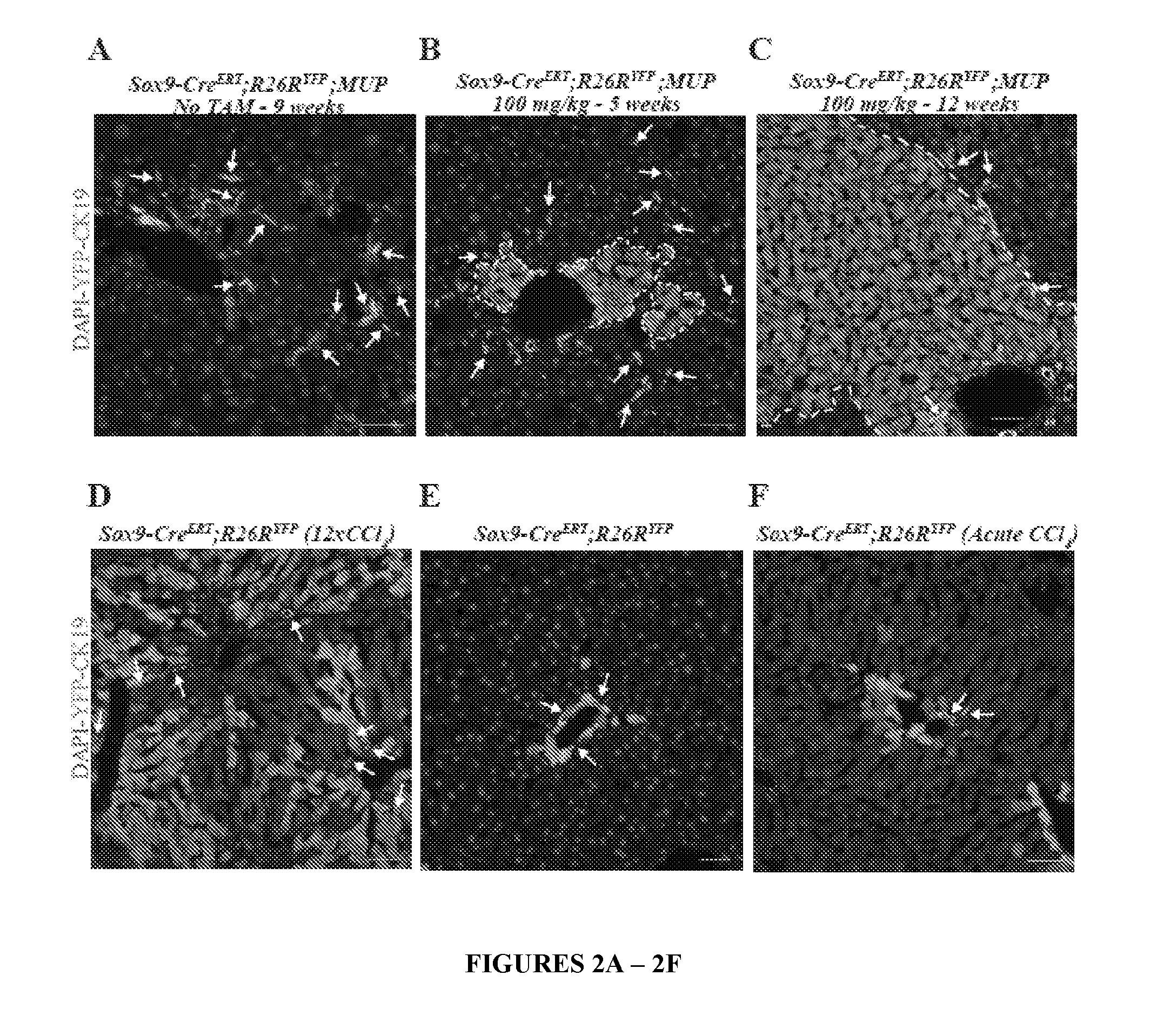
[0023] FIG. 2. HybHP make a major contribution to parenchymal restoration after liver injury. (A) Sox9-Cre.sup.ERT;R26R.sup.YFP;MUP-uPA mice (9 weeks old) were analyzed as in FIG. 1. 99.9% YFP.sup.+ labeled (green) cells are ductal/oval CK19.sup.+ cells (red). (B,C) Sox9-Cre.sup.ERT;R26R.sup.YFP;MUP-uPA mice were injected with 100 mg/kg tamoxifen at P10 and analyzed as above at 5 weeks (2 weeks after damage onset) (B) or at 12 weeks (C). (D) Tamoxifen (100 mg/kg)-injected, Sox9-Cre.sup.ERT;R26R.sup.YFP mice (8 weeks old), received 2 weekly CCl.sub.4 injections for 6 weeks. Livers were excised and analyzed as above. YFP.sup.+ HybHP (green) occupied a large portion of the liver parenchyma. (E). Without CCl.sub.4 treatment YFP.sup.+ cells did not expand. (F) Acute CCl.sub.4 administration resulted in a minor HybHP YFP.sup.+ cell expansion after 4 weeks. Arrows, ductal cells (CK19.sup.+, red). Scale bar, 50 .mu.m.
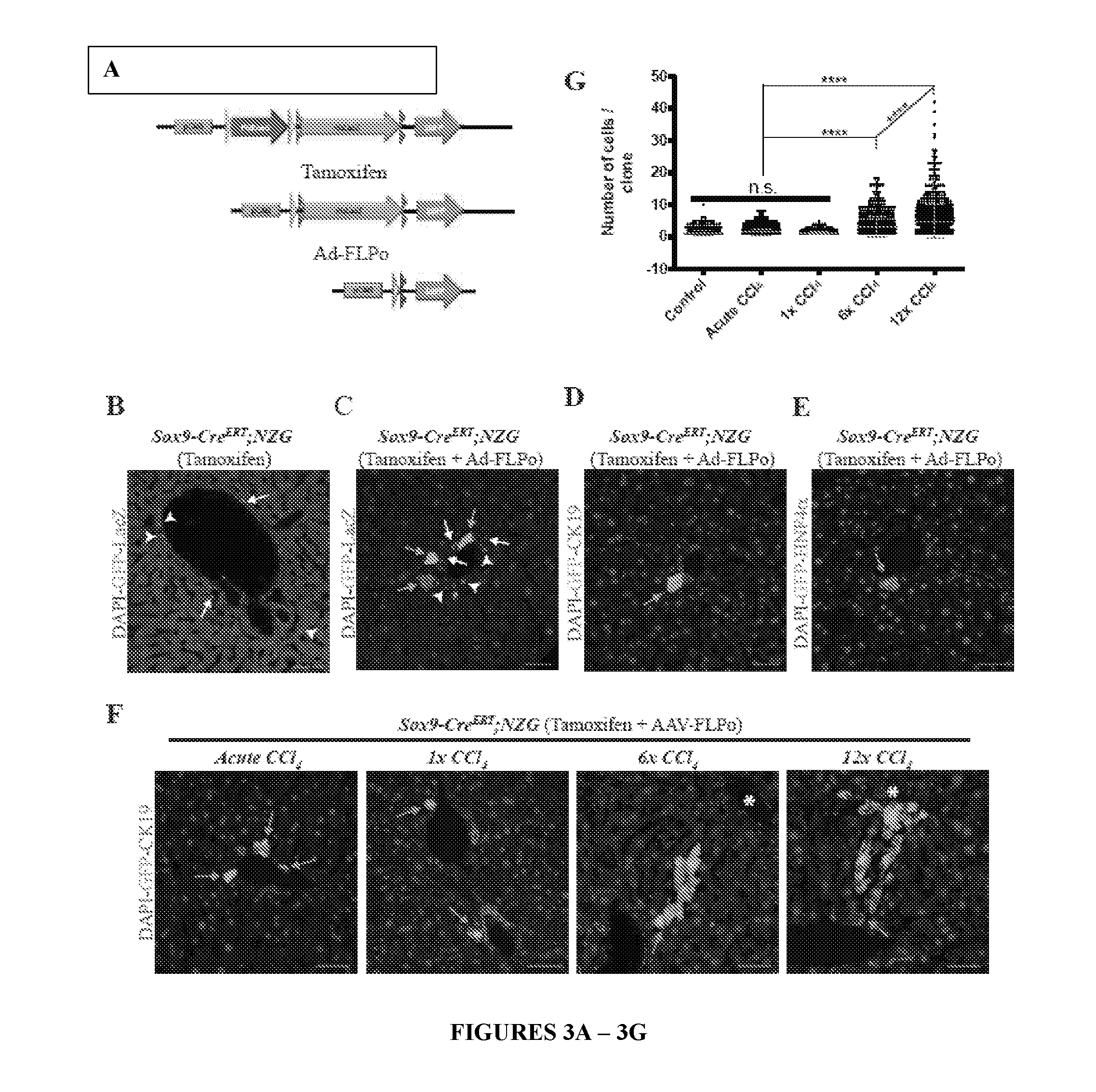
[0024] FIG. 3. Clonal labeling confirms HybHP role in liver injury repair. (A) A scheme outlining clonal labeling of HybHP with dual recombinase NZG reporter. A nuclear LacZ marker cassette is first expressed upon removal of the loxP (yellow arrows) flanked STOP cassette (PGK-Neo). A GFP marker is then expressed upon sequential Cre- and FLPo-dependent excisions of the STOP and Frt-flanked LacZ cassettes, respectively. (B) Sox9-Cre.sup.ERT;NZG mice (4-6 weeks old) were injected with 100 mg/kg of tamoxifen and 10 days later given 20 mg/kg tamoxifen. Ductal cells and HybHP were positive for nuclear LacZ (arrows and arrowheads, respectively), but not a single cell was GFP positive. (C-E) The same mice were first treated with tamoxifen as above, and then were injected with 10.sup.9 or 5.times.10.sup.11 Adeno- or AAV-FLPo viral particles, respectively. (C) After 2 weeks, livers were analyzed. Some HybHP were GFP.sup.+ and nuclear LacZ- (green arrows). (D) Liver sections were stained for CK19. HybHP are CK19.sup.-. (E) Liver sections were stained for HNF4.alpha.. GFP.sup.+ HybHP are HNF4.alpha..sup.+. (F) Sox9-Cre.sup.ERT;NZG mice were tamoxifen treated followed by AAV-FLPo administration and challenged with an acute dose of CCl.sub.4, or 1, 6 or 12 low CCl.sub.4 doses. Livers were excised and analyzed as in (D). (G) Quantification of cell number per clone in each experimental group. No treatment: 750 independent clones, n=3; acute CCl.sub.4: 939 independent clones, n=3; 1 low dose: 469 independent clones, n=3; 6 low doses, 626 independent clones, n=3; 12 low doses: 756 independent clones, n=4. ****=P value<0.0001 based on one way Anova with multiple comparisons. Scale bar, 50 .mu.m. Asterisks--central veins. n.s.--non significant.
[0025] FIG. 4. HybHP can generate ductal cells upon cholestatic injury. Sox9-Cre.sup.ERT;NZG mice were treated with tamoxifen and injected with Adeno-FLPo as in FIG. 3, and placed on DDC diet 3 weeks later. After 6 weeks, the fate of HybHP was analyzed by co-staining for Opn, Sox9, HNF4.alpha. and CK19. Arrows depict HybHP positive for ductal markers (Sox9, Opn and CK19) and negative for the hepatocyte marker HNF4.alpha.. Arrowheads denote HybHP negative for ductal markers and positive for HNF4.alpha.. Graphs show percentages of GFP.sup.+ HybHP positive for CK19 (n=1260), Sox9 (n=513) or Opn (n=994) and negative for HNF4.alpha. (n=676). In non-treated mice, no GFP.sup.+ HybHP positive for CK19 (n=551), Sox9 (n=232) or negative for HNF4.alpha. (n=323) were found. Only 0.72% of GFP.sup.+ HybHP were positive for Opn (n=556). Bracketed scale bar, 20 .mu.m; Open scale bar, 50 .mu.m.
[0026] FIG. 5. The HybHP transcriptome confirms their hybrid character. (A) Conventional hepatocytes (cHP), bile duct cells (BD) and Hybrid Hepatocytes (HybHP) from a liver collagenase digest were FACS separated after exclusion of dead cells and cell doublets and gating based on size/granularity (FCS/SSC) and tdTomato expression. (B) Total cellular RNA was extracted (3 independent isolations) and deep sequenced. Shown is a heatmap of genes that were differentially expressed in HybHP relative to cHP. (C) Proportion of genes that are differentially expressed in HybHP vs. cHP that show the same differential expression trend in BD vs. cHP. (D) Normalized expression values (in reads per kilobase per million) of the indicated genes in HybHP, cHP and BD. (E) IF analysis using Tyramide Signal Amplification (TSA) of Sox9-GFP transgenic mouse liver sections stained for Sox9 and Opn. White arrows: HybHP with weak Sox9 and Opn expression. (F) Normal human liver sections stained for Sox9 and Opn. Black arrows: periportal hepatocytes with weak expression of either marker. White arrows: ductal cells. Scale bars, 20 .mu.m.
[0027] FIG. 6. HybHP exhibit higher repopulation capacity than conventional hepatocytes or oval cells. (A) HybHP and conventional HP were FACS sorted from Sox9-Cre.sup.ERT;R26R.sup.tdTomato mice (8-12 weeks old), and 45,000 cells were transplanted into Fah.sup.-/-;Rag2.sup.-/-;Il2rg.sup.-/- mice (n=3 for conventional HP and n=2 for HybHP). Oval cells (OC; 45,000) were obtained by FACS sorting from a Sox9-Cre.sup.ERT;R26R.sup.tdTomato mouse fed with CDE diet for 3 weeks and transplanted into 3 Fah.sup.-/-;Rag2.sup.-/-;Il2rg.sup.-/- mice. Brightfield and tdTomato images of the medial lobes of non-transplanted and HybHP-, HD- and OC-transplanted livers 8 weeks after transplantation. Scale bar 5 mm. (B) Frozen sections of above livers were analyzed by direct fluorescence for tdTomato and IF for FAH expression. OC clones formed by tdTomato.sup.+ cells do not exhibit FAH expression. HybHP formed clones of tdTomato.sup.+ and FAH.sup.+ hepatocytes, whereas conventional HP gave rise to clones of tdTomato.sup.- and FAH.sup.+ hepatocytes. (C) Upper graph: clonal area quantification in 3 conventional HP and 2 HybHP transplanted liver. OC clones were not quantified due to their small number and size. ****=Unpaired t test with Welch's correction Pv<0.0001. cHP clones, n=500 and HybHP clones, n=695. Every measured clone with the mean and S.D. is shown. Lower graph: areas of all clones in frozen liver sections from (B). ****=Unpaired t test with Welch's correction P<0.0001. cHP clones, n=132 and HybHP clones, n=113. Every measured clone with mean and S.D. is shown. (D) Liver sections from HybHP transplanted mice in (B) stained for GS. White arrow: tdTomato.sup.+ GS.sup.+ HybHP in a HybHP clone without GS expression, surrounded by a damaged parenchyma with diffuse GS expression. Scale bar: 100 .mu.m. (E) Survival curves of an independent cohort of mice transplanted with HybHP (n=4), NH (n=7) or non-transplanted controls (n=9). P values were determined by Log-rank (Mantel-Cox) test. Black polygons over x-axis represent periods while the animals were on NTBC during the on-off NTBC cycles.
[0028] FIG. 7. Neither HybHP nor oval cells preferentially give rise to HCC. (A) Experimental design for HybHP and OC lineage tracing in three different HCC models using Sox9-Cre.sup.ERT;R26R.sup.YFP or Sox9-Cre.sup.ERT;R26R.sup.YFP;MUP-uPA mice. (B-D) Ki67 immunofluorescence and morphology were used to locate tumor nodules (delineated by a dashed line) in whole slide scans. None of the tumor areas contained YFP.sup.+ cancer cells. Three representative examples are shown for each HCC model. Scale bars, (B), 0.5 mm; (A,C) 1 mm.
[0029] FIG. 8. HybHP are present in both males and females and remain quiescent throughout their lives. Related to FIG. 1. (A) Transgenic Sox9-GFP males and females were analyzed at different ages by IF for GFP, CK19 and HNF4.alpha.. (B) Sox9-Cre.sup.ERT;R26R.sup.YFP mice were treated with 100 mg/kg of tamoxifen at P10 and analyzed at 2 and 9 months of age by IF for GFP, CK19 and HNF4.alpha.. Scale bar, 50 .mu.m. Arrows--BD; arrowheads--HybHP. (C) Direct visualization of tdTomato fluorescence (red) in DAPI (blue) stained liver sections from a Sox9-Cre.sup.ERT;R26Rtd.sup.Tomato mouse injected with 100 mg/kg and 20 mg/kg tamoxifen, 10 days apart from each other. Arrows: ductal cells; arrowheads: HybHP. (D) Two additional independent portal tracts from the same clarified liver from FIG. 1 G,H.
[0030] FIG. 9. New hepatocytes generated from HybHP acquire metabolic zonation after expansion. Related to FIG. 2. (A) Whole slide scans of liver sections from tamoxifen treated Sox9-Cre.sup.ERT;MUP;R26R.sup.YFP mice at 6, 7 and 9 weeks of age stained with GFP antibodies (green). Arrows indicate expanding HybHP clones whereas arrowheads indicate portal tracts devoid of HybHP but with an oval cell response. (B) Immunostaining for YFP (green) and glutamine synthetase (GS, red) of liver sections from the Sox9-Cre.sup.ERT;R26R.sup.YFP;MUP-uPA tamoxifen treated mice shown in FIG. 2A. (C) Immunostaining for YFP (green) and GS (red) of liver sections from the same Sox9-Cre.sup.ERT;R26R.sup.YFP tamoxifen treated mice shown in FIG. 2B. Scale bars, 50 .mu.m. Arrows--HybHP-derived GS.sup.+ hepatocytes.
[0031] FIG. 10. Genetic clonal analysis of hepatocytes reveals predominant clonal growth from the portal tract. Related to FIG. 2. Whole slide scans of IF images of YFP-, Sox9- and DAPI-stained liver sections from: (A) TTR-Cre.sup.ERT;R26R.sup.YFP mice that were injected with 100 mg/kg tamoxifen at P7-10. Scattered hepatocytes were labeled with YFP (green). When adult animals were tamoxifen injected at 6 weeks or later no ductal cells were labeled, whereas tamoxifen injection at P7-10 resulted in 0.5% of ductal cells (Sox9.sup.+) becoming YFP.sup.+, although most portal tracts (90%) lacked YFP.sup.+ cells. (B) Livers of TTR-Cre.sup.ERT;R26R.sup.YFP;MUP-uPA mice without tamoxifen treatment. No YFP.sup.+ cells were detected. (C) TTR-Cre.sup.ERT;R26R.sup.YFP;MUP-uPA mice were injected with 100 mg/kg of tamoxifen at P7, sacrificed at 5 weeks of age and analyzed as above. (D) Same mice as above that were sacrificed and analyzed at 8 weeks of age. At that point most labeled hepatocytes were eliminated and clonal expansion was mainly seen in the portal region. Arrows: parenchymal hepatocyte clones; arrowheads: clones initiated at periportal areas. (E) Livers of TTR-Cre.sup.ERT;R26R.sup.YFP mice injected with 100 mg/kg tamoxifen at 6 weeks of age contain scattered YFP hepatocytes (green). (F) Livers of TTR-Cre.sup.ERT;R26R.sup.YFP mice treated with biweekly CCl.sub.4 injections for 6 weeks. Several extended clones contacting the portal tract are highlighted with arrows. Asterisks--central veins.
[0032] FIG. 11. Lineage tracing of CK19.sup.+ cells in MUP-uPA mice and genetic labeling with Alb-Cre.sup.ERT. Related to FIG. 2. (A,B) CK19-Cre.sup.ERT;R26R.sup.YFP;MUP-uPA mice were injected twice with 100 mg/kg of tamoxifen at P7 and P10. At 8 weeks of age, livers were excised, sectioned and stained with GFP and CK19 antibodies (A) or GFP and HNF4.alpha. antibodies (B). For each staining combination, two different regions are shown. Arrows--oval cells. (C) IF analysis of livers from tamoxifen treated (5 mg/kg or 100 mg/kg) Alb-Cre.sup.ERT;R26R.sup.YFP mice. CK19.sup.+/YFP.sup.+ double positive cells are marked by arrows. Scale bar, 50 .mu.m.
[0033] FIG. 12. Flow cytometric analysis of sorted HybHP, cHP and BD populations. Related to FIG. 5. (A) Flow cytometric analysis of cells prepared by collagenase perfusion of livers from WT and Sox9-Cre.sup.ERT;R26R.sup.YFP mice treated with 100 mg/kg tamoxifen. The brightness of the YFP marker was insufficient to allow separation of the positive and negative populations for reliable purification. (B) Flow cytometric analysis and gating scheme for sorting of HybHP and cHP from cell suspensions prepared by collagenase perfusion of livers from Sox9-Cre.sup.ERT;R26Rtd.sup.Tomato mice treated as in (A). After exclusion of doublets and dead cells using fixable viability dye (left panel), vital cells (62%) were further gated based on size/granularity (middle, cHP gate). tdTomato (tdT) positive cells indicated HybHP (right). (C,D) To confirm the gating scheme for cHP and HybHP, the cells were stained with antibodies specific for HNF4.alpha. (C) and CK19 (D), showing that cHP and HybHP expressed HNF4.alpha. but were negative for CK19 (purity 97%). The corresponding isotype controls are shown in the left panels. (E) Flow cytometric analysis and gating scheme for sorting of isolated BD cells from same animals as in (B). After exclusion of doublets and dead cells using fixable viability dye (left panel), vital cells (83.5%) were further gated based on size/granularity (middle, BD gate). tdTomato (tdT) positive cells indicated BD (right). (F,G) To confirm the gating scheme for BD cells, the cells were stained for HNF4.alpha. (F) and CK19 (G), showing that BD cells did not express HNF4.alpha. and were positive for CK19 (purity of 97%). The corresponding isotype controls are shown in left panels.
[0034] FIG. 13. HybHP express a few ductal-specific genes and numerous hepatocyte-specific genes. Related to FIG. 5. (A) Clustering analysis showing distances based on normalized Reads per Kilobase per Million base pairs (RPKM) values from the three populations analyzed by Hclust with R. (B) The four classes of differentially expressed genes shown in panel B were analyzed according to function. Selection of the top functional entries by Benjamini corrected P value<0.05 are displayed. Class 3 did not show any functional enrichment and is not depicted. (C) Livers from WT animals were stained with Sox9 and Opn antibodies and visualized with TSA (red) and regular staining for CK19 (green). Arrows show HybHP. Scale bars, 20 .mu.m. (D) TSA-immunofluorescence using antibodies for the proteins indicated in red and regular staining for GFP in Sox9-GFP transgenic mouse liver sections. Arrows indicate HybHP expressing the corresponding markers whereas arrowheads indicate normal hepatocytes expressing the same marker. Scale bars, 50 .mu.m. (E) A hypothetical model illustrating the hybrid nature of HybHP. The two curves show profiles of bile duct-specific and zone 1 hepatocyte-specific genes.
[0035] FIG. 14. The relationship between HybHP expansion and the oval cell response. Related to FIG. 7. (A) Portion of a whole slide scan from Sox9-Cre.sup.ERT;R26R.sup.YFP;MUP mice used in FIGS. 2 and 9, stained with DAPI (blue), YFP (green) and CK19 (red). (B) Portion of a whole slide scan of STAM mouse liver, stained as in (A). (C) Portion of a whole slide scan of liver from Sox9-Cre.sup.ERT;R26R.sup.YFP mice treated with 100 mg/kg of tamoxifen and fed with CDE diet for 3 weeks and stained as in (A). Arrows--tissue areas with clonal expansion of HybHP; arrowheads--regions showing oval cell response but no HybHP clonal expansion. Scale bars: B, 250 .mu.m; A and C, 500 .mu.m. Asterisks--central veins.
[0036] FIG. 15. RNAseq statistical analysis. Related to FIG. 5. (A,B) Quantile-quantile plots comparing the expected value of chi-squared goodness-of-fit statistics (x-axis) with their observed values (y-axis) for gene-level counts. (A) Poisson distribution; (B) Negative Binomial distribution. (C) Quantile-quantile plot comparing expected z-scores (x-axis) under the Normal distribution with those from the observed normalized log 2-RPKM. Black dots indicate the average observed quantile, and the straight line the expected quantile if gene-level expressions truly followed a Normal distribution. (D) Quantile-quantile plot comparing expected quantiles under a Gamma distribution (x-axis) with the observed normalized log 2-RPKM values (y-axis). (E) Principal components plot of the observed log 2-RPKM expression. (F) Principal components plot with observed log 2-RPKM data and that simulated from the GaGa model (grey) under the null hypothesis that expression is equal in the HybHP and cHP groups for all genes. (G) Density plot for GaGa simulated ratios of within/between groups distance (rdist) under the null hypothesis of no differences between HybHP-cHP groups. Dashed line indicates observed rdist=0.83. (H) Histogram of percentage of genes estimated by GaGa to arise from Patterns 2-4 when data are simulated from the null hypothesis by the GaGa model. Dashed line indicates the 0.3% estimated in the observed data. (I) Same as in (F) but for simulated log 2-RPKM computed from counts generated under the Tweedie model. (J) Same as in (G) using the Tweedie model to simulate data under the null hypothesis. (K) Same as in (H) using the Tweedie model to simulate data under the null hypothesis. (L) Fold changes BDg/cHPg (x-axis) versus HybHPg/cHPg (y-axis) in the observed data for 770 genes with HybHPg/cHPg above 2 or below -2, read count >10 in the cHP group and total read count >100. Blue boundaries correspond to lower and upper boundaries of intervals containing 95% of the HybHPg(b)/cHPg(b) values simulated under the mixture hypothesis.
[0037] FIG. 16. Conventional hepatocytes (cHP), Hybrid Hepatocytes (HybHP) and Bile duct cells (BD) were sorted from liver homogenates of tamoxifen treated Sox9CreERT;ROSAtdTomato mice. Single cells from the three populations were seeded in matrigel and organoid media and imaged at different time points after seeding.
DEFINITIONS
[0038] "Hepatocyte cells" are highly polarized epithelial cells and form cords. Hepatocytes account for approximately 50% of total liver cells and 80% of the volume of the organ. Their basolateral surfaces face fenestrated sinusoidal endothelial cells, facilitating the exchange of materials between hepatocytes and blood vessels. Tight junctions formed between hepatocytes create a canaliculus that surrounds each hepatocyte. Bile secreted from mature hepatocytes is exported sequentially through bile canaliculi surrounded by the apical membrane of neighboring hepatocytes, intrahepatic bile ducts, extrahepatic bile ducts, and, finally, the duodenum. The bile duct is formed by a specialized type of epithelial cell called a cholangiocyte. The term "hepatocyte" cell includes conventional hepatocyte (cHP) cells, and the invention's newly discovered hybrid hepatocyte (HybHP) cells.
[0039] "Conventional hepatocyte cell" and "cHP cell" interchangeably refer to a hepatocyte cell that expresses at least one protein marker encoded by the genes in text that is neither in bold nor in italics in Table 2. The cHP cells co-migrate with HybHP cells, and differentially migrates from ductal cells, on density gradients (Examples 1, 2).
[0040] "Ductal cell" and "DC cell" interchangeably refer to a liver cell that expresses at least one protein marker encoded by the genes in bold italic text in Table 1 and Table 3. DC cells differentially migrates from cHP cells and from HybHP cells on density gradients (Examples 1, 2). Ductal cells include oval cells (OCs) and bile duct (BD) cells.
[0041] "Oval cell" is an ovoid cell observed in animal models of liver carcinogenesis (Farber, E. (1956) Cancer Res. 16, 142-148), and is sometimes introduced in literature as a synonym for liver stem cells. Oval cells have been extensively characterized histologically, which cumulatively suggests that they have bipotential differentiation capability toward both hepatocytes and cholangiocytes (Miyajima et al., 2014).
[0042] "Bile duct" cells (also referred to as "BD" cells) are located in the bile duct.
[0043] "Density gradient" is a spatial variation in density over an area. "Density gradient centrifugation" is a procedure for separating cells in which the sample is placed on a preformed gradient (e.g., percoll, Stractan, etc.). Upon centrifugation either by rate zonal or equilibrium procedures, the cells are banded in the gradient and can be collected as a purified fraction.
[0044] "Purified," "purify," "isolate," and grammatical equivalents thereof when in reference to a desirable component (such as cell type, protein, nucleic acid sequence, etc.) refer to the reduction in the amount of at least one undesirable component (such as another cell type, protein, nucleic acid sequence, etc.) from a sample. This reduction may be by any numerical percentage of from 5% to 100%, such as, but not limited to, from 10% to 100%, from 20% to 100%, from 30% to 100%, from 40% to 100%, from 50% to 100%, from 60% to 100%, from 70% to 100%, from 80% to 100%, from 90% to 100%, from 95% to 100%, from 99% to 100%, and most preferably by 100%. Thus, purification results in "enrichment" (i.e., an increase) in the amount of the desirable component relative to one or more undesirable component. For example, "purified HybHP cells" refers to HybHP cells that are purified from cHP cells and from DC cells. Preferably, the purified HybHP cells are substantially free from cHP cells and from DC cells. In another example, "purified first antibody-HybHP cell-second antibody conjugate" and "purified first-antibody-HybHP cell conjugate" refer to conjugates containing purified HybHP cells. Methods for purifying of single cells are known in the art, including fluorescence-activated cell sorting (FACS), magnetic-activated cell sorting (MACS), etc. Single-cell purification methods (such as FACS, MACS, etc.) may employ direct labeling of the antigen of interest using a primary antibody that specifically binds to the antigen of interest. Alternatively, purification methods employ indirect labeling of the antigen of interest using a secondary antibody that specifically binds to the Fc portion of the primary antibody, biotin, and/or to fluorescein isothiocyanate (FITC). Indirect labeling may also use Streptavidin, instead of the secondary antibody, which binds to biotin on the primary antibody.
[0045] "Fluorescence-activated cell sorting" and "FACS" interchangeably refer to a method for sorting a heterogeneous mixture of biological cells based upon the specific light scattering and fluorescent characteristics of each cell, thus purifying cells of interest (e.g., HybHP cells). FACS is particularly preferred for isolating a target cell population (e.g., HybHP cells) at high purity, when the target cell population expresses a very low level of the identifying marker or when cell populations require separation based on differential marker density. In addition, FACS is a particularly desired for technique to isolate cells based on internal staining and/or intracellular protein expression, such as a genetically modified fluorescent protein marker. FACS allows the purification of individual cells based on size, granularity and fluorescence. In order to purify cells of interest, they are first stained with a fluorescently-tagged primary antibody that recognizes specific surface markers on the desired cell population. Negative selection of unstained cells is also possible. Basu et al. (2010) "Purification of Specific Cell Population by Fluorescence Activated Cell Sorting (FACS)" J Vis Exp. (41): 1546. doi: 10.3791/1546.
[0046] "Magnetic-activated cell sorting" and "MACS" interchangeably refer to a method for purifying various cell populations depending on their surface antigens. In one embodiment, MACS uses nanoparticles conjugated to an antibody against an antigen of interest (e.g., on the surface of HybHP cells). In another embodiment, MACS uses magnetic nanoparticles coated with anti-fluorochrome antibodies that are incubated with the fluorescent-labelled antibodies against the antigen of interest for cell separation with respect to the antigen (such as by using anti-immunoglobulin MicroBeads, anti-biotin MicroBeads, streptavidin MicroBeads, and anti-fluorochrome MicroBeads).
[0047] The term "population of cells" refers to a plurality of cells, i.e., more than one cell. The population may be a pure population comprising one cell type. Alternatively, the population may comprise more than one cell type. In the present invention, there is no limit on the number of cell types that a cell population may comprise.
[0048] Cell "marker molecule" refer to a molecule (such as protein, nucleotide sequence, etc.) that is present on, and/or is produced by, a particular type of cell (such as cancer cell, epithelial cell, fibroblast cell, muscle cell, synovial cell, stem cell, embryonic cell, etc.), at a different level (e.g., a higher level or lower lever, preferably a higher level) than other types of cells. Cell marker molecules may be used to distinguish one cell type of one or more other cell types.
[0049] "Mammal" includes a human, non-human primate, murine (e.g., mouse, rat, guinea pig, hamster), ovine, bovine, ruminant, lagomorph, porcine, caprine, equine, canine, feline, ave, etc. In one preferred embodiment, the mammal is murine. In another preferred embodiment, the mammal is human.
[0050] A subject "in need" of treatment with the invention's methods and/or compositions includes a subject that is "suffering" from liver damage (i.e., a subject that is experiencing and/or exhibiting one or more symptoms of liver damage), and subject "at risk" of liver damage. A subject "in need" of treatment includes animal models of liver damage. Subject "at risk" of liver damage refers to a subject that is not currently exhibiting liver damage symptoms and is predisposed to expressing one or more symptoms of the disease. This predisposition may be based on family history, genetic factors, environmental factors such as exposure to detrimental compounds present in the environment, etc.). It is not intended that the present invention be limited to any particular signs or symptoms. Thus, it is intended that the present invention encompass subjects that are experiencing any range of disease, from sub-clinical symptoms to full-blown disease, wherein the subject exhibits at least one of the indicia (e.g., signs and symptoms) associated with the disease.
[0051] The term "conjugate" when in reference to a cell and antibody refers to a cell that is linked (directly or indirectly) to an antibody via an antigen that is expressed on the cell surface. Antibodies bind antigens through weak chemical interactions, and bonding is essentially non-covalent, such as electrostatic interactions, hydrogen bonds, van der Waals forces, and hydrophobic interactions. For example a "First-antibody-HybHP cell conjugate" refers to a HybHP cell that is linked to a first antibody via an antigen that is expressed on the surface of the HybHP cell. Also, a "HybHP cell-first antibody-second antibody conjugate" refers to a HybHP cell that is linked to a first antibody via a first antigen that is expressed on the surface of the HybHP cell, and also linked to a second antibody via a second antigen that is expressed on the surface of the HybHP cell.
[0052] The terms "specifically binds," "specific binding," and grammatical equivalents, when made in reference to the binding of antibody to a molecule (e.g., peptide) refer to an interaction of the antibody with one or more epitopes on the molecule where the interaction is dependent upon the presence of a particular structure on the molecule. For example, if an antibody is specific for epitope "A" on the molecule, then the presence of a protein containing epitope A (or free, unlabeled A) in a reaction containing labeled "A" and the antibody will compete with, and thus reduce, the amount of labeled A bound to the antibody.
[0053] "Overexpression," "upregulation," and grammatical equivalents, when used in reference to a protein in a cell of interest (such as a HybHP cell, cHP cell, DC cell, etc.) refers to the presence of a higher level of the protein, and/or its encoding mRNA, in the cell of interest compared to another cell (such as a control HybHP cell, cHP cell, DC cell, etc.).
[0054] "Underexpression," "downregulation," and grammatical equivalents, when used in reference to a protein in a cell of interest (such as a HybHP cell, cHP cell, DC cell, etc.) refers to the presence of a lower level of the protein, and/or its encoding mRNA, in the cell of interest compared to a another cell (such as a control HybHP cell, cHP cell, DC cell, etc.).
[0055] The terms "increase," "elevate," "raise," and grammatical equivalents (including "higher," "greater," etc.) when in reference to the level of any molecule (e.g., amino acid sequence, and nucleic acid sequence, antibody, etc.), cell, and/or phenomenon (e.g., level of expression of a gene, disease symptom, level of binding of two molecules such as binding of a ligand to a receptor, specificity of binding of two molecules (such as an antigen and antibody), affinity of binding of two molecules, disease symptom, specificity to disease, sensitivity to disease, affinity of binding, enzyme activity, etc.) in a first sample (or in a first subject) relative to a second sample (or relative to a second subject), mean that the quantity of the molecule, cell, and/or phenomenon in the first sample (or in the first subject) is higher than in the second sample (or in the second subject) by any amount that is statistically significant using any art-accepted statistical method of analysis. In one embodiment, the quantity of the molecule, cell, and/or phenomenon in the first sample (or in the first subject) is at least 10% greater than, at least 25% greater than, at least 50% greater than, at least 75% greater than, and/or at least 90% greater than the quantity of the same molecule, cell, and/or phenomenon in the second sample (or in the second subject). This includes, without limitation, a quantity of molecule, cell, and/or phenomenon in the first sample (or in the first subject) that is at least 10% greater than, at least 15% greater than, at least 20% greater than, at least 25% greater than, at least 30% greater than, at least 35% greater than, at least 40% greater than, at least 45% greater than, at least 50% greater than, at least 55% greater than, at least 60% greater than, at least 65% greater than, at least 70% greater than, at least 75% greater than, at least 80% greater than, at least 85% greater than, at least 90% greater than, and/or at least 95% greater than the quantity of the same molecule, cell, and/or phenomenon in the second sample (or in the second subject). In one embodiment, the first sample (or the first subject) is exemplified by, but not limited to, a sample (or subject) that has been manipulated using the invention's compositions and/or methods. In a further embodiment, the second sample (or the second subject) is exemplified by, but not limited to, a sample (or subject) that has not been manipulated using the invention's compositions and/or methods. In an alternative embodiment, the second sample (or the second subject) is exemplified by, but not limited to, a sample (or subject) that has been manipulated, using the invention's compositions and/or methods, at a different dosage and/or for a different duration and/or via a different route of administration compared to the first subject. In one embodiment, the first and second samples (or subjects) may be the same, such as where the effect of different regimens (e.g., of dosages, duration, route of administration, etc.) of the invention's compositions and/or methods is sought to be determined on one sample (or subject). In another embodiment, the first and second samples (or subjects) may be different, such as when comparing the effect of the invention's compositions and/or methods on one sample (subject), for example a patient participating in a clinical trial and another individual in a hospital.
[0056] The terms "reduce," "inhibit," "diminish," "suppress," "decrease," and grammatical equivalents (including "lower," "smaller," etc.) when in reference to the level of any molecule (e.g., amino acid sequence, and nucleic acid sequence, antibody, etc.), cell, and/or phenomenon (e.g., level of expression of a gene, disease symptom, level of binding of two molecules such as binding of a ligand to a receptor, specificity of binding of two molecules (such as an antigen and antibody), affinity of binding of two molecules, disease symptom, specificity to disease, sensitivity to disease, affinity of binding, enzyme activity, etc.) in a first sample (or in a first subject) relative to a second sample (or relative to a second subject), mean that the quantity of molecule, cell, and/or phenomenon in the first sample (or in the first subject) is lower than in the second sample (or in the second subject) by any amount that is statistically significant using any art-accepted statistical method of analysis. In one embodiment, the quantity of molecule, cell, and/or phenomenon in the first sample (or in the first subject) is at least 10% lower than, at least 25% lower than, at least 50% lower than, at least 75% lower than, and/or at least 90% lower than the quantity of the same molecule, cell, and/or phenomenon in the second sample (or in the second subject). In another embodiment, the quantity of molecule, cell, and/or phenomenon in the first sample (or in the first subject) is lower by any numerical percentage from 5% to 100%, such as, but not limited to, from 10% to 100%, from 20% to 100%, from 30% to 100%, from 40% to 100%, from 50% to 100%, from 60% to 100%, from 70% to 100%, from 80% to 100%, and from 90% to 100% lower than the quantity of the same molecule, cell, and/or phenomenon in the second sample (or in the second subject). In one embodiment, the first sample (or the first subject) is exemplified by, but not limited to, a sample (or subject) that has been manipulated using the invention's compositions and/or methods. In a further embodiment, the second sample (or the second subject) is exemplified by, but not limited to, a sample (or subject) that has not been manipulated using the invention's compositions and/or methods. In an alternative embodiment, the second sample (or the second subject) is exemplified by, but not limited to, a sample (or subject) that has been manipulated, using the invention's compositions and/or methods, at a different dosage and/or for a different duration and/or via a different route of administration compared to the first subject. In one embodiment, the first and second samples (or subjects) may be the same, such as where the effect of different regimens (e.g., of dosages, duration, route of administration, etc.) of the invention's compositions and/or methods is sought to be determined on one sample (or subject). In another embodiment, the first and second samples (or subjects) may be different, such as when comparing the effect of the invention's compositions and/or methods on one sample (subject), for example a patient participating in a clinical trial and another individual in a hospital.
[0057] The terms "alter" and "modify" when in reference to the level of any molecule (e.g., amino acid sequence, and nucleic acid sequence, antibody, etc.), cell, and/or phenomenon (e.g., level of expression of a gene, disease symptom, level of binding of two molecules such as binding of a ligand to a receptor, specificity of binding of two molecules (such as an antigen and antibody), affinity of binding of two molecules, disease symptom, specificity to disease, sensitivity to disease, affinity of binding, enzyme activity, etc.) refer to an increase and/or decrease.
[0058] "Substantially the same," "without substantially altering," "substantially unaltered," and grammatical equivalents, when in reference to the level of any molecule (e.g., amino acid sequence, and nucleic acid sequence, antibody, etc.), cell, and/or phenomenon (e.g., level of expression of a gene, disease symptom, level of binding of two molecules such as binding of a ligand to a receptor, specificity of binding of two molecules (such as an antigen and antibody), affinity of binding of two molecules, disease symptom, specificity to disease, sensitivity to disease, affinity of binding, enzyme activity, etc.) means that the quantity of molecule, cell, and/or phenomenon in the first sample (or in the first subject) is neither increased nor decreased by a statistically significant amount relative to the second sample (or in a second subject). Thus in one embodiment, the quantity of molecule, cell, and/or phenomenon in the first sample (or in the first subject) is from 90% to 100% (including, for example, from 91% to 100%, from 92% to 100%, from 93% to 100%, from 94% to 100%, from 95% to 100%, from 96% to 100%, from 97% to 100%, from 98% to 100%, and/or from 99% to 100%) of the quantity in the second sample (or in the second subject).
[0059] The term "composition," such as when made in reference to a composition comprising a desired component (such as HybHP cell, first-antibody-HybHP cell conjugate, first antibody-HybHP cell-second antibody conjugate, etc.) refers to any container, receptacle, and/or medium that can receive and/or contain the desired component. Thus, the term composition includes aqueous solutions (such as culture medium), three-dimensional matrix (such as matrigel, collagen, agar, etc.), flask, petri dish, multi-well dish, etc.
[0060] The terms "propagating," "culturing," and "growing," when made in reference to target cells, are interchangeably used to refer to increasing the number of the target cells.
[0061] The term "in vivo" refers to the natural environment (e.g., within an organism, tissue, and/or a cell).
[0062] The term "ex vivo" refers to an environment outside an organism.
[0063] The term "in vitro" refers to an ex vivo environment that includes manipulation under artificially-created conditions (e.g., culture medium, cell culture, transfection, assay) and/or using laboratory equipment (e.g., flasks, test tubes, petri dishes, multiwell plates, etc.).
[0064] The term "culture medium" refers to a nutritive substance that is suitable to support maintenance and/or growth of cells in vitro (i.e., cell cultures). A culture medium includes, for example, liquid media, and three-dimensional media. A culture medium includes salts (e.g., sodium chloride), carbohydrates (e.g., sugar), proteins (e.g., serum), etc.
[0065] "Three-dimensional media," "3D media," and "three-dimensional matrix" interchangeably refer to an artificially-created environment (e.g., matrigel, collagen, agar, etc.) in which biological cells are permitted to grow or interact with their surroundings in all three dimensions. This is in contrast to growing cells in "two-dimensional" (2D) monolayers (e.g., on a petri dish) because the 3D model more accurately models the in vivo cell environment.
[0066] The term "treating" liver damage interchangeably refers to delaying, reducing, palliating, ameliorating, stabilizing, preventing and/or reversing one or more symptoms (such as objective, subjective, pathological, clinical, sub-clinical, etc.) of liver damage. Symptoms of liver damage may be assessed by, for example, biopsy and histology, and blood tests to determine levels of relevant enzymes (e.g., enzymes involved in metabolism, such as aspartate aminotransferase (AST), alanine aminotransferase (ALT), alkaline phosphatase, 5' Nucleotidase, gamma-glutamyl transpeptidase (GGT)), levels of proteins involved in normal blood clotting (e.g., prothrombin time (PT) using international normalized ratio (INR) and/or partial thromboplastin time (PTT)), levels of albumin and/or bilirubin, levels of circulating antigen and/or antibody, and imaging tests (e.g., to detect a decrease in the growth rate or size of hepatocellular carcinoma).
[0067] The terms "therapeutic amount," "pharmaceutically effective amount," and "therapeutically effective amount," are used interchangeably herein to refer to an amount that is sufficient to achieve a desired result, such as treating liver damage, and/or repopulating the liver.
[0068] "Antibody" refers to an immunoglobulin (e.g., IgG, IgM, IgA, IgE, IgD, etc.) and/or portion thereof that contain a "variable domain" (also referred to as the "F.sub.V region") that specifically binds to an antigen. More specifically, variable loops, three each on the light (V.sub.L) and heavy (V.sub.H) chains are responsible for binding to the antigen. These loops are referred to as the "complementarity determining regions" ("CDRs") and "idiotypes." In one embodiment, the invention's antibodies are "monoclonal antibodies" ("MAbs") produced by a single clone of hybridoma cells.
DESCRIPTION OF THE INVENTION
[0069] The invention provides the discovery of the existence of hybrid hepatocyte (HybHP) cells. These cells are superior to any known cell type in repairing diseased and/or injured liver. This new and distinct population of hepatocytes is located at the periportal region that expresses several bile duct enriched genes. Hybrid Hepatocytes expand upon liver injury and form larger and more stably differentiated clones than other hepatocytes, and can also transdifferentiate into bile duct cells. Data herein demonstrate that in Fah.sup.-/- mice, which suffer chronic and fatal liver injury (Grompe et al., 1995), HybHP show higher regenerative capacity than normal hepatocytes and are far superior to oval cells. Neither HybHP nor oval cells serve as the origin for HCC in three independent mouse models, including two models of NASH-driven HCC (Fujii et al., 2013; Nakagawa et al., 2014b) (FIG. 7). These results demonstrate that the HybHP cells with the highest proliferative potential in a given tissue, may not necessarily be the ones that give rise to cancer.
[0070] The invention's compositions and methods are useful in the treatment of liver damage, liver tissue engineering, transplantation back into the donor subjects to generate a HLA compatible HybHP cell biobank and/or to treat other subjects.
[0071] The invention is further described under A) Purified hybrid hepatocyte (HybHP) cells and compositions comprising the, B) Methods for isolating HybHP cells, C) Methods for in vitro culture of HybHP cells, and D) Methods for repopulating the liver and treating liver damage.
[0072] A) Purified Hybrid Hepatocyte (HybHP) Cells and Compositions Comprising them
[0073] Compensatory proliferation triggered by liver damage is required for liver regeneration and maintenance of mass and function, but is also a potent tumor promoter giving rise to hepatocellular carcinoma (HCC). Despite extensive investigation, the nature of the cells responsible for liver regeneration or HCC development remains obscure.
[0074] Data herein demonstrate that the primary cell type responsible for regeneration of the liver parenchyma is the newly described HybHP. Yet, despite their high regenerative potential and extensive expansion during chronic liver injuries, which promote HCC development (Hui et al., 2007; Maeda et al., 2005; Nakagawa et al., 2014b), data herein show that HybHP are not the preferential origin for HCC in the 3 different liver cancer models we have examined herein. Thus, in contrast to the skin and the intestine, the liver does not contain a specialized compartment of adult or reserve stem cells that maintains tissue homeostasis and expands upon liver damage to restore tissue mass. A recent analysis of relative cancer rates at different tissue sites revealed a correlation with rates of cell division in most tissues and gave rise to the suggestion that the majority of cancer originates from adult tissue stem cells (Tomasetti and Vogelstein, 2015). This does not seem to apply to liver, in which HybHP generate new hepatocytes only upon liver damage but rarely give rise to cancer.
[0075] i) Hybrid Hepatocytes as a New Hepatocyte Subpopulation
[0076] Hepatocyte diversity has been long appreciated, especially in the context of metabolic zonation across the portal to central axis (Jungermann and Katz, 1989). However, it has been difficult to analyze the distinct properties of different hepatocyte populations under physiological conditions, although uni-zonal livers were generated through genetic manipulation of the Wnt-.beta.-catenin signaling pathway (Benhamouche et al., 2006). Using the ductal transcription factor Sox9 as a marker, we identified a subpopulation of periportal hepatocytes located at the limiting plate, which express low amounts of Sox9 and normal amounts of HNF4.alpha., a hepatocyte transcription factor. These cells were termed HybHP, a term fully supported by subsequent transcriptomic and immunohistochemical analyses, which show expression of hepatocyte-specific genes along with a small number of genes that are preferentially expressed in bile duct cells. Using a Sox9-Cre.sup.ERT line that is sensitive to low amounts of Sox9 gene transcription, we labeled HybHP and traced their fate under different conditions. HybHP are kept quiescent in the unchallenged liver for at least 9 months after birth, but during chronic liver damage, they proliferate and serve as a source for a large fraction of the new hepatocytes that repopulate the liver. Interestingly, in the two injury models we employed, the manner in which HybHP re-seed the liver appears to be different. In MUP-uPA mice, extensive damage is randomly produced throughout the parenchyma consequent to ER stress (Nakagawa et al., 2014b; Weglarz et al., 2000). In these mice, HybHP clones radiate from the portal area, covering as much volume as possible. Chronic CCl.sub.4 administration, on the other hand, results in necrotic damage to pericentral zone 3 hepatocytes. In this case, HybHP clones expand along the portal-central axis, following hepatic chords. These observations suggest that tissue polarity and polarized cell-cell interactions determine the pattern of tissue repair.
[0077] Many of the genes that are over-expressed in HybHP relative to conventional hepatocytes are also expressed in bile duct cells and are functionally related to cell adhesion and tubule formation, suggesting that HybHP and bile duct cells may originate from embryonic ductal plate progenitors (Carpentier et al., 2011). Perhaps ductal plate progenitors give rise to mature bile duct cells and HybHP which remain attached to duct cells via homotypic interactions, in clear contrast to parenchymal hepatocytes which originate from hepatoblasts (Miyajima et al., 2014). Interestingly, some of the genes that are underexpressed in HybHP relative to conventional hepatocytes are involved in drug metabolism and immune responses, categories that represent specialized functions of more differentiated hepatocytes that allow them to detoxify xenobiotics and products of the gastrointestinal microbiome that reach the liver via the portal vein. The underexpression of such genes may make HybHP more resistant to toxic insults and inflammatory stress. At this point, however, we do not know whether this specific transcriptomic profile is hardwired or not. Plausibly, it may be maintained by the specific location at which HybHP reside. Indeed, HybHP descendants upregulate certain metabolic genes, such as GS, once they reach the central zone. In normal human liver, a weak signal for Sox9 and Opn can be detected, reminiscent of what was observed in mice, suggesting that HybHP are also present in humans. However, markers that are uniquely expressed in HybHP remain to be identified.
[0078] "Hybrid hepatocyte" cell and "HybHP" cell interchangeably refer to a hepatocyte cell that has similar morphology to a conventional hepatocyte cell, and that expresses at least one marker gene that is specific to liver ductal (DC) cells (exemplified by genes in bold italic text in Tables 1 and Table 3), as well as at least one marker gene that is specific to conventional hepatocyte (cHP) cells (exemplified by the genes in text that is neither in bold nor in italics in Table 2) (Also see FIG. 13, and FIG. 5). Data herein show that HybHP cells are present in normal liver (including, for example, liver that does not exhibit symptoms of liver damage. Data herein in FIG. 3B-E show an exemplary system that specifically labels only HybHP. In a particular embodiment, a HybHP cell partially expresses the bile duct gene expression program, can repopulate liver organ and/or liver tissue more efficiently than conventional hepatocytes, can fully transdifferentiate in vitro and in vivo to ductal cell phenotype, can expand indefinitely, and can revert to the hepatocyte phenotype. Unlike conventional hepatocytes, HybHP cells can be cultured in vitro. Further characteristics of HybHP cells are described herein.
[0079] Data herein demonstrate that HybHP cells are located in the liver (including the portal area of the liver) (FIG. 1G,H; FIG. 8D).
[0080] Data herein (FIGS. 5E,F and 13C, D) confirm that HybHP are different from conventional hepatocytes not only in their functional behavior but also in their molecular characteristics.
[0081] Data herein demonstrate that, in one embodiment, HybHP cells display elevated basal Sox9 promoter activity and other ductal markers.
[0082] Data herein demonstrate that, in one embodiment, HybHP cells express markers Sox9 and Opn that are not expressed by conventional hepatocytes.
[0083] Data herein demonstrate that, in one embodiment, HybHP express Sox9 and HNF4.alpha. (FIG. 1).
[0084] Data herein demonstrate that, in one embodiment, FIG. 4 shows HybHP positive for ductal markers (Sox9, Opn and CK19) and negative for the hepatocyte marker HNF4.alpha.. FIG. 4 also shows HybHP negative for ductal markers and positive for HNF4.alpha..
[0085] Data herein demonstrate that there is not a single marker that is uniquely expressed only by HybHP (FIGS. 5 and 13). Data herein show that HybHP cells express a few ductal-specific genes and numerous hepatocyte-specific genes (FIG. 13 and FIG. 5). Based on their location and transcriptomic pattern, HybHP are either periportal or zone 1 hepatocytes that express a subset of ductal genes (FIG. 13E). Indeed, most class 3 genes share a similar pattern, suggesting that all of them are zone 1 markers (FIG. 13D). Interestingly, class 3 genes are the only ones that do not follow the trend in the analysis of hepatocyte transdifferentiation performed by Tarlow, et al. This observation suggests that class 3 genes are zone 1 hepatocyte genes that are downregulated during the transdifferentiation process and therefore do not define any specific functional property of HybHP. However we have stained human liver paraffin sections for the ductal markers that are weakly expressed by HybHP, such as Sox9 and OPN. We found that some periportal hepatocytes in human paraffin sections also weakly express these markers, suggesting that HybHP could also be present in humans (FIG. 5F).
[0086] Data herein demonstrate that, in one embodiment, specific expression of Sox9, Opn, Agxt2l1 and Aqp4 in HybHP by immunostaining (FIG. 5E and FIG. 13C,D) consistent with the differential expression analysis performed. In some embodiments, HybHP cells express lower levels of Sox9, and/or lower levels of Opn, compared to ductal cells (FIGS. 5E, 5F and 13C).
[0087] In one embodiment, HybHP cells are negative for the bile duct marker CK19 (FIG. 1B) and positive for the hepatocyte marker HNF4.alpha. (FIG. 1C) and comprise approximately 4.53%.+-.0.24% of all hepatocytes.
[0088] In one embodiment, expression of TLR5 and TLR8 is downregulated to very low levels in HybHP compared to HP.
[0089] In one embodiment, HybHP cells are negative for the bile duct marker CK19.
[0090] In one embodiment, HybHP cells are positive for the hepatocyte marker HNF4a.
[0091] The observed differences in gene expression between conventional hepatocytes and HybHP cannot be explained by contamination. Data herein also rule out that differences in gene expression could result from contamination of HybHP with bile duct (BD) cells using immunostaining (FIG. 5E and FIG. 13C,D) and statistical analysis.
[0092] Data herein demonstrate that HybHP cells multiply in number in vitro under conditions for growth of ductal cells (Example 8) to form organoids. This is unlike cHP cells that do not grow in vitro. Also, HybHP cells can be cultured indefinitely in vitro under growth conditions similar to those for ductal cells.
[0093] Data herein also show that HybHP cells transdifferentiate in vivo into ductal cells (including bile duct cells and oval cells) (Example 3, Example 5).
[0094] Upon in vivo transplantation of HybHP cells, the transplanted HybHP cells repopulate all liver tissue zones, including zone 1, zone 2, and zone 3 and differentiate and transdifferentiate into progeny hepatocytes and DC cells that express the correct liver zonation cell markers (Example 2, Example 3, Example 5). Data herein demonstrate that the in vitro expanded HybHP cells can be reverted to the hepatocyte phenotype for transplantation, and that HybHP cells expand upon liver injury and form larger and more stably differentiated clones than other hepatocytes (Example 2, Example 3, Example 5).
[0095] Data herein show that repopulation of diseased liver by HybHP is more efficient than by Pluripotent Stem Cells (iPSCs) or ductal cells. For example, following in vivo transplantation of 45,000 HybHP cells, the transplanted HybHP cells repopulated 50% of the liver after 3 months (Example 2, Example 3, Example 5, FIG. 6). This surprising degree of liver repopulation contrasts with the prior art's inefficient use of Pluripotent Stem Cells (iPSCs) and ductal cells for repopulating the liver. For example, transplantation of 1 million iPSCs resulted in only a 2% repopulation after 9 months (Zhu et al., Nature (2014) 508(7494):93-7). Similarly, 500,000-800,000 ductal cells reached only 1% of repopulation after 2-3 month and none of the animals survived (Huch et al., Nature (2013) 494(7436):247-50; Huch et al., Cell (2015) 160:299-312).
[0096] Data herein also demonstrate that the repopulation, i.e., the contribution of HybHP to new hepatocytes, in the CCl4 chronic injury model is around 65%. This means that starting with 5% of total hepatocytes, after 6 weeks of CCl4 treatment, HybHP expand 13-fold to give rise to 65% of all hepatocytes. At the same time, conventional hepatocytes decline from 95% to 35% of all hepatocytes.
[0097] The results clearly show that HybHP expand during the course of repetitive CCl4 injury whereas single or acute doses barely produce any effect on the HybHP population (FIG. 3F,G) as supported by further data herein (FIG. 10E, F FIG. 2D-F).
[0098] Data herein demonstrate that that HybHP are superior to conventional hepatocytes in repopulating an injured liver (FIG. 6E).
[0099] Not only do HybHP cells repopulate liver, but they also treat liver damage. Data herein show that animals suffering from liver damage that are transplanted with HybHP do not die from otherwise terminal liver damage (Example 5). Following transplantation of 14,000-50,000 HybHP cells, much less than the 500,000-1,000,000 cells commonly used in such studies, all HybHP transplanted animals were still alive compared to the death of 90% of control animals and of 50% of cHP-transplanted mice (Example 5; FIG. 6E).
[0100] Furthermore, transplanted HybHPs repopulate the liver without giving rise to hepatocellular carcinoma (HCC) (Example 6)].
[0101] ii) HybHP Provide a New Framework for Understanding Liver Regeneration
[0102] Liver regeneration has been most extensively studied in the context of partial (2/3.sup.rd) hepatectomy, in which all liver cells undergo limited rounds of cell division to restore organ mass. However, controversy had dominated studies of liver homeostasis and regeneration after injury giving rise to contrasting hypotheses such as the streaming liver hypothesis (Fellous et al., 2009; Furuyama et al., 2010), the liver stem cell hypothesis (Dorrell et al., 2011), and more recent findings that question the ability of oval cells to give rise to hepatocytes (Espanol-Suner et al., 2012; Malato et al., 2011; Rodrigo-Torres et al., 2014; Schaub et al., 2014; Tarlow et al., 2014a; Yanger et al., 2014) and suggest that hepatocytes can transdifferentiate into oval cells (Michalopoulos et al., 2005; Sekiya and Suzuki, 2014; Tanimizu et al., 2014; Tarlow et al., 2014b; Yanger et al., 2013). However, it was also shown that cultured bile duct cells can transdifferentiate into hepatocytes (Huch et al., 2013; Huch et al., 2015). The discovery of HybHP allows us to propose a model of liver regeneration that reaffirms the fundamental role of the portal-central axis in liver physiology, with the portal tract being the organizing center and source of cells responsible for the majority of parenchymal regeneration. We suggest that HybHP are mainly activated under conditions when the damaged parenchyma is more efficiently repopulated by hepatocytes that originate from the limiting plate. When the damage is mild and scattered, it may be more efficient to restore lost cells by a simple division of neighboring hepatocytes. But when many hepatocytes are continuously destroyed, the highly complex network of hepatic sinusoids and bile canaliculi is compromised, and the most effective way to properly regenerate the liver is to induce expansion of hepatocytes that are already connected to bile duct cells from the portal tract outwards, a task that can be most easily fulfilled by HybHP.
[0103] Despite their expansion in MUP-uPA mice, oval cells are incapable of differentiating into hepatocytes. Notably, oval cell expansion is mainly detected in areas where MUP-uPA mice have lost HybHP and a similar pattern was detected in the STAM liver and in mice fed with CDE diet. These observations suggest that the oval cell response is activated when HybHP fail to expand. Nonetheless, since oval cells do not give rise to differentiated hepatocytes the logic underlying their expansion remains enigmatic. One possible function for oval cells could be restoration of the bile canaliculi network and liver polarity. Supporting these lines, inking the ductal tree has allowed visualization of its structure in different models of liver injury with oval cell expansion (Kaneko et al., 2015). Such studies show that oval cells are connected, representing extensions of the preexisting ductal tree, further challenging the old view according to which oval cells are liver progenitor cells that seed the parenchyma to generate new hepatocytes. More likely, oval cells participate in remodeling of the ductal tree, rather than functioning as bipotential stem cells, at least in mice (Schaub et al., 2014; Tarlow et al., 2014a; Yanger et al., 2014).
[0104] Data herein demonstrate the use of genetic lineage tracing to identify cells responsible for liver repair following injury and then queried their roles in three distinct HCC models. Data herein teaches the discovery of HybHP cells, a new cell type that is a hybrid between a hepatocyte and a duct cell. Data herein show that the unique HybHP cells are low Sox9 expressing, periportal hepatocytes that also express other bile duct enriched genes (hence Hybrid Hepatocytes) and are the primary cells that mediate liver injury repair. Surprisingly, despite their high regenerative potential and multiple division cycles after tumor promoting injuries, data herein show that HybHP cells are not the preferred origin for HCC. Thus, the invention demonstrates that the inventors' newly discovered HybHP cells represent a unique mechanism for tissue repair that avoids excessive tumorigenesis by using a specialized set of differentiated cells rather than adult stem cells. The invention's cells can be used to treat liver injury and/or disease via transplantation.
[0105] Thus, in one embodiment, the invention provides a purified mammalian HybHP cell, and/or purified population of mammalian HybHP cells, that expresses at least one first protein marker of liver ductal (DC) cells, and express at least one second protein marker of conventional hepatocyte (cHP) cells.
[0106] The invention also provides a purified mammalian first antibody-HybHP cell conjugate wherein the first antibody specifically binds to a first protein marker of liver ductal (DC) cells. Also provided by the invention is a composition comprising the purified first antibody-HybHP cell conjugate.
[0107] The invention further provides a purified mammalian first antibody-HybHP cell-second antibody conjugate, wherein said first antibody specifically binds to a first protein marker of liver ductal (DC) cells, and wherein said second antibody specifically binds to a second protein marker of conventional hepatocyte (cHP) cells. Also provided by the invention is a composition comprising the purified first antibody-HybHP cell-second antibody conjugate.
[0108] The invention further provides a mammalian first antibody-HybHP cell-second antibody conjugate isolated by any of the methods described herein. The invention additionally provides a composition comprising the mammalian first antibody-HybHP cell-second antibody conjugate isolated by any of the methods described herein.
[0109] B) Methods for Isolating HybHP Cells
[0110] The invention also provides methods to propagate the newly discovered HybHP cells ex vivo. In a first approach, purifying a hepatocyte (HybHP) cell (and/or a population of hepatocyte (HybHP) cells) from a mammalian liver, comprises a) preparing a single-cell suspension from the liver (e.g., by collagenase digestion of liver tissue (Example 1)), b) combining the single-cell suspension with i) at least one first antibody that specifically binds to a first protein marker of liver ductal (DC) cells, and ii) at least one second antibody that specifically binds to a second protein marker of conventional hepatocyte (cHP) cells, wherein the combining is under conditions for specific binding of the at least one first antibody to the first protein marker and of the at least one second antibody to the second protein, and wherein the specific binding produces a first composition that comprises a first antibody-HybHP cell-second antibody conjugate, and c) isolating the first antibody-HybHP cell-second antibody conjugate from the single-cell suspension, thereby producing a second composition that comprises a purified HybHP cell, and/or a purified population of HybHP cell.
[0111] In one embodiment, the "isolating" step may be achieved using a second antibody that specifically binds to the one or both of the first antibody-second antibody.
[0112] In one embodiment, the first protein marker of liver ductal (DC) cells is encoded by one or more gene shown in bold italic text in Table 1 and Table 3.
TABLE-US-00001 TABLE 1 HybHP marker genes that are upregulated in HybHP compared to cHP. Bold italics text (numbered 1-193 in the first column) reflect genes that are also expressed in ductal cells. Mouse gene Mouse Human name Entrez gene ID Entrez gene ID 1 67082 #N/A 2 69862 #N/A 3 74430 #N/A 4 229722 #N/A 5 320939 #N/A 6 74525 #N/A 7 319951 #N/A 8 27416 10057 9 54608 11057 10 632263 11096 11 23794 11096 12 108897 83543 13 107765 27063 14 74251 122416 15 101772 55107 16 69787 312 17 80287 #N/A 18 11820 351 19 11856 395 20 230972 27237 21 11931 481 22 56336 9334 23 57278 4059 24 83675 80114 25 107771 #N/A 26 54648 90060 27 381175 80323 28 68339 440193 29 100048780 4345 30 17470 4345 31 12484 #N/A 32 12512 967 33 12550 999 34 12614 9620 35 12671 1131 36 11443 1140 37 54419 284620 38 53624 1366 39 12837 1295 40 26427 90993 41 30785 83992 42 13131 1600 43 195208 #N/A 44 13193 1641 45 73284 115265 46 12305 780 47 26757 10570 48 13614 1906 49 13618 1910 50 268780 133584 51 259300 30846 52 13661 26298 53 13717 2006 54 13723 133418 55 13730 2012 56 13800 55740 57 13821 #N/A 58 13824 #N/A 59 17075 4072 60 13838 2043 61 56226 83715 62 18612 2118 63 59056 2121 64 68525 132884 65 14063 2150 66 71721 220965 67 245050 729085 68 68659 51313 69 76886 145773 70 239463 84985 71 14115 2199 72 241639 55612 73 14182 2260 74 14183 2263 75 399558 23768 76 15223 2302 77 242022 341640 78 83554 10272 79 14537 2650 80 635918 2650 81 14613 2702 82 226075 169792 83 208166 #N/A 84 14697 10681 85 234267 2823 86 215798 57211 87 232431 9052 88 64297 51704 89 15131 #N/A 90 15132 #N/A 91 100044263 #N/A 92 380975 613227 93 393082 613227 94 554292 613227 95 216019 80201 96 545370 83872 97 319765 10644 98 29817 3490 99 50905 55540 100 57890 132014 101 106347 286676 102 57444 3669 103 16403 3655 104 320910 3696 105 67374 58494 106 100044693 10008 107 57442 10008 108 16525 3775 109 16531 3778 110 16560 547 111 16582 3801 112 16669 3880 113 110310 3855 114 16776 3911 115 16779 3913 116 16782 3918 117 23928 10319 118 16826 9079 119 192198 64101 120 16979 57633 121 17153 4118 122 26415 5603 123 19099 9479 124 73608 91862 125 14013 2122 126 230971 1953 127 17304 4240 128 30060 4241 129 208943 55930 130 100044475 8440 131 621446 8440 132 17974 8440 133 18049 4803 134 223473 79815 135 67122 441478 136 18212 4915 137 244723 93145 138 79196 114879 139 214230 56924 140 210622 25891 141 236899 9468 142 71785 80310 143 170676 23089 144 56421 5214 145 18767 5569 146 19041 5493 147 70673 63976 148 116847 5549 149 19126 8842 150 217138 79170 151 19249 5783 152 19250 5784 153 80718 5874 154 19415 8437 155 237504 9182 156 631094 8434 157 53614 8434 158 244058 56963 159 68585 57142 160 50524 6297 161 219151 51435 162 20250 #N/A 163 110876 #N/A 164 20348 10512 165 20356 9037 166 73251 80854 167 27428 57619 168 171286 84561 169 20536 6508 170 20537 6523 171 58994 55512 172 100340 27293 173 20677 6659 174 20673 6659 175 20682 6662 176 20732 6692 177 20750 6696 178 20779 6714 179 20606 6752 180 19143 6768 181 76630 57559 182 20849 6775 183 74051 261729 184 21808 7042 185 21859 7078 186 229302 7104 187 319832 730130 188 56277 55076 189 216350 7103 190 226654 100131187 191 22371 7450 192 22417 54361 193 72720 #N/A 194 Aim1l 230806 55057 195 Aldh1b1 72535 219 196 Apoa4 11808 337 197 Apoc2 11813 344 198 Aqp4 11829 361 199 Arhgap10 78514 79658 200 Atp4a 11944 495 201 Bdh2 69772 56898 202 Cbs 12411 875 203 Cib3 234421 117286 204 Clic5 224796 53405 205 Cryl1 68631 51084 206 Ctnnbip1 67087 56998 207 Ctsc 13032 1075 208 Cyp2f2 13107 #N/A 209 Defb1 13214 1672 210 Efna3 100046031 1944 211 Efna3 13638 1944 212 Fbp1 14121 2203 213 Gas2 14453 2620 214 Gldc 104174 2731 215 Gls2 216456 27165 216 Gucy2c 14917 2984 217 Hal 15109 3034 218 Hsbp1l1 66255 440498 219 Hsd17b13 243168 345275 220 Hsd17b6 27400 8630 221 Kcp 333088 375616 223 Mrgpre 244238 116534 224 Ncam2 17968 4685 225 Olfm3 229759 118427 226 Osbp2 100044882 23762 227 Osbp2 74309 23762 228 Plxna2 18845 5362 229 Ptgds 19215 5730 230 Ptges 64292 9536 231 Sdsl 257635 113675 232 Serpina12 68054 145264 233 Sulf2 72043 55959 234 Tbc1d30 74694 23329 235 Tmprss4 214523 56649 236 Ugt2b38 100559 #N/A 237 Xirp1 22437 165904 238 From Mouse Human 239 1700011H14Rik 67082 #N/A 240 1810064F22Rik 69862 #N/A 241 4930452B06Rik 74430 #N/A 242 5330417C22Rik 229722 #N/A
243 5930403L14Rik 320939 #N/A 244 8430419L09Rik 74525 #N/A 245 A230001M10Rik 319951 #N/A 246 Abcc5 27416 10057 247 Abhd2 54608 11057 248 Adamts5 632263 11096 249 Adamts5 23794 11096 250 Aif1l 108897 83543 251 Ankrd1 107765 27063 252 Ankrd9 74251 122416 253 Ano1 101772 55107 254 Anxa13 69787 312 255 Apobec3 80287 #N/A 256 App 11820 351 257 Arhgap6 11856 395 258 Arhgef16 230972 27237 259 Atp1b1 11931 481 260 B4galt5 56336 9334 261 Bcam 57278 4059 262 Bicc1 83675 80114 263 Bmyc 107771 #N/A 264 Ccdc120 54648 90060 265 Ccdc68 381175 80323 266 Ccdc88c 68339 440193 267 Cd200 100048780 4345 268 Cd200 17470 4345 269 Cd24a 12484 #N/A 270 Cd63 12512 967 271 Cdh1 12550 999 272 Celsr1 12614 9620 273 Chrm3 12671 1131 274 Chrnb1 11443 1140 275 Cldn6 54419 284620 276 Cldn7 53624 1366 277 Col8a1 12837 1295 278 Creb3l1 26427 90993 279 Cttnbp2 30785 83992 280 Dab1 13131 1600 281 Dcdc2a 195208 #N/A 282 Dcx 13193 1641 283 Ddit4l 73284 115265 284 Ddr1 12305 780 285 Dpysl4 26757 10570 286 Edn1 13614 1906 287 Ednrb 13618 1910 288 Egflam 268780 133584 289 Ehd2 259300 30846 290 Ehf 13661 26298 291 Eln 13717 2006 292 Emb 13723 133418 293 Emp1 13730 2012 294 Enah 13800 55740 295 Epb4.1l1 13821 #N/A 296 Epb4.1l4a 13824 #N/A 297 Epcam 17075 4072 298 Epha4 13838 2043 299 Espn 56226 83715 300 Etv4 18612 2118 301 Evc 59056 2121 302 Evc2 68525 132884 303 F2rl1 14063 2150 304 Fam13c 71721 220965 305 Fam198a 245050 729085 306 Fam198b 68659 51313 307 Fam81a 76886 145773 308 Fam83a 239463 84985 309 Fbln2 14115 2199 310 Fermt1 241639 55612 311 Fgfr1 14182 2260 312 Fgfr2 14183 2263 313 Flrt2 399558 23768 314 Foxj1 15223 2302 315 Frem2 242022 341640 316 Fstl3 83554 10272 317 Gcnt1 14537 2650 318 Gcnt1 635918 2650 319 Gja5 14613 2702 320 Glis3 226075 169792 321 Gm609 208166 #N/A 322 Gnb5 14697 10681 323 Gpm6a 234267 2823 324 Gpr126 215798 57211 325 Gprc5a 232431 9052 326 Gprc5b 64297 51704 327 Hbb-bh1 15131 #N/A 328 Hbb-bh1 15132 #N/A 329 Hbb-bh1 100044263 #N/A 330 Higd1c 380975 613227 331 Higd1c 393082 613227 332 Higd1c 554292 613227 333 Hkdc1 216019 80201 334 Hmcn1 545370 83872 335 Igf2bp2 319765 10644 336 Igfbp7 29817 3490 337 Il17rb 50905 55540 338 Il17re 57890 132014 339 Ildr1 106347 286676 340 Isg20 57444 3669 341 Itga6 16403 3655 342 Itgb8 320910 3696 343 Jam2 67374 58494 344 Kcne3 100044693 10008 345 Kcne3 57442 10008 346 Kcnk1 16525 3775 347 Kcnma1 16531 3778 348 Kif1a 16560 547 349 Kifc3 16582 3801 350 Krt19 16669 3880 351 Krt7 110310 3855 352 Lama5 16776 3911 353 Lamb2 16779 3913 354 Lamc2 16782 3918 355 Lamc3 23928 10319 356 Ldb2 16826 9079 357 Lrrc4 192198 64101 358 Lrrn1 16979 57633 359 Mal 17153 4118 360 Mapk13 26415 5603 361 Mapk8ip1 19099 9479 362 Marveld3 73608 91862 363 Mecom 14013 2122 364 Megf6 230971 1953 365 Mfge8 17304 4240 366 Mfi2 30060 4241 367 Myo5c 208943 55930 368 Nck2 100044475 8440 369 Nck2 621446 8440 370 Nck2 17974 8440 371 Ngf 18049 4803 372 Nipal2 223473 79815 373 Nrarp 67122 441478 374 Ntrk2 18212 4915 375 Olfm2 244723 93145 376 Osbpl5 79196 114879 377 Pak6 214230 56924 378 Pamr1 210622 25891 379 Pcyt1b 236899 9468 380 Pdgfd 71785 80310 381 Peg10 170676 23089 382 Pfkp 56421 5214 383 Pkia 18767 5569 384 Ppl 19041 5493 385 Prdm16 70673 63976 386 Prelp 116847 5549 387 Prom1 19126 8842 388 Prr15l 217138 79170 389 Ptpn13 19249 5783 390 Ptpn14 19250 5784 391 Rab27b 80718 5874 392 Rasal1 19415 8437 393 Rassf9 237504 9182 394 Reck 631094 8434 395 Reck 53614 8434 396 Rgma 244058 56963 397 Rtn4 68585 57142 398 Sall2 50524 6297 399 Scara3 219151 51435 400 Scd2 20250 #N/A 401 Scn2a1 110876 #N/A 402 Sema3c 20348 10512 403 Sema5a 20356 9037 404 Setd7 73251 80854 405 Shroom3 27428 57619 406 Slc12a8 171286 84561 407 Slc4a3 20536 6508 408 Slc5a1 20537 6523 409 Smpd3 58994 55512 410 Smpdl3b 100340 27293 411 Sox4 20677 6659 412 Sox4 20673 6659 413 Sox9 20682 6662 414 Spint1 20732 6692 415 Spp1 20750 6696 416 Src 20779 6714 417 Sstr2 20606 6752 418 St14 19143 6768 419 Stambpl1 76630 57559 420 Stat4 20849 6775 421 Steap2 74051 261729 422 Tgfb2 21808 7042 423 Timp3 21859 7078 424 Tm4sf4 229302 7104 425 Tmem229a 319832 730130 426 Tmem45a 56277 55076 427 Tspan8 216350 7103 428 Tstd1 226654 100131187 429 Vwf 22371 7450 430 Wnt4 22417 54361 431 Zfp248 72720 #N/A 432 Aim1l 230806 55057 433 Aldh1b1 72535 219 434 Apoa4 11808 337 435 Apoc2 11813 344 436 Aqp4 11829 361 437 Arhgap10 78514 79658 438 Atp4a 11944 495 439 Bdh2 69772 56898 440 Cbs 12411 875 441 Cib3 234421 117286 442 Clic5 224796 53405 443 Cryl1 68631 51084 444 Ctnnbip1 67087 56998 445 Ctsc 13032 1075 446 Cyp2f2 13107 #N/A 447 Defb1 13214 1672 448 Efna3 100046031 1944 449 Efna3 13638 1944 450 Fbp1 14121 2203 451 Gas2 14453 2620 452 Gldc 104174 2731 453 Gls2 216456 27165 454 Gucy2c 14917 2984 455 Hal 15109 3034 456 Hsbp1l1 66255 440498 457 Hsd17b13 243168 345275 458 Hsd17b6 27400 8630 459 Kcp 333088 375616 460 Mrgpre 244238 116534 461 Ncam2 17968 4685 462 Olfm3 229759 118427 463 Osbp2 100044882 23762 464 Osbp2 74309 23762 465 Plxna2 18845 5362 466 Ptgds 19215 5730 467 Ptges 64292 9536 468 Sdsl 257635 113675 469 Serpina12 68054 145264 470 Sulf2 72043 55959 471 Tbc1d30 74694 23329 472 Tmprss4 214523 56649 473 Ugt2b38 100559 #N/A 474 Xirp1 22437 165904
TABLE-US-00002 TABLE 2 HybHP marker genes that are downregulated in HybHP cells compared to cHP cells. Bold italics text (numbered 1-118 in the first column) reflect genes that are specifically downregulated in HybHP compared to cHP and BD. Text that is neither in bold nor in italics (numbered 119-573 in the first column) reflect genes that are downregulated both in HybHP and BD compared to cHP. Mouse gene Mouse Human name Entrez gene ID Entrez gene ID 1 231014 #N/A 2 26897 641371 3 432530 107 4 11629 199 5 26360 23452 6 71085 84986 7 26362 558 8 26879 8706 9 12162 655 10 12229 695 11 12259 712 12 12260 713 13 12262 714 14 319996 113201 15 20305 #N/A 16 12443 595 17 93671 9332 18 217305 100131439 19 100046097 100131439 20 12505 960 21 12508 963 22 11801 922 23 16149 972 24 12524 942 25 14007 10659 26 18636 5199 27 12738 9075 28 73149 #N/A 29 51811 165530 30 56620 #N/A 31 56644 64581 32 12831 1289 33 107684 7464 34 12978 1436 35 13034 1510 36 13040 1520 37 13058 1536 38 242484 #N/A 39 210293 55619 40 668212 22979 41 13733 2015 42 52614 #N/A 43 71946 23052 44 66540 83641 45 14131 #N/A 46 246256 #N/A 47 14133 #N/A 48 26382 221472 49 14190 10875 50 329977 114827 51 14276 2350 52 23880 2533 53 14675 9630 54 237175 10149 55 14960 #N/A 56 14968 #N/A 57 14998 #N/A 58 15162 3055 59 15277 642546 60 212032 3101 61 80719 10261 62 16408 3683 63 16414 3689 64 239217 115207 65 16792 7805 66 16854 3958 67 232801 353514 68 14728 11006 69 14727 11006 70 16956 4023 71 669888 4023 72 17105 #N/A 73 17476 219972 74 17969 653361 75 105855 3071 76 17988 10397 77 230103 4882 78 18227 4929 79 70021 64943 80 258571 #N/A 81 70839 64805 82 170721 89932 83 54635 56034 84 18768 5570 85 66350 81579 86 27226 7941 87 104759 122618 88 56193 5341 89 29858 5372 90 50706 10631 91 18751 5579 92 19264 5788 93 19354 5880 94 53901 10231 95 56739 9985 96 19699 5649 97 622473 92129 98 20970 9672 99 20612 6614 100 83382 #N/A 101 18173 6556 102 216867 162515 103 69309 201232 104 66859 220963 105 20514 6510 106 21810 7045 107 21814 7049 108 210757 387357 109 53791 7100 110 170744 51311 111 29820 55504 112 94185 27242 113 28240 7226 114 21807 8848 115 22177 7305 116 24108 10537 117 22359 7436 118 278180 11326 119 1500017E21Rik 668215 #N/A 120 1810058I24Rik 67705 #N/A 121 2210417A02Rik 70138 #N/A 122 Abcb1b 18669 #N/A 123 Abcg8 67470 64241 124 Abi3 66610 51225 125 Acsm5 272428 54988 126 Adam11 11488 4185 127 Adamts13 279028 11093 128 Adrb3 11556 155 129 Ahr 11622 196 130 Akr1c6 83702 #N/A 131 Aldh1a1 11668 216 132 Aldh3a2 11671 224 133 Ang 11727 283 134 Aqp9 64008 366 135 Asns 27053 440 136 Atp6v0d2 242341 245972 137 Avpr1a 54140 552 138 AW549542 100993 #N/A 139 Axin2 12006 8313 140 Blvrb 233016 645 141 Cela1 109901 1990 142 Chic1 12212 53344 143 Chrna2 110902 1135 144 Cib2 56506 10518 145 Csad 246277 51380 146 Csrp3 13009 8048 147 Cxcl9 17329 4283 148 Cyp1a2 13077 1544 149 Cyp27a1 104086 1593 150 Cyp2a22 233005 #N/A 151 Cyp2a22 13085 #N/A 152 Cyp2a5 330491 #N/A 153 Cyp2a5 13087 #N/A 154 Cyp2c29 13095 #N/A 155 Cyp2c37 13096 #N/A 156 Cyp2c37 100046484 #N/A 157 Cyp2c38 13097 #N/A 158 Cyp2c39 13098 #N/A 159 Cyp2c40 100048323 #N/A 160 Cyp2c40 545288 #N/A 161 Cyp2c40 13099 #N/A 162 Cyp2c40 100043108 #N/A 163 Cyp2c50 107141 #N/A 164 Cyp2c54 404195 #N/A 165 Cyp2c54 639023 #N/A 166 Cyp2c54 100044352 #N/A 167 Cyp2c67 100048323 #N/A 168 Cyp2c67 545288 #N/A 169 Cyp2c67 13099 #N/A 170 Cyp2c67 100043108 #N/A 171 Cyp2c69 100048323 #N/A 172 Cyp2c69 545288 #N/A 173 Cyp2c69 13099 #N/A 174 Cyp2c69 100043108 #N/A 175 Cyp2e1 13106 1571 176 Cyp2g1 13108 #N/A 177 Cyp7a1 13122 1581 178 Dcn 13179 1634 179 Dntt 21673 1791 180 E2f8 108961 79733 181 Ecm1 13601 1893 182 Egln3 112407 112399 183 Esrrg 26381 2104 184 Fam89a 69627 375061 185 Fam89a 100047808 375061 186 Fam89a 677631 375061 187 Fbxl21 213311 26223 188 Fitm1 68680 161247 189 Fmo3 14262 2328 190 Gabrb3 14402 2562 191 Gas7 14457 8522 192 Gbp10 626578 #N/A 193 Gbp10 76074 #N/A 194 Gbp10 634650 #N/A 195 Glul 240219 2752 196 Glul 14645 2752 197 Gsta3 14859 2940 198 Gstm1 433943 2944 199 Gstm1 100043965 2944 200 Gstm1 14862 2944 201 Gstm2 14863 2946 202 Gstm2 626327 2946 203 Gstm3 14864 2947 204 Gstm6 14867 #N/A 205 Gulo 268756 #N/A 206 H2-Q1 15006 #N/A 207 H2-Q1 15018 #N/A 208 H2-Q1 110558 #N/A 209 H2-Q1 100044307 #N/A 210 H2-Q1 68395 #N/A 211 H2-Q1 676708 #N/A 212 H2-Q1 15019 #N/A 213 H2-Q1 100044021 #N/A 214 H2-Q1 100044020 #N/A 215 H2-Q1 110557 #N/A 216 H2-Q1 100044019 #N/A 217 H2-Q1 15013 #N/A 218 Hamp2 66438 #N/A 219 Hand2 15111 9464 220 Hcrtr2 387285 3062 221 Hhip 15245 64399 223 Hsd3b7 101502 55124 224 Kif26b 269152 55083 225 Lama1 16772 284217 226 Lect1 16840 11061 227 Lect2 16841 3950 228 Lgr5 14160 8549 229 Lhpp 76429 64077 230 Lifr 16880 3977 231 Mab21l2 23937 10586 232 Meg3 17263 55384 233 Mirg 100040724 #N/A 234 Mycn 18109 4613 235 Nkd1 634379 85407 236 Nkd1 93960 85407 237 Notum 77583 147111 238 Nr1i3 12355 9970 239 Nrn1 68404 51299 240 Nt5e 23959 4907
241 Ntn1 672215 9423 242 Ntn1 18208 9423 243 Oat 18242 4942 244 Olig1 50914 116448 245 Paqr9 75552 344838 246 Pcp4l1 66425 654790 247 Phospho1 237928 162466 248 Pir 69656 8544 249 Plbd1 66857 79887 250 Plbd1 100045163 79887 251 Pon1 18979 5444 252 Pparg 19016 5468 253 Prrx1 18933 5396 254 Rap2a 76108 5911 255 Rarres1 109222 5918 256 Rdh9 103142 #N/A 257 Rfx4 71137 5992 258 Rgn 19733 9104 259 Rhbg 58176 57127 260 Rian 75745 #N/A 261 Rnase4 58809 6038 262 Robo1 19876 642132 263 Sema5b 20357 54437 264 Serpina7 331535 6906 265 Sh2d7 244885 646892 266 Slc13a3 114644 64849 267 Slc13a4 243755 26266 268 Slc16a10 72472 117247 269 Slc1a2 20511 6506 270 Slc1a4 55963 6509 271 Slc22a1 20517 6580 272 Slc22a3 20519 6581 273 Slc25a21 217593 89874 274 Slc26a4 23985 5172 275 Slco1a1 28248 #N/A 276 Slco1b2 28253 #N/A 277 Sp5 64406 389058 278 Susd4 96935 55061 279 Tbx3 21386 6926 280 Tlr12 384059 #N/A 281 Tmeff2 56363 23671 282 Tnfaip8l1 66443 126282 283 Tomm40l 641376 84134 284 Ttc7b 104718 145567 285 Ube2e2 218793 7325 286 Vnn1 22361 8876 287 From mouse human 288 9330182L06Rik 231014 #N/A 289 Acot1 26897 641371 290 Adcy1 432530 107 291 Aif1 11629 199 292 Angptl2 26360 23452 293 Arhgap19 71085 84986 294 Axl 26362 558 295 B3galnt1 26879 8706 296 Bmp7 12162 655 297 Btk 12229 695 298 C1qa 12259 712 299 C1qb 12260 713 300 C1qc 12262 714 301 Casc4 319996 113201 302 Ccl6 20305 #N/A 303 Ccnd1 12443 595 304 Cd163 93671 9332 305 Cd300ld 217305 100131439 306 Cd300ld 100046097 100131439 307 Cd44 12505 960 308 Cd53 12508 963 309 Cd51 11801 922 310 Cd74 16149 972 311 Cd86 12524 942 312 Celf2 14007 10659 313 Cfp 18636 5199 314 Cldn2 12738 9075 315 Clec4a3 73149 #N/A 316 Clec4f 51811 165530 317 Clec4n 56620 #N/A 318 Clec7a 56644 64581 319 Col5a1 12831 1289 320 Coro2a 107684 7464 321 Csf1r 12978 1436 322 Ctse 13034 1510 323 Ctss 13040 1520 324 Cybb 13058 1536 325 D630039A03Rik 242484 #N/A 326 Dock10 210293 55619 327 Efr3b 668212 22979 328 Emr1 13733 2015 329 Emr4 52614 #N/A 330 Endod1 71946 23052 331 Fam107b 66540 83641 332 Fcgr3 14131 #N/A 333 Fcgr4 246256 #N/A 334 Fcna 14133 #N/A 335 Fgd2 26382 221472 336 Fgl2 14190 10875 337 Fhad1 329977 114827 338 Folr2 14276 2350 339 Fyb 23880 2533 340 Gna14 14675 9630 341 Gpr64 237175 10149 342 H2-Aa 14960 #N/A 343 H2-Aa 14968 #N/A 344 H2-DMa 14998 #N/A 345 Hck 15162 3055 346 Hk2 15277 642546 347 Hk3 212032 3101 348 Igsf6 80719 10261 349 Itgal 16408 3683 350 Itgb2 16414 3689 351 Kctd12 239217 115207 352 Laptm5 16792 7805 353 Lgals3 16854 3958 354 Lilra5 232801 353514 355 Lilrb4 14728 11006 356 Lilrb4 14727 11006 357 Lpl 16956 4023 358 Lpl 669888 4023 359 Lyz2 17105 #N/A 360 Mpeg1 17476 219972 361 Ncf1 17969 653361 362 Nckap1l 105855 3071 363 Ndrg1 17988 10397 364 Npr2 230103 4882 365 Nr4a2 18227 4929 366 Nt5dc2 70021 64943 367 Olfr1033 258571 #N/A 368 P2ry12 70839 64805 369 Papln 170721 89932 370 Pdgfc 54635 56034 371 Pkib 18768 5570 372 Pla2g12a 66350 81579 373 Pla2g7 27226 7941 374 Pld4 104759 122618 375 Plek 56193 5341 376 Pmm1 29858 5372 377 Postn 50706 10631 378 Prkcb 18751 5579 379 Ptprc 19264 5788 380 Rac2 19354 5880 381 Rcan2 53901 10231 382 Rec8 56739 9985 383 Reln 19699 5649 384 Ripply1 622473 92129 385 Sdc3 20970 9672 386 Siglec1 20612 6614 387 Siglece 83382 #N/A 388 Slc11a1 18173 6556 389 Slc16a11 216867 162515 390 Slc16a13 69309 201232 391 Slc16a9 66859 220963 392 Slc1a5 20514 6510 393 Tgfbi 21810 7045 394 Tgfbr3 21814 7049 395 Themis 210757 387357 396 Tlr5 53791 7100 397 Tlr8 170744 51311 398 Tnfrsf19 29820 55504 399 Tnfrsf21 94185 27242 400 Trpm2 28240 7226 401 Tsc22d1 21807 8848 402 Tyrobp 22177 7305 403 Ubd 24108 10537 404 Vldlr 22359 7436 405 Vsig4 278180 11326 406 1500017E21Rik 668215 #N/A 407 1810058I24Rik 67705 #N/A 408 2210417A02Rik 70138 #N/A 409 Abcb1b 18669 #N/A 410 Abcg8 67470 64241 411 Abi3 66610 51225 412 Acsm5 272428 54988 413 Adam11 11488 4185 414 Adamts13 279028 11093 415 Adrb3 11556 155 416 Ahr 11622 196 417 Akr1c6 83702 #N/A 418 Aldh1a1 11668 216 419 Aldh3a2 11671 224 420 Ang 11727 283 421 Aqp9 64008 366 422 Asns 27053 440 423 Atp6v0d2 242341 245972 424 Avpr1a 54140 552 425 AW549542 100993 #N/A 426 Axin2 12006 8313 427 Blvrb 233016 645 428 Cela1 109901 1990 429 Chic1 12212 53344 430 Chrna2 110902 1135 431 Cib2 56506 10518 432 Csad 246277 51380 433 Csrp3 13009 8048 434 Cxcl9 17329 4283 435 Cyp1a2 13077 1544 436 Cyp27a1 104086 1593 437 Cyp2a22 233005 #N/A 438 Cyp2a22 13085 #N/A 439 Cyp2a5 330491 #N/A 440 Cyp2a5 13087 #N/A 441 Cyp2c29 13095 #N/A 442 Cyp2c37 13096 #N/A 443 Cyp2c37 100046484 #N/A 444 Cyp2c38 13097 #N/A 445 Cyp2c39 13098 #N/A 446 Cyp2c40 100048323 #N/A 447 Cyp2c40 545288 #N/A 448 Cyp2c40 13099 #N/A 449 Cyp2c40 100043108 #N/A 450 Cyp2c50 107141 #N/A 451 Cyp2c54 404195 #N/A 452 Cyp2c54 639023 #N/A 453 Cyp2c54 100044352 #N/A 454 Cyp2c67 100048323 #N/A 455 Cyp2c67 545288 #N/A 456 Cyp2c67 13099 #N/A 457 Cyp2c67 100043108 #N/A 458 Cyp2c69 100048323 #N/A 459 Cyp2c69 545288 #N/A 460 Cyp2c69 13099 #N/A 461 Cyp2c69 100043108 #N/A 462 Cyp2e1 13106 1571 463 Cyp2g1 13108 #N/A 464 Cyp7a1 13122 1581 465 Dcn 13179 1634 466 Dntt 21673 1791 467 E2f8 108961 79733 468 Ecm1 13601 1893 469 Egln3 112407 112399 470 Esrrg 26381 2104 471 Fam89a 69627 375061 472 Fam89a 100047808 375061 473 Fam89a 677631 375061 474 Fbxl21 213311 26223 475 Fitm1 68680 161247 476 Fmo3 14262 2328 477 Gabrb3 14402 2562 478 Gas7 14457 8522 479 Gbp10 626578 #N/A 480 Gbp10 76074 #N/A 481 Gbp10 634650 #N/A 482 Glul 240219 2752 483 Glul 14645 2752 484 Gsta3 14859 2940 485 Gstm1 433943 2944 486 Gstm1 100043965 2944 487 Gstm1 14862 2944 488 Gstm2 14863 2946 489 Gstm2 626327 2946 490 Gstm3 14864 2947 491 Gstm6 14867 #N/A
492 Gulo 268756 #N/A 493 H2-Q1 15006 #N/A 494 H2-Q1 15018 #N/A 495 H2-Q1 110558 #N/A 496 H2-Q1 100044307 #N/A 497 H2-Q1 68395 #N/A 498 H2-Q1 676708 #N/A 499 H2-Q1 15019 #N/A 500 H2-Q1 100044021 #N/A 501 H2-Q1 100044020 #N/A 502 H2-Q1 110557 #N/A 503 H2-Q1 100044019 #N/A 504 H2-Q1 15013 #N/A 505 Hamp2 66438 #N/A 506 Hand2 15111 9464 507 Hcrtr2 387285 3062 508 Hhip 15245 64399 509 Hsd3b7 101502 55124 510 Kif26b 269152 55083 511 Lama1 16772 284217 512 Lect1 16840 11061 513 Lect2 16841 3950 514 Lgr5 14160 8549 515 Lhpp 76429 64077 516 Lifr 16880 3977 517 Mab2112 23937 10586 518 Meg3 17263 55384 519 Mirg 100040724 #N/A 520 Mycn 18109 4613 521 Nkd1 634379 85407 522 Nkd1 93960 85407 523 Notum 77583 147111 524 Nr1i3 12355 9970 525 Nrn1 68404 51299 526 Nt5e 23959 4907 527 Ntn1 672215 9423 528 Ntn1 18208 9423 529 Oat 18242 4942 530 Olig1 50914 116448 531 Paqr9 75552 344838 532 Pcp4l1 66425 654790 533 Phospho1 237928 162466 534 Pir 69656 8544 536 Plbd1 66857 79887 537 Plbd1 100045163 79887 538 Pon1 18979 5444 539 Pparg 19016 5468 540 Prrx1 18933 5396 541 Rap2a 76108 5911 542 Rarres1 109222 5918 543 Rdh9 103142 #N/A 544 Rfx4 71137 5992 545 Rgn 19733 9104 546 Rhbg 58176 57127 547 Rian 75745 #N/A 548 Rnase4 58809 6038 549 Robo1 19876 642132 550 Sema5b 20357 54437 551 Serpina7 331535 6906 552 Sh2d7 244885 646892 553 Slc13a3 114644 64849 554 Slc13a4 243755 26266 555 Slc16a10 72472 117247 556 Slc1a2 20511 6506 557 Slc1a4 55963 6509 558 Slc22a1 20517 6580 559 Slc22a3 20519 6581 560 Slc25a21 217593 89874 561 Slc26a4 23985 5172 562 Slco1a1 28248 #N/A 563 Slco1b2 28253 #N/A 564 Sp5 64406 389058 565 Susd4 96935 55061 566 Tbx3 21386 6926 567 Tlr12 384059 #N/A 568 Tmeff2 56363 23671 569 Tnfaip8l1 66443 126282 570 Tomm40l 641376 84134 571 Ttc7b 104718 145567 572 Ube2e2 218793 7325 573 Vnn1 22361 8876
TABLE-US-00003 TABLE 3 HybHP marker genes for isolation of HybHP cells, that are located on the plasma membrane, and that are upregulated in HybHP compared to cHP. Bold italics text (numbered 1-64 in the first column) reflects genes that are also expressed in ductal cells. Mouse gene Mouse Human name Entrez gene ID Entrez gene ID 1 27416 10057 2 101772 55107 3 69787 312 4 11820 351 5 11931 481 6 57278 4059 7 100048780 4345 8 17470 4345 9 12484 #N/A 10 12512 967 11 12550 999 12 12614 9620 13 12671 1131 14 11443 1140 15 54419 284620 16 53624 1366 17 30785 83992 18 12305 780 19 13618 1910 20 268780 133584 21 259300 30846 22 13800 55740 23 17075 4072 24 13838 2043 25 56226 83715 26 14063 2150 27 241639 55612 28 242022 341640 29 14613 2702 30 14697 10681 31 215798 57211 32 232431 9052 33 64297 51704 34 545370 83872 35 50905 55540 36 106347 286676 37 16403 3655 38 320910 3696 39 67374 58494 40 100044693 10008 41 57442 10008 42 16531 3778 43 16669 3880 44 17153 4118 45 19099 9479 46 17304 4240 47 30060 4241 48 18212 4915 49 19041 5493 50 19126 8842 51 80718 5874 52 631094 8434 53 53614 8434 54 244058 56963 55 68585 57142 56 110876 #N/A 57 27428 57619 58 20537 6523 59 20779 6714 60 20606 6752 61 19143 6768 62 74051 261729 63 56277 55076 64 22371 7450 65 Aqp4 11829 361 66 Arhgap10 78514 79658 67 Atp4a 11944 495 68 Clic5 224796 53405 69 Cyp2f2 13107 #N/A 70 Efna3 100046031 1944 71 Efna3 13638 1944 72 Hsd17b6 27400 8630 73 Mrgpre 244238 116534 74 Ncam2 17968 4685 75 Ptges 64292 9536 76 Sulf2 72043 55959 77 Xirp1 22437 165904 78 From mouse human 79 Abcc5 27416 10057 80 Ano1 101772 55107 81 Anxa13 69787 312 82 App 11820 351 83 Atp1b1 11931 481 84 Bcam 57278 4059 85 Cd200 100048780 4345 86 Cd200 17470 4345 87 Cd24a 12484 #N/A 88 Cd63 12512 967 89 Cdh1 12550 999 90 Celsr1 12614 9620 91 Chrm3 12671 1131 92 Chrnb1 11443 1140 93 Cldn6 54419 284620 94 Cldn7 53624 1366 95 Cttnbp2 30785 83992 96 Ddr1 12305 780 97 Ednrb 13618 1910 98 Egflam 268780 133584 99 Ehd2 259300 30846 100 Enah 13800 55740 101 Epcam 17075 4072 102 Epha4 13838 2043 103 Espn 56226 83715 104 F2rl1 14063 2150 105 Fermt1 241639 55612 106 Frem2 242022 341640 107 Gja5 14613 2702 108 Gnb5 14697 10681 109 Gpr126 215798 57211 110 Gprc5a 232431 9052 111 Gprc5b 64297 51704 112 Hmcn1 545370 83872 113 Il17rb 50905 55540 114 Ildr1 106347 286676 115 Itga6 16403 3655 116 Itgb8 320910 3696 117 Jam2 67374 58494 118 Kcne3 100044693 10008 119 Kcne3 57442 10008 120 Kcnma1 16531 3778 121 Krt19 16669 3880 122 Mal 17153 4118 123 Mapk8ip1 19099 9479 124 Mfge8 17304 4240 125 Mfi2 30060 4241 126 Ntrk2 18212 4915 127 Ppl 19041 5493 128 Prom1 19126 8842 129 Rab27b 80718 5874 130 Reck 631094 8434 131 Reck 53614 8434 132 Rgma 244058 56963 133 Rtn4 68585 57142 134 Scn2a1 110876 #N/A 135 Shroom3 27428 57619 136 Slc5a1 20537 6523 137 Src 20779 6714 138 Sstr2 20606 6752 139 St14 19143 6768 140 Steap2 74051 261729 141 Tmem45a 56277 55076 142 Vwf 22371 7450 143 Aqp4 11829 361 144 Arhgap10 78514 79658 145 Atp4a 11944 495 146 Clic5 224796 53405 147 Cyp2f2 13107 #N/A 148 Efna3 100046031 1944 149 Efna3 13638 1944 150 Hsd17b6 27400 8630 151 Mrgpre 244238 116534 152 Ncam2 17968 4685 153 Ptges 64292 9536 154 Sulf2 72043 55959 155 Xirp1 22437 165904
[0113] In some embodiments, the second protein marker of conventional hepatocyte (cHP) cells is overexpressed in the HybHP cells compared to conventional hepatocyte (cHP) cells. Such overexpressed second proteins are encoded by one or more gene shown in Table 1 and Table 3. In an alternative embodiment, the second protein marker of conventional hepatocyte (cHP) cells is underexpressed in the HybHP cells compared to conventional hepatocyte (cHP) cells. Such underexpressed second proteins are encoded by one or more gene as shown in Table 2.
[0114] In some embodiments, the step of isolating the first antibody-HybHP cell-second antibody conjugate from the single-cell suspension comprises fluorescence-activated cell sorting (FACS).
[0115] In a second approach, purifying a hepatocyte (HybHP) cell (and/or a population of hepatocyte (HybHP) cells) from a mammalian liver, comprises a) preparing a single-cell suspension from the liver, b) substantially removing ductal cells from the single-cell suspension to obtain a first population of cells that contains conventional hepatocyte (cHP) cells and HybHP cells, and c) combining the first population of cells with at least one first antibody that specifically binds to a first protein marker of liver ductal (DC) cells, wherein the combining is under conditions for specific binding of the at least one first antibody to the first protein marker, and wherein the specific binding produces a first composition that comprises a first-antibody-HybHP cell conjugate, and d) isolating the first antibody-HybHP cell conjugate from the first population of cells, thereby producing a second population of cells that comprises a purified HybHP cell (and/or a purified population of HybHP cell).
[0116] In one embodiment, the "isolating" step may be achieved using a second antibody that specifically binds to the one or both of the first antibody-second antibody.
[0117] In a further embodiment, removal of ductal cells can be done based on the differences in physical properties (such as size/granularity) of ductal cells on the one hand, and conventional hepatocyte (cHP) cells and HybHP on the other hand, by for example differential centrifugation (Example 1; FIG. 5).
[0118] C) Methods for In Vitro Culture of HybHP Cells
[0119] The invention provides a method for propagating mammalian hybrid hepatocyte (HybHP) cells in vitro, comprising a) combining purified mammalian HybHP cells with culture medium that is suitable for in vitro growth of liver ductal (DC) cells, to produce a first culture composition, b) incubating the first culture composition in vitro under conditions for growth of liver ductal (DC) cells, thereby propagating the HybHP cells. This method is exemplified in Example 8 and FIG. 16.
[0120] Several culture methods are known for in vitro propagation of DC cells, including the use of matrigel (Example 8, FIG. 16) as well as other methods described in the art (Huch et al. "Long-term culture of genome-stable bipotent stem cells from adult human liver," Cell. (2015) 160(1-2):299-312; Huch et al., "In vitro expansion of single Lgr5+ liver stem cells induced by Wnt-driven regeneration," Nature. (2013) 494(7436):247-50; and Kamiya et al., "Enrichment and clonal culture of progenitor cells during mouse postnatal liver development in mice," Gastroenterology (2009) 137(3):1114-26).
[0121] In some embodiments, the step of combining purified mammalian HybHP cells with culture medium that is suitable for in vitro growth of liver ductal (DC) cells comprises introducing the purified HybHP cells into a three-dimensional matrix (e.g., matrigel) (Example 8 and FIG. 16). Preferably, incubation is under conditions for producing organoids.
[0122] D) Methods for Repopulating the Liver and Treating Liver Damage
[0123] The invention further provides methods for successfully transplanting the isolated and/or ex vivo propagated HybHP cells into a mammalian subject for repair of liver damage.
[0124] Currently, hepatocytes can be derived from iPSCs, however, this procedure does not generate fully functioning hepatocytes. Other approaches use ductal cells, which also does not generate fully functional hepatocytes. The inventors have overcome these issues by utilizing these newly discovered HybHP cells. Hybrid hepatocytes (HybHP) are morphologically similar to conventional hepatocytes but also express the bile duct gene expression program. These cells can repopulate a diseased mouse liver more efficiently than conventional hepatocytes. Unlike conventional hepatocytes, the HybHP can be efficiently grown in vitro with a ductal phenotype and expanded indefinitely. Later, these cells can be reverted to the hepatocyte phenotype for transplantation.
[0125] Thus in one embodiment, the invention provides a method for repopulating the liver of a mammalian host subject in need thereof, comprising transplanting purified hybrid hepatocyte (HybHP) cells into the host subject to produce a treated subject that comprises the purified hybrid hepatocyte (HybHP) cells, wherein the transplanting is under conditions for repopulating the liver of the host subject.
[0126] The terms liver "repopulation" and "regeneration" by a given cell type (e.g., HybHP cell) interchangeably refer to the proliferation (i.e., increase in number) of the given cell type in a recipient liver, and to its differentiation into other progeny cell types, such as into conventional hepatocyte (cHP) cells and liver ductal (DC) cells. Liver repopulation is assessed by methods disclosed herein, such as using two-dimensional histological sections to measure the relative area covered by HybHP derived cells compared with the total area of hepatocytes (Example 5, FIG. 6). Other methods for measuring liver repopulation are known in the art, such as measuring the levels of human serum albumin (HSA), immunostaining of liver sections, flow cytometry, and/or polymerase chain reaction (PCR).
[0127] In one embodiment, liver "repopulation" is at least 10%, 11%, 12%, 13%, 14%, 15%, 16%, 17%, 18%, 19%, 20%, 21%, 22%, 23%, 24%, 25%, 26%, 27%, 28%, 29%, 30%, 31%, 32%, 33%, 34%, 35%, 36%, 37%, 38%, 39%, 40%, 41%, 42%, 43%, 44%, 45%, 46%, 47%, 48%, 49%, 50%, 51%, 52%, 53%, 54%, 55%, 56%, 57%, 58%, 59%, 60%, 61%, 62%, 63%, 64%, 65%, 66%, 67%, 68%, 69%, 70%, 71%, 72%, 73%, 74%, 75%, 76%, 77%, 78%, 79%, 80%, 81%, 82%, 83%, 84%, 85%, 86%, 87%, 88%, 89%, and/or 90%. For example 40% repopulation by donor HybHP cells following transplantation of a host liver means that 40% of the total number of hepatocytes are derived from the donor HybHP, and 60% of the total number of hepatocytes are from the host liver. Methods for measuring the level of liver repopulation are known in the art including (Zhu et al., Nature (2014) 508(7494):93-7; Huch et al., Nature (2013) 494(7436):247-50; and Huch et al., Cell (2015) 160:299-312).
[0128] While not intending to limit the invention to any particular degree of liver repopulation, in one embodiment, in one preferred embodiment, repopulation is at least 20%. Data herein demonstrate, in each of three independent mouse models, that HybHP show higher regenerative capacity than normal hepatocytes and are far superior to oval cells (Example 2, Example 3, and Example 5). For example, data herein demonstrate a 13 fold proliferation by HybHP cells following transplantation, which resulted in 65% repopulation of the liver.
[0129] Also, data herein show that upon in vivo transplantation of HybHP cells, the transplanted HybHP cells differentiate into and/or revert to progeny hepatocytes that repopulate a host liver by 50% after 3 months following transplantation of 45,000 of the HybHP cells into a mammalian host (Example 2, Example 3, Example 5, FIG. 6). This surprising degree of liver repopulation contrasts with the prior art's inefficient use of Pluripotent Stem Cells (iPSCs) and ductal cells for repopulating the liver. For example, transplantation of 1 million iPSCs resulted in only a 2% repopulation after 9 months (Zhu et al. (2014)). Similarly, 500,000-800,000 ductal cells reached only 1% of repopulation after 2-3 month and none of the animals survived (Huch et al. (2013); and Huch et al. (2015)).
[0130] While not intending to limit the invention to any particular degree of liver proliferation, in one embodiment, repopulating comprises at least 10 fold proliferation of the HybHP cells. Data herein demonstrate a 13 fold proliferation by HybHP cells following transplantation, which resulted in 65% repopulation of the liver.
[0131] "Proliferation" of a given cell type (e.g., HybHP) after a particular treatment (e.g., following transplantation) refer to an increase in the number of the given cell type (e.g., HybHP cell) and/or the number of its progeny cell types (e.g., cHP cells and liver DC cells) after the particular treatment compared to prior to the particular treatment. In one embodiment, "proliferation" of HybHP cell results in at least 2 fold, 3 fold, 4 fold, 5 fold, 6 fold, 7 fold, 8 fold, 9 fold, 10 fold, 11 fold, 12 fold, 13 fold, 14 fold, 15 fold, 16 fold, 17 fold, 18 fold, 19 fold, 20 fold, 21 fold, 22 fold, 23 fold, 24 fold, 25 fold, 26 fold, 27 fold, 28 fold, 29 fold, 30 fold, 31 fold, 32 fold, 33 fold, 34 fold, 35 fold, 36 fold, 37 fold, 38 fold, 39 fold, 40 fold, 41 fold, 42 fold, 43 fold, 44 fold, 45 fold, 46 fold, 47 fold, 48 fold, 49 fold, 50 fold, 51 fold, 52 fold, 53 fold, 54 fold, 55 fold, 56 fold, 57 fold, 58 fold, 59 fold, 60 fold, 61 fold, 62 fold, 63 fold, 64 fold, 65 fold, 66 fold, 67 fold, 68 fold, 69 fold, 70 fold, 71 fold, 72 fold, 73 fold, 74 fold, 75 fold, 76 fold, 77 fold, 78 fold, 79 fold, 80 fold, 81 fold, 82 fold, 83 fold, 84 fold, 85 fold, 86 fold, 87 fold, 88 fold, 89 fold, 90 fold, 91 fold, 92 fold, 93 fold, 94 fold, 95 fold, 96 fold, 97 fold, 98 fold, 99 fold, and/or 100 fold increase in the number of HybHP cells and/or of its progeny cHP cells and/or of its progeny liver DC cells. Data herein demonstrate a 13 fold proliferation by HybHP cells following transplantation, which resulted in 65% repopulation of the liver.
[0132] The invention's methods are useful wherein the host subject has liver damage.
[0133] In one embodiment, liver repopulation by HybHP and its progeny treats the liver damage in the treated subject compared to the host subject. Data herein demonstrate that after transplantation of 14,000-50,000 HybHP cells, much less than the 500,000-1,000,000 cells commonly used in such studies, all HybHP transplanted animals were still alive compared to the death of 90% of control animals and of 50% of cHP-transplanted mice (Example 5; FIG. 6E).
[0134] "Liver damage" refers to one or more undesirable change in the structure (e.g., fibrosis) and/or synthetic function and/or metabolic function of liver cells and/or liver tissue. Symptoms of liver damage may be assessed by, for example, biopsy and histology, and blood tests to determine relevant enzyme levels or circulating antigen or antibody, and imaging tests (e.g., to detect a decrease in the growth rate or size of hepatocellular carcinoma). For example, liver damage may be determined by assays of enzymes involved in metabolism (e.g., aspartate aminotransferase (AST), alanine aminotransferase (ALT), alkaline phosphatase, 5' Nucleotidase, gamma-glutamyl transpeptidase (GGT)), proteins involved in normal blood clotting (e.g., prothrombin time (PT) using international normalized ratio (INR) and/or partial thromboplastin time (PTT)), and/or assays of albumin and/or bilirubin. Liver damage is exemplified by liver disease and liver injury.
[0135] "Liver disease" refers to a disorder of one or more of the liver functions. Some of the "symptoms" (i.e., signs) of liver disease include fatigue, flu-like symptoms, dark urine, pale stool, abdominal pain, loss of appetite, unexplained weight loss, and/or yellow skin and eyes (which may be signs of jaundice). Liver disease includes chronic liver diseases (such as cirrhosis and non-alcoholic steatohepatitis (NASH)), hepatocellular carcinoma (HCC), liver inflammation, fibrosis, and hepatitis.
[0136] "Cirrhosis" (also known as "hepatic; fibrosis") is a chronic disease of the liver marked by degeneration of cells, inflammation, and fibrous thickening of tissue. It is typically a result of alcoholism or hepatitis. Cirrhosis has also been increasingly defined by clinical outcomes. In this context, cirrhosis is distinguished between compensated and decompensated stages, with different features, prognoses and predictors of death. Within the compensated stage, two subpopulations have been identified based on the absence or presence of varices, each of which confers a distinct prognosis. Decompensated cirrhosis is defined by the development of clinically evident complications of portal hypertension (ascites, variceal hemorrhage, hepatic encephalopathy) or liver insufficiency (jaundice). The decompensated stage can be subclassified further into a more severe stage defined by the development of recurrent variceal hemorrhage, refractory ascites, hyponatremia and/or hepatorenal syndrome.
[0137] "Non-alcoholic steatohepatitis" ("NASH") is a chronic disease of the liver that develops in patients who are not alcoholic and is characterized by liver inflammation and damage caused by a buildup of fat in the liver, and is histologically indistinguishable from alcoholic hepatitis. NASH can get worse and cause scarring of the liver, which leads to cirrhosis. Laboratory findings of NASH include elevations in aminotransferase levels.
[0138] "Hepatocellular carcinoma" ("HCC"), also called "malignant hepatoma," is the most common type of liver cancer. Most cases of HCC are secondary to either a viral hepatitis infection (hepatitis B or C) or cirrhosis. Hepatocellular carcinoma may present with yellow skin, bloating from fluid in the abdomen, easy bruising from blood clotting abnormalities, loss of appetite, unintentional weight loss, abdominal pain especially in the right upper quadrant, nausea, vomiting, or feeling tired.
[0139] "Liver inflammation" is a reaction that occurs when liver cells are attacked by a disease-causing microbe (e.g., hepatitis A, hepatitis B, hepatitis C, hepatitis D, and hepatitis E), substance (e.g., alcohol), or autoimmune disorder.
[0140] Liver "fibrosis" is the excessive accumulation of extracellular matrix proteins including collagen that occurs in most types of chronic liver diseases. Advanced liver fibrosis results in cirrhosis, liver failure, and portal hypertension.
[0141] "Hepatitis" is a disease of the liver characterized by the presence of inflammatory cells in the tissue of the organ. Hepatitis may occur without symptoms, but can lead to jaundice (a yellow discoloration of the skin, mucous membranes, and conjunctiva of the eyes), poor appetite, and fatigue. Depending on the cause, hepatitis can manifest either as an acute or as a chronic disease. Worldwide, viral hepatitis (e.g., hepatitis A, hepatitis B, hepatitis C, hepatitis D, and hepatitis E) is the most common cause, followed closely by alcoholic liver disease and non-alcoholic liver disease (NAFLD). Other less common causes of hepatitis include autoimmune diseases, ingestion of toxic substances, certain medications (such as paracetamol), some industrial organic solvents, and plants.
[0142] "Liver injury" refers to mechanical and/or functional damage to liver tissue. The liver can be damaged as a result of impact (for example, a motor vehicle crash), penetrating trauma (such as a knife or gunshot wound), and/or exposure to chemicals, drugs, etc. Injuries may range from relatively small collections of blood (hematomas) within the liver to large tears that go deep into the liver. Because the liver has many large blood vessels, the main problem resulting from liver injury is severe bleeding, particularly within the abdominal cavity. Subjects with liver injury and severe bleeding have symptoms of shock, including a rapid heart rate, rapid breathing, and cold, clammy, pale or bluish skin. Subjects also have abdominal pain and tenderness because blood in the abdomen irritates the abdominal tissue. When bleeding is severe, the abdomen may also be swollen. Liver injury may be diagnosed by computed tomography (CT), ultrasonography, and/or surgery to determine the extent of the injury and to stop the bleeding.
[0143] While not intending to limit the type of donor subject that provides the source of the purified HybHP, and without limiting the type of host subject receiving the purified and/or in vitro cultured HybHP cells, in one embodiment, the host subject and the donor subject are different individuals, such as in heterologous liver cell therapy. In another embodiment, the host subject and the donor subject are the same individual (such as in heterologous liver cell therapy).
[0144] In some embodiments, the HybHP cells are transgenic. "Transgenic" and "genetically engineered" cell refer to a cell whose genome has been manipulated by any molecular biological technique, including, for example, the introduction of a transgene, homologous recombination, knockin of a gene, and/or knockout of a gene. The term "transgene" as used herein refers to any nucleic acid sequence which is introduced into the cell by experimental manipulations. A transgene may be an "endogenous DNA sequence" that is present in the cell in nature, or a "heterologous DNA sequence" (also referred to as "foreign DNA") that is not present in the cell in nature (including gene sequences that are found in that cell and that further contain some modification (e.g., a point mutation, the presence of a selectable marker gene, etc.) relative to the naturally-occurring gene). For example, the transgene may be a marker to facilitate detection of the transgenic HybHP cells and/or its progeny. Alternatively, the transgene may be a therapeutic gene whose expression treats liver damage.
[0145] In one embodiment, repopulating the liver does not produce hepatocellular carcinoma (HCC), i.e., the liver of the treated subject lacks HCC. In a further embodiment, the HybHP cells are purified and/or propagated in vitro prior to the transplanting.
[0146] The term "transplanting" refers to introducing cells of interest (e.g., HybHP cells) into a tissue of a host subject. Transplanting may be accomplished by intrasplenic injection, injection into the portal vein, direct intrahepatic injection, and/or injection into lymph nodes.
[0147] Thus, the invention provides therapeutic methods for the treatment of liver damage. In one embodiment, the invention provides a method for treating one or more of liver damage in a mammalian host subject in need thereof, comprising transplanting a therapeutically effective amount of purified hybrid hepatocyte (HybHP) cells into the host subject to produce a treated subject, wherein the transplanting is under conditions for repopulating the liver of the host subject, thereby treating the one or more of the liver damage.
[0148] Data herein demonstrate that HybHP with their high repopulation potential are ideal candidates for liver repair. Data herein demonstrate that after transplantation of 14,000-50,000 HybHP cells, much less than the 500,000-1,000,000 cells commonly used in such studies, all HybHP transplanted animals were still alive compared to the death of 90% of control animals and of 50% of cHP-transplanted mice (Example 5; FIG. 6E).
EXPERIMENTAL
[0149] The following examples serve to illustrate certain preferred embodiments and aspects of the present invention and are not to be construed as limiting the scope thereof.
Example 1
Experimental Procedures
Mice
[0150] Mouse studies were performed in accordance with NIH guidelines for the use and care of laboratory animals and approved by the UCSD Institutional Animal Care and Use Committee, 500218. Sox9-Cre.sup.ERT2 mice were generated as described (Kopp et al., 2011). For the current study, we utilized a founder that induces recombination in cells expressing high and low levels of Sox9 and exhibits higher recombination efficiency than the founder described in Kopp et al. (2011). In contrast to the previously reported founder, the founder used in this study displays low levels of recombination in the absence of tamoxifen. Sox9-Cre.sup.ERT2 mice were backcrossed to C57Bl/6 mice for at least 8 generations. R26R.sup.YFP (Jax 006148), R26R.sup.tdTomato (Jax 007909), MUP-uPA (Weglarz et al., 2000), Ck19Cre.sup.ERT and Ttr-Cre.sup.ERT and Alb-Cre.sup.ERT (Schuler et al., 2004) mice were also maintained in the C57Bl/6 background. NZG (Jax 012429) mice were maintained in the FBV background and all experiments involving this line were in 50% FBV-057Bl/6 background. In all experiments at least 3 mice were included per experimental group.
[0151] Tamoxifen (Sigma-Aldrich) was dissolved in corn oil and injected subcutaneously (s.c.). Mice were fed with 0.1% DDC (Sigma-Aldrich) in LabDiet #5015 (Testdiet) for 6 weeks. Mice were fed with a choline-deficient diet (MP Biomedicals) supplemented with 0.15% ethionine (CDE) in drinking water (MP Biomedicals) for 3 weeks. CCl.sub.4 (Sigma) was dissolved at 1:3 (25%) in corn oil and i.p. injected at 2 .mu.l/gr or dissolved 1:2 (50%) and injected at 2 .mu.l/gr for the acute model. HFD was composed of 59% fat, 15% protein, and 26% carbohydrates based on caloric content (BioServ).
[0152] The STAM model of NASH-driven HCC was established as previously described (Fujii et al., 2013). Briefly, 2-day-old male mice were (s.c.) injected with 200 .mu.g streptozotocin (Sigma), and then fed with HFD (Bio-Serv) beginning at 4 weeks of age. NASH-driven HCC in MUP-uPA mice was previously described (Nakagawa et al., 2014). DEN induced HCC was also described (Maeda et al., 2005).
Adenovirus and AAV Vectors
[0153] Recombinant adenovirus expressing FLP-recombinase (Ad-Flpo, Vector Biolabs #1775) was expanded and prepared at the Vector Core Developmental Lab at UCSD. Ad-Flpo was diluted in PBS and intravenously injected to mice (1.times.10.sup. 9 plaque-forming units/mouse). AAV-TBG-FLPo was obtained from Vector Biolabs (#1728) and diluted in PBS and intravenously injected to mice (5.times.10.sup. 11 genome copies/mouse).
Immunofluorescence and Immunohistochemical Analysis
[0154] Mice were intra-cardial perfused with Zn-Formalin (Polysciences) and excised livers further fixed o.n. in Zn-Formalin. Livers were washed with PBS and incubated for 2 hrs with 100 mM Tris pH 9.4-10 mM DTT. Livers were washed stepwise with PBS-15% and 30% sucrose at 4.degree. C. embedded in Tissue Tek OCT compound (Sakura Finetek) and frozen. Tissue blocks were cut with a cryostat to 8 .mu.m sections. Slides were washed in PBS and antigen retrieval was performed with citrate pH6 buffer at 96.degree. C. for 1 hr. After cooling and washing the slides with PBS, they were incubated with PBS-0.1% Triton-X-100 for 20 min. After extensive washing with PBS, the slides were blocked with PBS-0.1% Tween-2% Donkey serum for 30 min. Antibodies were diluted in the same blocking buffer and incubated at 4.degree. C. o.n. Slides were washed 3 times with PBS-0.1% Tween and incubated with corresponding secondary antibodies diluted in blocking solution for 2 hrs, followed by 3.times.PBS-0.1% Tween and 2.times.PBS washes. Slides were washed further with deionized water and 70% ethanol prior to incubation with 0.1% Sudan Black (Sigma) in 70% ethanol for 20-30 min. Extensive washing with PBS-0.2% Tween was performed before incubating the slides with DAPI for nuclear staining and mounting with Mowiol. Imaging was performed with a Zeiss Axioimager2 and Hamamatsu Nanozoomer. Images were processed using Zeiss ZEN and NDPview software.
[0155] Antibodies used were: chicken .alpha.-GFP (Abcam #ab13970), 1:200 for YFP and 1:1250 for GFP. Rabbit .alpha.-Sox9 (Santa Cruz #sc-20095) 1:50. Goat .alpha.-Opn (R&D#AF808) 1:100. Goat .alpha.-Ck19 (Santa Cruz #sc-33111), 1:100. Mouse .alpha.-Lacz (Promega #Z3781), 1:50. Goat HNF-4.alpha. (Santa Cruz #sc-6556), 1:50. Mouse .alpha.-GS (BD#610517), 1:200. Rabbit .alpha.-Ki67 (Gentex #GTX16667), 1:50. Rabbit .alpha.-FAH (AbboMax#602-910), 1:200. Alexafluor fluorescent antibodies (Molecular Probes, Invitrogen), 1:800. Donkey Alexa-488 .alpha.-Chicken (Jackson), 1:800.
[0156] For tyramide amplification signal (TSA), Life Technologies T-20925 kit was used following manufacturer instructions. Antibodies used with TSA were rabbit .alpha.-Sox9 (Millipore #5535), 1:20,000. Goat .alpha.-Opn (R&D#AF808) 1:2000. Rabbit .alpha.-Agxt211 (Sigma #HPA044546), 1:1000. Goat .alpha.-Aqp4 (Santa Cruz # sc-9888), 1:10000.
[0157] Normal human liver paraffin sections were stained with rabbit .alpha.-Sox9 (Millipore #5535), 1:100 and Goat .alpha.-Opn (R&D# AF1433) 1:100. ImmPRESS-AP Anti-Rabbit Ig (alkaline phosphatase) Polymer Detection Kit and Immpact VECTOR Red Alkaline Phosphatase (AP) Substrate Kit were used for Sox9. ImmPRESS HRP Anti-Goat Ig (Peroxidase) Polymer Detection Kit and ImmPACT NovaRED Peroxidase (HRP) Substrate were used for Opn.
Quantification of HybHP Labeling Efficiency
[0158] To estimate the number of HybHP cells in the unchallenged liver, sections from Sox9-GFP mice livers were stained with GFP and .beta.-Catenin antibodies to delineate the boundaries of hepatocytes. Stained slides including at least 4 lobes were scanned with Nanozoomer and for each slide 7-9 areas ranging from 0.05 to 0.25 mm.sup.2 were drawn. Hepatocytes contained in the areas were manually quantified Linear regression was then used to model the relationship between area and number of hepatocytes. We observed substantial variability in estimates of hepatocyte number obtained when applying models built from different slides (estimates were up to 20% different) suggesting that models did not generalize across slides. Thus we applied this procedure independently to every slide quantified. In all cases area explained most of the variability in hepatocyte count with R.sup.2>0.98. In parallel, different areas ranging from 4 to 10 mm.sup.2 were drawn from the same slides. Areas corresponding to CV and PV empty spaces were subtracted from the total area and the total number of hepatocytes was estimated using the slide-specific regression models. The number of HybHP (GFP.sup.+) within these larger areas was counted manually. The same procedure was repeated in Sox9-Cre.sup.ERT;R26R.sup.YFP mice treated with 100 mg/kg of tamoxifen. The labeling efficiency, estimated by the ratio between labeled HybHP (YFP.sup.+) and total HybHP (GFP.sup.+) estimated in Sox9-GFP mice was 51%.
Clarity
[0159] Whole livers from Zn-Formalin perfused Sox9-Cre.sup.ERT;R26R.sup.tdTomato mice were isolated and incubated in a polyacrylamide hydrogel solution as previously described (Chung et al., 2013, Yang et al., 2014) for 1 week. Samples were embedded in a polymerized hydrogel by raising the temperature to 37.degree. C. for 3 hours, and clarified by incubating in a solution of 4% Sodium Dodecyl Sulfate (SDS, Amresco) in Sodium Borate buffer (200 mM, pH 8.5, Sigma) at 37.degree. C. for 2-3 weeks, followed by washing for 2 days in PBS+0.1% Triton X-100 (PBST, Sigma). Individual lobes were isolated and stained with anti-CK19 antibody (TROMAIII Developmental Studies Hybridoma Bank) at 1:100 dilution in PBST, washed in PBST 4 times, and stained in donkey anti-rat secondary antibody (Jackson Immunoresearch) at 1:100 dilution in PBST, washed in PBST plus DAPI (1:5000 dilution), and incubated in FocusClear (CelExplorer) prior to imaging.
[0160] For imaging, samples were incubated in FocusClear mounted on coverglass-bottom dishes (Wilco) as previously described (Chung et al., 2013) and visualized with a single-photon excitation scanning confocal microscope system (Olympus), using a 10.times., 0.60NA, 3 mm WD water-immersion objective (Olympus). Images were taken with z-step size of 1 um and reconstructed using Imaris software (Bitplane).
Liver Perfusion, Flow Cytometry and Cell Sorting
[0161] For flow cytometry analysis, liver single cell suspensions were isolated by two step collagenase digestion and differential centrifugation. Single cell suspensions were analyzed using HNF-4.alpha. (Santa Cruz #sc-6556) and CK19 (TROMAIII Developmental Studies Hybridoma Bank) antibodies. Donkey anti-goat alexa-647 and donkey anti-goat alexa-405 (Molecular Probes, Invitrogen) were used as secondary antibodies. For intracellular staining Transcription Factor Staining Kit (BD Biosciences) was used. Fixable Viability Dye eFlour.RTM. 780 Dye or eFlour.RTM. 506 were used for exclusion of dead cells (eBioscience). Samples were measured on a CyAn.TM. ADP flow cytometer (Beckman Coulter, USA) and analyzed with FlowJo.8 software (Tree Star, USA) or a BD Influx for cell sorting.
RNA Extraction, Sequencing and Data Analysis
[0162] Sorted cells were pelleted and lysed with Trizol. Extracted aqueous phase was then purified with RNAeasy microkit (Qiagen) following manufacturer instructions. 200 ng of RNA with an RNA Integrity Number (RIN) of 8.5 or greater was used to generate libraries using Illumina's TruSeq Stranded mRNA Sample Prep Kit. The manufacturer's protocol was followed, with the exception that RNA was fragmented for 5 minutes. Libraries were multiplexed and sequenced with 50 basepair (bp) single end reads (SR) to a depth of approximately 20 million reads per sample on an Illumina HiSeq2000 using V3 chemistry.
[0163] Fastq files were aligned to the mouse genome (mm9 release) with STAR v2.3.0e (Dobin et al., 2013). Differential gene expression was obtained using the Homer pipeline (Heinz et al., 2010). The analyzeRepeats.pl script was used to obtain integer read counts from the alignments. Read counts were aggregated across exons and condensed at the gene level. Log 2 fold change, p-values and false discovery rates (FDR) for differentially expressed genes were obtained with Homer's getDiffExpression.pl script using the R EdgeR software (Robinson et al., 2010). Genes achieving an FDR less than 0.05 were considered differentially expressed. Gene classes were defined by: 1--HybHP/BD>HP (n=49) and BD>HybHP>HP (n=140). 2--HP/BD>HybHP (n=61); BD>HP>HybHP (n=58) and HP>BD>HybHP (n=2). 3--HybHP>HP/BD (n=42) and HybHP>HP>BD (n=2). 4--HP>HybHP/BD (n=71) and HP>HybHP>BD (n=65). Differentially expressed genes were analyzed with DAVID (Default settings without PIR_Superfamily, SMART, COG Ontology, SP_PIR_KEYWORDS and UP_SEQ_FEATURE) (Huang da et al., 2009a, b) to identify biological processes enriched for each category.
Transplantation
[0164] Fah.sup.-/- mice (Grompe et al., 1993) were crossed with the Il2rg.sup.-/- (B6.129S4-Il2rgtm1Wjl/J) mice from the Jackson Laboratory and Rag2.sup.-/- mice (RagN12) from Taconic Farms. Genotyping was done with the same primers and conditions as described elsewhere (Grompe et al., 1993) or according to the protocols provided by vendors. Mice were kept on 7.5 mg/ml NTBC (100%) via drinking water. Mice were kept in temperature- and humidity-controlled animal quarters with a 12-hr light-dark cycle.
[0165] Adult mice were transplanted as previously described (Ponder et al., 1991). Mice were anesthetized with isoflurane and kept on a heating pad during the whole procedure. Then a mid-abdominal incision was performed and the spleen was prepared for injection. The lower pole of the spleen was injected with the three different cell types--oval cells, cHP and the HybHP re-suspended in DMEM 15% FBS. Closing of the abdominal muscle layer was performed with 4-0 silk sutures, and the skin was stapled. NTBC was immediately withdrawn in a stepwise gradation with each step lasting a day (25%, 12%, 6% and 3% of 7.5 mg/ml colony maintenance concentration). After three weeks of the first cycle, the mice were put back on 100% NTBC containing water (7.5 mg/ml) for one week and then returned to 0% NTBC as described earlier in daily steps. The second cycle on regular water was continued for 4 weeks after which tissues were collected.
[0166] For the survival study, animals were intrasplenically transplanted with 14000-40000 of the two cell types HybHP and cHP as described above. The animals went through two cycles of NTBC withdrawal (each comprising of 3 weeks on water followed by one week on NTBC) at the end of which they were maintained only on regular water (starting post-op day 68). The periods on NTBC and on regular water are schematically depicted in the FIG. 6E. Animals were monitored during this period and were euthanized as they approached the clinical end points of the study. The study was terminated roughly 4 months after the transplantation at which point a Kaplan-Meir survival curve was generated.
Example 2
[0167] Periportal Hepatocytes with High Regenerative Capacity
[0168] Since the portal area may be the organizing center for liver repair, harboring putative stem cell niches (Kuwahara et al., 2008), we aimed to analyze the regenerative capacity of cells in this region using transgenic mice expressing GFP from the Sox9 promoter, which is primarily active in bile duct cells and bipotential hepatobilliary progenitors (Gong et al., 2003). Immunofluorescence (IF) analysis indicated that bile duct cells (CK19.sup.+) showed high GFP expression (FIG. 1A,B). However, other cells located in the limiting plate around the bile duct and the portal vein, expressed low amounts of GFP (FIG. 1B,C). These cells were negative for the bile duct marker CK19 (FIG. 1B) and positive for the hepatocyte marker HNF4.alpha. (FIG. 1C), suggesting that they are hepatocytes. These periportal hepatocytes comprise 4.53%.+-.0.24% of all hepatocytes (FIG. 1A, based on 7,390 GFP.sup.+ hepatocytes out of 163,292 total hepatocytes, n=3). Periportal hepatocytes are present in both males and females throughout their lifetime (FIG. 8A).
[0169] To examine the regenerative and differentiative capacity of Sox9.sup.+ periportal hepatocytes and determine whether they differ from other hepatocytes that are Sox9-GFP negative, we used a Sox9-Cre.sup.ERT transgenic line suitable for labeling low Sox9 expressing cells (see Experimental Procedures). When expressed in combination with a R26R.sup.YFP reporter line and treated with tamoxifen, the Cre.sup.ERT protein enters the nucleus and excises the STOP cassette that prevents YFP expression only in Sox9 expressing cells. Once recombination occurs, the cell and its progeny are permanently YFP labeled. In Sox9-Cre.sup.ERT;R26R.sup.YFP mice, some ductal cells (CK19.sup.+ HNF4.alpha..sup.-) were labeled with YFP prior to tamoxifen administration (FIG. 1D), suggesting leaky Cre.sup.ERT nuclear translocation in high Sox9 expressing cells. Importantly, however, almost no hepatocytes (CK19.sup.- HNF4.alpha..sup.+) were YFP.sup.+ in the absence of tamoxifen (FIG. 1D; 1427 CK19.sup.+/YFP.sup.+ out of 1434 YFP.sup.+ cells, n=2). YFP-labeled CK19.sup.- HNF4.alpha..sup.+ periportal hepatocytes appeared after a single 5 mg/kg tamoxifen dose at P10 (FIG. 1E), but their number increased after 100 mg/kg tamoxifen treatment, resulting in labeling of 2.3%.+-.0.15% of total hepatocytes with an estimated efficiency of 51.4% (FIG. 1F; 4,968 YFP.sup.+ hepatocytes out of 217,975 hepatocytes, n=3), mirroring what was observed with the Sox9-GFP transgene (FIGS. 1A-C) and inconsistent with the Sox9 expression pattern in WT mice given high doses of tamoxifen (Carpentier et al., 2011). The labelling efficiency for ductal cells with a single 100 mg/kg tamoxifen injection at P10 was 95.4% (FIG. 1F, 5,321/5,577 cells, n=3). The age at which mice were tamoxifen-treated did not affect the number of YFP.sup.+ periportal hepatocytes, whose population remained stable for at least 9 months (FIG. 8B). Given that these periportal hepatocytes display elevated basal Sox9 promoter activity and other ductal markers (see below), we named them Hybrid Hepatocytes (HybHP). To determine the exact location of HybHP within the portal tract and relative to the canals of Hering, we performed CLARITY analysis (Chung et al., 2013). Since the endogenous Sox9-GFP and R26R.sup.YFP fluorescent signals were too dim, we chose R26tdTomato due to its brightness (FIG. 8C). Clarified livers from tamoxifen-treated Sox9-Cre.sup.ERT;R26R.sup.tdTomato mice were stained with CK19 antibody to delineate the ductal tree and imaged. HybHP were found to wrap around the portal vein and contact the intricate mesh formed by the bile ducts and their terminal branches, which form the canals of Hering (FIGS. 1G and 8D). Interestingly, HybHP were not exclusively attached to bile duct cells (FIG. 1H).
[0170] Next, we analyzed the behavior of YFP.sup.+ HybHP during the regeneration period that follows different types of liver injury. First, we examined MUP-uPA transgenic mice, which undergo liver damage due to ER stress induced by expression of urokinase-type plasminogen activator (uPA) in hepatocytes (Nakagawa et al., 2014b; Weglarz et al., 2000). MUP-uPA mice first display liver damage at 3 weeks of age, and after peaking at 5 weeks the damage dissipates by 13 weeks (Weglarz et al., 2000). Without tamoxifen treatment, 99.9% YFP.sup.+ cells were also CK19.sup.+ in 9 weeks old Sox9-Cre.sup.ERT;MUP-uPA;R26R.sup.YFP mice (6 weeks after damage onset; FIG. 2A, 4,838 CK19.sup.+/YFP.sup.+ cells out of 4,842 YFP.sup.+ cells, n=3), suggesting that neither ductal nor oval cells produce new hepatocytes in this model. However, after administration of 100 mg/kg tamoxifen at P10, YFP.sup.+ HybHP showed a strong contribution to newly produced hepatocytes, forming clones that expanded out of all portal areas and represented 20% of hepatocytes at 5-6 weeks of age (FIGS. 2B and 9A). In mice analyzed 5-6 weeks later, numerous YFP.sup.+ labeled clones had disappeared and were replaced by ductular reactions, but the surviving clones were larger and contained approximately 200 hepatocytes each (FIGS. 2C and 9A). Expanding YFP.sup.+ hepatocytes that reached the central vein expressed glutamine synthetase (GS), a marker of zone 3 hepatocytes, indicating that HybHP produce fully differentiated and functional hepatocytes, whose metabolic profile matches their location along the portal-central axis (FIG. 9B).
[0171] We also used repetitive CCl.sub.4 administration which causes pericentral damage that eventually gives rise to fibrosis (Wong et al., 1998) as an independent model of chronic liver injury. No oval cell expansion takes place and new hepatocytes are not derived from ductal cells in this model (Espanol-Suner et al., 2012; Grompe, 2014; Rodrigo-Torres et al., 2014; Tarlow et al., 2014a; Yanger et al., 2014). After twice-weekly CCl.sub.4 administration for 6 weeks, 34.5%.+-.2.2 of hepatocytes in Sox9-Cre.sup.ERT;R26R.sup.YFP mice previously treated with 100 mg/kg tamoxifen were YFP.sup.+, and hence derived from HybHP, with most cells extending along the hepatic cords, from the periportal region to the central vein (FIG. 2D; 5,213 YFP.sup.+ out of 14,947 total hepatocytes from 41 independent areas, n=2). Given an estimated labeling efficiency of 52.8%, the contribution of HybHP to new hepatocytes is 65.4%. Littermates not receiving CCl.sub.4 did not show YFP.sup.+ HybHP expansion (FIG. 2E), while acute CCl.sub.4 treatment produced minor HybHP expansion (FIG. 2F; 10,654 HybHP out of 239,888 total hepatocytes, or 8.3%, n=3). Newly labeled hepatocytes reaching the central vein after chronic CCl.sub.4 challenge, expressed GS (FIG. 9C) as observed in the MUP-uPA model.
[0172] We confirmed the contribution of HybHP to newly produced hepatocytes using an independent genetic tool. We reasoned that by labeling hepatocytes sparsely, we could compare the clonal behavior of HybHP to that of parenchymal hepatocytes. We found that the TTR-Cre.sup.ERT driver (Tannour-Louet et al., 2002) can be used to randomly label hepatocytes in undamaged liver (FIG. 10A,D). In TTR-Cre.sup.ERT;R26R.sup.YFP;MUP-uPA mice treated with 100 mg/kg tamoxifen at P10 and analyzed 4 weeks later, some labeled hepatocytes produced proliferative YFP.sup.+ clones, but most of the original YFP.sup.+ hepatocytes were eliminated by ongoing liver damage (FIG. 10B). Nonetheless, some clones of YFP.sup.+ hepatocytes appeared at the portal area and increased in size with time, confirming what was seen with the Sox9-Cre.sup.ERT driver (FIG. 10C). We also subjected TTR-Cre.sup.ER;R26R.sup.YFP mice to chronic CCl.sub.4 treatment. The observed pattern of expanding clones with some contacting the portal tract was consistent with our previous observations (FIG. 10E,F). The absence of new hepatocytes derived from oval cells in MUP-uPA mice was confirmed using the CK19-Cre.sup.ERT driver, which specifically labels bile duct and oval cells (Means et al., 2008) (FIG. 11A,B; 1,333 CK19.sup.+/YFP.sup.+ out of 1,333 YFP.sup.+ cells, n=3).
Example 3
[0173] Clonally Labeled HybHP Produce New Hepatocytes and Transdifferentiate into Duct Cells
[0174] To further examine the role of HybHP in regeneration after chronic liver damage, we used a system in which only HybHP are specifically and clonally labeled without a strict dependence on tamoxifen. We crossed Sox9-Cre.sup.ERT and NZG mice (Yamamoto et al., 2009), which contain a loxP-flanked STOP cassette upstream of a nuclear targeted LacZ marker as well as a downstream GFP marker whose expression is prevented by the LacZ cassette, which is flanked by Frt sites (FIG. 3A). Tamoxifen administration to heterozygous mice labeled Sox9-expressing cells (duct cells and HybHP) with nuclear LacZ (FIG. 3B). To specifically label HybHP we took advantage of the fact that adenovirus (Nakagawa et al., 2014a) or AAV-TBG only target hepatocytes (Yanger et al., 2013). Infection of Sox9-Cre.sup.ERT;NZG mice, previously treated with tamoxifen, with either adenoviruses or AAV expressing FLPo recombinase that excises the LacZ cassette, resulted in GFP labeling of some HybHP (FIG. 3C) that were all CK19.sup.- (551 cells, n=3) and HNF4.alpha..sup.+ (323 cells, n=3), confirming labeling specificity (FIGS. 3D,E and 4). Three weeks after viral infection and 5 weeks after initial tamoxifen injection, different groups of animals were established to determine the clonal behavior of HybHP after CCl.sub.4 treatment. While no response was observed in animals treated with an acute or a single CCl.sub.4 dose, in which clones mainly consisting of a single hepatocyte were observed, multicellular clones that expanded from the portal tract were observed after 6 CCl.sub.4 doses. After 6 more injections, clones containing >5 hepatocytes were readily detected, with several clones having more than 20 cells (FIG. 3F,G), confirming what was observed with the single reporter system and further establishing the role of HybHP in repopulating the liver after chronic damage.
[0175] Biphenotypic hepatocytes expressing ductal markers were reported in different mouse models of cholestatic liver injury (Sekiya and Suzuki, 2014; Tanimizu et al., 2014; Tarlow et al., 2014b; Yanger et al., 2013). These cells are thought to descend from mature hepatocytes during cholestatic liver damage. Virus-mediated hepatocyte labeling or transplantation of labeled hepatocytes into damaged livers (Tanimizu et al., 2014; Tarlow et al., 2014b; Yanger et al., 2013) showed that a few hepatocytes can trans-differentiate into duct cells. Other studies have shown that almost all duct cells arose from biphenotypic hepatocytes (Sekiya and Suzuki, 2014), but another report ruled out transdifferentiation of mature hepatocytes into ductal cells in the same cholestatic injury models (Malato et al., 2011). We reasoned that transdifferentiation of HybHP during cholestatic liver injury may explain the above discrepancy. We tested this possibility with the NZG dual recombinase system that specifically labels HybHP and triggered cholestatic injury with 3,5-dicarbethoxy-1,4-dihydrocollidine (DDC) diet (Preisegger et al., 1999). After 6 weeks of DDC feeding, most labeled HybHP underwent pronounced morphological changes, with cell and nuclear sizes diminished and acquisition of strong expression of Sox9 and the ductal marker osteopontin (Opn) (FIG. 4). Complete loss of the hepatocyte phenotype with undetectable HNF4.alpha. expression was observed in 10% of HybHP, 2.5% of which incorporated into bile ducts with strong expression of CK19 (FIG. 4). These results are consistent with reports showing that in cholestatic injuries preexisting ductal cells generate most of the oval cells, but also suggest that a minor proportion of oval cells arise from trans-differentiating HybHP and not from parenchymal biphenotypic hepatocytes as previously reported (Sekiya and Suzuki, 2014). Congruently, we found that tamoxifen-treated Alb-Cre.sup.ERT2;R26R.sup.YFP mice consistently contained YFP.sup.+ duct cells in the absence of any damage (FIG. 11C), indicating that the hypothesis that DDC-induced oval cells arise mainly from mature hepatocytes (Sekiya and Suzuki, 2014) should be re-evaluated.
Example 4
HybHP Exhibit a Unique Transcriptome
[0176] To compare the molecular characteristics of HybHP to those of conventional hepatocytes (cHP) and bile duct cells (BD), we isolated the three populations by sorting collagenase liver digests from tamoxifen (100 mg/kg)-treated Sox9-Cre.sup.ERT;R26R.sup.tdTomato mice given an additional tamoxifen dose (20 mg/kg) 10 days later to maximize HybHP labeling (FIGS. 5A, 8C). Sox9-Cre.sup.ERT;R26R.sup.YFP mice were unsuitable for these experiments because YFP was insufficiently bright to separate labeled cells from autofluorescent unlabeled cells (FIG. 12A). RNA was isolated and subjected to RNAseq analysis. Clustering of samples according to transcript expression data indicated that the HybHP transcriptome was similar to the cHP transcriptome and that both cHP and HybHP differed extensively from BD (FIG. 13A). However, HybHP diverged from cHP by 490 genes, of which 233 were upregulated and 257 were downregulated (FIG. 5B). To rule out that the observed differences are the result of noise, we conducted detailed statistical analysis and found that it was highly unlikely that the differential expression patterns could originate from biological or technical noise (P-value<0.001; Example 7; FIGS. 5 and 15). The 490 genes that were differentially expressed between HybHP and cHP were subdivided into 4 general classes: 1) genes that are upregulated in both HybHP and BD relative to cHP; 2) genes whose expression is lower in HybHP than in cHP or BD; 3) genes whose expression is higher in HybHP than in cHP or BD; and 4) genes that are downregulated in both HybHP and BD relative to cHP (FIG. 5B). Classes 1 and 4, where differential expression relative to cHP follows the same trend in both HybHP and BD, comprise 64% of differentially expressed genes (FIG. 5C). The probability of obtaining such results by mere chance is negligible (p<10.sup.-49), supporting the notion that HybHP are distinct hepatocytes with some BD characteristics. Inspection of lineage-specific markers confirmed that HybHP express ductal markers, including Sox9 and Opn, which are barely expressed by cHP (FIG. 5D). Coincidentally, major hepatocyte fate determinants, such as HNF1.alpha. and HNF4.alpha., were equally expressed in HybHP and cHP, but other BD markers such as HNF1.beta. and EpCam were hardly upregulated (FIG. 5D). Classification of genes differentially expressed in HybHP versus cHP according to function, revealed that genes belonging to class 1, which are upregulated in both HybHP and BD relative to cHP, are mostly involved in cell adhesion, interactions with the extracellular matrix (ECM) and processes related to morphogenesis and tube formation typical of duct cells (FIG. 13B). The intimate interaction between HybHP and BD in the portal areas at the canals of Hering may require homotypic interactions mediated by such adhesion molecules which are expressed at very low levels in cHP. Immunofluorescence analysis of Sox9-GFP liver sections revealed that HybHP express low levels of Sox9 and Opn (FIG. 5E). Similar signals were detected in liver sections from WT mice (FIG. 13C).
[0177] It was previously shown that normal human liver contains periportal hepatocytes with simultaneous HNF4.alpha. and HNF1.beta. expression (Isse et al., 2013). We also detected hSox9 and hOpn in some periportal hepatocytes in human liver sections (FIG. 5F), suggesting that HybHP also exist in human liver.
[0178] Furthermore, we conducted a simulation study to determine whether HybHP-specific signals, could be due to BD contamination. The simulation identified a group of genes with large differences in expression between HybHP and cHP, which are extremely unlikely to have originated from a mixture of BD+cHP (P-value<0.001; Example 7; FIGS. 5 and 15), further supporting the hypothesis that the HybHP signal is specific and that a substantial portion of the differentially expressed genes are BD genes. Class 4 genes, which are downregulated in both HybHP and BD relative to cHP, are mainly involved in oxidative drug metabolism (FIG. 13B). These genes are expressed at high levels in cHP, and their downregulation may explain why HybHP are less sensitive to damage caused by metabolic activation of toxic compounds. For instance, CCl.sub.4 is mainly metabolized by Cyp2E, which is expressed in zone 3 hepatocytes, the cells that preferentially succumb to CCl.sub.4 injury (Wong et al., 1998). Finally, class 2 and 3 genes are those with the lowest and highest expression in HybHP relative to the other populations, respectively. Class 3 genes did not show enrichment for any specific functional category, suggesting that these genes are involved in many diverse functions. Immunofluorescence analysis of the proteins expressed by 2 such genes, Agxt2l1 and Aqp4, revealed that they are expressed by zone 1 hepatocytes (FIG. 13D). Since our analysis compared HybHP to bulk hepatocytes depleted of HybHP, it should be expected that genes that are highly expressed by zone 1 hepatocytes would appear upregulated in the HybHP population. Intriguingly, class 2 genes exhibit statistically significant enrichment for processes related to adaptive and innate immune responses, as well as inflammatory responses (FIG. 13B), implying that HybHP are less likely to respond to Damage-Associated Molecular Patterns (DAMPs), Pathogen-Associated Molecular Patterns (PAMPs) and other inflammatory stimuli that act on cHP to activate the acute phase response. For this specific class of genes, contamination of cHP with Kupffer cells could lead to their presence in the cHP fraction. Although the purity of cHP is very high (>97%), a small number of CD45.sup.+ cells was present in this population (data not shown). Whether this can explain the observed variations in class 2 genes is difficult to assess. However, some of these genes, for instance Tlr5 and Tlr8, are not expressed in Kupffer cells (Lavin et al., 2014). Altogether, these data suggest that the HybHP transcriptome is a hybrid of the HP and BD transcriptomes with a few immune response genes that are downregulated relative to cHP (FIG. 13E).
Example 5
Transplanted HybHP Display High Regenerative Capacity
[0179] The high regenerative potential and plasticity of HybHP make them attractive candidates for liver disease cell therapy. We therefore performed transplantation experiments using Fah.sup.-/- mice, which due to FAH (fumarylacetoacetate hydrolase) deficiency, undergo spontaneous liver damage upon withdrawal of NTBC (2-nitro-4-trifluoromethylbenzoyl-1,3-cyclo-hexanedione), and if left untreated succumb to fatal liver failure within 1-2 months (Grompe et al., 1995). We isolated HybHP (tdTomato.sup.+) and cHP (tdTomato.sup.-) from tamoxifen-treated Sox9-Cre.sup.ERT;R26R.sup.tdTomato mice and oval cells (tdTomato.sup.+) from CDE diet-fed animals of the same genotype. Although it was reported that DDC-induced oval cells can repopulate the Fah.sup.-/- liver (Wang et al., 2003), considering the ability of HybHP to transdifferentiate into duct cells in DDC-fed mice (FIG. 4), it is plausible that in previous studies, HybHP were inadvertently transplanted along with oval cells leading to Fah.sup.-/- liver repopulation. We therefore reasoned that CDE diet-induced oval cells are more suitable for such experiments because they are less likely to be contaminated with HybHP. We transplanted 45,000 sorted cells of each type into spleens of Fah.sup.-/-;Rag2.sup.-/-;Il2rg.sup.-/- recipient mice (Bissig et al., 2007), which were subsequently withdrawn from NTBC for 3 weeks followed by one week on NTBC and 4 more weeks without NTBC. As shown in FIG. 6A, oval cells generated very few tdTomato.sup.+ clones, with a scattered tdTomato signal across the liver surface. In clear contrast, HybHP-transplanted mice showed numerous tdTomato.sup.+ clones in all liver lobules covering a large part of the surface. Due to higher autofluorescence of cHP relative to the Fah.sup.-/- background, we could detect clones derived from these cells by examination of the intact liver, but very few oval cells (tdTomato.sup.+) were detected in the transplanted liver. These oval cells retained ductal morphology and were not FAH.sup.+ (FIG. 6B). HybHP clones consisted of FAH.sup.+/tdTomato.sup.+ hepatocytes, whereas cHP-generated clones were FAH.sup.+/tdTomato.sup.- on the Fah.sup.-/- background (FIG. 6B). Surface clonal area measurement showed that HybHP were superior to cHP, generating clones that were 2-times larger, (FIG. 6C upper graph). Measurement of histologic clone areas yielded similar results, with HybHP-generated clones being 2.5-fold larger than cHP-generated clones (FIG. 6C lower graph). Of note, human cirrhotic livers lose metabolic zonation resulting in diffuse GS expression throughout the parenchyma instead of zone 3 restricted expression (Fleming and Wanless, 2013). GS staining of liver sections from HybHP transplanted mice showed that expanding clones exhibit normal zonal expression; few HybHP expressed GS at the central vein and the remaining cells were devoid of GS expression in clear contrast to the surrounding damaged parenchyma (FIG. 6D). These results underscore the higher repopulation potential of HybHP, and as seen in other injury models (FIG. 9B, C), HybHP progeny acquire the correct metabolic profile according to their location along the portal-central axis. To further assess the therapeutic potential of HybHP, we transplanted another cohort of mice for a long term survival study, comparing three independent groups: non-transplanted, cHP and HybHP transplanted. Due to the limited number of HybHP that were available for these experiments, we transplanted 14,000-50,000 cells, much less than the 500,000-1,000,000 cells commonly used in such studies. At the end of the study, we found that all HybHP transplanted animals were still alive when 90% of control animals and more than 50% of cHP-transplanted mice had died, consistent with a higher regenerative capacity with improved survival rate (FIG. 6E).
Example 6
Neither HybHP Nor Oval Cells Give Rise to HCC
[0180] In many experimental models of hepatic carcinogenesis, oval cell responses precede the emergence of neoplasia, as has been observed in humans where ductular reactions precede cancer in cirrhotic livers (Roskams, 2006). Such observations led to the suggestion that oval cells could be the origin of a large fraction of liver cancers (Alison et al., 2009). Given the important role of liver damage and compensatory proliferation in liver tumorigenesis (Kuraishy et al., 2011; Maeda et al., 2005), the relationship between cell proliferation and cancer risk (Tomasetti and Vogelstein, 2015) and the high proliferation rate of HybHP during chronic liver injury, we examined the potential contribution of both oval cells and HybHP to hepatic carcinogenesis. We traced HybHPs and oval cells in three independent mouse models of HCC: DEN-induced HCC (Maeda et al., 2005), MUP-uPA mice fed with high fat diet (HFD) (Nakagawa et al., 2014b) and the STAM model of diabetes-promoted HCC (Fujii et al., 2013), using Sox9-Cre.sup.ERT;R26R.sup.YFP mice and the same labeling conditions as above. Whereas DEN is metabolically activated in pericentral/zone 3 hepatocytes and does not induce oval cell expansion, consumption of HFD, which induces liver damage and compensatory proliferation in both MUP-uPA and STAM mice, gives rise to substantial oval cell proliferation (FIG. 14) and is thought to induce a similar course of HCC development as in NASH-driven human HCC. Cell labeling in all 3 cases was conducted prior to induction of any cellular damage or carcinogenic insult using 100 mg/kg tamoxifen (FIG. 7A), which labels HybHP and duct cells with 51% and 95% efficiency, respectively. After searching for YFP.sup.+ cells in well-developed tumor nodules and hyperproliferative lesions, no YFP.sup.+ neoplastic cells were detected (FIG. 7B-D, n=106 HCC nodules from 9 DEN-treated mice, n=79 HCC nodules in 7 MUP-uPA+HFD mice and n=62 HCC nodules in 5 STAM mice). The corresponding binomial distributions suggest that it is extremely unlikely that HybHP or ductal cells are the exclusive or preferential origins for HCC in these models (p<10E-10). Notably, we computed 95% Credibility Intervals (Beta (1,1) prior distribution) for the expected percentage of HybHP- or ductal cells-derived tumors still compatible with the experimental data obtained. For HybHP, the obtained C.I. (DEN, 0-5.3%; MUP-uPA+HFD, 0-7.2% and STAM, 0-9%) suggest that even if HybHP were considered as a potential source for HCC, the majority of tumors (>91%) would not originate from them. Due to the higher labeling efficiencies for duct cells, the obtained C.I. (DEN, 0-2.8%; MUP-uPA+HFD, 0-3.9% and STAM, 0-4.9%) are even more definitive in ruling out ductal cells as the HCC origin. These results confirm that HCC most likely is derived from differentiated hepatocytes.
[0181] We next analyzed the relationship between HybHP and the oval cell response. Whole slide scans of Sox9-Cre.sup.ERT;R26R.sup.YFP;MUP-uPA livers revealed that in the portal tracts where HybHP are expanding, only a few oval cells were present (FIG. 14A). Similar results were obtained in STAM livers at 5 months of age (FIG. 14B). To further corroborate this negative correlation, we fed tamoxifen (100 mg/kg)-injected Sox9-Cre.sup.ERT;R26R.sup.YFP mice with CDE diet, which leads to extensive damage of the liver parenchyma with high mortality (Akhurst et al., 2001). In these mice, most HybHP were destroyed and vast oval cell expansion was seen (FIG. 14C), suggesting a reciprocal relationship between oval cells and HybHP.
Example 7
Statistical Analysis
1. Data Pre-Processing and Model Goodness-of-Fit Assessment
[0182] In order to simulate expression data for the HybHP under various hypothesized relationships with cHP and BD, we considered five probability models (Poisson, Negative Binomial, Tweedie, Normal, Gamma) and assessed the degree to which they captured the nature of the observed RNA-seq data. For these analyses we used the 13,827 genes that had at least 100 read counts across all samples, in order to consider genes with relatively precise gene expression measurements.
[0183] The first three models focus on the gene-level read counts, assuming that these arise from Poisson, Negative Binomial or Tweedie distributions (respectively). Esnaola et al (2013) pointed that, although commonly used for the analysis of RNA-seq data, the Poisson and Negative Binomial are often an inadequate description of RNA-seq counts and that a generalization based on the Tweedie distribution is needed. As suggested by the authors we used function gofTest from R package TweeDEseq to produce quantile-quantile plots (FIG. 15A,B, respectively), where the departure of the observed quantiles from the straight line confirmed the poor fit of the Poisson and Negative Binomial (respectively).
[0184] As an alternative, the Normal and Gamma models assume a continuous distribution for gene expression. As these would be inadequate for read counts, we computed log 2 reads per kilobase per million (RPKM) and applied quantile normalization to remove systematic biases across samples. We assessed goodness-of-fit of the Normal model using function qqnormGenomeWide from R package casper (Rossell et al., 2014), observing a departure from Normality (FIG. 15C), whereas the Gamma model provided a more accurate fit (FIG. 15D).
2. Testing the Hypothesis that HybHP Arises as Noise from cHP
[0185] We conducted an analysis to confirm that HybHP expression cannot be explained as simply a noisy version of cHP expression, i.e. we considered the null hypothesis that for all genes there are truly no differences between the HybHP and cHP groups.
[0186] HybHP samples clustered separately from cHP samples both in a Principal Components (FIG. 15E) and a hierarchical clustering analysis with Euclidean distance and complete linkage (FIG. 15A), suggesting the existence of systematic differences in expression between the two groups. To numerically assess these differences, we computed the ratio of the mean distance within the HybHP and cHP groups divided by the mean between-groups distances. We denote this ratio by rdist. In the absence of HybHP vs. cHP differences the ratio should be 1, but we observed rdist=0.83, suggesting the existence of systematic differences between HybHP and cHP. As a further numerical assessment, we used the GaGa model on the log 2-RPKM expressions (Rossell, 2009) to estimate the proportion of genes following each of the following expression patterns.
Pattern 0: BD=cHP=HybHP
[0187] Pattern 1: cHP=HybHP.noteq.BD
Pattern 2: BD=HybHP.noteq.HP
Pattern 3: BD=cHP.noteq.HybHP
Pattern 4: BD.noteq.HP.noteq.HybHP
[0188] GaGa estimated that 0.3% genes arose from Patterns 2-4, which correspond to configurations under which there truly are differences in expression between HybHP and cHP.
[0189] To conduct a formal statistical test we simulated 1,000 genome-wide datasets under the null hypothesis (no differences between HybHP and cHP for any gene) and assuming that the data are either Gamma (for log 2-RPKM) or Tweedie (for read counts) distributed. For the Gamma simulations we relabeled HybHP and cHP samples as belonging to a single group and generated new samples from the posterior predictive distribution of the GaGa model (Rossell, 2009), as implemented in function simnewsamples in the R package gaga. For the Tweedie simulations we adapted the rPT function in R package TweeDEseq. Given that the numerical optimizer in rPT to find parameter estimates may fail to converge for certain genes, whenever this happened we used the Negative Binomial distribution instead.
[0190] We produced a Principal Components plot showing the observed and simulated data (FIG. 15F,I) which revealed that the GaGa model captured the within-groups variability more adequately than the Tweedie model, which tended to under-estimate it. Both the GaGa and Tweedie simulations assign strong statistical significance (P-value<0.001) to the observed rdist=0.83 (FIG. 15G,J) and also to the estimated 0.3% genes arising from Patterns 2-4 (FIG. 15H,K). Overall, these results provided strong evidence against the hypothesis of no differences in expression between cHP and HybHP.
3. Testing the Hypothesis that HybHP is a Mixture of cHP and BD
[0191] We performed an analysis to rule out the hypothesis that the HybHP samples were generated by a mixture of cHP cells contaminated by a small fraction of BD cells. Under this hypothesis, the expression for any gene g should follow the pattern HybHP.sub.g=w BD.sub.g+(1-w) cHP.sub.g, where w is the proportion of contaminating BD cells (common across all genes), or equivalently the linear model HybHP.sub.g/cHP.sub.g=w BD.sub.g/cHP.sub.g+1-w. The common parameter w was estimated using robust linear regression as implemented in function rlm from R package MASS (Hampel et al., 1986; Venables and Ripley, 2011).
[0192] To prevent outliers in the HybHP.sub.g/cHP.sub.g or BD.sub.g/cHP.sub.g fold changes from unduly biasing the estimate, we obtained an estimated w=0.8% with 95% bootstrap confidence intervals [0.1%, 2.8%].
[0193] To test the mixture hypothesis we pre-screened candidate genes unlikely to arise from such a mixture by selecting the 770 genes with HybHP.sub.g/cHP.sub.g above 2 in both direction and read count >10 in the cHP group. We then simulated 10,000 datasets under the mixture hypothesis using the GaGa model (Algorithm 1).
Algorithm 1. Simulation of Gene Expression Data Under the Mixture Hypothesis
[0194] For b=1, . . . , 10000, do the following steps. 1. Simulate 3 new cHP.sup.(b) and HybHP.sup.(b) samples from the GaGa model posterior predictive under the pattern HybHP BD for the 770 genes. Center simulated data so that the grand average equals that in the observed data, i.e. 0.5(cHP.sub.g.sup.(b)+HybHP.sub.g.sup.(b))=0.5(cHP.sub.g+HybHP.sub.g). 2. Simulate a value of the proportion of BD cells w.sup.(b) from a bootstrap rlm fit. 3. Generate 3 new HybHP samples as HybHP.sub.g.sup.(b)=w.sup.(b) BD.sub.g.sup.(b)+(1-w.sup.(b)) cHP.sub.g.sup.(b). Algorithm 1 incorporates the uncertainty both in the parameter estimates at the gene level and that in w, and uses a Gamma distribution for the observations as it provided a better fit to the observed data that the other four probability models described in Example 7, under the heading "Testing the hypothesis that HybHP arises as noise from cHP." Under the mixture hypothesis, for each gene the proportion p.sub.g of simulated HybHP.sub.g.sup.(b)/cHP.sub.g.sup.(b) below the observed HybHP.sub.g/cHP.sub.g should follow a Uniform(0,1) distribution. Hence we computed a two-tailed P-value as min{p.sub.g,1-p.sub.g}, and applied Bonferroni's adjustment to determine statistically significant hits. To ensure that no hits were due to Monte Carlo error in the P-value estimate, we grouped the 770 genes into 15 subgroups according to their BD.sub.g/cHP.sub.g values and computed the interval containing 95% of the simulated fold changes (FIG. 15L). Only genes having both Bonferroni adjusted P-value<0.05 and HybHP.sub.g/cHP.sub.g beyond the 95% intervals were deemed significant.
[0195] The results of the statistical analysis are shown in FIG. 5, 15, and Tables 1-3 (supra).
Example 8
Culture of Isolated HybHP In Vitro
[0196] In practice, the therapeutic potential of HybHP will depend on the ability to maintain and expand them in culture. In recent years, there has been an explosion of three dimensional (3D) cell culture techniques, which have now been applied to a great variety of tissue and cell types. The liver has been no exception, and it was reported that ductal cells can form organoids that can be expanded for extended periods of time without compromising genome stability. Importantly, hepatocytes do not form organoids under the same conditions as ductal cells. Since HybHP retain a ductal character and even transdifferentiate into ductal cells in vivo in the setting of cholestatic injury, when cultured in the 3D culture conditions, HybHP could transdifferentiate and generate ductal organoids allowing their expansion. FIG. 16 shows that isolated ductal cells readily form organoids, while HybHP after a longer period start generating similar organoids. These results show that HybHP are unique hepatocytes with the potential to be expanded ex vivo to obtain enough number of cells to be used clinically.
REFERENCES CITED IN EXAMPLE 1: EXPERIMENTAL PROCEDURES" AND EXAMPLE 7: STATISTICAL ANALYSIS
[0197] Chung, K., and Deisseroth, K. (2013). CLARITY for mapping the nervous system. Nat Methods 10, 508-513. [0198] Dobin, A., Davis, C. A., Schlesinger, F., Drenkow, J., Zaleski, C., Jha, S., Batut, P., Chaisson, M., and Gingeras, T. R. (2013). STAR: ultrafast universal RNA-seq aligner. Bioinformatics 29, 15-21. [0199] Fujii, M., Shibazaki, Y., Wakamatsu, K., Honda, Y., Kawauchi, Y., Suzuki, K., Arumugam, S., Watanabe, K., Ichida, T., Asakura, H., et al. (2013). A murine model for non-alcoholic steatohepatitis showing evidence of association between diabetes and hepatocellular carcinoma. Medical molecular morphology 46, 141-152. [0200] Gong, S., Zheng, C., Doughty, M. L., Losos, K., Didkovsky, N., Schambra, U. B., Nowak, N. J., Joyner, A., Leblanc, G., Hatten, M. E., et al. (2003). A gene expression atlas of the central nervous system based on bacterial artificial chromosomes. Nature 425, 917-925. [0201] Grompe, M., al-Dhalimy, M., Finegold, M., Ou, C. N., Burlingame, T., Kennaway, N. G., and Soriano, P. (1993). Loss of fumarylacetoacetate hydrolase is responsible for the neonatal hepatic dysfunction phenotype of lethal albino mice. Genes & development 7, 2298-2307. [0202] Hampel, F. R., Ronchetti, E. M., Rousseeuw, P. J., and Stahel, W. A. (1986). Robust statistics. The approach based on influence functions, i. edn (New York: John Wiley E Sons). [0203] Heinz, S., Benner, C., Spann, N., Bertolino, E., Lin, Y. C., Laslo, P., Cheng, J. X., Murre, C., Singh, H., and Glass, C. K. (2010). Simple combinations of lineage-determining transcription factors prime cis-regulatory elements required for macrophage and B cell identities. Molecular cell 38, 576-589. [0204] Huang da, W., Sherman, B. T., and Lempicki, R. A. (2009a). Bioinformatics enrichment tools: paths toward the comprehensive functional analysis of large gene lists. Nucleic acids research 37, 1-13. [0205] Huang da, W., Sherman, B. T., and Lempicki, R. A. (2009b). Systematic and integrative analysis of large gene lists using DAVID bioinformatics resources. Nature protocols 4, 44-57. [0206] Kopp, J. L., Dubois, C. L., Schaffer, A. E., Hao, E., Shih, H. P., Seymour, P. A., Ma, J., and Sander, M. (2011). Sox9+ ductal cells are multipotent progenitors throughout development but do not produce new endocrine cells in the normal or injured adult pancreas. Development 138, 653-665. [0207] Maeda, S., Kamata, H., Luo, J. L., Leffert, H., and Karin, M. (2005). IKKbeta couples hepatocyte death to cytokine-driven compensatory proliferation that promotes chemical hepatocarcinogenesis. Cell 121, 977-990. [0208] Means, A. L., Xu, Y., Zhao, A., Ray, K. C., and Gu, G. (2008). A CK19(CreERT) knockin mouse line allows for conditional DNA recombination in epithelial cells in multiple endodermal organs. Genesis 46, 318-323. [0209] Nakagawa, H., Umemura, A., Taniguchi, K., Font-Burgada, J., Dhar, D., Ogata, H., Zhong, Z., Valasek, M. A., Seki, E., Hidalgo, J., et al. (2014). ER Stress Cooperates with Hypernutrition to Trigger TNF-Dependent Spontaneous HCC Development. Cancer cell 26, 331-343. [0210] Ponder, K. P., Gupta, S., Leland, F., Darlington, G., Finegold, M., DeMayo, J., Ledley, F. D., Chowdhury, J. R., and Woo, S. L. (1991). Mouse hepatocytes migrate to liver parenchyma and function indefinitely after intrasplenic transplantation. Proceedings of the National Academy of Sciences of the United States of America 88, 1217-1221. [0211] Robinson, M. D., McCarthy, D. J., and Smyth, G. K. (2010). edgeR: a Bioconductor package for differential expression analysis of digital gene expression data. Bioinformatics 26, 139-140. [0212] Rossell, D. (2009). Gaga: A Parsimonious and Flexible Model for Differential Expression Analysis. Ann Appl Stat 3, 1035-1051. [0213] Rossell, D., Attolini, C. S. O., Kroiss, M., and Stocker, A. (2014). Quantifying Alternative Splicing from Paired-End Rna-Sequencing Data. Ann Appl Stat 8, 309-330. [0214] Schuler, M., Dierich, A., Chambon, P., and Metzger, D. (2004). Efficient temporally controlled targeted somatic mutagenesis in hepatocytes of the mouse. Genesis 39, 167-172. [0215] Tannour-Louet, M., Porteu, A., Vaulont, S., Kahn, A., and Vasseur-Cognet, M. (2002). A tamoxifen-inducible chimeric Cre recombinase specifically effective in the fetal and adult mouse liver. Hepatology 35, 1072-1081. [0216] Venables, W. N., and Ripley, B. D. (2011). Modern applied statistics with S, 4th edn (New York; Berlin; Heidelberg: Springer). [0217] Weglarz, T. C., Degen, J. L., and Sandgren, E. P. (2000). Hepatocyte transplantation into diseased mouse liver. Kinetics of parenchymal repopulation and identification of the proliferative capacity of tetraploid and octaploid hepatocytes. The American journal of pathology 157, 1963-1974. [0218] Yamamoto, M., Shook, N. A., Kanisicak, O., Yamamoto, S., Wosczyna, M. N., Camp, J. R., and Goldhamer, D. J. (2009). A multifunctional reporter mouse line for Cre- and FLP-dependent lineage analysis. Genesis 47, 107-114.
REFERENCES CITED IN THE SPECIFICATION, OTHER THAN IN EXAMPLE 1: EXPERIMENTAL PROCEDURES
[0218] [0219] Akhurst, B., Croager, E. J., Farley-Roche, C. A., Ong, J. K., Dumble, M. L., Knight, B., and Yeoh, G. C. (2001). A modified choline-deficient, ethionine-supplemented diet protocol effectively induces oval cells in mouse liver. Hepatology 34, 519-522. [0220] Alison, M. R., Islam, S., and Lim, S. (2009). Stem cells in liver regeneration, fibrosis and cancer: the good, the bad and the ugly. The Journal of pathology 217, 282-298. [0221] Benhamouche, S., Decaens, T., Godard, C., Chambrey, R., Rickman, D. S., Moinard, C., Vasseur-Cognet, M., Kuo, C. J., Kahn, A., Perret, C., et al. (2006). Apc tumor suppressor gene is the "zonation-keeper" of mouse liver. Developmental cell 10, 759-770. [0222] Bissig, K.-D. D., Le, T. T., Woods, N.-B. B., and Verma, I. M. (2007). Repopulation of adult and neonatal mice with human hepatocytes: a chimeric animal model. Proceedings of the National Academy of Sciences of the United States of America 104, 20507-20511. [0223] Blanpain, C., and Fuchs, E. (2014). Stem cell plasticity. Plasticity of epithelial stem cells in tissue regeneration. Science (New York, N.Y.) 344, 1242281. [0224] Boulter, L., Lu, W.-Y. Y., and Forbes, S. J. (2013). Differentiation of progenitors in the liver: a matter of local choice. The Journal of clinical investigation 123, 1867-1873. [0225] Carpentier, R., Suner, R. E., van Hul, N., Kopp, J. L., Beaudry, J. B., Cordi, S., Antoniou, A., Raynaud, P., Lepreux, S., Jacquemin, P., et al. (2011). Embryonic ductal plate cells give rise to cholangiocytes, periportal hepatocytes, and adult liver progenitor cells. Gastroenterology 141, 1432-1438, 1438 e1431-1434. [0226] Cheung, T. H., and Rando, T. A. (2013). Molecular regulation of stem cell quiescence. Nature reviews Molecular cell biology 14, 329-340. [0227] Chung, K., Wallace, J., Kim, S. Y., Kalyanasundaram, S., Andalman, A. S., Davidson, T. J., Mirzabekov, J. J., Zalocusky, K. A., Mattis, J., Denisin, A. K., et al. (2013). Structural and molecular interrogation of intact biological systems. Nature 497, 332-337. [0228] Clevers, H. (2013). The intestinal crypt, a prototype stem cell compartment. Cell 154, 274-284. [0229] Dorrell, C., Erker, L., Schug, J., Kopp, J. L., Canaday, P. S., Fox, A. J., Smirnova, O., Duncan, A. W., Finegold, M. J., Sander, M., et al. (2011). Prospective isolation of a bipotential clonogenic liver progenitor cell in adult mice. Genes & development 25, 1193-1203. [0230] Duncan, A. W., Dorrell, C., and Grompe, M. (2009). Stem cells and liver regeneration. Gastroenterology 137, 466-481. [0231] Espanol-Suner, R., Carpentier, R., Van Hul, N., Legry, V., Achouri, Y., Cordi, S., Jacquemin, P., Lemaigre, F., and Leclercq, I. A. (2012). Liver progenitor cells yield functional hepatocytes in response to chronic liver injury in mice. Gastroenterology 143, 1564-1575 e1567. [0232] Fausto, N., Campbell, J. S., and Riehle, K. J. (2006). Liver regeneration. Hepatology (Baltimore, Md.) 43, 53. [0233] Fellous, T. G., Islam, S., Tadrous, P. J., Elia, G., Kocher, H. M., Bhattacharya, S., Mears, L., Turnbull, D. M., Taylor, R. W., Greaves, L. C., et al. (2009). Locating the stem cell niche and tracing hepatocyte lineages in human liver. Hepatology (Baltimore, Md.) 49, 1655-1663. [0234] Fleming, K. E., and Wanless, I. R. (2013). Glutamine synthetase expression in activated hepatocyte progenitor cells and loss of hepatocellular expression in congestion and cirrhosis. Liver international: official journal of the International Association for the Study of the Liver 33, 525-534. [0235] Fujii, M., Shibazaki, Y., Wakamatsu, K., Honda, Y., Kawauchi, Y., Suzuki, K., Arumugam, S., Watanabe, K., Ichida, T., Asakura, H., et al. (2013). A murine model for non-alcoholic steatohepatitis showing evidence of association between diabetes and hepatocellular carcinoma. Medical molecular morphology 46, 141-152. [0236] Furuyama, K., Kawaguchi, Y., Akiyama, H., Horiguchi, M., Kodama, S., Kuhara, T., Hosokawa, S., Elbahrawy, A., Soeda, T., Koizumi, M., et al. (2010). Continuous cell supply from a Sox9-expressing progenitor zone in adult liver, exocrine pancreas and intestine. Nature genetics 43, 34-41. [0237] Gong, S., Zheng, C., Doughty, M. L., Losos, K., Didkovsky, N., Schambra, U. B., Nowak, N. J., Joyner, A., Leblanc, G., Hatten, M. E., et al. (2003). A gene expression atlas of the central nervous system based on bacterial artificial chromosomes. Nature 425, 917-925. [0238] Grompe, M. (2014). Liver stem cells, where art thou? Cell Stem Cell 15, 257-258. [0239] Grompe, M., Lindstedt, S., al-Dhalimy, M., Kennaway, N. G., Papaconstantinou, J., Torres-Ramos, C. A., Ou, C. N., and Finegold, M. (1995). Pharmacological correction of neonatal lethal hepatic dysfunction in a murine model of hereditary tyrosinaemia type I. Nature genetics 10, 453-460. [0240] He, G., Dhar, D., Nakagawa, H., Font-Burgada, J., Ogata, H., Jiang, Y., Shalapour, S., Seki, E., Yost, S. E., Jepsen, K., et al. (2013). Identification of liver cancer progenitors whose malignant progression depends on autocrine IL-6 signaling. Cell 155, 384-396. [0241] Huch, M., Dorrell, C., Boj, S. F., van Es, J. H., Li, V. S., van de Wetering, M., Sato, T., Hamer, K., Sasaki, N., Finegold, M. J., et al. (2013). In vitro expansion of single Lgr5+ liver stem cells induced by Wnt-driven regeneration. Nature 494, 247-250. [0242] Huch, M., Gehart, H., van Boxtel, R., Hamer, K., Blokzijl, F., Verstegen, M. M., Ellis, E., van Wenum, M., Fuchs, S. A., de Ligt, J., et al. (2015). Long-term culture of genome-stable bipotent stem cells from adult human liver. Cell 160, 299-312. [0243] Hui, L., Bakiri, L., Mairhorfer, A., Schweifer, N., Haslinger, C., Kenner, L., Komnenovic, V., Scheuch, H., Beug, H., and Wagner, E. F. (2007). p38alpha suppresses normal and cancer cell proliferation by antagonizing the JNK-c-Jun pathway. Nature genetics 39, 741-749. [0244] Inokuchi, S., Aoyama, T., Miura, K., Osterreicher, C. H., Kodama, Y., Miyai, K., Akira, S., Brenner, D. A., and Seki, E. (2010). Disruption of TAK1 in hepatocytes causes hepatic injury, inflammation, fibrosis, and carcinogenesis. Proceedings of the National Academy of Sciences of the United States of America 107, 844-849. [0245] Isse, K., Lesniak, A., Grama, K., Maier, J., Specht, S., Castillo-Rama, M., Lunz, J., Roysam, B., Michalopoulos, G., and Demetris, A. J. (2013). Preexisting epithelial diversity in normal human livers: a tissue-tethered cytometric analysis in portal/periportal epithelial cells. Hepatology 57, 1632-1643. [0246] Itoh, T., and Miyajima, A. (2014). Liver regeneration by stem/progenitor cells. Hepatology (Baltimore, Md.) 59, 1617-1626. [0247] Jungermann, K., and Katz, N. (1989). Functional specialization of different hepatocyte populations. Physiological reviews 69, 708-764. [0248] Kaneko, K., Kamimoto, K., Miyajima, A., and Itoh, T. (2015). Adaptive remodeling of the biliary architecture underlies liver homeostasis. Hepatology. [0249] Kang, J. S., Wanibuchi, H., Morimura, K., Gonzalez, F. J., and Fukushima, S. (2007). Role of CYP2E1 in diethylnitrosamine-induced hepatocarcinogenesis in vivo. Cancer research 67, 11141-11146. [0250] Karin, M. (2006). Nuclear factor-kappaB in cancer development and progression. Nature 441, 431-436. [0251] Kisseleva, T., Gigante, E., and Brenner, D. A. (2010). Recent advances in liver stem cell therapy. Current opinion in gastroenterology 26, 395-402. [0252] Kuraishy, A., Karin, M., and Grivennikov, S. I. (2011). Tumor promotion via injury- and death-induced inflammation. Immunity 35, 467-477. [0253] Kuwahara, R., Kofman, A. V., Landis, C. S., Swenson, E. S., Barendswaard, E., and Theise, N. D. (2008). The hepatic stem cell niche: identification by label-retaining cell assay. Hepatology 47, 1994-2002. [0254] Lavin, Y., Winter, D., Blecher-Gonen, R., David, E., Keren-Shaul, H., Merad, M., Jung, S., and Amit, I. (2014). Tissue-resident macrophage enhancer landscapes are shaped by the local microenvironment. Cell 159, 1312-1326. [0255] Luedde, T., Beraza, N., Kotsikoris, V., van Loo, G., Nenci, A., De Vos, R., Roskams, T., Trautwein, C., and Pasparakis, M. (2007). Deletion of NEMO/IKKgamma in liver parenchymal cells causes steatohepatitis and hepatocellular carcinoma. Cancer cell 11, 119-132. [0256] Maeda, S., Kamata, H., Luo, J. L., Leffert, H., and Karin, M. (2005). IKKbeta couples hepatocyte death to cytokine-driven compensatory proliferation that promotes chemical hepatocarcinogenesis. Cell 121, 977-990. [0257] Malato, Y., Naqvi, S., Schurmann, N., Ng, R., Wang, B., Zape, J., Kay, M. A., Grimm, D., and Willenbring, H. (2011). Fate tracing of mature hepatocytes in mouse liver homeostasis and regeneration. J Clin Invest 121, 4850-4860. [0258] Means, A. L., Xu, Y., Zhao, A., Ray, K. C., and Gu, G. (2008). A CK19CreERT knockin mouse line allows for conditional DNA recombination in epithelial cells in multiple endodermal organs. Genesis 46, 318-323. [0259] Michalopoulos, G. K. (2007). Liver regeneration. Journal of cellular physiology 213, 286-300. [0260] Michalopoulos, G. K., Barua, L., and Bowen, W. C. (2005). Transdifferentiation of rat hepatocytes into biliary cells after bile duct ligation and toxic biliary injury. Hepatology (Baltimore, Md.) 41, 535-544. [0261] Miyajima, A., Tanaka, M., and Itoh, T. (2014). Stem/progenitor cells in liver development, homeostasis, regeneration, and reprogramming. Cell Stem Cell 14, 561-574. [0262] Nakagawa, H., Hikiba, Y., Hirata, Y., Font-Burgada, J., Sakamoto, K., Hayakawa, Y., Taniguchi, K., Umemura, A., Kinoshita, H., Sakitani, K., et al. (2014a). Loss of liver E-cadherin induces sclerosing cholangitis and promotes carcinogenesis. Proc Natl Acad Sci USA 111, 1090-1095. [0263] Nakagawa, H., Umemura, A., Taniguchi, K., Font-Burgada, J., Dhar, D., Ogata, H., Zhong, Z., Valasek, M. A., Seki, E., Hidalgo, J., et al. (2014b). ER Stress Cooperates with Hypernutrition to Trigger TNF-Dependent Spontaneous HCC Development. Cancer cell 26, 331-343. [0264] Preisegger, K. H., Factor, V. M., Fuchsbichler, A., Stumptner, C., Denk, H., and Thorgeirsson, S. S. (1999). Atypical ductular proliferation and its inhibition by transforming growth factor beta1 in the 3,5-diethoxycarbonyl-1,4-dihydrocollidine mouse model for chronic alcoholic liver disease. Laboratory investigation; a journal of technical methods and pathology 79, 103-109. [0265] Richardson, M. M., Jonsson, J. R., Powell, E. E., Brunt, E. M., Neuschwander-Tetri, B. A., Bhathal, P. S., Dixon, J. B., Weltman, M. D., Tilg, H., Moschen, A. R., et al. (2007). Progressive fibrosis in nonalcoholic steatohepatitis: association with altered regeneration and a ductular reaction. Gastroenterology 133, 80-90. [0266] Rodrigo-Torres, D., Affo, S., Coll, M., Morales-Ibanez, O., Millan, C., Blaya, D., Alvarez-Guaita, A., Rentero, C., Lozano, J. J., Maestro, M. A., et al. (2014). The biliary epithelium gives rise to liver progenitor cells. Hepatology. [0267] Roskams, T. (2006). Liver stem cells and their implication in hepatocellular and cholangiocarcinoma. Oncogene 25, 3818-3822. [0268] Sakurai, T., He, G., Matsuzawa, A., Yu, G. Y., Maeda, S., Hardiman, G., and Karin, M. (2008). Hepatocyte necrosis induced by oxidative stress and IL-1 alpha release mediate carcinogen-induced compensatory proliferation and liver tumorigenesis. Cancer cell 14, 156-165. [0269] Schaub, J. R., Malato, Y., Gormond, C., and Willenbring, H. (2014). Evidence against a Stem Cell Origin of New Hepatocytes in a Common Mouse Model of Chronic Liver Injury. Cell reports 8, 933-939. [0270] Schmelzer, E., Zhang, L., Bruce, A., Wauthier, E., Ludlow, J., Yao, H. L., Moss, N., Melhem, A., McClelland, R., Turner, W., et al. (2007). Human hepatic stem cells from fetal and postnatal donors. The Journal of experimental medicine 204, 1973-1987. [0271] Sekiya, S., and Suzuki, A. (2011). Direct conversion of mouse fibroblasts to hepatocyte-like cells by defined factors. Nature 475, 390-393. [0272] Sekiya, S., and Suzuki, A. (2014). Hepatocytes, rather than cholangiocytes, can be the major source of primitive ductules in the chronically injured mouse liver. The American journal of pathology 184, 1468-1478. [0273] Sell, S., and Leffert, H. L. (2008). Liver cancer stem cells. Journal of clinical oncology: official journal of the American Society of Clinical Oncology 26, 2800-2805. [0274] Si-Tayeb, K., Noto, F. K., Nagaoka, M., Li, J., Battle, M. A., Duris, C., North, P. E., Dalton, S., and Duncan, S. A. (2009). Highly efficient generation of human hepatocyte-like cells from induced pluripotent stem cells. Hepatology (Baltimore, Md.) 51, 297-305. [0275] Slack, J. M. (2007). Metaplasia and transdifferentiation: from pure biology to the clinic. Nature reviews Molecular cell biology 8, 369-378. [0276] Tanimizu, N., Nishikawa, Y., Ichinohe, N., Akiyama, H., and Mitaka, T. (2014). Sry HMG Box Protein 9-positive (Sox9+) Epithelial Cell Adhesion Molecule-negative (EpCAM-) Biphenotypic Cells Derived from Hepatocytes Are Involved in Mouse Liver Regeneration. The Journal of biological chemistry 289, 7589-7598. [0277] Tannour-Louet, M., Porteu, A., Vaulont, S., Kahn, A., and Vasseur-Cognet, M. (2002). A tamoxifen-inducible chimeric Cre recombinase specifically effective in the fetal and adult mouse liver. Hepatology (Baltimore, Md.) 35, 1072-1081. [0278] Tarlow, B. D., Finegold, M. J., and Grompe, M. (2014a). Clonal tracing of Sox9+ liver progenitors in mouse oval cell injury. Hepatology 60, 278-289. [0279] Tarlow, B. D., Pelz, C., Naugler, W. E., Wakefield, L., Wilson, E. M., Finegold, M. J., and Grompe, M. (2014b). Bipotential adult liver progenitors are derived from chronically injured mature hepatocytes. Cell Stem Cell 15, 605-618. [0280] Tomasetti, C., and Vogelstein, B. (2015). Cancer etiology. Variation in cancer risk among tissues can be explained by the number of stem cell divisions. Science 347, 78-81. [0281] Wang, X., Foster, M., Al-Dhalimy, M., Lagasse, E., Finegold, M., and Grompe, M. (2003). The origin and liver repopulating capacity of murine oval cells. Proceedings of the National Academy of Sciences of the United States of America 100 Suppl 1, 11881-11888. [0282] Weglarz, T. C., Degen, J. L., and Sandgren, E. P. (2000). Hepatocyte transplantation into diseased mouse liver. Kinetics of parenchymal repopulation and identification of the proliferative capacity of tetraploid and octaploid hepatocytes. The American journal of pathology 157, 1963-1974. [0283] Wong, F. W., Chan, W. Y., and Lee, S. S. (1998). Resistance to carbon tetrachloride-induced hepatotoxicity in mice which lack CYP2E1 expression. Toxicology and applied pharmacology 153, 109-118. [0284] Yamamoto, M., Shook, N. A., Kanisicak, O., Yamamoto, S., Wosczyna, M. N., Camp, J. R., and Goldhamer, D. J. (2009). A multifunctional reporter mouse line for Cre- and FLP-dependent lineage analysis. Genesis (New York, N.Y.: 2000) 47, 107-114. [0285] Yanger, K., Knigin, D., Zong, Y., Maggs, L., Gu, G., Akiyama, H., Pikarsky, E., and Stanger, B. Z. (2014). Adult hepatocytes are generated by self-duplication rather than stem cell differentiation. Cell Stem Cell 15, 340-349.
[0286] Yanger, K., Zong, Y., Maggs, L. R., Shapira, S. N., Maddipati, R., Aiello, N. M., Thung, S. N., Wells, R. G., Greenbaum, L. E., and Stanger, B. Z. (2013). Robust cellular reprogramming occurs spontaneously during liver regeneration. Genes & development 27, 719-724. [0287] Yu, B., He, Z.-Y. Y., You, P., Han, Q.-W. W., Xiang, D., Chen, F., Wang, M.-J. J., Liu, C.-C. C., Lin, X.-W. W., Borjigin, U., et al. (2013). Reprogramming fibroblasts into bipotential hepatic stem cells by defined factors. Cell stem cell 13, 328-340. [0288] Zhou, Q., and Melton, D. A. (2008). Extreme makeover: converting one cell into another. Cell stem cell 3, 382-388. [0289] Zhu, S., Rezvani, M., Harbell, J., Mattis, A. N., Wolfe, A. R., Benet, L. Z., Willenbring, H., and Ding, S. (2014). Mouse liver repopulation with hepatocytes generated from human fibroblasts. Nature 508, 93-97.
[0290] Each and every publication and patent mentioned in the above specification is herein incorporated by reference in its entirety for all purposes. Various modifications and variations of the described methods and system of the invention will be apparent to those skilled in the art without departing from the scope and spirit of the invention. Although the invention has been described in connection with specific embodiments, the invention as claimed should not be unduly limited to such specific embodiments. Indeed, various modifications of the described modes for carrying out the invention which are obvious to those skilled in the art and in fields related thereto are intended to be within the scope of the following claims.
* * * * *
D00000
D00001
D00002
D00003
D00004
D00005
D00006
D00007
D00008
D00009
D00010
D00011
D00012

D00013

D00014

D00015

D00016

P00001

P00002

P00003

P00004

P00005

P00006

P00007
P00008

P00009

P00010

P00011

P00012

P00013

P00014

P00015

P00016

P00017

P00018

P00019

P00020

P00021

P00022

P00023

P00024

P00025

P00026

P00027

P00028

P00029

P00030

P00031

P00032

P00033

P00034

P00035

P00036

P00037

P00038

P00039

P00040

P00041

P00042

P00043

P00044

P00045

P00046

P00047

P00048

P00049

P00050

P00051

P00052

P00053

P00054

P00055

P00056

P00057

P00058

P00059

P00060

P00061

P00062

P00063

P00064

P00065

P00066

P00067

P00068

P00069

P00070

P00071

P00072

P00073

P00074

P00075

P00076

P00077

P00078

P00079

P00080

P00081

P00082

P00083

P00084

P00085

P00086

P00087

P00088

P00089

P00090

P00091

P00092

P00093

P00094

P00095

P00096

P00097

P00098

P00099

P00100

P00101

P00102

P00103

P00104

P00105

P00106

P00107

P00108

P00109

P00110

P00111

P00112

P00113

P00114

P00115

P00116

P00117

P00118

P00119

P00120

P00121

P00122

P00123

P00124

P00125

P00126

P00127

P00128

P00129

P00130

P00131

P00132

P00133

P00134

P00135

P00136

P00137

P00138

P00139

P00140

P00141

P00142

P00143

P00144

P00145

P00146

P00147

P00148

P00149

P00150

P00151

P00152

P00153

P00154

P00155

P00156

P00157

P00158

P00159

P00160

P00161

P00162

P00163

P00164

P00165

P00166

P00167

P00168

P00169

P00170

P00171

P00172

P00173

P00174

P00175

P00176

P00177

P00178

P00179

P00180

P00181

P00182

P00183

P00184

P00185

P00186

P00187

P00188

P00189

P00190

P00191

P00192

P00193

P00194

P00195

P00196

P00197

P00198
P00199

P00200

P00201

P00202

P00203

P00204

P00205

P00206

P00207

P00208

P00209

P00210

P00211

P00212

P00213

P00214

P00215

P00216

P00217

P00218

P00219

P00220

P00221
P00222

P00223

P00224

P00225

P00226

P00227

P00228

P00229

P00230

P00231

P00232

P00233

P00234

P00235

P00236

P00237

P00238

P00239

P00240

P00241

P00242

P00243

P00244

P00245

P00246

P00247

P00248

P00249

P00250

P00251

P00252

P00253

P00254

P00255

P00256

P00257

P00258

P00259

P00260

P00261

P00262

P00263

P00264

P00265

P00266

P00267

P00268

P00269

P00270

P00271

P00272

P00273

P00274

P00275

P00276

P00277

P00278

P00279

P00280

P00281

P00282

P00283

P00284

P00285

P00286

P00287

P00288

P00289

P00290

P00291

P00292

P00293

P00294

P00295

P00296

P00297

P00298

P00299

P00300

P00301

P00302

P00303

P00304

P00305

P00306

P00307

P00308

P00309

P00310

P00311

P00312

P00313

P00314

P00315

P00316

P00317

P00318

P00319

P00320

P00321

P00322

P00323

P00324

P00325

P00326

P00327

P00328

P00329

P00330

P00331

P00332

P00333

P00334

P00335

P00336

P00337

P00338

P00339

P00340

P00341

P00342

P00343

P00344

P00345

P00346

P00347

P00348

P00349

P00350

P00351

P00352

P00353

P00354

P00355

P00356

P00357

P00358

P00359

P00360

P00361

P00362

P00363

P00364

P00365

P00366

P00367

P00368

P00369

P00370

P00371

P00372

P00373

P00374

P00375

XML
uspto.report is an independent third-party trademark research tool that is not affiliated, endorsed, or sponsored by the United States Patent and Trademark Office (USPTO) or any other governmental organization. The information provided by uspto.report is based on publicly available data at the time of writing and is intended for informational purposes only.
While we strive to provide accurate and up-to-date information, we do not guarantee the accuracy, completeness, reliability, or suitability of the information displayed on this site. The use of this site is at your own risk. Any reliance you place on such information is therefore strictly at your own risk.
All official trademark data, including owner information, should be verified by visiting the official USPTO website at www.uspto.gov. This site is not intended to replace professional legal advice and should not be used as a substitute for consulting with a legal professional who is knowledgeable about trademark law.