Defined Three-dimensional Microenvironment For Stem Cell
Cho; Ssang-Goo ; et al.
U.S. patent application number 15/191406 was filed with the patent office on 2016-12-29 for defined three-dimensional microenvironment for stem cell. The applicant listed for this patent is Amogreentech co., Ltd., Konkuk University Industrial Cooperation Corp.. Invention is credited to Kyuwon Baek, Ssang-Goo Cho, Hui-Gwan Goo, Seonho Jang, Chan Kim, Song Hee Koo, Sang-Eun Park, Dong-Sik Seo, In Yong Seo, Jihye Won, Gwang-Mo Yang, Chulbae Yoo.
| Application Number | 20160377600 15/191406 |
| Document ID | / |
| Family ID | 57602069 |
| Filed Date | 2016-12-29 |


View All Diagrams
| United States Patent Application | 20160377600 |
| Kind Code | A1 |
| Cho; Ssang-Goo ; et al. | December 29, 2016 |
DEFINED THREE-DIMENSIONAL MICROENVIRONMENT FOR STEM CELL
Abstract
This disclosure provides for a three-dimensional (3D) microenvironment presenting defined physical or mechanical cues that regulate cellular behavior and use of the matrix. The disclosure also provides for devices and methods for screening for optimal combinations of physical and mechanical cues in order to create a microenvironment that can regulate specific cellular behavior such as cell growth, proliferation, migration or differentiation.
| Inventors: | Cho; Ssang-Goo; (Seoul, KR) ; Baek; Kyuwon; (Seoul, KR) ; Yang; Gwang-Mo; (Seoul, KR) ; Kim; Chan; (Nam-gu, KR) ; Won; Jihye; (Seongnam-si, KR) ; Seo; In Yong; (Seoul, KR) ; Seo; Dong-Sik; (Nam-gu, KR) ; Yoo; Chulbae; (Bucheon-si, KR) ; Goo; Hui-Gwan; (Seoul, KR) ; Jang; Seonho; (Seoul, KR) ; Koo; Song Hee; (Seoul, KR) ; Park; Sang-Eun; (Suwon-si, KR) | ||||||||||
| Applicant: |
|
||||||||||
|---|---|---|---|---|---|---|---|---|---|---|---|
| Family ID: | 57602069 | ||||||||||
| Appl. No.: | 15/191406 | ||||||||||
| Filed: | June 23, 2016 |
Related U.S. Patent Documents
| Application Number | Filing Date | Patent Number | ||
|---|---|---|---|---|
| 62183663 | Jun 23, 2015 | |||
| Current U.S. Class: | 525/199 |
| Current CPC Class: | C08J 3/28 20130101; C08J 2327/16 20130101; C12N 5/0696 20130101; C08J 2333/20 20130101; C12N 5/0068 20130101; C12N 2535/00 20130101; C12N 2533/30 20130101; C08J 2427/16 20130101; C08L 33/20 20130101; C12N 5/0606 20130101; C08J 2433/20 20130101 |
| International Class: | G01N 33/50 20060101 G01N033/50; C12N 5/0735 20060101 C12N005/0735; C08J 3/28 20060101 C08J003/28; C08L 27/16 20060101 C08L027/16; C08L 33/20 20060101 C08L033/20; C12N 5/00 20060101 C12N005/00; C12N 5/074 20060101 C12N005/074 |
Claims
1. A microenvironment comprising: an electroprocessed composition presenting at least one hydrophilic component that regulates cellular behavior.
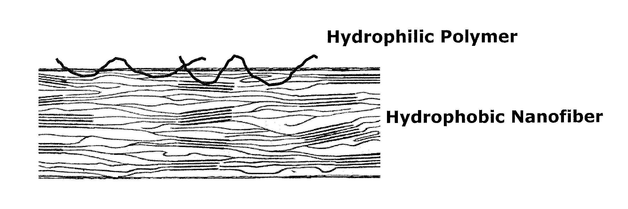
2. The microenvironment of claim 1, wherein the cellular behavior is selected from the group consisting of cell adhesion, cell migration, cell growth, cell differentiation, and any combination thereof.
3. The microenvironment of claim 1, wherein the electroprocessed composition is an electroprocessable biofunctional composition.
4. A method of making an extracellular microenvironment array, wherein the method comprises: (a) obtaining an electroprocessable biofunctional composition; (b) placing the electroprocessable biofunctional composition on a solid support in a pattern; and (c) electroprocessing the electroprocessable biofunctional composition to obtain the extracellular microenvironment array.
5. The method according to claim 4, wherein the electroprocessable biofunctional composition presents at least one hydrophilic component that regulates cellular behavior.
6. The method according to claim 5, wherein the cellular behavior is selected from the group consisting of cell adhesion, cell migration, cell growth, cell differentiation, and any combination thereof.
7. A method of making an extracellular microenvironment array, the method comprising: electroprocessing an electroprocessable biofunctional composition placed onto a solid support in a pattern so as to obtain an extracellular microenvironment array.
8. The method according to claim 7, wherein the electroprocessable biofunctional composition presents at least one hydrophilic component that regulates cellular behavior.
9. The method according to claim 8, wherein the cellular behavior is selected from the group consisting of cell adhesion, cell migration, cell growth, cell differentiation, and any combination thereof.
Description
CROSS-REFERENCE TO RELATED APPLICATION
[0001] This application claims the benefit of U.S. Provisional Patent Application Ser. No. 62/183,663, filed Jun. 23, 2015, the disclosure of which is hereby incorporated herein in its entirety by this reference.
TECHNICAL FIELD
[0002] This disclosure relates to a defined three-dimensional microenvironment for stem cells.
BACKGROUND
[0003] Increasing evidence has demonstrated that a local microenvironment, stem cell niche, is important in regulating their self-renewal and differentiation in tissue and organ development process. Stem cell niche provides a complex array of biochemical and physical cues in a spatiotemporally defined fashion, engaging and instructing stem cells to proliferate migrate and differentiate.
[0004] Such microenvironment consists of many factors including extracellular matrices (ECMs), growth factors, signaling molecules, etc. Although biochemical cues including soluble factors such as FGFs, BMPs and Wnts have been well studied for their role in regulating stem cell behavior, the effect of cell-matrix interaction in stem cell development is poorly understood.
[0005] Recapitulating the stem cell niche is a critical goal of regenerative medicine. Ideally, an engineered stem cell niche would include both spatial organization and dynamic modulation of cells, soluble factors, and matrix. While biomaterials technologies are being developed to address these needs, currently available matrices often lack this level of complexity. (See Kyle J. Lampe, et al., Building stem cell niches from the molecule up through engineered peptide materials, Neuroscience Letters (2012) 519:138-146; and M. P. Lutolf and J. A. Hubbell, Synthetic biomaterials as instructive extracellular microenvironments for morphogenesis in tissue engineering, Nature Biotechnology (2005) 23 (1):47-55.)
[0006] Many attempts have been made to create a synthetic stem cell niche by incorporating cell adhesion ligands into nanofiber or hydrogel-based scaffolds. For example, nanofiber-based scaffolds have been investigated for the regeneration of connective tissues, such as bone, meniscus, intervertebral disk, cartilage, tendons and ligaments. Nano-scale fibers have been shown to direct cell attachment and matrix deposition and represent an ideal system to model the collagenous matrices present within native tissue structures. These scaffolds exhibit high aspect ratio, surface area, permeability and porosity, and can be fabricated from a variety of polymers, both natural and synthetic, with tunable fiber diameter and matrix alignment.
[0007] A biochemically, mechanically and physically engineered nanofibrous microenvironment has been developed that mimics native extracellular microenvironments by presenting controlled fiber diameter, topography, pore size and elasticity of a matrix, as well as bioactive peptide motifs derived from extracellular matrix proteins on the nanofiber. Our engineered microenvironment can be used as an array of cell culture environments for screening of cell culture or tissue engineering environment by elucidating or regulating cellular behaviors such as cell adhesion, migration, growth, proliferation or morphogenesis as evidenced in stem cell assays.
BRIEF SUMMARY
[0008] The disclosure is directed to a three-dimensional nanofiber-based stem cell niche comprising a plurality of synthetic or natural polymer.
BRIEF DESCRIPTION OF DRAWINGS
[0009] The patent or application file contains at least one drawing executed in color. Copies of this patent or patent application publication with color drawing(s) will be provided by the Office upon request and payment of the necessary fee.
[0010] These and other features and advantages of the disclosure are evident from the following embodiments when read in conjunction with the accompanying drawings in which:
[0011] FIG. 1 shows the schematic representation of a synthetically designed nanofibrous extracellular matrix 3D microenvironment;
[0012] FIGS. 2A-2C represent scanning electron micrographs of nanofiber microenvironment having the same diameter size (400 nm) but having different surfaces. The hydrophobicity: 2A>2B>2C;
[0013] FIG. 3 shows the layout of microenvironment array;
[0014] FIGS. 4A and 4B show embryonic stem cell cultured on nanofiber microenvironment stained with alkaline phosphatase;
[0015] FIG. 5 shows Oct4 staining of embryonic stem cell cultured on nanofibrous microenvironment;
[0016] FIG. 6 shows the RT-PCR analyses of pluripotency markers OCT4, SOX2, MYC and KLF4 from stem cells cultured on different microenvironment;
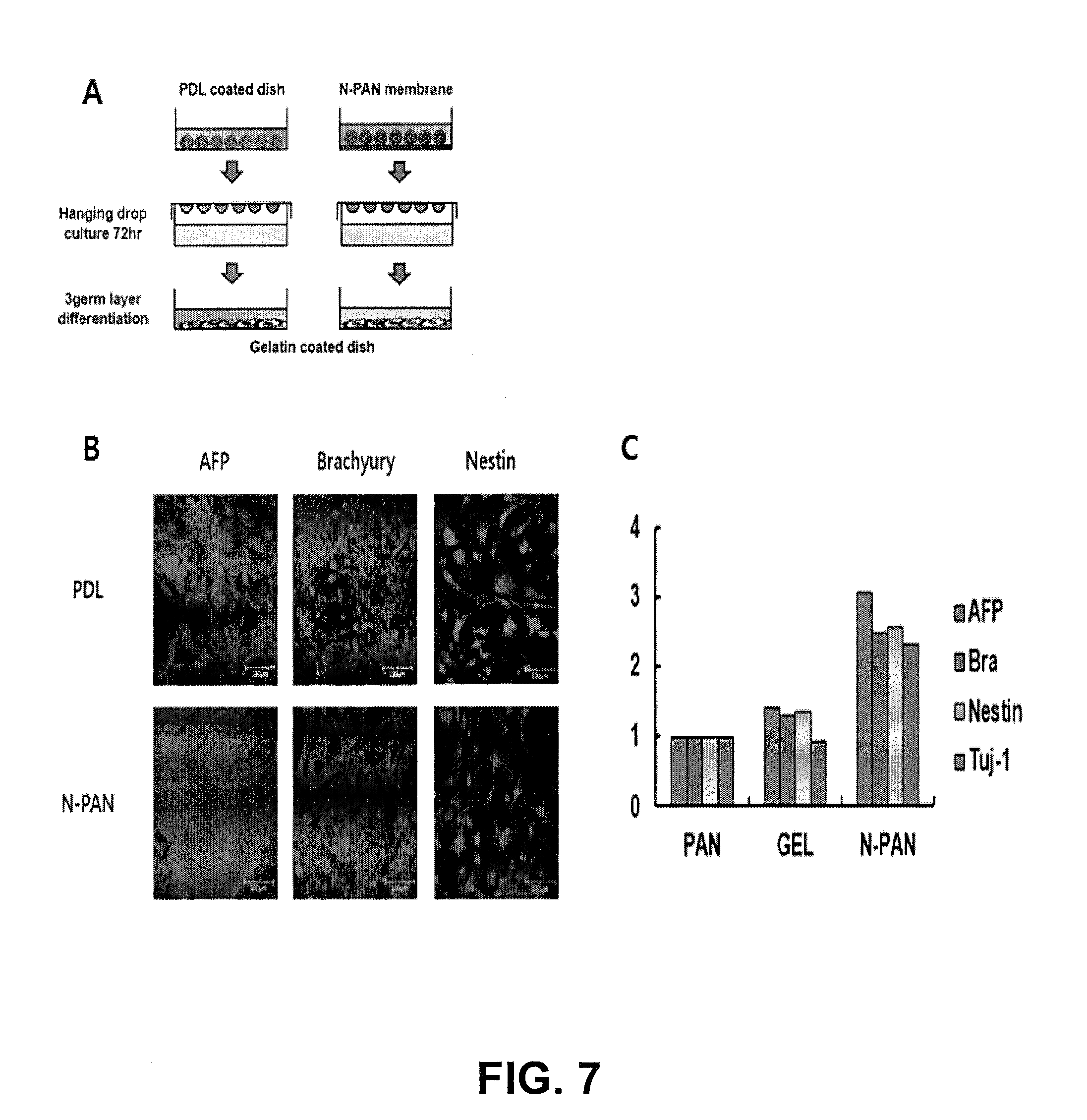
[0017] FIG. 7 represents characterization of pluripotency of human-induced pluripotent stem cell cultured on nanofibrous matrix;
[0018] FIGS. 8A and 8B represent layout of microenvironment array to screen optimal extracellular microenvironment to support self-renewal and proliferation of embryonic stem cell;
[0019] FIGS. 9A-9E represent the effect of integrin binding peptide motif, alone or in combination, on self-renewal and proliferation of embryonic stem cell;
[0020] FIGS. 10A-10C represent the inhibitory effect of WNT and LIF-derived peptide motif on stem cell differentiation;
[0021] FIG. 11 represents the effect of biochemically and physically defined nanofibrous microenvironment on self-renewal of murine embryonic stem cell;
[0022] FIG. 12 represents the effect of biochemical cues on self-renewal of murine embryonic stem cell;
[0023] FIGS. 13A-13C represent the effect of physical cues on self-renewal of murine embryonic stem cell; and
[0024] FIG. 14 represents the effect of biochemically and physically defined nanofibrous microenvironment on self-renewal of human-induced pluripotent stem cell.
DETAILED DESCRIPTION
[0025] This disclosure is directed to physically and mechanically defined microenvironments that mimic natural extracellular microenvironments.
[0026] In one aspect, an extracellular microenvironment surface that induces integrin signaling to promote signal pathway for self-renewal or proliferation of pluripotent stem cell in serum- or feeder-free conditions is provided. It is well known that integrin signaling involves Erk activation and self-renewal of embryonic stem cell is mediated signal through Ras-Raf-MEK-Erk cascade.
[0027] As used herein, "microenvironment" refers to physical and/or biochemical cues, surrounding a cell in an organism or in the laboratory. Physical cues refer to the physical or mechanical properties of a substrate surrounding the cell. For example, diameter or pore of the substrate is the physical cues. Molecules, including small molecules such as compounds and soluble factors, macromolecules such as insoluble polymers, nutrients, growth factors, fluids, cytokines and parameters such as pH, ionic strength and gas composition, and the like surrounding the cell are the biochemical cues. The molecules may be, reversibly or irreversibly in response to biological or physiological conditions, immobilized to the substrate.
[0028] This disclosure provides an electroprocessable composition to engineer an extracellular microenvironment presenting controlled physical and/or mechanical cues.
[0029] Electroprocess including electrospinning or electro-spraying is a means of producing fibers or particles with diameters generally between 10 to 1,000 nanometers. It has the ability to produce fibers or particles that are far smaller than those produced by conventional means such as wet spinning or melt spinning.
[0030] This disclosure provides a physical or mechanical microenvironment comprised of an electroprocessed composition presenting at least one or more hydrophilic component that supports cellular behavior such as cell adhesion, migration, or growth.
[0031] In one aspect, the disclosure provides an electrospinnable composition for a fibrous microenvironment comprised of two components, hydrophilic component and a structural component. In one embodiment, a structural component is a polymer to provide physical or mechanical cues such as pore size or elasticity, whereas hydrophilic component contains extracellular component.
[0032] Any electrospinnable polymer, natural or synthetic, for use in this disclosure can be a structural component. Preferably, an electrospinnable polymer is a synthetic polymer that has the appropriate viscosity in solution. Any polymer meeting the above requirements is useful herein, and the selection of the specific polymer and acquisitions or preparation of such polymer would be conventionally practiced in the art (see reference here). Preferably, such electrospinnable polymers are selected from groups comprising polyvinylidene fluoride (PVDF), polyacrylonitrile (PAN), polyethersulfone (PES), polylactic acid (PLA), polyglycolic acid (PGA), poly (lactide-glycolic) acid (PLGA), polycaprolactone, poly(alkylene oxides), particularly poly(ethylene glycols), poly(vinyl alcohols), polypeptides, poly(amino acids), such as poly(lysine), poly(allylamines) (PAM), poly(acrylates), polyesters, polyphosphazenes, pluronic polyols, polyoxamers, poly(uronic acids) and copolymers, including graft polymers thereof.
[0033] In another aspect, the disclosure provides a nanofibrous matrix to mimic a natural extracellular microenvironment, wherein the matrix has an elastic or linear modulus of about 0.5 kPa to about 1 MPa and fiber diameter of about 50 nm to 1,000 nm. In one embodiment, the method to making the nanofibrous matrix includes (a) generating an electrostatic field between a first electrode and a second electrode; and (b) electrospinning a solution of composition comprising the hydrophilic component and a synthetic polymer as a structural component onto a collection surface located between the first electrode and the second electrode to provide a plurality of nanofibers on the collection surface.
[0034] The structural polymer may be selected to have a wide range of molecular weights, generally from as low as 100,000 up to millions of Daltons. Preferably, the selected polymer has a molecular weight of less than about 300,000 to 500,000.
[0035] In another embodiment, a hydrophobic polymer is used to form an electrospinnable composition wherein PVDF, PAN, and/or PES, alone or in combination, has a molecular weight of from about 50 Kda to about 500 kDa. In one embodiment, the fiber includes about 0.1 weight percent mussel adhesive protein and about 10 weight percent hydrophobic polymer.
[0036] In another embodiment, a hydrophilic component is used to modify an electrospinnable composition wherein natural or synthetic polymer such as extracellular matrix mimetic protein, polyvinyl pyrrolidone (PVP) or polyethylene glycol (PEG) having a molecular weight of from about 30 kDa to about 300 kDa can be used to modify the composition.
[0037] In one embodiment of this disclosure, a 3D nanofibrous microenvironment having 200 nm and 700 nm diameter, respectively, are provided to support self-renewal and proliferation of murine embryonic stem cell.
[0038] This disclosure provides a nanofibrous microenvironment having elasticity that can be readily controlled by adjusting the weight ratio in hydrophilic component, whereas physical cues such as pore size and biochemical cues are constant.
[0039] In one embodiment, compositions are provided to engineer microenvironment having elasticity from 1 kPa to 1 MPa, whereas average pore size is constant and constant biochemical cues. The average pore size can range from 50 nm to 10 .mu.m.
[0040] Pore size of a scaffold can affect cell behavior within a scaffold and that subtle changes in pore size can have a significant effect on cell behavior such as cell migration. The porosity and pore architecture in terms of porosity and pore architecture play a significant role in cell survival, proliferation, and migration, and thus they are key elements to design a synthetic three-dimensional microenvironment. (N. Annabi et al., Controlling the Porosity and Microarchitecture of Hydrogels for Tissue Engineering, Tissue Eng. Part B Rev. 2010, 16(4):371-83). The porosity of a hydrogel depends on PEG molecular weight, concentration, acidity, gelation temperature and gelation time.
[0041] If the pores become too large, the mechanical properties of the scaffold will be compromised due to void volume and, as pore size increases further, the specific surface area will eventually reduce to a level that will limit cell adhesion.
[0042] As summarized in Table 2, the optimal pore size will vary with different cell types (F. J. O'Brien, et al., The effect of pore size on cell adhesion in collagen-GAG scaffolds, Biofunctionals 2005; 26(4):433-41).
TABLE-US-00001 TABLE 1 Optimal pore size for cell infiltration and host tissue ingrowth Cell/tissue type Pore size (.mu.m) Scaffold material Human skin fibroblasts <160 .mu.m PLA/PLG Bone 450 .mu.m PMMA Fibrocartilaginous tissue 150-300 .mu.m Polyurethane Adult mammalian skin cells 20-125 .mu.m Collagen -GAG Osteogenic cells 100-150 .mu.m Collagen-GAG Smooth muscle cells 60-150 .mu.m PLA Endothelial cells <80 .mu.m Silicon nitride
[0043] This disclosure provides a nanofibrous microenvironment presenting biochemical cues to regulate stem cell fate by immobilizing signaling cues that allows for generation of persistent gradients of biomolecules including extracellular matrix proteins, growth factors, or cytokines in a spatiotemporally defined manner.
[0044] It has been known that cross-talk between integrins and growth factor receptors by two mechanism, i) two separate signals merge with one another in multiple levels inside the cells (see Legate, et al., Genetic and cell biological analysis of integrin outside-in signaling, Genes Dev. 2009, 23:397-418), or ii) FGF1 directly binds to integrin .alpha.v.beta.3 and induces the FGFR1-FGF1-integrin .alpha.v.beta.3 ternary complex (S. Mori et al., Direct binding of integrin .alpha.v.beta.3 to FGF1 plays a role in FGF1 signaling, J. Biol. Chem. 2008, 283:18066-18075).
[0045] In one embodiment, the extracellular microenvironment surface to activate integrin .alpha.5.beta.1 and/or integrin .alpha.6.beta.1-mediated signaling at the same time is provided for self-renewal and proliferation of pluripotent or multi-potent stem cell.
[0046] A biochemical cue such as ECM protein or growth factors can be a natural or recombinant extracellular matrix protein, ECM-derived domain including core motif that binds to specific integrin or its mimetic, growth factor, GF-derived domain containing core motif that bind to specific binding sites of such growth factor receptor, or its mimetic. The mimetic comprises a recombinant protein or polypeptide functionalized with at least one or more peptide motifs derived from a variety of extracellular matrix proteins or growth factors.
[0047] Any suitable natural extracellular matrix proteins including, but not limited to, fibronectin, laminin, vitronectin, may be used as an extracellular component to activate integrins. Preferably, the extracellular matrix protein is fibronectin. More preferably, the fibronectin can be used alone or in combination with laminin, vitronectin or cadherin.
[0048] Any suitable natural growth factors are fibroblast growth factor (FGF) or transforming growth factor (TGF) may be used as an extracellular component to activate such growth factor receptors. Preferably, the growth factor can be used alone or in combination with FGF and TGF.
[0049] Generally, any extracellular mimetic component including extracellular matrix mimetic or growth factor mimetic comprises a substrate protein recombinantly or chemically functionalized with peptide motif derived from extracellular matrix proteins or growth factors.
[0050] Any suitable substrate protein including, but not limited to, fibrin, elastin, mussel adhesive protein may be used as the substrate protein to present extracellular component. Preferably, the protein is a recombinant mussel adhesive protein.
[0051] Any suitable recombinant mussel adhesive protein may be used as the extracellular component in this disclosure. Examples of commercially available substrate proteins include MAPTrix.TM. ECM marketed by Kollodis BioSciences, Inc. (North Augusta, S.C.). An optional third component is a biocompatible polymer (e.g., polyethylene glycol or polyvinylalcohol), which may be added to the compositions to enhance their physicomechanical characteristics such as physical or mechanical properties of a customizable microenvironment.
[0052] The MAPTrix.TM., developed by Kollodis BioSciences, Inc. (North Augusta, S.C.), are predesigned mussel adhesive protein or barnacle-based extracellular component mimetics. The mussel adhesive proteins were recombinantly functionalized with a variety of ECMs-, GFs-, or other ligand-derived peptides in order to mimic the bioactivity of naturally occurring ligands such as ECMs, GFs, or other soluble factors such as cytokines, including IL-3 or LIF, which were demonstrated to have a similar bioactivity to natural or recombinant ECMs, GFs, or soluble factors in primary cell cultures as compared to various natural or recombinant ECM, GF or cytokine proteins. The pre-designed MAPTrix.TM. mimetics are highly advantageous for creating extracellular microenvironments. For example, it provides for the design of cell-specific or user-defined regulation of extracellular microenvironments to emulate the native microenvironment in terms of biochemical cues.
[0053] The MAPTrix.TM. is a fusion protein comprising a first peptide of mussel foot protein FP-5 that is selected from the group consisting of SEQ ID NOS:1-4, or barnacle-derived adhesive protein consisting of SEQ ID NO:5 and a second peptide of at least one selected from the group consisting of mussel FP-1 selected from the group consisting of SEQ ID NOS:6-8, mussel FP-2 (SEQ ID NO:9), mussel FP-3 selected from the group consisting of SEQ ID NOS:10-11, mussel FP-4 (SEQ ID NO:12), mussel FP-6 (SEQ ID NO:13) and fragment thereof, and the second peptide is linked to C-terminus, N-terminus or C- and N-terminus of the FP-5. Preferably, the second peptide is the FP-1 comprising an amino acid sequence of SEQ ID NO:6.
[0054] Extracellular components including integrin binding motif or growth factor receptor binding motif such as fibroblast growth factor (FGF) and transforming growth factor (TGF)-derived peptide motif, WNT and/or LIF (leukemia inhibitor factor) may also be incorporated into the mussel adhesive protein to further enhance the beneficial effect of the extracellular environment mimic on self-renewal and pluripotency of a stem cell.
[0055] There are 24 known integrin heterodimers comprised of one of eighteen a subunits and one of eight .beta. subunits and these have a diverse range of functions mediating cell-cell adhesion, growth factor receptor responses and intracellular signaling cascades for cell migration, differentiation, survival and proliferation. A number of ECM molecules or domains are capable of assisting in the maintenance of undifferentiated hESC alone or in combination, including laminin 511 (see T. Miyazaki, et al., Recombinant human laminin isoforms can support the undifferentiated growth of human embryonic stem cells, Biochem. Biophys. Res. Commun. (2008), 375:27-32), fibronectin and vitronectin (see Melkoumian et al., Synthetic peptide-acrylate surfaces for long-term self-renewal and cardiomyocyte differentiation of human embryonic stem cells, Nat. Biotechnol. (2010), 28:606-610; Braam et al., Recombinant vitronectin is a functionally defined substrate that supports human embryonic stem cell self-renewal via alphavbeta5 integrin, Stem Cells (2008), 28:2257-2265).
[0056] The extracellular domain of integrins can bind ECM proteins used in hESC support such as collagen, fibronectin, laminin and vitronectin as well as members of the SIBLING family (Small Integrin Binding Ligand, N-Linked Glycoproteins, e.g., osteopontin and bone sialoprotein). Integrin clustering occurs after ECM adhesion promoting lateral association with other cell surface receptors and increases in the cytoplasmic concentration of cell signaling molecules such as PI3-kinase and MEK-ERK, which are involved in hESC maintenance (see J. Li, et al., MEK/ERK signaling contributes to the maintenance of human embryonic stem cell self-renewal, Differentiation (2007), 75:299-307).
[0057] Recently, the Hubbell laboratory developed and tested various synthetic substrates for their capacity to maintain mouse ES cell self-renewal and concluded that simultaneous ligation of .alpha.5.beta.1-, .alpha.v.beta.5-, .alpha.6.beta.1, and .alpha.9.beta.1 integrins promotes stemness of ES cells. These integrins have also been implicated in the regulation of mouse and human ES cell self-renewal in a number of other studies performed under various growth conditions (see Sandhanakrishnan Cattavarayan, et al., .alpha.6.beta.1- and .alpha.v-integrins are required long-term self-renewal of murine embryonic stem cells in the absence of LIF, BMC Cell Biology (2015), 16:3; Y. Meng, et al. Characterization of integrin engagement during defined human embryonic stem cell culture, FASEB J. 2010, 24(4):1056-65; S. R. Braam, et al., Recombinant vitronectin is a functionally defined substrate that supports human embryonic stem cell self-renewal via .alpha.v.beta.5 integrin. Stem Cells. 2008, 26(9):2257-65).
[0058] This disclosure also provides a microenvironmentally defined 3D surface that activates .alpha.5.beta.1, .alpha.6.beta.1 and/or .alpha.v.beta.5 simultaneously or sequentially in order to regulate signaling pathway for self-renewal and pluripotency maintenance of a stem cell. Any suitable substrate protein containing peptide ligand to activate integrin .alpha.5.beta.1-, .alpha.v.beta.5-, .alpha.6.beta.1, or .alpha.9.beta.1 simultaneously or sequentially to support self-renewal and pluripotency of a stem cell. In one embodiment, the microenvironment surface provides a substrate protein presenting .alpha.5.beta.1 integrin activating motif or heparin binding motif derived from fibronectin domain III. Any suitable .alpha.5.beta.1 integrin activating- or heparin binding motif can be selected from RGD (SEQ ID NO:15), GRGDSP (SEQ ID NO:16), PHSRN-RGDSP (SEQ ID NO:17), SPPRRARVT (SEQ ID NO:18), WQPPRARI (SEQ ID NO:19), KNNQKSEPLIGRKKT (SEQ ID NO:20), or its combination of .alpha.5.beta.1 integrin-activating motif and heparin binding motif.
[0059] In another embodiment, the microenvironment surface provides a substrate protein presenting .alpha.6.beta.1 integrin-activating motif-derived laminin al or laminin .alpha.5 LG domain to support self-renewal and pluripotency of a stem cell. Any suitable .alpha.6.beta.1 integrin-activating motif can be selected from GKNTGDHFVLYM (SEQ ID NO:22), VVSLYNFEQTFML (SEQ ID NO:23), RFDQELRLVSYN (SEQ ID NO:24), RLVSYSGVLFFLK (SEQ ID NO:25), ASKAIQVFLLGG (SEQ ID NO:26), VLVRVERATVFS (SEQ ID NO:27), TVFSVDQDNMLE (SEQ ID NO:28), RLRGPQRVFDLH (SEQ ID NO:29), FDLHQNMGSVN (SEQ ID NO:30), QQNLGSVNVSTG (SEQ ID NO:31), SRATAQKVSRRS (SEQ ID NO:32), TWYKIAFQRNRK (SEQ ID NO:45), NRWHSIYITRFG (SEQ ID NO:46).
[0060] In another embodiment, the microenvironment surface provides a substrate protein presenting cadherin-derived peptide motif to support self-renewal and pluripotency of a stem cell. Any suitable cadherin binding motif can be selected from SHAVSS (SEQ ID NO:48), LFSHAVSSNG (SEQ ID NO:49), ADTPPV (SEQ ID NO:50), DQNDN(SEQ ID NO:51), HAVDI (SEQ ID NO:52), LRAHAVDING (SEQ ID NO:53).
[0061] In another embodiment, the 3D microenvironment surface provides a substrate protein presenting a combinatorial motif of .alpha.5.beta.1 integrin-activating motif and .alpha.6.beta.1 binding motif at the same time to support self-renewal and pluripotency of a stem cell. Suitable combinatorial motif is a combination of PHSRN-RGDSP (SEQ ID NO:17) and NRWHSIYITRFG (SEQ ID NO:46) to support self-renewal and pluripotency of a stem cell.
[0062] Fibroblast growth factors (FGFs) are essential for maintaining self-renewal in human embryonic stem cells and induced pluripotent stem cells. Recombinant basic FGF (bFGF or FGF2) is conventionally used to culture pluripotent stem cells. Today, FGF family consists of 23 members including acidic and basic fibroblast growth factor, and each FGF has canofin, hexafin, and decafin domain (S. Li, et al., Fibroblast growth factor-derived peptides: functional agonists of the fibroblast growth factor receptor, J. Neurochem. 2008 February, 104(3):667-82; S. Li, et al., Agonists of fibroblast growth factor receptor induce neurite outgrowth and survival of cerebellar granule neurons, Dev. Neurobiol. 2009, 69(13):837-54; Shizhong Li, et al., Neuritogenic and Neuroprotective Properties of Peptide Agonists of the Fibroblast Growth Factor Receptor, Int. J. Mol. Sci. 2010, 11(6):2291-2305).
[0063] FGFRs are transmembrane glycoproteins with three extracellular domains, Ig1, Ig2 and Ig3. An FGFR fragment Ig2 and Ig3 is the minimal unit sufficient for specific ligand binding (see V. Manfe, et al., Peptides derived from specific interaction sites of the FGF 2-FGF receptor complexes induce receptor activation and signaling (see J. Neurochem. 2010, 114(1):74-86; S. K. Olsen, et al. (2004), Insights into the molecular basis for fibroblast growth factor receptor autoinhibition and ligand binding promiscuity, Proc. Natl Acad. Sci. USA 101:935-940).
[0064] Bell et al. (see 2000 Rotational coupling of the transmembrane and kinase domains of the Neu receptor tyrosine kinase, Mol. Biol. Cell 11:3589-3599) demonstrated that activation of receptor tyrosine kinases requires specific orientations of the kinase domains in a formed receptor dimer. The ligand binding mediates the optimal rotational positioning of the individual monomers within the dimer and thus the specific orientation of the catalytic domains. Binding of different agonists, such as FGF2 and canofins, resulted in different modes of orientation of catalytic domains yielding differences in receptor activation (see V. Manfe, et al., Peptides derived from specific interaction sites of the fibroblast growth factor 2-FGF receptor complexes induce receptor activation and signaling, J. Neurochem. 2010, 114(1):74-86).
[0065] When a growth factor binds to the extracellular domain of a receptor tyrosine kinase (RTK), its dimerization is triggered with other adjacent RTKs. Dimerization leads to a rapid activation of the protein's cytoplasmic kinase domains and the activated receptor as a result then becomes autophosphorylated on multiple specific intracellular tyrosine residues, resulting in signal transduction cascade.
[0066] Recent studies have demonstrated that the immobilization of soluble factors such as FGF, TGF or cytokines to the ECM plays an important role in mediating their biological effects (see C. C. Rider (2006), Heparin/heparan sulphate binding in the TGF-beta cytokine superfamily, Biochem. Soc. Trans. 34:458-460). Presentation of soluble factors in an immobilized fashion alters their local effective concentration, bioavailability, and stability, and thereby modulates their effects on target cells. For example, NSC-proliferative regions in the SVZ are situated in proximity to regions, in which growth factors, including basic fibroblast growth factor-2, are concentrated by heparan sulfate proteoglycan (HSPG) (see F. Mercier et al. (2002), Anatomy of the brain neurogenic zones revisited: fractones and the fibroblast/macrophage network, J. Comp. Neurol. 451:170-188).
[0067] This disclosure provides the FGF mimetic comprises recombinant mussel adhesive protein functionalized with FGF-derived peptide motif derived from hexafin domain or canofin domain. Preferably, FGF mimetic peptide motif can be selected from hexafin domain-derived ANRYLAMKEDGRLLAS (SEQ ID NO:33) or canofin domain-derived HFKDPKRLYCK (SEQ ID NO:34), FLPMSAKS (SEQ ID NO:35), KTGPGQKAIL (SEQ ID NO:76).
[0068] In one embodiment of this disclosure, a 3D microenvironment surface that combinatorially regulates the activity of both integrin and growth factor receptor to support self-renewal and pluripotency of murine embryonic stem cell is provided. The microenvironment surface comprises a substrate protein functionalized with a peptide such as fibronectin-derived peptide PHSRN-GRGDSP (SEQ ID NO:17) to target .alpha.5.beta.1 and FGF2-derived peptide ANRYLAMKEDGRLLAS (SEQ ID NO:33) to target FGF receptor; FGFR2IIIc.
[0069] This disclosure also provides a 3D microenvironment surface to activate TGF receptor or Frizzle receptor to induce signaling pathway to activate transcriptional factors for self-renewal and pluripotency of pluripotent stem cell. A recombinant mussel adhesive protein as a substrate protein containing TGF mimetic peptide to bind to TGF.beta. receptor domain T.beta.RI or T.beta.RII can be used in this disclosure. Preferably, TGF.beta. mimetic peptide can be selected from LTGKNFPMFHRN (SEQ ID NO:37), MHRMPSFLPTTL (SEQ ID NO:38).
[0070] In one embodiment of this disclosure, a 3D microenvironment surface that combinatorially regulates the activity of both integrin and growth factor receptor to support self-renewal and pluripotency of an embryonic stem cell is provided. The microenvironment surface comprises a substrate protein presenting a combinatorial motif to activate .alpha.5.beta.1 integrin and TGF.beta. receptor at the same time. The combinatorial motif is a combination of the substrate protein functionalized with a peptide such as fibronectin-derived peptide PHSRN-GRGDSP (SEQ ID NO:17) to target .alpha.5.beta.1 and TGF.beta.-derived peptide LTGKNFPMFHRN (SEQ ID NO:37), or MHRMPSFLPTTL (SEQ ID NO:38).
[0071] This disclosure provides a 3D microenvironment surface that generates WNT/.beta.-catenin signaling pathway by presenting WNT 1 peptide motif LCCGRGHRTRTQRVTERCNC (SEQ ID NO:39) or LGTQGRLCNKTSEGMDGCEL (SEQ ID NO:40). In one embodiment of this disclosure, a microenvironment surface that combinatorially regulates the activity of both integrin and frizzled receptor to support self-renewal and pluripotency of an embryonic stem cell is provided. The microenvironment surface comprises a substrate protein presenting a combinatorial motif to activate .alpha.5.beta.1 integrin and frizzled receptor at the same time. The combinatorial motif is a combination of the substrate protein functionalized with a peptide such as fibronectin-derived peptide PHSRN-GRGDSP (SEQ ID NO:17) to target .alpha.5.beta.1 and WNT-derived peptide LCCGRGHRTRTQRVTERCNC (SEQ ID NO:39) or LGTQGRLCNKTSEGMDGCEL (SEQ ID NO:40).
[0072] This disclosure provides a 3D microenvironment surface that generates LIF/STAT3 signaling pathway by presenting LIF peptide motif IVPLLLLVLH (SEQ ID NO:41) or YTAQGEPFPNNVEKLCAP (SEQ ID NO:42).
[0073] Various studies suggest that co-clustering or synergism occurs between downstream signaling molecules, once the basic requirements are met: growth factor receptor ligand binding, integrin occupancy by a ligand and clustering of each type of receptor (see M. A. Schwartz and V. Baron, Interactions between mitogenic stimuli or a thousand and one connections, Curr. Opin. Cell Biol. 11:197-202 (1999); K. M. Yamada and E. H. J. Danen, Integrin signaling, in Signaling Networks and Cell Cycle Control (ed. J. S. Gutkind) 1-25 (Humana Press, Totowa, N.J., 2000); S. Miyamoto, et al., Integrins can collaborate with growth factors for phosphorylation of receptor tyrosine kinases and MAP kinase activation: roles of integrin aggregation and occupancy of receptors, J. Cell Biol. 135:1633-1642 (1996)).
[0074] This disclosure provides a 3D microenvironment surface to activate at least two different receptors simultaneously by presenting a substrate protein having combinatorial motifs comprising at least two different peptide motifs that bind to at least two different receptors, respectively. The suitable combinatorial motifs may include one or more spacers between two peptide motifs to optimize flexibility and/or solubility and so afford increased affinity and/or bioavailability. The combinatorial motifs may have a peptide spacer sequence of at least two amino acids, preferably 2-15 amino acids, appended to the C-termini of at least one of the two peptide motifs.
[0075] In one embodiment of this disclosure, a 3D microenvironment surface that combinatorially regulates the activity of both integrin and growth factor receptor to support self-renewal and pluripotency of murine embryonic stem cell is provided. The microenvironment surface comprises mussel adhesive protein as a substrate protein, functionalized with two peptide motifs; one is fibronectin-derived peptide PHSRN-GRGDSP (SEQ ID NO:16) to target .alpha.5.beta.1 and the other is FGF2-derived peptide ANRYLAMKEDGRLLAS (SEQ ID NO:33) to target FGF receptor; FGFR2IIIc.
[0076] In one embodiment of this disclosure, a combinatorial 3D microenvironment surface comprising a nanofiber substrate having an average diameter of 100 nm to 20 microns, wherein the nanofiber surface presents extracellular components comprising extracellular matrix mimetic, growth factor mimetic, WNT mimetic, cytokine mimetic such as IL-3, LIF mimetic or combinations thereof.
[0077] The disclosure provides for a device of extracellular microenvironment array comprising: [0078] (a) obtaining an electroprocessable biofunctional composition; [0079] (b) placing the composition on a solid support in a pattern; and [0080] (c) electroprocessing the composition to obtain the extracellular microenvironment array.
[0081] The following examples are provided to demonstrate preferred embodiments of this disclosure and the disclosure is not intended to be limited in scope by the specific embodiments described herein, which are intended for the purposes of exemplification only. Functionally equivalent products, compositions and methods are clearly within the scope of the disclosure, as described herein.
EXAMPLES
Example 1
Preparation of Electrospinnable Composition that Provides an Extracellular Microenvironment
[0082] PVDF with an average molecular weight of 400 kDa (Solvay Plastics, Paris), PAN with an average molecular weight of 150 kDa (Sigma-Aldrich, St. Louis) were dissolved in DMAc, respectively, to prepare 20 wt % solution. Each polymer solution was mixed together and the mixture ratio was summarized in Table 2.
[0083] The electrospinnable solution was placed in a plastic syringe fitted with a 27 G needle. A syringe pump (KD Scientific, USA) was used to feed the polymer solution into the needle tip. A high voltage power supply (NanoNC Co. Ltd.) was used to charge the needle tip. The nanofibers were collected onto grounded aluminum foil target located at a certain distance from the needle tip. The fiber meshes were then removed, placed in a vacuum chamber for two days to remove residual solvent, and then stored in a desiccator.
TABLE-US-00002 TABLE 2 Electrospinnable solution composition Structural Polymer Solvent PVDF/PAN = 100/00 DMAc 5 mL PVDF/PAN = 70/30 DMAc 5 mL PVDF/PAN = 50/50 DMAc 5 mL PVDF/PAN = 30/70 DMAc 5 mL PVDF/PAN = 0/100 DMAc 5 mL
Example 2
Characterization of an Extracellular Microenvironment
[0084] The nanofiber membranes obtained were observed using a scanning electron microscope. The average diameter indicated is an average of at least three independent measurements, and the average diameter is 400 nm for all samples. The images corresponding to these fibrous microenvironments, obtained by scanning electron microscopy, are shown in FIG. 2A for PVDF/PAN (70/30), FIG. 2B for PVDF/PAN (50/50), and FIG. 2C for PVDF/PAN (30/70).
Example 3
Self-Renewal of Murine Embryonic Stem Cell
[0085] The ability of the microenvironment to support self-renewal of mESCs was evaluated by serial passaging of mESCs on the electroprocessed extracellular microenvironment as prepared in EXAMPLE 1.
[0086] For the maintenance of murine embryonic stem cell cultured on poly-D-lysine-coated surface (PDL, Sigma-Aldrich), DMEM Glutamax (GIBCO, Life Technology) containing high glucose 4.5 g/L, Na-pyruvate (0.11 g/L) and L-glutamine was used with L-Glutamine (Invitrogen), 1% non-essential amino acid (Sigma-Aldrich), 50 U/mL Penicillin/streptomycin (GIBCO) and 0.1 mM 2-Mercaptoethanol (GIBCO) as the basal medium, which was added with 20% fetal bovine serum (FBS, Hyclone) and leukemia inhibitory factor (LIF, 1,000 units/mL, Chemicon) at 37.degree. C., 5% CO2 incubator.
[0087] To elucidate the effect of fibrous microenvironment on self-renewal of murine embryonic stem cells, the cells (6.times.10.sup.4) were cultured for 48 hours on six different microenvironments placed in 12-well plates as schematically represented in FIG. 3.
Example 4
Alkaline Phosphatase Staining Assay
[0088] Cells were washed in PBS and fixed in formaldehyde (37%) for 30 seconds, washed and stained for 15 minutes in 100 .mu.L of FBB Alkaline solution (Sigma-Aldrich) in sodium nitrile solution. Stained cells were analyzed on an Olympus microscope. FIGS. 4A and 4B show the murine stem cell cultured on PVDF/PAN nanofiber membrane indicated self-renewal without loss of its differential potential while the stem cells cultured on PDL-coated surface indicated the differentiated state.
Example 5
Immunocytochemistry
[0089] Several biomarkers indicating undifferentiated ESCs and iPS cells were examined using immunofluorescent staining. Cells were fixed using 4% paraformaldehyde (Sigma-Aldrich) in 0.5% TRITON.RTM. X-100 solution at room temperature for 25 minutes, and washed three times with PBS for 5 minutes each time, and added with 10% normal goat serum (Sigma-Aldrich) at room temperature for 30 minutes.
[0090] As the primary antibody, monoclonal antibodies against SSEA1 (Santa Cruz Biotechnology) diluted at a concentration of 1:1,000 and Oct4 (Santa Cruz Biotechnology) diluted at a concentration of 1:1,000 was added to the above solution. The antibody was reacted for 24 hours at 4.degree. C. and washed with PBS three times with 0.5% TRITON.RTM. X-100 added PBS. As the secondary antibody, goat anti-mouse ALEXA FLUOR.RTM. 546 (Invitrogen) was diluted with 0.5% TRITON.RTM. X-100 added PBS diluted at a concentration of 1:1,000, and reacted for 1.5 hours. Thereafter, the cells were reacted with 10 .mu.g/mL of TO-PRO3 (Invitrogen) for the cell nucleus staining, and observed with laser scanning microscopy (Zeiss).
[0091] As shown in FIG. 5, all cells cultured on nanofiber membrane expressed Oct4 and SSEA1, the key biomarkers of murine stem cells.
Example 6
PCR Analysis for Gene Expression Profile
[0092] The cells cultured on each microenvironment were collected and RNA preparation and cDNA synthesis were accomplished. RT-PCR reaction was conducted with this cDNA as template using the primers for amplification of each marker gene fragment.
[0093] As shown in FIG. 6 by quantitative RT-PCR analyses of OCT4, SOX2, MYC, and KLF4 expression, the nanofibrous environment supported the self-renewal of mouse embryonic stem cell.
Example 7
Evaluation of Differentiation Potential of Murine ESC Cultured on Nanofiber Matrix
[0094] Embryoid body (EB) formation from ES cells is a common method for producing different cell lineages and hanging drop culture is a widely used EB formation induction method. Using handing drop culture, embryoid body (EB) that were formed from the murine embryonic stem cells cultured on the N-PAN-based nanofiber matrix, subsequently differentiated into three germ layers, characterized by immunostaining and qPCR as shown in FIG. 7.
Example 8
Screening Biochemical Factors to Support Self-Renewal and Proliferation of Embryonic Stem Cell
[0095] To identify optimal biochemical cues for self-renewal of murine embryonic stem cells, a screening for mESC adhesion and growth was performed. Briefly, 96 different ECM mimetics from fibronectin, laminin, vitronectin, cadherin. WNT, and LIF, at optimized concentration, were coated on a 12-microwell plate. FIGS. 8A and 8B showed some of coated plates, single peptide motif-coated microwell plate in FIG. 8A and combination of two different peptide motifs-coated plates in FIG. 8B. Then, 6.times.10.sup.4 cells were added to each microwell and incubated in a humidified cell culture incubator with controlled atmosphere (37.degree. C., 10% CO.sub.2).
[0096] FIG. 8A also includes the following sequences: KGHRGF (SEQ ID NO:54), TAGSCLRKFSTM (SEQ ID NO:55), GEFYFDLRLKGDK (SEQ ID NO:56), TAIPSCPEGTVPLYS (SEQ ID NO:57), YRVRVTPKEKTGPMKE (SEQ ID NO:58), KYILRWRPKNS (SEQ ID NO:59), KPDVRSYTITG (SEQ ID NO:60), ATETTITIS (SEQ ID NO:61), SPPRRARVT (SEQ ID NO:18), YEKPGSPPREVVPRPRPGV (SEQ ID NO:62), WQPPRARI (SEQ ID NO:19), KNNQKSEPLIGRKKT (SEQ ID NO:20), RKRLQVQLSIRT (SEQ ID NO:21), SINNTAVMQRLT (SEQ ID NO:63), RYVVLPR (SEQ ID NO:64), GIIFFL (SEQ ID NO:65), RLVSYSGVLFFLK (SEQ ID NO:25), ANVTHLLIRANY (SEQ ID NO:66), VLVRVERATVFS (SEQ ID NO:67), RIQNLLKITNLRIKFVK (SEQ ID NO:68), FHRRIKA (SEQ ID NO:69), FRHRNRKGY (SEQ ID NO:70), KRSR (SEQ ID NO:71), and GPGVVVVERQYI (SEQ ID NO: 72).
[0097] FIG. 8B also includes the following sequences: PHSRN-RGD (SEQ ID NO:73), RKRLQVQLSIRT (SEQ ID NO:21), and NRWHSIYITRFG (SEQ ID NO:46).
Example 9
Identification of Biochemical Factors to Support Self-Renewal and Proliferation of Embryonic Stem Cell
[0098] After 96 hours, all cells were stained with AP as described in Example 4 Alkaline phosphatase staining assay. As shown in FIGS. 9A and 9B, the surface presenting integrin .alpha.5.beta.1 binding motif such as RGD (SEQ ID NO:15) and PHSRN-RGDSP (SEQ ID NO:17) or .alpha.6.beta.1 binding motif such as NRWHSIYITRFG (SEQ ID NO:46) provided more favorable environment to support self-renewal of murine embryonic stem cell than the surface presenting .alpha.4.beta.1 binding motif such as REDV (SEQ ID NO:47).
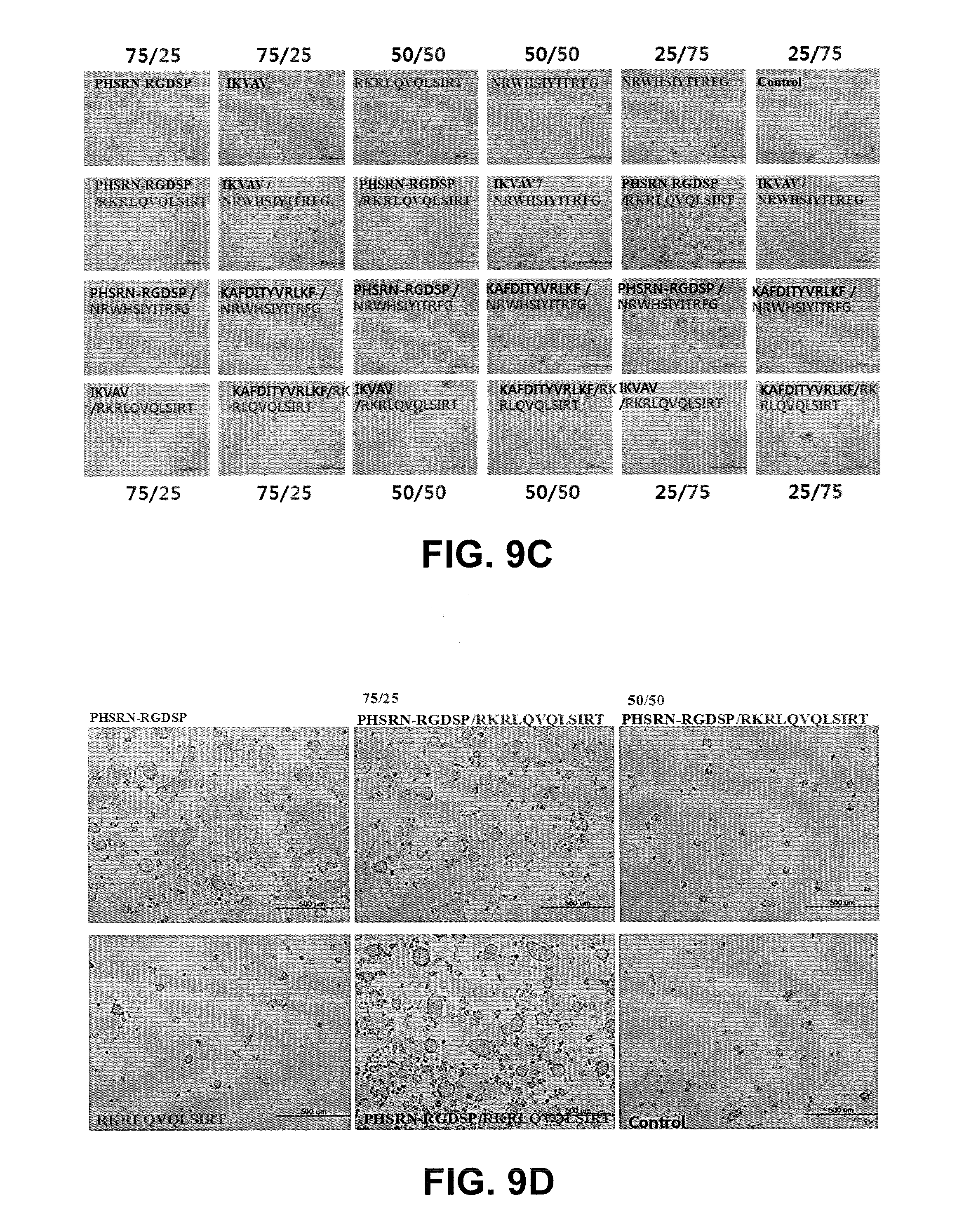
[0099] The presentation of combinatorial peptide motifs, for example, PHSRN-RGDSP/NRWHSIYITRFG (SEQ ID NO:46) (50/50) induced synergistic effect on self-renewal of murine embryonic stem cell by making the colony spherical as seen in FIG. 9C, while the combinatorial presentation of .alpha.5.beta.1/syndecan binding motif (PHSRN-RGDSP/RKRLQVQLSIRT (SEQ ID NO:17/SEQ ID NO:21)) did not show any synergistic effect on the self-renewal.
[0100] Cadherin-derived motif presenting surface also supports self-renewal of murine embryonic stem cell as shown in FIG. 9B. AP assay revealed all cadherin-derived peptide motif supported self-renewal.
[0101] LIF and WNT-derived peptide motif also support stemness maintenance of murine embryonic stem cell by inhibiting the differentiation depending on the concentration of these peptide motifs on the surface as seen in FIGS. 10A-10C. FIG. 10B shows the effect of two WNT-derived peptide motifs on self-renewal and proliferation in serum-free conditions, similar to that of PHSRN-RGDSP (SEQ ID NO:17) motif. While WNT-derived peptide motif seems to positively influence differentiation inhibition as well as proliferation, the effect of LIF-derived peptide motif seems to be limited only to differentiation inhibition. The colony size of murine stem cells is much smaller than that of murine stem cells culture on WNT-derived peptide presenting surface.
[0102] FIG. 9A also includes: GRGDSP (SEQ ID NO:16), PHSRN-RGDSP (SEQ ID NO:17), KLDAPT (SEQ ID NO:43), SPPRRARVT (SEQ ID NO:18), WQPPRARI (SEQ ID NO:19), KNNQKSEPLIGRKKT (SEQ ID NO:20), EILDVPSTT (SEQ ID NO:44), TWYKIAFQRNRK (SEQ ID NO:45), NRWHSIYITRFG (SEQ ID NO:46), RKRLQVQLSIRT (SEQ ID NO:21), and RKRLQV (SEQ ID NO:74).
[0103] FIG. 9C also includes: PHSRN-RGDSP (SEQ ID NO:17), IKVAV (SEQ ID NO:77), RKRLQVQLSIRT (SEQ ID NO:21), NRWHSIYITRFG (SEQ ID NO:46), KAFDITYVRLKF (SEQ ID NO:75), and IKFAV (SEQ ID NO:78).
[0104] FIG. 9D also includes: PHSRN-RGDSP (SEQ ID NO:17) and RKRLQVQLSIRT (SEQ ID NO:21).
[0105] FIG. 9E also includes: PHSRN-RGDSP (SEQ ID NO:17), and NRWHSIYITRFG (SEQ ID NO:46).
Example 10
Effect of Biochemically and Physically Defined Nanofibrous Matrix on Murine Embryonic Stem Cell
[0106] RGD (SEQ ID NO:15) and PHSRN-RGDSP (SEQ ID NO:17), activating integrin .alpha.5.beta.1 to support self-renewal, were selected to investigate the effect of biochemical and physical cues on the self-renewal of murine embryonic stem cell.
[0107] Electrospinnable composition was prepared to make nanofiber of 200 nm diameter whose surface presenting RGD and PHSRN-RGDSP (SEQ ID NO:17) motif with the same procedure set forth in Example 1. N-PAN and PVDF-based nanofiber with these peptide motifs was used as a control. As a positive control, poly-d-lysine and gelatin-coated surface in serum conditions were used for comparison with N-PAN and PVDF nanofiber in serum-free conditions.
[0108] As seen in FIG. 11, RGD presenting nanofiber provided more favorable environment to support self-renewal and proliferation of murine embryonic stem cell even in serum-free conditions. Some of murine stem cells cultured on gelatin-coated surface and N-PAN with no RGD motif were differentiated while no differentiation was observed on all nanofiber surface presenting RGD motif.
Example 11
Effect of Biochemically Defined Nanofibrous Matrix on Embryonic Stem Cell
[0109] To investigate the surface density of integrin binding motif on the same nanofiber surface, PVDF nanofiber having 200 nm diameter but different amount of PHSRN-RGDSP (SEQ ID NO:17). Higher PHSRN-RGDSP (SEQ ID NO:17) density showed more favorable environment to support self-renewal and proliferation of murine embryonic stem cell (FIG. 12).
Example 12
Effect of Physically Defined Nanofibrous Matrix on Embryonic Stem Cell
[0110] To investigate the effect of fiber diameter on stem cell fate, two different nanofiber matrix having 200 nm and 700 nm, respectively, were prepared with the same procedure set forth in Example 1. The amount of PHSRN-RGDSP (SEQ ID NO:17) was fixed at 10 mg.
[0111] Murine embryonic stem cells were cultured on the nanofiber surface with the same procedure as set forth in Example 3.
[0112] The effect of nanofiber diameter on the self-renewal and proliferation was assessed and characterized by expression of markers such as Oct-4 and Nanog.
[0113] Expression levels of SSEA-4 and Nanog, the established markers in murine embryonic stem cell, are assessed by immunofluorescence. Cells are harvested as single cell suspensions using trypsin or trypLE express, filtered through a 40 .mu.m sieve (BD) fixed, permeabilized and incubated with primary antibodies to SSEA-4 (1:1000 dilution, Santa Cruz), Nanog (1:50 dilution, Santa Cruz), Oct4 (1:1000 dilution, Santa Cruz).
[0114] The smaller diameter supported stemness maintenance while the larger diameter supported proliferation of murine embryonic stem cell as evidenced by the expression level of these markers (FIGS. 13B and 13C). Murine embryonic stem cell cultured on nanofiber matrix with 200 nm diameter expressed stem cell markers 1.5 times higher than those on nanofiber matrix with 700 nm (FIG. 13C) but stem cell proliferation was more efficient on the nanofiber matrix with 700 nm than nanofiber with 200 nm.
Example 13
Effect of Physically Defined Nanofibrous Matrix on Human-Induced Pluripotent Stem Cell
[0115] To investigate the effect of .alpha.5.beta.1 binding motif, PHSRN-RGDSP (SEQ ID NO:17), on the self-renewal and proliferation of human-induced pluripotent stem cell, the same test was conducted as set forth in Example 12. Human-induced pluripotent stem cells (Lonza) were cultured on the nanofibrous matrix presenting PHSRN-RGDSP (SEQ ID NO:17) in serum-free conditions. As positive controls, Matrigel.TM. (BD Bioscience) and PHSRN-RGDSP (SEQ ID NO:17)-coated microwell plate were used for comparison.
[0116] Morphological and AP staining analysis showed the nanofibrous matrix presenting PHSRN-RGDSP (SEQ ID NO:17) supported self-renewal and proliferation of human-induced stem cell as it did in murine embryonic stem cell culture (FIG. 14). As similar to that of murine embryonic stem cell, smaller diameter (Type A) provided more favorable environment to support stemness of human-induced pluripotent stem cell based on the morphological and AP staining analysis.
Sequence CWU 1
1
78110PRTArtificial Sequencemodel peptide of the tandem repeat
decapeptide derived from foot protein 1 (FP-1, Mytilus edulis) 1Ala
Lys Pro Ser Tyr Pro Pro Thr Tyr Lys 1 5 10 220PRTArtificial
Sequence2 times repeated sequence derived from foot protein 1
(FP-1, Mytilus edulis) 2Ala Lys Pro Ser Tyr Pro Pro Thr Tyr Lys Ala
Lys Pro Ser Tyr Pro 1 5 10 15 Pro Thr Tyr Lys 20 360PRTArtificial
Sequence6 times repeated sequence derived from foot protein 1
(FP-1, Mytilus edulis) 3Ala Lys Pro Ser Tyr Pro Pro Thr Tyr Lys Ala
Lys Pro Ser Tyr Pro 1 5 10 15 Pro Thr Tyr Lys Ala Lys Pro Ser Tyr
Pro Pro Thr Tyr Lys Ala Lys 20 25 30 Pro Ser Tyr Pro Pro Thr Tyr
Lys Ala Lys Pro Ser Tyr Pro Pro Thr 35 40 45 Tyr Lys Ala Lys Pro
Ser Tyr Pro Pro Thr Tyr Lys 50 55 60 439PRTArtificial
Sequencepartial sequence of foot protein type 2 (FP-2, Mytilus
californianus) 4Glu Val His Ala Cys Lys Pro Asn Pro Cys Lys Asn Asn
Gly Arg Cys 1 5 10 15 Tyr Pro Asp Gly Lys Thr Gly Tyr Lys Cys Lys
Cys Val Gly Gly Tyr 20 25 30 Ser Gly Pro Thr Cys Ala Cys 35
552PRTArtificial SequenceFoot protein type 3 (FP-3, Mytilus edulis)
5Ala Asp Tyr Tyr Gly Pro Lys Tyr Gly Pro Pro Arg Arg Tyr Gly Gly 1
5 10 15 Gly Asn Tyr Asn Arg Tyr Gly Gly Ser Arg Arg Tyr Gly Gly Tyr
Lys 20 25 30 Gly Trp Asn Asn Gly Trp Lys Arg Gly Arg Trp Gly Arg
Lys Tyr Tyr 35 40 45 Glu Phe Glu Phe 50 646PRTArtificial
SequenceFoot protein type 3 (FP-3, Mytilus galloprovincialis
mgfp-3A) 6Ala Asp Tyr Tyr Gly Pro Lys Tyr Gly Pro Pro Arg Arg Tyr
Gly Gly 1 5 10 15 Gly Asn Tyr Asn Arg Tyr Gly Arg Arg Tyr Gly Gly
Tyr Lys Gly Trp 20 25 30 Asn Asn Gly Trp Lys Arg Gly Arg Trp Gly
Arg Lys Tyr Tyr 35 40 45 760PRTArtificial Sequencepartial sequence
from foot protein type 4 (Mytilus californianus) 7Gly His Val His
Arg His Arg Val Leu His Lys His Val His Asn His 1 5 10 15 Arg Val
Leu His Lys His Leu His Lys His Gln Val Leu His Gly His 20 25 30
Val His Arg His Gln Val Leu His Lys His Val His Asn His Arg Val 35
40 45 Leu His Lys His Leu His Lys His Gln Val Leu His 50 55 60
875PRTArtificial SequenceFoot protein type5 (FP-5, Mytilus edulis)
8Ser Ser Glu Glu Tyr Lys Gly Gly Tyr Tyr Pro Gly Asn Ala Tyr His 1
5 10 15 Tyr His Ser Gly Gly Ser Tyr His Gly Ser Gly Tyr His Gly Gly
Tyr 20 25 30 Lys Gly Lys Tyr Tyr Gly Lys Ala Lys Lys Tyr Tyr Tyr
Lys Tyr Lys 35 40 45 Asn Ser Gly Lys Tyr Lys Tyr Leu Lys Lys Ala
Arg Lys Tyr His Arg 50 55 60 Lys Gly Tyr Lys Lys Tyr Tyr Gly Gly
Ser Ser 65 70 75 976PRTArtificial SequenceFoot protein 5 (FP-5,
Mytilus edulis) 9Ser Ser Glu Glu Tyr Lys Gly Gly Tyr Tyr Pro Gly
Asn Thr Tyr His 1 5 10 15 Tyr His Ser Gly Gly Ser Tyr His Gly Ser
Gly Tyr His Gly Gly Tyr 20 25 30 Lys Gly Lys Tyr Tyr Gly Lys Ala
Lys Lys Tyr Tyr Tyr Lys Tyr Lys 35 40 45 Asn Ser Gly Lys Tyr Lys
Tyr Leu Lys Lys Ala Arg Lys Tyr His Arg 50 55 60 Lys Gly Tyr Lys
Lys Tyr Tyr Gly Gly Gly Ser Ser 65 70 75 1071PRTArtificial
SequenceFoot protein 5 (FP-5, Mytilus coruscus) 10Tyr Asp Asp Tyr
Ser Asp Gly Tyr Tyr Pro Gly Ser Ala Tyr Asn Tyr 1 5 10 15 Pro Ser
Gly Ser His Trp His Gly His Gly Tyr Lys Gly Lys Tyr Tyr 20 25 30
Gly Lys Gly Lys Lys Tyr Tyr Tyr Lys Phe Lys Arg Thr Gly Lys Tyr 35
40 45 Lys Tyr Leu Lys Lys Ala Arg Lys Tyr His Arg Lys Gly Tyr Lys
Lys 50 55 60 His Tyr Gly Gly Ser Ser Ser 65 70 1176PRTArtificial
Sequencemussel adhesive protein foot protein type5 from (Mytilus
galloprovincialis) 11Ser Ser Glu Glu Tyr Lys Gly Gly Tyr Tyr Pro
Gly Asn Thr Tyr His 1 5 10 15 Tyr His Ser Gly Gly Ser Tyr His Gly
Ser Gly Tyr His Gly Gly Tyr 20 25 30 Lys Gly Lys Tyr Tyr Gly Lys
Ala Lys Lys Tyr Tyr Tyr Lys Tyr Lys 35 40 45 Asn Ser Gly Lys Tyr
Lys Tyr Leu Lys Lys Ala Arg Lys Tyr His Arg 50 55 60 Lys Gly Tyr
Lys Lys Tyr Tyr Gly Gly Gly Ser Ser 65 70 75 1299PRTArtificial
Sequencemussel adhesive protein foot protein type 6 12Gly Gly Gly
Asn Tyr Arg Gly Tyr Cys Ser Asn Lys Gly Cys Arg Ser 1 5 10 15 Gly
Tyr Ile Phe Tyr Asp Asn Arg Gly Phe Cys Lys Tyr Gly Ser Ser 20 25
30 Ser Tyr Lys Tyr Asp Cys Gly Asn Tyr Ala Gly Cys Cys Leu Pro Arg
35 40 45 Asn Pro Tyr Gly Arg Val Lys Tyr Tyr Cys Thr Lys Lys Tyr
Ser Cys 50 55 60 Pro Asp Asp Phe Tyr Tyr Tyr Asn Asn Lys Gly Tyr
Tyr Tyr Tyr Asn 65 70 75 80 Asp Lys Asp Tyr Phe Asn Cys Gly Ser Tyr
Asn Gly Cys Cys Leu Arg 85 90 95 Ser Gly Tyr 13194PRTArtificial
Sequencehybrid mussel adhesive protein (FP-151, MEFP-5 based
Kollodis) 13Ala Lys Pro Ser Tyr Pro Pro Thr Tyr Lys Ala Lys Pro Ser
Tyr Pro 1 5 10 15 Pro Thr Tyr Lys Ala Lys Pro Ser Tyr Pro Pro Thr
Tyr Lys Ala Lys 20 25 30 Pro Ser Tyr Pro Pro Thr Tyr Lys Ala Lys
Pro Ser Tyr Pro Pro Thr 35 40 45 Tyr Lys Ala Lys Pro Ser Tyr Pro
Pro Thr Tyr Lys Ser Ser Glu Glu 50 55 60 Tyr Lys Gly Gly Tyr Tyr
Pro Gly Asn Ala Tyr His Tyr His Ser Gly 65 70 75 80 Gly Ser Tyr His
Gly Ser Gly Tyr His Gly Gly Tyr Lys Gly Lys Tyr 85 90 95 Tyr Gly
Lys Ala Lys Lys Tyr Tyr Tyr Lys Tyr Lys Asn Ser Gly Lys 100 105 110
Tyr Lys Tyr Leu Lys Lys Ala Arg Lys Tyr His Arg Lys Gly Tyr Lys 115
120 125 Tyr Tyr Gly Gly Ser Ser Ala Lys Pro Ser Tyr Pro Pro Thr Tyr
Lys 130 135 140 Ala Lys Pro Ser Tyr Pro Pro Thr Tyr Lys Ala Lys Pro
Ser Tyr Pro 145 150 155 160 Pro Thr Tyr Lys Ala Lys Pro Ser Tyr Pro
Pro Thr Tyr Lys Ala Lys 165 170 175 Pro Ser Tyr Pro Pro Thr Tyr Lys
Ala Lys Pro Ser Tyr Pro Pro Thr 180 185 190 Tyr Lys
14196PRTArtificial Sequencehybrid mussel adhesive protein (FP-151,
MGFP-5 based) 14Ala Lys Pro Ser Tyr Pro Pro Thr Tyr Lys Ala Lys Pro
Ser Tyr Pro 1 5 10 15 Pro Thr Tyr Lys Ala Lys Pro Ser Tyr Pro Pro
Thr Tyr Lys Ala Lys 20 25 30 Pro Ser Tyr Pro Pro Thr Tyr Lys Ala
Lys Pro Ser Tyr Pro Pro Thr 35 40 45 Tyr Lys Ala Lys Pro Ser Tyr
Pro Pro Thr Tyr Lys Ser Ser Glu Glu 50 55 60 Tyr Lys Gly Gly Tyr
Tyr Pro Gly Asn Thr Tyr His Tyr His Ser Gly 65 70 75 80 Gly Ser Tyr
His Gly Ser Gly Tyr His Gly Gly Tyr Lys Gly Lys Tyr 85 90 95 Tyr
Gly Lys Ala Lys Lys Tyr Tyr Tyr Lys Tyr Lys Asn Ser Gly Lys 100 105
110 Tyr Lys Tyr Leu Lys Lys Ala Arg Lys Tyr His Arg Lys Gly Tyr Lys
115 120 125 Lys Tyr Tyr Gly Gly Gly Ser Ser Ala Lys Pro Ser Tyr Pro
Pro Thr 130 135 140 Tyr Lys Ala Lys Pro Ser Tyr Pro Pro Thr Tyr Lys
Ala Lys Pro Ser 145 150 155 160 Tyr Pro Pro Thr Tyr Lys Ala Lys Pro
Ser Tyr Pro Pro Thr Tyr Lys 165 170 175 Ala Lys Pro Ser Tyr Pro Pro
Thr Tyr Lys Ala Lys Pro Ser Tyr Pro 180 185 190 Pro Thr Tyr Lys 195
153PRTArtificial SequenceFibronectin-derived peptide (RGD) 15Arg
Gly Asp 1 166PRTArtificial SequenceFibronectin-derived peptide
(GRGDSP) 16Gly Arg Gly Asp Ser Pro 1 5 1710PRTArtificial
SequenceFibronectin-derived peptide (PHSRN-RGDSP) 17Pro His Ser Arg
Asn Arg Gly Asp Ser Pro 1 5 10 189PRTArtificial
SequenceFibronectin-derived peptide (SPPRRARVT) 18Ser Pro Pro Arg
Arg Ala Arg Val Thr 1 5 198PRTArtificial
SequenceFibronectin-derived peptide (WQPPRARI) 19Trp Gln Pro Pro
Arg Ala Arg Ile 1 5 2015PRTArtificial SequenceFibronectin-derived
peptide (KNNQKSEPLIGRKKT) 20Lys Asn Asn Gln Lys Ser Glu Pro Leu Ile
Gly Arg Lys Lys Thr 1 5 10 15 2112PRTArtificial
SequenceLaminin-derived peptide (RKRLQVQLSIRT) 21Arg Lys Arg Leu
Gln Val Gln Leu Ser Ile Arg Thr 1 5 10 2212PRTArtificial
SequenceLaminin-derived peptide (GKNTGDHFVLYM) 22Gly Lys Asn Thr
Gly Asp His Phe Val Leu Tyr Met 1 5 10 2313PRTArtificial
SequenceLaminin-derived peptide (VVSLYNFEQTFML) 23Val Val Ser Leu
Tyr Asn Phe Glu Gln Thr Phe Met Leu 1 5 10 2412PRTArtificial
SequenceLaminin-derived peptide (RFDQELRLVSYN) 24Arg Phe Asp Gln
Glu Leu Arg Leu Val Ser Tyr Asn 1 5 10 2513PRTArtificial
SequenceLaminin-derived peptide (RLVSYSGVLFFLK) 25Arg Leu Val Ser
Tyr Ser Gly Val Leu Phe Phe Leu Lys 1 5 10 2612PRTArtificial
SequenceLaminin-derived peptide (ASKAIQVFLLGG) 26Ala Ser Lys Ala
Ile Gln Val Phe Leu Leu Gly Gly 1 5 10 2712PRTArtificial
SequenceLaminin-derived peptide (VLVRVERATVFS) 27Thr His Arg Pro
Pro Met Trp Ser Pro Val Trp Pro 1 5 10 2812PRTArtificial
SequenceLaminin-derived peptide (TVFSVDQDNMLE) 28Thr Val Phe Ser
Val Asp Gln Asp Asn Met Leu Glu 1 5 10 2912PRTArtificial
SequenceLaminin-derived peptide (RLRGPQRVFDLH) 29Arg Leu Arg Gly
Pro Gln Arg Val Phe Asp Leu His 1 5 10 3011PRTArtificial
SequenceLaminin-derived peptide (FDLHQNMGSVN) 30Phe Asp Leu His Gln
Asn Met Gly Ser Val Asn 1 5 10 3112PRTArtificial
SequenceLaminin-derived peptide (QQNLGSVNVSTG) 31Gln Gln Asn Leu
Gly Ser Val Asn Val Ser Thr Gly 1 5 10 3212PRTArtificial
SequenceLaminin-derived (SRATAQKVSRRS) 32Ser Arg Ala Thr Ala Gln
Lys Val Ser Arg Arg Ser 1 5 10 3316PRTArtificial
SequenceFGF-derived peptide (ANRYLAMKEDGRLLAS) 33Ala Asn Arg Tyr
Leu Ala Met Lys Glu Asp Gly Arg Leu Leu Ala Ser 1 5 10 15
3411PRTArtificial SequenceFGF-derived peptide (HFKDPKRLYCK) 34His
Phe Lys Asp Pro Lys Arg Leu Tyr Cys Lys 1 5 10 358PRTArtificial
SequenceFGF-derived peptide (FLPMSAKS) 35Phe Leu Pro Met Ser Ala
Lys Ser 1 5 368PRTArtificial SequenceFGF-derived peptide (KTGPGQKA)
36Lys Thr Gly Pro Gly Gln Lys Ala 1 5 3712PRTArtificial
SequenceTGF-derived peptide (LTGKNFPMFHRN) 37Leu Thr Gly Lys Asn
Phe Pro Met Phe His Arg Asn 1 5 10 3812PRTArtificial
SequenceTGF-derived peptide (MHRMPSFLPTTL) 38Met His Arg Met Pro
Ser Phe Leu Pro Thr Thr Leu 1 5 10 3920PRTArtificial
SequenceWNT-derived peptide (LCCGRGHRTRTQRVTERCNC) 39Leu Cys Cys
Gly Arg Gly His Arg Thr Arg Thr Gln Arg Val Thr Glu 1 5 10 15 Arg
Cys Asn Cys 20 4020PRTArtificial SequenceWNT-derived peptide
(LGTQGRLCNKTSEGMDGCEL) 40Leu Gly Thr Gln Gly Arg Leu Cys Asn Lys
Thr Ser Glu Gly Met Asp 1 5 10 15 Gly Cys Glu Leu 20
4110PRTArtificial SequenceLIF-derived peptide (IVPLLLLVLH) 41Ile
Val Pro Leu Leu Leu Leu Val Leu His 1 5 10 4218PRTArtificial
SequenceLIF-derived peptide (YTAQGEPFPNNVEKLCAP) 42Tyr Thr Ala Gln
Gly Glu Pro Phe Pro Asn Asn Val Glu Lys Leu Cys 1 5 10 15 Ala Pro
436PRTArtificial SequenceFibronectin-derived peptide (KLDAPT) 43Lys
Leu Asp Ala Pro Thr 1 5 449PRTArtificial
SequenceFibronectin-derived peptide (EILDVPSTT) 44Glu Ile Leu Asp
Val Pro Ser Thr Thr 1 5 4512PRTArtificial SequenceLaminin-derived
peptide (TWYKIAFQRNRK) 45Thr Trp Tyr Lys Ile Ala Phe Gln Arg Asn
Arg Lys 1 5 10 4612PRTArtificial SequenceLaminin-derived peptide
(NRWHSIYITRFG) 46Asn Arg Trp His Ser Ile Tyr Ile Thr Arg Phe Gly 1
5 10 474PRTArtificial SequenceFibronectin-derived peptide (REDV)
47Arg Glu Asp Val 1 486PRTArtificial Sequencecadherin-derived
peptide (SHAVSS) 48Ser His Ala Val Ser Ser 1 5 4910PRTArtificial
Sequencecadherin-derived peptide (LFSHAVSSNG) 49Leu Phe Ser His Ala
Val Ser Ser Asn Gly 1 5 10 506PRTArtificial
Sequencecadherin-derived peptide (ADTPPV) 50Ala Asp Thr Pro Pro Val
1 5 515PRTArtificial Sequencecadherin-derived peptide (DQNDN) 51Asp
Gln Asn Asp Asn 1 5 525PRTArtificial Sequencecadherin-derived
peptide (HAVDI) 52His Ala Val Asp Ile 1 5 5310PRTArtificial
Sequencecadherin-derived peptide (LRAHAVDING) 53Leu Arg Ala His Ala
Val Asp Ile Asn Gly 1 5 10 546PRTArtificial Sequencederived peptide
54Lys Gly His Arg Gly Phe 1 5 5512PRTArtificial Sequencederived
peptide 55Thr Ala Gly Ser Cys Leu Arg Lys Phe Ser Thr Met 1 5 10
5613PRTArtificial Sequencederived peptide 56Gly Glu Phe Tyr Phe Asp
Leu Arg Leu Lys Gly Asp Lys 1 5 10 5715PRTArtificial
Sequencederived peptide 57Thr Ala Ile Pro Ser Cys Pro Glu Gly Thr
Val Pro Leu Tyr Ser 1 5 10 15 5816PRTArtificial Sequencederived
peptide 58Tyr Arg Val Arg Val Thr Pro Lys Glu Lys Thr Gly Pro Met
Lys Glu 1 5 10 15 5911PRTArtificial Sequencederived peptide 59Lys
Tyr Ile Leu Arg Trp Arg Pro Lys Asn Ser 1 5 10 6011PRTArtificial
Sequencederived peptide 60Lys Pro Asp Val Arg Ser Tyr Thr Ile Thr
Gly 1 5 10 619PRTArtificial Sequencederived peptide 61Ala Thr Glu
Thr Thr Ile Thr Ile Ser 1 5 6219PRTArtificial Sequencederived
peptide 62Tyr Glu Lys Pro Gly Ser Pro Pro Arg Glu Val Val Pro Arg
Pro Arg 1 5 10 15 Pro Gly Val 6312PRTArtificial Sequencederived
peptide 63Ser Ile Asn Asn Thr Ala Val Met Gln Arg Leu Thr 1 5 10
647PRTArtificial Sequencederived peptide 64Arg Tyr Val Val Leu Pro
Arg 1 5 656PRTArtificial Sequencederived peptide 65Gly Ile Ile Phe
Phe Leu 1 5 6612PRTArtificial Sequencederived peptide 66Ala Asn Val
Thr His Leu Leu Ile Arg Ala Asn Tyr 1 5 10 6712PRTArtificial
Sequencederived peptide 67Val Leu Val Arg Val Glu Arg Ala Thr Val
Phe Ser 1 5 10 6817PRTArtificial Sequencederived peptide 68Arg Ile
Gln Asn Leu Leu Lys Ile Thr Asn Leu Arg Ile Lys Phe Val 1 5 10 15
Lys 697PRTArtificial Sequencederived peptide 69Phe His Arg Arg Ile
Lys Ala 1 5 709PRTArtificial Sequencederived peptide 70Phe Arg His
Arg Asn Arg Lys Gly Tyr 1 5 714PRTArtificial
Sequencederived peptide 71Lys Arg Ser Arg 1 7212PRTArtificial
Sequencederived peptide 72Gly Pro Gly Val Val Val Val Glu Arg Gln
Tyr Ile 1 5 10 738PRTArtificial Sequencederived peptide 73Pro His
Ser Arg Asn Arg Gly Asp 1 5 746PRTArtificial Sequencederived
peptide 74Arg Lys Arg Leu Gln Val 1 5 7512PRTArtificial
Sequencederived peptide 75Lys Ala Phe Asp Ile Thr Tyr Val Arg Leu
Lys Phe 1 5 10 7610PRTArtificial Sequencederived peptide 76Lys Thr
Gly Pro Gly Gln Lys Ala Ile Leu 1 5 10 775PRTArtificial
Sequencederived peptide 77Ile Lys Val Ala Val 1 5 785PRTArtificial
Sequencederived peptide 78Ile Lys Phe Ala Val 1 5
D00000
D00001
D00002
D00003
D00004
D00005
D00006
D00007
D00008
D00009
D00010
D00011
D00012

D00013

D00014

D00015

S00001
XML
uspto.report is an independent third-party trademark research tool that is not affiliated, endorsed, or sponsored by the United States Patent and Trademark Office (USPTO) or any other governmental organization. The information provided by uspto.report is based on publicly available data at the time of writing and is intended for informational purposes only.
While we strive to provide accurate and up-to-date information, we do not guarantee the accuracy, completeness, reliability, or suitability of the information displayed on this site. The use of this site is at your own risk. Any reliance you place on such information is therefore strictly at your own risk.
All official trademark data, including owner information, should be verified by visiting the official USPTO website at www.uspto.gov. This site is not intended to replace professional legal advice and should not be used as a substitute for consulting with a legal professional who is knowledgeable about trademark law.