Protease-resistant Mutants Of Stromal Cell Derived Factor-1 In The Repair Of Tissue Damage
SEGERS; Vincent Frans Maria ; et al.
U.S. patent application number 15/054456 was filed with the patent office on 2016-12-29 for protease-resistant mutants of stromal cell derived factor-1 in the repair of tissue damage. The applicant listed for this patent is Mesoblast International Sarl. Invention is credited to Yan QIU, Anthony SANDRASAGRA, Vincent Frans Maria SEGERS.
| Application Number | 20160375100 15/054456 |
| Document ID | / |
| Family ID | 44507157 |
| Filed Date | 2016-12-29 |


View All Diagrams
| United States Patent Application | 20160375100 |
| Kind Code | A1 |
| SEGERS; Vincent Frans Maria ; et al. | December 29, 2016 |
PROTEASE-RESISTANT MUTANTS OF STROMAL CELL DERIVED FACTOR-1 IN THE REPAIR OF TISSUE DAMAGE
Abstract
The present invention features mutant stromal cell derived factor-1 (SDF-1) peptides that have been mutated to make them resistant to digestion by, for example, the proteases dipeptidyl peptidase IV (DPPIV), matrix metalloproteinase-2 (MMP-2), matrix metalloproteinase-9 (MMP-9), leukocyte elastase, cathepsin G, carboxypeptidase M, and carboxypeptidase N, but which retain chemoattractant activity.
| Inventors: | SEGERS; Vincent Frans Maria; (Bornem, BE) ; SANDRASAGRA; Anthony; (Arlington, MA) ; QIU; Yan; (Jamaica Plain, MA) | ||||||||||
| Applicant: |
|
||||||||||
|---|---|---|---|---|---|---|---|---|---|---|---|
| Family ID: | 44507157 | ||||||||||
| Appl. No.: | 15/054456 | ||||||||||
| Filed: | February 26, 2016 |
Related U.S. Patent Documents
| Application Number | Filing Date | Patent Number | ||
|---|---|---|---|---|
| 13029891 | Feb 17, 2011 | 9308277 | ||
| 15054456 | ||||
| 61345852 | May 18, 2010 | |||
| 61308090 | Feb 25, 2010 | |||
| Current U.S. Class: | 514/16.5 |
| Current CPC Class: | A61P 9/10 20180101; A61K 47/64 20170801; A61K 38/195 20130101; A61P 17/02 20180101; A61K 47/645 20170801; C07K 14/522 20130101; C07K 2319/70 20130101; A61K 38/00 20130101 |
| International Class: | A61K 38/19 20060101 A61K038/19 |
Claims
1-30. (canceled)
31. A method of treating or ameliorating tissue damage resulting from a disease or condition in a subject in need thereof, said method comprising administering to said damaged tissue an isolated mutant form of stromal cell derived factor-1 (SDF-1) peptide comprising the formula of a mutant SDF-1 (mSDF-1) mSDF 1 or X.sub.p-mSDF-1 peptide in an amount sufficient to treat or ameliorate said tissue damage in said subject, wherein said SDF-1 is a peptide comprising the amino acid sequence of at least amino acids 1-8 of SEQ ID NO: 53 and which is optionally extended at the C-terminus by all or any portion of the remaining sequence of SEQ ID NO: 53, said SEQ ID NO: 53 comprising the amino acid sequence: K P X.sub.3 X.sub.4 X.sub.5 X.sub.6 Y R C P C R F F E S H V A R A N V K H L K I L N T P N C A L Q I V A R L K N N N R Q V C I D P K L K W I Q E Y L E K A L N K (SEQ ID NO: 53), wherein X.sub.3 is any amino acid; X.sub.4 is serine or valine; X.sub.5 is leucine, proline, threonine, or valine; and X.sub.6 is any amino acid residue, and wherein (a) X.sub.p is a proteinogenic amino acid(s) or a protease protective organic group and p is any integer from 1 to 4; (b) said mSDF-1 maintains chemoattractant activity for T cells and is inactivated by matrix metalloproteinase-2 (MMP-2), matrix metalloproteinase-9 (MMP-9), leukocyte elastase, and/or cathepsin G at a rate that is at least 50% less than the rate of inactivation of native SDF-1; (c) said XD-mSDF-1 maintains chemoattractant activity for T cells, is inactivated by dipeptidyl peptidase IV (DPPIV) at a rate that is at least 50% less than the rate at which native SDF-1 is inactivated, and is inactivated by MMP-2, MMP-9, leukocyte elastase, and/or cathepsin G at a rate that is at least 50% less than the rate of inactivation of native SDF-1; and (d) said SDF-1 peptide does not comprise the amino acid sequence of at least amino acids 1-8 of SEQ ID NO: 52, SEQ ID NO: 56, SEQ ID NO: 60, or SEQ ID NOs: 65-67.
32. The method of claim 31, wherein said isolated mSDF-1 or X.sub.p-mSDF-1 peptide is attached to a biologically compatible membrane or is attached to a self-assembling peptide that forms a biologically compatible membrane after administration to said damaged tissue.
33. The method of claim 31, wherein said disease or condition is selected from the group consisting of stroke, limb ischemia, tissue damage due to trauma, myocardial infarction, peripheral vascular disease, and diabetic ulcers.
34. The method of claim 33, wherein said disease or condition is myocardial infarction.
35. The method of claim 33, wherein said disease is peripheral vascular disease.
36. The method of claim 31, wherein said subject is treated for damage to cardiac tissue.
37. The method of claim 31, wherein said administration comprises injecting or implanting said isolated mSDF-1 or X-mSDF-1 peptide into cardiac tissue of said subject.
38. The method of claim 31, wherein said mSDF-1 peptide is SDF(V3H), consisting of SEQ ID NO: 54.
39. The method of claim 38, wherein said peptide is an X.sub.p-mSDF-1 peptide and wherein X is a serine and p is 1.
40. The method of claim 31, wherein said mSDF-1 peptide is SDF(V3C), consisting of SEQ ID NO: 55.
41. The method of claim 40, wherein said peptide is an X.sub.p-mSDF-1 peptide and wherein X is a serine and p is 1.
42. The method of claim 31, wherein said mSDF-1 peptide is SDF(L5T), consisting of SEQ ID NO: 56.
43. The method of claim 42, wherein said peptide is an X.sub.p-mSDF-1 peptide and wherein X is a serine and p is 1.
44. The method of claim 31, wherein said mSDF-1 peptide is SDF(L5V), consisting of SEQ ID NO: 60.
45. The method of claim 44, wherein said peptide is an X.sub.p-mSDF-1 peptide and wherein X is a serine and p is 1.
46. The method of claim 31, wherein said mSDF-1 peptide is SDF(S6C), consisting of SEQ ID NO: 61.
47. The method of claim 46, wherein said peptide is an X.sub.p-mSDF-1 peptide and wherein X is a serine and p is 1.
48. The method of claim 31, wherein said mSDF-1 peptide is SDF(S6G), consisting of SEQ ID NO: 62.
49. The method of claim 48, wherein said peptide is an X.sub.p-mSDF-1 peptide and wherein X is a serine and p is 1.
50. The method of claim 32, wherein the biologically compatible peptide membrane comprises one or more self-assembling peptides having an amino acid sequence selected from the group consisting of SEQ ID NO: 1-SEQ ID NO: 51, and wherein between 0.1-10% of said one or more self-assembling peptides are bound to the isolated mSDF-1 or X.sub.p-mSDF-1 peptide.
51. A method of treating or ameliorating cardiac tissue damage resulting from myocardial infarction in a subject in need thereof, said method comprising administering to said damaged cardiac tissue an isolated mutant form of stromal cell derived factor-1 (SDF-1) peptide comprising the formula of a mutant SDF-1 (mSDF-1) or X.sub.p-mSDF-1 peptide in an amount sufficient to treat or ameliorate said cardiac tissue damage in said subject, wherein said SDF-1 is a peptide comprising the amino acid sequence of at least amino acids 1-8 of SEQ ID NO: 53 and which is optionally extended at the C-terminus by all or any portion of the remaining sequence of SEQ ID NO: 53, said SEQ ID NO: 53 comprising the amino acid sequence: K P X.sub.3 X.sub.4 X.sub.5 X.sub.6Y R C P C R F F E S H V A R A N V K H L K I L N T P N C A L Q I V A R L K N N N R Q V C I D P K L K W I Q E Y L E K A L N K (SEQ ID NO: 53), wherein X.sub.3 is any amino acid; X.sub.4 is serine or valine; X.sub.5 is leucine, proline, threonine, or valine; and X.sub.6 is any amino acid residue, and wherein (a) X.sub.p is a proteinogenic amino acid(s) or a protease protective organic group and p is any integer from 1 to 4; (b) said mSDF-1 maintains chemoattractant activity for T cells and is inactivated by matrix metalloproteinase-2 (MMP-2), matrix metalloproteinase-9 (MMP-9), leukocyte elastase, and/or cathepsin G at a rate that is at least 50% less than the rate of inactivation of native SDF-1; (c) said X.sub.p-mSDF-1 maintains chemoattractant activity for T cells, is inactivated by dipeptidyl peptidase IV (DPPIV) at a rate that is at least 50% less than the rate at which native SDF-1 is inactivated, and is inactivated by MMP-2, MMP-9, leukocyte elastase, and/or cathepsin G at a rate that is at least 50% less than the rate of inactivation of native SDF-1; and (d) said SDF-1 peptide does not comprise the amino acid sequence of at least amino acids 1-8 of SEQ ID NO: 52, SEQ ID NO: 56, SEQ ID NO: 60, or SEQ ID NOs: 65-67.
Description
CROSS-REFERENCE TO RELATED APPLICATIONS
[0001] This application claims the benefit of the filing date of U.S. provisional application Nos. 61/308,090, filed Feb. 25, 2010, and 61/345,852, filed May 18, 2010, each of which is herein incorporated by reference.
BACKGROUND OF THE INVENTION
[0002] In general, the invention relates to protease-resistant mutants of stromal cell derived factor-1 (SDF-1).
[0003] SDF-1 (also known as CXCL12) is a 68 amino acid member of the chemokine family that is a chemoattractant for resting T-lymphocytes, monocytes, and CD34.sup.+ stem cells. SDF-1 is produced in multiple forms: SDF-1.alpha. (CXCL12a), SDF-1.beta. (CXCL12b), and SDF-1.gamma., which are the result of differential mRNA splicing. The sequences of SDF-1.alpha. and SDF-1.beta. are essentially the same, except that SDF-1.beta. is extended by four amino acids (Arg-Phe-Lys-Met) at the C-terminus. The first three exons of SDF-1.gamma. are identical to those of SDF-1.alpha. and SDF-1.beta.. The fourth exon of SDF-1.gamma. is located 3200 base-pairs downstream from the third exon on the SDF-1 locus and lies between the third exon and the fourth exon of SDF-1.beta.. SDF-1 is initially made with a signal peptide (21 amino acids in length) that is cleaved to make the active peptide.
[0004] SDF-1 plays a key-role in the homing of hematopoietic stem cells to bone marrow during embryonic development and after stem cell transplantation. In addition to its role in stem cell homing, SDF-1 is also important in cardiogenesis and vasculogenesis. SDF-1-deficient mice die perinatally and have defects in cardiac ventricular septal formation, bone marrow hematopoiesis, and organ-specific vasculogenesis. It has also been reported that abnormally low levels of SDF-1 are at least partially responsible for impaired wound healing associated with diabetic patients and that impairment can be reversed by the administration of SDF-1 at the site of tissue damage.
[0005] In the normal adult heart, SDF-1 is expressed constitutively, but expression is upregulated within days after myocardial infarction. It has been shown previously that SDF-1 expression increased eight weeks after myocardial infarction by intramyocardial transplantation of stably transfected cardiac fibroblasts overexpressing SDF-1, in combination with G-CSF therapy. This procedure was associated with higher numbers of bone marrow stem cells (c-Kit or CD34.sup.+) and endothelial cells in the heart and resulted in an increase of vascular density and an improvement of left ventricular function. These studies suggest that the insufficiency of the naturally-occurring myocardial repair process may be, in part, due to inadequate SDF-1 availability. Hence, the delivery of SDF-1 in a controlled manner after myocardial infarction may attract more progenitor cells and thereby promote tissue repair.
[0006] There exists a need in the art for methods and compositions that promote wound healing and tissue repair.
SUMMARY OF THE INVENTION
[0007] The present invention is directed to stromal cell derived factor-1 (SDF-1) peptides that have been mutated in a manner that preserves their ability to function as chemoattractants, but renders them resistant to inactivation by proteases, particularly matrix metalloproteinase-2 (MMP-2), matrix metalloproteinase-9 (MMP-9), dipeptidyl peptidase IV (DPPIV/CD26), leukocyte elastase, cathepsin G, carboxypeptidase M, and carboxypeptidase N. Such peptides may be useful in the treatment of, for example, peripheral vascular disease (PVD; also known as peripheral artery disease (PAD) or peripheral artery occlusive disease (PAOD)); myocardial infarction; ulcers in the gastrointestinal tract or elsewhere; wounds resulting from accident, surgery, or disease; tissue damage; or cardiac tissue damaged as a result of myocardial infarction or other cardiovascular event. The peptides of the present invention may also be useful in treating or reducing the likelihood of tissue damage caused by wounds, ulcers, or lesions in diabetic patients.
[0008] Accordingly, in one aspect, the invention features an isolated mutant form of SDF-1 peptide (mSDF-1) or X.sub.p-mSDF-1, wherein mSDF-1 is a peptide that includes the amino acid sequence of at least amino acids 1-8 of SEQ ID NO:53 and which may be extended at the C-terminus by all or any portion of the remaining amino acid sequence of SEQ ID NO:53. SEQ ID NO:53 has the following amino acid sequence: [0009] K P X.sub.3 X.sub.4 X.sub.5 X.sub.6 Y R C P C R F F E S H V A R A N V K H L K I L N T P N C A L Q I V A R L K N N N R Q V C I D P K L K W I Q E Y L E K A L N K, wherein [0010] X.sub.3 is any amino acid; [0011] X.sub.4 is serine or valine; [0012] X.sub.5 is leucine, proline, threonine, or valine; and [0013] X.sub.6 is any amino acid residue. Excluded from the invention of the first aspect are peptides with an amino acid sequence of at least amino acids 1-8 of SEQ ID NO:52 or SEQ ID NOs:65-67.
[0014] The isolated mSDF-1 or X.sub.p-mSDF-1 peptide of the first aspect may include SDF(V3H) (SEQ ID NO:54), SDF(V3C) (SEQ ID NO:55), SDF(L5T) (SEQ ID NO:56), SDF(L5V) (SEQ ID NO:60), SDF(S6C) (SEQ ID NO:61), or SDF(S6G) (SEQ ID NO:62).
[0015] In a second aspect, the invention features an isolated mSDF-1 or X.sub.p-mSDF-1 peptide, wherein SDF-1 includes the amino acid sequence of at least amino acids 1-8 of SEQ ID NO:68 and may be extended at the C-terminus by all or any portion of the remaining sequence of SEQ ID NO:68. SEQ ID NO:68 has the following amino acid sequence: [0016] K P X.sub.3 S L X.sub.6 Y R C P C R F F E S H V A R A N V K H L K I L N T P N C A L Q I V A R L K N N N R Q V C I D P K L K W I Q E Y L E K A L N K, wherein X.sub.3 and [0017] X.sub.6 are any amino acid. Excluded from the invention of the second aspect are peptides with an amino acid sequence of at least amino acids 1-8 of SEQ ID NO:52.
[0018] In one embodiment of the second aspect, X.sub.3 is valine and X.sub.6 is any amino acid residue. In another embodiment of the second aspect, X.sub.3 is any amino acid residue and X.sub.6 is serine.
[0019] Peptides of the first or second aspect that have been mutated by the addition of amino acids at the N-terminus are abbreviated X.sub.p-mSDF-1, where X is a proteinogenic amino acid(s) (e.g., serine) or a protease protective organic group and p is any integer from 1 to 4 (e.g., 1). In each embodiment of the first and second aspects, mSDF-1 maintains chemoattractant activity for T cells and is inactivated by MMP-2, MMP-9, leukocyte elastase, and/or cathepsin G at a rate that is at least 50% less than the rate of inactivation of native SDF-1, and X.sub.p-mSDF-1 maintains chemoattractant activity for T cells, is inactivated by DPPIV at a rate that is at least 50% less than the rate at which native SDF-1 is inactivated, and is inactivated by MMP-2, MMP-9, leukocyte elastase, and/or cathepsin G at a rate that is at least 50% less than the rate of inactivation of native SDF-1.
[0020] In a third aspect, the invention features an isolated SDF-1-Y.sub.z, X.sub.p-SDF-1-Y.sub.z mSDF-1-Y.sub.z or X.sub.p-mSDF-1-Y.sub.z peptide, wherein mSDF-1 includes the amino acid sequence of SEQ ID NO:65, SEQ ID NO:66, or SEQ ID NO:67, wherein
[0021] a) X.sub.p is a proteinogenic amino acid(s) or a protease protective organic group and p is any integer from 1 to 4; and
[0022] b) Y.sub.z is a proteinogenic amino acid(s) and z is any integer from 1 to 4.
In each embodiment of the third aspect, mSDF-1-Y.sub.z maintains chemoattractant activity for T cells and is inactivated by MMP-2, MMP-9, leukocyte elastase, cathepsin G, carboxypeptidase M, and/or carboxypeptidase N at a rate that is at least 50% less than the rate of inactivation of native SDF-1, and X.sub.p-mSDF-1-Y.sub.z maintains chemoattractant activity for T cells, is inactivated by DPPIV at a rate that is at least 50% less than the rate at which native SDF-1 is inactivated, and is inactivated by MMP-2, MMP-9, leukocyte elastase, cathepsin G, carboxypeptidase M, and/or carboxypeptidase N at a rate that is at least 50% less than the rate of inactivation of native SDF-1. In certain embodiments of the third aspect, X is a serine and p is 1 when the isolated mSDF-1-Y.sub.z peptide is X.sub.p-mSDF-1-Y.sub.z. In some embodiments, Y is a serine and z is 1.
[0023] In one embodiment, the invention features an isolated mSDF-1-Y.sub.z or X.sub.p-mSDF-1-Y.sub.z peptide, wherein SDF-1 includes the amino acid sequence of SEQ ID NO:65, SEQ ID NO:66, or SEQ ID NO:67. The SDF-1 peptide of this aspect may also include SDF(V3H) (SEQ ID NO:54), SDF(V3C) (SEQ ID NO:55), SDF(L5T) (SEQ ID NO:56), SDF(L5V) (SEQ ID NO:60), SDF(S6C) (SEQ ID NO:61), or SDF(S6G) (SEQ ID NO:62). In addition, C-terminal modifications may be made to any of the mSDF-1 peptides described herein.
[0024] In a fourth aspect, the invention features a fusion protein having the formula: A-(L).sub.n-(R).sub.q, wherein: A is isolated mSDF-1, X.sub.p-mSDF-1, mSDF-1-Y.sub.z, or X.sub.p-mSDF-1-Y.sub.z peptide; n is an integer from 0-3; q is an integer from 1-3; L is a linker sequence of 3-9 amino acids; and R is a self-assembling peptide having the amino acid sequence of any one of the sequences selected from SEQ ID NOs:1-51, described herein. An exemplary fusion protein of the present invention includes mSDF-1 peptide or X.sub.p-mSDF-1 peptide and has the amino acid sequence of any one of SEQ ID NOs:53-56, 60-62, and 68. In certain embodiments, the linker of the fusion protein is GGGGGG (SEQ ID NO:57); GIVGPL (SEQ ID NO:58), or PVGLIG (SEQ ID NO:59) and n=1. In some embodiments, the self-assembling peptide of the fusion protein is RARADADARARADADA (SEQ ID NO:35) and q=1. In other embodiments, the self-assembling peptide of the fusion protein is RADARADARADARADA (SEQ ID NO:9) and q=1.
[0025] In a fifth aspect, the invention features a fusion protein that includes an Fc peptide attached to an isolated SDF-1, X.sub.p-SDF-1, mSDF-1, X.sub.p-mSDF-1, mSDF-1-Y.sub.z, or X.sub.p-mSDF-1-Y.sub.z peptide of the invention, wherein the fusion protein has the formula: A-(L).sub.n-Fc. Here,
[0026] A is an isolated mSDF-1, X.sub.p-mSDF-1, mSDF-1-Y.sub.z, or X.sub.p-mSDF-1-Y.sub.z peptide; n is an integer from 0-3; L is a linker sequence of 3-9 amino acids; and Fc is an Fc peptide from an Fc region of an immunoglobulin (e.g., human IgG1). In certain embodiments, A is either mSDF-1 peptide or X.sub.p-mSDF-1 peptide and includes the amino acid sequence of any one of SEQ ID NOs:53-56, 60-62, 65-67, and 68. In some embodiments, L is GGGGS (SEQ ID NO:84).
[0027] In a sixth aspect, the invention features a nucleic acid that includes a nucleotide sequence encoding an isolated mutant SDF-1 peptide or fusion peptide of any of the above aspects. Exemplary nucleotide sequences include, for example, the nucleic acid of SEQ ID NO:72 or the nucleic acid of SEQ ID NO:74.
[0028] In a seventh aspect, the invention features a biologically compatible peptide membrane that includes one or more self-assembling peptide(s) having an amino acid sequence selected from any one of SEQ ID NOs:1-51, wherein between 0.1-10% of the one or more self-assembling peptide(s) are bound to isolated mSDF-1, XP-mSDF-1, mSDF-1-Y.sub.z, or X.sub.p-mSDF-1-Y.sub.z peptide. In certain embodiments of the seventh aspect, isolated mSDF-1, X.sub.p-mSDF-1, mSDF-1-Y.sub.z, or X.sub.p-mSDF-1-Y.sub.z peptide is bound to a self-assembling peptide by a biotin/avidin linkage, or the mutant SDF-1 peptides may be covalently bound to a self-assembling peptide by a peptide bond. In any embodiment of the seventh aspect, a spacer can separate the isolated mSDF-1, X.sub.p-mSDF-1, mSDF-1-Y.sub.z, or X.sub.p-mSDF-1-Y.sub.z peptide from one or more self-assembling peptide(s) by at least 14 angstroms and no more than 250 angstroms.
[0029] In an eighth aspect, the invention features a method of treating or ameliorating tissue damage resulting from a disease or condition in a subject in need of treatment or amelioration. The method includes administering to damaged tissue of a subject an isolated mSDF-1, X.sub.p-mSDF-1, mSDF-1-Y.sub.z, X.sub.p-mSDF-1-Y.sub.z, or A-(L).sub.n-Fc peptide, as described herein, in an amount sufficient to treat or ameliorate tissue damage. In some embodiments, the peptide is attached to a biologically compatible membrane or is attached to a self-assembling peptide that forms a biologically compatible membrane after administration to damaged tissue of the subject.
[0030] The disease or condition may be stroke, limb ischemia, tissue damage due to trauma, myocardial infarction, peripheral vascular disease, peripheral artery disease, or diabetic ulcers. In some embodiments, a subject is being treated for damage to cardiac tissue.
[0031] Administration of the peptide may be through injection or implantation into, for example, cardiac tissue (e.g., myocardium) of a subject. In some embodiments, administration may be through intra-arterial or intra-coronary injection.
[0032] By "an amount sufficient" is meant the amount of a therapeutic agent (e.g., a protease-resistant SDF-1 peptide), alone or in combination with another therapeutic regimen, required to treat or ameliorate a disorder or condition in a clinically relevant manner. A sufficient amount of a therapeutic agent used to practice the present invention for therapeutic treatment of, e.g., tissue damage varies depending upon the manner of administration, age, and general health of the subject. Ultimately, the medical practitioner prescribing such treatment will decide the appropriate amount and dosage regimen.
[0033] By "fragment" is meant a portion of a nucleic acid or polypeptide that contains at least, e.g., 10%, 20%, 30%, 40%, 50%, 60%, 70%, 80%, 90%, 95%, or more of the entire length of the nucleic acid or polypeptide. A nucleic acid fragment may contain, e.g., 10, 20, 30, 40, 50, 60, 70, 80, 90, 100, or 200 or more nucleotides, up to the full length of the nucleic acid. A polypeptide fragment may contain, e.g., 10, 20, 30, 40, 50, or 60 or more amino acids, up to the full length of the polypeptide. Fragments can be modified as described herein and as known in the art.
[0034] By "operably linked" is meant genetic elements that are joined in a manner that enables them to carry out their normal functions. For example, a sequence encoding a peptide is operably linked to a promoter when its transcription is under the control of the promoter and the transcript produced is correctly translated into the peptide.
[0035] By "pharmaceutically acceptable carrier" is meant a carrier that is physiologically acceptable to the treated subject while retaining the therapeutic properties of the composition with which it is administered. One exemplary pharmaceutically acceptable carrier substance is physiological saline. Other physiologically acceptable carriers and their formulations are known to one skilled in the art and are described, for example, in Remington's Pharmaceutical Sciences (20.sup.th edition, ed. A. Gennaro, 2000, Lippincott, Williams & Wilkins, Philadelphia, Pa.).
[0036] By "promoting wound healing" or "promoting tissue repair" is meant augmenting, improving, increasing, or inducing closure, healing, or repair of a wound or damaged tissue. The wound or tissue damage may be the result of any disorder or condition (e.g., disease, injury, or surgery) and may be found in any location in the subject (e.g., an internal or wound). For example, the wound or tissue damage may be the result of a cardiovascular condition such as, e.g., myocardial infarction. Alternatively, the wound or tissue damage may be the result of peripheral vascular disease.
[0037] By "protein," "polypeptide," "polypeptide fragment," or "peptide" is meant any chain of two or more amino acid residues, regardless of posttranslational modification (e.g., glycosylation or phosphorylation), constituting all or part of a naturally occurring polypeptide or peptide or constituting a non-naturally occurring polypeptide or peptide. A polypeptide or peptide may be said to be "isolated" or "substantially pure" when physical, mechanical, or chemical methods have been employed to remove the polypeptide from cellular constituents. An "isolated peptide," "substantially pure polypeptide," or "substantially pure and isolated polypeptide" is typically considered removed from cellular constituents and substantially pure when it is at least 60% by weight free from the proteins and naturally occurring organic molecules with which it is naturally associated. The polypeptide may be at least 75%, 80%, 85%, 90%, 95%, or 99% by weight pure. A substantially pure polypeptide may be obtained by standard techniques, for example, by extraction from a natural source (e.g., cell lines or biological fluids), by expression of a recombinant nucleic acid encoding the polypeptide, or by chemically synthesizing the polypeptide. Purity can be measured by any appropriate method, e.g., by column chromatography, polyacrylamide gel electrophoresis, or HPLC analysis. Alternatively, a polypeptide is considered isolated if it has been altered by human intervention, placed in a location that is not its natural site, or if it is introduced into one or more cells.
[0038] The peptides and polypeptides of the invention, as defined above, include all "mimetic" and "peptidomimetic" forms. The terms "mimetic" and "peptidomimetic" refer to a synthetic chemical compound that has substantially the same structural and/or functional characteristics of the peptides or polypeptides of the invention. The mimetic can be either entirely composed of synthetic, non-natural analogs of amino acids or may be a chimeric molecule of natural amino acids and non-natural analogs of amino acids. The mimetic can also incorporate any amount of conservative substitutions, as long as such substitutions do not substantially alter the mimetic's structure or activity.
[0039] By "preventing" or "reducing the likelihood of" is meant reducing the severity, the frequency, and/or the duration of a disease or disorder (e.g., myocardial infarction or peripheral vascular disease) or the symptoms thereof. For example, reducing the likelihood of or preventing tissue damage is synonymous with prophylaxis or the chronic treatment of tissue damage.
[0040] By "protease protective organic group" is meant an organic group, other than a proteinogenic amino acid, that, when added to the N-terminal amino acid of SDF-1 or a mutated form of SDF-1 (mSDF-1), results in a modified peptide that maintains at least, for example, 10, 15, 20, 25, 30, 40, 50, 60, 70, 80, 90, 95, 99, or 100% of the chemoattractant activity of unmodified SDF-1 (as determined by, e.g., assays of Jurkat T cell migration, described herein) and that is inactivated by an enzyme (e.g., DPPIV) at a rate of less than, for example, 50, 45, 40, 35, 30, 25, 20, 15, 10, 5, or 1% of the rate of inactivation of unmodified SDF-1.
[0041] By "protease resistant" is meant a peptide or polypeptide that contains one or more modifications in its primary sequence of amino acids compared to a native or wild-type peptide or polypeptide (e.g., native or wild-type SDF-1) and exhibits increased resistance to proteolysis compared to the native or wild-type peptide or polypeptide without the one or more amino acid modifications. By "increased protease resistance" is meant an increase as assessed by in vitro or in vivo assays, including those exemplified herein, as compared to the peptide or polypeptide absent the amino acid sequence changes. Increased resistance to proteases can be assessed by testing for activity or expression following exposure to particular proteases (e.g., MMP-2, MMP-9, DPPIV, leukocyte elastase, cathepsin G, carboxypeptidase M, or carboxypeptidase N) using assays such as, for example, Jurkat T-lymphocyte migration assays, CXCR-4-cAMP receptor activation assays, and CXCR4- or CXCR7-.beta.-arrestin assays. Typically, the increase in protease resistance is at least about 1%, 2%, 3%, 4%, 5%, 6%, 7%, 8%, 9%, 10%, 20%, 30%, 40%, 50%, 60%, 70%, 80%, 90%, 91%, 92%, 93%, 94%, 95%, 96%, 97%, 98%, 99%, 100%, 200%, 300%, 400%, 500%, or more compared to the same peptide or polypeptide, absent the changes in amino acid sequence that confer the resistance.
[0042] By "proteinogenic" is meant that the amino acids of a polypeptide or peptide are the L-isomers of: alanine (A); arginine (R); asparagine (N); aspartic acid (D); cysteine (C); glutamic acid (E); glutamine (Q); glycine (G); histidine (H); isoleucine (I); leucine (L); lysine (K); methionine (M); phenylalanine (F); proline (P); serine (S); threonine (T); tryptophan (W); tyrosine (Y); or valine (V).
[0043] By "SDF" or "SDF-1" is meant a stromal cell derived factor peptide which can include the sequence of SEQ ID NO:52 or any of the multiple forms of SDF (e.g., SDF-1.alpha. (CXCL12a), SDF-1.beta. (CXCL12b), and SDF-.gamma., produced by alternate splicing of the same gene). SDF-1.beta. includes an additional four amino acid residues at the C-terminus of SDF-1.alpha., Arg-Phe-Lys-Met. The first three exons of SDF-1.gamma. are identical to those of SDF-1.alpha. and SDF-1.beta.. The fourth exon of SDF-1.gamma. is located 3200 base-pairs downstream from the third exon on the SDF-1 locus and lies between the third exon and the fourth exon of SDF-1.beta.. Although SEQ ID NO:52 shows the sequence of SDF-1.alpha., this sequence may be extended at the C-terminus to include additional amino acid residues. The invention includes mutations of SDF-1.alpha., SDF-1.beta. (SEQ ID NO: 63), and SDF-.gamma. (SEQ ID NO: 64). For the purposes of the present invention, the term "SDF" or "SDF-1" refers to the active form of the peptide, i.e., after cleavage of the signal peptide.
[0044] By "mSDF-1," "mSDF," or "SDF(NqN')" (where N is the one letter designation of the amino acid originally present, q is its position from the N-terminus of the peptide, and N' is the amino acid that has replaced N) is meant a mutant SDF-1 peptide. Peptides that have been mutated by the addition of amino acids (e.g., one or more amino acids) at the N-terminus are abbreviated "X.sub.p--R," where X is a proteinogenic amino acid or protease protective organic group, p is an integer, and R is the peptide prior to extension (e.g., SDF-1 or mSDF-1). For example, "SSDF-1" or "S-mSDF-1" is an SDF-1 or mSDF-1 molecule, respectively, with a serine residue added at the N-terminus. Peptides that have been mutated by the addition of amino acids (e.g., one or more amino acids) at the C-terminus are abbreviated "R--Y.sub.z," where Y is a proteinogenic amino acid or protease protective organic group, z is an integer, and R is the peptide prior to extension (e.g., SDF-1, mSDF-1, or X.sub.p-mSDF-1). Unless otherwise indicated, all pharmaceutically acceptable forms of peptides may be used, including all pharmaceutically acceptable salts.
[0045] By "subject" is meant a mammal, including, but not limited to, a human or non-human mammal, such as a bovine, equine, canine, ovine, or feline.
[0046] By "sustained release" or "controlled release" is meant that the therapeutically active component is released from the formulation at a controlled rate such that therapeutically beneficial levels (but below toxic levels) of the component are maintained over an extended period of time ranging from, e.g., about 12 hours to about 4 weeks (e.g., 12 hours, 24 hours, 48 hours, 1 week, 2 weeks, 3 weeks, or 4 weeks), thus providing, for example, a 12-hour to a 4-week dosage form.
[0047] By "treating" or "ameliorating" is meant administering a pharmaceutical composition for therapeutic purposes or administering treatment to a subject already suffering from a disorder to improve the subject's condition. By "treating a disorder" or "ameliorating a disorder" is meant that the disorder and the symptoms associated with the disorder are, e.g., alleviated, reduced, cured, or placed in a state of remission. As compared with an equivalent untreated control, such amelioration or degree of treatment is at least 5%, 10%, 20%, 30%, 40%, 50%, 60%, 70%, 80%, 90%, 95%, 99%, or 100%, as measured by any standard technique.
[0048] Other features and advantages of the invention will be apparent from the detailed description and from the claims.
BRIEF DESCRIPTION OF THE DRAWINGS
[0049] The patent or application file contains at least one drawing executed in color. Copies of this patent or patent application publication with color drawing(s) will be provided by the Office upon request and payment of the necessary fee.
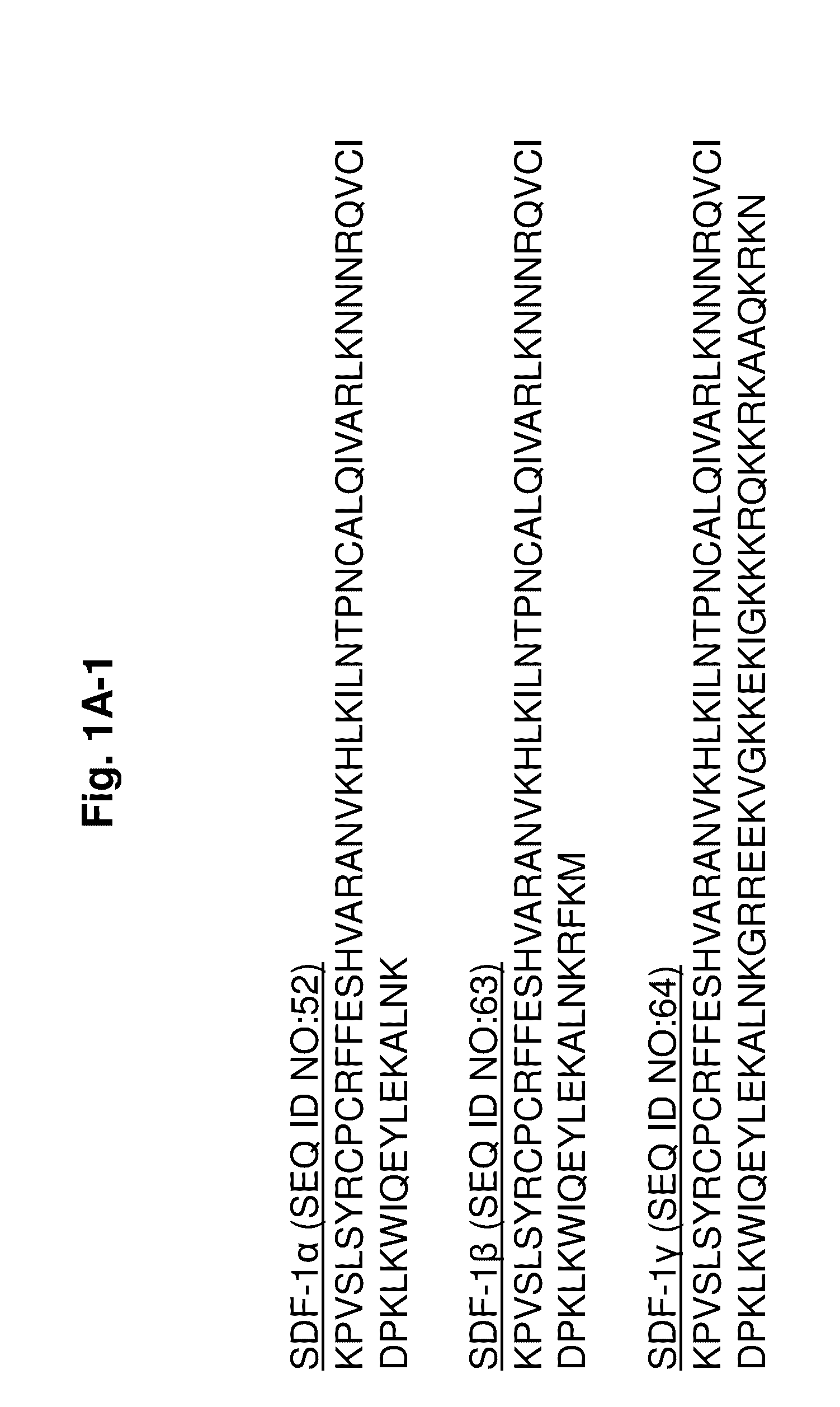
[0050] FIGS. 1A-1D show the cDNA sequences and amino acid sequences of SDF-1.alpha., SDF-1.beta., and SDF-1.gamma.. FIG. 1A-1 is a listing of the amino acid sequences of SDF-1.alpha. (SEQ ID NO:52), SDF-1.beta. (SEQ ID NO:63), and SDF-1.gamma. (SEQ ID NO:64) after removal of the signal peptide (amino acids 1-21). FIG. 1A-2 is a listing of the amino acid sequences of SDF-1.alpha. (SEQ ID NO:86), SDF-1.beta. (SEQ ID NO:87), and SDF-1.gamma. (SEQ ID NO:88), including the signal peptide. FIG. 1B is the cDNA sequence of SDF-1.alpha. (SEQ ID NO:69; NCBI Reference Sequence: NM_199168). FIG. 1C (-1 and -2) is the cDNA sequence of SDF-1.beta. (SEQ ID NO:70; NCBI Reference Sequence: NM_000609). FIG. 1D is the cDNA sequence of SDF-1.gamma. (SEQ ID NO:71; NCBI Reference Sequence: NM_001033886). For the cDNA sequences, the sequence coding for the final protein without signal peptide is underlined.
[0051] FIGS. 2A-2D are graphs showing that SSDF-1(S4V) has similar receptor specificity to SDF-1. FIG. 2A is a graph showing migration of Jurkat cells induced by SDF-1 variants (n>8). FIG. 2B is a graph showing that SDF-1 variants decrease cAMP levels by activation of CXCR4 and G.alpha.i (RLU=relative luminescence units) (n=4). FIG. 2C is a graph showing that SDF-1 variants induce binding of .beta.-arrestin to CXCR4 (n=3). FIG. 2D is a graph showing that SDF-1 variants induce binding of .beta.-arrestin to CXCR7 (n=3).
[0052] FIGS. 3A-3D are graphs and electrophoretic gels showing that SSDF-1(S4V) is resistant to MMP-2 cleavage. FIG. 3A is a graph of the fluorescence measurements taken at different time points after the addition of MMP-2 (n=4). FRET-based peptides were synthesized with the first 8 amino acids of SDF-1 or SDF-1(S4V). FIG. 3B is a bar graph showing the initial cleavage rate of the N-terminal 8 amino acid peptides of SDF-1 and SDF-1(S4V), calculated 60 minutes after the addition of MMP-2. FIGS. 3C and 3D are electrophoretic gels showing that a larger fraction of Sumo-SDF-1-RAD (FIG. 3C) is cleaved in 1 hour than Sumo-SDF-1(S4V)-RAD (FIG. 3D) in 48 hours. Sumo-SDF-1-RAD and Sumo-SDF-1(S4V)-RAD were incubated with MMP-2, and the reaction was stopped at different time points.
[0053] FIG. 4 is a bar graph showing the results of a Matrigel.TM. plug assay. The assay quantifies new vessel formation with different SDF-1 variants in vivo. The control group includes Matrigel.TM. only in the assay. The positive control includes VEGF and FGF-2 in the assay. Black bars represent newly made mutants of SDF-1. (*=P<0.05 compared to the control.)
[0054] FIG. 5 is a bar graph showing the results of a Matrigel.TM. plug assay. The assay quantifies new vessel formation with different SDF-1 variants in vivo at different dosages.
[0055] FIGS. 6A-6D are graphs and images showing SSDF-1-RAD stably incorporated into nanofibers. FIG. 6A is a graph showing the release of fluorescently labeled RAD from a nanofiber gel. 100 nM of FITC-labeled self-assembling peptides (FITC-RAD) were dissolved in PBS or mixed with a hydrogel of 1% nanofibers. Nanofibers were incubated in fresh PBS for time periods of 15 minutes, and cumulative release of fluorescence was measured. The curve of FITC-RAD+PBS indicates free diffusion in the employed system. FIG. 6B is a graph showing the release of fluorescently labeled SSDF-1 or SSDF-1-RAD from a nanofiber gel. 100 nM of Alexa-labeled SSDF-1, Alexa-labeled SSDF-1-RAD fusion protein, or Alexa 488 alone (as a small-molecule control) were mixed with a 1% nanofiber hydrogel. Cumulative release of fluorescence was measured by assessing the fluorescence remaining in the gel over the course of 24 hours. 78% of the SSDF-1-RAD fusion protein was still in the nanofiber gel after 24 hours. FIG. 6C is a micrograph showing tissue samples taken from mouse ischemic hindlimbs injected with nanofiber hydrogels containing biotinylated self-assembling peptides (1% of total peptides). Tissues were harvested at different time points and nanofibers were detected with Alexa-fluor-labeled streptavidin (green). Red=dystrophin; blue=DAPI. FIG. 6D is a graph plotting the total surface of the hydrogels as a function of time (n=5 per group).
[0056] FIGS. 7A-7C are images and graphs showing that protease-resistant SSDF-1(S4V) increases blood flow in a mouse hindlimb ischemia model. FIG. 7A is a set of representative images of the study in FIG. 7B. Blood flow in ischemic limb was normalized to blood flow in normal limb. FIG. 7B is a graph showing that nanofibers+SSDF-1(S4V)-RAD increased blood flow significantly over control animals at 4 and 6 weeks (n=10). FIG. 7C is a graph showing blood flow after different dosages of SSDF-1(S4V)-RAD were injected in the ischemic hindlimb (n=10). All groups included nanofibers. In FIGS. 7B and 7C, *=p<0.001 vs. control, .dagger.=p<0.01 vs. nanofibers; .dagger-dbl.=p<0.05 vs. nanofibers+SSDF-1-RAD; .sctn.=p<0.05 vs. control; #=p<0.001 vs. 0 nM; and **=p<0.01 vs. 0 nM.
[0057] FIGS. 8A-8B are images and graphs showing that SSDF-1(S4V) increases arteriolar density. FIG. 8A is a set of representative images of ischemic hindlimbs. Green=smooth muscle actin; red=isolectin. FIG. 8B is a bar graph showing the arteriolar vessel density in hindlimb of mice treated with different dosages of SSDF-1(S4V)-RAD. Per mouse, a total surface of 28 mm.sup.2 was measured. Scale bar=100 .mu.m (n>6 for each group).
DETAILED DESCRIPTION
[0058] The present invention is based upon the concept that the recovery of damaged tissue, e.g., damaged cardiac tissue, is promoted by exposing the tissue to SDF-1 that has been mutated to increase resistance to enzymatic cleavage (e.g., cleavage by MMP-2, MMP-9, DPPIV, leukocyte elastase, cathepsin G, carboxypeptidase M, or carboxypeptidase N). Such peptides may be delivered as isolated peptides, with or without a pharmaceutically acceptable carrier, or by means of a membrane formed by spontaneously assembling peptides (see, e.g., U.S. Pat. Nos. 5,670,483 and 6,548,630). Methods of attaching factors to membranes and the use of the membranes in delivering therapeutic agents to, for example, cardiac tissue have also been described (see, e.g., U.S. Patent Application Publication Nos. 2006/0148703 and 2006/0088510). The same procedures for making and using membranes may be applied to the present invention.
SDF-1 and Protease-Resistant Mutants
[0059] SDF-1 is a small cytokine belonging to the chemokine family that is officially designated chemokine (C-X-C motif) ligand 12 (CXCL12). SDF-1 is produced in multiple forms, SDF-1.alpha. (CXCL12a), SDF-1.beta. (CXCL12b), and SDF-1.gamma., by alternate splicing of the same gene.
[0060] Unmutated SDF-1.alpha. has the following sequence:
TABLE-US-00001 (SEQ ID NO: 52) K P V S L S Y R C P C R F F E S H V A R A N V K H L K I L N T P N C A L Q I V A R L K N N N R Q V C I D P K L K W I Q E Y L E K A L N K
[0061] The SDF-1 peptides of the present invention include SDF-1 peptides with mutations to render the peptides resistant to, for example, matrix metalloproteinase-2 (MMP-2), matrix metalloproteinase-9 (MMP-9), dipeptidyl peptidase IV (DPPIV), leukocyte elastase, cathepsin G, carboxypeptidase M, or carboxypeptidase N.
[0062] The peptides of the invention include mutant forms of SDF-1 (mSDF-1), which are characterized by a change in the third, fourth, fifth, and/or sixth amino acid residue from the N-terminus of unmutated SDF-1, and have at least amino acids 1-8 of the following sequence:
TABLE-US-00002 (SEQ ID NO: 53) K P X.sub.3 X.sub.4 X.sub.5 X.sub.6 Y R C P C R F F E S H V A R A N V K H L K I L N T P N C A L Q I V A R L K N N N R Q V C I D P K L K W I Q E Y L E K A L N K,
wherein [0063] X.sub.3 is any amino acid residue; [0064] X.sub.4 is serine or valine; [0065] X.sub.5 is leucine, proline, threonine, or valine; and [0066] X.sub.6 is any amino acid residue.
[0067] Excluded from the present invention are SDF-1 peptides that include the amino acid sequence of at least amino acids 1-8 of unmutated SDF-1 (SEQ ID NO:52) or SEQ ID NOs:65-67, defined as follows.
TABLE-US-00003 (SEQ ID NO: 65) K P V V L S Y R C P C R F F E S H V A R A N V K H L K I L N T P N C A L Q I V A R L K N N N R Q V C I D P K L K W I Q E Y L E K A L N K (SEQ ID NO: 66) K P V S P S Y R C P C R F F E S H V A R A N V K H L K I L N T P N C A L Q I V A R L K N N N R Q V C I D P K L K W I Q E Y L E K A L N K (SEQ ID NO: 67) K P V V P S Y R C P C R F F E S H V A R A N V K H L K I L N T P N C A L Q I V A R L K N N N R Q V C I D P K L K W I Q E Y L E K A L N K
[0068] In certain embodiments, the amino acid residue at position X.sub.3 of SEQ ID NO:53 is not isoleucine, e.g., when all other residues are wild-type amino acid residues. In other embodiments, the amino acid residue at position X.sub.5 of SEQ ID NO:53 is not a tryptophan or a glutamic acid amino acid residue, e.g., when all other residues are wild-type amino acid residues.
[0069] The peptides of the invention may include mSDF-1 peptides with a mutation at the third and/or sixth amino acid residue from the N-terminus of unmutated SDF-1 and have the sequence:
TABLE-US-00004 (SEQ ID NO: 68) K P X.sub.3 S L X.sub.6 Y R C P C R F F E S H V A R A N V K H L K I L N T P N C A L Q I V A R L K N N N R Q V C I D P K L K W I Q E Y L E K A L N K,
wherein X.sub.3 and X.sub.6 are any amino acid.
[0070] In certain embodiments, X.sub.3 of SEQ ID NO:68 is any amino acid residue and X.sub.6 of SEQ ID NO:68 is serine. In other embodiments, X.sub.3 of SEQ ID NO:68 is valine and X.sub.6 of SEQ ID NO:68 is any amino acid residue.
[0071] In certain embodiments, the amino acid residue at position X.sub.3 of SEQ ID NO:68 is not isoleucine.
[0072] For example, the mSDF-1 peptide may include a Val.fwdarw.His (SEQ ID NO:54) or Val.fwdarw.Cys (SEQ ID NO:55) mutation at the third amino acid position.
TABLE-US-00005 (SEQ ID NO: 54) K P H S L S Y R C P C R F F E S H V A R A N V K H L K I L N T P N C A L Q I V A R L K N N N R Q V C I D P K L K W I Q E Y L E K A L N K (SEQ ID NO: 55) K P C S L S Y R C P C R F F E S H V A R A N V K H L K I L N T P N C A L Q I V A R L K N N N R Q V C I D P K L K W I Q E Y L E K A L N K
[0073] In other embodiments, the mSDF-1 peptide may include a Leu.fwdarw.Thr (SEQ ID NO:56) or Leu.fwdarw.Val (SEQ ID NO:60) mutation at the fifth amino acid position. In certain embodiments, the fifth amino acid is not proline.
TABLE-US-00006 (SEQ ID NO: 56) K P V S T S Y R C P C R F F E S H V A R A N V K H L K I L N T P N C A L Q I V A R L K N N N R Q V C I D P K L K W I Q E Y L E K A L N K (SEQ ID NO: 60) K P V S V S Y R C P C R F F E S H V A R A N V K H L K I L N T P N C A L Q I V A R L K N N N R Q V C I D P K L K W I Q E Y L E K A L N K
[0074] In other embodiments, the mSDF-1 peptide may include a Ser.fwdarw.Cys (SEQ ID NO:61) or Ser.fwdarw.Gly (SEQ ID NO:62) mutation at the sixth amino acid position.
TABLE-US-00007 (SEQ ID NO: 61) K P V S L C Y R C P C R F F E S H V A R A N V K H L K I L N T P N C A L Q I V A R L K N N N R Q V C I D P K L K W I Q E Y L E K A L N K (SEQ ID NO: 62) K P V S L G Y R C P C R F F E S H V A R A N V K H L K I L N T P N C A L Q I V A R L K N N N R Q V C I D P K L K W I Q E Y L E K A L N K
[0075] The peptides of the invention also encompass any combination of the mutations described herein. For example, the mSDF-1 peptides may include a Val.fwdarw.Cys mutation at the third amino acid position of SEQ ID NO:53 and a Ser.fwdarw.Cys mutation at the sixth amino acid position of SEQ ID NO:53.
[0076] Mutations made to the SDF-1 peptides to confer protease resistance may also include, for example, the addition of a moiety (e.g., a proteinogenic amino acid or protease protective organic group) to the N-terminus of, e.g., the mSDF-1 peptides (described above), yielding X.sub.p-mSDF-1. For example, X may be: R.sup.1--(CH.sub.2).sub.d--, where d is an integer from 0-3, and R.sup.1 is selected from: hydrogen (with the caveat that when R.sup.1 is hydrogen, d must be at least 1); a branched or straight C.sub.1-C.sub.3 alkyl; a straight or branched C.sub.2-C.sub.3 alkenyl; a halogen, CF.sub.3; --CONR.sup.5R.sup.4; --COOR.sup.5; --COR.sup.5; --(CH.sub.2).sub.qNR.sup.5R.sup.4; --(CH.sub.2).sub.qSOR.sup.5; --(CH.sub.2).sub.qSO.sub.2R.sup.5, --(CH.sub.2).sub.qSO.sub.2NR.sup.5R.sup.4; and OR.sup.5, where R.sup.4 and R.sup.5 are each independently hydrogen or a straight or branched C.sub.1-C.sub.3 alkyl. In instances where an organic group is used for X, p should be 1. X may also represent a proteinogenic amino acid, wherein, for example, 1-10 (e.g., 1-9, 1-8, 1-7, 1-6, 1-5, 1-4, 1-3, 1-2, or 1) amino acid(s) is/are added to the N-terminus of SDF-1 (e.g., mSDF-1), and one or more of these added amino acids may be substituted with a protease protective organic group. For example, a proteinogenic amino acid (e.g., serine) or protease protective organic group may be added to the N-terminus of SDF-1 (e.g., mSDF-1) to confer, for example, resistance to DPPIV cleavage without substantially changing the chemoattractant activity or resistance to other proteases (e.g., MMP-2).
[0077] Mutations made to the SDF-1 peptides to confer protease resistance may also include, for example, the addition of a moiety (e.g., a proteinogenic amino acid) to the C-terminus of, e.g., the mSDF-1 peptides (described above), yielding mSDF-1-Y.sub.z or X.sub.p-mSDF-1-Y.sub.z. Y may represent a proteinogenic amino acid, wherein, for example, 1-10 (e.g., 1-9, 1-8, 1-7, 1-6, 1-5, 1-4, 1-3, 1-2, or 1) amino acid(s) is/are added to the C-terminus of SDF-1 (e.g., mSDF-1 or X.sub.p-mSDF-1). For example, a proteinogenic amino acid (e.g., serine) may be added to the C-terminus of SDF-1, mSDF-1, or X.sub.p-mSDF-1 to confer, for example, resistance to carboxypeptidase M or carboxypeptidase N cleavage without substantially changing the chemoattractant activity or resistance to other proteases (e.g., MMP-2). In one embodiment, the invention features an isolated mSDF-1-Y.sub.z or X.sub.p-mSDF-1-Y.sub.z peptide, wherein SDF-1 includes the amino acid sequence of SEQ ID NO:65, SEQ ID NO:66, or SEQ ID NO:67. However, C-terminal modifications may be made to any of the SDF-1 peptides described herein.
[0078] The mutated SDF-1 peptides of the invention retain their ability to act as chemoattractants, but are resistant to enzymatic (e.g., proteolytic) digestion. The mSDF-1 peptides maintain chemoattractant activity with a sensitivity (as determined by, e.g., the effective concentration needed to obtain 50% of maximal response in the assays of Jurkat T cell migration, as described herein) of at least, for example, 10, 15, 20, 25, 30, 40, 50, 60, 70, 80, 90, 95, 99, or 100% of the sensitivity of unmutated SDF-1. Loss of chemoattractant activity may be due to cleavage by, for example, MMP-2, MMP-9, leukocyte elastase, DPPIV, cathepsin G, carboxypeptidase M, or carboxypeptidase N. The rate of inactivation of mSDF-1 may be less than, for example, 50, 45, 40, 35, 30, 25, 20, 15, 10, 5, or 1% of the rate of inactivation of SDF-1.
[0079] The mutated SDF-1 peptides may be resistant to cleavage by, for example, MMP-2, MMP-9, DPPIV, leukocyte elastase, cathepsin G, carboxypeptidase M, or carboxypeptidase N. Thus, they are ideally suited for use at sites such as, e.g., damaged tissue (e.g., damaged cardiac tissue), where proteolytic enzymes are present at high concentrations, or delivery to the site via the blood or plasma. Accordingly, mutated SDF-1 peptides are suitable for intravenous, intra-arterial, and/or intracoronary administration due to the improved stability of such peptides. In addition, an MMP-2 cleavage site can, if desired, be placed in linker regions joining the SDF-1 peptides to self-assembling peptides, described herein. This will allow for the protease-resistant SDF-1 peptides to be released from an implanted or injected membrane, also described herein, over time.
[0080] Protease-resistant SDF-1 peptides of the present invention may include amino acids or sequences modified either by natural processes, such as posttranslational processing, or by chemical modification using techniques known in the art. Modifications may occur anywhere in a polypeptide, including the polypeptide backbone, the amino acid side-chains, and the amino- or carboxy-terminus. The same type of modification may be present in the same or varying degrees at several sites in a given polypeptide, and a polypeptide may contain more than one type of modification. Modifications include, e.g., PEGylation, acetylation, acylation, addition of acetomidomethyl (Acm) group, ADP-ribosylation, alkylation, amidation, biotinylation, carbamoylation, carboxyethylation, esterification, covalent attachment to fiavin, covalent attachment to a heme moiety, covalent attachment of a nucleotide or nucleotide derivative, covalent attachment of drug, covalent attachment of a marker (e.g., a fluorescent or radioactive marker), covalent attachment of a lipid or lipid derivative, covalent attachment of phosphatidylinositol, cross-linking, cyclization, disulfide bond formation, demethylation, formation of covalent crosslinks, formation of cystine, formation of pyroglutamate, formylation, gamma-carboxylation, glycosylation, GPI anchor formation, hydroxylation, iodination, methylation, myristoylation, oxidation, proteolytic processing, phosphorylation, prenylation, racemization, selenoylation, sulfation, transfer-RNA mediated addition of amino acids to proteins (e.g., arginylation), and ubiquitination. Posttranslational modifications also include the addition of polymers to stabilize the peptide or to improve pharmacokinetics or pharmacodynamics.
Fusion Proteins and Self-Assembling Peptides
[0081] The invention also encompasses fusion proteins in which any of the mSDF-1 or X.sub.p-mSDF-1 sequences described herein are linked to either self-assembling peptides capable of forming a biologically compatible membrane or the Fc region of IgG. Fusion proteins are formed either by joining the C-terminal end of a protease-resistant SDF-1 peptide directly to either the N-terminal end of a self-assembling peptide or the Fc region of IgG, or the two peptides can be joined by a linker sequence. Thus, the invention includes fusion proteins of the formula: A-(L).sub.n-(R).sub.q, wherein n is an integer from 0-3, q is an integer from 1-3, A is one of the protease-resistant SDF-1 peptides (i.e., mSDF-1, X.sub.p-mSDF-1, mSDF-1-Y.sub.z, or X.sub.p-mSDF-1-Y.sub.z), L is a linker sequence 3-9 amino acids long, and R is a self-assembling peptide.
[0082] Linker sequences may include, for example, GGGGGG (abbreviated as "6G," SEQ ID NO:57), GIVGPL (SEQ ID NO:58), and PVGLIG (SEQ ID NO:59). The lattermost represents an MMP-2 cleavage site (MCS). GIVGPL (SEQ ID NO:58) represents a scrambled version of MCS and is abbreviated as SCR. The SCR sequence also undergoes MMP-2 cleavage, although at a slower rate than MCS.
[0083] The self-assembling peptide may be, for example, one of the following peptides:
TABLE-US-00008 (SEQ ID NO: 1) AKAKAEAEAKAKAEAE; (SEQ ID NO: 2) AKAEAKAEAKAEAKAE; (SEQ ID NO: 3) EAKAEAKAEAKAEAKA; (SEQ ID NO: 4) KAEAKAEAKAEAKAEA; (SEQ ID NO: 5) AEAKAEAKAEAKAEAK; (SEQ ID NO: 6) ADADARARADADARAR; (SEQ ID NO: 7) ARADARADARADARAD; (SEQ ID NO: 8) DARADARADARADARA; (SEQ ID NO: 9) RADARADARADARADA; (SEQ ID NO: 10) ADARADARADARADAR; (SEQ ID NO: 11) ARADAKAEARADAKAE; (SEQ ID NO: 12) AKAEARADAKAEARAD; (SEQ ID NO: 13) ARAKADAEARAKADAE; (SEQ ID NO: 14) AKARAEADAKARADAE; (SEQ ID NO: 15) AQAQAQAQAQAQAQAQ; (SEQ ID NO: 16) VQVQVQVQVQVQVQVQ; (SEQ ID NO: 17) YQYQYQYQYQYQYQYQ; (SEQ ID NO: 18) HQHQHQHQHQHQHQHQ; (SEQ ID NO: 19) ANANANANANANANAN; (SEQ ID NO: 20) VNVNVNVNVNVNVNVN; (SEQ ID NO: 21) YNYNYNYNYNYNYNYN; (SEQ ID NO: 22) HNHNHNHNHNHNHNHN; (SEQ ID NO: 23) ANAQANAQANAQANAQ; (SEQ ID NO: 24) AQANAQANAQANAQAN; (SEQ ID NO: 25) VNVQVNVQVNVQVNVQ; (SEQ ID NO: 26) VQVNVQVNVQVNVQVN; (SEQ ID NO: 27) YNYQYNYQYNYQYNYQ; (SEQ ID NO: 28) YQYNYQYNYQYNYQYN; (SEQ ID NO: 29) HNHQHNHQHNHQHNHQ; (SEQ ID NO: 30) HQHNHQHNHQHNHQHN; (SEQ ID NO: 31) AKAQADAKAQADAKAQAD; (SEQ ID NO: 32) VKVQVDVKVQVDVKVQVD; (SEQ ID NO: 33) YKYQYDYKYQYDYKYQYD; (SEQ ID NO: 34) HKHQHDHKHQHDHKHQHD; (SEQ ID NO: 35) RARADADARARADADA; (SEQ ID NO: 36) RADARGDARADARGDA; (SEQ ID NO: 37) RAEARAEARAEARAEA; (SEQ ID NO: 38) KADAKADAKADAKADA; (SEQ ID NO: 39) AEAEAHAHAEAEAHAH; (SEQ ID NO: 40) FEFEFKFKFEFEFKFK; (SEQ ID NO: 41) LELELKLKLELELKLK; (SEQ ID NO: 42) AEAEAKAKAEAEAKAK; (SEQ ID NO: 43) AEAEAEAEAKAK; (SEQ ID NO: 44) KAKAKAKAEAEAEAEA; (SEQ ID NO: 45) AEAEAEAEAKAKAKAK; (SEQ ID NO: 46) RARARARADADADADA; (SEQ ID NO: 47) ADADADADARARARAR; (SEQ ID NO: 48) DADADADARARARARA; (SEQ ID NO: 49) HEHEHKHKHEHEHKHK; (SEQ ID NO: 50) VEVEVEVEVEVEVEVEVEVE; and (SEQ ID NO: 51) RFRFRFRFRFRFRFRFRFRF.
[0084] Exemplary fusion peptides include the self-assembling peptides RADARADARADARADA (SEQ ID NO:9) and RARADADARARADADA (SEQ ID NO:35), wherein q=1, and the protease-resistant SDF-1 peptide SDF(S6C) or X.sub.p-SDF(S6C), wherein p=1. When joined together, the resulting fusion proteins are, for convenience, abbreviated as SDF(S6C)-RAD or X.sub.p-SDF(S6C)-RAD. Exemplary fusion proteins containing linker sequences include SDF(S6C)-6G-RAD; X.sub.p-SDF(S6C)-6G-RAD; SDF(S6C)-MCS-RAD; X.sub.p-SDF(S6C)-MCS-RAD; SDF(S6C)-SCR-RAD; and X.sub.p-SDF(S6C)-SCR-RAD, wherein p=1.
[0085] The peptides used for self-assembly should be between, for example, at least 12 amino acids residues and 200 amino acid residues in length (e.g., between 12-24 amino acid residues in length) and contain alternating hydrophobic and hydrophilic amino acids. Peptides longer than about 200 amino acids may be less soluble and have decreased membrane stability.
[0086] The self-assembling peptides may be complementary. This means that the amino acids on one peptide are capable of forming ionic bonds or hydrogen bonds with the amino acids on another peptide. Ionic bonds may form between acidic and basic amino acid side chains. Ionic bonds may also form between an acidic residue on one peptide and a basic residue on another. Hydrophilic basic amino acids include Lys, Arg, His, and Orn. Hydrophilic acidic amino acids include Glu and Asp. Amino acids that form hydrogen bonds are Asn and Gln. Hydrophobic amino acids that may be incorporated into peptides include Ala, Val, Ile, Met, Phe, Tyr, Trp, Ser, Thr, and Gly.
[0087] Self-assembling peptides may also be structurally compatible. This means that they maintain an essentially constant distance between one another when they bind. Interpeptide distance can be calculated for each ionized or hydrogen bonding pair by taking the sum of the number of unbranched atoms on the side chains of each amino acid in the pair. For example, lysine has five unbranched atoms and glutamic acid has four unbranched atoms on their side chains. An interaction between these two residues on different peptides would result in an interpeptide distance of nine atoms. In a peptide containing only repeating units of EAK, all of the ion pairs would involve lysine and glutamate and, therefore, a constant interpeptide distance would be maintained. Thus, these peptides would be structurally complementary.
[0088] Peptides in which the variation in interpeptide distance varies by more than one atom (about 3-4 .ANG.) will not form gels properly. For example, if two bound peptides have ion pairs with a nine-atom spacing and other ion pairs with a seven-atom spacing, the requirement of structural complementarity would not have been met. A full discussion of complementarity and structural compatibility may be found in, for example, U.S. Pat. Nos. 5,670,483 and 6,548,630.
[0089] The invention also encompasses fusion proteins in which any of the SDF-1, mSDF-1, X.sub.p-mSDF-1, mSDF-1-Y.sub.z, or X.sub.p-mSDF-1-Y.sub.z, peptide sequences described herein are linked to the Fc region of IgG (e.g., human IgG1). Alternatively, the Fc region may be derived from IgA, IgM, IgE, or IgD of humans or other animals, including swine, mice, rabbits, hamsters, goats, rats, and guinea pigs. The Fc region of IgG includes the CH2 and CH3 domains of the IgG heavy chain and the hinge region. The hinge serves as a flexible spacer between the two parts of the Fc fusion protein, allowing each part of the molecule to function independently. The Fc region used in the present invention can be prepared in, for example, monomeric and dimeric form.
[0090] An exemplary Fc fusion peptide is SSDF-1(S4V)-Fc with the following amino acid sequence. The GGGGS linker is indicated in bold and the Fc peptide is underlined.
TABLE-US-00009 (SEQ ID NO: 83) S K P V V L S Y R C P C R F F E S H V A R A N V K H L K I L N T P N C A L Q I V A R L K N N N R Q V C I D P K L K W I Q E Y L E K A L N K G G G G S V D K T H T C P P C P A P E L L G G P S V F L F P P K P K D T L Met I S R T P E V T C V V V D V S H E D P E V K F N W Y V D G V E V H N A K T K P R E E Q Y N S T Y R V V S V L T V L H Q D W L N G K E Y K C K V S N K A L P A P I E K T I S K A K G Q P R E P Q V Y T L P P S R D E L T K N Q V S L T C L V K G F Y P S D I A V E W E S N G Q P E N N Y K T T P P V L D S D G S F F L Y S K L T V D K S R W Q Q G N V F S C S V Met H E A L H N H Y T Q K S L S L SPGK
[0091] Other non-limiting examples of Fc fusion peptides include, e.g., SDF-1(S4V)-Fc, SDF-1-Fc, and SSDF-1-Fc.
Binding of SDF-1 to Self-Assembling Peptides
[0092] Several strategies may be used for attaching protease-resistant SDF-1 to self-assembling peptides. Exemplary strategies are described as follows.
[0093] One strategy is non-covalent binding, which has previously been shown to be effective in delivering PDGF-BB, a growth factor, to tissues (Hsieh et al., J. Clin. Invest. 116: 237-248 (2006)).
[0094] A second attachment strategy is the biotin-sandwich method (Davis et al., Proc. Natl. Acad. Sci. USA 103: 8155-8160 (2006)) in which a protease-resistant SDF-1 is biotinylated and bound to biotinylated peptides using tetravalent streptavidin as a linker. To accomplish this, the protease-resistant SDF-1 may be coupled to a 15 amino acid sequence of an acceptor peptide for biotinylation (referred to as AP; Chen et al., Nat. Methods 2: 99-104 (2005)). Because the active site of SDF-1 is situated near the amino-terminus, fusion proteins can be made by incorporating the extra sequences at the C-terminus. The acceptor peptide sequence allows site-specific biotinylation by the E. coli enzyme biotin ligase (BirA; Chen et al., Nat. Methods 2: 99-104 (2005)). Many commercial kits are available for biotinylating proteins. However, many of these kits biotinylate lysine residues in a nonspecific manner, and this may reduce mSDF-1 activity, as it has been shown that the N-terminal lysine of SDF-1 is crucial for receptor binding and activity (Crump et al., EMBO J. 16: 6996-7007 (1997)). Biotinylated self-assembling peptides are made, for example, by MIT Biopolymers Laboratory and, when mixed in a 1-to-100 ratio with native self-assembling peptides, self-assembly of nanofibers should not be disturbed (Davis et al., Proc. Natl. Acad. Sci. USA 103: 8155-8160 (2006)).
[0095] A third targeting strategy is direct incorporation of protease-resistant SDF-1 peptides into self-assembling nanofibers by construction of a fusion protein of mutated SDF-1 with a self-assembling peptide. For example, mSDF-1 may be coupled (e.g., chemically or recombinantly) to the 16 amino acid sequence of SEQ ID NO:9 or SEQ ID NO:35. This "RAD" portion of the fusion protein will incorporate into the nanofiber scaffold while assembling.
Formation of Membranes
[0096] The invention also features biologically compatible membranes formed from self-assembling peptides, as described in U.S. Pat. Nos. 7,429,567 and 7,399,831, which have, for example, mSDF-1 or X.sub.p-mSDF-1 peptides attached. As used herein, the term "biologically compatible" indicates that the membranes are non-toxic and can be implanted or injected into a subject without triggering an immune response. Between 0.1% and 10% (e.g., between 0.5-5%) of the peptides that assemble into the membrane are bound to a mutant SDF-1. Binding may be either, e.g., covalent or noncovalent. Noncovalent binding occurs when protease-resistant SDF-1 peptides are trapped in the membrane matrix and/or when protease-resistant SDF-1 peptides are bound to self-assembling peptides in the membrane by biotin-avidin or biotin-streptavidin linkages. The membranes may, optionally, have other therapeutic agents (e.g., PDGF, interleukin-8, IGF-1, HGF, neuregulin, periostin, VEGF, or FGF) attached.
[0097] The use of the biotin-avidin linkage is well known in the art, and standard methodology can be used for attaching protease-resistant SDF-1 peptides to self-assembling peptides either before or after membrane formation. Specific methodology for using biotin-avidin in connection with self-assembling membranes has been described in, for example, U.S. Pat. No. 7,399,831. To prevent steric interference between the biotin-avidin groups and protease-resistant peptides, a spacer may be included between the biotin-avidin group and protease-resistant peptide. The spacer can take the form of, for example, between 1-15 (e.g., 1-10) fatty acids or, for example, between 1-15 (e.g., 1-10) amino acids and separates the protease-resistant SDF-1 peptide from the self-assembling peptide by, for example, at least an additional 12 .ANG. and by no more than an additional 250 .ANG.. Methodologies for incorporating spacers of this type are well known in the art.
[0098] Protease-resistant SDF-1 peptides may also be joined to a self-assembling peptide that is part of the membrane by a peptide bond, i.e., the protease-resistant SDF-1 may be part of a fusion protein in which it is joined to a self-assembling peptide either directly or via an intervening linker amino acid sequence. Any of the fusion proteins described herein may be used. The membranes are made from the fusion proteins or from the self-assembling peptides by taking advantage of the inability of the fusion proteins or self-assembling peptides described herein to congregate in water. Instead, the fusion proteins or self-assembling peptides assemble into a membrane in the presence of a low concentration of, for example, monovalent metal cations (e.g., Li.sup.+, Na.sup.+, K.sup.+, or Cs.sup.+). For example, fusion proteins may be made under conditions in which self-assembly does not occur and then exposed to conditions that promote membrane formation, e.g., low monovalent metal cation concentration. The end result is a matrix which can be, for example, implanted into a subject and which will maintain a high concentration of SDF-1 biological activity at the site of implantation. Alternatively, the fusion proteins can be incorporated into an injectable pharmaceutical composition at a concentration of monovalent cation that is too low to induce self-assembly and can then be administered to a subject to induce membrane formation in vivo.
[0099] Membranes may be formed from either a homogeneous mixture of peptides or a heterogeneous mixture of peptides. The term "homogeneous," in this context, means peptides that are identical. "Heterogeneous" indicates peptides that bind to one another, but which are structurally different. Regardless of whether homogenous or heterogeneous peptides are used, the requirements with respect to the arrangement of amino acids, length, complementarity, and structural compatibility apply. In addition, the carboxyl and amino groups of the terminal residues of peptides can either be protected or not protected using standard groups and standard methods known in the art.
[0100] The self-assembling peptides and fusion proteins described herein may not form membranes in water, but may instead assemble in the presence of a low concentration of monovalent metal cation. The order of effectiveness of these cations is Li.sup.+>Na.sup.+>K.sup.+>Cs.sup.+ (see, e.g., U.S. Pat. No. 6,548,630). The concentration of monovalent metal cation may be, for example, between 5 mM and 5M (e.g., 5 mM, 10 mM, 50 mM, 100 mM, 200 mM, 300 mM, 400 mM, 500 mM, 600 mM, 700 mM, 800 mM, 900 mM, 1M, 2M, 3M, 4M, or 5M). The anion associated with the monovalent cation is not critical to the invention and can be, for example, acetate, chloride, sulfate, or phosphate.
[0101] The initial concentration of self-assembling peptide will influence the final size and thickness of the membranes formed. In general, the higher the peptide concentration, the higher the extent of membrane formation. Formation can take place at peptide concentrations as low as 0.5 mM or 1 mg/ml. However, membranes are preferably formed at higher initial peptide concentrations, e.g., 10 mg/ml, to promote better handling characteristics. Peptides may be added to a salt solution (rather than adding salt to a peptide solution) to form membranes of the present invention. During membrane formation, the pH may be maintained below 12, and temperatures may be generally in the range of, for example, 4-90.degree. C.
[0102] An exemplary membrane matrix that may be used in the compositions and methods described herein is PuraMatrix.TM. (BD Biosciences).
[0103] Membrane formation may be observed by simple visual inspection and this can be aided, if desired, with stains such as Congo Red. The integrity of membranes can also be observed microscopically, with or without stain.
[0104] Membranes with protease-resistant SDF-1 attached may be, for example, injected or implanted at or near the site of damaged tissue (e.g., cardiac tissue), wounds (e.g., accidental wounds, surgical wounds, or wounds resulting from a disease), or ulcers of a subject in need of treatment. The membranes should be large enough to prevent the protease-resistant SDF-1 from being washed away by bodily fluids, and a sufficient amount of mSDF-1 should be present to promote the migration of, for example, T cells to the site of injury.
Peptide Synthesis
[0105] The self-assembling and protease-resistant SDF-1 peptides of the present invention can be made by solid-phase peptide synthesis using, for example, standard N-tert-butyoxycarbonyl (t-Boc) chemistry and cycles using n-methylpyrolidone chemistry. Exemplary methods for synthesizing peptides are found, for example, in U.S. Pat. Nos. 4,192,798; 4,507,230; 4,749,742; 4,879,371; 4,965,343; 5,175,254; 5,373,053; 5,763,284; and 5,849,954, hereby incorporated by reference.
[0106] Once peptides have been synthesized, they can be purified using procedures such as, for example, high pressure liquid chromatography (HPLC) on reverse-phase columns. Purity may also be assessed by HPLC, and the presence of a correct composition can be determined by amino acid analysis. A purification procedure suitable for mSDF-1 peptides is described in, for example, U.S. Patent Application Publication No. 2008/0095758, hereby incorporated by reference.
[0107] Fusion proteins may either be chemically synthesized or made using recombinant DNA techniques. The full sequences of these proteins are described herein. Non-limiting examples of fusion protein DNA and amino acid sequences are provided below.
TABLE-US-00010 SSDF-1-6G-RAD16-I (SEP ID NO: 72) agc aag ccg gtc agc ctg agc tac cgt tgc cca tgc cgt ttc ttc gaa agc cat gtt gcc cgc gcc aac gtc aag cat ctc aaa att ctc aac act cca aac tgt gcc ctt cag att gta gcc cgt ctg aag aac aac aac cgc caa gtg tgc att gac ccg aag ctg aag tgg att cag gag tac ctg gag aaa gct tta aac aag GGA GGC GGG GGA GGT GGG CGT GCC GAC GCT CGC GCA GAT GCG CGT GCC GAC GCT CGG GCA GAT GCG TGA Corresponding SSDF-1-6G-RAD16-I Amino Acid Sequence (SEQ ID NO: 73) S K P V S L S Y R C P C R F F E S H V A R A N V K H L K I L N T P N C A L Q I V A R L K N N N R Q V C I D P K L K W I Q E Y L E K A L N K G G G G G G R A D A R A D A R A D A R A D A SSDF-1(S4V)-6G-RAD16-I (SEQ ID NO: 74) agc aag ccg gtc gtc ctg agc tac cgt tgc cca tgc cgt ttc ttc gaa agc cat gtt gcc cgc gcc aac gtc aag cat ctc aaa att ctc aac act cca aac tgt gcc ctt cag att gta gcc cgt ctg aag aac aac aac cgc caa gtg tgc att gac ccg aag ctg aag tgg att cag gag tac ctg gag aaa gct tta aac aag GGA GGC GGG GGA GGT GGG CGT GCC GAC GCT CGC GCA GAT GCG CGT GCC GAC GCT CGG GCA GAT GCG TGA Corresponding SSDF-1(S4V)-6G-RAD16-I Amino Acid Sequence (SEQ ID NO: 75) S K P V V L S Y R C P C R F F E S H V A R A N V K H L K I L N T P N C A L Q I V A R L K N N N R Q V C I D P K L K W I Q E Y L E K A L N K G G G G G G R A D A R A D A R A D A R A D A
Nucleic Acids
[0108] The invention also features nucleic acids with a nucleotide sequence encoding any of the protease-resistant peptides or fusion proteins described above, vectors (e.g., Champion pET Sumo vectors (Invitrogen), pET101/D-TOPO vectors (Invitrogen), or pTrcHis or pTrcHis2 vectors) in which these nucleic acids are operably linked to a promoter sequence, and host cells (e.g., cell-free expression systems, prokaryotic expression systems (e.g., E. coli BL21, BL21-DE3, BL21Star, or BL21pLys), yeast expression systems (e.g., Pichia pastoris), baculovirus expression systems, or mammalian expression systems (e.g., transient expression in HEK293E cells or a stable CHO cell line)) transformed with the vector.
[0109] Exemplary nucleotide sequences of the peptides or fusion proteins of the invention are provided below and throughout the specification. Any of the codons can be mutated without changing the protein sequence following the genetic code.
TABLE-US-00011 SDF-1 native (SEQ ID NO: 76) aag ccc gtc agc ctg agc tac aga tgc cca tgc cga ttc ttc gaa agc cat gtt gcc aga gcc aac gtc aag cat ctc aaa att ctc aac act cca aac tgt gcc ctt cag att gta gcc cgg ctg aag aac aac aac aga caa gtg tgc att gac ccg aag cta aag tgg att cag gag tac ctg gag aaa gct tta aac aag tga SDF-1 codon optimized, excluding rare E. coli codons (SEQ ID NO: 77) aag ccg gtc agc ctg agc tac cgt tgc cca tgc cgt ttc ttc gaa agc cat gtt gcc cgc gcc aac gtc aag cat ctc aaa att ctc aac act cca aac tgt gcc ctt cag att gta gcc cgt ctg aag aac aac aac cgc caa gtg tgc att gac ccg aag ctg aag tgg att cag gag tac ctg gag aaa gct tta aac aag tga SSDF-1 codon optimized, excluding rare E. coli codons (SEQ ID NO: 78) agc aag ccg gtc agc ctg agc tac cgt tgc cca tgc cgt ttc ttc gaa agc cat gtt gcc cgc gcc aac gtc cat ctc aaa att ctc aac act cca aac tgt gcc ctt cag att gta gcc cgt ctg aag aac aac aac cgc caa gtg tgc att gac ccg aag ctg aag tgg att cag gag tac ctg gag aaa gct tta aac aag tga
Pharmaceutical Compositions and Dosages
[0110] Any of the peptides employed according to the present invention may be contained in any appropriate amount in any suitable carrier substance, and the protease-resistant peptides or fusion proteins of the invention are generally present in an amount of 1-95% by weight of the total weight of the composition, e.g., 5%, 10%, 20%, or 50%. The protease-resistant SDF-1 peptides or fusion proteins of the present invention may be incorporated into a pharmaceutical composition containing a carrier such as, e.g., saline, water, Ringer's solution, and other agents or excipients. The dosage form will generally be designed for implantation or injection (e.g., intravenous, intra-arterial, and/or intracoronary injection), particularly into cardiac tissue, but topical treatments will also be useful, e.g., in the treatment of wounds. Additionally, the composition may be provided in a dosage form that is suitable for the oral, parenteral, intrathecal, rectal, cutaneous, nasal, vaginal, inhalant, skin (e.g., patch), or ocular administration route. Thus, the composition may be in the form of, e.g., tablets, capsules, pills, powders, granulates, suspensions, emulsions, solutions, gels (e.g., hydrogels), pastes, ointments, creams, plasters, drenches, osmotic delivery devices, suppositories, enemas, injectables, implants, sprays, or aerosols. All dosage forms may be prepared using methods that are standard in the art (see, e.g., Remington's Pharmaceutical Sciences, 16th ed., A. Oslo. ed., Easton, Pa. (1980)).
[0111] The peptides of the invention can be delivered in a controlled-release or sustained-release system. For example, polymeric materials can be used to achieve controlled or sustained release of the peptides (see, e.g., Medical Applications of Controlled Release, Langer and Wise (eds.), CRC Pres., Boca Raton, Fla. (1974); Controlled Drug Bioavailability, Drug Product Design and Performance, Smolen and Ball (eds.), Wiley, N.Y. (1984); U.S. Pat. Nos. 5,679,377; 5,916,597; 5,912,015; 5,989,463; and 5,128,326; PCT Publication Nos. WO 99/15154 and WO 99/20253, hereby incorporated by reference). Examples of polymers used in sustained-release formulations include, e.g., poly(2-hydroxy ethyl methacrylate), poly(methyl methacrylate), poly(acrylic acid), poly(ethylene-co-vinyl acetate), poly(methacrylic acid), polyglycolides (PLG), polyanhydrides, poly(N-vinyl pyrrolidone), poly(vinyl alcohol), polyacrylamide, poly(ethylene glycol), polylactides (PLA), poly(lactide-co-glycolides) (PLGA), polyglutamic acid (PGA), and polyorthoesters.
[0112] It is expected that the skilled practitioner can adjust dosages on a case by case basis using methods well established in clinical medicine. The optimal dosage may be determined by methods known in the art and may be influenced by factors such as the age of the subject being treated, disease state, and other clinically relevant factors. Generally, when administered to a human, the dosage of any of the therapeutic agents (e.g., protease-resistant SDF-1 peptides) of the invention will depend on the nature of the agent and can readily be determined by one skilled in the art. Typically, such dosage is normally about 0.001 .mu.g to 2000 mg per day, about 0.001 mg to 1000 mg per day, or about 0.5 to 500 mg per day. In various embodiments, the dosage is about 1 mg to 100 mg per day or about 5 mg to 500 mg per day. The dosages can also be expressed as mg/kg and examples of such dosages include 0.001 mg/kg per day to 100 mg/kg per day, 0.001 mg/kg per day to 50 mg/kg per day, and 0.01 mg/kg per day to 10 mg/kg per day.
[0113] The peptides of the invention may be administered once, twice, three times, four times, or five times each day; once per week, twice per week, three times per week, four times per week, five times per week, or six times per week; once per month, once every two months, once every three months, or once every six months; or once per year. Alternatively, the peptides of the invention may be administered one or two times and repeated administration may not be needed. Administration of the peptides of the invention can continue until tissue damage (e.g., tissue damage resulting from myocardial infarction or peripheral vascular disease) has healed or has been ameliorated. The duration of therapy can be, e.g., one week to one month; alternatively, the peptides of the invention can be administered for a shorter or a longer duration. Continuous daily dosing with the peptides described herein may not be required. A therapeutic regimen may require cycles, during which time a composition is not administered, or therapy may be provided on an as-needed basis.
[0114] Appropriate dosages of the peptides used in the methods and compositions described herein depend on several factors, including the administration method, the severity of the disorder, and the age, weight, and health of the subject to be treated. Additionally, pharmacogenomic information (e.g., the effect of genotype on the pharmacokinetic, pharmacodynamic, or efficacy profile of a therapeutic) about a particular subject may affect the dosage used.
Diagnosis and Treatment
[0115] The compositions of the present invention are useful for treating any subject that has been diagnosed with or has suffered from tissue damage (e.g., damage to cardiac tissue due to myocardial infarction or tissue damage resulting from peripheral vascular disease) or wounds. Tissue damage may be the result of, for example, a cardiovascular condition (e.g., myocardial infarction); peripheral vascular disease (PVD); peripheral artery disease (PAD); ulcers (e.g., ulcers of the gastrointestinal tract); surgery; or diabetes. The compositions of the present invention may be used to promote wound healing or tissue repair. One skilled in the art will understand that subjects of the invention may have been subjected to standard tests or may have been identified, without examination, as one at high risk due to the presence of one or more risk factors. Diagnosis of these disorders may be performed using any standard method known in the art.
[0116] The compositions described herein may also be used to treat any disease or condition characterized by a high concentration of protease (e.g., MMP-2, MMP-9, DPPIV, leukocyte elastase, cathepsin G, carboxypeptidase M, and/or carboxypeptidase N), where the attraction of stem cells upon the administration of a protease-resistant SDF-1 peptide may induce regeneration or healing. Exemplary disorders to be treated by compositions of the present invention include inflammatory and ischemic diseases (e.g., stroke or limb ischemia), wound healing, and diabetic ulcers.
[0117] The protease-resistant SDF-1 peptides of the present invention may be used in combination with additional therapies to promote wound healing or tissue repair. Treatment therapies that can be used in combination with the methods of the invention include, but are not limited to heparin, .beta.-blockers (e.g., atenolol, metoprolol, nadolol, oxprenolol, pindolol, propranolol, or timolol), angiotensin-converting enzyme (ACE) inhibitors (e.g., captopril, enalapril, fosinopril, lisinopril, perindopril, quinapril, ramipril, trandolapril, or benazepril), angiotensin II receptor blockers (e.g., candesartan, eprosartan, irbesartan, losartan, olmesartan, telmisartan, or valsartan), diuretics, aspirin, cholesterol-lowering drugs (e.g., HMG-CoA reductase inhibitors (e.g., atorvastatin, cerivastatin, fluvastatin, lovastatin, mevastatin, pitavastatin, pravastatin, rosuvastatin, or simvastatin)), cell therapy, anti-platelet drugs (e.g., clopidogrel, prasugrel, ticlopidine, cilostazol, abciximab, eptifibatide, tirofiban, or dipyridamole), anti-hypertensive drugs, anti-arrhythmic drugs (e.g., quinidine, procainamide, disopyramide, lidocaine, mexiletine, tocainide, phenytoin, moricizine, flecainide, sotalol, ibutilide, amiodarone, bretylium, dofetilide, diltiazem, or verapamil), angiogenic drugs, wound dressings, PDGF, and/or negative pressure devices and therapies.
EXAMPLES
[0118] The present invention is illustrated by the following examples, which are in no way intended to be limiting of the invention.
Example 1
Initial Cleavage Rates of a FRET-Based Peptide Library
[0119] Peptides containing the first 8 amino acids of SDF-1 (KPVSLSYR) and all possible single amino acid mutations at position 3, 4, 5, or 6 (spanning the MMP-2 cleavage site) were synthesized, 77 peptides total (Anaspec, San Jose, Calif.). All peptides were synthesized with a fluorescent 7-methoxycoumarin-3-carboxylic acid (MCA) residue at the N-terminus and a quencher lysine-dinitrophenol (Dpn) residue at the C-terminus. Purity of peptides was confirmed with HPLC and mass spectrometry. Enzymatic cleavage of these peptides separated MCA from Dpn, resulting in an increased fluorescence. Peptides (10 .mu.M) were then incubated with activated MMP-2 (Table 1), leukocyte elastase (Table 2), or cathepsin G (Table 3) (1.4 nM) in TNCB buffer (50 mM Tris, pH 7.5, with 10 mM calcium chloride, 150 mM sodium chloride, and 0.05% BRIJ 35) or phosphate-buffered saline (PBS) at 21.degree. C. Fluorescence was measured at 340 nm excitation and 405 nm emission wavelengths at different time points with a Victor Wallace II plate reader. Initial cleavage rates (fluorescence per unit of time) were normalized to the cleavage rate of peptides containing the first 8 amino acids of SDF-1 (100%).
[0120] The results are shown in Tables 1-3. Column headers show the site of the mutation(s) and the row title shows the mutated amino acid in that position. Mutations with an initial cleavage rate of less than 10% are indicated in italics and mutations with an initial cleavage rate between 10% and 20% of SDF-1 analogue are indicated in grey shading.
TABLE-US-00012 TABLE 1 Initial cleavage rates of FRET-peptides by MMP-2 ##STR00001##
TABLE-US-00013 TABLE 2 Initial cleavage rates of FRET-peptides by leukocyte elastase ##STR00002##
TABLE-US-00014 TABLE 3 Initial cleavage rates of FRET-peptides by cathepsin G ##STR00003##
Example 2
Identification and Characterization of SDF-1 Variants Resistant to MMP-2 Cleavage
[0121] The site where SDF-1 is cleaved by MMP-2, leukocyte elastase, and cathepsin G is also the receptor binding site of SDF-1. Thus, mutations that make SDF-1 resistant to proteases may also interfere with receptor binding and activation. Based on the FRET-peptide library initial cleavage rates, mutations were selected for cloning into an E. coli expression vector. Proteins were expressed in E. coli and purified with cation-exchange chromatography and reverse-phase HPLC. Activity of these proteins was tested in a Jurkat T-lymphocyte migration assay, CXCR4 (cAMP) receptor activation assay, and CXCR7 .beta.-arrestin binding assay. For these experiments and other experiments described herein, data were analyzed with 2-tail t-tests if 2 independent samples were compared and with one-way ANOVA if more than 2 independent samples were present, followed by multiple comparisons with Bonferroni corrections. Data are presented as mean.+-.SEM.
[0122] For the Jurkat T-lymphocyte migration assay, the activity of purified SDF-1 variants was tested by migration of Jurkat T-lymphocytic cells in a ChemoTx chemotaxis system (5 .mu.m pore, Neuro Probe, Gaithersburg, Md.). Jurkat cells were labeled with Calcein-AM (1 .mu.M) for 60 minutes at 37.degree. C., washed twice and resuspended in RPMI without phenol red to a final density of 4,000,000 cells/ml. 50 .mu.l of this cell suspension was added to upper wells, and different concentrations of SDF-1 variants were added to lower wells. Cells were allowed to migrate for 3 hours, and cells in lower wells were washed once with PBS. Fluorescence in lower wells was measured at 485 nm excitation and 535 nm emission wavelengths with a Victor Wallace II plate reader. A standard curve was constructed for each experiment to normalize fluorescence to the number of cells.
[0123] For the CXCR4 cAMP assay, CHO-K1 cells stably expressing the CXCR4 receptor (DiscoveRx, Fremont, Calif.) were seeded at a density of 10,000 cells/well in a 384-well plate and were allowed to recover overnight. A decrease in cAMP by CXCR4-induced G.alpha.i activation was determined using the DiscoveRx HitHunter cAMP XS+ assay following the manufacturer's protocol. Forskolin (an inducer of cAMP) concentration-response curves were generated to determine the EC.sub.80 concentration for subsequent testing with SDF-1 variants. The cells were incubated in the presence of SDF-1 variants and forskolin (at EC.sub.80 concentration) at 37.degree. C. for 30 minutes. After the incubation period, assay signal was generated through incubation with 20 .mu.L cAMP XS+ED/CL lysis cocktail for one hour followed by incubation with 20 .mu.L cAMP XS+EA reagent for a three-hour incubation at 21.degree. C. Chemiluminescence was read with a PerkinElmer ViewLux instrument.
[0124] For the CXCR4, CXCR7, CXCR3, and CXCR1 .beta.-arrestin recruitment assays, activation of CXCR4, CXCR7, CXCR3, and CXCR1 receptors by SDF-1 variants was detected by measuring .beta.-arrestin recruitment to the receptors. PathHunter .beta.-arrestin assays were performed following the instructions of the manufacturer (DiscoveRx). Briefly, PathHunter cell lines stably expressing both a .beta.-arrestin-Enzyme Acceptor of .beta.-galactosidase fusion and one of the chemokine receptors fused with the ProLink donor peptide of .beta.-galactosidase were employed. Upon CXCR stimulation, .beta.-arrestin is recruited to the receptor for desensitization, bringing the two fragments (the Enzyme Acceptor and the ProLink) of .beta.-galactosidase together. This generates an active .beta.-galactosidase enzyme that can convert a substrate to a chemiluminescent product. Cells were seeded at a density of 5000 cells/well in a 384-well plate and were allowed to adhere overnight. Cells were incubated in the presence of different concentrations of SDF-1 variants at 37.degree. C. for 90 minutes. After incubation, chemiluminescent signal was generated through addition of 12.5 .mu.l (50% v/v) of PathHunter Detection reagent cocktail (DiscoveRx) followed by a 1-hour incubation at room temperature. Microplates were read with a PerkinElmer ViewLux instrument.
[0125] The results of these assays are described in Tables 4 and 5.
TABLE-US-00015 TABLE 4 Mutants described in Segers et al. (Circulation 116: 1683-1692, 2007) Jurkat CXCR4 CXCR7 Chemotaxis (cAMP) .beta.-arrestin Protein Resistance EC.sub.50 (nM) EC.sub.50 (nM) EC.sub.50 (nM) SDF-1 -- 1.5 5.4 22 SSDF-1 DPPIV 1.6 1.2 20 SSDF-1(S4V) DDPIV, MMP-2 5.4 20 37 SSDF-1(L5P) DDPIV, MMP- 120 Inactive 225 2, elastase, cathepsin G SSDF-1(L5E) DDPIV, MMP- Inactive -- -- 2, elastase, cathepsin G SSDF-1(L5W) DDPIV, MMP-2 Inactive -- --
TABLE-US-00016 TABLE 5 Identified mutants (resistance profile based on FRET-peptide cleavage assay) Jurkat CXCR4 CXCR7 Chemotaxis (cAMP) .beta.-arrestin Protein Resistance EC.sub.50 (nM) EC.sub.50 (nM) EC.sub.50 (nM) SSDF-1(V3H) DPPIV, elastase 6.7 1 92 SSDF-1(V3C) DPPIV, elastase 15 0.6 14 SSDF-1(L5T) DPPIV, MMP-2, 20 3.6 8.7 Cathepsin G SSDF-1(L5V) DPPIV, MMP-2, 36 33 21 Cathepsin G SSDF-1(S6C) DPPIV, MMP-2 23 12 29 SSDF-1(S6G) DPPIV, MMP-2, 11 8.5 25 Cathepsin G SSDF-1(V3G) DDPIV, Inactive -- -- elastase, Cathepsin G SSDF-1(V3P) DDPIV, MMP- Inactive -- -- 2, elastase, Cathepsin G SSDF-1(V3D) DDPIV, MMP- Inactive -- -- 2, elastase, Cathepsin G SSDF-1(L5C) DDPIV, MMP- Inactive -- -- 2, Cathepsin G SSDF-1(L5K) DDPIV, MMP- Inactive -- -- 2, Elastase SSDF-1(L5H) DDPIV, MMP- Inactive -- -- 2, Cathepsin G SSDF-1(L5D) DDPIV, MMP- Inactive -- -- 2, Elastase, Cathepsin G
[0126] Mutated variants of SDF-1 identified as being inactive in the Jurkat cell migration assay included SSDF-1(V3D), SSDF-1(L5C), SSDF-1(L5D), SSDF-1(L5E), SSDF-1(L5H), SSDF-1(L5K), SSDF-1(L5W), SSDF-1(V3G), and SSDF-1(V3P). SSDF-1(S4V) was identified as the mutated variant with the highest potency in the Jurkat cell migration assay.
Example 3
SSDF-1(S4V) has the Same Receptor Specificity as Native SDF-1
[0127] The activity of SSDF-1(S4V) was evaluated in Jurkat cell migration assays (FIG. 2A) and CXCR4 and CXCR7 receptor activation assays and compared to native SDF-1 and SSDF-1, as described above. The EC.sub.50 for CXCR4 receptor activation was 20 nM with SSDF-1(S4V) compared to 5.4 nM for SDF-1 (measured by the decrease in cAMP by Gi activation; FIG. 2B) and 28 nM for SSDF-1(S4V) compared to 3.8 nM for SDF-1 (measured by binding of .beta.-arrestin to the CXCR4 receptor, FIG. 2C). Together, these data indicate that SSDF-1(S4V) is an agonist of CXCR4, but with nanomolar potency that is 4- to 7-fold lower than native SDF-1. Compared to SDF-1, SSDF-1(S4V) has similar potency on CXCR7 binding to .beta.-arrestin (FIG. 2D). SSDF-1, which is different from native SDF-1 by an extra serine at the N-terminus, which provides resistance against DPPIV, had a similar profile in Jurkat cell migration experiments. To rule out a shift in CXCR receptor specificity induced by SDF-1 mutations, we examined activity of the SDF-1 variants on CXCR1 and CXCR3, which are normally not activated by SDF-1. As anticipated, the SDF-1 variants tested did not activate CXCR1 or CXCR3.
Example 4
SSDF-1(S4V) is Resistant to MMP-2 Cleavage
[0128] We determined MMP-2 cleavage rates of SDF-1, SDF-1(S4V), Sumo-SDF-1-RAD, and Sumo-SDF-1(S4V)-RAD using a FRET-based peptide substrate cleavage assay.
[0129] Sumo-SDF-1-RAD and Sumo-SDF-1(S4V)-RAD were constructed as described previously (see, e.g., Segers et al., Circulation 116: 1683-1692, 2007). The DNA sequence of mature SDF-1.alpha. was cloned from human cDNA into the pET-Sumo vector (Invitrogen). Sequences coding for an additional serine at the N-terminus were incorporated to increase resistance to DPPIV cleavage (SSDF-1). Because the active site of SDF-1 is within the N-terminal domain, fusion proteins designed to lock SDF-1 into self-assembling peptide nanofibers for sustained-release applications were made by incorporating extra sequences at the C-terminus. Fusion proteins included the SDF-1 sequence and the RAD16-I or RAD16-II sequence (Segers et al., Drug Discov Today 12: 561-568, 2007). SDF-1 was separated from RAD by a flexible linker, containing six glycine residues. To make SDF-1 resistant to MMP-2 cleavage, mutagenesis of the amino acids at positions 3 to 6 was performed as described previously (Segers et al., Circulation 116: 1683-1692, 2007). All sequences were confirmed by DNA sequencing.
[0130] Sumo-SSDF-1 fusion proteins were expressed in BL21 E. coli cells (Sigma) and grown to an optical density of 1.5 (at 600 nm) at 37.degree. C. Cells were induced with 0.25 mM isopropyl .beta.-D-thiogalactoside (IPTG) overnight and harvested by centrifugation. SSDF-1 was purified using a 3-step procedure; all steps were performed at 21.degree. C. Cells from a 6-L growth were lysed by sonication in lysis buffer (PBS, 10 .mu.g/ml DNAse, 1 mM PMSF, 100 .mu.g/ml lysozyme), and inclusion bodies were isolated by centrifugation and washed twice with wash buffer (PBS, 0.5% Triton-X 100). The second purification and refolding step was performed on a cation-exchange HPLC column (HiPrep 16/10SP-FF, Amersham). The inclusion bodies were dissolved in binding buffer (8 M urea, 30 mM 2-mercaptoethanol, 1 mM EDTA, 50 mM Tris, pH 8) and loaded onto the column. Refolding of Sumo-SSDF-1 was performed on the column with a 0-100% gradient of refolding buffer (50 mM Tris, pH 8, 75 mM NaCl, 0.1 mM reduced glutathione, and 0.1 mM oxidized glutathione). Sumo-SSDF-1 was eluted with a gradient of 0.1-1 M NaCl. The Sumo-SSDF-1 fusion protein was cleaved by Sumo Protease (1 U/50 .mu.g protein, Lifesensors) in 50 mM Tris, pH 8.0, and 500 mM NaCl. The sample was adjusted to 0.1% trifluoroacetic acid (TFA) and loaded onto a C18 reverse-phase HPLC column (XTerra-Prep-MS 5 .mu.m 7.8.times.150 mm, Waters) as a final purification step. The column was subjected to a linear gradient from 30-40% acetonitrile in 0.1% TFA.
[0131] Human recombinant MMP-2 (R&D Systems) was activated for 1 hour with 4-aminophenylmercuric acetate (APMA, 1 mM) at 37.degree. C. 4 .mu.g of Sumo-SSDF-1-RAD or Sumo-SSDF-1(S4V)-RAD was incubated for 1 hour to 48 hours at 21.degree. C. with 50 ng of activated MMP-2 in TCNB buffer (50 mM Tris, pH 7.5, 10 mM CaCl.sub.2, 150 mM NaCl, 0.05% Brij-35). Sample loading buffer (Invitrogen) and 2-mercaptoethanol (0.5 M, final concentration) were added, and samples were boiled for 5 minutes and run on a 10% Bis-Tris gel. Proteins used for this experiment were obtained after cation-exchange chromatography, but were neither cleaved by Sumo-protease nor purified by RPC-HPLC.
[0132] Synthetic peptides consisting of the first 8 amino acids of SDF-1 or SDF-1(S4V) with a fluorescent dye (MCA) at the N-terminus and a quencher (Dpn) at the C-terminus were incubated with MMP-2, and accumulation of fluorescence by peptide cleavage was measured. After 4 hours, 205.+-.18 pmol of the SDF-1-N-terminal peptide was cleaved, whereas no significant amount (1.+-.3 pmol) of the SDF-1(S4V)-N-terminal peptide was cleaved (n=4; FIG. 3A). The initial cleavage rate was 2.0.+-.0.1.times.10.sup.-10 M s.sup.-1 for the SDF-1-N-terminal peptide and 0.1.+-.0.1.times.10.sup.-10 M s.sup.-1 for the SDF-1(S4V)-N-terminal peptide (p<0.0001, n=4; FIG. 3B). Cleavage rates were also evaluated by incubation of complete Sumo-SDF-1-RAD or Sumo-SDF-1(S4V)-RAD proteins with MMP-2 followed by separation of cleaved fragments with SDS-PAGE (FIGS. 3C and 3D). Under identical conditions, more Sumo-SDF-1-RAD was cleaved within 1 hour than Sumo-SDF-1(S4V)-RAD in 48 hours, indicating that the rate of SDF-1(S4V) cleavage is at least 48 times lower than SDF-1.
Example 5
SDF-1 Variants are Angiogenic In Vivo
[0133] Eight week-old, female C57BL/6 mice were purchased from Charles River Laboratories. Matrigel.TM. Matrix High Concentration (BD Biosciences, Cat. No. 354234) was mixed with heparin at 3 ng/ml and with different SDF-1 variants at assigned concentrations and kept at 4.degree. C. Each mouse was implanted subcutaneously bilaterally (in the right and left flanks) with 0.5 ml of Matrigel.TM. using a 23-gauge needle. The injection was done rapidly to ensure that the entire content was delivered in one plug. Formed plugs from each mouse were collected on Day 10. Mice were euthanized by inhalation of CO.sub.2 and mouse skin was pulled back to expose the plugs. The intact plugs were removed and the two plugs from each mouse were fixed in 10% buffered formalin. Matrigel.TM. plugs were embedded in paraffin blocks and 4-.mu.m thickness slides were prepared. Sections collected around the center part of each plug were processed with antigen retrieval and then immunostained with a specific antibody against CD31 (BD Biosciences, Cat. No. 550274) and counterstained with hematoxylin and eosin stain. The number of CD31-positive blood vessels in one entire section of each Matrigel.TM. plug was counted under a microscope. The average number of vessels for each mouse group was calculated. The experiment was performed by operators unaware of the treatment groups.
[0134] FIG. 4 illustrates the results of a Matrigel.TM. plug assay showing new vessel formation with different SDF-1 variants (200 ng/ml) in vivo. FIG. 5 illustrates the results of a Matrigel.TM. plug assay showing new vessel formation with different SDF-1 variants in vivo at different dosages (e.g., 20, 67, 200, and 600 ng/ml).
Example 6
SSDF-1-RAD Fusion Proteins Stably Incorporate into Self-Assembling Peptides
[0135] To study release kinetics of nanofiber hydrogels, FITC-labeled self-assembling peptides (FITC-RAD) were incorporated into a nanofiber gel in cell culture inserts. Fusion proteins of SSDF-1 and the sequence of self-assembling peptides (called SSDF-1-RAD) for prolonged in vivo delivery were reported previously, but without detailed release kinetics (Segers et al., Circulation 116: 1683-1692, 2007).
[0136] The Ac-RADARADARADARADA-CONH2 (RAD16-I) peptide in 1% solution was obtained from PuraMatrix.TM. (BD BioSciences) and Ac-RARADADARARADADA-CONH2 (RAD16-II) and FITC-Ahx-RARADADARARADADA (FITC-RAD16-II) from Celtek Bioscience. SSDF-1 and SSDF-1-RAD were labeled using the Alexa Fluor-488 microscale protein-labeling kit (Invitrogen) with minor modifications. Proteins were labeled with Alexa-488-sulfodichlorophenol ester, and unreacted dye molecules were removed by gel filtration (Bio-Gel P-4, Bio-Rad). For the in vitro binding experiment, self-assembling peptides (RAD16-II) were dissolved in 295 mM sucrose in water. The peptide solution was mixed with 100 nM of Alexa-488 labeled SSDF-1, Alexa-488 labeled SSDF-1-RAD proteins, or unreacted Alexa-488 as a control. 100 .mu.l of this self-assembling peptide solution was added to a 0.4-.mu.m pore size culture plate insert (Millicell-CM, Millipore) and left overnight at 4.degree. C. to allow formation of nanofibers. Inserts were transferred to a well containing 300 .mu.l of fresh PBS every 15 minutes. Fluorescence in PBS wash fractions and fluorescence remaining in self-assembling peptides at the end of a 24-hour experiment were measured with a Victor Wallace plate reader. The same experiment was performed with FITC-labeled RAD16-II peptides instead of Alexa-488-labeled SSDF-1 and SSDF-1-RAD, with a final concentration of 100 to 1000 nM of FITC-RAD in a hydrogel of unlabeled self-assembling peptides.
[0137] For the FITC-RAD experiment, the starting concentration was 100 nM of FITC-RAD in a hydrogel of 1% (w/v) nanofibers. 100 nM of FITC-RAD in PBS was included as a control group. Cumulative release of FITC-RAD was measured over a period of 24 hours. Without nanofibers, 100 nM of FITC-RAD diffused through the insert membranes in the first 3 to 4 wash fractions (FIG. 6A). When incorporated into the nanofibers, 19.7.+-.1.3% of FITC-RAD diffused out in the first 8 hours and 35.+-.1.2% in the first 24 hours. 65% of the initial amount of FITC-RAD was still incorporated in the nanofibers after 24 hours.
[0138] SSDF-1 and SSDF-1-RAD were labeled with Alexa-488 and 100 nM of each protein was mixed with a hydrogel of nanofibers. Non-protein-bound Alexa-488 in nanofibers was included as a control group. Cumulative protein release was measured in the same manner as described for the experiment with FITC-RAD. Within 8 hours, almost all of the small molecule Alexa-488 was released from the nanofibers (FIG. 6B). 73.+-.0.8% of SSDF-1, compared to only 22.+-.1.4% of SSDF-1-RAD fusion protein, was released over 24 hours; most of the released protein diffused out in the first 4 hours with little release thereafter. 78% of the SSDF-1-RAD fusion protein was stably incorporated into the nanofibers (FIG. 6B).
[0139] To evaluate if nanofibers allow for prolonged delivery of proteins in vivo, 50 .mu.l of a 1% nanofiber hydrogel with 0.01% biotin-RAD (1% of the peptides were biotinylated) was injected into the calf muscle of 35 mice after excision of the femoral artery. Tissue sections were made at seven different time points and nanofibers were identified with Alexa-labeled streptavidin. The amount of nanofibers remaining at different time points was measured by determining the total surface of biotin-RAD normalized to d0 (FIG. 6C). The nanofiber content found in the calf muscle did not decrease significantly over the first 7 days (FIG. 6D), but decreased significantly between 7 and 14 days and had mostly disappeared at 3 weeks post surgery. Collectively, these data indicate that nanofiber hydrogels can be used for delivery of proteins or peptides for 14 days in ischemic hindlimbs.
Example 7
SSDF-1(S4V)-RAD Increases Blood Flow in a Hindlimb Ischemia Model
[0140] We tested protease-resistant SSDF-1(S4V)-RAD fusion protein delivered with nanofibers in a mouse model of hindlimb ischemia.
[0141] All animal protocols were approved by the institutional IACUC and performed in an AAALAC certified facility. Studies were performed blinded and randomized. Hindlimb ischemia was produced in male C57BL/6 mice (305 mice total), 8 to 12 weeks old (Jackson Laboratories and Charles River Laboratories). Mice were anesthetized with isoflurane (2-3% in 100% oxygen) and buprenorphin (0.05 mg/kg). Femoral artery was ligated and excised below the inguinal ligament and above the bifurcation of the popliteal artery. Protein and self-assembling peptide nanofibers were injected in the lower hindlimb after excision of the artery. Peptides were dissolved in isotonic sucrose (10%). Post-operative analgesia was provided with buprenorphin (0.05 mg/kg every 6 to 12 hours for 72 hours). The surgeon was blinded to the treatment groups in all studies performed. The first study included 35 animals total (5 animals per group), which received an intramuscular injection in the lower hindlimb with biotin-labeled nanofibers (biotin-RAD, 0.01% in 1% nanofibers hydrogel). Tissues were harvested at seven different time points and nanofibers were stained with Alexa fluor-labeled streptavidin. The second study included 40 animals (10 per group) with four groups: control without injection, nanofibers only, nanofibers+100 nM SSDF-1-RAD, and nanofibers+100 nM SSDF-1(S4V)-RAD. Laser Doppler blood flow measurements were performed with a Moor LDI2-IR (Moor Instruments) 2, 4, and 6 weeks after surgery. At each time point, five measurements (on average) per animal were made, and blood flow in the ischemic limb was normalized to blood flow in normal limb. The third study included 50 animals with four different dosages of SSDF-1(S4V)-RAD; all groups contained nanofibers. All laser Doppler measurements and histology quantifications were made by an operator blinded to the treatment groups.
[0142] We tested the activity of protease resistant SSDF-1(S4V)-RAD fusion protein delivered with nanofibers in a randomized, blinded experiment in mice with hindlimb ischemia. SSDF-1(S4V)-RAD (100 nM in 50 .mu.l of nanofibers) was compared to SSDF-1-RAD, nanofibers only and a control group without injection (10 mice/group, 40 mice total). Four weeks after excision of the femoral artery, SSDF-1(S4V)-RAD delivered with nanofibers significantly increased blood flow as measured by laser Doppler from 29.6.+-.3.4% (ratio of ischemic to control limb) to 45.9.+-.5.4% (p<0.05, FIG. 7B). After 6 weeks, SSDF-1(S4V)-RAD delivered with self-assembling peptides increased blood flow to 55.1.+-.5.7%, which was significantly higher than blood flow in the control group (23.1.+-.1.9%, p<0.001), in the group with nanofibers only (30.5.+-.3.4%, p<0.01), and in the group with SSDF-1-RAD fusion protein without the S4V mutation (36.4.+-.4.7%, p<0.05) (FIGS. 7A and 7B). A second experiment in 50 mice was performed in which four different concentrations of SSDF-1(S4V)-RAD incorporated into nanofibers (10 nM, 30 nM, 100 nM, and 300 nM) were injected in ischemic hindlimbs (10 mice/group). Four weeks after excision of the femoral artery, SSDF-1(S4V)-RAD increased blood flow from 17.8.+-.2.1% in the nanofiber only group to 51.+-.8.5% (p<0.001) in the 100 nM group (FIG. 7C). The other tested concentrations did not significantly differ from the group with nanofibers only. Six weeks after excision, three concentrations of SSDF-1(S4V)-RAD induced a significant increase in blood flow compared to nanofibers only (22.1.+-.2.3%, ischemic vs. control limb): 10 nM (52.0.+-.10.3%, p<0.01 vs. 0 nM), 30 nM (54.8.+-.6.8%, p<0.01 vs. 0 nM), and 100 nM (50.7.+-.4.6%, p<0.01 vs. 0 nM) (FIG. 7C).
[0143] We performed another study in 60 mice (10 mice/group, 20 mice in PBS control group) to investigate if delivery of protease resistant SSDF-1(S4V) without nanofibers can improve blood flow in ischemic hindlimb. Mice were injected 4 times (d0, d1, d3, and d5 after ligation of the femoral artery) with either 0.1 mg/kg or 1 mg/kg SSDF-1, SSDF-1(S4V) or PBS. Four weeks after surgery, average blood flow in PBS control mice was 25.2.+-.1.7% (ratio of ischemic to control limb) and 27.1.+-.2.5% in mice that received 4 administrations of 1 mg/kg SSDF-1(S4V). The average of the other groups ranged from 25.1 to 30.3% with no significant differences between groups. This result was confirmed in a similar but separate study in 50 mice (10 mice/group) which included the same doses and administration schedule of SSDF-1 and SSDF-1(S4V). This study also did not show significant differences between groups. To evaluate if the negative results of these 2 studies without self-assembling peptides could be attributed to the doses selected, we performed a third study without self-assembling peptides in 40 mice. Four different doses of SSDF-1(S4V) (0.1 mg/kg, 0.3 mg/kg, 1 mg/kg, and 3 mg/kg) were administered with the same administration schedule as the previous 2 studies (at d0, d1, d3, and d5). No significant differences in blood flow measured with laser Doppler were present between different groups of animals. To evaluate if the negative results of these studies without self-assembling peptides could be attributed to the administration schedule used, we performed a fourth study without self-assembling peptides in 30 mice. A single dose of SSDF-1 or SSDF-1(S4V) (1 mg/kg) was administered daily IM for the first 7 days after ligation and excision of the femoral artery. No significant differences in blood flow measured with laser Doppler were present between different groups of animals at different time points after surgery. Collectively, these results indicate that delivery of SSDF-1 or SSDF-1(S4V) without nanofibers is insufficient to improve blood flow in ischemic hindlimbs, and improvements in blood flow may be dependent on methods of delivery of a protease-resistant SDF-1 peptide.
Example 8
SSDF-1(S4V)-RAD Increases the Density of Arterioles
[0144] Sections of hindlimb tissue were deparaffinized, rehydrated, and pretreated with boiling 10 mM sodium citrate (pH 7.2) for 30 minutes, followed by incubation with antibodies against .alpha.-smooth muscle actin (Sigma, Cat. No. A5228), dystrophin (Abcam, Cat. No. ab5277) at 21.degree. C. for 2 hours, Alexa-Fluor-conjugated secondary antibodies, isolectin, or streptavidin (Invitrogen). After counterstaining with DAPI (0.5 .mu.g/ml), sections were observed under fluorescence microscopy. All quantitative analyses were performed by an observer blinded to the treatment groups.
[0145] Six weeks after excision of the femoral artery, smooth muscle cells and pericytes in the upper hindlimb were stained with an antibody specific for alpha-smooth muscle actin. The density of arterioles/mm.sup.2 was significantly increased with the four different dosages of SSDF-1(S4V)-RAD compared to the group with self-assembling peptides only (FIGS. 8A and 8B). Capillaries were stained with Alexa fluor-isolectin, and the number of capillaries was normalized to the number of myocytes (stained with dystrophin). No significant differences were observed in capillary density between different groups. These results indicate that protease resistant SDF-1 induces formation of new arterioles in the ischemic hindlimb.
Example 9
C-Terminal SDF-1 Variants
[0146] Carboxypeptidase M and N cleave SDF-1 at the C-terminus (i.e., between the C-terminal asparagine and lysine). We have shown using Jurkat T-lymphocyte migration assays, CXCR4 (cAMP) receptor activation assays, and CXCR4, CXCR7, CXCR1, and CXCR3 .beta.-arrestin binding assays (described above) that the activity of SDF-1 lacking the C-terminal lysine (SSDF-1(-68K)) is unchanged relative to SDF-1 and SSDF-1 (Tables 6 and 7). The addition of amino acid residues to the C-terminal end of SDF-1 (e.g., SSDF-1(+69S)) results in a variant that retains significant activity. We have additionally shown that other C-terminal modifications (e.g., the addition of self-assembling peptide sequences) do not affect SDF-1 activity in vivo. The addition of amino acid residues to the C-terminus of SDF-1 blocks cleavage by carboxypeptidase M and N and may increase the stability of the molecule in vivo. Furthermore, it is expected that C-terminal Fc fusions of SSDF-1 and SSDF-1(S4V) also retain activity.
TABLE-US-00017 TABLE 6 Activity of C-terminal SDF-1 variants Chemotaxis CXCR4 CXCR4 CXCR7 (Jurkat) (cAMP) (.beta.-arrestin) EC.sub.50 (.beta.-arrestin) EC.sub.50 EC.sub.50 (nM) EC.sub.50 (nM) (nM) (nM) SDF-1 1.8 5.4 3.8 22 SSDF-1 1.6 1.2 1.5 20.0 SSDF-1 2.2 5.6 ND 42.2 (-68K) SSDF-1 12.0 0.35 ND 10 (+69S)
[0147] Other C-terminal variants of SDF-1 and SDF-1(S4V) were made wherein extra amino acid residues were added at the C-terminus. With the exception of the SDF-1.gamma. versions, no significant differences in chemotaxis were observed by the deletion of, for example, one amino acid residue or the addition of 22 amino acid residues (Table 7).
TABLE-US-00018 TABLE 7 Activity of C-terminal SDF-1 variants Chemotaxis Chemotaxis EC.sub.50 EC.sub.50 Protein (nM) Protein (nM) SSDF-1(-68K) 1.3 SSDF-1(S4V)(-68K) 10 SSDF-1(+69S) 2 SSDF-1(S4V)-RADI 18 SSDF-1(+69C) 1.5 SSDF-1(S4V)-RADII 17 SSDF-1-RADI 10 SSDF-1(S4V).gamma. 47 SSDF-1-RADII 3 SSDF-1.gamma. 58
Other Embodiments
[0148] From the foregoing description, it is apparent that variations and modifications may be made to the invention described herein to adopt it to various usages and conditions. Such embodiments are also within the scope of the following claims.
[0149] All publications, patent applications, and patents, including, for example, U.S. Patent Application Publication No. 2008/0095758, mentioned in this specification are herein incorporated by reference to the same extent as if each independent publication or patent application was specifically and individually indicated to be incorporated by reference.
Sequence CWU 1
1
91116PRTArtificial SequenceDescription of Artificial Sequence
Synthetic peptide 1Ala Lys Ala Lys Ala Glu Ala Glu Ala Lys Ala Lys
Ala Glu Ala Glu 1 5 10 15 216PRTArtificial SequenceDescription of
Artificial Sequence Synthetic peptide 2Ala Lys Ala Glu Ala Lys Ala
Glu Ala Lys Ala Glu Ala Lys Ala Glu 1 5 10 15 316PRTArtificial
SequenceDescription of Artificial Sequence Synthetic peptide 3Glu
Ala Lys Ala Glu Ala Lys Ala Glu Ala Lys Ala Glu Ala Lys Ala 1 5 10
15 416PRTArtificial SequenceDescription of Artificial Sequence
Synthetic peptide 4Lys Ala Glu Ala Lys Ala Glu Ala Lys Ala Glu Ala
Lys Ala Glu Ala 1 5 10 15 516PRTArtificial SequenceDescription of
Artificial Sequence Synthetic peptide 5Ala Glu Ala Lys Ala Glu Ala
Lys Ala Glu Ala Lys Ala Glu Ala Lys 1 5 10 15 616PRTArtificial
SequenceDescription of Artificial Sequence Synthetic peptide 6Ala
Asp Ala Asp Ala Arg Ala Arg Ala Asp Ala Asp Ala Arg Ala Arg 1 5 10
15 716PRTArtificial SequenceDescription of Artificial Sequence
Synthetic peptide 7Ala Arg Ala Asp Ala Arg Ala Asp Ala Arg Ala Asp
Ala Arg Ala Asp 1 5 10 15 816PRTArtificial SequenceDescription of
Artificial Sequence Synthetic peptide 8Asp Ala Arg Ala Asp Ala Arg
Ala Asp Ala Arg Ala Asp Ala Arg Ala 1 5 10 15 916PRTArtificial
SequenceDescription of Artificial Sequence Synthetic peptide 9Arg
Ala Asp Ala Arg Ala Asp Ala Arg Ala Asp Ala Arg Ala Asp Ala 1 5 10
15 1016PRTArtificial SequenceDescription of Artificial Sequence
Synthetic peptide 10Ala Asp Ala Arg Ala Asp Ala Arg Ala Asp Ala Arg
Ala Asp Ala Arg 1 5 10 15 1116PRTArtificial SequenceDescription of
Artificial Sequence Synthetic peptide 11Ala Arg Ala Asp Ala Lys Ala
Glu Ala Arg Ala Asp Ala Lys Ala Glu 1 5 10 15 1216PRTArtificial
SequenceDescription of Artificial Sequence Synthetic peptide 12Ala
Lys Ala Glu Ala Arg Ala Asp Ala Lys Ala Glu Ala Arg Ala Asp 1 5 10
15 1316PRTArtificial SequenceDescription of Artificial Sequence
Synthetic peptide 13Ala Arg Ala Lys Ala Asp Ala Glu Ala Arg Ala Lys
Ala Asp Ala Glu 1 5 10 15 1416PRTArtificial SequenceDescription of
Artificial Sequence Synthetic peptide 14Ala Lys Ala Arg Ala Glu Ala
Asp Ala Lys Ala Arg Ala Asp Ala Glu 1 5 10 15 1516PRTArtificial
SequenceDescription of Artificial Sequence Synthetic peptide 15Ala
Gln Ala Gln Ala Gln Ala Gln Ala Gln Ala Gln Ala Gln Ala Gln 1 5 10
15 1616PRTArtificial SequenceDescription of Artificial Sequence
Synthetic peptide 16Val Gln Val Gln Val Gln Val Gln Val Gln Val Gln
Val Gln Val Gln 1 5 10 15 1716PRTArtificial SequenceDescription of
Artificial Sequence Syntheti c peptide 17Tyr Gln Tyr Gln Tyr Gln
Tyr Gln Tyr Gln Tyr Gln Tyr Gln Tyr Gln 1 5 10 15 1816PRTArtificial
SequenceDescription of Artificial Sequence Synthetic peptide 18His
Gln His Gln His Gln His Gln His Gln His Gln His Gln His Gln 1 5 10
15 1916PRTArtificial SequenceDescription of Artificial Sequence
Synthetic peptide 19Ala Asn Ala Asn Ala Asn Ala Asn Ala Asn Ala Asn
Ala Asn Ala Asn 1 5 10 15 2016PRTArtificial SequenceDescription of
Artificial Sequence Synthetic peptide 20Val Asn Val Asn Val Asn Val
Asn Val Asn Val Asn Val Asn Val Asn 1 5 10 15 2116PRTArtificial
SequenceDescription of Artificial Sequence Synthetic peptide 21Tyr
Asn Tyr Asn Tyr Asn Tyr Asn Tyr Asn Tyr Asn Tyr Asn Tyr Asn 1 5 10
15 2216PRTArtificial SequenceDescription of Artificial Sequence
Synthetic peptide 22His Asn His Asn His Asn His Asn His Asn His Asn
His Asn His Asn 1 5 10 15 2316PRTArtificial SequenceDescription of
Artificial Sequence Synthetic peptide 23Ala Asn Ala Gln Ala Asn Ala
Gln Ala Asn Ala Gln Ala Asn Ala Gln 1 5 10 15 2416PRTArtificial
SequenceDescription of Artificial Sequence Synthetic peptide 24Ala
Gln Ala Asn Ala Gln Ala Asn Ala Gln Ala Asn Ala Gln Ala Asn 1 5 10
15 2516PRTArtificial SequenceDescription of Artificial Sequence
Synthetic peptide 25Val Asn Val Gln Val Asn Val Gln Val Asn Val Gln
Val Asn Val Gln 1 5 10 15 2616PRTArtificial SequenceDescription of
Artificial Sequence Synthetic peptide 26Val Gln Val Asn Val Gln Val
Asn Val Gln Val Asn Val Gln Val Asn 1 5 10 15 2716PRTArtificial
SequenceDescription of Artificial Sequence Synthetic peptide 27Tyr
Asn Tyr Gln Tyr Asn Tyr Gln Tyr Asn Tyr Gln Tyr Asn Tyr Gln 1 5 10
15 2816PRTArtificial SequenceDescription of Artificial Sequence
Synthetic peptide 28Tyr Gln Tyr Asn Tyr Gln Tyr Asn Tyr Gln Tyr Asn
Tyr Gln Tyr Asn 1 5 10 15 2916PRTArtificial SequenceDescription of
Artificial Sequence Synthetic peptide 29His Asn His Gln His Asn His
Gln His Asn His Gln His Asn His Gln 1 5 10 15 3016PRTArtificial
SequenceDescription of Artificial Sequence Synthetic peptide 30His
Gln His Asn His Gln His Asn His Gln His Asn His Gln His Asn 1 5 10
15 3118PRTArtificial SequenceDescription of Artificial Sequence
Synthetic peptide 31Ala Lys Ala Gln Ala Asp Ala Lys Ala Gln Ala Asp
Ala Lys Ala Gln 1 5 10 15 Ala Asp 3218PRTArtificial
SequenceDescription of Artificial Sequence Synthetic peptide 32Val
Lys Val Gln Val Asp Val Lys Val Gln Val Asp Val Lys Val Gln 1 5 10
15 Val Asp 3318PRTArtificial SequenceDescription of Artificial
Sequence Synthetic peptide 33Tyr Lys Tyr Gln Tyr Asp Tyr Lys Tyr
Gln Tyr Asp Tyr Lys Tyr Gln 1 5 10 15 Tyr Asp 3418PRTArtificial
SequenceDescription of Artificial Sequence Synthetic peptide 34His
Lys His Gln His Asp His Lys His Gln His Asp His Lys His Gln 1 5 10
15 His Asp 3516PRTArtificial SequenceDescription of Artificial
Sequence Synthetic peptide 35Arg Ala Arg Ala Asp Ala Asp Ala Arg
Ala Arg Ala Asp Ala Asp Ala 1 5 10 15 3616PRTArtificial
SequenceDescription of Artificial Sequence Synthetic peptide 36Arg
Ala Asp Ala Arg Gly Asp Ala Arg Ala Asp Ala Arg Gly Asp Ala 1 5 10
15 3716PRTArtificial SequenceDescription of Artificial Sequence
Synthetic peptide 37Arg Ala Glu Ala Arg Ala Glu Ala Arg Ala Glu Ala
Arg Ala Glu Ala 1 5 10 15 3816PRTArtificial SequenceDescription of
Artificial Sequence Synthetic peptide 38Lys Ala Asp Ala Lys Ala Asp
Ala Lys Ala Asp Ala Lys Ala Asp Ala 1 5 10 15 3916PRTArtificial
SequenceDescription of Artificial Sequence Synthetic peptide 39Ala
Glu Ala Glu Ala His Ala His Ala Glu Ala Glu Ala His Ala His 1 5 10
15 4016PRTArtificial SequenceDescription of Artificial Sequence
Synthetic peptide 40Phe Glu Phe Glu Phe Lys Phe Lys Phe Glu Phe Glu
Phe Lys Phe Lys 1 5 10 15 4116PRTArtificial SequenceDescription of
Artificial Sequence Synthetic peptide 41Leu Glu Leu Glu Leu Lys Leu
Lys Leu Glu Leu Glu Leu Lys Leu Lys 1 5 10 15 4216PRTArtificial
SequenceDescription of Artificial Sequence Synthetic peptide 42Ala
Glu Ala Glu Ala Lys Ala Lys Ala Glu Ala Glu Ala Lys Ala Lys 1 5 10
15 4312PRTArtificial SequenceDescription of Artificial Sequence
Synthetic peptide 43Ala Glu Ala Glu Ala Glu Ala Glu Ala Lys Ala Lys
1 5 10 4416PRTArtificial SequenceDescription of Artificial Sequence
Synthetic peptide 44Lys Ala Lys Ala Lys Ala Lys Ala Glu Ala Glu Ala
Glu Ala Glu Ala 1 5 10 15 4516PRTArtificial SequenceDescription of
Artificial Sequence Synthetic peptide 45Ala Glu Ala Glu Ala Glu Ala
Glu Ala Lys Ala Lys Ala Lys Ala Lys 1 5 10 15 4616PRTArtificial
SequenceDescription of Artificial Sequence Synthetic peptide 46Arg
Ala Arg Ala Arg Ala Arg Ala Asp Ala Asp Ala Asp Ala Asp Ala 1 5 10
15 4716PRTArtificial SequenceDescription of Artificial Sequence
Synthetic peptide 47Ala Asp Ala Asp Ala Asp Ala Asp Ala Arg Ala Arg
Ala Arg Ala Arg 1 5 10 15 4816PRTArtificial SequenceDescription of
Artificial Sequence Synthetic peptide 48Asp Ala Asp Ala Asp Ala Asp
Ala Arg Ala Arg Ala Arg Ala Arg Ala 1 5 10 15 4916PRTArtificial
SequenceDescription of Artificial Sequence Synthetic peptide 49His
Glu His Glu His Lys His Lys His Glu His Glu His Lys His Lys 1 5 10
15 5020PRTArtificial SequenceDescription of Artificial Sequence
Synthetic peptide 50Val Glu Val Glu Val Glu Val Glu Val Glu Val Glu
Val Glu Val Glu 1 5 10 15 Val Glu Val Glu 20 5120PRTArtificial
SequenceDescription of Artificial Sequence Synthetic peptide 51Arg
Phe Arg Phe Arg Phe Arg Phe Arg Phe Arg Phe Arg Phe Arg Phe 1 5 10
15 Arg Phe Arg Phe 20 5268PRTHomo sapiens 52Lys Pro Val Ser Leu Ser
Tyr Arg Cys Pro Cys Arg Phe Phe Glu Ser 1 5 10 15 His Val Ala Arg
Ala Asn Val Lys His Leu Lys Ile Leu Asn Thr Pro 20 25 30 Asn Cys
Ala Leu Gln Ile Val Ala Arg Leu Lys Asn Asn Asn Arg Gln 35 40 45
Val Cys Ile Asp Pro Lys Leu Lys Trp Ile Gln Glu Tyr Leu Glu Lys 50
55 60 Ala Leu Asn Lys 65 5368PRTArtificial SequenceDescription of
Artificial Sequence Synthetic polypeptide 53Lys Pro Xaa Xaa Xaa Xaa
Tyr Arg Cys Pro Cys Arg Phe Phe Glu Ser 1 5 10 15 His Val Ala Arg
Ala Asn Val Lys His Leu Lys Ile Leu Asn Thr Pro 20 25 30 Asn Cys
Ala Leu Gln Ile Val Ala Arg Leu Lys Asn Asn Asn Arg Gln 35 40 45
Val Cys Ile Asp Pro Lys Leu Lys Trp Ile Gln Glu Tyr Leu Glu Lys 50
55 60 Ala Leu Asn Lys 65 5468PRTArtificial SequenceDescription of
Artificial Sequence Synthetic polypeptide 54Lys Pro His Ser Leu Ser
Tyr Arg Cys Pro Cys Arg Phe Phe Glu Ser 1 5 10 15 His Val Ala Arg
Ala Asn Val Lys His Leu Lys Ile Leu Asn Thr Pro 20 25 30 Asn Cys
Ala Leu Gln Ile Val Ala Arg Leu Lys Asn Asn Asn Arg Gln 35 40 45
Val Cys Ile Asp Pro Lys Leu Lys Trp Ile Gln Glu Tyr Leu Glu Lys 50
55 60 Ala Leu Asn Lys 65 5568PRTArtificial SequenceDescription of
Artificial Sequence Synthetic polypeptide 55Lys Pro Cys Ser Leu Ser
Tyr Arg Cys Pro Cys Arg Phe Phe Glu Ser 1 5 10 15 His Val Ala Arg
Ala Asn Val Lys His Leu Lys Ile Leu Asn Thr Pro 20 25 30 Asn Cys
Ala Leu Gln Ile Val Ala Arg Leu Lys Asn Asn Asn Arg Gln 35 40 45
Val Cys Ile Asp Pro Lys Leu Lys Trp Ile Gln Glu Tyr Leu Glu Lys 50
55 60 Ala Leu Asn Lys 65 5668PRTArtificial SequenceDescription of
Artificial Sequence Synthetic polypeptide 56Lys Pro Val Ser Thr Ser
Tyr Arg Cys Pro Cys Arg Phe Phe Glu Ser 1 5 10 15 His Val Ala Arg
Ala Asn Val Lys His Leu Lys Ile Leu Asn Thr Pro 20 25 30 Asn Cys
Ala Leu Gln Ile Val Ala Arg Leu Lys Asn Asn Asn Arg Gln 35 40 45
Val Cys Ile Asp Pro Lys Leu Lys Trp Ile Gln Glu Tyr Leu Glu Lys 50
55 60 Ala Leu Asn Lys 65 576PRTArtificial SequenceDescription of
Artificial Sequence Synthetic peptide 57Gly Gly Gly Gly Gly Gly 1 5
586PRTArtificial SequenceDescription of Artificial Sequence
Synthetic peptide 58Gly Ile Val Gly Pro Leu 1 5 596PRTArtificial
SequenceDescription of Artificial Sequence Synthetic peptide 59Pro
Val Gly Leu Ile Gly 1 5 6068PRTArtificial SequenceDescription of
Artificial Sequence Synthetic polypeptide 60Lys Pro Val Ser Val Ser
Tyr Arg Cys Pro Cys Arg Phe Phe Glu Ser 1 5 10 15 His Val Ala Arg
Ala Asn Val Lys His Leu Lys Ile Leu Asn Thr Pro 20 25 30 Asn Cys
Ala Leu Gln Ile Val Ala Arg Leu Lys Asn Asn Asn Arg Gln 35 40 45
Val Cys Ile Asp Pro Lys Leu Lys Trp Ile Gln Glu Tyr Leu Glu Lys 50
55 60 Ala Leu Asn Lys 65 6168PRTArtificial SequenceDescription of
Artificial Sequence Synthetic polypeptide 61Lys Pro Val Ser Leu Cys
Tyr Arg Cys Pro Cys Arg Phe Phe Glu Ser 1 5 10 15 His Val Ala Arg
Ala Asn Val Lys His Leu Lys Ile Leu Asn Thr Pro 20 25 30 Asn Cys
Ala Leu Gln Ile Val Ala Arg Leu Lys Asn Asn Asn Arg Gln 35 40 45
Val Cys Ile Asp Pro Lys Leu Lys Trp Ile Gln Glu Tyr Leu Glu Lys 50
55 60 Ala Leu Asn Lys 65 6268PRTArtificial SequenceDescription of
Artificial Sequence Synthetic polypeptide 62Lys Pro Val Ser Leu Gly
Tyr Arg Cys Pro Cys Arg Phe Phe Glu Ser 1 5 10 15 His Val Ala Arg
Ala Asn Val Lys His Leu Lys Ile Leu Asn Thr Pro 20 25 30 Asn Cys
Ala Leu Gln Ile Val Ala Arg Leu Lys Asn Asn Asn Arg Gln 35 40 45
Val Cys Ile Asp Pro Lys Leu Lys Trp Ile Gln Glu Tyr Leu Glu Lys 50
55 60 Ala Leu Asn Lys 65 6372PRTHomo sapiens 63Lys Pro Val Ser Leu
Ser Tyr Arg Cys Pro Cys Arg Phe Phe Glu Ser 1 5 10 15 His Val Ala
Arg Ala Asn Val Lys His Leu Lys Ile Leu Asn Thr Pro 20 25 30 Asn
Cys Ala Leu Gln Ile Val Ala Arg Leu Lys Asn Asn Asn Arg Gln 35 40
45 Val Cys Ile Asp Pro Lys Leu Lys Trp Ile Gln Glu Tyr Leu Glu Lys
50 55 60 Ala Leu Asn Lys Arg Phe Lys Met 65 70 6498PRTHomo sapiens
64Lys Pro Val Ser Leu Ser Tyr Arg Cys Pro Cys Arg Phe Phe Glu Ser 1
5 10 15 His Val Ala Arg Ala Asn Val Lys His Leu Lys Ile Leu Asn Thr
Pro 20 25 30 Asn Cys Ala Leu Gln Ile Val Ala Arg Leu Lys Asn Asn
Asn Arg Gln 35 40 45 Val Cys Ile Asp Pro Lys Leu Lys Trp Ile Gln
Glu Tyr Leu Glu Lys 50 55 60 Ala Leu Asn Lys Gly Arg Arg Glu Glu
Lys Val Gly Lys Lys Glu Lys 65 70 75 80 Ile Gly Lys Lys Lys Arg Gln
Lys Lys Arg Lys Ala Ala Gln Lys Arg 85 90 95 Lys Asn 6568PRTHomo
sapiens 65Lys Pro Val Val Leu Ser Tyr Arg Cys Pro Cys Arg Phe Phe
Glu Ser 1 5 10 15 His Val Ala Arg Ala Asn Val Lys His Leu Lys Ile
Leu Asn Thr Pro 20 25 30
Asn Cys Ala Leu Gln Ile Val Ala Arg Leu Lys Asn Asn Asn Arg Gln 35
40 45 Val Cys Ile Asp Pro Lys Leu Lys Trp Ile Gln Glu Tyr Leu Glu
Lys 50 55 60 Ala Leu Asn Lys 65 6668PRTHomo sapiens 66Lys Pro Val
Ser Pro Ser Tyr Arg Cys Pro Cys Arg Phe Phe Glu Ser 1 5 10 15 His
Val Ala Arg Ala Asn Val Lys His Leu Lys Ile Leu Asn Thr Pro 20 25
30 Asn Cys Ala Leu Gln Ile Val Ala Arg Leu Lys Asn Asn Asn Arg Gln
35 40 45 Val Cys Ile Asp Pro Lys Leu Lys Trp Ile Gln Glu Tyr Leu
Glu Lys 50 55 60 Ala Leu Asn Lys 65 6768PRTHomo sapiens 67Lys Pro
Val Val Pro Ser Tyr Arg Cys Pro Cys Arg Phe Phe Glu Ser 1 5 10 15
His Val Ala Arg Ala Asn Val Lys His Leu Lys Ile Leu Asn Thr Pro 20
25 30 Asn Cys Ala Leu Gln Ile Val Ala Arg Leu Lys Asn Asn Asn Arg
Gln 35 40 45 Val Cys Ile Asp Pro Lys Leu Lys Trp Ile Gln Glu Tyr
Leu Glu Lys 50 55 60 Ala Leu Asn Lys 65 6868PRTArtificial
SequenceDescription of Artificial Sequence Synthetic polypeptide
68Lys Pro Xaa Ser Leu Xaa Tyr Arg Cys Pro Cys Arg Phe Phe Glu Ser 1
5 10 15 His Val Ala Arg Ala Asn Val Lys His Leu Lys Ile Leu Asn Thr
Pro 20 25 30 Asn Cys Ala Leu Gln Ile Val Ala Arg Leu Lys Asn Asn
Asn Arg Gln 35 40 45 Val Cys Ile Asp Pro Lys Leu Lys Trp Ile Gln
Glu Tyr Leu Glu Lys 50 55 60 Ala Leu Asn Lys 65 691940DNAHomo
sapiens 69gccgcacttt cactctccgt cagccgcatt gcccgctcgg cgtccggccc
ccgacccgcg 60ctcgtccgcc cgcccgcccg cccgcccgcg ccatgaacgc caaggtcgtg
gtcgtgctgg 120tcctcgtgct gaccgcgctc tgcctcagcg acgggaagcc
cgtcagcctg agctacagat 180gcccatgccg attcttcgaa agccatgttg
ccagagccaa cgtcaagcat ctcaaaattc 240tcaacactcc aaactgtgcc
cttcagattg tagcccggct gaagaacaac aacagacaag 300tgtgcattga
cccgaagcta aagtggattc aggagtacct ggagaaagct ttaaacaagt
360aagcacaaca gccaaaaagg actttccgct agacccactc gaggaaaact
aaaaccttgt 420gagagatgaa agggcaaaga cgtgggggag ggggccttaa
ccatgaggac caggtgtgtg 480tgtggggtgg gcacattgat ctgggatcgg
gcctgaggtt tgccagcatt tagaccctgc 540atttatagca tacggtatga
tattgcagct tatattcatc catgccctgt acctgtgcac 600gttggaactt
ttattactgg ggtttttcta agaaagaaat tgtattatca acagcatttt
660caagcagtta gttccttcat gatcatcaca atcatcatca ttctcattct
cattttttaa 720atcaacgagt acttcaagat ctgaatttgg cttgtttgga
gcatctcctc tgctcccctg 780gggagtctgg gcacagtcag gtggtggctt
aacagggagc tggaaaaagt gtcctttctt 840cagacactga ggctcccgca
gcagcgcccc tcccaagagg aaggcctctg tggcactcag 900ataccgactg
gggctgggcg ccgccactgc cttcacctcc tctttcaacc tcagtgattg
960gctctgtggg ctccatgtag aagccactat tactgggact gtgctcagag
acccctctcc 1020cagctattcc tactctctcc ccgactccga gagcatgctt
aatcttgctt ctgcttctca 1080tttctgtagc ctgatcagcg ccgcaccagc
cgggaagagg gtgattgctg gggctcgtgc 1140cctgcatccc tctcctccca
gggcctgccc cacagctcgg gccctctgtg agatccgtct 1200ttggcctcct
ccagaatgga gctggccctc tcctggggat gtgtaatggt ccccctgctt
1260acccgcaaaa gacaagtctt tacagaatca aatgcaattt taaatctgag
agctcgcttt 1320gagtgactgg gttttgtgat tgcctctgaa gcctatgtat
gccatggagg cactaacaaa 1380ctctgaggtt tccgaaatca gaagcgaaaa
aatcagtgaa taaaccatca tcttgccact 1440accccctcct gaagccacag
cagggtttca ggttccaatc agaactgttg gcaaggtgac 1500atttccatgc
ataaatgcga tccacagaag gtcctggtgg tatttgtaac tttttgcaag
1560gcattttttt atatatattt ttgtgcacat ttttttttac gtttctttag
aaaacaaatg 1620tatttcaaaa tatatttata gtcgaacaat tcatatattt
gaagtggagc catatgaatg 1680tcagtagttt atacttctct attatctcaa
actactggca atttgtaaag aaatatatat 1740gatatataaa tgtgattgca
gcttttcaat gttagccaca gtgtattttt tcacttgtac 1800taaaattgta
tcaaatgtga cattatatgc actagcaata aaatgctaat tgtttcatgg
1860tataaacgtc ctactgtatg tgggaattta tttacctgaa ataaaattca
ttagttgtta 1920gtgatggagc ttaaaaaaaa 1940703545DNAHomo sapiens
70gccgcacttt cactctccgt cagccgcatt gcccgctcgg cgtccggccc ccgacccgcg
60ctcgtccgcc cgcccgcccg cccgcccgcg ccatgaacgc caaggtcgtg gtcgtgctgg
120tcctcgtgct gaccgcgctc tgcctcagcg acgggaagcc cgtcagcctg
agctacagat 180gcccatgccg attcttcgaa agccatgttg ccagagccaa
cgtcaagcat ctcaaaattc 240tcaacactcc aaactgtgcc cttcagattg
tagcccggct gaagaacaac aacagacaag 300tgtgcattga cccgaagcta
aagtggattc aggagtacct ggagaaagct ttaaacaaga 360ggttcaagat
gtgagagggt cagacgcctg aggaaccctt acagtaggag cccagctctg
420aaaccagtgt tagggaaggg cctgccacag cctcccctgc cagggcaggg
ccccaggcat 480tgccaagggc tttgttttgc acactttgcc atattttcac
catttgatta tgtagcaaaa 540tacatgacat ttatttttca tttagtttga
ttattcagtg tcactggcga cacgtagcag 600cttagactaa ggccattatt
gtacttgcct tattagagtg tctttccacg gagccactcc 660tctgactcag
ggctcctggg ttttgtattc tctgagctgt gcaggtgggg agactgggct
720gagggagcct ggccccatgg tcagccctag ggtggagagc caccaagagg
gacgcctggg 780ggtgccagga ccagtcaacc tgggcaaagc ctagtgaagg
cttctctctg tgggatggga 840tggtggaggg ccacatggga ggctcacccc
cttctccatc cacatgggag ccgggtctgc 900ctcttctggg agggcagcag
ggctaccctg agctgaggca gcagtgtgag gccagggcag 960agtgagaccc
agccctcatc ccgagcacct ccacatcctc cacgttctgc tcatcattct
1020ctgtctcatc catcatcatg tgtgtccacg actgtctcca tggccccgca
aaaggactct 1080caggaccaaa gctttcatgt aaactgtgca ccaagcagga
aatgaaaatg tcttgtgtta 1140cctgaaaaca ctgtgcacat ctgtgtcttg
tttggaatat tgtccattgt ccaatcctat 1200gtttttgttc aaagccagcg
tcctcctctg tgaccaatgt cttgatgcat gcactgttcc 1260ccctgtgcag
ccgctgagcg aggagatgct ccttgggccc tttgagtgca gtcctgatca
1320gagccgtggt cctttggggt gaactacctt ggttccccca ctgatcacaa
aaacatggtg 1380ggtccatggg cagagcccaa gggaattcgg tgtgcaccag
ggttgacccc agaggattgc 1440tgccccatca gtgctccctc acatgtcagt
accttcaaac tagggccaag cccagcactg 1500cttgaggaaa acaagcattc
acaacttgtt tttggttttt aaaacccagt ccacaaaata 1560accaatcctg
gacatgaaga ttctttccca attcacatct aacctcatct tcttcaccat
1620ttggcaatgc catcatctcc tgccttcctc ctgggccctc tctgctctgc
gtgtcacctg 1680tgcttcgggc ccttcccaca ggacatttct ctaagagaac
aatgtgctat gtgaagagta 1740agtcaacctg cctgacattt ggagtgttcc
ccttccactg agggcagtcg atagagctgt 1800attaagccac ttaaaatgtt
cacttttgac aaaggcaagc acttgtgggt ttttgttttg 1860tttttcattc
agtcttacga atacttttgc cctttgatta aagactccag ttaaaaaaaa
1920ttttaatgaa gaaagtggaa aacaaggaag tcaaagcaag gaaactatgt
aacatgtagg 1980aagtaggaag taaattatag tgatgtaatc ttgaattgta
actgttcttg aatttaataa 2040tctgtagggt aattagtaac atgtgttaag
tattttcata agtatttcaa attggagctt 2100catggcagaa ggcaaaccca
tcaacaaaaa ttgtccctta aacaaaaatt aaaatcctca 2160atccagctat
gttatattga aaaaatagag cctgagggat ctttactagt tataaagata
2220cagaactctt tcaaaacctt ttgaaattaa cctctcacta taccagtata
attgagtttt 2280cagtggggca gtcattatcc aggtaatcca agatatttta
aaatctgtca cgtagaactt 2340ggatgtacct gcccccaatc catgaaccaa
gaccattgaa ttcttggttg aggaaacaaa 2400catgacccta aatcttgact
acagtcagga aaggaatcat ttctatttct cctccatggg 2460agaaaataga
taagagtaga aactgcaggg aaaattattt gcataacaat tcctctacta
2520acaatcagct ccttcctgga gactgcccag ctaaagcaat atgcatttaa
atacagtctt 2580ccatttgcaa gggaaaagtc tcttgtaatc cgaatctctt
tttgctttcg aactgctagt 2640caagtgcgtc cacgagctgt ttactaggga
tccctcatct gtccctccgg gacctggtgc 2700tgcctctacc tgacactccc
ttgggctccc tgtaacctct tcagaggccc tcgctgccag 2760ctctgtatca
ggacccagag gaaggggcca gaggctcgtt gactggctgt gtgttgggat
2820tgagtctgtg ccacgtgttt gtgctgtggt gtgtccccct ctgtccaggc
actgagatac 2880cagcgaggag gctccagagg gcactctgct tgttattaga
gattacctcc tgagaaaaaa 2940ggttccgctt ggagcagagg ggctgaatag
cagaaggttg cacctccccc aaccttagat 3000gttctaagtc tttccattgg
atctcattgg acccttccat ggtgtgatcg tctgactggt 3060gttatcaccg
tgggctccct gactgggagt tgatcgcctt tcccaggtgc tacacccttt
3120tccagctgga tgagaatttg agtgctctga tccctctaca gagcttccct
gactcattct 3180gaaggagccc cattcctggg aaatattccc tagaaacttc
caaatcccct aagcagacca 3240ctgataaaac catgtagaaa atttgttatt
ttgcaacctc gctggactct cagtctctga 3300gcagtgaatg attcagtgtt
aaatgtgatg aatactgtat tttgtattgt ttcaattgca 3360tctcccagat
aatgtgaaaa tggtccagga gaaggccaat tcctatacgc agcgtgcttt
3420aaaaaataaa taagaaacaa ctctttgaga aacaacaatt tctactttga
agtcatacca 3480atgaaaaaat gtatatgcac ttataatttt cctaataaag
ttctgtactc aaatgtagcc 3540accaa 354571524DNAHomo sapiens
71gccgcacttt cactctccgt cagccgcatt gcccgctcgg cgtccggccc ccgacccgcg
60ctcgtccgcc cgcccgcccg cccgcccgcg ccatgaacgc caaggtcgtg gtcgtgctgg
120tcctcgtgct gaccgcgctc tgcctcagcg acgggaagcc cgtcagcctg
agctacagat 180gcccatgccg attcttcgaa agccatgttg ccagagccaa
cgtcaagcat ctcaaaattc 240tcaacactcc aaactgtgcc cttcagattg
tagcccggct gaagaacaac aacagacaag 300tgtgcattga cccgaagcta
aagtggattc aggagtacct ggagaaagct ttaaacaagg 360ggcgcagaga
agaaaaagtg gggaaaaaag aaaagatagg aaaaaagaag cgacagaaga
420agagaaaggc tgcccagaaa aggaaaaact agttatctgc cacctcgaga
tggaccacag 480ttcacttgct ctcggcgctt tgtaaatttg ctcgatcctc ctcc
52472276DNAArtificial SequenceDescription of Artificial Sequence
Synthetic polynucleotide 72agcaagccgg tcagcctgag ctaccgttgc
ccatgccgtt tcttcgaaag ccatgttgcc 60cgcgccaacg tcaagcatct caaaattctc
aacactccaa actgtgccct tcagattgta 120gcccgtctga agaacaacaa
ccgccaagtg tgcattgacc cgaagctgaa gtggattcag 180gagtacctgg
agaaagcttt aaacaaggga ggcgggggag gtgggcgtgc cgacgctcgc
240gcagatgcgc gtgccgacgc tcgggcagat gcgtga 2767391PRTArtificial
SequenceDescription of Artificial Sequence Synthetic polypeptide
73Ser Lys Pro Val Ser Leu Ser Tyr Arg Cys Pro Cys Arg Phe Phe Glu 1
5 10 15 Ser His Val Ala Arg Ala Asn Val Lys His Leu Lys Ile Leu Asn
Thr 20 25 30 Pro Asn Cys Ala Leu Gln Ile Val Ala Arg Leu Lys Asn
Asn Asn Arg 35 40 45 Gln Val Cys Ile Asp Pro Lys Leu Lys Trp Ile
Gln Glu Tyr Leu Glu 50 55 60 Lys Ala Leu Asn Lys Gly Gly Gly Gly
Gly Gly Arg Ala Asp Ala Arg 65 70 75 80 Ala Asp Ala Arg Ala Asp Ala
Arg Ala Asp Ala 85 90 74276DNAArtificial SequenceDescription of
Artificial Sequence Synthetic polynucleotide 74agcaagccgg
tcgtcctgag ctaccgttgc ccatgccgtt tcttcgaaag ccatgttgcc 60cgcgccaacg
tcaagcatct caaaattctc aacactccaa actgtgccct tcagattgta
120gcccgtctga agaacaacaa ccgccaagtg tgcattgacc cgaagctgaa
gtggattcag 180gagtacctgg agaaagcttt aaacaaggga ggcgggggag
gtgggcgtgc cgacgctcgc 240gcagatgcgc gtgccgacgc tcgggcagat gcgtga
2767591PRTArtificial SequenceDescription of Artificial Sequence
Synthetic polypeptide 75Ser Lys Pro Val Val Leu Ser Tyr Arg Cys Pro
Cys Arg Phe Phe Glu 1 5 10 15 Ser His Val Ala Arg Ala Asn Val Lys
His Leu Lys Ile Leu Asn Thr 20 25 30 Pro Asn Cys Ala Leu Gln Ile
Val Ala Arg Leu Lys Asn Asn Asn Arg 35 40 45 Gln Val Cys Ile Asp
Pro Lys Leu Lys Trp Ile Gln Glu Tyr Leu Glu 50 55 60 Lys Ala Leu
Asn Lys Gly Gly Gly Gly Gly Gly Arg Ala Asp Ala Arg 65 70 75 80 Ala
Asp Ala Arg Ala Asp Ala Arg Ala Asp Ala 85 90 76207DNAHomo sapiens
76aagcccgtca gcctgagcta cagatgccca tgccgattct tcgaaagcca tgttgccaga
60gccaacgtca agcatctcaa aattctcaac actccaaact gtgcccttca gattgtagcc
120cggctgaaga acaacaacag acaagtgtgc attgacccga agctaaagtg
gattcaggag 180tacctggaga aagctttaaa caagtga 20777207DNAArtificial
SequenceDescription of Artificial Sequence Synthetic polynucleotide
77aagccggtca gcctgagcta ccgttgccca tgccgtttct tcgaaagcca tgttgcccgc
60gccaacgtca agcatctcaa aattctcaac actccaaact gtgcccttca gattgtagcc
120cgtctgaaga acaacaaccg ccaagtgtgc attgacccga agctgaagtg
gattcaggag 180tacctggaga aagctttaaa caagtga 20778210DNAArtificial
SequenceDescription of Artificial Sequence Synthetic polynucleotide
78agcaagccgg tcagcctgag ctaccgttgc ccatgccgtt tcttcgaaag ccatgttgcc
60cgcgccaacg tcaagcatct caaaattctc aacactccaa actgtgccct tcagattgta
120gcccgtctga agaacaacaa ccgccaagtg tgcattgacc cgaagctgaa
gtggattcag 180gagtacctgg agaaagcttt aaacaagtga
21079207DNAArtificial SequenceDescription of Artificial Sequence
Synthetic polynucleotide 79aagccggtcg tcctgagcta ccgttgccca
tgccgtttct tcgaaagcca tgttgcccgc 60gccaacgtca agcatctcaa aattctcaac
actccaaact gtgcccttca gattgtagcc 120cgtctgaaga acaacaaccg
ccaagtgtgc attgacccga agctgaagtg gattcaggag 180tacctggaga
aagctttaaa caagtga 20780210DNAArtificial SequenceDescription of
Artificial Sequence Synthetic polynucleotide 80agcaagccgg
tcgtcctgag ctaccgttgc ccatgccgtt tcttcgaaag ccatgttgcc 60cgcgccaacg
tcaagcatct caaaattctc aacactccaa actgtgccct tcagattgta
120gcccgtctga agaacaacaa ccgccaagtg tgcattgacc cgaagctgaa
gtggattcag 180gagtacctgg agaaagcttt aaacaagtga 2108148DNAArtificial
SequenceDescription of Artificial Sequence Synthetic
oligonucleotide 81cgtgccgacg ctcgcgcaga tgcgcgtgcc gacgctcggg
cagatgcg 488248DNAArtificial SequenceDescription of Artificial
Sequence Synthetic oligonucleotide 82cgtgcccgtg ccgacgccga
cgcccgtgcc cgtgccgacg ccgacgcc 4883306PRTArtificial
SequenceDescription of Artificial Sequence Synthetic polypeptide
83Ser Lys Pro Val Val Leu Ser Tyr Arg Cys Pro Cys Arg Phe Phe Glu 1
5 10 15 Ser His Val Ala Arg Ala Asn Val Lys His Leu Lys Ile Leu Asn
Thr 20 25 30 Pro Asn Cys Ala Leu Gln Ile Val Ala Arg Leu Lys Asn
Asn Asn Arg 35 40 45 Gln Val Cys Ile Asp Pro Lys Leu Lys Trp Ile
Gln Glu Tyr Leu Glu 50 55 60 Lys Ala Leu Asn Lys Gly Gly Gly Gly
Ser Val Asp Lys Thr His Thr 65 70 75 80 Cys Pro Pro Cys Pro Ala Pro
Glu Leu Leu Gly Gly Pro Ser Val Phe 85 90 95 Leu Phe Pro Pro Lys
Pro Lys Asp Thr Leu Met Glu Thr Ile Ser Arg 100 105 110 Thr Pro Glu
Val Thr Cys Val Val Val Asp Val Ser His Glu Asp Pro 115 120 125 Glu
Val Lys Phe Asn Trp Tyr Val Asp Gly Val Glu Val His Asn Ala 130 135
140 Lys Thr Lys Pro Arg Glu Glu Gln Tyr Asn Ser Thr Tyr Arg Val Val
145 150 155 160 Ser Val Leu Thr Val Leu His Gln Asp Trp Leu Asn Gly
Lys Glu Tyr 165 170 175 Lys Cys Lys Val Ser Asn Lys Ala Leu Pro Ala
Pro Ile Glu Lys Thr 180 185 190 Ile Ser Lys Ala Lys Gly Gln Pro Arg
Glu Pro Gln Val Tyr Thr Leu 195 200 205 Pro Pro Ser Arg Asp Glu Leu
Thr Lys Asn Gln Val Ser Leu Thr Cys 210 215 220 Leu Val Lys Gly Phe
Tyr Pro Ser Asp Ile Ala Val Glu Trp Glu Ser 225 230 235 240 Asn Gly
Gln Pro Glu Asn Asn Tyr Lys Thr Thr Pro Pro Val Leu Asp 245 250 255
Ser Asp Gly Ser Phe Phe Leu Tyr Ser Lys Leu Thr Val Asp Lys Ser 260
265 270 Arg Trp Gln Gln Gly Asn Val Phe Ser Cys Ser Val Met Glu Thr
His 275 280 285 Glu Ala Leu His Asn His Tyr Thr Gln Lys Ser Leu Ser
Leu Ser Pro 290 295 300 Gly Lys 305 845PRTArtificial
SequenceDescription of Artificial Sequence Synthetic peptide 84Gly
Gly Gly Gly Ser 1 5 854PRTArtificial SequenceDescription of
Artificial Sequence Synthetic peptide 85Arg Phe Lys Met 1
8689PRTHomo sapiens 86Met Asn Ala Lys Val Val Val Val Leu Val Leu
Val Leu Thr Ala Leu 1 5 10 15 Cys Leu Ser Asp Gly Lys Pro Val Ser
Leu Ser Tyr Arg Cys Pro Cys 20 25 30 Arg Phe Phe Glu Ser His Val
Ala Arg Ala Asn Val Lys His Leu Lys 35 40 45 Ile Leu Asn Thr Pro
Asn Cys Ala Leu Gln Ile Val Ala Arg Leu Lys 50 55 60 Asn Asn Asn
Arg Gln Val Cys Ile Asp Pro Lys Leu Lys Trp Ile Gln 65 70 75 80 Glu
Tyr Leu Glu Lys Ala Leu Asn Lys 85 8793PRTHomo sapiens 87Met Asn
Ala Lys Val Val Val Val Leu Val Leu Val Leu Thr Ala Leu 1 5 10 15
Cys Leu Ser Asp Gly Lys Pro Val Ser Leu Ser Tyr Arg Cys Pro Cys 20
25 30 Arg Phe Phe Glu Ser His Val Ala Arg Ala Asn Val Lys His Leu
Lys 35
40 45 Ile Leu Asn Thr Pro Asn Cys Ala Leu Gln Ile Val Ala Arg Leu
Lys 50 55 60 Asn Asn Asn Arg Gln Val Cys Ile Asp Pro Lys Leu Lys
Trp Ile Gln 65 70 75 80 Glu Tyr Leu Glu Lys Ala Leu Asn Lys Arg Phe
Lys Met 85 90 88119PRTHomo sapiens 88Met Asn Ala Lys Val Val Val
Val Leu Val Leu Val Leu Thr Ala Leu 1 5 10 15 Cys Leu Ser Asp Gly
Lys Pro Val Ser Leu Ser Tyr Arg Cys Pro Cys 20 25 30 Arg Phe Phe
Glu Ser His Val Ala Arg Ala Asn Val Lys His Leu Lys 35 40 45 Ile
Leu Asn Thr Pro Asn Cys Ala Leu Gln Ile Val Ala Arg Leu Lys 50 55
60 Asn Asn Asn Arg Gln Val Cys Ile Asp Pro Lys Leu Lys Trp Ile Gln
65 70 75 80 Glu Tyr Leu Glu Lys Ala Leu Asn Lys Gly Arg Arg Glu Glu
Lys Val 85 90 95 Gly Lys Lys Glu Lys Ile Gly Lys Lys Lys Arg Gln
Lys Lys Arg Lys 100 105 110 Ala Ala Gln Lys Arg Lys Asn 115
898PRTHomo sapiens 89Lys Pro Val Ser Leu Ser Tyr Arg 1 5
9017PRTArtificial SequenceDescription of Artificial Sequence
Synthetic peptide 90Xaa Arg Ala Arg Ala Asp Ala Asp Ala Arg Ala Arg
Ala Asp Ala Asp 1 5 10 15 Ala 918PRTArtificial SequenceDescription
of Artificial Sequence Synthetic peptide 91Lys Pro Val Val Leu Ser
Tyr Arg 1 5
D00001
D00002
D00003
D00004
D00005
D00006
D00007
D00008
D00009
D00010
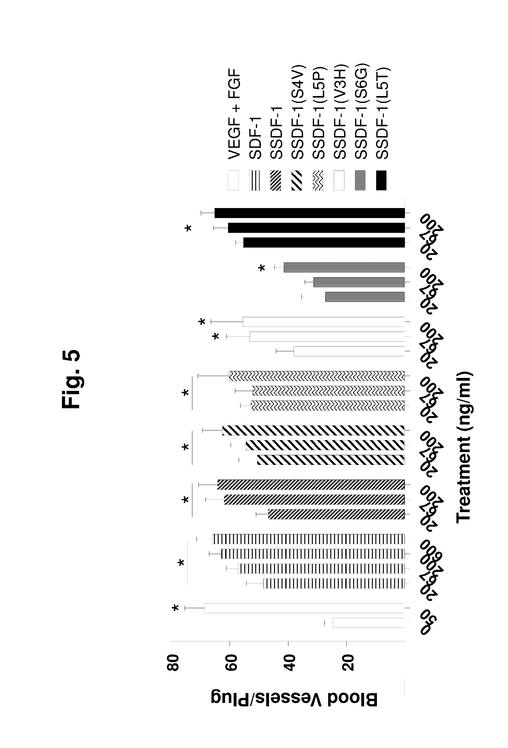
D00011

D00012

D00013

S00001
XML
uspto.report is an independent third-party trademark research tool that is not affiliated, endorsed, or sponsored by the United States Patent and Trademark Office (USPTO) or any other governmental organization. The information provided by uspto.report is based on publicly available data at the time of writing and is intended for informational purposes only.
While we strive to provide accurate and up-to-date information, we do not guarantee the accuracy, completeness, reliability, or suitability of the information displayed on this site. The use of this site is at your own risk. Any reliance you place on such information is therefore strictly at your own risk.
All official trademark data, including owner information, should be verified by visiting the official USPTO website at www.uspto.gov. This site is not intended to replace professional legal advice and should not be used as a substitute for consulting with a legal professional who is knowledgeable about trademark law.