Methods Of Treating Viral Infections, Particularly Rabies, Mers-cov, Influenza, Ebola, Chikungunya, Venezuelan Equine Encephalitus, Canine Parvovirus, Adenovirus, Respiratory Syncytial Virus, Rhinovirus, And Poxvirus In Mammalian Patients
Hodge, III; Thomas W.
U.S. patent application number 14/736170 was filed with the patent office on 2015-12-31 for methods of treating viral infections, particularly rabies, mers-cov, influenza, ebola, chikungunya, venezuelan equine encephalitus, canine parvovirus, adenovirus, respiratory syncytial virus, rhinovirus, and poxvirus in mammalian patients. This patent application is currently assigned to TAMIR BIOTECHNOLOGY, INC.. The applicant listed for this patent is Tamir Biotechnology, Inc.. Invention is credited to Thomas W. Hodge, III.
| Application Number | 20150376584 14/736170 |
| Document ID | / |
| Family ID | 54929856 |
| Filed Date | 2015-12-31 |

View All Diagrams
| United States Patent Application | 20150376584 |
| Kind Code | A1 |
| Hodge, III; Thomas W. | December 31, 2015 |
METHODS OF TREATING VIRAL INFECTIONS, PARTICULARLY RABIES, MERS-COV, INFLUENZA, EBOLA, CHIKUNGUNYA, VENEZUELAN EQUINE ENCEPHALITUS, CANINE PARVOVIRUS, ADENOVIRUS, RESPIRATORY SYNCYTIAL VIRUS, RHINOVIRUS, AND POXVIRUS IN MAMMALIAN PATIENTS
Abstract
Viral infections in mammals can be treated and prophylactically prevented by systemic administration of ranpirnase and three other ribonucleases that are highly homologous with it and that have activities that are highly similar to it. Experimental results against rabies, Middle East Respiratory Syndrome Coronavirus ("MERS-CoV"), influenza, Ebola virus, Chikungunya virus, Venezuelan equine encephalitis, canine parvovirus, adenovirus-2, respiratory syncytial virus, rhinovirus-14, and vaccinia are disclosed.
| Inventors: | Hodge, III; Thomas W.; (Athens, GA) | ||||||||||
| Applicant: |
|
||||||||||
|---|---|---|---|---|---|---|---|---|---|---|---|
| Assignee: | TAMIR BIOTECHNOLOGY, INC. San Diego CA |
||||||||||
| Family ID: | 54929856 | ||||||||||
| Appl. No.: | 14/736170 | ||||||||||
| Filed: | June 10, 2015 |
Related U.S. Patent Documents
| Application Number | Filing Date | Patent Number | ||
|---|---|---|---|---|
| 14667282 | Mar 24, 2015 | |||
| 14736170 | ||||
| 14316893 | Jun 27, 2014 | |||
| 14667282 | ||||
| 14247723 | Apr 8, 2014 | |||
| 14316893 | ||||
| 14229816 | Mar 28, 2014 | |||
| 14247723 | ||||
| 62147209 | Apr 14, 2015 | |||
| 62102671 | Jan 13, 2015 | |||
| 62063551 | Oct 14, 2014 | |||
| 62040885 | Aug 22, 2014 | |||
| Current U.S. Class: | 424/94.6 |
| Current CPC Class: | C12N 9/22 20130101; A01N 63/00 20130101; C12Y 301/27 20130101; C12Y 301/27005 20130101; A61K 38/465 20130101 |
| International Class: | C12N 9/22 20060101 C12N009/22 |
Claims
1. A method of treating a viral infection in a mammalian patient, the viral infection being other than Dengue fever, yellow fever, and severe acute respiratory syndrome and being classified in Baltimore Classification Group IV, comprising systemically administering a therapeutically effective dose of ranpirnase to the patient.
2. A method of treating a viral infection in a mammalian patient, the viral infection being other than Dengue fever, yellow fever, and severe acute respiratory syndrome and being classified in Baltimore Classification Group IV, comprising systemically administering a therapeutically effective dose of Amphinase 2 to the patient.
3. A method of treating a viral infection in a mammalian patient, the viral infection being other than Dengue fever, yellow fever, and severe acute respiratory syndrome and being classified in Baltimore Classification Group IV, comprising systemically administering a therapeutically effective dose of rAmphinase 2 to the patient.
4. A method of treating a viral infection in a mammalian patient, the viral infection being other than Dengue fever, yellow fever, and severe acute respiratory syndrome and being classified in Baltimore Classification Group IV, comprising systemically administering a therapeutically effective dose of the '805 variant to the patient.
5. A method of treating a viral infection in a mammalian patient, the viral infection classified in Baltimore Classification Group V, comprising systemically administering a therapeutically effective dose of ranpirnase to the patient.
6. A method of treating a viral infection in a mammalian patient, the viral infection classified in Baltimore Classification Group V, comprising systemically administering a therapeutically effective dose of Amphinase 2 to the patient.
7. A method of treating a viral infection in a mammalian patient, the viral infection classified in Baltimore Classification Group V, comprising systemically administering a therapeutically effective dose of rAmphinase 2 to the patient.
8. A method of treating a viral infection in a mammalian patient, the viral infection classified in Baltimore Classification Group V, comprising systemically administering a therapeutically effective dose of the '805 variant to the patient.
9. The method of claim 1, 2, 3, 4, 5, 6, 7, or 8, wherein the patient is a human being.
10. The method of claim 1, 2, 3, or 4 wherein the patient is an equine species.
11. A method of treating a rabies infection in a mammalian patient, comprising systemically administering a therapeutically effective dose of ranpirnase to the patient.
12. A method of treating a MERS-CoV infection in a mammalian patient, comprising systemically administering a therapeutically effective dose of ranpirnase to the patient.
13. A method of treating an influenza infection, or an infection in the adenoviridae family, or an infection in the rhinovirus family, or a respiratory syncytial virus infection in a mammalian patient, comprising systemically administering a therapeutically effective dose of ranpirnase to the patient.
14. A method of treating a Venezuelan equine encephalitis infection, or an infection in the poxvirus family in a mammalian patient, comprising systemically administering a therapeutically effective dose of ranpirnase to the patient.
15. A method of treating a Chikungunya infection in a mammalian patient, comprising systemically administering a therapeutically effective dose of ranpirnase to the patient.
16. The method of claim 11, 12, 13, 14, or 15, wherein the patient is a human being.
17. The method of claim 14, wherein the patient is an equine species.
18. A method of treating a canine parvovirus infection in a mammalian patient, comprising systemically administering a therapeutically effective dose of ranpirnase to the patient.
19. The method of claim 18, wherein the patient is a dog.
20. A method of treating a rabies infection in a mammalian patient, comprising systemically administering a therapeutically effective dose of Amphinase 2 to the patient.
21. A method of treating a MERS-CoV infection in a mammalian patient, comprising systemically administering a therapeutically effective dose of Amphinase 2 to the patient.
22. A method of treating an influenza infection, or an infection in the adenoviridae family, or an infection in the rhinovirus family, or a respiratory syncytial virus infection in a mammalian patient, comprising systemically administering a therapeutically effective dose of Amphinase 2 to the patient.
23. A method of treating a Venezuelan equine encephalitis infection, or an infection in the poxvirus family in a mammalian patient, comprising systemically administering a therapeutically effective dose of Amphinase 2 to the patient.
24. A method of treating a Chikungunya infection in a mammalian patient, comprising systemically administering a therapeutically effective dose of Amphinase 2 to the patient.
25. The method of claim 20, 21, 22, 23, or 24, wherein the patient is a human being.
26. The method of claim 23, wherein the patient is an equine species.
27. A method of treating a canine parvovirus infection in a mammalian patient, comprising systemically administering a therapeutically effective dose of Amphinase 2 to the patient.
28. The method of claim 27, wherein the patient is a dog.
29. A method of treating a rabies infection in a mammalian patient, comprising systemically administering a therapeutically effective dose of rAmphinase 2 to the patient.
30. A method of treating a MERS-CoV infection in a mammalian patient, comprising systemically administering a therapeutically effective dose of rAmphinase 2 to the patient.
31. A method of treating an influenza infection, or an infection in the adenoviridae family, or an infection in the rhinovirus family, or a respiratory syncytial virus infection in a mammalian patient, comprising systemically administering a therapeutically effective dose of rAmphinase 2 to the patient.
32. A method of treating a Venezuelan equine encephalitis infection, or an infection in the poxvirus family in a mammalian patient, comprising systemically administering a therapeutically effective dose of rAmphinase 2 to the patient.
33. A method of treating a Chikungunya infection in a mammalian patient, comprising systemically administering a therapeutically effective dose of rAmphinase 2 to the patient.
34. The method of claim 29, 30, 31, 32, or 33 wherein the patient is as human being.
35. The method of claim 32, wherein the patient is an equine species.
36. A method of treating a canine parvovirus infection in a mammalian patient, comprising systemically administering a therapeutically effective dose of rAmphinase 2 to the patient.
37. The method of claim 36, wherein the patient is a dog.
38. A method of treating a rabies infection in a mammalian patient, comprising systemically administering a therapeutically effective dose of the '805 variant to the patient.
39. A method of treating a MERS-CoV infection in a mammalian patient, comprising systemically administering a therapeutically effective dose of the '805 variant to the patient.
40. A method of treating an influenza infection, or an infection in the adenoviridae family, or an infection in the rhinovirus family, or a respiratory syncytial virus infection in a mammalian patient, comprising systemically administering a therapeutically effective dose of the '805 variant to the patient.
41. A method of treating a Venezuelan equine encephalitis infection, or an infection in the poxvirus family in a mammalian patient, comprising systemically administering a therapeutically effective dose of the '805 variant to the patient.
42. A method of treating a Chikungunya infection in a mammalian patient, comprising systemically administering a therapeutically effective dose of the '805 variant to the patient.
43. The method of claim 38, 39, 40, 41, or 42, wherein the patient is a human being.
44. The method of claim 41, wherein the patient is an equine species.
45. A method of treating a canine parvovirus infection in a mammalian patient, comprising systemically administering a therapeutically effective dose of the '805 variant to the patient.
46. The method of claim 45, wherein the patient is a dog.
47. A method of prophylactically protecting a patient from: a. rabies; or b. MERS-CoV; or c. influenza; or d. Chikungunya; or e. Venezuelan equine encephalitis; or f. canine parovirus; or g. a virus in the adenoviridae family; or h. respiratory syncytial virus; or, i. a virus in the rhinovirus family; or j. a virus in the poxvirus family, comprising the step of administering a therapeutically effective dose of ranpirnase to the patient.
48. A method of prophylactically protecting a patient from: a. rabies; or b. MERS-CoV; or c. influenza; or d. Chikungunya; or e. Venezuelan equine encephalitis; or f. canine parovirus; or g. a virus in the adenoviridae family; or h. respiratory syncytial virus; or, i. a virus in the rhinovirus family; or j. a virus in the poxvirus family, comprising the step of administering a therapeutically effective dose of the '805 variant to the patient.
49. A method of prophylactically protecting a patient from: a. rabies; or b. MERS-CoV; or c. influenza; or d. Chikungunya; or e. Venezuelan equine encephalitis; or f. canine parovirus; or g. a virus in the adenoviridae family; or h. respiratory syncytial virus; or i. a virus in the rhinovirus family; or j. a virus in the poxvirus family, comprising the step of administering a therapeutically effective dose of Amphinase 2 to the patient.
50. A method of prophylactically protecting a patient from: a. rabies; or b. MERS-CoV; or c. influenza; or d. Chikungunya; or e. Venezuelan equine encephalitis; or f. canine parovirus; or g. a virus in the adenoviridae family; or h. respiratory syncytial virus; or, i. a virus in the rhinovirus family; or j. a virus in the poxvirus family, comprising the step of administering a therapeutically effective dose of rAmphinase 2 to the patient.
51. A method of treating an Ebola infection in a mammalian patient, comprising systemically administering a therapeutically effective dose of ranpirnase to the patient.
52. A method of treating an Ebola infection in a mammalian patient, comprising systemically administering a therapeutically effective dose of Amphinase 2 to the patient.
53. A method of treating an Ebola infection in a mammalian patient, comprising systemically administering a therapeutically effective dose of rAmphinase 2 to the patient.
54. A method of treating an Ebola infection in a mammalian patient, comprising systemically administering a therapeutically effective dose of the '805 variant to the patient.
55. The method of claim 51, 52, 53, or 54, wherein the ranpirnase is administered prophylactically.
56. The method of claim 51, 52, 53, or 54, wherein the ranpirnase is administered therapeutically.
Description
BACKGROUND OF THE INVENTION
[0001] The invention relates to treatment of viral infections, and more particularly relates to treatment of viral infections in mammalian patients. In its most immediate sense, the invention relates to treatment of rabies, Middle East Respiratory Syndrome Coronavirus ("MERS-CoV"), influenza, Ebola virus ("EBOV"), Chikungunya virus ("CHIV"), Venezuelan equine encephalitis ("VEEV"), canine parvovirus, adenovirus ("AV"), respiratory syncytial virus ("RSV"), rhinovirus ("RV"), and poxvirus.
[0002] Various drugs, including ranpirnase, have been proposed as anti-viral agents. However, the demonstrated activity of each of these drugs has been specific to one or only a few particular viruses.
SUMMARY OF THE INVENTION
[0003] Recent experiments have shown that ranpirnase demonstrates surprisingly strong antiviral effects against a surprisingly large number of different viruses, including viruses (e.g. MERS-CoV and EBOV) that are highly resistant to treatment.
[0004] It is believed that the surprisingly broad-spectrum activity of the invention comes from the ways in which ranpirnase degrades various forms of RNA. To date, three RNA-degrading mechanisms appear to be relevant to antiviral therapy using ranpirnase.
[0005] The first of these mechanisms is degradation of tRNA. Degrading tRNA inside a mammalian cell makes that cell resistant to some viral infections. This is because some viruses replicate by protein synthesis using the ribosome, and protein synthesis cannot occur unless transfer RNAs enter the ribosome to deliver the amino acids needed to synthesize the protein. Thus, a systemic application of an agent that degrades tRNA will prevent or at least substantially impede some viruses from spreading to uninfected cells. If this application occurs before the virus has spread widely enough to endanger the host mammal, the virus will eventually die.
[0006] The second mechanism is degradation of viral double-stranded RNA. Some viruses produce double-stranded RNA as part of their process of proliferation in mammalian cells, and destroying that double-stranded RNA can prevent or at least substantially impede replication of such viruses.
[0007] The third mechanism is degradation of microRNA and siRNA. In certain viruses that proliferate using double-stranded RNA, that double-stranded RNA is produced by the interaction of microRNA or siRNA with single-stranded RNA. Destroying the microRNA or siRNA can prevent the formation of the viral double-stranded RNA by which the virus replicates.
[0008] Ranpirnase is known to degrade each of these RNAs. It degrades tRNA very effectively (see Lin et al., Biochemical and Biophysical Research Communications 201(1), 156-162 (1994)). And, because normal mammalian cells degrade approximately 80% of their tRNA as a natural process, this degradation causes little if any harm to the cells themselves. As a result, except in instances where a viral infection has spread too far to be effectively controlled, a systemic application of ranpirnase causes some viruses to die out without killing the normal cells that those viruses infect.
[0009] Ranpirnase is known to degrade double-stranded RNA as well (see Saxena et al., Anticancer Res. 29(4), 1067-1071 (April 2009)). And, ranpirnase is known to degrade certain microRNA (see Goparaju et al., Oncogene 30(24), 2767-2777, (Jun. 16, 2011)) and certain siRNA (see Zhao et al., Cell Cycle. 7(20), 3258-3261 (October 2008)).
[0010] Hence, it appears that the RNA-degrading characteristics of ranpirnase make it possible to use ranpirnase as a pharmaceutical for treating a wide variety of viral infections in mammals by administering ranpirnase systemically. And, it also appears that these characteristics permit ranpirnase to be administered prophylactically as well as therapeutically.
[0011] Furthermore, it is believed that other ribonucleases will have the same antiviral effects that ranpirnase has. These other ribonucleases (including the below-identified "'805 variant", "Amphinase 2", and rAmphinase 2'') are highly homologous to ranpirnase and have exhibited antiviral properties that are highly similar to those of ranpirnase when treating other viral infections.
BRIEF DESCRIPTION OF THE DRAWINGS
[0012] In the drawings, wherein
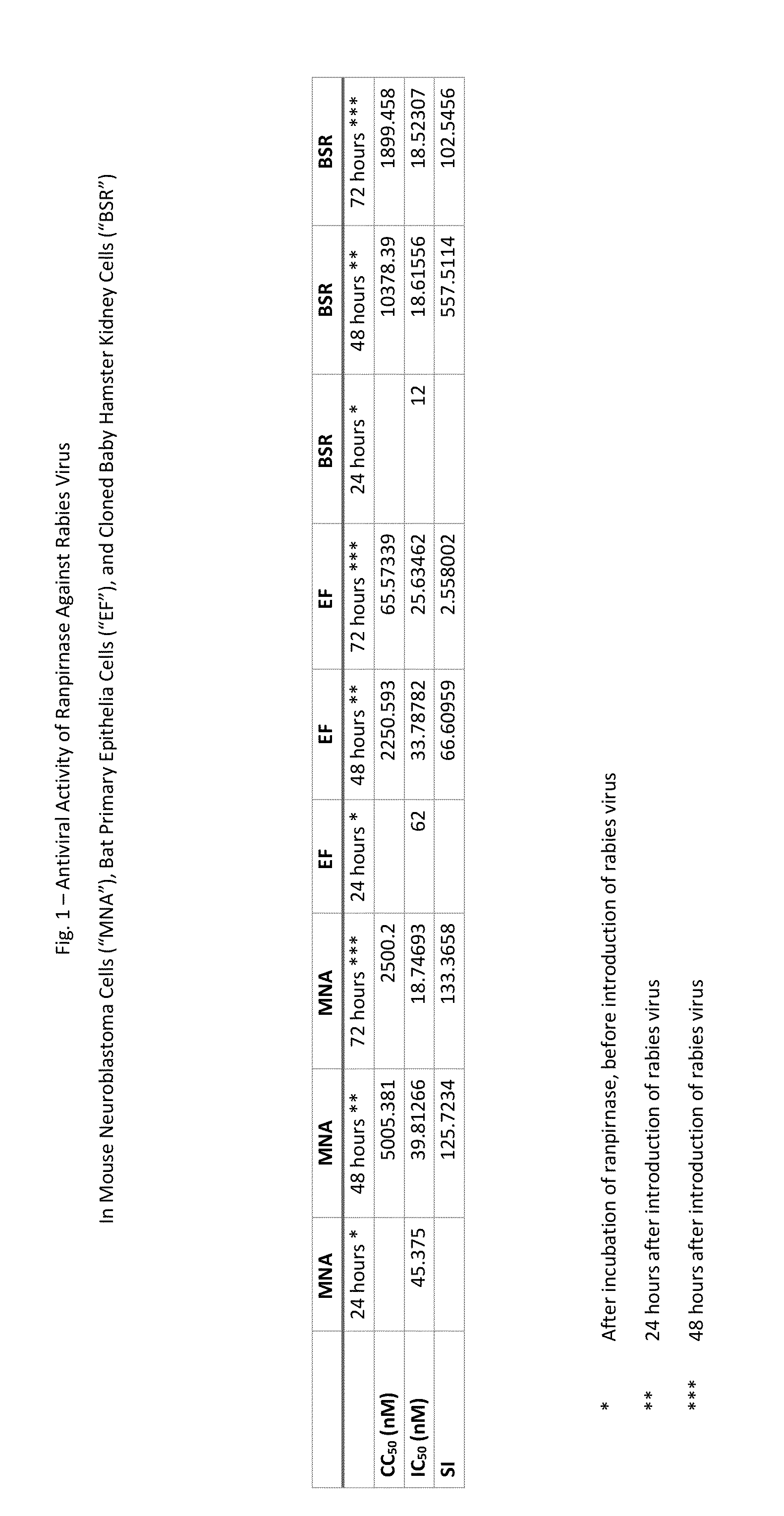
[0013] CC.sub.50 is the cytotoxic concentration (expressed in nM) of ranpirnase, i.e. the ranpirnase concentration that decreased cell viability by 50%, and
[0014] IC.sub.50 is the inhibitory concentration (expressed in nM) of ranpirnase, i.e. the ranpirnase concentration that inhibited replication of the virus under test by 50%,
[0015] SI, the selective index, is CC.sub.50/IC.sub.50. The higher the value of SI, the more active is the ranpirnase against the virus under test,
[0016] FIG. 1 shows the results of testing the anti-viral activity of ranpirnase against rabies virus in three separate cell lines;
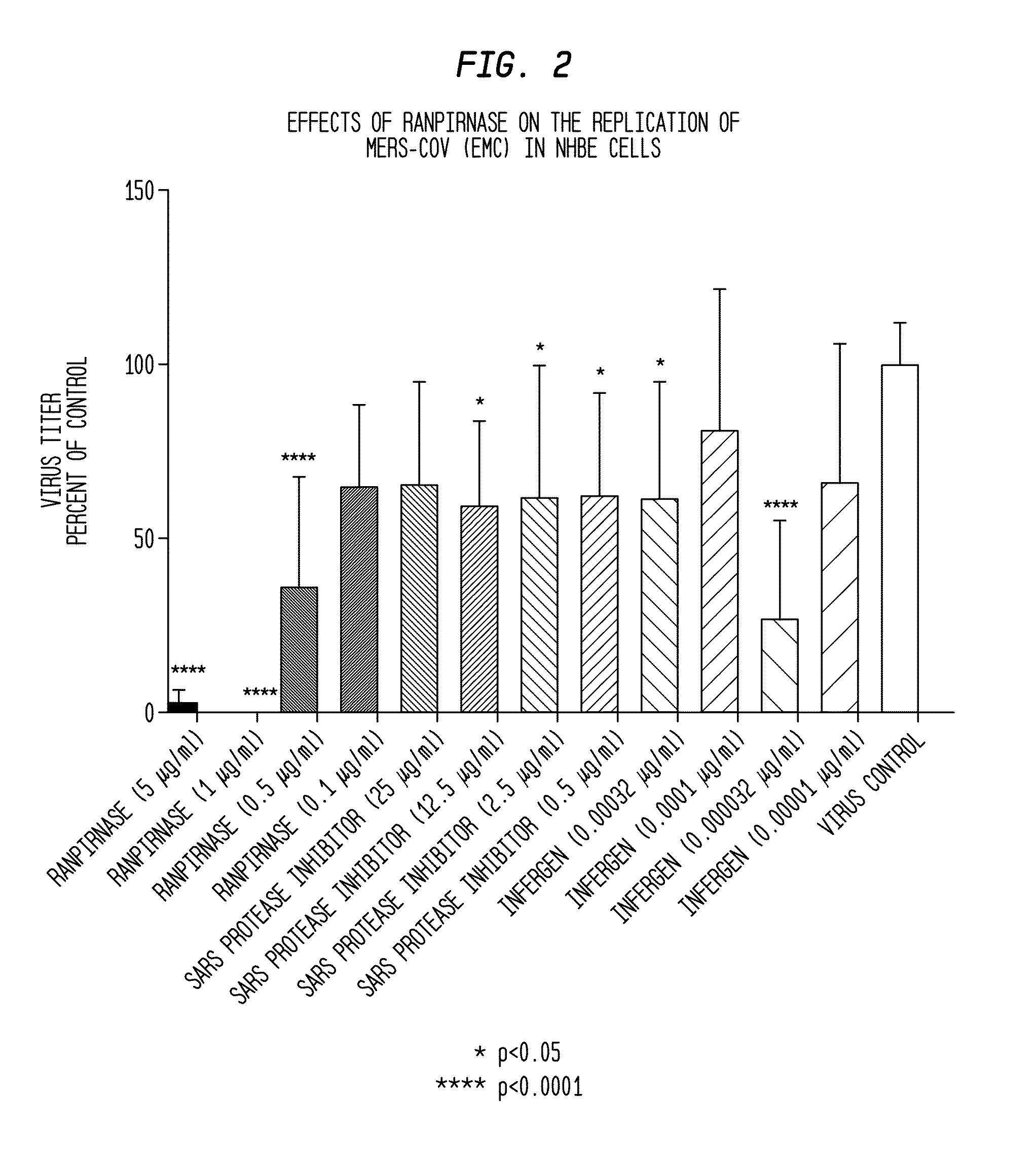
[0017] FIG. 2 shows the results of testing the anti-viral activity of various concentrations of ranpirnase against MERS-CoV virus in normal human bronchial epithelial ("NHBE") cells, compared with the anti-viral activities of various concentrations of SARS protease inhibitor and Infergen;
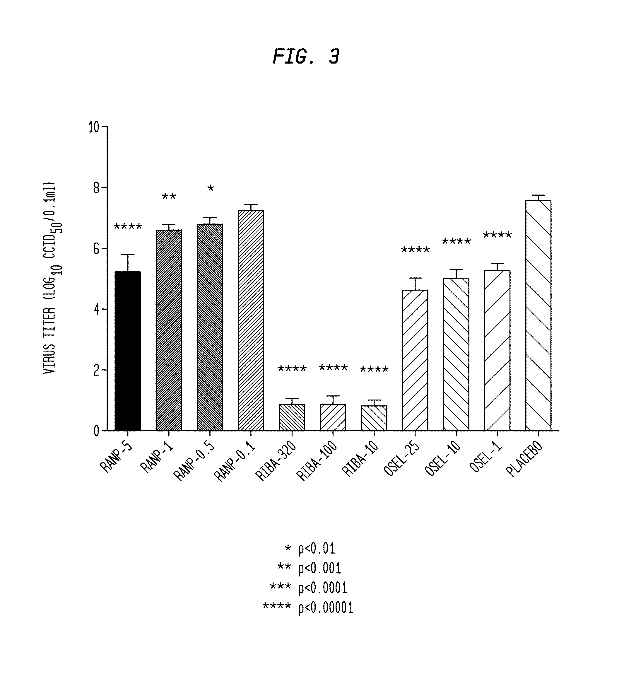
[0018] FIG. 3 shows the results of testing the anti-viral activity of various concentrations of ranpirnase ("Ranp") against a strain of influenza in NHBE cells, compared with the anti-viral activities of various concentrations of Ribavirin ("Riba") and Oseltamivir carboxylate ("Osel");
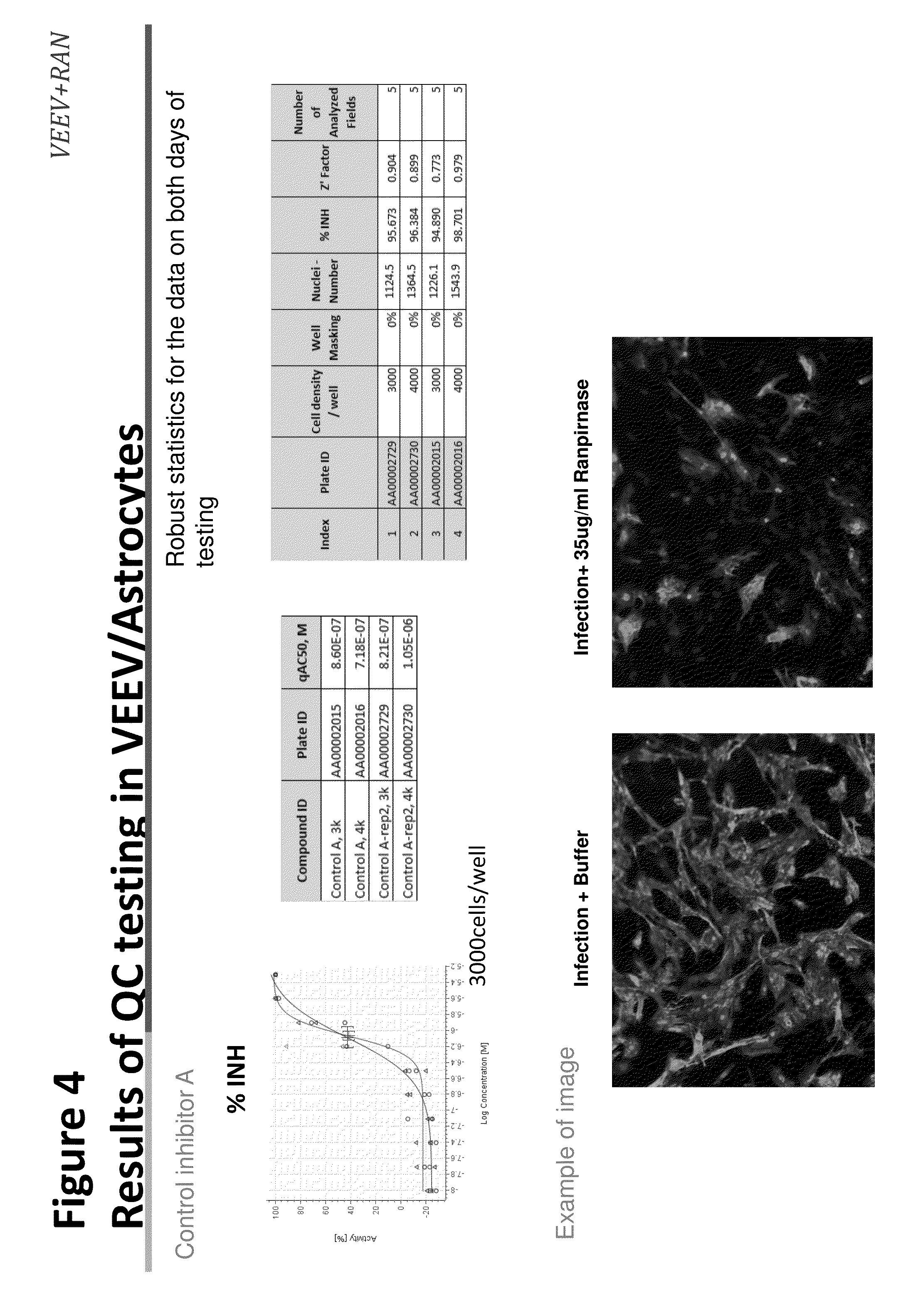
[0019] FIG. 4 shows results of quality control (QC) testing of ranpirnase against VEEV infection in astroctyes;
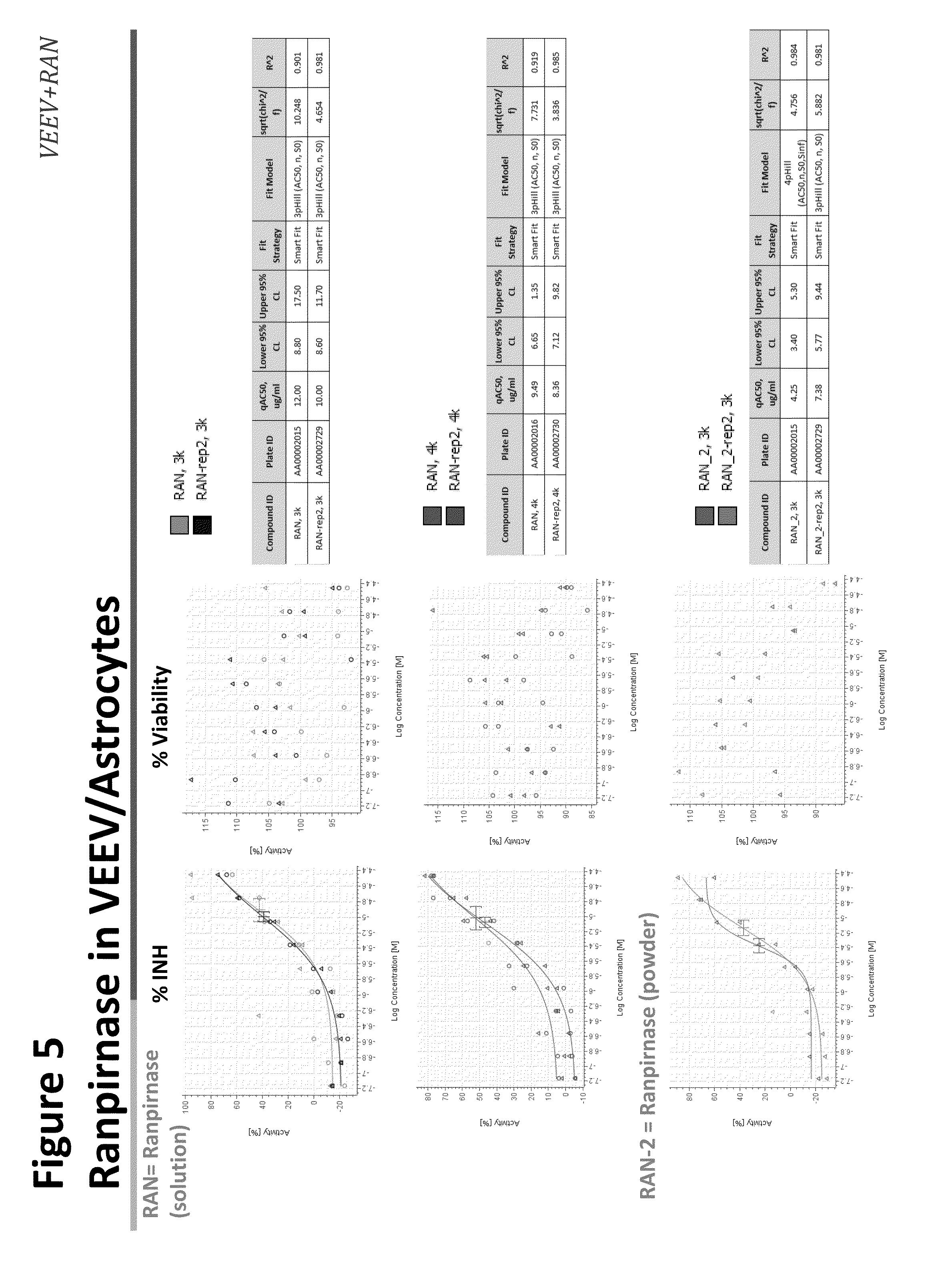
[0020] FIG. 5 shows the effect of ranpirnase against VEEV infection in astrocytes.
[0021] FIG. 6 shows results of QC testing of ranpirnase against VEEV infection in HeLa cells;
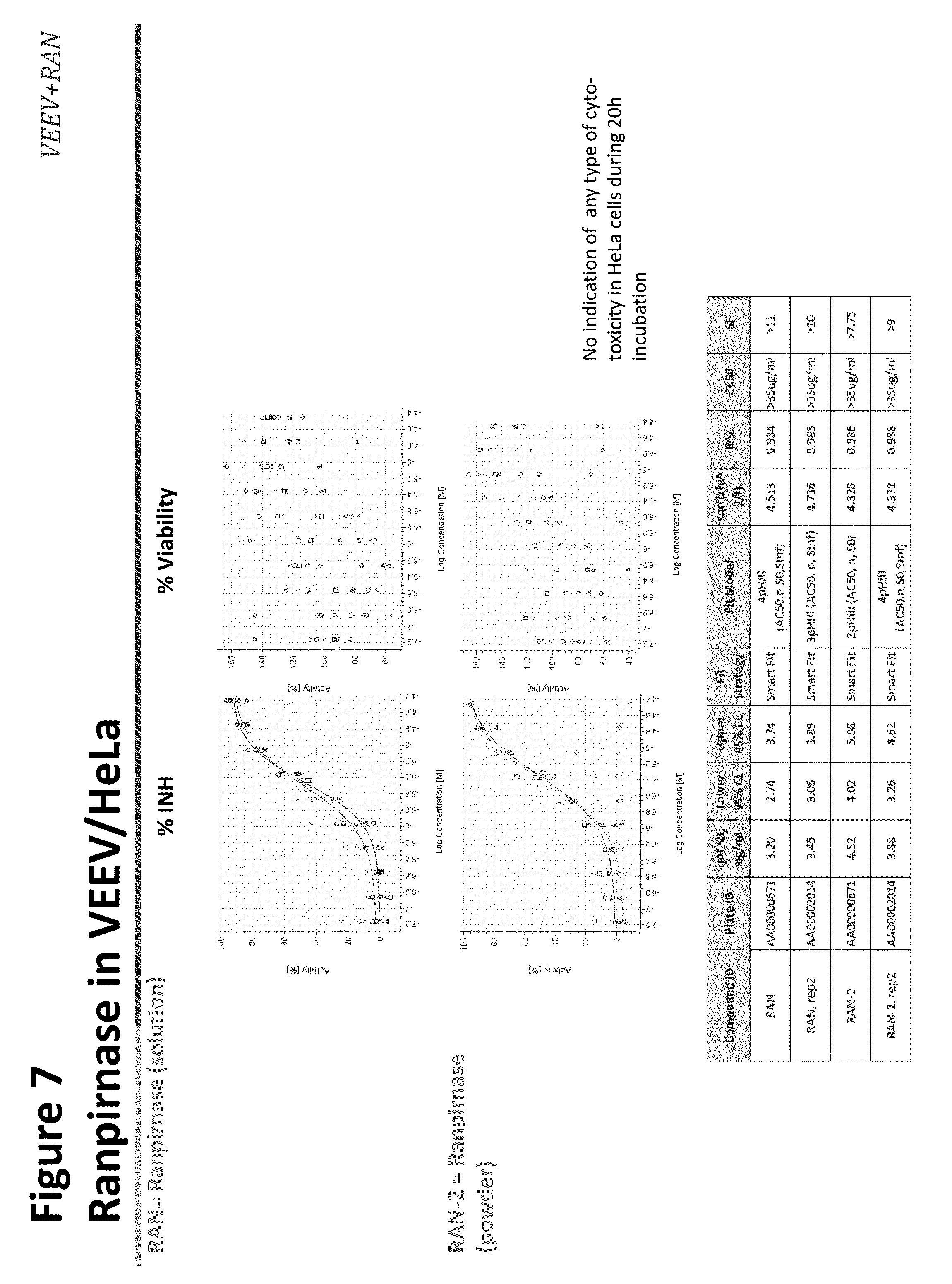
[0022] FIG. 7 shows the effect of ranpirnase against VEEV infection in HeLa cells;
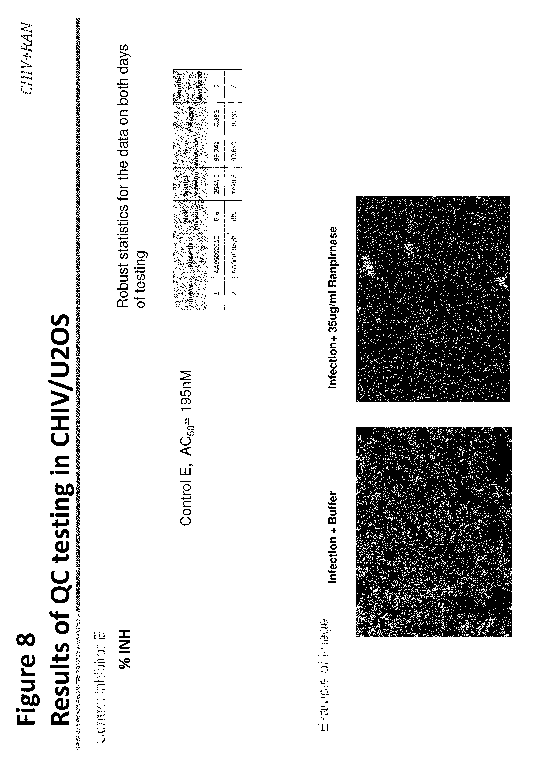
[0023] FIG. 8 shows results of QC testing of ranpirnase against CHIV infection in U2OS cells;
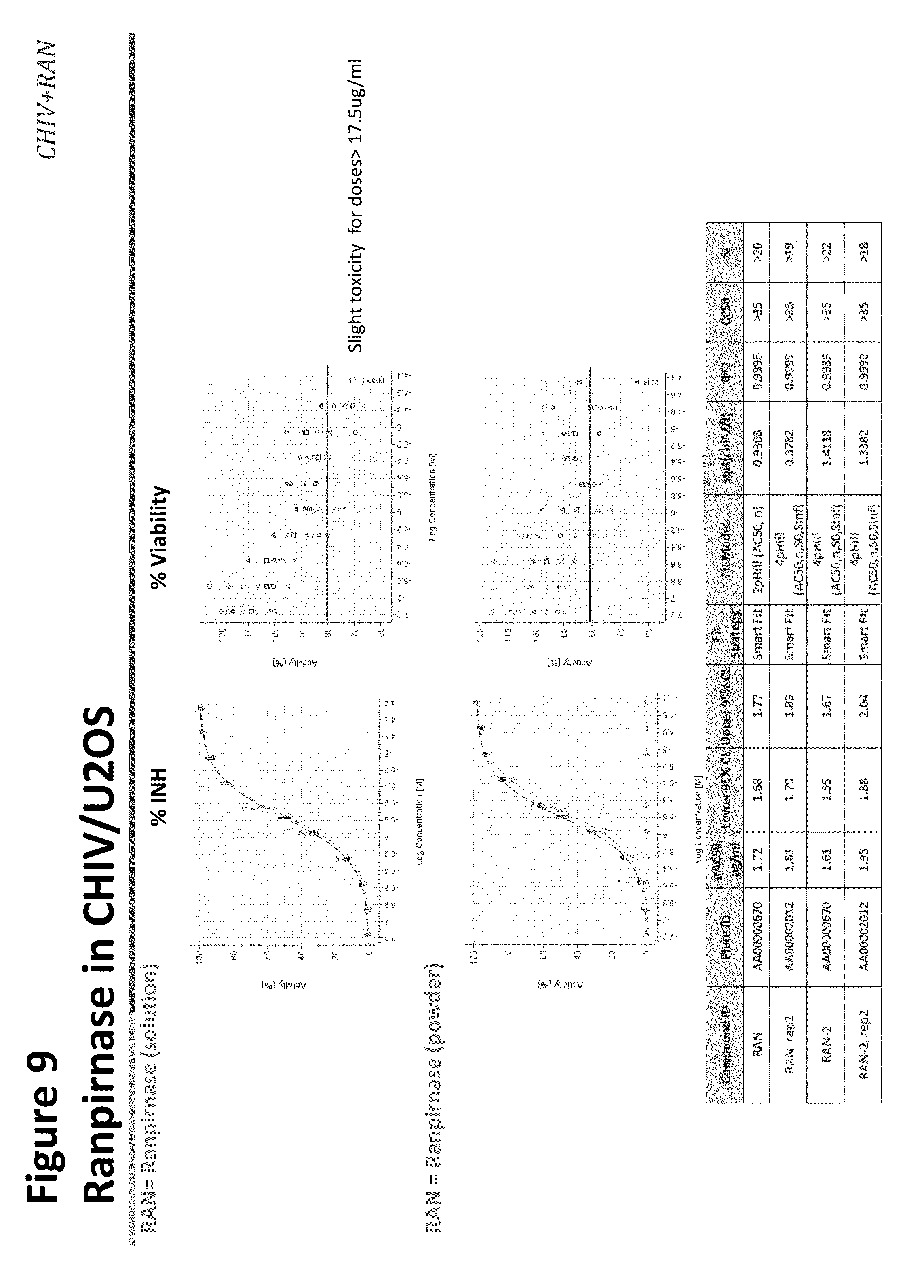
[0024] FIG. 9 shows the effect of ranpirnase against CHIV infection in U2OS cells;
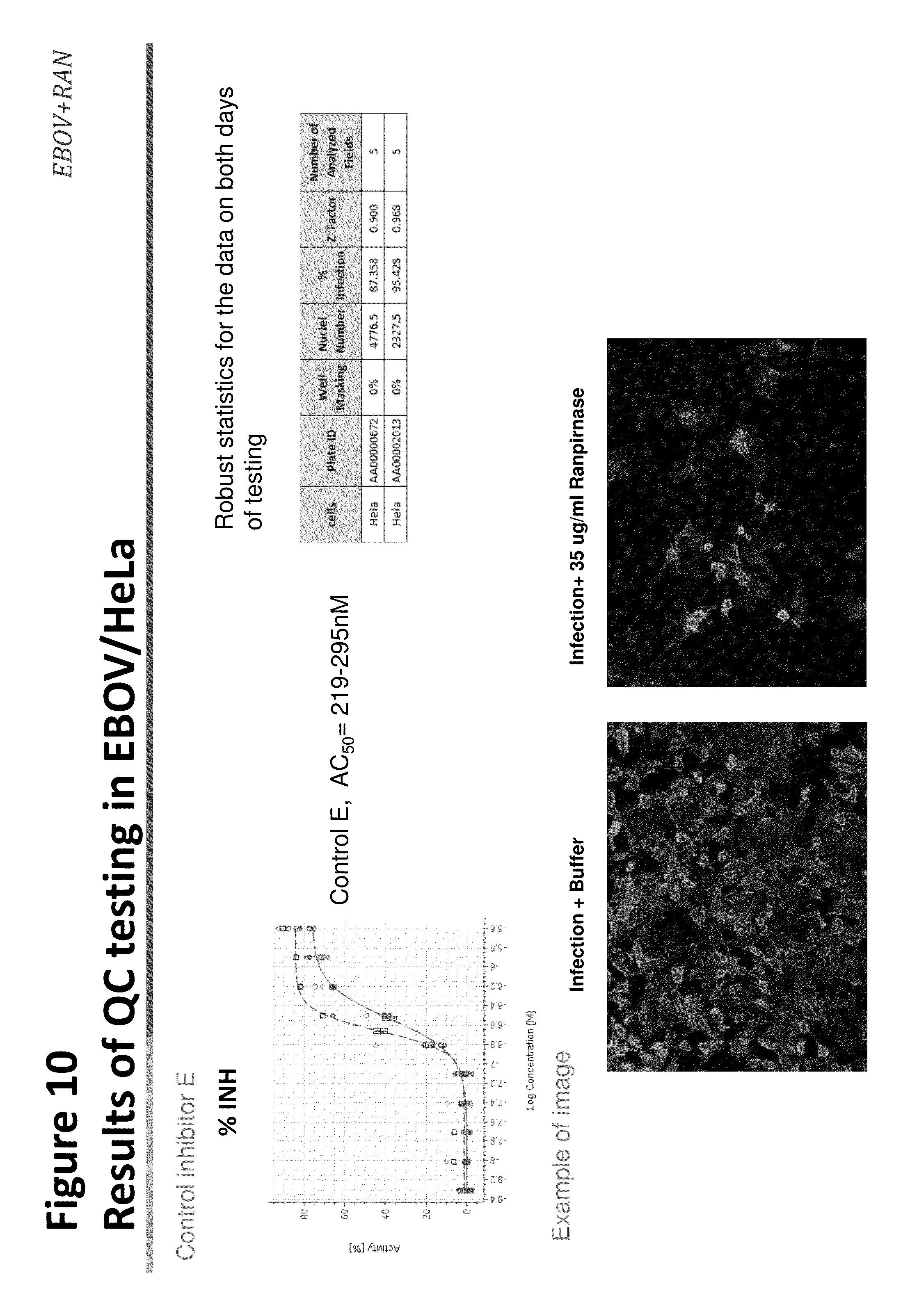
[0025] FIG. 10 shows results of QC testing of ranpirnase against EBOV infection in HeLa cells;
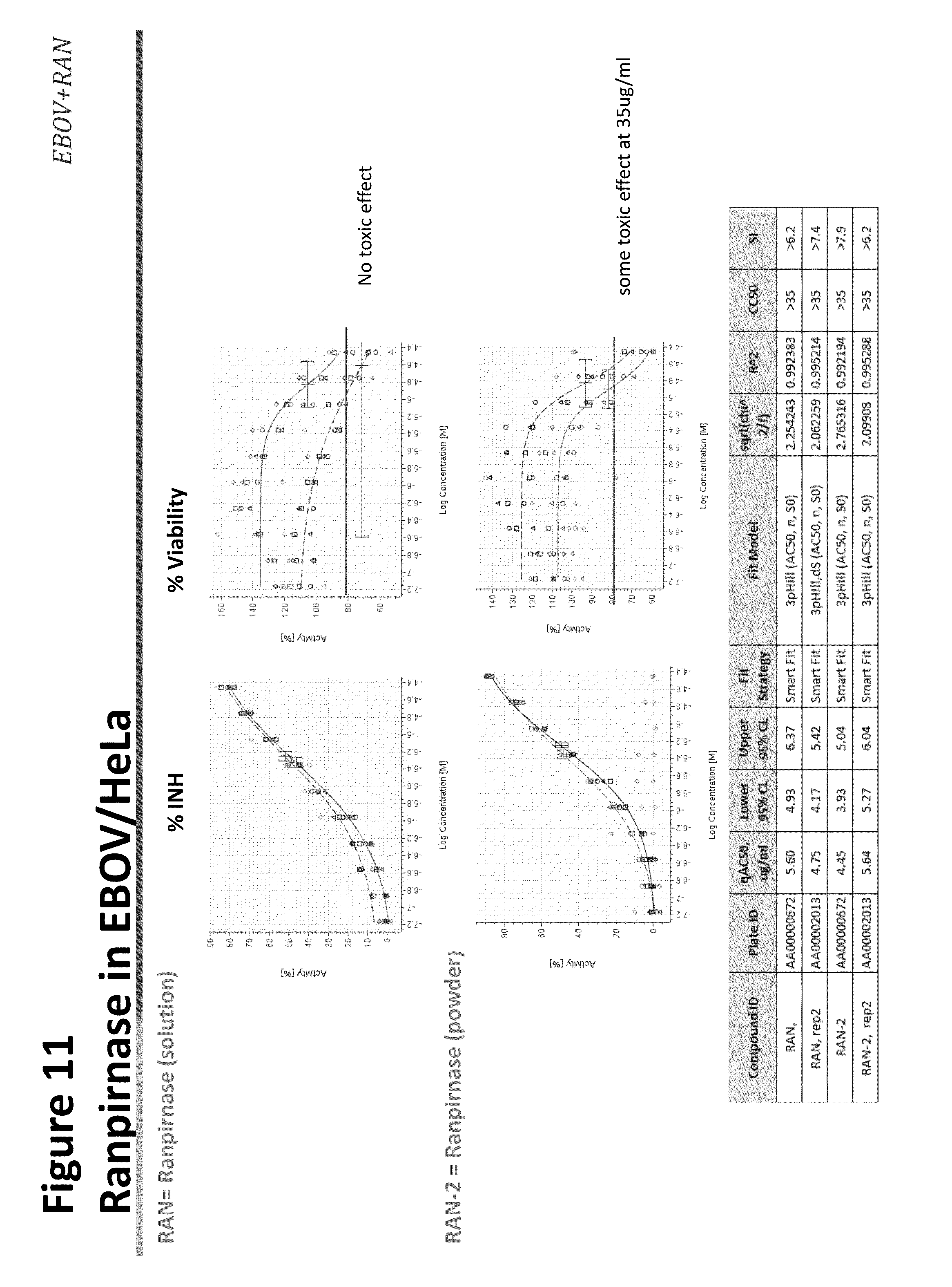
[0026] FIG. 11 shows the effect of ranpirnase against EBOV infection in HeLa cells;
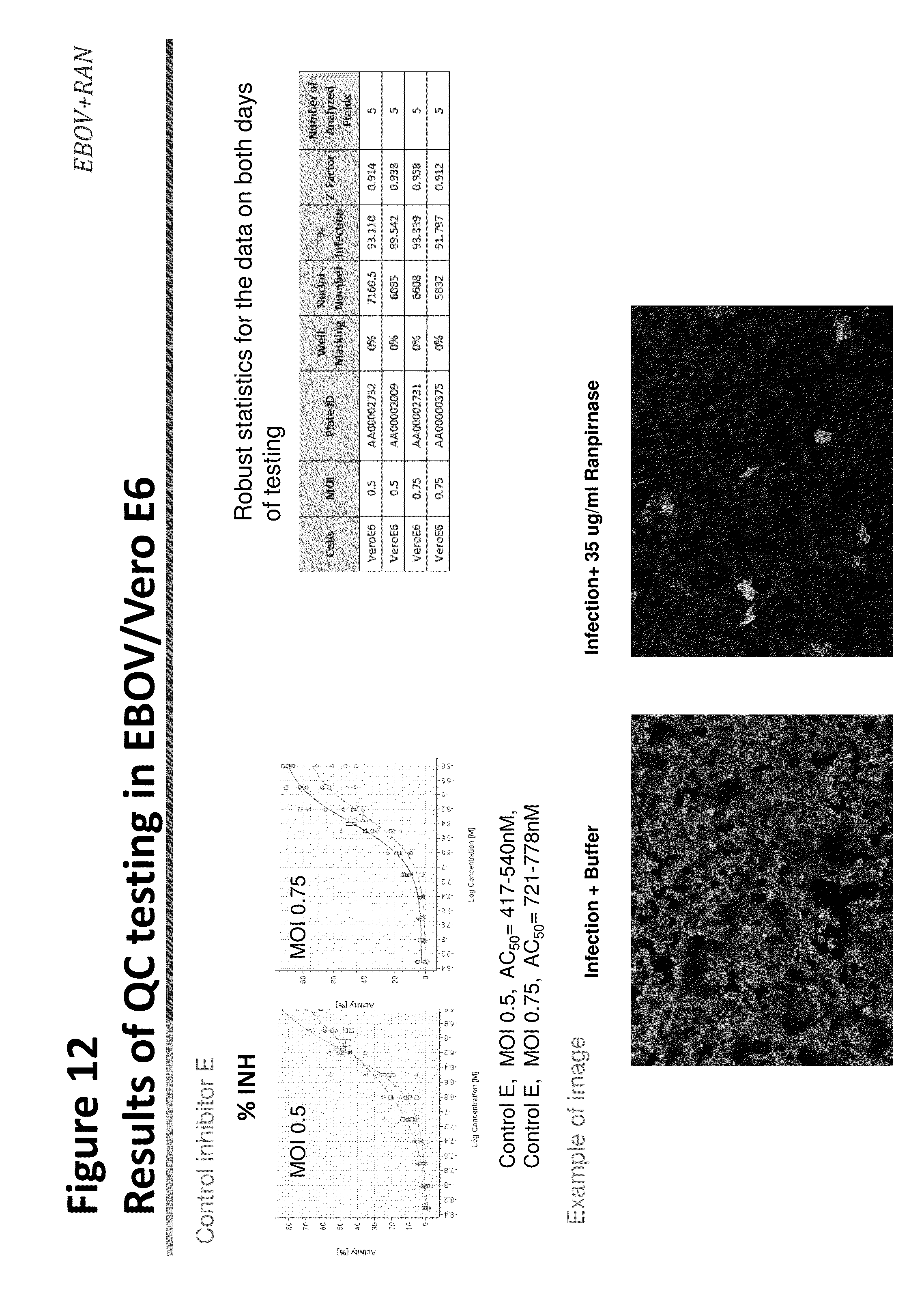
[0027] FIG. 12 shows results of QC testing of ranpirnase against EBOV infection in Vero E6 cells;
[0028] FIG. 13 shows the effect of ranpirnase against EBOV infection in Vero E6 cells;
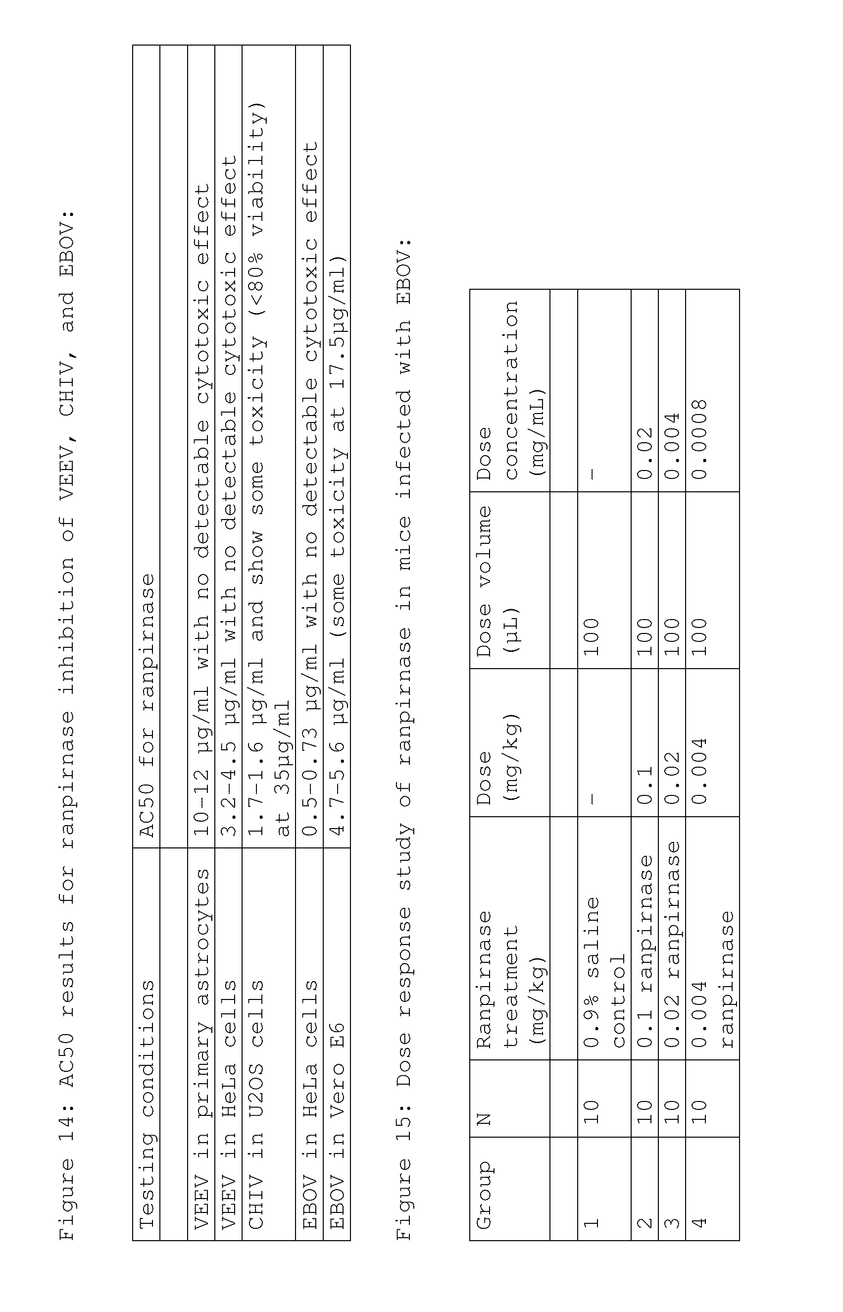
[0029] FIG. 14 shows the AC50 toxicity values for ranpirnase inhibition of VEEV, CHIV, and EBOV;
[0030] FIG. 15 shows the doses used in a dose response study of prophylactically administered ranpirnase in mice infected with EBOV;
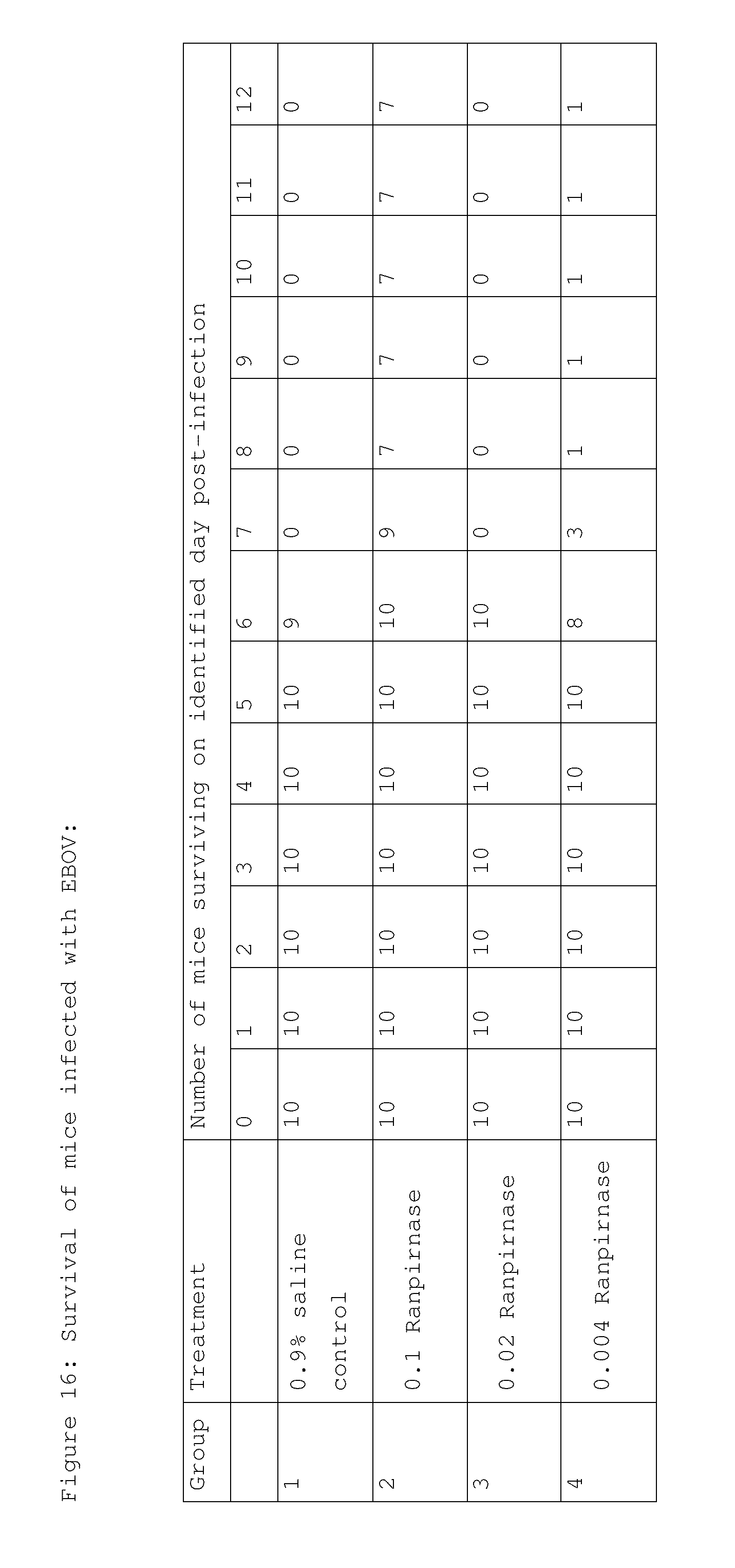
[0031] FIG. 16 shows mouse survival in the study of FIG. 15;
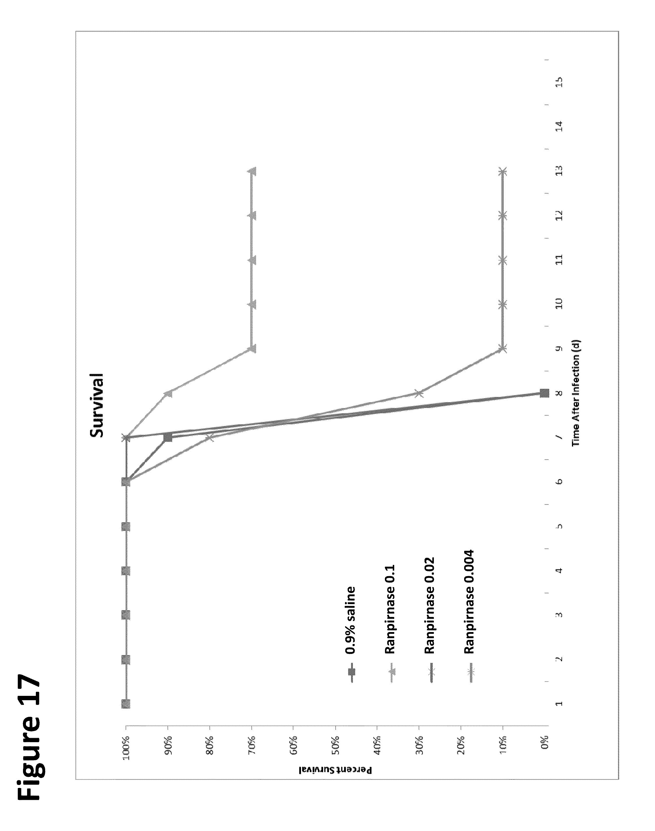
[0032] FIG. 17 is a graphical representation of the data in FIG. 16, shown in percentage terms;
[0033] FIG. 18 shows mouse weight loss in the study of FIG. 15;
[0034] FIG. 19 shows mouse weight loss in the study of FIG. 15 in percentage terms;
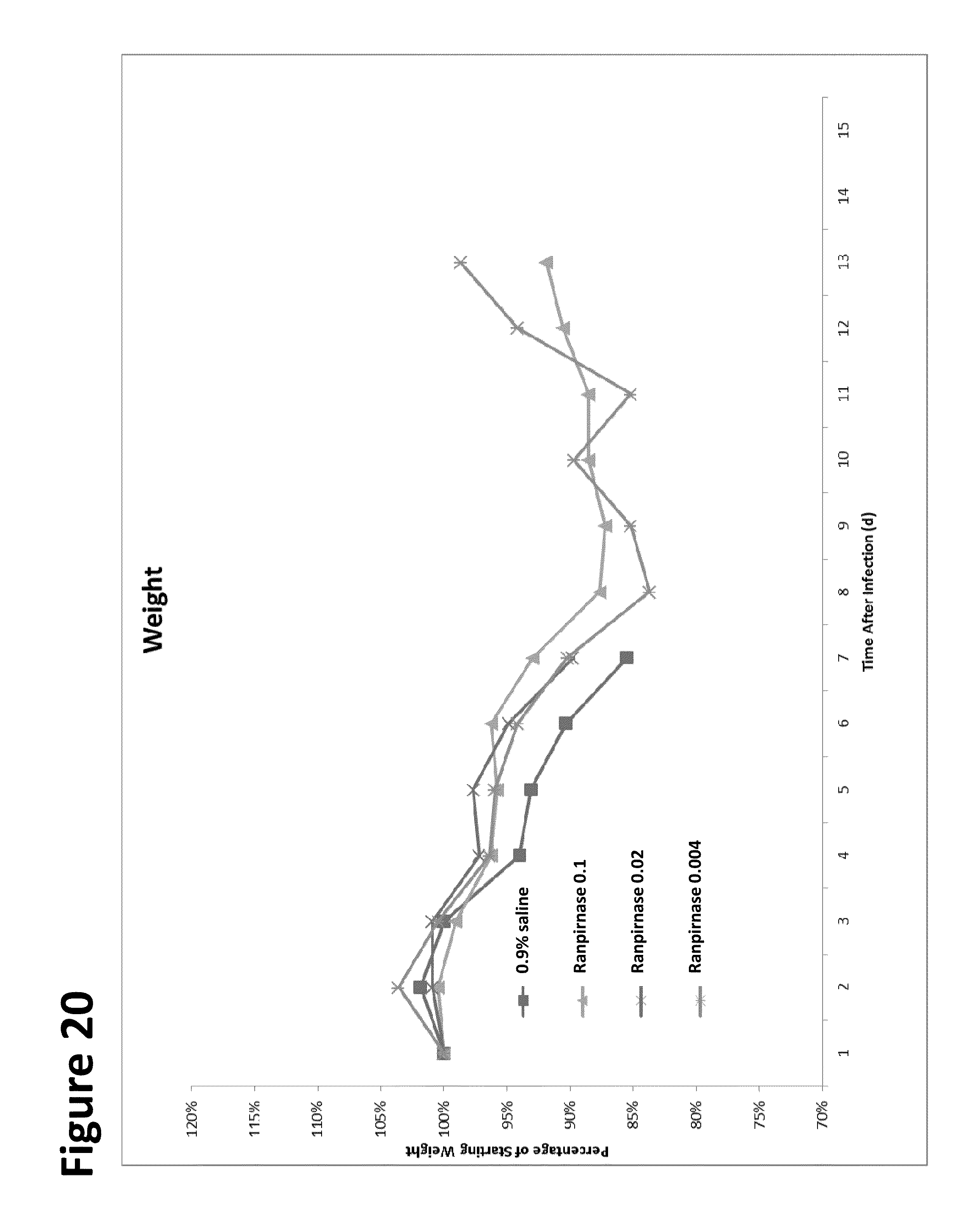
[0035] FIG. 20 is a graphical representation of the data in FIG. 19;
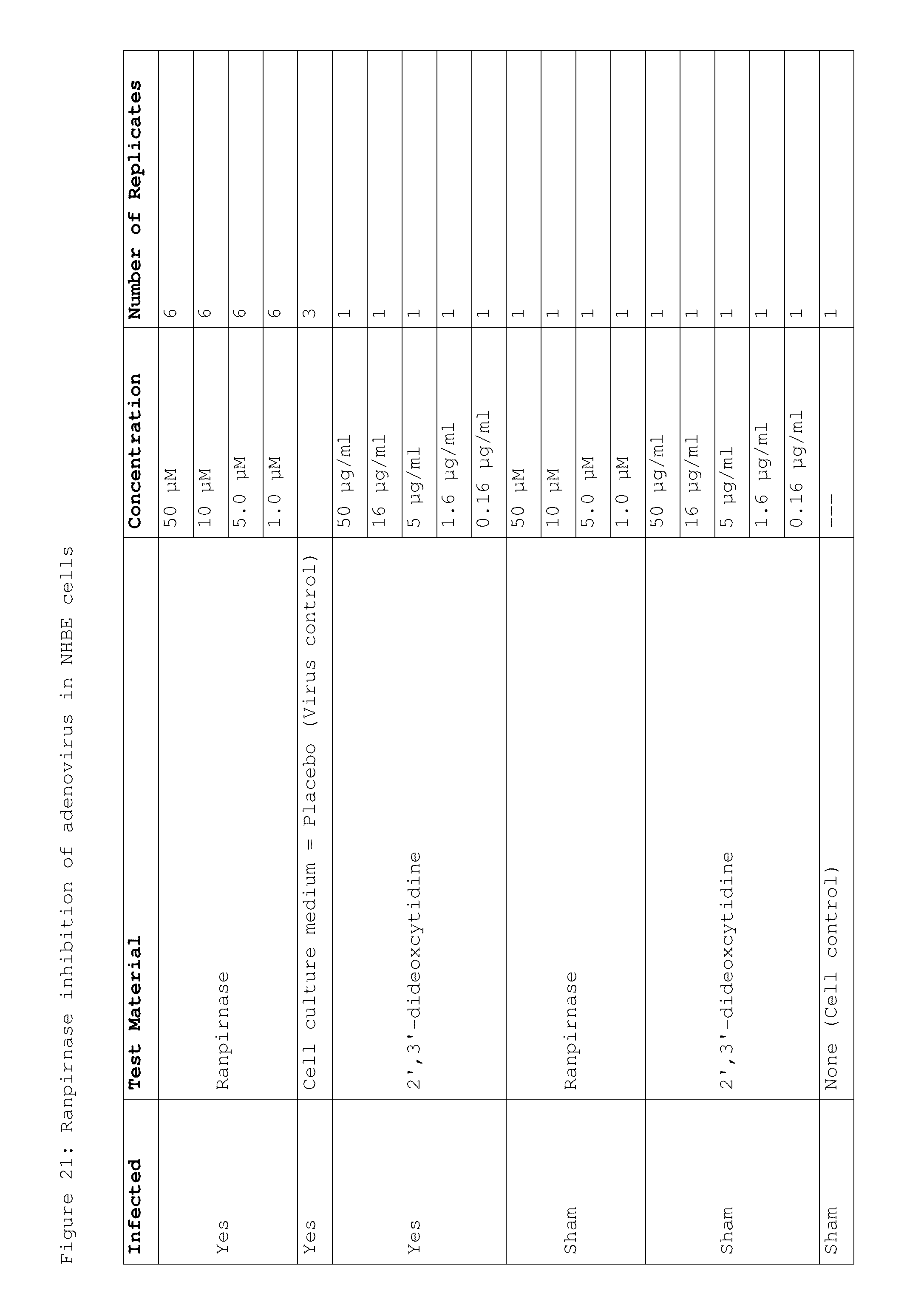
[0036] FIG. 21 shows the doses of ranpirnase used in a study of ranpirnase inhibition of AV in NHBE cells;
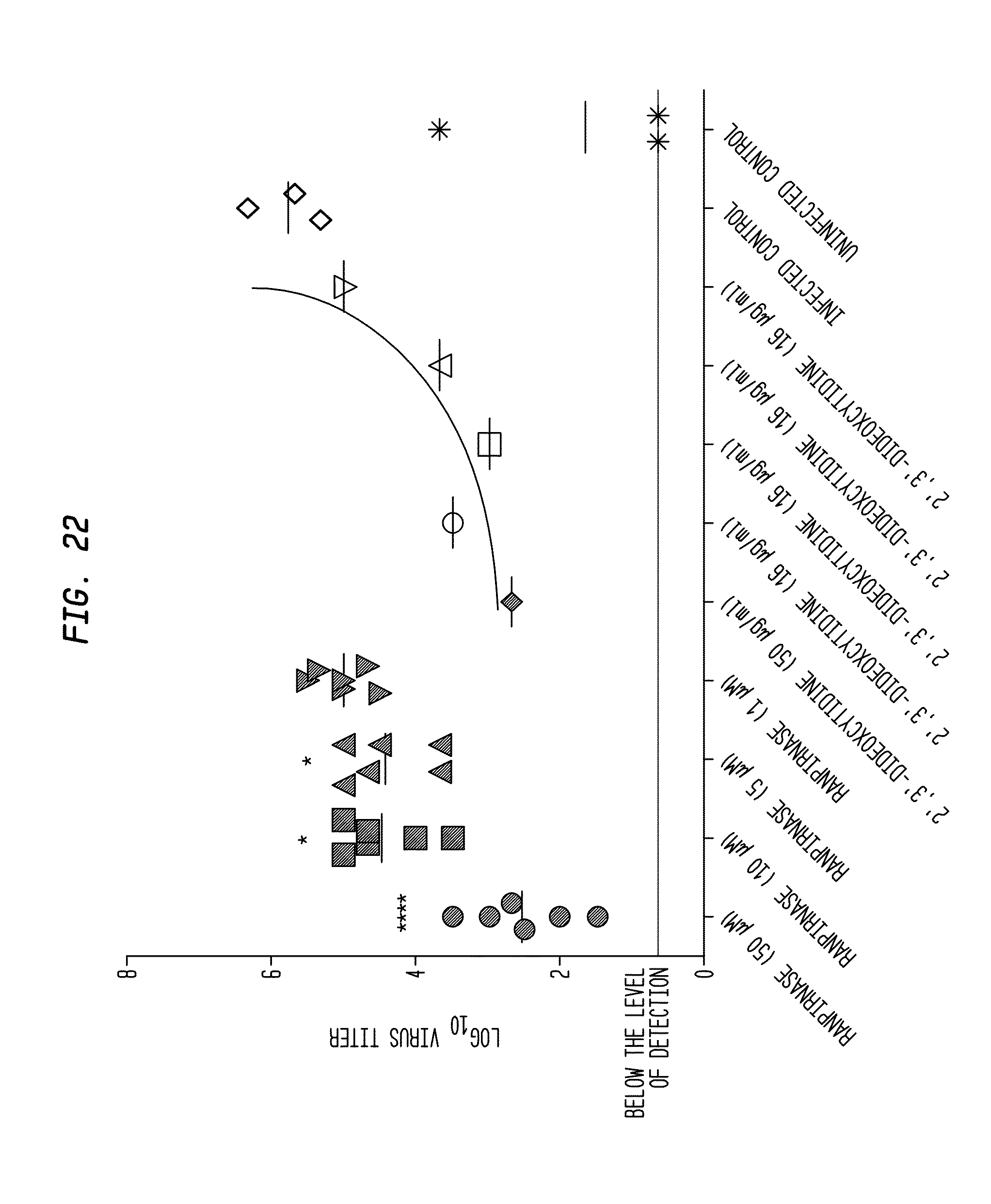
[0037] FIG. 22 shows the results of the study of FIG. 21;
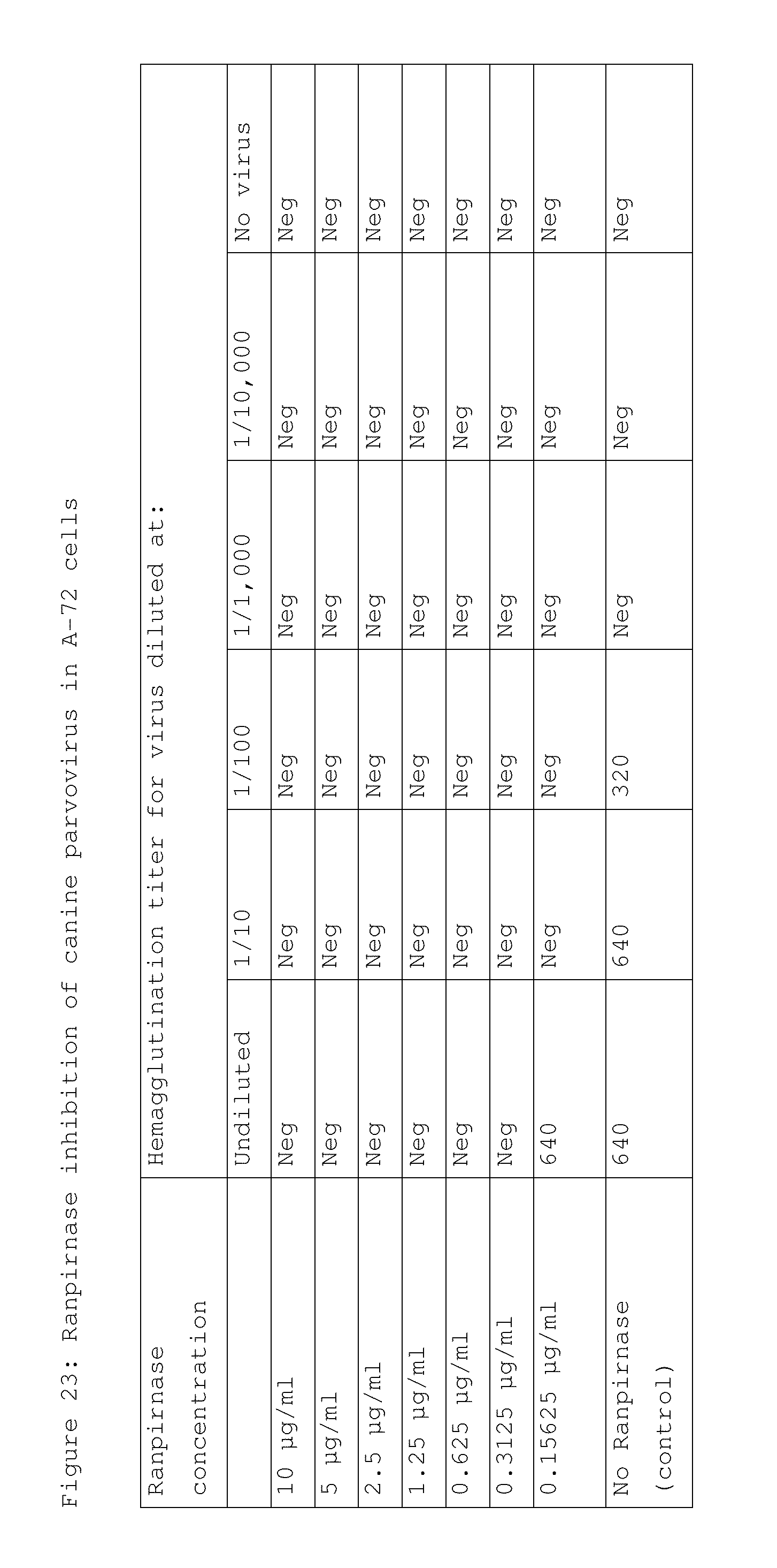
[0038] FIG. 23 shows the results of a study of ranpirnase inhibition of canine parvovirus in A-72 cells;
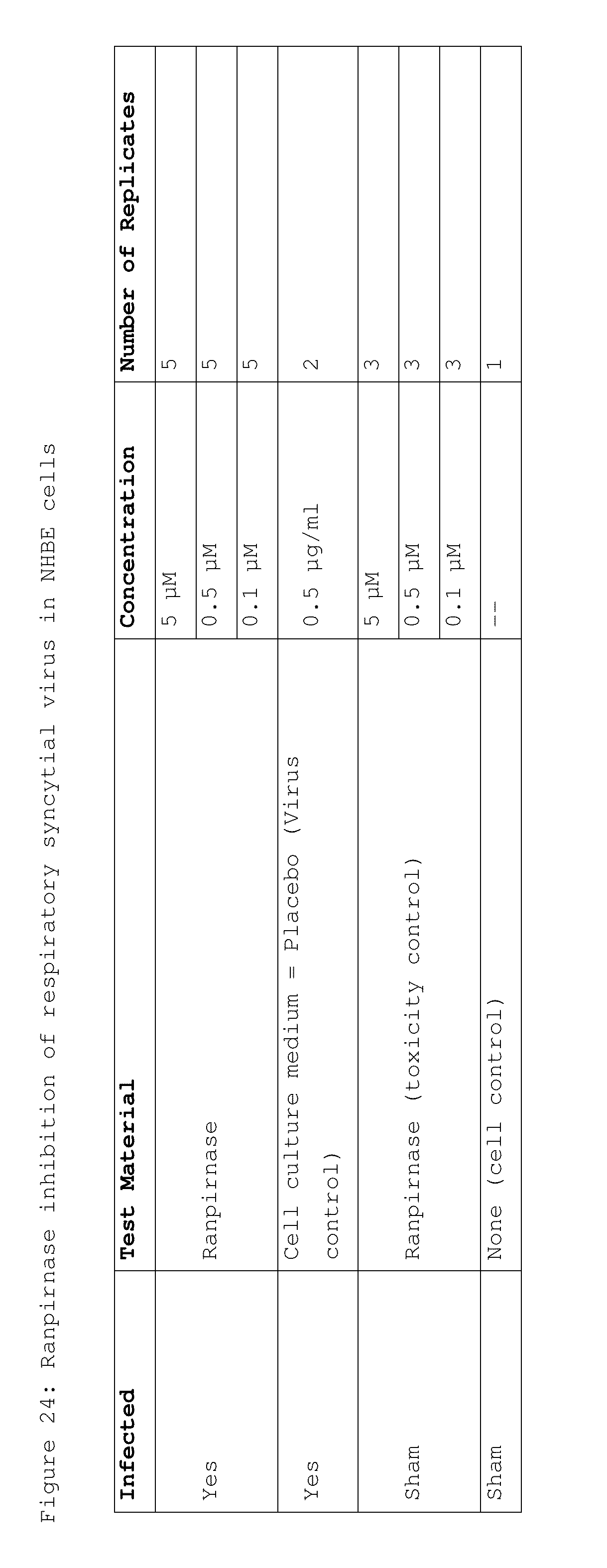
[0039] FIG. 24 shows the doses of ranpirnase used in a study of ranpirnase inhibition of RSV in NHBE cells;
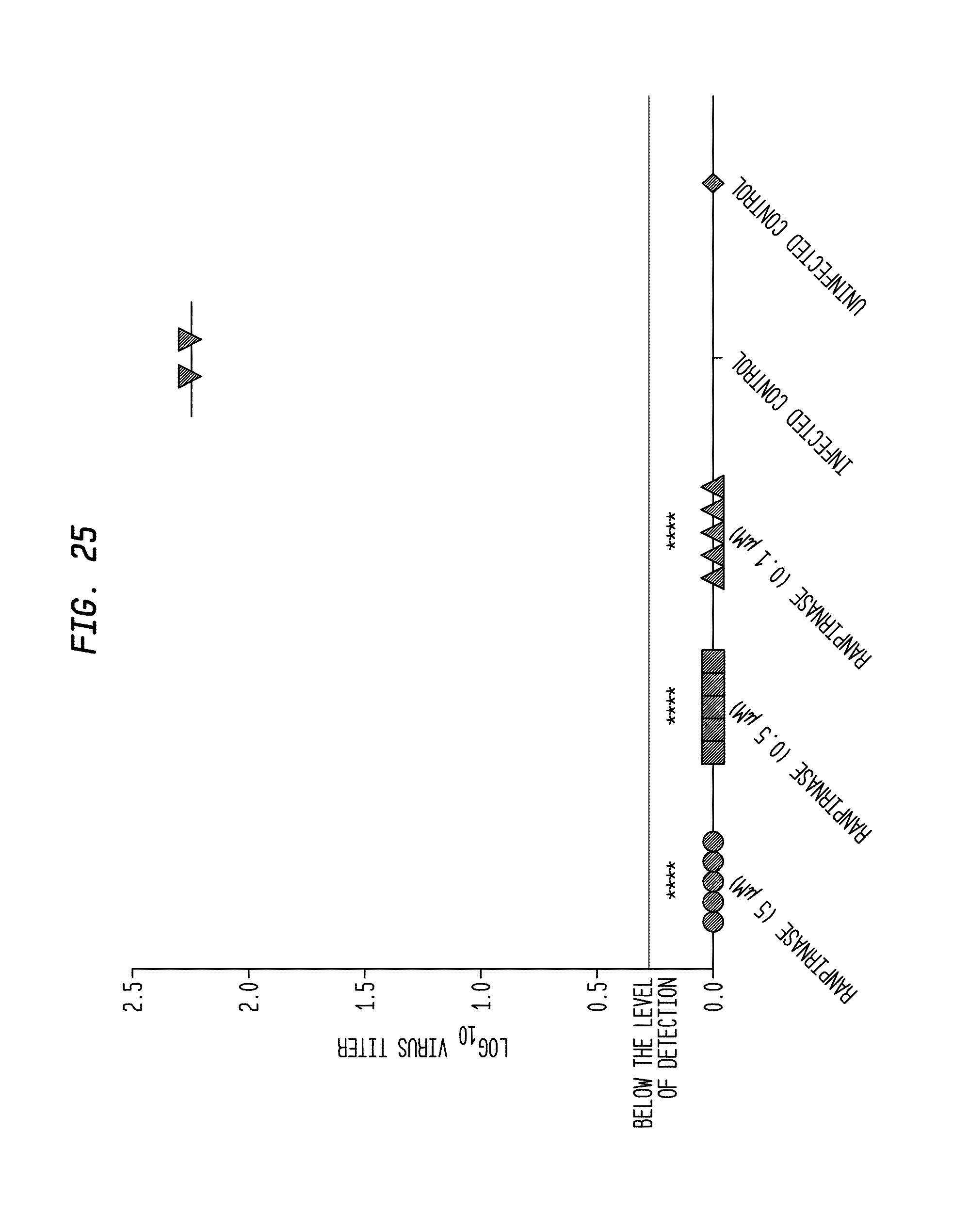
[0040] FIG. 25 shows the results of the study of FIG. 24;
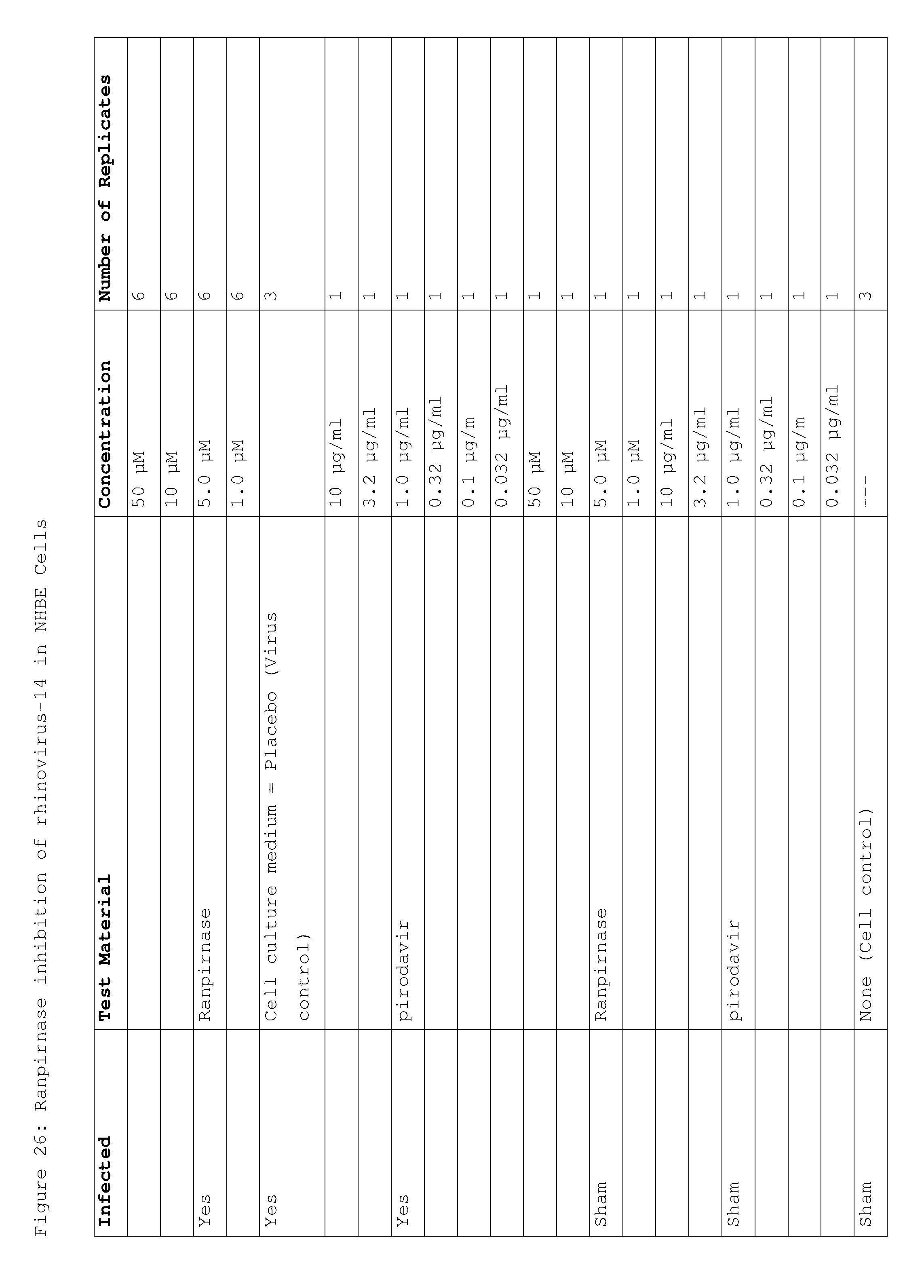
[0041] FIG. 26 shows the doses of ranpirnase used in a study of ranpirnase inhibition of RV-14 in NHBE cells;
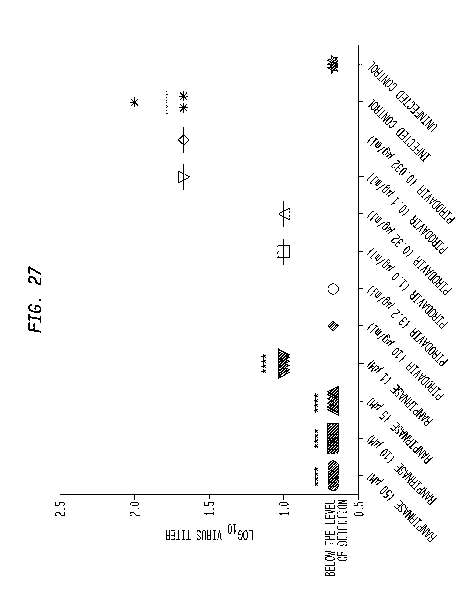
[0042] FIG. 27 shows the results of the study of FIG. 26;
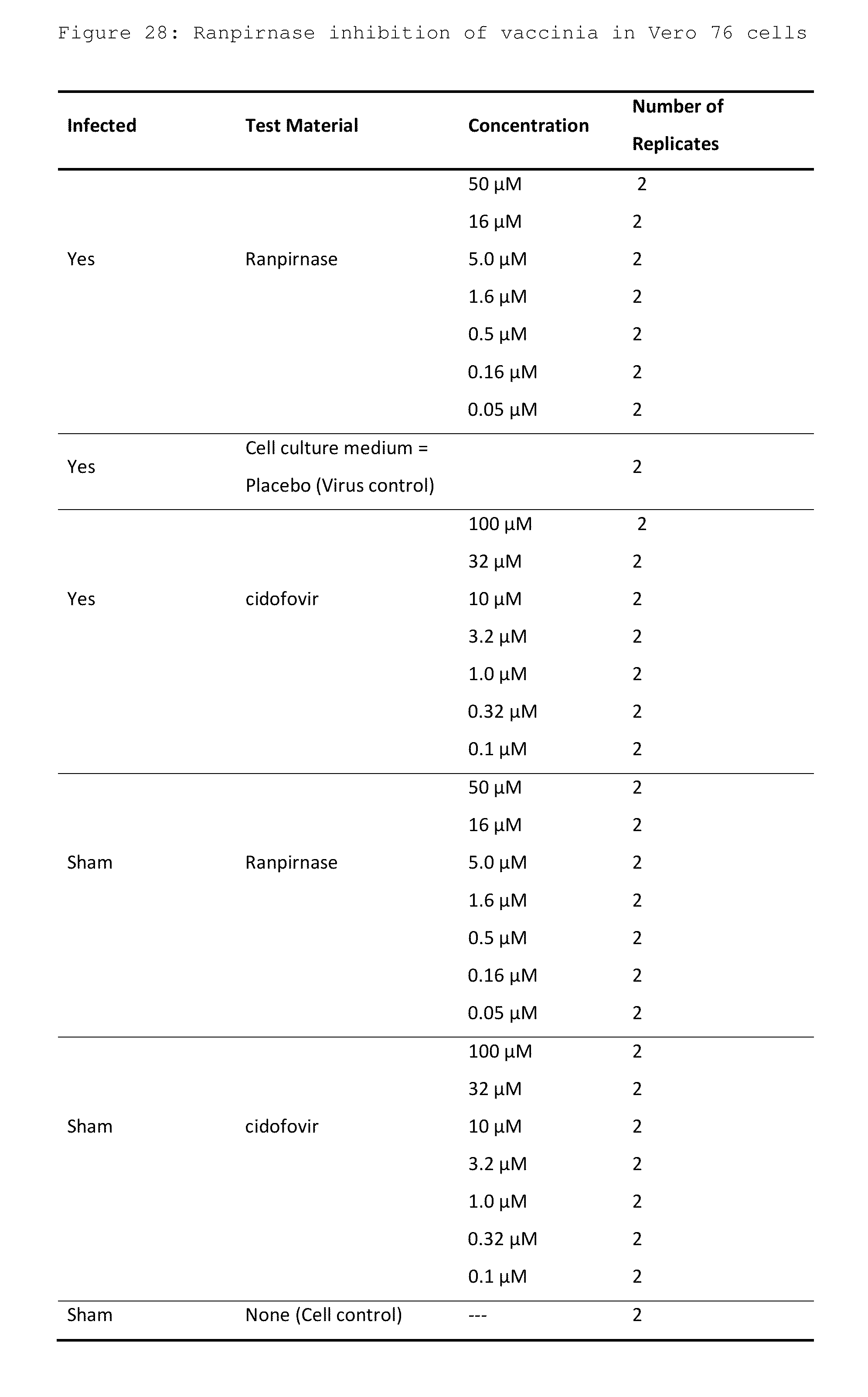
[0043] FIG. 28 shows the doses of ranpirnase used in a study of ranpirnase inhibition of vaccinia in Vero 76 cells;
[0044] FIG. 29 shows the results of the study of FIG. 28;
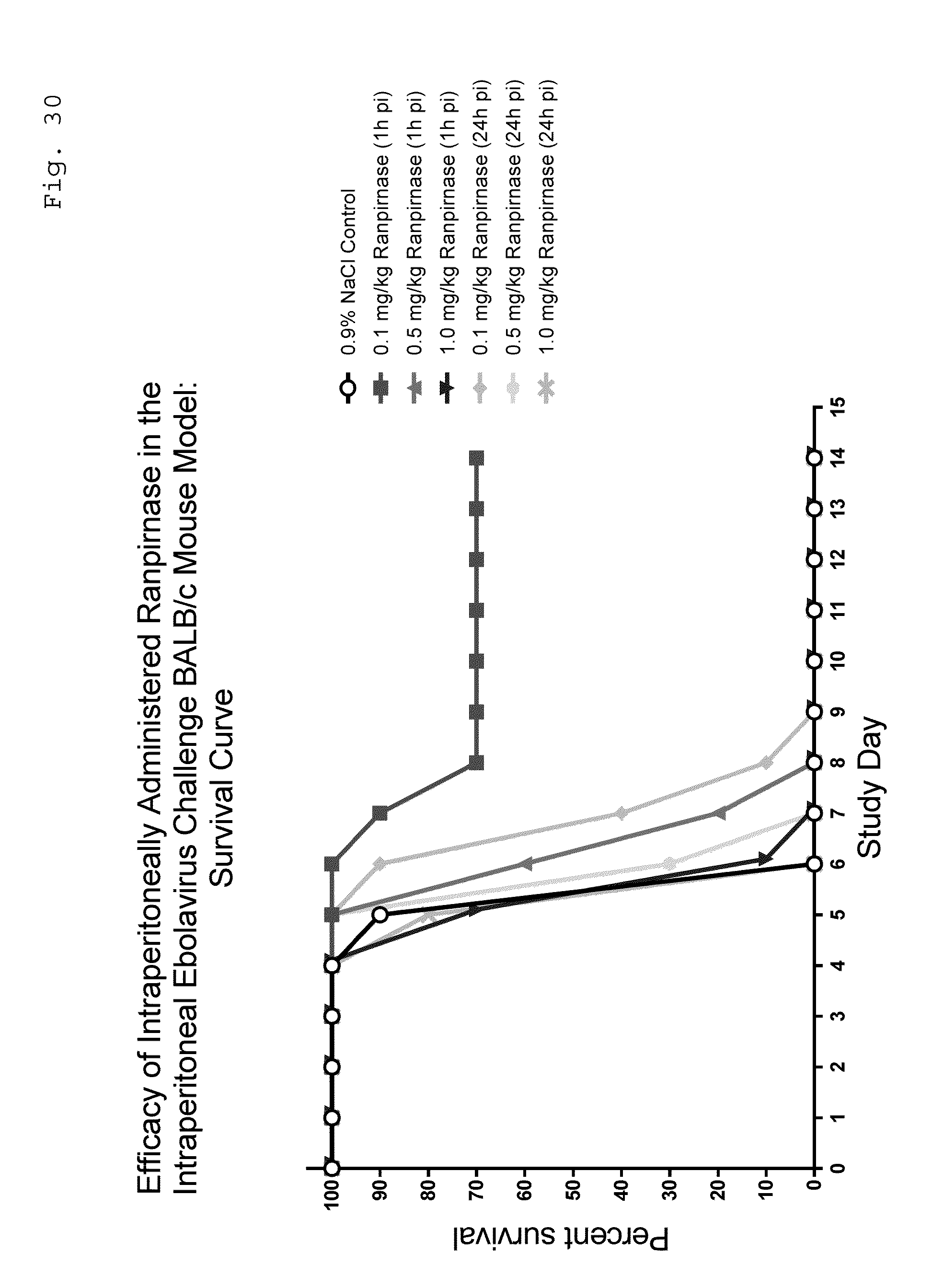
[0045] FIG. 30 is a graph showing survival of groups of Ebola-infected BALB/c mice that were therapeutically treated with ranpirnase;
[0046] FIG. 31 is a graph showing that survival of Ebola-infected BALB/c mice in the tested groups occurs in ranpirnase-treated mice of both sexes;
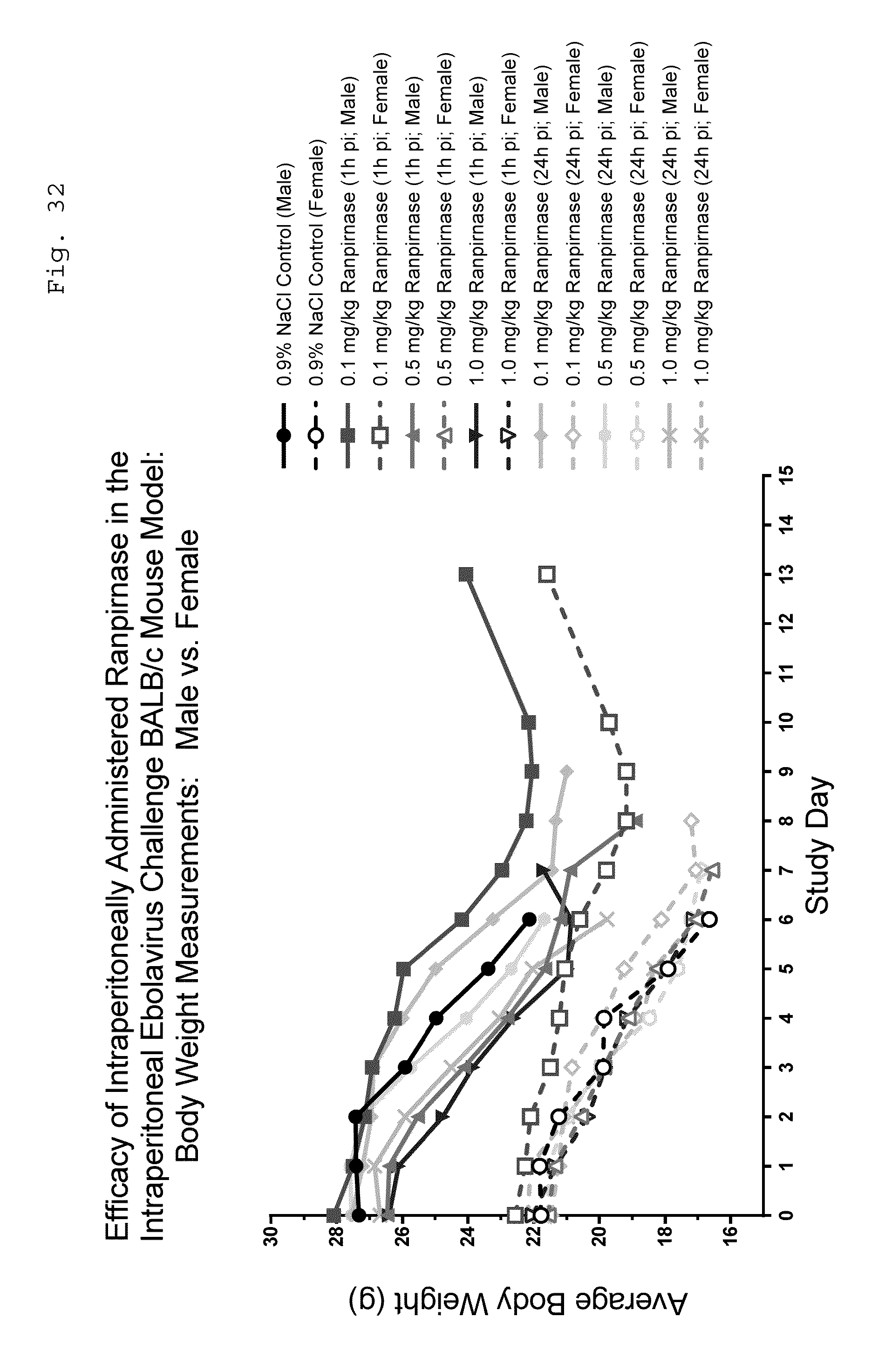
[0047] FIG. 32 is a graph showing variation in body weight of Ebola-infected BALB/c mice in the tested groups during the experiment;
[0048] FIG. 33 is a graph showing variation in the activity/behavior of Ebola-infected BALB/c mice in the tested groups during the experiment; and
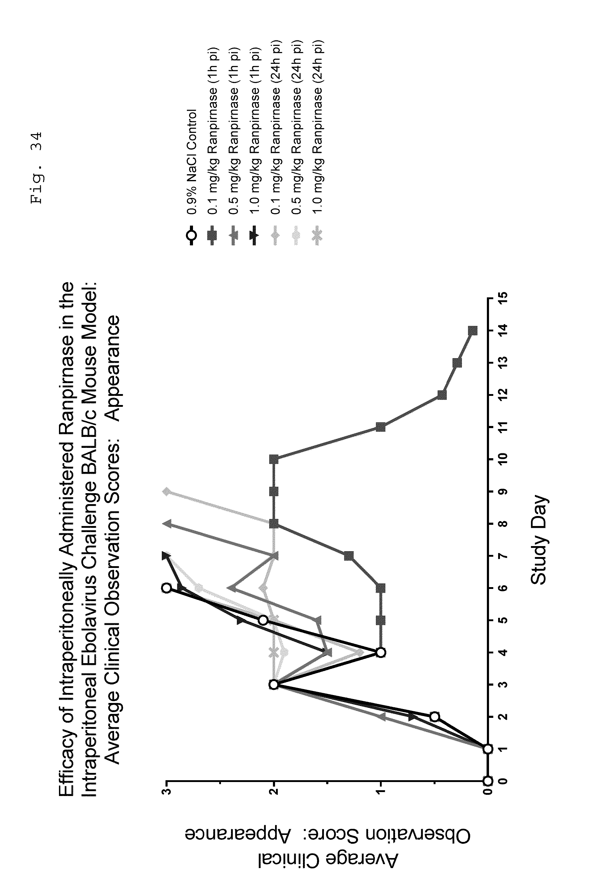
[0049] FIG. 34 is a graph showing variation in the appearance of Ebola-infected BALB/c mice in the tested groups during the experiment.
DETAILED DESCRIPTION OF PREFERRED EMBODIMENTS
Example 1
Rabies in MEM, EF, and BSR Cells
[0050] In the experiments described in FIG. 1, the density of each of the cell lines under test was adjusted to 50,000 cells/ml using minimal essential media (MEM). The cells were placed in wells and incubated at 37.degree. C. and 0.5% CO.sub.2 for 24 hours. Ranpirnase was diluted to concentrations of 10 uM, 3 uM, 900 nM, 270 nM, 81 nM, and 24 nM, added to the wells, and incubated at 37.degree. C. and 0.5% CO.sub.2 for 24 hours.
[0051] Rabies virus at a multiplicity of infection ("MOI") of 0.1 was then added to each of the wells and the ranpirnase and rabies virus--containing wells were incubated at 37.degree. C. and 0.5% CO.sub.2 for 72 hours.
[0052] The virus in each of the wells was titrated 24 and 48 hours after introduction of the rabies virus using mouse neuroblastoma cells, and the results are shown in FIG. 1.
[0053] The selectivity index SI is an accepted measurement of the ability of a drug under test to inhibit replication of a viral infection without killing the infected cells. Where SI in the accompanying Figure is greater than 1, ranpirnase is active against the virus indicated, and increasing values of SI indicate increasing activity. Thus, as can be seen in FIG. 1 at 24 hours after introduction of viruses into ranpirnase-containing cell lines, ranpirnase is extraordinarily active against rabies in the mammalian cell lines indicated.
[0054] Because SI measures the ability of a substance under test to inhibit replication of a particular virus without killing the infected cells themselves, it is reasonably correlated with usefulness of the substance in treating a mammalian subject that is infected with the virus. Accordingly, test results in which SI>1 indicate that mammalian subjects infected with rabies can be treated by systemic administration of an appropriate dose of ranpirnase. Furthermore, other below-disclosed experimental results in VEEV, CHIV, and EBOV indicate that it should be possible to use ranpirnase as a prophylactic to prevent rabies infection.
Example 2
MERS-CoV in NHBE Cells
[0055] In the experiment illustrated in FIG. 2, the anti-viral activity of ranpirnase against MERS-CoV virus was compared to the activities of two known anti-viral agents: SARS protease inhibitor and Infergen. The experiment was carried out using four different concentrations of each agent on normal human bronchial epithelial (NHBE) cells.
[0056] More specifically, the NHBE cells were grown in HEPES Buffered Saline Solution at 37.degree. C. for seven days. The cells were washed and refreshed once daily. Two controls were used: one contained MERS-CoV virus and the other contained uninfected NHBE cells that were treated with the agents under test.
[0057] On the eighth day, the tested concentrations of the three agents under test were introduced into the cells and buffer solution and the virus was introduced at a multiplicity of infection ("MOI") of 0.01. The virus- and agent-containing samples were then incubated for 72 hours at 37.degree. C. and 5% CO.sub.2, with the medium being replenished once each day. After 72 hours, the samples were then titrated to determine their viral content.
[0058] In this experiment SI was not calculated. Rather, the anti-viral activity of the various agents under test was determined by comparing viral production (viral titer in Vero 76 cells) in NHBE cells that had been treated with the various agents under test to the viral production in the NHBE cells used as controls.
[0059] As can be seen in FIG. 2, ranpirnase was far more active against MERS-CoV virus than either of the other agents. And, the activity of ranpirnase was clearly statistically significant, since all but the least concentrated of the doses of ranpirnase had a p value of less than 0.0001. This reduction of viral titers of MERS-CoV virus without killing the host cells is reasonably correlated with usefulness of ranpirnase in treating MERS-CoV.
[0060] Because ranpirnase was so effective at inhibiting replication of the MERS-CoV virus in NHBE cells while not killing the host cells, this experiment further evidences the likelihood that systemically administered ranpirnase will be useful in treating a mammalian subject infected with a virus, and particularly a mammalian subject infected with MERS-CoV virus. Furthermore, other below-disclosed experimental results in VEEV, CHIV, and EBOV indicate that it should be possible to use ranpirnase as a prophylactic to prevent MERS-CoV infection.
Example 3
Influenza in NHBE Cells
[0061] In the experiment illustrated in FIG. 3, the anti-viral activity of various concentrations (5 .mu.g/ml "Ranp-5", 1 .mu.g/ml "Ranp-1", 0.5 .mu.g/ml "Ranp-0.5", and 0.1 .mu.g/ml "Ranp-0.5") of ranpirnase against influenza A/California/07/2009(H1N1)pdm09 virus was compared to the activities of various concentrations of two known anti-viral agents (Ribavirin at 320 .mu.g/ml "Riba-320", 100 .mu.g/ml "Riba-100", and 10 .mu.g/ml "Riba-20" and Oseltamivir carboxylate at 25 .mu.M "Osel-25", 10 .mu.M "Osel-10", and 1 .mu.M) "Osel-1". The experiment was carried out on NHBE cells using four different concentrations of ranpirnase and three different concentrations of each of the other agents. Three controls were used. The first control was NHBE cells that were infected by the virus and treated with a placebo. (This control is shown in FIG. 3.) The second control was NHBE cells that were "infected" with a placebo and treated using the agents under test. The third control was NHBE cells that were "infected" with a placebo and treated with a placebo.
[0062] The NHBE cells were supplied with the vendor's proprietary culture medium and equilibrated at 37.degree. C. and 5% CO.sub.2 for at least 16 hours. After equilibration, the cells were washed and refreshed.
[0063] NHBE cells were then infected with influenza A/California/07/2009(H1N1)pdm09 virus at a multiplicity of infection level of 0.01. After an adsorption period of 1 hour, the viral inoculum was removed and the treatments were applied. Twenty four hours post infection, the treatments were replenished. Forty eight hours post infection, the supernatants were harvested. Then, virus titers were determined in Madin-Darby-Canine-Kidney cells and analyzed for statistical significance using one-way analysis of variance.
[0064] As can be seen in FIG. 3, with the exception of the least concentrated dose, the activity of ranpirnase against influenza A/California/07/2009(H1N1)pdm09 virus was statistically significant and dose-dependent. And, these experiments confirmed a result that was seen in the experiment illustrated in FIG. 2: ranpirnase did not decrease the viability of NHBE cells at any of the concentrations under test.
[0065] This reduction of viral titers of influenza A/California/07/2009(H1N1)pdm09 virus without reduction of viability of the host cells is reasonably correlated with usefulness of ranpirnase in treating influenza A/California/07/2009(H1N1)pdm09. Because ranpirnase inhibited replication of the tested influenza virus in NHBE cells while not killing the cells themselves, this experiment further evidences the likelihood that systemically administered ranpirnase will be useful in treating a mammalian subject infected with a virus, and particularly a mammalian subject infected with influenza. Furthermore, other below-disclosed experimental results in VEEV, CHIV, and EBOV indicate that it should be possible to use ranpirnase as a prophylactic to prevent influenza infection.
Example 4
VEEV, CHIV, and EBOV (all In Vitro)
[0066] Methodology
[0067] Several studies were conducted to assess the ability of ranpirnase to inhibit infection of cells by VEEV, CHIV, and EBOV. Ranpirnase solution and powder-derived ranpirnase were tested. The powder-derived ranpirnase was lyophilized ranpirnase provided by Tamir Biotechnology, Inc. Quality control of the assay was conducted using Positive (Neutral) control (n=16) or infected cells+media, uninfected cells (Negative control) (n=16) and dose response for control inhibitors (n=2 or 4). Z' was calculated for Neutral control and uninfected cells. Data were normalized on the plate bases. Data analysis was done using GeneData software and analysis of dose response curve to determine ED50 of ranpirnase was performed using GeneDataCondoseo software applying Levenberg-Marquardt algorithm (LMA) for curve fitting strategy.
[0068] VEEV in Astrocytes
[0069] To test the effect of ranpirnase on VEEV infection of astrocytes, ranpirnase solution ("RAN") was tested in duplicated 10 point dose response, and powder-derived ranpirnase ("RAN-2") was re-suspended in phosphate buffered saline at 3.5 mg/ml and was tested only as a single dose response. Both the RAN and the RAN-2 were tested in two independent experiments. In these experiments, astrocytes were plated at 4,000 and 3,000 cells/well, incubated overnight and pre-treated with ranpirnase for 2 hours before the infection. Cells were infected at a multiplicity of infection ("MOI") equal to 0.05 for 20 hours. The results of the study are provided in FIGS. 4 and 5.
[0070] To test the effect of ranpirnase on VEEV infection of HeLa cells, RAN was tested in quadruplicated (n=4) 10 point dose response repeated in two independent experiments (rep1 and rep2). RAN2 was tested in n=2 dose responses on plate and repeated in 2 independent experiments. HeLa cells were plated at 4,000 cells/well, incubated overnight and pre-treated with ranpirnase 2 hours before infection. Cells were infected at an MOI equal to 0.05 for 20 hours. The results of the study are provided in FIGS. 6 and 7. As shown in FIG. 7, SI values were over 10 for RAN and over 7.75 for RAN-2.
[0071] CHIV in U2OS Cells
[0072] To test the effect of ranpirnase on CHIV infection of U2OS cells, ranpirnase solution was tested in quadruplicated (n=4) 10 point dose response repeated in two independent experiments (rep1 and rep2). The RAN2 stock was tested in n=2 dose responses on plate and repeated in two independent experiments. U2OS cells were plated at 3,000 cells/well, incubated overnight and pre-treated with ranpirnase 2 hours before infection. Cells were infected at a MOI equal to 0.4 for 24 hours. The results of the study are provided in FIGS. 8 and 9. As shown in FIG. 9, SI values were over 18.
[0073] EBOV in HeLa and Vero E6 Cells
[0074] To test the effect of ranpirnase on EBOV infection of HeLa and Vero E6 cells, ranpirnase solution was tested in quadruplicated (n=4) 10 point dose response repeated in two independent experiments (rep1 and rep2). The RAN2 stock was tested in n=2 dose responses on plate and repeated in two independent experiments. HeLa cells were plated at 4,000 cells/well and Vero E6 at 4000 cells/well, incubated overnight and pre-treated with ranpirnase 2 hours before infection. HeLa cells were infected at a MOI equal to 0.5 and Vero E6 cells were infected at a MOI equal to 0.5 and 0.75 for 48 hours. The results of the study are provided in FIGS. 10, and 11 (HeLa cells) and in FIGS. 12, and 13 (Vero E6 cells). SI values were unexpectedly high, ranging from over 40 to over 77 in Vero E6 cells (FIG. 13).
[0075] Summary of In Vitro VEEV, CHIV, and EBOV Experiments
[0076] The results of the study showed that ranpirnase exhibited robust inhibition of VEEV, CHIV, and EBOV, with surprisingly low AC50 values and surprisingly high SI values. Because SI measures the ability of a substance under test to inhibit replication of a particular virus without killing the infected cells themselves, it is reasonably correlated with usefulness of the substance in treating a mammalian subject that is infected with the virus. Accordingly, test results such as these in which SI>1 indicate that mammalian subjects infected with VEEV, CHIV, and EBOV can be treated by systemic administration of an appropriate dose of ranpirnase. FIG. 14 provides an overall summary of the AC50 results of the studies. AC50 indicates the concentration of the tested agent--here, ranpirnase--that produce half the maximum inhibition of the virus being inhibited.
[0077] These experiments demonstrate that ranpirnase inhibited replication of the tested VEEV, CHIV, and EBOV in various mammalian cells (astrocytes, HeLa cells, U2OS cells, Vero E6 cells) without killing the cells themselves. These experiments further evidence the likelihood that systemically administered ranpirnase will be useful in treating a mammalian subject infected with a virus, and particularly a mammalian subject infected with VEEV, CHIV, and EBOV. Furthermore, it is to be noted that in these experiments, the ranpirnase was used prophylactically, in that the viruses were introduced into cells that had already been treated with ranpirnase. These experiments therefore constitute evidence that the antiviral qualities of ranpirnase can be used prophylactically as well as therapeutically.
Example 5A
EBOV in Mice (Prophylactic Administration)
[0078] The effect of a range of prophylactically administered doses of ranpirnase in EBOV infected mice was studied. C57BL/6 mice (eight to twelve weeks old) were infected with 1000 PFU of mouse-adapted EBOV-Zaire. Infection was accomplished by intraperitoneal injection (intravenous tail vein infusion). 1 hour before infection, each mouse received 0.9% saline (control group; Group 1), 0.1 mg/kg ranpirnase (Group 2), 0.02 mg/kg ranpirnase (Group 3), or 0.004 mg/kg ranpirnase (Group 4). The study groups are shown in FIG. 15.
[0079] Mice were monitored for survival, and the weight of each mouse was obtained on the day of infection and every day for 14 days post infection. (FIGS. 16 and 17 show data for 12 days post infection, but monitoring of the mice continued for two additional days.) The number of survivors per group (out of 10 mice per group) and the percent survival in each group are provided in FIG. 16 and FIG. 17, respectively. The weight of the mice in each group and the percent change from the starting weight are shown below in FIGS. 18 and 19, respectively. The data in FIG. 19 are also shown in graphical form in FIG. 20.
[0080] The study showed that prophylactically administered ranpirnase provides protection from lethal Ebola virus infection in mice. In particular, at a dose of 0.1 mg/kg ranpirnase, 70% of mice survived for at least 14 days after infection. In comparison, in control mice that did not receive ranpirnase, Ebola virus infection was 100% lethal by 7 days post-infection. In addition, surviving mice in the 0.1 mg/kg ranpirnase group maintained a weight within 92% of their starting weight at 12 days post-infection. Thus, the study showed that prophylactically administered ranpirnase effectively promotes survival in Ebola virus infected mice. Animal testing showing such extended survival shows that ranpirnase treatment is reasonably correlated with usefulness in treating Ebola virus. This still further evidences the likelihood that systemically administered ranpirnase will be useful in treating a mammalian subject infected with a virus, and particularly a mammalian subject infected with Ebola virus. Furthermore, this result is consistent with the above-described experimental results in VEEV, CHIV, and EBOV, which indicate that it is possible to use ranpirnase as a prophylactic to prevent EBOV infection.
Example 5B
EBOV in Mice (Therapeutic Administration)
[0081] The therapeutic effect of a range of doses of ranpirnase in mice infected with Ebola virus was studied. BALB/c mice were infected with a target of 100 PFU of mouse-adapted EBOV-Mayinga-CDC-808012. Infection was accomplished by intraperitoneal injection.
[0082] Seven groups of mice were used. Each group contained five males and five females. In the control group, EBOV-infected mice received an intraperitoneal injection of 0.9% NaCl. EVOV-infected mice in the other groups received therapeutic doses of ranpirnase as follows: [0083] Group 2: received 0.1 mg/kg ranpirnase 1 hour post-infection; [0084] Group 3: received 0.5 mg/kg ranpirnase 1 hour post-infection; [0085] Group 4 received 1.0 mg/kg ranpirnase 1 hour post-infection; [0086] Group 5: received 0.1 mg/kg ranpirnase 24 hours post-infection; [0087] Group 6: received 0.5 mg/kg ranpirnase 24 hours post-infection; and [0088] Group 7 received 1.0 mg/kg ranpirnase 24 hours post-infection.
[0089] In each case, ranpirnase in the stated dose was administered twice per day for the first three days of the study and then once per day through Day 9. Mice remained on study until they died or until they were euthanized because they had lost more than 20% of their body weight.
[0090] As can be seen in FIGS. 30, 31, and 32, relative to saline-treated animals, treatment with 0.1 mg/kg ranpirnase gave longer survival times with fewer deaths and longer survival to euthanasia, with slower body weight loss and fewer clinical signs of decreased activity and poor appearance. This effect was observed when treatment began at both 1 hour and 24 hours post infection although the effect was less for the 24 hour group. 0.5 mg/kg ranpirnase treatment gave less effect than 0.1 mg/kg, and the 1 mg/kg group was less effective than the 0.5 mg/kg group. Females seemed to be slightly better at responding to ranpirnase treatment at each dose level.
[0091] Surprisingly, it appeared that lower doses of ranpirnase were more effective than higher doses. The reason for this is not known. It may be due to the study design, which required that animals be euthanized when they lost more than 20% of their body weight. Had the animals been given supportive therapy and allowed to remain on study (Ebola patients are given supportive therapy regardless of their weight loss) they may have regained weight and been restored to health.
[0092] Such a possible result (increased weight and improved health) is suggested by the experimental results illustrated in FIGS. 33 and 34. During this experiment, the activity/behavior and the appearance of the animals was monitored. In FIG. 33: [0093] an activity/behavior score of 0 corresponds to normal behavior/response to external stimuli; [0094] an activity/behavior score of 1 corresponds to minor changes in behavior or exaggerated responses to external stimuli; [0095] an activity/behavior score of 2 corresponds to less mobile, less alert, inactive when activity expected, moderate abnormal response to external stimuli; and [0096] an activity/behavior score of 3 corresponds to moribund: minimal to no activity, mobility consistent with inability to reach food or water.
[0097] In FIG. 34:
[0098] an appearance score of 0 corresponds to normal; [0099] an appearance score of 1 corresponds to lack of grooming; [0100] an appearance score of 2 corresponds to coat rough, possible nasal and/or ocular discharge, ataxia, labored/irregular breathing, slightly hunched posture, partial paralysis; and [0101] an appearance score of 3 corresponds to coat very rough, abnormal/hunched posture, paralysis, seizures, weight loss exceeding 20% of body weight.
[0102] As can be seen in FIGS. 33 and 34, some of the animals showed only mild abnormalities in activity/behavior and in appearance at the time they were euthanized. It will be understood that when living animals are used in medical research, an institutional animal care and use committee ("IACUC") establishes standards that define the boundaries of humane treatment of the experimental subjects. In this study, the IACUC determined that it would be inhumane to allow mice to continue to live once they had lost more than 20% of their body weight. Hence, their apparent activity and health notwithstanding, mice were euthanized once this degree of weight loss occurred.
[0103] This experiment demonstrated that therapeutic administration of ranpirnase effectively promotes survival of mice that are infected with the Ebola virus at the time the ranpirnase is administered. This is true even when the Ebola virus has remained untreated for as long as 24 hours. Animal testing showing such extended survival shows that ranpirnase treatment is reasonably correlated with usefulness in therapeutically treating patients that are infected with the Ebola virus at the time the treatment is administered. This still further evidences the likelihood that systemically administered ranpirnase will be useful in therapeutically treating a mammalian subject infected with a virus, and particularly a mammalian subject infected with Ebola virus.
[0104] In summary: there is now a body of confirmatory in vitro and in vivo data demonstrating that administration of ranpirnase is active against EBOV. This is true whether the ranpirnase is administered prophylactically or therapeutically. A person of ordinary skill in this art would regard such confirmatory in vitro and in vivo data as powerful evidence of usefulness of ranpirnase in treating EBOV in a mammalian subject.
Example 6
AV in NHBE Cells
[0105] A study was conducted to evaluate the anti-adenoviral activity of ranpirnase in NHBE cells.
[0106] Ranpirnase stock solution was prepared at 200 .mu.M and stored in aliquots at -80.degree. C. On the first day of the experiment, one aliquot was thawed and working solutions were prepared by appropriately diluting the stock concentration in cell culture medium. Working solutions were pre-warmed in a water bath set to 37.degree. C. for 15-30 minutes prior to use.
[0107] Differentiated NHBE cells (MatTek Corporation, Ashland, Mass.) were used in the study. Cells were provided in kits with 12 or 24 tissue inserts each. The kits used in the study (EpiAirway.TM., AIR-110, and AIR-100, respectively) originated from a single human donor, #9831, a 23-year old, healthy, non-smoking, Caucasian male. Upon arrival, tissue inserts were immediately transferred to individual wells of a 6-well plate according to manufacturer's instructions. Tissues were supplied with 1 ml of MatTek's culture medium (AIR-100-MM/Maintenance Medium) to the basolateral side, whereas the apical side was exposed to a humidified 95% air/5% CO.sub.2 environment. Cells were equilibrated at 37.degree. C. for at least 16 hours before the start of the experiment. After the equilibration period, the mucin layer, secreted from the apical side of the cells, was removed by repeated (3.times.) careful washing with 500 .mu.l pre-warmed 30 mM HEPES Buffered Saline Solution (Lonza, CC-5024/Lot 0000354165) and the culture medium was replenished.
[0108] The virus (adenovirus 2, strain Miller from ATCC; titer 6.7 log 10 CCID.sub.50/0.1 ml) was stored at -80.degree. C. prior to use. The dose level of adenovirus corresponded to an MOI of 0.1.
[0109] Differentiated NHBE cells were experimentally infected with the virus. After an adsorption period of 1 hour, the viral inoculum was removed and treatments were applied as shown in FIG. 21. Twenty-four hours post infection, treatments were replenished in the basal compartment of the tissue inserts. Five days post infection, supernatants were harvested and stored at -80.degree. C. until determination of virus titers in A-549 cells (human lung carcinoma cells from ATCC). Controls included four groups: [0110] Group 1--infected and placebo-treated cells (virus control); [0111] Group 2--sham-infected and treated cells (toxicity controls); [0112] Group 3--sham-infected and placebo-treated cells (cell control); and [0113] Group 4--2',3'-dideoxcytidine as a positive control drug. Toxicity controls were microscopically examined for possible changes in tissue and/or cell morphology at the end of the experiment.
[0114] NHBE cells were inoculated by exposure of the apical side to AV or cell culture medium (sham) as shown in FIG. 21. After 1 hour.+-.10 min of incubation at 37.degree. C. and 5% CO.sub.2, the viral inoculum or cell culture medium was removed from the cells. The apical side of the cells was washed once with 500 .mu.l pre-warmed HEPES Buffered Saline Solution.
[0115] After inoculation, ranpirnase, 2',3'-dideoxcytidine, or cell culture medium (placebo/cell control) was added to the apical side of the cells and in the basal medium compartment, and incubated with the cells for 1 hour. After 1-hour incubation, the drug-containing medium was removed from the apical and basal chambers. Culture medium alone (placebo/cell control) or with drug (test condition) was added to the bottom chamber, and cells were incubated for 4 days. Twenty-four hours post infection, cell culture medium with and without drug was replenished to the basal compartment.
[0116] Following infection and treatment, cells were maintained at the air-liquid interface, and cell culture supernatant was harvested 48 hours post infection. Virus released into the apical compartment of the NHBE cells was harvested by the addition and collection of 500 .mu.l culture medium allowed to equilibrate for 30 min at 37.degree. C. and 5% CO.sub.2. The medium from the apical compartment was divided into 2 aliquots, which were stored at -80.degree. C. for future analysis of viral titers.
[0117] To assess the virus dose that was able to infect 50% of the cell cultures (CCID.sub.50), A-549 cells were seeded in 96-well plates and grown overnight to achieve confluence, then washed twice with 100 .mu.l infection medium (DMEM/EBSS supplemented with 50 .mu.l/ml gentamycin). Wells were filled with 100 .mu.l infection medium. Apical washes from the NHBE cell cultures were diluted 10-fold in infection medium and 100 .mu.l were transferred into respective wells of a 96-well microtiter plate. Each concentration of ranpirnase from the NHBE cells (6 NHBE cell wells/dose) was titered leading to six titers per concentration (each NHBE well treated as a replicate) to evaluate the virus yields from infected and infected, treated cells. Thus, each concentration of ranpirnase was titered a total of six times. For the positive control, 2'3'-dideoxycytidine, one well of NHBE cells only was assigned to each concentration. Thus, each concentration was titered only once. Three wells were assigned as untreated, infected controls. They were titered once, resulting in three replicate untreated, infected control titers. After 6 days of incubation at 37.degree. C. and 5% CO.sub.2, cells were microscopically examined and scored for virus-induced cytopathic effect ("CPE"). A well was scored positive if any trace of CPE (usually cell rounding or lysis) was observed as compared with the uninfected control. CCID.sub.50 was calculated by the Reed-Muench method.
[0118] The results of the study are provided in FIG. 22. All treatments with ranpirnase decreased virus titers to undetectable levels, except for the lowest dose. This reduction represented an approximately three log drop in virus titer for the 50 .mu.M ranpirnase treatment (four asterisks indicates that p<0.0001 as compared to placebo). For the 5 and 10 .mu.M ranpirnase treatments, a .about.1 log reduction in virus titers was detected when compared to the virus titers detected from the untreated, infected controls wells (a single asterisk indicates that p<0.05 as compared to placebo). 2'3'-dideoxycytidine inhibited virus replication as expected.
[0119] No virus cytopathic effects were detected in uninfected, ranpirnase-treated or 2'3'-dideoxycytidine-treated cells. Microscopy evaluations of ranpirnase-treated or 2'3'-dideoxycytidine-treated NHBE cells revealed no toxicological phenomena.
[0120] Therefore, the results of the study showed that all higher doses of ranpirnase treatment reduced AV titers in a statistically significant manner. Such reduction of viral titers of AV virus without cytopathic effects on the host cells is reasonably correlated with usefulness of ranpirnase in treating AV. Because ranpirnase was effective at inhibiting replication of AV in NHBE cells while not killing the host cells, this experiment further evidences the likelihood that systemically administered ranpirnase will be useful in treating a mammalian subject infected with a virus, and particularly a mammalian subject infected with AV. Furthermore, the above-disclosed experimental results in VEEV, CHIV, and EBOV indicate that it should be possible to use ranpirnase as a prophylactic to prevent AV infection.
Example 7
Canine Parvovirus in A-72 Cells
[0121] Canine parvovirus type 2c was grown in A-72 cell cultures and virus contained in cell culture medium (Dulbecco's minimum essential medium; "DMEM") was stored frozen at -80.degree. C. until used. The titer of the virus stock was about 105 cell culture infectious dose units/ml, with a hemagglutination (HA) titer of 1,024 to 2,048.
[0122] The A-72 continuous cell line, a fibroblastic line derived from a canine tumor, was grown in DMEM in 96-well plates, allowing for various conditions to be tested.
[0123] To assess the cytotoxicity of ranpirnase in A-72 cells, A-72 cells were seeded into a 24-well plate at a concentration of 125,000 cells/well in a volume of 1 ml of DMEM. The medium in each well was supplemented with 25 .mu.l of ranpirnase to concentration of 5 .mu.g/ml, 1 .mu.g/ml, 0.5 .mu.g/ml and 0.1 .mu.g/ml. Each concentration was tested in 4 replicates. After incubating at 35.5.degree. C. for 4 days, the cells were examined under an inverted microscope for any visual morphologic changes.
[0124] Next, the efficacy of ranpirnase was assessed. Ranpirnase was tested at various concentrations ranging from 10 .mu.g/ml down to 0.15625 .mu.g/ml in 2-fold dilutions. Stock virus was tested from undiluted through a dilution of 1/10,000. A-72 cells were trypsinized following standard procedures and seeded into 96-well cell culture plates at a concentration of 18,750 cells/well in a volume of 150 .mu.l. Ranpirnase was then added to each well to attain a final pre-determined concentration. After incubating for 2 hours at 35.5.degree. C., 25 .mu.l of virus at various dilutions was added. Controls included ranpirnase-treated/uninfected cells and ranpirnase inhibition of canine parvovirus replication in cell cultures of untreated/uninfected cells. All variables were tested in duplicate. At the end of 4 days of incubation at 35.5.degree. C., the supernatant fluid was harvested and tested for evidence of virus replication using the HA test (which tests the ability of the virus to agglutinate a 0.5% suspension of swine red blood cells). HA titers were expressed as the reciprocal of the highest dilution of supernatant fluid that induced visual agglutination of the red blood cells in a 96-well plate.
[0125] When ranpirnase-treated (various concentrations) and untreated cells were microscopically examined, there was no visual difference in morphology between the two groups, regardless of ranpirnase concentration. Thus, there was no apparent cytotoxic effect of ranpirnase at any of the concentrations tested.
[0126] The results of the study are provided in FIG. 23. Virus growth was detected in untreated cell cultures up to a virus dilution of 1/100. Virus growth was also detected when the undiluted virus stock was used to infect cell cultures treated with 0.15625 .mu.g/ml. There was no virus growth in cultures treated with a ranpirnase concentration of 0.3125 .mu.g/ml or higher. Inhibition of viral growth of canine parvovirus without cytotoxic effects on the host cells is reasonably correlated with usefulness of ranpirnase in treating canine parvovirus. Because ranpirnase was so effective at inhibiting viral growth of canine parvovirus while not killing the host cells, this experiment further evidences the likelihood that systemically administered ranpirnase will be useful in treating a mammalian subject infected with a virus, and particularly a mammalian subject infected with canine parvovirus. Furthermore, the above-disclosed experimental results in VEEV, CHIV, and EBOV indicate that it should be possible to use ranpirnase as a prophylactic to prevent canine parvovirus infection.
Example 8
RSV in NHBE Cells
[0127] Ranpirnase stock solution was prepared, stored, thawed, and used to prepare working solutions as described in the AV experiment disclosed above.
[0128] NHBE cells from MatTek Corporation were used in the study. They were the same cell line as was used in the AV experiment discussed above and were provided in the same kits. As in the AV experiment, tissue inserts were immediately transferred to individual wells of a 6-well plate according to manufacturer's instructions. Tissues were supplied with 1 ml of the same culture medium used in the AV experiment to the basolateral side, and the apical side was exposed to a humidified 95% air/5% CO.sub.2 environment. Cells were equilibrated as in the AV experiment, and after this equilibration period, the mucin layer was removed as in the AV experiment and the culture medium was replenished.
[0129] The virus (RSV A2 from ATCC; titer 4.7 log 10 CCID.sub.50/0.1 ml) was stored at -80.degree. C. prior to use. The dose level of challenge virus corresponds to an MOI of 0.1.
[0130] Differentiated NHBE cells were experimentally infected with the RSV virus. After an adsorption period of 1 hour, the viral inoculum was removed and treatments were applied as shown in FIG. 24. Twenty-four hours post infection, treatments were replenished in the basal compartment of the tissue inserts. Forty-eight hours post infection, supernatants were harvested and stored at -80.degree. C. until determination of virus titers in MA-104 cells (embryonic African green monkey kidney cells from ATCC). Controls consisted of three of the four groups used in the AV experiment:
Group 1--infected and placebo-treated cells (virus control); Group 2--sham-infected and treated cells (toxicity controls); and Group 3--sham-infected and placebo-treated cells (cell control). Toxicity controls were microscopically examined for possible changes in tissue and/or cell morphology at the end of the experiment.
[0131] NHBE cells were inoculated by exposure of the apical side to RSV or cell culture medium (sham) as shown in FIG. 24. After 1 hour.+-.10 min of incubation at 37.degree. C. and 5% CO.sub.2, the viral inoculum or cell culture medium was removed from the cells. The apical side of the cells was washed once with 500 .mu.l pre-warmed HEPES Buffered Saline Solution.
[0132] After inoculation, ranpirnase, or cell culture medium (placebo/cell control) was added to the apical side of the cells and in the basal medium compartment, and incubated with the cells for 1 hour. After 1-hour incubation, the drug-containing medium was removed from the apical and basal chambers. Culture medium alone (placebo/cell control) or with drug (test condition) was added to the bottom chamber, and cells were incubated for an additional 23 hours (24 hour total post-infection incubation). Twenty-four hours post infection, cell culture medium with and without drug was replenished to the basal compartment.
[0133] Following infection and treatment, cells were maintained at the air-liquid interface, and cell culture supernatant was harvested 48 hours.+-.30 min post infection. Virus released into the apical compartment of the NHBE cells was harvested by the addition and collection of 500 .mu.l culture medium allowed to equilibrate for 30 min at 37.degree. C. and 5% CO.sub.2. The medium from the apical compartment was divided into 2 aliquots, which were stored at -80.degree. C. for future analysis of viral titers. Images of one replicate tissue insert per treatment were taken with an inverted microscope at 40 and 100.times., respectively, prior to harvest.
[0134] To assess the CCID.sub.50, MA-104 cells were seeded in 96-well plates and grown overnight to achieve confluence, then washed twice with 100 .mu.l infection medium (MEM/EBSS supplemented with 50 .mu.l/ml gentamycin). Wells were filled with 100 .mu.l infection medium. Apical washes were diluted 10-fold in infection medium and 100 .mu.l were transferred into respective wells of a 96-well microtiter plate. Each sample was titered in triplicate (Passage 1) to evaluate the virus yields from infected and infected, treated cells. After 6 days of incubation at 37.degree. C. and 5% CO2, cells were microscopically examined and scored for virus-induced cytopathic effect (CPE). A well was scored positive if any trace of cytopathic effect (usually cell rounding or syncytium) was observed as compared with the uninfected control. CCID50 was calculated by the Reed-Muench method.
[0135] The results of the study are provided in FIG. 25. Results were analyzed for statistical significance by one-way ANOVA (GraphPad Prism, version 6c). All treatments with Ranpirnase decreased virus titers to undetectable levels (FIG. 25). This reduction represented a 2.3 log reduction in virus titers compared to untreated, infected controls (the four asterisks shown in FIG. 25 indicate that p<0.0001).
[0136] No virus cytopathic effects were detected Ranpirnase-treated/sham infected cells or uninfected control cells. Microscopy evaluations of sham infected, Ranpirnase-treated or 2'3'-dideoxycytidine-treated NHBE cells revealed no toxicological phenomena.
[0137] Therefore, the results of the study showed that all doses of ranpirnase tested (5 .mu.M, 0.5 .mu.M, and 0.1 .mu.M) reduced RSV titers in a statistically significant manner, and that ranpirnase alone did not elicit cytopathic effects in NHBE cells. Such reduction of viral titers without accompanying cytopathic effects on the host cells is reasonably correlated with usefulness of ranpirnase in treating RSV. Because ranpirnase was so effective at inhibiting RSV while not killing the host cells, this experiment further evidences the likelihood that systemically administered ranpirnase will be useful in treating a mammalian subject infected with a virus, and particularly a mammalian subject infected with RSV. Furthermore, the above-disclosed experimental results in VEEV, CHIV, and EBOV indicate that it should be possible to use ranpirnase as a prophylactic to prevent RSV infection.
Example 9
RV in NHBE Cells
[0138] Ranpirnase stock solution was prepared, stored, thawed, and used to prepare working solutions as described in the AV and RSV experiments disclosed above.
[0139] NHBE from MatTek Corporation were used in the study. They were the same cell line as were used in the AV and RSV experiments discussed above and were provided in the same kits. As in the AV and RSV experiments, tissue inserts were immediately transferred to individual wells of a 6-well plate according to manufacturer's instructions. Tissues were supplied with 1 ml of the same culture medium used in the AV and RSV experiments to the basolateral side, and the apical side was exposed to a humidified 95% air/5% CO.sub.2 environment. Cells were equilibrated as in the AV experiment, and after this equilibration period, the mucin layer was removed as in the AV and RSV experiments and the culture medium was replenished.
[0140] RV-14 (strain 1059 from ATCC) was stored at -80.degree. C. prior to use. The titer of the stock virus was equal to titer 3.6 log 10 CCID50/0.1 ml. The dose level of challenge virus was based on data from the previous experiments, and corresponded to a multiplicity of infection (MOI) of 0.0041.
[0141] Differentiated NHBE cells were experimentally infected with RV-14 virus. After an adsorption period of 1 hour, the viral inoculum was removed and treatments applied (FIG. 26). Twenty-four hours post infection, treatments were replenished in the basal compartment of the tissue inserts. Four days post infection, supernatants were harvested and stored at -80.degree. C. until determination of virus titers in HeLa-Ohio-1 cells (human cervical carcinoma cells from ATCC). Controls consisted of four groups: [0142] Group 1--infected and placebo-treated cells (virus control); [0143] Group 2--sham-infected and treated cells (toxicity controls); [0144] Group 3--sham-infected and placebo-treated cells (cell control); and [0145] Group 4--pirodavir as a positive control drug.
[0146] Toxicity controls were microscopically examined for possible changes in tissue and/or cell morphology at the end of the experiment.
[0147] NHBE cells were inoculated by exposure of the apical side to RV-14 or cell culture medium (sham infection) as seen in FIG. 27. After 1 hour.+-.10 min of incubation at 37.degree. C. and 5% CO.sub.2, the viral inoculum or cell culture medium was removed from the cells. The apical side of the cells was washed once with 500 .mu.l pre-warmed HEPES Buffered Saline Solution.
[0148] After inoculation, ranpirnase, pirodavir, or cell culture medium (placebo/cell control) was added to the apical side of the cells and in the basal medium compartment, and incubated with the cells for 1 hour. After 1-hour incubation, the drug-containing medium was removed from the apical and basal chambers. Culture medium alone (Placebo/Cell control) or with drug (test condition) was added to the bottom chamber, and cells were incubated for 4 days. Twenty-four hours post infection, cell culture medium with and without drug was replenished to the basal compartment.
[0149] Following infection and treatment, cells were maintained at the air-liquid interface, and cell culture supernatant was harvested 4 days post virus exposure. Virus released into the apical compartment of the NHBE cells was harvested by the addition and collection of 500 .mu.l culture medium allowed to equilibrate for 30 min at 37.degree. C. and 5% CO.sub.2. The medium from the apical compartment divided into 2 aliquots, which were stored at -80.degree. C. for future analysis of viral titers.
[0150] HeLa Ohio-1 cells were seeded in 96-well plates and grown overnight to achieve confluence, then washed twice with 100 .mu.l infection medium (MEM/EBSS supplemented with 50 .mu.l/ml gentamycin). Wells were filled with 100 .mu.l infection medium. Apical washes from the NHBE cell cultures were diluted 10-fold in infection medium and 100 .mu.l were transferred into respective wells of a 96-well microtiter plate. Each concentration of ranpirnase from the NHBE cells (6 NHBE cell wells/dose) was titered leading to six titers per concentration (each NHBE well treated as a replicate) to evaluate the virus yields from infected and infected, treated cells. Thus, each concentration of Ranpirnase was titered a total of six times. For the positive control, pirodavir, one well of NHBE cells only was assigned to each concentration. Thus, each concentration was titered only once. Three wells were assigned as untreated, infected controls. They were titered once, resulting in three replicate untreated, infected control titers. After 7 days of incubation at 37.degree. C. and 5% CO.sub.2, cells were microscopically examined and scored for virus-induced CPE. A well was scored positive if any trace of CPE (cell lysis) was observed as compared with the uninfected control. CCID50 was calculated by the Reed-Muench method and the inverse of that dilution represented the virus titer.
[0151] All ranpirnase treatments decreased virus titers relative to the titers of untreated infected controls except for the lowest dose (FIG. 27). The reduction in virus titer with 50, 10, 5 .mu.M ranpirnase treatment represented an approximate 1.67 log 10 drop in virus titer for the (P<0.0001). For the 1 .mu.M ranpirnase treatment, an .about.1 log reduction in virus titers was detected when compared to the virus titers detected from the untreated, infected controls wells (P<0.0001). Pirodavir inhibited virus replication as expected at 10 and 3.2 .mu.g/ml with a somewhat dose responsive decrease in virus yields at subsequent lower dilutions of drug. Typically at 0.0032 .mu.g/ml, pirodavir is also inactive against RV-14 in a HeLa Ohio-1 cell culture antiviral system as was seen in NHBE cells in this experiment.
[0152] No virus cytopathic effects were detected in uninfected, ranpirnase-treated or pirodavir-treated cells. Microscopy evaluations of ranpirnase-treated or pirodavir-treated NHBE cells revealed no toxicological phenomena.
[0153] Therefore, the results of the study showed that all doses of ranpirnase tested (50 .mu.M, 1.0 .mu.M, 5 .mu.M and 1 .mu.M) reduced RV-14 titers in a statistically significant manner, and that ranpirnase alone did not elicit cytopathic effects in NHBE cells. Such titer reduction, accompanied by absence of cytopathic effects on the host cells, is reasonably correlated with usefulness of ranpirnase in treating RV-14. Because ranpirnase was so effective at inhibiting RV-14 while not killing the host cells, this experiment further evidences the likelihood that systemically administered ranpirnase will be useful in treating a mammalian subject infected with a virus, and particularly a mammalian subject infected with RV-14. Furthermore, the above-disclosed experimental results in VEEV, CHIV, and EBOV indicate that it should be possible to use ranpirnase as a prophylactic to prevent RV-14 infection.
Example 10
Vaccinia in Vero 76 Cells
[0154] Ranpirnase stock solution was prepared, stored, thawed, and used to prepare working solutions as described in the AV, RSV, and RV-14 experiments disclosed above.
[0155] The virus (vaccinia, strain WR from ATCC) was stored at -80.degree. C. prior to use. The titer of the stock virus was equal to titer 7.7 log 10 CCID50/0.1 ml. The dose level of challenge virus was based on data from other experiments, and corresponded to 50-100 viral plaques/well.
[0156] To determine the antiviral effect of ranpirnase against vaccinia, Vero 76 cells in 24 well plates were experimentally infected with virus in the format shown in FIG. 28. Controls consisted of four groups: [0157] Group 1--infected and placebo-treated cells (virus control); [0158] Group 2--sham-infected and treated cells (toxicity controls); [0159] Group 3--sham-infected and placebo-treated cells (cell control); and [0160] Group 4--cidofovir as a positive control drug.
[0161] Toxicity controls were microscopically examined for possible changes in tissue and/or cell morphology at the end of the experiment and for staining intensity by crystal violet.
[0162] The assay was done in Vero 76 cells in 24-well plates as described above in FIG. 28. The virus was pre-titrated to produce 50-100 viral plaques in 48 hours and absorbed to the cells for 1 hour at 37.degree. C. The virus inoculum was then removed and test compounds in agarose (described below) were added to the wells where appropriate.
[0163] Dilutions of Ranpirnase and cidofovir (the positive control) were made using a half-log 10 dilution series (FIG. 28). Two microwells were used per dilution. The medium for these assays contained 1% final concentration of agarose (Sea Plaque agarose from FMC Corp.) in 2% FBS in MEM. The agar overlay containing test compound, positive control compound, or no compound (virus control) was hardened in the refrigerator for 5-10 minutes prior to incubation at 37.degree. C. At 2 days post virus exposure, the wells of each plate were overlaid with 1 ml of 10% buffered formalin to fix the cells to the wells. Then the agar overlays were removed. The wells were stained with 0.1% crystal violet and the plaques were counted with the aid of a plaque viewer at either 13.times. magnification. The concentration of inhibitor reducing plaque numbers by 50% (EC50 value) was determined by plotting inhibitor concentration versus percentage of plaques. The determination of toxicity was done by examining the density of staining of the monolayers designated for toxicity evaluation. The less intense the staining compared to sham, untreated cells, the greater the toxicity. There was no toxicity detected by this procedure and thus it was unnecessary to calculate a CC50 value.
[0164] The antiviral effect of ranpirnase against vaccinia infection in Vero 76 cells was evaluated. Neither ranpirnase nor cidofovir was toxic at the dilutions used in the assay (FIG. 29), which was in harmony with other previous studies in which toxicity for both drugs was quantified by neutral red uptake assay.
[0165] Ranpirnase strongly inhibited vaccinia replication at a good potent dose of 3.8 .mu.M, which compared favorably with cidofovir (EC50=10 .mu.M), the drug that is stockpiled by the Defense Department for use in case of a smallpox outbreak. Since ranpirnase was not toxic at 100 .mu.M, it was highly selective in its inhibition of virus (SI>26).
[0166] Because SI measures the ability of a substance under test to inhibit replication of a particular virus without killing the infected cells themselves, it is reasonably correlated with usefulness of the substance in treating a mammalian subject that is infected with the virus. Accordingly, test results in which SI>1 indicate that mammalian subjects infected with vaccinia can be treated by systemic administration of an appropriate dose of ranpirnase. Furthermore, other above-disclosed experimental results in VEEV, CHIV, and EBOV indicate that it should be possible to use ranpirnase as a prophylactic to prevent vaccinia infection.
[0167] All the above data except those relating to VEEV and canine parvovirus are reasonably correlated with activity against viral infections in humans. These data constitute strong evidence that ranpirnase will be active against viruses in humans. The data relating to VEEV are reasonably correlated with activity against a VEEV infection in an equine species, and the data relating to canine parvovirus are reasonably correlated with activity against a canine parvovirus infection in dogs and other mammals.
[0168] The above-disclosed AV data were acquired using adenovirus-2, which is a member of the adenoviridae family of viruses. The viruses in this family are very closely related and the demonstrated antiviral activity of ranpirnase against any one virus within the adenoviridae family is strong evidence that ranpirnase will have the same anti-replication activity against all viruses within the adenoviridae family.
[0169] The above-disclosed RV data were acquired using rhinovirus-14, an important member of the rhinovirus family of viruses. The viruses in this family are very closely related and the demonstrated antiviral activity of ranpirnase against any one virus within the rhinovirus family is strong evidence that ranpirnase will have the same anti-replication activity against all viruses within the rhinovirus family.
[0170] Vaccinia is a member of the poxvirus family of viruses. The viruses in the poxvirus family are very closely related and the demonstrated antiviral activity of ranpirnase against any one virus within the poxvirus family is strong evidence that ranpirnase will have the same antiviral activity against all viruses classified within the poxvirus family (specifically including smallpox, which is such a serious biohazard that it cannot prudently be tested in the laboratory).
[0171] Generally, in view of the different viruses that respond to treatment using ranpirnase, a person of ordinary skill in this art would conclude that any route by which ranpirnase is systemically administered will be adequate to treat any particular virus (although one route may be more effective than another in any particular instance). Thus, enteral administration (including without limitation oral administration and rectal administration) and parenteral administration (including without limitation intravenous administration, intramuscular administration, and aerosol delivery) are appropriate methods for administration of ranpirnase.
[0172] A therapeutically effective dose of ranpirnase can be determined by the skilled person as a matter of routine experimentation. The therapeutically effective dosage of a pharmaceutical composition can be determined readily by the skilled artisan, for example, from animal studies. Also, human clinical studies can be performed to determine the preferred effective dose for humans by a skilled artisan. Such clinical studies are routine and well known in the art. The precise dose to be employed will also depend on the route of administration. Effective doses may be extrapolated from dose-response curves derived from in vitro or animal test systems.
[0173] The above-recited experimental results were carried out using ranpirnase. However, other ribonucleases that are highly homologous to ranpirnase have exhibited highly similar activities against other viruses. These other ribonucleases are identified in U.S. Pat. Nos. 5,728,805, 6,239,257, and 7,829,224. The RNase of SEQ ID NO:2 in U.S. Pat. No. 5,728,805 is herein referred to as the "'805 variant", the RNase of SEQ ID NO:2 in U.S. Pat. No. 6,239,257 is herein referred to as "Amphinase 2", and the RNase of SEQ ID NO:59 of U.S. Pat. No. 8,518,399 is herein referred to as "rAmphinase 2". To a person of ordinary skill in this art, the similarities of homology and activity of these three other ribonucleases is strong evidence that these three other ribonucleases will have the same activity as ranpirnase has. Hence, although the above-disclosed experiments have not yet been repeated using the '805 variant, Amphinase 2, or rAmphinase 2, it is believed that the above data are fully applicable to these three ribonucleases and that these three ribonucleases will be active against rabies, MERS-CoV, influenza, CHIV, EBOV, AV, RSV, RV, and poxvirus in humans, VEEV in equine species, and canine parvovirus in dogs and other mammals.
[0174] As demonstrated above, ranpirnase inhibits growth of MERS-CoV, VEEV, and CHIV, and RV-14 in various cell types. These four viruses are all categorized in Baltimore Classification Group IV. This activity, taken together with the above-disclosed activity that ranpirnase has demonstrated against a broad spectrum of viruses, is substantial evidence justifying the conclusion that systemically administered ranpirnase will be effective against viruses categorized in Baltimore Classification Group IV. And, based upon the similarities of homology and activity of the '805 variant, Amphinase 2, and rAmphinase 2 to the homology and activity of ranpirnase, these three other ribonucleases would be expected to have the same activity as ranpirnase against viruses classified in Baltimore Classification Group IV.
[0175] As demonstrated above, ranpirnase inhibits growth of influenza, Ebola, measles, RSV, and rabies in various cell types. These five viruses are all categorized in Baltimore Classification Group V. This activity, taken together with the above-disclosed activity that ranpirnase has demonstrated against a broad spectrum of viruses, is substantial evidence justifying the conclusion that systemically administered ranpirnase will be effective against viruses categorized in Baltimore Classification Group V. And, based upon the similarities of homology and activity of the '805 variant, Amphinase 2, and rAmphinase 2 to the homology and activity of ranpirnase, these three other ribonucleases would be expected to have the same activity as ranpirnase against viruses classified in Baltimore Classification Group V.
[0176] Although at least one preferred embodiment of the invention has been described above, this description is not limiting and is only exemplary. The scope of the invention is defined only by the claims, which follow:
* * * * *
D00001
D00002
D00003
D00004
D00005
D00006
D00007
D00008
D00009
D00010
D00011
D00012
D00013
D00014
D00015
D00016
D00017
D00018
D00019
D00020
D00021
D00022
D00023
D00024
D00025
D00026
D00027

D00028
D00029

D00030
D00031
D00032
XML
uspto.report is an independent third-party trademark research tool that is not affiliated, endorsed, or sponsored by the United States Patent and Trademark Office (USPTO) or any other governmental organization. The information provided by uspto.report is based on publicly available data at the time of writing and is intended for informational purposes only.
While we strive to provide accurate and up-to-date information, we do not guarantee the accuracy, completeness, reliability, or suitability of the information displayed on this site. The use of this site is at your own risk. Any reliance you place on such information is therefore strictly at your own risk.
All official trademark data, including owner information, should be verified by visiting the official USPTO website at www.uspto.gov. This site is not intended to replace professional legal advice and should not be used as a substitute for consulting with a legal professional who is knowledgeable about trademark law.