Compositions And Methods For Immunotherapy
Fedorov; Victor D. ; et al.
U.S. patent application number 14/851983 was filed with the patent office on 2015-12-31 for compositions and methods for immunotherapy. The applicant listed for this patent is MEMORIAL SLOAN-KETTERING CANCER CENTER. Invention is credited to Victor D. Fedorov, Michel Sadelain.
| Application Number | 20150376296 14/851983 |
| Document ID | / |
| Family ID | 50513511 |
| Filed Date | 2015-12-31 |


View All Diagrams
| United States Patent Application | 20150376296 |
| Kind Code | A1 |
| Fedorov; Victor D. ; et al. | December 31, 2015 |
COMPOSITIONS AND METHODS FOR IMMUNOTHERAPY
Abstract
The present invention provides immunoresponsive cells, including T cells, cytotoxic T cells, regulatory T cells, and Natural Killer (NK) cells, expressing an antigen recognizing receptor and an inhibitory chimeric antigen receptor (iCAR). Methods of using the immunoresponsive cell include those for the treatment of neoplasia and other pathologies where an increase in an antigen-specific immune response is desired.
| Inventors: | Fedorov; Victor D.; (New York, NY) ; Sadelain; Michel; (New York, NY) | ||||||||||
| Applicant: |
|
||||||||||
|---|---|---|---|---|---|---|---|---|---|---|---|
| Family ID: | 50513511 | ||||||||||
| Appl. No.: | 14/851983 | ||||||||||
| Filed: | September 11, 2015 |
Related U.S. Patent Documents
| Application Number | Filing Date | Patent Number | ||
|---|---|---|---|---|
| PCT/US2014/030671 | Mar 17, 2014 | |||
| 14851983 | ||||
| 61802118 | Mar 15, 2013 | |||
| Current U.S. Class: | 424/93.71 ; 435/320.1; 435/325; 435/366; 435/455; 530/387.3; 536/23.4 |
| Current CPC Class: | A61K 39/001188 20180801; A61K 39/001166 20180801; A61K 39/001182 20180801; A61K 39/001109 20180801; A61K 39/001153 20180801; A61K 39/00117 20180801; C07K 2317/622 20130101; A61K 39/001102 20180801; A61P 35/02 20180101; C07K 14/70517 20130101; A61K 39/001195 20180801; C07K 16/40 20130101; A61K 39/0011 20130101; A61P 11/00 20180101; A61K 39/001106 20180801; A61P 37/04 20180101; A61K 39/001193 20180801; A61K 39/001171 20180801; A61K 2039/5158 20130101; A61P 31/00 20180101; A61K 39/001112 20180801; A61K 39/001126 20180801; A61P 35/00 20180101; A61K 39/001117 20180801; A61P 21/00 20180101; C07K 14/70514 20130101; A61K 2039/5156 20130101; C07K 14/7051 20130101; A61K 39/001129 20180801; A61K 39/001157 20180801; A61K 39/00118 20180801; A61P 37/02 20180101; A61K 39/001128 20180801; A61K 39/001186 20180801; A61K 39/001168 20180801; A61K 39/001124 20180801; A61P 43/00 20180101; A61K 39/001113 20180801; A61K 39/001119 20180801; A61P 15/00 20180101; A61K 35/17 20130101; C07K 2319/30 20130101; A61P 13/08 20180101; A61P 37/06 20180101; A61P 19/00 20180101; A61K 39/001114 20180801; A61P 1/18 20180101 |
| International Class: | C07K 16/40 20060101 C07K016/40; C07K 14/73 20060101 C07K014/73; C07K 14/725 20060101 C07K014/725; A61K 35/17 20060101 A61K035/17; C07K 14/705 20060101 C07K014/705 |
Claims
1. An isolated immunoresponsive cell comprising: 1) an antigen recognizing receptor that binds a first antigen, wherein the binding activates the immunoresponsive cell, and 2) an inhibitory chimeric antigen receptor that binds a second antigen, wherein the binding inhibits the immunoresponsive cell.
2. The isolated immunoresponsive cell of claim 1, wherein the first antigen is a tumor or pathogen antigen.
3. The isolated immunoresponsive cell of claim 1, wherein the second antigen is CD33, CD38, a human leukocyte antigen (HLA), an Epithelial-mesenchymal transition (FMT) antigen, an organ specific antigen, a blood-brain barrier specific antigen, E-cadherin, cytokeratin, Opioid-binding protein/cell adhesion molecule (OPCML), HYLA2, Deleted in Colorectal Carcinoma (DCC), Scaffold/Matrix attachment region-binding protein 1 (SMAR1), cell surface carbohydrate, mucin type O-glycan.
4. The isolated immunoresponsive cell of claim 1, wherein said inhibitory chimeric antigen receptor is recombinantly expressed.
5. The isolated immunoresponsive cell of claim 1, wherein the inhibitory chimeric antigen receptor is expressed from a vector.
6. The isolated immunoresponsive cell of claim 1, wherein said antigen recognizing receptor is a T cell receptor (TCR) or chimeric antigen receptor (CAR).
7. The isolated immunoresponsive cell of claim 1, wherein said antigen recognizing receptor is exogenous or endogenous.
8. The isolated immunoresponsive cell of claim 1, wherein said antigen recognizing receptor is recombinantly expressed.
9. The isolated immunoresponsive cell of claim 1, wherein the antigen recognizing receptor is expressed from a vector.
10. The isolated immunoresponsive cell of claim 1, wherein the cell is selected from the group consisting of a T cell, a Natural Killer (NK) cell, a cytotoxic T lymphocyte (CTL), a regulatory T cell, a human embryonic stem cell, a cell of the innate immune system, and a pluripotent stem cell from which lymphoid cells may be differentiated.
11. The isolated immunoresponsive cell of claim 1, wherein said immunoresponsive cell is autologous.
12. The isolated immunoresponsive cell of claim 1, wherein said immunoresponsive cell is non-autologous.
13. The isolated immunoresponsive cell of claim 1, wherein said first antigen is a tumor antigen selected from the group consisting of CD19, CAIX, CEA, CDS, CD7, CD10, CD20, CD22, CD30, CD33, CD34, CD38, CD41, CD44, CD49f, CD56, CD74, CD123, CD133, CD138, a cytomegalovirus (CMV) infected cell antigen, EGP-2, EGP-40, EpCAM, erb-B2,3,4, FBP, Fetal acetylcholine receptor, folate receptor-.alpha., GD2, GD3, HER-2, hTERT, IL-13R-.alpha.2, .kappa.-light chain, KDR, LeY, L1 cell adhesion molecule, MAGE-A1, Mesothelin, Muc-1, Muc-16, NKG2D ligands, NY-ESO-1, oncofetal antigen (h5T4), PSCA, PSMA, ROR1, TAG-72, VEGF-R2, and WT-1.
14. The isolated immunoresponsive cell of claim 1, wherein said inhibitory chimeric antigen receptor (iCAR) comprises an intracellular signaling domain selected from the group consisting of a CTLA-4 polypeptide, a PD-1 polypeptide, a LAG-3 polypeptide, a 2B4 polypeptide, and a BTLA polypeptide.
15. The isolated immunoresponsive cell of claim 14, wherein said inhibitory chimeric antigen receptor (iCAR) further comprises a transmembrane domain selected from the group consisting of a CD4 polypeptide, a CD8 polypeptide, a CTLA-4 polypeptide, a PD-1 polypeptide, a LAG-3 polypeptide, a 2B4 polypeptide, and a BTLA polypeptide.
16. The isolated immunoresponsive cell of claim 1, wherein said inhibitory chimeric antigen receptor (iCAR) further comprises a Fab, scFv, ligand, specific ligand, or polyvalent ligand.
17. The isolated immunoresponsive cell of claim 1, wherein the cell expresses a recombinant or an endogenous antigen receptor is selected from the group consisting of 19-28z, P28z, M28z, and 56-28z.
18. A method of reducing tumor burden in a subject, the method comprising administering an effective amount of an immunoresponsive cell comprising an antigen recognizing receptor that binds a first antigen, wherein the binding activates the immunoresponsive cell, and an inhibitory chimeric antigen receptor that binds a second antigen, wherein the binding inhibits the immunoresponsive cell, thereby inducing tumor cell death in the subject.
19. The method of claim 18, wherein the method selectively targets tumor cells compared to non-tumor cells.
20. The method of claim 18, wherein the method reduces the number of tumor cells.
21. The method of claim 18, wherein the method reduces tumor size.
22. The method of claim 18, wherein the method eradicates the tumor in the subject.
23. The method of claim 18, wherein the first antigen is a tumor or pathogen antigen.
24. The method of claim 18, wherein the second antigen is CD33, CD38, a human leukocyte antigen (HLA), an Epithelial-mesenchymal transition (EMT) antigen, an organ specific antigen, a blood-brain barrier specific antigen, E-cadherin, cytokeratin, Opioid-binding protein/cell adhesion molecule (OPCML), HYLA2, Deleted in Colorectal Carcinoma (DCC), Scaffold/Matrix attachment region-binding protein 1 (SMAR1), cell surface carbohydrate, or mucin type O-glycan.
25. The method of claim 23, wherein the second antigen is CD33, CD38, a human leukocyte antigen (HLA), an Epithelial-mesenchymal transition (EMT) antigen, an organ specific antigen, a blood-brain barrier specific antigen, E-cadherin, cytokeratin, Opioid-binding protein/cell adhesion molecule (OPCML), HYLA2, Deleted in Colorectal Carcinoma (DCC), Scaffold/Matrix attachment region-binding protein 1 (SMAR1), cell surface carbohydrate, or mucin type O-glycan.
26. The method of claim 18, wherein said inhibitory chimeric antigen receptor is recombinantly expressed.
27. The method of claim 18, wherein the inhibitory chimeric antigen receptor is expressed from a vector.
28. The method of claim 18, wherein said antigen recognizing receptor is a T cell receptor (TCR) or chimeric antigen receptor (CAR).
29. The method of claim 18, wherein said antigen recognizing receptor is exogenous or endogenous.
30. The method of claim 18, wherein said antigen recognizing receptor is recombinantly expressed.
31. The method of claim 18, wherein the antigen recognizing receptor is expressed from a vector.
32. The method of claim 18, wherein the immunoresponsive cell is selected from the group consisting of a T cell, a Natural Killer (NK) cell, a cytotoxic T lymphocyte (CTL), a regulatory T cell, a human embryonic stem cell, a cell of the innate immune system, and a pluripotent stem cell from which lymphoid cells may be differentiated.
33. The method of claim 18, wherein said immunoresponsive cell is autologous.
34. The method of claim 18, wherein said immunoresponsive cell is non-autologous.
35. The method of claim 18, wherein said first antigen is a tumor antigen selected from the group consisting of CD19, CAIX, CEA, CDS, CD7, CD10, CD20, CD22, CD30, CD33, CD34, CD38, CD41, CD44, CD49f, CD56, CD74, CD123, CD133, CD138, a cytomegalovirus (CMV) infected cell antigen, EGP-2, EGP-40, EpCAM, erb-B2,3,4, FBP, Fetal acetylcholine receptor, folate receptor-.alpha., GD2, GD3, HER-2, hTERT, IL-13R-.alpha.2, .kappa.-light chain, KDR, LeY, L1 cell adhesion molecule, MAGE-A1, Mesothelin, Muc-1, Muc-16, NKG2D ligands, NY-ESO-1, oncofetal antigen (h5T4), PSCA, PSMA, ROR1, TAG-72, VEGF-R2, and WT-1.
36. The method of claim 18, wherein said inhibitory chimeric antigen receptor (iCAR) comprises an intracellular signaling domain selected from the group consisting of a CTLA-4 polypeptide, a PD-1 polypeptide, a LAG-3 polypeptide, a 2B4 polypeptide, and a BTLA polypeptide.
37. The method of claim 36, wherein said inhibitory chimeric antigen receptor (iCAR) further comprises a transmembrane domain selected from the group consisting of a CD4 polypeptide, a CD8 polypeptide, a CTLA-4 polypeptide, a PD-1 polypeptide, a LAG-3 polypeptide, a 2B4 polypeptide, and a BTLA polypeptide.
38. The method of claim 18, wherein said inhibitory chimeric antigen receptor (iCAR) further comprises a Fab, scFv, ligand, specific ligand, or polyvalent ligand.
39. The method of claim 18, wherein the method reduces graft versus host disease (GVHD) in the subject, or a symptom thereof.
40. A method of increasing survival of a subject having neoplasia, the method comprising administering an effective amount of an immunoresponsive cell comprising an anti gen recognizing receptor that binds a first antigen, wherein the binding activates the immunoresponsive cell, and an inhibitory chimeric antigen receptor that binds a second antigen, wherein the binding inhibits the immunoresponsive cell, thereby treating or preventing a neoplasia in the subject.
41. The method of claim 40, wherein the neoplasia is selected from the group consisting of blood cancer, B cell leukemia, multiple myeloma, lymphoblastic leukemia (ALL), chronic lymphocytic leukemia, non-Hodgkin's lymphoma, ovarian cancer, prostate cancer, pancreatic cancer, lung cancer, breast cancer, and sarcoma, acute myeloid leukemia (AML).
42. The method of claim 40, where in the first antigen is a tumor or pathogen antigen.
43. The method of claim 40, wherein the second antigen is CD33, CD38, a human leukocyte antigen (HLA), an Epithelial-mesenchymal transition (EMT) antigen, an organ specific antigen, a blood-brain barrier specific antigen, E-cadherin, cytokeratin, Opioid-binding protein/cell adhesion molecule (OPCML), HYLA2, Deleted in Colorectal Carcinoma (DCC), Scaffold/Matrix attachment region-binding protein 1 (SMAR1), cell surface carbohydrate, or mucin type O-glycan.
44. The method of claim 42, wherein the second antigen is CD33, CD38, a human leukocyte antigen (HLA), an Epithelial-mesenchymal transition (EMT) antigen, an organ specific antigen, a blood-brain barrier specific antigen, E-cadherin, cytokeratin, Opioid-binding protein/cell adhesion molecule (OPCML), HYLA2, Deleted in Colorectal Carcinoma (DCC), Scaffold/Matrix attachment region-binding protein 1 (SMAR1), cell surface carbohydrate, or mucin type O-glycan.
45. The method of claim 40, wherein said inhibitory chimeric antigen receptor is recombinantly expressed.
46. The method of claim 40, wherein the inhibitory chimeric antigen receptor is expressed from a vector.
47. The method of claim 40, wherein said antigen recognizing receptor is a T cell receptor (TCR) or chimeric antigen receptor (CAR).
48. The method of claim 40, wherein said antigen recognizing receptor is exogenous or endogenous.
49. The method of claim 40, wherein said antigen recognizing receptor is recombinantly expressed.
50. The method of claim 40, wherein the antigen recognizing receptor is expressed from a vector.
51. The method of claim 40, wherein the cell is selected from the group consisting of a T cell, a Natural Killer (NK) cell, a cytotoxic T lymphocyte (CTL), a regulatory T cell, a human embryonic stem cell, a cell of the innate immune system, and a pluripotent stem cell from which lymphoid cells may be differentiated.
52. The method of claim 40, wherein said immunoresponsive cell is autologous.
53. The method of claim 40, wherein said immunoresponsive cell is non-autologous.
54. The method of claim 40, wherein said first antigen is a tumor antigen selected from the group consisting of CD19, CAIX, CEA, CDS, CD7, CD10, CD20, CD22, CD30, CD33, CD34, CD38, CD41, CD44, CD49f, CD56, CD74, CD123, CD133, CD138, a cytomegalovirus (CMV) infected cell antigen, EGP-2, EGP-40, EpCAM, erb-B2,3,4, FBP, Fetal acetylcholine receptor, folate receptor-.alpha., GD2, GD3, HER-2, hTERT, IL-13R-.alpha.2, .kappa.-light chain, KDR, LeY, L1 cell adhesion molecule, MAGE-A1, Mesothelin, Muc-1, Muc-16, NKG2D ligands, NY-ESO-1, oncofetal antigen (h5T4), PSCA, PSMA, ROR1, TAG-72, VEGF-R2, and WT-1.
55. The method of claim 40, wherein said inhibitory chimeric antigen receptor (iCAR) comprises an intracellular signaling domain selected from the group consisting of a CTLA-4 polypeptide, a PD-1 polypeptide, a LAG-3 polypeptide, a 2B4 polypeptide, and a BTLA polypeptide.
56. The method of claim 40, wherein said inhibitory chimeric antigen receptor (iCAR) further comprises a transmembrane domain selected from the group consisting of a CD4 polypeptide, a CD8 polypeptide, a CTLA-4 polypeptide, a PD-1 polypeptide, a LAG-3 polypeptide, a 2B4 polypeptide, and a BTLA polypeptide.
57. The method of claim 40, wherein said inhibitory chimeric antigen receptor (iCAR) further comprises a Fab, scFv, ligand, specific ligand, or polyvalent ligand.
58. The method of claim 40, wherein the method reduces or eradicates the tumor burden in the subject.
60. The method of claim 40, wherein the method reduces graft versus host disease (GVHD) in the subject, or a symptom thereof.
61. A method for producing an antigen-specific immunoresponsive cell that comprises an antigen recognizing receptor that binds a first antigen, the method comprising introducing into the immunoresponsive cell a nucleic acid sequence that encodes an inhibitory chimeric antigen receptor that binds a second antigen, wherein the binding inhibits the immunoresponsive cell.
62. The method of claim 61, wherein the first antigen is a tumor or pathogen antigen.
63. The method of claim 61, wherein the second antigen is CD33, CD38, a human leukocyte antigen (HLA), an Epithelial-mesenchymal transition (EMT) antigen, an organ specific antigen, a blood-brain barrier specific antigen, E-cadherin, cytokeratin, Opioid-binding protein/cell adhesion molecule (OPCML), HYLA2, Deleted in Colorectal Carcinoma (DCC), Scaffold/Matrix attachment region-binding protein 1 (SMAR1), cell surface carbohydrate, or mucin type O-glycan.
64. The method of claim 61, wherein said inhibitory chimeric antigen receptor is recombinantly expressed.
65. The method of claim 61, wherein the inhibitory chimeric antigen receptor is expressed from a vector.
66. The method of claim 61, wherein said antigen recognizing receptor is a T cell receptor (TCR) or chimeric antigen receptor (CAR).
67. The method of claim 61, wherein said antigen recognizing receptor is exogenous or endogenous.
68. The method of claim 61, wherein said antigen recognizing receptor is recombinantly expressed.
69. The method of claim 61, wherein the antigen recognizing receptor is expressed from a vector.
70. The method of claim 61, wherein the cell is selected from the group consisting of a T cell, a Natural Killer (NK) cell, a cytotoxic T lymphocyte (CTL), a regulatory T cell, a human embryonic stem cell, a cell of the innate immune system, and a pluripotent stem cell from which lymphoid cells may be differentiated.
71. The method of claim 61, wherein said immunoresponsive cell is autologous.
72. The method of claim 61, wherein said immunoresponsive cell is autologous.
73. The method of claim 61, wherein said first antigen is a tumor antigen selected from the group consisting of CD19, CAIX, CEA, CD4, CD7, CD10, CD20, CD22, CD30, CD33, CD34, CD38, CD41, CD44, CD49f, CD56, CD74, CD123, CD133, CD138, a cytomegalovirus (CMV) infected cell antigen, EGP-2, EGP-40, EpCAM, erb-B2,3,4, FBP, Fetal acetylcholine receptor, folate receptor-.alpha., GD2, GD3, HER-2, hTERT, IL-13R-.alpha.2, K-light chain, KDR, LeY, L1 cell adhesion molecule, MAGE-Al, Mesothelin, Muc-1, Muc-16, NKG2D ligands, NY-ESO-1, oncofetal antigen (h5T4), PSCA, PSMA, ROR1, TAG-72, VEGF-R2, and WT-1.
74. The method of claim 61, wherein said inhibitory chimeric antigen receptor (iCAR) comprises an intracellular signaling domain selected from the group consisting of a CTLA-4 polypeptide, a PD-1 polypeptide, a LAG-3 polypeptide, a 2134 polypeptide, and a BTLA polypeptide.
75. The method of claim 61, wherein said inhibitory chimeric antigen receptor (iCAR) further comprises a transmembrane domain selected from the group consisting of a CTLA-4 polypeptide, a PD-1 polypeptide, a LAG-3 polypeptide, a 2B4 polypeptide, and a BTLA polypeptide.
76. The method of any claim 61, wherein said inhibitory chimeric antigen receptor (iCAR) further comprises a Fab, scFv, ligand, specific ligand, or polyvalent ligand.
77. An inhibitory chimeric antigen receptor (iCAR) comprising: (i) an extracellular domain that binds an antigen; (ii) a transmembrane domain operably linked to the extracellular domain; and (iii) an intracellular domain that activates intracellular signaling to decrease an immune response, the intracellular domain operably linked to the transmembrane domain.
78. The inhibitory chimeric antigen receptor (iCAR) of claim 77, wherein said intracellular signaling domain is selected from the group consisting of a CTLA-4 polypeptide, a PD-1 polypeptide, a LAG-3 polypeptide, a 2B4 polypeptide, and a BTLA polypeptide.
79. The inhibitory chimeric antigen receptor (iCAR) of claim 77, wherein said transmembrane domain selected from the group consisting of a CD4 polypeptide, a CD8 polypeptide, CTLA-4 polypeptide, a PD-1 polypeptide, a LAG-3 polypeptide, a 2B4 polypeptide, and a BTLA polypeptide.
80. The inhibitory chimeric antigen receptor (iCAR) of claim 77, further comprising a Fab, scFv, ligand, specific ligand, or polyvalent ligand.
81. The inhibitory chimeric antigen receptor (iCAR) of claim 77, wherein the binding of the antigen to the iCAR activates the intracellular signaling domain that decreases an immune response.
82. A nucleic acid sequence encoding an inhibitory chimeric antigen receptor that binds the antigen, wherein the binding activates an intracellular signaling domain that decreases an immune response.
83. A vector comprising a nucleic acid sequence encoding an inhibitory chimeric antigen receptor that binds an antigen, wherein the binding activates an intracellular signaling domain that decreases an immune response.
84. A pharmaceutical composition comprising an effective amount of an immunoresponsive cell comprising: 1) an antigen recognizing receptor that binds a first antigen, wherein the binding activates the immunoresponsive cell, and 2) an inhibitory chimeric antigen receptor that binds a second antigen, wherein the binding inhibits the immunoresponsive cell, in a pharmaceutically acceptable excipient.
85. The pharmaceutical composition of claim 84, wherein the first antigen is a tumor antigen.
86. A kit for treatment of a neoplasia, pathogen infection, an autoimmune disorder, or an allogeneic transplant, the kit comprising an immunoresponsive cell comprising an antigen recognizing receptor that binds a first antigen and activates the immunoresponsive cell, and an inhibitory chimeric antigen receptor that binds a second antigen and inhibits the immunoresponsive cell.
87. The kit of claim 86, wherein the kit further comprises written instructions for using said cell for the treatment of a subject having a neoplasia, a pathogen infection, an autoimmune disorder, or an allogeneic transplant.
88. A method of modulating a graft versus leukemia response or graft versus tumor response in a subject, the method comprising administering an effective amount of an immunoresponsive cell allogeneic to the subject comprising an inhibitory chimeric antigen receptor that binds an antigen, wherein the binding inhibits the allogeneic immunoresponsive cell, thereby modulating a graft versus leukemia response or graft versus tumor response in the subject.
89. The method of claim 88, wherein the allogeneic cell comprises an endogenous T cell receptor.
90. The method of claim 88, wherein the antigen is CD33, CD 38, a human leukocyte antigen (HLA), an Epithelial-mesenchymal transition (EMT) antigen, an organ specific antigen, a blood-brain barrier specific antigen, E-cadherin, cytokeratin, Opioid-binding protein/cell adhesion molecule (OPCML), HYLA2, Deleted in Colorectal Carcinoma (DCC), Scaffold/Matrix attachment region-binding protein 1 (SMAR1), cell surface carbohydrate, or mucin type O-glycan.
91. The method of claim 90, wherein the subject has metastatic breast cancer, hematological malignancy, or a solid tumor, and the human leukocyte antigen (HLA) is HLA-I.
92. The method of claim 90, wherein the subject has a tumor that has undergone epithelium to mesenchymal transition (EMT), and the antigen is one or more of an Epithelial-mesenchymal transition (EMT) antigen, E-cadherin, and cytokeratin.
93. The method of claim 88, wherein binding of the inhibitory chimeric antigen receptor and the antigen, decreases cell death in a cell comprising the antigen.
94. The method of claim 88, wherein the method reduces graft versus host disease (GVHD) in the subject, or a symptom thereof.
Description
PRIORITY CLAIM
[0001] This application is a continuation of International Patent Application No. PCT/US2014/030671 filed, Mar. 17, 2014 and claims priority to U.S. Provisional Application No. 61/802,118, filed Mar. 15, 2013, to both of which priority is claimed and the contents of both of which are incorporated herein in their entireties.
INTRODUCTION
[0002] The present invention provides immunoresponsive cells including an antigen recognizing receptor that binds a first antigen and an inhibitory chimeric antigen receptor (iCAR) that binds a second antigen, where the binding of the antigen recognizing receptor to the first antigen activates the immunoresponsive cell, and the binding of the iCAR to the second antigen inhibits the immunoresponsive cell. The present invention also provides methods of producing the immunoresponsive cells, and methods of treating cancers by using the immunoresponsive cells.
BACKGROUND OF THE INVENTION
[0003] T-cell based therapies have curative potential in bone marrow and organ transplantation, cancer immunotherapy, viral infections, and autoimmune diseases. However, major treatment-related complications stem from unintended T-cell reactivity against normal tissues, such as in graft-versus-host disease (GVHD) following donor lymphocyte infusion or "on-target, off-tumor reactivity" in autologous targeted T-cell therapy.
[0004] The use of donor lymphocyte infusion (DLI) in allogeneic bone marrow transplants (BMT; 25,000 annually worldwide) has produced significant curative gains in certain patient subsets. Recently, DLI has been shown to provide effective therapy for patients with metastatic renal cell carcinoma, breast, colon, and ovarian cancer, with trials under way to treat metastatic solid tumors. The key efficacy of DLI, referred to as graft versus leukemia (GVL) in the context of hematological malignancies, is limited by the induction of both acute and chronic graft versus host disease (GVHD) (rates in excess of 40%), to such an extent that several groups have concluded the current scheme of DLI cannot improve survival unless a decrease in GVHD induction is achieved.
[0005] Likewise, cancer immunotherapy trials have reported "on-target but off-tissue" adverse events from TCR and CAR engineered T cells, including for example, B-cell aplasia in chronic lymphocytic leukemia (CLL) patients treated with cells expressing anti-CD19 CAR, fatal acute respiratory distress syndrome (ARDS) from anti-ERBB2 CAR T cell cross reactivity on lung epithelium, and fatalities from cardiac myonecrosis in melanoma and myeloma patients treated with a Mage-A3 TCR.
[0006] Non-specific immunosuppression and T-cell elimination are currently the only means to control undesirable T-cell responses, at the cost of abrogating therapeutic benefit and causing serious secondary complications. They rely on the appearance of symptoms, which can be severe and occasionally unmanageable, thus, limiting the use of an otherwise efficacious cellular treatment.
[0007] Strategies to prevent the consequences of cellular side effects are acutely needed Current approaches fail to utilize the higher order complexity of T-cell therapies, as an advantage in controlling undesirable side effects. Accordingly, novel therapeutic strategies are urgently required.
SUMMARY OF THE INVENTION
[0008] The present invention generally provides immunoresponsive cells (e.g., T cells, Natural Killer (NK) cells, cytotoxic T lymphocytes (CTLs), and regulatory T cells), expressing an antigen binding receptor (e.g., CAR or TCR) having immune cell activating activity and an inhibitory chimeric antigen receptor (iCAR) that selectively reduces or eliminates the immune activity of the immunoresponsive cell. Thus, off-target effects of the immunoresponsive cell are reduced. In some embodiments, the decrease in immune activity is reversible. Accordingly, the invention provides methods of using such immunoresponsive cells for the treatment of neoplasia, infectious disease, and other pathologies.
[0009] In one aspect, the invention provides an isolated immunoresponsive cell including an antigen recognizing receptor that binds a first antigen, where the binding activates the immunoresponsive cell, and an inhibitory chimeric antigen receptor that binds a second antigen, where the binding inhibits the immunoresponsive cell.
[0010] In another aspect, the invention provides a method of reducing tumor burden in a subject, the method involving administering an effective amount of an immunoresponsive cell including an antigen recognizing receptor that binds a first antigen, where the binding activates the immunoresponsive cell, and an inhibitory chimeric antigen receptor that binds a second antigen, where the binding inhibits the immunoresponsive cell, thereby inducing tumor cell death in the subject. The method can selectively target tumor cells compared to non-tumor cells. In some embodiments, the method reduces the number of tumor cells. In some embodiments, the method reduces tumor size. In other embodiments, the method eradicates the tumor in the subject.
[0011] In another aspect, the invention provides a method of increasing survival of a subject having neoplasia, the method involving administering an effective amount of an immunoresponsive cell including an antigen recognizing receptor that binds a first antigen, where the binding activates the immunoresponsive cell, and an inhibitory chimeric antigen receptor that binds a second antigen, where the binding inhibits the immunoresponsive cell, thereby treating or preventing a neoplasia in the subject. In certain embodiments, the neoplasia is selected from the group consisting of blood cancer, B cell leukemia, multiple myeloma, lymphoblastic leukemia (ALL), chronic lymphocytic leukemia, non-Hodgkin's lymphoma, ovarian cancer, prostate cancer, pancreatic cancer, lung cancer, breast cancer, sarcoma, and acute myeloid leukemia (AML).
[0012] In another aspect, the invention provides a method for producing an antigen specific immunoresponsive cell that includes an antigen recognizing receptor that binds a first antigen, the method involving introducing into the immunoresponsive cell a nucleic acid sequence that encodes an inhibitory chimeric antigen receptor that binds a second antigen, where the binding inhibits the immunoresponsive cell.
[0013] In another aspect the invention provides an inhibitory chimeric antigen receptor (iCAR) including an extracellular domain that binds an antigen; a transmembrane domain operably linked to the extracellular domain; and an intracellular domain that activates intracellular signaling to decrease an immune response, the intracellular domain operably linked to the transmembrane domain. In some embodiments, the intracellular signaling domain is selected from the group consisting of a CTLA-4 polypeptide, a PD-1 polypeptide, a LAG-3 polypeptide, a 2B4 polypeptide, and a BTLA polypeptide. In certain embodiments, the transmembrane domain is selected from the group consisting of a CD4 polypeptide, a CD8 polypeptide, a CTLA-4 polypeptide, a PD-1 polypeptide, a LAG-3 polypeptide, a 2B4 polypeptide, and a BTLA polypeptide. Additionally, the inhibitory chimeric antigen receptor (iCAR) can further comprise a Fab, scFv, ligand, specific ligand, or polyvalent ligand. In some embodiments, the binding of an antigen to the iCAR activates the intracellular signaling domain that decreases an immune response.
[0014] In a related aspect, the invention provides a nucleic acid sequence encoding an inhibitory chimeric antigen receptor that binds an antigen, where the binding activates an intracellular signaling domain that decreases an immune response.
[0015] In another related aspect, the invention provides a vector including a nucleic acid sequence encoding an inhibitory chimeric antigen receptor that binds an antigen, where the binding activates an intracellular signaling domain that decreases an immune response.
[0016] In a related aspect, the invention provides a pharmaceutical composition (for the treatment of a neoplasia or pathogen infection) including an effective amount of an immunoresponsive cell of the invention in a pharmaceutically acceptable excipient.
[0017] In another aspect, the invention provides a kit for treatment of a neoplasia, pathogen infection, an autoimmune disorder, or an allogeneic transplant, the kit including an immunoresponsive cell that includes an antigen recognizing receptor that binds a first antigen and activates the immunoresponsive cell, and an inhibitory chimeric antigen receptor that binds a second antigen and inhibits the immunoresponsive cell. In various embodiments, the kit further includes written instructions for using said cell for the treatment of a subject having a neoplasia, a pathogen infection, an autoimmune disorder, or an allogeneic transplant.
[0018] In another aspect, the invention provides a method of modulating a graft versus leukemia response or graft versus tumor response in a subject, the method involving administering an effective amount of an immunoresponsive cell allogeneic to the subject including an inhibitory chimeric antigen receptor that binds an antigen, where the binding inhibits the allogeneic immunoresponsive cell, thereby modulating a graft versus leukemia response or graft versus tumor response in the subject. In certain embodiments, the subject has metastatic breast cancer, hematological malignancy, or a solid tumor, and the human leukocyte antigen (HLA) is HLA-I. In certain embodiments, the subject has a tumor that has undergone epithelium to mesenchymal transition (EMT), and the antigen is one or more of an Epithelial-mesenchymal transition (EMT) antigen, E-cadherin, and cytokeratin. In various embodiments, the binding of the inhibitory chimeric antigen receptor and the antigen, decreases cell death in a cell comprising the antigen. The method can reduce graft versus host disease (GVHD) in the subject, or a symptom thereof.
[0019] In various embodiments of the aspects delineated herein, the first antigen or antigen of the antigen recognizing receptor is a tumor or pathogen antigen. In particular embodiments, the antigen of the antigen recognizing receptor is one or more tumor antigen selected from the following group consisting of CD19, CAIX, CEA, CD5, CD7, CD10, CD20, CD22, CD30, CD33, CD34, CD38, CD41, CD44, CD49f, CD56, CD74, CD123, CD133, CD138, a cytomegalovirus (CMV) infected cell antigen, EGP-2, EGP-40, EpCAM, erb-B2,3,4, F8P, Fetal acetylcholine receptor, folate receptor-a, GD2, GD3, HER-2, hTERT, IL-13R-.alpha.2, K-light chain, KDR, LeY, L1 cell adhesion molecule, MAGE-A1, Mesothelin, Muc-1, Muc-16, NKG2D ligands, NY-ESO-1, oncofetal antigen (h5T4), PSCA, PSMA, ROR1, TAG-72, VEGF-R2, and WT-1.
[0020] In various embodiments of the aspects delineated herein, the second antigen or antigen of the inhibitory chimeric antigen receptor is CD33, CD38, a human leukocyte antigen (HLA), an organ specific antigen, a blood-brain barrier specific antigen, an Epithelial-mesenchymal transition (EMT) antigen, E-cadherin, cytokeratin, Opioid-binding protein/cell adhesion molecule (OPCML), HYLA2, Deleted in Colorectal Carcinoma (DCC), Scaffold/Matrix attachment region-binding protein 1 (SMAR1), cell surface carbohydrate, or mucin type O-glycan. In various embodiments of the aspects delineated herein, the binding of an antigen to the iCAR activates the intracellular signaling domain that decreases an immune response.
[0021] In various embodiments of the aspects delineated herein, the inhibitory chimeric antigen receptor is recombinantly expressed. In various embodiments of the aspects delineated herein, the inhibitory chimeric antigen receptor is expressed from a vector. In various embodiments of the aspects delineated herein, the inhibitory chimeric antigen receptor (iCAR) includes an intracellular signaling domain selected from the group consisting of a CTLA-4 polypeptide, a PD-1 polypeptide, a LAG-3 polypeptide, a 2B4 polypeptide, and a BTLA polypeptide. In various embodiments of the aspects delineated herein, the inhibitory chimeric antigen receptor (iCAR) includes a transmembrane domain selected from the group consisting of a CD4 polypeptide, a CD8 polypeptide, a CTLA-4 polypeptide, a PD-1 polypeptide, a LAG-3 polypeptide, a 2B4 polypeptide, and a BTLA polypeptide. In various embodiments of the aspects delineated herein, the inhibitory chimeric antigen receptor (iCAR) includes a Fab, scFv, ligand, specific ligand, or polyvalent ligand. In certain embodiments, the cell expresses a recombinant or an endogenous antigen receptor that is selected from the group consisting of 19-28z, P28z, M28z, and 56-28z.
[0022] In various embodiments of the aspects delineated herein, the antigen recognizing receptor is a T cell receptor (TCR) or chimeric antigen receptor (CAR). In various embodiments of the aspects delineated herein, antigen recognizing receptor is exogenous or endogenous. In various embodiments of the aspects delineated herein, antigen recognizing receptor is recombinantly expressed. In various embodiments of the aspects delineated herein, the antigen recognizing receptor is expressed from a vector. In various embodiments of any of the aspects delineated herein, an allogeneic cell has an endogenous T cell receptor.
[0023] In various embodiments of the aspects delineated herein, the cell is one or more of a T cell, a Natural Killer (NK) cell, a cytotoxic T lymphocyte (CTL), a regulatory T cell, a human embryonic stem cell, a cell of the innate immune system, and a pluripotent stem cell from which lymphoid cells may be differentiated. In some embodiments, the immunoresponsive cell is autologous. In other embodiments, the immunoresponsive cell is non-autologous.
DEFINITIONS
[0024] Unless defined otherwise, all technical and scientific terms used herein have the meaning commonly understood by a person skilled in the art to which this invent ion belongs. The following references provide one of skill with a general definition of many of the terms used in this invention: Singleton et al., Dictionary of Microbiology and Molecular Biology (2nd ed. 1994); The Cam bridge Dictionary of Science and Technology (Walker ed., 1988); The Glossary of Genetics, 5th Ed., R. Rieger et al. (eds.), Springer Verlag (1991); and Hale & Marham, The Harper Collins Dictionary of Biology (1991). As used herein, the following terms have the meanings ascribed to them below, unless specified otherwise.
[0025] By "activates an immunoresponsive cell" is meant induction of signal transduction or changes in protein expression in the cell resulting in initiation of an immune response. For example, when CD3 Chains cluster in response to ligand binding and immunoreceptor tyrosine-based inhibition motifs (ITAMs) a signal transduction cascade is produced. In certain embodiments, when an endogenous TCR or an exogenous CAR binds antigen, a formation of an immunological synapse occurs that includes clustering of many molecules near the bound receptor (e.g. CD4 or CD8, CD3.gamma./.delta./.epsilon./.zeta., etc.) This clustering of membrane bound signaling molecules allows for ITAM motifs contained within the CD3 chains to become phosphorylated. This phosphorylation in turn initiates a T cell activation pathway ultimately activating transcription factors, such as NF-.kappa.B and AP-1. These transcription factors induce global gene expression of the T cell to increase IL-2 production for proliferation and expression of master regulator T cell proteins in order to initiate a T cell mediated immune response. By "stimulates an immunoresponsive cell" is meant a signal that results in a robust and sustained immune response. In various embodiments, this occurs after immune cell (e.g., T-cell) activation or concomitantly mediated through receptors including, but not limited to, CD28, CD137 (4-1BB), OX40, and ICOS. Without being bound to a particular theory, receiving multiple stimulatory signals is important to mount a robust and long-term T cell mediated immune response. Without receiving these stimulatory signals, T cells quickly become inhibited and unresponsive to antigen. While the effects of these co-stimulatory signals vary and remain partially understood, they generally result in increasing gene expression in order to generate long lived, proliferative, and anti-apoptotic T cells that robustly respond to antigen for complete and sustained eradication.
[0026] The term "antigen recognizing receptor" as used herein refers to a receptor that is capable of activating an immune cell (e.g., a T-cell) in response to antigen binding. Exemplary antigen recognizing receptors may be native or endogenous T cell receptors, exogenous T cell receptors introduced into a cell and/or chimeric antigen receptors in which a tumor antigen-binding domain is fused to an intracellular signaling domain capable of activating an immune cell (e.g., a T-cell).
[0027] As used herein, the term "antibody" means not only intact antibody molecules, but also fragments of antibody molecules that retain immunogen-binding ability. Such fragments are also well known in the art and are regularly employed both in vitro and in vivo. Accordingly, as used herein, the term "antibody" means not only intact immunoglobulin molecules but also the well-known active fragments F(ab').sub.2, and Fab. F(ab').sub.2, and Fab fragments that lack the Fc fragment of intact antibody, clear more rapidly from the circulation, and may have less non-specific tissue binding of an intact antibody (Wahl et al., J Nucl. Med. 24:316-325 (1983). The antibodies of the invention comprise whole native antibodies, bispecific antibodies; chimeric antibodies; Fab, Fab', single chain V region fragments (scFv), fusion polypeptides, and unconventional antibodies.
[0028] By "affinity" is meant a measure of binding strength. Without being bound to theory, affinity depends on the closeness of stereochemical fit between antibody combining sites and antigen determinants, on the size of the area of contact between them, and on the distribution of charged and hydrophobic groups. Affinity also includes the term "avidity," which refers to the strength of the antigen-antibody bond after formation of reversible complexes. Methods for calculating the affinity of an antibody for an antigen are known in the art, including use of binding experiments to calculate affinity. Antibody activity in functional assays (e.g., flow cytometry assay) is also reflective of antibody affinity. Antibodies and affinities can be phenotypically characterized and compared using functional assays (e.g., flow cytometry assay).
[0029] The term "chimeric antigen receptor" or "CAR" as used herein refers to an antigen-binding domain that is fused to an intracellular signaling domain capable of activating or stimulating an immune cell. Most commonly, the CAR's extracellular binding domain is composed of a single chain variable fragment (scFv) derived from fusing the variable heavy and light regions of a murine or humanized monoclonal antibody. Alternatively, scFvs may be used that are derived from Fab's (instead of from an antibody, e.g., obtained from Fab libraries). In various embodiments, this scFv is fused to a transmembrane domain and then to an intracellular signaling domain. "First generation" CARs include those that solely provide CD3 signals upon antigen binding, "Second generation" CARs include those that provide both costimulation (e.g. CD28 or CD137) and activation (CD3.zeta.). "Third-generation" CARs include those that provide multiple costimulation (e.g. CD28 and CD137) and activation (CD3.zeta.). In various embodiments, the CAR is selected to have high affinity or avidity for the antigen.
[0030] The term "inhibitory chimeric antigen receptor" or "iCAR" as used herein refers to an antigen-binding domain that is fused to an intracellular signaling domain capable of inhibiting or suppressing the immune activity of an immune cell. iCARs have immune cell inhibitory potential, and are distinct and distinguishable from CARs, which are receptors with immune cell activating potential. For example, CARs are activating receptors as they include CD3.zeta., iCARs do not contain activating domains. Thus, iCARs are distinct from CARs and are not a subgroup of CARs. In certain embodiments, the antigen-binding domain is fused to a transmembrane domain and the intracellular domain(s) of an immunoinhibitory receptor. The transmembrane domain of the iCAR can be a CD8 polypeptide, a CD4 polypeptide, a CTLA-4 polypeptide, a PD-1 polypeptide, a LAG-3 polypeptide, a 2B4 polypeptide, or a BTLA polypeptide. The intracellular domain of the iCAR can be a CTLA-4 polypeptide, a PD-1 polypeptide, a LAG-3 polypeptide, a 2B4 polypeptide, or a BTLA polypeptide. In various embodiments, the iCAR's extracellular binding domain is composed of a single chain variable fragment (scFv) derived from fusing the variable heavy and light regions of a murine or humanized monoclonal antibody. In certain embodiments, the scFV is an scFV specific for prostate-specific membrane antigen (PSMA). Alternatively, scFvs may be used that are derived from Fab's (instead of from an antibody, e.g., obtained from Fab libraries). iCARs have immunosuppressive activity that inhibits T-cell function specifically upon antigen recognition.
[0031] By "inhibits an immunoresponsive cell" or "suppresses an immunoresponsive cell" is meant induction of signal transduction or changes in protein expression in the cell resulting in suppression of an immune response (e.g., decrease in cytokine production). In preferred embodiments, inhibition or suppression of an immunoresponsive cell is selective and/or reversible.
[0032] The term "immunosuppressive activity" is meant induction of signal transduction or changes in protein expression in a cell (e.g., an activated immunoresponsive cell) resulting in a decrease in an immune response. Polypeptides known to suppress or decrease an immune response include, but are not limited to, CTLA-4 polypeptides, PD-1 polypeptides, LAG-3 polypeptides, 2B4 polypeptides, or BTLA polypeptides (e.g., via binding to their corresponding ligands). At a minimum, iCAR signaling uses the intracellular portions of such molecules and can also include portions of the transmembrane domain and/or extracellular domain of said molecules as necessary to produce a functional iCAR. The term "immunostimulatory activity" is meant induction of signal transduction or changes in protein expression in a cell (e.g., an activated immunoresponsive cell) resulting in an increase in an immune response. Immunostimulatory activity may include pro-inflammatory activity. Polypeptides known to stimulate or increase an immune response via their binding include CD28, OX-40, 4-1BB, and their corresponding ligands, including B7-1, B7-2, OX-40L, and 4-1BBL. Such polypeptides are present in the tumor microenvironment and activate immune responses to neoplastic cells. In various embodiments, promoting, stimulating, or agonizing pro-inflammatory polypeptides and/or their ligands enhances the immune response of the immunoresponsive cell.
[0033] By "CD3.zeta. polypeptide" is meant a protein having at least 85, 90, 95, 96, 97, 98, 99 or 100% identity to NCBI Reference No: NP.sub.--932170 or a fragment thereof that has activating or stimulatory activity. An exemplary CD3.zeta. is provided below [SEQ ID NO:1].
TABLE-US-00001 1 mkwkalftaa ilqaqlpite aqsfglldpk lcylldgilf iygviltalf lrvkfsrsad 61 apayqqgqnq lynelnlgrr eeydvldkrr grdpemggkp grrknpqegl ynelqkdkma 121 eayseigmkg errrgkghdg lyqglstatk dtydalhmga lppr
[0034] By "CD3.zeta. nucleic acid molecule" is meant a polynucleotide encoding a CD3.zeta. polypeptide.
[0035] By "CD28 polypeptide" is meant a protein having at least 85, 90, 95, 96, 97, 98, 99 or 100% identity to NCBI Reference No: NP 006130 or a fragment thereof that has stimulatory activity. An exemplary CD28 is provided below [SEQ ID NO:2].
TABLE-US-00002 1 mlrlllalnl fpsiqvtgnk ilvkqspmlv aydnavnlsc kysynlfsre fraslhkgld 61 savevcvvyg nysqqlqvys ktgfncdgkl gnesvtfylq nlyvnqtdiy fckievmypp 121 pyldneksng tiihvkgkhl cpsplfpgps kpfwvlvvvg gvlacysllv tvafiifwvr 181 skrsrllhsd ymnmtprrpg ptrkhyqpya pprdfaayrs
[0036] By "CD28 nucleic acid molecule" is meant a polynucleotide encoding a CD28 polypeptide.
[0037] By "19-28z" is meant a protein having at least 85, 90, 95, 96, 97, 98, 99 or 100% identity to the sequence provided below [SEQ ID NO:3], which includes a leader sequence at amino acids 1-18, and is able to bind CD19.
TABLE-US-00003 MALPVTALLLPLALLLHAEVKLQQSGAELVRPGSSVKISCKASGYAFSS YWMNWVKQRPGQGLEWIGQIYPGDGDTNYNGKFKGQATLTADKSSSTAY MQLSGLTSEDSAVYFCARKTISSVVDFYFDYWGQGTTVTVSSGGGGSGG GGSGGGGSDIELTQSPKFMSTSVGDRVSVTCKASQNVGTNVAWYQQKPG QSPKPLIYSATYRNSGVPDRFTGSGSGTDFTLTITNVQSKDLADYFCQQ YNRYPYTSGGGTKLEIKRAAAIEVMYPPPYLDNEKENGTIIHVKGKHLC PSPLFPGPSKPFWVLVVVGGVLACYSLLVTVAFIIFWVRSKRSRLLHSD YMNMTPRRPGPTRKHYQPYAPPRDFAAYRSRVKFSRSAEPPAYQQGQNQ LYNELNLGRREEYDVLDKRRGRDPEMGGKPRRKNPQEGLYNELQKDKMA EAYSEIGMKGERRRGKGHDGLYQGLSTATKDTYDALHMQALPPRX
[0038] An exemplary nucleic acid sequence encoding a 19-28z polypeptide, including a leader sequence, is provided below [SEQ ID NO:4].
TABLE-US-00004 ccatggctctcccagtgactgccctactgcttcccctagcgcttctcctg catgcagaggtgaagctgcagcagtctggggctgagctggtgaggcctgg gtcctcagtgaagatttcctgcaaggcttctggctatgcattcagtagct actggatgaactgggtgaagcagaggcctggacagggtcttgagtggatt ggacagatttatcctggagatggtgatactaactacaatggaaagttcaa gggtcaagccacactgactgcagacaaatcctccagcacagcctacatgc agctcagcggcctaacatctgaggactctgcggtctatttctgtgcaaga aagaccattagttcggtagtagatttctactttgactactggggccaagg gaccacggtcaccgtctcctcaggtggaggtggatcaggtggaggtggat ctggtggaggtggatctgacattgagctcacccagtctccaaaattcatg tccacatcagtaggagacagggtcagcgtcacctgcaaggccagtcagaa tgtgggtactaatgtagcctggtatcaacagaaaccaggacaatctccta aaccactgatttactcggcaacctaccggaacagtggagtccctgatcgc ttcacaggcagtggatctgggacagatttcactctcaccatcactaacgt gcagtctaaagacttggcagactatttctgtcaacaatataacaggtatc cgtacacgtccggaggggggaccaagctggagatcaaacgggcggccgca attgaagttatgtatcctcctccttacctagacaatgagaagagcaatgg aaccattatccatgtgaaagggaaacacctttgtccaagtcccctatttc ccggaccttctaagcccttttgggtgctggtggtggttggtggagtcctg gcttgctatagcttgctagtaacagtggcctttattattttctgggtgag gagtaagaggagcaggctcctgcacagtgactacatgaacatgactcccc gccgccccgggcccacccgcaagcattaccagccctatgccccaccacgc gacttcgcagcctatcgctccagagtgaagttcagcaggagcgcagagcc ccccgcgtaccagcagggccagaaccagctctataacgagctcaatctag gacgaagagaggagtacgatgttttggacaagagacgtggccgggaccct gagatggggggaaagccgagaaggaagaaccctcaggaaggcctgtacaa tgaactgcagaaagataagatggcggaggcctacagtgagattgggatga aaggcgagcgccggaggggcaaggggcacgatggcctttaccagggtctc agtacagccaccaaggacacctacgacgcccttcacatgcaggccctgcc ccctcgcg
[0039] Nucleic acid molecules useful in the methods of the invention include any nucleic acid molecule that encodes a polypeptide of the invention or a fragment thereof. Such nucleic acid molecules need not be 100% identical with an endogenous nucleic acid sequence, but will typically exhibit substantial identity. Polynucleotides having "substantial identity" to an endogenous sequence are typically capable of hybridizing with at least one strand of a double-stranded nucleic acid molecule. By "hybridize" is meant pair to form a double-stranded molecule between complementary polynucleotide sequences (e.g., a gene described herein), or portions thereof, under various conditions of stringency. (See, e.g., Wahl, G. M. and S. L. Berger (1987) Methods Enzymol. 152:399; Kimmel, A. R. (1987) Methods Enzymol. 152:507).
[0040] For example, stringent salt concentration will ordinarily be less than about 750 mM NaCl and 75 mM trisodium citrate, preferably less than about 500 mM NaCl and 50 mM trisodium citrate, and more preferably less than about 250 mM NaCl and 25 mM trisodium citrate. Low stringency hybridization can be obtained in the absence of organic solvent, e.g., formamide, while high stringency hybridization can be obtained in the presence of at least about 35% formamide, and more preferably at least about 50% formamide. Stringent temperature conditions will ordinarily include temperatures of at least about 30.degree. C., more preferably of at least about 37.degree. C., and most preferably of at least about 42.degree. C. Varying additional parameters, such as hybridization time, the concentration of detergent, e.g., sodium dodecyl sulfate (SDS), and the inclusion or exclusion of carrier DNA, are well known to those skilled in the art. Various levels of stringency are accomplished by combining these various conditions as needed. In a preferred: embodiment, hybridization will occur at 30.degree. C. in 750 mM NaCl, 75 mM trisodium citrate, and 1% SDS. In a more preferred embodiment, hybridization will occur at 37.degree. C. in 500 mM NaCl, 50 mM trisodium citrate, 1% SDS, 35% formamide, and 100 .mu.g/ml denatured salmon sperm DNA (ssDNA). In a most preferred embodiment, hybridization will occur at 42.degree. C. in 250 mM NaCl, 25 mM trisodium citrate, 1% SDS, 50% formamide, and 200 .mu.g/ml ssDNA. Useful variations on these conditions will be readily apparent to those skilled in the art.
[0041] For most applications, washing steps that follow hybridization will also vary in stringency. Wash stringency conditions can be defined by salt concentration and by temperature. As above, wash stringency can be increased by decreasing salt concentration or by increasing temperature. For example, stringent salt concentration for the wash steps will preferably be less than about 30 mM NaCl and 3 mM trisodium citrate, and most preferably less than about 15 mM NaCl and 1.5 mM trisodium citrate. Stringent temperature conditions for the wash steps will ordinarily include a temperature of at least about 25.degree. C., more preferably of at least about 42.degree. C., and even more preferably of at least about 68.degree. C. In a preferred embodiment, wash steps will occur at 25.degree. C. in 30 mM NaCl, 3 mM trisodium citrate, and 0.1% SDS. In a more preferred embodiment, wash steps will occur at 42.degree. C. in 15 mM NaCl, 1.5 mM trisodium citrate, and 0.1% SDS. In a more preferred embodiment, wash steps will occur at 68.degree. C. in 15 mM NaCl, 1.5 mM trisodium citrate, and 0.1% SDS. Additional variations on these conditions will be readily apparent to those skilled in the art. Hybridization techniques are well known to those skilled in the art and are described, for example, in Benton and Davis (Science 196:180, 1977); Grunstein and Hogness (Proc. Natl. Acad. Sci., USA. 72:3961, 1975); Ausubel et al. (Current Protocols in Molecular Biology, Wiley Interscience, New York, 2001); Berger and Kimmel (Guide to Molecular Cloning Techniques, 1987, Academic Press, New York); and Sam brook et al., Molecular Cloning: A Laboratory Manual, Cold Spring Harbor Laboratory Press, New York.
[0042] By "substantially identical" is meant a polypeptide or nucleic acid molecule exhibiting at least 50% identity to a reference amino acid sequence (for example, any one of the amino acid sequences described herein) or nucleic acid sequence (for example, any one of the nucleic acid sequences described herein). Preferably, such a sequence is at least 60%, more preferably 80% or 85%, and more preferably 90%, 95% or even 99% identical at the amino acid level or nucleic acid to the sequence used for comparison.
[0043] Sequence identity is typically measured using sequence analysis software (for example, Sequence Analysis Software Package of the Genetics Computer Group, University of Wisconsin Biotechnology Center, 1710 University Avenue, Madison, Wis. 53705, BLAST, BESTFIT, GAP, or PILEUP/PRETTYBOX programs). Such software matches identical or similar sequences by assigning degrees of homology to various substitutions, deletions, and/or other modifications. Conservative substitutions typically include substitutions within the following groups: glycine, alanine; valine, isoleucine, leucine; aspartic acid, glutamic acid, asparagine, glutamine; serine, threonine; lysine, arginine; and phenylalanine, tyrosine. In an exemplary approach to determining the degree of identity, a BLAST program may be used, with a probability score between e-3 and e-100 indicating a closely related sequence.
[0044] By "analog" is meant a structurally related polypeptide or nucleic acid molecule having the function of a reference polypeptide or nucleic acid molecule.
[0045] The term "ligand" as used herein refers to a molecule that binds to a receptor. In particular, the ligand binds a receptor on another cell, allowing for cell-to-cell recognition and/or interaction.
[0046] The term "constitutive expression" as used herein refers to expression under all physiological conditions.
[0047] By "disease" is meant any condition or disorder that damages or interferes with the normal function of a cell, tissue, or organ. Examples of diseases include neoplasia or pathogen infection of cell.
[0048] By "effective amount" is meant an amount sufficient to have a therapeutic effect. In one embodiment, an "effective amount" is an amount sufficient to arrest, ameliorate, or inhibit the continued proliferation, growth, or metastasis (e.g., invasion, or migration) of a neoplasia.
[0049] By "endogenous" is meant a nucleic acid molecule or polypeptide that is normally expressed in a cell or tissue.
[0050] By "enforcing tolerance" is meant preventing the activity of self-reactive cells or immunoresponsive cells that target transplanted organs or tissues.
[0051] By "exogenous" is meant a nucleic acid molecule or polypeptide that is not endogenously present in the cell, or not present at a level sufficient to achieve the functional effects obtained when over-expressed. The term "exogenous" would therefore encompass any recombinant nucleic acid molecule or polypeptide expressed in a cell, such as foreign, heterologous, and over-expressed nucleic acid molecules and polypeptides.
[0052] By a "heterologous nucleic acid molecule or polypeptide" is meant a nucleic acid molecule (e.g., acDNA, DNA or RNA molecule) or polypeptide that is not normally present in a cell or sample obtained from a cell. This nucleic acid may be from another organism, or it may be, for example, an mRNA molecule that is not normally expressed in a cell or sample.
[0053] By "immunoresponsive cell" is meant a cell that functions in an immune response or a progenitor, or progeny thereof.
[0054] By "increase" is meant to alter positively by at least 5%. An alteration may be by 5%, 10%, 25%, 30%, 50%, 75%, or even by 100%.
[0055] By "isolated cell" is meant a cell that is separated from the molecular and/or cellular components that naturally accompany the cell.
[0056] The terms "isolated," "purified," or "biologically pure" refer to material that is free to varying degrees from components which normally accompany it as found in its native state. "Isolate" denotes a degree of separation from original source or surroundings. "Purify" denotes a degree of separation that is higher than isolation. A "purified" or "biologically pure" protein is sufficiently free of other materials such that any impurities do not materially affect the biological properties of the protein or cause other adverse consequences. That is, a nucleic acid or peptide of this invention is purified if it is substantially free of cellular material, viral material, or culture medium when produced by recombinant DNA techniques, or chemical precursors or other chemicals when chemically synthesized. Purity and homogeneity are typically determined using analytical chemistry techniques, for example, polyacrylamide gel electrophoresis or high performance liquid chromatography. The term "purified" can denote that a nucleic acid or protein gives rise to essentially one band in an electrophoretic gel. For a protein that can be subjected to modifications, for example, phosphorylation or glycosylation, different modifications may give rise to different isolated proteins, which can be separately purified.
[0057] The term "tumor antigen-binding domain" as used herein refers to a domain capable of specifically binding a particular antigenic determinant or set of antigenic determinants present on a tumor.
[0058] The term "obtaining" as in "obtaining the agent" is intended to include purchasing, synthesizing or otherwise acquiring the agent (or indicated substance or material).
[0059] "Linker", as used herein, shall mean a functional group (e.g., chemical or polypeptide) that covalently attaches two or more polypeptides or nucleic acids so that they are connected to one another. As used herein, a "peptide linker" refers to one or more amino acids used to couple two proteins together (e.g., to couple V.sub.H and V.sub.L domains). An exemplary linker sequence used in the invention is GGGGSGGGGSGGGGS [SEQ ID NO:10].
[0060] By "modulate" is meant positively or negatively alter. Exemplary modulations include a 1%, 2%, 5%, 10%, 25%, 50%, 75%, or 100% change.
[0061] By "neoplasia" is meant a disease characterized by the pathological proliferation of a cell or tissue and its subsequent migration to or invasion of other tissues or organs. Neoplasia growth is typically uncontrolled and progressive, and occurs under conditions that would not elicit, or would cause cessation of, multiplication of normal cells. Neoplasias can affect a variety of cell types, tissues, or organs, including but not limited to an organ selected from the group consisting of bladder, bone, brain, breast, cartilage, glia, esophagus, fallopian tube, gallbladder, heart, intestines, kidney, liver, lung, lymph node, nervous tissue, ovaries, pancreas, prostate, skeletal muscle, skin, spinal cord, spleen, stomach, testes, thymus, thyroid, trachea, urogenital tract, ureter, urethra, uterus, and vagina, or a tissue or cell type thereof. Neoplasias include cancers, such as sarcomas, carcinomas, or plasmacytomas (malignant tumor of the plasma cells). Illustrative neoplasms for which the invention can be used include, but are not limited to leukemias (e.g., acute leukemia, acute lymphocytic leukemia, acute myelocytic leukemia, acute myeloblastic leukemia, acute promyelocytic leukemia, acute myelomonocytic leukemia, acute monocytic leukemia, acute erythroleukemia, chronic leukemia, chronic myelocytic leukemia, chronic lymphocytic leukemia), polycythemia vera, lymphoma (Hodgkin's disease, non-Hodgkin's disease), Waldenstrom's macroglobulinemia, heavy chain disease, and solid tumors such as sarcomas and carcinomas (e.g., fibrosarcoma, myxosarcoma, liposarcoma, chondrosarcoma, osteogenic sarcoma, chordoma, angiosarcoma, endotheliosarcoma, lymphangiosarcoma, lymphangioendotheliosarcoma, synovioma, mesothelioma, Ewing's tumor, leiomyosarcoma, rhabdomyosarcoma, colon carcinoma, pancreatic cancer, breast cancer, ovarian cancer, prostate cancer, squamous cell carcinoma, basal cell carcinoma, adenocarcinoma, sweat gland carcinoma, sebaceous gland carcinoma, papillary carcinoma, papillary adenocarcinomas, cystadenocarcinoma, medullary carcinoma, bronchogenic carcinoma, renal cell carcinoma, hepatoma, nile duct carcinoma, choriocarcinoma, seminoma, embryonal carcinoma, Wilm's tumor, cervical cancer, uterine cancer, testicular cancer, lung carcinoma, small cell lung carcinoma, bladder carcinoma, epithelial carcinoma, glioma, astrocytoma, medulloblastoma, craniopharyngioma, ependymoma, pinealoma, hemangioblastoma, acoustic neuroma, oligodenroglioma, schwannoma, meningioma, melanoma, neuroblastoma, and retinoblastoma).
[0062] By "operably linked", as used herein, is meant the linking of two or more biomolecules so that the biological functions, activities, and/or structure associated with the biomolecules are at least retained. In reference to polypeptides, the term means that the linking of two or more polypeptides results in a fusion polypeptide that retains at least some of the respective individual activities of each polypeptide component. The two or more polypeptides may be linked directly or via a linker. In reference to nucleic acids, the term means that a first polynucleotide is positioned adjacent to a second polynucleotide that directs transcription of the first polynucleotide when appropriate molecules (e.g., transcriptional activator proteins) are bound to the second polynucleotide.
[0063] By "pathogen" is meant a virus, bacteria, fungi, parasite or protozoa capable of causing disease.
[0064] Exemplary viruses include, but are not limited to, Retroviridae (e.g. human immunodeficiency viruses, such as HIV-1 (also referred to as HDTV-III, LAVE or HTLV-III/LAV, or HIV-III; and other isolates, such as HIV-LP; Picornaviridae (e.g. polio viruses, hepatitis A virus; enteroviruses, human Coxsackie viruses, rhinoviruses, echoviruses); Calciviridae (e.g. strains that cause gastroenteritis); Togaviridae (e.g. equine encephalitis viruses, rubella viruses); Flaviridae (e.g. dengue viruses, encephalitis viruses, yellow fever viruses); Coronoviridae (e.g. coronaviruses); Rhabdoviridae (e.g. vesicular stomatitis viruses, rabies viruses); Filoviridae (e.g. ebola viruses); Paramyxoviridae (e.g. parainfluenza viruses, mumps virus, measles virus, respiratory syncytial virus); Orthomyxoviridae (e.g. influenza viruses); Bungaviridae (e.g. Hantaan viruses, bunga viruses, phleboviruses and Nairo viruses); Arena viridae (hemorrhagic fever viruses); Reoviridae (e.g. reoviruses, orbiviurses and rotaviruses); Birnaviridae; Hepadnaviridae (Hepatitis B virus); Parvovirida (parvoviruses); Papovaviridae (papilloma viruses, polyoma viruses); Adenoviridae (most adenoviruses); Herpesviridae (herpes simplex virus (HSV) 1 and 2, varicella zoster virus, cytomegalovirus (CMV), herpes virus; Poxviridae (variola viruses, vaccinia viruses, pox viruses); and lridoviridae (e.g. African swine fever virus); and unclassified viruses (e.g. the agent of delta hepatitis (thought to be a defective satellite of hepatitis B virus), the agents of non-A, non-B hepatitis (class 1=internally transmitted; class 2=parenterally transmitted (i.e. Hepatitis C); Norwalk and related viruses, and astroviruses).
[0065] Exemplary bacteria include, but are not limited to, Pasteurella, Staphylococci, Streptococcus, Escherichia coli, Pseudomonas species, and Salmonella species. Specific examples of infectious bacteria include but are not limited to, Helicobacter pyloris, Borelia burgdoiferi, Legionella pneumophilia, Mycobacteria sps (e.g. M. tuberculosis, M. avium, M. intracellulare, M. kansaii, M. gordonae), Staphylococcus aureus, Neisseria gonorrhoeae, Neisseria meningitidis, Listeria monocytogenes, Streptococcuspyogenes (Group A Streptococcus), Streptococcus agalactiae (Group B Streptococcus), Streptococcus (viridans group), Streptococcus faecalis, Streptococcus hovis, Streptococcus (anaerobic sps.), Streptococcus pneumoniae, pathogenic Campylobacter sp., Enterococcus sp., Haemophilus influenzae, Bacillus antracis, corynebacterium diphtheriae, corynebacterium sp., Erysipelothrix rhusiopathiae, Clostridium perfringers, Clostridium tetani, Enterobacter aerogenes, Klebsiella pneumoniae, Pasteurella multocida, Bacteroides sp., Fusobacterium nucleatum, Streptobacillus moniliformis, Treponema pallidium, Treponema pertenue, Leptospira, Rickettsia, and Actinomyces israelli.
[0066] By "receptor" is meant a polypeptide, or portion thereof, present on a cell membrane that selectively binds one or more ligand.
[0067] By "reduce" is meant to alter negatively by at least 5%. An alteration may be by 5%, 10%, 25%, 30%, 50%, 75%, or even by 100%.
[0068] By "recognize" is meant selectively binds a target. A T cell that recognizes a virus typically expresses a receptor that binds an antigen expressed by the virus.
[0069] By "reference" or "control" is meant a standard of comparison. For example, the immune response of a cell expressing a CAR and an iCAR may be compared to the immune response of a corresponding cell expressing CAR alone.
[0070] As used herein, the term "single-chain variable fragment" or "scFv" is a fusion protein of the variable regions of the heavy (VH) and light chains (VL) of an immunoglobulin covalently linked to form a VH::VL heterodimer. The heavy (VH) and light chains (VL) are either joined directly or joined by a peptide-encoding linker (e.g., 10, 15, 20, 25 amino acids), which connects the N-terminus of the VH with the C-terminus of the VL, or the C-terminus of the VH with the N-terminus of the VL. The linker is usually rich in glycine for flexibility, as well as serine or threonine for solubility. Despite removal of the constant regions and the introduction of a linker, scFv proteins retain the specificity of the original immunoglobulin. Single chain Fv polypeptide antibodies can be expressed from a nucleic acid including VH- and VL-encoding sequences as described by Huston, et al. (Proc. Nat. Acad. Sci. USA, 85:5879-5883, 1988). See, also, U.S. Pat. Nos. 5,091,513, 5,132,405 and 4,956,778; and U.S. Patent Publication Nos. 20050196754 and 20050196754.
[0071] By "secreted" is meant a polypeptide that is released from a cell via the secretory pathway through the endoplasmic reticulum, Golgi apparatus, and as a vesicle that transiently fuses at the cell plasma membrane, releasing the proteins outside of the cell.
[0072] By "signal sequence" or "leader sequence" is meant a peptide sequence (5, 10, 15, 20, 25, 30 amino acids long) present at the N-terminus of newly synthesized proteins that directs their entry to the secretory pathway.
[0073] By "soluble" is meant a polypeptide that is freely diffusible in an aqueous environment (e.g., not membrane bound).
[0074] By "specifically binds" is meant a polypeptide or fragment thereof that recognizes and binds a biological molecule of interest (e.g., a polypeptide), but which does not substantially recognize and bind other molecules in a sample, for example, a biological sample, which naturally includes a polypeptide of the invention.
[0075] The term "tumor antigen" as used herein refers to an antigen (e.g., a polypeptide, glycoprotein, or glycolipid) that is uniquely or differentially expressed on a tumor cell compared to a normal or non-neoplastic cell. With reference to the invention, a tumor antigen includes any polypeptide expressed by a tumor that is capable of being recognized by an antigen recognizing receptor (e.g., CD19, Muc-1) or capable of suppressing an immune response via receptor-ligand binding (e.g., CD47, PD-L1/L2, 87.112).
[0076] By "tissue antigen" is meant an antigen (e.g., a polypeptide or glycoprotein or glycolipid) that is uniquely or differentially expressed on a normal or non-neoplastic cell or tissue compared to a tumor cell.
[0077] By "virus antigen" is meant a poly peptide expressed by a virus that is capable of inducing an immune response.
[0078] The terms "comprises", "comprising", and are intended to have the broad meaning ascribed to them in U.S. Patent Law and can mean "includes", "including" and the like.
[0079] As used herein, "treatment" refers to clinical intervention in an attempt to alter the disease course of the individual or cell being treated, and can be performed either for prophylaxis or during the course of clinical pathology. Therapeutic effects of treatment include, without limitation, preventing occurrence or recurrence of disease, alleviation of symptoms, diminishment of any direct or indirect pathological consequences of the disease, preventing metastases, decreasing the rate of disease progression, amelioration or palliation of the disease state, and remission or improved prognosis. By preventing progression of a disease or disorder, a treatment can prevent deterioration due to a disorder in an affected or diagnosed subject or a subject suspected of having the disorder, but also a treatment may prevent the onset of the disorder or a symptom of the disorder in a subject at risk for the disorder or suspected of having the disorder.
[0080] The term "subject" as used herein refers to a vertebrate, preferably a mammal, more preferably a human.
[0081] The term "immunocompromised" as used herein refers to a subject who has an immunodeficiency. The subject is very vulnerable to opportunistic infections, infections caused by organisms that usually do not cause disease in a person with a healthy immune system, but can affect people with a poorly functioning or suppressed immune system.
[0082] Other aspects of the invention are described in the following disclosure and are within the ambit of the invention.
BRIEF DESCRIPTION OF THE FIGURES
[0083] The following Detailed Description, given by way of example, but not intended to limit the invention to specific embodiments described, may be understood in conjunction with the accompanying drawings.
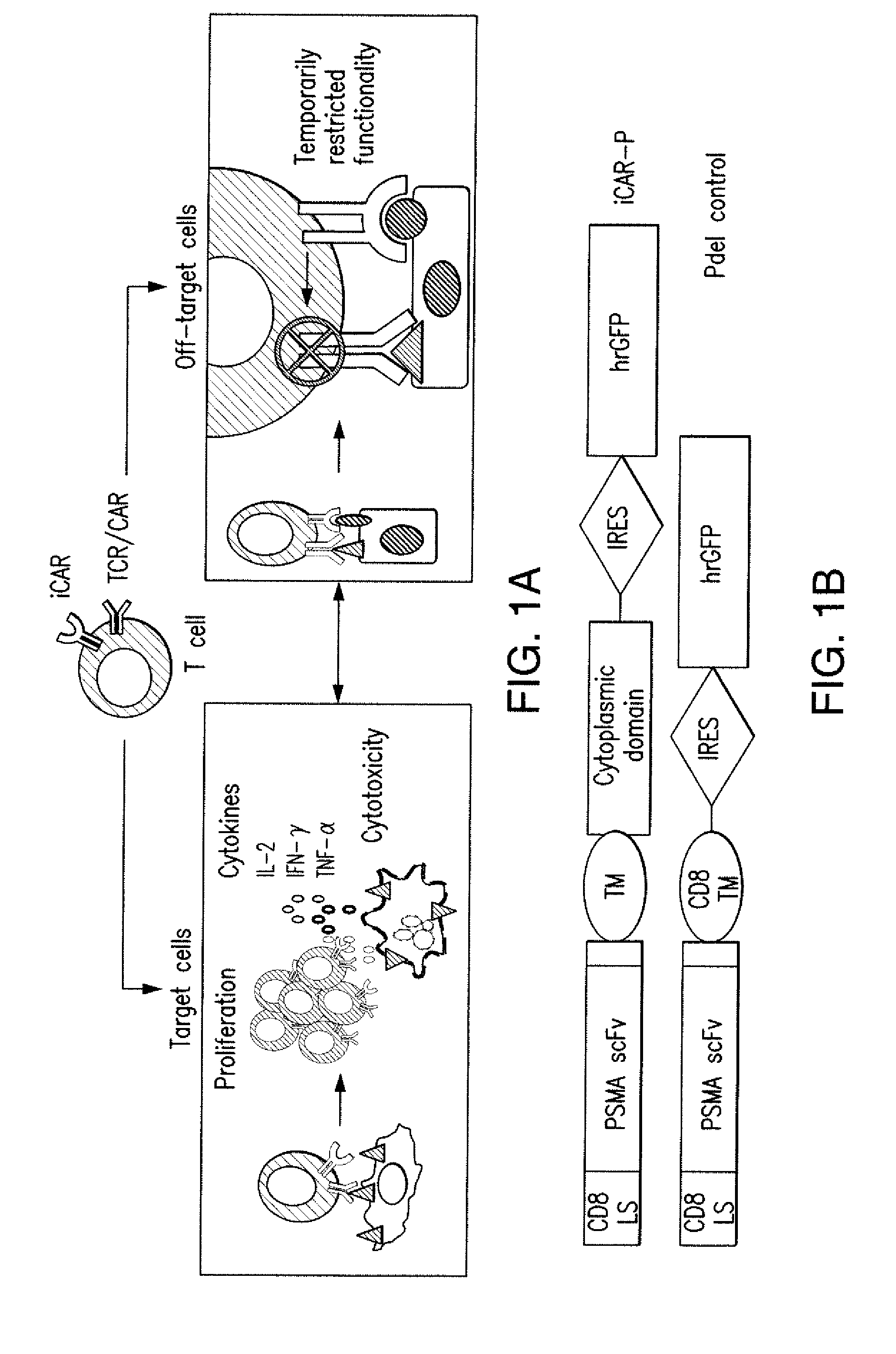
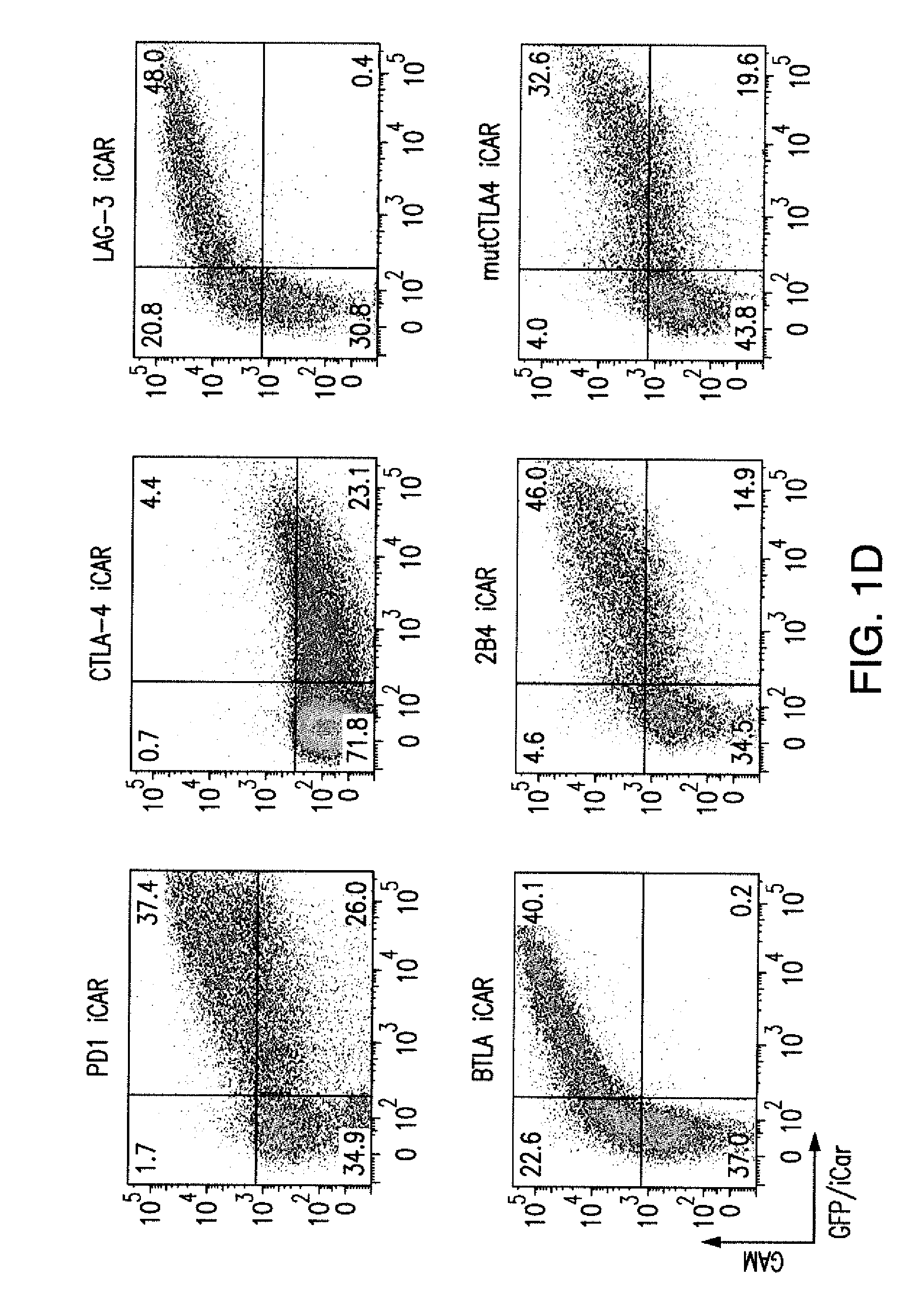
[0084] FIGS. 1A to 1D represent iCAR strategy, design, and expression in primary human T cells. (A) T cells with specificity for both tumor and off-target tissues can be restricted to tumor only by using an antigen-specific iCAR introduced into the T cells to protect the off-target tissue. (B) Schematic diagram of the bicistronic vectors used for iCARs and Pdel. iCAR-P: a spacer, transmembrane, and intracellular tail of each inhibitory receptor were cloned into a previously described retroviral vector having a CD8 leader sequence (LS). IRES, internal ribosomal entry site; hrGFP, humanized Renilla green fluorescent protein reporter. A Pdel control vector was designed with a spacer and CD8 transmembrane (TM) domain, and lacking an intracellular tail. (C) Cell surface expression of the iCARs was assessed by flow cytometry in transduced primary human T cells. Dot plots are representative of eight different donors. GAM, goat anti-mouse immunoglobulin G F(ab')2 antibody that binds to the murine-derived extracellular domain of the CAR. (D depicts flow cytometry analysis of cell surface expression of the iCARs using Goat-Anti-Mouse (GAM) staining in transduced primary human T cells.
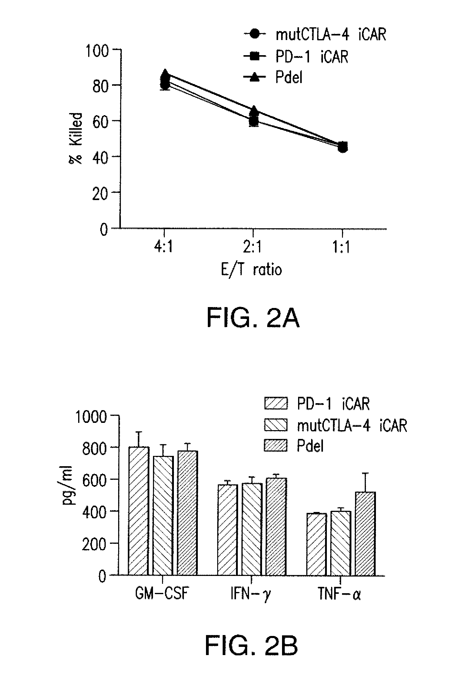
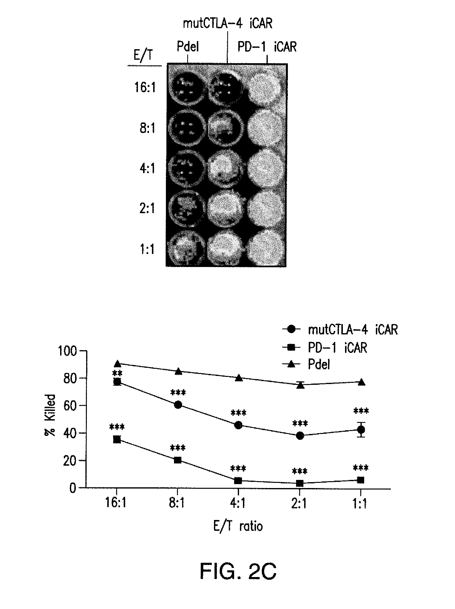
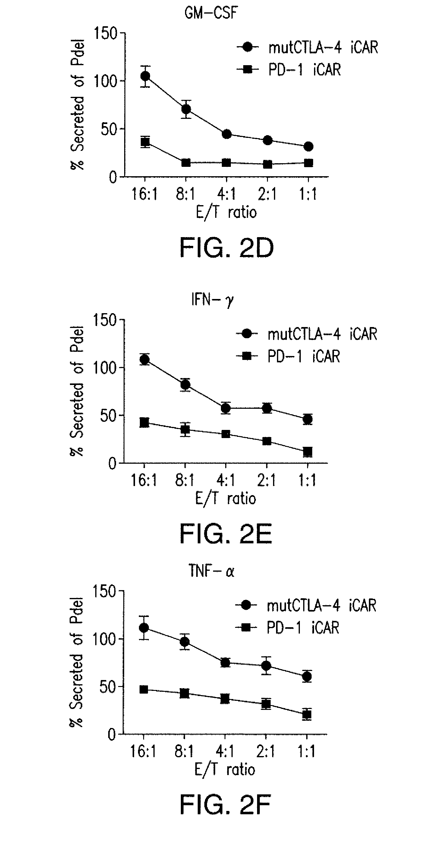
[0085] FIGS. 2A to 2F show that iCARs protected iPS-fib from TCR-mediated allogeneic reactions. Control Pdel- or iCAR-transduced T cells primed with allogeneic moDCs were incubated with iPS-derived fibroblasts (iPS-fib) expressing click beetle luciferase (CBL), isogenic to the moDCs, using a range of E/T ratios. (A) Pdel-, PD-1-, or mutCTLA-4 iCAR-P-transduced T cells reacting against target iPS-fib (n=3 per condition). Killing of the iPS-fib was quantified with the Bright-Glo assay system. (B) Cytokine secretion in cell culture supernatants from (A) at 4:1 E/T ratio was assessed at 18 hours. GM-CSF, granulocyte-macrophage colony-stimulating factor; IFN-.gamma., interferon-g; TNF-.alpha., tumor necrosis factor-a. (C) Pdel- or iCAR-positive T cells were incubated for 24 hours with off-target iPS-fib expressing PSMA (iPS-fib-PSMA), and luciferase signal (left) was quantified (right) (n=3 for each condition). (D to F) Cytokine secretion measured at 24 hours in cell culture supernatants from (C). Error bars represent .+-.SEM. *P<0.01, ***P<0.001 by analysis of variance (ANOVA) comparing iCARs to Pdel and post hoc analysis with multiple t tests corrected with the Holm-Sidak method. Raw data and P values are provided in the FIGS. 19A to 19E.
[0086] FIGS. 3A to 3D show that iCARs functioned in a stoichiometric manner. (A) Pdel- or PD-1 iCAR-P-transduced alloreactive T cells were sorted for high or low expression of each respective receptor, as shown in FIG. 13A, and were seeded on iPS-fib-PSMA-expressing CBL. Killing of iPS-fib-PSMA relative to untreated cells was assessed with the Bright-Glo assay system (n=3 for each condition). (B) Cytokine secretion, measured at 24 hours in the cell culture supernatant from (A) at 4:1 E/T ratio. (C) PD-1 iCAR-P-transduced alloreactive T cells were incubated with iPS-fib-PSMA sorted for high or low levels of PSMA expression as shown in FIG. 13B. Killing of each population relative to untreated cells was quantified with the Bright-Glo assay system (n=3 per condition). D) Cytokines from (C) were assessed at 24 hours. Error bars represent .+-.SEM. ***P<0.001 by Student's t test. Error bars represent .+-.SEM. *P<0.01, ***P<0.001 by ANOVA comparing to high Pdel group and post hoc analysis with multiple t tests corrected with the Holm-Sidak method. Raw data and P values are provided in the FIGS. 20A and 20B.
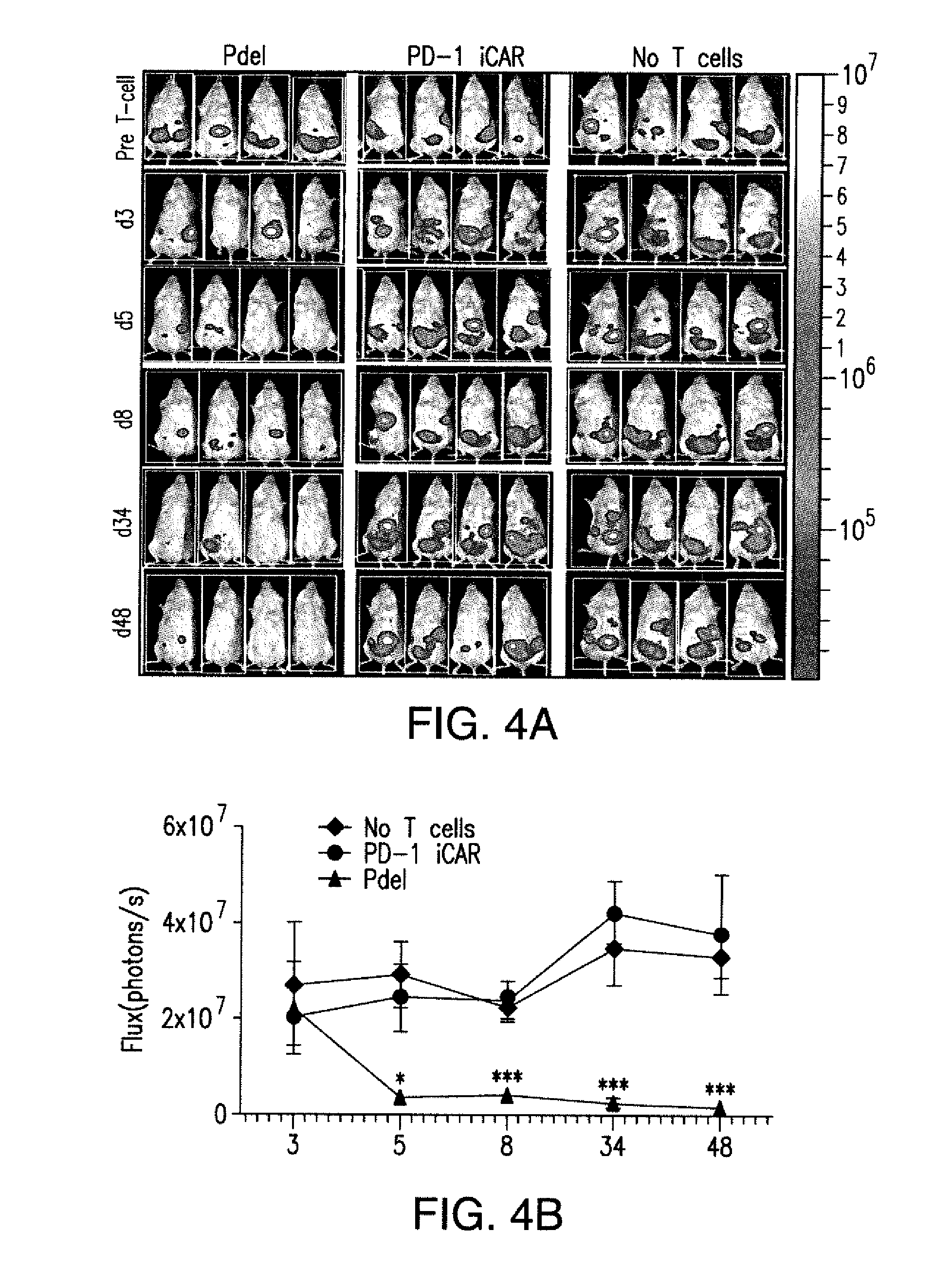
[0087] FIGS. 4A and 4B show that iCARs limited allogeneic responses in vivo. NOD/SCID/.gamma..sub.c.sup.- mice were injected intraperitoneally with 1.times.10.sup.6 iPS-derived fibroblasts expressing CBL/PSMA (iPS-fib-PSMA) and, 7 days later, were treated intraperitoneally with 5.times.10.sup.5 PD-1 iCAR-P- or Pdel-transduced, sorted, alloreactive T cells. Untreated mice (no T cells) were used as control. (A) Survival of iPS-fib-PSMA was assessed by BLI before and at selected time points after T cell infusion. Images of four representative mice from each group are shown. (B) Total body flux (photons per second) for each mouse was quantified and averaged per group (n=5 per group). Error bars represent .+-.SEM. *P<0.05, **P<0.01 by ANOVA comparing to Pdel and post hoc analysis with multiple t tests corrected with the Holm-Sidak method. Raw data and P values are provided in the FIGS. 21A and 21B.
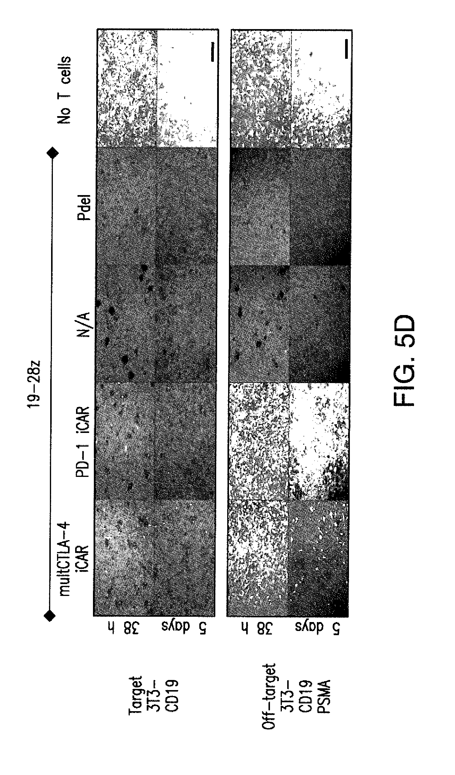
[0088] FIGS. 5A to 5F show that iCARs inhibited human T cell cytokine release, proliferation, and target cell elimination driven by 19-28z CAR. (A) Luminex multiplex cytokine analysis of culture supernatant 24 hours after seeding dual-sorted 19-28z/Pdel- or 19-28z/iCAR-transduced human T cells on 3T3-CD19 (target) or 3T3-CD19-PSMA (off-target) AAPCs. The data are represented as a ratio of off-target/target values and pooled from three independent experiments (n=6 wells per condition). Error bars represent .+-.SEM. **P<0.01, ***P<0.001 by ANOVA comparing iCARs to Pdel and post hoc analysis with multiple t tests corrected with the Holm-Sidak method. (B) Absolute counts of 19-28z/Pdel or 19-28z/iCAR T cells stimulated on days 0 and 7 with off-target AAPCs. No exogenous cytokines were added. Data are representative of six independent experiments. (C) Proliferation of 19-28z/Pdel or 19-28z/iCAR T cells stimulated on days 0 and 7 with offtarget AAPCs relative to proliferation on target AAPCs. No exogenous cytokines were added. Data are representative of six independent experiments. (D) T cells seeded at a 1:1 ratio on target and off-target mCherry.sup.+ AAPCs. Images at 38 hours and 5 days from one of five independent experiments are shown. Scale bars, 0.5 mm. (E and F) Quantification of mCherry signal from (D) against CD19 targets (E) or CD19-PSMA off-target cells (F), as described in Materials and Methods. Error bars represent .+-.SEM. **P<0.01, ***P<0.001 by Student's t test. Raw data and P values are provided in the FIGS. 22A to 22C.
[0089] FIGS. 6A to 6F show that iCARs restricted 19-28z CAR target cell specificity in vivo. (A) BLI depicting the tumor progress of NALM/6 or NALM/6-PSMA in NOD/SCID/.gamma..sub.c.sup.- mice treated with sorted 19-28z/PD-1 iCAR-P T cells. Untreated mice (no T cells) were used as control. (B) Tumor burden for each mouse was quantified, and average total flux per group is shown. (C) Spleen weight of mice from (A) sacrificed at day 21. Each dot represents one recipient mouse. (D) Flow cytometric analysis of the femur bone marrow from (C) for the presence of tumor cells (CD19.sup.+GFP.sup.+) and T cells (CD19-19-28z/GFP.sup.+CD4.sup.+CD8.sup.+). 19-28z expression was assessed by staining for LNGFR receptor whose complementary DNA (cDNA) is linked to 19-28z and is used as a detection marker. (E and F) Absolute numbers of tumor cells (E) and of CD19.sup.-19-28z/GFP.sup.+CD4.sup.+CD8.sup.+ T cells (F) in the spleens from (C) were quantified by flow cytometry with CountBright beads (n=4). Error bars represent .+-.SEM. **P<0.01, ***P<0.001 by Student's t test.
[0090] FIGS. 7A to 7D show that iCAR function was temporary and reversible. (A) 19-28z/Pdel or 19-28z/PD-1 iCAR-P T cells were incubated with target (T) or off-target (O) AAPCs for the first stimulation. After 3 or 7 days, the cells from each group were restimulated with either target [T.fwdarw.T (1) or O.fwdarw.T (2)] or off-target [T.fwdarw.O (3) or O.fwdarw.O (4)] AAPCs in a crisscross manner to analyze the effects of the first stimulation on subsequent T cell function. (B) Killing of target (T) or off-target (O) AAPCs at 24 hours after incubation with each T cell group (second stimulation) was analyzed with the Bright-Glo assay system (n=3 for each condition). (C) Secretion of effector cytokines in the cell culture supernatant from (B) was analyzed 24 hours after the second stimulation, and interferon-g (IFN-g) is shown as a representative result (n=3 for each condition). (D) T cell proliferation at day 7 after the second stimulation (n=3 for each condition). Error bars represent .+-.SEM. Statistical comparison was performed within each condition (that is, T.fwdarw.T Pdel versus PD-1 iCAR-P). ***P<0.001 by Student's t test.
[0091] FIGS. 8A to 8E show that iCAR- and CAR-expressing T cells discerned targets in vitro and vivo. (A) 19-28z/Pdel or 19-28z/PD-1 iCAR-P T cells were incubated with a 1:1 mix of target (CD19.sup.+GFP.sup.+, green) and off-target (CD19.sup.+PSMA.sup.+mCherry.sup.+, red) AAPCs, and time-lapse microscopy was used to visualize real-time killing of each population for 38 hours. Representative images are shown, and full-length movies were made. Scale bars, 0.1 mm. (B) As in (A), 19-28z/Pdel or 19-28z/PD-1 iCAR-P T cells were incubated with a 1:1 mix of target (CD19+) and off-target (CD19+PSMA+) AAPCs. Killing of each AAPC population was assessed in parallel experiments where one of each AAPC type was labeled with CBL (CD19+CBL+/CD19+PSMA+ mix or CD19+/CD19+PSMA+CBL+ mix). Killing was quantified with the Bright-Glo assay system at 38 hours (n=3 for each condition). (C to E) NOD/SCID/.gamma..sub.c.sup.- mice were injected with a 1:1 mixture of NALM/6 and NALM/6-PSMA cells and treated with 19-28z or 19-28z/PD-1 iCAR-P T cells. (C) Upon sacrifice, the presence of the target and off-target NALM/6 cells in the bone marrow was analyzed by flow cytometry. (D) Ratio of target/off-target NALM/6 cells in the bone marrow of sacrificed mice was quantified by flow cytometry. (E) Spleen weight of treated mice was also recorded at sacrifice. Error bars represent .+-.SEM. ***P<0.001 by Student's t test.
[0092] FIGS. 9A-C show that CTLA-4 iCAR cell surface expression was increased after T cell activation. (A) Cell surface and intracellular expression of CTLA4 iCAR-P on transduced primary human T cells (B) Western blot analysis using an antibody specific for the intracellular domain of CTLA4 on untransduced (2) and CTLA4 iCAR-P transduced (1) primary human T cells. Murine EL4 cells (3) served as negative control. (C) 1928z/CTLA4 iCAR T cells were activated using 3T3-19 AAPCs and analyzed for cell surface CTLA4 iCAR expression at 7 hrs and 30 hrs post activation (n=3).
[0093] FIG. 10 shows that iCAR-P bound to PSMA expressing cells. EL4-wt or EL4-PSMA cells, labeled with the lipophilic DiD dye, were incubated with iCAR/GFP expressing T cells in a cellular conjugation assay. Conjugates are detected by flow cytometry as DiD/GFP double positive events.
[0094] FIGS. 11A to 11D represent allogeneic reactivity model using iPS-derived fibroblasts and isogenic moDCs. (A) Induced pluripotent stem (iPS) cells were generated from Donor 1 PBMCs and used to derive fibroblasts. Donor 1 PBMCs were also used to derive moDCs, which were pulsed with fibroblast lysates and could prime an allogeneic reaction from a second donor's PBMCs. (B) Microscopy picture showing the morphology of teratoma-derived iPS-fibroblasts grown in culture. (C) iPS-fib, lacked expression of pluripotency markers, displayed fibroblast morphology, and stained positive for several fibroblast cell surface markers including CD90, PDGFr-b2, and CD10. (RED=isotype control; BLUE and GREEN are two independent isolated lines) (D) iPS-fib basally stained positive for HLA class I, but not class II, and rapidly upregulated expression of both upon recombinant INF gamma treatment.
[0095] FIGS. 12A to 12C represent potent reactivity of iCAR-transduced primary human T cells against allogeneic iPS-derived fibroblasts. (A) iCAR transduced T cells from one donor were primed with moDCs pulsed with iPS-fib lysates of a different donor, and 6 days later stained for the activation markers CD25 and HLA-DR. (B) Twice primed iCAR transduced T cells were incubated for 18 hrs with iPS-fib-luc and killing was quantified using the Bright-Glo assay system (n=3 for each condition). (C) Cytokines were measured at 18 hrs in the cell culture supernatant from (B) at 4:1 E:T ratio. Error bars represent +/- SEM. Statistical comparison was carried out within each condition (ie T.sub.1T). ***p<0.001 by Student's t test.
[0096] FIGS. 13A and 13B represent transduction and sorting strategy of iCAR or 19-28z/iCAR T cells. (A) PD1-iCAR(GFP) or Pdel (GFP) transduced T cells were sorted for transgene expression based on GFP expression level. Each donor represents a separate experiment. Post sort analysis was carried out to confirm purity. (B) 19-28z (LNGFR)/iCAR(GFP) or 19-28z/Pdel/GFP transduced T cells were sorted for transgene expression based on GFP expression and LNGFR level. Each donor represents a separate experiment. Post sort analysis was carried out to confirm purity and iCAR expression.
[0097] FIGS. 14A and 14B represent sorting strategy of low/high iCAR--expressing T cells and PSMA-expressing iPS fibroblasts. (A) PD1-iCAR/GFP or Pdel/GFP transduced T cells were sorted for low or high transgene expression based on GFP expression level. (B) iPS-derived fibroblasts (iPS-fib) were transduced to express PSMA and sorted using an anti-PSMA antibody (iPS-fib-PSMA sort bulk+). These cells were used for experiments in FIGS. 2 and 4. A second separate sort was used to purify low or high surface PSMA expressing iPS-fib using an anti-PSMA antibody and these cells were used in experiments in FIG. 3.
[0098] FIGS. 15A to 15D show that iCARs inhibited 19-28z-driven human T cell cytokine release and proliferation. (A) human T cell proliferation in artificial antigenpresenting cells (AAPCs) expressing CD19 or both CD19 and PSMA. (B) Representative INF.gamma. cytokine analysis of supernatants at 24 h and 48 h post seeding of dual sorted 19-28z/Pdel or iCAR transduced human T cells on CD19 (target) or CD19/PSMA (off-target) positive AAPCs. Data represented as a ratio of offtarget/target values and are pooled from three independent experiments. (n=6 wells per condition). Error bars represent +/- SEM. (C) Absolute counts of double positive 19-28z/Pdel or iCAR T cells stimulated on day 0 and 7 with CD19+ (target) AAPCs. (D) Comparison of therapeutic T cell responses with NALM/6 and NALM/6-PSMA cells in a xenograft NOD/SCID/.gamma..sub.c.sup.- mouse model.
[0099] FIGS. 16A to 161 show that basal expression of iCARs did not affect function of primary human T cells. (A,B) Seven days post transduction with 1928z/iCAR, T cells were activated with CD3/CD28 beads and IL-2/INF.gamma. levels were assessed after 24 hrs (C). At eight days after bead activation, absolute T cell expansion was quantified using CountBright beads (D) and the change in the percent of GFP positive cells in each iCAR group was normalized relative to unstimulated cells. (E) 1928z/iCAR T cells were co-cultured for five days with irradiated EL4-WT or EL4-PSMA cells and immunophenotyped using flow cytometry. (F) depicts cellular conjugation assay of EL4-wt or EL4-PSMA cells, labeled with the lipophilic DiD dye and incubated with iCAR expressing T-cells. (G) represent graphs depicting IL-2/INF.gamma. levels in T-cells 24 hrs after activation with CD3/CD28 beads, seven days post transduction with 1928z/iCAR. (H) is a graph depicting quantification of absolute T-cell expansion using CountBright beads at eight days post bead activation. (I) is a graph depicting the change in the percent of GFP positive cells in each iCAR group was calculated relative to unstimulated cells.
[0100] FIGS. 17A to 17C represent signaling and biochemical pathway characterization of the PD-1 iCAR. 19-28z/PD-1 iCAR cells were exposed to AAPCs expressing no antigen (WT), CD19 (Target), or CD19 and PSMA (Off-Target) at an E:T ratio of 4:1 for 60 min. (A) Human Phospho-Immunoreceptor Array incubated with 100 .mu.g of lysate from 19-28z/PD-1 iCAR T cells and respective AAPCs. All blots were detected using chemiluminescence on the same X-ray film to standardize exposure levels. (B,C) Quantification of arrays in (A) using scanned X-ray film images analyzed using image analysis software. All pixel density is normalized on each array using internal pY controls. (B) SHP1 and SHP2 phosphorylation states on target, off-target, or control AAPCs. (C) Quantification of phosphorylation levels of 59 ITAM/ITIM-associated immunoreceptors.
[0101] FIG. 18 shows that iCAR directly acted on the stimulatory receptor to block its signaling.
[0102] FIGS. 19A to 19E represent taw data and statistical significance testing for FIGS. 2A to 2F. (A) Killing of the iPS-fibroblasts was quantified using the Bright-Glo assay system for Pdel, PD-1, or mutCTLA-4 iCAR-P transduced T cells. (B) Cytokine secretion in cell culture supernatants from (A) at 4:1 E:T ratio was assessed at 18 hrs. (C) Pdel or iCAR positive T cells were incubated for 24 hrs with off-target iPS-fib expressing PSMA (iPS-fib-PSMA) and luciferase signal was quantified. (D) Posthoc analysis for (C) was carried out using multiple t-tests corrected with the Holm-Sidak method. (E) Cytokine secretion measured at 24 hrs in cell culture supernatants from (C). Raw values for GM-CSF are presented. E:T ratio, effector:target ratio.
[0103] FIGS. 20A and 20B represent raw data and statistical significance testing for FIGS. 3A to 3D. (A) Killing of iPSfib-PSMA relative to untreated cells was assessed by using the Bright-Glo assay system for sorted high and low Pdel or PD1 iCAR-P transduced alloreactive T cells. Posthoc analysis was carried out using multiple t-tests corrected with the Holm-Sidak method. (B) PD1 iCAR-P transduced alloreactive T-cells killing of iPS-fib-PSMA sorted for high or low levels of PSMA expression was quantified using the Bright-Glo assay system. Posthoc analysis was carried out using multiple t-tests corrected with the Holm-Sidak method.
[0104] FIGS. 21A and 21B represent raw data and statistical significance testing for FIGS. 4A and 4B. (A) Bioluminescent imaging (BLI) of iPS-fib-PSMA before and at selected time points after T cell infusion. (B) Posthoc analysis was carried out using multiple t-tests corrected with the Holm-Sidak method.
[0105] FIGS. 22A to 22C represent raw data and statistical significance testing for FIGS. 5A to 5F. (A) Luminex multiplex cytokine analysis of culture supernatant at 24 h, data are represented as a ratio of off-target/target values and pooled from three independent experiments (n=6 wells per condition). Posthoc analysis was carried out using multiple t-tests corrected with the Holm-Sidak method. (B) Posthoc analysis was carried out using multiple t-tests corrected with the Holm-Sidak method for FIG. 5B comparing the proliferation of 19-28z/Pdel and 19-28z/PD1 iCAR. (C) Quantification of mCherry signal against CD19 targets or CD19-PSMA off-target cells, as described in Methods. Posthoc analysis was carried out using multiple t-tests corrected with the Holm-Sidak method.
[0106] FIG. 23 is a schematic representation of a selection of target antigen for iCAR and CAR.
DETAILED DESCRIPTION OF THE INVENTION
[0107] The present invention generally provides cells, including genetically modified immunoresponsive cells (e.g., T cells (including all subsets such as CD4, CD8, memory, naive, effector, T-reg etc.), cells of the innate immune system, Natural Killer (NK) cells, cytotoxic T lymphocytes (CTL) cells) expressing at least a combination of an antigen-recognizing receptor (e.g., TCR or CAR) and an inhibitory chimeric antigen receptor (iCAR) that selectively reduces or eliminates the immune activity of the immunoresponsive cell, and methods of using such cells for the treatment of neoplasia and other pathologies where reducing "off-target" immune responses is desired. The invention is based, at least in part, on the discovery that inhibitory chimeric antigen receptors (iCARs) that bind a target antigen (e.g., PSMA as shown herein) are useful for selectively inhibiting and suppressing an immunoreactive cell. In particular, the iCARs of the invention decrease or prevent activation of the immune response of an immunoreactive cell. The present approach provides selective immunogenicity for tumor eradication, while sparing non-tumor tissues from the immune response. Accordingly, T cells expressing an antigen recognizing receptor and an iCAR represents a significant advance over conventional adoptive T cell therapy.
[0108] The broad use of donor leukocyte infusion to treat cancer is hampered by the inability to separate the therapeutic efficacy of Graft-versus-tumor effect (GVT) from the potentially lethal effects of Graft-versus-host disease (GVHD). Two general approaches have been used to control T-cell therapy side effects. The first is the use of immunosuppressive drugs, which work non-specifically by blocking cell division (cytostatics) and broadly limit immune responses (glucocorticoids, immunophilins, etc), or targeting T-cells for clearance/death (antibodies). Although powerful, these approaches are nonspecific in terms of separating therapeutically functioning T-cells and ones causing deleterious side effects. Additionally, all these drugs cause significant long-term secondary side effects (susceptibility to infections, cardiac, kidney, and neurological damage).
[0109] The second approach engineers T-cells with suicide genes/kill switches. These are genetic approaches that cause the engineered cell to die once a proper cue is provided. Several of them are based on introducing selective enzymatic metabolizers of toxic agents, such as herpes simplex virus thymidine kinase (HSV-tk). Additionally, the use of on an inducible caspase-9 protein that is activated using a specific chemical inducer of dimerization has been a promising approach to actively induced broad cell death. The major limitations with these approaches arc they induce cell death in all target cells (thereby eliminating beneficial cells). Like conventional immunosuppression, they usually require the appearance of symptoms before implementation (and therefore possible permanent damages to the patient).
[0110] In contrast, the iCAR strategy describe herein selectively filters T-cell actions, restricting activity at off-target sites, while sparing therapeutic functionality against the intended target. As shown herein, iCARs were capable of inhibiting human alloreactive T-cells from attacking a "host" tissue in a novel in vitro and in vivo model using iPS-derived human fibroblasts. Unique surface anti gens present in GVHD target tissues (e.g., CD33 for the myeloid lineage or organ-specific antigens) but absent from the targeted malignancy, are candidates for iCAR targets to differentiate GVHD from GVT. Similarly, the results described herein show that iCAR-mediated inhibition successfully restricts the "on-target but off-tissue" effects of CAR engineered T-cells, examples of which include B-cell aplasia in leukemia patients treated with T-cells expressing a CD19-specific CAR (Kalos et al., Science translational medicine, 2011. 3(95): 95ra73), fatal acute respiratory distress syndrome (ARDS) thought to result from anti-ERBB2 CART-cell cross reactivity on lung epithelium (Morgan et al., Mol. Ther., 2010. 18(4): 843-51), and fatalities from cardiac myonecrosis in melanoma and myeloma patients treated with Mage-A3 TCRs (June, Update on Immunotherapy Trials for HIV and Cancer, in Recombinant DNA Advisory Committee, 2012). Recognizing a surface antigen that is expressed on cardiac cells or lung epithelium but absent from tumor cells, iCARs could potentially be used to protect from Mage-A3 TCR or anti-ERBB2 CAR cross reactivity, thus resurrecting otherwise promising therapeutics. Alternatively, as many tumors actively down-regulate HLA molecules to escape immune recognition, HLA-targeted iCARs have the potential to provide concurrent protection to several tissues.
[0111] An important requirement for the clinical applicability of iCARs is the maintenance of T-cell functionality despite previous signaling of the iCAR. Interestingly, iCAR-transduced T-cells were found to still mount a response against a target antigen after exposure to an inhibitory antigen. This reversibility mimics natural killer cell behavior, in which the phosphorylation state of signaling molecules rather than transcriptional states control rapid functional responses such as cytotoxicity. Anti-PD-1 and anti-CTLA4 antibodies are able to reverse the impaired function of anergized or exhausted T-cells, again arguing for the ability to temporally regulate T-cell responses. Additionally, biochemical analyses of PD-1 and CTLA-4 effects on the TCR complex have been shown to depend on phosphorylation states, downstream kinases, and motility rather than apoptosis. Both the in vitro and in vivo results demonstrated inhibition in response to off-target cells with sustained therapeutic functionality, although a possibility exists that some of the cells may be anergized. In addition to functioning in T-cells, CTLA4 and PD-1 also operate in B cells, macrophages, and dendritic cells. Thus, iCARs have the potential to manipulate other immune phenomena as well.
[0112] The iCARs of the present invention can be used as a dampening tool to limit systemic cytokine storms or immune cell responses, e.g., by introducing the antigen to the iCAR such as a recombinant PSMA-Ig infused into a patient. The PSMA-Ig can bind and activate the iCAR, and can thus temporarily inhibit the T cell activation. This can temporarily break the cyclic spiral that causes cytokines storms and allow the T cells to activate with limited systemic side effects. The iCARs have this functionality, as shown by crosslinking the iCAR.
[0113] The present invention utilizes physiological mechanism to restrict T-cell side effects. This approach mimics the restriction of T-cell reactivity that occurs naturally, and thus does not require elimination of precious therapeutically viable cells. The present approach that takes advantage of the multi-faceted functionality of cells as drugs, by using synthetic receptors that guide and direct T-cells to perform only desired functions. In conclusion, antigen-specific inhibitory receptors successfully limited T-cell proliferation, cytokine secretion, and cytotoxicity upon engagement of specific cell-surface antigens, thus conferring protection to a normal tissue while retaining critical TCR or CAR mediated therapeutic functions. Thus, iCARs provide a novel strategy to establish safer and more efficacious T-cell therapies in both autologous and allogeneic settings.
Graft-Versus-Leukemia Effect (GVL) and Graft-Versus-Host-Disease (GVHD)
[0114] Since the first use of allogeneic bone marrow transplants, it has been appreciated that the eradication of leukemia was dependent on the donor-derived immune response. The Graft versus leukemia effect (GVL) was further elucidated and appreciated following the success of donor lymphocyte infusion following allogeneic bone marrow transplantation (BMT). Donor lymphocyte infusion (DLI) is the most established and widespread use of adoptive immunotherapy for malignancy. Unfortunately, the main source of morbidity and mortality following DLI is the occurrence of Graft-versus-host-disease (GVHD). Reducing its incidence and severity is an important limitation to wider and potentially curative use of DLI in solid and hematological tumors. The iCAR strategy aims to solve this problem by separating the beneficial effects of GVL from the hazardous consequences of GVHD. GVHD primarily affects the skin, liver, and intestinal tract, sites that possess unique antigens such as minor alia-antigens that can be absent from the target malignancy. Such occurrences support a possible role for iCARs to separate the effects of GVL and GVHD.
[0115] In adoptive therapy, retargeted T-cells have been shown to play a potential curative role in several malignancies. Still, this transformative approach is limited because of the cross reactivity and subsequent toxicity against critical normal tissues (heart, lung). Thus, a significant need exists for developing ways to control T-cell reactivity without dampening their therapeutic function, which the iCAR strategy sets out to do. Recognizing a surface antigen that is expressed on cardiac cells or lung epithelium but absent from tumor cells, iCARs could potentially be used to protect from Mage-A3 TCR or anti-ERBB2 CAR cross reactivity, thus reviving otherwise promising therapeutics. Alternatively, as many tumors actively down-regulate HLA molecules to escape immune recognition, HLA-targeted iCARs could potentially provide concurrent protection to several tissues.
[0116] In one embodiment, allogeneic lymphocytes (with a degree of immunological mismatch) are engineered to express an iCAR targeting HLA-I, an antigen universally expressed in different tissues for treatment of metastatic breast cancer (a type of cancer with extremely active HLA-I down regulation). The patient is infused with the cells. The iCAR protects all normal or non-neoplastic tissues that express HLA-I, while the tumor is eliminated due negative or extremely low HLA-I expression.
[0117] In another embodiment, a patient undergoes HSCT for the treatment of a hematological malignancy or as adjuvant treatment for a solid tumor. The patient relapses or has residual disease, which is analyzed to be HLA-I negative or down regulated. Donor lymphocytes are engineered with an iCAR targeting HLA-I. The patient is infused with the cells. The iCAR protects all normal or non-neoplastic tissues that express HLA-I, while the tumor is eliminated due to negative or extremely low HLA-I expression.
[0118] In yet another embodiment, a patient has a tumor originating at a site that is not related to skin, liver, or gut cells (the major sites of GVHD related mortality). Donor lymphocyte cells are engineered with iCARs that target antigens expressed on skin, liver, or gut or all three. For example, if the tumor has undergone epithelium to mesenchymal transition (EMT), as is found with tumor progression and metastatic tumors, E-cadherin and cytokeratin have shown to be down regulated as part of this process. E-cadherin is highly expressed in normal skin, liver, and gut (Tsuchiya et al., Arch, Histol. Cytol., 69(2): 135-145(2006)). Therefore donor lymphocytes expressing an iCAR against E-cadherin, react in a GVL manner against the tumor, but are restricted in their ability to attack skin, liver, or gut.
Selection of Target Antigen for iCAR and CAR
[0119] The invention provides a method and a set of novel reagents to control responses of T-cells or other immune modulatory cells by using synthetic receptors that utilize signaling domains of immune inhibitory receptors (the iCAR). An appropriate antigen for the iCARs will at times utilize a personalized medicine approach due to natural variation in tumors. At the same time, depending on the use and type of iCAR, several potential "classes" of antigens have the potential to provide protection for several tissues at the same time. These include: (1) Universally expressed immunogenic antigens that are down regulated by tumors but not normal tissues, such as human leukocyte antigens (HLAs). (2) Antigens down regulated in tumor progression especially the attainment of a metastatic phenotype, but maintained in certain normal tissues. Such antigens include: (3) cell surface EMT antigens (such as E-cadherin and cytokeratins); (4) cell surface tumor suppressor antigens, such as OPCML (Cui et al., PLoS ONE. 2008; 3(8): e2990); and (6) other similar antigens such as HYAL2, DCC, SMAR1, and the like. OPCML-v1 is widely expressed at varying levels in all normal adult and fetal tissues (except for placenta and peripheral blood mononuclear cells). (7) Cell surface carbohydrates, lipids, and posttranslational modifications, such as mucin-type O-glycans (core 3 O-glycans) (Lee and Fukuda, Methods Enzymol. 2010; 479:143-54; Suzuki-Anekoji et al., J Bioi Chem. 2011 Sep. 16; 286(37):32824-33; Bao and Fukuda, Methods Enzymol. 20 1 0; 479:387-96). (8) Additionally, there many other processes disrupted in tumors (metabolism, apoptosis, trafficking, differentiation, and the like) that each lead to down regulation of surface antigens, any of these could be used as potential iCAR antigen targets.
[0120] In general, the invention provides a personal approach that can be taken to each patient. As described herein, the iCAR antigen can be selected through an algorithmic process, after which the physician can order a specific receptor suited for the patient's tumor. This receptor is then introduced alongside a stimulating receptor (ie TCR or CAR or GVL signals) to provide protection against a select tissue.
[0121] It is important to note that the iCAR may bind the same antigen as the activating CAR. For example, there could be a situation in which no antigens are found that are binary in their expression between the tumor and a normal tissue (i.e., totally absent on one and present on the other). Still, if an antigen on the tumor is expressed at a much higher level than the normal tissue, both the stimulating CAR and the iCAR receptor could then target that same antigen. In the case of the tumor, since there is a great deal of antigen, the stimulatory CAR would dominate and cause the tumor to be eliminated. In the case of the normal tissue, since the level of antigen expression is low, the iCAR could provide adequate inhibition. This is true because levels of both/or a single the target antigen and the levels of the stimulating and the iCAR can affect the outcome response of the immunoresponsive cell (FIG. 23). Additionally, changing the affinity of each receptor can be used to modulate and fine-tune the response.
[0122] Antigen disparity between a target and off target tissue can be limited with differences primarily in the level of expression rather than the absolute absence of expression. Additionally, variable expression levels can be expected if several tissues are represented with one common antigen for protection using an iCAR. One such example is the expression of HLA molecules, which are broadly found on the majority of cell types, but often with different levels of expression. Interestingly, down regulation of HLA molecules represents a major mechanism of tumor escape from T-cell immune responses. In such a scenario, DLI T-cells engineered with an iCAR against an HLA molecule that is down regulated on the malignancy could provide broad protection for a variety of tissue types from GVHD while maintaining a GVL effect.
[0123] The combinatorial possibilities of such antigen selection are only limited by the available antibodies, tumor surface antigen profiling, and known tissue specific antigens.
Cytotoxic T-Lymphocyte Antigen 4 (CTLA-4)
[0124] CTLA-4 is an inhibitory receptor expressed by activated T cells, which when engaged by its corresponding ligands (CD80 and CD86; B7-1 and B7-2, respectively), mediates activated T cell inhibition or anergy. In both preclinical and clinical studies, CTLA-4 blockade by systemic antibody infusion, enhanced the endogenous anti-tumor response albeit, in the clinical setting, with significant unforeseen toxicities.
[0125] CTLA-4 contains an extracellular V domain, a transmembrane domain, and a cytoplasmic tail. Alternate splice variants, encoding different isoforms, have been characterized. The membrane-bound isoform functions as a homodimer interconnected by a disulfide bond, while the soluble isoform functions as a monomer. The intracellular domain is similar to that of CD28, in that it has no intrinsic catalytic activity and contains one YVKM motif able to bind PI3K, PP2A and SHP-2 and one proline-rich motif able to bind SH3 containing proteins. One role of CTLA-4 in inhibiting T cell responses seem to be directly via SHP-2 and PP2A dephosphorylation of TCR-proximal signaling proteins such as CD3 and LAT. CTLA-4 can also affect signaling indirectly via competing with CD28 for CD80/86 binding. CTLA-4 has also been shown to bind and/or interact with PI3K, CD80, AP2M1, and PPP2R5A.
[0126] CTLA-4 can have an amino acid sequence as set forth in SEQ ID NO:5.
TABLE-US-00005 1 MACLGFQRHK AQLNLATRTW PCTLLFFLLF IPVFCKAMHV AQPAVVLASS RGIASFVCEY 61 ASPGKATEVRVTVLRQADSQ VTEVCAATYM MGNELTFLDD SICTGTSSGN QVNLTIQGLR 121 AMDTGLYICK VELMYPPPYY LGIGNGTQIY VIDPEPCPDS DFLLWILAAV SSGLFFYSFL 181 LTAVSLSKML KKRSPLTTGV YVKMPPTEPE CEKQFQPYFI PIN
[0127] In accordance with the present invention, a CTLA-4 polypeptide can have an amino acid sequence that is at least 85%, 90%, 95%, 96%, 97%, 98%, 99% or 100% homologous to SEQ ID NO:5 (homology herein may be determined using standard software such as BLAST or FASTA). In non-limiting embodiments, a CTLA-4 polypeptide can have an amino acid sequence that is a consecutive portion of SEQ ID NO:5 which is at least 20, or at least 30, or at least 40, or at least 50, and up to 222 amino acids in length. Alternatively or additionally, in non-limiting various embodiments, the CTLA-4 polypeptide has an amino acid sequence of amino acids 1 to 223, 1 to 50, 50 to 100, 100 to 140, 141 to 161, 162 to 182, 183 to 223, 141 to 223, 162 to 223, or 183 to 223 of SEQ ID NO:5. In one embodiment, the CTLA-4 polypeptide has an amino acid sequence of amino acids 183 to 223 of SEQ ID NO:5. In certain embodiments, the intracellular signaling domain of the iCAR includes a CTLA-4 polypeptide having an amino acid sequence of amino acids 183 to 223 of SEQ ID NO:5. In certain embodiments, the transmembrane domain of the iCAR includes a CTLA-4 polypeptide having an amino acid sequence of amino acids 162 to 182 of SEQ ID NO:5.
[0128] In accordance with the present invention, a "CTLA-4 nucleic acid molecule" refers to a polynucleotide encoding a CTLA-4 polypeptide.
Programmed Cell Death Protein 1 (PD-1)
[0129] PD-1 is a negative immune regulator of activated T cells upon engagement with its corresponding ligands PD-L1 and PD-L2 expressed on endogenous macrophages and dendritic cells. PD-1 is a type I membrane protein of 268 amino acids. PD-1 has two ligands, PD-L1 and PD-L2, which are members of the B7 family. The protein's structure includes an extracellular IgV domain followed by a transmembrane region and an intracellular tail. The intracellular tail contains two phosphorylation sites located in an immunoreceptor tyrosine-based inhibitory motif and an immunoreceptor tyrosine-based switch motif, that PD-1 negatively regulates TCR signals. SHP-I and SHP-2 phosphatases bind to the cytoplasmic tail of PD-1 upon ligand binding. Upregulation of PD-L1 is one mechanism tumor cells may evade the host immune system. In pre-clinical and clinical trials, PD-1 blockade by antagonistic antibodies induced anti-tumor responses mediated through the host endogenous immune system.
[0130] PD-1 can have an amino acid sequence as set forth in SEQ ID NO:6.
TABLE-US-00006 1 mqipqapwpv vwavlqlgwr pgwfldspdr pwnpptfspa llvvtegdna tftcsfsnts 61 esfvlnwyrm spsngtdkla afpedrsqpg qdcrfrvtgl pngrdfhmsv vrarrndsgt 121 ylcgaislap kagikeslra elrvterrae vptahpspsp rpagqfgtlv vgvvggllgs 181 lvllvwvlav icsraargti garrtgqplk edpsavpvfs vdygeldfqw rektpeppvp 241 cvpegteyat ivfpsgmgts sparrgsadg prsaqplrpe dghcswpl
[0131] In accordance with the present invention, a PD-1 polypeptide can have an amino acid sequence that is at least 85%, 90%, 95%, 96%, 97%, 98%, 99% or 100% homologous to SEQ ID NO:6. In non-limiting embodiments, a PD-1 polypeptide can have an amino acid sequence that is a consecutive portion of SEQ ID NO:6 which is at least 20, or at least 30, or at least 40, or at least 50, and up to 287 amino acids in length. Alternatively or additionally, in non-limiting various embodiments, a PD-1 polypeptide has an amino acid sequence of amino acids 1 to 288, 1 to 50, 50 to 100, 100 to 144, 145 to 170, 171 to 191, 192 to 288, 145 to 288, 171 to 288, or 192 to 288 of SEQ ID NO:6. In one embodiment, the PD-1 polypeptide has an amino acid sequence of amino acids 192 to 288 of SEQ ID NO:6. In certain embodiments, the intracellular signaling domain of the iCAR includes a PD-1 polypeptide having an amino acid sequence of amino acids 192 to 288 of SEQ ID NO:6. In certain embodiments, the transmembrane domain of the iCAR includes a PD-1 polypeptide having an amino acid sequence of amino acids 171 to 191 of SEQ ID NO:6.
[0132] In accordance with the present invention, a "PD-1 nucleic acid molecule" refers to a polynucleotide encoding a PD-1 polypeptide.
Lymphocyte-Activation Gene 3 (LAG-3)
[0133] Lymphocyte-activation protein 3 (LAG-3) is a negative immune regulator of immune cells. LAG-3 belongs to the immunoglobulin (Ig) superfamily and contains 4 extracellular Ig-like domains. The LAG3 gene contains 8 exons. The sequence data, exon/intron organization, and chromosomal localization all indicate a close relationship of LAG3 to CD4. LAG3 has also been designated CD223 (cluster of differentiation 223).
[0134] LAG-3 can have an amino acid sequence as set forth in SEQ ID NO:7.
TABLE-US-00007 1 mweagflgll flqplwvapv kplqpgaevp vvwaqegapa qlpcsptipl qdlsllrrag 61 vtwqhqpdsg ppaaapghpl apgphpaaps swgprprryt vlsvgpgglr sgrlplqprv 121 qldergrqrg dfslwlrpar radageyraa vhlrdralsc rlrlrlgqas mtasppgslr 181 asdwvilncs fsrpdrpasv hwfrnrgqgr vpvresphhh laesflflpq vspmdsgpwg 241 ciltyrdgfn vsimynltvl glepptpltv yagagsrvgl pcrlpagvgt rsfltakwtp 301 pgggpdllvt gdngdftlrl edvsqaqagt ytchihlqeq qlnatvtlai itvtpksfgs 361 pgslgkllce vtpvsgqerf vwssldtpsq rsfsgpwlea qeaqllsqpw qcqlyggerl 421 lgaavyftel sspgaqrsgr apgalpaghl llflilgvls llllvtgafg fhlwrrqwrp 481 rrfsaleqgi hppqaqskie eleqepepep epepepepep epeql
[0135] In accordance with the present invention, a LAG-3 polypeptide can have an amino acid sequence that is at least 85%, 90%, 95%, 96%, 97%, 98%, 99% or 100% homologous to SEQ ID NO:7. In non-limiting embodiments, a LAG-3 polypeptide can have an amino acid sequence that is a consecutive portion of SEQ ID NO:7 which is at least 20, or at least 30, or at least 40, or at least 50, and up to 524 amino acids in length. Alternatively or additionally, in non-limiting various embodiments, a LAG-3 polypeptide has an amino acid sequence of amino acids 1 to 525, 1 to 50, 50 to 100, 100 to 150, 150 to 200, 200 to 250, 250 to 300, 300 to 350, 350 to 400, 400 to 420, 421 to 450, 451 to 471, 472 to 525, 421 to 525, 451 to 525, or 472 to 525 of SEQ ID NO:7. In one embodiment, the LAG-3 polypeptide has an amino acid sequence of amino acids 472 to 525 of SEQ ID NO:7. In certain embodiments, the intracellular signaling domain of the iCAR includes a LAG-3 polypeptide having an amino acid sequence of amino acids 472 to 525 of SEQ ID NO:7. In certain embodiments, the transmembrane domain of the iCAR includes a LAG-3 polypeptide having an amino acid sequence of amino acids 451 to 471 of SEQ ID NO:7.
[0136] In accordance with the present invention, a "LAG-3 nucleic acid molecule" refers to a polynucleotide encoding a LAG-3 polypeptide.
Natural Killer Cell Receptor 2B4 (2B4)
[0137] Natural Killer Cell Receptor 2B4 (2B4) mediates non-MHC restricted cell killing on NK cells and subsets of T cells. To date, the function of 2B4 is still under investigation, with the 2B4-S isoform believed to be an activating receptor, and the 2B4-L isoform believed to be a negative immune regulator of immune cells. 2B4 becomes engaged upon binding its high-affinity ligand, CD48. 2B4 contains a tyrosine-based switch motif, a molecular switch that allows the protein to associate with various phosphatases. 2B4 has also been designated CD244 (cluster of differentiation 244).
[0138] 2B4 can have an amino acid sequence as set forth in SEQ ID NO:8.
TABLE-US-00008 1 mlgqvvtlil llllkvyqgk gcqgsadhvv sisgvplqlq pnsiqtkvds iawkkllpsq 61 ngfhhilkwe ngslpsntsn drfsfivknl sllikaaqqq dsglyclevt sisgkvqtat 121 fqvfvfesll pdkvekprlq gqgkildrgr cqvalsclvs rdgnvsyawy rgskliqtag 181 nltyldeevd ingthtytcn vsnpvswesh tlnltqdcqn ahqefrfwpf lviivilsal 241 flgtlacfcv wrrkrkekqs etspkeflti yedvkdlktr rnheqeqtfp gggstiysmi 301 qsqssaptsq epaytlysli qpsrksgsrk rnhspsfnst iyevigksqp kaqnparlsr 361 kelenfdvys
[0139] In accordance with the present invention, a 2B4 polypeptide can have an amino acid sequence that is at least 85%, 90%, 95%, 96%, 97%, 98%, 99% or 100% homologous to SEQ ID NO:8. In non-limiting embodiments, a 2B4 polypeptide can have an amino acid sequence that is a consecutive portion of SEQ ID NO:8 which is at least 20, or at least 30, or at least 40, or at least 50, and up to 369 amino acids in length. Alternatively or additionally, in non-limiting various embodiments, a 2B4 polypeptide has an amino acid sequence of amino acids 1 to 370, 1 to 50, 50 to 100, 100 to 150, 150 to 215, 216 to 229, 230 to 250, 251 to 370, 216 to 370, 230 to 370, or 251 to 370 of SEQ ID NO:8. In one embodiment, the 2B4 polypeptide has an amino acid sequence of amino acids 251 to 370 of SEQ ID NO:8. In certain embodiments, the intracellular signaling domain of the iCAR includes a 2B4 polypeptide having an amino acid sequence of amino acids 251 to 370 of SEQ ID NO:8. In certain embodiments, the transmembrane domain of the iCAR includes a 2B4 polypeptide having an amino acid sequence of amino acids 230 to 250 of SEQ ID NO:8.
[0140] In accordance with the present invention, a "2B4 nucleic acid molecule" refers to a polynucleotide encoding a 2B4 polypeptide.
B- and T-Lymphocyte Aftermath (BTLA)
[0141] B- and T-lymphocyte attenuator (BTLA) expression is induced during activation of T cells, and BTLA remains expressed on Th1 cells but not Th2 cells. Like PD1 and CTLA4, BTLA interacts with a B7 homolog, B7H4. However, unlike PD-1 and CTLA-4, BTLA displays T-Cell inhibition via interaction with tumor necrosis family receptors (TNF-R), not just the B7 family of cell surface receptors. BTLA is a ligand for tumour necrosis factor (receptor) superfamily, member 14 (TNFRSF14), also known as herpes virus entry mediator (HVEM). BTLA-HVEM complexes negatively regulate T-cell immune responses. BTLA activation has been shown to inhibit the function of human CD8.sup.+ cancer-specific T cells. BTLA has also been designated as CD272 (cluster of differentiation 272).
[0142] BTLA can have an amino acid sequence as set forth in SEQ ID NO:9.
TABLE-US-00009 1 MKTLPAMLGT GKLFWVFFLI PYLDIWNIHG KESCDVQLYI KRQSEHSILA GDPFELECPV 61 KYCANRPHVT WCKLNGTTCV KLEDRQTSWK EEKNISFFIL HFEPVLPNDN GSYRCSANFQ 121 SNLIESHSTT LYVTDVKSAS ERPSKDEMAS RPWLLYRLLP LGGLPLLITT CFCLFCCLRR 181 HQGKQNELSD TAGREINLVD AHLKSEQTEA STRQNSQVLL SETGIYDNDP DLCFRMQEGS 241 EVYSNPCLEE NKPGIVYASL NHSVIGPNSR LARNVKEAPT EYASICVRS
[0143] In accordance with the present invention, a BTLA polypeptide can have an amino acid sequence that is at least 85%, 90%, 95%, 96%, 97%, 98%, 99% or 100% homologous to SEQ ID NO:9. In non-limiting embodiments, a BTLA polypeptide can have an amino acid sequence that is a consecutive portion of SEQ ID NO:9 which is at least 20, or at least 30, or at least 40, or at least 50, and up to 288 amino acids in length. Alternatively or additionally, in non-limiting various embodiments, a BTLA polypeptide has an amino acid sequence of amino acids 1 to 289, 1 to 50, 50 to 100, 100 to 134, 135 to 157, 158 to 178, 179 to 289, 135 to 289, 158 to 289, or 179 to 289 of SEQ ID NO:9. In one embodiment, the BTLA polypeptide has an amino acid sequence of amino acids 179 to 289 of SEQ ID NO:9. In certain embodiments, the intracellular signaling domain of the iCAR includes a BTLA polypeptide having an amino acid sequence of amino acids 179 to 289 of SEQ ID NO:9. In certain embodiments, the transmembrane domain of the iCAR includes a BTLA polypeptide having an amino acid sequence of amino acids 158 to 178 of SEQ ID NO:9.
[0144] In accordance with the present invention, a "BTLA nucleic acid molecule" refers to a polynucleotide encoding a BTLA polypeptide.
Hematopoietic Cell Lineages
[0145] Mammalian hematopoietic (blood) cells provide a diverse range of physiologic activities. Hematopoietic cells are divided into lymphoid, myeloid and erythroid lineages. The lymphoid lineage, comprising B, T and natural killer (NK) cells, provides for the production of antibodies, regulation of the cellular immune system, detection of foreign agents in the blood, detection of cells foreign to the host, and the like. The term "T cells" as used herein refers to lymphocytes that mature in the thymus and are chiefly responsible for cell-mediated immunity. T cells are involved in the adaptive immune system. The term "natural killer (NK) cells" as used herein refers to lymphocytes that are part of cell-mediated immunity and act during the innate immune response. They do not require prior activation in order to perform their cytotoxic effect on target cells. Cytotoxic T cells (CTL or killer T cells) arc a subset of T lymphocytes capable of inducing the death of infected somatic or tumor cells.
Cells for Use in the Methods of the Invention
[0146] The present invention provides cells expressing a combination of an antigen-recognizing receptor that activates an immunoresponsive cell (e.g., TCR, CAR) and an inhibitory chimeric antigen receptor (iCAR), and methods of using such cells for the treatment of a disease that requires an enhanced immune response. In one approach, tumor antigen-specific T cells (all subsets including CD4, CD8, memory, naive, effector, T-reg etc), cells of the innate immune system, NK cells, CTL cells or other immunoresponsive cells are used to express an iCAR that binds an antigen on non-tumor tissue, for the treatment or prevention of neoplasia. For example, a T cell expressing a chimeric antigen receptor 19-28z that recognizes CD19 is co-expressed in a T cell that expresses an iCAR that binds CD33. Such cells are administered to a human subject in need thereof for the treatment or prevention of blood cancers (e.g. leukemias, lymphomas, and myelomas). In another approach, viral antigen-specific T cells, NK cells, CTL cells can be used for the treatment of viral diseases. The cells can express a recombinant or an endogenous antigen receptor, which can be 19-28z that is specific for CD19, P28z that is specific for PSMA, M28z that is specific for Mesothelin, or 56-28z that is specific for CD56.
[0147] A patient's own T cells may be genetically modified to target tumors through the introduction of genes encoding artificial T cell receptors termed chimeric antigen receptors (CARs). CARs include CARs that activate an immune response and iCARs that suppress an immune response.
Tumor Antigen-Specific T Lymphocytes (and NK Cells)
[0148] Types of tumor antigen-specific human lymphocytes that can be used in the methods of the invention include, without limitation, peripheral donor lymphocytes genetically modified to express chimeric antigen receptors (CARs) (Sadelain, M., et al. 2003 Nat Rev Cancer 3:35-45), peripheral donor lymphocytes genetically modified to express a full-length tumor antigen-recognizing T cell receptor complex comprising the a and heterodimer (Morgan, R. A., et al. 2006 Science 314:126-129), lymphocyte cultures derived from tumor infiltrating lymphocytes (TILs) in tumor biopsies (Panelli, M. C., et al. 2000 J Immunol 164:495-504; Panelli, M. C., et al. 2000 J Immunol 164:4382-4392), and selectively in vitro-expanded antigen-specific peripheral blood leukocytes employing artificial antigen-presenting cells (AAPCs) or pulsed dendritic cells (Dupont, J., et al. 2005 Cancer Res 65:5417-5427; Papanicolaou, G. A., et al. 2003 Blood 102:2498-2505). The T cells may be autologous, non-autologous (e.g., allogeneic), or derived in vitro from engineered progenitor or stem cells. T cells may prepared in bolk as commonly performed with Peripheral blood lymphocytes (PBL), or tumor infiltrating lymphocytes (TILs), T cells may be purified by using, e.g. CD4, CD8, CD62L.
[0149] Any suitable tumor antigen (antigenic peptide) is suitable for use in the tumor-related embodiments described herein. Sources of immune response activating antigens include, but are not limited to cancer proteins. The antigen can be expressed as a peptide or as an intact protein or portion thereof. The intact protein or a portion thereof can be native or mutagenized. Suitable immune response activating antigens include prostate specific membrane antigen (PSMA) and prostate stem cell anti gen (PCSA).
Viral Antigen-Specific T Lymphocytes (and NK Cells)
[0150] Suitable antigens for use in the treatment of pathogen infection or other infectious disease, for example, in an immunocompromised subject include, without limitation, viral antigens present in Cytomegalovirus (CMV), Epstein Barr Virus (EBV), Human Immunodeficiency Virus (HIV), and influenza virus.
[0151] The unpurified source of CTLs may be any known in the art, such as the bone marrow, fetal, neonate or adult or other hematopoietic cell source, e.g., fetal liver, peripheral blood or umbilical cord blood. Various techniques can be employed to separate the cells. For instance, negative selection methods can remove non-CTLs initially. mAbs are particularly useful for identifying markers associated with particular cell lineages and/or stages of differentiation for both positive and negative selections.
[0152] A large proportion of terminally differentiated cells can be initially removed by a relatively crude separation. For example, magnetic bead separations can be used initially to remove large numbers of irrelevant cells. Preferably, at least about 80%, usually at least 70% of the total hematopoietic cells will be removed prior to cell isolation.
[0153] Procedures for separation include, but are not limited to, density gradient centrifugation; resetting; coupling to particles that modify cell density; magnetic separation with antibody-coated magnetic beads; affinity chromatography; cytotoxic agents joined to or used in conjunction with a mAb, including, but not limited to, complement and cytotoxins; and panning with antibody attached to a solid matrix, e.g. plate, chip, elutriation or any other convenient technique.
[0154] Techniques for separation and analysis include, but are not limited to, flow cytometry, which can have varying degrees of sophistication, e.g., a plurality of color channels, low angle and obtuse light scattering detecting channels, impedance channels.
[0155] The cells can be selected against dead cells, by employing d yes associated with dead cells such as propidium iodide (P1). Preferably, the cells are collected in a medium comprising 2% fetal calf serum (FCS) or 0.2% bovine serum albumin (BSA) or any other suitable, preferably sterile, isotonic medium.
[0156] Accordingly, the invention generally provides an immunoresponsive cell, such as a virus specific or tumor specific T cell comprising a receptor that binds a first antigen and activates the immunoresponsive cell and a receptor that binds a second antigen and inhibits the immunoresponsive cell.
Vectors
[0157] Genetic modification of immunoresponsive cells (e.g., T cells, CTL cells, NK cells) can be accomplished by transducing a substantially homogeneous cell composition with a recombinant DNA or RNA construct. Preferably, a retroviral vector (either gamma retroviral or lentiviral) is employed for the introduction of the DNA or RNA construct into the host cell genome. For example, a polynucleotide encoding a receptor that binds an antigen (e.g., a tumor antigen, or a variant, or a fragment thereof), can be cloned into a retroviral vector and expression can be driven from its endogenous promoter, from the retroviral long terminal repeat, or from an alternative internal promoter. Non-viral vectors or RNA may be used as well. Random chromosomal integration, or targeted integration (e.g., using a nuclease, transcription activator-like effector nucleases (TALENs), Zinc-finger nucleases (ZFNs), and/or clustered regularly interspaced short palindromic repeats (CRISPRs), or transgene expression (e.g., using a natural or chemically modified RNA) can be used.
[0158] For initial genetic modification of the cells to provide tumor or viral antigen-specific cells, a retroviral vector is generally employed for transduction, however any other suitable viral vector or non-viral delivery system can be used. For subsequent genetic modification of the cells to provide cells comprising an antigen presenting complex comprising at least two co-stimulatory ligands, retroviral gene transfer (transduction) likewise proves effective. Combinations of retroviral vector and an appropriate packaging line are also suitable, where the capsid proteins will be functional for infecting human cells. Various amphotropic virus-producing cell lines are known, including, but not limited to, PA12 (Miller, et al. (1985) Mol. Cell. Biol. 5:431-437); PA317 (Miller, et al. (1986) Mol. Cell. Biol. 6:2895-2902); and GRIP (Danos, et al. (1988) Proc. Natl. Acad. Sci. USA 85:6460-6464). Non-amphotropic particles are suitable too, e.g., particles pseudotyped with VSVG, RD114 or GALV envelope and any other known in the art.
[0159] Possible methods of transduction also include direct co-culture of the cells with producer cells, e.g., by the method of Bregni, et al. (1992) Blood 80:1418-1422, or culturing with viral supernatant alone or concentrated vector stocks with or without appropriate growth factors and polycations, e.g., by the method of Xu, et al. (1994) Exp. Hemat. 22:223-230; and Hughes, et al. (1992) J. Clin. Invest. 89:1817.
[0160] Transducing viral vectors can be used to express a co-stimulatory ligand in an immunoresponsive cell. Preferably, the chosen vector exhibits high efficiency of infection and stable integration and expression (see, e.g., Cayouette et al., Human Gene Therapy 8:423-430, 1997; Kido et al., Current Eye Research 15:833-844, 1996; Bloomer et al., Journal of Virology 71:6641-6649, 1997; Naldini et al., Science 272:263 267, 1996; and Miyoshi et al., Proc. Natl. Acad. Sci. U.S.A. 94:10319, 1997). Other viral vectors that can be used include, for example, adenoviral, lentiviral, and adeno-associated viral vectors, vaccinia virus, a bovine papilloma virus, or a herpes virus, such as Epstein-Barr Virus (also see, for example, the vectors of Miller, Human Gene Therapy 15-14, 1990; Friedman, Science 244:1275-1281, 1989; Eglitis et al., BioTechniques 6:608-614, 1988; Tolstoshev et al., Current Opinion in Biotechnology 1:55-61, 1990; Sharp, The Lancet 337:1277-1278, 1991; Cornetta et al., Nucleic Acid Research and Molecular Biology 36:311-322, 1987; Anderson, Science 226:401-409, 1984; Moen, Blood Cells 17:407-416, 1991; Miller et al., Biotechnology 7:980-990, 1989; Le Gal La Salle et al., Science 259:988-990, 1993; and Johnson, Chest 107:778-83S, 1995). Retroviral vectors are particularly well developed and have been used in clinical settings (Rosenberg et al., N. Engl. J. Med 323:370, 1990; Anderson et al., U.S. Pat. No. 5,399,346).
[0161] Non-viral approaches can also be employed for the expression of a protein in cell. For example, a nucleic acid molecule can be introduced into a cell by administering the nucleic acid in the presence of lipofection (Feigner et al., Proc. Natl. Acad. Sci. U.S.A. 84:7413, 1987; Ono et al., Neuroscience Letters 17:259, 1990; Brigham et al., Am. J. Med. Sci. 298:278, 1989; Staubinger et al., Methods in Enzymology 101:512, 1983), asialoorosomucoid-polylysine conjugation (Wu et al., Journal of Biological Chemistry 263:14621, 1988; Wu et al., Journal of Biological Chemistry 264:16985, 1989), or by micro-injection under surgical conditions (Wolff et al., Science 247:1465, 1990). Other non-viral means for gene transfer include transfection in vitro using calcium phosphate, DEAE dextran, electroporation, and protoplast fusion. Liposomes can also be potentially beneficial for delivery of DNA into a cell. Transplantation of normal genes into the affected tissues of a subject can also be accomplished by transferring a normal nucleic acid into a cultivatable cell type ex vivo (e.g., an autologous or heterologous primary cell or progeny thereof), after which the cell (or its descendants) are injected into a targeted tissue or are injected systemically. Recombinant receptors can also be derived or obtained using transposases or targeted nucleases (e.g. Zinc finger nucleases, meganucleases, or TALE nucleases). Transient expression may be obtained by RNA electroporation.
[0162] eDNA expression for use in polynucleotide therapy methods can be directed from any suitable promoter (e.g., the human cytomegalovirus (CMV), simian virus 40 (SV40), or metallothionein promoters), and regulated by any appropriate mammalian regulatory element or intron (e.g. the elongation factor 1 .alpha. enhancer/promoter/intron structure). For example, if desired, enhancers known to preferentially direct gene expression in specific cell types can be used to direct the expression of a nucleic acid. The enhancers used can include, without limitation, those that are characterized as tissue- or cell-specific enhancers. Alternatively, if a genomic clone is used as a therapeutic construct, regulation can be mediated by the cognate regulatory sequences or, if desired, by regulatory sequences derived from a heterologous source, including any of the promoters or regulatory elements described above.
[0163] The resulting cells can be grown under conditions similar to those for unmodified cells, whereby the modified cells can be expanded and used for a variety of purposes.
Polypeptides and Analogs
[0164] Also included in the invention are .alpha.CD19, .alpha.PSMA, CD28, CD3.zeta., CTLA-4, PD-1, and 19-28z polypeptides or fragments thereof that are modified in ways that enhance their anti-neoplastic activity (e.g., a humanized monoclonal antibody) or inhibit their cytotoxic activity (e.g., an iCAR) when expressed in an immunoresponsive cell. The invention provides methods for optimizing an amino acid sequence or nucleic acid sequence by producing an alteration in the sequence. Such alterations may include certain mutations, deletions, insertions, or post-translational modifications. The invention further includes analogs of any naturally-occurring polypeptide of the invention. Analogs can differ from a naturally-occurring polypeptide of the invention by amino acid sequence differences, by post-translational modifications, or by both. Analogs of the invention will generally exhibit at least 85%, 90%, 91%, 92%, 93%, 94%, 95%, 96%, 97%, 98%, 99% or more identity with all or part of a naturally-occurring amino, acid sequence of the invention. The length of sequence comparison is at least 5, 10, 15 or 20 amino acid residues, preferably at least 25, 50, or 75 amino acid residues, and more preferably more than 100 amino acid residues. Again, in an exemplary approach to determining the degree of identity, a BLAST program may be used, with a probability score between e.sup.-3 and e.sup.-100 indicating a closely related sequence. Modifications include in vivo and in vitro chemical derivatization of polypeptides, e.g., acetylation, carboxylation, phosphorylation, or glycosylation; such modifications may occur during polypeptide synthesis or processing or following treatment with isolated modifying enzymes. Analogs can also differ from the naturally-occurring polypeptides of the invention by alterations in primary sequence. These include genetic variants, both natural and induced (for example, resulting from random mutagenesis by irradiation or exposure to ethanemethylsulfate or by site-specific mutagenesis as described in Sambrook, Fritsch and Maniatis, Molecular Cloning: A Laboratory Manual (2d ed.), CSH Press, 1989, or Ausubel et al., supra). Also included are cyclized peptides, molecules, and analogs which contain residues other than L-amino acids, e.g., D-amino acids or non-naturally occurring or synthetic amino acids, e.g., .beta. or .gamma. amino acids.
[0165] In addition to full-length polypeptides, the invention also provides fragments of any one of the polypeptides or peptide domains of the invention. As used herein, the term "a fragment" means at least 5, 10, 13, or 15 amino acids. In other embodiments a fragment is at least 20 contiguous amino acids, at least 30 contiguous amino acids, or at least 50 contiguous amino acids, and in other embodiments at least 60 to 80, 100, 200, 300 or more contiguous amino acids. Fragments of the invention can be generated by methods known to those skilled in the art or may result from normal protein processing (e.g., removal of amino acids from the nascent polypeptide that are not required for biological activity or removal of amino acids by alternative mRNA splicing or alternative protein processing events).
[0166] Non-protein analogs have a chemical structure designed to mimic the functional activity of a protein of the invention. Such analogs are administered according to methods of the invention. Such analogs may exceed the physiological activity of the original polypeptide. Methods of analog design are well known in the art, and synthesis of analogs can be carried out according to such methods by modifying the chemical structures such that the resultant analogs increase the anti-neoplastic activity of the original polypeptide when expressed in an immunoresponsive cell. These chemical modifications include, but are not limited to, substituting alternative R groups and varying the degree of saturation at specific carbon atoms of a reference poly peptide. Preferably, the protein analogs are relatively resistant to in vivo degradation, resulting in a more prolonged therapeutic effect upon administration. Assays for measuring functional activity include, but are not limited to, those described in the Examples below.
Co-Stimulatory Ligands
[0167] The interaction with at least one co-stimulatory ligand provides a non-antigen-specific signal important for full activation of an immune cell (e.g., T cell). Co-stimulatory ligands include, without limitation, tumor necrosis factor (TNF) ligands, cytokines (such as IL-2, IL-12, IL-15 or IL21), and immunoglobulin (Ig) superfamily ligands. Tumor necrosis factor (TNF) is a cytokine involved in systemic inflammation and stimulates the acute phase reaction. Its primary role is in the regulation of immune cells. Tumor necrosis factor (TNF) ligands share a number of common features. The majority of the ligands are synthesized as type II transmembrane proteins (extracellular C-terminus) containing a short cytoplasmic segment and a relatively long extracellular region. TNF ligands include, without limitation, nerve growth factor (NGF), CD40L (CD40L)/CD154, CDI137L/4-1BBL, tumor necrosis factor alpha (TNF.alpha.), CD134L/OX40L/CD252, CD27L/CD70, Fas ligand (FasL), CD30L/CD153, tumor necrosis factor beta (TNF.beta.)/lymphotoxin-alpha (LT.alpha.), lymphotoxin-beta (LT.beta.), CD257/B cell-activating factor (BAFF)/Biys/THANK/Tall-1, glucocorticoid-induced TNF Receptor ligand (GITRL), and TNF-related apoptosis-inducing ligand (TRAIL), LIGHT (TNFSFI4). The immunoglobulin (Ig) superfamily is a large group of cell surface and soluble proteins that are involved in the recognition, binding, or adhesion processes of cells. These proteins share structural features with immunoglobulins--they possess an immunoglobulin domain (fold). Immunoglobulin superfamily ligands include, without limitation, CD80, CD86 and ICOS, both ligands for CD28.
Administration
[0168] Compositions comprising genetically modified immunoresponsive cells of the invention (e.g., T cells, cells of the innate immune system, NK cells, CTL cells, or their progenitors) can be provided systemically or directly to a subject for the treatment of a neoplasia, pathogen infection, or infectious disease. In one embodiment, cells of the invention are directly injected into an organ of interest (e.g., an organ affected by a neoplasia). Alternatively, compositions comprising genetically modified immunoresponsive cells are provided indirectly to the organ of interest, for example, by administration into the circulatory system (e.g., the tumor vasculature). Expansion and differentiation agents can be provided prior to, during or after administration of the cells to increase production of T cells, cells of the innate immune system, NK cells, or CTL cells in vitro or in vivo.
[0169] The modified cells can be administered in any physiologically acceptable vehicle, normally intravascularly, although they may also be introduced into bone or other convenient site where the cells may find an appropriate site for regeneration and differentiation (e.g., thymus). Usually, at least 1.times.10.sup.5 cells will be administered, eventually reaching 1.times.10.sup.10 or more. Genetically modified immunoresponsive cells of the invention can comprise a purified population of cells. Those skilled in the art can readily determine the percentage of genetically modified immunoresponsive cells in a population using various well-known methods, such as fluorescence activated cell sorting (FACS). Preferable ranges of purity in populations comprising genetically modified immunoresponsive cells are about 50 to about 55%, about 55 to about 60%, and about 65 to about 70%. More preferably the purity is about 70 to about 75%, about 75 to about 80%, about 80 to about 85%; and still more preferably the purity is about 85 to about 90%, about 90 to about 95%, and about 95 to about 100%. Dosages can be readily adjusted by those skilled in the art (e.g., a decrease in purity may require an increase in dosage). The cells can be introduced by injection, catheter, or the like. If desired, factors can also be included, including, but not limited to, interleukins, e.g. IL-2, IL-3, IL 6, IL-11, IL7, IL12, IL15, IL21, as well as the other interleukins, the colony stimulating factors, such as G-, M- and GM-CSF, interferons, e.g. gamma.-interferon and erythropoietin.
[0170] Compositions of the invention include pharmaceutical com positions comprising genetically modified immunoresponsive cells or their progenitors and a pharmaceutically acceptable carrier. Administration can be autologous or heterologous. For example, immunoresponsive cells, or progenitors can be obtained from one subject, and administered to the same subject or a different, compatible subject. Peripheral blood derived immunoresponsive cells of the invention or their progeny (e.g., in vivo, ex vivo or in vitro derived) can be administered via localized injection, including catheter administration, systemic injection, localized injection, intravenous injection, or parenteral administration. When administering a therapeutic composition of the present invention (e.g., a pharmaceutical composition containing a genetically modified immunoresponsive cell), it will generally be formulated in a unit dosage injectable form (solution, suspension, emulsion).
Formulations
[0171] Compositions of the invention comprising genetically modified immunoresponsive cells can be conveniently provided as sterile liquid preparations, e.g., isotonic aqueous solutions, suspensions, emulsions, dispersions, or viscous compositions, which may be buffered to a selected pH. Liquid preparations are normally easier to prepare than gels, other viscous compositions, and solid compositions. Additionally, liquid compositions are somewhat more convenient to administer, especially by injection. Viscous compositions, on the other hand, can be formulated within the appropriate viscosity range to provide longer contact periods with specific tissues. Liquid or viscous compositions can comprise carriers, which can be a solvent or dispersing medium containing, for example, water, saline, phosphate buffered saline, polyol (for example, glycerol, propylene glycol, liquid polyethylene glycol, and the like) and suitable mixtures thereof.
[0172] Sterile injectable solutions can be prepared by incorporating the genetically modified immunoresponsive cells utilized in practicing the present invention in the required amount of the appropriate solvent with various amounts of the other ingredients, as desired. Such compositions may be in admixture with a suitable carrier, diluent, or excipient such as sterile water, physiological saline, glucose, dextrose, or the like. The compositions can also be lyophilized. The compositions can contain auxiliary substances such as wetting, dispersing, or emulsifying agents (e.g., methylcellulose), pH buffering agents, gelling or viscosity enhancing additives, preservatives, flavoring agents, colors, and the like, depending upon the route of administration and the preparation desired. Standard texts, such as "REMINGTON'S PHARMACEUTICAL SCIENCE", 17th edition, 1985, incorporated herein by reference, may be consulted to prepare suitable preparations, without undue experimentation.
[0173] Various additives which enhance the stability and sterility of the compositions, including antimicrobial preservatives, antioxidants, chelating agents, and buffers, can be added. Prevention of the action of microorganisms can be ensured by various antibacterial and antifungal agents, for example, parabens, chlorobutanol, phenol, sorbic acid, and the like. Prolonged absorption of the injectable pharmaceutical form can be brought about by the use of agents delaying absorption, for example, aluminum monostearate and gelatin. According to the present invention, however, any vehicle, diluent, or additive used would have to be compatible with the genetically modified immunoresponsive cells or their progenitors.
[0174] The compositions can be isotonic, i.e., they can have the same osmotic pressure as blood and lacrimal fluid. The desired isotonicity of the compositions of this invention may be accomplished using sodium chloride, or other pharmaceutically acceptable agents such as dextrose, boric acid, sodium tartrate, propylene glycol or other inorganic or organic solutes. Sodium chloride is preferred particularly for buffers containing sodium ions.
[0175] Viscosity of the compositions, if desired, can be maintained at the selected level using a pharmaceutically acceptable thickening agent. Methylcellulose is preferred because it is readily and economically available and is easy to work with. Other suitable thickening agents include, for example, xanthan gum, carboxymethyl cellulose, hydroxypropyl cellulose, carbomer, and the like. The preferred concentration of the thickener will depend upon the agent selected. The important point is to use an amount that will achieve the selected viscosity. Obviously, the choice of suitable carriers and other additives will depend on the exact route of administration and the nature of the particular dosage form, e.g., liquid dosage form (e.g., whether the composition is to be formulated into a solution, a suspension, gel or another liquid form, such as a time release form or liquid-filled form).
[0176] Those skilled in the art will recognize that the components of the compositions should be selected to be chemically inert and will not affect the viability or efficacy of the genetically modified immunoresponsive cells as described in the present invention. This will present no problem to those skilled in chemical and pharmaceutical principles, or problems can be readily avoided by reference to standard texts or by simple experiments (not involving undue experimentation), from this disclosure and the documents cited herein.
[0177] One consideration concerning the therapeutic use of genetically modified immunoresponsive cells of the invention is the quantity of cells necessary to achieve an optimal effect. The quantity of cells to be administered will vary for the subject being treated. In a one embodiment, between 10.sup.4 to 10.sup.10 between 10.sup.5 to 10.sup.9 or between 10.sup.6 and 10.sup.8 genetically modified immunoresponsive cells of the invention are administered to a human subject. More effective cells may be administered in even smaller numbers. In some embodiments, at least about 1.times.10.sup.8, 2.times.10.sup.8, 3.times.10.sup.8, 4.times.10.sup.8, and 5.times.10.sup.8 genetically modified immunoresponsive cells of the invention are administered to a human subject. The precise determination of what would be considered an effective dose may be based on factors individual to each subject, including their size, age, sex, weight, and condition of the particular subject. Dosages can be readily ascertained by those skilled in the art from this disclosure and the knowledge in the art.
[0178] The skilled artisan can readily determine the amount of cells and optional additives, vehicles, and/or carrier in compositions and to be administered in methods of the invention. Typically, any additives (in addition to the active cell(s) and/or agent(s)) are present in an amount of 0.001 to 50% (weight) solution in phosphate buffered saline, and the active ingredient is present in the order of micrograms to milligrams, such as about 0.0001 to about 5 wt %, preferably about 0.0001 to about 1 wt %, still more preferably about 0.0001 to about 0.05 wt % or about 0.001 to about 20 wt %, preferably about 0.01 to about 10 wt %, and still more preferably about 0.05 to about 5 wt %. Of course, for any composition to be administered to an animal or human, and for any particular method of administration, it is preferred to determine therefore: toxicity, such as by determining the lethal dose (LD) and LD50 in a suitable animal model e.g., rodent such as mouse; and, the dosage of the composition(s), concentration of components therein and timing of administering the composition(s), which elicit a suitable response. Such determinations do not require undue experimentation from the knowledge of the skilled artisan, this disclosure and the documents cited herein. And, the time for sequential administrations can be ascertained without undue experimentation.
Methods of Treatment
[0179] Provided herein are methods for treating neoplasia in a subject. Also contemplated herein are methods for treating a pathogen infection or other infectious disease in a subject, such as an immunocompromised human subject. The methods comprise administering a T cell, a cell of the innate immune system, NK cell, or CTL cell of the invention in an amount effective to achieve the desired effect, be it palliation of an existing condition or prevention of recurrence. For treatment, the amount administered is an amount effective in producing the desired effect. An effective amount can be provided in one or a series of administrations. An effective amount can be provided in a bolus or by continuous perfusion.
[0180] An "effective amount" (or, "therapeutically effective amount") is an amount sufficient to effect a beneficial or desired clinical result upon treatment. An effective amount can be administered to a subject in one or more doses. In terms of treatment, an effective amount is an amount that is sufficient to palliate, ameliorate, stabilize, reverse or slow the progression of the disease, or otherwise reduce the pathological consequences of the disease. The effective amount is generally determined by the physician on a case-by-case basis and is within the skill of one in the art. Several factors are typically taken into account when determining an appropriate dosage to achieve an effective amount. These factors include age, sex and weight of the subject, the condition being treated, the severity of the condition and the form and effective concentration of the antigen-binding fragment administered.
[0181] For adoptive immunotherapy using antigen-specific T cells, cell doses in the range of 10.sup.6-10.sup.10 (e.g., 10.sup.9) are typically infused. Upon administration of the genetically modified cells into the host and subsequent differentiation, T cells are induced that are specifically directed against the specific antigen. "Induction" of T cells can include inactivation of antigen-specific T cells such as by deletion or anergy. Inactivation is particularly useful to establish or reestablish tolerance such as in autoimmune disorders. The modified cells can be administered by any method known in the art including, but not limited to, intravenous, subcutaneous, intranodal, intratumoral, intrathecal, intrapleural, intraperitoneal and directly to the thymus.
Therapeutic Methods
[0182] The invention provides methods for increasing an immune response in a subject in need thereof. In one embodiment, the invention provides methods for treating or preventing a neoplasia in a subject. The invention provides therapies that are particularly useful for the treatment of subjects having blood cancers (e.g. leukemias, lymphomas, and myelomas) or ovarian cancer, that are not amenable to conventional therapeutic interventions. Suitable human subjects for therapy typically comprise two treatment groups that can be distinguished by clinical criteria. Subjects with "advanced disease" or "high tumor burden" are those who bear a clinically measurable tumor. A clinically measurable tumor is one that can be detected on the basis of tumor mass (e.g., by palpation, CAT scan, sonogram, mammogram or X-ray; positive biochemical or histopathologic markers on their own are insufficient to identify this population). A pharmaceutical composition embodied in this invention is administered to these subjects to elicit an anti-tumor response, with the objective of palliating their condition. Ideally, reduction in turn or mass occurs as a result, but any clinical improvement constitutes a benefit. Clinical improvement includes decreased risk or rate of progression or reduction in pathological consequences of the tumor.
[0183] A second group of suitable subjects is known in the art as the "adjuvant group." These are individuals who have had a history of neoplasia, but have been responsive to another mode of therapy. The prior therapy can have included, but is not restricted to, surgical resection, radiotherapy, and traditional chemotherapy. As a result, these individuals have no clinically measurable tumor. However, they are suspected of being at risk for progression of the disease, either near the original tumor site, or by metastases. This group can be further subdivided into high-risk and low-risk individuals. The subdivision is made on the basis of features observed before or after the initial treatment. These features are known in the clinical arts, and are suitably defined for each different neoplasia. Features typical of high-risk subgroups are those in which the tumor has invaded neighboring tissues, or who show involvement of lymph nodes.
[0184] Another group have a genetic predisposition to neoplasia but have not yet evidenced clinical signs of neoplasia. For instance, women testing positive for a genetic mutation associated with breast cancer, but still of childbearing age, can wish to receive one or more of the antigen-binding fragments described herein in treatment prophylactically to prevent the occurrence of neoplasia until it is suitable to perform preventive surgery.
[0185] Human neoplasia subjects having any of the following neoplasias: glioblastoma, melanoma, neuroblastom a, adenocarcinoma, glioma, soft tissue sarcoma, and various carcinomas (including prostate and small cell lung cancer) are especially appropriate subjects. Suitable carcinomas further include any known in the field of oncology, including, but not limited to, astrocytoma, fibrosarcoma, myxosarcoma, liposarcoma, oligodendroglioma, ependymoma, medulloblastoma, primitive neural ectodermal tumor (PNET), chondrosarcoma, osteogenic sarcoma, pancreatic ductal adenocarcinoma, small and large cell lung adenocarcinomas, chordoma, angiosarcoma, endotheliosarcoma, squamous cell carcinoma, bronchoalveolarcarcinoma, epithelial adenocarcinoma, and liver metastases thereof, lymphangiosarcoma, lymphangioendotheliosarcoma, hepatoma, cholangiocarcinoma, synovioma, mesothelioma, Ewing's tumor, rhabdomyosarcoma, colon carcinoma, basal cell carcinoma, sweat gland carcinoma, papillary carcinoma, sebaceous gland carcinoma, papillary adenocarcinoma, cystadenocarcinoma, medullary carcinoma, bronchogenic carcinoma, renal cell carcinoma, bile duct carcinoma, choriocarcinoma. seminoma. embryonal carcinoma, Wilms' tumor, testicular tumor, medulloblastoma, craniopharyngioma, ependymoma, pinealoma, hemangioblastoma, acoustic neuroma, oligodendroglioma, meningioma, neuroblastoma, retinoblastoma, leukemia, multiple myeloma, Waldenstrom's macroglobulinemia, and heavy chain disease, breast tumors such as ductal and lobular adenocarcinoma, squamous and adenocarcinomas of the uterine cervix, uterine and ovarian epithelial carcinomas, prostatic adenocarcinomas, transitional squamous cell carcinoma of the bladder, B and T cell lymphomas (nodular and diffuse) plasmacytoma, acute and chronic leukemias, malignant melanoma, soft tissue sarcomas and leiomyosarcomas.
[0186] The subjects can have an advanced form of disease, in which case the treatment objective can include mitigation or reversal of disease progression, and/or amelioration of side effects. The subjects can have a history of the condition, for which they have already been treated, in which case the therapeutic objective will typically include a decrease or delay in the risk of recurrence.
[0187] Accordingly, the invention provides a method of treating or preventing a neoplasia in a subject, the method comprising administering an effective amount of an immunoresponsive cell comprising a receptor that binds a tumor antigen and activates the immunoresponsive cell (e.g., TCR, CAR) and a vector encoding an inhibitory chimeric antigen receptor (iCAR) that binds a target antigen and suppresses the immunoresponsive cell. As a consequence of surface expression of a receptor that binds a tumor antigen and activates the immunoresponsive cell (e.g., TCR, CAR) and a vector encoding an inhibitory chimeric antigen receptor (iCAR) that binds a target antigen and suppresses the immunoresponsive cell, adoptively transferred human T or NK cells are endowed with selective cytolytic activity.
[0188] In one embodiment, the neoplasia is selected from the group consisting of blood cancers (e.g. leukemias, lymphomas, and myelomas), ovarian cancer, sarcoma, and acute myeloid leukemia (AML), prostate cancer, breast cancer, bladder cancer, brain cancer, colon cancer, intestinal cancer, liver cancer, lung cancer, pancreatic cancer, prostate cancer, skin cancer, stomach cancer, glioblastoma, and throat cancer. In another embodiment, the tumor antigen is one or more of carbonic anhydrase IX (CA1X), carcinoembryonic antigen (CEA), CD5, CD7, CD10, CD19, CD20, CD22, CD30, CD33, CD34, CD38, CD41, CD44, CD49f, CD56, CD74, CD123, CD133, CD138, an antigen of a cytomegalovirus (CMV) infected cell (e.g., a cell surface antigen), epithelial glycoprotein2 (EGP 2), epithelial glycoprotein-40 (EGP-40), epithelial cell adhesion molecule (EpCAM), receptor tyrosine-protein kinases erb-B2,3,4, folate-binding protein (FBP), fetal acetylcholine receptor (AChR), folate receptor-a, Ganglioside G2 (GD2), Ganglioside G3 (GD3), human Epidermal Growth Factor Receptor 2 (HER-2), human telomerase reverse transcriptase (hTERT), Interleukin-13 receptor subunit alpha-2 (IL-13Ra2), K-light chain, kinase insert domain receptor (KDR), Lewis Y (LeY), L1 cell adhesion molecule (LICAM), melanoma antigen family A, 1 (MAGE-Al), Mucin 16 (Muc-16), Mucin 1 (Muc-1), Mesothelin (MSLN), NKG2D ligands, cancer-testis antigen NY-ESO-1, on cofetal antigen (h5T4), prostate stem cell antigen (PSCA), prostate-specific membrane antigen (PSMA), tumor-associated glycoprotein 72 (TAG-72), vascular endothelial growth factor R2 (VEGF-R2), or Wilms tumor protein (WT-1).
[0189] In other embodiments, the invention provides methods for treating subjects with a pathogen infection (e.g., viral infection, bacterial infection, fungal infection, parasite infection, or protozoal infection). The invention is particularly useful for enhancing an immune response in an immunocompromised subject. Exemplary viral infections susceptible to treatment using a method of the invention include, but are not limited to, Cytomegalovirus (CMV), Epstein Barr Virus (EBV), Human Immunodeficiency Virus (HIV), and influenza virus infections. Accordingly, the invention provides a method of treating or preventing a pathogen infection in a subject, the method comprising administering an effective amount of an immunoresponsive cell as described herein.
Kits
[0190] The invention provides kits for the treatment or prevention of a neoplasia, pathogen infection, immune disorder or allogeneic transplant. In one embodiment, the kit includes a therapeutic or prophylactic composition containing an effective amount of an immunoresponsive cell comprising an activating antigen receptor and an inhibitory chimeric antigen receptor (iCAR) in unit dosage form. In some embodiments, the kit comprises a sterile container which contains a therapeutic or prophylactic vaccine; such containers can be boxes, ampules, bottles, vials, tubes, bags, pouches, blister-packs, or other suitable container forms known in the art. Such containers can be made of plastic, glass, laminated paper, metal foil, or other materials suitable for holding medicaments.
[0191] If desired the immunoresponsive cell is provided together with instructions for administering the cell to a subject having or at risk of developing a neoplasia, pathogen infection, immune disorder or allogeneic transplant. The instructions will generally include information about the use of the composition for the treatment or prevention of neoplasia, pathogen infection, immune disorder or allogeneic transplant. In other embodiments, the instructions include at least one of the following: description of the therapeutic agent; dosage schedule and administration for treatment or prevention of a neoplasia, pathogen infection, immune disorder or allogeneic transplant or symptoms thereof; precautions; warnings; indications; counter-indications; overdosage information; adverse reactions; animal pharmacology; clinical studies; and/or references. The instructions may be printed directly on the container (when present), or as a label applied to the container, or as a separate sheet, pamphlet, card, or folder supplied in or with the container.
EXAMPLES
[0192] The practice of the present invention employs, unless otherwise indicated, conventional techniques of molecular biology (including recombinant techniques), microbiology, cell biology, biochemistry and immunology, which are well within the purview of the skilled artisan. Such techniques are explained fully in the literature, such as, "Molecular Cloning: A Laboratory Manual", second edition (Sambrook, 1989); "Oligonucleotide Synthesis" (Gait, 1984); "Animal Cell Culture" (Freshney, 1987); "Methods in Enzymology" "Handbook of Experimental Immunology" (Weir, I 996); "Gene Transfer Vectors for Mammalian Cells" (Miller and Calos, 1987); "Current Protocols in Molecular Biology" (Ausubel, 1987); "PCR: The Polymerase Chain Reaction", (Mullis, 1994); "Current Protocols in Immunology" (Coligan, 1991). These techniques are applicable to the production of the polynucleotides and polypeptides of the invention, and, as such, may be considered in making and practicing the invention. Particularly useful techniques for particular embodiments will be discussed in the sections that follow.
[0193] The following examples are put forth so as to provide those of ordinary skill in the art with a complete disclosure and description of how to make and use the assay, screening, and therapeutic methods of the invention, and are not intended to limit the scope of what the inventors regard as their invention.
Example 1
Inhibitory Chimeric Antigen Receptor (iCAR) Divert Off-Target Responses
SUMMARY
[0194] T cell therapies have demonstrated long-term efficacy and curative potential for the treatment of some cancers. However, their use is limited by damage to bystander tissues, as seen in graft-versus-host disease after donor lymphocyte infusion, or "on-target, off-tumor" toxicities incurred in some engineered T cell therapies. Non-specific immunosuppression and irreversible T cell elimination are currently the only means to control such deleterious responses, but at the cost of abrogating therapeutic benefits or causing secondary complications. On the basis of the physiological paradigm of immune inhibitory receptors, antigen-specific inhibitory chimeric antigen receptors (iCARs) were designed to preemptively constrain T cell responses. The results as presented below demonstrate that CTLA-4- or PD-1-based iCARs can selectively limit cytokine secretion, cytotoxicity, and proliferation induced through the endogenous T cell receptor or an activating chimeric receptor. The initial effect of the iCAR is temporary, thus enabling T cells to function upon a subsequent encounter with the antigen recognized by their activating receptor. iCARs thus provide a dynamic, self-regulating safety switch to prevent, rather than treat, the consequences of inadequate T cell specificity.
INTRODUCTION
[0195] T cell therapies have shown clinical efficacy in bone marrow and organ transplantation, cancer immunotherapy, viral infections, and autoimmune diseases (1-6). Unfortunately, T cells may also engage in deleterious side effects. "On-target but off-tumor" adverse events have been reported in cancer immunotherapy clinical trials using both T cell receptor (TCR)- and chimeric antigen receptor (CAR)-engineered T cells. These include B cell aplasia in chronic lymphocytic leukemia patients treated with T cells expressing anti-CD19 CAR (7-9), fatal acute respiratory distress syndrome after anti-ERBB2 CAR T cell infusion thought to result from cross-reactivity on lung epithelium (10), and TCR-induced fatalities from cardiac myonecrosis or neurological toxicity incurred in patients treated with TCRs recognizing cancer-testis antigens (11-13). Similarly, the curative gains of donor lymphocyte infusion (DLI) in allogeneic bone marrow transplantation are hampered by the induction of both acute and chronic graft-versus-host disease (GVHD) and bone marrow aplasia (14). Strategies to separate the beneficial effects of graft versus tumor (GVT) from GVHD have met with limited success to date (15).
[0196] The current approach to curb T cell-mediated toxicities relies on the use of immunosuppressive regimens such as high-dose corticosteroid therapy, which exert cytostatic or cytotoxic effects on T cells, to restrain immune responses (16). Although effective, this approach fails to discriminate between beneficial and deleterious T cell functions. Additionally, immunosuppressive drugs cause substantial secondary side effects, such as susceptibility to infections, and cardiac, kidney, and neurological damage (14). Suicide gene engineering strategies, which may use selective enzymatic metabolizers of toxic agents, such as herpes simplex virus thymidine kinase (17) or inducible caspase-9 (18), or antibody-mediated depletion strategies targeting ectopic epitopes engineered into T cells (19, 20), also eliminate T cells indiscriminately of their therapeutic efficacy. Furthermore, these approaches are reactive because they are implemented after observing deleterious side effects. Strategies that prevent unwanted T cell reactivity are thus highly desirable.
[0197] Physiological regulation of T cell activation is accomplished by several mechanisms that include immune inhibitory receptors, which play a pivotal role in attenuating or terminating T cell responses (21, 22). Inhibitory receptors can be upregulated during T cell priming to taper immune responses or basally expressed to regulate activation thresholds. Thus, mice deficient for the inhibitory receptor CTLA-4 display massive T cell activation and proliferation and eventually succumb to severe systemic autoimmune disease with infiltration of activated T cells (23). Similarly, loss of PD-1, another inhibitory receptor specifically expressed on activated T cells, causes progressive arthritis and glomerulonephritis in C57BL/6 mice and accelerated insulitis in nonobese diabetic (NOD) mice (24, 25). Modulation of these receptors and their downstream signaling pathways has substantial influence on T cell functions. In vitro ligation of CTLA-4 or PD-1 during T cell stimulation blocks activation, cytokine release, and proliferation (26). Notably, anti-CTLA-4 and anti-PD-1 antibodies have shown clinical promise by derepressing anti-T cell responses in some patients with melanoma, lung, and renal cancer (22, 27, 28). Blockade of both CTLA-4 and PD-1 is also being actively investigated for reversing immune dysfunction and viral persistence in chronic hepatitis B and HIV infection (29, 30). However, similar to nonspecific immunosuppression, antibody-mediated inhibitory receptor checkpoint blockade is not antigen-specific and therefore does not discern between beneficial and deleterious T cell populations.
[0198] A genetic engineering strategy was used to harness the natural T cell inhibition physiology and regulate T cell responses in an antigen-selective manner. An inhibitory chimeric antigen receptor (CAR (iCAR) having a surface antigen recognition domain combined with a powerful acute inhibitory signaling domain to limit T cell responsiveness despite concurrent engagement of an activating receptor was designed (FIG. 1A). As shown below, in human primary T cells, that PD-1- and CTLA-4-based iCARs reversibly restrict critical TCR or activating CAR functions, and thus allow for discrimination between target and off-target cells in vitro and in vivo.
Materials and Methods
Study Design
[0199] The purpose of this study was to create a synthetic receptor that could limit T cell toxicity toward a target cell in an antigen-dependent and reversible manner. Two such receptors using intracellular tails of CTLA-4 or PD-1 and an scFv targeting domain (against PSMA) were designed and were analyzed whether they could block (i) TCR- or (ii) CAR-driven T cell functionality in vitro and in vivo. In vitro, the focus was on analyzing (i) cytotoxicity, (ii) cytokine secretion, and (iii) T cell proliferation. In vivo experiments analyzed the integrated ability of the iCAR to protect a cellular target using live imaging and endpoint analysis (dictated by the untreated group of mice). The experimental procedures were approved by the Institutional Animal Care and Use Committee of Memorial Sloan-Kettering Cancer Center (MSKCC). The general design of the experiments was to expose T cells (expressing iCARs or the control Pdel receptor) to target cells (that expressed or lacked PSMA) and compare the groups trying to interrogate iCAR function, always in the presence of internal controls. T cells lacking iCARs were limited from contaminating the results by sorting T cells to be iCAR or iCAR/CAR double-positive (using reporter genes). Each experiment was performed multiple times using different donor T cells (T cells were never pooled). In most cases, data using a representative experiment (with sample replicates of more than three) were presented to avoid confounding variables such as differences due to transduction and sorting efficiencies, donor-related variability, and E/T ratios.
Inhibitory Chimeric Antigen Receptor (iCAR) Design
[0200] Each Inhibitory Chimeric Antigen Receptor (iCAR) was designed with the UniProt sequence annotation using two approaches. First, using commercial gene synthesis or cDNAs, the intracellular domain of each receptor was cloned in place of the CD28/CD3.zeta. domain of the previously described Pz1 receptor (Stephan et al., Nature medicine, 2007. 13(12): 1440-9), thus utilizing the CD8 transmembrane and hinge domains. A CD8 polypeptide can have an amino acid sequence as set forth below:
TABLE-US-00010 [SEQ ID NO: 11] MALPVTALLLPLALLLHAARPSQFRVSPLDRTWNLGETVELKCQVLLSNP TSGCSWLFQPRGAAASPTFLLYLSQNKPKAAEGLDTQRFSGKRLGDTF VLTLSDFRRENEGYYFCSALSNSIMYFSHFVPVFLPAKPTTTPAPRPPTP APTIASQPLSLRPEACRPAAGGAVHTRGLDFACDIYIWAPLAGTCGVLLL SLVITLYCNHRNRRRVCKCPRPVVKSGDKPSLSARYV
The CD8 transmembrane and hinge domains can have an amino acid sequence of amino acids 137 to 209 of SEQ ID NO: 11. Alternatively or additionally, CD4 transmembrane and hinge domains may also be used and were tested. Nucleic acid sequences and amino acid sequences of iCAR constructs are provided at Appendix A. Alternatively, the transmembrane domains and the amino acids up to the first annotated extracellular topological domain (for PD-1 amino acids 145 to 288: for CTLA4 amino acids 161 to 223) were included, so as to utilize the endogenous hinge region of each receptor. These constructs were cloned into the P28z vector after the PSMA scFv. No significant functional differences were observed between the receptors generated by the two approaches. Additionally, versions of each iCAR were created that lacked any targeting domain, but retained the transmembrane and intracellular portions of each receptor, were created. The control Pdel receptor was designed by excising the CD28/CD3.zeta. domain of P-28434). iCARs should be clearly distinguished from CARs, all of which trigger T cell activation, in stark contrast to iCARs. The nucleic acid sequence of the PSMA scFv is provided below:
TABLE-US-00011 [SEQ ID NO: 12] atggCTCTCCCAGTGACTGCCCTACTGCTTCCCCTAGCGCTTCTCCTGCA TGCAGAGGTGCAGCTGCAGcagtcaggacctgaactggtgaagcctggga cttcagtgaggatatcctgcaagacttctggatacacattcactgaatat accatacactgggtgaagcagagccatggaaagagccttgagtggattgg aaacatcaatcctaacaatggtggtaccacctacaatcagaagttcgagg acaaggccacattgactgtagacaagtcctccagtacagcctacatggag ctccgcagcctaacatctgaggattctgcagtctattattgtgcagctgg ttggaactttgactactggggccaagggaccacGGTCACCgtctcctcag gtggaggTggAtcaggTggaggtggAtctggTggAggTggatcTGACATT GTGATGACCCAGTCTCACAAATTCATGTCCACATCAGTAGGAGACAGGGT CAGCATCATCTGTAAGGCCAGTCAAGATGTGGGTACTGCTGTAGACTGGT ATCAACAGAAACCAGGACAATCTCCTAAACTACTGATTTATTGGGCATCC ACTCGGCACACTGGAGTCCCTGATCGCTTCACAGGCAGTGGATCTGGGAC AGACTTCACTCTCACCATTACTAATGTTCAGTCTGAAGACTTGGCAGATT ATTTCTGTCAGCAATATAACAGCTATCCCCTCACGTTCGGTGCTGGGACC ATGCTGGACCTGAAACGGgcggccgcA
The amino acid sequence of the PSMA scFv is provided below.
TABLE-US-00012 [SEQ ID NO: 13] MALPVTALLLPLALLLHAEVQLQQSGPELVKPGTSVRISCKTSGYTFTEY TIHWVKQSHGKSLEWIGNINPNNGGTTYNQKFEDKATLTVDKSSSTAYME LRSLTSEDSAVYYCAAGWNFDYWGQGTTVTVSSGGGGSGGGGSGGGGSDI VMTQSHKFMSTSVGDRVSIICKASQDVGTAVDWYQQKPGQSPKLLIYWAS TRHTGVPDRFTGSGSGTDFTLTITNVQSEDLADYFCQQYNSYPLTFGAGT MLDLKRAAA
[0201] The receptors, e.g., PD-1, CTLA-4, 2B4, LAG-3 and BTLA-4, were also tested with a CD19 target scFV. The nucleic acid sequence of the CD19 scFV is provided below:
TABLE-US-00013 [SEQ ID NO: 14] ATGGCTCTCCCAGTGACTGCCCTACTGCTTCCCCTAGCGCTTCTCCTGC ATGCAGAGGTGAAGCTGCAGCAGTCTGGGGCTGAGCTGGTGAGGCCTGG GTCCTCAGTGAAGATTTCCTGCAAGGCTTCTGGCTATGCATTCAGTAGC TACTGGATGAACTGGGTGAAGCAGAGGCCTGGACAGGGTCTTGAGTGGA TTGGACAGATTTATCCTGGAGATGGTGATACTAACTACAATGGAAAGTT CAAGGGTCAAGCCACACTGACTGCAGACAAATCCTCCAGCACAGCCTAC ATGCAGCTCAGCGGCCTAACATCTGAGGACTCTGCGGTCTATTTCTGTG CAAGAAAGACCATTAGTTCGGTAGTAGATTTCTACTTTGACTACTGGGG CCAAGGGACCACGGTCACCGTCTCCTCAGGTGGAGGTGGATCAGGTGGA GGTGGATCTGGTGGAGGTGGATCTGACATTGAGCTCACCCAGTCTCCAA AATTCATGTCCACATCAGTAGGAGACAGGGTCAGCGTCACCTGCAAGGC CAGTCAGAATGTGGGTACTAATGTAGCCTGGTATCAACAGAAACCAGGA CAATCTCCTAAACCACTGATTTACTCGGCAACCTACCGGAACAGTGGAG TCCCTGATCGCTTCACAGGCAGTGGATCTGGGACAGATTTCACTCTCAC CATCACTAACGTGCAGTCTAAAGACTTGGCAGACTATTTCTGTCAACAA TATAACAGGTATCCGTACACGTCCGGAGGGGGGACCAAGCTGGAGATCA AACGGgcggccgcA
The amino acid sequence of the CD19 scFV is provided below:
TABLE-US-00014 [SEQ ID NO: 15] MALPVTALLLPLALLLHAEVKLQQSGAELVRPGSSVKISCKASGYAFSSY WMNWVKQRPGQGLEWIGQIYPGDGDTNYNGKFKGQATLTADKSSSTAYMQ LSGLTSEDSAVYFCARKTISSVVDFYFDYWGQGTTVTVSSGGGGSGGGGS GGGGSDIELTQSPKFMSTSVGDRVSVTCKASQNVGTNVAWYQQKPGQSPK PLIYSATYRNSGVPDRFTGSGSGTDFTLTITNVQSKDLADYFCQQYNRYP YTSGGGTKLEIKRAAAMALPVTALLLPLALLLHAEVKLQQSGAELVRPGS SVKISCKASGYAFSSYWMNWVKQRPGQGLEWIGQIYPGDGDTNYNGKFKG QATLTADKSSSTAYMQLSGLTSEDSAVYFCARKTISSVVDFYFDYWGQGT TVTVSSGGGGSGGGGSGGGGSDIELTQSPKFMSTSVGDRVSVTCKASQNV GTNVAWYQQKPGQSPKPLIYSATYRNSGVPDRFTGSGSGTDFTLTITNVQ SKDLADYFCQQYNRYPYTSGGGTKLEIKRAAA
The PSMA target scFV and the CD19 target scFV can be exchanged due to the modular nature of the iCARs, as long as appropriate structural consideration are appreciated.
Conjugation Assay, Western Blots, and GAM Staining.
[0202] Cell-surface expression of each iCAR was analyzed using a previously described goat anti mouse stain protocol (Markley et al., Blood, 2010. 115(17): 3508-19). Cellular conjugation assay was performed as previously described (Burshtyn and Davidson, Natural killer cell conjugate assay using two-color flow cytometry. Methods in molecular biology, 2010. 612: 89-96). Briefly, EL4 or EL4-PSMA cells were labeled with the lipophilic DiD dye (Invitrogen) and mixed at a 1:1 ratio with T-cells in FACS tubes, incubated at 37.degree. C. for 5 minutes, and analyzed on a flow cytometer. Western blot analysis was performed using standard protocols with a Bio-rad Mini-PROTEAN Tetra system. Intracellular tail of CTLA-4 was detected using polyclonal antibody C-19 that recognizes the CTLA-4 terminus (Santa Cruz Biotechnology).
Retroviral Vectors and Viral Production
[0203] Plasmids encoding the SFG oncoretroviral vector were prepared using standard molecular biology techniques. Synthesis of 19-28z-IRES-LNGFR, CD19, PSMA, GFP, mCherry and click-beetle-luciferase (CBL) vectors have been described (Markley et al., Blood, 2010. 115(17): 3508-19; Stephan et al., Nature medicine, 2007. 13(12): 1440-9; Brentjens et al., Clin. Cancer Res., 2007. 13(18:1): 5426-35). Retroviral producers were prepared from plasmid-transfected 1129 cell supernatant (Stephan et al., Nature medicine, 2007. 13(12): 1440-9).
Cell Lines
[0204] EL4-CD19, EL4-PSMA and the artificial antigen presenting cells (AAPC) NIH3T3-CD19 and NIH3T3-PSMA have been described (Gade (2005); Stephan et al., Nature medicine, 2007. 13(12): 1440-9; Maher et al., Nature Biotechnology, 2002. 20(1): 70-75; Markley et al., Blood, 2010. 115(17): 3508-19; Brentjens et al., Clin. Cancer Res., 2007. 13(18:1): 5426-35). NIH3T3-CD19-PSMA, NIH3T3-CD19-mCherry, NIH3T3-CD19-GFP, and NIH3T3-CD19-CBL as well as NALM/6-CBL and NALM/6-PSMA-CBL were obtained after transduction with respective retroviral supernatants of H29 producer cells. All comparative groups of cell lines were sorted for equivalent expression of CD19, GFP, or mCherry using a MoFlo sorter.
Peripheral Blood Leukocyte (PBL) Collection and Retroviral Transduction
[0205] Peripheral blood was obtained from healthy donors after informed consent under a protocol approved by the Memorial Sloan-Kettering Cancer Center (MSKCC) institutional review board. PBLs were isolated using Ficoll-Paque and activated with phytohemagglutinin (PHA) for 48 hr. Activated T-cells were transduced on three consecutive days by centrifugation in retronectin-coated (Takara), retroviral vector-bound plates. Cells were fed every 3 days with RPMI media supplemented with 20 U of IL-2. Ten days post-transduction, FACS selection based on enhanced GFP (marking the iCARs) and LNGFR (marking 19-28z) was used to isolate positive cells on a MoFlo sorter. Post-sort analysis was carried out to ensure equivalent expression of both reporters.
Generation of iPS Derived, Fibroblasts
[0206] Peripheral blood lymphocytes were activated with PHA, transduced with retroviral supernatants (f-citrine-P2A-Myc-E2A-Sox2 and f-vexGFP-P2A-Okt4-T2A-Klf4), and plated after 24 hours on MEF feeder cells (Themeli (2013)). Medium was changed to human ES Medium with fibroblast growth factor (FGF) (8 ng/ml) at day 5 after transduction and half media changed was performed daily after that. T-iPS colonies appeared at approximately 22-25 days after transduction. A subcutaneous xenograft teratoma assay was performed using the T-iPS-1.10 cell line. At three months, the teratoma was removed and treated with 100 U/ml of collagenase (Invitrogen) and 2 U/ml of dispase (Invitrogen) for two hours at 37.degree. C. to generate a single cell suspension. The cells were sorted for HLA-ABC-positive cells, and after one week in culture in RPMI supplemented with 1% L-glutamine, 1% penicillin, 1% streptomycin and 10% fetal bovine serum (FBS), they reproducibly spontaneously generated the iPS-fib.
Flow Cytometry
[0207] All flow cytometry analysis was done on a LSRII cytometer (BD Biosciences) and analyzed on FlowJo software, Ver. 9.6 (TreeStar). Anti-human LNGFR, CD45, CD140b, CD10, HLA-ABC, HLA-DR, CD80, CD86, and CD62L were obtained from BD Biosciences; anti-human CD4, CD8, CD3, CD19, CD90, and 4',6-diamidino-2-phenylindole (DAPI) were obtained from Invitrogen; anti-human PSMA was obtained from Medical & Biological Laboratories; anti-human CCR7 was obtained from R&D; anti-human Foxp3 (236A/E7), and Foxp3 isotype were obtained from eBioscience.
In Vitro T-Cell Assays
[0208] In general for proliferation, effector cytokine production assays, and cytotoxicity assays, serial dilutions of sort purified T-cells were seeded on respective AAPCs (irradiated with 40-50Gy and seeded 24 hours earlier at 3.times.10.sup.4/well) in 96-well flat-bottom plates (with outside wells of the plate containing media only to minimize effects of evaporation). iPS-Fibroblasts were not irradiated when used as targets. Fresh medium was added every 3-4 days or upon medium color changes. Cytokine production was quantified either by enzyme-linked immunosorbent assay (ELISA) kits (eBioscience) or Luminex assays (Invitrogen) as stated in the text according to manufacturer instructions. T-cell counts calculated using viable cell number (DAPI) and CountBright beads (Invitrogen) on a LSR II flow cytometer (BD) by collecting whole wells. All in vitro culture experiments were done in RPMI supplemented with 1% L-glutamine, 1% penicillin, 1% streptomycin and 10% FBS. No exogenous cytokines were administered at any time unless explicitly stated.
Luciferase CTL Assay
[0209] Cytotoxic T lymphocyte (CTL) assays using bioluminescence as the readout were performed as previously described (Fu et al., PloS one, 2010. 5(7): e11867). Briefly, all in vitro luciferase assays were performed with the Bright-Glo Luciferase Assay System (Promega) and 96-well Optical Bottom Black Microplates (Nuns), and were conducted according to the manufacturer's protocol with minor adjustments. All targets cells were engineered to express CBL with aGFP reporter to ensure equivalent levels of expression. Culture media was removed to 50 .mu.l per well, 50 .mu.l of prepared luciferase reagent was added to each well of the 96-well plates and the plates were incubated for 5 minutes to completely lyse the cells. Measurements were carried out with the IVIS Imaging System 100 Series (Xenogen). Living Image software version 2.6 (Xenogen) was used to quantify photon emission intensities.
Time Lapse and Fluorescence Microscopy CTL
[0210] All microscopy imaging was performed using a Zeiss AxioVert 200M equipped with a live imaging system. Time-lapse videos were acquired and compiled using Multi-Dimensional Acquisition in MetaMorph software (Molecular Devices). For CTL experiments, the signal from mCherry-positive AAPCs was quantitated using the Integrated Morphometric Analysis function in MetaMorph.
moDCs and Priming
[0211] Monocyte derived dendritic cells were generated using the Mo-DC Generation Tool Box (Miltenyi) from the same donor as the T-iPS cells. The moDCs were pulsed for 24 hours at the immature stage (Day 5-6) with lysates from iPS-Fib, which were generated through six freeze-thaw cycles. The maturation of the DCs was confirmed by flow cytometry of CD8O, CD86 and HLA-DR. Priming was performed as previously described (Yuan et al., Journal of immunology, 2005. 174(2): 758-66). Briefly, first round of priming was done using 1:30 T-cell/moDC ratio, with the second round using 1:10 to 1:30 ratio. RPMI supplemented with 1% L-glutamine, 1% penicillin, 1% streptomycin, 10% human AB serum (CellGro) and 5 ng/ml human IL-15 (R&D Systems) was used. On day three, 20 U/ml of IL-2 was added.
Proteome Profiler Array
[0212] T cells were exposed to AAPCs at an E/T ratio of 4:1 for 60 min, washed, lysed, and incubated (100 .mu.g) on the Human Phospho-Immunoreceptor Array according to the manufacturer's protocol (R&DSystems). All blots were detected using chemiluminescence on the same x-ray film to standardize exposure levels. Scanned x-ray film images were analyzed with image analysis software. All pixel density was normalized on each array with internal pY controls.
Mouse Models and Quantitative Bioluminescence
[0213] For the NALM/6 studies, 6-12 week-old male NOD/SCID/.gamma..sub.c.sup.- mice (Jackson Laboratory) were inoculated intravenously with 5.times.10.sup.5 tumor cells (same dose for either single tumor or mixed tumor experiments). NALM/6 cells were engineered to express CBL with a GFP reporter. Four days later, 3.times.10.sup.5 sorted T-cells were infused intravenously; cell dose was based on the percent GFP.sup.+ 19-28z.sup.+ as confirmed by post-sort analysis. Mice were sacrificed at 21 days (no T-cells controls display hind limb paralysis). For iPS-fibs studies, 6-12 week old male NOD/SCID/.gamma..sub.c (null) mice were inoculated intraperitoneally with 1.times.10.sup.6 cells prepared in a 1:1 mixture of ice cold RPMI and Matrigel mixture (BD Biosciences). Eight days later, 5.times.10.sup.5 twice moDC primed GFP sorted T-cells were infused intraperitoneally; cell dose was based on the percent GFP.sup.+ as confirmed by post-sort analysis. Additionally, an in vitro luciferase CTL assay was performed to establish equivalent allogeneic reactivity in all groups using iPS-Fib as a target. In both models D-luciferin (Xenogen, 150 mg/kg intraperitoneally) was used as a substrate for click beetle luciferase, and bioluminescence images were collected on an IVIS 100 Imaging System. Living Image software Version 2.6 was used to acquire and quantify the bioluminescence imaging datasets as described before (Markley et al., Blood, 2010. 115(17): 3508-19). Mice were cared for in accordance with the institutional guidelines of Memorial Sloan-Kettering Cancer Center (MSKCC).
Statistical Methods
[0214] Data are presented as the mean.+-.standard deviation/standard error of the mean as stated in the txt. Results were analyzed by unpaired Student's t-test (twotailed) or by ANOVA as stated in the text and statistical significance was defined as p<0.05. Pairwise multiple comparisons were performed using multiple t tests corrected for multiple comparisons with the Holm-Sidak method. All exact P values are provided. All statistical analyses were done on Prism software version 6.0 (GraphPad).
Results
[0215] 1. iCARs are Well Expressed on the Cell Surface of Primary Human T Cells
[0216] Without being bound to a particular theory, it was hypothesized that a single-chain variable fragment (scFv) or Fab specific for an antigen fused to the signaling domains of immunoinhibitory receptors (CTLA-4, PD-1, LAG-3, 2B4, or BTLA) via a transmembrance regino would inhibit T-cell function specifically upon antigen recognition. These receptors are termed iCARs, as they have immune cell inhibitory potential, chimeric receptors that bind a specific antigen and are distinct from CARs, a term used to describe receptors with immune cell activating potential.
[0217] An scFv specific for human prostate-specific membrane antigen (PSMA) was used as a model surface antigen (31). This scFv has been extensively studied and is being investigated in phase I trials for immunotherapy of prostate cancer (32). PSMA is overexpressed in metastatic prostate cancer but is also found in normal kidney, liver, colon, and brain astrocytes (33). Five different iCARs specific for PSMA (referred to as iCAR-P) were generated having CTLA-4, PD-1, LAG-3, 2B4, and BTLA intracellular domains, respectively. A control receptor, Pdel, was generated which possessed only the targeting scFv and a transmembrane domain but lacking a cytoplasmic domain (FIG. (1B). Each iCAR was cloned into a bicistronic retroviral vector with an IRES-GFP reporter module (FIG. 1B). Upon transduction of human primary T cells from peripheral blood mononuclear cells, the CTLA4-iCAR were well expressed on the cell surface, and the PD-1-iCAR-P and Pdel were expressed on the cell surface at similar levels to the P28z receptor (34), a CD28/CD3.zeta.-based, dual-signaling PSMAspecific receptor that is currently used in a clinical trial (FIGS. 1C and 1D). Phospho-arrays were used to investigate if the PD1-iCAR was transducing a signal upon receptor engagement. The state of phosphorylation was analyzed using the RnD human Phospho-Immunoreceptor array upon exposure to 3t3-D vs 3T3-S targets. 1928z-P-PD1 cells were incubated with either 3T3 WT, 3T3-S, or 3T3-D cells. Two hours were allowed for receptor engagement, and then cell lysates for the phospho-state were analyzed. Cells exposed to 3T3-D had significant increases in the phosphorylation of SHP-1, SHP-2, as well as 2B4 all downstream targets of PD1 signaling, importantly in the case of exposure 3t3-S all three showed a decrease in signal strength (FIG. 18). In the case of the CTLA4-iCAR, robust intracellular expression was observed by Western blot and intracellular flow cytometry, but limited cell surface expression (FIGS. 1C, 1D and 9A-C). Surface expression was restored using a Y165G mutant CTLA4-tail, a key residue for cell surface trafficking, to construct a mutCTLA4-iCAR (FIGS. 1C, 1D and 9A-C). This finding is consistent with the physiological trafficking of CTLA-4, which is constitutively internalized in resting T cells and degraded through interaction with the endocytic adaptor complex AP-2 via its tyrosine motif YVKM (35). Indeed, upregulation of the CTLA-4-iCAR-P to the cell surface after T cell activation was found (FIG. 9C), and restored constitutive surface expression using a tyrosinemotif Y165G mutant to construct mutCTLA-4-iCAR-P, which exhibited cell surface expression in resting cells (FIG. 1C). PSMA recognition by iCARs was demonstrated using a cellular conjugation assay in which iCARexpressing T cells bound mouse thymoma EL4 cells expressing PSMA (FIG. 10A).
2. iCARs Limit TCR Responses in an Antigen-Restricted Manner
[0218] In addition to their use on autologously targeted T cells, iCARs have potential utility for the prevention of GVHD in post-transplantation donor lymphocyte infusion. Therefore, the efficacy of iCARs was assessed in protecting a non-transformed surrogate normal tissue in the context of alloreactivity, a powerful immune response that underlies graft rejection, GVHD and therapeutic graft-versus-tumor (GVT) responses. In addition, to study the effect of iCARs on endogenous TCR-driven primary human T cell responses, an alloreactivity model (FIG. 11A) using allogeneic dendritic cells (DCs) as priming antigen-presenting cells and fibroblasts isogenic to the DCs as the targets was established (FIG. 11A). In this model, the iCAR- or Pdel-engineered T cells were primed with monocyte-derived dendritic cells (moDCs), which is an extremely potent stimulator of the endogenous T-cell receptor (TCR), and then evaluated against fibroblasts expressing the PSMA antigen or not. To obtain replenishable fibroblasts isogenic to the DCs without requiring iterative skin biopsies, induced pluripotent stem cells (iPSCs) were established, stable fibroblast cell lines, termed iPS-fib were derived from iPSCs (FIGS. 11B-D). The iPS-fib displayed replicative senescence and contact inhibition, and could be easily transduced, passaged, and implanted in NOD/severe combined immunodeficient (SCID)/.gamma..sub.c.sup.- mice wherein they persisted for weeks without forming tumors. To acquire potent alloreactive T cells with endogenous TCR specificity against the iPS-fib, moDCs with lysates from the isogenic iPS-fib were pulsed. This priming culture system stimulated robust cytotoxicity and cytokine secretion from several T cell donors, producing both CD4- and CD8-driven responses (FIGS. 12A-C).
[0219] To investigate the ability of the iCARs to restrict alloreactivity against PSMA.sup.+ cells, iCAR- or Pdel-expressing T cells primed with two rounds of pulsed moDCs were sorted and then were co-incubated with iPS-fib or iPS-fib expressing PSMA (FIG. 13A) (36). All groups of T cells efficiently killed iPS-fib, demonstrating allogeneic cytotoxicity (FIGS. 2A and 2B), but the iCAR-positive T cells were significantly inhibited in their ability to kill iPS-fib-PSMA.sup.+ cells (FIG. 2C). Cytotoxicity by T cells expressing the PD-1-based iCAR was reduced by up to 95% at low effectorto-target (E/T) ratios. Because cytotoxicity occurs rapidly and has a low activation threshold relative to other T cell responses, cytokine secretion were also analyzed. The PD-1 iCAR produced the stronger inhibition of cytokine secretion (79 to 88% reduction), whereas the mutCTLA-4 iCAR elicited 55 to 71% reduction (FIG. 2D-F). These results suggested that iCARs could limit reactivity in an antigen-dependent manner.
3. iCARs Function in a Stoichiometric Manner
[0220] Whether the PD-1 iCAR-P could provide differential levels of inhibition depending on its level of expression or that of the target antigen were investigated. Primed T cells for high or low levels of PD-1 iCAR-P or Pdel expression were sorted and were exposed to iPS-fib-PSMA (FIG. 14A). A stoichiometric relationship between T cell killing, cytokine release, and the level of iCAR expressed was found. T cells sorted for low levels of expression of the PD-1 iCAR-P could provide 50% inhibition only up to E/T ratios of 1:1, but high levels of PD-1 iCAR-P expression allowed 80% inhibition up to E/T ratios of 8:1 and even 50% inhibition at 16:1 (FIGS. 3A and 3B). To examine the impact of the iCAR antigen expression level, iPS-fib for high or low PSMA expression were sorted and were exposed to sorted PD-1 iCAR-P T cells (FIG. 14B). iPS-fib with high PSMA expression inhibited at least 80% of the killing and cytokine secretion of PD-1 iCAR-P T cells across a range of E/T ratios (1:1 to 4:1), whereas iPS-fib with low PSMA expression failed to provide the same level inhibition (FIGS. 3C and 3D).
4. iCARs Limit Allogeneic Responses In Vivo
[0221] To investigate whether an iCAR could protect a tissue from T cell-mediated elimination in vivo, iPS-fib-PSMA.sup.+ cells (which also expressed CBL) were injected intraperitoneally into NOD/SOD/.gamma.c.sup.- mice (FIG. 14B). The cells established nodules that could bemonitored by bioluminescence imaging (BLI). Five days after injection of 1.times.10.sup.6 iPS-fib-PSMA.sup.+ cells, the mice were treated with 5.times.10.sup.5 moDC-primed Pdel- or PD-1-iCAR-P-expressing T cells. The Pdel group eliminated the iPS-fib-PSMA.sup.+ cells with a significant decrease in the BLI signal (7- to 22-fold), whereas the PD-1-iCAR-P group was unable to clear the nodules with BLI similar to control mice not treated with T cells (FIGS. 4A and 4B). These results provide evidence that an iCAR can limit a TCR-driven response in an antigen-specific fashion in vivo.
5. iCARs can Inhibit Activating Chimeric Antigen Receptors
[0222] To study the effect of iCARs on modulating activating CARs, 19-28z, an extensively characterized second-generation CAR currently used in clinical trials, was used; 19-28z provides activation and CD28 costimulation in response to the CD19 antigen (9, 34). Primary T cells were transduced with 19-28z CAR and the iCAR-P receptors, sorted for dual expression, and seeded on previously reported artificial antigenpresenting cells (AAPCs) expressing CD19 or both CD19 and PSMA, respectively, modeling target and off-target tissues (FIGS. 13B and 15A). The 19-28z chimeric antigen receptor (CAR), an extensively characterized second-generation CAR currently in clinical trials, was utilized to provide activation and costimulation signals in response to the CD19 antigen (Brentjens et al., Blood, 2011. 118(18): 4817-28; Maher et al., Nature Biotechnology, 2002. 20(1): 70-75). Although the T cells from the control groups (19-28z alone or 19-28z/Pdel) showed similar cytokine secretion on both AAPCs, the iCAR-expressing T cells showed a marked decrease in cytokine secretion when exposed to off-target cells relative to on-target cells (FIGS. 5A and 5B). PD-1 iCAR-P produced the strongest reduction of cytokine levels (71 to 89%), whereas mutCTLA-4 iCAR-P elicited a lesser reduction (48 to 67%), and LAG-3-, BTLA-, and 2B4 iCAR-P elicited 30% inhibition.
[0223] 19-28z provides a potent proliferation signal, induced by CD19-expressing AAPCs. Although 19-28z/Pdel T cells expanded similarly on either AAPCs, T cells expressing mutCTLA-4 or PD-1 iCARs showed reduced accumulation in the presence of the off-target AAPCs, with the PD-1 iCAR-P causing a cumulative 90% decrease in T cell accumulation after the second AAPC stimulation (FIGS. 5B, 5C and 15C). As the ability of the iCARs to block T-cell cytokine secretion and proliferation was demonstrated, iCARs were assessed for their effect on cytotoxicity, which occurs rapidly and has a lower activation threshold than other T-cell functions. In this coculture system, quantitative microscopy the fate of these AAPCs, were examined, which was modified to additionally express mCherry (FIG. 5D). Within 38 hours, all groups of 19-28z/iCAR-P and control double-positive T cells lysed the target cells (FIG. 5E). When exposed to CD19.sup.+PSMA.sup.+ off-target cells, the mutCTLA-4- and PD-1-based iCARs caused a 67 and 91% reduction in cytotoxicity, respectively (FIG. 5F). In the case of PD-1, AAPCs persisted for 5 days, whereas the effect of the mutCTLA-4 iCAR was more limited (FIG. 5D). Therefore, the PD-1-based iCAR was selected for further in vivo evaluation.
[0224] To evaluate the function of the PD-1 iCAR in vivo, NALM/6, a CD19.sup.+ B cell leukemia cell line, with PSMA, was evaluated and therapeutic T cell responses were compared against NALM/6 and NALM/6-PSMA cells in a previously established xenograft NOD/SCID/.gamma..sub.c.sup.- mouse model (Markley et al., Blood, 2010. 115(17): 3508-19; Brentjens et al., Clin. Cancer Res., 2007. 13(18:1): 5426-35) (FIG. 15D). Five days after systemic tumor infusion, the mice were treated with a single dose of 3.times.10.sup.5 19-28z/PD-1-iCARP-sorted double-positive T cells. Bioluminescent imaging (BLI) of tumor burden showed significant differences (3- to 10-fold reduction) in the eradication of NALM/6-PSMA (off-target) as opposed to NALM/6 (on-target) (FIGS. 6A and 6B). Although initially confined to bone marrow, NALM/6 leukemia eventually invades the spleen, the weight of which provides a late-stage index of disease burden. After treatment with 19-28z/PD-1-iCAR-P T cells, NALM/6-PSMA mice showed no significant difference in spleen weight from the control "no T cell" group, but the spleen weights of the mice with NALM/6 were 2.6-fold lower (FIG. 6C). Flow cytometric analyses confirmed the decreased number of NALM/6 cells in the spleen and bone marrow, in contrast to the NALM/6-PSMA group (FIGS. 6D and 6E). In parallel, greater persistence of T cells was found in the NALM/6 group than in the NALM/6-PSMA group (FIGS. 6D and 6F). These findings established that the PD-1-based iCAR selectively prevents the elimination of "off-target" NALM/6-PSMA cells in vivo while allowing the therapeutic response against "on-target" NALM/6 cells to proceed.
6. iCARs Function in a Temporary and Reversible Manner
[0225] An attractive aspect for the clinical usefulness of iCARs is functional reversibility, that is, the reemergence of T cell functionality after previous contact with an inhibitory off-target tissue. Effective PSMA recognition by iCARs was demonstrated by conjugate formation of iCAR-expressing T-cells with EL4 cells expressing PSMA, but not with wild type EL4 cells (FIG. 16FA). Constitutive expression of the iCARs did not impair the T cells' proliferative capacity (post-CD3/CD28 bead or DC activation), cytokine secretion, or surface marker expression compared to control T cells (FIGS. 16A-E and 16G-I). To assess the temporal features of iCAR-medicated inhibition, sequential T cell stimulation by target and off-target cells were set up to analyze the potential for killing, cytokine secretion, and proliferation in four different sequences. 19-28z/Pdel or 19-28z/PD-1-iCAR-P T cells were exposed to either the target (CD19.sup.+) or off-target (CD19.sup.+PSMA.sup.+) AAPCs as a first stimulation, followed by exposure to either AAPCs in a second stimulation (FIG. 7A). On the second stimulation, both T cell groups killed target cells equally well irrespective of the first stimulation target (FIG. 7B), supporting that the 19-28z/iCAR-P T cells exposed to off-target cells in the first stimulation killed target cells and proliferated during the second stimulation as well as the T cells that were exposed to target cells in both stimulations. Control T cells expressing the 19-28z/Pdel did not show reduced functionality under the same conditions. Additionally, T cells that were activated on the first stimulation could still be inhibited upon exposure to the iCAR ligand presented by the off-target AAPCs on the second stimulation, suggesting that iCARs could regulate an activated T cell. It was also observed that T cells exposed to off-target cells on both stimulations had greater inhibition of their killing capacity on the second exposure (FIGS. 7C and 7D).
[0226] Corroborating these functional findings, we found that the PD-1 iCAR, 19-28z/Pdel, and 19-28z/PD-1-iCAR-P double-positive T cells differentially phosphorylated the regulatory SHP-1 and SHP-2 phosphatases (FIGS. 17A-C). Exposure to CD19.sup.+ target AAPCs showed lower SHP-1 and SHP-2 phosphorylation levels compared to the basal levels seen after exposure to AAPCs lacking CD19, consistent with previous studies demonstrating dephosphorylation and consequent blockade of the suppressive effects of SHP-1/2 upon T cell activation (39, 40). In contrast, after exposure to off-target AAPCs expressing CD19 and PSMA, SHP-1 and SHP-2 showed increased levels of phosphorylation, supporting that the PD-1 iCAR recruits the same biochemical pathways as the endogenous PD-1 molecule.
7. iCAR and CAR Dual-Expressing T Cells Discern Targets In Vitro and In Vivo
[0227] Whether T cells expressing the PD-1-based iCAR could distinguish between target cells in vitro and especially in vivo by protecting off-target cells in the presence of target cells within the same organism were assessed. This scenario was first addressed in an in vitro coculture system mixing GFP.sup.+CD19.sup.+ target AAPCs and mCherry.sup.+CD19.sup.+PSMA.sup.+ off-target AAPCs at a 1:1 ratio. Time-lapse microscopy was performed to analyze the effect of 19-28z/Pdel or 19-28z/iCAR-P T cells. Both the target and the off-target cells were eliminated at a similar rate by 19-28z/Pdel T cells, but the 19-28z/iCAR-P T cells preferentially eliminated the target cells while sparing the off-target cells (FIG. 8A). Crisscross experiments with Click-beetle luciferase (CBL)-transduced versions of the AAPCs were used to quantify this selectivity. At 38 hours, the 19-28z/iCAR-P T cells eliminated most (85%) of the target AAPCs but few (10%) of the off-target cells, corroborating the results from the time-lapse microscopy (FIG. 8B).
[0228] To analyze whether the same selectivity could be attained in vivo, NOD/SCID/.gamma..sub.c.sup.- mice were injected with a mixture of NALM/6 and NALM/6-PSMA tumor cells and these animals were treated with 19-28z- or 19-28z/iCAR-P-transduced T cells. Upon sacrifice, the mice treated with 19-28z T cells showed a three-fold reduction in the number of PSMA+ cells in the spleen and bone marrow compared to mice treated with 19-28z/iCAR-P T cells (FIGS. 8C and 8D). Accordingly, the iCAR treated group had a 3.3-fold increase in spleen weight and overall increased tumor burden (FIG. 8E). These experiments demonstrate that, in the presence of a mixture of target and off-target cells, an iCAR can selectively protect off-target cells without abrogating rejection of target cells, both in vivo and in vitro.
DISCUSSION
[0229] In this Example, a genetic approach was taken to restrict the specificity of T cells and it was demonstrated that T cells can be engineered to have an endogenous regulatory targeting mechanism to deliver tumor-specific immunotherapy. An antigen recognition domain was successfully combined with the signaling domains of immune inhibitory receptors CTLA-4 and PD-1 to achieve antigen-specific suppression of T cell cytotoxicity, cytokine release, and proliferation. This proof-of-concept study demonstrates the potential for iCARs as a strategy to limit T cell function at off-target sites and thus divert immune responses away from unintended target tissues.
[0230] The crux of the iCAR strategy relies on three critical properties. The first is that basal expression of the iCAR does not inhibit T cell function in the absence of antigen. Endogenous CTLA-4 or PD-1 signaling requires the presence of the respective ligands to exert their effect. Likewise, expression of the iCARs described herein was not found to affect basal T cell functions. Other inhibitory receptors that are restricted to T cell subsets may act in concert to fine-tune the regulation of T cell responses (21, 22). Receptors such as LAG-3, 2B4, and BTLA and their combination (for example, as a single second generation iCAR with multiple combined cytoplasmic domains) warrant further investigation.
[0231] The second key property is the maintenance of T cell functionality despite previous engagement of the iCAR. It was found that iCAR-transduced T cells could still mount a response against a target antigen after a previous exposure to an inhibitory antigen. This reversibility is reminiscent of natural killer cell behavior, in which the phosphorylation state of signaling molecules rather than transcriptional changes control rapid functional responses, such as cytotoxicity (41). Anti-PD-1 and anti-CTLA-4 antibodies are able to reverse the impaired function of anergized or exhausted T cells, again arguing for the ability to temporally regulate T cell responses (22). Additionally, biochemical analyses of PD-1 and CTLA-4 effects on the TCR complex depend on phosphorylation states, downstream kinases, and motility rather than apoptosis (40, 42-44). Although both in vitro and in vivo results demonstrate inhibition in response to off-target cells with sustained therapeutic functionality, there is still the possibility that some of the cells may be anergized over time (42). Ultimately, a T cell infusion is stochastic, with some T cells promptly encountering their target and eliminating it, whereas other T cells will first encounter the inhibitory cells. It is conceivable that T cells that repeatedly encounter off-target cells will not expand--a satisfactory outcome for the iCAR strategy, which aims to allow for therapeutic responses to proceed while diminishing the immune attack against normal tissues. The overall expansion of the infused T cell population will integrate these different paths occurring at the clonal level, with some T cells undergoing expansion while others are suppressed, possibly resulting in the disappearance of all infused T cells over time. Under the experimental conditions, enough T cells persisted over 3 weeks to eliminate the targeted tumor. Under such a circumstance, a second or third T cell infusion could be infused if needed, which may be clinically advantageous as discussed elsewhere (9). The eventual induction of anergy and clonal elimination as a means to protect off-target tissues while allowing tumor elimination to proceed should be contrasted to suicide gene strategies where adverse reactivity must manifest itself before T cell elimination is triggered, which results in terminating therapeutic responses as well.
[0232] The iCAR mediated immune response is useful for control of Graft Versus Host Disease post donor lymphocyte infusion for the treatment of cancer and chronic infections (specifically allowing for the beneficial properties of DLI with limited toxicities). Additionally, iCARs are useful for control of on target-off tissue toxicity from engineered adoptive T-cells for the treatment of cancer and chronic infections. This raises the possibility of reviving promising therapeutics, which have unacceptable toxicity profiles such as unintended cardiac or lung recognition following adoptive T cell therapy). Thus, iCARs provide a novel strategy to establish safe and efficacious T-cell therapies in both autologous and allogeneic settings.
[0233] Third, the iCAR approach is antigen-specific and thus requires the ability to identify tissue-specific target antigens that are absent or down-regulated on the tumor but expressed by the off-target tissues. This question has not been as broadly investigated as the search for tumor antigens, although efforts, such as the Protein Atlas database, are under way to characterize the "surfaceome" of all human tissues (45). One strategy is to use broad classes of surface antigens that are down-regulated on tumor cells. One example is represented by human leukocyte antigen (HLA) molecules, which are found in virtually all cell types, but are down-regulated on tumors as a mechanism of tumor escape from T cell immune responses (46). Thus, allogeneic T cells expressing an iCAR against a host HLA molecule that is down-regulated on the tumor may selectively promote the GVT effect. The iCAR approach may be of particular interest in the setting of DLI as a means to protect GVHD target tissues without impairing GVT responses. Another class of antigens amiable to a similar strategy includes cell surface tumor suppressor antigens, such as OPCML, HYAL2, DCC, and SMAR1 (47-49). OPCML-v1, for example, is widely expressed in all normal adult and fetal tissues but is down-regulated in lymphomas and breast and prostate cancer. Cell surface carbohydrates, lipids, and posttranslational modifications, such as mucin-type O-glycans (core 3 O-glycans) have also been found to be down-regulated by tumors (50). Another candidate target is E-cadherin, which is highly expressed in normal skin, liver, and gut--the primary targets of GVHD (51)--but down-regulated by tumor cells undergoing an epithelial to mesenchymal transition, indicating tumor progression and metastasis (52).
[0234] A major limitation of this study is the lack of availability of a robust clinically relevant human "normal tissue" model, especially one that allows utilization of human cells, human antigens, and human TCRs, CARs, and iCARs. It was attempted to bridge this gap by establishing iPS cells combined with DCs from the same donor to derive an alloreactivity reaction using human T cells, human target antigens, and human iCARs. Simply co-incubating HLA mismatched allogeneic T cells with iPS or iPS-fib cells did not produce alloreactivity. The use of isogenic DCs was critical to generating potent alloreactivity. the nature of this alloreactivity was not defined, and it is thus possible that the responses that were blocked have no bearing on the mechanisms involved in GVHD.
[0235] It was shown that the level of expression of the iCARs is critical. In settings of high expression level of activating receptor or antigen and/or low expression of iCAR or iCAR-targeted antigen, sufficient blockade could not be achieved. In most of the analyses, the iCAR reduced T cell function but did not abrogate it, rarely exceeding 90% inhibition in any assay. In applying the iCAR strategy in a clinical setting, the functionality of every iCAR will need to be optimized on the basis of receptor affinity, receptor expression level (that is, promoter strength), and selection of suitable target antigens based, in part, on their expression level. These will also need to be balanced against the activating receptor to achieve inhibition at off-target sites. In the case of CAR-targeted therapy, an optimized CAR/iCAR ratio could be achieved through careful vector design.
[0236] In conclusion, a proof of concept that antigen-specific inhibitory receptors can successfully redirect T cell proliferation, cytokine secretion, and cytotoxicity upon engagement of specific cell surface antigens, thus diverting T cell toxicity away from one tissue while retaining critical effector function against another expressing the same antigen, was provided. This was shown in responses mediated by either TCRs or CARs. This approach prevents, or at least reduces, damage to unintended target tissues and thus obviates the need to irreversibly eliminate therapeutic T cells after unacceptable toxicity has developed. It is a paradigm-shifting approach that takes advantage of the multifaceted functionality of cells as drugs by using synthetic receptors that guide and educate T cells to only perform beneficial functions. This dynamic safety switch may find useful applications in a range of autologous and allogeneic T cell therapies.
REFERENCES
[0237] 1. J. N. Blattman, P. D. Greenberg, Cancer immunotherapy: A treatment for the masses. Science 305, 200-205 (2004). [0238] 2. N. P. Restifo, M. E. Dudley, S. A. Rosenberg, Adoptive immunotherapy for cancer: Harnessing the T cell response. Nat. Rev. Immunol. 12, 269-281 (2012). [0239] 3. M. Sadelain, R. Brentjens, I. Riviere, The promise and potential pitfalls of chimeric antigen receptors. Curr. Opin. Immunol. 21, 215-223 (2009). [0240] 4. C. J. Turtle, M. Hudecek, M. C. Jensen, S. R. Riddell, Engineered T cells for anti-cancer therapy. Curr. Opin. Immunol. 24, 633-639 (2012). [0241] 5. B. R. Blazar, W. J. Murphy, M. Abedi, Advances in graft-versus-host disease biology and therapy. Nat. Rev. Immunol. 12, 443-458 (2012). [0242] 6. T. M. Brusko, A. L. Putnam, J. A. Bluestone, Human regulatory T cells: Role in autoimmune disease and therapeutic opportunities. Immunol. Rev. 223, 371-390 (2008). [0243] 7. M. Kalos, B. L. Levine, D. L. Porter, S. Katz, S. A. Grupp, A. Bagg, C. H. June, T cells with chimeric antigen receptors have potent antitumor effects and can establish memory in patients with advanced leukemia. Sci. Transl. Med. 3, 95ra73 (2011). [0244] 8. R. J. Brentjens, I. Riviere, J. H. Park, M. L. Davila, X. Wang, J. Stefanski, C. Taylor, R. Yeh, S. Bartido, O. Borquez-Ojeda, M. Olszewska, Y. Bernal, H. Pegram, M. Przybylowski, D. Hollyman, Y. Usachenko, D. Pirraglia, J. Nosey, E. Santos, E. Halton, P. Maslak, D. Scheinberg, J. Jurcic, M. Heaney, G. Heller, M. Frattini, M. Sadelain, Safety and persistence of adoptively transferred autologous CD19-targeted T cells in patients with relapsed or chemotherapy refractory B-cell leukemias. Blood 118, 4817-4828 (2011). [0245] 9. R. J. Brentjens, M. L. Davila, I. Riviere, J. Park, X. Wang, L. G. Cowell, S. Bartido, J. Stefanski, C. Taylor, M. Olszewska, O. Borquez-Ojeda, J. Qu, T. Wasielewska, Q. He, Y. Bernal, I. V. Rijo, C. Hedvat, R. Kobos, K. Curran, P. Steinherz, J. Jurcic, T. Rosenblat, P. Maslak, M. Frattini, M. Sadelain, CD19-targeted T cells rapidly induce molecular remissions in adults with chemotherapy-refractory acute lymphoblastic leukemia. Sci. Transl. Med. 5, 177ra38 (2013). [0246] 10. R. A. Morgan, J. C. Yang, M. Kitano, M. E. Dudley, C. M. Laurencot, S. A. Rosenberg, Case report of a serious adverse event following the administration of T cells transduced with a chimeric antigen receptor recognizing ERBB2. Mol. Ther. 18, 843-851 (2010). [0247] 11. R. A. Morgan, N. Chinnasamy, D. Abate-Daga, A. Gros, P. F. Robbins, Z. Zheng, M. E. Dudley, S. A. Feldman, J. C. Yang, R. M. Sherry, G. Q. Phan, M. S. Hughes, U. S. Kammula, A. D. Miller, C. J. Hessman, A. A. Stewart, N. P. Restifo, M. M. Quezado, M. Alimchandani, A. Z. Rosenberg, A. Nath, T. Wang, B. Bielekova, S. C. Wuest, N. Akula, F. J. McMahon, S. Wilde, B. Mosetter, D. J. Schendel, C. M. Laurencot, S. A. Rosenberg, Cancer regression and neurological toxicity following anti-MAGE-A3 TCR gene therapy. J. Immunother. 36, 133-151 (2013). [0248] 12. B. J. Cameron, A. B. Gerry, J. Dukes, J. V. Harper, V. Kannan, F. C. Bianchi, F. Grand, J. E. Brewer, M. Gupta, G. Plesa, G. Bassi, A. Vuidepot, A. S. Powlesland, A. Legg, K. J. Adams, A. D. Bennett, N. J. Pumphrey, D. D. Williams, G. Binder-Scholl, I. Kulikovskaya, B. L. Levine, J. L. Riley, A. Varela-Rohena, E. A. Stadtmauer, A. P. Rapoport, G. P. Linette, C. H. June, N. J. Hassan, M. Kalos, B. K. Jakobsen, Identification of a Titin-derived HLA-A1-presented peptide as a cross-reactive target for engineered MAGE A3-directed T cells. Sci. Transl. Med. 5, 197ra103 (2013). [0249] 13. G. P. Linette, E. A. Stadtmauer, M. V. Maus, A. P. Rapoport, B. L. Levine, L. Emery, L. Litzky, A. Bagg, B. M. Carreno, P. J. Cimino, G. K. Binder-Scholl, D. P. Smethurst, A. B. Gerry, N. J. Pumphrey, A. D. Bennett, J. E. Brewer, J. Dukes, J. Harper, H. K. Tayton-Martin, B. K. Jakobsen, N. J. Hassan, M. Kalos, C. H. June, Cardiovascular toxicity and titin cross-reactivity of affinity-enhanced T cells in myeloma and melanoma. Blood 122, 863-871 (2013). [0250] 14. J. L. Ferrara, J. E. Levine, P. Reddy, E. Holler, Graft-versus-host disease. Lancet 373, 1550-1561 (2009). [0251] 15. E. Kotsiou, J. K. Davies, New ways to separate graft-versus-host disease and graft-versustumour effects after allogeneic haematopoietic stem cell transplantation. Br. J. Haematol. 160, 133-145 (2013). [0252] 16. G. Akpek, S. M. Lee, V. Anders, G. B. Vogelsang, A high-dose pulse steroid regimen for controlling active chronic graft-versus-host disease. Biol. Blood Marrow Transplant. 7, 495-502 (2001). [0253] 17. M. T. Lupo-Stanghellini, E. Provasi, A. Bondanza, F. Ciceri, C. Bordignon, C. Bonini, Clinical impact of suicide gene therapy in allogeneic hematopoietic stem cell transplantation. Hum. Gene Ther. 21, 241-250 (2010). [0254] 18. A. Di Stasi, S. K. Tey, G. Dotti, Y. Fujita, A. Kennedy-Nasser, C. Martinez, K. Straathof, E. Liu, A. G. Durett, B. Grilley, H. Liu, C. R. Cruz, B. Savoldo, A. P. Gee, J. Schindler, R. A. Krance, H. E. Heslop, D. M. Spencer, C. M. Rooney, M. K. Brenner, Inducible apoptosis as a safety switch for adoptive cell therapy. N. Engl. J. Med. 365, 1673-1683 (2011). [0255] 19. I. Vogler, S. Newrzela, S. Hartmann, N. Schneider, D. von Laer, U. Koehl, M. Grez, An improved bicistronic CD20/tCD34 vector for efficient purification and in vivo depletion of gene-modified T cells for adoptive immunotherapy. Mol. Ther. 18, 1330-1338 (2010). [0256] 20. E. Kieback, J. Charo, D. Sommenneyer, T. Blankenstein, W. Uckert, A safeguard eliminates T cell receptor gene-modified autoreactive T cells after adoptive transfer. Proc. Natl. Acad. Sci. U.S.A. 105, 623-628 (2008). [0257] 21. D. M. Pardon, The blockade of immune checkpoints in cancer Immunotherapy. Nat. Rev. Cancer 12, 252-264 (2012). [0258] 22. P. Sharma, K. Wagner, J. D. Wolchok, J. P. Allison, Novel cancer immunotherapy agents with survival benefit: Recent successes and next steps. Nat. Rev. Cancer 11, 805-812 (2011). [0259] 23. E. A. Tivol, F. Borriello, A. N. Schweitzer, W. P. Lynch, J. A. Bluestone, A. H. Sharpe, Loss of CTLA-4 leads to massive lymphoproliferation and fatahnultiorgan tissue destruction, revealing a critical negative regulatory role of CTLA-4. Immunity 3, 541-547 (1995). [0260] 24. H. Nishimura, M. Nose, H. Hiai, N. Minato, T. Honjo, Development of lupus-like autoimmune diseases by disruption of the PD-1 gene encoding an ITIM motif-carrying immunoreceptor. Immunity 11, 141-151 (1999). [0261] 25. J. Wang, T. Yoshida, F. Nakaki, H. Hiai, T. Okazaki, T. Honjo, Establishment of NOD-Pdcd1-/- mice as an efficient animal model of type I diabetes. Proc. Natl. Acad. Sci. U.S.A. 102, 11823-11828 (2005). [0262] 26. R. V. Parry, J. M. Chemnitz, K. A. Frauwirth, A. R. Lanfranco, I. Braunstein, S. V. Kobayashi, P. S. Linsley, C. B. Thompson, J. L. Riley, CTLA-4 and PD-1 receptors inhibit T-cell activation by distinct mechanisms. Mol. Cell. Biol. 25, 9543-9553 (2005). [0263] 27. S. L. Topalian, F. S. Hodi, J. R. Brahmer, S. N. Gettinger, D. C. Smith, D. F. McDermott, J. D. Powderly, R. D. Carvajal, J. A. Sosman, M. B. Atkins, P. D. Leming, D. R. Spigel, S. J. Antonia, L. Horn, C. G. Drake, D. M. Pardoll, L. Chen, W. H. Sharfman, R. A. Anders, J. M. Taube, T. L. McMiller, H. Xu, A. J. Konnan, M. Jure-Kunkel, S. Agrawal, D. McDonald, G. D. Kollia, A. Gupta, J. M. Wigginton, M. Sznol, Safety, activity, and immune correlates of anti-PD-1 antibody in cancer. N. Engl. J. Med. 366, 2443-2454 (2012). [0264] 28. F. S. Hodi, S. J. O'Day, D. F. McDermott, R. W. Weber, J. A. Sosman, J. B. Haanen, R. Gonzalez, C. Robert, D. Schadendorf, J. C. Hassel, W. Akerley, A. J. van den Eertwegh, J. Lutzky, P. Lorigan, J. M. Vaubel, G. P. Linette, D. Hogg, C. H. Ottensmeier, C. Lebbe, C. Peschel, I. Quirt, J. I. Clark, J. D. Wolchok, J. S. Weber, J. Tian, M. J. Yellin, G. M. Nichol, A. Hoos, W. J. Urba, Improved survival with ipilimumab in patients with metastatic melanoma. N. Engl. J. Med. 363, 711-723 (2010). [0265] 29. V. A. Pedicord, W. Montalvo, I. M. Leiner, J. P. Allison, Single dose of anti-CTLA-4 enhances CD8+ T-cell memory formation, function, and maintenance. Proc. Natl. Acad. Sci. U.S.A. 108, 266-271 (2011). [0266] 30. V. Velu, K. Titanji, B. Zhu, S. Husain, A. Pladevega, L. Lai, T. H. Vanderford, L. Chennareddi, G. Silvestri, G. J. Freeman, R. Ahmed, R. R. Amara, Enhancing SIV-specific immunity in vivo by PD-1 blockade. Nature 458, 206-210 (2009). [0267] 31. T. P. Gade, W. Hassen, E. Santos, G. Gunset, A. Saudemont, M. C. Gong, R. Brentjens, X. S. Zhong, M. Stephan, J. Stefanski, C. Lyddane, J. R. Osborne, I. M. Buchanan, S. J. Hall, W. D. Heston, I. Riviere, S. M. Larson, J. A. Koutcher, M. Sadelain, Targeted elimination of prostate cancer by genetically directed human T lymphocytes. Cancer Res. 65, 9080-9088 (2005). [0268] 32. M. Sadelain, R. Brentjens, 1. Riviere, The basic principles of chimeric antigen receptor design. Cancer Discov. 3, 388-398 (2013). [0269] 33. Y. Kinoshita, K. Kuratsukuri, S. Landas, K. Imaida, P. M. Rovito Jr., C. Y. Wang, G. P. Haas, Expression of prostate-specific membrane antigen in normal and malignant human tissues. World J. Surg. 30, 628-636 (2006). [0270] 34. J. Maher, R. J. Brentjens, G. Gunset, I. Riviere, M. Sadelain, Human T-lymphocyte cytotoxicity and proliferation directed by a single chimeric TCRz/CD28 receptor. Nat. Biotechnol. 20, 70-75 (2002). [0271] 35. W. A. left, M. G. Kirchhof, J. Madrenas, A molecular perspective of CTLA-4 function. Annu. Rev. Immunol. 24, 65-97 (2006). [0272] 36. X. Fu, L. Tao, A. Rivera, S. Williamson, X. T. Song, N. Ahmed, X. Zhang, A simple and sensitive method for measuring tumor-specific T cell cytotoxicity. PLOS One 5, e11867 (2010). [0273] 37. R. J. Brentjens, E. Santos, Y. Nikhamin, R. Yeh, M. Matsushita, K. La Perle, A. QuintasCardama, S. M. Larson, M. Sadelain, Genetically targeted T cells eradicate systemic acute lymphoblastic leukemia xenografts. Clin. Cancer Res. 13, 5426-5435 (2007). [0274] 38. J. C. Markley, M. Sadelain, IL-7 and IL-21 are superior to IL-2 and IL-15 in promoting human T cell-mediated rejection of systemic lymphoma in immunodeficient mice. Blood 115, 3508-3519 (2010). [0275] 39. J. M. Chemnitz, R. V. Parry, K. E. Nichols, C. H. June, J. L. Riley, SHP-1 and SHP-2 associate with immunoreceptor tyrosine-based switch motif of programmed death 1 upon primary human T cell stimulation, but only receptor ligation prevents T cell activation. J. Immunol. 173, 945-954 (2004). [0276] 40. T. Yokosuka, M. Takamatsu, W. Kobayashi-Imanishi, A. Hashimoto-Tane, M. Azuma, T. Saito, Programmed cell death 1 forms negative costimulatory microclusters that directly inhibit T cell receptor signaling by recruiting phosphatase SHP2. J. Exp. Med. 209, 1201-1217 (2012). [0277] 41. Y. T. Bryceson, E. O. Long, Line of attack: NK cell specificity and integration of signals. Curr. Opin. Immunol. 20, 344-352 (2008). [0278] 42. S. Amarnath, C. W. Mangus, J. C. Wang, F. Wei, A. He, V. Kapoor, J. E. Foley, P. R. Massey, T. C. Felizardo, J. L. Riley, B. L. Levine, C. H. June, J. A. Medin, D. H. Fowler, The PDL1-PD1 axis converts human TH1 cells into regulatory T cells. Sci. Transl. Med. 3, 111ra120 (2011). [0279] 43. K. S. Peggs, S. A. Quezada, J. P. Allison, Cell intrinsic mechanisms of T-cell inhibition and application to cancer therapy. Immunol. Rev. 224, 141-165 (2008). [0280] 44. C. E. Rudd, The reverse stop-signal model for CTLA4 function. Nat. Rev. Immunol. 8, 153-160 (2008). [0281] 45. M. Uhlen, P. Oksvold, L. Fagerberg, E. Lundberg, K. Jonasson, M. Forsberg, M. Zwahlen, C. Kampf, K. Wester, S. Hober, H. Wernerus, L. Bjorling, F. Ponten, Towards a knowledge-based Human Protein Atlas. Nat. Biotechnol. 28, 1248-1250 (2010). [0282] 46. M. Campoli, S. Ferrone, HLA antigen changes in malignant cells: Epigenetic mechanisms and biologic significance. Oncogene 27, 5869-5885 (2008). [0283] 47. Y. Cui, Y. Ying, A. van Hasselt, K. M. Ng, J. Yu, Q. Zhang, J. Jin, D. Liu, J. S. Rhim, S. Y. Rha, M. Loyo, A. T. Chan, G. Srivastava, G. S. Tsao, G. C. Sellar, J. J. Sung, D. Sidransky, Q. Tao, OPCML is a broad tumor suppressor for multiple carcinomas and lymphomas with frequently epigenetic inactivation. PLOS One 3, e2990 (2008). [0284] 48. K. Singh, D. Mogare, R. O. Giridharagopalan, R. Gogiraju, G. Pande, S. Chattopadhyay, p53 target gene SMAR1 is dysregulated in breast cancer: Its role in cancer cell migration and invasion. PLOS One 2, e660 (2007). [0285] 49. L. Meimei, L. Peiling, L. Baoxin, L. Changmin, Z. Rujin, H. Chunjie, Lost expression of DCC gene in ovarian cancer and its inhibition in ovarian cancer cells. Med. Oncol. 28, 282-289 (2011). [0286] 50. S. Tsuboi, S. Hatakeyama, C. Ohyama, M. Fukuda, Two opposing roles of 0-glycans in tumor metastasis. Trends Mol. Med. 18, 224-232 (2012). [0287] 51. B. Tsuchiya, Y. Sato, T. Kameya, I. Okayasu, K. Mukai, Differential expression of N-cadherin and E-cadherin in normal human tissues. Arch. Histol. Cytol. 69, 135-145 (2006). [0288] 52. C. L. Chaffer, R. A. Weinberg, A perspective on cancer cellmetastasis. Science 331, 1559-1564 (2011). [0289] 53. M. T. Stephan, V. Ponomarev, R. J. Brentjens, A. H. Chang, K. V. Dobrenkov, G. Heller, M. Sadelain, T cell-encoded CD80 and 4-1BBL induce auto- and transcostimulation, resulting in potent tumor rejection. Nat. Med. 13, 1440-1449 (2007). [0290] 54. D. N. Burshtyn, C. Davidson, Natural killer cell conjugate assay using two-color flow cytometry. Methods Mol. Biol. 612, 89-96 (2010). [0291] 55. M. Themeli, C. C. Kloss, G. Ciriello, V. D. Fedorov, F. Perna, M. Gonen, M. Sadelain, Generation of tumor-targeted human T lymphocytes from induced pluripotent stem cells for cancer therapy. Nat. Biotechnol. 31, 928-933 (2013). [0292] 56. J. Yuan, J. B. Latouche, J. L. Reagan, G. Heller, I. Riviere, M. Sadelain, J. W. Young, Langerhans cells derived from genetically modified human CD34+ hemopoietic progenitors are more potent than peptide-pulsed Langerhans cells for inducing antigen-specific CD8+ cytolytic T lymphocyte responses. J. Immunol. 174, 758-766 (2005).
EMBODIMENTS OF THE INVENTION
[0293] From the foregoing description, it will be apparent that variations and modifications may be made to the invention described herein to adopt it to various usages and conditions. Such embodiments are also within the scope of the following claims.
[0294] The recitation of a listing of elements in any definition of a variable herein includes definitions of that variable as any single element or combination (or subcombination) of listed elements. The recitation of an embodiment herein includes that embodiment as any single embodiment or in combination with any other embodiments or portions thereof.
[0295] Some of the subject matter of this application may be related to U.S. patent application Ser. No. 12/593,751, which is the U.S. national phase application, pursuant to 35 U.S.C. .sctn.371, of International Patent Application No.: PCT/US2008/004251, filed Mar. 8, 2010, which claims the benefit of U.S. Provisional Application Ser. No. 60/921,144, filed Mar. 30, 2007, the disclosures of which are hereby incorporated herein in their entireties by reference.
[0296] All patents and publications mentioned in this specification are herein incorporated by reference to the same extent as if each independent patent and publication was specifically and individually indicated to be incorporated by reference.
TABLE-US-00015 APPENDIX A Nucleic acid sequence of "P-PD1tm-PD1". "P- PD1tm-PD1" is an iCAR including a PD-1 transmembrane domain, a PD-1 cytoplasmic domain, and a PSMA svFV atggCTCTCCCAGTGACTGCCOTACTGCTTCCCCTAGCGCTTCTCCTGCATGCAGAGGT GCAGCTGCAGcagtcaggacctgaactggtgaagcctgggacttcagtgaggatatcct gcaagacttctggatacacattcactgaatataccatacactgggtgaagcagagccat ggaaagagccttgagtggattggaaacatcaatcctaacaatggtggtaccacctacaa tcagaagttcgaggacaaggccacattgactgtagacaagtcctccagtacagcctaca tggagctccgcagcctaacatctgaggattctgcagtctattattgtgcagctggttgg aactttgactactggggccaagggaccacGGTCACCgtctcctcaggtggaggTggAtc aggTggaggtggAtctggTggAggTggatcTGACATTGTGATGACCCAGTCTCACAAAT TCATGTCCACATCAGTAGGAGACAGGGTCAGCATCATCTGTAAGGCCAGTCAAGATGTG GGTACTGCTGTAGACTGGTATCAACAGAAACCAGGACAATCTCCTAAACTACTGATTTA TTGGGCATCCACTCGGCACACTGGAGTCCCTGATCGCTTCACAGGCAGTGGATCTGGGA CAGACTTCACTCTCACCATTACTAATGTTCAGTCTGAAGACTTGGCAGATTATTTCTGT CAGCAATATAACAGCTATCCCCTCACGTTCGGTGCTGGGACCATGCTGGACCTGAAACG GgcggccgcAgagagaagggcagaagtgcccacagcccaccccagcccctcacccaggc cagccggccagttccaaaccctggtggttggtgtcgtgggcggcctgctgggcagcctg gtgctgctagtctgggtcctggccgtcatctgctcccgggccgcacgagggacaatagg agccaggcgcaccggccagcccctgaaggaggacccctcagccgtgcctgtgttctctg tggactatggggagctggatttccagtggcgagagaagaccccggagccccccgtgccc tgtgtccctgagcagacggagtatgccaccattgtctttcctagcggaatgggcacctc atcccccgcccgcaggggctcagccgacggccctcggagtgcccagccactgaggcctg aggatggacactgctcttggcccctctga [SEQ ID NO: 16] Amino acid sequence of "P-PD1tm-PD1" MALPVTALLLPLALLLHAEVQLQQSGPELVKPGTSVRISCKTSGYTFTEYTIHWVKQSH GKSLEWIGNINPNNGGTTYNQKFEDKATLTVDKSSSTAYMELRSLTSEDSAVYYCAAGW NFDYWGQGTTVTVSSGGGGSGGGGSGGGGSDIVMTQSHKFMSTSVGDRVSIICKASQDV GTAVDWYQQKPGQSPKLLIYWASTRHTGVPDRFTGSGSGTDFTLTITNVQSEDLADYFC QQYNSYPLTFGAGTMLDLKRAAAERRAEVPTAHPSPSPRPAGQFQTLVVGVVGGLLGSL VLLVWVLAVICSRAARGTIGARRTGQPLKEDPSAVPVFSVDYGELDFQWREKTPEPPVP CVPEQTEYATIVFPSGMGTSSPARRGSADGPRSAQPLRPEDGHCSWPL [SEQ ID NO: 17] Nucleic acid sequence of "P-CTLA-4tm-CTLA-4wt", "P- CTLA-4tm-CTLA-4wt" is an iCAR including a CTLA-4 transmembrane domain, a wild-type CTLA-4 cytoplasmic domain and a PSMA svFV. atggCTCTCCCAGTGACTGCCCTACTGCTTCCCCTAGCGCTTCTCCTGCATGCAGAGGT GCAGCTGCAGcagtcaggacctgaactggtgaagcctgggacttcagtgaggatatcct gcaagacttctggatacacattcactgaatataccatacactgggtgaagcagagccat ggaaagagccttgagtggattggaaacatcaatcctaacaatggtggtaccacctacaa tcagaagttcgaggacaaggccacattgactgtagacaagtcctccagtacagcctaca tggagctccgcagcctaacatctgaggattctgcagtctattattgtgcagctggttgg aactttgactactggggccaagggaccacGGTCACCgtctcctcaggtggaggTggAtc aggTggaggtggAtctggTggAggTggatcTGACATTGTGATGACCCAGTCTCACAAAT TCATGTCCACATCAGTAGGAGACAGGGTCAGCATCATCTGTAAGGCCAGTCAAGATGTG GGTACTGCTGTAGACTGGTATCAACAGAAACCAGGACAATCTCCTAAACTACTGATTTA TTGGGCATCCACTCGGCACACTGGAGTCCCTGATCGCTTCACAGGCAGTGGATCTGGGA CAGACTTCACTCTCACCATTACTAATGTTCAGTCTGAAGACTTGGCAGATTATTTCTGT CAGCAATATAACAGCTATCCCCTCACGTTCGGTGCTGGGACCATGCTGGACCTGAAACG GgcggccgcACTGGGCATAGGCAACGGAACCCAGATTTATGTAATTGATCCAGAACCGT GCCCAGATTCTGACTTCCTCCTCTGGATCCTTGCAGCAGTTAGTTCGGGGTTGTTTTTT TATAGCTTTCTCCTCACAGCTGTTTCTTTGAGCAAAATGCTAAAGAAAAGAAGCCCTCT TACAACAGGGGTCTATGTGAAAATGCCCCCAACAGAGCCAGAATGTGAAAAGCAATTTC AGCCTTATTTTATTCCCATCAATTGA [SEQ ID NO: 18] Amino acid sequence of "P-CTLA-4tm-CTLA-4wt" MALPVTALLLPLALLLHAEVQLQQSGPELVKPGTSVRISCKTSGYTFTEYTIHWVKQSH GKSLEWIGNINPNNGGTTYNQKFEDKATLTVDKSSSTAYMELRSLTSEDSAVYYCAAGW NFDYWGQGTTVTVSSGGGGSGGGGSGGGGSDIVMTQSHKFMSTSVGDRVSIICKASQDV GTAVDWYQQKPGQSPKLLIYWASTRHTGVPDRFTGSGSGTDFTLTITNVQSEDLADYFC QQYNSYPLTFGAGTMLDLKRAAALGIGNGTQIYVIDPEPCPDSDFLLWILAAVSSGLFF YSFLLTAVSLSKMLKKRSPLTTGVYVKMPPTEPECEKQFQPYFIPIN [SEQ ID NO: 19] Nucleic acid sequence of "P-CTLA-4tm-CTLA-4mut" "P-CTLA-4tm-CTLA-4mut" is an iCAR including a mutant CTLA-4 transmembrane domain (Y165G mutant), a CTLA- 1 cytoplasmic domain and a PSMA target svFV. Another two mutant versions of CTLA-4: Y182G mutant and Y165G & Y182G mutant, were also made. atggCTCTCCCAGTGACTGCCCTACTGCTTCCCCTAGCGCTTCTCCTGCATGCAGAGGT GCAGCTGCAGcagtcaggacctgaactggtgaagcctgggacttcagtgaggatatcct gcaagacttctggatacacattcactgaatataccatacactgggtgaagcagagccat ggaaagagccttgagtggattggaaacatcaatcctaacaatggtggtaccacctacaa tcagaagttcgaggacaaggccacattgactgtagacaagtcctccagtacagcctaca tggagctccgcagcctaacatctgaggattctgcagtctattattgtgcagctggttgg aactttgactactggggccaagggaccacGGTCACCgtctcctcaggtggaggTggAtc aggTggaggtggAtctggTggAggTggatcTGACATTGTGATGACCCAGTCTCACAAAT TCATGTCCACATCAGTAGGAGACAGGGTCAGCATCATCTGTAAGGCCAGTCAAGATGTG GGTACTGCTGTAGACTGGTATCAACAGAAACCAGGACAATCTCCTAAACTACTGATTTA TTGGGCATCCACTCGGCACACTGGAGTCCCTGATCGCTTCACAGGCAGTGGATCTGGGA CAGACTTCACTCTCACCATTACTAATGTTCAGTCTGAAGACTTGGCAGATTATTTCTGT CAGCAATATAACAGCTATCCCCTCACGTTCGGTGCTGGGACCATGCTGGACCTGAAACG GgcggccgcACTGGGCATAGGCAACGGAACCCAGATTTATGTAATTGATCCAGAACCGT GCCCAGATTCTGACTTCCTCCTCTGGATCCTTGCAGCAGTTAGTTCGGGGTTGTTTTTT TATAGCTTTCTCCTCACAGCTGTTTCTTTGAGCAAAATGCTAAAGAAAAGAAGCCCTCT TACAACAGGGGTCGGTGTGAAAATGCCCCCAACAGAGCCAGAATGTGAAAAGCAATTTC AGCCTTATTTTATTCCCATCAATTGA [SEQ ID NO: 20] Amino acid sequence of "P-CTLA-4tm-CTLA-4mut" MALPVTALLLPLALLLHAEVQLQQSGPELVKPGTSVRISCKTSGYTFTEYTIHWVKQSH GKSLEWIGNINPNNGGTTYNQKFEDKATLTVDKSSSTAYMELRSLTSEDSAVYYCAAGW NFDYWGQGTTVTVSSGGGGSGGGGSGGGGSDIVMTQSHKFMSTSVGDRVSIICKASQDV GTAVDWYQQKPGQSPKLLIYWASTRHTGVPDRFTGSGSGTDFTLTITNVQSEDLADYFC QQYNSYPLTFGAGTMLDLKRAAALGIGNGTQIYVIDPEPCPDSDFLLWILAAVSSGLFF YSFLLTAVSLSKMLKKRSPLTTGVGVKMPPTEPECEKQFQPYFIPIN [SEQ ID NO: 21] Nucleic acid sequence of "P-LAG3tm-LAG3". "P- LAG3tm-LAG3" is an iCAR including a LAG3 transmembrane domain, a LAG3 cytoplasmic domain, and a PSMA svFV atggCTCTCCCAGTGACTGCCCTACTGCTTCCCCTAGCGCTTCTCCTGCATGCAGAGGT GCAGCTGCAGcagtcaggacctgaactggtgaagcctgggacttcagtgaggatatcct gcaagacttctggatacacattcactgaatataccatacactgggtgaagcagagccat ggaaagagccttgagtggattggaaacatcaatcctaacaatggtggtaccacctacaa tcagaagttcgaggacaaggccacattgactgtagacaagtcctccagtacagcctaca tggagctccgcagcctaacatctgaggattctgcagtctattattgtgcagctggttgg aactttgactactggggccaagggaccacGGTCACCgtctcctcaggtggaggTggAtc aggTggaggtggAtctggTggAggTggatcTGACATTGTGATGACCCAGTCTCACAAAT TCATGTCCACATCAGTAGGAGACAGGGTCAGCATCATCTGTAAGGCCAGTCAAGATGTG GGTACTGCTGTAGACTGGTATCAACAGAAACCAGGACAATCTCCTAAACTACTGATTTA TTGGGCATCCACTCGGCACACTGGAGTCCCTGATCGCTTCACAGGCAGTGGATCTGGGA CAGACTTCACTCTCACCATTACTAATGTTCAGTCTGAAGACTTGGCAGATTATTTCTGT CAGCAATATAACAGCTATCCCCTCACGTTCGGTGCTGGGACCATGCTGGACCTGAAACG GgcggccgcACTTGGAGCAGCAGTGTACTTCACAGAGCTGTCTAGCCCAGGTGCCCAAC GCTCTGGGAGAGCCCCAGGTGCCCTCCCAGCAGGCCACCTCCTGCTGTTTCTCATCCTT GGTGTCCTTTCTCTGCTCCTTTTGGTGACTGGAGCCTTTGGCTTTCACCTTTGGAGAAG ACAGTGGCGACCAAGACGATTTTCTGCCTTAGAGCAAGGGATTCACCCTCCGCAGGCTC AGAGCAAGATAGAGGAGCTGGAGCAAGAACCGGAGCCGGAGCCGGAGCCGGAACCGGAG CCCGAGCCCGAGCCCGAGCCGGAGCAGCTCTGA [SEQ ID NO: 22] Amino acid sequence of "P-LAG3tm-LAG3" MALPVTALLLPLALLLHAEVQLQQSGPELVKPGTSVRISCKTSGYTFTEYTIHWVKQSH GKSLEWIGNINPNNGGTTYNQKFEDKATLTVDKSSSTAYMELRSLTSEDSAVYYCAAGW NFDYWGQGTTVTVSSGGGGSGGGGSGGGGSDIVMTQSHKFMSTSVGDRVSIICKASQDV GTAVDWYQQKPGQSPKLLIYWASTRHTGVPDRFTGSGSGTDFTLTITNVQSEDLADYFC QQYNSYPLTFGAGTMLDLKRAAALGAAVYFTELSSPGAQRSGRAPGALPAGHLLLFLIL GVLSLLLLVTGAFGFHLWRRQWRPRRFSALEQGTHPPQAQSKIEELEQEPEPEPEPEPE PEPEPEPEQL [SEQ ID NO: 23] Nucleic acid sequence of "P-BTLAtm-BTLA". "P- BTLAtm-BTLA" is an iCAR including a BTLA transmembrane domain, a BTLA cytoplasmic domain, and a PSMA svFV atggCTCTCCCAGTGACTGCCCTACTGCTTCCCCTAGCGCTTCTCCTGCATGCAGAGGT GCAGCTGCAGcagtcaggacctgaactggtgaagcctgggacttcagtgaggatatcct gcaagacttctggatacacattcactgaatataccatacactgggtgaagcagagccat ggaaagagccttgagtggattggaaacatcaatcctaacaatggtggtaccacctacaa tcagaagttcgaggacaaggccacattgactgtagacaagtcctccagtacagcctaca tggagctccgcagcctaacatctgaggattctgcagtctattattgtgcagctggttgg aactttgactactggggccaagggaccacGGTCACCgtctcctcaggtggaggTggAtc aggTggaggtggAtctggTggAggTggatcTGACATTGTGATGACCCAGTCTCACAAAT TCATGTCCACATCAGTAGGAGACAGGGTCAGCATCATCTGTAAGGCCAGTCAAGATGTG GGTACTGCTGTAGACTGGTATCAACAGAAACCAGGACAATCTCCTAAACTACTGATTTA TTGGGCATCCACTCGGCACACTGGAGTCCCTGATCGCTTCACAGGCAGTGGATCTGGGA CAGACTTCACTCTCACCATTACTAATGTTCAGTCTGAAGACTTGGCAGATTATTTCTGT CAGCAATATAACAGCTATCCCCTCACGTTCGGTGCTGGGACCATGCTGGACCTGAAACG GgcggccgcAGATGTAAAAAGTGCCTCAGAACGACCCTCCAAGGACGAAATGGCAAGCA GACCCTGGCTCCTGTATAGTTTACTTCCTTTGGGGGGATTGCCTCTACTCATCACTACC TGTTTCTGCCTGTTCTGCTGCCTGAGAAGGCACCAAGGAAAGCAAAATGAACTCTCTGA CACAGCAGGAAGGGAAATTAACCTGGTTGATGCTCACCTTAAGAGTGAGCAAACAGAAG CAAGCACCAGGCAAAATTCCCAAGTACTGCTATCAGAAACTGGAATTTATGATAATGAC CCTGACCTTTGTTTCAGGATGCAGGAAGGGTCTGAAGTTTATTCTAATCCATGCCTGGA AGAAAACAAACCAGGCATTGTTTATGCTTCCCTGAACCATTCTGTCATTGGACCGAACT CAAGACTGGCAAGAAATGTAAAAGAAGCACCAACAGAATATGCATCCATATGTGTGAGG AGTTAA [SEQ ID NO: 24] Amino acid sequence of "P-BTLAtm-BTLA" MALPVTALLLPLALLLHAEVQLQQSGPELVKPGTSVRISCKTSGYTFTEYTTHWVKQSH GKSLEWIGNINPNNGGTTYNQKFEDKATLTVDKSSSTAYMELRSLTSEDSAVYYCAAGW NFDYWGQGTTVTVSSGGGGSGGGGSGGGGSDIVMTQSHKFMSTSVGDRVSIICKASQDV GTAVDWYQQKPGQSPKLLIYWASTRHTGVPDRFTGSGSGTDFTLTITNVQSEDLADYFC QQYNSYPLTFGAGTMLDLKRAAADVKSASERPSKDEMASRPWLLYSLLPLGGLPLLITT CFCLFCCLRRHQGKQNELSDTAGREINLVDAHLKSEQTEASTRQNSQVLLSETGIYDND PDLCFRMQEGSEVYSNPCLEENKPGIVYASLNHSVIGPNSRLARNVKEAPTEYASICVR S [SEQ ID NO: 25] Nucleic acid sequence of "P-2B4tm-2B4". "P- 2B4tm-2B4" is an iCAR including an iCAR including a 2B4 transmembrane domain, a 2B4 cytoplasmic domain, and a PSMA svFV atggCTCTCCCAGTGACTGCCCTACTGCTTCCCCTAGCGCTTCTCCTGCATGCAGAGGT GCAGCTGCAGcagtcaggacctgaactggtgaagcctgggacttcagtgaggatatcct gcaagacttctggatacacattcactgaatataccatacactgggtgaagcagagccat ggaaagagccttgagtggattggaaacatcaatcctaacaatggtggtaccacctacaa tcagaagttcgaggacaaggccacattgactgtagacaagtcctccagtacagcctaca tggagctccgcagcctaacatctgaggattctgcagtctattattgtgcagctggttgg aactttgactactggggccaagggaccacGGTCACCgtctcctcaggtggaggTggAtc aggTggaggtggAtctggTggAggTggatcTGACATTGTGATGACCCAGTCTCACAAAT TCATGTCCACATCAGTAGGAGACAGGGTCAGCATCATCTGTAAGGCCAGTCAAGATGTG GGTACTGCTGTAGACTGGTATCAACAGAAACCAGGACAATCTCCTAAACTACTGATTTA TTGGGCATCCACTCGGCACACTGGAGTCCCTGATCGCTTCACAGGCAGTGGATCTGGGA CAGACTTCACTCTCACCATTACTAATGTTCAGTCTGAAGACTTGGCAGATTATTTCTGT CAGCAATATAACAGCTATCCCCTCACGTTCGGTGCTGGGACCATGCTGGACCTGAAACG GgcggccgcACAGGACTGTCAGAATGCCCATCAGGAATTCAGATTTTGGCCGTTTTTGG TGATCATCGTGATTCTAAGCGCACTGTTCCTTGGCACCCTTGCCTGCTTCTGTGTGTGG AGGAGAAAGAGGAAGGAGAAGCAGTCAGAGACCAGTCCCAAGGAATTTTTGACAATTTA CGAAGATGTCAAGGATCTGAAAACCAGGAGAAATCACGAGCAGGAGCAGACTTTTCCTG GAGGGGGGAGCACCATCTACTCTATGATCCAGTCCCAGTCTTCTGCTCCCACGTCACAA GAACCTGCATATACATTATATTCATTAATTCAGCCTTCCAGGAAGTCTGGATCCAGGAA GAGGAACCACAGCCCTTCCTTCAATAGCACTATCTATGAAGTGATTGGAAAGAGTCAAC CTAAAGCCCAGAACCCTGCTCGATTGAGCCGCAAAGAGCTGGAGAACTTTGATGTTTAT TCCTAG [SEQ ID NO: 26] Amino acid sequence of "P-2B4tm-2B4" MALPVTALLLPLALLLHAEVQLQQSGPELVKPGTSVRISCKTSGYTFTEYTIHWVKQSH GKSLEWIGNINPNNGGTTYNQKFEDKATLTVDKSSSTAYMELRSLTSEDSAVYYCAAGW NFDYWGQGTTVTVSSGGGGSGGGGSGGGGSDIVMTQSHKFMSTSVGDRVSIICKASQDV GTAVDWYQQKPGQSPKLLIYWASTRHTGVPDRFTGSGSGTDFTLTITNVQSEDLADYFC QQYNSYPLTFGAGTMLDLKRAAAQDCQNAHQEFRFWPFLVIIVILSALFLGTLACFCVW RRKRKEKQSETSPKEFLTIYEDVKDLKTRRNHEQEQTFPGGGSTIYSMIQSQSSAPTSQ EPAYTLYSLIQPSRKSGSRKRNHSPSFNSTIYEVIGKSQPKAQNPARLSRKELENFDVY S [SEQ ID NO: 27] Nucleic acid sequence of "Full CTLA4 tail CD8 hindge and tm", which is an iCAR including CD8 hinge and trasmembrane domains, a CTLA-4 intracellular tail, and a PSMA svFV atggCTCTCCCAGTGACTGCCCTACTGCTTCCCCTAGCGCTTCTCCTGCATGCAGAGGT GCAGCTGCAGcagtcaggacctgaactggtgaagcctgggacttcagtgaggatatcct gcaagacttctggatacacattcactgaatataccatacactgggtgaagcagagccat ggaaagagccttgagtggattggaaacatcaatcctaacaatggtggtaccacctacaa tcagaagttcgaggacaaggccacattgactgtagacaagtcctccagtacagcctaca tggagctccgcagcctaacatctgaggattctgcagtctattattgtgcagctggttgg aactttgactactggggccaagggaccacGGTCACCgtctcctcaggtggaggTggAtc aggTggaggtggAtctggTggAggTggatcTGACATTGTGATGACCCAGTCTCACAAAT TCATGTCCACATCAGTAGGAGACAGGGTCAGCATCATCTGTAAGGCCAGTCAAGATGTG GGTACTGCTGTAGACTGGTATCAACAGAAACCAGGACAATCTCCTAAACTACTGATTTA TTGGGCATCCACTCGGCACACTGGAGTCCCTGATCGCTTCACAGGCAGTGGATCTGGGA CAGACTTCACTCTCACCATTACTAATGTTCAGTCTGAAGACTTGGCAGATTATTTCTGT CAGCAATATAACAGCTATCCCCTCACGTTCGGTGCTGGGACCATGCTGGACCTGAAACG GgcggccgcACCCACCACGACGCCAGCGCCGCGACCACCAACCCCGGCGCCCACGATCG CGTCGCAGCCCctgtccctgcgcccagaggcgtgccggccagcggcggggggcgcagtg cacacgagggggctggacttcgcctgtgatatctacatctgggcgcccCtggccgggac ttgtggggtccttctcctgtcactggttatcaccctttactgcaaccacagagcaccgg cgGTTTCTTTGAGCAAAATGCTAAAGAAAAGAAGCCCTCTTACAACAGGGGTCGGTGTG AAAATGCCCCCAACAGAGCCAGAATGTGAAAAGCAATTTCAGCCTTATTTTATTCCCAT CAATTGA [SEQ ID NO: 28] Amino acid sequence of "Full CTLA4 tail CD8 hindge and tm" MALPVTALLLPLALLLHAEVQLQQSGPELVKPGTSVRISCKTSGYTFTEYTIHWVKQSH GKSLEWIGNINPNNGGTTYNQKFEDKATLTVDKSSSTAYMELRSLTSEDSAVYYCAAGW NFDYWGQGTTVTVSSGGGGSGGGGSGGGGSDIVMTQSHKFMSTSVGDRVSIICKASQDV GTAVDWYQQKPGQSPKLLIYWASTRHTGVPDRFTGSGSGTDFTLTITNVQSEDLADYFC QQYNSYPLTFGAGTMLDLKRAAAPTTTPAPRPPTPAPTIASQPLSLRPEACRPAAGGAV HTRGLDFACDIYIWAPLAGTCGVLLLSLVITLYCNHRAPAVSLSKMLKKRSPLTTGVGV KMPPTEPECEKQFQPYFIPIN [SEQ ID NO: 29] Nucleic acid sequence of "P-CD8tm-PD1". "P- CD8tm-PD1" is an iCAR including a CD8 trasmembrane domain, a PD-1 cytoplasmic domain, and a PSMA svFV atggCTCTCCCAGTGACTGCCCTACTGCTTCCCCTAGCGCTTCTCCTGCATGCAGAGGT GCAGCTGCAGcagtcaggacctgaactggtgaagcctgggacttcagtgaggatatcct gcaagacttctggatacacattcactgaatataccatacactgggtgaagcagagccat ggaaagagccttgagtggattggaaacatcaatcctaacaatggtggtaccacctacaa
tcagaagttcgaggacaaggccacattgactgtagacaagtcctccagtacagcctaca tggagctccgcagcctaacatctgaggattctgcagtctattattgtgcagctggttgg aactttgactactggggccaagggaccacGGTCACCgtctcctcaggtggaggTggAtc aggTggaggtggAtctggTggAggTggatcTGACATTGTGATGACCCAGTCTCACAAAT TCATGTCCACATCAGTAGGAGACAGGGTCAGCATCATCTGTAAGGCCAGTCAAGATGTG GGTACTGCTGTAGACTGGTATCAACAGAAACCAGGACAATCTCCTAAACTACTGATTTA TTGGGCATCCACTCGGCACACTGGAGTCCCTGATCGCTTCACAGGCAGTGGATCTGGGA CAGACTTCACTCTCACCATTACTAATGTTCAGTCTGAAGACTTGGCAGATTATTTCTGT CAGCAATATAACAGCTATCCCCTCACGTTCGGTGCTGGGACCATGCTGGACCTGAAACG GgcggccgcACCCACCACGACGCCAGCGCCGCGACCACCAACCCCGGCGCCCACGATCG CGTCGCAGCCCctgtccctgcgcccagaggcgtgccggccagcggcggggggcgcagtg cacacgagggggctggacttcgcctgtgatatctacatctgggcgcccCtggccgggac ttgtggggtccttctcctgtcactggttatcaccctttactgcaaccacagaatgcatt gctcccgggccgcacgagggacaataggagccaggcgcaccggccagcccctgaaggag gacccctcagccgtgcctgtgttctctgtggactatggggagctggatttccagtggcg agagaagaccccggagccccccgtgccctgtgtccctgagcagacggagtatgccacca ttgtctttcctagcggaatgggcacctcatcccccgcccgcaggggctcagccgacggc cctcggagtgcccagccactgaggcctgaggatggacactgctcttggcccctctga [SEQ ID NO: 30] Amino acid sequence of "P-CD8tm-PD1" MALPVTALLLPLALLLHAEVQLQQSGPELVKPGTSVRISCKTSGYTFTEYTIHWVKQSH GKSLEWIGNINPNNGGTTYNQKFEDKATLTVDKSSSTAYMELRSLTSEDSAVYYCAAGW NFDYWGQGTTVTVSSGGGGSGGGGSGGGGSDIVMTQSHKFMSTSVGDRVSIICKASQDV GTAVDWYQQKPGQSPKLLIYWASTRHTGVPDRFTGSGSGTDFTLTITNVQSEDLADYFC QQYNSYPLTFGAGTMLDLKRAAAPTTTPAPRPPTPAPTIASQPLSLRPEACRPAAGGAV HTRGLDFACDIYTWAPLAGTCGVLLLSLVITLYCNHRMHCSRAARGTIGARRTGQPLKE DPSAVPVFSVDYGELDFQWREKTPEPPVPCVPEQTEYATIVFPSGMGTSSPARRGSADG PRSAQPLRPEDGHCSWPL [SEQ ID NO: 31] Nucleic acid sequence of "P-CD8tm-CTLA4wt". "P- CD8tm-CTLA4wt" is an iCAR including a CD8 trasmembrane domains, a wild-type CTLA-4 cytoplasmic domain, and a PSMA svFV atggCTCTCCCAGTGACTGCCCTACTGCTTCCCCTAGCGCTTCTCCTGCATGCAGAGGT GCAGCTGCAGcagtcaggacctgaactggtgaagcctgggacttcagtgaggatatcct gcaagacttctggatacacattcactgaatataccatacactgggtgaagcagagccat ggaaagagccttgagtggattggaaacatcaatcctaacaatggtggtaccacctacaa tcagaagttcgaggacaaggccacattgactgtagacaagtcctccagtacagcctaca tggagctccgcagcctaacatctgaggattctgcagtctattattgtgcagctggttgg aactttgactactggggccaagggaccacGGTCACCgtctcctcaggtggaggTggAtc aggTggaggtggAtctggTggAggTggatcTGACATTGTGATGACCCAGTCTCACAAAT TCATGTCCACATCAGTAGGAGACAGGGTCAGCATCATCTGTAAGGCCAGTCAAGATGTG GGTACTGCTGTAGACTGGTATCAACAGAAACCAGGACAATCTCCTAAACTACTGATTTA TTGGGCATCCACTCGGCACACTGGAGTCCCTGATCGCTTCACAGGCAGTGGATCTGGGA CAGACTTCACTCTCACCATTACTAATGTTCAGTCTGAAGACTTGGCAGATTATTTCTGT CAGCAATATAACAGCTATCCCCTCACGTTCGGTGCTGGGACCATGCTGGACCTGAAACG GgcggccgcACCCACCACGACGCCAGCGCCGCGACCACCAACCCCGGCGCCCACGATCG CGTCGCAGCCCctgtccctgcgcccagaggcgtgccggccagcggcggggggcgcagtg cacacgagggggctggacttcgcctgtgatatctacatctgggcgcccCtggccgggac ttgtggggtccttctcctgtcactggttatcaccctttactgcaaccacagagcaccgg cgATGCTAAAGAAAAGAAGCCCTCTTACAACAGGGGTCTATGTGAAAATGCCCCCAACA GAGCCAGAATGTGAAAAGCAATTTCAGCCTTATTTTATTCCCATCAATTGA [SEQ ID NO: 32] Amino acid sequence of "P-CD8tm-CTLA4wt" MALPVTALLLPLALLLHAEVQLQQSGPELVKPGTSVRISCKTSGYTFTEYTTHWVKQSH GKSLEWIGNINPNNGGTTYNQKFEDKATLTVDKSSSTAYMELRSLTSEDSAVYYCAAGW NFDYWGQGTTVTVSSGGGGSGGGGSGGGGSDIVMTQSHKFMSTSVGDRVSIICKASQDV GTAVDWYQQKPGQSPKLLIYWASTRHTGVPDRFTGSGSGTDFTLTITNVQSEDLADYFC QQYNSYPLTFGAGTMLDLKRAAAPTTTPAPRPPTPAPTIASQPLSLRPEACRPAAGGAV HTRGLDFACDIYIWAPLAGTCGVLLLSLVITLYCNHRAPAMLKKRSPLTTGVYVKMPPT EPECEKQFQPYFIPIN [SEQ ID NO: 33]
Sequence CWU 1
1
341164PRTHomo sapiens 1Met Lys Trp Lys Ala Leu Phe Thr Ala Ala Ile
Leu Gln Ala Gln Leu 1 5 10 15 Pro Ile Thr Glu Ala Gln Ser Phe Gly
Leu Leu Asp Pro Lys Leu Cys 20 25 30 Tyr Leu Leu Asp Gly Ile Leu
Phe Ile Tyr Gly Val Ile Leu Thr Ala 35 40 45 Leu Phe Leu Arg Val
Lys Phe Ser Arg Ser Ala Asp Ala Pro Ala Tyr 50 55 60 Gln Gln Gly
Gln Asn Gln Leu Tyr Asn Glu Leu Asn Leu Gly Arg Arg 65 70 75 80 Glu
Glu Tyr Asp Val Leu Asp Lys Arg Arg Gly Arg Asp Pro Glu Met 85 90
95 Gly Gly Lys Pro Gln Arg Arg Lys Asn Pro Gln Glu Gly Leu Tyr Asn
100 105 110 Glu Leu Gln Lys Asp Lys Met Ala Glu Ala Tyr Ser Glu Ile
Gly Met 115 120 125 Lys Gly Glu Arg Arg Arg Gly Lys Gly His Asp Gly
Leu Tyr Gln Gly 130 135 140 Leu Ser Thr Ala Thr Lys Asp Thr Tyr Asp
Ala Leu His Met Gln Ala 145 150 155 160 Leu Pro Pro Arg 2220PRTHomo
sapiens 2Met Leu Arg Leu Leu Leu Ala Leu Asn Leu Phe Pro Ser Ile
Gln Val 1 5 10 15 Thr Gly Asn Lys Ile Leu Val Lys Gln Ser Pro Met
Leu Val Ala Tyr 20 25 30 Asp Asn Ala Val Asn Leu Ser Cys Lys Tyr
Ser Tyr Asn Leu Phe Ser 35 40 45 Arg Glu Phe Arg Ala Ser Leu His
Lys Gly Leu Asp Ser Ala Val Glu 50 55 60 Val Cys Val Val Tyr Gly
Asn Tyr Ser Gln Gln Leu Gln Val Tyr Ser 65 70 75 80 Lys Thr Gly Phe
Asn Cys Asp Gly Lys Leu Gly Asn Glu Ser Val Thr 85 90 95 Phe Tyr
Leu Gln Asn Leu Tyr Val Asn Gln Thr Asp Ile Tyr Phe Cys 100 105 110
Lys Ile Glu Val Met Tyr Pro Pro Pro Tyr Leu Asp Asn Glu Lys Ser 115
120 125 Asn Gly Thr Ile Ile His Val Lys Gly Lys His Leu Cys Pro Ser
Pro 130 135 140 Leu Phe Pro Gly Pro Ser Lys Pro Phe Trp Val Leu Val
Val Val Gly 145 150 155 160 Gly Val Leu Ala Cys Tyr Ser Leu Leu Val
Thr Val Ala Phe Ile Ile 165 170 175 Phe Trp Val Arg Ser Lys Arg Ser
Arg Leu Leu His Ser Asp Tyr Met 180 185 190 Asn Met Thr Pro Arg Arg
Pro Gly Pro Thr Arg Lys His Tyr Gln Pro 195 200 205 Tyr Ala Pro Pro
Arg Asp Phe Ala Ala Tyr Arg Ser 210 215 220 3486PRTArtificial
Sequencesource/note="Description of Artificial Sequence Synthetic
polypeptide" 3Met Ala Leu Pro Val Thr Ala Leu Leu Leu Pro Leu Ala
Leu Leu Leu 1 5 10 15 His Ala Glu Val Lys Leu Gln Gln Ser Gly Ala
Glu Leu Val Arg Pro 20 25 30 Gly Ser Ser Val Lys Ile Ser Cys Lys
Ala Ser Gly Tyr Ala Phe Ser 35 40 45 Ser Tyr Trp Met Asn Trp Val
Lys Gln Arg Pro Gly Gln Gly Leu Glu 50 55 60 Trp Ile Gly Gln Ile
Tyr Pro Gly Asp Gly Asp Thr Asn Tyr Asn Gly 65 70 75 80 Lys Phe Lys
Gly Gln Ala Thr Leu Thr Ala Asp Lys Ser Ser Ser Thr 85 90 95 Ala
Tyr Met Gln Leu Ser Gly Leu Thr Ser Glu Asp Ser Ala Val Tyr 100 105
110 Phe Cys Ala Arg Lys Thr Ile Ser Ser Val Val Asp Phe Tyr Phe Asp
115 120 125 Tyr Trp Gly Gln Gly Thr Thr Val Thr Val Ser Ser Gly Gly
Gly Gly 130 135 140 Ser Gly Gly Gly Gly Ser Gly Gly Gly Gly Ser Asp
Ile Glu Leu Thr 145 150 155 160 Gln Ser Pro Lys Phe Met Ser Thr Ser
Val Gly Asp Arg Val Ser Val 165 170 175 Thr Cys Lys Ala Ser Gln Asn
Val Gly Thr Asn Val Ala Trp Tyr Gln 180 185 190 Gln Lys Pro Gly Gln
Ser Pro Lys Pro Leu Ile Tyr Ser Ala Thr Tyr 195 200 205 Arg Asn Ser
Gly Val Pro Asp Arg Phe Thr Gly Ser Gly Ser Gly Thr 210 215 220 Asp
Phe Thr Leu Thr Ile Thr Asn Val Gln Ser Lys Asp Leu Ala Asp 225 230
235 240 Tyr Phe Cys Gln Gln Tyr Asn Arg Tyr Pro Tyr Thr Ser Gly Gly
Gly 245 250 255 Thr Lys Leu Glu Ile Lys Arg Ala Ala Ala Ile Glu Val
Met Tyr Pro 260 265 270 Pro Pro Tyr Leu Asp Asn Glu Lys Ser Asn Gly
Thr Ile Ile His Val 275 280 285 Lys Gly Lys His Leu Cys Pro Ser Pro
Leu Phe Pro Gly Pro Ser Lys 290 295 300 Pro Phe Trp Val Leu Val Val
Val Gly Gly Val Leu Ala Cys Tyr Ser 305 310 315 320 Leu Leu Val Thr
Val Ala Phe Ile Ile Phe Trp Val Arg Ser Lys Arg 325 330 335 Ser Arg
Leu Leu His Ser Asp Tyr Met Asn Met Thr Pro Arg Arg Pro 340 345 350
Gly Pro Thr Arg Lys His Tyr Gln Pro Tyr Ala Pro Pro Arg Asp Phe 355
360 365 Ala Ala Tyr Arg Ser Arg Val Lys Phe Ser Arg Ser Ala Glu Pro
Pro 370 375 380 Ala Tyr Gln Gln Gly Gln Asn Gln Leu Tyr Asn Glu Leu
Asn Leu Gly 385 390 395 400 Arg Arg Glu Glu Tyr Asp Val Leu Asp Lys
Arg Arg Gly Arg Asp Pro 405 410 415 Glu Met Gly Gly Lys Pro Arg Arg
Lys Asn Pro Gln Glu Gly Leu Tyr 420 425 430 Asn Glu Leu Gln Lys Asp
Lys Met Ala Glu Ala Tyr Ser Glu Ile Gly 435 440 445 Met Lys Gly Glu
Arg Arg Arg Gly Lys Gly His Asp Gly Leu Tyr Gln 450 455 460 Gly Leu
Ser Thr Ala Thr Lys Asp Thr Tyr Asp Ala Leu His Met Gln 465 470 475
480 Ala Leu Pro Pro Arg Xaa 485 41458DNAArtificial
Sequencesource/note="Description of Artificial Sequence Synthetic
polynucleotide" 4ccatggctct cccagtgact gccctactgc ttcccctagc
gcttctcctg catgcagagg 60tgaagctgca gcagtctggg gctgagctgg tgaggcctgg
gtcctcagtg aagatttcct 120gcaaggcttc tggctatgca ttcagtagct
actggatgaa ctgggtgaag cagaggcctg 180gacagggtct tgagtggatt
ggacagattt atcctggaga tggtgatact aactacaatg 240gaaagttcaa
gggtcaagcc acactgactg cagacaaatc ctccagcaca gcctacatgc
300agctcagcgg cctaacatct gaggactctg cggtctattt ctgtgcaaga
aagaccatta 360gttcggtagt agatttctac tttgactact ggggccaagg
gaccacggtc accgtctcct 420caggtggagg tggatcaggt ggaggtggat
ctggtggagg tggatctgac attgagctca 480cccagtctcc aaaattcatg
tccacatcag taggagacag ggtcagcgtc acctgcaagg 540ccagtcagaa
tgtgggtact aatgtagcct ggtatcaaca gaaaccagga caatctccta
600aaccactgat ttactcggca acctaccgga acagtggagt ccctgatcgc
ttcacaggca 660gtggatctgg gacagatttc actctcacca tcactaacgt
gcagtctaaa gacttggcag 720actatttctg tcaacaatat aacaggtatc
cgtacacgtc cggagggggg accaagctgg 780agatcaaacg ggcggccgca
attgaagtta tgtatcctcc tccttaccta gacaatgaga 840agagcaatgg
aaccattatc catgtgaaag ggaaacacct ttgtccaagt cccctatttc
900ccggaccttc taagcccttt tgggtgctgg tggtggttgg tggagtcctg
gcttgctata 960gcttgctagt aacagtggcc tttattattt tctgggtgag
gagtaagagg agcaggctcc 1020tgcacagtga ctacatgaac atgactcccc
gccgccccgg gcccacccgc aagcattacc 1080agccctatgc cccaccacgc
gacttcgcag cctatcgctc cagagtgaag ttcagcagga 1140gcgcagagcc
ccccgcgtac cagcagggcc agaaccagct ctataacgag ctcaatctag
1200gacgaagaga ggagtacgat gttttggaca agagacgtgg ccgggaccct
gagatggggg 1260gaaagccgag aaggaagaac cctcaggaag gcctgtacaa
tgaactgcag aaagataaga 1320tggcggaggc ctacagtgag attgggatga
aaggcgagcg ccggaggggc aaggggcacg 1380atggccttta ccagggtctc
agtacagcca ccaaggacac ctacgacgcc cttcacatgc 1440aggccctgcc ccctcgcg
14585223PRTHomo sapiens 5Met Ala Cys Leu Gly Phe Gln Arg His Lys
Ala Gln Leu Asn Leu Ala 1 5 10 15 Thr Arg Thr Trp Pro Cys Thr Leu
Leu Phe Phe Leu Leu Phe Ile Pro 20 25 30 Val Phe Cys Lys Ala Met
His Val Ala Gln Pro Ala Val Val Leu Ala 35 40 45 Ser Ser Arg Gly
Ile Ala Ser Phe Val Cys Glu Tyr Ala Ser Pro Gly 50 55 60 Lys Ala
Thr Glu Val Arg Val Thr Val Leu Arg Gln Ala Asp Ser Gln 65 70 75 80
Val Thr Glu Val Cys Ala Ala Thr Tyr Met Met Gly Asn Glu Leu Thr 85
90 95 Phe Leu Asp Asp Ser Ile Cys Thr Gly Thr Ser Ser Gly Asn Gln
Val 100 105 110 Asn Leu Thr Ile Gln Gly Leu Arg Ala Met Asp Thr Gly
Leu Tyr Ile 115 120 125 Cys Lys Val Glu Leu Met Tyr Pro Pro Pro Tyr
Tyr Leu Gly Ile Gly 130 135 140 Asn Gly Thr Gln Ile Tyr Val Ile Asp
Pro Glu Pro Cys Pro Asp Ser 145 150 155 160 Asp Phe Leu Leu Trp Ile
Leu Ala Ala Val Ser Ser Gly Leu Phe Phe 165 170 175 Tyr Ser Phe Leu
Leu Thr Ala Val Ser Leu Ser Lys Met Leu Lys Lys 180 185 190 Arg Ser
Pro Leu Thr Thr Gly Val Tyr Val Lys Met Pro Pro Thr Glu 195 200 205
Pro Glu Cys Glu Lys Gln Phe Gln Pro Tyr Phe Ile Pro Ile Asn 210 215
220 6288PRTHomo sapiens 6Met Gln Ile Pro Gln Ala Pro Trp Pro Val
Val Trp Ala Val Leu Gln 1 5 10 15 Leu Gly Trp Arg Pro Gly Trp Phe
Leu Asp Ser Pro Asp Arg Pro Trp 20 25 30 Asn Pro Pro Thr Phe Ser
Pro Ala Leu Leu Val Val Thr Glu Gly Asp 35 40 45 Asn Ala Thr Phe
Thr Cys Ser Phe Ser Asn Thr Ser Glu Ser Phe Val 50 55 60 Leu Asn
Trp Tyr Arg Met Ser Pro Ser Asn Gln Thr Asp Lys Leu Ala 65 70 75 80
Ala Phe Pro Glu Asp Arg Ser Gln Pro Gly Gln Asp Cys Arg Phe Arg 85
90 95 Val Thr Gln Leu Pro Asn Gly Arg Asp Phe His Met Ser Val Val
Arg 100 105 110 Ala Arg Arg Asn Asp Ser Gly Thr Tyr Leu Cys Gly Ala
Ile Ser Leu 115 120 125 Ala Pro Lys Ala Gln Ile Lys Glu Ser Leu Arg
Ala Glu Leu Arg Val 130 135 140 Thr Glu Arg Arg Ala Glu Val Pro Thr
Ala His Pro Ser Pro Ser Pro 145 150 155 160 Arg Pro Ala Gly Gln Phe
Gln Thr Leu Val Val Gly Val Val Gly Gly 165 170 175 Leu Leu Gly Ser
Leu Val Leu Leu Val Trp Val Leu Ala Val Ile Cys 180 185 190 Ser Arg
Ala Ala Arg Gly Thr Ile Gly Ala Arg Arg Thr Gly Gln Pro 195 200 205
Leu Lys Glu Asp Pro Ser Ala Val Pro Val Phe Ser Val Asp Tyr Gly 210
215 220 Glu Leu Asp Phe Gln Trp Arg Glu Lys Thr Pro Glu Pro Pro Val
Pro 225 230 235 240 Cys Val Pro Glu Gln Thr Glu Tyr Ala Thr Ile Val
Phe Pro Ser Gly 245 250 255 Met Gly Thr Ser Ser Pro Ala Arg Arg Gly
Ser Ala Asp Gly Pro Arg 260 265 270 Ser Ala Gln Pro Leu Arg Pro Glu
Asp Gly His Cys Ser Trp Pro Leu 275 280 285 7525PRTHomo sapiens
7Met Trp Glu Ala Gln Phe Leu Gly Leu Leu Phe Leu Gln Pro Leu Trp 1
5 10 15 Val Ala Pro Val Lys Pro Leu Gln Pro Gly Ala Glu Val Pro Val
Val 20 25 30 Trp Ala Gln Glu Gly Ala Pro Ala Gln Leu Pro Cys Ser
Pro Thr Ile 35 40 45 Pro Leu Gln Asp Leu Ser Leu Leu Arg Arg Ala
Gly Val Thr Trp Gln 50 55 60 His Gln Pro Asp Ser Gly Pro Pro Ala
Ala Ala Pro Gly His Pro Leu 65 70 75 80 Ala Pro Gly Pro His Pro Ala
Ala Pro Ser Ser Trp Gly Pro Arg Pro 85 90 95 Arg Arg Tyr Thr Val
Leu Ser Val Gly Pro Gly Gly Leu Arg Ser Gly 100 105 110 Arg Leu Pro
Leu Gln Pro Arg Val Gln Leu Asp Glu Arg Gly Arg Gln 115 120 125 Arg
Gly Asp Phe Ser Leu Trp Leu Arg Pro Ala Arg Arg Ala Asp Ala 130 135
140 Gly Glu Tyr Arg Ala Ala Val His Leu Arg Asp Arg Ala Leu Ser Cys
145 150 155 160 Arg Leu Arg Leu Arg Leu Gly Gln Ala Ser Met Thr Ala
Ser Pro Pro 165 170 175 Gly Ser Leu Arg Ala Ser Asp Trp Val Ile Leu
Asn Cys Ser Phe Ser 180 185 190 Arg Pro Asp Arg Pro Ala Ser Val His
Trp Phe Arg Asn Arg Gly Gln 195 200 205 Gly Arg Val Pro Val Arg Glu
Ser Pro His His His Leu Ala Glu Ser 210 215 220 Phe Leu Phe Leu Pro
Gln Val Ser Pro Met Asp Ser Gly Pro Trp Gly 225 230 235 240 Cys Ile
Leu Thr Tyr Arg Asp Gly Phe Asn Val Ser Ile Met Tyr Asn 245 250 255
Leu Thr Val Leu Gly Leu Glu Pro Pro Thr Pro Leu Thr Val Tyr Ala 260
265 270 Gly Ala Gly Ser Arg Val Gly Leu Pro Cys Arg Leu Pro Ala Gly
Val 275 280 285 Gly Thr Arg Ser Phe Leu Thr Ala Lys Trp Thr Pro Pro
Gly Gly Gly 290 295 300 Pro Asp Leu Leu Val Thr Gly Asp Asn Gly Asp
Phe Thr Leu Arg Leu 305 310 315 320 Glu Asp Val Ser Gln Ala Gln Ala
Gly Thr Tyr Thr Cys His Ile His 325 330 335 Leu Gln Glu Gln Gln Leu
Asn Ala Thr Val Thr Leu Ala Ile Ile Thr 340 345 350 Val Thr Pro Lys
Ser Phe Gly Ser Pro Gly Ser Leu Gly Lys Leu Leu 355 360 365 Cys Glu
Val Thr Pro Val Ser Gly Gln Glu Arg Phe Val Trp Ser Ser 370 375 380
Leu Asp Thr Pro Ser Gln Arg Ser Phe Ser Gly Pro Trp Leu Glu Ala 385
390 395 400 Gln Glu Ala Gln Leu Leu Ser Gln Pro Trp Gln Cys Gln Leu
Tyr Gln 405 410 415 Gly Glu Arg Leu Leu Gly Ala Ala Val Tyr Phe Thr
Glu Leu Ser Ser 420 425 430 Pro Gly Ala Gln Arg Ser Gly Arg Ala Pro
Gly Ala Leu Pro Ala Gly 435 440 445 His Leu Leu Leu Phe Leu Ile Leu
Gly Val Leu Ser Leu Leu Leu Leu 450 455 460 Val Thr Gly Ala Phe Gly
Phe His Leu Trp Arg Arg Gln Trp Arg Pro 465 470 475 480 Arg Arg Phe
Ser Ala Leu Glu Gln Gly Ile His Pro Pro Gln Ala Gln 485 490 495 Ser
Lys Ile Glu Glu Leu Glu Gln Glu Pro Glu Pro Glu Pro Glu Pro 500 505
510 Glu Pro Glu Pro Glu Pro Glu Pro Glu Pro Glu Gln Leu 515 520 525
8370PRTHomo sapiens 8Met Leu Gly Gln Val Val Thr Leu Ile Leu Leu
Leu Leu Leu Lys Val 1 5 10 15 Tyr Gln Gly Lys Gly Cys Gln Gly Ser
Ala Asp His Val Val Ser Ile 20 25 30 Ser Gly Val Pro Leu Gln Leu
Gln Pro Asn Ser Ile Gln Thr Lys Val 35 40 45 Asp Ser Ile Ala Trp
Lys Lys Leu Leu Pro Ser Gln Asn Gly Phe His 50 55 60 His Ile Leu
Lys Trp Glu Asn Gly Ser Leu Pro Ser Asn Thr Ser Asn 65 70 75 80 Asp
Arg Phe Ser Phe Ile Val Lys Asn Leu Ser Leu Leu Ile Lys Ala 85 90
95 Ala Gln Gln Gln Asp Ser Gly Leu Tyr Cys Leu Glu Val Thr Ser Ile
100 105 110 Ser Gly Lys Val Gln Thr Ala Thr Phe Gln Val Phe Val Phe
Glu Ser 115 120
125 Leu Leu Pro Asp Lys Val Glu Lys Pro Arg Leu Gln Gly Gln Gly Lys
130 135 140 Ile Leu Asp Arg Gly Arg Cys Gln Val Ala Leu Ser Cys Leu
Val Ser 145 150 155 160 Arg Asp Gly Asn Val Ser Tyr Ala Trp Tyr Arg
Gly Ser Lys Leu Ile 165 170 175 Gln Thr Ala Gly Asn Leu Thr Tyr Leu
Asp Glu Glu Val Asp Ile Asn 180 185 190 Gly Thr His Thr Tyr Thr Cys
Asn Val Ser Asn Pro Val Ser Trp Glu 195 200 205 Ser His Thr Leu Asn
Leu Thr Gln Asp Cys Gln Asn Ala His Gln Glu 210 215 220 Phe Arg Phe
Trp Pro Phe Leu Val Ile Ile Val Ile Leu Ser Ala Leu 225 230 235 240
Phe Leu Gly Thr Leu Ala Cys Phe Cys Val Trp Arg Arg Lys Arg Lys 245
250 255 Glu Lys Gln Ser Glu Thr Ser Pro Lys Glu Phe Leu Thr Ile Tyr
Glu 260 265 270 Asp Val Lys Asp Leu Lys Thr Arg Arg Asn His Glu Gln
Glu Gln Thr 275 280 285 Phe Pro Gly Gly Gly Ser Thr Ile Tyr Ser Met
Ile Gln Ser Gln Ser 290 295 300 Ser Ala Pro Thr Ser Gln Glu Pro Ala
Tyr Thr Leu Tyr Ser Leu Ile 305 310 315 320 Gln Pro Ser Arg Lys Ser
Gly Ser Arg Lys Arg Asn His Ser Pro Ser 325 330 335 Phe Asn Ser Thr
Ile Tyr Glu Val Ile Gly Lys Ser Gln Pro Lys Ala 340 345 350 Gln Asn
Pro Ala Arg Leu Ser Arg Lys Glu Leu Glu Asn Phe Asp Val 355 360 365
Tyr Ser 370 9289PRTHomo sapiens 9Met Lys Thr Leu Pro Ala Met Leu
Gly Thr Gly Lys Leu Phe Trp Val 1 5 10 15 Phe Phe Leu Ile Pro Tyr
Leu Asp Ile Trp Asn Ile His Gly Lys Glu 20 25 30 Ser Cys Asp Val
Gln Leu Tyr Ile Lys Arg Gln Ser Glu His Ser Ile 35 40 45 Leu Ala
Gly Asp Pro Phe Glu Leu Glu Cys Pro Val Lys Tyr Cys Ala 50 55 60
Asn Arg Pro His Val Thr Trp Cys Lys Leu Asn Gly Thr Thr Cys Val 65
70 75 80 Lys Leu Glu Asp Arg Gln Thr Ser Trp Lys Glu Glu Lys Asn
Ile Ser 85 90 95 Phe Phe Ile Leu His Phe Glu Pro Val Leu Pro Asn
Asp Asn Gly Ser 100 105 110 Tyr Arg Cys Ser Ala Asn Phe Gln Ser Asn
Leu Ile Glu Ser His Ser 115 120 125 Thr Thr Leu Tyr Val Thr Asp Val
Lys Ser Ala Ser Glu Arg Pro Ser 130 135 140 Lys Asp Glu Met Ala Ser
Arg Pro Trp Leu Leu Tyr Ser Leu Leu Pro 145 150 155 160 Leu Gly Gly
Leu Pro Leu Leu Ile Thr Thr Cys Phe Cys Leu Phe Cys 165 170 175 Cys
Leu Arg Arg His Gln Gly Lys Gln Asn Glu Leu Ser Asp Thr Ala 180 185
190 Gly Arg Glu Ile Asn Leu Val Asp Ala His Leu Lys Ser Glu Gln Thr
195 200 205 Glu Ala Ser Thr Arg Gln Asn Ser Gln Val Leu Leu Ser Glu
Thr Gly 210 215 220 Ile Tyr Asp Asn Asp Pro Asp Leu Cys Phe Arg Met
Gln Glu Gly Ser 225 230 235 240 Glu Val Tyr Ser Asn Pro Cys Leu Glu
Glu Asn Lys Pro Gly Ile Val 245 250 255 Tyr Ala Ser Leu Asn His Ser
Val Ile Gly Pro Asn Ser Arg Leu Ala 260 265 270 Arg Asn Val Lys Glu
Ala Pro Thr Glu Tyr Ala Ser Ile Cys Val Arg 275 280 285 Ser
1015PRTArtificial Sequencesource/note="Description of Artificial
Sequence Synthetic peptide" 10Gly Gly Gly Gly Ser Gly Gly Gly Gly
Ser Gly Gly Gly Gly Ser 1 5 10 15 11235PRTArtificial
Sequencesource/note="Description of Artificial Sequence Synthetic
polypeptide" 11Met Ala Leu Pro Val Thr Ala Leu Leu Leu Pro Leu Ala
Leu Leu Leu 1 5 10 15 His Ala Ala Arg Pro Ser Gln Phe Arg Val Ser
Pro Leu Asp Arg Thr 20 25 30 Trp Asn Leu Gly Glu Thr Val Glu Leu
Lys Cys Gln Val Leu Leu Ser 35 40 45 Asn Pro Thr Ser Gly Cys Ser
Trp Leu Phe Gln Pro Arg Gly Ala Ala 50 55 60 Ala Ser Pro Thr Phe
Leu Leu Tyr Leu Ser Gln Asn Lys Pro Lys Ala 65 70 75 80 Ala Glu Gly
Leu Asp Thr Gln Arg Phe Ser Gly Lys Arg Leu Gly Asp 85 90 95 Thr
Phe Val Leu Thr Leu Ser Asp Phe Arg Arg Glu Asn Glu Gly Tyr 100 105
110 Tyr Phe Cys Ser Ala Leu Ser Asn Ser Ile Met Tyr Phe Ser His Phe
115 120 125 Val Pro Val Phe Leu Pro Ala Lys Pro Thr Thr Thr Pro Ala
Pro Arg 130 135 140 Pro Pro Thr Pro Ala Pro Thr Ile Ala Ser Gln Pro
Leu Ser Leu Arg 145 150 155 160 Pro Glu Ala Cys Arg Pro Ala Ala Gly
Gly Ala Val His Thr Arg Gly 165 170 175 Leu Asp Phe Ala Cys Asp Ile
Tyr Ile Trp Ala Pro Leu Ala Gly Thr 180 185 190 Cys Gly Val Leu Leu
Leu Ser Leu Val Ile Thr Leu Tyr Cys Asn His 195 200 205 Arg Asn Arg
Arg Arg Val Cys Lys Cys Pro Arg Pro Val Val Lys Ser 210 215 220 Gly
Asp Lys Pro Ser Leu Ser Ala Arg Tyr Val 225 230 235
12777DNAArtificial Sequencesource/note="Description of Artificial
Sequence Synthetic polynucleotide" 12atggctctcc cagtgactgc
cctactgctt cccctagcgc ttctcctgca tgcagaggtg 60cagctgcagc agtcaggacc
tgaactggtg aagcctggga cttcagtgag gatatcctgc 120aagacttctg
gatacacatt cactgaatat accatacact gggtgaagca gagccatgga
180aagagccttg agtggattgg aaacatcaat cctaacaatg gtggtaccac
ctacaatcag 240aagttcgagg acaaggccac attgactgta gacaagtcct
ccagtacagc ctacatggag 300ctccgcagcc taacatctga ggattctgca
gtctattatt gtgcagctgg ttggaacttt 360gactactggg gccaagggac
cacggtcacc gtctcctcag gtggaggtgg atcaggtgga 420ggtggatctg
gtggaggtgg atctgacatt gtgatgaccc agtctcacaa attcatgtcc
480acatcagtag gagacagggt cagcatcatc tgtaaggcca gtcaagatgt
gggtactgct 540gtagactggt atcaacagaa accaggacaa tctcctaaac
tactgattta ttgggcatcc 600actcggcaca ctggagtccc tgatcgcttc
acaggcagtg gatctgggac agacttcact 660ctcaccatta ctaatgttca
gtctgaagac ttggcagatt atttctgtca gcaatataac 720agctatcccc
tcacgttcgg tgctgggacc atgctggacc tgaaacgggc ggccgca
77713259PRTArtificial Sequencesource/note="Description of
Artificial Sequence Synthetic polypeptide" 13Met Ala Leu Pro Val
Thr Ala Leu Leu Leu Pro Leu Ala Leu Leu Leu 1 5 10 15 His Ala Glu
Val Gln Leu Gln Gln Ser Gly Pro Glu Leu Val Lys Pro 20 25 30 Gly
Thr Ser Val Arg Ile Ser Cys Lys Thr Ser Gly Tyr Thr Phe Thr 35 40
45 Glu Tyr Thr Ile His Trp Val Lys Gln Ser His Gly Lys Ser Leu Glu
50 55 60 Trp Ile Gly Asn Ile Asn Pro Asn Asn Gly Gly Thr Thr Tyr
Asn Gln 65 70 75 80 Lys Phe Glu Asp Lys Ala Thr Leu Thr Val Asp Lys
Ser Ser Ser Thr 85 90 95 Ala Tyr Met Glu Leu Arg Ser Leu Thr Ser
Glu Asp Ser Ala Val Tyr 100 105 110 Tyr Cys Ala Ala Gly Trp Asn Phe
Asp Tyr Trp Gly Gln Gly Thr Thr 115 120 125 Val Thr Val Ser Ser Gly
Gly Gly Gly Ser Gly Gly Gly Gly Ser Gly 130 135 140 Gly Gly Gly Ser
Asp Ile Val Met Thr Gln Ser His Lys Phe Met Ser 145 150 155 160 Thr
Ser Val Gly Asp Arg Val Ser Ile Ile Cys Lys Ala Ser Gln Asp 165 170
175 Val Gly Thr Ala Val Asp Trp Tyr Gln Gln Lys Pro Gly Gln Ser Pro
180 185 190 Lys Leu Leu Ile Tyr Trp Ala Ser Thr Arg His Thr Gly Val
Pro Asp 195 200 205 Arg Phe Thr Gly Ser Gly Ser Gly Thr Asp Phe Thr
Leu Thr Ile Thr 210 215 220 Asn Val Gln Ser Glu Asp Leu Ala Asp Tyr
Phe Cys Gln Gln Tyr Asn 225 230 235 240 Ser Tyr Pro Leu Thr Phe Gly
Ala Gly Thr Met Leu Asp Leu Lys Arg 245 250 255 Ala Ala Ala
14798DNAArtificial Sequencesource/note="Description of Artificial
Sequence Synthetic polynucleotide" 14atggctctcc cagtgactgc
cctactgctt cccctagcgc ttctcctgca tgcagaggtg 60aagctgcagc agtctggggc
tgagctggtg aggcctgggt cctcagtgaa gatttcctgc 120aaggcttctg
gctatgcatt cagtagctac tggatgaact gggtgaagca gaggcctgga
180cagggtcttg agtggattgg acagatttat cctggagatg gtgatactaa
ctacaatgga 240aagttcaagg gtcaagccac actgactgca gacaaatcct
ccagcacagc ctacatgcag 300ctcagcggcc taacatctga ggactctgcg
gtctatttct gtgcaagaaa gaccattagt 360tcggtagtag atttctactt
tgactactgg ggccaaggga ccacggtcac cgtctcctca 420ggtggaggtg
gatcaggtgg aggtggatct ggtggaggtg gatctgacat tgagctcacc
480cagtctccaa aattcatgtc cacatcagta ggagacaggg tcagcgtcac
ctgcaaggcc 540agtcagaatg tgggtactaa tgtagcctgg tatcaacaga
aaccaggaca atctcctaaa 600ccactgattt actcggcaac ctaccggaac
agtggagtcc ctgatcgctt cacaggcagt 660ggatctggga cagatttcac
tctcaccatc actaacgtgc agtctaaaga cttggcagac 720tatttctgtc
aacaatataa caggtatccg tacacgtccg gaggggggac caagctggag
780atcaaacggg cggccgca 79815532PRTArtificial
Sequencesource/note="Description of Artificial Sequence Synthetic
polypeptide" 15Met Ala Leu Pro Val Thr Ala Leu Leu Leu Pro Leu Ala
Leu Leu Leu 1 5 10 15 His Ala Glu Val Lys Leu Gln Gln Ser Gly Ala
Glu Leu Val Arg Pro 20 25 30 Gly Ser Ser Val Lys Ile Ser Cys Lys
Ala Ser Gly Tyr Ala Phe Ser 35 40 45 Ser Tyr Trp Met Asn Trp Val
Lys Gln Arg Pro Gly Gln Gly Leu Glu 50 55 60 Trp Ile Gly Gln Ile
Tyr Pro Gly Asp Gly Asp Thr Asn Tyr Asn Gly 65 70 75 80 Lys Phe Lys
Gly Gln Ala Thr Leu Thr Ala Asp Lys Ser Ser Ser Thr 85 90 95 Ala
Tyr Met Gln Leu Ser Gly Leu Thr Ser Glu Asp Ser Ala Val Tyr 100 105
110 Phe Cys Ala Arg Lys Thr Ile Ser Ser Val Val Asp Phe Tyr Phe Asp
115 120 125 Tyr Trp Gly Gln Gly Thr Thr Val Thr Val Ser Ser Gly Gly
Gly Gly 130 135 140 Ser Gly Gly Gly Gly Ser Gly Gly Gly Gly Ser Asp
Ile Glu Leu Thr 145 150 155 160 Gln Ser Pro Lys Phe Met Ser Thr Ser
Val Gly Asp Arg Val Ser Val 165 170 175 Thr Cys Lys Ala Ser Gln Asn
Val Gly Thr Asn Val Ala Trp Tyr Gln 180 185 190 Gln Lys Pro Gly Gln
Ser Pro Lys Pro Leu Ile Tyr Ser Ala Thr Tyr 195 200 205 Arg Asn Ser
Gly Val Pro Asp Arg Phe Thr Gly Ser Gly Ser Gly Thr 210 215 220 Asp
Phe Thr Leu Thr Ile Thr Asn Val Gln Ser Lys Asp Leu Ala Asp 225 230
235 240 Tyr Phe Cys Gln Gln Tyr Asn Arg Tyr Pro Tyr Thr Ser Gly Gly
Gly 245 250 255 Thr Lys Leu Glu Ile Lys Arg Ala Ala Ala Met Ala Leu
Pro Val Thr 260 265 270 Ala Leu Leu Leu Pro Leu Ala Leu Leu Leu His
Ala Glu Val Lys Leu 275 280 285 Gln Gln Ser Gly Ala Glu Leu Val Arg
Pro Gly Ser Ser Val Lys Ile 290 295 300 Ser Cys Lys Ala Ser Gly Tyr
Ala Phe Ser Ser Tyr Trp Met Asn Trp 305 310 315 320 Val Lys Gln Arg
Pro Gly Gln Gly Leu Glu Trp Ile Gly Gln Ile Tyr 325 330 335 Pro Gly
Asp Gly Asp Thr Asn Tyr Asn Gly Lys Phe Lys Gly Gln Ala 340 345 350
Thr Leu Thr Ala Asp Lys Ser Ser Ser Thr Ala Tyr Met Gln Leu Ser 355
360 365 Gly Leu Thr Ser Glu Asp Ser Ala Val Tyr Phe Cys Ala Arg Lys
Thr 370 375 380 Ile Ser Ser Val Val Asp Phe Tyr Phe Asp Tyr Trp Gly
Gln Gly Thr 385 390 395 400 Thr Val Thr Val Ser Ser Gly Gly Gly Gly
Ser Gly Gly Gly Gly Ser 405 410 415 Gly Gly Gly Gly Ser Asp Ile Glu
Leu Thr Gln Ser Pro Lys Phe Met 420 425 430 Ser Thr Ser Val Gly Asp
Arg Val Ser Val Thr Cys Lys Ala Ser Gln 435 440 445 Asn Val Gly Thr
Asn Val Ala Trp Tyr Gln Gln Lys Pro Gly Gln Ser 450 455 460 Pro Lys
Pro Leu Ile Tyr Ser Ala Thr Tyr Arg Asn Ser Gly Val Pro 465 470 475
480 Asp Arg Phe Thr Gly Ser Gly Ser Gly Thr Asp Phe Thr Leu Thr Ile
485 490 495 Thr Asn Val Gln Ser Lys Asp Leu Ala Asp Tyr Phe Cys Gln
Gln Tyr 500 505 510 Asn Arg Tyr Pro Tyr Thr Ser Gly Gly Gly Thr Lys
Leu Glu Ile Lys 515 520 525 Arg Ala Ala Ala 530 161209DNAArtificial
Sequencesource/note="Description of Artificial Sequence Synthetic
polynucleotide" 16atggctctcc cagtgactgc cctactgctt cccctagcgc
ttctcctgca tgcagaggtg 60cagctgcagc agtcaggacc tgaactggtg aagcctggga
cttcagtgag gatatcctgc 120aagacttctg gatacacatt cactgaatat
accatacact gggtgaagca gagccatgga 180aagagccttg agtggattgg
aaacatcaat cctaacaatg gtggtaccac ctacaatcag 240aagttcgagg
acaaggccac attgactgta gacaagtcct ccagtacagc ctacatggag
300ctccgcagcc taacatctga ggattctgca gtctattatt gtgcagctgg
ttggaacttt 360gactactggg gccaagggac cacggtcacc gtctcctcag
gtggaggtgg atcaggtgga 420ggtggatctg gtggaggtgg atctgacatt
gtgatgaccc agtctcacaa attcatgtcc 480acatcagtag gagacagggt
cagcatcatc tgtaaggcca gtcaagatgt gggtactgct 540gtagactggt
atcaacagaa accaggacaa tctcctaaac tactgattta ttgggcatcc
600actcggcaca ctggagtccc tgatcgcttc acaggcagtg gatctgggac
agacttcact 660ctcaccatta ctaatgttca gtctgaagac ttggcagatt
atttctgtca gcaatataac 720agctatcccc tcacgttcgg tgctgggacc
atgctggacc tgaaacgggc ggccgcagag 780agaagggcag aagtgcccac
agcccacccc agcccctcac ccaggccagc cggccagttc 840caaaccctgg
tggttggtgt cgtgggcggc ctgctgggca gcctggtgct gctagtctgg
900gtcctggccg tcatctgctc ccgggccgca cgagggacaa taggagccag
gcgcaccggc 960cagcccctga aggaggaccc ctcagccgtg cctgtgttct
ctgtggacta tggggagctg 1020gatttccagt ggcgagagaa gaccccggag
ccccccgtgc cctgtgtccc tgagcagacg 1080gagtatgcca ccattgtctt
tcctagcgga atgggcacct catcccccgc ccgcaggggc 1140tcagccgacg
gccctcggag tgcccagcca ctgaggcctg aggatggaca ctgctcttgg
1200cccctctga 120917402PRTArtificial
Sequencesource/note="Description of Artificial Sequence Synthetic
polypeptide" 17Met Ala Leu Pro Val Thr Ala Leu Leu Leu Pro Leu Ala
Leu Leu Leu 1 5 10 15 His Ala Glu Val Gln Leu Gln Gln Ser Gly Pro
Glu Leu Val Lys Pro 20 25 30 Gly Thr Ser Val Arg Ile Ser Cys Lys
Thr Ser Gly Tyr Thr Phe Thr 35 40 45 Glu Tyr Thr Ile His Trp Val
Lys Gln Ser His Gly Lys Ser Leu Glu 50 55 60 Trp Ile Gly Asn Ile
Asn Pro Asn Asn Gly Gly Thr Thr Tyr Asn Gln 65 70 75 80 Lys Phe Glu
Asp Lys Ala Thr Leu Thr Val Asp Lys Ser Ser Ser Thr 85 90 95 Ala
Tyr Met Glu Leu Arg Ser Leu Thr Ser Glu Asp Ser Ala Val Tyr 100 105
110 Tyr Cys Ala Ala Gly Trp Asn Phe Asp Tyr Trp Gly Gln Gly Thr Thr
115 120 125 Val Thr Val Ser Ser Gly Gly Gly Gly Ser Gly Gly Gly Gly
Ser Gly 130 135 140 Gly Gly Gly Ser Asp Ile Val Met Thr Gln Ser His
Lys Phe Met Ser 145 150 155 160 Thr Ser Val Gly Asp Arg Val Ser Ile
Ile Cys Lys Ala Ser Gln Asp
165 170 175 Val Gly Thr Ala Val Asp Trp Tyr Gln Gln Lys Pro Gly Gln
Ser Pro 180 185 190 Lys Leu Leu Ile Tyr Trp Ala Ser Thr Arg His Thr
Gly Val Pro Asp 195 200 205 Arg Phe Thr Gly Ser Gly Ser Gly Thr Asp
Phe Thr Leu Thr Ile Thr 210 215 220 Asn Val Gln Ser Glu Asp Leu Ala
Asp Tyr Phe Cys Gln Gln Tyr Asn 225 230 235 240 Ser Tyr Pro Leu Thr
Phe Gly Ala Gly Thr Met Leu Asp Leu Lys Arg 245 250 255 Ala Ala Ala
Glu Arg Arg Ala Glu Val Pro Thr Ala His Pro Ser Pro 260 265 270 Ser
Pro Arg Pro Ala Gly Gln Phe Gln Thr Leu Val Val Gly Val Val 275 280
285 Gly Gly Leu Leu Gly Ser Leu Val Leu Leu Val Trp Val Leu Ala Val
290 295 300 Ile Cys Ser Arg Ala Ala Arg Gly Thr Ile Gly Ala Arg Arg
Thr Gly 305 310 315 320 Gln Pro Leu Lys Glu Asp Pro Ser Ala Val Pro
Val Phe Ser Val Asp 325 330 335 Tyr Gly Glu Leu Asp Phe Gln Trp Arg
Glu Lys Thr Pro Glu Pro Pro 340 345 350 Val Pro Cys Val Pro Glu Gln
Thr Glu Tyr Ala Thr Ile Val Phe Pro 355 360 365 Ser Gly Met Gly Thr
Ser Ser Pro Ala Arg Arg Gly Ser Ala Asp Gly 370 375 380 Pro Arg Ser
Ala Gln Pro Leu Arg Pro Glu Asp Gly His Cys Ser Trp 385 390 395 400
Pro Leu 181029DNAArtificial Sequencesource/note="Description of
Artificial Sequence Synthetic polynucleotide" 18atggctctcc
cagtgactgc cctactgctt cccctagcgc ttctcctgca tgcagaggtg 60cagctgcagc
agtcaggacc tgaactggtg aagcctggga cttcagtgag gatatcctgc
120aagacttctg gatacacatt cactgaatat accatacact gggtgaagca
gagccatgga 180aagagccttg agtggattgg aaacatcaat cctaacaatg
gtggtaccac ctacaatcag 240aagttcgagg acaaggccac attgactgta
gacaagtcct ccagtacagc ctacatggag 300ctccgcagcc taacatctga
ggattctgca gtctattatt gtgcagctgg ttggaacttt 360gactactggg
gccaagggac cacggtcacc gtctcctcag gtggaggtgg atcaggtgga
420ggtggatctg gtggaggtgg atctgacatt gtgatgaccc agtctcacaa
attcatgtcc 480acatcagtag gagacagggt cagcatcatc tgtaaggcca
gtcaagatgt gggtactgct 540gtagactggt atcaacagaa accaggacaa
tctcctaaac tactgattta ttgggcatcc 600actcggcaca ctggagtccc
tgatcgcttc acaggcagtg gatctgggac agacttcact 660ctcaccatta
ctaatgttca gtctgaagac ttggcagatt atttctgtca gcaatataac
720agctatcccc tcacgttcgg tgctgggacc atgctggacc tgaaacgggc
ggccgcactg 780ggcataggca acggaaccca gatttatgta attgatccag
aaccgtgccc agattctgac 840ttcctcctct ggatccttgc agcagttagt
tcggggttgt ttttttatag ctttctcctc 900acagctgttt ctttgagcaa
aatgctaaag aaaagaagcc ctcttacaac aggggtctat 960gtgaaaatgc
ccccaacaga gccagaatgt gaaaagcaat ttcagcctta ttttattccc
1020atcaattga 102919342PRTArtificial
Sequencesource/note="Description of Artificial Sequence Synthetic
polypeptide" 19Met Ala Leu Pro Val Thr Ala Leu Leu Leu Pro Leu Ala
Leu Leu Leu 1 5 10 15 His Ala Glu Val Gln Leu Gln Gln Ser Gly Pro
Glu Leu Val Lys Pro 20 25 30 Gly Thr Ser Val Arg Ile Ser Cys Lys
Thr Ser Gly Tyr Thr Phe Thr 35 40 45 Glu Tyr Thr Ile His Trp Val
Lys Gln Ser His Gly Lys Ser Leu Glu 50 55 60 Trp Ile Gly Asn Ile
Asn Pro Asn Asn Gly Gly Thr Thr Tyr Asn Gln 65 70 75 80 Lys Phe Glu
Asp Lys Ala Thr Leu Thr Val Asp Lys Ser Ser Ser Thr 85 90 95 Ala
Tyr Met Glu Leu Arg Ser Leu Thr Ser Glu Asp Ser Ala Val Tyr 100 105
110 Tyr Cys Ala Ala Gly Trp Asn Phe Asp Tyr Trp Gly Gln Gly Thr Thr
115 120 125 Val Thr Val Ser Ser Gly Gly Gly Gly Ser Gly Gly Gly Gly
Ser Gly 130 135 140 Gly Gly Gly Ser Asp Ile Val Met Thr Gln Ser His
Lys Phe Met Ser 145 150 155 160 Thr Ser Val Gly Asp Arg Val Ser Ile
Ile Cys Lys Ala Ser Gln Asp 165 170 175 Val Gly Thr Ala Val Asp Trp
Tyr Gln Gln Lys Pro Gly Gln Ser Pro 180 185 190 Lys Leu Leu Ile Tyr
Trp Ala Ser Thr Arg His Thr Gly Val Pro Asp 195 200 205 Arg Phe Thr
Gly Ser Gly Ser Gly Thr Asp Phe Thr Leu Thr Ile Thr 210 215 220 Asn
Val Gln Ser Glu Asp Leu Ala Asp Tyr Phe Cys Gln Gln Tyr Asn 225 230
235 240 Ser Tyr Pro Leu Thr Phe Gly Ala Gly Thr Met Leu Asp Leu Lys
Arg 245 250 255 Ala Ala Ala Leu Gly Ile Gly Asn Gly Thr Gln Ile Tyr
Val Ile Asp 260 265 270 Pro Glu Pro Cys Pro Asp Ser Asp Phe Leu Leu
Trp Ile Leu Ala Ala 275 280 285 Val Ser Ser Gly Leu Phe Phe Tyr Ser
Phe Leu Leu Thr Ala Val Ser 290 295 300 Leu Ser Lys Met Leu Lys Lys
Arg Ser Pro Leu Thr Thr Gly Val Tyr 305 310 315 320 Val Lys Met Pro
Pro Thr Glu Pro Glu Cys Glu Lys Gln Phe Gln Pro 325 330 335 Tyr Phe
Ile Pro Ile Asn 340 201029DNAArtificial
Sequencesource/note="Description of Artificial Sequence Synthetic
polynucleotide" 20atggctctcc cagtgactgc cctactgctt cccctagcgc
ttctcctgca tgcagaggtg 60cagctgcagc agtcaggacc tgaactggtg aagcctggga
cttcagtgag gatatcctgc 120aagacttctg gatacacatt cactgaatat
accatacact gggtgaagca gagccatgga 180aagagccttg agtggattgg
aaacatcaat cctaacaatg gtggtaccac ctacaatcag 240aagttcgagg
acaaggccac attgactgta gacaagtcct ccagtacagc ctacatggag
300ctccgcagcc taacatctga ggattctgca gtctattatt gtgcagctgg
ttggaacttt 360gactactggg gccaagggac cacggtcacc gtctcctcag
gtggaggtgg atcaggtgga 420ggtggatctg gtggaggtgg atctgacatt
gtgatgaccc agtctcacaa attcatgtcc 480acatcagtag gagacagggt
cagcatcatc tgtaaggcca gtcaagatgt gggtactgct 540gtagactggt
atcaacagaa accaggacaa tctcctaaac tactgattta ttgggcatcc
600actcggcaca ctggagtccc tgatcgcttc acaggcagtg gatctgggac
agacttcact 660ctcaccatta ctaatgttca gtctgaagac ttggcagatt
atttctgtca gcaatataac 720agctatcccc tcacgttcgg tgctgggacc
atgctggacc tgaaacgggc ggccgcactg 780ggcataggca acggaaccca
gatttatgta attgatccag aaccgtgccc agattctgac 840ttcctcctct
ggatccttgc agcagttagt tcggggttgt ttttttatag ctttctcctc
900acagctgttt ctttgagcaa aatgctaaag aaaagaagcc ctcttacaac
aggggtcggt 960gtgaaaatgc ccccaacaga gccagaatgt gaaaagcaat
ttcagcctta ttttattccc 1020atcaattga 102921342PRTArtificial
Sequencesource/note="Description of Artificial Sequence Synthetic
polypeptide" 21Met Ala Leu Pro Val Thr Ala Leu Leu Leu Pro Leu Ala
Leu Leu Leu 1 5 10 15 His Ala Glu Val Gln Leu Gln Gln Ser Gly Pro
Glu Leu Val Lys Pro 20 25 30 Gly Thr Ser Val Arg Ile Ser Cys Lys
Thr Ser Gly Tyr Thr Phe Thr 35 40 45 Glu Tyr Thr Ile His Trp Val
Lys Gln Ser His Gly Lys Ser Leu Glu 50 55 60 Trp Ile Gly Asn Ile
Asn Pro Asn Asn Gly Gly Thr Thr Tyr Asn Gln 65 70 75 80 Lys Phe Glu
Asp Lys Ala Thr Leu Thr Val Asp Lys Ser Ser Ser Thr 85 90 95 Ala
Tyr Met Glu Leu Arg Ser Leu Thr Ser Glu Asp Ser Ala Val Tyr 100 105
110 Tyr Cys Ala Ala Gly Trp Asn Phe Asp Tyr Trp Gly Gln Gly Thr Thr
115 120 125 Val Thr Val Ser Ser Gly Gly Gly Gly Ser Gly Gly Gly Gly
Ser Gly 130 135 140 Gly Gly Gly Ser Asp Ile Val Met Thr Gln Ser His
Lys Phe Met Ser 145 150 155 160 Thr Ser Val Gly Asp Arg Val Ser Ile
Ile Cys Lys Ala Ser Gln Asp 165 170 175 Val Gly Thr Ala Val Asp Trp
Tyr Gln Gln Lys Pro Gly Gln Ser Pro 180 185 190 Lys Leu Leu Ile Tyr
Trp Ala Ser Thr Arg His Thr Gly Val Pro Asp 195 200 205 Arg Phe Thr
Gly Ser Gly Ser Gly Thr Asp Phe Thr Leu Thr Ile Thr 210 215 220 Asn
Val Gln Ser Glu Asp Leu Ala Asp Tyr Phe Cys Gln Gln Tyr Asn 225 230
235 240 Ser Tyr Pro Leu Thr Phe Gly Ala Gly Thr Met Leu Asp Leu Lys
Arg 245 250 255 Ala Ala Ala Leu Gly Ile Gly Asn Gly Thr Gln Ile Tyr
Val Ile Asp 260 265 270 Pro Glu Pro Cys Pro Asp Ser Asp Phe Leu Leu
Trp Ile Leu Ala Ala 275 280 285 Val Ser Ser Gly Leu Phe Phe Tyr Ser
Phe Leu Leu Thr Ala Val Ser 290 295 300 Leu Ser Lys Met Leu Lys Lys
Arg Ser Pro Leu Thr Thr Gly Val Gly 305 310 315 320 Val Lys Met Pro
Pro Thr Glu Pro Glu Cys Glu Lys Gln Phe Gln Pro 325 330 335 Tyr Phe
Ile Pro Ile Asn 340 221095DNAArtificial
Sequencesource/note="Description of Artificial Sequence Synthetic
polynucleotide" 22atggctctcc cagtgactgc cctactgctt cccctagcgc
ttctcctgca tgcagaggtg 60cagctgcagc agtcaggacc tgaactggtg aagcctggga
cttcagtgag gatatcctgc 120aagacttctg gatacacatt cactgaatat
accatacact gggtgaagca gagccatgga 180aagagccttg agtggattgg
aaacatcaat cctaacaatg gtggtaccac ctacaatcag 240aagttcgagg
acaaggccac attgactgta gacaagtcct ccagtacagc ctacatggag
300ctccgcagcc taacatctga ggattctgca gtctattatt gtgcagctgg
ttggaacttt 360gactactggg gccaagggac cacggtcacc gtctcctcag
gtggaggtgg atcaggtgga 420ggtggatctg gtggaggtgg atctgacatt
gtgatgaccc agtctcacaa attcatgtcc 480acatcagtag gagacagggt
cagcatcatc tgtaaggcca gtcaagatgt gggtactgct 540gtagactggt
atcaacagaa accaggacaa tctcctaaac tactgattta ttgggcatcc
600actcggcaca ctggagtccc tgatcgcttc acaggcagtg gatctgggac
agacttcact 660ctcaccatta ctaatgttca gtctgaagac ttggcagatt
atttctgtca gcaatataac 720agctatcccc tcacgttcgg tgctgggacc
atgctggacc tgaaacgggc ggccgcactt 780ggagcagcag tgtacttcac
agagctgtct agcccaggtg cccaacgctc tgggagagcc 840ccaggtgccc
tcccagcagg ccacctcctg ctgtttctca tccttggtgt cctttctctg
900ctccttttgg tgactggagc ctttggcttt cacctttgga gaagacagtg
gcgaccaaga 960cgattttctg ccttagagca agggattcac cctccgcagg
ctcagagcaa gatagaggag 1020ctggagcaag aaccggagcc ggagccggag
ccggaaccgg agcccgagcc cgagcccgag 1080ccggagcagc tctga
109523364PRTArtificial Sequencesource/note="Description of
Artificial Sequence Synthetic polypeptide" 23Met Ala Leu Pro Val
Thr Ala Leu Leu Leu Pro Leu Ala Leu Leu Leu 1 5 10 15 His Ala Glu
Val Gln Leu Gln Gln Ser Gly Pro Glu Leu Val Lys Pro 20 25 30 Gly
Thr Ser Val Arg Ile Ser Cys Lys Thr Ser Gly Tyr Thr Phe Thr 35 40
45 Glu Tyr Thr Ile His Trp Val Lys Gln Ser His Gly Lys Ser Leu Glu
50 55 60 Trp Ile Gly Asn Ile Asn Pro Asn Asn Gly Gly Thr Thr Tyr
Asn Gln 65 70 75 80 Lys Phe Glu Asp Lys Ala Thr Leu Thr Val Asp Lys
Ser Ser Ser Thr 85 90 95 Ala Tyr Met Glu Leu Arg Ser Leu Thr Ser
Glu Asp Ser Ala Val Tyr 100 105 110 Tyr Cys Ala Ala Gly Trp Asn Phe
Asp Tyr Trp Gly Gln Gly Thr Thr 115 120 125 Val Thr Val Ser Ser Gly
Gly Gly Gly Ser Gly Gly Gly Gly Ser Gly 130 135 140 Gly Gly Gly Ser
Asp Ile Val Met Thr Gln Ser His Lys Phe Met Ser 145 150 155 160 Thr
Ser Val Gly Asp Arg Val Ser Ile Ile Cys Lys Ala Ser Gln Asp 165 170
175 Val Gly Thr Ala Val Asp Trp Tyr Gln Gln Lys Pro Gly Gln Ser Pro
180 185 190 Lys Leu Leu Ile Tyr Trp Ala Ser Thr Arg His Thr Gly Val
Pro Asp 195 200 205 Arg Phe Thr Gly Ser Gly Ser Gly Thr Asp Phe Thr
Leu Thr Ile Thr 210 215 220 Asn Val Gln Ser Glu Asp Leu Ala Asp Tyr
Phe Cys Gln Gln Tyr Asn 225 230 235 240 Ser Tyr Pro Leu Thr Phe Gly
Ala Gly Thr Met Leu Asp Leu Lys Arg 245 250 255 Ala Ala Ala Leu Gly
Ala Ala Val Tyr Phe Thr Glu Leu Ser Ser Pro 260 265 270 Gly Ala Gln
Arg Ser Gly Arg Ala Pro Gly Ala Leu Pro Ala Gly His 275 280 285 Leu
Leu Leu Phe Leu Ile Leu Gly Val Leu Ser Leu Leu Leu Leu Val 290 295
300 Thr Gly Ala Phe Gly Phe His Leu Trp Arg Arg Gln Trp Arg Pro Arg
305 310 315 320 Arg Phe Ser Ala Leu Glu Gln Gly Ile His Pro Pro Gln
Ala Gln Ser 325 330 335 Lys Ile Glu Glu Leu Glu Gln Glu Pro Glu Pro
Glu Pro Glu Pro Glu 340 345 350 Pro Glu Pro Glu Pro Glu Pro Glu Pro
Glu Gln Leu 355 360 241245DNAArtificial
Sequencesource/note="Description of Artificial Sequence Synthetic
polynucleotide" 24atggctctcc cagtgactgc cctactgctt cccctagcgc
ttctcctgca tgcagaggtg 60cagctgcagc agtcaggacc tgaactggtg aagcctggga
cttcagtgag gatatcctgc 120aagacttctg gatacacatt cactgaatat
accatacact gggtgaagca gagccatgga 180aagagccttg agtggattgg
aaacatcaat cctaacaatg gtggtaccac ctacaatcag 240aagttcgagg
acaaggccac attgactgta gacaagtcct ccagtacagc ctacatggag
300ctccgcagcc taacatctga ggattctgca gtctattatt gtgcagctgg
ttggaacttt 360gactactggg gccaagggac cacggtcacc gtctcctcag
gtggaggtgg atcaggtgga 420ggtggatctg gtggaggtgg atctgacatt
gtgatgaccc agtctcacaa attcatgtcc 480acatcagtag gagacagggt
cagcatcatc tgtaaggcca gtcaagatgt gggtactgct 540gtagactggt
atcaacagaa accaggacaa tctcctaaac tactgattta ttgggcatcc
600actcggcaca ctggagtccc tgatcgcttc acaggcagtg gatctgggac
agacttcact 660ctcaccatta ctaatgttca gtctgaagac ttggcagatt
atttctgtca gcaatataac 720agctatcccc tcacgttcgg tgctgggacc
atgctggacc tgaaacgggc ggccgcagat 780gtaaaaagtg cctcagaacg
accctccaag gacgaaatgg caagcagacc ctggctcctg 840tatagtttac
ttcctttggg gggattgcct ctactcatca ctacctgttt ctgcctgttc
900tgctgcctga gaaggcacca aggaaagcaa aatgaactct ctgacacagc
aggaagggaa 960attaacctgg ttgatgctca ccttaagagt gagcaaacag
aagcaagcac caggcaaaat 1020tcccaagtac tgctatcaga aactggaatt
tatgataatg accctgacct ttgtttcagg 1080atgcaggaag ggtctgaagt
ttattctaat ccatgcctgg aagaaaacaa accaggcatt 1140gtttatgctt
ccctgaacca ttctgtcatt ggaccgaact caagactggc aagaaatgta
1200aaagaagcac caacagaata tgcatccata tgtgtgagga gttaa
124525414PRTArtificial Sequencesource/note="Description of
Artificial Sequence Synthetic polypeptide" 25Met Ala Leu Pro Val
Thr Ala Leu Leu Leu Pro Leu Ala Leu Leu Leu 1 5 10 15 His Ala Glu
Val Gln Leu Gln Gln Ser Gly Pro Glu Leu Val Lys Pro 20 25 30 Gly
Thr Ser Val Arg Ile Ser Cys Lys Thr Ser Gly Tyr Thr Phe Thr 35 40
45 Glu Tyr Thr Ile His Trp Val Lys Gln Ser His Gly Lys Ser Leu Glu
50 55 60 Trp Ile Gly Asn Ile Asn Pro Asn Asn Gly Gly Thr Thr Tyr
Asn Gln 65 70 75 80 Lys Phe Glu Asp Lys Ala Thr Leu Thr Val Asp Lys
Ser Ser Ser Thr 85 90 95 Ala Tyr Met Glu Leu Arg Ser Leu Thr Ser
Glu Asp Ser Ala Val Tyr 100 105 110 Tyr Cys Ala Ala Gly Trp Asn Phe
Asp Tyr Trp Gly Gln Gly Thr Thr 115 120 125 Val Thr Val Ser Ser Gly
Gly Gly Gly Ser Gly Gly Gly Gly Ser Gly 130 135 140 Gly Gly Gly Ser
Asp Ile Val Met Thr Gln Ser His Lys Phe Met Ser 145 150 155 160 Thr
Ser Val Gly Asp Arg Val Ser Ile Ile Cys Lys Ala Ser Gln Asp 165 170
175 Val Gly Thr Ala Val Asp Trp Tyr Gln Gln Lys Pro Gly Gln Ser Pro
180 185 190 Lys Leu Leu Ile Tyr Trp Ala Ser Thr Arg His Thr Gly Val
Pro Asp 195 200
205 Arg Phe Thr Gly Ser Gly Ser Gly Thr Asp Phe Thr Leu Thr Ile Thr
210 215 220 Asn Val Gln Ser Glu Asp Leu Ala Asp Tyr Phe Cys Gln Gln
Tyr Asn 225 230 235 240 Ser Tyr Pro Leu Thr Phe Gly Ala Gly Thr Met
Leu Asp Leu Lys Arg 245 250 255 Ala Ala Ala Asp Val Lys Ser Ala Ser
Glu Arg Pro Ser Lys Asp Glu 260 265 270 Met Ala Ser Arg Pro Trp Leu
Leu Tyr Ser Leu Leu Pro Leu Gly Gly 275 280 285 Leu Pro Leu Leu Ile
Thr Thr Cys Phe Cys Leu Phe Cys Cys Leu Arg 290 295 300 Arg His Gln
Gly Lys Gln Asn Glu Leu Ser Asp Thr Ala Gly Arg Glu 305 310 315 320
Ile Asn Leu Val Asp Ala His Leu Lys Ser Glu Gln Thr Glu Ala Ser 325
330 335 Thr Arg Gln Asn Ser Gln Val Leu Leu Ser Glu Thr Gly Ile Tyr
Asp 340 345 350 Asn Asp Pro Asp Leu Cys Phe Arg Met Gln Glu Gly Ser
Glu Val Tyr 355 360 365 Ser Asn Pro Cys Leu Glu Glu Asn Lys Pro Gly
Ile Val Tyr Ala Ser 370 375 380 Leu Asn His Ser Val Ile Gly Pro Asn
Ser Arg Leu Ala Arg Asn Val 385 390 395 400 Lys Glu Ala Pro Thr Glu
Tyr Ala Ser Ile Cys Val Arg Ser 405 410 261245DNAArtificial
Sequencesource/note="Description of Artificial Sequence Synthetic
polynucleotide" 26atggctctcc cagtgactgc cctactgctt cccctagcgc
ttctcctgca tgcagaggtg 60cagctgcagc agtcaggacc tgaactggtg aagcctggga
cttcagtgag gatatcctgc 120aagacttctg gatacacatt cactgaatat
accatacact gggtgaagca gagccatgga 180aagagccttg agtggattgg
aaacatcaat cctaacaatg gtggtaccac ctacaatcag 240aagttcgagg
acaaggccac attgactgta gacaagtcct ccagtacagc ctacatggag
300ctccgcagcc taacatctga ggattctgca gtctattatt gtgcagctgg
ttggaacttt 360gactactggg gccaagggac cacggtcacc gtctcctcag
gtggaggtgg atcaggtgga 420ggtggatctg gtggaggtgg atctgacatt
gtgatgaccc agtctcacaa attcatgtcc 480acatcagtag gagacagggt
cagcatcatc tgtaaggcca gtcaagatgt gggtactgct 540gtagactggt
atcaacagaa accaggacaa tctcctaaac tactgattta ttgggcatcc
600actcggcaca ctggagtccc tgatcgcttc acaggcagtg gatctgggac
agacttcact 660ctcaccatta ctaatgttca gtctgaagac ttggcagatt
atttctgtca gcaatataac 720agctatcccc tcacgttcgg tgctgggacc
atgctggacc tgaaacgggc ggccgcacag 780gactgtcaga atgcccatca
ggaattcaga ttttggccgt ttttggtgat catcgtgatt 840ctaagcgcac
tgttccttgg cacccttgcc tgcttctgtg tgtggaggag aaagaggaag
900gagaagcagt cagagaccag tcccaaggaa tttttgacaa tttacgaaga
tgtcaaggat 960ctgaaaacca ggagaaatca cgagcaggag cagacttttc
ctggaggggg gagcaccatc 1020tactctatga tccagtccca gtcttctgct
cccacgtcac aagaacctgc atatacatta 1080tattcattaa ttcagccttc
caggaagtct ggatccagga agaggaacca cagcccttcc 1140ttcaatagca
ctatctatga agtgattgga aagagtcaac ctaaagccca gaaccctgct
1200cgattgagcc gcaaagagct ggagaacttt gatgtttatt cctag
124527414PRTArtificial Sequencesource/note="Description of
Artificial Sequence Synthetic polypeptide" 27Met Ala Leu Pro Val
Thr Ala Leu Leu Leu Pro Leu Ala Leu Leu Leu 1 5 10 15 His Ala Glu
Val Gln Leu Gln Gln Ser Gly Pro Glu Leu Val Lys Pro 20 25 30 Gly
Thr Ser Val Arg Ile Ser Cys Lys Thr Ser Gly Tyr Thr Phe Thr 35 40
45 Glu Tyr Thr Ile His Trp Val Lys Gln Ser His Gly Lys Ser Leu Glu
50 55 60 Trp Ile Gly Asn Ile Asn Pro Asn Asn Gly Gly Thr Thr Tyr
Asn Gln 65 70 75 80 Lys Phe Glu Asp Lys Ala Thr Leu Thr Val Asp Lys
Ser Ser Ser Thr 85 90 95 Ala Tyr Met Glu Leu Arg Ser Leu Thr Ser
Glu Asp Ser Ala Val Tyr 100 105 110 Tyr Cys Ala Ala Gly Trp Asn Phe
Asp Tyr Trp Gly Gln Gly Thr Thr 115 120 125 Val Thr Val Ser Ser Gly
Gly Gly Gly Ser Gly Gly Gly Gly Ser Gly 130 135 140 Gly Gly Gly Ser
Asp Ile Val Met Thr Gln Ser His Lys Phe Met Ser 145 150 155 160 Thr
Ser Val Gly Asp Arg Val Ser Ile Ile Cys Lys Ala Ser Gln Asp 165 170
175 Val Gly Thr Ala Val Asp Trp Tyr Gln Gln Lys Pro Gly Gln Ser Pro
180 185 190 Lys Leu Leu Ile Tyr Trp Ala Ser Thr Arg His Thr Gly Val
Pro Asp 195 200 205 Arg Phe Thr Gly Ser Gly Ser Gly Thr Asp Phe Thr
Leu Thr Ile Thr 210 215 220 Asn Val Gln Ser Glu Asp Leu Ala Asp Tyr
Phe Cys Gln Gln Tyr Asn 225 230 235 240 Ser Tyr Pro Leu Thr Phe Gly
Ala Gly Thr Met Leu Asp Leu Lys Arg 245 250 255 Ala Ala Ala Gln Asp
Cys Gln Asn Ala His Gln Glu Phe Arg Phe Trp 260 265 270 Pro Phe Leu
Val Ile Ile Val Ile Leu Ser Ala Leu Phe Leu Gly Thr 275 280 285 Leu
Ala Cys Phe Cys Val Trp Arg Arg Lys Arg Lys Glu Lys Gln Ser 290 295
300 Glu Thr Ser Pro Lys Glu Phe Leu Thr Ile Tyr Glu Asp Val Lys Asp
305 310 315 320 Leu Lys Thr Arg Arg Asn His Glu Gln Glu Gln Thr Phe
Pro Gly Gly 325 330 335 Gly Ser Thr Ile Tyr Ser Met Ile Gln Ser Gln
Ser Ser Ala Pro Thr 340 345 350 Ser Gln Glu Pro Ala Tyr Thr Leu Tyr
Ser Leu Ile Gln Pro Ser Arg 355 360 365 Lys Ser Gly Ser Arg Lys Arg
Asn His Ser Pro Ser Phe Asn Ser Thr 370 375 380 Ile Tyr Glu Val Ile
Gly Lys Ser Gln Pro Lys Ala Gln Asn Pro Ala 385 390 395 400 Arg Leu
Ser Arg Lys Glu Leu Glu Asn Phe Asp Val Tyr Ser 405 410
281128DNAArtificial Sequencesource/note="Description of Artificial
Sequence Synthetic polynucleotide" 28atggctctcc cagtgactgc
cctactgctt cccctagcgc ttctcctgca tgcagaggtg 60cagctgcagc agtcaggacc
tgaactggtg aagcctggga cttcagtgag gatatcctgc 120aagacttctg
gatacacatt cactgaatat accatacact gggtgaagca gagccatgga
180aagagccttg agtggattgg aaacatcaat cctaacaatg gtggtaccac
ctacaatcag 240aagttcgagg acaaggccac attgactgta gacaagtcct
ccagtacagc ctacatggag 300ctccgcagcc taacatctga ggattctgca
gtctattatt gtgcagctgg ttggaacttt 360gactactggg gccaagggac
cacggtcacc gtctcctcag gtggaggtgg atcaggtgga 420ggtggatctg
gtggaggtgg atctgacatt gtgatgaccc agtctcacaa attcatgtcc
480acatcagtag gagacagggt cagcatcatc tgtaaggcca gtcaagatgt
gggtactgct 540gtagactggt atcaacagaa accaggacaa tctcctaaac
tactgattta ttgggcatcc 600actcggcaca ctggagtccc tgatcgcttc
acaggcagtg gatctgggac agacttcact 660ctcaccatta ctaatgttca
gtctgaagac ttggcagatt atttctgtca gcaatataac 720agctatcccc
tcacgttcgg tgctgggacc atgctggacc tgaaacgggc ggccgcaccc
780accacgacgc cagcgccgcg accaccaacc ccggcgccca cgatcgcgtc
gcagcccctg 840tccctgcgcc cagaggcgtg ccggccagcg gcggggggcg
cagtgcacac gagggggctg 900gacttcgcct gtgatatcta catctgggcg
cccctggccg ggacttgtgg ggtccttctc 960ctgtcactgg ttatcaccct
ttactgcaac cacagagcac cggcggtttc tttgagcaaa 1020atgctaaaga
aaagaagccc tcttacaaca ggggtcggtg tgaaaatgcc cccaacagag
1080ccagaatgtg aaaagcaatt tcagccttat tttattccca tcaattga
112829375PRTArtificial Sequencesource/note="Description of
Artificial Sequence Synthetic polypeptide" 29Met Ala Leu Pro Val
Thr Ala Leu Leu Leu Pro Leu Ala Leu Leu Leu 1 5 10 15 His Ala Glu
Val Gln Leu Gln Gln Ser Gly Pro Glu Leu Val Lys Pro 20 25 30 Gly
Thr Ser Val Arg Ile Ser Cys Lys Thr Ser Gly Tyr Thr Phe Thr 35 40
45 Glu Tyr Thr Ile His Trp Val Lys Gln Ser His Gly Lys Ser Leu Glu
50 55 60 Trp Ile Gly Asn Ile Asn Pro Asn Asn Gly Gly Thr Thr Tyr
Asn Gln 65 70 75 80 Lys Phe Glu Asp Lys Ala Thr Leu Thr Val Asp Lys
Ser Ser Ser Thr 85 90 95 Ala Tyr Met Glu Leu Arg Ser Leu Thr Ser
Glu Asp Ser Ala Val Tyr 100 105 110 Tyr Cys Ala Ala Gly Trp Asn Phe
Asp Tyr Trp Gly Gln Gly Thr Thr 115 120 125 Val Thr Val Ser Ser Gly
Gly Gly Gly Ser Gly Gly Gly Gly Ser Gly 130 135 140 Gly Gly Gly Ser
Asp Ile Val Met Thr Gln Ser His Lys Phe Met Ser 145 150 155 160 Thr
Ser Val Gly Asp Arg Val Ser Ile Ile Cys Lys Ala Ser Gln Asp 165 170
175 Val Gly Thr Ala Val Asp Trp Tyr Gln Gln Lys Pro Gly Gln Ser Pro
180 185 190 Lys Leu Leu Ile Tyr Trp Ala Ser Thr Arg His Thr Gly Val
Pro Asp 195 200 205 Arg Phe Thr Gly Ser Gly Ser Gly Thr Asp Phe Thr
Leu Thr Ile Thr 210 215 220 Asn Val Gln Ser Glu Asp Leu Ala Asp Tyr
Phe Cys Gln Gln Tyr Asn 225 230 235 240 Ser Tyr Pro Leu Thr Phe Gly
Ala Gly Thr Met Leu Asp Leu Lys Arg 245 250 255 Ala Ala Ala Pro Thr
Thr Thr Pro Ala Pro Arg Pro Pro Thr Pro Ala 260 265 270 Pro Thr Ile
Ala Ser Gln Pro Leu Ser Leu Arg Pro Glu Ala Cys Arg 275 280 285 Pro
Ala Ala Gly Gly Ala Val His Thr Arg Gly Leu Asp Phe Ala Cys 290 295
300 Asp Ile Tyr Ile Trp Ala Pro Leu Ala Gly Thr Cys Gly Val Leu Leu
305 310 315 320 Leu Ser Leu Val Ile Thr Leu Tyr Cys Asn His Arg Ala
Pro Ala Val 325 330 335 Ser Leu Ser Lys Met Leu Lys Lys Arg Ser Pro
Leu Thr Thr Gly Val 340 345 350 Gly Val Lys Met Pro Pro Thr Glu Pro
Glu Cys Glu Lys Gln Phe Gln 355 360 365 Pro Tyr Phe Ile Pro Ile Asn
370 375 301296DNAArtificial Sequencesource/note="Description of
Artificial Sequence Synthetic polynucleotide" 30atggctctcc
cagtgactgc cctactgctt cccctagcgc ttctcctgca tgcagaggtg 60cagctgcagc
agtcaggacc tgaactggtg aagcctggga cttcagtgag gatatcctgc
120aagacttctg gatacacatt cactgaatat accatacact gggtgaagca
gagccatgga 180aagagccttg agtggattgg aaacatcaat cctaacaatg
gtggtaccac ctacaatcag 240aagttcgagg acaaggccac attgactgta
gacaagtcct ccagtacagc ctacatggag 300ctccgcagcc taacatctga
ggattctgca gtctattatt gtgcagctgg ttggaacttt 360gactactggg
gccaagggac cacggtcacc gtctcctcag gtggaggtgg atcaggtgga
420ggtggatctg gtggaggtgg atctgacatt gtgatgaccc agtctcacaa
attcatgtcc 480acatcagtag gagacagggt cagcatcatc tgtaaggcca
gtcaagatgt gggtactgct 540gtagactggt atcaacagaa accaggacaa
tctcctaaac tactgattta ttgggcatcc 600actcggcaca ctggagtccc
tgatcgcttc acaggcagtg gatctgggac agacttcact 660ctcaccatta
ctaatgttca gtctgaagac ttggcagatt atttctgtca gcaatataac
720agctatcccc tcacgttcgg tgctgggacc atgctggacc tgaaacgggc
ggccgcaccc 780accacgacgc cagcgccgcg accaccaacc ccggcgccca
cgatcgcgtc gcagcccctg 840tccctgcgcc cagaggcgtg ccggccagcg
gcggggggcg cagtgcacac gagggggctg 900gacttcgcct gtgatatcta
catctgggcg cccctggccg ggacttgtgg ggtccttctc 960ctgtcactgg
ttatcaccct ttactgcaac cacagaatgc attgctcccg ggccgcacga
1020gggacaatag gagccaggcg caccggccag cccctgaagg aggacccctc
agccgtgcct 1080gtgttctctg tggactatgg ggagctggat ttccagtggc
gagagaagac cccggagccc 1140cccgtgccct gtgtccctga gcagacggag
tatgccacca ttgtctttcc tagcggaatg 1200ggcacctcat cccccgcccg
caggggctca gccgacggcc ctcggagtgc ccagccactg 1260aggcctgagg
atggacactg ctcttggccc ctctga 129631431PRTArtificial
Sequencesource/note="Description of Artificial Sequence Synthetic
polypeptide" 31Met Ala Leu Pro Val Thr Ala Leu Leu Leu Pro Leu Ala
Leu Leu Leu 1 5 10 15 His Ala Glu Val Gln Leu Gln Gln Ser Gly Pro
Glu Leu Val Lys Pro 20 25 30 Gly Thr Ser Val Arg Ile Ser Cys Lys
Thr Ser Gly Tyr Thr Phe Thr 35 40 45 Glu Tyr Thr Ile His Trp Val
Lys Gln Ser His Gly Lys Ser Leu Glu 50 55 60 Trp Ile Gly Asn Ile
Asn Pro Asn Asn Gly Gly Thr Thr Tyr Asn Gln 65 70 75 80 Lys Phe Glu
Asp Lys Ala Thr Leu Thr Val Asp Lys Ser Ser Ser Thr 85 90 95 Ala
Tyr Met Glu Leu Arg Ser Leu Thr Ser Glu Asp Ser Ala Val Tyr 100 105
110 Tyr Cys Ala Ala Gly Trp Asn Phe Asp Tyr Trp Gly Gln Gly Thr Thr
115 120 125 Val Thr Val Ser Ser Gly Gly Gly Gly Ser Gly Gly Gly Gly
Ser Gly 130 135 140 Gly Gly Gly Ser Asp Ile Val Met Thr Gln Ser His
Lys Phe Met Ser 145 150 155 160 Thr Ser Val Gly Asp Arg Val Ser Ile
Ile Cys Lys Ala Ser Gln Asp 165 170 175 Val Gly Thr Ala Val Asp Trp
Tyr Gln Gln Lys Pro Gly Gln Ser Pro 180 185 190 Lys Leu Leu Ile Tyr
Trp Ala Ser Thr Arg His Thr Gly Val Pro Asp 195 200 205 Arg Phe Thr
Gly Ser Gly Ser Gly Thr Asp Phe Thr Leu Thr Ile Thr 210 215 220 Asn
Val Gln Ser Glu Asp Leu Ala Asp Tyr Phe Cys Gln Gln Tyr Asn 225 230
235 240 Ser Tyr Pro Leu Thr Phe Gly Ala Gly Thr Met Leu Asp Leu Lys
Arg 245 250 255 Ala Ala Ala Pro Thr Thr Thr Pro Ala Pro Arg Pro Pro
Thr Pro Ala 260 265 270 Pro Thr Ile Ala Ser Gln Pro Leu Ser Leu Arg
Pro Glu Ala Cys Arg 275 280 285 Pro Ala Ala Gly Gly Ala Val His Thr
Arg Gly Leu Asp Phe Ala Cys 290 295 300 Asp Ile Tyr Ile Trp Ala Pro
Leu Ala Gly Thr Cys Gly Val Leu Leu 305 310 315 320 Leu Ser Leu Val
Ile Thr Leu Tyr Cys Asn His Arg Met His Cys Ser 325 330 335 Arg Ala
Ala Arg Gly Thr Ile Gly Ala Arg Arg Thr Gly Gln Pro Leu 340 345 350
Lys Glu Asp Pro Ser Ala Val Pro Val Phe Ser Val Asp Tyr Gly Glu 355
360 365 Leu Asp Phe Gln Trp Arg Glu Lys Thr Pro Glu Pro Pro Val Pro
Cys 370 375 380 Val Pro Glu Gln Thr Glu Tyr Ala Thr Ile Val Phe Pro
Ser Gly Met 385 390 395 400 Gly Thr Ser Ser Pro Ala Arg Arg Gly Ser
Ala Asp Gly Pro Arg Ser 405 410 415 Ala Gln Pro Leu Arg Pro Glu Asp
Gly His Cys Ser Trp Pro Leu 420 425 430 321113DNAArtificial
Sequencesource/note="Description of Artificial Sequence Synthetic
polynucleotide" 32atggctctcc cagtgactgc cctactgctt cccctagcgc
ttctcctgca tgcagaggtg 60cagctgcagc agtcaggacc tgaactggtg aagcctggga
cttcagtgag gatatcctgc 120aagacttctg gatacacatt cactgaatat
accatacact gggtgaagca gagccatgga 180aagagccttg agtggattgg
aaacatcaat cctaacaatg gtggtaccac ctacaatcag 240aagttcgagg
acaaggccac attgactgta gacaagtcct ccagtacagc ctacatggag
300ctccgcagcc taacatctga ggattctgca gtctattatt gtgcagctgg
ttggaacttt 360gactactggg gccaagggac cacggtcacc gtctcctcag
gtggaggtgg atcaggtgga 420ggtggatctg gtggaggtgg atctgacatt
gtgatgaccc agtctcacaa attcatgtcc 480acatcagtag gagacagggt
cagcatcatc tgtaaggcca gtcaagatgt gggtactgct 540gtagactggt
atcaacagaa accaggacaa tctcctaaac tactgattta ttgggcatcc
600actcggcaca ctggagtccc tgatcgcttc acaggcagtg gatctgggac
agacttcact 660ctcaccatta ctaatgttca gtctgaagac ttggcagatt
atttctgtca gcaatataac 720agctatcccc tcacgttcgg tgctgggacc
atgctggacc tgaaacgggc ggccgcaccc 780accacgacgc cagcgccgcg
accaccaacc ccggcgccca cgatcgcgtc gcagcccctg 840tccctgcgcc
cagaggcgtg ccggccagcg gcggggggcg cagtgcacac gagggggctg
900gacttcgcct gtgatatcta catctgggcg cccctggccg ggacttgtgg
ggtccttctc 960ctgtcactgg ttatcaccct ttactgcaac cacagagcac
cggcgatgct aaagaaaaga 1020agccctctta caacaggggt ctatgtgaaa
atgcccccaa cagagccaga atgtgaaaag 1080caatttcagc cttattttat
tcccatcaat tga 111333370PRTArtificial
Sequencesource/note="Description of Artificial Sequence Synthetic
polypeptide" 33Met Ala Leu Pro Val Thr Ala Leu Leu Leu Pro Leu Ala
Leu Leu Leu 1 5 10 15 His Ala Glu Val Gln Leu Gln Gln Ser Gly
Pro
Glu Leu Val Lys Pro 20 25 30 Gly Thr Ser Val Arg Ile Ser Cys Lys
Thr Ser Gly Tyr Thr Phe Thr 35 40 45 Glu Tyr Thr Ile His Trp Val
Lys Gln Ser His Gly Lys Ser Leu Glu 50 55 60 Trp Ile Gly Asn Ile
Asn Pro Asn Asn Gly Gly Thr Thr Tyr Asn Gln 65 70 75 80 Lys Phe Glu
Asp Lys Ala Thr Leu Thr Val Asp Lys Ser Ser Ser Thr 85 90 95 Ala
Tyr Met Glu Leu Arg Ser Leu Thr Ser Glu Asp Ser Ala Val Tyr 100 105
110 Tyr Cys Ala Ala Gly Trp Asn Phe Asp Tyr Trp Gly Gln Gly Thr Thr
115 120 125 Val Thr Val Ser Ser Gly Gly Gly Gly Ser Gly Gly Gly Gly
Ser Gly 130 135 140 Gly Gly Gly Ser Asp Ile Val Met Thr Gln Ser His
Lys Phe Met Ser 145 150 155 160 Thr Ser Val Gly Asp Arg Val Ser Ile
Ile Cys Lys Ala Ser Gln Asp 165 170 175 Val Gly Thr Ala Val Asp Trp
Tyr Gln Gln Lys Pro Gly Gln Ser Pro 180 185 190 Lys Leu Leu Ile Tyr
Trp Ala Ser Thr Arg His Thr Gly Val Pro Asp 195 200 205 Arg Phe Thr
Gly Ser Gly Ser Gly Thr Asp Phe Thr Leu Thr Ile Thr 210 215 220 Asn
Val Gln Ser Glu Asp Leu Ala Asp Tyr Phe Cys Gln Gln Tyr Asn 225 230
235 240 Ser Tyr Pro Leu Thr Phe Gly Ala Gly Thr Met Leu Asp Leu Lys
Arg 245 250 255 Ala Ala Ala Pro Thr Thr Thr Pro Ala Pro Arg Pro Pro
Thr Pro Ala 260 265 270 Pro Thr Ile Ala Ser Gln Pro Leu Ser Leu Arg
Pro Glu Ala Cys Arg 275 280 285 Pro Ala Ala Gly Gly Ala Val His Thr
Arg Gly Leu Asp Phe Ala Cys 290 295 300 Asp Ile Tyr Ile Trp Ala Pro
Leu Ala Gly Thr Cys Gly Val Leu Leu 305 310 315 320 Leu Ser Leu Val
Ile Thr Leu Tyr Cys Asn His Arg Ala Pro Ala Met 325 330 335 Leu Lys
Lys Arg Ser Pro Leu Thr Thr Gly Val Tyr Val Lys Met Pro 340 345 350
Pro Thr Glu Pro Glu Cys Glu Lys Gln Phe Gln Pro Tyr Phe Ile Pro 355
360 365 Ile Asn 370 344PRTHomo sapiens 34Tyr Val Lys Met 1
D00001
D00002
D00003
D00004
D00005
D00006
D00007
D00008
D00009
D00010
D00011
D00012
D00013
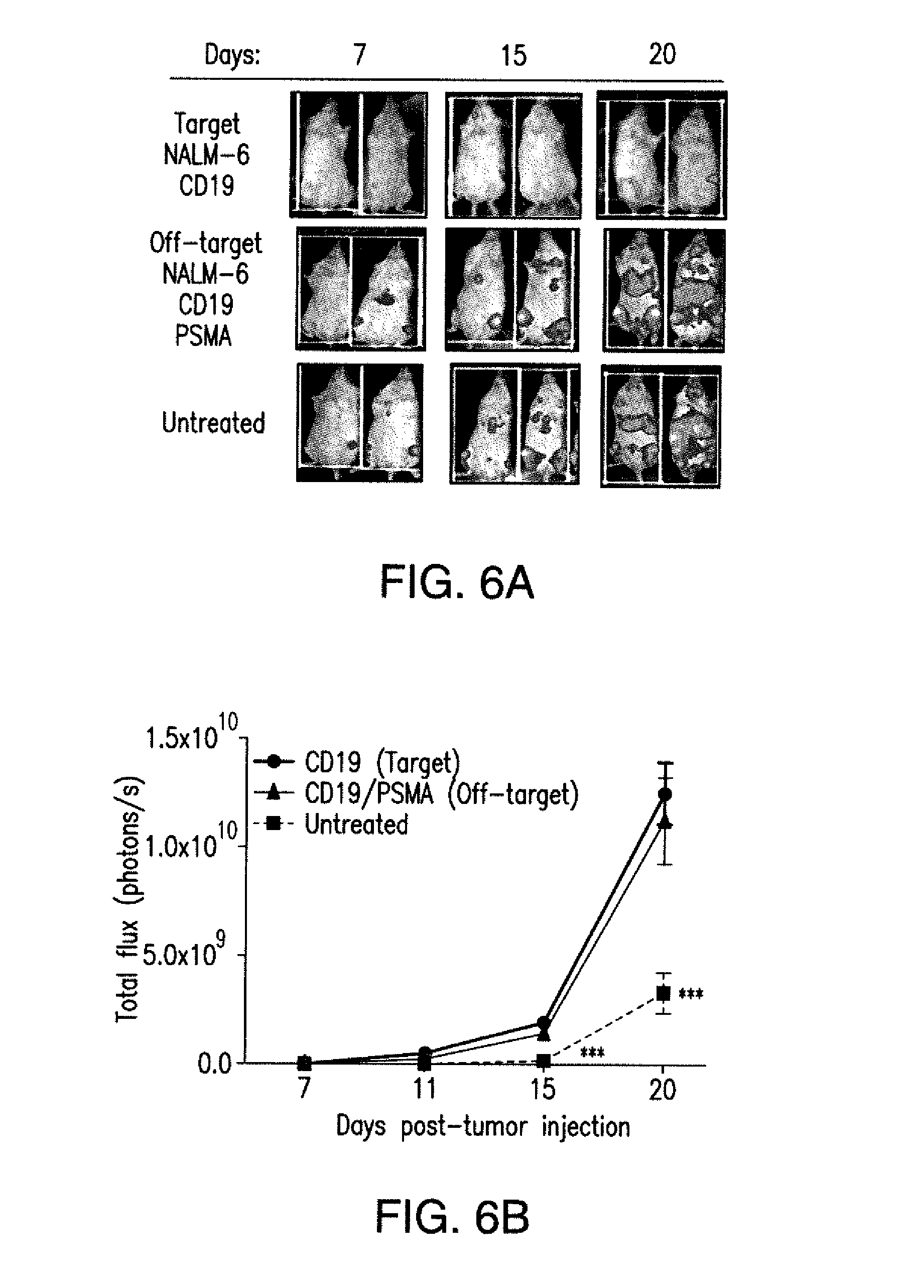
D00014

D00015
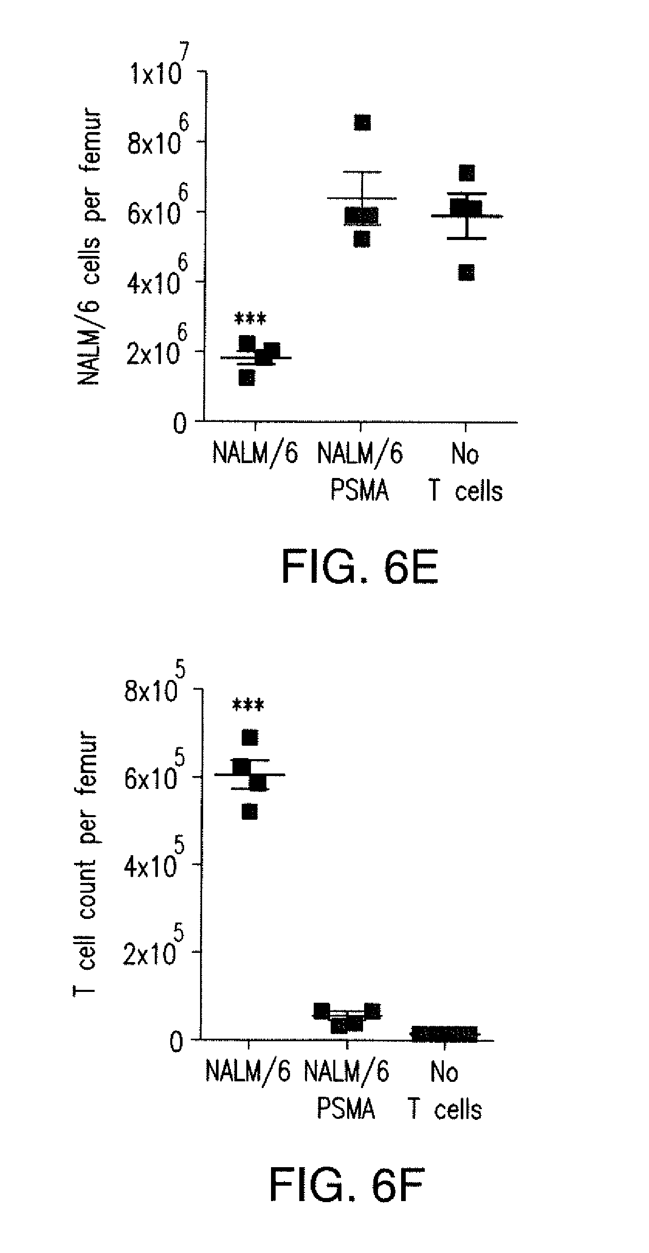
D00016

D00017
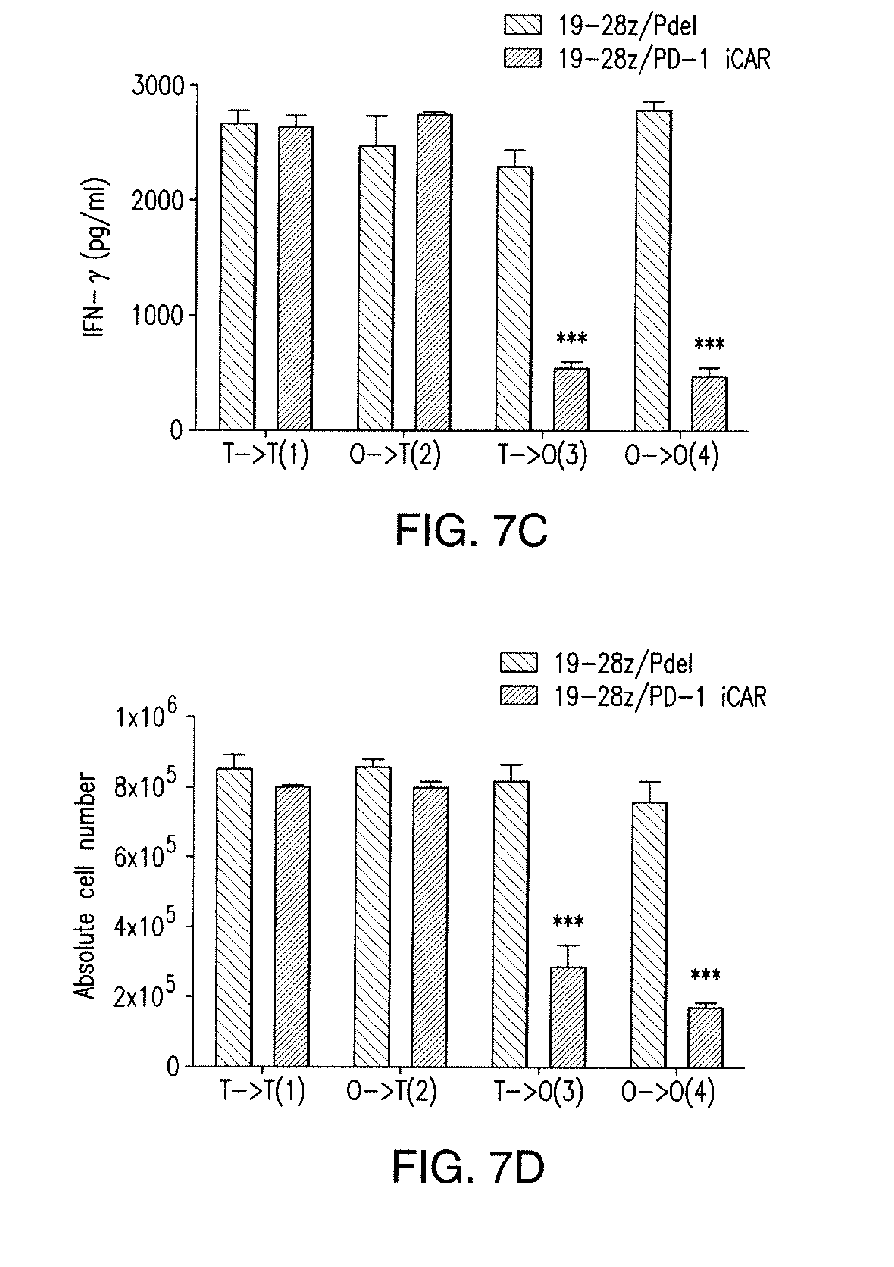
D00018

D00019
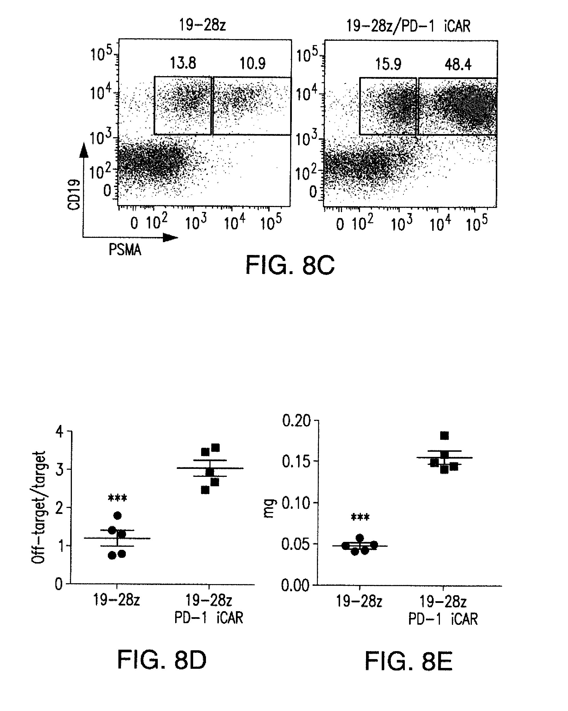
D00020
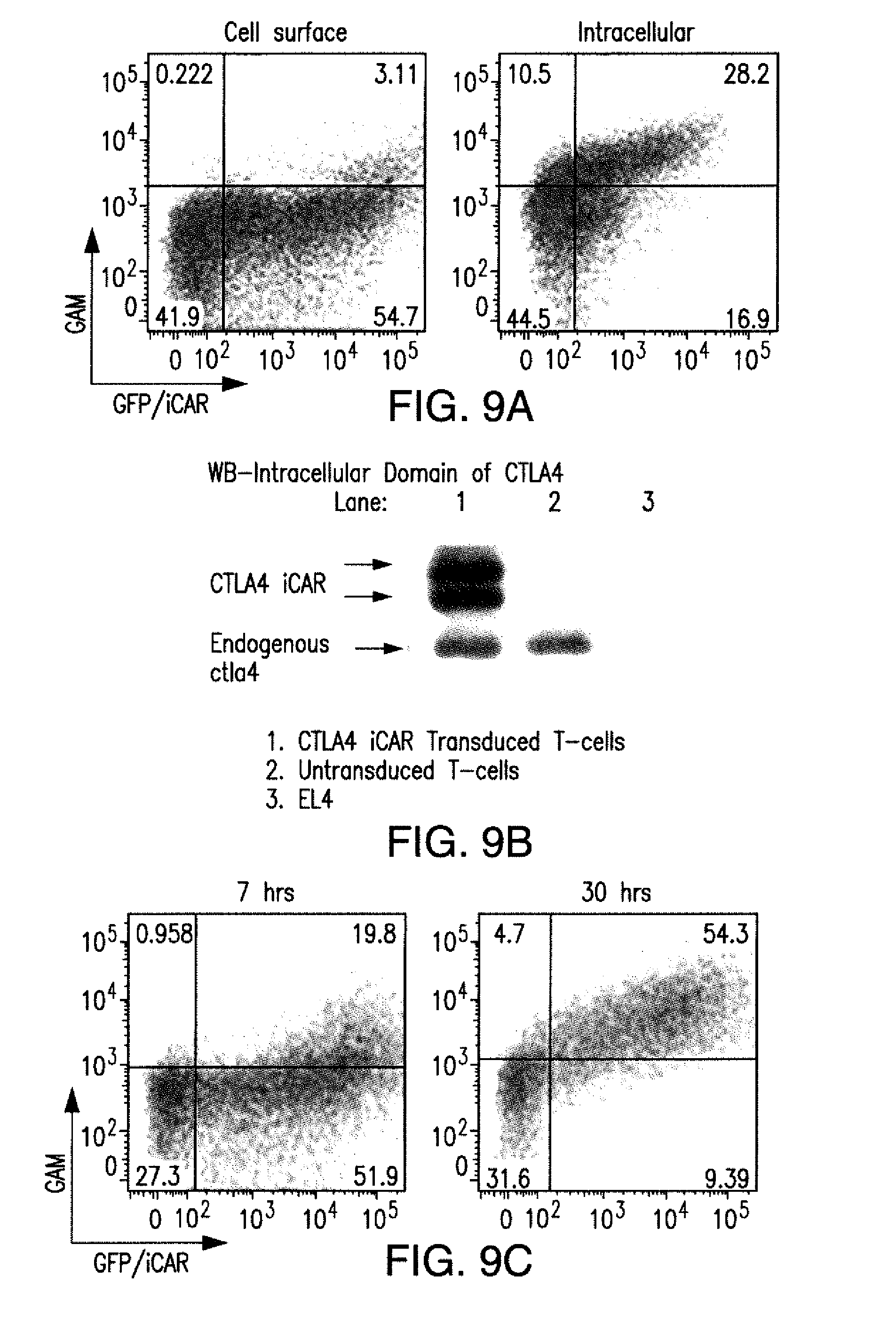
D00021

D00022
D00023
D00024

D00025
D00026

D00027

D00028

D00029
D00030

D00031
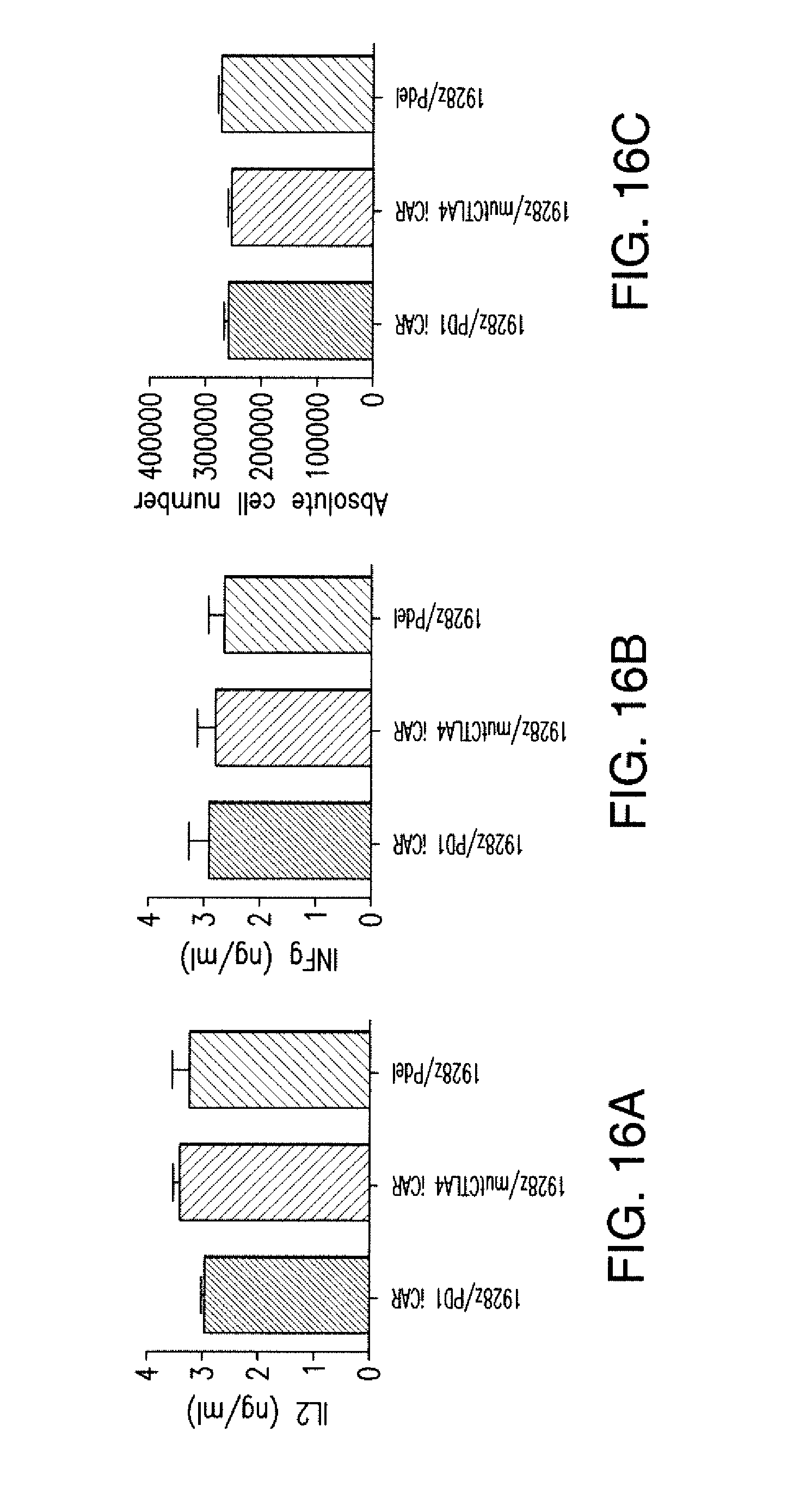
D00032
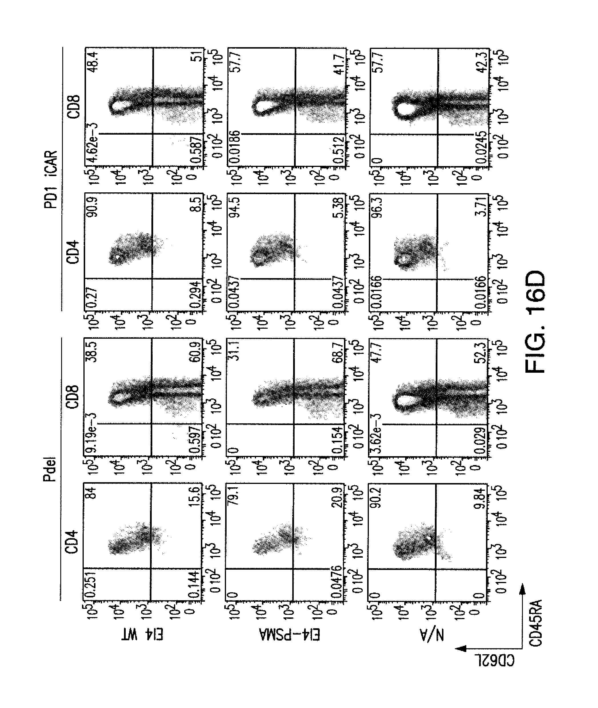
D00033

D00034

D00035
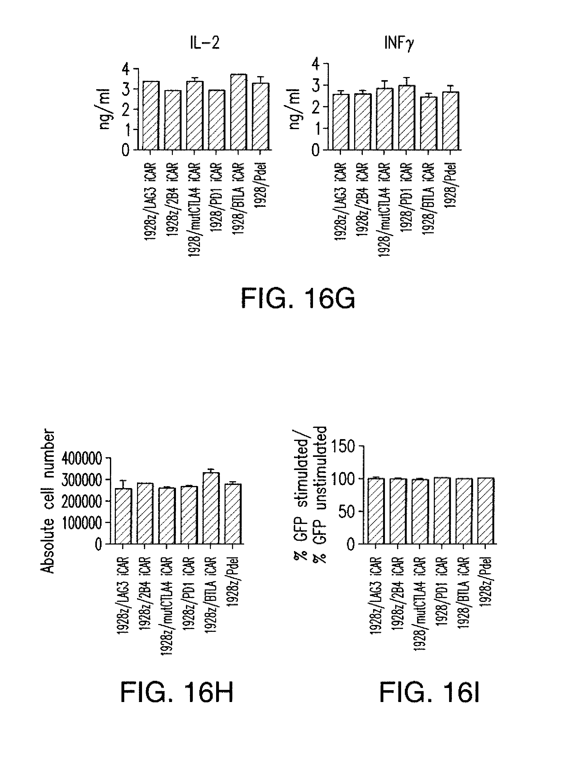
D00036

D00037
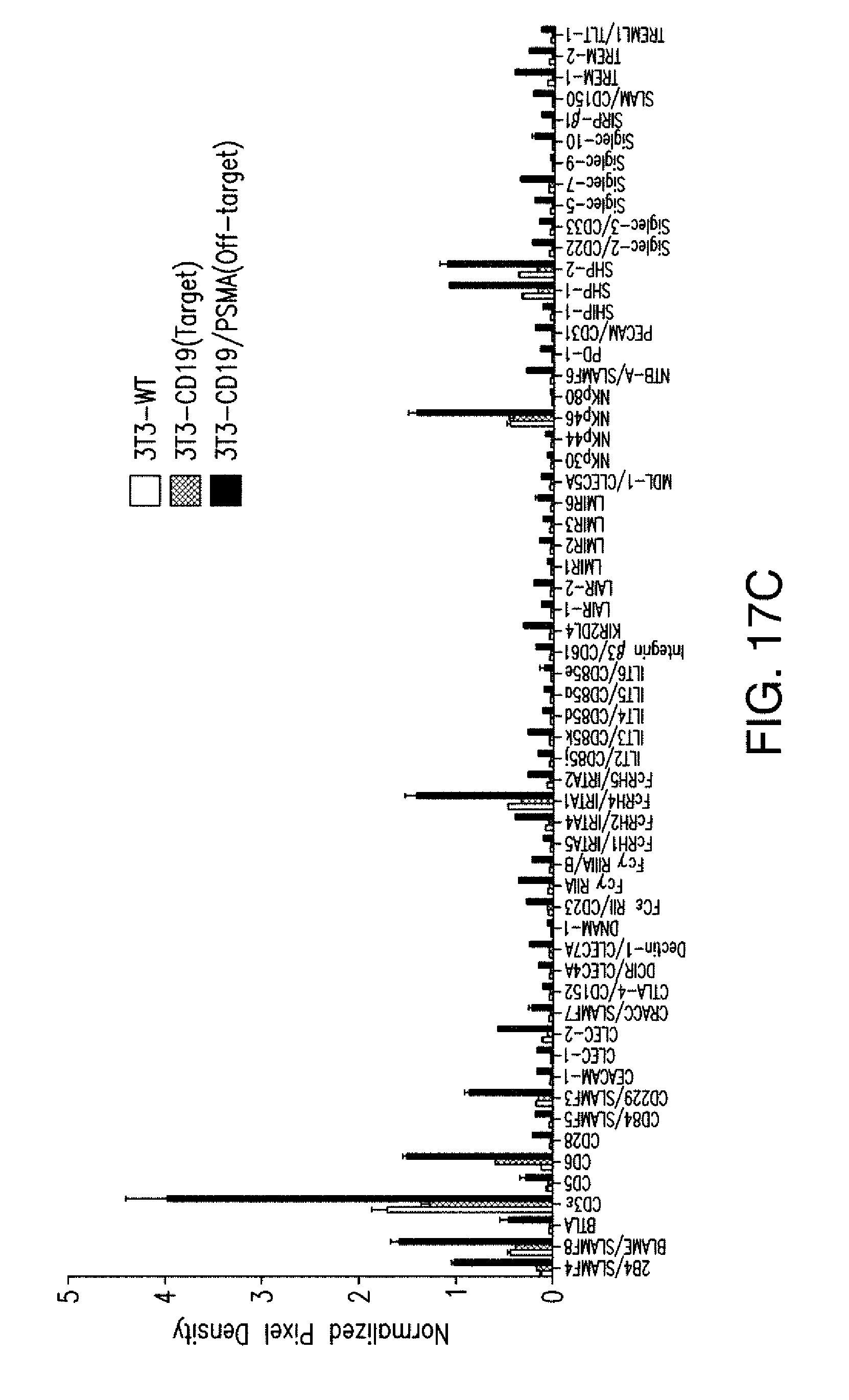
D00038
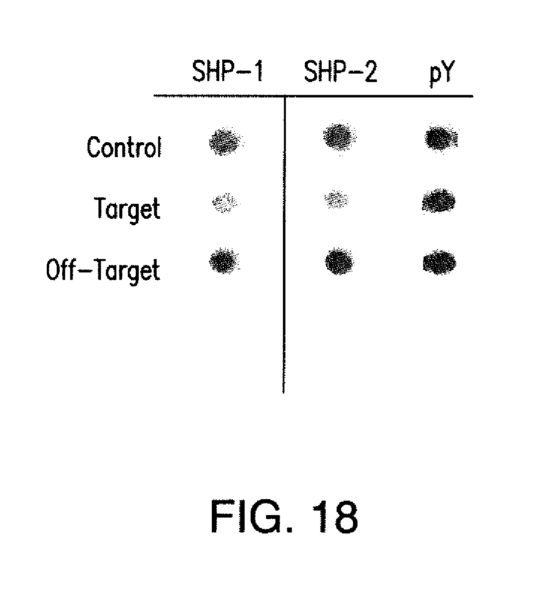
D00039

D00040

D00041
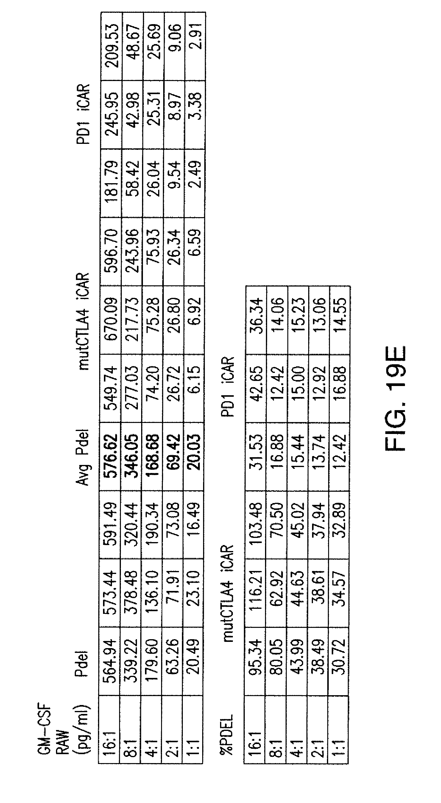
D00042
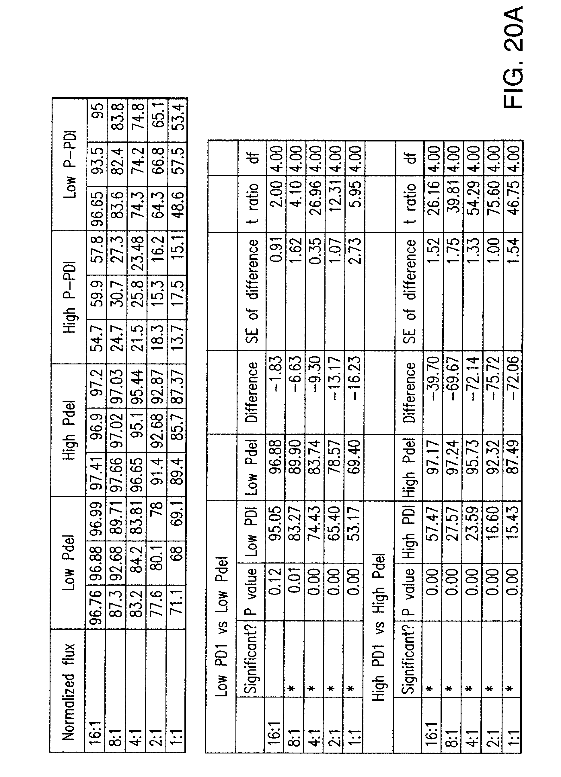
D00043
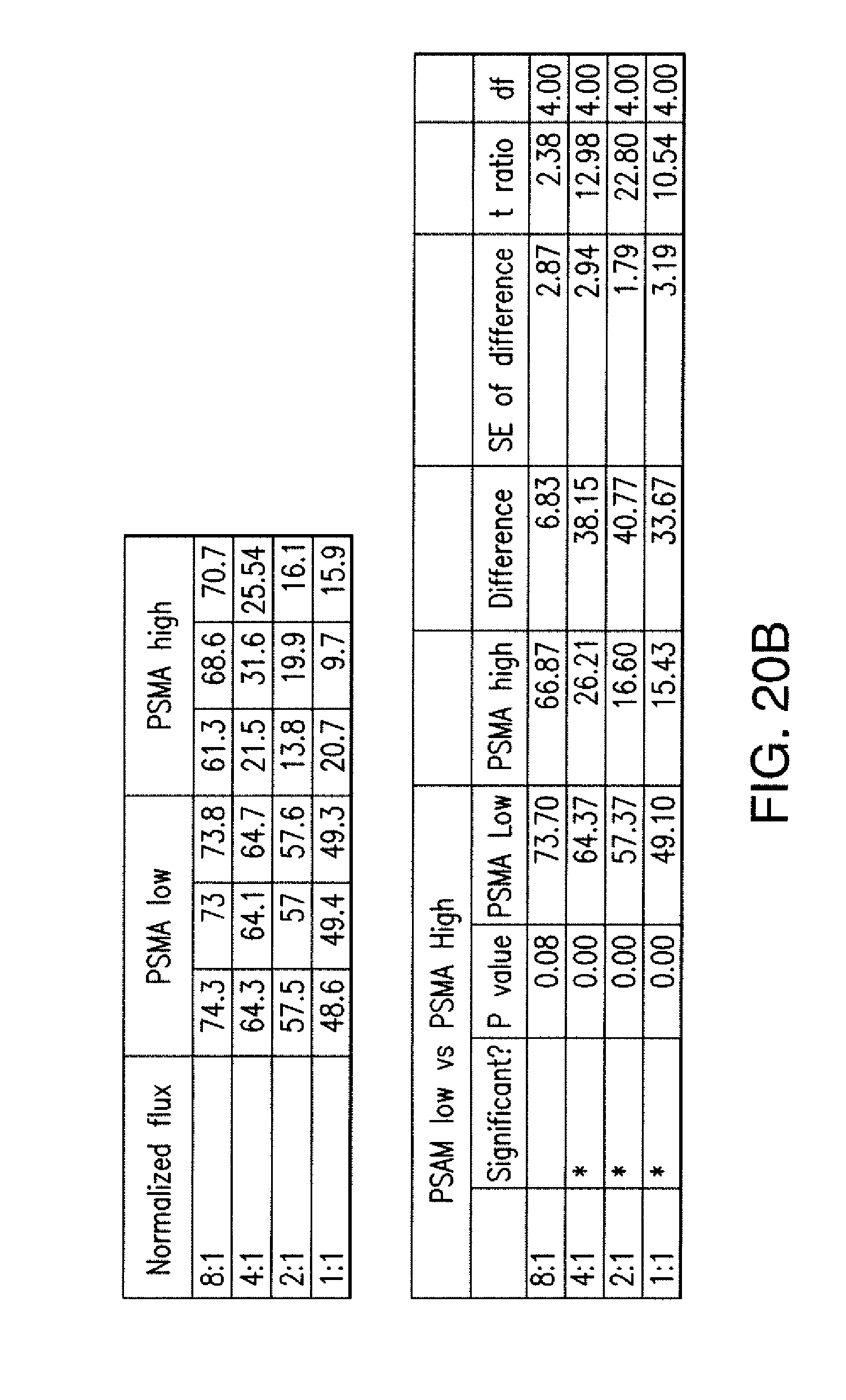
D00044
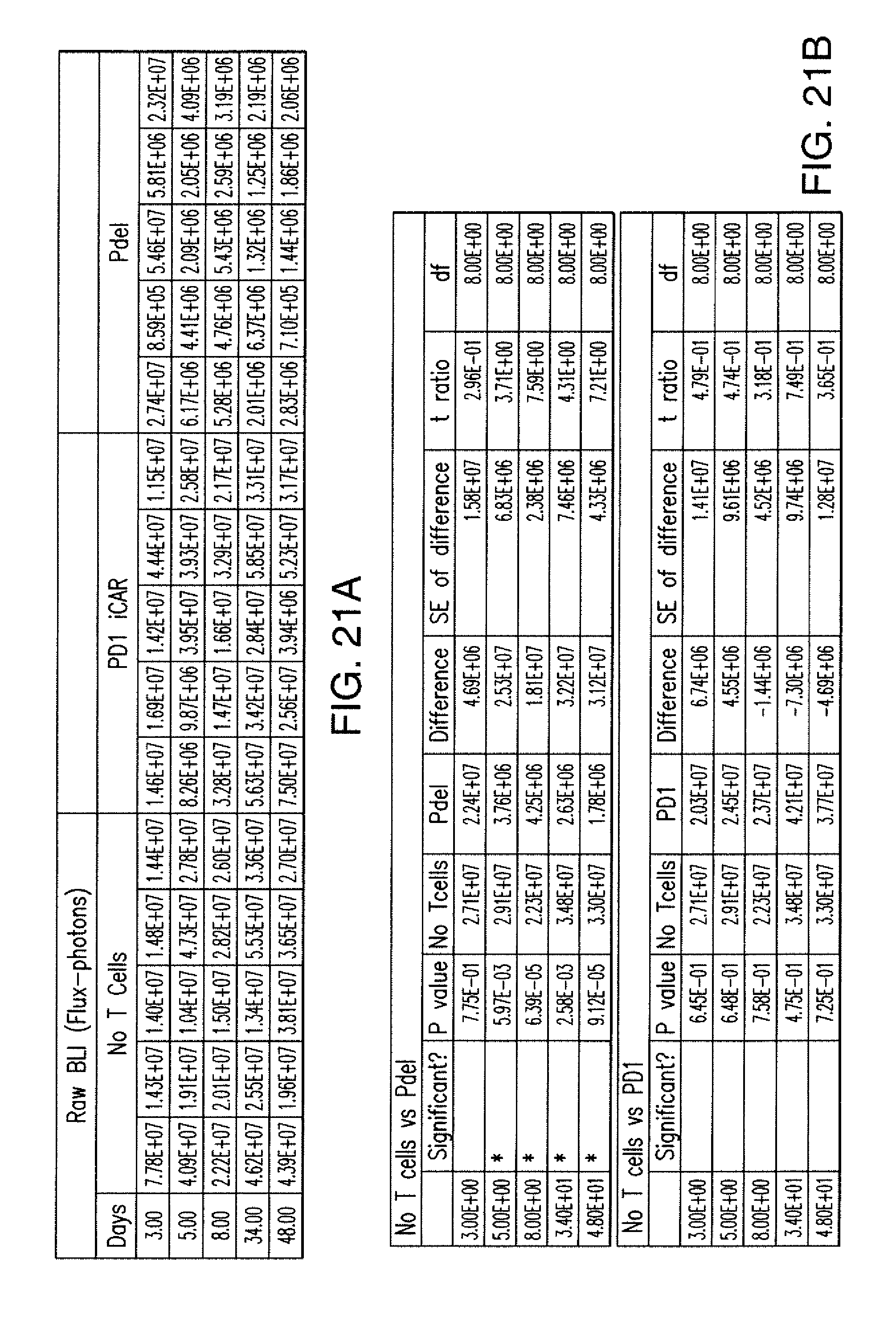
D00045
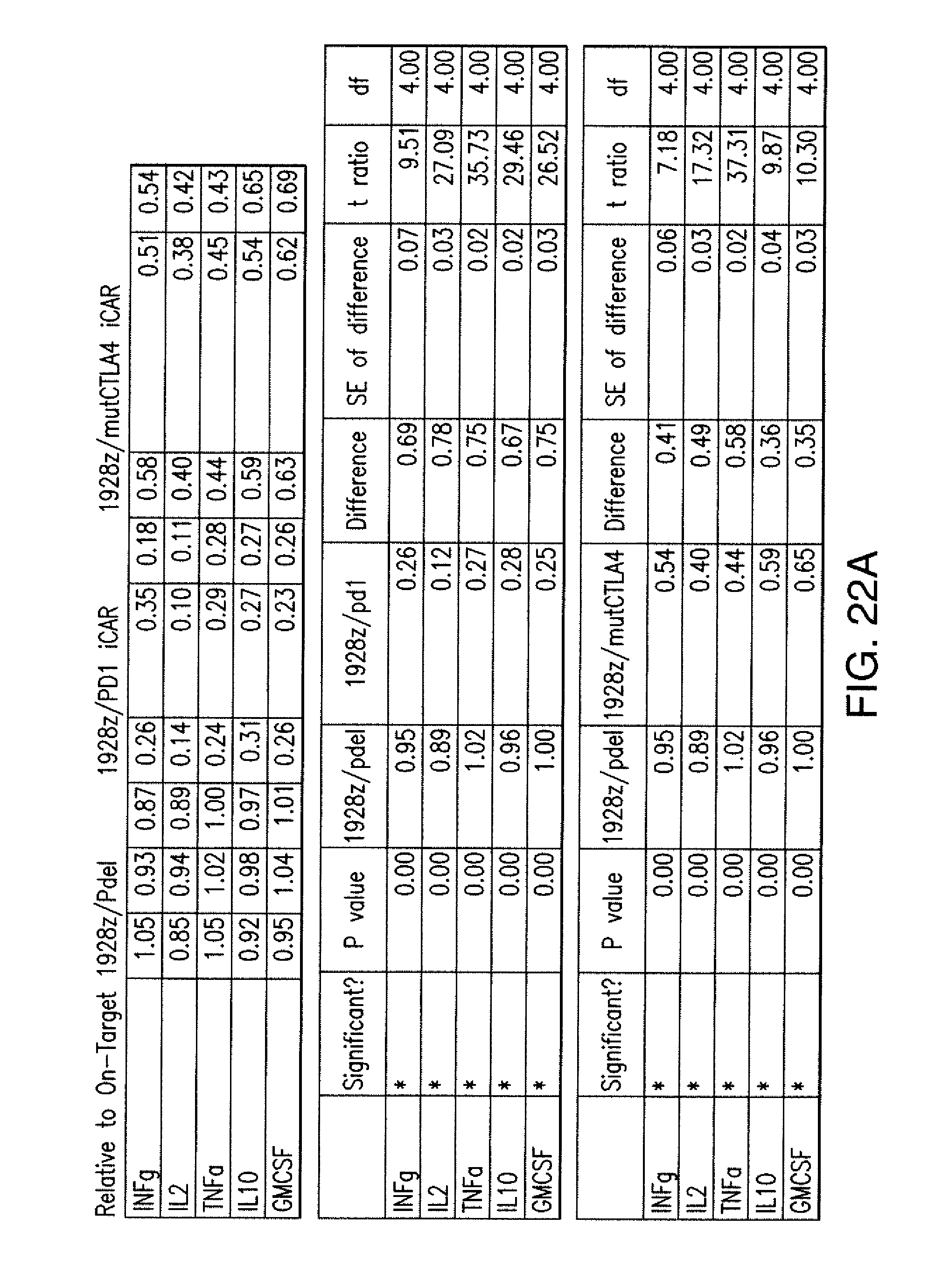
D00046
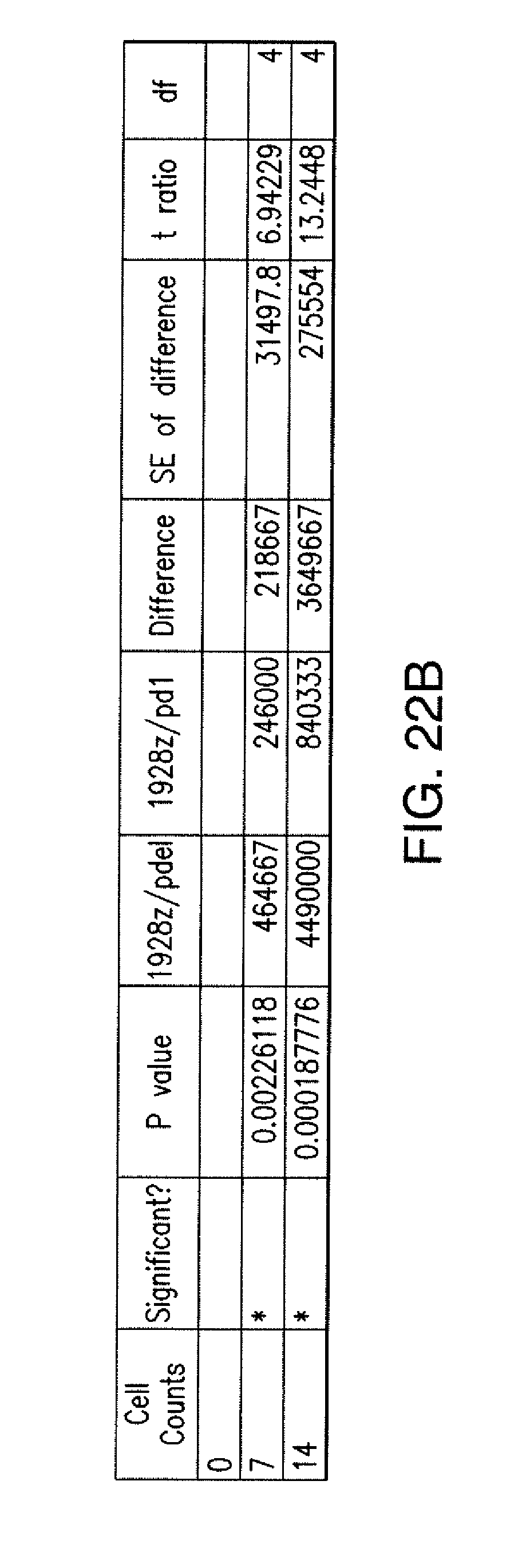
D00047

D00048
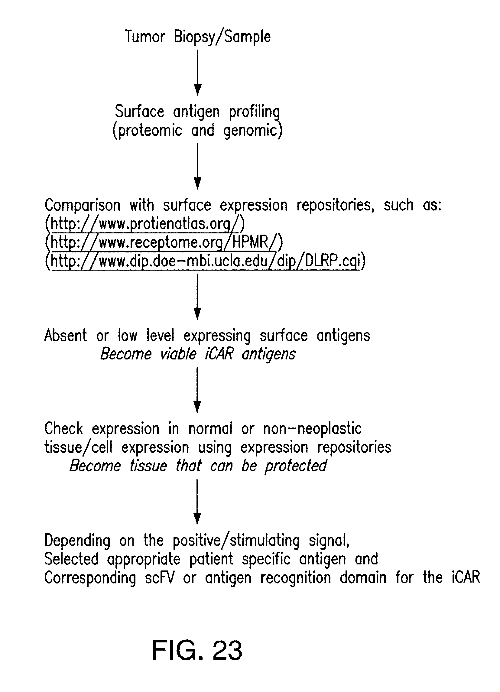
S00001
XML
uspto.report is an independent third-party trademark research tool that is not affiliated, endorsed, or sponsored by the United States Patent and Trademark Office (USPTO) or any other governmental organization. The information provided by uspto.report is based on publicly available data at the time of writing and is intended for informational purposes only.
While we strive to provide accurate and up-to-date information, we do not guarantee the accuracy, completeness, reliability, or suitability of the information displayed on this site. The use of this site is at your own risk. Any reliance you place on such information is therefore strictly at your own risk.
All official trademark data, including owner information, should be verified by visiting the official USPTO website at www.uspto.gov. This site is not intended to replace professional legal advice and should not be used as a substitute for consulting with a legal professional who is knowledgeable about trademark law.