Plant Wall Degradative Compounds And Systems
Taylor; Larry Edmund ; et al.
U.S. patent application number 13/465714 was filed with the patent office on 2012-12-27 for plant wall degradative compounds and systems. Invention is credited to Nathan A. Ekborg, Michael Howard, Steven Wayne Hutcheson, Larry Edmund Taylor, Ronald M. Weiner.
| Application Number | 20120329103 13/465714 |
| Document ID | / |
| Family ID | 36387151 |
| Filed Date | 2012-12-27 |


View All Diagrams
| United States Patent Application | 20120329103 |
| Kind Code | A1 |
| Taylor; Larry Edmund ; et al. | December 27, 2012 |
PLANT WALL DEGRADATIVE COMPOUNDS AND SYSTEMS
Abstract
The present invention relates to cell wall degradative systems, in particular to systems containing enzymes that bind to and/or depolymerize cellulose. These systems have a number of applications.
| Inventors: | Taylor; Larry Edmund; (Lakewood, CO) ; Weiner; Ronald M.; (Potomac, MD) ; Hutcheson; Steven Wayne; (Columbia, MD) ; Ekborg; Nathan A.; (Beverly, MA) ; Howard; Michael; (Annapolis, MD) |
| Family ID: | 36387151 |
| Appl. No.: | 13/465714 |
| Filed: | May 7, 2012 |
Related U.S. Patent Documents
| Application Number | Filing Date | Patent Number | ||
|---|---|---|---|---|
| 12099653 | Apr 8, 2008 | 8173787 | ||
| 13465714 | ||||
| 11121154 | May 4, 2005 | 7365180 | ||
| 12099653 | ||||
| 60567971 | May 4, 2004 | |||
| Current U.S. Class: | 435/105 ; 435/201; 435/252.33; 435/320.1; 536/23.2 |
| Current CPC Class: | C12N 9/2437 20130101; A01N 37/46 20130101; C12N 9/248 20130101; A01N 63/10 20200101; C12P 19/14 20130101; C12N 9/2402 20130101; C12N 9/2488 20130101; C12Y 302/01004 20130101 |
| Class at Publication: | 435/105 ; 536/23.2; 435/201; 435/320.1; 435/252.33 |
| International Class: | C12N 9/26 20060101 C12N009/26; C12P 19/02 20060101 C12P019/02; C12N 1/21 20060101 C12N001/21; C12N 15/56 20060101 C12N015/56; C12N 15/63 20060101 C12N015/63 |
Claims
1-31. (canceled)
32. An isolated Saccharophagus degradans strain 2-40 polynucleotide encoding an amino acid sequence with arabinofuranosidase activity and comprising a GH43 domain.
33. An isolated Saccharophagus degradans strain 2-40 polypeptide with arabinofuranosidase activity and comprising a GH43 domain.
34. A vector comprising an isolated Saccharophagus degradans strain 2-40 polynucleotide encoding an amino acid sequence with arabinofuranosidase activity and comprising a GH43 domain.
35. A host cell comprising an isolated Saccharophagus degradans strain 2-40 polynucleotide encoding an amino acid sequence with arabinofuranosidase activity and comprising a GH43 domain.
36. The host cell of claim 35, wherein the cell is an Escherichia coli cell.
37. A method of processing cereal comprising degrading hemicellulose in the cereal with an isolated Saccharophagus degradans strain 2-40 polypeptide with arabinofuranosidase activity and comprising a GH43 domain.
38. The method of claim 37, wherein the polypeptide is directly applied to a plant such that the polypeptide degrades the plant.
39. The method of claim 37, wherein the method is conducted in a marine environment.
40. The method of claim 37, wherein the method is conducted under water.
Description
CROSS-REFERENCE TO RELATED APPLICATIONS
[0001] This patent application is a continuation of U.S. patent application Ser. No. 12/099,653, filed Apr. 8, 2008, which claims priority to U.S. divisional patent application Ser. No. 11/121,154, filed May 4, 2005, issued as U.S. Pat. No. 7,365,180, on Apr. 29, 2008, which claims priority to U.S. Provisional Application No. 60/567,971, filed May 4, 2004, the contents of which are incorporated herein, in their entirety, by reference.
BACKGROUND OF THE INVENTION
[0002] 1. Field of the Invention
[0003] The invention is generally directed to degradative enzymes and systems. In particular, the present invention is directed to plant cell wall degrading enzymes and associated proteins found in Microbulbifer degradans and systems containing such enzymes and/or proteins.
[0004] 2. Background of the Invention
[0005] Cellulases and related enzymes have been utilized in food, beer, wine, animal feeds, textile production and laundering, pulp and paper industry, and agricultural industries. Various such uses are described in the paper "Cellulases and related enzymes in biotechnology" by M. K. Bhat (Biotechnical Advances 18 (2000) 355-383), the subject matter of which is hereby incorporated by reference in its entirety.
[0006] Saccharophaagus degradans strain 2-40 (herein referred to as "S. degradans 2-40" or "2-40") is a representative of an emerging group of marine bacteria that degrade complex polysaccharides (CP). S. degradans has been deposited at the American Type Culture Collection and bears accession number ATCC 43961. S. degradans 2-40, formerly known and referred to synonomously herein as Microbulbifer degradans strain 2-40 ("M. degradans 2-40"), is a marine y-proteobacterium that was isolated from decaying Sparina altemiflora, a salt marsh cord grass in the Chesapeake Bay watershed. Consistent with its isolation from decaying plant matter, S. degradans strain 2-40 is able to degrade many complex polysaccharides, including cellulose, pectin, xylan, and chitin, which are common components of the cell walls of higher plants. S. degradans strain 2-40 is also able to depolymerize algal cell wall components, such as agar, agarose, and laminarin, as well as protein, starch, pullulan, and alginic acid. In addition to degrading this plethora of polymers, S. degradans strain 2-40 can utilize each of the polysaccharides as the sole carbon source. Therefore, S. degradans strain 2-40 is not only an excellent model of microbial degradation of insoluble complex polysaccharides (ICPs) but can also be used as a paradigm for complete metabolism of these ICPs. ICPs are polymerized saccharides that are used for form and structure in animals and plants. They are insoluble in water and therefore are difficult to break down.
[0007] Microbulbifer degradans strain 2-40 requires at least 1% sea salts for growth and will tolerate salt concentrations as high as 10%. It is a highly pleomorphic. Gram-negative bacterium that is aerobic, generally rod-shaped, and motile by means of a single polar flagellum. Previous work has determined that 2-40 can degrade at least 10 different carbohydrate polymers (CP), including agar, chitin, alginic acid, carboxymethylcellulose (CMC), 6-glucan, laminarin, pectin, pullulan, starch and xylan (Ensor, Stotz et al. 1999). In addition, it has been shown to synthesize a true tyrosinase (Kelley, Coyne et al. 1990), 16S rDNA analysis shows that 2-40 is a member of the gamma-subclass of the phylum Proteobacteria, related to Microbulbifer hydrolyticus (Gonzalez and Weiner 2000) and to Teridinibacter sp., (Distel, Morrill et al, 2002) cellulolytic nitrogen-fixing bacteria that are symbionts of shipworms.
[0008] The agarase, chitinase and aiginase systems have been generally characterized. Zymogram activity gels indicate that all three systems are comprised of multiple depolymerases and multiple lines of evidence suggest that at least some of these depolymerases are attached to the cell surface (Stotz 1994; Whitehead 1997; Chakravorty 1998). Activity assays reveal that the majority of 2-40 enzyme activity resides with the cell fraction during logarithmic growth on CP, while in later growth phases the bulk of the activity is found in the supernatant and cell-bound activity decreases dramatically (Stotz 1994). Growth on CP is also accompanied by dramatic alterations in cell morphology. Glucose-grown cultures of 2-40 are relatively uniform in cell size and shape, with generally smooth and featureless cell surfaces. However when grown on agarose, alginate or chitin, 2-40 cells exhibit novel surface structures and features.
[0009] These exo- and extra-cellular structures (ES) include small protuberances, ger bleb-like structures that appear to be released from the cell, fine fimbrae or pill, and a network of fibril-like appendages which may be tubules of some kind. Immunoelectron microscopy has shown that agarases, alginases and/or chitinases are localized in at least some types of 2-40 ES. The surface topology and pattern of immunolocalization of 2-40 enzymes to surface protuberances are very similar to what is seen with cellulolytic members of the genus Clostridium.
[0010] There exists a need to identify enzyme systems that use cellulose as a substrate, express the genes encoding the proteins using suitable vectors, identify and isolate the amino acid products (enzymes and non-enzymatic products), and use these products as well as organisms containing these genes for purposes such as those described in the Bhat paper.
SUMMARY OF THE INVENTION
[0011] One aspect of the present invention is directed to systems of plant wallactive carbohydrases and related proteins.
[0012] A further aspect of the invention is directed to a method for the degradation of substances comprising cellulose. The method involves contacting the cellulose containing substances with one or more compounds obtained from Saccharophagus degradans strain 2-40.
[0013] Another aspect of the present invention is directed to groups of enzymes that catalyze reactions involving cellulose.
[0014] Another aspect of the present invention is directed to polynucleotides that encode polypeptides with cellulose degrading or cellulose binding activity.
[0015] A further aspect of the invention is directed to chimeric genes and vectors comprising genes that encode polypeptides with cellulose depolymerase activity.
[0016] A further aspect of the invention is directed to a method for the identification of a nucleotide sequence encoding a polypeptide comprising any one of the following activities from S. degradans: cellulose depolymerase, or cellulose binding. An S. degradans genomic library can be constructed in E. coli and screened for the desired activity. Transformed E. coli cells with specific activity are created and isolated.
[0017] Further aspects of the invention are directed to utilization of the cellulose degrading substances in food, beer, wine, animal feeds, textile production and laundering, pulp and paper industry, and agricultural industries.
[0018] Other aspects, features, and advantages of the invention will become apparent from the following detailed description, which when taken in conjunction with the accompanying figures, which are part of this disclosure, and which illustrate by way of example the principles of this invention.
BRIEF DESCRIPTION OF THE FIGURES
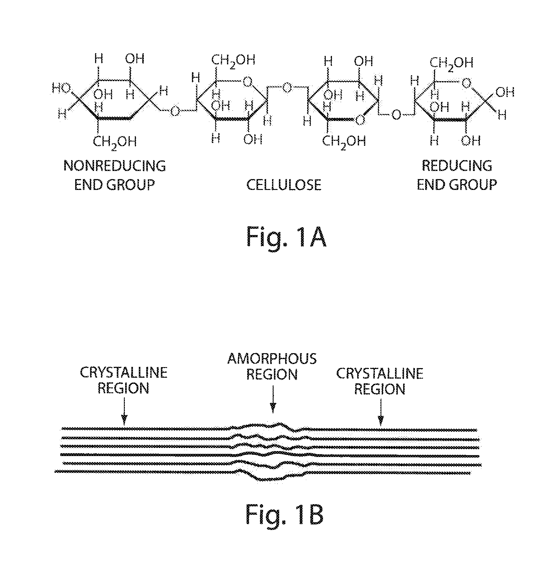
[0019] FIG. 1A shows the chemical formula of cellulose;
[0020] FIG. 1B illustrates the physical structure of cellulose;
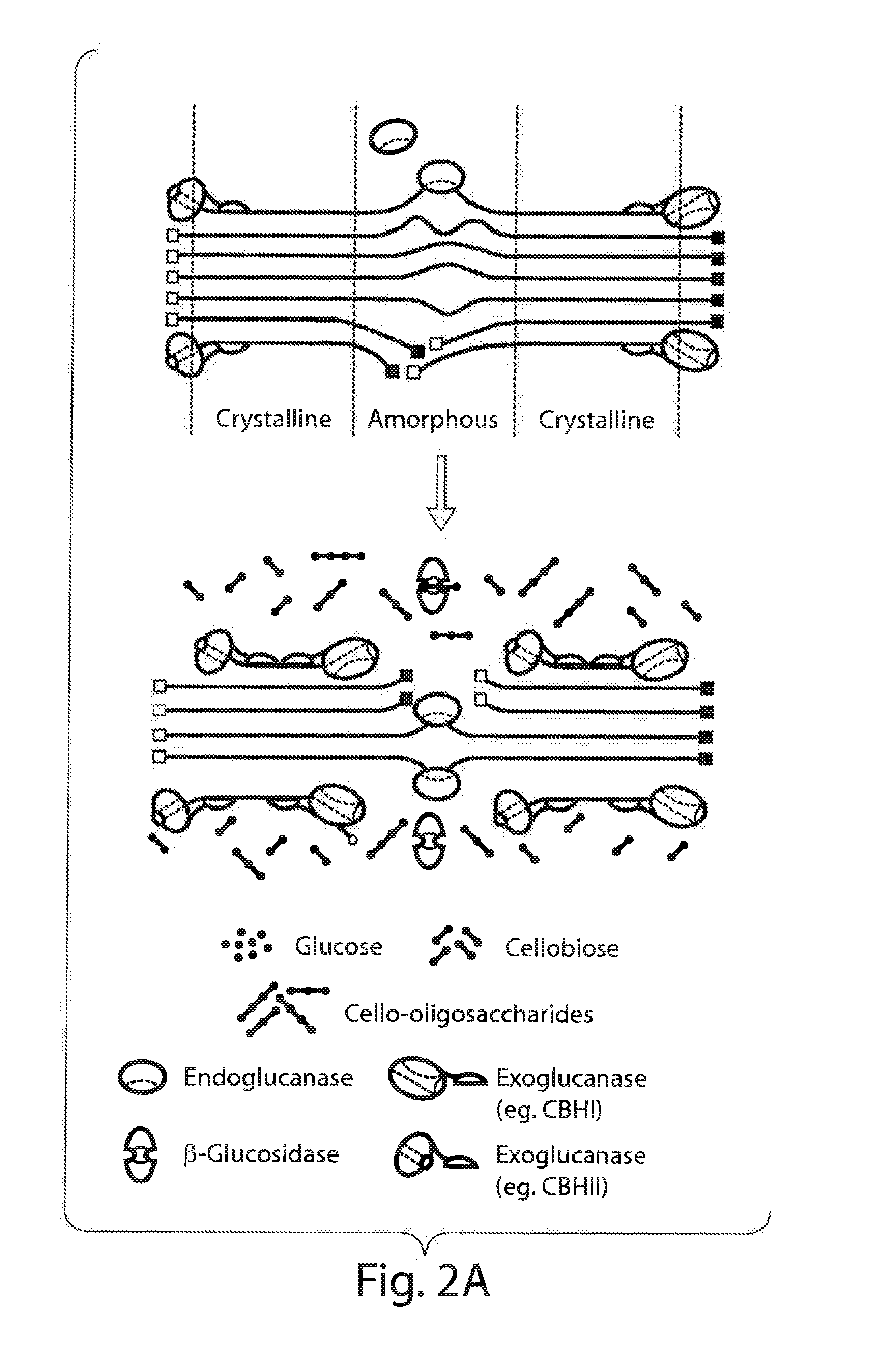
[0021] FIG. 2A illustrates the degradation of cellulose fibrils;
[0022] FIG. 2B shows the chemical representation of cellulose degradation to cellobiose and glucose;
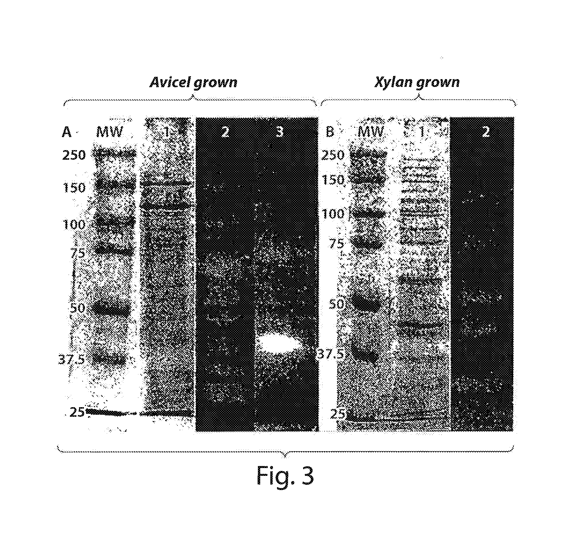
[0023] FIG. 3 shows SDS-PAGE and Zymogram analysis of 2-40 culture supernatants;
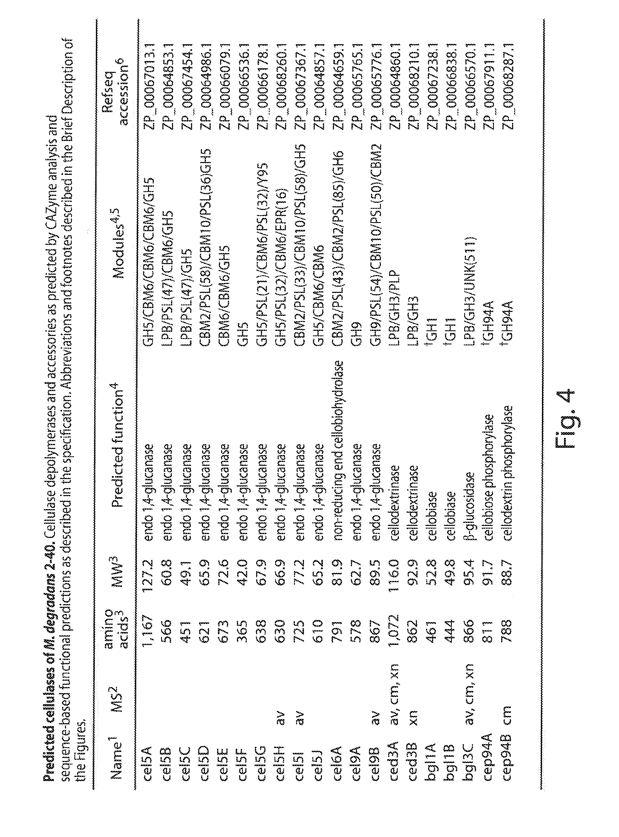
[0024] FIG. 4 lists the predicted cellulases of S. degradans 2-40 the sequences from FIGS. 4-40 are disclosed as SEC) ID NOs 1-214, respectively in order of appearance, 1--Acronyms, cel=cellulase, ced=cellodextrinase, bql=6-glucosidase, cep=cellobiose/cellodextrin phosphorylase; 2--Protein identified by tandem mass spectrometry in supernatant concentrates. Growth substrates: av=avicel; aq=agarose; al=alginate, cm=CMC, xn=xylan; 3--MW and amino acid count calculated using the protParam (protein parameters) tool at the Expasy website based on the DOE/JGI gene model amino acid sequence translations and includes any predicted signal peptide; 4--Predictions of function and GH, GT and CBM module determination according to CAZy ModO analysis by B. Henrissat, AFMB-CRNS; Da.sup.ggers (t) indicate lack of a secretion signal sequence; 5--Nonstandard module abbreviations, LPB=lipobox motif, PSL=polyserine linker, EPR=glutamic acid-proline rich region, PLP=phospholipase-like domain, number in parentheses indicates the length of the indicated feature in amino acid residues; 6--Refseq accession number of gene amino acid sequence from the Entrez protein database;
[0025] FIG. 5 lists the predicted xylanases, xylosidases and related accessories of M. degradans 2-40;
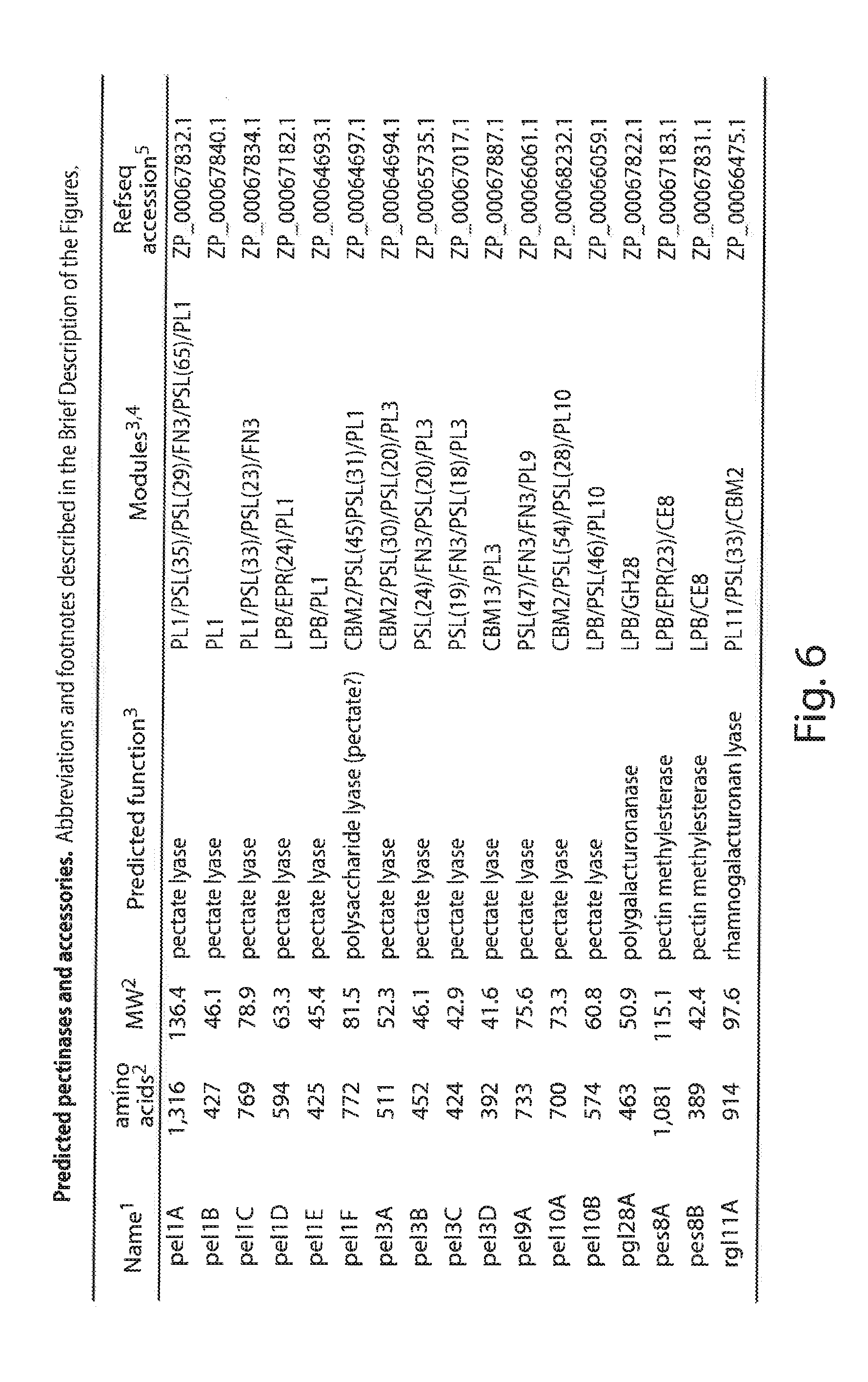
[0026] FIG. 6 lists the predicted pectinases and related accessories of S. degradans 2-40, 1--Acronyms, pel pectate lyase, pes=pectin methylesterase, rql=rhamnogalacturonan lyase; 2--MW and amino acid count calculated using the protParam (protein parameters) tool at the Expasy website based on the DOE/JGI gene model amino acid sequence translations and includes any predicted signal peptide; 3-Predictions of function and GET, GT, PI, CE and CBM module determination according to CAZy Mod( ) analysis by B. Henrissat, AFMB-CRNS; 4--Module abbreviations, CE=carbohydrate esterase, FN3=fibronectin type3-like domain, LPB=lipobox motif, PL=pectate lyase, PSR=polyserine region, EPR=glutamic acid-proline rich region, number in parentheses indicates the length of the indicated feature in amino acid residues; 5--Refseq accession number of gene model amino acid sequence from the Entrez Pubmed database;
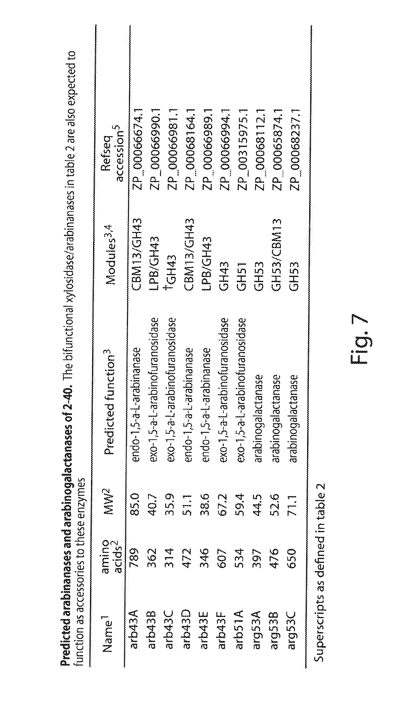
[0027] FIG. 7 lists the arabinanases and arabinogalactanases of S. degradans 2-40;
[0028] FIG. 8 lists the mannanases of S. degradans 2-40;
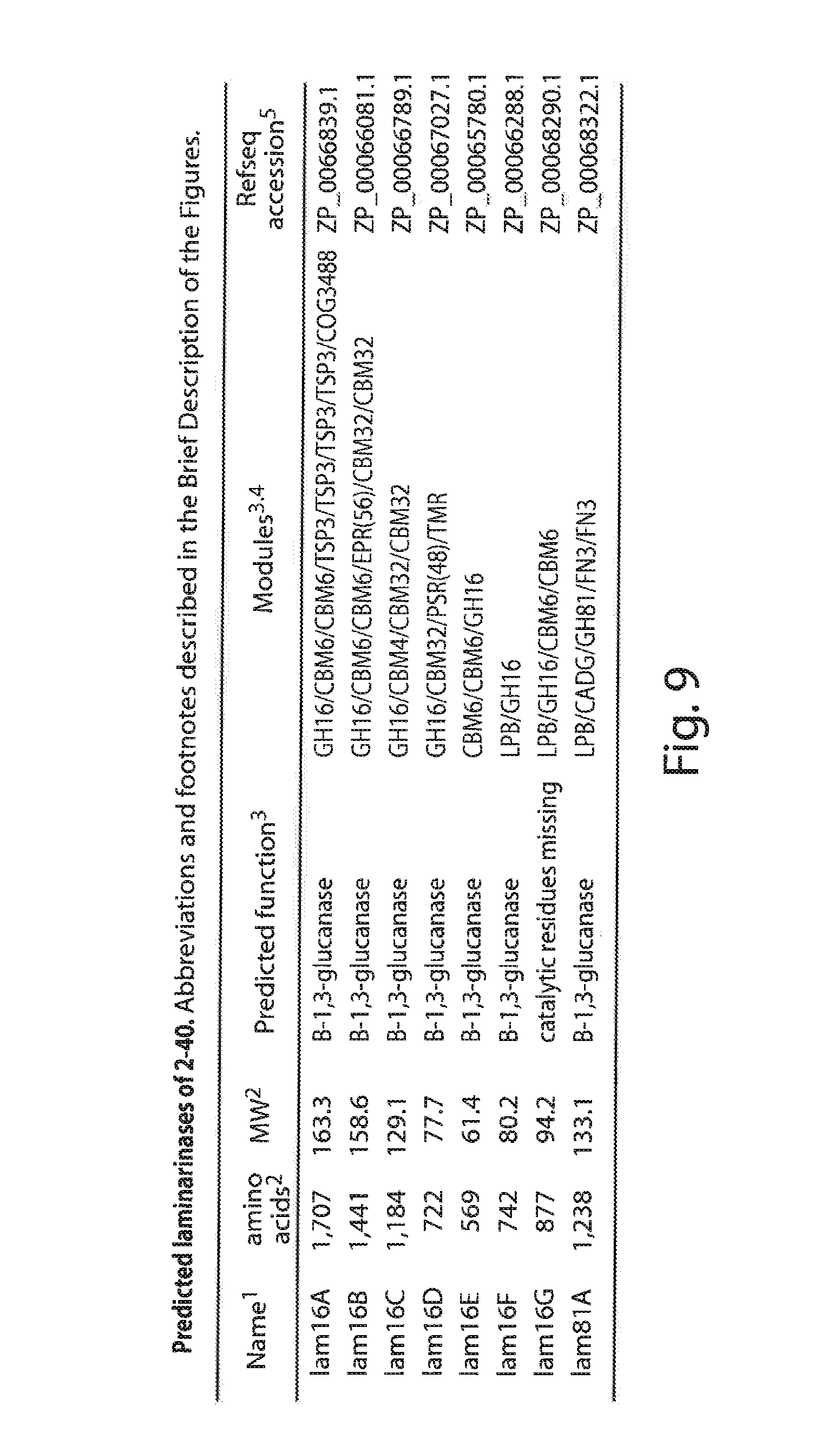
[0029] FIG. 9 lists the laminarinases of S. Degradans 2-40, Superscripts: 1-Acronyms, lam=laminarinase; 2--MW and amino acid count calculated using the protParam (protein parameters) tool at the Expasy website based on the DOE/JGI gene model amino acid sequence translations and includes any predicted signal peptide; 3-Predictions of function and OH, GT, PL, CE and CBM module determination according to CAZy Mod( ) analysis by B. Henrissat, AFMB-CRNS; 4--Module abbreviations: TSP3=thrombospondin type3 repeats, COG3488=thiol-oxidoreductase like domain of unknown function (Interestingly, a similar domain is found in cbm32A: see table 7), PSD=polyserine domain, TMR=predicted transmembrane region. FN3=fibronectin type3like domain, EPR=qlutamic acid-proline rich region, CADG=cadherin-like calcium binding motif, number in parentheses indicates the length of the indicated feature in amino acid residues; 5-Refseq accession number can be used to retrieve the gene model amino acid sequence from the Entrez Pubmed database:
[0030] FIG. 10 lists selected carbohydrate-binding module proteins of S. degradans 2-40; and
[0031] FIG. 11 lists the recombinant proteins of S. degradans 2-40 and a comparison of predicted vs. observed molecular weights thereof.
DETAILED DESCRIPTION
[0032] Analysis of the genome sequence of S. degradans 2-40 reveals an abundance of genes coding for enzymes that are predicted to degrade plant-derived carbohydrates. To date, 2-40 is the only sequenced marine bacterium with apparently complete cellulase and xylanase systems, as well as a number of other systems containing plant-wall active carbohydrases.
[0033] Thus it appears that 2-40 can play a significant role in the marine carbon cycle, functioning as a "super-degrader" that mediates the breakdown of CP from various algal, plantal, and invertebrate sources. The remarkable enzymatic diversity, novel surface features (ES), and the apparent localization of carbohydrases to ES make S. degradans 2-40 an intriguing organism in which to study the cell biology of CP metabolism and surface enzyme attachment.
[0034] It has now been discovered that 2-40 has a complete complement of enzymes, suitably positioned, to degrade plant cell walls. This has been accomplished by the following approaches: a) annotation and genomic analysis of 2-40 plant-wall active enzyme systems, b) identification of enzymes and other proteins which contain domains or motifs that may be involved in surface enzyme display, c) the development of testable models based on identified protein motifs, and d) cloning and expression of selected proteins for the production of antibody probes to allow testing of proposed models of surface enzyme display using immunoelectron microscopy.
[0035] These efforts have been greatly facilitated by the recent sequencing of the genome of 2-40, allowing a strategy where genes which code for proteins with potential involvement in surface attachment may be identified based on sequence homology with modules or domains known to function in surface attachment and/or adhesion.
[0036] Enzymatic and non-enzymatic ORFs with compelling sequence elements are identified using BLAST and other amino acid sequence alignment and analysis tools. Genes of interest can be cloned into E. coli, expressed with in-frame polyhistidine affinity tag, fusions and purified by nickel ion chromatography, thus providing the means of identifying and producing recombinant 2-40 proteins for study and antibody probe production.
[0037] The genome sequence of 2-40 was recently obtained in conjunction with the Department of Energy's Joint Genome initiative (JGI). The finished draft sequence dated January 19, 2.005 comprises 5.1 Mbp contained in a single contiguous sequence. Automated annotation of open reading frames (ORFs) was performed by the computational genomics division of the Oak Ridge National Laboratory (ORNL), and the annotated sequence is available on the World Wide Web.
[0038] The initial genome annotation has revealed a variety of carbohydrases, including a number of agarases, alginases and chitinases. Remarkably, the genome also contains an abundance of enzymes with predicted roles in the degradation of plant cell wall polymers, including a number of ORFs with homology to cellulases, xylanases, pectinases, and other glucanases and glucosidases. In all, over 180 open reading frames with a probable role in carbohydrate catabolism were identified in the draft genome.
[0039] To begin to define the cellulase, xylanase, and pectinase systems of 2-40, genes were initially classified as belonging to one of those systems by BLAST homology. Ambiguous ORFs were tentatively assigned to the class of the best known hit. Other tools used to refine this tentative classification include Pfam (Protein families database of alignments and HMMs and SMART (Simple Modular Architecture Research Tool) which use multiple alignments and hidden Markov models (statistical models of sequence consensus homology) to identify discreet modular domains within a protein sequence. These analyses were relatively successful; however, a number of ORFs remained difficult to classify based on sequence homology alone.
[0040] Enzymes have traditionally been classified by substrate specificity and reaction products. In the pre-genomic era, function was regarded as the most amenable (and perhaps most useful) basis for comparing enzymes and assays for various enzymatic activities have been well-developed for many years, resulting in the familiar EC classification scheme. Cellulases and other O-Glycosyl hydrolases, which act upon glycosidic bonds between two carbohydrate moieties (or a carbohydrate and non-carbohydrate moiety as occurs in nitrophenol-glycoside derivatives) are designated as EC 3.2.1.-, with the final number indicating the exact type of bond cleaved. According to this scheme an endo-acting cellulase (1, 4-8-endoglucanase) is designated EC 3.2.1.4.
[0041] With the advent of widespread genome sequencing projects and the ease of determining the nucleotide sequence of cloned genes, ever-increasing amounts of sequence data have facilitated analyses and comparison of related genes and proteins on an unprecedented scale. This is particularly true for carbohydrases; it has become clear that classification of such enzymes according to reaction specificity, as is seen in the E.C. nomenclature scheme, is limited by the inability to convey sequence similarity. Additionally, a growing number of carbohydrases have been crystallized and their 3D structures solved.
[0042] One of the major revelations of carbohydrase sequence and structure analyses is that there are discreet families of enzymes with related sequence, which contain conserved three-dimensional folds that can be predicted based on their amino acid sequence. Further, it has been shown that enzymes with the same three-dimensional fold exhibit the same stereospecificity of hydrolysis, even when they catalyze different reactions (Henrissat, Teeri et al, 1998: Coutinho and Henrissat 1999).
[0043] These findings form the basis of a sequence-based classification of carbohydrase modules which is available in the form of an internet database, the Carbohydrate-Active enZYme server (CAZy)(Coutinho and Henrissat 1999; Coutinho and Henrissat 1999).
[0044] CAZy defines four major classes of carbohydrases, based on the type of reaction catalyzed: Glycosyl Hydrolases (GH's), Glycosyltansferases (GT's), Polysaccharide Lyases (PL's), and Carbohydrate Esterases (CE's), GH's cleave glycosidic bonds through hydrolysis. This class includes many familiar polysaccharidases such as cellulases, xylanases, and agarases. GT's generally function in polysaccharide synthesis, catalyzing the formation of new glycosidic bonds through the transfer of a sugar molecule from an activated carrier molecule, such as uridine diphosphate (UDP), to an acceptor molecule. While GT's often function in biosynthesis, there are examples where the mechanism is exploited for bond cleavage, as occurs in the phosphorolytic cleavage of cellobiose and cellodextrins (Lou, Dawson et al, 1996). PL's use a 6-elimination mechanism to mediate bond cleavage and are commonly involved in alginate and pectin depolymerization. CE's generally act as deacetylases on 0- or N-substituted polysaccharides. Common examples include xylan and chitin deacetylases. Sequence-based families are designated by number within each class, as is seen with GH5; glycosyl hydrolase family 5. Members of GH5 hydrolyze 6-1,4 bonds in a retaining fashion, using a double-displacement mechanism which results in retention of the original bond stereospecificity. Retention or inversion of anomeric configuration is a general characteristic of a given GH family (Henrissat and Bairoch 1993; Coutinho and Henrissat 1999). Many examples of endocellulases, xylanases and mannanases belonging to GH5 have been reported, illustrating the variety of substrate specificity possible within a GH family. Also, GH5s are predominantly endohydrolases--cleaving chains of their respective substrates at random locations internal to the polymer chains. While true for GH5, this generalization does not hold for many other GH in addition to carbohydrases, the CAZy server defines numerous families of Carbohydrate Binding Modules (CBM). As with catalytic modules, CBM families are designated based on amino acid sequence similarity and conserved three-dimensional folds.
[0045] The CAZyme structural families have been incorporated into a new classification and nomenclature scheme, developed by Bernard Henrissat and colleagues (Henrissat, Teeri et al, 1998). Traditional gene/protein nomenclature assigns an acronym indicating general function and order of discovery; in this scheme an organism's cellulose genes are designated celA, celB, etc., regardless of their actual mechanism of action on cellulose. Some researchers have attempted to convey more information by naming cellulases as endoglucanases (engA, engB) or cellobiohydrolases (cbhA, cbhB), however this requires determination of function in vitro and still fails to convey relatedness of protein sequence and structure. CAZyme nomenclature retains the familiar acronym to indicate the functional system a gene belongs to and incorporates the family number designation. Capital letters after the family number indicate the order of report within a given organism system. An example is provided by two endoglucanases, CenA and CenB, of Cellulomonas fimi. In the old nomenclature nothing can be deduced from the names except order of discovery. Naming them Cel6A and Cel9A, respectively, makes it immediately clear that these two cellulases are unrelated in sequence, and so belong to different GH families (where Cel stands for cellulase, and 9 for glycosyl hydrolase family nine). While this scheme does not distinguish between endo- and exo-activity, these designations are not absolute and can be included in discussion of an enzyme when relevant (i.e. the cellobiohydrolase Cel6A, the endoxylanase Xyn10B). Catalytic modules take precedence in naming carhohydrases; since many for even most) carbohydrases contain at least one CBM, they are named for their enzymatic module. If more than one catalytic domain is present, they are named in order from N-terminus to C-terminus, i.e. cel9A-cel48A contains a GH9 at the amino-terminus and a GH48 at the carboxyterminus. Both domains act against cellulose. There are, however, many examples of CBM modules occurring on proteins with no predicted carbohydrase module. In the absence of some other predicted functional domain (like a protease) these proteins are named for the CBM module family. If there are multiple CBM families present, then naming is again from amino to carboxy end, i.e. cbm2D-cbm 10A (Henrissat, Teeri et al, 1998). This nomenclature has been widely accepted and will be used in the naming of all 2-40 plant-wall active carhohydrases and related proteins considered as part of this study.
[0046] The cell walls of higher plants are comprised of a variety of carbohydrate polymer (CP) components. These CP interact through covalent and non-covalent means, providing the structural integrity plants required to form rigid cell walls and resist turgor pressure. The major CP found in plants is cellulose, which forms the structural backbone of the cell wall. See FIG. 1A. During cellulose biosynthesis, chains of poly-13-1,4-D-glucose self associate through hydrogen bonding and hydrophobic interactions to form cellulose microfibrils which further self-associate to form larger fibrils. Cellulose microfibrils are somewhat irregular and contain regions of varying crystallinity. The degree of crystallinity of cellulose fibrils depends on how tightly ordered the hydrogen bonding is between its component cellulose chains. Areas with less-ordered bonding, and therefore more accessible glucose chains, are referred to as amorphous regions (FIG. 1B). The relative crystallinity and fibril diameter are characteristic of the biological source of the cellulose (Beguin and Aubert 1994; Tomme, Warren et al. 1995; Lynd, Weimer et al. 2002). The irregularity of cellulose fibrils results in a great variety of altered bond angles and steric effects which hinder enzymatic access and subsequent degradation.
[0047] The general model for cellulose depolymerization to glucose involves a minimum of three distinct enzymatic activities (See FIGS. 2A and 2B). Endoglucanases cleave cellulose chains internally to generate shorter chains and increase the number of accessible ends, which are acted upon by exoglucanases. These exoglucanases are specific; for either reducing ends or non-reducing ends and frequently liberate cellobiose, the dimer of cellulose (cellobiohydrolases). The accumulating cellobiose is cleaved to glucose by cellobiases ((3-1,4-glucosidases). In many systems an additional type of enzyme is present: cellodextrinases are 6-1,4-glucosidases which cleave glucose monomers from cellulose oligomers, but not from cellobiose. Because of the variable crystallinity and structural complexity of cellulose, and the enzymatic activities required for is degradation, organisms with "complete" cellulase systems synthesize a variety of endo and/or exo-acting glucanases.
[0048] For example, Cellulomonas fimi and Thermomonospora fusca have each been shown to synthesize six cellulases while Clostridium thermocellum has as many as 15 or more ((Tome, Warren et al. 1995). Presumably, the variations in the shape of the substrate-binding pockets and/or active sites of these numerous cellulases facilitate complete cellulose degradation (Warren 1996). Organisms with complete cellulase systems are believed to be capable of efficiently using plant biomass as a carbon and energy source while mediating cellulose degradation. The ecological and evolutionary role of incomplete cellulose systems is less clear, although it is believed that many of these function as members of consortia (such as ruminal communities) which may collectively achieve total or near-total cellulose hydrolysis (Ljunedahl and Eriksson 1985; Tomme. Warren et al, 1995).
[0049] In the plant cell wall, microfibrils of cellulose are embedded in a matrix of hemicelluloses (including xylans, arabinans and mannans), pectins (galacturonans and galactans), and various 6-1,3 and 6-1,4 glucans. These matrix polymers are often substituted with arabinose, galactose and/or xylose residues, yielding arabinoxylans, galactomannans and xyloglucans--to name a few (Tomme, Warren et al. 1995; Warren 1996; Kosugi, Murashima et al. 2002: Lynd, Weimer et al. 2002). The complexity and sheer number of different glycosyl bonds presented by these non-cellulosic. CP requires specific enzyme systems which often rival cellulase systems in enzyme count and complexity. Because of its heterogeneity, plant cell wall degradation often requires consortia of microorganisms (Ljunadahl and Eriksson 1985; Tomme, Warren et al. 1995).
[0050] Objectives--M. degradans synthesizes complete multi-enzyme systems that degrade the major structural polymers of plant cell walls, A) define cellulase and xylanase systems, determining the activities of genes for which function cannot be predicted by sequence homology and B) genomic identification and annotation of other plant-degrading enzyme systems by sequence homology (i.e. pectinases, laminarinases, etc.).
[0051] Experimental Results
[0052] 1: Genomic, proteomic and functional analyses of 2-40 plant-wall active enzymes
[0053] Front the ORNL annotation it is clear that the 2-40 genome contains numerous enzymes with predicted activity against plant cell wall polymers. This is particularly surprising since 2-40 is an estuarine bacterium with several complex enzyme systems that degrade common marine polysaccharides such as agar, alginate, and chitin. Defining multienzyme systems based on automated annotations is complicated by the presence of poorly conserved domains and/or novel combinations of domains. There are ninny examples of this in the plant-wall active enzymes of 2-40. Accordingly, the ORNL annotations of carhohydrase ORFs were manually reviewed with emphasis on the modular composition and then assigned to general groups based on the substrate they were likely to be involved with (i.e. cellulose or xylan degradation). These genomic sequence analyses resulted in a pool of about 25 potential cellulases, 11 xylanases and 17 pectinases.
[0054] When sequence homology is well-conserved, highly accurate predictions of function are possible. Therefore, to verify the presence of functioning cellulase and xylanase systems in M. degradans, zymograms and enzyme activity assays were performed as discussed below. Also, attempts were made to identify enzymes from 2-40 culture supernatants using Mass Spectrometry based proteomics.
[0055] Next, more sophisticated genomic analyses were used to predict function where possible and to identify ORFs which require functional characterization to determine their roles, if any, in the cellulase and xylanase systems. ORFs which belong to other plant wall-active enzyme systems were tentatively classified based on the sequence analyses and functional predictions of B. Henrissat.
[0056] To gain insight into the induction and expression of 2-40 cellulases and xylanases, specific activities were determined for avicel and xylan-grown cells and supernatants by dinitrosalicylic acid reducing-sugar assays (DNSA assays), as discussed in the Experimental Protocols section at the end of this proposal. Xylanase activity was measured for avicel-grown cultures, and vice versa, in order to investigate possible co-induction of activity by these two substrates which occur together in the plant cell wall.
[0057] Growth on either avicel or xylan yields enzymatic activity against both substrates, suggesting co-induction of the cellulase and xylanase systems. As with other 2-40 carbohydrase systems, highest levels of activity were induced by the homologous substrate. The results also reveal some key differences in the expression of these two systems. When grown on avicel, cellulose activity is cell-associated in early growth and accumulates significantly in late-stage supernatants. Cell and supernatant fractions exhibit low levels of xylanase activity that remain roughly equal throughout all growth phases. In contrast, xylan-grown cultures exhibit the majority of xylanase and cellulase activity in the cellular fraction throughout the growth cycle. Cellulase activity does not accumulate in the supernatant and xylanase activity accumulates modestly, but still remains below the cell-bound activity.
[0058] Enzyme activity gels (zymograms) of avicel and xylan grown cell pellets and culture supernatants were analyzed to visualize and identify expressed cellulases and xylanases. The zymograms revealed five xylanolytic bands in xylan-grown supernatants (FIG. 3), four of which correspond well with the calculated MW of predicted xylanases (xyl/arb43G-xyn1OD: 129.6 kDa, xyn10E: 75.2 kDa, xyn1OC: 42.3 kDa, and xyn11A: 30.4 kDa; see Table 2). Avicel-grown cultures showed eight active bands with MWs ranging from 30-150 kDa in CMC zymograms. CMC is generally able substrate for endocellulase activity. These zymograms clearly demonstrate that 2-40 synthesizes a number of endocellulases of varied size during growth on avicel--indicative of a functioning multienzyme cellulase system. Together, the CMC and xylan zymograms confirm the results of the genomic analyses and the inducible expression of multienzyme cellulase and xylanase systems in M. degradans 2-40.
[0059] To identify individual cellulases and xylanases produced during growth on CP, culture supernatants were subjected to proteomic analysis using reversed-phase high-performance liquid chromatography (RP-HPLC) coupled with tandem Mass Spectrometry (MS/MS). The power resulting from separating the peptides on the RPHPLC column prior to electrospray ionization and MS/MS analysis allows the identification of a great number of proteins from complex samples (Smith, Loo et al. 1990; Shevchenko, Wilm et al. 1996; Jonsson, Aissouni et al, 2001). These analyses confidently identified over 100 different non-enzymatic proteins and a number of carbohydrases, including a xylanase, two xylosidases, a cellulase, and two cellodextrinases. An agarase was identified during additional analyses of agarose-grown supernatant.
[0060] Gel-slice digestion, extraction, and MS/MS analyses performed at the Stanford University Mass Spectrometry facility identified two annotated cellulases from an avicel-grown supernatant sample. One, designated cel5H, has a predicted MW of 67 kDa and was identified from a band with an apparent MW of 75 kDa. The other, cel9B, has a predicted MW of 89 kDa, but an apparent MW of 120 kDa. The discrepancy between the predicted and apparent MW of cel9B is consistent with similar instances where certain 2-40 proteins, cloned and expressed in E. coli, exhibit apparent MWs which are 30-40% higher than their predicted MW.
[0061] The amino acid translations of all gene models in the 2-40 draft genome were analyzed on the CAZy ModO (Carbohydrase Active enZyme Modular Organization) server at AFMB-CRNS. This analysis identified all gene models that contain a catalytic module (GH, GT, PL, or CE) and/or a CBM. In all, the genome contains 222 gene models containing CAZy domains, most of which have modular architecture. Of these, 117 contain a GH module, 39 have GTs, 29 and 17 CE. Many of these carry one or more CBM from various families, that contain a CBM hut no predicted carbohydrase domain.
[0062] Detailed comparisons of 2-40 module sequences to those in the ModO database allowed specific predictions of function for modules where the sequence of the active site is highly conserved. For example, Cel9B (from the gel slice MS/MS) contains a GH9 module which is predicted to function as an endocellulase, a CBM2 and a CBM10 module.
[0063] When catalytic module sequences are less conserved, only a general mechanism can be predicted. This is the case with gly5M which contains a GH5 predicted to be either a 1, 3 or 1,4 glucanase-sequence analysis cannot be certain which, and so the acronym designation "gly" for glycanase.
[0064] The results of this detailed evaluation and analysis were used to assign genes to cellulase, xylanase, pectinase, laminarinase, arabinanase and mannanase systems. Each system was also assigned the relevant accessory enzymes, i.e. cellobiases belong to the cellulase system and xylosidases belong to the xylanase system. Genes with less-conserved GH modules which have the most potential to function as cellulases, xylanases or accessories were identified and designated as needing demonstration of function.
[0065] The results of the ORNL annotation, follow-up annotation analyses, proteomic (mass spectrometry) analyses, CAZyme modular analyses and functional predictions have been incorporated into FIGS. 4-11, which contain tables that summarize the predicted plant wall active carbohydrases and selected CBM only genes of 2-40.
[0066] The genes chosen for cloning, and functional analysis include the carbohydrases gly3C, gly5M, gly9C, and gly43M. Because the active site of gly5L is highly homologous to that of gly5K, its activity is inferred from the results obtained from gly5K. Four of the 20 "CBM only" proteins, cbm2A, cbm2B, cbm2C and cbm2D-cbm10A are included in activity assays to investigate their predicted lack of enzymatic function. These four contain CBM2 modules that are predicted to bind to crystalline cellulose. This predicted affinity is the reason for their inclusion in activity assays; those proteins that bind to cellulose are most likely to contain cellulase or xylanase modules which were not detected by sequence analysis. With CBM only proteins, a lack of detected enzyme activity will confirm the absence of a catalytic domain (CD).
[0067] in order to define the complete cellulase and xylanase systems of M. degradans, those enzymes which may belong to the systems but cannot be confidently assigned based on sequence homology will be expressed, purified and assayed for activity as described in the Experimental Protocols. To date, gly3C, gly5K, gly5M, gly9C and gly43M, as well as cbm2A, cbm2B, cbm2C and cbm2D-cbm10A, have been cloned into expression strains as pETBlue2 (Novagen) constructs. This vector places expression under the control an inducible T7 lac promoter and incorporates a C-terminal 6.times. Histidine tag, allowing purification of the recombinant protein by nickel ion affinity. Successful cloning and expression of these proteins was confirmed by western blots using a-HisTag.RTM. monoclonal antibody (Novagen). All expressed proteins have apparent MWs which are close to, or larger, than their predicted MW (Table 8) except for Cbm2DCbm10A which appears to be unstable; two separate attempts to clone and express this protein have resulted in HisTag.RTM. containing bands which occur near the dye front in western blots, suggesting proteolytic degradation of this gene product. An additional enzyme, Cel5A, has been cloned and expressed for use as an endocellulase positive control in activity assays. Cel5A has a predicted MW of 129 kDa, contains two GH5 modules, and is highly active in HE-cellulose zymograms.
[0068] The major criteria for assigning function will be the substrate acted upon, and the type of activity detected. As such, the various enzyme activity assays will focus on providing a qualitative demonstration of function rather than on rigorously quantifying relative activity levels. The assays required are dictated by the substrate being tested, and are discussed in more detail in Experimental Protocols. For cellulose it is important to distinguish between 13-1,4-endoglucanase (endocellulase), 13-1,4-exoglucanase (cellobiohydrolase), and 13-1,4-glucosidase (cellobiase) activities. This will be accomplished using zymograms to assay for endocellulase, DNSA reducing-sugar assays for cellobiohydrolase, and p-nitrophenol-13-1,4-cellobioside (pnp-cellobiose) for cellobiase activity. The combined results from all three assays will allow definition of function as follows: a positive zymogram indicates endocellulase activity, a negative zymogram combined with a positive DNSA assay and a negative pnp-cellobiose assay indicates an exocellulase, while a negative zymogram and DNSA with a positive pop-cellobiose result will imply that the enzyme is a cellobiase.
[0069] Xylanase (13-1,4-xylanase), laminarinase (13-1,3-glucanase), and mixed glucanase (13-1,3(4)-glucanase) activity will be determined by xylan, laminarin and barley glucan zymograms, respectively. Unlike cellulose, there do not appear to be any reports of "xylobiohydrolases" or other exo-acting, enzymes which specifically cleave dimers from these substrates. Thus zymograms will suffice for demonstrating depolymerase (endo) activity and pnp-derivatives will detect monosaccharide (exo) cleavage. The pnp-derivatives used in this study will include pnp-a-L-arabinofuranoside, -a-L-arabinopyranoside, -13-L-arabinopyranoside, -P-D-cellobioside, -a-D-xylopyranoside and -p-D-xylopyranoside. These substrates were chosen based on the possible activities of the domains in question. The assays will allow determination of function for any a- and p-arabinosidases, P-cellobiases, P-xylosidases, bifunctional aarabinosidase/p-xylosidases, and a-xylosidases--which cleave a-linked xylose substituents from xyloglucans. The pnp-derivative assays will be run in 96-well microtiter plates using a standard curve of p-nitrophenol concentrations, as discussed in Experimental Protocols.
[0070] The combination of assays for 13-1,4-, P-1,3-, and 13-1,3(4)-glucanase activities, as well as for 13-1,4-xylanase and the various exo-glycosidase activities should clearly resolve the function of the ambiguous carbohydrases. Proteins with demonstrated activity will be assigned to the appropriate enzyme system.
[0071] Experimental Protocols
[0072] Zymograms
[0073] All activity gels were prepared as standard SDS-PAGE gels with the appropriate CP substrate incorporated directly into the separating gel. Zymograms are cast with 8% polyacrylamide concentration and the substrate dissolved in dH.sub.2O and/or gel buffer solution to give a final concentration of 0.1% (HE-cellulose), 0.15% (barley 13-Oilcan), or 0.2% (xylan). Gels are ran under discontinuous conditions according to the procedure of Laemmli (Laemmli 1970) with the exception of an 8 minute treatment at 95.degree. C. in sample buffer containing a final concentration of 2% SDS and 100 mM dithiothreitol (DTT). After electrophoresis, gels are incubated at room temperature for 1 hour in 80 ml of a renaturing buffer of 20 mM PIPES buffer pH 6.8 which contains 2.5% Triton X-100, 2 mM DTT and 2.5 mM CaCl.sub.2. The calcium was included to assist the refolding of potential calcium-binding domains such as the tsp3s of Lam16A.
[0074] After the 1 hour equilibration, gels were placed in a fresh 80 ml portion of renaturing buffer and held overnight at 4.degree. C. with gentle rocking. The next morning gels were equilibrated in 80 ml of 20 mM PIPES pH6.8 for 1 hour at room temperature, transferred to a clean container, covered with the minimal amount of PIPES buffer and incubated at 37.degree. C. for 4 hours. Following incubation gels were stained for 30 minutes with a solution of either 0.25% Congo red in dH.sub.2O (HE-cellulose, p-glucan and xylan) or 0.01% Toluidine blue in 7% acetic acid. Gels were destained with 1M NaCl for Congo red and dH.sub.2O for Toluidine blue until clear bands were visible against a stained background.
[0075] Nelson-Somogyi Reducing-Sugar Assays
[0076] Purified proteins were assayed for activity using a modification of the Nelson-Somogyi reducing sugar method adapted for 96-well microtiter plates, using 50 ul reaction volumes (Green, Clausen et al. 1989). Test substrates included avicel, CMC, phosphoric-acid swollen cellulose (PASC), Barley glucan, laminarin, and xylan dissolved at 1% in 20 mM PIPES pH 6.8 (Barley glucan and laminarin, 0.5%). Barley glucan, laminarin and xylan assays were incubated 2 hours at 37.degree. C.; avicel, CMC and PASC assays were incubated 36 hours at 37.degree. C. Samples were assayed in triplicate, corrected for blank values, and levels estimated from a standard curve. Protein concentration of enzyme assay samples was measured in triplicate using the Pierce BCA protein assay according to the manufacture's instructions. Enzymatic activity was calculated, with one unit (U) defined as 11.1 M of reducing sugar released/minute and reported as specific activity in U/mg protein.
[0077] Exoglycosidase Activity Assays: Pnp-Derivatives
[0078] Purified proteins were assayed for activity against pNp derivatives of .alpha.-L-arabinofuranoside, -.alpha.-L-arabinopyranoside, .beta.-L-arabinopyranoside, -.beta.-D-cellobioside, -.alpha.-D-glucopyranoside, -.beta.-D-glucopyranoside, -.alpha.-D-xylopyranoside and -.beta.-Dxylopyranoside. 25 .mu.l of enzyme solution was added to 125 .mu.l of 5 mM substrate solution in 20 mM PIPES pH 6.8, incubated for 30 min at 37.degree. C. and A.sub.405 was determined. After correcting for blank reactions, readings were compared to a p-nitrophenol standard curve and reported as specific activities in U/mg protein, with one unit (U) defined as 1 .mu.lmol p-Np/min.
[0079] Mass Spectrometry and Proteomic Analyses
[0080] Stationary-phase supernatants from avicel, CMC, and xylan-grown cultures were concentrated to .sup.-25.times. by centrifugal ultrafiltration using microcon or centricon devices (Millipore). Sample protein concentrations were determined by the BCA protein assay. Samples were exchanged into 100 mM Tris buffer, pH 8.5, which also contained 8 M urea and 10 mM DTT. Samples were incubated 2 hours at 37.degree. C. with shaking to denature the proteins and reduce disulfide bonds. After reduction, 1M iodoacetate was added to a final concentration of 50 mM and the reaction was incubated 30 minutes at 25.degree. C. in the dark. This step alkylates the reduced cysteine residues, thereby preventing reformation of disulfide bonds. The samples are then exchanged into 50 mM Tris, 1 mM Ca01.sub.2, pH 8.5 using microcon devices. The denatured, reduced, and alkylated sample is digested into peptide fragments using proteomics-grade trypsin (Promega) at a 1:50 enzyme (trypsin) to substrate (supernatant) ratio. Typical digestion reactions were around 150 .mu.l total volume. Digestions were incubated overnight at 37.degree. C. stopped by addition of 99% formic acid to a final concentration of .sup.-1% and analyzed by RPHPLC-MS/MS at the UMCP College of Life Sciences CORE Mass Spectrometry facility.
[0081] Peptide fragments were loaded onto a Waters 2960 HPLC fitted with a 12 cm microbore column containing 018 as the adsorbent and eluted with a linear gradient of increasing acetonitrile (CH.sub.3CN) concentration into an electrospray ionization apparatus. The electrospray apparatus ionized and injected the peptides into a Finnagin LCQ tandem Mass Spectrometer. Automated operating software controlled the solvent gradient and continually scanned the eluted peptides. The program identifies each of the three most abundant ion species in a survey scan, isolates each of them in the Mass Spectrometer's ion trap and fragments them by inducing collisions with helium molecules. The resulting sub-fragment masses are recorded for further analysis by peptide analysis packages like SEQUEST and MASCOT. After the three subscan and collision cycles have completed, the MS takes another survey scan and the cycle repeats until the end of the run, usually about three hours. The raw MS reads are used by the analysis software to generate peptide fragment sequences, which were compared to amino acid sequence translations of all gene models in the 2-40 draft genome. Peptide identity matches were evaluated using accepted thresholds of statistical significance which are specific for each program.
[0082] Cloning and Expression of 2-40 Proteins in E. Coli
[0083] The basic cloning and expression system uses pETBlue2 (Novagen) as the vector, E. coli DH5a (invitrogen) as the cloning strain, and E. coli BL-21 (DE3) Tuner.RTM. cells (Novagen) for protein expression strain. This system allows the cloning of toxic or otherwise difficult genes because the vector places expression under the control of a T7 lac promoter--which is lacking in the cloning strain DH5a, thereby abolishing even low-level expression during plasmid screening and propagation. After the blue/white screen, plasmids are purified from DH5a and transformed into the expression host (Tuners). The Tuner strain has the T7 lac promoter, allowing IPTG-inducible expression of the vector-coded protein and lacks the Lon and Omp proteases.
[0084] The nucleotide, sequences of gene models were obtained from the DOEJGI's Microbulbifer degradans genome web server and entered into the PrimeQuest.TM. design tool provided on Integrated DNA Technologies web page. The design parameters were Optimum T.sub.m 60.degree. C., Optimum Primer Size 2 Ont, Optimum GC %=50, and the product size ranges were chosen so that the primers were selected within the first and last 100 nucleotides of each ORF in order to clone as much of the gene as reasonably possible. The cloning and expression vector, pETBlue2, provides a C-terminal 6.times.Histidine fusion as well as the start and stop codon for protein expression. Thus, careful attention to the frame of the vector and insert sequences is required when adding 5' restriction sites to the PCR primers. The resulting "tailed primers" were, between 26 to 3Ont long, and their sequences were verified by "virtual cloning" analysis using the PDRAW software package. This program allows vector and insert DNA sequences to be cut with standard restriction enzymes and ligated together. The amino acid translations of the resulting sequences were examined to detect any frame shifts introduced by errors in primer design. Following this verification, the primers were purchased from Invitrogen (Frederick, Md.).
[0085] PCR reactions contained 10 pMol of forward and reverse primers, 111.1 of 10 mM DNTPs, 1.5.sub.111 of 100 mM MgCl.sub.2, and 1[11 Proof Pro.RTM. Pfu Polymerase in a 50111 reaction with 0.5 pl of 2-40 genomic DNA as the template. PCRs conditions used standard parameters for tailed primers and Pfu DNA polymerase. PCR products were cleaned up with the QIAGEN QIAquick PCR Cleanup kit and viewed in 0.8% agarose gels. Following cleanup and confirmation of size, PCR products and pETBlue2 are digested with appropriate restriction enzymes, usually Ascl and Clal at 37.degree. C. for 1 to 4 hours, cleaned up using the QIAquick kit, and visualized in agarose gels. Clean digestions are ligated using T4 DNA ligase for at least 2 hours in the dark at room temperature. Ligations are then transformed into E. coli DH5a by electroporation. Transformants are incubated one hour at 37.degree. C. in non-selective media, and then plated onto LB agar containing ampicillin and X-gal. As pETBlue2 carries an Amp gene and inserts are cloned into the lacZ ORF, white colonies contain the insert sequence. White colonies are picked with toothpicks and patched onto a new LB/Amp/X-gal plate, with three of the patched colonies also being used to inoculate, 3 ml overnight broths. Plasmids are prepped from broths which correspond to patched colonies which remained white after overnight outgrowth. These plasmid preps are then singly digested with an appropriate restriction enzyme and visualized by agarose electrophoresis for size confirmation.
[0086] The plasmids are then heat-shock transformed into the Tuner.RTM. strain, which carries a chromosomal chloramphenicol resistance gene (Cm'). The Transformants are incubated 1 hour at 37.degree. C. in non-selective rescue medium, plated on LB agar with Amp and Cm (Tuner medium) and incubated overnight at 37.degree. C. Any colonies thus selected should contain the vector and insert. This is confirmed by patching three colonies onto a Tuner medium plate and inoculating corresponding 3 ml overnight broths. The next morning the broths are used to inoculate 25 ml broths which are, grown to an OD.sub.600 of around 0.6 (2-3 hours). At this point a 1 ml aliquot is removed from the culture, pelleted and resuspended in 1/10 volume 1.times.SDS-PAGE treatment buffer. This pre-induced sample is frozen at -20.degree. C. for later use in western blots. The remaining broth is then amended to 1 mM IPTG and incubated 4 hours at 37.degree. C. Induced pellet samples are collected at hourly intervals. These samples and the pre-induced control are run in standard SDS-PAGE gels and electroblotted onto PVDF membrane. The membranes are then processed as western blots using a 1/5000 dilution of monoclonal mouse cc-HisTag0 primary antibodies followed by HRP-conjugated goat a-mouse IgG secondary antibodies. Bands are visualized colorimetrically using BioRad's Opti-4CN substrate kit. Presence of His tagged hands in the induced samples, but not in uninduced controls, confirms successful expression and comparison of bands from the hourly time points are used to optimize induction parameters in later, larger-scale purifications.
[0087] Production and Purification of Recombinant Proteins
[0088] Expression strains are grown to an OD.sub.600 of 0.6 to 0.8 in 500 ml or 1 liter broths of tuner medium. At this point a non-induced sample is collected and the remaining culture induced by addition of 100 mM IPTG to a final concentration of 1 mM. Induction is carried out for four hours at 37.degree. C. or for 16 hours at 25.degree. C. Culture pellets are harvested and frozen overnight at -20.degree. C. for storage and to aid cell lysis. Pellets are then thawed on ice for 10 minutes and transferred to pre-weighed falcon tubes and weighed. The cells are then rocked for 1 hour at 25.degree. C. in 4 ml of lysis buffer (8M Urea, 100 mM NaH.sub.2PO.sub.4, 25 mM Tris, pH 8.0) per gram wet pellet weight. The lysates are centrifuged for 30 minutes at 15.000 g to pellet cell debris. The cleared lysate (supernatant) is pipetted into a clean falcon tube, where 1 ml of QIAGEN 50% Nickel-NTA resin is added for each 4 ml cleared lysate. This mixture is gently agitated for 1 hour at room temperature to facilitate binding between the Ni.sup.+2 ions on the resin and the His tags of the recombinant protein. After binding, the slurry is loaded into a disposable mini column and the flow thru (depleted lysate) is collected and saved for later evaluation. The resin is washed twice with lysis buffer that has been adjusted to pH 7.0; the volume of each of these washes is equal to the original volume of cleared lysate. The flow thru of these two washes is also saved for later analysis in western blots to evaluate purification efficiency.
[0089] At this point the columns contain relatively purified recombinant proteins which are immobilized by the His tags at their C-terminus. This is an ideal situation for refolding, so the column is moved to a 4.degree. C. room and a series of renaturation buffers with decreasing urea concentrations are passed through the column. The renaturation buffers contain varying amounts of urea in 25 mM Tris pH 7.4, 500 mM NaCl, and 20% glycerol. This buffer is prepared as stock solutions containing 6M, 4M, 2M and 1M urea. Aliquots of these can be easily mixed to obtain 5M and 3M urea concentrations thus providing a descending series of urea concentrations in 1 M steps. One volume (the original lysate volume) of 6M buffer is passed through the column, followed by one volume of 5M buffer, continuing on to the 1 M buffer which is repeated once to ensure equilibration of the column at 1M urea. At this point the refolded proteins are eluted in 8 fractions of 1/10.sup.th original volume using 1M urea, 25 mM Tris pH 7.4, 500 mM NaCl, 20% glycerol containing 250 mM imidazole. The imidazole disrupts the Nickel ion-His tag interaction, thereby releasing the protein from the column.
[0090] Western blots are used to evaluate the amount of His tagged protein in the depleted lysate, the two washes, and the eluted fractions. If there is an abundance of recombinant protein in the depleted lysate and/or washes it is possible to repeat the process and "scavenge" more protein. Eluate fractions that contain the protein of interest are pooled and then concentrated and exchanged into storage buffer (20 mM Tris pH 7.4, 10 mM NaCl, 10% glycerol) using centricon centrifugal ultrafiltration devices (Millipore). The enzyme preparations are then aliquoted and frozen at -80.degree. C. for use in activity assays.
[0091] In various embodiments of this invention, the cellulose degrading enzymes, related proteins and systems containing thereof, of this invention, for example including one or more enzymes or cellulose-binding proteins, have a number of uses. Many possible uses of the cellulases of the present invention are the same as described for other cellulases in the paper "Cellulases and related enzymes in biotechnology" by M. K. Bhat (Biotechnical Advances 18 (2000) 355-383), the subject matter of which is hereby incorporated by reference in its entirety. For examples, the cellulases and systems thereof of this invention can be utilized in food, beer, wine, animal feeds, textile production and laundering, pulp and paper industry, and agricultural industries.
[0092] In one embodiment, these systems can be used to degrade cellulose to produce short chain peptides for use in medicine.
[0093] In other embodiments, these systems are used to break down cellulose in the extraction and/or clarification of fruit and vegetable juices, in the production and preservation of fruit nectars and purees, in altering the texture, flavor and other sensory properties of food, in the extraction of olive oil, in improving the quality of bakery products, in brewing beer and making wine, in preparing monogastic and ruminant feeds, in textile and laundry technologies including "fading" denim material, defribillation of lyocell, washing garments and the like, preparing paper and pulp products, and in agricultural uses.
[0094] In some embodiments of this invention, cellulose may be used to absorb environmental pollutants and waste spills. The cellulose may then be degraded by the cellulose degrading systems of the present invention. Bacteria that can metabolize environmental pollutants and can degrade cellulose may be used in bioreactors that degrade toxic materials. Such a bioreactor would be advantageous since there would be no need to add additional nutrients to maintain the bacteria they would use cellulose as a carbon source.
[0095] In some embodiments of this invention, cellulose degrading enzyme systems can be supplied in dry form, in buffers, as pastes, paints, micelles, etc. Cellulose degrading enzyme systems can also comprise additional components such as metal ions, chelators, detergents, organic ions, inorganic ions, additional proteins such as biotin and albumin.
[0096] In some embodiments of this invention, the cellulose degrading systems of this invention could be applied directly to the cellulose material. For example, a system containing one, some or all of the compounds listed in FIGS. 4-11 could be directly applied to a plant or other cellulose containing item such that the system would degrade the plant or other cellulose containing item. As another example, 2-40 could be grown on the plant or other cellulose containing item, which would allow the 2-40 to produce the compounds listed in FIGS. 4-11 in order to degrade the cellulose containing item as the 2-40 grows. An advantage of using the 2-40 or systems of this invention is that the degradation of the cellulose containing plant or item can be conducted in a marine environment, for example under water.
[0097] It is one aspect of the present invention to provide a nucleotide sequence that has a homology selected from 100%, 99%, 98%, 97%, 96%, 95%, 90%, 85%, 80%, or 75% to any of the sequences of the compounds listed in FIGS. 4-11
[0098] The present invention also covers replacement of between 1 and 20 nucleotides of any of the sequences of the compounds listed in FIGS. 4-11 with non-natural or non-standard nucleotides for example phosphorothioate, deoxyinosine, deoxyuridine, isocytosine, isoguanosine, ribonucleic acids including 2-0-methyl, and replacement of the phosphodiester backbone with, for example, alkyl chains, aryl groups, and protein nucleic acid (PNA).
[0099] It is another aspect of some embodiments of this invention to provide a nucleotide sequence that hybridizes to any one of the sequences of the compounds listed in FIGS. 4-11 under stringency condition of 1.times.SSC, 2.times.SSC, 3.times.SSC, 4.times.SSC, 5.times.SSC, 6.times.SSC, 7.times.SSC, 8.times.SSC, 9.times.SSC, or 10.times.SSC.
[0100] The scope of this invention covers natural and non-natural alleles of any one of the sequences of the compounds listed in FIGS. 4-11. In some embodiments of this invention, alleles of any one of any one of the sequences of the compounds listed in FIGS. 4-11 can comprise replacement of one, two, three, four, or five naturally occurring amino acids with similarly charged, shaped, sized, or situated amino acids (conservative substitutions). The present invention also covers non-natural or nonstandard amino acids for example selenocysteine, pyrrolysine, 4-hydroxyproline, 5-hydroxylysine, phosphoserine, phosphotyrosine, and the D-isomers of the 20 standard amino acids.
[0101] It is to be understood that while the invention has been described above using specific embodiments, the description and examples are intended to illustrate the structural and functional principles of the present invention and are not intended to limit the scope of the invention. On the contrary, the present invention is intended to encompass all modifications, alterations, and substitutions within the spirit and scope of the appended claims.
REFERENCES CITED
[0102] Andrykovitch, G. and I. Marx (1988). "Isolation of a new polysaccharide-digesting bacterium from a salt marsh." Applied and Environmental Microbiology 54: 3-4. [0103] Beguin, P. and J. P. Aubert (1994), "The biological degradation of cellulose," FEMS Microbiol Rev 13(1): 25-58. [0104] Chakravorty, D, (1998). Cell Biology of Alginic Acid degradation by Marine Bacterium 240. College Park, University of Maryland. [0105] Coutinho, P. M. and B. Henrissat (1999). Carbohydrate-active enzyme server. Accessed Jan. 21, 2004 [0106] Coutinho, P. M. and B. Henrissat (1999). The modular structure of cellulases and other carbohydrat-active enzymes: an integrated database approach. Genetics, biochemistry and ecology of cellulose degradation. T. Kimura. Tokyo, Uni Publishers Co: 15-73, [0107] Distel, D. L., W. Morrill, et al. (2002). "Teredinibacter turnerae gen. nov., sp. nov., a dinitrogen-fixing, cellulolytic, endosymbiotic gamma-proteobacterium isolated from the gills of wood-boring molluscs (Bivalvia: Teredinidae)." Int J Syst Evol Microbiol 52(6): 2261-2269. [0108] Ensor, L., S. K. Stotz, et al, (1999). "Expression of multiple insoluble complex polysaccharide degrading enzyme systems by a marine bacterium." J Ind Microbiol Biotechnol 23: 123-126. [0109] Gonzalez, J. and R. M. Weiner (2000). "Phylogenetic characterization of marine bacterium strain 2-40, a degrader of complex polysaccharides." International journal of systematic evolution microbiology 50: 831-834. [0110] Henrissat, B. and A. Bairoch (1993). "New families in the classification of glycosyl hydrolases based on amino acid sequence similarities." Biochem J 293 (Pt 3): 781-8. [0111] Henrissat, B., T. T. Teeri, et al. (1998). "A scheme for designating enzymes that hydrolyse the polysaccharides in the cell walls of plants." FEBS Lett 425(2): 3524. Jonsson, A. P., Y. Aissouni, et al. (2001). "Recovery of gel-separated proteins for in-solution digestion and mass spectrometry." Anal Chem 73(22): 5370-7, [0112] Kelley, S. K., V. Coyne, et al. (1990). "Identification of a tyrosinase from a periphytic marine bacterium."FEMS Microbiol Lett 67: 275-280. [0113] Kosugi, A., K. Murashima, et al. (2002), "Characterization of two noncellulosomal subunits, ArfA and BgaA, from Clostridium cellulovorans that cooperate with the cellulosome in plant cell wall degradation," J Bacteriol 184(24): 6859-65. [0114] Laemmli, U. K. (1970). "Cleavage of structural proteins during the assembly of the head of the bacteriophage T4." Nature 277: 680-685. [0115] Ljungdahl, L. G. and K. E. Eriksson (1985). Ecology of Microbial Cellulose Degradation. Advances in Microbial Ecology. New York, Plenum Press. 8: 237-299. [0116] Lou, J., K. Dawson, et al. (1996). "Role of phosphorolytic cleavage in cellobiose and cellodextrin metabolism by the ruminal bacterium Prevotella ruminicola." Appl. Environ. Microbiol. 62(5): 1770-1773. [0117] Lynd, L. R., P. J. Weimer, et al. (2002), "Microbial cellulose utilization: fundamentals and biotechnology." Microbiol Mol Biol Rev 66(3): 506-77, table of contents. Shevchenko, A., [0118] M. Wilm, et al. (1996). "Mass spectrometric sequencing of proteins silver-stained polyacrylamide gels." Anal Chem 68(5): 850-8. [0119] Smith, R. D., J. A. Loo, et al. (1990). "New developments in biochemical mass spectrometry: electrospray ionization." Anal Chem 62(9): 882-99. [0120] Stotz., S. K. (1994). an agarase system from a periphytic prokaryote. College Park. University of Maryland. [0121] Sumner, J. B. and E. B. Sisler (1944), "A simple method for blood sugar." Archives of Biochemistry 4: 333-336, [0122] Tomme, P., R. A. Warren, et al. (1995). "Cellulose hydrolysis by bacteria and fungi," Adv Microb Physiol 37: 1-81. [0123] Warren, R. A. (1996), "Microbial hydrolysis of polysaccharides." Annu Rev Microbiol 50: 183-212. [0124] Whitehead, L. (1997). Complex Polysaccharide Degrading Enzyme Arrays Synthesized By a Marine Bacterium. College Park, University of Maryland.
Sequence CWU 0 SQTB SEQUENCE LISTING The patent application
contains a lengthy "Sequence Listing" section. A copy of the
"Sequence Listing" is available in electronic form from the USPTO
web site
(http://seqdata.uspto.gov/?pageRequest=docDetail&DocID=US20120329103A1).
An electronic copy of the "Sequence Listing" will also be available
from the USPTO upon request and payment of the fee set forth in 37
CFR 1.19(b)(3).
0 SQTB SEQUENCE LISTING The patent application contains a lengthy
"Sequence Listing" section. A copy of the "Sequence Listing" is
available in electronic form from the USPTO web site
(http://seqdata.uspto.gov/?pageRequest=docDetail&DocID=US20120329103A1).
An electronic copy of the "Sequence Listing" will also be available
from the USPTO upon request and payment of the fee set forth in 37
CFR 1.19(b)(3).
References
D00001
D00002
D00003
D00004
D00005
D00006
D00007
D00008
D00009
D00010
D00011
D00012
XML
uspto.report is an independent third-party trademark research tool that is not affiliated, endorsed, or sponsored by the United States Patent and Trademark Office (USPTO) or any other governmental organization. The information provided by uspto.report is based on publicly available data at the time of writing and is intended for informational purposes only.
While we strive to provide accurate and up-to-date information, we do not guarantee the accuracy, completeness, reliability, or suitability of the information displayed on this site. The use of this site is at your own risk. Any reliance you place on such information is therefore strictly at your own risk.
All official trademark data, including owner information, should be verified by visiting the official USPTO website at www.uspto.gov. This site is not intended to replace professional legal advice and should not be used as a substitute for consulting with a legal professional who is knowledgeable about trademark law.