Molecular Marker For Evaluating Pathological Conditions And Treatment Of Muscular Dystrophy
Takeda; Shin'ichi ; et al.
U.S. patent application number 13/461075 was filed with the patent office on 2012-12-27 for molecular marker for evaluating pathological conditions and treatment of muscular dystrophy. This patent application is currently assigned to National Center of Neurology and Psychiatry. Invention is credited to Masanori Kobayashi, Akinori Nakamura, Takashi Okada, Shin'ichi Takeda.
| Application Number | 20120329046 13/461075 |
| Document ID | / |
| Family ID | 47362190 |
| Filed Date | 2012-12-27 |


| United States Patent Application | 20120329046 |
| Kind Code | A1 |
| Takeda; Shin'ichi ; et al. | December 27, 2012 |
MOLECULAR MARKER FOR EVALUATING PATHOLOGICAL CONDITIONS AND TREATMENT OF MUSCULAR DYSTROPHY
Abstract
Novel markers associated with the development of muscular dystrophy that elucidate the mechanisms of muscular dystrophy development and provide a means for diagnosis and treatment of muscular dystrophy are presented. The expression level of one or more markers selected from the group consisting of c-Fos, EGR1, IL-6, and IL-8 in a sample obtained from the subject can be compared with a reference value to diagnose muscular dystrophy in the subject.
| Inventors: | Takeda; Shin'ichi; (Tokyo, JP) ; Nakamura; Akinori; (Tokyo, JP) ; Kobayashi; Masanori; (Tokyo, JP) ; Okada; Takashi; (Tokyo, JP) |
| Assignee: | National Center of Neurology and
Psychiatry Tokyo JP |
| Family ID: | 47362190 |
| Appl. No.: | 13/461075 |
| Filed: | May 1, 2012 |
| Current U.S. Class: | 435/6.11 ; 435/6.12; 435/6.13; 435/7.1; 435/7.21; 435/7.9; 436/501 |
| Current CPC Class: | G01N 33/5023 20130101; G01N 2800/2878 20130101; G01N 33/6872 20130101; G01N 2333/5421 20130101; G01N 33/5061 20130101; C12Q 1/6883 20130101; G01N 33/6869 20130101; C12Q 2600/136 20130101; C12Q 2600/158 20130101; G01N 2333/5412 20130101 |
| Class at Publication: | 435/6.11 ; 435/6.12; 435/7.9; 435/7.21; 436/501; 435/7.1; 435/6.13 |
| International Class: | G01N 33/566 20060101 G01N033/566; C12Q 1/68 20060101 C12Q001/68 |
Foreign Application Data
| Date | Code | Application Number |
|---|---|---|
| Jun 27, 2011 | JP | 2011-142312 |
Claims
1. A method of diagnosing muscular dystrophy in a subject comprising (a) determining an expression level of at least one marker selected from the group consisting of c-Fos, EGR1, IL-6, and IL-8 in a cell, tissue, or body fluid sample obtained from the subject; (b) comparing the results of step (a) with a reference value; and (c) diagnosing muscular dystrophy for the subject by determining that the expression level of the at least one marker is significantly elevated compared with the reference value.
2. The method according to claim 1, wherein the determination of the expression level is conducted by using a DNA primer and/or DNA probe.
3. The method according to claim 1, wherein the determination of the expression level is conducted by using an antibody.
4. The method according to claim 1, wherein the sample is selected from the group consisting of a muscle sample, a blood sample, and a serum sample.
5. The method according to claim 1, wherein diagnosis of muscular dystrophy is evaluation of a muscular dystrophy carrier or prediction of development of muscular dystrophy.
6. A method for screening for a therapeutic agent or a technique for treating muscular dystrophy comprising (a) treating a muscle cell derived from an animal that had developed muscular dystrophy or a muscular dystrophy carrier animal with the test agent or the technique or combinations thereof; (b) determining an expression level of at least one marker selected from the group consisting of c-Fos, EGR1, IL-6, and IL-8 in the muscle cell; and (c) identifying a the test agent or the technique as a candidate for the therapeutic agent or technique for treating muscular dystrophy based on the results obtained in step (b).
7. A method for screening for a therapeutic agent or a technique for treating muscular dystrophy comprising (a) determining an expression level of at least one marker selected from the group consisting of c-Fos, EGR1, IL-6, and IL-8 in a cell, tissue, or body fluid sample obtained from an animal that had developed muscular dystrophy or a muscular dystrophy carrier animal, which has been treated with the test agent or the technique or combinations thereof; and (b) identifying the test agent or the technique as a candidate for the therapeutic agent or technique for treating muscular dystrophy based on the results obtained in step (a).
8. The method according to claim 6, which further comprises a step of determining an expression level of at least one marker selected from the group consisting of c-Fos, EGR1, IL-6, and IL-8 in the muscle cell prior to the treatment with the test agent or technique.
9. The method according to claim 6, wherein the test agent or technique is identified as a candidate for the therapeutic agent or technique for muscular dystrophy when the expression level of a marker in the treated muscle cell is lower than that of the same marker in an untreated muscle cell or sample from the same animal.
10. The method according to claim 6, wherein the animal that had developed muscular dystrophy or the muscular dystrophy carrier animal is a human who had developed muscular dystrophy or is a muscular dystrophy carrier, or an animal model of muscular dystrophy.
11. A method for evaluating the efficacy of a therapeutic agent or technique for treating muscular dystrophy comprising: (a) determining an expression level of at least one marker selected from the group consisting of c-Fos, EGR1, IL-6, and IL-8 in a cell, tissue, or body fluid sample obtained from an animal that had developed muscular dystrophy or a muscular dystrophy carrier animal, which has been treated with a test agent or technique; and (b) evaluating the efficacy of the test agent or technique based on the results obtained in step (a).
12. The method according to claim 11, wherein the animal that had developed muscular dystrophy or the muscular dystrophy carrier animal is a human who had developed muscular dystrophy or is a muscular dystrophy carrier, or an animal model of muscular dystrophy.
13. The method according to claim 7, which further comprises the step of determining an expression level of at least one marker selected from the group consisting of c-Fos, EGR1, IL-6, and IL-8 in the sample prior to the treatment with the test agent or technique.
14. The method according to claim 7, wherein the test agent or the technique is identified as a candidate for the therapeutic agent or the technique for treating muscular dystrophy when the expression level of a marker in the treated sample is lower than that of the same marker in an untreated muscle cell or sample from the same animal.
15. The method according to claim 7, wherein the animal that had developed muscular dystrophy or the muscular dystrophy carrier animal is a human who had developed muscular dystrophy or is a muscular dystrophy carrier, or an animal model of muscular dystrophy.
16. A method of diagnosing muscular dystrophy in a subject comprising (a) obtaining a sample of cells, tissue, or body fluid from the subject; (b) determining an expression level of one or more markers selected from the group consisting of c-Fos, EGR1, IL-6, and IL-8 in the sample; (c) comparing the expression level of the one or more markers in the sample to the expression level of the one or more markers in a reference sample; (d) determining that the expression level of the one or more markers in the subject's sample is significantly elevated compared to the expression level of the one or more markers in the reference sample.
17. The method according to claim 16, wherein the reference sample is a sample of tissue or body fluid selected from the group consisting of (1) a subject that does not have muscular dystrophy, (2) a subject that is a known genetic carrier for muscular dystrophy, and (3) a subject that has developed muscular dystrophy.
Description
CROSS-REFERENCE TO RELATED APPLICATIONS
[0001] The present application claims priority from Japanese patent application JP 2011-142312 filed on Jun. 27, 2011, the content of which is hereby incorporated by reference into this application.
FIELD OF THE INVENTION
[0002] The present invention relates to a novel marker for muscular dystrophy. More specifically, the present invention relates to a diagnostic method for muscular dystrophy. In addition, the present invention relates to a method for screening for a therapeutic agent or technique for muscular dystrophy and a method for evaluating efficacy of a therapeutic agent or technique for muscular dystrophy.
BACKGROUND OF THE INVENTION
[0003] "Muscular dystrophy" is a generic name for hereditary diseases causing progressive amyotrophia and muscular weakness throughout the body. Among various types of muscular dystrophy, Duchenne muscular dystrophy (DMD) is associated with X chromosome, and is the most frequent type (i.e., 1 patient per 3,500 newborn males). In general, DMD is a very serious disease, which develops as gait disturbance at the age of 2 to 5 and advances to gait inability up to the age of 13, and patients die of respiratory failure or cardiac failure at around age 30. DMD develops due to mutation of the dystrophin gene that encodes dystrophin distributed in the sarcolemma. While dystrophin is believed to have functions of stabilizing muscular fibers and maintaining intracellular calcium homeostasis between muscle contraction and muscle relaxation (Infante, J. P., et al., Mol. Cell. Biochem., 1999, 195: 155-167), it is considered that dystrophin deficiency results in a weakened sarcolemma and elevated calcium level in the muscle cells, which lead to activation of various proteases or myonecrosis (Hopf, F. W., et al., Am. J. Physiol., 1996, 271: C1325-C1339). It is important to understand the pathological mechanisms of muscular dystrophy in order to develop therapeutic techniques. To this end, animal models are essential. However, the mdx mice, which are the most frequently employed DMD animal models, exhibit very active muscle regeneration in addition to myonecrosis (Tanabe, Y., et al., Acta Neuropathologica (Berl) 1986, 69: 91-95), and the molecular mechanisms of myodystrophy have not yet been fully elucidated.
[0004] It has heretofore been reported that the serum or plasma creatine kinase (CK) level has been high in the case of neonatal DMD (Heyck, H. et al., Klin. Wescher, 1966, 44 695-700; Demos, J., Am. J. Phys. Med., 1971, 50: 271-284; Zellweger, H. et al., Pediatrics, 1975, 55: 3-4; and Ionasescu, V. et al., Lancet, 1978, 2: 1251). However, causes thereof have not yet been fully elucidated, and techniques for neonatal diagnosis of DMD have not yet been established. Meanwhile, other animal models of DMD (i.e., dog models of muscular dystrophy; they may be referred to as "dystrophic dogs") exhibit significantly high serum CK levels at birth, and the fatality rate at the newborn stage is also high (Valentine, B. A. et al., J. Neurol. Sci., 1988, 88: 69-81; and Shimatsu, Y. et al., Acta Myologica, 2005, 24: 145-154), although detailed causes thereof remain unknown.
SUMMARY OF THE INVENTION
[0005] Diagnosis of muscular dystrophy has involved the use of serum CK levels since the serum CK levels are significantly elevated by myopathy or necrosis. Since the serum CK levels are easily changed with motion or at rest, it has been pointed out that such levels are insufficient for diagnosis or comprehension of disease progression.
[0006] Accordingly, objects of the present invention are provision of a novel marker associated with the development of muscular dystrophy, elucidation of the developmental mechanisms of muscular dystrophy, and provision of means for diagnosis and treatment of muscular dystrophy.
[0007] The present inventors have conducted concentrated studies in order to attain the above objects. As a result, we discovered four novel markers associated with muscular dystrophy (i.e., c-Fos, EGR1, IL-6, and IL-8), and found that the utilization of the expression levels of such markers would enable diagnosis of muscular dystrophy or screening for a therapeutic agent or technique for muscular dystrophy. This has led to the completion of the present invention.
[0008] Specifically, the present invention encompasses [1] to [11] below.
[0009] [1] A diagnostic agent for muscular dystrophy comprising a means for determining expression level of at least one marker selected from the group consisting of c-Fos, EGR1, IL-6, and IL-8 in a sample.
[0010] [2] The diagnostic agent according to [1], wherein the means is a DNA primer and/or DNA probe.
[0011] [3] The diagnostic agent according to [1], wherein the means is an antibody.
[0012] [4] The diagnostic agent according to any of [1] to [3], wherein the sample is selected from the group consisting of a muscle sample, a blood sample, and a serum sample.
[0013] [5] The diagnostic agent according to any of [1] to [4], wherein muscular dystrophy diagnosis is evaluation of carrying of muscular dystrophy or prediction of development of muscular dystrophy.
[0014] [6] A method of diagnosing muscular dystrophy in a subject comprising:
[0015] (a) determining expression level of at least one marker selected from the group consisting of c-Fos, EGR1, IL-6, and IL-8 in a sample obtained from the subject; and
[0016] (b) diagnosing muscular dystrophy for the subject by comparing the results of step (a) with a reference.
[0017] [7] A method for screening for a therapeutic agent or technique for muscular dystrophy comprising:
[0018] (a) treating a muscle cell derived from an animal that had developed muscular dystrophy or a muscular dystrophy carrier animal with a test agent or technique;
[0019] (b) determining expression level of at least one marker selected from the group consisting of c-Fos, EGR1, IL-6, and IL-8 in the muscle cell; and (c) identifying a test agent or technique as a candidate for the therapeutic agent or technique for muscular dystrophy based on the results obtained in step (b).
[0020] [8] A method for screening for a therapeutic agent or technique for muscular dystrophy comprising:
[0021] (a) determining expression level of at least one marker selected from the group consisting of c-Fos, EGR1, IL-6, and IL-8 in a sample obtained from an animal that had developed muscular dystrophy or a muscular dystrophy carrier animal, which has been treated with a test agent or technique; and
[0022] (b) identifying a test agent or technique as a candidate for the therapeutic agent or technique for muscular dystrophy based on the results obtained in step (a).
[0023] [9] The method according to [7] or [8], which further comprises a step of determining expression level of at least one marker selected from the group consisting of c-Fos, EGR1, IL-6, and IL-8 in the muscle cell or sample prior to the treatment with the test agent or technique.
[0024] [10] The method according to any of [7] to [9], wherein the test agent or technique is identified as a candidate for the therapeutic agent or technique for muscular dystrophy when the expression level of a marker in the muscle cell or sample is lower than that of the same marker in an untreated muscle cell or sample.
[0025] [11] A method for evaluating the efficacy of a therapeutic agent or technique for muscular dystrophy comprising:
[0026] (a) determining expression level of at least one marker selected from the group consisting of c-Fos, EGR1, IL-6, and IL-8 in a sample obtained from an animal that had developed muscular dystrophy or a muscular dystrophy carrier animal, which has been treated with a test agent or technique; and
[0027] (b) evaluating the efficacy of the test agent or technique based on the results obtained in step (a).
[0028] [12] The method according to any of [7] to [11], wherein the animal that had developed muscular dystrophy or the muscular dystrophy carrier animal is a human who had developed muscular dystrophy or is a muscular dystrophy carrier, or an animal model of muscular dystrophy.
[0029] [13] A therapeutic or preventive agent for muscular dystrophy comprising a means for inhibiting or suppressing expression or activity of at least one marker selected from the group consisting of c-Fos, EGR1, IL-6, and IL-8.
[0030] The diagnostic method for muscular dystrophy according to the present invention enables early diagnosis and prediction of carrying and future development of muscular dystrophy, and it is thus useful for early treatment of muscular dystrophy. In addition, the novel marker according to the present invention is associated with muscular dystrophy, and it can be used for elucidation of the developmental mechanisms of muscular dystrophy and development of therapeutic techniques or agents for the same, in addition to diagnosis and prediction of muscular dystrophy.
BRIEF DESCRIPTION OF THE DRAWINGS
[0031] The patent or application file contains at least one drawing executed in color. Copies of this patent or patent application publication with color drawings will be provided by the Office upon request and payment of the necessary fee.
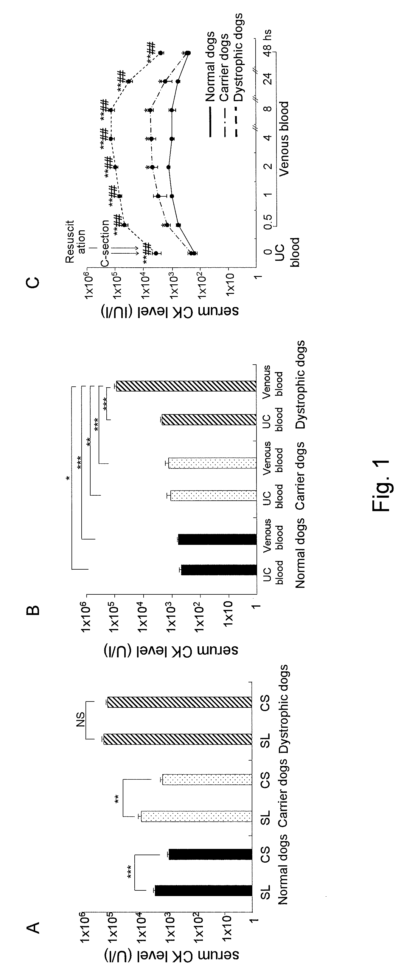
[0032] FIG. 1 shows charts showing postnatal creatinine kinase (CK) levels in normal dogs, carrier dogs, and dystrophic dogs. (A) shows postnatal CK levels in newborn dogs delivered via spontaneous labor (SL) and caesarean section (CS); (B) shows serum CK levels in the umbilical cord (UC) blood and in the venous blood of newborn dogs after the initiation of breathing; and (C) shows changes in serum CK levels of newborn dogs over time (from the serum CK levels prior to the initiation of breathing to those 48 hours after birth).
[0033] FIG. 2 shows photographs showing histopathological changes of the diaphragm of newborn dystrophic dogs before and after the initiation of breathing.
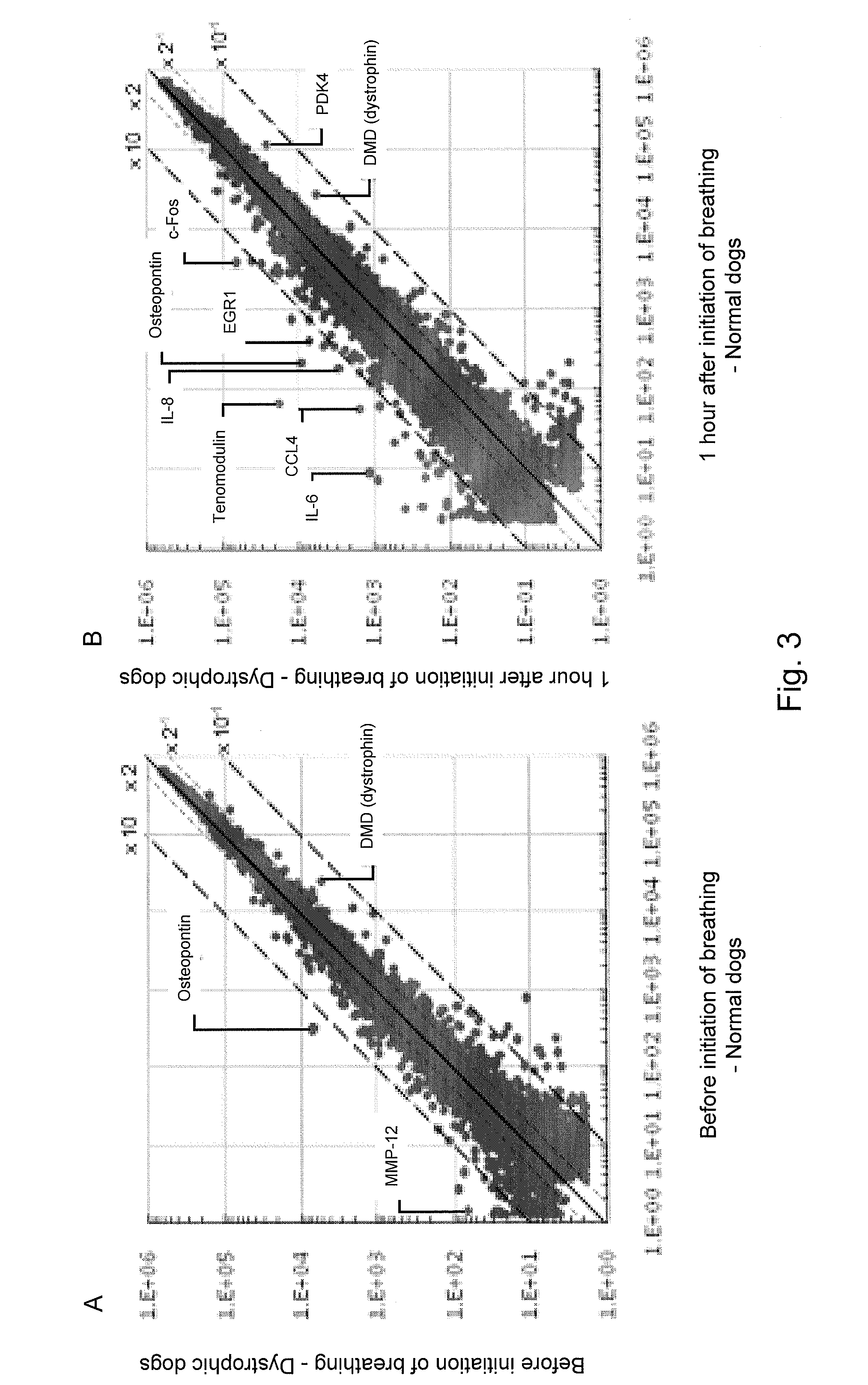
[0034] FIG. 3 shows charts showing the results of gene expression changes in the diaphragms of normal dogs and of dystrophic dogs before the initiation of breathing (A) and after the initiation of breathing (B) using microarrays.
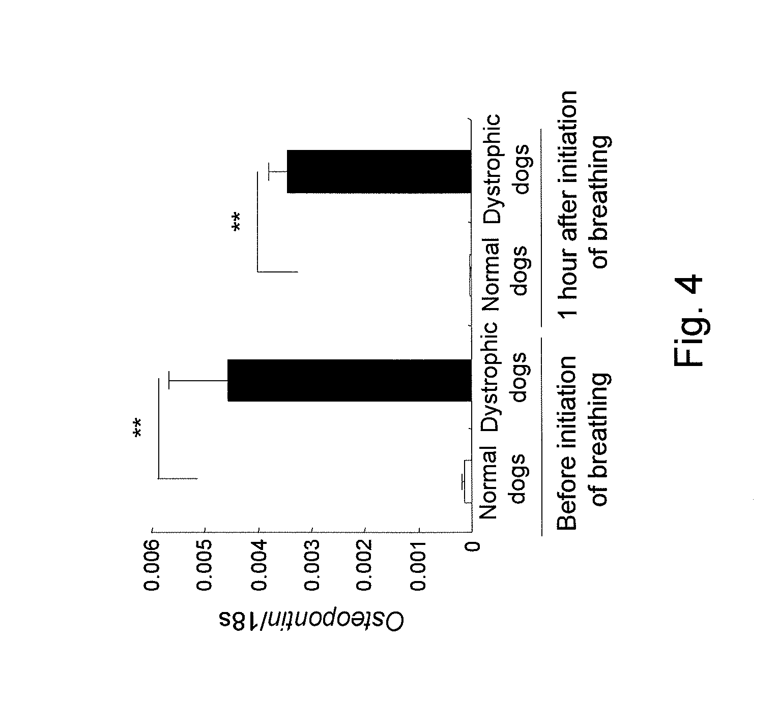
[0035] FIG. 4 shows a chart showing the results of changes in osteopontin expression in the diaphragms of normal dogs and of dystrophic dogs before and after the initiation of breathing by quantitative PCR.
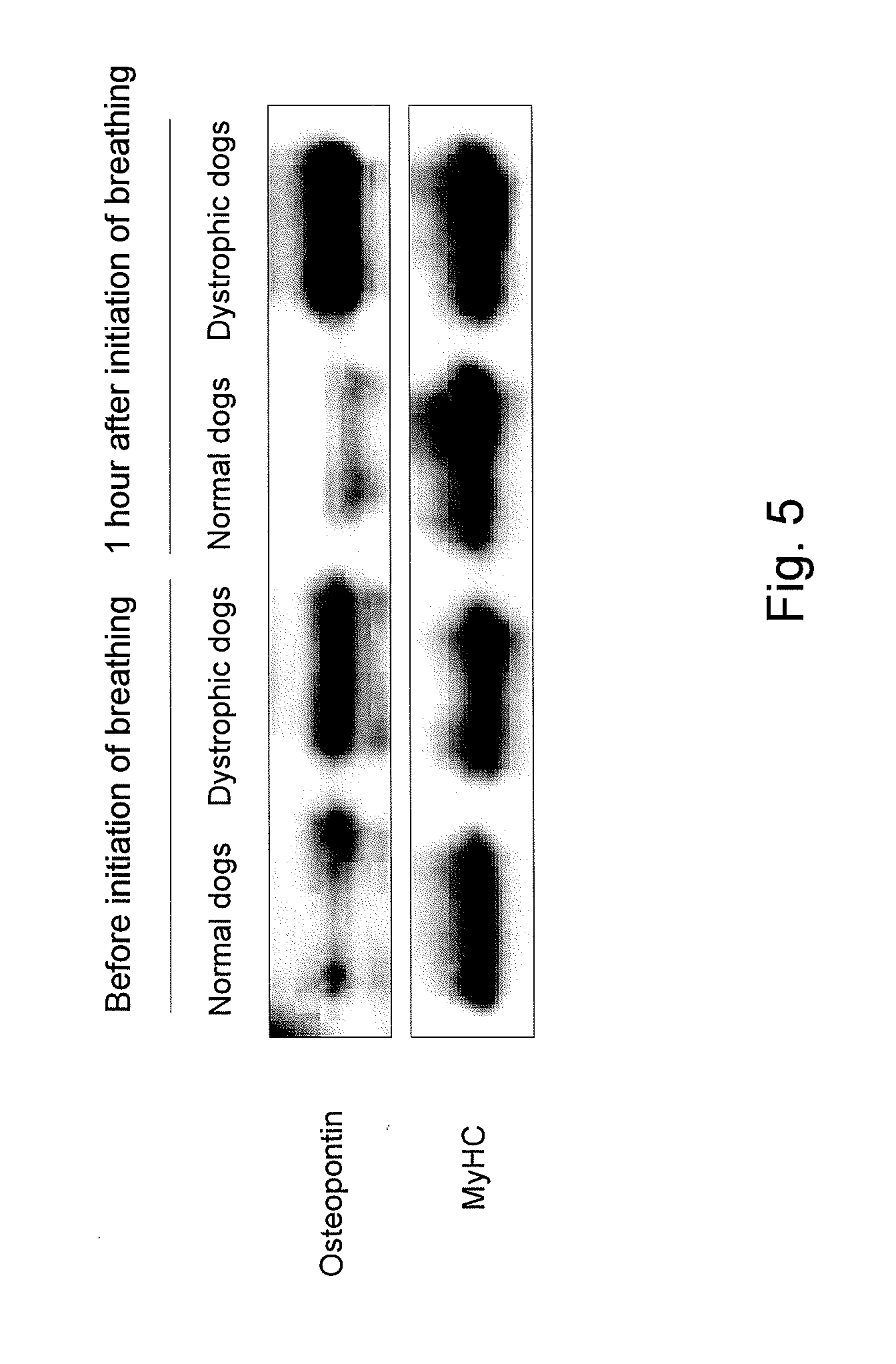
[0036] FIG. 5 shows photographs showing the results of changes in osteopontin expression in the diaphragms of normal dogs and of dystrophic dogs before and after the initiation of breathing by Western blotting.
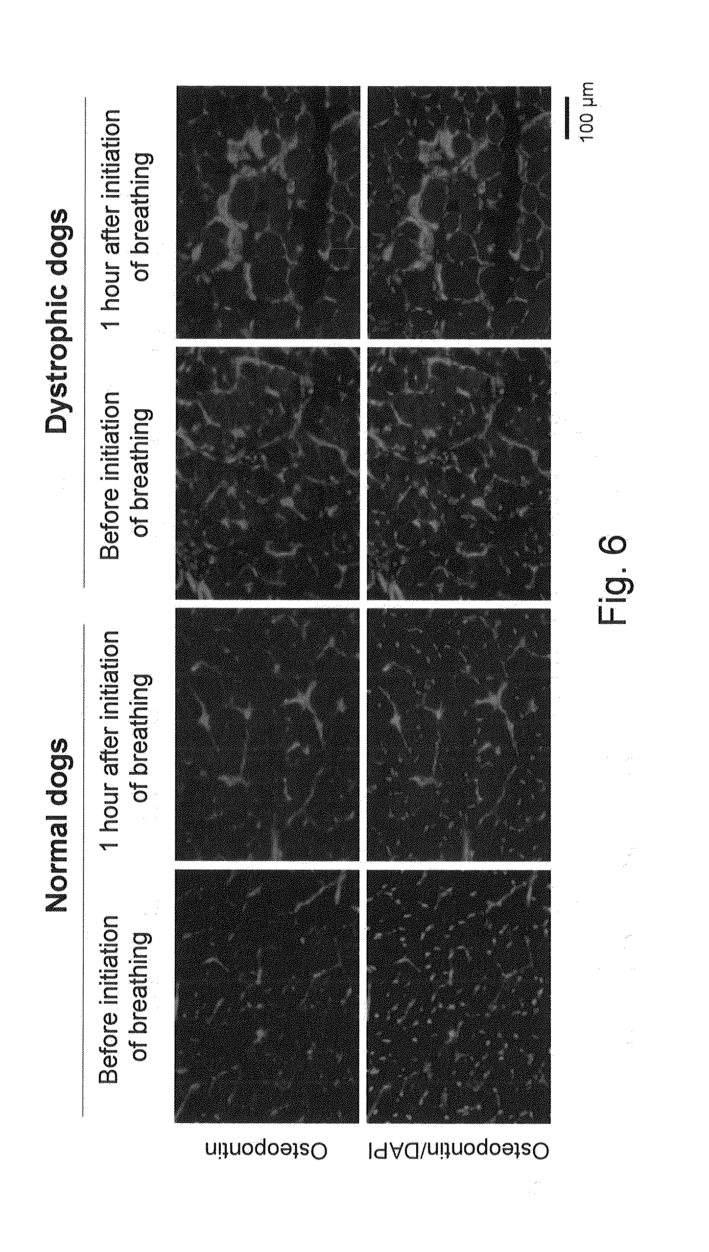
[0037] FIG. 6 shows photographs showing the results of changes in osteopontin expression in the diaphragms of normal dogs and of dystrophic dogs before and after the initiation of breathing by immunohistochemistry.
[0038] FIG. 7 shows a chart showing the results of changes in gene expression in the diaphragms of dystrophic dogs before and after the initiation of breathing using microarrays.
[0039] FIG. 8 shows charts showing the results of changes in expression of c-Fos, EGR1, IL-6, and IL-8 in the diaphragms of normal dogs and of dystrophic dogs before and after the initiation of breathing by quantitative PCR.
[0040] FIG. 9 shows photographs showing the results of changes in expression of c-Fos, EGR1, IL-6, and IL-8 in the diaphragms of normal dogs and of dystrophic dogs before and after the initiation of breathing by Western blotting.
[0041] FIG. 10 shows photographs showing the results of changes in expression of c-Fos, EGR1, IL-6, and IL-8 in the diaphragms of normal dogs and of dystrophic dogs before and after the initiation of breathing by immunohistochemistry.
DETAILED DESCRIPTION OF THE INVENTION
[0042] The present invention provides novel markers for diagnosis of muscular dystrophy. The expression level of the markers according to the present invention is low in a normal state and high in the case of muscular dystrophy. Accordingly, such markers are useful for diagnosis intended to determine whether or not a patient carries muscular dystrophy, prediction of the development of muscular dystrophy, screening for a therapeutic agent or technique for muscular dystrophy, evaluation of the efficacy of a therapeutic agent or technique for muscular dystrophy, and other purposes.
[0043] The present inventors considered that elucidation of pathological mechanisms in newborn dystrophic dogs would lead to elucidation of the mechanisms of myodegeneration. The serum CK levels are high at the time of birth of healthy newborns, although such CK levels are not as high as those in the case of DMD (Rudolph, N., et al., Pediatrics, 1966, 38: 1039-1046; Gilboa, N., et al., Arch Dis Child, 1976, 51: 283-285; Zellweger, H., et al., Pediatrics 1975, 55: 30-34). Since the serum CK levels are lowered by caesarean section, pressure applied to a fetus at the time of delivery from the birth canal, external injuries, or hypoxia is considered to be associated with elevated blood CK levels (Rudolph, 1966; Gilboa, 1976; Drummond L. M. et al., Arch Dis Child 1979; 54: 362-366). Thus, the present inventors examined whether or not stress applied at the time of delivery or the initiation of breathing immediately after birth would be associated with the elevated blood CK levels of newborn dystrophic dogs (see, Example 1).
[0044] Subsequently, we examined the genes or molecules with expression levels that would increase in the diaphragms of newborn dystrophic dogs before and after the initiation of breathing. As a result, we found that the expression level of osteopontin increased in dystrophic dogs before the initiation of breathing, compared with the case of normal dogs. While the expression level of osteopontin increased specifically in the diaphragms of newborn dystrophic dogs before the initiation of breathing, it is considered to be activated by intracellular Ca.sup.2+ increased though the stretch-activated channel, and osteopontin had been activated at a stage prior to myonecrosis, in addition to a regeneration or fibrosis stage. Thus, it is considered to be a molecule associated with the nature of pathological conditions of muscular dystrophy, and it is associated with induction of inflammatory cells (see, Example 2).
[0045] In contrast, the expression levels of c-fos and egr-1, which are immediate early genes referred to as "third messengers," were elevated in dystrophic dogs after the initiation of breathing, compared with the case of dystrophic dogs before the initiation of breathing. Also, the expression levels of the interleukin-6 (IL-6) and interleukin-8 (IL-8) genes located downstream of the above genes were elevated. While these molecules were considered to be activated by significantly increased intracellular Ca.sup.2+ inflow from mechanically damaged sites of the stretch-activated channel and the muscle cell membrane, IL-6 and IL-8 are also referred to as "myokines," which are cytokines and chemokines expressed in a muscle cell endogenously and considered to be associated with induction of inflammatory cells, such as neutrophils, occurring at an early stage of muscle damage (see, Example 2).
[0046] The present inventors proposed a two-phase hypothesis based on identification of causes for the elevated blood CK levels in newborn dystrophic dogs. That is, the expression level of osteopontin is elevated in the diaphragm before the application of mechanical stress upon initiation of breathing, sarcolemma collapse, and calcium influx in the diaphragm after the application of mechanical stress upon initiation of breathing. Subsequently, the immediate early genes are expressed to increase the expression levels of cytokines and chemokines of IL-6 or IL-8, which are molecules located downstream, and inflammatory cells, such as neutrophils, are induced. The genes and the molecules identified in the research provide a novel perspective on the pathological mechanisms of muscular dystrophy, and such genes and molecules serve as novel molecular markers for evaluation of disease progression or therapeutic effects.
[0047] According to the present invention, c-Fos, EGR1, IL-6, and IL-8 proteins with expression levels that are low in a normal state but high in the case of muscular dystrophy, as described above, and genes encoding the proteins are used as markers. Such genes and proteins are referred to as "the markers of the present invention" herein. The markers of the present invention are also referred to as "marker genes" or "marker proteins" herein.
[0048] The marker proteins and the marker genes of the present invention are known in the art, and the amino acid sequences and the nucleotide sequences thereof are also known. However, there has been no report regarding any correlation between the markers of the present invention and muscular dystrophy. The names, accession numbers, nucleotide sequences, and amino acid sequences of the markers of the present invention are summarized in Table 1.
TABLE-US-00001 TABLE 1 Nucleotide Amino acid Nucleotide Amino acid Marker Accession No. sequence sequence Accession No. sequence sequence names (human) (human) (human) (canine) (canine) (canine) c-Fos K00650 1 2 XM_547914 3 4 EGR1 NM_001964 5 6 XM_846145 7 8 IL-6 NM_000600 9 10 NM_001003301 11 12 IL-8 NM_000584 13 14 NM_001003200 15 16
[0049] According to the present invention, the markers listed in Table 1 can be used individually. Specifically, c-Fos can be used as a marker in an embodiment. In another embodiment, EGR1 can be used as a marker. In a further embodiment, IL-6 can be used as a marker. In a further embodiment, IL-8 can be used as a marker. Markers listed in Table 1 can be used in combination, according to need. Alternatively, a marker listed in Table 1 may be used in combination with another marker of muscular dystrophy known in the art. Any number of markers can be used in any combination, provided that at least one marker listed in Table 1 is included. Examples of combinations that can be employed include c-Fos and ERG-1; c-Fos and IL-6; c-Fos and IL-8; ERG-1 and IL-6; ERG-1 and IL-8; IL-6 and IL-8; c-Fos, ERG-1, and IL-6; c-Fos, ERG-1, and IL-8; c-Fos, IL-6, and IL-8; and ERG-1, IL-6, and IL-8. An example of another marker includes serum creatinine kinase (CK) level. Use of markers in combination enables more accurate diagnosis of muscular dystrophy.
[0050] As described above, the expression levels of such markers are high in the case of muscular dystrophy. According to the present invention, expression of the above marker or the combination of markers in a sample obtained from a subject is determined. When expression of two or more markers is determined, steps of determining the expression of each marker may be carried out simultaneously or sequentially. According to the present invention, "expression of a marker" may be expression of a marker protein, a derivative or precursor thereof, or a gene encoding the protein (mRNA). The term "derivative" or "precursor" refers to a substance derived from a marker protein or a substance from which a marker protein originates. Examples thereof include, but are not limited to, a protein containing a signal peptide, a specific subunit molecule of a protein, a modified protein, and a protein fragment.
[0051] Accordingly, the diagnostic agent for muscular dystrophy of the present invention (hereafter, it may be referred to as "the present diagnostic agent") comprises a means for determining expression of at least one marker selected from the group consisting of c-Fos, EGR1, IL-6, and IL-8 in a sample obtained from a subject.
[0052] Any sample can be used, provided that such sample is obtained from a subject to be diagnosed for muscular dystrophy. An adequate sample may be selected depending on a method or means for determining the expression of a marker. Examples include, but not limited to, biological fluid samples (e.g., blood, blood serum, blood plasma, urine, spinal fluid, or ascites) and tissue or cell samples (e.g., muscle tissue or cells, such as diaphragmatic tissue or cells). From the viewpoint of ease of sampling, use of a muscle cell, blood, blood serum, or blood plasma as a sample may be preferable. When plasma sample is used, use of EDTA as an anticoagulant is preferable, and substances known or general in the art, such as heparin or sodium citrate, can be used. In the case of a blood sample, it is preferable that the blood sample be ice-cooled or refrigerated after blood sampling. A tissue or cell sample may be preferably frozen and cryopreserved via a technique known or general in the art, such as with the use of liquid nitrogen or dry ice, immediately after sampling. Muscle cells can be sampled by a method known in the art. Specifically, for example, the skeletal muscle is sliced and transferred to a 50-ml conical bottom tube. Thereafter, 4 ml of a solution of Dispase II (2.4 IU/ml) and Collagenase XI (0.2%) is added per g of muscle, incubated at 37.degree. C. for 45 to 60 minutes, and subjected to pipetting every 15 minutes. Thereafter, tissue slices are pierced with an 18G injection needle several times and grounded, and the supernatant is recovered. The supernatant is applied to a 80-.mu.m filter to remove cell masses, a cell suspension is transferred to a 50-ml conical bottom tube, a growth medium is added to the tube after the treatment with Dispase II (2.4 IU/ml) and Collagenase XI (0.2%), and the supernatant is applied to a filter and recovered in the same manner as described above. A growth medium is added to bring the total amount to 30 ml, and pipetting is carried out several times, followed by centrifugation at 1,000 rpm, 4.degree. C. for 5 minutes. The supernatant is discarded, and 20 ml of growth medium is added to the precipitated cells to resuspend the cells, followed by centrifugation at 1,000 rpm, 4.degree. C. for 5 minutes. The precipitated cells are suspended in 25 ml of growth medium, the suspension is transferred to an uncoated 15-cm culture dish, bFGF is added thereto, culture is conducted at 37.degree. C. in 5% CO.sub.2 for 90 minutes, and the supernatant containing nonadherent cells is recovered. The bottom of the culture dish is washed with the use of the supernatant, the culture dish is turned 180 degrees, culture is conducted again at 37.degree. C. in 5% CO.sub.2 for 90 minutes, and the supernatant is recovered. The recovered supernatant is transferred to a 15-cm collagen-coated culture dish, bFGF is added thereto, and culture is conducted at 37.degree. C. in 5% CO.sub.2 overnight. The culture product is subjected to subculture on the following day if myoblasts reach at least 30% to 40% confluency. If the confluence level is below the aforementioned level, medium exchange is carried out. Thereafter, subculture or medium exchange is carried out every day in order to prevent muscle differentiation.
[0053] Subjects may be humans or other mammals, such as primates (e.g., monkeys and chimpanzees), livestock animals (e.g., cattle, horses, pigs, and sheep), pet animals (e.g., dogs and cats), or experimental animals (e.g., mice, rats, and rabbits). Further, subjects may also be reptiles and birds.
[0054] Determination of the expression of a marker preferably involves semi-quantitative or quantitative determination of the amounts or concentrations of markers in a sample. Such an amount may be an absolute amount or relative amount. Determination can be carried out directly or indirectly. Direct determination involves determination of the amount or concentration of the marker proteins or genes (mRNAs) existing in a sample based on signals that are directly correlated with the numbers of molecules of such marker proteins or genes. Such signals are based on given physical or chemical characteristics of a protein or gene, for example. Indirect determination involves determination of signals derived from secondary components (i.e., components other than marker proteins or mRNA), such as ligands of antibodies or aptamers, labels, or enzyme reaction products. Determination means used in accordance with the present invention vary depending on methods of determining expression of a marker employed.
[0055] In one embodiment of the present invention, marker expression can be determined by a means for determining the marker protein level in a sample. Such means are known in the art, and examples thereof include techniques and reagents for immunoassays. Also, expression of a marker protein can be determined by a means for determining physical or chemical characteristics specific to a marker protein, such as a means for accurately assaying the molecular level or NMR spectra. Examples of means for determining the expression of a marker protein include analyzers, such as biosensors, protein chips, optical devices coupled to immunoassays, mass spectrometers, NMR spectrometers, two-dimensional electrophoresis apparatuses, and chromatography apparatuses.
[0056] For example, expression of a marker protein in a sample can be determined by immunoassays (immunological assay techniques). Specifically, expression of a marker protein in the sample can be determined based on the reaction between such protein and an antibody that specifically binds thereto. Immunoassays may be carried out in a liquid phase or solid phase, provided that the technique used is conventional in the art. From the viewpoint of ease of detection, use of a solid phase may be preferable. In addition, immunoassay techniques are not limited, and immunoassay can be carried out by sandwich assay, competitive assay, Western blotting, or enzyme linked immunosorbent assay (ELISA), as well as a direct solid-phase assay.
[0057] When immunoassay techniques are adopted, the present diagnostic agent comprises an antibody against a marker protein. An antibody against a marker protein may be a monoclonal or polyclonal antibody. Alternatively, it may be, for example, an Fab or Fv fragment capable of binding to an epitope of a marker protein. When a primary antibody and a secondary antibody are used, both thereof may be monoclonal antibodies. Alternatively, either the primary or secondary antibody may be a polyclonal antibody. An antibody can be prepared by a method known in the art or a commercially available antibody may be used.
[0058] Binding (reaction) between a marker protein and an antibody can be assayed in accordance with a method well-known in the art. A person skilled in the art can determine an effective and optimal assay technique in accordance with the type, format, type of labels to be used, and other conditions of the immunoassay to be adopted. In order to easily detect binding between a marker protein in a sample and an antibody, for example, the binding can be directly detected by labeling the antibody or indirectly detected with the use of, for example, a labeled secondary antibody or a biotin-avidin complex.
[0059] When a solid-phase immunoassay is selected, for example, a protein component in a sample can be immobilized to a solid phase. An example of a method that can be adopted is a method comprising: (1) preparing a protein component from a sample; (2) fractionation via SDS-polyacrylamide gel electrophoresis; (3) transferring a protein on a gel to a solid phase; (4) reacting the solid phase with an antibody against the marker protein (a primary antibody); (5) washing the solid phase; (6) contacting a labeled antibody against the primary antibody (a secondary antibody) with the solid phase; (7) washing the solid phase; and (8) assaying the expression level of the protein based on the label. Alternatively, an antibody may be immobilized onto a solid phase. This method is referred to as a so-called "sandwich assay" method, which is extensively employed for "ELISA" when an enzyme is used as a marker. Such solid-phase technique may be preferable for detection of trace amounts of proteins and simplification of procedures.
[0060] In a solid-phase system, an antibody or a protein component in a sample may be immobilized onto a solid phase (e.g., a plate, membrane, or bead), and immunological binding between a marker protein and an antibody may be tested on the solid phase. Any solid phase that is conventionally used in the art can be used without particular limitation. For example, a commercially available nitrocellulose membrane or PVDF membrane can be used. By immobilizing an antibody or a protein component in a sample onto a solid phase, an unbound sample component or reagent can be easily removed. In the case of protein array techniques involving the use of a membrane onto which several types of antibodies have been immobilized, in particular, expression of a plurality of marker proteins can be analyzed within a short period of time with the use of a small amount of a sample obtained from a subject (e.g., a blood plasma sample). Such immunoassays can be carried out via, for example, test strip assays that are easy to operate.
[0061] When a liquid-phase immunoassay system is selected, for example, a labeled antibody may be contacted with a sample to bind the labeled antibody to a marker protein, the resulting complex is separated, and a labeled signal is detected. Alternatively, an antibody against a marker protein (a primary antibody) may be contacted with a sample to bind the primary antibody to a marker protein, a labeled antibody (a secondary antibody) is allowed to bind to the resulting complex, and a labeled signal in the complex of such three components is then detected. In order to further potentiate signals, a nonlabeled secondary antibody is first allowed to bind to a complex of an antibody and a marker protein, and a label may then be bound to the secondary antibody. A label can be bound to a secondary antibody by, for example, biotinylating a secondary antibody and avidinylating a label.
[0062] Antibodies used in immunoassays can be labeled with enzyme, radioisotope, fluorescent dye, or avidin-biotin system. Enzymes used for conventional enzyme immunoassays (ETA), such as peroxidase, .beta.-galactosidase, or alkaline phosphatase, can be used. Enzyme inhibitors, coenzymes, or the like can also be used. Such enzymes can be bound to antibodies in accordance with a conventional technique involving the use of a crosslinking agent, such as a maleimide compound. Radioisotopes, such as .sup.125I or .sup.3H, that are used for conventional radioimmunoassay (RIA) can be used. Fluorescent dyes, such as fluorescein isothiocyanate (FITC) or tetramethylrhodamine isothiocyanate (TRITC), that are used for conventional fluorescent antibody techniques can be used.
[0063] When a biotin-avidin conjugate is used, a biotinylated antibody is allowed to react with a sample, and labeled avidin is then allowed to react with the resulting conjugate. Since avidin is capable of specifically binding to biotin, binding between an antibody and a marker protein can be determined by detecting a signal emitted from the label added to avidin. A label added to avidin is not particularly limited, and an enzyme label, such as peroxidase or alkaline phosphatase, may be preferable.
[0064] A signal of a label can also be detected in accordance with a method known in the art. When an enzyme label is used, for example, a substrate that develops color upon degradation caused by enzymatic action is added, the amount of the substrate degraded is optically assayed to determine the enzyme activity, the determined value is converted to yield the amount of bound antibody, and the obtained value is compared with the reference value. Thus, the amount of antibody is determined. Different substrates can be used in accordance with the type of enzyme to be used. When peroxidase is used as an enzyme, for example, 3,3',5,5'-tetramethylbenzidine can be used. When alkaline phosphatase is used as an enzyme, for example, paranitrophenol can be used. When a radioactive label is used, a radiation dose emitted by a radioactive label may be assayed with the use of a scintillation counter or the like. A fluorescent label can be detected and quantified with the use of, for example, a fluorescence microscope or plate reader.
[0065] In order to detect a marker protein in situ as in the case of immunohistochemical staining (e.g., immunostaining) or immune electron microscopy, an antibody against a marker protein can be used in these methods. In situ detection can be carried out by resecting histological samples (e.g., muscle tissue, muscle cell, or diaphragmatic samples) from a subject (e.g., slices of paraffin-embedded tissue) and bringing a labeled antibody into contact therewith.
[0066] According to the immunological techniques described above, the expression levels of marker proteins increase in a sample as the amounts of antibodies bound to marker proteins increase in a sample.
[0067] Alternatively, the expression of a marker protein can be determined by mass spectrometry (MS). Analysis by liquid chromatography coupled with mass spectrometry (LC/MS) is accurate and thus is particularly advantageous. Mass spectrometry analysis can be carried out by, for example, (1) preparing a protein component from a sample, (2) labeling proteins or peptides; (3) fractionating proteins or peptides, (4) subjecting proteins or peptides to mass analysis, and (5) identifying marker proteins based on the values obtained by mass spectra. Isotopic labeling reagents known in the art can be used as labels, and adequate labeling reagents are commercially available. Also, fractionation can be carried out by a method known in the art. For example, it can be carried out with the use of commercially available strong cation-exchange columns. In such a case, the present diagnostic agent comprises isotopic labeling reagents, mini columns for fractionation, and the like as means for determining the expression of a marker.
[0068] According to the present invention, the determination of marker expression can be carried out by a means for determining the amount of marker genes in a sample. Such means is known in the art. Examples thereof include primer DNA or probe DNA containing or consisting of all or part of the DNA sequence of a marker gene or a sequence complementary thereto. Such primer DNA or probe DNA specifically binds to mRNA of the marker gene expressed in a sample obtained from a subject or cDNA corresponding to such mRNA, and it is capable of detecting marker gene expression in a sample.
[0069] Primer DNA or probe DNA can be readily designed based on the nucleotide sequence of DNA of a marker gene with the use of a known program, and primer DNA or probe DNA can be prepared in accordance with a method known in the art. Specifically, primer DNA or probe DNA can be designed based on the nucleotide sequence of the marker gene, such as the nucleotide sequence as shown in SEQ ID NO: 1, 3, 5, 7, 9, 11, 13, or 15 or a sequence complementary thereto. DNA that substantially functions as a primer preferably comprises 10 or more nucleotides, more preferably 15 to 50 nucleotides, and further preferably 20 to 30 nucleotides. Also, DNA that substantially functions as a probe preferably comprises 10 or more nucleotides, more preferably 15 to 50 nucleotides, and further preferably 20 to 30 nucleotides. Primer DNA or probe DNA may comprise an additional sequence other than a region that may anneal or hybridize to a marker gene, such as a tag sequence, as is well-known in the art.
[0070] In order to determine the expression of a marker gene in a sample obtained from a subject, the above-described primer DNA and/or probe DNA may be used in amplification or hybridization, and the amplification or hybridization product may be detected. In such a case, in general, mRNA or cDNA corresponding thereto may be prepared from a sample obtained from a subject by a method well-known in the art. When RNA is to be extracted, for example, guanidine-cesium chloride ultracentrifugation, the hot-phenol method or the acid guanidium thiocyanate-phenol-chloroform (AGPC) method can be employed. cDNA can be prepared with the use of a known reverse transcriptase. The thus-obtained sample may be used to carry out the amplification and/or hybridization reactions described below.
[0071] The expression of a marker gene in a sample can be determined by carrying out an amplification using primer DNA and mRNA or cDNA as a template, and detecting specific amplification. Amplification techniques are not particularly limited. For example, known techniques based on the principle of polymerase chain reactions (PCR), such as PCR, RT-PCR, or real-time PCR, can be employed. An amplification product can be detected by a known means that is capable of specifically recognizing an amplification product. For example, whether or not a fragment of a certain size is amplified may be determined by agarose gel electrophoresis or other means to detect a specific amplification reaction.
[0072] Alternatively, a label, such as a radioisotope, fluorescent substance, or luminescent substance, may be added to dNTP that is to be incorporated during amplification, and the resulting labeled substance can be detected. Examples of radioisotopes that can be used include .sup.32P, .sup.125I, and .sup.35S. Fluorescent substances, such as fluorescein isothiocyanate (FITC), sulforhodamine (SR), and tetramethylrhodamine isothiocyanate (TRITC), can be used. Luciferin or the like can be used as a luminescent substance. Types of label, methods of introducing a label, and others are not particularly limited, and various conventional means can be employed. An example of a method of introducing a label is a random priming method involving the use of a radioisotope.
[0073] An amplification product into which labeled dNTP has been incorporated may be observed by any technique for detecting such a label that is known in the art. When a radioisotope is used as a label, for example, radioactivity can be measured by a liquid scintillation counter or .gamma.-counter. When a fluorescent substance is used as a label, for example, fluorescence can be detected by a fluorescence microscope or fluorescence plate reader.
[0074] The expression of a marker gene can be determined by subjecting probe DNA to hybridization to a sample, and detecting specific binding (hybridization). It may be necessary to carry out hybridization under conditions where probe DNA specifically and selectively binds to mRNA or cDNA of the marker gene in a sample (i.e., stringent conditions). When hybridization is carried out, an adequate label, such as a fluorescent label (e.g., fluorescein or rhodamine), radioactive label (e.g., .sup.32P), or biotin label, can be added to probe DNA.
[0075] Detection involving the use of labeled probe DNA comprises contacting a sample or either mRNA or cDNA prepared therefrom with probe DNA, so as to allow hybridization to take place. Specifically, a sample or either mRNA or cDNA may be immobilized onto an adequate solid phase, and labeled probe DNA may be applied thereto. Alternatively, labeled probe DNA may be immobilized onto an adequate solid phase, and a sample or either mRNA or cDNA may be applied thereto. Thus, probe DNA is brought into contact with a sample or either mRNA or cDNA to carry out hybridization, unhybridized probe DNA is removed, and a label of the probe DNA that has hybridized to a sample or either mRNA or cDNA is then detected. The detection of a label indicates that mRNA of a marker gene is expressed in a sample. Examples of expression assay techniques involving the use of labeled probe DNA include Southern hybridization and Northern hybridization.
[0076] As described above, marker expression in a sample can be determined with the use of a means for determining expression of a marker, and muscular dystrophy can be diagnosed based on the results. The disease to be diagnosed with the use of the present diagnostic agent is muscular dystrophy, and, in particular, Duchenne muscular dystrophy. The term "diagnosis of muscular dystrophy" used herein indicates determination of carrier of muscular dystrophy or prediction of development of muscular dystrophy in a subject. According to the present invention, the term "diagnosis" may also encompass continuous monitoring of muscular dystrophy that has already been diagnosed and confirmation of previous diagnosis of muscular dystrophy.
[0077] Diagnosis in accordance with the present invention is not intended to always yield accurate results for all subjects to be diagnosed (i.e., 100%). The present invention is intended to diagnose subjects with statistically significant accuracy. For example, 60% or more, preferably 80% or more, or more preferably 90% or more of the subjects can be adequately diagnosed according to the present invention.
[0078] At the time of diagnosis, marker expression in a sample obtained from a subject is compared with a reference value. A reference value may be, for example, a marker expression level determined in a sample obtained from a healthy individual or a marker expression level determined in a sample obtained from a patient who has been diagnosed as being a carrier of or having developed muscular dystrophy. The reference value adopted for each subject varies depending on various biological parameters, such as a marker type, a subject type, age, and other factors. When the marker expression level in a sample obtained from a subject is greater in 10% or more, preferably 30% or more, more preferably 70% or more, and most preferably 100%, compared with that in a sample obtained from a healthy individual, specifically, a subject can be diagnosed as being likely to have muscular dystrophy.
[0079] Further, diagnosis of muscular dystrophy may be carried out in combination with other known techniques for diagnosing muscular dystrophy. Examples of known diagnostic techniques include measurement of serum creatine kinase (CK) levels, tension measurement of isolated skeletal muscle, histological measurement of the maximal diameter of muscles and frequency of centronuclear fibers, multiplex ligation-dependent probe amplification (MLPA), identification of gene mutation by polymerase chain reaction (PCR)/sequencing, immunohistochemical techniques involving the use of anti-dystrophin antibodies, and qualitative and quantitative analysis of the dystrophin protein by Western blotting.
[0080] The present diagnostic agent may comprise other components useful for diagnosis, in addition to a means for determining expression of a marker. This enables easy and simple assay of marker expression and diagnosis of muscular dystrophy.
[0081] An example of the present diagnostic agent is a set of reagents for immunoassays that at least comprises an antibody reagent for a marker protein. In addition, the set may comprise a buffer for dilution or washing, a standard antigen, a labeled antibody reagent that specifically binds to an antibody reagent, substrate reagents that develop color, luminescence or fluorescence, and instructions describing procedures and evaluation methods. An antibody included in the set may be labeled in advance, or it may not be labeled. In addition, an antibody may be immobilized onto a solid-phase support (e.g., a membrane or bead). Another example of the diagnostic agent according to the present invention is a set of reagents for mass analysis, which is composed of, for example, isotopic labeling reagents, mini columns for fractionation, buffer, and instructions. A further example of the diagnostic agent according to the present invention may comprise a means for determining expression of a marker gene in a sample (e.g., primer DNA or probe DNA).
[0082] The diagnostic agent according to the present invention may comprise instructions describing procedures and protocols for use, a table showing reference values or ranges used for diagnosis of muscular dystrophy, and other components.
[0083] Components included in the diagnostic agent according to the present invention may be provided separately or in a single container. Preferably, the diagnostic agent according to the present invention comprises all components adjusted at concentrations that allow all the necessary components to be used immediately.
[0084] In addition, the efficacy of a therapeutic agent or technique for muscular dystrophy can be evaluated, and a candidate for the therapeutic agent or technique for muscular dystrophy can be screened with the use of the marker(s) described above. Specifically, an animal that had developed muscular dystrophy, a muscular dystrophy carrier animal, or a muscle cell derived from an animal that had developed muscular dystrophy or a muscular dystrophy carrier animal (e.g., the diaphragmatic cell) may be treated with a test agent or technique, and marker expression in such animals or muscle cells may be determined. Thus, whether or not the test agent or technique affects marker expression (i.e., muscular dystrophy) can be determined.
[0085] According to the screening method of the present invention (hereafter, it may be referred to as "the present screening method") and the method for evaluating the efficacy of a therapeutic agent or technique (hereafter, it may be referred to as "the present evaluation method"), an animal that had developed muscular dystrophy or a muscular dystrophy carrier animal is first treated with a test agent or technique. Alternatively, a muscle cell derived from an animal that had developed muscular dystrophy or a muscular dystrophy carrier animal is first treated with a test agent or technique.
[0086] In the present method, a sample may be obtained from an animal that had developed muscular dystrophy or a muscular dystrophy carrier animal, and marker expression in the sample may be determined. Alternatively, marker expression in the muscle cell may be determined. Preferably, a sample may be obtained from an animal that had developed muscular dystrophy or a muscular dystrophy carrier animal prior to the treatment with a test agent or technique, and marker expression in the sample or in the muscle cell may be determined. An animal that had developed muscular dystrophy, a muscular dystrophy carrier animal, or a muscle cell derived from an animal that had developed muscular dystrophy or a muscular dystrophy carrier animal may be treated with the test agent or technique, and marker expression in the sample or cell may be determined at appropriate times. For example, marker expression may be determined immediately after treatment or 30 minutes, 1 hour, 3 hours, 5 hours, 10 hours, 15 hours, 20 hours, 24 hours (1 day), 2 to 10 days, 10 to 20 days, 20 to 30 days, and/or 1 month to 6 months after treatment. Sample collection and determination of marker expression in a sample can be carried out in the same manner as described above.
[0087] Animals to be tested may be humans who have developed muscular dystrophy or are muscular dystrophy carriers, or animal models of muscular dystrophy, and experimental animal models of muscular dystrophy (e.g., mice, dogs, and rats) are preferable. Examples of experimental animals that can be employed include mouse models of muscular dystrophy (mdx mice: Sicinski, P. et al., Science 244: 1578-1580, 1989), dog models of muscular dystrophy (c-xmd dogs: Komegay, J. N. et al., Muscle Nerve 11, 1056-1064, 1988; CXMD.sub.J dogs: Shimatsu, Y. et al., Exp. Anim. 52, 93-7. 2003, Shimatsu, Y. et al., Acta Myol. 24, 145-54, 2005), and cat models of muscular dystrophy (HFMD cats: Vas, J. H. et al., J. Comp. Pathol., 96, 335-41, 1986). In general, the efficacy of a test agent or technique may be first verified in animal models, and it is then evaluated via, for example, clinical trials in humans.
[0088] Test agents or techniques subjected to the screening method and the evaluation method of the present invention are not particularly limited. For example, the test agent or technique include: any physical factors, and specifically, naturally occurring molecules, such as amino acids, peptides, oligopeptides, polypeptides, proteins, nucleic acids, lipids, carbohydrates (e.g., sugar), steroids, glycopeptides, glycoproteins, and proteoglycans; synthetic analogues or derivatives of naturally occurring molecules, such as peptide mimics and nucleic acid molecules (e.g., aptamers, anti-sense nucleic acids, and double-stranded RNA (RNAi)); non-naturally occurring molecules, such as low-molecular-weight organic compounds prepared with the use of combinatorial chemistry techniques (e.g., a library of inorganic and organic compounds or a combinatorial library); and a mixture of any thereof. Moreover, the test agent or technique may involve the use of a single substance, a complex constituted by a plurality of substances, a transcription factor, or the like. Furthermore, the test agent or technique may involve the use of, for example, radiation or ultraviolet light, in addition to the physical factors described above.
[0089] Moreover, a single test agent or technique may be independently examined, or a combination of several candidate agents or techniques (including in the form of libraries or the like) may be examined. Examples of libraries containing a plurality of test agents or techniques to be tested include a library of synthetic compounds (e.g., a combinatorial library) and a peptide library (e.g., a combinatorial library).
[0090] When an animal is treated with a test agent or technique, conditions for treatment such as the dose for treatment, treatment period, and route of treatment vary depending on the type of test agent or technique. A person skilled in the art can easily determine such conditions. When the test agent is administered to an animal, for example, administration routes such as intramuscular injection, oral administration, intravenous injection, intraperitoneal injection, transdermal injection, or subcutaneous injection may be appropriately selected in accordance with the type of test agent, the type of animal to be used, and other conditions.
[0091] Muscle cells may be collected according to methods known in the art, such as the method described above. Alternatively, muscle cells that are commercially available or available to the public can be used. When cells are exposed to the test agent or technique, the conditions for exposure vary depending on the type of test agent or technique; however, a person skilled in the art can easily determine such conditions. For example, such exposure may be performed by culturing the muscle cells in a medium supplemented with the test agent, immersing the muscle cells in a solution containing the test agent, or overlaying the test agent on the muscle cells.
[0092] Furthermore, the effects and the efficacy of the test agent or technique may be examined under various conditions. Examples of such conditions include the time or period, the amount (large or small), and the frequency of the treatment with the test agent or technique. For example, a plurality of doses may be set by preparing a dilution series of test agents. The period of treatment with the test agent or technique can also be appropriately set, and the treatment may be preformed for a period of 1 day to several weeks, several months, or several years, for example.
[0093] Furthermore, when the additive action, synergistic action, or the like of a plurality of test agents and/or test techniques are examined, such plurality of test agents and/or test techniques may be used in combination.
[0094] Subsequently, marker expression in an animal model or cell may be determined. Marker expression can be determined in the manner as described above. After marker expression is determined, the determined value may be compared with a control sample, and a test agent or technique that lowers marker expression levels may be selected. Animals or cells that are not treated with the test agent or technique can be used as controls.
[0095] According to the present invention, muscle cells may be treated with the test agent or technique for the primary screening, the test agent or technique that exhibits the lower marker expression level in the muscle cells may be selected. Subsequently, secondary screening may be carried out by treating animals with the selected test agent or technique and determining marker expression in such animals to select the test agent or technique that exhibits the lower expression level.
[0096] When screening for a therapeutic agent or technique, further, a selected test agent may be administered to animal models of muscular dystrophy, or the animals may be subjected to the a selected test technique to determine whether or not the test agent or technique would affect the development, progression, or symptoms of muscular dystrophy in animal models. The results of such determination vary depending on the type of animal model, symptoms to be diagnosed, various factors, or other conditions. However, a person skilled in the art would be able to adequately determine influences imposed on muscular dystrophy. When influences imposed on muscle diseases or myopathy are to be assayed, for example, measurement of muscle strength, measurement of serum creatine kinase levels, tension measurement of isolated skeletal muscles, and histological measurement of the maximal diameter of muscles and frequency of centronuclear fibers can be carried out.
[0097] When amelioration of muscular dystrophy is observed (e.g., amelioration of symptoms or delay of disease development or progression) as described above, the test agent or technique can be selected as a candidate for therapeutics for treatment or prevention of muscular dystrophy.
[0098] Thus, the present screening method and the present evaluation method enable identification of the therapeutic agent or technique used for treatment or prevention of muscular dystrophy and verification of efficacy of such therapeutic agent or technique.
[0099] Since the expression level of c-Fos, EGR1, IL-6, or IL-8 is elevated at the time of disease development (after birth), expression or activity of at least one member selected from the group consisting of c-Fos, EGR1, IL-6, and IL-8 may be inhibited to treat or prevent muscular dystrophy. Accordingly, the present invention also relates to an agent for treating or preventing muscular dystrophy comprising a means for inhibiting or suppressing expression or activity of at least one member selected from the group consisting of c-Fos, EGR1, IL-6, and IL-8. Examples of such means include means for inhibiting or suppressing gene expression, such as methods involving the use of anti-sense nucleic acids or RNAi (e.g., micro RNA or siRNA), and means for inhibiting or suppressing protein expression, such as antibodies and nucleic acid aptamers.
EXAMPLES
[0100] Hereafter, the present invention is described in greater detail with reference to the following examples and the drawings, although the present invention is not limited to the examples below.
Example 1
Changes in Serum Creatinine Kinase (CK) Level
[0101] It has been reported that stress during labor may result in the development of myopathy in newborns, and that such stress may be reduced by caesarean section. Based on these reports, elective caesarean section was adopted to examine differences in serum CK levels after spontaneous labor and after caesarean section in normal dogs, carrier dogs, and dystrophic dogs. The term "carrier dogs" refers to female dogs having a mutation in the dystrophin gene. Specifically, human and dog chromosomes consist of 23 pairs of 46 chromosomes in total. Among these pairs, 22 pairs are autosomal chromosomes, and there is 1 pair of two sex chromosomes that determine the gender. While male dogs (males) have X- and Y-chromosomes, female dogs (females) have two X-chromosomes. The dystrophin gene, which is a causal gene for muscular dystrophy, is present on the X-chromosome and it exhibits X-linked inheritance. Thus, a male individual having a mutation in the X-chromosome develops muscular dystrophy. A female individual having a mutation in the dystrophin gene on one of the two X-chromosomes is referred to as a "carrier dog (carrier)." A dystrophic dog results from crossing such a carrier dog with a normal male dog. In general, carrier dogs exhibit no symptoms, but carrier dogs sometimes exhibit intermediate symptoms between dystrophic dogs and normal dogs. Such a carrier dog is referred to as a "symptomatic carrier dog."
[0102] As test dogs, there were 71 normal dogs, 37 carrier dogs, and 34 dystrophic dogs obtained via spontaneous labor (39 times) and 39 normal dogs, 26 carrier dogs, and 41 dystrophic dogs obtained via elective caesarean section (28 times) from December, 2001, to April, 2008, at the dystrophic dog breeding colony of the Mid-sized Animal Research Facility, the National Institute of Neuroscience, the National Center of Neurology and Psychiatry (NCNP). Elective caesarean section was carried out on a date predicted based on the LH Surge (Witness.RTM. LH, Synbiotics, Kansas City, Mo., U.S.A.) or when the body temperature of a pregnant carrier dog rapidly dropped (Kobayashi, M., et al., Muscle Nerve, 2009, 40: 815-826). Caesarean section of a pregnant carrier dog was carried out with the use of isoflurane (2.0% to 3.0%) from induction to maintenance of anesthesia. Newborn dogs were subjected to resuscitation by a veterinarian or a licensed animal handling technician who has experience in resuscitation under the supervision of a veterinarian. A respiratory stimulant (i.e., doxapram) was used for each dog after resuscitation, and the dog was laid on a dry, warm towel in a box supplemented with oxygen.
[0103] In order to detect changes in serum creatinine kinase (CK) levels over time (the umbilical cord blood and 30 minutes, 1 hour, 2 hours, 4 hours, 8 hours, 24 hours, and 48 hours after resuscitation), 5 normal dogs, 3 carrier dogs, and 6 dystrophic dogs obtained via elective caesarean section (3 times) were used. Four dogs from each group obtained via caesarean section were subjected to pathological and molecular biological analyses. This research was approved by the Committee on Mid-sized Animal Ethics, the National Institute of Neuroscience, the National Center of Neurology and Psychiatry (Approval numbers: 13-03, 14-03, 15-03, 16-03, 17-03, 18-03, 19-04, and 20-04) and conducted in accordance with the animal experiment guidelines thereof.
[0104] The serum creatinine kinase (CK) levels were determined by subjecting the umbilical cord blood or venous blood to centrifugation at room temperature, 1,800 g for 10 minutes to separate the blood serum, followed by colorimetry (FDC3500, FujiFilm, Tokyo, Japan).
[0105] After euthanasia, the diaphragm was freeze-fixed, cut into 7 .mu.m slices, and then subjected to hematoxylin-eosin (H & E) staining and calcium staining (i.e., alizarin red staining) (pH 4.1).
[0106] Data for 2 groups were compared by the student-t test. The chi-square test was carried out to determine the mortality rate. Data were expressed as average.+-.standard deviation and considered to be significantly different by p<0.05.
[0107] As a result, the serum CK levels were found to have decreased significantly in normal dogs and carrier dogs after caesarean section, although no decrease was observed in dystrophic dogs (FIG. 1A). The above results suggest that stress during labor is not a primary cause for the elevated blood CK levels of newborn dystrophic dogs. Since newborns undergo the important breathing initiation process immediately after birth, whether or not the cause of the elevated blood CK levels is associated with the initiation of pulmonary breathing was examined by comparing the serum CK levels in the umbilical cord blood of newborns obtained via caesarean section (reflecting conditions before the initiation of breathing) and in the jugular venous blood 1 hour after the initiation of breathing. No differences between the serum CK levels and the CK levels in the umbilical cord blood of normal dogs and carrier dogs after the initiation of breathing were observed.
[0108] While the CK levels in the umbilical cord blood of dystrophic dogs were approximately 5 times higher than those of normal dogs, the CK levels in venous blood of dystrophic dogs after the initiation of breathing were approximately 35 times higher than those in the umbilical cord blood of dystrophic dogs and approximately 150 times higher than those in the venous blood of normal dogs after the initiation of breathing (FIG. 1B). In all dog groups, the serum CK levels elevated rapidly up to 30 minutes after the initiation of breathing, reached to a peak 4 to 8 hours after the initiation of breathing, and returned to levels equivalent to those in the umbilical cord blood 48 hours later. The serum CK levels remained high in dystrophic dogs (FIG. 1C).
[0109] According to a pathological test of the diaphragm before the initiation of breathing, calcium-positive, opaque fibers were occasionally observed (indicated by arrows), and the fundamental muscular structure was maintained. However, after the initiation of breathing, many opaque calcium-positive fibers were observed (indicated by arrows), the interstitium was increased, hyaline degeneration was observed, and substantially no invasion of inflammatory cells (e.g., neutrophils) was observed (FIG. 2).
[0110] Based on the above results, rapid mechanical stress imposed by the initiation of breathing was considered to have caused significant myopathy, including the tearing of muscular fibers in the diaphragm. In general, respiratory disorders are not observed at birth in case of DMD; however, it is reported that respiratory disorders frequently occur in premature newborns (Phadek, A, et al., Anesth Analg., 2007, 105: 977-980). Since the muscular development of dogs, including dystrophic dogs, takes place at a significantly slower rate than that of other animals or humans (Lanfossi, M., et al., Acta Neuropathol., 1999, 97: 127-138), the different respiratory disorder development timing between dystrophic dog and DMD is considered to be associated with differences in muscular maturation at birth. Based on the observation of serum CK levels over time, the serum CK levels for all dogs returned to the levels in the umbilical cord blood 48 hours after birth, although dystrophic dogs maintained high serum CK levels. This suggests that influence of labor observed in normal dogs and carrier dogs is eliminated at least 2 days after birth and that screening of newborns with the use of serum CK levels at this point may be feasible. Early diagnosis of DMD is considered to be important for future family plans or for the examination of therapeutic methods that may be developed in the future at an early stage. Accordingly, determination of the serum CK levels at various time point during the neonatal period may achieve a novel finding regarding the timing for screening of newborns with DMD.
Example 2
Changes in Gene Expression in the Diaphragm
[0111] In this Example, genes with expression levels that increased in the diaphragms of dystrophic dogs after the initiation of breathing compared to that before the initiation of breathing were examined with the use of cDNA microarrays. Specifically, neonatal normal dogs and dystrophic dogs (4 individuals each) were delivered via caesarean section before the initiation of breathing and subjected to autopsy before resuscitation. In addition, neonatal normal dogs and dystrophic dogs (4 individuals each) that had already initiated breathing were subjected to autopsy 1 hour after resuscitation. Total RNA was extracted from the sampled and cryopreserved diaphragm with the use of the RNeasy Mini Kit (Qiagen, Hilden, Germany). RNA concentration was measured using NanoDrop ND-1000 UV-spectrophotometer (NanoDrop Technologies, Wilmington, Del., U.S.A.). RNA quality was examined by Agilent Bioanalyzer 2100 (Agilent Technologies, Santa Cruz, Calif., U.S.A.). Total RNA (500 ng) was applied to the dog whole genome oligo microarray 44K (Agilent Technologies), and hybridization was carried out at Bio Matrix Research, Inc. (Nagareyama, Chiba). A fluorescent image was obtained with the use of the Agilent Technologies microarray scanner (Agilent Technologies). Normalization was carried out by comparing genes on different chips with the use of GeneSpring 10.0 (Torry Digital Biology, Denver, Colo., U.S.A.). Genes exhibiting different expression levels before the initiation of breathing and after the initiation of breathing of normal dogs and dystrophic dogs were subjected to ANOVA test and then selected via the multiple-group test by the Benjamini and Hochberg method.
[0112] Based on the results of microarray analysis, genes exhibiting expression levels that were elevated by 10 times or greater compared with other groups were tested via quantitative PCR (real-time PCR). The total RNA of each dog was the same as that used for cDNA microarray analysis. Amplification primers for the 18s RNA (the internal control), osteopontin, c-fos, egr-1, IL6, and IL8 genes were designed (Table 2).
TABLE-US-00002 TABLE 2 Primer sequences for real-time RT-PCR Genes forward (5' > 3') reverse (5' > 3') 18sRNA GGAAAGTACAGCCAGGTCC ACACGAAGTCCCCAAAAGTG (SEQ ID NO: 17) (SEQ ID NO: 18) Osteopontin ACGATGTGGATAGCCAGGAC GGACGGCATTGAAGTCATCT (SEQ ID NO: 19) (SEQ ID NO: 20) c-fos ACTCCAGGGCTGGCGTTGTG AGTCAGCTCCCTCCTGCGGT (SEQ ID NO: 21) (SEQ ID NO: 22) EGR1 GACAACCACCTTTTCTCCCA GGCAGTAGGAACTGCAGAGG (SEQ ID NO: 23) (SEQ ID NO: 24) IL-6 GCTACTGCTTTCCCTACCCC TTTTCTGCCAGTGCCTCTTT (SEQ ID NO: 25) (SEQ ID NO: 26) IL-8 AGAGTGATTGACAGTGGCCC ACACCAGGTCTACACGGGAC (SEQ ID NO: 27) (SEQ ID NO: 28)
[0113] Real-time PCR was carried out using the SYBR mixed Ex Taq II kit (Takara) by repeating a PCR cycle of 95.degree. C. for 20 seconds and 60.degree. C. for 1 minute 40 times in the BioRad iCycler system (BioRad). Gene expression levels were determined relative to 18s RNA ((Ct/18s RNA--Ct/target genes)), and the expression levels of groups of normal dogs and dystrophic dogs were compared before and after the initiation of breathing.
[0114] In addition, proteins corresponding to the genes were subjected to Western blot analysis and protein assay via an immunohistochemistry. Western blot analysis was carried out as described below. The freeze-fixed diaphragm was homogenized in a sample buffer (62.5 mM Tris-HCl, pH 6.8, 2% SDS, 10% glycerol), and centrifuged at 15,000 g for 10 minutes, and the supernatant was then recovered. After protein concentration was assayed using the DC Assay Kit (BioRad, CA, U.S.A.), proteins were subjected to thermal denaturation at 95.degree. C. for 5 minutes, and 40 .mu.g of the resultant was subjected to Western blot analysis. A PVDF transfer membrane was blocked with 0.1% Tween 20-containing Iris-buffered saline (TBST)+5% skimmed milk, and incubated with primary antibodies at 4.degree. C. for 16 hours. As primary antibodies, Osteopontin (Rb-9097, Thermo Fisher Scientific), c-Fos (#2250, Cell Signaling Technology), EGR1 (sc-189, Santa Cruz Biotechnology), IL-6 (AF1609, R&D Systems, Minneapolis, Minn., U.S.A.), and IL-8 (ab34100, Abeam) were used. After the transfer membrane was washed with a TBST solution, the membrane was incubated with secondary antibodies (mouse- or rabbit-specific HRP-labeled antibodies) and washed again with a TBST solution, and detection was carried out with the use of the ECL-Plus Western Blotting Detection System (GE HealthCare, Buckinghamshire, UK).
[0115] Immunohistochemistry was carried out by drying the sliced freeze-fixed sample for 15 minutes, washing the resultant with phosphate buffer saline (PBS) (pH 7.4) containing 5% bovine serum albumin (BSA) or heat-inactivated normal goat serum albumin, and incubating the samples with primary antibodies at 4.degree. C. for 16 hours. Primary antibodies used were CD18 (MCA1780, AbD Serotec, Oxford, UK), CD68 (M0876, Dako, Denmark), CD11b (MCA1777S, AbD Serotec), C5b-9 (ab66768, Abeam, Cambridge, UK), cleaved-caspase 3 (#9661, Cell Signaling Technology, Beverly, Mass., U.S.A.), LC3 (#4108, Cell Signaling Technology), osteopontin (Rb-9097, Thermo Fisher Scientific, Waltham, Mass., U.S.A.), c-Fos (#2250, Cell Signaling Technology), EGR1 (#4153, Cell Signaling Technology), IL-6 (sc-80108, Santa Cruz Biotechnology, Santa Cruz, Calif., U.S.A.), and IL-8 (109-401-311, Rockland Immunohistochemical, Gilbertsville, Pa., U.S.A.). The samples were washed with PBS at room temperature, incubated with FITC-labeled secondary antibodies at room temperature, washed again with PBS, and then observed under a fluorescence microscope.
[0116] Data for 2 groups were compared by the student-t test. The chi-square test was carried out to determine the mortality rate. For comparison of data obtained by real-time PCR among multiple groups, the ANOVA test and then the multiple-group test based on the Tukey's method were performed. Data were expressed as average.+-.standard deviation and considered to be significantly different by p<0.05.
[0117] As a result, the osteopontin expression level in the diaphragms of dystrophic dogs before the initiation of breathing was found to be approximately 27 times greater than that of normal dogs, and the elevated expression levels and localization of proteins in the muscle cytoplasm and in the interstitium were confirmed (FIGS. 3 to 6). Also, the osteopontin expression level was high in the diaphragms of dystrophic dogs after the initiation of breathing. Osteopontin is reported to be expressed at high levels at an early stage of regeneration of dystrophin-deficient muscle (Hirata, A, et al., Am. J. Pathol., 2003, 163: 203-205) and to be associated with the acceleration of fibrosis (Vetrone, S. A., et al., J. Clin. Invest., 2009, 119: 1583-1594). Osteopontin is activated by influx of intracellular calcium ions from the stretch-activated channel (Allen, D. G, et al., Can. J. Physiol. Pharmacol., 2010, 88: 83-91), and it functions as a cytokine that induces neutrophils or macrophages (Wang, K. X., et al., Cytokine Growth Fact, 2008, 19: 333-345). If the data by the present inventors and the existing reports are taken into consideration, it is considered that osteopontin may be expressed at a very early stage of muscular dystrophy, may be associated with induction of inflammatory cells, may be associated with fibrosis that occurs at a later stage, and may have an essential role in DMD conditions. At present, osteopontin is considered to be a target molecule for the treatment of DMD (Vetrone, S. A., 2009; and Qureshi, M. M., et al., J. Diet Suppl., 2001, 7: 159-178).
[0118] In the diaphragms of dystrophic dogs after the initiation of breathing, potent expression of c-fos and egr-1, which are transcription factors/signaling molecules, was observed in the muscle cell nuclei and cytoplasm (FIGS. 7 to 10). The immediate early genes (i.e., c-fos and egr-1) are regulated by the local concentration of intracellular calcium ions (Schaefer, A, et al., Biochem. J., 1998, 355: 505-511; Grembowicz, K. P., et al., Mol. Biol. Cell, 1999, 10: 1247-1257), and such genes induce various genes located downstream. In addition, the expression levels of the inflammatory/immune response genes (IL-6 and IL-8) were significantly elevated in the diaphragms of dystrophic dogs, and such genes were localized in the muscle cytoplasms (FIGS. 7 to 10). IL-6 and IL-8 are downstream genes of EGR1 (Schuring a, J. J., et al., Cytokine, 2001, 14: 78-87) and c-Fos (Cullen, E. M., et al., Mol. Immunol., 2010, 47: 1701-1709), respectively, and such genes are reported to exhibit elevated expression levels in normal skeletal muscles in the form of cytokines produced in muscle cells (myokines) due to exercises (Pedersen, B. K., et al., J. Appl. Physiol., 2007, 103: 1093-1098). It is pointed out that IL-6 may be associated with the maintenance of the metabolic homeostasis of normal skeletal muscles (Febbraio, M. A., et al., FASEB, J., 2002, 16: 1335-1347); however, it may induce inflammation in dystrophic muscle to which mechanical stress has been applied. IL-8 is a major chemokine that is associated with the induction of neutrophils at a damaged site (Peterson, J. M., et al., J. Appl. Physiol., 2009, 106: 130-137). Based on the fact that the removal of neutrophils with the use of antibodies has resulted in the reduction of myonecrosis in mdx mice, neutrophils are reported to play key roles at an early stage of dystrophy (Hodgetts, S., et al., Neuromuscl Disord., 2006, 16: 591-602). Based on the data presented above, the molecular mechanisms from the application of mechanical stress to invasion of inflammatory cells in dystrophic muscle are considered to have been elucidated.
[0119] Thus, the expression levels of c-fos, erg-1, IL-6, and IL-8 were found to be high in dystrophic dogs after the initiation of breathing, and such genes were found to be useful as markers for muscular dystrophy.
[0120] All publications, patents, and patent applications cited herein are incorporated herein by reference in their entirety.
Sequence CWU 1
1
2816210DNAHomo
sapiensCDS(889)..(1029)CDS(1783)..(2034)CDS(2466)..(2573)CDS(2688)..(3326-
) 1gcaggaacag tgctagtatt gctcgagccc gagggctgga ggttagggga
tgaaggtctg 60cttccacgct ttgcactgaa ttagggctag aattggggat gggggtaggg
gcgcattcct 120tcgggagccg aggcttaagt cctcggggtc ctgtactcga
tgccgtttct cctatctctg 180agcctcagaa ctgtcttcag tttccgtaca
agggtaaaaa ggcgctctct gccccatccc 240ccccgacctc gggaacaagg
gtccgcattg aaccaggtgc gaatgttctc tctcattctg 300cgccgttccc
gcctcccctc ccccagccgc ggcccccgcc tccccccgca ctgcaccctc
360ggtgttggct gcagcccgcg agcagttccc gtcaatccct ccccccttac
acaggatgtc 420catattagga catctgcgtc agcaggtttc cacggccttt
ccctgtagcc ctggggggag 480ccatccccga aacccctcat cttggggggc
ccacgagacc tctgagacag gaactgcgaa 540atgctcacga gattaggaca
cgcgccaagg cgggggcagg gagctgcgag cgctggggac 600gcagccgggc
ggccgcagaa gcgcccaggc ccgcgcgcca cccctctggc gccaccgtgg
660ttgagcccgt gacgtttaca ctcattcata aaacgcttgt tataaaagca
gtggctgcgg 720cgcctcgtac tccaaccgca tctgcagcga gcaactgaga
agccaagact gagccggcgg 780ccgcggcgca gcgaacgagc agtgaccgtg
ctcctaccca gctctgcttc acagcgccca 840cctgtctccg cccctcggcc
cctcgcccgg ctttgcctaa ccgccacg atg atg ttc 897 Met Met Phe 1tcg ggc
ttc aac gca gac tac gag gcg tca tcc tcc cgc tgc agc agc 945Ser Gly
Phe Asn Ala Asp Tyr Glu Ala Ser Ser Ser Arg Cys Ser Ser 5 10 15gcg
tcc ccg gcc ggg gat agc ctc tct tac tac cac tca ccc gca gac 993Ala
Ser Pro Ala Gly Asp Ser Leu Ser Tyr Tyr His Ser Pro Ala Asp20 25 30
35tcc ttc tcc agc atg ggc tcg cct gtc aac gcg cag gtaaggctgg
1039Ser Phe Ser Ser Met Gly Ser Pro Val Asn Ala Gln 40 45cttcccgtcg
ccgcggggcc gggggcttgg ggtcgcggag gaggagacac cgggcgggac
1099gctccagtag atgagtaggg ggctcccttg tgcctggagg gaggctgccg
tggccggagc 1159ggtgccggct cgggggctcg ggacttgctc tgagcgcacg
cacgcttgcc atagtaagaa 1219ttggttcccc cttcgggagg caggttcgtt
ctgagcaacc tctggtctgc actccaggac 1279ggatctctga cattagctgg
agcagacgtg tcccaagcac aaactcgcta actagagcct 1339ggcttcttcg
gggaggtggc agaaagcggc aatcccccct cccccggcag cctggagcac
1399ggaggaggga tgagggagga gggtgcagcg ggcgggtgtg taaggcagtt
tcattgataa 1459aaagcgagtt cattctggag actccggagc ggcgcctgcg
tcagcgcaga cgtcagggat 1519atttataaca aacccccttt caagcaagtg
atgctgaagg gataacggga acgcagcggc 1579aggatggaag agacaggcac
tgcgctgcgg aatgcctggg aggaaaaggg ggagaccttt 1639catccaggat
gagggacatt taagatgaaa tgtccgtggc aggatcgttt ctcttcactg
1699ctgcatgcgg cactgggaac tcgccccacc tgtgtccgga acctgctcgc
tcacgtcggc 1759tttccccttc tgttttgttc tag gac ttc tgc acg gac ctg
gcc gtc tcc agt 1812 Asp Phe Cys Thr Asp Leu Ala Val Ser Ser 50
55gcc aac ttc att ccc acg gtc act gcc atc tcg acc agt ccg gac ctg
1860Ala Asn Phe Ile Pro Thr Val Thr Ala Ile Ser Thr Ser Pro Asp Leu
60 65 70cag tgg ctg gtg cag ccc gcc ctc gtc tcc tct gtg gcc cca tcg
cag 1908Gln Trp Leu Val Gln Pro Ala Leu Val Ser Ser Val Ala Pro Ser
Gln 75 80 85acc aga gcc cct cac cct ttc gga gtc ccc gcc ccc tcc gct
ggg gct 1956Thr Arg Ala Pro His Pro Phe Gly Val Pro Ala Pro Ser Ala
Gly Ala90 95 100 105tac tcc agg gct ggc gtt gtg aag acc atg aca gga
ggc cga gcg cag 2004Tyr Ser Arg Ala Gly Val Val Lys Thr Met Thr Gly
Gly Arg Ala Gln 110 115 120agc att ggc agg agg ggc aag gtg gaa cag
gtgaggaact ctagcgtact 2054Ser Ile Gly Arg Arg Gly Lys Val Glu Gln
125 130cttcctggga atgtgggggc tgggtgggaa gcagccccgg agatgcagga
gcccagtaca 2114gaggatgaag ccactgatgg ggctggctgc acatccgtaa
ctgggagccc tggctccaag 2174cccattccat cccaactcag actctgagtc
tcaccctaag aagtactctc atagtttctt 2234ccctaagttt cttaccgcat
gctttcagac tgggctcttc tttgttctct tgctgaggat 2294cttattttaa
atgcaagtca cacctattct gcaactgcag gtcagaaatg gtttcacagt
2354ggggtgccag gaagcaggga agctgcagga gccagttcta ctggggtggg
tgaatggagg 2414tgatggcaga cacttttact gaatgtcggt ctttttttgt
gattattcta g tta tct 2471 Leu Sercca gaa gaa gaa gag aaa agg aga
atc cga agg gaa agg aat aag atg 2519Pro Glu Glu Glu Glu Lys Arg Arg
Ile Arg Arg Glu Arg Asn Lys Met 135 140 145gct gca gcc aaa tgc cgc
aac cgg agg agg gag ctg act gat aca ctc 2567Ala Ala Ala Lys Cys Arg
Asn Arg Arg Arg Glu Leu Thr Asp Thr Leu150 155 160 165caa gcg
gtaggtactc tgtgggttgc tcctttttaa aacttaaggg aaagttggag 2623Gln
Alaattgagcata agggcccttg agtaagactg tgtcttatgc tttcctttat
ccctctgtat 2683acag gag aca gac caa cta gaa gat gag aag tct gct ttg
cag acc gag 2732 Glu Thr Asp Gln Leu Glu Asp Glu Lys Ser Ala Leu
Gln Thr Glu 170 175 180att gcc aac ctg ctg aag gag aag gaa aaa cta
gag ttc atc ctg gca 2780Ile Ala Asn Leu Leu Lys Glu Lys Glu Lys Leu
Glu Phe Ile Leu Ala 185 190 195gct cac cga cct gcc tgc aag atc cct
gat gac ctg ggc ttc cca gaa 2828Ala His Arg Pro Ala Cys Lys Ile Pro
Asp Asp Leu Gly Phe Pro Glu 200 205 210gag atg tct gtg gct tcc ctt
gat ctg act ggg ggc ctg cca gag gtt 2876Glu Met Ser Val Ala Ser Leu
Asp Leu Thr Gly Gly Leu Pro Glu Val215 220 225 230gcc acc ccg gag
tct gag gag gcc ttc acc ctg cct ctc ctc aat gac 2924Ala Thr Pro Glu
Ser Glu Glu Ala Phe Thr Leu Pro Leu Leu Asn Asp 235 240 245cct gag
ccc aag ccc tca gtg gaa cct gtc aag agc atc agc agc atg 2972Pro Glu
Pro Lys Pro Ser Val Glu Pro Val Lys Ser Ile Ser Ser Met 250 255
260gag ctg aag acc gag ccc ttt gat gac ttc ctg ttc cca gca tca tcc
3020Glu Leu Lys Thr Glu Pro Phe Asp Asp Phe Leu Phe Pro Ala Ser Ser
265 270 275agg ccc agt ggc tct gag aca gcc cgc tcc gtg cca gac atg
gac cta 3068Arg Pro Ser Gly Ser Glu Thr Ala Arg Ser Val Pro Asp Met
Asp Leu 280 285 290tct ggg tcc ttc tat gca gca gac tgg gag cct ctg
cac agt ggc tcc 3116Ser Gly Ser Phe Tyr Ala Ala Asp Trp Glu Pro Leu
His Ser Gly Ser295 300 305 310ctg ggg atg ggg ccc atg gcc aca gag
ctg gag ccc ctg tgc act ccg 3164Leu Gly Met Gly Pro Met Ala Thr Glu
Leu Glu Pro Leu Cys Thr Pro 315 320 325gtg gtc acc tgt act ccc agc
tgc act gct tac acg tct tcc ttc gtc 3212Val Val Thr Cys Thr Pro Ser
Cys Thr Ala Tyr Thr Ser Ser Phe Val 330 335 340ttc acc tac ccc gag
gct gac tcc ttc ccc agc tgt gca gct gcc cac 3260Phe Thr Tyr Pro Glu
Ala Asp Ser Phe Pro Ser Cys Ala Ala Ala His 345 350 355cgc aag ggc
agc agc agc aat gag cct tcc tct gac tcg ctc agc tca 3308Arg Lys Gly
Ser Ser Ser Asn Glu Pro Ser Ser Asp Ser Leu Ser Ser 360 365 370ccc
acg ctg ctg gcc ctg tgagggggca gggaagggga ggcagccggc 3356Pro Thr
Leu Leu Ala Leu375 380acccacaagt gccactgccc gagctggtgc attacagaga
ggagaaacac atcttcccta 3416gagggttcct gtagacctag ggaggacctt
atctgtgcgt gaaacacacc aggctgtggg 3476cctcaaggac ttgaaagcat
ccatgtgtgg actcaagtcc ttacctcttc cggagatgta 3536gcaaaacgca
tggagtgtgt attgttccca gtgacacttc agagagctgg tagttagtag
3596catgttgagc caggcctggg tctgtgtctc ttttctcttt ctccttagtc
ttctcatagc 3656attaactaat ctattgggtt cattattgga attaacctgg
tgctggatat tttcaaattg 3716tatctagtgc agctgatttt aacaataact
actgtgttcc tggcaatagt gtgttctgat 3776tagaaatgac caatattata
ctaagaaaag atacgacttt attttctggt agatagaaat 3836aaatagctat
atccatgtac tgtagttttt cttcaacatc aatgttcatt gtaatgttac
3896tgatcatgca ttgttgaggt ggtctgaatg ttctgacatt aacagttttc
catgaaaacg 3956ttttattgtg tttttaattt atttattaag atggattctc
agatatttat atttttattt 4016tatttttttc taccttgagg tcttttgaca
tgtggaaagt gaatttgaat gaaaaattta 4076agcattgttt gcttattgtt
ccaagacatt gtcaataaaa gcatttaagt tgaatgcgac 4136caaccttgtg
ctcttttcat tctggaagtc ttgtaagttt ctgaaaggta ttattggaga
4196ccagtttgtc aagaagggta gctgctggag ggggacacac cctctgtctg
atcccttatc 4256aaagaggaca aggaaactat agagctgatt ttagaatatt
ttacaaatac atgccttcca 4316ttggaatgct aagattttct actgcttctg
gggacgggaa accgctgtgt aacagctttt 4376gtgggaatac attttttctg
tttcagtact cgcaggggga aatatttaaa ttttgttgtg 4436ctaatattaa
attcagatgt tttgatctta aaggaaccct ttaagcaaac agaacctagc
4496tttgtacaga ctattttaac tttttattct cacaaaatca cgtggagggt
tattctactt 4556caaagatgag caaattgaag aatggttaga ataaacaact
ttcttgatat tccgttatcg 4616gcattagaat cttcctgctc gttatcgtat
ccagcaggct gaactgcctc ttgatacttg 4676gttaaaaaaa attttcaggc
cgggcgcggt ggcccatgcc tgtaatccta gcactttggg 4736aggccgaggc
aggcggatca cctgaggtcg ggagttcgag accagcctga ccaacatgga
4796gaaaccccgt ctttactaaa aatacaaaat tagcctggtg tggtggtgca
tgcctgtaat 4856cctagctact tgagaggctg agacaggaaa atcacttgaa
ctcgggaggc ggatgttgca 4916gcgaactgag attgcgccat tgcactccag
cctgggcaac aagattgaaa ctctgtttaa 4976aaaaaaaagt tttcactaat
gtgtacattt ttttgtactc ttttattctc gaaagggaag 5036gagggctatt
gccctatccc ttattaataa atgcattgtg gtttctggtt tctctaatac
5096catatgccct tcattcagtt tatagtgggc ggaagtgggg gagaaaaagt
tgctcagaaa 5156tcaaaagata tctcaaacag cacaaataat ggctgatcgt
tctgcaaaca aaaagttaca 5216taatagctca agaaggagaa gtcaacatga
ctctgaacaa gctttaactt agaaacttta 5276tcatcttaag gaagaacgtg
acctttgtcc aggacgtctc tggtaatggg gcacttacac 5336acacatgcac
acgtacaaac cacagggaaa ggagaccgcc cttctgcctc tgctcgcgag
5396tatcacgcag gcaccatgca ctatgttttc acacacactg ggtggaagaa
gagcttcagc 5456gccagtcttc taatgctttg gtgataatga aaatcactgg
gtgcttatgg ggtgtcatat 5516tcaatcgagt taaaagtttt aattcaaaat
gacagtttta ctgaggttga tgttctcgtc 5576tatgatatct ctgcccctcc
cataaaaatg gacatttaaa agcaacttac cgctctttag 5636atcactccta
tatcacacac cacttggggt gctgtttctg ctagacttgt gatgacagtg
5696gccttaggat ccctgtttgc tgttcaaagg gcaaatattt tatagccttt
aaatatacct 5756aaactaaata cagaattaat ataactaaca aacacctggt
ctgaaataac aaggtgatct 5816accctggaag gaacccagct ggtgggccag
gagcggtggc tcacacctgt aattccagca 5876ctttgggagg ctgagacagg
aggatcactg gagtccagga gtttgagacc agcctgggca 5936acatggcaaa
acccagtgtg cttctgttgt cccagctaca ctactcagga ggctgaggca
5996ggagtatgac ttgagcctgg gagggggagg ttgcagagaa ctgatattgc
accaccactg 6056cactccagcc tgggtgacac agcaaaaccc tatctcaaaa
aaaaaaaaaa aaaaaaggaa 6116cccagctggt tcctgtaggt gtgcaataat
aacaaccaga ggaagaaaag gaagacgatt 6176tcccagatga agaagggcag
ctggaccttc ggac 62102380PRTHomo sapiens 2Met Met Phe Ser Gly Phe
Asn Ala Asp Tyr Glu Ala Ser Ser Ser Arg1 5 10 15Cys Ser Ser Ala Ser
Pro Ala Gly Asp Ser Leu Ser Tyr Tyr His Ser 20 25 30Pro Ala Asp Ser
Phe Ser Ser Met Gly Ser Pro Val Asn Ala Gln Asp 35 40 45Phe Cys Thr
Asp Leu Ala Val Ser Ser Ala Asn Phe Ile Pro Thr Val 50 55 60Thr Ala
Ile Ser Thr Ser Pro Asp Leu Gln Trp Leu Val Gln Pro Ala65 70 75
80Leu Val Ser Ser Val Ala Pro Ser Gln Thr Arg Ala Pro His Pro Phe
85 90 95Gly Val Pro Ala Pro Ser Ala Gly Ala Tyr Ser Arg Ala Gly Val
Val 100 105 110Lys Thr Met Thr Gly Gly Arg Ala Gln Ser Ile Gly Arg
Arg Gly Lys 115 120 125Val Glu Gln Leu Ser Pro Glu Glu Glu Glu Lys
Arg Arg Ile Arg Arg 130 135 140Glu Arg Asn Lys Met Ala Ala Ala Lys
Cys Arg Asn Arg Arg Arg Glu145 150 155 160Leu Thr Asp Thr Leu Gln
Ala Glu Thr Asp Gln Leu Glu Asp Glu Lys 165 170 175Ser Ala Leu Gln
Thr Glu Ile Ala Asn Leu Leu Lys Glu Lys Glu Lys 180 185 190Leu Glu
Phe Ile Leu Ala Ala His Arg Pro Ala Cys Lys Ile Pro Asp 195 200
205Asp Leu Gly Phe Pro Glu Glu Met Ser Val Ala Ser Leu Asp Leu Thr
210 215 220Gly Gly Leu Pro Glu Val Ala Thr Pro Glu Ser Glu Glu Ala
Phe Thr225 230 235 240Leu Pro Leu Leu Asn Asp Pro Glu Pro Lys Pro
Ser Val Glu Pro Val 245 250 255Lys Ser Ile Ser Ser Met Glu Leu Lys
Thr Glu Pro Phe Asp Asp Phe 260 265 270Leu Phe Pro Ala Ser Ser Arg
Pro Ser Gly Ser Glu Thr Ala Arg Ser 275 280 285Val Pro Asp Met Asp
Leu Ser Gly Ser Phe Tyr Ala Ala Asp Trp Glu 290 295 300Pro Leu His
Ser Gly Ser Leu Gly Met Gly Pro Met Ala Thr Glu Leu305 310 315
320Glu Pro Leu Cys Thr Pro Val Val Thr Cys Thr Pro Ser Cys Thr Ala
325 330 335Tyr Thr Ser Ser Phe Val Phe Thr Tyr Pro Glu Ala Asp Ser
Phe Pro 340 345 350Ser Cys Ala Ala Ala His Arg Lys Gly Ser Ser Ser
Asn Glu Pro Ser 355 360 365Ser Asp Ser Leu Ser Ser Pro Thr Leu Leu
Ala Leu 370 375 38032102DNACanis familiarisCDS(156)..(1298)
3accgcatctg cagcgagcag ccgagaagcc gagacggagc cggcggccgc ggcgcagcga
60gcgagcagtg accgcgctcc cacccagctc tgccccacag ctccggcctg tctccgcccc
120tcagcccctc gccccggccc tgactcaccg cgacc atg atg ttc tct ggt ttc
173 Met Met Phe Ser Gly Phe 1 5aac gcc gac tac gag gcg tcc tcc tcc
cgc tgc agc agc gcg tcc ccg 221Asn Ala Asp Tyr Glu Ala Ser Ser Ser
Arg Cys Ser Ser Ala Ser Pro 10 15 20gcc ggg gac acc ctc tcc tac tac
cac tca ccg gcc gac tcc ttc tcc 269Ala Gly Asp Thr Leu Ser Tyr Tyr
His Ser Pro Ala Asp Ser Phe Ser 25 30 35agc atg ggc tct ccc gtc aat
gcg cag gac ttc tgc acc gat ctg gcc 317Ser Met Gly Ser Pro Val Asn
Ala Gln Asp Phe Cys Thr Asp Leu Ala 40 45 50gtc tcc agt gcc aac ttc
atc ccg acg gtg act gcc atc tcc acc agc 365Val Ser Ser Ala Asn Phe
Ile Pro Thr Val Thr Ala Ile Ser Thr Ser55 60 65 70ccg gac ctg cag
tgg ctg gtg cag ccc acc ctg gtc tcc tcc gtg gcc 413Pro Asp Leu Gln
Trp Leu Val Gln Pro Thr Leu Val Ser Ser Val Ala 75 80 85ccg tcc cag
acc aga gcc ccc cac ccg tat gga gtc ccc acc ccc tcg 461Pro Ser Gln
Thr Arg Ala Pro His Pro Tyr Gly Val Pro Thr Pro Ser 90 95 100gct
ggg gct tac tcc agg gct ggc gtt gtg aag acc atg acg gga ggc 509Ala
Gly Ala Tyr Ser Arg Ala Gly Val Val Lys Thr Met Thr Gly Gly 105 110
115aga gct cag agc att ggc cgg agg ggc aag gtg gaa cag ctg tcc cca
557Arg Ala Gln Ser Ile Gly Arg Arg Gly Lys Val Glu Gln Leu Ser Pro
120 125 130gaa gaa gaa gag aaa agg aga atc cga agg gaa agg aat aag
atg gct 605Glu Glu Glu Glu Lys Arg Arg Ile Arg Arg Glu Arg Asn Lys
Met Ala135 140 145 150gca gcc aag tgc cgg aac cgc agg agg gag ctg
act gac acg ctc caa 653Ala Ala Lys Cys Arg Asn Arg Arg Arg Glu Leu
Thr Asp Thr Leu Gln 155 160 165gcg gag aca gac caa cta gaa gac gag
aag tct gct ctg cag acc gag 701Ala Glu Thr Asp Gln Leu Glu Asp Glu
Lys Ser Ala Leu Gln Thr Glu 170 175 180att gcc aac ctg ctg aag gag
aag gag aaa cta gag ttc atc ctg gca 749Ile Ala Asn Leu Leu Lys Glu
Lys Glu Lys Leu Glu Phe Ile Leu Ala 185 190 195gct cac cga cct gcc
tgc aag atc cct gat gac ctg ggc ttc ccc gaa 797Ala His Arg Pro Ala
Cys Lys Ile Pro Asp Asp Leu Gly Phe Pro Glu 200 205 210gag atg tcc
gtg gct tcc cta gat ctg agc ggg ggc ctg ccc gaa gct 845Glu Met Ser
Val Ala Ser Leu Asp Leu Ser Gly Gly Leu Pro Glu Ala215 220 225
230gcc acc ccg gag tcc gag gag gct ttc acc ctg ccc ctc ctc aat gat
893Ala Thr Pro Glu Ser Glu Glu Ala Phe Thr Leu Pro Leu Leu Asn Asp
235 240 245cct gag ccc aag ccc tcc gtg gag ccc gtc aag agc atc ggc
agc atg 941Pro Glu Pro Lys Pro Ser Val Glu Pro Val Lys Ser Ile Gly
Ser Met 250 255 260gag ctg aag gcc gag ccc ttt gat gac ttc ctg ttt
cca gca tca tcc 989Glu Leu Lys Ala Glu Pro Phe Asp Asp Phe Leu Phe
Pro Ala Ser Ser 265 270 275agg ccc agc ggc tcg gag acc gcc cgc tcc
gtg cca gac atg gac ctg 1037Arg Pro Ser Gly Ser Glu Thr Ala Arg Ser
Val Pro Asp Met Asp Leu 280 285 290tct ggt tcc ttc tat gca gca gac
tgg gag ccc ctg cat ggt ggc tcc 1085Ser Gly Ser Phe Tyr Ala Ala Asp
Trp Glu Pro Leu His Gly Gly Ser295 300 305 310ctg ggg atg ggg ccc
atg gcc aca gag ccc gag cct ctg tgc acc ccc 1133Leu Gly Met Gly Pro
Met Ala Thr Glu Pro Glu Pro Leu Cys Thr Pro 315 320 325gta gtc acc
tgt act cct agc tgc act acc tat acg tct tcc ttc gtc 1181Val Val Thr
Cys Thr Pro Ser Cys Thr Thr Tyr Thr Ser Ser Phe Val 330 335
340ttc acc tac cct gag gct gac tcc ttc ccc agc tgt gcg gcc gct cat
1229Phe Thr Tyr Pro Glu Ala Asp Ser Phe Pro Ser Cys Ala Ala Ala His
345 350 355cgc aag ggc agc agc agc aac gaa ccc tcc tct gac tcg ctc
agc tca 1277Arg Lys Gly Ser Ser Ser Asn Glu Pro Ser Ser Asp Ser Leu
Ser Ser 360 365 370ccc acg ctg ctg gcc ctg tga gcaggcaggg
aggggaggcg gcaggcaccc 1328Pro Thr Leu Leu Ala Leu375 380tagggtgcta
ctgcccaagt tggtgcatta cagagaggag aaacacgtct tccctcgagg
1388gttcccgtag acctagggag gaccttatct gtgcgcgaaa cacaccaggc
ggtgggcctc 1448aaggacttga aagcatccac gcgcggcctc aagtccttac
ctcttccgga gatgtagcaa 1508aacgcatgga gtgtgtattg ttcccagtga
cacatctgag agctggtagt tagtagcatg 1568ttgagccagg cctgggtctg
tgtctcttat ctctttctct ttagtcttct catagcatta 1628actaatctat
tgggttcatt attggaatta acctggtgct ggatattttc gaattgtatc
1688tagtgcagct gattttaaca ataactactg tgttcccggc aatagtgtgt
tctgattagc 1748aatgaccaat attaaactaa gaaaagatat gactttattt
tctagtagat agaaataaat 1808agctctatcc atgtactgta gttttttctt
caacatcaat gttcattgta acgttactga 1868tcatgcattg ttgaggtggt
ctgaatgttc tgacattaac agttttccat gaaaacgttt 1928tattgtgttt
ttaatttatt tattaagatg gattctcaga tatttatatt tttattttat
1988ttttttctac cttgaggtct tttgacatgt ggaaagtgaa tttgaatgaa
aaatttaagc 2048attgtttgct tattgttcaa agacattgtc aataaaagca
tttaagttga atgc 21024380PRTCanis familiaris 4Met Met Phe Ser Gly
Phe Asn Ala Asp Tyr Glu Ala Ser Ser Ser Arg1 5 10 15Cys Ser Ser Ala
Ser Pro Ala Gly Asp Thr Leu Ser Tyr Tyr His Ser 20 25 30Pro Ala Asp
Ser Phe Ser Ser Met Gly Ser Pro Val Asn Ala Gln Asp 35 40 45Phe Cys
Thr Asp Leu Ala Val Ser Ser Ala Asn Phe Ile Pro Thr Val 50 55 60Thr
Ala Ile Ser Thr Ser Pro Asp Leu Gln Trp Leu Val Gln Pro Thr65 70 75
80Leu Val Ser Ser Val Ala Pro Ser Gln Thr Arg Ala Pro His Pro Tyr
85 90 95Gly Val Pro Thr Pro Ser Ala Gly Ala Tyr Ser Arg Ala Gly Val
Val 100 105 110Lys Thr Met Thr Gly Gly Arg Ala Gln Ser Ile Gly Arg
Arg Gly Lys 115 120 125Val Glu Gln Leu Ser Pro Glu Glu Glu Glu Lys
Arg Arg Ile Arg Arg 130 135 140Glu Arg Asn Lys Met Ala Ala Ala Lys
Cys Arg Asn Arg Arg Arg Glu145 150 155 160Leu Thr Asp Thr Leu Gln
Ala Glu Thr Asp Gln Leu Glu Asp Glu Lys 165 170 175Ser Ala Leu Gln
Thr Glu Ile Ala Asn Leu Leu Lys Glu Lys Glu Lys 180 185 190Leu Glu
Phe Ile Leu Ala Ala His Arg Pro Ala Cys Lys Ile Pro Asp 195 200
205Asp Leu Gly Phe Pro Glu Glu Met Ser Val Ala Ser Leu Asp Leu Ser
210 215 220Gly Gly Leu Pro Glu Ala Ala Thr Pro Glu Ser Glu Glu Ala
Phe Thr225 230 235 240Leu Pro Leu Leu Asn Asp Pro Glu Pro Lys Pro
Ser Val Glu Pro Val 245 250 255Lys Ser Ile Gly Ser Met Glu Leu Lys
Ala Glu Pro Phe Asp Asp Phe 260 265 270Leu Phe Pro Ala Ser Ser Arg
Pro Ser Gly Ser Glu Thr Ala Arg Ser 275 280 285Val Pro Asp Met Asp
Leu Ser Gly Ser Phe Tyr Ala Ala Asp Trp Glu 290 295 300Pro Leu His
Gly Gly Ser Leu Gly Met Gly Pro Met Ala Thr Glu Pro305 310 315
320Glu Pro Leu Cys Thr Pro Val Val Thr Cys Thr Pro Ser Cys Thr Thr
325 330 335Tyr Thr Ser Ser Phe Val Phe Thr Tyr Pro Glu Ala Asp Ser
Phe Pro 340 345 350Ser Cys Ala Ala Ala His Arg Lys Gly Ser Ser Ser
Asn Glu Pro Ser 355 360 365Ser Asp Ser Leu Ser Ser Pro Thr Leu Leu
Ala Leu 370 375 38053136DNAHomo sapiensCDS(271)..(1902) 5gcgcagaact
tggggagccg ccgccgccat ccgccgccgc agccagcttc cgccgccgca 60ggaccggccc
ctgccccagc ctccgcagcc gcggcgcgtc cacgcccgcc cgcgcccagg
120gcgagtcggg gtcgccgcct gcacgcttct cagtgttccc cgcgccccgc
atgtaacccg 180gccaggcccc cgcaactgtg tcccctgcag ctccagcccc
gggctgcacc cccccgcccc 240gacaccagct ctccagcctg ctcgtccagg atg gcc
gcg gcc aag gcc gag atg 294 Met Ala Ala Ala Lys Ala Glu Met 1 5cag
ctg atg tcc ccg ctg cag atc tct gac ccg ttc gga tcc ttt cct 342Gln
Leu Met Ser Pro Leu Gln Ile Ser Asp Pro Phe Gly Ser Phe Pro 10 15
20cac tcg ccc acc atg gac aac tac cct aag ctg gag gag atg atg ctg
390His Ser Pro Thr Met Asp Asn Tyr Pro Lys Leu Glu Glu Met Met
Leu25 30 35 40ctg agc aac ggg gct ccc cag ttc ctc ggc gcc gcc ggg
gcc cca gag 438Leu Ser Asn Gly Ala Pro Gln Phe Leu Gly Ala Ala Gly
Ala Pro Glu 45 50 55ggc agc ggc agc aac agc agc agc agc agc agc ggg
ggc ggt gga ggc 486Gly Ser Gly Ser Asn Ser Ser Ser Ser Ser Ser Gly
Gly Gly Gly Gly 60 65 70ggc ggg ggc ggc agc aac agc agc agc agc agc
agc acc ttc aac cct 534Gly Gly Gly Gly Ser Asn Ser Ser Ser Ser Ser
Ser Thr Phe Asn Pro 75 80 85cag gcg gac acg ggc gag cag ccc tac gag
cac ctg acc gca gag tct 582Gln Ala Asp Thr Gly Glu Gln Pro Tyr Glu
His Leu Thr Ala Glu Ser 90 95 100ttt cct gac atc tct ctg aac aac
gag aag gtg ctg gtg gag acc agt 630Phe Pro Asp Ile Ser Leu Asn Asn
Glu Lys Val Leu Val Glu Thr Ser105 110 115 120tac ccc agc caa acc
act cga ctg ccc ccc atc acc tat act ggc cgc 678Tyr Pro Ser Gln Thr
Thr Arg Leu Pro Pro Ile Thr Tyr Thr Gly Arg 125 130 135ttt tcc ctg
gag cct gca ccc aac agt ggc aac acc ttg tgg ccc gag 726Phe Ser Leu
Glu Pro Ala Pro Asn Ser Gly Asn Thr Leu Trp Pro Glu 140 145 150ccc
ctc ttc agc ttg gtc agt ggc cta gtg agc atg acc aac cca ccg 774Pro
Leu Phe Ser Leu Val Ser Gly Leu Val Ser Met Thr Asn Pro Pro 155 160
165gcc tcc tcg tcc tca gca cca tct cca gcg gcc tcc tcc gcc tcc gcc
822Ala Ser Ser Ser Ser Ala Pro Ser Pro Ala Ala Ser Ser Ala Ser Ala
170 175 180tcc cag agc cca ccc ctg agc tgc gca gtg cca tcc aac gac
agc agt 870Ser Gln Ser Pro Pro Leu Ser Cys Ala Val Pro Ser Asn Asp
Ser Ser185 190 195 200ccc att tac tca gcg gca ccc acc ttc ccc acg
ccg aac act gac att 918Pro Ile Tyr Ser Ala Ala Pro Thr Phe Pro Thr
Pro Asn Thr Asp Ile 205 210 215ttc cct gag cca caa agc cag gcc ttc
ccg ggc tcg gca ggg aca gcg 966Phe Pro Glu Pro Gln Ser Gln Ala Phe
Pro Gly Ser Ala Gly Thr Ala 220 225 230ctc cag tac ccg cct cct gcc
tac cct gcc gcc aag ggt ggc ttc cag 1014Leu Gln Tyr Pro Pro Pro Ala
Tyr Pro Ala Ala Lys Gly Gly Phe Gln 235 240 245gtt ccc atg atc ccc
gac tac ctg ttt cca cag cag cag ggg gat ctg 1062Val Pro Met Ile Pro
Asp Tyr Leu Phe Pro Gln Gln Gln Gly Asp Leu 250 255 260ggc ctg ggc
acc cca gac cag aag ccc ttc cag ggc ctg gag agc cgc 1110Gly Leu Gly
Thr Pro Asp Gln Lys Pro Phe Gln Gly Leu Glu Ser Arg265 270 275
280acc cag cag cct tcg cta acc cct ctg tct act att aag gcc ttt gcc
1158Thr Gln Gln Pro Ser Leu Thr Pro Leu Ser Thr Ile Lys Ala Phe Ala
285 290 295act cag tcg ggc tcc cag gac ctg aag gcc ctc aat acc agc
tac cag 1206Thr Gln Ser Gly Ser Gln Asp Leu Lys Ala Leu Asn Thr Ser
Tyr Gln 300 305 310tcc cag ctc atc aaa ccc agc cgc atg cgc aag tac
ccc aac cgg ccc 1254Ser Gln Leu Ile Lys Pro Ser Arg Met Arg Lys Tyr
Pro Asn Arg Pro 315 320 325agc aag acg ccc ccc cac gaa cgc cct tac
gct tgc cca gtg gag tcc 1302Ser Lys Thr Pro Pro His Glu Arg Pro Tyr
Ala Cys Pro Val Glu Ser 330 335 340tgt gat cgc cgc ttc tcc cgc tcc
gac gag ctc acc cgc cac atc cgc 1350Cys Asp Arg Arg Phe Ser Arg Ser
Asp Glu Leu Thr Arg His Ile Arg345 350 355 360atc cac aca ggc cag
aag ccc ttc cag tgc cgc atc tgc atg cgc aac 1398Ile His Thr Gly Gln
Lys Pro Phe Gln Cys Arg Ile Cys Met Arg Asn 365 370 375ttc agc cgc
agc gac cac ctc acc acc cac atc cgc acc cac aca ggc 1446Phe Ser Arg
Ser Asp His Leu Thr Thr His Ile Arg Thr His Thr Gly 380 385 390gaa
aag ccc ttc gcc tgc gac atc tgt gga aga aag ttt gcc agg agc 1494Glu
Lys Pro Phe Ala Cys Asp Ile Cys Gly Arg Lys Phe Ala Arg Ser 395 400
405gat gaa cgc aag agg cat acc aag atc cac ttg cgg cag aag gac aag
1542Asp Glu Arg Lys Arg His Thr Lys Ile His Leu Arg Gln Lys Asp Lys
410 415 420aaa gca gac aaa agt gtt gtg gcc tct tcg gcc acc tcc tct
ctc tct 1590Lys Ala Asp Lys Ser Val Val Ala Ser Ser Ala Thr Ser Ser
Leu Ser425 430 435 440tcc tac ccg tcc ccg gtt gct acc tct tac ccg
tcc ccg gtt act acc 1638Ser Tyr Pro Ser Pro Val Ala Thr Ser Tyr Pro
Ser Pro Val Thr Thr 445 450 455tct tat cca tcc ccg gcc acc acc tca
tac cca tcc cct gtg ccc acc 1686Ser Tyr Pro Ser Pro Ala Thr Thr Ser
Tyr Pro Ser Pro Val Pro Thr 460 465 470tcc ttc tcc tct ccc ggc tcc
tcg acc tac cca tcc cct gtg cac agt 1734Ser Phe Ser Ser Pro Gly Ser
Ser Thr Tyr Pro Ser Pro Val His Ser 475 480 485ggc ttc ccc tcc ccg
tcg gtg gcc acc acg tac tcc tct gtt ccc cct 1782Gly Phe Pro Ser Pro
Ser Val Ala Thr Thr Tyr Ser Ser Val Pro Pro 490 495 500gct ttc ccg
gcc cag gtc agc agc ttc cct tcc tca gct gtc acc aac 1830Ala Phe Pro
Ala Gln Val Ser Ser Phe Pro Ser Ser Ala Val Thr Asn505 510 515
520tcc ttc agc gcc tcc aca ggg ctt tcg gac atg aca gca acc ttt tct
1878Ser Phe Ser Ala Ser Thr Gly Leu Ser Asp Met Thr Ala Thr Phe Ser
525 530 535ccc agg aca att gaa att tgc taa agggaaaggg gaaagaaagg
gaaaagggag 1932Pro Arg Thr Ile Glu Ile Cys 540aaaaagaaac acaagagact
taaaggacag gaggaggaga tggccatagg agaggagggt 1992tcctcttagg
tcagatggag gttctcagag ccaagtcctc cctctctact ggagtggaag
2052gtctattggc caacaatcct ttctgcccac ttccccttcc ccaattacta
ttccctttga 2112cttcagctgc ctgaaacagc catgtccaag ttcttcacct
ctatccaaag aacttgattt 2172gcatggattt tggataaatc atttcagtat
catctccatc atatgcctga ccccttgctc 2232ccttcaatgc tagaaaatcg
agttggcaaa atggggtttg ggcccctcag agccctgccc 2292tgcacccttg
tacagtgtct gtgccatgga tttcgttttt cttggggtac tcttgatgtg
2352aagataattt gcatattcta ttgtattatt tggagttagg tcctcacttg
ggggaaaaaa 2412aaaaaagaaa agccaagcaa accaatggtg atcctctatt
ttgtgatgat gctgtgacaa 2472taagtttgaa cctttttttt tgaaacagca
gtcccagtat tctcagagca tgtgtcagag 2532tgttgttccg ttaacctttt
tgtaaatact gcttgaccgt actctcacat gtggcaaaat 2592atggtttggt
ttttcttttt tttttttttt gaaagtgttt tttcttcgtc cttttggttt
2652aaaaagtttc acgtcttggt gccttttgtg tgatgcgcct tgctgatggc
ttgacatgtg 2712caattgtgag ggacatgctc acctctagcc ttaagggggg
cagggagtga tgatttgggg 2772gaggctttgg gagcaaaata aggaagaggg
ctgagctgag cttcggttct ccagaatgta 2832agaaaacaaa atctaaaaca
aaatctgaac tctcaaaagt ctattttttt aactgaaaat 2892gtaaatttat
aaatatattc aggagttgga atgttgtagt tacctactga gtaggcggcg
2952atttttgtat gttatgaaca tgcagttcat tattttgtgg ttctatttta
ctttgtactt 3012gtgtttgctt aaacaaagtg actgtttggc ttataaacac
attgaatgcg ctttattgcc 3072catgggatat gtggtgtata tccttccaaa
aaattaaaac gaaaataaag tagctgcgat 3132tggg 31366543PRTHomo sapiens
6Met Ala Ala Ala Lys Ala Glu Met Gln Leu Met Ser Pro Leu Gln Ile1 5
10 15Ser Asp Pro Phe Gly Ser Phe Pro His Ser Pro Thr Met Asp Asn
Tyr 20 25 30Pro Lys Leu Glu Glu Met Met Leu Leu Ser Asn Gly Ala Pro
Gln Phe 35 40 45Leu Gly Ala Ala Gly Ala Pro Glu Gly Ser Gly Ser Asn
Ser Ser Ser 50 55 60Ser Ser Ser Gly Gly Gly Gly Gly Gly Gly Gly Gly
Ser Asn Ser Ser65 70 75 80Ser Ser Ser Ser Thr Phe Asn Pro Gln Ala
Asp Thr Gly Glu Gln Pro 85 90 95Tyr Glu His Leu Thr Ala Glu Ser Phe
Pro Asp Ile Ser Leu Asn Asn 100 105 110Glu Lys Val Leu Val Glu Thr
Ser Tyr Pro Ser Gln Thr Thr Arg Leu 115 120 125Pro Pro Ile Thr Tyr
Thr Gly Arg Phe Ser Leu Glu Pro Ala Pro Asn 130 135 140Ser Gly Asn
Thr Leu Trp Pro Glu Pro Leu Phe Ser Leu Val Ser Gly145 150 155
160Leu Val Ser Met Thr Asn Pro Pro Ala Ser Ser Ser Ser Ala Pro Ser
165 170 175Pro Ala Ala Ser Ser Ala Ser Ala Ser Gln Ser Pro Pro Leu
Ser Cys 180 185 190Ala Val Pro Ser Asn Asp Ser Ser Pro Ile Tyr Ser
Ala Ala Pro Thr 195 200 205Phe Pro Thr Pro Asn Thr Asp Ile Phe Pro
Glu Pro Gln Ser Gln Ala 210 215 220Phe Pro Gly Ser Ala Gly Thr Ala
Leu Gln Tyr Pro Pro Pro Ala Tyr225 230 235 240Pro Ala Ala Lys Gly
Gly Phe Gln Val Pro Met Ile Pro Asp Tyr Leu 245 250 255Phe Pro Gln
Gln Gln Gly Asp Leu Gly Leu Gly Thr Pro Asp Gln Lys 260 265 270Pro
Phe Gln Gly Leu Glu Ser Arg Thr Gln Gln Pro Ser Leu Thr Pro 275 280
285Leu Ser Thr Ile Lys Ala Phe Ala Thr Gln Ser Gly Ser Gln Asp Leu
290 295 300Lys Ala Leu Asn Thr Ser Tyr Gln Ser Gln Leu Ile Lys Pro
Ser Arg305 310 315 320Met Arg Lys Tyr Pro Asn Arg Pro Ser Lys Thr
Pro Pro His Glu Arg 325 330 335Pro Tyr Ala Cys Pro Val Glu Ser Cys
Asp Arg Arg Phe Ser Arg Ser 340 345 350Asp Glu Leu Thr Arg His Ile
Arg Ile His Thr Gly Gln Lys Pro Phe 355 360 365Gln Cys Arg Ile Cys
Met Arg Asn Phe Ser Arg Ser Asp His Leu Thr 370 375 380Thr His Ile
Arg Thr His Thr Gly Glu Lys Pro Phe Ala Cys Asp Ile385 390 395
400Cys Gly Arg Lys Phe Ala Arg Ser Asp Glu Arg Lys Arg His Thr Lys
405 410 415Ile His Leu Arg Gln Lys Asp Lys Lys Ala Asp Lys Ser Val
Val Ala 420 425 430Ser Ser Ala Thr Ser Ser Leu Ser Ser Tyr Pro Ser
Pro Val Ala Thr 435 440 445Ser Tyr Pro Ser Pro Val Thr Thr Ser Tyr
Pro Ser Pro Ala Thr Thr 450 455 460Ser Tyr Pro Ser Pro Val Pro Thr
Ser Phe Ser Ser Pro Gly Ser Ser465 470 475 480Thr Tyr Pro Ser Pro
Val His Ser Gly Phe Pro Ser Pro Ser Val Ala 485 490 495Thr Thr Tyr
Ser Ser Val Pro Pro Ala Phe Pro Ala Gln Val Ser Ser 500 505 510Phe
Pro Ser Ser Ala Val Thr Asn Ser Phe Ser Ala Ser Thr Gly Leu 515 520
525Ser Asp Met Thr Ala Thr Phe Ser Pro Arg Thr Ile Glu Ile Cys 530
535 54073131DNACanis familiarisCDS(266)..(1894) 7aacttgggga
gccgccgccg ccagccgccg ccgccgccag cttccgccgc cgcaggaccg 60gcccctgccc
cagccccggt agcagcgccg cgtccgcacc ggccggcggc gagggcgagc
120ctggaggccc cacctgccct ggccacagtg tgccctgcgt cccgcatgtg
acccggccag 180gcccccgaga gtgtgtcccc cgcagccgcg gctccgggct
gcgcccaccc gccccaacac 240tagctctcca gcccgcccgt ccggg atg gcc gcg
gcc aag gcc gag atg cag 292 Met Ala Ala Ala Lys Ala Glu Met Gln 1
5ctg atg tct ccg ctg cag atc tcc gac ccg ttc ggc tcc ttt cct cac
340Leu Met Ser Pro Leu Gln Ile Ser Asp Pro Phe Gly Ser Phe Pro
His10 15 20 25tcg ccc acc atg gac aac tat ccc aag ctg gag gag atg
atg ctg ctg 388Ser Pro Thr Met Asp Asn Tyr Pro Lys Leu Glu Glu Met
Met Leu Leu 30 35 40agc aac ggg gct ccc cag ttc ctc ggt gca gcc ggg
gcc tcg gag ggc 436Ser Asn Gly Ala Pro Gln Phe Leu Gly Ala Ala Gly
Ala Ser Glu Gly 45 50 55agc ggc ggt agc agc agc ggc ggc agc ggg ggc
ggt gga ggt gga ggg 484Ser Gly Gly Ser Ser Ser Gly Gly Ser Gly Gly
Gly Gly Gly Gly Gly 60 65 70ggc ggc agc ggc ggc agc agc ggc agc gcc
ttc aac cct cag ggg gag 532Gly Gly Ser Gly Gly Ser Ser Gly Ser Ala
Phe Asn Pro Gln Gly Glu 75 80 85gcg ggc gag cag ccc tac gag cac ctg
acc gca gag tct ttt ccc gac 580Ala Gly Glu Gln Pro Tyr Glu His
Leu
Thr Ala Glu Ser Phe Pro Asp90 95 100 105atc tct ctg aat aac gag aag
gtt ctg gtg gag acc agt tac ccc agc 628Ile Ser Leu Asn Asn Glu Lys
Val Leu Val Glu Thr Ser Tyr Pro Ser 110 115 120caa acc acg cgg ctg
ccg ccc atc acc tac act ggc cgc ttc tct ctg 676Gln Thr Thr Arg Leu
Pro Pro Ile Thr Tyr Thr Gly Arg Phe Ser Leu 125 130 135gag cct gca
ccc aac agc ggc aac acc ttg tgg cca gag ccc ctc ttc 724Glu Pro Ala
Pro Asn Ser Gly Asn Thr Leu Trp Pro Glu Pro Leu Phe 140 145 150agc
ctg gtc agc ggc ctc gtg agc atg acc aac cca ccg gcc acc tcg 772Ser
Leu Val Ser Gly Leu Val Ser Met Thr Asn Pro Pro Ala Thr Ser 155 160
165tct tcg gcg ccg tct cca gca gcc tcc tcc tcc tcc gcc gcc tct cag
820Ser Ser Ala Pro Ser Pro Ala Ala Ser Ser Ser Ser Ala Ala Ser
Gln170 175 180 185agc cca ccc ctg agc tgt gcc gtc cag tcc aac gac
agc agc ccc att 868Ser Pro Pro Leu Ser Cys Ala Val Gln Ser Asn Asp
Ser Ser Pro Ile 190 195 200tac tcg gcc gcg ccg acc ttc ccc acg cct
aac agt gac atc ttc ccg 916Tyr Ser Ala Ala Pro Thr Phe Pro Thr Pro
Asn Ser Asp Ile Phe Pro 205 210 215gag ccg cag agc cag gcc ttc ccg
ggc tcc acg ggc gcc gcg ctc cag 964Glu Pro Gln Ser Gln Ala Phe Pro
Gly Ser Thr Gly Ala Ala Leu Gln 220 225 230tac ccg cct ccc acc tac
cct gcg gcc aag ggt ggc ttc cag gtc ccc 1012Tyr Pro Pro Pro Thr Tyr
Pro Ala Ala Lys Gly Gly Phe Gln Val Pro 235 240 245atg atc cct gac
tac ctg ttt cca caa cag cag ggg gac ctg ggc ctg 1060Met Ile Pro Asp
Tyr Leu Phe Pro Gln Gln Gln Gly Asp Leu Gly Leu250 255 260 265ggc
acc ccc gac cag aag ccc ttc caa ggc ctg gag ggc cgt acc cag 1108Gly
Thr Pro Asp Gln Lys Pro Phe Gln Gly Leu Glu Gly Arg Thr Gln 270 275
280cag cct tcg ctc act ccg ttg tct acc atc aag gcc ttt gcc acg cag
1156Gln Pro Ser Leu Thr Pro Leu Ser Thr Ile Lys Ala Phe Ala Thr Gln
285 290 295tcg ggc tcc cag gac ttg aag acc ctc aac acc act tac cag
tcc cag 1204Ser Gly Ser Gln Asp Leu Lys Thr Leu Asn Thr Thr Tyr Gln
Ser Gln 300 305 310ctc atc aaa ccc agc cgc atg cgc aag tac ccc aac
cgg ccc agc aag 1252Leu Ile Lys Pro Ser Arg Met Arg Lys Tyr Pro Asn
Arg Pro Ser Lys 315 320 325acg ccc ccc cac gaa cgc ccg tac gcc tgc
ccg gtc gag tcc tgc gac 1300Thr Pro Pro His Glu Arg Pro Tyr Ala Cys
Pro Val Glu Ser Cys Asp330 335 340 345cgt cgc ttc tcc cgc tcc gac
gag ctc acg cgc cac atc cgc atc cac 1348Arg Arg Phe Ser Arg Ser Asp
Glu Leu Thr Arg His Ile Arg Ile His 350 355 360acc ggc cag aag ccc
ttc cag tgt cgc atc tgc atg cgc aac ttc agc 1396Thr Gly Gln Lys Pro
Phe Gln Cys Arg Ile Cys Met Arg Asn Phe Ser 365 370 375cgc agt gac
cat ctc acc acc cac atc cgc acc cac acg ggc gag aag 1444Arg Ser Asp
His Leu Thr Thr His Ile Arg Thr His Thr Gly Glu Lys 380 385 390ccc
ttc gcc tgc gac atc tgt ggg aga aag ttt gcc agg agc gat gag 1492Pro
Phe Ala Cys Asp Ile Cys Gly Arg Lys Phe Ala Arg Ser Asp Glu 395 400
405cgc aag agg cat acc aag atc cac tta agg caa aag gac aaa aaa gca
1540Arg Lys Arg His Thr Lys Ile His Leu Arg Gln Lys Asp Lys Lys
Ala410 415 420 425gac aaa ggt gtt gtg gcc tcc tca gct gcc acc tcc
ctc tct tcc tac 1588Asp Lys Gly Val Val Ala Ser Ser Ala Ala Thr Ser
Leu Ser Ser Tyr 430 435 440ccg tcc cag gtg gct acc tcc tac acg tcc
ccg gtt act acc tct tat 1636Pro Ser Gln Val Ala Thr Ser Tyr Thr Ser
Pro Val Thr Thr Ser Tyr 445 450 455ccc tcc cca gcc acc acc tcc tat
ccg tca cct gta ccc acc tcc tac 1684Pro Ser Pro Ala Thr Thr Ser Tyr
Pro Ser Pro Val Pro Thr Ser Tyr 460 465 470tcc tct ccc ggt tcc tca
acc tac cca tcc cct gtg cac agt ggc ttc 1732Ser Ser Pro Gly Ser Ser
Thr Tyr Pro Ser Pro Val His Ser Gly Phe 475 480 485ccc tca ccc tca
gtg gcc acc aca tac tct tcc gtc ccc cct gct ttc 1780Pro Ser Pro Ser
Val Ala Thr Thr Tyr Ser Ser Val Pro Pro Ala Phe490 495 500 505ccg
gcc caa gtc agc agc ttc cct tcc tcg gct gtc acc aac tcc ttc 1828Pro
Ala Gln Val Ser Ser Phe Pro Ser Ser Ala Val Thr Asn Ser Phe 510 515
520agc gcc tct gca ggg ctt tcg gac atg aca acc acc ttt tct ccc agg
1876Ser Ala Ser Ala Gly Leu Ser Asp Met Thr Thr Thr Phe Ser Pro Arg
525 530 535aca att gaa atc tgc tga agggaaagga gaaaccaggg aaaagagaaa
1924Thr Ile Glu Ile Cys 540gaaacacaag agacttaaga gacaggagga
ggagatggcc acaggagggg gttcctctag 1984gtgagatgga ggttctcaga
gccaaatcct ccccctctac tccaccccag ggctggtgtg 2044gaaggtctgt
tggcctgcga tcctttctgc ccacttgccc ttcctctgca gttcctactg
2104cctgtgactt cagctgcctg aaacagccat gtccaagttc ttcacctcta
tccaaagaac 2164ttgatttgca tggattttgg atatatcatt tcagtatcat
ctccatcgta tgcctgaccc 2224ccccctgctc ccttcgatgc tagaaaatca
agttggcaaa aatggggttt gggctcctca 2284gagcccagcc ctgcaccctt
gtacagtgtc tgtgccatgg attttgtttt tcttggggta 2344ctcttgatgt
gaagataatt tgcatattct attgtattat ttggaattag gtcctttggg
2404gggggaaaaa aaaaaagaaa agccaagcaa accaacggtg atcctttatt
ttgtgatgat 2464gctgtgacga ttaagtttga agcttttttt gaaacagcag
tccttggtat taatcagagc 2524atgtgtcaga gtgacgttcc gttaactttt
tgtaaatagt gcccgactgt actctcacac 2584gtgacaaaat atggtttggt
ttttcttctt tttttttgaa agtgttcttt ttttccgtcc 2644ttttggttta
aaaagtttca cgtcttggtg ccttttgtgt gatgcgcctt gctgacagct
2704tgacatgtgc aattgtgagg gatgtgctca cctctagcct taaggggggc
agggagtgac 2764gattcggggg aggctttggg agcaaaataa ggaagagggc
tgagctaagc ctcggctctc 2824cagaatgtaa gaaaacaaaa tttaaaacaa
aatctgaact ctcaaaagtc tattttttta 2884actgaaaatg taaatttata
aatatattca ggagttggaa tgttgtagtt acctactgag 2944taggcggcga
tttttgtatg ttatgaacat gcagttcatt attttgtggt tttattttac
3004tttgtacttg tgtttgctta aacaaagtga ctgtttggct tataaacaca
ttgaatgcgc 3064tttattgccc atgggatatg tggtgtatat ccttcagaaa
aattaaaaag aaaataaaat 3124agctgcg 31318542PRTCanis familiaris 8Met
Ala Ala Ala Lys Ala Glu Met Gln Leu Met Ser Pro Leu Gln Ile1 5 10
15Ser Asp Pro Phe Gly Ser Phe Pro His Ser Pro Thr Met Asp Asn Tyr
20 25 30Pro Lys Leu Glu Glu Met Met Leu Leu Ser Asn Gly Ala Pro Gln
Phe 35 40 45Leu Gly Ala Ala Gly Ala Ser Glu Gly Ser Gly Gly Ser Ser
Ser Gly 50 55 60Gly Ser Gly Gly Gly Gly Gly Gly Gly Gly Gly Ser Gly
Gly Ser Ser65 70 75 80Gly Ser Ala Phe Asn Pro Gln Gly Glu Ala Gly
Glu Gln Pro Tyr Glu 85 90 95His Leu Thr Ala Glu Ser Phe Pro Asp Ile
Ser Leu Asn Asn Glu Lys 100 105 110Val Leu Val Glu Thr Ser Tyr Pro
Ser Gln Thr Thr Arg Leu Pro Pro 115 120 125Ile Thr Tyr Thr Gly Arg
Phe Ser Leu Glu Pro Ala Pro Asn Ser Gly 130 135 140Asn Thr Leu Trp
Pro Glu Pro Leu Phe Ser Leu Val Ser Gly Leu Val145 150 155 160Ser
Met Thr Asn Pro Pro Ala Thr Ser Ser Ser Ala Pro Ser Pro Ala 165 170
175Ala Ser Ser Ser Ser Ala Ala Ser Gln Ser Pro Pro Leu Ser Cys Ala
180 185 190Val Gln Ser Asn Asp Ser Ser Pro Ile Tyr Ser Ala Ala Pro
Thr Phe 195 200 205Pro Thr Pro Asn Ser Asp Ile Phe Pro Glu Pro Gln
Ser Gln Ala Phe 210 215 220Pro Gly Ser Thr Gly Ala Ala Leu Gln Tyr
Pro Pro Pro Thr Tyr Pro225 230 235 240Ala Ala Lys Gly Gly Phe Gln
Val Pro Met Ile Pro Asp Tyr Leu Phe 245 250 255Pro Gln Gln Gln Gly
Asp Leu Gly Leu Gly Thr Pro Asp Gln Lys Pro 260 265 270Phe Gln Gly
Leu Glu Gly Arg Thr Gln Gln Pro Ser Leu Thr Pro Leu 275 280 285Ser
Thr Ile Lys Ala Phe Ala Thr Gln Ser Gly Ser Gln Asp Leu Lys 290 295
300Thr Leu Asn Thr Thr Tyr Gln Ser Gln Leu Ile Lys Pro Ser Arg
Met305 310 315 320Arg Lys Tyr Pro Asn Arg Pro Ser Lys Thr Pro Pro
His Glu Arg Pro 325 330 335Tyr Ala Cys Pro Val Glu Ser Cys Asp Arg
Arg Phe Ser Arg Ser Asp 340 345 350Glu Leu Thr Arg His Ile Arg Ile
His Thr Gly Gln Lys Pro Phe Gln 355 360 365Cys Arg Ile Cys Met Arg
Asn Phe Ser Arg Ser Asp His Leu Thr Thr 370 375 380His Ile Arg Thr
His Thr Gly Glu Lys Pro Phe Ala Cys Asp Ile Cys385 390 395 400Gly
Arg Lys Phe Ala Arg Ser Asp Glu Arg Lys Arg His Thr Lys Ile 405 410
415His Leu Arg Gln Lys Asp Lys Lys Ala Asp Lys Gly Val Val Ala Ser
420 425 430Ser Ala Ala Thr Ser Leu Ser Ser Tyr Pro Ser Gln Val Ala
Thr Ser 435 440 445Tyr Thr Ser Pro Val Thr Thr Ser Tyr Pro Ser Pro
Ala Thr Thr Ser 450 455 460Tyr Pro Ser Pro Val Pro Thr Ser Tyr Ser
Ser Pro Gly Ser Ser Thr465 470 475 480Tyr Pro Ser Pro Val His Ser
Gly Phe Pro Ser Pro Ser Val Ala Thr 485 490 495Thr Tyr Ser Ser Val
Pro Pro Ala Phe Pro Ala Gln Val Ser Ser Phe 500 505 510Pro Ser Ser
Ala Val Thr Asn Ser Phe Ser Ala Ser Ala Gly Leu Ser 515 520 525Asp
Met Thr Thr Thr Phe Ser Pro Arg Thr Ile Glu Ile Cys 530 535
54091201DNAHomo sapiensCDS(117)..(755) 9aatattagag tctcaacccc
caataaatat aggactggag atgtctgagg ctcattctgc 60cctcgagccc accgggaacg
aaagagaagc tctatctccc ctccaggagc ccagct atg 119 Met 1aac tcc ttc
tcc aca agc gcc ttc ggt cca gtt gcc ttc tcc ctg ggg 167Asn Ser Phe
Ser Thr Ser Ala Phe Gly Pro Val Ala Phe Ser Leu Gly 5 10 15ctg ctc
ctg gtg ttg cct gct gcc ttc cct gcc cca gta ccc cca gga 215Leu Leu
Leu Val Leu Pro Ala Ala Phe Pro Ala Pro Val Pro Pro Gly 20 25 30gaa
gat tcc aaa gat gta gcc gcc cca cac aga cag cca ctc acc tct 263Glu
Asp Ser Lys Asp Val Ala Ala Pro His Arg Gln Pro Leu Thr Ser 35 40
45tca gaa cga att gac aaa caa att cgg tac atc ctc gac ggc atc tca
311Ser Glu Arg Ile Asp Lys Gln Ile Arg Tyr Ile Leu Asp Gly Ile
Ser50 55 60 65gcc ctg aga aag gag aca tgt aac aag agt aac atg tgt
gaa agc agc 359Ala Leu Arg Lys Glu Thr Cys Asn Lys Ser Asn Met Cys
Glu Ser Ser 70 75 80aaa gag gca ctg gca gaa aac aac ctg aac ctt cca
aag atg gct gaa 407Lys Glu Ala Leu Ala Glu Asn Asn Leu Asn Leu Pro
Lys Met Ala Glu 85 90 95aaa gat gga tgc ttc caa tct gga ttc aat gag
gag act tgc ctg gtg 455Lys Asp Gly Cys Phe Gln Ser Gly Phe Asn Glu
Glu Thr Cys Leu Val 100 105 110aaa atc atc act ggt ctt ttg gag ttt
gag gta tac cta gag tac ctc 503Lys Ile Ile Thr Gly Leu Leu Glu Phe
Glu Val Tyr Leu Glu Tyr Leu 115 120 125cag aac aga ttt gag agt agt
gag gaa caa gcc aga gct gtg cag atg 551Gln Asn Arg Phe Glu Ser Ser
Glu Glu Gln Ala Arg Ala Val Gln Met130 135 140 145agt aca aaa gtc
ctg atc cag ttc ctg cag aaa aag gca aag aat cta 599Ser Thr Lys Val
Leu Ile Gln Phe Leu Gln Lys Lys Ala Lys Asn Leu 150 155 160gat gca
ata acc acc cct gac cca acc aca aat gcc agc ctg ctg acg 647Asp Ala
Ile Thr Thr Pro Asp Pro Thr Thr Asn Ala Ser Leu Leu Thr 165 170
175aag ctg cag gca cag aac cag tgg ctg cag gac atg aca act cat ctc
695Lys Leu Gln Ala Gln Asn Gln Trp Leu Gln Asp Met Thr Thr His Leu
180 185 190att ctg cgc agc ttt aag gag ttc ctg cag tcc agc ctg agg
gct ctt 743Ile Leu Arg Ser Phe Lys Glu Phe Leu Gln Ser Ser Leu Arg
Ala Leu 195 200 205cgg caa atg tag catgggcacc tcagattgtt gttgttaatg
ggcattcctt 795Arg Gln Met210cttctggtca gaaacctgtc cactgggcac
agaacttatg ttgttctcta tggagaacta 855aaagtatgag cgttaggaca
ctattttaat tatttttaat ttattaatat ttaaatatgt 915gaagctgagt
taatttatgt aagtcatatt tatattttta agaagtacca cttgaaacat
975tttatgtatt agttttgaaa taataatgga aagtggctat gcagtttgaa
tatcctttgt 1035ttcagagcca gatcatttct tggaaagtgt aggcttacct
caaataaatg gctaacttat 1095acatattttt aaagaaatat ttatattgta
tttatataat gtataaatgg tttttatacc 1155aataaatggc attttaaaaa
attcagcaaa aaaaaaaaaa aaaaaa 120110212PRTHomo sapiens 10Met Asn Ser
Phe Ser Thr Ser Ala Phe Gly Pro Val Ala Phe Ser Leu1 5 10 15Gly Leu
Leu Leu Val Leu Pro Ala Ala Phe Pro Ala Pro Val Pro Pro 20 25 30Gly
Glu Asp Ser Lys Asp Val Ala Ala Pro His Arg Gln Pro Leu Thr 35 40
45Ser Ser Glu Arg Ile Asp Lys Gln Ile Arg Tyr Ile Leu Asp Gly Ile
50 55 60Ser Ala Leu Arg Lys Glu Thr Cys Asn Lys Ser Asn Met Cys Glu
Ser65 70 75 80Ser Lys Glu Ala Leu Ala Glu Asn Asn Leu Asn Leu Pro
Lys Met Ala 85 90 95Glu Lys Asp Gly Cys Phe Gln Ser Gly Phe Asn Glu
Glu Thr Cys Leu 100 105 110Val Lys Ile Ile Thr Gly Leu Leu Glu Phe
Glu Val Tyr Leu Glu Tyr 115 120 125Leu Gln Asn Arg Phe Glu Ser Ser
Glu Glu Gln Ala Arg Ala Val Gln 130 135 140Met Ser Thr Lys Val Leu
Ile Gln Phe Leu Gln Lys Lys Ala Lys Asn145 150 155 160Leu Asp Ala
Ile Thr Thr Pro Asp Pro Thr Thr Asn Ala Ser Leu Leu 165 170 175Thr
Lys Leu Gln Ala Gln Asn Gln Trp Leu Gln Asp Met Thr Thr His 180 185
190Leu Ile Leu Arg Ser Phe Lys Glu Phe Leu Gln Ser Ser Leu Arg Ala
195 200 205Leu Arg Gln Met 210111104DNACanis lupusCDS(58)..(681)
11gccctcgagc ccaccaggaa cgaaagagag ctccatctgc cctccaggac cccagct
57atg aac tcc ctc tcc aca agc gcc ttc tcc ctg ggg ctg ctc ctg gtg
105Met Asn Ser Leu Ser Thr Ser Ala Phe Ser Leu Gly Leu Leu Leu Val1
5 10 15atg gct act gct ttc cct acc ccg gga ccc ctg gca gga gat tcc
aag 153Met Ala Thr Ala Phe Pro Thr Pro Gly Pro Leu Ala Gly Asp Ser
Lys 20 25 30gat gat gcc act tca aat agt cta cca ctc acc tct gca aac
aaa gtg 201Asp Asp Ala Thr Ser Asn Ser Leu Pro Leu Thr Ser Ala Asn
Lys Val 35 40 45gaa gaa ctg att aag tac atc ctc ggc aaa atc tct gca
ctg aga aag 249Glu Glu Leu Ile Lys Tyr Ile Leu Gly Lys Ile Ser Ala
Leu Arg Lys 50 55 60gag atg tgt gac aag ttt aac aag tgt gaa gac agc
aaa gag gca ctg 297Glu Met Cys Asp Lys Phe Asn Lys Cys Glu Asp Ser
Lys Glu Ala Leu65 70 75 80gca gaa aat aac cta cat ctt ccc aaa ctg
gag gga aaa gat gga tgc 345Ala Glu Asn Asn Leu His Leu Pro Lys Leu
Glu Gly Lys Asp Gly Cys 85 90 95ttc caa tct ggg ttc aat cag gag acc
tgc ttg aca aga atc act acc 393Phe Gln Ser Gly Phe Asn Gln Glu Thr
Cys Leu Thr Arg Ile Thr Thr 100 105 110ggt ctt gtg gag ttt cag cta
cac ctg aat atc ctc cag aac aac tat 441Gly Leu Val Glu Phe Gln Leu
His Leu Asn Ile Leu Gln Asn Asn Tyr 115 120 125gag ggt gat aag gaa
aat gtc aag tct gtg cac atg agt acc aag atc 489Glu Gly Asp Lys Glu
Asn Val Lys Ser Val His Met Ser Thr Lys Ile 130 135 140ctg gtc cag
atg cta aag agc aag gta aag aat cag gat gaa gtg acc 537Leu Val Gln
Met Leu Lys Ser Lys Val Lys Asn Gln Asp Glu Val Thr145 150 155
160act cct gac cca acc aca gac gcc agc ctg cag gct atc ttg cag tcg
585Thr Pro Asp Pro Thr Thr Asp Ala Ser Leu Gln Ala Ile Leu Gln Ser
165 170 175cag gat gag tgc gtg aag cac aca aca att cac ctc atc ctg
cgg agt 633Gln Asp Glu Cys Val Lys His Thr Thr Ile His Leu Ile Leu
Arg Ser 180 185 190ctg gag gat ttc ctg cag ttc agt ctg agg gct gtt
cgg ata atg tag
681Leu Glu Asp Phe Leu Gln Phe Ser Leu Arg Ala Val Arg Ile Met 195
200 205cctgggcatc taagattgct gtagttcatg ggcattcctt tctccagtca
gaaacctgtg 741cagtgggcac aaaacttatg ttgttctctg tgaggaacta
aaagtatgag cgttaggaca 801ctattttaat tatttttaat ttattgatat
ttaaatatgt gatatggagt taatttatat 861aagtaataga tatttatatt
ttttatgaag tgccacttga aatattttat gtattcattt 921tgaaaaagtt
aacgtaaaat gctatgcggc ttgaatatcc tcgatgtttc ggagccaggt
981catttcttgg aatgtgtagg tttacctcaa atacatggct aacttatgca
tatttttaaa 1041agaaatattt atactgtgtt tatataatgt ttaaattgtt
tttataccaa taaacacctt 1101ttt 110412207PRTCanis lupus 12Met Asn Ser
Leu Ser Thr Ser Ala Phe Ser Leu Gly Leu Leu Leu Val1 5 10 15Met Ala
Thr Ala Phe Pro Thr Pro Gly Pro Leu Ala Gly Asp Ser Lys 20 25 30Asp
Asp Ala Thr Ser Asn Ser Leu Pro Leu Thr Ser Ala Asn Lys Val 35 40
45Glu Glu Leu Ile Lys Tyr Ile Leu Gly Lys Ile Ser Ala Leu Arg Lys
50 55 60Glu Met Cys Asp Lys Phe Asn Lys Cys Glu Asp Ser Lys Glu Ala
Leu65 70 75 80Ala Glu Asn Asn Leu His Leu Pro Lys Leu Glu Gly Lys
Asp Gly Cys 85 90 95Phe Gln Ser Gly Phe Asn Gln Glu Thr Cys Leu Thr
Arg Ile Thr Thr 100 105 110Gly Leu Val Glu Phe Gln Leu His Leu Asn
Ile Leu Gln Asn Asn Tyr 115 120 125Glu Gly Asp Lys Glu Asn Val Lys
Ser Val His Met Ser Thr Lys Ile 130 135 140Leu Val Gln Met Leu Lys
Ser Lys Val Lys Asn Gln Asp Glu Val Thr145 150 155 160Thr Pro Asp
Pro Thr Thr Asp Ala Ser Leu Gln Ala Ile Leu Gln Ser 165 170 175Gln
Asp Glu Cys Val Lys His Thr Thr Ile His Leu Ile Leu Arg Ser 180 185
190Leu Glu Asp Phe Leu Gln Phe Ser Leu Arg Ala Val Arg Ile Met 195
200 205131718DNAHomo sapiensCDS(154)..(453) 13gagggtgcat aagttctcta
gtagggtgat gatataaaaa gccaccggag cactccataa 60ggcacaaact ttcagagaca
gcagagcaca caagcttcta ggacaagagc caggaagaaa 120ccaccggaag
gaaccatctc actgtgtgta aac atg act tcc aag ctg gcc gtg 174 Met Thr
Ser Lys Leu Ala Val 1 5gct ctc ttg gca gcc ttc ctg att tct gca gct
ctg tgt gaa ggt gca 222Ala Leu Leu Ala Ala Phe Leu Ile Ser Ala Ala
Leu Cys Glu Gly Ala 10 15 20gtt ttg cca agg agt gct aaa gaa ctt aga
tgt cag tgc ata aag aca 270Val Leu Pro Arg Ser Ala Lys Glu Leu Arg
Cys Gln Cys Ile Lys Thr 25 30 35tac tcc aaa cct ttc cac ccc aaa ttt
atc aaa gaa ctg aga gtg att 318Tyr Ser Lys Pro Phe His Pro Lys Phe
Ile Lys Glu Leu Arg Val Ile40 45 50 55gag agt gga cca cac tgc gcc
aac aca gaa att att gta aag ctt tct 366Glu Ser Gly Pro His Cys Ala
Asn Thr Glu Ile Ile Val Lys Leu Ser 60 65 70gat gga aga gag ctc tgt
ctg gac ccc aag gaa aac tgg gtg cag agg 414Asp Gly Arg Glu Leu Cys
Leu Asp Pro Lys Glu Asn Trp Val Gln Arg 75 80 85gtt gtg gag aag ttt
ttg aag agg gct gag aat tca taa aaaaattcat 463Val Val Glu Lys Phe
Leu Lys Arg Ala Glu Asn Ser 90 95tctctgtggt atccaagaat cagtgaagat
gccagtgaaa cttcaagcaa atctacttca 523acacttcatg tattgtgtgg
gtctgttgta gggttgccag atgcaataca agattcctgg 583ttaaatttga
atttcagtaa acaatgaata gtttttcatt gtaccatgaa atatccagaa
643catacttata tgtaaagtat tatttatttg aatctacaaa aaacaacaaa
taatttttaa 703atataaggat tttcctagat attgcacggg agaatataca
aatagcaaaa ttgaggccaa 763gggccaagag aatatccgaa ctttaatttc
aggaattgaa tgggtttgct agaatgtgat 823atttgaagca tcacataaaa
atgatgggac aataaatttt gccataaagt caaatttagc 883tggaaatcct
ggattttttt ctgttaaatc tggcaaccct agtctgctag ccaggatcca
943caagtccttg ttccactgtg ccttggtttc tcctttattt ctaagtggaa
aaagtattag 1003ccaccatctt acctcacagt gatgttgtga ggacatgtgg
aagcacttta agttttttca 1063tcataacata aattattttc aagtgtaact
tattaaccta tttattattt atgtatttat 1123ttaagcatca aatatttgtg
caagaatttg gaaaaataga agatgaatca ttgattgaat 1183agttataaag
atgttatagt aaatttattt tattttagat attaaatgat gttttattag
1243ataaatttca atcagggttt ttagattaaa caaacaaaca attgggtacc
cagttaaatt 1303ttcatttcag ataaacaaca aataattttt tagtataagt
acattattgt ttatctgaaa 1363ttttaattga actaacaatc ctagtttgat
actcccagtc ttgtcattgc cagctgtgtt 1423ggtagtgctg tgttgaatta
cggaataatg agttagaact attaaaacag ccaaaactcc 1483acagtcaata
ttagtaattt cttgctggtt gaaacttgtt tattatgtac aaatagattc
1543ttataatatt atttaaatga ctgcattttt aaatacaagg ctttatattt
ttaactttaa 1603gatgttttta tgtgctctcc aaattttttt tactgtttct
gattgtatgg aaatataaaa 1663gtaaatatga aacatttaaa atataatttg
ttgtcaaagt aaaaaaaaaa aaaaa 17181499PRTHomo sapiens 14Met Thr Ser
Lys Leu Ala Val Ala Leu Leu Ala Ala Phe Leu Ile Ser1 5 10 15Ala Ala
Leu Cys Glu Gly Ala Val Leu Pro Arg Ser Ala Lys Glu Leu 20 25 30Arg
Cys Gln Cys Ile Lys Thr Tyr Ser Lys Pro Phe His Pro Lys Phe 35 40
45Ile Lys Glu Leu Arg Val Ile Glu Ser Gly Pro His Cys Ala Asn Thr
50 55 60Glu Ile Ile Val Lys Leu Ser Asp Gly Arg Glu Leu Cys Leu Asp
Pro65 70 75 80Lys Glu Asn Trp Val Gln Arg Val Val Glu Lys Phe Leu
Lys Arg Ala 85 90 95Glu Asn Ser151437DNACanis lupusCDS(7)..(312)
15gtaaac atg act tcc aag ctg gct gtt gct ctc ttg gca gct ttt gtc 48
Met Thr Ser Lys Leu Ala Val Ala Leu Leu Ala Ala Phe Val 1 5 10ctt
tct gca gct ctc tgt gaa gct gca gtt ctg tca aga gtc agt tca 96Leu
Ser Ala Ala Leu Cys Glu Ala Ala Val Leu Ser Arg Val Ser Ser15 20 25
30gaa ctt cga tgc cag tgt ata aaa aca cac tcc aca cct ttc cat ccc
144Glu Leu Arg Cys Gln Cys Ile Lys Thr His Ser Thr Pro Phe His Pro
35 40 45aaa tat att aaa gaa ctg aga gtg att gac agt ggc cca cat tgt
gaa 192Lys Tyr Ile Lys Glu Leu Arg Val Ile Asp Ser Gly Pro His Cys
Glu 50 55 60aac tca gaa atc att gta aag ctt ttc aat gga aat gag gtg
tgc ctg 240Asn Ser Glu Ile Ile Val Lys Leu Phe Asn Gly Asn Glu Val
Cys Leu 65 70 75gac ccc aag gaa aaa tgg gta caa aag gtt gtg cag ata
ttt cta aag 288Asp Pro Lys Glu Lys Trp Val Gln Lys Val Val Gln Ile
Phe Leu Lys 80 85 90aag gct gag aaa caa gat ccg tga aacaacaaac
acattctctg tggtttccaa 342Lys Ala Glu Lys Gln Asp Pro95
100gaattcctca ggaaagatgc caatgagact tcaaaaaaat ctatttcagt
acttcatgtc 402ccgtgtagac ctggtgtagg attgccagat aaaaatacag
tatgcccagt tagatttgaa 462tattaagtaa aacaatgaat agtttttttc
taaagtctca tatatgttgc cctattcaat 522gtctaggcac acttacatta
aacatattat tcattgtttg ctgtaaattc aaatgtagct 582ggaaatcctg
gatatatttt gttgttgtta catctttcca cctcacctac aggccaggat
642gcatgagtcc cttttcaacc ttgccttggt ctcttcttta ttcctcaact
ggagaaaagg 702tatcagcaag catcctacct cacagaaata tgaggacata
tggaagcact ttaacttttt 762ctcatgttgt ctaaattatg ttcaagtgaa
acttgtttgc ctatttatta tttatgtatt 822tatttaagaa acaaatatgg
gaatatctgt gcataaattt ggaaaaatag gaaaggaagc 882attgttgata
agttagtata atgatggtag tgaatttata tttattttgg tatttagtga
942tgttatatta aagaactatt ttgttttttt ttttttaaag aactattttg
aacaaggttg 1002ctagatttag caaaattaaa aatgagatac tcatttaatt
ttgatttcaa acaataattt 1062tttattatat tattatttat ctgaaatttc
aattgaaccg caatcctact tttgatactc 1122ctagtcttgt ctattcactg
acagccttgt tcaatgctgg gttgaatgat cataaccctg 1182agttagaatt
gtttctccaa agagcaaaaa ctcgacaagc aatattaatg aagtaatttc
1242ttgccagtta aaatttgtat atttataata tacaaaatag attccttata
attttactta 1302ttgtgttctt aaacactgac ttttttactt taagatgctt
ttatatgttt cccaagagat 1362ttttttttcc tcctattttt gatgctatgg
aaataaaaat gtaaaatatt taaaataaaa 1422cttattgtca aagtc
143716101PRTCanis lupus 16Met Thr Ser Lys Leu Ala Val Ala Leu Leu
Ala Ala Phe Val Leu Ser1 5 10 15Ala Ala Leu Cys Glu Ala Ala Val Leu
Ser Arg Val Ser Ser Glu Leu 20 25 30Arg Cys Gln Cys Ile Lys Thr His
Ser Thr Pro Phe His Pro Lys Tyr 35 40 45Ile Lys Glu Leu Arg Val Ile
Asp Ser Gly Pro His Cys Glu Asn Ser 50 55 60Glu Ile Ile Val Lys Leu
Phe Asn Gly Asn Glu Val Cys Leu Asp Pro65 70 75 80Lys Glu Lys Trp
Val Gln Lys Val Val Gln Ile Phe Leu Lys Lys Ala 85 90 95Glu Lys Gln
Asp Pro 1001719DNAArtificialprimer 17ggaaagtaca gccaggtcc
191820DNAArtificialprimer 18acacgaagtc cccaaaagtg
201920DNAArtificialprimer 19acgatgtgga tagccaggac
202020DNAArtificialprimer 20ggacggcatt gaagtcatct
202120DNAArtificialprimer 21actccagggc tggcgttgtg
202220DNAArtificialprimer 22agtcagctcc ctcctgcggt
202320DNAArtificialprimer 23gacaaccacc ttttctccca
202420DNAArtificialprimer 24ggcagtagga actgcagagg
202520DNAArtificialprimer 25gctactgctt tccctacccc
202620DNAArtificialprimer 26ttttctgcca gtgcctcttt
202720DNAArtificialprimer 27agagtgattg acagtggccc
202820DNAArtificialprimer 28acaccaggtc tacacgggac 20
D00001
D00002
D00003
D00004
D00005
D00006
D00007
D00008
D00009
D00010
S00001
XML
uspto.report is an independent third-party trademark research tool that is not affiliated, endorsed, or sponsored by the United States Patent and Trademark Office (USPTO) or any other governmental organization. The information provided by uspto.report is based on publicly available data at the time of writing and is intended for informational purposes only.
While we strive to provide accurate and up-to-date information, we do not guarantee the accuracy, completeness, reliability, or suitability of the information displayed on this site. The use of this site is at your own risk. Any reliance you place on such information is therefore strictly at your own risk.
All official trademark data, including owner information, should be verified by visiting the official USPTO website at www.uspto.gov. This site is not intended to replace professional legal advice and should not be used as a substitute for consulting with a legal professional who is knowledgeable about trademark law.