Modified Immunization Vectors
Pantaleo; Giuseppe ; et al.
U.S. patent application number 13/266282 was filed with the patent office on 2012-12-27 for modified immunization vectors. Invention is credited to Thierry Calandra, Mariano Esteban, Elias Haddad, Alexandre Harari, Bertram Jacobs, Karen Kibler, Cornelius Melief, Giuseppe Pantaleo, Thierry Roger, Rafick-Pierre Sekaly, James Tartaglia.
| Application Number | 20120328653 13/266282 |
| Document ID | / |
| Family ID | 42269435 |
| Filed Date | 2012-12-27 |


View All Diagrams
| United States Patent Application | 20120328653 |
| Kind Code | A1 |
| Pantaleo; Giuseppe ; et al. | December 27, 2012 |
MODIFIED IMMUNIZATION VECTORS
Abstract
The disclosure relates to recombinant vectors and methods for using the same. In certain embodiments, the recombinant vectors are immunogenic.
| Inventors: | Pantaleo; Giuseppe; (Lausanne, CH) ; Calandra; Thierry; (Grandvaux, CH) ; Harari; Alexandre; (Lausanne, CH) ; Roger; Thierry; (Pully, CH) ; Esteban; Mariano; (Torrelodones, ES) ; Jacobs; Bertram; (Tempe, AZ) ; Kibler; Karen; (Tempe, AZ) ; Melief; Cornelius; (Leiden, NL) ; Sekaly; Rafick-Pierre; (Port St. Lucic, FL) ; Haddad; Elias; (Port St. Lucic, FL) ; Tartaglia; James; (Nazareth, PA) |
| Family ID: | 42269435 |
| Appl. No.: | 13/266282 |
| Filed: | April 29, 2010 |
| PCT Filed: | April 29, 2010 |
| PCT NO: | PCT/US10/32966 |
| 371 Date: | January 6, 2012 |
Related U.S. Patent Documents
| Application Number | Filing Date | Patent Number | ||
|---|---|---|---|---|
| 61174024 | Apr 30, 2009 | |||
| Current U.S. Class: | 424/206.1 ; 435/320.1; 514/44R |
| Current CPC Class: | C12N 15/86 20130101; C12N 2710/24143 20130101; A61K 39/295 20130101; A61K 39/12 20130101; A61P 37/02 20180101; A61K 49/00 20130101; A61K 2039/5256 20130101; A61K 39/275 20130101 |
| Class at Publication: | 424/206.1 ; 435/320.1; 514/44.R |
| International Class: | A61K 31/7088 20060101 A61K031/7088; A61K 39/145 20060101 A61K039/145; A61P 37/02 20060101 A61P037/02; C12N 15/63 20060101 C12N015/63 |
Claims
1. A recombinant vector comprising a modification in its genome of at least one polynucleotide encoding a B8R (SEQ ID NO. 1) or B19R (SEQ ID NO. 3) polypeptide, the polynucleotide being selected from the group consisting of SEQ ID NO. 2, SEQ ID NO. 4, an open reading frame encoding SEQ ID NO. 1, and an open reading frame encoding SEQ ID NO. 3.
2. The recombinant vector of claim 1 wherein the modification renders the vector unable to express the at least one polypeptide.
3. The recombinant vector of claim 1 or 2 wherein the vector is a vaccinia virus.
4. The recombinant vector of claim 3 wherein the vector is NYVAC.
5. The recombinant vector of any one of claims 1-4 wherein at least two polynucleotides are modified such that neither B8R (SEQ ID NO. 1) nor B19R (SEQ ID NO. 3) are expressible from the vector.
6. The recombinant vector of any one of claims 1-5 further comprising a polynucleotide encoding ATV eIF2.alpha.H (SEQ ID NO. 30).
7. A modified NYVAC vector comprising a modified host range region within the vector genome, the genome encoding at least one polypeptide selected from the group consisting of C1L (SEQ ID NO. 5), C2L (SEQ ID NO. 7), C3L (SEQ ID NO. 9), C4L (SEQ ID NO. 11), C5L (SEQ ID NO. 13), C6L (SEQ ID NO. 15), C7L (SEQ ID NO. 17), N1L (SEQ ID NO. 19), N2L (SEQ ID NO. 21), M1L (SEQ ID NO. 23), M2L (SEQ ID NO. 25) and K1L (SEQ ID NO. 27).
8. The modified NYVAC vector of claim 7 wherein the vector genome encodes two, three, four, five, six, seven, eight, nine, ten, eleven, or twelve of the polypeptides.
9. The modified NYVAC vector of claim 7 or 8 wherein the polypeptides are C7L (SEQ ID NO. 5) and K1L (SEQ ID NO. 27).
10. The modified NYVAC vector of any one of claims 7-9, the vector further comprising one or more modifications of at least one polynucleotide encoding B8R (SEQ ID NO. 1) or B19R (SEQ ID NO. 3).
11. The modified NYVAC vector of claim 10 comprising modifications of a polynucleotides encoding B8R (SEQ ID NO. 1) and B19R (SEQ ID NO. 3).
12. The modified NYVAC vector of any one of claims 7-11 further comprising a polynucleotide encoding ATV eIF2.alpha.H (SEQ ID NO. 29).
13. A recombinant NYVAC vector comprising within its genome a polynucleotide encoding at least one polypeptide selected from the group consisting of C1L (SEQ ID NO. 5), C2L (SEQ ID NO. 7), C3L (SEQ ID NO. 9), C4L (SEQ ID NO. 11), C5L (SEQ ID NO. 13), C6L (SEQ ID NO. 15), C7L (SEQ ID NO. 17), N1L (SEQ ID NO. 19), N2L (SEQ ID NO. 21), M1L (SEQ ID NO. 23), M2L (SEQ ID NO. 25) and K1L (SEQ ID NO. 27), the vector further comprising one or more modifications of at least one polynucleotide encoding B8R (SEQ ID NO. 1) or B19R (SEQ ID NO. 3).
14. The recombinant NYVAC vector of claim 13 wherein the modification renders the vector unable to express the at least one polypeptide.
15. The recombinant NYVAC vector of any one of claim 13 or 14 further comprising a polynucleotide encoding ATV eIF2.alpha.H (SEQ ID NO. 29).
16. The recombinant vector of any one of claims 1-15, the vector further comprising a polynucleotide encoding an immunogen.
17. The recombinant vector or modified NYVAC vector of claim 16 wherein the immunogen directs an immune response against an antigen selected from the group consisting of a viral target antigen, a bacterial target antigen, a parasitic target antigen, or a tumor target antigen.
18. The recombinant vector or modified NYVAC vector of claim 17 wherein the viral target antigen is derived from a virus selected from the group consisting of an adenovirus, herpes virus, epstein-barr virus, human cytomegalovirus, varicella-zoster virus, poxvirus, parvovirus, papillomavirus, reovirus, picornavirus, coxsackie virus, hepatitis A virus, poliovirus, togavirus, rubella virus, flavivirus, hepatitis C virus, yellow fever virus, dengue virus, west Nile virus, orthomyxovirus, influenza virus, rhabdovirus, paramyxovirus, measles virus, mumps virus, parainfluenza virus, respiratory syncytial virus, rhabdovirus, rabies virus, retrovirus, human immunodeficiency virus (HIV), hepadnavirus, and hepatitis B virus.
19. The recombinant vector or modified NYVAC vector of claim 18 wherein the virus is human immunodeficiency virus (HIV).
20. The recombinant vector or modified NYVAC vector of claim 19 wherein the immunogen is encoded by the genome of HIV-1 intersubtype (C/B').
21. The recombinant vector or modified NYVAC vector of claim 19 wherein the immunogen is selected from the group consisting of Env, Gag, Nef, and Pol.
22. The recombinant vector or modified NYVAC vector of claim 21 wherein the immunogen is the GAG-POL-NEF fusion protein as encoded by the HIV genome.
23. The recombinant vector or modified NYVAC vector of claim 19 wherein the immunogen has the amino acid sequence selected from the group consisting of VGNLWVTVYYGVPVW (SEQ ID NO. 56), WVTVYYGVPVWKGAT (SEQ ID NO. 57), GATTTLFCASDAKAY (SEQ ID NO. 58), TTLFCASDAKAYDTE (SEQ ID NO. 59), THACVPADPNPQEMV (SEQ ID NO. 60), ENVTENFNMWKNEMV (SEQ ID NO. 61), ENFNMWKNEMVNQMQ (SEQ ID NO. 62), EMVNQMQEDVISLWD (SEQ ID NO. 63), CVKLTPLCVTLECRN (SEQ ID NO. 64), NCSFNATTVVRDRKQ (SEQ ID NO. 65), NATTVVRDRKQTVYA (SEQ ID NO. 66), VYALFYRLDIVPLTK (SEQ ID NO. 67), FYRLDIVPLTKKNYS (SEQ ID NO. 68), INCNTSAITQACPKV (SEQ ID NO. 69), PKVTFDPIPIHYCTP (SEQ ID NO. 70), FDPIPIHYCTPAGYA (SEQ ID NO. 71), TGDIIGDIRQAHCNI (SEQ ID NO. 72), SSSIITIPCRIKQII (SEQ ID NO. 73), ITIPCRIKQIINMWQ (SEQ ID NO. 74), CRIKQIINMWQEVGR (SEQ ID NO. 75), VGRAMYAPPIKGNIT (SEQ ID NO. 76), MYAPPIKGNITCKSN (SEQ ID NO. 77), PIKGNITCKSNITGL (SEQ ID NO. 78), ETFRPGGGDMRNNWR (SEQ ID NO. 79), ELYKYKVVEIKPLGV (SEQ ID NO. 80), YKVVEIKPLGVAPTT (SEQ ID NO. 81), EIKPLGVAPTTTKRR (SEQ ID NO. 82), LGVAPTTTKRRVVER (SEQ ID NO. 83), and YSENSSEYY (SEQ ID NO. 84).
24. The recombinant vector or modified NYVAC vector of claim 17 wherein the bacterial target antigen is derived from a bacterial organism selected from the group consisting of Bacillus anthracis, Bordetella pertussis, Borrelia burgdorferi, Brucella abortus, Brucella canis, Brucella melitensis, Brucella suis, Campylobacter jejuni, Chlamydia pneumoniae, Chlamydia psittaci, Chlamydia trachomatis, Clostridium botulinum, Clostridium difficile, Clostridium perfringens, Clostridium tetani, Corynebacterium diptheriae, Enterococcus faecalis, enterococcus faecum, Escherichia coli, Francisella tularensis, Haemophilus influenza, Helicobacter pylori, Legionella pneumophila, Leptospira interrogans, Listeria monocytogenes, Mycobacterium leprae, Mycobacterium tuberculosis, Mycoplasma pneumoniae, Neisseria gonorrhea, Neisseria meningitidis, Pseudomonas aeruginosa, Rickettsia rickettsii, Salmonella typhi, Salmonella typhinurium, Shigella sonnei, Staphylococcus aureus, Staphylococcus epidermidis, Staphylococcus saprophyticus, coagulase negative staphylococcus, Streptococcus agalactiae, Streptococcus pneumoniae, Streptococcus pyrogenes, Treponema pallidum, Vibrio cholerae, and Yersinia pestis.
25. The recombinant vector or modified NYVAC vector of claim 17 wherein the parasite target antigen is derived from an organism selected from the group consisting of Ancylostoma duodenale, Anisakis spp., Ascaris lumbricoides, Balantidium coli, Cestoda spp., Cimicidae spp., Clonorchis sinensis, Dicrocoelium dendriticum, Dicrocoelium hospes, Diphyllobothrium latum, Dracunculus spp., Echinococcus granulosus, Echinococcus multilocularis, Entamoeba histolytica, Enterobius vermicularis, Fasciola hepatica, Fasciola magna, Fasciola gigantica, Fasciola jacksoni, Fasciolopsis buski, Giardia lamblia, Gnathostoma spp., Hymenolepis nana, Hymenolepis diminuta, Leishmania spp., Loa loa, Metorchis conjunctus, Metorchis albidus, Necator americanus, Oestroidea spp., Onchocercidae spp., Opisthorchis viverrini, Opisthorchis felineus, Opisthorchis guayaquilensis, Opisthorchis noverca, Plasmodium falciparum, Protofasciola robusta, Parafasciolopsis fasciomorphae, Paragonimus westermani, Schistosoma mansoni, Schistosoma japonicum, Schistosoma mekongi, Schistosoma haematobium, Spirometra erinaceieuropaei, Strongyloides stercoralis, Taenia saginata, Taenia solium, Toxocara canis, Toxocara cati, Toxoplasma gondii, Trichobilharzia regenti, Trichinella spiralis, Trichuris trichiura, Trombiculidae spp., Trypanosoma spp., Tunga penetrans, and Wuchereria bancrofti.
26. The recombinant vector or modified NYVAC vector of claim 17 wherein the tumor target antigen is selected from the group consisting of a gp100 MART-1/Melan A, gp75 (TRP-1), tyrosinase, NY-ESO-1, melanoma proteoglycan a MAGE family antigen, a BAGE family antigen, a GAGE family antigen, a RAGE family antigens, N-acetylglucosaminyltransferase-V, p15, .beta.-catenin, MUM-1, cyclin dependent kinase-4 (CDK4), p21-ras, BCR-abl, p53, p185 HER2/neu, epidermal growth factor receptor (EGFR), carcinoembryonic antigen (CEA), a carcinoma-associated mutated mucin, MUC-1, prostate specific antigen (PSA), prostate specific membrane antigen (PSMA), KSA, kinesin 2, HIP-55, TGF.beta.-1 anti-apoptotic factor, tumor protein D52, H1FT, NY-BR-1, NY-BR-62, NY-BR-75, NY-BR-85, NY-BR-87, NY-BR-96, and a pancreatic cancer antigen.
27. A composition comprising a recombinant vector or modified NYVAC vector of any one of claims 1-26 and a pharmaceutically acceptable carrier.
28. A method of immunizing a host against a viral target antigen, a bacterial target antigen, a parasitic target antigen, or a tumor target antigen comprising administering to the host a composition of claim 27 to the host.
29. The method of claim 28 for immunizing a host against human immunodeficiency virus (HIV) wherein the vector encodes an immunogen derived from HIV and administering to the host a peptide selected from the group consisting of VGNLWVTVYYGVPVW (SEQ ID NO. 31), WVTVYYGVPVWKGAT (SEQ ID NO. 32), GATTTLFCASDAKAY (SEQ ID NO. 33), TTLFCASDAKAYDTE (SEQ ID NO. 34), THACVPADPNPQEMV (SEQ ID NO. 35), ENVTENFNMWKNEMV (SEQ ID NO. 36), ENFNMWKNEMVNQMQ (SEQ ID NO. 37), EMVNQMQEDVISLWD (SEQ ID NO. 38), CVKLTPLCVTLECRN (SEQ ID NO. 39), NCSFNATTVVRDRKQ (SEQ ID NO. 40), NATTVVRDRKQTVYA (SEQ ID NO. 41), VYALFYRLDIVPLTK (SEQ ID NO. 42), FYRLDIVPLTKKNYS (SEQ ID NO. 43), INCNTSAITQACPKV (SEQ ID NO. 44), PKVTFDPIPIHYCTP (SEQ ID NO. 45), FDPIPIHYCTPAGYA (SEQ ID NO. 46), TGDIIGDIRQAHCNI (SEQ ID NO. 47), SSSIITIPCRIKQII (SEQ ID NO. 48), ITIPCRIKQIINMWQ (SEQ ID NO. 49), CRIKQIINMWQEVGR (SEQ ID NO. 50), VGRAMYAPPIKGNIT (SEQ ID NO. 51), MYAPPIKGNITCKSN (SEQ ID NO. 52), PIKGNITCKSNITGL (SEQ ID NO. 53), ETFRPGGGDMRNNWR (SEQ ID NO. 54), ELYKYKVVEIKPLGV (SEQ ID NO. 55), YKVVEIKPLGVAPTT (SEQ ID NO. 56), EIKPLGVAPTTTKRR (SEQ ID NO. 57), LGVAPTTTKRRVVER (SEQ ID NO. 58), and YSENSSEYY (SEQ ID NO. 59).
30. The method of claim 28 or 29 wherein administration of the composition affects cells of the host immune system as determined by detecting a change in at least one immune cells characteristic selected from the group consisting of maturation, proliferation, improved direct presentation of antigen, improved cross-presentation of antigen, and an activated immunodulatory gene expression profile.
31. The method of claim 30 wherein the cells comprise one or more cell types selected from the group consisting of dendritic cells, lymphocytes, monocytes, macrophages, natural killer cells, and granulocytes.
32. The method of claim 31 wherein the lymphocytes are cytotoxic T cells.
33. The method of claim 31 wherein the lymphocytes are B cells.
34. The method of any one of claims 28-33 wherein the immune response is protective.
Description
PRIOR APPLICATIONS
[0001] This application claims priority to U.S. Ser. No. 61/174,024 filed Apr. 30, 2009.
FIELD OF THE INVENTION
[0002] The disclosure relates to modified vectors for use in immunological compositions.
BACKGROUND OF THE INVENTION
[0003] There is need in the art for effective immunological compositions and methods for immunizing animals and humans using recombinant vectors. It is known in the art that certain vectors (e.g., replication-incompetent vaccinia vectors) are insufficient as immunomodulators. As described herein, modification of such vectors provides a solution to these problems.
BRIEF DESCRIPTION OF THE DRAWINGS
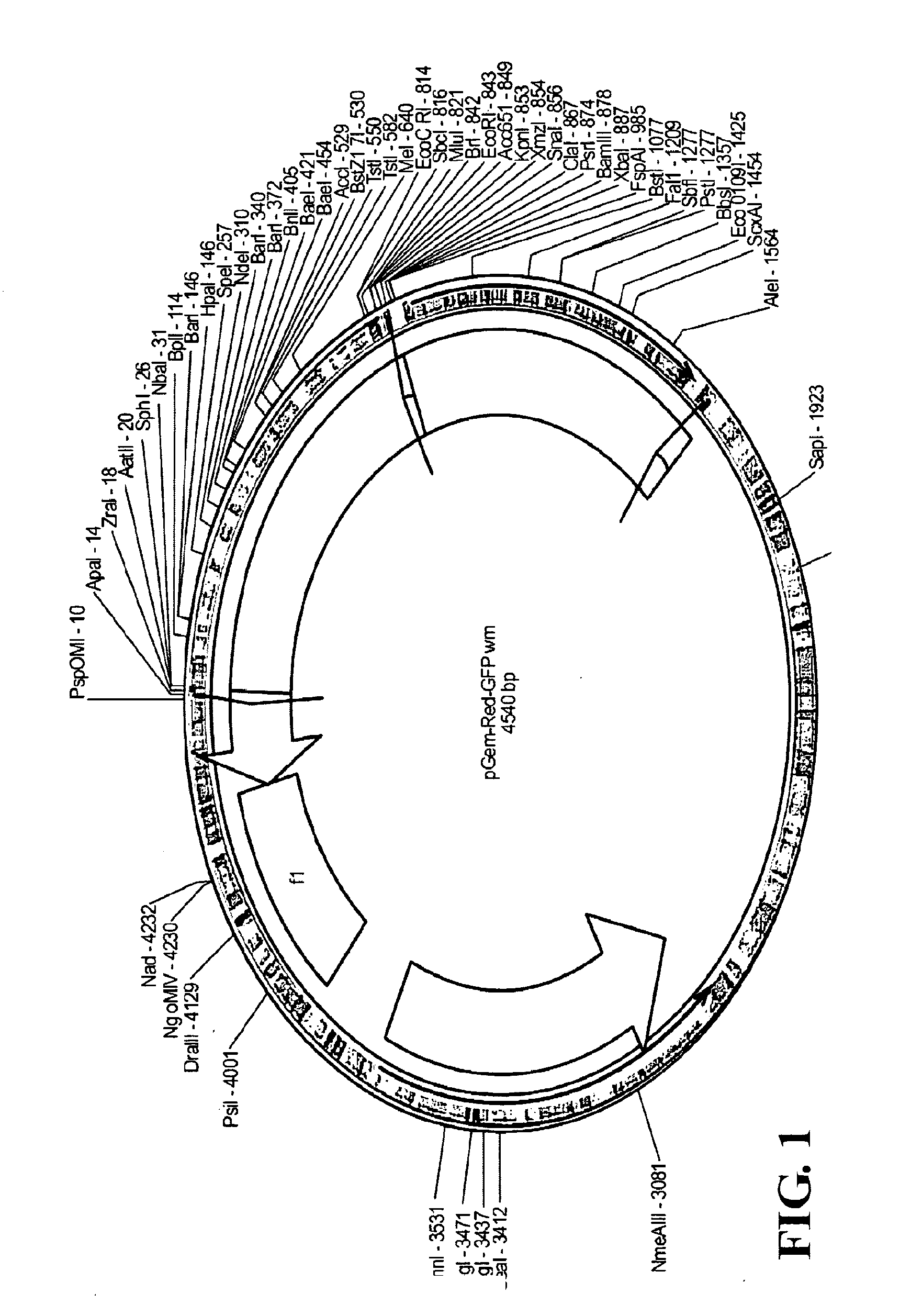
[0004] FIG. 1. Plasmid maps of transfer vectors.
[0005] FIG. 2. Schematic representation of NYVAC genome.
[0006] FIG. 3A. Construction of the plasmid transfer vector pGem-RG-B8R wm.
[0007] FIG. 3B. pGem-RG-B8R win plot.
[0008] FIG. 4. PCR analysis of NYVAC-C-.DELTA.B8R, NYVAC-C-.DELTA.B19R and NYVAC-C-.DELTA.B8RB19R.
[0009] FIG. 5. Immunoblot analysis of NYVAC-C-.DELTA.B8R, NYVAC-C-.DELTA.B19R and NYVAC-C-.DELTA.B8RB19R.
[0010] FIG. 6. Immunostain analysis of NYVAC-C-.DELTA.B8R, NYVAC-C-.DELTA.B19R and NYVAC-C-.DELTA.B8RB19R.
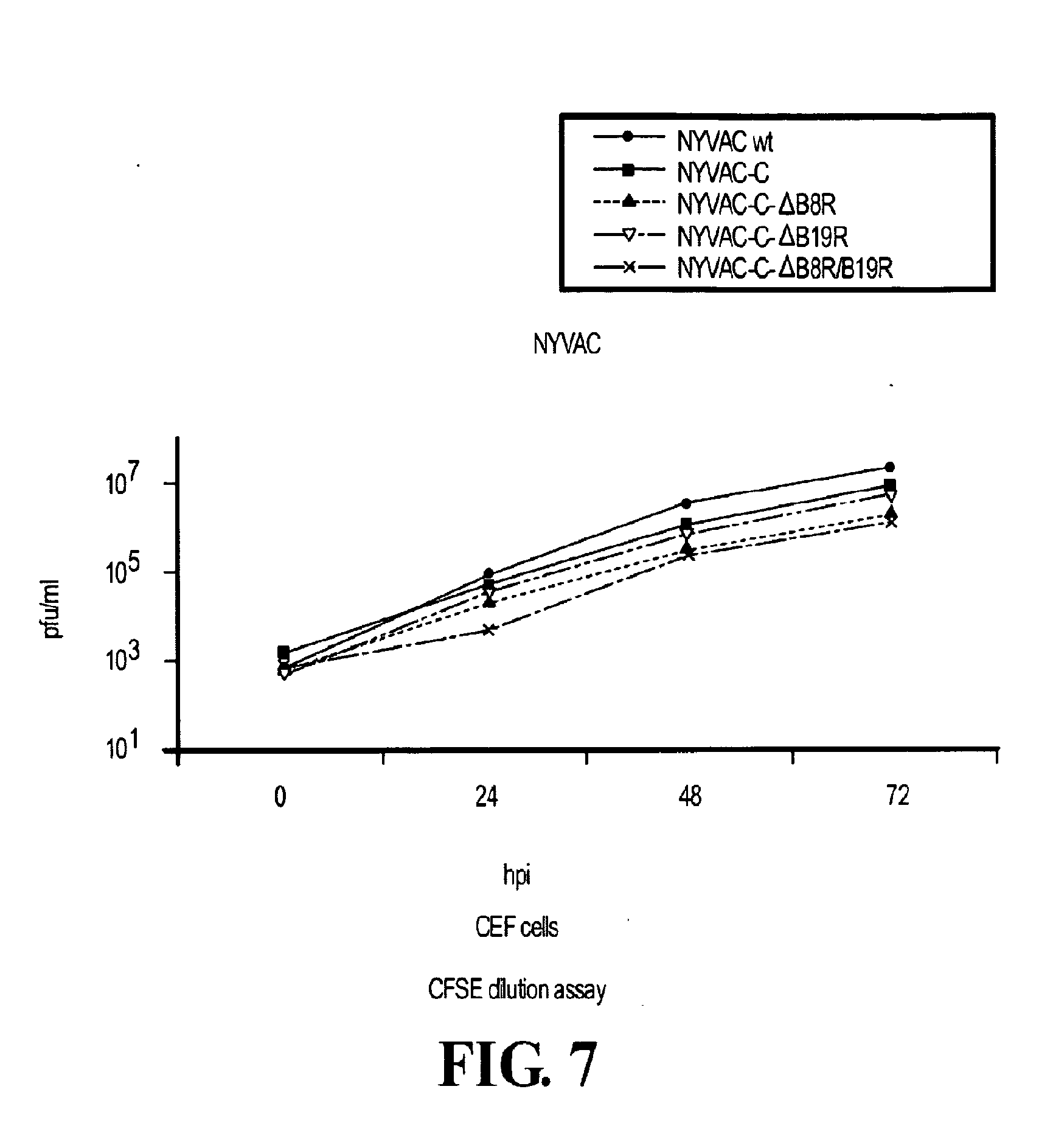
[0011] FIG. 7. Virus growth curves of NYVAC-C-.DELTA.B8R, NYVAC-C-.DELTA.B19R and NYVAC-C-.DELTA.B8RB19R.
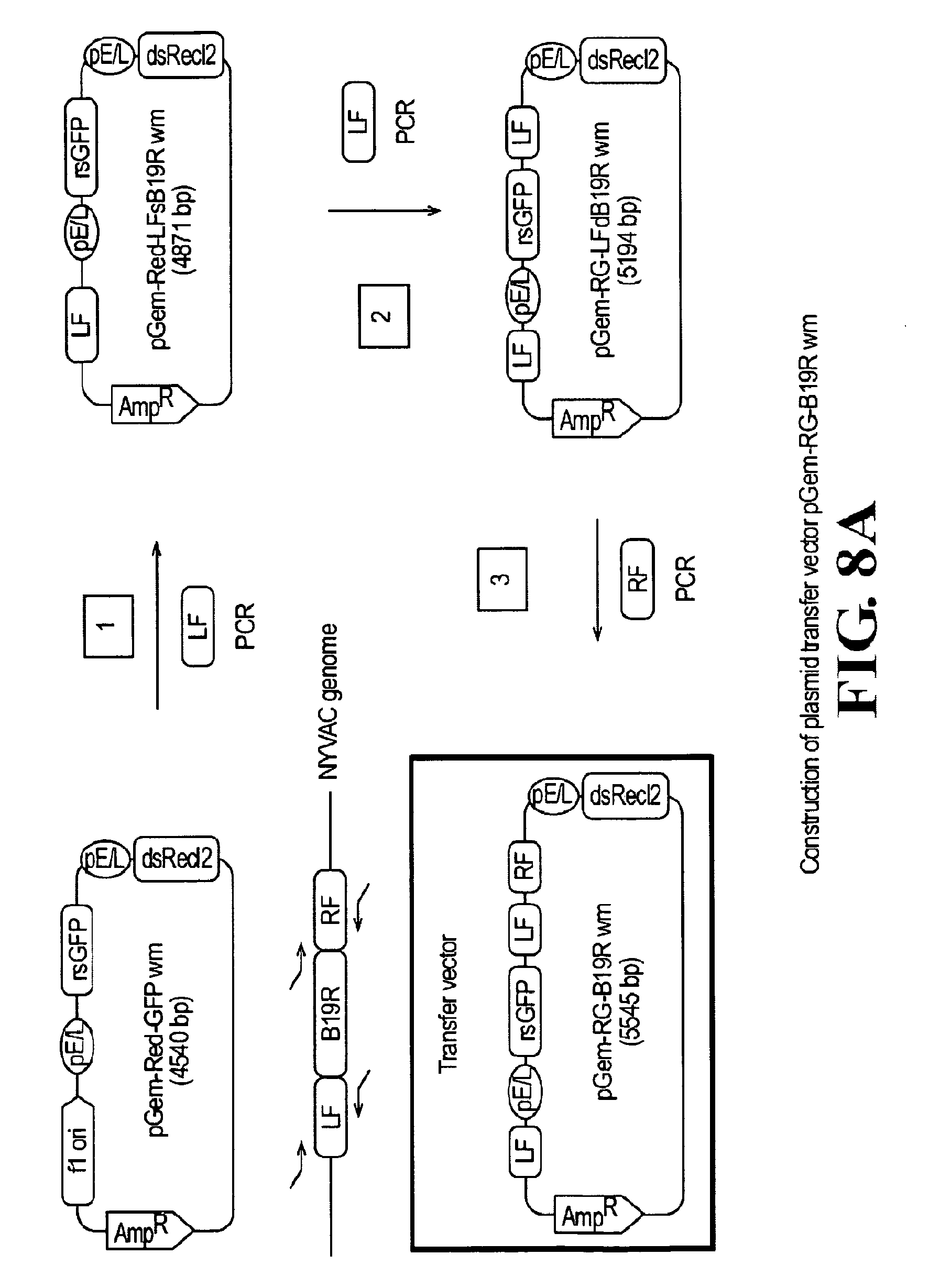
[0012] FIG. 8A. Construction of plasmid transfer vector pGem-RG-B19R wm.
[0013] FIG. 8B. pGem-RG-B19R wm plot.
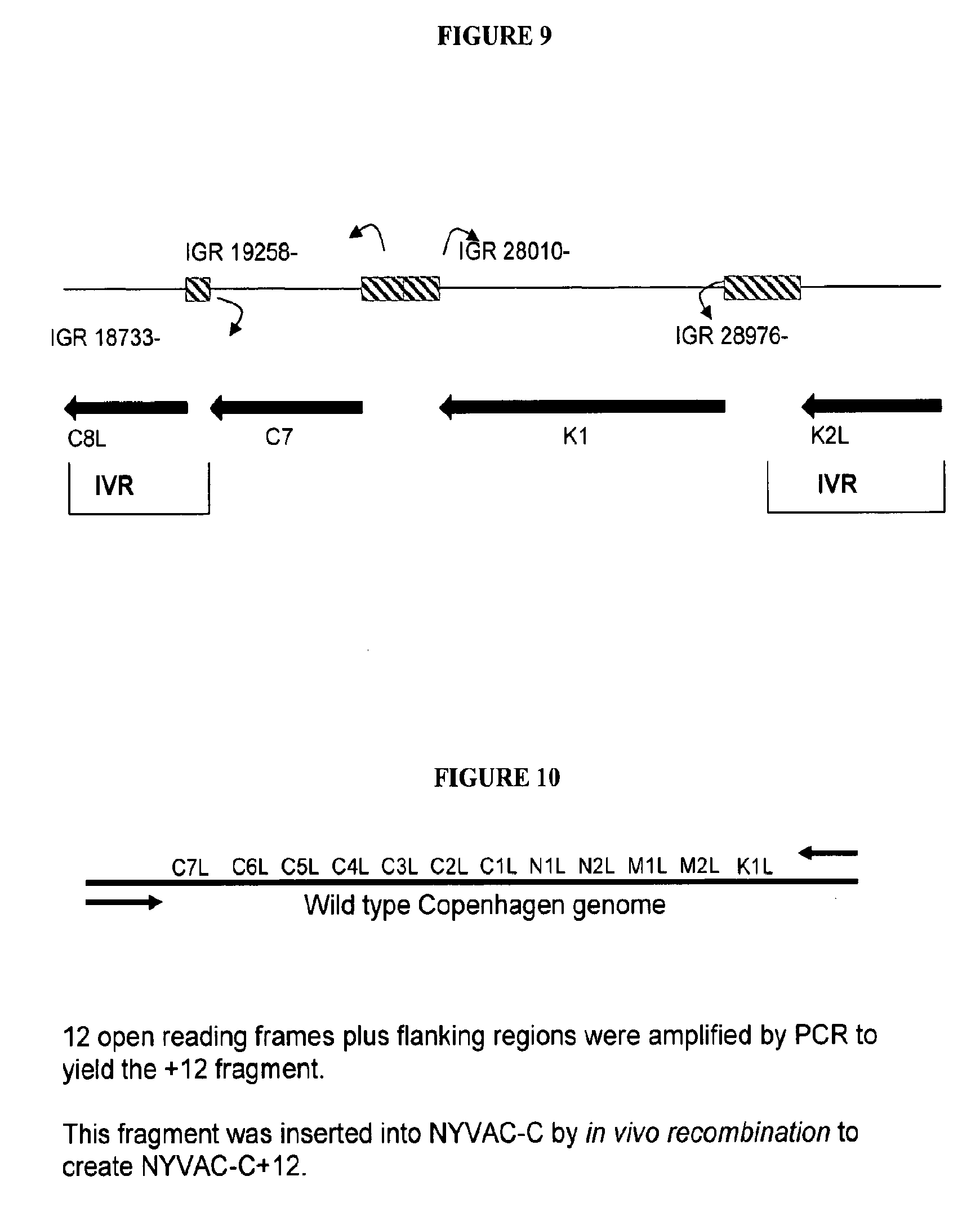
[0014] FIG. 9. Methodology used to construct KC viruses. To create a single fragment containing K1L and C7L, the two genes were first amplified by PCR from the wild type vaccinia virus genome, Copenhagen strain. PCR was used to fuse the two fragments into one. In vivo recombination (IVR) was used to insert the final PCR product between the existing inter-genic regions of the genome, creating NYVAC. In vivo combination was also used to create NYVAC-C-KC-.DELTA.B8R-.DELTA.B19R, using NYVAC-C-.DELTA.B8R-.DELTA.B19R as the parental virus.
[0015] FIG. 10. Methodology used to construct NYVAC-C+12 virus.
[0016] FIG. 11. Methodology used to construct NYVAC-C+12-ATVh virus. The E3L gene of NYVAC was replaced with the eIF2.alpha. homologue from Ambystoma tigrinum virus (ATVh) by in vivo recombination (IVR) to create NYVAC-C+12-ATVh. The ATVh had been amplified by PCR, with flanking region sequences at either end of the gene to allow for recombination, and inserted into a transfer plasmid. The plasmid was used in the IVR to transfer the ATVh into the virus genome.
[0017] FIGS. 12A and 12B. Flow cytometric analysis.
[0018] FIG. 13A. Upregulation of costimulatory molecules on infected human moDCs. IL-4 and GM-CSF differentiated DC were infected with NYVAC-C and the deletion mutants B19R (A) and B8R/B19R (B) expression of costimulatory molecules was analyzed by FACS analysis 48 hr post infection. DCs were infected with an MOI of 0.1. The shaded peaks in the histograms represent NYVAC-wt infected DC; the unshaded peaks represent DC infected with NYVAC-C, B19R or B8R/B19R.
[0019] FIG. 13B. Cytokine production by HIV specific CD8 T cells in a direct and cross presentation assay. DCs were infected or incubated with apoptotic infected HeLa cells for 6 hrs before CD8 T cells were added. After overnight incubation the amount of single, double and triple cytokine producing cells was determined by FACS analysis.
[0020] FIG. 14. IL-8 production assays. IL-8 and TNF release by human THP-1 macrophages (A) and whole blood (B) infected with wild-type and mutant NYVAC and NYVAC-C.
[0021] FIG. 15. Gene array assays. A. chemokine and cytokine expression levels; B. IFN expression levels; C. Enhanced expression of pathogen sensing molecules; D. Enhanced expression in genes associated with inflammatory response.
SUMMARY OF THE DISCLOSURE
[0022] Disclosed herein are compositions and reagents for immunizing human beings against infectious or other agents such as tumor cells by inducing or enhancing thereto. In certain embodiments, the compositions comprise recombinant viral vectors comprising modified nucleotide sequences. In certain embodiments, the vectors were modified by deletion of and/or insertion of nucleic acids encoding any one or more of the polypeptides shown in SEQ ID NOS. 1, 3, 5, 7, 9, 11, 13, 15, 17, 19. 21. 23, 25, or 27. Exemplary of such polynucleotides are those shown in SEQ ID NOS. 2, 4, 6, 8, 10, 12, 14, 16, 18, 20, 22, 24, 26, and 28. In some embodiments, such vectors further comprise polynucleotides encoding immunogens. Methods for constructing and using such vectors are described herein. Compositions comprising such vectors and methods for using such compostions are also provided.
DETAILED DESCRIPTION
[0023] The present disclosure provides compositions and methodologies useful for expressing nucleic acids and the polypeptides, peptides, or nucleic acids encoded thereby using recombinant vectors. In one embodiment, the compositions comprise recombinant vectors for introducing or altering the expression of a polypeptide, peptide, or nucleic acid in a host. In some embodiments, the compositions may include one or more recombinant viruses comprising polynucleotides encoding polypeptides, peptides, or polynucleotides that were not previously expressed by the virus, or are normally expressed in different amounts or at different times in the life cycle of the virus. In certain embodiments, polynucleotides are incorporated into the genome of a virus to produce a recombinant virus with altered characteristics as compared to the non-modified virus. In some embodiments, the incorporated polynucleotides encode polypeptides, peptides, or polynucleotides that alter the growth characteristics, infectivity, host range, replicative capacity, or immunogenicity of the recombinant virus as compared to the non-modified virus. Such polynucleotides may be used alone or in combination with other polynucleotides such as those described below (e.g., encoding one or more immunogens).
[0024] Expression vectors may also be modified by deleting polynucleotides (e.g., a gene) normally found within the vector therefrom. For instance, the poxvirus NYVAC (described in more detail below) was derived from the Copenhagen vaccinia strain using transient dominant selection (Falkner & Moss, 1990) which allows for deletion of one or more target genes without incorporation of a polynucleotide encoding a selectable marker into the viral genome. Polynucleotides may be completely or partially deleted, or inactivated with or without partial deletion. Partial deletion may be accomplished by removing a portion of a polynucleotide encoding a polypeptide from the "genome" of the vector ("vector genome"). As referred to herein, the vector genome may refer to the polynucleotide encoding the various factors required for the viability of a replication-competent or replication-incompetent viral vector, the polynucleotide making up a non-viral (e.g., bacterial, eukaryotic) or viral plasmid vector, or the like.
[0025] For instance, NYVAC is derived from the VACV strain Copenhagen (COP) from which 18 genes encoding proteins involved in host range and virulence were deleted (Tartaglia et al., 1992). These vectors were shown to exhibit altered host range and to be useful for expressing immunogens within a wide range of species (Tartaglia et al., 1994). Such vectors have been used as recombinant vaccines against numerous pathogens and tumours in animal models and in target species, including humans (Myagkikh et al., 1996; Benson et al., 1998; Siemens et al., 2003; Franchini et al., 2004). Clinical trials using NYVAC-based vectors showed an acceptable safety profile, with induction of high levels of immunity against heterologous antigens (Kanesa-thasan et al., 2000; Gomez, C. E et al. 2007; Harari, A et al, 2008). Such vectors may be further modified by insertion or deletion of additional polynucleotides using the techniques described herein. Suitable polynucleotides may include, for example, those involved in host range, apoptosis, signaling, cytokine and/or chemokine expression or activity, cytokine and/or chemokine pathways, and/or the like, resulting in novel biological characteristics of the vectors.
[0026] In some embodiments, polynucleotides encoding immunomodulatory polypeptides are selectively deleted from a vector genome. Polynucleotides encoding immunogens may also be incorporated into the vector genome. This may lead to modulation of virus-host cell interactions and "improvement" in the immunological profiles of the modified vectors as candidate vaccines. By "improvement" is meant that an immune response against a target antigen is induced or enhanced. In certain embodiments, the modified vectors may exhibit improved safety profiles as compared to non-modified (e.g., parental) vectors.
[0027] Polynucleotides suitable to modification (e.g., deletion from, alteration of sequence, or incorporation into a vector genome) may include, for example, any polynucleotide that provides the desired effect (e.g., an improved immune response). For instance, within the NYVAC vector, candidate polynucleotides may include polynucleotides that may be characterized as immunomodulators, and those affecting viral host range, one or more signalling pathways, apoptosis, secreted proteins (e.g., those binding host cytokines and/or chemokines). Exemplary polynucleotides and polypeptides that are candidates for modification include those encoding, for example, B8R (SEQ ID NOS. 1, 2) and/or B19R (SEQ ID NOS. 3, 4). In certain embodiments, suitable and exemplary polynucleotides may encode immunomodulatory polypeptides that interact with, for example, one or more interferons, cytokines and/or chemokines (e.g., B8R (SEQ ID NOS. 1, 2), and/or B19R (SEQ ID NOS. 3, 4)). The nomenclature of these sequences is related to the Copenhagen strain of vaccinia virus (GenBank Accession No. M35027; Goebel, et al. The complete DNA sequence of vaccinia virus. Virology 179 (1), 247-266 (1990); Goebel, et al. Appendix to `The complete DNA sequence of Vaccinia virus`. Virology 179, 517-563 (1990)). Any of such polynucleotides may be modified (e.g., incorporated into a recombinant vector or as part of a composition containing multiple recombinant vectors) in combination with any other of such polynucleotides. Other polynucleotides may also be suitable for modification in vaccinia or in other viruses (e.g., MVA, avipox, and the like).
[0028] The B8R gene (open reading frame ("ORF") shown in SEQ ID NO. 1) encodes the B8R protein (SEQ ID NO. 2) with amino acid similarity to the extracellular domain of the IFN-.gamma. receptor (Alcami & Smith, 1995; Mossman et al., 1995). The protein B8 binds and inhibits IFN-.gamma. from a wide variety of species but not the mouse. Deletion of B8R from WR did not alter virus replication or virulence in mouse models (Symons, et al. 2002a).
[0029] The B19R gene of VACV (ORF shown in SEQ ID NO. 3 encoding the B19R polypeptide, SEQ ID NO. 4) is equivalent to the B18R gene of VACV WR and encodes a type I IFN (.alpha.,.beta.)-receptor homolog. Protein B19 binds and inhibits type I IFN from a wide variety of species except murine IFN which binds but does not inhibit it. Deletion of B19R from VACV WR has been shown to cause attenuation in a murine intranasal model.
[0030] Within vaccinia, the C1L (SEQ ID NO. 5), C2L (SEQ ID NO. 7), C3L (SEQ ID NO. 9), C4L (SEQ ID NO. 11), C5L (SEQ ID NO. 13), C6L (SEQ ID NO. 15), C7L (SEQ ID NO. 17), N1L (SEQ ID NO. 19), N2L (SEQ ID NO. 21), M1L (SEQ ID NO. 23), M2L (SEQ ID NO. 25) and K1L (SEQ ID NO. 27) polypeptides have been shown to be involved in defining the "host range" or replication competence of the virus. Polynucleotides encoding such host range polypeptides are illustrated in SEQ ID NOS. 6, 8, 10, 12, 14, 16, 18, 20, 22, 24, 26, and 28. In the NYVAC virus, these genes have been deleted. In certain embodiments, one or more polynucleotides representing one or more of these host range genes may be introduced into the genome of a viral vector to affect the replication competence of the vector. In NYVAC, for example, one or more polynucleotides representing one or more of such host range genes may be re-incorporated into the NYVAC genome to modify its replication competence. In certain embodiments, as shown in the Examples, polynucleotides encoding C7L (e.g., SEQ ID NOS. 17, 18) and K1L (e.g., SEQ ID NOS. 27, 28) were shown to effect replication competence of NYVAC. In certain embodiments, the recombinant vector (e.g., NYVAC) expresses at least one, two, three, four, five, six, seven, eight, nine, ten, eleven or twelve of C1L (e.g., SEQ ID NOS. 5, 6), C2L (e.g., SEQ ID NOS. 7, 8), C3L (e.g., SEQ ID NOS. 9, 10), C4L (SEQ ID NOS. 11, 12), C5L (e.g., SEQ ID NOS. 13, 14), C6L (e.g., SEQ ID NOS. 15, 16), C7L (e.g., SEQ ID NOS. 17, 18), N1L (SEQ ID NOS. 19, 20), N2L (e.g., SEQ ID NOS. 21, 22), M1L (e.g., SEQ ID NOS. 23, 24), M2L (e.g., SEQ ID NOS. 25, 26), and K1L (e.g., SEQ ID NOS. 27, 28). Various combinations of such polynucleotides and/or polypeptides, as would be apparent to one of skill in the art, may be utilized in vectors. These polynucleotides and/or polypeptides may also be incorporated into vectors engineered to contain or express other polynucleotides and/or polypeptides such as, for example, B8R (SEQ ID NOS. 1, 2) and/or B19R (SEQ ID NOS. 3, 4). Suitable recombinant vectors for introduction or re-introduction of such host range genes include those from which such sequences have been previously deleted or those that otherwise do not contain such genes within the vector genome. It is also possible to modify such host range genes such that their function is altered by, for example, altering the timing or character (e.g., expression level) of expression within a host cell.
[0031] Polynucleotides encoding other polypeptides, peptides, or nucleic acids affecting the activity of a recombinant vector (e.g., recombinant virus) may also be incorporated into the vector. In certain embodiments, polynucleotides representing genes from other organisms (exogenous genes) may be incorporated into the vector. The polynucleotides may be inserted into a polynucleotide by insertion, either de novo or by replacement of an existing polynucleotide sequence within the vector genome. For instance, a polynucleotide may replace a gene of a virus. For example, the ranavirus eIF2.alpha.-like gene ("eIF2.alpha.H") from Ambystoma tigrinum virus isolate YEL protein gene (GenBank Accession No. EU512333; version EU512333.1; GI:170180537; "ATV eIF2.alpha.H"; SEQ ID NO. 29 encoded by SEQ ID NO. 30; see, e.g., U.S. Pat. No. 7,431,929) may be utilized. ATV eIF2.alpha.H encodes a potent, non-dsRNA-binding inhibitor of RNA-dependent protein kinase (PKR). In one embodiment, a polynucleotide encoding ATV eIF2.alpha.H (e.g., SEQ ID NO. 30) may be incorporated into a recombinant vector described herein. Without being limited to any particular theory of operation, it is believed that ATV eIF2.alpha.H induces signal transduction through NF-.kappa.B and IRF-3, while sparing viral protein synthesis from the inhibitory effects of PKR activation. In certain embodiments, a recombinant virus may be produced that exhibits little, decreased, or no replication competence but also induces an immune response in a host. Such a virus may provide an optimal recombinant vector that represents a "compromise" between replication competent that may cause complications in hosts, and replication deficient recombinant vectors that may fail to induce an immune response, or may induce a sub-optimal immune response.
[0032] In certain embodiments, in addition to the one or more polynucleotides encoding one or more of B8R (SEQ ID NOS. 1, 2) and/or B19R (SEQ ID NOS. 3, 4)), the recombinant vector may also comprise a polynucleotide encoding ATV eIF2.alpha.H (e.g., SEQ ID NOS. 29, 30) such as SEQ ID NO. 54. In other embodiments, a recombinant vector including any one or more of C1L (e.g., SEQ ID NOS. 5, 6), C2L (e.g., SEQ ID NOS. 7, 8), C3L (e.g., SEQ ID NOS. 9, 10), C4L (SEQ ID NOS. 11, 12), C5L (e.g., SEQ ID NOS. 13, 14), C6L (e.g., SEQ ID NOS. 15, 16), C7L (e.g., SEQ ID NOS. 17, 18), N1L (SEQ ID NOS. 19, 20), N2L (e.g., SEQ ID NOS. 21, 22), M1L (e.g., SEQ ID NOS. 23, 24), M2L (e.g., SEQ ID NOS. 25, 26), and/or K1L (e.g., SEQ ID NOS. 27, 28) (or a deletion of any one or more of these sequences) may also comprise a polynucleotide encoding ATV eIF2.alpha.H (e.g., SEQ ID NOS. 29, 30). In yet other embodiments, a recombinant vector may also comprise one or more polynucleotides encoding one or more of B8R (SEQ ID NOS. 1, 2) and/or B19R (SEQ ID NOS. 3, 4)) and/or a polynucleotide encoding ATV eIF2.alpha.H (e.g., SEQ ID NOS. 29, 30), and/or any one or more of C1L (e.g., SEQ ID NOS. 5, 6), C2L (e.g., SEQ ID NOS. 7, 8), C3L (e.g., SEQ ID NOS. 9, 10), C4L (SEQ ID NOS. 11, 12), C5L (e.g., SEQ ID NOS. 13, 14), C6L (e.g., SEQ ID NOS. 15, 16), C7L (e.g., SEQ ID NOS. 17, 18), N1L (SEQ ID NOS. 19, 20), N2L (e.g., SEQ ID NOS. 21, 22), M1L (e.g., SEQ ID NOS. 23, 24), M2L (e.g., SEQ ID NOS. 25, 26), and/or K1L (e.g., SEQ ID NOS. 27, 28). For instance, the Examples demonstrate a recombinant vaccinia virus in which the E3L gene was deleted and replaced by a polynucleotide encoding ATV eIF2.alpha.H (SEQ ID NO. 30 encoding SEQ ID NO. 29; see, e.g., U.S. Pat. No. 7,431,929). It was observed that this modified virus induces host cell production of IFN, exhibits increased sensitivity to IFN, and induces a potent Th1-dominated immune response at low doses. Other embodiments, as could be derived from this disclosure, may also be suitable for use.
[0033] In some embodiments, the compositions may include one or more recombinant vectors encoding one or more immunogens that may be used to induce or enhance an immune response that is beneficial to the host. As such, the compositions described herein may also be used to treat and/or prevent conditions relating to an infectious or other agent(s) by inducing or enhancing an immune response against such an agent. In certain embodiments, the compositions may comprise one or more recombinant vectors encoding one or more immunogens (e.g., comprising a polynucleotide encoding the antigen). An immunogen may be isolated from its source (e.g., an infectious agent) of which it forms a part (e.g., a polypeptide normally found within or expressed by that infectious agent). In certain embodiments, the immunogen may be encoded by a nucleotide sequence in expressible form (e.g., within an expression vector).
[0034] An immunogen may be a moiety (e.g., polypeptide, peptide or nucleic acid) that induces or enhances the immune response of a host to whom or to which the immunogen is administered. An immune response may be induced or enhanced by either increasing or decreasing the frequency, amount, or half-life of a particular immune modulator (e.g, the expression of a cytokine, chemokine, co-stimulatory molecule). This may be directly observed within a host cell containing a polynucleotide of interest (e.g., following infection by a recombinant virus) or within a nearby cell or tissue (e.g., indirectly). The immune response is typically directed against a target antigen. For example, an immune response may result from expression of an immunogen in a host following administration of a nucleic acid vector encoding the immunogen to the host. The immune response may result in one or more of an effect (e.g., maturation, proliferation, direct- or cross-presentation of antigen, gene expression profile) on cells of either the innate or adaptive immune system. For example, the immune response may involve, effect, or be detected in innate immune cells such as, for example, dendritic cells, monocytes, macrophages, natural killer cells, and/or granulocytes (e.g., neutrophils, basophils or eosinophils). The immune response may also involve, effect, or be detected in adaptive immune cells including, for example, lymphocytes (e.g., T cells and/or B cells). The immune response may be observed by detecting such involvement or effects including, for example, the presence, absence, or altered (e.g., increased or decreased) expression or activity of one or more immunomodulators such as a hormone, cytokine, interleukin (e.g., any of IL-1 through IL-35), interferon (e.g., any of IFN-I (IFN-.alpha., IFN-.beta., IFN-.epsilon., IFN-.kappa., IFN-.tau., IFN-.zeta., IFN-.omega.), IFN-II (e.g., IFN-.gamma.), IFN-III (IFN-.lamda.1, IFN-.lamda.2, IFN-.lamda.3)), chemokine (e.g., any CC cytokine (e.g., any of CCL1 through CCL28), any CXC chemokine (e.g., any of CXCL1 through CXCL24), Mip1a), any C chemokine (e.g., XCL1, XCL2), any CX3C chemokine (e.g., CX3CL1)), tumor necrosis factor (e.g., TNF-.alpha., TNF-.beta.)), negative regulators (e.g., PD-1, IL-T) and/or any of the cellular components (e.g., kinases, lipases, nucleases, transcription-related factors (e.g., IRF-1, IRF-7, STAT-5, NFKB, STAT3, STAT1, IRF-10), and/or cell surface markers suppressed or induced by such immunomodulators) involved in the expression of such immunomodulators. The presence, absence or altered expression may be detected within cells of interest or near those cells (e.g., within a cell culture supernatant, nearby cell or tissue in vitro or in vivo, and/or in blood or plasma). Administration of the immunogen may induce (e.g., stimulate a de novo or previously undetected response), or enhance or suppress an existing response against the immunogen by, for example, causing an increased antibody response (e.g., amount of antibody, increased affinity/avidity) or an increased cellular response (e.g., increased number of activated T cells, increased affinity/avidity of T cell receptors). In certain embodiments, the immune response may be protective, meaning that the immune response may be capable of preventing initiation or continued infection of or growth within a host and/or by eliminating an agent (e.g., a causative agent, such as HIV) from the host.
[0035] The compositions described herein may include one or more immunogen(s) from a single source or multiple sources. For instance, immunogens may also be derived from or direct an immune response against one or more viruses (e.g., viral target antigen(s)) including, for example, a dsDNA virus (e.g. adenovirus, herpesvirus, epstein-barr virus, herpes simplex type 1, herpes simplex type 2, human herpes virus simplex type 8, human cytomegalovirus, varicella-zoster virus, poxvirus); ssDNA virus (e.g., parvovirus, papillomavirus (e.g., E1, E2, E3, E4, E5, E6, E7, E8, BPV1, BPV2, BPV3, BPV4, BPV5 and BPV6 (In Papillomavirus and Human Cancer, edited by H. Pfister (CRC Press, Inc. 1990); Lancaster et al., Cancer Metast. Rev. pp. 6653-6664 (1987); Pfister, et al. Adv. Cancer Res 48, 113-147 (1987)); dsRNA viruses (e.g., reovirus); (+)ssRNA viruses (e.g., picornavirus, coxsackie virus, hepatitis A virus, poliovirus, togavirus, rubella virus, flavivirus, hepatitis C virus, yellow fever virus, dengue virus, west Nile virus); (-)ssRNA viruses (e.g., orthomyxovirus, influenza virus, rhabdovirus, paramyxovirus, measles virus, mumps virus, parainfluenza virus, respiratory syncytial virus, rhabdovirus, rabies virus); ssRNA-RT viruses (e.g. retrovirus, human immunodeficiency virus (HIV)); and, dsDNA-RT viruses (e.g. hepadnavirus, hepatitis B). Immunogens may also be derived from other viruses not listed above but available to one of skill in the art.
[0036] With respect to HIV, immunogens may be selected from any HIV isolate. As is well-known in the art, HIV isolates are now classified into discrete genetic subtypes. HIV-1 is known to comprise at least ten subtypes (A1, A2, A3, A4, B, C, D, E, F1, F2, G, H, J and K) (Taylor et al, NEJM, 359(18):1965-1966 (2008)). HIV-2 is known to include at least five subtypes (A, B, C, D, and E). Subtype B has been associated with the HIV epidemic in homosexual men and intravenous drug users worldwide. Most HIV-1 immunogens, laboratory adapted isolates, reagents and mapped epitopes belong to subtype B. In sub-Saharan Africa, India and China, areas where the incidence of new HIV infections is high, HIV-1 subtype B accounts for only a small minority of infections, and subtype HIV-1 C appears to be the most common infecting subtype. Thus, in certain embodiments, it may be preferable to select immunogens from HIV-1 subtypes B and/or C. It may be desirable to include immunogens from multiple HIV subtypes (e.g., HIV-1 subtypes B and C, HIV-2 subtypes A and B, or a combination of HIV-1 and HIV-2 subtypes) in a single immunological composition. Suitable HIV immunogens include ENV, GAG, POL, NEF, as well as variants, derivatives, and fusion proteins thereof, as described by, for example, Gomez et al. Vaccine, Vol. 25, pp. 1969-1992 (2007)). Exemplary, suitable peptide immunogens derived from HIV include but are not limited to VGNLWVTVYYGVPVW (SEQ ID NO. 31), WVTVYYGVPVWKGAT (SEQ ID NO. 32), GATTTLFCASDAKAY (SEQ ID NO. 33), TTLFCASDAKAYDTE (SEQ ID NO. 34), THACVPADPNPQEMV (SEQ ID NO. 35), ENVTENFNMWKNEMV (SEQ ID NO. 36), ENFNMWKNEMVNQMQ (SEQ ID NO. 37), EMVNQMQEDVISLWD (SEQ ID NO. 38), CVKLTPLCVTLECRN (SEQ ID NO. 39), NCSFNATTVVRDRKQ (SEQ ID NO. 40), NATTVVRDRKQTVYA (SEQ ID NO. 41), VYALFYRLDIVPLTK (SEQ ID NO. 42), FYRLDIVPLTKKNYS (SEQ ID NO. 43), INCNTSAITQACPKV (SEQ ID NO. 44), PKVTFDPIPIHYCTP (SEQ ID NO. 45), FDPIPIHYCTPAGYA (SEQ ID NO. 46), TGDIIGDIRQAHCNI (SEQ ID NO. 47), SSSIITIPCRIKQII (SEQ ID NO. 48), ITIPCRIKQIINMWQ (SEQ ID NO. 49), CRIKQIINMWQEVGR (SEQ ID NO. 50), VGRAMYAPPIKGNIT (SEQ ID NO. 51), MYAPPIKGNITCKSN (SEQ ID. NO. 52), PIKGNITCKSNITGL (SEQ ID NO. 53), ETFRPGGGDMRNNWR (SEQ ID NO. 54), ELYKYKVVEIKPLGV (SEQ ID NO. 55), YKVVEIKPLGVAPTT (SEQ ID NO. 56), EIKPLGVAPTTTKRR (SEQ ID NO. 57), LGVAPTTTKRRVVER (SEQ ID NO. 58), and/or YSENSSEYY (SEQ ID NO. 59). Any of these may be encoded by a polynucleotide within a recombinant vector, and/or used in combination with a recombinant vector as part of an immunization strategy.
[0037] Immunogens may also be derived from or direct an immune response against one or more bacterial species (spp.) (e.g., bacterial target antigen(s)) including, for example, Bacillus spp. (e.g., Bacillus anthracis), Bordetella spp. (e.g., Bordetella pertussis), Borrelia spp. (e.g., Borrelia burgdorferi), Brucella spp. (e.g., Brucella abortus, Brucella canis, Brucella melitensis, Brucella suis), Campylobacter spp. (e.g., Campylobacter jejuni), Chlamydia spp. (e.g., Chlamydia pneumoniae, Chlamydia psittaci, Chlamydia trachomatis), Clostridium spp. (e.g., Clostridium botulinum, Clostridium difficile, Clostridium perfringens, Clostridium tetani), Corynebacterium spp. (e.g., Corynebacterium diptheriae), Enterococcus spp. (e.g., Enterococcus faecalis, enterococcus faecum), Escherichia spp. (e.g., Escherichia coli), Francisella spp. (e.g., Francisella tularensis), Haemophilus spp. (e.g., Haemophilus influenza), Helicobacter spp. (e.g.; Helicobacter pylori), Legionella spp. (e.g., Legionella pneumophila), Leptospira spp. (e.g., Leptospira interrogans), Listeria spp. (e.g., Listeria monocytogenes), Mycobacterium spp. (e.g., Mycobacterium leprae, Mycobacterium tuberculosis), Mycoplasma spp. (e.g., Mycoplasma pneumoniae), Neisseria spp. (e.g., Neisseria gonorrhea, Neisseria meningitidis), Pseudomonas spp. (e.g., Pseudomonas aeruginosa), Rickettsia spp. (e.g., Rickettsia rickettsii), Salmonella spp. (e.g., Salmonella typhi, Salmonella typhinurium), Shigella spp. (e.g., Shigella sonnei), Staphylococcus spp. (e.g., Staphylococcus aureus, Staphylococcus epidermidis, Staphylococcus saprophyticus, coagulase negative staphylococcus (e.g., U.S. Pat. No. 7,473,762)), Streptococcus spp. (e.g., Streptococcus agalactiae, Streptococcus pneumoniae, Streptococcus pyrogenes), Treponema spp. (e.g., Treponema pallidum), Vibrio spp. (e.g., Vibrio cholerae), and Yersinia spp. (Yersinia pestis). Immunogens may also be derived from or direct the immune response, against other bacterial species not listed above but available to one of skill in the art.
[0038] Immunogens may also be derived from or direct an immune response against one or more parasitic organisms (spp.) (e.g., parasite target antigen(s)) including, for example, Ancylostoma spp. (e.g., A. duodenale), Anisakis spp., Ascaris lumbricoides, Balantidium coli, Cestoda spp., Cimicidae spp., Clonorchis sinensis, Dicrocoelium dendriticum, Dicrocoelium hospes, Diphyllobothrium latum, Dracunculus spp., Echinococcus spp. (e.g., E. granulosus, E. multilocularis), Entamoeba histolytica, Enterobius vermicularis, Fasciola spp. (e.g., F. hepatica, F. magna, F. gigantica, F. jacksoni), Fasciolopsis buski, Giardia spp. (Giardia lamblia), Gnathostoma spp., Hymenolepis spp. (e.g., H. nana, H. diminuta), Leishmania spp., Loa loa, Metorchis spp. (M. conjunctus, M. albidus), Necator americanus, Oestroidea spp. (e.g., botfly), Onchocercidae spp., Opisthorchis spp. (e.g., O. viverrini, O. felineus, O. guayaquilensis, and O. noverca), Plasmodium spp. (e.g., P. falciparum), Protofasciola robusta, Parafasciolopsis fasciomorphae, Paragonimus westermani, Schistosoma spp. (e.g., S. mansoni, S. japonicum, S. mekongi, S. haematobium), Spirometra erinaceieuropaei, Strongyloides stercoralis, Taenia spp. (e.g., T. saginata, T. solium), Toxocara spp. (e.g., T. canis, T. cati), Toxoplasma spp. (e.g., T. gondii), Trichobilharzia regenti, Trichinella spiralis, Trichuris trichiura, Trombiculidae spp., Trypanosoma spp., Tunga penetrans, and/or Wuchereria bancrofti. Immunogens may also be derived from or direct the immune response against other parasitic organisms not listed above but available to one of skill in the art.
[0039] Immunogens may be derived from or direct the immune response against tumor target antigens (e.g., tumor target antigens). The term tumor target antigen (TA) may include both tumor-associated antigens (TAAs) and tumor-specific antigens (TSAs), where a cancerous cell is the source of the antigen. A TA may be an antigen that is expressed on the surface of a tumor cell in higher amounts than is observed on normal cells or an antigen that is expressed on normal cells during fetal development. A TSA is typically an antigen that is unique to tumor cells and is not expressed on normal cells. TAs are typically classified into five categories according to their expression pattern, function, or genetic origin: cancer-testis (CT) antigens (i.e., MAGE, NY-ESO-1); melanocyte differentiation antigens (i.e., Melan A/MART-1, tyrosinase, gp100); mutational antigens (i.e., MUM-1, p53, CDK-4); overexpressed `self` antigens (i.e., HER-2/neu, p53); and, viral antigens (i.e., HPV, EBV). Suitable TAs include, for example, gp100 (Cox et al., Science, 264:716-719 (1994)), MART-1/Melan A (Kawakami et al., J. Exp. Med., 180:347-352 (1994)), gp75 (TRP-1) (Wang et al., J. Exp. Med., 186:1131-1140 (1996)), tyrosinase (Wolfel et al., Eur. J. Immunol., 24:759-764 (1994)), NY-ESO-1 (WO 98/14464; WO 99/18206), melanoma proteoglycan (Hellstrom et al., J. Immunol., 130:1467-1472 (1983)), MAGE family antigens (i.e., MAGE-1, 2, 3, 4, 6, and 12; Van der Bruggen et al., Science, 254:1643-1647 (1991); U.S. Pat. No. 6,235,525), BAGE family antigens (Boel et al., Immunity, 2:167-175 (1995)), GAGE family antigens (i.e., GAGE-1, 2; Van den Eynde et al., J. Exp. Med., 182:689-698 (1995); U.S. Pat. No. 6,013,765), RAGE family antigens (i.e., RAGE-1; Gaugler et al., Immunogenetics, 44:323-330 (1996); U.S. Pat. No. 5,939,526), N-acetylglucosaminyltransferase-V (Guilloux et al., J. Exp. Med., 183:1173-1183 (1996)), p15 (Robbins et al., J. Immunol. 154:5944-5950 (1995)), .beta.-catenin (Robbins et al., J. Exp. Med., 183:1185-1192 (1996)), MUM-1 (Coulie et al., Proc. Natl. Acad. Sci. USA, 92:7976-7980 (1995)), cyclin dependent kinase-4 (CDK4) (Wolfel et al., Science, 269:1281-1284 (1995)), p21-ras (Fossum et al., Int. J. Cancer, 56:40-45 (1994)), BCR-abl (Bocchia et al., Blood, 85:2680-2684 (1995)), p53 (Theobald et al., Proc. Natl. Acad. Sci. USA, 92:11993-11997 (1995)), p185 HER2/neu (erb-B1; Fisk et al., J. Exp. Med., 181:2109-2117 (1995)), epidermal growth factor receptor (EGFR) (Harris et al., Breast Cancer Res. Treat, 29:1-2 (1994)), carcinoembryonic antigens (CEA) (Kwong et al., J. Natl. Cancer Inst., 85:982-990 (1995) U.S. Pat. Nos. 5,756,103; 5,274,087; 5,571,710; 6,071,716; 5,698,530; 6,045,802; EP 263933; EP 346710; and, EP 784483); carcinoma-associated mutated mucins (i.e., MUC-1 gene products; Jerome et al., J. Immunol., 151:1654-1662 (1993)); EBNA gene products of EBV (i.e., EBNA-1; Rickinson et al., Cancer Surveys, 13:53-80 (1992)); E7, E6 proteins of human papillomavirus (Ressing et al., J. Immunol, 154:5934-5943 (1995)); prostate specific antigen (PSA; Xue et al., The Prostate, 30:73-78 (1997)); prostate specific membrane antigen (PSMA; Israeli, et al., Cancer Res., 54:1807-1811 (1994)); idiotypic epitopes or antigens, for example, immunoglobulin idiotypes or T cell receptor idiotypes (Chen et al., J. Immunol., 153:4775-4787 (1994)); KSA (U.S. Pat. No. 5,348,887), kinesin 2 (Dietz, et al. Biochem Biophys Res Commun 2000 Sep. 7; 275(3):731-8), HIP-55, TGF.beta.-1 anti-apoptotic factor (Toomey, et al. Br J Biomed Sci 2001; 58(3):177-83), tumor protein D52 (Bryne J. A., et al., Genomics, 35:523-532 (1996)), HIFT, NY-BR-1 (WO 01/47959), NY-BR-62, NY-BR-75, NY-BR-85, NY-BR-87 and NY-BR-96 (Scanlan, M. Serologic and Bioinformatic Approaches to the Identification of Human Tumor Antigens, in Cancer Vaccines 2000, Cancer Research Institute, New York, N.Y.), and/or pancreatic cancer antigens (e.g., SEQ ID NOS: 1-288 of U.S. Pat. No. 7,473,531). Immunogens may also be derived from or direct the immune response against include TAs not listed above but available to one of skill in the art.
[0040] In some embodiments, derivatives of polypeptides, peptides, or polynucleotides incorporated into or expressed by the vectors described herein including, for example, fragments and/or variants thereof may be utilized. Derivatives may result from, for example, substitution, deletion, or addition of amino acids or nucleotides from or to the reference sequence (e.g., the parental sequence). A derivative of a polypeptide or protein, for example, typically refers to an amino acid sequence that is altered with respect to the referenced polypeptide or peptide. A derivative of a polypeptide typically retains at least one activity of the polypeptide. A derivative will typically share at least approximately 60%, 70%, 80%, 90%, 95%, or 99% identity to the reference sequence. With respect to polypeptides and peptides, the derivative may have "conservative" changes, wherein a substituted amino acid has similar structural or chemical properties. A derivative may also have "nonconservative" changes. Exemplary, suitable conservative amino acid substitutions may include, for example, those shown in Table 1:
TABLE-US-00001 TABLE 1 Original Preferred Residues Exemplary Substitutions Substitutions Ala Val, Leu, Ile Val Arg Lys, Gln, Asn Lys Asn Gln Gln Asp Glu Glu Cys Ser, Ala Ser Gln Asn Asn Glu Asp Asp Gly Pro, Ala Ala His Asn, Gln, Lys, Arg Arg Ile Leu, Val, Met, Ala, Phe, Norleucine Leu Leu Norleucine, Ile, Val, Met, Ala, Phe Ile Lys Arg, 1,4 Diamino-butyric Acid, Gln, Asn Arg Met Leu, Phe, Ile Leu Phe Leu, Val, Ile, Ala, Tyr Leu Pro Ala Gly Ser Thr, Ala, Cys Thr Thr Ser Ser Trp Tyr, Phe Tyr Tyr Trp, Phe, Thr, Ser Phe Val Ile, Met, Leu, Phe, Ala, Norleucine Leu
Other amino acid substitutions may be considered non-conservative. Derivatives may also include amino acid or nucleotide deletions and/or additions/insertions, or some combination of these. Guidance in determining which amino acid residues or nucleotides may be substituted, inserted, or deleted without abolishing the desired activity of the derivative may be identified using any of the methods available to one of skill in the art.
[0041] Derivatives may also refer to a chemically modified polynucleotide or polypeptide. Chemical modifications of a polynucleotide may include, for example, replacement of hydrogen by an alkyl, acyl, hydroxyl, or amino group. A derivative polynucleotide may encode a polypeptide which retains at least one biological or immunological function of the natural molecule. A derivative polypeptide may be one modified by glycosylation, pegylation, biotinylation, or any similar process that retains at least one biological or immunological function of the polypeptide from which it was derived.
[0042] The phrases "percent identity" and "% identity," as applied to polypeptide sequences, refer to the percentage of residue matches between at least two polypeptide sequences aligned using a standardized algorithm. Methods of polypeptide sequence alignment are well-known. Some alignment methods take into account conservative amino acid substitutions. Such conservative substitutions, explained in more detail above, generally preserve the charge and hydrophobicity at the site of substitution, thus preserving the structure (end therefore function) of the polypeptide. Percent identity may be measured over the length of an entire defined polypeptide sequence, for example, as defined by a particular SEQ ID number, or may be measured over a shorter length, for example, over the length of a fragment taken from a larger, defined polypeptide sequence, for instance, a fragment of at least 10, at least 15, at least 20, at least 30, at least 40, at least 50, at least 70 or at least 150 contiguous residues. Such lengths are exemplary only, and it is understood that any fragment length supported by the sequences shown herein, in the tables, figures or Sequence Listing, may be used to describe a length over which percentage identity may be measured.
[0043] As mentioned above, this disclosure relates to compositions comprising recombinant vectors, the vectors per se, and methods of using the same. A "vector" is any moiety (e.g., a virus or plasmid) used to carry, introduce, or transfer a polynucleotide or interest to another moiety (e.g., a host cell). In certain cases, an expression vector is utilized. An expression vector is a nucleic acid molecule containing a polynucleotide of interest encoding a polypeptide, peptide, or polynucleotide and also containing other polynucleotides that direct and/or control the expression of the polynucleotide of interest. Expression includes, but is not limited to, processes such as transcription, translation, and/or splicing (e.g., where introns are present).
[0044] Viral vectors that may be used include, for example, retrovirus, adenovirus, adeno-associated virus (AAV), alphavirus, herpes virus, and poxvirus vectors, among others. Many such viral vectors are available in the art. The vectors described herein may be constructed using standard recombinant techniques widely available to one skilled in the art. Such techniques may be found in common molecular biology references such as Molecular Cloning: A Laboratory Manual (Sambrook, et al., 1989, Cold Spring Harbor Laboratory Press), Gene Expression Technology (Methods in Enzymology, Vol. 185, edited by D. Goeddel, 1991. Academic Press, San Diego, Calif.), and PCR Protocols: A Guide to Methods and Applications (Innis, et al. 1990. Academic Press, San Diego, Calif.).
[0045] Suitable retroviral vectors may include derivatives of lentivirus as well as derivatives of murine or avian retroviruses. Exemplary, suitable retroviral vectors may include, for example, Moloney murine leukemia virus (MoMuLV), Harvey murine sarcoma virus (HaMuSV), murine mammary tumor virus (MuMTV), SIV, BIV, HIV and Rous Sarcoma Virus (RSV). A number of retroviral vectors can incorporate multiple exogenous polynucleotides. As recombinant retroviruses are defective, they require assistance in order to produce infectious vector particles. This assistance can be provided by, for example, helper cell lines encoding retrovirus structural genes. Suitable helper cell lines include .PSI.2, PA317 and PA12, among others. The vector virions produced using such cell lines may then be used to infect a tissue cell line, such as NIH 3T3 cells, to produce large quantities of chimeric retroviral virions. Retroviral vectors may be administered by traditional methods (i.e., injection) or by implantation of a "producer cell line" in proximity to the target cell population (Culver, K., et al., 1994, Hum. Gene Ther., 5 (3): 343-79; Culver, K., et al., Cold Spring Harb. Symp. Quant. Biol., 59: 685-90); Oldfield, E., 1993, Hum. Gene Ther., 4 (1): 39-69). The producer cell line is engineered to produce a viral vector and releases viral particles in the vicinity of the target cell. A portion of the released viral particles contact the target cells and infect those cells, thus delivering a nucleic acid encoding an immunogen to the target cell. Following infection of the target cell, expression of the polynucleotide of interest from the vector occurs.
[0046] Adenoviral vectors have proven especially useful for gene transfer into eukaryotic cells (Rosenfeld, M., et al., 1991, Science, 252 (5004): 431-4; Crystal, R., et al., 1994, Nat. Genet., 8 (1): 42-51), the study eukaryotic gene expression (Levrero, M., et al., 1991, Gene, 101 (2): 195-202), vaccine development (Graham, F. and Prevec, L., 1992, Biotechnology, 20: 363-90), and in animal models (Stratford-Perricaudet, L., et al., 1992, Bone Marrow Transplant., 9 (Suppl. 1): 151-2; Rich, et al., 1993, Hum. Gene Ther., 4 (4): 461-76). Experimental routes for administrating recombinant Ad to different tissues in vivo have included intratracheal instillation (Rosenfeld, M., et al., 1992, Cell, 68 (1): 143-55) injection into muscle (Quantin, B., et al., 1992, Proc. Natl. Acad. Sci. U.S.A., 89 (7): 2581-4), peripheral intravenous injection (Herz, J., and Gerard, R., 1993, Proc. Natl. Acad. Sci. U.S.A., 90 (7): 2812-6) and/or stereotactic inoculation to brain (Le Gal La Salle, G., et al., 1993, Science, 259 (5097): 988-90), among others.
[0047] Adeno-associated virus (AAV) demonstrates high-level infectivity, broad host range and specificity in integrating into the host cell genome (Hermonat, P., et al., 1984, Proc. Natl. Acad. Sci. U.S.A., 81 (20): 6466-70). And Herpes Simplex Virus type-1 (HSV-1) is yet another attractive vector system, especially for use in the nervous system because of its neurotropic property (Geller, A., et al., 1991, Trends Neurosci., 14 (10): 428-32; Glorioso, et al., 1995, Mol. Biotechnol., 4 (1): 87-99; Glorioso, et al., 1995, Annu. Rev. Microbiol., 49: 675-710).
[0048] Alphavirus may also be used to express the immunogen in a host. Suitable members of the Alphavirus genus include, among others, Sindbis virus, Semliki Forest virus (SFV), the Ross River virus and Venezuelan, Western and Eastern equine encephalitis viruses, among others. Expression systems utilizing alphavirus vectors are described in, for example, U.S. Pat. Nos. 5,091,309; 5,217,879; 5,739,026; 5,766,602; 5,843,723; 6,015,694; 6,156,558; 6,190,666; 6,242,259; and, 6,329,201; WO 92/10578; Xiong et al., Science, Vol 243, 1989, 1188-1191; Liliestrom, et al. Bio/Technology, 9: 1356-1361, 1991. Thus, the use of alphavirus as an expression system is well known by those of skill in the art.
[0049] Poxvirus is another useful expression vector (Smith, et al. 1983, Gene, 25 (1): 21-8; Moss, et al, 1992, Biotechnology, 20: 345-62; Moss, et al, 1992, Curr. Top. Microbiol. Immunol., 158: 25-38; Moss, et al. 1991. Science, 252: 1662-1667). The most often utilized poxyiral vectors include vaccinia and derivatives therefrom such as NYVAC and MVA, and members of the avipox genera such as fowlpox, canarypox, ALVAC, and ALVAC(2), among others.
[0050] An exemplary suitable vector is NYVAC (vP866) which was derived from the Copenhagen vaccine strain of vaccinia virus by deleting six nonessential regions of the genome encoding known or potential virulence factors (see, for example, U.S. Pat. Nos. 5,364,773 and 5,494,807). The deletion loci were also engineered as recipient loci for the insertion of foreign genes. The deleted regions are: thymidine kinase gene (TK; J2R); hemorrhagic region (u; B13R+B14R); A type inclusion body region (AT1; A26L); hemagglutinin gene (HA; A56R); host range gene region (C7L-K1L); and, large subunit, ribonucleotide reductase (14L). NYVAC is a genetically engineered vaccinia virus strain that was generated by the specific deletion of eighteen open reading frames encoding gene products associated with virulence and host range. NYVAC has been show to be useful for expressing TAs (see, for example, U.S. Pat. No. 6,265,189). NYVAC (vP866), vP994, vCP205, vCP1433, placZH6H4Lreverse, pMPC6H6K3E3 and pC3H6FHVB were also deposited with the ATCC under the terms of the Budapest Treaty, accession numbers VR-2559, VR-2558, VR-2557, VR-2556, ATCC-97913, ATCC-97912, and ATCC-97914, respectively.
[0051] Another suitable virus is the Modified Vaccinia Ankara (MVA) virus which was generated by 516 serial passages on chicken embryo fibroblasts of the Ankara strain of vaccinia virus (CVA) (for review see Mayr, A., et al. Infection 3, 6-14 (1975)). It was shown in a variety of animal models that the resulting MVA was significantly avirulent (Mayr, A. & Danner, K. [1978] Dev. Biol. Stand. 41: 225.34) and has been tested in clinical trials as a smallpox vaccine (Mayr et al., Zbl. Bakt. Hyg. I, Abt. Org. B 167, 375-390 (1987), Stickl et al., Dtsch. med. Wschr. 99, 2386-2392 (1974)). MVA has also been engineered for use as a viral vector for both recombinant gene expression studies and as a recombinant vaccine (Sutter, G. et al. (1994), Vaccine 12: 1032-40; Blanchard et al., 1998, J Gen Virol 79, 1159-1167; Carroll & Moss, 1997, Virology 238, 198-211; Altenberger, U.S. Pat. No. 5,185,146; Ambrosini et al., 1999, J Neurosci Res 55(5), 569). Modified virus Ankara (MVA) has been previously described in, for example, U.S. Pat. Nos. 5,185,146 and 6,440,422; Sutter, et al. (B. Dev. Biol. Stand. Basel, Karger 84:195-200 (1995)); Antoine, et al. (Virology 244: 365-396, 1998); Sutter et al. (Proc. Natl. Acad. Sci. USA 89: 10847-10851, 1992); Meyer et al. (J. Gen. Virol. 72: 1031-1038, 1991); Mahnel, et al. (Berlin Munch. Tierarztl. Wochenschr. 107: 253-256, 1994); Mayr et al. (Zbl. Bakt. Hyg. I, Abt. Org. B 167: 375-390 (1987); and, Stickl et al. (Dtsch. med. Wschr. 99: 2386-2392 (1974)). An exemplary MVA is available from the ATCC under accession numbers VR-1508 and VR-1566.
[0052] ALVAC-based recombinant viruses (i.e., ALVAC-1 and ALVAC-2) are also suitable for use in practicing the present invention (see, for example, U.S. Pat. No. 5,756,103). ALVAC(2) is identical to ALVAC(1) except that ALVAC(2) genome comprises the vaccinia E3L and K3L genes under the control of vaccinia promoters (U.S. Pat. No. 6,130,066; Beattie et al., 1995a, 1995b, 1991; Chang et al., 1992; Davies et al., 1993). Both ALVAC(1) and ALVAC(2) have been demonstrated to be useful in expressing foreign DNA sequences, such as TAs (Tartaglia et al., 1993 a, b; U.S. Pat. No. 5,833,975). ALVAC was deposited under the terms of the Budapest Treaty with the American Type Culture Collection (ATCC), 10801 University Boulevard, Manassas, Va. 20110-2209, USA, ATCC accession number VR-2547. Vaccinia virus host range genes (e.g., C18L, C17L, C7L, K1L, E3L, B4R, B23R, and B24R) have also been shown to be expressible in canarypox (e.g., U.S. Pat. No. 7,473,536).
[0053] Another useful poxvirus vector is TROVAC. TROVAC refers to an attenuated fowlpox that was a plaque-cloned isolate derived from the FP-1 vaccine strain of fowlpoxvirus which is licensed for, vaccination of 1 day old chicks. TROVAC was likewise deposited under the terms of the Budapest Treaty with the ATCC, accession number 2553.
[0054] "Non-viral" plasmid vectors may also be suitable for use. Plasmid DNA molecules comprising expression cassettes for expressing an immunogen may be used for "naked DNA" immunization. Preferred plasmid vectors are compatible with bacterial, insect, and/or mammalian host cells. Such vectors include, for example, PCR-II, pCR3, and pcDNA3.1 (Invitrogen, San Diego, Calif.), pBSII (Stratagene, La Jolla, Calif.), pET15 (Novagen, Madison, Wis.), pGEX (Pharmacia Biotech, Piscataway, N.J.), pEGFP-N2 (Clontech, Palo Alto, Calif.), pETL (BlueBacII, Invitrogen), pDSR-alpha (PCT pub. No. WO 90/14363) and pFastBacDual (Gibco-BRL, Grand Island, N.Y.) as well as Bluescript.RTM. plasmid derivatives (a high copy number COLE1-based phagemid, Stratagene Cloning Systems, La Jolla, Calif.), PCR cloning plasmids designed for cloning Taq-amplified PCR products (e.g., TOPO.TM. TA Cloning.RTM. kit, PCR2.1.RTM. plasmid derivatives, Invitrogen, Carlsbad, Calif.).
[0055] Bacterial vectors may also be suitable for use. These vectors include, for example, Shigella, Salmonella, Vibrio cholerae, Lactobacillus, Bacille calmette guerin (BCG), and Streptococcus (see for example, WO 88/6626; WO 90/0594; WO 91/13157; WO 92/1796; and WO 92/21376). Many other non-viral plasmid expression vectors and systems are known in the art and could be used with the current invention.
[0056] The polynucleotides and polypeptides referred to herein as being suitable for use and/or modification (e.g., SEQ ID NOS. 1-28) may be inserted into non-homologous vector genomes. For instance, while the polynucleotides and polypeptides of SEQ ID NOS. 1-28 may be derived from vaccinia, any one or more of such polynucleotides and/or polypeptides may be incorporated into and/or expressed within a different viral (e.g., MVA, ALVAC, ALVAC(2), TROVAC), bacterial or plasmid vector. If such different vectors contain sequence homologous to one or more of SEQ ID NOS. 1-28, such sequence may be replaced by a polynucleotide encoding SEQ ID NOS. 1-28. Such vectors may further comprise or be modified to comprise a polynucleotide encoding SEQ ID NO. 29, such as SEQ ID NO. 30.
[0057] Expression vectors typically comprise one or more flanking polynucleotides "operably linked" to a heterologous polynucleotide encoding a polypeptide. As used herein, the term "operably linked" refers to a linkage between polynucleotide elements in a functional relationship such as when promoter or enhancer affects transcription of a polynucleotide of interest (e.g., a coding sequence). Flanking polynucleotides may be homologous (e.g., from the same species and/or strain as the host cell), heterologous (e.g., from a species other than the host cell species and/or strain), hybrid (e.g., a combination of flanking sequences from more than one source), or synthetic, for example. All polynucleotides referred to herein are typically incorporated into vectors in expressible form, meaning that such polynucleotides are capable of being expressed from the expression vector transformed into a cell or after incorporation of the expression vector or portions thereof into the genome of an infected or transformed cell, such that the polypeptide encoded thereby is expressed in the infected or transformed cell. The flanking sequences described herein typically assist in achieving expression in the infected or transformed cell.
[0058] In certain embodiments, it is preferred that the flanking polynucleotide includes a transcriptional regulatory region that drives expression of a polynucleotide of interest in an environment such as a target cell. The transcriptional regulatory region may comprise, for example, a promoter, enhancer, silencer, repressor element, or combinations thereof. The transcriptional regulatory region may be either constitutive, tissue-specific, cell-type specific (e.g., the region is drives higher levels of transcription in a one type of tissue or cell as compared to another) and/or regulatable (e.g., responsive to interaction with a compound such as tetracycline). The source of a transcriptional regulatory region may be any prokaryotic or eukaryotic organism, any vertebrate or invertebrate organism, or any plant, provided that the flanking polynucleotide functions in an environment (e.g., a cell) by causing transcription of a polynucleotide within that environment. A wide variety of suitable transcriptional regulatory regions are available to one of skill in the art.
[0059] Suitable transcriptional regulatory regions include, for example, the synthetic e/l promoter; the CMV promoter (e.g., the CMV-immediate early promoter); promoters from eukaryotic genes (e.g., the estrogen-inducible chicken ovalbumin gene, the interferon genes, the gluco-corticoid-inducible tyrosine aminotransferase gene, and the thymidine kinase gene); and the major early and late adenovirus gene promoters; the sv40 early promoter region (Bernoist, et al. Nature 290:304-10 (1981)); the promoter contained in the 3' long terminal repeat (LTR) of Rous sarcoma virus (RSV) (Yamamoto, et al., 1980, cell 22:787-97); the herpes simplex virus thymidine kinase (HSV-TK) promoter (Wagner et al., Proc. Natl. Acad. Sci. USA, 78:1444-45 (1981)); the regulatory sequences of the metallothionine gene (Brinster et al. Nature 296:39-42 (1982)); prokaryotic expression vectors such as the beta-lactamase promoter (Villa-kamaroff et al., Proc. Natl. Acad. Sci. USA, 75:3727-31 (1978)); or, the tac promoter (Deboer et al. Proc. Natl. Acad. Sci. U.s.a., 80:21-25 (1983)). Tissue- and/or cell-type specific transcriptional control regions include, for example, the elastase I gene control region which is active in pancreatic acinar cells (Swift et al. Cell 38:639-46 (1984); Ornitz, et al. Cold Spring Harbor Symp. Quant. Biol. 50:399-409 (1986); Macdonald, et al. Hepatology 7:425-515 (1987)); the insulin gene control region which is active in pancreatic beta cells (Hanahan, et al. Nature 315:115-22 (1985)); the immunoglobulin gene control region which is active in lymphoid cells (Grosschedl et al. Cell 38:647-58 (1984); Adames et al. Nature 318:533-38 (1985); Alexander et al., Mol. Cell. Biol., 7:1436-44 (1987)); the mouse mammary tumor virus control region in testicular, breast, lymphoid and mast cells (Leder et al. Cell 45:485-95 (1986)); the albumin gene control region in liver (Pinkert et al. Genes and Devel. 1:268-76 (1987)); the alpha-feto-protein gene control region in liver (Krumlauf et al. Mol. Cell. Biol., 5:1639-48 (1985); Hammer et al. Science 235:53-58 (1987)); the alpha 1-antitrypsin gene control region in liver (Kelsey et al. Genes and Devel. 1:161-71 (1987)); the beta-globin gene control region in myeloid cells (Mogram et al. Nature 315:338-40 (1985); Kollias et al. Cell 46:89-94 (1986)); the myelin basic protein gene control region in oligodendrocyte cells in the brain (Readhead et al. Cell 48:703-12 (1987)); the myosin light chain-2 gene control region in skeletal muscle (Sani, et al. Nature 314:283-86 (1985)); the gonadotropic releasing hormone gene control region in the hypothalamus (Mason et al. Science 234:1372-78 (1986)), and the tyrosinase promoter in melanoma cells (Hart, et al. Semin. Oncol. February; 23(1):154-8 (1996); Siders, et al. Cancer Gene Ther. September-October, 5(5):281-91 (1998)), among others. Other suitable promoters are known in the art.
[0060] Nucleic acid delivery or transformation techniques that may be used include DNA-ligand complexes, adenovirus-ligand-DNA complexes, direct injection of DNA, CaPO.sub.4 precipitation, gene gun techniques, electroporation, and colloidal dispersion systems, among others. Colloidal dispersion systems include macromolecule complexes, nanocapsules, microspheres, beads, and lipid-based systems including oil-in-water emulsions, micelles, mixed micelles, and liposomes. The preferred colloidal system of this invention is a liposome, which are artificial membrane vesicles useful as delivery vehicles in vitro and in vivo. RNA, DNA and intact virions can be encapsulated within the aqueous interior and be delivered to cells in a biologically active form (Fraley, R., et al. Trends Biochem. Sci., 6: 77 (1981)). The composition of the liposome is usually a combination of phospholipids, particularly high-phase-transition-temperature phospholipids, usually in combination with steroids, especially cholesterol. Other phospholipids or other lipids may also be used. The physical characteristics of liposomes depend on pH, ionic strength, and the presence of divalent cations. Examples of lipids useful in liposome production include phosphatidyl compounds, such as phosphatidylglycerol, phosphatidylcholine, phosphatidylserine, phosphatidylethanolamine, sphingolipids, cerebrosides, and gangliosides. Particularly useful are diacylphosphatidylglycerols, where the lipid moiety contains from 14-18 carbon atoms, particularly from 16-18 carbon atoms, and is saturated. Illustrative phospholipids include egg phosphatidylcholine, dipalmitoylphosphatidylcholine and distearoylphosphatidylcholine.
[0061] Strategies for improving the efficiency of nucleic acid-based immunization may also be used including, for example, the use of self-replicating viral replicons (Caley, et al. Vaccine, 17: 3124-2135 (1999); Dubensky, et al. Mol. Med. 6: 723-732 (2000); Leitner, et al. Cancer Res. 60: 51-55 (2000)), codon optimization (Liu, et al. Mol. Ther., 1: 497-500 (2000); Dubensky, supra; Huang, et al. J. Virol. 75: 4947-4951 (2001)), in vivo electroporation (Widera, et al. J. Immunol. 164: 4635-3640 (2000)), incorporation of CpG stimulatory motifs (Gurunathan, et al. Ann. Rev. Immunol. 18: 927-974 (2000); Leitner, supra), sequences for targeting of the endocytic or ubiquitin-processing pathways (Thomson, et al. J. Virol. 72: 2246-2252 (1998); Velders, et al. J. Immunol. 166: 5366-5373 (2001)), prime-boost regimens (Gurunathan, supra; Sullivan, et al. Nature, 408: 605-609 (2000); Hanke, et al. Vaccine, 16: 439-445 (1998); Amara, et al. Science, 292: 69-74 (2001)), and the use of mucosal delivery vectors such as Salmonella (Darji, et al. Cell, 91: 765-775 (1997); Woo, et al. Vaccine, 19: 2945-2954 (2001)). Other methods are known in the art, some of which are described below.
[0062] In other embodiments, it may be advantageous to combine or include within the compositions or recombinant vectors additional polypeptides, peptides or polynucleotides encoding one or more polypeptides or peptides that function as "co-stimulatory" component(s). Such co-stimulatory components may include, for example, cell surface proteins, cytokines or chemokines in a composition of the present invention. The co-stimulatory component may be included in the composition as a polypeptide or peptide, or as a polynucleotide encoding the polypeptide or peptide, for example. Suitable co-stimulatory molecules may include, for example, polypeptides that bind members of the CD28 family (i.e., CD28, ICOS; Hutloff, et al. Nature 1999, 397: 263-265; Peach, et al. J Exp Med 1994, 180: 2049-2058) such as the CD28 binding polypeptides B7.1 (CD80; Schwartz, 1992; Chen et al, 1992; Ellis, et al. J. Immunol., 156(8): 2700-9) and B7.2 (CD86; Ellis, et al. J. Immunol., 156(8): 2700-9); polypeptides which bind members of the integrin family (i.e., LFA-1 (CD11a/CD18); Sedwick, et al. J Immunol 1999, 162: 1367-1375; Wulfing, et al. Science 1998, 282: 2266-2269; Lub, et al. Immunol Today 1995, 16: 479-483) including members of the ICAM family (i.e., ICAM-1, -2 or -3); polypeptides which bind CD2 family members (i.e., CD2, signalling lymphocyte activation molecule (CDw150 or "SLAM"; Aversa, et al. J Immunol 1997, 158: 4036-4044) such as CD58 (LFA-3; CD2 ligand; Davis, et al. Immunol Today 1996, 17: 177-187) or SLAM ligands (Sayos, et al. Nature 1998, 395: 462-469); polypeptides which bind heat stable antigen (HSA or CD24; Zhou, et al. Eur J Immunol 1997, 27: 2524-2528); polypeptides which bind to members of the TNF receptor (TNFR) family (i.e., 4-1BB (CD137; Vinay, et al. Semin Immunol 1998, 10: 481-489)), OX40 (CD134; Weinberg, et al. Semin Immunol 1998, 10: 471-480; Higgins, et al. J Immunol 1999, 162: 486-493), and CD27 (Lens, et al. Semin Immunol 1998, 10: 491-499)) such as 4-1BBL (4-IBB ligand; Vinay, et al. Semin Immunol 1998, 10: 481-48; DeBenedette, et al. J Immunol 1997, 158: 551-559), TNFR associated factor-1 (TRAF-1; 4-IBB ligand; Saoulli, et al. J Exp Med 1998, 187: 1849-1862, Arch, et al. Mol Cell Biol 1998, 18: 558-565), TRAF-2 (4-IBB and OX40 ligand; Saoulli, et al. J Exp Med 1998, 187: 1849-1862; Oshima, et al. Int Immunol 1998, 10: 517-526, Kawamata, et al. J Biol Chem 1998, 273: 5808-5814), TRAF-3 (4-1BB and OX40 ligand; Arch, et al. Mol Cell Biol 1998, 18: 558-565; Jang, et al. Biochem Biophys Res Commun 1998, 242: 613-620; Kawamata S. et al. J Biol Chem 1998, 273: 5808-5814), OX40L (OX40 ligand; Gramaglia, et al. J Immunol 1998, 161: 6510-6517), TRAF-5 (OX40 ligand; Arch, et al. Mol Cell Biol 1998, 18: 558-565; Kawamata, et al. J Biol Chem 1998, 273: 5808-5814), and CD70 (CD27 ligand; Couderc, et al. Cancer Gene Ther., 5(3): 163-75). CD154 (CD40 ligand or "CD40L"; Gurunathan, et al. J. Immunol., 1998, 161: 4563-4571; Sine, et al. Hum. Gene Ther., 2001, 12: 1091-1102) Other co-stimulatory molecules may also be suitable for practicing the present invention.
[0063] One or more cytokines may also be suitable co-stimulatory components or "adjuvants", either as polypeptides or being encoded by polynucleotides contained within the compositions of the present invention (Parmiani, et al. Immunol Lett 2000 Sep. 15; 74(1): 41-4; Berzofsky, et al. Nature Immunol. 1: 209-219). Suitable cytokines include, for example, interleukin-2 (IL-2) (Rosenberg, et al. Nature Med. 4: 321-327 (1998)), IL-4, IL-7, IL-12 (reviewed by Pardoll, 1992; Harries, et al. J. Gene Med. 2000 July-August; 2(4):243-9; Rao, et al. J. Immunol. 156: 3357-3365 (1996)), IL-15 (Xin, et al. Vaccine, 17:858-866, 1999), IL-16 (Cruikshank, et al. J. Leuk Biol. 67(6): 757-66, 2000), IL-18 (J. Cancer Res. Clin. Oncol. 2001. 127(12): 718-726), GM-CSF (CSF (Disis, et al. Blood, 88: 202-210 (1996)), tumor necrosis factor-alpha (TNF-.alpha.), or interferon-gamma (INF-.gamma.). Other cytokines may also be suitable for practicing the present invention.
[0064] Chemokines may also be utilized. For example, fusion proteins comprising CXCL10 (IP-10) and CCL7 (MCP-3) fused to a tumor self-antigen have been shown to induce anti-tumor immunity (Biragyn, et al. Nature Biotech. 1999, 17: 253-258). The chemokines CCL3 (MIP-1.alpha.) and CCL5 (RANTES) (Boyer, et al. Vaccine, 1999, 17 (Supp. 2): S53-S64) may also be of use. Other suitable chemokines are known in the art.
[0065] It is also known in the art that suppressive or negative regulatory immune mechanisms may be blocked, resulting in enhanced immune responses. For instance, treatment with anti-CTLA-4 (Shrikant, et al. Immunity, 1996, 14: 145-155; Sutmuller, et al. J. Exp. Med., 2001, 194: 823-832), anti-CD25 (Sutmuller, supra), anti-CD4 (Matsui, et al. J. Immunol., 1999, 163: 184-193), the fusion protein IL13R.alpha.2-Fc (Terabe, et al. Nature Immunol., 2000, 1: 515-520), and combinations thereof (i.e., anti-CTLA-4 and anti-CD25, Sutmuller, supra) have been shown to upregulate anti-tumor immune responses and would be suitable in practicing the present invention.
[0066] An immunogen may also be administered in combination with one or more adjuvants to boost the immune response. Adjuvants may also be included to stimulate or enhance the immune response. Non-limiting examples of suitable adjuvants include those of the gel-type (i.e., aluminum hydroxide/phosphate ("alum adjuvants"), calcium phosphate), of microbial origin (muramyl dipeptide (MDP)), bacterial exotoxins (cholera toxin (CT), native cholera toxin subunit B (CTB), E. coli labile toxin (LT), pertussis toxin (PT), CpG oligonucleotides, BCG sequences, tetanus toxoid, monophosphoryl lipid A (MPL) of, for example, E. coli, Salmonella minnesota, Salmonella typhimurium, or Shigella exseri), particulate adjuvants (biodegradable, polymer microspheres), immunostimulatory complexes (ISCOMs)), oil-emulsion and surfactant-based adjuvants (Freund's incomplete adjuvant (FIA), microfluidized emulsions (MF59, SAF), saponins (QS-21)), synthetic (muramyl peptide derivatives (murabutide, threony-MDP), nonionic block copolymers (L121), polyphosphazene (PCCP), synthetic polynucleotides (poly A:U, poly I:C), thalidomide derivatives (CC-4407/ACTIMID)), RH3-ligand, or polylactide glycolide (PLGA) microspheres, among others. Fragments, homologs, derivatives, and fusions to any of these toxins are also suitable, provided that they retain adjuvant activity. Suitable mutants or variants of adjuvants are described, e.g., in WO 95/17211 (Arg-7-Lys CT mutant), WO 96/6627 (Arg-192-Gly LT mutant), and WO 95/34323 (Arg-9-Lys and Glu-129-Gly PT mutant). Additional LT mutants that can be used in the methods and compositions of the invention include, e.g., Ser-63-Lys, Ala-69-Gly, Glu-110-Asp, and Glu-112-Asp mutants. Other suitable adjuvants are also well-known in the art.
[0067] As an example, metallic salt adjuvants such alum adjuvants are well-known in the art as providing a safe excipient with adjuvant activity. The mechanism of action of these adjuvants are thought to include the formation of an antigen depot such that antigen may stay at the site of injection for up to 3 weeks after administration, and also the formation of antigen/metallic salt complexes which are more easily taken up by antigen presenting cells. In addition to aluminium, other metallic salts have been used to adsorb antigens, including salts of zinc, calcium, cerium, chromium, iron, and berilium. The hydroxide and phosphate salts of aluminium are the most common. Formulations or compositions containing aluminium salts, antigen, and an additional immunostimulant are known in the art. An example of an immunostimulant is 3-de-O-acylated monophosphoryl lipid A (3D-MPL).
[0068] Any of these components may be used alone or in combination with other agents. For instance, it has been shown that a combination of CD80, ICAM-1 and LFA-3 ("TRICOM") may potentiate anti-cancer immune responses (Hodge, et al. Cancer Res. 59: 5800-5807 (1999). Other effective combinations include, for example, IL-12+GM-CSF (Ahlers, et al. J. Immunol., 158: 3947-3958 (1997); Iwasaki, et al. J. Immunol. 158: 4591-4601 (1997)), IL-12+GM-CSF+TNF-.alpha. (Ahlers, et al. Int. Immunol. 13: 897-908 (2001)), CD80+IL-12 (Fruend, et al. Int. J. Cancer, 85: 508-517 (2000); Rao, et al. supra), and CD86+GM-CSF+IL-12 (Iwasaki, supra). One of skill in the art would be aware of additional combinations useful in carrying out the present invention. In addition, the skilled artisan would be aware of additional reagents or methods that may be used to modulate such mechanisms. These reagents and methods, as well as others known by those of skill in the art, may be utilized in practicing the present invention.
[0069] Other agents that may be utilized in conjunction with the compositions and methods provided herein include anti-infective agents (e.g., antibiotics, anti-viral medications). For example, with respect to HIV, agents including, for example, protease inhibitor, an HIV entry inhibitor, a reverse transcriptase inhibitor, and/or an anti-retroviral nucleoside analog. Suitable compounds include, for example, Agenerase (amprenavir), Combivir (Retrovir/Epivir), Crixivan (indinavir), Emtriva (emtricitabine), Epivir (3tc/lamivudine), Epzicom, Fortovase/Invirase (saquinavir), Fuzeon (enfuvirtide), Hivid (ddc/zalcitabine), Kaletra (lopinavir), Lexiva (Fosamprenavir), Norvir (ritonavir), Rescriptor (delavirdine), Retrovir/AZT (zidovudine), Reyatax (atazanavir, BMS-232632), Sustiva (efavirenz), Trizivir (abacavir/zidovudine/lamivudine), Truvada (Emtricitabine/Tenofovir DF), Videx (ddI/didanosine), Videx EC (ddI, didanosine), Viracept (nevirapine), Viread (tenofovir disoproxil fumarate), Zerit (d4T/stavudine), and Ziagen (abacavir) may be utilized. Other suitable agents are known to those of skill in the art. Such agents may either be used prior to, during, or after administration of the compositions and/or use of the methods described herein.
[0070] Other agents that may be utilized in conjunction with the compositions and methods provided herein include chemotherapeutics and the like (e.g., chemotherapeutic agents, radiation, anti-angiogenic compounds (Sebti, et al. Oncogene 2000 Dec. 27; 19(56):6566-73)). For example, in treating metastatic breast cancer, useful chemotherapeutic agents include cyclophosphamide, doxorubicin, paclitaxel, docetaxel, navelbine, capecitabine, and mitomycin C, among others. Combination chemotherapeutic regimens have also proven effective including cyclophosphamide+methotrexate+5-fluorouracil; cyclophosphamide+doxorubicin+5-fluorouracil; or, cyclophosphamide+doxorubicin, for example. Other compounds such as prednisone, a taxane, navelbine, mitomycin C, or vinblastine have been utilized for various reasons. A majority of breast cancer patients have estrogen-receptor positive (ER+) tumors and in these patients, endocrine therapy (i.e., tamoxifen) is preferred over chemotherapy. For such patients, tamoxifen or, as a second line therapy, progestins (medroxyprogesterone acetate or megestrol acetate) are preferred. Aromatase inhibitors (i.e., aminoglutethimide and analogs thereof such as letrozole) decrease the availability of estrogen needed to maintain tumor growth and may be used as second or third line endocrine therapy in certain patients.
[0071] Other cancers may require different chemotherapeutic regimens. For example, metastatic colorectal cancer is typically treated with Camptosar (irinotecan or CPT-11), 5-fluorouracil or leucovorin, alone or in combination with one another. Proteinase and integrin inhibitors such as the MMP inhibitors marimastate (British Biotech), COL-3 (Collagenex), Neovastat (Aeterna), AG3340 (Agouron), BMS-275291 (Bristol Myers Squibb), CGS 27023A (Novartis) or the integrin inhibitors Vitaxin (Medimmune), or MED1522 (Merck KgaA) may also be suitable for use. As such, immunological targeting of immunogenic targets associated with colorectal cancer could be performed in combination with a treatment using those chemotherapeutic agents. Similarly, chemotherapeutic agents used to treat other types of cancers are well-known in the art and may also be suitable for use.
[0072] Many anti-angiogenic agents are known in the art may also be used in combination with the recombinant vectors described herein (see, for example, Timar, et al. 2001. Pathology Oncol. Res., 7(2): 85-94). Such agents include, for example, physiological agents such as growth factors (i.e., ANG-2, NK1, 2, 4 (HGF), transforming growth factor beta (TGF-.beta.)), cytokines (i.e., interferons such as IFN-.alpha., -.beta., -.gamma., platelet factor 4 (PF-4), PR-39), proteases (i.e., cleaved AT-III, collagen XVIII fragment (Endostatin)), HmwKallikrein-d5 plasmin fragment (Angiostatin), prothrombin-F1-2, TSP-1), protease inhibitors (i.e., tissue inhibitor of metalloproteases such as TIMP-1, -2, or -3; maspin; plasminogen activator-inhibitors such as PAI-1; pigment epithelium derived factor (PEDF)), Tumstatin (available through ILEX, Inc.), antibody products (i.e., the collagen-binding antibodies HUIV26, HUI77, XL313; anti-VEGF; anti-integrin (i.e., Vitaxin, (Lxsys))), and glycosidases (i.e., heparinase-I, -III). "Chemical" or modified physiological agents known or believed to have anti-angiogenic potential include, for example, vinblastine, taxol, ketoconazole, thalidomide, dolestatin, combrestatin A, rapamycin (Guba, et al. 2002, Nature Med., 8: 128-135), CEP-7055 (available from Cephalon, Inc.), flavone acetic acid, Bay 12-9566 (Bayer Corp.), AG3340 (Agouron, Inc.), CGS 27023A (Novartis), tetracycline derivatives (i.e., COL-3 (Collagenix, Inc.)), Neovastat (Aeterna), BMS-275291 (Bristol-Myers Squibb), low dose 5-FU, low dose methotrexate (MTX), irsofladine, radicicol, cyclosporine, captopril, celecoxib, D45152-sulphated polysaccharide, cationic protein (Protamine), cationic peptide-VEGF, Suramin (polysulphonated napthyl urea), compounds that interfere with the function or production of VEGF (i.e., SU5416 or SU6668 (Sugen), PTK787/ZK22584 (Novartis)), Distamycin A, Angiozyme (ribozyme), isoflavinoids, staurosporine derivatives, genistein, EMD121974 (Merck KcgaA), tyrphostins, isoquinolones, retinoic acid, carboxyamidotriazole, TNP-470, octreotide, 2-methoxyestradiol, aminosterols (i.e., squalamine), glutathione analogues (i.e., N-acetyl-L-cysteine), combretastatin A-4 (Oxigene), Eph receptor blocking agents (Nature, 414:933-938, 2001), Rh-Angiostatin, Rh-Endostatin (WO 01/93897), cyclic-RGD peptide, accutin-disintegrin, benzodiazepenes, humanized anti-avb3 Ab, Rh-PAI-2, amiloride, p-amidobenzamidine, anti-uPA ab, anti-uPAR Ab, L-phanylalanin-N-methylamides (i.e.; Batimistat, Marimastat), AG3340, and minocycline. Other suitable agents are known in the art and may be suitable for use.
[0073] Administration of a composition of the present invention to a host may be accomplished using any of a variety of techniques known to those of skill in the art. The composition(s) may be processed in accordance with conventional methods of pharmacy to produce medicinal agents for administration to patients, including humans and other mammals (i.e., a "pharmaceutical composition"). The pharmaceutical composition is preferably made in the form of a dosage unit containing a given amount of DNA, viral vector particles, polypeptide, peptide, or other drug candidate, for example. A suitable daily dose for a human or other mammal may vary widely depending on the condition of the patient and other factors, but, once again, can be determined using routine methods. The compositions are administered to a patient in a form and amount sufficient to elicit a therapeutic effect. Amounts effective for this use will depend on various factors, including for example, the particular composition of the vaccine regimen administered, the manner of administration, the stage and severity of the disease, the general state of health of the patient, and the judgment of the prescribing physician. The dosage regimen for immunizing a host or otherwise treating a disorder or a disease with a composition of this invention is based on a variety of factors, including the type of disease, the age, weight, sex, medical condition of the patient, the severity of the condition, the route of administration, and the particular compound employed. Thus, the dosage regimen may vary widely, but can be determined routinely using standard methods.
[0074] In general, recombinant viruses may be administered in compositions in an amount of about 10.sup.4 to about 10.sup.9 pfu per inoculation; often about 10.sup.4 pfu to about 10.sup.6 pfu, or as shown in the Examples, 10.sup.7 to 10.sup.3 pfu. Higher dosages such as about 10.sup.4 pfu to about 10.sup.10 pfu, e.g., about 10.sup.5 pfu to about 10.sup.9 pfu, or about 10.sup.6 pfu to about 10.sup.8 pfu, or about 10.sup.7 pfu can also be employed. Another measure commonly used is DICC.sub.50; suitable DICC.sub.50 ranges for administration include about 10.sup.1, about 10.sup.2, about 10.sup.3, about 10.sup.4, about 10.sup.5, about 10.sup.6, about 10.sup.7, about 10.sup.8, about 10.sup.9, about 10.sup.10 DICC.sub.50. Ordinarily, suitable quantities of plasmid or naked DNA are about 1 .mu.g to about 100 mg, about 1 mg, about 2 mg, but lower levels such as 0.1 to 1 mg or 1-10 .mu.g may be employed. Actual dosages of such compositions can be readily determined by one of ordinary skill in the field of vaccine technology.
[0075] The pharmaceutical composition may be administered orally, parentally, by inhalation spray, rectally, intranodally, or topically in dosage unit formulations containing conventional pharmaceutically acceptable carriers, adjuvants, and vehicles. The term "pharmaceutically acceptable carrier" or "physiologically acceptable carrier" as used herein refers to one or more formulation materials suitable for accomplishing or enhancing the delivery of a nucleic acid, polypeptide, or peptide as a pharmaceutical composition. A "pharmaceutical composition" is a composition comprising a therapeutically effective amount of a nucleic acid or polypeptide. The terms "effective amount" and "therapeutically effective amount" each refer to the amount of a nucleic acid or polypeptide used to observe the desired therapeutic effect (e.g., induce or enhance and immune response).
[0076] Injectable preparations, such as sterile injectable aqueous or oleaginous suspensions, may be formulated according to known methods using suitable dispersing or wetting agents and suspending agents. The injectable preparation may also be a sterile injectable solution or suspension in a non-toxic parenterally acceptable diluent or solvent. Suitable vehicles and solvents that may be employed are water, Ringer's solution, and isotonic sodium chloride solution, among others. For instance, a viral vector such as a poxvirus may be prepared in 0.4% NaCl or a Tris-HCl buffer, with or without a suitable stabilizer such as lactoglutamate, and with or without freeze drying medium. In addition, sterile, fixed oils are conventionally employed as a solvent or suspending medium. For this purpose, any bland fixed oil may be employed, including synthetic mono- or diglycerides. In addition, fatty acids such as oleic acid find use in the preparation of injectables.
[0077] Pharmaceutical compositions may take any of several forms and may be administered by any of several routes. The compositions are administered via a parenteral route (e.g., intradermal, intramuscular, subcutaneous, skin scarification) to induce an immune response in the host. Alternatively, the composition may be administered directly into a lymph node (intranodal) or tumor mass (i.e., intratumoral administration). Preferred embodiments of administratable compositions include, for example, nucleic acids, viral particles, or polypeptides in liquid preparations such as suspensions, syrups, or elixirs. Preferred injectable preparations include, for example, nucleic acids or polypeptides suitable for parental, subcutaneous, intradermal, intramuscular or intravenous administration such as sterile suspensions or emulsions. For example, a naked DNA molecule and/or recombinant poxvirus may separately or together be in admixture with a suitable carrier, diluent, or excipient such as sterile water, physiological saline, glucose or the like. The composition may also be provided in lyophilized form for reconstituting, for instance, in isotonic aqueous, saline buffer. In addition, the compositions can be co-administered or sequentially administered with one another, other antiviral compounds, other anti-cancer compounds and/or compounds that reduce or alleviate ill effects of such agents.
[0078] As previously mentioned, while the compositions described herein may be administered as the sole active pharmaceutical agent, they can also be used in combination with one or more other compositions or agents (i.e., other immunogens, co-stimulatory molecules, adjuvants). When administered as a combination, the individual components can be formulated as separate compositions administered at the same time or different times, or the components can be combined as a single composition. In one embodiment, a method of administering to a host a first form of an immunogen and subsequently administering a second form of the immunogen, wherein the first and second forms are different, and wherein administration of the first form prior to administration of the second form enhances the immune response resulting from administration of the second form relative to administration of the second form alone, is provided. Also provided are compositions for administration to the host. For example, a two-part immunological composition where the first part of the composition comprises a first form of an immunogen and the second part comprises a second form of the immunogen, wherein the first and second parts are administered separately from one another such that administration of the first form enhances the immune response against the second form relative to administration of the second form alone, is provided. The immunogens, which may be the same or different, are preferably derived from the infectious agent or other source of immunogens. The multiple immunogens may be administered together or separately, as a single or multiple compositions, or in single or multiple recombinant vectors.
[0079] A kit comprising a composition of the present invention is also provided. The kit can include a separate container containing a suitable carrier, diluent or excipient. The kit may also include additional components for simultaneous or sequential-administration. In one embodiment, such a kit may include a first form of an immunogen and a second form of the immunogen. Additionally, the kit can include instructions for mixing or combining ingredients and/or administration. A kit may provide reagents for performing screening assays, such as one or more PCR primers, hybridization probes, and/or biochips, for example.
[0080] All references cited within this application are incorporated by reference. A better understanding of the present'invention and of its many advantages will be had from the following examples, given by way of illustration.
EXAMPLES
Example 1
NYVAC-HIV C Vector
[0081] The recombinant vectors DNA C and NYVAC-HIV C expressed HIV genes derived from the Chinese R5 clade C virus (97CN54; Su, et al. J. Virol. 2000. 74: 11367-76; WO 01/36614; Gomez et al., Vaccine, Vol. 25, pp. 1969-1992 (2007)). This clone has been shown to be representative of clade C strains circulating in China and India. All HIV genes have been optimised for codon usage since it has recently been shown that humanization of synthetic HIV gene codons allowed for an enhanced and REV/RRE-independent expression of env and gag-pol genes in mammalian cells. Genes were optimized for both safety and translation efficiency. The env gene has been designed to express the secreted gp120 form of the envelope proteins and contain an optimal synthetic leader sequence for enhanced expression. The gag, pol and nef genes were fused to express a GAG-POL-NEF polyprotein. An artificial -1 frameshift introduced in the natural slippery sequence of the p7-p6 gene junction results in an in-frame GAG-POL-NEF fusion protein due to the absence of ribosomal frameshift. An N-terminal Gly.fwdarw.Ala substitution in gag prevents the formation and release of virus-like particles from transfected cells. This strategy allows for an equimolar production of GAG, POL and NEF proteins and an enhanced MHC Class-I restricted presentation of their CTL epitopes. For safety and regulatory reason, the packaging signal sequence has been removed; the integrase gene deleted; and the reverse transcriptase gene disrupted by insertion of a scrambled nef gene at the 3' end of the DNA sequence coding for the RT active site known to be associated with an immunodominant CTL epitope. The nef gene has been dislocated by fusing its 5' half to its 3' half without losing its immunodominant CTL epitopes.
A. NYVAC-HIV-C (vP2010) 1. Donor Plasmid pMA60gp120C/gagpolnef-C-14.
[0082] Donor plasmid pMA60gp120C/GAG-POL-NEF-C-14 was constructed for engineering of NYVAC or MVA expressing HIV-1 clade C gp120 envelope and GAG-POL-NEF proteins. The plasmid is a pUC derivative containing TK left and right flanking sequences in pUC cloning sites. Between two flanking sequences two synthetic early/late (E/L) promoters in a back to back orientation individually drive codon-optimized clade C gp120 gene and gag-pol-nef gene. The locations of the TK flanking sequences, E/L promoters, transcriptional termination signal, gp120 and gag-pol-nef genes as described in Table 2 below:
TABLE-US-00002 TABLE 2 pMA60gp120C/gagpolnef-C-14 Left flanking sequence Nt. 1609-2110 (complementary) Right flanking sequence Nt. 4752-5433 (complementary) E/L promoter for gp120 Nt.12-51 Gp120 gene (ATG-TGA) Nt 61-1557 Terminal signal for gp120 Nt.1586-1592 E/L promoter for gagpolnef Nt. 9794-9833 (complementary) gagpolnef gene (ATG-TAA) Nt. 5531-9784 (complementary) Terminal signal for gagpolnef Nt.5422-5416 (complementary)
2. Construction of pMA60gp120C/gagpolnef-C-14 DNA Origin: a. pMA60:
[0083] This plasmid is a pUC derivative containing TK right and left flanking sequences in pUC cloning sites. Between the two flanking sequences there is a synthetic E/L promoter. The left flanking sequence is located at 37-550 and right flanking sequence is at 610-1329. The E/L promoter (AAAATTGAAATTTTATTTTTTTTTTTTGGAATATAAATA; SEQ ID NO. 60) is located at 680-569.
b. pCR-Script Clade C-svngp120:
[0084] The plasmid contained a codon-optimized clade C HIV-1 gp120 gene. The gp120 gene is located at nucleotides 1-1497 (ATG to TAA).
c. pCRs-Cript Clade C-syngagnolnef:
[0085] The plasmid containing a codon-optimized clade C HIV-1 gagpolnef gene was provided by Hans Wolf and Walf Wagner (Regensburg University, Germany). The gagpolnef gene was located between nucleotides 1-4473 (ATG to TAA).
d. pSE1379.7:
[0086] The plasmid is a Bluescript derivative containing a synthetic E/L promoter. The E/L promoter is located at nucleotides 1007-968.
3. Construction of pMA60 gp120C/gagpolnef-C-14: a. Construction of pMA60-T5NT-24:
[0087] pMA60 has a synthetic E/L promoter but has no transcriptional termination signal for the promoter. To insert a terminal signal T5NT for the promoter, a DNA fragment composed of a pair of oligonucleotides, 5'-CCGGAATTTTTATT-3'(7291) (SEQ ID NO. 61)/3'-TTAAAAATAAGGCC-5' (7292) (SEQ ID NO. 62), was inserted into Xma I site on pMA60. The resulted plasmid was designated pMA60-T5NT-24.
b. Construction of pMA60gp120C-10:
[0088] To generate a clade C gp120 gene without extra sequence between promoter and start codon ATG a KpnI-KpnI fragment (nt. 4430-1527) containing the gp120 gene was isolated from pCR-Script clade C-syngp120 and used as template in a PCR. In the PCR, primers 7490/7491 (7490: 5'-TTGAATTCTCGAGCATGGACAGGGCCAAGCTGCTGCTGCTGCTG (SEQ ID NO. 63) and 7491: 5'-TGCTGCTCACGTTCCTGCACTCCAGGGT (SEQ ID NO. 64)) were used to amplify a .about.370 bp 5'-gp120 fragment. The fragment was cut with EcoRI and AatII generating an EcoRI-AatII fragment (.about.300 bp). The EcoRI-AatII fragment was used to replace a corresponding EcoRI-Aat II fragment (nt. 4432-293) on pCR-Script clade C-syngp120 resulting in a plasmid pCR-Script clade Cgp120-PCR-19. A XhoI-XhoI fragment containing a gp120 gene was isolated from pCR-Script cladeCgp120-PCR-19 and cloned into XhoI site on pMA60-T5NT-24 generating pMA60gp120C-10.
c. Construction of pMA60gp120C/gagpolnef-C-14:
[0089] To create a clade C gagpolnef gene without extra sequence between promoter and stat codon of the gene a KpnI-KpnI (nt 7313-4352) fragment containing the gagpolnef gene was isolated from pCRscript-Syngagpolnef and used as template in a PCR reaction. The primers were oligonucleotides (7618: 5'TTTCTCGAGCATGGCCGCCAGGGCCAGCATCCTGAGG (SEQ ID NO. 65)/7619: 5'-ATCTGCTCCTGCAGGTTGCTGGTGGT (SEQ ID NO. 66). A fragment (.about.740 bp) amplified in the PCR was cloned into Sma I site on pUC18 resulting in a plasmid designated pATGgpn-740. The .about.740 bp fragment in pATGgpn-740 was confirmed by DNA sequencing. The pATGgpn-740 was cut with XhoI and StuI generating an XhoI-StuI fragment (.about.480 bp). In addition, pCRScript-syngagpolnef was cut with StuI and KpnI generating a StuI-KpnI fragment (nt. 479-4325). Meanwhile pSE1379.7, a Bluescript derivative containing an E/L promoter, was linealized with XhoI and KpnI generating an XhoI-KpnI receptor fragment (.about.3 kb). The two fragments (XhoI-Stu I and StuI-KpnI) and the receptor fragment (XhoI-KpnI) were ligated together generating a plasmid pATGgagpolnef-C-2. Finally, the pATG-gagpolnef-C-2 was cut with SalI generating a SalI-SalI fragment that contained an E/L-gagpolnef cassette. The SalI-SalI fragment was cloned into a SalI site on pMA60gp120C-10 generating pMA60gp120C/gagpolnef-C-14.
4. Generation of NYVAC-HIV-C Recombinant (vP2010; "NYVAC-C")
[0090] The IVR was performed by transfection of 1.degree. CEF cells (Merial product) with pMA60gp120C/gagpolenef C-14 using calcium phosphate method and simultaneously infection of the cells with NYVAC as rescue virus at MOI of 10. After .about.14 hr, the transfected-infected cells were harvested, sonicated and used for recombinant virus screening. Recombinant plaques were screened based on plaque lift hybridization method. A 1.5 kb clade C gp120 gene that was labeled with p32 according to a random primer labeling kit protocol (Promega) was used as probe. In the first round screening, .about.11700 plaques were screened and three positive clones designated vP2010-1, vP 2010-2, vP2010-3, were obtained. After sequential four rounds of plaque purification, recombinants designated vP2010-1-2-1-1, vP2010-1-2-2-1, vP2010-1-4-1-1, vP2010-1-4-1-2 and vP2010-1-4-2-1 were generated and confirmed by hybridization as 100% positive using the gp120 probe. P2 stocks of these recombinants were prepared. A P3 (roller bottle) stock with a titer 1.2.times.10.sup.9 was prepared.
Example 2
Modified Expression Vectors
A. Immunomodulatory Vectors
[0091] The plasmid backbone used for the generation of the different plasmid transfer vectors is termed pGem-Red-GFP wm (FIG. 1) This plasmid, derived from pGem-7Zf(-) (Promega Corp.), contains two different fluorescent proteins (Red2 and rsGFP), each under the control of the vaccinia virus synthetic early/late promoter. The plasmid transfer vectors listed in Table 3 were generated by the sequential cloning of the recombination flanking sequences of the specific genes to be deleted.
TABLE-US-00003 TABLE 3 Deleted Plasmid transfer vector Gene Recombinant Virus pGem-RG-B8R wm B8R NYVAC-C-.DELTA.B8R NYVAC-C-.DELTA.B8R/B19R pGem-RG-B19R wm B19R NYVAC-C-.DELTA.B19R NYVAC-C-.DELTA.B8R/B19R
2. NYVAC-C-.DELTA.B8R Recombinant Vectors
[0092] The plasmid transfer vector pGem-RG-B8R wm, used for the construction of the recombinant virus "NYVAC-C-.DELTA.B8R", having the B8R open reading frame (e.g, SEQ ID NO. 2 encoding SEQ ID NO. 1) deleted, was obtained by sequential cloning of five DNA fragments containing dsRed2 and rsGFP genes and B8R recombination flanking sequences into the plasmid pGem-7Zf(-) (Promega). The dsRed2 gene under the control of the synthetic early/late promoter was amplified by PCR from plasmid pG-dsRed2 with oligonucleotides Red2-B (5'-GAACTAGGATCCTAA CTCGAGAAA-3'; SEQ ID NO. 67) (Bam HI site underlined) and Red2-N (5'-ATTAGTATGCATTTATTTATTTAGG-3'; SEQ ID NO. 68) (Nsi I site underlined) (785 bp), digested with Bam HI and Nsi I and inserted into the Bam HI/Nsi I-digested pGem-7Zf(-) to generate pGem-Red wm (3740 bp). The rsGFP gene under the control of the synthetic early/late promoter was amplified by PCR from plasmid pG-dsRed2 with oligonucleotides GFP-X (5'-CGTTGGTCTAGAGAGAAAAATTG-3'; SEQ ID NO. 69) (Xba I site underlined) and GFP-E (5'-CTATAGAATTCTCAAGCTATGC-3'; SEQ ID NO. 70) (Eco RI site underlined) (832 bp), digested with Xba I and Eco RI and inserted into plasmid pGem-Red wm previously digested with Xba I and Eco RI to obtain pGem-Red-GFP wm (4540 bp).
[0093] NYVAC genome (FIG. 2) was used as the template to amplify the left flank of B8R gene (358 bp) with oligonucleotides LFB8R-AatII-F (5'-TTTTTTGACGTCATTGACTCGTCTACTATTC-3'; SEQ ID NO. 71) (Aat II site underlined) and LFB8R-XbaI-R (5'-TTTTTTTCTAGATGG TGTTGTTTGTTATTTG-3'; SEQ ID NO. 72) (Xba I site underlined). This left flank was digested with Aat II and Xba I and cloned into plasmid pGem-Red-GFP wm previously digested with the same restriction enzymes to generate pGem-RG-LFsB8R wm (4865 bp). The repeated left flank of B8R gene (358 bp) was amplified by PCR from NYVAC genome with oligonucleotides LFB8R'-EcoRI-F (5'-TTTTTTGAATTCATTGACTCGTCTACTATTC-3'; SEQ ID NO. 73) (Eco RI site underlined) and LFB8R'-ClaI-R (5'-TTTTTTATCGATTGGTGTTGTTTGTTATTTG-3'; SEQ ID NO. 74) (Cla I site underlined), digested with Eco RI and Cla I and inserted into the Eco RI/Cla I-digested pGem-RG-LFsB8R wm to generate pGem-RG-LFdB8R wm (5182 bp). The right flank of B8R gene (367 bp) was amplified by PCR from NYVAC genome with oligonucleotides RFB8R-ClaI-F (5'-TTTTTTATCGATCTAATTT TTATTAATGATAC-3'; SEQ ID NO. 75) (Cla I site underlined) and RFB8R-BamHI-R (5'-TTTTTTGGATCCAAACAGCGGACACATTGC-3'; SEQ ID NO. 76) (Bam HI site underlined), digested with Cla I and Bam HI and inserted into the Cla I/Bam HI-digested pGem-RG-LFdB8R wm. The resulting plasmid pGem-RG-B8R wm (5519 bp; FIGS. 3A and 3B) was confirmed by DNA sequence analysis and directs the deletion of B8R gene from NYVAC-C genome.
[0094] This deletion mutant NYVAC-C-.DELTA.B8R was constructed by transient dominant selection using dsRed2 and rsGFP genes as the transiently selectable markers. 3.times.10.sup.6 BSC-40 cells were infected with 0.01 PFU/cell of NYVAC-C and transfected 1 h later with 6 .mu.g DNA of plasmid pGem-RG-B8R wm using Lipofectamine (Invitrogen, San Diego, Calif.) according to the manufacturer's recommendations. After 48 h post-infection, the cells were harvested, lysed by freeze-thaw cycling, sonicated and used for recombinant virus screening. The deletion mutant was selected from progeny virus by consecutive rounds of plaque purification in BSC-40 cells during which plaques were screened for Red2/GFP fluorescence. In the first two passages, viruses from selected plaques expressed both fluorescent proteins, in the next two passages viral progeny from selected plaques expressed only one fluorescent marker (Red2 or GFP) and in the last two passages (six passages in total) viruses from selected plaques do not express any marker due to the loss of the fluorescent marker. The deletion mutant was detected by PCR amplifying the B8R locus.
[0095] The resulting NYVAC-C-.DELTA.B8R positive virus plaques were grown in BSC-40 cells, and further passage twice in primary CEF cells. A P-2 stock was prepared in CEF and used for the propagation of the virus in CEF, followed by virus, purification through two 36% (w/v) sucrose cushions in 10 mM Tris-HCl pH 9, and titrated by plaque assay in BSC-40 cells. The purified grown stock of virus was referred as P-3.
[0096] To test the purity of the deletion mutant NYVAC-C-.DELTA.B8R, viral DNA was extracted by the method of SDS-Proteinase K-Phenol from BSC-40 cells mock-infected or infected at 5 PFU/cell with NYVAC-C-.DELTA.B8R. Primers LFB8R-AatII-F and LFB8R-BamHI-R spanning B8R flanking regions were used for PCR analysis of B8R locus. The amplification reactions were carried out with Platinum Taq DNA polymerase (Invitrogen, San Diego, Calif.) (results shown in FIG. 4).
[0097] To test the correct expression of HIV proteins gp120 and GPN from NYVAC-C-.DELTA.B8R, monolayers of BSC-40 cells were mock-infected or infected at 5 PFU/cell with NYVAC wt, NYVAC-C, NYVAC-C-.DELTA.B8R. At 48 h post-infection, cells were lysed in Laemmli buffer, cells extracts fractionated by 8% SDS-PAGE and analyzed by Western blot using rabbit polyclonal anti-gp120 antibody (Centro Nacional de Biotecnologia; diluted 1:3000) or polyclonal anti-gag p24 serum (ARP 432, NIBSC, Centralised Facility for AIDS reagent, UK; diluted 1:1000) followed by anti-rabbit-HRP (Sigma; diluted 1:5000) to evaluate the expression of gp120 and GPN proteins (FIG. 5).
[0098] To evaluate the stability of HIV proteins expressed by NYVAC-C-.DELTA.B8R, monolayers of BSC-40 cells grown in 6 well-plates were infected with serial dilutions of NYVAC wt, NYVAC-C, NYVAC-C-.DELTA.B8R produced after 12 successive passages. At 42 h post-infection, the viruses were titrated by plaque immunostaining assay using rabbit polyclonal antibody against vaccinia virus strain WR (Centro Nacional de Biotecnologia; diluted 1:1000) or rabbit polyclonal anti-gp120 antibody (Centro Nacional de Biotecnologia; diluted 1:250) followed by anti-rabbit-HRP (Sigma; diluted 1:1000) (FIG. 6). All recombinant viruses showed similar immunoreactivity to both anti-WR and anti-gp120.
[0099] To determine virus-growth profiles, monolayers of CEF cells grown in 12-well tissue culture plates were infected in duplicate at 0.01 PFU/cell with NYVAC wt, NYVAC-C, NYVAC-C-.DELTA.B8R. Following virus adsorption for 60 min at 37.degree. C., the inoculum was removed. The infected cells were washed once with DMEM without serum and incubated with fresh DMEM containing 2% FCS at 37.degree. C. in a 5% CO.sub.2 atmosphere. At different times post-infection (0, 24, 48 and 72 hours), cells were removed by scraping (lysates at 5.times.10.sup.5 cells/ml), freeze-thawed three times and briefly sonicated. Virus titers in cell lysates were determined by crystal violet staining in BSC-40 cells (FIG. 7).
2. NYVAC-C-.DELTA.B19R Recombinant Vectors
[0100] The plasmid transfer vector pGem-RG-B19R wm, used for the construction of the recombinant viruse "NYVAC-C-.DELTA.B19R", having the B19R open reading frame (e.g, SEQ ID NO. 4 encoding SEQ ID NO. 3), was obtained by the sequential cloning of B19R recombination flanking sequences into the plasmid pGem-Red-GFP win (previously described). NYVAC genome (FIG. 2) was used as the template to amplify the left flank of B19R gene (364 bp) with oligonucleotides LFB19R-AatII-F (5'-TTTTTTGACGTCGAGAAAGTTAAGAAGATAC-3'; SEQ ID NO. 77) (Aat II site underlined) and LFB19R-XbaI-R (5'-TTTTTTTCTAGATCTTTATTATACGGCACTAA-3'; SEQ ID NO. 78) (Xba I site underlined). This left flank was digested with Aat II and Xba I and cloned into plasmid pGem-Red-GFP wm previously digested with the same restriction enzymes to generate pGem-RG-LFsB19R wm (4871 bp). The repeated left flank of B19R gene (364 bp) was amplified by PCR from NYVAC genome with oligonucleotides LFB19R'-EcoRI-F (5'-TTTTTTGAATTCGAGAAAGTTAAGA AGATAC-3'; SEQ ID NO. 79) (Eco RI site underlined) and LFB19R'-ClaI-R (5'-TTTTTTATCGAT TCTTTATTATACGGCACTAA-3'; SEQ ID NO. 80) (Cla I site underlined), digested with Eco RI and Cla I and inserted into the Eco RI/Cla I-digested pGem-RG-LFsB19R wm to generate pGem-RG-LFdB19R wm (5194 bp). The right flank of B19R gene (381 bp) was amplified by PCR from NYVAC genome with oligonucleotides RFB19R-ClaI-F (5'-TTTTTTTATCGATATATACAATGCATTTTTATATAC-3'; SEQ ID NO. 81) (Cla I site underlined) and RFB19R-BamHI-R (5'-TTTTTTGGATCCAGTTCTA TCATAATCATC-3'; SEQ ID NO. 82) (Bam HI site underlined), digested with Cla I and Bam HI and inserted into the Cla I/Bam HI-digested pGem-RG-LFdB19R wm. The resulting plasmid pGem-RG-B19R wm (5545 bp; FIGS. 8A and 8B) was confirmed by DNA sequence analysis and directs the deletion of B19R gene from NYVAC-C genomes.
[0101] This deletion mutant NYVAC-C-.DELTA.B19R was constructed by transient dominant selection using dsRed2 and rsGFP genes as the selectable markers. 3.times.10.sup.6 BSC-40 cells were infected with 0.01 PFU/cell of NYVAC-C and transfected 1 h later with 6 .mu.g DNA of plasmid pGem-RG-B19R win using Lipofectamine (Invitrogen, San Diego, Calif.). After 48 h post-infection, the cells were harvested, lysed by freeze-thaw cycling, sonicated and used for recombinant virus screening. The deletion mutant was selected from progeny virus by consecutive rounds of plaque purification in BSC-40 cells during which plaques were screened for Red2/GFP fluorescence. In the first two passages, viruses from selected plaques expressed both fluorescent proteins. In the next two passages, viral progeny from selected plaques expressed only one fluorescent marker. In the last two passages, viruses from selected plaques do not express any marker due to the loss of the fluorescent marker. The deletion mutant was detected by PCR amplifying the B19R locus.
[0102] The resulting NYVAC-C-.DELTA.B19R positive virus plaques were grown in BSC-40 cells, and further passage twice in primary CEF cells. A P-2 stock was prepared in CEF and used for the propagation of the virus in CEF, followed by virus purification through two 36% (w/v) sucrose cushions in 10 mM Tris-HCl pH 9, and titrated by plaque assay in BSC-40 cells. The purified grown stock of virus was referred as P-3.
[0103] To test the purity of the deletion mutant NYVAC-C-.DELTA.B19R, viral DNA was extracted by the method of SDS-Proteinase K-Phenol from BSC-40 cells mock-infected or infected at 5 PFU/cell with NYVAC-C-.DELTA.B19R. Primers LFB19R-AatII-F and LFB19R-Bam-R spanning B19R flanking regions were used for PCR analysis of B19R locus. The amplification reactions were carried out with Platinum Taq DNA polymerase (Invitrogen, San Diego, Calif.) (FIG. 4).
[0104] To test the correct expression of HIV proteins gp120 and GPN from NYVAC-C-.DELTA.B19R, monolayers of BSC-40 cells were mock-infected or infected at 5 PFU/cell with NYVAC wt, NYVAC-C, NYVAC-C-.DELTA.B19R. At 48 h post-infection, cells were lysed in Laemmli buffer, cells extracts fractionated by 8% SDS-PAGE and analyzed by Western blot using rabbit polyclonal anti-gp120 antibody (Centro Nacional de Biotecnologia; diluted 1:3000) or polyclonal anti-gag p24 serum (ARP 432, NIBSC, Centralised Facility for AIDS reagent, UK; diluted 1:1000) followed by anti-rabbit-HRP (Sigma; diluted 1:5000) to evaluate the expression of gp120 and GPN proteins (FIG. 5).
[0105] To evaluate the stability of HIV proteins expressed by NYVAC-C-.DELTA.B19R, monolayers of BSC-40 cells grown in 6 well-plates were infected with serial dilutions of NYVAC wt, NYVAC-C, NYVAC-C-.DELTA.B19R produced after 12 successive passages. At 42 h post-infection, the viruses were titrated by plaque immunostaining assay using rabbit polyclonal antibody against vaccinia virus strain WR (Centro Nacional de Biotecnologia; diluted 1:1000) or rabbit polyclonal anti-gp120 antibody (Centro Nacional de Biotecnologia; diluted 1:250) followed by anti-rabbit-HRP (Sigma; diluted 1:1000) (FIG. 6). All recombinant viruses showed similar immunoreactivity to both anti-WR and anti-gp120.
[0106] To determine virus-growth profiles, monolayers of CEF cells grown in 12-well tissue culture plates were infected in duplicate at 0.01 PFU/cell with NYVAC wt, NYVAC-C, NYVAC-C-.DELTA.B19R. Following virus adsorption for 60 min at 37.degree. C., the inoculum was removed. The infected cells were washed once with DMEM without serum and incubated with fresh DMEM containing 2% FCS at 37.degree. C. in a 5% CO.sub.2 atmosphere. At different times post-infection (0, 24, 48 and 72 hours), cells were removed by scraping (lysates at 5.times.10.sup.5 cells/ml), freeze-thawed three times and briefly sonicated. Virus titers in cell lysates were determined by crystal violet staining in BSC-40 cells (FIG. 7).
4. NYVAC-C-.DELTA.B8R/B19R Recombinant Vectors
[0107] The plasmid transfer vector pGem-RG-B19R wm, used for the construction of the recombinant virus "NYVAC-C-.DELTA.B8R/B19R", having the B8R open reading frame (e.g, SEQ ID NO. 2 encoding SEQ ID NO. 1) and the B19R open reading frame (e.g, SEQ ID NO. 4 encoding SEQ ID NO. 3) deleted from the NYVAC genome, was obtained by the sequential cloning of B19R recombination flanking sequences into the plasmid pGem-Red-GFP wm (previously described). NYVAC genome (FIG. 2) was used as the template to amplify the left flank of B19R gene (364 bp) with oligonucleotides LFB19R-AatII-F (5'-TTTTTTGACGTCGAGAAAGTTAAGAAGATAC-3'; SEQ ID NO. 77) (Aat II site underlined) and LFB19R-XbaI-R (5'-TTTTTTCTAGATCTTTATTATACGGCACTAA-3'; SEQ ID NO. 78) (Xba I site underlined). This left flank was digested with Aat II and Xba I and cloned into plasmid pGem-Red-GFP wm previously digested with the same restriction enzymes to generate pGem-RG-LFsB19R wm (4871 bp). The repeated left flank of B19R gene (364 bp) was amplified by PCR from NYVAC genome with oligonucleotides LFB19R'-EcoRI-F (5'-TTTTTTGAATTCGAGAAAGTTAAGAAGATAC-3'; SEQ ID NO. 79) (Eco RI site underlined) and LFB19R'-ClaI-R (5'-TTTTTTATCGAT TCTTATTATACGGCACTAA-3'; SEQ ID NO. 80) (Cla I site underlined), digested with Eco RI and Cla I and inserted into the Eco RI/Cla I-digested pGem-RG-LFsB19R wm to generate pGem-RG-LFdB19R wm (5194 bp). The right flank of B19R gene (381 bp) was amplified by PCR from NYVAC genome with oligonucleotides RFB19R-ClaI-F (5'-TTTTTTATCGATATATACAATGCATTTTTATATAC-3') (Cla I site underlined; SEQ ID NO. 81) and RFB19R-BamHI-R (5'-TTTTTTGGATCCAGTTCTA TCATAATCATC-3'; SEQ ID NO. 82) (Bam HI site underlined), digested with Cla I and Bam HI and inserted into the Cla I/Bam HI-digested pGem-RG-LFdB19R wm. The resulting plasmid pGem-RG-B19R wm (5545 bp; FIG. 8A and FIG. 8B) was confirmed by DNA sequence analysis and directs the deletion of B19R gene from NYVAC-C-.DELTA.B8R genomes.
[0108] This deletion mutant, NYVAC-C-.DELTA.B8R/B19R, was constructed by transient dominant selection using dsRed2 and rsGFP genes as the selectable markers. 3.times.10.sup.6 BSC-40 cells were infected with 0.01 PFU/cell of NYVAC-C-.DELTA.B8R and transfected 1 h later with 6 .mu.g DNA of plasmid pGem-RG-B19R wm using Lipofectamine (Invitrogen, San Diego, Calif.). After 48 h post-infection, the cells were harvested, lysed by freeze-thaw cycling, sonicated and used for recombinant virus screening. Deletion mutant was selected from progeny virus by consecutive rounds of plaque purification in BSC-40 cells during which plaques were screened for Red2/GFP fluorescence. In the first two passages viruses from selected plaques expressed both fluorescent proteins. In the next two passages, viral progeny from selected plaques expressed only one fluorescent marker. In the last two passages, viruses from selected plaques do not express any marker due to the loss of the fluorescent marker. The deletion mutant was detected by PCR amplifying the B19R locus.
[0109] The resulting NYVAC-C-.DELTA.B8P/B19R positive virus plaques were grown in BSC-40 cells, and further passaged twice in primary CEF cells. A P-2 stock was prepared in CEF and used for the propagation of the virus in CEF, followed by virus purification through two 36% (w/v) sucrose cushions in 10 mM Tris-HCl pH 9, and titrated by plaque assay in BSC-40 cells. The purified grown stock of virus was referred as P-3.
[0110] To test the purity of the deletion mutant NYVAC-C-.DELTA.B8P/B19R, viral DNA was extracted by the method of SDS-Proteinase K-Phenol from BSC-40 cells mock-infected or infected at 5 PFU/cell with NYVAC-C-.DELTA.B8P/B19R. Primers LFB8R-AatII-F and LFB8R-BamHI-R spanning B8R flanking regions were used for PCR analysis of B8R locus. Primers LFB19R-AatII-F and LFB19R-BamHI-R spanning B19R flanking regions were used for PCR analysis of B19R locus The amplification reactions were carried out with Platinum Taq DNA polymerase (Invitrogen, San Diego, Calif.) (FIG. 4).
[0111] To test the correct expression of HIV proteins gp120 and GPN from NYVAC-C-.DELTA.B8R/B19R, monolayers of BSC-40 cells were mock-infected or infected at 5 PFU/cell with NYVAC wt, NYVAC-C, NYVAC-C-.DELTA.B8P/B19R. At 48 h post-infection, cells were lysed in Laemmli buffer, cells extracts fractionated by 8% SDS-PAGE and analyzed by Western blot using rabbit polyclonal anti-gp120 antibody (Centro Nacional de Biotecnologia; diluted 1:3000) or polyclonal anti-gag p24 serum (ARP 432, NIBSC, Centralised Facility for AIDS reagent, UK; diluted 1:1000) followed by anti-rabbit-HRP (Sigma; diluted 1:5000) to evaluate the expression of gp120 and GPN proteins (FIG. 5).
[0112] To evaluate the stability of HIV proteins expressed by NYVAC-C-.DELTA.B8R/B19R, monolayers of BSC-40 cells grown in 6 well-plates were infected with serial dilutions of NYVAC wt, NYVAC-C, NYVAC-C-.DELTA.B8R/B19R produced after 12 successive passages. At 42 h post-infection, the viruses were titrated by plaque immunostaining assay using rabbit polyclonal antibody against vaccinia virus strain WR (Centro Nacional de Biotecnologia; diluted 1:1000) or rabbit polyclonal anti-gp120 antibody (Centro Nacional de Biotecnologia; diluted 1:250) followed by anti-rabbit-HRP (Sigma; diluted 1:1000) (FIG. 6). All recombinant viruses showed similar immunoreactivity to both anti-WR and anti-gp120.
[0113] To determine virus-growth profiles, monolayers of CEF cells grown in 12-well tissue culture plates were infected in duplicate at 0.01 PFU/cell with NYVAC wt, NYVAC-C, NYVAC-C-.DELTA.B8R/B19R. Following virus adsorption for 60 min at 37.degree. C., the inoculum was removed. The infected cells were washed once with DMEM without serum and incubated with fresh DMEM containing 2% FCS at 37.degree. C. in a 5% CO.sub.2 atmosphere. At different times post-infection (0, 24, 48 and 72 hours), cells were removed by scraping (lysates at 5.times.10.sup.5 cells/ml), freeze-thawed three times and briefly sonicated. Virus titers in cell lysates were determined by crystal violet staining in BSC-40 cells (FIG. 7).
B. Replication Competent NYVAC
[0114] The development of attenuated, replication competent strains of vaccinia virus that induce a potent immune response is described below. It is known in the art that replication-defective strains of vaccinia virus often do not induce a sufficiently potent immune response to be therapeutically useful. This may be due to the limitation in replication and the failure of most strains of vaccinia virus to induce pro-inflammatory signal transduction and pro-inflammatory gene expression. The recombinant vectors described herein have been developed to provide a solution to these problems. As shown below, replication-competent, attenuated strains of vaccinia virus induce potent pro-inflammatory signal transduction and pro-inflammatory gene expression.
1. NYVAC-C-KC and NYVAC-C+12 Recombinant Vectors
[0115] During construction of NYVAC, a non-essential region of the vaccinia virus genome containing twelve genes flanked by the K1L and C7L host range genes was deleted. Deletion of genes in this region renders NYVAC replication-defective in human cells. As shown below, replication competence of NYVAC was restored by re-insertion of the two host range genes C7L and K1L into NYVAC (NYVAC-KC) or NYVAC-C (NYVAC-C-KC), or re-insertion of the entire host range region (C1L (e.g., SEQ ID NOS. 5, 6), C2L (e.g., SEQ ID NOS. 7, 8), C3L (e.g., SEQ ID NOS. 9, 10), C4L (SEQ ID NOS. 11, 12), C5L (e.g., SEQ ID NOS. 13, 14), C6L (e.g., SEQ ID NOS. 15, 16), C7L (e.g., SEQ ID NOS. 17, 18), N1L (SEQ ID NOS. 19, 20), N2L (e.g., SEQ ID NOS. 21, 22), M1L (e.g., SEQ ID NOS. 23, 24), M2L (e.g., SEQ ID NOS. 25, 26), and K1L (e.g., SEQ ID NOS. 27, 28)) into NYVAC (NYVAC+12) or NYVAC-C (NYVAC-C+12). The NYVAC-KC, NYVAC-C-KC, NYVAC+12, and NYVAC-C+12 have other attenuating deletions (present in the "wild-type" NYVAC and NYVAC-C vectors), and thus remain relatively attenuated despite being replication competent.
[0116] To produce NYVAC-KC, the C7L and K1L genes from the Copenhagen strain of vaccinia virus were inserted back into the genome of either NYVAC or NYVAC-C. Each gene, plus a corresponding portion of the flanking regions, was amplified by PCR. The two fragments were combined into one fragment using PCR. The entire cassette containing both genes and flanking regions homologous to the adjacent genes of the NYVAC genome was inserted into NYVAC-C by in vivo recombination. Recombinants were selected by growth on RK-13 cells. The resulting viruses are called "NYVAC-KC" and "NYVAC-C-KC", respectively (FIG. 9).
[0117] To produce the recombinant vector "NYVAC+12" and "NYVAC-C+12", C1L (e.g., SEQ ID NOS. 5, 6), C2L (e.g., SEQ ID NOS. 7, 8), C3L (e.g., SEQ ID NOS. 9, 10), C4L (SEQ ID NOS. 11, 12), C5L (e.g., SEQ ID NOS. 13, 14), C6L (e.g., SEQ ID NOS. 15, 16), C7L (e.g., SEQ ID NOS. 17, 18), N1L (SEQ ID NOS. 19, 20), N2L (e.g., SEQ ID NOS. 21, 22), M1L (e.g., SEQ ID NOS. 23, 24), M2L (e.g., SEQ ID NOS. 25, 26), and K1L (e.g., SEQ ID NOS. 27, 28), which span the region from C7L to K1L of the Copenhagen strain of vaccinia virus (FIG. 2) were inserted (e.g., incorporated) back into genome of either NYVAC or NYVAC-C, respectively (FIG. 10). The entire cassette of genes, with sequences flanking K1L and C7L, was prepared by long-range PCR. The entire cassette was inserted into NYVAC or NYVAC-C by in vivo recombination. Recombinants were selected by growth on RK-13 cells.
[0118] The K1L (e.g, SEQ ID NO. 28 encoding SEQ ID NO. 27) and C7L (e.g, SEQ ID NO. 18 encoding SEQ ID NO. 17) genes of VACV Copenhagen were re-inserted into NYVAC-.DELTA.B8R/.DELTA.B19R and NYVAC-C-.DELTA.B8R/.DELTA.B19R, as described above, to yield the viruses "NYVAC-KC-.DELTA.B8R/.DELTA.B19R" and "NYVAC-C-KC-.DELTA.B8R/.DELTA.B19R", respectively. RK-13 cells were co-infected with "NYVAC-C+12-ATVh" (see below) and NYVAC-C-.DELTA.B8R/.DELTA.B19R, or the corresponding viruses lacking HIV genes (e.g., NYVAC-KC-.DELTA.B8R/.DELTA.B19R and NYVAC+12-ATVh), to screen for recombinant viruses containing an intact host range region. Individual plaques were screened by PCR to identify recombinants containing ATV and lacking B8R and B19R.
2. NYVAC-C-KC-ATVh and NYVAC-C+12-ATVh Recombinant Vectors
[0119] Double-stranded RNA (dsRNA) is a potent inducer of signalling through stress related signalling pathways, such as TRL3/RIG1 and the p38 MAP kinase pathway. Signalling through these pathways leads to activation of pro-inflammatory transcription factors ATF-2, NF-.kappa.B and IRF-3. Vaccinia virus blocks signalling by dsRNA by encoding a dsRNA-binding protein (the product of the E3L gene) that sequesters dsRNA and prevents signalling leading to activation of the pro-inflammatory transcription factors IRF-3, NF-.kappa.B; and ATF-2. Vaccinia virus lacking E3L (VV.DELTA.E3L) induces signalling that leads to activation of these three pro-inflammatory transcription factors. This induces pro-inflammatory gene expression and induces a potent Th1 dominated immune response in mice, despite replicating to three logs lower titer than wild-type vaccinia (wtVV). However, utility of VV.DELTA.E3L is limited by activation of RNA-dependent protein kinase (PKR) by dsRNA in infected cells. Activation of PKR in cells infected with VV.DELTA.E3L leads to a rapid inhibition of viral protein synthesis, limiting gene expression to the first four hours of infection.
[0120] To overcome this deficit, recombinant vectors have been developed that replace the E3L gene in vaccinia with the ATV eIF2.alpha.H (SEQ ID NO. 29, 30) which is known to be a potent, non-dsRNA-binding inhibitor of PKR. The eIF2.alpha.H gene from ATV (Accession No. EU51233.1) (e.g., SEQ ID NO. 30) was cloned between the BamH1 and BclI sites in the pre-existing transfer plasmid pMPE3.DELTA.GPTMCS (Kibler et al. 1997. Double-stranded RNA is a trigger for apoptosis in vaccinia virus-infected cells. J Virol 71:1992-2003). A map of the resulting plasmid, called pMPATVhom, is shown in FIG. 11. Transient dominant selection (mycophenolic acid resistance) was used to replace the E3L gene in NYVAC+12 and NYVAC-C+12 with the ATV eIF2.alpha.H gene (producing "NYVAC+12-ATVh" and "NYVAC-C+12-ATVh"). Correct insertion was confirmed by PCR.
[0121] It is believed that virus having the E3L gene replaced by the ATV eIF2.alpha.H gene induces signal transduction through NF-.kappa.B and IRF-3, while sparing viral protein synthesis from the inhibitory effects of PKR activation. Replication of viruses expressing ATV eIF2.alpha.H was limited to a single round by replacing the E3L gene with the gene for the ATV PKR inhibitor. Unlike its parental virus, this virus is highly sensitive to anti-viral effects of interferon. Without being bound by theory, it is believed that the unique interferon-sensitivity of this virus limits replication, to a single round. In addition to limiting replication to a single round in human cells, this modification provides increased pro-inflammatory signal transduction and increased pro-inflammatory gene expression to occur in infected cells. This virus also induces a potent Th1 dominated immune response at low doses. Thus, this virus has the intrinsic safety profile of NYVAC with replication limited by induction and sensitivity to IFN, and increased pro-inflammatory signal transduction and increased pro-inflammatory gene expression.
C. Recombinant Vector Compositions
[0122] For administration to a host, recombinant virus is typically, but not necessarily (e.g., for the animal experiments described herein) maintained in a liquid form, with an extractable volume of 1 ml to 1.1 ml in single dose 3 ml vials stored at -20.degree. C. The composition contains approximately 10.sup.8 DICC.sub.50 recombinant vector (e.g., recombinant NYVAC), 0.25 ml 10 mM Tris-HCl buffer; pH 7.5, 0.25 ml Virus stabilizer (lactoglutamate); and, 0.5 ml freeze-drying medium.
D. Immunological Data
[0123] The recombinant viruses generated herein were tested for their ability to improve the immune response. The results of these studies are shown below.
1. T-Cell Assays
[0124] Analysis of the in vitro immunogenicity of the replication-competent NYVAC-C-KC and NYVAC-C+12-ATVh revealed a significant improvement of the ability to stimulate recall HIV-1-specific CD8 T-cell proliferative responses as compared to non-replication competent NYVAC-C (e.g., "wild-type" NYVAC-C). All of the replication competent recombinant viruses (e.g., NYVAC-C-KC and NYVAC-C+12-ATVh) tested were able to induce an HIV-1-specific CD8 T-cell responses in the range of 10-15%, whereas NYVAC-C exhibited a range of CD8 T-cell responses below 0.5%. These data were obtained in several independent experiments; two examples are shown in FIG. 12A. Furthermore, deletion of B8R and B19R genes on NYVAC-C-KC (e.g., NYVAC-C-KC-.DELTA.B8R/.DELTA.B19R) further increased the in vitro immunogenicity in the range of about 30%, as shown in FIG. 12B. Of note, these viruses were tested in a dose-dependent manner (ranging from 10.sup.7-10.sup.3 PFU, i.e. corresponding to a range of MOI going from 10-0.001) in a conventional 6-day CFSE proliferation assay. The proportion of proliferating cells (i.e. CFSE.sup.low cells) was gated on live CD3.sup.+CD8.sup.+ T cells after 6 days of in vitro stimulation with the different doses of virus.
2. DC Maturation and Cross Presentation
[0125] The above-described NYVAC recombinant vectors were tested for their effect on dendritic cell (DC) maturation and antigen processing to HIV specific CD8 T cells. Maturation of monocyte derived human DCs was measured 48 hrs post infection by the expression levels of several co-stimulatory molecules by FACS analysis. Antigen presentation was analyzed after direct infection of DCs and after cross presentation, where the DCs have been incubated with infected apoptotic HeLa cells. Cytokine production by HIV-specific CD8 T cells was measured upon overnight stimulation with the DCs, either directly infected or incubated with HeLa cells.
[0126] Enhanced DC maturation was repeatedly observed in DCs infected with the B19R single and B8R/B19R double deletion mutants. Results from a representative experiment are shown in FIG. 13A. Forty eight hours after infection, increased expression of CD86, HLA-DR, HLA-A2 and CD80 was observed. These and other single deletion mutants, however, are not different from NYVAC-C in their antigen presentation. In contrast, the replication competent virus variants showed enhanced antigen presentation in both direct and cross presentation assays compared to NYVAC-C, as determined by the number of single, double and triple cytokine (IFN-.gamma., TNF-.alpha., MIP-1.beta.) producing CD8 T cells. Results from a representative experiment of NYVAC-C KC are shown in FIG. 13B.
3. Macrophages
[0127] The innate immune response elicited by wild-type and modified NYVAC poxvirus was assessed by measuring IL-8 production by human THP-1 macrophages (FIG. 14A) and whole blood (FIG. 14B). THP-1 cells (TIB-202, American Type Culture Collection) were differentiated into macrophages by treatment with 0.5 .mu.M phorbol 12-myristate 13-acetate for 24 h. THP-1 cells were then infected with increasing multiplicity of infection of wild-type and mutant NYVAC and NYVAC-C. After 1 h of contact with cells, the virus inoculum was removed and fresh medium added to the cultures. Cell-culture supernatants collected 24 h after infection were used to quantify IL-8 by ELISA. For whole blood assay, 100 .mu.l of heparinized whole blood collected from three healthy volunteers were diluted 5-fold in RPMI 1640 medium containing viruses and incubated for 24 h at 37.degree. C. in the presence of 5% CO.sub.2. Samples were centrifuged, and cell-free supernatants collected to quantify IL-8 and TNF by ELISA.
[0128] Results showed that expression of the clade-C gag-pol-nef HIV polypeptides in NYVAC (ie NYVAC-C) enhanced IL-8 production by THP-1 cells by two to four-fold (FIG. 14A). NYVAC-C with B8R, B19R and B8R-B19R gene deletions and replication competent NYVAC-KC and NYVAC-C-KC induced more IL-8 than NYVAC, but did not increase the IL-8 response as compared to NYVAC-C. In whole blood, NYVAC-C induced less IL-8 than NYVAC, whereas NYVAC-C with B19R and B8R-B19R gene deletions and replication competent NYVAC-KC, NYVAC-C-KC, NYVAC-C+12 and NYVAC-C+12-ATV increased IL-8 release by 1.5 to 2.5-fold. NYVAC-C, NYVAC-C with B19R and B8R-B19R gene deletions and replication competent NYVAC-KC and NYVAC-C+12-ATV also increased TNF release by whole blood.
4. Gene Array
[0129] Gene expression profiling was conducted following PD-1 engagement. Myeloid or plasmacytoid dendritic cells were infected with wild type and mutant pox viruses for 6 hours. Cells were harvested and RNA was extracted using Qiagen RNeasy.TM. kit (Cat #74104) according to the manufacturer's instructions. DNA was then hybridized on Illumina.TM. chips. Quantification was done using Illumina BeadStation.TM. 500GX scanner and Illumina BeadStudio.TM. 3 Software. Illumina gene averaged data was exported from BeadStudio.TM. as raw data and was screened for quality (visual inspection of the chip image, analysis of the Illumina controls, diagnostic plots). Outliers were removed before subsequent analysis. The data was normalized using quantile method. Genes having intensities below background across all samples were filtered out and values below background were surrogate replaced. The data was log 2 transformed before its analysis in R statistical package "Linear models for microarray analysis" (LIMMA) where a fold change greater or equal to 1.5, or less or equal to -1.5 and a moderated p-value less or equal to 0.05 was considered significant. The NYVAC-C-.DELTA.B8R/.DELTA.B19R double mutants induced gene expression profiles similar to those induces by MVA expressing the C clade, as described below: [0130] Enhanced expression of early and late chemokines (CXC110, CXCL13, CXCL9, CXCL16; FIG. 15A); [0131] Enhanced expression of chemokines that attract T cells, B cells, NK cells and neutrophils; [0132] Enhanced expression of cytokines which activate T cells (IL-15) (FIG. 15A); [0133] Enhanced expression of the IFN-.alpha. and IFN-.beta. "machinery" (FIG. 15B); [0134] Enhanced expression pathogen sensory molecules including RIG-1, TLR-7 (FIG. 15C); [0135] Induced the expression of the inflammsomes genes (FIG. 15D). [0136] Induced a unique transcriptional network including but not limited to IRF-1, IRF-7, STAT-5, NFKB, STAT3, STAT1, and IRF-10; and, [0137] Induced a transcriptional network signature resembling that induced by YF vaccine. It is noted that similar gene expression signatures were elicited by macrophages to and DC.
[0138] All documents cited in this disclosure are hereby incorporated into this disclosure in their entirety. While the present invention has been described in terms of the preferred embodiments, it is understood that variations and modifications will occur to those skilled in the art. Therefore, it is intended that the appended claims cover all such equivalent variations that come within the scope of the invention as claimed.
Sequence CWU 1
1
821272PRTHuman immunodeficiency virus 1Met Arg Tyr Ile Ile Ile Leu
Ala Val Leu Phe Ile Asn Ser Ile His 1 5 10 15 Ala Lys Ile Thr Ser
Tyr Lys Phe Glu Ser Val Asn Phe Asp Ser Lys 20 25 30 Ile Glu Trp
Thr Gly Asp Gly Leu Tyr Asn Ile Ser Leu Lys Asn Tyr 35 40 45 Gly
Ile Lys Thr Trp Gln Thr Met Tyr Thr Asn Val Pro Glu Gly Thr 50 55
60 Tyr Asp Ile Ser Ala Phe Pro Lys Asn Asp Phe Val Ser Phe Trp Val
65 70 75 80 Lys Phe Glu Gln Gly Asp Tyr Lys Val Glu Glu Tyr Cys Thr
Gly Leu 85 90 95 Cys Val Glu Val Lys Ile Gly Pro Pro Thr Val Thr
Leu Thr Glu Tyr 100 105 110 Asp Asp His Ile Asn Leu Tyr Ile Glu His
Pro Tyr Ala Thr Arg Gly 115 120 125 Ser Lys Lys Ile Pro Ile Tyr Lys
Arg Gly Asp Met Cys Asp Ile Tyr 130 135 140 Leu Leu Tyr Thr Ala Asn
Phe Thr Phe Gly Asp Ser Glu Glu Pro Val 145 150 155 160 Thr Tyr Asp
Ile Asp Asp Tyr Asp Cys Thr Ser Thr Gly Cys Ser Ile 165 170 175 Asp
Phe Ala Thr Thr Glu Lys Val Cys Val Thr Ala Gln Gly Ala Thr 180 185
190 Glu Gly Phe Leu Glu Lys Ile Thr Pro Trp Ser Ser Glu Val Cys Leu
195 200 205 Thr Pro Lys Lys Asn Val Tyr Thr Cys Ala Ile Arg Ser Lys
Glu Asp 210 215 220 Val Pro Asn Phe Lys Asp Lys Met Ala Arg Val Ile
Lys Arg Lys Phe 225 230 235 240 Asn Lys Gln Ser Gln Ser Tyr Leu Thr
Lys Phe Leu Gly Ser Thr Ser 245 250 255 Asn Asp Val Thr Thr Phe Leu
Ser Met Leu Asn Leu Thr Lys Tyr Ser 260 265 270 2819DNAHuman
immunodeficiency virus 2atgagatata ttataattct cgcagttttg ttcattaata
gtatacacgc taaaataact 60agttataagt ttgaatccgt caattttgat tccaaaattg
aatggactgg ggatggtcta 120tacaatatat cccttaaaaa ttatggcatc
aagacgtggc aaacaatgta tacaaatgta 180ccagaaggaa catacgacat
atccgcattt ccaaagaatg atttcgtatc tttctgggtt 240aaatttgaac
aaggcgatta taaagtggaa gagtattgta cgggactatg cgtcgaagta
300aaaattggac caccgactgt aacattgact gaatacgacg accatatcaa
tttgtacatc 360gagcatccgt atgctactag aggtagcaaa aagattccta
tttacaaacg cggtgacatg 420tgtgatatct acttgttgta tacggctaac
ttcacattcg gagattctga agaaccagta 480acatatgata tcgatgacta
cgattgcacg tctacaggtt gcagcataga ctttgccaca 540acagaaaaag
tgtgcgtgac agcacaggga gccacagaag ggtttctcga aaaaattact
600ccatggagtt cggaagtatg tctgacacct aaaaagaatg tatatacatg
tgcaattaga 660tccaaagaag atgttcccaa tttcaaggac aaaatggcca
gagttatcaa gagaaaattt 720aataaacagt ctcaatctta tttaactaaa
tttctcggta gcacatcaaa tgatgttacc 780acttttctta gcatgcttaa
cttgactaaa tattcataa 8193353PRTHuman immunodeficiency virus 3Met
Thr Met Lys Met Met Val His Ile Tyr Phe Val Ser Leu Ser Leu 1 5 10
15 Leu Leu Leu Leu Phe His Ser Tyr Ala Ile Asp Ile Glu Asn Glu Ile
20 25 30 Thr Glu Phe Phe Asn Lys Met Arg Asp Thr Leu Pro Ala Lys
Asp Ser 35 40 45 Lys Trp Leu Asn Pro Ala Cys Met Phe Gly Gly Thr
Met Asn Asp Met 50 55 60 Ala Thr Leu Gly Glu Pro Phe Ser Ala Lys
Cys Pro Pro Ile Glu Asp 65 70 75 80 Ser Leu Leu Ser His Arg Tyr Lys
Asp Tyr Val Val Lys Trp Glu Arg 85 90 95 Leu Glu Lys Asn Arg Arg
Arg Gln Val Ser Asn Lys Arg Val Lys His 100 105 110 Gly Asp Leu Trp
Ile Ala Asn Tyr Thr Ser Lys Phe Ser Asn Arg Arg 115 120 125 Tyr Leu
Cys Thr Val Thr Thr Lys Asn Gly Asp Cys Val Gln Gly Ile 130 135 140
Val Arg Ser His Ile Lys Lys Pro Pro Ser Cys Ile Pro Lys Thr Tyr 145
150 155 160 Glu Leu Gly Thr His Asp Lys Tyr Gly Ile Asp Leu Tyr Cys
Gly Ile 165 170 175 Leu Tyr Ala Lys His Tyr Asn Asn Ile Thr Trp Tyr
Lys Asp Asn Lys 180 185 190 Glu Ile Asn Ile Asp Asp Ile Lys Tyr Ser
Gln Thr Gly Lys Glu Leu 195 200 205 Ile Ile His Asn Pro Glu Leu Glu
Asp Ser Gly Arg Tyr Asp Cys Tyr 210 215 220 Val His Tyr Asp Asp Val
Arg Ile Lys Asn Asp Ile Val Val Ser Arg 225 230 235 240 Cys Lys Ile
Leu Thr Val Ile Pro Ser Gln Asp His Arg Phe Lys Leu 245 250 255 Ile
Leu Asp Pro Lys Ile Asn Val Thr Ile Gly Glu Pro Ala Asn Ile 260 265
270 Thr Cys Thr Ala Val Ser Thr Ser Leu Leu Ile Asp Asp Val Leu Ile
275 280 285 Glu Trp Glu Asn Pro Ser Gly Trp Leu Ile Gly Phe Asp Phe
Asp Val 290 295 300 Tyr Ser Val Leu Thr Ser Arg Gly Gly Ile Thr Glu
Ala Thr Leu Tyr 305 310 315 320 Phe Glu Asn Val Thr Glu Glu Tyr Ile
Gly Asn Thr Tyr Lys Cys Arg 325 330 335 Gly His Asn Tyr Tyr Phe Glu
Lys Thr Leu Thr Thr Thr Val Val Leu 340 345 350 Glu 41062DNAHuman
immunodeficiency virus 4atgacgatga aaatgatggt acatatatat ttcgtatcat
tatcattatt gttattgcta 60ttccacagtt acgccataga catcgaaaat gaaatcacag
aattcttcaa taaaatgaga 120gatactctac cagctaaaga ctctaaatgg
ttgaatccag catgtatgtt cggaggcaca 180atgaatgata tggccactct
aggagagcca ttcagtgcaa agtgtcctcc tattgaagac 240agtcttttat
cgcacagata taaagactat gtggttaaat gggagaggct agaaaagaat
300agacggcgac aggtttctaa taaacgtgtt aaacatggtg atttatggat
agccaactat 360acatctaaat tcagtaaccg taggtatttg tgcaccgtaa
ctacaaagaa tggtgactgt 420gttcagggta tagttagatc tcatattaaa
aaacctcctt catgcattcc aaaaacatat 480gaactaggta ctcatgataa
gtatggcata gacttatact gtggaattct ttacgcaaaa 540cattataata
atataacttg gtataaagat aataaggaaa ttaatatcga cgacattaag
600tattcacaaa cgggaaagga attaattatt cataatccag agttagaaga
tagcggaaga 660tacgactgtt acgttcatta cgacgacgtt agaatcaaga
atgatatcgt agtatcaaga 720tgtaaaatac ttacggttat accgtcacaa
gaccacaggt ttaaactaat actagatccg 780aaaatcaacg taacgatagg
agaacctgcc aatataacat gcactgctgt gtcaacgtca 840ttattgattg
acgatgtact gattgaatgg gaaaatccat ccggatggct tataggattc
900gattttgatg tatactctgt tttaactagt agaggcggta tcaccgaggc
gaccttgtac 960tttgaaaatg ttactgaaga atatataggt aatacatata
aatgtcgtgg acacaactat 1020tattttgaaa aaacccttac aactacagta
gtattggagt aa 10625224PRTHuman immunodeficiency virus 5Met Val Lys
Asn Asn Lys Ile Ser Asn Ser Cys Arg Met Ile Met Ser 1 5 10 15 Thr
Asn Pro Asn Asn Ile Leu Met Arg His Leu Lys Asn Leu Thr Asp 20 25
30 Asp Glu Phe Lys Cys Ile Ile His Arg Ser Ser Asp Phe Leu Tyr Leu
35 40 45 Ser Asp Ser Asp Tyr Thr Ser Ile Thr Lys Glu Thr Leu Val
Ser Glu 50 55 60 Ile Val Glu Glu Tyr Pro Asp Asp Cys Asn Lys Ile
Leu Ala Ile Ile 65 70 75 80 Phe Leu Val Leu Asp Lys Asp Ile Asp Val
Asp Ile Glu Thr Lys Leu 85 90 95 Lys Pro Lys Pro Ala Val Arg Phe
Ala Ile Leu Asp Lys Met Thr Glu 100 105 110 Asp Ile Lys Leu Thr Asp
Leu Val Arg His Tyr Phe Arg Tyr Ile Glu 115 120 125 Gln Asp Ile Pro
Leu Gly Pro Leu Phe Lys Lys Ile Asp Ser Tyr Arg 130 135 140 Thr Arg
Ala Ile Asn Lys Tyr Ser Lys Glu Leu Gly Leu Ala Thr Glu 145 150 155
160 Tyr Phe Asn Lys Tyr Gly His Leu Met Phe Tyr Thr Leu Pro Ile Pro
165 170 175 Tyr Asn Arg Phe Phe Cys Arg Asn Ser Ile Gly Phe Leu Ala
Val Leu 180 185 190 Ser Pro Thr Ile Gly His Val Lys Ala Phe Tyr Lys
Phe Ile Glu Tyr 195 200 205 Val Ser Ile Asp Asp Arg Arg Lys Phe Lys
Lys Glu Leu Met Ser Lys 210 215 220 6675DNAHuman immunodeficiency
virus 6atggtgaaaa ataataaaat aagtaatagc tgccgaatga taatgagtac
taaccctaat 60aatattctaa tgagacatct caaaaatctt acagatgatg aatttaaatg
tattattcat 120cgatcatctg attttcttta tttgtccgat agtgactata
ctagtataac caaagaaaca 180ttagttagtg agatcgtaga agaatatccg
gatgattgta ataaaatatt agctattata 240tttttggtgt tagataaaga
catagatgta gatatagaaa ctaaactaaa gcctaaaccc 300gcagttagat
ttgccattct agacaagatg actgaggata ttaaactaac ggatctagtc
360agacattatt ttagatacat agaacaagat ataccactag gtccgttgtt
caaaaaaata 420gattcgtaca gaacaagagc cattaataag tattcgaaag
agttaggatt ggctactgaa 480tactttaata agtacggaca tttaatgttt
tatactctcc ctataccata taacagattc 540ttttgtagaa attcgatagg
ctttttagcg gttctatcgc ctacgatagg acacgtaaaa 600gcattttata
aattcataga atatgtttct atagatgata gacgcaaatt taaaaaggaa
660ttaatgtcga aatga 6757512PRTHuman immunodeficiency virus 7Met Glu
Ser Val Ile Phe Ser Ile Asn Gly Glu Ile Ile Gln Val Asn 1 5 10 15
Lys Glu Ile Ile Thr Ala Ser Pro Tyr Asn Phe Phe Lys Arg Ile Gln 20
25 30 Asp His His Leu Lys Asp Glu Ala Ile Ile Leu Asn Gly Ile Asn
Tyr 35 40 45 His Ala Phe Glu Ser Leu Leu Asp Tyr Met Arg Trp Lys
Lys Ile Asn 50 55 60 Ile Thr Ile Asn Asn Val Glu Met Ile Leu Val
Ala Ala Val Ile Ile 65 70 75 80 Asp Val Pro Pro Val Val Asp Leu Cys
Val Lys Thr Met Ile His Asn 85 90 95 Ile Asn Ser Thr Asn Cys Ile
Arg Met Phe Asn Phe Ser Lys Arg Tyr 100 105 110 Gly Ile Lys Lys Leu
Tyr Asn Ala Ser Met Ser Glu Ile Ile Asn Asn 115 120 125 Ile Thr Ala
Val Thr Ser Asp Pro Glu Phe Gly Lys Leu Ser Lys Asp 130 135 140 Glu
Leu Thr Thr Ile Leu Ser His Glu Asp Val Asn Val Asn His Glu 145 150
155 160 Asp Val Thr Ala Met Ile Leu Leu Lys Trp Ile His Lys Asn Pro
Asn 165 170 175 Asp Val Asp Ile Ile Asn Ile Leu His Pro Lys Phe Met
Thr Asn Thr 180 185 190 Met Arg Asn Ala Ile Ser Leu Leu Gly Leu Thr
Ile Ser Lys Ser Thr 195 200 205 Lys Pro Val Thr Arg Asn Gly Ile Lys
His Asn Ile Val Val Ile Lys 210 215 220 Asn Ser Asp Tyr Ile Ser Thr
Ile Thr His Tyr Ser Pro Arg Thr Glu 225 230 235 240 Tyr Trp Thr Ile
Val Gly Asn Thr Asp Arg Gln Phe Tyr Asn Ala Asn 245 250 255 Val Leu
His Asn Cys Leu Tyr Ile Ile Gly Gly Met Ile Asn Asn Arg 260 265 270
His Val Tyr Ser Val Ser Arg Val Asp Leu Glu Thr Lys Lys Trp Lys 275
280 285 Thr Val Thr Asn Met Ser Ser Leu Lys Ser Glu Val Ser Thr Cys
Val 290 295 300 Asn Asp Gly Lys Leu Tyr Val Ile Gly Gly Leu Glu Phe
Ser Ile Ser 305 310 315 320 Thr Gly Val Ala Glu Tyr Leu Lys His Gly
Thr Ser Lys Trp Ile Arg 325 330 335 Leu Pro Asn Leu Ile Thr Pro Arg
Tyr Ser Gly Ala Ser Val Phe Val 340 345 350 Asn Asp Asp Ile Tyr Val
Met Gly Gly Val Tyr Thr Thr Tyr Glu Lys 355 360 365 Tyr Val Val Leu
Asn Asp Val Glu Cys Phe Thr Lys Asn Arg Trp Ile 370 375 380 Lys Lys
Ser Pro Met Pro Arg His His Ser Ile Val Tyr Ala Val Glu 385 390 395
400 Tyr Asp Gly Asp Ile Tyr Val Ile Thr Gly Ile Thr His Glu Thr Arg
405 410 415 Asn Tyr Leu Tyr Lys Tyr Ile Val Lys Glu Asp Lys Trp Ile
Glu Leu 420 425 430 Tyr Met Tyr Phe Asn His Val Gly Lys Met Phe Val
Cys Ser Cys Gly 435 440 445 Asp Tyr Ile Leu Ile Ile Ala Asp Ala Lys
Tyr Glu Tyr Tyr Pro Lys 450 455 460 Ser Asn Thr Trp Asn Leu Phe Asp
Met Ser Thr Arg Asn Ile Glu Tyr 465 470 475 480 Tyr Asp Met Phe Thr
Lys Asp Glu Thr Pro Lys Cys Asn Val Thr His 485 490 495 Lys Ser Leu
Pro Ser Phe Leu Ser Asn Cys Glu Lys Gln Phe Leu Gln 500 505 510
81539DNAHuman immunodeficiency virus 8atggaaagcg tgatattttc
tatcaatggg gaaattatac aagtgaataa ggaaattatt 60acggcgtctc cgtataattt
ttttaaacgc attcaggatc accatctaaa ggatgaagcg 120attatattga
atggtataaa ctatcacgcg tttgaatcgc tattagacta tatgcgctgg
180aagaagataa acatcaccat aaacaatgta gaaatgatac tagttgctgc
cgtaataatt 240gatgttccgc ctgtagtaga tctatgtgta aaaactatga
ttcataatat taattccaca 300aattgtataa ggatgtttaa cttttctaaa
agatatggaa ttaaaaaact atataatgcg 360tcgatgtcag aaataatcaa
caatattact gcggtgacat ccgatccaga atttggaaaa 420ttatcaaagg
atgaactgac aactatctta tcccacgaag acgttaacgt aaatcatgag
480gatgttacag ctatgatatt attaaagtgg atacataaaa atccaaacga
tgtagatatc 540atcaacattt tacatcccaa gtttatgact aatactatgc
gcaatgctat atcattgttg 600ggattaacta tatccaaatc tacaaagcca
gtgacacgaa atggtataaa acataatata 660gtagtcatta aaaactctga
ttatatatcc acaataaccc attactctcc taggacagaa 720tattggacga
tagtcggtaa tacagataga caattctata atgcaaatgt tttacataat
780tgtctataca ttattggcgg catgattaac aatagacatg tttattccgt
atcgcgggta 840gatcttgaaa cgaaaaaatg gaaaacggtt actaatatgt
cgtcgttaaa atcagaagtt 900agtacttgtg ttaacgatgg aaagttatat
gtaataggag gattagaatt ttctatttca 960acgggtgtgg cagaatattt
gaaacacggc acttcgaaat ggataagact tccaaactta 1020attactccta
gatattcagg cgcgtcggta ttcgtaaacg atgatatata tgtaatgggt
1080ggagtttata ccacgtatga aaaatatgta gtattaaacg atgtggaatg
tttcactaaa 1140aatcgttgga taaaaaagtc tcccatgcct agacatcata
gtatagttta tgctgtagag 1200tacgacggcg acatctatgt aattactgga
attactcacg agactcgtaa ttatctatac 1260aaatatatag ttaaggaaga
caaatggata gaattgtaca tgtactttaa ccatgtagga 1320aagatgttcg
tgtgttcttg cggtgattat atcttaatta tagcagatgc aaaatacgaa
1380tattatccaa aatcaaatac ttggaatttg ttcgatatgt caactcgtaa
tatcgaatat 1440tatgatatgt ttactaaaga tgagactcca aagtgtaacg
taactcataa gtcactgcca 1500tcgtttttga gcaactgtga aaaacaattt
ctacaatag 15399263PRTHuman immunodeficiency virus 9Met Lys Val Glu
Ser Val Thr Phe Leu Thr Leu Leu Gly Ile Gly Cys 1 5 10 15 Val Leu
Ser Cys Cys Thr Ile Pro Ser Arg Pro Ile Asn Met Lys Phe 20 25 30
Lys Asn Ser Val Glu Thr Asp Ala Asn Ala Asn Tyr Asn Ile Gly Asp 35
40 45 Thr Ile Glu Tyr Leu Cys Leu Pro Gly Tyr Arg Lys Gln Lys Met
Gly 50 55 60 Pro Ile Tyr Ala Lys Cys Thr Gly Thr Gly Trp Thr Leu
Phe Asn Gln 65 70 75 80 Cys Ile Lys Arg Arg Cys Pro Ser Pro Arg Asp
Ile Asp Asn Gly Gln 85 90 95 Leu Asp Ile Gly Gly Val Asp Phe Gly
Ser Ser Ile Thr Tyr Ser Cys 100 105 110 Asn Ser Gly Tyr His Leu Ile
Gly Glu Ser Lys Ser Tyr Cys Glu Leu 115 120 125 Gly Ser Thr Gly Ser
Met Val Trp Asn Pro Glu Ala Pro Ile Cys Glu 130 135 140 Ser Val Lys
Cys Gln Ser Pro Pro Ser Ile Ser Asn Gly Arg His Asn 145 150 155 160
Gly Tyr Glu Asp Phe Tyr Thr Asp Gly Ser Val Val Thr Tyr Ser Cys 165
170 175 Asn Ser Gly Tyr Ser Leu Ile Gly Asn Ser Gly Val Leu Cys Ser
Gly 180 185 190 Gly Glu Trp Ser Asp Pro Pro Thr Cys Gln Ile Val Lys
Cys Pro His 195 200 205 Pro Thr Ile Ser Asn Gly Tyr Leu Ser Ser Gly
Phe Lys Arg Ser Tyr 210 215 220 Ser Tyr Asn Asp Asn Val Asp Phe Lys
Cys Lys Tyr Gly Tyr Lys Leu 225 230 235 240 Ser Gly Ser Ser Ser Ser
Thr Cys Ser Pro Gly Asn Thr Trp Lys Pro 245 250 255 Glu Leu
Pro Lys Cys Val Arg 260 10792DNAHuman immunodeficiency virus
10atgaaggtgg agagcgtgac gttcctgaca ttgttgggaa taggatgcgt tctatcatgc
60tgtactattc cgtcacgacc cattaatatg aaatttaaga atagtgtgga gactgatgct
120aatgctaatt acaacatagg agacactata gaatatctat gtctacctgg
atacagaaag 180caaaaaatgg gacctatata tgctaaatgt acaggtactg
gatggacact ctttaatcaa 240tgtattaaac ggagatgccc atcgcctcga
gatatcgata atggccaact tgatattggt 300ggagtagact ttggctctag
tataacgtac tcttgtaata gcggatatca tttgatcggt 360gaatctaaat
cgtattgtga attaggatct actggatcta tggtatggaa tcccgaggca
420cctatttgtg aatctgttaa atgccaatcc cctccatcta tatccaacgg
aagacataac 480ggatacgagg atttttatac cgatggaagc gttgtaactt
atagttgcaa tagtggatat 540tcgttgattg gtaactctgg tgtcctgtgt
tcaggaggag aatggtccga tccacccacg 600tgtcagattg ttaaatgtcc
acatcctaca atatcaaacg gatacttgtc tagcgggttt 660aaaagatcat
actcatacaa cgacaatgta gactttaagt gcaagtacgg atataaacta
720tctggttcct catcatctac ttgctctcca ggaaatacat ggaagccgga
acttccaaaa 780tgtgtacgct aa 79211316PRTHuman immunodeficiency virus
11Met Asp Thr Ile Lys Ile Phe Asn His Gly Glu Phe Asp Thr Ile Arg 1
5 10 15 Asn Glu Leu Val Asn Leu Leu Lys Val Val Lys Trp Asn Thr Ile
Asn 20 25 30 Ser Asn Val Thr Val Ser Ser Thr Asp Thr Ile Asp Ile
Ser Asp Cys 35 40 45 Ile Arg Glu Ile Leu Tyr Lys Gln Phe Lys Asn
Val Arg Asn Ile Glu 50 55 60 Val Ser Ser Asp Ile Ser Phe Ile Lys
Tyr Asn Arg Phe Asn Asp Thr 65 70 75 80 Thr Leu Thr Asp Asp Asn Val
Gly Tyr Tyr Leu Val Ile Tyr Leu Asn 85 90 95 Arg Thr Lys Ser Val
Lys Thr Leu Ile Tyr Pro Thr Pro Glu Thr Val 100 105 110 Ile Thr Ser
Ser Glu Asp Ile Met Phe Ser Lys Ser Leu Asn Phe Arg 115 120 125 Phe
Glu Asn Val Lys Arg Asp Tyr Lys Leu Val Met Cys Ser Ile Ser 130 135
140 Leu Thr Tyr Lys Pro Ser Ile Cys Arg Ile Gln Tyr Asp Asn Asn Lys
145 150 155 160 Tyr Leu Asp Ile Ser Asp Ser Gln Glu Cys Asn Asn Ile
Cys Tyr Cys 165 170 175 Val Ile Thr Met Asp Pro His His Leu Ile Asp
Leu Glu Thr Ile Cys 180 185 190 Val Leu Val Asp Lys Ser Gly Lys Cys
Leu Leu Val Asn Glu Phe Tyr 195 200 205 Ile Arg Phe Arg Lys Asn His
Ile Tyr Asn Ser Phe Ala Asp Leu Cys 210 215 220 Met Asp His Ile Phe
Glu Leu Pro Asn Thr Lys Glu Leu Phe Thr Leu 225 230 235 240 Arg Asn
Asp Asp Gly Arg Asn Ile Ala Trp Asp Asn Asp Lys Leu Glu 245 250 255
Ser Gly Asn Asn Thr Trp Ile Pro Lys Thr Asp Asp Glu Tyr Lys Phe 260
265 270 Leu Ser Lys Leu Met Asn Ile Ala Lys Phe Asn Asn Thr Lys Phe
Asp 275 280 285 Tyr Tyr Val Leu Val Gly Asp Thr Asp Pro Cys Thr Val
Phe Thr Phe 290 295 300 Lys Val Thr Lys Tyr Tyr Ile Asn Leu Asn Tyr
Glu 305 310 315 12951DNAHuman immunodeficiency virus 12atggatacta
ttaaaatatt taatcatgga gagtttgata ctattagaaa tgaattggtc 60aacttgttaa
aagttgtaaa atggaacacc attaattcaa atgtaacggt atcgtctacc
120gacacaatag atatatctga ttgtatcaga gagatattat acaaacagtt
taaaaatgta 180cgaaatatag aggtgagcag tgatatatct ttcataaaat
ataatagatt taatgatact 240actctaacag atgataatgt gggatattat
ttagtgattt atttgaatcg gacgaaatct 300gtaaagactt taatatatcc
tactccagaa acagtgataa catcatccga ggatataatg 360ttttctaaat
ctcttaactt tagattcgag aatgtaaagc gcgactataa attggttatg
420tgttctatat ctttgacata caagccatct atatgcagaa tccaatacga
taacaataaa 480tatttagata ttagcgacag tcaagaatgt aataatatat
gttactgcgt cataactatg 540gatccccacc acttgataga tttagaaacc
atatgtgtct tggtcgataa atcaggaaag 600tgtttgctag taaacgagtt
ttatattaga tttaggaaaa atcacatcta caatagcttt 660gccgatctat
gcatggatca tatatttgaa ctaccgaata caaaagaatt attcactctg
720cgcaacgatg atggacgaaa catagcctgg gataatgata agttggaaag
tggtaataat 780acatggattc ctaaaacaga tgatgagtat aagtttctat
ctaaattgat gaatattgcg 840aagtttaaca acaccaaatt tgattactac
gtgcttgttg gagatacgga tccgtgtact 900gtctttacgt tcaaagtaac
aaagtattac atcaatctca attatgaata g 95113204PRTHuman
immunodeficiency virus 13Met Ala Tyr Met Asn Arg Ser Asp Leu Asp
Lys Leu Lys His Glu Asn 1 5 10 15 Ile Phe Ser Gly Asn Ile Ile Glu
Asp Ala Lys Glu Phe Val Phe Gly 20 25 30 Ser Arg Lys Ile Tyr Thr
Asp Ser Val Asp Asp Leu Ile Glu Leu Tyr 35 40 45 Ser Leu Ala Lys
Tyr Leu Asn Asn Glu Asn Leu Lys Asp Val Val Ile 50 55 60 Glu Arg
Met Asp Tyr Val Cys Lys Tyr Ile Gly Lys Asp Asn Trp Ser 65 70 75 80
Thr Ile Tyr Ser Phe Tyr Lys Glu Asn Gly Leu Arg Asn Ser Phe Leu 85
90 95 Arg Gln Tyr Ile Asn Asn Asn Ile Glu Glu Ile Cys Asn Thr Asp
Gln 100 105 110 Phe Leu Lys Leu Asp Val Asp Ser Val Cys Asp Ile Leu
Asp Asn Asp 115 120 125 Glu Ile Val Val Thr Arg Glu Tyr Thr Ile Leu
Asn Met Val Leu Arg 130 135 140 Trp Leu Glu Asn Lys Arg Val Asn Ile
Asp Asp Phe Thr Lys Val Met 145 150 155 160 Phe Val Ile Arg Phe Lys
Phe Ile Thr Tyr Ser Glu Leu Thr Asn Ala 165 170 175 Ile Glu Lys Ile
Ala Pro Glu Tyr Arg Gln Cys Leu Gln Asp Leu Tyr 180 185 190 His Met
Lys Ile Thr Arg Pro Arg His Phe Asp Asn 195 200 14615DNAHuman
immunodeficiency virus 14atggcgtata tgaacagatc agatctcgat
aaactaaaac acgaaaatat cttttctggg 60aatattatag aggacgccaa agagttcgtc
tttggatcta gaaaaatata tactgattct 120gttgatgatc taatagaatt
atatagttta gccaaatatc ttaacaacga aaatcttaaa 180gatgtagtga
ttgagagaat ggattatgtg tgcaaatata tcggtaaaga taattggagt
240actatatatt cgttttataa agaaaatggt ctacgtaata gttttctacg
acaatacatc 300aacaataata tagaagagat atgtaacaca gaccaatttc
taaaattgga tgtagattca 360gtatgtgata ttctagacaa cgatgagatc
gtagtaacta gagaatatac tatcttaaac 420atggtattac gatggctaga
aaataaaaga gttaatatag acgactttac taaagttatg 480ttcgttattc
gatttaaatt tataacatat tctgaactca ctaatgcgat agagaaaata
540gctccagaat acagacaatg cttacaggat ctataccata tgaaaattac
gcgtcctaga 600cactttgata actag 61515151PRTHuman immunodeficiency
virus 15Met Asn Ala Tyr Asn Lys Ala Asp Ser Phe Ser Leu Glu Ser Asp
Ser 1 5 10 15 Ile Lys Asp Val Ile His Asp Tyr Ile Cys Trp Leu Ser
Met Thr Asp 20 25 30 Glu Met Arg Pro Ser Ile Gly Asn Val Phe Lys
Ala Met Glu Thr Phe 35 40 45 Lys Ile Asp Ala Val Arg Tyr Tyr Asp
Gly Asn Ile Tyr Asp Leu Ala 50 55 60 Lys Asp Ile Asn Ala Met Ser
Phe Asp Ser Phe Ile Arg Ser Leu Gln 65 70 75 80 Asn Ile Ser Ser Lys
Lys Asp Lys Leu Thr Val Tyr Gly Thr Met Gly 85 90 95 Leu Leu Ser
Ile Val Val Asp Ile Asn Lys Gly Cys Asp Ile Ser Asn 100 105 110 Ile
Lys Phe Ala Ala Gly Ile Ile Ile Leu Met Glu Tyr Ile Phe Asp 115 120
125 Asp Thr Asp Met Ser His Leu Lys Val Ala Leu Tyr Arg Arg Ile Gln
130 135 140 Arg Arg Asp Asp Val Asp Arg 145 150 16456DNAHuman
immunodeficiency virus 16atgaatgcgt ataataaagc cgattcgttt
tctttagagt ctgattctat caaagatgtt 60atacacgatt atatttgttg gctcagtatg
actgatgaaa tgagaccatc tatcggaaac 120gtctttaaag cgatggaaac
gtttaagata gacgcggtta gatattacga tggtaacata 180tacgatttag
ctaaagatat aaatgcgatg tcattcgaca gttttataag atctctacaa
240aatatctctt caaagaaaga taaactcact gtttatggaa ccatgggact
gctgtctatt 300gtcgtagata ttaacaaagg ttgtgatata tccaatatca
agttcgctgc cggaataatc 360attttaatgg agtatatttt tgatgacacg
gatatgtctc atcttaaagt agcactctat 420cgtagaatac agagacgtga
tgatgtagat agataa 45617150PRTHuman immunodeficiency virus 17Met Gly
Ile Gln His Glu Phe Asp Ile Ile Ile Asn Gly Asp Ile Ala 1 5 10 15
Leu Arg Asn Leu Gln Leu His Lys Gly Asp Asn Tyr Gly Cys Lys Leu 20
25 30 Lys Ile Ile Ser Asn Asp Tyr Lys Lys Leu Lys Phe Arg Phe Ile
Ile 35 40 45 Arg Pro Asp Trp Ser Glu Ile Asp Glu Val Lys Gly Leu
Thr Val Phe 50 55 60 Ala Asn Asn Tyr Ala Val Lys Val Asn Lys Val
Asp Asp Thr Phe Tyr 65 70 75 80 Tyr Val Ile Tyr Glu Ala Val Ile His
Leu Tyr Asn Lys Lys Thr Glu 85 90 95 Ile Leu Ile Tyr Ser Asp Asp
Glu Asn Glu Leu Phe Lys His Tyr Tyr 100 105 110 Pro Tyr Ile Ser Leu
Asn Met Ile Ser Lys Lys Tyr Lys Val Lys Glu 115 120 125 Glu Asn Tyr
Ser Ser Pro Tyr Ile Glu His Pro Leu Ile Pro Tyr Arg 130 135 140 Asp
Tyr Glu Ser Met Asp 145 150 18453DNAHuman immunodeficiency virus
18atgggtatac agcacgaatt cgacatcatt attaatggag atatcgcgtt gagaaattta
60cagttacata aaggggataa ctacggatgc aaactaaaaa ttatttcgaa tgattacaag
120aaattaaagt ttagattcat tatacgccca gattggtcgg aaatcgacga
ggtcaaagga 180ttaaccgtat ttgcaaacaa ctatgcggtg aaagttaata
aggtagatga cacgttctat 240tacgtaatat atgaggctgt aatacatctg
tataacaaaa aaacagagat attgatttat 300tctgatgatg agaacgagct
cttcaaacac tattacccat acatcagtct aaatatgatt 360agtaaaaagt
ataaagttaa agaagagaac tactcatccc cgtatataga acatccgtta
420atcccgtata gagattatga gtccatggat taa 45319117PRTHuman
immunodeficiency virus 19Met Arg Thr Leu Leu Ile Arg Tyr Ile Leu
Trp Arg Asn Asp Asn Asp 1 5 10 15 Gln Thr Tyr Tyr Asn Asp Asp Phe
Lys Lys Leu Met Leu Leu Asp Glu 20 25 30 Leu Val Asp Asp Gly Asp
Val Cys Thr Leu Ile Lys Asn Met Arg Met 35 40 45 Thr Leu Ser Asp
Gly Pro Leu Leu Asp Arg Leu Asn Gln Pro Val Asn 50 55 60 Asn Ile
Glu Asp Ala Lys Arg Met Ile Ala Ile Ser Ala Lys Val Ala 65 70 75 80
Arg Asp Ile Gly Glu Arg Ser Glu Ile Arg Trp Glu Glu Ser Phe Thr 85
90 95 Ile Leu Phe Arg Met Ile Glu Thr Tyr Phe Asp Asp Leu Met Ile
Asp 100 105 110 Leu Tyr Gly Glu Lys 115 20354DNAHuman
immunodeficiency virus 20atgaggactc tacttattag atatattctt
tggagaaatg acaacgatca aacctattat 60aatgatgatt ttaaaaagct tatgttgttg
gatgaattgg tagatgacgg cgatgtatgt 120acattgatta agaacatgag
aatgacgctg tccgacggtc cattgctaga tagattgaat 180caaccagtta
ataatataga agacgctaag cgaatgatcg ctattagtgc caaagtggct
240agagacattg gtgaacgttc agaaattaga tgggaagagt cattcaccat
actctttagg 300atgattgaaa catattttga tgatctaatg attgatctat
atggtgaaaa ataa 35421175PRTHuman immunodeficiency virus 21Met Thr
Ser Ser Ala Met Asp Asn Asn Glu Pro Lys Val Leu Glu Met 1 5 10 15
Val Tyr Asp Ala Thr Ile Leu Pro Glu Gly Ser Ser Met Asp Pro Asn 20
25 30 Ile Met Asp Cys Ile Asn Arg His Ile Asn Met Cys Ile Gln Arg
Thr 35 40 45 Tyr Ser Ser Ser Ile Ile Ala Ile Leu Asn Arg Phe Leu
Thr Met Asn 50 55 60 Lys Asp Glu Leu Asn Asn Thr Gln Cys His Ile
Ile Lys Glu Phe Met 65 70 75 80 Thr Tyr Glu Gln Met Ala Ile Asp His
Tyr Gly Glu Tyr Val Asn Ala 85 90 95 Ile Leu Tyr Gln Ile Arg Lys
Arg Pro Asn Gln His His Thr Ile Asp 100 105 110 Leu Phe Lys Lys Ile
Lys Arg Thr Pro Tyr Asp Thr Phe Lys Val Asp 115 120 125 Pro Val Glu
Phe Val Lys Lys Val Ile Gly Phe Val Ser Ile Leu Asn 130 135 140 Lys
Tyr Lys Pro Val Tyr Ser Tyr Val Leu Tyr Glu Asn Val Leu Tyr 145 150
155 160 Asp Glu Phe Lys Cys Phe Ile Asn Tyr Val Glu Thr Lys Tyr Phe
165 170 175 22528DNAHuman immunodeficiency virus 22atgacgtcct
ctgcaatgga taacaatgaa cctaaagtac tagaaatggt atatgatgct 60acaattttac
ccgaaggtag tagcatggat ccaaatatca tggattgtat aaacagacac
120atcaatatgt gtatacaacg cacctatagt tctagtataa ttgccatttt
gaatagattc 180ctaacaatga acaaggatga actaaacaat acacagtgtc
atataattaa agaatttatg 240acatacgaac aaatggcgat tgaccattat
ggagaatatg taaacgctat tctatatcaa 300attcgtaaaa gacctaatca
acatcacacc attgatctgt ttaaaaaaat aaaaagaacc 360ccgtatgaca
cttttaaagt ggatcccgta gaattcgtaa aaaaagttat cggatttgta
420tctatcttga acaaatataa accggtttat agttacgtcc tgtacgagaa
cgtcctgtac 480gatgagttca aatgtttcat taactacgtg gaaactaagt atttctaa
52823472PRTHuman immunodeficiency virus 23Met Ile Phe Val Ile Glu
Ser Lys Leu Leu Gln Ile Tyr Arg Asn Arg 1 5 10 15 Asn Arg Asn Ile
Asn Phe Tyr Thr Thr Met Asp Asn Ile Met Ser Ala 20 25 30 Glu Tyr
Tyr Leu Ser Leu Tyr Ala Lys Tyr Asn Ser Lys Asn Leu Asp 35 40 45
Val Phe Arg Asn Met Leu Gln Ala Ile Glu Pro Ser Gly Asn Asn Tyr 50
55 60 His Ile Leu His Ala Tyr Cys Gly Ile Lys Gly Leu Asp Glu Arg
Phe 65 70 75 80 Val Glu Glu Leu Leu His Arg Gly Tyr Ser Pro Asn Glu
Thr Asp Asp 85 90 95 Asp Gly Asn Tyr Pro Leu His Ile Ala Ser Lys
Ile Asn Asn Asn Arg 100 105 110 Ile Val Ala Met Leu Leu Thr His Gly
Ala Asp Pro Asn Ala Cys Asp 115 120 125 Lys His Asn Lys Thr Pro Leu
Tyr Tyr Leu Ser Gly Thr Asp Asp Glu 130 135 140 Val Ile Glu Arg Ile
Asn Leu Leu Val Gln Tyr Gly Ala Lys Ile Asn 145 150 155 160 Asn Ser
Val Asp Glu Glu Gly Cys Gly Pro Leu Leu Ala Cys Thr Asp 165 170 175
Pro Ser Glu Arg Val Phe Lys Lys Ile Met Ser Ile Gly Phe Glu Ala 180
185 190 Arg Ile Val Asp Lys Phe Gly Lys Asn His Ile His Arg His Leu
Met 195 200 205 Ser Asp Asn Pro Lys Ala Ser Thr Ile Ser Trp Met Met
Lys Leu Gly 210 215 220 Ile Ser Pro Ser Lys Pro Asp His Asp Gly Asn
Thr Pro Leu His Ile 225 230 235 240 Val Cys Ser Lys Thr Val Lys Asn
Val Asp Ile Ile Asp Leu Leu Leu 245 250 255 Pro Ser Thr Asp Val Asn
Lys Gln Asn Lys Phe Gly Asp Ser Pro Leu 260 265 270 Thr Leu Leu Ile
Lys Thr Leu Ser Pro Ala His Leu Ile Asn Lys Leu 275 280 285 Leu Ser
Thr Ser Asn Val Ile Thr Asp Gln Thr Val Asn Ile Cys Ile 290 295 300
Phe Tyr Asp Arg Asp Asp Val Leu Glu Ile Ile Asn Asp Lys Gly Lys 305
310 315 320 Gln Tyr Asp Ser Thr Asp Phe Lys Met Ala Val Glu Val Gly
Ser Ile 325 330 335 Arg Cys Val Lys Tyr Leu Leu Asp Asn Asp Ile Ile
Cys Glu Asp Ala 340 345 350 Met Tyr Tyr Ala Val Leu Ser Glu Tyr Glu
Thr Met Val Asp Tyr Leu 355 360 365 Leu Phe Asn His Phe Ser Val Asp
Ser Val Val Asn Gly His Thr Cys 370 375 380 Met Ser Glu Cys Val Arg
Leu Asn Asn Pro Val Ile Leu Ser Lys Leu 385 390 395 400 Met Leu His
Asn Pro Thr Ser Glu Thr Met Tyr Leu Thr Met Lys Ala 405 410 415 Ile
Glu Lys Asp Lys Leu Asp Lys Ser Ile Ile Ile Pro Phe Ile Ala 420 425
430 Tyr Phe Val Leu Met His Pro Asp Phe Cys Lys Asn Arg Arg Tyr Phe
435 440 445
Thr Ser Tyr Lys Arg Phe Val Thr Asp Tyr Val His Glu Gly Val Ser 450
455 460 Tyr Glu Val Phe Asp Asp Tyr Phe 465 470 241419DNAHuman
immunodeficiency virus 24atgatatttg ttatagagag taaattgttg
caaatataca gaaatagaaa tagaaatatt 60aatttttata ctacaatgga caacattatg
tcggccgagt attatctatc tctttatgcc 120aaatataata gtaaaaattt
agatgtattt aggaatatgc tacaagctat cgaaccttct 180ggaaataatt
atcacattct acatgcgtat tgtggaatta aaggactaga tgaacgattt
240gtcgaagaac ttcttcatag aggatactct ccaaatgaga cggatgatga
tggaaattat 300ccattgcata tagcttctaa aattaataat aatagaatag
tcgcgatgct gctgacgcac 360ggcgcagatc caaacgcgtg tgataaacat
aataaaacac ctctatatta tctctcggga 420acagatgatg aagtcataga
gagaataaat ttattggtac agtatggagc caagattaac 480aactcggttg
atgaagaagg atgtggtccg ttgttggcgt gtacagatcc ttcagaaaga
540gtgtttaaaa aaataatgtc catcggattc gaagccagga tagtggataa
atttggcaaa 600aatcatattc atagacatct tatgtcagac aatccaaaag
cttctacaat ctcatggatg 660atgaaactag gaattagtcc ctcaaaacca
gatcatgatg gaaatacacc tctccatatt 720gtatgctcta aaacagtcaa
gaatgtagac attatagatc ttttacttcc atcaacggat 780gttaataaac
aaaacaaatt cggagatagt cctcttacac ttcttattaa gacattgagc
840ccagcgcatc ttattaacaa attgctatcg actagcaatg ttattacgga
tcaaacagtt 900aatatttgta tcttttatga tagagatgat gttctagaaa
ttattaatga taaaggaaag 960caatatgatt ctaccgattt taagatggct
gttgaagtgg gatccataag atgcgtcaaa 1020tatctattag acaatgatat
aatttgtgaa gatgctatgt actacgctgt actatctgaa 1080tacgaaacaa
tggtagacta tctattgttc aatcatttta gtgtagactc tgtagttaac
1140ggtcatacat gtatgagcga atgtgtaaga ctaaataacc cagtcatttt
atcgaagctg 1200atgttacata atcctacttc tgagaccatg tatctaacta
tgaaagctat agaaaaagat 1260aaactagata aatctattat tattccgttt
atcgcgtact ttgtacttat gcatccggac 1320ttttgtaaaa atcgtagata
ctttacttca tataaacgtt ttgtaactga ttatgttcat 1380gaaggagtat
cttacgaagt attcgatgat tatttttaa 141925220PRTHuman immunodeficiency
virus 25Met Val Tyr Lys Leu Val Leu Leu Phe Cys Ile Ala Ser Leu Gly
Tyr 1 5 10 15 Ser Val Glu Tyr Lys Asn Thr Ile Cys Pro Pro Arg Gln
Asp Tyr Arg 20 25 30 Tyr Trp Tyr Phe Ala Ala Glu Leu Thr Ile Gly
Val Asn Tyr Asp Ile 35 40 45 Asn Ser Thr Ile Ile Gly Glu Cys His
Met Ser Glu Ser Tyr Ile Asp 50 55 60 Arg Asn Ala Asn Ile Val Leu
Thr Gly Tyr Gly Leu Glu Ile Asn Met 65 70 75 80 Thr Ile Met Asp Thr
Asp Gln Arg Phe Val Ala Ala Ala Glu Gly Val 85 90 95 Gly Lys Asp
Asn Lys Leu Ser Val Leu Leu Phe Thr Thr Gln Arg Leu 100 105 110 Asp
Lys Val His His Asn Ile Ser Val Thr Ile Thr Cys Met Glu Met 115 120
125 Asn Cys Gly Thr Thr Lys Tyr Asp Ser Asp Leu Pro Glu Ser Ile His
130 135 140 Lys Ser Ser Ser Cys Asp Ile Thr Ile Asn Gly Ser Cys Val
Thr Cys 145 150 155 160 Val Asn Leu Glu Thr Asp Pro Thr Lys Ile Asn
Pro His Tyr Leu His 165 170 175 Pro Lys Asp Lys Tyr Leu Tyr His Asn
Ser Glu Tyr Gly Met Arg Gly 180 185 190 Ser Tyr Gly Val Thr Phe Ile
Asp Glu Leu Asn Gln Cys Leu Leu Asp 195 200 205 Ile Lys Glu Leu Ser
Tyr Asp Ile Cys Tyr Arg Glu 210 215 220 26663DNAHuman
immunodeficiency virus 26atggtttaca aattagtctt gctcttttgt
atcgcatctc tcgggtattc ggtagaatac 60aagaatacta tatgtcctcc tcgacaagat
taccggtatt ggtactttgc cgccgaactc 120actattggtg taaattacga
cattaattct actattattg gtgagtgtca tatgagtgaa 180agctatatcg
acagaaatgc taacatagtg ttgactggat acggactaga aataaacatg
240accatcatgg atacggatca gagatttgtg gcagcggctg agggtgttgg
taaagataat 300aaactatctg ttctgttgtt taccactcag cggctagata
aagttcatca taatattagt 360gtgacaataa catgtatgga aatgaattgt
ggaaccacaa aatacgatag cgatcttccg 420gaatcaattc ataaatcatc
atcgtgtgat ataactataa atggatcatg tgtgacatgt 480gttaacttag
agactgatcc aacaaagatt aatccccatt acctacaccc caaggataaa
540tatctttatc ataattctga gtatggcatg cgtggtagtt atggcgtgac
atttatagat 600gaactaaacc agtgccttct tgacataaaa gaactaagtt
atgatatttg ttatagagag 660taa 66327284PRTHuman immunodeficiency
virus 27Met Asp Leu Ser Arg Ile Asn Thr Trp Lys Ser Lys Gln Leu Lys
Ser 1 5 10 15 Phe Leu Ser Ser Lys Asp Thr Phe Lys Ala Asp Val His
Gly His Ser 20 25 30 Ala Leu Tyr Tyr Ala Ile Ala Asp Asn Asn Val
Arg Leu Val Cys Thr 35 40 45 Leu Leu Asn Ala Gly Ala Leu Lys Asn
Leu Leu Glu Asn Glu Phe Pro 50 55 60 Leu His Gln Ala Ala Thr Leu
Glu Asp Thr Lys Ile Val Lys Ile Leu 65 70 75 80 Leu Phe Ser Gly Met
Asp Asp Ser Gln Phe Asp Asp Lys Gly Asn Thr 85 90 95 Ala Leu Tyr
Tyr Ala Val Asp Ser Gly Asn Met Gln Thr Val Lys Leu 100 105 110 Phe
Val Lys Lys Asn Trp Arg Leu Met Phe Tyr Gly Lys Thr Gly Trp 115 120
125 Lys Thr Ser Phe Tyr His Ala Val Met Leu Asn Asp Val Ser Ile Val
130 135 140 Ser Tyr Phe Leu Ser Glu Ile Pro Ser Thr Phe Asp Leu Ala
Ile Leu 145 150 155 160 Leu Ser Cys Ile His Thr Thr Ile Lys Asn Gly
His Val Asp Met Met 165 170 175 Ile Leu Leu Leu Asp Tyr Met Thr Ser
Thr Asn Thr Asn Asn Ser Leu 180 185 190 Leu Phe Ile Pro Asp Ile Lys
Leu Ala Ile Asp Asn Lys Asp Ile Glu 195 200 205 Met Leu Gln Ala Leu
Phe Lys Tyr Asp Ile Asn Ile Tyr Ser Val Asn 210 215 220 Leu Glu Asn
Val Leu Leu Asp Asp Ala Glu Ile Thr Lys Met Ile Ile 225 230 235 240
Glu Lys His Val Glu Tyr Lys Ser Asp Ser Tyr Thr Lys Asp Leu Asp 245
250 255 Ile Val Lys Asn Asn Lys Leu Asp Glu Ile Ile Ser Lys Asn Lys
Glu 260 265 270 Leu Arg Leu Met Tyr Val Asn Cys Val Lys Lys Asn 275
280 28855DNAHuman immunodeficiency virus 28atggatctgt cacgaattaa
tacttggaag tctaagcagc tgaaaagctt tctctctagt 60aaagatacat ttaaggcgga
tgtccatgga catagtgcct tgtattatgc aatagctgat 120aataacgtgc
gtctagtatg tacgttgttg aacgctggag cattgaaaaa tcttctagag
180aatgaatttc cattacatca ggcagccaca ttagaagata ccaaaatagt
aaagattttg 240ctattcagtg gaatggatga ttcacaattt gatgacaaag
gaaacaccgc attgtattat 300gcggttgata gtggtaacat gcaaacggtg
aaactgtttg ttaagaaaaa ttggagactg 360atgttctatg ggaaaactgg
atggaaaact tcattttatc atgccgtcat gcttaatgat 420gtaagtattg
tatcatactt tctttcagaa ataccatcta cttttgatct ggctattctc
480cttagttgta ttcacaccac tataaaaaat ggacacgtgg atatgatgat
tctcttgctc 540gactatatga cgtcgacaaa caccaataat tcccttctct
tcattccgga cattaaattg 600gctatagata ataaagacat tgagatgtta
caggctctgt tcaaatacga cattaatatc 660tactctgtta atctggaaaa
tgtactattg gatgatgccg aaataactaa gatgattata 720gaaaagcatg
ttgaatacaa gtctgactcc tatacaaaag atctcgatat cgtcaagaat
780aataaattgg atgaaataat tagcaaaaac aaggaactca gactcatgta
cgtcaattgt 840gtaaagaaaa actaa 85529259PRTHuman immunodeficiency
virus 29Met Ala His Asn Arg Phe Tyr Ser Glu Ile Leu Pro Lys Gln Gly
Asp 1 5 10 15 Val Thr Met Cys Arg Val Leu Ser Gln Ser Asp Ser Trp
Asp Glu Gly 20 25 30 Val Tyr Val Ser Met Met Glu Tyr Gly Asn Val
Glu Gly Tyr Val Ala 35 40 45 Ile Gly Val Glu Asn His Arg Asp Ile
Arg Lys Arg Phe Arg Lys Leu 50 55 60 Ala Pro Gly Ala Glu Met Cys
Met Thr Val Leu Arg Val Asp Arg Glu 65 70 75 80 Lys Gly Tyr Val Asp
Leu Asp Asp Arg Ala Val Asn Ala Asn Gln Ala 85 90 95 Tyr Glu Cys
Cys Ser Arg Tyr Gln Leu Arg Arg Thr Glu Met Ala Val 100 105 110 Ala
Glu Arg Ala Ala Glu Tyr Ala Gly Val Lys Gly Ser Ala Val Tyr 115 120
125 Asp Phe Leu Asp Glu Thr Val Arg Ala Leu Ile Pro Gly Ser Leu Met
130 135 140 Ser Gly Thr Lys Gly Leu Lys Ile Ser Ser Asp Leu Lys Gln
Leu Leu 145 150 155 160 Lys Glu Phe Gly Ala Glu Val Gly Leu Asp Arg
Ala Gly Arg Ala Glu 165 170 175 Ala Val Val Arg Val Pro Gly Ala Phe
Phe Gly His Val Leu Arg Gly 180 185 190 Val Thr Asn Ala Tyr Asp Ala
Met Lys Glu Met Lys Pro Asp Ser Gly 195 200 205 Val Asn Val Ala Val
Tyr Pro Pro Glu Arg Gly Val Val Ala Val Thr 210 215 220 Val Met Ala
Gly Asp Ser Glu Ala Ala Tyr Trp Gly Leu His Ala Val 225 230 235 240
Leu Phe Lys Val Arg Glu Val Val Lys Ala Ala Gly Gly Gly Leu Cys 245
250 255 Pro Phe Val 30777DNAHuman immunodeficiency virus
30atggcacaca acaggtttta cagcgagata ctcccaaaac aaggagacgt tacaatgtgc
60agagtcttgt ctcagtcaga ctcttgggac gaaggcgttt acgtgtctat gatggagtac
120ggaaacgtag agggttacgt ggcaatcggc gttgaaaacc acagggacat
cagaaagagg 180ttcaggaaac ttgccccagg cgctgaaatg tgcatgacgg
tcctcagggt agaccgcgaa 240aagggctacg tggatttgga cgacagggcc
gtgaatgcca atcaggcgta cgagtgttgc 300tccaggtacc agctgaggag
gacggagatg gcggtcgcag agagggcggc agagtacgct 360ggagtaaagg
gatctgcagt ctatgacttt ttggacgaaa ccgtgagggc cctgataccc
420ggctccctga tgtctggaac gaaaggtcta aaaatctctt ccgacctgaa
gcagctccta 480aaagagtttg gagcagaggt cggccttgac agagccggtc
gggccgaggc tgtcgtgaga 540gttcccggcg ccttctttgg gcacgtcctg
aggggtgtga cgaatgccta cgacgcaatg 600aaggagatga agccagactc
tggggtgaat gtggccgttt accctcccga acgcggggtt 660gtggctgtga
ctgtgatggc cggcgactct gaggcggcgt actggggact gcacgccgtg
720ctgtttaagg tgcgagaggt cgtaaaggcg gccgggggag gactgtgccc ctttgtg
7773115PRTHuman immunodeficiency virus 31Val Gly Asn Leu Trp Val
Thr Val Tyr Tyr Gly Val Pro Val Trp 1 5 10 15 3215PRTHuman
immunodeficiency virus 32Trp Val Thr Val Tyr Tyr Gly Val Pro Val
Trp Lys Gly Ala Thr 1 5 10 15 3315PRTHuman immunodeficiency virus
33Gly Ala Thr Thr Thr Leu Phe Cys Ala Ser Asp Ala Lys Ala Tyr 1 5
10 15 3415PRTHuman immunodeficiency virus 34Thr Thr Leu Phe Cys Ala
Ser Asp Ala Lys Ala Tyr Asp Thr Glu 1 5 10 15 3515PRTHuman
immunodeficiency virus 35Thr His Ala Cys Val Pro Ala Asp Pro Asn
Pro Gln Glu Met Val 1 5 10 15 3615PRTHuman immunodeficiency virus
36Glu Asn Val Thr Glu Asn Phe Asn Met Trp Lys Asn Glu Met Val 1 5
10 15 3715PRTHuman immunodeficiency virus 37Glu Asn Phe Asn Met Trp
Lys Asn Glu Met Val Asn Gln Met Gln 1 5 10 15 3815PRTHuman
immunodeficiency virus 38Glu Met Val Asn Gln Met Gln Glu Asp Val
Ile Ser Leu Trp Asp 1 5 10 15 3915PRTHuman immunodeficiency virus
39Cys Val Lys Leu Thr Pro Leu Cys Val Thr Leu Glu Cys Arg Asn 1 5
10 15 4015PRTHuman immunodeficiency virus 40Asn Cys Ser Phe Asn Ala
Thr Thr Val Val Arg Asp Arg Lys Gln 1 5 10 15 4115PRTHuman
immunodeficiency virus 41Asn Ala Thr Thr Val Val Arg Asp Arg Lys
Gln Thr Val Tyr Ala 1 5 10 15 4215PRTHuman immunodeficiency virus
42Val Tyr Ala Leu Phe Tyr Arg Leu Asp Ile Val Pro Leu Thr Lys 1 5
10 15 4315PRTHuman immunodeficiency virus 43Phe Tyr Arg Leu Asp Ile
Val Pro Leu Thr Lys Lys Asn Tyr Ser 1 5 10 15 4415PRTHuman
immunodeficiency virus 44Ile Asn Cys Asn Thr Ser Ala Ile Thr Gln
Ala Cys Pro Lys Val 1 5 10 15 4515PRTHuman immunodeficiency virus
45Pro Lys Val Thr Phe Asp Pro Ile Pro Ile His Tyr Cys Thr Pro 1 5
10 15 4615PRTHuman immunodeficiency virus 46Phe Asp Pro Ile Pro Ile
His Tyr Cys Thr Pro Ala Gly Tyr Ala 1 5 10 15 4715PRTHuman
immunodeficiency virus 47Thr Gly Asp Ile Ile Gly Asp Ile Arg Gln
Ala His Cys Asn Ile 1 5 10 15 4815PRTHuman immunodeficiency virus
48Ser Ser Ser Ile Ile Thr Ile Pro Cys Arg Ile Lys Gln Ile Ile 1 5
10 15 4915PRTHuman immunodeficiency virus 49Ile Thr Ile Pro Cys Arg
Ile Lys Gln Ile Ile Asn Met Trp Gln 1 5 10 15 5015PRTHuman
immunodeficiency virus 50Cys Arg Ile Lys Gln Ile Ile Asn Met Trp
Gln Glu Val Gly Arg 1 5 10 15 5115PRTHuman immunodeficiency virus
51Val Gly Arg Ala Met Tyr Ala Pro Pro Ile Lys Gly Asn Ile Thr 1 5
10 15 5215PRTHuman immunodeficiency virus 52Met Tyr Ala Pro Pro Ile
Lys Gly Asn Ile Thr Cys Lys Ser Asn 1 5 10 15 5315PRTHuman
immunodeficiency virus 53Pro Ile Lys Gly Asn Ile Thr Cys Lys Ser
Asn Ile Thr Gly Leu 1 5 10 15 5415PRTHuman immunodeficiency virus
54Glu Thr Phe Arg Pro Gly Gly Gly Asp Met Arg Asn Asn Trp Arg 1 5
10 15 5515PRTHuman immunodeficiency virus 55Glu Leu Tyr Lys Tyr Lys
Val Val Glu Ile Lys Pro Leu Gly Val 1 5 10 15 5615PRTHuman
immunodeficiency virus 56Tyr Lys Val Val Glu Ile Lys Pro Leu Gly
Val Ala Pro Thr Thr 1 5 10 15 5715PRTHuman immunodeficiency virus
57Glu Ile Lys Pro Leu Gly Val Ala Pro Thr Thr Thr Lys Arg Arg 1 5
10 15 5815PRTHuman immunodeficiency virus 58Leu Gly Val Ala Pro Thr
Thr Thr Lys Arg Arg Val Val Glu Arg 1 5 10 15 599PRTHuman
immunodeficiency virus 59Tyr Ser Glu Asn Ser Ser Glu Tyr Tyr 1 5
6039DNAVaccinia virus 60aaaattgaaa ttttattttt tttttttgga atataaata
396114DNAVaccinia virus 61ccggaatttt tatt 146214DNAVaccinia virus
62ttaaaaataa ggcc 146344DNAVaccinia virus 63ttgaattctc gagcatggac
agggccaagc tgctgctgct gctg 446428DNAVaccinia virus 64tgctgctcac
gttcctgcac tccagggt 286537DNAVaccinia virus 65tttctcgagc atggccgcca
gggccagcat cctgagg 376626DNAVaccinia virus 66atctgctcct gcaggttgct
ggtggt 266724DNAVaccinia virus 67gaactaggat cctaactcga gaaa
246825DNAVaccinia virus 68attagtatgc atttatttat ttagg
256923DNAVaccinia virus 69cgttggtcta gagagaaaaa ttg
237022DNAVaccinia virus 70ctatagaatt ctcaagctat gc
227131DNAVaccinia virus 71ttttttgacg tcattgactc gtctactatt c
317231DNAVaccinia virus 72tttttttcta gatggtgttg tttgttattt g
317331DNAVaccinia virus 73ttttttgaat tcattgactc gtctactatt c
317431DNAVaccinia virus 74ttttttatcg attggtgttg tttgttattt g
317532DNAVaccinia virus 75ttttttatcg atctaatttt tattaatgat ac
327630DNAVaccinia virus 76ttttttggat ccaaacagcg gacacattgc
307731DNAVaccinia virus 77ttttttgacg tcgagaaagt taagaagata c
317832DNAVaccinia virus 78tttttttcta gatctttatt atacggcact aa
327931DNAVaccinia virus 79ttttttgaat tcgagaaagt taagaagata c
318032DNAVaccinia virus 80ttttttatcg attctttatt atacggcact aa
328135DNAVaccinia virus 81ttttttatcg atatatacaa tgcattttta tatac
358230DNAVaccinia virus 82ttttttggat ccagttctat cataatcatc 30
D00001
D00002
D00003
D00004
D00005
D00006
D00007
D00008
D00009
D00010
D00011
D00012
D00013

D00014

D00015

D00016

D00017

D00018

D00019

D00020

D00021
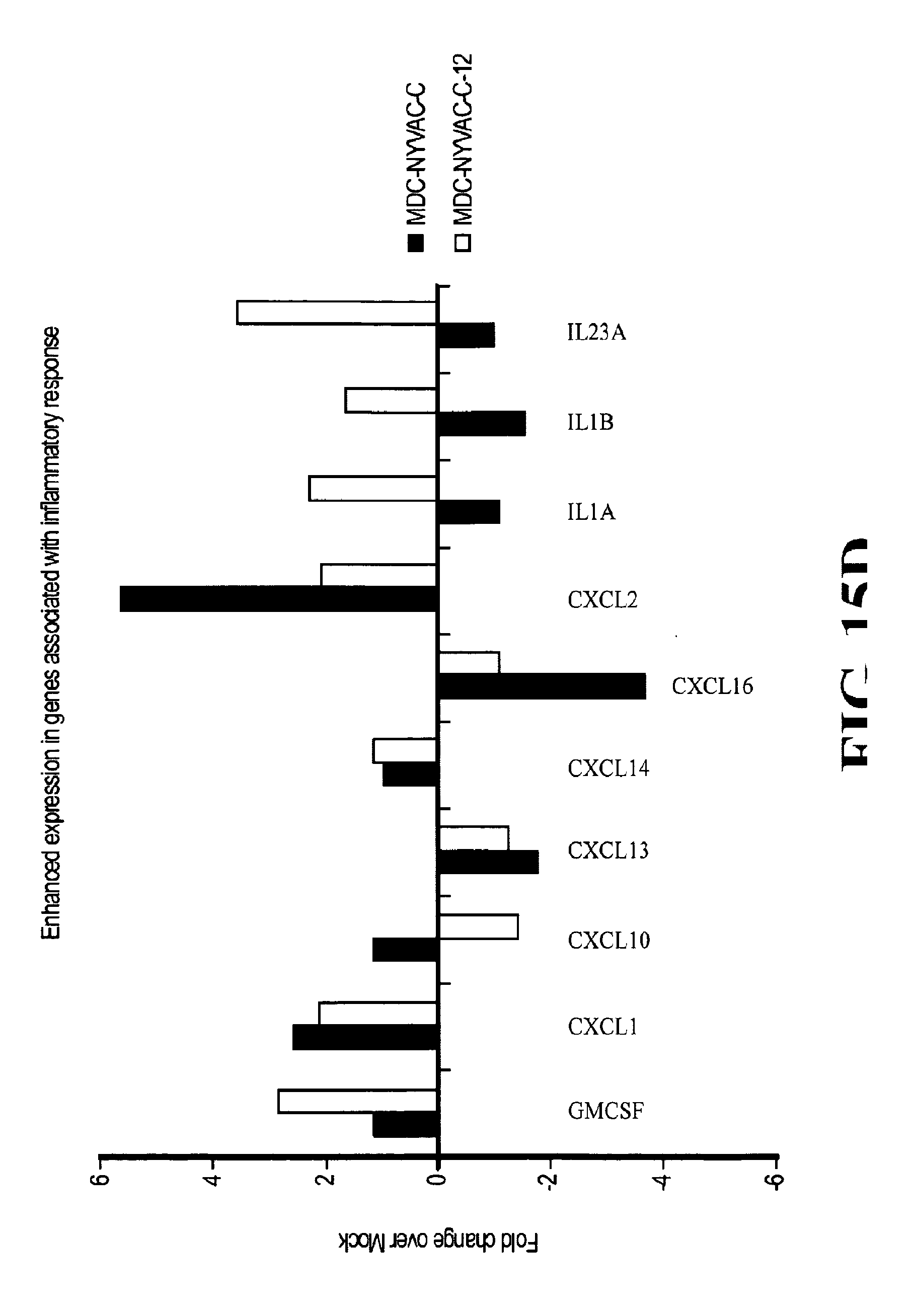
S00001
XML
uspto.report is an independent third-party trademark research tool that is not affiliated, endorsed, or sponsored by the United States Patent and Trademark Office (USPTO) or any other governmental organization. The information provided by uspto.report is based on publicly available data at the time of writing and is intended for informational purposes only.
While we strive to provide accurate and up-to-date information, we do not guarantee the accuracy, completeness, reliability, or suitability of the information displayed on this site. The use of this site is at your own risk. Any reliance you place on such information is therefore strictly at your own risk.
All official trademark data, including owner information, should be verified by visiting the official USPTO website at www.uspto.gov. This site is not intended to replace professional legal advice and should not be used as a substitute for consulting with a legal professional who is knowledgeable about trademark law.