Method For Enhancing A Function Of A T Cell
Shiku; Hiroshi ; et al.
U.S. patent application number 13/254602 was filed with the patent office on 2011-12-29 for method for enhancing a function of a t cell. Invention is credited to Hiroaki Ikeda, Koichi Iwamura, Ikunoshin Kato, Junichi Mineno, Hiroshi Shiku.
| Application Number | 20110318839 13/254602 |
| Document ID | / |
| Family ID | 42709802 |
| Filed Date | 2011-12-29 |


| United States Patent Application | 20110318839 |
| Kind Code | A1 |
| Shiku; Hiroshi ; et al. | December 29, 2011 |
METHOD FOR ENHANCING A FUNCTION OF A T CELL
Abstract
Disclosed is a method for enhancing the function of a T cell, which is characterized by inhibiting the expression of programmed death-1 ligand 1 (PD-L1) and/or programmed death-1 ligand 2 (PD-L2) in the T cell. Also disclosed is a function-enhanced T cell which is produced by the function enhancement method. Further disclosed is a therapeutic agent comprising the function-enhanced T cell. The T cell can enhance an immune response to cancer, and is useful in an immunotherapy effective for cancer and the treatment or prevention of infectious diseases and autoimmune diseases.
| Inventors: | Shiku; Hiroshi; (Tsu-shi, JP) ; Ikeda; Hiroaki; (Tsu-shi, JP) ; Iwamura; Koichi; (Tsu-shi, JP) ; Mineno; Junichi; (Otsu-shi, JP) ; Kato; Ikunoshin; (Otsu-shi, JP) |
| Family ID: | 42709802 |
| Appl. No.: | 13/254602 |
| Filed: | March 5, 2010 |
| PCT Filed: | March 5, 2010 |
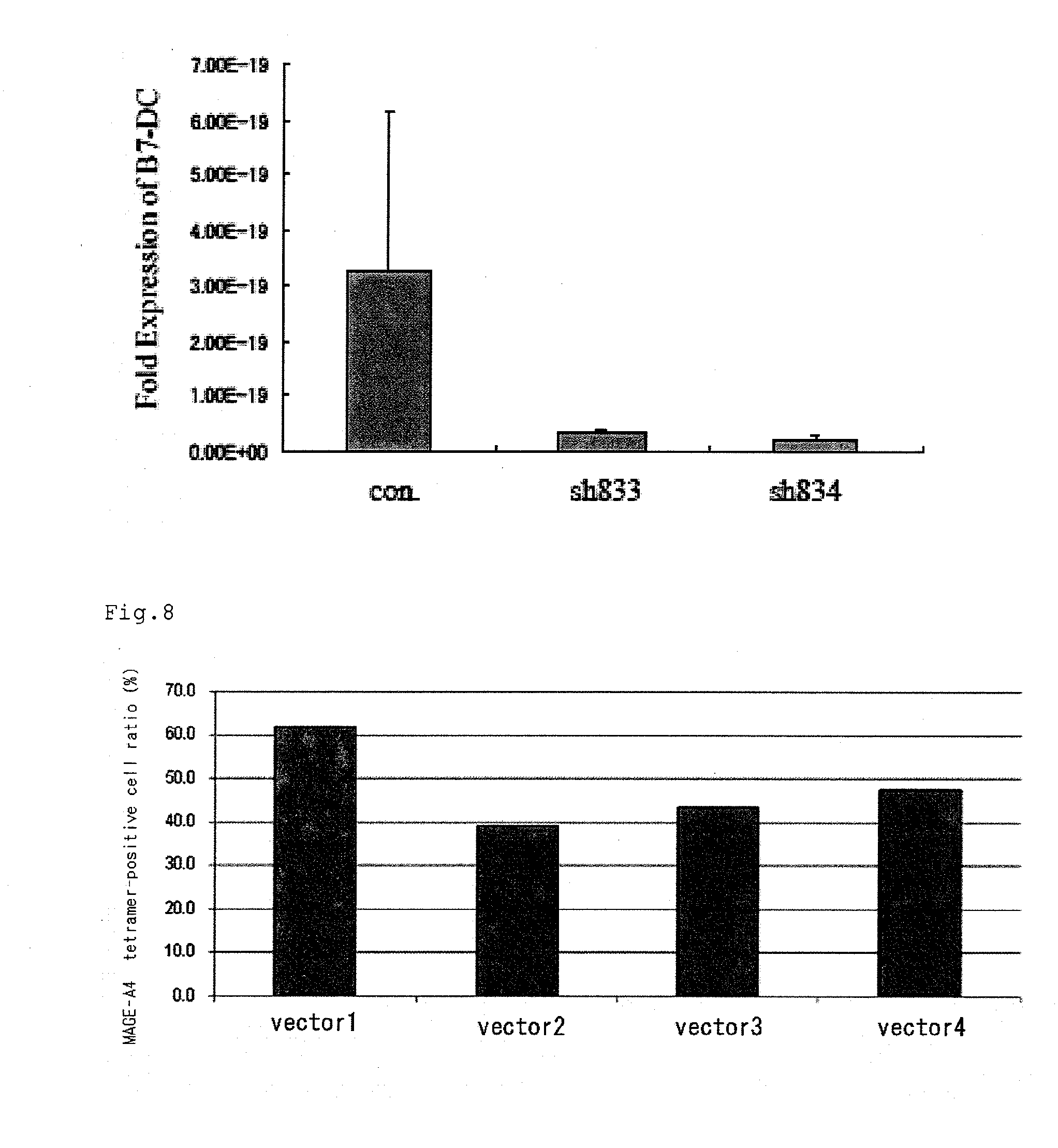
| PCT NO: | PCT/JP2010/053666 |
| 371 Date: | September 2, 2011 |
| Current U.S. Class: | 435/375 |
| Current CPC Class: | A61K 35/17 20130101; A61P 31/00 20180101; C12N 2501/599 20130101; A61P 37/06 20180101; A61K 2035/124 20130101; C12N 2501/48 20130101; A61P 35/00 20180101; C12N 5/0636 20130101; C12N 2310/14 20130101; A61P 37/04 20180101; C12N 15/113 20130101; A61K 2039/5158 20130101; C12N 2310/14 20130101; C12N 2310/531 20130101 |
| Class at Publication: | 435/375 |
| International Class: | C12N 5/0783 20100101 C12N005/0783 |
Foreign Application Data
| Date | Code | Application Number |
|---|---|---|
| Mar 6, 2009 | JP | 2009-053536 |
Claims
1. A method for enhancing the function of a T cell, comprising suppressing the expression of Programmed Death-1 Ligand 1 (PD-L1) and/or Programmed Death-1 Ligand 2 (PD-L2) of the T cell.
2. The method for enhancing the function of a T cell according to claim 1, wherein the T cell is a cytotoxic T cell or a regulatory T cell.
3. The method for enhancing the function of a T cell according to claim 2, wherein the regulatory T cell is a helper T cell, a controlling T cell or a Th17 cell.
4. The method for enhancing the function of a T cell according to claim 1, wherein the function enhancement is the production enhancement of T cell interferon-.gamma. (IFN-.gamma.).
5. The method for enhancing the function of a T cell according to claim 1, wherein the expression of PD-L1 and/or PD-L2 is suppressed by siRNA for PD-L1 and/or PD-L2.
6. A T cell with enhanced function, which is suppressed in the expression of PD-L1 and/or PD-L2.
7. The T cell with enhanced function according to claim 6, wherein the T cell is a cytotoxic T cell or a regulatory T cell.
8. The T cell with enhanced function according to claim 7, wherein the regulatory T cell is a helper T cell, a controlling T cell or a Th17 cell.
9. The T cell with enhanced function according to claim 6, wherein the function enhancement is the production enhancement of T cell interferon-.gamma. (IFN-.gamma.).
10. The T cell with enhanced function according to claim 6, wherein the expression of PD-L1 and/or PD-L2 is suppressed by siRNA for PD-L1 and/or PD-L2.
11. A therapeutic agent for a disease showing sensitivity to the T cell with enhanced function according to claim 6, comprising the T cell as an active ingredient.
12. A therapeutic agent for a disease showing sensitivity to the T cell with enhanced function according to claim 7, comprising the T cell as an active ingredient.
13. A therapeutic agent for a disease showing sensitivity to the T cell with enhanced function according to claim 8, comprising the T cell as an active ingredient.
14. A therapeutic agent for a disease showing sensitivity to the T cell with enhanced function according to claim 9, comprising the T cell as an active ingredient.
15. A therapeutic agent for a disease showing sensitivity to the T cell with enhanced function according to claim 10, comprising the T cell as an active ingredient.
Description
TECHNICAL FIELD
[0001] The present invention relates to a method for enhancing the function of a T cell by suppressing the expression of Programmed Death-1 Ligand 1 (PD-L1) and/or Programmed Death-1 Ligand 2 (PD-L2) of the T cell, etc.
BACKGROUND OF THE INVENTION
[0002] A signal via the B7: CD28 costimulatory molecular family is deeply involved in activation, inhibition and adjustment of a T cell reaction. It has been known that a PD-1 (Programmed Death-1) molecule belonging to the B7: CD28 costimulatory molecular family reacts with PD-L1 and PD-L2, and negatively controls the T cell reaction (Non-Patent Document 1). At first, it has been suggested that PD-1 expressed on a T cell reacts with PD-L1 expressed on an antigen presenting cell or a cancer cell, or PD-L2 on an antigen presenting cell to inhibit the activation of the T cell.
[0003] On the other hand, it has been known that PD-L1 is also expressed on a T cell. Recently, a possibility that PD-L1 binds with not only PD-1 but also B7-1 (CD80), and PD-L1 present on a T cell sends a negative signal to the T cell has been demonstrated (Non-Patent Document 2). However, what physiological and pathological roles are played by PD-L1 expressed on a T cell, in an immune reaction, is entirely unknown. Regarding PD-L2, it has been only known so far that it is expressed on a dendritic cell, a macrophage, a B1B cell, and a mast cell derived from bone marrow only upon induction, and its expression on a T cell is not clear. Therefore, a role played by PD-L2, which is physiologically or forcibly expressed on a T cell, is not entirely known.
[0004] There has been demonstrated a possibility that an immune response inhibiting signal from a costimulatory molecule, a representative of which is CTLA4 or PD-1, induces tolerance (immunological tolerance) via suppression of priming or the effector function at the initial stage of an autoreactive T cell while balancing with an immune response activating signal from a costimulatory molecule, a representative of which is a T cell receptor (TCR) or CD28, thereby protecting tissues from the autoreactive T cell and, on the other hand, controlling infection immunity or tumor immunity. In addition, it has been thought that certain tumors or viruses suppress the activation and proliferation of a T cell by a direct or indirect mechanism utilizing the costimulatory molecule, to weaken an immune reaction against themselves. Further, it has been thought that in a part of diseases attributed to a functional disorder of a T cell, abnormality of the costimulatory molecule is a cause for a functional disorder of a T cell.
PRIOR ART DOCUMENTS
Non-Patent Document
[0005] Non-Patent Document 1: Keir M, and other three persons, Annu. Rev. Immunol., Vol. 26, pp. 677-704 (2008) [0006] Non-Patent Document 2: Butte M, and other four persons, Immunity, Vol. 27, pp. 111-122 (2007)
SUMMARY OF THE INVENTION
Problems to be Solved by the Invention
[0007] An object of the present invention is to provide a method for enhancing the function of a T cell, and a T cell with enhanced function.
Means for Solving the Problems
[0008] The present inventors have found that it is possible to control a T cell immune response by artificially controlling the expression of PD-L1 and/or PD-L2 of a T cell. Particularly, the present inventors have found that reduction of the expression of PD-L1 and/or PD-L2 in an anti-tumor T cell enhances the anti-tumor effect of the T cell, and thus it results in completion of the present invention.
[0009] That is, the present invention relates to:
[1] a method for enhancing the function of a T cell, comprising suppressing the expression of Programmed Death-1 Ligand 1 (PD-L1) and/or Programmed Death-1 Ligand 2 (PD-L2) of the T cell; [2] the method for enhancing the function of a T cell according to [1], wherein the T cell is a cytotoxic T cell or a regulatory T cell; [3] the method for enhancing the function of a T cell according to [2], wherein the regulatory T cell is a helper T cell, a controlling T cell or a Th17 cell; [4] the method for enhancing the function of a T cell according to [1], wherein the function enhancement is the production enhancement of T cell interferon-.gamma. (IFN-.gamma.); [5] the method for enhancing the function of a T cell according to [1], wherein the expression of PD-L1 and/or PD-L2 is suppressed by siRNA for PD-L1 and/or PD-L2; [6] a T cell with enhanced function, which is suppressed in the expression of PD-L1 and/or PD-L2; [7] the T cell with enhanced function according to [6], wherein the T cell is a cytotoxic T cell or a regulatory T cell; [8] the T cell with enhanced function according to [7], wherein the regulatory T cell is a helper T cell, a controlling T cell or a Th17 cell; [9] the T cell with enhanced function according to [6], wherein the function enhancement is the production enhancement of T cell interferon-.gamma. (IFN-.gamma.); [10] the T cell with enhanced function according to [6], wherein the expression of PD-L1 and/or PD-L2 is suppressed by siRNA for PD-L1 and/or PD-L2; and [11] a therapeutic agent for a disease showing sensitivity to the T cell with enhanced function according to any one of [6] to [10], comprising the T cell as an active ingredient.
ADVANTAGES OF THE INVENTION
[0010] According to the present invention, a method for enhancing the function of a T cell, a T cell with enhanced function, etc., are provided. This T cell enhances an immune response to cancer, and is useful in effective cancer immunotherapy, or treatment or prevention of an infection disease or an autoimmune disease.
BRIEF DESCRIPTION OF THE DRAWINGS
[0011] FIG. 1 is a view analyzing the expression of PD-L1 of a CD8-positive T cell.
[0012] FIG. 2 is a view analyzing the expression of PD-L1 of a CD8-positive T cell.
[0013] FIG. 3 is a view analyzing the expression of PD-L2 of a CD8-positive T cell.
[0014] FIG. 4 is a view showing the concentration of interferon .gamma. (IFN-.gamma.) of a CD8-positive T cell.
[0015] FIG. 5 is a view showing the concentration of IFN-.gamma. of a CD4-positive T cell.
[0016] FIG. 6 is a view analyzing the expression of PD-L1 of LCL.
[0017] FIG. 7 is a view analyzing the expression of PD-L2 of LCL.
[0018] FIG. 8 is a view showing the ratio of a CD8-positive tetramer-positive T cell.
DETAILED DESCRIPTION OF THE PREFERRED EMBODIMENTS
[0019] Herein, "enhanced function of a T cell" means that the effector function of the T cell is improved. The enhanced function of the T cell, which does not limit the present invention, includes an improvement in the proliferation rate of the T cell, an increase in the production amount of cytokine, or an improvement in cytotoxity. Further, the enhanced function of the T cell includes cancellation and suppression of tolerance of the T cell in the suppressed state such as the anergy (unresponsive) state, or the rest state, that is, transfer of the T cell from the suppressed state into the state where the T cell responds to stimulation from the outside.
[0020] In the present invention, "anergy" includes unresponsiveness to an immune cell to stimulation, for example, stimulation by an activation receptor or cytokine. The anergy may occur due to, for example, exposure to an immune suppressor or exposure to an antigen in a high dose. Such anergy is generally antigen-specific, and continues even after completion of exposure to a tolerized antigen. For example, the anergy in a T cell is characterized by failure of production of cytokine, for example, interleukin (IL)-2. The T cell anergy occurs when a first signal (signal via TCR or CD-3) is received in the absence of a second signal (costimulatory signal) upon exposure of a T cell to an antigen.
[0021] Herein, "PD-L1" means ligand 1 of PD-1, which is a costimulatory molecule expressed on a so-called antigen presenting cell such as an activated monocyte or a dendritic cell, and is also referred to as B7-H1. The nucleotide sequence of human PD-L1 is shown by GenBank Acc. No. AF233516, and the nucleotide sequence of mouse PD-L1 is shown by NM.sub.--021893 (Freeman et al. (2000) J. Exp. Med. 192:1027).
[0022] Herein, "PD-L2" means ligand 2 of PD-1, and is also referred to as B7-DC. The nucleotide sequence of human PD-L2 is shown by GenBank Acc. No. NM.sub.--025239, and the nucleotide sequence of mouse PD-L2 is shown by NM.sub.--021896 (Nature Immunology, 2001, Vol. 2, No. 3, pp. 261-267).
[0023] Herein, "T cell" is also referred to as T lymphocyte, and means a cell derived from thymus among lymphocytes involved in an immune response. The T cell includes any of a CD8-positive T cell (cytotoxic T cell: CTL), a CD4-positive T cell (helper T cell), a suppressor T cell, a regulatory T cell such as a controlling T cell, an effector cell, a naive T cell, a memory T cell, an .alpha..beta.T cell expressing TCR .alpha. and .beta. chains, and a .gamma..delta.T cell expressing TCR .gamma. and .delta. chains. The T cell includes a precursor cell of a T cell in which differentiation into a T cell is directed. Examples of "cell populations containing T cells" include, in addition to body fluids such as blood (peripheral blood, umbilical blood etc.) and bone marrow fluids, cell populations containing peripheral blood mononuclear cells (PBMC), hematopoietic cells, hematopoietic stem cells, umbilical blood mononuclear cells etc., which have been collected, isolated, purified or induced from the body fluids. Further, a variety of cell populations containing T cells and derived from hematopoietic cells can be used in the present invention. These cells may have been activated by cytokine such as IL-2 in vivo or ex vivo. As these cells, any of cells collected from a living body, or cells obtained via ex vivo culture, for example, a T cell population obtained by the method of the present invention as it is, or obtained by freeze preservation, can be used.
[0024] Herein, "cancer" means a malignant tumor, and refers to a cell exhibiting abnormal proliferation phenotype or the abnormal cell state, which has a feature of exhibiting autonomous proliferation and, as a result, uncontrollable cell proliferation. "Tumor cells" include "malignant tumor cells" or "benign tumor cells", and are also referred to as "cells derived from neoplasm".
[0025] Herein, "expression" means generation of mRNA by transcription from nucleic acids such as genes, polynucleotides, and oligonucleotides, or generation of a protein or a polypeptide by transcription from mRNA. "Suppression of expression" refers to a decrease of a transcription product or a translation product in a significant amount as compared with the case of no suppression. The suppression of expression herein shows, for example, a decrease of a transcription product or a translation product in an amount of 30% or more, preferably 50% or more, more preferably 70% or more, and further preferably 90% or more.
[0026] (1) Method for Enhancing Function of T Cell and T Cell with Enhanced Function of the Present Invention
[0027] The method for enhancing the function of a T cell of the present invention is a method including the step of suppressing the expression of PD-L1 and/or PD-L2 of the T cell. In addition, the T cell with enhanced function of the present invention is a cell obtained by the method for enhancing the function of a T cell of the present invention. The method of enhancing the function of a T cell of the present invention makes it possible to improve the effector function of the T cell, including an improvement in the proliferation rate of the T cell, an increase in the production amount of cytokine or an improvement in cytotoxity. Further, the suppressed state of the T cell such as the anergy state, or the rest state is cancelled or suppressed, and the sensitivity of the T cell to stimulation from the outside is improved. The thus obtained T cell with enhanced function of the present invention is useful in treating or preventing cancer, an infection disease or an autoimmune disease.
[0028] In the method of the present invention, the step of suppressing the expression of PD-L1 and/or PD-L2 of the T cell can be performed ex vivo or in vivo.
[0029] The method of the present invention in which the step is performed ex vivo includes the steps of: (a) preparing a T cell or a cell population containing a T cell, and (b) suppressing the expression of PD-L1 and/or PD-L2 of the T cell obtained in the step (a) to prepare a T cell with enhanced function. The method can be applied to cell immunotherapy including adoptive immunotherapy, and is useful. The step (a) may be the step of separating a T cell or a cell population containing a T cell from a living body, or the step of preparing an established T cell or a cell population containing a T cell. Further, the method of the present invention may includes the step of separating the cell population containing a T cell from cell populations to prepare a cell sub-population and/or the step of culturing and stimulating the cell population. These steps can be performed at an arbitrary stage before, after or simultaneous with the step (a) and the step (b). In the method of the present invention in which the step of suppressing the expression of PD-L1 and/or PD-L2 of the T cell is performed ex vivo, the amount and kind of the cell to which the method is applied can be controlled through a suitable means, for example, preparation of a cell sub-population, or culturing or stimulation of the cell population. Therefore, a harmful event such as autoimmunity, which is concerned when a substance suppressing the expression of PD-L1 and/or PD-L2 or a substance inhibiting signal transmission is administered to a living body, can be reduced, and therefore it is extremely useful.
[0030] The step of separating the cell population and cell sub-population containing a T cell can be performed, for example, by fractionation of a mononuclear cell fraction by density gradient centrifugation, or a separation means using the surface marker of the T cell as an index. Examples of the surface marker include CD3, CD8 and CD4, and separation methods depending on these surface markers are known in the art. For example, the step can be performed by mixing a carrier such as beads or a culturing container on which an anti-CD8 antibody has been immobilized, with a cell population containing a T cell, and recovering a CD8-positive T cell bound to the carrier. As the beads on which an anti-CD8 antibody has been immobilized, for example, CD8 MicroBeads (manufactured by Miltenyi Biotec), Dynabeads M450 CD8 (manufactured by Invitrogen), and Eligix anti-CD8 mAb coated nickel particles (manufactured by Biotransplant) can be suitably used. This is also the same as in implementation using CD4 as an index and, for example, CD4 MicroBeads (manufactured by Miltenyi Biotec), Dynabeads M-450 CD4 (manufactured by Invitrogen) can be suitably used.
[0031] The step of culturing the cell population and cell sub-population containing a T cell can be performed by selecting suitable known culturing conditions depending on the cell population. In addition, in the step of stimulating the cell population, known proteins and chemical ingredients, etc., may be added to the medium to perform culturing. For example, cytokines, chemokines or other ingredients may be added to the medium. Herein, the cytokine is not particularly limited as far as it can act on the T cell, and examples thereof include IL-2, IFN-.gamma., transforming growth factor (TGF)-.beta., IL-15, IL-7, IFN-.alpha., IL-12, CD40L, and IL-27. From the viewpoint of enhancing cellular immunity, particularly suitably, IL-2, IFN-.gamma., or IL-12 is used and, from the viewpoint of improvement in survival of a transferred T cell in vivo, IL-7, IL-15 or IL-21 is suitably used. In addition, the chemokine is not particularly limited as far as it acts on the T cell and exhibits migration activity, and examples thereof include RANTES, CCL21, MIP1.alpha., MIP1.beta., CCL19, CXCL12, IP-10 and MIG. The stimulation of the cell population can be performed by the presence of a ligand for a molecule present on the surface of the T cell, for example, CD3, CD28, or CD44 and/or an antibody to the molecule. Further, the cell population can be stimulated by contacting with other lymphocytes such as antigen presenting cells (dendritic cell) presenting a target peptide such as a peptide derived from a cancer antigen on the surface of a cell.
[0032] When the step of suppressing the expression of PD-L1 and/or PD-L2 of the T cell is performed in vivo, the step is performed by administering a means to specifically suppress the expression thereof described later to a living body. Thereupon, a suitable drug delivery system (e.g. liposome, fine particles, microcapsule etc.) can be utilized. Further, for example, utilizing the delivery system using the surface marker of the T cell as an index, the expression of PD-L1 and/or PD-L2 of the T cell can be specifically suppressed.
[0033] Examples of the means to suppress the expression of PD-L1 and/or PD-L2 of the T cell include utilization of nucleic acids such as siRNA for PD-L1 and/or PD-L2, an antisense nucleotide, and a ribozyme.
[0034] The utilization of siRNA in the present invention is for the purpose of selectively suppressing the expression of PD-L1 and/or PD-L2, and is based on RNA interference (RNAi) utilizing a double-stranded RNA molecule having a sequence homologous with the nucleotide sequence of mRNA which is transcribed from a gene encoding PD-L1 and/or PD-L2, and a sequence complementary with the nucleotide sequence. Herein, "having a sequence homologous with the nucleotide sequence of mRNA which is transcribed from a gene encoding PD-L1 and/or PD-L2, and a sequence complementary with the nucleotide sequence" not only refers to the fact that each sequence is completely homologous or complementary with the nucleotide sequence of mRNA, but also includes the fact that each sequence is substantially homologous or complementary in such a range that the desired function is exerted. The double-stranded RNA molecule is referred to as siRNA (short interfering RNA). The siRNA may be one kind of siRNA which is homologous/complementary with one region of mRNA which is transcribed from a gene encoding PD-L1 and/or PD-L2, or may be siRNA including a plurality of RNA molecules which are homologous/complementary with different regions.
[0035] Examples of the chain length of the siRNA used in the present invention include, from the viewpoint of suppression of interferon response in a mammal cell, those in which one strand of a duplex constituting the siRNA has a chain length of 13 to 29 base pairs, preferably a chain length of 15 to 25 base pairs, further preferably a chain length of 20 to 25 base pairs. In addition, all of the nucleotide sequences of the chain lengths may be derived from the nucleotide sequence of the mRNA of PD-L1 and/or PD-L2, or a part thereof may be derived from the nucleotide sequence. Further, the siRNA used in the present invention, from the viewpoint of effectiveness of RNA interference in a mammal cell, may be, for example, a shape of double-stranded RNA having a single-stranded region which is protruded on a 3'-terminal side by 2 to 4 nucleotides, further preferably a shape of double-stranded RNA having a single-stranded region which is protruded on a 3'-terminal side by 2 nucleotides. As the protruded single-stranded region, deoxythymidine residue of continuous 2 to 4 nucleotides (TT, TTT or TTTT) is exemplified.
[0036] The siRNA used in the present invention is mainly comprised of ribonucleotides, and a part thereof may include nucleotides other than ribonucleotides, for example, deoxyribonucleotides, a derivative of deoxyribonucleotides, a derivative of ribonucleotides, etc. The siRNA can be synthesized by a known chemical synthesis method, but the method is not particularly limited. It may be enzymatically (e.g., using an RNA polymerase) prepared using a suitable template nucleic acid. The siRNA used in the present invention may be in the form of single-stranded RNA which can form a duplex in the molecule, and single-stranded RNA with a stem-loop structure (short hairpin structure: sh structure) having the si RNA part as a stem and an arbitrary sequence as a loop (shRNA) is exemplified. As the arbitrary sequence, a sequence of 1 to 30 nucleotides is exemplified, and a sequence of preferably 1 to 25 nucleotides, further preferably 5 to 22 nucleotides is used.
[0037] The sequence of the siRNA can be appropriately designed based on a gene sequence whose expression is desired to be suppressed. Many siRNA design algorisms have been reported (see, e.g., WO 2004/0455543, and WO 2004/048566), and a commercially available software can also be used. In addition, there are many companies which design siRNA from information of a gene sequence whose expression is desired to be suppressed, and synthesize and provide the siRNA. Therefore, a person skilled in the art can easily obtain the siRNA based on the gene sequence whose expression is desired to be suppressed. As the siRNA used in the present invention, any can be used as far as it is siRNA which selectively suppresses the expression of PD-L1 and/or PD-L2. For example, siRNA including the nucleotide sequence of SEQ ID NO: 1 or SEQ ID NO: 9 can be used for PD-L1, and siRNA including the nucleotide sequence of SEQ ID NO: 2 or SEQ ID NO: 11 can be used for PD-L2, without particular limitation.
[0038] In the present invention, an antisense nucleotide can be used for suppressing the expression of PD-L1 and/or PD-L2. The antisense nucleotide is used for suppressing the expression of a protein, for example, by directly interfering translation of the mRNA molecule of PD-L1 and/or PD-L2, by degradation of mRNA by an RNA degradation enzyme H, by interfering the 5' capping of mRNA, by masking the 5' cap, by preventing binding of a translation factor with mRNA, or by inhibiting polyadenylation of mRNA. The suppression of the expression of a protein occurs by hybridization between an antisense nucleotide and the mRNA of PD-L1 and/or PD-L2. In order to reduce stability of, or degrade mRNA, as a target of the antisense nucleotide, a specific targeting site on the mRNA is selected. When one or more target sites are identified, a nucleotide having a nucleotide sequence sufficiently complementary with the target site (that is, which hybridizes sufficiently and with sufficient specificity under the physiological conditions) is designed.
[0039] As the antisense nucleotide used in the method of the present invention, for example, a nucleotide having a chain length of 8 to 100 nucleotides, preferably 10 to 80 nucleotides, more preferably 14 to 35 nucleotides is exemplified.
[0040] In the present invention, nucleic acids such as siRNA and an antisense nucleotide used in suppressing the expression of PD-L1 and/or PD-L2 may be directly introduced into a cell, or a nucleic acid construct which has been designed so that the siRNA or the antisense nucleotide is transcribed in a cell may be introduced into a cell. When directly introduced into a cell, a reagent for introducing a nucleic acid such as TransIT-TKO (manufactured by Mirus), and Human T Cell Nucleofector Kit (Amaxa) can be suitably used. On the other hand, when the nucleic acid construct in which the RNA molecule is transcribed is used, a nucleic acid construct can be used which is functionally connected downstream of a promoter capable of exerting the function in the T cell in the state where transcription of a nucleic acid encoding the siRNA or the antisense nucleotide can occur, but it does not particularly limit the present invention. In addition, in order to attain effective transcription of a gene, other regulatory sequences cooperating with a promoter or a transcription initiation site, for example, an enhancer sequence or a terminator sequence may be present in the nucleic acid construct. In addition, for the purpose of insertion of a subject to be introduced into a chromosome of the T cell by homologous recombination, for example, the gene may be arranged between flanking sequences including nucleotide sequences, each having homology with nucleotide sequences on both sides of a desired target insertion site of a gene in the chromosome.
[0041] The promoter used in the nucleic acid construct is not particularly limited as far as it can function in a mammal cell, and examples thereof include an RNA polymerase II promoter, an RNA polymerase III promoter, and an artificially regulatable promoter. Example of the RNA polymerase II promoter includes a CMV promoter. In addition, examples of the RNA polymerase III-based promoter include a tRNA promoter, a U6snRNA promoter, and a histone H1 promoter. As the promoter which can be artificially regulated with tetracycline, a promoter which can be regulated with tetracycline is exemplified, and examples thereof include a tetracycline-regulated U6 promoter, and a TR promoter. In addition, combination of the promoter with the Cre-loxP system makes it possible to control transcription more strictly.
[0042] The nucleic acid construct used for transcribing siRNA can be constructed so that a sense strand and an antisense strand of double-stranded RNA which can suppress the function of an objective gene are transcribed by the following system: (A) a tandem type in which a nucleic acid encoding sense RNA and a nucleic acid encoding antisense RNA are connected downstream of different two promoters, respectively, and these two transcription units are arranged in the forward direction, and the sense RNA and the antisense RNA are transcribed separately, (B) a type in which a nucleic acid encoding sense RNA and a nucleic acid encoding antisense RNA are arranged downstream of one promoter, respectively, in the forward direction, and RNA having a stem-loop type (or short hairpin type) in which the sense RNA and the antisense RNA are connected directly or with a loop is transcribed, or (C) a counter type in which a promoter is arranged on both end sides of nucleic acids encoding a sense strand and an antisense strand (with both strands, respectively), respectively, and both RNA strands are transcribed with separate promoters. In the present invention, the tandem type, the stem-loop type or the counter type can be used depending on the use conditions, for example, the kind of a cell, the kind of a sense sequence or an antisense sequence etc.
[0043] The nucleic acid construct used in the present invention may be incorporated into a suitable vector, for example, a plasmid vector or a virus vector so as to more stably exert the effect in a cell. Further, the nucleic acid construct of the present invention may be incorporated on the chromosome DNA of a cell. Examples of the plasmid vector include, but are not particularly limited to, a piGENE tRNA plasmid (trade name, manufactured by iGENE), siLentGene (manufactured by Promega), and pSEC Hygro Vector (manufactured by Ambion). In order to introduce the plasmid vector into a cell, a method using a carrier such as a liposome or ligand-polylysine, a calcium phosphate method, an electroporation method, a particle gun method, etc., can be used.
[0044] The virus vector is not particularly limited, and usually, a known virus vector used in a gene introduction method, for example, a retrovirus vector (including lentivirus vector, pseudo type vector), an adenovirus vector, an adeno-associated virus vector, a simian virus vector, a vaccinia virus vector or a sendai virus vector is used. Particularly preferably, a retrovirus vector, an adenovirus vector or a lentivirus vector is used. As the virus vector, a virus vector in which the replicating ability is defective so that it cannot self-replicate in an infected cell is preferable. In addition, upon gene introduction, a substance improving a gene introduction efficiency such as retronectin (registered trademark, manufactured by TAKARA BIO INC.) can be also used. An example of a commercially available adenovirus vector includes Knockout Adenoviral RNAi System (manufactured by Clonetech) and, examples of a commercially available retrovirus vector include a pSINsi vector (manufactured by TAKARA BIO INC.) and pSIREN-RetroQ Vector (manufactured by Clonetech), respectively. In the case of the virus vector, it can be introduced into an objective cell utilizing the ability of the virus to infect a cell.
[0045] In the method of the present invention, a means to suppress the expression of PD-L1 and/or PD-L2 is implemented on the T cell. That is, in the step (b), for example, a helper T cell, a suppressor T cell, a controlling T cell, a cytotoxic T cell (CTL), a naive T cell, a memory T cell, an .alpha..beta.T cell expressing TCR .alpha. and .beta. chains or a .gamma..delta.T cell expressing TCR .gamma. and .delta. chains, or a cell population containing these cells, blood (peripheral blood, umbilical blood etc.) or a bone marrow fluid can be used. These T cells may be derived from a mammal, or may be derived from either human or a non-human mammal.
[0046] The suppression of the expression of PD-L1 and/or PD-L2 can be confirmed by quantitative determination of the PD-L1 and/or PD-L2 protein, or quantitative determination of RNA transcribed from the PD-L1 and/or PD-L2 gene.
[0047] The function enhancement of the T cell in the method of the present invention can be assessed at a plurality of time points before and after each step using a cytokine assay, an antigen-specific cell assay (tetramer assay), a proliferation assay, a cytolytic cell assay, or an in vivo delayed hypersensitivity test using a recombinant tumor-associated antigen or an immunogenic fragment or an antigen-derived peptide. Examples of an additional method for measuring an increase in an immune response include a delayed hypersensitivity test, flow cytometry using a peptide major histocompatibility gene complex tetramer. a lymphocyte proliferation assay, an enzyme-linked immunosorbent assay, an enzyme-linked immunospot assay, cytokine flow cytometry, a direct cytotoxity assay, measurement of cytokine mRNA by a quantitative reverse transcriptase polymerase chain reaction, or an assay which is currently used for measuring a T cell response such as a limiting dilution method.
[0048] The T cell with enhanced function of the present invention is enhanced in the function of the T cell as compared with a control T cell in which the expression of PD-L1 and/or PD-L2 is not suppressed. The T cell with enhanced function of the present invention is increased in, for example, the production amount of IFN-.gamma. as compared with a control T cell. The T cell of the present invention is increased in the production amount of IFN-.gamma. by 10% or more, preferably 20% or more, more preferably 30% or more as compared with a control T cell.
[0049] Examples of one aspect of the present invention include a method of further enhancing the function of the T cell, in addition to the suppression of the expression of PD-L1 and/or PD-L2. For example, introduction of a gene encoding TCR recognizing a desired antigen into a T cell makes it possible to obtain a T cell to which specificity for the antigen is imparted, and whose effector function is improved. Examples of the antigen include, but are not particularly limited to, a tumor antigen, a microorganism and a virus-derived antigen. Regarding TCR to a variety of antigens, its amino acid sequence, and the nucleotide sequence of a gene encoding the amino acid sequence are known and, construction of a vector for TCR expression based on the information and introduction of the vector into a T cell makes it possible to produce a T cell exerting an antigen-specific action. Thereupon, simultaneous introduction of a nucleic acid construct for suppressing the expression of PD-L1 and/or PD-L2 into a T cell, or incorporation of the construct into the vector for TCR expression makes it possible to improve the effector function of an antigen-specific T cell.
[0050] When TCR including .alpha. and .beta. chains is artificially expressed in a T cell, mispairing between the .alpha. and .beta. chains derived from an endogenous TCR gene possessed by the T cell itself, etc., may hinder the imparting of desired antigen specificity. For example, implementation of codon conversion of an introduced TCR gene or knockdown of an endogenous TCT gene utilizing siRNA, which is disclosed in WO 2008/153029, makes it possible to produce a T cell which has acquired high antigenic specificity. The method for enhancing the function of a T cell of the present invention can also be utilized in producing such a T cell.
[0051] (2) Treating Method or Preventing Method Using T Cell with Enhanced Function of the Present Invention
[0052] The treating method or preventing method of the present invention has a feature of administering the T cell prepared by the method for enhancing the function of the present invention in (1) to a living body. That is, the treating method or preventing method of the present invention includes the step of: (c) administering the T cell with enhanced function obtained in the step (b) to a living body. The disease for which the T cell with enhanced function of the present invention is administered is not particularly limited as far as it is a disease exhibiting sensitivity to the T cell, and examples thereof include cancer (leukemia, solid tumor, etc.), hepatitis, influenza, an infectious disease, a cause of which is a virus such as HIV, a bacterium or a fungus, for example, tuberculosis, MRSA, VRE, deep mycosis. In addition, the cell of the present invention can also be utilized in bone mallow transplantation, or prevention of an infectious disease after radiation, or donor lymphocyte infusion for the purpose of remission of recurrent leukemia. Further, the cell improves the function of a controlling T cell, and can be utilized in preventing or treating an autoimmune disease.
[0053] In the method of the present invention, the T cell with enhanced function can be administered to a subject intradermally, intramuscularly, subcutaneously, intraperitoneally, intraarterially, intravenously (including a method performed by an indwelling catheter), intratumorally, or into an afferent lymph vessel.
[0054] In the treating method or preventing method of the present invention, the T cell with enhanced function to be administered may be a cell which is self to a living body, or may be a cross-derived cell.
[0055] An immune response induced in implementation of the present invention can be assessed in a living body before first administration of the T cell with enhanced function of the present invention, or at various time points after initiation of treatment, using an antigen-specific cell assay, a proliferation assay, a cytolytic cell assay, or an in vivo delayed hypersensitivity test using a recombinant tumor-associated antigen or an immunogenic fragment or an antigen-derived peptide. Examples of an additional method for measuring an increase in an immune response include a delayed hypersensitivity test, flow cytometry using a peptide major histocompatibility gene complex tetramer. a lymphocyte proliferation assay, an enzyme-linked immunosorbent assay, an enzyme-linked immunospot assay, cytokine flow cytometry, a direct cytotoxity assay, measurement of cytokine mRNA by a quantitative reverse transcriptase polymerase chain reaction, or an assay which is currently used for measuring a T cell response such as a limiting dilution method. Further, an immune response can be assessed by a weight, diameter or malignant degree of a tumor possessed by a living body, the infection virus amount or infection bacterium amount of a subject, or the survival rate or survival term of a subject.
[0056] In the method of the present invention, examples of the amount of the T cell with enhanced function to be administered to a living body include, but are not particularly limited to, preferably 1.times.10.sup.6 to 1.times.10.sup.12 cells/day, more preferably 1.times.10.sup.7 to 5.times.10.sup.11 cells/day, and further preferably 1.times.10.sup.8 to 2.times.10.sup.11 cells/day per adult a day.
[0057] (3) Therapeutic Agent for Disease Containing T Cell with Enhanced Function of the Present Invention
[0058] The T cell with enhanced function prepared by the method for enhancing the function of the present invention in (1), and a composition containing the T cell as an active ingredient are useful as a therapeutic agent for a disease having sensitivity to the T cell, for example, the aforementioned cancer, hepatitis or an infectious disease.
[0059] The therapeutic agent of the present invention can be prepared, for example, by mixing the T cell prepared by the method of present invention as an active ingredient with, for example, known organic or inorganic carrier, excipient, stabilizer, etc., which are suitable for parenteral administration, according to a method known in the pharmacy filed. In addition, use thereof may be performed according to the treating method of the present invention in (2).
EXAMPLES
[0060] The present invention will be described further concretely below by way of Examples, but the present invention is not limited to only the scope of the following Examples.
Example 1
Confirmation of Expression of PD-L1 in CD8-Positive T Cell
[0061] According to WO 2007/032255, HLA-A24 restrictive MAGE-A 4.sub.143-151-specific cytotoxic CD8-positive T cell clone 2-28 (Miyahara Y, and other 13 persons, Clin. Cancer Res., Vol. 11, pp. 5581-5589 (2005), hereinafter, referred to as CD8-positive T cell, clone 2-28) and a MAGE-A4.sub.141-151 peptide were prepared. The CD8-positive T cell clone 2-28 was co-cultured at 37.degree. C. with self lymphoblastoid cell line (LCL) which had been irradiated with 80 Gly X-ray and had been pulsed with MAGE-A4.sub.143-151 peptide, PD-L1 on the cell surface after 0 day, 1 day, 2 days and 3 days was stained with an anti-human PD-L1 antibody (manufactured by BD bioscience), and analyzed with a flow cytometer. FIG. 1 shows the results of analysis with a flow cytometer. The expression of PD-L1 was highest after 1 day of culturing.
Example 2
Suppression of Expression of PD-L1 and PD-L2 of CD8-Positive T Cell by RNA Interference
[0062] SiRNA specific for PD-L1 having a sequence described in SEQ ID NO: 1, siRNA specific for PD-L2 having a sequence described in SEQ ID NO: 2, or negative control siRNA (all manufactured by Invitrogen) was introduced into CD8-positive T cell clone 2-28 by electroporation.
[0063] A cell with siRNA for PD-L1 introduced therein was cultured for 3 days, and then co-cultured at 37.degree. C. with self LCL which had been irradiated with 80 Gly X-ray and had been pulsed with a MAGE-A4.sub.143-151 peptide. On the next day, this was stained with an anti-human PD-L1 antibody, and the expression of PD-L1 on the cell surface was analyzed with a flow cytometer.
[0064] Regarding a cell with siRNA for PD-L2 introduced therein, the cell was stained with an anti-human PD-L2 antibody (manufactured by BD bioscience) and the expression of PD-L2 in the cell was analyzed with a flow cytometer, after 2 days and 3 days after introduction.
[0065] The results of analysis of PD-L1 are shown in FIG. 2, and the results of analysis of PD-L2 are shown in FIG. 3, respectively. In the cell with no siRNA introduced therein (non-treatment) and the cell with negative control siRNA introduced therein (si control), suppression of the expression of PD-L1 or PD-L2 was not seen. In the cell with siRNA specific for PD-L1 introduced therein (si PD-L1), and the cell with siRNA specific for PD-L2 introduced therein (si PD-L2), suppression of the expression of PD-L1 and PD-L2 was seen, respectively.
Example 3
Promotion of Production of IFN-.gamma. of PD-L1 and PD-L2-Expressing CD8-Positive T-Cell by RNA Interference
[0066] Negative control siRNA, siRNA specific for PD-1 having a sequence described in SEQ ID NO: 3 (manufactured by Invitrogen), siRNA specific for PD-L1 having a sequence described in SEQ ID NO: 1, or siRNA specific for PD-L2 having a sequence described in SEQ ID NO: 2 was introduced into CD8-positive T cell clone 2-28 by electroporation. After culturing for 3 days, this cell was mixed with a cell in which self LCL had been pulsed with a MAGE-A4.sub.143-151 peptide at a ratio of 4:1 or 2:1, and the mixture was co-cultured at 37.degree. C. and, on the next day, the concentration of IFN-.gamma. in each culturing supernatant was measured by an ELISA method.
[0067] The concentration of IFN-.gamma. in each cell supernatant is shown in FIG. 4. In the cell with siRNA specific for PD-1 introduced therein (si PD-1), change in the concentration of IFN-.gamma. was not seen at culturing with both mixing ratios, as compared with the cell with negative control siRNA introduced therein. In the cell with siRNA specific for PD-L1 introduced therein and the cell with siRNA specific for PD-L2 introduced therein, an increase in the concentration of IFN-.gamma. was significantly seen, respectively, as compared with the cell with negative control siRNA introduced therein and the cell with siRNA specific for PD-1 introduced therein.
Example 4
Promotion of Expression of IFN-.gamma. of PD-L1 and PD-L2-Expressing CD4-Positive T Cell by RNA Interference
[0068] Negative control siRNA, siRNA specific for PD-1 having a sequence described in SEQ ID NO: 3, siRNA specific for PD-L1 having a sequence described in SEQ ID NO: 1, or siRNA specific for PD-L1 having a sequence described in SEQ ID NO: 2 was introduced into MAGE-A4.sub.46-66-specific CD4-positive T cell clone 5 by electroporation, and this was cultured for 3 days. This cell was mixed with a cell in which self LCL had been pulsed with a MAGE-A4.sub.46-66 peptide at 2:1, and the mixture was cultured at 37.degree. C. and, on the next day, the concentration of IFN-.gamma. in each culturing supernatant was measured by an ELISA method.
[0069] The concentration of IFN-.gamma. in each cell supernatant is shown in FIG. 5. In the cell with negative control siRNA introduced therein, and the cell with siRNA specific for PD-1 introduced therein, the expression of IFN-.gamma. was not seen. In the cell with siRNA specific for PD-L1 introduced therein and the cell with siRNA specific for PD-L2 introduced therein, the expression of IFN-.gamma. was significantly seen.
Example 5
Selection of siRNA for Suppressing Expression of PD-L1 and PD-L2
[0070] SiRNAs specific for PD-L1 each having a sequence described in SEQ ID NO: 8 or 9 (sh831, sh832), or siRNAs specific for PD-L2 each having a sequence described in SEQ ID NO: 10 or 11 (sh833, sh834) (all manufactured by TAKARA BIO INC.), or the negative control siRNA used in Example 2 was introduced into LCL by electroporation, this was cultured at 37.degree. C. for 2 days, total RNA was extracted from each cell, the expression of PD-L1 and PD-L2 was measured by real time RT-PCR using QuanTitect SYBR PCR Kit (manufactured by QIAGEN), and siRNA having strong expression suppressing power was selected.
[0071] Sequences of primers for real time RT-PCR used in measuring the expression of PD-L1 are shown in SEQ ID Nos.: 12 and 13, sequences of primers for real time RT-PCR used in measuring the expression of PD-L2 are shown in SEQ ID Nos.: 14 and 15, and sequences of primers for real time RT-PCR of human GAPDH (glyceraldehyde-3-phosphate dehydrogenase) as a subject of measurement of the expression are shown in SEQ ID Nos.: 16 and 17, respectively.
[0072] The results of real time RT-PCR of PD-L1 are shown in FIG. 6, and the results of real time RT-PCR of PD-L2 are shown in FIG. 7. In the figures, an ordinate axis shows the ratio of the expression of PD-L1 or PD-L2 relative to the expression of human GAPDH. From this result, sh832 shown in SEQ ID NO: 9 as siRNA specific for PD-L1, and sh834 shown in SEQ ID NO: 11 as siRNA specific for PD-L2 were selected.
Example 6
Production of Codon Conversion-Type TCR-Expressing Retrovirus Vector
[0073] According to WO 2008/153029, pMS-Ma2 and pMS-Pb2 were produced. These vectors include the retrovirus vector system based on MSCV (Murine Stem Cell Virus). Further, those vectors include genes encoding TCR .alpha. and .beta. chains recognizing tumor antigen MAGE-A4, respectively and, in these genes, codons were converted so that the expression is not suppressed by RNA interference with siRNA on TCR described later (hereinafter, a gene in which codons are converted is referred to as codon conversion-type).
Example 7
Production of Codon Conversion-Type TCR and siRNA-Expressing Retrovirus Vector
[0074] A codon conversion-type TCR.alpha. gene was excised with MluI and BglII from pMS-Ma2 produced in Example 6, and TCR.alpha.-MluI/BglII was prepared. A siRNA cluster, a PDL1-PDL2-TCR.alpha.-TCR.beta. artificially synthesized gene shown in SEQ ID NO: 4 was digested with BglII and NotI, and cloned into the MluI-NotI site of pMS-Pb2 together with TCR.alpha.-MluI/BglII, to prepare an MS-aPb1-siPDL.sub.--1/2_siTCR vector (vector 1). The vector 1 expresses the codon conversion-type TCR, and can express siRNAs for PD-L1, PD-L2, TCR.alpha. and TCR.beta. shown in SEQ ID Nos.: 9, 11, 18 and 19. Among them, siRNA s for TCR.alpha. and TCR.beta. suppress the expression of endogeneous wild-type TCR, and does not suppress the expression of the codon conversion-type TCR.
[0075] Further, a siRNA cluster, a PDL1-PDL2 artificially synthesized gene shown in SEQ ID NO: 5, a siRNA cluster PDL1 artificially synthesized gene shown in SEQ ID NO: 6, and a siRNA cluster PDL2 artificially synthesized gene shown in SEQ ID NO: 7 were prepared, and an MS-aPb1-siPDL.sub.--1/2 vector (vector 2), an MS-aPb1-siPDL.sub.--1 vector (vector 3), and an MS-aPb1-siPDL.sub.--2 vector (vector 4) were prepared as in the vector 1. The summary of genes expressed by respective vectors is shown in Table 1.
TABLE-US-00001 TABLE 1 Expressed gene Vector 1 Vector 2 Vector 3 Vector 4 Codon .largecircle. .largecircle. .largecircle. .largecircle. conversion-type TCR.alpha. gene Codon .largecircle. .largecircle. .largecircle. .largecircle. conversion-type TCR.beta. gene siRNA for TCR.alpha. .largecircle. siRNA for TCR.beta. .largecircle. siRNA for PD-L1 .largecircle. .largecircle. .largecircle. siRNA for PD-L2 .largecircle. .largecircle. .largecircle.
Example 8
Production of Codon Conversion-Type TCR and siRNA-Expressing Retrovirus Solution
[0076] Escherichia coli JM109 was transformed with the vectors 1 to 4 prepared in Example 7, and plasmid DNA was purified using QIAGEN Plasmid Midi Kit (manufactured by QIAGEN) to provide DNA for transfection.
[0077] Each prepared DNA for transfection was transfected into a 293T cell according to a product protocol using Retrovirus Packing Kit Eco (manufactured by TAKARA BIO INC.) to obtain each ecotropic virus supernatant solution. This supernatant solution was filtered with a 0.45 .mu.m filter (Milex HV, manufactured by Millipore), and made to infect a PG13 cell (ATCC CRL-10686) by a method using polybrene [Kawai et al., Mol. Cell. Biol., Vol. 4, p. 1172, (1984)], the resulting culture supernatant of the cell was recovered, and filtered with a 0.45 .mu.m filter to prepare a retrovirus solution.
Example 9
Infection of Human Peripheral Blood Mononuclear Cell with Codon Conversion-Type TCR and siRNA-Co-Expressing Retrovirus Vector
[0078] A peripheral blood mononuclear cell (PBMC) separated from human peripheral blood was infected with the retrovirus solution produced in Example 8 two times according to a manual using Retronectin (registered trademark, manufactured by TAKARA BIO INC.) to produce codon conversion-type TCR and a siRNA-co-expressing introduction peripheral blood mononuclear cell. After 3 days from second virus infection, the cell was recovered, genome was extracted with FastPure DNA Kit (TAKARA BIO INC.), and the average provirus copy number in each cell genome was calculated with Proviral Copy Number Detection Primer Set (manufactured by TAKARA BIO INC.) and CycleavePCR Core Kit (manufactured by TAKARA BIO INC.). The provirus copy number of PBMC infected with the virus solution containing the vectors 1 to 4 was 4.6 copies/cell, 2.2 copies/cell, 3.2 copies/cell, and 4.5 copies/cell, respectively. Then, after 5 days from second virus infection, the cell was recovered, and stained with HLA-A2402 MAGE-A4 tetramer PE (manufactured by MBL) and Human CD8-FITC CONJUGATE (manufactured by Becton, Dickinson), and the ratio of a cell which is CD8-positive and tetramer-positive according to a flow cytometer was determined.
[0079] The rate of an MAGE-A4 tetramer-positive cell is shown in FIG. 8. An abscissa axis indicates an introduced retrovirus vector, and an ordinate axis indicates the rate of an MAGE-A4 tetramer-positive cell (%). As shown in FIG. 8, the expression of an anti-MAGE-A4TCR.alpha./.beta. composite protein by gene introduction into a human peripheral blood mononuclear cell was confirmed by each retrovirus vector introduction. In addition, the expression of siRNA for PD-L1 and PD-L2 expressed by the retrovirus vector did not greatly influence on the expression of the introduced codon conversion-type TCR. Further, siRNAs for TCR.alpha. and TCR.beta. enhanced the rate of codon conversion-type TCR.alpha./.beta. composite protein expression by the effect of suppressing the expression of a wild-type endogeneous TCR gene by siRNA.
INDUSTRIAL APPLICABILITY
[0080] The present invention provides a method for enhancing the function of a T cell, and a T cell with enhanced function. This T cell enhances an immune response to cancer, and can be utilized in effective cancer immunotherapy, and treatment or prevention of an infection disease or an autoimmune disease.
SEQUENCE LISTING FREE TEXT
[0081] SEQ ID NO:1: siRNA specific for PD-L1 SEQ ID NO:2: siRNA specific for PD-L2 SEQ ID NO:3: siRNA specific for PD-1 SEQ ID NO:4: siRNA cluster PDL1-PDL2-TCR alpha-TCR beta SEQ ID NO:5: siRNA cluster PDL1-PDL2 SEQ ID NO:6: siRNA cluster PDL1 SEQ ID NO:7: siRNA cluster PDL2 SEQ ID NO:8: siRNA for PD-L1 (sh831) SEQ ID NO:9: siRNA for PD-L1 (sh832) SEQ ID NO:10: siRNA for PD-L2 (sh833) SEQ ID NO:11: siRNA for PD-L2 (sh834) SEQ ID NO:12: real time PCR F-primer for PD-L1 SEQ ID NO:13: real time PCR R-primer for PD-L1 SEQ ID NO:14: real time PCR F-primer for PD-L2 SEQ ID NO:15: real time PCR R-primer for PD-L2 SEQ ID NO:16: real time PCR F-primer for GAPDH SEQ ID NO:17: real time PCR R-primer for GAPDH SEQ ID NO:18: siRNA for TCR alpha SEQ ID NO:19: siRNA for TCR beta
Sequence CWU 1
1
19125RNAArtificialsiRNA specific for PD-L1 1aaugcguuca gcaaaugcca
guagg 25225RNAArtificialsiRNA specific for PD-L2 2aaaugaaagc
aaugaugcag gaggg 25325RNAArtificialsiRNA specific for PD-1
3uuacgucucc uccaaaugug uauca 254657DNAArtificialsiRNA cluster
PDL1-PDL2-TCR alpha-TCR beta 4atcgggccca gatcttattt caaatttagc
aggaaaaaag agaacatcac cttgtaaaac 60tgaagattgt gaccagtcag aataatgtcg
actacaagcg aattactcat gctacagtca 120agatgagtaa ttcgcttgta
gtcgcattat ggtgacagct gcctcgggaa gccaagttgg 180gctttaaagt
gcagggcctg ctgatgttga gtgctttttg ttcggacagt accaatgcat
240aaactgtgct ctgagaagct tatgcattgg tactgtccca taagaagtta
tgtattcatc 300caataattca agccaagcaa gtatataggt gttttaatag
tttttgtttg cagtcctctg 360ttgtaaggat tctgatgtgt acacagggaa
gcgagtctgt acacatcaga atccttactg 420gtggcctgct atttccttca
aatgaatgat ttttactaat tttgtgtact tttattgtgt 480cgatgtagaa
tctgcctggt ctatctgatg tgacagcttc tgtagcaccc accatcctct
540atgagattag tgctcctggt tgatctcata gaggatggtg gtactgctag
ctgtagaact 600ccagcttcgg cctgtcgccc aatcaaactg tcctgttact
ggcggccgca tcgattc 6575351DNAArtificialsiRNA cluster PDL1-PDL2
5atcgggccca gatcttattt caaatttagc aggaaaaaag agaacatcac cttgtaaaac
60tgaagattgt gaccagtcag aataatgtcg actacaagcg aattactcat gctacagtca
120agatgagtaa ttcgcttgta gtcgcattat ggtgacagct gcctcgggaa
gccaagttgg 180gctttaaagt gcagggcctg ctgatgttga gtgctttttg
ttcggacagt accaatgcat 240aaactgtgct ctgagaagct tatgcattgg
tactgtccca taagaagtta tgtattcatc 300caataattca agccaagcaa
gtatataggt gttttaatag cggccgctcc c 3516200DNAArtificialsiRNA
cluster PDL1 6atcgggccca gatcttattt caaatttagc aggaaaaaag
agaacatcac cttgtaaaac 60tgaagattgt gaccagtcag aataatgtcg actacaagcg
aattactcat gctacagtca 120agatgagtaa ttcgcttgta gtcgcattat
ggtgacagct gcctcgggaa gccaagttgg 180gctttaaagc ggccgcatat
2007203DNAArtificialsiRNA cluster PDL2 7atcgggccca gatcttattt
caaatttagc aggaaaaaag agaacatcac cttgtaaaac 60tgaagattgt gaccagtcag
aataatgtgg acagtaccaa tgcataaact gtgctctgag 120aagcttatgc
attggtactg tcccattatg gtgacagctg cctcgggaag ccaagttggg
180ctttaaagcg gccgcatcga ttc 203819RNAArtificialsiRNA for PD-L1
(sh831) 8cuaauugucu auugggaaa 19919RNAArtificialsiRNA for PD-L1
(sh832) 9cgacuacaag cgaauuacu 191019RNAArtificialsiRNA for PD-L2
(sh833) 10ggacgaagga caguaccaa 191119RNAArtificialsiRNA for PD-L2
(sh834) 11ggacaguacc aaugcauaa 191220DNAArtificialreal time PCR
F-primer for PD-L1 12tatggtggtg ccgactacaa 201320DNAArtificialreal
time PCR R-primer for PD-L1 13tgcttgtcca gatgacttcg
201420DNAArtificialreal time PCR F-primer for PD-L2 14gggacgaagg
acactaccaa 201520DNAArtificialreal time PCR R-primer for PD-L2
15tttggccagg atacttctgc 201620DNAArtificialreal time PCR F-primer
for GAPDH 16gagtcaacgg atttggtcgt 201720DNAArtificialreal time PCR
R-primer for GAPDH 17gatctcgctc ctggaagatg 201819RNAArtificialsiRNA
for TCR alpha 18guaaggauuc ugaugugua 191919RNAArtificialsiRNA for
TCR beta 19ccaccauccu cuaugagau 19
D00001
D00002
D00003
D00004
D00005
S00001
XML
uspto.report is an independent third-party trademark research tool that is not affiliated, endorsed, or sponsored by the United States Patent and Trademark Office (USPTO) or any other governmental organization. The information provided by uspto.report is based on publicly available data at the time of writing and is intended for informational purposes only.
While we strive to provide accurate and up-to-date information, we do not guarantee the accuracy, completeness, reliability, or suitability of the information displayed on this site. The use of this site is at your own risk. Any reliance you place on such information is therefore strictly at your own risk.
All official trademark data, including owner information, should be verified by visiting the official USPTO website at www.uspto.gov. This site is not intended to replace professional legal advice and should not be used as a substitute for consulting with a legal professional who is knowledgeable about trademark law.