MHC Multimers in Cancer Vaccines and Immune Monitoring
Brix; Liselotte ; et al.
U.S. patent application number 13/122027 was filed with the patent office on 2011-12-29 for mhc multimers in cancer vaccines and immune monitoring. This patent application is currently assigned to DAKO DENMARK A/S. Invention is credited to Liselotte Brix, Henrik Pedersen, Jorgen Scholler.
| Application Number | 20110318380 13/122027 |
| Document ID | / |
| Family ID | 41479174 |
| Filed Date | 2011-12-29 |


View All Diagrams
| United States Patent Application | 20110318380 |
| Kind Code | A1 |
| Brix; Liselotte ; et al. | December 29, 2011 |
MHC Multimers in Cancer Vaccines and Immune Monitoring
Abstract
The present invention relates to MHC-peptide complexes and uses thereof in the diagnosis of, treatment of or vaccination against a disease in an individual. More specifically the invention discloses MHC complexes comprising cancer antigenic peptides and uses there of.
| Inventors: | Brix; Liselotte; (Bagsvaerd, DK) ; Scholler; Jorgen; (Lyngby, DK) ; Pedersen; Henrik; (Lynge, DE) |
| Assignee: | DAKO DENMARK A/S Glostrup DK |
| Family ID: | 41479174 |
| Appl. No.: | 13/122027 |
| Filed: | October 1, 2009 |
| PCT Filed: | October 1, 2009 |
| PCT NO: | PCT/DK2009/050255 |
| 371 Date: | September 14, 2011 |
Related U.S. Patent Documents
| Application Number | Filing Date | Patent Number | ||
|---|---|---|---|---|
| 61101878 | Oct 1, 2008 | |||
| Current U.S. Class: | 424/193.1 ; 424/277.1; 435/325; 435/7.24; 530/300; 530/405 |
| Current CPC Class: | A61P 37/04 20180101; A61P 35/00 20180101; G01N 33/56972 20130101; C07K 14/70539 20130101; A61K 47/6901 20170801; C07K 14/705 20130101; A61K 39/0011 20130101 |
| Class at Publication: | 424/193.1 ; 530/405; 424/277.1; 530/300; 435/7.24; 435/325 |
| International Class: | A61K 39/385 20060101 A61K039/385; A61K 39/00 20060101 A61K039/00; C07K 2/00 20060101 C07K002/00; A61P 35/00 20060101 A61P035/00; G01N 33/566 20060101 G01N033/566; C12N 5/0783 20100101 C12N005/0783; A61P 37/04 20060101 A61P037/04; C07K 14/74 20060101 C07K014/74; C07K 1/107 20060101 C07K001/107 |
Foreign Application Data
| Date | Code | Application Number |
|---|---|---|
| Oct 1, 2008 | DK | PA 2008 01382 |
| Mar 6, 2009 | EP | 09154516.0 |
Claims
1. A MHC monomer comprising a-b-P or a MHC multimer comprising (a-b-P).sub.n, wherein n>1, wherein a and b together form a functional MHC protein capable of binding the antigenic peptide, P, wherein (a-b-P) is the MHC-peptide complex formed when the peptide P binds to the functional MHC protein, wherein each MHC peptide complex of a MHC multimer is associated with one or more multimerization domains and wherein P is a cancer antigenic peptide.
2. An antigenic peptide comprising or consisting of a sequence selected from the group of sequences included in FIG. 29 and FIG. 32 and modified sequences obtained by modification of a sequence selected from the group of sequences included in FIG. 29 and FIG. 32.
3-1630. (canceled)
1631. A composition comprising a plurality of MHC monomers and/or MHC multimers and/or antigenic peptides and/or antigenic polypeptides according to any of claims 1 to 2, wherein the WIC monomers and/or MHC multimers and/or antigenic peptides and/or antigenic polypeptides are identical or different, and a carrier.
1632. A kit comprising a MHC monomer or a MHC multimer or an antigenic peptide or an antigenic polypeptide or a composition according to any of claims 1 to 2 and 1631 and at least one additional component, such as a positive control and/or instructions for use.
1633. A method for generating the MHC multimer according to claim 1, said method comprising the steps of i) providing one or more antigenic peptides P; ii) providing one or more functional MHC proteins, iii) optionally providing one or more multimerization domains, and iv) contacting or reacting the one or more antigenic peptides P and the one or more functional MHC proteins and the one or more multimerization domains simultaneously or sequentially in any order, thereby obtaining MHC multimers according to the present invention.
1634. A method for immune monitoring and/or diagnosing one or more diseases comprising the following steps providing; a MHC monomer or MHC multimer or antigenic peptide or antigenic polypeptide according to any of claims 1 to 2, or the individual components thereof, and providing a population of T cells, and measuring the number, activity or state of T cells specific for said MHC monomer, MHC multimer, antigenic peptide, antigenic polypeptide, and thereby immune monitoring and/or diagnosing said one or more diseases.
1635. A method for isolation of one or more antigen-specific T cells, said method comprising the steps of providing a MHC monomer or MHC multimer or antigenic peptide or antigenic polypeptide according to any of claims 1 to 2 or individual components thereof, providing a population of T cells, and isolating T cells specific for said MFIC monomer, MHC multimer, antigenic peptide or antigenic polypeptide.
1636. A method for performing a vaccination of an individual in need thereof, said method comprising the steps of providing a MHC monomer or MHC multimer or antigenic peptide or antigenic polypeptide according to any of claims 1 to 2 or the individual components thereof, and administering said MHC monomer, MHC multimer, antigenic peptide or antigenic polypeptide, to said individual and obtaining a protective immune response, and thereby performing a vaccination of the said individual.
1637. A method for performing therapeutic treatment of an individual comprising the steps of providing; a MHC monomer or MHC multimer or antigenic peptide or an antigenic polypeptide according to any of claims 1 to 2 or the individual components thereof, and isolating or obtaining T-cells from a source, such as an individual or an ex-vivo library or cell bank, wherein said isolated or obtained T-cells are specific for said provided MHC monomer, MHC multimer, antigenic peptide or antigenic polypeptide, optionally manipulating said T-cells, and introducing said isolated or obtained T-cells into an individual to be subjected to a therapeutic treatment, wherein the individual can be the same individual or a different individual from the source individual.
1638. A vaccine comprising: one or more MHC monomers, one or more MHC multimers, one or more antigenic peptides or/and one or more antigenic polypeptides according to any of the claims 1 to 2 or one or more nucleic acids encoding said MHC monomers, MHC multimers, antigenic peptides and/or antigenic polypeptides.
1639. A method for performing a vaccination of an individual in need thereof, said method comprising the steps of providing a vaccine according to claim 1638 administering said vaccine to said individual and obtaining a protective immune response and thereby performing a vaccination of the said individual.
1640. A kit comprising a vaccine according to claim 1638 and at least one additional component.
1641. A method for minimization of undesired binding of the MHC multimer according to claim 1.
1642. A method for performing a control experiment comprising use of MHC multimers as a positive control.
1643. A method for performing a control experiment comprising use of MHC multimers as a negative control.
Description
[0001] The Danish patent application PA 2008 01382, the European patent application EP09154516.0 and the U.S. provisional patent application U.S. Ser. No. 61/101,878 are hereby incorporated by reference in its entirety.
[0002] All patent and non-patent references cited in PA 2008 01382, EP09154516.0 and U.S. 61/101,878, or in the present application, are also hereby incorporated by reference in their entirety.
[0003] PCT/DK2009/050185, PCT/DK2008/050167, PA 2008 01384 and PCT/DK2008/000118 are hereby incorporated by reference in its entirety.
[0004] All patent and non-patent references cited in PCT/DK2009/050185, PCT/DK2008/050167, PA 2008 01384 and PCT/DK2008/000118, are also hereby incorporated by reference in their entirety.
FIELD OF INVENTION
[0005] The present invention relates to MHC-peptide complexes and uses thereof in the treatment of a disease in an individual.
BACKGROUND OF INVENTION
[0006] Biochemical interactions between peptide epitope specific membrane molecules encoded by the Major Histocompatibility Complex (MHC, in humans HLA) and T-cell receptors (TCR) are required to elicit specific immune responses. This requires activation of T-cells by presentation to the T-cells of peptides against which a T-cell response should be raised. The peptides are presented to the T-cells by the MHC complexes.
The Immune Response
[0007] The immune response is divided into two parts termed the innate immune response and the adaptive immune response. Both responses work together to eliminate pathogens (antigens). Innate immunity is present at all times and is the first line of defense against invading pathogens. The immediate response by means of pre-existing elements, i.e. various proteins and phagocytic cells that recognize conserved features on the pathogens, is important in clearing and control of spreading of pathogens. If a pathogen is persistent in the body and thus only partially cleared by the actions of the innate immune system, the adaptive immune system initiate a response against the pathogen. The adaptive immune system is capable of eliciting a response against virtually any type of pathogen and is unlike the innate immune system capable of establishing immunological memory.
[0008] The adaptive response is highly specific to the particular pathogen that activated it but it is not so quickly launched as the innate when first encountering a pathogen.
[0009] However, due to the generation of memory cells, a fast and more efficient response is generated upon repeated exposure to the same pathogen. The adaptive response is carried out by two distinct sets of lymphocytes, the B cells producing antibodies leading to the humoral or antibody mediated immune response, and the T cells leading to the cell mediated immune response.
[0010] T cells express a clonotypic T cell receptor (TCR) on the surface. This receptor enable the T cell to recognize peptide antigens bound to major histocompatibility complex (MHC) molecules, called human leukocyte antigens (HLA) in man. Depending on the type of pathogen, being intracellular or extracellular, the antigenic peptides are bound to MHC class I or MHC class II, respectively. The two classes of MHC complexes are recognized by different subsets of T cells; Cytotoxic CD8+ T cells recognizing MHC class I and CD4+ helper cells recognizing MHC class II. In general, TCR recognition of MHC-peptide complexes result in T cell activation, clonal expansion and differentiation of the T cells into effector, memory and regulatory T cells.
[0011] B cells express a membrane bound form of immunoglobulin (Ig) called the B cell receptor (BCR). The BCR recognizes an epitope that is part of an intact three dimensional antigenic molecule. Upon BCR recognition of an antigen the BCR:antigen complex is internalized and fragments from the internalized antigen is presented in the context of MHC class II on the surface of the B cell to CD4+ helper T-cells (Th). The specific Th cell will then activate the B cell leading to differentiation into an antibody producing plasma cell.
[0012] A very important feature of the adaptive immune system is its ability to distinguish between self and non-self antigens, and preferably respond against non-self. If the immune system fails to discriminate between the two, specific immune responses against self-antigens are generated. These autoimmune reactions can lead to damage of self-tissue.
[0013] The adaptive immune response is initiated when antigens are taken up by professional antigen presenting cells such as dendritic cells, Macrophages, Langerhans cells and B-cells. These cells present peptide fragments, resulting from the degradation of proteins, in the context of MHC class II proteins (Major Histocompatibility Complex) to helper T cells. The T helper cells then mediate help to B-cells and antigen-specific cytotoxic T cells, both of which have received primary activation signals via their BCR respective TCR. The help from the Th-cell is mediated by means of soluble mediators e.g. cytokines.
[0014] In general the interactions between the various cells of the cellular immune response is governed by receptor-ligand interactions directly between the cells and by production of various soluble reporter substances e.g. cytokines by activated cells.
MHC-Peptide Complexes.
[0015] MHC complexes function as antigenic peptide receptors, collecting peptides inside the cell and transporting them to the cell surface, where the MHC-peptide complex can be recognized by T-lymphocytes. Two classes of classical MHC complexes exist, MHC class I and II. The most important difference between these two molecules lies in the protein source from which they obtain their associated peptides. MHC class I molecules present peptides derived from endogenous antigens degraded in the cytosol and are thus able to display fragments of viral proteins and unique proteins derived from cancerous cells. Almost all nucleated cells express MHC class I on their surface even though the expression level varies among different cell types. MHC class II molecules bind peptides derived from exogenous antigens. Exogenous proteins enter the cells by endocytosis or phagocytosis, and these proteins are degraded by proteases in acidified intracellular vesicles before presentation by MHC class II molecules. MHC class II molecules are only expressed on professional antigen presenting cells like B cells and macrophages.
[0016] The three-dimensional structure of MHC class I and II molecules are very similar but important differences exist. MHC class I molecules consist of two polypeptide chains, a heavy chain, .alpha., spanning the membrane and a light chain, .beta.2-microglobulin (.beta.2m). The heavy chain is encoded in the gene complex termed the major histocompatibility complex (MHC), and its extracellular portion comprises three domains, .alpha.1, .alpha.2 and .alpha.3. The .beta.2m chain is not encoded in the MHC gene and consists of a single domain, which together with the .alpha.3 domain of the heavy chain make up a folded structure that closely resembles that of the immunoglobulin. The .alpha.1 and .alpha.2 domains pair to form the peptide binding cleft, consisting of two segmented .alpha. helices lying on a sheet of eight .beta.-strands. In humans as well as in mice three different types of MHC class I molecule exist. HLA-A, B, C are found in humans while MHC class I molecules in mice are designated H-2K, H-2D and H-2L.
[0017] The MHC class II molecule is composed of two membrane spanning polypeptide chains, .alpha. and .beta., of similar size (about 30000 Da). Genes located in the major histocompatibility complex encode both chains. Each chain consists of two domains, where .alpha.1 and .beta.1 forms a 9-pocket peptide-binding cleft, where pocket 1, 4, 6 and 9 are considered as major peptide binding pockets. The .alpha.2 and .beta.2, like the .alpha.2 and .beta.2m in the MHC class I molecules, have amino acid sequence and structural similarities to immunoglobulin constant domains. In contrast to MHC class I complexes, where the ends of the antigenic peptide is buried, peptide-ends in MHC class II complexes are not. HLA-DR, DQ and DP are the human class II molecules, H-2A, M and E are those of the mice.
[0018] A remarkable feature of MHC genes is their polymorphism accomplished by multiple alleles at each gene. The polygenic and polymorphic nature of MHC genes is reflected in the peptide-binding cleft so that different MHC complexes bind different sets of peptides. The variable amino acids in the peptide binding cleft form pockets where the amino acid side chains of the bound peptide can be buried. This permits a specific variant of MHC to bind some peptides better than others.
MHC Multimers
[0019] Due to the short half-life of the peptide-MHC-T cell receptor ternary complex (typically between 10 and 25 seconds) it is difficult to label specific T cells with labelled MHC-peptide complexes, and like-wise, it is difficult to employ such monomers of MHC-peptide for therapeutic and vaccine purposes because of their weak binding. In order to circumvent this problem, MHC multimers have been developed. These are complexes that include multiple copies of MHC-peptide complexes, providing these complexes with an increased affinity and half-life of interaction, compared to that of the monomer MHC-peptide complex. The multiple copies of MHC-peptide complexes are attached, covalently or non-covalently, to a multimerization domain. Known examples of such MHC multimers include the following: [0020] MHC-dimers: Each MHC dimer contains two copies of MHC-peptide. IgG is used as multimerization domain, and one of the domains of the MHC protein is covalently linked to IgG. [0021] MHC-tetramers: Each MHC-tetramer contains four copies of MHC-peptide, each of which is biotinylated. The MHC complexes are held together in a complex by the streptavidin tetramer protein, providing a non-covalent linkage between a streptavidin monomer and the MHC protein. Tetramers are described in U.S. Pat. No. 5,635,363. [0022] MHC pentamers: Five copies of MHC-peptide complexes are multimerised by a self-assembling coiled-coil domain, to form a MHC pentamer. MHC pentamers are described in the US patent 2004209295 [0023] MHC dextramers: A large number of MHC-peptide complexes, typically more than ten, are attached to a dextran polymer. MHC-dextramers are described in the patent application WO 02/072631 A2. [0024] MHC streptamers: 8-12 MHC-peptide complexes attached to Streptactin. MHC streptamers are described in Knabel M et al. Reversibel MHC multimer staining for functional isolation of T-cell populations and effective adoptive transfer. Nature medicine 6. 631-637 (2002).
Use of MHC Multimers in Flow Cytometry and Related Techniques
[0025] The concentration of antigen-specific T-cells in samples from e.g. peripheral blood can be very low. Flow cytometry and related methods offer the ability to analyze a large number of cells and simultaneously identify the few of interest. MHC multimers have turned out to be very valuable reagents for detection and characterization of antigen-specific T-cells in flow cytometer experiments. The relative amount of antigen-specific T cells in a sample can be determined and also the affinity of the binding of MHC multimer to the T-cell receptor can be determined.
[0026] The basic function of a flow cytometer is its ability to analyse and identify fluorochrome labelled entities in a liquid sample, by means of its excitation, using a light source such as a laser beam and the light emission from the bound fluorochrome.
[0027] MHC multimers is used as detections molecule for identification of antigen-specific T-cells in flow cytometry, by labelling the MHC multimer with a specific fluorochrome, which is detectable, by the flow cytometer used.
[0028] In order to facilitate the identification of a small amount of cells, the cells can be sub-categorized using antibodies or other fluorochrome labelled detections molecules directed against surface markers other than the TCR on the specific T-cells population. Antibodies or other fluorochrome labelled detections molecules can also be used to identify cells known not to be antigen-specific T-cells. Both kinds of detections molecules are in the following referred to as gating reagents. Gating reagents, helps identify the "true" antigen-specific T cells bound by MHC multimers by identifying specific subpopulations in a sample, e.g. T cells and by excluding cells that for some reason bind MHC multimers without being antigen-specific T-cells.
[0029] Other cytometry methods, e.g. fluorescence microscopy and IHC can like flow cytometry be employed in identification of antigen-specific T cells in a cell sample using MHC multimers.
Application of MHC Multimers in Immune Monitoring, Diagnostics, Prognostics, Therapy and Vaccines
[0030] T cells are pivotal for mounting an adaptive immune response. It is therefore of importance to be able to measure the number of specific T cells when performing a monitoring of a given immune response, for example in connection with vaccine development, infectious diseases e.g. tuberculosis, toxicity studies etc.
[0031] Accordingly, the present invention further provides powerful tools in the fields of vaccines, therapy and diagnosis. One objective of the present invention is to provide methods for anti-bacterial immunotherapy by generating antigen-specific T-cells capable of inactivating or eliminating undesirable target cells. Another objective is to isolate antigen-specific T-cells and culture these in the presence of co-stimulatory molecules. Ex vivo priming and expansion of T-cell populations allows the T-cells to be used in immunotherapy of various types of infectious diseases. A third objective of the present invention is to identify and label specific subsets of cells with relevance for the development or treatment of diseases.
[0032] One disease of special interest of the present invention is cancer. MHC multimers of the present invention are can be used in prognostics, diagnosis, vaccine monitoring, vaccine and therapy related to this disease.
SUMMARY OF INVENTION
[0033] Measurement of antigen-specific T cells during an immune response are important parameters in vaccine development, autologous cancer therapy, transplantation, infectious diseases, inflammation, autoimmunity, toxicity studies etc. MHC multimers are crucial reagents in monitoring of antigen-specific T cells. The present invention describes novel methods to generate MHC multimers and methods to improve existing and new MHC multimers. The invention also describes improved methods for the use of MHC multimers in analysis of T cells in samples including diagnostic, prognostic and immune monitoring methods. Furthermore the use of MHC multimers in anti-tumour therapy are described, including isolation of antigen-specific T cells capable of inactivation or elimination of undesirable target cells or isolation of specific T cells capable of regulation of other immune cells. The present invention also relates to MHC multimers comprising one or more tumour derived peptides. In one preferred embodiment the present invention relates to a cancer vaccine comprising antigenic peptides derived from cancer proteins. The antigenic peptides may be used themselves as a vaccine or used in a MHC multimer bound in the peptide binding cleft of MHC.
[0034] The present invention also relates to a composition for cancer vaccination and/or immune monitoring of a vaccine response. In another embodiment the present invention relates to a method of making the composition for cancer vaccination and/or immune monitoring of a vaccine response. This invention also relates to a method for cancer vaccination comprising administration to an individual in need thereof an effective amount of a cancer vaccine composition.
Definitions
[0035] As used everywhere herein, the term "a", "an" or "the" is meant to be one or more, i.e. at least one.
[0036] Adjuvant: adjuvants are drugs that have few or no pharmacological effects by themselves, but can increase the efficacy or potency of other drugs when given at the same time. In another embodiment, an adjuvant is an agent which, while not having any specific antigenic effect in it self, can stimulate the immune system, increasing the response to a vaccine.
[0037] Agonist: agonist as used herein is a substance that binds to a specific receptor and triggers a response in the cell. It mimics the action of an endogenous ligand that binds to the same receptor.
[0038] Anchor amino acid: Anchor amino acid is used interchangeably herein with anchor residue and is an amino acid of antigenic peptide having amino acid sidechains that bind into pockets lining the peptide-binding groove of MHC molecules thereby anchoring the peptide to the MHC molecule. Anchor residues being responsible for the main anchoring of peptide to MHC molecule are called primary anchor amino acids. Amino acids contributing to the binding of antigenic peptide to MHC molecule but in a lesser extend than primary anchor amino acids are called secondary anchor amino acids.
[0039] Anchor motif: The pattern of anchor residues in an antigenic peptide binding a certain MHC molecule. Peptides binding different MHC molecules have different anchor motifs defined by the patterns of anchor residues in the peptide sequence.
[0040] Anchor residue: Anchor residue is used interchangeably herein with anchor amino acid
[0041] Anchor position: The position of an anchor amino acid in antigenic peptide sequence. For MHC II the anchor positions is defined in the 9-mer core motif.
[0042] Antagonist: antagonist as used herein is a substance that binds to a specific receptor and blocks the response in the cell. It blocks the action of an endogenous ligand that binds to the same receptor.
[0043] Antibodies: As used herein, the term "antibody" means an isolated or recombinant binding agent that comprises the necessary variable region sequences to specifically bind an antigenic epitope. Therefore, an antibody is any form of antibody or fragment thereof that exhibits the desired biological activity, e.g., binding the specific target antigen. Antibodies can derive from multiple species. For example, antibodies include rodent (such as mouse and rat), rabbit, sheep, camel, and human antibodies. Antibodies can also include chimeric antibodies, which join variable regions from one species to constant regions from another species. Likewise, antibodies can be humanized, that is constructed by recombinant DNA technology to produce immunoglobulins which have human framework regions from one species combined with complementarity determining regions (CDR's) from a another species' immunoglobulin. The antibody can be monoclonal or polyclonal.
[0044] Antibodies can be divided into isotypes (IgA, IgG, IgM, IgD, IgE, IgG1, IgG2, IgG3, IgG4, IgA1, IgA2, IgM1, IgM2)
[0045] Antibodies: In another embodiment the term "antibody" refers to an intact antibody, or a fragment of an antibody that competes with the intact antibody for antigen binding. In certain embodiments, antibody fragments are produced by recombinant DNA techniques. In certain embodiments, antibody fragments are produced by enzymatic or chemical cleavage of intact antibodies. Exemplary antibody fragments include, but are not limited to, Fab, Fab', F(ab')2, Fv, and scFv. Exemplary antibody fragments also include, but are not limited to, domain antibodies, nanobodies, minibodies ((scFv-C.sub.H3).sub.2), maxibodies ((scFv-C.sub.H2-C.sub.H3).sub.2), diabodies (noncovalent dimer of scFv).
[0046] Antigenic peptide: Used interchangeably with binding peptide. Any peptide molecule that is bound or able to bind into the binding groove of either MHC class 1 or MHC class 2.
[0047] Antigen presenting cell: An antigen-presenting cell (APC) as used herein is a cell that displays foreign antigen complexed with MHC on its surface.
[0048] Antigenic polypeptide: Polypeptide that contains one or more antigenic peptide sequences.
[0049] APC: Antigen presenting cell
[0050] Aptamer: the term aptamer as used herein is defined as oligonucleic acid or peptide molecules that bind a specific target molecule. Aptamers are usually created by selecting them from a large random sequence pool, but natural aptamers also exist. Aptamers can be divided into DNA aptamers, RNA aptamers and peptide aptamers.
[0051] Avidin: Avidin as used herein is a glycoprotein found in the egg white and tissues of birds, reptiles and amphibians. It contains four identical subunits having a combined mass of 67,000-68,000 daltons. Each subunit consists of 128 amino acids and binds one molecule of biotin.
[0052] Biologically active molecule: A biologically active molecule is a molecule having itself a biological activity/effect or is able to induce a biological activity/effect when administered to a biological system. Biologically active molecules include adjuvants, immune targets (e.g. antigens), enzymes, regulators of receptor activity, receptor ligands, immune potentiators, drugs, toxins, cytotoxic molecules, co-receptors, proteins and peptides in general, sugar moieties, lipid groups, nucleic acids including siRNA, nanoparticles, and small molecules.
[0053] Bioluminescent: Bioluminescence, as used herein, is the production and emission of light by a living organism as the result of a chemical reaction during which chemical energy is converted to light energy.
[0054] Biotin: Biotin, as used herein, is also known as vitamin H or B.sub.7. Niotin has the chemical formula C.sub.10H.sub.16N.sub.2O.sub.3S.
[0055] Bispecific antibodies: The term bispecific antibodies as used herein is defined as antibodies that have binding specificities for at least two different antigens. The antibody can also be trispecific or multispecific.
[0056] Bispecific capture molecule: Molecule that have binding specificities for at least two different antigens. The molecule can also be trispecific or multispecific.
[0057] Carrier: A carrier as used herin can be any type of molecule that is directly or indirectly associated with the MHC peptide complex. In this invention, a carrier will typically refer to a functionalized polymer (e.g. dextran) that is capable of reacting with MHC-peptide complexes, thus covalently attaching the MHC-peptide complex to the carrier, or that is capable of reacting with scaffold molecules (e.g. streptavidin), thus covalently attaching streptavidin to the carrier; the streptavidin then may bind MHC-peptide complexes. Carrier and scaffold are used interchangeably herein where scaffold typically refers to smaller molecules of a multimerization domain and carrier typically refers to larger molecule and/or cell like structures.
[0058] Chelating chemical compound: Chelating chemical compound, as used herein, is the process of reversible binding of a ligand to a metal ion, forming a metal complex.
[0059] Chemiluminescent: Chemiluminescence, as used herein, is the emission of light (luminescence) without emission of heat as the result of a chemical reaction.
[0060] Chromophore: A chromophore, as used herein, is the part of a visibly coloured molecule responsible for light absorption over a range of wavelengths thus giving rise to the colour. By extension the term can be applied to uv or it absorbing parts of molecules.
[0061] Coiled-coil polypeptide: Used interchangeably with coiled-coil peptide and coiled-coil structure. The term coiled-coil polypeptide as used herein is a structural motif in proteins, in which 2-7 alpha-helices are coiled together like the strands of a rope.
[0062] Complement protein: Protein of the complement system.
[0063] Counting beads: Beads countable in a flow cytometry experiment.
[0064] Covalent binding: The term covalent binding is used herein to describe a form of chemical bonding that is characterized by the sharing of pairs of electrons between atoms. Attraction-to-repulsion stability that forms between atoms when they share electrons is known as covalent bonding.
[0065] Crosslinking is the process of chemically joining two or more molecules by a covalent bond. Crosslinking reagents contain reactive ends to specific functional groups (primary amines, sulfhydryls, etc.) on proteins or other molecules.
[0066] CSF: Cerebrospinal fluid.
[0067] Diagnosis: The act or process of identifying or determining the nature and cause of a disease or injury through evaluation
[0068] Diabodies: The term "diabodies" refers to small antibody fragments with two antigen-binding sites, which fragments comprise a heavy-chain variable domain (VH) connected to a light-chain variable domain (VL) in the same polypeptide chain (V.sub.H-V.sub.L). By using a linker that is too short to allow pairing between the two domains on the same chain, the domains are forced to pair with the complementary domains of another chain and create two antigen-binding sites.
[0069] Dendritic cell: The term dendritic cell as used herein is a type of immune cells. Their main function is to process antigen material and present it on the surface to other cells of the immune system, thus functioning as antigen-presenting cells.
[0070] Detection: In this invention detection means any method capable of measuring one molecule bound to another molecule. The molecules are typically proteins but can be any type of molecule
[0071] Dextran: the term dextran as used herein is a complex, branched polysaccharide made of many glucose molecules joined into chains of varying lengths. The straight chain consists of .alpha.1->6 glycosidic linkages between glucose molecules, while branches begin from .alpha.1->3 linkages (and in some cases, .alpha.1->2 and .alpha.1->4 linkages as well).
[0072] Direct detection of T cells: Direct detection of T cells is used herein interchangeably with direct detection of TCR and direct detection of T cell receptor. As used herein direct detection of T cells is detection directly of the binding interaction between a specific T cell receptor and a MHC multimer.
[0073] DNA: The term DNA (Deoxyribonucleic acid) duplex as used herein is a polymer of simple units called nucleotides, with a backbone made of sugars and phosphate atoms joined by ester bonds. Attached to each sugar is one of four types of molecules called bases.
[0074] DNA duplex: In living organisms, DNA does not usually exist as a single molecule, but instead as a tightly-associated pair of molecules. These two long strands entwine like vines, in the shape of a double helix.
[0075] Electrophilic: electrophile, as used herein, is a reagent attracted to electrons that participates in a chemical reaction by accepting an electron pair in order to bond to a nucleophile.
[0076] Enzyme label: enzyme labelling, as used herein, involves a detection method comprising a reaction catalysed by an enzyme.
[0077] Epitope-focused antibody: Antibodies also include epitope-focused antibodies, which have at least one minimal essential binding specificity determinant from a heavy chain or light chain CDR3 from a reference antibody, methods for making such epitope-focused antibodies are described in U.S. patent application Ser. No. 11/040,159, which is incorporated herein by reference in its entirety.
[0078] Flow cytomerty: The analysis of single cells using a flow cytometer.
[0079] Flow cytometer: Instrument that measures cell size, granularity and flourescence due to bound fluorescent marker molecules as single cells pass in a stream past photodectors. A flow cytomter carry out the measurements and/or sorting of individual cells.
[0080] Fluorescent: the term fluorescent as used herein is to have the ability to emit light of a certain wavelength when activated by light of another wavelength.
[0081] Fluorochromes: fluorochrome, as used herein, is any fluorescent compound used as a dye to mark e.g. protein with a fluorescent label.
[0082] Fluorophore: A fluorophore, as used herein, is a component of a molecule which causes a molecule to be fluorescent.
[0083] Folding: In this invention folding means in vitro or in vivo folding of proteins in a tertiery structure.
[0084] Fusion antibody: As used herein, the term "fusion antibody" refers to a molecule in which an antibody is fused to a non-antibody polypeptide at the N- or C-terminus of the antibody polypeptide.
[0085] Glycosylated: Glycosylation, as used herein, is the process or result of addition of saccharides to proteins and lipids.
[0086] Hapten: A residue on a molecule for which there is a specific molecule that can bind, e.g. an antibody.
[0087] Heteroconjugate antibodies are composed of two covalently joined antibodies. Such antibodies have, for example, been proposed to target immune system cells to unwanted cells.
[0088] IgG: IgG as used herein is a monomeric immunoglobulin, built of two heavy chains and two light chains. Each molecule has two antigen binding sites.
[0089] Isolated antibody: The term "isolated" antibody as used herein is an antibody which has been identified and separated and/or recovered from a component of its natural environment.
[0090] Immunoconjugates: The invention also pertains to immunoconjugates comprising an antibody or a MHC-peptide complex conjugated to a cytotoxic agent such as a chemotherapeutic agent, toxin (e.g., an enzymatically active toxin of bacterial, fungal, plant, or animal origin, or fragments thereof), or a radioactive isotope (i.e., a radioconjugate). Enzymatically active toxins and fragments thereof that can be used include diphtheria A chain, nonbinding active fragments of diphtheria toxin, exotoxin A chain (from Pseudomonas aeruginosa), ricin A chain, abrin A chain, modeccin A chain, alpha-sarcin, Aleurites fordii proteins, dianthin proteins, Phytolaca americana proteins (PAPI, PAPII, and PAP-S), momordica charantia inhibitor, curcin, crotin, sapaonaria officinalis inhibitor, gelonin, mitogellin, restrictocin, phenomycin, enomycin, and the tricothecenes. A variety of radionuclides are available for the production of radioconjugated antibodies or MHC-peptide complexes. Conjugates of the antibody or MHC-peptide complex and cytotoxic agent are made using a variety of bifunctional protein-coupling agents such as N-succinimidyl-3-(2-pyridyldithiol) propionate (SPDP), iminothiolane (IT), bifunctional derivatives of imidoesters (such as dimethyl adipimidate HCL), active esters (such as disuccinimidyl suberate), aldehydes (such as glutaraldehyde), bis-azido compounds (such as bis(p-azidobenzoyl)hexanediamine), bis-diazonium derivatives (such as bis-(p-diazoniumbenzoyl)-ethylenediamine), diisocyanates (such as tolyene 2,6-diisocyanate), and bis-active fluorine compounds (such as 1,5-difluoro-2,4-dinitrobenzene).
[0091] Immune monitoring: Immune monitoring of the present invention refers to testing of immune status in the diagnosis and therapy of diseases like but not limited to cancer, immunoproliferative and immunodeficiency disorders, autoimmune abnormalities, and infectious diseases. It also refers to testing of immune status before, during and after vaccination and transplantation procedures.
[0092] Immune monitoring process: a series of one or more immune monitoring analysis
[0093] Immunologically active molecules: By the term "immuno active molecules" is meant any compound that as an active part of the therapeutics or vaccine is modulating the immuno-activity of the therapeutic/vaccine itself or the immune system as such.
[0094] Immuno profiling: Immuno profiling as used herein defines the profiling of an individual's antigen-specific T-cell repertoire
[0095] Indirect detection of T cells: Indirect detection of T cells is used interchangeably herein with Indirect detection of TCR and indirect detection of T cell receptor. As used herein indirect detection of T cells is detection of the binding interaction between a specific T cell receptor and a MHC multimer by measurement of the effect of the binding interaction.
[0096] Ionophore: ionophore, as used herein, is a lipid-soluble molecule usually synthesized by microorganisms capable of transporting ions.
[0097] Label: Label herein is used interchangeable with labeling molecule. Label as described herein is an identifiable substance that is detectable in an assay and that can be attached to a molecule creating a labeled molecule. The behavior of the labeled molecule can then be studied.
[0098] Labelling: Labelling herein means attachment of a label to a molecule.
[0099] Lanthanide: lanthanide, as used herein, series comprises the 15 elements with atomic numbers 57 through 71, from lanthanum to lutetium.
[0100] Linker molecule: Linker molecule and linker is used interchangeable herein. A linker molecule is a molecule that covalently or non-covalently connects two or more molecules, thereby creating a larger complex consisting of all molecules including the linker molecule.
[0101] LDA: limiting dilution assay
[0102] Liposomes: The term liposomes as used herein is defined as a spherical vesicle with a membrane composed of a phospholipid and cholesterol bilayer. Liposomes, usually but not by definition, contain a core of aqueous solution; lipid spheres that contain no aqueous material are called micelles.
[0103] Immunoliposomes: The antibodies or MHC-peptide complexes disclosed herein can also be formulated as immunoliposomes. Liposomes comprising the antibody or MHC-peptide complexes are prepared by methods known in the art, such as described in Epstein et al., Proc. Natl. Acad. Sci. USA 82: 3688 (1985); Hwang et al., Proc. Natl. Acad. Sci. USA 77: 4030 (1980); and U.S. Pat. Nos. 4,485,045 and 4,544,545.
[0104] Particularly useful liposomes can be generated by the reverse-phase evaporation method with a lipid composition comprising phosphatidylcholine, cholesterol, and PEG-derivatized phosphatidylethanolamine (PEG-PE).
[0105] Marker: Marker is used interchangeably with marker molecule herein. A marker is molecule that specifically associates covalently or non-covalently with a molecule belonging to or associated with an entity.
[0106] MHC: Denotes the major histocompatibility complex.
[0107] MHC I is used interchangeably herein with MHC class I and denotes the major histocompatibility complex class I.
[0108] MHC II is used interchangeably herein with MHC class II and denotes the major histocompatibility complex class I.
[0109] MHC molecule: a MHC molecule as used everywhere herein is defined as any MHC class I molecule or MHC class II molecule as defined herein.
[0110] A "MHC Class I molecule" as used everywhere herein is used interchangeably with MHC I molecule and is defined as a molecule which comprises 1-3 subunits, including a MHC I heavy chain, a MHC I heavy chain combined with a MHC I beta2microglobulin chain, a MHC I heavy chain combined with MHC I beta2microglobulin chain through a flexible linker, a MHC I heavy chain combined with an antigenic peptide, a MHC I heavy chain combined with an antigenic peptide through a linker, a MHC I heavy chain/MHC I beta2microglobulin dimer combined with an antigenic peptide, and a MHC I heavy chain/MHC I beta2microglobulin dimer combined with an antigenic peptide through a flexible linker to the heavy chain or beta2microglobulin. The MHC I molecule chains can be changed by substitution of single or by cohorts of native amino acids, or by inserts, or deletions to enhance or impair the functions attributed to said molecule. MHC complex: MHC complex is herein used interchangeably with MHC-peptide complex, and defines any MHC I and/or MHC II molecule combined with antigenic peptide unless it is specified that the MHC complex is empty, i.e. is not complexed with antigenic peptide
[0111] MHC Class I like molecules (including non-classical MHC Class I molecules) include CD1d, HLA E, HLA G, HLA F, HLA H, MIC A, MIC B, ULBP-1, ULBP-2, and ULBP-3.
[0112] A "MHC Class II molecule" as used everywhere herein is used interchangeably with MHC II molecule and is defined as a molecule which comprises 2-3 subunits including a MHC II alpha-chain and a MHC II beta-chain (i.e. a MHC II alpha/beta-dimer), an MHC II alpha/beta dimer with an antigenic peptide, and an MHC II alpha/beta dimer combined with an antigenic peptide through a flexible linker to the MHC II alpha or MHC II beta chain, a MHC II alpha/beta dimer combined through an interaction by affinity tags e.g. jun-fos, a MHC II alpha/beta dimer combined through an interaction by affinity tags e.g. jun-fos and further combined with an antigenic peptide through a flexible linker to the MHC II alpha or MHC II beta chain. The MHC II molecule chains can be changed by substitution of single or by cohorts of native amino acids, or by inserts, or deletions to enhance or impair the functions attributed to said molecule. Under circumstances where the MHC II alpha-chain and MHC II beta-chain have been fused, to form one subunit, the "MHC Class II molecule" can comprise only 1 subunit or 2 subunits if antigenic peptide is also included.
[0113] MHC Class II like molecules (including non-classical MHC Class II molecules) include HLA DM, HLA DO, I-A beta2, and I-E beta2.
[0114] A "peptide free MHC Class I molecule" is used interchangeably herein with "peptide free MHC I molecule" and as used everywhere herein is meant to be a MHC Class I molecule as defined above with no peptide.
[0115] A "peptide free MHC Class II molecule" is used interchangeably herein with "peptide free MHC II molecule" and as used everywhere herein is meant to be a MHC Class II molecule as defined above with no peptide.
[0116] Such peptide free MHC Class I and II molecules are also called "empty" MHC Class I and II molecules.
[0117] The MHC molecule may suitably be a vertebrate MHC molecule such as a human, a mouse, a rat, a porcine, a bovine or an avian MHC molecule. Such MHC complexes from different species have different names. E.g. in humans, MHC complexes are denoted HLA. The person skilled in the art will readily know the name of the MHC complexes from various species.
[0118] In general, the term "MHC molecule" is intended to include all alleles. By way of example, in humans e.g. HLA A, HLA B, HLA C, HLA D, HLA E, HLA F, HLA G, HLA H, HLA DR, HLA DQ and HLA DP alleles are of interestshall be included, and in the mouse system, H-2 alleles are of interestshall be included. Likewise, in the rat system RT1-alleles, in the porcine system SLA-alleles, in the bovine system BoLA, in the avian system e.g. chicken-B alleles, are of interestshall be included.
[0119] "MHC complexes" and "MHC constructs" are used interchangeably herein.
[0120] By the terms "MHC complexes" and "MHC multimers" as used herein are meant such complexes and multimers thereof, which are capable of performing at least one of the functions attributed to said complex or multimer. The terms include both classical and non-classical MHC complexes. The meaning of "classical" and "non-classical" in connection with MHC complexes is well known to the person skilled in the art. Non-classical MHC complexes are subgroups of MHC-like complexes. The term "MHC complex" includes MHC Class I molecules, MHC Class II molecules, as well as MHC-like molecules (both Class I and Class II), including the subgroup non-classical MHC Class I and Class II molecules.
[0121] MHC multimer: The terms MHC multimer, MHC-multimer, MHCmer and MHC'mer herein are used interchangeably, to denote a complex comprising more than one MHC-peptide complexes, held together by covalent or non-covalent bonds.
[0122] Monoclonal antibodies: Monoclonal antibodies, as used herein, are antibodies that are identical because they were produced by one type of immune cell and are all clones of a single parent cell.
[0123] Monovalent antibodies: The antibodies in the present invention can be monovalent antibodies. Methods for preparing monovalent antibodies are well known in the art. For example, one method involves recombinant expression of immunoglobulin light chain and modified heavy chain. The heavy chain is truncated generally at any point in the Fc region so as to prevent heavy chain crosslinking. Alternatively, the relevant cysteine residues are substituted with another amino acid residue or are deleted so as to prevent crosslinking. In vitro methods are also suitable for preparing monovalent antibodies. Digestion of antibodies to produce fragments thereof, particularly, Fab fragments, can be accomplished using routine techniques known in the art.
[0124] Multimerization domain: A multimerization domain is a molecule, a complex of molecules, or a solid support, to which one or more MHC or MHC-peptide complexes can be attached. A multimerization domain consist of one or more carriers and/or one or more scaffolds and may also contain one or more linkers connecting carrier to scaffold, carrier to carrier, scaffold to scaffold. The multimerization domain may also contain one or more linkers that can be used for attachment of MHC complexes and/or other molecules to the multimerization domain.
[0125] Multimerization domains thus include IgG, streptavidin, streptactin, micelles, cells, polymers, beads and other types of solid support, and small organic molecules carrying reactive groups or carrying chemical motifs that can bind MHC complexes and other molecules.
[0126] Nanobodies: Nanobodies as used herein is a type of antibodies derived from camels, and are much smaller than traditional antibodies.
[0127] Neutralizing antibodies: neutralizing antibodies as used herein is an antibody which, on mixture with the homologous infectious agent, reduces the infectious titer.
[0128] NMR: NMR (Nuclear magnetic resonance), as used herein, is a physical phenomenon based upon the quantum mechanical magnetic properties of an atom's nucleus. NMR refers to a family of scientific methods that exploit nuclear magnetic resonance to study molecules.
[0129] Non-covalent: The term noncovalent bond as used herein is a type of chemical bond, that does not involve the sharing of pairs of electrons, but rather involves more dispersed variations of electromagnetic interactions.
[0130] Nucleic acid duplex: A nucleic acid is a complex, high-molecular-weight biochemical macromolecule composed of nucleotide chains that convey genetic information. The most common nucleic acids are deoxyribonucleic acid (DNA) and ribonucleic acid (RNA).
[0131] Nucleophilic: a nucleophile, as used herein, is a reagent that forms a chemical bond to its reaction partner (the electrophile) by donating both bonding electrons.
[0132] "One or more" as used everywhere herein is intended to include one and a plurality.
[0133] A "peptide free MHC Class I molecule" as used everywhere herein is meant to be a MHC Class I molecule as defined above with no peptide.
[0134] A "peptide free MHC Class II molecule" as used everywhere herein is meant to be a MHC Class II molecule as defined above with no peptide.
[0135] Such peptide free MHC Class I and II molecules are also called "empty" MHC Class I and II molecules.
[0136] Pegylated: pegylated, as used herein, is conjugation of Polyethylene glycol (PEG) to proteins.
[0137] Pentamer, MHC pentamer and pentamer MHC multimer is used interchangeable herein and refers to a MHC multimer comprising 5 MHC molecules and optionally one or more labelling compounds.
[0138] Peptide or protein: Any molecule composed of at least two amino acids. Peptide normally refers to smaller molecules of up to around 30 amino acids and protein to larger molecules containing more amino acids.
[0139] Phosphorylated; phosphorylated, as used herein, is the addition of a phosphate (PO.sub.4) group to a protein molecule or a small molecule.
[0140] "A plurality" as used everywhere herein should be interpreted as two or more. PNA: PNA (Peptide nucleic acid) as used herein is a chemical similar to DNA or RNA. PNA is not known to occur naturally in existing life on Earth but is artificially synthesized and used in some biological research and medical treatments.DNA and RNA have a deoxyribose and ribose sugar backbone, respectively, whereas PNA's backbone is composed of repeating N-(2-aminoethyl)-glycine units linked by peptide bonds. The various purine and pyrimidine bases are linked to the backbone by methylene carbonyl bonds. PNAs are depicted like peptides, with the N-terminus at the first (left) position and the C-terminus at the right.
[0141] "A plurality" as used everywhere herein should be interpreted as two or more. This applies i.a. to the MHC peptide complex and the binding entity. When a plurality of MHC peptide complexes is attached to the multimerization domain, such as a scaffold or a carrier molecule, the number of MHC peptide complexes need only be limited by the capacity of the multimerization domain.
[0142] Polyclonal antibodies: a polyclonal antibody as used herein is an antibody that is derived from different B-cell lines. They are a mixture of immunoglobulin molecules secreted against a specific antigen, each recognising a different epitope.
[0143] Polymer: the term polymer as used herein is defined as a compound composed of repeating structural units, or monomers, connected by covalent chemical bonds.
[0144] Polypeptide: Peptides are the family of short molecules formed from the linking, in a defined order, of various .alpha.-amino acids. The link between one amino acid residue and the next is an amide bond and is sometimes referred to as a peptide bond. Longer peptides are referred to as proteins or polypeptide.
[0145] Polysaccharide: The term polysaccharide as used herein is defined as polymers made up of many monosaccharides joined together by glycosidic linkages.
[0146] Radicals: radicals, as used herein, are atomic or molecular species with unpaired electrons on an otherwise open shell configuration. These unpaired electrons are usually highly reactive, so radicals are likely to take part in chemical reactions.
[0147] Radioactivity: Radioactive decay is the process in which an unstable atomic nucleus loses energy by emitting radiation in the form of particles or electromagnetic waves. RNA: RNA (Ribonucleic acid) as used herein is a nucleic acid polymer consisting of nucleotide monomers that plays several important roles in the processes that translate genetic information from deoxyribonucleic acid (DNA) into protein products
[0148] Scaffold: A scaffold is typically an organic molecule carrying reactive groups, capable of reacting with reactive groups on a MHC-peptide complex. Particularly small organic molecules of cyclic structure (e.g. functionalized cycloalkanes or functionalized aromatic ring structures) are termed scaffolds. Scaffold and carrier are used interchangeably herein where scaffold typically refers to smaller molecules of a multimerization domain and carrier typically refers to larger molecule and/or cell like structures.
[0149] Staining: In this invention staining means specific or unspecific labelling of cells by binding labeled molecules to defined proteins or other structures on the surface of cells or inside cells. The cells are either in suspension or part of a tissue. The labeled molecules can be MHC multimers, antibodies or similar molecules capable of binding specific structures on the surface of cells.
[0150] Streptavidin: Streptavidin as used herein is a tetrameric protein purified from the bacterium Streptomyces avidinii. Streptavidin is widely use in molecular biology through its extraordinarily strong affinity for biotin.
[0151] Sugar: Sugars as used herein include monosaccharides, disaccharides, trisaccharides and the oligosaccharides--comprising 1, 2, 3, and 4 or more monosaccharide units respectively.
[0152] Therapy: Treatment of illness or disability
[0153] Treatment: As used herein, the term "treatment" refers to prophylactic, ameliorating, therapeutic or curative treatment.
[0154] Vaccine: A vaccine is an antigenic preparation used to establish immunity to a disease or illness and thereby protects or cure the body from a specific disease or illness. Vaccines are either prophylactic and prevent disease or therapeutic and treat disease.
[0155] Vaccines may contain more than one type of antigen and is then called a combined vaccine.
[0156] Vaccination: The introduction of vaccine into the body of human or animals for the purpose of inducing immunity.
[0157] B.L. is an abbreviation for Bind level
[0158] Aff. Is an abbreviation for affinity
DETAILED DESCRIPTION OF INVENTION
[0159] The present invention in one aspect refers to a MHC monomer comprising a-b-P, or a MHC multimer comprising (a-b-P).sub.n, wherein n>1,
wherein a and b together form a functional MHC protein capable of binding the antigenic peptide P, wherein (a-b-P) is the MHC-peptide complex formed when the antigenic peptide P binds to the functional MHC protein, and wherein each MHC peptide complex of a MHC multimer is associated with one or more multimerization domains.
[0160] In the following the antigenic peptide P is used interchangeably with antigenic peptide.
[0161] Another aspect of the present invention refers to an antigenic peptide not bound to a MHC molecule or an antigenic polypeptide featuring one or more antigenic peptides.
[0162] The antigenic peptide is in one embodiment a cancer peptide such as e.g. a peptide derived from a tumour antigen.
[0163] MHC monomers and MHC multimers comprising one or more MHC peptide complexes of class 1 or class 2 MHC are covered by the present invention. In another embodiment the present invention covers antigenic peptides able to bind MHC class 1 and/or MHC class 2 molecules or antigenic polypeptides featuring such antigenic peptides. Accordingly, the antigenic peptide of the present invention can have a length of e.g. 8, 9, 10, 11, 12, 13, 14, 15, 16, 16-20, or 20-30 amino acid residues.
[0164] Examples of the antigenic peptide P or antigenic peptide is provided herein below. In one embodiment, the antigenic peptide P as part of an MHC monomer or MHC multimer can be selected from the group consisting of sequences disclosed in the sequence listing starting with SEQ ID NO 1 and ending with SEQ ID NO 146508. An isolated antigenic peptide can according to the invention be selected from the group consisting of sequences identified by SEQ ID NO 1-105978 and SEQ ID NO 107384-109570 and SEQ ID NO 116661-146508.
[0165] In another aspect the present invention is directed to a composition comprising a plurality of MHC monomers and/or MHC multimers according to the present invention, wherein the MHC multimers are identical or different, and a carrier.
[0166] In another aspect the present invention is directed to a composition comprising a plurality of antigenic peptides and/or antigenic polypeptides according to the present invention, wherein the antigenic peptides and/or antigenic polypeptides are identical or different.
[0167] In yet another aspect there is provided a kit comprising one or more MHC monomer(s), one or more MHC multimer(s), one or more antigenic peptides or one or more antigenic polypeptides according to the present invention, or a composition according to the present invention, and at least one additional component, such as a positive control and/or instructions for use.
[0168] The present invention further relates to a method for detection of antigen-specific T cells, said method comprising the steps of 1) providing the MHC multimer described above, 2) providing a population of antigen-specific T cells, and 3) detecting antigen-specific T cells specific for the peptide P of the MHC multimer.
[0169] The present invention also relates to a method for detection of antigen-specific T cells, said method comprising the steps of 1) providing the antigenic peptide or antigenic polypeptide described above, 2) providing a population of antigen-specific T cells, and 3) detecting antigen-specific T cells specific for the antigenic peptide P in complex with MHC molecules.
[0170] In a further embodiment the present invention relates to a method for counting of antigen-specific T cells, said method comprising the steps of 1) providing the MHC multimer described above, 2) providing a population of antigen-specific T cells, and 3) counting antigen-specific T cells specific for the peptide P of the MHC multimer.
[0171] The present invention also relates to a method for sorting of antigen-specific T cells, said method comprising the steps of 1) providing the MHC multimer described above, 2) providing a population of antigen-specific T cells, and 3) sorting antigen-specific T cells specific for the peptide P of the MHC multimer.
[0172] In yet another embodiment the present invention relates to a method for isolation of antigen-specific T cells, said method comprising the steps of 1) providing the MHC multimer described above, 2) providing a population of antigen-specific T cells, and 3) isolating antigen-specific T cells specific for the peptide P of the MHC multimer.
[0173] In a still further aspect there is provided a method for immune monitoring one or more diseases or effects of vaccines comprising monitoring of antigen-specific T cells, said method comprising the steps of [0174] i) providing the MHC monomer or MHC multimer or individual components thereof according to the present invention, or the individual components thereof, [0175] ii) providing a population of antigen-specific T cells or individual antigen-specific T cells, and [0176] iii) measuring the number, activity or state and/or presence of antigen-specific of T cells specific for the antigenic peptide P of the said MHC monomer or MHC multimer, thereby immune monitoring said one or more diseases. or [0177] i) providing the antigenic peptide or antigenic polypeptide according to the present invention, [0178] ii) providing a population of antigen presenting cells [0179] iii) providing a population of antigen-specific T cells or individual antigen-specific T cells, and [0180] iv) measuring the number, activity or state and/or presence of antigen-specific of T cells specific for the antigenic peptide or antigenic polypeptide, thereby immune monitoring said one or more diseases.
[0181] In yet another aspect there is provided a method for diagnosing one or more diseases comprising immune monitoring of antigen-specific T cells, said method comprising the following steps: of [0182] i) providing the MHC monomer or MHC multimer or individual components thereof according to the present invention, or individual components thereof, [0183] ii) providing a population of antigen-specific T cells or individual antigen-specific T cells, and [0184] iii) measuring the number, activity or state and/or presence of T cells specific for said MHC monomer or the antigenic peptide P of the MHC multimer, thereby diagnosing said one or more diseases.
[0185] Or [0186] i) providing the antigenic peptide or antigenic polypeptide according to the present invention, [0187] ii) providing a population of antigen presenting cells [0188] iii) providing a population of antigen-specific T cells or individual antigen-specific T cells, and [0189] iv) measuring the number, activity or state and/or presence of T cells specific for said MHC monomer or the antigenic peptide P of the MHC multimer, thereby diagnosing said one or more diseases.
[0190] There is also provided a method for isolation of one or more antigen-specific T cells, said method comprising the steps of [0191] i) providing the MHC monomer or MHC multimer or individual components thereof according to the present invention, or individual components thereof, and [0192] ii) providing a population of antigen-specific T cells or individual antigen-specific T cells, and [0193] iii) thereby isolating said T cells specific for the antigenic peptide P of the said MHC monomer or MHC multimer.
[0194] The present invention makes it possible to pursue different immune monitoring methods using the MHC monomers, MHC multimers, antigenic peptides and/or antigenic polypeptides according to the present invention. The immune monitoring methods include e.g. flow cytometry, ELISPOT, LDA, Quantaferon and Quantaferon-like methods. Using the above-cited methods, the MHC monomers and/or the MHC multimers can be provided as a MHC peptide complex, or the peptide and the MHC monomer and/or multimer can be provided separately.
[0195] Accordingly, recognition of TCR's can be achieved by direct or indirect detection, e.g. by using one or more of the following methods:
ELISPOT technique using indirect detection, e.g. by adding the antigenic peptide optionally associated with a MHC monomer or MHC multimer or adding antigenic polypeptide comprising antigenic peptide, followed by measurement of INF-gamma secretion from a population of cells or from individual cells.
[0196] Another technique involves a Quantaferon-like detection assays, e.g. by using indirect detection, e.g. by adding the antigenic peptide optionally associated with a MHC monomer or MHC multimer or adding antigenic polypeptide comprising antigenic peptide, followed by measurement of INF-gamma secretion from a population of cells or from individual cells.
[0197] Flow cytometry offers another alternative for performing detection assays, e.g. by using direct detection (e.g. of MHC tetramers), e.g. by adding the antigenic peptide optionally associated with a MHC monomer or MHC multimer, followed by detection of a fluorescein label, thereby measuring the number of TCRs on specific T-cells.
[0198] Flow cytometry can also be used for indirect detection, e.g. by adding the antigenic peptide optionally associated with a MHC monomer or MHC multimeror adding antigenic polypeptide comprising antigenic peptide, followed by addition of a "cell-permeabilizing factor", and subsequent measurement of an intracellular component (e.g. INF-gamma mRNA), from individual cells or populations of cells.
[0199] By using the above-mentioned and other techniques, one can diagnose and/or monitor cancer disease. The diagnosis and/or monitoring of a particular disease can greatly aid in directing an optimal treatment of said disease in an individual.
[0200] In still further aspects of the present invention there is provided a method for performing a vaccination of an individual in need thereof, said method comprising the steps of [0201] providing a MHC monomer or a MHC multimer according to the present invention, or the individual components thereof, and [0202] administering said MHC monomer or MHC multimer to said individual and obtaining a protective immune response, thereby performing a vaccination of the said individual.
[0203] Or [0204] providing an antigenic peptide or an antigenic polypeptide according to the present invention, and [0205] administering said antigenic peptide or antigenic polypeptide to said individual and obtaining a protective immune response, thereby performing a vaccination of the said individual.
[0206] In yet another embodiment there is provided a method for performing therapeutic treatment of an individual comprising the steps of [0207] Providing the MHC multimer according to the present invention, or individual components thereof, and [0208] Isolating or obtaining T-cells from a source, such as an individual or an ex-vivo library or cell bank, wherein said isolated or obtained T-cells are specific for said provided MHC multimer, [0209] Optionally manipulating said T-cells, and [0210] Introducing said isolated or obtained T-cells into an individual to be subjected to a therapeutic treatment, wherein the individual can be the same individual or a different individual from the source individual.
[0211] There is also provided in accordance with the present invention a method for immune monitoring one or more cancer diseases or effects of cancer vaccines comprising the step of monitoring one or more cancer antigen specific T-cells, said method comprising the steps of [0212] providing a MHC monomer or MHC multimer, or individual components thereof, according to any of the claims 1 and 3-817 and 819-851, [0213] providing a population of cancer antigen specific T cells, or individual cancer antigen specific T cells, and [0214] measuring the number and/or presence of cancer antigen specific T cells specific for the antigenic peptide of the MHC monomer or MHC multimer, thereby immune monitoring said one or more cancer diseases.
[0215] Or [0216] providing an antigenic peptide or an antigenic polypeptide, according to any of the claims 2 and 818-853, [0217] providing a population of cancer antigen specific T cells, or individual cancer antigen specific T cells, and [0218] measuring the number and/or presence of cancer antigen specific T cells specific for the antigenic peptide or antigenic polypeptide, thereby immune monitoring said one or more cancer diseases.
[0219] In a still further aspect there is provided a method for diagnosing one or more cancer diseases in an individual, said method comprising the step of performing an immune monitoration of one or more cancer antigen specific T cell(s), said method comprising the further steps of [0220] providing the MHC multimer or individual components thereof according to the present invention, and [0221] providing a population of cancer antigen specific T cells, or individual cancer antigen specific T cells, and [0222] measuring the number and/or presence of T cells specific for the antigenic peptide of the MHC monomer or MHC multimer, thereby diagnosing said one or more cancer diseases.
[0223] Or [0224] providing an antigenic peptide or an antigenic polypeptide, and [0225] providing a population of cancer antigen specific T cells, or individual cancer antigen specific T cells, and [0226] measuring the number and/or presence of cancer antigen specific T cells specific for the antigenic peptide or antigenic polypeptide, thereby immune monitoring said one or more cancer diseases.
[0227] In yet another aspect of the present invention there is provided a method for performing a cancer vaccination of an individual in need thereof, said method comprising the steps of [0228] providing a MHC monomer, MHC multimer, antigenic peptide or antigenic polypeptide according to any of the present invention, and [0229] administering said MHC monomer, said MHC multimer, said antigenic peptide or said antigenic polypeptide to said individual, thereby performing a cancer vaccination of the said individual.
[0230] In a still further aspect of the present invention there is provided a method for performing a cancer therapeutic treatment of an individual comprising the steps of [0231] Providing the MHC multimer according to the present invention, and [0232] Isolation of T cells specific for said MHC multimer, and [0233] Optionally manipulation of said T cell and [0234] Introduction of said T cells into the same or a different individual to obtain a cancer therapeutic treatment.
[0235] There is also provided a method comprising one or more steps for minimizing undesired binding of the MHC multimer according to the present invention. This method is disclosed herein below in more detail.
[0236] In further aspects the present invention provides:
[0237] A method for performing a control experiment comprising the step of counting of particles comprising the MHC multimer according to the present invention.
[0238] A method for performing a control experiment comprising the step of sorting of particles comprising the MHC multimer according to the present invention.
[0239] A method for performing a control experiment comprising the step of performing flow cytometry analysis of particles comprising the MHC multimer according to the present invention.
[0240] A method for performing a control experiment comprising the step of performing a immunohistochemistry analysis comprising the MHC multimer according to the present invention.
[0241] A method for performing a control experiment comprising the step of performing a immunocytochemistry analysis comprising the MHC multimer according to the present invention.
[0242] A method for performing a control experiment comprising the step of performing an ELISA analysis comprising the MHC multimer according to the present invention.
[0243] In a still further aspect of the present invention there is provided a method for generating MHC multimers according to the present invention, said method comprising the steps of [0244] i) providing one or more peptides P; and/or [0245] ii) providing one or more functional MHC proteins, [0246] iii) optionally providing one or more multimerization domains, and [0247] iv) contacting the one or more peptides P and the one or more functional MHC proteins and the one or more multimerization domains simultaneously or sequentially in any order, thereby obtaining MHC multimers according to the present invention.
[0248] The method can also be performed by initially providing one or more antigenic peptide(s) P and one or more functional MHC proteins to generate a MHC-peptide complex (a-b-P); subsequently providing one or more multimerisation domain(s); and reacting the one or more MHC-peptide complexes and the one or more multimerization domain(s) to generate a MHC multimer according to the present invention.
[0249] In one aspect, the present invention is directed to novel MHC complexes optionally comprising a multimerization domain preferably comprising a carrier molecule and/or a scaffold.
[0250] There is also provided a MHC multimer comprising 2 or more MHC-peptide complexes and a multimerization domain to which the 2 or more MHC-peptide complexes are associated. The MHC multimer can generally be formed by association of the 2 or more MHC-peptide complexes with the multimerization domain to which the 2 or more MHC-peptide complexes are capable of associating.
[0251] The multimerization domain can be a scaffold associated with one or more MHC-peptide complexes, or a carrier associated with one or more, preferably more than one, MHC-peptide complex(es), or a carrier associated with a plurality of scaffolds each associated with one or more MHC-peptide complexes, such as 2 MHC-peptide complexes, 3 MHC-peptide complexes, 4 MHC-peptide complexes, 5 MHC-peptide complexes or more than 5 MHC-peptide complexes. Accordingly, multimerization domain collectively refers to each and every of the above. It will be clear from the detailed description of the invention provided herein below when the multimerization domain refers to a scaffold or a carrier or a carrier comprising one or more scaffolds.
[0252] Generally, when a multimerization domain comprising a carrier and/or a scaffold is present, the MHC complexes can be associated with this domain either directly or via one or more binding entities. The association can be covalent or non-covalent.
[0253] Accordingly, there is provided in one embodiment a MHC complex comprising one or more entities (a-b-P).sub.n, wherein a and b together form a functional MHC protein capable of binding a antigenic peptide P, and wherein (a-b-P) is the MHC-peptide complex formed when the antigenic peptide P binds to the functional MHC protein, said MHC complex optionally further comprising a multimerization domain comprising a carrier molecule and/or a scaffold. "MHC complex" refers to any MHC complex, including MHC monomers in the form of a single MHC-peptide complex and MHC multimers comprising a multimerization domain to which more than one MHC peptide complex is associated.
[0254] When the invention is directed to complexes comprising a MHC multimer, i.e. a plurality of MHC peptide complexes of the general composition (a-b-P).sub.n associated with a multimerization domain, n is by definition more than 1, i.e. at least 2 or more. Accordingly, the term "MHC multimer" is used herein specifically to indicate that more than one MHC-peptide complex is associated with a multimerization domain, such as a scaffold or carrier or carrier comprising one or more scaffolds. Accordingly, a single MHC-peptide complex can be associated with a scaffold or a carrier or a carrier comprising a scaffold and a MHC-multimer comprising 2 or more MHC-peptide complexes can be formed by association of the individual MHC-peptide complexes with a scaffold or a carrier or a carrier comprising one or more scaffolds each associated with one or more MHC-peptide complexes.
[0255] When the MHC complex comprises a multimerization domain to which the n MHC-peptide complexes are associated, the association can be a covalent linkage so that each or at least some of the n MHC-peptide complexes is covalently linked to the multimerization domain, or the association can be a non-covalent association so that each or at least some of the n MHC-peptide complexes are non-covalently associated with the multimerization domain.
[0256] The MHC complexes of the invention may be provided in non-soluble or soluble form, depending on the intended application.
[0257] Effective methods to produce a variety of MHC complexes comprising highly polymorphic human HLA encoded proteins makes it possible to perform advanced analyses of complex immune responses, which may comprise a variety of peptide epitope specific T-cell clones.
[0258] One of the benefits of the MHC complexes of the present invention is that the MHC complexes overcome low intrinsic affinities of monomer ligands and counter receptors. The MHC complexes have a large variety of applications that include targeting of high affinity receptors (e.g. hormone peptide receptors for insulin) on target cells. Taken together poly-ligand binding to target cells has numerous practical, clinical and scientifically uses.
[0259] Thus, the present invention provides MHC complexes which present mono-valent or multi-valent binding sites for MHC recognising cells, such as MHC complexes optionally comprising a multimerization domain, such as a scaffold or a carrier molecule, which multimerization domain have attached thereto, directly or indirectly via one or more linkers, covalently or non-covalently, one or more MHC peptide complexes. "One or more" as used herein is intended to include one as well as a plurality, such as at least 2. This applies i.a. to the MHC peptide complexes and to the binding entities of the multimerization domain. The scaffold or carrier molecule may thus have attached thereto a MHC peptide complex or a plurality of such MHC peptide complexes, and/or a linker or a plurality of linkers.
Product
[0260] In one embodiment of the present invention the product is a MHC monomer or a MHC multimer as described above. As used in the description of this invention, the term "MHC multimers" will be used interchangeably with the terms MHC'mers and MHCmers, and will include any number, (larger than one) of MHC-peptide complexes, held together in a large complex by covalent or non-covalent interactions between a multimerization domain and one or more MHC-peptide complexes, and will also include the monomeric form of the MHC-peptide complex, i.e. a MHC-peptide complex that is not attached to a multimerization domain. The multimerization domain consists of one or more carriers and/or one or more scaffolds while the MHC-peptide complex consists of MHC molecule and antigenic peptide. MHC-peptide complexes may be attached to the multimerization domain through one or more linkers. A schematic representation of a MHC multimer is presented in FIG. 1.
[0261] In another embodiment of the present invention the product is antigenic peptide or antigenic polypeptide containing one or more antigenic peptide(s). As used in the description of this invention the term antigenic peptide will be used interchangeably with the term binding peptide and refers to any peptide molecule that is bound or able to bind into the binding groove of either MHC class 1 or MHC class 2.
[0262] In the following the design and generation of the different components of MHC monomers, MHC multimers, antigenic peptides and/or antigenic polypeptides are described.
Design and Generation of Antigenic Peptides
[0263] Antigenic peptides of the present invention may be used in processes of the present invention either as part of MHC monomers, MHC multimers or antigenic polypeptides or used themselves as a product. Antigenic polypeptide and antigenic peptide products will later in the process they are used for, bind MHC molecules and thereby generate MHC monomers and/or MHC multimers, e.g. when used as a vaccine the antigenic peptides may bind MHC molecules on cells inside the body or when used for an immune monitoring process antigenic peptides binds MHC molecules present in the sample they are applied to.
[0264] Therefore the features of and principles for design and generation of antigenic peptides used themselves as a product or used in MHC monomers, MHC multimers or in antigenic polypeptides are identical and will be described in more detail in the following.
[0265] MHC class 1 protein typically binds octa-, nona-, deca- or ondecamer (8-, 9-, 10, -11-mer) peptides in their peptide binding groove. The individual MHC class 1 alleles have individual preferences for the peptide length within the given range. MHC class 2 proteins typically bind peptides with a total length of 13-18 amino acids, comprising a 9'-mer core motif containing the important amino acid anchor residues. However the total length is not strictly defined, as opposed to most MHC class 1 molecules.
[0266] For some of the MHC alleles the optimal peptide length and the preferences for specific amino acid residues in the so called anchor positions are known.
[0267] To identify high-affinity binding peptides derived from a specific protein for a given MHC allele it is necessary to systematically work through the amino acid sequence of the protein to identify the putative high-affinity binding peptides. Although a given peptide is a binder it is not necessarily a functional T-cell epitope. Functionality needs to be confirmed by a functional analysis e.g. ELISPOT, CTL killing assay or flow cytometry assay as described elsewhere herein.
[0268] The binding affinity of the peptide for the MHC molecules can for some MHC molecules be predicted in databases such as www.syfpeithi.de; http://www-bimas.cit.nih.gov/molbio/hla_bind/; www.cbs.dtu.dk/services/NetMHC/; www.cbs.dtu.dk/services/NetMHClI/
Design of Binding Peptides
[0269] The first step in the design of binding peptides is obtaining the protein's amino acid sequence. In many cases the amino acid sequence of the protein from which antigenic peptides have to be identified from are known. However, when only the genomic DNA sequences are known, i.e. the reading frame and direction of transcription of the genes is unknown, the DNA sequence needs to be translated in all three reading frames in both directions leading to a total of six amino acid sequences for a given genome. From these amino acid sequences binding peptides can then be identified as described below. In organisms having intron/exon gene structure the present approach must be modified accordingly, to identify peptide sequence motifs that are derived by combination of amino acid sequences derived partly from two separate introns. cDNA sequences can be translated into the actual amino acid sequences to allow peptide identification. In cases where the protein sequence is known, these can directly be used to predict peptide epitopes.
[0270] Binding peptide sequences can be predicted from any protein sequence by either a total approach, generating binding peptide sequences for potentially any MHC allele, or by a directed approach, identifying a subset of binding peptides with certain preferred characteristics such as affinity for MHC protein, specificity for MHC protein, likelihood of being formed by proteolysis in the cell, and other important characteristics.
Design of MHC Class 1 Binding Peptide Sequence
[0271] Many parameters influence the design of the individual binding peptide, as well as the choice of the set of binding peptides to be used in a particular application. Important characteristics of the MHC-peptide complex are physical and chemical (e.g. proteolytic) stability. The relevance of these parameters must be considered for the production of the antigenic peptides, the antigenic polypeptides, the MHC-peptide complexes and the MHC multimers, as well as for their use in a given application. As an example, the stability of the MHC-peptide complex in assay buffer (e.g. PBS), in blood, or in the body can be very important for a particular application.
[0272] In the interaction of the MHC-peptide complex with the TCR, a number of additional characteristics must be considered, including binding affinity and specificity for the TCR, degree of cross-talk, undesired binding or interaction with other TCRs. Finally, a number of parameters must be considered for the interaction of MHC-peptide complexes or MHC multimers with the sample or individual it is being applied to. These include immunogenicity, allergenicity, as well as side effects resulting from un-desired interaction with "wrong" T cells, including cross-talk with e.g. autoimmune diseases and un-desired interaction with other cells than antigen-specific T cells.
[0273] For some applications, e.g. immuno profiling of an individual's immune response focused on one antigen, it is preferred that all possible binding peptides of that antigen are included in the application (i.e. the "total approach" for the design of binding peptides described below). For other applications, e.g. vaccines it may be adequate to include a few or just one binding peptide for each of the HLA-alleles included in the application (i.e. the "directed approach" whereby only the most potent binding peptides can be included). Personalized diagnostics, therapeutics and vaccines will often fall in-between these two extremes, as it will only be necessary to include a few or just one binding peptide in e.g. a vaccine targeting a given individual, but the specific binding peptide may have to be picked from binding peptides designed by the total approach, and identified through the use of immuno profiling studies involving all possible binding peptides. The principles of immuno profiling is described elsewhere herein.
a) Total Approach
[0274] The MHC class 1 binding peptide prediction is done as follows using the total approach. The actual protein sequence is split up into 8-, 9-, 10-, and 11-mer peptide sequences. This is performed by starting at amino acid position 1 identifying the first 8-mer; then move the start position by one amino acid identifying the second 8-mer; then move the start position by one amino acid, identifying the third 8-mer. This procedure continues by moving start position by one amino acid for each round of peptide identification. Generated peptides will be amino acid position 1-8, 2-9, 3-10 etc. This procedure can be carried out manually or by means of a software program (FIG. 2). This procedure is then repeated in an identical fashion for 9-, 10 and 11-mers, respectively.
b) Directed Approach
[0275] The directed approach identifies a preferred subset of binding peptides from the binding peptides generated in the total approach. This preferred subset is of particularly value in a given context.
[0276] One way to select subsets of antigenic peptides is to use consensus sequences to choose a set of relevant binding peptides able to bind the individual MHC allele and that will suit the "average" individual. Such consensus sequences often solely consider the affinity of the binding peptide for the MHC protein; in other words, a subset of binding peptides is identified where the designed binding peptides have a high probability of forming stable MHC-peptide complexes, but where it is uncertain whether this MHC-peptide complex is of high relevance in a population, and more uncertain whether this MHC-peptide complex is of high relevance in a given individual. For class I MHC-alleles, the consensus sequence for a binding peptide is generally given by the formula
X1-X2-X3-X4- . . . -Xn,
where n equals 8, 9, 10, or 11, and where X represents one of the twenty naturally occurring amino acids, optionally modified as described elsewhere in this application. X1-Xn can be further defined. Thus certain positions in the consensus sequence are more likely to contribute to binding to a given MHC molecule than others.
[0277] Antigenic peptide-binding by MHC I is accomplished by interaction of specific amino acid side chains of the antigenic peptide with discrete pockets within the peptide-binding groove of the MHC molecule. The peptide-binding groove is formed by the .alpha.1 and .alpha.2 domains of the MHC I heavy chain and contains six pockets denoted A, B, C, D, E, F. For human HLA molecules the main binding energy associating antigenic peptide to MHC I is provided by interaction of amino acids in position 2 and at the c-terminus of the antigenic peptide with the B and F binding pockets of the MHC I molecule. The amino acids of the antigenic peptide being responsible for the main anchoring of the peptide to the MHC molecule are in the following called primary anchor amino acids and the motif they form for primary anchor motif. Other amino acid side chains of an antigenic peptide may also contribute to the anchoring of the antigenic peptide to the MHC molecule but to a lesser extent. Such amino acids are often referred to as secondary anchor amino acids and form a secondary anchor motif.
[0278] Different HLA alleles have different amino acids lining the various pockets of the peptide-binding groove enabling the various alleles to bind unique repertoires of antigenic peptides with specific anchor amino acid motifs. Thus for a selected consensus sequence certain positions are the socalled anchor positions and the selection of useful amino acids for these positions is limited to those able to fit into the corresponding binding pockets in the HLA molecule. For example for peptides binding HLA-A*02, X2 and X9 are primary anchor positions docking into the B and F pocket of the HLA molecule respectively, and useful amino acids at these two positions in the binding peptide are preferable limited to leucine or methionine for X2 and to valine or leucine at position X9. In contrast the primary anchor positions of peptides binding HLA-B*08 are X3, X5 and X9 and the corresponding preferred amino acids at these positions are lysine at position X3, lysine or arginine at position X5 and leucine at position X9.
[0279] However, the different HLA alleles can be grouped into clusters or supertypes where the alleles of the supertype share peptide-binding pocket similarities in that they are able to recognize the same type of antigenic peptide primary anchor motif. Therefore antigenic peptides can be selected on their ability to bind a given HLA molecule or a given HLA supertype on the basis of their amino acid sequence, e.g. the identity of the primary anchor motif.
[0280] Antigenic peptide primary anchor motifs of special interest of the present invention are listed in table 6.
TABLE-US-00001 TABLE 6 HLA I supertype familie's and their antigenic peptide anchor motifs Anchor motif Example Example B pocket aa B F pocket aa F Example of HLA Supertype specificity pocket specificity pocket allele's A01 Small and A, T, S, V, Aromatic and F, W, Y, A*0101, A*2601, aliphatic L, I, M, Q large L, I, M A*2602, A*2603, hydrophobic A*3002, A*3003, A*3004, A*3201 A01/A03 Small and A, T, S, V, Aromatic and Y, R, K A*3001, A*3201, aliphatic L, I, M, Q basic A*7401 A01/A24 Small, A, S, T, V, Aromatic and F, W, Y, A*2902 aliphatic and L, I, M, Q, large L, I, M aromatic F, W, Y hydrophobic A02 Small and A, T, S, V, Aliphatic and L, I, V, M, A*0201, A*0202, aliphatic L, I, M, Q small Q, A A*0203, A*0204, hydrophobic A*0205, A*0206, A*0207, A*0214, A*0217, A*6802, A*6901 A03 Small and A, T, S, V, Basic R, H, K A*0301, A*1101, aliphatic L, I, M, Q A*3101, A*3301, A*3303, A*6601, A*6801, A*7401 A24 Aromatic and F, W, Y, L, Aromatic, F, W, Y, A*2301, A*2402 aliphatic I, V, M, Q aliphatic and L, I, V, M, hydrophbic Q, A B07 Proline P Aromatic, F, W, Y, B*0702, B*0703, aliphatic and L, I, V, M, B*0705, B*1508, hydrophbic Q, A B*3501, B*3503, B*4201, B*5101, B*5102, B*5103, B*5301, B*5401, B*5501, B*5502, B*5601, B*6701, B*7801 B08 **Undefined Aromatic, F, W, Y, B*0801, B*0802 aliphatic and L, I, V, M, hydrophbic Q, A B27 Basic R, H, K Aromatic, F, W, Y, B*1402, B*1503, aliphatic, L, I, V, M, B*1509, B*1510, basic and Q, A, R, B*1518, B*2702, hydrophbic H, K B*2703, B*2704, B*2705, B*2706, B*2707, B*2709, B*3801, B*3901, B*3902, B*3909, B*4801, B*7301 B44 Acidic D, E Aromatic, F, W, Y, B*1801, B*3701, aliphatic and L, I, V, M, B*4001, hydrophbic Q, A B*4002, B*4006, B*4402, B*4403, B*4501 B58 Small A, S, T Aromatic, F, W, Y, B*1516, B*1517, aliphatic and L, I, V, M, B*5701, B*5702, hydrophbic Q, A B*5801, B*5802 B62 Aliphatic L, I, V, M, Aromatic, F, W, Y, B*1501, B*1502, Q aliphatic and L, I, V, M, B*1512, B*1513, hydrophbic Q, A B*4501, B*4601, B*5201
[0281] Antigenic peptides able to bind a given MHC molecule do not necessarily have primary anchor amino acid residues compatible with both main anchoring pockets of the MHC molecule but may have one or no primary anchor amino acids suitable for binding the MHC molecule in question. However, having the preferred primary anchor motif for a given MHC allele increases the affinity of the antigenic peptide for that given allele and thereby the likelihood of making a stable and usefull MHC-peptide molecule.
[0282] Therefore in one embodiment of the present invention antigenic peptides can be identified and selected on their ability to bind a given HLA or other MHC molecule based on what amino acids they have at primary anchor positions and/or secondary anchor positions.
[0283] Software programs are available that use neural networks or established binding preferences to predict the interaction of specific binding peptides with specific MHC class I alleles. Examples of such programs are www.syfpeithi.de; www.imtech.res.in/raghava/propred1/index.html; www.cbs.dtu.dk/services/NetMHC/.
[0284] Another useful parameter for prediction and selection of useful antigenic peptides are the probability of the binding peptide in question to be generated in vivo by the proteolytic machinery inside cells. For example for a given antigen the combined action of endosolic, cytosolic and membrane bound protease activities as well as the TAP1 and TAP2 transporter specificities can be taken into consideration. However, the proteolytic activitiy varies a lot among individuals, and for personalized diagnostics, treatment or vaccination it may be desirable to disregard these general proteolytic data. An example of a program predicting the ability of antigenic peptides to be processed is www.cbs.dtu.dk/services/NetCTU.
[0285] Using the above described principles individual peptides or a subset of peptides able to bind one or more types of MHC molecules and make stable MHC-peptide complexes can be identified. The identified peptides can then be tested for biological relevance in functional assays such as Cytokine release assays (e.g. ELISPOT), cytotoxicity assays (e.g. CTL killing assays) or using other methods as described in the section "Detection" elsewhere herein. Alternatively or complementary hereto the ability of the identified antigenic peptides to bind selected MHC molecules may be determined in binding assays like Biacore measurement, competition assays or other assays useful for measurement of binding of peptide to MHC molecules, known by persons skilled in the art.
Design of MHC Class 2 Binding Peptide Sequence.
a) Total Approach and b) Directed Approach
[0286] The approach to predict antigenic peptide binders for MHC class 2 can be done in a similar way as described for MHC class 1 binding peptide prediction above. The change is the different size of the antigenic peptides binding MHC II compared to MHC
[0287] I. MHC II molecules bind antigenic peptides with a size of 12-24 amino acids or even longer peptides. From a given antigenic protein, MHC II molecules typically can bind sets of overlapping peptides that shares a common core sequence but differs in the overall peptide size and in positioning of the core sequence in the peptide. The core peptide sequence is typically 9 amino acids long but may also be shorter or longer. Useful antigenic peptide sequences binding MHC II of the present invention are described by the central part of the peptide mainly the 9-mer core peptide. The core peptide sequence may be flanked with a few or several important amino acids, generating antigenic peptides with a length of 13-16 amino acids. In some cases the peptide may contain even more flanking residues resulting in binding peptides longer than 13-16 amino acids. Thus, antigenic peptides of special interest of the present invention are peptides consisting of or containing 9-mer core peptide sequences The antigenic peptide sequences may be selected using the total approach as described for MHC I antigenic peptides elsewhere herein, e.g. using the software program shown in FIG. 2.
[0288] Alternatively a directed approach identifying a preferred subset of binding peptides from the binding peptides generated in the total approach can be used. As for MHC I one way to select subsets of antigenic peptides is to use consensus sequences to choose a set of relevant binding peptides able to bind the individual MHC allele and that will suit the "average" individual. Such consensus sequences often solely consider the affinity of the binding peptide for the MHC protein; in other words, a subset of binding peptides is identified where the designed binding peptides have a high probability of forming stable MHC-peptide complexes, but where it is uncertain whether this MHC-peptide complex is of high relevance in a population, and more uncertain whether this MHC-peptide complex is of high relevance in a given individual. For class II MHC-alleles, the consensus sequence for the interacting core of a binding peptide is generally given by the formula
X1-X2-X3-X4- . . . -Xn,
where n equals 9, and where X represents one of the twenty naturally occurring amino acids, optionally modified as described elsewhere in this application.
[0289] X1-Xn can be further defined. Thus, certain positions in the consensus sequence are the socalled anchor positions and the selection of useful amino acids for these positions is limited to those able to fit into the corresponding binding pockets in the HLA molecule. For example HLA-DRB1*1501 have X1, X4 and X7 as primary anchor positions where preferred amino acids at the three positions are as follows, X1: leucine, valine and isoleucine, X4: phenylalanine, tyrosine or isoleucine, X7: isoleucine, leucine, valine, methionine or phenylalanine.
[0290] Therefore in one embodiment of the present invention antigenic peptides can be identified and selected on their ability to bind a given HLA or other MHC molecule based on what amino acids they have at various anchor positions.
[0291] In general, MHC II binding peptides have much more varied anchor positions than MHC I binding peptides and the number of useful amino acids at each anchor position is much higher. For some MHC II alleles no really consensus sequence has been identified. In general position 1, 4, 6 and 9 of the 9-mer core motif of MHC II antigenic peptides are important for anchoring of the antigenic peptide to the MHC II molecule.
[0292] Table 7 shows examples of primary anchor positions and corresponding useful amino acids for antigenic peptides binding various MHC II molecules.
TABLE-US-00002 TABLE 7 Examples of primary anchor positions and corresponding usefull amino acids shown as one letter code. MHC II 1 2 3 4 5 6 7 8 9 DQ2 F, W, D, E, P, D, D, E F, W, Y, I, L, V, E, H, Y, I, L, V I, H P, A L, V, M DQA1*0101/ L Y, F, DQB1*0501 W DQA1*0102/ L, I, A, G, DQB1*0602 V S, T DR17 L, I, D K, R, Y, L, (DRB1*0301) F, M, E, Q, F V N DR4 F, Y, P, W, N, S, D, E, E, H, (DRB1*0401) W, I, I, L, T, Q, H, K, K, N, L, V, V, A, H, R N, Q, Q, R, M D, E R, S, S, T, T, Y, Y, A, A, C, C, I, I, L, L, M, M, V V DRB1*1101 W, Y, L, V, R, K, A, G, F M, A, H S, P F, Y DRB1*1301 I, V, Y, R, K Y, F, F, L W, L, A, S, V, A, T M DRB1*1302 Y, F, Y, W, R, K Y, F, V, A, L, V, A, S, I A, M T
[0293] Another useful parameter for prediction and selection of useful antigenic peptides are the probability of the binding peptide in question to be generated in vivo or processed by the proteolytic machinery inside cells. However, like for MHC I, the proteolytic activity varies a lot among individuals, and for personalized diagnostics, treatment or vaccination it may be desirable to disregard these general proteolytic data.
[0294] Using the above described principles individual peptides or one or more subsets of peptides able to bind one or more types of MHC molecules and make stable MHC-peptide complexes can be identified. The identified peptides can then be tested for biological relevance in functional assays such as inteferone gamma release assays, ELISPOT, CTL killing assays or using other methods as described in the section "Detection" elsewhere herein. Alternatively or complementary hereto the ability of the identified antigenic peptides to bind selected MHC molecules may be determined in binding assays like Biacore measurement, competition assays or other assays useful for measurement of binding of peptide to MHC molecules, known by persons skilled in the art.
Peptide Modifications
[0295] In addition to the binding peptides designed by the total approach and/or directed approach, homologous peptides and peptides that have been modified in the amino acid side chains or in the backbone can be used as binding peptides.
Homologous Peptides
[0296] Homologues MHC peptide sequences may arise from the existence of multiple strongly homologous alleles, from small insertions, deletions, inversions or substitutions. If they are sufficiently homologous to peptides derived by the total approach, i.e. have an amino acid sequence identity greater than e.g. more than 90%, more than 80%, or more than 70%, or more than 60%, to one or two binding peptides derived by the total approach, they may be good candidates. Identity is often most important for the anchor residues.
[0297] A MHC binding peptide may be of split- or combinatorial epitope origin i.e. formed by linkage of peptide fragments derived from two different peptide fragments and/or proteins. Such peptides can be the result of either genetic recombination on the DNA level or due to peptide fragment association during the complex break down of proteins during protein turnover. Possibly it could also be the result of faulty reactions during protein synthesis i.e. caused by some kind of mixed RNA handling. A kind of combinatorial peptide epitope can also be seen if a portion of a longer peptide make a loop out leaving only the terminal parts of the peptide bound in the groove.
Uncommon, Artificial and Chemically Modified Amino Acids.
[0298] Peptides having un-common amino acids, such as selenocysteine and pyrrolysine, may be bound in the MHC groove as well. Artificial amino acids e.g. having the isomeric D-form may also make up isomeric D-peptides that can bind in the binding groove of the MHC molecules. Bound peptides may also contain amino acids that are chemically modified or being linked to reactive groups that can be activated to induce changes in or disrupt the peptide. Example post-translational modifications are shown below. However, chemical modifications of amino acid side chains or the peptide backbone can also be performed.
[0299] Any of the modifications can be found individually or in combination at any position of the peptide, e.g. position 1, 2, 3, 4, 5, 6, etc. up to n.
TABLE-US-00003 TABLE 1 Post translational modification of peptides Protein primary structure and posttranslational modifications N-terminus Acetylation, Formylation, Pyroglutamate, Methylation, Glycation, Myristoylation (Gly), carbamylation C-terminus Amidation, Glycosyl phosphatidylinositol (GPI), O-methylation, Glypiation, Ubiquitination, Sumoylation Lysine Methylation, Acetylation, Acylation, Hydroxylation, Ubiquitination, SUMOylation, Desmosine formation, ADP-ribosylation, Deamination and Oxidation to aldehyde Cysteine Disulfide bond, Prenylation, Palmitoylation Serine/ Phosphorylation, Glycosylation Threonine Tyrosine Phosphorylation, Sulfation, Porphyrin ring linkage, Flavin linkage GFP prosthetic group (Thr-Tyr-Gly sequence) formation, Lysine tyrosine quinone (LTQ) formation, Topaquinone (TPQ) formation Asparagine Deamidation, Glycosylation Aspartate Succinimide formation Glutamine Transglutamination Glutamate Carboxylation, Methylation, Polyglutamylation, Polyglycylation Arginine Citrullination, Methylation Proline Hydroxylation
Post Translationally Modified Peptides
[0300] The amino acids of the antigenic peptides can also be modified in various ways dependent on the amino acid in question, or the modification can affect the amino- or carboxy-terminal end of the peptide. See table 1. Such peptide modifications are occurring naturally as the result of post translational processing of the parental protein. A non-exhaustive description of the major post translational modifications is given below, divided into three main types.
a) Modification that Adds a Chemical Moiety to the Binding Peptide. [0301] acetylation, the addition of an acetyl group, usually at the N-terminus of the protein [0302] alkylation, the addition of an alkyl group (e.g. methyl, ethyl). Methylation, the addition of a methyl group, usually at lysine or arginine residues is a type of alkylation. Demethylation involves the removal of a methyl-group. [0303] amidation at C-terminus [0304] biotinylation, acylation of conserved lysine residues with a biotin appendage [0305] formylation [0306] gamma-carboxylation dependent on Vitamin K [0307] glutamylation, covalent linkage of glutamic acid residues to tubulin and some other proteins by means of tubulin polyglutamylase [0308] glycosylation, the addition of a glycosyl group to either asparagine, hydroxylysine, serine, or threonine, resulting in a glycoprotein. Distinct from glycation, which is regarded as a nonenzymatic attachment of sugars. [0309] glycylation, covalent linkage of one to more than 40 glycine residues to the tubulin C-terminal tail [0310] heme moiety may be covalently attached [0311] hydroxylation, is any chemical process that introduces one or more hydroxyl groups (--OH) into a compound (or radical) thereby oxidizing it. The principal residue to be hydroxylated is Proline. The hydroxilation occurs at the C.sup..gamma. atom, forming hydroxyproline (Hyp). In some cases, proline may be hydroxylated instead on its C.sup..beta. atom. Lysine may also be hydroxylated on its C.sup..delta. atom, forming hydroxylysine (Hyl). [0312] iodination
[0313] isoprenylation, the addition of an isoprenoid group (e.g. farnesol and geranylgeraniol) [0314] lipoylation, attachment of a lipoate functionality, as in prenylation, GPI anchor formation, myristoylation, farnesylation, geranylation [0315] nucleotides or derivatives thereof may be covalently attached, as in ADP-ribosylation and flavin attachment [0316] oxidation, lysine can be oxidized to aldehyde [0317] pegylation, addition of poly-ethylen-glycol groups to a protein. Typical reactive amino acids include lysine, cysteine, histidine, arginine, aspartic acid, glutamic acid, serine, threonine, tyrosine. The N-terminal amino group and the C-terminal carboxylic acid can also be used [0318] phosphatidylinositol may be covalently attached [0319] phosphopantetheinylation, the addition of a 4'-phosphopantetheinyl moiety from coenzyme A, as in fatty acid, polyketide, non-ribosomal peptide and leucine biosynthesis [0320] phosphorylation, the addition of a phosphate group, usually to serine, tyrosine, threonine or histidine [0321] pyroglutamate formation as a result of N-terminal glutamine self-attack, resulting in formation of a cyclic pyroglutamate group. [0322] racemization of proline by prolyl isomerase [0323] tRNA-mediated addition of amino acids such as arginylation [0324] sulfation, the addition of a sulfate group to a tyrosine. [0325] Selenoylation (co-translational incorporation of selenium in selenoproteins) b) Modification that Adds Protein or Peptide. [0326] ISGylation, the covalent linkage to the ISG15 protein (Interferon-Stimulated Gene 15) [0327] SUMOylation, the covalent linkage to the SUMO protein (Small Ubiquitin-related MOdifier) [0328] ubiquitination, the covalent linkage to the protein ubiquitin. c) Modification that Converts One or More Amino Acids to Different Amino Acids. [0329] citrullination, or deimination the conversion of arginine to citrulline [0330] deamidation, the conversion of glutamine to glutamic acid or asparagine to aspartic acid
[0331] The peptide modifications can occur as modification of a single amino acid or more than one i.e. in combinations. Modifications can be present on any position within the peptide i.e. on position 1, 2, 3, 4, 5,etc. for the entire length of the peptide.
Sources of Binding Peptides
a) From Natural Sources
[0332] The binding peptides can be obtained from natural sources by enzymatic digestion or proteolysis of natural proteins or proteins derived by in vitro translation of mRNA. Binding peptides may also be eluted from the MHC binding groove.
b) From Recombinant Sources
[0333] 1) As Monomeric or Multimeric Peptide
[0334] Alternatively peptides can be produced recombinantly by transfected cells either as monomeric antigenic peptides or as multimeric (concatemeric) antigenic peptides. Optionally, the Multimeric antigenic peptides are cleaved to form monomeric antigenic peptides before binding to MHC protein.
[0335] 2) As Part of a Bigger Recombinant Protein
[0336] Binding peptides may also constitute a part of a bigger recombinant protein e.g. consisting of,
[0337] 2a) For MHC Class 1 Binding Peptides,
[0338] Peptide-linker-.beta.2m, .beta.2m being full length or truncated;
[0339] Peptide-linker-MHC class 1 heavy chain, the heavy chain being full length or truncated. Most importantly the truncated class I heavy chain will consist of the extracellular part i.e. the .alpha.1, .alpha.2, and .alpha. domains. The heavy chain fragment may also only contain the .alpha.1 and .alpha.2 domains, or .alpha.1 domain alone, or any fragment or full length .beta.2m or heavy chain attached to a designer domain(s) or protein fragment(s).
[0340] 2b) For MHC Class 2 Binding Peptides the Recombinant Construction can Consist of,
[0341] Peptide-linker-MHC class 2 .alpha.-chain, full length or truncated;
[0342] Peptide-linker-MHC class 2 .beta.-chain, full length or truncated;
[0343] Peptide-linker-MHC class 2 .alpha.-chain-linker-MHC class 2 .beta.-chain, both chains can be full length or truncated, truncation may involve, omission of .alpha.- and/or .beta.-chain intermembrane domain, or omission of .alpha.- and/or .beta.-chain intermembrane plus cytoplasmic domains. MHC class 2 part of the construction may consist of fused domains from NH2-terminal, MHC class 2 .beta.1 domain-MHC class 2 .alpha.1domain-constant .alpha.3 of MHC class 1, or alternatively of fused domains from NH2-terminal, MHC class 2 .alpha.1domain-MHC class 2 .beta.1domain-constant .alpha.3 of MHC class 1. In both cases .beta.2m will be associated non-covalently in the folded MHC complex. .beta.2m can also be covalently associated in the folded MHC class 2 complex if the following constructs are used from NH2 terminal, MHC class 2 .beta.1domain-MHC class 2 .alpha.1domain-constant .alpha.3 of MHC class 1-linker-.beta.2m, or alternatively of fused domains from NH2-terminal, MHC class 2 .alpha.1domain-MHC class 2 .beta.1domain-constant .alpha.3 of MHC class 1-linker-.beta.2m; the construct may also consist of any of the above MHC class 2 constructs with added designer domain(s) or sequence(s).
c) From Chemical Synthesis
[0344] MHC binding peptide may also be chemically synthesized by solid phase or fluid phase synthesis, according to standard protocols.
[0345] Comprehensive collections of antigenic peptides, derived from one antigen, may be prepared by a modification of the solid phase synthesis protocol, as described in the following and exemplified in Example 24.
[0346] The protocol for the synthesis of the full-length antigen on solid support is modified by adding a partial cleavage step after each coupling of an amino acid. Thus, the starting point for the synthesis is a solid support to which has been attached a cleavable linker. Then the first amino acid X1 (corresponding to the C-terminal end of the antigen) is added and a coupling reaction performed. The solid support now carries the molecule "linker-X1". After washing, a fraction (e.g. 10%) of the cleavable linkers are now cleaved, to release into solution X1. The supernatant is transferred to a collection container. Additional solid support carrying a cleavable linker is added, e.g. corresponding to 10% of the initial amount of solid support.
[0347] Then the second amino acid X2 is added and coupled to X1 or the cleavable linker, to form on solid support the molecules "linker-X2" and "linker-X1-X2". After washing, a fraction (e.g. 10%) of the cleavable linker is cleaved, to release into solution X2 and X1-X2. The supernatant is collected into the collection container, which therefore now contains X1, X2, and X1-X2. Additional solid support carrying a cleavable linker is added, e.g. corresponding to 10% of the initial amount of solid support.
[0348] Then the third amino acid X3 is added and coupled to X2 or the cleavable linker, to form on solid support the molecules "linker-X3", "linker-X2-X3" and "linker-X1-X2-X3". After washing, a fraction (e.g. 10%) of the cleavable linker is cleaved, to release into solution X3, X2-X3 and X1-X2-X3. The supernatant is collected into the collection container, which therefore now contains X1, X2, X3, X1-X2, X2-X3 and X1-X2-X3. Additional solid support carrying a cleavable linker is added, e.g. corresponding to 10% of the initial amount of solid support.
[0349] This step-wise coupling and partial cleavage of the linker is continued until the N-terminal end of the antigen is reached. The collection container will now contain a large number of peptides of different length and sequence. In the present example where a 10% partial cleavage was employed, a large fraction of the peptides will be 8'-mers, 9'-mers, 10'-mers and 11'-mers, corresponding to class I antigenic peptides. As an example, for a 100 amino acid antigen the 8'-mers will consist of the sequences X1-X2-X3-X4-X5-X6-X7-X8, X2-X3-X4-X5-X6-X7-X8-X9, X93-X94-X95-X96-X97-X98-X99-X100.
[0350] Optionally, after a number of coupling and cleavage steps or after each coupling and cleavage step, the used (inactivated) linkers on solid support can be regenerated, in order to maintain a high fraction of linkers available for synthesis. The collection of antigenic peptides can be used as a pool for e.g. the display by APCs to stimulate CTLs in ELISPOT assays, or the antigenic peptides may be mixed with one or more MHC alleles, to form a large number of different MHC-peptide complexes which can e.g. be used to form a large number of different MHC multimers which can e.g. be used in flow cytometry experiments.
Sequences for Use in MHC Monomers, MHC Multimers, Antigenic Peptides and Antigenic Polypeptides.
[0351] The present invention relates in one embodiment to cancer antigenic peptides derived from cancer antigens. The one or more antigenic peptides can in one embodiment comprise one or more fragments from one or more cancer antigens capable of interacting with one or more MHC class 1 molecules. The one or more antigenic peptides can in another embodiment comprise one or more fragments from one or more cancer antigens capable of interacting with one or more MHC class 2 molecules. The peptide(s) can e.g. be 8 mers, 9 mers, 10 mers, 11 mers, 12 mers, 13 mers, 14 mers, 15 mers, 16 mers or even longer peptides.
[0352] The antigenic peptides used in MHC multimers and/or MHC monomers can be generated from any cancer antigen such as the cancer antigens mentioned in this application including the cancer antigens listed in Table 10, Table 11 and Table 12.
[0353] In another embodiment where the antigenic peptides are not used as part of a MHC multimer and/or MHC monomer these antigenic peptides can be generated from the cancer antigens listed in Table 10 and Table 12.
[0354] MHC Class I and MHC Class II molecules have different structures, as described above, and therefore have different restrictions on the size of the peptide which may be accommodated. In general, MHC class 1 molecules will accommodate peptides of from about 8 amino acids in length to about 11 amino acids. MHC Class II molecules will in general accommodate peptides of from about 13 amino acids in length to about 16 amino acids or even longer peptides.
[0355] The antigenic peptides can in one embodiment be identified and generated by the total approach as described above.
[0356] In another embodiment a more directed approach identifying individual or subsets of antigenic peptides are used. This can be done as described elsewhere herein by computational prediction e.g. using NetMHC (www.cbs.dtu.dk/services/NetMHC/) or by selection of specific 8, 9, 10, 11, 13, 14, 15 or 16 amino acid sequences.
[0357] The present invention relates in one embodiment to one or more antigenic peptides such as the antigenic peptides listed in Table 10 and/or Table 13 (SEQ ID NO 1-105978 and 107384-109570 and 116661-146508) and/or the antigenic peptides characterized by item 1 to 735 herein below.
[0358] In another embodiment the present invention relates to one or more MHC multimers and/or one or more MHC complexes comprising one or more antigenic peptides such as the antigenic peptides listed in this application including the antigenic peptides listed in Table 8, Table 9, Table 10, Table 11, and/or Table 13 (SEQ ID NO 1 to SEQ ID NO 146508) and/or the antigenic peptides characterized by item 1 to 735 herein below.
[0359] The one or more antigenic peptides can in one embodiment comprise or consist of a fragment of one or more antigenic peptides listed in Table 10 and/or Table 13 (SEQ ID NO 1-105978 and 107384-109570 and 116661-146508) and/or the antigenic peptides characterized by item 1 to 735 herein below, such as a fragment consisting of 1, 2, 3, 4, 5, 6, 7, 8, 9, 10, 11, 12, 13, 14, 15 or 16 amino acids.
[0360] In another embodiment the one or more antigenic peptides are part of one or more MHC multimers and/or MHC monomers and these antigenic peptides can comprise or consist of a fragment of one or more antigenic peptides listed in Table 8, Table 9, Table 10, Table 11 and/or Table 13 (SEQ ID NO 1 to SEQ ID NO 146508) and/or the antigenic peptides characterized by item 1 to 735 herein below, such as a fragment consisting of 1, 2, 3, 4, 5, 6, 7, 8, 9, 10, 11, 12, 13, 14, 15 or 16 amino acids.
[0361] In another embodiment the antigenic peptide listed in Table 10 and/or Table 13 (SEQ ID NO 1-105978 and 107384-109570 and 116661-146508) and/or the antigenic peptides characterized by item 1 to 735 herein below can be part of a larger antigenic polypeptide, wherein the larger antigenic polypeptide may be of a total length of 17, such as 18, for example 19, such as 20, for example 21, such as 22, for example 23, such as 24, for example 25, such as 26, for example 27, such as 28, for example 29, such as 30, for example 31, such as 32, for example 33, such as 34, for example 35, such as 36, for example 37, such as 38, for example 39, such as 40 amino acids, wherein 8 to 16 of said amino acids are defined in the items below. In another embodiment, the larger protein may be of a total length of between 20 to 30, such as 30-40, for example 40-50, such as 50-60, for example 60-70, such as 70-80, for example 80-90, such as 90-100, for example 100-150, such as 150-200, for example 200-250, such as 250-300, for example 300-500, such as 500-1000, for example 1000-2000, such as 2000-3000, for example 3000-4000, such as 4000-5000, for example 5000-10,000, such as 10,000-20,000, for example 20,000-30,000, such as 30,000-40,000, for example 40,000-50,000, such as 50,000-75,000, for example 75,000-100,000, such as 100,000-250,000, for example 250,000-,500,000, such as 500,000-1,000,000 amino acids.
[0362] In one embodiment the antigenic peptides listed in Table 10 and/or Table 13 (SEQ ID NO 1-105978 and 107384-109570 and 116661-146508) are modified by one or more type(s) of post-translational modifications such as one or more of the post-translational modifications listed in the items (item 1 to 735) herein below. The same or different types of post-translational modification can occur on one or more amino acids in the antigenic peptide such as on 2, 3, 4, 5, 6, 7, 8, 9, 10, 11, 12, 13, 14, 15 or 16 amino acids.
[0363] In another embodiment where the antigenic peptides are part of one or more MHC multimers and/or MHC monomers the antigenic peptides listed in Table 8, Table 9, Table 10, Table 11 and/or Table 13 (SEQ ID NO 1 to SEQ ID NO 146508) are modified by one or more type(s) of post-translational modifications such as one or more of the post-translational modifications listed in the items (item 1 to 735) herein below. The same or different types of post-translational modification can occur on one or more amino acids in the antigenic peptide such as on 2, 3, 4, 5, 6, 7, 8, 9, 10, 11, 12, 13, 14, or 16 amino acids.
TABLE-US-00004 Lengthy table referenced here US20110318380A1-20111229-T00001 Please refer to the end of the specification for access instructions.
TABLE-US-00005 Lengthy table referenced here US20110318380A1-20111229-T00002 Please refer to the end of the specification for access instructions.
TABLE-US-00006 Lengthy table referenced here US20110318380A1-20111229-T00003 Please refer to the end of the specification for access instructions.
TABLE-US-00007 Lengthy table referenced here US20110318380A1-20111229-T00004 Please refer to the end of the specification for access instructions.
TABLE-US-00008 Lengthy table referenced here US20110318380A1-20111229-T00005 Please refer to the end of the specification for access instructions.
TABLE-US-00009 Lengthy table referenced here US20110318380A1-20111229-T00006 Please refer to the end of the specification for access instructions.
Antigenic Peptide Variants
[0364] MHC class 2 proteins typically bind peptides with a total length of 13-18 amino acids, comprising a 9'-mer core motif containing the important amino acid anchor residues. However the total length is not strictly defined, as opposed to most MHC class 1 molecules.
[0365] The putative binding peptide sequences only describe the central part of the peptide including the 9-mer core motif; in other words, the peptide sequences shown represent the core of the binding peptide with a few important flanking amino acids, which in some cases may be of considerably length generating binding peptides longer than the 13-16 amino acids.
[0366] The above mentioned 9-mer core motif core peptides can be part of one or more 13 mers, 14 mers, 15 mers, 16 mers or n mers. The 9-mer core motif core peptides can have different positions in the 13 mers, 14 mers, 15 mers, 16 mers or n mers as indicated below.
[0367] In one embodiment the 9-mer core motif starts at position 1 from the N-terminus of the 13 mer. In one embodiment the 9-mer core motif starts at position 2 from the N-terminus of the 13 mer. In one embodiment the 9-mer core motif starts at position 3 from the N-terminus of the 13 mer. In one embodiment the 9-mer core motif starts at position 4 from the N-terminus of the 13 mer. In one embodiment the 9-mer core motif starts at position 5 from the N-terminus of the 13 mer.
[0368] In one embodiment the 9-mer core motif starts at position 1 from the N-terminus of the 14 mer. In one embodiment the 9-mer core motif starts at position 2 from the N-terminus of the 14 mer. In one embodiment the 9-mer core motif starts at position 3 from the N-terminus of the 14 mer. In one embodiment the 9-mer core motif starts at position 4 from the N-terminus of the 14 mer. In one embodiment the 9-mer core motif starts at position 5 from the N-terminus of the 14 mer. In one embodiment the 9-mer core motif starts at position 6 from the N-terminus of the 14 mer.
[0369] In one embodiment the 9-mer core motif starts at position 1 from the N-terminus of the mer. In one embodiment the 9-mer core motif starts at position 2 from the N-terminus of the 15 mer. In one embodiment the 9-mer core motif starts at position 3 from the N-terminus of the 15 mer. In one embodiment the 9-mer core motif starts at position 4 from the N-terminus of the 15 mer. In one embodiment the 9-mer core motif starts at position 5 from the N-terminus of the 15 mer. In one embodiment the 9-mer core motif starts at position 6 from the N-terminus of the 15 mer. In one embodiment the 9-mer core motif starts at position 7 from the N-terminus of the 15 mer.
[0370] In one embodiment the 9-mer core motif starts at position 1 from the N-terminus of the 16 mer. In one embodiment the 9-mer core motif starts at position 2 from the N-terminus of the 16 mer. In one embodiment the 9-mer core motif starts at position 3 from the N-terminus of the 16 mer. In one embodiment the 9-mer core motif starts at position 4 from the N-terminus of the 16 mer. In one embodiment the 9-mer core motif starts at position 5 from the N-terminus of the 16 mer. In one embodiment the 9-mer core motif starts at position 6 from the N-terminus of the 16 mer. In one embodiment the 9-mer core motif starts at position 7 from the N-terminus of the 16 mer. In one embodiment the 9-mer core motif starts at position 8 from the N-terminus of the 16 mer.
[0371] In one embodiment the 9-mer core motif starts at position 1 from the N-terminus of the n mer where n equals any number between 17 and 30. In one embodiment the 9-mer core motif starts at position 2 from the N-terminus of the n mer. In one embodiment the 9-mer core motif starts at position 3 from the N-terminus of the n mer. In one embodiment the 9-mer core motif starts at position 4 from the N-terminus of the n mer.
[0372] In one embodiment the 9-mer core motif starts at position 5 from the N-terminus of the n mer. In one embodiment the 9-mer core motif starts at position 6 from the N-terminus of the n mer. In one embodiment the 9-mer core motif starts at position 7 from the N-terminus of the n mer. In one embodiment the 9-mer core motif starts at position 8 from the N-terminus of the n mer. In one embodiment the 9-mer core motif starts at position n-8 from the N-terminus of the n mer.
[0373] The amino acids surrounding the 9-mer core motif core peptides in the 13 mers, 14 mers, 15 mers, 16 mers and/or n mers can in one embodiment be any amino acids.
[0374] The present invention further relates to one or more antigenic peptides such as the antigenic peptides disclosed in this application, wherein the one or more antigenic peptides have one or more amino acid substitutions such as 1, 2, 3, 4, 5, 6, 7, or 8. In one embodiment the one or more amino acid substitutions are within the amino acid anchor motif. In another embodiment the one or more amino acid substitutions are outside the amino acid anchor motif. In one embodiment the one or more amino acid substitutions are within the 9 mer core motif. In another embodiment the one or more amino acid substitutions are outside the 9 mer core motif.
[0375] In a preferred embodiment these amino acid substitutions comprise substitution with an "equivalent amino acid residue". An "equivalent amino acid residue" refers to an amino acid residue capable of replacing another amino acid residue in a polypeptide without substantially altering the structure and/or functionality of the polypeptide. Equivalent amino acids thus have similar properties such as bulkiness of the side-chain, side chain polarity (polar or non-polar), hydrophobicity (hydrophobic or hydrophilic), pH (acidic, neutral or basic) and side chain organization of carbon molecules (aromatic/aliphatic). As such, "equivalent amino acid residues" can be regarded as "conservative amino acid substitutions".
[0376] The classification of equivalent amino acids refers in one embodiment to the following classes: 1) HRK, 2) DENQ, 3) C, 4) STPAG, 5) MILV and 6) FYW.
[0377] Within the meaning of the term "equivalent amino acid substitution" as applied herein, one amino acid may be substituted for another, in one embodiment, within the groups of amino acids indicated herein below:
[0378] Amino acids having polar side chains (Asp, Glu, Lys, Arg, His, Asn, Gln, Ser, Thr, Tyr, and Cys)
[0379] Amino acids having non-polar side chains (Gly, Ala, Val, Leu, Ile, Phe, Trp, Pro, and Met)
[0380] Amino acids having aliphatic side chains (Gly, Ala Val, Leu, Ile)
[0381] Amino acids having cyclic side chains (Phe, Tyr, Trp, His, Pro)
[0382] Amino acids having aromatic side chains (Phe, Tyr, Trp)
[0383] Amino acids having acidic side chains (Asp, Glu)
[0384] Amino acids having basic side chains (Lys, Arg, His)
[0385] Amino acids having amide side chains (Asn, Gln)
[0386] Amino acids having hydroxy side chains (Ser, Thr)
[0387] Amino acids having sulphor-containing side chains (Cys, Met),
[0388] Neutral, weakly hydrophobic amino acids (Pro, Ala, Gly, Ser, Thr)
[0389] Hydrophilic, acidic amino acids (Gln, Asn, Glu, Asp), and
[0390] Hydrophobic amino acids (Leu, Ile, Val)
[0391] A Venn diagram is another method for grouping of amino acids according to their properties (Livingstone & Barton, CABIOS, 9, 745-756, 1993). In another preferred embodiment one or more amino acids may be substituted with another within the same Venn diagram group.
[0392] In another preferred embodiment these amino acid substitutions comprise substitution with a "non-equivalent amino acid residue". Non-equivalent amino acid residues are amino acid residues with dissimilar properties to the properties of the amino acid they substitute according to the groupings described above.
[0393] In one preferred embodiment the amino acid substitutions increases the affinity of the peptide for the MHC molecule and thereby increase the stability of the MHC-peptide complex.
[0394] In another preferred embodiment the amino acid substitutions decreases the affinity of the peptide for the MHC molecule and thereby increase the stability of the MHC-peptide complex.
[0395] In one preferred embodiment the amino acid substitutions increases the overall affinity of one or more T-cell receptors for the MHC-peptide complex containing the modified antigenic peptide.
[0396] In another preferred embodiment the amino acid substitutions decreases the overall affinity of one or more T-cell receptors for the MHC-peptide complex containing the modified antigenic peptide.
[0397] It is also to be understood, that the co-translational and post-translational modifications may occur either individually or in combination, on the same or different amino acid residues. Thus, in one embodiment, any one amino acid may be modified once, twice or three times with the same or different types of modifications. Furthermore, said identical and/or different modification may be present on 1, 2, 3, 4, 5, 6, 7, 8, 9, 10, 11, 12, 13, 14, 15 or 16 of the amino acid residues of the peptide according to the present invention as defined in the items below. In addition, modifications may also be present on amino acid residues outside said 8 to 16 amino acids, in case the peptide is part of a larger protein or antigenic polypeptide.
Choice of MHC Allele
[0398] More than 600 MHC alleles (class 1 and 2) are known in humans; for many of these, the peptide binding characteristics are known. FIG. 3 presents an updated list of the HLA class 1 alleles. The frequency of the different HLA alleles varies considerably, also between different ethnic groups (FIG. 4). Thus it is of outmost importance to carefully select the MHC alleles that corresponds to the population that one wish to study.
The Combined Choice of Peptide, MHC and Carrier.
[0399] Above it has been described how to generate binding peptides, and which MHC alleles are available. In the following it is described what characteristics of binding peptides and MHC alleles are important when using the MHC-peptide complex or MHC-multimer for different purposes.
[0400] A first preferred embodiment employs binding peptides of particularly high affinity for the MHC proteins. This may be done in order to increase the stability of the MHC-peptide complex. A higher affinity of the binding peptide for the MHC proteins may in some instances also result in increased rigidity of the MHC-peptide complex, which in turn often will result in higher affinity and/or specificity of the MHC-peptide complex for the T-cell receptor. A higher affinity and specificity will in turn have consequences for the immunogenicity and allergenicity, as well as possible side-effects of the MHC-peptide complex in e.g. the body.
[0401] Binding peptides of particularly high affinity for the MHC proteins may be identified by several means, including the following. [0402] Incubation of candidate binding peptides and MHC proteins, followed by analysis of the resulting complexes to identify those binding peptides that have most frequently been associated with MHC proteins. The binding peptides that have most frequently been associated with MHC proteins typically will represent high-affinity binding peptides. The identification of binding peptides with particularly high-affinity may involve enrichment of binding peptides, e.g. incubation of candidate peptides with immobilized MHC molecules, removal of non-binding peptides by e.g. washing, elution of binding peptides. This pool of peptides enriched for binding to the chosen MHC molecules may then be identified e.g. by mass spectrometry or HPLC and amino acid sequencing or the pool can be further enriched by another round of incubation with immobilized MHC. [0403] Candidate binding peptides may be compared to consensus sequences for the binding to a specific MHC allele. Thus, for a given class 1 allele, the consensus 8' mer sequence may be given by the sequence "X1-X2-X3-X4-X5-X6-X7-X8", where each of the X1-X8 amino acids can be chosen from a specific subset of amino acids, as described above. [0404] Those binding peptides that correlate the best with the consensus sequence are expected to have particularly high affinity for the MHC allele in question. [0405] Based on a large data set of affinities of binding peptides for specific MHC alleles, software programs (often involving neural networks) have been developed that allow a relatively accurate prediction of the affinity of a given candidate binding peptide for a given MHC allele. By examining candidate binding peptides using such software programs, one can identify binding peptides of expected high-affinity for the MHC molecule.
[0406] A second preferred embodiment employs binding peptides with medium affinity for the MHC molecule. A medium affinity of the peptide for the MHC protein will often lead to lower physical and chemical stability of the MHC-peptide complex, which can be an advantage for certain applications. As an example, it is often desirable to administer a drug on a daily basis due to convenience. An MHC-peptide complex-based drug with high stability in the body would not allow this. In contrast a binding peptide with medium or low affinity for the MHC protein can be an advantage for such applications, since these functional MHC-peptide molecules will be cleared more rapidly from the body due to their lower stability.
[0407] For some applications where some level of cross-talk is desired, e.g. in applications where the target is a number of T cell clones that interact with a number of structurally related MHC-peptide complexes, e.g. MHC-peptide complexes containing binding peptides from different strains of a given species, a medium or low affinity of the binding peptide for the MHC protein can be an advantage. Thus, these MHC-peptide complexes are often more structurally flexible, allowing the MHC-peptide complexes to interact with several structurally related TCRs.
[0408] The affinity of a given peptide for a MHC protein, predicted by a software program or by its similarity to a consensus sequence, should only be considered a guideline to its real affinity. Moreover, the affinity can vary a lot depending on the conditions in the environment, e.g. the affinity in blood may be very different from the affinity in a biochemical assay. Further, in the context of a MHC multimer, the flexibility of the MHC-peptide complex can sometimes be an important parameter for overall avidity.
[0409] In summary, a lot of factors must be considered for the choice of binding peptides in a certain application. Some applications benefit from the use of all possible binding peptides for an antigen ("total approach"), other applications benefit from the selective choice of just a few binding peptides. Depending on the application, the affinity of the binding peptide for MHC protein is preferably high, medium, or low; the physical and/or chemical stability of the MHC-peptide complex is preferably high, medium or low; the binding peptide is preferably a very common or very rare epitope in a given population; etc.
[0410] It is obvious from the above preferred embodiments that most or all of the binding peptides generated by the total approach have important applications. In other words, in order to make relevant MHC multimers that suit the different applications with regard to e.g. personalized or general targeting, or with regard to affinity, avidity, specificity, immunogenicity, stimulatory efficiency, or stability, one must be able to choose from the whole set of binding peptides generated by the total approach
Loading of the Peptide into the MHCmer
[0411] Loading of the peptides into the MHCmer being either MHC class 1 or class 2 can be performed in a number of ways depending on the source of the peptide and the MHC, and depending on the application. MHC class 2 molecules can in principle be loaded with peptides in similar ways as MHC class 1. However, due to complex instability the most successful approach have been to make the complexes recombinant in toto in eukaryotic cells from a gene construct encoding the following form .beta. chain-flexible linker-.alpha. chain-flexible linker-antigenic peptide.
[0412] The antigenic peptide may be added to the other peptide chain(s) at different times and in different forms, as follows.
a) Loading of Antigenic Peptide During MHC Complex Folding
[0413] a1) Antigenic Peptide is Added as a Free Peptide
[0414] MHC class 1 molecules are most often loaded with peptide during assembly in vitro by the individual components in a folding reaction i.e. consisting of purified recombinant heavy chain .alpha. with the purified recombinant .beta.2 microglobulin and a peptide or a peptide mix.
a2) Antigenic Peptide is Part of a Recombinant Protein Construct
[0415] Alternatively the peptide to be folded into the binding groove can be encoded together with e.g. the .alpha. heavy chain or fragment hereof by a gene construct having the structure, heavy chain-flexible linker-peptide. This recombinant molecule is then folded in vitro with .beta.2-microglobulin.
b) Antigenic Peptide Replaces Another Antigenic Peptide by an Exchange Reaction.
[0416] b1) Exchange Reaction "in Solution"
[0417] Loading of desired peptide can also be made by an in vitro exchange reaction where a peptide already in place in the binding groove are being exchanged by another peptide species.
b2) Exchange Reaction "In Situ"
[0418] Peptide exchange reactions can also take place when the parent molecule is attached to other molecules, structures, surfaces, artificial or natural membranes and nano-particles.
b3) Aided Exchange Reaction.
[0419] This method can be refined by making the parent construct with a peptide containing a meta-stable amino acid analog that is split by either light or chemically induction thereby leaving the parent structure free for access of the desired peptide in the binding groove.
b4) Display by In Vivo Loading
[0420] Loading of MHC class I and II molecules expressed on the cell surface with the desired peptides can be performed by an exchange reaction. Alternatively cells can be transfected by the peptides themselves or by the mother proteins that are then being processed leading to an in vivo analogous situation where the peptides are bound in the groove during the natural cause of MHC expression by the transfected cells. In the case of professional antigen presenting cells e.g. dendritic cells, macrophages, Langerhans cells, the proteins and peptides can be taken up by the cells themselves by phagocytosis and then bound to the MHC complexes the natural way and expressed on the cell surface in the correct MHC context.
Other Features of Product
[0421] In one preferred embodiment the MHC multimer is between 50,000 Da and 1,000,000 Da, such as from 50,000 Da to 980,000; for example from 50,000 Da to 960,000; such as from 50,000 Da to 940,000; for example from 50,000 Da to 920,000; such as from 50,000 Da to 900,000; for example from 50,000 Da to 880,000; such as from 50,000 Da to 860,000; for example from 50,000 Da to 840,000; such as from 50,000 Da to 820,000; for example from 50,000 Da to 800,000; such as from 50,000 Da to 780,000; for example from 50,000 Da to 760,000; such as from 50,000 Da to 740,000; for example from 50,000 Da to 720,000; such as from 50,000 Da to 700,000; for example from 50,000 Da to 680,000; such as from 50,000 Da to 660,000; for example from 50,000 Da to 640,000; such as from 50,000 Da to 620,000; for example from 50,000 Da to 600,000; such as from 50,000 Da to 580,000; for example from 50,000 Da to 560,000; such as from 50,000 Da to 540,000; for example from 50,000 Da to 520,000; such as from 50,000 Da to 500,000; for example from 50,000 Da to 480,000; such as from 50,000 Da to 460,000; for example from 50,000 Da to 440,000; such as from 50,000 Da to 420,000; for example from 50,000 Da to 400,000; such as from 50,000 Da to 380,000; for example from 50,000 Da to 360,000; such as from 50,000 Da to 340,000; for example from 50,000 Da to 320,000; such as from 50,000 Da to 300,000; for example from 50,000 Da to 280,000; such as from 50,000 Da to 260,000; for example from 50,000 Da to 240,000; such as from 50,000 Da to 220,000; for example from 50,000 Da to 200,000; such as from 50,000 Da to 180,000; for example from 50,000 Da to 160,000; such as from 50,000 Da to 140,000; for example from 50,000 Da to 120,000; such as from 50,000 Da to 100,000; for example from 50,000 Da to 80,000; such as from 50,000 Da to 60,000; such as from 100,000 Da to 980,000; for example from 100,000 Da to 960,000; such as from 100,000 Da to 940,000; for example from 100,000 Da to 920,000; such as from 100,000 Da to 900,000; for example from 100,000 Da to 880,000; such as from 100,000 Da to 860,000; for example from 100,000 Da to 840,000; such as from 100,000 Da to 820,000; for example from 100,000 Da to 800,000; such as from 100,000 Da to 780,000; for example from 100,000 Da to 760,000; such as from 100,000 Da to 740,000; for example from 100,000 Da to 720,000; such as from 100,000 Da to 700,000; for example from 100,000 Da to 680,000; such as from 100,000 Da to 660,000; for example from 100,000 Da to 640,000; such as from 100,000 Da to 620,000; for example from 100,000 Da to 600,000; such as from 100,000 Da to 580,000; for example from 100,000 Da to 560,000; such as from 100,000 Da to 540,000; for example from 100,000 Da to 520,000; such as from 100,000 Da to 500,000; for example from 100,000 Da to 480,000; such as from 100,000 Da to 460,000; for example from 100,000 Da to 440,000; such as from 100,000 Da to 420,000; for example from 100,000 Da to 400,000; such as from 100,000 Da to 380,000; for example from 100,000 Da to 360,000; such as from 100,000 Da to 340,000; for example from 100,000 Da to 320,000; such as from 100,000 Da to 300,000; for example from 100,000 Da to 280,000; such as from 100,000 Da to 260,000; for example from 100,000 Da to 240,000; such as from 100,000 Da to 220,000; for example from 100,000 Da to 200,000; such as from 100,000 Da to 180,000; for example from 100,000 Da to 160,000; such as from 100,000 Da to 140,000; for example from 100,000 Da to 120,000; such as from 150,000 Da to 980,000; for example from 150,000 Da to 960,000; such as from 150,000 Da to 940,000; for example from 150,000 Da to 920,000; such as from 150,000 Da to 900,000; for example from 150,000 Da to 880,000; such as from 150,000 Da to 860,000; for example from 150,000 Da to 840,000; such as from 150,000 Da to 820,000; for example from 150,000 Da to 800,000; such as from 150,000 Da to 780,000; for example from 150,000 Da to 760,000; such as from 150,000 Da to 740,000; for example from 150,000 Da to 720,000; such as from 150,000 Da to 700,000; for example from 150,000 Da to 680,000; such as from 150,000 Da to 660,000; for example from 150,000 Da to 640,000; such as from 150,000 Da to 620,000; for example from 150,000 Da to 600,000; such as from 150,000 Da to 580,000; for example from 150,000 Da to 560,000; such as from 150,000 Da to 540,000; for example from 150,000 Da to 520,000; such as from 150,000 Da to 500,000; for example from 150,000 Da to 480,000; such as from 150,000 Da to 460,000; for example from 150,000 Da to 440,000; such as from 150,000 Da to 420,000; for example from 150,000 Da to 400,000; such as from 150,000 Da to 380,000; for example from 150,000 Da to 360,000; such as from 150,000 Da to 340,000; for example from 150,000 Da to 320,000; such as from 150,000 Da to 300,000; for example from 150,000 Da to 280,000; such as from 150,000 Da to 260,000; for example from 150,000 Da to 240,000; such as from 150,000 Da to 220,000; for example from 150,000 Da to 200,000; such as from 150,000 Da to 180,000; for example from 150,000 Da to 160,000.
[0422] In another preferred embodiment the MHC multimer is between 1,000,000 Da and 3,000,000 Da, such as from 1,000,000 Da to 2,800,000; for example from 1,000,000 Da to 2,600,000; such as from 1,000,000 Da to 2,400,000; for example from 1,000,000 Da to 2,200,000; such as from 1,000,000 Da to 2,000,000; for example from 1,000,000 Da to 1,800,000; such as from 1,000,000 Da to 1,600,000; for example from 1,000,000 Da to 1,400,000.
[0423] Above it was described how to design and produce the key components of the MHC multimers, i.e. the MHC-peptide complex. In the following it is described how to generate the MHC monomer or MHC multimer products of the present invention.
Number of MHC Complexes pr Multimer
[0424] A non-exhaustive list of possible MHC mono- and multimers illustrates the possibilities. n indicates the number of MHC complexes comprised in the multimer:
a) n=1, Monomers b) n=2, Dimers, multimerization can be based on IgG scaffold, streptavidin with two MHC's, coiled-coil dimerization e.g. Fos.Jun dimerization c) n=3, Trimers, multimerization can be based on streptavidin as scaffold with three MHC's, TNFalpha-MHC hybrids, triplex DNA-MHC konjugates or other trimer structures d) n=4, Tetramers, multimerization can be based on streptavidin with all four binding sites occupied by MHC molecules or based on dimeric IgA e) n=5, Pentamers, multimerization can take place around a pentameric coil-coil structure f) n=6, Hexamers g) n=7, Heptamers h) n=8-12, Octa-dodecamers, multimerization can take place using Streptactin i) n=10, Decamers, multimerization can take place using IgM j) 1<n<100, Dextramers, as multimerization domain polymers such as polypeptide, polysaccharides and Dextrans can be used. k) 1<n<1000, Multimerization can make use of dendritic cells (DC), antigen-presenting cells (APC), micelles, liposomes, beads, surfaces e.g. microtiterplate, tubes, microarray devices, micro-fluidic systems l) 1<n, n in billions or trillions or higher, multimerization take place on beads, and surfaces e.g. microtiterplate, tubes, microarray devices, micro-fluidic systems
MHC Origin
[0425] Any of the three components of a MHC complex can be of any of the below mentioned origins. The list is non-exhaustive. A complete list would encompass all Chordate species. By origin is meant that the sequence is identical or highly homologous to a naturally occurring sequence of the specific species.
List of Origins:
[0426] Human [0427] Mouse [0428] Primate [0429] Chimpansee [0430] Gorilla [0431] Orang Utan [0432] Monkey [0433] Macaques [0434] Porcine (Swine/Pig) [0435] Bovine (Cattle/Antilopes) [0436] Equine (Horse) [0437] Camelides (Camels) [0438] Ruminants (Deears) [0439] Canine (Dog) [0440] Feline (Cat) [0441] Bird [0442] Chicken [0443] Turkey [0444] Fish [0445] Reptiles [0446] Amphibians
Generation of MHC Multimers
[0447] Different approaches to the generation of various types of MHC multimers are described in U.S. Pat. No. 5,635,363 (Altmann et al.), patent application WO 02/072631 A2 (Winther et al.), patent application WO 99/42597, US patent 2004209295, U.S. Pat. No. 5,635,363, and is described elsewhere in the present patent application as well. In brief, MHC multimers can be generated by first expressing and purifying the individual protein components of the MHC protein, and then combining the MHC protein components and the peptide, to form the MHC-peptide complex. Then an appropriate number of MHC-peptide complexes are linked together by covalent or non-covalent bonds to a multimerization domain. This can be done by chemical reactions between reactive groups of the multimerization domain (e.g. vinyl sulfone functionalities on a dextran polymer) and reactive groups on the MHC protein (e.g. amino groups on the protein surface), or by non-covalent interaction between a part of the MHC protein (e.g. a biotinylated peptide component) and the multimerization domain (e.g. four binding sites for biotin on the strepavidin tetrameric protein). As an alternative, the MHC multimer can be formed by the non-covalent association of amino acid helices fused to one component of the MHC protein, to form a pentameric MHC multimer, held together by five helices in a coiled-coil structure making up the multimerization domain.
[0448] Appropriate chemical reactions for the covalent coupling of MHC and the multimerization domain include nucleophilic substitution by activation of electrophiles (e.g. acylation such as amide formation, pyrazolone formation, isoxazolone formation; alkylation; vinylation; disulfide formation), addition to carbon-hetero multiple bonds (e.g. alkene formation by reaction of phosphonates with aldehydes or ketones; arylation; alkylation of arenes/hetarenes by reaction with alkyl boronates or enolethers), nucleophilic substitution using activation of nucleophiles (e.g. condensations; alkylation of aliphatic halides or tosylates with enolethers or enamines), and cycloadditions.
[0449] Appropriate molecules, capable of providing non-covalent interactions between the multimerization domain and the MHC-peptide complex, involve the following molecule pairs and molecules: streptavidin/biotin, avidin/biotin, antibody/antigen, DNA/DNA, DNA/PNA, DNA/RNA, PNA/PNA, LNA/DNA, leucine zipper e.g. Fos/Jun, IgG dimeric protein, IgM multivalent protein, acid/base coiled-coil helices, chelate/metal ion-bound chelate, streptavidin (SA) and avidin and derivatives thereof, biotin, immunoglobulins, antibodies (monoclonal, polyclonal, and recombinant), antibody fragments and derivatives thereof, leucine zipper domain of AP-1 (jun and fos), hexa-his (metal chelate moiety), hexa-hat GST (glutathione S-transferase) glutathione affinity, Calmodulin-binding peptide (CBP), Strep-tag, Cellulose Binding Domain, Maltose Binding Protein, S-Peptide Tag, Chitin Binding Tag, Immuno-reactive Epitopes, Epitope Tags, E2Tag, HA Epitope Tag, Myc Epitope, FLAG Epitope, AU1 and AU5 Epitopes, Glu-Glu Epitope, KT3 Epitope, IRS Epitope, Btag Epitope, Protein Kinase-C Epitope, VSV Epitope, lectins that mediate binding to a diversity of compounds, including carbohydrates, lipids and proteins, e.g. Con A (Canavalia ensiformis) or WGA (wheat germ agglutinin) and tetranectin or Protein A or G (antibody affinity). Combinations of such binding entities are also comprised. In particular, when the MHC complex is tagged, the binding entity can be an "anti-tag". By "anti-tag" is meant an antibody binding to the tag and any other molecule capable of binding to such tag.
Generation of Components of MHC
[0450] When employing MHC multimers for diagnostic purposes, it is preferable to use a MHC allele that corresponds to the tissue type of the person or animal to be diagnosed. Once the MHC allele has been chosen, a peptide derived from the antigenic protein may be chosen. The choice will depend on factors such as known or expected binding affinity of the MHC protein and the various possible peptide fragments that may be derived from the full sequence of the antigenic peptide, and will depend on the expected or known binding affinity and specificity of the MHC-peptide complex for the TCR. Preferably, the affinity of the peptide for the MHC molecule, and the affinity and specificity of the MHC-peptide complex for the TCR, should be high.
[0451] Similar considerations apply to the choice of MHC allele and peptide for therapeutic and vaccine purposes. In addition, for some of these applications the effect of binding the MHC multimer to the TCR is also important. Thus, in these cases the effect on the T-cell's general state must be considered, e.g. it must be decided whether the desired end result is apoptosis or proliferation of the T-cell.
[0452] Likewise, it must be decided whether stability is important. For some applications low stability may be an advantage, e.g. when a short-term effect is desired; in other instances, a long-term effect is desired and MHC multimers of high stability is desired.
[0453] Stabilities of the MHC protein and of the MHC-peptide complex may be modified as described elsewhere herein.
[0454] Finally, modifications to the protein structure may be advantageous for some diagnostics purposes, because of e.g. increased stability, while for vaccine purposes modifications to the MHC protein structure may induce undesired allergenic responses.
Generation of Protein Chains of MHC
Generation of MHC Class I Heavy Chain and .beta.2-Microglobulin
[0455] MHC class I heavy chain (HC) and .beta.2-microglobulin (.beta.2m) can be obtained from a variety of sources. [0456] a) Natural sources by means of purification from eukaryotic cells naturally expressing the MHC class 1 or .beta.2m molecules in question. [0457] b) The molecules can be obtained by recombinant means e.g. using. [0458] a. in vitro translation of mRNA obtained from cells naturally expressing the MHC or .beta.2m molecules in question [0459] b. by expression and purification of HC and/or .beta.2m gene transfected cells of mammalian, yeast, bacterial or other origin. This last method will normally be the method of choice. The genetic material used for transfection/transformation can be: [0460] i. of natural origin isolated from cells, tissue or organisms [0461] ii. of synthetical origin i.e. synthetic genes identical to the natural DNA sequence or it could be modified to introduce molecular changes or to ease recombinant expression. [0462] The genetic material can encode all or only a fragment of .beta.2m, all or only a fragment of MHC class 1 heavy chain. Of special interest are MHC class 1 heavy chain fragments consisting of, the complete chain minus the intramembrane domain, a chain consisting of only the extracellular .alpha.1 and .alpha.2 class 1 heavy chain domains, or any of the mentioned .beta.2m and heavy chain fragments containing modified or added designer domain(s) or sequence(s).
Generation of MHC Class 2 .alpha.- and .beta.-Chains
[0463] MHC class 2 .alpha.- and .beta.-chains can be obtained from a variety of sources: [0464] a) Natural sources by means of purification from eukaryotic cells naturally expressing the MHC class 2 molecules in question. [0465] b) By recombinant means e.g. using: [0466] a. in vitro translation of mRNA obtained from cells naturally expressing the MHC class 2 molecules in question [0467] b. By purification from MHC class 2 gene transfected cells of mammalian, yeast, bacterial or other origin. This last method will normally be the method of choice. The genetic material used for transfection/transformation can be [0468] i. of natural origin isolated from cells, tissue or organisms [0469] ii. of synthetical origin i.e. synthetic genes identical to the natural DNA sequence or it could be modified to introduce molecular changes or to ease recombinant expression. [0470] The genetic material can encode all or only a fragment of MHC class 2 .alpha.- and .beta.-chains. Of special interest are MHC class 2 .alpha.- and .beta.-chain fragments consisting of, the complete .alpha.- and .beta.-chains minus the intramembrane domains of either or both chains; and .alpha.- and .beta.-chains consisting of only the extracellular domains of either or both, i.e. .alpha.1 plus .alpha.2 and .beta.1 plus .beta.2 domains, respectively. The genetic material can be modified to encode the interesting MHC class 2 molecule fragments consisting of domains starting from the amino terminal in consecutive order, MHC class 2 .beta.1 plus MHC class 2 .alpha.1 plus MHC class 1 .alpha.3 domains or in alternative order, MHC class 2 .alpha.1 plus MHC class 2 .beta.1 plus MHC class 1 .alpha.3 domains. [0471] Lastly, the genetic material can encode any of the above mentioned MHC class 2 .alpha.- and .beta.-chain molecules or fragments containing modified or added designer domain(s) or sequence(s). [0472] c) The MHC material may also be of exclusively synthetic origin manufactured by solid phase protein synthesis. Any of the above mentioned molecules can be made this way.
Modified MHC I or MHC II Complexes
[0473] MHC I and MHC II complexes modified in any way as described above, can bind TCR. Modifications include mutations (substitutions, deletions or insertions of natural or non-natural amino acids, or any other organic molecule. The mutations are not limited to those that increase the stability of the MHC complex, and could be introduced anywhere in the MHC complex. One example of special interest is mutations introduced in the .alpha.3 subunit of MHC I heavy chain. The .alpha.3-subunit interacts with CD8 molecules on the surface of T cells. To minimize binding of MHC multimer to CD8 molecules on the surface of non-specific T cells, amino acids in a3 domain involved in the interaction with CD8 can be mutated. Such a mutation can result in altered or abrogated binding of MHC to CD8 molecules. Another example of special interest is mutations in areas of the .beta.2-domain of MHC II molecules responsible for binding CD4 molecules.
[0474] Another embodiment is chemically modified MHC complexes where the chemical modification could be introduced anywhere in the complex, e.g. a MHC complex where the peptide in the peptide-binding cleft has a dinitrophenyl group attached. Modified MHC complexes could also be MHC I or MHC II fusion proteins where the fusion protein is not necessarily more stable than the native protein. Of special interest is MHC complexes fused with genes encoding an amino acid sequence capable of being biotinylated with a Bir A enzyme (Schatz, P. J., (1993), Biotechnology 11(10):1138-1143). This biotinylation sequence could be fused with the COOH-terminal of .beta.2m or the heavy chain of MHC I molecules or the COOH-terminal of either the .alpha.-chain or .beta.-chain of MHC II. Similarly, other sequences capable of being enzymatically or chemically modified, can be fused to the NH.sub.2 or COOH-terminal ends of the MHC complex.
Stabilization of Empty MHC Complexes and MHC-Peptide Complexes.
[0475] Classical MHC complexes are in nature embedded in the membrane. A preferred embodiment includes multimers comprising a soluble form of MHC II or I where the transmembrane and cytosolic domains of the membrane-anchored MHC complexes are removed. The removal of the membrane-anchoring parts of the molecules can influence the stability of the MHC complexes. The stability of MHC complexes is an important parameter when generating and using MHC multimers.
[0476] MHC I complexes consist of a single membrane-anchored heavy chain that contains the complete peptide binding groove and is stable in the soluble form when complexed with .beta.2m. The long-term stability is dependent on the binding of peptide in the peptide-binding groove. Without a peptide in the peptide binding groove the heavy chain and .beta.2m tend to dissociate. Similarly, peptides with high affinity for binding in the peptide-binding groove will typically stabilize the soluble form of the MHC complex while peptides with low affinity for the peptide-binding groove will typically have a smaller stabilizing effect.
[0477] In contrast, MHC II complexes consist of two membrane-anchored chains of almost equal size. When not attached to the cell membrane the two chains tend to dissociate and are therefore not stable in the soluble form unless a high affinity peptide is bound in the peptide-binding groove or the two chains are held together in another way.
[0478] In nature MHC I molecules consist of a heavy chain combined with .beta.2m, and a peptide of typically 8-11 amino acids. Herein, MHC I molecules also include molecules consisting of a heavy chain and .beta.2m (empty MHC), or a heavy chain combined with a peptide or a truncated heavy chain comprising a1 and a2 subunits combined with a peptide, or a full-length or truncated heavy chain combined with a full-length or truncated .beta.2m chain. These MHC I molecules can be produced in E. coli as recombinant proteins, purified and refolded in vitro (Garboczi et al., (1992), Proc. Natl. Acad. Sci. 89, 3429-33). Alternatively, insect cell systems or mammalian cell systems can be used. To produce stable MHC I complexes and thereby generate reliable MHC I multimers several strategies can be followed. Stabilization strategies for MHC I complexes are described in the following.
Stabilization Strategies for MHC I Complexes
[0479] Generation of Covalent Protein-Fusions. [0480] MHC I molecules can be stabilized by introduction of one or more linkers between the individual components of the MHC I complex. This could be a complex consisting of a heavy chain fused with .beta.2m through a linker and a soluble peptide, a heavy chain fused to .beta.2m through a linker, a heavy chain /.beta.2m dimer covalently linked to a peptide through a linker to either heavy chain or .beta.2m, and where there can or can not be a linker between the heavy chain and .beta.2m, a heavy chain fused to a peptide through a linker, or the .alpha.1 and .alpha.2 subunits of the heavy chain fused to a peptide through a linker. In all of these example protein-fusions, each of the heavy chain, .beta.2m and the peptide can be truncated. [0481] The linker could be a flexible linker, e.g. made of glycine and serine and e.g. between 5-20 residues long. The linker could also be rigid with a defined structure, e.g. made of amino acids like glutamate, alanine, lysine, and leucine creating e.g. a more rigid structure. [0482] In heavy chain-.beta.2m fusion proteins the COOH terminus of .beta.2m can be covalently linked to the NH.sub.2 terminus of the heavy chain, or the NH.sub.2 terminus of .beta.2m can be linked to the COOH terminus of the heavy chain. The fusion-protein can also comprise a .beta.2m domain, or a truncated .beta.2m domain, inserted into the heavy chain, to form a fusion-protein of the form "heavy chain (first part)-(32m-heavy chain (last part)". [0483] Likewise, the fusion-protein can comprise a heavy chain domain, or a truncated heavy chain, inserted into the .beta.2m chain, to form a fusion-protein of the form ".beta.2m(first part)-heavy chain-.beta.2m(last part)". [0484] In peptide-.beta.2m fusion proteins the COOH terminus of the peptide is preferable linked to the NH.sub.2 terminus of .beta.2m but the peptide can also be linked to the COOH terminal of .beta.2m via its NH.sub.2 terminus. In heavy chain-peptide fusion proteins it is preferred to fuse the NH.sub.2 terminus of the heavy chain to the COOH terminus of the peptide, but the fusion can also be between the COOH terminus of the heavy chain and the NH.sub.2 terminus of the peptide. In heavy chain-.beta.2m-peptide fusion proteins the NH.sub.2 terminus of the heavy chain can be fused to the COOH terminus of .beta.2m and the NH.sub.2 terminus of .beta.2m can be fused to the COOH terminus of the peptide.
[0485] Non-Covalent Stabilization by Binding to an Unnatural Component [0486] Non-covalent binding of unnatural components to the MHC I complexes can lead to increased stability. The unnatural component can bind to both the heavy chain and the .beta.2m, and in this way promote the assemble of the complex, and/or stabilize the formed complex. Alternatively, the unnatural component can bind to either .beta.2m or heavy chain, and in this way stabilize the polypeptide in its correct conformation, and in this way increase the affinity of the heavy chain for .beta.2m and/or peptide, or increase the affinity of .beta.2m for peptide. [0487] Here, unnatural components mean antibodies, peptides, aptamers or any other molecule with the ability to bind peptides stretches of the MHC complex. Antibody is here to be understood as truncated or full-length antibodies (of isotype IgG, IgM, IgA, IgE), Fab, scFv or bi-Fab fragments or diabodies. [0488] An example of special interest is an antibody binding the MHC I molecule by interaction with the heavy chain as well as .beta.2m. The antibody can be a bispecific antibody that binds with one arm to the heavy chain and the other arm to the .beta.2m of the MHC complex. Alternatively the antibody can be monospecific, and bind at the interface between heavy chain and .beta.2m. [0489] Another example of special interest is an antibody binding the heavy chain but only when the heavy chain is correct folded. Correct folded is here a conformation where the MHC complex is able to bind and present peptide in such a way that a restricted T cell can recognize the MHC-peptide complex and be activated. This type of antibody can be an antibody like the one produced by the clone W6/32 (M0736 from Dako, Denmark) that recognizes a conformational epitope on intact human and some monkey MHC complexes containing .beta.2m, heavy chain and peptide.
[0490] Generation of Modified Proteins or Protein Components [0491] One way to improve stability of a MHC I complex is to increase the affinity of the binding peptide for the MHC complex. This can be done by mutation/substitution of amino acids at relevant positions in the peptide, by chemical modifications of amino acids at relevant positions in the peptide or introduction by synthesis of non-natural amino acids at relevant positions in the peptide. Alternatively, mutations, chemical modifications, insertion of natural or non-natural amino acids or deletions could be introduced in the peptide binding cleft, i.e. in the binding pockets that accommodate peptide side chains responsible for anchoring the peptide to the peptide binding cleft. Moreover, reactive groups can be introduced into the antigenic peptide; before, during or upon binding of the peptide, the reactive groups can react with amino acid residues of the peptide binding cleft, thus covalently linking the peptide to the binding pocket. [0492] Mutations/substitutions, chemical modifications, insertion of natural or non-natural amino acids or deletions could also be introduced in the heavy chain and/or .beta.2m at positions outside the peptide-binding cleft. By example, it has been shown that substitution of XX with YY in position nn of human .beta..sub.2m enhance the biochemical stability of MHC class 1 molecule complexes and thus may lead to more efficient antigen presentation of subdominant peptide epitopes. [0493] A preferred embodiment is removal of "unwanted cysteine residues" in the heavy chain by mutation, chemical modification, amino acid exchange or deletion. "Unwanted cysteine residues" is here to be understood as cysteines not involved in the correct folding of the final MHC I molecule. The presence of cysteine not directly involved in the formation of correctly folded MHC I molecules can lead to formation of intra molecular disulfide bridges resulting in a non correct folded MHC complex during in vitro refolding. [0494] Another method for covalent stabilization of MHC I complex am to covalently attach a linker between two of the subunits of the MHC complex. This can be a linker between peptide and heavy chain or between heavy chain and beta2microglobulin. Stabilization with Soluble Additives. [0495] The stability of proteins in aqueous solution depends on the composition of the solution. Addition of salts, detergents organic solvent, polymers ect. can influence the stability. Of special interest are additives that increase surface tension of the MHC molecule without binding the molecule. Examples are sucrose, mannose, glycine, betaine, alanine, glutamine, glutamic acid and ammoniumsulfate. Glycerol, mannitol and sorbitol are also included in this group even though they are able to bind polar regions. [0496] Another group of additives of special interest are able to increase surface tension of the MHC molecule and simultaneously interact with charged groups in the protein. Examples are MgSO.sub.4, NaCl, polyethylenglycol, 2-methyl-2,4-pentandiol and guanidiniumsulfate. [0497] Correct folding of MHC I complexes is very dependent on binding of peptide in the peptide-binding cleft and the peptide binding stabilises correct conformation. Addition of molar excess of peptide will force the equilibrium against correct folded MHC-peptide complexes. Likewise is excess .beta.2m also expected to drive the folding process in direction of correct folded MHC I complexes. Therefore peptide identical to the peptide bound in the peptide-binding cleft and .beta.2m are included as stabilizing soluble additives. [0498] Other additives of special interest for stabilization of MHC I molecules are BSA, fetal and bovine calf serum or individual protein components in serum with a protein stabilizing effect. [0499] All of the above mentioned soluble additives could be added to any solution containing MHC I molecules in order to increase the stability of the molecule. That could be during the refolding process, to the soluble monomer or to a solutions containing MHC I bound to a carrier.
[0500] MHC II molecules as used herein are defined as classical MHC II molecule consisting of a .alpha.-chain and a .beta.-chain combined with a peptide. It could also be a molecule only consisting of .alpha.-chain and .beta.-chain (.alpha./.beta.dimer or empty MHC II), a truncated .alpha.-chain (e.g. .alpha.1 domain alone) combined with full-length .beta.-chain either empty or loaded with a peptide, a truncated .beta.-chain (e.g. .beta.1 domain alone) combined with a full-length .alpha.-chain either empty or loaded with a peptide or a truncated .alpha.-chain combined with a truncated .beta.-chain (e.g. .alpha.1 and .beta.1 domain) either empty or loaded with a peptide.
[0501] In contrast to MHC I molecules MHC II molecules are not easily refolded in vitro. Only some MHC II alleles may be produced in E. coli followed by refolding in vitro. Therefore preferred expression systems for production of MHC II molecules are eukaryotic systems where refolding after expression of protein is not necessary. Such expression systems could be stable Drosophila cell transfectants, baculovirus infected insect cells, CHO cells or other mammalian cell lines suitable for expression of proteins.
[0502] Stabilization of soluble MHC II molecules is even more important than for MHC I molecules since both .alpha.- and .beta.-chain are participants in formation of the peptide binding groove and tend to dissociate when not embedded in the cell membrane.
Stabilization Strategies for MHC II Complexes
[0503] Generation of Covalent Protein-Fusions. [0504] MHC II complexes can be stabilized by introduction of one or more linkers between the individual components of the MHC II complex. This can be a .alpha./.beta. dimer with a linker between .alpha.-chain and .beta.-chain; a .alpha./.beta. dimer covalently linked to the peptide via a linker to either the .alpha.-chain or .beta.-chain; a .alpha./.beta. dimer, covalently linked by a linker between the .alpha.-chain and .beta.-chain, and where the dimer is covalently linked to the peptide; a .alpha./.beta. dimer with a linker between .alpha.-chain and .beta.-chain, where the dimer is combined with a peptide covalently linked to either .alpha.-chain or .beta.-chain. [0505] The linker can be a flexible linker, e.g. made of glycine and serine, and is typically between 5-20 residues long, but can be shorter or longer. The linker can also be more rigid with a more defined structure, e.g. made of amino acids like glutamate, alanine, lysine, and leucine. [0506] The peptides can be linked to the NH.sub.2- or COOH-terminus of either .alpha.-chain or .beta.-chain. Of special interest are peptides linked to the NH.sub.2-terminus of the .beta.-chain via their COOH-terminus, since the linker required is shorter than if the peptide is linked to the COOH-terminus of the .beta.-chain. [0507] Linkage of .alpha.-chain to .beta.-chain can be via the COOH-terminus of the .beta.-chain to the NH.sub.2-terminus of the .alpha.-chain or from the COOH-terminus of the .alpha.-chain to the NH.sub.2-terminus of the .beta.-chain. [0508] In a three-molecule fusion protein consisting of .alpha.-chain, .beta.-chain and peptide a preferred construct is where one linker connect the COOH-terminus of the .beta.-chain with the NH.sub.2-terminus of the .alpha.-chain and another linker connects the COOH-terminal of the peptide with the NH.sub.2-terminal of the .beta.-chain. Alternatively one linker joins the COOH-terminus of the .alpha.-chain with the NH.sub.2-terminus of the .beta.-chain and the second linker joins the NH.sub.2-terminus of the peptide with the COOH-terminus of the .beta.-chain. The three peptides of the MHC complex can further be linked as described above for the three peptides of the MHC complex, including internal fusion points for the proteins.
[0509] Non-Covalent Stabilization by Binding Ligand. [0510] Non-covalent binding of ligands to the MHC II complex can promote assembly of .alpha.- and .beta.-chain by bridging the two chains, or by binding to either of the .alpha.- or .beta.-chains, and in this way stabilize the conformation of .alpha. or .beta., that binds .beta. or .alpha., respectively, and/or that binds the peptide. Ligands here mean antibodies, peptides, aptamers or any other molecules with the ability to bind proteins. [0511] A particular interesting example is an antibody binding the MHC complex distal to the interaction site with TCR, i.e. distal to the peptide-binding cleft. An antibody in this example can be any truncated or full length antibody of any isotype (e.g. IgG, IgM, IgA or IgE), a bi-Fab fragment or a diabody. The antibody could be bispecific with one arm binding to the .alpha.-chain and the other arm binding to the .beta.-chain. Alternatively the antibody could be monospecific and directed to a sequence fused to the .alpha.-chain as well as to the .beta.-chain. [0512] Another example of interest is an antibody binding more central in the MHC II molecule, but still interacting with both .alpha.- and .beta.-chain. Preferable the antibody binds a conformational epitope, thereby forcing the MHC molecule into a correct folded configuration. The antibody can be bispecific binding with one arm to the .alpha.-chain and the other arm to the .beta.-chain. Alternatively the antibody is monospecific and binds to a surface of the complex that involves both the .alpha.- and .beta.-chain, e.g. both the .alpha.2- and .beta.2-domain or both the .alpha.1- and .beta.1-domain. [0513] The antibodies described above can be substituted with any other ligand that binds at the .alpha.-/.beta.-chain interface, e.g. peptides and aptamers. The ligand can also bind the peptide, although, in this case it is important that the ligand does not interfere with the interaction of the peptide or binding cleft with the TCR.
[0514] Non-Covalent Stabilization by Induced Multimerization. [0515] In nature the anchoring of the .alpha.- and .beta.-chains in the cell membrane stabilizes the MHC II complexes considerably. As mentioned above, a similar concept for stabilization of the .alpha./.beta.-dimer was employed by attachment of the MHC II chains to the Fc regions of an antibody, leading to a stable .alpha./.beta.-dimer, where .alpha. and .beta. are held together by the tight interactions between two Fc domains of an antibody. Other dimerization domains can be used as well. [0516] In one other example of special interest MHC II molecules are incorporated into artificial membrane spheres like liposomes or lipospheres. MHC II molecules can be incorporated as monomers in the membrane or as dimers like the MHC II-antibody constructs describes above. In addition to stabilization of the MHC II complex an increased avidity is obtained. The stabilization of the dimer will in most cases also stabilize the trimeric MHC-peptide complex. [0517] Induced multimerization can also be achieved by biotinylation of .alpha.- as well as .beta.-chain and the two chains brought together by binding to streptavidin. Long flexible linkers such as extended glycine-serine tracts can be used to extend both chains, and the chains can be biotinylated at the end of such extended linkers. Then streptavidin can be used as a scaffold to bring the chains together in the presence of the peptide, while the flexible linkers still allow the chains to orientate properly.
[0518] Generation of Modified Proteins or Protein Components [0519] Stability of MHC II complexes can be increased by covalent modifications of the protein. One method is to increase the affinity of the peptide for the MHC complex. This can be done by exchange of the natural amino acids with other natural or non-natural amino acids at relevant positions in the peptide or by chemical modifications of amino acids at relevant positions in the peptide. Alternatively, mutations, chemical modifications, insertion of natural or non-natural amino acids or deletions can be introduced in the peptide-binding cleft. [0520] Mutations, chemical modifications, insertion of natural or non-natural amino acids or deletions can alternatively be introduced in .alpha.- and/or .beta.-chain at positions outside the peptide-binding cleft. [0521] In this respect a preferred embodiment is to replace the hydrophobic transmembrane regions of .alpha.-chain and .beta.-chain by leucine zipper dimerisation domains (e.g. Fos-Jun leucine zipper; acid-base coiled-coil structure) to promote assembly of .alpha.-chain and .beta.-chain. [0522] Another preferred embodiment is to introduce one or more cysteine residues by amino acid exchange at the COOH-terminal of both .alpha.-chain and .beta.-chain, to create disulfide bridges between the two chains upon assembly of the MHC complex. Another embodiment is removal of "unwanted cysteine residues" in either of the chains by mutation, chemical modification, amino acid exchange or deletion. "Unwanted cysteine residues" is here to be understood as cysteines not involved in correct folding of the MHC II-peptide complex. The presence of cysteines not directly involved in the formation of correctly folded MHC II complexes can lead to formation of intra molecular disulfide bridges and incorrectly folded MHC complexes. [0523] MHC II complexes can also be stabilized by chemically linking together the subunits and the peptide. That can be a linker between peptide and .alpha.-chain, between peptide and .beta.-chain, between .alpha.-chain and .beta.-chain, and combination thereof. [0524] Such linkages can be introduced prior to folding by linking two of the complex constituents together, then folding this covalent hetero-dimer in the presence of the third constituent. An advantage of this method is that it only requires complex formation between two, rather than three species. [0525] Another possibility is to allow all three constituents to fold, and then to introduce covalent cross-links on the folded MHC-complex, stabilizing the structure. An advantage of this method is that the two chains and the peptide will be correctly positioned relatively to each other when the cross linkages are introduced.
[0526] Stabilization with Soluble Additives. [0527] Salts, detergents, organic solvent, polymers and any other soluble additives can be added to increase the stability of MHC complexes. Of special interest are additives that increase surface tension of the MHC complex. Examples are sucrose, mannose, glycine, betaine, alanine, glutamine, glutamic acid and ammonium sulfate. Glycerol, mannitol and sorbitol are also included in this group even though they are able to bind polar regions. [0528] Another group of additives of special interest increases surface tension of the MHC complex and simultaneously can interact with charged groups in the protein. Examples are MgSO.sub.4, NaCl, polyethylenglycol, 2-methyl-2,4-pentanediol and guanidiniumsulphate. [0529] Correct formation of MHC complexes is dependent on binding of peptide in the peptide-binding cleft; the bound peptide appears to stabilize the complex in its correct conformation. Addition of molar excess of peptide will force the equilibrium towards correctly folded MHC-peptide complexes. Likewise, excess .beta.2m is also expected to drive the folding process in direction of correctly folded MHC complexes. Therefore peptide identical to the peptide bound in the peptide-binding cleft and .beta.2m can be included as stabilizing soluble additives. [0530] Other additives of special interest for stabilization of MHC complexes are BSA, fetal and bovine calf serum, and other protein components in serum with a protein stabilizing effect. [0531] All of the above mentioned soluble additives could be added to any solution containing MHC complexes in order to increase the stability of the molecule. This can be during the refolding process, to the formed MHC complex or to a solution of MHC multimers comprising several MHC complexes That could be to the soluble monomer, to a solution containing MHC II bound to a carrier or to solutions used during analysis of MHC II specific T cells with MHC II multimers. [0532] Other additives of special interest for stabilization of MHC II molecules are BSA, fetal and bovine calf serum or individual protein components in serum with a protein stabilizing effect. [0533] All of the above mentioned soluble additives could be added to any solution containing MHC II molecules in order to increase the stability of the molecule. That could be to the soluble monomer, to a solution containing MHC II bound to a carrier or to solutions used during analysis of MHC II specific T cells with MHC II multimers.
[0534] Chemically Modified MHC I and II Complexes [0535] There are a number of amino acids that are particularly reactive towards chemical cross linkers. In the following, chemical reactions are described that are particularly preferable for the cross-linking or modification of MHC I or MHC II complexes. The amino group at the N-terminal of both chains and of the peptide, as well as amino groups of lysine side chains, are nucleophilic and can be used in a number of chemical reactions, including nucleophilic substitution by activation of electrophiles (e.g. acylation such as amide formation, pyrazolone formation, isoxazolone formation; alkylation; vinylation; disulfide formation), addition to carbon-hetero multiple bonds (e.g. alkene formation by reaction of phosphonates with aldehydes or ketones; arylation; alkylation of arenes/hetarenes by reaction with alkyl boronates or enolethers), nucleophilic substitution using activation of nucleophiles (e.g. condensations; alkylation of aliphatic halides or tosylates with enolethers or enamines), and cycloadditions. Example reagents that can be used in a reaction with the amino groups are activated carboxylic acids such as NHS-ester, tetra and pentafluoro phenolic esters, anhydrides, acid chlorides and fluorides, to form stable amide bonds. Likewise, sulphonyl chlorides can react with these amino groups to form stable sulphone-amides. Iso-Cyanates can also react with amino groups to form stable ureas, and isothiocyanates can be used to introduce thio-urea linkages. [0536] Aldehydes, such as formaldehyde and glutardialdehyde will react with amino groups to form shiff's bases, than can be further reduced to secondary amines. The guanidino group on the side chain of arginine will undergo similar reactions with the same type of reagents. [0537] Another very useful amino acid is cysteine. The thiol on the side chain is readily alkylated by maleimides, vinyl sulphones and halides to form stable thioethers, and reaction with other thiols will give rise to disulphides. [0538] Carboxylic acids at the C-terminal of both chains and peptide, as well as on the side chains of glutamic and aspartic acid, can also be used to introduce cross-links. They will require activation with reagents such as carbodiimides, and can then react with amino groups to give stable amides. [0539] Thus, a large number of chemistries can be employed to form covalent cross-links. The crucial point is that the chemical reagents are bi-functional, being capable of reacting with two amino acid residues. [0540] They can be either homo bi-functional, possessing two identical reactive moieties, such as glutardialdehyde or can be hetero bi-functional with two different reactive moieties, such as GMBS (MaleimidoButyryloxy-Succinimide ester). [0541] Alternatively, two or more reagents can be used; i.e. GMBS can be used to introduce maleimides on the .alpha.-chain, and iminothiolane can be used to introduce thiols on the .beta.-chain; the malemide and thiol can then form a thioether link between the two chains. [0542] For the present invention some types of cross-links are particularly useful. The folded MHC-complex can be reacted with dextrans possessing a large number (up to many hundreds) of vinyl sulphones. These can react with lysine residues on both the .alpha. and .beta. chains as well as with lysine residues on the peptide protruding from the binding site, effectively cross linking the entire MHC-complex. Such cross linking is indeed a favored reaction because as the first lysine residue reacts with the dextran, the MHC-complex becomes anchored to the dextran favoring further reactions between the MHC complex and the dextran multimerization domain. Another great advantage of this dextran chemistry is that it can be combined with fluorochrome labelling; i.e. the dextran is reacted both with one or several MHC-complexes and one or more fluorescent protein such as APC. [0543] Another valuable approach is to combine the molecular biological tools described above with chemical cross linkers. As an example, one or more lysine residues can be inserted into the .alpha.-chain, juxtaposed with glutamic acids in the .beta.-chain, where after the introduced amino groups and carboxylic acids are reacted by addition of carbodiimide. Such reactions are usually not very effective in water, unless as in this case, the groups are well positioned towards reaction. This implies that one avoids excessive reactions that could otherwise end up denaturing or changing the conformation of the MHC-complex. [0544] Likewise a dextran multimerization domain can be cross-linked with appropriately modified MHC-complexes; i.e. one or both chains of the MHC complex can be enriched with lysine residues, increasing reactivity towards the vinylsulphone dextran. The lysine's can be inserted at positions opposite the peptide binding cleft, orienting the MHC-complexes favorably for T-cell recognition. [0545] Another valuable chemical tool is to use extended and flexible cross-linkers. An extended linker will allow the two chains to interact with little or no strain resulting from the linker that connects them, while keeping the chains in the vicinity of each other should the complex dissociate. An excess of peptide should further favor reformation of dissociated MHC-complex.
Other TCR Binding Molecules
[0546] MHC I and MHC II complexes bind to TCRs. However, other molecules also bind TCR. Some TCR-binding molecules are described in the following. MHC I and MHC II complexes binding to TCRs may be substituted with other molecules capable of binding TCR or molecules that have homology to the classical MHC molecules and therefore potentially could be TCR binding molecules. These other TCR binding or MHC like molecules include:
Non-Classical MHC Complexes and Other MHC-Like Molecules:
[0547] Non-classical MHC complexes include protein products of MHC Ib and MHC IIb genes. MHC Ib genes encode .beta.2m-associated cell-surface molecules but show little polymorphism in contrast to classical MHC class I genes. Protein products of MHC class Ib genes include HLA-E, HLA-G, HLA-F, HLA-H, MIC A, MIC B, ULBP-1, ULBP-2, ULBP-3 in humans and H2-M, H2-Q, H2-T and Rae1 in mice.
[0548] Non-classical MHC II molecules (protein products of MHC IIb genes) include HLA-DM, HLA-DO in humans and H2-DM and H2-DO in mice that are involved in regulation of peptide loading into MHC II molecules.
[0549] Another MHC-like molecule of special interest is the MHC I-like molecule CD1. CD1 is similar to MHC I molecules in its organization of subunits and association with .beta.2m but presents glycolipids and lipids instead of peptides.
Artificial Molecules Capable of Binding Specific TCRs
[0550] Of special interest are antibodies that bind TCRs. Antibodies herein include full length antibodies of isotype IgG, IgM, IgE, IgA and truncated versions of these, antibody fragments like Fab fragments and scFv. Antibodies also include antibodies of antibody fragments displayed on various supramolecular structures or solid supports, including filamentous phages, yeast, mammalian cells, fungi, artificial cells or micelles, and beads with various surface chemistries.
Peptide Binding TCR
[0551] Another embodiment of special interest is peptides that bind TCRs. Peptides herein include peptides composed of natural, non-natural and/or chemically modified amino acids with a length of 8-20 amino acid. The peptides could also be longer than 20 amino acids or shorter than 8 amino acids. The peptides can or can not have a defined tertiary structure.
Aptamers
[0552] Aptamers are another preferred group of TCR ligands. Aptamers are herein understood as natural nucleic acids (e.g. RNA and DNA) or unnatural nucleic acids (e.g. PNA, LNA, morpholinos) capable of binding TCR. The aptamer molecules consist of natural or modified nucleotides in various lengths.
[0553] Other TCR-binding molecules can be ankyrin repeat proteins or other repeat proteins, Avimers, or small chemical molecules, as long as they are capable of binding TCR with a dissociation constant smaller than 10.sup.-3 M.
Verification of Correctly Folded MHC-Peptide Complexes
Quantitative ELISA and Other Techniques to Quantify Correctly Folded MHC Complexes
[0554] When producing MHC multimers, it is desirable to determine the degree of correctly folded MHC.
[0555] The fraction or amount of functional and/or correctly folded MHC can be tested in a number of different ways, including: [0556] Measurement of correctly folded MHC in a quantitative ELISA, e.g. where the MHC bind to immobilized molecules recognizing the correctly folded complex. [0557] Measurement of functional MHC in an assay where the total protein concentration is measured before functional MHC is captured, by binding to e.g. immobilized TCR, and the excess, non-bound protein are measured. If the dissociation constant for the interaction is known, the amount of total and the amount of non-bound protein can be determined. From these numbers, the fraction of functional MHC complex can be determined. [0558] Measurement of functional MHC complex by a non-denaturing gel-shift assay, where functional MHC complexes bind to TCR (or another molecule that recognize correctly folded MHC complex), and thereby shifts the TCR to another position in the gel.
Multimerization Domain
[0559] A number of MHC complexes associate with a multimerization domain to form a MHC multimer. The size of the multimerization domain spans a wide range, from multimerisation domains based on small organic molecule scaffolds to large multimers based on a cellular structure or solid support. The multimerization domain may thus be based on different types of carriers or scaffolds, and likewise, the attachment of MHC complexes to the multimerization domain may involve covalent or non-covalent linkers. Characteristics of different kinds of multimerization domains are described below.
Molecular Weight of Multimerization Domain.
[0560] In one embodiment the multimerization domain(s) in the present invention is preferably less than 1,000 Da (small molecule scaffold). Examples include short peptides (e.g. comprising 10 amino acids), and various small molecule scaffolds (e.g. aromatic ring structures). [0561] In another embodiment the multimerization domain(s) is preferably between 1,000 Da and 10,000 Da (small molecule scaffold, small peptides, small polymers). Examples include polycyclic structures of both aliphatic and aromatic compounds, peptides comprising e.g. 10-100 amino acids, and other polymers such as dextran, polyethylenglycol, and polyureas. [0562] In another embodiment the multimerization domain(s) is between 10,000 Da and 100,000 Da (Small molecule scaffold, polymers e.g. dextran, streptavidin, IgG, pentamer structure). Examples include proteins and large polypeptides, small molecule scaffolds such as steroids, dextran, dimeric streptavidin, and multi-subunit proteins such as used in Pentamers. [0563] In another embodiment the multimerization domain(s) is preferably between 100,000 Da and 1,000,000 Da (Small molecule scaffold, polymers e.g. dextran, streptavidin, IgG, pentamer structure). Typical examples include larger polymers such as dextran (used in e.g. Dextramers), and streptavidin tetramers. [0564] In another embodiment the multimerization domain(s) is preferably larger than 1,000,000 Da (Small molecule scaffold, polymers e.g. dextran, streptavidin, IgG, pentamer structure, cells, liposomes, artificial lipid bilayers, polystyrene beads and other beads. Most examples of this size involve cells or cell-based structures such as micelles and liposomes, as well as beads and other solid supports.
[0565] As mentioned elsewhere herein multimerisation domains can comprise carrier molecules, scaffolds or combinations of the two.
[0566] Type of Multimerization Domain. [0567] In principle any kind of carrier or scaffold can be used as multimerization domain, including any kind of cell, polymer, protein or other molecular structure, or particles and solid supports. Below different types and specific examples of multimerization domains are listed. [0568] Cell. Cells can be used as carriers. Cells can be either alive and mitotic active, alive and mitotic inactive as a result of irradiation or chemically treatment, or the cells may be dead. The MHC expression may be natural (i.e. not stimulated) or may be induced/stimulated by e.g. Inf-.gamma.. Of special interest are natural antigen presenting cells (APCs) such as dendritic cells, macrophages, Kupfer cells, Langerhans cells, B-cells and any MHC expressing cell either naturally expressing, being transfected or being a hybridoma. [0569] Cell-like structures. Cell-like carriers include membrane-based structures carrying MHC-peptide complexes in their membranes such as micelles, liposomes, and other structures of membranes, and phages such as filamentous phages. [0570] Solid support. Solid support includes beads, particulate matters and other surfaces. A preferred embodiment include beads (magnetic or non-magnetic beads) that carry electrophilic groups e.g. divinyl sulfone activated polysaccharide, polystyrene beads that have been functionalized with tosyl-activated esters, magnetic polystyrene beads functionalized with tosyl-activated esters), and where MHC complexes may be covalently immobilized to these by reaction of nucleophiles comprised within the MHC complex with the electrophiles of the beads. Beads may be made of sepharose, sephacryl, polystyrene, agarose, polysaccharide, polycarbamate or any other kind of beads that can be suspended in aqueous buffer. [0571] Another embodiment includes surfaces, i.e. solid supports and particles carrying immobilized MHC complexes on the surface. Of special interest are wells of a microtiter plate or other plate formats, reagent tubes, glass slides or other supports for use in microarray analysis, tubings or channels of micro fluidic chambers or devices, Biacore chips and beads [0572] Molecule. Multimerization domains may also be molecules or complexes of molecules held together by non-covalent bonds. The molecules constituting the multimerization domain can be small organic molecules or large polymers, and may be flexible linear molecules or rigid, globular structures such as e.g. proteins. Different kinds of molecules used in multimerization domains are described below. [0573] Small organic molecules. Small organic molecules here includes steroids, peptides, linear or cyclic structures, and aromatic or aliphatic structures, and many others. The prototypical small organic scaffold is a functionalized benzene ring, i.e. a benzene ring functionalized with a number of reactive groups such as amines, to which a number of MHC molecules may be covalently linked. However, the types of reactive groups constituting the linker connecting the MHC complex and the multimerization domain, as well as the type of scaffold structure, can be chosen from a long list of chemical structures. A non-comprehensive list of scaffold structures are listed below. [0574] Typical scaffolds include aromatic structures, benzodiazepines, hydantoins, piperazines, indoles, furans, thiazoles, steroids, diketopiperazines, morpholines, tropanes, coumarines, qinolines, pyrroles, oxazoles, amino acid precursors, cyclic or aromatic ring structures, and many others. [0575] Typical carriers include linear and branched polymers such as peptides, polysaccharides, nucleic acids, and many others. Multimerization domains based on small organic or polymer molecules thus include a wealth of different structures, including small compact molecules, linear structures, polymers, polypeptides, polyureas, polycarbamates, cyclic structures, natural compound derivatives, alpha-, beta-, gamma-, and omega-peptides, mono-, di- and tri-substituted peptides, L- and D-form peptides, cyclohexane- and cyclopentane-backbone modified beta-peptides, vinylogous polypeptides, glycopolypeptides, polyamides, vinylogous sulfonamide peptide, Polysulfonamide-conjugated peptide (i.e., having prosthetic groups), Polyesters, Polysaccharides such as dextran and aminodextran, polycarbamates, polycarbonates, polyureas, poly-peptidylphosphonates, Azatides, peptoids (oligo N-substituted glycines), Polyethers, ethoxyformacetal oligomers, poly-thioethers, polyethylene, glycols (PEG), polyethylenes, polydisulfides, polyarylene sulfides, Polynucleotides, PNAs, LNAs, Morpholinos, oligo pyrrolinone, polyoximes, Polyimines, Polyethyleneimine, Polyacetates, Polystyrenes, Polyacetylene, Polyvinyl, Lipids, Phospholipids, Glycolipids, polycycles, (aliphatic), polycycles (aromatic), polyheterocycles, Proteoglycan, Polysiloxanes, Polyisocyanides, Polyisocyanates, polymethacrylates, Monofunctional, Difunctional, Trifunctional and Oligofunctional open-chain hydrocarbons, Monofunctional, Difunctional, Trifunctional and Oligofunctional Nonaromat Carbocycles, Monocyclic, Bicyclic, Tricyclic and Polycyclic Hydrocarbons, Bridged Polycyclic Hydrocarbones, Monofunctional, Difunctional, Trifunctional and Oligofunctional Nonaromatic, Heterocycles, Monocyclic, Bicyclic, Tricyclic and Polycyclic Heterocycles, bridged Polycyclic Heterocycles, Monofunctional, Difunctional, Trifunctional and Oligofunctional Aromatic Carbocycles, Monocyclic, Bicyclic, Tricyclic and Polycyclic Aromatic Carbocycles, Monofunctional, Difunctional, Trifunctional and Oligofunctional Aromatic Hetero-cycles. Monocyclic, Bicyclic, Tricyclic and Polycyclic Heterocycles. Chelates, fullerenes, and any combination of the above and many others. [0576] Biological polymers. Biological molecules here include peptides, proteins (including antibodies, coiled-coil helices, streptavidin and many others), nucleic acids such as DNA and RNA, and polysaccharides such as dextran. The biological polymers may be reacted with MHC complexes (e.g. a number of MHC complexes chemically coupled to e.g. the amino groups of a protein), or may be linked through e.g. DNA duplex formation between a carrier DNA molecule and a number of DNA oligonucleotides each coupled to a MHC complex. Another type of multimerization domain based on a biological polymer is the streptavidin-based tetramer, where a streptavidin binds up to four biotinylated MHC complexes, as described above (see Background of the invention). [0577] Self-assembling multimeric structures. Several examples of commercial MHC multimers exist where the multimer is formed through self-assembling. Thus, the Pentamers are formed through formation of a coiled-coil structure that holds together 5 MHC complexes in an apparently planar structure. In a similar way, the Streptamers are based on the Streptactin protein which oligomerizes to form a MHC multimer comprising several MHC complexes (see Background of the invention).
[0578] In the following, alternative ways to make MHC multimers based on a molecule multimerization domain are described. They involve one or more of the abovementioned types of multimerization domains.
[0579] MHC dextramers can be made by coupling MHC complexes to dextran via a streptavidin-biotin interaction. In principle, biotin-streptavidin can be replaced by any dimerization domain, where one half of the dimerization domain is coupled to the MHC-peptide complex and the other half is coupled to dextran. For example, an acidic helix (one half of a coiled-coil dimer) is coupled or fused to MHC, and a basic helix (other half of a coiled-coil dimmer) is coupled to dextran. Mixing the two results in MHC binding to dextran by forming the acid/base coiled-coil structure.
[0580] Antibodies can be used as scaffolds by using their capacity to bind to a carefully selected antigen found naturally or added as a tag to a part of the MHC molecule not involved in peptide binding. For example, IgG and IgE will be able to bind two MHC molecules, IgM having a pentameric structure will be able to bind 10 MHC molecules. The antibodies can be full-length or truncated; a standard antibody-fragment includes the Fab2 fragment.
[0581] Peptides involved in coiled-coil structures can act as scaffold by making stable dimeric, trimeric, tetrameric and pentameric interactions. Examples hereof are the Fos-Jun heterodimeric coiled coil, the E. coli homo-trimeric coiled-coil domain Lpp-56, the engineered Trp-zipper protein forming a discrete, stable, .alpha.-helical pentamer in water at physiological pH.
[0582] Further examples of suitable scaffolds, carriers and linkers are streptavidin (SA) and avidin and derivatives thereof, biotin, immunoglobulins, antibodies (monoclonal, polyclonal, and recombinant), antibody fragments and derivatives thereof, leucine zipper domain of AP-1 (jun and fos), hexa-his (metal chelate moiety), hexa-hat GST (glutathione S-tranferase), glutathione, Calmodulin-binding peptide (CBP), Strep-tag, Cellulose Binding Domain, Maltose Binding Protein, S-Peptide Tag, Chitin Binding Tag, Immuno-reactive Epitopes, Epitope Tags, E2Tag, HA Epitope Tag, Myc Epitope, FLAG Epitope, AU1 and AU5 Epitopes, Glu-Glu Epitope, KT3 Epitope, IRS Epitope, Btag Epitope, Protein Kinase-C Epitope, VSV Epitope, lectins that mediate binding to a diversity of compounds, including carbohydrates, lipids and proteins, e.g. Con A (Canavalia ensiformis) or WGA (wheat germ agglutinin) and tetranectin or Protein A or G (antibody affinity). Combinations of such binding entities are also comprised. Non-limiting examples are streptavidin-biotin and jun-fos. In particular, when the MHC molecule is tagged, the binding entity may be an "anti-tag". By "anti-tag" is meant an antibody binding to the tag, or any other molecule capable of binding to such tag.
[0583] MHC complexes can be multimerized by other means than coupling or binding to a multimerization domain. Thus, the multimerization domain may be formed during the multimerization of MHCs. One such method is to extend the bound antigenic peptide with dimerization domains. One end of the antigenic peptide is extended with dimerization domain A (e.g. acidic helix, half of a coiled-coil dimer) and the other end is extended with dimerization domain B (e.g. basic helix, other half of a coiled-coil dimer). When MHC complexes are loaded/mixed with these extended peptides the following multimer structure will be formed: A-MHC-BA-MHC-BA-MHC-B etc. The antigenic peptides in the mixture can either be identical or a mixture of peptides with comparable extended dimerization domains. Alternatively both ends of a peptide are extended with the same dimerization domain A and another peptide (same amino acid sequence or a different amino acid sequence) is extended with dimerization domain B. When MHC and peptides are mixed the following structures are formed: A-MHC-AB-MHC-BA-MHC-AB-MHC-B etc. Multimerization of MHC complexes by extension of peptides are restricted to MHC II molecules since the peptide binding groove of MHC I molecules is typically closed in both ends thereby limiting the size of peptide that can be embedded in the groove, and therefore preventing the peptide from extending out of the groove.
[0584] Another multimerization approach applicable to both MHC I and MHC II complexes is based on extension of N- and C-terminal of the MHC complex. For example the N-terminal of the MHC complex is extended with dimerization domain A and the C-terminal is extended with dimerization domain B. When MHC complexes are incubated together they pair with each other and form multimers like: A-MHC-BA-MHC-BA-MHC-BA-MHC-B etc. Alternatively the N-terminal and the C-terminal of a MHC complex are both extended with dimerization domain A and the N-terminal and C-terminal of another preparation of MHC complex (either the same or a different MHC) are extended with dimerization domain B. When these two types of MHC complexes are incubated together multimers will be formed: A-MHC-AB-MHC-BA-MHC-AB-MHC-B etc.
[0585] In all the above-described examples the extension can be either chemically coupled to the peptide/MHC complex or introduced as extension by gene fusion.
[0586] Dimerization domain AB can be any molecule pair able to bind to each other, such as acid/base coiled-coil helices, antibody-antigen, DNA-DNA, PNA-PNA, DNA-PNA, DNA-RNA, LNA-DNA, leucine zipper e.g. Fos/Jun, streptavidin-biotin and other molecule pairs as described elsewhere herein.
Linker Molecules.
[0587] A number of MHC complexes associate with a multimerization domain to form a MHC multimer. The attachment of MHC complexes to the multimerization domain may involve covalent or non-covalent linkers, and may involve small reactive groups as well as large protein-protein interactions.
[0588] The coupling of multimerization domains and MHC complexes involve the association of an entity X (attached to or part of the multimerization domain) and an entity Y (attached to or part of the MHC complex). Thus, the linker that connects the multimerization domain and the MHC complex comprises an XY portion. [0589] Covalent linker. The XY linkage can be covalent, in which case X and Y are reactive groups. In this case, X can be a nucleophilic group (such as --NH.sub.2, --OH, --SH, --NH--NH.sub.2), and Y an electrophilic group (such as CHO, COOH, CO) that react to form a covalent bond XY; or Y can be a nucleophilic group and X an electrophilic group that react to form a covalent bond XY. Other possibilities exist, e.g either of the reactive groups can be a radical, capable of reacting with the other reactive group. A number of reactive groups X and Y, and the bonds that are formed upon reaction of X and Y, are shown in FIG. 5. [0590] X and Y can be reactive groups naturally comprised within the multimerization domain and/or the MHC complex, or they can be artificially added reactive groups. Thus, linkers containing reactive groups can be linked to either of the multimerization domain and MHC complex; subsequently the introduced reactive group(s) can be used to covalently link the multimerization domain and MHC complex. [0591] Example natural reactive groups of MHC complexes include amino acid side chains comprising --NH.sub.2, --OH, --SH, and --NH--. Example natural reactive groups of multimerization domains include hydroxyls of polysaccharides such as dextrans, but also include amino acid side chains comprising --NH.sub.2, --OH, --SH, and --NH-- of polypeptides, when the polypeptide is used as a multimerization domain. In some MHC multimers, one of the polypeptides of the MHC complex (i.e. the 132M, heavy chain or the antigenic peptide) is linked by a protein fusion to the multimerization domain. Thus, during the translation of the fusion protein, an acyl group (reactive group X or Y) and an amino group (reactive group Y or X) react to form an amide bond. Example MHC multimers where the bond between the multimerization domain and the MHC complex is covalent and results from reaction between natural reactive groups, include MHC-pentamers (described in US patent 2004209295) and MHC-dimers, where the linkage between multimerization domain and MHC complex is in both cases generated during the translation of the fusion protein. [0592] Example artificial reactive groups include reactive groups that are attached to the multimerization domain or MHC complex, through association of a linker molecule comprising the reactive group. The activation of dextran by reaction of the dextran hydroxyls with divinyl sulfone, introduces a reactive vinyl group that can react with e.g. amines of the MHC complex, to form an amine that now links the multimerization domain (the dextran polymer) and the MHC complex. An alternative activation of the dextran multimerization domain involves a multistep reaction that results in the decoration of the dextran with maleimide groups, as described in the patent Siiman et al. U.S. Pat. No. 6,387,622. In this approach, the amino groups of MHC complexes are converted to --SH groups, capable of reacting with the maleimide groups of the activated dextran. Thus, in the latter example, both the reactive group of the multimerization domain (the maleimide) and the reactive group of the MHC complex (the thiol) are artificially introduced. [0593] Sometimes activating reagents are used in order to make the reactive groups more reactive. For example, acids such as glutamate or aspartate can be converted to activated esters by addition of e.g. carbodiimid and NHS or nitrophenol, or by converting the acid moiety to a tosyl-activated ester. The activated ester reacts efficiently with a nucleophile such as --NH.sub.2, --SH, --OH, etc. [0594] For the purpose of this invention, the multimerization domains (including small organic scaffold molecules, proteins, protein complexes, polymers, beads, liposomes, micelles, cells) that form a covalent bond with the MHC complexes can be divided into separate groups, depending on the nature of the reactive group that the multimerization domain contains. One group comprise multimerization domains that carry nucleophilic groups (e.g. --NH.sub.2, --OH, --SH, --CN, --NH--NH.sub.2), exemplified by polysaccharides, polypeptides containing e.g. lysine, serine, and cysteine; another group of multimerization domains carry electrophilic groups (e.g. --COOH, --CHO, --CO, NHS-ester, tosyl-activated ester, and other activated esters, acid-anhydrides), exemplified by polypeptides containing e.g. glutamate and aspartate, or vinyl sulfone activated dextran; yet another group of multimerization domains carry radicals or conjugated double bonds. [0595] The multimerization domains appropriate for this invention thus include those that contain any of the reactive groups shown in FIG. 5 or that can react with other reactive groups to form the bonds shown in FIG. 5. [0596] Likewise, MHC complexes can be divided into separate groups, depending on the nature of the reactive group comprised within the MHC complex. One group comprise MHCs that carry nucleophilic groups (e.g. --NH.sub.2, --OH, --SH, --CN, --NH--NH.sub.2), e.g. lysine, serine, and cysteine; another group of MHCs carry electrophilic groups (e.g. --COOH, --CHO, --CO, NHS-ester, tosyl-activated ester, and other activated esters, acid-anhydrides), exemplified by e.g. glutamate and aspartate; yet another group of MHCs carry radicals or conjugated double bonds. [0597] The reactive groups of the MHC complex are either carried by the amino acids of the MHC-peptide complex (and may be comprised by any of the peptides of the MHC-peptide complex, including the antigenic peptide), or alternatively, the reactive group of the MHC complex has been introduced by covalent or non-covalent attachment of a molecule containing the appropriate reactive group. [0598] Preferred reactive groups in this regard include --CSO.sub.2OH, phenylchloride, --SH, --SS, aldehydes, hydroxyls, isocyanate, thiols, amines, esters, thioesters, carboxylic acids, triple bonds, double bonds, ethers, acid chlorides, phosphates, imidazoles, halogenated aromatic rings, any precursors thereof, or any protected reactive groups, and many others. Example pairs of reactive groups, and the resulting bonds formed, are shown in FIG. 5. [0599] Reactions that may be employed include acylation (formation of amide, pyrazolone, isoxazolone, pyrimidine, comarine, quinolinon, phthalhydrazide, diketopiperazine, benzodiazepinone, and hydantoin), alkylation, vinylation, disulfide formation, Wittig reaction, Horner-Wittig-Emmans reaction, arylation (formation of biaryl or vinylarene), condensation reactions, cycloadditions ((2+4), (3+2)), addition to carbon-carbon multiplebonds, cycloaddition to multiple bonds, addition to carbon-hetero multiple bonds, nucleophilic aromatic substitution, transition metal catalyzed reactions, and may involve formation of ethers, thioethers, secondary amines, tertiary amines, beta-hydroxy ethers, beta-hydroxy thioethers, beta-hydroxy amines, beta-amino ethers, amides, thioamides, oximes, sulfonamides, di- and tri-functional compounds, substituted aromatic compounds, vinyl substituted aromatic compounds, alkyn substituted aromatic compounds, biaryl compounds, hydrazines, hydroxylamine ethers, substituted cycloalkenes, substituted cyclodienes, substituted 1, 2, 3 triazoles, substituted cycloalkenes, beta-hydroxy ketones, beta-hydroxy aldehydes, vinyl ketones, vinyl aldehydes, substituted alkenes, substituted alkenes, substituted amines, and many others. [0600] MHC dextramers can be made by covalent coupling of MHC complexes to the dextran backbone, e.g. by chemical coupling of MHC complexes to dextran backbones. The MHC complexes can be coupled through either heavy chain or .beta.2-microglobulin if the MHC complexes are MHC I or through .alpha.-chain or .beta.-chain if the MHC complexes are MHC II. MHC complexes can be coupled as folded complexes comprising heavy chain/beta2microglobulin or .alpha.-chain/.beta.-chain or either combination together with peptide in the peptide-binding cleft. Alternatively either of the protein chains can be coupled to dextran and then folded in vitro together with the other chain of the MHC complex not coupled to dextran and together with peptide. Direct coupling of MHC complexes to dextran multimerization domain can be via an amino group or via a sulphide group. Either group can be a natural component of the MHC complex or attached to the MHC complex chemically. Alternatively, a cysteine may be introduced into the genes of either chain of the MHC complex. [0601] Another way to covalently link MHC complexes to dextran multimerization domains is to use the antigenic peptide as a linker between MHC and dextran. Linker containing antigenic peptide at one end is coupled to dextran. Antigenic peptide here means a peptide able to bind MHC complexes in the peptide-binding cleft. As an example, 10 or more antigenic peptides may be coupled to one dextran molecule. When MHC complexes are added to such peptide-dextran construct the MHC complexes will bind the antigenic peptides and thereby MHC-peptide complexes are displayed around the dextran multimerization domain. The antigenic peptides can be identical or different from each other. Similarly MHC complexes can be either identical or different from each other as long as they are capable of binding one or more of the peptides on the dextran multimerization domain. [0602] Non-covalent linker. The linker that connects the multimerization domain and the MHC complex comprises an XY portion. Above different kinds of covalent linkages XY were described. However, the XY linkage can also be non-covalent. [0603] Non-covalent XY linkages can comprise natural dimerization pairs such as antigen-antibody pairs, DNA-DNA interactions, or can include natural interactions between small molecules and proteins, e.g. between biotin and streptavidin. Artificial XY examples include XY pairs such as His.sub.6 tag (X) interacting with Ni-NTA (Y) and PNA-PNA interations. [0604] Protein-protein interactions. The non-covalent linker may comprise a complex of two or more polypeptides or proteins, held together by non-covalent interactions. Example polypeptides and proteins belonging to this group include Fos/Jun, Acid/Base coiled coil structure, antibody/antigen (where the antigen is a peptide), and many others. [0605] A preferred embodiment involving non-covalent interactions between polypeptides and/or proteins are represented by the Pentamer structure described in US patent 2004209295. [0606] Another preferred embodiment involves the use of antibodies, with affinity for the surface of MHC opposite to the peptide-binding groove. Thus, an anti-MHC antibody, with its two binding site, will bind two MHC complexes and in this way generate a bivalent MHC multimer. In addition, the antibody can stabilize the MHC complex through the binding interactions. This is particularly relevant for MHC class II complexes, as these are less stable than class I MHC complexes. [0607] Polynucleotide-polynucleotide interactions. The non-covalent linker may comprise nucleotides that interact non-covalently. Example interactions include PNA/PNA, DNA/DNA, RNA/RNA, LNA/DNA, and any other nucleic acid duplex structure, and any combination of such natural and unnatural polynucleotides such as DNA/PNA, RNA/DNA, and PNA/LNA. [0608] Protein-small molecule interactions. The non-covalent linker may comprise a macromolecule (e.g. protein, polynucleotide) and a small molecule ligand of the macromolecule. The interaction may be natural (i.e., found in Nature, such as the Streptavidin/biotin interaction) or non-natural (e.g. His-tag peptide/Ni-NTA interaction). Example interactions include Streptavidin/biotin and anti-biotin antibody/biotin. [0609] Combinations--non-covalent linker molecules. Other combinations of proteins, polynucleotides, small organic molecules, and other molecules, may be used to link the MHC to the multimerization domain. These other combinations include protein-DNA interactions (e.g. DNA binding protein such as the gene regulatory protein CRP interacting with its DNA recognition sequence), RNA aptamer-protein interactions (e.g. RNA aptamer specific for growth hormone interacting with growth hormone) [0610] Synthetic molecule-synthetic molecule interaction. The non-covalent linker may comprise a complex of two or more organic molecules, held together by non-covalent interactions. Example interactions are two chelate molecules binding to the same metal ion (e.g. EDTA-Ni.sup.++-NTA), or a short polyhistidine peptide (e.g. His.sub.6) bound to NTA-Ni.sup.++.
[0611] In another preferred embodiment the multimerization domain is a bead. The bead is covalently or non-covalently coated with MHC multimers or single MHC complexes, through non-cleavable or cleavable linkers. As an example, the bead can be coated with streptavidin monomers, which in turn are associated with biotinylated MHC complexes; or the bead can be coated with streptavidin tetramers, each of which are associated with 0, 1, 2, 3, or 4 biotinylated MHC complexes; or the bead can be coated with MHC-dextramers where e.g. the reactive groups of the MHC-dextramer (e.g. the divinyl sulfone-activated dextran backbone) has reacted with nucleophilic groups on the bead, to form a covalent linkage between the dextran of the dextramer and the beads.
[0612] In another preferred embodiment, the MHC multimers described above (e.g. where the multimerization domain is a bead) further contains a flexible or rigid, and water soluble, linker that allows for the immobilized MHC complexes to interact efficiently with cells, such as T-cells with affinity for the MHC complexes. In yet another embodiment, the linker is cleavable, allowing for release of the MHC complexes from the bead. If T-cells have been immobilized, by binding to the MHC complexes, the T-cells can very gently be released by cleavage of this cleavable linker. Appropriate cleavable linkers are shown in FIG. 6. Most preferably, the linker is cleaved at physiological conditions, allowing for the integrity of the isolated cells.
[0613] Further examples of linker molecules that may be employed in the present invention include Calmodulin-binding peptide (CBP), 6.times.HIS, Protein A, Protein G, biotin, Avidine, Streptavidine, Strep-tag, Cellulose Binding Domain, Maltose Binding Protein, S-Peptide Tag, Chitin Binding Tag, Immuno-reactive Epitopes, Epitope Tags, GST tagged proteins, E2Tag, HA Epitope Tag, Myc Epitope, FLAG Epitope, AU1 and AU5 Epitopes, Glu-Glu Epitope, KT3 Epitope, IRS Epitope, Btag Epitope, Protein Kinase-C Epitope, VSV Epitope.
[0614] The list of dimerization- and multimerization domains, described elsewhere in this document, define alternative non-covalent linkers between the multimerization domain and the MHC complex.
[0615] The abovementioned dimerization- and multimerization domains represent specific binding interactions. Another type of non-covalent interactions involves the non-specific adsorption of e.g. proteins onto surfaces. As an example, the non-covalent adsorption of proteins onto glass beads represents this class of XY interactions. Likewise, the interaction of MHC complexes (comprising full-length polypeptide chains, including the transmembrane portion) with the cell membrane of for example dendritic cells is an example of a non-covalent, primarily non-specific XY interaction.
[0616] In some of the abovementioned embodiments, several multimerization domains (e.g. streptavidin tetramers bound to biotinylated MHC complexes) are linked to another multimerization domain (e.g. the bead). For the purpose of this invention we shall call both the smaller and the bigger multimerization domain, as well as the combined multimerization domain, for multimerization domain
Additional Features of Product
[0617] Additional components may be coupled to carrier or added as individual components not coupled to carrier
Attachment of Biologically Active Molecules to MHc Multimers
[0618] Engagement of MHC complex to the specific T cell receptor leads to a signaling cascade in the T cell. However, T-cells normally respond to a single signal stimulus by going into apoptosis. T cells needs a second signal in order to become activated and start development into a specific activation state e.g. become an active cytotoxic T cell, helper T cell or regulatory T cell.
[0619] It is to be understood that the MHC multimer of the invention may further comprise one or more additional substituents. The definition of the terms "one or more", "a plurality", "a", "an", and "the" also apply here. Such biologically active molecules may be attached to the construct in order to affect the characteristics of the constructs, e.g. with respect to binding properties, effects, MHC molecule specificities, solubility, stability, or detectability. For instance, spacing could be provided between the MHC complexes, one or both chromophores of a Fluorescence Resonance Energy Transfer (FRET) donor/acceptor pair could be inserted, functional groups could be attached, or groups having a biological activity could be attached.
[0620] MHC multimers can be covalently or non-covalently associated with various molecules: having adjuvant effects; being immune targets e.g. antigens; having biological activity e.g. enzymes, regulators of receptor activity, receptor ligands, immune potentiators, drugs, toxins, co-receptors, proteins and peptides in general; sugar moieties; lipid groups; nucleic acids including siRNA; nano particles; small molecules. In the following these molecules are collectively called biologically active molecules. Such molecules can be attached to the MHC multimer using the same principles as those described for attachment of MHC complexes to multimerisation domains as described elsewhere herein. In brief, attachment can be done by chemical reactions between reactive groups on the biologically active molecule and reactive groups of the multimerisation domain and/or between reactive groups on the biologically active molecule and reactive groups of the MHC-peptide complex. Alternatively, attachment is done by non-covalent interaction between part of the multimerisation domain and part of the biological active molecule or between part of the MHC-peptide complex and part of the biological active molecule. In both covalent and non-covalent attachment of the biologically molecule to the multimerisation domain a linker molecule can connect the two. The linker molecule can be covalent or non-covalent attached to both molecules. Examples of linker molecules are described elsewhere herein. Some of the MHCmer structures better allows these kind of modifications than others.
[0621] Biological active molecules can be attached repetitively aiding to recognition by and stimulation of the innate immune system via Toll or other receptors.
[0622] MHC multimers carrying one or more additional groups can be used as therapeutic or vaccine reagents.
[0623] In particular, the biologically active molecule may be selected from
proteins such as MHC Class I-like proteins like MIC A, MIC B, CD1d, HLA E, HLA F, HLA G, HLA H, ULBP-1, ULBP-2, and ULBP-3, co-stimulatory molecules such as CD2, CD3, CD4, CD5, CD8, CD9, CD27, CD28, CD30, CD69, CD134 (OX40), CD137 (4-1BB), CD147, CDw150 (SLAM), CD152 (CTLA-4), CD153 (CD30L), CD40L (CD154), NKG2D, ICOS, HVEM, HLA Class II, PD-1, Fas (CD95), FasL expressed on T and/or NK cells, CD40, CD48, CD58, CD70, CD72, B7.1 (CD80), B7.2 (CD86), B7RP-1, B7-H3, PD-L1, PD-L2, CD134L, CD137L, ICOSL, LIGHT expressed on APC and/or tumour cells, cell modulating molecules such as CD16, NKp30, NKp44, NKp46, NKp80, 2B4, KIR, LIR, CD94/NKG2A, CD94/NKG2C expressed on NK cells, IFN-alpha, IFN-beta, IFN-gamma, IL-1, IL-2, IL-3, IL-4, IL-6, IL-7, IL-8, IL-10, IL-11, IL-12, IL-15, CSFs (colony-stimulating factors), vitamin D3, IL-2 toxins, cyclosporin, FK-506, rapamycin, TGF-beta, clotrimazole, nitrendipine, and charybdotoxin, accessory molecules such as LFA-1, CD11a/18, CD54 (ICAM-1), CD106 (VCAM), and CD49a,b,c,d,e,f/CD29 (VLA-4), adhesion molecules such as ICAM-1, ICAM-2, GlyCAM-1, CD34, anti-LFA-1, anti-CD44, anti-beta7, chemokines, CXCR4, CCR5, anti-selectin L, anti-selectin E, and anti-selectin P, toxic molecules selected from toxins, enzymes, antibodies, radioisotopes, chemi-luminescent substances, bioluminescent substances, polymers, metal particles, and haptens, such as cyclophosphamide, methrotrexate, Azathioprine, mizoribine, 15-deoxuspergualin, neomycin, staurosporine, genestein, herbimycin A, Pseudomonas exotoxin A, saporin, Rituxan, Ricin, gemtuzumab ozogamicin, Shiga toxin, heavy metals like inorganic and organic mercurials, and FN18-CRM9, radioisotopes such as incorporated isotopes of iodide, cobalt, selenium, tritium, and phosphor, and haptens such as DNP, and digoxiginin, and combinations of any of the foregoing, as well as antibodies (monoclonal, polyclonal, and recombinant) to the foregoing, where relevant. Antibody derivatives or fragments thereof may also be used.
Design and Generation of Product to be Used for Immune Monitoring, Diagnosis, Therapy or Vaccination
[0624] The product of the present invention may be used for immune monitoring, diagnosis, therapy and/or vaccination. The generation of product may follow some or all of the following general steps. [0625] 1. Design of antigenic peptides [0626] 2. Choice of MHC allele [0627] 3. Generation of product [0628] 4. Validation and optimization of product
Production of a MHC Multimer, Antigenic Peptide or Antigenic Polypeptide Diagnostic or Immune Monitoring Reagent May Follow Some or all of the Following Steps.
[0628] [0629] 1. Identify disease of interest. Most relevant diseases in this regard are infectious-, cancer-, auto immune-, transplantation-, or immuno-suppression-related diseases. [0630] 2. Identify relevant protein antigen(s). This may be individual proteins, a group of proteins from a given tissue or subgroups of proteins from an organism. [0631] 3. Identify the protein sequence. Amino acid sequences can be directly found in databases or deduced from gene- or mRNA sequence e.g. using the following link http://www.ncbi.nlm.nih.gov/Genbank/index.html. If not in databases relevant proteins or genes encoding relevant proteins may be isolated and sequenced. In some cases only DNA sequences will be available without knowing which part of the sequence is protein coding. Then the DNA sequence is translated into amino acid sequence in all reading frames. [0632] 4. Choose MHC allele(s). Decide on needed MHC allele population coverage. If a broad coverage of a given population is needed (i.e. when generally applicable reagents are sought) the most frequently expressed MHC alleles by the population of interest may be chosen e.g. using the database http://www.allelefrequencies.net/test/default1.asp or http://epitope.liai.org:8080/tools/population/iedb_input. [0633] In case of personalized medicine the patient is tissue typed (HLA type) and then MHC alleles may be selected according to that. [0634] 5. Run the general peptide epitope generator program described elsewhere herein on all selected amino acid sequences from step 3, thereby generating all possible epitopes of defined length (8'-, 9'-, 10'-, 11'-, 13-, 14'-, 15'-, and/or 16'-mers). [0635] 6. If searching for broadly applicable epitope sequences, a good alternative to step 5 is to run the "intelligent" peptide epitope prediction programs on the selected amino acid sequences of step 3 using the selected MHC alleles from step 4 e.g. using epitope prediction programs like http://www.syfpeithi.de/, http://www.cbs.dtu.dk/services/NetMHC/, and http://www.cbs.dtu.dk/services/NetMHClI/. [0636] This step can also be used supplementary to step 5 by running selected or all epitopes from the general peptide epitope generator program through one or more of the intelligent peptide epitope prediction programs. [0637] 7. If searching for broadly applicable epitope sequences, one may choose to select the epitopes with highest binding score, or the most likely proteolytic products of the species in question, for the chosen MHC alleles and run them through the BLAST program (http://www.ncbi.nlm.nih.gov/blast/Blast.cgi) to validate the uniqueness of the peptides. If the peptide sequences are present in other species, evaluate the potential risk of disease states caused by the non-relevant species in relation to causing false positive results. If considered being a potential problem for evaluating the future analysis outcome, leave out the peptide. Preferably, choose unique peptide sequences only present in the selected protein. [0638] 8. Produce selected peptides as described elsewhere herein, e.g. by standard organic synthesis, and optionally test for binding to the desired MHC alleles by e.g. in vitro folding, peptide exchange of already preloaded MHC complexes or another method able to test for peptide binding to MHC I or II molecules. [0639] 9. Generate desired MHC multimer by covalently or non-covalently attaching MHC-peptide complex(es) to multimerization domain, and optionally attach a fluorophore to the MHC multimer, as described elsewhere herein. Optionally, test efficacy in detecting specific T-cells using e.g. the methods described in the section "Detection". [0640] The MHC multimer, antigenic peptide or antigenic polypeptide reagents may be used in a diagnostic procedure or kit for testing patient and control samples e.g. by flow cytometry, immune histochemistry, Elispot or other methods as described herein.
Production of a MHC Multimer, Antigenic Peptide or Antigenic Polypeptide Vaccine or Therapeutic Reagent May Follow Some or all of the Following Steps.
[0640] [0641] 1. As step 1-8 above for diagnostic reagent. [0642] 9. Select additional molecules (e.g. biologically active molecules, toxins) to attach or add to the MHC multimer, antigenic peptide or antigenic polypeptide as described elsewhere herein. The additional molecules can have different functionalities as e.g. adjuvants, specific activators, toxins etc. [0643] 10. Test the vaccine or therapeutic reagent following general guidelines [0644] 11. Use for vaccination or therapy Processes Involving MHC Multimers, Antigenic Peptides and/or Antigenic Polypeptides
[0645] The present invention relates to methods for detecting the presence of MHC recognising cells in a sample comprising the steps of
(a) providing a sample suspected of comprising MHC recognising cells, (b) contacting the sample with a MHC multimer as defined above, and (c) determining any binding of the MHC multimer.
[0646] Binding indicates the presence of MHC recognising cells.
[0647] Or
(a) providing a sample suspected of comprising MHC recognising cells, (b) contacting the sample with an antigenic peptide or an antigenic polypeptide as defined above, and (c) determining any binding of the MHC multimer generated due to addition of antigenic peptide or antigenic polypeptide to sample.
[0648] Binding indicates the presence of MHC recognising cells.
[0649] Such methods are a powerful tool in diagnosing various diseases. Establishing a diagnosis is important in several ways. A diagnosis provides information about the disease, thus the patient can be offered a suitable treatment regime. Also, establishing a more specific diagnosis may give important information about a subtype of a disease for which a particular treatment will be beneficial (i.e. various subtypes of diseases may involve display of different peptides which are recognised by MHC recognising cells, and thus treatment can be targeted effectively against a particular subtype). In this way, it may also be possible to gain information about aberrant cells, which emerge through the progress of the disease or condition, or to investigate whether and how T-cell specificity is affected. The binding of the MHC multimer makes possible these options, since the binding is indicative for the presence of the MHC recognising cells in the sample, and accordingly the presence of MHC multimers displaying the peptide.
[0650] The present invention also relates to methods for monitoring MHC recognising cells comprising the steps of
(a) providing a sample suspected of comprising MHC recognising cells, (b) contacting the sample with a MHC complex as defined above, and (c) determining any binding of the MHC multimer, thereby monitoring MHC recognising cells.
[0651] Or
(a) providing a sample suspected of comprising MHC recognising cells, (b) contacting the sample with an antigenic peptide or an antigenic polypeptide as defined above, and (c) determining any binding of the MHC multimer generated due to addition of antigenic peptide or antigenic polypeptide to sample, thereby monitoring MHC recognising cells.
[0652] Such methods are a powerful tool in monitoring the progress of a disease, e.g. to closely follow the effect of a treatment. The method can i.e. be used to manage or control the disease in a better way, to ensure the patient receives the optimum treatment regime, to adjust the treatment, to confirm remission or recurrence, and to ensure the patient is not treated with a medicament which does not cure or alleviate the disease. In this way, it may also be possible to monitor aberrant cells, which emerge through the progress of the disease or condition, or to investigate whether and how T-cell specificity is affected during treatment. The binding of the MHC multimer makes possible these options, since the binding is indicative for the presence of the MHC recognising cells in the sample, and accordingly the presence of MHC multimers displaying the antigenic peptide.
[0653] The present invention also relates to methods for establishing a prognosis of a disease involving MHC recognising cells comprising the steps of
(a) providing a sample suspected of comprising MHC recognising cells, (b) contacting the sample with a MHC multimer as defined above, and (c) determining any binding of the MHC multimer, thereby establishing a prognosis of a disease involving MHC recognising cells.
[0654] Or (a) providing a sample suspected of comprising MHC recognising cells,
(b) contacting the sample with an antigenic peptide or an antigenic polypeptide as defined above, and (c) determining any binding of the MHC multimer generated due to addition of antigenic peptide or antigenic polypeptide to sample, thereby establishing a prognosis of a disease involving MHC recognising cells.
[0655] Such methods are a valuable tool in order to manage diseases, i.a. to ensure the patient is not treated without effect, to ensure the disease is treated in the optimum way, and to predict the chances of survival or cure. In this way, it may also be possible to gain information about aberrant cells, which emerge through the progress of the disease or condition, or to investigate whether and how T-cell specificity is affected, thereby being able to establish a prognosis. The binding of the MHC multimer makes possible these options, since the binding is indicative for the presence of the MHC recognising cells in the sample, and accordingly the presence of MHC complexs displaying the antigenic peptide.
[0656] The present invention also relates to methods for determining the status of a disease involving MHC recognising cells comprising the steps of
(a) providing a sample suspected of comprising MHC recognising cells, (b) contacting the sample with a MHC complex as defined above, and (c) determining any binding of the MHC complex, thereby determining the status of a disease involving MHC recognising cells.
[0657] Or
(a) providing a sample suspected of comprising MHC recognising cells, (b) contacting the sample with an antigenic peptide or an antigenic polypeptide as defined above, and (c) determining any binding of the MHC multimer generated due to addition of antigenic peptide or antigenic polypeptide to sample, thereby determining the status of a disease involving MHC recognising cells.
[0658] Such methods are a valuable tool in managing and controlling various diseases. A disease could, e.g. change from one stage to another, and thus it is important to be able to determine the disease status. In this way, it may also be possible to gain information about aberrant cells which emerge through the progress of the disease or condition, or to investigate whether and how T-cell specificity is affected, thereby determining the status of a disease or condition. The binding of the MHC complex makes possible these options, since the binding is indicative for the presence of the MHC recognising cells in the sample, and accordingly the presence of MHC complexs displaying the antigenic peptide.
[0659] The present invention also relates to methods for the diagnosis of a disease involving MHC recognising cells comprising the steps of
(a) providing a sample suspected of comprising MHC recognising cells, (b) contacting the sample with a MHC multimer as defined above, and (c) determining any binding of the MHC multimer, thereby diagnosing a disease involving MHC recognising cells.
[0660] Or
(a) providing a sample suspected of comprising MHC recognising cells, (b) contacting the sample with an antigenic peptide or an antigenic polypeptide as defined above, and (c) determining any binding of the MHC multimer generated due to addition of antigenic peptide or antigenic polypeptide to sample, thereby diagnosing a disease involving MHC recognising cells.
[0661] Such diagnostic methods are a powerful tool in the diagnosis of various diseases. Establishing a diagnosis is important in several ways. A diagnosis gives information about the disease, thus the patient can be offered a suitable treatment regime. Also, establishing a more specific diagnosis may give important information about a subtype of a disease for which a particular treatment will be beneficial (i.e. various subtypes of diseases may involve display of different peptides which are recognised by MHC recognising cells, and thus treatment can be targeted effectively against a particular subtype). Valuable information may also be obtained about aberrant cells emerging through the progress of the disease or condition as well as whether and how T-cell specificity is affected. The binding of the MHC multimer makes possible these options, since the binding is indicative for the presence of the MHC recognising cells in the sample, and accordingly the presence of MHC multimers displaying the peptide.
[0662] The present invention also relates to methods of correlating cellular morphology with the presence of MHC recognising cells in a sample comprising the steps of
(a) providing a sample suspected of comprising MHC recognising cells, (b) contacting the sample with a MHC multimer as defined above, and (c) determining any binding of the MHC multimer, thereby correlating the binding of the MHC multimer with the cellular morphology.
[0663] Such methods are especially valuable as applied in the field of histochemical methods, as the binding pattern and distribution of the MHC multimers can be observed directly. In such methods, the sample is treated so as to preserve the morphology of the individual cells of the sample. The information gained is important i.a. in diagnostic procedures as sites affected can be observed directly.
[0664] The present invention also relates to methods for determining the effectiveness of a medicament against a disease involving MHC recognising cells comprising the steps of
(a) providing a sample from a subject receiving treatment with a medicament, (b) contacting the sample with a MHC multimer as defined herein, and (c) determining any binding of the MHC multimer, thereby determining the effectiveness of the medicament.
[0665] Or
(a) providing a sample from a subject receiving treatment with a medicament, (b)) contacting the sample with an antigenic peptide or an antigenic polypeptide as defined above, and (c) determining any binding of the MHC multimer generated due to addition of antigenic peptide or antigenic polypeptide to sample, thereby determining the effectiveness of the medicament.
[0666] Such methods are a valuable tool in several ways. The methods may be used to determine whether a treatment is effectively combating the disease. The method may also provide information about aberrant cells which emerge through the progress of the disease or condition as well as whether and how T-cell specificity is affected, thereby providing information of the effectiveness of a medicament in question. The binding of the MHC multimer makes possible these options, since the binding is indicative for the presence of the MHC recognising cells in the sample, and accordingly the presence of MHC multimers displaying the peptide.
[0667] The present invention also relates to methods for manipulating MHC recognising cells populations comprising the steps of
(a) providing a sample comprising MHC recognising cells, (b) contacting the sample with a MHC multimer immobilised onto a solid support as defined above, (c) isolating the relevant MHC recognising cells, and (d) expanding such cells to a clinically relevant number, with or without further manipulation.
[0668] Or
(a) providing a sample comprising MHC recognising cells, (b) contacting the sample with an antigenic peptide or an antigenic polypeptide as defined above, (c) identify MHC recognizing cells being activated upon binding MHC multimer generated from added antigenic peptide or antigenic polypeptide (c) isolating the relevant MHC recognising cells, and (d) expanding such cells to a clinically relevant number, with or without further manipulation.
[0669] Such ex vivo methods are a powerful tool to generate antigen-specific, long-lived human effector T-cell populations that, when re-introduced to the subject, enable killing of target cells and has a great potential for use in immunotherapy applications against various types of cancer and infectious diseases.
[0670] As used everywhere herein, the term "MHC recognising cells" are intended to mean such which are able to recognise and bind to MHC multimers. The intended meaning of "MHC multimers" is given above. Such MHC recognising cells may also be called MHC recognising cell clones, target cells, target MHC recognising cells, target MHC molecule recognising cells, MHC molecule receptors, MHC receptors, MHC peptide specific receptors, or peptide-specific cells. The term "MHC recognising cells" is intended to include all subsets of normal, abnormal and defect cells, which recognise and bind to the MHC molecule. Actually, it is the receptor on the MHC recognising cell that binds to the MHC molecule.
[0671] As described above, in diseases and various conditions, peptides are displayed by means of MHC multimers, which are recognised by the immune system, and cells targeting such MHC multimers are produced (MHC recognising cells). Thus, the presence of such MHC protein recognising cells is a direct indication of the presence of MHC multimers displaying the peptides recognised by the MHC protein recognising cells. The peptides displayed are indicative and may involved in various diseases and conditions.
[0672] For instance, such MHC recognising cells may be involved in diseases of inflammatory, auto-immune, allergic, viral, cancerous, infectious, allo- or xenogene (graft versus host and host versus graft) origin.
[0673] The MHC multimers, antigenic peptides and/or antigenic polypeptides of the present invention have numerous uses and are valuable and powerful tools e.g. in the fields of therapy, diagnosis, prognosis, monitoring, stratification, and determining the status of diseases or conditions. Thus, the MHC multimers, antigenic peptides and/or antigenic polypeptides may be applied in the various methods involving the detection of MHC recognising cells.
[0674] Furthermore, the present invention relates to compositions comprising the MHC multimers, antigenic peptides and/or antigenic polypeptides in a solubilising medium. The present invention also relates to compositions comprising the MHC multimers, antigenic peptides and/or antigenic polypeptides immobilised onto a solid or semi-solid support.
[0675] The MHC multimers, antigenic peptides and/or antigenic polypeptides can be used in a number of applications, including analyses such as flow cytometry, immunohistochemistry (IHC), and ELISA-like analyses, and can be used for diagnostic, prognostic or therapeutic purposes including autologous cancer therapy or vaccines such as HIV vaccine or cancer vaccine.
[0676] The MHC multimers, antigenic peptides and/or antigenic polypeptides are very suitable as detection systems. Thus, the present invention relates to the use of the MHC multimers, antigenic peptides and/or antigenic polypeptides as defined herein as detection systems.
[0677] In another aspect, the present invention relates to the general use of antigenic peptides, antigenic polypeptides, MHC peptide complexes and multimers of such MHC peptide complexes in various methods. These methods include therapeutic methods, diagnostic methods, prognostic methods, methods for determining the progress and status of a disease or condition, and methods for the stratification of a patient.
[0678] The MHC multimers, antigenic peptides and/or antigenic polypeptides of the present invention are also of value in testing the expected efficacy of medicaments against or for the treatment of various diseases. Thus, the present invention relates to methods of testing the effect of medicaments or treatments, the methods comprising detecting the binding of the MHC multimers to MHC recognising cells and establishing the effectiveness of the medicament or the treatment in question based on the specificity of the MHC recognising cells.
[0679] As mentioned above, the present invention also relates generally to the field of therapy. Thus, the present invention relates per se to the MHC multimers, antigenic peptides and/or antigenic polypeptides as defined herein for use as medicaments, and to the MHC multimers, antigenic peptides and/or antigenic polypeptides for use in in vivo and ex vivo therapy.
[0680] The present invention relates to therapeutic compositions comprising as active ingredients the MHC multimers, antigenic peptides and/or antigenic polypeptides as defined herein.
[0681] An important aspect of the present invention is therapeutic compositions comprising as active ingredients effective amounts of MHC recognising cells obtained using the MHC multimers, antigenic peptides and/or antigenic polypeptides as defined herein to isolate relevant MHC recognising cells, and expanding such cells to a clinically relevant number.
[0682] The present invention further relates to methods for treating, preventing or alleviating diseases, methods for inducing anergy of cells, as well as to methods for up-regulating, down-regulating, modulating, stimulating, inhibiting, restoring, enhancing and/or otherwise manipulating immune responses.
[0683] The invention also relates to methods for obtaining MHC recognising cells by using the MHC multimers, antigenic peptides and/or antigenic polypeptides as described herein.
[0684] Also encompassed by the present invention are methods for preparing the therapeutic compositions of the invention.
[0685] The present invention is also directed to generating MHC multimers for detecting and analysing receptors on MHC recognising cells, such as epitope specific T-cell clones or other immune competent effector cells.
[0686] It is a further object of the present invention to provide new and powerful strategies for the development of preventive and/or curative vaccines. This in turn will improve the possibilities for directed and efficient immune manipulations against diseases caused by tumour genesis or infection by pathogenic agent like viruses and bacteria. The ability to generate and optionally attach recombinant MHC multimers to multimerization domains, such as scaffolds and/or carrier molecules, will enable the development of a novel analytical and therapeutical tool for monitoring immune responses and contribute to a rational platform for novel therapy and "vaccine" applications.
[0687] Therapeutic compositions (e.g. "therapeutical vaccines") that stimulate specific T-cell proliferation by antigenic peptide-specific stimulation are indeed a possibility within the present invention. Thus, quantitative analysis and ligand-based detection of specific T-cells that proliferate by the antigenic peptide specific stimulation should be performed simultaneously to monitoring the generated response.
Application of Mhc Multimers in Immune Monitoring, Diagnostics, Therapy, Vaccine
[0688] MHC multimers, antigenic peptides and/or antigenic polypeptides as described herein can be used to identify and isolate specific T cells in a wide array of applications. In principle all kind of samples possessing T cells can be analyzed with MHC multimers, antigenic peptides and/or antigenic polypeptides creating one or more MHC multimers in sample.
[0689] MHC multimers detect antigen-specific T cells of the various T cell subsets. T cells are pivotal for mounting an adaptive immune response. It is therefore of importance to be able to measure the number of specific T cells when performing a monitoring of a given immune response. Typically, the adaptive immune response is monitored by measuring the specific antibody response, which is only one of the effector arms of the immune system. This can lead to miss-interpretation of the actual clinical immune status.
[0690] In many cases intruders of the organism can hide away inside cells, which often does not provoke a humoral response. In other cases, e.g. in the case of certain viruses the intruder mutates fast, particularly in the genes encoding the proteins that are targets for the humoral response. Examples include the influenza and HIV viruses. The high rate of mutagenesis renders the humoral response unable to cope with the infection. In these cases the immune system relies on the cellular immune response. When developing vaccines against such targets one needs to provoke the cellular response in order to get an efficient vaccine.
[0691] MHC multimers, antigenic peptides and/or antigenic polypeptides can be used for monitoring immune responses elicited by vaccines One preferred embodiment of the present invention is monitoring the effect of cancer vaccines.
[0692] Therapeutically cancer vaccines generally rely on cytotoxic effector T cells and have short duration of function. Therefore, continuous monitoring is important.
[0693] MHC multimers, antigenic peptides and/or antigenic polypeptides are therefore very important for immune monitoring of vaccine responses both during vaccine development, as a means to verify the obtained immunity for lifelong vaccines and to follow cancer patients under treatment with therapeutically cancer vaccines.
[0694] In another preferred embodiment of the present invention MHC multimers, antigenic peptides and/or antigenic polypeptides are used as components of a cancer vaccine. An example of useful MHC multimers are cells expressing MHC-peptide complexes where the antigenic peptides are derived from tumor proteins. Such cells if used as a vaccine in itself or generated upon injection of antigenic peptides and/or antigenic polypeptides may be able to induce a cellular immune response generating T cells specific for the protein from which the antigenic peptides are derived and thereby generate an immune response against the tumor. To further enhance the MHC-peptide specific stimulation of the T cells, T cell stimulatory molecules can be coupled to the multimerisation domain together with MHC or may be added as soluble adjuvant together with the MHC multimer. Example T cell stimulatory molecules include but are not limited to IL-2, CD80 (B7.1), CD86 (B7.2), anti-CD28 antibody, CD40, CD37ligand (4-1BBL), IL-6, IL-15,IL-21, IFN-.gamma., IFN-.alpha., IFN-.beta., CD27 ligand, CD30 ligand, IL-23, IL-1.alpha. and IL-1.beta..
[0695] One or more T cell stimulatory molecules may be added together with or coupled to the MHC multimer. Likewise, adjuvants or molecules enhancing or otherwise affecting the cellular, humoral or innate immune response may be coupled to or added together with the MHC multimer, antigenic peptides and/or antigenic polypeptides vaccine. Other MHC multimers as described elsewhere herein may also be useful as cancer vaccines by eliciting a tumor-specific immune response
[0696] In principles any MHC multimer, antigenic peptides and/or antigenic polypeptides or derivatives of MHC multimers, antigenic peptides and/or antigenic polypeptides can be useful as vaccines, as vaccine components or as engineered intelligent adjuvant. The possibility of combining MHC multimers, antigenic peptides and/or antigenic polypeptides that specifically bind certain T cells with molecules that trigger, e.g. the humoral response or the innate immune response, can accelerate vaccine development and improve the efficiency of vaccines.
[0697] The number of antigen-specific cytotoxic T cells can be used as surrogate markers for the overall wellness of the immune system. The immune system can be compromised severely by natural causes such as HIV infections, big traumas or cancers or by immuno suppressive therapy in relation to transplantation or due to chemotherapy. The efficacy of an anti HIV treatment can be evaluated by studying the number of common antigen-specific cytotoxic T cells, specific for e.g. Cytomegalovirus (CMV) and Epstein-Barr virus. In this case the measured T cells can be conceived as surrogate markers. The treatment can then be corrected accordingly and a prognosis can be made.
[0698] A reaction denoted graft versus cancer is sometimes employed in the treatment of malignancies of the lymphoid system. It is evident that such treatment is balancing on the edge of a knife and will benefit of specific measurement of relevant effector T cells in order to determine the wellbeing of the immune system.
[0699] MHC multimers, antigenic peptides and/or antigenic polypeptides can be of importance in diagnosis. One preferred embodiment of the present invention is the use of MHC multimers, antigenic peptides and/or antigenic polypeptides in the diagnosis of cancer and/or residual tumor. For example cancers can be diagnosed early in its development if increased numbers of cancer specific T cells can be measured in circulation, even though the tumor is not yet localized.
[0700] Infiltrating lymphocytes can be used to identify tumor lesions and metastases as the antigen specific T cells will migrate/home to the tumor site to exert their help or immuno modulatory action (CD4+ T helper cells) or cytotoxic killing of tumor cells expressing the tumor specific/tumor associated peptide MHC multimer (CD8+T-cells). Likewise identification of sites of infection tumor lesions can be identified as they typically attract antigen specific T-cells.
[0701] Localization of tumors and sites of infection can be carried out using antigen specific T-cells labelled with a paramagnetic isotope in conjunction with magnetic resonance imaging (MRI) or electron spin resonance (ESR). In general, any conventional method for diagnostic imaging visualization can be utilized. Usually gamma and positron emitting radioisotopes are used for camera and paramagnetic isotopes for MRI. For peripheral cancer lesion in skin (e.g. melanoma) fluorescently labeled antigen specific T-cells can be used likewise.
[0702] MHC multimers may be used to label the tumor infiltration lymphocytes, e.g. MHC multimers may be labeled with a paramagnetic isotope are injected into the patient, the labeled MHC multimer binds specific T cells and are then internalized thereby introducing the paragmagnetic isotope to the T cell in this way labeling the T cell.
[0703] Therapeutic use of MHC multimers, antigenic peptides and/or antigenic polypeptides is possible, either directly or as part of therapeutic vaccines. In therapies involving T cells, e.g. treatment with in vitro amplified antigen-specific effector T cells, the T cells often do not home effectively to the correct target sites but ends up in undesired parts of the body. If the molecules responsible for interaction with the correct homing receptor can be identified these can be added to a MHC multimer making a dual, triple or multiple molecular structure that are able to aid the antigen-specific T cells home to the correct target, as the MHC multimer will bind to the specific T cell and the additional molecules will mediate binding to the target cells.
[0704] In a preferable embodiment, MHC multimers bound to other functional molecules are employed to directly block, regulate or kill the targeted cells.
[0705] In another aspect of the present invention modulation of regulatory T cells could be part of a treatment. In diseases where the function of regulatory T cells is understood it may be possible to directly block, regulate or kill these regulatory cells by means of MHC multimers that besides MHC-peptide complexes also features other functional molecules. The MHC multimers specifically recognize the target regulatory T cells and direct the action of the other functional molecules to this target T cell.
Diseases
[0706] MHC multimers, antigenic peptides and/or antigenic polypeptides of the present invention can be used in immune monitoring, diagnostics, prognostics, therapy and vaccines for many different cancer diseases, including but not limited to the diseases listed in the following.
[0707] Cancerous diseases associated with antigens such as:
Survivin, Survivin-2B, Livin/ML-IAP, Bcl-2, Mcl-1, Bcl-X(L), Mucin-1, NY-ESO-1, Telomerase, CEA, MART-1, HER-2/neu, bcr-abl, PSA, PSCA, Tyrosinase, p53, hTRT, Leukocyte Proteinase-3, hTRT, gp100, MAGE antigens, GASC, JMJD2C, JARD2 (JMJ), JHDM3a, WT-1,CA 9, Protein kinases, where the cancerous diseases include malignant melanoma, renal carcinoma, breast cancer, lung cancer, cancer of the uterus, cervical cancer, prostatic cancer, pancreatic cancer, brain cancer, head and neck cancer, leukemia, cutaneous lymphoma, hepatic carcinoma, colorectal cancer, bladder cancer.
[0708] In one embodiment, the present invention relates to diagnosis, monitoring and/or treatment of cancer diseases as listed herein: Acute Lymphoblastic Leukemia, Acute Myeloid Leukemia, Adrenocortical Carcinoma, AIDS-Related Cancers, AIDS-Related Lymphoma, Anal Cancer, Astrocytoma (e.g. Childhood Cerebellar or Childhood Cerebral), Basal Cell Carcinoma, Extrahepatic Bile Duct Cancer, Bladder Cancer, Bone Cancer, Osteosarcoma/Malignant Fibrous Histiocytoma, Brain Stem Glioma, Brain Tumor, Breast Cancer, Male Breast Cancer, Bronchial Adenomas/Carcinoids, Burkitt's Lymphoma, Carcinoid Tumor, Carcinoma of Unknown Primary, Primary Central Nervous System Lymphoma, Cerebral Astrocytoma/Malignant Glioma, Cervical Cancer, Childhood Cancers, Chronic Lymphocytic Leukemia, Chronic Myelogenous Leukemia, Chronic Myeloproliferative Disorders, Colon Cancer, Cutaneous T-Cell Lymphoma, Endometrial Cancer, Ependymoma (such as Childhood Epdndymoma), Esophageal Cancer, Ewing's Family of Tumors, Extracranial Germ Cell Tumor (such as Childhood Extracranial Germ Cell Tumor), Extragonadal Germ Cell Tumor, Eye Cancer (Intraocular Melanoma or Retinoblastoma), Gallbladder Cancer, Gastric (Stomach) Cancer, Gastrointestinal Carcinoid Tumor, Gestational Trophoblastic Tumor, Glioma, Hairy Cell Leukemia, Head and Neck Cancer, Hepatocellular (Liver) Cancer, Hodgkin's Lymphoma, Hypopharyngeal Cancer, Hypothalamic and Visual Pathway Glioma (such as Childhood Hypothalamic and Visual Pathway Glioma), Intraocular Melanoma, Islet Cell Carcinoma (Endocrine Pancreas), Kaposi's Sarcoma, Kidney (Renal Cell) Cancer, Laryngeal Cancer, Lip and Oral Cavity Cancer, Lung Cancer (Non-Small Cell or Small Cell), Lymphoma (such as AIDS-Related Lymphoma, Burkitt's Lymphoma, Cutaneous T-Cell Lymphoma, Non-Hodgkin's Lymphoma), Macroglobulinemia (such as Waldenstrom's Macroglobulinemia), Malignant Fibrous Histiocytoma of Bone/Osteosarcoma, Medulloblastoma (such as Childhood Medulloblastoma), Melanoma, Merkel Cell Carcinoma, Mesothelioma (such as Adult Malignant Mesothelioma or childhood Mesothelioma), Metastatic Squamous Neck Cancer with Occult Primary, Multiple Endocrine Neoplasia Syndrome (such as occurring in childhood), Multiple Myeloma/Plasma Cell Neoplasm, Mycosis Fungoides, Myelodysplastic Syndromes, Myelodysplastic/Myeloproliferative Diseases, Myeloma (such as Multiple Myeloma), Chronic myeloproliferative disorders, Nasal Cavity and Paranasal Sinus Cancer, Nasopharyngeal Cancer, Nasopharyngeal Cancer (such as Childhood Nasopharyngeal Cancer), Neuroblastoma, Oropharyngeal Cancer, Osteosarcoma/Malignant Fibrous Histiocytoma of Bone, Ovarian Cancer (such as Childhood Ovarian Cancer), Ovarian Epithelial Cancer, Ovarian Germ Cell Tumor, Ovarian Low Malignant Potential Tumor, Pancreatic Cancer, Pancreatic Cancer, Paranasal Sinus and Nasal Cavity Cancer, Parathyroid Cancer, Penile Cancer, Pheochromocytoma, Pineoblastoma and Supratentorial Primitive Neuroectodermal Tumors, Pituitary Tumor, Pleuropulmonary Blastoma, Prostate Cancer, Renal Pelvis and Ureter Transitional Cell Cancer, Retinoblastoma, Rhabdomyosarcoma (such as Childhood Rhabdomyosarcoma), Salivary Gland Cancer, Adult-onset soft tissue Sarcoma, Soft Tissue Sarcoma (such as Childhood Soft Tissue Sarcoma), uterine Sarcoma, Sezary Syndrome, Skin Cancer (such as non-Melanoma skin cancer), Merkel Cell Skin Carcinoma, Small Intestine Cancer, Supratentorial Primitive Neuroectodermal Tumors (such as occurring in Childhood), Cutaneous T-Cell Lymphoma, Testicular Cancer, Thymoma and Thymic Carcinoma, Thyroid Cancer, Transitional Cell Cancer of the Renal Pelvis and Ureter, Trophoblastic Tumor (such as Gestational Trophoblastic Tumor), Urethral Cancer, Endometrial uterine cancer, Uterine Sarcoma, Vaginal Cancer, Visual Pathway and Hypothalamic Glioma (such as Childhood Visual Pathway and Hypothalamic Glioma), Waldenstrom's Macroglobulinemia and Wilms' Tumor.
[0709] The term "cancer" as used herein is meant to emcompass any cancer, neoplastic and preneoplastic disease. Said cancer may for example be selected from the group consisting of colon carcinoma, breast cancer, pancreatic cancer, ovarian cancer, prostate cancer, fibrosarcoma, myxosarcoma, liposarcoma, chondrosarcoma, osteogenic sarcoma, chordoma, angiosarcoma, endotheliosarcoma, lymphangeosarcoma, lymphangeoendothelia sarcoma, synovioma, mesothelioma, Ewing's sarcoma, leiomyosarcoma, rhabdomyosarcoma, squamous cell carcinoma, basal cell carcinoma, adenocarcinoma, sweat gland carcinoma, sebaceous gland carcinoma, papillary carcinoma, papillary adenocarcinomas, cystandeocarcinoma, medullary carcinoma, bronchogenic carcinoma, renal cell carcinoma, hepatoma, bile duct carcinoma, choriocarcinoma, seminoma, embryonal carcinoma, Wilms' tumor, cervical cancer, testicular tumor, lung carcinoma, small cell lung carcinoma, bladder carcinoma, epithelial carcinoma, glioblastomas, neuronomas, craniopharingiomas, schwannomas, glioma, astrocytoma, medulloblastoma, craniopharyngioma, ependymoma, pinealoma, hemangioblastoma, acoustic neuroama, oligodendroglioma, meningioma, melanoma, neuroblastoma, retinoblastoma, leukemias and lymphomas, acute lymphocytic leukemia and acute myelocytic polycythemia vera, multiple myeloma, Waldenstrom's macroglobulinemia, and heavy chain disease, acute nonlymphocytic leukemias, chronic lymphocytic leukemia, chronic myelogenous leukemia, Hodgkin's Disease, non-Hodgkin's lymphomas, rectum cancer, urinary cancers, uterine cancers, oral cancers, skin cancers, stomach cancer, brain tumors, liver cancer, laryngeal cancer, esophageal cancer, mammary tumors, childhood-null acute lymphoid leukemia (ALL), thymic ALL, B-cell ALL, acute myeloid leukemia, myelomonocytoid leukemia, acute megakaryocytoid leukemia, Burkitt's lymphoma, acute myeloid leukemia, chronic myeloid leukemia, and T cell leukemia, small and large non-small cell lung carcinoma, acute granulocytic leukemia, germ cell tumors, endometrial cancer, gastric cancer, cancer of the head and neck, chronic lymphoid leukemia, hairy cell leukemia and thyroid cancer.
Approaches to the Analysis or Treatment of Diseases.
[0710] For each application of a MHC multimer, antigenic peptides and/or antigenic polypeptides, a number of choices must be made. These include: [0711] A. Disease (to be e.g. treated, prevented, diagnosed, monitored). [0712] B. Application (e.g. analyze by flow cytometry, isolate specific cells, induce an immune response) [0713] C. Label (e.g. should the MHC multimer be labelled with a fluorophore or a chromophore) [0714] D. Biologically active molecule (e.g. should a biologically active molecule such as an interleukin be added or chemically linked to the MHC multimer, antigenic peptides and/or antigenic polypeptides) [0715] E. Antigenic peptide (e.g. decide on an antigenic peptide to be complexed with MHC) [0716] F. MHC (e.g. use a MHC allele that does not interfere with the patient's immune system in an undesired way).
[0717] A number of diseases A.sub.1-A.sub.n, relevant in connection with MHC multimers, have been described herein; a number of applications B.sub.1-B.sub.n, relevant in connection with MHC multimers, have been described herein; a number of Labels C.sub.1-C.sub.n, relevant in connection with MHC multimers, have been described herein; a number of biologically active molecules D.sub.1-D.sub.n, relevant in connection with MHC multimers, have been described herein; a number of peptides E.sub.1-E.sub.n, relevant in connection with MHC multimers, have been described herein; and a number of MHC molecules F.sub.1-F.sub.n, relevant in connection with MHC multimers, have been described herein.
[0718] Thus, each approach involves a choice to be made regarding all or some of the parameters A-F. A given application and the choices it involves can thus be described as follows:
Ai.times.Bi.times.Ci.times.Di.times.Ei.times.Fi
[0719] Where i specifies a number between 1 and n. n is different for different choices A, B, C, D, E, or F. Consequently, the present invention describes a large number of approaches to the diagnosis, monitoring, prognosis, therapeutic or vaccine treatment of diseases. The total number of approaches, as defined by these parameters, are
n(A).times.n(B).times.n(C).times.n(D).times.n(E).times.n(F),
where n(A) describes the number of different diseases A described herein, n(B) describes the number of different applications B described herein, etc.
Detection
[0720] Diagnostic procedures, immune monitoring and some therapeutic processes of the present invention all involve identification and/or enumeration and/or isolation of antigen-specific T cells. Identification and enumeration of antigen-specific T cells may be done in a number of ways, and several assays are currently employed to provide this information.
[0721] In the following it is described how MHC multimers, antigenic peptides and/or antigenic polypeptides as described in the present invention can be used to detect specific T cell receptors (TCRs) and thereby antigen-specific T cells in a variety of methods and assays. In the present invention detection includes detection of the presence of antigen-specific TCR/T cells in a sample, detection of and isolation of cells or entities with antigen-specific TCR from a sample and detection and enrichment of cells or entities with antigen-specific TCR in a sample.
[0722] The sample may be a biological sample including solid tissue, solid tissue section and fluid samples such as, but not limited to, whole blood, serum, plasma, nasal secretions, sputum, urine, sweat, saliva, transdermal exudates, pharyngeal exudates, bronchoalveolar lavage, tracheal aspirations, cerebrospinal fluid, synovial fluid, fluid from joints, vitreous fluid, vaginal or urethral secretions, seemen, or the like. Herein, disaggregated cellular tissues such as, for example, hair, skin, synovial tissue, tissue biopsies and nail scrapings are also considered as biological samples.
[0723] Many of the assays and methods described in the present invention are particularly useful for assaying T-cells in blood samples. Blood samples includes but is not limited to whole blood samples or blood processed to remove erythrocytes and platelets (e.g., by Ficoll density centrifugation or other such methods known to one of skill in the art) and the remaining PBMC sample, which includes the T-cells of interest, as well as B-cells, macrophages and dendritic cells, is used directly. Also included are blood samples processed in other ways e.g. isolating various subsets of blood cells by selecting or deselecting cells or entities in blood.
[0724] In order to be able to detect specific T cells by MHC multimers, labels and marker molecules can be used.
Marker Molecules
[0725] Marker molecules are molecules or complexes of molecules that bind to other molecules. Marker molecules thus may bind to molecules on entities, including the desired entities as well as undesired entities. Labeling molecules are molecules that may be detected in a certain analysis, i.e. the labeling molecules provide a signal detectable by the used method. Marker molecules, linked to labeling molecules, constitute detection molecules. Likewise labeling molecules linked to MHC multimers also constitute detection molecules but in contrast to detection molecules made up of marker and labelling molecule labeled MHC multimers are specific for TCR. Sometimes a marker molecule in itself provides a detectable signal, wherefore attachment to a labeling molecule is not necessary.
[0726] Marker molecules are typically antibodies or antibody fragments but can also be aptamers, proteins, peptides, small organic molecules, natural compounds (e.g. steroids), non-peptide polymers, or any other molecules that specifically and efficiently bind to other molecules are also marker molecules.
Labelling Molecules
[0727] Labelling molecules are molecules that can be detected in a certain analysis, i.e. the labelling molecules provide a signal detectable by the used method. The amount of labelling molecules can be quantified.
[0728] The labelling molecule is preferably such which is directly or indirectly detectable.
[0729] The labelling molecule may be any labelling molecule suitable for direct or indirect detection. By the term "direct" is meant that the labelling molecule can be detected per se without the need for a secondary molecule, i.e. is a "primary" labelling molecule. By the term "indirect" is meant that the labelling molecule can be detected by using one or more "secondary" molecules, i.e. the detection is performed by the detection of the binding of the secondary molecule(s) to the primary molecule.
[0730] The labelling molecule may further be attached via a suitable linker. Linkers suitable for attachment to labelling molecules would be readily known by the person skilled in the art and as described elsewhere herein for attachment of MHC molecules to multimerisation domains.
[0731] Examples of such suitable labelling compounds are fluorescent labels, enzyme labels, radioisotopes, chemiluminescent labels, bioluminescent labels, polymers, metal particles, haptens, antibodies, and dyes.
[0732] The labelling compound may suitably be selected:
from fluorescent labels such as 5-(and 6)-carboxyfluorescein, 5- or 6-carboxy-fluorescein, 6-(fluorescein)-5-(and 6)-carboxamido hexanoic acid, fluorescein isothio-cyanate (FITC), rhodamine, tetramethylrhodamine, and dyes such as Cy2, Cy3, and Cy.sub.5, optionally substituted coumarin including AMCA, PerCP, phycobiliproteins including R-phycoerythrin (RPE) and allophycoerythrin (APC), Texas Red, Princeston Red, Green fluorescent protein (GFP) and analogues thereof, and conjugates of R-phycoerythrin or allophycoerythrin and e.g. Cy5 or Texas Red, and inorganic fluorescent labels based on semiconductor nanocrystals (like quantum dot and Qdot.TM. nanocrystals), and time-resolved fluorescent labels based on lanthanides like Eu3+ and Sm3+, from haptens such as DNP, biotin, and digoxiginin, from enzymic labels such as horse radish peroxidase (HRP), alkaline phosphatase (AP), beta-galactosidase (GAL), glucose-6-phosphate dehydrogenase, beta-N-acetyl-glucosaminidase, .beta.-glucuronidase, invertase, Xanthine Oxidase, firefly luciferase and glucose oxidase (GO), from luminiscence labels such as luminol, isoluminol, acridinium esters, 1,2-dioxetanes and pyridopyridazines, and from radioactivity labels such as incorporated isotopes of iodide, cobalt, selenium, tritium, and phosphor.
[0733] Radioactive labels may in particular be interesting in connection with labelling of the peptides harboured by the MHC multimers.
[0734] Different principles of labelling and detection exist, based on the specific property of the labelling molecule. Examples of different types of labelling are emission of radioactive radiation (radionuclide, isotopes), absorption of light (e.g. dyes, chromophores), emission of light after excitation (fluorescence from fluorochromes), NMR (nuclear magnetic resonance form paramagnetic molecules) and reflection of light (scatter from e.g. such as gold-, plastic- or glass-beads/particles of various sizes and shapes). Alternatively, the labelling molecules can have an enzymatic activity, by which they catalyze a reaction between chemicals in the near environment of the labelling molecules, producing a signal, which include production of light (chemi-luminescence), precipitation of chromophor dyes, or precipitates that can be detected by an additional layer of detection molecules. The enzymatic product can deposit at the location of the enzyme or, in a cell based analysis system, react with the membrane of the cell or diffuse into the cell to which it is attached. Examples of labelling molecules and associated detection principles are shown in table 2 below.
TABLE-US-00010 TABLE 2 Examples of labelling molecules and associated detection principles. Labelling substance Effect Assay-principle Fluorochromes emission of light having a .sup. Photometry, Microscopy, specific spectra spectroscopy PMT, photographic film, CCD's (Color-Capture Device or Charge-coupled device). Radionuclide irradiation, .alpha., .beta. or gamma Scintillation counting, GM- rays tube, photographic film, excitation of phosphor- imager screen Enzyme; catalysis of H.sub.2O.sub.2 reduction .sup. Photometry, Microscopy, HRP, (horse reddish using luminol as Oxygen spectroscopy peroxidase), acceptor, resulting in PMT, photographic film, peroxidases in general oxidized luminal + light CCD's (Colour-Capture catalysis of H.sub.2O.sub.2 reduction Device or Charge-coupled using a soluble dye, or device), molecule containing a Secondary label linked hapten, such as a biotin antibody residue as Oxygen acceptor, resulting in precipitation. The habten can be recognized by a detection molecule. Particles; gold, polystyrene Change of scatter, Microscopy, cytometry, beads, pollen and other reflection and transparency electron microscopy particles of the associated entity PMT's, light detecting devices, flowcytometry scatter AP (Alkaline Phosphatase) Catalyze a chemical .sup. Photometry, Microscopy, conversion of a non- spectroscopy detectable to a precipitated Secondary label linked detectable molecule, such antibody as a dye or a hapten Ionophores or chelating Change in absorption and .sup. Photometry, Cytometry, chemical compounds emission spectrums when spectroscopy binding to specific ions, binding. e.g. Ca.sup.2+ Change in intensity Lanthanides Fluorescence .sup. photometry, cytometry, Phosphorescence spectroscopy Paramagnetic NMR (Nuclear magnetic resonance) DNA fluorescing stains Propidium iodide .sup. Photometry, cytometry, Hoechst stain spectroscopy DAPI AMC DraQ5 .TM. Acridine orange 7-AAD .sup. Photometry; is to be understood as any method that can be applied to detect the intensity, analyze the wavelength spectra, and or measure the accumulation of light derived form a source emitting light of one or multiple wavelength or spectra.
[0735] Labelling molecules can be used to label MHC multimers as well as other reagents used together with MHC multimers, e.g. antibodies, aptamers or other proteins or molecules able to bind specific structures in another protein, in sugars, in DNA or in other molecules. In the following molecules able to bind a specific structure in another molecule are named a marker.
[0736] Labelling molecules can be attached to a given MHC multimer or any other protein marker by covalent linkage as described for attachment of MHC multimers to multimerization domains elsewhere herein. The attachment can be directly between reactive groups in the labelling molecule and reactive groups in the marker molecule or the attachment can be through a linker covalently attached to labelling molecule and marker, both as described elsewhere herein. When labelling MHC multimers the label can be attached either to the MHC complex (heavy chain, .beta.2m or peptide) or to the multimerization domain.
[0737] In particular,
one or more labelling molecules may be attached to the carrier molecule, or one or more labelling molecules may be attached to one or more of the scaffolds, or one or more labelling compounds may be attached to one or more of the MHC complexes, or one or more labelling compounds may be attached to the carrier molecule and/or one or more of the scaffolds and/or one or more of the MHC complexes, or one or more labelling compounds may be attached to the peptide harboured by the MHC molecule.
[0738] A single labelling molecule on a marker does not always generate sufficient signal intensity. The signal intensity can be improved by assembling single label molecules into large multi-labelling compounds, containing two or more label molecule residues. Generation of multi-label compounds can be achived by covalent or non-covalent, association of labelling molecules with a major structural molecule. Examples of such structures are synthetic or natural polymers (e.g. dextramers), proteins (e.g. streptavidin), or polymers. The labelling molecules in a multi-labelling compound can all be of the same type or can be a mixture of different labelling molecules.
[0739] In some applications, it may be advantageous to apply different MHC complexs, either as a combination or in individual steps. Such different MHC multimers can be differently labelled (i.e. by labelling with different labelling compounds) enabling visualisation of different target MHC recognising cells. Thus, if several different MHC multimers with different labelling compounds are present, it is possible simultaneously to identify more than one specific receptor, if each of the MHC multimers present a different peptide.
[0740] Detection principles, such as listed in Table 2, can be applied to flow cytometry, stationary cytometry, and batch-based analysis. Most batch-based approaches can use any of the labelling substances depending on the purpose of the assay. Flow cytometry primarily employs fluorescence, whereas stationary cytometry primarily employs light absorption, e.g. dyes or chromophore deposit from enzymatic activity. In the following section, principles involving fluorescence detection will be exemplified for flow cytometry, and principles involving chromophore detection will be exemplified in the context of stationary cytometry. However, the labelling molecules can be applied to any of the analyses described in this invention.
Labelling Molecules of Particular Utility in Flow Cytometry:
[0741] In flowcytometry the typical label is detected by its fluorescence. Most often a positive detection is based on the presents of light from a single fluorochrome, but in other techniques the signal is detected by a shift in wavelength of emitted light; as in FRET based techniques, where the exited fluorochrome transfer its energy to an adjacent bound fluorochrome that emits light, or when using Ca.sup.2+ chelating fluorescent props, which change the emission (and absorption) spectra upon binding to calcium. Preferably labelling molecules employed in flow cytometry are illustrated in Table 3 and 4 and described in the following.
[0742] Simple fluorescent labels: [0743] Fluor dyes, Pacific Blue.TM., Pacific Orange.TM., Cascade Yellow.TM. [0744] AlexaFluor.RTM. (AF); [0745] AF405, AF488,AF500, AF514, AF532, AF546, AF555, AF568, AF594, AF610, AF633, AF635, AF647, AF680, AF700, AF710, AF750, AF800 [0746] Quantum Dot based dyes, QDot.RTM. Nanocrystals (Invitrogen, MolecularProbs) [0747] Qdot.RTM.525, Qdot.RTM.565, Qdot.RTM.585, Qdot.RTM.605, Qdot.RTM.655, Qdot.RTM.705, Qdot.RTM.800 [0748] DyLight.TM. Dyes (Pierce) (DL); [0749] DL549, DL649, DL680, DL800 [0750] Fluorescein (Flu) or any derivate of that, ex. FITC [0751] Cy-Dyes [0752] Cy2, Cy3, Cy3.5, Cy5, Cy5.5, Cy7 [0753] Fluorescent Proteins; [0754] RPE, PerCp, APC [0755] Green fluorescent proteins; [0756] GFP and GFP-derived mutant proteins; BFP, CFP, YFP, DsRed, T1, Dimer2, mRFP1,MBanana, mOrange, dTomato, tdTomato, mTangerine, mStrawberry, mCherry [0757] Tandem dyes: [0758] RPE-Cy5, RPE-Cy5.5, RPE-Cy7, RPE-AlexaFluor.RTM. tandem conjugates; RPE-Alexa610, RPE-TxRed [0759] APC-Aleca600, APC-Alexa610, APC-Alexa750, APC-Cy5, APC-Cy5.5 [0760] Ionophors; ion chelating fluorescent props [0761] Props that change wavelength when binding a specific ion, such as Calcium [0762] Props that change intensity when binding to a specific ion, such as Calcium [0763] Combinations of fluorochromes on the same marker. Thus, the marker is not identified by a single fluorochrome but by a code of identification being a specific combination of fluorochromes, as well as inter related ratio of intensities. [0764] Example: Antibody Ab1 and Ab2, are conjugated to both. FITC and BP but Ab1 have 1 FITC to 1 BP whereas Ab2 have 2 FITC to 1 BP. Each antibody may then be identified individually by the relative intensity of each fluorochrome. Any such combinations of n fluorochromes with m different ratios can be generated.
TABLE-US-00011 [0764] TABLE 3 Examples of preferable fluorochromes Excitation Emission Fluorofor/Fluorochrome nm nm 2-(4'-maleimidylanilino)naphthalene-6- 322 417 sulfonic acid, sodium salt 5-((((2-iodoacetyl)amino)ethyl)amino) 336 490 naphthalene-1-sulfonic acid Pyrene-1-butanoic acid 340 376 AlexaFluor 350 (7-amino-6-sulfonic acid-4- 346 442 methyl coumarin-3-acetic acid) AMCA (7-amino-4-methyl coumarin-3- 353 442 acetic acid) 7-hydroxy-4-methyl coumarin-3-acetic acid 360 455 Marina Blue (6,8-difluoro-7-hydroxy-4- 362 459 methyl coumarin-3-acetic acid) 7-dimethylamino-coumarin-4-acetic acid 370 459 Fluorescamin-N-butyl amine adduct 380 464 7-hydroxy-coumarine-3-carboxylic acid 386 448 CascadeBlue (pyrene-trisulphonic acid 396 410 acetyl azide) Cascade Yellow 409 558 Pacific Blue (6,8 difluoro-7-hydroxy 416 451 coumarin-3-carboxylic acid) 7-diethylamino-coumarin-3-carboxylic acid 420 468 N-(((4-azidobenzoyl)amino)ethyl)-4- 426 534 amino-3,6-disulfo-1,8-naphthalimide, dipotassium salt Alexa Fluor 430 434 539 3-perylenedodecanoic acid 440 448 8-hydroxypyrene-1,3,6-trisulfonic acid, 454 511 trisodium salt 12-(N-(7-nitrobenz-2-oxa-1,3-diazol-4- 467 536 yl)amino)dodecanoic acid N,N'-dimethyl-N-(iodoacetyl)-N'-(7- 478 541 nitrobenz-2-oxa-1,3-diazol-4- yl)ethylenediamine Oregon Green 488 (difluoro carboxy 488 518 fluorescein) 5-iodoacetamidofluorescein 492 515 propidium iodide-DNA adduct 493 636 Carboxy fluorescein 495 519
TABLE-US-00012 TABLE 4 Examples of preferable fluorochrome families Fluorochrome family Example fluorochrome AlexaFluor .RTM.(AF) AF .RTM.350, AF405, AF430, AF488, AF500, AF514, AF532, AF546, AF555, AF568, AF594, AF610, AF633, AF635, AF647, AF680, AF700, AF710, AF750, AF800 Quantum Dot (Qdot .RTM.) Qdot .RTM.525, Qdot .RTM.565, Qdot .RTM.585, based dyes Qdot .RTM.605, Qdot .RTM.655, Qdot .RTM.705, Qdot .RTM.800 DyLight .TM. Dyes (DL) DL549, DL649, DL680, DL800 Small fluorescing dyes FITC, Pacific Blue .TM., Pacific Orange .TM., Cascade Yellow .TM., Marina blue .TM., DSred, DSred-2, 7-AAD, TO-Pro-3, Cy-Dyes Cy2, Cy3, Cy3.5, Cy5, Cy5.5, Cy7 Phycobili Proteins: R-Phycoerythrin (RPE), PerCP, Allophycocyanin (APC), B-Phycoerythrin, C-Phycocyanin Fluorescent Proteins (E)GFP and GFP ((enhanced) green fluorescent protein) derived mutant proteins; BFP, CFP, YFP, DsRed, T1, Dimer2, mRFP1, MBanana, mOrange, dTomato, tdTomato, mTangerine, mStrawberry, mCherry Tandem dyes with RPE RPE-Cy5, RPE-Cy5.5, RPE-Cy7, RPE-AlexaFluor .RTM. tandem conjugates; RPE-Alexa610, RPE-TxRed Tandem dyes with APC APC-Aleca600, APC-Alexa610, APC-Alexa750, APC-Cy5, APC-Cy5.5 Calcium dyes Indo-1-Ca2+ Indo-2-Ca2+
Preferably Labelling Molecules Employed in Stationary Cytometry and IHC
[0765] Enzymatic labelling, as exemplified in Table 5: [0766] Horse radish peroxidase; reduces peroxides (H.sub.2O.sub.2), and the signal is generated by the Oxygen acceptor when being oxidized. [0767] Precipitating dyes; Dyes that when they arereduced they are soluble, and precipitate when oxidized, generating a coloured deposit at the site of the reaction. [0768] Precipitating agent, carrying a chemical residue, a hapten, for second layer binding of marker molecules, for amplification of the primary signal. [0769] Luminol reaction, generating a light signal at the site of reaction. [0770] Other enzymes, such as Alkaline Phosphatase, capable of converting a chemical compound from a non-detectable molecule to a precipitated detectable molecule, which can be coloured, or carries a hapten as described above. [0771] Fluorescent labels, as exemplified in Table 3 and 4; as those described for Flow cytometry are likewise important for used in stationary cytometry, such as in fluorescent microscopy.
TABLE-US-00013 [0771] TABLE 5 Examples of preferable labels for stationary cytometry Enzyme substrate, Precipitate or Oxygen acceptor Residue, hapten* Chromogen/precip- for secondary Binding partner Label itating agent detection layer to hapten HRP diaminobenzidine Colored precipitate -- (DAB) HRP 3-amino-9-ethyl- Colored precipitate -- carbazole (AEC+) AP Fast red dye Red precipitate -- HRP biotinyl tyramide Exposed Biotin Streptavidin, residue avidine HRP fluorescein tyramide Exposed Fluorescein Anti-Fluorecein residue Antibody "Enzyme" Substrate that when Primary label; being Secondary reacted precipitate a dye, label in case chemiluminescence's, the primary or exposure of a label is hapten a hapten
Detection Methods and Principles
[0772] Detection of TCRs with multimers may be direct or indirect.
Direct Detection
[0773] Direct detection of TCRs is detection directly of the binding interaction between the specific T cell receptor and the MHC multimer. Direct detection includes detection of TCR when TCR is attached to lipid bilayer (e.g. T cells), when TCR is attached to or in a solid medium or when TCR is in solution.
Direct Detection of TCR Attached to Lipid Bilayer
[0774] One type of TCRs to detect and measure are TCRs attached to lipid bilayer including but not limited to naturally occurring T cells (from blood, spleen, lymphnode, brain or any other tissue containing T cells), TCR transfected cells, T cell hybridomas, TCRs embedded in liposomes or any other membrane structure. In the following methods for direct detection of entities of TCRs attached to lipid bilayer will be described and any entity consisting of TCR attached to lipid bilayer will be referred to as T cells.
[0775] T cells can be directly detected either when in a fluid solution or when immobilized to a solid support.
Direct Detection of T Cells in Fluid Sample.
[0776] T cells can be detected in fluid samples using the methods described below including but not limited to detection of T cells in culture media, in buffers, in water or in other liquids and also suspensions of disrupted tissues e.g. homogenized tissue resuspended in the fluids described above. T cells in fluid samples can be detected individually or detected as populations of T cells. In the following different methods for direct detection of T cells in fluid samples are described.
Direct Detection of Individual T Cells
[0777] Direct detection of individual T cells using flow cytometry. [0778] An example of direct detection of individual T cells by flow cytometry is measurement of antigen specific T cells using MHC multimers like Tetramers, Pentamers, Dextramers or similar types of reagents. [0779] Briefly, a suspension of T cells are added MHC multimers, the sample washed and then the amount of MHC multimer bound to each cell are measured. Bound MHC multimers may be labelled directly or measured through addition and binding of labelled marker molecules. The sample is analyzed using a flow cytometer, able to detect and count individual cells passing in a stream through a laser beam. For identification of specific T cells using MHC multimers, cells are stained with fluorescently labeled MHC multimer by incubating cells with fluorochrome labelled MHC multimer and then forcing the cells with a large volume of liquid through a nozzle creating a stream of spaced cells. Each cell passes through a laser beam and any fluorochrome bound to the cell is excited and thereby fluoresces. Sensitive photomultipliers detect emitted fluorescence, providing information about the amount of MHC multimer bound to the cell. By this method MHC multimers can be used to identify specific T cell populations in liquid samples such as blood, CSF, synovial fluid, cell cultures or any other liquid sample containing T cells. [0780] When analyzing blood samples whole blood can be used with or without lysis of red blood cells. Alternatively lymphocytes can be purified from blood before flow cytometry analysis e.g. using a standard procedure like a Ficoll-Hypaque gradient. Another possibility is to isolate lymphocytes, subgroups of lymphocytes, T cells or subgroups of T cells from the blood sample for example by affinity purification like binding to antibody coated surfaces, followed by elution of bound cells. This purified lymphocyte or T cell population can then be used for flow cytometry analysis together with MHC multimers.
[0781] Instead of actively isolating T cells or subgroups of lymphocytes unwanted cells like B cells, NK cells or any other unwanted cells or substances can be removed prior to the analysis. One way to do this is by affinity purification e.g. using columns or beads or other surfaces coated with antibodies specific for the unwanted cells. Alternatively, specific antibodies recognizing the unwanted cells can be added to the blood sample together with complement proteins, thereby killing cells recognized by the antibodies. [0782] Various gating reagents can be included in the analysis. Gating reagents here means labeled antibodies or other labeled marker molecules identifying subsets of cells by binding to unique surface proteins. Preferred gating reagents when using MHC multimers are antibodies or other marker molecules directed against CD3, CD4, and CD8 identifying major subsets of T cells. Other preferred gating reagents are antibodies or marker molecules specifically binding CD14, CD15, CD19, CD25, CD56, CD27, CD28, CD45, CD45RA, CD45RO, CCR7, CCR5, CD62L, Foxp3, CD95, CD127, CD7, CD57, CD154, CD137 or other specific proteins or molecules unique for different lymphocytes of the immune system. Following labelling with MHC multimers and before analysis on a flow cytometer stained cells can be treated with a fixation reagent e.g. formaldehyde to cross-link bound MHC multimer to the cell surface. Stained cells can also be analyzed directly without fixation. [0783] The number of cells in a sample can vary. When the target cells are rare, it is preferable to analyze large amounts of cells. In contrast, fewer cells are required when looking at T cell lines or samples containing many cells of the target cell type. [0784] The flow cytometer can be equipped to separate and collect particular types of cells. This is called cell sorting. MHC multimers in combination with sorting on a flowcytometer can be used to isolate specific T cell populations. Isolated specific T cell populations can then be further manipulated as described elsewhere herein, e.g. expanded in vitro. This can be useful in autologous cancer therapy. [0785] Amounts of MHC-peptide specific T cells in a blood sample can be determined by flow cytometry by calculating the amount of MHC multimer labeled cells in a given volumen of sample with a given cell density and then back calculate. Exact enumeration of specific T cells is better achieved by incubating sample with MHC multimers (and optionally gating reagents) together with an exact amount of counting beads followed by flow cytometry analysis. Counting beads is here to be understood as any fluorescent bead with a size that can be visualized in a sample containing T cells by flow cytometry. The beads could e.g. be made of polystyrene with a size of about 1-10 .mu.m. They could also be made of agarose, polyacrylamide, silica, or any other material, and have any size between 0.1 .mu.m and 100 .mu.m. The counting beads are used as reference population to measure the exact volume of analyzed sample. The sample are analyzed on a flow cytometer and the amount of MHC-specific T cell detected can then be correlated with the amount of counting beads in the same volume of the sample and an exact number of MHC-peptide specific T cells determined using the following equation:
[0785] Concentration of MHC-specific T-cell in sample=(number of MHC-peptide specific T cells counted/number of counting beads counted).times.concentration of counting beads in sample [0786] Alternatively MHC multimers are added to one tube (below denoted tube 1) together with sample and counting beads are added to a separate tube (below denoted tube 2) containing the same sample but no MHC multimers. To both tubes one or more gating reagents are added able to identify other cell subsets in sample e.g. CD3+, CD4+, CD8+, CD19+, CD56+ cells. The exact amount of one of the cell subsets for which gating reagents are included are then calculated from the tube containing counting beads. For example if CD8+ cells are measured in both tubes the following equation can be used to determine the exact concentration of CD8+ cells in the sample:
[0786] (((number of CD8+ cells counted (tube 2))/(number of counting beads counted (tube 2))).times.(concentration of counting beads in sample)=exact concentration of CD8+ cells in sample [0787] The exact concentration of CD8+cells in sample are then used to determine the exact concentration of MHC-specific T cells in sample using the equation:
[0787] (Calculated exact concentration of CD8+ cells in sample (calculated from tube 2)).times.(MHC-specific T cells counted as percentage of CD8+ events counted in sample (tube 1))=concentration of MHC-specific T-cell in sample
[0788] Direct Detection of Individual T Cells in Fluid Sample by Microscopy [0789] A suspension of T cells are added MHC multimers, the sample washed and then the amount of MHC multimer bound to each cell are measured. Bound MHC multimers may be labelled directly or labelled through addition of labelled marker molecules. The sample is then spread out on a slide or similar in a thin layer able to distinguish individual cells and labelled cells identified using a microscope. Depending on the type of label different types of microscopes may be used, e.g. if fluorescent labels are used a fluorescent microscope is used for the analysis. For example MHC multimers can be labeled with a fluorochrome or bound MHC multimer detected with a fluorescent antibody. Cells with bound fluorescent MHC multimers can then be visualized using an immunofluorescence microscope or a confocal fluorescence microscope.
Direct Detection of Populations of T Cells
[0789] [0790] Cell suspensions are added labeled MHC multimer, samples are washed and then total signal from label are measured. The MHC multimers may be labeled themselves or detected through a labeled marker molecule. [0791] Cell suspensions are added labeled MHC multimer, samples are washed and then signal from label are amplified and then total signal from label and/or amplifier are measured.
Direct Detection of Immobilized T Cells.
[0792] T cells may be immobilized and then detected directly. Immobilization can be on solid support, in solid tissue or in fixator (e.g. paraffin, a sugar matrix or another medium fixing the T cells).
Direct Detection of T Cells Immobilized on Solid Support.
[0793] In a number of applications, it may be advantageous to immobilize the T cell onto a solid or semi-solid support. Such support may be any which is suited for immobilisation, separation etc. Non-limiting examples include particles, beads, biodegradable particles, sheets, gels, filters, membranes (e.g. nylon membranes), fibres, capillaries, needles, microtitre strips, tubes, plates or wells, combs, pipette tips, micro arrays, chips, slides, or indeed any solid surface material. The solid or semi-solid support may be labelled, if this is desired. The support may also have scattering properties or sizes, which enable discrimination among supports of the same nature, e.g. particles of different sizes or scattering properties, colour or intensities.
[0794] Conveniently the support may be made of glass, silica, latex, plastic or any polymeric material. The support may also be made from a biodegradable material.
[0795] Generally speaking, the nature of the support is not critical and a variety of materials may be used. The surface of support may be hydrophobic or hydrophilic.
[0796] Preferred are materials presenting a high surface area for binding of the T cells. Such supports may for example be porous or particulate e.g. particles, beads, fibres, webs, sinters or sieves. Particulate materials like particles and beads are generally preferred due to their greater binding capacity. Particularly polymeric beads and particles may be of interest.
[0797] Conveniently, a particulate support (e.g. beads or particles) may be substantially spherical. The size of the particulate support is not critical, but it may for example have a diameter of at least 1 .mu.m and preferably at least 2 .mu.m, and have a maximum diameter of preferably not more than 10 .mu.m and more preferably not more than 6 .mu.m. For example, particulate supports having diameters of 2.8 .mu.m and 4.5 .mu.m will work well.
[0798] An example of a particulate support is monodisperse particles, i.e. such which are substantially uniform in size (e.g. size having a diameter standard deviation of less than 5%). Such have the advantage that they provide very uniform reproducibility of reaction. Monodisperse particles, e.g. made of a polymeric material, produced by the technique described in U.S. Pat. No. 4,336,173 (ref. 25) are especially suitable.
[0799] Non-magnetic polymer beads may also be applicable. Such are available from a wide range of manufactures, e.g. Dynal Particles AS, Qiagen, Amersham Biosciences, Serotec, Seradyne, Merck, Nippon Paint, Chemagen, Promega, Prolabo, Polysciences, Agowa, and Bangs Laboratories.
[0800] Another example of a suitable support is magnetic beads or particles. The term "magnetic" as used everywhere herein is intended to mean that the support is capable of having a magnetic moment imparted to it when placed in a magnetic field, and thus is displaceable under the action of that magnetic field. In other words, a support comprising magnetic beads or particles may readily be removed by magnetic aggregation, which provides a quick, simple and efficient way of separating out the beads or particles from a solution. Magnetic beads and particles may suitably be paramagnetic or superparamagnetic. Superparamagnetic beads and particles are e.g. described in EP 0 106 873 (Sintef, ref. 26). Magnetic beads and particles are available from several manufacturers, e.g. Dynal Biotech ASA (Oslo, Norway, previously Dynal AS, e.g. Dynabeads.RTM.).
[0801] The support may suitably have a functionalised surface. Different types of functionalisation include making the surface of the support positively or negatively charged, or hydrophilic or hydrophobic. This applies in particular to beads and particles. Various methods therefore are e.g. described in U.S. Pat. No. 4,336,173 (ref. 25), U.S. Pat. No. 4,459,378 (ref. 27) and U.S. Pat. No. 4,654,267 (ref. 28).
[0802] Immobilized T cells may be detected in several ways including:
[0803] Direct Detection of T Cells Directly Immobilized on Solid Support. [0804] T cells may be directly immobilized on solid support e.g. by non-specific adhesion. Then MHC multimers are added to the immobilized T cells thereby allowing specific T cells to bind the MHC multimers. Bound MHC multimer may be measured through label directly attached to the multimer or through labeled marker molecules. Individual T cells may be detected if the method for analysis is able to distinguish individual labelled cells, e.g. cells are immobilized in a monolayer on a cell culture well or a glass slide. Following staining with labelled multimer a digital picture is taken and labelled cells identified and counted. Alternatively a population of T cells is detected by measurement of total signal from all labelled T cells, e.g. cells are plated to wells of a microtiter plate, stained with labelled MHC multimer and total signal from each well are measured.
[0805] Direct Detection of T Cells Immobilized on Solid Support Through Linker Molecule [0806] T cells can also be immobilized to solid support through a linker molecule. The linker molecule can be an antibody specific for the T cell, a MHC multimer, or any molecule capable of binding T cells. In any case the linker may be attached directly to the solid support, to the solid support through another linker, or the linker molecule may be embedded in a matrix, e.g. a sugar matrix. [0807] Then MHC multimers are added to the immobilized T cells thereby allowing specific T cells to bind the MHC multimers. Bound MHC multimer may be measured through label directly attached to the multimer or through labeled marker molecules. Individual T cells may be detected if the method for analysis is able to distinguish individual labelled cells, e.g. a digital picture is taken and labelled cells identified and counted. [0808] By using a specific MHC multimer both for the immobilization of the T-cells and for the labelling of immobilized cells (e.g. by labelling immobilized cells with chromophore- or fluorophore-labelled MHC multimer), a very high analytical specificity may be achieved because of the low background noise that results. [0809] Alternatively a population of T cells is detected by measurement of total signal from all labeled T cells.
[0810] Immuno Profiling: Phenotyping T Cell Sample Using MHC Multimer Beads or Arrays. [0811] Different MHC multimers are immobilized to different beads with different characteristics (e.g. different size, different fluorophores or different fluorescence intensities) where each kind of bead has a specific type of MHC multimer molecule immobilized. The immobilization may be direct or through a linker molecule as described above. The amount of bound T cells to a specific population of beads can be analyzed, thereby phenotyping the sample. The TCR on the T cell is defined by the MHC multimer and hence the bead to which it binds. [0812] Likewise, MHC multimers can be immobilized in an array, e.g. on a glass plate or pin array so that the position in the array specifies the identity of the MHC multimer. Again, the immobilization may be direct or through a linker molecule as described above. After addition of T cells, the amount of bound T cells at a specified position in the array can be determined by addition of a label or labelled marker that binds to cells in general, or that binds specifically to the cells of interest. For example, the cells may be generally labelled by the addition of a labelled molecule that binds to all kinds of cells, or specific cell types, e.g. CD4+ T-cells, may be labelled with anti-CD4 antibodies that are labelled with e.g. a chromophore or fluorophore. Either of these approaches allow a phenotyping of the sample. An example for the use of immuno profiling is given below. [0813] Profiling of an individual's disease-specific T-cell repertoire. [0814] Mass profiling of the T-cells of an individual may be done by first immobilizing specific MHC multimers (e.g. 10.sup.-10.sup.6 different MHC multimers, each comprising a specific MHC-peptide combination) in an array (e.g. a glass plate), adding e.g. a blood sample from the individual, and then after washing the unbound cells off, label the immobilized cells. Positions in the array of particularly high staining indicate MHC-peptide combinations that recognize specific T-cells of particularly high abundance or affinity. Thus, an immuno profiling of the individual with regard to the tested MHC-peptide combinations is achieved. A similar profiling of an individuals disease may be made using MHC multimers immobilized to different beads as described above. [0815] Whether the profiling is performed using beads or arrays, the profiling may entail a number of diseases, a specific disease, a set of specific antigens implicated in one or more diseases, or a specific antigen (e.g. implicated in a specific disease or set of diseases). [0816] In a preferred embodiment, an individual's immuno profile for a particular antigen is obtained. Thus, peptides corresponding to all possible 8'-, 9'-10'- and 11'-mer peptide sequences derived from the peptide antigen sequence are generated, for example by standard organic synthesis or combinatorial chemistry, and the corresponding MHC multimers are produced, using one or more of the class I MHC-alleles of the individual in question. Further, peptides of e.g. 13, 14, 15, 16 and up to 25 amino acids length may be generated, for example by organic synthesis or combinatorial chemistry, corresponding to all 13', 14', 15', 16' and up to 25'-mers of the antigen, and the corresponding class II MHC multimers are produced, using one or more of the class II MHC-alleles of the individual in question. For a complete profiling for this particular antigen, all of the HLA-alleles of the individual in question should be used for the generation of the array; i.e., if the HLA class I haplotype of the individual is HLA-A*02, HLA-A*03, HLA-B*08 and HLA-B*07, all these HLA class I alleles should be combined with every tested peptide and similarly for all HLA class II alleles of the given individual. [0817] Based on the profile, a personalized drug, -vaccine or -diagnostic test may be produced. [0818] The principle described above may also be employed to distinguish between the immune response raised against a disease (e.g. an infection with a bacterium or the formation of a tumour), and the immune response raised against a vaccine for the same disease (in the example, a vaccine against the bacterium or the tumour). Most vaccines consists of subcomponents of the pathogen/tumour they are directed against and/or are designed to elicit an immune response different from the natural occurring immune response i.e. the T cell epitopes of the two immune responses differs. Thus, by establishing the immuno profile, using a comprehensive array (i.e. an array that comprises all possible epitopes from one or more antigen(s)) or a subset of these epitopes, it is possible to deduce whether the immune response has been generated against the disease or the vaccine, or against both the disease and the vaccine. If the vaccine raises a response against a particular epitope or a particular set of epitopes, the corresponding positions in the array will give rise to high signals (compared to the remaining positions). Similarly a natural generated immune response will be directed against other and/or more particular epitopes and therefore give rise to high signals in other positions and/or more positions in the array. When an individual is vaccinated the immuno profile will reflect the effect of the vaccination on the immune response, and even if the individual has encountered the disease before and has generated a general immune response towards this disease, it will still be possible to deduce from the profiling whether this individual also has generated a specific response against the vaccine. [0819] In another preferred embodiment, an individual's immuno profile for a set of antigens implicated in a specific disease is obtained. A subset of epitopes from a number of antigens is used. Thus, this is not a comprehensive profiling of this individual with regard to these antigens, but careful selection of the epitopes used may ensure that the profiling data can be used afterwards to choose between e.g. a limited set of vaccines available, or the data can be used to evaluate the immune response of the individual following an infection, where the epitopes used have been selected in order to avoid interference from related infectious diseases. [0820] As above, a personalized drug, -vaccine or -diagnostic test may be produced. based on the information obtained from the immuno profiling. [0821] In yet another preferred embodiment, the array comprising all possible 8'-, 9'-10'- and 11'-mer peptide sequences derived from a given peptide antigen, and all 13, 14, 15 and 16'-mers of the same antigen, are synthesized and assembled in MHC multimers, and immobilized in an array. Then, the ability of the individual peptide to form a complex with MHC is tested. As an example, one may add labelled W6/32 antibody, an antibody that binds correctly folded MHC I heavy chain, when this heavy chain is assembled together with antigenic peptide and beta2microglobulin, and which therefore can be used to detect formation of MHC-peptide complex, as binding of W6/32 antibody is usually considered a strong indication that the MHC-peptide complex has been formed. The ability of different peptides to enter into a MHC-peptide complex may also be promoted by the addition to the array of T-cells. Specific T-cells will drive the formation of the corresponding specific MHC-peptide complexes. Thus, after addition of T-cells to the array, the MHC-peptide complex integrity can be examined by addition of the labelled W6/32 antibody or other antibodies specific for correct conformation. Positions on the array that have strong signals indicate that the peptide that was added to MHC and immobilized at this position, was capable of forming the MHC-peptide complex in the presence of specific T-cells. Alternatively, the binding of the specific T-cells to the corresponding MHC-peptide complexes may be detected directly through a labelled antibody specific for the T cell.
Direct Detection of Immobilized T Cells Followed by Sorting
[0822] Specific T cells or specific T cell subsets can be isolated from a sample containing other T cells, T cell subsets and/or other cells by immobilization of the wanted specific
[0823] T cells in sample to solid support as described above followed by washing and elution. For example, MHC multimers are immobilized to a support e.g. beads, immunotubes, wells of a microtiterplate, CD, mircrochip or similar as described elsewhere herein, then a suspension of T cells (the sample) are added allowing specific T cells to bind MHC multimer molecules. Following washing bound T cells are recovered/eluted (e.g. using acid or competition with one or more competitor molecules) and counted.
[0824] Specific T-cells can e.g. be isolated through the use of bead-based MHC multimers. Bead-based MHC multimers are beads whereto monomer MHC-peptide complexes or MHC multimers are immobilized.
[0825] The isolated T cells can following elution optionally be manipulated further before final use. For example the isolated cells can be activated (to differentiate or proliferate), they can undergo induced apoptosis, or undesired cells of the isolated cell population can be removed. Then, the manipulated cell population can be re-introduced into the patient from which the sample originate, or can be introduced into another patient. A typical cell sorting experiment, based on bead-based MHC multimers, would follow some of the steps of the general procedure outlined in general terms in the following: Acquire the sample, e.g. a cell sample from the blood or bone marrow of a cancer patient.
[0826] Block the sample with a protein solution, e.g. BSA or skim milk.
[0827] Block the beads coated with MHC complexes or MHC multimers, with BSA or skim milk.
[0828] Mix MHC-coated beads and the cell sample, and incubate.
[0829] Wash the beads with washing buffer, to remove unbound cells and non-specifically bound cells.
[0830] Isolate the immobilized cells, by either cleavage of the linker that connects MHC complex/MHC multimer and bead; or alternatively, release the cells by a change in pH, salt-concentration addition of competitive binding molecule or the like. Preferably, the cells are released under conditions that do not disrupt the integrity of the cells. Manipulate the isolated cells (e.g. induce apoptosis, proliferation or differentiation)
Direct Detection of T Cells in Solid Tissue.
[0831] Direct Detection of T Cells in Solid Tissue In Vitro. [0832] Example direct detection of T cells in solid tissue in vitro include but is not limited til Immunohistochemistry (IHC). IHC is here referred to as the detection of antigens in solid tissue by antibodies or other marker molecules labelled with a labelling molecule as described elsewhere herein. [0833] For in vitro methods of the present invention solid tissue includes tissue, tissue biopsies, frozen tissue or tissue biopsies, paraffin embedded tissue or tissue biopsies and sections of either of the above mentioned. In a preferred method of this invention sections of fixed or frozen tissues are incubated with MHC multimer, allowing MHC multimer to bind to specific T cells in the tissue section. The MHC multimer may be labeled directly or through a labeled marker molecule. As an example, the MHC multimer can be labeled with a tag that can be recognized by e.g. a secondary antibody, optionally labeled with HRP or another label. The bound MHC multimer is then detected by its fluorescence or absorbance (for fluorophore or chromophore), or by addition of an enzyme-labeled antibody directed against this tag, or another component of the MHC multimer (e.g. one of the protein chains, a label on the multimerization domain). The enzyme can be Horse Raddish Peroxidase (HRP) or Alkaline Phosphatase (AP), both of which convert a colorless substrate into a colored reaction product in situ. This colored deposit identifies the binding site of the MHC multimer, and can be visualized under a light microscope. The MHC multimer can also be directly labeled with e.g. HRP or AP, and used in IHC without an additional antibody. [0834] The tissue sections may derive from blocks of tissue or tissue biopsies embedded in paraffin, and tissue sections from this paraffin-tissue block fixed in formalin before staining. This procedure may influence the structure of the TCR in the fixed T cells and thereby influence the ability to recognize specific MHC complexes. In this case, the native structure of TCR needs to be at least partly preserved in the fixed tissue. Fixation of tissue therefore should be gentle. Alternatively, the staining is performed on frozen tissue sections, and the fixation is done after MHC multimer staining.
[0835] Direct Detection of T Cells in Solid Tissue In Vivo [0836] For in vivo detection of T cells labeled MHC multimers are injected in to the body of the individual to be investigated. The MHC multimers may be labeled with e.g. a paramagnetic isotope. Using a magnetic resonance imaging (MRI) scanner or electron spin resonance (ESR) scanner MHC multimer binding T cells can then be measured and localized. In general, any conventional method for diagnostic imaging visualization can be utilized. Usually gamma and positron emitting radioisotopes are used for camera and paramagnetic isotopes for MRI.
[0837] The methods described above for direct detection of TCR embedded in lipid bilayers collectively called T cells using MHC multimers also applies to detection of TCR in solution and detection of TCR attached to or in a solid medium. Though detection of individual TCRs may not be possible when TCR is in solution.
Indirect Detection of TCR
[0838] Indirect detection of TCR is primarily useful for detection of TCRs embedded in lipid bilayer, preferably natural occurring T cells, T cell hybridomas or transfected T cells. In indirect detection, the number or activity of T cells are measured, by detection of events that are the result of TCR-MHC-peptide complex interaction. Interaction between MHC multimer and T cell may stimulate the T cell resulting in activation of T cells, in cell division and proliferation of T cell populations or alternatively result in inactivation of T cells. All these mechanism can be measured using various detection methods.
Indirect Detection of T Cells by Measurement of Activation.
[0839] MHC multimers, e.g. antigen presenting cells, can stimulate T cells resulting in activation of the stimulated T cells. Activation of T cell can be detected by measurement of production of specific soluble factor from the stimulated T cell, e.g. production of cytokines like INF.gamma. and IL2. Stimulation of T cells can also be detected by measurement of changes in expression of specific surface receptors, or by measurement of T cell effector functions.
[0840] Measurement of activation of T cells involves the following steps: [0841] a) Antigenic peptide is added to a sample of T cells containing antigen presenting cells, preferably a suspension of cells e.g. blood. The antigenic peptide have to be able to bind MHC I or MHC II molecules of one or more antigen presenting cells in the sample thereby generating one or more cell based MHC multimer(s) in sample. Alternatively antigenic polypeptide containing one or more antigenic peptides is added to such sampe. The antigenic polypeptide is then taken up by antigen presenting cells in sample, processed into antigenic peptides and presented by MHC I or MHC II molecules on the surface of antigen presenting cells thereby creating cell based MHC multimers in the sample. Several different antigenic peptides or antigenic polypeptides may be added to the sample. The peptide-loaded antigen presenting cells (the cell based MHC multimers) can then stimulate specific T cells in sample, and thereby induce the production of soluble factor, up- or down-regulation of surface receptors, or mediate other changes in the T cell, e.g. enhancing effector functions. [0842] Alternatively, one or more MHC multimer(s) containing one or more antigenic peptide(s) are added to a sample containing T cells, preferably a suspension of cells, to stimulate MHC multimer specific T cells in sample and thereby induce production of soluble factor, up- or down-regulation of surface receptor and/or other changes in the T cell. [0843] Following addition of antigenic peptide, antigenic polypeptide or MHC multimer to sample, a second soluble factor, e.g. cytokine and/or growth factor(s) may optionally be added to facilitate continued activation and expansion of antigen-specific T cells [0844] b) Detection of the presence of produced soluble factor, the presence/absence of surface receptor or detection of effector function. [0845] Correlate the measured result with presence of T cells. The measured signal/response indicates the presence of specific T cells that have been stimulated with particular MHC multimer. The signal/response of a T lymphocyte population is a measure of the overall response in sample. [0846] The frequency of specific T cells able to respond to a given MHC multimer can be determined by including a limiting-dilution culture in the assay also called a Limiting dilution assay.
[0847] The limiting-dilution culture method involves the following steps: [0848] i. Sample of T cells in suspension are plated into culture wells at increasing dilutions. [0849] ii. Antigen presenting cells are provided into the sample if not already in sample and then antigenic peptide or protein containing antigenic peptide is added to the sample as described above thereby creating cell based MHC multimers in sample able to stimulate antigen-specific T cells in the sample. Alternatively, already generated MHC multimers are added to sample to stimulate specific T cells. [0850] Optionally growth factors, cytokines or other factors helping T cells to proliferate are added. [0851] iii. Cells are allowed to grow and proliferate (1/2-several days). Each well that initially contained a specific T cell will make a response to the MHC multimer and divide. [0852] iv. Wells are tested for a specific response e.g. production of soluble factors, cell proliferation, cytotoxicity or other effector functions. [0853] The assay is replicated with different numbers of T cells in the sample, and each well that originally contained a specific T cell will make a response to the MHC multimer. The frequency of specific T cells in the sample equals the reciprocal of the number of cells added to each well when 37% of the wells are negative, because due to Poisson distribution each well then on average contained one specific T cell at the beginning of the culture. Optionally step i) and ii) from above maybe reversed, e.g. adding T cells in various dilutions to wells or containers containing antigenic peptide, antigenic peptide+antigen presenting cells or MHC multimer.
[0854] In the following various methods to measure production of specific soluble factor, expression of surface receptors, effector functions or proliferation is described.
Indirect Detection of T Cells by Measurement of Production of Soluble Factors.
Indirect Detection of T Cells by Measurement of Secreted Soluble Factors.
[0855] Secreted soluble factors can be measured directly in fluid suspension or the soluble factor captured by immobilization on solid support and then detected or an effect of the secreted soluble factor can be detected.
[0856] Examples of such detection methods are interferon gamma release assays (IGRA's) like Quantiferon, enzyme-linked immunospot (ELISPOT) and cytokine flow cytometry (CFC), where INF-.gamma. released from antigen stimulated T cells are measured. Principles of the various and alternative assays are described in more details below.
[0857] Indirect Detection of T Cells by Measurement of Secreted Soluble Factor Directly in Fluid Sample. [0858] A sample of T cells are added MHC multimer or antigenic peptide as described above to induce production and secretion of soluble factors from antigen-specific T cells. The secreted soluble factors can be measured directly in the supernatant using e.g. mass spectrometry.
[0859] Indirect Detection of T Cells by Capture of Secreted Soluble Factor on Solid Support. [0860] A sample of T cells are added MHC multimer, antigenic peptide or antigenic polypeptide as described above to induce production and secretion of soluble factors from antigen-specific T cells. Secreted soluble factors in the supernatant are then immobilized on a solid support either directly or through a linker as described for immobilization of T cells elsewhere herein. Then immobilized soluble factors can be detected using labeled marker molecules. [0861] Soluble factors secreted from individual T cells can be detected using ELISPOT assays or related techniques. The principle is capturing of the secreted soluble factors locally by marker molecules, e.g antibodies specific for the soluble factor. Soluble factor recognised by marker molecules are immobilised on a solid support together with T cells and soluble factors secreted by individual T cells are thereby captured in the proximity of each T cell. Bound soluble factor can then be measured using labelled marker molecules specific for the captured soluble factor. The number of T cells that has given rise to labelled spots on solid support can then be enumerated and these spots indicate the presence of specific T cells that have been stimulated with particular MHC multimer. [0862] Soluble factors secreted from a population of T cells are detected by capture and detection of soluble factor secreted from the entire population of specific T cells. In this case soluble factor do not have to be captured locally close to each T cell but the secreted soluble factors my be captured and detected in the same well or container as where the T cells are, or supernatant containing secreted soluble factor transferred to another solid support with marker molecules for capture e.g. beads or wells of ELISA plate. An example of such an assay is QuantiFERON or QuantiFERON like assays measuring secretion of INF-.gamma. from antigen stimulated T cells.
[0863] Indirect Detection of T Cells Immobilized to Solid Support in a Defined Pattern.
[0864] Different MHC multimers or MHC-peptide complexes are immobilized to a support to form a spatial array in a defined pattern, where the position specifies the identity of the MHC multimer/MHC-peptide complex immobilized at this position. Marker molecules able to bind T cell secreted soluble factors are co-spotted together with MHC multimer/MHC-peptide complex. Such marker molecules can e.g. be antibodies specific for cytokines like INF.gamma. or IL-2. The immobilization may be direct or through a linker molecule as described above. Then a suspension of labeled T cells are added or passed over the array of MHC multimers/MHC-peptide complexes and specific T cells will bind to the immobilized MHC multimers/MHC-peptide complexes and upon binding be stimulated to secrete soluble factors e.g. cytokines like INF.gamma. ord IL-2. Soluble factors secreted by individual T cells are then captured in the proximity of each T cell and bound soluble factor can be measured using labelled marker molecule specific for the soluble factor. The number and position of different specific T cells that has given rise to labelled spots on solid support can then be identified and enumerated. In this way T cells bound to defined areas of the support are analyzed, thereby, phenotyping the sample. Each individual T cell is defined by the TCR it expose and depending on these TCRs each entity will bind to different types of MHC multimers/MHC-peptide complexes immobilized at defined positions on the solid support.
[0865] Indirect Detection of T Cells by Measurement of Secreted Soluble Factor on Surface of T Cell [0866] An alternative way to detect secretion of soluble factor from individual cells is to use soluble factor capture on the surface of the T cell secreting the soluble factor. This can be done by using a bispecific capture molecule able to bind a component on the surface of the T cell with one part of the capture molecule and bind the secreted soluble factor by another part of the capture molecule. Example useful capture molecules are bispecific antibodies in which two different heavy- and light chain pairs from different antibodies are combined in one antibody resulting in an antibody molecule with the two antigen-binding sites recognizing different ligands. [0867] Activated T cells in a sample can then be detected by adding the bispecific capture molecules to the sample. These molecules will then bind all T cells with on part of the molecule. T cells secreting soluble factor (due to activation) will then capture the secreted soluble factor on their surface by the soluble factor binding part of the capture molecule. Bound soluble factor can then be detected by addition of a labelled marker molecule specific for the soluble factor in question.
[0868] Indirect Detection of T Cells by Measurement of Effect of Secreted Soluble Factor. [0869] Secreted soluble factors can be measured and quantified indirectly by measurement of the effect of the soluble factor on other cell systems. Briefly, a sample of T cells are added MHC multimer or antigenic peptide as described above to induce secretion of soluble factors from antigen-specific T cells. The supernatant containing secreted soluble factor are transferred to another cell system and the effect measured. The soluble factor may induce proliferation, secretion of other soluble factors, expression/downregulation of receptors, or the soluble factor may have cytotoxic effects on these other cells. All effects can be measured as described elsewhere herein.
Indirect Detection of T Cells by Measurement of Produced Soluble Factors Intracellularly
[0870] Production of soluble factors can be measured intracellularly using flow cytometry. Often cytokines are measured and the method is therefore referred to as cytokine flow cytometry (CFC). The principles are described below.
[0871] Soluble factor production by stimulated T cells can also be measured intracellular by e.g. flow cytometry. This can be done using block of secretion of soluble factor (e.g. by monensin), permeabilization of cell (by e.g. saponine) followed by immunofluorescent staining. The method involves the following steps: 1) Stimulation of T cells e.g. by binding specific MHC multimers: The MHC multimers may be generated and added to sample containing T cells or antigenic peptide or protein containing antigenic peptide can be added to sample and MHC multimers generated in sample as described elsewhere herein. An example of useful MHC multimers for stimulation of specific T cells is antigen presenting cells displaying MHC molecules containing antigenic peptide. A reagent able to block extracellular secretion of cytokine is added during stimulation, e.g. monensin that interrupt intracellular transport processes leading to accumulation of produced soluble factor, e.g. cytokine in the Golgi complex. Other soluble factors may be added to the T cell sample during stimulation to enhance activation and/or expansion. This other soluble factor can be cytokine and or growth factors. 2) addition of one or more labelled marker molecules able to detect special surface receptors (e.g. marker molecules able to bind CD8, CD4, CD3, CD27, CD28, CD2). 3) Fixation of cell membrane using mild fixator followed by permeabilization of cell membrane e.g. by saponine. 4) Addition of labelled marker specific for the produced soluble factor to be determined, e.g. INF.gamma., IL-2, IL-4, IL-10. 5) Measurement of labelled cells using a flow cytometer.
[0872] An alternative to this procedure is to trap secreted soluble factors on the surface of the secreting T cell as described elsewhere herein or as described by Manz, R. et al., Proc. Natl. Acad. Sci. USA 92:1921 (1995).
Indirect Detection of T Cells by Measurement of Expression of Receptors
[0873] Activation of T cells can be detected by measurement of expression and/or down regulation of specific surface receptors. The method includes the following steps. A sample of T cells are added MHC multimer, antigenic peptide or protein containng antigenic peptide as described elsewhere herein to stimulate T cell and thereby induce expression or downregulation of specific surface receptors on antigen-specific T cells. These receptors include but are not limited to CD28, CD27, CCR7, CD45RO, CD45RA, IL2-receptor, CD62L, CCR5. Their expression level can be detected by addition of labelled marker specific for the desired receptor and then measure the amount of label cells using flow cytometry, microscopy, immobilization of activated T cell on solid support or any other method like those described for direct detection of TCR.
Indirect Detection of T Cells by Measurement of Effector Function
[0874] Activation of T cells can be detected indirectly by measurement of effector functions. A sample of T cells are added MHC multimer, antigenic peptide or protein containing antigenic peptide as described elsewhere herein to stimulate T cell and thereby induce one or more effector functions of the antigen-specific T cells. The one or more effector function(s) are then measured. For example activation of antigen-specific CD8 positive T cells can be determined by measurement of killing of target cells, i.e. cells displaying specific MHC-peptide complexes recognized by the activated antigen-specific CD8 positive T cell. This method is often referred to as cytotoxicity assays and involves the following steps:
[0875] 1) Sample containing antigen-specific CD8 positive cells are stimulated by addition of MHC multimer, antigenic peptide or protein containing antigenic peptide as described elsewhere herein. 2) Another sampe containing live target cells displaying MHC I molecules containing specific antigenic peptide are added labelled molecules that can be taken up by live cells but that are not spontaneously released by the target cells following uptake e.g. radioactive labelled compounds. 3) Stimulated and activated T cells from step 1 are then added to target cells of step 2. target cells displaying the MHC complexes containing specific antigenic peptide(s) are then killed releasing labelled compound from the target cells and the presence of this labelled compound may be detected in the supernatant of mixtures of target and cytoxic cells. Alternatively, amount of labelled compound in cells that are not killed by the CD8 positive T cells are measured, by removing labelled compound released by killed target cells followed by measurement of label inside remaining cells either directly or by release of the labelled compound from these remaining cells.
Indirect Detection of T Cells by Measurement of Proliferation
[0876] T cells can be stimulated to proliferate upon binding specific MHC multimers. Proliferation of T cells can be measured several ways including but not limited to:
[0877] Detection of mRNA [0878] Proliferation of T cells can be detected by measurement of mRNA inside cell. Cell division and proliferation requires production of new protein in each cell which as an initial step requires production of mRNA encoding the proteins to be synthesized. [0879] A sample of T cells are added MHC multimer or antigenic peptide as described above to induce proliferation of antigen-specific T cells. Detection of levels of mRNA inside the proliferating T cells can be done by quantitative PCR and indirectly measure activation of a T cell population as a result of interaction with MHC multimer. An example is measurement of cytokine mRNA by in situ hybridization.
[0880] Detection of Incorporation of Thymidine [0881] The proliferative capacity of T cells in response to stimulation by MHC multimer can be determined by a radioactive assay based on incorporation of [.sup.3H]thymidine ([.sup.3H]TdR) into newly generated DNA followed by measurement of radioactive signal.
[0882] Detection of Incorporation of BrdU [0883] T cell proliferation can also be detected by of incorporation of bromo-2'-deoxyuridine (BrdU) followed by measurement of incorporated BrdU using a labeled anti-BrdU antibody in an ELISA based analysis.
[0884] Viability of cells may be measured by measurement ATP in a cell culture.
Indirect Detection of T Cells by Measurement of Inactivation
[0885] Not all MHC multimers will lead to activation of the T cells they bind. Under certain circumstances some MHC multimers may rather inactivate the T cells they bind to.
Indirect Detection of T Cells by Measurement of Effect of Blockade of TCR
[0886] Inactivation of T cells by MHC multimers may be measured be measuring the effect of blocking TCR on antigen-specific T cells. MHC multimers, e.g. MHC-peptide complexes coupled to IgG scaffold can block the TCR of an antigen-specific T cell by binding the TCR, thereby prevent the blocked T cell receptor interacting with e.g. antigen presenting cells. Blockade of TCRs of a T cell can be detected in any of the above described methods for detection of TCR by addition of an unlabeled blocking MHC multimer together with the labelled MHC multimer and then measuring the effect of the blockade on the readout.
Indirect Detection of T Cells by Measurement of Induction of Apoptosis
[0887] Inactivation of T cells by MHC multimers may be measured be measuring apoptosis of the antigen-specific T cell. Binding of some MHC multimers to specific T cells may lead to induction of apoptosis. Inactivation of T cells by binding MHC multimer may therefore be detected by measuring apoptosis in the T cell population. Methods to measure apoptosis in T cells include but are not limited to measurement of the following: [0888] DNA fragmentation [0889] Alterations in membrane asymmetry (phosphatidylserine translocation) [0890] Activation of apoptotic caspases [0891] Release of cytochrome C and AIF from mitochondria into the cytoplasm
Positive Control Experiments for the Use of MHC Multimers in Flow Cytometry and Related Techniques
[0892] When performing flow cytometry experiments, or when using similar technologies, it is important to include appropriate positive and negative controls. In addition to establishing proper conditions for the experiments, positive and negative control reagents can also be used to evaluate the quality (e.g. specificity and affinity) and stability (e.g. shelf life) of produced MHC multimers.
[0893] The quality and stability of a given MHC multimer can be tested in a number of different ways, including: [0894] Measurement of specific MHC multimer binding to beads, other types of solid support, or micelles and liposomes, to which TCR's have been immobilized. Other kinds of molecules that recognize specifically the MHC-peptide complex can be immobilized and used as well. Depending on the nature of the solid support or membrane structure to which the TCR is immobilized, the TCR can be full-length (i.e. comprise the intracellular- and intra-membrane domains), or can be truncated (e.g. only comprise the extracellular domains). Likewise, the TCR can be recombinant, and can be chemically or enzymatically modified. [0895] Measurement of MHC multimer binding to beads, other types of solid support, or micelles and liposomes, to which aptamers, antibodies or other kinds of molecules that recognize correctly folded MHC-peptide complexes have been immobilized. [0896] Measurement of specific MHC multimer binding to specific cell lines (e.g. T-cell lines) displaying MHC multimer-binding molecules, e.g. displaying TCRs with appropriate specificity and affinity for the MHC multimer in question. [0897] Measurement of specific MHC multimer binding to cells in blood samples, preparations of purified lymphocytes (HPBMCs), or other bodily fluids that contain cells carrying receptor molecules specific for the MHC multimer in question. [0898] Measurement of specific MHC multimer binding to soluble TCRs, aptamers, antibodies, or other soluble MHC-peptide complex-binding molecules, by density-gradient centrifugation (e.g. in CsCl) or by size exclusion chromatography, PAGE or other type of chromatographic method.
[0899] Measurement of specific MHC binding to TCRs, aptamers, antibodies, streptavidin, or other MHC-peptide complex-binding molecules immobilized on a solid surface (e.g. a microtiter plate). The degree of MHC multimer binding can be visualized with a secondary component that binds the MHC multimer, e.g. a biotinylated fluorophore in cases where the MHC multimer contains streptavidin proteins, not fully loaded with biotin. Alternatively, the secondary component is unlabelled, and a labelled second component-specific compound is employed (e.g. EnVision System, Dako) for visualization. This solid surface can be beads, immunotubes, microtiterplates act. The principle for purification are basically the same I.e. T cells are added to the solid with immobilized MHC'mer, non-binding T cells are washed away and MHC-peptide specific T cells can be retrieved by elution with mild acid or a competitive binding reagent. [0900] Measurement of specific MHC multimer binding to TCRs, aptamers, antibodies, streptavidin, or other MHC-peptide complex-binding molecules immobilized on a solid surface (e.g. a microtiter plate) visualized with a secondary component specific to MHC multimer (e.g. TCRs, aptamers, antibodies, streptavidin, or other MHC-peptide binding complex-binding molecules). Alternatively the secondary receptor is unlabelled, and a labelled second receptor-specific compound is employed (e.g. EnVision System, Dako) before visualization.
[0901] In the above mentioned approaches, positive control reagents include MHC multimers comprising correctly folded MHC, complexed with an appropriate peptide that allows the MHC multimer to interact specifically and efficiently with its cognate TCR. Negative control reagents include empty MHC multimers, or correctly folded MHC multimers complexed with so-called nonsense peptides that support a correct conformation of the MHC-peptide complex, but that do not efficiently bind TCRs through the peptide-binding site of the MHC complex.
Negative Control Reagents and Negative Control Experiments for the Use of MHC Multimers in Flow Cytometry and Related Techniques
[0902] Experiments with MHC multimers require a negative control in order to determine background staining with MHC multimer. Background staining can be due to unwanted binding of any of the individual components of the MHC multimer, e.g., MHC complex or individual components of the MHC complex, multimerization domain or label molecules. The unwanted binding can be to any surface or intracellular protein or other cellular structure of any cell in the test sample, e.g. undesired binding to B cells, NK cells or T cells. Unwanted binding to certain cells or certain components on cells can normally be corrected for during the analysis, by staining with antibodies that bind to unique surface markers of these specific cells, and thus identifies these as false positives, or alternatively, that bind to other components of the target cells, and thus identifies these cells as true positives. A negative control reagent can be used in any experiment involving MHC multimers, e.g. flow cytometry analysis, other cytometric methods, immunohistochemistry (IHC) and ELISA. Negative control reagents include the following: [0903] MHC complexes or MHC multimers comprising MHC complexes carrying nonsense peptides. A nonsense peptide is here to be understood as a peptide that binds the MHC protein efficiently, but that does not support binding of the resultant MHC-peptide complex to the desired TCR. An example nonsense peptide is a peptide with an amino acid sequence different from the linear sequence of any peptide derived from any known protein. When choosing an appropriate nonsense peptide the following points are taken into consideration. The peptide should ideally have appropriate amino acids at relevant positions that can anchor the peptide to the peptide-binding groove of the MHC. The remaining amino acids should ideally be chosen in such a way that possible binding to TCR (through interactions with the peptide or peptide-binding site of MHC) are minimized. The peptide should ideally be soluble in water to make proper folding with MHC alpha chain and .beta.2m possible in aqueous buffer. The length of the peptide should ideally match the type and allele of MHC complex. The final peptide sequence should ideally be taken through a blast search or similar analysis, to ensure that it is not identical with any peptide sequence found in any known naturally occurring proteins. [0904] MHC complexes or MHC multimers comprising MHC complexes carrying a chemically modified peptide in the peptide-binding groove. The modification should ideally allow proper conformation of the MHC-peptide structure, yet should not allow efficient interaction of the peptide or peptide-binding site of MHC with the TCR. [0905] MHC complexes or MHC multimers comprising MHC complexes carrying a naturally occurring peptide different from the peptide used for analysis of specific T cells in the sample. When choosing the appropriate natural peptide the following should be taken into consideration. The peptide in complex with the MHC protein should ideally not be likely to bind a TCR of any T cell in the sample with such an affinity that it can be detected with the applied analysis method. The peptide should ideally be soluble in water to make proper folding with MHC alpha chain and .beta.2m possible in aqueous buffer. The length of the peptide should match the type and allele of MHC complex. [0906] Empty MHC complexes or MHC multimers comprising empty MHC complexes, meaning any correctly folded MHC complex without a peptide in the peptide-binding groove. [0907] MHC heavy chain or MHC multimers comprising MHC heavy chain, where MHC heavy chain should be understood as full-length MHC I or MHC II heavy chain or any truncated version of MHC I or MHC II heavy chain. The MHC heavy chains can be either folded or unfolded. Of special interest is MHC I alpha chains containing the .alpha.3 domain that binds CD8 molecules on cytotoxic T cells. Another embodiment of special interest is MHC II .beta. chains containing the .beta.2 domain that binds CD4 on the surface of helper T cells. [0908] Beta2microglobulin or subunits of beta2microglobulin, or MHC multimers comprising Beta2microglobulin or subunits of beta2microglobulin, folded or unfolded. [0909] MHC-like complexes or MHC multimers comprising MHC-like complexes, folded or unfolded. An example could be CD1 molecules that are able to bind peptides in a peptide-binding groove that can be recognized by T cells (Russano et al. (2007). CD1-restricted recognition of exogenous and self-lipid antigens by duodenal gammadelta+T lymphocytes. J. Immunol. 178(6):3620-6) [0910] Multimerization domains without MHC or MHC-like molecules, e.g. dextran, streptavidin, IgG, coiled-coil-domain liposomes. [0911] Labels, e.g. FITC, PE, APC, pacific blue, cascade yellow, or any other label listed elsewhere herein.
[0912] Negative controls 1-4 can provide information about potentially undesired binding of the MHC multimer, through interaction of a surface of the MHC-peptide complex different from the peptide-binding groove and its surroundings. Negative control 5 and 6 can provide information about binding through interactions through the MHC I or MHC II proteins (in the absence of peptide). Negative control 7 can provide information about binding through surfaces of the MHC complex that is not unique to the MHC complex. Negative controls 8 and 9 provide information about potential undesired interactions between non-MHC-peptide complex components of the MHC multimer and cell constituents.
Minimization of Undesired Binding of the MHC Multimer
[0913] Identification of MHC-peptide specific T cells can give rise to background signals due to unwanted binding to cells that do not carry TCRs. This undesired binding can result from binding to cells or other material, by various components of the MHC multimer, e.g. the dextran in a MHC dextramer construct, the labelling molecule (e.g. FITC), or surface regions of the MHC-peptide complex that do not include the peptide and the peptide-binding cleft.
[0914] MHC-peptide complexes bind to specific T cells through interaction with at least two receptors in the cell membrane of the T-cell. These two receptors are the T-cell receptor (TCR) and CD8 for MHC I-peptide complexes and TCR and CD4 receptor protein for MHC II-peptide complexes. Therefore, a particularly interesting example of undesired binding of a MHC multimer is its binding to the CD8 or CD4 molecules of T cells that do not carry a TCR specific for the actual MHC-peptide complex. The interaction of CD8 or CD4 molecules with the MHC is not very strong; however, because of the avidity gained from the binding of several MHC complexes of a MHC multimer, the interaction between the MHC multimer and several CD8 or CD4 [0915] receptors potentially can result in undesired but efficient binding of the MHC multimer to these T cells. In an analytical experiment this would give rise to an unwanted background signal; in a cell sorting experiment undesired cells might become isolated. Other particular interesting examples of undesired binding is binding to lymphoid cells different from T cells, e.g. NK-cells, B-cells, monocytes, dendritic cells, and granulocytes like eosinophils, neutrophils and basophiles.
[0916] Apart from the MHC complex, other components in the MHC multimer can give rise to unspecific binding. Of special interest are the multimerization domain, multimerization domain molecules, and labelling molecules.
[0917] One way to overcome the problem with unwanted binding is to include negative controls in the experiment and subtract this signal from signals derived from the analyzed sample, as described elsewhere in the invention.
[0918] Alternatively, unwanted binding could be minimized or eliminated during the experiment. Methods to minimize or eliminate background signals include: [0919] Mutations in areas of the MHC complex responsible for binding to unwanted cells can be introduced. Mutations here mean substitution, insertion, or deletion of natural or non-natural amino acids. Sub-domains in the MHC complex can be responsible for unwanted binding of the MHC multimer to cells without a TCR specific for the MHC-peptide complex contained in the MHC multimer. One example of special interest is a small region in the .alpha.3-domain of the .alpha.-chain of MHC I molecules that is responsible for binding to CD8 on all cytotoxic T cells. Mutations in this area can alter or completely abolish the interaction between CD8 on cytotoxic T cells and MHC multimer (Neveu et al. (2006) Int Immunol. 18, 1139-45). Similarly a sub domain in the .beta.2 domain of the .beta.-chain of MHC II molecules is responsible for binding CD4 molecules on all CD4 positive T cells. Mutations in this sub domain can alter or completely abolish the interaction between MHC II and CD4. [0920] Another embodiment is to mutate other areas of MHC I/MHC II complexes that are involved in interactions with T cell surface receptors different from TCR, CD8 and CD4, or that bind surface receptors on B cells, NK cells, Eosiniophils, Neutrophils, Basophiles, Dendritic cells or monocytes. [0921] Chemical alterations in areas of the MHC complex responsible for binding to unwanted cells can be employed in order to minimize unwanted binding of MHC multimer to irrelevant cells. Chemical alteration here means any chemical modification of one or more amino acids. Regions in MHC complexes that are of special interest are as mentioned above the .alpha.3 domain of the .alpha.-chain in MHC I molecules and .beta.2 domains in the .beta.-chain of MHC II molecules. Other regions in MHC I/MHC II molecules that can be chemically modified to decrease the extent of undesired binding are regions involved in interaction with T cell surface receptors different from TCR, CD8 and CD4, or that bind surface receptors on B cells, NK cells, Eosiniophils, Neutrophils, Basophiles, Dendritic cells or monocytes. [0922] Another method to minimize undesired binding involves the addition of one or more components of a MHC multimer, predicted to be responsible for the unwanted binding. The added component is not labeled, or carries a label different from the label of the MHC multimer used for analysis. Of special interest is addition of MHC multimers that contain nonsense peptides, i.e. peptides that interact efficiently with the MHC protein, but that expectably do not support specific binding of the MHC multimer to the TCR in question. Another example of interest is addition of soluble MHC complexes not coupled to a multimerization domain, and with or without peptide bound in the peptide binding cleft. In another embodiment, individual components of the MHC complex can be added to the sample, e.g. I .alpha.-chain or subunits of MHC I .alpha.-chain either folded or unfolded, beta2microglobulin or subunits thereof either folded or unfolded, .alpha./.beta.-chain of MHC II or subunits thereof either folded or unfolded. Any of the above mentioned individual components can also be attached to a multimerization domain identical or different from the one used in the MHC multimer employed in the analysis. [0923] Of special interest is also addition of multimerization domain similar or identical to the multimerization domain used in the MHC multimer or individual components of the multimerization domain. [0924] Reagents able to identify specific cell types either by selection or exclusion can be included in the analysis to help identify the population of T cells of interest, and in this way deselect the signal arising from binding of the MHC multimer to undesired cells. [0925] Of special interest is the use of appropriate gating reagents in flow cytometry experiments. Thus, fluorescent antibodies directed against specific surface markers can be used for identification of specific subpopulations of cells, and in this way help to deselect signals resulting from MHC multimers binding to undesired cells. Gating reagents of special interest that helps identify the subset of T cells of interest when using MHC I multimers are reagents binding to CD3 and CD8 identifying all cytotoxic T cells. These reagents are preferably antibodies but can be any labeled molecule capable of binding CD3 or CD8. Gating reagents directed against CD3 and CD8 are preferably used together. As they stain overlapping cell populations they are preferably labeled with distinct fluorochromes. However, they can also be used individually in separate samples. In experiments with MHC II multimers reagents binding to CD3 and CD4 identifying T helper cells can be used. These reagents are preferably antibodies but can be any labeled molecule capable of binding CD3 or CD4. Gating reagents directed against CD3 and CD4 are preferable used together. As they stain overlapping cell populations they are preferably labeled with distinct fluorochromes. However, they can also be used individually in separate samples.
[0926] Other gating reagents of special interest in experiments with any MHC multimer, are reagents binding to the cell surface markers CD2, CD27, CD28, CD45RA, CD45RO, CD62L and CCR7. These surface markers are unique to T cells in various differentiation states. Co staining with either of these reagents or combinations thereof together with MHC multimers helps to select MHC multimer binding T cells expressing a correct TCR. These reagents can also be combined with reagents directed against CD3, CD4 and/or CD8.
[0927] Another flow cytometric method of special interest to remove signals from MHC multimer stained cells not expressing the specific TCR, is to introduce an exclusion gate. Antibodies or other reagents specific for surface markers unique to the unwanted cells are labeled with a fluorochrome and added to the test sample together with the MHC multimer. The number of antibodies or surface marker specific reagents are not limited to one but can be two, three, four, five, six, seven, eight, nine, ten or more individual reagents recognizing different surface markers, all of which are unique to the unwanted cells. During or after collection of data all events representing cells labeled with these antibodies are dumped in the same gate and removed from the dataset. This is possible because all the antibodies/reagents that bind to the wrong cells are labeled with the same fluorochrome.
[0928] Reagents of special interest that exclude irrelevant cells include reagents against CD45 expressed on red blood cells, CD19 expressed on B cells, CD56 expressed on NK cells, CD4 expressed on T helper cells and CD8 expressed on cytotoxic T cells, CD14 expressed on monocytes and CD15 expressed on granulocytes and monocytes.
Vaccines
[0929] The present invention also relates to a cancer vaccine such as the types of cancer vaccines described herein below.
[0930] One type of cancer vaccine comprises one or more antigenic peptides such as one or more of the antigenic peptides listed in Table 10 and Table 13. The cancer vaccine comprising one or more antigenic peptides can e.g. be administered to an individual in need there of by intravenoeus administration. The one or more antigenic peptides bind to MHC complexes after administration to said individual and specific T cells are hereby stimulated to proliferate. In one embodiment the cancer vaccine does not comprise administration of one or more MHC complexes and/or one or more MHC multimers.
[0931] A second type of cancer vaccine comprises a vira and/or another DNA vector encoding one or more antigenic peptides such as any of the peptides listed in Table 10 and Table 13. The vira infect and/or the DNA is introduced into cells in the individual in need of the cancer vaccine. Hereafter the one or more antigenic peptides are generated in said cells and the antigenic peptides bind to MHC complexes in said individual. Specific T cells are hereby stimulated to proliferate in said individual.
[0932] A third type of cancer vaccine comprises one or more types of cells expressing MHC complexes and/or MHC multimers. The cells expressing MHC complexes and/or MHC multimers comprising antigenic peptides such as any of the peptides listed in Table 8, Table 9, Table 10, Table 11 and Table 13. The cells expressing MHC complexes loaded with one or more peptides are administered as a cancer vaccine or part thereof to an individual in need thereof. Specific T cells are hereby stimulated to proliferate in said individual.
[0933] A fourth type of cancer vaccine comprises administration to an individual in need there of MHC complexes and/or MHC multimers such as any MHC complex and/or any MHC multimer according to the present invention bound to one or more identical or different antigenic peptides such as any of the peptides listed in Table 8, Table 9, Table 10, Table 11 and Table 13. Specific T cells are hereby stimulated to proliferate in said individual.
[0934] The cancer vaccines may be categorized depending on several characteristics, including way of action; being prophylactic or therapeutic i.e. whether the cancer vaccine induce complete prevention from disease; or improvement of disease or relief from disease symptoms; way of administration; times of administration; what kind of physical feature or matter is administered; what specific physical feature or matter is treated and how is this feature or matter treated.
[0935] A vaccine is an antigenic preparation used to establish immunity to a disease or illness and thereby protects or cures the body from a specific disease or illness. The cancer vaccine can be either prophylactic and prevent disease or therapeutic and treat the cancer disease. Cancer vaccines may contain more than one type of antigen and is then called a combined vaccine.
[0936] For cancer treatment, researchers are developing vaccines that can encourage the immune system to recognize cancer cells. These vaccines can help the body reject tumors and prevent cancer from recurring. In contrast to traditional vaccines against infectious diseases, cancer vaccines are often designed to be injected after the disease is diagnosed, rather than before it develops and are therefore therapeutic. By example, it has been shown that immunization with dendritic cells (DC) loaded with appropriate peptides from tumor associated antigens (TAAs) stimulate "tumor specific" T-cells, which in some patients prevent further progression of the disease and eventually lead to regression of the disease.
[0937] The cancer vaccine of the present invention can be administered by several routes including but not limited to injection including intravenously, intramuscularly, subcutaneously, inter peritoneal injection and transmucosally (nasal, rectal, vaginal) application, by inhalation, per-orally or by inoculation.
[0938] The cancer vaccine of the present invention can be administered alone or in combination with one or more adjuvant and/or one or more drugs and/or one or more other vaccines such one or more other cancer vaccines.
[0939] The cancer vaccine may be administered only once or may be administered several times. The cancer vaccine administered more than once may have the same composition throughout the vaccination program or alternatively the vaccine change composition from 1.sup.st administration to 2.sup.nd, 3.sup.rd, etc administration.
[0940] The cancer vaccine administered more than once can be administered by same route or by alternating routes. Similarly the individual components of the cancer vaccine can be administered alone or in combinations by the same route or by alternating/mixed routes
[0941] In the present invention vaccines are subdivided into the following categories: [0942] Vaccines made of living virulent microorganisms. Virulence refers to the degree of pathogenecity of a microbe, or in other words the relative ability of a microbe to cause disease. Examples of such organisms include but is not limited to bacteria, virus, parasites or other pathogens. Most pathogens will not be useful for vaccines if they are fully virulent but certain natural occurring modest virulent strains can be used. The vaccine may result in protection against the organism used for vaccination but may also induce protections against related virulent organisms [0943] The organism may be fully virulent [0944] The organism may be partly virulent meaning that the virulence of the organism has been reduced. Such organisms are often called live attenuated microorganisms. Attenuated means reducing the virulence of the microorganism while keeping it viable. Examples of reducing the virulence og an microorganism includes but is not limited to [0945] Modifying the microorganism by physical means, e.g. by heating [0946] Modifying the microorganism by chemical means, e.g. by addition of chemicals to the microorganism. [0947] Genetically modified microorganisms, e.g. recombinant bacteria or virus missing virulence factors [0948] Cultured under conditions that disable their virulent properties One way to reduce the virulence of an organism is passage through a foreign host e.g. tissue cultures, embryo eggs or live animals. [0949] Killed microorganisms are another type of vaccine. Microorganisms can be killed in several ways including but not limited to [0950] Physically killing [0951] Killing by heating [0952] Killing by radioactive irradiation [0953] Chemically killing, e.g. by treatment with phenol, formaldehyde or other chemicals able to kill microorganism. [0954] Subunit/fragment(s) of microorganism can be used as vaccine. The fragments may be isolated directly from microorganism or produced using recombinant DNA technology. Fragments/subunits of microorganisms useful in vaccines of the present invention includes but is not limited to [0955] Macromolecules, e.g. naturally occurring or artificial made. Macromolecules of the present invention includes but is not limited to: [0956] Proteins. The proteins may be full length or truncated and may be modified e.g. by introduction of additional amino acids, mutated, chemically modified (e.g. acetylation, methylation, Pegylation, phosphorylation, glycosylation ect.) or carrying other modifications e.g. converted into lipoprotein by the N-terminal addition of NE-palmytoyl-lysine. The proteins may also be stabilized by covalent or non-covalent attachment of protein linkers or other protein molecules. Proteins of the present invention includes but is not limited to: [0957] Proteins of the immune system [0958] Cytokines Interleukins (cytokines produces by leukocytes) Interferon's (cytokines that can induce cells to resist viral replication) [0959] Chemokines or their receptors [0960] Antibodies (monoclonal, polyclonal, Full length Fab fragments scFv fragments antibody-like (scaffolds) [0961] MHC molecules. MHC I molecules MHC I molecules consisting of full length or truncated heavy chain, full length or truncated .beta.2m and peptide MHC I molecule consisting of full length or truncated heavy chain and full length and truncated .beta.2m but no peptide (empty MHC I molecule) MHC I molecule consisting of full length or truncated heavy chain and peptide MHC I molecule consisting of full length or truncated heavy chain MHC II molecules MHC II molecules consisting of full length or truncated alpha chain and full length or truncated beta chain and peptide MHC II molecules consisting of full length or truncated alpha chain and full length and truncated beta chain but no peptide (empty MHC II molecule) [0962] Peptides Antigenic peptides, meaning any peptide that is bound or able to bind MHC molecules. With binding motif for MHC I With binding motif for MHC II Other peptides [0963] Heat shock proteins e.g. HSP70 and HSP90 [0964] T cell receptor (TCR) Full length Truncated Stabilized by e.g. a peptide linker [0965] Proteins from microorganisms [0966] Surface proteins [0967] Intracellular proteins [0968] Secreted proteins (e.g. toxins) Unmodified Modified (e.g. toxoid) Chemically modified Physically modified [0969] Nucleic acids [0970] DNA [0971] Encoding protein [0972] Structural not encoding protein [0973] RNA [0974] Ribosome's [0975] Antisense [0976] Silencing RNA [0977] Micro RNA [0978] LNA [0979] PNA [0980] Carbohydrates [0981] Saccharides and derivatives thereof, e.g. phosphorylated, oxidized, reduced, amino derivatives, acetylated ect. Saccharides may have more than one modification [0982] Monosaccharide's [0983] Disaccharides [0984] Polysaccharides Homopolysaccharides (e.g. glycans, dextran) Polymers of repeating disaccharide units in which one of the sugars are is either N-acetylgalactosamine or N-acetylglucosamine (e.g. Glucosaminoglycans) Polysaccharide-peptide polymer (Peptidoglycan (bacterial cell wall)) [0985] Proteins carrying covalent attached oligosaccharides or polysaccharides (Glycoprotein's) [0986] Lipids carrying covalent attached oligo- or polysaccharides (Glycolipids) [0987] All macromolecules may be individual or in complex (e.g. attached to polymer backbone, solid support e.g. beads or other solid support, microspheres, liposome's or other nanoclusters) [0988] Cell based vaccine is another type of vaccine of the present invention. Characteristics of different cell based vaccines are listed below. [0989] Consisting of naturally occurring cells [0990] Cells are isolated and optionally amplified e.g. by proliferation [0991] Cells are isolated and modified to display specific molecules e.g. specific MHC complexes by incubation with antigenic peptide. Following modification the cells may be proliferated. [0992] Consisting of non-naturally occurring cells. Non-naturally occurring cells of the present invention includes but is not limited to: [0993] Chemically modified cells [0994] Genetically modified cells [0995] Cells fused to another cell (e.g. hybridomas) [0996] Cells transfected, transformed or transduced with genes or nucleic acids encoding specific proteins (e.g. with super coiled plasmid DNA linear DNA, RNA, siRNA or other)
Adjuvants
[0997] The cancer vaccine according to the present invention may be combined with one or more adjuvant(s) in order to improve the effect of the vaccine. Adjuvants are pharmacological or immunological agents that modify the effect of other agents (e.g. vaccines and drugs) while having few if any direct effects when given by themselves.
[0998] Adjuvants can be mixed with the vaccine and administered simultaneously with the vaccine. The adjuvant can be attached to one or more components of the vaccine by covalent or non-covalent interaction or be in the vaccine as an individual component. The adjuvant may also be administered before or after the vaccine is administered. More than one type of adjuvant can be used together with a given vaccine likewise one specific type of adjuvant may be used for more than one vaccine.
[0999] Immunological adjuvants are substances that stimulate the immune system and increase the response to a vaccine without having any specific antigenic effect itself. An immunological adjuvant either potentiates the immune responses to an antigen and/or modulates it towards the desired immune responses.
[1000] More than one adjuvant may be present in the final vaccine product. They may be combined together with a single antigen or all antigens present in the vaccine, or each adjuvant may be combined with one particular antigen.
[1001] Examples of immunological adjuvants include oil emulsions and surfactant based formulations e.g. MF59, QS21, AS02, Montanide ISA-51, ISA-720, Titermax gold, mineral salts (e.g. aluminium hydroxide, aluminium or calcium phosphate gels), particulate adjuvants (e.g. virosomes, AS04, immune stimulatory complexes (ISCOMs), polylactide co-glycolide (PLG)), natural and synthetic microbial derivatives (e.g. monophosphoryl lipid A (MPL), Detox (MPL+M. Phlei cell wall skeleton), AGP [RC-529] (synthetic acylated monosaccharide), DC_Chol (lipoidal immunostimulators able to self organize into liposome's), OM-174 (lipid A derivative), CpG motifs (synthetic oligonucleotides containing immunostimulatory CpG motifs), modified LT and CT (genetically modified bacterial toxins to provide non-toxic adjuvant effects), endogenous human immunomodulators, (e.g., hGM-CSF or hIL-12 (cytokines that can be administered either as protein or plasmid encoded), Immudaptin (C3d tandem array)), saponins, squalene or phosphate based adjuvants, lipopolysaccharides, Inert vehicles, such as gold particles microbial antigens, copolymers.
[1002] Adjuvant pertaining to the present invention may be grouped according to their origin, be it mineral, bacterial, plant, synthetic, or host product. The first group under this classification is the mineral adjuvants, such as aluminium compounds. Antigens precipitated with aluminum salts or antigens mixed with or adsorbed to performed aluminum compounds have been used extensively to augment immune responses in animals and humans. Aluminum particles have been demonstrated in regional lymph nodes of rabbits seven days following immunization, and it may be that another significant function is to direct antigen to T cell containing areas in the nodes themselves. Adjuvant potency has been shown to correlate with intimation of the draining lymph nodes. While many studies have confirmed that antigens administered with aluminum salts led to increased humoral immunity, cell mediated immunity appears to be only slightly increased, as measured by delayed-type hypersensitivity. Aluminum hydroxide has also been described as activating the complement pathway. This mechanism may play a role in the local inflammatory response as well as immunoglobulin production and B cell memory. Primarily because of their excellent record of safety, aluminum compounds are presently the only adjuvants used in humans.
[1003] While aluminum salts have been a sufficient adjuvant for strong immunogens that require only antibody responses to elicit protection, they may not always be effective when used with weak immunogens such as e.g. synthetic peptides of malaria, or for introducing cell-mediated immune responses or IgG isotype of the type required to fight infections. Thus, the immunostimulating fragment of TGF according to the present invention may in one embodiment act as an adjuvant or immunostimulator and may be conjugated or non-conjugated to the immunogenic determinant against which it is desirable to raise an immune response.
[1004] Another large group of adjuvants are those of bacterial origin. Adjuvants with bacterial origins can be purified and synthesized (e.g. muramyl dipeptides, lipid A) and host mediators have been cloned (Interleukin 1 and 2). The last decade has brought significant progress in the chemical purification of at least three adjuvants of active components of bacterial origin: Bordetella pertussis, lipopolysaccharide and Freund's Complete Adjuvant (FCA). Additionally suitable adjuvants in accordance with the present invention are e.g. Titermax, ISCOMS, Quil A, and ALUN, see U.S. Pat. Nos. 58,767 and 5,554,372, Lipid A derivatives, choleratoxin derivatives, HSP derivatives, LPS derivatives, synthetic peptide matrixes, GMDP, and other as well as combined with immunostimulants (U.S. Pat. No. 5,876,735).
[1005] B. pertussis is of interest as an adjuvant in the context of the present invention due to its ability to modulate cell-mediated immunity through action on T-lymphocyte populations. For lipopolysaccharide and Freund's Complete Adjuvant, adjuvant active moieties have been identified and synthesized which permit study of structure-function relationships. These are also considered for inclusion in immunogenic compositions according to the present invention.
[1006] Lipopolysaccharide and its various derivatives, including lipid A, have been found to be powerful adjuvants in combination with liposome's or other lipid emulsions. It is not yet certain whether derivatives with sufficiently low toxicity for general use in humans can be produced. Freund's Complete Adjuvant is the standard in most experimental studies.
[1007] Endogenous human immunomodulators are another group of adjuvants of interest for the present invention and among others include cytokines, interleukins, interferons and growth factors. These immunomodulators can be administered either as protein or plasmid encoded.
[1008] Many other types of materials can be used as adjuvants in immunogenic compositions according to the present invention. They include plant products such as saponin, animal products such as chitin, inert vehicles, such as gold particles and numerous synthetic chemicals.
[1009] Adjuvants according to the present invention can also be categorized by their proposed mechanisms of action. This type of classification is necessarily somewhat arbitrary because most adjuvants appear to function by more than one mechanism. Adjuvants may act through antigen localization and delivery, or by direct effects on cells making up the immune system, such as macrophages and lymphocytes. Another mechanism by which adjuvants according to the invention enhance the immune response is by creation of an antigen depot. This appears to contribute to the adjuvant activity of aluminum compounds, oil emulsions, liposomes, and synthetic polymers. The adjuvant activity of lipopolysaccharides and muramyl dipeptides appears to be mainly mediated through activation of the macrophage, whereas B. pertussis affects both macrophages and lymphocytes. Further examples of adjuvants that may be useful when incorporated into immunogenic compositions according to the present invention are described in U.S. Pat. No. 5,554,372.
[1010] At present only a few of the above mentioned adjuvants are approved for human use. Most relevant in this aspect are Alhydrogel (AluminiumHydroxide), MF59 and the proprietary Montanide ISA720.
[1011] In one embodiment adjuvants are any substance whose admixture into the vaccine composition increases or otherwise modifies the immune response to the pharmamers of the present invention.
[1012] Adjuvants could for example be selected from the group consisting of: AlK(SO.sub.4).sub.2, AlNa(SO.sub.4).sub.2, AlNH.sub.4 (SO.sub.4), silica, alum, Al(OH).sub.3, Ca.sub.3 (PO.sub.4).sub.2, kaolin, carbon, aluminum hydroxide, muramyl dipeptides, N-acetyl-muramyl-L-threonyl-D-isoglutamine (thr-DMP), N-acetyl-nornuramyl-L-alanyl-D-isoglutamine (CGP 11687, also referred to as nor-MDP), N-acetylmuramyul-L-alanyl-D-isoglutaminyl-L-alanine-2-(1'2'-dipalmitoyl-s- n-glycero-3-hydroxphosphoryloxy)-ethylamine (CGP 19835A, also referred to as MTP-PE), RIBI (MPL+TDM+CWS) in a 2% squalene/Tween-80.RTM.. emulsion, lipopolysaccharides and its various derivatives, including lipid A, Freund's Complete Adjuvant (FCA), Freund's Incomplete Adjuvants, Merck Adjuvant 65, polynucleotides (for example, poly IC and poly AU acids), wax D from Mycobacterium, tuberculosis, substances found in Corynebacterium parvum, Bordetella pertussis, and members of the genus Brucella, liposomes or other lipid emulsions, Titermax, ISCOMS, Quil A, ALUN (see U.S. Pat. Nos. 58,767 and 5,554,372), Lipid A derivatives, choleratoxin derivatives, HSP derivatives, LPS derivatives, synthetic peptide matrixes or GMDP, Interleukin 1, Interleukin 2, Montanide ISA-51 and QS-21. Preferred adjuvants to be used with the invention include Montanide ISA-51 and QS-21.
[1013] Montanide ISA-51 (Seppic, Inc.) is a mineral oil-based adjuvant analogous to incomplete Freund's adjuvant, which must be administered as an emulsion. QS-21 (Antigenics; Aquila Biopharmaceuticals, Framingham, Mass.) is a highly purified, water-soluble saponin that handles as an aqueous solution. QS-21 and Montanide ISA-51 adjuvants can be provided in sterile, single-use vials.
[1014] Additional preferred adjuvants capable of being used in vaccine compositions comprising one or more of the pharmamers of the present invention are e.g. any substance which promote an immune responses. Frequently, the adjuvant of choice is Freund's complete or incomplete adjuvant, or killed B. pertussis organisms, used e.g. in combination with alum precipitated antigen. A general discussion of adjuvants is provided in Goding, Monoclonal Antibodies: Principles & Practice (2nd edition, 1986) at pages 61-63. Goding notes, however, that when the antigen of interest is of low molecular weight, or is poorly immunogenic, coupling to an immunogenic carrier is recommended. Examples of such carrier molecules include keyhole limpet haemocyanin, bovine serum albumin, ovalbumin and fowl immunoglobulin. Various saponin extracts have also been suggested to be useful as adjuvants in immunogenic compositions. Recently, it has been proposed to use granulocyte-macrophage colony stimulating factor (GM-CSF), a well known cytokine, as an adjuvant (WO 97/28816).
[1015] Desirable functionalities of adjuvants capable of being used in accordance with the present invention are listed in the below table.
TABLE-US-00014 Modes of adjuvant action Action Adjuvant type Benefit 1. Generally small molecules or Upregulation of immune Immunomodulation proteins which modify the response. Selection of Th1 or cytokine network Th2 2. Presentation Generally amphipathic molecules Increased neutralizing antibody or complexes which interact with response. Greater duration of immunogen in its native response conformation 3. CTL Particles which can bind or Cytosolic processing of protein induction enclose immunogen and yielding correct class 1 which can fuse with or disrupt restricted peptides cell membranes w/o emulsions for direct Simple process if promiscuous attachment of peptide to cell peptide(s) known surface MHC-1 4. Targeting Particulate adjuvants which Efficient use of adjuvant and bind immunogen. Adjuvants immunogen which saturate Kupffer cells Carbohydrate adjuvants which As above. May also determine target lectin receptors on type of response if targeting macrophages and DCs selective 5. Depot w/o emulsion for short Efficiency Generation term Potential for single-dose Microspheres or nanospheres for vaccine long term Source: Cox, J. C., and Coulter, A. R. (1997). Vaccine 15, 248-56.
[1016] A vaccine composition according to the present invention may comprise more than one different adjuvant such as one or more of the adjuvants mentioned in this application. Furthermore, the invention encompasses a therapeutic composition further comprising any adjuvant substance including any of the above or combinations thereof. It is also contemplated that one or more of the pharmamers according to the present invention, and the adjuvant can be administered separately in any appropriate sequence.
Vaccine Treatment
[1017] For the purpose of making cancer vaccines or other types of vaccines it can be desirable to employ MHC multimers that comprise a polymer such as dextran, or that are cell-based (e.g. specialized dendritic cells such as described by Banchereau and Palucka, Nature Reviews, Immunology, 2005, vol. 5, p. 296-306). [1018] Preventive vaccination leading to prophylaxis/sterile immunity by inducing memory in the immune system may be obtained by immunizing/vaccinating an individual or animal with MHC alone, or with MHC in combination with other molecules as mentioned elsewhere in the patent. [1019] Vaccine antigens can be administered alone [1020] Vaccine can be administered in combination with adjuvant(s). [1021] Adjuvant can be mixed with vaccine component or administered alone, simultaneously or in any order. [1022] Adjuvant can be administered by the same route as the other vaccine components [1023] Vaccine administered more than once may change composition from 1.sup.st administration to the 2.sup.nd, 3.sup.rd, etc. [1024] Vaccine administered more than once can be administered by alternating routes [1025] Vaccine components can be administered alone or in combinations by the same route or by alternating/mixed routes [1026] Vaccine can be administered by the following routes [1027] Cutaneously [1028] Subcutaneously (SC) [1029] Intramuscular (IM) [1030] Intravenous (IV) [1031] Per-oral (PO) [1032] Inter peritoneally [1033] Pulmonally [1034] Vaginally [1035] Rectally [1036] Therapeutic vaccination i.e. vaccination "teaching" the immune system to fight an existing infection or disease, may be obtained by immunizing/vaccinating an individual or animal with MHC alone, or with MHC in combination with other molecules as mentioned elsewhere in the patent. [1037] Vaccine antigens can be administered alone [1038] Vaccine can be administered in combination with adjuvant(s). [1039] Adjuvant can be mixed with vaccine component or administered alone, simultaneously or in any order. [1040] Adjuvant can be administered by the same route as the other vaccine components [1041] Vaccine administered more than once may change composition from 1.sup.st administration to the 2.sup.nd, 3.sup.r d, etc. [1042] Vaccine administered more than once can be administered by alternating routes [1043] Vaccine components can be administered alone or in combinations by the same route or by alternating/mixed routes [1044] Vaccine can be administered by the following routes [1045] Cutaneously [1046] Subcutaneously (SC) [1047] Intramuscular (IM) [1048] Intravenous (IV) [1049] Per-oral (PO) [1050] Inter peritoneally [1051] Pulmonally [1052] Vaginally [1053] Rectally
Therapeutic Treatment
[1053] [1054] Therapeutic treatment includes the use of MHC molecules alone or in any molecular combination mentioned elsewhere in the patent application for the purpose of treating a disease in any state. Treatment may be in the form of [1055] Per-orally intake [1056] Pills [1057] Capsules [1058] Injections [1059] Systemic [1060] Local [1061] Jet-infusion (micro-drops, micro-spheres, micro-beads) through skin [1062] Drinking solution, suspension or gel [1063] Inhalation [1064] Nose-drops [1065] Eye-drops [1066] Ear-drops [1067] Skin application as ointment, gel or creme [1068] Vaginal application as ointment, gel, creme or washing [1069] Gastro-Intestinal flushing [1070] Rectal washings or by use of suppositories [1071] Treatment can be performed as [1072] Single intake, injection, application, washing [1073] Multiple intake, injection, application, washing [1074] On single day basis [1075] Over prolonged time as days, month, years [1076] Treatment dose and regimen can be modified during the course
Patient Groups, Dosage and Administration
[1077] The individual to receive the cancer vaccine composition according to the present invention may be any individual in need thereof, preferably a mammal in need thereof, more preferably a human being in need thereof, such as a newborn, a child, an adult, a woman or a man.
[1078] In one preferred embodiment the cancer vaccine composition is administered prophylactically and in this embodiment the individual in need thereof, may be any individual at risk of encountering the given clinical condition against which the cancer vaccine composition is directed.
[1079] It is further contemplated that the amount of antigenic peptide required to induce a systemic immune response will typically be in the range of from 0.0001 to 10000 ug/kg/dose, such as from 0.01 to 1000 ug/kg/dose, from 0.1 to 100 ug/kg/dose, or from 1 to 10 ug/kg/dose.
[1080] In one embodiment, each dosage unit of said vaccine composition preferably comprises in the range of 0.01 to 1 .mu.g, such as in the range of 0.05 to 1 .mu.g, for example in the range of 0.1 to 1 .mu.g, such as in the range of 0.05 to 1 .mu.g, for example in the range of 0.1 to 1 .mu.g, such as in the range of 0.05 to 0.8 .mu.g, for example in the range of 0.05 to 0.6 .mu.g, such as in the range of 0.05 to 0.4 .mu.g, for example in the range of 0.05 to 0.2 .mu.g, such as in the range of 0.1 to 0.8 .mu.g, for example in the range of 0.1 to 0.6 .mu.g, such as in the range of 0.1 to 0.5 .mu.g, for example in the range of 0.1 to 0.4 .mu.g, such as in the range of 0.1 to 0.3 .mu.g, for example in the range of 0.1 to 0.2 .mu.g of the antigenic peptide of the present invention.
[1081] In one embodiment the daily dosage of the antigenic peptide may be varied over a wide range from 0.001 to 1,000 mg per adult human/per day. For intraveneous administration, the compositions can be administered in doses of 0.01, 0.05, 0.1, 0.5, 1.0, 2.5, 5.0, 10.0, 15.0, 25.0, and 50.0 milligrams of the antigenic peptide for the symptomatic adjustment of the dosage to the patient to be treated. An effective amount of the drug is ordinarily supplied at a dosage level of from about 0.0001 mg/kg to about 100 mg/kg of body weight per day. The range is more particularly from about 0.001 mg/kg to 10 mg/kg of body weight per day. Even more particularly, the range varies from about 0.05 to about 1 mg/kg.
[1082] Of course the dosage level will vary depending upon the potency of the particular antigenic peptide. Certain antigenic peptides will be more potent than others.
[1083] The amount of the antigenic peptide of the invention in the pharmaceutical composition may vary, depending on the particular application. However, a single dose of the antigenic peptide is in one embodiment preferably anywhere from about 10 .mu.g to about 5000 .mu.g, more preferably from about 50 .mu.g to about 2500 .mu.g such as about 100 .mu.g to about 1000 .mu.g.
[1084] Modes of administration include intradermal, subcutaneous and intravenous administration, implantation in the form of a time release formulation, etc. Any and all forms of administration known in the art are encompassed herein. Also any and all conventional dosage forms that are known in the art to be appropriate for formulating injectable immunogenic peptide composition are encompassed, such as lyophilised forms and solutions, suspensions or emulsion forms containing, if required, conventional pharmaceutically acceptable carriers, diluents, preservatives, adjuvants, buffer components, etc.
[1085] The vaccine compositions may be administered in any suitable manner. For example the vaccine compositions may be administered enterally, parenterally, transdermally, orally, by inhalation and/or over a mucosal membrane.
[1086] In a preferred embodiment, the vaccine compositions are administered parenterally. Examples of parenteral routes of administration include injections and infusions, e.g. intravenous, intraarterial, intramuscular, intracardial, subcutaneous, intraosseous, intradermal, intraperitonal or intrethecal. A preferred route of parenteral administration of the vaccine comporision is by subcutaneous injection. A suitable administration form for parenteral administration is a solution or dispersion.
[1087] In some embodiments of the present invention it is desirable that the cancer vaccine composition comprises an isotonic agent. In particular when the cancer vaccine composition is prepared for administration by injection or infusion it is often desirable that an isotonic agent is added.
[1088] Accordingly, the composition may comprise at least one pharmaceutically acceptable additive which is an isotonic agent.
[1089] The pharmaceutical composition may be isotonic, hypotonic or hypertonic. However it is often preferred that a pharmaceutical composition for infusion or injection is essentially isotonic, when it is administrated. Hence, for storage the pharmaceutical composition may preferably be isotonic or hypertonic. If the pharmaceutical composition is hypertonic for storage, it may be diluted to become an isotonic solution prior to administration. The isotonic agent may be an ionic isotonic agent such as a salt or a non-ionic isotonic agent such as a carbohydrate. Examples of ionic isotonic agents include but are not limited to NaCl, CaCl.sub.2, KCl and MgCl.sub.2. Examples of non-ionic isotonic agents include but are not limited to mannitol, sorbitol and glycerol.
[1090] It is also contained within the present invention that at least one pharmaceutically acceptable additive is a buffer. For some purposes, for example, when the cancer vaccine composition is meant for infusion or injection, it is often desirable that the composition comprises a buffer, which is capable of buffering a solution to a pH in the range of 4 to 10, such as 5 to 9, for example 6 to 8. However, in other embodiments of the invention the cancer vaccine composition may comprise no buffer at all or only micromolar amounts of buffer. The buffer may for example be selected from the group consisting of TRIS, acetate, glutamate, lactate, maleate, tartrate, phosphate, citrate, carbonate, glycinate, histidine, glycine, succinate and triethanolamine buffer. TRIS buffer is known under various other names for example tromethamine including tromethamine USP, THAM, Trizma, Trisamine, Tris amino and trometamol. The designation TRIS covers all the aforementioned designations. The buffer may furthermore for example be selected from USP compatible buffers for parenteral use, in particular, when the pharmaceutical formulation is for parenteral use. For example the buffer may be selected from the group consisting of monobasic acids such as acetic, benzoic, gluconic, glyceric and lactic, dibasic acids such as aconitic, adipic, ascorbic, carbonic, glutamic, malic, succinic and tartaric, polybasic acids such as citric and phosphoric and bases such as ammonia, diethanolamine, glycine, triethanolamine, and TRIS.
[1091] The vaccine composition may also comprise antioxidants and/or reducing agents for example acetone sodium bisulfite, ascorbate, bisulfite sodium, butylated hydroxy anisole, butylated hydroxy toluene, cystein/cysteinate HCL, dithionite sodium, gentisic acid, gentisic acid ethanolamine, glutamate monosodium, formaldehyde sulfoxylate sodium, metabisulfite potassium, metabisulfite sodium, monothioglycerol, propyl gallate, sulfite sodium and thioglycolate sodium.
[1092] In another embodiment the vaccine composition is administered transdermally. Examples of useful administration forms for transdermal administration include ointment, gel, cream, gel-like cream, paste, liquid, lotion, aerosol, spray, liniment, plaster, poultice, foam, bath admixture, a patch and a bandage. Preferably, if the vaccine composition is to be administered transdermally, it is preferably in the form of a patch.
[1093] Ointments, lotions, creams and the like may be prepared as described in Remington "The science and practice of pharmacy", chapter 44 pages 845-851, 20.sup.th edition. Patches for transdermal vaccines may be prepared by any conventional methods, for example as described in EP1384403 or WO2004/030696. Preferably patches for transdermal vaccines are prepared essentially as described in Examples 4 to 13 of WO2004/03069 except that the transdermal patches should comprise the vaccine composition according to the present invention. The skilled person will readily be able to make the required adaptations.
[1094] For inhalation the cancer vaccine composition may be administered in the form of an aerosol and/or spray dosage form which can be prepared, for example, by filling an aerosol container with above-mentioned solution for example, together with an injection agent such as liquefied petroleum gas. A poultice can be prepared by adding the above mentioned cancer vaccine composition to an ointment base formed from a partially neutralized polyacrylic acid, sodium polyacrylate, and the like.
[1095] For oral administration, the cancer vaccine composition may be administered in the form of a tablet, capsule, drop, liquid mixture or powder. Tablet, capsules, and drops may be swallowed or chewed. Oral administration may result in uptake via the mucosa in the mouth, such as buccal or sublingual uptake, and/or in uptake via the gastro-intestinal route, such as uptake over the mucosa of the intestines.
[1096] As the pH adjuster, for example, lactic acid, citric acid, phosphoric acid and the like can be mentioned for adjusting to a lower pH range, and sodium hydroxide, potassium hydroxide, sodium lactate, sodium citrate, monoethanolamine and diisopropanolamine and the like can be mentioned for adjusting to a higher pH range. Addition of carboxyvinyl polymer, which is a water-soluble polymer, can also achieve a lower pH. Buffers such as acetate buffer, a phosphate buffer, a citrate buffer, a succinate buffer or TRIS may also be included for pH adjustment.
[1097] As the stabilizer, for example, ascorbic acid, dibutylhydroxytoluene, sodium thiosulfate, sodium thioglycolate, sodium thiomalate, erythorbic acid, sodium erythorbate, sodium pyrosulfite, benzoic acid, sodium benzoate, sodium alginate, sodium caprylate, L-arginine, L-cysteine, dl-.alpha.-tocopherol, tocopherol acetate, propyl gallate, disodium edetate and the like can be mentioned.
[1098] As the preservative, for example, benzethonium chloride, benzalkonium chloride, methylparaben, ethylparaben, propylparaben, chlorobutanol, benzyl alcohol, thimerosal and the like can be mentioned.
[1099] The cancer vaccine compositions of the present invention may also be administered over a mucosal membrane. Thus, the vaccine composition may be applied to any mucous membrane including the conjunctiva, nasopharynx, orthopharnyx, vagina, colon, urethra, urinary bladder, lung, large (rectal) and small (enteral) intestine.
[1100] When administered ocularly or nasally, the compositions of the present invention can be formulated in an aqueous solution buffered to a pH of between 3.0 and 8.0, most preferably pH 5.0-5.4, by means of a pharmaceutically acceptable buffer system. Any pharmaceutically acceptable buffering system capable of maintaining the pH in the preferred ranges can be used in the practice of this invention. A typical buffer will be, for example, an acetate buffer, a phosphate buffer, a citrate buffer, a succinate buffer, or the like. The concentration of buffer is typically in the range from between 0.005 and 0.1 molar, most preferably about 0.02 molar.
[1101] In one embodiment of the invention, the cancer vaccine compositions of the present invention may be formulated for sustained release. For example, one or more of the immune stimulating complex, carrier protein, saccharide antigen and/or aluminium containing adjuvant may be combined with a silicone elastomer that releases the saccharide antigen over a long period of time. The silicone elastomer can also comprise albumin. See U.S. Pat. No. 4,985,253, the contents of which are fully incorporated by reference herein. The release rate of the antigen from the silicone elastomer can be controlled by incorporation of a water soluble or fat soluble mixing agent or cosolvent (e.g., polyethylene glycol 400, polysorbate 80, sodium alginate, L-alanine, sodium chloride, polydimethylsiloxane) into the silicone elastomer. Any other additive can also be incorporated into the silicone elastomer for the purpose of accelerating the release rate.
[1102] The cancer vaccine composition according to the invention may be formulated into unit dosage forms, wherein each unit, for example a sealed container, comprises one dosage of the vaccine composition.
[1103] The cancer vaccine compositions may be administered once; however, more often the vaccine compositions are administered more than once, such as more than 2 times, for example more than 5 times, such as more than 10, for example more than 15, such as more than 20, for example more than 30 times, such as more than 50 times, for example more than 100 times. Preferably, the cancer vaccine compositions are administered in the range of 2 to 10 times, more preferably in the range of 2 to 5 times, such as twice.
[1104] When the cancer vaccine composition is administered more than once, the time period between two individual administrations is typically in the range of 1 week to 5 years, such as in the range of 1 month to 2 year, for example in the range of 1 month to 1 year, such as in the range of 2 months to 7 months. If the vaccine composition is administered more than twice, then the time period between the individual administrations may each individually be any of the aforementioned. Thus by way of example the interval between the first and second administration may be around 2 months and the interval between the second and the third administration may be around 7 months.
[1105] In one embodiment the cancer vaccine can be administered by several routes including but not limited to injection including intravenously, intramuscularly, subcutaneously, inter peritoneal injection and transmucosally (nasal, rectal, vaginal) application, by inhalation, per-orally or by inoculation.
[1106] The administration of the cancer vaccine of the invention can be as single doses or as several doses. In certain cases, administration only once can be sufficient. In general, several doses should be given with intervals of e.g. a day, a week, two weeks, a month, or several months, etc. For example, a single dose can be given once, or a dose can be given as a primer, followed by one or more administration, or a continuous administration regime like up to four doses per week, followed by one month without administrations, followed by up to four doses per week. Further but not limiting examples are vaccination protocols where administration is performed on week 0, 4, 8, and 16; or on week 0, 2, 4, 6, 8, 10, 12, and 14; or on week 0, 5, 11, 17; or on month 0, 1, and 2; or on day 0, 7, and 30; or every year. Optionally with increasing amount of cancer vaccine; optionally using different adjuvants or combinations of adjuvants in the different administrations. Administration protocol can also be linked to age of the individual in need of the vaccine. Known examples are childhood vaccines. Examples of age related protocols, at the age of 3 month, 5, month, and 12 month; or 3 month, 5, month, 12 month, and 5 years; or 15 month, and 4 years. These examples are not exhaustive. The person skilled in the art will readily know how to optimise this.
[1107] Other medicaments can be administered simultaneously in order to enhance or support the vaccine treatment.
Kit-Of-Parts
[1108] The present invention also relates to a kit-of-parts comprising a cancer vaccine composition. The kit-of-parts can include a separate container containing a suitable carrier, diluent or excipients. In addition, the kit-of-parts can include instructions for mixing or combining ingredients and/or administration route/schemes and/or a dosage regime.
Detection of Vaccine Results
[1109] In one embodiment the present invention relates to use of one or more of the antigenic peptides mentioned in this application for detection of a vaccine result. Detection of the vaccine response can comprise any method for immune monitoring know in the art including one or more assays described in PCT/DK/2008/050168, PCT/DK2008/050167 and PA 2008 01035. PCT/DK/2008/050168, PCT/DK2008/050167 and PA 2008 01035 are hereby incorporated by reference in there entirety in this application.
[1110] In one embodiment a blood sample such as peripheral blood is obtained from a patient before vaccination and subsequent to a series of vaccinations. In order to identify peptide-specific T-cell precursors, periferal blood lymphocytes (PBL) are used e.g. directly in ELISPOT (designated direct ELISPOT) or any other relevant assay know in the art such as ELISA.
[1111] In one aspect the invention relates to methods of monitoring immunisation, said method comprising the steps of [1112] i) providing a blood sample from an individual [1113] ii) providing an antigenic peptide of the present invention or a MHC complex/MHC multimer comprising an antigenic peptide of the present invention [1114] iii) determining whether said blood sample comprises antibodies or T-cells comprising T-cell receptors specifically binding the antigenic peptide of the present invention [1115] iv) thereby determining whether an immune response to said antigenic peptide has been raised in said individual.
[1116] Use of the antigenic peptides of the present invention for immune monitorering assays such as MHC multimer assays, ELISPOT, CFC and other assays involving formation of MHC-peptide complexes are also part of the present invention.
[1117] There is, in still further aspects, provided a diagnostic kit for ex vivo or in situ diagnosis of the presence specific T cells among PBLs or in tissue such as tumour tissue comprising one or more antigenic peptides of the invention, and a method of detecting in a patient the presence of such reactive T cells, the method comprising contacting a tissue or a blood sample with a complex of a peptide of the invention and e.g. a Class I HLA molecule or a fragment of such molecule and detecting binding of the complex to the tissue or the blood cells.
Cancer Vaccine Preparation and Administration
[1118] Cancer vaccine compositions may be prepared and administered using any conventional protocol known by a person skilled in the art. Below is a non-limiting example of preparation of a vaccine composition according to the invention is given as well as a non-limiting example of administration of such as a vaccine. It will be appreciated by the person skilled in the art that the protocol may be easily adapted to any of the vaccine compositions described herein.
[1119] The peptides can e.g. be synthesized e.g. at the UVA Biomolecular Core Facility with a free amide NH.sub.2 terminus and free acid COOH terminus. Each was provided as a lyophilized peptide, which was then reconstituted in sterile water and diluted with Lactated Ringer's solution (LR, Baxter Healthcare, Deerfield, Ill.) as a buffer for a final concentration of 67-80% Lactated Ringer's in water. These solutions were then sterile-filtered, placed in borosilicate glass vials, and submitted to a series of quality assurance studies including confirmation of identity, sterility, general safety, and purity, in accordance with FDA guidelines, as defined in IND 6453.
[1120] In practical circumstances, patients will receive a vaccine comprising about 100 .mu.g of a class I HLA-restricted peptide with or without a class II HLA-restricted helper peptide. The patients are vaccinated with e.g. about 100 .mu.g of the class I HLA peptide in adjuvant alone, or vaccinated with e.g. about 100 .mu.g of the HLA class I-restricted peptide plus 190 .mu.g of the class II-restricted helper peptide. The higher dose of the helper peptide is calculated to provide equimolar quantities of the helper and cytotoxic epitopes. Additionally, patients can be vaccinated with a longer peptide comprising the amino acid sequences of both peptides.
[1121] The above peptides, in 1-ml aqueous solution, can be administered either as a solution/suspension with about 100 .mu.g of QS-21, or as an emulsion with about 1 ml of Montanide ISA-51 adjuvant.
[1122] Patients are immunized e.g. at day 0 and months 1, 2, 3, 6, 9, and 12, with the antigenic peptide plus adjuvant, for a total of seven immunizations. The antigenic peptide compositions are administered s.c.
Combination Therapy
[1123] The present invention furthermore relates to cancer vaccine compositions and kit-of-parts for use in combination therapy.
[1124] Combination therapy as used herein denotes treatment an individual in need thereof with more than one different method. Hence combination therapy may in one aspect involve administration of a pharmaceutical composition or a kit-of-parts comprising a vaccine composition as described herein above and e.g. an anti-cancer medicament and/or anti-cancer treatment.
[1125] Anti-cancer medicaments may be any of the medicaments described herein below, for example a chemotherapeutic agent or a immunotherapeutic agent.
[1126] In particular combination therapy may involve administration to an individual of a chemotherapeutic agent and/or an immunotherapeutic agent in combination with one or more of i) the cancer vaccine of the present invention, ii) an antigen presenting cell presenting one or more of the antigenic peptide of the present invention, and iii) an activated, antigenic peptide specific T-cell. However, combination therapy may also involve radiation therapy, gene therapy and/or surgery.
[1127] Combination therapy thus may include administration, simultaneously, or sequentially in any order, of e.g.: [1128] i) the cancer vaccine of the present invention+at least one chemotherapeutic agent/anti-cancer drug [1129] ii) the cancer vaccine of the present invention+at least one immunotherapeutic agent [1130] iii) Antigen presenting cell presenting one or more antigenic peptides of the present invention+at least one chemotherapeutic agent/anti-cancer drug [1131] iv) Antigen presenting cell presenting one or more antigenic peptides of the present invention+at least one immunotherapeutic agent [1132] v) Activated T-cells+at least one chemotherapeutic agent/anti-cancer drug [1133] vi) Activated T-cells+at least one immunotherapeutic agent
[1134] Further combinations include i) and ii); iii) and iv); v) and vi); i) and iii); i) and iv), i) and v); i) and vi); ii) and iii); ii) and iv); ii) and v); ii) and vi); iii) and v); iii) and vi); iv) and v); iv) and vi); i) and iv) and any of v) and vi).
[1135] The chemotherapeutic agent can be e.g. methotrexate, vincristine, adriamycin, cisplatin, non-sugar containing chloroethylnitrosoureas, 5-fluorouracil, mitomycin C, bleomycin, doxorubicin, dacarbazine, taxol, fragyline, Meglamine GLA, valrubicin, carmustaine and poliferposan, MM1270, BAY 12-9566, RAS farnesyl transferase inhibitor, farnesyl transferase inhibitor, MMP, MTA/LY231514, LY264618/Lometexol, Glamolec, CI-994, TNP-470, Hycamtin/Topotecan, PKC412, Valspodar/PSC833, Novantrone/Mitroxantrone, Metaret/Suramin, Batimastat, E7070, BCH-4556, CS-682, 9-AC, AG3340, AG3433, Incel/VX-710, VX-853, ZD0101, 1S1641, ODN 698, TA 2516/Marmistat, BB2516/Marmistat, CDP 845, D2163, PD183805, DX8951f, Lemonal DP 2202, FK 317, Picibanil/OK-432, AD 32/Valrubicin, Metastron/strontium derivative, Temodal/Temozolomide, Evacet/liposomal doxorubicin, Yewtaxan/Placlitaxel, Taxol/Paclitaxel, Xeload/Capecitabine, Furtulon/Doxifluridine, Cyclopax/oral paclitaxel, Oral Taxoid, SPU-077/Cisplatin, HMR 1275/Flavopiridol, CP-358 (774)/EGFR, CP-609 (754)/RAS oncogene inhibitor, BMS-182751/oral platinum, UFT (Tegafur/Uracil), Ergamisol/Levamisole, Eniluracil/776C85/5FU enhancer, Campto/Levamisole, Camptosar/Irinotecan, Tumodex/Ralitrexed, Leustatin/Cladribine, Paxex/Paclitaxel, Doxil/liposomal doxorubicin, Caelyx/liposomal doxorubicin, Fludara/Fludarabine, Pharmarubicin/Epirubicin, DepoCyt, ZD1839, LU 79553/Bis-Naphtalimide, LU 103793/Dolastain, Caetyx/liposomal doxorubicin, Gemzar/Gemcitabine, ZD 0473/Anormed, YM 116, Iodine seeds, CDK4 and CDK2 inhibitors, PARP inhibitors, D4809/Dexifosamide, Ifes/Mesnex/Ifosamide, Vumon/Teniposide, Paraplatin/Carboplatin, Plantinol/cisplatin, Vepeside/Etoposide, ZD 9331, Taxotere/Docetaxel, prodrug of guanine arabinoside, Taxane Analog, nitrosoureas, alkylating agents such as melphelan and cyclophosphamide, Aminoglutethimide, Asparaginase, Busulfan, Carboplatin, Chlorombucil, Cytarabine HCl, Dactinomycin, Daunorubicin HCl, Estramustine phosphate sodium, Etoposide (VP16-213), Floxuridine, Fluorouracil (5-FU), Flutamide, Hydroxyurea (hydroxycarbamide), Ifosfamide, Interferon Alfa-2a, Alfa-2b, Leuprolide acetate (LHRH-releasing factor analogue), Lomustine (CCNU), Mechlorethamine HCl (nitrogen mustard), Mercaptopurine, Mesna, Mitotane (o.p'-DDD), Mitoxantrone HCl, Octreotide, Plicamycin, Procarbazine HCl, Streptozocin, Tamoxifen citrate, Thioguanine, Thiotepa, Vinblastine sulfate, Amsacrine (m-AMSA), Azacitidine, Erthropoietin, Hexamethylmelamine (HMM), Interleukin 2, Mitoguazone (methyl-GAG; methyl glyoxal bis-guanylhydrazone; MGBG), Pentostatin (2'deoxycoformycin), Semustine (methyl-CCNU), Teniposide (VM-26) and Vindesine sulfate. Furthermore, the chemotheraputic agent may be any of the chemotherapeutic agents mentioned in table 3 of U.S. Pat. No. 6,482,843 column 13 to 18.
[1136] The anti-cancer drug can in one preferred embodiment be selected from the group consisting of Aldesleukin/Proleukin (Chiron Corp), Alemtuzumab/Campath (Millennium and ILEX Partners, LP), alitretinoin/Panretin (Ligand Pharmaceuticals), allopurinol/Zyloprim (GlaxoSmithKline), altretamine/Hexylen (US Bioscience), amifostine/Ethyol (US Bioscience), anastrozole/Arimidex (AstraZeneca), arsenic trioxide/Trisenox (Cell Therapeutic), Asparaginase/Elspar (Merck & Co, Inc), BCG Live/TICE BCG (Organon Teknika Corp), bexarotene capsules/Targretin (Ligand Pharmaceuticals), bleomycin/Blenoxane (Bristol-Myers Squibb), busulfan/Busulfex (GlaxoSmithKline), calusterone/Methosarb (Pharmacia & Upjohn Company), capecitabine/Xeloda (Roche), carboplatin/Paraplatin (Bristol-Myers Squibb), carmustine/BCNU, BiCNU (Bristol-Myers Squibb), carmustine with Polifeprosan 20 Implant/Gliadel Wafer (Guilford Pharmaceuticals Inc.), celecoxib/Celebrex (Searle), chlorambucil/Leukeran (GlaxoSmithKline), cisplatin/Platinol (Bristol-Myers Squibb), cladribine/Leustatin, 2-CdA (R.W. Johnson Pharmaceutical Research Institute), cyclophosphamide Cytoxan/Neosar (Bristol-Myers Squibb), cytarabine/Cytosar-U (Pharmacia & Upjohn Company), dacarbazine/DTIC-Dome (Bayer), dactinomycin/actinomycin D Cosmegen (Merck), Darbepoetin alfa/Aranesp (Amgen, Inc), daunorubicin/daunomycin/Daunorubicin (Bedford Labs), daunorubicin/daunomycin/Cerubidine (Wyeth Ayerst), Denileukin/diftitox/Ontak (Seragen, Inc), dexrazoxane/Zinecard (Pharmacia & Upjohn Company), docetaxel/Taxotere (Aventis Pharmaceutical), doxorubicin Adriamycin/Rubex (Pharmacia & Upjohn Company), DROMOSTANOLONE PROPIONATE/MASTERONE INJECTION (SYNTEX), Elliott's B Solution (Orphan Medical, Inc), epirubicin/Ellence (Pharmacia & Upjohn Company), etoposide phosphate (Bristol-Myers Squibb), etoposide/VP-16/Vepesid (Bristol-Myers Squibb), exemestane/Aromasin (Pharmacia & Upjohn Company), Filgrastim/Neupogen (Amgen, Inc), floxuridine/FUDR (Roche), fludarabine/Fludara (Berlex Laboratories Inc.), fluorouracil/5-FU/Adrucil (ICN Puerto Rico), fulvestrant/Faslodex (IPR), gemcitabine/Gemzar (Eli Lilly), gemtuzumab/ozogamicin/Mylotarg (Wyeth Ayerst), goserelin acetate/Zoladex Implant (AstraZeneca Pharmaceuticals), hydroxyurea/Hydrea (Bristol-Myers Squibb), Ibritumomab Tiuxetan/Zevalin (IDEC Pharmaceuticals Corp), idarubicin/Idamycin (Adria Laboratories), ifosfamide/IFEX (Bristol-Myers Squibb), imatinib mesylate/Gleevec (Novartis), Interferon alfa-2a/Roferon-A (Hoffmann-La Roche Inc), Interferon alfa-2b/Intron A (Schering Corp), irinotecan/Camptosar (Pharmacia & Upjohn Company), letrozole/Femara (Novartis), leucovorin Wellcovorin/Leucovorin (Immunex Corporation), levamisole/Ergamisol (Janssen Research Foundation), lomustine/CCNU/CeeBU (Bristol-Myers Squibb), meclorethamine/nitrogen mustard/Mustargen (Merck), megestrol acetate/Megace (Bristol-Myers Squibb), melphalan/L-PAM/Alkeran (GlaxoSmithKline), mercaptopurine/6-MP Purinethol (GlaxoSmithKline), mesna/Mesnex (Asta Medica), methotrexate (Lederle Laboratories), methoxsalen/Uvadex (Therakos), mitomycin C/Mutamycin (Bristol-Myers Squibb), mitomycin C/Mitozytrex (Supergen), mitotane/Lysodren (Bristol-Myers Squibb), mitoxantrone/Novantrone (Lederle Laboratories), nandrolone phenpropionate/Durabolin-50 (Organon), Nofetumomab/Verluma (Boehringer Ingelheim Pharma KG (formerly Dr. Karl Thomae GmbH)), Oprelvekin/Neumega (Genetics Institute), oxaliplatin/Eloxatin (Sanofi Synthelabo), paclitaxel/Taxol (Bristol-Myers Squibb), pamidronate/Aredia (Novartis), pegademase/Adagen (Pegademase Bovine) (Enzon), Pegaspargase/Oncaspar (Enzon, Inc), Pegfilgrastim/Neulasta (Amgen, Inc), pentostatin/Nipent (Parke-Davis Pharmaceutical Co.), pipobroman/Vercyte (Abbott Labs), plicamycin/mithramycin/Mithracin (Pfizer Labs), porfimer sodium/Photofrin (QLT Phototherapeutics Inc.), procarbazine/Matulane (Sigma Tau Pharms), quinacrine/Atabrine (Abbott Labs), Rasburicase/Elitek (Sanofi-Synthelabo, Inc), Rituximab/Rituxan (Genentech, Inc), Sargramostim/Prokine (Immunex Corp), streptozocin/Zanosar (Pharmacia & Upjohn Company), talc/Sclerosol (Bryan), tamoxifen/Nolvadex (AstraZeneca Pharmaceuticals), temozolomide/Temodar (Schering), teniposide/VM-26/Vumon (Bristol-Myers Squibb), testolactone/Teslac (Bristol-Myers Squibb), thioguanine/6-TG/Thioguanine (GlaxoSmithKline), thiotepa/Thioplex (Lederle Laboratories), topotecan/Hycamtin (GlaxoSmithKline), topotecan/Hycamtin (GlaxoSmithKline), toremifene/Fareston (Orion Corp), Tositumomab/Bexxar (Corixa Corporation), Trastuzumab/Herceptin (Genentech, Inc), tretinoin/ATRA/Vesanoid (Roche), Uracil Mustard (Roberts Labs), valrubicin/Valstar (Medeva), vinblastine/Velban (Eli Lilly), vincristine/Oncovin (Eli Lilly), vinorelbine/Navelbine (GlaxoSmithKline), and zoledronate/Zometa (Novartis). The immunotherapeutic agent can be e.g. Ributaxin, Herceptin, Quadramet, Panorex, IDEC-Y2B8, BEC2, C225, Oncolym, SMART MI 95, ATRAGEN, Ovarex, Bexxar, LDP-03, ior t6, MDX-210, MDX-11, MDX-22, OV103, 3622W94, anti-VEGF, Zenapax, MDX-220, MDX-447, MELIMMUNE-2, MELIMMUNE-1, CEACIDE, Pretarget, NovoMAb-G2, TNT, Gliomab-H, GNI-250, EMD-72000, LymphoCide, CMA 676, Monopharm-C, 4B5, ior egf.r3, ior c5, BABS, anti-FLK-2, MDX-260, ANA Ab, SMART 1D10 Ab, SMART ABL 364 Ab and ImmuRAIT-CEA. Furthermore the immunotherapeutic agent may be any cytokine or interferon.
[1137] The cancer vaccine of the invention can also be used in combination with other anti-cancer strategies, and such combination therapies are effective in inhibiting and/or eliminating tumor growth and metastasis. The methods of the present invention can advantageously be used with other treatment modalities, including, without limitation, radiation, surgery, gene therapy and chemotherapy.
The Variation in Peptide Epitope Usage Among Individuals Must be Considered when Developing Personalized Medicine Based on Antigenic Peptides and/or MHC Complexes.
[1138] The immune system is very complex. Each individual has a very large repertoire of specific T cells (on the order of 10.sup.6-10.sup.9 different T cell specificities, differing in the identity of the T cell receptor), which again is only a small subset of the total T cell repertoire of a population of individuals. It is estimated that the Caucasian population represents a T cell diversity of 10.sup.10-10.sup.12.
[1139] The T cell receptor recognizes MHC peptide complexes, embedded in the cell membrane. Each individual has between 3 and 6 MHC I alleles and 3 and 8 MHC II alleles. Each of these MHC alleles forms complexes with short antigenic peptides generated by proteolytic degradation and prematurely terminated protein synthesis. Individuals of a population differ in their pattern of peptide degradation. The MHC allele diversity described above combined with this variation among individuals' proteolytic metabolism further enhances the variation among different individuals' immune responses. As a result, each individual has its own characteristic immune response profile, comprising its unique set of alleles and peptide combinations.
[1140] This is important when designing an antigenic peptide-based or a MHC multimer-based immune monitoring reagent or immunotherapeutic agent. If an agent is sought that should be generally applicable to the majority of individuals in a population, one should try to identify peptide epitopes and MHC alleles that are common to the majority of individuals of a population. As described elsewhere in this application, such peptide epitopes can be identified through computerized search algorithms and/or experimental testing of a large set of individuals.
[1141] This approach will be advantageous in many cases, but because of the variability among immune response profiles of different individuals, is likely to be inefficient in certain individuals, because of these individuals' non-average profile. In these latter cases one may have to turn to personalized medicine. In the case of immune monitoring and immunotherapy, this may involve testing a large number of different epitopes from a given antigen, in order to find peptide epitopes that apllies to the given individual.
[1142] When considering the patient population as a whole, a large fraction of the epitopes that theoretically may be generated from a given antigen, for use as a free antigenic peptide agent or to be included in a MHC I or MHC II multimer reagent, are therefore of relevance in personalized medicine. For the individual patient only a small fraction of these will be efficient; and in order to make generally applicable diagnostics, vaccines or therapeutics, even less epitopes are of relevance. Only in the case where one wants to generate a therapeutic agent or diagnostic reagent that is applicable to the majority of individuals of a population can the large majority of epitope sequences be said to be irrelevant, and those identified by computerized search algorithms and experimental testing be said to be the only epitopes of value. For the odd individual with the odd immune response these disregarded peptide epitopes may be the epitopes that provide an efficient diagnostic reagent or cures that individual from a deadly disease. In conclusion, a large fraction of the theoretical epitopes that can be generated from an antigen are of great practical value for use in personalized diagnostics, vaccines and therapeutics.
Items
[1143] 1. An antigenic peptide of between 8 to 16 consecutive amino acids, comprising at least 8 of amino acid number X.sub.1-X.sub.2-X.sub.3-X.sub.4-X.sub.5-X.sub.6-X.sub.7-X.sub.8-X.sub.9-X- .sub.10-X.sub.11-X.sub.12-X.sub.13-X.sub.14-X.sub.15-X.sub.16 [1144] 2. The peptide according to item 1, wherein X.sub.1 is alanine [1145] 3. The peptide according to item 1, wherein X.sub.1 is arginine [1146] 4. The peptide according to item 1, wherein X.sub.1 is asparagine [1147] 5. The peptide according to item 1, wherein X.sub.1 is aspartic acid [1148] 6. The peptide according to item 1, wherein X.sub.1 is cysteine [1149] 7. The peptide according to item 1, wherein X.sub.1 is glutamic acid [1150] 8. The peptide according to item 1, wherein X.sub.1 is glutamine [1151] 9. The peptide according to item 1, wherein X.sub.1 is glycine [1152] 10. The peptide according to item 1, wherein X.sub.1 is histidine [1153] 11. The peptide according to item 1, wherein X.sub.1 is isoleucine [1154] 12. The peptide according to item 1, wherein X.sub.1 is leucine [1155] 13. The peptide according to item 1, wherein X.sub.1 is lysine [1156] 14. The peptide according to item 1, wherein X.sub.1 is methionine [1157] 15. The peptide according to item 1, wherein X.sub.1 is phenylalanine [1158] 16. The peptide according to item 1, wherein X.sub.1 is proline [1159] 17. The peptide according to item 1, wherein X.sub.1 is serine [1160] 18. The peptide according to item 1, wherein X.sub.1 is threonine [1161] 19. The peptide according to item 1, wherein X.sub.1 is tryptophan [1162] 20. The peptide according to item 1, wherein X.sub.1 is tyrosine [1163] 21. The peptide according to item 1, wherein X.sub.1 is valine [1164] 22. The peptide according to item 1, wherein X.sub.2 is alanine [1165] 23. The peptide according to item 1, wherein X.sub.2 is arginine [1166] 24. The peptide according to item 1, wherein X.sub.2 is asparagine [1167] 25. The peptide according to item 1, wherein X.sub.2 is aspartic acid [1168] 26. The peptide according to item 1, wherein X.sub.2 is cysteine [1169] 27. The peptide according to item 1, wherein X.sub.2 is glutamic acid [1170] 28. The peptide according to item 1, wherein X.sub.2 is glutamine [1171] 29. The peptide according to item 1, wherein X.sub.2 is glycine [1172] 30. The peptide according to item 1, wherein X.sub.2 is histidine [1173] 31. The peptide according to item 1, wherein X.sub.2 is isoleucine [1174] 32. The peptide according to item 1, wherein X.sub.2 is leucine [1175] 33. The peptide according to item 1, wherein X.sub.2 is lysine [1176] 34. The peptide according to item 1, wherein X.sub.2 is methionine [1177] 35. The peptide according to item 1, wherein X.sub.2 is phenylalanine [1178] 36. The peptide according to item 1, wherein X.sub.2 is proline [1179] 37. The peptide according to item 1, wherein X.sub.2 is serine [1180] 38. The peptide according to item 1, wherein X.sub.2 is threonine [1181] 39. The peptide according to item 1, wherein X.sub.2 is tryptophan [1182] 40. The peptide according to item 1, wherein X.sub.2 is tyrosine [1183] 41. The peptide according to item 1, wherein X.sub.2 is valine [1184] 42. The peptide according to item 1, wherein X.sub.3 is alanine [1185] 43. The peptide according to item 1, wherein X.sub.3 is arginine [1186] 44. The peptide according to item 1, wherein X.sub.3 is asparagine [1187] 45. The peptide according to item 1, wherein X.sub.3 is aspartic acid [1188] 46. The peptide according to item 1, wherein X.sub.3 is cysteine [1189] 47. The peptide according to item 1, wherein X.sub.3 is glutamic acid [1190] 48. The peptide according to item 1, wherein X.sub.3 is glutamine [1191] 49. The peptide according to item 1, wherein X.sub.3 is glycine [1192] 50. The peptide according to item 1, wherein X.sub.3 is histidine [1193] 51. The peptide according to item 1, wherein X.sub.3 is isoleucine [1194] 52. The peptide according to item 1, wherein X.sub.3 is leucine [1195] 53. The peptide according to item 1, wherein X.sub.3 is lysine [1196] 54. The peptide according to item 1, wherein X.sub.3 is methionine [1197] 55. The peptide according to item 1, wherein X.sub.3 is phenylalanine [1198] 56. The peptide according to item 1, wherein X.sub.3 is proline [1199] 57. The peptide according to item 1, wherein X.sub.3 is serine [1200] 58. The peptide according to item 1, wherein X.sub.3 is threonine [1201] 59. The peptide according to item 1, wherein X.sub.3 is tryptophan [1202] 60. The peptide according to item 1, wherein X.sub.3 is tyrosine [1203] 61. The peptide according to item 1, wherein X.sub.3 is valine [1204] 62. The peptide according to item 1, wherein X.sub.4 is alanine [1205] 63. The peptide according to item 1, wherein X.sub.4 is arginine [1206] 64. The peptide according to item 1, wherein X.sub.4 is asparagine [1207] 65. The peptide according to item 1, wherein X.sub.4 is aspartic acid [1208] 66. The peptide according to item 1, wherein X.sub.4 is cysteine [1209] 67. The peptide according to item 1, wherein X.sub.4 is glutamic acid [1210] 68. The peptide according to item 1, wherein X.sub.4 is glutamine [1211] 69. The peptide according to item 1, wherein X.sub.4 is glycine [1212] 70. The peptide according to item 1, wherein X.sub.4 is histidine [1213] 71. The peptide according to item 1, wherein X.sub.4 is isoleucine [1214] 72. The peptide according to item 1, wherein X.sub.4 is leucine [1215] 73. The peptide according to item 1, wherein X.sub.4 is lysine [1216] 74. The peptide according to item 1, wherein X.sub.4 is methionine [1217] 75. The peptide according to item 1, wherein X.sub.4 is phenylalanine [1218] 76. The peptide according to item 1, wherein X.sub.4 is proline [1219] 77. The peptide according to item 1, wherein X.sub.4 is serine [1220] 78. The peptide according to item 1, wherein X.sub.4 is threonine [1221] 79. The peptide according to item 1, wherein X.sub.4 is tryptophan [1222] 80. The peptide according to item 1, wherein X.sub.4 is tyrosine [1223] 81. The peptide according to item 1, wherein X.sub.4 is valine [1224] 82. The peptide according to item 1, wherein X.sub.5 is alanine [1225] 83. The peptide according to item 1, wherein X.sub.5 is arginine [1226] 84. The peptide according to item 1, wherein X.sub.5 is asparagine [1227] 85. The peptide according to item 1, wherein X.sub.5 is aspartic acid [1228] 86. The peptide according to item 1, wherein X.sub.5 is cysteine [1229] 87. The peptide according to item 1, wherein X.sub.5 is glutamic acid [1230] 88. The peptide according to item 1, wherein X.sub.5 is glutamine [1231] 89. The peptide according to item 1, wherein X.sub.5 is glycine [1232] 90. The peptide according to item 1, wherein X.sub.5 is histidine [1233] 91. The peptide according to item 1, wherein X.sub.5 is isoleucine [1234] 92. The peptide according to item 1, wherein X.sub.5 is leucine [1235] 93. The peptide according to item 1, wherein X.sub.5 is lysine [1236] 94. The peptide according to item 1, wherein X.sub.5 is methionine [1237] 95. The peptide according to item 1, wherein X.sub.5 is phenylalanine [1238] 96. The peptide according to item 1, wherein X.sub.5 is proline [1239] 97. The peptide according to item 1, wherein X.sub.5 is serine [1240] 98. The peptide according to item 1, wherein X.sub.5 is threonine [1241] 99. The peptide according to item 1, wherein X.sub.5 is tryptophan [1242] 100. The peptide according to item 1, wherein X.sub.5 is tyrosine [1243] 101. The peptide according to item 1, wherein X.sub.5 is valine [1244] 102. The peptide according to item 1, wherein X.sub.6 is alanine [1245] 103. The peptide according to item 1, wherein X.sub.6 is arginine [1246] 104. The peptide according to item 1, wherein X.sub.6 is asparagine [1247] 105. The peptide according to item 1, wherein X.sub.6 is aspartic acid [1248] 106. The peptide according to item 1, wherein X.sub.6 is cysteine [1249] 107. The peptide according to item 1, wherein X.sub.6 is glutamic acid [1250] 108. The peptide according to item 1, wherein X.sub.6 is glutamine [1251] 109. The peptide according to item 1, wherein X.sub.6 is glycine [1252] 110. The peptide according to item 1, wherein X.sub.6 is histidine [1253] 111. The peptide according to item 1, wherein X.sub.6 is isoleucine [1254] 112. The peptide according to item 1, wherein X.sub.6 is leucine [1255] 113. The peptide according to item 1, wherein X.sub.6 is lysine [1256] 114. The peptide according to item 1, wherein X.sub.6 is methionine [1257] 115. The peptide according to item 1, wherein X.sub.6 is phenylalanine [1258] 116. The peptide according to item 1, wherein X.sub.6 is proline [1259] 117. The peptide according to item 1, wherein X.sub.6 is serine [1260] 118. The peptide according to item 1, wherein X.sub.6 is threonine [1261] 119. The peptide according to item 1, wherein X.sub.6 is tryptophan [1262] 120. The peptide according to item 1, wherein X.sub.6 is tyrosine [1263] 121. The peptide according to item 1, wherein X.sub.6 is valine [1264] 122. The peptide according to item 1, wherein X.sub.7 is alanine [1265] 123. The peptide according to item 1, wherein X.sub.7 is arginine [1266] 124. The peptide according to item 1, wherein X.sub.7 is asparagine [1267] 125. The peptide according to item 1, wherein X.sub.7 is aspartic acid [1268] 126. The peptide according to item 1, wherein X.sub.7 is cysteine [1269] 127. The peptide according to item 1, wherein X.sub.7 is glutamic acid [1270] 128. The peptide according to item 1, wherein X.sub.7 is glutamine [1271] 129. The peptide according to item 1, wherein X.sub.7 is glycine [1272] 130. The peptide according to item 1, wherein X.sub.7 is histidine [1273] 131. The peptide according to item 1, wherein X.sub.7 is isoleucine [1274] 132. The peptide according to item 1, wherein X.sub.7 is leucine [1275] 133. The peptide according to item 1, wherein X.sub.7 is lysine [1276] 134. The peptide according to item 1, wherein X.sub.7 is methionine [1277] 135. The peptide according to item 1, wherein X.sub.7 is phenylalanine [1278] 136. The peptide according to item 1, wherein X.sub.7 is proline [1279] 137. The peptide according to item 1, wherein X.sub.7 is serine [1280] 138. The peptide according to item 1, wherein X.sub.7 is threonine [1281] 139. The peptide according to item 1, wherein X.sub.7 is tryptophan [1282] 140. The peptide according to item 1, wherein X.sub.7 is tyrosine [1283] 141. The peptide according to item 1, wherein X.sub.7 is valine [1284] 142. The peptide according to item 1, wherein X.sub.8 is alanine [1285] 143. The peptide according to item 1, wherein X.sub.8 is arginine [1286] 144. The peptide according to item 1, wherein X.sub.8 is asparagine [1287] 145. The peptide according to item 1, wherein X.sub.8 is aspartic acid [1288] 146. The peptide according to item 1, wherein X.sub.8 is cysteine [1289] 147. The peptide according to item 1, wherein X.sub.8 is glutamic acid [1290] 148. The peptide according to item 1, wherein X.sub.8 is glutamine [1291] 149. The peptide according to item 1, wherein X.sub.8 is glycine [1292] 150. The peptide according to item 1, wherein X.sub.8 is an histidine [1293] 151. The peptide according to item 1, wherein X.sub.8 is isoleucine [1294] 152. The peptide according to item 1, wherein X.sub.8 is leucine [1295] 153. The peptide according to item 1, wherein X.sub.8 is lysine [1296] 154. The peptide according to item 1, wherein X.sub.8 is methionine [1297] 155. The peptide according to item 1, wherein X.sub.8 is phenylalanine [1298] 156. The peptide according to item 1, wherein X.sub.8 is proline [1299] 157. The peptide according to item 1, wherein X.sub.8 is serine [1300] 158. The peptide according to item 1, wherein X.sub.8 is threonine [1301] 159. The peptide according to item 1, wherein X.sub.8 is tryptophan [1302] 160. The peptide according to item 1, wherein X.sub.8 is tyrosine [1303] 161. The peptide according to item 1, wherein X.sub.8 is valine [1304] 162. The peptide according to item 1, wherein X.sub.9 is alanine [1305] 163. The peptide according to item 1, wherein X.sub.9 is arginine [1306] 164. The peptide according to item 1, wherein X.sub.9 is asparagine [1307] 165. The peptide according to item 1, wherein X.sub.9 is aspartic acid [1308] 166. The peptide according to item 1, wherein X.sub.9 is cysteine [1309] 167. The peptide according to item 1, wherein X.sub.9 is glutamic acid [1310] 168. The peptide according to item 1, wherein X.sub.9 is glutamine [1311] 169. The peptide according to item 1, wherein X.sub.9 is glycine [1312] 170. The peptide according to item 1, wherein X.sub.9 is an histidine [1313] 171. The peptide according to item 1, wherein X.sub.9 is isoleucine [1314] 172. The peptide according to item 1, wherein X.sub.9 is leucine [1315] 173. The peptide according to item 1, wherein X.sub.9 is lysine [1316] 174. The peptide according to item 1, wherein X.sub.9 is methionine [1317] 175. The peptide according to item 1, wherein X.sub.9 is phenylalanine [1318] 176. The peptide according to item 1, wherein X.sub.9 is proline [1319] 177. The peptide according to item 1, wherein X.sub.9 is serine [1320] 178. The peptide according to item 1, wherein X.sub.9 is threonine [1321] 179. The peptide according to item 1, wherein X.sub.9 is tryptophan [1322] 180. The peptide according to item 1, wherein X.sub.9 is tyrosine [1323] 181. The peptide according to item 1, wherein X.sub.9 is valine [1324] 182. The peptide according to item 1, wherein X.sub.9 is alanine [1325] 183. The peptide according to item 1, wherein X.sub.9 is arginine [1326] 184. The peptide according to item 1, wherein X.sub.9 is asparagine [1327] 185. The peptide according to item 1, wherein X.sub.9 is aspartic acid [1328] 186. The peptide according to item 1, wherein X.sub.9 is cysteine [1329] 187. The peptide according to item 1, wherein X.sub.9 is glutamic acid [1330] 188. The peptide according to item 1, wherein X.sub.9 is glutamine [1331] 189. The peptide according to item 1, wherein X.sub.9 is glycine [1332] 190. The peptide according to item 1, wherein X.sub.9 is an histidine [1333] 191. The peptide according to item 1, wherein X.sub.9 is isoleucine [1334] 192. The peptide according to item 1, wherein X.sub.9 is leucine [1335] 193. The peptide according to item 1, wherein X.sub.9 is lysine [1336] 194. The peptide according to item 1, wherein X.sub.9 is methionine [1337] 195. The peptide according to item 1, wherein X.sub.9 is phenylalanine [1338] 196. The peptide according to item 1, wherein X.sub.9 is proline [1339] 197. The peptide according to item 1, wherein X.sub.9 is serine [1340] 198. The peptide according to item 1, wherein X.sub.9 is threonine [1341] 199. The peptide according to item 1, wherein X.sub.9 is tryptophan [1342] 200. The peptide according to item 1, wherein X.sub.9 is tyrosine [1343] 201. The peptide according to item 1, wherein X.sub.9 is valine [1344] 202. The peptide according to item 1, wherein X.sub.10 is alanine [1345] 203. The peptide according to item 1, wherein X
.sub.10 is arginine [1346] 204. The peptide according to item 1, wherein X.sub.10 is asparagine [1347] 205. The peptide according to item 1, wherein X.sub.10 is aspartic acid [1348] 206. The peptide according to item 1, wherein X.sub.10 is cysteine [1349] 207. The peptide according to item 1, wherein X.sub.10 is glutamic acid [1350] 208. The peptide according to item 1, wherein X.sub.10 is glutamine [1351] 209. The peptide according to item 1, wherein X.sub.10 is glycine [1352] 210. The peptide according to item 1, wherein X.sub.10 is an histidine [1353] 211. The peptide according to item 1, wherein X.sub.10 is isoleucine [1354] 212. The peptide according to item 1, wherein X.sub.10 is leucine [1355] 213. The peptide according to item 1, wherein X.sub.10 is lysine [1356] 214. The peptide according to item 1, wherein X.sub.10 is methionine [1357] 215. The peptide according to item 1, wherein X.sub.10 is phenylalanine [1358] 216. The peptide according to item 1, wherein X.sub.10 is proline [1359] 217. The peptide according to item 1, wherein X.sub.10 is serine [1360] 218. The peptide according to item 1, wherein X.sub.10 is threonine [1361] 219. The peptide according to item 1, wherein X.sub.10 is tryptophan [1362] 220. The peptide according to item 1, wherein X.sub.10 is tyrosine [1363] 221. The peptide according to item 1, wherein X.sub.10 is valine [1364] 222. The peptide according to item 1, wherein X.sub.11 is alanine [1365] 223. The peptide according to item 1, wherein X.sub.11 is arginine [1366] 224. The peptide according to item 1, wherein X.sub.11 is asparagine [1367] 225. The peptide according to item 1, wherein X.sub.11 is aspartic acid [1368] 226. The peptide according to item 1, wherein X.sub.11 is cysteine [1369] 227. The peptide according to item 1, wherein X.sub.11 is glutamic acid [1370] 228. The peptide according to item 1, wherein X.sub.11 is glutamine [1371] 229. The peptide according to item 1, wherein X.sub.11 is glycine [1372] 230. The peptide according to item 1, wherein X.sub.11 is an histidine [1373] 231. The peptide according to item 1, wherein X.sub.11 is isoleucine [1374] 232. The peptide according to item 1, wherein X.sub.11 is leucine [1375] 233. The peptide according to item 1, wherein X.sub.11 is lysine [1376] 234. The peptide according to item 1, wherein X.sub.11 is methionine [1377] 235. The peptide according to item 1, wherein X.sub.11 is phenylalanine [1378] 236. The peptide according to item 1, wherein X.sub.11 is proline [1379] 237. The peptide according to item 1, wherein X.sub.11 is serine [1380] 238. The peptide according to item 1, wherein X.sub.11 is threonine [1381] 239. The peptide according to item 1, wherein X.sub.11 is tryptophan [1382] 240. The peptide according to item 1, wherein X.sub.11 is tyrosine [1383] 241. The peptide according to item 1, wherein X.sub.11 is valine [1384] 242. The peptide according to item 1, wherein X.sub.12 is alanine [1385] 243. The peptide according to item 1, wherein X.sub.12 is arginine [1386] 244. The peptide according to item 1, wherein X.sub.12 is asparagine [1387] 245. The peptide according to item 1, wherein X.sub.12 is aspartic acid [1388] 246. The peptide according to item 1, wherein X.sub.12 is cysteine [1389] 247. The peptide according to item 1, wherein X.sub.12 is glutamic acid [1390] 248. The peptide according to item 1, wherein X.sub.12 is glutamine [1391] 249. The peptide according to item 1, wherein X.sub.12 is glycine [1392] 250. The peptide according to item 1, wherein X.sub.12 is histidine [1393] 251. The peptide according to item 1, wherein X.sub.12 is isoleucine [1394] 252. The peptide according to item 1, wherein X.sub.12 is leucine [1395] 253. The peptide according to item 1, wherein X.sub.12 is lysine [1396] 254. The peptide according to item 1, wherein X.sub.12 is methionine [1397] 255. The peptide according to item 1, wherein X.sub.12 is phenylalanine [1398] 256. The peptide according to item 1, wherein X.sub.12 is proline [1399] 257. The peptide according to item 1, wherein X.sub.12 is serine [1400] 258. The peptide according to item 1, wherein X.sub.12 is threonine [1401] 259. The peptide according to item 1, wherein X.sub.12 is tryptophan [1402] 260. The peptide according to item 1, wherein X.sub.12 is tyrosine [1403] 261. The peptide according to item 1, wherein X.sub.12 is valine [1404] 262. The peptide according to item 1, wherein X.sub.13 is alanine [1405] 263. The peptide according to item 1, wherein X.sub.13 is arginine [1406] 264. The peptide according to item 1, wherein X.sub.13 is asparagine [1407] 265. The peptide according to item 1, wherein X.sub.13 is aspartic acid [1408] 266. The peptide according to item 1, wherein X.sub.13 is cysteine [1409] 267. The peptide according to item 1, wherein X.sub.13 is glutamic acid [1410] 268. The peptide according to item 1, wherein X.sub.13 is glutamine [1411] 269. The peptide according to item 1, wherein X.sub.13 is glycine [1412] 270. The peptide according to item 1, wherein X.sub.13 is histidine [1413] 271. The peptide according to item 1, wherein X.sub.13 is isoleucine [1414] 272. The peptide according to item 1, wherein X.sub.13 is leucine [1415] 273. The peptide according to item 1, wherein X.sub.13 is lysine [1416] 274. The peptide according to item 1, wherein X.sub.13 is methionine [1417] 275. The peptide according to item 1, wherein X.sub.13 is phenylalanine [1418] 276. The peptide according to item 1, wherein X.sub.13 is proline [1419] 277. The peptide according to item 1, wherein X.sub.13 is serine [1420] 278. The peptide according to item 1, wherein X.sub.13 is threonine [1421] 279. The peptide according to item 1, wherein X.sub.13 is tryptophan [1422] 280. The peptide according to item 1, wherein X.sub.13 is tyrosine [1423] 281. The peptide according to item 1, wherein X.sub.13 is valine [1424] 282. The peptide according to item 1, wherein X.sub.14 is alanine [1425] 283. The peptide according to item 1, wherein X.sub.14 is arginine [1426] 284. The peptide according to item 1, wherein X.sub.14 is asparagine [1427] 285. The peptide according to item 1, wherein X.sub.14 is aspartic acid [1428] 286. The peptide according to item 1, wherein X.sub.14 is cysteine [1429] 287. The peptide according to item 1, wherein X.sub.14 is glutamic acid [1430] 288. The peptide according to item 1, wherein X.sub.14 is glutamine [1431] 289. The peptide according to item 1, wherein X.sub.14 is glycine [1432] 290. The peptide according to item 1, wherein X.sub.14 is histidine [1433] 291. The peptide according to item 1, wherein X.sub.14 is isoleucine [1434] 292. The peptide according to item 1, wherein X.sub.14 is leucine [1435] 293. The peptide according to item 1, wherein X.sub.14 is lysine [1436] 294. The peptide according to item 1, wherein X.sub.14 is methionine [1437] 295. The peptide according to item 1, wherein X.sub.14 is phenylalanine [1438] 296. The peptide according to item 1, wherein X.sub.14 is proline [1439] 297. The peptide according to item 1, wherein X.sub.14 is serine [1440] 298. The peptide according to item 1, wherein X.sub.14 is threonine [1441] 299. The peptide according to item 1, wherein X.sub.14 is tryptophan [1442] 300. The peptide according to item 1, wherein X.sub.14 is tyrosine [1443] 301. The peptide according to item 1, wherein X.sub.14 is valine [1444] 302. The peptide according to item 1, wherein X.sub.15 is alanine [1445] 303. The peptide according to item 1, wherein X.sub.15 is arginine [1446] 304. The peptide according to item 1, wherein X.sub.15 is asparagine [1447] 305. The peptide according to item 1, wherein X.sub.15 is aspartic acid [1448] 306. The peptide according to item 1, wherein X.sub.15 is cysteine [1449] 307. The peptide according to item 1, wherein X.sub.15 is glutamic acid [1450] 308. The peptide according to item 1, wherein X.sub.15 is glutamine [1451] 309. The peptide according to item 1, wherein X.sub.15 is glycine [1452] 310. The peptide according to item 1, wherein X.sub.15 is histidine [1453] 311. The peptide according to item 1, wherein X.sub.15 is isoleucine [1454] 312. The peptide according to item 1, wherein X.sub.15 is leucine [1455] 313. The peptide according to item 1, wherein X.sub.15 is lysine [1456] 314. The peptide according to item 1, wherein X.sub.15 is methionine [1457] 315. The peptide according to item 1, wherein X.sub.15 is phenylalanine [1458] 316. The peptide according to item 1, wherein X.sub.15 is proline [1459] 317. The peptide according to item 1, wherein X.sub.15 is serine [1460] 318. The peptide according to item 1, wherein X.sub.15 is threonine [1461] 319. The peptide according to item 1, wherein X.sub.15 is tryptophan [1462] 320. The peptide according to item 1, wherein X.sub.15 is tyrosine [1463] 321. The peptide according to item 1, wherein X.sub.15 is valine [1464] 322. The peptide according to item 1, wherein X.sub.16 is alanine [1465] 323. The peptide according to item 1, wherein X.sub.16 is arginine [1466] 324. The peptide according to item 1, wherein X.sub.16 is asparagine [1467] 325. The peptide according to item 1, wherein X.sub.16 is aspartic acid [1468] 326. The peptide according to item 1, wherein X.sub.16 is cysteine [1469] 327. The peptide according to item 1, wherein X.sub.16 is glutamic acid [1470] 328. The peptide according to item 1, wherein X.sub.16 is glutamine [1471] 329. The peptide according to item 1, wherein X.sub.16 is glycine [1472] 330. The peptide according to item 1, wherein X.sub.16 is histidine [1473] 331. The peptide according to item 1, wherein X.sub.16 is isoleucine [1474] 332. The peptide according to item 1, wherein X.sub.16 is leucine [1475] 333. The peptide according to item 1, wherein X.sub.16 is lysine [1476] 334. The peptide according to item 1, wherein X.sub.16 is methionine [1477] 335. The peptide according to item 1, wherein X.sub.16 is phenylalanine [1478] 336. The peptide according to item 1, wherein X.sub.16 is proline [1479] 337. The peptide according to item 1, wherein X.sub.16 is serine [1480] 338. The peptide according to item 1, wherein X.sub.16 is threonine [1481] 339. The peptide according to item 1, wherein X.sub.16 is tryptophan [1482] 340. The peptide according to item 1, wherein X.sub.16 is tyrosine [1483] 341. The peptide according to item 1, wherein X.sub.16 is valine [1484] 342. The peptide according to any of items 2, 22, 42, 62, 82, 102, 122, 142, 162, 182, 202, 222, 242, 262, 282, 302 or 322, wherein the alanine is D-alanine [1485] 343. The peptide according to any of items 2, 22, 42, 62, 82, 102, 122, 142, 162, 182, 202, 222, 242, 262, 282, 302 or 322, wherein the alanine is L-alanine [1486] 344. The peptide according to any of items 3, 23, 43, 63, 83, 103, 123, 143, 163, 183, 203, 223, 243, 263, 283, 303 or 323, wherein the arginine is D-arginine [1487] 345. The peptide according to any of items 3, 23, 43, 63, 83, 103, 123, 143, 163, 183, 203, 223, 243, 263, 283, 303 or 323, wherein the arginine is L-arginine [1488] 346. The peptide according to any of items 4, 24, 44, 64, 84, 104, 124, 144, 164, 184, 204, 224, 244, 264, 284, 304 or 324, wherein the asparagine is D-asparagine [1489] 347. The peptide according to any of items 4, 24, 44, 64, 84, 104, 124, 144, 164, 184, 204, 224, 244, 264, 284, 304 or 324, wherein the asparagine is L-asparagine [1490] 348. The peptide according to any of items 5, 25, 45, 65, 85, 105, 125, 145, 165, 185, 205, 225, 245, 265, 285, 305 or 325, wherein the aspartic acid is D-aspartic acid [1491] 349. The peptide according to any of items 5, 25, 45, 65, 85, 105, 125, 145, 165, 185, 205, 225, 245, 265, 285, 305 or 325, wherein the aspartic acid is L-aspartic acid [1492] 350. The peptide according to any of items 6, 26, 46, 66, 86, 106, 126, 146, 166, 186, 206, 226, 246, 266, 286, 306 or 326, wherein the cysteine is D-cysteine [1493] 351. The peptide according to any of items 6, 26, 46, 66, 86, 106, 126, 146, 166, 186, 206, 226, 246, 266, 286, 306 or 326, wherein the cysteine is L-cysteine [1494] 352. The peptide according to any of items 7, 27, 47, 67, 87, 107, 127, 147, 167, 187, 207, 227, 247, 267, 287, 307 or 327, wherein the glutamic acid is D-glutamic acid [1495] 353. The peptide according to any of items 7, 27, 47, 67, 87, 107, 127, 147, 167, 187, 207, 227, 247, 267, 287, 307 or 327, wherein the glutamic acid is L-glutamic acid [1496] 354. The peptide according to any of items 8, 28, 48, 68, 88, 108, 128, 148, 168, 188, 208, 228, 248, 268, 288, 308 or 328, wherein the glutamine is D-glutamine [1497] 355. The peptide according to any of items 8, 28, 48, 68, 88, 108, 128, 148, 168, 188, 208, 228, 248, 268, 288, 308 or 328, wherein the glutamine is L-glutamine [1498] 356. The peptide according to any of items 9, 29, 49, 69, 89, 109, 129, 149, 169, 189, 209, 229, 249, 269, 289, 309 or 329, wherein the glycine is D-glycine [1499] 357. The peptide according to any of items 9, 29, 49, 69, 89, 109, 129, 149, 169, 189, 209, 229, 249, 269, 289, 309 or 329, wherein the glycine is L-glycine [1500] 358. The peptide according to any of items 10, 30, 50, 70, 90, 110, 130, 150, 170, 190, 210, 230, 250, 270, 290, 310 or 330, wherein the histidine is D-histidine [1501] 359. The peptide according to any of items 10, 30, 50, 70, 90, 110, 130, 150, 170, 190, 210, 230, 250, 270, 290, 310 or 330, wherein the histidine is L-histidine [1502] 360. The peptide according to any of items 11, 31, 51, 71, 91, 111, 131, 151, 171, 191, 211, 231, 251, 271, 291, 311 or 331, wherein the isoleucine is D-isoleucine [1503] 361. The peptide according to any of items 11, 31, 51, 71, 91, 111, 131, 151, 171, 191, 211, 231, 251, 271, 291, 311 or 331, wherein the isoleucine is L-isoleucine [1504] 362. The peptide according to any of items 12, 32, 52, 72, 92, 112, 132, 152, 172, 192, 212, 232, 252, 272, 292, 312 or 332, wherein the leucine is D-leucine [1505] 363. The peptide according to any of items 12, 32, 52, 72, 92, 112, 132, 152, 172, 192, 212, 232, 252, 272, 292, 312 or 332, wherein the leucine is L-leucine [1506] 364. The peptide according to any of items 13, 33, 53, 73, 93, 113, 133, 153, 173, 193, 213, 233, 253, 273, 293, 313 or 333, wherein the lysine is D-lysine [1507] 365. The peptide according to any of items 13, 33, 53, 73, 93, 113, 133, 153, 173, 193, 213, 233, 253, 273, 293, 313 or 333, wherein the lysine is L-lysine [1508] 366. The peptide according to any of items 14, 34, 54, 74, 94, 114, 134, 154, 174, 194, 214, 234, 254, 274, 294, 314 or 334, wherein the methionine is D-methionine [1509] 367. The peptide according to any of items 14, 34, 54, 74, 94, 114, 134, 154, 174, 194, 214, 234, 254, 274, 294, 314 or 334, wherein the methionine is L-methionine [1510] 368. The peptide according to any of items 15, 35, 55, 75, 95, 115, 135, 155, 175, 195, 215, 235, 255, 275, 295, 315 or 335, wherein the phenylalanine is D-phenylalanine [1511] 369. The peptide according to any of items 15, 35, 55, 75, 95, 115, 135, 155, 175, 195, 215, 235, 255, 275, 295, 315 or 335, wherein the phenylalanine is L-phenylalanine
[1512] 370. The peptide according to any of items 16, 36, 56, 76, 96, 116, 136, 156, 176, 196, 216, 236, 256, 276, 296, 316 or 336, wherein the proline is D-proline [1513] 371. The peptide according to any of items 16, 36, 56, 76, 96, 116, 136, 156, 176, 196, 216, 236, 256, 276, 296, 316 or 336, wherein the proline is L-proline [1514] 372. The peptide according to any of items 17, 37, 57, 77, 97, 117, 137, 157, 177, 197, 217, 237, 257, 277, 297, 317 or 337, wherein the serine is D-serine [1515] 373. The peptide according to any of items 17, 37, 57, 77, 97, 117, 137, 157, 177, 197, 217, 237, 257, 277, 297, 317 or 337, wherein the serine is L-serine [1516] 374. The peptide according to any of items 18, 38, 58, 78, 98, 118, 138, 158, 178, 198, 218, 238, 258, 278, 298, 318 or 338, wherein the threonine is D-threonine [1517] 375. The peptide according to any of items 18, 38, 58, 78, 98, 118, 138, 158, 178, 198, 218, 238, 258, 278, 298, 318 or 338, wherein the threonine is L-threonine [1518] 376. The peptide according to any of items 19, 39, 59, 79, 99, 119, 139, 159, 179, 199, 219, 239, 259, 279, 299, 319 or 339, wherein the tryptophan is D-tryptophan [1519] 377. The peptide according to any of items 19, 39, 59, 79, 99, 119, 139, 159, 179, 199, 219, 239, 259, 279, 299, 319 or 339, wherein the tryptophan is L-tryptophan [1520] 378. The peptide according to any of items 20, 40, 60, 80, 100, 120, 140, 160, 180, 200, 220, 240, 260, 280, 300, 320 or 340, wherein the tyrosine is D-tyrosine [1521] 379. The peptide according to any of items 20, 40, 60, 80, 100, 120, 140, 160, 180, 200, 220, 240, 260, 280, 300, 320 or 340, wherein the tyrosine is L-tyrosine [1522] 380. The peptide according to any of items 21, 41, 61, 81, 101, 121, 141, 161, 181, 201, 221, 241, 261, 281, 301, 321 or 341, wherein the valine is D-valine [1523] 381. The peptide according to any of items 21, 41, 61, 81, 101, 121, 141, 161, 181, 201, 221, 241, 261, 281, 301, 321 or 341, wherein the valine is L-valine [1524] 382. The peptide according to item 1 to 381, wherein one or more of said amino acid residues are modified, such as post-translationally modified or co-translationally modified [1525] 383. The peptide according to item 382, wherein said modification is acetylation of one or more amino acid residues [1526] 384. The peptide according to item 382, wherein said modification is phosphorylation of one or more amino acid residues [1527] 385. The peptide according to item 382, wherein said modification is glycosylation of one or more amino acid residues [1528] 386. The peptide according to item 382, wherein said modification is non-enzymatic glycosylation (or glycation) of one or more amino acid residues [1529] 387. The peptide according to item 382, wherein said modification is methylation of one or more amino acid residues [1530] 388. The peptide according to item 382, wherein said modification is amidation of one or more amino acid residues [1531] 389. The peptide according to item 382, wherein said modification is deamidation of one or more amino acid residues [1532] 390. The peptide according to item 382, wherein said modification is succinimide formation of one or more amino acid residues [1533] 391. The peptide according to item 382, wherein said modification is biotinylation of one or more amino acid residues [1534] 392. The peptide according to item 382, wherein said modification is formylation of one or more amino acid residues [1535] 393. The peptide according to item 382, wherein said modification is carboxylation of one or more amino acid residues [1536] 394. The peptide according to item 382, wherein said modification is carbamylation of one or more amino acid residues [1537] 395. The peptide according to item 382, wherein said modification is hydroxylation of one or more amino acid residues [1538] 396. The peptide according to item 382, wherein said modification is iodination of one or more amino acid residues [1539] 397. The peptide according to item 382, wherein said modification is isoprenylation (or prenylation or lipidation or lipoylation) of one or more amino acid residues [1540] 398. The peptide according to item 382, wherein said modification is GPI (glycosyl phosphatidylinositol) anchor formation of one or more amino acid residues [1541] 399. The peptide according to item 382, wherein said modification is myristoylation of one or more amino acid residues [1542] 400. The peptide according to item 382, wherein said modification is farnesylation of one or more amino acid residues [1543] 401. The peptide according to item 382, wherein said modification is geranylgeranylation of one or more amino acid residues [1544] 402. The peptide according to item 382, wherein said modification is covalent attachment of nucleotides or derivates thereof to one or more amino acid residues [1545] 403. The peptide according to item 382, wherein said modification is ADP-ribosylation of one or more amino acid residues [1546] 404. The peptide according to item 382, wherein said modification is flavin attachment to one or more amino acid residues [1547] 405. The peptide according to item 382, wherein said modification is oxidation of one or more amino acid residues [1548] 406. The peptide according to item 382, wherein said modification is oxidative deamination of one or more amino acid residues [1549] 407. The peptide according to item 382, wherein said modification is deamination of one or more amino acid residues [1550] 408. The peptide according to item 382, wherein said modification is palmitoylation of one or more amino acid residues [1551] 409. The peptide according to item 382, wherein said modification is pegylation of one or more amino acid residues [1552] 410. The peptide according to item 382, wherein said modification is attachment of phosphatidyl-inositol of one or more amino acid residues [1553] 411. The peptide according to item 382, wherein said modification is phosphopantetheinylation of one or more amino acid residues [1554] 412. The peptide according to item 382, wherein said modification is polysialylation of one or more amino acid residues [1555] 413. The peptide according to item 382, wherein said modification is sulfation of one or more amino acid residues [1556] 414. The peptide according to item 382, wherein said modification is selenoylation of one or more amino acid residues [1557] 415. The peptide according to item 382, wherein said modification is arginylation of one or more amino acid residues [1558] 416. The peptide according to item 382, wherein said modification is glutamylation or polyglutamylation of one or more amino acid residues [1559] 417. The peptide according to item 382, wherein said modification is glycylation or polyglycylation of one or more amino acid residues [1560] 418. The peptide according to item 382, wherein said modification is acylation (or alkanoylation) of one or more amino acid residues [1561] 419. The peptide according to item 382, wherein said modification is Methylidene-imidazolone (MIO) formation of one or more amino acid residues [1562] 420. The peptide according to item 382, wherein said modification is p-Hydroxybenzylidene-imidazolone formation of one or more amino acid residues [1563] 421. The peptide according to item 382, wherein said modification is Lysine tyrosyl quinone (LTQ) formation of one or more amino acid residues [1564] 422. The peptide according to item 382, wherein said modification is Topaquinone (TPQ) formation of one or more amino acid residues [1565] 423. The peptide according to item 382, wherein said modification is Porphyrin ring linkage of one or more amino acid residues [1566] 424. The peptide according to item 382, wherein said modification is glypiation (addition of glycosyl phosphatidyl inositol) of one or more amino acid residues [1567] 425. The peptide according to item 382, wherein said modification is addition of heme to one or more amino acid residues [1568] 426. The peptide according to item 382, wherein said modification is ubiquitination of one or more amino acid residues [1569] 427. The peptide according to item 382, wherein said modification is SUMOylation (Small Ubiquitin-like Modifier) of one or more amino acid residues [1570] 428. The peptide according to item 382, wherein said modification is ISGylation of one or more amino acid residues [1571] 429. The peptide according to item 382, wherein said modification is citrullination (or deimination) of one or more amino acid residues [1572] 430. The peptide according to item 382, wherein said modification is the formation of pyroglutamic acid (or pidolic acid) of one or more amino acid residues [1573] 431. The peptide according to item 382, wherein said modification is formation of disulfide bridges (or disulfide bond or SS-bond or persulfide connection) between two amino acid residues [1574] 432. The peptide according to item 382, wherein said modification is formation of a desmosine cross-link between two or more amino acid residues [1575] 433. The peptide according to item 382, wherein said modification is transglutamination between two or more amino acid residues [1576] 434. The peptide according to item 1, wherein any of X.sub.1, X.sub.2, X.sub.3, X.sub.4, X.sub.5, X.sub.6, X.sub.7, X.sub.8, X.sub.9, X.sub.10, X.sub.11, X.sub.12, X.sub.13, X.sub.14, X.sub.16 and/or X.sub.16 is an uncommon or modified amino acid [1577] 435. The peptide according to item 434, wherein said uncommon amino acid is acetylalanine [1578] 436. The peptide according to item 434, wherein said uncommon amino acid is acetylaspartic acid [1579] 437. The peptide according to item 434, wherein said uncommon amino acid is acetylcysteine [1580] 438. The peptide according to item 434, wherein said uncommon amino acid is acetylglutamic acid [1581] 439. The peptide according to item 434, wherein said uncommon amino acid is acetylglutamine [1582] 440. The peptide according to item 434, wherein said uncommon amino acid is acetylglycine [1583] 441. The peptide according to item 434, wherein said uncommon amino acid is acetylisoleucine [1584] 442. The peptide according to item 434, wherein said uncommon amino acid is acetyllysine [1585] 443. The peptide according to item 434, wherein said uncommon amino acid is acetylmethionine [1586] 444. The peptide according to item 434, wherein said uncommon amino acid is acetylproline [1587] 445. The peptide according to item 434, wherein said uncommon amino acid is acetylserine [1588] 446. The peptide according to item 434, wherein said uncommon amino acid is acetylthreonine [1589] 447. The peptide according to item 434, wherein said uncommon amino acid is acetyltyrosine [1590] 448. The peptide according to item 434, wherein said uncommon amino acid is acetylvaline [1591] 449. The peptide according to item 434, wherein said uncommon amino acid is acetyllysine [1592] 450. The peptide according to item 434, wherein said uncommon amino acid is acetylcysteine [1593] 451. The peptide according to item 434, wherein said uncommon amino acid is alanine amide [1594] 452. The peptide according to item 434, wherein said uncommon amino acid is arginine amide [1595] 453. The peptide according to item 434, wherein said uncommon amino acid is asparagine amide [1596] 454. The peptide according to item 434, wherein said uncommon amino acid is aspartic acid amide [1597] 455. The peptide according to item 434, wherein said uncommon amino acid is cysteine amide [1598] 456. The peptide according to item 434, wherein said uncommon amino acid is glutamine amide [1599] 457. The peptide according to item 434, wherein said uncommon amino acid is glutamic acid amide [1600] 458. The peptide according to item 434, wherein said uncommon amino acid is glycine amide [1601] 459. The peptide according to item 434, wherein said uncommon amino acid is histidine amide [1602] 460. The peptide according to item 434, wherein said uncommon amino acid is isoleucine amide [1603] 461. The peptide according to item 434, wherein said uncommon amino acid is leucine amide [1604] 462. The peptide according to item 434, wherein said uncommon amino acid is lysine amide [1605] 463. The peptide according to item 434, wherein said uncommon amino acid is methionine amide [1606] 464. The peptide according to item 434, wherein said uncommon amino acid is phenylalanine amide [1607] 465. The peptide according to item 434, wherein said uncommon amino acid is proline amide [1608] 466. The peptide according to item 434, wherein said uncommon amino acid is serine amide [1609] 467. The peptide according to item 434, wherein said uncommon amino acid is threonine amide [1610] 468. The peptide according to item 434, wherein said uncommon amino acid is tryptophan amide [1611] 469. The peptide according to item 434, wherein said uncommon amino acid is tyrosine amide [1612] 470. The peptide according to item 434, wherein said uncommon amino acid is valine amide [1613] 471. The peptide according to item 434, wherein said uncommon amino acid is an amino acid alcohol [1614] 472. The peptide according to item 434, wherein said uncommon amino acid is Aminobenzoic Acid [1615] 473. The peptide according to item 434, wherein said uncommon amino acid is Aminobutyric Acid [1616] 474. The peptide according to item 434, wherein said uncommon amino acid is Aminocyanobutyric acid [1617] 475. The peptide according to item 434, wherein said uncommon amino acid is Aminocyanopropionic acid [1618] 476. The peptide according to item 434, wherein said uncommon amino acid is Aminocyclohexanoic acid [1619] 477. The peptide according to item 434, wherein said uncommon amino acid is Aminocyclopropanoic acid [1620] 478. The peptide according to item 434, wherein said uncommon amino acid is Aminocylopentanoic acid [1621] 479. The peptide according to item 434, wherein said uncommon amino acid is Aminodecanoic acid [1622] 480. The peptide according to item 434, wherein said uncommon amino acid is Aminododecanoic acid [1623] 481. The peptide according to item 434, wherein said uncommon amino acid is Aminohexanoic acid [1624] 482. The peptide according to item 434, wherein said uncommon amino acid is Aminoisobutyric acid [1625] 483. The peptide according to item 434, wherein said uncommon amino acid is Aminomethylbenzoic acid [1626] 484. The peptide according to item 434, wherein said uncommon amino acid is Aminomethylcyclohexanoic acid [1627] 485. The peptide according to item 434, wherein said uncommon amino acid is Aminononanoic acid [1628] 486. The peptide according to item 434, wherein said uncommon amino acid is Aminooctanoic acid [1629] 487. The peptide according to item 434, wherein said uncommon amino acid is Aminophenylalanine [1630] 488. The peptide according to item 434, wherein said uncommon amino acid is Amino Salicylic acid [1631] 489. The peptide according to item 434, wherein said uncommon amino acid is 2-Amino-2-Thiazoline-4-carboxylic acid [1632] 490. The peptide according to item 434, wherein said uncommon amino acid is Aminoundecanoic acid
[1633] 491. The peptide according to item 434, wherein said uncommon amino acid is Aminovaleric acid [1634] 492. The peptide according to item 434, wherein said uncommon amino acid is [1635] 4-Benzoylphenylalanine [1636] 493. The peptide according to item 434, wherein said uncommon amino acid is Biphenylalanine [1637] 494. The peptide according to item 434, wherein said uncommon amino acid is Bromophenylalanine [1638] 495. The peptide according to item 434, wherein said uncommon amino acid is gamma-Carboxyglutamic acid [1639] 496. The peptide according to item 434, wherein said uncommon amino acid is canavanine [1640] 497. The peptide according to item 434, wherein said uncommon amino acid is Carnitine [1641] 498. The peptide according to item 434, wherein said uncommon amino acid is Chlorophenylalanine [1642] 499. The peptide according to item 434, wherein said uncommon amino acid is Chlorotyrosine [1643] 500. The peptide according to item 434, wherein said uncommon amino acid is Cine [1644] 501. The peptide according to item 434, wherein said uncommon amino acid is Citrulline [1645] 502. The peptide according to item 434, wherein said uncommon amino acid is [1646] 4-Cyano-2-Aminobutyric acid [1647] 503. The peptide according to item 434, wherein said uncommon amino acid is Cyclohexylalanine [1648] 504. The peptide according to item 434, wherein said uncommon amino acid is Cyclohexylglycine [1649] 505. The peptide according to item 434, wherein said uncommon amino acid is Diaminobenzoic acid [1650] 506. The peptide according to item 434, wherein said uncommon amino acid is 2,4-Diaminobutyric acid [1651] 507. The peptide according to item 434, wherein said uncommon amino acid is 2,3-Diaminopropionic acid [1652] 508. The peptide according to item 434, wherein said uncommon amino acid is Dibutylglycine [1653] 509. The peptide according to item 434, wherein said uncommon amino acid is Diethylglycine [1654] 510. The peptide according to item 434, wherein said uncommon amino acid is Dihydrotryptophan [1655] 511. The peptide according to item 434, wherein said uncommon amino acid is Dipropylglycine [1656] 512. The peptide according to item 434, wherein said uncommon amino acid is Fluorophenylalanine [1657] 513. The peptide according to item 434, wherein said uncommon amino acid is formylmethionine [1658] 514. The peptide according to item 434, wherein said uncommon amino acid is formylglycine [1659] 515. The peptide according to item 434, wherein said uncommon amino acid is formyllysine [1660] 516. The peptide according to item 434, wherein said uncommon amino acid is farnesylcysteine [1661] 517. The peptide according to item 434, wherein said uncommon amino acid is hydroxyfarnesylcysteine [1662] 518. The peptide according to item 434, wherein said uncommon amino acid is Homoalanine [1663] 519. The peptide according to item 434, wherein said uncommon amino acid is Homoarginine [1664] 520. The peptide according to item 434, wherein said uncommon amino acid is Homoasparagine [1665] 521. The peptide according to item 434, wherein said uncommon amino acid is Homoaspartic acid [1666] 522. The peptide according to item 434, wherein said uncommon amino acid is Homoglutamic acid [1667] 523. The peptide according to item 434, wherein said uncommon amino acid is Homoglutamine [1668] 524. The peptide according to item 434, wherein said uncommon amino acid is Homoisoleucine [1669] 525. The peptide according to item 434, wherein said uncommon amino acid is Homophenylalanine [1670] 526. The peptide according to item 434, wherein said uncommon amino acid is Homoserine [1671] 527. The peptide according to item 434, wherein said uncommon amino acid is Homotyrosine [1672] 528. The peptide according to item 434, wherein said uncommon amino acid is Hydroxyproline [1673] 529. The peptide according to item 434, wherein said uncommon amino acid is Hydroxylysine [1674] 530. The peptide according to item 434, wherein said uncommon amino acid is 2-Indanylglycine [1675] 531. The peptide according to item 434, wherein said uncommon amino acid is 2-Indolecarboxylic acid [1676] 532. The peptide according to item 434, wherein said uncommon amino acid is Indoleglycine [1677] 533. The peptide according to item 434, wherein said uncommon amino acid is Iodophenylalanine [1678] 534. The peptide according to item 434, wherein said uncommon amino acid is Isonipecotic Acid [1679] 535. The peptide according to item 434, wherein said uncommon amino acid is Kynurenine [1680] 536. The peptide according to item 434, wherein said uncommon amino acid is 6-(S-Benzyl)Mercapto-6,6-cyclopentamethylene propionic acid [1681] 537. The peptide according to item 434, wherein said uncommon amino acid is Methyltyrosine [1682] 538. The peptide according to item 434, wherein said uncommon amino acid is Methylphenylalanine [1683] 539. The peptide according to item 434, wherein said uncommon amino acid is methylalanine [1684] 540. The peptide according to item 434, wherein said uncommon amino acid is trimethylalanine [1685] 541. The peptide according to item 434, wherein said uncommon amino acid is methylglycine [1686] 542. The peptide according to item 434, wherein said uncommon amino acid is methylmethionine [1687] 543. The peptide according to item 434, wherein said uncommon amino acid is methylphenylalanine [1688] 544. The peptide according to item 434, wherein said uncommon amino acid is dimethylproline [1689] 545. The peptide according to item 434, wherein said uncommon amino acid is dimethylarginine [1690] 546. The peptide according to item 434, wherein said uncommon amino acid is methylarginine [1691] 547. The peptide according to item 434, wherein said uncommon amino acid is methylasparagine [1692] 548. The peptide according to item 434, wherein said uncommon amino acid is methylglutamine [1693] 549. The peptide according to item 434, wherein said uncommon amino acid is methylhistidine [1694] 550. The peptide according to item 434, wherein said uncommon amino acid is trimethyllysine [1695] 551. The peptide according to item 434, wherein said uncommon amino acid is dimethyllysine [1696] 552. The peptide according to item 434, wherein said uncommon amino acid is methyllysine [1697] 553. The peptide according to item 434, wherein said uncommon amino acid is methylcysteine [1698] 554. The peptide according to item 434, wherein said uncommon amino acid is glutamic acid 5-methyl ester [1699] 555. The peptide according to item 434, wherein said uncommon amino acid is Naphthylalanine [1700] 556. The peptide according to item 434, wherein said uncommon amino acid is Nipecotic acid [1701] 557. The peptide according to item 434, wherein said uncommon amino acid is Nitrophenylalanine [1702] 558. The peptide according to item 434, wherein said uncommon amino acid is Norleucine [1703] 559. The peptide according to item 434, wherein said uncommon amino acid is Norvaline [1704] 560. The peptide according to item 434, wherein said uncommon amino acid is Octahydroindolecarboxylic acid [1705] 561. The peptide according to item 434, wherein said uncommon amino acid is ornithine [1706] 562. The peptide according to item 434, wherein said uncommon amino acid is Penicillamine [1707] 563. The peptide according to item 434, wherein said uncommon amino acid is Phenylglycine [1708] 564. The peptide according to item 434, wherein said uncommon amino acid is phosphocysteine [1709] 565. The peptide according to item 434, wherein said uncommon amino acid is phosphohistidine [1710] 566. The peptide according to item 434, wherein said uncommon amino acid is phosphoserine [1711] 567. The peptide according to item 434, wherein said uncommon amino acid is phosphothreonine [1712] 568. The peptide according to item 434, wherein said uncommon amino acid is phosphotyrosine [1713] 569. The peptide according to item 434, wherein said uncommon amino acid is phosphoarginine [1714] 570. The peptide according to item 434, wherein said uncommon amino acid is (phospho-5'-adenosine)-tyrosine [1715] 571. The peptide according to item 434, wherein said uncommon amino acid is phosphopantetheine-serine [1716] 572. The peptide according to item 434, wherein said uncommon amino acid is (phospho-5'-RNA)-serine [1717] 573. The peptide according to item 434, wherein said uncommon amino acid is (phospho-5'-adenosine)-lysine [1718] 574. The peptide according to item 434, wherein said uncommon amino acid is (phospho-5'-guanosine)-lysine [1719] 575. The peptide according to item 434, wherein said uncommon amino acid is (phospho-5'-DNA)-serine [1720] 576. The peptide according to item 434, wherein said uncommon amino acid is (phospho-5'-RNA)-tyrosine [1721] 577. The peptide according to item 434, wherein said uncommon amino acid is (phospho-5'-adenosine)-threonine [1722] 578. The peptide according to item 434, wherein said uncommon amino acid is (phospho-5'-DNA)-tyrosine [1723] 579. The peptide according to item 434, wherein said uncommon amino acid is (phospho-5'-DNA)-threonine [1724] 580. The peptide according to item 434, wherein said uncommon amino acid is (phospho-5'-uridine)-tyrosine [1725] 581. The peptide according to item 434, wherein said uncommon amino acid is 4-Phosphonomethylphenylalanine [1726] 582. The peptide according to item 434, wherein said uncommon amino acid is palmitoylcysteine [1727] 583. The peptide according to item 434, wherein said uncommon amino acid is palmitoyllysine [1728] 584. The peptide according to item 434, wherein said uncommon amino acid is palmitoylthreonine [1729] 585. The peptide according to item 434, wherein said uncommon amino acid is palmitoylserine [1730] 586. The peptide according to item 434, wherein said uncommon amino acid is palmitoylcysteine [1731] 587. The peptide according to item 434, wherein said uncommon amino acid is phycoerythrobilin-bis-cysteine [1732] 588. The peptide according to item 434, wherein said uncommon amino acid is phycourobilin-bis-cysteine [1733] 589. The peptide according to item 434, wherein said uncommon amino acid is pyrrolidone-5-carboxylic acid [1734] 590. The peptide according to item 434, wherein said uncommon amino acid is Pipericolic Acid [1735] 591. The peptide according to item 434, wherein said uncommon amino acid is Propargylglycine [1736] 592. The peptide according to item 434, wherein said uncommon amino acid is Pyridinylalanine [1737] 593. The peptide according to item 434, wherein said uncommon amino acid is pyroglutamic acid [1738] 594. The peptide according to item 434, wherein said uncommon amino acid is Sarcosine [1739] 595. The peptide according to item 434, wherein said uncommon amino acid is Tert-Leucine [1740] 596. The peptide according to item 434, wherein said uncommon amino acid is Tetrahydroisoquinoline-3-carboxylic acid [1741] 597. The peptide according to item 434, wherein said uncommon amino acid is Thiazolidinecarboxylic acid [1742] 598. The peptide according to item 434, wherein said uncommon amino acid is Thyronine [1743] 599. The peptide according to item 434, wherein said uncommon amino acid is selenocysteine [1744] 600. The peptide according to item 434, wherein said uncommon amino acid is selenomethionine [1745] 601. The peptide according to item 434, wherein said uncommon amino acid is erythro-beta-hydroxyasparagine [1746] 602. The peptide according to item 434, wherein said uncommon amino acid is erythro-beta-hydroxyaspartic acid [1747] 603. The peptide according to item 434, wherein said uncommon amino acid is gamma-carboxyglutamic acid [1748] 604. The peptide according to item 434, wherein said uncommon amino acid is aspartic 4-phosphoric anhydride [1749] 605. The peptide according to item 434, wherein said uncommon amino acid is 2'-[3-carboxamido-3-(trimethylammonio)propyl]-histidine [1750] 606. The peptide according to item 434, wherein said uncommon amino acid is glucuronoylglycine [1751] 607. The peptide according to item 434, wherein said uncommon amino acid is geranylgeranylcysteine [1752] 608. The peptide according to item 434, wherein said uncommon amino acid is myristoylglycine [1753] 609. The peptide according to item 434, wherein said uncommon amino acid is myristoyllysine [1754] 610. The peptide according to item 434, wherein said uncommon amino acid is cysteine methyl disulfide [1755] 611. The peptide according to item 434, wherein said uncommon amino acid is diacylglycerolcysteine [1756] 612. The peptide according to item 434, wherein said uncommon amino acid is isoglutamylcysteine [1757] 613. The peptide according to item 434, wherein said uncommon amino acid is cysteinylhistidine [1758] 614. The peptide according to item 434, wherein said uncommon amino acid is lanthionine [1759] 615. The peptide according to item 434, wherein said uncommon amino acid is mesolanthionine [1760] 616. The peptide according to item 434, wherein said uncommon amino acid is methyllanthionine [1761] 617. The peptide according to item 434, wherein said uncommon amino acid is cysteinyltyrosine [1762] 618. The peptide according to item 434, wherein said uncommon amino acid is carboxylysine [1763] 619. The peptide according to item 434, wherein said uncommon amino acid is carboxyethyllysine [1764] 620. The peptide according to item 434, wherein said uncommon amino acid is (4-amino-2-hydroxybutyl)-lysine [1765] 621. The peptide according to item 434, wherein said uncommon amino acid is biotinyllysine [1766] 622. The peptide according to item 434, wherein said uncommon amino acid is lipoyllysine [1767] 623. The peptide according to item 434, wherein said uncommon amino acid is pyridoxal phosphate-lysine [1768] 624. The peptide according to item 434, wherein said uncommon amino acid is retinal-lysine [1769] 625. The peptide according to item 434, wherein said uncommon amino acid is allysine [1770] 626. The peptide according to item 434, wherein said uncommon amino acid is lysinoalanine [1771] 627. The peptide according to item 434, wherein said uncommon amino acid is isoglutamyllysine [1772] 628. The peptide according to item 434, wherein said uncommon amino acid is glycyllysine [1773] 629. The peptide according to item 434, wherein said uncommon amino acid is isoaspartylglycine [1774] 630. The peptide according to item 434, wherein said uncommon amino acid is pyruvic acid [1775] 631. The peptide according to item 434, wherein said uncommon amino acid is phenyllacetic acid [1776] 632. The peptide according to item 434, wherein said uncommon amino acid is oxobutanoic acid [1777] 633. The peptide according to item 434, wherein said uncommon amino acid is succinyltryptophan [1778] 634. The peptide according to item 434, wherein said uncommon amino acid is phycocyanobilincysteine [1779] 635. The peptide according to item 434, wherein said uncommon amino acid is phycoerythrobilincysteine [1780] 636. The peptide according to item 434, wherein said uncommon amino acid is phytochromobilincysteine [1781] 637. The peptide according to item 434, wherein said uncommon amino acid is heme-bis-cysteine
[1782] 638. The peptide according to item 434, wherein said uncommon amino acid is heme-cysteine [1783] 639. The peptide according to item 434, wherein said uncommon amino acid is tetrakis-cysteinyl iron [1784] 640. The peptide according to item 434, wherein said uncommon amino acid is tetrakis-cysteinyl diiron disulfide [1785] 641. The peptide according to item 434, wherein said uncommon amino acid is tris-cysteinyl triiron trisulfide [1786] 642. The peptide according to item 434, wherein said uncommon amino acid is tris-cysteinyl triiron tetrasulfide [1787] 643. The peptide according to item 434, wherein said uncommon amino acid is tetrakis-cysteinyl tetrairon tetrasulfide [1788] 644. The peptide according to item 434, wherein said uncommon amino acid is cysteinyl homocitryl molybdenum-heptairon-nonasulfide [1789] 645. The peptide according to item 434, wherein said uncommon amino acid is cysteinyl molybdopterin [1790] 646. The peptide according to item 434, wherein said uncommon amino acid is (8alpha-FAD)-cysteine [1791] 647. The peptide according to item 434, wherein said uncommon amino acid is (8alpha-FAD)-histidine [1792] 648. The peptide according to item 434, wherein said uncommon amino acid is (8alpha-FAD)-tyrosine [1793] 649. The peptide according to item 434, wherein said uncommon amino acid is dihydroxyphenylalanine [1794] 650. The peptide according to item 434, wherein said uncommon amino acid is topaquinone [1795] 651. The peptide according to item 434, wherein said uncommon amino acid is tryptophyl quinine [1796] 652. The peptide according to item 434, wherein said uncommon amino acid is (tryptophan)-tryptophyl quinone [1797] 653. The peptide according to item 434, wherein said uncommon amino acid is glycosylasparagine [1798] 654. The peptide according to item 434, wherein said uncommon amino acid is glycosylcysteine [1799] 655. The peptide according to item 434, wherein said uncommon amino acid is glycosylhydroxylysine [1800] 656. The peptide according to item 434, wherein said uncommon amino acid is glycosylserine [1801] 657. The peptide according to item 434, wherein said uncommon amino acid is glycosylthreonine [1802] 658. The peptide according to item 434, wherein said uncommon amino acid is glycosyltryptophan [1803] 659. The peptide according to item 434, wherein said uncommon amino acid is glycosyltyrosine [1804] 660. The peptide according to item 434, wherein said uncommon amino acid is asparaginyl-glycosylphosphatidylinositolethanolamine [1805] 661. The peptide according to item 434, wherein said uncommon amino acid is aspartyl-glycosylphosphatidylinositolethanolamine [1806] 662. The peptide according to item 434, wherein said uncommon amino acid is cysteinyl-glycosylphosphatidylinositolethanolamine [1807] 663. The peptide according to item 434, wherein said uncommon amino acid is glycyl-glycosylphosphatidylinositolethanolamine [1808] 664. The peptide according to item 434, wherein said uncommon amino acid is seryl-glycosylphosphatidylinositolethanolamine [1809] 665. The peptide according to item 434, wherein said uncommon amino acid is seryl-glycosylsphingolipidinositolethanolamine [1810] 666. The peptide according to item 434, wherein said uncommon amino acid is (phosphoribosyl dephospho-coenzyme A)-serine [1811] 667. The peptide according to item 434, wherein said uncommon amino acid is (ADP-ribosyl)-arginine [1812] 668. The peptide according to item 434, wherein said uncommon amino acid is (ADP-ribosyl)-cysteine [1813] 669. The peptide according to item 434, wherein said uncommon amino acid is glutamyl-glycerylphosphorylethanolamine [1814] 670. The peptide according to item 434, wherein said uncommon amino acid is sulfocysteine [1815] 671. The peptide according to item 434, wherein said uncommon amino acid is sulfotyrosine [1816] 672. The peptide according to item 434, wherein said uncommon amino acid is bromohistidine [1817] 673. The peptide according to item 434, wherein said uncommon amino acid is bromophenylalanine [1818] 674. The peptide according to item 434, wherein said uncommon amino acid is triiodothyronine [1819] 675. The peptide according to item 434, wherein said uncommon amino acid is thyroxine [1820] 676. The peptide according to item 434, wherein said uncommon amino acid is bromotryptophan [1821] 677. The peptide according to item 434, wherein said uncommon amino acid is dehydroalanine [1822] 678. The peptide according to item 434, wherein said uncommon amino acid is dehydrobutyrine [1823] 679. The peptide according to item 434, wherein said uncommon amino acid is dehydrotyrosine [1824] 680. The peptide according to item 434, wherein said uncommon amino acid is seryl-imidazolinone glycine [1825] 681. The peptide according to item 434, wherein said uncommon amino acid is oxoalanine [1826] 682. The peptide according to item 434, wherein said uncommon amino acid is alanyl-imidazolinone glycine [1827] 683. The peptide according to item 434, wherein said uncommon amino acid is allo-isoleucine [1828] 684. The peptide according to item 434, wherein said uncommon amino acid is isoglutamyl-polyglycine [1829] 685. The peptide according to item 434, wherein said uncommon amino acid is isoglutamyl-polyglutamic acid [1830] 686. The peptide according to item 434, wherein said uncommon amino acid is aminovinyl-cysteine [1831] 687. The peptide according to item 434, wherein said uncommon amino acid is (aminovinyl)-methyl-cysteine [1832] 688. The peptide according to item 434, wherein said uncommon amino acid is cysteine sulfenic acid [1833] 689. The peptide according to item 434, wherein said uncommon amino acid is glycyl-cysteine [1834] 690. The peptide according to item 434, wherein said uncommon amino acid is hydroxycinnamyl-cysteine [1835] 691. The peptide according to item 434, wherein said uncommon amino acid is chondroitin sulfate glucuronyl-galactosyl-galactosyl-xylosyl-serine [1836] 692. The peptide according to item 434, wherein said uncommon amino acid is dermatan sulfate glucuronyl-galactosyl-galactosyl-xylosyl-serine [1837] 693. The peptide according to item 434, wherein said uncommon amino acid is heparan sulfate glucuronyl-galactosyl-galactosyl-xylosyl-serine [1838] 694. The peptide according to item 434, wherein said uncommon amino acid is glycosyl-hydroxyproline [1839] 695. The peptide according to item 434, wherein said uncommon amino acid is hydroxy-arginine [1840] 696. The peptide according to item 434, wherein said uncommon amino acid is isoaspartyl-cysteine [1841] 697. The peptide according to item 434, wherein said uncommon amino acid is alpha-mannosyl-tryptophan [1842] 698. The peptide according to item 434, wherein said uncommon amino acid is mureinyl-lysine [1843] 699. The peptide according to item 434, wherein said uncommon amino acid is chondroitin sulfate-aspartic acid ester [1844] 700. The peptide according to item 434, wherein said uncommon amino acid is (6-FMN)-cysteine [1845] 701. The peptide according to item 434, wherein said uncommon amino acid is diphytanylglycerol diether-cysteine [1846] 702. The peptide according to item 434, wherein said uncommon amino acid is bis-cysteinyl bis-histidino diiron disulfide [1847] 703. The peptide according to item 434, wherein said uncommon amino acid is hexakis-cysteinyl hexairon hexasulfide [1848] 704. The peptide according to item 434, wherein said uncommon amino acid is cysteine glutathione disulfide [1849] 705. The peptide according to item 434, wherein said uncommon amino acid is nitrosyl-cysteine [1850] 706. The peptide according to item 434, wherein said uncommon amino acid is (ADP-ribosyl)-asparagine [1851] 707. The peptide according to item 434, wherein said uncommon amino acid is beta-methylthioaspartic acid [1852] 708. The peptide according to item 434, wherein said uncommon amino acid is (lysine)-topaquinone [1853] 709. The peptide according to item 434, wherein said uncommon amino acid is hydroxymethyl-asparagine [1854] 710. The peptide according to item 434, wherein said uncommon amino acid is (ADP-ribosyl)-serine [1855] 711. The peptide according to item 434, wherein said uncommon amino acid is cysteine oxazolecarboxylic acid [1856] 712. The peptide according to item 434, wherein said uncommon amino acid is cysteine oxazolinecarboxylic acid [1857] 713. The peptide according to item 434, wherein said uncommon amino acid is glycine oxazolecarboxylic acid [1858] 714. The peptide according to item 434, wherein said uncommon amino acid is glycine thiazolecarboxylic acid [1859] 715. The peptide according to item 434, wherein said uncommon amino acid is serine thiazolecarboxylic acid [1860] 716. The peptide according to item 434, wherein said uncommon amino acid is phenyalanine thiazolecarboxylic acid [1861] 717. The peptide according to item 434, wherein said uncommon amino acid is cysteine thiazolecarboxylic acid [1862] 718. The peptide according to item 434, wherein said uncommon amino acid is lysine thiazolecarboxylic acid [1863] 719. The peptide according to item 434, wherein said uncommon amino acid is keratan sulfate glucuronyl-galactosyl-galactosyl-xylosyl-threonine [1864] 720. The peptide according to item 434, wherein said uncommon amino acid is selenocysteinyl molybdopterin guanine dinucleotide [1865] 721. The peptide according to item 434, wherein said uncommon amino acid is histidyl-tyrosine [1866] 722. The peptide according to item 434, wherein said uncommon amino acid is methionine sulfone [1867] 723. The peptide according to item 434, wherein said uncommon amino acid is dipyrrolylmethanemethyl-cysteine [1868] 724. The peptide according to item 434, wherein said uncommon amino acid is glutamyl-tyrosine [1869] 725. The peptide according to item 434, wherein said uncommon amino acid is glutamyl-poly-glutamic acid [1870] 726. The peptide according to item 434, wherein said uncommon amino acid is cysteine sulfinic acid [1871] 727. The peptide according to item 434, wherein said uncommon amino acid is trihydroxyphenylalanine [1872] 728. The peptide according to item 434, wherein said uncommon amino acid is (sn-1-glycerophosphoryl)-serine [1873] 729. The peptide according to item 434, wherein said uncommon amino acid is thioglycine [1874] 730. The peptide according to item 434, wherein said uncommon amino acid is heme P460-bis-cysteine-tyrosine [1875] 731. The peptide according to item 434, wherein said uncommon amino acid is tris-cysteinyl-cysteine persulfido-bis-glutamato-histidino tetrairon disulfide trioxide [1876] 732. The peptide according to item 434, wherein said uncommon amino acid is cysteine persulfide [1877] 733. The peptide according to item 434, wherein said uncommon amino acid is Lactic acid (2-hydroxypropanoic acid) [1878] 734. The peptide according to any of items 434 to 733, wherein said uncommon amino acid is the L-enantiomer [1879] 735. The peptide according to any of items 434 to 733, wherein said uncommon amino acid is the D-enantiomer
FIGURE LEGENDS
[1880] FIG. 1: Schematic representation of MHC multimer.
[1881] A MHC multimer consist of a multimerization domain whereto one or more MHC-peptide complexes are attached through one or more linkers. The multimerization domain comprise one or more carriers and/or one or more scaffolds. The MHC-peptide complexes comprise a peptide and a MHC molecule.
[1882] FIG. 2: Program for peptide sequence motifs prediction
[1883] FIG. 3: Full List of HLA Class I alleles assigned as of January 2007 from http://www.anthonynolan.org.uk/HIG/lists/classllist.html
[1884] FIG. 4: Top 30 HLA class 1 alleles in human ethnic groups
[1885] FIG. 5: Reactive groups and the bonds formed upon their reaction.
[1886] FIG. 6: Cleavable linkers, conditions for cleaving them and the resulting products of the cleavage.
[1887] FIG. 7: Size exclusion chromatography of folded HLA-A*0201-.beta.2m-QLFEELQEL (SEQ ID NO 110876) peptide-complex.
[1888] Purification of HLA-A*0201-.beta.2m-QLFEELQEL (SEQ ID NO 110876) peptide-complex by size exclusion chromatography on a HiLoad 16/60 Superdex 75 column. Eluted protein was followed by measurement of the absorbance at 280 nm. The elution profile consisted of 4 peaks, corresponding to aggregated Heavy Chain, correctly folded MHC-complex, .beta.2m and excess biotin and peptide.
[1889] FIG. 8: MHC-SHIFT Assay.
[1890] The SHIFT Assay shows that heavy chain is efficiently biotinylated, since the band corresponding to biotinylated heavy chain (lane 2) is shifted up-wards upon incubation with streptavidin.
[1891] Lane 1: Benchmark protein-ladder
[1892] Lane 2: Folded HLA-A*0201-.beta.2m-QLFEELQEL (SEQ ID NO 110876) peptide-complex.
[1893] Lane 3: Folded HLA-A*0201-.beta.2m-QLFEELQEL (SEQ ID NO 110876) peptide-complex incubated with molar excess Streptavidin.
[1894] FIG. 9: Composition of Fluorescein-linker molecule.
[1895] (A) Schematic representation of an example of a Fluorescein-linker molecule. (B) Composition of a L15 linker.
[1896] FIG. 10: HLA alleles of the NetMHC databases
[1897] List of the 24 MHC class 1 alleles used for peptide prediction by the database http://www.cbs.dtu.dk/services/NetMHC/ and the 14 MHC class 2 alleles used for peptide prediction by the database http://www.cbs.dtu.dk/services/NetMHClI/WO
[1898] FIG. 11: Ex vivo ELISPOT analysis of BclX(L)-specific CD8 positive T cells in PBL from a breast cancer patient.
[1899] Ex vivo ELISPOT analysis of BclX(L)-specific, CD8 positive T cells in PBL from a breast cancer patient either with or without the BclX(L) YLNDHLEPWI (SEQ ID NO 110877) peptide. Analysis were performed in doublets and number of IFN-gamma producing T-cells are presented. (Reference: Sorensen R B, Hadrup S R, Kollgaard T, Svane I M, Thor Straten P, Andersen M H (2006) Efficient tumor cell lysis mediated by a Bcl-X(L) specific T cell clone isolated from a breast cancer patient. Cancer Immunol Immunother April; 56(4)527-33)
[1900] FIG. 12: PBL from a breast cancer patient analyzed by flow cytometry.
[1901] PBL from a breast cancer patient was analyzed by flow cytometry to identify Bcl-X(L)173-182 (peptide YLNDHLEPWI) (SEQ ID NO 110877) specific CD8 T cells using the dextramer complex HLA-A2/Bcl-X(L)173-182-APC, 7-AAD-PerCP, CD3-FITC, and CD8-APC-Cy7. The dextramer complex HLA-A2/HIV-1 pol476-484-APC was used as negative control.
[1902] (Reference: Sorensen R B, Hadrup S R, Kollgaard T, Svane I M, Thor Straten P, Andersen M H (2006) Efficient tumor cell lysis mediated by a Bcl-X(L) specific T cell clone isolated from a breast cancer patient. Cancer Immunol Immunother April; 56(4)527-33)
[1903] FIG. 13: 51-Cr release assay of isolated T cell clones.
[1904] Ten expanded T cell clones isolated by Flow sorting and then expanded were tested for their specificity by analysis in a standard 51-Cr release assay. For this purpose, T2 cells loaded with either Bcl-X(L)173-182, YLNDHLEPWI (SEQ ID NO 110877) peptide or an irrelevant peptide (BA4697-105, GLQHWVPEL (SEQ ID NO 110878)) were used as target cells.
[1905] (Reference: Sorensen R B, Hadrup S R, Kollgaard T, Svane I M, Thor Straten P, Andersen M H (2006) Efficient tumor cell lysis mediated by a Bcl-X(L) specific T cell clone isolated from a breast cancer patient. Cancer Immunol Immunother April; 56(4)527-33)
[1906] FIG. 14: Bcl-X(L)173-182 specific clone tested for its cytotoxic potential in 51Cr-release assays.
[1907] A Bcl-X(L)173-182 specific clone was tested for its cytotoxic potential in 51Cr-release assays. Two assays were performed a Cell lysis of T2 cells pulsed with Bcl-X(L)173-182 peptide or an irrelevant peptide (BA4697-105, GLQHWVPEL (SEQ ID NO 110878)) in three E:T ratios. b Cell lysis of T2 cells pulsed with different concentrations of Bcl-X(L)173-182 peptide at the E:T ratio 1:1
[1908] (Reference: Sorensen R B, Hadrup S R, Kollgaard T, Svane I M, Thor Straten P, Andersen M H (2006) Efficient tumor cell lysis mediated by a Bcl-X(L) specific T cell clone isolated from a breast cancer patient. Cancer Immunol Immunother April; 56(4)527-33)
[1909] FIG. 15: Detection of CMV specific T cells using MHC dextramers.
[1910] Dot plots showing live gated CD3.sup.+/CD4.sup.- lymphocytes from CMV infected patient stained with (A) Negative Control MHC Dextramers (HLA-A*0201(GLAGDVSAV) (SEQ ID NO 110879)) or (B) MHC Dextramers containing peptides from CMV pp 65 antigen (HLA-A*0201(NLVPMVATV); (SEQ ID NO 110880)).
[1911] FIG. 16: Conformational ELISA.
[1912] The ELISA is carried out as a sandwich-ELISA. The ELISA-plate was coated with W6/32 mouse-anti-hHLA-ABC (DAKO M0736) antibody, which recognizes a conformational epitope on correctly folded MHC-complex. Then MHC complex in various concentration was added. .beta.2m in various concentrations was used as negative control. HRP-conjugated rabbit anti-.beta.2m (DAKO P0174) was used for detection of bound MHC complex. TMB One-step substrate system (Dako) was used as a substrate for HRP, and color formation was followed by measurement of absorbance at 450 nm.
[1913] FIG. 17. Carboxylate-modified beads coupled to TCR and stained with HLA-A*0201(NLVPMVATV; (SEQ ID NO 110880))/RPE or HLA-A*0201(ILKEPVHGV) (SEQ ID NO 110881)/RPE dextramers.
[1914] TCR in various concentrations were coupled to carboxylate-modified beads and then stained with HLA-A*0201(NLVPMVATV; (SEQ ID NO 110880))/RPE or HLA-A*0201(ILKEPVHGV; (SEQ ID NO 110881))/RPE dextramers in a flow cytometry experiment.
[1915] A) Histogram showing x-axis: Fluorescence intensity measured in the RPE channel (FL2), y-axis: events counted. Events measured in the Region R9 are regarded as negative, and events measured in Region R10 are regarded as positive.
[1916] B) Percentage of positively stained beads is shown for each preparation of beads.
[1917] Negative control samples:
1) Beads coupled with 10 .mu.g TCR stained with HLA-A*0201(ILKEPVHGV; (SEQ ID NO 110881))/RPE 2) Beads coupled with 0 .mu.g TCR stained with HLA-A*0201(NLVPMVATV; (SEQ ID NO 110880))/RPE
[1918] Positive control samples:
3) Beads coupled with 2 .mu.g TCR stained with HLA-A*0201(NLVPMVATV; (SEQ ID NO 110880))/RPE 4) Beads coupled with 5 .mu.g TCR stained with HLA-A*0201(NLVPMVATV; (SEQ ID NO 110880))/RPE 5) Beads coupled with 10 .mu.g TCR stained with HLA-A*0201(NLVPMVATV; (SEQ ID NO 110880))/RPE 6) Beads coupled with 20 .mu.g TCR stained with HLA-A*0201(NLVPMVATV; (SEQ ID NO 110880))/RPE
[1919] FIG. 18: Flow cytometry analysis of human cell samples added TCR-coated beads. TCR-beads were added into human peripheral whole blood (left panel) or HPBMC (right panel) and then the samples were analysed by flow cytometry. Region R1 represents TCR-beads; region R2 represents lymphocyte cell population of interest.
[1920] FIG. 19: Flow cytometry analysis of MHC multimer constructs carrying nonsense peptides.
[1921] Human Peripheral Blood Lymphocytes were ficoll purified from blood from a human donor and stained with mouse anti-human CD3/PE antibody and mouse anti-human CD8/PB antibody together with either of the MHC Dextramer molecule constructs A) HLA-A*0201(NLVPMVATV; (SEQ ID NO 110880))/APC, B) HLA-A*0201(ILKEPVHGV; (SEQ ID NO 110881))/APC, C) HLA-A*0201(nonsense peptide 1)/APC or D) HLA-A*0201(nonsense peptide 2)/APC. The staining was analysed on a CyAn ADP flow cytometer. Live-gated and CD3 positive lymphocytes are shown.
[1922] FIG. 20: Summary of flow cytometry analysis of the binding of different MHC multimer constructs to specific T cells in purified Human Peripheral Blood.
[1923] Mononuclear Cell samples. Purified HPBMC were stained with different MHC(peptide) molecules attached to APC labeled dextran270 multimerization domain and analyzed by flow cytometry. See example 58 for details on experimental procedures. 5 different MHC (peptide) molecules were investigated. Construct 1: HLA-A*0201(GLAGDVSAV) (SEQ ID NO 110879), construct 2: HLA-A*0201(ALIAPVHAV; SEQ ID NO 100882)), construct 3: HLA-A*0201(NLVPMVATV; (SEQ ID NO 110880)), construct 4: HLA-A*0201(GLCTLVAML; (SEQ ID NO 110883)) and construct 5: HLA-A*0201(ILKEPVHGV; (SEQ ID NO 110881)). A positive staining is symbolized with a (+) and is here defined as the identification of a distinct CD8 positive and MHC (peptide) positive population when visualized in a dot plot (as exemplified in FIG. 15). Negative staining is symbolized with a (-) and is defined as absence of a distinct CD8 positive and MHC (peptide) positive population when visualized in a dot plot. Nt means not determined. All samples have previously been analyzed for the presence of T-cells restricted by HLA-A*0201(NLVPMVATV; (SEQ ID NO 110880)), HLA-A*0201(GLCTLVAML; (SEQ ID NO 110883)) and HLA-A*0201(ILKEPVHGV; (SEQ ID NO 110881)) and these results are shown in italics in the figure (column 2 and 3).
[1924] FIG. 21: Gating strategy for no-lyse no-wash procedure.
[1925] Whole blood was stained with MHC multimer, anti-CD8/APC, anti-CD3/PB and CD45/CY antibody in a no-lyse no-wash procedure. For further details see text in example 66. During analysis of data the following gating strategy was used: CD45/PB antibody was used to set a trigger discriminator to allow the flow cytometer to distinguish between red blood cells and stained white blood cells. This was done during data collection by gating on CD45/PB positive cells in a CD45/PB vs. side scatter dot plot as shown in A. After data collection and during data analysis CD3 positive cells were selected by gating CD3/FITC positive cells in a CD3/FITC vs side scatter plot as shown in B. The final data was illustrated in a MHC multimer/PE vs CD8/APC plot (see FIG. 22).
[1926] FIG. 22: Identification of CMV-specific T cells in a blood sample using no-lyse no-wash procedure.
[1927] Whole blood from three different donors were analysed for the presence of CMV-specific T cells by flow cytometry using a no-lyse no-wash procedure. Donor 1 was stained with a MHC multimer consisting of PE-conjugated 270 kDa dextran coupled with HLA-A*0201 in complex with beta2microglobulin and the peptide NLVPMVATV (SEQ ID NO 110880) derived from Human Cytomegalo Virus (HCMV) (left panel) and with a negative control MHC multimer consisting of PE conjugated 270 kDa dextran coupled with HLA-A*0201 in complex with beta2microglobulin and the peptide ILKEPVHGV (SEQ ID NO 110881) derived from Human Immunodeficiency Virus (HIV) (right panel). Donor 2 was stained with a MHC multimer consisting of PE-conjugated 270 kDa dextran coupled with HLA-A*0101 in complex with beta2microglobulin and the peptide VTEHDTLLY (SEQ ID NO 110884) derived from Human Cytomegalo Virus (HCMV) (left panel) and a negative control MHC multimer consisting of PE-conjugated 270 kDa dextran coupled with HLA-A*0101 in complex with beta2microglobulin and the peptide IVDCLTEMY (SEQ ID NO 110885) derived from ubiquitin specific peptidase 9 (USP9) (right panel). Donor 3 was stained with twoMHC multimers consisting of PE conjugated 270 kDa dextran coupled with HLA-B*0207 in complex with beta2microglobulin and either of the peptides TPRVTGGGAM (SEQ ID NO 110886) (left panel) or RPHERNGFTVL (SEQ ID NO 110887) (center panel) both derived from Human Cytomegalo Virus (HCMV) and with a negative control MHC multimer consisting of PE-conjugated 270 kDa dextran coupled with HLA-B*0207 in complex with beta2microglobulin and the peptide TPGPGVRYPL (SEQ ID NO 110888) derived from Human Immunodeficiency Virus (HIV) (right panel).
[1928] All samples were also added Anti-CD45/PB, anti-CD3/FITC and anti-CD8/APC antibodies. The samples were gated as shown in FIG. 21.
[1929] FIG. 23: Enumeration of specific T cells using CytoCount.TM. beads.
[1930] Whole blood from a human donor were analysed for the presence of CMV-specific T cells with MHC multimers by flow cytometry using a no-lyse no-wash procedure. 2.times.100 .mu.l donor blood was analysed with two different MHC multimers: A) PE-conjugated 270 kDa dextran coupled with HLA-A*0101 in complex with beta2microglobulin and the peptide VTEHDTLLY (SEQ ID NO 110884) derived from Human Cytomegalo Virus (HCMV) and a negative control construct B) consisting of PE-conjugated 270 kDa dextran coupled with HLA-A*0101 in complex with beta2microglobulin and the peptide IVDCLTEMY (SEQ ID NO 110885) derived from ubiquitin specific peptidase 9 (USP9). To each sample Anti-CD45/CY, anti-CD3/APC and anti-CD8/PB antibody was added together with 50 .mu.l CytoCount beads (1028 beads/A. Following staining for 15 minutes PBS was added to 1 ml and the samples analysed on a CyAn flow cytometer. During analysis CD45/CY antibody was used to set a trigger discriminator to allow the flow cytometer to distinguish between red blood cells and stained white blood cells and CD3/APC antibody was used to gate for CD3 positive T lymphocytes.
[1931] Amount of counted beads in sample A are shown in the histogram C and amount of beads counted in the negative control sample B are show in histogram D. Concentration of HLA-A*0101(VTEHDTLLY; (SEQ ID NO 110884)) specific T cells in the blood sample was determined as follows:
((count of MHC multimer+CD8+ cells in A.times. concentration of beads.times.dilution factor of beads)/counted beads C))-((counted MHC multimer+CD8+ cells in B.times. concentration of beads.times.dilution factor of beads)/counted beads D)=((1300 cells.times.1028 beads/.mu.l.times.0.05)/67225 beads)-((2 cells.times.1028 beads/.mu.l.times.0.05)/72623 beads)=0.9926 cells/.mu.l=992.6 celler/ml
[1932] FIG. 24: MHC dextramers can be embedded in a sugar matrix together with antibodies and used for detection of specific T cells in a blood sample.
[1933] MHC dextramer constructs was embedded in a sugar matrix together with relevant gating reagents (anti-CD3/Pacific Blue, anti-CD8/Alexa700 and anti-CD45/Cascade Yellow antibodies) and the matrix dried. Then EDTA stabilized blood from a human donor were added and the samples analyzed by flow cytometry. Two different MHC construct were used HLA-A*0101(VTEHDTLLY) (SEQ ID NO 110884)/PE dextramer (A) and the negative control construct HLA-A*0101(IVDCLTEMY) (SEQ ID NO 110885)/PE (B). As a control antibodies and MHC dextramer constructs were used to stain blood from the same donor following a general staining procedure without embedding the antibodies and MHC dextramers in a sugar matrix as described elsewhere herein. (C) Staining with HLA-A*0101(VTEHDTLLY) (SEQ ID NO 110884)/PE dextramer following a normal staining procedure and (D) Staining with HLA-A*0101(IVDCLTEMY) (SEQ ID NO 110885)/PE dextramer following a normal staining procedure.
[1934] FIG. 25: Summary flow chart, ELISPOT
[1935] summary flow chart showing measurement of antigen reactive T-Cells by IFN-.gamma. capture in blood samples by ELISPOT. See example 31 for more detailed information.
[1936] FIG. 26. Detection of activated lymphocytes using MHC pentamers and IFN-.gamma..
[1937] The figures illustrate IFN-.gamma. versus MHC Pentamer staining of live lymphocytes. PBMCs were incubated with either a negative control (non-specific) Pentamer (A*0201/EBV (GLCTLVAML; (SEQ ID NO 110883))) or a Pentamer specific for the cells of interest (B*0801/EBV (RAKFKQLL)), then stimulated with LAC (non-specific activation) or B*0801/EBV peptide (specific peptide activation) for 15 hours in the presence of Brefeldin A. Fixation, permeabilization and staining for IFN-.gamma. were carried out exactly as detailed in the protocol. From www.proimmune.com: Pro5 Recombinant MHC Pentamer staining protocol for human Intracellular Proteins. Version 4.1 February 2007.
[1938] FIG. 27. IFN-.gamma. ELISPOT to KLH and autologous tumor lysate
[1939] PBMC response to KLH (a) and autologous tumor lysate (b) was examined pretreatment (Pre-V) and post treatment 4 weeks after last vaccination (Post-V), as described in example 52. The figure is modified from Redman et al. Phase 1b trial assessing autologous, tumor-pulsed dendritic cells as a vaccine administered with or without IL-2 in patients with metastatic melanoma. J. Immunother. 2008; 31(6): 591-598.
[1940] FIG. 28. Proliferation to KLH and autologous tumor lysate
[1941] PBMC to KLH (a) and autologous tumor lysate (b) was measured pretreatment (Pre-V) and post treatment 4 weeks after last vaccination (Post-V), as described in example 52. The figure is modified from Redman et al. Phase 1b trial assessing autologous, tumor-pulsed dendritic cells as a vaccine administered with or without IL-2 in patients with metastatic melanoma. J. Immunother. 2008; 31(6): 591-598.
[1942] FIG. 29. Proliferation of vaccine draining lymph node cells.
[1943] Vaccine draining lymph node cells response was measured to KLH (a) and autologous tumor lysate (b), as described in example 52. The figure is modified from Redman et al. Phase 1b trial assessing autologous, tumor-pulsed dendritic cells as a vaccine administered with or without IL-2 in patients with metastatic melanoma. J. Immunother. 2008; 31(6): 591-598.
[1944] FIG. 30. IFN-.gamma. ELISPOT of vaccine draining lymph node cells
[1945] Vaccine draining lymph node cells response was examined to KLH (a) and autologous tumor lysate (b) as described in example 52. The figure is modified from Redman et al. Phase 1b trial assessing autologous, tumor-pulsed dendritic cells as a vaccine administered with or without IL-2 in patients with metastatic melanoma. J. Immunother. 2008; 31(6): 591-598.
[1946] Table 8: Prediction of cancer antigen BcIX(L) specific MHC class 1, 8-, 9-, 10-, 11-mer peptide binders.
[1947] Prediction of cancer antigen BclX(L) specific MHC class 1, 8-, 9-, 10-, 11-mer peptide binders for 24 MHC class 1 alleles using the http://www.cbs.dtu.dk/services/NetMHC/database. The peptide sequences in Table 8 correspond to SEQ ID NO 109571 to SEQ ID NO 110363 in the sequence listing. The MHC class 1 molecules for which no binders were found are not listed.
[1948] Table 9: Prediction of cancer antigen BcIX(L) specific MHC class 2, 15-mer peptide binders.
[1949] Prediction of cancer antigen BclX(L) specific MHC class 2, 15-mer peptide binders for 14 MHC class 2 alleles using the http://www.cbs.dtu.dk/services/NetMHClI/database. The peptide sequences in Table 9 correspond to SEQ ID NO 110364 to SEQ ID NO 110875 in the sequence listing. The MHC class 2 molecules for which no binders were found are not listed. The 9-mer core motif is listed after each 15-mer peptide.
[1950] Table 10. Sequences of cancer antigens and cancer antigenic peptides predicted from these antigens.
[1951] The antigenic peptide epitopes shown in the figure have been selected from the matching cancer antigen sequences also shown in the figure, by using either the NetMHC algorithm software or by random prediction software as shown in FIG. 2. All MHC class I epitopes (8-11 mers) are predicted by the NetMHC algorithm software. All MHC class II epitopes (13-16 mers) are selected by random prediction software besides the 15-mers and 9-mer core motifs derived from MAGE A2, gp100 and NY-ESO-1 which are predicted by the NetMHC algorithm software.
[1952] Table 11. Sequences of Bcl-X(L), Bcl-2, Survivin, Mcl-1 and livin inhibitor of apoptosis cancer antigens and cancer antigenic peptides predicted from these antigens either by the NetMHC algorithm or by random prediction. A) Sequences of the cancer antigens Bcl-2, BclX(L), Survivin, Mcl-1 and livin inhibitor. These protein sequences were used for prediction of the antigenic peptide sequences shown in B-I. B) Antigenic peptide epitopes derived form Bcl-X(L) antigen able to bind MHC 1 molecules. Binding peptides were predicted using NetMHC software C) Antigenic peptide epitopes derived form Bcl-2 antigen able to bind MHC I molecules. Binding peptides were predicted using NetMHC software D) Antigenic peptide epitopes derived form Bcl-X(L) antigen able to bind MHC II molecules. Binding peptides were predicted using NetMHClI software E) Antigenic peptide epitopes derived form Bcl-2 antigen able to bind MHC II molecules. Binding peptides were predicted using NetMHClI software F) Antigenic peptide epitopes derived from Survivin antigen. Binding peptides were predicted using random prediction software shown in FIG. 2. G) Antigenic peptide epitopes derived form Mcl-1 antigen able to MHC II molecules. Binding peptides were predicted using random prediction software shown in FIG. 2. H) Antigenic peptide epitopes derived from Mcl-1, Bcl-X.sub.L, Bcl-2 and Livin inhibitor of apoptosis cancer antigens. Binding peptides were predicted using random prediction software shown in FIG. 2. I) Antigenic peptide epitopes derived from Livin inhibitor of apoptosis cancer antigens. Binding peptides were predicted using NetMHC software or random prediction software as shown in FIG. 2.
[1953] Table 12. Sequences of HPV E6 and E7 cancer antigens. Sequences of different isoforms of the cancer antigens HPV E6 and HPV E7.
[1954] Table 13. Sequences of cancer antigenic peptides predicted by random prediction. Sequences of antigenic peptide 9 mer epitopes predicted from cancer antigens using random prediction. The sequences of the cancer antigens from which the 9 mer epitopes are predicted are listed in Table 10 and 31.
EXAMPLES
Example 1
[1955] This example describes how to make a MHC class I complex with a peptide in the peptide binding-groove using in vitro refolding. The MHC-complex in this example consisted of light chain .beta.2m, the MHC class I Heavy Chain allele HLA-A*0201 (a truncated version in which the intracellular and transmembrane domains have been deleted) and the peptide QLFEELQEL (SEQ ID NO 110876).
[1956] MHC I-complexes consists of 3 components; Light Chain (.beta.2m), Heavy Chain and a peptide of typically 8-10 amino acids. In this example MHC-complexes was generated by in vitro refolding of heavy chain, .beta.2m and peptide in a buffer containing reduced and oxidized glutathione. By incubation in this buffer a non-covalent complex between Heavy Chain, .beta.2m and peptide was formed. Heavy chain and .beta.2m was expressed as inclusion bodies in E. coli prior to in vitro refolding following standard procedures as described in Garboczi et al., (1996), Nature 384, 134-141. Following refolding the MHC complexes was biotinylated using BirA enzyme able to biotinylate a specific amino acid residue in a recognition sequence fused to the C-terminal of the Heavy Chain by genetic fusion. Monomer MHC complexes was then purified by size exclusion chromatography. [1957] 1. 200 ml of refolding buffer (100 mM Tris, 400 mM L-arginin-HCL, 2 mM NaEDTA, 0.5 mM oxidized Gluthathione, 5 mM reduced Glutathione, pH 8.0) was supplied with protease inhibitors PMSF (phenylmethylsulphonyl fluoride), Pepstatin A and Leupeptin (to a final concentration of 1 mM, 1 mg/l and 1 mg/l, respectively). The refolding buffer was placed at 10.degree. C. on a stirrer. [1958] 2. 12 mg of peptide QLFEELQEL (SEQ ID NO 110876) was dissolved in DMSO or another suitable solvent (300-500 .mu.l), and added drop-wise to the refolding buffer at vigorous stirring. [1959] 3. 4.4 mg of human Light Chain .beta.2m was added drop-wise to the refolding buffer at vigorous stirring. [1960] 4. 6.2 mg of Heavy Chain HLA-A*0201 (supplied with DTT to a concentration of 0.1 mM) was added drop-wise to the refolding buffer at vigorous stirring. [1961] 5. The folding reaction was placed at 10.degree. C. at slow stirring for 4-8 hours. [1962] 6. After 4-8 hours, step 3 and 4 was repeated and the folding reaction is placed at 10.degree. C. at slow stirring O/N. [1963] 7. Step 3 and 4 was repeated, and the folding reaction is placed at 10.degree. C. at slow stirring for 6-8 hours.
[1964] Optionally, steps 5-7 may be done in less time, e.g. a total of 0.5-5 hours. [1965] 8. After 6-8 hours the folding reaction was filtrated through a 0.2 .mu.m filter to remove aggregates. [1966] 9. The folding reaction was concentrated O/N at 10.degree. C. shaking gently in a suitable concentrator with a 5000 mw cut-off filter. The folding reaction was concentrated to approximately 5-10 ml. (Optionally the filtrate can be stored at 4.degree. C. and reused for another folding with the same peptide and heavy chain.) [1967] 10. The concentrated folding reaction was buffer-exchanged at least 8 times, into a MHC-buffer (20 mM Tris-HCl, 50 mM NaCl, pH 8.0) and concentrated (at 10.degree. C. in a suitable concentrator with a 5000 mw cut-off filter) down to approximately 1 ml. [1968] 11. The heavy chain part of the MHC-complex was biotinylated by mixing the following components: approximately 1000 .mu.l folded MHC-complex, 100 .mu.l each of Biomix-A, Biomix-B and d-Biotin (all 3 from Biotin Protein Ligase Kit from Avidity, 10 .mu.l birA enzyme (3 mg/ml, from Biotin Protein Ligase Kit from Avidity, 0.5 .mu.l Pepstatin A (2 mg/ml) and 0.5 .mu.l Leupeptin (2 mg/ml). The above was gently mixed and incubated O/N at room temperature. [1969] 12. The biotinylated and folded MHC-complex solution was centrifuged for 5 min. at 1700.times.g, room temperature. [1970] 13. Correctly folded MHC-complex was separated and purified from excess biotin, excess .beta.2m, excesss heavy chain and aggregates thereoff, by size exclusion chromatography on a column that separates proteins in the 10-100 kDa range. Correctly folded monomer MHC-complex was eluted with a MHC-buffer (20 mM Tris-HCl, 50 mM NaCl, pH 8.0). The elution profile consisted of 4 peaks, corresponding to aggregated Heavy Chain, correctly folded monomer MHC-complex, .beta.2m and excess biotin and peptide (See FIG. 7). [1971] 14. Fractions containing the folded MHC-complex were pooled and concentrated to approximately 1 ml in a suitable concentrator with a 5000 mw cut-off filter. The protein-concentration was estimated from its absorption at 280 nm. [1972] 15. Folded MHC-complex can optionally be stored stored at -170.degree. C. before further use. [1973] 16. The grade of biotinylation was analyzed by a SDS PAGE SHIFT-assay with Streptavidin (FIG. 8) and correct folding was confirmed by ELISA, using the antibody W6/32 that recognizes correctly folded MHC-peptide complex.
[1974] The above procedure may be used for folding any MHC I complexes consisting of any .beta.2m, any heavy chain and any peptide approx. 8-11 amino acids long. Either of the components can be truncated or otherwise modified. The above procedure can also be used for generation of "empty" MHC I complexes consisting of 32m and heavy chain without peptide.
Example 2
[1975] This example describes how to generate soluble biotinylated MHC II complexes using a baculovirus expression system, where the MHC II complex was DR4 consisting of the .alpha.-chain DRA1*0101 and the .beta.-chain DRB1*0401 as described by Svendsen et al., (2004), J. Immunol. 173(11):7037-45. Briefly, The hydrophobic transmembrane regions of the DR.alpha. and DR.beta. chains of DR4 were replaced by leucine zipper dimerization domains from the transcription factors Fos and Jun to promote DR .alpha./.beta. assembly. This was done by ligating cytoplasmic cDNA sequences of DRA1*0101 and DRB1*0401 to fos- and jun-encoding sequences. A birA site GLNDIFEAQKIEWH (SEQ ID NO 110889) was added to the 3' end of the DRA1*0101-fos template. Covalently bound peptide AGFKGEQGPKGEP (SEQ ID NO 110890) derived from collagen II amino acid 261-273 were genetically attached by a flexible linker peptide to the N terminus of the DR.beta.-chain. Finally, the modified DRA1*0101 and DRB1*0401 inserts were cloned into the expression vector pAcAb3. The pAcAB3-DRA1*0101/DRB1*0401 plasmids were cotransfected with linearized baculovirus DNA (BD Pharmingen; BaculoGold kit) into Sf9 insect cells, according to the manufacturer's instructions. Following two rounds of plaque purification, clonal virus isolates were further amplified three times before preparation of high-titer virus (10.sup.8-10.sup.16/ml). These stocks were used to infect High Five or serum-free Sf21 insect cells (Invitrogen Life Technologies, Carlsbad, Calif.) for protein production. Spinner cultures (2-3.times.10.sup.6 cells/ml) were infected at a multiplicity of infection of 1-3 in a volume of 150 ml per 2 L spinner flask. Supernatants were harvested and proteinase inhibitor tablets (Roche, Basel, Switzerland) were added before affinity purification on MiniLeak-Low columns (Kem-En-Tec) coupled with the anti-HLA-DR monoclonal antibody L243. HLA-DR4 complexes were eluted with diethylamine (pH 11) into neutralization buffer (2 M Tris, pH 6.5) and immediately buffer exchanged and concentrated in PBS, 0.01% NaN.sub.3, using Millipore (Bedford, Mass.) concentrators. The purity of protein was confirmed by SDS-PAGE. The purified DR4 complexes were biotinylated in vitro as described for MHC I complexes elsewhere herein. These complexes may now be used for coupling to any dimerization domain, e.g. divynylsulfone activated dextran 270coupled with SA and a fluorochrome.
Example 3
[1976] This example describes how to generate empty biotinylated MHC II complexes using a baculovirus expression system, where the MHC II complex consist of any .alpha.-chain and any .beta.-chain, including truncated and otherwise modified versions of the two. Briefly, The hydrophobic transmembrane regions of the DR.alpha. and DR.beta. chains of MHC II are replaced by leucine zipper dimerization domains from the transcription factors Fos and Jun to promote DR .alpha./.beta. assembly. This is done by ligating cytoplasmic cDNA sequences of DR.alpha. and DR.beta. to fos- and jun-encoding sequences. A birA site GLNDIFEAQKIEWH (SEQ ID NO 110889) is added to the 3' end of either the DR.alpha.-fos/DR.alpha.-jun or the DR.beta.-jun/DR.beta.-fos template. The modified DR.alpha. and DR.beta. inserts is cloned into the expression vector pAcAb3 and cotransfected with linearized baculovirus DNA into Sf9 insect cells, according to the manufacturer's instructions. Following rounds of plaque purification, clonal virus isolates is further amplified before preparation of high-titer virus. These stocks are used to infect High Five or serum-free Sf21 insect cells (Invitrogen Life Technologies, Carlsbad, Calif.) for protein production, e.g. as Spinner cultures. Supernatants are harvested and proteinase inhibitors added before affinity purification, e.g. using a MiniLeak-Low columns (Kem-En-Tec) coupled with anti-MHC II antibody. The purified MHC II complexes is biotinylated in vitro as described for MHC I complexes elsewhere herein. These biotinylated MHC II complexes may now be used for coupling to any dimerization domain, e.g. divynylsulfone activated dextran 270coupled with SA and a fluorochrome.
Example 4
[1977] This example describes how to generate biotinylated MHC II complexes using a cell based protein expression system, where the MHC II complex consist of any .alpha.-chain and any .beta.-chain, including truncated and otherwise modified versions of the two. The MHC II complex may also have a peptide bound in the peptide binding cleft. The hydrophobic transmembrane regions of the MHC II .alpha.-chain and MHC II .beta.-chain are replaced by leucine zipper dimerization domains from the transcription factors Fos and Jun to promote .alpha./.beta. chain assembly. This is done by ligating cytoplasmic cDNA sequences of .alpha.-chain and .beta.-chain to fos- and jun-encoding sequences. A birA site GLNDIFEAQKIEWH (SEQ ID NO 110889) is added to the 3' end of the DR.alpha.-fos template. Optionally covalently bound peptide is genetically attached by a flexible linker peptide to the N terminus of the DR.beta.-chain. The modified DR.alpha. and DR.beta. inserts is cloned into a suitable expression vector and transfected into a cell line capable of protein expression, e.g. insect cells, CHO cells or similar. Transfected cells are grown in culture, supernatants are harvested and proteinase inhibitors added before affinity purification, e.g. using a MiniLeak-Low columns (Kem-En-Tec) coupled with anti-MHC II antibody. Alternatively the expressed MHC II complexes may be purified by anion- or cation-exchange chromatography. The purified MHC II complexes is biotinylated in vitro as described for MHC I complexes elsewhere herein. These biotinylated MHC II complexes may now be used for coupling to any dimerization domain, e.g. divynylsulfone activated dextran 270coupled with SA and a fluorochrome.
Example 5
[1978] This is an example of how to make a MHC multimer that is a tetramer and where the MHC are attached to the multimerization domain through a non-covalent interaction The multimerization domain consist of Streptavidin. The MHC molecule was biotinylated DR4 consisting of the .alpha.-chain DRA1*0101 and the .beta.-chain DRB1*0401 and the peptide AGFKGEQGPKGEP (SEQ ID NO 110890) derived from collagen II amino acid 261-273. The biotinylated MHC-peptide complexes was generated as described in a previous example herein.
[1979] Fluorescent DR4-peptide tetramer complexes were assembled by addition of ultra-avidin-R-PE (Leinco Technologies, St. Louis, Mo.) at a final molar ratio of biotinylated to DR4-peptide ultra-avidin-R-PE of 6:1. The resulting DR4-peptide multimer complexes were subjected to size exclusion on a Superdex-200 column to separate the tetramer complexes from protein aggregates and lower molecular weight complexes and excess fre DR4-peptide. The tetramer complexes were concentrated using Centicon-30 concentrators and stored at 0.1-0.3 mg/ml in a mixture of protease inhibitors.
[1980] These complexes could be used to detect specific T cells in a flow cytometry assay as described by Svendsen et al. (2004) Tracking of Proinflammatory Collagen-Specific T cells in Early and Late Collagen-Induced Arthritis in Humanized mice. J. Immunol. 173:7037-7045.
Example 6
[1981] This example describes how an activated divinylsylfone-dextran(270 kDa)(VS-dex270) was coupled with streptavidin (SA) and Allophycocyanin (APC). Such molecules can be used as multimerization domains for attachment of biotinylated MHC molecules. [1982] 1. Streptavidin (approx. 100 mg SA/ml in 10 mM HEPES, 0.1M NaCl, pH 7.85) was dialysed with gentle stirring for 2 days against 10 mM HEPES, 0.1M NaCl, pH 7.85 (20 fold excess volume) at 2-8.degree. C. with 1 buffer change/day. [1983] 2. 5 ml of APC from a homogen suspension (approx. 10 mg/ml) was centrifuged 40 min. at 3000 rpm. The supernatant was discharged and the precipitate dissolved in 5 ml of 10 mM HEPES, 0.1M NaCl, pH 7.85. This APC solution was dialysed with gentle stirring in the dark for 2 days against 10 mM HEPES, 0.1 M NaCl, pH 7.85 (20 fold excess volume) at 2-8.degree. C. with 1 buffer change/day. [1984] 3. The APC-solution was concentrated to 1 ml and the concentration measured to 47 g/L at UV 650 nm. The A650/A278-ratio was measured to 4.2. [1985] 4. The SA-solution was filtrated through a 0.45 .mu.m filter and the protein concentration was measured to 61.8 g SA/L at UV 278 nm. [1986] 5. Conjugation: The reagents was mixed to a total volume of 500 .mu.l in the following order with 8.1 mol SA/mol Dex and 27 mol APC/mol Dex.: [1987] a) 90 .mu.l water [1988] b) 160 .mu.l activated VS-dex270 [1989] c) 23 .mu.l SA (61.8 g/L).about.8.1 equivalents, [1990] d) 177 .mu.l APC (47 g/L).about.27 equivalents, [1991] e) 50 .mu.l of 100 mM HEPES, 1M NaCl, pH 8
[1992] The reaction was placed in a water bath with stirring at 30.degree. C. in the dark for 18 hours. [1993] 6. The coupling was stopped by adding 50 .mu.l 0.1M ethanolamine, pH 8.0. [1994] 7. The conjugate was purified on a Sephacryl S-200 column with 10 mM HEPES, 0.1M NaCl buffer, pH 7.2. [1995] 8. 3 peaks were collected (peak 1: APC-SA-dex270; peak 2: Free APC; peak 3: Free SA). Volume, UV A650 and UV A278 were measured. [1996] 9. The concentration of dextran270, APC/Dex and SA/Dex were calculated to 22.4.times.10.sup.-8 M; 3.48 and 9.54 respectively. [1997] 10. The conjugate were added NaN.sub.3 and BSA to a final concentration of 15 mM and 1% respectively. The volume was adjusted with 10 mM HEPES, 0.1M NaCl, pH 7.2 to a final concentration of 16.times.10.sup.-8M Dex270. [1998] 11. The conjugate were kept at 2-8.degree. C. in dark until further use.
[1999] The conjugate can be coupled with biotinylated MHC molecules to generate a MHC multimer as described in example 8.
Example 7
[2000] This example describes how an activated divinylsylfone-dextran(270 kDa)(VS-dex270) was coupled with streptavidin (SA) and R-phycoerythrin (RPE).
[2001] The coupling procedure described for coupling of SA and APC to VS-dex270 (as described in example 6) were followed with the exception that APC were replaced with RPE.
[2002] The conjugate can be coupled with biotinylated MHC molecules to generate a MHC multimer as described in example 8.
Example 8
[2003] This example describes how to couple an empty MHC or a MHC-complex to a dextran multimerization domain through a non-covalent coupling, to generate a MHC-dextramer. The MHC-dextramer in this example consisted of APC-streptavidin (APC-SA)-conjugated 270 kDA dextran and a biotinylated, folded MHC-complex composed of .beta.2m, HLA-A*0201 heavy chain and the peptide NLVPMVATV (SEQ ID NO 110880). The APC-SA conjugated 270 kDA dextran was generated as described in example 6 and contained 3.7 molecules of SA per dextran (each SA can bind 3 MHC-complexes) and the concentration was 16.times.10.sup.-8 M. The concentration of the HLA-A*0201/NLVPMVATV (SEQ ID NO 110880)-complex was 4 mg/ml (1 .mu.g=20.663 .mu.mol). The molecular concentration of the MHC-complex was 8.27.times.10.sup.-5M.
[2004] The MHC-complex was attached to the dextran by a non-covalent Biotin-Streptavidin interaction between the biotinylated Heavy Chain part of the MHC-complex and the SA, conjugated to dextran.
[2005] Here follows a protocol for how to produce 1000 .mu.l of a MHC-dextramer solution with a final concentration of approximately 32.times.10.sup.-9M: [2006] 1. 200 .mu.L 270 kDA vinylsulfone-activated dextran, corresponding to 3.2.times.10.sup.-11 mol, and 4 .mu.l MHC-complex, corresponding to 3.55.times.10.sup.-19 mol was mixed and incubated at room temperature in the dark for 30 min. [2007] 2. A buffer of 0.05M Tris-HCl, 15 mM NaN.sub.3, 1% BSA, pH 7.2 was added to a total volume of 1000 .mu.l. [2008] 3. The resulting MHC-dextramer preparation may now be used in flow cytometry experiments.
Example 9
[2009] This is an example of how to make and use MHC multimers that are trimers consisting of a streptavidin multimerization domain with 3 biotinylated MHC complexes and 1 fluorophore molecule attached to the biotin binding pockets of streptavidin. MHC complexes consisting of HLA-A*0201 heavy chain, beta2microglobulin and NLVPMVATV (SEQ ID NO 110880) peptide or the negative control peptide GLAGDVSAV (SEQ ID NO 110879) were generated as described elsewhere herein. The fluorophore in this example was Fluorescein-linker molecules as shown in FIG. 9. Each of these molecules consist of a linker-biotin molecule mounted with 4 trippel fluorescein-linker molecules. The linker-biotin molecule was here H-L30-Lys(NH.sub.2)-L30-Lys(NH.sub.2)-L30-Lys(NH.sub.2)L300Lys(caproylami- dobiotin)-NH.sub.2 where L30 was a 30 atom large linker and L300 was a 300 atom large linker. Both L30 and L300 was composed of multiple L15 linkers with the structure shown in FIG. 9B. Linker-biotin molecules were generated as follows: Downloaded Boc-L300-Lys(Fmoc) resin (100 mg) was deprotected and subjected to coupling with Boc-Lys(2ClZ)-OH, Boc-L30-OH, Boc-Lys(2ClZ)-OH, Boc-L30-OH, Boc-Lys(2ClZ)-OH then Boc-L30-OH. The resin was Fmoc deprotected and reacted twice (2.times.2 h) with caproylamido biotin NHS ester (25 mg in 0.5 mL NMP+25 microL DIPEA). The resin was washed with TFA and the product cleaved off with TFA:TFMSA:mCresol:thioanisol (6:2:1:1), 1 mL, precipitated with diethyl ether and purified by RP-HPLC. MS calculated for C.sub.300H.sub.544N.sub.64O.sub.137S is 7272.009 Da, found 7271.19 Da.
[2010] Alternatively linker-biotin molecule was H-L60-Lys(NH.sub.2)-L60-Lys(NH.sub.2)-L60-Lys(NH.sub.2)L300Lys(caproylami- dobiotin)-NH.sub.2 and made from downloaded Boc-L300-Lys(Fmoc) resin (100 mg), and then prepared analogously to H-L30-Lys(NH.sub.2)-L30-Lys(NH.sub.2)-L30-Lys(NH.sub.2)L300Lys(caproylami- dobiotin)-NH.sub.2. MS calculated for C.sub.360H.sub.652N.sub.76O.sub.167S is 8749.5848 Da and was found to be 7271.19 Da. Yield 3 mg. The trippel fluorescein-linker molecules was here betaalanin-L90-Lys(Flu)-L90-Lys(Flu)-L90-Lys(Flu)-NH.sub.2 where Lys=Lysine, Flu=Fluorescein and L90 is a 90 atom linker (se FIG. 9 for further details). The trippel-fluorescein-linker molecule was generated as follows: Downloaded Boc-Lys(Fmoc) resin, 2 g, was Boc deprotected and subjected to 3.times. coupling with Boc-L30-OH, Boc-Lys(Fmoc)-OH, 3.times.Boc-L30-OH, Boc-Lys(Fmoc)-OH, 3.times.Boc-L30-OH. The three Fmoc groups were removed and carboxyfluorescein, 301 mg, activated with HATU, 274 mg, and DIPEA, 139 .mu.L, in 8 mL NMP, was added to the resin twice for 30 min. The resin was Boc deprotected and subjected to 2.times.30 min coupling with beta-alanine-N,N-diacetic acid benzyl ester, followed by 5 min treatment with 20% piperidine in NMP. The resin was washed with DCM, then TFA and the product was cleaved off the resin, precipitated with diethyl ether and purified by RP-HPLC. Yield was 621 mg. MS calculated for C268H402N440116 is 6096.384 Da, while MS found was 6096 Da.
[2011] Biotin-linker molecule were coupled together with 4 trippel fluorescein-linker molecules as follows: (500 nmol) was dissolved in 88 microliter NMP+2 .mu.l it pyridine and activated for 10 min at room temperature (conversion to cyclic anhydride) by addition of 10 .mu.l N,N' diisopropylcarbodiimide. Following activation the trippel fluorescein-linker was precipitated with diethyl ether and redissolved in 100 microliter NMP containing 10 nmol biotin-linker. Once dissolved the coupling was initiated by addition of 5 .mu.l diisopropyl ethyl amine, and was complete after 30 min.
[2012] Streptavidin and Fluorescein-linker molecules are then mixed in a molar ration of 1:1 and incubated for 1/2 hour. Then MHC complexes are added in 3-fold molar excess in respect to streptavidin and incubated for another 1/2 hour. Alternatively, MHC complexes are added first, then Fluorescein-linker molecules or MHC complexes are mixed with Fluorescein-linker molecules before addition to Streptavidin.
[2013] These MHC multimers are then used to stain CMV specific T cells in a flow Cytometry experiment. 1.times.10.sup.6 purified HPBMC from a donor with T cells specific for HLA-A*0201(NLVPMVATV; (SEQ ID NO 110880)) are incubated with 10 .mu.l of each of the two HLA-A*0201(peptide)/Fluorescein constructs described above for 10 minutes in the dark at room temperature with a cell concentration of 2.times.10.sup.7cells/ml. 10 .mu.l of mouse-anti-human CD8/PB (clone DK25 from Dako) are added and the incubation continued for another 20 minutes at 4.degree. C. in the dark. The samples are then washed by adding 2 ml PBS; pH=7.2 followed by centrifugation for 5 minutes at 200.times.g and the supernatant removed. The cells are resuspended in 400-500 .mu.l PBS; pH=7.2 and analyzed on a flowcytometer.
[2014] In the above described example the Fluorescein-linker is as shown in FIG. 9 but the linker molecule can be any linker molecule as described in patent application WO 2007/015168 A2 (Lohse (2007)) or alternatively chemical biotinylated fluorochrom can be used instead of Fluorescein-linker molecules. The MHC complexes described in this example is a MHC I molecule composed of HLA-A*0201 heavy chain, beta2microglobulin and NLVPMVATV (SEQ ID NO 110880) peptide but can in principle be any MHC complex or MHC like molecule as described elsewhere herein.
Example 10
[2015] This is an example of how to make MHC multimers consisting of a streptavidin multimerization domain with 3 biotinylated MHC complexes attached to the biotin binding pockets of streptavidin and how to use such trimer MHC complexes to detect specific T cells by direct detection of individual cells in a flow cytometry experiment by addition of a biotinylated fluorophore molecule. In this example the fluorophore is Fluorescein linker molecules constructed as described elsewhere herein.
[2016] MHC complexes consisting of HLA-A*0201 heavy chain, beta2microglobulin and peptide are generated as described elsewhere. MHC complexes are incubated with streptavidin in a molar ratio of 3:1 for 1/2 hour.
[2017] These trimer MHC multimers are then used to stain CMV specific T cells in a flow Cytometry experiment. 1.times.10.sup.6 purified HPBMC from a donor with T cells specific for HLA-A*0201(NLVPMVATV) (SEQ ID NO 110880) are incubated with 10 .mu.l HLA-A*0201(peptide) multimer construct for 10 minutes in the dark at room temperature with a cell concentration of 2.times.10.sup.7 cells/ml. Then Fluorescein linker molecules (as described in Example 9) are added and incubation continued for 5 minutes. 10 .mu.l mouse-anti-human CD8/PB antibody (clone DK25 from Dako) is added and the incubation continued for another 20 minutes at 4.degree. C. in the dark. The samples are then washed by addition of 2 ml PBS; pH=7.2 followed by centrifugation for 5 minutes at 200.times.g and the supernatant removed. Cells are resuspended in 400-500 .mu.l PBS; pH=7.2 and analyzed on a flowcytometer.
[2018] In this example the Fluorescein-linker is as shown in FIG. 9 but the linker molecule can be any linker molecule as described in Lohse, Jesper, (2007), WO 2007/015168 A2 or alternative chemically biotinylated fluorochrome may be used. The MHC complexes described in this example is a MHC I molecule composed of HLA-A*0201 heavy chain, beta2microglobulin and NLVPMVATV (SEQ ID NO 110880) peptide but can in principle be any MHC complex or MHC like molecule as described elsewhere herein.
Example 11
[2019] This is an example of how to make MHC multimers where the multimerization domain is dextran and the MHC complexes are chemically conjugated to the dextran multimerization domain.
[2020] MHC complexes consisting of HLA-A*0201 heavy chain, beta2microglobulin and NLVPMVATV (SEQ ID NO 110880) peptide or the negative control peptide GLAGDVSAV (SEQ ID NO 110879) are generated as described elsewhere herein. Dextran with a molecular weight of 270 kDa is activated with divinylsulfone. Activated Dextran is then incubated with MHC and RPE in a 0.05 M NaCHO.sub.3 buffer; pH=9.5 with a molar ratio between MHC and Dextran of 30-60 and a molar ratio between RPE and dextran of 3-7:1 The mixture is placed in a water bath at 30.degree. C. for 16 hours. Excess fluorochrome, MHC and dextran are removed by FPLC using a sephacryl S-300 column.
[2021] These MHC/RPE dextramers are then used to stain CMV specific T cells in a flow Cytometry experiment. Briefly, 1.times.10.sup.6 purified HPBMC from a donor with T cells specific for HLA-A*0201(NLVPMVATV; (SEQ ID NO 110880)) are incubated with 10 .mu.l of each of the two HLA-A*0201(peptide)/RPE constructs described above for 10 minutes in the dark at room temperature with a cell concentration of 2.times.10.sup.7 cells/ml. 10 .mu.l mouse-anti-human CD8/PB antibody (clone DK25 from Dako) are added and the incubation continued for another 20 minutes at 4.degree. C. in the dark. The samples are then washed by adding 2 ml PBS; pH=7.2 followed by centrifugation for 5 minutes at 200.times.g and the supernatant removed. The cells are then resuspended in 400-500 .mu.l PBS; pH=7.2 and analyzed on a flow cytometer.
Example 12
[2022] This is an example of how to make MHC multimers where the multimerization domain is dextran and MHC complexes are MHC I molecules chemically conjugated to dextran multimerization domain and the dextran multimerization domain also have fluorochrome chemically coupled.
[2023] Human beta2microglobulin is coupled to dextran as follows. Dextran with a molecular weight of 270 kDa is activated with divinylsulfone. Activated dextran is incubated with human beta2microglobulin and RPE in a 0.05 M NaCHO.sub.3 buffer; pH=9.5 with a molar ratio between beta2microglobulin and Dextran of 30-60 and a molar ratio between RPE and dextran of 3-7:1. The molar ratio of the final product is preferable 4-6 RPE and 15-24 beta2microglobulin per dextran. The mixture is placed in a water bath at 30.degree. C. for 16 hours. Excess fluorochrome, beta2microglobulin and dextran are removed by FPLC using a sephacryl S-300 column. The beta2microglobulin-RPE-dextran construct is then refolded in vitro together with heavy chain and peptide using the following procedure. 200 ml refolding buffer (100 mM Tris, 400 mM L-arginin-HCL, 2 mM NaEDTA, 0.5 mM oxidized Gluthathione, 5 mM reduced Glutathione, pH 8.0) supplied with protease inhibitors PMSF, Pepstatin A and Leupeptin (to a final concentration of 1 mM, 1 mg/l and 1 mg/l, respectively) is made and cooled to 10.degree. C. 12 mg NLVPMVATV (SEQ ID NO 110880) peptide is dissolved in DMSO and added to the refolding buffer together with 20-30 mg beta2microglobulin-RPE-dex and 6 mg HLA-A*0201 heavy chain. Incubation at 10.degree. C. for 4-8 hours, then 20-30 mg beta2microglobulin-RPE-dex and 6 mg HLA-A*0201 heavy chain is added and incubation continued for 4-8 hours. Another 20-30 mg beta2microglobulin-RPE-dex and 6 mg HLA-A*0201 heavy chain is added and incubation continued for 6-8 hours. The folding reaction is filtrated through a 0.2 .mu.m filter to remove larger aggregates and then buffer exchanged into a buffer containing 20 mM Tris-HCl, 50 nM NaCl; pH=8.0 followed by concentration to 1-2 ml sample. Dextran-RPE-MHC complexes are then separated from excess heavy chain and peptide by size exclusion chromatography using a sephacryl S-300, S-400 or sephacryl S-500 column.
[2024] These MHC/RPE dextramers may be used to stain CMV specific T cells in a flow Cytometry experiment. Briefly, 1.times.10.sup.6 purified HPBMC from a donor with T cells specific for HLA-A*0201(NLVPMVATV) (SEQ ID NO 110880) are incubated with 10 .mu.l of each of the two HLA-A*0201(peptide)/RPE constructs described above for 10 minutes in the dark at room temperature with a cell concentration of 2.times.10.sup.7 cells/ml. 10 .mu.l of mouse-anti-human CD8/PB antibody (clone DK25 from Dako) are added and the incubation continued for another 20 minutes at 4.degree. C. in the dark. The samples are then washed by adding 2 ml PBS; pH=7.2 followed by centrifugation for 5 minutes at 200.times.g and the supernatant removed. The cells are then resuspended in 400-500 .mu.l PBS; pH=7.2 and analyzed on a flowcytometer.
Example 13
[2025] The preparation of a Pentamer MHC multimer is described in e.g. (United States Patent application 20040209295). Briefly, the following steps lead to a fluorescent Pentamer MHC multimer reagent:
[2026] The following is a detailed example for cloning, expressing, and purifying a pentameric class I MHC multimer, which comprises a chimeric fusion of .beta.2m with COMP. The chimeric .beta.2m-COMP protein is expressed in insoluble inclusion bodies in E. coli and subsequently assembled as pentameric .beta.2m-COMP in vitro. The pentameric class I MHC peptide multimer is then formed in a second refolding reaction by combining .beta.2m-COMP pentamers and the human MHC class I .alpha. molecule known as HLA-A*0201, in the presence of an appropriate synthetic binding peptide representing the T cell antigen. In this example, a well characterized antigen derived from Epstein-Barr virus BMLF1 protein, GLCTLVAML (SEQ ID NO 110883), is used. The resultant complex is labelled with a fluorescent entity and used as a staining reagent for detecting antigen-specific T cells from a mixed lymphocyte population, in a flow cytometry application.
[2027] The strategy involves the sequential cloning into pET-24c vector of .beta.2m, yielding a construct referred to as pETBMC01, followed by the insertion of the oligomerisation domain of cartilage oligomeric matrix protein (COMP) with a biotin acceptor sequence (BP) for site-specific biotinylation with the biotin-protein ligase BirA, yielding a construct referred to as pETBMC02. Thirdly a polyglycine linker is cloned in between .beta.2m and COMP, yielding a construct referred to as pETBMC03, and finally, a serine-residue is removed by site-directed mutagenesis, which serine residue precedes the poly-glycine linker, to give the final .beta.2m-COMP/pET-24c construct, referred to as pETBMC04 (see also FIG. 3). Removal of the serine residue is carried out to avoid steric hindrance when the .beta.2m molecule is associated with the MHC class I chain protein.
[2028] The extracellular portion of .beta.2m comprises of 99 amino acids (equivalent to Ile1-Met99 of the mature protein) encoded by 74 bp-370 bp of the DNA sequence. This region of the .beta.2m sequence is amplified from a normal human lymphocyte cDNA library, by polymerase chain reaction (PCR)
[2029] beta.2m PCR product is purified from the above reaction mix using a QIAquick.RTM. PCR purification kit according to the manufacturer's instructions (Qiagen). 200 ng of purified PCR product and 1 .mu.g pET-24c vector (Novagen) are each digested with BamH I (10 U) and Nde I (10 U) restriction enzymes (New England Biolabs, NEB) for 4 h at 37.degree. C., in accordance with the manufacturer's instructions, and purified.
[2030] The gel-purified insert and vector DNA are ligated at a 1:3 molar ratio (vector:insert, 50 ng: 7.5 ng) using T4 DNA ligase (5 U; Bioline), in T4 DNA ligase buffer (as supplied) for 16 hrs at 16.degree. C.
[2031] The ligation mixtures and appropriate controls are subsequently transformed into XL1-Blue strain competent E. coli cells, according to the manufacturer's instructions (Stratagene). Successful transformants are selected by plating the cells on Luria-Bertani (LB) agar plates containing 30.mu.g/ml kanamycin, and incubating overnight at 37.degree. C.
[2032] A selection of single colonies from the bacterial transformation plates are screened by PCR with T7 promoter (1.mu.M) and T7 terminator (1.mu.M) primers (Sigma Genosys, see Appendix I for primer sequences), which are complementary to regions of the pET vector flanking the cloning site. Amplification is carried out using Taq DNA polymerase (1 U, Bioline) in Taq reaction buffer (as supplied), supplemented with 2 mM MgSO.sub.4 and 0.2 mM dNTPs, using 25 thermal-cycling reactions as detailed above. Successful transformants generated a DNA fragment of approximately 500 bp, ascertained by 1.5% agarose gel electrophoresis.
[2033] Bacterial transformants that generated the correct size of PCR products are inoculated into 6 ml of sterile LB-kanamycin medium and incubated overnight at 37.degree. C. with 200 rpm shaking. pETBMC01 plasmid DNA is recovered from the bacterial cultures using a QIAprep.RTM.. Spin Mini-prep kit according to the manufacturer's instructions (Qiagen). The presence of the .beta.2m fragment in these plasmids is further verified by automated DNA sequencing.
[2034] The sequence of the oligomerisation domain of COMP is obtained from the Genbank database (accession #1705995) and a region encoding the coiled-coil domain (amino acids 21-85) is selected based on self-association experiments of COMP (Efinov et al., FEBS Letters 341:54-58 (1994)). A biotin acceptor sequence `BP`: SLNDIFEAQKIEWHE (SEQ ID NO 110893) is incorporated at the C terminus and an additional 14 amino acid linker, PQPQPKPQPKPEPET (SEQ ID NO 110894) is included to provide a physical separation between the COMP oligomerising domain and BP.
[2035] The whole region is synthesized using the overlapping complementary oligonucleotides, and purified COMP-BP and 1 .mu.g pETBMC01 vector are digested for 4 hrs at 37.degree. C. using Hind III (10 U) and Xho I (10 U) restriction enzymes (NEB), as described in Section 1.1. The digestion products are purified, ligated, transformed and PCR screened as in Section 1.1. Plasmids positive from the screen are purified and sequenced as described in Section 1.1.
[2036] The poly-glycine linker is synthesized by annealing overlapping oligonucleotides. Since the nucleotide sequence of the polyGlycine linker only incorporates the 5' overhang of the cut BamH I restriction site, and the 3' overhang of the cut Hind III nucleotide recognition motifs, there is no need to digest the annealed product to produce the complementary single-stranded overhangs suitable for subsequent ligation. The oligonucleotides are phosphorylated and annealed as described in Section 1.2.
[2037] pETBMC02 is digested with BamH I (10 U) and Hind III (10 U). Ligation of the annealed poly-glycine linker into pETBMC02 was as described previously (Section 1.1), assuming 96 fmoles of annealed oligonucleotide/.mu.l. The transformation and PCR-screening reactions are as described in Section 1.1, but in addition, the presence of an inserted linker is verified by a restriction enzyme digestion of the PCR screen product to ascertain the presence or absence of a Sal I restriction site. Successful transformants are not susceptible to Sal I digestion, given the removal of the site from the plasmid vector backbone. Purification of pETBMC03 and automated sequencing is as described in Section 1.1.
[2038] Analysis of X-ray crystallography models of MHC class 1 molecules reveal that the C terminus of .beta.2m closely abuts the .alpha.3 domain of the .alpha. chain. It is therefore desirable to achieve maximum flexibility at the start of the poly-glycine linker.
[2039] The extracellular portion of HLA-A*0201.alpha. chain (EMBL M84379) comprises of 276 amino acids (equivalent to Glyl-Pro276 of the mature protein) encoded by bases 73-900 of the messenger RNA sequence. In the following HLA-A*0201 is used interchangeably with A*0201. This region of the A*0201 sequence is amplified from a normal human lymphocyte cDNA library by PCR, using suitable primers which incorporated NcoI and BamHI restriction sites respectively. The procedure for cloning the A*0201 insert into Nco I/BamH I-digested pET-11d vector (Novagen) is essentially as described for .beta.2m in Section 1.1.
[2040] An identical procedure is carried out to produce either .beta.2m-COMP or A*0201 .alpha. chain proteins. Plasmid DNA is transformed into an E. coli expression host strain in preparation for a large scale bacterial prep. Protein is produced as insoluble inclusion bodies within the bacterial cells, and is recovered by sonication. Purified inclusion bodies are solubilised in denaturing buffer and stored at -80.degree. C. until required.
[2041] Purified plasmid DNA is transformed into the BL21(DE3)pLysS E. coli strain, which carries a chromosomal copy of the T7 RNA polymerase required to drive protein expression from pET-based constructs. Transformations into BL21(DE3)pLysS competent cells (Stratagene) are carried out with appropriate controls.
[2042] A single bacterial transformant colony is innoculated into 60 ml sterile LB medium, containing appropriate antibiotics for selection, and left to stand overnight in a warm room (.about.24.degree. C.) The resulting overnight culture is added to 6 litres of LB and grown at 37.degree. C. with shaking (.about.240 rpm), up to mid-log phase (OD.sub.600=0.3-0.4). Protein expression is induced at this stage by addition of 1.0 ml of 1M IPTG to each flask. The cultures are left for a further 4 h at 37.degree. C. with shaking, after which the cells are harvested by centrifugation and the supernatant discarded.
[2043] The bacterial cell pellet is resuspended in ice-cold balanced salt solution and sonicated (XL series sonicator; Misonix Inc., USA) in a small glass beaker on ice in order to lyse the cells and release the protein inclusion bodies. Once the cells are completely lysed the inclusion bodies are spun down in 50 ml polycarbonate Oak Ridge centrifuge tubes in a Beckman high-speed centrifuge (J2 series) at 15,000 rpm for 10 min. The inclusion bodies are then washed three times in chilled Triton.RTM. wash This is followed by a final wash in detergent-free wash buffer.
[2044] The resultant purified protein preparation is solubilised in 20-50 ml of 8 M urea buffer, containing 50 mM MES, pH 6.5, 0.1 mM EDTA and 1 mM DTT, and left on an end-over-end rotator overnight at 4.degree. C. Insoluble particles are removed by centrifugation and the protein yield is determined using Bradford's protein assay reagent (Bio-Rad Laboratories) and by comparison with known standards. Urea-solubilised protein is dispensed in 10 mg aliquots and stored at -80.degree. C. for future use.
[2045] Assembly of .beta.2m-COMP from the urea-solubilised inclusion bodies is performed by diluting the protein into 20 mM CAPS buffer, pH 11.0, containing 0.2 M sodium chloride and 1 mM EDTA, to give a final protein concentration of 1.5 mg/ml. The protein is oxidised at room temperature by addition of oxidised and reduced glutathione to final concentrations of 20 mM and 2 mM, respectively. Following an overnight incubation, disulphide bond formation is analysed by non-reducing SDS-PAGE on 10% bis-tricine gels (Invitrogen).
[2046] The protein mixture is subsequently buffer exchanged into 20 mM Tris, pH 8.0, 50 mM sodium chloride (`S200 buffer`), and concentrated to a final volume of 4.5 ml, in preparation for enzymatic biotinylation with BirA (Affinity, Denver, Colo.). 0.5 ml of 10.times. BirA reaction buffer (as supplied) is added, and recombinant BirA enzyme at 10 .mu.M final concentration, supplemented with 10 mM ATP, pH 7.0. A selection of protease inhibitors is also used to preserve the proteins: 0.2 mM PMSF, 2 .mu.g/ml pepstatin and 2 .mu.g/ml leupeptin. The reaction is left for 4 hours at room temperature.
[2047] Biotinylated .beta.2m-COMP is purified by size exclusion chromatography (SEC) on a Superdex.RTM.200 HR 26/60 column (Amersham Biosciences), running S200 buffer.
[2048] 500 ml of refolding buffer is prepared as follows: 100 mM Tris, pH 8.0, 400 mM Larginine hydrochloride, 2 mM EDTA, 5 mM reduced glutathione and 0.5 mM oxidised glutathione, dissolved in deionised water and left stirring at 4.degree. C. 15 mg of lyophilised synthetic peptide GLCTLVAML (SEQ ID NO 110883) is dissolved in 0.5 ml dimethylsulfoxide and added to the refolding buffer whilst stirring. 50 mg of biotinylated pentameric .beta.2m-COMP and 30 mg of A*0201.alpha. chain is added sequentially, injected through a 23gauge hypodermic needle directly into the vigorously-stirred buffer, to ensure adequate dispersion. The refolding mixture is then left stirring gently for 16 hours at 4.degree. C.
[2049] The protein refolding mixture is subsequently concentrated from 500 ml to 20 ml using a MiniKros hollow fibre ultrafiltration cartridge (Spectrum Labs, Rancho Dominguez, Calif.) with a 30 kD molecular weight cutoff. Further concentration of the complex from 20 ml to 5 ml is carried out in Centricon Plus-20 centrifugal concentrators (30 kD molecular weight cut-off) according to the manufacturers instructions, followed by buffer exchange into S200 buffer using disposable PD10 desalting columns (Amersham Biosciences), according to the manufacturer's instructions. Final volume is 7.5 ml. The concentrated protein refold mixture is first purified by SEC on a Superdex.RTM. 200 HR 26/60 gel filtration chromatography column, as in Section 4.2. Fractions containing protein complexes in the region of 310 kD is collected.
[2050] Fractions collected from SEC are pooled and subjected to further purification by anion exchange chromatography on a MonoQ.RTM. HR 5/5 column (Amersham Biosciences), running a salt gradient from 0-0.5 M sodium chloride in 20 mM Tris over 15 column volumes. The dominant peak is collected. Protein recovery is determined using the Bradford assay.
[2051] Since each streptavidin molecule is able to bind up to 4 biotin entities, final labelling with phycoerythrin (PE)-conjugated streptavidin is carried out in a molar ratio of 1:0.8, streptavidin to biotinylated pentamer complex respectively, taking into account the initial biotinylation efficiency measurement made for .beta.2m-COMP in Section 4.2. The total required amount of pentamer complex is subdivided (e.g. into 5 equal amounts) and titrated successively into streptavidin-PE. The concentration of A*0201 pentamer-streptavidin complex is adjusted to 1 mg/ml with phosphate buffered saline (PBS), supplemented with 0.01% azide and 1% BSA.
[2052] This resultant fluorescent Pentamer MHC multimer reagent is stored at 4.degree until use. This reagent may be used for detection of antigen specific T cells by flow cytometry , IHC or other procedures described herein useful for detection of specific T cells using MHC multimers.
[2053] Pentamer MHC multimers are used in the following interchangeably with Pentamers or pentamer complexes.
Example 14
[2054] This is an example of how the directed approach described elsewhere herein for selection of antigenic peptides (as described elsewhere herein) is applied to an antigenic protein with known protein sequence, the cancer protein BclX(L) encoded by the human genome. The purpose is to predict BclX(L) peptide sequences that binds to MHC class 1 molecules for use in construction of MHC'mers designed to be used for analytical, diagnostic, prognostic, therapeutic and vaccine purposes, through the interaction of the MHC'mers with human BclX(L) specific T-cells. Prediction is carried out using the known preferences of the 24 HLA class 1 alleles included in the http://www.cbs.dtu.dk/services/NetMHC/database (FIG. 10).
[2055] The result of the prediction software is used to find all strong and weak 8-, 9-, 10- and 11-mer peptide binders of the 24 HLA class 1 alleles. The result can be seen in Table 8. The MHC class 1 alleles for whom no binders are predicted are omitted from the list. The listed peptides are ranked according to decreased binding affinity for the individual MHC alleles. Strong binders are defined as binders with an affinity value of less than 50 nM and weak binders with a value of less than 500 nM. Only peptides defined as weak or strong binders are shown.
Example 15
[2056] This is an example of how the directed approach described elsewhere herein for selection of antigenic peptides (as described elsewhere herein) is applied to an antigenic protein with known protein sequence, the cancer protein BclX(L) encoded by the human genome. The purpose is to predict BclX(L) peptide sequences that binds to MHC class 2 molecules for use in construction of MHC'mers designed to be used for analytical, diagnostic, prognostic, therapeutic and vaccine purposes, through the interaction of the MHC'mers with human BcIX(L) specific T-cells. Prediction is carried out using the known preferences of the 14 HLA class 2 alleles included in the http://www.cbs.dtu.dk/services/NetMHClI/database (FIG. 10).
[2057] The result of the prediction software is used to find all strong and weak 15-mer peptide binders of the 14 HLA class 2 alleles. It also finds the important central nonamer core peptide sequence of each binding peptide. The result can be seen in Table 9. The MHC class 2 alleles for whom no binders are predicted are omitted from the list. The listed peptides are ranked according to decreased binding affinity for the individual MHC alleles. Strong binders are defined as binders with an affinity value of less than 50 nM and weak binders with a value of less than 500 nM. Only peptides defined as weak or strong binders are shown.
Example 16
Test of Predicted BcIX(L) 10-mer Binding Peptide Functionality in ELISPOT
[2058] This is an example of how antigenic peptides derived from a cancer antigen are used to detect antigen specific T cells using an indirect detection method measuring secreted soluble factor from individual cells.
[2059] In example 14 the best binding BclX(L) 10-mer peptide for HLA-A*0201 was identified to be YLNDHLEPWI (SEQ ID NO 110877). This peptide has then been tested in ELISPOT to see if it were able to detect the presence Bcl-X(L)-specific, CD8 positive T cells in PBL (Peripheral Blood Lymphocytes) from a breast cancer patient. PBL from a breast cancer patient was analyzed by ELISPOT ex vivo either with or without the Bcl-X(L)173-182 peptide (YLNDHLEPWI; (SEQ ID NO 110877)), 106 PBL/well in doublets. The number of spots was counted using the Immunospot Series 2.0 Analyzer (CTL Analysers). The result is given as number of spots above the pictures of the result as shown in FIG. 11 and it clearly shows the presence of BclX(L) specific T-cells and thereby the functionality of the peptide as compared to the absence of added peptide. This example is from Cancer Immunol Immunother April; 56(4)527-33.
Example 17
Test of Predicted BcIX(L) 10-mer Binding Peptide Functionality in Flow Cytometry
[2060] This is an example of how antigenic peptides derived from a cancer antigen are used in a MHC multimer to detect antigen specific T cells by direct detection of individual cells using flow cytometry.
[2061] In example 14 the best binding BclX(L) 10-mer peptide for HLA-A*0201 was identified to be YLNDHLEPWI (SEQ ID NO 110877). In the present example the functionality of the peptide is shown in a flow cytometric analysis of PBL from the patient was analyzed ex vivo by Flow cytometry to identify Bcl-X(L)173-182 specific CD8 T cells using the dextramer complex HLA-A2/Bcl-X(L)173-182-APC, 7-AAD-PerCP, CD3-FITC, and CD8-APC-Cy7. The dextramer complex HLA-A2/HIV-1 pol476-484-APC was used as negative control. The result (FIG. 12) clearly demonstrate that a MHC Dextramer HLA-A*0201/YLNDHLEPWI (SEQ ID NO 110877) complex detects BclX(L) antigen specific CD-8 cells in the patient sample at a level of 0.03% as compared with the negative control using HIV specific MHC Dextramer.
[2062] This example is from Cancer Immunol Immunother April; 56(4)527-33.
Example 18
Use of BclX(L) Specific MHC Dextramer for Sorting of Antigen Specific CD8 T Cells from Patient Sample
[2063] This is an example of use of MHC multimers for direct detection of individual cells followed by sorting.
[2064] This is also an example of how sorted antigen specific T cells are further manipulated.
[2065] The antigen specific CD8 positive T-cells of example 17 were sorted out during the flow cytometric analysis using the MHC Dextramer HLA-A*0201/YLNDHLEPWI (SEQ ID NO 110877). The detectable population of dextramer positive CD8 T cells was sorted as single cells into 96 well plates using the following protocol:
[2066] Small lymphocytes were gated by forward and side scatter profile, before cloning according to CD8/MHC-multimer double staining. CD8/MHC-multimer double-positive cells were sorted as single cells into 96 well plates (Nunc) already containing 10.sup.5 cloning mix cells/well. The cloning mix was prepared containing 10.sup.6 irradiated (20 Gy) lymphocytes from three healthy donors per ml in X-vivo with 5% heat-inactivated human serum, 25 mM HEPES buffer (GibcoBRL), 1 .mu.g/ml phytohemagglutinin (PHA) (Peprotech) and 120 U/ml IL-2. The cloning mix was incubated for two hours at 37.degree. C./5% CO.sub.2, prior to cloning. After cloning, the plates were incubated at 37.degree. C./5% CO.sub.2. Every 3-4 days 50 .mu.l fresh media were added containing IL-2 to a final concentration of 120 U/ml. Following 10-14 days of incubation, growing clones were further expanded using cloning mix cells. Consequently, each of the growing clones were transferred (split) into two or three wells (depending on the number of growing cells) of a new 96 well plate containing 5.times.10.sup.4 cloning mix cells/well. Clones that were not growing at this time were incubated for another week with IL-2, and then expanded. Subsequently, the specificity of the growing clones was tested in a .sup.51Cr-release assay or by FACS.
[2067] Out of twenty-isolated dextramer positive CD8 T cells, ten were able to be expanded into T-cell clones.
Example 19
Demonstration of Specific Cytolytic Activity of Isolated Bclx(L) Specific CD8 T-Cells
[2068] This is an example of indirect detection of antigen specific T cells, detecting the activation of the T cells upon stimulation with antigenic peptide followed by measurement of elicited effector function.
[2069] The ten expanded T cell clones isolated by Flow sorting as shown in example 18 were tested for their specificity by analysis in a standard 51-Cr release assay. For this purpose, T2 cells loaded with either Bcl-X(L)173-182 peptide or an irrelevant peptide (BA4697-105, GLQHWVPEL; (SEQ ID NO 110878)) were used as target cells. Five CD8 T-cell clones (Clone 8, 9, 10, 11, and 12) effectively lysed T2 cells pulsed with Bcl-X(L)173-182 without killing of T2 cells pulsed with an irrelevant peptide (FIG. 13). One of these BclX(L)173-182 specific CD8 T-cell clones [Clone 9] were expanded for further analyses. The remaining five expanded clones (Clone 7, 13, 15, 17, and 18) did not show specific lysis against T2 cells pulsed with Bcl-X(L)173-182 peptide. This example is from Cancer Immunol Immunother April; 56(4)527-33.
Example 20
Demonstration of the Cytotoxic Capacity of a Bclx(L)173-182 Specific CD8 T Cell Clone Isolated by Flow Aided Sorting of Antigen (HLA-A*0201/YLNDHLEPWI (SEQ ID NO 110877)) Specific T Cells
[2070] This is an example of indirect detection of antigen specific T cells, detecting the activation of the T cells upon stimulation with antigenic peptide followed by measurement of elicited effector function.
[2071] The Bcl-X(L)173-182 specific clone 9 from example 19 was expanded for additional 2 weeks before the cytotoxic potential was examined further in 51Cr-release assays. Two assays were performed a Cell lysis of T2 cells pulsed with Bcl-X(L)173-182 peptide or an irrelevant peptide (BA4697-105, GLQHWVPEL (SEQ ID NO 110878)) in three E:T ratios. b Cell lysis of T2 cells pulsed with different concentrations of Bcl-X(L)173-182 peptide at the E:T ratio 1:1 The result is given in FIG. 14. As can be seen the presence of the specific peptide is necessary to get killing of the target cell and the effect of the peptide is significant even at low concentrations.
[2072] This example is from Cancer Immunol Immunother April; 56(4)527-33.
Example 21
[2073] This is an example of synthesis of a comprehensive library of antigenic peptides of variable size derived from a full-length antigen sequence.
[2074] In this example it is described how virtually all of the possible 8'- to 20'-mer peptide epitopes of an antigen may be synthetically prepared by modification of the standard Fmoc peptide synthesis protocol.
[2075] N-.alpha.-amino acids are incorporated into a peptide of the desired sequence with one end of the sequence remaining attached to a solid support matrix. All soluble reagents can be removed from the peptide-solid support matrix by filtration and washed away at the end of each coupling step. After each of the coupling steps, and after the removal of reagents, a fraction of the generated peptides are removed and recovered from the polymeric support by cleavage of the cleavable linker that links the growing peptide to solid support.
[2076] The solid support can be a synthetic polymer that bears reactive groups such as --OH. These groups are made so that they can react easily with the carboxyl group of an N-.alpha.-protected amino acid, thereby covalently binding it to the polymer. The amino protecting group can then be removed and a second N-a-protected amino acid can be coupled to the attached amino acid. These steps are repeated until the desired sequence is obtained. At the end of the synthesis, a different reagent is applied to cleave the bond between the C-terminal amino acid and the polymer support; the peptide then goes into solution and can be obtained from the solution.
[2077] Initially, the first Fmoc amino acid (starting at the C-terminal end of the antigen sequence) is coupled to a precursor molecule on an insoluble support resin via an acid labile linker. Deprotection of Fmoc is accomplished by treatment of the amino acid with a base, usually piperidine. Before coupling the next amino acid, a fraction of the synthesized peptide (for example 0.1%) is detached from the solid support, and recovered. Then additional beads carrying only the precursor molecule including the linker (for example corresponding to 0.1% of the total amount of solid support in the reaction) is added. Then the next Fmoc amino acid is coupled utilizing a pre-activated species or in situ activation.
[2078] This cycle of amino acid coupling, removal of reagents, detachment of a small fraction of synthesized peptide and recovery of these, and activation of the immobilized peptide to prepare for the next round of coupling, goes on until the entire antigen sequence has been processed.
[2079] The recovered peptides thus represent different fragments of the antigen, with varying lengths. The peptide pool thus contains most or all of the possible peptide epitopes of the antigen, and may be used in the preparation of MHC multimers as a pool.
[2080] The entire process, including the detachment of a fraction of the peptides after each round of coupling, follows standard Fmoc peptide synthesis protocols, and involves weak acids such as TFA or TMSBr, typical scavengers such as thiol compounds, phenol and water, and involves standard protecting groups.
Example 22
[2081] This is an example of how MHC multimers may be used for detection of Cytomegalovirus (CMV) specific T cells in blood samples from humans infected with CMV.
[2082] In this example the MHC multimer used are MHC complexes coupled to fluorophor-labelled dextran (Dextramers). The dextramers are used for direct detection of TCR in flow cytometry. The antigen origin is CMV, thus, immune monitoring of CMV. MHC multimers carrying CMV specific peptides is in this example used to detect the presence of CMV specific T cells in the blood of patients infected with Cytomegalovirus.
[2083] Purified MHC-peptide complexes consisting of HLA-A*0201 heavy chain, human beta2microglobulin and peptide derived from a region in CMV internal matrix protein pp 65 or a negative control peptide are generated by in vitro refolding, purified and biotinylated as described elsewhere herein. Biotinylated MHC-peptide complexes are then coupled to a 270 kDa dextran multimerization domain labelled with APC by interaction with streptavidin (SA) on the dextran multimerization domain. The dextran-APC-SA multimerization domain is generated as described elsewhere herein. MHC-peptide complexes are added in an amount corresponding to a ratio of three MHC-peptide molecules per SA molecule and each molecule dextran contains 3.7 SA molecule and 8.95 molecules APC. The final concentration of dextran is 3.8.times.10e-8 M. The following MHC(peptide)/APC dextran constructs are made: [2084] 1. APC-SA conjugated 270 kDa dextran coupled with HLA-A*0201 in complex with beta2microglobulin and the peptide NLVPMVATV (SEQ ID NO 110880) derived from CMV pp 65. [2085] 2. APC-SA conjugated 270 kDa dextran coupled with HLA-A*0201 in complex with beta2microglobulin and the non-sense peptide GLAGDVSAV (SEQ ID NO 110879).
[2086] The binding of the above described MHC(peptide)/APC dextran is used to determine the presence of CMV pp 65 specific T cells in the blood from CMV infected individuals by flow cytometry following a standard flow cytometry protocol.
[2087] Blood from a patient with CMV infection is isolated and 100 ul of this blood is incubated with 10 .mu.l of the MHC(peptide)/APC dextran constructs described above for 10 minutes in the dark at room temperature. 5 .mu.l of each of each of the antibodies mouse-anti-human CD3/PB (clone UCHT1 from Dako), and mouse-anti-human CD8/PE (clone DK25 from Dako) are added and the incubation continues for another 20 minutes at 4.degree. C. in the dark. The samples are then washed by adding 2 ml PBS; pH=7.2 followed by centrifugation for 5 minutes at 300.times.g and the supernatant removed. The washing step is repeated twice. The washed cells are resuspended in 400-500 .mu.l PBS+1% BSA; pH=7.2 and analyzed on flowcytometer.
[2088] The presence of cells labeled with anti-CD3/PB, anti-CD8/PE and the MHC(peptide)/APC dextran construct 1 described above and thereby the presence of CMV specific T cells indicate that the patient are infected with Cytomegalovirus. Blood analysed with MHC(peptide)/APC dextran construct 2 show no staining of CD3 and CD8 positive cells with this MHC(peptide)/APC dextran construct. The result is shown in FIG. 15
[2089] The sensitivity of the above described test may be enhanced by addition of labeled antibodies specific for activation markers expressed in or on the surface of the CMV specific T cells.
[2090] We conclude that the MHC(peptide)/APC dextran constructs can be used to detect the presence of CMV specific T cells in the blood of patients infected with Cytomegalovirus.
Example 23
[2091] This is an example of how MHC multimers may be used for detection of Cytomegalovirus (CMV) specific T cells in blood samples from humans infected with CMV.
[2092] In this example the MHC multimer used are MHC complexes coupled to fluorophor-labelled multimerisation domain Streptavidin (SA), used for direct detection of TCR in flow cytometry. The antigen origin is CMV, thus, immune monitoring of CMV. MHC multimers carrying CMV specific peptides is in this example used to detect the presence of CMV specific T cells in the blood of patients infected with Cytomegalovirus.
[2093] Purified MHC-peptide complexes consisting of HLA-A*0201 heavy chain, human beta2microglobulin and peptide derived from a region in CMV internal matrix protein pp65 or a negative control peptide were generated by in vitro refolding, purified and biotinylated as described elsewhere herein. Biotinylated MHC-peptide complexes are then coupled SA labelled with APC. MHC-peptide complexes were added in an amount corresponding to a ratio of 5 MHC-peptide molecules per SA molecule. Then SA/APC carrying four MHC complexes were purified from free SA, free monomeric MHC complex, SA carrying three, two and one MHC complexes.
[2094] The following SA-MHC(peptide)/APC tetramers are made: [2095] 3. APC-SA coupled with HLA-A*0201 in complex with beta2microglobulin and the peptide NLVPMVATV (SEQ ID NO 110880) derived from CMV pp 65. [2096] 4. APC-SA coupled with HLA-A*0201 in complex with beta2microglobulin and the non-sense peptide GLAGDVSAV (SEQ ID NO 110879).
[2097] The binding of the above described MHC(peptide)/APC dextran can be used to determine the presence of CMV pp 65 specific T cells in the blood from Cytomegalovirus infected individuals by flow cytometry following a standard flow cytometry protocol.
[2098] Blood from a patient with CMV is isolated and 100 ul of this blood is incubated with either of the SA-MHC(peptide)/APC tetramers described above for 10 minutes in the dark at room temperature. 5 .mu.l of each of each of the antibodies mouse-anti-human CD3/PB (clone UCHT1 from Dako) and mouse-anti-human CD8/PE (clone DK25 from Dako) are added and the incubation continued for another 20 minutes at 4.degree. C. in the dark. The samples are then washed by adding 2 ml PBS; pH=7.2 followed by centrifugation for 5 minutes at 200.times.g and the supernatant removed. The washing step is repeated. The washed cells are resuspended in 400-500 .mu.l PBS; pH=7.2 and analyzed on flowcytometer.
[2099] The presence of cells labeled with anti-CD3/PB, anti-CD8/PE and the SA-MHC(peptide)/APC tetramers 3 described above and thereby the presence of CMV specific T cells will indicate that the patient are infected with Cytomegalovirus. Blood analysed with SA-MHC(peptide)/APC tetramers 4 should show no staining of CD3 and CD8 positive cells with this SA-MHC(peptide)/APC tetramer.
[2100] The sensitivity of the above described test may be enhanced by addition of labeled antibodies specific for activation markers expressed in or on the surface of the CMV specific T cells.
[2101] We conclude that the APC-SA coupled MHC(peptide) constructs may be used to detect the presence of CMV specific T cells in the blood of patients infected with Cytomegalovirus.
Example 24
[2102] This is an example of how MHC multimers may be used for detection of Cytomegalovirus (CMV) specific T cells in blood samples from humans infected with CMV.
[2103] In this example the MHC multimer used are MHC complexes coupled to any fluorophor-labelled multimerisation as described elsewhere herein. The MHC multimers are used for direct detection of TCR in flow cytometry. The antigen origin is CMV, thus, immune monitoring of CMV.
[2104] MHC multimers carrying CMV specific peptides is in this example used to detect the presence of CMV specific T cells in the blood of patients infected with Cytomegalovirus.
[2105] Purified MHC-peptide complexes consisting of HLA-A*0201 heavy chain, human beta2microglobulin and peptide derived a region in CMV internal matrix protein pp 65 or a negative control peptide were generated by in vitro refolding and purified or purified from antigen presenting cells. MHC-peptide complexes are then coupled to a multimerisation domain together with APC.
[2106] The following MHC(peptide)/APC multimers are made: [2107] 5. APC-multimerisation domain coupled with HLA-A*0201 in complex with beta2microglobulin and the peptide NLVPMVATV (SEQ ID NO 110880) derived from CMV pp 65. [2108] 6. APC-multimerisation domain coupled with HLA-A*0201 in complex with beta2microglobulin and the non-sense peptide GLAGDVSAV (SEQ ID NO 110879).
[2109] The binding of the above described MHC(peptide)/APC multimers can be used to determine the presence of CMV pp 65 specific T cells in the blood from CMV infected individuals by flow cytometry following a standard flow cytometry protocol.
[2110] Blood from a patient with CMV infection is isolated and 100 ul of this blood is incubated with either of the MHC(peptide)/APC multimers described above for 10 minutes in the dark at room temperature. 5 .mu.l of each of each of the antibodies mouse-anti-human CD3/PB (clone UCHT1 from Dako) and mouse-anti-human CD8/PE (clone DK25 from Dako) are added and the incubation continued for another 20 minutes at 4.degree. C. in the dark. The samples are then washed by adding 2 ml PBS; pH=7.2 followed by centrifugation for 5 minutes at 200.times.g and the supernatant removed. The washing step is repeated. The washed cells are resuspended in 400-500 .mu.l PBS; pH=7.2 and analyzed on flowcytometer.
[2111] The presence of cells labeled with anti-CD3/PB, anti-CD8/PE and the MHC(peptide)/APC multimers 5 described above and thereby the presence of CMV specific T cells will indicate that the patient are infected with Cytomegalovirus. Blood analysed with MHC(peptide)/APC multimer 6 should show no staining of CD3 and CD8 positive cells with this SA-MHC(peptide)/APC multimer.
[2112] The sensitivity of the above described test may be enhanced by addition of labeled antibodies specific for activation markers expressed in or on the surface of the CMV specific T cells.
[2113] We conclude that the APC-multimerisation domain coupled MHC(peptide) constructs may be used to detect the presence of CMV specific T cells in the blood of patients infected with Cytomegalovirus.
Example 25
[2114] This is an example of how MHC multimers may be used for detection of Cytomegalovirus (CMV) specific T cells in blood samples from humans infected with CMV.
[2115] In this example the MHC multimer used are MHC complexes coupled to fluorophor-labelled dextran (Dextramers). The dextramers are used for direct detection of TCR in flow cytometry. The antigen origin is CMV, thus, immune monitoring of CMV. MHC multimers carrying CMV specific peptides is in this example used to detect the presence of CMV specific T cells in the blood of patients infected with Cytomegalovirus.
[2116] Purified MHC-peptide complexes consisting of HLA-A*2402 heavy chain, human beta2microglobulin and peptide derived from a region in CMV internal matrix protein pp65 or a negative control peptide are generated by in vitro refolding, purified and biotinylated as described elsewhere herein. Biotinylated MHC-peptide complexes are then coupled to a 270 kDa dextran multimerization domain labelled with APC by interaction with streptavidin (SA) on the dextran multimerization domain. The dextran-APC-SA multimerization domain is generated as described elsewhere herein. MHC-peptide complexes are added in an amount corresponding to a ratio of three MHC-peptide molecules per SA molecule and each molecule dextran contains 3.7 SA molecule and 8.95 molecules APC. The final concentration of dextran is 3.8.times.10e-8 M. The following MHC(peptide)/APC dextran constructs are made: [2117] 7. APC-SA conjugated 270 kDa dextran coupled with HLA-A*2402 in complex with beta2microglobulin and the peptide QYDPVAALF (SEQ ID NO 110891) derived from CMV pp 65. [2118] 8. APC-SA conjugated 270 kDa dextran coupled with HLA-A*2402 in complex with beta2microglobulin and the peptide VYALPLKML (SEQ ID NO 110892) derived from CMV pp 65. [2119] 9. APC-SA conjugated 270 kDa dextran coupled with HLA-A*2402 in complex with beta2microglobulin and the non-sense peptide.
[2120] The binding of the above described MHC(peptide)/APC dextran is used to determine the presence of CMV pp 65 specific T cells in the blood from CMV infected individuals by flow cytometry following a standard flow cytometry protocol.
[2121] Blood from a patient with CMV infection is isolated and 100 ul of this blood is incubated with 10 .mu.l of the MHC(peptide)/APC dextran constructs described above for 10 minutes in the dark at room temperature. 5 .mu.l of each of each of the antibodies mouse-anti-human CD3/PB (clone UCHT1 from Dako), and mouse-anti-human CD8/PE (clone DK25 from Dako) are added and the incubation continues for another 20 minutes at 4.degree. C. in the dark. The samples are then washed by adding 2 ml PBS; pH=7.2 followed by centrifugation for 5 minutes at 300.times.g and the supernatant removed. The washing step is repeated. The washed cells are resuspended in 400-500 .mu.l PBS+1% BSA; pH=7.2 and analyzed on flowcytometer.
[2122] The presence of cells labeled with anti-CD3/PB, anti-CD8/PE and the MHC(peptide)/APC dextran constructs 7 or 8 described above and thereby the presence of CMV specific T cells indicate that the patient are infected with Cytomegalovirus. Blood analysed with MHC(peptide)/APC dextran construct 9 show no staining of CD3 and CD8 positive cells with this MHC(peptide)/APC dextran construct.
[2123] The sensitivity of the above described test may be enhanced by addition of labeled antibodies specific for activation markers expressed in or on the surface of the CMV specific T cells.
[2124] We conclude that the MHC(peptide)/APC dextran constructs can be used to detect the presence of CMV specific T cells in the blood of patients infected with Cytomegalovirus.
Example 26
[2125] This is an example of how MHC multimers may be used for detection of Cytomegalovirus (CMV) specific T cells in blood samples from humans infected with CMV.
[2126] In this example the MHC multimer used are MHC complexes coupled to fluorophor-labelled multimerisation domain Streptavidin (SA), used for direct detection of TCR in flow cytometry. The antigen origin is CMV, thus, immune monitoring of CMV. MHC multimers carrying CMV specific peptides is in this example used to detect the presence of CMV specific T cells in the blood of patients infected with Cytomegalovirus.
[2127] Purified MHC-peptide complexes consisting of HLA-A*2402 heavy chain, human beta2microglobulin and peptide derived from a region in CMV internal matrix protein pp65 or a negative control peptide were generated by in vitro refolding, purified and biotinylated as described elsewhere herein. Biotinylated MHC-peptide complexes are then coupled SA labelled with APC. MHC-peptide complexes were added in an amount corresponding to a ratio of 5 MHC-peptide molecules per SA molecule. Then SA/APC carrying four MHC complexes were purified from free SA, free monomeric MHC complex, SA carrying three, two and one MHC complexes.
[2128] The following SA-MHC(peptide)/APC tetramers are made: [2129] 10. APC-SA coupled with HLA-A*2402 in complex with beta2microglobulin and the peptide QYDPVAALF (SEQ ID NO 110891) derived from CMV pp 65. [2130] 11. APC-SA coupled with HLA-A*2402 in complex with beta2microglobulin and the peptide VYALPLKML (SEQ ID NO 110892) derived from CMV pp 65. [2131] 12. APC-SA coupled with HLA-A*2402 in complex with beta2microglobulin and the non-sense peptide.
[2132] The binding of the above described MHC(peptide)/APC dextran can be used to determine the presence of CMV pp 65 specific T cells in the blood from Cytomegalovirus infected individuals by flow cytometry following a standard flow cytometry protocol.
[2133] Blood from a patient with CMV is isolated and 100 ul of this blood is incubated with either of the SA-MHC(peptide)/APC tetramers described above for 10 minutes in the dark at room temperature. 5 .mu.l of each of each of the antibodies mouse-anti-human CD3/PB (clone UCHT1 from Dako) and mouse-anti-human CD8/PE (clone DK25 from Dako) are added and the incubation continued for another 20 minutes at 4.degree. C. in the dark. The samples are then washed by adding 2 ml PBS; pH=7.2 followed by centrifugation for 5 minutes at 200.times.g and the supernatant removed. The washing step is repeated. The washed cells are resuspended in 400-500 .mu.l PBS; pH=7.2 and analyzed on flowcytometer.
[2134] The presence of cells labeled with anti-CD3/PB, anti-CD8/PE and either of the SA-MHC(peptide)/APC tetramers 10 or 11 described above and thereby the presence of CMV specific T cells will indicate that the patient are infected with Cytomegalovirus. Blood analysed with SA-MHC(peptide)/APC tetramers 12 should show no staining of CD3 and CD8 positive cells with this SA-MHC(peptide)/APC tetramer.
[2135] The sensitivity of the above described test may be enhanced by addition of labeled antibodies specific for activation markers expressed in or on the surface of the CMV specific T cells.
[2136] We conclude that the APC-SA coupled MHC(peptide) constructs may be used to detect the presence of CMV specific T cells in the blood of patients infected with Cytomegalovirus.
Example 27
[2137] This is an example of how MHC multimers may be used for detection of Cytomegalovirus (CMV) specific T cells in blood samples from humans infected with CMV.
[2138] In this example the MHC multimer used are MHC complexes coupled to any fluorophor-labelled multimerisation as described elsewhere herein. The MHC multimers are used for direct detection of TCR in flow cytometry. The antigen origin is CMV, thus, immune monitoring of CMV.
[2139] MHC multimers carrying CMV specific peptides is in this example used to detect the presence of CMV specific T cells in the blood of patients infected with Cytomegalovirus.
[2140] Purified MHC-peptide complexes consisting of HLA-A*2402 heavy chain, human beta2microglobulin and peptide derived a region in CMV internal matrix protein pp 65 or a negative control peptide were generated by in vitro refolding and purified or purified from antigen presenting cells. MHC-peptide complexes are then coupled to a multimerisation domain together with APC.
[2141] The following MHC(peptide)/APC multimers are made: [2142] 13. APC-multimerisation domain coupled with HLA-A*2402 in complex with beta2microglobulin and the peptide QYDPVAALF (SEQ ID NO 110891) derived from CMV pp65. [2143] 14. APC-multimerisation domain coupled with HLA-A*2402 in complex with beta2microglobulin and the peptide VYALPLKML (SEQ ID NO 110892) derived from CMV pp65. [2144] 15. APC-multimerisation domain coupled with HLA-A*2402 in complex with beta2microglobulin and the non-sense peptide.
[2145] The binding of the above described MHC(peptide)/APC multimers can be used to determine the presence of CMV pp 65 specific T cells in the blood from CMV infected individuals by flow cytometry following a standard flow cytometry protocol.
[2146] Blood from a patient with CMV infection is isolated and 100 ul of this blood is incubated with either of the MHC(peptide)/APC multimers described above for 10 minutes in the dark at room temperature. 5 .mu.l of each of each of the antibodies mouse-anti-human CD3/PB (clone UCHT1 from Dako) and mouse-anti-human CD8/PE (clone DK25 from Dako) are added and the incubation continued for another 20 minutes at 4.degree. C. in the dark. The samples are then washed by adding 2 ml PBS; pH=7.2 followed by centrifugation for 5 minutes at 200.times.g and the supernatant removed. The washing step is repeated. The washed cells are resuspended in 400-500 .mu.l PBS; pH=7.2 and analyzed on flowcytometer.
[2147] The presence of cells labeled with anti-CD3/PB, anti-CD8/PE and either of the MHC(peptide)/APC multimers 13 or 14 described above and thereby the presence of CMV specific T cells will indicate that the patient are infected with Cytomegalovirus. Blood analysed with MHC(peptide)/APC multimer 15 should show no staining of CD3 and CD8 positive cells with this SA-MHC(peptide)/APC multimer.
[2148] The sensitivity of the above described test may be enhanced by addition of labeled antibodies specific for activation markers expressed in or on the surface of the CMV specific T cells.
[2149] We conclude that the APC-multimerisation domain coupled MHC(peptide) constructs may be used to detect the presence of CMV specific T cells in the blood of patients infected with Cytomegalovirus.
Example 28
[2150] This is an example of how MHC multimers may be used for detection of cancer specific T cells in blood samples from patients.
[2151] In this example the MHC multimer used are MHC complexes coupled to fluorophor-labelled dextran (Dextramers). The dextramers are used for direct detection of TCR in flow Cytometry. The antigen origin is cancer, thus, immune monitoring of a cancer. MHC multimers carrying cancer specific peptides is in this example used to detect the presence of cancer specific T cells in the blood from cancer patients.
[2152] Purified MHC-peptide complexes consisting of HLA-A*1101 heavy chain, human beta2microglobulin and peptide derived from a region in Survivin (Table 11) or a negative control peptide were generated by in vitro refolding, purified and biotinylated as described elsewhere herein. Biotinylated MHC-peptide complexes were then coupled to a 270 kDa dextran multimerization domain labelled with APC by interaction with streptavidin (SA) on the dextran multimerization domain. The dextran-APC-SA multimerization domain was generated as described elsewhere herein. MHC-peptide complexes were added in an amount corresponding to a ratio of three MHC-peptide molecules per SA molecule and each molecule dextran contains 3.7 SA molecule and 8.95 molecules APC. The final concentration of dextran was 3.8.times.10e-8 M. The following MHC(peptide)/APC dextran constructs were made: [2153] 16. APC-SA conjugated 270 kDa dextran coupled with HLA-A*1101 in complex with beta2microglobulin and the peptide DLAQCFFCFK derived from Survivin. [2154] 17. APC-SA conjugated 270 kDa dextran coupled with HLA-A*1101 in complex with beta2microglobulin and the non-sense peptide.
[2155] The binding of the above described MHC(peptide)/APC dextran was used to determine the presence of Survivin specific T cells in the blood from cancer patients by flow cytometry following a standard flow cytometry protocol.
[2156] Blood from a cancer patient is isolated and 100 ul of this blood is incubated with 10 .mu.l of the MHC(peptide)/APC dextran constructs described above for 10 minutes in the dark at room temperature. 5 .mu.l of each of each of the antibodies mouse-anti-human CD3/PB (clone UCHT1 from Dako), and mouse-anti-human CD8/PE (clone DK25 from Dako) are added and the incubation continues for another 20 minutes at 4.degree. C. in the dark. The samples are then washed by adding 2 ml PBS; pH=7.2 followed by centrifugation for 5 minutes at 200.times.g and the supernatant removed. The washing step is repeated. The washed cells are resuspended in 400-500 .mu.l PBS; pH=7.2 and analyzed on flowcytometer.
[2157] The presence of cells labeled with anti-CD3/PB, anti-CD8/PE and the MHC(peptide)/APC dextran construct 1 described above and thereby the presence of Survivin specific T cells in the blood. Blood analysed with MHC(peptide)/APC dextran construct 2 show no staining of CD3 and CD8 positive cells with this MHC(peptide)/APC dextran construct.
[2158] The sensitivity of the above described test may be enhanced by addition of labeled antibodies specific for activation markers expressed in or on the surface of the Survivin specific T cells.
[2159] We conclude that the MHC(peptide)/APC dextran constructs can be used to detect the presence of Survivin specific T cells in the blood of cancer.
Example 29
[2160] This is an example of how MHC multimers may be used for detection of cancer specific T cells in blood samples from patients.
[2161] In this example the MHC multimer used are MHC complexes coupled to fluorophor-labelled multimerisation domain Streptavidin (SA, used for direct detection of TCR in flow Cytometry. The antigen origin is cancer, thus, immune monitoring of a cancer. MHC multimers carrying cancer specific peptides is in this example used to detect the presence of cancer specific T cells in the blood from cancer patients.
[2162] Purified MHC-peptide complexes consisting of HLA-A*1101 heavy chain, human beta2microglobulin and peptide derived from a region in Survivin (Table 11) or a negative control peptide were generated by in vitro refolding, purified and biotinylated as described elsewhere herein. Biotinylated MHC-peptide complexes are then coupled SA labelled with APC. MHC-peptide complexes were added in an amount corresponding to a ratio of 5 MHC-peptide molecules per SA molecule. Then SA/APC carrying four MHC complexes were purified from free SA, free monomeric MHC complex, SA carrying three, two and one MHC complexes.
[2163] The following SA-MHC(peptide)/APC tetramers are made: [2164] 18. APC-SA coupled with HLA-A*1101 in complex with beta2microglobulin and the peptide DLAQCFFCFK derived from Survivin. [2165] 19. APC-SA coupled with HLA-A*1101 in complex with beta2microglobulin and the non-sense peptide.
[2166] The binding of the above described MHC(peptide)/APC dextran can be used to determine the presence of Survivin specific T cells in the blood from cancer patients by flow cytometry following a standard flow cytometry protocol.
[2167] Blood from a cancer patient is isolated and 100 ul of this blood is incubated with either of the SA-MHC(peptide)/APC tetramers described above for 10 minutes in the dark at room temperature. 5 .mu.l of each of each of the antibodies mouse-anti-human CD3/PB (clone UCHT1 from Dako) and mouse-anti-human CD8/PE (clone DK25 from Dako) are added and the incubation continued for another 20 minutes at 4.degree. C. in the dark. The samples are then washed by adding 2 ml PBS; pH=7.2 followed by centrifugation for 5 minutes at 200.times.g and the supernatant removed. The washing step is repeated. The washed cells are resuspended in 400-500 .mu.l PBS; pH=7.2 and analyzed on flowcytometer.
[2168] The presence of cells labeled with anti-CD3/PB, anti-CD8/PE and the SA-MHC(peptide)/APC tetramers 3 described above and thereby the presence of Survivin specific T cells in the blood. Blood analysed with SA-MHC(peptide)/APC tetramers 4 should show no staining of CD3 and CD8 positive cells with this SA-MHC(peptide)/APC tetramer.
[2169] The sensitivity of the above described test may be enhanced by addition of labeled antibodies specific for activation markers expressed in or on the surface of the Survivin specific T cells.
[2170] We conclude that the APC-SA coupled MHC(peptide) constructs may be used to detect the presence of Survivin specific T cells in the blood of cancer patients.
Example 30
[2171] This is an example of how MHC multimers may be used for detection of cancer specific T cells in blood samples from patients.
[2172] In this example the MHC multimer used are MHC complexes coupled to any fluorophor-labelled multimerisation as described elsewhere herein. The MHC multimers are used for direct detection of TCR in flow Cytometry. The antigen origin is cancer, thus, immune monitoring of a cancer.
[2173] MHC multimers carrying cancer specific peptides is in this example used to detect the presence of cancer specific T cells in the blood from cancer patients.
[2174] Purified MHC-peptide complexes consisting of HLA-A*1101 heavy chain, human beta2microglobulin and peptide derived a region in Survivin (Table 11) or a negative control peptide were generated by in vitro refolding and purified or purified from antigen presenting cells. MHC-peptide complexes are then coupled to a multimerisation domain together with APC.
[2175] The following MHC(peptide)/APC multimers are made: [2176] 20. APC-multimerisation domain coupled with HLA-A*1101 in complex with beta2microglobulin and the peptide DLAQCFFCFK derived from Survivin. [2177] 21. APC-multimerisation domain coupled with HLA-A*1101 in complex with beta2microglobulin and the non-sense peptide.
[2178] The binding of the above described MHC(peptide)/APC multimers can be used to determine the presence of Survivin specific T cells in the blood from cancer patients by flow cytometry following a standard flow cytometry protocol.
[2179] Blood from a cancer patient is isolated and 100 ul of this blood is incubated with either of the MHC(peptide)/APC multimers described above for 10 minutes in the dark at room temperature. 5 .mu.l of each of each of the antibodies mouse-anti-human CD3/PB (clone UCHT1 from Dako) and mouse-anti-human CD8/PE (clone DK25 from Dako) are added and the incubation continued for another 20 minutes at 4.degree. C. in the dark. The samples are then washed by adding 2 ml PBS; pH=7.2 followed by centrifugation for 5 minutes at 200.times.g and the supernatant removed. The washing step is repeated. The washed cells are resuspended in 400-500 .mu.l PBS; pH=7.2 and analyzed on flowcytometer.
[2180] The presence of cells labeled with anti-CD3/PB, anti-CD8/PE and the MHC(peptide)/APC multimers 5 described above and thereby the presence of Survivin specific T cells in the blood. Blood analysed with MHC(peptide)/APC multimer 6 should show no staining of CD3 and CD8 positive cells with this SA-MHC(peptide)/APC multimer.
[2181] The sensitivity of the above described test may be enhanced by addition of labeled antibodies specific for activation markers expressed in or on the surface of the Survivin specific T cells.
[2182] We conclude that the APC-multimerisation domain coupled MHC(peptide) constructs may be used to detect the presence of Survivin specific T cells in the blood of cancer patients.
Example 31
[2183] This example describes how to identify specific T cells in a blood sample with MHC multimers using flow cytometry analysis without lysis of red blood cells and without washing the cells after staining. MHC complexes in this example consisted of HLA-A*0201 heavy chain, human beta2microglobulin and different peptides, and the MHC complexes were coupled to a 270 kDa dextran multimerization domain.
[2184] Purified MHC-peptide complexes consisting of human heavy chain, human beta2microglobulin and peptide were generated by in vitro refolding, purified and biotinylated as described elsewhere herein. Biotinylated MHC-peptide complexes were then coupled to a 270 kDa dextran multimerization domain labelled with PE by interaction with streptavidin (SA) on the dextran multimerization domain. The SA-PE-dextran was made as described elsewhere herein. MHC-peptide complexes was added in an amount corresponding to a ratio of three MHC-peptide molecules per SA molecule and each molecule dextran contained 6.1 SA molecule and 3.9 molecules PE. The final concentration of dextran was 3.8.times.10e-8 M. The following constructs were made: [2185] 1. PE conjugated 270 kDa dextran coupled with HLA-A*0101 in complex with beta2microglobulin and the peptide VTEHDTLLY (SEQ ID NO 110884) derived from Human Cytomegalo Virus (HCMV). [2186] 2. PE conjugated 270 kDa dextran coupled with HLA-A*0101 in complex with beta2microglobulin and the peptide IVDCLTEMY (SEQ ID NO 110885) derived from ubiquitin specific peptidase 9 (USP9). [2187] 3. PE conjugated 270 kDa dextran coupled with HLA-A*0201 in complex with beta2microglobulin and the peptide NLVPMVATV (SEQ ID NO 110880) derived from Human Cytomegalo Virus (HCMV). [2188] 4. PE conjugated 270 kDa dextran coupled with HLA-A*0201 in complex with beta2microglobulin and the peptide ILKEPVHGV (SEQ ID NO 110881) derived from Human Immunodeficiency Virus (HIV). [2189] 5. PE/SA conjugated 270 kDa dextran coupled with HLA-B*0207 in complex with beta2microglobulin and the peptide TPRVTGGGAM (SEQ ID NO 110886) derived from Human Cytomegalo Virus (HCMV). [2190] 6. PE conjugated 270 kDa dextran coupled with HLA-B*0207 in complex with beta2microglobulin and the peptide RPHERNGFTVL (SEQ ID NO 110887) derived from Human Cytomegalo Virus (HCMV). [2191] 7. PE conjugated 270 kDa dextran coupled with HLA-B*0207 in complex with beta2microglobulin and the peptide TPGPGVRYPL (SEQ ID NO 110888) derived from Human Immunodeficiency Virus (HIV).
[2192] These seven MHC multimer constructs were used for detection of specific T cells in flow cytometry analysis using a no-lyse no-wash procedure. Blood samples from three individual donors were analyzed. The donors had previously been screened for the presence of specific T cells using a general staining procedure including lysis and wash of the cell sample, and donor one turned out to be positive for HLA*0201 in complex with the peptide NLVPMVATV (SEQ ID NO 110880), donor two were positive for HLA*0101 in complex with the peptide VTEHDTLLY (SEQ ID NO 110884) and donor three were positive for HLA-B*0207 in complex with the peptides TPRVTGGGAM (SEQ ID NO 110886) and RPHERNGFTVL (SEQ ID NO 110887). In this experiment blood from each donor were analyzed with the MHC multimer construct they were supposed to have specific T cells restricted for and with MHC multimers of same haplotype but carrying a negative control peptide. The negative control peptides were either derived from HIV or the self-protein USP 9. Self-protein here means a naturally occurring protein in normal cells of a human individual. Normal healthy donors not infected with HIV are not expected to have specific T cells recognizing HIV derived peptides or peptides derived from self-proteins in complex with any HLA molecule in an amount detectable with this analysis method.
[2193] The blood were stained as follows:
[2194] 100 .mu.l EDTA stabilized blood were incubated with 5 .mu.l MHC(peptide)/PE dextran for 5 minutes at room temperature. Anti-CD45/PB, anti-CD3/FITC and anti-CD8/APC antibody in an amount of 0.4-1.2 .mu.g/sample was added to each tube and the incubation continued for another 15 minutes. 850 .mu.l PBS; pH=7.2 was added and the sample analyzed on a CyAn ADP flowcytometry instrument with a speed of 150 .mu.l/minute. A total of 20.000 CD8 positive cells were acquired. During analysis CD45/PB antibody was used to set a trigger discriminator to allow the flow cytometer to distinguish between red blood cells and stained white blood cells (see FIG. 21A). Furthermore CD3/FITC antibody was used to select CD3 positive cells in a second gating strategy (see FIG. 21B).
[2195] Blood from donor one showed specific staining with HLA-A*0201(NLVPMVATV; (SEQ ID NO 110880)) multimer (construct 3) while no staining of specific T cells was observed with the negative control HLA-A*0201(ILKEPVHGV; (SEQ ID NO 110881)) multimer (construct 4). Donor two showed specific staining with HLA-A*0101(VTEHDTLLY; (SEQ ID NO 110884)) multimer (construct 1) and no staining was observed with the negative control HLA-A*0101(IVDCLTEMY; (SEQ ID NO 110885)) multimer (construct 2). In blood from donor three a population of T cells were stained with HLA-B*0207(TPRVTGGGAM; (SEQ ID NO 110886)) multimer (construct 5) and another population with HLA-B*0207(RPHERNGFTVL; (SEQ ID NO 110887)) multimer (construct 6) while no specific staining was observed with the negative control HLA-B*0207(TPGPGVRYPL) (SEQ ID NO 110888) multimer (construct 7). The results are shown in FIG. 22.
[2196] We have shown that MHC multimers of three different haplotypes can be used to identify specific T cells in blood samples from three different donors using an approach without lysing red blood cells and without wash following staining with MHC multimer. This method is simple, fast and interfere as little as possible with cells in the blood sample.
Example 32
[2197] This example illustrates how MHC multimers together with counting beads was used for exact numeration of MHC-peptide specific T cells in a flow cytometry analysis whit no lyses of red blood cells and no washing steps during or after staining. Counting beads in this example was CytoCount.TM., Count Control Beads from Dako that are polystyrene Fluorospheres with a diameter of 5.2 .mu.m. The MHC multimer consisted of HLA-A*0101 heavy chain complexed with human beta2 microgloblin and a peptide and the MHC-peptide complexes were coupled to a 270 kDa dextran multimerization domain labelled with PE. MHC multimers were generated as described elsewhere herein and the following two constructs were made: [2198] 1) PE conjugated 270 kDa dextran coupled with HLA-A*0101 in complex with beta2microglobulin and the peptide VTEHDTLLY (SEQ ID NO 110884) derived from Human Cytomegalo Virus (HCMV). [2199] 2) PE conjugated 270 kDa dextran coupled with HLA-A*0101 in complex with beta2microglobulin and the peptide IVDCLTEMY (SEQ ID NO 110885) derived from ubiquitin specific peptidase 9 (USP9).
[2200] Construct 2 is a negative control for construct 1 in this example and both were used for detection of specific T cells by flow cytometry using a no-lyse no-wash procedure: 100 .mu.l of EDTA stabilized blood from a donor positive for HLA*0101 in complex with the peptide VTEHDLLY were incubated with 5 .mu.l MHC multimer for 5 minutes at room temperature. Anti-CD45/CY, anti-CD3/PB and anti-CD8/APC antibody in an amount of 0.4-1.2 .mu.g/sample was added and the incubation continued for another 15 minutes. 850 .mu.l PBS; pH=7.2 was added together with precise 50 .mu.l CytoCount beads 1028 bead/.mu.l and the sample analyzed on a CyAn ADP flowcytometry instrument with a speed of 150 .mu.l/minute. A total of 20.000 CD8 positive cells were acquired. During analysis CD45/CY antibody was used to set a trigger discriminator to allow the flow cytometer to distinguish between red blood cells and stained white blood cells. A dot plot was made for each sample showing MHC multimer vs CD8 positive events (se FIGS. 23 A and B). Based on the negative control a gate comprising events representing CD8 positive T cells specific for MHC multimer was defined. Similarly histogram plots for each sample was made showing FITC signal vs counts (FIGS. 23 C and D). In these histograms the amount of beads in the analyzed sample were identified since the beads in contrast to the cells emit light in the FITC channel. In principle the beads could be visualized in any fluorochrome channel because they emit light in all channels but it was important to visualize the beads in a channel where there was no interfering signal from labelled cells.
[2201] The concentration of T cells specific for HLA-A*0101(VTEHDTLLY; (SEQ ID NO 110884)) multimer (construct 1) in the blood sample were determined using the counting beads as an internal standard. Events obtained from staining with the negative control MHC multimer, construct 2, were defined as background signals and subtracted from the result obtained from staining with construct 1.
Concentration of HLA-A*0101(VTEHDTLLY; (SEQ ID NO 110884)) specific T cells in the blood sample=((Count of MHC multimer+CD8+ positive cells, construct 1.times. concentration of beads.times.dilution factor of beads)/counted beads))-((Counted MHC multimer+CD8+ cells, construct 2.times. concentration of beads.times.dilution factor of beads)/counted beads)=992.6 cells/ml
[2202] For details see FIG. 23.
[2203] This experiment demonstrated how CytoCount.TM. counting beads together with MHC multimers could be used to determine the exact concentration of MHC-peptide specific T cells in a blood sample using a no-lyse no-wash method.
Example 33
[2204] This example describes an analysis of specific T cells in blood using MHC multimers where MHC multimers together with antibodies are pre-mixed and embedded in a matrix material to retain and immobilize the reagents prior to use. In this example the matrix was composed of Trehalose and Fructose and the MHC complex consisted of HLA-A*0101 heavy chain complexed with human beta2microglobulin and peptide. The MHC-peptide complexes were coupled to a 270 kDa dextran multimerization domain. Purified MHC-peptide complexes consisting of heavy chain, human beta2microglobulin and peptide were generated by in vitro refolding, purified and biotinylated as described elsewhere herein. Biotinylated MHC(peptide) complexes were coupled to a 270 kDa dextran multimerization domain labelled with PE, thereby generating PE labelled MHC multimers. The following MHC multimer constructs were made: [2205] 1) PE conjugated 270 kDa dextran coupled with HLA-A*0101 in complex with beta2microglobulin and the peptide VTEHDTLLY (SEQ ID NO 110884) derived from Human Cytomegalo Virus (HCMV). [2206] 2) PE conjugated 270 kDa dextran coupled with HLA-A*0101 in complex with beta2microglobulin and the negative control peptide IVDCLTEMY (SEQ ID NO 110885) derived from ubiquitin specific peptidase 9 (USP9).
[2207] Tubes with a matrix material to retain and immobilize the above described MHC multimer constructs together with antibodies relevant for later flow cytometer analysis was made. The matrix material was made to retain MHC multimer and antibody in the container when dry but release them into the sample medium when a sample comprising cells of interest was added to the tube.
[2208] Experimentally, solutions of 20% Fructose in water and 20% Trehalose in water were made and mixed in a 1:1 ratio. 15 .mu.l of this mixture were transferred to two 5 ml Falcon tubes. A premix of antibodies were made consisting of 40 .mu.l anti-CD8 Alexa700 labelled antibody in a concentration of 25 .mu.g/ml+40 .mu.l anti-CD3 Pacific Blue labelled antibody in a concentration of 100 .mu.g/ml+160 .mu.l anti-CD45 Cascade Yellow labelled antibody in a concentration of 200 .mu.g/ml. 12 .mu.l of this mixture were added to each Falcon tube together with 3 .mu.l of either of the two MHC multimer constructs. 100 .mu.l butylated hydroxytoluen (BHT) with a concentration of 99 mg/L were added. The mixtures were dried under vacuum a 2-8.degree. C. over night. 100 .mu.l EDTA stabilized blood from a donor with T cells specific for HLA-A*0101 complexed with the peptide VTEHDTLLY (SEQ ID NO 110884) were added to each of the two tubes. As a control experiment 6 .mu.l of the antibody premix described above were transferred to two empty 5 ml Falcon tubes together with 3 .mu.l of either of the MHC multimer constructs and 100 .mu.l blood from the same donor. All four tubes were incubated for 15 minutes at room temperature. Then 900 .mu.l PBS; pH=7.2 was added and the sample analyzed on a CyAn ADP flowcytometer instrument.
[2209] A total of 20.000 CD8 positive cells were acquired for each sample. During analysis CD45/CY antibody was used to set a trigger discriminator to allow the flow cytometer to distinguish between red blood cells and stained white blood cells.
[2210] As expected and shown in FIG. 24 a population of CD8 positive and HLA-A*0101(VTEHDTLLY; (SEQ ID NO 110884)) multimer positive cells were observed in the two samples stained with construct 1. The amount of specific T cells detected in the matrix sample was comparable to the amount of specific T cells detected in the control sample without matrix material. No HLA-A*0101(IVDCLTEMY; (SEQ ID NO 110885)) multimer specific CD8 positive cells were observed in the two samples stained with the negative control MHC multimer construct 2.
[2211] This experiment demonstrates that the MHC multimer constructs used in this experiment can be embedded in a sugar matrix and later used for analysis of specific T cells in a blood sample and that this method gives results comparable to results obtained from a no-lyse no-wash staining procedure.
Example 34
[2212] This example describes the generation and application of negative controls, where the MHC complex is HLA-A*0201 loaded with either of the nonsense peptides GLAGDVSAV (SEQ ID NO 110879) or ALIAPVHAV SEQ ID NO 100882) and these MHC complexes are coupled to a 270 kDa dextran multimerization domain. The nonsense peptides have an amino acid sequence different from the linear sequence of any peptide derived from any known naturally occurring protein. This was analyzed by a blast search. The amino acids at position 2 and 9 can serve as anchor residues when binding to HLA-A*0201 molecules.
[2213] Purified MHC(peptide) molecules consisting of the allele HLA-A*0201, human beta2microglobulin and peptide was generated by in vitro refolding, purified and biotinylated as described elsewhere herin. Biotinylated HLA-A*0201(peptide) was mixed with APC-SA-conjugated 270 kDa dextran in an amount corresponding to a ratio of three biotinylated HLA-A*0201(peptide) molecules per SA molecule and incubated for 30 minutes in the dark at room temperature. The APC-SA-conjugated 270 kDa dextran contained 9 molecules APC and 3.7 molecules SA per dextran molecule. Following incubation the mixture was diluted into a buffer comprising 0.05M Tris/HCl, 15 nM NaN.sub.3 and 1% BSA to a final concentration of 3.8.times.10.sup.-8 M dextran. By this procedure the following MHC multimer constructs were made: [2214] 1) A negative control construct comprising APC-SA-conjugated 270 kDa dextran and biotinylated HLA-A*0201 in complex with beta2microglobulin and the nonsense peptide GLAGDVSAV (nonsense peptide 1; (SEQ ID NO 110879)). [2215] 2) A negative control construct comprising APC-SA-conjugated 270 kDa dextran and biotinylated HLA-A*0201 in complex with beta2microglobulin and the nonsense peptide ALIAPVHAV (nonsense peptide 2; (SEQ ID NO 100882)). [2216] 3) A construct comprising APC-SA-conjugated 270 kDa dextran and biotinylated HLA-A*0201 in complex with beta2microglobulin and the peptide NLVPMVATV (SEQ ID NO 110880) derived from pp 65 protein from human cytomegalovirus (HCMV). [2217] 4) A construct comprising APC-SA-conjugated 270 kDa dextran and biotinylated HLA-A*0201 in complex with beta2microglobulin and the peptide GLCTLVAML (SEQ ID NO 110883) derived from BMLF-1 protein from Epstein Barr virus (EBV). [2218] 5) A construct comprising APC-SA-conjugated 270 kDa dextran and biotinylated HLA-A*0201 in complex with beta2microglobulin and the peptide ILKEPVHGV (SEQ ID NO 110881) Reverse Transcriptase from Human Immunodeficiency Virus (HIV).
[2219] The binding of the HLA-A*0201(peptide)/APC dextran constructs to Human Peripheral Blood Mononuclear Cells (HPBMC) from various donors was analyzed by flow cytometry following a standard flow cytometry protocol. Briefly, HPBMC from the blood of 9 individual donors were isolated, by a standard protocol using Ficoll-Hypaque. 1.times.10.sup.6 purified HPBMC at a concentration of 2.times.10.sup.7 cells/ml were incubated with 10 .mu.l of one of the HLA-A*0201(peptide)/APC dextran constructs described above for 10 minutes in the dark at room temperature. 10 .mu.l of each of the antibodies mouse-anti-human CD3/PE (clone UCHT1 from Dako) and mouse-anti-human CD8/PB (clone DK25 from Dako) were added and the incubation continued for another 20 minutes at 4.degree. C. in the dark. The samples were then washed by adding 2 ml PBS; pH=7.2 followed by centrifugation for 5 minutes at 200.times.g and the supernatant removed. The cells were then resuspended in 400-500 .mu.l PBS; pH=7.2 and analyzed on a CYAN ADP flowcytometer.
[2220] Donor 1-5 were known to have detectable T cells specific for HLA-A*0201(NLVPMVATV) (SEQ ID NO 110880) and no detectable T cells specific for HLA-A*0201(ILKEPVHGV; (SEQ ID NO 110881)) while donor 6 were known not to have detectable specific T cells for either HLA-A*0201(NLVPMVATV) (SEQ ID NO 110880) nor HLA-A*0201(ILKEPVHGV; (SEQ ID NO 110881)). Lymphocytes from these 6 donors were stained with MHC multimer construct 1, 2, 3, and 5. Donor 1-5 showed positive staining with MHC multimer construct 3 as expected while no staining was observed with the either of the negative control MHC complex constructs 1 and 2 or with MHC complex construct 5. An example showing the staining patterns for donor 2 is shown in FIG. 19. No specific staining was observed of lymphocytes from donor 6 with either of the MHC multimer constructs.
[2221] Donor 7-8 known to have detectable T cells specific for HLA-A*0201(GLCTLVAML (SEQ ID NO 110883)) and no detectable T cells recognizing HLA-A*0201(ILKEPVHGV; (SEQ ID NO 110881)) and donor 9 having no detectable T cells specific for either HLA-A*0201(GLCTLVAML (SEQ ID NO 110883)) nor HLA-A*0201(ILKEPVHGV; (SEQ ID NO 110881)) were all stained with MHC multimer construct 1, 2, 4, and 5. Donor 7 and 8 demonstrated efficient staining with MHC multimer construct 4 as expected while no staining was observed with the other MHC multimer constructs tested. No staining was observed of lymphocytes from donor 9 with either of the MHC multimer constructs tested. A summary of the results is shown in FIG. 20.
[2222] In conclusion this experiment demonstrates that the negative MHC multimer constructs 1 and 2 did not stain any specific T cells in lymphocyte preparations from 10 different donors. Donors known to have specific T cells for either HLA-A*0201(GLCTLVAML; (SEQ ID NO 110883)) or HLA-A*0201(NLVPMVATV) (SEQ ID NO 110880) also demonstrated positive staining with the corresponding MHC multimer constructs 3 and 4. None of the 10 donors were infected with HIV and as expected did not appear to have T cells specific for HLA-A*0201 in complex with the HIV derived peptide ILKEPVHGV (SEQ ID NO 110881), and as expected none of these donors showed staining with MHC multimere construct 5. MHC multimer construct 1 and 2 are therefore suitable negative controls when using HLA-A*0201(peptide) multimers for detection of specific T cells in Flow Cytometry.
Example 35
[2223] This example describes the generation of a negative control, where the MHC complex is HLA-A*0201 coupled to a 270 kDa dextran, and where the MHC is loaded with the peptide ILAKFLHWL that have pivaloyl coupled to Lysine at position 4. ILAKFLHWL is a peptide derived from telomerase and is known to bind HLA-A*0201. Pivaloyl is a small molecule that confers high sterical hindrance. Because pivaloyl is placed at a central position in the peptide it is likely to inhibit or completely abrogate the interaction with a specific TCR, because TCR-recognition is normally directed to the middle of the peptide when bound in the peptide-binding cleft. In the following the pivaloyl-modified peptide will be designated ILAK.sup.pFLHWL (SEQ ID NO 115533).
[2224] Purified HLA-A*0201(ILAK.sup.pFLHWL; (SEQ ID NO 110895)) molecules consisting of the HLA-A*0201 heavy chain, human beta2microglobulin and ILAK.sup.pFLHWL (SEQ ID NO 110895) peptide is generated by in vitro refolding, purified and biotinylated as described elsewhere herein. Biotinylated HLA-A*0201(ILAK.sup.pFLHWL; (SEQ ID NO 110895)) molecules are mixed with fluorochrome-SA-conjugated 270 kDa dextran molecules. The resulting HLA-A*0201(ILAK.sup.pFLHWL; (SEQ ID NO 110895))/fluorochrome-carrying dextran molecules can be used as negative controls in e.g. flow cytometric analysis.
Example 36
[2225] This example describes the generation of a negative control, where the MHC complex is any MHC I or MHC II molecule of human, mouse, rabbit, rat, swine, monkey or any other origin loaded with the peptide ILAK.sup.pFLHWL (SEQ ID NO 110895) and coupled to any multimerization domain labeled with fluorochrome, HRP or any other label. Purified MHC(ILAK.sup.pFLHWL; (SEQ ID NO 110895)) complexes consisting of the heavy chain, human beta2microglobulin and ILAK.sup.pFLHWL (SEQ ID NO 110895) peptide is generated by in vitro refolding, purified and biotinylated as described elsewhere herein. Biotinylated MHC(ILAK.sup.PFLHWL; (SEQ ID NO 110895)) complexes are mixed with labeled multimerization domain, thereby generating MHC(ILAK.sup.PFLHWL; (SEQ ID NO 110895)) multimers. The MHC(ILAK.sup.PFLHWL; (SEQ ID NO 110895)) multimers may be used as negative controls in e.g. flow cytometric analysis, 1HC, ELISA or similar.
Example 37
[2226] This example describes how to verify that a MHC-complex is correctly folded by a sandwich-ELISA assay. W6/32 mouse-anti-HLA-ABC antibody (Dako M0736), that recognizes a conformational epitope on correctly folded MHC-complex, was used as coating-antibody. HRP-conjugated rabbit anti-.beta.2m (Dako P0174) was used for visualization. [2227] 1. Wells of a microtiter plate was pre-coated with W6/32 antibody (Dako M0736, 5 .mu.g/ml in 0.1M NaHCO.sub.3, 1 mM MgCl.sub.2, pH 9.8, 50 .mu.l/well) following a standard ELISA procedure regarding washes and blocking ect. [2228] 2. After addition of 50 .mu.l of 0.5M Tris-HCl, 0.1 M NaCl, 0.1% Tween 20, 0.01% Bronidox, pH 7.2 to each well, 50 .mu.l of a sample of purified folded MHC-complex (in a concentration of approx. 0.4 mg/ml) was added to two wells in to columns in the microtiter plate, diluted 2-fold down the column and incubated 2 hours at 4.degree. C. Light chain .beta.2m (0.15 mg/ml in 0.5M Tris-HCl, 0.1 M NaCl, 0.1% Tween 20, 0.01% Bronidox, pH 7.2) was used as a negative control and the cell-line KG-1a, expressing HLA-A*30, HLA-A*31 and HLA-B*35 heavy chains, was used as positive control (10.sup.6 cells/well). [2229] 3. After a standard ELISA wash, 50 .mu.l of the detecting antibody; HRP-conjugated rabbit anti-132m (Dako P0174), diluted 1:2500 in 1% Skimmed Milk in 0.5M Tris-HCl, 0.1 M NaCl, 0.1% Tween 20, 0.01% Bronidox, pH 7.2 was added to each well. The plate wass incubated 1 hour at 4.degree. C. [2230] 4. After a standard ELISA wash, 50 .mu.l of an amplifying antibody; HRP-Dextran500-conjugated goat anti-rabbit (Dako DM0106), diluted 1:2000 in 1% Skimmed Milk in 0.5M Tris-HCl, 0.1 M NaCl, 0.1% Tween 20, 0.01% Bronidox, 1% mouse serum (Dako X0190) pH 7.2 was added. The plate was incubated 30 min. at 20.degree. C. [2231] 5. After a standard ELISA wash, 50 .mu.l of Dako S1599 (TMB+Substrat Chromogen) was added to each well for visualization. [2232] 6. After 10 min. the visualization reaction was stopped with 50 .mu.l 0.5M H.sub.2SO.sub.4/well. [2233] 7. The chromogenic intensity was measured at OD.sub.450 and the result from the ELISA assay evaluated.
[2234] As shown in FIG. 16 the OD.sub.450 values from wells with MHC complex was more than 6 times higher than OD.sub.450 values from wells with the negative control .beta.2m. This ELISA procedure can be used to verify the presence of correctly folded MHC-peptide complexes in a preparation of MHC complexes.
Example 38
[2235] This example describes how MHC multimers can be used for detection of TCR immobilized to solid support. This example also describes how the quality of a MHC multimer can be tested.
[2236] The MHC multimer is in this example a MHC-dextramer, and the test involves specific binding of the MHC-dextramer to TCRs immobilized on beads.
[2237] Recombinant TCRs (CMV3 TCRs; Soluble CMVpp65(NLVPMVATV; (SEQ ID NO 110880))-specific TCR protein) specific for the MHC-peptide complex HLA-A*0201(NLVPMVATV; (SEQ ID NO 110880)), where the letters in parenthesis denote the peptide complexed to the MHC-allel HLA-A*0201, were obtained from Altor Biosciences. The TCRs were dimers linked together via an IgG framework. The purity of the TCRs were verified by SDS PAGE and was between 95-100% pure. The quality of the TCRs were verified by their ability to recognize the relevant MHC-dextramer and not irrelevant MHC dextramers in ELISA experiments (data not shown).
[2238] Carboxylate-modified beads were coupled with dimeric TCR (CMV3 TCRs; Soluble CMVpp65(NLVPMVATV; (SEQ ID NO 110880))-specific TCR protein), incubated with fluorescently labeled MHC-dextramers and the extend of cell staining analysed by flow cytometry, as follows:
[2239] Immobilization of TCR on carboxylate beads: [2240] 1. 3.times.10.sup.9 Carboxylate-modified beads, Duke Scientific Corporation, XPR-1536, 4 .mu.m, lot:4394 were washed in 2.times.500 .mu.l Wash buffer 1 (0.05% Tetronic 1307, 0.1 M MES-buffer (2-[N-morpholino]ethanesulfonic acid), pH 6.0), centrifuged 4 min at 15000 g, and the supernatant was discarded. [2241] 2. 125 .mu.l EDAC/Sulfo-NHS (50 mM EDAC (1-ethyl-3-(3-dimethylaminopropyl)carbodiimide), 50 mM Sulfo-NHS, in Wash buffer 1) was added to the beads, and the suspension incubated at room temperature for 20 min. [2242] 3. Beads were washed in 2.times.250 .mu.l Wash buffer 1 and centrifuged 2 min at 15000 g, and the supernatant was discarded. [2243] 4. TCR was added in various concentrations from 0 .mu.g to 20 .mu.g, and incubated with slow shaking overnight at 4.degree. C. [2244] 5. Beads were centrifuged 4 min at 15000 g, and the supernatant discarded. [2245] 6. Beads were washed in 2.times.500 .mu.l Wash buffer 1 and centrifuged 4 min at 1500 g, and the supernatant was discarded. [2246] 7. 125 .mu.l 20 mM Glycin in Wash buffer 1 was added, and resuspended beads incubated for 1 hour at room temperature. [2247] 8. Beads were washed in 2.times.500 .mu.l phosphate-buffered saline (PBS) pH 7.2, 0.5% Tetronic 1307, and centrifuged 2 min at 15000 g, and the supernatant was discarded. [2248] 9. Beads were resuspended in 250 .mu.l PBS pH 7.2, 0.05% Tetronic 1307.
[2249] Bead concentration after resuspension was 1.2.times.10.sup.7 beads/.mu.l. Beads coated with TCR were stored at 2-8.degree. C. until further use.
[2250] Flow cytometry analysis: [2251] 1. 20 .mu.l beads (1.2.times.10.sup.7 beads/.mu.l) coated with 0-20 .mu.g TCRs, as described above were washed in 200 .mu.l Wash buffer 2 (5% FCS, PBS, pH 7.4). [2252] 2. Beads were centrifuged 3 min at 12000 g, and the supernatant was discarded, and beads resuspended in 50 .mu.l Wash buffer 2. [2253] 3. 10 .mu.l MHC-dextramers were added, and samples were incubated 15 min. at room temperature in the dark. [2254] 4. Samples were washed in 1 ml Wash buffer 2, centrifuged at 300 g for 5 min. The supernatant was discarded, and pellet resuspended in 0.4 ml PBS pH 7.4, and kept at 4.degree. C. in the dark until analysis on flow cytometer. [2255] 5. Samples were analysed by flow cytometry on a CyAn instrument.
[2256] The results are shown in FIG. 17. Beads coated with 2-20 .mu.g TCR all showed positive staining with the specific HLA-A*0201(NLVPMVATV; (SEQ ID NO 110880))/RPE and not with an irrelevant HLA-A*0201(ILKEPVHGV; (SEQ ID NO 110881))/RPE dextramer. It can be concluded that carboxylate beads coated with dimeric TCRs can be used to test the quality of the MHC-dextramers.
Example 39
[2257] This example describes how MHC multimers can be used for detection of TCR immobilized to solid support. This example also describes how TCR-coated beads can be used as internal, positive controls when analysing suspensions of Human Peripheral Blood Mononuclear Cells (HPBMCs), whole blood samples or any other cell sample of interest. The MHC multimer employed in this example is a MHC-dextramer.
[2258] In this example TCR-coated carboxylated beads generated as described in example 35 were added to a sample comprising either HPBMCs or whole peripheral blood.
[2259] HPBMCs and TCR-beads were incubated with fluorescently labelled MHC-dextramers and the extent of cell staining analysed by flow cytometry according to this general staining procedure: [2260] 1. Transfer 1-3.times.10.sup.6 lymphoid cells (PBMC or splenocytes) to a 12.times.75 mm polystyrene test tube. Other cells of interest can be used. Allocate only 2-5.times.10.sup.5 cells per tube when staining T-cell clones or cell lines due to the high frequency of antigen-specific T cells [2261] 2. Add 2 ml 0.01 mol/L PBS comprising 5% fetal calf serum and centrifuge at 300.times.g for 5 minutes. Remove supernatant and resuspend cells in remaining liquid. [2262] 3. Add 10 .mu.l of MHC Dextramer and mix gently with a vortex mixer. Incubate in the dark at room temperature for 10 minutes. [2263] 4. Add an optimally titrated amount of anti-CD8 antibody conjugated with a relevant fluorochrome (e.g. Dako clone DK25 for human lymphocytes or clone YTS169.4/KT15 for mouse lymphocytes). Incubate in the dark at 2-8.degree. C. for 20 min. [2264] 5. Add 2 ml of 0.01 mol/L PBS comprising 5% fetal calf serum and centrifuge at 300.times.g for 5 minutes. [2265] 6. Resuspend pellet in an appropriate fluid for flow cytometry, e.g. 0.4 ml PBS. Analyse on a flow cytometer or store at 2-8.degree. C. in the dark until analysis. Do not store longer than 2 hours before analysis.
[2266] Human peripheral whole blood and TCR-beads were incubated with fluorescently labelled MHC-dextramers and the extent of cell staining analysed by flow cytometry as follows: [2267] 1. Transfer 100 .mu.L whole blood to a 12.times.75 mm polystyrene test tube. [2268] 2. Add 10 .mu.l of MHC Dextramer and mix with a vortex mixer. Incubate in the dark at room temperature for 10 minutes. [2269] 3. Add an optimally titrated amount of anti-CD8 antibody (e.g. Dako clone DK25) conjugated with a relevant fluorochromes and mix well. Continue incubation at 2-8.degree. C. in the dark for 20 minutes. [2270] 4. Add 2 mL EasyLyse.TM. working solution (Code No. S2364) and incubate for 10 minutes. [2271] 5. Centrifuge for 5 minutes at 300.times.g and aspirate supernatant. [2272] 6. Add 2 mL 0.01 mol/L PBS and centrifuge for 5 minutes at 300.times.g and aspirate supernatant. [2273] 7. Resuspend pellet in an appropriate fluid for flow cytometry, e.g. 0.4 mL PBS, and analyze on a flow cytometer or store at 2-8.degree. C. in the dark until analysis. Do not store longer than 2 hours before analysis.
[2274] FIG. 18 shows examples of TCR-beads added into whole blood or HPBMC samples.
[2275] In both experiments it is possible, by forward- vs. side-scatter measurements, to distinguish TCR-beads from cell populations in the sample. Region R1 is TCR-beads, and region R2 is lymphocyte cell population of interest in the analysis of MHC positive T cells.
[2276] The size and conditions of coating of beads might be optimized. The size of beads or labeling of beads (e.g. fluorescent labeling) can be optimized to allow separation of cells of interest in the sample. In this example the forward- vs. side-scatter dot plot has been used for gating of cell populations of interest. Other parameters (e.g. fluorescence intensity) for cell populations of interest can be used.
[2277] Human peripheral whole blood and other cells (e.g. HPBMCs) can be stained with MHC Dextramers simultaneously with immuno-phenotyping of relevant antigens. The staining procedure describes the use of labelled CD8 antibody together with MHC dextramers; additional antibodies for detection of other extracellular antigens can be added. Likewise, detection of intracellular antigens can be performed simultaneously with MHC-detection (for protocol, see IntraStain procedure, cat no. K2311, Dako. Additional washing step prior to IntraStain Reagent A is essential for good results using MHC Dextramers together with this IntraStain procedure).
Example 40
[2278] This is an example of measurement of antigen reactive T-Cells by IFN-.gamma. capture in blood samples by ELISPOT.
[2279] This is an example of indirect detection of TCR, where individual cells are immobilized and measured by a chromogen assay.
[2280] The example provides a sensitive assay for the detection of T-cells reactive to an antigen by detecting a soluble factor whose production is induced by stimulation of the T-cell by the antigen.
[2281] A summary flow chart of the method is shown in FIG. 25. In brief, peripheral blood is diluted threefold in Dulbecco's phosphate buffered saline (DPBS), underlain with 15 ml of Ficoll (Pharmacia Ficoll-Paque #17-0840-02, Piscataway, N.J.) per 40 ml diluted blood in a 50 ml polypropylene centrifuge tube, and spun at 2000 RPM for 20 minutes in a Beckman CS-6R centrifuge (Beckman Inc., Palo Alto, Calif.). The buffy layer at the DPBS/Ficoll interface is removed, washed twice with DPBS and once with human tissue culture medium (hTCM: aMEM+5% heat inactivated human AB serum (Ultraserum, BioWhittaker, Walkersville, Md.), penicillin/streptomycin, 1-glutamine) at low RCF to remove platelets. Sixty percent of the PBMCs are resuspended in freezing medium (10% dimethyl sulfoxide(Sigma Chemical Co., St. Louis, Mo.), 90% fetal bovine serum to a concentration of 5.times.10.sup.6cells/ml, frozen in a programmable Cryo-Med (New Baltimore, Mich.) cell freezer, and stored under liquid nitrogen until needed.
[2282] The purified PBMCs are plated at 2.times.10.sup.5 cells/well at a volume of 0.1 ml in 96 well Costar cell culture plates. An equal volume of antigen at 10 .mu.g/ml is added to triplicate or sextuplet sets of wells and the plate is incubated in a 37.degree. C., 5% CO.sub.2 incubator. On day five, 10 .mu.l/well of 100 U/ml stock recombinant IL-2 (Advanced Biotechnologies Inc., Columbia, Md.) is added to each well. On day 8, frozen PBMCs are thawed, washed in DPBS+0.5% bovine serum albumin (BSA) to remove DMSO, resuspended to a concentration of 4.times.10.sup.6cells/ml in hTCM, and .gamma.-irradiated (3,000 RADS). Fifty microliters/well are dispensed along with 50 .mu.l of the appropriate antigen at a stock concentration of 40 .mu.l/ml to give a final antigen concentration of 10 .mu.g/ml.
[2283] To prepare a capture plate, IFN-.gamma. capture antibody (monoclonal mouse anti-human IFN-g, Endogen #M700A, Cambridge, Mass.) is diluted to 10 .mu.g/ml in sterile 0.1 M Na(CO.sub.3).sub.2 pH 8.2 buffer, aliquotted at 50 .mu.l/well in flat bottomed 96 well sterile microtiter plates (Corning Costar Corp.), and incubated at 4.degree. C. for a minimum of 24 hours. Prior to use, excess antibody is removed and wells are washed twice with dPBS+1% Tween 20 (PBST). To block further nonspecific protein binding, plates are incubated with 250 .mu.l/well of PBS+5% BSA at room temperature for 1 hour. After discarding the blocking solution, wells are washed once with PBST (0.1% Tween), followed by hTCM in preparation for the antigen stimulated cells.
[2284] On day 9 of the assay, twenty four hours after the second antigen stimulation, the stimulation plate is spun for 5 minutes at 1500 RPM in a Beckman CS-6R centrifuge and 90 .mu.l of supernatant is carefully removed from each well with a micropipette. The pelleted cells are resuspended in 100 .mu.l of hTCM, pooled in sterile tubes (Corning Costar corp sterile ClusterTAb #4411, Cambridge, Mass.), mixed and transferred into an equal number of wells of an anti IFN-.gamma. capture plate. Capture plates are incubated undisturbed at 37.degree. C. for 16-20 hours. At the end of the IFN-.gamma. secretion phase, the cells are discarded and the plates are washed three times with 0.1% PBST. A final aliquot of PBST is added to the wells for ten minutes, removed, and 100 .mu.l of a 1:500 dilution of rabbit anti-human IFN-.gamma. polyclonal antibody (Endogen #P700, Cambridge, Mass.) in PBST+1% BSA is added to each well for 3.5 hours at room temperature with gentle rocking. Unbound anti-IFN-.gamma. polyclonal antibody is removed by three washes with PBST, followed by a wash with 250 .mu.l of 1.times.Tris-buffered saline+0.05% Tween 20 (TBST). Next, a 100 .mu.l aliquot of 1:5000 alkaline phosphatase-conjugated mouse anti-rabbit polyclonal antibody (Jackson Immunological #211-055-109, West Grove, Pa.) diluted in TBST is added to each well and incubated at room temperature for 1.5-2 hours with gentle rocking. Excess enzyme-conjugated antibody is removed by three washes with PBST and two washes with alkaline phosphatase buffer (APB=0.1 M NaCl, 0.05 M MgCl.sub.2, 0.1 M Tris HCl, pH 9.5) followed by addition of the substrate mix of p-Toluidine salt and nitroblue tetrazolium chloride (BCIP/NBT, GIBCO BRL #18280-016, Gaithersburg, Md.). To stop the calorimetric reaction, plates were washed three times in dH.sub.2O, inverted to minimize deposition of dust in the wells, and dried overnight at 28.degree. C. in a dust free drying oven.
[2285] Images of the spots corresponding to the lymphokine secreted by individual antigen-stimulated T cells are captured with a CCD video camera and the image is analyzed by NIH image software. Captured images are enhanced using the Look Up Table which contrasts the images. Thresholding is then applied to every image and a wand tool is used to highlight the border to effectively subtract the edge of the well so that background counts won't be high and artificial. Density slicing over a narrow range is then used to highlight the spots produced from secreting cells. Pixel limits are set to subtract out small debris and large particles, and the number of spots falling within the prescribed pixel range are counted by the software program. Totals from each well are then manually recorded for future analysis. Alternatively, spots can be counted by other commercially available or customized software applications, or may be quantitated manually by a technician using standard light microscopy. Spots can also be counted manually under a light microscope.
[2286] We conclude that the protocol detailed above can be used for the enumeration of single IFN-.gamma. secreting T cells.
Example 41
[2287] This is an example of measurement of antigen reactive T-Cells by IFN-.gamma. capture in blood samples by ELISPOT.
[2288] This is an example of indirect detection of TCR, where individual cells are immobilized and measured by a chromogen assay. The antigenic peptide origin is a library of antigens.
[2289] The example provides a sensitive assay for the detection of T-cells reactive to the antigen of a library generated as described in example 21, by detecting a soluble factor whose secretion is induced by stimulation of the T-cell by the antigen.
[2290] This example is similar to the experiment above. PMBC are isolated, prepared and stored as described in the example above.
[2291] The purified PBMCs are plated at 2.times.10.sup.5 cells/well at a volume of 0.1 ml in 96 well Costar cell culture plates. An equal volume of antigens from the library, at 10 .mu.g/ml is added to triplicate or sextuplet sets of wells and the plate is incubated in a 37.degree. C., 5% CO.sub.2 incubator. On day five, 10 .mu.l/well of 100 U/ml stock recombinant IL-2 is added to each well. On day 8, frozen PBMCs are thawed, washed in DPBS+0.5% BSA to remove DMSO, resuspended to a concentration of 4.times.10.sup.6cells/ml in hTCM, and .gamma.-irradiated (3,000 RADS). 50 .mu.l/well are dispensed along with 50 .mu.l of the appropriate antigen at a stock concentration of 40 .mu.l/ml to give a final antigen concentration of 10 .mu.g/ml.
[2292] A capture plate with IFN-.gamma. antibody is prepared, washed and blocked as described in the example above.
[2293] On day 9 of the assay, twenty four hours after the second antigen stimulation, the stimulation plate is spun for 5 minutes at 1500 RPM and 90 .mu.l of supernatant is carefully removed from each well with a micropipette. The pelleted cells are resuspended in 100 .mu.l of hTCM, pooled in sterile tubes, mixed and transferred into an equal number of wells of an anti IFN-.gamma. capture plate. Capture plates are incubated undisturbed at 37.degree. C. for 16-20 hours. At the end of the IFN-.gamma. secretion phase, the cells are discarded and the plates are washed three times with 0.1% PBST. A final aliquot of PBST is added to the wells for ten minutes, removed, and 100 .mu.l of a 1:500 dilution of rabbit anti-human IFN-.gamma. polyclonal antibody in PBST+1% BSA is added to each well for 3.5 hours at room temperature with gentle rocking. Unbound anti-IFN-.gamma. polyclonal antibody is removed by three washes with PBST, followed by a wash with 250 .mu.l of 1.times.Tris-buffered saline+0.05% Tween 20 (TBST). Next, a 100 .mu.l aliquot of 1:5000 alkaline phosphatase-conjugated mouse anti-rabbit polyclonal antibody diluted in TBST is added to each well and incubated at room temperature for 1.5-2 hours with gentle rocking. Excess enzyme-conjugated antibody is removed by three washes with PBST and two washes with alkaline phosphatase followed by addition of the substrate mix of p-Toluidine salt and nitroblue tetrazolium chloride. To stop the calorimetric reaction, plates were washed three times in dH.sub.2O, inverted to minimize deposition of dust in the wells, and dried overnight at 28.degree. C. in a dust free drying oven.
[2294] Images of the spots corresponding to the lymphokine secreted by individual antigen-stimulated T cells are captured with a CCD video camera and the image is analyzed as described in the example above
[2295] We conclude that the experiment detailed above can be used for the enumeration of single IFN-.gamma. secreting T cells in blood.
Example 43
[2296] This is an example of how antigen specific T-cells can be detected using a direct detection method detecting T cell immobilized in solid tissue. In this example MHC dextramers are used to detect antigen specific T cells on frozen tissue sections using enzymatic chromogenic precipitation detection.
[2297] Equilibrate the cryosection tissue (e.g. section of spleen from transgenic mice) to -20.degree. C. in the cryostate. Cut 5 .mu.m sections and then dry sections on slides at room temperature. Store slides frozen until use at -20.degree. C.
[2298] Equilibrate frozen sections to room temperature. Fix with acetone for 5 min. Immediately after fixation transfer slides to TBS buffer (50 mM Tris-HCL pH 7.6, 150 mM NaCl) for 10 min.
[2299] Incubate slides with FITC-conjugated MHC-dextramers at appropriate dilution (1:40-1:80) and incubate for 30 min at room temperature. Other dilution ranges, as well as incubation time and temperature, may be desirable.
[2300] Decant solution and gently tap slides against filter paper, submerge in TBS buffer.
[2301] Decant and wash for 10 min in TBS buffer.
[2302] Incubate with rabbit polyclonal anti-FITC antibody (Dako P5100) at 1:100 dilution in TBS at room temperature for 30 min.
[2303] Repeat step 5 and 6.
[2304] Incubate with Envision anti-Rabbit HRP (Dako K4003) at room temperature for 30 min. Other visualization systems may be used.
[2305] Repeat step 5 and 6.
[2306] Develop with DAB+ (Dako K3468) in fume hood for 10 min. Other substrates may be used. Rinse slides in tap-water for 5 min. Counterstain with hematoxylin (Dako S3309) for 2 min. Repeat step 12, mount slides. The slides stained with MHC-Dextramers can now be evaluated by microscopy.
Example 44
[2307] This is an example of how antigen specific T-cells can be detected using a direct detection method detecting T cell immobilized in solid tissue. In this example MHC dextramers are used to detect antigen specific T cells on paraffin embedded tissue sections using enzymatic chromogenic precipitation detection.
[2308] Formaldehyde fixed paraffin-embedded tissue are cut in section and mounted on the glass slice, for subsequent IHC staining with MHC-dextramers. Tissue fixed and prepared according to other protocols may be used as well. E.g. fresh tissue, lightly fixed tissue section (e.g. tissue fixed in 2% formaldehyde) or formalin-fixed, paraffin-embedded tissue section.
[2309] Optimal staining may require target retrieval treatment with enzymes as well as heating in a suitable buffer before incubation with antibodies and MHC-dextramer.
[2310] The sample is stained for DNA using DAPI stain, followed by incubated with an antigen specific MHCdex/FITC reagent, followed by addition of anti-FITC antibody labeled with HRP.
[2311] Then the substrate for HRP, "DAP" is added and the reaction allows to progress. The sample is analyzed by light microscopy for the present of a colored precipitate on the cells (DAPI stained nucleus) positive for the specific MHC/dex reagent.
[2312] A digital image of the stained sample is obtained, and this can be analyzed manually in the same way as by microscopy. However, a digital image may be used for automatic determination of where and how many cells that are positive, related to the total amount of cells, determined by the DAPI staining, or other criteria or stainings.
Example 45
[2313] This example describes how MHC multimers can be used for direct detection of a specific T cell line. This example also describes how the quality of a MHC multimer can be tested. The MHC multimer in this example is a MHC-dextramer, and the test involves specific binding of the MHC-dextramer to a cell line that express specific TCRs and display these on the cell surface.
[2314] A transfected Jurkat T celle line (JT3A) from Altor Biosciences specific for the MHC complex HLA-A*0201(NLVPMVATV; (SEQ ID NO 110880)) was evaluated as positive control for the MHC-dextramer HLA-A*0201(NLVPMVATV; (SEQ ID NO 110880)). The cells were cultured and treated to express TCR just before evaluation. Under the conditions used, 20-50% of the cells were expected to express and display TCR. After stimulation the cells were incubated with fluorescently labeled MHC-dextramers and the extent of cell staining analyzed by flow cytometry, as follows: [2315] 1. JT3A cells growing in log phase were incubated at room temperature for 2-3 hours to express TCRs (The TCRs are not stable expressed at 37.degree. C.). [2316] 2. After 3 hours cells were centrifuged for 5 min at 400 g, and the supernatant was discarded. [2317] 3. Cells were washed in PBS pH 7.4+5% FCS, and centrifuged for 5 min at 400 g. The supernatant was discarded, and cells resuspended in proper volume PBS pH 7.4+5% FCS for counting in a Burker chamber. [2318] 4. 1.times.10.sup.6 cells per sample in 1000 PBS pH 7.4+5% FCS were added to each sample tube. [2319] 5. 10 .mu.l MHC-dextramers were added. Incubation for 30 min at 4.degree. C. in the dark. [2320] 6. 5 .mu.l anti-CD3 was added to each sample. Further incubation for 30 min at 4.degree. C. in the dark. [2321] 7. Samples were washed in 2 ml PBS, centrifuged for 5 min at 300 g. Supernatant discarded and sample resuspended in 0.4 ml PBS pH 7.4. [2322] 8. Samples were kept at 2-8.degree. C. in the dark until analysis on flow cytometer. [2323] 9. Samples were analyzed by flow cytometry on a CyAn instrument.
[2324] Data were analyzed by the Summit software. Stimulated JT3A cells were stained with the specific MHC-dextramer HLA-A*0201(NLVPMVATV; (SEQ ID NO 110880)) and anti-CD3. Another sample of cells were stained with the irrelevant MHC-dextramer HLA-A*0201(GILGFVFTL) and anti-CD3. The cells stained with HLA-A*0201(GILGFVFTL) had weak signals (low fluorescent intensity), and therefore regarded as the negative population. A boundary was introduced in the dot plot, to mark the negative population. Cells with fluorescence higher than the negative boundary were hereafter regarded positive. 19% and 0.25% of the cells were regarded positive when stained with the relevant and irrelevant MHC-dextramer, respectively. See table below.
TABLE-US-00015 MHC-complex Percentage of positive cells HLA-A*0201 (NLVPMVATV) 19% (SEQ ID NO 110880) HLA-A*0201(GILGFVFTL) 0.25%
[2325] The results thus correlate well with the expected 20-50% HLA-A*0201(NLVPMVATV; (SEQ ID NO 110880)) positive JT3A cells after stimulation. We conclude that the transfected Jurkat cell line (JT3A) can be used as positive control for the MHC-dextramer.
Example 46
[2326] This example describes how MHC multimers can be used for direct detection of a specific T cell line. This example also describes how the quality of a MHC multimer can be tested. The MHC multimer in this example is a MHC-dextramer, and the test involves specific binding of the MHC-dextramer to cell preparations expressing TCRs.
[2327] Three different peptide specific T-cell preparations of Human cytotoxic T lymphocyte lines specific for a viral peptides were incubated with fluorescently labeled MHC-dextramers and the extent of cell staining analyzed by flow cytometry. The following T-cell preparations were examined: (NLV) specific for MHC-dextramer HLA-A*0201(NLVPMVATV; (SEQ ID NO 110880)), (IPSI) specific for MHC-dextramer B*3501(IPSINVHHY) and (GLC) specific for MHC-dextramer A*0201(GLCLVALM). [2328] 1. Cells were added 1 ml RPMI and then transfer to a tube with 9ml RPMI. Cells were centrifuged for 5 min at 300 g, and the supernatant was discarded. [2329] 2. Cells were washed in 10 ml PBS pH 7.4+5% FCS, and centrifuged for 5 min at 300 g, and the supernatant was discarded. [2330] 3. 1.times.10.sup.6 cells per sample in 1000 PBS pH 7.4+5% FCS were added to sample tubes. [2331] 4. 10 .mu.l MHC Dextramers were added, and incubated at room temperature in the dark for 10 min. [2332] 5. 5 .mu.l anti-CD3 and anti-CD8 were added to each sample. Further incubation for 20 min at 4.degree. C. in the dark. [2333] 6. Samples were washed in 2 ml PBS pH 7.4+5% FCS and centrifuged for 5 min at 300 g, and the supernatant was discarded. [2334] 7. Pellets were resuspended in 0.4 ml PBS pH 7.4. [2335] 8. Samples were kept in the dark at 2-8.degree. C. until analysis on a flow cytometer. [2336] 9. Samples were analyzed by flow cytometry on a CyAn instrument.
[2337] Data were analyzed by the Summit software. The cell preparations were stained with anti-CD3, anti-CD8, the respective specific MHC-dextramer, or an irrelevant MHC-dextramer. Anti-CD3 positive cells were positively gated and anti-CD8 vs. MHC-dextramer were depicted in a dot plot. The main population of anti-CD8 positive cells stained with the irrelevant MHC-dextramer was regarded as negative, and a boundary was introduced in the dot plot to mark the negative population. Anti-CD8 positive cells with fluorescence higher than the negative boundary were regarded positive. In the NLV and IPSI cell preparations, approximately 95% of the CD8.sup.+ cells were positive for the relevant MHC dextramer. 45% of the CD8.sup.+ GLC cells were positive for relevant MHC Dextramers, see table below. Cell preparations were not stained by the irrelevant MHC-dextramer.
[2338] We conclude that the different peptide specific T-cell preparations can be used as positive controls for the relevant MHC-dextramer.
TABLE-US-00016 Cell Percentage of positive preparation MHC-complex cells NLV HLA-A*0201(NLVPMVATV) 97% (SEQ ID NO 110880) HLA-B*3501(IPSINVHHY) 0.02% IPSI HLA-B*3501(IPSINVHHY) 95% HLA-A*0201 (NLVPMVATV) 0.01% (SEQ ID NO 110880) GLC HLA-A*0201(GLCLVALM) 45% HLA-A*0201(ILKEPVHGV) 0.1% (SEQ ID NO 110881)
Example 47
[2339] This is an example of how MHC multimers may be used for the detection of antigen specific T-cells simultaneously with activation of T cells.
[2340] This example is a combination of i) direct detection of TCR, using MHC complexes coupled to any multimerisation as described elsewhere herein to stain antigen specific T cells, and ii) indirect detection of TCR, by detection of induced intracellular cytokine production by addition of fluorophor-labelled anti-cytokine antibodies by flow cytometry. Multicolor immunofluorescent staining with antibodies against intracellular cytokines and cell surface markers provides a high resolution method to identify the nature and frequency of cells which express a particular cytokine(s). In addition to enabling highly specific and sensitive measurements of several parameters for individual cells simultaneously, this method has the capacity for rapid analysis of large numbers of cells which are required for making statistically significant measurements.
[2341] Production of cytokines plays an important role in the immune response. Examples include the induction of many antiviral proteins by IFN-.gamma., the induction of T cell proliferation by IL-2 and the inhibition of viral gene expression and replication by TNF-.alpha.. Cytokines are not preformed factors; instead they are rapidly produced upon relevant stimulation. Intracellular cytokine staining relies upon the stimulation of T cells in the presence of an inhibitor of protein transport thus retaining the cytokines inside the cell.
[2342] Cellular activation to trigger cytokine production generally results in down-regulation of the T cell receptor. For this reason, MHC multimer staining is carried out prior to activation to ensure a good level of staining. The MHC multimers may be internalized with the T cell receptor during this period, but can still be detected in permeabilized cells. To analyze the effector function of antigen-specific T cells, the cells are first stained with MHC multimers, and then stimulated with antigen. This is followed by staining with antibodies specific for extracellular epitopes (such as CD8), then by membrane permeabilization and intracellular cytokine staining. The following protocol is an example of MHC multimer co-staining with anti-IFN-.gamma., TNF-.alpha., MIP-1b, or IL-2.
[2343] Protocol applicable for intracellular staining of IFN-gamma, TNF.alpha., MIP-1b, or IL-2
[2344] 1. Prepare peripheral blood cells in phosphate buffered saline (PBS) at a cell concentration of 2.times.10.sup.7 cells/ml.
[2345] 2. Transfer the cell suspension to individual tubes in 50 .mu.l aliquots.
[2346] 3. Add relevant titrated fluorescently-labeled MHC multimers to the desired tubes, and incubate for 10 min at 22.degree. C. (nonstimulated single-color controls should not be stained at this stage). Add 10 .mu.l PBS to remaining tubes.
[2347] 4. Add 500 .mu.l PBS to each tube. Centrifuge at 450.times.g for 5 minutes at 10.degree. C.
[2348] 5. Aspirate supernatant. Agitate to disrupt cell pellets and resuspend in 200 .mu.l complete RPMI.
[2349] 6. Dilute peptide/antigen stock 1:50 in complete RPMI. Add 2 .mu.l of this (10 .mu.g/ml (investigate the effect on cytokine response of titrating your peptide)) to each desired tube. If using Leukocyte Activation cocktail (LAC) as a control, rapidly thaw this at 37.degree. C. in a water bath and add 0.33 .mu.l of this to each desired tube.
[2350] 7. Place the tubes at 37.degree. C. in a humidified CO.sub.2 incubator for 15 minutes to 1 hour.
[2351] 8. Add Brefeldin A (10 .mu.g/ml final) to the desired tubes (n.b. LAC contains Brefeldin A) and return to the incubator. Incubate for 15 hours (the optimal incubation time is variable and must be determined).
[2352] 9. Remove tubes from the incubator. Centrifuge at 450.times.g for 5 minutes at 10.degree. C.
[2353] 10. Aspirate supernatant. Resuspend desired cell pellets in 50 .mu.l PBS containing an optimally titrated amount of anti-CD8 antibody. Add 50 .mu.l PBS to remaining tubes.
Note: Single-color controls should be stained at this stage. If additional phenotyping of samples is desired, antibodies to other cell surface receptors may also be added at this time.
[2354] 11. Incubate for 20 minutes on ice.
[2355] 12. Add 500 .mu.l PBS to each tube. Centrifuge at 450.times.g for 5 minutes at 10.degree. C.
[2356] 13. Aspirate supernatant. Agitate to disrupt cell pellets.
[2357] 14. Add 200 .mu.l 4% paraformaldehyde to each sample tube. Vortex tubes. Incubate for 20 minutes on ice. This step will fix the cell morphology of the activated cells.
Note: The procedure can be stopped at this point. Repeat steps 12 and 13. Resuspend the cells in 100 .mu.l/tube PBS. Cover and store the cells at 4.degree. C. for up to 3 days. To proceed, repeat steps 12 and 13. Resuspend the cells in 100 .mu.l/tube permeabilization buffer and proceed to step 16.
[2358] 15. Add 200 .mu.l permeabilization buffer to each tube.
[2359] 16. Centrifuge at 450.times.g for 5 minutes at 10.degree. C. Aspirate supernatant.
[2360] 17. Add 100 .mu.l permeabilization buffer to the sample tubes that are to be stained with anti-cytokine antibody. Add 100 .mu.l PBS to the remaining tubes (i.e. Single-color controls).
[2361] 18. Incubate for 5 minutes at room temperature.
[2362] 19. Add an optimally titrated amount of conjugated anti-cytokine antibody to the desired sample tubes and mix.
[2363] 20. Incubate for 20 minutes at room temperature.
[2364] 21. Add 200 .mu.l permeabilization buffer to each tube and centrifuge at 450.times.g for 5 minutes at 10.degree. C. Aspirate supernatant and agitate tubes to disrupt the cell pellets.
[2365] 22. Resuspend the cells in 200 .mu.l fix solution. Vortex tubes. It is important to vortex well when adding this fixative so that cells do not clump.
[2366] 23. The samples are now ready for data acquisition and analysis on a flow cytometer but may be stored overnight at 4.degree. C. in the dark prior to analysis.
[2367] We conclude that the MHC multimer constructs can be used to detect the presence of specific T cells in the blood simultaneously with activation and intracellular staining of cytokines.
Example 48
[2368] This is an example of how MHC multimers may be used for the detection of antigen specific T-cells simultaneously with activation of T cells.
[2369] This example is a combination of i) direct detection of TCR, using MHC complexes coupled as pentamer structures to stain antigen specific T cells, and ii) indirect detection of TCR, by detection of induced intracellular cytokine production by addition of fluorophor-labelled anti-cytokine antibodies by flow cytometry. The antigenic origin is Epstein-Barr Virus (EBV), thus, immune monitoring of EBV infection
[2370] PBMCs were incubated with either a negative control (non-specific) Pentamer MHC multimer (A*0201/EBV (GLCTLVAML; (SEQ ID NO 110883))) or a Pentamer MHC multimer specific for the cells of interest (B*0801/EBV (RAKFKQLL)), then stimulated with LAC (non-specific activation) or B*0801/EBV peptide (specific peptide activation) for 15 hours in the presence of Brefeldin A. Pentamer MHC multimers were produced as described elsewhere herein. Fixation, permeabilization and staining for IFN-.gamma. were carried out exactly as detailed in the protocol outlined in example 47 above.
[2371] FIG. 26 illustrates Pentamer (specific or non-specific) versus intracellular IFN-.gamma. staining after activation with specific or non-specific antigen.
[2372] We conclude that the MHC multimer constructs can be used to detect the presence of EBV specific T cells in the blood simultaneously with activation and intracellular staining of cytokines.
[2373] Modified from www.proimmune.com: Pro5 Recombinant MHC Pentamer staining protocol for human Intracellular Proteins. Version 4.1 February 2007.
Example 49
[2374] This is an example of how MHC multimers may be used for the detection of antigen specific T-cells and activation of T cells
[2375] This example is a combination of i) direct detection of TCR, using MHC complexes generated as any multimerisation as described elsewhere herein to stain antigen specific T cells, and ii) indirect detection of TCR, by detection of induced intracellular cytokine production by addition of fluorophor-labelled anti-cytokine antibodies by flow cytometry.
[2376] PBMCs are stimulated with either a negative control (non-specific) MHC multimer or a MHC multimer carrying a cancer specific antigenic peptide (specific peptide activation) for an optimal period of time in the presence of Brefeldin A. Fixation, permeabilization and staining for IFN-.gamma. are carried out as detailed in the protocol outlined in the example 47.
[2377] We conclude that the MHC multimer constructs can activate T cells. The cytokine production is detected by intracellular staining in flow cytometric analysis.
Example 50
[2378] This is an example of indirect detection of a population of T cells, where cells in suspension are induced to produce soluble factor. The soluble factor produced is a cytokine) and is detected by a chromogen assay using anti-cytokine antibodies. The antigenic peptides origin is Melan-A (see Table 10).
[2379] Blood from cancer patients vaccinated with a cancer vaccine containing antigenic peptides or antigenic polypeptides from the cancer antigen Melan-A are withdrawn and the presence of IFN-.gamma. releasing T cells specific for the antigenic peptides described below are detected as described in the following.
[2380] The procedure used in this example is a whole blood IFN-.gamma. assay (QuantiFERON [QFT]; Cellestis, Carnegie, Australia) and involves two stages: (1) overnight incubation of whole blood with antigens and (2) measurement of IFN-.gamma. production in harvested plasma samples by ELISA.
[2381] Briefly, the procedure is as follows:
[2382] Within 12 hours of collection, 1-ml aliquots of blood samples are dispensed into 24-well tissue culture plates and antigens are added to appropriate wells. Three drops of saline (nil control) or phytohemagglutinin (5 .mu.g/ml; mitogen-positive control), and 100 .mu.A of a peptide cocktail, are added to separate wells to give a final peptide concentration of 1 .mu.g/ml. The peptide cocktail contain 5 antigenic peptides selected from the cancer antigen Melan-A. The 5 peptides are able to bind different HLA class 1 molecules and have the following sequences: MPREDAHFI;EDAHFIYGY; TTAEEAAGI;AEEAAGIGI; EAAGIGILT (see Table 10).
[2383] Blood samples were incubated with antigens for 16 to 24 hours at 37.degree. C. before harvesting about 300 .mu.l of plasma from above the settled blood cells.
[2384] The concentration of IFN-.gamma. produced in the four plasma samples from each subject, as a result of stimulation of specific T cells with antigen presenting cells displaying the above listed peptides, is determined by QuantiFERON-CMI ELISA or another IFN-.gamma. measuring ELISA assay following the manufacturer's instructions.
[2385] Samples from up to 16 subjects are tested in each ELISA run, which also included a set of standards that are measured in duplicate. For an ELISA run to be valid, strict performance criteria (coefficient of variation less than 15% and correlation coefficient for the standard curve greater than 0.98) had to be met. ELISA data for the cancer-specific antigen Melan-A and the nil and mitogen controls are converted to international units per milliliter on the basis of the IFN-.gamma. standard curve generated for each ELISA plate. For an individual's test to be deemed valid, their response to at least one antigen (Melan-A or mitogen) has to be at least 0.25 IU of IFN-.gamma. per milliliter above that of their nil control (five times the limit of detection for the ELISA). Results for Melan-A are expressed as the concentration of IFN-.gamma. detected minus the concentration of IFN-.gamma. in the respective nil control plasma.
[2386] The presence of IFN-.gamma. in blood of the tested individual indicates the presence of activated T cells specific for one or more of the investigated peptide epitopes from the cancer antigen Melan-A tested and can be regarded as a response to the cancer vaccine.
Example 51
[2387] This is an example of indirect detection of a population of T cells, where cells in suspension are induced to produce soluble factor. The soluble factor produced is a cytokine (IFN-.gamma.) and is detected by a chromogen assay using anti-cytokine antibodies. The antigenic peptides origin is any cancer antigen.
[2388] Blood from cancer patients vaccinated with a cancer vaccine containing antigenic peptides or antigenic polypeptides from one or more cancer antigen(s) are withdrawn and the presence of IFN-.gamma. releasing T cells specific for the antigenic peptides derived from the cancer antigen described are detected as described in the following.
[2389] The procedure used in this example is a whole blood IFN-1 assay (QuantiFERON [QFT]; Cellestis, Carnegie, Australia) and involves two stages: (1) overnight incubation of whole blood with antigens and (2) measurement of IFN-.gamma. production in harvested plasma samples by ELISA.
[2390] Briefly, the procedure is as follows:
[2391] Within 12 hours of collection, 1-ml aliquots of blood samples are dispensed into 24-well tissue culture plates and antigens are added to appropriate wells. Three drops of saline (nil control) or phytohemagglutinin (5 .mu.g/ml; mitogen-positive control), and 100 .mu.l of a peptide cocktail, are added to separate wells to give a final peptide concentration of 1 .mu.g/ml. The peptide cocktail contain 5-20 antigenic peptides selected from one or more cancer antigen(s). The 5-20 peptides are able to bind different HLA class 1 and/or 2 molecules and have sequences selected from the lists of cancer derived antigenic peptide sequences enclosed in this application.
[2392] Blood samples were incubated with antigens for 16 to 24 hours at 37.degree. C. before harvesting about 300 .mu.l of plasma from above the settled blood cells.
[2393] The concentration of IFN-.gamma. produced in the four plasma samples from each subject, as a result of stimulation of specific T cells with antigen presenting cells displaying the above listed peptides, is determined by QuantiFERON-CMI ELISA or another IFN-.gamma. measuring ELISA assay following the manufacturer's instructions.
[2394] Samples from up to 16 subjects are tested in each ELISA run, which also included a set of standards that are measured in duplicate. For an ELISA run to be valid, strict performance criteria (coefficient of variation less than 15% and correlation coefficient for the standard curve greater than 0.98) had to be met. ELISA data for the cancer-specific antigen(s) and the nil and mitogen controls are converted to international units per milliliter on the basis of the IFN-.gamma. standard curve generated for each ELISA plate. For an individual's test to be deemed valid, their response to at least one antigen (cancer antigen or mitogen) has to be at least 0.25 IU of IFN-.gamma. per milliliter above that of their nil control (five times the limit of detection for the ELISA). Results for cancer antigen(s) are expressed as the concentration of IFN-.gamma. detected minus the concentration of IFN-.gamma. in the respective nil control plasma.
[2395] The presence of IFN-.gamma. in blood of the tested individual indicates the presence of activated T cells specific for one or more of the investigated antigenic peptide epitopes from the cancer antigen(s) tested and can be regarded as a response to the cancer vaccine.
Example 52
[2396] This is an example of treatment of cancer patients with a cancer vaccine and where the effect of the cancer vaccine was followed by immune monitoring. The disease treated is Melanoma. The vaccine was a dendritic cell (DC) based vaccine and were administered with or without an adjuvant. Two different immune monitoring methods used: 1) Indirect detection of T cells by measurement of proliferation (proliferation assay) and 2) Indirect detection of individual T cell by capture of secreted soluble factor on solid support (ELISPOT assays).
Vaccine Administration Protocol
[2397] Eligible patients were randomized to DC alone, DC followed by low dose IL-2, or DC followed by high dose IL-2.
[2398] Each patient underwent a pretreatment leukapheresis to obtain PBMC for DC vaccine preparation and also to obtain pretreatment lymphocytes for immunologic monitoring. Pretreatment each patient received DTH testing with irradiated autologous melanoma cells. Each cohort of patients received for each vaccination 107 DC pulsed with KLH and autologous melanoma lysate by i.d. injection near an inguinal or axillary nodal region felt to be free of disease. A total of 3 vaccinations administered at the same site at 2 week intervals were planned (week 0, 2 and 4). Vaccination preceded IL-2 administration in those subjects receiving IL-2. For those patients randomized to low dose IL-2 the IL-2 was administered at a fixed dose of 3 million IU subcutaneously once a day for 4 days starting the day of the vaccination. For those patients randomized to high dose IL-2 the IL-2 was administered at 360,000 IU/kg by 15 minute IV infusion every 8 hours beginning the day of vaccination for a planned maximum of 9 doses of IL-2 after each vaccination. A scheduled dose of IL-2 was omitted for toxicity rather than dose reduced or delayed. Reasons for omitting IL-2 doses were; systolic blood pressure<90 mmHg refractory to fluid boluses or requiring doses of dopamine>5 mcg/kg/hour, respiratory distress requiring supplemental oxygen, mental confusion, tachydysrhythmia or cardiac ischemia (patients with these events received no further IL-2 during the study), or any other serious toxicity that in the judgment of the investigator (B.G.R.) warranted omitting IL-2 doses. At week 7 patients had a lymph node removed draining the vaccine site under conscious sedation. At week 9 patients were assessed for tumor response and underwent a repeat leukapheresis or large volume peripheral blood draw (100 ml) to obtain lymphocytes for immunologic monitoring. Also at week 9 patients underwent repeat DTH testing with irradiated autologous melanoma cells as well as DTH testing to KLH. Tumor response was determined by RECIST criteria. Patients who had a tumor response (at least a PR) were eligible for retreatment at week 11.
Leukapheresis and Cryopreservation of PBMCs
[2399] Patients underwent a 4-h leukapheresis on a COBE spectrum apheresis system to ensure adequate numbers of PBMCs for DC culture and for immune monitoring. PBMCs were obtained by taking the apheresis product, diluting it 4-fold in DPBS and overlaying it on Ficoll-Hypaque gradients. The cells were then centrifuged at 900.times.g for 30 min at room temperature. The interface representing the PBMCs were then collected and washed in DPBS twice to reduce platelets. Aliquots of PBMCs were then cryopreserved in 70% human AB serum 20% X-VIVO 15 and 10% DMSO for future use in cryopreservation bags (Baxter Corp., Deerfield, Ill.) or cryovials.
Vaccination Preparation
[2400] DC cultures and antigen pulsing were performed in the Human Applications Laboratory of the General Clinical Research Center, which is a facility that operates under Good Manufacturing Procedures. Vaccines were prepared from cryopreserved PBMCs obtained from the pretreatment leukapheresis. PBMCs were resuspended in serum-free X-VIVO 15 medium (BioWhittaker, Walkerville, Md.) at 1.times.10.sup.7 cells/ml for a total volume of 30 ml in 225-cm2 flasks. The cells were allowed to adhere for 2 h at 37.degree. C. in 5% CO2, and the nonadherent cells were removed after gentle rocking of the flasks and aspiration of the medium. Immediate replacement of 30 ml of X-VIVO 15 medium containing GM-CSF (100 .mu.g/ml; Schering-Plough, Kenilworth, N.J.) and IL-4 (50 ug/ml, Schering-Plough) was completed, and the cells were incubated for 6 days at 37.degree. C., 5% CO2 before pulsing with tumor lysate and KLH. The adherent DCs were harvested from the flasks using 10 ml of EDTA (3 mM) for each flask and allowed to incubate for 10 min. The detached DCs were harvested, washed, and resuspended at 1.times.10.sup.6 cells/ml in fresh X-VIVO 15 medium containing GM-CSF and IL-4.
[2401] Ten ml of the cell suspension were placed in 75-cm2 flasks (107 DCs/flask) for pulsing with tumor lysate and KLH. Single cell suspensions of tumor were snap freeze-thawed three times in rapid succession, irradiated at 10,000 cGy, and stored at -80.degree. C. for later use. Tumor lysate suspension was added to DCs at 1:1 cell equivalent ratio. Specifically, a volume of tumor lysate equal to 107 tumor cells was added to the flask and incubated for 18 h at 37.degree. C., 5% CO2. A volume of 300 .mu.l of KLH stock solution diluted in PBS (50 .mu.g/ml; Calbiochem, San Diego, Calif.) was added to the flask and incubated for 18 h. After incubation, the tumor lysate-pulsed and KLH-pulsed DCs were harvested and counted. The DC suspension was adjusted to a total volume of 0.5 ml of PBS at 107 DC for injection.
Immune Monitoring Using PBMCs
[2402] PBMCs were harvested pretreatment at the time of leukapheresis for DC generation and 1 month after the third vaccination when tumor response assessment was determined. All of the assays were done in batch on cryopreserved PBMCs. Cells were used soon after thawing. Cell viabilities ranged from 67 to 98% between patients, but for a given patient, viabilities between pre- and post treatment samples were within 15%. PBMC were thawed, washed with sterile PBS, and suspended in complete medium: X-VIVO-15 supplemented with 2% HEPES, 100 units/ml penicillin, 10 mg/ml streptomycin, 2 mM glutamine, 50 .mu.M 2-mercaptoethanol, and 3% AB serum. Counts and viability were determined with trypan blue. All incubations were conducted at 37.degree. C., 5% CO2. Antigens for assays were KLH (40 .mu.g/ml, Calbiochem, San Diego, Calif.), tumor lysate (cell equivalence), C. albicans (1/100 dilution of cellular lysate, (Allermed, San Diego, Calif.). The following assays were performed by the Immunologic Monitoring Core of the University of Michigan Comprehensive Cancer Center.
[2403] Proliferation Assay--Cryopreserved PBMCs were thawed, washed, and suspended in complete medium. Viability was assessed by trypan blue exclusion, and cell concentrations were adjusted to 5.times.106/ml. Cells were added to 96-well, round-bottomed plates (Falcon-BD, Franklin Lakes, N.J.) in 100 .mu.l volumes and incubated in a final volume of 200 .mu.l with either medium alone, KLH (40 .mu.g/ml), or tumor lysate (prepared to deliver lysate at tumor cell equivalence) for a total of 6 days at 37.degree. C., 5% CO2. Phytohemagglutinin (10 ug/ml; Sigma Chemical Co.) was added to some of the wells as a positive control on day 3. The cultures were pulsed with 1 .mu.Ci-well of [3H]thymidine (ICN, Costa Mesa, Calif.) on day 5 and incubated overnight before harvest onto glass fiber filter plates (Millipore, Bedford, Mass.). Data were collected on a TopCount NXT scintillation counter (Meriden, Conn.). A stimulation index (SI) was calculated:
SI=Avg. cpm of antigen-stimulated culture/Avg.cpm of unstimulated culture
[2404] ELISPOT Assay--One day prior to assay, ELISPOT plates (Millipore, Bedford, Ma) were pre-wet with 70% ethanol, immediately washed with sterile PBS, then incubated overnight at 4.degree. C. with 75 .mu.l/well of anti-IFN-.gamma. coating Ab (Pierce, Rockford, Ill.) suspended at 4 .mu.g/ml in sterile 0.1 M carbonate buffer. The day of assay, the plates were washed with sterile PBS (Mediatech, Herndon, Va.) and then blocked for 1 hour with complete medium. PBMCs were prepared as above and adjusted to 1.times.107/ml. One hundred .mu.l of PBMCs were added to each well and incubated with antigen as above. Negative controls for the assay were unstimulated PBMCs. Background counts for these samples were quite low and were subtracted from the counts generated from stimulated cultures. Positive controls were stimulated with phorbol myristate+ionomycin. Cultures were incubated undisturbed at 37.degree. C., 5% CO2 for 24 h. After 24 h, cells were removed, and the plates were washed two times with PBS and then two times with wash buffer (tris-buffered saline+0.05% Tween-20). Biotinylated secondary Ab suspended in assay buffer (TBS+0.2% casein) at 2 .mu.g/ml was added and incubated for 2 hours.
[2405] Plates were washed 5 times and incubated for 1 hour with streptavidin-alkaline phosphatase (Sigma). After 6 washes, plates were developed with NBT-BCIP substrate (Bio/FX, Owings Mills, Md.) for 20-40 minutes, and stopped in running water. Plates were allowed to dry at least 24 hours before analysis using an ImmunoSpot Series 1 analyzer (Cellular Technologies Ltd, Cleveland, Ohio). Background counts were generally low and subtracted from the responses in stimulated cultures for presentation. We defined a positive ELISPOT as a 3.times. increase over the pretreatment result or if the pretreatment was 0 spots then the post-vaccine result had to be>10 spots.
[2406] Lymph Node Assays--Harvested lymph nodes were teased apart and cells were washed and cryopreserved prior to assay. ELISPOT and proliferation assays were performed as above using pre-vaccine PBMC as antigen presenting cells. For both assays, LN cells and PBMC were added at 105/well. The proliferation assay was performed in 96 well plates and developed using a dye-conversion assay (Dojindo, Gaithersburg, Md.).
DTH Testing
[2407] In addition to in vitro immune monitoring, we assessed patients for in vivo immune reactivity to KLH and autologous tumor by DTH testing. For KLH reactivity, patients were given intradermal injections of 2, 20, and 100 .mu.g of KLH in 0.2-ml volumes of PBS. Induration was measured 48 h later in two perpendicular diameters. For autologous tumor reactivity, patients were assessed before treatment and 1 month after treatment with irradiated (6,000 cGy) autologous tumor cells at 104, 105, and 106 doses i.d. Induration was measured in a similar fashion as KLH. Positive DTH reactions were scored if the average perpendicular measurements exceeded 5 mm.
Results
Patient Characteristics
[2408] A total of 24 subjects were registered and randomized. Overall the patients were relatively young (median age 44 years old) and the majority had not received any systemic therapy for Stage 1V disease. Only 3 subjects had a diagnosis of non-cutaneous primary melanoma (1 ocular, 2 mucosal). Twenty two subjects received at least one vaccine. Two subjects were not treated due to problems with vaccine production. Eighteen subjects received all 3 vaccines with 3 receiving 2 and 1 receiving 1 vaccine. Of the 3 subjects who received 2 vaccines, 2 had symptomatic progression of disease and 1 had vaccine production problems. The subject receiving 1 vaccine was due to production difficulties. All vaccines were prepared in antibiotic free medium as required at that time by the FDA. Of the 18 subjects who received all 3 vaccines, 14 had post treatment PBL harvest and 13 had post treatment lymph node biopsy. The 14 subjects for which there was post treatment PBL were randomized to; 5 no IL-2, 4 low dose IL-2 and 5 high dose IL-2.
DTH Response
[2409] Fourteen subjects received all 3 vaccines and had pre and post DTH responses assessed. No subject had a pretreatment DTH response to autologous tumor. Three subjects converted to positive DTH response to autologous tumor, one in each of the treatment arms. Nine subjects had a post treatment DTH response to KLH (2/4 high dose IL-2, 3/4 low dose IL-2, 4/5 vaccine alone). All 3 subjects with response to autologous tumor also had a response to KLH.
Analysis of PBMC (Immune Monitoring)
[2410] Pre and post treatment PBMC were available from 14 subjects. Interferon-gamma ELISPOT to KLH and autologous tumor was determined (FIG. 27). Across all treatment arms the post treatment response to KLH was significantly increased compared to pretreatment (p=0.005).
[2411] A similar significant increase was seen between pre and post treatment interferon gamma response to autologous tumor (p=0.011). A similar pattern was seen (FIG. 28) with respect to the proliferative responses to KLH and autologous tumor across all treatment arms (p<0.001, p=0.005, respectively). The three treatment arms were not significantly different from one another in respect to interferon-gamma ELISPOT or proliferation. The table below summarizes the DTH and ELISPOT responses by patient.
TABLE-US-00017 DTH and ELISPOT Response Post Tx DTH Post Tx ELISPOT Patient KLH Tumor KLH Tumor 11 - - + + 12 + ND - - 13 + - + + 14 + - - - 15 + + - - 16 + - + + 21 + + + + 22 + - + + 23 - - + + 24 + - + + 31 - - + - 32 + + - - 33 - - + + 34 - - + + ND--Not Done
Analysis of Vaccine Draining Lymph Nodes
[2412] Vaccine draining lymph nodes were harvested approximately 10 to 14 days after the third vaccination. Ten subjects had vaccine draining lymph nodes retrieved and were analyzed for reactivity to KLH and autologous tumor lysate by ELISPOT and proliferative assays (FIGS. 29 and 30). By IFN.gamma. ELISPOT assay, 9 of 10 subjects demonstrated reactivity to KLH whereas 4 of 10 had responses to autologous tumor lysate. The greater reactivity to KLH compared to tumor lysate was borne out in the proliferation assay as well. A ratio of proliferation was calculated for each subject (net absorbance of presenters+KLH to presenters) yielding ratios>1. The mean ratio for KLH was 1.61 and for tumor lysate was 1.28; this difference was statistically significant (p=0.03 by paired t-test). These data indicate that KLH immune reactivity was reliably elicited in draining lymph nodes; and was significantly more prevalent than reactivity to autologous tumor lysate. Due to the small number of subjects, no differences between the randomized groups could be observed.
Clinical Response
[2413] There was no tumor response as defined by RECIST criteria in any subject. There were 2 minor responses (patients 32 and 33) both in subjects who received high dose IL-2 and vaccine. These patients had reduction in size of their metastatic lesions but not enough to meet RECIST criteria for a partial response. One of these minor responses occurred in a subject who had progressed on high dose IL-2 prior to participation in the DC vaccine trial.
[2414] This example shows how Dendritic cells can be pulsed with autologous tumor lysate and used for vaccination of cancer patients with melanoma and how the immunologic response can be followed using immune monitoring methods.
[2415] In this example several patients showed an increased immunological response while no patients had a clinical anti-tumor response.
[2416] This example is modified from Redman et al. Phase 1b trial assessing autologous, tumor-pulsed dendritic cells as a vaccine administered with or without IL-2 in patients with metastatic melanoma. J. Immunother. 2008; 31(6): 591-598.
Example 53
[2417] This is an example of treatment of cancer patients with a cancer vaccine and where the effect of the cancer vaccine is followed by immune monitoring. The disease treated is Melanoma.
[2418] The vaccine is a dendritic cell (DC) based vaccine administered with an adjuvant (IL-2).
[2419] The immune monitoring method used is: Direct detection of individual T cells in fluid sample using flow cytometry.
Vaccine Administration Protocol
[2420] Patients with the HLA type HLA-A*0201 are treated with DC pulsed with autologous tumor lysates and IL-2.
[2421] Each patient gets a pretreatment leukapheresis to obtain PBMC for DC vaccine preparation and also to obtain pretreatment lymphocytes for immunologic monitoring. Each patient receives for each vaccination 107 DC pulsed with KLH and autologous melanoma lysate by i.d. injection near an inguinal or axillary nodal region felt to be free of disease. A total of 3 vaccinations administered at the same site at 2 week intervals are planned (week 0, 2 and 4). Vaccination preceded IL-2 administration. The IL-2 was administered at 360,000 IU/kg by 15 minute IV infusion every 8 hours beginning the day of vaccination for a planned maximum of 9 doses of IL-2 after each vaccination. At week 7 patients have a lymph node removed draining the vaccine site under conscious sedation. At week 9 patients are assessed for tumor response and undergoes a repeat leukapheresis or large volume peripheral blood draw (100 ml) to obtain lymphocytes for immunologic monitoring. Clinical tumor response is determined by RECIST criteria. Patients who have a tumor response (at least a PR) are eligible for retreatment at week 11.
Leukapheresis and Cryopreservation of PBMCs
[2422] Patients undergoes a 4-h leukapheresis on a COBE spectrum apheresis system to ensure adequate numbers of PBMCs for DC culture and for immune monitoring. PBMCs are obtained by taking the apheresis product, diluting it 4-fold in DPBS and overlaying it on Ficoll-Hypaque gradients. The cells are then centrifuged at 900.times.g for 30 min at room temperature. The interface representing the PBMCs are then collected and washed in DPBS twice to reduce platelets. Aliquots of PBMCs are then cryopreserved in 70% human AB serum 20% X-VIVO 15 and 10% DMSO for future use in cryopreservation bags (Baxter Corp., Deerfield, Ill.) or cryovials.
Vaccination Preparation
[2423] Vaccines are prepared from cryopreserved PBMCs obtained from the pretreatment leukapheresis. PBMCs are resuspended in serum-free X-VIVO 15 medium (BioWhittaker, Walkerville, Md.) at 1.times.10.sup.7 cells/ml for a total volume of 30 ml in 225-cm2 flasks. The cells are allowed to adhere for 2 h at 37.degree. C. in 5% CO2, and the nonadherent cells are removed after gentle rocking of the flasks and aspiration of the medium. Immediate replacement of 30 ml of X-VIVO 15 medium containing GM-CSF (100 .mu.g/ml; Schering-Plough, Kenilworth, N.J.) and IL-4 (50 ug/ml, Schering-Plough) is completed, and the cells are incubated for 6 days at 37.degree. C., 5% CO2 before pulsing with tumor lysate and KLH. The adherent DCs are harvested from the flasks using 10 ml of EDTA (3 mM) for each flask and allowed to incubate for 10 min. The detached DCs are harvested, washed, and resuspended at 1.times.10.sup.6 cells/ml in fresh X-VIVO 15 medium containing GM-CSF and IL-4. Ten ml of the cell suspension are placed in 75-cm2 flasks (107 DCs/flask) for pulsing with tumor lysate and KLH. Single cell suspensions of tumor are snap freeze-thawed three times in rapid succession, irradiated at 10,000 cGy, and stored at -80.degree. C. for later use. Tumor lysate suspension are added to DCs at 1:1 cell equivalent ratio. Specifically, a volume of tumor lysate equal to 107 tumor cells is added to the flask and incubated for 18 h at 37.degree. C., 5% CO2. A volume of 300 .mu.l of KLH stock solution diluted in PBS (50 .mu.g/ml; Calbiochem, San Diego, Calif.) is added to the flask and incubated for 18 h. After incubation, the tumor lysate-pulsed and KLH-pulsed DCs are harvested and counted. The DC suspension is adjusted to a total volume of 0.5 ml of PBS at 107 DC for injection.
Immune Monitoring
[2424] Fluorochrome labeleld MHC multimers are used to stain PBMC obtained from patients before and after vaccine treatment and then the sample are analyzed by flow cytometry.
[2425] In this example the MHC multimer used are MHC complexes coupled to fluorophor-labelled dextran (Dextramers). The Dextramers are used for direct detection of TCR in flow Cytometry.
[2426] MHC multimers carrying melanoma specific peptides is in this example used to detect the presence of melanoma specific T cells in the blood from cancer patients. Purified MHC-peptide complexes consisting of HLA-A*0201 heavy chain, human beta2microglobulin and peptides derived from the melanoma antigens gp100 and Mart-1 or a negative control peptide are generated by in vitro refolding, purified and biotinylated using standard procedures known by persons skilled in the art. Biotinylated MHC-peptide complexes are then coupled to a 270 kDa dextran multimerization domain labelled with APC by interaction with streptavidin (SA) on the dextran multimerization domain. MHC-peptide complexes are added in an amount corresponding to a ratio of three MHC-peptide molecules per SA molecule and each molecule dextran contains 3.7 SA molecule and 8.95 molecules APC. The final concentration of dextran is 3.8.times.10e-8 M. The following MHC(peptide)/APC dextran constructs are made:
[2427] 1) APC-SA conjugated 270 kDa dextran coupled with HLA-A*0201 in complex with beta2microglobulin and the peptide ITDQVPGSV derived from the melanoma antigen gp100.
[2428] 2) APC-SA conjugated 270 kDa dextran coupled with HLA-A*0201 in complex with beta2microglobulin and the peptide GILTVILGV derived from the melanoma antigen Mart-1
[2429] 3) APC-SA conjugated 270 kDa dextran coupled with HLA-A*0201 in complex with beta2microglobulin and a negative control peptide (non-sense peptide): GLAGDVSAV.
[2430] The binding of the above described MHC(peptide)/APC dextran is used to determine the presence of Melanoma specific T cells in the blood from cancer patients by flow cytometry following a standard flow cytometry protocol.
[2431] Cryopreserved PBMC isolated from the patients and prepared as described are thawed and washed once in 10 RPMI medium with 5% FCS. PBMC's are then resuspended in PBS with 5% BSA in a concentration of 1-5.times.10.sup.7 cells/ml and aliquoted into appropriate tubes with 100 ul in each tube. 10 .mu.l of each of the MHC(peptide)/APC dextran constructs described above are added to separate tubes and incubated for 10 minutes in the dark at room temperature. 5 .mu.l of each of each of the antibodies mouse-anti-human CD3/PB (clone UCHT1 from Dako), and mouse-anti-human CD8/PE (clone DK25 from Dako) are added and the incubation continues for another 20 minutes at 4.degree. C. in the dark. The samples are then washed by adding 2 ml PBS; pH=7.2 followed by centrifugation for 5 minutes at 200.times.g and the supernatant removed. The washing step is repeated. The washed cells are resuspended in 400-500 .mu.l PBS; pH=7.2 and analyzed on flowcytometer.
[2432] The presence of cells labeled with anti-CD3/PB, anti-CD8/PE and the MHC(peptide)/APC dextran construct 1 or 2 can then be determined and thereby the presence of Melanoma specific T cells in the blood of the patients. Blood analysed with MHC(peptide)/APC dextran construct 3 is the negative control and is used to determine the level of background signal.
[2433] In order to exactly enumerate the melanoma specific T cells counting beads may be added to the sample before analysis on the flowcytometer as described elsewhere herein.
[2434] An increase in the number of melanoma specific T cells upon vaccination with the DC vaccine then indicates that the vaccine has elicited a tumor-specific immune response while no increase in the number of melanoma specific T cells upon vaccination indicates that the effect of the vaccine is limited. The sensitivity of the above described test may be enhanced further by addition of labeled antibodies specific for activation markers expressed in or on the surface of the Melanoma specific T cells.
Example 54
[2435] This is an example of treatment of cancer patients with a cancer vaccine and where the effect of the cancer vaccine is followed by immune monitoring. The disease treated is Melanoma. The vaccine is a dendritic cell (DC) based vaccine pulsed with autologous tumor-lysates and administered with an adjuvant (IL-2).
[2436] The immune monitoring methods used are: Direct detection of individual T cells in fluid sample using flow cytometry and Direct detection of individual T cells in solid sample using IHC.
Vaccine Administration Protocol
[2437] Patients with the HLA type HLA-A*0201 are treated with DC pulsed with autologous tumor lysates and IL-2. Each patient gets a pretreatment leukapheresis to obtain PBMC for DC vaccine preparation and also to obtain pretreatment lymphocytes for immunologic monitoring. Each patient receives for each vaccination 107 DC pulsed with KLH and autologous melanoma lysate by i.d. injection near an inguinal or axillary nodal region felt to be free of disease. A total of 3 vaccinations administered at the same site at 2 week intervals are planned (week 0, 2 and 4). Vaccination preceded IL-2 administration. The IL-2 was administered at 360,000 IU/kg by 15 minute IV infusion every 8 hours beginning the day of vaccination for a planned maximum of 9 doses of IL-2 after each vaccination. At week 7 patients have a lymph node removed draining the vaccine site under conscious sedation. At week 9 patients are assessed for tumor response and undergoes a repeat leukapheresis or large volume peripheral blood draw (100 ml) to obtain lymphocytes for immunologic monitoring. Clinical tumor response is determined by RECIST criteria. Patients who have a tumor response (at least a PR) are eligible for retreatment at week 11.
Leukapheresis and Cryopreservation of PBMCs
[2438] Patients undergoes a 4-h leukapheresis on a COBE spectrum apheresis system to ensure adequate numbers of PBMCs for DC culture and for immune monitoring. PBMCs are obtained by taking the apheresis product, diluting it 4-fold in DPBS and overlaying it on Ficoll-Hypaque gradients. The cells are then centrifuged at 900.times.g for 30 min at room temperature. The interface representing the PBMCs are then collected and washed in DPBS twice to reduce platelets. Aliquots of PBMCs are then cryopreserved in 70% human AB serum 20% X-VIVO 15 and 10% DMSO for future use in cryopreservation bags (Baxter Corp., Deerfield, Ill.) or cryovials.
Vaccination Preparation
[2439] Vaccines are prepared from cryopreserved PBMCs obtained from the pretreatment leukapheresis. PBMCs are resuspended in serum-free X-VIVO 15 medium (BioWhittaker, Walkerville, Md.) at 1.times.10.sup.7 cells/ml for a total volume of 30 ml in 225-cm2 flasks. The cells are allowed to adhere for 2 h at 37.degree. C. in 5% CO2, and the nonadherent cells are removed after gentle rocking of the flasks and aspiration of the medium. Immediate replacement of 30 ml of X-VIVO 15 medium containing GM-CSF (100 .mu.g/ml; Schering-Plough, Kenilworth, N.J.) and IL-4 (50 ug/ml, Schering-Plough) is completed, and the cells are incubated for 6 days at 37.degree. C., 5% CO2 before pulsing with tumor lysate and KLH. The adherent DCs are harvested from the flasks using 10 ml of EDTA (3 mM) for each flask and allowed to incubate for 10 min. The detached DCs are harvested, washed, and resuspended at 1.times.10.sup.6 cells/ml in fresh X-VIVO 15 medium containing GM-CSF and IL-4.
[2440] Ten ml of the cell suspension are placed in 75-cm2 flasks (107 DCs/flask) for pulsing with tumor lysate and KLH. Single cell suspensions of tumor are snap freeze-thawed three times in rapid succession, irradiated at 10,000 cGy, and stored at -80.degree. C. for later use. Tumor lysate suspension are added to DCs at 1:1 cell equivalent ratio. Specifically, a volume of tumor lysate equal to 107 tumor cells is added to the flask and incubated for 18 h at 37.degree. C., 5% CO2. A volume of 300 .mu.l of KLH stock solution diluted in PBS (50 .mu.g/ml; Calbiochem, San Diego, Calif.) is added to the flask and incubated for 18 h. After incubation, the tumor lysate-pulsed and KLH-pulsed DCs are harvested and counted. The DC suspension is adjusted to a total volume of 0.5 ml of PBS at 107 DC for injection.
Immune Monitoring
Flow Cytometry Analysis:
[2441] Fluorochrome labeled MHC multimers are used to stain PBMC obtained from patients before and after vaccine treatment and then the sample are analyzed by flow cytometry.
[2442] In this example the MHC multimer used are MHC complexes coupled to fluorophor-labelled dextran (Dextramers). The Dextramers are used for direct detection of TCR in flow Cytometry.
[2443] MHC multimers carrying melanoma specific peptides is in this example used to detect the presence of melanoma specific T cells in the blood from cancer patients. Purified MHC-peptide complexes consisting of HLA-A*0201 heavy chain, human beta2microglobulin and peptides derived from the melanoma antigens gp100 and Mart-1 or a negative control peptide are generated by in vitro refolding, purified and biotinylated using standard procedures known by persons skilled in the art. Biotinylated MHC-peptide complexes are then coupled to a 270 kDa dextran multimerization domain labelled with FITC by interaction with streptavidin (SA) on the dextran multimerization domain. MHC-peptide complexes are added in an amount corresponding to a ratio of three MHC-peptide molecules per SA molecule and each molecule dextran contains 14 SA molecule and 31 molecules FITCC. The final concentration of dextran is 3.8.times.10e-8 M. The following MHC(peptide)/FITC dextran constructs are made: [2444] 1. FITC-SA conjugated 270 kDa dextran coupled with HLA-A*0201 in complex with beta2microglobulin and the peptide ITDQVPGSV derived from the melanoma antigen gp100. [2445] 2. FITC-SA conjugated 270 kDa dextran coupled with HLA-A*0201 in complex with beta2microglobulin and the peptide GILTVILGV derived from the melanoma antigen Mart-1 [2446] 3. FITC-SA conjugated 270 kDa dextran coupled with HLA-A*0201 in complex with beta2microglobulin and a negative control peptide (non-sense peptide): GLAGDVSAV.
[2447] The binding of the above described MHC(peptide)/FITC dextran is used to determine the presence of Melanoma specific T cells in the blood from cancer patients by flow cytometry following a standard flow cytometry protocol.
[2448] Cryopreserved PBMC isolated from the patients and prepared as described are thawed and washed once in 10 RPMI medium with 5% FCS. PBMC's are then resuspended in PBS with 5% BSA in a concentration of 1-5.times.10' cells/ml and aliquoted into appropriate tubes with 100 ul in each tube. 10 .mu.l of each of the MHC(peptide)/FITC dextran constructs described above are added to separate tubes and incubated for 10 minutes in the dark at room temperature. 5 .mu.l of each of each of the antibodies mouse-anti-human CD3/PB (clone UCHT1 from Dako), and mouse-anti-human CD8/PE (clone DK25 from Dako) are added and the incubation continues for another 20 minutes at 4.degree. C. in the dark. The samples are then washed by adding 2 ml PBS; pH=7.2 followed by centrifugation for 5 minutes at 200.times.g and the supernatant removed. The washing step is repeated. The washed cells are resuspended in 400-500 .mu.l PBS; pH=7.2 and analyzed on flowcytometer.
[2449] The presence of cells labeled with anti-CD3/PB, anti-CD8/PE and the MHC(peptide)/FITC dextran construct 1 or 2 can then be determined and thereby the presence of Melanoma specific T cells in the blood of the patients. Blood analysed with MHC(peptide)/APC dextran construct 3 is the negative control and is used to determine the level of background signal. In order to exactly enumerate the melanoma specific T cells counting beads may be added to the sample before analysis on the flowcytometer as described elsewhere herein. An increase in the number of melanoma specific T cells upon vaccination with the DC vaccine then indicates that the vaccine has elicited an tumor-specific immune response while no increase in the number of melanoma specific T cells upon vaccination indicates that the effect of the vaccine is limited. The sensitivity of the above described test may be enhanced further by addition of labeled antibodies specific for activation markers expressed in or on the surface of the Melanoma specific T cells.
IHC Analysis
[2450] Tumor specific T cells are detected in biopsies taken from tumor before vaccination and after the 3 vaccinations. MHC dextramers are then used to detect antigen-specific T cells on frozen tissue sections using enzymatic chromogenic precipitation detection. Biopsies from melanoma tumor are taken out, freezed and stored frozen until use.
[2451] Staining procedure:
[2452] Equilibrate the cryosection tissue (e.g. section of spleen from transgenic mice) to -20.degree. C. in the cryostate. Out 5 .mu.m sections and then dry sections on slides at room temperature. Store slides frozen until use at -20.degree. C.
[2453] Equilibrate frozen sections to room temperature. Fix with acetone for 5 min.
[2454] Immediately after fixation transfer slides to TBS buffer (50 mM Tris-HCL pH 7.6, 150 mM NaCl) for 10 min.
[2455] Incubate slides with FITC-conjugated MHC-dextramers 1, 2 or 3 described above at appropriate dilution (1:40-1:80) and incubate for 30 min at room temperature. Other dilution ranges, as well as incubation time and temperature, may be desirable.
[2456] Decant solution and gently tap slides against filter paper, submerge in TBS buffer.
[2457] Decant and wash for 10 min in TBS buffer.
[2458] Incubate with rabbit polyclonal anti-FITC antibody (Dako P5100) at 1:100 dilution in TBS at room temperature for 30 min.
[2459] Repeat step 5 and 6.
[2460] Incubate with Envision anti-Rabbit HRP (Dako K4003) at room temperature for 30 min. Other visualization systems may be used.
[2461] Repeat step 5 and 6.
[2462] Develop with DAB+ (Dako K3468) in fume hood for 10 min. Other substrates may be used. Rinse slides in tap-water for 5 min. Counterstain with hematoxylin (Dako S3309) for 2 min. Repeat step 12, mount slides. The slides stained with MHC-Dextramers can now be evaluated by microscopy.
[2463] An increase in the number of melanoma specific T cells in the tissue sections upon vaccination with the DC vaccine then indicates that the vaccine has elicited a tumor-specific immune response while no increase in the number of melanoma specific T cells upon vaccination indicates that the effect of the vaccine is limited.
[2464] The sensitivity of the above described test may be enhanced further by addition of labeled antibodies specific for other molecule expressed on the surface of the Melanoma specific T cells.
[2465] The result of the flowcytometry and IHC analysis may be combined and used to determine the effect of the vaccinations and/or to determine whether further vaccinations should be performed.
TABLE-US-LTS-00001 LENGTHY TABLES The patent application contains a lengthy table section. A copy of the table is available in electronic form from the USPTO web site (http://seqdata.uspto.gov/?pageRequest=docDetail&DocID=US20110318380A1). An electronic copy of the table will also be available from the USPTO upon request and payment of the fee set forth in 37 CFR 1.19(b)(3).
Sequence CWU 0 SQTB SEQUENCE LISTING The patent application
contains a lengthy "Sequence Listing" section. A copy of the
"Sequence Listing" is available in electronic form from the USPTO
web site
(http://seqdata.uspto.gov/?pageRequest=docDetail&DocID=US20110318380A1).
An electronic copy of the "Sequence Listing" will also be available
from the USPTO upon request and payment of the fee set forth in 37
CFR 1.19(b)(3).
0 SQTB SEQUENCE LISTING The patent application contains a lengthy
"Sequence Listing" section. A copy of the "Sequence Listing" is
available in electronic form from the USPTO web site
(http://seqdata.uspto.gov/?pageRequest=docDetail&DocID=US20110318380A1).
An electronic copy of the "Sequence Listing" will also be available
from the USPTO upon request and payment of the fee set forth in 37
CFR 1.19(b)(3).
References
-
syfpeithi.de
-
www-bimas.cit.nih.gov/molbio/hla_bind
-
cbs.dtu.dk/services/NetMHC
-
-
imtech.res.in/raghava/propred1/index.html
-
-
ncbi.nlm.nih.gov/Genbank/index.html
-
allelefrequencies.net/test/default1.aspor
-
epitope.liai.org:8080/tools/population/iedb_input
-
-
-
-
anthonynolan.org.uk/HIG/lists/classllist.html
-
-
-
proimmune.com:Pro5RecombinantMHCPentamerstainingprotocolforhumanIntracellularProteins
-
-
-
seqdata.uspto.gov/?pageRequest=docDetail&DocID=US20110318380A1
D00001
D00002
D00003
D00004
D00005
D00006
D00007
D00008
D00009
D00010
D00011
D00012
D00013

D00014

D00015

D00016

D00017

D00018

D00019

D00020

D00021

D00022

D00023
D00024

D00025

D00026

D00027

D00028

D00029

D00030

D00031

D00032

D00033

D00034

D00035

D00036

D00037

D00038

D00039

D00040

D00041

D00042

D00043

D00044

D00045

D00046

D00047

D00048

D00049

D00050

D00051

T00001


T00002


T00003


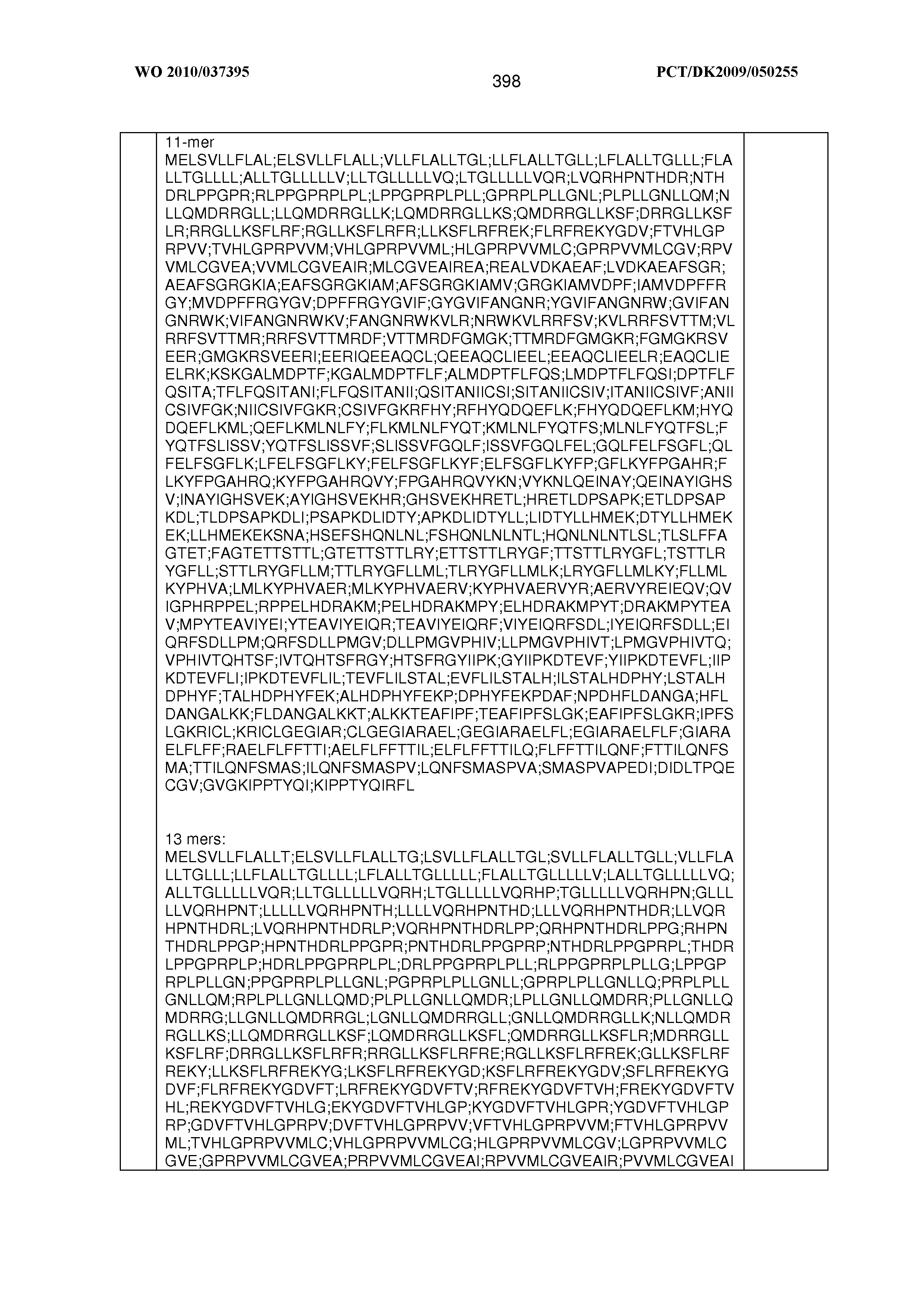


T00004


T00005


T00006


XML
uspto.report is an independent third-party trademark research tool that is not affiliated, endorsed, or sponsored by the United States Patent and Trademark Office (USPTO) or any other governmental organization. The information provided by uspto.report is based on publicly available data at the time of writing and is intended for informational purposes only.
While we strive to provide accurate and up-to-date information, we do not guarantee the accuracy, completeness, reliability, or suitability of the information displayed on this site. The use of this site is at your own risk. Any reliance you place on such information is therefore strictly at your own risk.
All official trademark data, including owner information, should be verified by visiting the official USPTO website at www.uspto.gov. This site is not intended to replace professional legal advice and should not be used as a substitute for consulting with a legal professional who is knowledgeable about trademark law.