Rapamycin Coated Expandable Devices
Dadino; Ronald C. ; et al.
U.S. patent application number 12/790046 was filed with the patent office on 2010-12-30 for rapamycin coated expandable devices. Invention is credited to Ronald C. Dadino, Jonathon Z. Zhao.
| Application Number | 20100331816 12/790046 |
| Document ID | / |
| Family ID | 44839456 |
| Filed Date | 2010-12-30 |


| United States Patent Application | 20100331816 |
| Kind Code | A1 |
| Dadino; Ronald C. ; et al. | December 30, 2010 |
RAPAMYCIN COATED EXPANDABLE DEVICES
Abstract
Medical devices may be utilized for local and regional therapeutic agent delivery. These therapeutic agents or compounds may reduce a biological organism's reaction to the introduction of the medical device to the organism. In addition, these therapeutic drugs, agents and/or compounds may be utilized to promote healing, including the prevention of thrombosis. The drugs, agents, and/or compounds may also be utilized to treat specific disorders, including restenosis, vulnerable plaque, and atherosclerosis in type 2 diabetic patients.
| Inventors: | Dadino; Ronald C.; (Moorestown, NJ) ; Zhao; Jonathon Z.; (Belle Mead, NJ) |
| Correspondence Address: |
    PHILIP S. JOHNSON;JOHNSON & JOHNSON
    ONE JOHNSON & JOHNSON PLAZA
    NEW BRUNSWICK
    NJ
    08933-7003
    US
|
| Family ID: | 44839456 |
| Appl. No.: | 12/790046 |
| Filed: | May 28, 2010 |
Related U.S. Patent Documents
| Application Number | Filing Date | Patent Number | ||
|---|---|---|---|---|
| 12059291 | Mar 31, 2008 | |||
| 12790046 | ||||
| Current U.S. Class: | 604/509 ; 424/423; 514/291 |
| Current CPC Class: | A61K 31/16 20130101; A61K 31/08 20130101; A61K 31/4745 20130101; A61K 31/337 20130101; A61L 29/08 20130101; A61L 29/16 20130101; A61L 31/16 20130101; A61K 31/436 20130101; A61L 2300/416 20130101; A61P 9/10 20180101; A61L 2300/606 20130101; A61K 31/4355 20130101 |
| Class at Publication: | 604/509 ; 424/423; 514/291 |
| International Class: | A61L 31/16 20060101 A61L031/16; A61F 2/00 20060101 A61F002/00; A61K 31/436 20060101 A61K031/436 |
Claims
1. A medical device comprising: an expandable member having a first diameter for insertion into a vessel and a second diameter for making contact with the vessel walls; and a liquid formulation of a rapamycin affixed to at least a portion of the surface of the expandable member, the liquid formulation of a rapamycin comprising about 50 mg/ml of sirolimus and about 2.5 mg/ml BHT combined in a solvent system of acetone/ethanol/water in a ratio of 50/40/10 by volume, the liquid formulation of a rapamycin affixed to the expandable member having a surface density of sirolimus of up to about 7 .mu.g/mm.sup.2 when dried on the surface of the expandable member.
2. The medical device according to claim 1, wherein the expandable member comprises a balloon.
3. The medical device according to claim 2, further comprising a stent positioned over the balloon.
4. A medical device comprising: an expandable member having a first diameter for insertion into a vessel and a second diameter for making contact with the vessel walls; and a liquid formulation of a rapamycin affixed to at least a portion of the surface of the expandable member, the liquid formulation of a rapamycin comprising about 50 mg/ml of sirolimus and about 2.5 mg/ml BHT combined in a solvent system of isopropanol/water in a ratio of 3.4/1 by volume, the liquid formulation of a rapamycin affixed to the expandable member having a surface density of sirolimus of up to about 7 .mu.g/mm.sup.2 when dried on the surface of the expandable member.
5. The medical device according to claim 4, wherein the expandable member comprises a balloon.
6. The medical device according to claim 5, further comprising a stent positioned over the balloon.
7. A liquid formulation of a rapamycin comprising about 50 mg/ml of sirolimus and about 2.5 mg/ml BHT combined in a solvent system of acetone/ethanol/water in a ratio of 50/40/10 by volume.
8. A liquid formulation of a rapamycin comprising about 50 mg/ml of sirolimus and about 2.5 mg/ml BHT combined in a solvent system of isopropanol/water in a ratio of 3.4/1 by volume.
9. A method for the treatment of vascular disease comprising: positioning an expandable member having a first unexpanded diameter proximate a treatment site of a diseased vessel; and expanding the expandable member to a second diameter such that it makes contact with the vessel walls at the treatment site, the expandable member having a coating comprising about 50 mg/ml of sirolimus and about 2.5 mg/ml BHT combined in a solvent system of acetone/ethanol/water in a ratio of 50/40/10 by volume, the liquid formulation of a rapamycin affixed to the expandable member having a surface density of sirolimus of up to about 7 .mu.g/mm.sup.2 when dried on the surface of the expandable member, wherein the expansion of the expandable member to the second diameter facilitates the uptake of the liquid formulation into the tissues comprising the vessel walls.
10. The method for the treatment of vascular disease according to claim 9, wherein the expandable member is expanded to a final diameter at the treatment site that is larger than the nominal diameter of the artery by at least 10 percent.
11. A method for the treatment of vascular disease comprising: positioning an expandable member having a first unexpanded diameter proximate a treatment site of a diseased vessel; and expanding the expandable member to a second diameter such that it makes contact with the vessel walls at the treatment site, the expandable member having a coating comprising about 50 mg/ml of sirolimus and about 2.5 mg/ml BHT combined in a solvent system of isopropanol/water in a ratio of 3.4/1 by volume, the liquid formulation of a rapamycin affixed to the expandable member having a surface density of sirolimus of up to about 7 .mu.g/mm.sup.2 when dried on the surface of the expandable member, wherein the expansion of the expandable member to the second diameter facilitates the uptake of the liquid formulation into the tissues comprising the vessel walls.
12. The method for the treatment of vascular disease according to claim 11, wherein the expandable member is expanded to a final diameter at the treatment site that is larger than the nominal diameter of the artery by at least 10 percent.
Description
CROSS REFERENCE TO RELATED APPLICATIONS
[0001] This application is a continuation-in-part of U.S. patent application Ser. No. 12/059,291 filed Oct. 13, 2006.
BACKGROUND OF THE INVENTION
[0002] 1. Field of the Invention
[0003] The present invention relates to the local and/or regional administration of therapeutic agents and/or therapeutic agent combinations, and more particularly to expandable medical devices for the local and/or regional delivery of therapeutic agents and/or therapeutic agent combinations for the prevention and treatment of vascular disease.
[0004] 2. Discussion of the Related Art
[0005] Many individuals suffer from circulatory disease caused by a progressive blockage of the blood vessels that perfuse the heart and other major organs. More severe blockage of blood vessels in such individuals often leads to hypertension, ischemic injury, stroke, or myocardial infarction. Atherosclerotic lesions, which limit or obstruct coronary blood flow, are the major cause of ischemic heart disease. Percutaneous transluminal coronary angioplasty is a medical procedure whose purpose is to increase blood flow through an artery. Percutaneous transluminal coronary angioplasty is the predominant treatment for coronary vessel stenosis. The increasing use of this procedure is attributable to its relatively high success rate and its minimal invasiveness compared with coronary bypass surgery. A limitation associated with percutaneous transluminal coronary angioplasty is the abrupt closure of the vessel, which may occur immediately after the procedure and restenosis, which occurs gradually following the procedure. Additionally, restenosis is a chronic problem in patients who have undergone saphenous vein bypass grafting. The mechanism of acute occlusion appears to involve several factors and may result from vascular recoil with resultant closure of the artery and/or deposition of blood platelets and fibrin along the damaged length of the newly opened blood vessel.
[0006] Restenosis after percutaneous transluminal coronary angioplasty is a more gradual process initiated by vascular injury. Multiple processes, including thrombosis, inflammation, growth factor and cytokine release, cell proliferation, cell migration and extracellular matrix synthesis each contribute to the restenotic process.
[0007] Upon pressure expansion of an intracoronary balloon catheter during angioplasty and/or stent implantation, smooth muscle cells and endothelial cells within the vessel wall become injured, initiating a thrombotic and inflammatory response. Cell derived growth factors such as platelet derived growth factor, basic fibroblast growth factor, epidermal growth factor, thrombin, etc., released from platelets, invading macrophages and/or leukocytes, or directly from the smooth muscle cells provoke a proliferative and migratory response in medial smooth muscle cells. These cells undergo a change from a contractile phenotype to a synthetic phenotype characterized by only a few contractile filament bundles, extensive rough endoplasmic reticulum, Golgi and free ribosomes. Proliferation/migration usually begins within one to two days' post-injury and peaks several days thereafter (Campbell and Campbell, 1987; Clowes and Schwartz, 1985).
[0008] Daughter cells migrate to the intimal layer of arterial smooth muscle and continue to proliferate and secrete significant amounts of extracellular matrix proteins. Proliferation, migration and extracellular matrix synthesis continue until the damaged endothelial layer is repaired at which time proliferation slows within the intima, usually within seven to fourteen days post-injury. The newly formed tissue is called neointima. The further vascular narrowing that occurs over the next three to six months is due primarily to negative or constrictive remodeling.
[0009] Simultaneous with local proliferation and migration, inflammatory cells adhere to the site of vascular injury. Within three to seven days post-injury, inflammatory cells have migrated to the deeper layers of the vessel wall. In animal models employing either balloon injury or stent implantation, inflammatory cells may persist at the site of vascular injury for at least thirty days (Tanaka et al., 1993; Edelman et al., 1998). Inflammatory cells therefore are present and may contribute to both the acute and chronic phases of restenosis.
[0010] Unlike systemic pharmacologic therapy, stents have proven useful in significantly reducing restenosis. Typically, stents are balloon-expandable slotted metal tubes (usually, but not limited to, stainless steel), which, when expanded within the lumen of an angioplastied coronary artery, provide structural support through rigid scaffolding to the arterial wall. This support is helpful in maintaining vessel lumen patency. In two randomized clinical trials, stents increased angiographic success after percutaneous transluminal coronary angioplasty, by increasing minimal lumen diameter and reducing, but not eliminating, the incidence of restenosis at six months (Serruys et al., 1994; Fischman et al., 1994).
[0011] Additionally, the heparin coating of stents appears to have the added benefit of producing a reduction in sub-acute thrombosis after stent implantation (Serruys et al., 1996). Thus, sustained mechanical expansion of a stenosed coronary artery with a stent has been shown to provide some measure of restenosis prevention, and the coating of stents with heparin has demonstrated both the feasibility and the clinical usefulness of delivering drugs locally, at the site of injured tissue. However, in certain circumstances it may not be desirable to leave any type of implantable device in the body.
[0012] Accordingly, there exists a need for drug/drug combinations and associated local delivery devices for the prevention and treatment of vascular injury causing intimal thickening which is either biologically induced, for example, atherosclerosis, or mechanically induced, for example, through percutaneous transluminal coronary angioplasty.
SUMMARY OF THE INVENTION
[0013] A device for the local and/or regional delivery of rapamycin formulations in accordance with the present invention may be utilized to overcome the disadvantages set forth above.
[0014] Medical devices may be utilized for local and regional therapeutic agent delivery. These therapeutic agents or compounds may reduce a biological organism's reaction to the introduction of the medical device to the organism. In addition, these therapeutic drugs, agents and/or compounds may be utilized to promote healing, including the prevention of thrombosis. The drugs, agents, and/or compounds may also be utilized to treat specific disorders, including restenosis, vulnerable plaque, and atherosclerosis in type 2 diabetic patients.
[0015] The drugs, agents or compounds will vary depending upon the type of medical device, the reaction to the introduction of the medical device and/or the disease sought to be treated. The type of coating or vehicle utilized to immobilize the drugs, agents or compounds to the medical device may also vary depending on a number of factors, including the type of medical device, the type of drug, agent or compound and the rate of release thereof.
[0016] The present invention is directed to balloons or other inflatable or expandable devices that may be temporarily positioned within a body to deliver a therapeutic agent and/or continuation of therapeutic agents and then removed. The therapeutic agents may include liquid formulations of rapamycin. This type of delivery device may be particularly advantageous in the vasculature where stents may not be suitable, for example, in the larger vessels of the peripheral vascular system.
[0017] In use, the balloon or other inflatable or expandable device may be coated with one or more liquid formulations of therapeutic agent(s) and delivered to a treatment site. The act of inflation or expansion would, force the therapeutic agents into the surrounding tissue. The device may be kept in position for a period of between ten seconds to about five minutes depending upon the location. If utilized in the heart, shorter durations are required relative to other areas such as the leg.
[0018] In accordance with one aspect, the present invention is directed to a medical device comprising an expandable member having a first diameter for insertion into a vessel and a second diameter for making contact with the vessel walls; and a liquid formulation of a rapamycin affixed to at least a portion of the surface of the expandable member, the liquid formulation of a rapamycin comprising about 50 mg/ml of sirolimus and about 2.5 mg/ml BHT combined in a solvent system of acetone/ethanol/water in a ratio of 50/40/10 by volume, the liquid formulation of a rapamycin affixed to the expandable member having a surface density of sirolimus of up to about 7 .mu.g/mm.sup.2 when dried on the surface of the expandable member.
[0019] In accordance with another aspect, the present invention is directed to a medical device comprising an expandable member having a first diameter for insertion into a vessel and a second diameter for making contact with the vessel walls; and a liquid formulation of a rapamycin affixed to at least a portion of the surface of the expandable member, the liquid formulation of a rapamycin comprising about 50 mg/ml of sirolimus and about 2.5 mg/ml BHT combined in a solvent system of isopropanol/water in a ratio of 3.4/1 by volume, the liquid formulation of a rapamycin affixed to the expandable member having a surface density of sirolimus of up to about 7 .mu.g/mm.sup.2 when dried on the surface of the expandable member.
[0020] In accordance with still another aspect, the present invention is directed to a liquid formulation of a rapamycin comprising about 50 mg/ml of sirolimus and about 2.5 mg/ml BHT combined in a solvent system of acetone/ethanol/water in a ratio of 50/40/10 by volume.
[0021] In accordance with still another aspect, the present invention is directed to a liquid formulation of a rapamycin comprising about 50 mg/ml of sirolimus and about 2.5 mg/ml BHT combined in a solvent system of isopropanol/water in a ratio of 3.4/1 by volume.
[0022] In accordance with still another aspect, the present invention is directed to a method for the treatment of vascular disease comprising positioning an expandable member having a first unexpanded diameter proximate a treatment site of a diseased vessel; and expanding the expandable member to a second diameter such that it makes contact with the vessel walls at the treatment site, the expandable member having a coating comprising about 50 mg/ml of sirolimus and about 2.5 mg/ml BHT combined in a solvent system of acetone/ethanol/water in a ratio of 50/40/10 by volume, the liquid formulation of a rapamycin affixed to the expandable member having a surface density of sirolimus of up to about 7 .mu.g/mm.sup.2 when dried on the surface of the expandable member, wherein the expansion of the expandable member to the second diameter facilitates the uptake of the liquid formulation into the tissues comprising the vessel walls.
[0023] In accordance with still another aspect, the present invention is directed to a method for the treatment of vascular disease comprising positioning an expandable member having a first unexpanded diameter proximate a treatment site of a diseased vessel; and expanding the expandable member to a second diameter such that it makes contact with the vessel walls at the treatment site, the expandable member having a coating comprising about 50 mg/ml of sirolimus and about 2.5 mg/ml BHT combined in a solvent system of isopropanol/water in a ratio of 3.4/1 by volume, the liquid formulation of a rapamycin affixed to the expandable member having a surface density of sirolimus of up to about 7 .mu.g/mm.sup.2 when dried on the surface of the expandable member, wherein the expansion of the expandable member to the second diameter facilitates the uptake of the liquid formulation into the tissues comprising the vessel walls.
BRIEF DESCRIPTION OF THE DRAWINGS
[0024] The foregoing and other features and advantages of the invention will be apparent from the following, more particular description of preferred embodiments of the invention, as illustrated in the accompanying drawings.
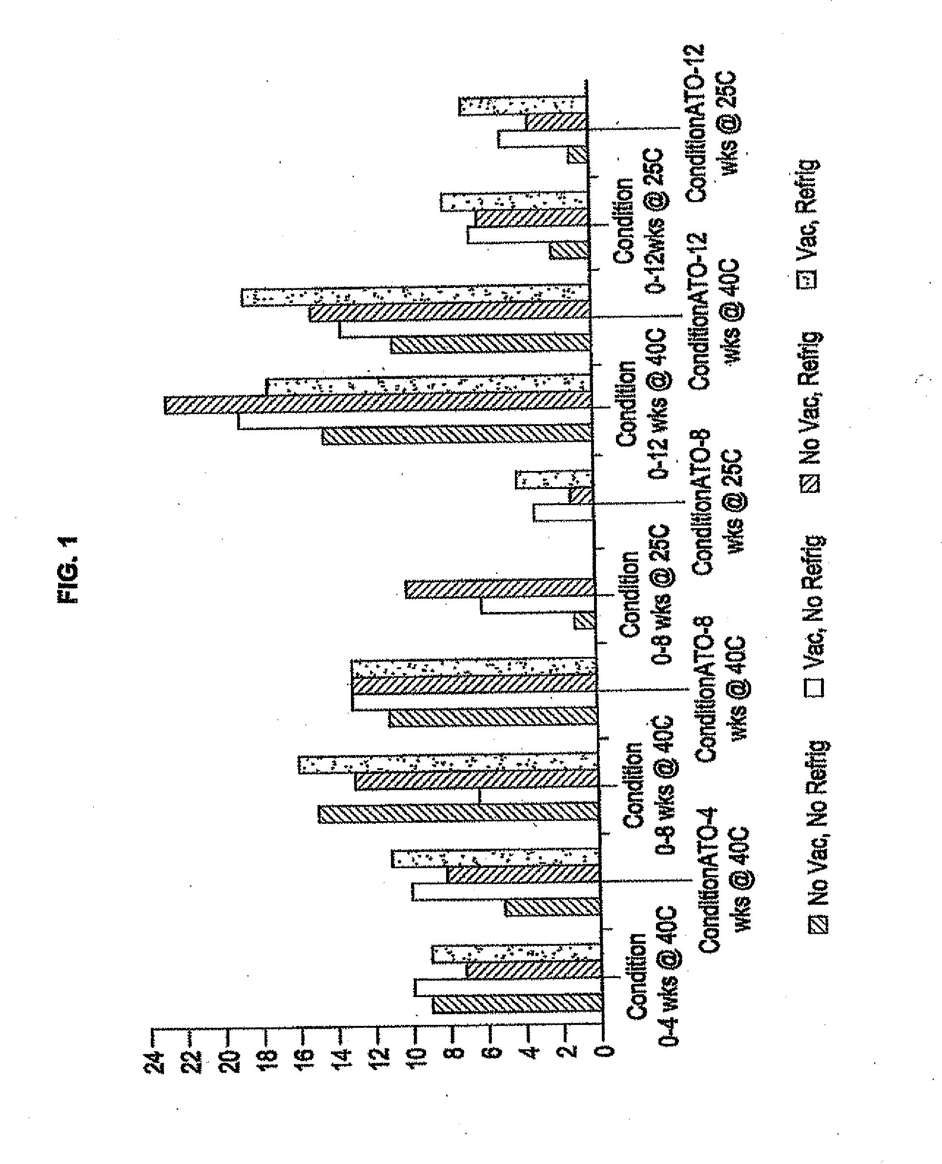
[0025] FIG. 1 is a graphical representation of the results of a bioactivity study in accordance with the present invention.
[0026] FIGS. 2A and 2B illustrate a dip coating process of a PTCA balloon in a liquid formulation of a therapeutic agent in accordance with the present invention.
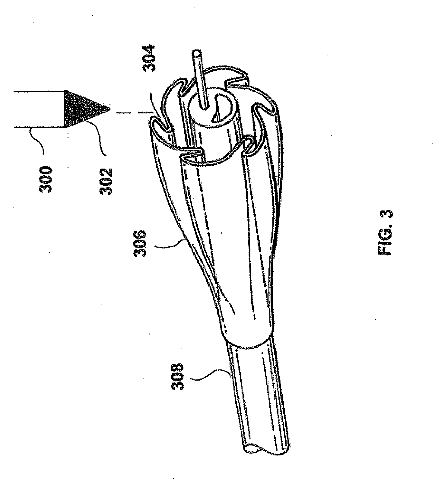
[0027] FIG. 3 is a diagrammatic illustration of a first process for coating a PTCA balloon in accordance with the present invention.
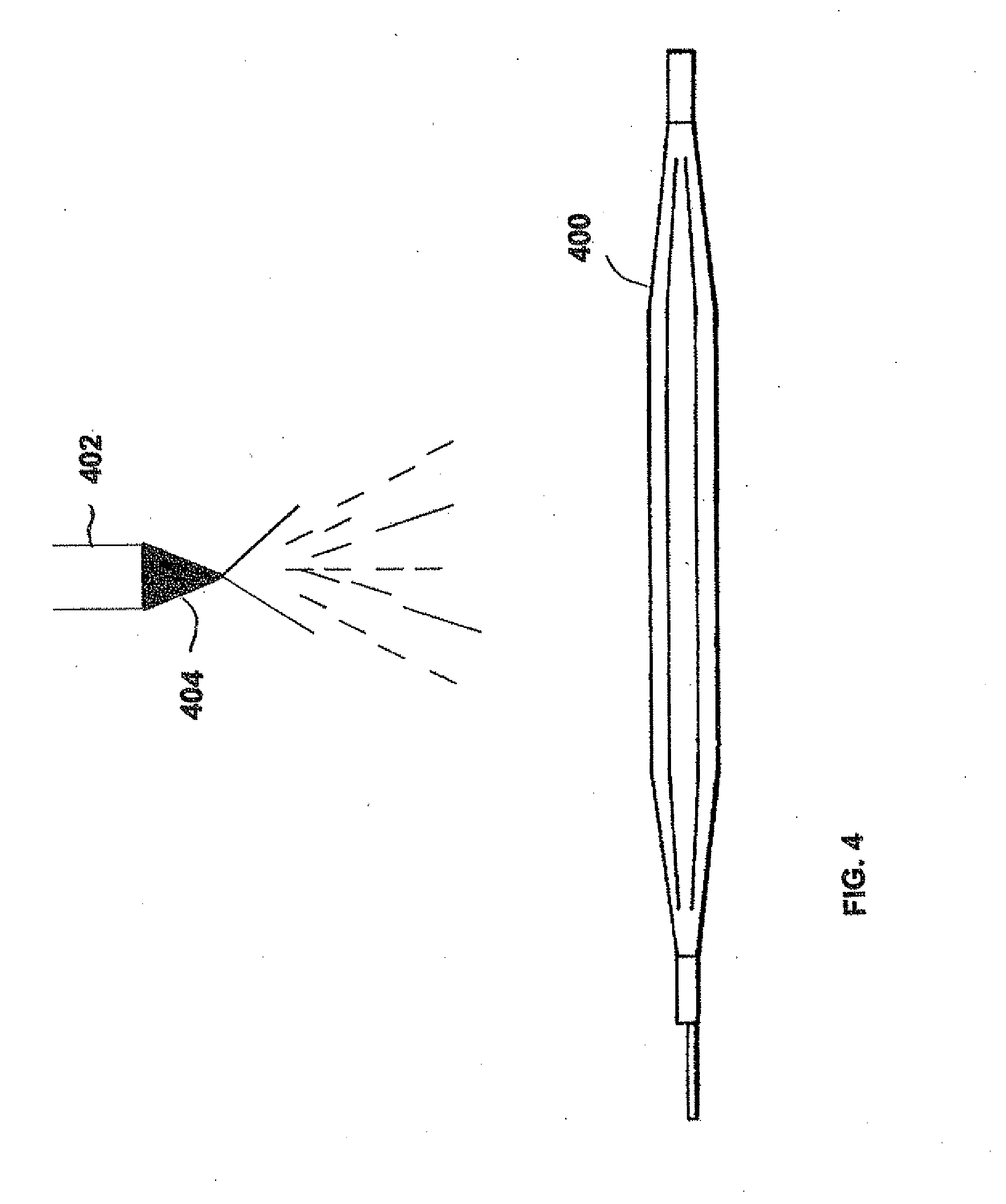
[0028] FIG. 4 is a diagrammatic illustration of a second process for coating a PTCA balloon in accordance with the present invention.
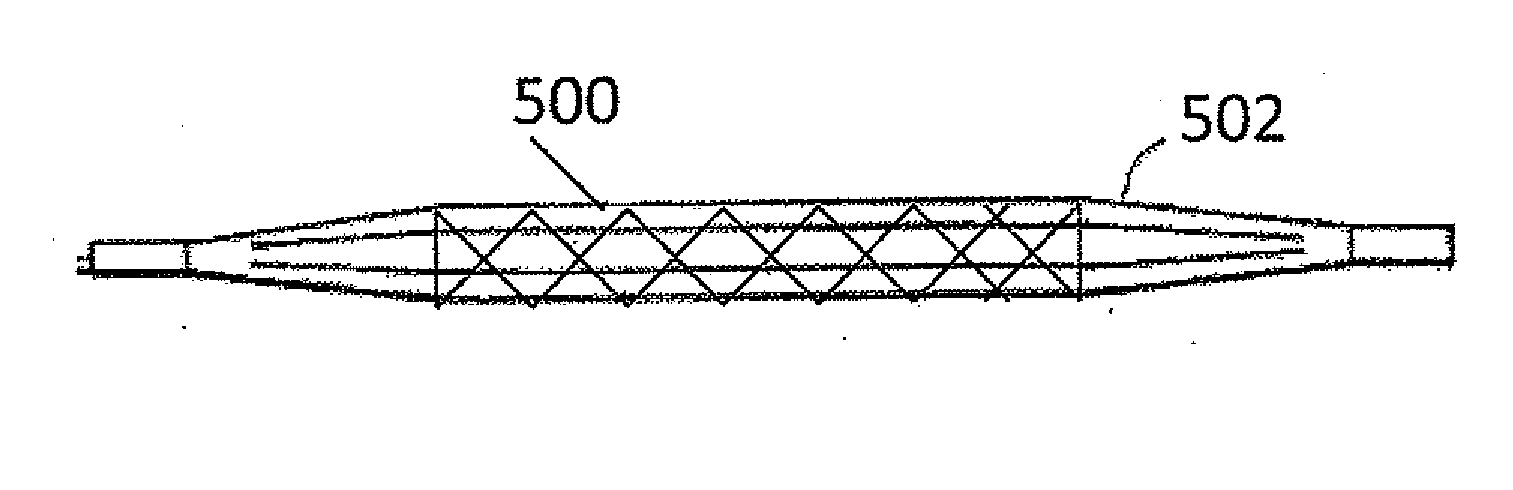
[0029] FIG. 5 is a diagrammatic illustration of a stent on a coated PTCA balloon in accordance with the present invention.
[0030] FIG. 6 is a graphical representation of 30 day late lumen loss.
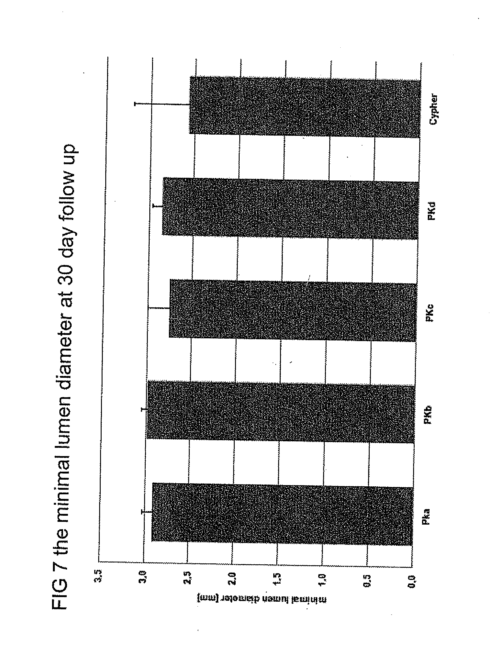
[0031] FIG. 7 is a graphical representation of minimal lumen diameter at 30 day follow up.
DETAILED DESCRIPTION OF THE PREFERRED EMBODIMENTS
[0032] The drug/drug combinations and delivery devices of the present invention may be utilized to effectively prevent and treat vascular disease, including vascular disease caused by injury. Various medical treatment devices utilized in the treatment of vascular disease may ultimately induce further complications. For example, balloon angioplasty is a procedure utilized to increase blood flow through an artery and is the predominant treatment for coronary vessel stenosis. However, the procedure typically causes a certain degree of damage to the vessel wall, thereby potentially exacerbating the problem at a point later in time. Although other procedures and diseases may cause similar injury, exemplary embodiments of the present invention will be described with respect to the treatment of restenosis and related complications.
[0033] While exemplary embodiments of the invention will be described with respect to the treatment of restenosis and related complications following percutaneous transluminal coronary angioplasty, it is important to note that the local delivery of drug/drug combinations may be utilized to treat a wide variety of conditions utilizing any number of medical devices, or to enhance the function and/or life of the device. For example, intraocular lenses, placed to restore vision after cataract surgery is often compromised by the formation of a secondary cataract. The latter is often a result of cellular overgrowth on the lens surface and can be potentially minimized by combining a drug or drugs with the device. Other medical devices which often fail due to tissue in-growth or accumulation of proteinaceous material in, on and around the device, such as shunts for hydrocephalus, dialysis grafts, colostomy bag attachment devices, ear drainage tubes, leads for pace makers and implantable defibrillators can also benefit from the device-drug combination approach. Devices which serve to improve the structure and function of tissue or organ may also show benefits when combined with the appropriate agent or agents. For example, improved osteointegration of orthopedic devices to enhance stabilization of the implanted device could potentially be achieved by combining it with agents such as bone-morphogenic protein. Similarly other surgical devices, sutures, staples, anastomosis devices, vertebral disks, bone pins, suture anchors, hemostatic barriers, clamps, screws, plates, clips, vascular implants, tissue adhesives and sealants, tissue scaffolds, various types of dressings, bone substitutes, intraluminal devices, and vascular supports could also provide enhanced patient benefit using this drug-device combination approach. Perivascular wraps may be particularly advantageous, alone or in combination with other medical devices. The perivascular wraps may supply additional drugs to a treatment site. Essentially, any type of medical device may be coated in some fashion with a drug or drug combination which enhances treatment over use of the singular use of the device or pharmaceutical agent.
[0034] In addition to various medical devices, the coatings on these devices may be used to deliver therapeutic and pharmaceutic agents including: anti-proliferative/antimitotic agents including natural products such as vinca alkaloids (i.e. vinblastine, vincristine, and vinorelbine), paclitaxel, epidipodophyllotoxins (i.e. etoposide, teniposide), antibiotics (dactinomycin (actinomycin D) daunorubicin, doxorubicin and idarubicin), anthracyclines, mitoxantrone, bleomycins, plicamycin (mithramycin) and mitomycin, enzymes (L-asparaginase which systemically metabolizes L-asparagine and deprives cells which do not have the capacity to synthesize their own asparagine); antiplatelet agents such as G(GP) II.sub.b/III.sub.a inhibitors and vitronectin receptor antagonists; anti-proliferative/antimitotic alkylating agents such as nitrogen mustards (mechlorethamine, cyclophosphamide and analogs, melphalan, chlorambucil), ethylenimines and methylmelamines (hexamethylmelamine and thiotepa), alkyl sulfonates-busulfan, nirtosoureas (carmustine (BCNU) and analogs, streptozocin), trazenes--dacarbazinine (DTIC); anti-proliferative/antimitotic antimetabolites such as folic acid analogs (methotrexate), pyrimidine analogs (fluorouracil, floxuridine, and cytarabine), purine analogs and related inhibitors (mercaptopurine, thioguanine, pentostatin and 2-chlorodeoxyadenosine {cladribine}); platinum coordination complexes (cisplatin, carboplatin), procarbazine, hydroxyurea, mitotane, aminoglutethimide; hormones (i.e. estrogen); anti-coagulants (heparin, synthetic heparin salts and other inhibitors of thrombin); fibrinolytic agents (such as tissue plasminogen activator, streptokinase and urokinase), aspirin, dipyridamole, ticlopidine, clopidogrel, abciximab; antimigratory; antisecretory (breveldin); anti-inflammatory: such as adrenocortical steroids (cortisol, cortisone, fludrocortisone, prednisone, prednisolone, 6.alpha.-methylprednisolone, triamcinolone, betamethasone, and dexamethasone), non-steroidal agents (salicylic acid derivatives i.e. aspirin; para-aminophenol derivatives i.e. acetaminophen; indole and indene acetic acids (indomethacin, sulindac, and etodalac), heteroaryl acetic acids (tolmetin, diclofenac, and ketorolac), arylpropionic acids (ibuprofen and derivatives), anthranilic acids (mefenamic acid, and meclofenamic acid), enolic acids (piroxicam, tenoxicam, phenylbutazone, and oxyphenthatrazone), nabumetone, gold compounds (auranofin, aurothioglucose, gold sodium thiomalate); immunosuppressives: (cyclosporine, tacrolimus (FK-506), sirolimus (rapamycin), azathioprine, mycophenolate mofetil); angiogenic agents: vascular endothelial growth factor (VEGF), fibroblast growth factor (FGF); angiotensin receptor blockers; nitric oxide donors; antisense oligionucleotides and combinations thereof; cell cycle inhibitors, mTOR inhibitors, and growth factor receptor signal transduction kinase inhibitors; retenoids; cyclin/CDK inhibitors; HMG co-enzyme reductase inhibitors (statins); and protease inhibitors.
[0035] Rapamycin is a macrocyclic triene antibiotic produced by Streptomyces hygroscopicus as disclosed in U.S. Pat. No. 3,929,992. It has been found that rapamycin among other things inhibits the proliferation of vascular smooth muscle cells in vivo. Accordingly, rapamycin may be utilized in treating intimal smooth muscle cell hyperplasia, restenosis, and vascular occlusion in a mammal, particularly following either biologically or mechanically mediated vascular injury, or under conditions that would predispose a mammal to suffering such a vascular injury. Rapamycin functions to inhibit smooth muscle cell proliferation and does not interfere with the re-endothelialization of the vessel walls.
[0036] Rapamycin reduces vascular hyperplasia by antagonizing smooth muscle proliferation in response to mitogenic signals that are released during an angioplasty induced injury. Inhibition of growth factor and cytokine mediated smooth muscle proliferation at the late G1 phase of the cell cycle is believed to be the dominant mechanism of action of rapamycin. However, rapamycin is also known to prevent T-cell proliferation and differentiation when administered systemically. This is the basis for its immunosuppressive activity and its ability to prevent graft rejection.
[0037] The molecular events that are responsible for the actions of rapamycin, a known anti-proliferative, which acts to reduce the magnitude and duration of neointimal hyperplasia, are still being elucidated. It is known, however, that rapamycin enters cells and binds to a high-affinity cytosolic protein called FKBP12. The complex of rapamycin and FKPB12 in turn binds to and inhibits a phosphoinositide (PI)-3 kinase called the "mammalian Target of Rapamycin" or TOR. TOR is a protein kinase that plays a key role in mediating the downstream signaling events associated with mitogenic growth factors and cytokines in smooth muscle cells and T lymphocytes. These events include phosphorylation of p27, phosphorylation of p70 s6 kinase and phosphorylation of 4BP-1, an important regulator of protein translation.
[0038] It is recognized that rapamycin reduces restenosis by inhibiting neointimal hyperplasia. However, there is evidence that rapamycin may also inhibit the other major component of restenosis, namely, negative remodeling. Remodeling is a process whose mechanism is not clearly understood but which results in shrinkage of the external elastic lamina and reduction in lumenal area over time, generally a period of approximately three to six months in humans.
[0039] Negative or constrictive vascular remodeling may be quantified angiographically as the percent diameter stenosis at the lesion site where there is no stent to obstruct the process. If late lumen loss is abolished in-lesion, it may be inferred that negative remodeling has been inhibited. Another method of determining the degree of remodeling involves measuring in-lesion external elastic lamina area using intravascular ultrasound (IVUS). Intravascular ultrasound is a technique that can image the external elastic lamina as well as the vascular lumen. Changes in the external elastic lamina proximal and distal to the stent from the post-procedural timepoint to four-month and twelve-month follow-ups are reflective of remodeling changes.
[0040] Evidence that rapamycin exerts an effect on remodeling comes from human implant studies with rapamycin coated stents showing a very low degree of restenosis in-lesion as well as in-stent. In-lesion parameters are usually measured approximately five millimeters on either side of the stent i.e. proximal and distal. Since the stent is not present to control remodeling in these zones which are still affected by balloon expansion, it may be inferred that rapamycin is preventing vascular remodeling.
[0041] The local delivery of drug/drug combinations from a stent has the following advantages; namely, the prevention of vessel recoil and remodeling through the scaffolding action of the stent and the prevention of multiple components of neointimal hyperplasia or restenosis as well as a reduction in inflammation and thrombosis. This local administration of drugs, agents or compounds to stented coronary arteries may also have additional therapeutic benefit. For example, higher tissue concentrations of the drugs, agents or compounds may be achieved utilizing local delivery, rather than systemic administration. In addition, reduced systemic toxicity may be achieved utilizing local delivery rather than systemic administration while maintaining higher tissue concentrations. Also in utilizing local delivery from a stent rather than systemic administration, a single procedure may suffice with better patient compliance. An additional benefit of combination drug, agent, and/or compound therapy may be to reduce the dose of each of the therapeutic drugs, agents or compounds, thereby limiting their toxicity, while still achieving a reduction in restenosis, inflammation and thrombosis. Local stent-based therapy is therefore a means of improving the therapeutic ratio (efficacy/toxicity) of anti-restenosis, anti-inflammatory, anti-thrombotic drugs, agents or compounds.
[0042] A stent is commonly used as a tubular structure left inside the lumen of a duct to relieve an obstruction. Commonly, stents are inserted into the lumen in a non-expanded form and are then expanded autonomously, or with the aid of a second device in situ. A typical method of expansion occurs through the use of a catheter-mounted angioplasty balloon which is inflated within the stenosed vessel or body passageway in order to shear and disrupt the obstructions associated with the wall components of the vessel and to obtain an enlarged lumen.
[0043] The data in Table 1 below illustrate that in-lesion percent diameter stenosis remains low in the rapamycin treated groups, even at twelve months. Accordingly, these results support the hypothesis that rapamycin reduces remodeling.
TABLE-US-00001 TABLE 1.0 Angiographic In-Lesion Percent Diameter Stenosis (%, mean .+-. SD and "n =") In Patients Who Received a Rapamycin-Coated Stent Coating Post 4-6 month 12 month Group Placement Follow Up Follow Up Brazil 10.6 .+-. 5.7 (30) 13.6 .+-. 8.6 (30) 22.3 .+-. 7.2 (15) Netherlands 14.7 .+-. 8.8 22.4 .+-. 6.4 --
[0044] Additional evidence supporting a reduction in negative remodeling with rapamycin comes from intravascular ultrasound data that was obtained from a first-in-man clinical program as illustrated in Table 2 below.
TABLE-US-00002 TABLE 2.0 Matched IVUS data in Patients Who Received a Rapamycin-Coated Stent 4-Month 12-Month Follow-Up Follow-Up IVUS Parameter Post (n =) (n =) (n =) Mean proximal vessel area 16.53 .+-. 3.53 16.31 .+-. 4.36 13.96 .+-. 2.26 (mm.sup.2) (27) (28) (13) Mean distal vessel area 13.12 .+-. 3.68 13.53 .+-. 4.17 12.49 .+-. 3.25 (mm.sup.2) (26) (26) (14)
[0045] The data illustrated that there is minimal loss of vessel area proximally or distally which indicates that inhibition of negative remodeling has occurred in vessels treated with rapamycin-coated stents.
[0046] Other than the stent itself, there have been no effective solutions to the problem of vascular remodeling. Accordingly, rapamycin may represent a biological approach to controlling the vascular remodeling phenomenon.
[0047] It may be hypothesized that rapamycin acts to reduce negative remodeling in several ways. By specifically blocking the proliferation of fibroblasts in the vascular wall in response to injury, rapamycin may reduce the formation of vascular scar tissue. Rapamycin may also affect the translation of key proteins involved in collagen formation or metabolism.
[0048] In an exemplary embodiment, the rapamycin is delivered by a local delivery device to control negative remodeling of an arterial segment after balloon angioplasty as a means of reducing or preventing restenosis. While any delivery device may be utilized, it is preferred that the delivery device comprises a stent that includes a coating or sheath which elutes or releases rapamycin. The delivery system for such a device may comprise a local infusion catheter that delivers rapamycin at a rate controlled by the administrator.
[0049] Rapamycin may also be delivered systemically using an oral dosage form or a chronic injectible depot form or a patch to deliver rapamycin for a period ranging from about seven to forty-five days to achieve vascular tissue levels that are sufficient to inhibit negative remodeling. Such treatment is to be used to reduce or prevent restenosis when administered several days prior to elective angioplasty with or without a stent.
[0050] Data generated in porcine and rabbit models show that the release of rapamycin into the vascular wall from a nonerodible polymeric stent coating in a range of doses (35-430 .mu.g/15-18 mm coronary stent) produces a peak fifty to fifty-five percent reduction in neointimal hyperplasia as set forth in Table 3 below. This reduction, which is maximal at about twenty-eight to thirty days, is typically not sustained in the range of ninety to one hundred eighty days in the porcine model as set forth in Table 4 below.
TABLE-US-00003 TABLE 3.0 Animal Studies with Rapamycin-coated stents. Values are mean .+-. Standard Error of Mean Neointimal % Change From Study Duration Stene Rapamycin N Area (mm.sup.2) Polyene Metal Porcine 98009 14 days Metal 8 2.04 .+-. 0.17 1X + rapamycin 153 .mu.g 8 1.66 .+-. 0.17* -42% -19% 1X + TC300 + rapamycin 155 .mu.g 8 1.51 .+-. 0.19* -47% -26% 99005 28 days Metal 10 2.29 .+-. 0.21 9 3.91 .+-. 0.60** 1X + TC30 + rapamycin 130 .mu.g 8 2.81 .+-. 0.34 +23% 1X + TC100 + rapamycin 120 .mu.g 9 2.62 .+-. 0.21 +14% 99006 28 days Metal 12 4.57 .+-. 0.46 EVA/BMA 3X 12 5.02 .+-. 0.62 +10% 1X + rapamycin 125 .mu.g 11 2.84 .+-. 0.31* ** -43% -38% 3X + rapamycin 430 .mu.g 12 3.06 .+-. 0.17* ** -39% -33% 3X + rapamycin 157 .mu.g 12 2.77 .+-. 0.41* ** -45% -39% 99011 28 days Metal 11 3.09 .+-. 0.27 11 4.52 .+-. 0.37 1X + rapamycin 189 .mu.g 14 3.05 .+-. 0.35 -1% 3X + rapamycin/dex 182/363 .mu.g 14 2.72 .+-. 0.71 -12% 99021 60 days Metal 12 2.14 .+-. 0.25 1X + rapamycin 181 .mu.g 12 2.95 .+-. 0.38 +38% 99034 28 days Metal 8 5.24 .+-. 0.58 1X + rapamycin 186 .mu.g 8 2.47 .+-. 0.33** -53% 3X + rapamycin/dex 185/369 .mu.g 6 2.42 .+-. 0.64** -54% 20001 28 days Metal 6 1.81 .+-. 0.09 1X + rapamycin 172 .mu.g 5 1.66 .+-. 0.44 -8% 20007 30 days Metal 9 2.94 .+-. 0.43 1XTC + rapamycin 155 .mu.g 10 1.40 .+-. 0.11* -52%* Rabbit 99019 28 days Metal 8 1.20 .+-. 0.07 EVA/BMA 1X 10 1.26 .+-. 0.16 +5% 1X + rapamycin 64 .mu.g 9 0.92 .+-. 0.14 -27% -23% 1X + rapamycin 196 .mu.g 10 0.66 .+-. 0.12* ** -48% -45% 99020 28 days Metal 12 1.18 .+-. 0.10 EVA/BMA 1X + rapamycin 197 .mu.g 8 0.81 .+-. 0.16 -32% .sup.1Stent nomenclature: EVA/BMA 1X, 2X, and 3X signifies approx. 500 .mu.g, 1000 .mu.g, and 1500 .mu.g total mass (polymer + drug), respectively. TC, top coat of 30 .mu.g, 100 .mu.g, or 300 .mu.g drug-free BMA; Biphasic; 2 .times. 1X layers of rapamycin in EVA/BMA spearated by a 100 .mu.g drug-free BMA layer. .sup.20.25mg/kg/d .times. 14 d preceeded by a loading dose of 0.5 mg/kg/d .times. 3d prior to stent implantation. *p<0.05 from EVA/BMA control. **p<0.05 from Metal; .sup.# Inflammation score: (0 = essentially no intimal involvement; 1 = <25% intima involved; 2 = .gtoreq.25% intima involved; 3 = >50% intima involved).
TABLE-US-00004 TABLE 4.0 180 day Porcine Study with Rapamycin-coated stents. Values are mean .+-. Standard Error of Mean % Change Neointimal From Inflammation Study Duration Stent.sup.1 Rapamycin N Area (mm.sup.2) Polyme Metal Score # 20007 3 days Metal 10 0.38 .+-. 0.06 1.05 .+-. 0.06 (ETP-2-002233-P) 1XTC + rapamycin 155 .mu.g 10 0.29 .+-. 0.03 -24% 1.08 .+-. 0.04 30 days Metal 9 2.94 .+-. 0.43 0.11 .+-. 0.08 1XTC + rapamycin 155 .mu.g 10 1.40 .+-. 0.11* -52%* 0.25 .+-. 0.10 90 days Metal 10 3.45 .+-. 0.34 0.20 .+-. 0.08 1XTC + rapamycin 155 .mu.g 10 3.03 .+-. 0.29 -12% 0.80 .+-. 0.23 1X + rapamycin 171 .mu.g 10 2.86 .+-. 0.35 -17% 0.60 .+-. 0.23 180 days Metal 10 3.65 .+-. 0.39 0.65 .+-. 0.21 1XTC + rapamycin 155 .mu.g 10 3.34 .+-. 0.31 -8% 1.50 .+-. 0.34 1X + rapamycin 171 .mu.g 10 3.87 .+-. 0.28 +6% 1.68 .+-. 0.37
[0051] The release of rapamycin into the vascular wall of a human from a nonerodible polymeric stent coating provides superior results with respect to the magnitude and duration of the reduction in neointimal hyperplasia within the stent as compared to the vascular walls of animals as set forth above.
[0052] Humans implanted with a rapamycin coated stent comprising rapamycin in the same dose range as studied in animal models using the same polymeric matrix, as described above, reveal a much more profound reduction in neointimal hyperplasia than observed in animal models, based on the magnitude and duration of reduction in neointima. The human clinical response to rapamycin reveals essentially total abolition of neointimal hyperplasia inside the stent using both angiographic and intravascular ultrasound measurements. These results are sustained for at least one year as set forth in Table 5 below.
TABLE-US-00005 TABLE 5.0 Patients Treated (N = 45 patients) with a Rapamycin-coated Stent Sirolimus FIM 95% Effectiveness Measures (N = 45 Patients, 45 Lesions) Confidence Limit Procedure Success (QCA) 100.0% (45/45) [92.1%, 100.0%] 4-month In-Stent Diameter Stenosis (%) Mean .+-. SD (N) 4.8% .+-. 6.1% (30) [2.6%, 7.0%] Range (min, max) (-8.2%, 14.9%) 6-month In-Stent Diameter Stenosis (%) Mean .+-. SD (N) 8.9% .+-. 7.6% (13) [4.8%, 13.0%] Range (min, max) (-2.9%, 20.4%) 12-month In-Stent Diameter Stenosis (%) Mean .+-. SD (N) 8.9% .+-. 6.1% (15) [5.8%, 12.0%] Range (min, max) (-3.0%, 22.0%) 4-month In-Stent Late Loss (mm) Mean .+-. SD (N) 0.00 .+-. 0.29 (30) [-0.10, 0.10] Range (min, max) (-0.51, 0.45) 6-month In-Stent Late Loss (mm) Mean .+-. SD (N) 0.25 .+-. 0.27 (13) [0.10, 0.39] Range (min, max) (-0.51, 0.91) 12-month In-Stent Late Loss (mm) Mean .+-. SD (N) 0.11 .+-. 0.36 (15) [-0.08, 0.29] Range (min, max) (-0.51, 0.82) 4-month Obstruction Volume (%) (IVUS) Mean .+-. SD (N) 10.48% .+-. 2.78% (28) [9.45%, 11.51%] Range (min, max) (4.60%, 16.35%) 6-month Obstruction Volume (%) (IVUS) Mean .+-. SD (N) 7.22% .+-. 4.60% (13) [4.72%, 9.72%], Range (min, max) (3.82%, 19.88%) 12-month Obstruction Volume (%) (IVUS) Mean .+-. SD (N) 2.11% .+-. 5.28% (15) [0.00%, 4.78%], Range (min, max) (0.00%, 19.89%) 6-month Target Lesion Revascularization (TLR) 0.0% (0/30) [0.0%, 9.5%] 12-month Target Lesion Revascularization 0.0% (0/15) [0.0%, 18.1%] (TLR) QCA = Quantitative Coronary Angiography SD = Standard Deviation IVUS = Intravascular Ultrasound
[0053] Rapamycin produces an unexpected benefit in humans when delivered from a stent by causing a profound reduction in in-stent neointimal hyperplasia that is sustained for at least one year. The magnitude and duration of this benefit in humans is not predicted from animal model data.
[0054] These results may be due to a number of factors. For example, the greater effectiveness of rapamycin in humans is due to greater sensitivity of its mechanism(s) of action toward the pathophysiology of human vascular lesions compared to the pathophysiology of animal models of angioplasty. In addition, the combination of the dose applied to the stent and the polymer coating that controls the release of the drug is important in the effectiveness of the drug.
[0055] As stated above, rapamycin reduces vascular hyperplasia by antagonizing smooth muscle proliferation in response to mitogenic signals that are released during angioplasty injury. Also, it is known that rapamycin prevents T-cell proliferation and differentiation when administered systemically. It has also been determined that rapamycin exerts a local inflammatory effect in the vessel wall when administered from a stent in low doses for a sustained period of time (approximately two to six weeks). The local anti-inflammatory benefit is profound and unexpected. In combination with the smooth muscle anti-proliferative effect, this dual mode of action of rapamycin may be responsible for its exceptional efficacy.
[0056] Accordingly, rapamycin delivered from a local device platform, reduces neointimal hyperplasia by a combination of anti-inflammatory and smooth muscle anti-proliferative effects. Local device platforms include stent coatings, stent sheaths, grafts and local drug infusion catheters, porous or non-porous balloons or any other suitable means for the in situ or local delivery of drugs, agents or compounds. For example, as set forth subsequently, the local delivery of drugs, agents or compounds may be directly from a coating on a balloon.
[0057] The anti-inflammatory effect of rapamycin is evident in data from an experiment, illustrated in Table 6, in which rapamycin delivered from a stent was compared with dexamethasone delivered from a stent. Dexamethasone, a potent steroidal anti-inflammatory agent, was used as a reference standard. Although dexamethasone is able to reduce inflammation scores, rapamycin is far more effective than dexamethasone in reducing inflammation scores. In addition, rapamycin significantly reduces neointimal hyperplasia, unlike dexamethasone.
TABLE-US-00006 TABLE 6.0 Group Rapamycin Neointimal Area % Area Inflammation Rap N = (mm.sup.2) Stenosis Score Uncoated 8 5.24 .+-. 1.65 54 .+-. 19 0.97 .+-. 1.00 Dexamethasone 8 4.31 .+-. 3.02 45 .+-. 31 0.39 .+-. 0.24 (Dex) Rapamycin 7 2.47 .+-. 0.94* 26 .+-. 10* 0.13 .+-. 0.19* (Rap) Rap + Dex 6 2.42 .+-. 1.58* 26 .+-. 18* 0.17 .+-. 0.30* *= significance level P < 0.05
[0058] Rapamycin has also been found to reduce cytokine levels in vascular tissue when delivered from a stent. The data illustrates that rapamycin is highly effective in reducing monocyte chemotactic protein
(MCP-1) levels in the vascular wall. MCP-1 is an example of a proinflammatory/chemotactic cytokine that is elaborated during vessel injury. Reduction in MCP-1 illustrates the beneficial effect of rapamycin in reducing the expression of proinflammatory mediators and contributing to the anti-inflammatory effect of rapamycin delivered locally from a stent. It is recognized that vascular inflammation in response to injury is a major contributor to the development of neointimal hyperplasia.
[0059] Since rapamycin may be shown to inhibit local inflammatory events in the vessel it is believed that this could explain the unexpected superiority of rapamycin in inhibiting neointima.
[0060] As set forth above, rapamycin functions on a number of levels to produce such desired effects as the prevention of T-cell proliferation, the inhibition of negative remodeling, the reduction of inflammation, and the prevention of smooth muscle cell proliferation. While the exact mechanisms of these functions are not completely known, the mechanisms that have been identified may be expanded upon.
[0061] Studies with rapamycin suggest that the prevention of smooth muscle cell proliferation by blockade of the cell cycle is a valid strategy for reducing neointimal hyperplasia. Dramatic and sustained reductions in late lumen loss and neointimal plaque volume have been observed in patients receiving rapamycin delivered locally from a stent. Various embodiments of the present invention expand upon the mechanism of rapamycin to include additional approaches to inhibit the cell cycle and reduce neointimal hyperplasia without producing toxicity.
[0062] The cell cycle is a tightly controlled biochemical cascade of events that regulate the process of cell replication. When cells are stimulated by appropriate growth factors, they move from G.sub.0 (quiescence) to the G1 phase of the cell cycle. Selective inhibition of the cell cycle in the G1 phase, prior to DNA replication (S phase), may offer therapeutic advantages of cell preservation and viability while retaining anti-proliferative efficacy when compared to therapeutics that act later in the cell cycle i.e. at S, G2 or M phase.
[0063] Accordingly, the prevention of intimal hyperplasia in blood vessels and other conduit vessels in the body may be achieved using cell cycle inhibitors that act selectively at the G1 phase of the cell cycle. These inhibitors of the G1 phase of the cell cycle may be small molecules, peptides, proteins, oligonucleotides or DNA sequences. More specifically, these drugs or agents include inhibitors of cyclin dependent kinases (cdk's) involved with the progression of the cell cycle through the G1 phase, in particular cdk2 and cdk4.
[0064] Examples of drugs, agents or compounds that act selectively at the G1 phase of the cell cycle include small molecules such as flavopiridol and its structural analogs that have been found to inhibit cell cycle in the late G1 phase by antagonism of cyclin dependent kinases. Therapeutic agents that elevate an endogenous kinase inhibitory protein.sup.kip called P27, sometimes referred to as P27.sup.kip1, that selectively inhibits cyclin dependent kinases may be utilized. This includes small molecules, peptides and proteins that either block the degradation of P27 or enhance the cellular production of P27, including gene vectors that can transfact the gene to produce P27. Staurosporin and related small molecules that block the cell cycle by inhibiting protein kinases may be utilized. Protein kinase inhibitors, including the class of tyrphostins that selectively inhibit protein kinases to antagonize signal transduction in smooth muscle in response to a broad range of growth factors such as PDGF and FGF may also be utilized.
[0065] Any of the drugs, agents or compounds discussed herein may be administered either systemically, for example, orally, intravenously, intramuscularly, subcutaneously, nasally or intradermally, or locally, for example, stent coating, stent covering, local delivery catheter or balloon. In addition, the drugs or agents discussed above may be formulated for fast-release or slow release with the objective of maintaining the drugs or agents in contact with target tissues for a period ranging from three days to eight weeks.
[0066] As set forth above, the complex of rapamycin and FKPB12 binds to and inhibits a phosphoinositide (PI)-3 kinase called the mammalian Target of Rapamycin or TOR. An antagonist of the catalytic activity of TOR, functioning as either an active site inhibitor or as an allosteric modulator, i.e. an indirect inhibitor that allosterically modulates, would mimic the actions of rapamycin but bypass the requirement for FKBP12. The potential advantages of a direct inhibitor of TOR include better tissue penetration and better physical/chemical stability. In addition, other potential advantages include greater selectivity and specificity of action due to the specificity of an antagonist for one of multiple isoforms of TOR that may exist in different tissues, and a potentially different spectrum of downstream effects leading to greater drug efficacy and/or safety.
[0067] The inhibitor may be a small organic molecule (approximate mw<1000), which is either a synthetic or naturally derived product. Wortmanin may be an agent which inhibits the function of this class of proteins. It may also be a peptide or an oligonucleotide sequence. The inhibitor may be administered either sytemically (orally, intravenously, intramuscularly, subcutaneously, nasally, or intradermally) or locally (stent coating, stent covering, local drug delivery catheter). For example, the inhibitor may be released into the vascular wall of a human from a nonerodible polymeric stent coating. In addition, the inhibitor may be formulated for fast-release or slow release with the objective of maintaining the rapamycin or other drug, agent or compound in contact with target tissues for a period ranging from three days to eight weeks.
[0068] As stated previously, the implantation of a coronary stent in conjunction with balloon angioplasty is highly effective in treating acute vessel closure and may reduce the risk of restenosis. Intravascular ultrasound studies (Mintz et al., 1996) suggest that coronary stenting effectively prevents vessel constriction and that most of the late luminal loss after stent implantation is due to plaque growth, probably related to neointimal hyperplasia. The late luminal loss after coronary stenting is almost two times higher than that observed after conventional balloon angioplasty. Conventional balloon angioplasty is distinguished from drug delivery via balloons in that no drug is imparted by the balloon. Thus, inasmuch as stents prevent at least a portion of the restenosis process, the use of drugs, agents or compounds which prevent inflammation and proliferation, or prevent proliferation by multiple mechanisms, combined with a stent may provide the most efficacious treatment for post-angioplasty restenosis.
[0069] Further, insulin supplemented diabetic patients receiving rapamycin eluting vascular devices, such as stents, may exhibit a higher incidence of restenosis than their normal or non-insulin supplemented diabetic counterparts. Accordingly, combinations of drugs may be beneficial.
[0070] As used herein, rapamycin includes rapamycin and all analogs, derivatives and conjugates that bind to FKBP12, and other immunophilins and possesses the same pharmacologic properties as rapamycin including inhibition of TOR.
[0071] Although the anti-proliferative effects of rapamycin may be achieved through systemic use, superior results may be achieved through the local delivery of the compound. Essentially, rapamycin works in the tissues, which are in proximity to the compound, and has diminished effect as the distance from the delivery device increases. In order to take advantage of this effect, one would want the rapamycin in direct contact with the lumen walls.
[0072] As described herein, there are a number of advantages to the local or regional delivery of certain drugs, agents and/or compounds via means other than or in addition to delivery from an implantable medical device. However, the efficacy of the drugs, agents and/or compounds may, to a certain extent, depend on the formulation thereof. The mode of delivery may determine the formulation of the drug. Accordingly, different delivery devices may utilize different formulations. As illustrated above, drugs may be delivered from a stent; however, in other embodiments as described in detail subsequently, any number of devices may be utilized.
[0073] It is typically very difficult to create solution dosage forms of water insoluble and lipohilic (having an affinity for and/or tending to combine with lipids) drugs such as rapamycin without resorting to substantial quantities of surfactants, co-solvents and the like. Often times, these excipients (inert substance that acts as a vehicle), such as Tween 20 and 80, Cremophor and polyethylene glycol (PEG) come with varying degrees of toxicity to the surrounding tissue. Accordingly, the use of organic co-solvents such as dimethol sulfoxide (DMSO), N-methylpyrrolidone (NMP) and ethanol need to be minimized to reduce the toxicity of the solvent. Essentially, the key for a liquid formulation of a water insoluble drug is to find a good combination of excipient and co-solvent, and an optimal range of the additives in the final dosage form to balance the improvement of drug solubility and necessary safety margins.
[0074] As the outstanding results from clinical trials of recent drug eluting stents such as the Cypher.RTM. and Taxus.RTM. drug eluting stents demonstrated, a prolonged local high concentration and tissue retention of a potent anti-inflammatory and anti-neoplastic agent released from a stent coating can substantially eliminate the neointimal growth following an angioplasty procedure. Rapamycin, released from the Cypher.RTM. stent has consistently demonstrated superior efficacy against restenosis after stent implantation as compared to a bare metal stent. However, there are clinical situations where a non-stent approach for the local delivery or regional delivery may be advantageous, including bifurcated junctions, small arteries and the restenosis of previously implanted stents. Accordingly, there may exist a need for potent therapeutics that only need to be deposited locally or regionally and the drug will exert its pharmacological functions mainly through its good lipophilic nature and long tissue retention property.
[0075] A locally or regionally delivered solution of a potent therapeutic agent, such as rapamycin, offers a number of advantages over a systemically delivered agent or an agent delivered via an implantable medical device. For example, a relatively high tissue concentration may be achieved by the direct deposition of the pharmaceutical agent in the arterial wall. Depending on the location of the deposition, a different drug concentration profile may be achieved than through that of a drug eluting stent. In addition, with a locally or regionally delivered solution, there is no need for a permanently implanted device such as a stent, thereby eliminating the potential side affects associated therewith, such as inflammatory reaction and long term tissue damage. It is, however, important to note that the locally or regionally delivered solution may be utilized in combination with drug eluting stents or other coated implantable medical devices. Another advantage of solution or liquid formulations lies in the fact that the adjustment of the excipients in the liquid formulation would readily change the drug distribution and retention profiles. In addition, the liquid formulation may be mixed immediately prior to the injection through a pre-packaged multi-chamber injection device to improve the storage and shelf life of the dosage forms.
[0076] In accordance with exemplary embodiments of the present invention, a series of liquid formulations were developed for the local or regional delivery of water insoluble compounds such as sirolimus and its analogs, including CCI-779, ABT-578 and everolimus, through weeping balloons and catheter injection needles. Sirolimus and its analogs are rapamycins. These liquid formulations increase the apparent solubility of the pharmacologically active but water insoluble compounds by two to four orders of magnitude as compared to the solubility limits of the compounds in water. These liquid formulations rely on the use of a very small amount of organic solvents such as Ethanol and a larger amount of safe amphiphilic (of or relating to a molecule having a polar, water soluble group attached to a non-polar, water insoluble hydration chain) excipients such as polyethylene glycol (PEG 200, PEG 400) and vitamin E TPGS to enhance the solubility of the compounds. These liquid formulations of highly water insoluble compounds are stable and readily flowable at room temperature. Certain excipients, such as Vitamin E TPGS and BHT may be utilized to enhance the storage stability of sirolimus compounds through their anti-oxidation properties.
[0077] Table 7, shown below, summarizes the concentrations of the excipient, the co-solvents and the drug for four different liquid formulations in accordance with exemplary embodiments of the present invention. The concentrations of each constituent were determined by liquid chromatography and are presented as weight by volume figures. As may be seen from Table 7, a 4 mg/ml concentration of sirolimus was achieved with an ethanol concentration of two percent, a water concentration of twenty-five percent and a PEG 200 concentration of seventy-five percent.
TABLE-US-00007 TABLE 7 Formulation B1 Formulation A1 Sirolimus conc. (mg/mL) 1.79 1.0 EtOH conc. (%) 3.83 2 H2O conc. (%) 7.7 25 PEG 200 conc. (%) 88.5 73 Sirolimus conc. (mg/mL) 2.0 4 EtOH conc. (%) 2.0 2.0 H2O conc. (%) 25 25 PEG 200 conc. (%) 75 75
[0078] As set forth above, a liquid formulation comprising 4 mg/ml of sirolimus may be achieved utilizing PEG 200 as the excipient and ethanol and water as the co-solvents. This concentration of sirolimus is about four hundred to about one thousand times higher than the solubility of sirolimus in water. The inclusion of an effective co-solvent, PEG 200, ensures that the high concentration of sirolimus does not start to precipitate out of solution until diluted five to ten fold with water. The high concentration of sirolimus is necessary to maintain an effective and high local concentration of sirolimus after delivery to the site. The liquid formulations are flowable at room temperature and are compatible with a number of delivery devices. Specifically, each of these formulations were successfully injected through an infusion catheter designated by the brand name CRESCENDO.TM. from Cordis Corporation, Miami, Fla., as described in more detail subsequently, and the EndoBionics Micro Syringe.TM. Infusion Catheter available from EndoBionics, Inc., San Leandros, Calif., as described in more detail above, in porcine studies.
[0079] In another exemplary embodiment, the liquid formulation of sirolimus comprises water and ethanol as co-solvents and Vitamin E TPGS as the excipient. The liquid formulation was created utilizing the following process. Two hundred milligrams of sirolimus and two grams of ethanol were added to a pre-weighed twenty milliliter scintillation vial. The vial was vortexed and sonicated until the sirolimus was completely dissolved. Approximately six hundred milligrams of Vitamin E TPGS was then added to the solution of ethanol and sirolimus. The vial was vortexed again until a clear yellowish solution was obtained. Nitrogen gas was then used to reduce the amount of ethanol in the vial to approximately two hundred twenty-nine milligrams. In a separate vial, three hundred milligrams of Vitamin E TPGS was dissolved in eleven milliliters of purified water while undergoing vortexing. The Vitamin E TPGS and water solution was then added to the first vial containing the sirolimus, Vitamin E TPGS and ethanol. The first vial was then vortexed vigorously and continuously for three minutes. The resulting sirolimus solution was clear with a foam on top. The foam gradually disappeared after sitting at room temperature. An HPLC assay of sirolimus indicated that the sirolimus concentration in the final solution was 15 mg/ml. The final solution had an ethanol concentration of less than two percent, which as stated above is important so as to maintain ethanol as an inactive ingredient. Accordingly, utilizing Vitamin E TPGS as the excipient rather than PEG, resulted in a higher concentration of sirolimus in the final formulation.
[0080] Table 8, as shown below, summarizes the composition and visual observations for multiple aqueous formulations of sirolimus utilizing ethanol, Vitamin E TPGS and water at different ratios. The solutions represented by the data contained in Table 8 were generated using essentially the same procedure as described above, except that the ratios between sirolimus and Vitamin E TPGS were varied.
TABLE-US-00008 TABLE 8 13.3 ml water containing Observation Group Sirolimus Vitamin E Ethanol Vitamin E of final # mg TPGS, mg mg TPGS, mg solution 1 202.7 642 230 320 Clear 2 205.2 631 260 330 Clear 3 201.1 618 260 600 Clear 4 204.1 625 260 590 Clear 5 203.3 618 250 1400 Hazy to clear, Viscous 6 204.5 630 250 1420 Clear, viscous
[0081] All of the above preparations except for number five remained as stable solutions at both room temperature and under refrigerated condition. The results in Table 8 indicate that, Vitamin E TPGS may be utilized over a wide range of concentrations to increase the solubility of sirolimus in an aqueous solution.
[0082] In another exemplary embodiment, a liquid formulation of CCI-779, a sirolimus analog, is prepared utilizing ethanol, Vitamin E TPGS and water. This liquid formulation was made under similar conditions as to that described above. Because of its better solubility in ethanol, only 0.8 grams of ethanol was used to dissolve two hundred milligrams of CCI-779 as opposed to the two grams of sirolimus. After the amount of ethanol was reduced to approximately two hundred thirty milligrams, eleven milliliters of purified water containing three hundred milligrams of Vitamin E TPGS was added to the vial of ethanol and CCI-779. The combined solution was vortexed for three minutes and resulted in a clear solution. An HPLC assay of CCI-779 indicated that the concentration of CCI-779 in the final solution was 15 mg/ml. The concentration of ethanol in the final solution was less than two percent. Accordingly, the results are substantially identical to that achieved for the sirolimus.
[0083] As stated above, a number of catheter-based delivery systems may be utilized to deliver the above-described liquid formulations. One such catheter-based system is the CRESCENDO.TM. infusion catheter. The CRESCENDO.TM. infusion catheter is indicated for the delivery of solutions, such as heparinized saline and thrombolytic agents selectively to the coronary vasculature. The infusion catheter may also be utilized for the delivery of the liquid formulations, including the liquid solution of sirolimus, described herein. The infusion region includes an area comprised of two inflatable balloons with multiple holes at the catheter's distal tip. The infusion region is continuous with a lumen that extends through the catheter and terminates at a Luer port in the proximal hub. Infusion of solutions is accomplished by hand injection through an infusion port. The catheter also comprises a guidewire lumen and a radiopaque marker band positioned at the center of the infusion region to mark its relative position under fluoroscopy.
[0084] A larger amount of safe amphiphilic excipients, such as Vitamin E TPGS, PEG 200, and PEG 400, may be used alone or in combination to enhance the solubility and stability of the drug during the preparation of the formulations. Vitamin E TPGS may also enhance the drug transfer into the local tissues during the deployment of the medical device and contact with a vascular tissue. Enhanced transfer of the drug from the external surfaces and subsequent deposition of the drug in the local tissue provide for a long-term drug effects and positive efficacy such as reduced neointimal formation after an angioplasty procedure or a stent implantation. In addition to improving the solubility of a water-insoluble drug during the formulation preparation, these excipients may also help form a non-crystalline drug formulation on a device surface when the water is substantially dried off, and facilitate a fast detachment of the drug formulation from the coating of a medical device when contacted with a local tissue.
[0085] In addition to infusion catheters, these liquid formulations of highly water insoluble compounds are stable and may be used for coating an external surface of a medical device such as a PTCA balloon.
[0086] Alternately, stable solutions, suspensions or emulsions of water insoluble compounds may be formed utilizing similar solubility-enhancing agents to obtain a higher drug concentration than the formulations set forth above for coating the external surfaces of a medical device. The pH value of these suspensions or emulsions may be adjusted to improve the stability of the drug formulations.
[0087] The viscosity of the liquid formulations can be adjusted by changing the mixture ratio of PEG and Vitamin E TPGS. Also, additional excipients may be included without substantially affecting the viscosity of the final coating solution but improve the stability of the drug in the formulation and coating.
[0088] Although anti-restenotic agents have been primarily described herein, the present invention may also be used to deliver other agents alone or in combination with anti-restenotic agents. Some of the therapeutic agents for use with the present invention which may be transmitted primarily luminally, primarily murally, or both and may be delivered alone or in combination include, but are not limited to, antiproliferatives, antithrombins, immunosuppressants including sirolimus, antilipid agents, anti-inflammatory agents, antineoplastics, antiplatelets, angiogenic agents, anti-angiogenic agents, vitamins, antimitotics, metalloproteinase inhibitors, NO donors, estradiols, anti-sclerosing agents, and vasoactive agents, endothelial growth factors, estrogen, beta blockers, AZ blockers, hormones, statins, insulin growth factors, antioxidants, membrane stabilizing agents, calcium antagonists, retenoid, bivalirudin, phenoxodiol, etoposide, ticlopidine, dipyridamole, and trapidil alone or in combinations with any therapeutic agent mentioned herein. Therapeutic agents also include peptides, lipoproteins, polypeptides, polynucleotides encoding polypeptides, lipids, protein-drugs, protein conjugate drugs, enzymes, oligonucleotides and their derivatives, ribozymes, other genetic material, cells, antisense, oligonucleotides, monoclonal antibodies, platelets, prions, viruses, bacteria, and eukaryotic cells such as endothelial cells, stem cells, ACE inhibitors, monocyte/macrophages or vascular smooth muscle cells to name but a few examples. The therapeutic agent may also be a pro-drug, which metabolizes into the desired drug when administered to a host. In addition, therapeutic agents may be pre-formulated as microcapsules, microspheres, microbubbles, liposomes, niosomes, emulsions, dispersions or the like before they are incorporated into the therapeutic layer. Therapeutic agents may also be radioactive isotopes or agents activated by some other form of energy such as light or ultrasonic energy, or by other circulating molecules that can be systemically administered. Therapeutic agents may perform multiple functions including modulating angiogenesis, restenosis, cell proliferation, thrombosis, platelet aggregation, clotting, and vasodilation.
[0089] Anti-inflammatories include but are not limited to non-steroidal anti-inflammatories (NSAID), such as aryl acetic acid derivatives, e.g., Diclofenac; aryl propionic acid derivatives, e.g., Naproxen; and salicylic acid derivatives, e.g., Diflunisal. Anti-inflammatories also include glucocoriticoids (steroids) such as dexamethasone, aspirin, prednisolone, and triamcinolone, pirfenidone, meclofenamic acid, tranilast, and nonsteroidal anti-inflammatories. Anti-inflammatories may be used in combination with antiproliferatives to mitigate the reaction of the tissue to the antiproliferative.
[0090] The agents may also include anti-lymphocytes; anti-macrophage substances; immunomodulatory agents; cyclooxygenase inhibitors; anti-oxidants; cholesterol-lowering drugs; statins and angiotens in converting enzyme (ACE); fibrinolytics; inhibitors of the intrinsic coagulation cascade; antihyperlipoproteinemics; and anti-platelet agents; anti-metabolites, such as 2-chlorodeoxy adenosine (2-CdA or cladribine); immuno-suppressants including sirolimus, everolimus, tacrolimus, etoposide, and mitoxantrone; anti-leukocytes such as 2-CdA, IL-1 inhibitors, anti-CD116/CD18 monoclonal antibodies, monoclonal antibodies to VCAM or ICAM, zinc protoporphyrin; anti-macrophage substances such as drugs that elevate NO; cell sensitizers to insulin including glitazones; high density lipoproteins (HDL) and derivatives; and synthetic facsimile of HDL, such as lipator, lovestatin, pranastatin, atorvastatin, simvastatin, and statin derivatives; vasodilators, such as adenosine, and dipyridamole; nitric oxide donors; prostaglandins and their derivatives; anti-TNF compounds; hypertension drugs including Beta blockers, ACE inhibitors, and calcium channel blockers; vasoactive substances including vasoactive intestinal polypeptides (VIP); insulin; cell sensitizers to insulin including glitazones, P par agonists, and metformin; protein kinases; antisense oligonucleotides including resten-NG; antiplatelet agents including tirofiban, eptifibatide, and abciximab; cardio protectants including, VIP, pituitary adenylate cyclase-activating peptide (PACAP), apoA-I milano, amlodipine, nicorandil, cilostaxone, and thienopyridine; cyclooxygenase inhibitors including COX-1 and COX-2 inhibitors; and petidose inhibitors which increase glycolitic metabolism including omnipatrilat. Other drugs which may be used to treat inflammation include lipid lowering agents, estrogen and progestin, endothelin receptor agonists and interleukin-6 antagonists, and Adiponectin.
[0091] Agents may also be delivered using a gene therapy-based approach in combination with an expandable medical device. Gene therapy refers to the delivery of exogenous genes to a cell or tissue, thereby causing target cells to express the exogenous gene product. Genes are typically delivered by either mechanical or vector-mediated methods.
[0092] Some of the agents described herein may be combined with additives which preserve their activity. For example additives including surfactants, antacids, antioxidants, and detergents may be used to minimize denaturation and aggregation of a protein drug. Anionic, cationic, or nonionic surfactants may be used. Examples of nonionic excipients include but are not limited to sugars including sorbitol, sucrose, trehalose; dextrans including dextran, carboxy methyl (CM) dextran, diethylamino ethyl (DEAE) dextran; sugar derivatives including D-glucosaminic acid, and D-glucose diethyl mercaptal; synthetic polyethers including polyethylene glycol (PEO) and polyvinyl pyrrolidone (PVP); carboxylic acids including D-lactic acid, glycolic acid, and propionic acid; surfactants with affinity for hydrophobic interfaces including n-dodecyl-.beta.-D-maltoside, n-octyl-.beta.-D-glucoside, PEO-fatty acid esters (e.g. stearate (myrj 59) or oleate), PEO-sorbitan-fatty acid esters (e.g. Tween 80, PEO-20 sorbitan monooleate), sorbitan-fatty acid esters (e.g. SPAN 60, sorbitan monostearate), PEO-glyceryl-fatty acid esters; glyceryl fatty acid esters (e.g. glyceryl monostearate), PEO-hydrocarbon-ethers (e.g. PEO-10 oleyl ether; triton X-100; and Lubrol. Examples of ionic detergents include but are not limited to fatty acid salts including calcium stearate, magnesium stearate, and zinc stearate; phospholipids including lecithin and phosphatidyl choline; (PC) CM-PEG; cholic acid; sodium dodecyl sulfate (SDS); docusate (AOT); and taumocholic acid.
[0093] Although antioxidants may be utilized with any number of drugs, including all the drugs described herein, exemplary embodiments of the invention are described with respect to rapamycin and more specifically, drug eluting implantable medical devices comprising rapamycin. As briefly set forth above, molecules or specific portions of molecules may be particularly sensitive to oxidation. In rapamycins, the conjugated triene moiety of the molecule is particularly susceptible to oxidation. Essentially, oxygen breaks the carbon chain of the conjugate triene moiety and the bioactivity of the rapamycin is degraded. In addition, as is typical with oxidation processes, the drug is broken down into one or more different compounds. Accordingly, it may be particularly advantageous to mix or co-mingle an antioxidant with the rapamycin. Specifically, in order to achieve the best results, it is important to co-mingle the antioxidant and the drug to the greatest extent possible. More importantly, the physical positioning of the antioxidant proximate to the drug is the key to success. The antioxidant preferably remains free to combine with oxygen so that the oxygen does not break up the moiety and ultimately degrade the drug. Given that the rapamycin may be incorporated into a polymeric coating or matrix, it is particularly important that the antioxidant be maintained proximate to the drug rather than the polymer(s). Factors that influence this include the constituents of the polymeric matrix, the drug, and how the polymer/drug coating is applied to the implantable medical device. Accordingly in order to achieve the desired result, selection of the appropriate antioxidant, the process of mixing all of the elements and the application of the mixture is preferably tailored to the particular application.
[0094] In accordance with exemplary embodiments of the invention, a number of antioxidants were tested to determine their efficacy in preventing the degradation of rapamycin, or more specifically, sirolimus. Screening experiments were performed to evaluate the solubility of various antioxidants in tetrahydroxyfuran (THF) solutions containing sirolimus and the percentage of antioxidant required to prevent oxidation of sirolimus alone and in a basecoat polymeric matrix. THF is the solvent in which sirolimus may be dissolved. It is important to note that other solvents may be utilized. Two sets of controls were utilized. Control #1 comprises solutions of THF and sirolimus and/or polymers with no antioxidant, and Control #2 comprises solutions of THF and sirolimus and/or polymers, wherein the THF contains a label claim of 250 ppm of BHT as a stabilizer from the vendor of THF. In other words, the BHT is an added constituent of the THF solvent to prevent oxidation of the solvent. Table 9 shown below is a matrix of the various mixtures. All percentages are given as weight/volume.
TABLE-US-00009 TABLE 9 Antioxidant Target Antioxidant Target Grams/ % Grams/ Antioxidant % Antioxidant 50 mL Antioxidant 50 mL Ascorbic 0.02 0.01 0.5 0.25 Acid Ascorbyl 0.01 0.005 0.02 0.01 Palmitate BHT 0.005 0.0025 0.02 0.01 Tocopherol 0.05 0.025 0.075 0.0375 Control #1 0.0 0.0 0.0 0.0 Control #2 250 ppm BHT 0.0 0.0 0.0
[0095] Table 10, shown below, identifies the samples for evaluation. All percentages are given as weight/volume. The samples in Table 10 contain no polymer. Table 11, also shown below, identifies the samples for evaluation with the solutions now comprising polymers, including PBMA and PEVA.
TABLE-US-00010 TABLE 10 Solutions with Sirolimus Only-No Polymers SAMPLE ID # ACTUAL % ANTIOXIDANT AA1A 0.026 Ascorbic Acid AA2A 0.50 Ascorbic Acid AP1A 0.01 Ascorbyl Palmitate AP2A 0.02 Ascorbyl Palmitate BHT1A 0.006 BHT BHT2A 0.02 BHT C2A Control #2 - 250 ppm BHT TP1A 0.048 Tocopherol TP2A 0.082 Tocopherol C1A Control #1
TABLE-US-00011 TABLE 11 Solutions with Sirolimus and Polymers SAMPLE ID # ACTUAL % ANTIOXIDANT AA1B 0.022 Ascorbic Acid AA2B 0.508 Ascorbic Acid AP1B 0.01 Ascorbyl Palmitate AP2B 0.02 Ascorbyl Palmitate BHT1B 0.006 BHT BHT2B 0.02 BHT C2B Control #2 - 250 ppm BHT TP1B 0.054 Tocopherol TP2B 0.102 Tocopherol C1B Control #1
[0096] As set forth above, each of the samples in Tables 10 and 11 were tested to determine the solubility of the various antioxidants as well as their effectiveness in preventing drug degradation. All of the antioxidants were soluble in both the solvent with sirolimus solutions and the solvent with sirolimus and polymer solutions. The solubility of each of the antioxidants was determined by a visual inspection of the test samples.
[0097] Table 12, as shown below, identifies the chosen samples that were evaluated for drug content (percent label claim or % LC) after five (5) days in an oven set at a temperature of sixty degrees C. (60.degree. C.). The samples were evaluated after five (5) days utilizing a drug testing assay for sirolimus. In the exemplary embodiment, a HPLC assay was utilized. The important numbers are the percent label claim number (% LC) of the solutions that indicates how much of the drug remains or is recovered. The antioxidants, BHT, Tocopherol, and/or Ascorbic Acid provided significant protection against the harsh environmental conditions of the test. Lower % LC numbers are evident in solutions samples that do not contain an antioxidant.
TABLE-US-00012 TABLE 12 Solutions with Sirolimus and Polymers after 5 days 60.degree. C. storage SAMPLE ID # ACTUAL % ANTIOXIDANT % LC AA2B 0.508 Ascorbic Acid 96.4 AP2B 0.02 Ascorbyl Palmitate 82.5 BHT2B 0.02 BHT 94.8 TP2B 0.102 Tocopherol 97.3 C2B Control #2 - 250 ppm BHT 99.5 C1B Control #1 70.0 C1B Control #1 69.2
[0098] As shown below, Table 13 provides the % LC results for the samples without polymers and Table 14 provides the % LC results for the samples with polymer after four (4) weeks of sixty degrees C. (60.degree. C.).
TABLE-US-00013 TABLE 13 CALCULATED THEORETICAL SAMPLE RESULTS CONCENTRATION ID # (.mu.g/ml) (.mu.g/ml) % LC AA1A 1155.56 1669.2 69.2 AA2A 1280.90 1669.2 76.7 AP1A 851.45 1669.2 51.0 AP2A 939.36 1669.2 56.3 BHT1A 437.38 1669.2 26.2 BHT2A 1434.98 1669.2 86.0 TP1A 1335.58 1669.2 80.0 TP2A 1618.61 1669.2 97.0 C1A #1 608.64 1669.2 36.5 C1A #2 552.57 1669.2 33.1 C2A #1 1794.70 1669.2 107.5 C2A #2 1794.67 1669.2 107.5
TABLE-US-00014 TABLE 14 CALCULATED THEORETICAL. SAMPLE RESULTS CONCENTRATION ID # (.mu.g/ml) (.mu.g/ml) % LC AA1B 884.95 1669.2 53.0 AA2B 1489.70 1669.2 89.2 AP1B 743.98 1669.2 44.6 AP2B 906.76 1669.2 54.3 BHT1B 595.18 1669.2 35.7 BHT2B 1396.55 1669.2 83.7 TP1B 1177.30 1669.2 70.5 TP2B 1695.45 1669.2 101.6 C1B #1 490.56 1669.2 29.4 C1B #2 470.15 1669.2 28.2 C2B #1 1807.44 1669.2 108.3 C2B #2 1810.41 1669.2 108.5
[0099] As seen from a review of the % LC or drug recovery enumerated in Tables 13 and 14, higher percent concentrations of Tocopherol, BHT, and/or Ascorbic Acid provide significant protection against the harsh environmental conditions of the test. However, higher % LC numbers are evident in all controls containing 250 ppm BHT due to possible solution evaporation of the samples from loose caps on the samples in the 60.degree. C. storage condition.
[0100] Additional samples were tested under ambient conditions, rather than at 60.degree. C., and using the same compositions; however, the test period was expanded to seven weeks. The results are given in Table 15, shown below.
TABLE-US-00015 TABLE 15 CALCULATED THEORETICAL SAMPLE RESULTS CONCENTRATION ID # (.mu.g/ml) (.mu.g/ml) % LC C1A 1248.04 1669.2 74.8 C2A 1578.15 1669.2 94.5 C1BMS 1376.46 1669.2 82.5 C1BMS 1377.20 1669.2 82.5 C2B 1633.07 1669.2 97.8 TP1A 1635.54 1669.2 98.0 TP2A 1632.05 1669.2 97.8 TP1B 1631.75 1669.2 97.8 TP2B 1621.64 1669.2 97.2 AA1A 1590.17 1669.2 95.3 AA2A 1578.21 1669.2 94.5 AA1B 1598.79 1669.2 95.8 AA2B 1592.47 1669.2 95.4 AP1A 1429.76 1669.2 87.7 AP2A 1415.83 1669.2 84.8 AP1B 1472.45 1669.2 88.2 AP2B 1480.31 1669.2 88.7 BHT1A 1527.18 1669.2 91.5 BHT2A 1601.72 1669.2 96.0 BHT1B 1579.50 1669.2 94.6 BHT2B 1614.52 1669.2 96.7
[0101] As may be seen from a review of Table 15, the results are substantially similar to those obtained for five (5) days and four (4) weeks at sixty degrees C. (60.degree. C.) % LC data. Accordingly, in a preferred exemplary embodiment, Tocopherol, BHT and/or Ascorbic Acid may be utilized to substantially reduce drug degradation due to oxidation.
[0102] Referring to FIG. 1, there is illustrated in graphical format, the results of the same drug screening as described above with the solution applied to a cobalt-chromium, 18 mm stent. In this test, two sets of solution samples were utilized, one with sirolimus and polymer solution containing the antioxidant and one with sirolimus and polymer solution containing no antioxidant. The antioxidant utilized was 0.02 weight percent BHT per total basecoat solids. The test was utilized to determine the percent drug content change over a time period of 0 to 12 weeks under two conditions; namely, 40.degree. C. with 75 percent relative humidity, and ambient conditions (25.degree. C.). As can be seen from the chart, the addition of BHT to the solution lessens drug degradation at both 8 weeks and 12 weeks under ambient conditions. Accordingly, if one does not stabilize the base coat solution, other process techniques must be utilized; namely, refrigeration and/or vacuum drying.
[0103] In accordance with another exemplary embodiment, balloons or other inflatable or expandable devices may be temporarily positioned within a body to deliver a therapeutic agent and/or combination of therapeutic agents and then removed. The therapeutic agents may include liquid formulations of rapamycins as described above or any other formulations thereof. This type of delivery device may be particularly advantageous in the vasculature where stents may not be suitable, for example, in the larger vessels of the peripheral vascular system and at bifurcation points in the vasculature, or where the long term scaffolding of a stent is not required or desired.
[0104] In use, the balloon or other inflatable or expandable device may be coated with one or more liquid formulations of therapeutic agents(s) and delivered to a treatment site. The act of inflation or expansion would force the therapeutic agents into the surrounding tissue. The device may be kept in position for a period of between ten seconds to about five minutes depending upon the location. If utilized in the heart, shorter durations are required relative to other areas such as the leg.
[0105] The balloon or other inflatable device may be coated in any suitable manner including dipping and spraying as described above. In addition, various drying steps may also be utilized. If multiple coats are required for a specific dosage, then additional drying steps may be utilized between coats.
[0106] In accordance with another embodiment, a solution formulation of a rapamycin may be created for use as a coating on the surface of a balloon, as opposed to a stent or through a weeping balloon or infusion catheter. These formulations have a higher concentration of rapamycin than those described above.
[0107] As set forth herein, aqueous solutions of sirolimus, with solubility enhancers such as polymers of various molecular weight, PEG 400, PEG 1000, PEG 1500, PEG 2000, vitamin E and its derivatives vitamin E TPGS, non-ionic surfactants including Triton X, alky poly(ethylene oxide), Tween 20, Tween 80, and Brij 35. Low molecular weight anionic surfactant such as sodium dodecyl sulfate, cationic surfactants such as benzalkonium chloride, non-ionic surfactants such as lauryl myristate, lauryl palmitate, etc. may be used to create coating solutions or emulsions for balloon coating.
[0108] Experimentation is required to obtain the optimal formulations for the particular coating purpose such that the coating solutions would dry up within a required time and the coating morphology would be stable on a balloon surface. But in general, these aqueous formulations are especially advantageous as a surface balloon coating in that the water content in the coating formulations (from 10% water to 90% water in the solutions) serves to decrease the ability of an organic solvent such as acetone or DMSO to swell and even dissolve the balloons which are made from Nylon, Polyester, PBAX and the like. These aqueous solutions will also cause less damage to the physical and chemical properties of balloons during and after the coating processes as compared to pure organic solvent based formulations.
[0109] In addition to the solubility enhancers described herein, water miscible organic solvents such as ethanol, methanol, acetone, acetonitrile (ACN), methyl ethyl ketone (MEK), dimethylsulfoxide (DMSO) and dimethylformamide (DMF) may be used initially to dissolve the drug and establish a homogeneous solution before water is added to make a coating solution with specific concentrations for use as a surface balloon coating. A proper titration of the ratio between the organic solvent and water will also help adjust the concentration of drug in the coating solution, amount of coating put on the balloons, drying time for each coating steps, and eventually the coating morphology and physical integrity of the coating with the drug.
[0110] In addition to the solubility enhancers and the organic solvents described herein, other polymeric or non-volatile dissolution enhancing agents may be further added to enhance the formulations. The most useful ones as discussed herein are vitamin E TPGS, polyvinyl alcohol (PVA), microcrystalline cellulose, phospholipids, triglycerides, dextran, heparin and the like. Other antioxidant excipients can also be used in the formulations to stabilize the sirolimus (rapamycin) in the coating. Such antioxidants include BHT, BHA, vitamin E, vitamin E TPGS, ascorbic acid (vitamin C), ascorbyl palmitate, ascorbyl myristate, resveratrol and its many synthetic and semi-synthetic derivatives and analogs, etc. These antioxidant excipients may also serve additional functions such as facilitating the release of drug coatings from the balloon surface upon contact with the artery wall. These and other similar excipients will remain in the coating after the drying processes and serve to speed up the drug in the coating from detaching from the balloon surface at the disease site. The enhancement of drug coating separation from the balloon through the use of these agents is possibly caused by their inherent tendency to absorb water upon placement in the physiological situation such as inside the arteries. The swelling and physical expansion of the coating at the delivery site will help increase the delivery efficiency of the drug coating into the diseased arterial tissue. Depending on the nature of the particular excipients they may also have the added benefits of enhancing the drug transport from the coating into the diseased cells and the tissues. For instance, vasodilators such as cilostazol and dipyridamole, may also be used as excipients to improve the intracellular transport of the drugs. Also certain excipients may also enhance the cross-membrane transport and even sequestration of the drugs into the local tissues.
[0111] The balloon coating conditions may also play important roles in creating the optimal morphology of the final drug coating in that the drying speed of the drug coating matrix on the balloons, the exposure time of subsequent coating time (second, third, fourth coatings, etc. if needed) may re-dissolve the previously laid coating layers. A variation of the current invention is that coating formulations with gradually increasing water content may be used in subsequent coating steps to minimize the coatings laid down previously and increase coating weight and uniformity of each coating step. The final coating solution may even be an emulsion (high water content, and/or high drug content) as opposed to clear aqueous solutions (high organic solvent content) to complete the coating processes.
[0112] The following experiments serve to illustrate the principles and formulations described above. Many of the excipients may be interchanged to enhance one aspect or another of the formulations, without affecting the efficacy of the particular formulation.
[0113] In a first experiment, an aqueous coating solution using PEG 400 and BHT as the solubility and transport enhancers was formulated. To a tared 10-ml scintillation vial was added about 100.5 mg of sirolimus (rapamycin, stock #124623500 batch # RB5070)), followed by about 9.8 mg of PEG 400 (Aldrich), and 10.1 mg of BHT (Aldrich). One ml of ethanol was then added to dissolve the above components under shaking. Once the solution became completely clear, 1-ml of water was slowly added to the solution. The mixed solution became cloudy and sirolimus in the organic solution was immediately precipitated out. Sirolimus remained insoluble upon agitation. The composition of the coating formulation is shown in Table 16.
TABLE-US-00016 TABLE 16 Aqueous coating solution using PEG 400, BHT (A1 formulation) Actual amt Formulation in 2 mL A1 solution Sirolimus conc 50 100.5 mg (mg/ml) PEG 400 (mg/ml) 5 9.8 mg BHT (mg/ml) 5 10.1 mg EtOH (%) 50 1 ml H2O (%) 50 1 ml
[0114] No further experimentation on this particular formula was done because of the insolubility of the sirolimus.
[0115] In a second experiment, an aqueous coating solution using PEG 400 and BHT as the solubility and transport enhancers was formulated. To a tared 10-ml scintillation vial was added about 99.0 mg of sirolimus (rapamycin, stock #124623500 batch # RB5070)), followed by about 10.1 mg of PEG 400 (Aldrich), and 9.9 mg of BHT (Aldrich). One and half ml (1.5 ml) of ethanol was then added to dissolve the above components under shaking. Once the solution became completely clear, 0.5-ml of water was slowly added to the solution. The mixed solution remained clear and stable upon agitation. The composition of the coating formulation is shown in Table 17.
TABLE-US-00017 TABLE 17 Aqueous coating solution using PEG 400, BHT (A3) Actual amt Formulation in 2 mL A3 solution Sirolimus conc 50 99 mg (mg/ml) PEG 400 (mg/ml) 5 10.1 mg BHT (mg/ml) 5 9.9 mg EtOH (%) 75 1.5 ml H2O (%) 25 0.5 ml
[0116] The clear solution formulation of Table 17 was transferred to a glass slide for coating morphology studies. A Gilson pipetteman was used to transfer 20 ul of the coating solution onto a pre-weighed glass slide three times. The coating spots on the slides were allowed to dry at room temperature in a laminar hood. The coating spots gradually become opaque after drying. The weight of the slides with coated spots were measured and recorded in lines 1 and 4 of Table 18. The drug content transfer efficiency of the coating solution was determined to be approximately 95 percent.
TABLE-US-00018 TABLE 18 Coating formulations and weight of coated glass slides Glass Tare coating coating slide weight wt. after weight coat wt. solution theor Transfer # (g) coating (g) in mg vol (ul) amt (mg) eff (%) Note 1 (A3) 4.7626 4.7653 0.0027 2.70 3 .times. 20 ul 2.85 94.7 clear solution 2 (B1) 4.7614 4.7640 0.0026 2.60 3 .times. 20 ul 2.85 91.2 stable emulsion 3 (B1) 4.7444 4.7491 0.0047 4.70 100 ul 4.75 98.9 stable emulsion 4 (A3) 4.7665 4.7714 0.0049 4.90 100 ul 4.95 99.0 clear solution 5 (A5) 4.7666 4.7689 0.0023 2.30 3 .times. 20 ul 3.03 75.9 partial precipitation 6 (C1) 4.7347 4.7371 0.0024 2.40 50 ul 2.51 95.6 clear solution 7 (A5) 4.7367 4.7397 0.003 3.00 100 ul 5.05 59.4 partial precipitation 8 4.8726 discarded 9 (B1) 4.7716 4.7739 0.0023 2.30 50 ul 2.38 96.6 stable emulsion 10 (C1) 4.7646 4.7742 0.0096 4.80 100 ul 5.05 95.0 clear solution
[0117] In a third experiment, an aqueous coating solution using PEG 400 and BHT as the solubility and transport enhancers was formulated. To a tared 10-ml scintillation vial was added about 101.0 mg of sirolimus (rapamycin, stock #124623500 batch # RB5070)), followed by about 10.0 mg of PEG 1000 (Aldrich), and 10.2 mg of BHT (Aldrich). One point three ml (1.3 ml) of acetone was then added to dissolve the above components under shaking. Once the solution became completely clear, 0.7-ml of water was slowly added to the solution. The mixed solution immediately became cloudy. Upon agitation, part of the drug precipitated out of the solution and stuck to the vial wall. The composition of the coating formulation is shown in Table 19.
TABLE-US-00019 TABLE 19 Aqueous coating formulation using PEG 1000, BHT (A5) Actual am Formulation in 2 mL A5 solution Sirolimus conc 50 101.0 (mg/ml) PEG 1000 (mg/ml) 5 10.0 BHT (mg/ml) 5 10.2 EtOH (%) 65 1.3 H2O 35 0.7
[0118] The clear portion of the solution of the formulation of Table 19 was transferred to a glass slide for coating morphology studies. A Gilson pipetteman was used to transfer 20 ul of the coating solution onto a pre-weighed glass slide three times. The coating spots on the slides were allowed to dry at room temperature in a laminar hood. The coating spots gradually become opaque after drying. The weight of the slides with coated spots were measured and recorded in lines 5 and 7 of Table 18. The drug content transfer efficiency of the coating solution was determined to be approximately 76 percent. The decreased efficiency of drug transfer was mostly like caused by the precipitation of sirolimus from the solution upon the addition of water. This formulation is not suitable for coating since the weight of final coating is not easily controlled.
[0119] In a fourth experiment, an aqueous coating solution using PEG 400 and BHT as the solubility and transport enhancers was formulated. To a tared 10-ml scintillation vial was added about 95.5 mg of sirolimus (rapamycin, stock #124623500 batch # RB5070)), followed by about 9.9 mg of PEG 400 (Aldrich), and 10.2 mg of BHT (Aldrich). One point two ml (1.2 ml) of acetone was then added to dissolve the above components under shaking. Once the solution became completely clear, 0.8-ml of water was slowly added to the solution. The mixed solution immediately became cloudy and remained as a stable emulsion at room temperature. The composition of the coating formulation is shown in Table 20.
TABLE-US-00020 TABLE 20 Aqueous coating formulation using PEG 400, BHT (B1) actual am in Formulation 2 mL B1 solution Sirolimus conc 50 95.5 (mg/ml) PEG 400 (mg/ml) 5 9.9 BHT (mg/ml) 5 10.2 Acetone (%) 60 1.2 H2O (%) 40 0.8
[0120] The stable emulsion of the formulation of Table 20 was transferred to a glass slide for coating morphology studies. A Gilson pipetteman was used to transfer 20 ul of the coating solution onto a pre-weighed glass slide three times. The coating spots on the slides were allowed to dry at room temperature in a laminar hood. The coating spots gradually become opaque after drying. The weight of the slides with coated spots were measured and recorded in line 2 of Table 18. Coating solution B1 was similarly transferred to glass slides with various amounts, with the results recorded in lines 3 and 9 of Table 18, to test the effects of drying speed on the coating appearance and morphology. The drug content transfer efficiency of the coating solution was determined to be over 90 percent. The small transferred amounts in line 2 gave the better coating morphology in that the coating membrane is clear, most transparent and even on the slides. When larger amounts of the coating emulsion were transferred to the slides, lines 3 and 9, the coating became slightly opaque. The results suggested that it may be beneficial in the coating of slides and balloons that multiple passes be utilized to achieve the best coating morphology and appearances.
[0121] In a fifth experiment, an aqueous coating solution using PEG 400 and BHT as the solubility and transport enhancers was formulated. To a tared 10-ml scintillation vial was added about 100.5 mg of sirolimus (rapamycin, stock #124623500 batch # RB5070), followed by about 10.1 mg of PEG 400 (Aldrich), and 9.9 mg of BHT (Aldrich). One point five ml (1.5 ml) of acetone was then added to dissolve the above components under shaking. Once the solution became completely clear, 0.5-ml of water was slowly added to the solution. The mixed solution remained a clear and stable solution at room temperature. The composition of the coating formulation is shown in Table 21.
TABLE-US-00021 TABLE 21 Aqueous coating formulation using PEG 400, BHT (C1) Actual am Formulation in 2 mL C1 solution Sirolimus conc 50 100.5 (mg/ml) PEG 1000 25 10.1 BHT (mg/ml) 5 9.9 Acetone (%) 75 1.5 H2O (%) 25 0.5
[0122] The clear solution of the formulation of Table 21 was transferred to a glass slide for coating morphology studies. A Gilson pipetteman was used to transfer 50 ul of the coating solution onto a pre-weighed glass slide. The coating spot on the slides was allowed to dry at room temperature in a laminar hood. The coating spots gradually become opaque after drying. The weight of the slides with coated spots were measured and recorded in line 6 of Table 18. A larger amount of coating solution C1 was similarly transferred to a glass slide with various amounts, recorded in line 10 Table 18, to test the effects of drying speed on the coating appearance and morphology. The drug content transfer efficiency of the coating solution was determined to be over 95 percent. This experiment shows that a higher percentage of an organic solvent (acetone) resulted in a clear solution as compared to the stable emulsion from the fourth experiment. However, the coated membrane turned out to be hazy and opaque. This morphology is likely due to a faster drying speed with a higher percentage of acetone in the coating solution, 75 percent, compared to the formulation of the fourth experiment wherein the acetone percentage was 60 percent. The slightly lower acetone concentration led to a slower drying process and a more even and transparent appearance.
[0123] In a sixth experiment, an aqueous coating solution using PEG 400, BHT, and PVA as the solubility and transport enhancers was formulated. To a tared 10-ml scintillation vial was added about 100.1 mg of sirolimus (rapamycin, stock #124623500 batch # RB5070), followed by about 10.1 mg of PEG 400 (Aldrich), and 9.9 mg of BHT (Aldrich) and 9.7 poly(vinyl alcohol) (PVA, 80% hydrolyzed from Aldrich). One point five ml (1.5 ml) of acetone was then added to dissolve the above components under shaking. Once the solution became completely clear, 0.5-ml of water was slowly added to the solution. The mixed solution remained a clear and stable solution at room temperature. The composition of the coating formulation is shown Table 22.
TABLE-US-00022 TABLE 22 Aqueous coating formulation using PEG 400, BHT, PVA (C2) Actual am Formulation in 2 mL C2 solution Sirolimus conc 50 100.1 (mg/ml) PEG 400 25 10.1 BHT (mg/ml) 5 9.9 PVA (mg/ml) 5 9.7 Acetone (%) 75 1.5 H2O (%) 25 0.5
[0124] About 100 ul of the clear solution was transferred to a glass slide to form a membrane. The membrane had a weight of 4.8 mg (96 percent transfer efficiency) and formed a smooth and even film. Furthermore, a 3.0.times.20 mm PTCA balloon was dipped into the coating solution for ten seconds before being pulled out to dry in the laminar hood. The dried weight of the drug coatings are listed in Table 23. The coating appeared to be translucent to clear. The second dip with about five second duration increased the weight by another 2.6 mg and the coating become thicker and more opaque.
TABLE-US-00023 TABLE 23 Drug coating weight on balloon surface after dipping coating Tare wt w/1 Net 1 weight (g) coat (g) coat (g) balloon 1 0.0139 0.0169 0.003 balloon 2 0.0159 0.0188 0.0029 balloon 3 0.0471 0.0511 0.004
[0125] The coated balloons were then immersed in deionized water (DI water) for two minutes under gentle agitation. The balloons then were clipped to a clamp and placed in a laminar hood to dry for thirty minutes. The coating on the balloons became opaque with a white film on the balloon. On average, the coating lost about 14-54 percent drug coating. The results are listed below in Table 24.
TABLE-US-00024 TABLE 24 Loss of coating weight after immersion in water wt. wt. post wt. total after 1 water removed coat % coat (g) soak (g) (g) (g) removal balloon 0.0169 0.0158 0.0011 0.0077 14.3 1 balloon 0.0188 0.0165 0.0023 0.0042 54.8 2 balloon 0.0511 0.0488 0.0023 0.0077 29.9 3
[0126] In a seventh experiment, an aqueous coating solution using PEG 400, BHT, PVA and Brij 35 as the solubility and transport enhancers was formulated. To a tared 10-ml scintillation vial was added about 100.0 mg of sirolimus (rapamycin, stock #124623500 batch # RB5070), followed by about 10.1 mg of PEG 400 (Aldrich), and 9.9 mg of BHT (Aldrich) and 10.1 poly(vinyl alcohol) (PVA, 80 percent hydrolyzed from Aldrich), and 5.7 mg of Brij 35 (Polyoxyethyleneglycol dodecyl ether, a nonionic surfactant, Aldrich). One point five ml (1.2 ml) of acetone was then added to dissolve the above components under shaking. Once the solution became completely clear, 0.8-ml of water was slowly added to the solution. The mixed solution remained a clear and stable solution at room temperature. The composition of the coating formulation is shown in Table 25.
TABLE-US-00025 TABLE 25 Aqueous coating formulation using PEG 400, BHT, PVA (B2) Actual am Formulation in 2 mL B2 solution Sirolimus conc 50 100.0 (mg/ml) PEG 400 25 10.1 BHT (mg/ml) 5 9.9 PVA (mg/ml) 5 10.1 Brij 35 (mg/ml) 2.5 5.7 Acetone (%) 60 1.2 H2O (%) 40 0.8
[0127] This coating solution was clear, in contrast to the stable emulsion of B1 from the fourth experiment. This is possibly caused by the addition of PVA and Brij 35 which helps the solubility of sirolimus in the mixed solution. About 100 ul of the clear solution was transferred to a glass slide to form a membrane. The membrane had a weight of 4.6 mg (92 percent transfer efficiency) and formed a smooth and even film. Furthermore, a 3.0.times.20 mm PTCA balloon was dipped into the coating solution for 10 seconds before being pulled out to dry in the laminar hood. The dried weight of the drug coating was 2.2 mg. The coating appeared to be translucent to clear. The second dip increased the weight by another 3.0 mg and the coating become more opaque. The third dip increased the coating weight by another 3 mg. Also the speed of the dipping is critical in that prolonged exposure to the coating solution will dissolve the previously laid down coating there. The coating weight after each dipping step and final coating weight were listed in Table 26.
TABLE-US-00026 TABLE 26 Drug coating weight on balloon surface after dipping coating tare weight wt. w/1 net 1 wt. w/2 net 2 wt. w/3 net 3 total coat (g) coat (g) coat (g) coat (g) coat (g) coat (g) coat (g) wt. (g) balloon 1 0.0234 0.029 0.0056 0.0308 0.0018 0.0311 0.0003 0.0077 balloon 2 0.018 0.019 0.001 0.0196 0.0006 0.0222 0.0026 0.0042 balloon 3 0.0231 0.0255 0.0024 0.0276 0.0021 0.0308 0.0032 0.0077
[0128] From the study it appears that between 4-7 mg of coating was added to the balloon surface after three dipping steps. The coating appeared to be clear to translucent.
[0129] In the final step of the study, the coating balloons were then immersed in deionized water (DI water) for two minutes under gentle agitation. The balloons then clamped to a clip and were placed in a laminar hood to dry for thirty minutes. The coating on the balloons became an opaque and white film on the balloon. On average, the coating lost about 70 percent weight as shown in the Table 27.
TABLE-US-00027 TABLE 27 Loss of coating weight after immersion in water wt. wt. post wt. after 3 water removed total coat % coat (g) soak (g) (g) (g) removal balloon 1 0.0311 0.0257 0.0054 0.0077 70.1 balloon 2 0.0222 0.0192 0.003 0.0042 71.4 balloon 3 0.0308 0.0256 0.0052 0.0077 67.5
[0130] The loss of coating was probably further facilitated by the additional use of Brij 35 (surfactant) and PVA (water soluble polymer) which hydrate upon contact with water. The amount of Brij 35 and PVA in the final formulation may be adjusted to control the percent of drug release from the balloon surface.
[0131] Some of the above listed aqueous formulations are suitable for use as a PTCA balloon surface coating, especially exemplified by formulations B1, B2, C1, and C2. The various excipients may be adjusted to control the coating solution for better stability and ease of detachment from the balloon surface upon deployment.
[0132] The formulations, B1 and C1 as listed in Table 18, wherein a good balance of organic solvent such as acetone and water is reached, together with the optional use of excipients such as PEG, PVA and BHT may be used to control separation of the drug coating from the balloon surface. These excipients, by their amphiphilic nature (PEG, Brij 35, and PVA) should also facilitate the transport of drug into the tissue and enhance their tissue retention as well. An additional detachment facilitating agent such as PVA and non-ionic surfactant (Brij 35) as used in the formulation set forth in Table 22 for C2, and table 23 for B2 also helped separate the drug coating from the balloon surface.
[0133] Accordingly, Table 28 below lists the preferred formulation ranges for surface coatings based upon the individual formulations B1, B2, C1 and C2 described above.
TABLE-US-00028 TABLE 28 Formulation summary B1 C1 B2 C2 Sirolimus conc 50 50 50 50 (mg/ml) PEG 400 (mg/ml) 5 5 5 5 BHT (mg/ml) 5 5 5 5 Brij 35 (mg/ml) N/A N/A 2.5 2.5 Acetone/H2O 60/40 75/25 60/40 75/25
[0134] It is important to note that the balloon or other medical device may be coated in any suitable manner. For example, the balloon may be spray coated, have the coating brushed or wiped on, or dip coated. FIG. 2A illustrates a balloon 200 being dipped into a coating solution, suspension and/or emulsion 202 contained within a vial 204 and FIG. 2B illustrates the coated balloon 206. This process, as described herein, may be repeated multiple times to achieve the desired drug concentration.
[0135] It is important to note that when utilizing a balloon or other expandable member to deliver drugs and/or therapeutic agents, the balloon or other expandable member is expanded to a diameter at least ten percent higher than the nominal diameter of the vessel. This over expansion serves a number of functions, including facilitation of the drug and/or therapeutic agent into the surrounding tissues. Furthermore, the level and duration of inflation or expansion may influence the extent of drug uptake in the target tissue.
[0136] In accordance with another exemplary embodiment, a formulation of a rapamycin may be specifically tailored for balloon delivery. More specifically, a formulation of a rapamycin designed for release from the surface of a balloon or other expandable device for a very short period of time is disclosed. Important requirements for a drug coated device to show sufficient efficacy include having an active pharmaceutical ingredient (API) selected to treat restenosis properly coated onto the surface of an implantable medical device, particularly a PTCA balloon, in a sufficient quantity, and to be released at the site of intervention in sufficient quantity within a short period of time when the device surface is in contact with the lesion. A number of compositions and coating methods have been proposed to achieve a formulation that is potent enough to treat lesions such as a de novo stenosis in the coronary artery or a restenosis following an angioplasty procedure, for example, in-stent restenosis. The main challenges of devising such a formulation lie in the multiple technical requirements of making the drug formulations such that they adhere to the balloon surface until the time for delivery into the tissue, keeping the coating stable during storage and the transit through the vasculature to the site of intervention, and having the coating released in sufficient quantities upon deployment. These requirements usually require more than one excipient or sets of excipients that have properties that may be exploited for opposing purposes. For instance, excipients may be required to enhance the adhesion of the coating formulations to the balloon surface or the surface in the balloon folds so that the API in the coating is not lost upon expansion. On the other hand, excipients may be needed to facilitate the detachment of the API from the surface and enter the arterial tissue for its intended anti-restenotic and/or anti-proliferative functions. These two requirements are often contradicting in nature and experimentation is required to fine-tune or balance these opposing requirements in the final formulation.
[0137] During experimentation to determine formulations in accordance with the present invention, it was observed that butylated hydroxytoluene, (BHT), seemed to be effective in enhancing the adhesion of the sirolimus, a rapamycin that has shown remarkable efficacy when used as the API in drug eluting stents, to the surface of the device or balloon. Several methods of evaluating the adherence of the sirolimus coating to the balloon surface and the final percent delivery of the sirolimus at the lesion site seems to suggest that BHT in a certain ratio to sirolimus (0.5 to 5 percent w/w) is effective in enhancing the adhesion and retention of the rapamycin coating to the surface of the balloon during adhesion testing. In addition, the porcine studies detailed herein also suggest that the rapamycin coating on a PTCA balloon with 5 percent BHT admixed in the sirolimus coating formulation was effective in suppressing intimal hyperplasia in a standard porcine coronary artery intimal proliferation model as compared to uncoated controls.
[0138] A number of experiments were conducted to determine the formulations that achieved the minimal requirements set forth above. While the exact mechanism for the enhancement of the sirolimus formulation via the use of BHT to the balloon surface and its ultimate enhanced antiproliferative efficacy is not completely understood, it is reasonable to assume that it either enhanced the adhesion of the rapamycin to the balloon surface, or made the final formulation more compliant thereby allowing the formulation or coating to remain on the balloon surface more securely, while enhancing the release of the rapamycin coating at the lesion site due to its more hydrophilic nature. Accordingly, BHT in this particular application, may have multiple roles.
[0139] In accordance with a set of first experiments for a typical balloon coating formulation, rapamycin is dissolved in a solvent system that has multiple organic solvents such as ethanol, acetone, or isopropanol (IPA) mixed with water in a preselected ratio. A typical ratio between organic solvent to water was 3.4/1 (volume/volume). The drug and BHT were added to the organic solvent for full dissolution before water was added to make the final coating formulation. The target concentration of sirolimus in the coating formulation is designed based on the calculation that the final surface density of sirolimus on the balloon surface should be up to about 7 .mu.g/mm.sup.2 of the balloon surface, although the final rapamycin concentration or density on the surface as determined by analytical method such as high pressure liquid chromatography (HPLC) was lower than the target concentration. The balloon catheter used in the present formulation and porcine studies has a diameter of 3.5 mm and a length of 20 mm and a total nominal surface area of 220 square millimeters. Balloons meeting this description are commercially available from Cordis Corporation and sold under the name FIRE STAR.RTM. PTCA balloon (3.5.times.20 mm). The final target sirolimus concentration in the coating is around 1.54 mg/balloon. These balloons are mounted with a standard bare metal stent such as the Bx VELOCITY Coronary Stent or any newer generation coronary and/or peripheral stent available from Cordis Corporation. During experimentation, it was also observed that in the acetone/ethanol/water solvent system FIRE STAR.RTM. PTCA balloon with a hydrophilic coating is not as conducive to a durable drug coating when compared to a comparable Fire Star.RTM. PTCA balloon without a hydrophilic surface treatment prior to the application of the sirolimus drug coating. Drug coating on a hydrophilic balloon surface lost substantially more drug during the coating adherence tests. This observation is not surprising in that the hydrophilic treatment is designed to decrease the tackiness of the surface. Accordingly, a drug coating formulation should preferably be applied to an unmodified balloon surface.
[0140] In accordance with a first experiment, multiple balloon coating formulations of sirolimus with BHT at 0 percent, 1 percent, and 5 percent (w/w) were prepared. To a vial containing 3.4 ml of IPA were added 220 mg of sirolimus and 2.2 mg of BHT (1 percent BHT formulation). Upon agitation and full dissolution of sirolimus and BHT in the solvent, 1 ml of water was added and agitated to form the final coating formulation. The concentration of sirolimus in the final coating formulation was 50 mg/ml. The formulations with BHT at 0 percent and 5 percent (11 mg) were similarly prepared. The sirolimus coating solutions (16 ul) were pippetted to the folds of a folded FIRE STAR.RTM.PTCA balloon and dried at room temperature. FIG. 3 illustrates the use of a pipette 300 to precisely deliver the sirolimus formulation 302 into the folds 304 of a balloon 306 on the end of a delivery catheter 308. A second application of each formulation was applied to the balloon surface utilizing an identical procedure and dried to complete the coating process. It is important to note that any number of processes may be utilized to coat the balloon. For example, the balloon may be dip coated as described above or have the formulation sprayed onto the surface of a balloon 400 as illustrated in FIG. 4. In this process, a spray head 402 is utilized to deliver the formulation 404 onto the surface of the balloon 400. In addition, various syringe pumps and/or micro dispensers may be utilized to coat the balloon surface or the surfaces of the balloon folds. Also, the balloon may be entirely coated or just certain regions such as the balloon folds.
[0141] The coated FIRE STAR.RTM. PCTA balloons were then tested in a wet-adhesion test that simulates the deployment procedure of a drug coated balloon. The sirolimus loss test consisted of passage of the drug coated balloon through a standard hemostatic valve, then a guiding catheter (Medtronic Launcher.RTM. Catheter JL 3.5 6 French available from Medtronic Corporation), and one minute incubation in stirred blood (37 degrees C.). The amount of sirolimus remaining in the balloon after the incubation is assayed by HPLC to arrive at the percentage of sirolimus loss during the test. The results of the drug loss test for each formulation is given in Table 29.
TABLE-US-00029 TABLE 29 Loss of sirolimus coating with varying concentrations of BHT in the coating formulation Balloon with Hydrophilic Solvent BHT/sirolimus Sirolimus Loss in treatment system (% w/w) test (%) Yes Acetone/ 0% 78 .+-. 5 Yes ethanol/water 1% 76 .+-. 3 Yes 5% 40 .+-. 13 No Acetone/ 0% 49 .+-. 3 No ethanol/water 1% 49 .+-. 4 No 5% 33 .+-. 5 Yes IPA/water no 22 .+-. 7 Yes 1% 21 .+-. 1 Yes 5% 2 .+-. 5
[0142] The test results in Table 29 clearly demonstrate that a sirolimus solution comprising 5 percent BHT is effective in reducing the loss of sirolimus during the simulated deployment procedure. The data also suggested that in the acetone/ethanol/water solvent system a hydrophilic treatment on the PTCA balloon adversely affects the retention or adhesion of sirolimus on the balloon surface. The sirolimus solution comprising 5 percent BHT was determined to be a preferred formulation and further used in the porcine tests of its efficacy in a standard porcine injury and restenosis model, details of which are given subsequently.
[0143] In accordance with a second experiment, the efficacy of a PTCA balloon coated with the 5 percent BHT solution was tested in a porcine injury model. The balloon coating formulation of sirolimus and BHT (5 percent BHT, w/w) was made according to the procedure described above. In total, three coating solutions of sirolimus and BHT (5 percent BHT, w/w) and one coating solution without BHT were prepared for the study. A standard CYPHER.RTM. Sirolimus-eluting Coronary Stent available from the Cordis Corporation was used as a control for the study. Both FIRE STAR.RTM. PTCA balloons (3.5 mm.times.20 mm, with total surface area of 220 mm.sup.2) with hydrophilic treatment and the ones without a hydrophilic treatment were tested in the study. The four formulation compositions are set forth in Table 30 below. The final coating density of sirolimus and sirolimus loss during expansion were measured by HPLC. The tissue concentration in the porcine coronary arteries was measured by liquid chromatography-mass spectroscopy (LC-MS). The amount of intimal hyperplasia was determined by standard quantitative coronary angiography (QCA) at day 30.
TABLE-US-00030 TABLE 30 sirolimus coating formulations tested in porcine intimal hyperplasia model studies Hydrophilic Sirolimus conc in coat on Solvent system coating solution BHT/sirolimus balloon (v/v) (mg/ml) (%, w/w) yes IPA/Water 50 0 yes (3.4/1) 50 5 no 50 5 no Acetone/ethanol/ 50 5 water (50/40/10)
[0144] Specifically, 2.5 ml of each coating solution was prepared and two applications of 16 .mu.l coating solution was applied to the PTCA balloon surface and dried before use as described above. The percentage of drug coating loss after expansion in air (dry state) and post deployment in the coronary artery of a pig are shown below in Table 31.
TABLE-US-00031 TABLE 31 Sirolimus coating loss post expansion coating loss coating Hydrophilic during dry retention coat on Solvent system BHT/sirolimus Coating apperance expansion post deploy balloon (v/v) (%, w/w) before EO (%) (%) yes IPA/Water 0 white, homogeneous, 63.8 .+-. 07.2 3.2 (3.4/1) somewhat loose coating yes 5 white, homogeneous, 66.6 .+-. 10.8 3 somewhat loose coating No 5 only slightly white, 43.3 .+-. 5.1 14.7 almost homogeneous No Acetone/ethanol/ 5 slightly white, 40.3 .+-. 2.2 11.1 water (50/40/10) spotty, stripes, folds loosened
[0145] From the data in Table 31 it is clear that the hydrophilic coating or treatment on the PTCA balloon prior to sirolimus formulation coating did cause more drug loss in drug coating during dry state expansion and consequently resulted in less drug retention in the coating post deployment. This is not surprising in that a hydrophilic coating is designed to decrease the tackiness of the surface and possibly repel subsequent coatings and facilitate the coating detachment from the hydrophilic coating after deployment. The two coating formulations put on the balloon surface without a prior hydrophilic treatment resulted in less loss of drug coating during dry state expansion and retained more drug on the balloon after deployment.
[0146] From the data presented in Table 32 shown below, it is clear that for the two groups that had a hydrophilic coating before the sirolimus coating was applied, the addition of 5 percent BHT to the coating formulation did result in higher initial tissue concentrations.
TABLE-US-00032 TABLE 32 Sirolimus tissue concentration at various times post-implantation Sirolimus conc in artery Hydrophilic Sirolimus tissue post-deploy coat on Solvent conc BHT/sirolimus (ng sirolimus/mg tissue) balloon system (v/v) (mg/ml) (%, w/w) 20 min 24 hr 8 day 30 day yes IPA/Water (3.4/1) 50 0 219 .+-. 85 16 .+-. 11 16 .+-. 18 3.2 .+-. 2.8 yes 50 5 313 .+-. 61 40.7 .+-. 14.6 9.8 .+-. 10.4 8.4 .+-. 5.7 No 50 5 218 .+-. 96 39 .+-. 37 14 .+-. 15 5.0 .+-. 4.8 No Acetone/ethanol/ 50 5 382 .+-. 190 25 .+-. 20 21 .+-. 36 12 .+-. 18 water (50/40/10)
[0147] For the two groups that used balloons with prior hydrophilic treatment before sirolimus and BHT 5 percent coating, there seemed to be a higher initial tissue concentration for the acetone/ethanol group, presumably tied to the different physical state of the coating during the expansion. The slightly lower initial tissue concentration of sirolimus correlated in IPA/water group correlated to the slightly lower amount of sirolimus remaining on the balloon surface post deployment. Regardless of the formulation, the tissue concentration of sirolimus at 20 minutes, 24 hours, 8 days and 30 days were all above therapeutic efficacious levels shown in a comparable drug eluting stent, generally in the range of 1 ng sirolimus/mg of tissue.
[0148] The sirolimus and BHT coated balloons and the control CYPHER.RTM. Sirolimus-eluting Cornary Stents were used in a standard porcine coronary artery implantation study. The over-sizing of the balloon during balloon expansion in the study was controlled at 10-20 percent. The end point is late lumen loss at 30 days post implant using QCA. The codes and formulations for the four sirolimus coated balloons and CYPHER.RTM. Sirolimus-eluting Coronary Stents control in the 30 day PK studies are listed below in Table 33 and the 30-day late lumen loss of the different groups is illustrated graphically in FIG. 6.
TABLE-US-00033 TABLE 33 Formulations used in porcine 30 day implantation studies Hydrophilic Sirolimus Porcine coat on Solvent system conc BHT/sirolimus study balloon (v/v) (mg/ml) (%, w/w) code Yes IPA/Water 50 0 PKc Yes (3.4/1) 50 5 Pka No 50 5 PKb No Acetone/ethanol/ 50 5 PKd water (50/40/10) Cypher N/A N/A N/A Pkcy
[0149] The study results demonstrated that all four formulations had similar late loss (mm) comparable to the clinically proven CYPHER.RTM. Sirolimus-eluting Cornary Stent control.
[0150] Similar measurements of efficacy such as the minimal lumen diameter at 30 days also suggested that sirolimus coated balloons had comparable efficacy as the CYPHER.RTM. Sirolimus-eluting Coronary Stent group in the study as graphically illustrated in FIG. 7.
[0151] It may be beneficial to utilize a bare metal stent in conjunction with a drug coated balloon to further decease the chance of vessel closure. In addition, the placement of the bare metal stent over the drug coated balloon for delivery thereof may also serve to protect the drug coating on the balloon surface or in the folds. FIG. 5 illustrates a stent 500 on a drug coated balloon 502.
[0152] Although shown and described is what is believed to be the most practical and preferred embodiments, it is apparent that departures from specific designs and methods described and shown will suggest themselves to those skilled in the art and may be used without departing from the spirit and scope of the invention. The present invention is not restricted to the particular constructions described and illustrated, but should be constructed to cohere with all modifications that may fall within the scope of the appended claims.
* * * * *
D00000
D00001
D00002
D00003
D00004
D00005
D00006
D00007
XML
uspto.report is an independent third-party trademark research tool that is not affiliated, endorsed, or sponsored by the United States Patent and Trademark Office (USPTO) or any other governmental organization. The information provided by uspto.report is based on publicly available data at the time of writing and is intended for informational purposes only.
While we strive to provide accurate and up-to-date information, we do not guarantee the accuracy, completeness, reliability, or suitability of the information displayed on this site. The use of this site is at your own risk. Any reliance you place on such information is therefore strictly at your own risk.
All official trademark data, including owner information, should be verified by visiting the official USPTO website at www.uspto.gov. This site is not intended to replace professional legal advice and should not be used as a substitute for consulting with a legal professional who is knowledgeable about trademark law.