Mesenchymal Stem Cells And Uses Therefor
Aggarwal; Sudeepta ; et al.
U.S. patent application number 12/868415 was filed with the patent office on 2010-12-30 for mesenchymal stem cells and uses therefor. Invention is credited to Sudeepta Aggarwal, Alla Danilkovitch, Mark F. Pittenger, Timothy Varney.
| Application Number | 20100330048 12/868415 |
| Document ID | / |
| Family ID | 39735516 |
| Filed Date | 2010-12-30 |


View All Diagrams
| United States Patent Application | 20100330048 |
| Kind Code | A1 |
| Aggarwal; Sudeepta ; et al. | December 30, 2010 |
MESENCHYMAL STEM CELLS AND USES THEREFOR
Abstract
Methods of treating autoimmune diseases, allergic responses, cancer, inflammatory diseases, or fibrosis in an animal, promoting would healing, repairing epithelial damage and promoting angiogenesis in an organ or tissue of an animal by administering to the animal mesenchymal stem cells in an effective amount.
| Inventors: | Aggarwal; Sudeepta; (North Potomac, MD) ; Pittenger; Mark F.; (Severna Park, MD) ; Varney; Timothy; (Baltimore, MD) ; Danilkovitch; Alla; (Columbia, MD) |
| Correspondence Address: |
    MCANDREWS HELD & MALLOY, LTD
    500 WEST MADISON STREET, SUITE 3400
    CHICAGO
    IL
    60661
    US
|
| Family ID: | 39735516 |
| Appl. No.: | 12/868415 |
| Filed: | August 25, 2010 |
Related U.S. Patent Documents
| Application Number | Filing Date | Patent Number | ||
|---|---|---|---|---|
| 11726676 | Mar 22, 2007 | |||
| 12868415 | ||||
| 11541853 | Oct 2, 2006 | |||
| 11726676 | ||||
| 11080298 | Mar 15, 2005 | |||
| 11541853 | ||||
| 60555118 | Mar 22, 2004 | |||
| Current U.S. Class: | 424/93.7 |
| Current CPC Class: | A61P 11/00 20180101; A61K 2035/124 20130101; A61K 2035/122 20130101; C12N 5/0663 20130101; A61K 35/28 20130101 |
| Class at Publication: | 424/93.7 |
| International Class: | A61K 35/12 20060101 A61K035/12; A61P 11/00 20060101 A61P011/00; A61P 11/06 20060101 A61P011/06 |
Goverment Interests
FEDERALLY SPONSORED RESEARCH OR DEVELOPMENT
[0002] The invention was made with Government support under Contract No. N66001-02-C-8068 awarded by the Department of the Navy. The Government has certain rights in this invention
Claims
1. A method of treating fibrosis in a lung of an animal suffering from an inflammatory disease or disorder, comprising: administering to the animal isolated allogeneic mesenchymal stem cells in an amount effective to treat fibrosis in the animal.
2. The method of claim 1, wherein the mesenchymal stem cells inhibit TGF-beta expression.
3. The method of claim 1, wherein the mesenchymal stem cells inhibit fibroblast recruitment.
4. The method of claim 1, wherein the mesenchymal stem cells secrete matrix metalloproteinases.
5. The method of claim 1, wherein the animal is human.
6. The method of claim 1, wherein the mesenchymal stem cells are administered in an amount of from about 1.times.10.sup.6 cells per kilogram of body weight to about 1.times.10.sup.7 cells per kilogram of body weight.
7. The method of claim 1, wherein the mesenchymal stem cells are administered in an amount of from about 0.5.times.10.sup.6 cells per kilogram of body weight to about 5.0.times.10.sup.6 cells per kilogram of body weight,
8. The method of claim 1, wherein the mesenchymal stem cells are administered systemically.
9. The method of claim 8, wherein the mesenchymal stem cells are administered intravenously.
10. The method of claim 8, wherein the mesenchymal stem cells are administered intraarterially,
11. The method of claim 8, wherein the mesenchymal stem cells are administered intraperitoneally.
12. The method of claim 1, wherein the mesenchymal stem cells express one or more cell surface markers selected from the group consisting of CD73, C0105, and C0166.
13. The method of claim 1, wherein the mesenchymal stem cells bind to an antibody selected from the group consisting of an antibody produced from hybridoma cell line SH2, ATCC accession number HB 10743; an antibody produced from hybridoma cell line SH3, ATCC accession number HB 10744; and an antibody produced from hybridoma cell line SH4, ATCC accession number HB 10745.
14. The method of claim 1, wherein the mesenchymal stem cells are administered in conjunction with an acceptable pharmaceutical carrier.
15. The method of claim 14, wherein the acceptable pharmaceutical carrier is a pharmaceutically acceptable liquid medium for injection.
16. The method of claim 1, wherein the mesenchymal stem cells are administered as a suspension of cells.
17. The method of claim 16, wherein at least about 95% of the cells in the suspension express one or more cell surface markers selected from the group consisting of CD73, CD105, and CD166.
18. The method of claim 16, wherein at least about 98% of the cells in the suspension express one or more cell surface markers selected from the group consisting of CD73, CD105, and CD166.
19. The method of claim 1, wherein the disease or disorder is Acute Respiratory Distress Syndrome.
20. The method of claim 1, wherein the disease or disorder is Chronic Obstructive Pulmonary Disease.
21. The method of claim 1, wherein the disease or disorder is asthma.
Description
RELATED APPLICATIONS
[0001] This application is a continuation of U.S. application Ser. No. 11/726,676, which was filed on Mar. 22, 2007 as a continuation in-part of U.S. application Ser. No. 11/541,853, which was filed on Oct. 2, 2006 as a continuation-in-part of U.S. application Ser. No. 11/080,298, which was filed on Mar. 15, 2005 and claims priority based on provisional application Ser. No. 60/555,118, which was filed on Mar. 22, 2004. The entire contents of the aforementioned applications are incorporated by reference.
BACKGROUND OF THE INVENTION
[0003] This invention relates to mesenchymal stem cells. More particularly, this invention relates to novel uses for mesenchymal stem cells, including promoting angiogenesis in various tissues and organs, treating autoimmune diseases, treating allergic responses, treating cancer, treating inflammatory diseases and disorders, promoting would healing, treating inflammation, and repairing epithelial damage.
[0004] Mesenchymal stem cells (MSCs) are multipotent stem cells that can differentiate readily into lineages including osteoblasts, myocytes, chondrocytes, and adipocytes (Pittenger, et al., Science, vol. 284, pg. 143 (1999); Haynesworth, et al., Bone, vol. 13, pg. 69 (1992); Prockop, Science, vol. 276, pg. 71 (1997)). In vitro studies have demonstrated the capability of MSCs to differentiate into muscle (Wakitani, et al., Muscle Nerve, vol. 18, pg. 1417 (1995)), neuronal-like precursors (Woodbury, et al., J. Neurosci. Res., vol. 69, pg. 908 (2002); Sanchez-Ramos, et al., Exp. Neurol., vol. 171, pg. 109 (2001)), cardiomyocytes (Toma, et al., Circulation, vol. 105, pg. 93 (2002); Fakuda, Artif. Organs, vol. 25, pg. 187 (2001)) and possibly other cell types. In addition, MSCs have been shown to provide effective feeder layers for expansion of hematopoietic and embryonic stem cells (Eaves, et al., Ann. N.Y. Acad. Sci., vol. 938, pg. 63 (2001); Wagers, et al., Gene Therapy, vol. 9, pg. 606 (2002)). Recent studies with a variety of animal models have shown that MSCs may be useful in the repair or regeneration of damaged bone, cartilage, meniscus or myocardial tissues (DeKok, et al., Clin. Oral Implants Res., vol. 14, pg. 481 (2003)); Wu, et al., Transplantation, vol. 75, pg. 679 (2003); Noel, et al., Curr. Opin. Investig. Drugs, vol. 3, pg. 1000 (2002); Ballas, et al., J. Cell. Biochem. Suppl., vol. 38, pg. 20 (2002); Mackenzie, et al., Blood Cells Mol. Dis., vol. 27 (2002)). Several investigators have used MSCs with encouraging results for transplantation in animal disease models including osteogenesis imperfecta (Pereira, et al., Proc. Nat. Acad. Sci., vol. 95, pg. 1142 (1998)), parkinsonism (Schwartz, et al., Hum. Gene Ther., vol. 10, pg. 2539 (1999)), spinal cord injury (Chopp, et al., Neuroreport, vol. 11, pg. 3001 (2000); Wu, et al., J. Neurosci. Res., vol. 72, pg. 393 (2003)) and cardiac disorders (Tomita, et al., Circulation, vol. 100, pg. 247 (1999). Shake, et al., Ann. Thorac. Surg., vol. 73, pg. 1919 (2002)). Importantly, promising results also have been reported in clinical trials for osteogenesis imperfecta (Horwitz, et al., Blood, vol. 97, pg. 1227 (2001); Horowitz, et al. Proc. Nat. Acad. Sci., vol. 99, pg. 8932 (2002)) and enhanced engraftment of heterologous bone marrow transplants (Frassoni, et al., Int. Society for Cell Therapy, SA006 (abstract) (2002); Koc, et al., J. Clin. Oncol., vol. 18, pg. 307 (2000)).
[0005] MSCs express major histocompatibility complex (MHC) class I antigen on their surface but do not express MHC class II (Le Blanc, et al., Exp. Hematol., vol. 31, pg. 890 (2003); Potian, et al., J. Immunol., vol. 171, pg. 3426 (2003)) and no B7 or CD40 co-stimulatory molecules (Majumdar, et al., J. Biomed. Sci., vol. 10, pg. 228 (2003)), suggesting that these cells have a low-immunogenic phenotype (Tse, et al., Transplantation, vol. 75, pg. 389 (2003)). MSCs also inhibit 1-cell proliferative responses in an MHC-independent manner (Bartholomew, et al., Exp. Hematol., vol. 30, pg. 42 (2002); Devine, et al., Cancer J., vol. 7, pg. 576 (2001); DiNicola, et al., Blood, vol. 99, pg. 3838 (2002)). These immunological properties of MSCs may enhance their transplant engraftment and limit the ability of the recipient immune system to recognize and reject allogeneic cells following transplantation. The production of factors by MSCs, that modulate the immune response and support hematopoiesis together with their ability to differentiate into appropriate cell types under local stimuli make them desirable stem cells for cellular transplantation studies (Majumdar, et al., Hematother. Stem Cell Res., vol. 9, pg. 841 (2000); Haynesworth, et al., J. Cell. Physiol., vol. 166, pg. 585 (1996).
BRIEF SUMMARY OF THE INVENTION
[0006] Applicants presently have examined the interactions of mesenchymal stem cells with isolated immune cell populations, including dendritic cells (DC1 and DC2), effector T-cells (Th1 and Th2), and NK cells. Based on such interactions, Applicants discovered that mesenchymal stem cells may regulate the production of various factors that may regulate several steps in the immune response process. Thus, the mesenchymal stem cells may be employed in the treatment of disease conditions and disorders involving the immune system, or diseases, conditions, or disorders involving inflammation, epithelial damage, or allergic responses. Such diseases, conditions, and disorders include, but are not limited to, autoimmune diseases, allergies, arthritis, inflamed wounds, alopecia araeta (baldness), periodontal diseases including gingivitis and periodontitis, and other diseases, conditions or disorders involving an immune response.
[0007] In addition, it is believed that mesenchymal stem cells express and secrete vascular endothelial growth factor, or VEGF, which promotes angiogenesis by stimulating the formation of new blood vessels. Mesenchymal stem cells also stimulate peripheral blood mononuclear cells (PBMCs) to produce VEGF.
[0008] Furthermore, it is believed that mesenchymal stem cells stimulate dendritic cells (DCs) to produce Interferon-Beta (IFN-.beta.), which promotes tumor suppression and immunity against viral infection.
DETAILED DESCRIPTION OF THE INVENTION
[0009] In accordance with an aspect of the present invention, there is provided a method of treating a disease selected from the group consisting of autoimmune diseases and graft-versus-host disease in an animal. The method comprises administering to the animal mesenchymal stem cells in an amount effective to treat the disease in the animal.
[0010] Although the scope of this aspect of the present invention is not to be limited to any theoretical reasoning, it is believed that at least one mechanism by which the mesenchymal stem cells suppress autoimmune disease and graft-versus-host disease is by causing the release of Interleukin-10 (IL-10) from regulatory T-cells (T.sub.Reg cells) and/or dendritic cells (DC).
[0011] Autoimmune diseases which may be treated in accordance with the present invention include, but are not limited to, multiple sclerosis, Type 1 diabetes, rheumatoid arthritis, uveitis, autoimmune thyroid disease, inflammatory bowel disease, scleroderma, Graves' Disease, lupus, Crohn's disease, autoimmune lymphoproliferative disease (ALPS), demyelinating disease, autoimmune encephalomyelitis, autoimmune gastritis (AIG), and autoimmune glomerular diseases. Also, as noted hereinabove, graft-versus-host disease may be treated. It is to be understood, however, that the scope of the present invention is not to be limited to the treatment of the specific diseases mentioned herein.
[0012] In one embodiment, the animal to which the mesenchymal stem cells are administered is a mammal. The mammal may be a primate, including human and non-human primates.
[0013] In general, the mesenchymal stem cell (MSC) therapy is based, for example, on the following sequence: harvest of MSC-containing tissue, isolation and expansion of MSCs, and administration of the MSCs to the animal, with or without biochemical or genetic manipulation.
[0014] The mesenchymal stem cells that are administered may be a homogeneous composition or may be a mixed cell population enriched in MSCs. Homogeneous mesenchymal stem cell compositions may be obtained by culturing adherent marrow or periosteal cells, and the mesenchymal stem cell compositions may be obtained by culturing adherent marrow or periosteal cells, and the mesenchymal stem cells may be identified by specific cell surface markers which are identified with unique monoclonal antibodies. A method of obtaining a cell population enriched in mesenchymal stem cells is described, for example, in U.S. Pat. No. 5,486,359. Alternative sources for mesenchymal stem cells include, but are not limited to, blood, skin, cord blood, muscle, fat, bone, and perichondrium.
[0015] Compositions having greater than about 95%, usually greater than about 98%, of human mesenchymal stem cells can be achieved using techniques for isolation, purification, and culture expansion of mesenchymal stem cells. For example, isolated, cultured mesenchymal stem cells may comprise a single phenotypic population (about 95% or about 98% homogeneous) by flow cytometric analysis of expressed surface antigens. The desired cells in such composition are identified as expressing a cell surface marker (e.g., CD73 or CD105) specifically bound by an antibody produced from hybridoma cell line SH2, ATCC accession number HB 10743, an antibody produced from hybridoma cell line SH3, ATCC accession number HB 10744, or an antibody produced from hybridoma cell line SH4, ATCC accession number HB 10745.
[0016] The mesenchymal stem cells may be administered by a variety of procedures. The mesenchymal stem cells may be administered systemically, such as by intravenous, intraarterial, or intraperitoneal administration.
[0017] The mesenchymal stem cells may be from a spectrum of sources including autologous, allogeneic, or xenogeneic.
[0018] The mesenchymal stem cells are administered in an amount effective to treat an autoimmune disease or graft-versus-host disease in an animal. The mesenchymal stem cells may be administered in an amount of from about 1.times.10.sup.5 cells/kg to about 1.times.10.sup.7 cells/kg. In another embodiment, the mesenchymal stem cells are administered in an amount of from about 1.times.10.sup.6 cells/kg to about 5.times.10.sup.6 cells/kg. The amount of mesenchymal stem cells to be administered is dependent upon a variety of factors, including the age, weight, and sex of the patient, the autoimmune disease to be treated, and the extent and severity thereof.
[0019] The mesenchymal stem cells may be administered in conjunction with an acceptable pharmaceutical carrier. For example, the mesenchymal stem cells may be administered as a cell suspension in a pharmaceutically acceptable liquid medium or gel for injection or topical application.
[0020] In accordance with another aspect of the present invention, there is provided a method of treating an inflammatory response in an animal. The method comprises administering to the animal mesenchymal stem cells in an amount effective to treat the inflammatory response in the animal.
[0021] Although the scope of this aspect of the present invention is not to be limited to any theoretical reasoning, it is believed that the mesenchymal stem cells promote T-cell maturation to regulatory T-cells (T.sub.Reg), thereby controlling inflammatory responses. It is also believed that the mesenchymal stem cells inhibit T helper 1 cells (Th1 cells), thereby decreasing the expression of the Interferon-.gamma. (IFN-.gamma.) in certain inflammatory reactions, such as those associated with psoriasis, for example.
[0022] In one embodiment, the inflammatory responses which may be treated are those associated with psoriasis.
[0023] In another embodiment, the mesenchymal stem cells may be administered to an animal such that the mesenchymal stem cells contact microglia and/or astrocytes in the brain to reduce inflammation, whereby the mesenchymal stem cells limit neurodegeneration caused by activated glial cells in diseases or disorders such as Alzheimer's Disease, Parkinson's Disease, stroke, or brain cell injuries.
[0024] In yet another embodiment, the mesenchymal stem cells may be administered to an animal such that the mesenchymal stem cells contact keratinocytes and Langerhans cells in the epidermis of the skin to reduce inflammation as may occur in psoriasis, chronic dermatitis, and contact dermatitis. Although this embodiment is not to be limited to any theoretical reasoning, it is believed that the mesenchymal stem cells may contact the keratinocytes and Langerhans cells in the epidermis, and alter the expression of T-cell receptors and cytokine secretion profiles, leading to decreased expression of tumor necrosis factor-alpha (TNF-.alpha.) and increased regulatory T-cell (T.sub.reg cell) population.
[0025] In a further embodiment, the mesenchymal stem cells may be used to reduce inflammation in the bone, as occurs in arthritis and arthritis-like conditions, including but not limited to, osteoarthritis and rheumatoid arthritis, and other arthritic diseases listed in the website www.arthritis.org/conditions/diseases. Although the scope of this embodiment is not intended to be limited to any theoretical reasoning, it is believed that the mesenchymal stem cells may inhibit Interleukin-17 secretion by memory T-cells in the synovial fluid.
[0026] In another embodiment, the mesenchymal stem cells may be used to limit inflammation in the gut and liver during inflammatory bowel disease and chronic hepatitis, respectively. Although the scope of this aspect of the present invention is not intended to be limited to any theoretical reasoning, it is believed that the mesenchymal stem cells promote increased secretion of Interleukin-10 (IL-10) and the generation of regulatory T-cells (T.sub.reg cells).
[0027] In another embodiment, the mesenchymal stem cells may be used to inhibit excessive neutrophil and macrophage activation in pathological conditions such as sepsis and trauma, including burn injury, surgery, and transplants. Although the scope of this embodiment is not to be limited to any theoretical reasoning, it is believed the mesenchymal stem cells promote secretion of suppressive cytokines such as IL-10, and inhibit macrophage migration inhibitory factor.
[0028] In another embodiment, the mesenchymal stem cells may be used to control inflammation in immune privileged sites such as the eye, including the cornea, lens, pigment epithelium, and retina, brain, spinal cord, pregnant uterus and placenta, ovary, testes, adrenal cortex, liver, and hair follicles. Although the scope of this embodiment is not to be limited to any theoretical reasoning, it is believed that the mesenchymal stem cells promote the secretion of suppressive cytokines such as IL-10 and the generation of T.sub.reg cells.
[0029] In yet another embodiment, the mesenchymal stem cells may be used to treat tissue damage associated with end-stage renal disease (ESRD) infections during dialysis and/or glomerulonephritis. Although the scope of this embodiment is not to be limited to any theoretical reasoning, it is believed that mesenchymal stem cells may promote renal repair. Mesenchymal stem cells also express and secrete vascular endothelial growth factor, or VEGF, which stimulates new blood vessel formation, which should aid in the repair of damaged kidney tissue.
[0030] In a further embodiment, the mesenchymal stem cells may be used to control viral infections such as influenza, hepatitis C, Herpes Simplex Virus, vaccinia virus infections, and Epstein-Barr virus. Although the scope of this embodiment is not to be limited to any theoretical reasoning, it is believed that the mesenchymal stem cells promote the secretion of Interferon-Beta (IFN-.beta.).
[0031] In yet another embodiment, the mesenchymal stem cells may be used to control parasitic infections such as Leishmania infections and Helicobacter infections. Although the scope of this embodiment is not to be limited to any theoretical reasoning, it is believed that the mesenchymal stem cells mediate responses by T helper 2 (Th2) cells, and thereby promote increased production of Immunoglobulin E (IgE) by B-cells.
[0032] In another embodiment, the mesenchymal stem cells may be administered to an animal to treat inflammation which results from a lung disease or disorder. Such lung diseases or disorders include, but are not limited to, Acute Respiratory Distress Syndrome (ARDS), Chronic Obstructive Pulmonary Disease (COPD), Idiopathic Pulmonary Fibrosis (IPF), asthma, and pulmonary hypertension.
[0033] Although the scope of this embodiment is not to be limited to any theoretical reasoning, the inflammatory response in the above-mentioned lung diseases or disorders involves the secretion of TNF-alpha and/or MCP-1. It is believed that the mesenchymal stem cells migrate to inflamed lung tissue due to increased production of TNF-alpha and/or MCP-1, which are chemoattractants for mesenchymal stem cells.
[0034] It is to be understood, however, that the scope of this aspect of the present invention is not to be limited to the treatment of any particular inflammatory response.
[0035] The mesenchymal stem cells may be administered to a mammal, including human and non-human primates, as hereinabove described.
[0036] The mesenchymal stem cells also may be administered systemically, as hereinabove described. Alternatively, in the case of osteoarthritis or rheumatoid arthritis, the mesenchymal stem cells may be administered directly to an arthritic joint.
[0037] The mesenchymal stem cells are administered in an amount effective to treat an inflammatory response in an animal. The mesenchymal stem cells may be administered in an amount of from about 1.times.10.sup.5 cells/kg to about 1.times.10.sup.7 cells/kg. In another embodiment, the mesenchymal stem cells are administered in an amount of from about 1.times.10.sup.6 cells/kg to about 5.times.10.sup.6 cells/kg. The exact dosage of mesenchymal stem cells to be administered is dependent upon a variety of factors, including the age, weight, and sex of the patient, the inflammatory response being treated, and the extent and severity thereof.
[0038] The mesenchymal stem cells may be administered in conjunction with an acceptable pharmaceutical carrier, as hereinabove described.
[0039] In accordance with another aspect of the present invention, there is provided a method of treating inflammation and/or repairing epithelial damage in an animal. The method comprises administering to the animal mesenchymal stem cells in an amount effective to treat the inflammation and/or epithelial damage in the animal.
[0040] Although the scope of this aspect of the present invention is not to be limited to any theoretical reasoning, it is believed that the mesenchymal stem cells cause a decrease in the secretion of the pro-inflammatory cytokines TNF-.alpha. and Interferon-.gamma. by T-cells, and an increase in the secretion of the anti-inflammatory cytokines Interleukin-10 (IL-10) and Interleukin-4 (IL-4) by T-cells. It is also believed that the mesenchymal stem cells cause a decrease in Interferon-.gamma. secretion by natural killer (NK) cells.
[0041] The inflammation and/or epithelial damage which may be treated in accordance with this aspect of the present invention includes, but is not limited to, inflammation and/or epithelial damage caused by a variety of diseases and disorders, including, but not limited to, autoimmune disease, rejection of transplanted organs, burns, cuts, lacerations, and ulcerations, including skin ulcerations and diabetic ulcerations.
[0042] In one embodiment, the mesenchymal stem cells are administered to an animal in order to repair epithelial damage resulting from autoimmune diseases, including, but not limited to, rheumatoid arthritis, Crohn's Disease, Type 1 diabetes, multiple sclerosis, scleroderma, Graves' Disease, lupus, inflammatory bowel disease, autoimmune gastritis (AIG), and autoimmune glomerular disease. The mesenchymal stem cells also may repair epithelial damage resulting from graft-versus-host disease (GVHD).
[0043] This aspect of the present invention is applicable particularly to the repair of epithelial damage resulting from graft-versus-host disease, and more particularly, to the repair of epithelial damage resulting from severe graft-versus-host disease, including Grades III and IV graft-versus-host disease affecting the skin and/or the gastrointestinal system. Applicants have discovered, in particular, that mesenchymal stem cells, when administered to a patient suffering from severe graft-versus-host disease, and in particular, Grades III and IV gastrointestinal graft-versus-host disease, the administration of the mesenchymal stem cells resulted in repair of skin and/or ulcerated intestinal epithelial tissue in the patient.
[0044] In another embodiment, the mesenchymal stem cells are administered to an animal in order to repair epithelial damage to a transplanted organ or tissue including, but not limited to, kidney, heart, and lung, caused by rejection of the transplanted organ or tissue.
[0045] In yet another embodiment, the mesenchymal stem cells are administered to an animal to repair epithelial damage caused by burns, cuts, lacerations, and ulcerations, including, but not limited to, skin ulcerations and diabetic ulcerations.
[0046] The mesenchymal stem cells may be administered to a mammal, including human and non-human primates, as hereinabove described.
[0047] The mesenchymal stem cells also may be administered systemically, as hereinabove described.
[0048] The mesenchymal stem cells are administered in an amount effective to repair epithelial damage in an animal. The mesenchymal stem cells may be administered in an amount of from about 1.times.10.sup.5 cells/kg to about 1.times.10.sup.7 cells/kg. In another embodiment, the mesenchymal stem cells are administered in an amount of from about 1.times.10.sup.6 cells/kg to about 5.times.10.sup.6 cells/kg. The exact dosage of mesenchymal stem cells to be administered is dependent upon a variety of factors, including the age, weight, and sex of the patient, the type of epithelial damage being repaired, and the extent and severity thereof.
[0049] In accordance with yet another aspect of the present invention, there is provided a method of treating cancer in an animal. The method comprises administering to the animal mesenchymal stem cells in an amount effective to treat cancer in the animal.
[0050] Although the scope of this aspect of the present invention is not to be limited to any theoretical reasoning, it is believed that the mesenchymal stem cells interact with dendritic cells, which leads to IFN-.beta. secretion, which in turn acts as a tumor suppressor. Cancers which may be treated include, but are not limited to, hepatocellular carcinoma, cervical cancer, pancreatic cancer, prostate cancer, fibrosarcoma, medullablastoma, and astrocytoma. It is to be understood, however, that the scope of the present invention is not to be limited to any specific type of cancer.
[0051] The animal may be a mammal, including human and non-human primates, as hereinabove described.
[0052] The mesenchymal stem cells are administered to the animal in an amount effective to treat cancer in the animal. In general, the mesenchymal stem cells are administered in an amount of from about 1.times.10.sup.5 cells/kg to about 1.times.10.sup.7 cells/kg. In another embodiment, the mesenchymal stem cells are administered in an amount of from about 1.times.10.sup.6 cells/kg to about 5.times.10.sup.6 cells/kg. The exact amount of mesenchymal stem cells to be administered is dependent upon a variety of factors, including the age, weight, and sex of the patient, the type of cancer being treated, and the extent and severity thereof.
[0053] The mesenchymal stem cells are administered in conjunction with an acceptable pharmaceutical carrier, and may be administered systemically, as hereinabove described. Alternatively, the mesenchymal stem cells may be administered directly to the cancer being treated.
[0054] In accordance with still another aspect of the present invention, there is provided a method of treating an allergic disease or disorder in an animal. The method comprises administering to the animal mesenchymal stem cells in an amount effective to treat the allergic disease or disorder in the animal.
[0055] Although the scope of this aspect of the present invention is not to be limited to any theoretical reasoning, it is believed that mesenchymal stem cells, when administered after an acute allergic response, provide for inhibition of mast cell activation and degranulation. Also, it is believed that the mesenchymal stem cells down-regulate basophil activation and inhibit cytokines such as TNF-.alpha., chemokines such as Interleukin-8 and monocyte chemoattractant protein, or MCP-1, lipid mediators such as leukotrienes, and inhibit main mediators such as histamine, heparin, chondroitin sulfates, and cathepsin.
[0056] Allergic diseases or disorders which may be treated include, but are not limited to, asthma, allergic rhinitis, atopic dermatitis, and contact dermatitis. It is to be understood, however, that the scope of the present invention is not to be limited to any specific allergic disease or disorder.
[0057] The mesenchymal stem cells are administered to the animal in an amount effective to treat the allergic disease or disorder in the animal. The animal may be a mammal. The mammal may be a primate, including human and non-human primates. In general, the mesenchymal stem cells are administered in an amount of from about 1.times.10.sup.5 cells/kg to about 1.times.10.sup.7 cells/kg. In another embodiment, the mesenchymal stem cells are administered in an amount of from about 1.times.10.sup.6 cells/kg to about 5.times.10.sup.6 cells/kg. The exact dosage is dependent upon a variety of factors, including the age, weight, and sex of the patient, the allergic disease or disorder being treated, and the extent and severity thereof.
[0058] The mesenchymal stem cells may be administered in conjunction with an acceptable pharmaceutical carrier, as hereinabove described. The mesenchymal stem cells may be administered systemically, such as by intravenous or intraarterial administration, for example.
[0059] In accordance with a further aspect of the present invention, there is provided a method of promoting wound healing in an animal. The method comprises administering to the animal mesenchymal stem cells in an amount effective to promote wound healing in the animal.
[0060] Although the scope of the present invention is not to be limited to any theoretical reasoning, it is believed that, as mentioned hereinabove, the mesenchymal stem cells cause T.sub.reg cells and dendritic cells to release Interleukin-10 (IL-10). The IL-10 limits or controls inflammation in a wound, thereby promoting healing of a wound.
[0061] Furthermore, the mesenchymal stem cells may promote wound healing and fracture healing by inducing secretion factors by other cell types. For example, the mesenchymal stem cells may induce prostaglandin E.sub.2 (PGE.sub.2)-mediated release of vascular endothelial growth factor (VEGF) by peripheral blood mononuclear cells (PBMCs), as well as PGE.sub.2-mediated release of growth hormone, insulin, insulin-like growth factor 1 (IGF-1) insulin-like growth factor binding protein-3 (IGFBP-3), and endothelin-1.
[0062] Wounds which may be healed include, but are not limited to, those resulting from cuts, lacerations, burns, and skin ulcerations.
[0063] The mesenchymal stem cells are administered to the animal in an amount effective to promote wound healing in the animal. The animal may be a mammal, and the mammal may be a primate, including human and non-human primates. In general, the mesenchymal stem cells are administered in an amount of from about 1.times.10.sup.5 cells/kg to about 1.times.10.sup.7 cells/kg. In another embodiment, the mesenchymal stem cells are administered in an amount of from about 1.times.10.sup.6 cells/kg to about 5.times.10.sup.6 cells/kg. The exact amount of mesenchymal stem cells to be administered is dependent upon a variety of factors, including the age, weight, and sex of the patient, and the extent and severity of the wound being treated.
[0064] The mesenchymal stem cells may be administered in conjunction with an acceptable pharmaceutical carrier, as hereinabove described. The mesenchymal stem cells may be administered systemically, as hereinabove described. Alternatively, the mesenchymal stem cells may be administered directly to a wound, such as in a fluid on a dressing or reservoir containing the mesenchymal stem cells.
[0065] In accordance with yet another aspect of the present invention, there is provided a method of treating or preventing fibrosis or fibrotic disorder in an animal. The method comprises administering to the animal mesenchymal stem cells in an amount effective to treat or prevent fibrosis or a fibrotic disorder in an animal.
[0066] The mesenchymal stem cells may be administered to the animal in order to treat or prevent any type of fibrosis or fibrotic disorder and in the animal, including, but not limited to, cirrhosis of the liver, fibrosis of the kidneys associated with end-stage renal disease, and lung disorders or diseases having fibrotic and may include in addition, inflammatory components, including, but not limited to, Acute Respiratory Distress Syndrome (ARDS), Chronic Obstructive Pulmonary Disease (COPD), Idiopathic Pulmonary Fibrosis (IPF), asbestosis, and fibrosis resulting from pulmonary hypertension and asthma. It is to be understood that the scope of the present invention is not to be limited to any specific type of fibrosis or fibrotic disorder.
[0067] The mesenchymal stem cells, in one embodiment, are administered to the animal in order to improve pulmonary function due to pulmonary diseases or diseases in other organs leading to pulmonary insufficiency or lung fibrosis. Such diseases have fibrotic, and may also have in addition, inflammatory and/or immunological components, and include but are not limited to, Acute Respiratory Distress Syndrome (ARDS), Chronic Obstructive Pulmonary Disease (COPD), asthma, pulmonary hypertension, asbestosis, and Idiopathic Pulmonary Fibrosis (IPF).
[0068] Acute Respiratory Distress Syndrome (ARDS) is a life threatening lung disease having a variety of causes, including but not limited to ventilator injury and sudden blunt trauma to the chest. The disease is characterized by inflammation of the lung parenchyma, resulting in impaired gas exchange and concomitant expression and secretion of inflammatory mediators. These inflammatory mediators include TNF-alpha, IL-1, IL-8, and monocyte chemoattractant protein-1 or MCP-1. TNF-alpha and MCP-1 are chemoattractants for mesenchymal stem cells. Thus, increased expression of TNF-alpha and MCP-1 in the damaged lung will facilitate mesenchymal stem cell recruitment to the area of lung tissue damage.
[0069] Chronic Obstructive Pulmonary Disease, or COPD, is a major cause of illness and death worldwide. COPD is characterized by airflow obstruction due to chronic bronchitis or emphysema. COPD is characterized by a thickening of the alveolar walls and inflammation, resulting in a loss of elasticity in and damage to the alveolar tissue, as well as clogging of the lung bronchi with mucus deposits.
[0070] The inflammatory response in COPD includes the local secretion of IL-6, IL-1 Beta, TNF-alpha, and MCP-1. Although the scope of the present invention is not intended to be limited to any theoretical reasoning, it is believed that the mesenchymal stem cells migrate to the damaged lung tissue in COPD patients due to increased production in the damaged lung of the chemoattractants TNF-alpha and MCP-1.
[0071] In addition, increased neutrophil infiltration is characteristic of COPD, and neutrophilic inflammation is resistant to current COPD treatments, such as corticosteroid therapy. Although the scope of the present invention is not to be limited to any theoretical reasoning, it is believed that treatment with mesenchymal stem cells inhibits neutrophilic inflammation by downregulation of factors that act as chemoattractants for neutrophils.
[0072] In addition, current evidence suggests that the inflammatory component of COPD may extend to other tissues in the body. (Heaney, et al., Current Med. Chem. vol. 14, No. 7, pgs. 787-796 (2007)). Although the scope of the present invention is not intended to be limited to any theoretical reasoning, it is believed that mesenchymal stem cells home specifically to such sites as well, and participate in the local healing process.
[0073] Apoptotic death of lung cells is another result of COPD (Calabrese, et al., Respir. Res., vol. 6, pg. 14 (2005)). Mesenchymal stem cells secrete a variety of growth factors including hepatocyte growth factor (HFG) and fibroblast growth factors (FGFs), which have been shown to be beneficial for treatment of pulmonary emphysema (Shigemura, et al., Circulation, vol. 111, pg. 1407 (2005); Morino, et al., Chest, vol. 128, pg. 920 (2005)).
[0074] Asthma is a chronic or recurring inflammatory condition in which the airway develops increased responsiveness to various stimuli; characterized by bronchial hyper responsiveness, inflammation, increased mucus production, and intermittent airway obstruction.
[0075] Evidence of fibrosis has been noted even in asthma patients with mild asthma. (Larsen, et al., Am. J. Respir. Crit.-Care Med., vol. 170, pgs. 1049-1056 (2004)). In patients with severe asthma, repeated episodes of allergic inflammation can lead to extensive scarring and fibrosis of lung tissue.
[0076] Although the scope of the present invention is not intended to be limited to any theoretical reasoning, it is believed the mesenchymal stem cells downregulate the inflammatory and immune reactions associated with asthma, as well as repair the fibrotic and scar tissue associated therewith.
[0077] Idiopathic Pulmonary Fibrosis (IPF) is marked by progressive scarring of the lungs. The scarring interferes with the patient's ability to breathe and obtain enough oxygen for vital organs to function normally. Injured lung epithelial cells subsequently initiate apoptosis and the production of excess TNF-alpha and MCP-1. The interstitium, or tissue surrounding the air sacs, progressively becomes thickened and stiff as fibrosis continues. As the disease progresses, oxygen cannot pass effectively from the air sacs to the capillaries of the lung.
[0078] Although the scope of the present invention is not to be limited to any theoretical reasoning, it is believed that TNF-alpha and MCP-1 expression results in the recruitment of mesenchymal stem cells to damaged lung tissue.
[0079] Although the scope of the present invention is not intended to be limited to any theoretical reasoning, it is believed that the mesenchymal stem cells improve pulmonary function in an animal having lung fibrosis by inhibiting inflammatory responses by downregulating pro-inflammatory cytokine and chemokine secretion, resulting in a subsequent decrease in recruitment of inflammatory cells to the site. It also is believed that the mesenchymal stem cells inhibit the immune response in those lung disorders which elicit an immune response, thereby preventing cell-mediated as well as soluble factor mediated tissue cell killing.
[0080] The mesenchymal stem cells also facilitate tissue repair by protection of tissue cells from apoptosis, and stimulate cell proliferation and mobilization of tissue-specific stem cells via secretion of growth factors such as HGF, VEGF, and FGFs. In addition, the mesenchymal stem cells prevent pathological remodeling and scar formation in the lung tissue.
[0081] More particularly, it is believed that the mesenchymal stem cells reduce local expression of TNF-alpha, which in turn leads to a reduction in TGF-beta expression and a reduction in a recruitment of fibroblasts, which are the major cells contributing to scar formation. In addition, the mesenchymal stem cells remodel the existing lung scar tissue and/or prevent expansion of the scar though the expression and local secretion of matrix metalloproteinases (MMPs). The enzymatic activity of MMPs leads to degradation of extracellular matrix proteins, including those proteins that are included in scar tissue.
[0082] The mesenchymal stem cells are administered to the animal in an amount effective to treat or prevent fibrosis or a fibrotic disorder in the animal. The animal may be a mammal, and the mammal may be a primate, including human and non-human primates. In general, the mesenchymal stem cells are administered in an amount of from about 1.times.10.sup.5 cells/kg to about 1.times.10.sup.7 cells/kg, In another embodiment, the mesenchymal stem cells are administered in an amount of from about 1.times.10.sup.6 cells/kg to about 5.times.10.sup.6 cells/kg. The exact amount of mesenchymal stem cells to be administered is dependent upon a variety of factors, including the age, weight, and sex of the patient, and the extent and severity of the fibrosis or fibrotic disorder being treated or prevented.
[0083] The mesenchymal stem cells may be administered in conjunction with an acceptable pharmaceutical carrier, as hereinabove described. The mesenchymal stem cells may be administered systemically, also as hereinabove described.
[0084] It is another object of the present invention to promote angiogenesis in a tissue or organ of an animal, wherein such tissue or organ is in need of angiogenesis.
[0085] Thus, in accordance with a further aspect of the present invention, there is provided a method of promoting angiogenesis in an organ or tissue of an animal. The method comprises administering to the animal mesenchymal stem cells in an amount effective to promote angiogenesis in an organ or tissue of the animal.
[0086] Angiogenesis is the formation of new blood vessels from a pre-existing microvascular bed.
[0087] The induction of angiogenesis may be used to treat coronary and peripheral artery insufficiency, and thus may be a noninvasive and curative approach to the treatment of coronary artery disease, ischemic heart disease, and peripheral artery disease. Angiogenesis may play a role in the treatment of diseases and disorders in tissue and organs other than the heart, as well as in the development and/or maintenance of organs other than the heart. Angiogenesis may provide a role in the treatment of internal and external wounds, as well as dermal ulcers. Angiogenesis also plays a role in embryo implantation, and placental growth, as well as the development of the embryonic vasculature. Angiogenesis also is essential for the coupling of cartilage resorption with bone formation, and is essential for correct growth plate morphogenesis.
[0088] Furthermore, angiogenesis is necessary for the successful engineering and maintenance of highly metabolic organs, such as the liver, where a dense vascular network is necessary to provide sufficient nutrient and gas transport.
[0089] The mesenchymal stem cells can be administered to the tissue or organ in need of angiogenesis by a variety of procedures. The mesenchymal stem cells may be administered systemically, such as by intravenous, intraarterial, or intraperitoneal administration, or the mesenchymal stem cells may be administered directly to the tissue or organ in need of angiogenesis, such as by direct injection into the tissue or organ in need of angiogenesis.
[0090] The mesenchymal stem cells may be from a spectrum of sources including autologous, allogeneic, or xenogeneic.
[0091] Although the scope of the present invention is not to be limited to any theoretical reasoning, it is believed that the mesenchymal stem cells, when administered to an animal, stimulate peripheral blood, mononuclear cells (PBMCs) to produce vascular endothelial growth factor, or VEGF, which stimulates the formation of new blood vessels.
[0092] In one embodiment, the animal is a mammal. The mammal may be a primate, including human and non-human primates.
[0093] The mesenchymal stem cells, in accordance with the present invention, may be employed in the treatment, alleviation, or prevention of any disease or disorder which can be alleviated, treated, or prevented through angiogenesis. Thus, for example, the mesenchymal stem cells may be administered to an animal to treat blocked arteries, including those in the extremities, i.e., arms, legs, hands, and feet, as well as the neck or in various organs. For example, the mesenchymal stem cells may be used to treat blocked arteries which supply the brain, thereby treating or preventing stroke. Also, the mesenchymal stem cells may be used to treat blood vessels in embryonic and post-natal corneas and may be used to provide glomerular structuring. In another embodiment, the mesenchymal stem cells may be employed in the treatment of wounds, both internal and external, as well as the treatment of dermal ulcers found in the feet, hands, legs or arms, including, but not limited to, dermal ulcers caused by diseases such as diabetes and sickle cell anemia.
[0094] Furthermore, because angiogenesis is involved in embryo implantation and placenta formation, the mesenchymal stem sells may be employed to promote embryo implantation and prevent miscarriage.
[0095] In addition, the mesenchymal stem cells may be administered to an unborn animal, including humans, to promote the development of the vasculature in the unborn animal.
[0096] In another embodiment, the mesenchymal stem cells may be administered to an animal, born or unborn, in order to promote cartilage resorption and bone formation, as well as promote correct growth plate morphogenesis.
[0097] The mesenchymal stem cells are administered in an amount effective in promoting angiogenesis in an animal. The mesenchymal stem cells may be administered in an amount of from about 1.times.10.sup.5 cells/kg to about 1.times.10.sup.7 cells/kg. In another embodiment, the mesenchymal stem cells are administered in an amount of from about 1.times.10.sup.6 cells/kg to about 5.times.10.sup.6 cells/kg. The amount of mesenchymal stem cells to be administered is dependent upon a variety of factors, including the age, weight, and sex of the patient, the disease or disorder to be treated, alleviated, or prevented, and the extent and severity thereof.
[0098] The mesenchymal stem cells may be administered in conjunction with an acceptable pharmaceutical carrier. For example, the mesenchymal stem cells may be administered as a cell suspension in a pharmaceutically acceptable liquid medium for injection. Injection can be local, i.e., directly into the tissue or organ in need of angiogenesis, or systemic.
[0099] The mesenchymal stem cells may be genetically engineered with one or more polynucleotides encoding a therapeutic agent. The polynucleotides may be delivered to the mesenchymal stem cells via an appropriate expression vehicle. Expression vehicles which may be employed to genetically engineer the mesenchymal stem cells include, but are not limited to, retroviral vectors, adenoviral vectors, and adeno-associated virus vectors.
[0100] The selection of an appropriate polynucleotide encoding a therapeutic agent is dependent upon various factors, including the disease or disorder being treated, and the extent and severity thereof. Polynucleotides encoding therapeutic agents, and appropriate expression vehicles are described further in U.S. Pat. No. 6,355,239.
[0101] It is to be understood that the mesenchymal stem cells, when employed in the above-mentioned therapies and treatments, may be employed in combination with other therapeutic agents known to those skilled in the art, including, but not limited to, growth factors, cytokines, drugs such as anti-inflammatory drugs, and cells other than mesenchymal stem cells, such as dendritic cells, and may be administered with soluble carriers for cells such as hyaluronic acid, or in combination with solid matrices, such collagen, gelatin, or other biocompatible polymers, as appropriate.
[0102] It is to be understood that the methods described herein may be carried out in a number of ways and with various modifications and permutations thereof that are well known in the art. It also may be appreciated that any theories set forth as to modes of action or interactions between cell types should not be construed as limiting this invention in any manner, but are presented such that the methods of the invention can be understood more fully.
BRIEF DESCRIPTION OF SEVERAL VIEWS OF THE DRAWINGS
[0103] The invention now will be described with respect to the drawings, wherein:
[0104] FIG. 1 MSCs modulate dendritic cell functions. (A) Flow cytometric analysis of mature monocytic DC1 cells using antibodies against HLA-DR and CD11c and of plasmacytoid DC2 cells using antibodies against HLA-DR and CD123 (IL-3 receptor). (---): isotype control; (-) FITC/PE conjugated antibodies. (B) MSCs inhibit TNF-.alpha. secretion (primary y-axis) and increase IL-10 secretion (secondary y-axis) from activated DC1 and DC2 respectively. (C) MSCs cultured with mature DC1 cells inhibit IFN-.gamma. secretion (primary y-axis) by T cells and increase IL-4 levels (secondary y-axis) as compared to MSC or DC alone. The decreased production of pro-inflammatory IFN-.gamma. and increased production of anti-inflammatory IL-4 in the presence of MSCs indicated a shift in the T cell population towards an anti-inflammatory phenotype.
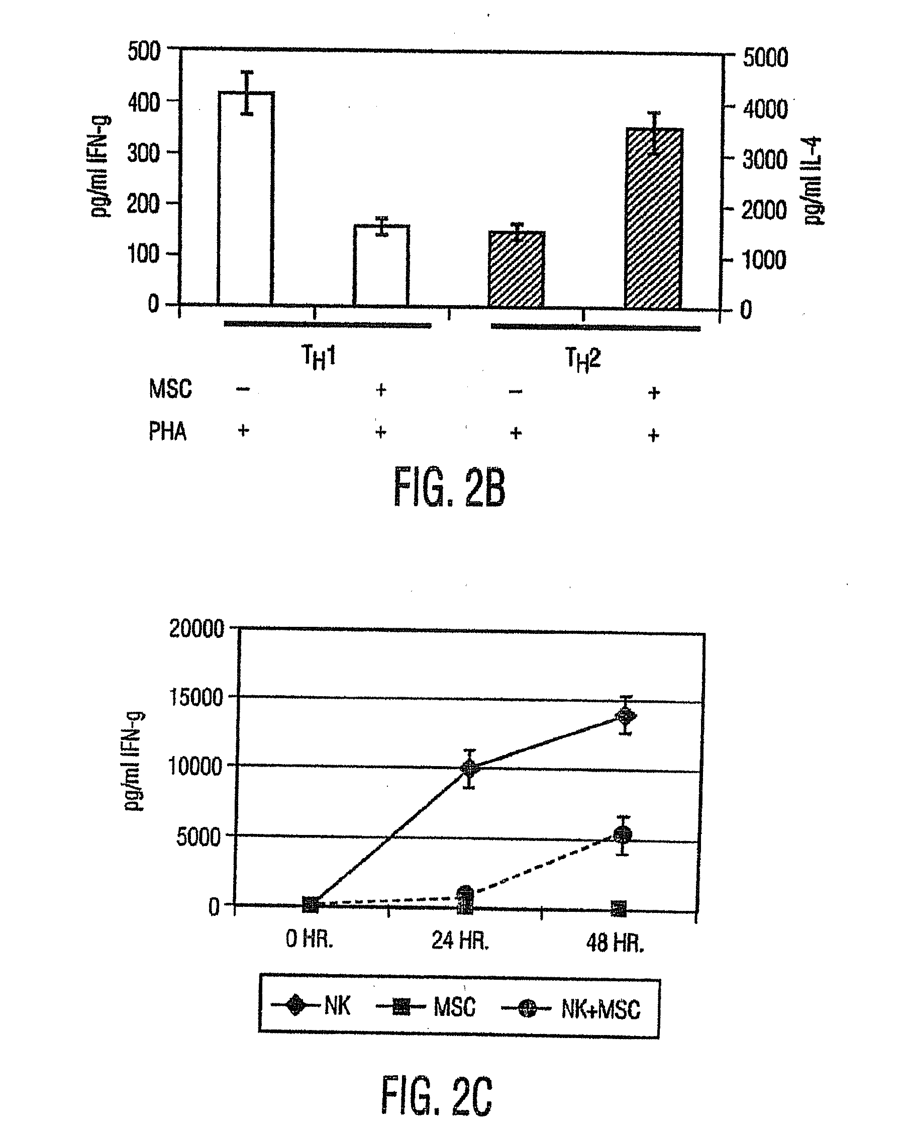
[0105] FIG. 2 MSCs inhibit pro-inflammatory effector T cell function. (A) Flow cytometric analysis of T.sub.Reg cell numbers (in %) by staining PBMCs or non-adherent fraction in MSC+PBMC culture (MSC+PBMC) with FITC-conjugated CD4 (x-axis) and PE conjugated CD25 (y-axis) antibodies. Gates were set based on isotype control antibodies as background. Graphs are representative of 5 independent experiments. (B) T.sub.H1 cells generated in presence of MSCs secreted reduced levels of IFN-.gamma. (primary Y-axis) and T.sub.H2 cells generated in presence of MSCs secreted increased amounts of IL-4 (secondary y-axis) in cell culture supernatants. (C) MSCs inhibit IFN-.gamma. secretion from purified NK cells cultured for 0, 24, or 48 hours in a 24-well plate. Data shown are mean.+-.SD cytokine secretion in one experiment and are representative of 3 independent experiments.
[0106] FIG. 3 MSCs lead to increased numbers of T.sub.Reg cell population and increased GITR expression. (A) A CD4+CD25+T.sub.Reg cell population from PBMC or MSC+PBMC (MSC to PBMC ratio 1:10) cultures (cultured without any further stimulation for 3 days) was isolated using a 2-step magnetic isolation procedure. These cells were irradiated (to block any further proliferation) and used as stimulators in a mixed lymphocyte reaction (MLR), where responders were allogeneic PBMCs (stimulator to responder ratio 1:100) in the presence of phytohemagglutinin (PHA) (2.5 mg/ml). The cells were cultured for 48 hours, following which .sup.3H thymidine was added, and incorporated radioactivity was counted after 24 hours. The results showed that the T.sub.Reg population, generated in the presence of MSCs (lane 3) was similar functionally to the T.sub.Reg cells generated in the absence of MSCs (lane 2). (B) PBMCs were cultured for 3 days in the absence (top plot) or presence (bottom plot) of MSCs (MSC to PBMC ratio 1:10), following which the non-adherent fraction was harvested and immunostained with FITC-labeled GITR and PE-labeled CD4. Results show a greater than twofold increase in GITR expression in cells cultured in the presence of MSCs.
[0107] FIG. 4 MSCs produce PGE.sub.2 and blocking PGE.sub.2 reverses MSC-mediated immuno-modulatory effects. (A) PGE.sub.2 secretion (mean.+-.SD) in culture supernatants obtained from MSCs cultured in the presence or absence of PGE.sub.2 blockers NS-398 or indomethacin (Indometh.) at various concentrations. Inhibitor concentrations are in .mu.M and data presented are values obtained after 24 hour culture (B) COX-1 and COX-2 expression in MSCs and PBMCs using real-time RT-PCR. MSCs expressed significantly higher levels of COX-2 as compared to PBMCs, and when MSCs were cultured in presence of PBMCs, there was a >3-fold increase in COX-2 expression in MSCs. Representative data from 1 of 3 independent experiments is shown. The MSC+PBMC cultures were setup in a trans-well chamber plate where MSCs were plated onto the bottom chamber and PBMCs onto the top chamber. (C) Presence of PGE.sub.2 blockers indomethacin (Ind.) or NS-398 increases TNF-.alpha. secretion from activated DCs (.quadrature.) and IFN-.gamma. secretion from T.sub.H1 cells (.quadrature.) as compared to controls. Data were calculated as % change from cultures generated in absence of MSCs and PGE.sub.2 inhibitors (D) Presence of PGE.sub.2 blockers indomethacin (Indo) and NS-398 during MSCPBMC co-culture (1:10) reverses MSC-mediated anti-proliferative effects on PHA-treated PBMCs. Data shown are from one experiment and are representative of 3 independent experiments.
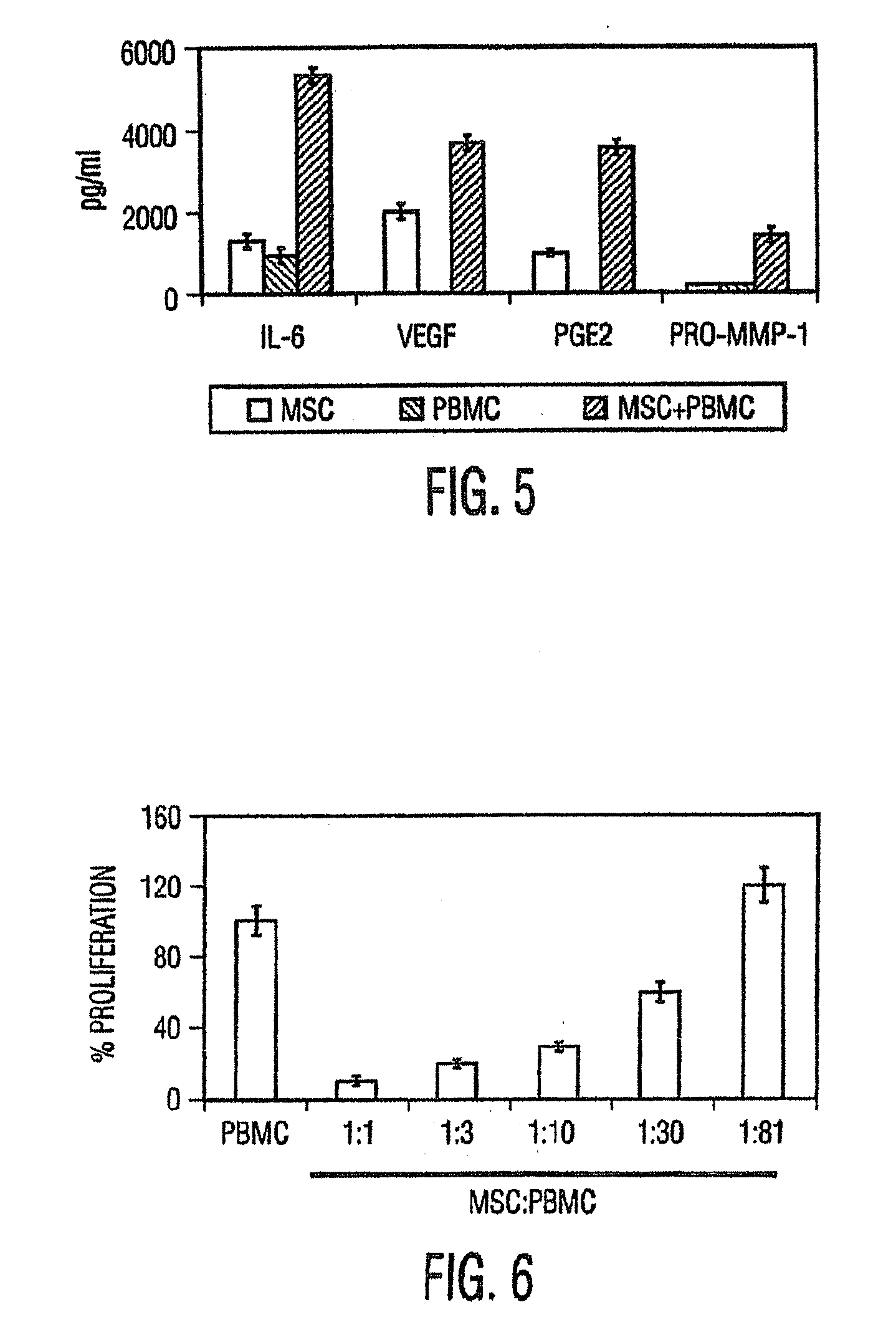
[0108] FIG. 5 Constitutive MSC cytokine secretion is elevated in the presence of allogeneic PBMCs. Using previously characterized human MSCs, the levels of the cytokines 1 L-6 and VEGF, lipid mediator PGE.sub.2, and matrix metalloproteinase 1 (pro MMP-1) in culture supernatant of MSCs cultured for 24 hours in the presence (hatched bars) or absence (open bars) of PBMCs (MSC to PBMC ratio 1:10) were analyzed. The MSCs produced 1 L-6, VEGF, and PGE.sub.2 constitutively, and the levels of these factors increased upon co-culture with PBMCs, thereby suggesting that MSCs may play a role in modulating immune functions in an inflammatory setting.
[0109] FIG. 6 MSCs inhibit mitogen-induced T-cell proliferation in a dose-dependent manner. Increasing numbers of allogeneic PBMCs were incubated with constant numbers of MSCs (2,000 cells/well) plated on a 96-well plate in the presence or absence of PHA (2.5 mg/ml) for 72 hours, and .sup.3H thymidine incorporation determined (in counts per minute, or cpm). There was a dose-dependent inhibition of the proliferation of PHA-treated PBMCs in the presence of MSCs. Representative results from 1 of 3 independent experiments are shown. Similar results were reported by LeBlanc, et al., Scand J. Immunol., vol. 57, pg. 11 (2003).
[0110] FIG. 7 Schematic diagram of proposed MSC mechanism of action. MSCs mediate their immuno-modulatory effects by affecting cells from both the innate (DCs-pathways 2-4; and NK-pathway 6) and adaptive (T-pathways 1 and 5 and B-pathway 7) immune systems. In response to an invading pathogen, immature DCs migrate to the site of potential entry, mature and acquire an ability to prime naive T cells (by means of antigen specific and co-stimulatory signals) to become protective effector T cells (cell-mediated T.sub.H1 or humoral T.sub.H2 immunity). During MSC-DC interaction, MSCs, by means of direct cell-cell contact or via secreted factor, may alter the outcome of immune response by limiting the ability of DCs to mount a cell-mediated response (pathway 2) or by promoting the ability to mount a humoral response (pathway 4). Also, when mature effector T cells are present, MSCs may interact with them to skew the balance of T.sub.H1 (pathway 1) responses towards T.sub.H2 responses (pathway 5), and probably towards an increased IgE producing B cell activity (pathway 7), desirable outcomes for suppression of GvHD and autoimmune disease symptoms. MSCs in their ability to result in an increased generation of T.sub.Reg population (pathway 3) may result in a tolerant phenotype and may aid a recipient host by dampening bystander inflammation in their local micro-environment. Dashed line (----) represents proposed mechanism.
[0111] FIG. 8. MSC treatment provides an improvement in percent of predicted forced expiratory volume in one second (Pred. FEV1%) in patients treated with mesenchymal stem cells as compared to patients who received a placebo.
[0112] FIG. 9. Measurement of distance walked on a treadmill after six minutes. Patients who were treated with MSCs showed an increase in distance walked as compared to those who received a placebo.
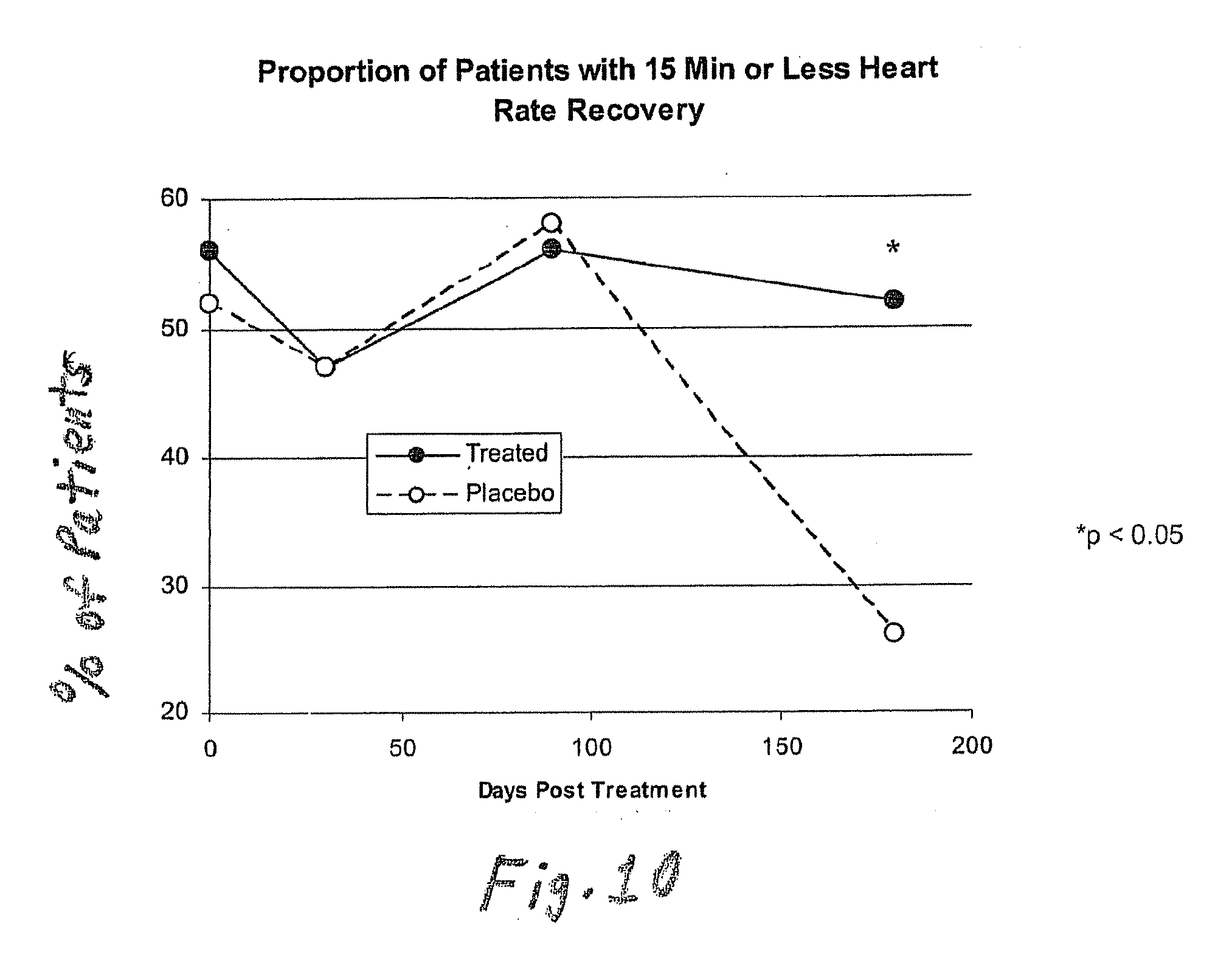
[0113] FIG. 10. Heart Rate Recovery in Patients Subjected to Treadmill Test. A greater percentage of the patients subjected to the treadmill test showed heart rate recovery to baseline values in 15 minutes or less than those who were treated with a placebo.
EXAMPLES
[0114] The invention now will be described with respect to the following examples; it is to be understood, however, that the scope of the present invention is not to be limited thereby.
Example 1
Materials and Methods Culture of Human MSCs
[0115] Human MSCs were cultured as described by Pittenger et al., Science, vol. 284, pg. 143 (1999). Briefly, marrow samples were collected from the iliac crest of anonymous donors following informed consent by Poietics Technologies, Div of Cambrex Biosciences. MSCs were cultured in complete Dulbecco's Modified Eagle's Medium-Low Glucose (Life Technologies, Carlsbad, Calif.) containing 1% antibiotic-antimyotic solution (Invitrogen, Carlsbad, Calif.) and 10% fetal bovine serum (FBS, JRH BioSciences, Lenexa, Kans.). MSCs grew as an adherent monolayer and were detached with trypsin/EDTA (0.05% trypsin at 37.degree. C. for 3 minutes). All MSCs used were previously characterized for multilineage potential and retained the capacity to differentiate into mesenchymal lineages (chondrocytic, adipogenic, and osteogenic) (Pittenger, et al., Science, vol. 284, pg. 143 (1999)).
Isolation of Dendritic Cells
[0116] Peripheral blood mononuclear cells (PBMCs) were obtained from Poietics Technologies, Div of Cambrex Biosciences (Walkersville, Md.). Precursors of dendritic cells (DCs) of monocytic lineage (CD1c+) were positively selected from PBMCs using a 2-step magnetic separation method according to Dzionek, et. al., J. Immunol., vol. 165, pg. 6037 (2000). Briefly, CD1 c expressing B cells were magnetically depleted of CD19+ cells using magnetic beads, followed by labeling the B-cell depleted fraction with biotin-labeled CD1c (BDCA1+) and anti-biotin antibodies and separating them from the unlabeled cell fraction utilizing magnetic columns according to the manufacturer's instructions (Miltenyi Biotech, Auburn, Calif.). Precursors of DCs of plasmacytoid lineage were isolated from PBMCs by immuno-magnetic sorting of positively labeled antibody coated cells (BDCA2+) (Miltenyi Biotech, Auburn, Calif.).
MSC-DC Culture
[0117] In most experiments, human MSCs and DCs were cultured in equal numbers for various time periods and cell culture supernatant collected and stored at -80.degree. C. until further evaluation. In selected experiments, MSCs were cultured with mature DC1 or DC2 cells (1:1 MSC:DC ratio) for 3 days, and then the combined cultures (MSCs and DCs) were irradiated to prevent any proliferation. Next, antibody purified, naive, allogeneic T cells (CD4+,CD45RA+) were added to the irradiated MSCs/DCs and cultured for an additional 6 days. The non-adherent cell fraction (purified T cells) was then collected from the cultures, washed twice and re-stimulated with PHA for another 24 hours, following which cell culture supernatants were harvested and analyzed for secreted IFN-.gamma. and IL-4 by ELISA.
Isolation of NK Cells
[0118] Purified populations of NK cells were obtained by depleting non-NK cells that are magnetically labeled with a cocktail of biotin-conjugated monoclonal antibodies (anti-CD3, -CD14, -CD19, -CD36 and anti-IgE antibodies) as a primary reagent and anti-biotin monoclonal antibodies conjugated to Microbeads as secondary labeling reagent. The magnetically labeled non-NK cells were retained in MACS (Miltenyi Biotech, Auburn, Calif.) columns in a magnetic field, while NK cells passed through and were collected.
Isolation of T.sub.Reg Cell Population
[0119] The T.sub.Reg cell population was isolated using a 2-step isolation procedure. First non-CD4.sup.+ T cells were indirectly magnetically labeled with a cocktail of biotin labeled antibodies and anti-biotin microbeads. The labeled cells were then depleted by separation over a MACS column (Miltenyi Biotech, Auburn, Calif.). Next, CD4.sup.+CD25.sup.+ cells were directly labeled with CD25 microbeads and isolated by positive selection from the pre-enriched CD4.sup.+. T cell fraction. The magnetically labeled CD4.sup.+CD25.sup.+T cells were retained on the column and eluted after removal of the column from the magnetic field.
[0120] In order to determine whether the increased CD4+CD25+ population generated in the presence of MSCs were suppressive in nature, CD4+CD25+T.sub.Reg cell populations were isolated from PBMC or MSC+PBMC (MSC to PBMC ratio 1:10) cultures (cultured without any further stimulation for 3 days) using a 2-step magnetic isolation procedure. These cells were irradiated to block any further proliferation and used as stimulators in a mixed lymphocyte reaction (MLR), where responders were allogeneic PBMCs (stimulator to responder ratio 1:100) in the presence of PHA (2.5 .mu.g/ml). The culture was carried out for 48 hours, following which .sup.3H thymidine was added. Incorporated radioactivity was counted after 24 hours.
[0121] PBMCs were cultured in the absence or presence of MSCs (MSC to PBMC ratio 1:10), following which the non-adherent fraction was harvested and immunostained with FITC-labeled glucocorticoid-induced TNF receptor, or GITR, and PE-labeled CD4.
Generation of T.sub.H1/T.sub.H2 Cells
[0122] Peripheral blood mononuclear cells (PBMCs) were plated at 2.times.10.sup.6 cells/ml for 45 min. at 37.degree. C. in order to remove monocytes. Non-adherent fraction was incubated in the presence of plate-bound anti-CD3 (5 .mu.g/ml) and anti-CD28 (1 .mu.g/ml) antibodies under T.sub.H1 (IL-2 (4 ng/ml) IL-12 (5 ng/ml)+anti-IL-4 (1 .mu.g/ml)) or T.sub.H2 (IL-2 (4 ng/ml)+IL-4 (4 ng/ml)+anti-IFN-.gamma. (1 .mu.g/ml)) conditions for 3 days in the presence or absence of MSCs. The cells were washed and then re-stimulated with PHA (2.5 .mu.g/ml) for another 24 or 48 hours, following which levels of IFN-.gamma. and IL-4 were measured in culture supernatants by ELISA (R&D Systems, Minneapolis, Minn.).
Analysis of Levels of VEGF, PGE.sub.2, and Pro-MMP-1 in Culture Supernatant of MSCs.
[0123] Using previously characterized human MSCs, the levels of Interleukin-6 (IL-6), VEGF, lipid mediator prostaglandin E.sub.2 (PGE.sub.2), and matrix metalloproteinase 1 (pro-MMP-1) were analyzed in culture supernatant of MSCs cultured for 24 hours in the presence or absence of PBMCs (MSC to PBMC ratio 1:10).
Proliferation of PBMCs
[0124] Purified PBMCs were prepared by centrifuging leukopack (Cambrex, Walkersville, Md.) on Ficoll-Hypaque (Lymphoprep, Oslo, Norway). Separated cells were cultured (in triplicates) in the presence or absence of MSCs (plated 3-4 hours prior to PBMC addition to allow them to settle) for 48 hours in presence of the mitogen PHA (Sigma Chemicals, St. Louis, Mo.). In selected experiments, PBMCs were resuspended in medium containing PGE.sub.2 inhibitors Indomethacin (Sigma Chemicals, St. Louis, Mo.) or NS-938 (Cayman Chemicals, Ann Arbor, Mich.). (.sup.3H)-thymidine was added (20 .mu.l in a 200 .mu.l culture) and the cells harvested after an additional 24 hour culture using an automatic harvester. The effects of MSCs or PGE.sub.2 blockers were calculated as the percentage of the control response (100%) in presence of PHA.
Quantitative RT-PCR
[0125] Total RNA from cell pellets were prepared using a commercially available kit (Qiagen, Valencia, Calif.) and according to the manufacturer's instructions. Contaminating genomic DNA was removed using the DNA-free kit (Ambion, Austin, Tex.). Quantitative RT-PCR was performed on a MJ Research Opticon detection system (South San Francisco, Calif.) using QuantiTect SYBR Green RT-PCR kit (Qiagen, Valencia, Calif.) with primers at concentration of 0.5 Relative changes in expression levels in cells cultured under different conditions were calculated by the difference in Ct values (crossing point) using .beta.-actin as internal control. The sequence for COX-1 and COX-2 specific primers were: COX-1: 5'-CCG GAT GCC AGT CAG GAT GAT G-3'(forward) (SEQ ID NO:1), 5'-CTA GAC AGC CAG ATG CTG ACA G-3' (reverse) (SEQ ID NO:2); COX-2: 5'-ATC TAC CCT CCT CAA GTC CC-3' (forward) (SEQ ID NO:3), 5'-TAC CAG AAG GGC AGG ATA CAG-3' (reverse) (SEQ ID NO:4).
[0126] Increasing numbers of allogeneic PBMCs were incubated with constant numbers of MSCs (2,000 cells/well) plated on a 96-well plate in the presence of PHA (2.5 .mu.g/ml) for 72 hours, and .sup.3H thymidine incorporation (counts per minute, cpm) was determined. The PBMCs and MSCs were cultured at ratios of MSC:PBMC of 1:1, 1:3, 1:10, 1:30, and 1:81.
Results
[0127] In the present studies, the interaction of human MSCs with isolated immune cell populations, including dendritic cells (DC1 and DC2), effector T cells (T.sub.H1 and T.sub.H2) and NK cells was examined. The interaction of MSCs with each immune cell type had specific consequences, suggesting that MSCs may modulate several steps in the immune response process. The production of secreted factor(s) that modulate and may be responsible for MSC immuno-modulatory effects was evaluated and prostaglandin synthesis was implicated.
[0128] Myeloid (DC1) and plasmacytoid (DC2) precursor dendritic cells were isolated by immuno-magnetic sorting of BDCA1.sup.+ and BDCA2.sup.+ cells respectively and matured by incubation with GM-CSF and IL-4 (1.times.10.sup.3 IU/ml and 1.times.10.sup.3 IU/ml, respectively) for DC1 cells, or IL-3 (10 ng/ml) for DC2 cells. Using flow cytometry, DC1 cells were HLA-DR.sup.+ and CD11c.sup.+, whereas DC2 cells were HLA-DR.sup.+ and CD123.sup.+ (FIG. 1A). In the presence of the inflammatory agent bacterial lipopolysaccharide (LPS, 1 ng/ml), DC1 cells produced moderate levels of TNF-.alpha. but when MSCs were present (ratios examined 1:1 and 1:10), there was >50% reduction in TNF-.alpha. secretion (FIG. 1B). On the other hand, DC2 cells produced IL-10 in the presence of LPS and its levels were increased greater than 2-fold upon MSC:DC2 co-culture (1:1) (FIG. 1B). Therefore, the MSCs modified the cytokine profile of activated DCs in culture towards a more tolerogenic phenotype. Additionally, activated DCs, when cultured with MSCs, were able to reduce IFN-.gamma. and increase IL-4 levels secreted by naive CD4.sup.+T cells (FIG. 1C) suggesting a MSC-mediated shift from pro-inflammatory to anti-inflammatory T cell phenotype.
[0129] As increased IL-10 secretion plays a role in generation of regulatory cells (Kingsley, et al., J. Immunol., vol. 168, pg. 1080 (2002)), T-regulatory cells (T.sub.Reg) were quantified by flow cytometry in co-cultures of PBMCs and MSCs. Upon culture of PBMCs with MSCs for 3-5 days, there was an increase in T.sub.Reg cell numbers as determined by staining of PBMCs with anti-CD4 and anti-CD25 antibodies (FIG. 2A), further supporting a MSC-induced tolerogenic response. The CD4.sup.+CD25.sup.+T.sub.Reg cell population, generated in presence of MSCs expressed increased levels of glucocorticoid-induced TNF receptor (GITR), a cell surface receptor expressed on T.sub.Reg cell populations, and was suppressive in nature as it suppressed allogeneic T cell proliferation (FIG. 3A,B). Next, MSCs were investigated as to their direct ability to affect T cell differentiation. Using antibody selected purified T cells (CD4.sup.+Th cells), IFN-.gamma. producing T.sub.H1 and IL-4 producing T.sub.H2 cells were generated in presence or absence of MSCs. When MSCs were present during differentiation, there was reduced IFN-.gamma. secretion by T.sub.H1 cells and increased IL-4 secretion by T.sub.H2 cells (FIG. 2B). No significant change in IFN-.gamma. or IL-4 levels were seen when MSCs were added to the culture after Th cells had differentiated (at 3 days) into effector T.sub.H1 or T.sub.H2 types (data not shown). These experiments suggest that MSCs can affect effector T cell differentiation directly and alter the T cell cytokine secretion towards a humoral phenotype.
[0130] Similarly, when MSCs were cultured with purified NK cells (CD3-, CD14-, CD19-, CD36-) at a ratio 1:1 for different time periods (0-48 hrs), there was decreased IFN-.gamma. secretion in the culture supernatant (FIG. 2C), thereby suggesting that MSCs can modulate NK cell functions also.
[0131] Previous work has indicated that MSCs modify T-cell functions by soluble factor(s) (LeBlanc, et al., Exp. Hematol., vol. 31, pg. 890 (2003); Tse, et al., Transplantation, vol. 75, pg. 389 (2003). It was observed that the MSCs secreted several factors, including IL-6, prostaglandin E.sub.2, VEGF and proMMP-1 constitutively, and the levels of each increased upon culture with PBMCs (FIG. 5). In order to investigate MSC-derived factors leading to inhibition of TNF-.alpha. and increase of IL-10 production by DCs, the potential role of prostaglandin E.sub.2 was investigated, as it has been shown to inhibit TNF-.alpha. production by activated DCs (Vassiliou, et al., Cell. Immunol., vol. 223, pg. 120 (2003)). Conditioned media from MSC culture (24 hour culture of 0.5.times.10.sup.6 cells/ml) contained approx. 1000 .mu.g/ml of PGE.sub.2 (FIG. 4A). There was no detectable presence of known inducers of PGE.sub.2 secretion, e.g., TNF-.alpha., IFN-.gamma. or IL-1.beta. (data not shown) in the culture supernatant indicating a constitutive secretion of PGE.sub.2 by MSCs. The PGE.sub.2 secretion by hMSCs was inhibited 60-90% in the presence of known inhibitors of PGE.sub.2 production, NS-398 (5 .mu.M) and indomethacin (4 .mu.M) (FIG. 4A). As the release of PGE.sub.2 secretion occurs as a result of enzymatic activity of constitutively active cycloxygenase enzyme 1 (COX-1) and inducible cycloxygenase enzyme 2 (COX-2) (Harris, et al., Trends Immunol., vol. 23, pg. 144 (2002)) the mRNA expression for COX-1 and COX-2 in MSCs and PBMCs using trans-well culture system was analyzed. MSCs expressed significantly higher levels of COX-2 as compared to PBMCs and the expression levels increase >3-fold upon co-culture of MSCs and PBMCs (MSC to PBMC ratio 1:10) for 24 hours (FIG. 4B). Modest changes in COX-1 levels were seen suggesting that the increase in PGE.sub.2 secretion upon MSC-PBMC co-culture (FIG. 5) is mediated by COX-2 up-regulation. To investigate whether the immunomodulatory effects of MSC on DCs and T-cells were mediated by PGE.sub.2, MSCs were cultured with activated dendritic cells (DC1) or T.sub.H1 cells in the presence of PGE.sub.2 inhibitors NS-398 or indomethacin. The presence of NS-398 or indomethacin increased TNF-.alpha. secretion by DC1s, and IFN.gamma. secretion from T.sub.H1 cells (FIG. 4C), respectively, suggesting that MSC effects on immune cell types may be mediated by secreted PGE.sub.2. Recent studies have shown that MSCs inhibit T-cell proliferation induced by various stimuli (DeNicola, et al., Blood, vol. 99, pg. 3838 (2002); LeBlanc, et al., Scand. J. Immunol., vol. 57, pg. 11 (2003)). It was observed that MSCs inhibit mitogen-induced T cell proliferation in a dose-dependent manner (FIG. 6) and when PGE.sub.2 inhibitors NS-398 (5 .mu.M) or indomethacin (4 .mu.M) were present, there was a >70% increase in (.sup.3H) thymidine incorporation by PHA-treated PBMCs in MSC containing cultures as compared to controls without inhibitors (FIG. 4D).
[0132] In summary, a model of MSC interaction with other immune cell types (FIG. 7) is proposed. When mature T cells are present, MSCs may interact with them directly and inhibit the pro-inflammatory IFN-.gamma. production (pathway 1) and promote regulatory T cell phenotype (pathway 3) and anti-inflammatory T.sub.H2 cells (pathway 5). Further, MSCs can alter the outcome of the T cell immune response through DCs by secreting PGE.sub.2, inhibiting pro-inflammatory DC1 cells (pathway 2) and promoting anti-inflammatory DC2 cells (pathway 4) or regulatory DCs (pathway 3). A shift towards T.sub.H2 immunity in turn, suggests a change in B cell activity towards increased generation of IgE/IgG1 subtype antibodies (pathway 7). MSCs, by their ability to inhibit IFN-.gamma. secretion from NK cells likely modify NK cell function (pathway 6). This model of MSC:Immune cell interactions is consistent with the experimentation performed in several other laboratories (LeBlanc, et al., Exp. Hematol., vol. 31, pg. 890 (2003); Tse, et al., Transplantation, vol. 75, pg. 389. (2003); DiNicola, et al., Blood, vol. 99, pg. 3838 (2002)). Further examination of the proposed mechanisms is underway and animal studies are now necessary to examine the in vivo effects of MSC administration.
Example 2
[0133] Mesenchymal stem cells were given to a 33-year-old female patient suffering from severe Grade IV gastrointestinal graft-versus-host disease (GVHD). The patient was refractory to all other GVHD treatments. Endoscopic views of the patient's colon showed areas of ulceration and inflammation prior to treatment. Histology of the patient's colon showed that the graft-versus-host disease had destroyed the vast majority of the patient's intestinal crypts, prior to treatment.
[0134] The patient was given an intravenous infusion of allogeneic mesenchymal stem cells in 50 ml of Plasma Lyte A (Baxter) in an amount of 3.times.10.sup.6 cells per kilogram of body weight.
[0135] The patient was evaluated at two weeks post-infusion. At two weeks post-infusion, an endoscopic view of the patient's colon showed that the areas of inflammation and ulceration visible prior to treatment were resolved. In addition, a biopsy of the patient's colon showed significant regeneration of intestinal crypts. Thus, the administration of the mesenchymal stem cells to the patient resulted in a significant reduction in the inflammatory component of gastrointestinal graft-versus-host disease, and resulted in the regeneration of new functional intestinal tissue.
Example 3
[0136] 9 patients received 0.5.times.10.sup.6 mesenchymal stem cells per kilogram of body weight, 10 patients received 1.6.times.10.sup.6 mesenchymal stem cells per kilogram of body weight, and 15 patients received 5.0.times.10.sup.6 mesenchymal stem cells per kilogram of body weight by intravenous infusion of a suspension of mesenchymal stem cells in Plasma Lyte A (Baxter) in which the mesenchymal stem cells were present in the suspension at a concentration of 2.5.times.10.sup.6 cells/ml. The total volume of mesenchymal stem cell suspension given thus was dependent upon the dosage of cells and the weight of the patient.
[0137] 19 patients received placebo doses of Plasma Lyte A. The placebo doses were in low, medium, and high doses in proportion to the volumes of the mesenchymal stem cell suspension given to the patients treated with the mesenchymal stem cells. Of the placebo patients, 5 patients were given a low dose of the suspension, 4 patients were given a medium dose of the suspension, and 10 patients were given a high dose of the suspension.
[0138] The FEV1 spirometry test was performed over a six month period to detect potential changes related to the treatment. The testing was done according to American Thoracic Society guidelines. (Miller, et al., Eur. Respir. J., vol. 26, pgs. 319-338 (2005)).
[0139] Forced expiratory volume, or FEV1, is the maximal volume of air exhaled in the first second of a forced expiration from a position of full inspiration, expressed in liters, at body temperature (37.degree. C.), ambient pressure, saturated with water vapor (BTPS). The predicted FEV1 values were calculated for each patient based on age, sex, height, and race. The predicted FEV1 for men was calculated as follows (Crapo, et al., Am. Rev. Respir. Dis., vol. 123, pgs. 659-664 (1981):
predicted FEV1=0.0414.times.height (cm)-0.0244.times.age (years)-2.190
[0140] The predicted FEV1 for women was calculated as follows (Crapo, 1981):
predicted FEV1=0.0342.times.height (cm)-0.0255.times.age (years)-1.578
[0141] The above values were calculated for men and women of other than African-American background. For men and women of African-American background, the above values were multiplied by a correction factor of 0.88.
[0142] The weight of each patient also was taken. Although weight is not factored into the determination of the FEV1 values, obesity may lower the measured lung volumes, and changes in body weight can result in small changes in lung function.
[0143] FEV1 values for all patients were measured using a spirometer, which was connected to a mouthpiece or tube which was inserted into the patient's mouth. The height and weight of each patient was measured, a nose clip was placed on each patient. Each patient was instructed to inhale completely and rapidly, with a pause of less than one second at total lung capacity, and to exhale maximally until no more air can be expelled. The procedure is repeated in triplicate, and the FEV1 values for each patient were measured. From the FEV1 values, the percent of predicted FEV1 (Pred. FEV1%) values were calculated. The percent of predicted FEV1 (Pred. FEV1%) values for each patient were calculated as follows:
Pred . FEV 1 % = 100.0 .times. Observed FEV 1 Predicted FEV 1 ##EQU00001##
[0144] The percent improvement in Pred. FEV1% values for the patients also was calculated at various time intervals up until 6 months (180 days) after treatment. The results for the MSC and placebo-treated groups are shown in FIG. 8. Such results show the average percent change in Pred. FEV1% values for all MSC-treated patients and the average percent change in Pred. FEV1% values for all patients who received the placebo.
[0145] Compared to the control group of patients, the patients who were treated with the MSCs showed a greater improvement in Pred. FEV1%, relative to baseline (pre-treatment) values, from three days through six months post-infusion. At both 10 and 30 days post-infusion, the difference in improvement in Pred. FEV1% values observed for MSC-treated and placebo patients was significant statistically (p<0.05).
[0146] MSC-treated and placebo patients also were subjected to a treadmill test in which the patients walked on a treadmill in which the distance walked by each patient was measured in six minute intervals. Distance measurements were taken after treatment (baseline), and at one month, three months, and six months post-treatment. The test was performed according to ATS (American Thoracic Society) guidelines (Am. J. Respir. Crit. Care Med., vol. 166, pg. 111 (2002)).
[0147] The average percent changes in distance walked for all MSC-treated patients and all placebo-treated patients as compared to the baseline distance are shown in FIG. 9. At both the three month and six month timepoints, the MSC-treated patients showed an increase in the distance walked compared to the patients who received the placebo.
[0148] Following the six minute treadmill test that was given to the patients shortly after treatment, and at one, three, and six months after treatment, heart rate recovery for the patients was determined.
[0149] The heart rate recovery results are shown in FIG. 10. As shown in FIG. 10, at six months after the treatment, the difference in the percentage of patients who received the MSC treatments showing heart rate recovery to baseline values within 15 minutes after the cessation of the treadmill walking as compared to the patients who received the placebo treatments was significant statistically.
[0150] The above results with respect to improvement in Fred. FEV1%, distance walked, and heart rate recovery, show that the patients treated with the mesenchymal stem cells had improved pulmonary functions as compared to the placebo group. The above results suggest that fibrotic lung diseases or disorders may be treated with mesenchymal stem cells, whereby the mesenchymal stem cells improve pulmonary function, reduce existing scar tissue in the lung, and/or prevent further scar expansion in the lung.
Specific Embodiments
[0151] The compositions and methods described herein can be illustrated by the following embodiments enumerated in the numbered paragraphs that follow:
[0152] 1. A method of promoting angiogenesis in an organ or tissue of an animal other than the heart, comprising:
[0153] administering to the animal mesenchymal stem cells in an amount effective to promote angiogenesis in an organ or tissue other than the heart of the animal.
[0154] 2. The method of paragraph 1 wherein the animal is a mammal.
[0155] 3. The method of paragraph 2 wherein the mammal is a primate.
[0156] 4. The method of paragraph 3 wherein the primate is a human.
[0157] 5. The method of paragraph 1 wherein the mesenchymal stem cells are administered in an amount of from about 1.times.10.sup.5 cells/kg to about 1.times.10.sup.7 cells/kg.
[0158] 6. The method of paragraph 5 wherein the mesenchymal stem cells are administered in an amount of from about 1.times.10.sup.6 cells/kg to about 5.times.10.sup.6 cells/kg.
[0159] 7. The method of paragraph 1 wherein the mesenchymal stem cells are administered systemically.
[0160] 8. The method of paragraph 1 wherein the mesenchymal stem cells are administered intravenously.
[0161] 9. The method of paragraph 1 wherein the mesenchymal stem cells are administered by direct injection to the organ or tissue other than the heart of the animal.
[0162] 10. A method of treating a disease selected from the group consisting of autoimmune diseases and graft-versus-host in an animal, comprising:
[0163] administering to the animal mesenchymal stem cells in an amount effective to treat the disease in the animal
[0164] 11. The method of paragraph 10 wherein the animal is a mammal
[0165] 12. The method of paragraph 11 wherein the mammal is a human.
[0166] 13. The method of paragraph 10 wherein the disease is multiple sclerosis.
[0167] 14. A method of treating an inflammatory response in an animal, comprising: administering to the animal mesenchymal stem cells in an amount effective to treat the inflammatory response in the animal.
[0168] 15. The method of paragraph 14 wherein the animal is a mammal.
[0169] 16. The method of paragraph 15 wherein the mammal is a human.
[0170] 17. The method of paragraph 14 wherein the inflammatory response is associated with psoriasis.
[0171] 18. A method of treating cancer in an animal, comprising:
[0172] administering to the animal mesenchymal stem cells in an amount effective to treat cancer in the animal.
[0173] 19. The method of paragraph 18 wherein the animal is a mammal.
[0174] 20. The method of paragraph 19 wherein the mammal is a human.
[0175] 21. A method of treating an allergic disease or disorder in an animal, comprising:
[0176] administering to the animal mesenchymal stem cells in an amount effective to treat the allergic disease or disorder in the animal.
[0177] 22. The method of paragraph 21 wherein the animal is a mammal.
[0178] 23. The method of paragraph 22 wherein the mammal is a human.
[0179] 24. The method of paragraph 14 wherein the allergic disease or disorder is arthritis.
[0180] 25. A method of promoting wound healing in an animal, comprising: administering to the animal mesenchymal stem cells in an amount effective to promote wound healing in the animal.
[0181] 26. The method of paragraph 25 wherein the animal is a mammal.
[0182] 27. The method of paragraph 26 wherein the mammal is a human.
[0183] 28. A method of treating or preventing a fibrotic disorder in an animal, comprising:
[0184] administering to the animal mesenchymal stem cells in an amount effective to treat or prevent the fibrotic disorder in the animal.
[0185] 29. The method of paragraph 28 wherein the fibrotic disorder is Acute Respiratory Distress Syndrome.
[0186] 30. The method of paragraph 28 wherein the fibrotic disorder is Chronic Obstructive Pulmonary Disease.
[0187] 31. The method of paragraph 28 wherein the fibrotic disorder is Idiopathic Pulmonary Fibrosis.
[0188] 32. A method of repairing epithelial damage in an animal, comprising: administering to the animal mesenchymal stem cells in an amount affective to repair epithelial damage in the animal.
[0189] 33. The method of paragraph 32 wherein the animal is a mammal.
[0190] 34. The method of paragraph 33 wherein the mammal is a human.
[0191] 35. The method of paragraph 32 wherein the epithelial damage is a result of graft-versus-host disease.
[0192] The disclosures of all patents, publications, including published patent applications, depository accession numbers, and database accession numbers are hereby incorporated by reference to the same extent as if each patent, publication, depository accession number, and database accession number were specifically and individually incorporated by reference.
[0193] It is to be understood, however, that the scope of the present invention is not to be limited to the specific embodiments described above. The invention may be practiced other than as particularly described and still be within the scope of the accompanying claims.
Sequence CWU 1
1
4122DNAArtificialsynthetic sequences and are primers for PCR
derived from human COX-1 1ccggatgcca gtcaggatga tg
22222DNAArtificialsynthetic sequences and are primers for PCR
derived from human COX-1 2ctagacagcc agatgctgac ag
22320DNAartificialsynthetic sequences and are primers for PCR
derived from human COX-2 3atctaccctc ctcaagtccc
20421DNAArtificialprimer for PCR derived from human COX-2
4taccagaagg gcaggataca g 21
References
D00001
D00002
D00003
D00004
D00005
D00006
D00007
D00008
D00009
D00010
D00011
D00012

P00001

S00001
XML
uspto.report is an independent third-party trademark research tool that is not affiliated, endorsed, or sponsored by the United States Patent and Trademark Office (USPTO) or any other governmental organization. The information provided by uspto.report is based on publicly available data at the time of writing and is intended for informational purposes only.
While we strive to provide accurate and up-to-date information, we do not guarantee the accuracy, completeness, reliability, or suitability of the information displayed on this site. The use of this site is at your own risk. Any reliance you place on such information is therefore strictly at your own risk.
All official trademark data, including owner information, should be verified by visiting the official USPTO website at www.uspto.gov. This site is not intended to replace professional legal advice and should not be used as a substitute for consulting with a legal professional who is knowledgeable about trademark law.