Mesp1 As A Master Regulator Of Multipotent Cardiovascular Progenitor Specification And Uses Thereof
BLANPAIN; CEDRIC ; et al.
U.S. patent application number 12/494941 was filed with the patent office on 2010-12-30 for mesp1 as a master regulator of multipotent cardiovascular progenitor specification and uses thereof. This patent application is currently assigned to UNIVERSITE LIBRE DE BRUXELLES. Invention is credited to CEDRIC BLANPAIN, ANTOINE BONDUE, GA LLE LAPOUGE.
| Application Number | 20100330044 12/494941 |
| Document ID | / |
| Family ID | 43381014 |
| Filed Date | 2010-12-30 |


View All Diagrams
| United States Patent Application | 20100330044 |
| Kind Code | A1 |
| BLANPAIN; CEDRIC ; et al. | December 30, 2010 |
MESP1 AS A MASTER REGULATOR OF MULTIPOTENT CARDIOVASCULAR PROGENITOR SPECIFICATION AND USES THEREOF
Abstract
A method for differentiating or promoting or inducing differentiation of stem cells into pluripotent cardiovascular progenitors (MCPs) by transiently inducing the expression of a single gene, namely Mesp1, is disclosed. Cells obtained by the method and their uses in research and clinical settings are also disclosed. Using genome wide transcriptional analysis, the inventors found that Mesp1 rapidly activates and represses a discrete set of genes, which form potential new targets for both therapy and for the identification of MCPs. Insights into the molecular mechanisms underlying the earliest step of cardiovascular specification and potential methods for dramatically increasing the number of cardiovascular cells for cellular therapy in humans are provided.
| Inventors: | BLANPAIN; CEDRIC; (LASNE, BE) ; BONDUE; ANTOINE; (BRUXELLES, BE) ; LAPOUGE; GA LLE; (BRUXELLES, BE) |
| Correspondence Address: |
    KNOBBE MARTENS OLSON & BEAR LLP
    2040 MAIN STREET, FOURTEENTH FLOOR
    IRVINE
    CA
    92614
    US
|
| Assignee: | UNIVERSITE LIBRE DE
BRUXELLES BRUXELLES BD |
| Family ID: | 43381014 |
| Appl. No.: | 12/494941 |
| Filed: | June 30, 2009 |
| Current U.S. Class: | 424/93.7 ; 435/325; 435/377; 435/6.16; 435/7.21 |
| Current CPC Class: | G01N 33/5014 20130101; G01N 2800/32 20130101; A61K 45/06 20130101; G01N 33/5073 20130101; A61P 9/10 20180101; C12N 2506/02 20130101; C12N 2510/00 20130101; G01N 2800/323 20130101; C12N 2501/60 20130101; C12N 5/0657 20130101; G01N 2800/385 20130101 |
| Class at Publication: | 424/93.7 ; 435/377; 435/7.21; 435/6; 435/325 |
| International Class: | A61K 45/00 20060101 A61K045/00; C12N 5/071 20100101 C12N005/071; G01N 33/50 20060101 G01N033/50; C12Q 1/68 20060101 C12Q001/68; A61P 9/10 20060101 A61P009/10 |
Claims
1. A method of inducing, of enhancing the induction, or of differentiating stem cells into cardiovascular precursor or progenitor cells comprising the steps of: a) transiently inducing the expression of the Mesp1 gene in said stem cells, and b) culturing said induced stem cells in vitro thereby obtaining differentiated stem cells that are enriched in cardiovascular progenitor cells. c) specifying and differentiating the cardiovascular progenitors generated by method of the invention into a particular subset of cardiovascular lineages such as cardiomyocytes, vascular cells or endothelial cells.
2. The method of claim 1, wherein the transient expression is performed in vitro by transforming said stem cells with a vector comprising the gene sequence of the Mesp-1 protein.
3. The method of claim 2, wherein said Mesp-1 gene sequence is placed in an inducible expression cassette.
4. The method of claim 3, wherein the inducible expression cassette is chosen from the group of the Tetracyclin or doxycyclin induced systems, Rheo switch systems, IPTG-LAC inducible systems, ecdysone inducible systems, or the cumate repressor/operator systems, inducible activation of modulator systems.
5. The method of claim 1, wherein the induction of the Mesp-1 expression is performed during cardiovascular competence which need to be precise for each types of stem cells used and that correspond for murine ESC to day 2 or day 3, or day 2 and day 3 of the culturing period of the stem cells.
6. The method of claims 5, wherein the induction is performed for one or two days only.
7. The method of claim 1, wherein the stem cells are selected from the group of: Embryonic Stem cells (ES), pluripotent stem cells, haematopoietic stem cells, totipotent stem cells, mesenchymal stem cells, induced pluripotent stem cells (iPS) or adult stem cells, adult heart, epicardial, vessel or muscular cells.
8. A method for performing cellular therapy for restoring the heart or vasculature function in a subject in need thereof, comprising the steps of: a) providing cells according to the method of claim 1, and b) injecting said cells into the heart or the vasculature of the subject in need thereof, wherein said cardiovascular function is preferably disturbed due to a disease or disorder selected from the group consisting of: Congenital Heart Disease, such as malformations and misplacements of cardiac structures, acquired heart and vascular diseases, such as myocardial infarction, cardiac hypertrophy and cardiac arrhythmia and cardiovascular damage due to trauma.
9. A method for restoring the heart or vasculature function in a subject in need thereof, in an endogenous manner, comprising the step of transiently inducing the expression of the Mesp-1 protein in the cells of the heart or the vasculature, wherein said cardiovascular function is preferably disturbed due to a disease or disorder selected from the group consisting of: Congenital Heart Disease, such as malformations and misplacements of cardiac structures, acquired heart and vascular diseases, such as myocardial infarction, cardiac hypertrophy and cardiac arrhythmia and cardiovascular damage due to trauma.
10. The method of claim 9, wherein said induction is performed by injecting the subject with an amount of an expression vector encoding for the Mesp-1 protein.
11. The method of claim 9, wherein said induction is performed by injecting the subject with an amount of an expression vector encoding for the Mesp-1 protein packed in a virus.
12. An assay for assessing the toxicity of an agent on heart or vascular cells, comprising the steps of: a) differentiating stem cells into cardiovascular progenitor cells according to the method of claim 1, b) subjecting said cells in vitro to said agent, and c) analysing the toxic effect of said agent on the cells obtained in step a).
13. An assay for assessing the pharmacology of a candidate drug comprising the steps of: a) differentiating stem cells into cardiovascular progenitor cells according to the method of claim 1, b) subjecting said cells in vitro to said candidate drug, and c) analysing the behaviour of said cells in the presence and absence of said candidate drug.
14. A method for identifying target genes for therapy of cardiovascular disorders comprising the steps of: a) differentiating stem cells into cardiovascular progenitor cells according to the method of claim 1, b) analysing the expression level of the genes in said cells prior to and after said induction of Mesp-1 expression in said stem cells, wherein genes that are up-regulated after the gene-induction are putative targets for stimulation of differentiation of cardiovascular differentiation and those genes that are down-regulated after the gene-induction are putative targets for inhibiting cardiovascular differentiation of stem cells.
15. Cardiovascular progenitor cells obtained by the method of claim 1.
16. Cardiovascular cells obtained by the method of claim 1.
17. A method of diagnosis and/or treatment of congenital heart diseases comprising the detection of the occurrence of mutations in the Mesp1 genomic.
Description
FIELD OF THE INVENTION
[0001] The present invention relates to processes and compositions for controlling cell fate promotion from stem cells, preferably embryonic stem (ES) cells and promoting specifically and in a controlled manner cardiac and vascular differentiation from ES cells, other pluripotent or multipotent stem cell (SC) types through the specification of multipotent cardiovascular progenitor cells (MCP) as well as the use of said MCP or the differentiated cells arising from the differentiation of MCP for therapeutic and research purposes.
BACKGROUND OF THE INVENTION
[0002] Differentiation of mouse embryonic stem cells into cardiomyocytes has historically been achieved through spontaneous differentiation of embryonic stem cells or carcinomic embryonic stem cells in serum containing medium or through treatment with compounds like DMSO, retinoic acid, bone morphogenetic proteins, fibroblast growth factors or the broad and non-specific de-methylating agent 5-aza-deoxycytidine. Compounds such as 5-aza-deoxycytidine have also been used to induce differentiation of cardiomyocytes from other stem cell types. This indicates that different types of stem cells could potentially be used as a source for the generation of cardiomyocytes for applications such as cell therapy, tissue engineering, pharmacological and toxicological screening or the like, provided effective processes and methods for the differentiation of cardiomyocytes are available. Unlike direct specification of MCP from undifferentiated cells, current methods used to promote cardiac differentiation, generally act in the latter step of cardiac expansion and differentiation, and the promotion in cardiac differentiation usually induced by a single factor rarely exceeds 100%.
[0003] When allowed to differentiate, ES cells form spherical structures called "embryoid bodies" (EBs) that are thought to mimic interactions that arise during development. This differentiation process results in the formation of beating areas around day 8, made of cardiomyocytes when differentiation is performed in serum-containing medium. Current published methods for forming cardiomyocytes from ES cells rely on spontaneous differentiation in media containing animal serum, a medium component that is largely undefined and subject to batch-to-batch variations. This method does not lend itself to being a clinically useful and reproducible system. Moreover the global efficiency of cardiac differentiation using the previously described methods is low, leading to 2-3% of cardiac cells whatever the differentiating system used.
[0004] Different lines of evidence suggest that different cardiac cell types, including cardiomyocytes (CM), endothelial cells (EC), smooth muscle cells (SMC) as well as conduction cells, which compose the mature heart tissue, arise from the differentiation of multipotent cardiovascular progenitors (MCPs) generated soon after gastrulation (Garry and Olson, 2006, Cell 127:1101-1104; Moretti et al., 2006, Cell 127, 1137-1150; Wu et al., 2006, Cell 127, 1137-1150). Recent studies also provide compelling evidence that different sources of MCPs, which are specified during embryonic development, are also generated during pluripotent embryonic stem cell (ESCs) differentiation (Moretti et al., 2006, Cell 127, 1137-1150; Murry and Keller Cell 132, 661-680, 2008; Wu et al., 2006, Cell 127, 1137-1150). Mesp1, a transcription factor of the b-HLH family, is one of the earliest markers of cardiovascular development in vertebrates (Saga et al., 2000, Trends Cardiovasc Med 10, 345-352). During gastrulation in mice, Mesp1 is strongly expressed at the onset of gastrulation (E6.5) along the primitive streak and in the prospective cardiac mesoderm and is then rapidly downregulated after E7.5 (Saga et al., 1996, Development 122, 2769-2778). Lineage tracing experiments, using mice in which the CRE recombinase has been knocked-in into the Mesp1 locus, demonstrated that most cardiac cells and some vascular cells, arise from cells that expressed Mesp1 at one point of their development (Saga et al., 1999, Development 126, 3437-3447).
[0005] The need for cardiovascular progenitors or adult cells is very high in both clinical and research settings and is currently not easily fulfilled. The current invention provides for improved means and methods to obtain such cardiovascular progenitors or differentiated cardiovascular cells, which can be safely transplanted in patients with cardiovascular diseases or used in research and industrial perspectives.
SUMMARY OF THE INVENTION
[0006] In the present invention, Mesp1 is identified as the key molecular determinant of multipotent cardiovascular progenitors specification. It is shown by genetically engineered ESC in which Mesp1 expression can be conditionally induced that transient but not continuous expression of this gene promote greatly the specification of MCP and their different cardiovascular cell progenies including cardiomyocytes, vascular cells, cell of the conduction system, and smooth muscle cells. The inventors describe a novel method that allows a high efficient generation of cardiovascular cells from pluripotent cells by transiently expressing a single gene. Moreover they identify many MesP1 target genes that are responsible for the cardiac and vascular promoting effect of Mesp1, and which now represents key target genes for which pharmaceutical intervention will be useful to improve cardiac regeneration in acute and chronic heart failure. In addition, the technique presented here to promote cardiovascular differentiation represent an extremely versatile method for promoting cardiac cell differentiation from various sources of stem cells that can be used for cellular therapy in humans but also for, tissue engineering, pharmacological and toxicological screening
[0007] The invention therefore provides for a novel and highly efficient method for generation of cardiovascular cells or progenitors and uses thereof in research and the clinic.
[0008] In a particular embodiment, the invention provides a method of inducing, enhancing the induction or differentiation of stem cells into cardiovascular precursor or progenitor cells comprising the steps of:
a) transiently inducing the expression of the Mesp1 gene in said stem cells, and b) culturing said induced stem cells in vitro thereby obtaining differentiated stem cells that are enriched in cardiovascular progenitor cells. c) specifying and differentiating the cardiovascular progenitors generated by method of the invention into a particular subset of cardiovascular lineages such as cardiomyocytes, vascular or endothelial cells. Preferably, the transient expression is performed in vitro by transforming said stem cells with a vector comprising the gene sequence of the Mesp-1 protein. In another embodiment, said Mesp-1 gene sequence is placed in an inducible expression cassette such as any one of the following non-limiting examples: the Tetracyclin or doxycyclin induced systems, Rheo switch systems, FRT system, IPTG-LAC inducible systems, ecdysone inducible systems.
[0009] In another embodiment of the invention the induction of the Mesp-1 expression is performed during cardiovascular competence which need to be precise for each types of stem cells used and that correspond for murine ESC to day 2 or day 3, or day 2 and day 3 of the culturing period of the stem cells. Preferably, said induction is performed for one or two days only.
[0010] In a further embodiment, the method of the invention can use several types of cells as a starting point. Non-limiting examples are: Embryonic Stem cells (ES), pluripotent stem cells, haematopoietic stem cells, totipotent stem cells, mesenchymal stem cells, induced pluripotent stem cells (iPS) or adult stem cells, adult heart, epicardial, vessel or muscular cells.
[0011] In another embodiment, the present invention can have different potential applications. In one embodiment, the method of the invention allows for the generation of multipotent cardiovascular progenitor cells from stem cells for cellular therapy and reactivation of these progenitor in-vivo for improving the repair of cardiovascular diseases. The transient Mesp1 expression method can be used to produce high amount of cardiovascular progenitor cells that could be used for transplantion in patients or animals suffering from any condition where cardiac, vascular or conductive cells are lacking.
[0012] In a further embodiment, the invention therefore provides for a method for performing cellular therapy, comprising the steps of: a) providing cells according to the method of the invention, and b) injecting said cells into the heart or the vasculature of the subject in need thereof allowing exogenous, autologous or not, cell therapy.
[0013] In a further embodiment, the invention provides for a method for restoring the heart or vasculature function in a subject in need thereof, in an endogenous manner, comprising the step of transiently inducing the expression of the Mesp-1 protein in the cells of the heart or the vasculature. Preferably, said induction is performed by injecting the subject with an amount of an expression vector encoding for the Mesp-1 protein. In one non-limiting example, said induction is performed by injecting the subject with an amount of an expression vector encoding for the Mesp-1 protein packed in a virus or not.
[0014] In a further embodiment, the invention provides for a method for identifying target genes for therapy of cardiovascular disorders comprising the steps of: a) differentiating stem cells into cardiovascular progenitor cells according to the method of the invention, b) analysing the expression level of the genes in said cells prior to and after said induction of Mesp-1 expression in said stem cells, wherein genes that are up-regulated after the gene-induction are putative targets for stimulation of differentiation of cardiovascular differentiation and those genes that are down-regulated after the gene-induction are putative targets for inhibiting cardiovascular differentiation of stem cells. The results of such a test are given in Table 3.
[0015] In a further embodiment, the invention provides for diagnostic methods and tools for determining cardiovascular abnormalities by measuring or monitoring the expression of the Mesp1 gene or one of its target genes as defined in Table 3 below.
[0016] In a further embodiment, the invention provides for methods of treating a subject in need thereof with a therapeutically effective amount of a composition leading to increased presence of the Mesp1 protein or one or more of its targets. The composition can either be a vector encoding the Mesp1 gene under the control of an inducible promoter. When a vector system is used, this vector can be delivered to the site of therapy (e.g. the heart or vasculature where repair is needed) by methods of direct DNA or RNA injection known in the art or by infection by an attenuated viral delivery system or other DNA or RNA delivery system known in the art and usable in a clinical setting.
[0017] In another embodiment, the downstream targets of Mesp1, as identified in Table 3 below can also be of use in method of diagnosis and treatment of cardiovascular disorders. The target genes that are upregulated can be seen as alternative positive targets (activation can be of therapeutic use) for inducing cardiovascular differentiation, while the genes that are downregulated by Mesp1 can be seen as negative targets (inactivation can be of therapeutic use) for the differentiation of cardiovascular cells. The invention therefore provides methods to induce cardiovascular differentiation in stem cells by modulating the expression of one or more of the genes listed in Table 3 of the present invention. In a preferred embodiment, genes that are up- or down-regulated five fold, ten-fold or twenty-fold are preferred. In a particularly preferred embodiment, those genes are selected from the group of: Ripply2, Cited1, Trim9, Raspgrp3, Foxl2os, Tctex1d1, Hey2, Otx1, Pcsk5, DII3, Rai2, Kctd12, Caecam10, Myl1, Clstn2, Pcdh17, similar to Dhand protein, Pcdh19, Wnt5a, Ebf2, Chodl, Snap91, Hprt1, Lhfp, Pdzrn3, Brachyury, Slc35d3, Foxa2, Sox17, FgF8 and Fst.
[0018] Again, as for Mesp1 modulation, the target genes of Mesp1 can in certain emodiments of the invention be modulated by direct induction of their expression by injecting DNA or RNA (direct injection or viral transduction or other known means of delivery) encoding for the protein at the site of need, or they can be modulated by injecting a modulator (agonist or antagonist depending on whether the target is up- or down-regulated by Mesp1) of said genes or proteins at the site of need.
[0019] In a further embodiment, the invention provides for a panel of genes for detecting, quantifying, or isolating cardiovascular precursor cells based on the expression pattern of one or more of the Mesp1 modulated genes, comprising at least two genes selected from the group of genes listed in Table 3. Such a panel can according to a further aspect of the invention be used for detecting, quantifying, or isolating cardiovascular precursor cells based on the expression pattern of one or more of the Mesp1 modulated genes. Typically, the panel is in the form of a customised microarray known in the art, comprising oligonucleotides or probes that are highly specific for each of the listed genes. In a further embodiment, the panel can be in the form of a protein array known in the art.
[0020] In a further embodiment, the invention provides for the use of an Mesp-1-expressing vector under the control of an inducible promoter in the preparation of a medicament for restoring cardiovascular functioning in a subject.
[0021] In a further embodiment, the invention provides for the use of a virus particle encompassing an Mesp1 expressing vector under the control of an inducible promoter.
[0022] In a further embodiment, the invention provides for the use of cardiovascular differentiated cells obtained by the method of the invention for the preparation of a medicament for restoring cardiovascular functioning in a subject.
[0023] In a particular embodiment, said cardiovascular function is disturbed due a disease or disorder selected from the group of: Congenital Heart Disease, such as malformations and misplacements of cardiac structures, acquired heart and vascular diseases, such as myocardial infarction, cardiac hypertrophy and cardiac arrhythmia and cardiovascular damage due to trauma, although said list is exemplary and non-limiting.
[0024] In a further embodiment, the invention provides for an assay for assessing the toxicity of an agent on heart or vascular cells, comprising the steps of:
a) differentiating stem cells into cardiovascular progenitor cells according to the method of the invention, b) subjecting said cells in vitro to said agent, and c) analysing the toxic effect of said agent on the cells obtained in step a).
[0025] In a further embodiment, the invention provides for an assay for assessing the pharmacology of a candidate drug comprising the steps of: a) differentiating stem cells into cardiovascular progenitor cells according to the method of the invention, b) subjecting said cells in vitro to said candidate drug, and c) analysing the behaviour of said cells in the presence and absence of said candidate drug.
[0026] In a further embodiment, the invention provides for a method for identifying target genes for therapy of cardiovascular disorders comprising the steps of: a) differentiating stem cells into cardiovascular progenitor cells according to the method of the invention, b) analysing the expression level of the genes in said cells prior to and after said induction of Mesp-1 expression in said stem cells, wherein genes that are up-regulated after the gene-induction are putative targets for stimulation of differentiation of cardiovascular differentiation and those genes that are down-regulated after the gene-induction are putative targets for inhibiting cardiovascular differentiation of stem cells.
[0027] In another embodiment, the present invention also provides a source of cardiovascular cells for tissue engineering that can be used in the development of transplantation therapies and for research purposes.
[0028] In a further embodiment, the invention provides for the identification of genes implicated the molecular specification of multipotent cardiovascular progenitor cells from stem cells and in the repair of cardiovascular diseases. The microarray data obtained by the methods of the invention provides for the identification of novel molecular determinants for the induction of MCP from stem cells. Expression of these genes, stimulation or inhibition of these genes or their proteins, stimulates MCP specification, migration, differentiation, repair and thus will be useful for therapy aiming at repairing acute and chronic cardiac diseases as well as acute and chronic vascular diseases. The monitoring of the expression of theses genes will be also use to predict the outcome of cardiac diseases and to identify the causes of congenital and acquired cardiovascular diseases. Modulated genes/markers are summarized in the table.
[0029] In a further embodiment, the invention provides for cardiovacular cells or progenitor obtained by the method of the invention.
[0030] In a further embodiment, the invention provides for a method for diagnosis and treatment of congenital heart diseases comprising the detection of the occurrence of mutations in the Mesp1 genomic region or in the Mesp1 target genes as listed in Table 3.
[0031] In yet another embodiment, the invention further provides for method for restoring the heart or vasculature function in an endogenous manner, in a subject in need thereof, comprising the step of transiently inducing the expression of the Mesp-1 protein in the cells of the heart or the vasculature. Preferably, said induction is performed by injecting the subject with an amount of an expression vector encoding for the Mesp-1 protein.
[0032] Different congenital heart diseases are characterized by abnormal closure or malposition of cardiac structures, following specification or migration defects during embryogenesis. In a further embodiment, the invention provides new candidates (Mesp1 and its identified target genes) for diagnosis and treatment of congenital heart disease, including detection of mutations in Mesp1 coding sequence and regulatory regions, but also mutations in Mesp1 target genes. Similarly, quantification of the expression of these genes in different cardiovascular diseases will predict the cause and the clinical outcome of the disease.
[0033] In a further embodiment, the invention provides for the use of cardiovascular cells obtained by the methods as indicated above, for evaluating the cardiovascular effects of a drug on differentiated cardiac cells or for evaluating the cardiovascular effects of a drug during cardiovascular development. The invention provides for an assay for assessing the pharmacology of a candidate drug comprising the steps of: a) differentiating stem cells into cardiovascular progenitor cells according to the method of the invention, b) specifying and differentiating the cardiovascular progenitors generated by method of the invention into a particular subset of lineages such as cardiomyocytes, vascular or endothelial cells b) subjecting said cells in vitro to said candidate drug, and c) analysing the behaviour of said cells in the presence and absence of said candidate drug.
[0034] In a further embodiment, the invention provides for an assay for assessing the toxicity of an agent on heart or vascular cells, comprising the steps of: a) differentiating stem cells into cardiovascular progenitor cells according to the method of the invention, b) specifying and differentiating the cardiovascular progenitors generated by method of the invention into a particular subset of lineages such as cardiomyocytes, vascular or endothelial cells c) subjecting said cells in vitro to said agent, and d) analysing the toxic effect of said agent on the cells obtained in step a and b).
[0035] In a further embodiment, the invention provides for cardiovascular progenitor cells or cardiovascular cells obtained by the methods of the invention.
BRIEF DESCRIPTION OF THE DRAWINGS
[0036] FIG. 1. Precocious expression of Mesp1 dramatically accelerates and increases cardiac differentiation from ES cells. (A) Expression profiles of early mesodermal (Brachyury and Mesp1) and cardiac transcripts (Nkx2.5, Gata4, Mef2c, Hand2 and TroponinT2) during normal ES cell differentiation as measured by RT-PCR. Upon differentiation, genes regulating the specification of the primitive streak such as Brachyury are rapidly upregulated. Mesp1 is expressed soon after and peaks transiently at D4, before the appearance of cardiac genes. (B) Acceleration of cardiac differentiation upon transient Mesp1 expression. A pulse of Mesp1 (D2-D3), before its endogenous expression, accelerates cardiac commitment in EBs as detected by the appearance of beating areas as early as day 6, compared to day 8 in unstimulated or GFP overexpressing cells, while a continuous administration of Dox inhibits cardiac differentiation. (C) Immunostainings for troponinT: following differentiation, EBs were fixed and stained using a troponinT antibody, a specific marker of cardiomyocytes. Mesp1 expression during D2-D3 induced a dramatic acceleration in troponinT expression. Squares indicate areas of magnification for each condition shown on the right of the panel. (D and E) Quantification of troponinT expression during ESC differentiation by FACS analysis. At different times of differentiation, EBs were dissociated, stained for troponinT and analysed by FACS. These data show the dramatic acceleration and increase in cardiac differentiation following Mesp1 expression. (F) Absolute enrichment in troponinT positive cells following a Mesp1 transient expression. The percentage of troponinT positive cells was adjusted for the total number of cells presented in both conditions (see FIG. 10).
[0037] FIG. 2. Mesp1 specifically promotes multipotent cardiovascular progenitor cell fate. (A) Expression of cardiovascular markers analyzed by RT-PCR after 10 days of differentiation in Mesp1 stimulated cells (black bars) versus control (white bars). Gene expression profiles after 10 days of differentiation of Mesp1 expressing cells compared to unstimulated cells. These data demonstrates the enhancement in cardiac transcription factors (Nkx2.5, Gata4, Tbx5 and Tbx20), pancardiac (TroponinT2, .beta.-MHC), atrial (Mlc2a and ANF), ventricular (Mlc2v) and conductive cells markers (Kcne1) in Mesp1 overexpressing EBs. Mesp1 also increases the expression of CD31, a vascular marker. (B to E) Immunostainings of EBs using ventricular (mlc2v-B), atrial (Mlc2a-C) and vascular (VE-cadherin-D) or smooth muscle cell markers (smooth muscle actin-SMA-E). These immunostainings demonstrate an acceleration and an enhancement in atrial, ventricular and vascular markers expression in Mesp1 stimulated EBs. Squares indicate areas of magnification for each condition represented on the right of the panel. (F) Expression of Islet1 as measured by RT-PCR following Mesp1 induction. Mesp1 induces a transient enhancement of Islet1 expression in EBs. Results are normalized for the expression of unstimulated cells. (G) Immunostainings of Islet1 in EBs demonstrates a precocious expression of islet1 at day 6 in Mesp1 induced EBs similar to its expression at day 8 in unstimulated EBs. (H) Expression of other mesodermal, endodermal and neurectodermal markers analyzed by RT-PCR after 10 days of differentiation in Mesp1 stimulated cells (black bars) versus control (white bars). These data show an increase in expression of hepatic markers (Albumin, a-foetoprotein, and TCF1) and striated muscle markers (Myogenin) while other endodermal markers (Pdx1, Sox17, Sox18 and Gata6), bone markers (runx2, Col1A1), epitelial markers (K8, K14 and K18) or neuronal markers (Tuj1, nestin, and Sox1) were unchanged or relatively decreased.
[0038] FIG. 3. Mesp1 specifically promotes cardiac progenitor cell fate by an intrinsic and cell autonomous mechanism. (A) Schematic representation of the experimental procedure using conditioned media (CM). We collected CM of Mesp1 stimulated cells daily, transferred it to control cells and analyzed cardiac differentiation over time. (B) Percentage of beating EBs over time following addition of Mesp1 CM. Addition of Mesp1 CM did not promote cardiac differentiation to naive cells. Controls are Mesp1 stimulated cells and naive cells receiving CM from Mesp1 unstimulated cells. (C) Schematic representation of the experimental procedure using EBs cocultures: Mesp1-IRES-GFP expressing EBs are co-cultured with control EBs expressing DsRed alone. EBs are plated in the same well 24 hours after Dox addition. (D) Percentage of beating EBs in mixed EBs experiment. (E) Quantification of troponinT expression in the mixed EBs experiment at day 8 of differentiation as measured by FACS analysis. At day 8, EBs are dissociated and stained using troponinT antibody, and the percentage of troponinT positive cells was determined in Mesp1 (GFP positive) or in control (DsRed positive) cells. These experiments show that cardiac promotion is observed only in Mesp1 expressing cells (GFP). (F) Schematic representation of chimeric EB experiment. Equivalent cell number of Mesp1-IRES-GFP cells or control GFP ES cells were mixed with DsRed expressing cells.(G) TroponinT expression in Mesp1 expressing cells (GFP positive), mixed with DsRed cells as measured by FACS analysis after 8 days of differentiation. Percentage of troponin T positive cells is much higher in Mesp1 stimulated cells than in RFP stimulated cells or in RFP cells that have been in contact with GFP cells, demonstrating that Mesp1 promotes cardiac specification mainly through a cellular autonomous mechanism.
[0039] FIG. 4. Mesp1 promotes multipotent progenitor cardiac cell fate by directly promoting the expression of the core cardiac transcriptional machinery. (A) Temporal expression of cardiovascular transcription factors Hand2, Myocardin, Nkx2.5, Gata4, Mef2c, FoxH1, FoxC1 and FoxC2 following a transient Mesp1 induction. These results demonstrate a rapid modulation of these genes already detectable as early as 12-18 hours post Dox stimulation. (B-D-F-H) Representation of genomic region surrounding Hand2 (B), Myocardin (D), Nkx2.5 (F) and Gata4 (H) genes. Untranslated regions are depicted in yellow. Exons are shown by wide blue lines and intron by thin blue lines. The previously described cardiac enhancers are highlighted in green (Lien et al., 1999; McFadden et al., 2000; Searcy et al., 1998). Conserved EBox sites between human and mouse sequences and relative position of PCR fragments used to measure the enrichment following ChIP are shown. C-E-G-I-Quantification of DNA fragments enrichment by ChIP using anti-Mesp1 antibody relative to control isotype antibody as measured by RT-PCR for Hand2 (C), Myocardin (E), Nkx2.5 (G) and Gata4 (I).
[0040] FIG. 5. Mesp1 represses the expression of genes regulating pluripotency, early mesoderm and endoderm cell fates. A and B-. Expression of Eras and Id2 (A), Oct4, Nanog and Sox2 (B) mRNAs, following a transient Mesp1 expression determined by RT-PCR. Results are normalized for expression in unstimulated cells at the same day of differentiation. Note the more rapid downregulation of Eras and Id2 expression (A) compared to Oct4, nanog and Sox2 expression (B). C-Immunostainings for Nanog on cytospins after dissociation of embryoid bodies 48 hours after Dox stimulation (day 4) D-Temporal expression of Sox17, Foxa2, Brachyury, FGF8, Gsc, Cer1 and Nodal following Mesp1 induction using RT-PCR analysis showing the rapid downregulation of these genes following Mesp1 induction. E-Immunostaining for Foxa2 on replated EBs at day 6 showed the downregulation in Foxa2 expression following Mesp1 induction. F-H-J-L-Representation of genomic region surrounding Foxa2 (F), Gsc (H), Sox17 (J) and Brachyury (L) genes. Untranslated regions are depicted in yellow. Exons are shown by wide blue lines and introns by thin blue lines. Conserved Ebox sites between human and mouse sequences and relative position of PCR fragments used to measure the enrichment following ChIP are shown in orange. G-I-K-M Quantification of DNA fragments enrichment by ChIP using anti-Mesp antibody relative to control isotype antibody as measured by RT-PCR for Foxa2 (G), Gsc (I), Sox17 (K) and Brachyury (M).
[0041] FIG. 6. Mesp1 regulates its own expression through a complex gene regulatory circuit. (A) Temporal expression of Ripply2 following Mesp1 expression as determined by RT-PCR analysis. Ripply2 expression is strongly and rapidly upregulated following Mesp1 induction. (B) Representation of the genomic region surrounding the Ripply2 gene. Untranslated regions are depicted in yellow. Exons are shown by wide blue lines and introns by thin blue lines. Conserved EBox sites between human and mouse sequences and relative position of PCR fragments used to measure the enrichment following ChIP are shown. (C) Quantification of DNA fragments in the Ripply2 gene enriched by ChIP using anti-Mesp antibody relative to control isotype as measured by RT-PCR. Mesp1 IP enriches by 20 fold a DNA fragment 6.5 kB upstream of the start translation site. (D) Temporal expression of endogenous Mesp1 and Mesp2 following Mesp1 expression by RT-PCR analysis. Endogenous Mesp1 transcript is specifically detected by PCR of the 3' UTR region of Mesp1, which is not presented in the inducible construct. Note the biphasic effect of Mesp1 on its endogenous expression. (E) Representation of the genomic region surrounding the Mesp1 gene. Untranslated regions are depicted in yellow. Exons are shown by wide blue lines and introns by thin blue lines. Conserved EBox sites between human and mouse sequences and relative position of primer pairs used to measure the enrichment following ChIP are shown. (F) Quantification of DNA fragments enrichment by ChIP using anti-Mesp antibody relative to control isotype measured by RT-PCR for Mesp1.
[0042] FIG. 7. Model of Mesp1 functions during multipotent cardiovascular progenitor specification. We proposed a model in which Mesp1 acts as a molecular switch to promote cardiovascular specification from undifferentiated mesoderm, by directly stimulating the expression of most key cardiac transcription factors of the primary and secondary heart field. Mesp1 directly repressed the key transcription factors controlling alternate cell fate during this stage of differentiation. Mesp1 first stimulates its own expression through a positive auto regulatory loop followed by a subsequent repression of Mesp1, ensuring the strong and transient Mesp1 expression, and thus acting as a molecular switch during cardiovascular specification.
[0043] FIG. 8. Recombinant ES cells allowing Dox inducible expression of a C-terminus flagged Mesp1 IRES GFP transgene. (A) Schematic representation of the experimental procedures used for the generation of an ES cell line allowing a Dox inducible expression of Mesp1. Mesp1-ORF is cloned in frame with a 3XFlag, followed by a double stop codon followed by an IRES-EGFP in the p2Lox vector backbone. As previously described, this entry vector is co-electroporated with the pSalCre vector in a modified ES cell line (MI and MK, manuscript submitted) allowing a Dox inducible transgene expression after clone selection (Kyba et al., 2002). (B) Kinetic of GFP expression as measured by FACS after dox induction in EBs. (C) Kinetic of flagged-Mesp1 expression by immunostaining using anti-Flag M2 antibody. EBs are dissociated at different times following Dox addition. Mesp1 is undetectable in the absence of Dox and becomes detectable as soon as 6 hours after stimulation, localizes in the nucleus after 12 hours and is expressed in about 80% of cells 24 hours post stimulation. (D) Kinetic of flagged-Mesp1 expression using western blotting with anti-Flag M2 antibody. While undetectable in basal conditions, Mesp1 is rapidly upregulated after Dox stimulation. (E) Expression levels of endogenous Mesp1 peak as measured by RT-PCR at day 4 of differentiation and transgene expression after 24 hours of stimulation using Dox at 100 ng/ml and 1 .mu.g/ml. Results are normalized for expression in ESCs. Stimulation of cells with 100 ng/ml of Dox results in a lower Mesp1 expression than the physiological level, while Mesp1 expression using 1 .mu.g/ml of Dox is similar to the peak of endogenous Mesp1.
[0044] FIG. 9. (A) MF20 Immunostaining following Mesp1 induction. EBs were fixed and stained using an anti-.beta.-MHC antibody (MF20). Mesp1 expression induces a dramatic acceleration and enhancement of muscle differentiation. (B) Triple immunostaining against Nkx2-5, Flk1 and Islet1 demonstrating MCP specification in Mesp1 induced EBs at D6 of differentiation. Arrows indicate triple positive cells.
[0045] FIG. 10. Effect of Mesp1 on cardiac progenitor cells expansion. (A) Pictures of embryonic bodies 24 hours and 48 hours following Dox stimulation. Mesp1 overexpressing EBs are bigger in size. (B) Cell counts of dissociated embryonic bodies following transient Mesp1 expression. Compared to controls, Mesp1 expressing cells show only a transient growth advantage during the first 48 hours post stimulation. (C) Cell cycle analysis of control and stimulated Mesp1 expressing cells measured by BrdU incorporation and DNA content using FACS, 48 hours after Dox stimulation. (D) Active caspase-3 activity in cells measured by FACS 48 hours after Dox stimulation. Note the reduction of apoptosis in Mesp1 expressing cells.
[0046] FIG. 11. (A to E) PCR amplification of genomic DNA fragments after ChIP using anti-Mesp antibody compared to control isotype for Hand2 (A), Myocardin (B), Nkx2.5 (C), Gata4 (D) and Mef2c (E) after 40 cycles. (F) Mesp1 induces reporter activity of Hand2 enhancer regions (regions L+M and D+E) inserted into a luciferase reporter construct containing a minimal promoter in 293 cells. No stimulation is observed in reporters that do not contain any enhancer or conserved EBox sites (region G). Results are normalized to luciferase expression in cells not transfected with Mesp1.
[0047] FIG. 12. (A to D) PCR amplification of genomic DNA fragments after ChIP using anti-Mesp antibody compared to control isotype for Foxa2 (A), GSC (B), Sox17 (C) and Brachyury (D) after 40 cycles.
[0048] FIG. 13. (A) Representation of genomic region surrounding Dkk1 gene. Untranslated regions are depicted in light grey. Exons are shown by wide lines and introns by thin lines. Conserved Ebox sites between human and mouse sequences and relative position of primer pairs used to measure the enrichment following ChIP are shown. (B) Quantification of DNA fragments enrichment by ChIP using anti-Mesp antibody relative to control isotype measured by RT-PCR for Mesp1. (C) Induction of Mesp1 expression following 24h of Wnt3a addition in wt cells as measured by RT-PCR at day 3. Results are normalized for Mesp1 expression in untreated cells at day 3.
[0049] FIG. 14. (A and B) PCR amplification of genomic DNA fragments after ChIP using anti-Mesp antibody compared to control isotype for Ripply2 (A), and Mesp1 (B) after 40 cycles.
DETAILED DESCRIPTION OF THE PREFERRED EMBODIMENTS
[0050] In order to provide improved means and methods to differentiate stem cells into cardiovascular progenitor cells, the inventors investigated the signaling pathways involved in this differentiation process. More particularly, the inventors investigated whether Mesp1 instructs the primitive mesoderm to become multipotent cardiovascular progenitors (MCPs) and demonstrated that Mesp1 acts in a cell autonomous manner to promote multipotent cardiovascular progenitor specification, and further cardiac and vascular differentiation from pluripotent cells.
[0051] The inventors showed that, during ESC differentiation, Mesp1 is expressed very transiently, reminiscent of its expression during embryonic development. The inventors discovered that by transiently forcing the expression of Mesp1 during ESC differentiation, i.e. using an inducible expression system, the cardiovascular cell fate is drastically accelerated and promoted specifically, through a cellular autonomous mechanism. This is however not the case when constitutively Mesp1 overexpression is used. The method used by the inventors does not require addition of factors other than Mesp1 to promote MCP specification and differentiation in contrast to the method used in Lindsley et al (Cell Stem Cell, 2008, Vol. 3(1):55-68), that required Dkk1 addition to obtain cardiovascular differentiation of ES cells. In addition, using genome wide transcriptional analysis after Mesp1 induction, a discrete set of genes was uncovered, which are rapidly regulated upon Mesp1 induction (Table 3). The inventors further established that Mesp1 both directly activates many key genes belonging to the core cardiac transcriptional machinery and directly represses genes promoting pluripotency, early mesoderm and endoderm cell-fate specification. Moreover, Mesp1 first transiently stimulates its own endogenous expression through a direct positive auto-upregulatory loop, and then inhibits its own expression, therefore acting as a molecular switch during cardiac specification. Altogether, these results provide compelling evidence that Mesp1 acts as a cardiovascular master gene during specification of MCPs during ESC differentiation. This study is the first work showing a dramatic cardiovascular enhancement (by 4 to 5 fold) and acceleration (by 24-36 hours) following the addition of a single factor. These findings open a new range of possible applications related to generation and isolation of cardiovascular progenitor cells and their use.
[0052] Using an ES cell line, in which Mesp1 (NM.sub.--008588.1) expression can be temporally regulated, the present invention demonstrates that only transient, but not continuous Mesp1 expression leads to a major enhancement and acceleration of cardiac differentiation during ES cell differentiation.
[0053] This effect is mediated by a direct activation of cardiovascular genes and a direct repression of genes associated with other cell fates and pluripotency (see Table 3). This repression of pluripotency induced by Mesp1 precludes the use of a constitutive expression system, such as the one described by David et al (Nature Cell Biology 2008; 10 (3): 338-45) to generate a large amount of ES cells that can be used to produce in large scale cardiac cells.
[0054] The transient Mesp1 expression represents a novel and highly efficient method that leads to massive cardiovascular differentiation by the promotion of multipotent cardiovascular progenitors from pluripotent cells.
[0055] The present invention provides a novel and highly efficient method for generation of multipotent cardiovascular progenitor cells and their differentiation into mature functional cardiac, vascular and endothelial cells. The results that describe how Mesp1 promotes cardiovascular cell fate are now elaborated in more detail below.
Mesp1 Acts Intrinsically to Promote Multipotent Cardiovascular Progenitor Specification From Undifferentiated Mesoderm.
[0056] The inventors could demonstrate that Mesp1 acts in a cellular autonomous manner to promote multipotent cardiovascular progenitor cell specification from early mesodermal cells. The inventors revealed that precocious and transient expression of Mesp1 two days earlier (D2) than its endogenous expression (D4), before the presence of cardiovascular progenitors, accelerated cardiac differentiation by precisely two days, strongly suggesting that Mesp1 directly promotes cardiovascular progenitor cell fate specification rather than promoting progenitor expansion or differentiation. The results show that Mesp1 promotes the specification of MCPs from the primary and secondary heart fields as demonstrated by the massive increase in cardiac, endothelial and smooth muscle cells and upregulation of markers common of both sources of cells (eg: Nkx2-5, Gata4, Hand2, Tbx20, FoxH1) and specific for the primary (eg: Tbx5) and the secondary (eg: Isl1) heart field following Mesp1 induction.
[0057] Very recently, it has been proposed that Mesp1 induces cardiac differentiation through the secretion of Dkk1, a soluble Wnt inhibitor, suggesting that cardiac fate specification induced by Mesp1 occurred through a cellular non-autonomous mechanism (David et al., 2008). However, in this study the different functional experiments using conditioned medium from Mesp1 stimulated cells, co-culture of Mesp1 and control EBs, as well as the co-differentiation of chimeric EBs consisting of Mesp1 and control ESCs, demonstrate unambiguously that cardiovascular specification induced by Mesp1 is mediated predominantly through an intrinsic and cellular autonomous mechanism. Data are consistent with the in-vivo chimeric studies showing that Mesp1/2 null cells cannot contribute to the cardiac lineage despite the presence of wild type cells, demonstrating the cellular autonomous role of Mesp1/2 during mouse cardiac development (Kitajima et al., 2000). These novel data, as well as the in-vivo data, demonstrate that Mesp1 acts in a cellular autonomous manner during cardiovascular specification.
Mesp1 Directly Promotes the Expression of Key Components of the Core Cardiac Transcriptional Machinery.
[0058] The core cardiac transcriptional machinery is composed of an evolutionarily conserved set of transcription factors belonging to the Nkx (eg: Nkx2-5), Gata (eg: Gata4), Mef (eg: Mef2c), Tbx (eg: TbxS), Hand (eg: Hand2), Myocardin/SRF (eg: Myocardin) transcription factor families, which reinforce each other's expression and stimulate alone or in combination the expression of genes required for proper cardiac development (Davidson and Erwin, 2006; Olson, 2006). Little is known about the upstream factors that initiate expression of these key cardiac transcription factors during development, resulting in their co-regulated expression in the cardiac crescent of the primary and secondary heart fields.
[0059] The inventors could now unambiguously demonstrate that Mesp1 rapidly and strongly stimulates the expression of Hand2, Myocardin, Nkx2-5, Gata4, Mef2c, Tbx20, or FoxH1. Chromatin-immuno-precipitation (ChIP) experiments showed that Mesp1 binds directly the previously described cardiac enhancers of Hand2 and Nkx2-5 (Lien et al., 1999; McFadden et al., 2000; Searcy et al., 1998), and in different regulatory regions of the proximal promoter of Myocardin and Gata4. The strength of Mesp1 binding to the regulatory regions of these genes correlates well with the importance and the rapidity of their upregulation following Mesp1 expression, strongly suggesting that Mesp1 directly regulated their transcription. The inventors now could place Mesp1 at the top of the transcriptional network that regulates cardiac differentiation, by directly coordinating the expression of the vast majority of key cardiac transcription factors at the right place and at the right time.
[0060] The transient expression of Mesp1 is sufficient to initiate the expression of these cardiac transcription factors, and as they positively regulate each other's expression, the gene network they form results in the sustained expression of these cardiac transcription factors despite the transient expression of Mesp1. The novel method does not require addition of any extrinsic factor such as Dkk1 following transient Mesp1 expression to initiate cardiac gene expression and MCP specification, in contrast to the method described in Lindsley et al. (Cell Stem Cell, 2008, Vol. 3(1):55-68).
Mesp1 Directly Represses the Transcription of Genes Regulating Pluripotency and Alternate Cell Fates
[0061] Mesp1 down-regulated the expression of about a hundred genes. The genes down-regulated by Mesp1 included many genes involved in the maintenance of pluripotency and the specification of early mesoderm and endoderm cell fate (FIGS. 5 and 8). Mesp1 downregulates the expression of Eras and Id2, but also Oct4, Nanog and Sox2, all genes that are involved in the maintenance of pluripotency of stem cells. This repression of pluripotency induced by Mesp1 precludes the use of a constitutive expression system, such as the one described by David et al (Nature Cell Biology 2008; 10 (3): 338-45) to generate a large amount of ES cells that can be used to produce in large scale cardiac cells. Moreover Mesp1 downregulates directly the expression of Brachyury and FGF8, which both act during early primitive streak specification (Huber et al., 2004; Tam et al., 2003; Tam and Loebel, 2007), as well as Foxa2, Sox17, Gsc, Nodal, and Cer1, which all function during endoderm specification (Tam et al., 2003; Tam and Loebel, 2007). The temporal analysis of these genes expression following Mesp1 induction, demonstrates that some genes (eg: Brachyury, Foxa2, Gsc and Sox17) were already strongly repressed only a few hours after the presence of Mesp1 in the nucleus (FIG. 5). These data demonstrate that Mesp1 actually directly represses these genes. The repression of these early mesodermal and endodermal genes by Mesp1 may ensure that Mesp1 induces specifically, unidirectionaly and irreversibly, the promotion of cardiovascular specification and inhibits the acquisition of other possible cell fates during this developmental stage, leading to a highly efficient method to generate cardiovascular cells from pluripotent cells.
Mesp1 Negatively Regulates its Expression Through a Complex Gene Regulatory Network.
[0062] The inventors further demonstrated that Mesp1 very rapidly but transiently stimulated its own endogenous expression, probably through a direct mechanism as suggested by the ChIP experiments. This transient increase in Mesp1 expression is followed by a sustained and profound downregulation of its own endogenous expression, as well as the expression of its closest homologue Mesp2. The strongest upregulated gene following Mesp1 stimulation is Ripply2, a transcriptional co-repressor containing a WRPW motif (Kawamura et al., 2008).
[0063] These positive and then negative autoregulatory loops of Mesp1 expression ensure that Mesp1 acts as a gene regulatory switch during cardiovascular specification during embryonic development and ESCs differentiation.
[0064] These results demonstrate that only transient expression (and not constitutive expression as suggested by others e.g. David et al., 2008) of Mesp1 during cardiovascular competence of ESC differentiation promotes and is sufficient (in contrast to suggested by others e.g. Lindsley et al., 2008) to induce cardiovascular specification in a cell autonomous manner by promoting the cardiovascular core transcriptional machinery and by repressing alternative cell fates, and should be considered as method of choice to achieve robust and reproducible cardiovascular enrichment from ESCs.
Mesp1 Regulates the Expression of Many Other Regulators of Cardiovascular Progenitors Functions.
[0065] Mesp1 exert its different functions through the regulation of its target genes expression. Our microarray revealed that Mesp1 also directly and rapidly regulated the expression of many key genes required for cardiovascular progenitor migration, proliferation, patterning and differentiation. Expression or inhibition of these genes can recapitulate or inhibit specific function of Mesp1 on cardiovascular progenitor specification, expansion, migration and differentiation into mature cardiac and vascular cells. Activation and inhibition of these novel regulator of cardiovascular progenitors will be useful in the treatment of a variety of cardiovascular diseases in which cardiovascular repair or remodeling are affected. See Table 3 for the list of genes.
[0066] The invention provides novel and important insights into the molecular mechanisms that promote the specification of MCPs from undifferentiated mesoderm and demonstrate that Mesp1 acts as a key molecular switch during this process, residing at the top of the hierarchy of the cardiovascular transcriptional network and stimulating the coordinated expression of the main transcription factors necessary for cardiovascular development. The genome wide transcriptional analysis of Mesp1 target genes performed in the present invention provides a comprehensive analysis of the earliest molecular mechanisms controlling cardiovascular commitment, which will constitute a framework for further exploration of the complex transcriptional network involved in cardiovascular progenitor specification.
[0067] The high efficiency method of the invention to generate cardiac cells from a potentially high number of cells in culture opens new industrial and therapeutical perspectives in e.g. cardiovascular regeneration, cellular transplantation, toxicology and pharmacology studies, isolation of cardiovascular progenitors for both clinical and research purposes, treatment and studying congenital heart disease (CHD), characterization before and after differentiation, animal models of human disease, etc.
[0068] In a first aspect of the present invention there is provided a method of inducing or enhancing the differentiation of stem cells into cardiovascular precursor cells comprising the steps of: a) transiently inducing the expression of the Mesp1 gene in said stem cells, and b) culturing said induced stem cells in vitro thereby obtaining differentiated stem cells that are enriched in cardiovascular progenitor cells.
[0069] In a preferred embodiment, the transient expression is performed by transforming said stem cells with a vector or modified virus comprising the gene sequence of the Mesp-1 protein. In a further embodiment, said Mesp-1 gene sequence is placed in an inducible expression cassette, such as commercially accessible expression cassettes chosen from the group of the Tetracyclin or doxycyclin induced systems, Rheo switch systems, IPTG-LAC inducible systems, ecdysone inducible systems, dimerization/reconstitution system or the cumate repressor/operator systems.
[0070] In another embodiment, the induction of the Mesp-1 expression is performed at day 2 or day 3, or day 2 and day 3 of the culturing period of the stem cells and preferably the induction is performed for one or two days only.
Potential Cells to be Used in the Method of the Present Invention.
[0071] In yet a further embodiment of the invention, the stem cells are selected from the group of: Embryonic Stem cells (ES), pluripotent stem cells, haematopoietic stem cells, totipotent stem cells, mesenchymal stem cells, induced pluripotent stem cells (iPS) or other adult stem cells. In another embodiment, the cells are adult heart, epicardial, vessel or muscular cells.
Potential Applications of the Methods of the Invention
[0072] In a first aspect, the method of the invention can be used for the generation of cardiovascular cells for cellular therapy and cell transplantation. Cardiovascular disease remains the first cause of death in western countries. Cardiovascular differentiation from pluripotent cells offers great promises for cellular therapy in humans as well as a source of cardiac cells for drug screening. In addition, the discovery that transient expression of only four transcription factors (Oct4, Sox2, myc and Klf4) can induce the reprogramming of skin fibroblasts into pluripotent embryonic like cells (Yamanaka, 2007), opens new avenues to generate induced autologous pluripotent cells from patients suffering from various cardiac diseases, and to generate cardiovascular cells for cell-therapy aiming to repair the damaged heart.
[0073] The present invention has important implications for these clinical applications, in which increasing the efficiency of cardiovascular differentiation would be needed to be useful in practice. By temporally regulating the expression of Mesp1 at different times along cardiac differentiation, the inventors demonstrated that Mesp1 specifies cardiovascular cell fate only during a restricted period of time, suggesting that ESCs are only competent to give rise to cardiovascular lineages during the early mesodermal stage of differentiation. In addition, the inventors demonstrated that only a transient pulse of Mesp1 resulted in acceleration and increase in cardiac differentiation while continuous Mesp1 expression resulted in the inhibition of cardiomyogenesis. Moreover, the inventors showed that Mesp1 expression resulted in the downregulation of many genes implicated in the maintenance of pluripotency. Thus constitutive expression of Mesp1 is likely to result in the inability to maintain ESC selfrenewal and pluripotency and precludes pluripotent cell growth before induced cardiovascular differentiation and represents a major obstacle for large scale cardiovascular cells production. Moreover transient expression of MesP1 is sufficient to induce cardiovascular gene expression and does not require addition of other factors in the culture system.
[0074] The transient Mesp1 expression method can be used to produce high amount of cardiovascular cells that could be transplanted in patients or animals suffering from any condition where cardiac, vascular or conductive cells are lacking. The invention further provides for a method for performing cellular therapy, comprising the steps of: a) providing cells according to the method of the invention, b) specifying and differentiating the cardiovascular progenitors generated by method of the invention into a particular subset of cardiovascular lineages such as cardiomyocytes, vascular or endothelial cells and c) injecting said cells into the heart or the vasculature of the subject in need thereof allowing exogenous, autologous or not, cell therapy.
[0075] The invention also provides for a method for identifying target genes for therapy of cardiovascular disorders comprising the steps of: a) differentiating stem cells into cardiovascular progenitor cells according to the method of the invention, b) specifying and differentiating the cardiovascular progenitors generated by method of the invention into a particular subset of cardiovascular lineages such as cardiomyocytes, vascular or endothelial cells b) analysing the expression level of the genes in said cells prior to and after said induction of Mesp-1 expression in said stem cells, wherein genes that are up-regulated after the gene-induction are putative targets for stimulation of differentiation of cardiovascular differentiation and those genes that are down-regulated after the gene-induction are putative targets for inhibiting cardiovascular differentiation of stem cells.
[0076] Adult cardiac progenitors have been recently isolated in adult heart. Recent evidence suggested that these cells can be activated and recruited to following cardiac injury and contribute to cardiac repair. The methods of the invention therefore make it possible to induce differentiation of said adult progenitor cells in the adult heart through the promotion of Mesp1 expression and activation of multipotent endogenous cardiovascular progenitor increasing their regenerative potential. Our discovery opens new perspectives in the recruitment, amplification, migration and differentiation processes of these multipotent endogenous progenitors following cardiac injury. In such embodiment, the invention further provides for method for restoring the heart or vasculature function in an endogenous manner, in a subject in need thereof, comprising the step of transiently inducing the expression of the Mesp-1 protein in the cells of the heart or the vasculature. Preferably, said induction is performed by injecting the subject with an amount of an expression vector encoding for the Mesp-1 protein or its target genes. Alternatively, said induction is performed by injecting a factor or an agent inducing the expression of Mesp-1 target genes in said cells of the heart or the vasculature.
[0077] To uncover the molecular mechanisms by which Mesp1 induced cardiovascular specification, the inventors performed a genome wide analysis of Mesp1 regulated genes. The inventors determined which genes were regulated upon Mesp1 induction, by at least 1.5 fold (see Table 3). A functional annotation clustering of Mesp1 regulated genes revealed that Mesp1 preferentially regulated genes implicated in morphogenesis and development (enrichment score=16.1 fold), tube morphogenesis and vessel development (7 fold), membrane proteins (6.3 fold), cell migration (5.3 fold), transcriptional regulation (4.8 fold), or negative regulation of physiological processes (4.6 fold). Modulated genes/markers are summarized in Table 3. In this embodiment, the invention leads to a prospective identification, quantification and characterization of the cardiovascular potential of any isolated cell for cardiovascular cell therapy by analyzing the expression pattern of one or more genes as listed in Table 3.
[0078] In a preferred embodiment, a panel of at least two such markers of Table 3 is used in the methods of the invention. In a further embodiment, the number of markers used in the panel can be 3 or more, 4 or more, 5 or more, 6 or more, 7 or more, 8 or more, 9 or more, 10 or more, 15 or more, 20 or more, 25 or more, 30 or more, 35 or more, 40 or more, 45 or more, 50 or more, 100 or more. In a further embodiment, all markers are listed in Table 3 are used in a single custom made microarray for the detection of MCP cells according to the methods of the invention.
[0079] The selection of genes used in the method of the invention can be made by using either the amount of up- or down-regulation of said genes or by their functional characteristics such as being surface markers, being involved in migration, morphogenesis and development, tube morphogenesis and vessel development, membrane association, cell migration, transcriptional regulation, involvement in pathways or negative regulation of physiological processes. Those characteristics are listed in Table 3. Preferred genes from the table for use in the methods of the invention are those genes that are up-regulated at least or more than 1.5 fold, 2 fold, 2.5 fold, 3 fold, 3.5 fold, 4 fold, 4.5 fold, 5 fold, 5.5 fold, 6 fold, 6.5 fold, 7 fold, 7.5 fold, 8 fold, 8.5 fold, 9 fold, 9.5 fold, 10 fold, 10, 5 fold, 11 fold, 11.5 fold, 12 fold, 12.5 fold, 13 fold, 13.5 fold, 14 fold, 14.5 fold, 15 fold, 15.5 fold, 16 fold, 16.5 fold, 17 fold, 17.5 fold, 18 fold, 18.5 fold, 19 fold, 19.5 fold, 20 fold, 20.5 fold, 21 fold, 21.5 fold, 22 fold, 22.5 fold, 23 fold, 23.5 fold, 24 fold, 24.5 fold, 25 fold, 30 fold, 35 fold, 40 fold, or 43 fold or are down-regulated at least or more than 1.5 fold, 2 fold, 2.5 fold, 3 fold, 3.5 fold, 4 fold, 4.5 fold, 5 fold, 5.5 fold, 6 fold, 6.5 fold, 7 fold, 7.5 fold, 8 fold, 8.5 fold.
[0080] The invention further provides for cardiovascular progenitor cells or cardiovascular cells obtained by the method of the invention that may be used for transplantation, cell therapy or gene therapy. Preferably, the invention provides a differentiated cell produced using methods of the invention that may be used for therapeutic purposes, such as in methods of restoring cardiac function in a subject suffering from a heart or vascular disease or condition.
[0081] The invention thus provides a method of treating or preventing a cardiovascular disease or condition. Cardiac disease is typically associated with decreased cardiac function and includes conditions such as, but not limited to, myocardial infarction, cardiac hypertrophy and cardiac arrhythmia. In this aspect of the invention, the method includes introducing an isolated differentiated cardiomyocyte cell of the invention and/or a cell capable of differentiating into a cardiomyocyte cell when treated using a method of the invention into cardiac tissue of a subject. The isolated cardiomyocyte cell is preferably transplanted into damaged cardiac tissue of a subject. More preferably, the method results in the restoration of cardiac function in person suffering from chronic or acute cardiac insufficiency.
[0082] In yet another aspect of the invention there is provided a method of repairing cardiac tissue, the method including introducing an isolated cardiomyocyte or cardiac progenitor cell of the invention and/or a cell capable of differentiating into a cardiomyocyte cell when treated using a method of the invention into damaged cardiac tissue of a subject.
[0083] It is preferred that the subject is suffering from a cardiac disease or condition. In the method of repairing cardiac tissue of the present invention, the isolated cardiomyocyte cell is preferably transplanted into damaged cardiac tissue of a subject. More preferably, the method results in the restoration of cardiac function in a subject.
[0084] The present invention preferably also provides a myocardial model for testing the ability of stem cells that have differentiated into cardiomyocytes to restore cardiac function.
[0085] The present invention preferably provides a myocardial model for testing the ability of stem cells that have differentiated into cardiomyocytes or cardiac progenitors using methods of the invention to restore cardiac function. In order to test the effectiveness of cardiomyocyte transplantation in vivo, it is important to have a reproducible animal model with a measurable parameter of cardiac function. The myocardial model of the present invention is preferably designed to assess the extent of cardiac repair following transplant of cardiomyocytes or suitable progenitors into a suitable host animal. More preferably, the host animal is an immunodeficient animal created as a model of cardiac muscle degeneration following infarct that is used as a universal acceptor of the differentiated cardiomyocytes. This animal can be any species including but not limited to murine, ovine, bovine, canine, porcine and any non-human primates. Parameters used to measure cardiac repair in these animals may include, but are not limited to, electrophysiological characteristic of heart tissue or various heart functions. For instance, contractile function may be assessed in terms of volume and pressure changes in a heart. Preferably, ventricular contractile function is assessed. Methods of assessing heart function and cardiac tissue characteristics would involve techniques also known to those skilled in the field.
[0086] The present invention also provides a model for the study of human cardiomyocytes in culture, comprising differentiated cardiomyocytes or cardiac progenitors of the invention. This model is useful in the development of cardiomyocyte transplantation therapies and for research purposes.
[0087] The present invention also provides a source of cardiovascular cells for tissue engineering, that can be used in the development of transplantation therapies and for research purposes.
[0088] The methods of the invention further allow a large production of cardiovascular cells, which is a substrate of choice for the large scale screening of new molecules, or identification of novel effects of known drugs in cardiovascular drug research.
[0089] The invention further provides for use of cardiovascular cells obtained by the methods as indicated above, for evaluating the cardiovascular effects of a drug on differentiated cardiac cells or for evaluating the cardiovascular effects of a drug during cardiovascular development.
[0090] The invention provides for an assay for assessing the pharmacology of a candidate drug comprising the steps of: a) differentiating stem cells into cardiovascular progenitor cells according to the method of the invention, b) subjecting said cells in vitro to said candidate drug, and c) analysing the behaviour of said cells in the presence and absence of said candidate drug.
[0091] In another embodiment, the invention further provides for use of cardiovascular cells obtained by the methods as indicated above for the preparation of a medicament for restoring cardiovascular functioning in a subject.
[0092] The invention further provides for use of a Mesp-1-expressing vector in the preparation of a medicament for restoring cardiovascular functions in a subject.
[0093] The present invention also provides a method of conducting in vitro drug metabolism studies comprising: (i) exposing a heart stem cell or cell population thereof according to the invention, to a test agent, and (ii) observing at least one change, if any, involving the test agent after a predetermined test period. Preferably, the at least one change includes a change in the structure, concentration, or both of the test agent.
[0094] The invention further provides for an assay for assessing the toxicity of an agent on heart or vascular cells, comprising the steps of: a) differentiating stem cells into cardiovascular progenitor cells according to the method of the invention, b) subjecting said cells in vitro to said agent, and c) analysing the toxic effect of said agent on the cells obtained in step a). So far, no method is available to generate specific cardiovascular cells. The present invention allows the generation of large source of cardiovascular cells that can be used to perform a large scale screening of drug toxicity or to study the molecular mechanism underlying cardiac toxicity of drugs and to identify new targets to prevent it. The present invention also provides a method of conducting in vitro toxicity testing comprising: exposing to a test agent a heart stem cell or cell population thereof according to the invention, and observing at least one effect, if any, of the test agent on the population of heart cells. Preferably, the at least one effect includes an effect on cell viability, cell function, or both.
[0095] The invention further provides for tools for molecular diagnosis in Congenital Heart Disease (CHD). Different congenital heart diseases are characterized by abnormal closure or malposition of cardiac structures following a migration or specification defect. The invention therefore further encompasses a method for studying genetic defects in Mesp1 during the onset of progression of cardiac malformation. The occurrence of mutation of in Mesp1 genomic region (coding sequence and promoter), and its target genes, in congenital heart diseases in which a cell migration or specification defect can be involved, can also be studied. The invention provides a large scale strategy that will allow the clinical detection of such condition.
[0096] The present invention also provides a method for enhancing the regeneration of an injured or diseased heart comprising administering into the liver an effective amount of a heart stem cell or cell population thereof according to the invention.
[0097] The present invention also provides a method of conducting testing for efficacious agents for treating heart infections comprising (i) infecting with an infectious agent of interest a heart stem cell or cell population thereof according to the invention to provide an infected population, (ii) exposing the infected population to a predetermined amount of test agent, and (iii) observing effects, if any, of the exposure on the infected population. In an embodiment, the infectious agent includes a microorganism. In another embodiment, the infectious agent includes one or more viruses, bacteria, fungi, or combinations thereof. In a particular embodiment, the observed effects include effects on viral replication of a viral infectious agent.
[0098] The present invention also provides a method for treating errors of gene expression comprising: (i) introducing into a heart stem cell or cell population thereof prepared according to the invention a functional copy of a gene to provide a transformed population; and (ii) introducing into a patient's heart, which patient is in need of the functional copy of the gene, at least a portion of the transformed population.
[0099] The present invention also provides a composition for treating errors of gene expression comprising a transformed heart stem cell or cell population thereof according to the invention into which a functional copy of a gene has been introduced.
[0100] The present invention also provides a pharmaceutical composition for treating errors of gene expression comprising a heart stem cell or cell population thereof prepared according to the invention into which a functional copy of a gene has been introduced and a pharmaceutically acceptable carrier.
[0101] The heart stem cells according to the invention are particularly useful in medicine, cardiology, inborn errors of hearty functioning or differentiation, transplantation, infectious diseases, heart failure. The heart stem cells according to the invention are particularly useful for (human) heart cell transplantation, the preparation of animal models of human heart cell transplantation, bioartificial hearts, in vitro heart cell lines and animal models of acquired human heart diseases or heart-function disorders, heart rhythm tests and heart cell directed gene therapy. The heart stem cell according to the invention can be further differentiated into cardiomyocytes or vascular cells.
[0102] In the present invention the term "stem cell" is preferably a human stem cell and is undifferentiated prior to culturing and is capable of undergoing differentiation. The stem cell may be selected from the group including, but not limited to, isolated embryonic stem (ES) cells e.g. human embryonic stem cells (hES), established embryonic stem cell lines (e.g. human), pluripotent stem cells, haematopoietic stem cells, totipotent stem cells, mesenchymal stem cells, neural stem cells, or adult stem cells. The stem cell is preferably a human embryonic stem (hES) cell which may be derived directly from an embryo or from a culture of embryonic stem cells. For example, the stem cell may be derived from a cell culture, such as human embryonic stem cells (hES) cells as disclosed in Reubinoff et al., (Nature Biotech. 16:399-404 2000). The stem cells may be derived from an embryonic cell line or embryonic tissue. The embryonic stem cells may be cells which have been cultured and maintained in an undifferentiated state. Such cells have been described in WO2000/027995, WO2001/042421, WO2001/098463 and WO2001/068815, the contents of which are incorporated herein by reference.
[0103] The stem cells suitable for use in the present methods may be derived from a patient's own tissue. This would enhance compatibility of differentiated tissue grafts derived from the stem cells with the patient. The stem cells may be first genetically modified prior to use through introduction of genes that may control their state of differentiation prior to, during or after their exposure to the factors that contribute to the promotion of cardiovascular differentiation of the stem cells. They may be genetically modified through introduction of vectors expressing factors that induced pluripotent states such as Oct4, Sox2, Nanog, or Klf4, or selectable marker under the control of a stem cell specific promoter such as Oct-4 or of genes that may be upregulated to induce differentiation such as Mesp1. The stem cells may be genetically modified at any stage with markers or gene so that the markers or genes are carried through to any stage of cultivation. The markers may be used to purify the differentiated or undifferentiated stem cell populations at any stage of cultivation.
[0104] It is expected that these culture conditions for improved or enhanced differentiation will be applicable at least to all stem cell lines from the same sources as those tested and suggest that these culture conditions for improved differentiation are applicable to all stem cell lines and stem cells in general. Furthermore, the fact that these differentiation conditions can be established without fetal calf serum, and thus without the potential presence of animal pathogens, increases the chance that these hES-derived embryonic germ layer derivatives such as ectoderm, mesoderm or endoderm, more preferably cardiomyocytes or cardiac mesoderm are suitable for transplantation in patients preferably with heart disease. Cardiomyocyte or cardiac mesoderm differentiation.
[0105] The terms "cardiac differentiation", "cardiomyogenic differentiation", "cardiomyogenesis" or "differentiating stem cells into cardiomyocytes" means the formation of cardiomyocytes from stem cells preferably from hES cells. Formation of cardiomyocytes is defined by the formation of contracting EBs, contracting seeded cells, immune cytological staining for cardiomyocyte specific marker, and expression of cardiomyocyte specific marker.
[0106] Culturing the stem cells can be done in the presence of a medium that is substantially free of xeno- and serum-components and thus comprises a clinically compliant medium, free of contaminants such as bacteria, virusses, growth factors, allergens, prions, etc. In the method of the invention, the stem cells can be transiently transformed with a plasmid encoding Mesp1 protein under the control of an inducible promotor or expression system.
EXAMPLES
[0107] The invention is illustrated by the following non-limiting examples
Materials and Methods
Tetracycline Inducible Mesp1 ES Cell Line
[0108] The Mesp1 gene and protein are as identified by Saga et al., 1996, Development 122, 2769-2778, Genbank accession number NM.sub.--018670 and Genpept accession number NP.sub.--061140. Murine Mesp1 ORF was cloned by PCR using the IMAGE clone 4006663 as template with primers adding a 3Xflag sequence in its C-terminus part, followed by a double stop codon. The PCR product was inserted into a TOPO vector (Invitrogen, Carlsbad) for sequencing and then subcloned in the p2LoxGFP vector in place of the GFP using Sall and EcoRI restriction sites. We added to this P2LoxMesp1 vector an IRES-EGFP subcloned from pIRES2-EGFP vector (Clontech) using the EcoRI and NotI sites. The final vector p2LoxMesp1-IRES-GFP was co-electroporated with the pSalkCre vector in A2Lox cells (MI and MK, manuscript submitted). For DsRed constructs, DsRed ORF was PCR amplified from pIRES2-DsRedexpress vector (Clontech) and cloned in place of the GFP in the P2LoxGFP vector using SallSmal sites.
ES Cells Culture and Differentiation
[0109] A2Lox ES cells were maintained on irradiated MEFs in DMEM (Gibco) supplemented with 15% ES-cells qualified FBS (Gibco), 0,1 mM non essentials amino acids (Gibco), 1 mM sodium-pyruvate (Gibco), 0,1 mM .quadrature.-mercaptoethanol (Sigma), 100 U/ml Penicilline (Gibco), 100 .quadrature.g/ml Streptomycin (Gibco) and 1000 U/ml LIF (ESGRO). For EB differentiation, ES cells were trypsinized and resuspended in the same medium without LIF, supplemented with ascorbic acid 50 .mu.g/ml (Sigma), and plated for 20 min in gelatin coated Petri disches to allow MEFs to adhere. Non adherent cells were collected and plated in hanging drops at 1000 cells per 25 .mu.l drop. Concentrated doxycycline (Sigma) was added to hanging drops at corresponding days to a final concentration of 1 .mu.g/ml as previously described (Kyba et al., 2002). EBs were collected after four days of differentiation and then plated in gelatin-coated Petri dishes for further differentiation. Medium was replaced at days 5, 7 and 9 of differentiation. For conditioned medium experiments, Mesp1 expressing cells producing the conditioned medium were differentiated in hanging drops 24 hours in advance to medium receiving cells. Medium receiving cells were initially differentiated in hanging drops of 20 .mu.l and received a supplemental 25 .mu.l of centrifuged conditioned medium at day 2 and day 3 of differentiation. After cell replating, medium were changed every day with conditioned medium. For mixed Mesp1-IRESGFP/DsRed expressing cells experiments, cells were plated in hanging drops in an equivalent ratio.
Reverse Transcription and Quantitative PCR
[0110] Total RNA extraction and Dnase treatment of samples were performed using Absolutely RNA-microprep kit (Stratagene), according manufacturer's recommendations. 1 .mu.g of purified RNA was used to synthesize the first strand cDNA in a 50 .mu.l final volume, using a SuperscriptII (Invitrogen) and random hexamers (Roche). Control of genomic contamination was performed for each sample by performing the same procedure with or without reverse transcriptase. qPCR analysis were performed with one-twentieth of the cDNA reaction as template, using a Quantifast SYBR Green mix (Qiagen) on a ABI Fast7500 Real-Time PCR system. All primers were designed using Lasergene 7.2 software (DNAStar Inc) and are presented in Table 1. Specificity of the primers was assessed by electrophoresis of the amplicons on agarose gels. Analysis of results was performed by using the qBase (Hellemans et al., 2007) and GraphPad Prism softwares.
Flow Cytometry Analysis
[0111] Before day 4 of differentiation, EBs were dissociated by trypsinization. After day 4 of differentiation, EB were dissociated by a half an hour collagenase IV treatment (Gibco) followed by trypsinization. For intracellular stainings, the BD Cytofix-Cytoperm kit was used according to manufacturer's recommendations. Anti cardiac isoform of troponinT (Ab1) staining (NeoMarkers, Fremont, Calif.) was performed for half an hour in PBS-BSA1% at a final concentration of 1/100, and revealed with an anti-mouse-PE secondary antibody (BD Biosciences). BrdU-APC and 7AAD stainings were performed using a BrdU flow kit according to manufacturer's recommendations (BD Biosciences). Active caspase-3 was revealed using an anti-caspase3-PE antibody 1/50 (BD Biosciences). Analysis was performed using a fluorescence-activated cell sorter (FACSCalibur, Beckton Dickinson, Immunocytometry Systems) and data were analyzed using CellQuest Pro.
Immunofluorescence Analysis
[0112] After differentiation in hanging drops, EBs were plated on gelatin coated coverslips and fixed in 4% paraformaldehyde for 20 minutes, washed three times and then stained in a solution containing 1% BSA, 0,2% Triton, and 5% normal Donkey serum in PBS. Antibodies used were the following: anti cardiac isoform of troponinT Ab1 (clone 13-11; mouse monoclonal; 1/100; NeoMarkers, Fremont, Calif.), MF20 (mouse monoclonal-supernatant; 1/25; Developmental Studies Hybridoma Bank), Mlc2v (mouse monoclonal; 1/25; Alexis Corp), Mlc2a (mouse monoclonal; 1/200; Synaptic Systems), VE-cadherin (clone 11D4.1; Rat monoclonal; 1/100; BD Biosciences), Smooth muscle actin (clone 1A4; 1/200; Sigma), Islet1 (clone 39.4D5; mouse monoclonal-supernatant; 1/10; Developmental Studies Hybridoma Bank), Nanog (Rabbit polyclonal; 1/1000; Abcam), FoxA2 (clone 4 C7; mouse monoclonal concentrated; 1/100; Developmental Studies Hybridoma Bank). Primary antibodies were revealed with appropriate RRx-coupled secondary antibodies from Jackson laboratories (1/400). Single images and mosaics were acquired on a Zeiss Axio Imager with a Zeiss Axiocam MRn camer and using the Axiovision Rel. 4.6 software.
Western Blot
[0113] 50 mg of protein were loaded in 10% SDS-PAGE gel and incubate with anti-M2 (1/1000; Sigma). The amount of loaded proteins was normalized by the incubation with anti-.quadrature.actin (abcam, Cambridge, UK) antibody.
Chromatin Immunoprecipitation Assay
[0114] After 36 hours of Dox induction, EBs were harvested and fixed in 1% formaldehyde for 10 min at room temperature and neutralized with 125 mM glycine for 5 min at 4.degree. C. After 10 min of incubation in cells lysis buffer (25 mM Hepes-KOH pH7.9; 1.5 mM MgCl2; 10 mM KCI; 0,1% NP40; 1 mM DTT; 1% Protease inhibitors), cells were centrifuged 5 min at 5.000 rpm at 4.degree. C. and the pellet was resuspended in 300 ml sonication buffer (50 mM Hepes-KOH pH7.9; 140 mM NaCl; 1 mM EDTA; 1% triton; 0,1% Na-Deoxycholate; 0,1% SDS; 1% Protease inhibitors). DNA was then sonicated using Bioruptor (Diagenode, Liege, Belgium) following 30 cycles of 30s ON and 30s OFF. The DNA--protein complexes were pre-cleared 2h00 at 4.degree. C. with protein G coupled to magnetic beads (Milteniy Biotec, Utrecht, The Netherlands) and then immunoprecipitated at 4.degree. C. overnight with the anti-Flag M5 (Sigma-aldrich, Bornem, Belgium) or with related isotype. Finally, these complexes were incubated 2h30 at 4.degree. C. with protein G coupled to magnetic beads (Milteniy Biotec, Utrecht, The Netherlands). The DNA--protein complexes were washed 4 times with the sonication buffer, 10 times with low-salt wash buffer (0,1% SDS; 1% triton; 2 mM EDTA; 150 mM NaCL; 20 mM TrisHCl pH8; 1% protease inhibitors), 20 times with high-salt wash buffer (0,1% SDS; 1% triton; 2 mM EDTA; 500 mM NaCl; 20 mM TrisHCl pH8; 1% protease inhibitors) and finally the samples were eluted with 400 ml Elution buffer (50 mM TrisHCl pH8; 1 mM EDTA; 1% SDS). After an overnight decrosslinking at 65.degree. C. with 200 mM NaCl and 25 mg/ml Ribonuclease A (Sigma-aldrich), immunoprecipitated and input DNA were purified using the Qiaquick PCR purification kit (Qiagen, Venlo, The Netherlands) and real-time PCR was performed with 0,8 ml of purified DNA using the fast Sybr green master mix and designed primers compiled in Table 2.
TABLE-US-00001 TABLE 1 Primers used for gene specific PCR Gene Sense sequence (5'->3') Antisense sequence (5'->3') SEQ ID No. 1 to SEQ ID No. 70 .beta.-Actin ACCAACTGGGACGATATGGAGAAGA TACGACCAGAGGCATACAGGGACAA TATA-BP TGTACCGCAGCTTCAAAATATTGTAT AAATCAACGCAGTTGTCCGTG Brachyury CTCCAACCTATGCGGACAAT CCCCTTCATACATCGGAGAA Mesp1 (ORF-3'UTR) TGTACGCAGAAACAGCATCC TTGTCCCCTCCACTCTTCAG Nkx2.5 CTCCGATCCATCCCACTTTA AGTGTGGAATCCGTCGAAAG Gata4 CCACGGGCCCTCCATCCAT GGCCCCCACGTCCCAAGTC Hand2 CCCGCCGACACCAAACTCTC CCCCCGGCTCACTGCTCTC Mef2c AGGCACCAGCGCAGGGAATG CCACCGGGGTAGCCAATGACT TroponinT2 (cTnT) GCGGAAGAGTGGGAAGAGACAGAC GCACGGGGCAAGGACACAAG Hand1 GGTCGGCAGGTCCTTCGTGTC GTGCGGCGGGTGTGAGTGG Tbx1 CAGCCCCGATTCCATGTTGTCTAT GGTTCCGGGGCCAGTCCTC Tbx5 CTACCCCGCGCCCACTCTCAT TGCGGTCGGGGTCCAACACT Tbx20 ATCGCCGCGCTTATGTCCAG CCCCGCCGCCAAACTCC Islet1 AGCAGCAACCCAACGACAAAACTA GTATCTGGGAGCTGCGAGGACAT .alpha.MHC GCTGGGCTCCCTGGACATTGAC CCTGGGCCTGGATTCTGGTGAT Mlc2a AAGGGAAGGGTCCCATCAACTTCA AACAGTTGCTCTACCTCAGCAGGA M1c2v ACTTCACCGTGTTCCTCACGATGT TCCGTGGGTAATGATGTGGACCAA ANF ACCCTGGGCTTCTTCCTCGTCTT GCGGCCCCTGCTTCCTCA Kcne1 CTGGGCTTCTTCGGCTTCTTCAC CTACGGCCGCCTGGTTTTCAAT CD31 AATGGCAACTGGAGCGAGCACT GGAGAAGGCGAGGAGGGTTAGGT Gata1 GTGGCGGAGGGACAGGACAG GATTCGACCCCCGCTTCTTTTT Tie2 GGAGCCGCGGACTGACTACGA CCTGCGCCTTGGTGTTGACTC Runx2 AGGCCGCCGCACGACAAC CGCTCCGGCCCACAAATCTC Colla1 GTCCCCCTGGCTCTGCTGGTT TCGGGGCTGCGGATGTTCTC Albumin1 CTTCGTCTCCGGCTCTGCTTTTTC CGTTTCTTTCGGGCTCTTGTTTTG AFP GAGAAATGGTCCGGCTGTGGTG GGGGGAGGGGCATAGGTTTCA TCF1 CTCCAGCCACCACCATCCACAT GCTGCGGCCCTCCTCACC Pdx1 ACCTAGGCGTCGCACAAGAAGAAA GCTCGCCTGCTGGTCCGTATT Sox17 GCGGCGCAAGCAGGTGAAG GGGGCCCATGTGCGGAGAC Sox18 GCGCAGCCCCGAATCAGG CGAGGCCCGGGAGCAAGAG Gata6 GCCGCACCGCTGACTCCTG ACGCGCTTCTGTGGCTTGATGA K8 TGGCGGGGCTGGTGTCG CTGCCGGCGGAGGTTGTTG K14 GCCGCCCCTGGTGTGGAC GTGCGCCGGAGCTCAGAAATC K18 TGGACTCCGCAAGGTGGTAGATGA ATTTCGGCAGACTTGGTGGTGACA .beta.3-tubulin CCAGTGCGGCAACCAGATAGG AAAGGCGCCAGACCGAACACT SEQ ID No. 71 to SEQ ID No. 128 Nestin CGGAGAGGGAGCAGCACCAA GGCCTCCCCCACAGCATCCT Sox1 GCGCCCTCGGATCTCTGGTC GCCCGCGCCCTGGTAGTG Islet1 AGCAGCAACCCAACGACAAAACTA GTATCTGGGAGCTGCGAGGACAT Myocardin AGTGGGCCCAGCATTTTCAACATC CCCTCCCCATTTTCCCCACTTC FoxH1 GGGGCCTCGCGACAACTCTC CACTGCCTGGACCTGACGGATAAT Foxc1 CCGGCCCCTATGAGCGTGTA CTTCTTGTCCGGGGCATTCTGG Foxc2 CCAGAACGCGCCAGAGAAGAAGAT CCGCCGCCGCCGCAGGAAG Oct4 CCAATCAGCTTGGGCTAGAG TGTCTACCTCCCTTGCCTTG Nanog GCCTCTCCTCGCCCTTCCTCT CCACCGCTTGCACTTCATCCTT Sox2 ACCGGCGGCAACCAGAAGAA CAAGCCTCCGGGAAGCGTGTA Eras TGCTGGGCGTCTTTGCTCTTGA CCTCCTGGGCCCTCTGAATCTC Id2 GCCGCTGACCACCCTGAACAC AGAACGACACCTGGGCAAGACGA Foxa2 CTGAAGCCCGAGCACCATTACG ATCCAGGCCCGCTTTGTTCG FGF8 TCAGCCGCCGCCTCATCC CAGCGCCGTGTAGTTGTTCTCCAG GSC ACGAGGGCCCCGGTTCTGTA CACTTCTCGGCGTTTTCTGACTCC Cer1 CAGCAGATGGGAGGAAAGTGGAG GGGCGAGCAGTGGGAGCAG Noda1 GGGCGGATGGGGCAGAAG CCGGTGGGGTTGGTATCGTT DII1 CGCCTTCAGCAACCCCATCC CTGTGCGGCCGCTACTGTGAAG DII3 GCACGCCATTCCCAGACGAGT CCGGGGACAGGCACATTCAAA DII4 GCGGCATGCCTGGGAAGTATC GGGCCGGAGCTGGGTGTCT Notch1 AGGGGGAGGTGGATGCTGACT GCTGGCGCCCTGGTAGATGAAG Notch4 GGCTGCCCCCTGGTTTCATT TCTTCAGGGCCCGAGCACAT Hes6 GCCAGGGGGTGCACTAAAGAAAG GCCCGCCTCCCCTGGTC Hey2 CCGAAAGCGACCTGGACGAGAC ACCCCCTGTAGCCTGGAGCATC Wnt3a CACCCGGGAGTCAGCCTTTGT GCGCCCAGCCTCATTGTTGT Wnt5a CGGGAGGGCGAGCTGTCTACC CCTACGGCCTGCTTCATTGTTGTG Dkk1 GCTGCCCCGGGAACTACTGC GAGCCTTCTTGTCCTTTGGTGTGA Ripply2 CGGGTCCGAGGGCTTCTGG GCCCCGTCCGCTTCTCTTTCT Mesp2 CGCCTGGCCATCCGCTACAT CACCCCCAGGACACCCCACTACT
TABLE-US-00002 TABLE 2 Primers used to measure enrichment in regions containing conserved Ebox sites following ChIP: Positions of primers and conserved Ebox sites are indicated relative to the ATG for each gene. HAND2 5' 3' 5' 3' cHand2AF1 AGAGAAGGCCTCGGCGGTAAC 1652 1672 EBOX 1249 1254 cHand2AR1 TCTAAACAGAAAGGGGGCGAGAG 1855 1833 EBOX 1314 1319 EBOXA 1989 1994 cHand2BF1 CCCGGGATTGGCGTGAGG -985 -968 EBOXA -1170 -1165 cHand2BR1 GAAGCGGCGAATGGACTCTCG -784 -804 EBOXB -1159 -1154 NBOX -562 -557 cHand2CF1 TCATAAAAACTAGAAAATAAGCTCCGAACA -1780 -1751 EBOXB -1975 -1970 cHand2CR1 GTTAGGATGACAACTTGCAGAGAACG -1554 -1579 EBOCC + NBOCC -1911 -1906 EBOXA -1836 -1831 cHand2DF1 AGGACATAATCATCTTACCCAGTCTACCTG -2884 -2855 EBOXB -3176 -3171 cHand2DR1 TTGATGGCAGAACAAAGTGACCTACATA -2622 -2649 EBOX -2807 -2802 cHand2EF1 CTCCAGCCACCTACAGAACGCTATCC -3361 -3336 EBOXB -3176 -3171 cHand2ER1 ACCACCAACAACAACAAAAAGTAGGGGTAT -3113 -3142 EBOX -2807 -2802 cHand2FF1 TGAGGAGTCTTCCCAATCAGTTTACTACCT -4439 -4410 EBOXA + EBOXB -4556 -4551 cHand2FR1 AAAGGCAAGGGCAATTTTGTATCTCGT -4237 -4263 EBOXA -4794 -4789 cHand2GF1 AATGCACACTCTGAAAAAGACTCATGACATTG -6530 -6499 cHand2GR1 AAAAACATTGGTAAAGTTAGCACTGGGATAC -6354 -6384 cHand2HF1 CAGAAAGTTCAGAGAATGGAAGGCTTGATATG -7880 -7849 EBOX -7694 -7689 cHand2HR1 GGCTTACCTTCCCAAGCTAGAGAAGAACCTAC -7738 -7769 cHand2IF1 AGAAAGGAAGCTGAAGAAACAACACTGGTG -9535 -9506 NBOX -9210 -9205 cHand2IR1 AAAGGGCTCTCAGCGTGGAGTTAGAC -9406 -9431 EBOXB -9096 -9091 cHand2JF1 CCCAACACCGAGGGGAGACTACC -9822 -9800 EBOXA + EBOXB -10145 -10140 cHand2JR1 GCTGTTAAACTGCTAGCATGATTTGTCGTAT -9628 -9658 cHand2KF1 AACAATGCCTTACGGTTATTTTCATAGTC -10976 -10948 EBOX -10894 -10889 cHand2KR1 TAATACTCCAAGGCATAGGAAATCGTAGTTAG -10706 -10737 EBOXB -10819 -10814 cHand2LF1 CTGTCAGTCAGCAGAAATAAAGAATCCTATTG -11574 -11543 NBOX -11655 -11650 cHand2LR1 AGAACACTTCTTTGGAATTCCTTTTTGGATAC -11329 -11360 EBOXA -11515 -11510 EBOX -11491 -11486 EBOXB -11019 -11314 cHand2MF1 TATCAGCCAGTTATTTCCAAGTATCAGAGTTA -12974 -12943 EBOXB -12875 -12870 cHand2MR1 CAGACATTTTTATTTCTTTTCCTCCGCTCATA -12746 -12777 EBOXA -12657 -12652 SEQ ID No. 129 to SEQ ID No. 154
TABLE-US-00003 5' 3' 5' 3' MYOCARDIN cMyocAF AAAAATATTTGTGGCGCTGGTTT 564 542 EBOXB 474 469 cMyocAR TTATAGAGTGGGCGCGTTATCAGTTAC 352 378 EBOX 452 447 cMyocBF ACGCGGCCCCAGGAGTC -410 -426 EBOXA-B -547 -552 cMyocBR TCGGATACAGAGGGGAAGC -724 -706 cMyocCF ACAGGGTCCCACGTGCATCATTA -1026 -1048 EBOXB -1035 -1040 cMyocCR GGCCTCCACCTGTCATTGTCATTC -1241 -1218 EBOXB + EBOCC -1151 -1156 EBOXA -1230 -1235 cMyocCOF AGGGCCGGGCTTTTGCATCTAAC 4800 4778 cMyocCOR TGTGCCTCCATGTCCAGTGATA 4642 4663 NKX25 cN24AF ATGGTGGCGACGCAGGTTTCAC 1 -20 EBOXA -26 -31 cN25AR GGCCCAATGGCAGGCTGAATC -212 -192 EBOXB -26 -31 NBOX -151 -156 EBOX -318 -323 cN25BF ACGGGCAGTTCTGCGTCACCTAAT -235 -258 NBOX -151 -156 cN25BR TGGGATTTTCAGGCTAACGAGGAG -377 -354 EBOX -318 -323 cN25C AACGGTAATATTTCAGGCGTCAGC -2191 -2168 EBOX -2353 -2358 cN25C GCTGGCCCTGCGGATCG -2124 -2140 EBOXA + B -2353 -2358 cN25D AGCGGCCCCTTTGTTGATACAGTA -3213 -3190 EBOXA -3074 -3079 cN25D CTGCAATCAGCCGCGAAAAGTATA -2910 -2933 cN25E ACACGGGGAAAGCCCAGACTACG -8951 -8973 EBOXB -9133 -9138 cN25E GTGCACTCCGGAATTGTGAACG -9281 -9260 GATA 4 cG4AF GAAGCGAAGCGGCAGTCCTGAAG -481 -503 EBOX -588 -593 cG4AR GCTGTACTGGGCGTCCGTTGAACC -638 -615 cG4BF TCGTACTGTGCATCATGTGGGTGTCAA -1190 -1216 EBOXB -1203 -1208 cG4BR GAGAGGAAGGATTGGAATTAACAGGTG -1520 -1494 cG4CF CAAGCCTGAAAAACTGCAAGCACTA -2561 -2585 EBOX -2633 -2638 cG4CR ATGAACGTTGTAGGGTGATTTGAAAGA -2846 -2820 cG4DF CAGGGGAGCTTGAGCCGACTAC -3335 -3356 NBOX -3244 -3249 cG4DR TGCACTGCTGAATACTACCCACATACA -3445 -3419 cG4EF TTCCCAAAGCTCCCCCAACAGG -4248 -4269 EBOXB + EBOCC -4177 -4182 cG4ER GGAGGAAAGAGAAGGAGAATAAACACG -4407 -4351 cG4FF TCCAACAGCTGCCAGGCGACATT -6174 -6196 EBOX -6142 -6147 cG4FR GAAAGGAAAAGCGGTCTGGGATGGTC -6405 -6380 cG4GF CGAAGGGAGGGTGACGACGAC -7193 -7213 EBOX -7369 -7374 cG4GR CTGTGCGGCTTGTGAGTTGATTCC -7415 -7392
TABLE-US-00004 5' 3' 5' 3' SEQ ID No. 155 to SEQ ID No. 186 FOXA2 cFoxA2AF ACCTGAAGCCCGAGCACCATTACG 2092 2115 EBOXA 2656 2651 cFoxA2AR GCATCCAGGCCCGCTTTGTTCG 2311 2290 cFoxA2BF CGCGCTGCCAAACATAACTCTG 350 329 EBOX 213 208 cFoxA2BR GCCGCCTTTTCCGCCCTCCTTCTA 182 205 cFoxA2CF CGGGGCGCTCGTAGACTT -3031 -3048 EBOXA -3269 -3274 cFoxA2CR TCGGCCCCAGGTGAGGTTTAG -3316 -3296 cFoxA2DF TTGTTTGGAAGTCTGGGTTTTAGTTAT -5152 -5178 EBOX -5221 -5226 cFoxA2DR TCCTCCTCACGGTTCTCTGTTGTTATT -5460 -5434 EBOXA -5240 -5245 EBOX -9922 -9927 EBOX -9950 -9955 EBOX -10143 -10148 GSC cGSCAF1 CTCCCGGACCCAAGCCTCACAACT -2790 -2813 EBOXA -2720 -2725 cGSCAR1 GGGCATGCGGGGCGACAAACAAT -3108 -3086 EBOXB -2987 -2992 EBOX -3031 -3036 EBOX -3043 -3048 EBOXB -3327 -3332 cGSCBF1 ATCAGCTTCTAACGTTTTCATTCA -4432 -4455 EBOX -4311 -4316 cGSCBR1 CTCTTTTAGCAGTTTTTCACAA -4583 -4562 EBOX -4370 -4375 EBOX -4553 -4558 EBOX -4584 -4589 NBOX -4612 -4617 EBOXB -4648 -4653 EBOXB -4727 -4732 EBOX -4815 -4820 EBOX -4889 -4894 EBOX -4908 -4913 cGSCCF1 TGAGAACGGGCCACTTACTAT -4917 -4937 NBOX -4612 -4617 cGSCCR1 TTTGGTGCGCCTTGGACTGATTTC -5197 -5174 EBOXB -4648 -4653 EBOXB -4727 -4732 EBOX -4815 -4820 EBOX -4889 -4894 EBOX -4908 -4913 EBOX -5208 -5213 EBOX -6231 -6236 SOX17 cSox17AF2 TCCAGGATTTATTTAAGATTGAGA 2011 1988 EBOXB 1830 1825 cSox17AR2 AACTACCCCGACATTTGAC 1673 1691 cSox17BF2 GGGGCTGGCTCTGGTCGTCACT 48 27 EBOXA + EBOXB -359 -364 cSox17BR2 AGGAGAGCAGCGGGAGGGTAGCA -144 -122 cSox17cF2 CAGACCCGCCAGCAGTGTGAG -2955 -2975 EBOX -3047 -3052 cSox17cR2 TTGGGGATGTGGCTTAGGCAGTAG -3244 -3221 NBOX -3110 -3115 EBOX -3313 -3318 cSox17DF2 GGGGGCTCATTCCGCACAC -3984 -4002 EBOCC -3944 -3949 cSox17DR2 GATGGGGTATGGGTTCTAAG -4231 -4212 EBOXA -4228 -4233 cSox17EF2 TGAGCCAGGACTAAATGAGAATAC -7916 -7939 cSox17ER2 TGGAGGGCAGGATGTGGGGTTTA -8131 -8109 SEQ ID No. 187 to SEQ ID No. 210 BRACHYURY cBrachAF CGCGCTGGAGCCCATTGT -434 -417 EBOX -492 -487 cBrachAR GACACCCTTTGAAGTACCGAGCAG -331 -354 EBOXB -220 -215 cBrachBF GGAGGGCGGGGGTGTCG (17) -484 -468 EBOX -492 -487 cBrachBR AGGCTGGGGGCCAACAATGG (20) -404 -423 EBOXB -220 -215 cBrachCF TTGACTTTCATGTCCTCCTCCACCGAGATT -1742 -1713 EBOX -1580 -1575 cBrachCR CCCTCCTCTGCCCTTTCCCACTGAATACTG -1453 -1482 DKK1 cDkk1AF GAATATGGGGAGAGAAGTGG -2253 -2234 EBOX -2611 -2616 cDkk1AR CAGCATACTACTAGCAATGTC -2167 -2187 cDkk1BF GCTTGTCTATCACGATGAGC -3943 -3924 cDkk1BR GCAAAGATTTCCCGTTCCTG -3845 -3864 RIPPLY 2 cRply2AF CGCATGCTGTTTTTCTCCCAGACC -228 -205 EBOX -156 -151 cRply2AR CTCGGCGCTCTCGGTGGTATCC 23 2 EBOCC -142 -137 EBOCC -106 -101 cRply2BF TTCCCAACCACAAAAGTATCGTCT -811 -788 EBOXB -659 -654 cRply2BR TGTTACTTGAAGGGGATGGACAAT -564 -587 cRply2CF CAGCTCTGCTCAGTTCTGCGTCAG -6806 -6783 EBOXA -6844 -6839 cRply2CR TTTGAAACTCACTTGCCCAACCAA -6546 -6569 EBOXA -6773 -6768 EBOX -6648 -6643 EBOX -6539 -6534 cRply2COF GGGGACCATCAGCATCACG -3460 -3442 cRply2COR ATTCAGCGACTAAAGGGTTCTACG -3268 -3291 MESP1 cM1AF GGGCCTGAACCCTTTGAACC -1123 -1142 EBOXA + EBOXB -1188 -1193 cM1AR CCTGGCCATAGGTGCCTGACTTAC -1236 -1213 762 762 cM1BF CAGGAGAGGGAGGCTGTGAACGA -4440 -4462 EBOXA -4551 -4556 cM1BR CACAGGGGCAACAGTGGTAACAGA -4660 -4637 EBOXB -4620 -4625 EBOXA -4687 -4692 NBOX -4690 -4695 cM1CF TTTGGGGCCTGTGITTTGACAAGT -4847 -4870 EBOXB -4866 -4871 cM1CR GGCTGCAGAGTGGGTGGGAGTATG -5095 -5072 cM1DF ACTGGCCCTCCTCACACCTCTCG -6180 -6202 EBOX -6229 -6234 cM1DR TGGCCCAGGACCAGATAATCAGAT -6284 -6261 EBOXA -6283 -6288 SEQ ID No. 211 to SEQ ID No. 236
Example 1
Transient Expression of Mesp1 Dramatically Accelerates and Increases Cardiac Differentiation from ES Cells
[0115] We first examined by RT-PCR the temporal expression of key transcription factors implicated in the transition from pluripotent ESC to cardiac terminal differentiation (FIG. 1A). When pluripotent ESCs are induced to differentiate, the temporal appearance of the key transcriptional factors implicated in mesoderm and cardiac commitment, is very similar to the temporal expression of these genes during embryonic development (Murry and Keller, 2008, Cell 132, 661-680). Genes regulating the specification of the primitive streak, such as Brachyury, are strongly and rapidly upregulated. Mesp1 began to be expressed soon after, peaks at day 4 (D4) and then was rapidly downregulated. Key cardiac transcription factors began to be expressed at D3-4, peaking around D6 while cardiac structural genes such as troponinT, began to be expressed at D5, peaked at D8 and their expression were maintained thereafter, in good accordance with the contractile phenotype of the cells observed upon microscopic inspection (FIG. 1B). To study the role of Mesp1 during cardiac cell fate specification, we generated a recombinant ES cell line, in which the expression of an epitope tagged version of Mesp1 followed by an IRES-GFP can be temporally and specifically induced upon doxyclin (Dox) addition (FIG. 8A) (Kyba et al., 2002, Cell 109, 29-37). To determine whether Mesp1 directly promotes cardiac specification, we induced the expression of Mesp1 from D2 to D3, one day earlier than its endogenous expression, and monitored the temporal appearance of beating EBs. In the absence of Dox, no expression of transgene was detectable by FACS, western blot or immunostaining analysis (FIGS. 8B-D). Upon Dox addition, Mesp1 is rapidly induced and 12h after Dox administration Mesp1 is clearly seen in the nucleus of ESCs and after 24 h about 80% of ESCs expressed Mesp1 (FIGS. 8B-D). The precocious expression of Mesp1 resulted in an acceleration of cardiac differentiation as demonstrated by the premature appearance of beating cells in the EB culture, which occurred at D7, a day earlier than in untreated cells or control GFP-inducible ESCs treated with Dox (FIG. 1B). A close observation of the EBs revealed an increased number of beating zones within EBs that has been stimulated with Mesp1. Immunofluorescence and FACS analysis demonstrated the precocious and increased expression of TroponinT, a cardiac specific marker, following Mesp1 induction (FIGS. 1C-F). Mesp1 stimulated cells generated four to five times more cardiac cells (FIGS. 1E and 1F), which represents one if not the greatest promotion of cardiac differentiation induced by a single factor.
Example 2
Mesp1 Specifically Promotes the Specification of Multipotent Cardio-Vascular Progenitors from Primitive Mesoderm
[0116] During embryonic development or ESC differentiation, cardiac cells are thought to arise from the differentiation of MCPs (Murry and Keller, 2008, Cell 132, 661-680). To determine whether Mesp1 promotes the specification of MCPs or whether its effect is restricted only to the promotion of cardiac differentiation, we analyzed by RT-PCR and immuno-staining the expression of markers specific for the different mature cardiovascular cell types. Mesp1 increased the expression of cardiac transcription factors such as Nkx2-5, Gata4, Tbx5 or Tbx20, pan-cardiac markers (troponinT, Mf20 and aMHC) (FIGS. 2A, 1C and 9), ventriclar markers such as Myosin Light Chain 2v (Mlc2v) (FIGS. 2A and 2B), atrial markers such as MLC2a or atrial natriuretic factor (FIGS. 2A and 2C), as well as markers of pace maker cells such as the potassium channel Kcne1 (FIG. 2A). In addition to promoting myocardial differentiation, Mesp1 also accelerated and promoted the differentiation of vascular cells as shown by the precocious and increased expression of CD31 and VE-Cadherin (FIGS. 2A and 2D), and smooth muscle cells as shown by expression of smooth muscle actin (SMA) (FIG. 2E). The similar promotion and acceleration in the differentiation of ESCs into different cardiac and vascular cell types induced by Mesp1 suggested that Mesp1 induced the specification of MCPs rather than only promotion of cardiac differentiation. Two distinct sources of cardiogenic mesoderm give rise to MCPs during embryonic development and ESC differentiation (Buckingham et al., 2005, Nat Rev Genet 6, 826-835; Laugwitz et al., 2008, Development 135, 193-205). The primary heart field originates from the anterior splanchnic mesoderm, and gives rise to the cardiac crescent, which contributes to the development of left ventricle and atria. The secondary heart field is derived from the pharyngeal mesoderm, anterior to the cardiac crescent, and is marked by the expression of Islet1 (Isl1), a transcription factor of the LIM9 homeo-domain family. Lineage tracing experiments using Isl1-CRE knock-in mice demonstrated that the secondary heart field gives rise to the outflow tract, the right ventricle and cells of atrial tissue (Buckingham et al., 2005, Nat Rev Genet 6, 826-835; Laugwitz et al., 2008, Development 135, 193-205). During ESC differentiation, Isl1 expression can also be used to isolate multipotent cardiovascular progenitors of the secondary heart field (Moretti et al., 2006, Cell 127, 1151-1165). To determine whether Mesp1 promotes the specification of MCPs from the primary and/or the secondary heart field, we monitored the expression of Isl1 following Mesp1 induction. During ESC differentiation, Isl1 protein is first detected around D6 and reaches its maximum at D8 (Moretti et al., 2006, Cell 127, 1151-1165). In Mesp1 stimulated cells, Isl1 mRNA expression was increased four days after Dox addition (D6), but this effect was only transient and Isl1 expression returned to the control level at D8 (FIG. 2F). Immunostaining of EBs revealed the precocious expression of Isl1 in Mesp1 stimulated cells, in which EBs at D6 contained as many Isl1 positive cells as did control cells two days later (FIG. 2G). Our results reveal that Mesp1 promotes the specification of MCPs from both primary and secondary heart fields. To determine whether Mesp1 promotes the fate of other cell types during ESC differentiation, we analyzed the expression of a panel of markers specific for the wide range of differentiated cells that are produced after 10 days of ESC differentiation (Keller, 2005, Genes Dev 19, 1129-1155). In addition to promoting an increase in the expression of cardiovascular markers, Mesp1 also increased the expression of hepatic markers such as Albumin, alpha-foeto protein or TCF1 (FIG. 2H), consistent with the known cellular non autonomous promoting effect of cardiac cells during liver development (Zaret, 2000, Mech Dev 92, 83-88). Mesp1 also promoted the expression of striated muscle cell markers such as MyoD or Myogenin (FIG. 2H), but this effect only appeared around D10 (FIG. 2H and data not shown), during the late stage of ESC differentiation, potentially related to the later expression and function of Mesp1 in the presomitic and somatic mesoderm (Saga, 1998, Mech Dev 75, 53-66; Takahashi et al., 2005, Development 132, 787-796). Expression of markers for other mesodermal derivatives such as hematopoieitic tissue (CD45, Gata1, Tie2), bone (runx2, Col2a1), endoderm (Pdx1, Sox17, Sox18, Hex) or neuro-ectodermal derivatives (Keratin 8, 14, 18, beta-Tubulin or Sox1), were unchanged or relatively decreased in Mesp1 stimulated cells (FIG. 2H). These results demonstrate that Mesp1 promotes very specifically cardiovascular progenitor specification during an early window of ESC differentiation.
Example 3
Mesp1 Specifies Multipotent Cardiovascular Progenitor Cell Fate by an Intrinsic and Cellular Autonomous Mechanism
[0117] Cell fate can be specified by extrinsic cues, such as the secretion of soluble proteins, by intrinsic cues, such as the expression of transcription factors, or by a combination of both mechanisms. We used different cellular assays to determine the cellular mechanisms by which Mesp1 promotes MCP specification. We first determined whether the addition of conditioned media (CM) from Mesp1 stimulated cells to control cells could recapitulate the cardiac promoting effect of Mesp1 expression (FIG. 3A). Unlike Mesp1 stimulated cells, the daily addition of CM from Mesp1 stimulated cells did not promote or accelerate cardiac differentiation in control cells (FIG. 3B), indicating that Mesp1 does not promote cardiovascular specification by the secretion of soluble proteins. To validate these observations, we also cocultured Mesp1-IRES-GFP EBs with EBs that expressed the red fluorescent protein DsRed (FIG. 3C). While 80% EBs that expressed Mesp1 (GFP positive) showed beating zones at D7, only 20% of neigbhouring EBs that expressed DsRed presented signs of cardiac contraction at this stage (FIG. 3D). We quantified this effect by determining the expression of troponinT in cells expressing Mesp1 (GFP positive) and in control cells (DsRed) by FACS analysis (FIG. 3E). At day 8, 18% of Mesp1 stimulated cells expressed troponinT, whereas only 4% of the DsRed cells of the neighbored co-culture EBs, which is not significantly different than the control cells. These data showed the cardiac promotion induced by Mesp1 does no t involve the secretion of soluble molecules that act at long range. As a more rigorous test of an autonomous or non-autonomous mechanism, we generated chimeric EBs, in which ESCs conditionally expressing Mesp1 and DsRed are mixed together, to form EBs containing both cell types (FIG. 3A). These chimeric EBs were stimulated with Dox, and cardiac differentiation (troponinT+) was measured in the Mesp1 (GFP+) or control (DsRed+) cells by FACS. At D8, the percentage of troponinT positive cells was much higher in Mesp1 stimulated cells (15%) than in DsRed cells that had been in direct contact with Mesp1 stimulated cells (6%) or in DsRed cells than have been in direct contact with GFP cells (4%) (FIG. 3B). These results showed that Mesp1 promotes cardiac specification through an intrinsic and cell autonomous mechanism.
Example 4
Effect of Mesp1 on Cardiac Progenitor Cell Expansion
[0118] 48 hours after Mesp1 induction, EBs were bigger and presented two times more cells than control EBs (FIGS. 10A and B). However, the growth advantage of Mesp1 stimulated cells was only transient since 3 days after Mesp1 induction, both Mesp1 stimulated and control cells grew at the same rate (FIG. 10B). To determine the cause of this transient growth advantage, we analyzed cell proliferation and apoptosis 48 h after Mesp1 induction. FACS analysis revealed that Mesp1 induction did not significantly modify the cell cycle profile of these cells (FIG. 10C) but apoptosis was significantly reduced in Mesp1 stimulated cells (FIG. 10D), suggesting that the transient growth advantage observed following Mesp1 induction, is related to a transient inhibition of apoptosis. The small and transient cell growth advantage observed in Mesp1 stimulated cells contrasted with the major increase in cardiovascular differentiation induced by Mesp1, strongly suggesting that Mesp1 promotes cardiovascular cell fate specification through an instructive rather than a selective mechanism.
Example 5
Mesp1 Directly Regulates the Expression of the Core Cardiac Transcriptional Machinery
[0119] To uncover the molecular mechanisms by which Mesp1 induced cardiovascular specification, we performed a genome wide analysis of Mesp1 regulated genes. We determined which genes were regulated upon Mesp1 induction, by at least 1.5 fold in two separate experiments (Table 3). Surprisingly, Mesp1 regulated only a discrete set of genes (586 of 45101 probes), corresponding to 1.3% of the murine genome. Among the 423 unique annotated genes that were rapidly modulated upon Mesp1 induction, 276 were upregulated whereas 148 were downregulated, suggesting that Mesp1 exerts its function by both positive and negative regulation of gene expression. A functional annotation clustering of Mesp1 regulated genes revealed that Mesp1 preferentially regulated genes implicated in morphogenesis and development (enrichment score=16.1 fold), tube morphogenesis and vessel development (7 fold), membrane proteins (6.3 fold), cell migration (5.3 fold), transcriptional regulation (4.8 fold), or negative regulation of physiological process (4.6 fold). This analysis reveals that Mesp1 preferentially regulates the expression of genes controlling cardiovascular development and transcriptional regulation. Cardiac morphogenesis and differentiation are governed by an evolutionarily conserved set of transcriptional factors, which regulate the expression of genes implicated in morphogenesis and patterning during heart development, as well as the genes required for cardiac terminal differentiation (Olson, 2006, Science 313, 1922-1927). Our microarray analysis revealed that Mesp1 induced the rapid upregulation of many, if not all genes, belonging to the core cardiac transcriptional machinery: Hand2 was upregulated by 6,7 fold, Gata4 by 2.4 fold, Gata6 by 1.8 fold, Tbx20 by 1.9 fold, or Myocardin by 3.7 fold. (Table 3). In addition to these genes, we found that Nkx2-5 and Mef2c, although not listed in our microarray analysis, due to their low levels of expression, were also upregulated in our microarray and confirmed by RT-PCR (FIG. 4A). Other genes playing important role during cardiovascular development, such as Hey2 or Foxc1, were also rapidly upregulated following Mesp1 induction (Table 3) (Fischer et al., 2004, Genes Dev 18, 901-911; Kume et al., 2001, Genes Dev 15, 2470-2482). To study in more detail how Mesp1 regulates the expression of these key cardiac transcriptions factors, we investigated, by RT-PCR analysis, the kinetics of their upregulation following Mesp1 expression. As early as 18 h following Dox addition, 6 h only after the appearance of Mesp1 in the nucleus of ESCs, expression of Hand2, Myocardin, Gata4, FoxH1 and FoxC1 were already upregulated by 2 to 15 fold (FIG. 4A). For most of these genes, the maximum increase in gene expression occurred 24 hours following Mesp1 induction, although a sustained increase in the expression of Hand2, Myocardin, Nkx2-5, Mef2c or FoxC1 was still observed after 48 h or 72 h of Mesp1 stimulation (FIG. 4A). The very rapid upregulation of these genes by Mesp1 strongly suggests they are direct Mesp1 target genes. Other genes, such as Isl1 (FIG. 2F), presented their maximal expression only 96 h after Mesp1, suggesting an indirect mode of regulation of these genes by Mesp1.
[0120] To determine whether these cardiac transcription factors are direct Mesp1 target genes, we performed chromatin immuno-precipitation (ChIP) analysis following Mesp1 induction and determined by PCR analysis, whether ChIP using anti-Mesp1 antibody (Ab), enriched for DNA fragments containing conserved putative Mesp1 binding sites (EBox). Quantitative PCR analyses were performed for DNA fragments showing different amounts of PCR products following ChiP using anti-Mesp1 and isotype control antibodies (FIGS. 11-14). Hand2 gene contains many conserved EBox sites in several conserved regions of its promoter (FIG. 4B). Our ChIP experiments demonstrated that multiple regions within the Hand2 promoter were enriched using Mesp1 Ab (FIGS. 4C and 11B). The strongest enrichment (about 20 fold) was found within a genomic region located 2.8 kB upstream of the ATG, a genomic region corresponding to the previously identified cardiac enhancer of Hand2 (McFadden et al., 2000, Development 127, 5331-5341). We identified other DNA regions also enriched following Mesp1 ChIP (FIGS. 4B, 4C and 11B). Several other regions containing a cluster of conserved EBox sites within Hand2 promoter were not significantly enriched by Mesp1 ChIP (FIGS. 4C and 11B), suggesting that Mesp1 binds directly and specifically to different promoter regions of Hand2, encompassing the previously identified cardiac enhancer (McFadden et al., 2000, Development 127, 5331-5341). To validate these results using another assay, we tested the ability of different Hand2 enhancers to promote transactivation of a reporter construct by Mesp1. Mesp1 stimulated the expression of reporter constructs containing the two distal enhancers and the more proximal cardiac enhancer, while no stimulation was observed in reporters without enhancer or containing a Hand2 enhancer without conserved EBox (FIG. 11F). We identified one DNA region within the proximal promoter of myocardin containing a conserved EBox site, that was strongly enriched by ChIP using Mesp1 Ab (about 20 fold) (FIGS. 4D, 4E and 11C). The Nkx2-5 promoter contained at least three regions enriched for Mesp1 binding (FIGS. 4F, 4G and 11D). Two of them are located in genomic regions previously identified as enhancers that promote Nkx2-5 expression in the cardiac crescent and heart tube (Schwartz and Olson, 1999, Development 126, 4187-4192). Gata4 contained two genomic regions located in the proximal promoter strongly enriched following Mesp1 ChIP whereas other regions containing conserved EBox sites and located more upstream were not enriched by Mesp1 Ab (FIGS. 4H, 4I and 11E).
Example 6
Mesp-1 Represses the Expression of Genes Regulating Pluripotency, Early Mesoderm and Endoderm Cell Fates
[0121] The micro-array analyses indicated that Mesp1 rapidly repressed the expression of several genes implicated in the maintenance of pluripotency such as Id2 and Eras (Table 3) (Takahashi et al., 2003, Nature 423, 541-545; Ying et al., 2003, Cell 115, 281-292). We expanded our analysis by examining the expression of other key regulators of pluripotency by RT-PCR (Jaenisch and Young, 2008, Cell 132, 567-582). Expression of Nanog, Oct4, Sox2 were also downregulated upon Mesp1 induction but less rapidly than Eras or Id2, suggesting they do not represent direct Mesp1 target genes (FIGS. 5A and 5B). Using immunostaining, we found that Nanog disappears more rapidly, during ESCs differentiation, in Mesp1 stimulated cells compared to control cells (FIG. 5C). During early gastrulation, specification of the primitive streak to mesoderm and endoderm cell fate is tightly regulated temporally and spatially by specific transcription factors such as Brachyury, Sox17, Foxa2 and also by different extrinsic factors such as Wnts or Nodal (Tam et al., 2003, Curr Opin Genet Dev 13, 393-400; Tam end Loebel 2007, Nat Rev Genet 8, 368-381). Specification of the PS to mesoderm and endoderm cell fate is tightly regulated temporally and spatially by specific transcription factors such as Brachyury, Sox17, Foxa2 but also by different extrinsic factors such as Wnts or Nodal (Tam et al., 2003, Curr Opin Genet Dev 13, 393-400; Tam and Loebel, 2007, Nat Rev Genet 8, 368-381). The formation of cells of the primitive streak also occurs during ESC differentiation, and gives rise to either mesoderm or endoderm cells (Murry and Keller, 2008, Proc Natl Acad Sci USA 103, 19812-19817). Our micro-array analysis showed that Brachyury (T) and FGF8, which are expressed throughout the primitive streak, as well as Foxa2 and Sox17, which are expressed in the anterior primitive streak and control the specification of definitive endoderm lineages, were among the five most downregulated genes following Mesp1 induction (Table 3). Several other genes important for early mesoderm and endoderm specification, such as Nodal, Goosecoid, Cerberus, Follistatin, and FoxD3 (Tam and Loebel, 2007, Nat Rev Genet 8, 368-381), were also downregulated following Mesp1 induction suggesting that Mesp1 selectively represses genes implicated in the specification of other early mesoderm and endoderm cell fates (Table3). We used RT-PCR to investigate in more detail the kinetics of their transcriptional repression by Mesp1. Brachyury, Sox17, FGF8, Foxa2, Gsc, and Cer1 were rapidly downregulated following Mesp1 expression as soon as 12 h after Dox addition, and reached their maximal downregulation 24 h after Dox addition (FIG. 5D). Among these genes, Foxa2 was the most downregulated. Using immunostaining, we demonstrated that Foxa2 expression was indeed strongly down-regulated in cells expressing Mesp1 (FIG. 5E). To determine whether Mesp1 directly controls the expression of these genes, we performed ChIP experiments following Mesp1 induction and determined by PCR analysis whether immunoprecipitated fragments contained conserved EBoxes. Our ChIP experiments revealed that Mesp1 IP enriched by about 20 fold DNA fragments containing one conserved EBox site located 5 kB upstream of the ATG of Foxa2 (FIGS. 5F, G and 12A). Our ChIP experiments showed that DNA fragments located 4.5 kB upstream of the ATG of Gsc and containing a cluster of 10 conserved EBox sites were enriched by 20 fold using anti-Mesp1 antibody (FIGS. 5H, 5I and 12B). Our ChIP experiments revealed that Mesp1 IP enriched by about 10 fold DNA fragments located 4 kB upstream of the ATG of Sox17 (FIGS. 5J, K and 12C). Mesp1 ChiP enriched by 5 fold DNA fragments located 1.5 kB upstream of the ATG of Brachyury (FIGS. 5M and 12D). The specific binding of Mesp1 to regions of genomic DNA located in Sox17, Gsc, Foxa2 and Brachyury revealed by our ChIP experiments, together with their very rapid downregulation following Mesp1 induction, strongly suggest that Mesp1 directly controls the repression of genes involved in the specification of the other cell types that arise during the early stages of gastrulation.
Example 7
Mesp1 Directly Regulates Multiple Components of the Canonical Notch and Wnt Signaling Pathways and Prime these Pathways Toward Cardiac Commitment
[0122] Wnt and Notch signaling pathways are well known to regulate different aspects of cardiovascular differentiation from progenitor specification to cardiac and vascular cell terminal differentiation (Cohen et al., 2008, Development 135, 789-798; Gridley, 2007, Development 134, 2709-2718).
[0123] Our microarray analysis revealed that many components of the Wnt and Notch pathways were rapidly upregulated following Mesp1 expression (Table 3). All three delta ligands, three of the four Notch receptors, and two well known downstream target genes of the Notch pathway (Hey2, Hes6) were upregulated following Mesp1 induction (Table 3). The promotion of Notch ligands and Notch target gene expression mediated by Mesp1 was relatively transient, peaked after 24 h, and decreased to the basal level thereafter, whereas the increase in Notch1 expression was more sustained. To investigate whether Notch signaling was necessary for the cardiovascular specification induced by Mesp1, we treated ESCs with N-[N-(3,5-Difluorophenacetyl)-L-alanyl]S-phenylglycine t-butyl ester (DAPT), a gamma-secretase inhibitor, that prevents Notch activation (Geling et al., 2002, EMBO Rep 3, 688-694), while at the same time inducing Mesp1 expression. DAPT did not profoundly reduce the number of beating EBs after 8 days of differentiation in control or Mesp1 stimulated cells. However, immunostaining analysis showed that DAPT treatment from D2 to D4 caused a decrease in the total number of troponinT and VE-Cadherin positive cells in both control and Mesp1 stimulated cells, suggesting that Notch influences MCPs specification and/or early cardiovascular lineage commitment.
[0124] It has been recently suggested that Mesp1 promotes cardiac differentiation through the upregulation of Dkk1, a soluble Wnt inhibitor (David et al., 2008, Nat Cell Biol 10, 338-345). Our microarray and RT-PCR analysis revealed that Mesp1 promoted the expression of Lef1, a transcription factor that relays canonical Wnt signaling, and Wnt5a, a ligand of the non canonical Wnt pathway, and decreased the expression of Wnt3a, a ligand of the canonical Wnt pathway (Table 3). We did not detect any change in Dkk1 expression following Mesp1 induction, neither by micro-array nor by real time RT-PCR (FIG. 6F). We could not detect any enrichment of Mesp1 bound to Dkk1 promoter by ChIP, in the DNA region recently identified (FIGS. 13A and B). We found, as previously reported (Kwon et al., 2007, Proc Natl Acad Sci USA 104, 10894-10899; Lindsley et al., 2006, Development 133, 3787-3796; Naito et al., 2006, Proc Natl Acad Sci USA 103, 19812-19817; Ueno et al., 2007, Proc Natl Acad Sci USA 104, 9685-9690), that Dkk1 addition during ESC differentiation profoundly inhibits cardiac differentiation as measured by the number of beating EBs) and the number of troponinT positive cells. Addition of Dkk1, during Mesp1 induction, also decreased significantly but not completely the cardiac promoting effect of Mesp1 (FIGS. 6G-I). We confirmed the promoting effect of Wnt3a addition on Mesp1 expression (FIG. 13C) (Ueno et al., 2007, Proc Natl Acad Sci USA 104, 9685-9690), and demonstrate that addition of Dkk1 from D2 to D4 profoundly inhibited Mesp1 expression, suggesting that Wnt signaling can act upstream of Mesp1 expression and potentially explains why Wnt signaling is important during cardiovascular specification. Our results reinforce the prevaling notion that stimulation of canonical Wnt signaling is required for cardiac progenitor specification and/or expansion (Klaus et al., 2007, Proc Natl Acad Sci USA 104, 18531-18536; Kwon et al., 2007, Proc Natl Acad Sci USA 104, 10894-10899; Lindsley et al., 2006, Development 133, 3787-3796; Naito et al., 2006, Proc Natl Acad Sci USA 103, 19812-19817; Qyang et al., 2007, Cell Stem Cell 1, 165-179; Ueno et al., 2007, Proc Natl Acad Sci USA 104, 9685-9690) but clearly demonstrated that Dkk1 is not a direct Mesp1 target gene, and that Dkk1 is not responsible for the cardiac promotion mediated by Mesp1. Interestingly, while Dkk1 addition in early stage of ESC differentiation inhibits cardiac specification, Dkk1 addition during the latter stage promotes cardiac differentiation, suggesting that Wnt signaling present a biphasic effect during cardiac differentiation (Naito et al., 2006, Proc Natl Acad Sci USA 103, 19812-19817; Ueno et al., 2007, Proc Natl Acad Sci USA 104, 9685-9690; Yang et al., 2008, Nature).
Example 8
Mesp-1 Regulates its Own Expression Through a Complex Gene Regulatory Circuit
[0125] Mesp1 and Mesp2 are well known to repress each others expression and possibly also their own expression (Kitajima et al., 2000, Development 127, 3215-3226). Positive and negative auto-regulatory loops are common mechanisms during cell fate specification, ensuring a sharp boundary of gene expression as well as transient gene expression (Alon, 2007, Nat Rev Genet 8, 450-461). Our microarray analysis revealed that Ripply2, a well known direct negative regulator of Mesp2 expression (Kawamura et al., 2008, Mol Cell Biol 28, 3236-3244; Morimoto et al., 2007, Development 134, 1561-1569), was the most upregulated gene (about 50 fold) following Mesp1 induction (Table 3). Ripply2 expression is barely detectable at D3 of ESC differentiation, and its expression was very rapidly and strongly upregulated following Mesp1 induction (FIG. 6A). We investigated using ChIP experiments whether Mesp1 directly binds to Ripply2 promoter. Our ChIP experiments revealed that Mesp1 Ab enriches, by 20 fold, DNA fragments located 6.5 kB upstream of the ATG of Ripply2, and containing a cluster of four conserved Ebox sites (FIGS. 6B, 6C and 14A). To determine whether Mesp1 regulates its own expression, we measured the endogenous level of Mesp1 following Mesp1 induction, by designing RT-PCR primers specific for the 3'UTR region of endogenous Mesp1 mRNA that do not amplify the Mesp1 mRNA of the inducible construct. Interestingly, Mesp1 expression initially stimulated its own expression, but this effect was only transient, and after 24 hours following Dox addition, the endogenous expression of Mesp1 and Mesp2 were down-regulated in Mesp1 stimulated cells (FIG. 6D). The rapid and transient stimulation of Mesp1 expression following exogenous Mesp1 induction strongly suggested that Mesp1 first stimulated its own expression through a direct positive feedback loop. We used ChIP experiments to determine whether Mesp1 binds directly to its own regulatory region. Our ChIP experiments revealed that Mesp1 Ab enriched for DNA fragments located 4.6 kB upstream of the transcription initiation site of Mesp1 (FIGS. 6E, 6F and 14B), suggesting a direct auto-regulation of Mesp1. The subsequent downregulation of Mesp1 expression suggested an indirect mechanism, possibly mediated by the increase of Ripply2 expression.
[0126] To uncover the molecular mechanisms by which Mesp1 induced cardiovascular specification, we performed a genome wide analysis of Mesp1 regulated genes. We determined which genes were regulated upon Mesp1 induction, by at least 1.5 fold (see Table 3). A functional annotation clustering of Mesp1 regulated genes revealed that Mesp1 preferentially regulated genes implicated in morphogenesis and development (enrichment score=16.1 fold), tube morphogenesis and vessel development (7 fold), membrane proteins (6.3 fold), cell migration (5.3 fold), transcriptional regulation (4.8 fold), or negative regulation of physiological process (4.6 fold). The complete results are given in Table 3 below, further indicating relevant characteristics of said genes.
TABLE-US-00005 TABLE 3 Microarray Data Upregualted genes Downregulated genes Transcription Ripply2 (43.7), Cited1 (36.8), Trim9 (22.7), Foxl2os T (8.5), Foxa2 (6.3), Sox17 factors (17.9), Hey2 (13.8), Otx1 (13.6), dHand (6.7), Ebf2 (6.2), Ldb2 (4.6), Klhl4 (4.3), (6.3), Lhfp (5.5), Snail (4.9), Lef1 (4.3), Nfatc1 (4.0), Gsc (3.3), Sp8 (3.1), Id2 (2.7), Pdlim4 (4.0), Myocd (3.7), Pdlim2 (3.5), Asxl3 (3.3), Eras (2.7), Mixl1 (2.4), Zic5 Foxc1 (3.2), sVax1 (3.1), Twist1 (3.1), Fli1 (2.9), Fosl2 (2.4), Irf6 (1.9), Foxd3 (2.1), (2.8), KlHl6 (2.8), Zeb1 (2.7), Ankrd6 (2.6), Insm1 Bhlhb2 (2.1), Nr5a2 (1.9), (2.6), Gata4 (2.4), Hes6 (2.6), Spic (2.5), Hmga2 (2.5), Hopx (1.9), Tox3 (1.8), Gbx2 (2.3), Pdlim5 (2.3), Dmrta1 (2.3), Ankrd1 (2.3), Nkx6.3 (1.8), Prdm1 (1.8), sFOG (2.3), Hmgn3 (2.2), Dact1 (2.1), Zfp711 (2.1), Tcfcp2l1 (1.8), Dmrt1 (1.8), Pbx1 (2.1), Zfp238 (2.1), Specc1 (2.1), Hdgfrp3 (2.1), Esrrb (1.7), Mycl1 (1.7), Dachshund1 (2.1), Etv2 (2.0), Tshz1 (2.0), Hoxd13 Mcf2l (1.7), Nr0b1 (1.7), (2.0), Tox (1.9), Lmo1 (1.9), Tbx20 (1.9), Creb3l2 Pycard (1.7), Ltbp4 (1.6), (1.9), Tbx3 (1.9), Lbx1 (1.9), Gata6 (1.8), Phc2 (1.8), Mybl2 (1.6), Klf2 (1.6), Pcaf L3mbtl3 (1.8), Neurod1 (1.7), Foxh1 (1.7), Dicer (1.7), (1.5) Tceal1 (1.7), Fhl1 (1.7), Sap30l (1.6), Ppfibp1 (1.6), Zeb2 (1.6), Cbx2 (1.6), Sox4 (1.6), Smad1 (1.6), Pitx2 (1.6) Signalling Notch: Ligands: Dll3 (12.3), Dll1 (3.2), Dlk1 (3.0). Receptors: Notch1 (2.5) Notch4 (2.0) Notch3 (1.6) Downstream Transcription factors: Hey2 (13.8), Hes6 (2.6) Wnt: Wnt5a (6.5), Lef1 (4.3) Frzb (3.1) FGF: Fgf3 (3.5) Fgf8 (5.5), Fgf5 (2.6), Fgf17 (1.9) TGF-b: Tgfb1i1 (3.4), Fstl1 (2.9) Fst (5.1), Cer1 (3.9), Nodal (1.5) Ras: Rab25 (2.2), Shb (2.8) Rhob (2.6), Rasl11b (2.3), Rasgrp3 (21.3), Rragd (1.7) Cardiac Kctd12 (11.0), Myl1 (8.3), Fbn2 (4.3), Myl7 (3.6), Mylpf (2.1), conduction and Clcn2, Myo1b (1.9), Mylip (1.9), Kctd6 (1.9), Kctd15 muscle fiber (1.7), Kcnmb4 (1.6), Tpm1 (1.5) contraction
Sequence CWU 1
1
236125DNAartificial sequenceBeta-Actin 1accaactggg acgatatgga gaaga
25225DNAartificial sequenceBeta-Actin 2tacgaccaga ggcatacagg gacaa
25326DNAartificial sequenceTATA-BP 3tgtaccgcag cttcaaaata ttgtat
26421DNAartificial sequenceTATA-BP 4aaatcaacgc agttgtccgt g
21520DNAartificial sequenceBrachyury 5ctccaaccta tgcggacaat
20620DNAartificial sequenceBrachyury 6ccccttcata catcggagaa
20720DNAartificial sequenceMesp1 (ORF-3 prime UTR) 7tgtacgcaga
aacagcatcc 20820DNAartificial sequenceMesp1 (ORF-3 prime UTR)
8ttgtcccctc cactcttcag 20920DNAartificial sequenceNkx2.5
9ctccgatcca tcccacttta 201020DNAartificial sequenceNkx2.5
10agtgtggaat ccgtcgaaag 201119DNAartificial sequenceGata4
11ccacgggccc tccatccat 191219DNAartificial sequenceGata4
12ggcccccacg tcccaagtc 191320DNAartificial sequenceHand2
13cccgccgaca ccaaactctc 201419DNAartificial sequenceHand2
14cccccggctc actgctctc 191520DNAartificial sequenceMef2c
15aggcaccagc gcagggaatg 201621DNAartificial sequenceMef2c
16ccaccggggt agccaatgac t 211724DNAartificial sequenceTroponinT2
(cTnT) 17gcggaagagt gggaagagac agac 241820DNAartificial
sequenceTroponinT2 (cTnT) 18gcacggggca aggacacaag
201921DNAartificial sequenceHand1 19ggtcggcagg tccttcgtgt c
212019DNAartificial sequenceHand1 20gtgcggcggg tgtgagtgg
192124DNAartificial sequenceTbx1 21cagccccgat tccatgttgt ctat
242219DNAartificial sequenceTbx1 22ggttccgggg ccagtcctc
192321DNAartificial sequenceTbx5 23ctaccccgcg cccactctca t
212420DNAartificial sequenceTbx5 24tgcggtcggg gtccaacact
202520DNAartificial sequenceTbx20 25atcgccgcgc ttatgtccag
202617DNAartificial sequenceTbx20 26ccccgccgcc aaactcc
172724DNAartificial sequenceIslet1 27agcagcaacc caacgacaaa acta
242823DNAartificial sequenceIslet1 28gtatctggga gctgcgagga cat
232922DNAartificial sequenceAlphaMHC 29gctgggctcc ctggacattg ac
223022DNAartificial sequenceAlphaMHC 30cctgggcctg gattctggtg at
223124DNAartificial sequenceMlc2a 31aagggaaggg tcccatcaac ttca
243224DNAartificial sequenceMlc2a 32aacagttgct ctacctcagc agga
243324DNAartificial sequenceMlc2v 33acttcaccgt gttcctcacg atgt
243424DNAartificial sequenceMlc2v 34tccgtgggta atgatgtgga ccaa
243523DNAartificial sequenceANF 35accctgggct tcttcctcgt ctt
233618DNAartificial sequenceANF 36gcggcccctg cttcctca
183723DNAartificial sequenceKcne1 37ctgggcttct tcggcttctt cac
233822DNAartificial sequenceKcne1 38ctacggccgc ctggttttca at
223922DNAartificial sequenceCD31 39aatggcaact ggagcgagca ct
224023DNAartificial sequenceCD31 40ggagaaggcg aggagggtta ggt
234120DNAartificial sequenceGata1 41gtggcggagg gacaggacag
204222DNAartificial sequenceGata1 42gattcgaccc ccgcttcttt tt
224321DNAartificial sequenceTie2 43ggagccgcgg actgactacg a
214421DNAartificial sequenceTie2 44cctgcgcctt ggtgttgact c
214518DNAartificial sequenceRunx2 45aggccgccgc acgacaac
184620DNAartificial sequenceRunx2 46cgctccggcc cacaaatctc
204721DNAartificial sequenceCol1a1 47gtccccctgg ctctgctggt t
214820DNAartificial sequenceCol1a1 48tcggggctgc ggatgttctc
204924DNAartificial sequenceAlbumin1 49cttcgtctcc ggctctgctt tttc
245024DNAartificial sequenceAlbumin1 50cgtttctttc gggctcttgt tttg
245122DNAartificial sequenceAFP 51gagaaatggt ccggctgtgg tg
225221DNAartificial sequenceAFP 52gggggagggg cataggtttc a
215322DNAartificial sequenceTCF1 53ctccagccac caccatccac at
225418DNAartificial sequenceTCF1 54gctgcggccc tcctcacc
185524DNAartificial sequencePdx1 55acctaggcgt cgcacaagaa gaaa
245621DNAartificial sequencePdx1 56gctcgcctgc tggtccgtat t
215719DNAartificial sequenceSox17 57gcggcgcaag caggtgaag
195819DNAartificial sequenceSox17 58ggggcccatg tgcggagac
195918DNAartificial sequenceSox18 59gcgcagcccc gaatcagg
186019DNAartificial sequenceSox18 60cgaggcccgg gagcaagag
196119DNAartificial sequenceGata6 61gccgcaccgc tgactcctg
196222DNAartificial sequenceGata6 62acgcgcttct gtggcttgat ga
226317DNAartificial sequenceK8 63tggcggggct ggtgtcg
176419DNAartificial sequenceK8 64ctgccggcgg aggttgttg
196518DNAartificial sequenceK14 65gccgcccctg gtgtggac
186621DNAartificial sequenceK14 66gtgcgccgga gctcagaaat c
216724DNAartificial sequenceK18 67tggactccgc aaggtggtag atga
246824DNAartificial sequenceK18 68atttcggcag acttggtggt gaca
246921DNAartificial sequenceBeta3-tubulin 69ccagtgcggc aaccagatag g
217021DNAartificial sequenceBeta3-tubulin 70aaaggcgcca gaccgaacac t
217120DNAartificial sequenceNestin 71cggagaggga gcagcaccaa
207220DNAartificial sequenceNestin 72ggcctccccc acagcatcct
207320DNAartificial sequenceSox1 73gcgccctcgg atctctggtc
207418DNAartificial sequenceSox1 74gcccgcgccc tggtagtg
187524DNAartificial sequenceIslet1 75agcagcaacc caacgacaaa acta
247623DNAartificial sequenceIslet1 76gtatctggga gctgcgagga cat
237724DNAartificial sequenceMyocardin 77agtgggccca gcattttcaa catc
247822DNAartificial sequenceMyocardin 78ccctccccat tttccccact tc
227920DNAartificial sequenceFoxH1 79ggggcctcgc gacaactctc
208024DNAartificial sequenceFoxc1 80cactgcctgg acctgacgga taat
248120DNAartificial sequenceFoxc1 81ccggccccta tgagcgtgta
208222DNAartificial sequenceFoxc1 82cttcttgtcc ggggcattct gg
228324DNAartificial sequenceFoxc2 83ccagaacgcg ccagagaaga agat
248419DNAartificial sequenceFoxc2 84ccgccgccgc cgcaggaag
198520DNAartificial sequenceOct4 85ccaatcagct tgggctagag
208620DNAartificial sequenceOct4 86tgtctacctc ccttgccttg
208721DNAartificial sequenceNanog 87gcctctcctc gcccttcctc t
218822DNAartificial sequenceNanog 88ccaccgcttg cacttcatcc tt
228920DNAartificial sequenceSox2 89accggcggca accagaagaa
209021DNAartificial sequenceSox2 90caagcctccg ggaagcgtgt a
219122DNAartificial sequenceEras 91tgctgggcgt ctttgctctt ga
229222DNAartificial sequenceEras 92cctcctgggc cctctgaatc tc
229321DNAartificial sequenceId2 93gccgctgacc accctgaaca c
219423DNAartificial sequenceId2 94agaacgacac ctgggcaaga cga
239522DNAartificial sequenceFoxa2 95ctgaagcccg agcaccatta cg
229620DNAartificial sequenceFoxa2 96atccaggccc gctttgttcg
209718DNAartificial sequenceFGF8 97tcagccgccg cctcatcc
189824DNAartificial sequenceFGF8 98cagcgccgtg tagttgttct ccag
249920DNAartificial sequenceGSC 99acgagggccc cggttctgta
2010024DNAartificial sequenceGSC 100cacttctcgg cgttttctga ctcc
2410123DNAartificial sequenceCer1 101cagcagatgg gaggaaagtg gag
2310219DNAartificial sequenceCer1 102gggcgagcag tgggagcag
1910318DNAartificial sequenceNodal 103gggcggatgg ggcagaag
1810420DNAartificial sequenceNodal 104ccggtggggt tggtatcgtt
2010520DNAartificial sequenceDll1 105cgccttcagc aaccccatcc
2010622DNAartificial sequenceDll1 106ctgtgcggcc gctactgtga ag
2210721DNAartificial sequenceDll3 107gcacgccatt cccagacgag t
2110821DNAartificial sequenceDll3 108ccggggacag gcacattcaa a
2110921DNAartificial sequenceDll4 109gcggcatgcc tgggaagtat c
2111019DNAartificial sequenceDll4 110gggccggagc tgggtgtct
1911121DNAartificial sequenceNotch1 111agggggaggt ggatgctgac t
2111222DNAartificial sequenceNotch1 112gctggcgccc tggtagatga ag
2211320DNAartificial sequenceNotch4 113ggctgccccc tggtttcatt
2011420DNAartificial sequenceNotch4 114tcttcagggc ccgagcacat
2011523DNAartificial sequenceHes6 115gccagggggt gcactaaaga aag
2311617DNAartificial sequenceHes6 116gcccgcctcc cctggtc
1711722DNAartificial sequenceHey2 117ccgaaagcga cctggacgag ac
2211822DNAartificial sequenceHey2 118accccctgta gcctggagca tc
2211921DNAartificial sequenceWnt3a 119cacccgggag tcagcctttg t
2112020DNAartificial sequenceWnt3a 120gcgcccagcc tcattgttgt
2012121DNAartificial sequenceWnt5a 121cgggagggcg agctgtctac c
2112224DNAartificial sequenceWnt5a 122cctacggcct gcttcattgt tgtg
2412320DNAartificial sequenceDkk1 123gctgccccgg gaactactgc
2012424DNAartificial sequenceDkk1 124gagccttctt gtcctttggt gtga
2412519DNAartificial sequenceRipply2 125cgggtccgag ggcttctgg
1912621DNAartificial sequenceRipply2 126gccccgtccg cttctctttc t
2112720DNAartificial sequenceMesp2 127cgcctggcca tccgctacat
2012823DNAartificial sequenceMesp2 128cacccccagg acaccccact act
2312921DNAartificial sequencecHand2AF1 129agagaaggcc tcggcggtaa c
2113023DNAartificial sequencecHand2AR1 130tctaaacaga aagggggcga gag
2313118DNAartificial sequencecHand2BF1 131cccgggattg gcgtgagg
1813221DNAartificial sequencecHand2BR1 132gaagcggcga atggactctc g
2113330DNAartificial sequencecHand2CF1 133tcataaaaac tagaaaataa
gctccgaaca 3013426DNAartificial sequencecHand2CF1 134gttaggatga
caacttgcag agaacg 2613530DNAartificial sequencecHand2DF1
135aggacataat catcttaccc agtctacctg 3013628DNAartificial
sequencecHand2DR1 136ttgatggcag aacaaagtga cctacata
2813726DNAartificial sequencecHand2EF1 137ctccagccac ctacagaacg
ctatcc 2613830DNAartificial sequencecHand2ER1 138accaccaaca
acaacaaaaa gtaggggtat 3013930DNAartificial sequencecHand2FF1
139tgaggagtct tcccaatcag tttactacct 3014027DNAartificial
sequencecHand2FR1 140aaaggcaagg gcaattttgt atctcgt
2714132DNAartificial sequencecHand2GF1 141aatgcacact ctgaaaaaga
ctcatgacat tg 3214231DNAartificial sequencecHand2GR1 142aaaaacattg
gtaaagttag cactgggata c 3114332DNAartificial sequencecHand2HF1
143cagaaagttc agagaatgga aggcttgata tg 3214432DNAartificial
sequencecHand2HR1 144ggcttacctt cccaagctag agaagaacct ac
3214530DNAartificial sequencecHand2IF1 145agaaaggaag ctgaagaaac
aacactggtg 3014626DNAartificial sequencecHand2IR1 146aaagggctct
cagcgtggag ttagac 2614723DNAartificial sequencecHand2JF1
147cccaacaccg aggggagact acc 2314831DNAartificial sequencecHand2JR1
148gctgttaaac tgctagcatg atttgtcgta t 3114929DNAartificial
sequencecHand2KF1 149aacaatgcct tacggttatt ttcatagtc
2915032DNAartificial sequencecHand2KR1 150taatactcca aggcatagga
aatcgtagtt ag 3215132DNAartificial sequencecHand2LR1 151ctgtcagtca
gcagaaataa agaatcctat tg 3215232DNAartificial sequencecHand2LR1
152agaacacttc tttggaattc ctttttggat ac 3215332DNAartificial
sequencecHand2MF1 153tatcagccag ttatttccaa gtatcagagt ta
3215432DNAartificial sequencecHand2MR1 154cagacatttt tatttctttt
cctccgctca ta 3215523DNAartificial sequencecMyocAF 155aaaaatattt
gtggcgctgg ttt 2315627DNAartificial sequencecMyocAR 156ttatagagtg
ggcgcgttat cagttac 2715717DNAartificial sequencecMyocBF
157acgcggcccc aggagtc 1715819DNAartificial sequencecMyocBR
158tcggatacag aggggaagc 1915923DNAartificial sequencecMyocCF
159acagggtccc acgtgcatca tta 2316024DNAartificial sequencecMyocCR
160ggcctccacc tgtcattgtc attc 2416123DNAartificial sequencecMyocCOF
161agggccgggc ttttgcatct aac 2316222DNAartificial sequencecMyocCOR
162tgtgcctcca tgtccagtga ta 2216322DNAartificial sequencecN24AF
163atggtggcga cgcaggtttc ac
2216421DNAartificial sequencecN25AR 164ggcccaatgg caggctgaat c
2116524DNAartificial sequencecN25BF 165acgggcagtt ctgcgtcacc taat
2416624DNAartificial sequencecN25BR 166tgggattttc aggctaacga ggag
2416724DNAartificial sequencecN25C 167aacggtaata tttcaggcgt cagc
2416817DNAartificial sequencecN25C 168gctggccctg cggatcg
1716924DNAartificial sequencecN25D 169agcggcccct ttgttgatac agta
2417024DNAartificial sequencecN25D 170ctgcaatcag ccgcgaaaag tata
2417123DNAartificial sequencecN25E 171acacggggaa agcccagact acg
2317222DNAartificial sequencecN25E 172gtgcactccg gaattgtgaa cg
2217323DNAartificial sequencecG4AF 173gaagcgaagc ggcagtcctg aag
2317424DNAartificial sequencecG4AR 174gctgtactgg gcgtccgttg aacc
2417527DNAartificial sequencecG4BF 175tcgtactgtg catcatgtgg gtgtcaa
2717627DNAartificial sequencecG4BR 176gagaggaagg attggaatta acaggtg
2717725DNAartificial sequencecG4CF 177caagcctgaa aaactgcaag cacta
2517827DNAartificial sequencecG4CR 178atgaacgttg tagggtgatt tgaaaga
2717922DNAartificial sequencecG4DF 179caggggagct tgagccgact ac
2218027DNAartificial sequencecG4DR 180tgcactgctg aatactaccc acataca
2718122DNAartificial sequencecG4EF 181ttcccaaagc tcccccaaca gg
2218227DNAartificial sequencecG4ER 182ggaggaaaga gaaggagaat aaacacg
2718323DNAartificial sequencecG4FF 183tccaacagct gccaggcgac att
2318426DNAartificial sequencecG4FR 184gaaaggaaaa gcggtctggg atggtc
2618521DNAartificial sequencecG4GF 185cgaagggagg gtgacgacga c
2118624DNAartificial sequencecG4GR 186ctgtgcggct tgtgagttga ttcc
2418724DNAartificial sequencecFoxA2AF 187acctgaagcc cgagcaccat tacg
2418822DNAartificial sequencecFoxA2AR 188gcatccaggc ccgctttgtt cg
2218922DNAartificial sequencecFoxA2BF 189cgcgctgcca aacataactc tg
2219024DNAartificial sequencecFoxA2BR 190gccgcctttt ccgccctcct tcta
2419118DNAartificial sequencecFoxA2CF 191cggggcgctc gtagactt
1819221DNAartificial sequencecFoxA2CR 192tcggccccag gtgaggttta g
2119327DNAartificial sequencecFoxA2DF 193ttgtttggaa gtctgggttt
tagttat 2719427DNAartificial sequencecFoxA2DR 194tcctcctcac
ggttctctgt tgttatt 2719524DNAartificial sequencecGSCAF1
195ctcccggacc caagcctcac aact 2419623DNAartificial sequencecGSCAR1
196gggcatgcgg ggcgacaaac aat 2319724DNAartificial sequencecGSCBF1
197atcagcttct aacgttttca ttca 2419822DNAartificial sequencecGSCBR1
198ctcttttagc agtttttcac aa 2219921DNAartificial sequencecGSCCF1
199tgagaacggg ccacttacta t 2120024DNAartificial sequencecGSCCR1
200tttggtgcgc cttggactga tttc 2420124DNAartificial
sequencecSox17AF2 201tccaggattt atttaagatt gaga
2420219DNAartificial sequencecSox17AR2 202aactaccccg acatttgac
1920322DNAartificial sequencecSox17BF2 203ggggctggct ctggtcgtca ct
2220423DNAartificial sequencecSox17BR2 204aggagagcag cgggagggta gca
2320521DNAartificial sequencecSox17cF2 205cagacccgcc agcagtgtga g
2120624DNAartificial sequencecSox17cR2 206ttggggatgt ggcttaggca
gtag 2420719DNAartificial sequencecSox17DF2 207gggggctcat tccgcacac
1920820DNAartificial sequencecSox17DR2 208gatggggtat gggttctaag
2020924DNAartificial sequencecSox17EF2 209tgagccagga ctaaatgaga
atac 2421023DNAartificial sequencecSox17ER2 210tggagggcag
gatgtggggt tta 2321118DNAartificial sequencecBrachAF 211cgcgctggag
cccattgt 1821224DNAartificial sequencecBrachAR 212gacacccttt
gaagtaccga gcag 2421317DNAartificial sequencecBrachBF 213ggagggcggg
ggtgtcg 1721420DNAartificial sequencecBrachBR 214aggctggggg
ccaacaatgg 2021530DNAartificial sequencecBrachCF 215ttgactttca
tgtcctcctc caccgagatt 3021630DNAartificial sequencecBrachCR
216ccctcctctg ccctttccca ctgaatactg 3021720DNAartificial
sequencecDkk1AF 217gaatatgggg agagaagtgg 2021821DNAartificial
sequencecDkk1AR 218cagcatacta ctagcaatgt c 2121920DNAartificial
sequencecDkk1BF 219gcttgtctat cacgatgagc 2022020DNAartificial
sequencecDkk1BR 220gcaaagattt cccgttcctg 2022124DNAartificial
sequencecRply2AF 221cgcatgctgt ttttctccca gacc 2422222DNAartificial
sequencecRply2AR 222ctcggcgctc tcggtggtat cc 2222324DNAartificial
sequencecRply2BF 223ttcccaacca caaaagtatc gtct 2422424DNAartificial
sequencecRply2BR 224tgttacttga aggggatgga caat 2422524DNAartificial
sequencecRply2CF 225cagctctgct cagttctgcg tcag 2422624DNAartificial
sequencecRply2CR 226tttgaaactc acttgcccaa ccaa 2422719DNAartificial
sequencecRply2COF 227ggggaccatc agcatcacg 1922824DNAartificial
sequencecRply2COR 228attcagcgac taaagggttc tacg
2422920DNAartificial sequencecM1AF 229gggcctgaac cctttgaacc
2023024DNAartificial sequencecM1AR 230cctggccata ggtgcctgac ttac
2423123DNAartificial sequencecM1BF 231caggagaggg aggctgtgaa cga
2323224DNAartificial sequencecM1BR 232cacaggggca acagtggtaa caga
2423324DNAartificial sequencecM1CF 233tttggggcct gtgttttgac aagt
2423424DNAartificial sequencecM1CR 234ggctgcagag tgggtgggag tatg
2423523DNAartificial sequencecM1DF 235actggccctc ctcacacctc tcg
2323624DNAartificial sequencecM1DR 236tggcccagga ccagataatc agat
24
D00000
D00001
D00002
D00003
D00004
D00005
D00006
D00007
D00008
D00009
D00010
D00011
D00012
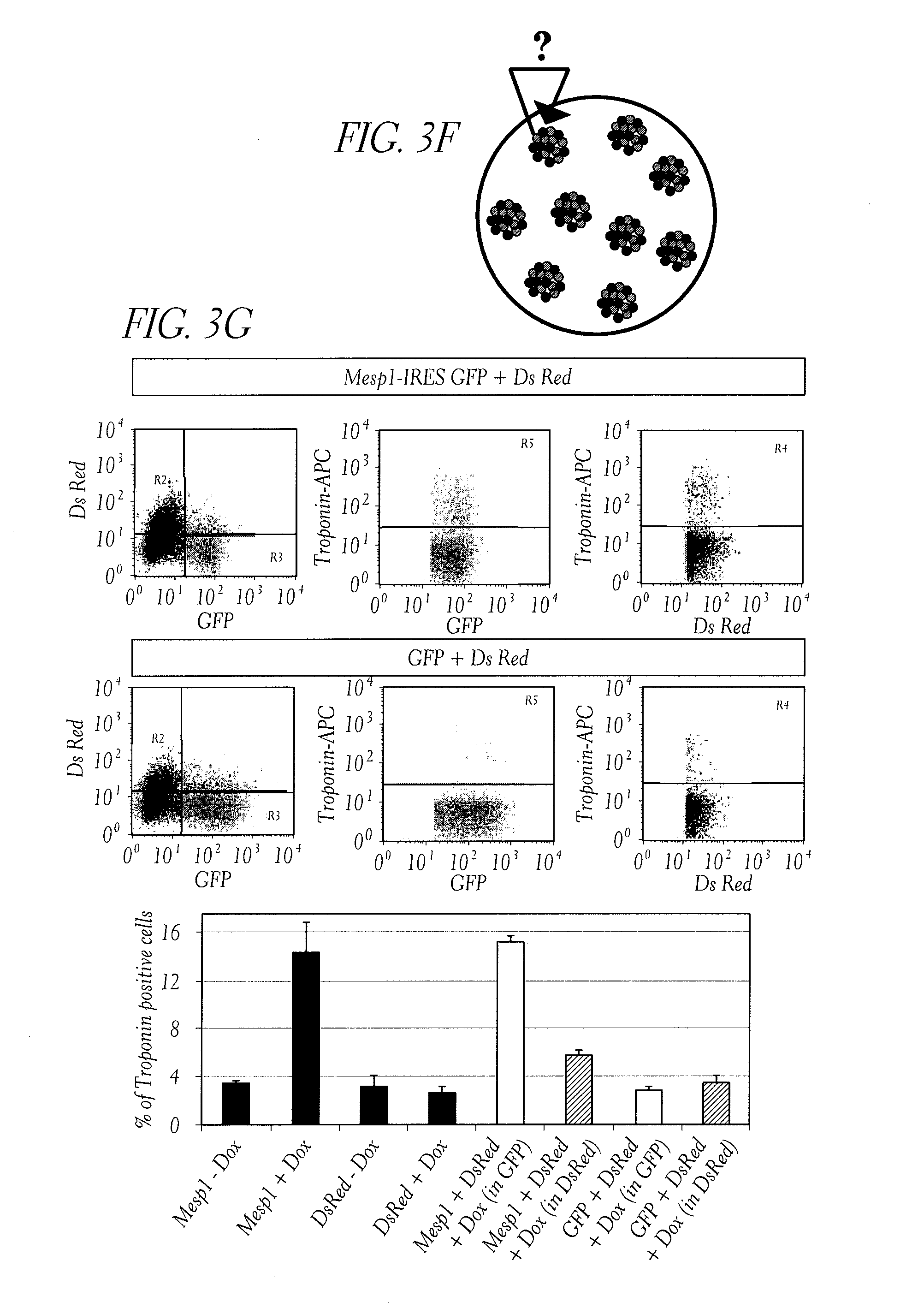
D00013

D00014

D00015

D00016

D00017

D00018

D00019

D00020

D00021

D00022

D00023

D00024

D00025

D00026
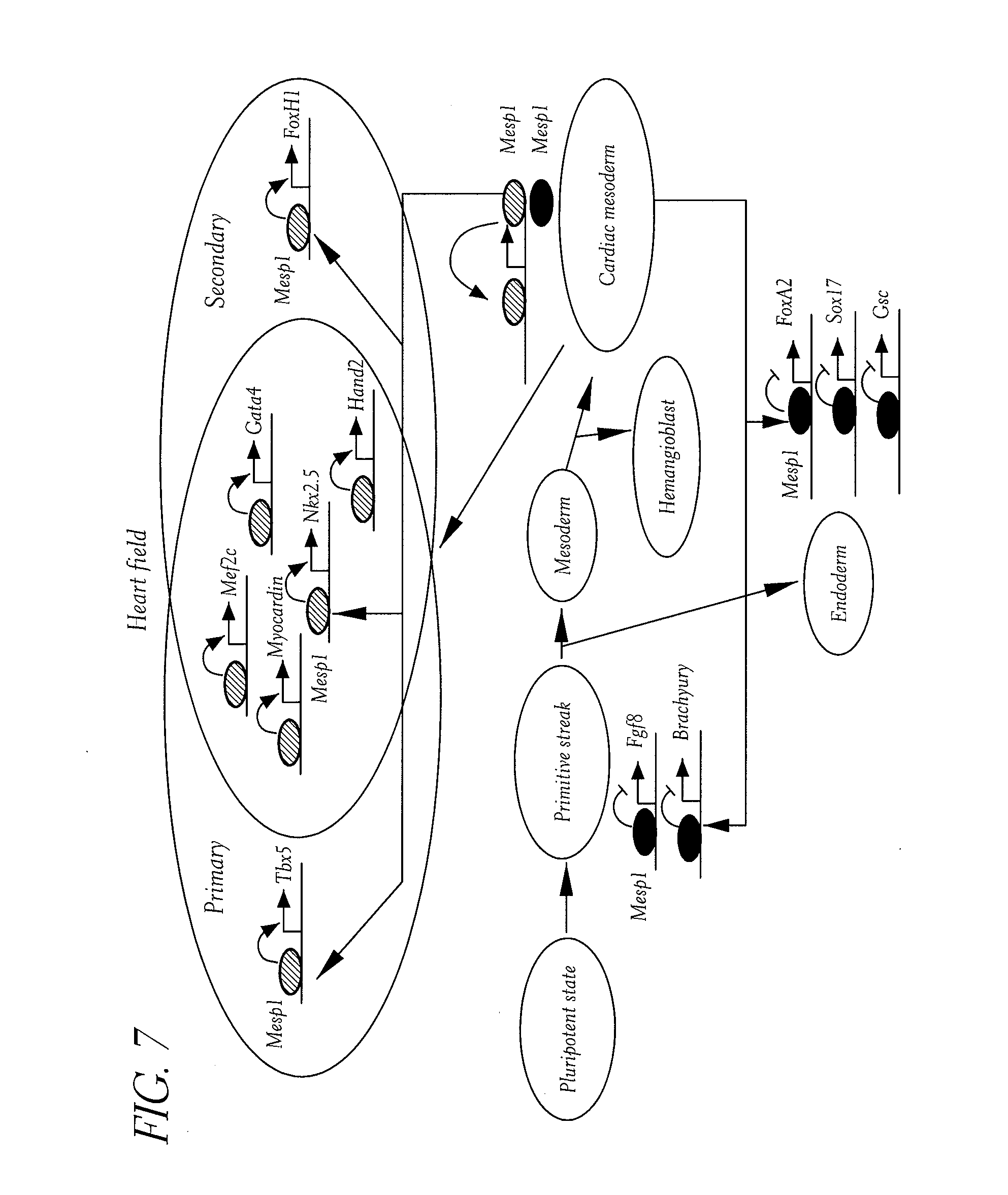
D00027

D00028

D00029

D00030

D00031

D00032

D00033

D00034

D00035

D00036

D00037
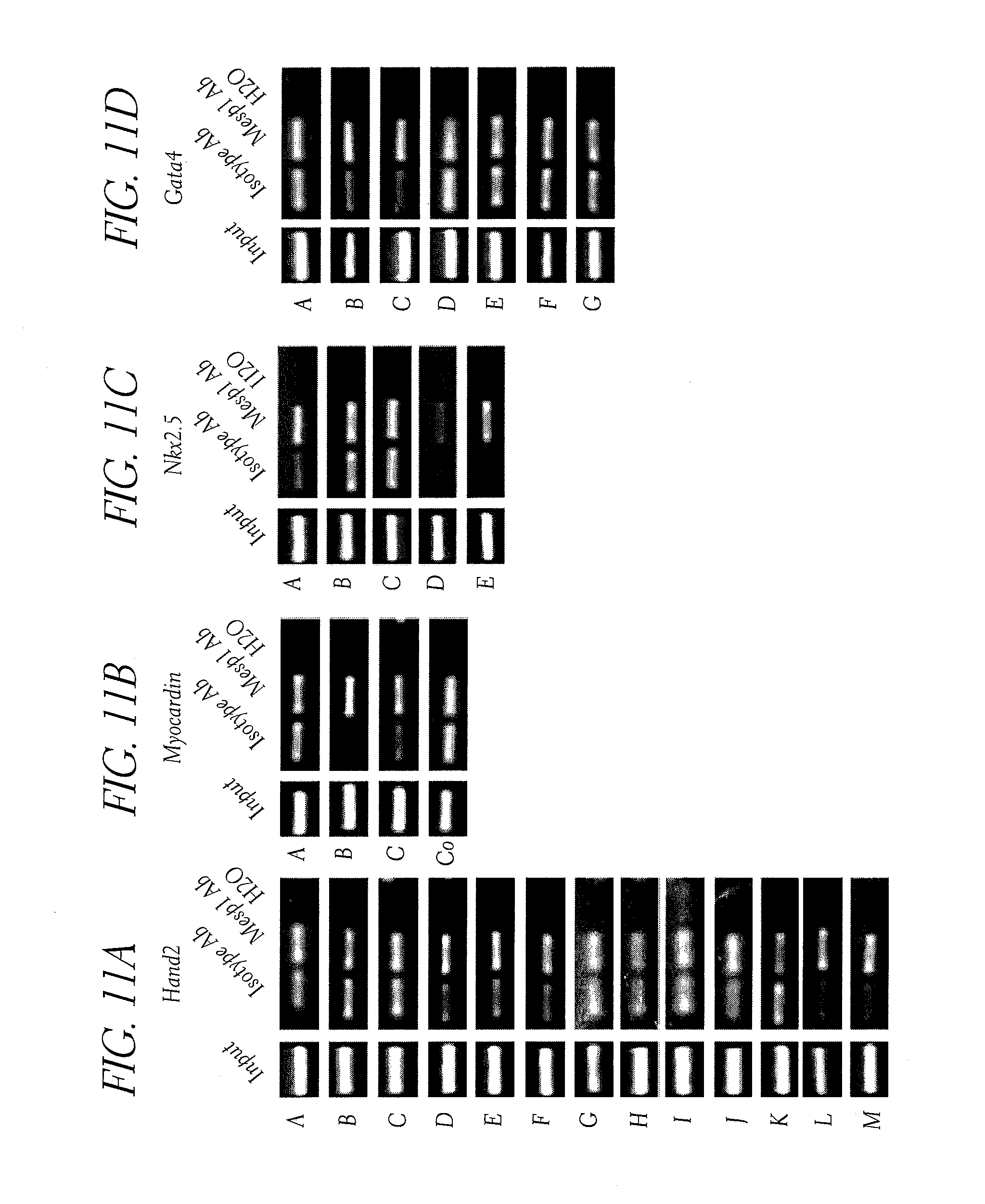
D00038

D00039

D00040

D00041
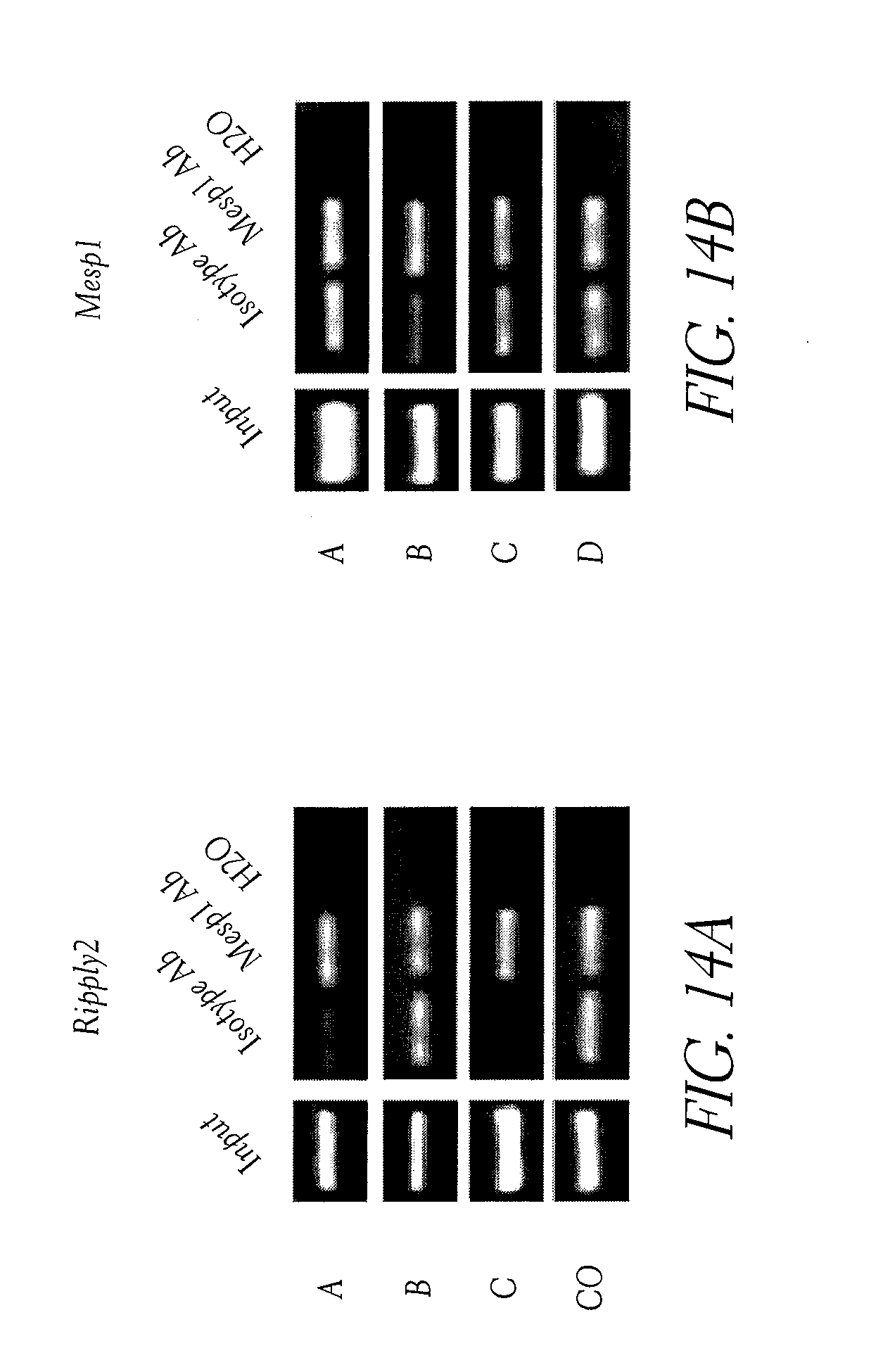
S00001
XML
uspto.report is an independent third-party trademark research tool that is not affiliated, endorsed, or sponsored by the United States Patent and Trademark Office (USPTO) or any other governmental organization. The information provided by uspto.report is based on publicly available data at the time of writing and is intended for informational purposes only.
While we strive to provide accurate and up-to-date information, we do not guarantee the accuracy, completeness, reliability, or suitability of the information displayed on this site. The use of this site is at your own risk. Any reliance you place on such information is therefore strictly at your own risk.
All official trademark data, including owner information, should be verified by visiting the official USPTO website at www.uspto.gov. This site is not intended to replace professional legal advice and should not be used as a substitute for consulting with a legal professional who is knowledgeable about trademark law.