Reduced volume formulation of glatiramer acetate and methods of administration
Altman , et al. December 30, 2
U.S. patent number 8,920,373 [Application Number 13/384,021] was granted by the patent office on 2014-12-30 for reduced volume formulation of glatiramer acetate and methods of administration. This patent grant is currently assigned to Teva Pharmaceutical Industries, Ltd.. The grantee listed for this patent is Ayelet Altman, Tomer El-Gad, Paul Greenhalgh, David George Robinson, Doris Saltkill, Dalton Tomlinson. Invention is credited to Ayelet Altman, Tomer El-Gad, Paul Greenhalgh, David George Robinson, Doris Saltkill, Dalton Tomlinson.


View All Diagrams
| United States Patent | 8,920,373 |
| Altman , et al. | December 30, 2014 |
Reduced volume formulation of glatiramer acetate and methods of administration
Abstract
The present invention provides an improved injection assisting device wherein the improvement comprises an injection lock indicator located between the first and the second outer shell and the outside of the indicator window, configured to indicate to a user whether the injection locking member is in a locked state, and configured to substantially contrast in color with both the first outer shell and the second outer shell, which first and second outer shells contrast in color with each other, and configured to be substantially shielded from a view of a user in the presence of the predetermined amount of compression force on the second outer shell.
| Inventors: | Altman; Ayelet (Hod-haSharon, IL), El-Gad; Tomer (Rehovot, IL), Saltkill; Doris (Kansas City, MO), Tomlinson; Dalton (Overland Park, KS), Greenhalgh; Paul (Newport Pagnell, GB), Robinson; David George (Duxford, GB) | ||||||||||
|---|---|---|---|---|---|---|---|---|---|---|---|
| Applicant: |
|
||||||||||
| Assignee: | Teva Pharmaceutical Industries,
Ltd. (Petach Tikva, IL) |
||||||||||
| Family ID: | 46796088 | ||||||||||
| Appl. No.: | 13/384,021 | ||||||||||
| Filed: | July 14, 2010 | ||||||||||
| PCT Filed: | July 14, 2010 | ||||||||||
| PCT No.: | PCT/US2010/001972 | ||||||||||
| 371(c)(1),(2),(4) Date: | May 25, 2012 | ||||||||||
| PCT Pub. No.: | WO2011/008274 | ||||||||||
| PCT Pub. Date: | January 20, 2011 |
Prior Publication Data
| Document Identifier | Publication Date | |
|---|---|---|
| US 20120232017 A1 | Sep 13, 2012 | |
Related U.S. Patent Documents
| Application Number | Filing Date | Patent Number | Issue Date | ||
|---|---|---|---|---|---|
| 12761367 | Apr 15, 2010 | ||||
| 61271009 | Jul 15, 2009 | ||||
| 61271340 | Jul 20, 2009 | ||||
| 61337011 | Jan 29, 2010 | ||||
| Current U.S. Class: | 604/130; 604/218; 604/134 |
| Current CPC Class: | A61P 25/00 (20180101); A61K 38/16 (20130101); A61K 9/0019 (20130101); A61K 38/02 (20130101); A61K 47/26 (20130101) |
| Current International Class: | A61M 5/20 (20060101) |
| Field of Search: | ;604/117,130,131,134-137,156,157,187,208-211,218,220 |
References Cited [Referenced By]
U.S. Patent Documents
| 4822340 | April 1989 | Kamstra |
| 4950246 | August 1990 | Muller |
| 5137516 | August 1992 | Rand et al. |
| 5981589 | November 1999 | Konfino et al. |
| 6048898 | April 2000 | Konfino et al. |
| 6054430 | April 2000 | Konfino et al. |
| 6342476 | January 2002 | Konfino et al. |
| 6362161 | March 2002 | Konfino et al. |
| 6448225 | September 2002 | O'Connor et al. |
| 6454746 | September 2002 | Bydlon et al. |
| 6620847 | September 2003 | Konfino et al. |
| 6939539 | September 2005 | Konfino et al. |
| 7033582 | April 2006 | Yong et al. |
| 7199098 | April 2007 | Konfino et al. |
| 7442185 | October 2008 | Amark et al. |
| 7560100 | July 2009 | Pinchasi et al. |
| 7585843 | September 2009 | Garren et al. |
| D607558 | January 2010 | Abry et al. |
| 7655221 | February 2010 | Rasmussen et al. |
| D622374 | August 2010 | Julian et al. |
| 2002/0077278 | June 2002 | Yong et al. |
| 2004/0039336 | February 2004 | Amark et al. |
| 2004/0106554 | June 2004 | Konfino et al. |
| 2005/0277885 | December 2005 | Scherer |
| 2006/0154862 | July 2006 | Ray et al. |
| 2007/0161566 | July 2007 | Pinchasi |
| 2007/0161960 | July 2007 | Chen et al. |
| 2008/0118553 | May 2008 | Frenkel et al. |
| 2009/0048181 | February 2009 | Schipper et al. |
| 2010/0160894 | June 2010 | Julian et al. |
| 2011/0066112 | March 2011 | Altman et al. |
| 1349590 | May 2006 | EP | |||
| WO 02/47746 | Jun 2002 | WO | |||
| WO 2006/029036 | Mar 2006 | WO | |||
| WO 2007/081975 | Jul 2007 | WO | |||
| WO 2007/081975 | Jul 2007 | WO | |||
| WO 2007/081975 | Jul 2007 | WO | |||
| WO 2009/070298 | Jun 2009 | WO | |||
| WO 2009/070298 | Jun 2009 | WO | |||
| WO 2011/008274 | Jan 2011 | WO | |||
Other References
|
US. Appl. NO. 12/785,125, filed May 21, 2010, Altman et al. cited by applicant . U.S. Appl. No. 12/806,684, filed Aug. 19, 2010, Klinger. cited by applicant . U.S. Appl. No. 12/861,655, filed Aug. 23, 2010, Stark and Ladakani. cited by applicant . U.S. Appl. No. 29/370,420, filed Jul. 14, 2010, El-Gad et al. cited by applicant . U.S. Appl. No. 29/370,417, filed Jul. 14, 2010, El-Gad et al. cited by applicant . Anderson, et al. (1992) "Revised estimate of the prevalence of multiple sclerosis in the United States". Ann Neural. 31:333-36. cited by applicant . Anderson, et al. "Injection pain decreases with new 0.5mL formulation of glatiramer acetate" The Consortium of Multiple Sclerosis Centers 2010 Annual Meeting, Jun. 2-5, 20. cited by applicant . Amon and Aharoni (2007) Neurogenesis and neuroprotection in the CAN--Fundamental elements in the effect of glatiramer acetate on treatment of autoimmune neurological di. cited by applicant . Bjartmar, et al. (2002) "Pathological mechanisms and disease progression of multiple sclerosis: therapeutic implications". Drugs of Today. 38(1):17-29. cited by applicant . Bornstein, et al. (1987) "A pilot trial of Cop 1 in exacerbating remitting multiple sclerosis". New Eng J Med. 317:408-14. cited by applicant . Bornstein, et al. (1991) "A placebo-controlled, double-blind, randomized, two-center, pilot trial of Cop-1 in chronic progressive multiple sclerosis". Neurology. 41:533-39. cited by applicant . Brazeau, et al. (1998) "Current perspectives on pain upon injection of drugs". J Pharmaceutical Sci. (87)6:667-677. cited by applicant . Chantelau, et al. (1991) "What make insulin injections painful?" BMJ. 303:26-27. cited by applicant . Comi G. "Treatment with glatiramer acetate delays conversion to clinically definite multiple sclerosis (CDMS) . . . "American Academy of Neurology 60th Annual Meeting 2008. cited by applicant . Comi, et al. (2001) "European/Canadian multicenter, double-blind, randomized, placebo-controlled study . . . ". Ann Neurol, 49:290-7. cited by applicant . Comi, et al. (2008) "Results from a phase III, one-year, randomized, double-blind, parallel-group . . . " Mult Scier. 14(suppl 1):S299. cited by applicant . Cohen et al., "Randomized, double-blind, dose-comparison study of glatiramer acetate in relapsing--remitting MS", Neurology, 2007; 68:939-944. cited by applicant . Dhib-Jalbut S. (2002) "Mechanisms of action of interferons and glatiramer acetate in multiple sclerosis". Neurology. 58(Suppl 4):S3-S9. cited by applicant . Dhib-Jalbut S. (2003) "Glatiramer acetate (Copaxone) therapy for multiple sclerosis" Pharmacology & Therapeutics. 98:245-55. cited by applicant . Frenken, et al. (1994) "Analysis of the efficacy of measures to reduce pain after subcutaneous administration of epoetin alfa". Nephrol Dial Transplant. 9:1295-1298. cited by applicant . Gibson (2004) "Selection of Injecting Volume", Pharmaceutical Preformulation and Formulation. p. 332. cited by applicant . Guideline on clinical investigation of medicinal products for the treatment of multiple sclerosis EMEA, London Nov. 16, 2006 CPMP/EWP/561/98 Rev.1. cited by applicant . Johnson, et al. (1998) "Extended use of glatiramer acetate (Copaxone) is well tolerated and maintains its clinical effect on multiple sclerosis relapse . . . " Neurology. 50:701-8. cited by applicant . Jorgensen, et al. (1996) "Pain Assessment of Subcutaneous Injections" Ann Pharmacother. 30(7):729-732. cited by applicant . Kansara, et al. (2009) "Subcutaneous Delivery". Drug Detiv Technol. Jun. 2009; 9(6):38-42. cited by applicant . Medical News Today. Jul. 8, 2008. Web: Sep. 9, 2010. www.medicalnewstoday.com/articles/114183.php. cited by applicant . Miller, et al. (2005) "Clinically isolated syndromes suggestive of multiple sclerosis, part I: natural history, pathogenesis . . . ". Lancet Neurol. 4(5):281-288. cited by applicant . Miller, et al. (2005) "Clinically isolated syndromes suggestive of multiple sclerosis, part II: non-conventional MRI . . . ". Lancet Neurol.4(6):341-348. cited by applicant . Neuhaus, et al. (2003) "Immunomodulation in multiple sclerosis: from immunosuppression to neuroprotection". Trends Pharmacol Sci. 24:131-138. cited by applicant . Noseworthy, et al. (2000) "Multiple sclerosis". N Engl J Med. 343:938-52. cited by applicant . Polman, et al. (2005) "Diagnostic Criteria for Multiple Sclerosis: 2005 Revisions to the "McDonald" Criteria". Ann Neurol. 58:840-846. cited by applicant . Product Monograph, Copaxone, Revised Apr. 2, 2010: 1-35. cited by applicant . Ruggiere, et al. (2007) "Glatiramer acetate in multiple sclerosis: A review". CNS Drug Reviews. 13(2):178-91. cited by applicant . Schrempf, et al. (2007) "Glatiramer acetate: Mechanisms of action in multiple sclerosis". Autoimmunity Reviews 2007. 6:469-475. cited by applicant . Shire, et al. (2004) "Challenges in the Development of High Protein Concentration Formulations". J Pharm Sci. 93(6):1390-1402. cited by applicant . The National MS Society (USA) [cited Feb. 5, 2010]. Available from: www.nationalmssociety.org/about-multiple-sclerosis/what-we-know-about-ms/- treatments/index.aspx. cited by applicant . Thrower BW. (2007) "Clinically isolated syndromes. Predicting and delaying multiple sclerosis". Neurology. 68 (Suppl 4):S12-S15. cited by applicant . Tselis, et al. (2007) "Glatiramer acetate in the treatment of multiple sclerosis". Neuropsychiatric Dis Treat. 3(2):259-67. cited by applicant . Van Metre, et al. (1996) "Pain and dermal reaction caused by injected glycerin in immunotherapy solutions". J Allergy Clin Immunol. 97:1033-9. cited by applicant . Weber, et al. (2007) "Mechanism of action of glatiramer acetate in treatment of multiple sclerosis". Neurotherapeutics. 4(4):647-53. cited by applicant . Wolinsky, et al. (2007) "Glatiramer acetate in primary progressive multiple sclerosis: Results of a multinational . . . " Ann Neurol. 61:14-24. cited by applicant . Wolinsky, JS (2006) "The use of glatiramer acetate in the treatment of multiple sclerosis". Adv Neurol. 273-92. cited by applicant . Vxcn Ziemssen and Schrempf (2007) "Glatiramer acetate: Mechanisms of action in multiple sclerosis". International Rev of Neurobiol. 79:537-70. cited by applicant . Teva Press Release, Jul. 7, 2008. cited by applicant . Pre-Examination Search Report, Feb. 23, 2010 from Envision IP Inc. cited by applicant . Revised Pre-Examination Search Report, Apr. 9, 2010 from Envision IP Inc. cited by applicant . International Search Report issued Jun. 9, 2011 in connection with PCT International Application No. PCT/US2010/001972. cited by applicant . Written Opinion of the International Searching Authority issued Jun. 9, 2011 in connection with PCT International Application Publication No. PCT/US2010/001972. cited by applicant . European Search Report issued Jul. 13, 2010 in connection with European Patent Application Publication No. EP 10160099.7. cited by applicant . Apr. 2, 2009 Final Office Action issued in connection with U.S. Appl. No. 11/651,212, filed Jan. 9, 2007. cited by applicant . Aug. 24, 2010 Office Action issued by the U.S. Patent and Trademark Office in connection with U.S. Appl. No. 12/785,125, filed May 21, 2010. cited by applicant . Jul. 20, 2009 Office Action issued in connection with U.S. Appl. No. 11/651,212, filed Jan. 9, 2007. cited by applicant . Jun. 20, 2008 Office Action issued in connection with U.S. Appl. No. 11/651,212, filed Jan. 9, 2007. cited by applicant . Nov. 1, 2010 Notice of Allowability in connection with U.S. Appl. No. 12/785,125, filed May 21, 2010. cited by applicant . Jul. 29, 2010 Supplementary European Search Report issued in connection with European Application No. 10160099.7. cited by applicant . Jul. 27, 2010 Office Action issued in connection with Canadian Patent Application No. 2,697,570, filed Apr. 15, 2010. cited by applicant . Dec. 22, 2008 Amendment filed in Response to Jun. 20, 2008 Office Action issued in connection with U.S. Appl. No. 11/651,212, filed Jan. 9, 2007. cited by applicant . Jul. 1, 2009 Amendment Under 37 C.F.R. 1.116 filed in Response to Apr. 2, 2009 Final Office Action issued in connection with U.S. Appl. No. 11/651,212. cited by applicant . Aug. 30, 2010 Response to the Jul. 27, 2010 Office Action issued in connection with the Canadian Patent Application No. 2,697,570, filed Apr. 15, 2010. cited by applicant . Sep. 23, 2010 Response to Aug. 24, 2010 Office Action, Summary of Sep. 15, 2010 Examiner Interview filed in connection with U.S. Appl. No. 12/785,125. cited by applicant . Communication pursuant to Article 94(3) issued Feb. 11, 2011 in connection with European Patent Application Publication No. EP 10160099.7. cited by applicant . Jun. 13, 2011 Response to Communication pursuant to Article 94(3) issued Feb. 11, 2011 in connection with European Patent Application Publication No. EP 10160099.7. cited by applicant . Official Action issued Jul. 27, 2010 in connection with Canadian Patent Application No. 2,697,570. cited by applicant . Aug. 30, 2010 Response to Official Action issued Jul. 27, 2010 in connection with Canadian Patent Application No. 2,697,570. cited by applicant . Oct. 10, 2012 Office Action issued in connection with U.S. Appl. No. 12/761,367, filed Apr. 15, 2010. cited by applicant . Decision to grant a European patent pursuant to Article 97(1) EPC, issued Feb. 16, 2012 in connection with European Patent Application Publication No. EP 10160099.7. cited by applicant . Communication of a notice of Opposition, issued Dec. 19, 2012 in connection with European Patent Application Publication No. EP 10160099.7. cited by applicant . Patient Information Leaflet for Copaxone, cited in Communication of a notice of Opposition, issued Dec. 19, 2012 in connection with EPO Application Pub. No. EP 10160099.7. cited by applicant . Excerpt from "Pharmaceutical Preformulation and Formulation" by M. Gibson, published in 2004, cited in Communication of a notice of Opposition issued in EPO No. 10160099.7. cited by applicant . Teva Pharmaceutical Industries Limited Q1 2009 Earnings Call Transcript cited in Communication of a notice of Opposition issued in EPO No. 10160099.7. cited by applicant . Communication of a notice of Opposition (R. 79(1) EPC), issued Feb. 19, 2013 in connection with European Patent Application Publication No. EP 10160099.7. cited by applicant . Office Action issued Mar. 25, 2015 in connection with Taiwanese Patent Application No. 100103482, including English language translation thereof. cited by applicant . International Search Report, mailed Jun. 9, 2011 in connection with PCT International Application No. PCT/US2010/001972, filed Jul. 14, 2010. cited by applicant. |
Primary Examiner: Bosworth; Kami A
Attorney, Agent or Firm: White; John P. Gershik; Gary J. Cooper & Dunham LLP
Parent Case Text
This application is a .sctn.371 national stage of PCT International Application No. PCT/US2010/001972, filed Jul. 14, 2010, which is a continuation-in-part of U.S. Ser. No. 12/761,367, filed Apr. 15, 2010, now abandoned, which claims the benefit of U.S. Provisional Application No. 61/337,011, filed Jan. 29, 2010, U.S. Provisional Application No. 61/271,340, filed Jul. 20, 2009, and U.S. Provisional Application No. 61/271,009, filed Jul. 15, 2009, the contents of each of which are hereby incorporated by reference in their entirety.
Claims
What is claimed is:
1. An improved injection assisting device comprising: a first outer shell having a first internal cavity with first and second oppositely arranged openings and a second outer shell movably and removably connectable to the first outer shell, the second outer shell having a second internal cavity with at least a first opening oriented towards the second opening of the first outer shell; a button configured to initiate an injection; an injection locking member configured to prevent the initiation of an injection in the absence of a predetermined compressive force moving the second outer shell to the first outer shell; an injection energy storing member configured to absorb and retain a predetermined amount of injection energy applied to a syringe located within at least one of the first and second cavities during an injection; an injection actuator configured, upon the initiation of an injection, to apply the injection energy to the syringe so as to displace a predetermined amount of matter from within the syringe; and an injection completion indicator inside an indicator window, configured to indicate to a user when the predetermined amount of matter has been displaced from within the syringe, wherein the improvement comprises: an injection lock indicator located between the first and the second outer shells and outside of the indicator window, configured to indicate to a user whether the injection locking member is in a locked state, and configured to substantially contrast in color with both the first outer shell and the second outer shell, which first and second outer shells contrast in color with each other, and configured to be substantially shielded from a view of a user in the presence of the predetermined compression force on the second outer shell.
2. The injection assisting device of claim 1, wherein the improvement further comprises an attention director configured to direct a user's attention to the injection completion indicator.
3. The injection assisting device of claim 2, wherein the attention director is configured to direct a user's attention to the direction of the injection.
4. The injection assisting device of claim 2, wherein the attention director is configured to direct a user's attention to the injection lock indicator.
5. The injection assisting device of claim 2, wherein the attention director is configured to direct a user's attention to the direction of force to be applied to unlock the locking member.
6. The injection assisting device of claim 2, wherein: a color of the attention director is configured to substantially contrast with a color of proximally located components, and a shape of the attention director is configured to direct a user's attention to the injection completion indicator.
7. The injection assisting device of claim 1, further comprising an injection force storage indicator configured to indicate to a user whether at least the predetermined amount of injection energy is stored in the injection energy storing member.
8. The injection assisting device of claim 1, wherein the improvement further comprises an outer grip member disposed on an outer surface of the second outer shell.
9. The injection assisting device of claim 8, wherein the outer grip member is formed of a material softer than the second outer shell.
10. The injection assisting device of claim 1, wherein ridges exist in an outer surface of the outer grip member so as to increase friction between the outer grip member and a human hand.
11. The injection assisting device of claim 1, wherein the improvement further comprises a ring member between the first outer shell and the second outer shell configured to substantially contrast with a color of the first outer shell, a color of the second outer shell, and a color of the injection lock indicator.
12. The injection assisting device of claim 1, further comprising a needle penetration depth adjustment member and a syringe needle cap remover member.
13. The injection assisting device of claim 12, wherein the improvement further comprises the syringe needle cap remover member configured to substantially correspond in color with the button configured to initiate an injection.
14. The injection assisting device of claim 1, wherein the improvement further comprises the injection actuator configured to displace 0.5 ml of liquid from the syringe.
15. The injection assisting device of claim 14, wherein the syringe contains in a 0.5 ml solution 20 mg of glatiramer acetate.
16. The injection assisting device of claim 1, wherein the improvement further comprises the injection lock indicator configured to be substantially shielded from the view of a user when the injection locking member is not in a locked state.
Description
Throughout this application various publications are referenced. The full citations of these references appear at the end of the specification before the claims. The disclosures of these publications in their entireties are hereby incorporated by reference into this application in order to more fully describe the state of the art to which this invention pertains.
BACKGROUND OF THE INVENTION
Multiple sclerosis (MS) is a chronic, debilitating disease of the central nervous system (CNS) with either relapsing-remitting (RR) or progressive course leading to neurologic deterioration and disability. At time of initial diagnosis, RRMS is the most common form of the disease (Noseworthy, 2000) which is characterized by unpredictable acute episodes of neurological dysfunction (relapses), followed by variable recovery and periods of clinical stability. The vast majority of RRMS patients eventually develop secondary progressive (SP) disease with or without superimposed relapses. Around 15% of patients develop a sustained deterioration of their neurological function from the beginning; this form is called primary progressive (PP) MS. Patients who have experienced a single clinical event (Clinically Isolated Syndrome or "CIS") and who show lesion dissemination on subsequent magnetic resonance imaging (MRI) scans according to McDonald's criteria, are also considered as having relapsing MS (EMEA Guideline, 2006).
Evidence is accumulating from pathophysiology, pathology, clinical and MRI studies that axonal damage and associated inflammation is characteristic of MS and may occur early in the disease course. It is believed that a confluence of elements must be present for MS to occur: genetic predisposition, immune dysregulation and one or more environmental factors. Although prevalence varies considerably around the world, MS is the most common cause of chronic neurological disability in young adults (Bjartmar, 2002; Fleming, 2002). Anderson et al. estimated that there were about 350,000 physician-diagnosed patients with MS in the United States in 1990 (approx. 140 per 100,000 population) (Anderson, 1992). It is estimated that about 2.5 million individuals are affected worldwide (Compston, 2006). In general, there has been a trend toward an increasing prevalence and incidence of MS worldwide, but the reasons for this trend are not fully understood (Anderson, 1992).
Current therapeutic approaches consist of i) symptomatic treatment ii) treatment of acute relapses with corticosteroids and iii) treatment aimed to modify the course of the disease. Currently approved therapies target the inflammatory processes of the disease. Most of them are considered to act as immunomodulators but their mechanisms of action have not been completely elucidated. Immunosuppressants or cytotoxic agents are also used in some patients after failure of conventional therapies.
Glatiramer Acetate Injection
Glatiramer acetate (GA) is the active substance in Copaxone.RTM., a marketed product indicated for reduction of the frequency of relapses in patients with RRMS. Glatiramer acetate consists of the acetate salts of synthetic polypeptides containing four naturally occurring amino acids: L-glutamic acid, L-alanine, L-tyrosine and L-lysine. The average molecular weight of glatiramer acetate is between 5,000 and 9,000 Daltons. The marketed medicinal product, Copaxone.RTM., contains 20 mg glatiramer acetate and 40 mg mannitol in 1.0 ml water for injection.
Although extensively researched, the mechanism of action of GA in humans remains uncertain and has been the subject of several recent reviews (Arnon, 2007; Ruggiere, 2007; Weber, 2007; Ziemssen, 2007). Based on the preclinical and clinical pharmacology data accumulated in the last four decades of research, it appears that GA's mechanism of action addresses the main pathological mechanisms driving MS, i.e. anti-inflammation, remyelination and neuroprotection (prevention of axonal loss) (Dhib-Jalbut, 2003).
The currently available data suggest that after subcutaneous (sc) injection, GA binds HLA class II (DR) on antigen-presenting cells in lymph nodes. As a result, GA can block the activation of myelin-reactive T cells or render these cells anergic. In addition, GA induces GA-specific Th2 cells that cross the blood-brain barrier (BBB) and produce bystander suppression as a result of cross-recognition of myelin antigens. These cells secrete both anti-inflammatory cytokines as well as neurotrophic factors and therefore induce both anti-inflammatory and neuroprotective functions (Dhib-Jalbut, 2002).
Clinical experience with GA consists of information obtained from completed and ongoing clinical trials and from post-marketing experience. The clinical program includes three double-blind, placebo-controlled studies in RRMS subjects treated with GA 20 mg/day (Bornstein, 1987; Comi, 2001; Johnson, 1998). A significant reduction in the number of relapses, compared with placebo, was seen. In the largest controlled study, the relapse rate was reduced by 32% from 1.98 under placebo to 1.34 under GA 20 mg. GA 20 mg has also demonstrated beneficial effects over placebo on MRI parameters relevant to RRMS. A significant effect in median cumulative number of Gd-enhancing lesions over 9 months of treatment (11 lesions in the 20 mg group compared to 17 lesions under placebo) was demonstrated.
The clinical program with GA also includes one double-blind study in chronic-progressive MS subjects (Bornstein, 1991), one double-blind placebo-controlled study in primary progressive patients (Wolinsky, 2007), one double-blind placebo-controlled study in CIS patients (Comi, 2008; Stark, 2009) and numerous open-label and compassionate use studies, mostly in RRMS. The clinical use of GA has been extensively reviewed and published in the current literature (Tselis, 2007; Wolinsky, 2006; Comi, 2008; Comi, 2008).
Safety data accumulated for GA in clinical trials shows that the drug product is safe and well tolerated. However, a reaction termed Immediate Post-Injection Reaction (IPIR) consisting of one or more of the following symptoms: vasodilatation, chest pain, dyspnoea, palpitations or tachycardia was reported for 31% of the GA patients vs. 13% on placebo. Additional adverse reactions reported by patients treated with GA 20 mg with at least 2% higher incidence than with placebo were pain, nausea, anxiety, rash, back pain, chills, face edema, local reaction, lymphadenopathy, vomiting, weight increase, tremor, skin disorder, eye disorder, vaginal candidiasis and injection site atrophy.
In all clinical trials, injection-site reactions were seen to be the most frequent adverse reactions and were reported by the majority of patients receiving GA. In controlled studies, the proportion of patients reporting these reactions, at least once, was higher following treatment with GA (70%) than placebo injections (37%). The most commonly reported injection-site reactions, which were more frequently reported in GA vs. placebo-treated patients, were erythema, pain, mass, pruritus, edema, inflammation and hypersensitivity.
Reducing the number and/or severity of the injection-site reactions in order to promote compliance and improving the quality of life for the patient remains a problem with GA treatment. However, for a drug product composed of peptides and whose mechanism of action is not understood, the effects of any modification cannot be readily predicted. Modifications of the formulation may unpredictably affect efficacy. To accommodate an indicated dose requirement in a limited injection volume, a polypeptide drug needs to be delivered at high concentrations. This alone is a significant problem when dealing with peptides of low solubility such as glatiramer acetate which is described as "sparingly soluble" (Product Monograph, 2009). Furthermore, concentrated polypeptide solutions are prone to additional problems. Such formulations suffer from poor shelf-life, unacceptable turbidity, changes in pH, chemical degradation including hydrolysis and aggregation (both reversible and irreversible) and increases in viscosity; all of which potentially reduce shelf-life and bioavailability (Shire, 2004).
Drug administration by subcutaneous injection results in delivery of the drug to the interstitial area underneath the skin. The fluid environment of the interstitial space is essentially that of plasma although the constituent proteins are at a lower concentration. This physiological medium may conflict with the solubility characteristics of the concentrated peptide drug (Kansara, 2009). Following injection, the interaction of the delivered drug with the interstitial environment dictates the pattern of absorption of the peptide. Formulation characteristics particularly concentration, injection volume and pH, influence the rate of diffusion and absorption by the patient. Because the interstitium also comprises a fibrous matrix of collagen and glycosaminoglycans, it acts as a barrier to the diffusion and permeability of the drug. As a result, drugs delivered in a concentrated form to the interstitial space may be susceptible to enzymatic degradation at the injection site, precipitation and/or aggregation in the interstitial fluid, and endocytic/phagocytic mechanisms (Kansara, 2009). For a peptide drug product such as glatiramer acetate, clinical testing is therefore required to determine whether any modification can effectively reduce the number and severity of injection-site reactions while still substantially maintaining therapeutic efficacy.
SUMMARY OF THE INVENTION
This invention provides a method for reducing frequency of relapses in a human patient afflicted with relapsing-remitting multiple sclerosis (RRMS) comprising administering to the patient by subcutaneous injection 0.5 ml of an aqueous pharmaceutical solution which contains in solution 20 mg glatiramer acetate and 20 mg mannitol.
The invention also provides a method for reducing the frequency of relapse in a human patient who experienced a first clinical episode consistent with multiple sclerosis and who has at least one lesion consistent with multiple sclerosis comprising administering to the patient by subcutaneous injection 0.5 ml of an aqueous pharmaceutical solution which contains in solution 20 mg glatiramer acetate and 20 mg mannitol.
This invention also provides an injection assisting device comprising: a first outer shell having a first internal cavity with first and second oppositely arranged openings and a second outer shell movably and removably connectable to the first outer shell, the second outer shell being having a second internal cavity with at least a first opening oriented towards the second opening of the first outer shell; a button configured to initiate an injection; an injection locking member configured to prevent the initiation of an injection in the absence of a predetermined compressive force between the second assembly and the first assembly; an injection energy storing member configured to absorb and retain a predetermined amount of injection energy applied to a syringe located within at least one of the first and second cavities during an injection; an injection actuator configured, upon the initiation of an injection, to apply the injection energy to the syringe so as to displace a predetermined amount of matter from within the syringe; an injection lock indicator configured to indicate to a user whether the injection locking member is in a locked state; an injection completion indicator configured to indicate to a user when the predetermined amount of matter has been displaced from within the syringe; and either, i) an attention director configured to direct a user's attention to the injection completion indicator, ii) an outer grip member formed of a material softer than the second outer shell and disposed on an outer surface of the second outer shell; and ridges formed in an outer surface of the outer grip member so as to increase friction between the outer grip member and a human hand, or iii) a color of the injection lock indicator is configured to substantially contrast with a color of at least one of the first outer shell and the second outer shell, and the injection lock indicator is configured to be substantially shielded from a view of a user in the presence of the predetermined amount of compression force between the second outer shell and the first outer shell.
This invention provides a syringe pre-filled with 0.5 ml of an aqueous pharmaceutical solution which contains in solution 20 mg glatiramer acetate and 20 mg mannitol.
This invention also provides a unit dose of 0.5 ml of an aqueous pharmaceutical solution which contains in solution 20 mg glatiramer acetate and 20 mg mannitol for use in reducing frequency of relapses in a human patient afflicted with relapsing, remitting multiple sclerosis (RRMS).
This invention additionally provides a unit dose of 0.5 ml of an aqueous pharmaceutical solution which contains in solution 20 mg glatiramer acetate and 20 mg mannitol for use in reducing the frequency of relapse in a human patient who experienced a first clinical episode consistent with multiple sclerosis and who has at least one lesion consistent with multiple sclerosis.
This invention also provides a unit dose of 0.5 ml of an aqueous pharmaceutical solution which contains in solution 20 mg glatiramer acetate and 20 mg mannitol for use in reducing the frequency of relapse in a human patient who experienced a first clinical episode consistent with multiple sclerosis and who has a high risk of developing clinically definite multiple sclerosis (CDMS).
This invention provides a unit dose of 0.5 ml of an aqueous pharmaceutical solution which contains in solution 20 mg glatiramer acetate and 20 mg mannitol for use in reducing the frequency of relapse in a human patient who experienced a first clinical episode consistent with multiple sclerosis and who has at least two clinically silent MRI lesions characteristic of multiple sclerosis.
This invention further provides a use of 0.5 ml of an aqueous pharmaceutical solution which contains in solution 20 mg glatiramer acetate and 20 mg mannitol in the manufacture of a medicament for treating a human patient afflicted with relapsing-remitting multiple sclerosis (RRMS).
This invention yet further provides a use of 0.5 ml of an aqueous pharmaceutical solution which contains in solution 20 mg glatiramer acetate and 20 mg mannitol in the manufacture of a medicament for treating a human patient who experienced a first clinical episode consistent with multiple sclerosis and who has at least one lesion consistent with multiple sclerosis.
This invention also provides a unit dose of 0.5 ml of an aqueous pharmaceutical solution which contains in solution 20 mg glatiramer acetate and 20 mg mannitol for use in treating a human patient who experienced a first clinical episode consistent with multiple sclerosis and who has been determined to be at high risk of developing clinically definite multiple sclerosis (CDMS).
This invention provides a use of 0.5 ml of an aqueous pharmaceutical solution which contains in solution 20 mg glatiramer acetate and 20 mg mannitol in the manufacture of a medicament for treating a human patient who experienced a first clinical episode consistent with multiple sclerosis and who has at least two clinically silent MRI lesions characteristic of multiple sclerosis.
This invention provides a pharmaceutical composition for use in treating a human patient afflicted with relapsing-remitting multiple sclerosis (RRMS) comprising a unit dose of 0.5 ml of an aqueous pharmaceutical solution which contains in solution 20 mg glatiramer acetate and 20 mg mannitol.
This invention also provides a pharmaceutical composition for use in treating a human patient who experienced a first clinical episode consistent with multiple sclerosis and who has at least one lesion consistent with multiple sclerosis comprising a unit dose of 0.5 ml of an aqueous pharmaceutical solution which contains in solution 20 mg glatiramer acetate and 20 mg mannitol.
This invention provides a method of treating a patient suffering from a relapsing form of multiple sclerosis which comprises periodically administering to the patient by subcutaneous injection a 20 mg dose of a pharmaceutical composition, wherein the subcutaneous injection is delivered by automatic injection and wherein the pharmaceutical composition comprises 20 mg of glatiramer acetate in 0.5 ml of solution so as to thereby treat the patient.
This invention provides a unit dose of 0.5 ml of an aqueous pharmaceutical solution which contains in solution 20 mg glatiramer acetate and 20 mg mannitol for use in decreasing the frequency of clinical exacerbations or reducing the number and volume of active MRI brain lesions in a human patient afflicted with relapsing-remitting multiple sclerosis (RRMS).
This invention also provides a unit dose of 0.5 ml of an aqueous pharmaceutical solution which contains in solution 20 mg glatiramer acetate and 20 mg mannitol for use in delaying the onset of Clinically Definite Multiple Sclerosis or decreasing the number and volume of active MRI brain lesions in a human patient who experienced a single demyelinating event and who is considered to be at risk of developing Clinically Definite Multiple Sclerosis.
This invention provides a use of 0.5 ml of an aqueous pharmaceutical solution which contains in solution 20 mg glatiramer acetate and 20 mg mannitol for decreasing the frequency of clinical exacerbations or reducing the number and volume of active MRI brain lesions in a human patient afflicted with relapsing-remitting multiple sclerosis (RRMS).
This invention also provides a use of 0.5 ml of an aqueous pharmaceutical solution which contains in solution 20 mg glatiramer acetate and 20 mg mannitol for delaying the onset of clinically definite multiple sclerosis or decreasing the number and volume of active MRI brain lesions in a human patient who experienced a single demyelinating event and who is considered to be at risk of developing clinically definite multiple sclerosis.
This invention provides a pharmaceutical composition for use in treating a human patient afflicted with relapsing-remitting multiple sclerosis (RRMS) comprising a unit dose of 0.5 ml of an aqueous pharmaceutical solution which contains in solution 20 mg glatiramer acetate and 20 mg mannitol.
This invention provides a pharmaceutical composition for use in treating a human patient who experienced a single demyelinating event and who is considered to be at risk of developing clinically definite multiple sclerosis comprising a unit dose of 0.5 ml of an aqueous pharmaceutical solution which contains in solution 20 mg glatiramer acetate and 20 mg mannitol.
This invention also provides an injection assisting device comprising: a first outer shell having a first internal cavity with first and second oppositely arranged openings and a second outer shell movably and removably connectable to the first outer shell, the second outer shell having a second internal cavity with at least a first opening oriented towards the second opening of the first outer shell; a button configured to initiate an injection; an injection locking member configured to prevent the initiation of an injection in the absence of a predetermined compressive force between the second assembly and the first assembly; an injection energy storing member configured to absorb and retain a predetermined amount of injection energy applied to a syringe located within at least one of the first and second cavities during an injection; an injection actuator configured, upon the initiation of an injection, to apply the injection energy to the syringe so as to displace a predetermined amount of matter from within the syringe; an injection lock indicator configured to indicate to a user whether the injection locking member is in a locked state, and configured to substantially contrast with a color of both of the first outer shell and the second outer shell, and configured to be substantially shielded from a view of a user in the presence of the predetermined amount of compression force between the second outer shell and the first outer shell; an injection completion indicator configured to indicate to a user when the predetermined amount of matter has been displaced from within the syringe.
BRIEF DESCRIPTION OF THE FIGURES
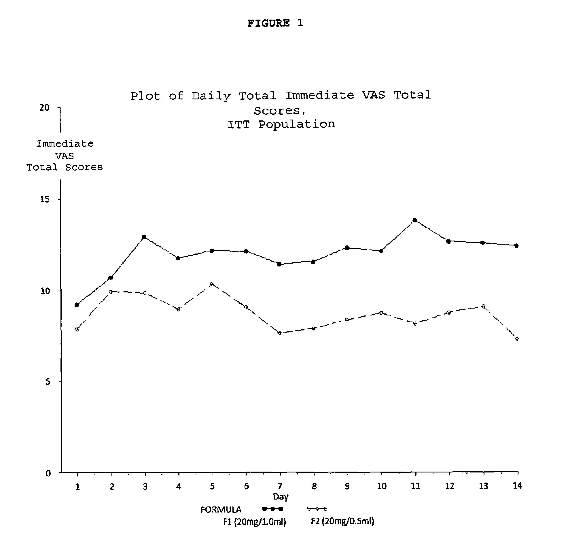
FIG. 1. Plot of daily total immediate VAS total scores for the ITT Population.
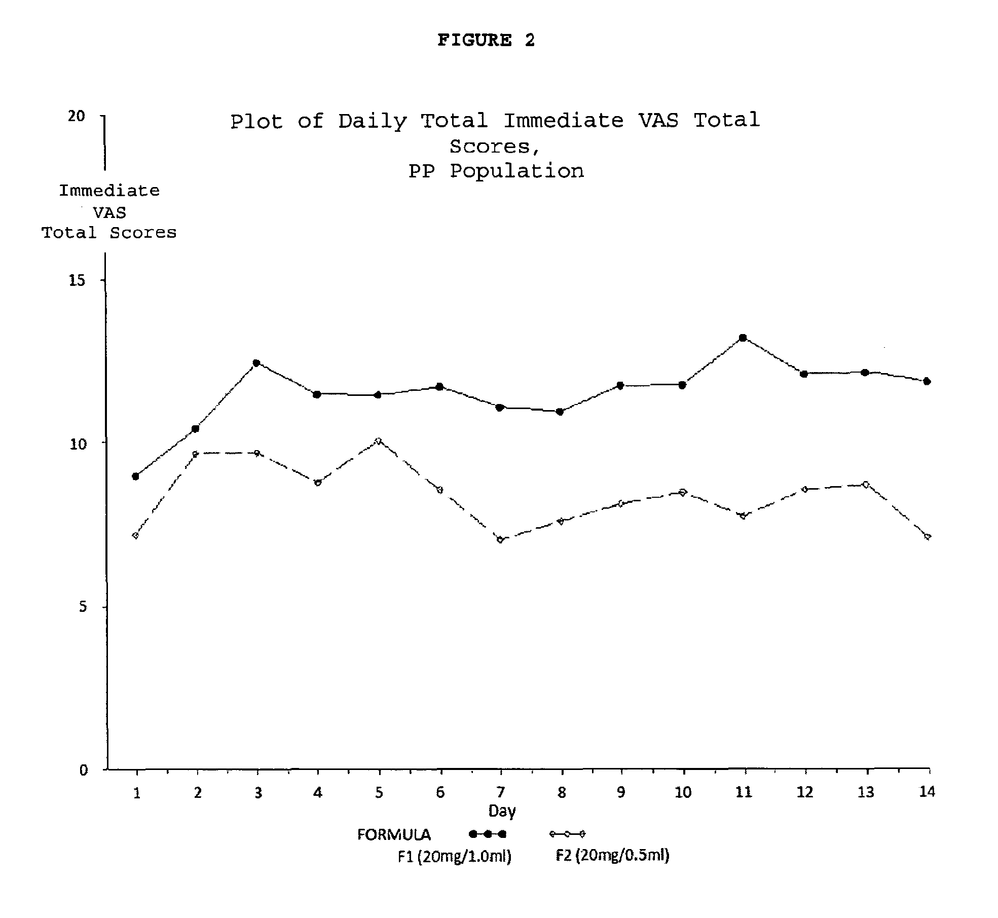
FIG. 2. Plot of daily total immediate VAS total scores for the PP population.
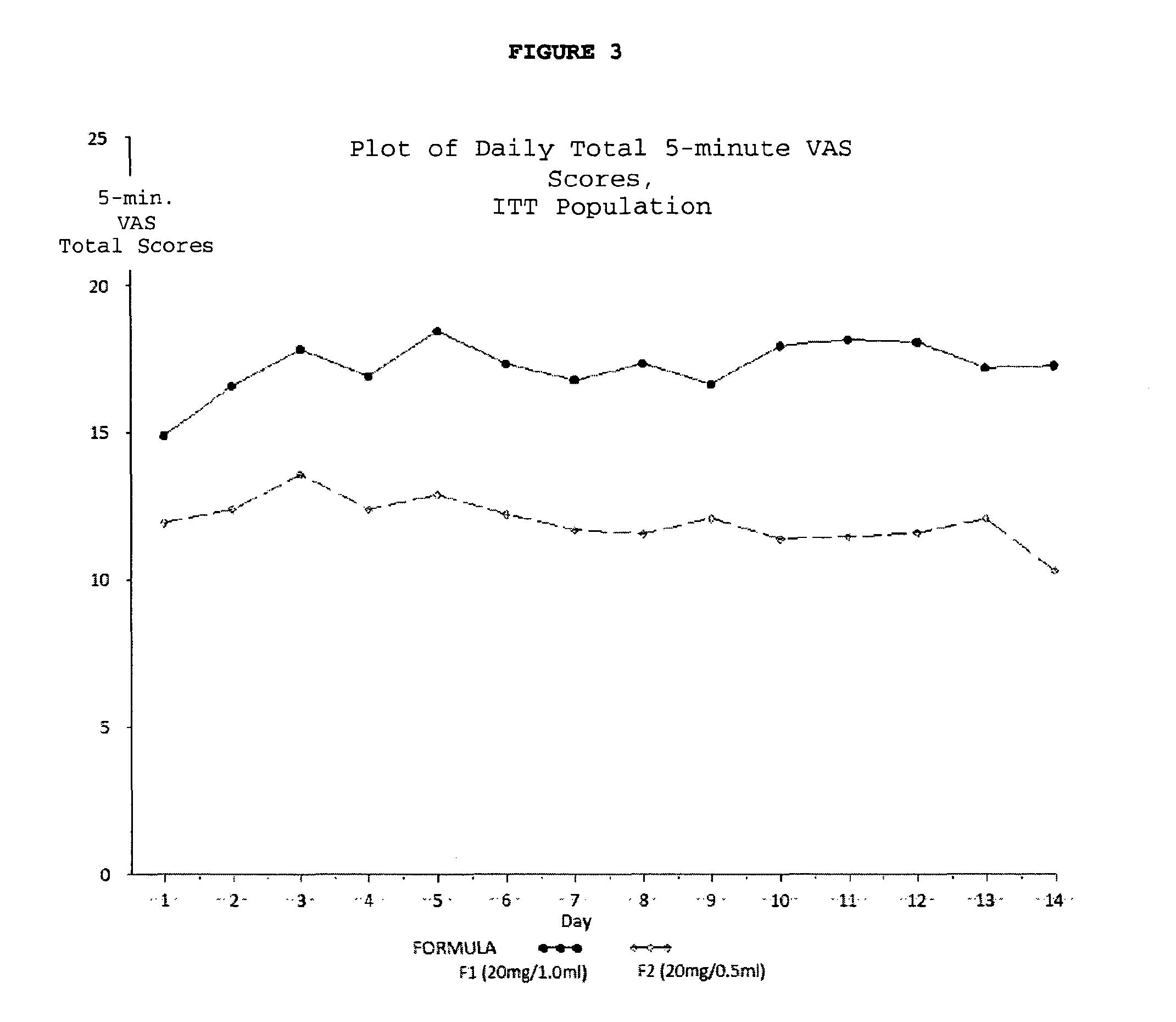
FIG. 3. Plot of daily total 5-minute VAS total scores for the ITT population.
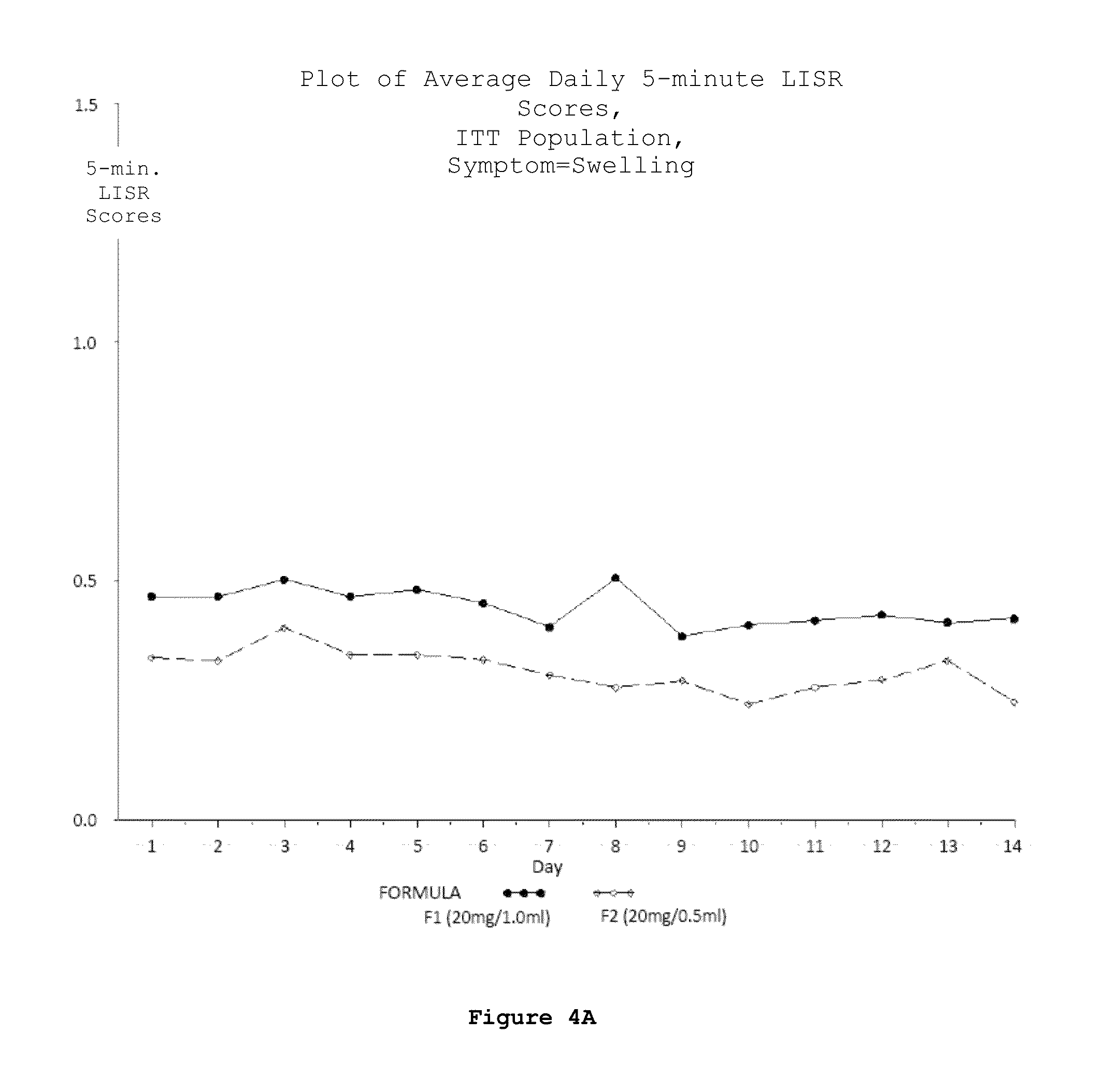
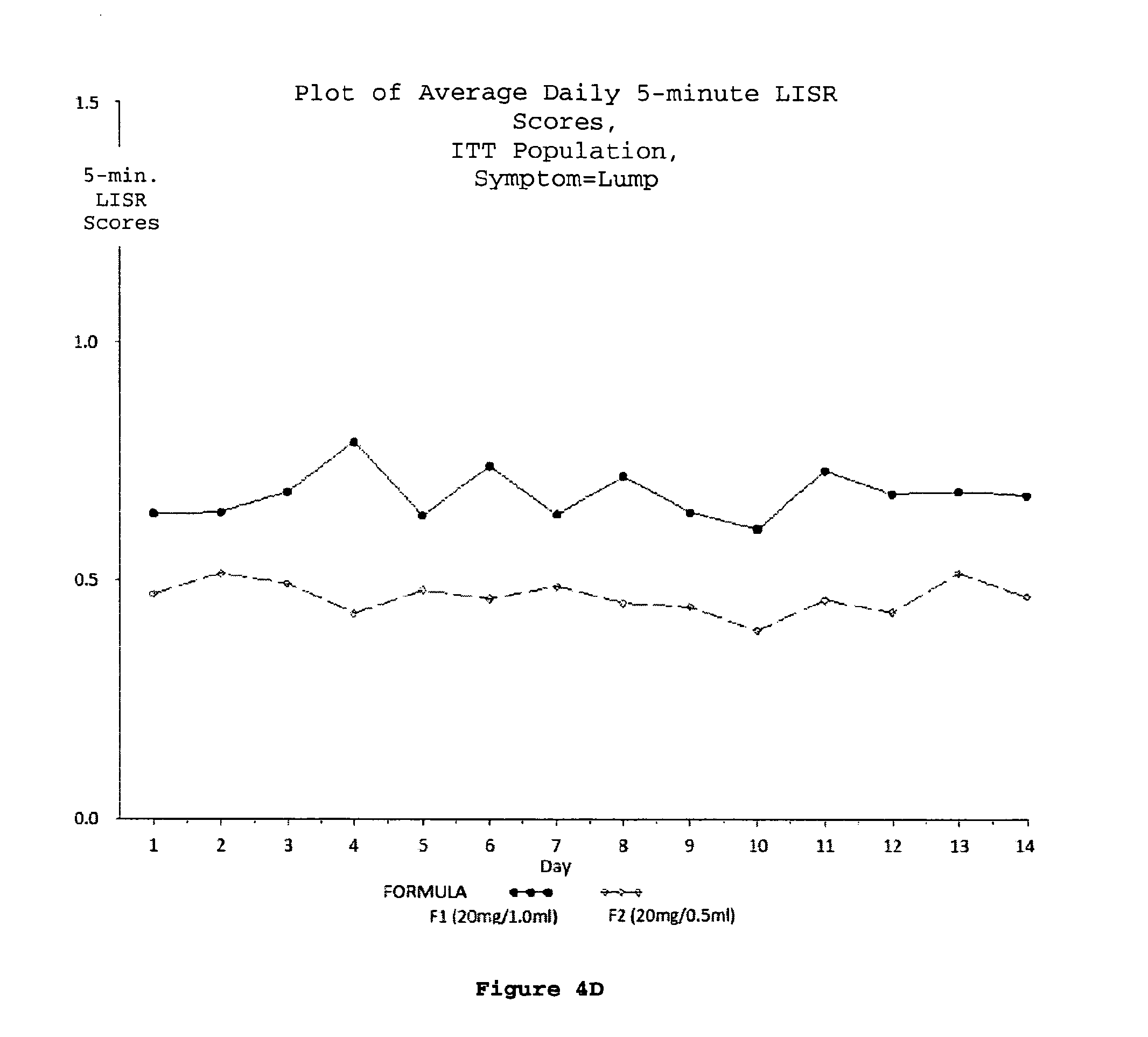
FIG. 4 (A-D). Plots of average daily 5-minute LISR scores for the ITT population; swelling (A), redness (B), itching (C) and lump (D) were the 4 symptoms considered as LISRs during the study.
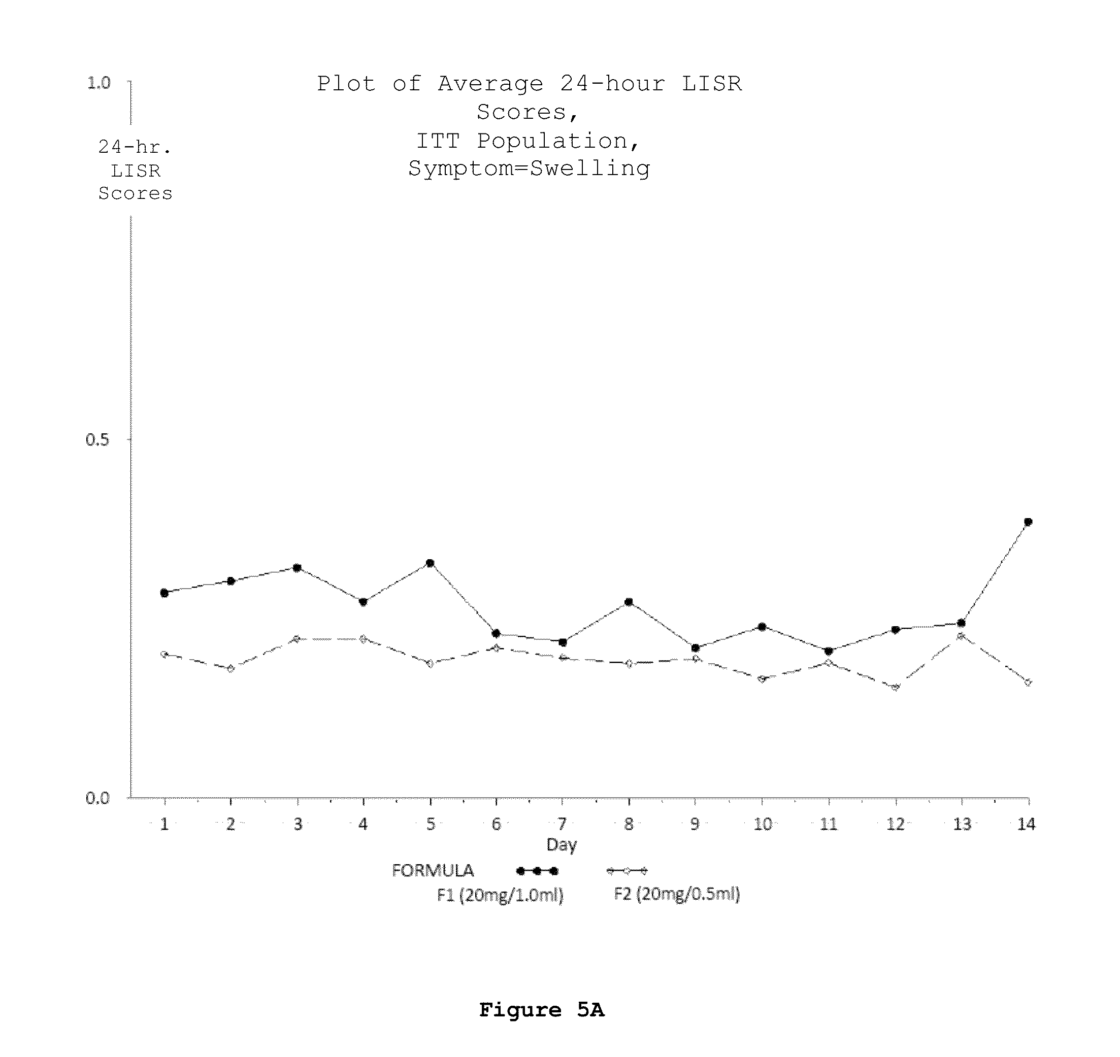
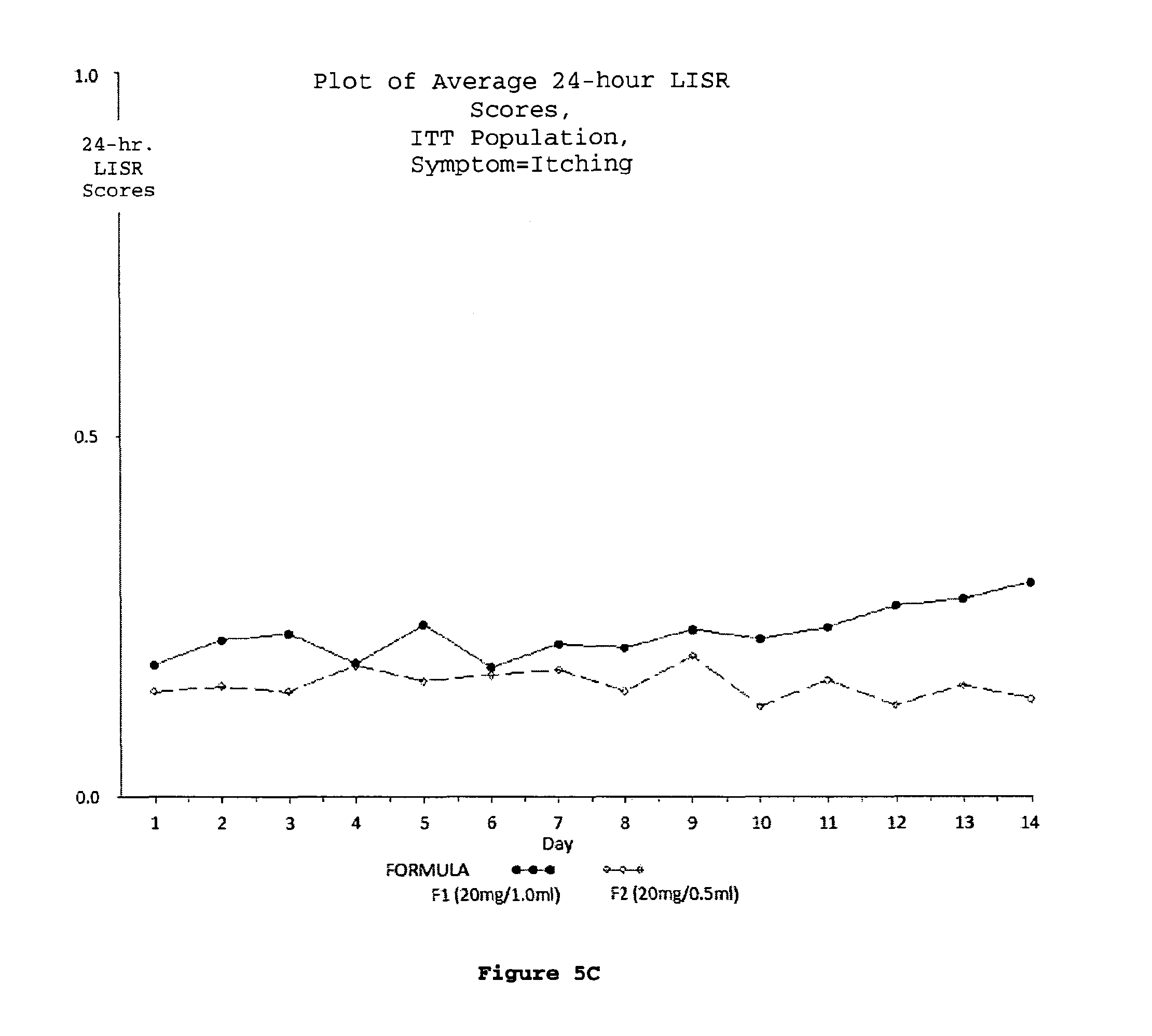
FIG. 5 (A-D). Plots of average daily 24-hour LISR scores for the ITT population; swelling (A), redness (B), itching (C) and lump (D) were the 4 symptoms considered as LISRs during the study.
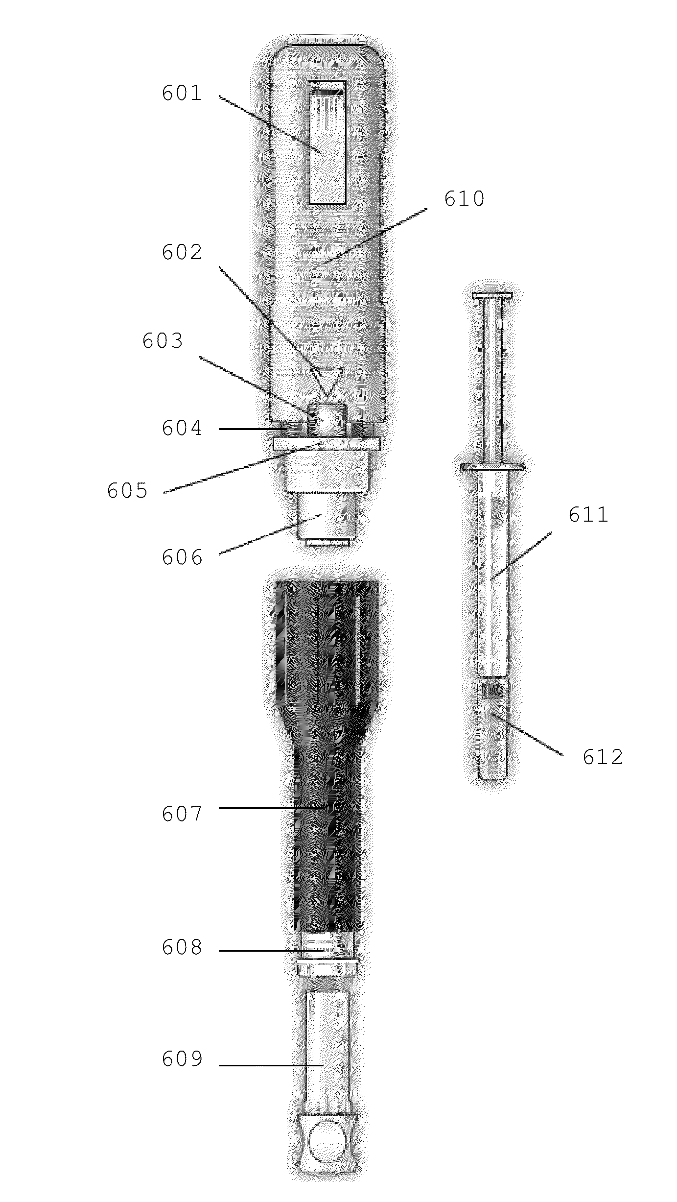
FIG. 6. Exploded view of an exemplary embodiment of the injection assisting device disclosed herein. 601--Firing button/button configured to initiate injection 602--Triangle/Attention director 603--Indicator window/Injection completion indicator 604--Band/Injection lock indicator 605--Band/Ring member 606--Plunger/Injection actuator 607--Syringe housing/First outer shell 608--Depth adjuster/needle penetration depth adjustment member 609--Cap remover/syringe needle cap remover member 610--Injection body/Second outer shell 611--Fixed needle syringe 612--Needle cap
FIG. 7. Perspective view of an exemplary embodiment of the injection assisting device disclosed herein. 701--Firing Button/button configured to initiate injection 702--Injection Body/Second outer shell 703--Attention director 704--Indicator Window/Injection lock indicator 705--Syringe housing/First outer shell 706--Needle penetration depth adjustment member 707--Syringe needle cap remover member 708--Viewing window
FIGS. 8A-8R. Instructions for use of injection assisting device.
Step 1: Depth Adjustment
Determine correct needle depth adjustment, which must be made before the device is loaded or used.
For example, to set the depth of the needle penetration to 6 mm, first verify that the Cap Remover is fully attached to the Depth Adjuster (FIG. 8A). Screw the Depth Adjuster using the Cap Remover (FIG. 8B) until scale mark 6 is level with the end of the Syringe Housing (FIG. 8C). The lower the number shown on the Depth Adjuster, the shallower the injection depth will be. Adjustment can be made by twisting the Depth Adjuster to a higher number setting for a deeper injection or a lower number setting for a more shallow injection.
Step 2: Loading the Injection Assisting Device
Unscrew the Syringe Housing from the Injector Body (FIG. 8D), ensuring that the Cap Remover remains attached inside the Depth Adjuster. Hold the Injector Body with one hand, making sure the Firing Button is not touched. Hold the Syringe Housing and push the attached Cap Remover squarely against the Plunger on the Injector Body, as shown in FIG. 8E, until the Plunger locks into place inside the Injector Body (FIG. 8F).
Hold the Syringe Housing upright with the Cap Remover against a flat surface (such as a table) as shown in FIG. 8G. Make sure the Cap Remover remains inserted all the way into the Depth Adjuster.
Remove the syringe from its protective blister pack by peeling back the paper label. Before use, look at the liquid in the syringe. If it is cloudy or contains any particles, do not use. If the liquid is clear, place the syringe on the clean, flat surface.
Insert the syringe with Needle Cap still on, needle end first, into the Syringe Housing (FIG. 8H) and push the syringe down firmly into the Syringe Housing until the syringe click into place (FIG. 8I).
Screw the Syringe Housing and the Injector Body together, taking care not to touch the Firing Button (FIG. 8J).
Step 3: Preparing the Device for Injection
When ready to perform the injection, hold the Injector Body and remove the Needle Cap by firmly pulling the Cap Remover out of the device without twisting it (FIG. 8K). The Cap Remover will come out of the Syringe Housing with the Needle Cap inside (FIG. 8L).
Turn the Cap Remover upside down to release the Needle Cap (FIG. 8M). The Needle Cap has two parts: a rubber inner part covered by a transparent plastic outer part. Make sure that both parts of the Needle Cap are intact when the cap falls out of the Cap Remover. If both parts of the Needle Cap have not been removed, unscrew the Syringe Housing and Injector Body, remove the syringe and start over from Step 2 with a new syringe.
Dispose of the Needle Cap. Check again that the Depth Adjuster is in the correct position. Save the Cap Remover for future use.
Step 4: Injecting the Drug
Choose an injection site on the body and clean the injection site with a new alcohol prep and let the site air dry to reduce stinging.
When ready to inject, hold the device by the Injector Body. Do not touch the Firing Button. Make sure the Indicator Window is visible. Place the tip of the device against the injection prepared site by gently pressing the injector onto the injection site (FIG. 8N).
To protect against accidental injection, the devise has a safety lock. The Injector Body must be slid towards the injection site in the direction pointed by the tip of the arrow on the Injector Body so that the band indicative of Injection lock is hidden before an injection can be delivered (FIG. 8O). However, avoid excessive force on the injection site.
When the band indicative of Injection lock is hidden (FIG. 8P) the device is ready to deliver an injection. If the band indicative of Injection lock is still visible the device will not inject (FIG. 8N).
Press the Firing Button and watch the Indicator Window (FIG. 8Q). Continue holding the device tip gently against the skin until the injection is complete. The Firing Button can be released while the syringe content is injected automatically.
Injection is complete when the mark indicative of injection completion remains stationary in the Indicator Window (FIG. 8R), which should take approximately 10 seconds. Do not remove the device from the injection site until the injection is complete. Always ensure that the full dose has been delivered from the syringe by verifying that the mark indicative of injection completion appears in the Indicator Window.
Press a dry cotton ball on the injection site for a few seconds and do not rub the injection site.
Step 5: Removing and Disposing of the Syringe
After use, unscrew the Syringe Housing from the Injector Body and separate the two slowly. Hold the Syringe Housing above the open top area of a safe hard-walled container and invert the Syringe Housing, allowing the syringe to fall out directly into the hard-walled container.
Step 6: Cleaning the Injection Assisting Device
After every use, the external components and the inside of the Syringe Housing of the device should be cleaned by wiping with a clean damp cloth or an alcohol wipe. Do not immerse the device in water.
Step 7: Storing the Injection Assisting Device Safely
Screw together the Syringe Housing and Injector Body. Re-insert the Cap Remover into the Depth Adjuster. Store the device safely in the nylon wallet provided.
DETAILED DESCRIPTION OF THE INVENTION
This invention provides a method for reducing frequency of relapses in a human patient afflicted with relapsing-remitting multiple sclerosis (RRMS) comprising administering to the patient by subcutaneous injection 0.5 ml of an aqueous pharmaceutical solution which contains in solution 20 mg glatiramer acetate and 20 mg mannitol.
The invention also provides a method for reducing the frequency of relapse in a human patient who experienced a first clinical episode consistent with multiple sclerosis and who has at least one lesion consistent with multiple sclerosis comprising administering to the patient by subcutaneous injection 0.5 ml of an aqueous pharmaceutical solution which contains in solution 20 mg glatiramer acetate and 20 mg mannitol.
According to any embodiment of the methods disclosed herein, the pH of the aqueous pharmaceutical solution is 5.5 to 7.0.
According to any embodiment of the methods disclosed herein, the 20 mg of glatiramer acetate does not form polypeptide aggregates in the 0.5 ml of aqueous pharmaceutical solution.
According to any embodiment of the methods disclosed herein, the 20 mg of glatiramer acetate does not precipitate in the subcutaneous environment after injection.
According to any embodiment of the methods disclosed herein, the 20 mg of glatiramer acetate is absorbed by the patient after the subcutaneous injection.
According to any embodiment of the methods disclosed herein, the 20 mg of glatiramer acetate in 0.5 ml of solution is absorbed by the patient at least as readily as 20 mg of glatiramer acetate in 1 ml of solution.
According to any embodiment of the methods disclosed herein, the 20 mg of glatiramer acetate in 0.5 ml of solution is co-injected with a vasodilator.
According to any embodiment of the methods disclosed herein, the 20 mg of glatiramer acetate in 0.5 ml of solution is co-injected with a vasoconstrictor.
According to any embodiment of the methods disclosed herein, 20 mg of glatiramer acetate in 0.5 ml of solution is co-injected with an extracellular matrix-modifying enzyme.
According to any embodiment of the methods disclosed herein, the subcutaneous injection is administered to the upper back portion of the arm, to the stomach area outside of a 2 inch area around the navel, to the upper outer-rear quadrant of the buttocks, or to the front and outer area of the thigh 2 inches above the knee and 2 inches below the groin.
According to any embodiment of the methods disclosed herein, the pain associated with the subcutaneous injection is reduced relative to pain associated with subcutaneous injection of 1.0 ml of an aqueous pharmaceutical solution of 20 mg glatiramer acetate and 40 mg mannitol.
In an embodiment of the methods disclosed, the pain is the patient-reported total injection pain rating on a visual analogue scale (VAS) occurring immediately after injection.
In a further embodiment of the methods disclosed, the patient-reported total injection pain rating is reduced by about 27%.
In another embodiment of the methods disclosed, the pain is the patient-reported total injection pain rating on a visual analogue scale (VAS) experienced five minutes following subcutaneous injection.
In a further embodiment of the methods disclosed, the patient-reported total injection pain rating experienced five minutes following subcutaneous injection is reduced by about 31%.
In another embodiment of the methods disclosed, the pain is the immediate pain presence following the subcutaneous injection.
In yet another embodiment of the methods disclosed, the immediate pain presence is reduced by about 19%.
In an embodiment of the methods disclosed, the pain is pain presence five minutes following the subcutaneous injection.
In an additional embodiment of the methods disclosed, the pain presence five minutes following the subcutaneous injection is reduced by about 19%.
In yet another embodiment of the methods disclosed, the total number or total severity of Local Injection Site Reactions (LISRs) is reduced relative to the total number or total severity of LISRs associated with subcutaneous injection of 1.0 ml of an aqueous pharmaceutical solution of 20 mg glatiramer acetate and 40 mg mannitol.
In a further embodiment of the methods disclosed, the total number or total severity of Local Injection Site Reactions (LISRs) five minutes after subcutaneous injection is reduced.
In yet a further embodiment of the methods disclosed, the total number of Local Injection Site Reactions (LISRs) five minutes after the subcutaneous injection is reduced by about 24%.
In an additional embodiment of the methods disclosed, the total severity of Local Injection Site Reactions (LISRs) five minutes after the subcutaneous injection is reduced by about 29%.
In an embodiment of the methods disclosed, the total number or total severity of Local Injection Site Reactions (LISRs) 24 hours after subcutaneous injection of glatiramer is reduced.
In another embodiment of the methods disclosed, the total number of Local Injection Site Reactions (LISRs) 24 hours after the subcutaneous injection is reduced by about 23%.
In yet another embodiment of the methods disclosed, the total severity of Local Injection Site Reactions (LISRs) 24 hours after the subcutaneous injection is reduced by about 25%.
According to any embodiment of the methods disclosed herein, the daily five-minute Local Injection Site Reaction (LISR) score is reduced relative to the daily 5-minute LISR score associated with subcutaneous injection of 1.0 ml of an aqueous pharmaceutical solution of 20 mg glatiramer acetate and 40 mg mannitol.
According to any embodiment of the methods disclosed herein, the daily 24-hour Local Injection Site Reaction (LISR) score is reduced relative to the daily 24-hour LISR score associated with subcutaneous injection of 1.0 ml of an aqueous pharmaceutical solution of 20 mg glatiramer acetate and 40 mg mannitol.
In an additional embodiment of the methods disclosed, the Local Injection Site Reactions (LISRs) comprise redness, itching and formation of a lump.
In yet another embodiment of the methods disclosed, the percent of patients who report no Local Injection Site Reactions (LISRs) 5-minutes after injection is increased relative to the percent of patients who report no LISRs 5-minutes after subcutaneous injection of 1.0 ml of an aqueous pharmaceutical solution of 20 mg glatiramer acetate and 40 mg mannitol.
In a further embodiment of the methods disclosed, the percent of patients who report no Local Injection Site Reactions (LISRs) is increased by 3 fold.
In an embodiment of the methods disclosed, the percent of patients who report no Local Injection Site Reactions (LISRs) 24-hours after injection is increased relative to the percent of patients who report no LISRs 24-hours after subcutaneous injection of 1.0 ml of an aqueous pharmaceutical solution of 20 mg glatiramer acetate and 40 mg mannitol.
In an additional embodiment of the methods disclosed, the percent of patients who report no Local Injection Site Reactions (LISRs) is increased by about 50%.
According to any embodiment of the methods disclosed herein, the 0.5 ml aqueous pharmaceutical solution is in a pre-filled syringe.
In yet another embodiment of the methods disclosed, the administration is by an automated subcutaneous injection device containing the prefilled syringe and a means for initiating the subcutaneous injection, completing the subcutaneous injection and indicating to the user that the subcutaneous injection of the 0.5 ml aqueous pharmaceutical solution is complete.
In a further embodiment of the methods disclosed, the 0.5 ml of an aqueous pharmaceutical solution which contains in solution 20 mg glatiramer acetate and 20 mg mannitol is at least as effective as 1.0 ml of an aqueous pharmaceutical solution of 20 mg glatiramer acetate and 40 mg mannitol in reducing the frequency of relapses in a human patient afflicted with relapsing-remitting multiple sclerosis (RRMS).
This invention also provides an injection assisting device comprising: a first outer shell having a first internal cavity with first and second oppositely arranged openings and a second outer shell movably and removably connectable to the first outer shell, the second outer shell being having a second internal cavity with at least a first opening oriented towards the second opening of the first outer shell; a button configured to initiate an injection; an injection locking member configured to prevent the initiation of an injection in the absence of a predetermined compressive force between the second assembly and the first assembly; an injection energy storing member configured to absorb and retain a predetermined amount of injection energy applied to a syringe located within at least one of the first and second cavities during an injection; an injection actuator configured, upon the initiation of an injection, to apply the injection energy to the syringe so as to displace a predetermined amount of matter from within the syringe; an injection lock indicator configured to indicate to a user whether the injection locking member is in a locked state; an injection completion indicator configured to indicate to a user when the predetermined amount of matter has been displaced from within the syringe; and either, i) an attention director configured to direct a user's attention to the injection completion indicator, ii) an outer grip member formed of a material softer than the second outer shell and disposed on an outer surface of the second outer shell; and ridges formed in an outer surface of the outer grip member so as to increase friction between the outer grip member and a human hand, or iii) a color of the injection lock indicator is configured to substantially contrast with a color of at least one of the first outer shell and the second outer shell, and the injection lock indicator is configured to be substantially shielded from a view of a user in the presence of the predetermined amount of compression force between the second outer shell and the first outer shell.
In an additional embodiment of the device disclosed, the injection assisting device further comprising: an injection force storage indicator configured to indicate to a user whether at least the predetermined amount of injection energy is stored in the injection energy storing member.
In another embodiment of the device disclosed, the injection assisting device further comprising: an outer grip member formed of a material softer than the second outer shell and disposed on an outer surface of the second outer shell; and ridges formed in an outer surface of the outer grip member so as to increase friction between the outer grip member and a human hand.
In yet another embodiment of the device disclosed, a color of the injection lock indicator is configured to substantially contrast with a color of at least one of the first outer shell and the second outer shell, and the injection lock indicator is configured to be substantially shielded from a view of a user in the presence of the predetermined amount of compression force between the second outer shell and the first outer shell.
In a further embodiment of the device disclosed, a color of the attention director is configured to substantially contrast with a color of proximally located components, and a shape of the attention director is configured to direct a user's attention to the injection completion indicator.
This invention provides a unit dose of 0.5 ml of an aqueous pharmaceutical solution which contains in solution 20 mg glatiramer acetate and 20 mg mannitol. According to any embodiment of the methods disclosed herein, the 20 mg of glatiramer acetate does not form polypeptide aggregates in the 0.5 ml of aqueous pharmaceutical solution. In an additional embodiment the aqueous pharmaceutical solution has a pH of 5.5-7.0. In another embodiment, the aqueous pharmaceutical solution is in a prefilled syringe.
This invention also provides a unit dose of 0.5 ml of an aqueous pharmaceutical solution which contains in solution 20 mg glatiramer acetate and 20 mg mannitol for use in reducing frequency of relapses in a human patient afflicted with relapsing, remitting multiple sclerosis (RRMS).
This invention additionally provides a unit dose of 0.5 ml of an aqueous pharmaceutical solution which contains in solution 20 mg glatiramer acetate and 20 mg mannitol for use in reducing the frequency of relapse in a human patient who experienced a first clinical episode consistent with multiple sclerosis and who has at least one lesion consistent with multiple sclerosis.
This invention also provides a unit dose of 0.5 ml of an aqueous pharmaceutical solution which contains in solution 20 mg glatiramer acetate and 20 mg mannitol for use in reducing the frequency of relapse in a human patient who experienced a first clinical episode consistent with multiple sclerosis and who has been determined to be at high risk of developing clinically definite multiple sclerosis (CDMS).
This invention provides a unit dose of 0.5 ml of an aqueous pharmaceutical solution which contains in solution 20 mg glatiramer acetate and 20 mg mannitol for use in reducing the frequency of relapse in a human patient who experienced a first clinical episode consistent with multiple sclerosis and who has at least two clinically silent MRI lesions characteristic of multiple sclerosis.
This invention further provides a use of 0.5 ml of an aqueous pharmaceutical solution which contains in solution 20 mg glatiramer acetate and 20 mg mannitol in the manufacture of a medicament for treating a human patient afflicted with relapsing-remitting multiple sclerosis (RRMS).
This invention yet further provides a use of 0.5 ml of an aqueous pharmaceutical solution which contains in solution 20 mg glatiramer acetate and 20 mg mannitol in the manufacture of a medicament for treating a human patient who experienced a first clinical episode consistent with multiple sclerosis and who has at least one lesion consistent with multiple sclerosis.
This invention also provides a unit dose of 0.5 ml of an aqueous pharmaceutical solution which contains in solution 20 mg glatiramer acetate and 20 mg mannitol for use in treating a human patient who experienced a first clinical episode consistent with multiple sclerosis and who has been determined to be at high risk of developing clinically definite multiple sclerosis (CDMS).
This invention provides a use of 0.5 ml of an aqueous pharmaceutical solution which contains in solution 20 mg glatiramer acetate and 20 mg mannitol in the manufacture of a medicament for treating a human patient who experienced a first clinical episode consistent with multiple sclerosis and who has at least two clinically silent MRI lesions characteristic of multiple sclerosis.
This invention provides a pharmaceutical composition for use in treating a human patient afflicted with relapsing-remitting multiple sclerosis (RRMS) comprising a unit dose of 0.5 ml of an aqueous pharmaceutical solution which contains in solution 20 mg glatiramer acetate and 20 mg mannitol.
This invention also provides a pharmaceutical composition for use in treating a human patient who experienced a first clinical episode consistent with multiple sclerosis and who has at least one lesion consistent with multiple sclerosis comprising a unit dose of 0.5 ml of an aqueous pharmaceutical solution which contains in solution 20 mg glatiramer acetate and 20 mg mannitol.
This invention provides a method of treating a patient suffering from a relapsing form of multiple sclerosis which comprises periodically administering to the patient by subcutaneous injection a 20 mg dose of a pharmaceutical composition, wherein the subcutaneous injection is delivered by automatic injection and wherein the pharmaceutical composition comprises 20 mg of glatiramer acetate in 0.5 ml of solution so as to thereby treat the patient.
In an embodiment of the methods disclosed, the injection of 20 mg glatiramer acetate in 0.5 ml of solution is as effective as injection of 20 mg of glatiramer acetate in 1 ml of solution.
In another embodiment of the methods disclosed, the 20 mg of glatiramer acetate in 0.5 ml of solution has a pH equivalent to that of 20 mg of glatiramer acetate in 1 ml of solution.
In yet another embodiment of the methods disclosed, the 20 mg of glatiramer acetate is completely soluble in 0.5 ml of solution.
This invention provides a unit dose of 0.5 ml of an aqueous pharmaceutical solution which contains in solution 20 mg glatiramer acetate and 20 mg mannitol for use in decreasing the frequency of clinical exacerbations or reducing the number and volume of active MRI brain lesions in a human patient afflicted with relapsing-remitting multiple sclerosis (RRMS).
This invention also provides a unit dose of 0.5 ml of an aqueous pharmaceutical solution which contains in solution 20 mg glatiramer acetate and 20 mg mannitol for use in delaying the onset of clinically definite multiple sclerosis or decreasing the number and volume of active MRI brain lesions in a human patient who experienced a single demyelinating event and who is considered to be at risk of developing clinically definite multiple sclerosis.
This invention provides a use of 0.5 ml of an aqueous pharmaceutical solution which contains in solution 20 mg glatiramer acetate and 20 mg mannitol for decreasing the frequency of clinical exacerbations or reducing the number and volume of active MRI brain lesions in a human patient afflicted with relapsing-remitting multiple sclerosis (RRMS).
This invention also provides a use of 0.5 ml of an aqueous pharmaceutical solution which contains in solution 20 mg glatiramer acetate and 20 mg mannitol for delaying the onset of clinically definite multiple sclerosis or decreasing the number and volume of active MRI brain lesions in a human patient who experienced a single demyelinating event and who is considered to be at risk of developing clinically definite multiple sclerosis.
This invention provides a pharmaceutical composition for use in treating a human patient afflicted with relapsing-remitting multiple sclerosis (RRMS) comprising a unit dose of 0.5 ml of an aqueous pharmaceutical solution which contains in solution 20 mg glatiramer acetate and 20 mg mannitol.
This invention provides a pharmaceutical composition for use in treating a human patient who experienced a single demyelinating event and who is considered to be at risk of developing clinically definite multiple sclerosis comprising a unit dose of 0.5 ml of an aqueous pharmaceutical solution which contains in solution 20 mg glatiramer acetate and 20 mg mannitol.
An embodiment of the use, unit dose or pharmaceutical composition disclosed herein, adapted for subcutaneous injection, wherein the 20 mg of glatiramer acetate is absorbed by the patient after the subcutaneous injection.
Another embodiment of the use, unit dose or pharmaceutical composition disclosed herein, adapted for subcutaneous injection to the upper back portion of the arm, to the stomach area outside of a 2 inch area around the navel, to the upper outer-rear quadrant of the buttocks, or to the front and outer area of the thigh 2 inches above the knee and 2 inches below the groin.
The Injection Assisting Device
The mechanical workings of the injection assisting device can be prepared according to the disclosure in European application publication No. EP0693946 which is incorporated herein by reference.
The device is described in detail below with reference to FIGS. 6 and 7 which show exemplary embodiments of the injection assisting device disclosed herein.
This invention provides an injection assisting device comprising: a first outer shell (607) having a first internal cavity with first and second oppositely arranged openings and a second outer shell (610) movably and removably connectable to the first outer shell, the second outer shell having a second internal cavity with at least a first opening oriented towards the second opening of the first outer shell; a button configured to initiate an injection (601); an injection locking member configured to prevent the initiation of an injection in the absence of a predetermined compressive force between the second assembly and the first assembly; an injection energy storing member configured to absorb and retain a predetermined amount of injection energy applied to a syringe located within at least one of the first and second cavities during an injection; an injection actuator (606) configured, upon the initiation of an injection, to apply the injection energy to the syringe so as to displace a predetermined amount of matter from within the syringe; an injection lock indicator (604) configured to indicate to a user whether the injection locking member is in a locked state, and configured to substantially contrast with a color of both of the first outer shell and the second outer shell, and configured to be substantially shielded from a view of a user in the presence of the predetermined amount of compression force between the second outer shell and the first outer shell; an injection completion indicator (603) configured to indicate to a user when the predetermined amount of matter has been displaced from within the syringe.
In one embodiment, the injection assisting device further comprises an attention director (602) configured to direct a user's attention to the injection completion indicator.
In another embodiment, the injection assisting device further comprises an injection force storage indicator configured to indicate to a user whether at least the predetermined amount of injection energy is stored in the injection energy storing member.
In yet another embodiment, the injection assisting device further comprises an outer grip member disposed on an outer surface of the second outer shell.
In one embodiment, the outer grip member is formed of a material softer than the second outer shell. In another embodiment, ridges exist in an outer surface of the outer grip member so as to increase friction between the outer grip member and a human hand.
In another embodiment, the injection assisting device further comprises a ring member (605) between the first outer shell and the second outer shell configured to substantially contrast with a color of the first outer shell, a color of the second outer shell, or a color of the injection lock indicator.
In one embodiment, the attention director (602) is configured to direct a user's attention to the direction of the injection. In another embodiment, the attention director is configured to direct a user's attention to the injection lock indicator. In yet another embodiment, the attention director is configured to direct a user's attention to the direction of force to be applied to unlock the locking member.
According to one embodiment, a color of the attention director is configured to substantially contrast with a color of proximally located components, and a shape of the attention director is configured to direct a user's attention to the injection completion indicator.
According to another embodiment, the injection assisting device comprises a needle penetration depth adjustment member (608) and a syringe needle cap remover member (609). According to another embodiment, the syringe needle cap remover member is configured to substantially correspond in color with the button configured to initiate an injection, and to contrast with a color of the first outer shell and the second outer shell.
In one embodiment, a color of the injection lock indicator (604) is configured to substantially contrast with a color of the first outer shell (607). In another embodiment, a color of the injection lock indicator (604) is configured to substantially contrast with a color of the second outer shell (610). In another embodiment, a color of the injection lock indicator (604) is configured to substantially contrast with a color of the attention director (602). In another embodiment, a color of the injection lock indicator (604) is configured to substantially contrast with a color of the ring member (605). In another embodiment, a color of the injection lock indicator (604) is configured to substantially contrast with a color of the syringe needle cap remover (609). In another embodiment, a color of the injection lock indicator (604) is configured to substantially contrast with a color of the injection completion indicator (603). In another embodiment, a color of the injection lock indicator (604) is configured to substantially contrast with a color of the button configured to initiate injection (601).
In one embodiment, a color of the injection completion indicator (603) is configured to substantially contrast with a color of the first outer shell (607). In another embodiment, a color of the injection completion indicator (603) is configured to substantially contrast with a color of the second outer shell (610). In another embodiment a color of the injection completion indicator (603) is configured to substantially contrast with a color of the attention director (602). In another embodiment, a color of the injection completion indicator (603) is configured to substantially contrast with a color of the ring member (605). In another embodiment, a color of the injection completion indicator (603) is configured to substantially contrast with a color of the syringe needle cap remover (609). In another embodiment, a color of the injection completion indicator (603) is configured to substantially contrast with a color of the injection lock indicator (604). In another embodiment, a color of the injection completion indicator (603) is configured to substantially contrast with a color of the button configured to initiate injection (601).
In one embodiment, a color of the attention director (602) is configured to substantially contrast with a color of the first outer shell (607). In another embodiment, a color of the attention director (602) is configured to substantially contrast with a color of the second outer shell (610). In another embodiment, a color of the attention director (602) is configured to substantially contrast with a color of the injection lock indicator (604). In another embodiment, a color of the attention director (602) is configured to substantially contrast with a color of the injection completion indicator (603).
In one embodiment, a color of the ring member (605) is configured to substantially contrast with a color of the first outer shell (607). In another embodiment, a color of the ring member (605) is configured to substantially contrast with a color of the second outer shell (610). In another embodiment, a color of the ring member (605) is configured to substantially contrast with a color of the injection lock indicator (604). In another embodiment, a color of the ring member (605) is configured to substantially contrast with a color of the injection completion indicator (603).
In one embodiment, a color of the button (601) is configured to substantially contrast with a color of the first outer shell (607). In another embodiment, a color of the button (601) is configured to substantially contrast with a color of the second outer shell (610). In another embodiment, a color of the button (601) is configured to substantially contrast with a color of the injection lock indicator (604). In another embodiment, a color of the button (601) is configured to substantially contrast with a color of the injection completion indicator (603).
In one embodiment, a color of the syringe needle cap remover (609) is configured to substantially contrast with a color of the first outer shell (607). In another embodiment, a color of the syringe needle cap remover (609) is configured to substantially contrast with a color of the second outer shell (610). In another embodiment, a color of the syringe needle cap remover (609) is configured to substantially contrast with a color of the injection lock indicator (604). In another embodiment, a color of the syringe needle cap remover (609) is configured to substantially contrast with a color of the injection completion indicator (603).
In one embodiment, the syringe needle cap remover (609) is configured to substantially correspond in color with the ring member (605). In another embodiment, the syringe needle cap remover (609) is configured to substantially correspond in color with the attention director (602). In another embodiment, the syringe needle cap remover (609) is configured to substantially correspond in color with the button (601). In another embodiment, the ring member (605) is configured to substantially correspond in color with the attention director (602). In another embodiment, the ring member (605) is configured to substantially correspond in color with the button (601). In another embodiment, the attention director (602) is configured to substantially correspond in color with the button (601).
In an embodiment of any of the injection assisting device disclosed herein, the injection actuator is configured to displace 0.5 ml of liquid from the syringe. In another embodiment of any of the injection assisting device disclosed herein, the syringe contains in a 0.5 ml solution 20 mg of glatiramer acetate.
For the foregoing embodiments, each embodiment disclosed herein is contemplated as being applicable to each of the other disclosed embodiment.
Definitions
As used herein, a patient at risk of developing MS (i.e. clinically definite MS) is a patient presenting any of the known risk factors for MS. The known risk factors for MS include anyone of a clinically isolated syndrome (CIS), a single attack suggestive of MS without a lesion, the presence of a lesion (in any of the CNS, PNS, or myelin sheath) without a clinical attack, environmental factors (geographical location, climate, diet, toxins, sunlight) (Marrie, 2004; Ascherio, April 2007; Ascherio, July 2007), genetics (variation of genes encoding HLA-DRB1, IL7R-alpha and IL2R-alpha) (Niino, 2007; Reich, 2005), and immunological components (viral infection such as by Epstein-Barr virus, high avidity CD4+ T cells, CD8+ T cells, anti-NF-L, antiCSF114(Glc)) (McFarland, 2007; Lutterotti, 2007; Rinaldi, 2005).
As used herein, clinically isolated syndrome (CIS) refers to 1) a single clinical attack (used interchangeably herein with "first clinical event" and "first demyelinating event") suggestive of MS, which, for example, presents as an episode of optic neuritis, blurring of vision, diplopia, involuntary rapid eye movement, blindness, loss of balance, tremors, ataxia, vertigo, clumsiness of a limb, lack of co-ordination, weakness of one or more extremity, altered muscle tone, muscle stiffness, spasms, tingling, paraesthesia, burning sensations, muscle pains, facial pain, trigeminal neuralgia, stabbing sharp pains, burning tingling pain, slowing of speech, slurring of words, changes in rhythm of speech, dysphagia, fatigue, bladder problems (including urgency, frequency, incomplete emptying and incontinence), bowel problems (including constipation and loss of bowel control), impotence, diminished sexual arousal, loss of sensation, sensitivity to heat, loss of short term memory, loss of concentration, or loss of judgment or reasoning, and 2) at least one lesion suggestive of MS. In a specific example, CIS diagnosis would be based on a single clinical attack and at least 2 lesions suggestive of MS measuring 6 mm or more in diameter.
Visual Analogue Scale (VAS):
As used herein, the VAS is a measurement instrument that tries to measure a characteristic that is believed to range across a continuum of values and cannot easily be directly measured. The amount of pain that a patient feels ranges across a continuum from "no pain" to "worst possible pain". From the patient's perspective this spectrum appears continuous; their pain does not take discrete jumps, as a categorization of none, mild, moderate and severe would suggest. The VAS is a horizontal line, 100 mm in length, anchored by the above word descriptors at each end. The patient marks on the line the point that they feel represents their perception of their current state. The VAS score is determined by measuring in millimeters from the left hand end of the line to the point that the patient marks.
As used herein, "patient-reported total injection pain" refers to pain occurring after the injection as recorded on a 100 mm VAS, where 0 mm represents "no pain" and 100 mm represents "worst possible pain."
As used herein, "immediate pain presence" refers to whether pain is present immediately after injection, wherein pain scores recorded on a 100 mm VAS are dichotomized as either No Pain=0 (if the VAS score is 0-4 mm), or Pain=1 (if the VAS score is 5-100 mm).
LISRs:
Local injection site reactions manifesting as redness, itching, swelling, and/or a lump at the site of injection.
As used herein "subcutaneous injection" refers to delivery of a bolus to the interstitial area underlying the dermis of the skin.
Relapses:
Relapses (also referred to as "exacerbations" or "clinical exacerbations") are characterized by the occurrence of neurological dysfunction symptoms, appearing after a 30-day period of stability or improvement and lasting for more than 24 hours (no infection, no fever). The number of relapses are analyzed using a logistic regression model controlling for treatment and age.
"Relapse Rate" is the number of confirmed relapses per unit time. "Annualized relapse rate" is the mean value of the number of confirmed relapses per each patient multiplied by 365 and divided by the number of days on study drug per each patient.
Forms of Multiple Sclerosis:
There are five distinct disease stages and/or types of MS: 1) benign multiple sclerosis; 2) relapsing-remitting multiple sclerosis (RRMS); 3) secondary progressive multiple sclerosis (SPMS); 4) progressive relapsing multiple sclerosis (PRMS; and 5) primary progressive multiple sclerosis (PPMS)
Benign multiple sclerosis is a retrospective diagnosis which is characterized by 1-2 exacerbations with complete recovery, no lasting disability and no disease progression for 10-15 years after the initial onset. Benign multiple sclerosis may, however, progress into other forms of multiple sclerosis.
Patients suffering from RRMS experience sporadic exacerbations or relapses, as well as periods of remission. Lesions and evidence of axonal loss may or may not be visible on MRI for patients with RRMS.
SPMS may evolve from RRMS. Patients afflicted with SPMS have relapses, a diminishing degree of recovery during remissions, less frequent remissions and more pronounced neurological deficits than RRMS patients. Enlarged ventricles, which are markers for atrophy of the corpus callosum, midline center and spinal cord, are visible on MRI of patients with SPMS.
PPM is characterized by a steady progression of increasing neurological deficits without distinct attacks or remissions. Cerebral lesions, diffuse spinal cord damage and evidence of axonal loss are evident on the MRI of patients with PPMS. PPMS has periods of acute exacerbations while proceeding along a course of increasing neurological deficits without remissions. Lesions are evident on MRI of patients suffering from PRMS (Johnson, 1986).
A clinically isolated syndrome (CIS) is a single monosymptomatic attack compatible with MS, such as optic neuritis, brain stem symptoms, and partial myelitis. Patients with CIS that experience a second clinical attack are generally considered to have clinically definite multiple sclerosis (CDMS). Over 80 percent of patients with CIS and MRI lesions go on to develop MS, while approximately 20 percent have a self-limited process (Brex, 2002; Frohman, 2003).
Multiple sclerosis may present with optic neuritis, blurring of vision, diplopia, involuntary rapid eye movement, blindness, loss of balance, tremors, ataxia, vertigo, clumsiness of a limb, lack of co-ordination, weakness of one or more extremity, altered muscle tone, muscle stiffness, spasms, tingling, paraesthesia, burning sensations, muscle pains, facial pain, trigeminal neuralgia, stabbing sharp pains, burning tingling pain, slowing of speech, slurring of words, changes in rhythm of speech, dysphagia, fatigue, bladder problems (including urgency, frequency, incomplete emptying and incontinence), bowel problems (including constipation and loss of bowel control), impotence, diminished sexual arousal, loss of sensation, sensitivity to heat, loss of short term memory, loss of concentration, or loss of judgment or reasoning.
Relapsing Form of Multiple Sclerosis:
The term relapsing MS includes: 1) patients with RRMS; 2) patients with SPMS and superimposed relapses; and 3) patients with CIS who show lesion dissemination on subsequent MRI scans according to McDonald's criteria.
As used herein, relapsing forms of multiple sclerosis include: Relapsing-remitting multiple sclerosis (RRMS), characterized by unpredictable acute episodes of neurological dysfunction (relapses), followed by variable recovery and periods of clinical stability;
Secondary Progressive MS (SPMS), wherein patients having RRMS develop sustained deterioration with or without relapses superimposed; and
Primary progressive-relapsing multiple sclerosis (PPRMS) or progressive-relapsing multiple sclerosis (PRMS), an uncommon form wherein patients developing a progressive deterioration from the beginning can also develop relapses later on.
As used herein, "about" in the context of a numerical value or range means.+-.10% of the numerical value or range recited or claimed.
As used herein, "active MRI brain lesions" refers to active brain lesions identified on Magnetic Resonance Imaging (MRI) scans.
The unit dose disclosed herein can be administered daily, every other day, weekly, twice weekly, three times weekly, four times weekly, five times weekly or six times weekly.
It is understood that where a parameter range is provided, all integers within that range, and tenths thereof, are also provided by the invention.
This invention will be better understood by reference to the Experimental Details which follow, but those skilled in the art will readily appreciate that the specific experiments detailed are only illustrative of the invention as described more fully in the claims which follow thereafter.
EXAMPLE 1
Evaluating Subject-Reported Injection Pain Associated with Injections of a 20 Mg/0.5 Ml Formulation of GA
A multicenter, randomized, two arm, single crossover study was undertaken to compare the subject-reported pain of GA 20 mg/1.0 ml (F1) versus GA 20 mg/0.5 ml (F2) administered subcutaneously in subjects with RRMS. Safety and tolerability of the F2 formulation were also assessed. Subjects received both doses once daily in a cross over fashion, for a total treatment duration of five (5) weeks. Subject-reported injection pain was recorded in a daily diary. The primary endpoint was the difference in daily subject-reported injection pain occurring immediately after the injection, for the two GA formulations (F1 versus F2), as recorded on a 100 mm VAS. Secondary objectives included: To compare subject-reported injection pain associated with injections of F1 versus F2 5 minutes following injection. To compare the subject-reported presence or absence of Local Injection Site Reactions (LISRs) and degree of severity with injections of F1 versus F2 within the 5-minute period following injection. To compare the subject-reported presence or absence of LISRs and degree of severity with injections of F1 versus F2 occurring within the 24-hour period following injection. Methods
At screening, all subjects (and/or caregiver, if applicable) were provided information on manual injection, site rotation and injection site reaction management. The study consisted of a seven day run-in period prior to a fourteen day treatment period of either F1 or F2 (Period 1) and crossover to an additional fourteen day treatment period with the alternate treatment (Period 2). There were four planned study visits: Visit 1 (screening), Visit 2, Visit 3, and Visit 4 (end of study).
148 subjects were randomized to either Sequence F1/F2 (n=76) or Sequence F2/F1 (n=72). A total of 147 (99.3%) subjects received study medication and were analyzed as the safety population; a total of 144 (97.3%) subjects qualified for the intent-to-treat (ITT) population and 139 (93.9%) subjects for the per protocol (PP) population. Of the 148 randomized subjects, 142 (95.9%) subjects completed the study. The reasons for discontinuation were withdrawal of consent (5 [3.4%] subjects) and protocol violation (1 [0.7%] subject). Five subjects were excluded from the PP population due to protocol deviations of treatment non-compliance, intake of prohibited medication, and not meeting inclusion criteria.
Rationale for Study Design, Dose and Population
The purpose of the study was to provide comparative data between a new 20 mg/0.5 ml formulation (F2) of GA and the known profile of the currently marketed 20 mg/1.0 ml formulation (F1). This study was performed in subjects with RRMS who have been on the currently marketed formulation of GA (F1) for a minimum of 90 days.
This study was designed to compare subject-reported pain associated with injection of the 20 mg/1.0 ml formulation (F1) of GA versus a new 20 mg/0.5 ml formulation of GA (F2) immediately after and at five minutes following injections. Additionally, the study compared the presence of local injection site reactions (LISRs), severity of LISRs present and adverse events. Evidence was gathered to assess the safety of the 20 mg/0.5 ml formulation.
The crossover study design was chosen to allow each eligible subject to use the 20 mg/1.0 ml and the 20 mg/0.5 ml formulation for comparable lengths of time. The two week duration of each cross over period was of sufficient length to determine if differences between the two formulations, as defined by the study outcomes, exist. Each subject served as his/her own control, thereby, reducing subject variability and increasing statistical power in comparison of the two formulations. Blinding in this study was not possible due to the subjects' ability to detect differences in the volume of each formulation. The lack of blinding in this study was a known limitation.
Study Population
148 subjects who met the inclusion/exclusion criteria were enrolled.
Inclusion Criteria
Subjects must have met all of the following inclusion criteria in order to be eligible for the study: Subjects>18 years of age with a diagnosis of RRMS. Currently injecting GA 20 mg/1.0 ml per day subcutaneously (SC) for a minimum of 90 days utilizing the Autoject.RTM.2 for glass syringe or by a manual injection technique. Willing to switch from Autoject.RTM.2 for glass syringe to manual injection technique or continuing with a manual injection technique during the course of the study. Willing and able to be trained on a seven site injection rotation. Subject must be willing to comply with a minimum five injection site rotation plan during the study. Willing and able to complete all procedures and evaluations related to the study. Willing to continue to follow usual injection site preparation and routine adjunctive LISR management techniques. Willing and able to provide written informed consent. Exclusion Criteria
Subjects meeting any of the following exclusion criteria were not eligible for study participation: Currently using or treatment with another immunomodulating therapy (IMT) in conjunction with GA in the 30 days prior to screening for this study. Currently using intermittent or pulse courses of corticosteroids by any route of administration in the 30 days prior to screening for this study (Corticosteroids are prohibited for the duration of the study.). Currently using an investigational drug or using treatment with any other investigational agent in the 30 days prior to screening for this study. Presence or history of skin necrosis. Known extensive dermatological condition that could be a confounding factor. Pregnant or planning pregnancy or breastfeeding. Any physical condition that impairs ability to be injected at the minimum of five (5) sites rotation. Not able or willing to complete a daily diary. Use of any other parenteral medications [e.g., intramuscular (IM), SC, intravenous (IV), etc.] either currently or in the past 30 days prior to screening for this study. Any other medical or psychiatric conditions that would make the subject unsuitable for this research, as determined by the Investigator. Previous participation in this study. Early Treatment Discontinuation
An end-of-study visit was completed for all subjects who prematurely discontinued from the study. This included subjects who were screened and received at least one dose of study drug, including the run-in period.
Criteria for Early Treatment Discontinuation
The following events/criteria were considered sufficient reason for a subject to discontinue from study medication: Subject withdrew consent Sponsor requested subject to be withdrawn Request of primary care physician or investigator Protocol violation/non-compliance Loss to follow-up/failure to return Adverse event (AE) Pregnancy Death Treatment failure Other
If an early discontinued subject refused to continue in the study according to the scheduled visits and an AE was present at the last visit, the subject was followed until the medical condition returned to baseline or was considered stable or chronic. If a subject was withdrawn due to AEs, the appropriate AE sections of the case report form (CRF) were completed and the clinical management or monitor was immediately informed of any withdrawal.
In case of manifestation of a severe degree of intolerance to the study drug and/or ongoing exacerbation, the subject was allowed to prematurely discontinue at the discretion of the investigator and the subject was followed until stabilization of the above-mentioned conditions. Subjects who withdrew from the study early were replaced at the discretion of the sponsor.
Disposition of Subjects
Table 1 summarizes the subject disposition in the study. All of 148 subjects screened were randomized to either Sequence F1/F2 (n=76) or Sequence F2/F1 (n=72). Overall, 142 (95.9%) subjects completed the study. Six (4.1%) subjects discontinued from the study; 5 [3.4%] subjects due to withdrawal of consent and 1 [0.7%] subject due to a protocol violation (Subject 19/03 was randomized despite not meeting inclusion criteria).
TABLE-US-00001 TABLE 1 Subject Disposition, Screened Subjects Sequence Sequence F1/F2 F2/F1 Total (N = 76) (N = 72) (N = 148) Screened [1] 76 72 148 Randomized [n (%)] 76 (100.0) 72 (100.0) 148 (100.0) Safety [n (%)] [2] 76 (100.0) 71 (98.6) 147 (99.3) Intent-to-treat 73 (96.1) 71 (98.6) 144 (97.3) Population [n (%)] [3] Per Protocol Population 70 (92.1) 69 (95.8) 139 (93.9) [n (%)] [4] Completed Study 73 (96.1) 69 (95.8) 142 (95.9) [n (%)] Discontinued Study 3 (3.9) 3 (4.2) 6 (4.1) [n (%)] Primary Reason for Discontinuation [n (%)] Protocol Violation/ 0 (0.0) 1 (1.4) 1 (0.7) non-compliance Other 3 (3.9) 2 (2.8) 5 (3.4) F1 = 20 mg/1.0 ml formulation of GA; F2 = 20 mg/0.5 ml formulation of GA; GA = glatiramer acetate. Note: The number screened was used for the number randomized percentage. The number randomized was used as the denominator for all other percentages. [1] Screened subjects refer to those subjects that were screened and had signed an informed consent. [2] Safety population included all subjects that received at least one injection post screening. [3] ITT population consisted of all randomized subjects with at least one post baseline observation, where baseline is considered to be the end of the period just prior to Period 1. [4] Per Protocol population consisted of all subjects who completed both study periods with no major protocol deviations/violations.
Subject Demographics
Table 2 summarizes subject demographics for the safety population. Overall, the population was predominantly female (81.0%) and white (90.5%). The mean age of the population was 46.0 years with the range between 22 to 71 years. The subject groups receiving Sequence F1/F2 and Sequence F2/F1 were comparable in demographic characteristics.
TABLE-US-00002 TABLE 2 Subject Demographics, Safety Population Sequence Sequence F1/F2 F2/F1 Total (N = 76) (N = 71) (N = 147) Age N 76 71 147 (years) Mean 45.1 46.9 46.0 Standard 10.64 9.64 10.17 Deviation Median 45.0 48.0 47.0 Minimum, 24, 71 22, 63 22, 71 Maximum Gender Male 15 (19.7%) 13 (18.3%) 28 (19.0%) [n (%)] Female 61 (80.3%) 58 (81.7%) 119 (81.0%) Race Asian/Oriental 0 (0.0%) 1 (1.4%) 1 (0.7%) [n (%)] Black Or 2 (2.6%) 4 (5.6%) 6 (4.1%) African American White 71 (93.4%) 62 (87.3%) 133 (90.5%) Other 3 (3.9%) 4 (5.6%) 7 (4.8%) Weight N 76 71 147 (kg) Mean 85.6 77.4 81.6 Standard 20.07 20.82 20.78 Deviation Median 85.5 76.0 80.0 Minimum, 40, 137 48, 128 40, 137 Maximum Height N 76 71 147 (cm) Mean 167.1 167.1 167.1 Standard 9.36 8.22 8.80 Deviation Median 165.0 166.1 165.0 Minimum, 150, 196 152, 191 150, 196 Maximum F1 = 20 mg/1.0 ml formulation of GA; F2 = 20 mg/0.5 ml formulation of GA; GA = glatiramer acetate
Investigational Medicinal Products/Study Drugs Treatments Administered
GA injection is a clear, colorless to slightly yellow, sterile, non-pyrogenic solution for subcutaneous injection. It is supplied as a single-use PFS. The marketed medicinal product, Copaxone.RTM., contains 20 mg GA and 40 mg mannitol in 1.0 ml of water for injection. The tested investigational medicinal product (IMP) contains 20 mg GA and 20 mg mannitol in 0.5 ml water for injection. GA is the name used to designate the active ingredient of Copaxone.RTM.. It is the acetate salt of a mixture of synthetic polypeptides composed of four amino acids: L-alanine, L-glutamic acid, L-lysine and L-tyrosine in a specified ratio and with an average molecular weight of 5,000-9000 Daltons.
Description of Investigational Medicinal Products/Study Drugs
Two concentrations of GA injection were used in this study: Formulation 1 (F1): 20 mg/1.0 ml of GA injection marketed as Copaxone.RTM.; and Formulation 2 (F2): 20 mg/0.5 ml of GA injection (F2).
Teva Pharmaceutical Industries Ltd., Israel, was responsible for the manufacturing of Copaxone.RTM., according to Good Manufacturing Practice (GMP) principles and guidelines applicable to IMP.
Packaging, labeling, and distribution of study medication and supplies was performed by: Aptuit, 10245 Hickman Mills Drive Kansas City, Mo. 64137.
The study drug was packed and shipped in appropriate storage boxes. Study drug was examined immediately upon arrival at the study center. If the drug supplies appeared to be damaged, the sponsor was contacted immediately. Individual subject's kit should not be open. The tamper proof seal should remain intact until the kit was open when providing the subject with study drug and instructions.
Each shipment of drug supplies for the study contained a shipment form to assist in maintaining current and accurate inventory records. When a shipment was received, the investigator/coordinator/pharmacist acknowledged receipt. Drug supplies must be kept in a secure, limited-access, refrigerated (2.degree. C.-8.degree. C.) and temperature-controlled storage area. Only authorized personnel had access to the study drug at the study centers.
The study drugs were dispensed to the subject at the study center by a person authorized by the study investigator at each scheduled visit. Instructions regarding study drug storage were provided to the subject. The subject returned all unused study drug at each visit. The investigator or designee was responsible for performing study drug accountability.
Method of Assigning Subjects to Treatment Groups
The subjects were randomly assigned in a 1:1 assignment ratio to one of two drug sequences; study drugs were distributed to the patient in the order of the sequence (F1/F2 or F2/F1). Block randomization stratified across study was done according to a computer generated schedule to ensure that subjects were distributed equally between the drug sequences.
Selection of Study Doses
The clinical use of GA in doses of 20 mg/ml (20 mg glatiramer acetate and 40 mg mannitol in 1.0 ml water for injection) once daily is indicated for reducing frequency of relapses in subjects with RRMS. However, injection-site reactions, including pain, were seen in clinical trials of GA to be the most frequent adverse reactions and were reported by the majority of subjects receiving GA. Therefore this study was designed to see if reducing injection volume would lead to a difference in pain associated with injections and injection-site reactions. Hence, the tested formulation contained 20 mg GA and 20 mg mannitol in 0.5 ml water for injection.
Selection of Timing and Dose for Each Subject
Subjects manually injected GA (F1 or F2) once daily for 14 days of treatment then crossover to the other formulation for another 14 days of treatment. There were no specific instructions regarding timing of dosing.
Prior and Concomitant Medications
All concomitant medications taken up to 30 days prior to screening were recorded.
Prior Medications:
Prior medications referred to any medication taken prior to the first injection of study medication. The following medications were not allowed 30 days prior to screening: immunomodulating therapies (IMT); corticosteroids by any route; investigational drugs or agents; and any other parenteral medications (e.g., IM, SC, IV, etc.)
Prior medications for the safety population are summarized in the Table 3 below. All randomized subjects had received prior medications. By design, all subjects were on glatiramer acetate (GA) prior to study entry. Patient 11/02 inadvertently had the end date of GA listed as the same day as the start of study drug resulting in GA not being listed as a prior medication but as a concomitant medication.
TABLE-US-00003 TABLE 3 Prior Medications, Safety Population Drug Class Sequence Sequence Preferred F1/F2 F2/F1 Total Term [n (%)] (N = 76) (N = 71) (N = 147) Any Prior 76 (100.0%) 71 (100.0%) 147 (100.0%) Medication Antibiotics 0 (0.0%) 1 (1.4%) 1 (0.7%) Clindamycin 0 (0.0%) 1 (1.4%) 1 (0.7%) Hydrochloride Centrally Acting 1 (1.3%) 0 (0.0%) 1 (0.7%) Sympathomimetics Methylphenidate 1 (1.3%) 0 (0.0%) 1 (0.7%) Hydrochloride Modafinil 1 (1.3%) 0 (0.0%) 1 (0.7%) Imidazole And 0 (0.0%) 1 (1.4%) 1 (0.7%) Triazole Derivatives Lotrisone 0 (0.0%) 1 (1.4%) 1 (0.7%) Other Antiemetics 0 (0.0%) 1 (1.4%) 1 (0.7%) Promethazine 0 (0.0%) 1 (1.4%) 1 (0.7%) F1 = 20 mg/1.0 ml formulation of GA; F2 = 20 mg/0.5 ml formulation of GA Note: Prior medications were coded using the WHO Drug Dictionary (200809 version) n = the number of subjects reporting medication use and the denominator for percentages is the safety population counts. Note: For Glatiramer Acetate, Patient 20-06 returned all study medication one day after screening and withdrew consent. Therefore this patient is not included in the count for prior use of glatiramer acetate as there is no stop date. 100% of patients were on Glatiramer Acetate (GA) prior to study entry. Patient 11-02 inadvertently had the end date of GA listed as the same day as the start of study drug resulting in GA not being listed as a prior medication but as a concomitant medication.
Concomitant Medications
Concomitant medications referred to any medication taken after the first injection of study medication, including those medications that started prior to entry into the study and continued into the study. All medications were allowed except for those medications listed below.
The following concomitant medications were not allowed during the study: immunomodulating therapies (IMT); corticosteroids by any route; investigational drugs or agents; and any other parenteral medications (e.g., IM, SC, IV, etc.)
Table 4 summarizes the concomitant medications used during the study by >5% of subjects in the safety population. The most frequently used drug classes were multivitamins (27.8% during use of each formulation), selective serotonin reuptake inhibitors (21.5% during 20 mg/1.0 ml GA and 22.9% during 20 mg/0.5 ml GA), other antidepressants (26.4% during 20 mg/1.0 ml GA and 25.7% during 20 mg/0.5 ml GA), benzodiazepine derivatives (21.2% during 20 mg/1.0 ml GA and 22.9% during 20 mg/0.5 ml GA), and propionic acid derivatives (21.5% during use of each formulation).
TABLE-US-00004 TABLE 4 Concomitant Medications (Used by >5% of Subjects), Safety Population Drug Class Run-in Period 20 mg/1.0 ml GA 20 mg/0.5 ml GA Preferred Term [n (%)] (N = 147) (N = 144) (N = 144) Any Concomitant Medication 144 (98.0%) 141 (97.9%) 141 (97.9%) ACE Inhibitors, Plain 13 (8.8%) 12 (8.3%) 12 (8.3%) Lisinopril 8 (5.4%) 7 (4.9%) 7 (4.9%) Anilides 18 (12.2%) 19 (13.2%) 20 (13.9%) Paracetamol 10 (6.8%) 10 (6.9%) 10 (6.9%) Benzodiazepine Derivatives 34 (23.1%) 32 (22.2%) 33 (22.9%) Alprazolam 12 (8.2%) 12 (8.3%) 12 (8.3%) Clonazepam 9 (6.1%) 9 (6.3%) 9 (6.3%) Calcium 22 (15.0%) 22 (15.3%) 22 (15.3%) Calcium 15 (10.2%) 15 (10.4%) 15 (10.4%) Centrally Acting Sympathomimetics 26 (17.7%) 23 (16.0%) 23 (16.0%) Modafinil 18 (12.2%) 14 (9.7%) 14 (9.7%) Multivitamins, Other Combinations 41 (27.9%) 40 (27.8%) 40 (27.8%) Multivitamins 41 (27.9%) 40 (27.8%) 40 (27.8%) Other Analgesics And Antipyretics 23 (15.6%) 23 (16.0%) 23 (16.0%) Gabapentin 23 (15.6%) 23 (16.0%) 23 (16.0%) Other Antidepressants 39 (26.5%) 38 (26.4%) 37 (25.7%) Bupropion Hydrochloride 10 (6.8%) 10 (6.9%) 10 (6.9%) Duloxetine Hydrochloride 13 (8.8%) 13 (9.0%) 12 (8.3%) Other Antiepileptics 22 (15.0%) 20 (13.9%) 20 (13.9%) Topiramate 8 (5.4%) 7 (4.9%) 7 (4.9%) Other Centrally Acting Agents 22 (15.0%) 20 (13.9%) 20 (13.9%) Baclofen 16 (10.9%) 14 (9.7%) 14 (9.7%) Other Lipid Modifying Agents 30 (20.4%) 30 (20.8%) 30 (20.8%) Fish Oil 26 (17.7%) 26 (18.1%) 26 (18.1%) Platelet Aggregation Inhibitors Excluding 24 (16.3%) 22 (15.3%) 22 (15.3%) Heparin Acetylsalicylic Acid 20 (13.6%) 18 (12.5%) 18 (12.5%) Propionic Acid Derivatives 32 (21.8%) 31 (21.5%) 31 (21.5%) Ibuprofen 27 (18.4%) 26 (18.1%) 26 (18.1%) Selective Serotonin Reuptake Inhibitors 33 (22.4%) 31 (21.5%) 33 (22.9%) Fluoxetine 11 (7.5%) 11 (7.6%) 11 (7.6%) Thiazides, Plain 8 (5.4%) 9 (6.3%) 9 (6.3%) Hydrochlorothiazide 8 (5.4%) 9 (6.3%) 9 (6.3%) Thyroid Hormones 18 (12.2%) 18 (12.5%) 18 (12.5%) Levothyroxine Sodium 12 (8.2%) 12 (8.3%) 12 (8.3%) Vitamin D And Analogues 27 (18.4%) 27 (18.8%) 28 (19.4%) Ergocalciferol 25 (17.0%) 25 (17.4%) 26 (18.1%) ACE = Angiotensin-Converting Enzyme; GA = glatiramer acetate
Accountability and Compliance
Study drug accountability records were maintained at the site at all times. The identification number of the subject, the date, batch code, expiry date and quantity of study drug dispensed and the date and quantity of study drug returned by the subject were recorded. The returned study drug was noted on the appropriate inventory forms.
At study conclusion, all unused study drugs were returned to the sponsor or sponsor's designee for destruction. Documented evidence of destruction was made available to the site and the Clinical Management. Ancillary supplies did not have to be returned.
The subject was requested to return all unused study drug syringes in the original box to the study site at every visit. Compliance with the dosing regimen was determined by performing accountability of returned study drug. The number of returned syringes was counted and recorded by site personnel. The subject number, randomization number, quantity of study drug returned by the subject and visit date were recorded by the site personnel.
Compliance with the dosing regimen for each period was determined by performing accountability of returned unused study drug syringes. Compliance was computed as the actual number of used injections (dispensed minus returned) divided by the number of expected number of injections (number of days in the period) times 100 percent. Subjects with compliance .gtoreq.75% were considered compliant.
Medication compliance was quite high in this trial with the percentage of subjects showing 100% compliance equal to 94.4% following for 20 mg/0.5 ml dose and 91.7% following the 20 mg/1.0 ml dose of GA.
Study Conduct
Study Periods
Subjects were seen and evaluated in accordance with the following study evaluation schedule in Table 5.
TABLE-US-00005 TABLE 5 Evaluation Schedule Screening and Run-In Period.sup.1 Period 1 Period 2 End of study/ (1 Week + (2 weeks + (2 weeks + Early 2 days) 3 days) 3 days) Discontinuation Study Procedure Visit 1 Visit 2 Visit 3 Visit 4 Informed Consent.sup.2 and X Demography Medical History and X Medication Use Eligibility Criteria X Randomization X Physical and Standard X X Neurological Examination Vital signs.sup.3, weight and X X.sup.4 X.sup.4 X.sup.4 height Laboratory Assessments.sup.5 X.sup.6 X X Manual Injection Technique X X X Training/Review Dispense/Review Diary X X X Dispense Drug X X X Study Injection Days X.sup.7 X.sup.7 X.sup.7 Subject Completes Diary X X X Diary review/retrieval and X X X drug accountability Record Concomitant X X X X Medications Record AEs X X X .sup.1The start of the run in period was the day the first dose of study drug was taken. The run-in period was for seven days + 2 days. .sup.2Consent occurred before the conduct of any study activities or evaluations .sup.3Vital signs included: temperature, pulse, blood pressure .sup.4Weight was collected at Visit 1 and Visit 4. Height was only collected at Visit 1 (Screening) .sup.5Laboratory assessments included: hematology, clinical chemistry and urinalysis .sup.6Included urine pregnancy test for women of childbearing potential .sup.7If possible, the first injection of study drug for each treatment period was to be observed in the clinic, along with the review of the completed diary pages for the first day Source: Appendix 16.1
Detailed Study Plan Visit 1--Screening
Prior to performing any study activities/evaluations, the subject was thoroughly informed about all aspects of the study, including scheduled study visits and activities, and must sign the informed consent. A signed copy of the informed consent was given to the subject.
Screening procedures consisted of: Subjects signing the Institutional Review Board (IRB) approved written informed consent form before any study procedures were done. Assigning subject a study number. Obtaining and recording the subject's medical history, including documentation of the diagnosis of RRMS. Recording all medications used in the past 30 days, including those for MS. Record the indication, dose, dose administration schedule and start/stop date or ongoing. Recording vital signs (temperature, pulse, blood pressure), including height and weight. Completing physical examination, including: general appearance, skin, head, eyes, ears, nose and throat (HEENT), lungs, heart, abdomen, and musculoskeletal system. Completing standard neurological examination, to include the following: mental status, pupil and fundi, cranial nerves, motor examination, gait (if not wheelchair bound), coordination reflexes and sensory function. Drawing blood and collecting urine for clinical laboratory tests. Women of childbearing potential have a urine .beta.-HCG to detect pregnancy. Pregnant women cannot be enrolled into the study. Instructing the subject on the following: Manual injection technique. Subjects were instructed to continue using their normal process for preparing for an injection, such as use of ice or heat prior to or immediately after the injection. Site personnel explain the importance of being consistent in their injection technique for the next 5 weeks, Seven day, seven site injection rotation (minimum of five site injection rotation), and Diary completion. Randomizing subject to receive study drug for the run-in period. Randomization was determined according to a computer-generated randomization scheme. The lowest numbered study drug kit, available at the site was assigned as the randomization number to the subject. This information was recorded on a drug accountability log and in the CRF. Providing the subject with study drug (10 pre-filled syringes [PFS] of F1). Each subject was assigned the lowest numbered study drug kit available at the site. Providing the subject with the diary. If possible, observing the subject manually administering their first dose of study drug in the clinic and completion of their diary. Instructing the subject to contact the clinic in the event of any change in their medical condition, or the appearance of any AEs. Scheduling the subject to return to the clinic seven days from today's visit (+2 days) and to return his/her completed diary and unused study drug.
Visit 1 (screening) and first day of dosing for the Run-in Period could occur on the same day. If the first day of dosing could not be done during the screening visit, the first day of dosing must be done within two days of screening. The first day of dosing was captured in the CRF (run-in period).
Run-in Period:
The run-in period was seven (+two) days. Subjects were treated with F1 for the run-in period. The first day of the run-in period was the first day of dosing with study drug and continued for seven days. Run-in Period procedures for the subject consist of: Daily injection of study medication Daily completion of subject diary Visit 2--Period 1:
Visit 2 occurred seven (+two) days after the date of first day of dosing in the run-in period.
Visit 2 procedures consisted of: Recording vital signs (temperature, pulse, blood pressure). Collecting and reviewing the completed diary with the subject. If any data points were missing, the instructions for completing the diary were reviewed with the subject. Reviewing with the subject their manual injection technique, and the seven site rotation. Recording any interruptions in study drug dosing. Counting and recording the number of unused syringes that were dispensed at Visit 1. Recording all concomitant medications and adverse events. Providing the subject with the study drug for Period 1 (20 PFS of F1 or F2). Providing the subject with the diary for Period 1. If possible, observing the subject manually administer their first dose of study drug, for Period 1, in the clinic and completion of their diary. Instructing the subject to contact the clinic in the event of any change in their medical condition, or the appearance of any AES. Scheduling the subject to return to the clinic 14 days from today's visit (+three days) and to return his/her completed diary and unused study drug. Period 1, (14 days):
Period one (1) procedures for the subject consisted of: Daily study drug injections. Daily completion of subject diary. Visit 3--Period 2:
Visit 3 occurred 14 (+three) days after Visit 2/Period 1 starts.
Visit 3 procedures consisted of: Recording vital signs (temperature, pulse, blood pressure). Collecting and reviewing the completed diary with the subject. If any data points were missing, the instructions for completing the diary were reviewed with the subject. Reviewing with the subject their manual injection technique, and the seven site rotation. Recording all concomitant medications and adverse events. Recording any interruptions in study drug dosing. Counting and recording the number of unused syringes that were dispensed at Visit 2. Providing the subject with the study drug for Period 2 (20 PFS of F1 or F2). Providing the subject with the diary for Period 2. If possible, observing the subject manually administering their first dose of study drug, for Period 2, in the clinic and completion of their diary. Instructing the subject to contact the clinic in the event of any change in their medical condition, or the appearance of any AEs. Drawing blood and collecting urine for clinical laboratory tests. Scheduling the subject to return to the clinic 14 days from today's visit (+3 days) and to return his/her completed diary and unused study drug. Period 2 (14 days)
Period 2 procedures for the subject consisted of: Daily study drug injections. Daily subject diary completion. Visit 4--End of Study or Early Discontinuation:
Visit 4 occurred 14 (+three) days after the start of Period 2 or at time of early discontinuation.
Visit 4/end of study or early discontinuation procedures consisted of: Collecting and reviewing the completed diary with the subject. Recording all concomitant medications and adverse events. If an AE was still ongoing, or a new AE was present at the end of study visit, it was followed until the medical condition returned to baseline or was considered stable or chronic and was recorded in the subject's source documents and in the CRF. Recording vital signs (temperature, pulse, blood pressure), including weight. Completing physical examination including: general appearance, skin, HEENT, lungs, heart, abdomen, and musculoskeletal system. Completing standard neurological examination to include the following: mental status, pupil and fundi, cranial nerves, motor examination, gait (if not wheelchair bound), coordination, reflexes and sensory function. Drawing blood and collecting urine for clinical laboratory tests. Recording any interruptions in study drug dosing. Counting and recording the number of unused syringes that were dispensed at Visit 3.
Early discontinuation was defined as any withdrawal from the study prior to the completion of the full study period.
All reasons for discontinuation of therapy were documented in the source documents. If there were multiple reasons for early discontinuation, the primary reason for subject discontinuation was recorded in the CRF. If one of the reasons for early discontinuation was an AE, this was chosen as the reason. The sponsor was informed of all subjects who were withdrawn for this reason.
If a subject was withdrawn because of an adverse event, he/she was followed until the medical condition returned to baseline or was considered as stable or chronic.
Assessment Methods
Clinical Assessments
All study sites received instruction on how to train subjects to complete the daily diary, with particular emphasis on the VAS. This helped to ensure the standardization of the assessment.
Primary Outcome Measure:
The primary outcome measure was the difference in daily subject-reported injection pain occurring immediately after the injection for the two GA formulations as recorded on a 100 mm VAS, where 0 mm represents "no pain" and 100 mm represents "worst possible pain." The VAS was scored (i.e., measured) by four central raters. Inter-rater consistency was confirmed as follows; each of the four raters independently measured and scored each of the diary responses and recorded findings on a separate scoring sheet. Scorers provided responses in succession and submitted their scoring sheets to a designated team member in order to blind scores between scorers. Each score was subsequently listed and sent to a statistician for analysis. The overall agreement for all four raters based on Lin's Concordance Correlation Coefficient was 0.99988 with a 95% CI of (0.99977, 0.99995) and the average difference between rater pairs was 0.3 mm. The primary outcome was based on difference in total pain ratings between F1 and F2.
Secondary Outcome Measures:
The difference in daily subject-reported injection pain, occurring five minutes after the injection, for the two. GA formulations as recorded on a 100 mm VAS where 0 mm represents "no pain" and 100 mm represents "worst possible pain". The VAS was scored (i.e., measured) by a single individual at a central location; daily scores across each period was averaged to provide total pain ratings. This secondary outcome was based on difference in total pain ratings between F1 and F2. The difference in subject-reported presence or absence of LISRs and degree of severity scores that occurred within the five minute period following the injection, for the two formulations, as recorded daily on a 4-point ordinal scale where the response options are "none," "mild," "moderate," and "severe." Scores were recoded as "present" or "absent". LISRs to be assessed include: redness, itching, swelling, and lump. Daily scores across each period were averaged to provide two total LISR ratings: degree of severity and presence or absence of each LISR. These two total LISR ratings represent two endpoints for this secondary outcome measure.
The difference in subject-reported presence or absence of LISRs and degree of severity scores that occurred ("at its worst") within the previous 24 hours, for the two formulations as recorded daily on a 4-point ordinal scale where the response options are "none," "mild," "moderate," and "severe." Scores were recoded as "present" or "absent." LISRs assessed include: redness, itching, swelling, and lump. Daily scores across each period were averaged to provide two total LISR ratings: degree of severity and presence or absence of each LISR. These two total LISR ratings represent two endpoints for this secondary outcome measure.
Exploratory Outcome Measures:
The average pain presence scores immediately and at 5 minutes post injection by study drug formulation. Pain scores were dichotomized as either No Pain=0 (a VAS score of 0-4 mm), or Pain=1 (a VAS score of 5-100 mm). The pain presence score was averaged across the period for each subject for both the immediate and 5-minute pain recordings. The average 5-minute and 24-hour total LISR scores and daily 5-minute and 24 hour scores for each individual LISR symptom by drug formulation. The number of subjects reporting no symptoms at 5 minutes and 24 hours post-injection summarized for the following day intervals: 0-3, 4-6, 7-9, and >9, by drug formulation. The frequency of subjects reporting symptoms at each injection site summarized by symptom type and drug formulation. Safety Assessment
Adverse events were recorded as soon as the subject signed the Informed Consent Form and throughout the study. Adverse events were reviewed and updated at each subsequent visit and during any phone contact with the subject.
Adverse Event:
An adverse event (AE) was defined as any untoward medical occurrence in a clinical study subject administered a medicinal product and which does not necessarily have a causal relationship with the treatment.
In the study, any event occurring after the clinical study subject has signed the study Informed Consent was recorded and reported as an AE. An adverse event could therefore be any unfavorable and unintended sign (including an abnormal laboratory finding), symptom, or disease temporally associated with the use of an investigational medicinal product, whether or not considered related to the investigational medicinal product.
A new condition or the worsening of a pre-existing condition was considered an AE. Stable chronic conditions such as arthritis that were present prior to study entry and do not worsen during the study were not considered AEs. Worsening of the disease under study was measured by clinical impression of the Investigator and was only recorded as an AE if the outcome was more serious than would normally be expected from the normal course of the disease in a particular subject.
An abnormal result of diagnostic procedures including abnormal laboratory findings was considered an AE if it: resulted in subject's withdrawal by the investigator; was associated with a serious adverse event (SAE); was associated with clinical signs or symptoms; was considered by the physician to be of clinical significance.
The intensity or severity of the AE were characterized as: Mild: AE which is easily tolerated. Moderate: AE sufficiently discomforting to interfere with daily activity. Severe: AE which prevents normal daily activities. Unlabeled/Unexpected AE:
A reaction which was not included in the Adverse Reaction section of the relevant Reference Safety Information by its specificity, severity, outcome or frequency.
The relationship of an AE to the study drug is characterized according to Table 6 below.
TABLE-US-00006 TABLE 6 Classification of AEs With Respect to Study Drug TERM DEFINITION CLARIFICATION No This category applies to those An adverse experience may be considered No Reasonable adverse events which, after careful Reasonable Possibility if it is clearly due to Possibility consideration, are clearly due to extraneous causes or when (must have two): extraneous causes (disease, It does not follow a reasonable temporal environment, etc.) or to those sequence from the administration of the adverse events, which after careful test drug. medical consideration at the time It could readily have been produced by the they are evaluated, are judged to subject's clinical state, environmental be unrelated to the test drug. or toxic factors, or other modes of therapy administered to the subject. It does not follow a known pattern of response to the test drug. It does not reappear or worsen when the drug is re-administered. Reasonable This category applies to those An adverse experience may be considered Possibility adverse events for which, after possibly related if or when (at least two of careful medical consideration at the following): the time they are evaluated, a It follows a reasonable temporal sequence connection with the test drug from administration of the drug. administration cannot be ruled out It could not be reasonably explained by with certainty or felt with a high the known characteristics of the degree of certainty to be related to subject's clinical state, environmental the test drug. or toxic factors or other modes of therapy administered to the subject It disappears or decreases on cessation or reduction in dose. There are important exceptions when an adverse event does not disappear upon discontinuation of the drug, yet drug- relatedness clearly exists. It follows a known pattern of response to the test drug.
A determination of the relationship (if any) between an AE and the study drug was made. A causal relationship was present if a determination was made that there was a reasonable possibility that the AE could have been caused by the drug.
AEs were recorded as soon as the subject signed the ICF and throughout the study. AEs were to be reviewed and updated at each subsequent visit and during any phone contact with the subject.
Any injection site reaction not captured in the diary (i.e. anything other than redness, itching, lump, swelling or pain) but reported by the subjects was captured as an AE and followed until resolution. If the subject reported one of the events mentioned in the diary, but the Primary Investigator and/or the subject felt that there was something unusual about it compared to their usual injection site reaction, this too was reported as an AE.
With regard to reported AEs, the following were recorded: date of onset, description of the AE, severity, seriousness, action taken, relationship to the study drug, outcome of the event, and date of resolution. Serious Adverse Event:
A Serious Adverse Event (SAE) is defined as an event which was fatal was life-threatening at the time of event resulted in persistent or significant disability/incapacity constituted a congenital anomaly/birth defect required inpatient hospitalization or prolongation of existing hospitalization for treatment of AE, or occurred as a consequence of the AE, unless hospitalization was for: IV steroid treatment of a relapse, unless it was a worsening of condition beyond expected disease progression elective or pre-planned treatment for a pre-existing condition that had not worsened since the start of study drug was medically important, i.e., defined as an event that may not have been immediately life-threatening but jeopardized the subject or may have required medical or surgical intervention to prevent one of the outcomes listed above.
Important medical events were those which may not have been immediately life-threatening, but may have jeopardized the subject and may have required intervention to prevent one of the other serious outcomes listed above. Events such as intensive treatment in an emergency room or at home for allergic bronchospasm; blood dyscrasias or convulsions that do not result in hospitalization, resulting in an adverse event are normally considered serious by this criterion.
Inpatient hospitalization or prolongation of existing hospitalization means that hospital inpatient admission and/or prolongation of hospital stay were required for treatment of AE, or that they occurred as a consequence of the event. It does not refer to pre-planned elective hospital admission for treatment of a pre-existing condition that has not significantly worsened, or to diagnostic procedure. It does not refer either to hospitalization for I.V. steroid treatment of a relapse, unless it was a worsening of condition beyond expected disease progression.
All pregnancies, including normal pregnancies without an AE, were to be reported to the CRO for inclusions to the safety database. Pregnancies were to be followed up to determine outcome, including spontaneous or voluntary termination, details of birth, presence, or absence of any birth defect, congenital abnormalities, or maternal and newborn complications.
Pregnancy report forms and pregnancy follow-up forms were to be provided by the CRO. The pregnancies reporting procedure was the same as SAE reporting procedure.
The term "life-threatening" in the definition of "serious" refers to an event in which the patient was at risk of death at the time of the event; it does not refer to an event which hypothetically might have caused death if it were more severe. Any new SAE that occurs after the study period and is considered to be related (possibly/probably) to the IMP or study participation should be recorded and reported immediately. The study period for the purpose of SAE reporting is defined as the period from the subject's signature on the informed consent form until 30 days after the last dose.
Safety Laboratory Evaluations:
All laboratory testing, except the urine pregnancy test, was performed by: Physicians Reference Laboratory (PRL) 7800 West 110.sup.th Street Overland Park, Kans. 66210
The following tests are performed at Visit 1 (screening), Visit 3, and Visit 4/End of Study. Serum Chemistry:
Glucose, creatinine, total bilirubin, BUN, AST(SGOT), ALT(SGPT), cholesterol, total protein, albumin, sodium, potassium, chloride, calcium and phosphorous. Hematology:
Red blood cells (RBC) count, hemoglobin, hematocrit, white blood cells (WBC) count and differential count (reported in %) and platelet count. Urinalysis dipstick:
Color, clarity, specific gravity, pH, protein, glucose, ketones, leukocyte esterase, nitrite, bilirubin, urobilinogen and blood. Urine .beta.-HCG pregnancy test was to be performed at Visit 1 (screening) for women of childbearing potential. Vital Signs:
Vital signs, including body temperature, pulse and blood pressure were completed at all scheduled visits. Height and weight were measured at screening and weight was measured only at the end of the study.
Physical Examination:
A physical examination, including general appearance, skin, HEET, lungs, heart, abdomen, and musculoskeletal systems, was performed and documented by the investigator or a qualified designee on Visit 1 (screening), and Visit 4 (end of study).
Any abnormal findings, assessed by the investigator as clinically significant, were recorded in the relevant CRF modules (e.g. adverse event, medical history).
Standard Neurological Examination:
A standard neurological examination was performed and documented by the investigator or a qualified designee on Visit 1 (screening), and Visit 4 (end of study). Examinations included mental status, pupil and fundi, cranial nerves, motor examination, gait (if not wheelchair bound), coordination, reflexes and sensory function.
Any abnormal findings, assessed by the investigator as clinically significant, were recorded in the relevant CRF modules (e.g. adverse event, medical history).
Statistical Methodology
This study compared the tolerability and safety of two formulations of GA for SC injection; 20 mg/1.0 ml (F1) or 20 mg/0.5 ml (F2).
Statistical and Analytical Methods
All descriptive and inferential statistical analyses planned were performed using SAS.RTM. (SAS Institute Inc., Cary, N.C.), Version 8. Statistical tests were 2-tailed unless otherwise stated and statistical significance was declared, if the p-value was .ltoreq.0.05 without adjustment for multiple testing. Nominal p-values are presented for hypothesis testing and were displayed to 3 decimal places.
Descriptive statistics for continuous variables consisted of N, mean, median, standard deviation (SD), minimum, and maximum values. For categorical variables, the number and percent (%) of each category are displayed.
Baseline was defined as the last assessment prior to the first injection of study medication. For clinical outcomes analyses, baseline was any measurement just prior to starting the Period 1 study treatment. For safety, baseline was defined as the measurements assessed just prior to starting the run-in period treatment (non-missing screening visit).
Data Sets Analyzed:
Data from this study were summarized and analyzed for the 3 analysis populations, namely the Safety population, the Intent-To-Treat (ITT) population, and the Per Protocol (PP) population, defined as follows: Safety population: The safety population (n=147) consists of all subjects that received at least one injection of either formulation. Intent-to-Treat population (ITT): Consists of all randomized subjects with at least one post baseline observation, where baseline was considered to be the end of the run-in period just prior to Period 1 (n=144). Per Protocol population (PP): Consists of all subjects who complete both study treatment periods with no major protocol deviations/violations. This population was determined prior to unblinding (n=139).
"Screened subjects" refers to those subjects that signed an informed consent and had screening assessments.
All the primary and secondary efficacy analyses were done for the ITT population and the PP population. All the safety analyses were done for the safety population.
Subject Demographic and Baseline Characteristics:
Demographics and background variables included age, gender, race, height, and weight and were summarized using descriptive statistics. Age was calculated as the integer portion of the date of the screening visit minus the date of birth, divided by 365.25 days per year. Height and weight are presented in cm and kg.
The disposition of all randomized subjects was summarized by treatment sequence and total subjects in Table 1. The number and percentage of subjects included in each study population is presented. The number and percentage of subjects who completed or withdrew from the study are also presented. For subjects who withdrew from the study prematurely, reasons for withdrawal were summarized.
Protocol violations/deviations identified by the monitors and data management were discussed prior to data freeze. A review of the data including, but not limited to, inclusion/exclusion criteria, drug and diary compliance, and prohibited medications to exclude subjects from the PP population was made by the sponsor prior to data freeze. Unless a documented decision of protocol violations/deviations inclusion was made, no subjects were allowed to be removed from statistical summaries.
Exposure was defined as duration of treatment and was calculated for the run-in period as well as from Day 1 (the day of first dose for each period) to the last dose of each period. Total exposure was the summation of the exposure from both periods, summarized descriptively and by drug formulation.
Frequency and percentages was used to summarize concomitant and prior medication groups for the safety population. Medications were coded using the World Health Organization Drug Dictionary (WHODD), Version 2008. Subjects with multiple occurrences of a medication were counted only once.
Medical history was obtained from each subject and listed by drug formulation sequence group.
Handling of Dropouts or Missing Data:
In general, data were summarized and analyzed "as observed" without imputation. A sensitivity analysis was performed to confirm the "as observed" analysis as follows: A LOCF approach was used to impute data for all post-baseline time points with missing data within a period for Periods 1 and 2, for the primary clinical outcome variable. Baseline values were not carried forward. Multicenter Studies:
All study sites were pooled together because subject randomization was based on many sites with most sites enrolling 6 to 12 subjects. Subjects were randomized in blocks according to a computer-generated schedule to ensure that subjects were distributed equally between the drug sequences.
Given the number of investigator sites (23 sites that were eligible to enroll, 21 sites enrolled at least one subject) relative to the number of enrolled subjects (N=148), stratification of summarization and analyses by site was not planned (and thus, the pooling of small investigator sites was not applicable).
Primary Clinical Outcomes Variable:
The primary clinical outcomes variable was the total injection pain rating occurring immediately after injection (i.e., Immediate Total Pain score). The null hypothesis was that there was no difference between the 2 study drug formulations.
The primary analysis for testing the Immediate Total Pain score was based on the analysis of variance (ANOVA) model for a 2-treatment crossover study with treatment, sequence, and period as fixed effect terms, and a random effect term of subject within sequence. The corresponding 95% confidence interval for the treatment difference in scores is presented.
Secondary Clinical Outcomes Variables:
Clinical outcomes were also assessed by the following secondary variables: Difference between the two study drug formulations in total injection pain rating occurring five minutes after the injection between the two study drug formulations. Difference between the two study drug formulations in total injection LISR severity rating occurring within the five minute period after the injection between the two study-drug formulations. Difference between the two study drug formulations in total injection LISR occurrence (i.e., presence) within the five minute period after the injection between the two study drug formulations Difference between the two study drug formulations in total injection LISR severity rating occurring within the previous 24 hours after the injection between the two study-drug formulations. Difference between the two study drug formulations in total injection LISR occurrence (i.e., presence) within the previous 24 hours after the injection between the two study drug formulations.
In addition to summaries using descriptive statistics for all clinical outcomes variables, secondary variables were analyzed using the ANOVA technique described above for the primary clinical outcomes variable analysis. Since the primary outcomes variable was not normally distributed, the same non-parametric method was employed for the secondary clinical outcomes variables. If required, appropriate categorical analysis procedures were performed. Plots are provided for visual representation of the daily pain scores and the presences/absence and severity of LIRSs, by study drug formulation.
Exploratory Analysis:
Average Immediate Pain Presence Total scores by study drug formulation. Average 5-minute Pain Presence Total scores by study drug formulation. Average 5-minute daily scores for each individual LISR symptom by drug formulation. Plots of mean daily scores are also provided in FIG. 4(A-D). Average 24-hour daily scores for each individual LISR symptom by drug formulation. Plots of mean daily scores are also provided in FIG. 5(A-D). Tolerability Analyses:
Tolerability was based on between group comparisons in subjects prematurely discontinuing the study due to AEs, injection site pain or reactions, and laboratory abnormalities. In addition, if the data warranted, the Kaplan-Meier product-limit algorithm was applied to compute the time to discontinuation curves, the median event time, and the 95% confidence interval for the median for each study drug formulation.
Safety Analyses:
All safety analyses and summaries were performed using the safety population and included AEs, clinical laboratory tests, physical and neurological examinations, and vital signs assessments. The baseline assessment for all safety parameters was the screening assessment.
Adverse Events: AEs were classified by system organ class (SOC) and preferred term using the Medical Dictionary for Regulatory Affairs (MedDRA) dictionary version 11.1 and the incidence summarized by study drug formulation. The incidence of subjects with AEs, study drug related AEs and SAEs (if any) were summarized by study drug formulation according to preferred term and SOC for all treatment emergent adverse events (TEAEs). TEAEs were assigned to the drug formulation received during the Period that they occurred (including run-in). In addition, the most frequently occurring TEAEs, occurring in at least 5% in either study drug formulation are presented by SOC and preferred term by study drug formulation.
A treatment-related AE was defined as an AE considered possibly or probably related to the study drug by an investigator. The most severe occurrence of each preferred term and the most related occurrence of each preferred term were selected for each subject.
Any AE starting at Screening was not recorded on the AE page, but instead was added to the medical history.
Clinical Laboratory Tests: Clinical laboratory assessments, including tests from hematology and chemistry were summarized using descriptive statistics, by study drug formulation at baseline (Screening) and at each assessment time point, including change from baseline, during the study.
Vital Signs: Vital signs, including blood pressure, pulse rate, and temperature were summarized using descriptive statistics, by study drug formulation at baseline and at each assessment time point, including change from baseline, during the study
Physical Examinations: The count and percentage of physical examination changes from baseline were summarized for: general appearance, skin, head, eyes, ears, nose and throat (HEENT), lungs, heart, abdomen, musculoskeletal system.
Neurological Examinations: Neurological examination changes from baseline were summarized in a shift table for: mental status, pupil and fundi, cranial nerves, motor examination, gait (if not wheelchair bound), coordination reflexes and sensory function.
Sample Size Rationale:
In a previous study (PM020) in subjects using an autoject.RTM.2 for glass syringe device for administering 20 mg/1.0 ml dose of Copaxone.RTM., the mean and standard deviation for the pain ratings averaged over a 4-day period immediately following the daily injection was used to estimate the sample size for this study. This study averages injections over a 14-day period. Using the mean of 1.69 and standard deviation of 0.81 from the PM020 study, an effect size of 18%, and a two-tailed t-test for correlated sample means with and alpha value of 0.05 and 80% power, approximately 60 subjects per drug sequence group (120 in total) are required. Allowing for a 10% dropout rate, then a total of at least 132 patients were needed for the study.
Randomization:
After a subject meets the eligibility criteria, he/she was allocated to a treatment sequence, based on a randomization procedure employing a 1:1 assignment ratio, with blocks stratified by center. The randomization scheme was prepared by the CRO.
Once the enrollment of a subject was approved, a subject ID number (subject number) was assigned.
Results
Measurements of Treatment Compliance
Overall, 99.56% of subjects were compliant during the 20 mg/1.0 ml dose of GA and 99.46% of subjects were compliant during the use of 20 mg/0.5 ml dose of GA. The percentage of subjects showing 100% compliance was little higher during the 20 mg/0.5 ml dose (94.4%) than during the 20 mg/1.0 ml dose (91.7%) of GA. One subject (16/06) receiving 20 mg/1.0 ml dose of GA had >100% (106%) compliance doe to mistakenly taking an injection on the morning of the Visit 4 appointment; however, this was not considered an overdose.
Primary Clinical Outcome Measure
Table 7 presents the analysis of average immediate VAS total pain scores for the ITT population. The mean immediate VAS total pain score was 11.89 after administration of 20 mg/1.0 ml dose of GA and was 8.64 after administration of 20 mg/0.5 ml dose of GA. This indicates that less pain was experienced after 20 mg/0.5 ml injection of GA compared with 20 mg/1.0 ml injection of GA.
In comparing the 2 treatments, the ranked VAS scores differed by 21.1 (95% CI: 13.4, 28.8) observations; this difference in the ranks was statistically significant (p<0.0001) in favor of 20 mg/0.5 ml dose of GA, although neither group had a high level of pain on average as the VAS scale ranges from 0 to 100.
TABLE-US-00007 TABLE 7 Analysis of Average Immediate VAS Total Scores, ITT Population 20 mg/1.0 ml GA 20 mg/0.5 ml GA (N = 144) (N = 144) P-value [a] VAS - N 143 144 Immediate Mean 11.89 8.64 Scores Standard Deviation 14.375 10.825 Median 6.29 4.69 Minimum, Maximum (0.7, 73.3) (0.7, 72.1) Least Square Mean [a] 154.7 133.6 Period: 0.9261 95% CI (141.0, 168.3) (120.0, 147.2) Sequence: 0.6271 Treatment: <0.0001 Comparison F1 to F2 [b] Difference in LS Means [a] 21.1 95% CI for Difference (13.4, 28.8) P-value <0.0001 F1 = 20 mg/1.0 ml formulation of GA; F2 = 20 mg/0.5 ml formulation of GA; GA = glatiramer acetate; ITT = Intent-to-treat; LS Means = least square means [a] ANOVA model with mean ranked average VAS score as the response; sequence, period, and treatment as fixed effects and subject within sequence as random effect. Ranked data was used due to non-normality. [b] Comparison of the 20 mg/1.0 ml GA drug formulation to the 20 mg/0.5 ml GA drug formulation.
FIG. 1 presents the plot of daily total immediate VAS total scores for the ITT population.
A sensitivity analysis of average immediate VAS total scores using last observation carried forward (LOCF) for subjects discontinuing early in the ITT population was performed. Similar results were observed in this sensitivity analysis with statistically significant difference (p<0.0001) between the two formulations of GA in favor of 20 mg/0.5 ml dose.
The analysis of average immediate VAS total scores for the PP population was also performed. The mean immediate VAS total score for the PP population was 11.44 after administration of 20 mg/1.0 ml dose of GA and was 8.31 after administration of 20 mg/0.5 ml dose of GA. In the comparison of the two treatments, the ranked VAS scores differed by 20.4 (95% CI: 12.7, 28.0) observations; this difference in the ranks was statistically significant (p<0.0001) in favor of 20 mg/0.5 ml dose of GA.
The plot of daily total immediate VAS total scores for the PP population is presented in FIG. 2. In comparison to the ITT population, similar results were observed in the PP population with statistically significant difference (p<0.0001) in the 2 formulations of GA in favor of 20 mg/0.5 ml dose of GA.
Secondary Clinical Outcome Measures
Five-Minute Total Pain Score
Table 8 presents the analysis of average 5-minute VAS total scores for the ITT population. The mean VAS total pain score was 17.19 at 5 minutes after administration of 20 mg/1.0 ml dose of GA and was 11.85 at 5 minutes after administration of 20 mg/0.5 ml dose of GA. In the comparison of the 2 treatments, the ranked VAS scores differed by 27.2 (95% CI: 20.2, 34.3) observations; this difference in the ranks was statistically significant (p<0.0001) in favor of 20 mg/0.5 ml dose of GA.
FIG. 3 presents the plot of daily total 5-minute VAS total scores for the ITT population.
Similar results were observed for the PP population with statistically significant difference (p<0.0001) in the 2 formulations of GA in favor of 20 mg/0.5 ml dose of GA.
TABLE-US-00008 TABLE 8 Analysis of Average 5-minute VAS Total Scores, ITT Population 20 mg/1.0 ml GA 20 mg/0.5 ml GA (N = 144) (N = 144) P-value [a] VAS - N 143 144 5-minute Mean 17.19 11.85 Scores Standard Deviation 18.583 14.112 Median 10.14 7.18 Minimum, Maximum (0.7, 84.4) (0.6, 83.3) Least Square Mean [a] 157.6 130.4 Period: 0.3104 95% CI (144.1, 171.2) (116.9, 143.9) Sequence: 0.7218 Treatment: <0.0001 Comparison F1 to F2 [b] Difference in LS Means [a] 27.2 95% CI for Difference (20.2, 34.3) P-value <.0001 CI = confidence interval; F1 = 20 mg/1.0 ml formulation of GA: F2 = 20 mg/0.5 ml formulation of GA; GA = glatiramer acetate; ITT = Intent-to-treat; LS Means = least square means; VAS = visual analogue scale. [a] ANOVA model with mean ranked average 5-minute VAS score as the response; Sequence, Period, and Treatment as fixed effects and subject within sequence as random effect. Ranked data was used due to non-normality. [b] Comparison of the 20 mg/1.0 ml GA drug formulation to the 20 mg/0.5 ml GA drug formulation. Source: Table 14.2.2.2
Five-Minute LISR Presence Scores
Table 9 presents the analysis of average 5-minute LISR total presence scores for the ITT population. LISR total presence scores could range from 0 to 4 for an individual subject depending on how many of the following symptoms were experienced--redness, itching, swelling, and lump. The mean LISR total presence score was 1.85 at 5 minutes after administration of 20 mg/1.0 ml dose of GA and was 1.41 at 5 minutes after administration of 20 mg/0.5 ml dose of GA. In the comparison of the 2 treatments, the ranked VAS scores differed by 35.0 (95% CI: 25.4, 44.6) observations; this difference in the ranks was statistically significant (p<0.0001) in favor of 20 mg/0.5 ml dose of GA.
Similar results were observed in the PP population with statistically significant difference (p<0.0001) in the 2 formulations of GA in favor of 20 mg/0.5 ml dose of GA.
TABLE-US-00009 TABLE 9 Analysis of Average 5-minute LISR Total Presence Scores, ITT Population 20 mg/1.0 ml GA 20 mg/0.5 ml GA (N = 144) (N = 144) P-value [a] 5-minute N 143 144 LISR Mean 1.85 1.41 Presence Standard Deviation 0.988 0.991 Scores Median 1.79 1.28 Minimum, Maximum (0.0, 4.0) (0.0, 3.7) Least Square Mean [a] 161.3 126.2 Period: 0.6382 95% CI (147.9, 174.7) (112.9, 139.6) Sequence: 0.5195 Treatment: <0.0001 Comparison F1 to F2 [b] Difference in LS Means [a] 35.0 95% CI for Difference (25.4, 44.6) P-value <.0001 CI = confidence interval; F1 = 20 mg/1.0 ml formulation of GA; F2 = 20 mg/0.5 ml formulation of GA; GA = glatiramer acetate; ITT = Intent-to-treat; LS Means = least square means; LISR = local injection site reaction [a] ANOVA model with mean ranked average 5-minute LISR Presence score as the response; Sequence, Period, and Treatment as fixed effects and subject within sequence as random effect. Ranked data was used due to non-normality. [b] Comparison of the 20 mg/1.0 ml GA drug formulation to the 20 mg/0.5 ml GA drug formulation. Source: Table 14.2.5.1
Five-Minute LISR Severity Scores
Table 10 presents the analysis of average 5-minute LISR total severity scores for the ITT population. LISR total severity scores could range from 0 to 12 for an individual subject depending on the severity (rated 0 to 3) of each of the following symptoms experienced--redness, itching, swelling, and lump. The mean LISR total severity score was 2.30 at 5 minutes after administration of 20 mg/1.0 ml dose of GA and was 1.64 at 5 minutes after administration of 20 mg/0.5 ml dose of GA.
In the comparison of the 2 treatments, the ranked VAS scores differed by 36.9 (95% CI: 27.3, 46.5) observations; this difference in the ranks was statistically significant (p<0.0001) in favor of 20 mg/0.5 ml dose of GA.
Similar results were observed for the PP population with statistically significant difference (p<0.0001) in the 2 formulations of GA in favor of 20 mg/0.5 ml dose of GA.
TABLE-US-00010 TABLE 10 Analysis of Average 5-minute LISR Total Severity Scores, ITT Population 20 mg/1.0 ml GA 20 mg/0.5 ml GA (N = 144) (N = 144) P-value [a] 5-minute N 143 144 LISR Mean 2.30 1.64 Scores Standard Deviation 1.549 1.361 Median 2.00 1.29 Minimum, Maximum (0.0, 7.6) (0.0, 6.4) Least Square Mean [a] 162.2 125.3 Period: 0.4573 95% CI (148.9, 175.6) (112.0, 138.6) Sequence: 0.4351 Treatment: <0.0001 Comparison F1 to F2 [b] Difference in LS Means [a] 36.9 95% CI for Difference (27.3, 46.5) P-value <.0001 CI = confidence interval; F1 = 20 mg/1.0 ml formulation of GA; F2 = 20 mg/0.5 ml formulation of GA; GA = glatiramer acetate; ITT = Intent-to-treat; LS Means = least square means; LISR = local injection site reaction [a] ANOVA model with mean ranked average 5-minute LISR score as the response; Sequence, Period, and Treatment as fixed effects and subject within sequence as random effect. Ranked data was used due to non-normality. [b] Comparison of the 20 mg/1.0 ml GA drug formulation to the 20 mg/0.5 ml GA drug formulation.
Twenty-Four Hour LISR Presence Scores
Table 11 presents the analysis of average 24-hour LISR total presence scores for the ITT population. The mean LISR total presence score was 1.19 at 24 hours after administration of 20 mg/1.0 ml dose of GA and was 0.92 at 24 hours after administration of 20 mg/0.5 ml dose of GA. In the comparison of the 2 treatments, the ranked VAS scores differed by 23.8 (95% CI: 14.9, 32.6) observations; this difference in the ranks was statistically significant (p<0.0001) in favor of 20 mg/0.5 ml dose of GA.
TABLE-US-00011 TABLE 11 Analysis of Average 24-hour LISR Total Presence Scores, ITT Population 20 mg/1.0 ml GA 20 mg/0.5 ml GA (N = 144) (N = 144) P-value [a] 24-hour N 143 144 LISR Mean 1.19 0.92 Presence Standard Deviation 0.981 0.894 Scores Median 1.00 0.69 Minimum, Maximum (0.0, 4.0) (0.0, 3.9) Least Square Mean [a] 155.8 132.0 Period: 0.9731 95% CI (142.2, 169.3) (118.5, 145.6) Sequence: 0.8711 Treatment: <0.0001 Comparison F1 to F2 [b] Difference in LS Means [a] 23.8 95% CI for Difference (14.9, 32.6) P-value <.0001 CI = confidence interval; F1 = 20 ml/1.0 ml formulation of GA; F2 = 20 mg/0.5 ml formulation of GA; GA = glatiramer acetate; ITT = Intent-to-treat; LS Means = least square means; LISR = local injection site reaction [a] ANOVA model with mean ranked average 24-hour LISR Presence score as the response; Sequence, Period, and Treatment as fixed effects and subject within sequence as random effect. Ranked data was used due to non-normality. [b] Comparison of the 20 mg/1.0 ml GA drug formulation to the 20 mg/0.5 ml GA drug formulation.
Similar results were observed in the PP population with statistically significant difference (p<0.0001) in the 2 formulations of GA in favor of 20 mg/0.5 ml dose of GA.
Twenty-Four Hour LISR Total Severity Scores
Table 12 presents the analysis of average 24-hour LISR total severity scores for the ITT population. The mean LISR total severity score was 1.47 at 24 hours after administration of 20 mg/1.0 ml dose of GA and was 1.10 at 24 hours after administration of 20 mg/0.5 ml dose of GA.
In the comparison of the 2 treatments, the ranked VAS scores differed by 23.8 (95% CI: 15.0, 32.7) observations; this difference in the ranks was statistically significant (p<0.0001) in favor of 20 mg/0.5 ml dose of GA.
TABLE-US-00012 TABLE 12 Analysis of Average 24-hour LISR Total Severity Scores, ITT Population 20 mg/1.0 ml GA 20 mg/0.5 ml GA (N = 144) (N = 144) P-value [a] 24-hour N 143 144 LISR Mean 1.47 1.10 Scores Standard Deviation 1.370 1.225 Median 1.00 0.69 Minimum, Maximum (0.0, 6.5) (0.0, 7.7) Least Square Mean [a] 155.8 132.0 Period: 0.6346 95% CI (142.2, 169.4) (118.4, 145.5) Sequence: 0.9096 Treatment: <0.0001 Comparison F1 to F2 [b] Difference in LS Means [a] 23.8 95% CI for Difference (15.0, 32.7) P-value <.0001 CI = confidence interval; F1 = 20 mg/1.0 ml formulation or GA; F2 = 20 mg/0.5 ml formulation of GA; GA = glatiramer acetate; ITT = Intent-to-treat; LS Means = least square means; LISR = local injection site reaction [a] ANOVA model with mean ranked average 24-hour LISR score as the response; sequence, period, and treatment as fixed effects and subject within sequence as random effect. Ranked data was used due to non-normality. [b] Comparison of the 20 mg/1.0 ml GA drug formulation to the 20 mg/0.5 ml GA drug formulation.
Similar results were observed in the PP population with statistically significant difference (p<0.0001) in the 2 formulations of GA in favor of 20 mg/0.5 ml dose of GA.
Additional Observations
Injections of F2 are at least as effective as injections of F1 in treating RRMS.
Exploratory Clinical Measurements
Average Immediate VAS Pain Presence Total Scores
Table 13 below presents the analysis of average immediate pain presence total scores for the ITT population (Pain scores were dichotomized as either No Pain=0 (a VAS score of 0-4 mm), or Pain=1 (a VAS score of 5-100 mm; see page 57 "Exploratory Outcome Measures"). The mean immediate VAS pain presence total score was 0.53 after administration of 20 mg/1.0 ml dose of GA and was 0.43 after administration of 20 mg/0.5 ml dose of GA. This suggests that the pain following injection of 20 mg/1.0 ml dose of GA was greater than the pain following injection of 20 mg/0.5 ml dose of GA.
In the comparison of the 2 treatments, the ranked VAS pain presence scores differed by 20.3 (95% CI: 12.0, 28.6) observations; this difference in the ranks was statistically significant (p<0.0001) in favor of 20 mg/0.5 ml dose of GA.
TABLE-US-00013 TABLE 13 Analysis of Average Immediate VAS Pain Presence Total Scores, ITT Population 20 mg/1.0 ml GA 20 mg/0.5 ml GA (N = 144) (N = 144) P-value [a] Immediate N 143 144 Pain Mean 0.53 0.43 Presence Standard Deviation 0.394 0.391 Scores Median 0.47 0.30 Minimum, Maximum (0.0, 1.0) (0.0, 1.0) Least Square Mean [a] 154.1 133.8 Period: 0.7788 95% CI (140.6, 167.6) (120.3, 147.3) Sequence: 0.6358 Treatment: <.0001 Comparison F1 to F2 [b] Difference in LS Means [a] 20.3 95% CI for Difference (12.0, 28.6) P-value <.0001 CI = confidence interval; F1 = 20 mg/1.0 ml formulation of GA; F2 = 20 mg/0.5 ml formulation of GA; GA = glatiramer acetate; ITT = Intent-to-treat; LS Means = least square means [a] ANOVA model with mean ranked average Pain Presence score as the response; sequence, period, and treatment as fixed effects and subject within sequence as random effect. Ranked data was used due to non-normality. [b] Comparison of the 20 mg/1.0 ml GA drug formulation to the 20 mg/0.5 ml GA drug formulation.
Average 5--Minute VAS Pain Presence Total Scores
Table 14 presents the analysis of average 5-minute VAS pain presence total scores for the ITT population (Pain scores were dichotomized as either No Pain=0 (a VAS score of 0-4 mm), or Pain=1 (a VAS score of 5-100 mm; see page 57 "Exploratory Outcome Measures"). The mean VAS pain presence total score was 0.67 at 5 minutes after administration of 20 mg/1.0 ml dose of GA and was 0.54 at 5 minutes after administration of 20 mg/0.5 ml dose of GA. In the comparison of the 2 treatments, the ranked 5-minute VAS pain presence total scores differed by 27.0 (95% CI: 19.0, 34.9) observations; this difference in the ranks was statistically significant (p<0.0001) in favor of 20 mg/0.5 ml dose of GA.
TABLE-US-00014 TABLE 14 Analysis of Average 5-minute Pain Presence Total Scores, ITT Population 20 mg/1.0 ml GA 20 mg/0.5 ml GA (N = 144) (N = 144) P-value [a] 5-minute N 143 144 Pain Mean 0.67 0.54 Presence Standard Deviation 0.375 0.394 Scores Median 0.87 0.55 Minimum, Maximum (0.0, 1.0) (0.0, 1.0) Least Square Mean [a] 157.3 130.3 Period: 0.9127 95% CI (144.1, 170.5) (117.1, 143.5) Sequence: 0.3600 Treatment: <0.000 Comparison F1 to F2 [b] Difference in LS Means [a] 27.0 95% CI for Difference (19.0, 34.9) P-value <.0001 CI = confidence interval; F1 = 20 mg/1.0 ml formulation of GA; F2 = 20 mg/0.5 ml formulation of GA; GA = glatiramer acetate; ITT = Intent-to-treat; LS Means = least square means [a] ANOVA model with mean ranked average 5-minute Pain Presence score as the response; sequence, period, and treatment as fixed effects and subject within sequence as random effect. Ranked data was used due to non-normality. [b] Comparison of the 20 mg/1.0 ml GA drug formulation to the 20 mg/0.5 ml GA drug formulation.
Average Daily 5-Minute LISR Scores
Table 15 presents the average daily 5-minute LISR scores for the ITT population. Plot of average daily 5-minute LISR scores is presented for the ITT population in FIG. 4(A-D). Swelling, redness, itching and lump were the 4 symptoms considered as local injection site reactions during the study. The mean LISR scores for each of these symptoms were lower for subjects receiving 20 mg/0.5 ml dose of GA than for subjects receiving 20 mg/1.0 ml dose of GA at 5 minutes after administration of injections at each visit.
TABLE-US-00015 TABLE 15 Average Daily 5-minute LISR Scores, ITT Population 20 mg/1.0 ml GA 20 mg/0.5 ml GA LISR Symptom (N = 144) (N = 144) Swelling Day 1 N 143 144 Mean (SD) 0.47 (0.739) 0.34 (0.605) Median 0.00 0.00 Minimum, (0.0, 3.0) (0.0, 2.0) Maximum Day 14 N 140 133 Mean (SD) 0.42 (0.669) 0.25 (0.499) Median 0.00 0.00 Minimum, (0.0, 3.0) (0.0, 2.0) Maximum Redness Day 1 N 143 144 Mean (SD) 0.70 (0.702) 0.62 (0.719) Median 1.00 0.50 Minimum, (0.0, 3.0) (0.0, 3.0) Maximum Day 14 N 140 134 Mean (SD) 0.74 (0.653) 0.50 (0.572) Median 1.00 0.00 Minimum, (0.0, 3.0) (0.0, 2.0) Maximum Itching Day 1 N 142 144 Mean (SD) 0.45 (0.669) 0.31 (0.585) Median 0.00 0.00 Minimum, (0.0, 3.0) (0.0, 2.0) Maximum Day 14 N 140 133 Mean (SD) 0.46 (0.650) 0.26 (0.475) Median 0.00 0.00 Minimum, (0.0, 2.0) (0.0, 2.0) Maximum Lump Day 1 N 142 144 Mean (SD) 0.64 (0.656) 0.47 (0.636) Median 1.00 0.00 Minimum, (0.0, 3.0) (0.0, 3.0) Maximum Day 14 N 140 133 Mean (SD) 0.68 (0.712) 0.47 (0.558) Median 1.00 0.00 Minimum, (0.0, 3.0) (0.0, 2.0) Maximum GA = glatiramer acetate: ITT = Intent-to-treat; SD = Standard deviation
Average Daily 24-Hour LISR Scores
Table 16 presents the average daily 24-hour LISR scores for the ITT population. The plot of average daily 24-hour LISR scores is presented for the ITT population in FIG. 5(A-D). The mean LISR scores for each of these symptoms were lower for subjects receiving 20 mg/0.5 ml dose of GA than for subjects receiving 20 mg/1.0 ml dose of GA at 24 hours after administration of injections at each visit.
TABLE-US-00016 TABLE 16 Average Daily 24-hour LISR Scores, ITT Population 20 mg/1.0 ml GA 20 mg/0.5 ml GA LISR Symptom (N = 144) (N = 144) Swelling Day 1 N 143 144 Mean (SD) 0.29 (0.539) 0.20 (0.467) Median 0.00 0.00 Minimum, (0.0, 3.0) (0.0, 2.0) Maximum Day 14 N 44 37 Mean (SD) 0.39 (0.655) 0.16 (0.442) Median 0.00 0.00 Minimum, (0.0, 2.0) (0.0, 2.0) Maximum Redness Day 1 N 143 144 Mean (SD) 0.43 (0.587) 0.30 (0.503) Median 0.00 0.00 Minimum, (0.0, 2.0) (0.0, 2.0) Maximum Day 14 N 44 37 Mean (SD) 0.52 (0.664) 0.32 (0.580) Median 0.00 0.00 Minimum, (0.0, 2.0) (0.0, 2.0) Maximum Itching Day 1 N 143 144 Mean (SD) 0.18 (0.405) 0.15 (0.442) Median 0.00 0.00 Minimum, (0.0, 2.0) (0.0, 2.0) Maximum Day 14 N 44 37 Mean (SD) 0.30 (0.553) 0.14 (0.419) Median 0.00 0.00 Minimum, (0.0, 2.0) (0.0, 2.0) Maximum Lump Day 1 N 143 144 Mean (SD) 0.62 (0.700) 0.44 (0.623) Median 1.00 0.00 Minimum, (0.0, 3.0) (0.0, 3.0) Maximum Day 14 N 44 37 Mean (SD) 0.50 (0.731) 0.41 (0.644) Median 0.00 0.00 Minimum, (0.0, 3.0) (0.0, 2.0) Maximum GA = glatiramer acetate; ITT = Intent-to-treat; SD = Standard deviation
No Symptoms at 5 Minutes Post-Injection
Table 17 presents the number and percent of subjects reporting no symptoms at 5 minutes following injection for the ITT population. Most subjects reported some symptoms 5 minutes following injection of either formulation. However, the number and percentage of subjects reporting no symptoms at 5 minutes after 20 mg/0.5 ml GA injection was two to three times the number and percentage of subjects reporting no symptoms at 5 minutes after 20 mg/1.0 ml GA injection for each day interval.
TABLE-US-00017 TABLE 17 Number and Percent of Subjects Reporting No Symptoms 5-minutes Following Injection, ITT Population 20 mg/1.0 ml GA 20 mg/0.5 ml GA Day Intervals (N = 144) (N = 144) LISR Symptoms 0-3 6 (4.2%) 16 (11.1%) 4-6 10 (6.9%) 19 (13.2%) 7-9 12 (8.3%) 23 (16.0%) >9 6 (4.2%) 20 (13.9%) GA = glatiramer acetate; ITT = Intent-to-treat
No Symptoms at 24 Hours Post-Injection
Table 18 presents the number and percent of subjects reporting no symptoms 24 hours following injection for the ITT population. The majority of subjects reported symptoms 24 hours following injection of either formulation; however there were fewer subjects reporting symptoms 24 hours following injection than those reporting at 5 minutes following injections. The number and percentage of subjects reporting no symptoms after 24 hours following injection of 20 mg/0.5 ml GA was approximately 25%-50% higher than the number and percentage of subjects reporting no symptoms after 24 hours following injection of 20 mg/1.0 ml GA for most day intervals.
TABLE-US-00018 TABLE 18 Number and Percent of Subjects Reporting No Symptoms 24-hours Following Injection, ITT Population 20 mg/1.0 ml GA 20 mg/0.5 ml GA Day Intervals (N = 144) (N = 144) LISR Symptoms 0-3 28 (19.4%) 36 (25.0%) 4-6 29 (20.1%) 43 (29.9%) 7-9 35 (24.3%) 47 (32.6%) >9 7 (4.9%) 16 (11.1%) GA = glatiramer acetate; ITT = Intent-to-treat
Symptoms at 5 Minutes Following Injection by Injection Site
Table 19 presents the number and percent of subjects reporting symptoms at 5-minutes following injection by injection site for the ITT population. Overall, the number and percentage of subjects reporting symptoms at 5 minutes following injection of 20 mg/0.5 ml GA was less than the number and percentage of subjects reporting symptoms at 5 minutes following injection of 20 mg/1.0 ml GA with the exception of redness in the left arm and itching in the right arm.
TABLE-US-00019 TABLE 19 Number and Percent of Subjects Reporting Symptoms 5-Minutes Following Injection by Injection Site, ITT Population 20 mg/1.0 ml GA 20 mg/0.5 ml GA Injection site Symptoms (N = 144) (N = 144) Stomach n = 138 n = 138 Swelling 59 (42.8) 43 (31.2) Redness 108 (78.3) 88 (63.8) Itching 70 (50.7) 60 (43.5) Lump 83 (60.1) 70 (50.7) Left Arm n = 111 n = 113 Swelling 60 (54.1) 44 (38.9) Redness 73 (65.8) 75 (66.4) Itching 49 (44.1) 42 (37.2) Lump 79 (71.2) 67 (59.3) Right Arm n = 111 n = 111 Swelling 50 (45.0) 41 (36.9) Redness 78 (70.3) 64 (57.7) Itching 41 (36.9) 46 (41.4) Lump 86 (77.5) 63 (56.8) Left Thigh n = 136 n = 139 Swelling 69 (50.7) 64 (46.0) Redness 98 (72.1) 94 (67.6) Itching 70 (51.5) 45 (32.4) Lump 102 (75.0) 76 (54.7) Right Thigh n = 139 n = 139 Swelling 69 (49.6) 61 (43.9) Redness 107 (77.0) 93 (66.9) Itching 66 (47.5) 48 (34.5) Lump 95 (68.3) 89 (64.0) Left Hip n = 140 n = 141 Swelling 54 (38.6) 47 (33.3) Redness 98 (70.0) 83 (58.9) Itching 62 (44.3) 52 (36.9) Lump 84 (60.0) 66 (46.8) Right Hip n = 140 n = 141 Swelling 49 (35.0) 42 (29.8) Redness 97 (69.3) 80 (56.7) Itching 63 (45.0) 51 (36.2) Lump 87 (62.1) 67 (47.5) GA = glatiramer acetate; ITT = Intent-to-treat Note: Included subjects reporting at least once within a period. Each subject was counted once within symptom, however, they could have been counted for each symptom experienced within a site.
Symptoms at 24 Hours Following Injection by Injection Site
Table 20 presents the number and percent of subjects reporting symptoms at 24-hours following injection by injection site for the ITT population. Overall, the number and percentage of subjects reporting symptoms at 24 hours following injection of 20 mg/0.5 ml GA was less than the number and percentage of subjects reporting symptoms at 24 hours following injection of 20 mg/1.0 ml GA with the exception of redness when the left arm was the injection site.
TABLE-US-00020 TABLE 20 Number and Percent of Subjects Reporting Symptoms 24-hours Following Injection by Injection Site, ITT Population 20 mg/1.0 ml GA 20 mg/0.5 ml GA Injection site Symptoms (N = 144) (N = 144) Stomach n = 138 n = 137 Swelling 38 (27.5) 35 (25.5) Redness 73 (52.9) 60 (43.8) Itching 40 (29.0) 30 (21.9) Lump 67 (48.6) 64 (46.7) Left Arm n = 110 n = 113 Swelling 36 (32.7) 32 (28.3) Redness 41 (37.3) 45 (39.8) Itching 33 (30.0) 24 (21.2) Lump 66 (60.0) 62 (54.9) Right Arm n = 111 n = 111 Swelling 33 (29.7) 24 (21.6) Redness 43 (38.7) 35 (31.5) Itching 33 (29.7) 27 (24.3) Lump 68 (61.3) 56 (50.5) Left Thigh n = 136 n = 139 Swelling 51 (37.5) 31 (22.3) Redness 66 (48.5) 54 (38.8) Itching 36 (26.5) 22 (15.8) Lump 83 (61.0) 66 (47.5) Right Thigh n = 139 n = 139 Swelling 55 (39.6) 41 (29.5) Redness 63 (45.3) 57 (41.0) Itching 36 (25.9) 22 (15.8) Lump 84 (60.4) 77 (55.4) Left Hip n = 139 n = 141 Swelling 28 (20.1) 27 (19.1) Redness 51 (36.7) 40 (28.4) Itching 31 (22.3) 31 (22.0) Lump 75 (54.0) 59 (41.8) Right Hip n = 138 n = 141 Swelling 29 (21.0) 20 (14.2) Redness 60 (43.5) 42 (29.8) Itching 33 (23.9) 26 (18.4) Lump 79 (57.2) 62 (44.0) GA = glatiramer acetate; ITT = Intent-to-treat Note: Included subjects reporting at least once within a period. Each subject was counted once within symptom, however, they could have been counted for each symptom experienced within a site.
Comparison of 20 Mg/0.5 Ml Unit Dosage Form to 40 Mg/Ml Unit Dosage Form
The 20 mg/0.5 ml formulation exhibits surprising properties in clinical testing in view of the 20 mg/ml and 40 mg/ml formulations.
As indicated in Table 21 hereinbelow, the 40 mg/ml solution of glatiramer acetate is no more effective than administering a 20 mg/ml dose.sup.42. The doubling of the drug dosage together with a doubling of drug concentration did not lead to a doubling in the efficacy to the patient.sup.40-42. Thus, doubling both the amount of active drug and the drug concentration does not double the efficacy of the unit dosage form. In fact, the prior art indicates that doubling drug concentration may have inhibited the effectiveness of the drug.
TABLE-US-00021 TABLE 21 Comparison of glatiramer acetate formulations 20 mg/1 ml.sup.39 40 mg/1 ml.sup.40-42 20 mg/0.5 ml Content 20 mg glatiramer 40 mg glatiramer 20 mg glatiramer acetate acetate acetate 40 mg mannitol 40 mg mannitol 20 mg mannitol 1 ml water for 1 ml water for 0.5 ml water for injection injection injection Effectivity Reduction in GD- Same.sup.42 Same (see pg. 28, enhancing lesions lines 13-15 and pg. 84, lines 4-5) Reduction in Mean Same.sup.42 Same (see pg. 23, Relapse rate lines 3-9 and pg. 84, lines 4-5) Adverse reactions Occurrence of 22.7% of subjects 32.6% of Immediate Post- subjects (43.6% Injection increase over reactions (IPIRs) 20 mg/ml).sup.40 IPIRs (flushing, Mild severity Moderate chest pain, severity.sup.40 palpitations, Anxiety, dyspna, throat constriction, urticaria) Injection site burning, mass, 5% higher reactions pain, and incidence of urticartia are reactions observed relative to 20 mg/ml.sup.40 patient-reported VAS scores taken Reduced by about total injection as baseline 27% relative to pain rating on a those present for visual analogue 20 mg/ml (see page scale (VAS) 19, lines 26-27 of occurring the subject immediately after application) injection the patient- VAS scores taken Reduced by about reported total as baseline 31% relative to injection pain those present for rating on a visual 20 mg/ml (see page analogue scale 20, lines 5-7 of (VAS) experienced the subject five minutes application) following the subcutaneous injection Immediate pain Dichotomized VAS Reduced by about presence scores taken as 19% relative to baseline those present for 20 mg/ml (see page 20, lines 10-11 of the subject application) Pain presence five Dichotomized VAS Reduced by about minutes following scores taken as 19% relative to the subcutaneous baseline those present for injection 20 mg/ml (see page 20, lines 14-16 of the subject application) Number of Local Number taken as Reduced by about Injection Site baseline 24% relative to Reactions (LISRs) those present for five minutes after 20 mg/ml (see page the subcutaneous 21, lines 1-3 of injection the subject application) Severity of Local Reduced by about Injection Site 29% relative to Reactions (LISRs) those present for five minutes after 20 mg/ml (see page the subcutaneous 21, lines 4-6 of injection the subject application) Number of Local Number taken as Reduced by about Injection Site baseline 23% relative to Reactions (LISRs) those present for 24 hours after the 20 mg/ml (see page subcutaneous 21, lines 10-12 of injection the subject application) Severity of Local Scores taken as Reduced by about Injection Site baseline 25% relative to Reactions (LISRs) those present for 24 hours after the 20 mg/ml (see page subcutaneous 21, lines 13-15 of injection the subject application) Daily five-minute Score taken as Reduced relative Local Injection baseline to those present Site Reaction for 20 mg/ml (see (LISR) score: page 21, lines 16- swelling, redness, 21 of the subject itching, lump application) Daily 24-hour Score taken as reduced relative Local Injection baseline to those present Site Reaction for 20 mg/ml (see (LISR) score: page 21, lines 22- swelling, redness, 26 of the subject itching, lump application)
In summary, the 20 mg/0.5 ml form of glatiramer acetate represents a unique and novel formulation that differs from prior art formulations of glatiramer acetate. Specifically: 1) Physically, 20 mg/0.5 ml, is delivered in half the volume disclosed in the prior art (1 ml) and comprises half the amount of mannitol i.e., 20 mg. 2) Despite these physical differences, the 20 mg/0.5 ml formulation is at least as effective as either the prior art formulations of 20 mg/l ml or 40 mg/ml glatiramer acetate (see Table 21 above). 3) Applicants' clinical trial data revealed that the 40 mg/ml glatiramer acetate formulation is associated with a significant increase in adverse reactions i.e., occurrence of immediate post-injection reactions (IPIRs), severity of IPIRs, and incidence of injection site reactions (see Table 21 above). 4) In contrast, applicants have discovered through clinical trial that the new formulation of 20 mg/0.5 ml induces less pain and both fewer and less severe injection site reactions (see Table 21 above). Discussion
Rapid advancements in biotechnology have led to the development of many peptide therapeutics. Successful clinical use of such peptide therapeutics requires proper formulation. The most conventional route of delivery for protein drugs that require frequent and chronic administration is subcutaneous injection (Shire, 2004). When combined with prefilled syringe and autoinjector device technology, subcutaneous delivery allows for home administration and improved compliance of administration. However, despite the advantages of subcutaneous injection there exist several obstacles and limitations associated with subcutaneous drug delivery. Firstly, limits on the acceptable injection volume pose a major disadvantage. Typically no more than 1 to 2 ml of solution is permitted (Kansara, 2009). Secondly, the potential exists for drug degradation at the site of injection resulting in reduced bioavailability. Thirdly, based on the physiochemical properties of the drug, potent compounds may become locally trapped in the interstitial space which can lead to further localized irritation, precipitation of the drug and concentration-dependent adverse effects (Kansara, 2009).
Thus, despite the advancements in biological therapeutics, there still exists a need for improved methods of subcutaneous drug delivery. In particular there is a need for reliable concentrated drug preparations, allowing for reduced injection volumes while preserving drug activity and stability.
Clinical Outcomes
This was a multicenter, randomized, two-arm, single crossover study to compare the tolerability and safety of 2 formulations of GA: 20 mg/1.0 ml (F1) versus 20 mg/0.5 ml (F2) administered subcutaneously in subjects with RRMS. Subjects were to receive both doses once daily in a crossover fashion, for a total treatment duration of 5 weeks.
The primary clinical outcome variable was the total injection pain rating occurring immediately after injection (i.e., immediate total pain score). Pain immediately following injection and at later timepoints was relatively low for both formulations. However, the mean immediate VAS total pain score was less after administration of 20 mg/0.5 ml GA injection compared with 20 mg/1.0 ml GA injection. The difference in ranked scores between the 2 formulations of GA was statistically significant (p<0.0001) in favor of 20 mg/0.5 ml dose.
Pain occurring immediately after injection and at five minutes was lower with 20 mg/0.5 ml formulation of GA compared with 20 mg/1.0 ml formulation of GA; the difference in the ranked scores was statistically significant (p<0.0001). The secondary clinical outcomes were daily VAS pain scores (at 5 minutes post-injection), and the incidence and severity of LIRSs (at 5 minutes and 24 hours post-injection), by study drug formulation. The total VAS pain score at 5 minutes post-dose was less for 20 mg/0.5 ml injection of GA compared with 20 mg/1.0 ml injection of GA.
Similarly the subject-reported incidence and severity of LISRs were lower with 20 mg/0.5 ml GA injection compared with 20 mg/1.0 ml GA injection at five minutes and 24 hours post-dose; the difference in the ranked scores was statistically significant (p<0.0001).
Exploratory analyses supported the finding of the primary and secondary analyses. The mean pain presence scores were lower immediately and at five minutes post injection with 20 mg/0.5 ml dose of GA compared with 20 mg/1.0 ml dose of GA. A greater number and percentage of subjects reported no symptoms after five minutes and after 24 hours of injection of 20 mg/0.5 ml GA versus after receiving an injection of 20 mg/1.0 ml GA. The LISRs data were further considered by splitting the subjects according to the presence or absence of symptoms. Only 4% to 16% of subjects reported no symptoms at five minutes following injection across both formulations during Day 0-3, Day 4-6, Day 7-9 and following Day 9; however, the rate of subjects with no symptoms was approximately two to three times higher following the 20 mg/0.5 ml formulation than the 20 mg/1.0 ml formulation. By 24 hours post-injection, 4% to 33% of subjects reported no symptoms across both formulations during Day 0-3, Day 4-6, Day 7-9 and following Day 9; the rate following 20 mg/0.5 ml was 50% higher than that following the other formulation.
Safety Conclusions
A greater number and percentage of subjects reported no symptoms after five minutes and after 24 hours of injection of 20 mg/0.5 ml GA versus after receiving an injection of 20 mg/1.0 ml GA. The percentage of subjects reporting TEAEs was low (<20%) for both treatments. A total of 27 TEAEs were reported for 18 (12.5%) subjects following 20 mg/1.0 ml GA injection and 38 TEAEs were reported for 26 (18.1%) subjects following 20 mg/0.5 ml GA injection. The most frequently reported TEAEs after administration of 20 mg/0.5 ml GA injection were urinary tract infection (2.8%), and viral upper respiratory tract infection, arthralgia, and headache (1.4% each). The most frequently reported TEAEs after administration of 20 mg/1.0 ml GA injection were contusion, muscular weakness, and ataxia (1.4% each). Overall, 2 severe TEAEs were reported during the study: severe biliary dyskinesia during the run-in period and severe hypertonia after administration of 20 mg/1.0 ml GA injection. Both events were not related to the study treatment and resolved in a day or two. All other TEAEs were either mild or moderate in intensity.
Overall, 3 (2.1%) subjects following 20 mg/1.0 ml GA injection and 4 (2.8%) subjects following 20 mg/0.5 ml GA injection had treatment-related TEAEs. The treatment-related TEAEs were constipation, injection site nodule, headache, panic attack, and dyspnoea reported for 1 (0.7%) subject each after administration of 20 mg/0.5 ml GA injection; and hepatic enzyme increased, headache, anxiety and panic attack reported for 1 (0.7%) subject each after administration of 20 mg/1.0 ml GA injection.
The mean changes from baseline in the laboratory parameters (hematology and serum chemistry) were small and not appreciably different across formulations. No significant shifts from normal to abnormal laboratory values (hematology and serum chemistry) were observed for either group during the study. No clinically significant changes in the vital signs were noted at the end of study compared to baseline in the study. The changes in the physical examination results at the end of study compared to baseline were minimal. No significant physical findings were noted during the study. No significant shifts from normal to abnormal neurological results were observed for either group during the study.
Summary
The primary outcome of the clinical study, pain severity, was met as there was a significant difference in ranked scores between formulations in favor of the 20 mg/0.5 ml formulation of GA. Mean LISR presence and severity scores were low for both formulations, indicating that injection site reactions were relatively infrequent and when present, not severe in most cases. Similar to the VAS findings, the ranked scores for injection site reaction measures significantly favored the 20 mg/0.5 ml formulation of GA. Both formulations showed a good tolerability and safety profile. Since injection site reactions, including pain, are the most frequently reported adverse events in subjects receiving daily injections of GA for RRMS, the 20 mg/0.5 ml may offer clinical benefits for some patients. Having comparable efficacy, the 20 mg/0.5 ml GA formulation may be advantageous to patients with RRMS who require daily injections, as subjects in this study reported less pain and fewer injection site reactions following use of this formulation in comparison with the 20 mg/1.0 ml GA formulation.
References
1. Noseworthy J H, Lucchinetti C, Rodriguez M, Weinshenker B G. Multiple sclerosis. N Engl J Med 2000; 343:938-52. 2. Guideline on clinical investigation of medicinal products for the treatment of multiple sclerosis EMEA, London 16 Sep. 2006. 3. Bjartmar C, Fox R J. Pathological mechanisms and disease progression of multiple sclerosis: therapeutic implications. Drugs of Today 2002; 38:17-29. 4. Fleming J O. Diagnosis and management of multiple sclerosis. 1st ed. New York: Professional communications, Inc., 2002. 5. Anderson D W, Ellenberg J H, Leventhal C M et al. Revised estimate of the prevalence of multiple sclerosis in the United States. Ann Neurol 1992; 31:333-36. 6. Compston A, Lassmann H, McDonald I. The story of multiple sclerosis. In: Compston A, Confavreux C, Lassman H, Mcdonald I, Miller D, Noseworthy J H, Smith K, Wekerle H, editors. McAlpine's Multiple Sclerosis. London: Churchill Livingstone; 2006. p. 3-68. 7. Arnon, R, Aharoni, R. Neurogenesis and neuroprotection in the CAN--Fundamental elements in the effect of glatiramer acetate on treatment of autoimmune neurological disorders. Mol Nenrobiol 2007; 36:245-53. 8. Ruggiere, M, Avolio, C, Livrea, P, et al. Glatiramer acetate in multiple sclerosis: A review. CNS Drug Reviews 2007; 13(2): 178-91. 9. Weber, M S, Hohfeld, R, Zamvil, S S. Mechanism of action of glatiramer acetate in treatment of multiple sclerosis. Neurotherapeutics 2007; 4(4):647-53. 10. Ziemssen, T, Schrempf, W. Glatiramer acetate: Mechanisms of action in multiple sclerosis. International Rev of Neurobiol 2007; 79:537-70. 11. Dhib-Jalbut S. Glatiramer acetate (Copaxone) therapy for multiple sclerosis. Pharmacol Ther 2003; 98:245-55. 12. Dhib-Jalbut S. Mechanisms of action of interferons and glatiramer acetate in multiple sclerosis. Neurology 2002; 58(Suppl 4):S3-S9. 13. Bornstein, M B, Miller, A, Slagle, S, et al. A pilot trial of Cop 1 in exacerbating remitting multiple sclerosis. New Eng J Med 1987; 317: 408-14. 14. Comi, G, Fillippi, M, Wolinsky, J S, et al. European/Canadian multicenter, double-blind, randomized, placebo-controlled study of the effects of glatiramer acetate on magnetic resonance imagine-measured disease activity and burden in patients with relapsing multiple sclerosis. Ann Neurol 2001; 49: 290-7. 15. Johnson, K P, Brooks, B R, Cohen, J A, et al. Extended use of glatiramer acetate (Copaxone) is well tolerated and maintains its clinical effect on multiple sclerosis relapse rate and degree of disability. Neurology 1998; 50:701-8. 16. Bornstein, M B, Miller, A, Slagle, S, et al. A placebo-controlled, double-blind, randomized, two-center, pilot trial of Cop-1 in chronic progressive multiple sclerosis. Neurology 1991; 41: 533-39. 17. Wolinsky, J S, Narayana, P A, O'Conner, P, et al. Glatiramer acetate in primary progressive multiple sclerosis: Results of a multinational, multicenter, double-blind, placebo-controlled trial. Ann Neurol 2007; 61:14-24. 18. Tselis, A, Khan, O, Lisak, R P, Glatiramer acetate in the treatment of multiple sclerosis. Neuropsychiatric Dis Treat 2007; 3(2):259-67. 19. Wolinsky, J S, The use of glatiramer acetate in the treatment of multiple sclerosis. Adv Neurol 2006; 273-92. 20. Comi G, Filippi M, Treatment with glatiramer acetate delays conversion to clinically definite multiple sclerosis (CDMS) in patients with clinically isolated syndromes (CIS). Neurology 2008; 71 (2): 153. 21. Stark et al., PCT International Publication No. WO/2009/070298, published Jun. 4, 2009. 22. Comi G, Cohen J A, Filippi M, Results from a phase III, one-year, randomized, double-blind, parallel-group, dose-comparison study with glatiramer acetate in relapsing-remitting multiple sclerosis. Mult Scler 2008; 14(suppl 1):S299. 23. Comi G, Filippi M. Presented at: 60th Annual Meeting of the American Academy of Neurology: Apr. 12-19, 2008; Chicago, Ill. Abstract LBS.003. 24. Periodic Safety Update Report, Copaxone.RTM. (glatiramer acetate for injection): 1 Dec. 2007-30 Nov. 2008. Teva Pharmaceutical Industries Ltd., January 2009. 25. Shire S J, Shahrokh Z, Liu J, Challenges in the Development of High Protein Concentration Formulations. J Pharm Sci, 2004; 93(6): 1390-1402. 26. Kansara V, Mitra A, Wu Y, Subcutaneous Delivery. Drug Deliv Technol, June 2009; 9(6):38-42. 27. Product Monograph, Copaxone, Revised Apr. 14, 2009: 1-35. 28. Johnson D, Hafler D A, Fallis R J, Lees M B, Brady R O, Quarles R H, Weiner H L., "Cell-mediated immunity to myelin-associated glycoprotein, proteolipid protein, and myelin basic protein in multiple sclerosis.", J. Neuroimmunol. 1986 November; 13 (1):99-108. 29. Brex P A et al., "A longitudinal study of abnormalities on MRI and disability from multiple sclerosis", N Engl J Med 2002 Jan. 17, 346(3):158-64. 30. Frohman E M et al., "The utility of MRI in suspected MS: report of the Therapeutics and Technology Assessment Subcommittee of the American Academy of Neurology", Neurology, 2003, Sep. 9, 61(5):602-11. 31. Marrie R A, "Environmental risk factors in multiple sclerosis aetiology, Lancet Neurol. 2004 December, 3(12):709-18. 32. Ascherio A, Munger K L, "Environmental risk factors for multiple sclerosis. Part I: the role of infection", Ann Neurol, 2007 April, 61(4):288-99. 33. Ascherio A, Munger K L, "Environmental risk factors for multiple sclerosis. Part II: non-infectious factors", Ann Neurol, 2007 July, 61(6):504-13. 34. Niino M, "Recent advances in genetic analysis of multiple sclerosis: genetic associations and therapeutic implications, Expert Rev Neurother, 2007, September, 2(9):1175-88. 35. Reich D, "A whole-genome admixture scan finds a candidate locus for multiple sclerosis susceptibility, Nat Genet, 2005, October, 37(10):1113-8, Epub 2005, Sep. 25. 36. McFarland H F, "Multiple sclerosis: a complicated picture of autoimmunity", Nat Immunol, 2007, September (9):913-9. 37. Lutterotti A, "Biological markers for multiple sclerosis", CurrMedChem, 2007, 14(18):1956-65. 38. Rinaldi L. and Gallo P., "Immunological markers in multiple sclerosis: tackling the missing elements", Neurol Sci, 2005, 26: S215-5217. 39. Copaxone.RTM. (glatiramer acetate injection), prescribing information, www.copaxone.com/pdf/PrescribingInformation.pdf, February, 2009 40. Cohen et al., Randomized, double-blind, dose-comparison study of glatiramer acetate in relapsing-remitting MS, Neurology, 2007; 68:939-944. 41. U.S. Patent Application Publication No. 2007/0161566 A1 to Pinchasi. 42. Teva Provides Update on FORTE Trial, Teva News & PR, Press Release, Jul. 7, 2008.
* * * * *
References
D00000
D00001
D00002
D00003
D00004
D00005
D00006
D00007
D00008
D00009
D00010
D00011
D00012

D00013

D00014

D00015

D00016

D00017

D00018

D00019
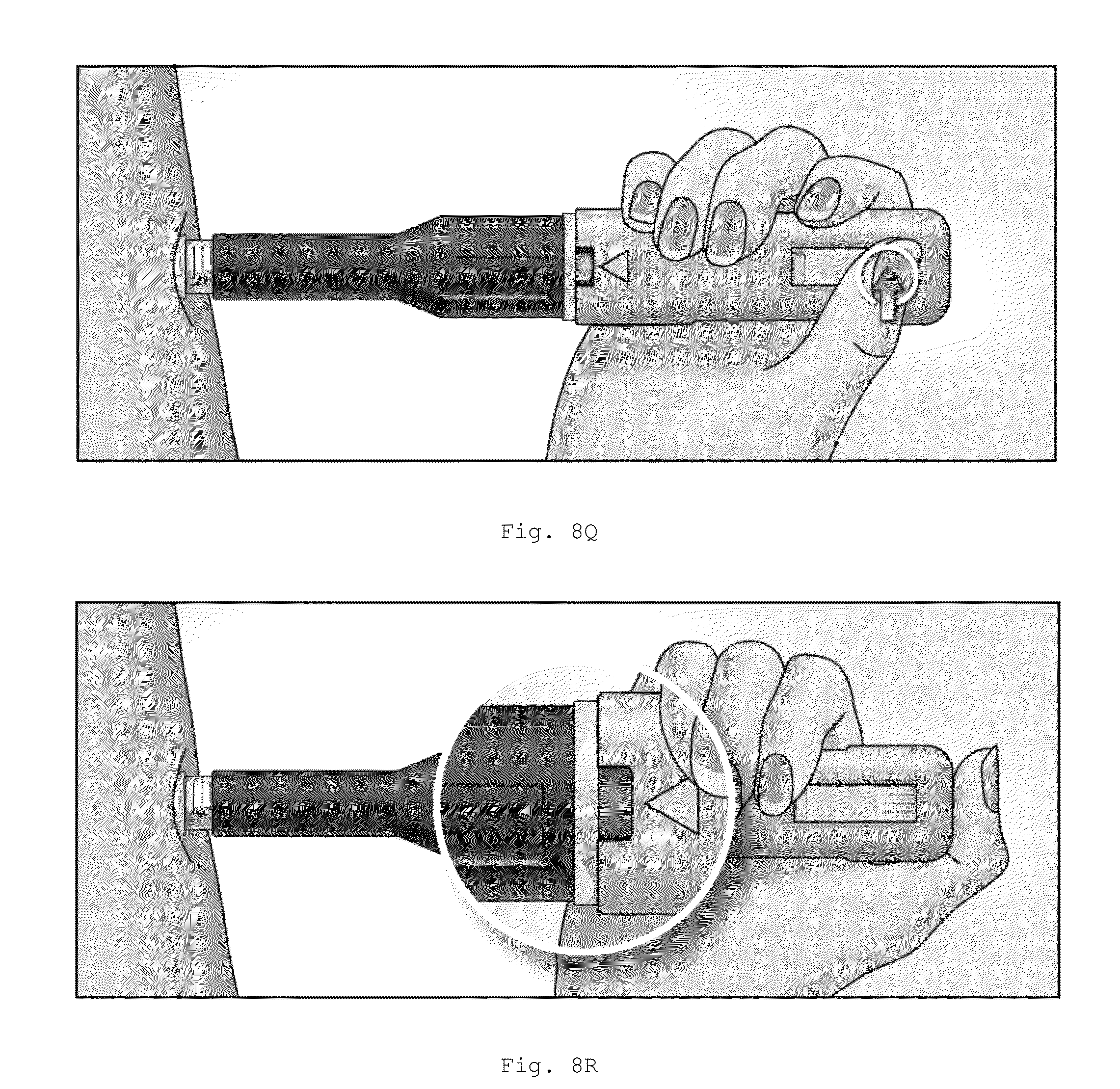
XML
uspto.report is an independent third-party trademark research tool that is not affiliated, endorsed, or sponsored by the United States Patent and Trademark Office (USPTO) or any other governmental organization. The information provided by uspto.report is based on publicly available data at the time of writing and is intended for informational purposes only.
While we strive to provide accurate and up-to-date information, we do not guarantee the accuracy, completeness, reliability, or suitability of the information displayed on this site. The use of this site is at your own risk. Any reliance you place on such information is therefore strictly at your own risk.
All official trademark data, including owner information, should be verified by visiting the official USPTO website at www.uspto.gov. This site is not intended to replace professional legal advice and should not be used as a substitute for consulting with a legal professional who is knowledgeable about trademark law.