Methods of diagnosing pancreatic inflammation by using chemokine PANEC-1 antibodies
Coleman , et al. December 31, 2
U.S. patent number 8,617,551 [Application Number 12/854,005] was granted by the patent office on 2013-12-31 for methods of diagnosing pancreatic inflammation by using chemokine panec-1 antibodies. This patent grant is currently assigned to Incyte Corporation. The grantee listed for this patent is Olga Bandman, Roger T. Coleman, Craig G. Wilde. Invention is credited to Olga Bandman, Roger T. Coleman, Craig G. Wilde.


| United States Patent | 8,617,551 |
| Coleman , et al. | December 31, 2013 |
Methods of diagnosing pancreatic inflammation by using chemokine PANEC-1 antibodies
Abstract
The present invention provides nucleotide and amino acid sequences that identify and encode novel expressed chemokines (PANEC-1 and PANEC-2) from human pancreas cells. The present invention also provides for antisense molecules to the nucleotide sequences which encode PANEC-1 and PANEC-2, expression vectors for the production of purified PANEC-1 and PANEC-2, antibodies capable of binding specifically to PANEC-1 and PANEC-2, hybridization probes or oligonucleotides for the detection of PANEC-1- or PANEC-2-encoding nucleotide sequences, genetically engineered host cells for the expression of PANEC-1 and PANEC-2, diagnostic tests for chemokine activation based on PANEC-1- and PANEC-2-encoding nucleic acid molecules and antibodies capable of binding specifically to the protein.
| Inventors: | Coleman; Roger T. (Seattle, WA), Bandman; Olga (Mountain View, CA), Wilde; Craig G. (Sunnyvale, CA) | ||||||||||
|---|---|---|---|---|---|---|---|---|---|---|---|
| Applicant: |
|
||||||||||
| Assignee: | Incyte Corporation (Palo Alto,
CA) |
||||||||||
| Family ID: | 23543735 | ||||||||||
| Appl. No.: | 12/854,005 | ||||||||||
| Filed: | August 10, 2010 |
Prior Publication Data
| Document Identifier | Publication Date | |
|---|---|---|
| US 20130224769 A1 | Aug 29, 2013 | |
Related U.S. Patent Documents
| Application Number | Filing Date | Patent Number | Issue Date | ||
|---|---|---|---|---|---|
| 12153507 | May 20, 2008 | 7803373 | |||
| 11604251 | Nov 27, 2006 | 7396652 | |||
| 10057275 | Oct 25, 2001 | ||||
| 08390740 | Feb 17, 1995 | 7005509 | |||
| Current U.S. Class: | 424/139.1; 530/389.1; 424/145.1; 424/141.1; 530/387.1 |
| Current CPC Class: | C07K 16/24 (20130101); A61P 43/00 (20180101); C07K 1/22 (20130101); C12Q 1/68 (20130101); A61P 29/00 (20180101); G01N 33/6863 (20130101); C07K 14/523 (20130101); G01N 33/6893 (20130101); C12Q 2600/118 (20130101); A61K 38/00 (20130101); G01N 2800/067 (20130101) |
| Current International Class: | A61K 39/00 (20060101); A61K 39/395 (20060101); C07K 16/24 (20060101); C07K 16/00 (20060101) |
References Cited [Referenced By]
U.S. Patent Documents
| 3817837 | June 1974 | Rubenstein |
| 3850752 | November 1974 | Schuurs et al. |
| 3939350 | February 1976 | Kronick et al. |
| 3996345 | December 1976 | Ullman et al. |
| 4275149 | June 1981 | Litman et al. |
| 4277437 | July 1981 | Maggio |
| 4366241 | December 1982 | Tom et al. |
| 4430318 | February 1984 | Langone |
| 4657760 | April 1987 | Kung et al. |
| 4683195 | July 1987 | Mullis et al. |
| 4800195 | January 1989 | Burgess et al. |
| 4816567 | March 1989 | Cabilly et al. |
| 4965188 | October 1990 | Mullis et al. |
| 5206344 | April 1993 | Katre et al. |
| 5225212 | July 1993 | Martin et al. |
| 6001649 | December 1999 | Caput et al. |
| 6403782 | June 2002 | Luster et al. |
| 6518046 | February 2003 | Li et al. |
| 7005509 | February 2006 | Coleman et al. |
| 7396652 | July 2008 | Coleman et al. |
| 7803373 | September 2010 | Coleman et al. |
| 2002/0155545 | October 2002 | Coleman et al. |
| 05/276986 | Oct 1993 | JP | |||
| WO 92/09629 | Jun 1992 | WO | |||
| WO 92/19737 | Nov 1992 | WO | |||
| WO 96/06169 | Feb 1996 | WO | |||
| WO 96/25497 | Aug 1996 | WO | |||
| WO 98/14581 | Apr 1998 | WO | |||
| WO 00/38706 | Jul 2000 | WO | |||
Other References
|
Winter et al., "Man-made Antibodies," Nature, vol. 349, pp. 293-299 (1991). cited by applicant . Huse et al., "Generation of a Large Combinatorial Library of the Immunoglobulin Repertoire in Phage Lambda," Science, vol. 246, pp. 1275-1281 (1989). cited by applicant . Orlandi et al., "Cloning Immunoglobulin Variable Domains for Expression by the Polymerase Chain Reaction," Proc. Natl. Acad. Sci., vol. 86, pp. 3833-3837 (1989). cited by applicant . Sambrook, "Molecular Cloning," 70 pages (1989). cited by applicant . Hodgson, "Data-Directed Drug Design," Bio/Technology, vol. 9, pp. 19-21 (1991). cited by applicant . Ausubel, F. M. et al., eds., "Current Protocols in Molecular Biology," vol. I, Green Publishing Associates, Inc. and John Wiley & Sons, Inc., New York at pp. 6.3.1-6.3.6 and 2.10.3), (1989). cited by applicant . Hebert et al., "Scanning Mutagenesis of Interleukin-8 Identifies a Cluster of Residues Required for Receptor Binding," The Journ. of Biological Chem., vol. 266, No. 28, pp. 18989-18994 (1991). cited by applicant . Bolton et al., "The Labelling of Proteins to High Specific Radioactivities by Conjugation to a .sup.125I-Containing Acylating Agent," Biochem. J., vol. 133, pp. 529-539 (1973). cited by applicant . Athauda et al., "Entrapment and Inhibition of Human Immunodeficiency Virus Proteinase by .alpha..sub.2-Macroglobulin and Structural Changes in the Inhibitor," J. Biochem, vol. 113, pp. 742-746 (1993). cited by applicant . Braxton et al., "Incorporation of a Stabilizing ca.sup.2+-Binding Loop into Subtilisin BPN' .dagger.," Biochemistry, vol. 31, pp. 7796-7801 (1992). cited by applicant . Jiang et al., "Monocyte Chemoattractant Protein-1 Regulates Adhesion Molecule Expression and Cytokine Production in Human Monocytes," The Journ. of Immunology, vol. 148, pp. 2423-2428 (1992). cited by applicant . Taub et al., "Preferential Migraiton of Activated CD4.sup.+ and CD8.sup.+ T Cells in Response to MIP-1.alpha. and MIP-1.beta.," Science, vol. 260, pp. 355-358 (1993). cited by applicant . McColl et al., "Uncoupling of Early Signal Transduction Events from Effector Function in Human Peripheral Blood Neutrophils in Response to Recombinant Macrophage Inflammatory Proteins-1.alpha. and -1.beta..sup.1," The Jounr. of Immunology, pp. 4550-4560 (1993). cited by applicant . Grynkiewicz et al., "A New Generation of Ca.sup.2+ Indicators with Greatly Improved Fluorescence Properties," The Journ. of Biolog. Chem., vol. 260, No. 6, pp. 3440-3450 (1985). cited by applicant . Zachariae et al., "Properties of Monocyte Chemotactic and Activating Factor (MCAF) Purified from a Human Fibrosarcoma Cell Line," The Journ. of Experimental Medicine, vol. 171, pp. 2177-2183 (1990). cited by applicant . Falk et al., "A 48-Well Micro Chemotaxis Assembly for Rapid and Accurate Measurement of Leukocyte Migration," Journ. of Immunological Methods, vol. 33, pp. 239-247 (1980). cited by applicant . Maddox et al., "Elevanted Serum Levels in Human Pregnancy of a Molecule Immunochemically Similar to Eosinophil Granule Major Basic Protein," J. Exp. Med., pp. 1211-1226 (1983). cited by applicant . Merrifield, "Solid Phase Peptide Synthesis. I. The Synthesis of Tetrapeptide," Synthesis of a Tetrapeptide, pp. 2149-2154 (1963). cited by applicant . Walsh et al., "Preferential PCR Amplification of Alleles: Mechanisms and Solutions," PCR Methods and Applications, pp. 241-250 (1992). cited by applicant . Paul, "The Chemokine Family of Inflammatory Cytokines," Fundamental Immunology, pp. 822-826 (1993). cited by applicant . Chirgwin Et Al., "Isolution of Biologically Active Ribonucleic Acid From Sources Enriched in Ribonuclease," Biochemistry, pp. 5294-5299 (1979). cited by applicant . Jasny, Human Genome Maps, TOC plus 2 pages, vol. 265, Sep. 30, 1994. cited by applicant . U.S. Appl. No. 08/294,251, filed Aug. 1994, Li et al. cited by applicant . Banchereau, Jacques, Interleukin-4, The Cytokine Handbook, Academic Press, Copyright 1991, Chapter 6, pp. 120-148. cited by applicant . Charo, et al., "Molecular Cloning and Functional Expression of Two Monocyte Chemoattractant Protein 1 Receptors Reveals Alternative Splicing of the Carboxyl-Terminal Tails," Proc. Natl. Acad. Sci. USA, 91: 2752-2756. cited by applicant . Esposito et al., The Journal of Biological Chemistry, vol. 263, No. 23, pp. 11466-11472, 1988. cited by applicant . Kitaura et al., "Molecular Cloning of Human Eotaxin, an Eosinophil Selective CC Chemokine, and Identification of a Specific Eosinophil Eotaxin Receptor, CC Chemokine Receptor 3", J. Biol. Chem., 271(13), 7725-7730, (1996). cited by applicant . Ponath et al., "Cloning of the Human Eosinophil Chemoattractant, Eotaxin", The Journal of Clinical Investigation, Feb. 1, 1996, vol. 97, No. 3, pp. 604-612. cited by applicant . Rink et al., J. of Onterferon and Cytokine Research, 16: 861-868. cited by applicant . Schall, The Chemokines, The Cytokine Handbook, Academic Press, 1994, Sep. 19-20, 1994, Second Edition, pp. 180-272. cited by applicant . Schall, The Chemokines, The Cytokine Handbook, Academic Press, 1994, Second Edition, Chapter 22, pp. 419-460. cited by applicant . Shaw, Molecular Biology of Cytokines, The Cytokine Handbook, Academic Press, Copyright 1991, Chapter 2, Chapter 2, pp. 20-46. cited by applicant . Vilcek, et al., Immunology of Cytokines, The Cytokine Handbook, Academic Press, Copyright 1991, Chapter 1, pp. 1-17. cited by applicant . Yoshie, (Direct Submission) NCBI Accession No. BAA08370 (GI 1552241), Feb. 10, 1999. cited by applicant . Yoshie, (Direct Submission) NCBI Accession No. D49372 (GI 1552240), Feb. 10, 1999. cited by applicant . Decision of the Board of Patent Appeals and Interferences (Patent interference 105,449), Dec. 20, 2006. cited by applicant. |
Primary Examiner: Stoica; Elly-Gerald
Attorney, Agent or Firm: Foley & Lardner LLP
Parent Case Text
This application is a Divisional of U.S. patent application Ser. No. 12/153,507, filed May 20, 2008, which is a Divisional of U.S. patent application Ser. No. 11/604,251, filed Nov. 27, 2009, now U.S. Pat. No. 7,396,652, which is a Divisional of U.S. patent application Ser. No. 10/057,272, filed Oct. 25, 2001, now abandoned, which is a Continuation of U.S. patent application Ser. No. 08/390,740, filed Feb. 17, 1995, now U.S. Pat. No. 7,005,509, entitled NEW CHEMOKINES EXPRESSED IN PANCREAS, all of which are hereby incorporated by reference in their entirety.
Claims
What is claimed is:
1. A method of diagnosis for pancreatic inflammation in a subject comprising the steps of: (a) providing a pancreatic biological sample from the subject; (b) contacting the biological sample with an antibody which specifically binds to the polypeptide of SEQ ID NO: 2; (c) detecting the presence of bound antibody in the biological sample; (d) comparing the presence of antibody bound in the biological sample with the presence of antibody bound in a control sample; (e) wherein a higher presence in the biological sample as compared to the control sample is indicative of pancreatic inflammation.
2. The method of claim 1, wherein the subject is a human.
3. The method of claim 1, wherein the subject biological sample comprises tissues or extracts of such tissues.
4. The method of claim 1, further comprising: (a) determining the amount of bound antibody in the biological sample; (b) comparing the amount determined in (a) with an amount of bound antibody in a control sample; wherein a higher amount of bound antibody in the biological sample is correlated with pancreatic inflammation.
5. The method of claim 1, wherein the antibody comprises a monoclonal antibody.
6. The method of claim 1, wherein the antibody comprises a polyclonal antibody.
7. The method of claim 1, wherein the biological sample comprises pancreatic tissue, an extract of pancreatic tissue, or a combination thereof.
8. The method of claim 1, wherein the antibody is labeled.
9. The method of claim 8, wherein the antibody label is selected from the group consisting of radionuclides, enzymes, substrates, cofactors, inhibitors, fluorescent agents, chemiluminescent agents, and magnetic particles.
10. The method of claim 1, wherein the detecting step utilizes enzyme-linked immunosorbent assay (ELISA), radioimmunoassay (RIA), fluorescent activated cell sorting (FACS), or any combination thereof.
Description
BACKGROUND OF THE INVENTION
The pancreas is an elongated organ which lies behind and below the stomach and consists of both exocrine and endocrine tissues. In descending order, the exocrine portion is divided into lobes, lobules, and functional secretory units known as acini. All acini eventually drain into the main pancreatic duct which joins the bile duct from the liver before it empties into the duodenum. Acinar cells comprise 80% of the pancreas and secrete enzymes in either inactive or active form which assist digestion. Epithelial cells of the ductules secrete large amounts of bicarbonate ions and water which neutralize acidic chyme as it leaves the stomach and enters the duodenum as well as the enzymes for digesting protein, carbohydrates, and fats.
The most important and abundant proteolytic enzymes are trypsin, chymotrypsin, and carboxypeptidase. The serine proteases, trypsin and chymotrypsin, split whole and partially-digested proteins into polypeptides of different sizes; then, carboxypeptidase breaks down the polypeptides into individual amino acids. Several elastases, which are also serine proteases, and nucleases, which digest nucleic acids, are also found in the pancreatic juice.
The principal enzyme for digesting carbohydrates in the gut is pancreatic amylase. It hydrolyzes starches, glycogen, and most other non-cellulosic carbohydrates to form disaccharides and trisaccharides. The main enzymes for fat digestion are pancreatic lipase, cholesterol esterase, and phospholipase. Pancreatic lipase hydrolyzes neutral fat into fatty acids and monoglycerides. Cholesterol esterase hydrolyzes cholesterol esters, and phospholipase removes fatty acid molecules from phospholipids.
The four molecules which control acinar secretion are acetylcholine and the hormones, gastrin, cholecystokinin (CCK), and secretin. Acetylcholine is released from the parasympathetic vagus and other cholinergic nerve endings, gastrin is secreted by cells of the stomach, and CCK and secretin are secreted by the upper small intestine. The gastrointestinal (GD hormones are absorbed into the blood and transported to the pancreas where they stimulate acini to secrete enzymes and ductal cells to secrete the sodium bicarbonate and water which washes the pancreatic enzymes into the duodenum.
The endocrine pancreas consists of islets of Langerhans, whose cells are separated from the exocrine lobules and are distributed throughout the pancreas. The function of the various types of endocrine cells which make up the islets is to secrete the hormones which participate in the metabolism of proteins, carbohydrates, and fats.
The major endocrine cells are .alpha., .beta., and .delta. cells; the minor cells are C cells, EC cells, and PP cells. About 15% of the islet cell population are a cells which are located along the periphery of islets and secrete the hormone glucagon. B cells comprise about 70% of the islet cell population, are located around the center of the islets, and secrete the hormone insulin. .delta. cells comprise about 10% of the population, are located close to a cells and secrete two different hormones, somatostatin and vasoactive intestinal peptide (VIP). C, EC, and PP cells make up the final 5% of the islet cell population. The function of C cells is unknown, but EC and PP cells secrete serotonin and pancreatic polypeptide, respectively.
Inflammation of the pancreas or pancreatitis may be classified as either acute or chronic by clinical criteria. With treatment, acute pancreatitis can often be cured and normal function restored. Chronic pancreatitis often results in permanent damage. The precise mechanisms which trigger acute inflammation are not understood. However, some causes in the order of their importance are alcohol ingestion, biliary tract disease, post-operative trauma, and hereditary pancreatitis. One theory provides that autodigestion, the premature activation of proteolytic enzymes in the pancreas rather than in the duodenum, causes acute pancreatitis. Any number of other factors including endotoxins, exotoxins, viral infections, ischemia, anoxia, and direct trauma may activate the proenzymes. In addition any internal or external blockage of pancreatic ducts can also cause an accumulation of pancreatic juices in the pancreas resulting cellular damage.
As is the case in inflammation of other tissues, leukocytes including monocytes, macrophages, basophils and eosinophils infiltrate the inflamed area of the pancreas. Their primary role is to clean up the site of the inflammation; however, macrophages may produce powerful oxidants and proteases which contribute to tissue destruction. Leukocytes also secrete a range of cytokines which recruit other cells to the area.
The investigation of the critical, regulatory processes by which white cells proceed to their appropriate destination and interact with other cells is underway. The current model of leukocyte movement or trafficking from the blood to injured or inflamed tissues comprises the following steps. The first step is the rolling adhesion of the leukocyte along the endothelial cells of the blood vessel wall. This movement is mediated by transient interactions between selectins and their ligands. A second step involves cell activation which promotes a more stable leukocyte-endothelial cell interaction mediated by the integrins and their ligands. This stronger, more stable adhesion precipitates the final steps of leukocyte diapedesis and extravasation into the tissues.
The chemokine family of polypeptide cytokines possesses the cellular specificity required to explain leukocyte trafficking in different abnormal, inflammatory or diseased situations. First, chemokines mediate the expression of particular adhesion molecules on endothelial cells; and second, they generate gradients of chemoattractant factors which activate specific cell types. In addition, the chemokines stimulate the proliferation of specific cell types and regulate the activation of cells which bear specific receptors. These activities demonstrate a high degree of target cell specificity.
The chemokines are small polypeptides, generally about 70-100 amino acids (aa) in length, 8-11 kD in molecular weight and active over a 1-100 ng/ml concentration range. Initially, they were isolated and purified from inflamed tissues and characterized relative to their bioactivity. More recently, chemokines have been discovered through molecular cloning techniques and characterized by structural as well as functional analysis.
The chemokines are related through a four-cysteine motif which is based primarily on the spacing of the first two cysteine residues in the mature molecule. Currently the chemokines are assigned to one of two families, the C--C chemokines (.alpha.) and the C--X--C chemokines (.beta.). Although exceptions exist, the C--X--C chemokines generally activate neutrophils and fibroblasts while the C--C chemokines act on a more diverse group of target cells which include monocytes/macrophages, basophils, eosinophils, T lymphocytes and others. The known chemokines of both families are synthesized by many diverse cell types as reviewed in Thomson A. (1994) The Cytokine Handbook, 2d Ed. Academic Press, NY. The two groups of chemokines will be described in turn.
At this time, relatively few C--C chemokines have been described, and they appear to have less N-terminal processing than the C--X--C chemokines. A brief description of the known human (and/or murine) C--C chemokines follows. The macrophage inflammatory proteins alpha and beta (MIP-1.alpha. and .beta.) were first purified from stimulated mouse macrophage cell line and elicited an inflammatory response when injected into normal tissues. At least three distinct and non-allelic genes encode human MIP-1.alpha.a, and seven distinct genes encode MIP-1.beta..
MIP-1.alpha. and MIP-1.beta. consist of 68-69 amino acids which are about 70% identical in their acidic, mature secreted forms. They are both expressed in stimulated T cells, B cells and monocytes in response to mitogens, anti-CD3 and endotoxin, and both polypeptides bind heparin. While both molecules stimulate monocytes, MIP-1.alpha. chemoattracts the CD-8 subset of T lymphocytes and eosinophils, while MIP-1.beta. chemoattracts the CD-4 subset of T lymphocytes. In mouse, these proteins are known to stimulate myelopoiesis.
1-309 was cloned from a human .gamma.-.delta. T cell line and shows 42% amino acid identity to T cell activation gene 3 (TCA3) cloned from mouse. There is considerable nucleotide homology between the 5' flanking regions of these two proteins, and they share an extra pair of cysteine residues not found in other chemokines. Such similarities suggest 1-309 and TCA3 are species homologs which have diverged over time in both sequence and function.
RANTES is another C--C chemokine which is expressed in T cells (but not B cells), in platelets, in some tumor cell lines, and in stimulated rheumatoid synovial fibroblasts. In the latter, it is regulated by interleukins-1 and -4, transforming nerve factor and interferon-.gamma.. The cDNA cloned from T cells encodes a basic 8 kD protein which lacks N-linked glycosylation and is able to affect lymphocytes, monocytes, basophils and eosinophils. The expression of RANTES mRNA is substantially reduced following T cell stimulation.
Monocyte chemotactic protein (MCP-1) is a 76 amino acid protein which appears to be expressed in almost all cells and tissues upon stimulation by a variety of agents. The targets of MCP-1, however, are limited to monocytes and basophils in which it induces a MCP-1 receptor:G protein-linked calcium flux (Chard I, personal communication). Two other related proteins (MCP-2 and MCP-3) were purified from a human osteosarcoma cell line. MCP-2 and MCP-3 have 62% and 73% aa identity, respectively, with MCP-1 and share its chemoattractant specificity for monocytes.
Current techniques for diagnosis of abnormalities in the inflamed or diseased tissues mainly rely on observation of clinical symptoms or serological analyses of body tissues or fluids for hormones, polypeptides or various metabolites. Patients often manifest no clinical symptoms at early stages of disease or tumor development. Furthermore, serological analyses do not always differentiate between invasive diseases and genetic syndromes which have overlapping or very similar ranges. Thus, development of new diagnostic techniques comprising small molecules such as the expressed chemokines are important to provide for early and accurate diagnoses, to give a better understanding of molecular pathogenesis, and to use in the development of effective therapies.
The pancreas is reviewed in Guyton A C (1991) Textbook of Medical Physiology, WB Saunders Co, Philadelphia; and The Merck Manual of Diagnosis and Therapy, (1992) Merck Research Laboratories, Rahway, N.J. The chemokine molecules are reviewed in Schall T J (1994) Chemotactic Cytokines: Targets for Therapeutic Development. International Business Communications, Southborough, Mass., pp 180-270; and in Paul W E (1993) Fundamental Immunology, Raven Press, New York City (NYC), pp 822-826.
SUMMARY OF THE INVENTION
The subject invention provides nucleotide sequences which uniquely encode two novel human pancreatic proteins. The new genes, which are known as pancreatic expressed chemokines, or panec-1 and parsec-2 (Incyte Clones 223187 and 226152), encode polypeptides designated PANEC-1 and PANEC-2, of the C--C chemokine family, and referred to collectively as PANEC.
The invention also comprises diagnostic tests for physiologic or pathologic compromise of the pancreas which include the steps of testing a sample or an extract thereof with panec-1 or panes-2 DNA, fragments or oligomers thereof. Aspects of the invention include the antisense DNAs of panes-1 and panes-2; cloning or expression vectors containing panec-1 or panec-2; host cells or organisms transformed with expression vectors containing panes-1 or panes-2; a method for the production and recovery of purified PANEC-1 or PANEC-2 from host cells; and purified proteins, PANEC-1 and PANEC-2.
DESCRIPTION OF THE FIGURES
FIG. 1 displays the nucleotide sequence for panec-1 (SEQ ID NO:1) and the predicted amino acid (aa) sequence of the pancreas expressed chemokine, PANEC-1 (SEQ ID NO:2).
FIG. 2 displays the nucleotide sequence for panec-2 (SEQ ID NO:3) and the predicted amino acid (aa) sequence of the pancreas expressed chemokine. PANEC-2 (SEQ ID NO:4).
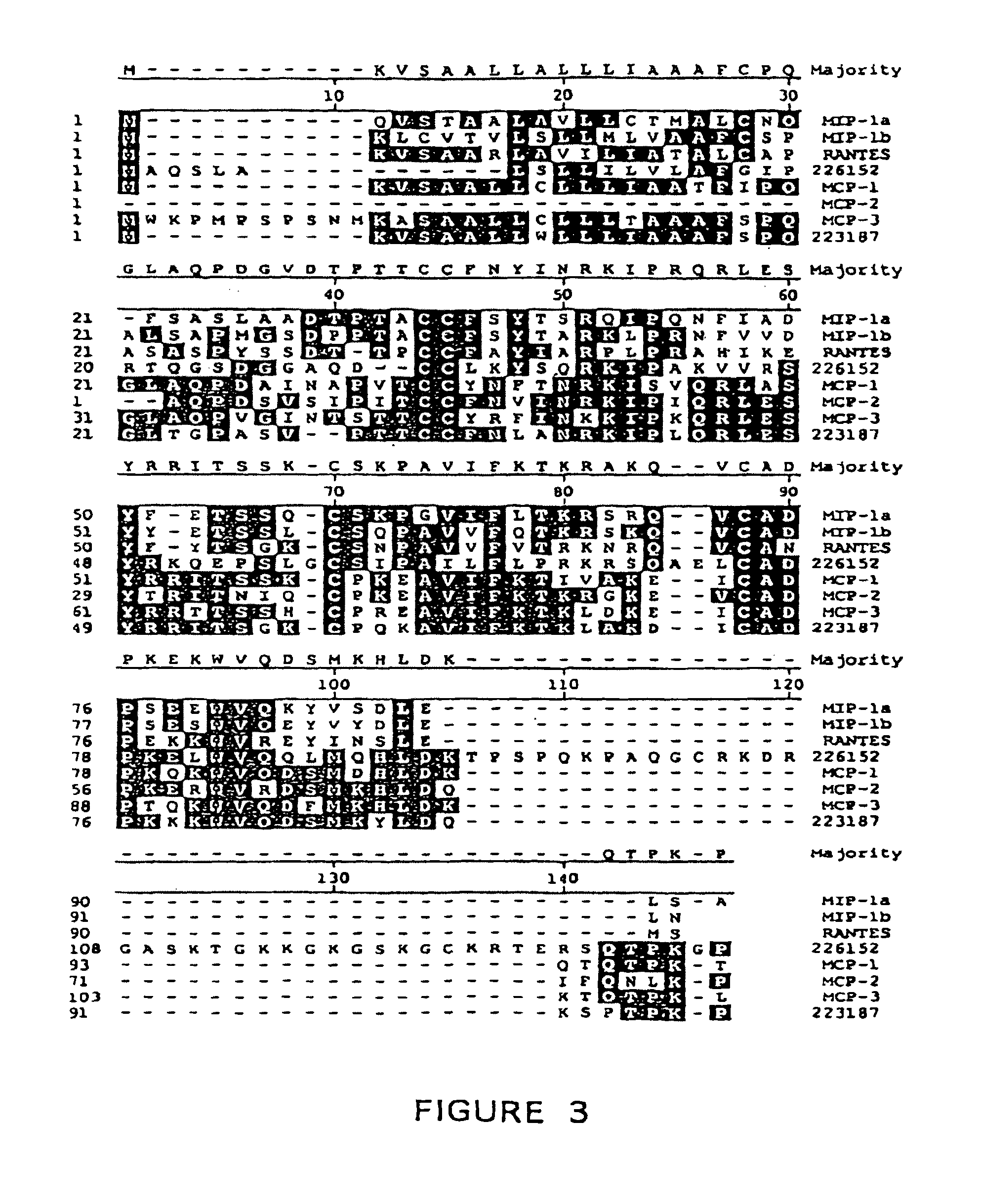
FIG. 3 shows the aa alignment of PANEC-1 and PANEC-2 with other human chemokines of the C--C family. Alignments shown were produced using the multisequence alignment program of DNASTAR software (DNASTAR Inc, Madison Wis.) (Majority=SEQ ID NO:5; MIP 1.alpha.=SEQ ID NO:6; MIP 1.beta.=SEQ ID NO:7; RANTES=SEQ ID NO:8; MCP-1=SEQ ID NO:9; MCP-2=SEQ ID NO:10; MCP-3=SEQ ED NO:11).
FIG. 4 displays an analysis of PANEC-1 hydrophobicity based on the predicted amino acid sequence and composition.
FIG. 5 displays an analysis of PANEC-2 hydrophobicity based on the predicted amino acid sequence and composition.
FIG. 6 shows a relatedness tree of human C--C chemokines. The phylogenetic tree was generated by phylogenetic tree program of DNASTAR software using the Clustal method with the PAM250 residue weight table.
DETAILED DESCRIPTION OF THE INVENTION
Definitions
As used herein, "pancreas expressed chemokines" or PANECs, refer to polypeptides, naturally occurring PANECs or active fragments thereof, which are encoded by mRNAs transcribed from the cDNAs of Seq ID No: 1 and Seq ID No:3.
"Active" refers to those forms of PANEC which retain the biologic and/or immunologic activities of any naturally occurring PANEC.
"Naturally occurring PANEC" refers to PANECs produced by human cells that have not been genetically engineered and specifically contemplates various PANECs arising from post-translational modifications of the polypeptide including but not limited to acetylation, carboxylation, glycosylation, phosphorylation, lipidation and acylation.
"Derivative" refers to polypeptides derived from naturally occurring PANECs by chemical modifications such as ubiquitination, labeling (e.g., with radionuclides, various enzymes, etc.), pegylation (derivatization with polyethylene glycol) or by insertion or substitution by chemical synthesis of amino acids such as ornithine, which do not normally occur in human proteins.
"Recombinant variant" refers to any polypeptide differing from naturally occurring PANECs by amino acid insertions, deletions, and substitutions, created using recombinant DNA techniques. Guidance in determining which amino acid residues may be replaced, added or deleted without abolishing activities of interest, such as cell adhesion and chemotaxis, may be found by comparing the sequence of the particular PANEC with that of homologous cytokines and minimizing the number of amino acid sequence changes made in regions of high homology.
Preferably, amino acid "substitutions" are the result of replacing one amino acid with another amino acid having similar structural and/or chemical properties, such as the replacement of a leucine with an isoleucine or valine, an aspartate with a glutamate, or a threonine with a serine, i.e., conservative amino acid replacements. "Insertions" or "deletions" are typically in the range of about 1 to 5 amino acids. The variation allowed may be experimentally determined by systematically making insertions, deletions, or substitutions of amino acids in a PANEC molecule using recombinant DNA techniques and assaying the resulting recombinant variants for activity.
Where desired, a "signal or leader sequence" can direct the polypeptide through the membrane of a cell. Such a sequence may be naturally present on the polypeptides of the present invention or provided from heterologous protein sources by recombinant DNA techniques.
A polypeptide "fragment," "portion," or "segment" is a stretch of amino acid residues of at least about 5 amino acids, often at least about 7 amino acids, typically at least about 9 to 13 amino acids, and, in various embodiments, at least about 17 or more amino acids. To be active, any PANEC polypeptide must have sufficient length to display biologic and/or immunologic activity.
An "oligonucleotide" or polynucleotide "fragment", "portion," or "segment" is a stretch of nucleotide residues which is long enough to use in polymerase chain reaction (PCR) or various hybridization procedures to amplify or simply reveal related parts of mRNA or DNA molecules.
The present invention includes purified PANEC-1 and PANEC-2 polypeptides from natural or recombinant sources, and cells transformed with recombinant nucleic acid molecules encoding PANEC-1 and PANEC-2. Various methods for the isolation of the PANEC-1 and PANEC-2 polypeptides may be accomplished by procedures well known in the art. For example, such polypeptides may be purified by immunoaffinity chromatography by employing the antibodies provided by the present invention. Various other methods of protein purification well known in the art include those described in Deutscher M (1990) Methods in Enzymology, Vol 182, Academic Press, San Diego; and Scopes R (1982) Protein Purification: Principles and Practice. Springer-Verlag, NYC, both incorporated herein by reference.
"Recombinant" may also refer to a polynucleotide which encodes PANEC-1 or PANEC-2 and is prepared using recombinant DNA techniques. The DNAs which encode PANEC-1 and PANEC-2 may also include allelic or recombinant variants and mutants thereof.
"Oligonucleotides" or "nucleic acid probes" are prepared based on the cDNA sequences which encode PANEC-1 and PANEC-2 provided by the present invention. Oligonucleotides comprise portions of the DNA sequence having at least about 15 nucleotides, usually at least about 20 nucleotides. Nucleic acid probes comprise portions of the sequence having fewer nucleotides than about 6 kb, usually fewer than about 1 kb. After appropriate testing to eliminate false positives, these probes may be used to determine whether mRNAs encoding PANEC-1 and PANEC-2 are present in a cell or tissue or to isolate similar nucleic acid sequences from chromosomal DNA as described by Walsh P S et al (1992 PCR Methods Appl 1:241-250).
Probes may be derived from naturally occurring or recombinant single- or double-stranded nucleic acids or be chemically synthesized. They may be labeled by nick translation, Klenow reaction, PCR or other methods well known in the art. Probes of the present invention, their preparation and/or labeling are elaborated in Sambrook J et al (1989) Molecular Cloning: A Laboratory Manual, Cold Spring Harbor Laboratory, New York; or Ausubel F M et al (1989) Current Protocols in Molecular Biology, John Wiley & Sons, New York City, both incorporated herein by reference.
Alternatively, recombinant variants encoding these same or similar polypeptides may be synthesized or selected by making use of the "redundancy" in the genetic code. Various codon substitutions, such as the silent changes which produce various restriction sites, may be introduced to optimize cloning into a plasmid or viral vector or expression in a particular prokaryotic or eukaryotic system. Mutations may also be introduced to modify the properties of the polypeptide, to change ligand-binding affinities, interchain affinities, or polypeptide degradation or turnover rate.
DETAILED DESCRIPTION OF THE INVENTION
The present invention provides nucleotide sequences uniquely identifying novel chemokines of the C--C family, PANEC-1 and PANEC-2, which are highly expressed in the pancreas. Because PANEC-1 and PANEC-2 are specifically expressed in pancreas, the nucleic acids (panec-1 and panec-2), polypeptides (PANEC-1 and PANEC-2) and antibodies to PANEL-1 and PANEC-2 are useful in diagnostic assays based on chemokine production in cases of inflammation or disease affecting the pancreas. Excessive expression of either PANEC-1 or PANEC-2 can lead to activation of monocytes, macrophages, basophils, eosinophils, T lymphocytes and/or other cells which respond to the chemokines by producing abundant proteases and other molecules which can lead to tissue damage or destruction. Therefore, a diagnostic test for excess expression of PANECs can accelerate diagnosis and proper treatment of an abnormal condition caused by viral or bacterial infections; mechanical injury associated with trauma; hereditary diseases affecting pancreatitis; biliary disease; infiltrative diseases such as leukemias and lymphomas; or other physiologic and pathologic problems which affect the function of the organ.
The nucleotide sequences encoding PANEC-1 and PANEC-2 (or their complement) have numerous applications in techniques known to those skilled in the art of molecular biology. These techniques include use as hybridization probes, use as oligomers for PCR, use for chromosome and gene mapping, use in the recombinant production of PANEC-1 and PANEC-2, and use in generation of anti-sense DNA or RNA, their chemical analogs and the like. Uses of nucleotides encoding PANEC-1 and PANEC-2 disclosed herein are exemplary of known techniques and are not intended to limit their use in any technique known to a person of ordinary skill in the art. Furthermore, the nucleotide sequences disclosed herein may be used in molecular biology techniques that have not yet been developed, provided the new techniques rely on properties of nucleotide sequences that are currently known, e.g., the triplet genetic code, specific base pair interactions, etc.
It will be appreciated by those skilled in the art that as a result of the degeneracy of the genetic code, a multitude of PANEC-encoding nucleotide sequences, some bearing minimal homology to the nucleotide sequence of any known and naturally occurring gene may be produced. The invention has specifically contemplated each and every possible variation of nucleotide sequence that could be made by selecting combinations based on possible codon choices. These combinations are made in accordance with the standard triplet genetic code as applied to the nucleotide sequence of naturally occurring PANECs, and all such variations are to be considered as being specifically disclosed.
Although nucleotide sequences which encode PANEC-1 and PANEC-2 and/or their variants are preferably capable of hybridizing to the nucleotide sequence of the naturally occurring PANEC genes under stringent conditions, it may be advantageous to produce nucleotide sequences encoding PANEC-1 and PANEC-2 or their derivatives possessing a substantially different codon usage. Codons can be selected to increase the rate at which expression of the peptide occurs in a particular prokaryotic or eukaryotic expression host in accordance with the frequency with which particular codons are utilized by the host. Other reasons for substantially altering the nucleotide sequence encoding PANEC-1 and PANEC-2 and/or their derivatives without altering the encoded amino acid sequence include the production of RNA transcripts having more desirable properties, such as a greater half-life, than transcripts produced from the naturally occurring sequence.
Nucleotide sequences encoding PANEC-1 or PANEC-2 may be joined to a variety of other nucleotide sequences by means of well established recombinant DNA techniques (cf Sambrook J et al. (1989) Molecular Cloning: A Laboratory Manual, Cold Spring Harbor Laboratory, New York). Useful nucleotide sequences for joining to panec include an assortment of cloning vectors, e.g., plasmids, cosmids, lambda phage derivatives, phagemids, and the like, that are well known in the art. Vectors of interest include expression vectors, replication vectors, probe generation vectors, sequencing vectors, and the like. In general, vectors of interest may contain an origin of replication functional in at least one organism, convenient restriction endonuclease sensitive sites, and selectable markers for the host cell.
Another aspect of the subject invention is to provide for panec-1- or panec-2-specific nucleic acid hybridization probes capable of hybridizing with naturally occurring nucleotide sequences encoding PANEC-1 or PANEC-2. Such probes may also be used for the detection of similar chemokine encoding sequences and should preferably contain at least 50% of the nucleotides from a C--C encoding sequence. The hybridization probes of the subject invention may be derived from the nucleotide sequences of the SEQ ID NO:1 or SEQ ID NO:3 from genomic sequences including promoters, enhancer elements and introns of the respective naturally occurring panecs. Hybridization probes may be labeled by a variety of reporter groups, including radionuclides such as .sup.32P or .sup.35S, or enzymatic labels such as alkaline phosphatase coupled to the probe via avidin/biotin coupling systems, and the like.
PCR as described U.S. Pat. Nos 4,683,195; 4,800,195; and 4,965,188 provides additional uses for oligonucleotides based upon the nucleotide sequences which encode either PANEC-1 or PANEC-2. Such probes used in PCR may be of recombinant origin, may be chemically synthesized, or a mixture of both and comprise a discrete nucleotide sequence for diagnostic use or a degenerate pool of possible sequences for identification of closely related genomic sequences.
Other means of producing specific hybridization probes for panec DNAs include the cloning of nucleic acid sequences encoding PANEC-1 and PANEC-2 or PANEC-1 and PANEC-2 derivatives into vectors for the production of mRNA probes. Such vectors are known in the art and are commercially available and may be used to synthesize RNA probes in vitro by means of the addition of the appropriate RNA polymerase as T7 or SP6 RNA polymerase and the appropriate radioactively labeled nucleotides.
It is now possible to produce a DNA sequence, or portions thereof, encoding PANEC-1 and PANEC-2 and their derivatives entirely by synthetic chemistry, after which the gene can be inserted into any of the many available DNA vectors using reagents, vectors and cells that are known in the art at the time of the filing of this application. Moreover, synthetic chemistry may be used to introduce mutations into the panec sequences or any portion thereof.
The nucleotide sequence can be used to construct an assay to detect inflammation or disease associated with abnormal levels of expression of PANEC-1 or PANEC-2. The nucleotide sequence can be labeled by methods known in the art and added to a fluid or tissue sample from a patient under hybridizing conditions. After an incubation period, the sample is washed with a compatible fluid which optionally contains a dye (or other label requiring a developer) if the nucleotide has been labeled with an enzyme. After the compatible fluid is rinsed off, the dye is quantitated and compared with a standard. If the amount of dye is significantly elevated, the nucleotide sequence has hybridized with the sample, and the assay indicates the presence of inflammation and/or disease.
The nucleotide sequence for panec-1 or panec-2 can be used to construct hybridization probes for mapping that gene. The nucleotide sequence provided herein may be mapped to a chromosome and specific regions of a chromosome using well known genetic and/or chromosomal mapping techniques. These techniques include in situ hybridization, linkage analysis against known chromosomal markers, hybridization screening with libraries or flow-sorted chromosomal preparations specific to known chromosomes, and the like. The technique of fluorescent in situ hybridization of chromosome spreads has been described, among other places, in Verma et al (1988) Human Chromosomes: A Manual of Basic Techniques, Pergamon Press, New York City.
Fluorescent in situ hybridization of chromosomal preparations and other physical chromosome mapping techniques may be correlated with additional genetic map data. Examples of genetic map data can be found in the 1994 Genome Issue of Science (265:1981f). Correlation between the location of panec on a physical chromosomal map and a specific disease (or predisposition to a specific disease) can help delimit the region of DNA associated with that genetic disease. The nucleotide sequence of the subject invention may be used to detect differences in gene sequence between normal and carrier or affected individuals.
Nucleotide sequences encoding PANEC-1 and PANEC-2 may be used to produce purified PANEC-1 and PANEC-2 using well known methods of recombinant DNA technology. Among the many publications that teach methods for the expression of genes after they have been isolated is Goeddel (1990) Gene Expression Technology, Methods and Enzymology, Vol 185, Academic Press, San Diego. PANEC-1 and PANEC-2 may be expressed in a variety of host cells, either prokaryotic or eukaryotic. Host cells may be from the same species in which panec nucleotide sequences are endogenous or from a different species. Advantages of producing PANEC-1 and PANEL-2 by recombinant DNA technology include obtaining adequate amounts of the protein for purification and the availability of simplified purification procedures.
Cells transformed with DNA encoding PANEC-1 or PANEC-2 may be cultured under conditions suitable for the expression of chemokines and recovery of the protein from the cell culture. PANEC-1 or PANEC-2 produced by a recombinant cell may be secreted or may be contained intracellularly, depending on the particular genetic construction used. In general, it is more convenient to prepare recombinant proteins in secreted form. Purification steps vary with the production process and the particular protein produced.
In addition to recombinant production, fragments of PANEC-1 or PANEC-2 may be produced by direct peptide synthesis using solid-phase techniques (cf Stewart et al (1969) Solid-Phase Peptide Synthesis, WH Freeman Co, San Francisco; Merrifield J (1963) J Am Chem Soc 85:2149-2154. In vitro protein synthesis may be performed using manual techniques or by automation. Automated synthesis may be achieved, for example, using Applied Biosystems 431A Peptide Synthesizer (Foster City, Calif.) in accordance with the instructions provided by the manufacturer. Various fragments of PANEC-1 and PANEC-2 may be chemically synthesized separately and combined using chemical methods to produce the full length molecule.
PANEC-1 or PANEL-2 for antibody induction does not require biological activity; however, the protein must be immunogenic. Peptides used to induce specific antibodies may have an amino acid sequence consisting of at least five amino acids, preferably at least 10 amino acids. They should mimic a portion of the amino acid sequence of the protein and may contain the entire amino acid sequence of a small naturally occurring molecules like PANEC-1 and PANEC-2. Short stretches of PANEC-1 or PANEC-2 amino acids may be fused with those of another protein such as keyhole limpet hemocyanin and the chimeric molecule used for antibody production.
Antibodies specific for PANEL-1 or PANEC-2 may be produced by inoculation of an appropriate animal with the polypeptide or an antigenic fragment. An antibody is specific for PANEC-1 or PANED-2 if it is produced against an epitope of the polypeptide and binds to at least part of the natural or recombinant protein. Antibody production includes not only the stimulation of an immune response by injection into animals, but also analogous steps in the production of synthetic antibodies or other specific-binding molecules such as the screening of recombinant immunoglobulin libraries (cf Orlandi R et al (1989) PNAS 86:3833-3837, or Huse W D et al (1989) Science 256:1275-1281) or the in vitro stimulation of lymphocyte populations. Current technology (Winter G and Milstein C (1991) Nature 349:293-299) provides for a number of highly specific binding reagents based on the principles of antibody formation. These techniques may be adapted to produce molecules specifically binding PANECs.
An additional embodiment of the subject invention is the use of PANEC-1 or PANEC-2 specific antibodies, inhibitors, receptors or their analogs as bioactive agents to treat inflammation or disease of the pancreas including, but not limited to viral or bacterial infections; mechanical injury associated with trauma; hereditary diseases affecting pancreatitis; biliary disease; infiltrative diseases such as leukemias and lymphomas; or other physiologic and pathologic problems which affect the function of the organ.
Bioactive compositions comprising agonists, antagonists, receptors or inhibitors of PANEC-1 or PANEC-2 may be administered in a suitable therapeutic dose determined by any of several methodologies including clinical studies on mammalian species to determine maximal tolerable dose and on normal human subjects to determine safe dose. Additionally, the bioactive agent may be complexed with a variety of well established compounds or compositions which enhance stability or pharmacological properties such as half-life. It is contemplated that the therapeutic, bioactive composition may be delivered by intravenous infusion into the bloodstream or any other effective means which could be used for treating problems of the pancreas.
The examples below are provided to illustrate the subject invention. These examples are provided by way of illustration and are not included for the purpose of limiting the invention.
EXAMPLES
I Isolation of mRNA and Construction of cDNA Libraries
The panec-1 and panec-2 cDNA sequences were identified among the sequences comprising the human pancreas library. The normal pancreas used for this library was obtained from the Keystone Skin Bank, International Institute for the Advancement of Medicine (Exton, Pa.). Normal pancreas tissue from a 56 year old Caucasian male (Lot HDS330) was flash frozen, ground in a mortar and pestle, and lyzed immediately in buffer containing guanidinium isothiocyanate. Lysis was followed by several phenol chloroform extractions and ethanol precipitation. Poly A*RNA was isolated using biotinylated oligo d(T) primer and streptavidin coupled to a paramagnetic particle (Promega Corp, Madison Wis.) and sent to Stratagene (11011 North Torrey Pines Road, La Jolla, Calif. 92037).
An alternate method of purifying phagemid has recently become available. It utilizes the MINIPREP kit (Catalog No. 77468, available from Advanced Genetic Technologies Corp., 19212 Orbit Drive, Gaithersburg, Md.). This kit is in the 96-well format and provides enough reagents for 960 purifications. Each kit is provided with a recommended protocol, which has been employed except for the following changes. First, the 96 wells are each filled with only 1 ml of sterile terrific broth with carbenicillin at 25 mg/L and glycerol at 0.4%. After the wells are inoculated, the bacteria are cultured for 24 hours and lysed with 60 .mu.l of lysis buffer. A centrifugation step (2900 rpm for 5 minutes) is performed before the contents of the block are added to the primary filter plate. The optional step of adding isopropanol to TRIS buffer is not routinely performed. After the last step in the protocol, samples are transferred to a Beckman 96-well block for storage.
Stratagene prepared the cDNA library using oligo d(T) priming. Synthetic adapter oligonucleotides were ligated onto the cDNA molecules enabling them to be inserted into the UNI-ZAP vector system (Stratagene). This allowed high efficiency unidirectional (sense orientation) lambda library construction and the convenience of a plasmid system with blue/white color selection to detect clones with cDNA insertions.
The quality of the cDNA library was screened using DNA probes, and then, the PBLUESCRIPT phagemid (Stratagene) was excised. This phagemid allows the use of a plasmid system for easy insert characterization, sequencing, site-directed mutagenesis, the creation of unidirectional deletions and expression of fusion polypeptides. Subsequently, the custom-constructed library phage particles were infected into E. coli host strain XL1-BLUE (Stratagene). The high transformation efficiency of this bacterial strain increases the probability that the cDNA library will contain rare, under-represented clones. Alternative unidirectional vectors might include, but are not limited to, pcDNAI (Invitrogen) and pSHlox-1 (Novagen).
II Isolation of cDNA Clones
The phagemid forms of individual cDNA clones were obtained by the in vivo excision process, in which XL1-BLUE was coinfected with an f1 helper phage. Proteins derived from both lambda phage and f1 helper phage initiated new DNA synthesis from defined sequences on the lambda target DNA and create a smaller, single-stranded circular phagemid DNA molecule that includes all DNA sequences of the pBluescript plasmid and the cDNA insert. The phagemid DNA was released from the cells and purified, then used to re-infect fresh bacterial host cells (SOLR, Stratagene Inc), where the double-stranded phagemid DNA was produced Because the phagemid carries the gene for .beta.-lactamase, the newly transformed bacteria were selected on medium containing ampicillin.
Phagemid DNA was purified using the QIAWELL-8 plasmid purification system from QIAGEN DNA purification system (QIAGEN Inc, 9259 Eton Ave, Chatsworth, Calif. 91311). This technique provides a rapid and reliable high-throughput method for lysing the bacterial cells and isolating highly purified phagemid DNA. The DNA eluted from the purification resin was suitable for DNA sequencing and other analytical manipulations.
An alternate method of purifying phagemid has recently become available. It utilizes the NITINTIPREP kit (Catalog No. 77468, available from Advanced Genetic Technologies Corp., 19212 Orbit Drive, Gaithersburg, Md.). This kit is in the 96-well format and provides enough reagents for 960 purifications. Each kit is provided with a recommended protocol, which has been employed except for the following changes. First, the 96 wells are each filled with only 1 ml of sterile terrific broth with carbenicillin at 25 mg/L and glycerol at 0.4%. After the wells are inoculated, the bacteria are cultured for 24 hours and lysed with 60 .mu.l of lysis buffer. A centrifugation step (2900 rpm for 5 minutes) is performed before the contents of the block are added to the primary filter plate. The optional step of adding isopropanol to TRIS buffer is not routinely performed. After the last step in the protocol, samples are transferred to a Beckman 96-well block for storage.
III Sequencing of cDNA Clones
The cDNA inserts from random isolates of the human pancreas library were sequenced in part. Methods for DNA sequencing are well known in the art. Conventional enzymatic methods employed DNA polymerase Klenow fragment, SEQUENASE (US Biochemical Corp, Cleveland, Ohio) or Tag polymerase to extend DNA chains from an oligonucleotide primer annealed to the DNA template of interest. Methods have been developed for the use of both single- and double-stranded templates. The chain termination reaction products were electrophoresed on urea-acrylamide gels and detected either by autoradiography (for radionuclide-labeled precursors) or by fluorescence (for fluorescent-labeled precursors). Recent improvements in mechanized reaction preparation, sequencing and analysis using the fluorescent detection method have permitted expansion in the number of sequences that can be determined per day (using machines such as the Catalyst 800 and the Applied Biosystems 373 DNA sequencer).
IV Homology Searching of cDNA Clones and Deduced Proteins
Each sequence so obtained was compared to sequences in GenBank using a search algorithm developed by Applied Biosystems Inc. and incorporated into the INHERIT 670 Sequence Analysis System. In this algorithm, Pattern Specification Language (developed by TRW Inc.) was used to determine regions of homology. The three parameters that determine how the sequence comparisons run were window size, window offset, and error tolerance. Using a combination of these three parameters, the DNA database was searched for sequences containing regions of homology to the query sequence, and the appropriate sequences were scored with an initial value. Subsequently, these homologous regions were examined using dot matrix homology plots to distinguish regions of homology from chance matches. Smith-Waterman alignments were used to display the results of the homology search.
Peptide and protein sequence homologies were ascertained using the INHERIT 670 sequence analysis system in a way similar to that used in DNA sequence homologies. Pattern Specification Language and parameter windows were used to search protein databases for sequences containing regions of homology which were scored with an initial value. Dot-matrix homology plots were examined to distinguish regions of significant homology from chance matches.
The nucleotide and amino acid sequences for the entire coding region of the the pancreas expressed chemokines, PANEC-1 and PANEC-2, claimed in this invention are shown in FIG. 1.
V Identification and Full Length Sequencing of the Genes
From all of the randomly picked and sequenced clones of the human pancreas library, the panec sequences were homologous to but clearly different from one another and from any known C--C chemokine molecule. The complete nucleotide sequences for panec-1 and panec-2 were translated, and the in-frame translations, as identified, are shown in FIGS. 1 and 2, respectively. When all three possible predicted translations of the sequence were searched against protein databases such as SwissProt and PIR, no exact matches were found to the possible translations of panec-1 or panec-2. FIG. 3 shows the comparison of PANEC-1 and PANEC-2 amino acid sequences with other B chemokine molecules. The substantial regions of homology among these molecules which includes the definitive C--C motif are shaded. Hydrophobicity plots for PANEC-1 and PANEC-2 are shown as FIGS. 4 and 5, respectively. The phylogenetic analysis (FIG. 6) shows how closely panec-1 and panec-2 are related to one another and to other well characterized human C--C chemokines. The most related of these molecules cluster together at the right hand side of the figure.
VI Antisense Analysis
Knowledge of the correct, complete cDNA sequences of novel expressed chemokine genes will enable their use in antisense technology in the investigation of gene function. Either oligonucleotides, genomic or cDNA fragments comprising the antisense strand of panec-1 or panec-2 can be used either in vitro or in vivo to inhibit expression of the specific protein. Such technology is now well known in the art, and probes can be designed at various locations along the nucleotide sequences. By treatment of cells or whole test animals with such antisense sequences, the gene of interest can be effectively turned off. Frequently, the function of the gene can be ascertained by observing behavior at the cellular, tissue or organismal level (e.g. lethality, hiss of differentiated function, changes in morphology, etc.).
In addition to using sequences constructed to interrupt transcription of the open reading frame, modifications of gene expression can be obtained by designing antisense sequences to intron regions, promoter/enhancer elements, or even to trans-acting regulatory genes. Similarly, inhibition can be achieved using Hogeboom base-pairing methodology, also known as "triple helix" base pairing.
VII Expression of PANEC-1 and PANEC-2
Expression of panec-1 and panec-2 may be accomplished by subcloning the cDNAs into appropriate expression vectors and transfecting the vectors into an appropriate expression hosts. In this particular case, the cloning vector previously used for the generation of the tissue library also provide for direct expression of the included panec-1 and panec-2 sequences in E. coli. Upstream of the cloning site, this vector contains a promoter for .beta.-galactosidase, followed by sequence containing the amino-terminal Met and the subsequent 7 residues of .beta.-galactosidase. Immediately following these eight residues is an engineered bacteriophage promoter useful for artificial priming and transcription and a number of unique restriction sites, including Eco RI, for cloning.
Induction of the isolated, transfected bacterial strain with IPTG using standard methods will produce a fusion protein corresponding to the first seven residues of .beta.-galactosidase, about 15 residues of "linker", and the peptide encoded within the cDNA. Since cDNA clone inserts are generated by an essentially random process, there is one chance in three that the included cDNA will lie in the correct frame for proper translation. If the cDNA is not in the proper reading frame, it can be obtained by deletion or insertion of the appropriate number of bases by well known methods including in vitro mutagenesis, digestion with exonuclease III or mung bean nuclease, or oligonucleotide linker inclusion.
Panec-1 or panec-2 cDNA can be shuttled into other vectors known to be useful for expression of protein in specific hosts. Oligonucleotide amplimers containing cloning sites as well as a segment of DNA sufficient to hybridize to stretches at both ends of the target cDNA (25 bases) can be synthesized chemically by standard methods. These primers can then used to amplify the desired gene segments by PCR. The resulting new gene segments can be digested with appropriate restriction enzymes under standard conditions and isolated by gel electrophoresis. Alternately, similar gene segments can be produced by digestion of the cDNA with appropriate restriction enzymes and filling in the missing gene segments with chemically synthesized oligonucleotides. Segments of the coding sequence from more than one gene can be ligated together and cloned in appropriate vectors to optimize expression of recombinant sequence.
Suitable expression hosts for such chimeric molecules include but are not limited to mammalian cells such as Chinese Hamster Ovary (CHO) and human 293 cells, insect cells such as Sf9 cells, yeast cells such as Saccharomyces cerevisiae, and bacteria such as E. coli. For each of these cell systems, a useful expression vector may also include an origin of replication to allow propagation in bacteria and a selectable marker such as the .beta.-lactamase antibiotic resistance gene to allow selection in bacteria. In addition, the vectors may include a second selectable marker such as the neomycin phosphotransferase gene to allow selection in transfected eukaryotic host cells. Vectors for use in eukaryotic expression hosts may require RNA processing elements such as 3' polyadenylation sequences if such are not part of the cDNA of interest.
Additionally, the vector may contain promoters or enhancers which increase gene expression. Such promoters are host specific and include MMTV, SV40, or metallothionine promoters for CHO cells; trp, lac, tac or T7 promoters for bacterial hosts, or alpha factor, alcohol oxidase or PGH promoters for yeast. Transcription enhancers, such as the rous sarcoma virus (RSV) enhancer, may be used in mammalian host cells. Once homogeneous cultures of recombinant cells are obtained through standard culture methods, large quantities of recombinantly produced PANEC-1 and PANEC-2 can be recovered from the conditioned medium and analyzed using chromatographic methods known in the art.
VIII Isolation of Recombinant PANEC-1 and PANEC-2
PANEC may be expressed as a chimeric protein with one or more additional polypeptide domains added to facilitate protein purification. Such purification facilitating domains include, but are not limited to, metal chelating peptides such as histidine-tryptophan modules that allow purification on immobilized metals, protein A domains that allow purification on immobilized immunoglobulin, and the domain utilized in the FLAGS extension/affinity purification system (lrrununex Corp., Seattle Wash.). The inclusion of a cleavable linker sequence such as Factor XA or enterokinase(Invitrogen, San Diego Calif.) between the purification domain and the panec sequence may be useful to facilitate expression of PANEC.
IX Production of PANEC-1 and PANEC-2 Specific Antibodies
Two approaches are utilized to raise antibodies to PANEC-1 and PANEC-2, and each approach is useful for generating either polyclonal or monoclonal antibodies. In one approach, denatured protein from the reverse phase HPLC separation is obtained in quantities up to 75 mg. This denatured protein can be used to immunize mice or rabbits using standard protocols; about 100 micrograms are adequate for immunization of a mouse, while up to 1 mg might be used to immunize a rabbit. For identifying mouse hybridomas, the denatured protein can be radioiodinated and used to screen potential murine B-cell hybridomas for those which produce antibody. This procedure requires only small quantities of protein, such that 20 mg would be sufficient for labeling and screening of several thousand clones.
In the second approach, the amino acid sequence of PANEC-1 or PANEC-2, as deduced from translation of the cDNA, is analyzed to determine regions of high irnmunogenicity. Oligopeptides comprising appropriate hydrophilic regions, as shown in FIGS. 4 and 5, are synthesized and used in suitable immunization protocols to raise antibodies. Analysis to select appropriate epitopes is described by Ausubel F M et al (1989, Current Protocols in Molecular Biology, John Wiley & Sons, New York City). The optimal amino acid sequences for immunization are usually at the C-terminus, the N-terminus and those intervening, hydrophilic regions of the polypeptide which are likely to be exposed to the external environment when the protein is in its natural conformation.
Typically, selected peptides, about 15 residues in length, are synthesized using an Applied Biosystems peptide synthesizer Model 431A using fmoc-chemistry and coupled to keyhole limpet hemocyanin (KLH, Sigma) by reaction with M-maleimidobenzoyl-N-hydroxysuccinimide ester (MBS; cf. Ausubel F M et al, supra). If necessary, a cysteine may be introduced at the N-terminus of the peptide to permit coupling to KLH. Rabbits are immunized with the peptide-KLH complex in complete Freund's adjuvant. The resulting antisera are tested for antipeptide activity by binding the peptide to plastic, blocking with 1% BSA, reacting with antisera, washing and reacting with labeled (radioactive or fluorescent), affinity purified, specific goat anti-rabbit IgG.
Hybridomas may also be prepared and screened using standard techniques. Hybridomas of interest are detected by screening with labeled PANEC-1 or PANEC-2 to identify those fusions producing the monoclonal antibody with the desired specificity. In a typical protocol, wells of plates (FAST; Becton-Dickinson, Palo Alto, Calif.) are coated with affinity purified, specific rabbit-anti-mouse (or suitable anti-species Ig) antibodies at 10 mg/ml. The coated wells are blocked with 1% BSA, washed and exposed to supernatants from hybridomas. After incubation the wells are exposed to labeled PANEC-1 or PANEC-2 at 1 mg/ml. Clones producing antibodies will bind a quantity of labeled PANEC-1 or PANEC-2 which is detectable above background. Such clones are expanded and subjected to 2 cycles of cloning at limiting dilution (1 cell/3 wells). Cloned hybridomas are injected into pristame mice to produce ascites, and monoclonal antibody is purified from mouse ascitic fluid by affinity chromatography on Protein A. Monoclonal antibodies with affinities of at least 10.sup.8M.sup.-1, preferably 10.sup.9 to 10.sup.10 or stronger, will typically be made by standard procedures as described in Harlow and Lane (1988) Antibodies: A Laboratory Manual. Cold Spring Harbor Laboratory New York; and in Goding (1986) Monoclonal Antibodies: Principles and Practice, Academic Press, New York City, both incorporated herein by reference.
X Diagnostic Test Using PANEC-1 and PANEC-2 Specific Antibodies
Particular PANEC-1 or PANEC-2 antibodies are useful for the diagnosis of prepathologic conditions, and chronic or acute diseases which are characterized by differences in the amount or distribution of PANEC-1 or PANEC-2, respectively. To date, PANEC-1 and PANEC-2 has only been found in the human pancreas library and is thus specific for abnormalities or pathologies which affect the pancreas.
Diagnostic tests for PANEC include methods utilizing the antibody and a label to detect PANEC in human body fluids, tissues or extracts of such tissues. The polypeptides and antibodies of the present invention may be used with or without modification. Frequently, the polypeptides and antibodies will be labeled by joining them, either covalently or noncovalently, with a substance which provides for a detectable signal. A wide variety of labels and conjugation techniques are known and have been reported extensively in both the scientific and patent literature. Suitable labels include radionuclides, enzymes, substrates, cofactors, inhibitors, fluorescent agents, chemiluminescent agents, magnetic particles and the like. Patents teaching the use of such labels include U.S. Pat. Nos. 3,817,837; 3,850,752; 3,939,350; 3,996,345; 4,277,437; 4,275,149; and 4,366,241. Also, recombinant immunoglobulins may be produced as shown in U.S. Pat. No. 4,816,567, incorporated herein by reference.
A variety of protocols for measuring soluble or membrane-bound PANEC-1 or PANEC-2, using either polyclonal or monoclonal antibodies specific for the respective protein are known in the art. Examples include enzyme-linked immunosorbent assay (ELISA), radioimmunoassay (RIA) and fluorescent activated cell sorting (FACS). A two-site monoclonal-based immunoassay utilizing monoclonal antibodies reactive to two non-interfering epitopes on PANEC-1 or PANEC-2 is preferred, but a competitive binding assay may be employed. These assays are described, among other places, in Maddox, D E et at (1983, J Exp Med 158:1211).
XI Purification of Native PANEC-1 and PANEC-2 Using Specific Antibodies
Native or recombinant PANEC-1 or PANEC-2 can be purified by immunoaffinity chromatography using antibodies specific for either PANEC-1 or PANEC-2, respectively. In general, an immunoaffinity column is constructed by covalently coupling the anti-PANEC-1 or PANEC-2 antibody to an activated chromatographic resin.
Polyclonal immunoglobulins are prepared from immune sera either by precipitation with ammonium sulfate or by purification on immobilized Protein A (Pharmacia LKB Biotechnology, Piscataway, N.J.). Likewise, monoclonal antibodies are prepared from mouse ascites fluid by ammonium sulfate precipitation or chromatography on immobilized Protein A. Partially purified immunoglobulin is covalently attached to a chromatographic resin such as CnBr-activated SEPHAROSE (Pharmacia LKB Biotechnology). The antibody is coupled to the resin, the resin is blocked, and the derivative resin is washed according to the manufacturer's instructions.
Such immunoaffinity columns were utilized in the purification of PANEC-1 and PANEC-2 by preparing a fraction from cells containing PANEC-1 or PANEC-2 in a soluble form. This preparation was derived by solubilization of the whole cell or of a subcellular fraction obtained via differential centrifugation by the addition of detergent or by other methods well known in the art. Alternatively, soluble PANEC-1 or PANEC-2 containing a signal sequence may be secreted in useful quantity into the medium in which the cells are grown.
A soluble PANEC-1 or PANEC-2-containing preparation was passed over the immunoaffiniry column, and the column was washed under conditions that allow the preferential absorbance of chemokines (e.g., high ionic strength buffers in the presence of detergent). Then, the column was eluted under conditions that disrupt antibody/chemokine binding (e.g., a buffer of pH 2-3 or a high concentration of a chaotrope such as urea or thiocyanate ion), and PANEC-1 or PANEC-2 was collected.
XII PANEC-1 and PANEC-2 Induced Chemotaxis or Cell Activation
The chemotactic activities of PANEC-1 and PANEC-2 were measured in 48-well microchemotaxis chambers (Falk W R et al (1980) J Immunol Methods 33:239). In each well, two compartments are separated by a filter that allows the passage of cells in response to a chemical gradient. Cell culture medium such as RPMI 1640 (Sigma, St. Louis Mo.) containing the expressed chemokine is placed on one side of a filter, usually polycarbonate, and cells suspended in the same media are placed on the opposite side of the filter. Sufficient incubation time is allowed for the cells to traverse the filter in response to the concentration gradient across the filter. Filters are recovered from each well, and cells adhering to the side of the filter facing the chemokine are typed and quantified.
The specificity of the chemoattraction is determined by performing the chemotaxis assay on specific populations of cells. First, blood cells obtained from venipuncture are fractionated by density gradient centrifugation and the chemotactic activity of PANEC-1 or PANEC-2 is tested on enriched populations of neutrophils, peripheral blood mononuclear cells, monocytes and lymphocytes. Optionally, such enriched cell populations are further fractionated using CD8+ and CD4+ specific antibodies for negative selection of CD4+ and CD8+ enriched T-cell populations, respectively.
Another assay elucidates the chemotactic effect of PANEC-1 or PANEC-2 on activated T-cells. There, unfractionated T-cells or fractionated T-cell subsets are cultured for 6 to 8 hours in tissue culture vessels coated with CD-3 antibody. After this CD-3 activation, the chemotactic activity of PANEC-1 or PANEC-2 is tested as described above. Many other methods for obtaining enriched cell populations are known in the art.
Some chemokines also produce a non-chemotactic cell activation of neutrophils and monocytes. This is tested via standard measures of neutrophil activation such as actin polymerization, increase in respiratory burst activity, degranulation of the azurophilic granule and mobilization of Ca.sup.++ as part of the signal transduction pathway. The assay for mobilization of Ca.sup.++ involves preloading neutrophils with a fluorescent probe whose emission characteristics have been altered by Ca.sup.++ binding. When the cells are exposed to an activating stimulus, Ca.sup.++ flux is determined by observation of the cells in a fluorometer. The measurement of Ca.sup.++ mobilization has been described in Grynkievicz G et al. (1985) J Biol Chem 260:3440, and McColl S et al. (1993) J Immunol 150:4550-4555, incorporated herein by reference.
Degranulation and respiratory burst responses are also measured in monocytes (Zachariae COC et al. (1990) J Exp Med 171: 2177-82). Further measures of monocyte activation are regulation of adhesion molecule expression and cytokine production (Jiang Y et al. (1992) J Immunol 148: 2423-8). Expression of adhesion molecules also varies with lymphocyte activation (Taub D et al. (1993) Science 260: 355-358).
XIII Drug Screening
This invention is particularly useful for screening compounds by using PANEC-1 or PANEC-2 polypeptide or binding fragments thereof in any of a variety of drug screening techniques. The chemokine polypeptide or fragment employed in such a test may either be free in solution, affixed to a solid support, borne on a cell surface or located intracellularly. One method of drug screening utilizes eukaryotic or prokaryotic host cells which are stably transformed with recombinant nucleic acids expressing the polypeptide or fragment. Drugs are screened against such transformed cells in competitive binding assays. Such cells, either in viable or fixed form, can be used for standard binding assays. One may measure, for example, the formation of complexes between PANEC-1 or PANEC-2 and the agent being tested. Alternatively, one can examine the diminution in complex formation between PANEC-1 or PANEC-2 and its target cell, for example, a monocyte caused by the agent being tested.
Thus, the present invention provides methods of screening for drugs or any other agents which can affect inflammation and disease. These methods comprise contacting such an agent with a PANEC-1 or PANEC-2 polypeptide or fragment thereof and assaying (i) for the presence of a complex between the agent and the PANEC-1 or PANEC-2 polypeptide or fragment, or (ii) for the presence of a complex between the PANEC-1 or PANEC-2 polypeptide or fragment and the cell, by methods well known in the art. In such competitive binding assays, the chemokine polypeptide or fragment is typically labeled. After suitable incubation, free PANEC-1 or PANEC-2 polypeptide or fragment is separated from that present in bound form, and the amount of free or uncomplexed label is a measure of the ability of the particular agent to bind to PANEC-1 or PANEC-2 or to interfere with the PANEC-1 or PANEC-2 and agent complex.
Another technique for drug screening provides high throughput screening for compounds having suitable binding affinity to the PANEC-1 or PANEC-2 polypeptides and is described in detail in European Patent Application 84/03564, published on Sep. 13, 1984, incorporated herein by reference. Briefly stated, large numbers of different small peptide test compounds are synthesized on a solid substrate, such as plastic pins or some other surface. The peptide test compounds are reacted with PANEC-1 or PANEC-2 polypeptide and washed. Bound PANEC-1 or PANEC-2 polypeptide is then detected by methods well known in the art. Purified PANEC-1 or PANEC-2 can also be coated directly onto plates for use in the aforementioned drug screening techniques. In addition, non-neutralizing antibodies can be used to capture the peptide and immobilize it on the solid support.
This invention also contemplates the use of competitive drug screening assays in which neutralizing antibodies capable of binding PANEC-1 or PANEC-2 specifically compete with a test compound for binding to chemokine polypeptides or fragments thereof. In this manner, the antibodies can be used to detect the presence of any peptide which shares one or more antigenic determinants with PANEC-1 or PANEC-2.
XIV Rational Drug Design
The goal of rational drug design is to produce structural analogs of biologically active polypeptides of interest or of small molecules with which they interact, e.g., agonists, antagonists, or inhibitors. Any of these examples can be used to fashion drugs which are more active or stable forms of the polypeptide or which enhance or interfere with the function of a polypeptide in vivo (cf Hodgson J (1991) Bio/Technology 9:19-21, incorporated herein by reference).
In one approach, the three-dimensional structure of a protein of interest, or of a protein-inhibitor complex, is determined by x-ray crystallography, by computer modeling or, most typically, by a combination of the two approaches. Both the shape and charges of the polypeptide must be ascertained to elucidate the structure and to determine active site(s) of the molecule. Less often, useful information regarding the structure of a polypeptide may be gained by modeling based on the structure of homologous proteins. In both cases, relevant structural information is used to design analogous chemokine-like molecules or to identify efficient inhibitors. Useful examples of rational drug design may include molecules which have improved activity or stability as shown by Braxton S and Wells J A (1992 Biochemistry 31:7796-7801) or which act as inhibitors, agonists, or antagonists of native peptides as shown by Athauda S B et al (1993 J Biochem 113:742-746), incorporated herein by reference.
It is also possible to isolate a target-specific antibody, selected by functional assay, as described above, and then to solve its crystal structure. This approach, in principle, yields a pharmacore upon which subsequent drug design can be based. It is possible to bypass protein crystallography altogether by generating anti-idiotypic antibodies (anti-ids) to a functional, pharmacologically active antibody. As a mirror image of a mirror image, the binding site of the anti-ids would be expected to be an analog of the original receptor. The anti-id could then be used to identify and isolate peptides from banks of chemically or biologically produced peptides. The isolated peptides would then act as the pharmacore.
By virtue of the present invention, sufficient amount of polypeptide may be made available to perform such analytical studies as X-ray crystallography. In addition, knowledge of the PANEC amino acid sequence provided herein will provide guidance to those employing computer modeling techniques in place of or in addition to x-ray crystallography.
XV Identification of PANEC-1 and PANEC-2 Receptors
Purified PANEC-1 and PANEC-2 are useful for characterization and purification of specific cell surface receptors and other binding molecules. Cells which respond to PANEC-1 and PANEC-2 by chemotaxis or other specific responses are likely to express a receptor for PANEC-1 and PANEC-2, respectively. Radioactive labels may be incorporated into PANEC-1 and PANEC-2 by various methods known in the art. A preferred embodiment is the labeling of primary amino groups in PANEC-1 and PANEC-2 with .sup.125I Bolton-Hunter reagent (Bolton, A E and Hunter, W M (1973) Biochem J 133: 529), which has been used to label other chemokines without concomitant loss of biological activity (Hebert C A et al (1991) J Biol Chem 266: 18989; McColl S et al (1993) J Immunol 150:4550-4555). Receptor-bearing cells are incubated with the labeled chemokine molecule. The cells are then washed to removed unbound chemokine, and receptor-bound labeled molecule is quantified. The data obtained using different concentrations of PANEC-1 or PANEC-2 are used to calculate values for the number and affinity of receptors.
Labeled PANEC-1 or PANEC-2 is also useful as a reagent for purification of its specific receptor. In one embodiment of affinity purification, the chemokine is covalently coupled to a chromatography column. Receptor-bearing cells are extracted, and the extract is passed over the column. The receptor binds to the column by virtue of its biological affinity for either PANEC-1 or PANEC-2. The receptor is recovered from the column and subjected to N-terminal protein sequencing. This amino acid sequence is then used to design degenerate oligonucleotide probes for cloning the receptor gene.
In an alternate method, mRNA is obtained from receptor-bearing cells and made into a cDNA library. The library is transfected into a population of cells, and those cells expressing the receptor are selected using fluorescently labeled PANEC-1 or PANEC-2. The PANEC-1 or PANEC-2 specific receptor is identified by recovering and sequencing recombinant DNA from highly labeled cells.
In another alternate method, antibodies are raised against the surface of receptor-bearing cells, specifically monoclonal antibodies. The monoclonal antibodies are screened to identify those which inhibit the binding of labeled PANEC-1 or PANEC-2. These monoclonal antibodies are then used in affinity purification or expression cloning of the receptor.
Soluble receptors or other soluble binding molecules are identified in a similar manner. Labeled PANEC-1 or PANEC-2 is incubated with extracts or other appropriate materials derived from the pancreas. After incubation, PANEC-1 or PANEC-2 complexes (which are larger than the size of purified the purified chemokine molecule) are identified by a sizing technique such as size exclusion chromatography or density gradient centrifugation and are purified by methods known in the art. The soluble receptors or binding protein(s) are subjected to N-terminal sequencing to obtain information sufficient for database identification, if the soluble protein is known, or for cloning, if the soluble protein is unknown.
XVI Use and Administration of PANEC-1 and PANEC-2
Antibodies, inhibitors, receptors or antagonists of PANEC-1 and PANEC-2 (or other treatments for excessive chemokine production, hereinafter abbreviated TEC), can provide different effects when administered therapeutically. TECs will be formulated in a nontoxic, inert, pharmaceutically acceptable aqueous carrier medium preferably at a pH of about 5 to 8, more preferably 6 to 8, although the pH may vary according to the characteristics of the antibody, inhibitor, receptor or antagonist being formulated and the condition to be treated. Characteristics of TEC include solubility of the molecule, half-life and antigenicity/immuno-genicity; these and other characteristics may aid in defining an effective carrier. Native human proteins are preferred as TECs, but organic or synthetic molecules resulting from drug screens may be equally effective in particular situations.
TECs may be delivered by known routes of administration including but not limited to topical creams and gels; transmucosal spray and aerosol, transdermal patch and bandage; injectable, intravenous and lavage formulations; and orally administered liquids and pills, particularly formulated to resist stomach acid and enzymes. The particular formulation, exact dosage, and route of administration will be determined by the attending physician and will vary according to each specific situation.
Such determinations are made by considering multiple variables such as the condition to be treated, the TEC to be administered, and the pharmacokinetic profile of the particular TEC. Additional factors which may be taken into account include disease state (e.g. severity) of the patient, age, weight, gender, diet, time of administration, drug combination, reaction sensitivities, and tolerance/response to therapy. Long acting TEC formulations might be administered every 3 to 4 days, every week, or once every two weeks depending on half-life and clearance rate of the particular TEC.
Normal dosage amounts may vary from 0.1 to 100,000 micrograms, up to a total dose of about 1 g, depending upon the route of administration. Guidance as to particular dosages and methods of delivery is provided in the literature; see U.S. Pat. Nos. 4,657,760; 5,206,344; or 5,225,212. It is anticipated that different formulations will be effective for different TECs and that administration targeting the pancreas may necessitate delivery in a manner different from that to another organ or tissue.
It is contemplated that conditions or diseases of the pancreas which activate monocytes, macrophages, basophils, eosinophils or other leukocytes may precipitate damage that is treatable with TECs. These conditions or diseases may be specifically diagnosed by the tests discussed above, and such testing should be performed in suspected cases of viral or bacterial infections; mechanical injury associated with trauma; hereditary diseases affecting pancreatitis; biliary disease; infiltrative diseases such as leukemias and lymphomas; or other physiologic and pathologic problems which affect the function of the organ.
All publications and patents mentioned in the above specification are herein incorporated by reference. The foregoing written specification is considered to be sufficient to enable one skilled in the art to practice the invention. Indeed, various modifications of the above described modes for carrying out the invention which are obvious to those skilled in the field of molecular biology or related fields are intended to be within the scope of the following claims.
SEQUENCE LISTINGS
1
SEQUENCE LISTING (1) GENERAL INFORMATION: (iii) NUMBER OF SEQUENCES: 11 (2) INFORMATION FOR SEQ ID NO: 1 (i) SEQUENCE CHARACTERISTICS: (A) LENGTH: 291 base pairs (B) TYPE: nucleic acid (C) STRANDEDNESS: single (D) TOPOLOGY: linear (ii) MOLECULE TYPE: cDNA (vii) IMMEDIATE SOURCE: (A) LIBRARY: Human Pancreas (B) CLONE: 223187 (xi) SEQUENCE DESCRIPTION: SEQ ID NO: 1: ATGAAGGTCT CCGCAGCACT TCTGTGGCTG CTGCTCATAG CAGCTGCCTT CAGCCCCCAG 60 GGGCTCACTG GGCCAGCTTC TGTCCCAACC ACCTGCTGCT TTAACCTGGC CAATAGGAAG 120 ATACCCCTTC AGCGACTAGA GAGCTACAGG AGAATCACCA GTGGCAAATG TCCCCAGAAA 180 GCTGTGATCT TCAAGACCAA ACTGGCCAAG GATATCTGTG CCGACCCCAA GAAGAAGTGG 240 GTGCAGGATT CCATGAAGTA TCTGGACCAA AAATCTCCAA CTCCAAAGCC A 291 (2) INFORMATION FOR SEQ ID NO: 2: (i) SEQUENCE CHARACTERISTICS: (A) LENGTH: 97 amino acids (B) TYPE: amino acid (C) STRANDEDNESS: single (D) TOPOLOGY: linear (ii) MOLECULE TYPE: peptide (vii) IMMEDIATE SOURCE: (A) LIBRARY: Human Pancreas (B) CLONE: 223187 (xi) SEQUENCE DESCRIPTION: SEQ ID NO: 2: Met Lys Val Ser Ala Ala Leu Leu Trp Leu Leu Leu Ile Ala Ala Ala 1 5 10 15 Phe Ser Pro Gln Gly Leu Thr Gly Pro Ala Ser Val Pro Thr Thr Cys 20 25 30 Cys Phe Asn Leu Ala Asn Arg Lys Ile Pro Leu Gln Arg Leu Glu Ser 35 40 45 Tyr Arg Arg Ile Thr Ser Gly Lys Cys Pro Gln Lys Ala Val Ile Phe 50 55 60 Lys Thr Lys Leu Ala Lys Asp Ile Cys Ala Asp Pro Lys Lys Lys Trp 65 70 75 80 Val Gln Asp Ser Met Lys Tyr Leu Asp Gln Lys Ser Pro Thr Pro Lys 85 90 95 Pro (2) INFORMATION FOR SEQ ID NO: 3: (i) SEQUENCE CHARACTERISTICS: (A) LENGTH: 402 base pairs (B) TYPE: nucleic acid (C) STRANDEDNESS: single (D) TOPOLOGY: linear (ii) MOLECULE TYPE: cDNA (vii) IMMEDIATE SOURCE: (A) LIBRARY: Human Pancreas (B) CLONE: 226152 (xi) SEQUENCE DESCRIPTION: SEQ ID NO: 3: ATGGCTCAGT CACTGGCTCT GAGCCTCCTT ATCCTGGTTC TGGCCTTTGG CATCCCCAGG 60 ACCCAAGGCA GTGATGGAGG GGCTCAGGAC TGTTGCCTCA AGTACAGCCA AAGGAAGATT 120 CCCGCCAAGG TTGTCCGCAG CTACCGGAAG CAGGAACCAA GCTTAGGCTG CTCCATCCCA 180 GCTATCCTGT TCTTGCCCCG CAAGCGCTCT CAGGCAGAGC TATGTGCAGA CCCAAAGGAG 240 CTCTGGGTGC AGCAGCTGAT GCAGCATCTG GACAAGACAC CATCCCCACA GAAACCAGCC 300 CAGGGCTGCA GGAAGGACAG GGGGGCCTCC AAGACTGGCA AGAAAGGAAA GGGCTCCAAA 360 GGCTGCAAGA GGACTGAGCG GTCACAGACC CCTAAAGGGC CA 402 (2) INFORMATION FOR SEQ ID NO: 4: (i) SEQUENCE CHARACTERISTICS: (A) LENGTH: 134 amino acids (B) TYPE: amino acid (C) STRANDEDNESS: single (D) TOPOLOGY: linear (ii) MOLECULE TYPE: peptide (vii) IMMEDIATE SOURCE: (A) LIBRARY: Human Pancreas (B) CLONE: 226152 (xi) SEQUENCE DESCRIPTION: SEQ ID NO: 4: Met Ala Gln Ser Leu Ala Leu Ser Leu Leu Ile Leu Val Leu Ala Phe 1 5 10 15 Gly Ile Pro Arg Thr Gln Gly Ser Asp Gly Gly Ala Gln Asp Cys Cys 20 25 30 Leu Lys Tyr Ser Gln Arg Lys Ile Pro Ala Lys Val Val Arg Ser Tyr 35 40 45 Arg Lys Gln Glu Pro Ser Leu Gly Cys Ser Ile Pro Ala Ile Leu Phe 50 55 60 Leu Pro Arg Lys Arg Ser Gln Ala Glu Leu Cys Ala Asp Pro Lys Glu 65 70 75 80 Leu Trp Val Gln Gln Leu Met Gln His Leu Asp Lys Thr Pro Ser Pro 85 90 95 Gln Lys Pro Ala Gln Gly Cys Arg Lys Asp Arg Gly Ala Ser Lys Thr 100 105 110 Gly Lys Lys Gly Lys Gly Ser Lys Gly Cys Lys Arg Thr Glu Arg Ser 115 120 125 Gln Thr Pro Lys Gly Pro 130 (2) INFORMATION FOR SEQ ID NO: 5: (i) SEQUENCE CHARACTERISTICS: (A) LENGTH: 97 amino acids (B) TYPE: amino acid (C) STRANDEDNESS: single (D) TOPOLOGY: linear (ii) MOLECULE TYPE: peptide (xi) SEQUENCE DESCRIPTION: SEQ ID NO: 5: Met Lys Val Ser Ala Ala Leu Leu Ala Leu Leu Leu Ile Ala Ala Ala 1 5 10 15 Phe Cys Pro Gln Gly Leu Ala Gln Pro Asp Gly Val Asp Thr Pro Thr 20 25 30 Thr Cys Cys Phe Asn Tyr Ile Asn Arg Lys Ile Pro Arg Gln Arg Leu 35 40 45 Glu Ser Tyr Arg Arg Ile Thr Ser Ser Lys Cys Ser Lys Pro Ala Val 50 55 60 Ile Phe Lys Thr Lys Arg Ala Lys Gln Val Cys Ala Asp Pro Lys Glu 65 70 75 80 Lys Trp Val Gln Asp Ser Met Lys His Leu Asp Lys Gln Thr Pro Lys 85 90 95 Pro (2) INFORMATION FOR SEQ ID NO: 6: (i) SEQUENCE CHARACTERISTICS: (A) LENGTH: 92 amino acids (B) TYPE: amino acid (C) STRANDEDNESS: single (D) TOPOLOGY: linear (ii) MOLECULE TYPE: peptide (vii) IMMEDIATE SOURCE: (A) LIBRARY: GenBank (B) CLONE: MIP-1? (xi) SEQUENCE DESCRIPTION: SEQ ID NO: 6: Met Gln Val Ser Thr Ala Ala Leu Ala Val Leu Leu Cys Thr Met Ala 1 5 10 15 Leu Cys Asn Gln Phe Ser Ala Ser Leu Ala Ala Asp Thr Pro Thr Ala 20 25 30 Cys Cys Phe Ser Tyr Thr Ser Arg Gln Ile Pro Gln Asn Phe Ile Ala 35 40 45 Asp Tyr Phe Glu Thr Ser Ser Gln Cys Ser Lys Pro Gly Val Ile Phe 50 55 60 Leu Thr Lys Arg Ser Arg Gln Val Cys Ala Asp Pro Ser Glu Glu Trp 65 70 75 80 Val Gln Lys Tyr Val Ser Asp Leu Glu Leu Ser Ala 85 90 (2) INFORMATION FOR SEQ ID NO: 7: (i) SEQUENCE CHARACTERISTICS: (A) LENGTH: 92 amino acids (B) TYPE: amino acid (C) STRANDEDNESS: single (D) TOPOLOGY: linear (ii) MOLECULE TYPE: peptide (vii) IMMEDIATE SOURCE: (A) LIBRARY: GenBank (B) CLONE: MIP-1? (xi) SEQUENCE DESCRIPTION: SEQ ID NO: 7: Met Lys Leu Cys Val Thr Val Leu Ser Leu Leu Met Leu Val Ala Ala 1 5 10 15 Phe Cys Ser Pro Ala Leu Ser Ala Pro Met Gly Ser Asp Pro Pro Thr 20 25 30 Ala Cys Cys Phe Ser Tyr Thr Ala Arg Lys Leu Pro Arg Asn Phe Val 35 40 45 Val Asp Tyr Tyr Glu Thr Ser Ser Leu Cys Ser Gln Pro Ala Val Val 50 55 60 Phe Gln Thr Lys Arg Ser Lys Gln Val Cys Ala Asp Pro Ser Glu Ser 65 70 75 80 Trp Val Gln Glu Tyr Val Tyr Asp Leu Glu Leu Asn 85 90
(2) INFORMATION FOR SEQ ID NO: 8: (i) SEQUENCE CHARACTERISTICS: (A) LENGTH: 91 amino acids (B) TYPE: amino acid (C) STRANDEDNESS: single (D) TOPOLOGY: linear (ii) MOLECULE TYPE: peptide (vii) IMMEDIATE SOURCE: (A) LIBRARY: GenBank (B) CLONE: RANTES (xi) SEQUENCE DESCRIPTION: SEQ ID NO: 8: Met Lys Val Ser Ala Ala Arg Leu Ala Val Ile Leu Ile Ala Thr Ala 1 5 10 15 Leu Cys Ala Pro Ala Ser Ala Ser Pro Tyr Ser Ser Asp Thr Thr Pro 20 25 30 Cys Cys Phe Ala Tyr Ile Ala Arg Pro Leu Pro Arg Ala His Ile Lys 35 40 45 Glu Tyr Phe Tyr Thr Ser Gly Lys Cys Ser Asn Pro Ala Val Val Phe 50 55 60 Val Thr Arg Lys Asn Arg Gln Val Cys Ala Asn Pro Glu Lys Lys Trp 65 70 75 80 Val Arg Glu Tyr Ile Asn Ser Leu Glu Met Ser 85 90 (2) INFORMATION FOR SEQ ID NO: 9: (i) SEQUENCE CHARACTERISTICS: (A) LENGTH: 99 amino acids (B) TYPE: amino acid (C) STRANDEDNESS: single (D) TOPOLOGY: linear (ii) MOLECULE TYPE: peptide (vii) IMMEDIATE SOURCE: (A) LIBRARY: GenBank (B) CLONE: MCP-1 (xi) SEQUENCE DESCRIPTION: SEQ ID NO: 9: Met Lys Val Ser Ala Ala Leu Leu Cys Leu Leu Leu Ile Ala Ala Thr 1 5 10 15 Phe Ile Pro Gln Gly Leu Ala Gln Pro Asp Ala Ile Asn Ala Pro Val 20 25 30 Thr Cys Cys Tyr Asn Phe Thr Asn Arg Lys Ile Ser Val Gln Arg Leu 35 40 45 Ala Ser Tyr Arg Arg Ile Thr Ser Ser Lys Cys Pro Lys Glu Ala Val 50 55 60 Ile Phe Lys Thr Ile Val Ala Lys Glu Ile Cys Ala Asp Pro Lys Gln 65 70 75 80 Lys Trp Val Gln Asp Ser Met Asp His Leu Asp Lys Gln Thr Gln Thr 85 90 95 Pro Lys Thr (2) INFORMATION FOR SEQ ID NO: 10: (i) SEQUENCE CHARACTERISTICS: (A) LENGTH: 77 amino acids (B) TYPE: amino acid (C) STRANDEDNESS: single (D) TOPOLOGY: linear (ii) MOLECULE TYPE: peptide (vii) IMMEDIATE SOURCE: (A) LIBRARY: GenBank (B) CLONE: MCP-2 (xi) SEQUENCE DESCRIPTION: SEQ ID NO: 10: Ala Gln Pro Asp Ser Val Ser Ile Pro Ile Thr Cys Cys Phe Asn Val 1 5 10 15 Ile Asn Arg Lys Ile Pro Ile Gln Arg Leu Glu Ser Tyr Thr Arg Ile 20 25 30 Thr Asn Ile Gln Cys Pro Lys Glu Ala Val Ile Phe Lys Thr Lys Arg 35 40 45 Gly Lys Glu Val Cys Ala Asp Pro Lys Glu Arg Trp Val Arg Asp Ser 50 55 60 Met Lys His Leu Asp Gln Ile Phe Gln Asn Leu Lys Pro 65 70 75 (2) INFORMATION FOR SEQ ID NO: 11: (i) SEQUENCE CHARACTERISTICS: (A) LENGTH: 109 amino acids (B) TYPE: amino acid (C) STRANDEDNESS: single (D) TOPOLOGY: linear (ii) MOLECULE TYPE: peptide (vi) ORIGINAL SOURCE: (vii) IMMEDIATE SOURCE: (A) LIBRARY: GenBank (B) CLONE: MCP-3 (xi) SEQUENCE DESCRIPTION: SEQ ID NO: 11: Met Trp Lys Pro Met Pro Ser Pro Ser Asn Met Lys Ala Ser Ala Ala 1 5 10 15 Leu Leu Cys Leu Leu Leu Thr Ala Ala Ala Phe Ser Pro Gln Gly Leu 20 25 30 Ala Gln Pro Val Gly Ile Asn Thr Ser Thr Thr Cys Cys Tyr Arg Phe 35 40 45 Ile Asn Lys Lys Ile Pro Lys Gln Arg Leu Glu Ser Tyr Arg Arg Thr 50 55 60 Thr Ser Ser His Cys Pro Arg Glu Ala Val Ile Phe Lys Thr Lys Leu 65 70 75 80 Asp Lys Glu Ile Cys Ala Asp Pro Thr Gln Lys Trp Val Gln Asp Phe 85 90 95 Met Lys His Leu Asp Lys Lys Thr Gln Thr Pro Lys Leu 100 105
* * * * *
D00001
D00002
D00003
D00004
D00005
D00006
S00001
XML
uspto.report is an independent third-party trademark research tool that is not affiliated, endorsed, or sponsored by the United States Patent and Trademark Office (USPTO) or any other governmental organization. The information provided by uspto.report is based on publicly available data at the time of writing and is intended for informational purposes only.
While we strive to provide accurate and up-to-date information, we do not guarantee the accuracy, completeness, reliability, or suitability of the information displayed on this site. The use of this site is at your own risk. Any reliance you place on such information is therefore strictly at your own risk.
All official trademark data, including owner information, should be verified by visiting the official USPTO website at www.uspto.gov. This site is not intended to replace professional legal advice and should not be used as a substitute for consulting with a legal professional who is knowledgeable about trademark law.