Universal anchor peptide for nanoparticles
Wickline , et al. December 31, 2
U.S. patent number 8,617,516 [Application Number 12/910,385] was granted by the patent office on 2013-12-31 for universal anchor peptide for nanoparticles. This patent grant is currently assigned to Washington University. The grantee listed for this patent is Gregory M. Lanza, Hua Pan, Paul H. Schlesinger, Neelesh R. Soman, Samuel A. Wickline. Invention is credited to Gregory M. Lanza, Hua Pan, Paul H. Schlesinger, Neelesh R. Soman, Samuel A. Wickline.


View All Diagrams
| United States Patent | 8,617,516 |
| Wickline , et al. | December 31, 2013 |
| **Please see images for: ( Certificate of Correction ) ** |
Universal anchor peptide for nanoparticles
Abstract
The present invention provides a substantially non-lytic, non-cytotoxic anchor peptide that is capable of stably inserting into lipid membranes. In particular, the invention provides nanoparticles comprising stably inserted anchor peptides, which may be conjugated to a variety of different cargo complexes.
| Inventors: | Wickline; Samuel A. (St. Louis, MO), Pan; Hua (St. Louis, MO), Soman; Neelesh R. (St. Louis, MO), Lanza; Gregory M. (St. Louis, MO), Schlesinger; Paul H. (St. Louis, MO) | ||||||||||
|---|---|---|---|---|---|---|---|---|---|---|---|
| Applicant: |
|
||||||||||
| Assignee: | Washington University (St.
Louis, MO) |
||||||||||
| Family ID: | 41417330 | ||||||||||
| Appl. No.: | 12/910,385 | ||||||||||
| Filed: | October 22, 2010 |
Prior Publication Data
| Document Identifier | Publication Date | |
|---|---|---|
| US 20110123438 A1 | May 26, 2011 | |
Related U.S. Patent Documents
| Application Number | Filing Date | Patent Number | Issue Date | ||
|---|---|---|---|---|---|
| PCT/US2009/041000 | Apr 17, 2009 | ||||
| 61047013 | Apr 22, 2008 | ||||
| Current U.S. Class: | 424/1.69; 514/1.1; 424/1.11; 424/1.29; 530/300; 424/1.65 |
| Current CPC Class: | A61K 49/1806 (20130101); A61K 9/5123 (20130101); A61P 43/00 (20180101) |
| Current International Class: | A61K 51/00 (20060101); A61M 36/14 (20060101) |
| Field of Search: | ;424/1.11,1.29,1.65,1.69,9.32,9.321,9.322,9.34,9.51,400,104,417,450,489,490 ;514/1,1.1 ;530/300,324,325,326,327,328,329 |
References Cited [Referenced By]
U.S. Patent Documents
| 6676963 | January 2004 | Lanza et al. |
| 6821506 | November 2004 | Lanza et al. |
| 6869591 | March 2005 | Lanza et al. |
| 7001983 | February 2006 | Shai et al. |
| 7179449 | February 2007 | Lanza et al. |
| 7220401 | May 2007 | Lanza et al. |
| 7235227 | June 2007 | Lanza et al. |
| 7255875 | August 2007 | Lanza et al. |
| 7279150 | October 2007 | Lanza et al. |
| 7344698 | March 2008 | Lanza et al. |
| 7566442 | July 2009 | Lanza et al. |
| 7727512 | June 2010 | Lanza et al. |
| 7943168 | May 2011 | Schlesinger et al. |
| 8003078 | August 2011 | Lanza et al. |
Other References
|
Winter et al (Expert Rev. Med. Devices, 2007, vol. 4, No. 2, pp. 137-145). cited by examiner . Karin. NF-kappaB in cancer: from innocent bystander to major culprit. Nat Rev Cancer 2002;2:301-310. cited by applicant . Karin. Nuclear factor-kappaB in cancer development and progression. Nature 2006;441:431-436. cited by applicant . Pasparakis. Regulation of tissue homeostasis by NF-kappaB signalling: implications for inflammatory diseases. Nat Rev Immunol 2009;9:778-788. cited by applicant . Bhog. Ubiquitylation in innate and adaptive immunity. Nature 2009;458:430-437. cited by applicant . Lopez-Guerra. NF-kappaB as a therapeutic target in chronic lymphocytic leukemia. Expert Opin Ther Targets 2010;14:275-288. cited by applicant . Bidwell. Therapeutic peptides for cancer therapy. Part I--peptide inhibitors of signal transduction cascades. Expert Opin Drug Deliv 2009;6:1033-1047. cited by applicant . Weissmann. Effect of melittin upon cellular and lysosomal membranes. Biochem Pharmacol 1969;18:1771-1775. cited by applicant . Lanza. Nanomedicine opportunities for cardiovascular disease with perfluorocarbon nanoparticles. Nanomedicine (Lond) 2006;1:321-329. cited by applicant . Angelova. Liposome electroformation. Faraday Discuss Chem Soc 1986;81:303-311. cited by applicant . Bacia. Fluorescence correlation spectroscopy. Methods Mol Biol 2007;398:73-84. cited by applicant . Hess. Biological and chemical applications of fluorescence correlation spectroscopy: a review. Biochemistry 2002;41:697-705. cited by applicant . Winter. Emerging nanomedicine opportunities with perfluorocarbon nanoparticles. Expert Rev Med Devices 2007;4:137-145. cited by applicant . Klocek. Thermodynamics of melittin binding to lipid bilayers. Aggregation and pore formation. Biochemistry 2009;48:2586-2596. cited by applicant . Rothwarf. IKK-gamma is an essential regulatory subunit of the IkappaB kinase complex. Nature 1998;395:297-300. cited by applicant . Acharyya et al., "Interplay of IKK/NF-.kappa.B signaling in macrophages and myofibers promotes muscle degeneration in Duchenne muscular dystrophy", J Clin Invest, 2007, pp. 889-901, vol. 117, No. 4. cited by applicant . Baud et al., "Is NF-.kappa.B a good target for cancer therapy? Hopes and pitfalls", Nat Rev Drug Discov, 2009, pp. 33-40, vol. 8. cited by applicant . Bernal-Mizrachi et al., "The role of NF-.kappa.B-1 and NF-.kappa.B-2-mediated resistance to apoptosis in lymphomas", Proc Natl Acad Sci USA, 2006, pp. 9220-9225, vol. 103, No. 24. cited by applicant . Boxus et al., "The HTLV-1 Tax interactome", Retrovirology, 2008, pp. 76-99, vol. 5. cited by applicant . Caruthers et al., "Anti-angiogenic perfluorocarbon nanoparticles for diagnosis and treatment of atherosclerosis", Wiley Interdiscip Rev Nanomed Nanobiotechnol, 2009, pp. 311-323, vol. 1. cited by applicant . Garg et al., "Nuclear transcription factor-.kappa.B as a target for cancer drug development", Leukemia, 2002, pp. 1053-1068, vol. 16. cited by applicant . Grossman et al., "Cytokine Expression and Tumorigenicity of Large Granular Lymphocytic Leukemia Cells From Mice Transgenic for the tax Gene of Human T-Cell Leukemia Virus Type I", Blood, 1997, pp. 783-794, vol. 90, No. 2. cited by applicant . Grossman et al., "Development of leukemia in mice transgenic for the tax gene of human T-cell leukemia virus type I", Proc. Natl Acad Sci USA, 1995, pp. 1057-1061, vol. 92. cited by applicant . Kaneda et al., "Perfluorocarbon Nanoemulsions for Quantitative Molecular Imaging and Targeted Therapeutics", Ann Biomed Eng, 2009, pp. 1922-1933, vol. 37, No. 10. cited by applicant . Karin, "The Beginning of the End: I.kappa.B Kinase (IKK) and NF-.kappa.B Activation", J Biol Chem, 1999, pp. 27339-27342, vol. 274, No. 39. cited by applicant . May et al., "Selective Inhibition of NF-.kappa.B Activation by a Peptide That Blocks the Interaction of NEMO with the I.kappa.B Kinase Complex", Science, 2000, pp. 1550-1554, vol. 289. cited by applicant . Pan et al., "Lipid membrane editing with peptide cargo linkers in cells and synthetic nanostructures", FASEB J, 2010, pp. 2928-2937, vol. 24, No. 8. cited by applicant . Petrasek et al., "Precise Measurement of Diffusion Coefficients using Scanning Fluorescence Correlation Spectroscopy", Biophys J, 2008, pp. 1437-1448, vol. 94. cited by applicant . Rhoades et al., "Quantification of .alpha.-Synuclein Binding to Lipid Vesicles Using Fluorescence Correlation Spectroscopy", Biophys J, 2006 pp. 4692-4700, vol. 90. cited by applicant . Smale, "Selective Transcription in Response to an Inflammatory Stimulus", Cell, 2010, pp. 833-844, vol. 140, No. 6. cited by applicant . Soman et al., "Molecularly targeted nanocarriers deliver the cytolytic peptide melittin specifically to tumor cells in mice, reducing tumor growth", J Clin Invest, 2009, pp. 2830-2842, vol. 119, No. 9. cited by applicant . Soman et al., "Synthesis and Characterization of Stable Fluorocarbon Nanostructures as Drug Delivery Vehicles for Cytolytic Peptides", Nano Lett., 2008, pp. 1131-1136, vol. 8, No. 4. cited by applicant . Sun et l., "Persistent activation of NF-.kappa.B by the Tax transforming protein of HTLV-1: hijacking cellular I.kappa.B kinases", Oncogene, 1999, pp. 6948-6958, vol. 18. cited by applicant . Torreri et al., "Biomolecular interactions by Surface Plasmon Resonance Technology", Ann 1st Super Sanita, 2005, pp. 437-441, vol. 41. cited by applicant . Yamaoka et al., "Complementation Cloning of NEMO, a Component of the I.kappa.B Kinase Complex Essential for NF-.kappa.B Activation", Cell, 1998, pp. 1231-1240, vol. 93. cited by applicant . International Search Report and Written Opinion from related International Application No. PCT/US09/41000, dated Apr. 23, 2010, 10 pgs. cited by applicant. |
Primary Examiner: Jones; D L
Attorney, Agent or Firm: Polsinelli PC
Government Interests
GOVERNMENTAL RIGHTS
This invention was made with government support under grant number U54CA119342 awarded by the National Cancer Center and HL073646 awarded by the National Heart Lung and Blood Institute. The government has certain rights in the invention.
Parent Case Text
CROSS REFERENCE TO RELATED APPLICATIONS
This application claims the priority of PCT/US2009/041000, filed Apr. 17, 2009, which claims the priority of U.S. provisional application No. 61/047,013, filed Apr. 22, 2008, each of which is hereby incorporated by reference in its entirety.
Claims
What is claimed is:
1. A nanoparticle, the nanoparticle comprising a core encapsulated by a lipid layer, the lipid layer comprising a stably inserted anchor peptide, wherein the anchor peptide is substantially non-lytic, non-cytotoxic, consists of an amino acid sequence selected from the group consisting of SEQ ID NOs:5-218, and has an association rate of at least about 9.times.10.sup.5 M.sup.-1 s.sup.-1 and a dissociation constant of less than about 1.times.10.sup.-6 M.
2. The nanoparticle in of claim 1, wherein the anchor peptide consists of SEQ ID NO:88.
3. The nanoparticle of claim 1, wherein the anchor peptide is conjugated to at least one cargo complex selected from the group consisting of an imaging cargo, a therapeutic cargo, a cytotoxic cargo, and a targeting cargo.
4. A kit for preparing a nanoparticle comprising an anchor peptide, the kit comprising a first composition and a second composition, the first composition comprising a nanoparticle comprising a core encapsulated by a lipid layer, the second composition comprising the anchor peptide which is substantially non-lytic, non-cytotoxic, consists of an amino acid sequence selected from the group consisting of SEQ ID NOs:5-218, and has an association rate of at least about 9.times.10.sup.5 M.sup.-1 s.sup.-1 and a dissociation constant of less than about 1.times.10.sup.-6 M.
5. The kit of claim 4, wherein the anchor peptide consists of SEQ ID NO:88.
6. The kit of claim 4, wherein the anchor peptide is conjugated to a cargo complex selected from the group consisting of an imaging cargo, a therapeutic cargo, a cytotoxic cargo, and a targeting cargo.
7. A method for adding a cargo complex to a nanoparticle comprising a lipid layer, the method comprising contacting the nanoparticle with an anchor peptide that is conjugated to the cargo complex, the anchor peptide being substantially non-lytic, non-cytotoxic, and consisting of an amino acid sequence selected from the group consisting of SEQ ID NOs:5-218 with an association rate of at least about 9.times.10.sup.5 M.sup.-1 s.sup.-1 and a dissociation constant of less than about 1.times.10.sup.-6 M, wherein the anchor peptide stably inserts into the lipid layer of the nanoparticle.
8. The method of claim 7, wherein the cargo complex is selected from the group consisting of an imaging cargo, a therapeutic cargo, a cytotoxic cargo, and a targeting cargo.
Description
FIELD OF THE INVENTION
The present invention generally relates to a universal anchor peptide that is capable of stably inserting into lipid membranes. In particular, the anchor peptide is a cationic amphipathic alpha helical peptide that is capable of integrating into lipid membranes of nanoparticle or cells. Moreover, the anchor peptide may be conjugated to a variety of different cargo complexes.
BACKGROUND OF THE INVENTION
One of the vexing problems with targeted delivery of chemotherapeutic agents or other forms of therapy with the use of nanoparticle carriers is how to associate these agents with specific cells to achieve selective molecular imaging or site targeted drug therapy. This requires the incorporation of a targeting ligand that can bind to a specific molecular epitope on the cell surface, which subsequently allows detection of particle binding by imaging methods, or drug delivery to the cell of choice. Generally, this targeting ligand is formulated into the nanoparticle by a chemical reaction or by physical association, in a process that is integral to the very construction of the nanoparticle itself such that at the end of the process, a singular and highly specific targeting delivery system is produced. In order to produce an alternatively targeted delivery system, the entire formulation process must be recapitulated for another targeting ligand, typically requiring new design strategies for ligand association that could change the formulation process dramatically and affect its performance as a targeted delivery system. Consequently, there is a need in the art for a universal anchor peptide that would allow the pre-formed construction of carrier systems, and then later allow their flexible association with a particular ligand for targeting, therapeutic, reporting, or imaging purposes.
SUMMARY OF THE INVENTION
Among the various aspects of the present invention is the provision of a nanoparticle comprising an anchor peptide that is substantially non-lytic and non-cytotoxic. The nanoparticle comprises a core encapsulated by a lipid layer, wherein the anchor peptide is stably inserted into the lipid layer.
Another aspect of the present invention encompasses a kit for preparing a nanoparticle comprising an anchor peptide. The kit comprises a first composition comprising a nanoparticle that comprises a core encapsulated by a lipid layer. The kit also comprises a second composition comprising the anchor peptide that is substantially non-lytic, non-cytotoxic, and is capable of stably inserting into the lipid layer of the nanoparticle.
A further aspect of the present invention provides a method for adding a cargo complex to a nanoparticle. The method comprises contacting the nanoparticle comprising a lipid layer with an substantially non-lytic, non-cytotoxic anchor peptide that is conjugated to the cargo complex, wherein the anchor peptide stably inserts into the lipid layer of the nanoparticle.
Yet another aspect of the invention encompasses an anchor peptide. The anchor peptide is cationic, comprises at least one amphipathic alpha helix, and is substantially non-lytic and non-cytotoxic.
Other aspects and features of the invention are detailed below.
BRIEF DESCRIPTION OF THE FIGURES
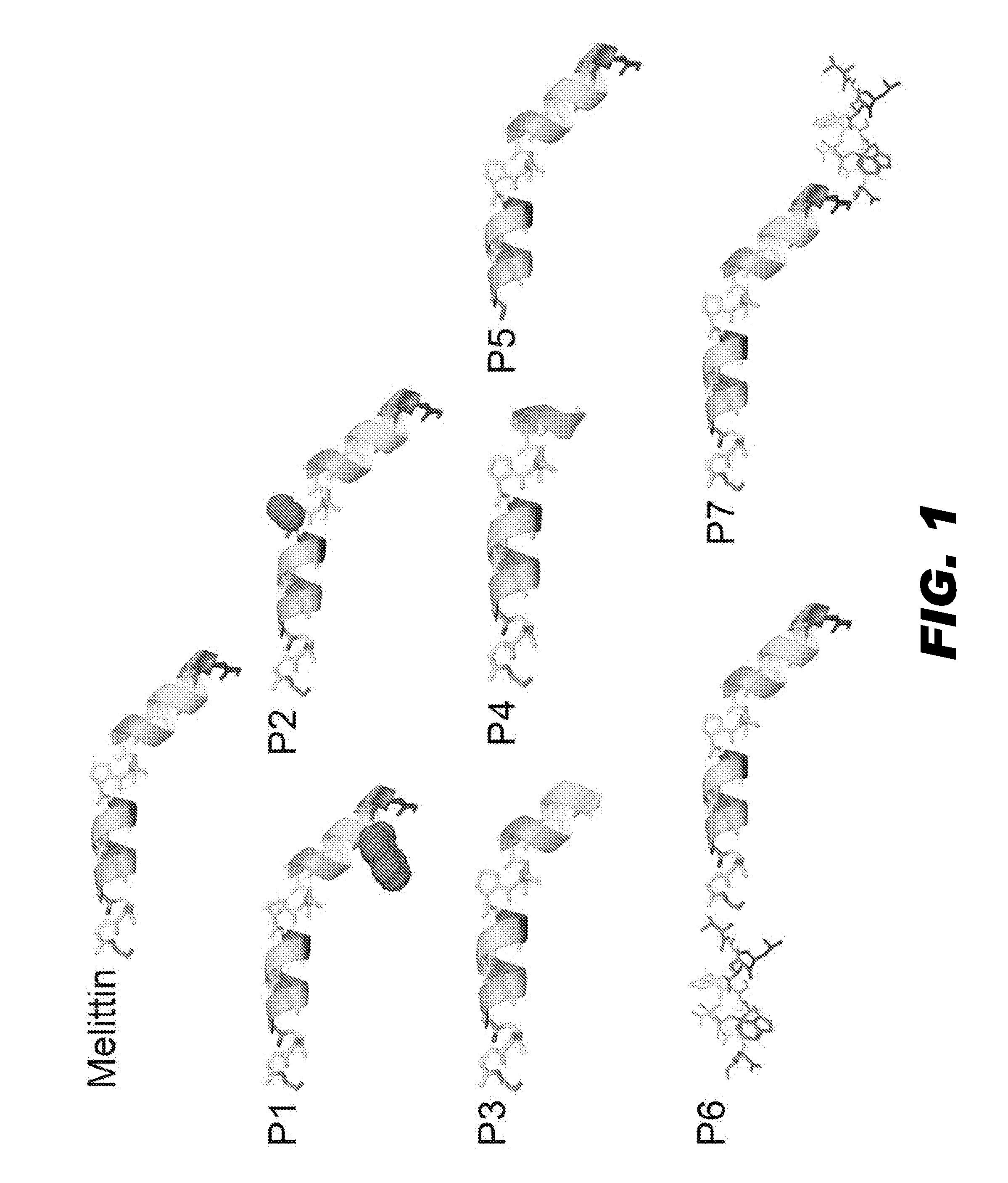
FIG. 1 depicts structures of melittin (2mlt) and seven derivatives of melittin (P1 to P7). PDB file of 2mlt was downloaded from protein data bank, and B chain of the 2mlt was used for presenting the structures of melittin and the seven derivatives by PyMOL. The structures are presented with the C-terminal on the left and the N-terminal on the right. P1 had the 5th amino acid valine substituted by tryptophan, which is depicted in a space filling view. P2 had the 14th amino acid proline substituted by alanine, which is also shown in a space filling model. P3 had the first 4 N-terminal amino acids deleted. P4 had first 7 N-terminal amino acids deleted. P5 had the last 4 C-terminal amino acids deleted. P6 and P7 had another small peptide added to the C- and N-terminal of melittin, respectively.
FIG. 2 presents a graph showing the cell lytic actions of native melittin and the melittin derivatives, as defined in FIG. 1. Plotted is the percent of live cells as a function of peptide concentration.
FIG. 3 depicts a series of graphs showing the binding kinetics of linker peptide 1 and linker peptide 2 on perfluorocarbon and oil-based nanoparticles immobilized on Biacore Sensor chip L1. Selected sensorgrams are shown at the indicated concentrations. (A, B) Sensorgrams of linker peptide 1 on perfluorocarbon and oil-based nanoparticles. (C, D) Sensorgrams of linker peptide 2 on perfluorocarbon and oil-based nanoparticles.
FIG. 4 illustrates dissociation of loaded linker peptide 1 from PFOB nanoparticles at different time points after the loading.
FIG. 5 shows that insertion of the linker peptide 1 into PFOB nanoparticles forms stable peptide-nanoparticle complexes. (A) Presents a plot of the average hydrodynamic diameters and the zeta potentials of PFOB nanoparticles with or without insertion of linker peptide 1. (B) Presents a transmission electron microscopy image of linker peptide 1 inserted PFOB nanoparticles in which the lipid membrane of the PFOB nanoparticles appears intact.
FIG. 6 illustrates that insertion of linker peptide 1 into the liposomes forms stable peptide-liposome complexes. (A) Presents a plot of the average hydrodynamic diameters and the zeta potentials of liposomes with or without insertion of linker peptide 1. (B) Presents a transmission electron microscopy image of linker peptide 1 inserted liposomes in which the lipid membrane of the liposomes appears intact.
FIG. 7 presents a micrograph showing the cellular distributions of FITC labeled linker peptide 1-associated PFOB nanoparticles in C-32 melanoma cells as visualized by confocal microscopy. (A) Cells exposed to FITC labeled linker peptide 1-associated PFOB nanoparticles. (B) Cells exposed to plain PFOB nanoparticles.
FIG. 8 depicts the binding kinetic of two VCAM-targeting peptides on PFOB nanoparticles immobilized on Biacore Sensor chip L1. (A) The sequence and structure of the VCAM-targeting peptide (TCP1), which is an anti-VCAM peptide fused on the C-terminal of linker peptide 1 with two glycines as a spacer. (B) The sequence and structure of the VCAM-targeting peptide (TCP2), which is an anti-VCAM peptide fused on the N-terminal of linker peptide 1 with two glycines as a spacer. (C) Sensorgram of the VCAM-targeting peptide (TCP1) at the indicated concentrations. (D) Sensorgram of the VCAM-targeting peptide (TCP2) at the indicated concentrations.
FIG. 9 depicts dissociation of loaded TCP1 (A) or TCP2 (B) from PFOB nanoparticles at different time points after the loading.
FIG. 10 illustrates that insertion of TCP1 or TCP2 into PFOB nanoparticles forms stable peptide-nanoparticle complexes. The average hydrodynamic diameters and zeta potentials of PFOB nanoparticles with TCP1, TCP2, or no peptide are shown.
FIG. 11 presents micrographs showing the cellular distribution of Alexa Fluor 488 labeled nanoparticles with VCAM-targeting peptide TCP1 (A), Alexa Fluor 488 labeled nanoparticles with VCAM-targeting peptide TCP2 (B), and nanoparticles without the VCAM-targeting peptide (C) in mouse endothelial cells (2F2B cells).
FIG. 12 depicts functional importance of specific linker-cargo conjugation site. (A-C) Representative .sup.19F MR spectra of 2F2B endothelial cells treated with VTNP1, VTNP2, or non-targeted PFC nanoparticles, respectively. (D) Quantification of specific delivery of VTNP1, VTNP2, and non-targeted nanoparticles (NP) to 2F2B cells calculated from .sup.19F MR spectra. VTNP1 or VTNP2 were generated with addition of 100 nmol of TCP1 or TCP2 on 20 .mu.l CE nanoparticles, respectively. Data are mean.+-.STD (n=6). (E) Dose-dependent comparison between VTNP1 and VTNP2 at selected peptide loadings into 20 .mu.l CE nanoparticles. X-axis represented the amount peptide; Y-axis represented the number of nanoparticles delivered to one 2F2B endothelial cell. A log scale was used. (F) Fluorine magnetic resonance image overlayed onto proton images of cell pellets in test tubes, acquired from cells treated with VTNP2, NP, or VTNP1, from top to bottom. The .sup.19F signal (bright pellet) was apparent only in the cells treated with VTNP1.
FIG. 13 illustrates that treatment with NBD loaded PFOB nanoparticles partially inhibit NF-kB activation. Plotted is the amount of p65 translocated into the nucleus without TNF-.alpha. stimulation (Non-Sti), after TNF-.alpha. stimulation for 4 hours (Sti), and after 1 hour pretreatment with VCAM-1 targeted NBD-loaded PFOB nanoparticles followed by TNF-.alpha. stimulation (NBD-NP-Sti).
FIG. 14 depicts that treatment with NBD loaded PFOB nanoparticles decreases the expression of NF-kB dependent gene. (A) Plotted is the amount of IL-6 expressed without TNF-.alpha. stimulation (Non-Sti), after TNF-.alpha. stimulation for 4 hours (Sti), and after 1 hour pretreatment with VCAM-1 targeted NBD-loaded PFOB nanoparticles followed by TNF-.alpha. stimulation (NBD-NP-Sti). (B) Shown are micrographs of VCAM-1 expression on the cell membrane in the absence of TNF-.alpha. stimulation (Non-Sti), after TNF-.alpha. stimulation for 4 hours (Sti), and after 1 hour pretreatment with VCAM-1 targeted NBD-loaded PFOB nanoparticles followed by TNF-.alpha. stimulation (NBD-NP-Sti). VCAM-1 expression was detected with a FITC conjugated secondary antibody. Nuclei were stained with DAPI.
FIG. 15 shows that the linker peptide 1 itself can be used to label cells. FITC-conjugated linker peptide 1 was incubated with human red blood cells for 1 hour, and a fluorescence image (A) and a bright field image (B) were taken after washing. The scale bars represent 100 .mu.m.
FIG. 16 depicts the characterization of NF-KB inhibiting PFC nanoparticles generated with the use of a linker peptide. (A) Sequence of the NBD peptide conjugated on the N-terminal of the linker peptide (Italic) with two Glycines (SEQ ID NO:225). (B) Mean hydrodynamic diameter and zeta potential of nanoparticles with or without incorporation of NBD-linker, respectively. (C) Transmission electron micrograph of PFC nanoparticles incorporated with NBD-Linker. Scale bar represents 250 nm. (D) A schematic illustration of PFC nanoparticle with enlarged NBD-Linkers incorporated in the lipid monolayer.
FIG. 17 depicts a visualization of NBD-Linker incorporation into the lipid membrane. A-C. Confocal microscope images show NBD-Linker incorporated onto the lipid membrane of Giant Unilamellar Vesicles (GUV). A. Confocal image of GUV with membrane labeled with lipophilic dye DiD (red rings). B. Confocal image of Alexa Fluor 488 labeled NBD-Linker (green rings). C. Co-localization of Alexa Fluor 488 labeled NBD-Linker and the lipid membrane of GUV (yellow rings). D. Schematic of the FCS observation volume formed by the focused laser beam (.about.1 femtoliter). E. Normalized auto-correlation curves for Alexa Fluor 488, Alexa Fluor 488 labeled NBD-Linker (NBD-Linker-488), nanoparticles incorporated with labeled peptide (NP-NBD-Linker-488). F. Diffusion time of Alexa Fluor 488, Alexa Fluor 488 labeled NBD-Linker (NBD-Linker-488), nanoparticles incorporated with labeled peptide (NP-NBD-Linker-488), and nanoparticles formulated with Alexa Fluor 488 conjugated lipids. Data presented as mean.+-.STD (n=3).
FIG. 18 depicts a mechanism of NBD-Linker incorporation into the lipid membrane of PFC nanoparticles. A. Incorporation of NBD-Linker into the PFC nanoparticles. Sensorgram, acquired by BIAcore X100, depicts the kinetics of the NBD-Linker incorporation into the PFC nanoparticles, which are immobilized on the surface of a L1 sensor chip. The NBD-Linker concentrations were 0.8, 1, 2, 5, and 10 .mu.M. B. Stable incorporation of NBD-Linker incorporation onto PFC nanoparticles at various loading concentrations. C. Secondary structural change of NBD-Linker after lipid insertion, which was measured by circular dichroism spectroscopy. Free NBD-Linker (light grey) presented unordered structure; while lipid bounded NBD-Linker (black) adopted .alpha.-helical structure. D. Relative location of Tryptophans of NBD-Linker in PFC nanoparticles. Fluorescence emission spectra demonstrating both quenching and blue shift of endogenous tryptophan fluorescence of NBD-Linker. NBD-Linker concentration was 40 .mu.M. The lipid:peptide ratio was 10.
FIG. 19 depicts the inhibition of NF-kB signaling pathway by NBD-Linker incorporated PFC nanoparticles. A. NBD-Linker incorporated PFC nanoparticles inhibit NF-kB protein (P65) translocation into the nucleus in a dose-dependent fashion. Data presented as mean.+-.s.d. (n=3). B. At a concentration of 30 .mu.M, NBD-Linker loaded nanoparticles reduce P65 nuclear translocation by half, but mutNBD-Linker loaded nanoparticles do not inhibit P65 nuclear translocation. C. Expression of NF-kB dependent gene (ICAM-1) was inhibited by NBD-Linker incorporated PFC nanoparticle treatment. The histograms were from one of six sets of independent experiments. Black and grey curves represent ICAM-1 expression with and without treatment for 9.5 hours, respectively. D. Bar graph of mean fluorescence intensity from ICAM 1 stained F8 cells without treatment and with treatment of either NBD-Linker or mutNBD-Linker nanoparticles at concentration of 30 .mu.M. ICAM-1 expression is not significantly affected by mutNBD-Linker nanoparticles. E-F. NBD-Linker incorporated PFC nanoparticles do not affect Akt signaling, an signaling pathway upstream of NF-KB. Total Akt (tAkt) (E) and phosphorylated Akt (pAkt) (F) levels do not differ significantly between F8 cells without or with treatment at selected concentrations. Data presented as mean.+-.STD (n=6)
FIG. 20 depicts a schematic of relative location of NBD-Linker incorporated into lipid membrane of PFC nanoparticles. Top: NBD-Linker in .alpha.-helical structure. Three tryptophans are highlighted with purple spheres and pointed by arrows. Bottom: NBD-Linker incorporated into the lipid monolayer of PFOB nanoparticle. Two tryptophans, (white arrows), are close to the PFOB core. The fluorescence of these two trypophans was quenched by PFOB; while the third Tryptophan in the lipid membrane was away from the PFOB core, which contributed to the blue shift of the tryptophan emission spectra.
DETAILED DESCRIPTION OF THE INVENTION
The present invention provides an anchor peptide that may be utilized in several applications to link a variety of cargo complexes to a nanoparticle and facilitate the delivery of the cargo complex to a cell. In particular, the anchor peptide is a cationic, amphipathic alpha helical peptide that is capable of stably inserting into lipid membranes. Furthermore, the anchor peptide is typically functionalized so that it may bind a variety of cargo complexes. Suitable cargo complexes may include imaging, therapeutic, cytotoxic, or targeting complexes. Accordingly, the anchor peptide, which may be linked to a cargo complex, is capable of stably inserting into the lipid membrane of a nanoparticle. Upon delivery of the nanoparticle to a cell, the anchor peptide-cargo complex is capable of dissociating from the lipid membrane of the nanoparticle and stably associating with the lipid membrane of the cell, thereby delivering the cargo complex to the cell. In some embodiments, the anchor peptide may stably insert into the lipid bilayer of the cell, such that the associated cargo complex may be displayed on the surface of the cell. In other embodiments, the anchor peptide may penetrate the lipid bilayer of the cell, thereby delivering the associated cargo complex to the interior of the cell.
Accordingly, the present invention provides a nanoparticle comprising a stably inserted anchor peptide, a kit for preparing a nanoparticle comprising a stably inserted anchor peptide, and methods for adding cargo complexes to the nanoparticles.
(I) Nanoparticles Comprising a Universal Anchor Peptide
(a) Anchor Peptide
One aspect of the present invention encompasses an anchor peptide that is capable of stably inserting into a lipid membrane. The lipid membrane may be a monolayer, a bilayer, or a multilaminar bilayer. Accordingly, the lipid membrane may be part of a nanoparticle, a cell, or a liposome.
In general, the anchor peptide comprises at least one hydrophobic segment. More specifically, the hydrophobic segment of the anchor peptide comprises at least one amphipathic alpha helix. In some embodiments, the anchor peptide may comprise two amphipathic alpha helices. In other embodiments, the anchor peptide may comprise more than two alpha helices. The anchor peptide may also contain at least one hydrophilic segment. In some embodiments, the hydrophilic segment is positively charged. That is, the hydrophilic segment comprises a majority of positively charged amino acids (i.e., Arg, Lys, or His). Preferably, the overall charge of the anchor peptide is cationic. Thus, an exemplary anchor peptide comprises a cationic amphipathic alpha helical peptide. Accordingly, the amphipathic alpha helical segment stably inserts into a lipid membrane and the hydrophilic cationic segment remains on the surface of the lipid membrane.
As used herein, the terms "stably inserts" or "stably inserted" indicate that the hydrophobic segment of the anchor peptide integrates into the midst of a lipid monolayer or bilayer membrane. Stated another way, the amphipathic alpha helical segment of the anchor peptide interdigitates with the lipid membrane. The interactions between the hydrophobic amino acids of the anchor peptide and the lipid membrane are non-covalent hydrophobic interactions and/or van der Waals interactions. In embodiments in which the overall charge of the anchor peptide is positive, the stable integration of the anchor peptide into a lipid membrane may be assessed by changes in the zeta potential of the membrane. Specifically, the zeta potential shifts to a more positive value upon stable insertion of the positively charged anchor peptide into the lipid membrane.
In general, the anchor peptide has a high association affinity for a lipid membrane and a small dissociation constant, which allows it to rapidly and stably insert into a lipid membrane. In general, the association rate is typically greater than about 9.0.times.10.sup.5 M.sup.-1 s.sup.-1. In some embodiments, the association rate may be greater than about 1.0.times.10.sup.6, 2.0.times.10.sup.6, 3.0.times.10.sup.6, 4.0.times.10.sup.6, 5.0.times.10.sup.6, 6.0.times.10.sup.6, 7.0.times.10.sup.6, 8.0.times.10.sup.6, 9.0.times.10.sup.6, or 1.0.times.10.sup.7 M.sup.-1 s.sup.-1. In an exemplary embodiment, the association rate may be greater than about 4.0.times.10.sup.6 M.sup.-1 s.sup.-1. Methods of calculating the association rate are known in the art, and may, for instance, be calculated using the Biacore system.
The dissociation constant typically is less than about 1.0.times.10.sup.-6 M. In some embodiments, the dissociation constant may be between about 1.0.times.10.sup.-6 and 1.0.times.10.sup.-7 M. In other embodiments, the dissociation constant may be between about 1.0.times.10.sup.-7 and 1.0.times.10.sup.-8 M. In still other embodiments, the dissociation constant may be between about 1.0.times.10.sup.-8 and 1.0.times.10.sup.-9 M. In yet other embodiments, the dissociation constant may be less than 9.9.times.10.sup.-10 M. Methods of calculating the dissociation rate are known in the art, and may, for instance, be calculated using the Biacore system, as detailed in Example 2.
Furthermore, the anchor peptide is substantially non-lytic and non-cytotoxic to cells. Although the anchor peptide is substantially non-lytic and non-cytotoxic, a cargo complex conjugated to the anchor peptide may be lytic or cytotoxic (as detailed below). The term "non-lytic" means that the lipid bilayer of the cell typically is not compromised upon contact with the anchor peptide. The integrity of the lipid bilayer may be assessed by the improper entry or exit of cellular or extracellular components into a cell. For example, cellular proteins and/or organelles may leak out of a cell with a compromised lipid bilayer. Alternatively, extracellular components (i.e., those that normally do not enter via gap junctions, for example) may enter a cell with a compromised lipid bilayer. It should be noted, however, that the anchor peptide may penetrate the lipid bilayer of a cell and enter the interior of the cell, but in doing so the integrity of the lipid bilayer is not affected. The term "non-cytotoxic" indicates that the cell typically is not killed upon contact with the anchor peptide. For example, in one embodiment, more than 95% of the cells are viable after contact with the anchor peptide for at least 3 hours. More preferably, more than 99% of the cells are viable after contact with the anchor peptide for at least 3 hours. In embodiments in which the anchor peptide is a derivative or mutant of melittin (see below), the cytotoxicity index or IC50 of the anchor peptide will generally be at least about 50-fold higher than that of melittin.
As stated above, the anchor peptide comprises at least one hydrophobic segment and preferably at least one hydrophilic segment. The hydrophobic segment comprises mainly non-polar amino acids, such as Phe (F), Met (M), Ala (A), Gly (G), Ile (I), Leu (L), Val (V), Pro (P), Cys (C), or Trp (W). In some embodiments about 70%, 75%, 80%, 85%, 90%, 95%, or 100% of the amino acids of a hydrophobic segment of an anchor peptide are hydrophobic as defined herein. The optional hydrophilic segment of the anchor peptide comprises primarily polar amino acids, such as, Tyr (Y), Ser (S), Thr (T), Lys (K), His (H), Gln (Q), Glu (E), Arg (R), Asp (D), or Asn (N). In some embodiments about 70%, 75%, 80%, 85%, 90%, 95%, or 100% of the amino acids of a hydrophilic segment of an anchor peptide are hydrophilic as defined herein.
The length of the anchor peptide can and will vary depending upon the intended use of the anchor peptide and/or the intended cargo complex to be linked to the anchor peptide. The anchor peptide (i.e., in the absence of any conjugated cargo) may range from about 10 amino acids to about 50 amino acids in length. In general, at least one amphipathic alpha helical segment of the anchor peptide is long enough to stably interact with the lipid membrane, but short enough to avoid unwanted molecular interactions. In some embodiments, the amphipathic alpha helical segment may be between about 5 amino acids and about 20 amino acids in length. In other embodiments, the amphipathic alpha helical segment may be between about 5 amino acids and about 18 amino acids in length. In certain embodiments, the amphipathic alpha helical segment may be between about 5 amino acids and about 13 amino acids in length. In still other embodiments, the amphipathic alpha helical segment may be between about 5 amino acids and about 10 amino acids in length. In yet other embodiments, the amphipathic alpha helical segment may be between about 5 amino acids and about 8 amino acids in length. Similarly, the optional hydrophilic segment of the anchor peptide may range from about 3 amino acids to about 10 amino acids in length. In some embodiments, however, the hydrophilic segment may represent a conjugated cargo complex. Thus, depending on the role of the hydrophobic anchoring segment, the hydrophilic segment may be a cargo peptide or protein, an antibody or antibody fragment, genomic material (i.e., DNA or RNA), or a natural or synthetic small molecule (see section (I)(c) below).
In one preferred embodiment, the at least one hydrophobic segment of the anchor peptide may comprise at least the amino acid sequence ALISWI (SEQ ID NO.1) or the amino acid sequence AWISWI (SEQ ID NO:2). In another embodiment, the hydrophobic segment may comprise SEQ ID NO.1 or SEQ ID NO:2 and additional N-terminal or C-terminal hydrophobic amino acid residues or a substantially hydrophobic amino acid sequence. The at least one hydrophilic segment of the anchor peptide may be located on the N-terminal side of the hydrophobic or amphipathic alpha helical segment, or alternatively, on the C-terminal side of the hydrophobic or amphipathic alpha helical segment. In some preferred embodiments, the hydrophilic segment may comprise at least the amino acid sequence KRKRQQ (SEQ ID NO:3) or C-terminal truncations thereof.
In other preferred embodiments, the anchor peptide may comprise the amino acid sequence X.sub.aa1ALISWIX.sub.aa2, (SEQ ID NO:4) or the amino acid sequence X.sub.aa1AWISWIX.sub.aa2 (SEQ ID NO:5), wherein X.sub.aa1 represents a hydrophobic amino acid as defined above, and X.sub.aa2 represents a hydrophilic amino acid as defined above. In another preferred embodiment, the anchor peptide may comprise the amino acid sequence X.sub.aa1GLX.sub.aa3ALISWIKRKRQQ (SEQ ID NO:6) or the amino acid sequence X.sub.aa1GLX.sub.aa3AWISWIKRKRQQ (SEQ ID NO:7), wherein X.sub.aa1 is as defined above, and X.sub.aa3 may represent an amino acid selected from the group comprising Pro (P), Ala (A), Met (M), Leu (L), Ile (I), or Trp (W). In some embodiments, X.sub.aa3 may be located on the same face of the alpha helix as other non-polar amino acids and may be a non-polar amino acid. If X.sub.aa3 is a non-polar amino acid on the same face of the alpha helix as other non-polar amino acids, X.sub.aa3 may facilitate association with a lipid membrane.
In still yet another embodiment, the anchor peptide may comprise the amino acid sequence X.sub.aa1TTGLX.sub.aa3ALISWIKRKRQQ (SEQ ID NO:8) or the amino acid sequence X.sub.aa1TTGLX.sub.aa3AWISWIKRKRQQ (SEQ ID NO:9), wherein X.sub.aa1 and X.sub.aa3 are as defined above. As above, in some embodiments X.sub.aa3 may be located on the same face of the alpha helix as other non-polar amino acids and may be a non-polar amino acid. If X.sub.aa3 is a non-polar amino acid on the same face of the alpha helix as other non-polar amino acids, X.sub.aa3 may facilitate association with a lipid membrane.
In exemplary embodiments, the anchor peptide may consist of an amino acid sequence selected from the group consisting of SEQ ID NO:10 to SEQ ID NO:218, as detailed in Table A. In an especially exemplary embodiment, the anchor peptide may consist of the amino acid sequence of SEQ ID NO:88.
TABLE-US-00001 TABLE A Exemplary Amino Acid Sequences of the Anchor Peptide SEQ Amino Acid Sequence ID NO: GIGAWLKVLTTGLPALISWIKRKRQQ 10 GIGAWLKVLTTGLPALISWIKRKRQ 11 GIGAWLKVLTTGLPALISWIKRKR 12 IGAWLKVLTTGLPALISWIKRKRQQ 13 IGAWLKVLTTGLPALISWIKRKRQ 14 IGAWLKVLTTGLPALISWIKRKR 15 GAWLKVLTTGLPALISWIKRKRQQ 16 GAWLKVLTTGLPALISWIKRKRQ 17 GAWLKVLTTGLPALISWIKRKR 18 AWLKVLTTGLPALISWIKRKRQQ 19 AWLKVLTTGLPALISWIKRKRQ 20 AWLKVLTTGLPALISWIKRKR 21 WLKVLTTGLPALISWIKRKRQQ 22 WLKVLTTGLPALISWIKRKRQ 23 WLKVLTTGLPALISWIKRKR 24 GIGAVLKVLTTGLAALISWIKRKRQQ 25 GIGAVLKVLTTGLAALISWIKRKRQ 26 GIGAVLKVLTTGLAALISWIKRKR 27 IGAVLKVLTTGLAALISWIKRKRQQ 28 IGAVLKVLTTGLAALISWIKRKRQ 29 IGAVLKVLTTGLAALISWIKRKR 30 GAVLKVLTTGLAALISWIKRKRQQ 31 GAVLKVLTTGLAALISWIKRKRQ 32 GAVLKVLTTGLAALISWIKRKR 33 AVLKVLTTGLAALISWIKRKRQQ 34 AVLKVLTTGLAALISWIKRKRQ 35 AVLKVLTTGLAALISWIKRKR 36 VLKVLTTGLAALISWIKRKRQQ 37 VLKVLTTGLAALISWIKRKRQ 38 VLKVLTTGLAALISWIKRKR 39 LKVLTTGLAALISWIKRKRQQ 40 LKVLTTGLAALISWIKRKRQ 41 LKVLTTGLAALISWIKRKR 42 KVLTTGLAALISWIKRKRQQ 43 KVLTTGLAALISWIKRKRQ 44 KVLTTGLAALISWIKRKR 45 VLTTGLAALISWIKRKRQQ 46 VLTTGLAALISWIKRKRQ 47 VLTTGLAALISWIKRKR 48 LTTGLAALISWIKRKRQQ 49 LTTGLAALISWIKRKRQ 50 LTTGLAALISWIKRKR 51 TTGLAALISWIKRKRQQ 52 TTGLAALISWIKRKRQ 53 TTGLAALISWIKRKR 54 TGLAALISWIKRKRQQ 55 TGLAALISWIKRKRQ 56 TGLAALISWIKRKR 57 GLAALISWIKRKRQQ 58 GLAALISWIKRKRQ 59 GLAALISWIKRKR 60 LAALISWIKRKRQQ 61 LAALISWIKRKRQ 62 LAALISWIKRKR 63 AALISWIKRKRQQ 64 AALISWIKRKRQ 65 AALISWIKRKR 66 ALISWIKRKRQQ 67 ALISWIKRKRQ 68 ALISWIKRKR 69 IGAVLKVLTTGLPALISWIKRKRQQ 70 IGAVLKVLTTGLPALISWIKRKRQ 71 IGAVLKVLTTGLPALISWIKRKR 72 GAVLKVLTTGLPALISWIKRKRQQ 73 GAVLKVLTTGLPALISWIKRKRQ 74 GAVLKVLTTGLPALISWIKRKR 75 AVLKVLTTGLPALISWIKRKRQQ 76 AVLKVLTTGLPALISWIKRKRQ 77 AVLKVLTTGLPALISWIKRKR 78 VLKVLTTGLPALISWIKRKRQQ 79 VLKVLTTGLPALISWIKRKRQ 80 VLKVLTTGLPALISWIKRKR 81 LKVLTTGLPALISWIKRKRQQ 82 LKVLTTGLPALISWIKRKRQ 83 LKVLTTGLPALISWIKRKR 84 KVLTTGLPALISWIKRKRQQ 85 KVLTTGLPALISWIKRKRQ 86 KVLTTGLPALISWIKRKR 87 VLTTGLPALISWIKRKRQQ 88 VLTTGLPALISWIKRKRQ 89 VLTTGLPALISWIKRKR 90 LTTGLPALISWIKRKRQQ 91 LTTGLPALISWIKRKRQ 92 LTTGLPALISWIKRKR 93 TTGLPALISWIKRKRQQ 94 TTGLPALISWIKRKRQ 95 TTGLPALISWIKRKR 96 TGLPALISWIKRKRQQ 97 TGLPALISWIKRKRQ 98 TGLPALISWIKRKR 99 GLPALISWIKRKRQQ 100 GLPALISWIKRKRQ 101 GLPALISWIKRKR 102 LPALISWIKRKRQQ 103 LPALISWIKRKRQ 104 LPALISWIKRKR 105 PALISWIKRKRQQ 106 PALISWIKRKRQ 107 PALISWIKRKR 108 GIGAVLKVLTTGLPALISWIKRKRQ 109 GIGAVLKVLTTGLPALISWIKRKR 110 GIGAVLKVLTTGLPALISWIKRK 111 GIGAVLKVLTTGLPALISWIKR 112 GIGAVLKVLTTGLPALISWIK 113 GIGAWLKVLTTGLPAWISWIKRKRQQ 114 GIGAWLKVLTTGLPAWISWIKRKRQ 115 GIGAWLKVLTTGLPAWISWIKRKR 116 IGAWLKVLTTGLPAWISWIKRKRQQ 117 IGAWLKVLTTGLPAWISWIKRKRQ 118 IGAWLKVLTTGLPAWISWIKRKR 119 GAWLKVLTTGLPAWISWIKRKRQQ 120 GAWLKVLTTGLPAWISWIKRKRQ 121 GAWLKVLTTGLPAWISWIKRKR 122 AWLKVLTTGLPAWISWIKRKRQQ 123 AWLKVLTTGLPAWISWIKRKRQ 124 AWLKVLTTGLPAWISWIKRKR 125 WLKVLTTGLPAWISWIKRKRQQ 126 WLKVLTTGLPAWISWIKRKRQ 127 WLKVLTTGLPAWISWIKRKR 128 GIGAVLKVLTTGLAAWISWIKRKRQQ 129 GIGAVLKVLTTGLAAWISWIKRKRQ 130 GIGAVLKVLTTGLAAWISWIKRKR 131
IGAVLKVLTTGLAAWISWIKRKRQQ 132 IGAVLKVLTTGLAAWISWIKRKRQ 133 IGAVLKVLTTGLAAWISWIKRKR 134 GAVLKVLTTGLAAWISWIKRKRQQ 135 GAVLKVLTTGLAAWISWIKRKRQ 136 GAVLKVLTTGLAAWISWIKRKR 137 AVLKVLTTGLAAWISWIKRKRQQ 138 AVLKVLTTGLAAWISWIKRKRQ 139 AVLKVLTTGLAAWISWIKRKR 140 VLKVLTTGLAAWISWIKRKRQQ 141 VLKVLTTGLAAWISWIKRKRQ 142 VLKVLTTGLAAWISWIKRKR 143 LKVLTTGLAAWISWIKRKRQQ 144 LKVLTTGLAAWISWIKRKRQ 145 LKVLTTGLAAWISWIKRKR 146 KVLTTGLAAWISWIKRKRQQ 147 KVLTTGLAAWISWIKRKRQ 148 KVLTTGLAAWISWIKRKR 149 VLTTGLAAWISWIKRKRQQ 150 VLTTGLAAWISWIKRKRQ 151 VLTTGLAAWISWIKRKR 152 LTTGLAAWISWIKRKRQQ 153 LTTGLAAWISWIKRKRQ 154 LTTGLAAWISWIKRKR 155 TTGLAAWISWIKRKRQQ 156 TTGLAAWISWIKRKRQ 157 TTGLAAWISWIKRKR 158 TGLAAWISWIKRKRQQ 159 TGLAAWISWIKRKRQ 160 TGLAAWISWIKRKR 161 GLAAWISWIKRKRQQ 162 GLAAWISWIKRKRQ 163 GLAAWISWIKRKR 164 LAAWISWIKRKRQQ 165 LAAWISWIKRKRQ 166 LAAWISWIKRKR 167 AAWISWIKRKRQQ 168 AAWISWIKRKRQ 169 AAWISWIKRKR 170 AWISWIKRKRQQ 171 AWISWIKRKRQ 172 AWISWIKRKR 173 IGAVLKVLTTGLPAWISWIKRKRQQ 174 IGAVLKVLTTGLPAWISWIKRKRQ 175 IGAVLKVLTTGLPAWISWIKRKR 176 GAVLKVLTTGLPAWISWIKRKRQQ 177 GAVLKVLTTGLPAWISWIKRKRQ 178 GAVLKVLTTGLPAWISWIKRKR 179 AVLKVLTTGLPAWISWIKRKRQQ 180 AVLKVLTTGLPAWISWIKRKRQ 181 AVLKVLTTGLPAWISWIKRKR 182 VLKVLTTGLPAWISWIKRKRQQ 183 VLKVLTTGLPAWISWIKRKRQ 184 VLKVLTTGLPAWISWIKRKR 185 LKVLTTGLPAWISWIKRKRQQ 186 LKVLTTGLPAWISWIKRKRQ 187 LKVLTTGLPAWISWIKRKR 188 KVLTTGLPAWISWIKRKRQQ 189 KVLTTGLPAWISWIKRKRQ 190 KVLTTGLPAWISWIKRKR 191 VLTTGLPAWISWIKRKRQQ 192 VLTTGLPAWISWIKRKRQ 193 VLTTGLPAWISWIKRKR 194 LTTGLPAWISWIKRKRQQ 195 LTTGLPAWISWIKRKRQ 196 LTTGLPAWISWIKRKR 197 TTGLPAWISWIKRKRQQ 198 TTGLPAWISWIKRKRQ 199 TTGLPAWISWIKRKR 200 TGLPAWISWIKRKRQQ 201 TGLPAWISWIKRKRQ 202 TGLPAWISWIKRKR 203 GLPAWISWIKRKRQQ 204 GLPAWISWIKRKRQ 205 GLPAWISWIKRKR 206 LPAWISWIKRKRQQ 207 LPAWISWIKRKRQ 208 LPAWISWIKRKR 209 PAWISWIKRKRQQ 210 PAWISWIKRKRQ 211 PAWISWIKRKR 212 GIGAVLKVLTTGLPAWISWIKRKRQQ 213 GIGAVLKVLTTGLPAWISWIKRKRQ 214 GIGAVLKVLTTGLPAWISWIKRKR 215 GIGAVLKVLTTGLPAWISWIKRK 216 GIGAVLKVLTTGLPAWISWIKR 217 GIGAVLKVLTTGLPAWISWIK 218
The anchor peptide of the invention may be produced using means commonly known in the art.
(b) Functionalization
As stated above, the anchor peptide may be functionalized so as to form a bond with a cargo complex. Generally speaking, the anchor peptide may be functionalized with any of a variety of active groups known in the art so as to facilitate bond formation with a cargo complex. The bond may be a covalent bond or a non-covalent bond. For instance, the bond may be a covalent bond, a hydrogen bond, an ionic bond, a bond based on van der Waals, or a hydrophobic bond. One of skill in the art would recognize that the choice of functional group can and will vary depending on the cargo complex.
By way of non-limiting example, the anchor peptide may be functionalized with an active group, such as a photo-reactive group, that when contacted with light may become activated, and capable of covalently attaching to the cargo complex. Exemplary reactive groups include, but are not limited to, reactive groups typically used in the preparation of chromatography media which include: epoxides, oxiranes, esters of N-hydroxysuccinimide, aldehydes, hydrazines, maleimides, mercaptans, amino groups, alkylhalides, isothiocyanates, carbodiimides, diazo compounds, tresyl chloride, tosyl chloride, and trichloro S-triazine. Exemplary photo-reactive groups include aryl azides, diazarenes, beta-carbonyldiazo, and benzophenones. The reactive species are nitrenes, carbenes, and radicals. These reactive species are generally capable of covalent bond formation.
In some embodiments, the anchor peptide may be functionalized with a bi-functional peptide that would connect the anchor peptide to the cargo complex. The bi-functional peptide may be homo-bi-functional or hetero-bi-functional.
Methods of functionalizing the anchor peptide are known in the art.
(c) Cargo Complex
The anchor peptide may be bound to a cargo complex. The means by which the complex is associated with the anchor peptide can and will vary depending on the embodiment. The cargo complex may be covalently or non-covalently bound to the anchor peptide, as detailed above. The cargo complex may be any molecule or agent that may be carried by or bound to the anchor peptide of a nanoparticle, and in some instances, may even be a complex of micron size. For instance, the cargo complex may be an imaging cargo, a therapeutic cargo, a cytotoxic cargo, or a targeting cargo.
(i) Imaging Cargo
Non-limiting examples of imaging cargo molecules and agents may include any molecule, agent, or material having a detectable physical or chemical property. Such imaging cargos have been well-developed in the field of fluorescent imaging, magnetic resonance imaging, positron emission tomography, Raman imaging, optical coherence tomography, photoacoustic imaging, Fourier transform infrared imaging, or immunoassays and, in general, most any label useful in such methods may be applied to the present invention. Thus, an imaging cargo may be any molecule or agent detectable by spectroscopic, photochemical, biochemical, immunochemical, electrical, optical, physical (e.g. atomic force microscopy) or chemical means. Useful imaging molecules and agents in the present invention may include visible or infrared fluorescent dyes (e.g., fluorescein isothiocyanate, AlexaFluor555, Texas red, rhodamine, and the like), radiolabels (e.g., .sup.3H, .sup.14C, .sup.35S, .sup.125I, .sup.121I, .sup.112In, .sup.99mTc), other imaging agents such as microbubbles or nanobubbles (for ultrasound imaging), .sup.18F, .sup.11C, .sup.15O, (for positron emission tomography), .sup.99mTC, .sup.111In (for single photon emission tomography), gadolinium chelate or iron (for magnetic resonance imaging), enzymes (e.g., horse radish peroxidase, alkaline phosphatase and others commonly used in an ELISA), and calorimetric labels such as colloidal gold or colored glass or plastic (e.g. polystyrene, polypropylene, latex, and the like) beads. Patents that describe the use of such imaging molecules include U.S. Pat. Nos. 3,817,837; 3,850,752; 3,939,350; 3,996,345; 4,277,437; 4,275,149; and 4,366,241, each incorporated herein by reference in their entirety. See also Handbook of Fluorescent Probes and Research Chemicals, 6th Ed., Molecular Probes, Inc., Eugene Oreg.
The anchor peptide may also be conjugated directly to signal generating compounds, e.g., by conjugation with an enzyme or fluorophore. Enzymes of interest may be hydrolases, particularly phosphatases, esterases and glycosidases, or oxidoreductases, particularly peroxidases. Fluorescent compounds may include fluorescein and its derivatives, rhodamine and its derivatives, dansyl, umbelliferone, and the like. Chemiluminescent compounds may include luciferin, and 2,3-dihydrophthalazinediones, e.g., luminol. For a review of various labeling or signal producing systems that may be used, see U.S. Pat. No. 4,391,904, incorporated herein by reference in its entirety.
(ii) Therapeutic Cargo
Non-limiting examples of therapeutic cargo may include any substance that has a biological activity, such as pharmacological agents. Such therapeutic cargo may include analgesics, antipyretics, antiasthamatics, antibiotics, antidepressants, antidiabetics, antifungal agents, antihypertensive agents, anti-inflammatories including non-steroidal and steroidal, antineoplastics, antianxiety agents, immunosuppressive agents, antimigraine agents, sedatives, hypnotics, antianginal agents, antipsychotic agents, antimanic agents, antiarrhythmics, antiarthritic agents, antigout agents, anticoagulants, thrombolytic agents, antifibrinolytic agents, hemorheologic agents, antiplatelet agents, anticonvulsants, antiparkinson agents, antihistamines, anti-restenosis agents, antipruritics, agents useful for calcium regulation, antibacterial agents, antiviral agents, antimicrobials, anti-infectives, bronchodilators, steroidal compounds and hormones, and combinations thereof. Alternatively, the cargo complex could be in the form of components of molecular complexes or pharmacologically acceptable salts.
Suitable therapeutic cargos include, without limit, immune-related agents such as immune serums, antitoxins, antivenoms bacterial vaccines, viral vaccines, rabies prophylaxis products; thyroid agents such as iodine products and anti-thyroid agents; respiratory products such as xanthine derivatives theophylline and aminophylline; antineoplastic agents such as platinum compounds (e.g., spiroplatin, cisplatin, and carboplatin), methotrexate, fluorouracil, adriamycin, mitomycin, ansamitocin, bleomycin, cytosine arabinoside, arabinosyl adenine, mercaptopolylysine, vincristine, busulfan, chlorambucil, melphalan (e.g., PAM, L-PAM or phenylalanine mustard), mercaptopurine, mitotane, procarbazine hydrochloride dactinomycin (actinomycin D), daunorubicin hydrochloride, doxorubicin hydrochloride, paclitaxel and other taxenes, rapamycin, manumycin A, TNP-470, plicamycin (mithramycin), aminoglutethimide, estramustine phosphate sodium, flutamide, leuprolide acetate, megestrol acetate, tamoxifen citrate, testolactone, trilostane, amsacrine (m-AMSA), asparaginase (L-asparaginase) Erwina asparaginase, interferon .alpha.-2a, interferon .alpha.-2b, teniposide (VM-26), vinblastine sulfate (VLB), vincristine sulfate, bleomycin sulfate, hydroxyurea, procarbazine, and dacarbazine; anti-helmintics such as pyrantel pamoate, piperazine, tetrachloroethylene, thiabendazole, and niclosamide; antimalarials such as chloroquine, amodiaquine, antifolate drugs, proguanil (chloroguanide), mefloquine, quinine, halofantrine, artemesinin and derivaties, primaquine, doxycycline, tetracycline, and clindamycin; mitotic inhibitors such as tauromustine, bofumustine, fotemustine, etoposide, colchicine, methotrexate, fluorouracil, 5-bromodeoxyuridine, 6-azacytidine, cytarabine and the vinca alkaloids, such as vincristine, paclitaxel, etoposide, nocodazole, indirubin, anthracycline derivatives, daunorubicin, daunomycin, plicamycin, and the like; hormones such as androgens, progestins, estrogens and antiestrogens, growth hormone, melanocyte stimulating hormone, estradiol, beclomethasone dipropionate, betamethasone, betamethasone acetate and betamethasone sodium phosphate, vetamethasone disodium phosphate, vetamethasone sodium phosphate, cortisone acetate, dexamethasone, dexamethasone acetate, dexamethasone sodium phosphate, flunisolide, hydrocortisone, hydrocortisone acetate, hydrocortisone cypionate, hydrocortisone sodium phosphate, hydrocortisone sodium succinate, methylprednisolone, methylprednisolone acetate, methylprednisolone sodium succinate, paramethasone acetate, prednisolone, prednisolone acetate, prednisolone sodium phosphate, prednisolone tebutate, prednisone, triamcinolone, triamcinolone acetonide, triamcinolone diacetate, triamcinolone hexacetonide, fludrocortisone acetate, oxytocin, vassopressin, glucagon and their derivatives; antiprotozoans such as chloroquine, hydroxychloroquine, metronidazole, quinine, and meglumine antimonite; antituberculars such as para-aminosalicylic acid, isoniazid, capreomycin sulfate cycloserine, ethambutol hydrochloride ethionamide, pyrazinamide, rifampin, and streptomycin sulfate; cardiovascular products such as chelating agents and mercurial diuretics and cardiac glycosides; blood products such as parenteral iron, hemin, hematoporphyrins and their derivatives; biological response modifiers such as muramyldipeptide, muramyltripeptide, microbial cell wall components, lymphokines (e.g., bacterial endotoxin such as lipopolysaccharide, macrophage activation factor), sub-units of bacteria (such as Mycobacteria, Corynebacteria), the synthetic dipeptide N-acetyl-muramyl-L-alanyl-D-isoglutamine, and the like; anti-fungal agents such as ketoconazole, nystatin, griseofulvin, flucytosine (5-fc), miconazole, amphotericin B, ricin, cyclosporins, and .beta.-lactam antibiotics (e.g., sulfazecin); vitamins such as cyanocobalamin neinoic acid, retinoids and derivatives such as retinol palmitate, and .alpha.-tocopherol; peptides such as manganese super oxide dismutase; enzymes such as alkaline phosphatase; anti-allergic agents such as amelexanox; anti-coagulation agents such as phenprocoumon, and heparin; circulatory drugs such as propranolol and other beta blockers; metabolic potentiators such as glutathione; antivirals such as acyclovir, amantadine azidothymidine (AZT, DDI, Foscarnet, or Zidovudine), ribavirin and vidarabine monohydrate (adenine arabinoside, ara-A); antianginals such as diltiazem, nifedipine, verapamil, erythritol tetranitrate, isosorbide dinitrate, nitroglycerin (glyceryl trinitrate) and pentaerythritol tetranitrate; antibiotics such as dapsone, chloramphenicol, neomycin, cefaclor, cefadroxil, cephalexin, cephradine erythromycin, clindamycin, lincomycin, amoxicillin, ampicillin, bacampicillin, carbenicillin, dicloxacillin, cyclacillin, picloxacillin, hetacillin, methicillin, nafcillin, oxacillin, penicillin including penicillin G and penicillin V, ticarcillin rifampin, aminoglycosides, and tetracycline; antiinflammatories such as diflunisal, ibuprofen, indomethacin, meclofenamate, mefenamic acid, naproxen, oxyphenbutazone, phenylbutazone, piroxicam, sulindac, tolmetin, aspirin, and salicylates; antirheumatics such as adalimumab, azathioprine, chloroquine and hydroxychloroquine (antimalarials), cyclosporine (Cyclosporin A), D-penicillamine, etanercept, gold salts (sodium aurothiomalate, auranofin), infliximab, leflunomide, methotrexate, minocycline (a tetracycline antibiotic), and sulfasalazine; narcotics such as paregoric, opiates, codeine, heroin, methadone, morphine, and opium; cardiac glycosides such as deslanoside, digitoxin, digoxin, digitalin, and digitalis; neuromuscular blockers such as atracurium mesylate, gallamine triethiodide, hexafluorenium bromide, metocurine iodide, pancuronium bromide, succinylcholine chloride (suxamethonium chloride), tubocurarine chloride, and vecuronium bromide; sedatives (hypnotics) such as amobarbital, amobarbital sodium, aprobarbital, butabarbital sodium, chloral hydrate, ethchlorvynol, ethinamate, flurazepam hydrochloride, glutethimide, methotrimeprazine hydrochloride, methyprylon, midazolam hydrochloride, paraldehyde, pentobarbital, pentobarbital sodium, phenobarbital sodium, secobarbital sodium, talbutal, temazepam, and triazolam; local anesthetics such as bupivacaine hydrochloride, chloroprocaine hydrochloride, etidocaine hydrochloride, lidocaine hydrochloride, mepivacaine hydrochloride, procaine hydrochloride, and tetracaine hydrochloride; general anesthetics such as droperidol, etomidate, fentanyl citrate with droperidol, ketamine hydrochloride, methohexital sodium, and thiopental sodium; radioactive particles or ions such as strontium, iodide rhenium, yttrium; and radiopharmaceuticals such as radioactive iodine and phosphorus products.
(iii) Cytotoxic Cargo
Cytotoxic cargo refers to a molecule or agent that is detrimental to (e.g., kills or damages) a cell. Examples may include anti-microtubule drugs such as the taxols (paclitaxel, docetaxel) and vinca alkaloids (vincristine, vinblastine). For instance, examples may include taxol, cytochalasin B, gramicidin D, ethidium bromide, emetine, mitomycin, etoposide, tenoposide, vincristine, vinblastine, colchicin, doxorubicin, daunorubicin, dihydroxy anthracin didne, mitoxantrone, mithramycin, actinomycin D, 1-dehydrotestosterone, glucocorticoids, procaine, tetracaine, lidocaine, propranolol, and puromycin and analogs or homologs thereof.
Non-limiting examples of cytotoxic cargo may also include radionuclides suitable for pharmacological administration. Such radionuclides may include .sup.123I, .sup.125I, .sup.131I, .sup.90Y, .sup.211At, .sup.67Cu, .sup.186Re, .sup.188Re, .sup.212Pb, and .sup.212Bi. Additionally, cytotoxic agents may include chemotoxic agents or toxins. Examples of chemotoxic agents may include small-molecule drugs such as methotrexate, and pyrimidine and purine analogs.
Proteins that may be used as cytotoxic agents may include ricin, abrin, diphtheria toxin, cholera toxin, gelonin, Pseudomonas exotoxin, Shigella toxin, pokeweed antiviral protein, and other toxin proteins known in the medicinal biochemistry arts.
(iv) Targeting Cargo
Targeting cargo may be any molecule or agent that directs a nanoparticle to a specific location. Non-limiting examples of targeting agents may include an antibody or antibody fragment, receptor ligand, small molecule, peptide, polypeptide, lipid, carbohydrate, nucleic acid, siRNA, shRNA, antisense RNA, dendrimer, microbubble, or aptamer. A targeting cargo may be directed to a eukaryotic target cell or a prokaryotic target cell.
(d) Nanoparticles
Generally speaking, the nanoparticle comprises a core encapsulated by a lipid layer. The lipid membrane of the nanoparticle may comprise a single lipid layer, two lipid layers (a bilayer), more than two lipid layers, a multilaminar lipid layer, and the like. As used herein, the term nanoparticle encompasses particle carriers that may be nano scale, micro scale, or macro scale. Suitable particle carriers include nanoparticles, nanospheres, nanostructures, liposomes, micelles, microbubbles, gas-filled microbubbles, dendrimers, polymeric structures, or any such carrier (naturally occurring or synthetic) that has an exterior lipid layer into which the anchor peptide may stably insert. Bacteria, viruses, prions, red blood cells, white blood cells, isolated tissue cells, platelets, and other such biologically derived, lipid-encased structures also may be carriers of the anchor peptide. Structures comprising a glycolipid membrane surrounding a cell (animal or vegetable) or particle or synthetic nanostructure also are potential hosts for the anchor peptide. In exemplary embodiments, the particle carrier may be a lipid-encapsulated nanoparticle.
In some embodiments, the anchor peptide may be bound to a cargo complex, as described above. The anchor peptide itself is generally non-lytic and non-cytotoxic, but the cargo complex may be lytic or cytotoxic. In some embodiments, a single nanoparticle may be associated with more than one type of cargo complex. For instance, a nanoparticle may be associated with both an anchor peptide bound to a targeting cargo and an anchor peptide bound to a therapeutic cargo. In one embodiment, a nanoparticle may be associated with at least 2, 3, 4, 5, 6, 7, 8, 9, or 10 different cargo complexes. In another embodiment, a nanoparticle may be associated with at least one therapeutic cargo, at least one imagining cargo, at least one cytotoxic cargo, at least one targeting cargo, or any combination thereof.
Methods of making lipid-encapsulated nanoparticles are well known in the art. For instance, see U.S. Pat. Nos. 6,676,963, 7,255,875 and 7,186,399, herein incorporated by reference in their entirety. As described below, a nanoparticle may be initially produced with an anchor peptide, or, alternatively, a nanoparticle may be pre-formed, and the anchor peptide subsequently added to the lipid membrane. Methods of adding an anchor peptide to a pre-formed nanoparticle are described in more detail below.
(e) Administration
Nanoparticles comprising an anchor peptide, which may be linked to a cargo complex, may be administered to a subject. Suitable subjects include, but are not limited to, mammals, amphibians, reptiles, birds, fish, insects, and plants. In certain embodiments, nanoparticles of the invention may be used to image a tissue from a subject. Tissue, as used herein, may refer to cells, organs, tumors, or material associated with cells, organs, or tumors, such as blood clots. Suitable tissues may include, but are not limited to, heart, lungs, brain, eye, stomach, spleen, bones, pancreas, kidneys, liver, intestines, skin, uterus, bladder, eyes, lymph nodes, blood vessels, and blood and lymph components.
The nanoparticles may be formulated and administered to a subject by several different means. For instance, nanoparticles may generally be administered parenteraly, intraperitoneally, intravascularly, topically, or intrapulmonarily in dosage unit formulations containing conventional nontoxic pharmaceutically acceptable carriers, adjuvants, and vehicles as desired. The term parenteral as used herein includes subcutaneous, intravenous, intramuscular, intrathecal, or intrasternal injection, or infusion techniques. In one embodiment, the composition may be administered in a bolus. In a preferred embodiment, the composition may be administered intravenously. Formulation of pharmaceutical compositions is discussed in, for example, Hoover, John E., Remington's Pharmaceutical Sciences, Mack Publishing Co., Easton, Pa. (1975), and Liberman, H. A. and Lachman, L., Eds., Pharmaceutical Dosage Forms, Marcel Decker, New York, N.Y. (1980).
Injectable preparations, for example, sterile injectable aqueous or oleaginous suspensions, may be formulated according to the known art using suitable dispersing or wetting agents and suspending agents. The sterile injectable preparation may also be a sterile injectable solution or suspension in a nontoxic parenterally acceptable diluent or solvent. Among the acceptable vehicles and solvents that may be employed are water, Ringer's solution, and isotonic sodium chloride solution. In addition, sterile, fixed oils are conventionally employed as a solvent or suspending medium. For this purpose, any bland fixed oil may be employed, including synthetic mono- or diglycerides. In addition, fatty acids such as oleic acid are useful in the preparation of injectables. Dimethyl acetamide, surfactants including ionic and non-ionic detergents, and polyethylene glycols can be used. Mixtures of solvents and wetting agents such as those discussed above are also useful.
For imaging purposes, formulations for parenteral administration may be in the form of aqueous or non-aqueous isotonic or hypotonic sterile injection solutions or suspensions. These solutions and suspensions may be prepared from sterile powders or granules having one or more carriers or diluents. The compounds may be dissolved in water, polyethylene glycol, propylene glycol, ethanol, corn oil, cottonseed oil, peanut oil, sesame oil, benzyl alcohol, sodium chloride, and/or various buffers. Other adjuvants and modes of administration are well and widely known in the pharmaceutical art.
One of skill in the art will recognize that the amount and concentration of the composition administered to a subject will depend in part on the subject and the reason for the administration (i.e. imaging, drug delivery, etc.). Methods for determining optimal amounts are known in the art. In one embodiment, the amount administered may be between about 0.1 cc/kg to about 5 cc/kg. In another embodiment, the amount administered may be about 0.5, 1.0, 1.5, 2.0, 2.5, 3.0, 3.5, 4.0, 4.5, or 5.0 cc/kg. In yet another embodiment, the amount may be 1 cc/kg.
Following administration, the nanoparticle comprising the anchor peptide contacts a cell. As detailed above and demonstrated in Example 5, the nanoparticle may be targeted to a particular cell by linking a specific targeting cargo to the anchor peptide of the nanoparticle. Upon contact with the cell, the anchor peptide of the nanoparticle may be transferred or move from the lipid monolayer of the nanoparticle and interact with and stably insert into the lipid bilayer of the cell. Depending upon the type of cargo complex that is linked to the anchor peptide, the anchor peptide may remain tethered to the lipid membrane of the cell. In such an embodiment, the cargo complex is on the surface of the cell via its attachment to the hydrophilic segment of the anchor peptide, whereas the hydrophobic segment of the anchor peptide is stably integrated into the lipid bilayer of the cell. In other embodiments, the anchor peptide and linked cargo complex may penetrate the lipid bilayer and enter the cell cytoplasm (as demonstrated in Examples 3 and 5, and depicted in FIG. 7 and FIG. 11). In such an embodiment, the cargo that is linked to the anchor peptide may be an imaging agent or a therapeutic agent, for example. Those of skill in the art will appreciate that a number of scenarios are possible, depending upon the properties of the anchor peptide and the conjugated cargo complex.
(II) Kits
In some embodiments, the invention encompasses a kit. The kit generally comprises a first composition comprising a nanoparticle, and a second composition comprising an anchor peptide. In exemplary embodiments, the nanoparticle of the kit may be pre-formed. By following directions provided by the kit, a user of the kit may bind the anchor peptide to a cargo complex of interest, and then mix the conjugated anchor peptide/cargo complex with the pre-formed nanoparticles, so as to incorporate the anchor peptide into the lipid membrane of the pre-formed nanoparticle. In some embodiments, the pre-formed nanoparticles may be sterile.
(III) Methods
Yet another aspect of the present invention encompasses methods for adding a cargo complex to a nanoparticle. A first method generally comprises contacting the nanoparticle with an anchor peptide that is bound to a cargo complex. The anchor peptide stably inserts into the lipid membrane of the nanoparticle, thereby linking the cargo complex with the nanoparticle. This method advantageously allows the nanoparticle to be pre-formed and then loaded with one or more cargo complexes at a later time.
In one embodiment, a nanoparticle may be contacted with an anchor peptide by combining a composition comprising a nanoparticle with a composition comprising an anchor peptide under conditions suitable for the anchor peptide to stably insert into the lipid membrane of the nanoparticle. Typically, such conditions may comprise a temperature of about 20.degree. C. to about 30.degree. C., and incubation times of between about 30 sec and 30 min. In some embodiments, a nanoparticle may be incubated with the anchor peptide for about 5, 10, 15, 20, or 25 minutes at a temperature between about 20.degree. C. and 30.degree. C. One skilled in the art will appreciate that the length and temperature of incubation can and will vary depending on the composition of the nanoparticle, the anchor peptide, and the cargo complex.
The ratio of the anchor peptide to the nanoparticle will also vary depending on the composition of the nanoparticle, the anchor peptide, and the cargo complex. Molar membrane lipid to anchor peptide ratios may range from about 1500 to about 20. In some embodiments, the ratio may range from about 1000 to about 40. In other embodiments, the ratio may be about 900, 800, 700, 600, 500, 400, 300, 200, 100, 90, 80, 70, 60, 50, 40, 30 or 20.
In one example, nanoparticles may be formulated by mixing known amounts of anchor peptides to perfluorocarbon nanoparticles. An anchor peptide may be dissolved in 100 mM KCl (pH 7, 10 mM HEPES) at 0.1 mM and 2.times.20 mL may be added to 50 ml of nanoparticle suspension with mixing. After incubation at room temperature for 10 min, the nanoparticles may be washed twice by centrifugation (100 g, 10 min) to remove unbound anchor peptide. In some embodiments in which the anchor peptide contains a tryptophan residue, the anchor peptide in the supernatant may be quantified by measuring the tryptophan fluorescence using methods known in the art.
A second method comprises adding a cargo complex to a nanoparticle comprising a stably inserted (functionalized) anchor peptide. The starting nanoparticle may already comprise at least one cargo complex that was previously conjugated with the stably inserted anchor peptide. The method comprises contacting the nanoparticle with the cargo complex, wherein the cargo complex forms a bond with the functionalized anchor peptide. The conditions under which the nanoparticle comprising the stably inserted anchor peptide is contacted with the cargo complex can and will vary depending upon, for example, the type of cargo complex and the type of functionalization of the anchor peptide. Those of skill in the art are familiar with suitable reaction conditions.
(IV) Anchor Peptides
A further aspect of the invention comprises an anchor peptide as detailed above in section (I)(a). In particular, an anchor peptide comprises a substantially non-lytic, non-cytotoxic, amphipathic alpha helical peptide that is capable of stably inserting into a lipid membrane. In general, the anchor peptide is cationic and comprises at least one amphipathic alpha helix.
The anchor peptide may be associated with nanoparticles to facilitate the delivery of cargo complexes to cells, as detailed above in section (I). Alternatively, the anchor peptide itself may associate with and insert into the cell membranes of cells. Thus, the anchor peptide itself may be used to label cells, target cells, or deliver cargos to cells. As an example, the anchor peptide may be conjugated to a fluorescent molecule, wherein the anchor peptide may stably insert into the lipid bilayer of a cell and "label" the cell (as demonstrated in Example 7 and FIG. 15). In other embodiments, the anchor peptide may be conjugated to a specific targeting agent and/or therapeutic agent, such that conjugated anchor peptide is targeted to a specific cell or a therapeutic agent is delivered to the cell. As detailed above, the anchor peptide may stably integrate into the lipid membrane of the cell such that the conjugated agent is displayed on the cell surface. Alternatively, the anchor peptide may penetrate the cell membrane and deliver the conjugated agent to the interior of the cell. In general, the nature of the anchor peptide and the conjugated agent will determine the eventual fate of the conjugated anchor peptide. The anchor peptides of the invention, therefore, may be used as research tools or as medical tools for imaging and/or therapeutics.
The following examples are included to demonstrate preferred embodiments of the invention. It should be appreciated by those of skill in the art that the techniques disclosed in the examples that follow represent techniques discovered by the inventors to function well in the practice of the invention. Those of skill in the art should, however, in light of the present disclosure, appreciate that many changes can be made in the specific embodiments that are disclosed and still obtain a like or similar result without departing from the spirit and scope of the invention, therefore all matter set forth or shown in the accompanying drawings is to be interpreted as illustrative and not in a limiting sense.
EXAMPLES
The following examples illustrate various iterations of the invention.
Example 1
Lytic Activity of Anchor Peptide
Typically, cargos such as targeting ligands, imaging agents, and/or drugs are incorporated into nanocariers during the formulation process, which requires a dedicated particle design and preformulation for each individual application. In contrast, a strategy designed to accommodate swapping and/or combining multiple cargos in generic base nanocarriers would enable flexible and personalized decision making for application to any disease. Despite the myriad regulatory hurdles to the adoption of such a paradigm, the design of flexible nanocarriers with polyvalent and rapidly swappable targeting and drug delivery cargos represents a necessary first step.
The first goal was to design an appropriate anchoring agent that could be used with generic nanoparticle carriers, and for this purpose the model peptide, melittin (MMLT), was selected. Melittin is a 26 amino acid peptide that comprises more than half of the dry weight of the venom of the honeybee Apis mellifera. Residues 1-20 of melittin form two amphipathic .alpha.-helixes with an intervening proline hinge, while residues 21-26 of melittin are highly positively charged. Both hydrophobic and hydrophilic segments of melittin are essential for its lytic activity. It was hypothesized that the structure of melittin could be modified to attenuate its lytic activity while retaining the property of stable insertion into perfluorocarbon (PFC) nanoparticles. Accordingly, the native melittin sequence was altered by selective point mutations and truncations. Table 1 presents the amino acid sequences of the various peptides and FIG. 1 presents schematics of their secondary structures.
TABLE-US-00002 TABLE 1 Melittin and Derivatives of Melittin SEQ ID Name(s) Peptide Sequence NO: MMLT GIGAVLKVLTTGLPALISWIKRKRQQ 219 P1 GIGAWLKVLTTGLPALISWIKRKRQQ 10 P2 (LP2) GIGAVLKVLTTGLAALISWIKRKRQQ 25 P3 VLKVLTTGLPALISWIKRKRQQ 79 P4 (LP1) VLTTGLPALISWIKRKRQQ 88 P5 GIGAVLKVLTTGLPALISWIKR 112 P6 GIGAVLKVLTTGLPALISWIKRKRQQTALDWSWLQTE 220 P7 TALDWSWLQTEGIGAVLKVLTTGLPALISWIKRKRQQ 221
The peptides were synthesized automatically by Fmoc solid-phase peptide synthesis. The product identities were assessed by mass spectrometry (Washington University Proteomics Center, St. Louis, Mo.) and the purity (>99%) was determined by analytical HPLC. All the peptides contained N-terminal acetylation and C-terminal amidation to mimic the situation of covalent cargo attachment that would apply when these peptides would be used as a linker on the nanocarriers.
For each peptide, cytotoxicity was assessed in 2F2B mouse endothelial cells by the XTT cell viability assay (Biotium, Hayward, Calif.). Cells were seeded in 96-well plates at 10,000 cells/well 24 hours before treatment with melittin and mutated melittins at selected concentrations. After 3 hours of treatment at 37.degree. C., 25 .mu.l of activated XTT solution was added to each well and incubated at 37.degree. C. for 5 hours. After 5 hours of incubation, plates were gently shaken to achieve even dye distribution. The absorbances of the samples were measured by a microplate reader (Biorad, Model 550, Hercules, Calif.) at a wavelength of 450 nm. Background was subtracted and the results were normalized to untreated control cells.
FIG. 2 presents the cytotoxicity results. Among the modified peptides, P2 and P4 exhibited the least cytotoxicity. In particular, P4 had essentially no cell toxicity up to a concentration of 90 .mu.M. Table 2 presents the IC50 (.mu.M) values of some of the tested peptides. The cytotoxicity index of P4 (114.4.+-.10.2) was reduced most compared to that of MMLT (2.36.+-.0.23), representing about a 50-fold decrease in cytotoxicity. From this point forward, P4 is referred to as linker peptide 1 (LP1) and P2 is referred to as linker peptide 2 (LP2).
TABLE-US-00003 TABLE 2 Cytotoxicity of Peptides IC50 Peptide (.mu.M) MMLT 2.36 .+-. 0.23 P1 0.75 .+-. 0.04 P2 26.56 .+-. 0.56 P3 14.44 .+-. 0.92 P4 114.6 .+-. 10.2 P5 6.41 .+-. 0.64
Example 2
Anchor Peptide Affinity for Lipid Membranes
In order to evaluate whether linker peptide 1 (LP1) and linker peptide 2 (LP2) still retained high affinity on a lipid monolayer of the nanoparticles, surface plasmon resonance (SPR) studies were performed by using Biacore X 100 (Biacore Inc, Piscataway, N.J.). The binding kinetics and affinity of LP1 and LP2 on two type of nanoparticles were studied. One type was perfluorocarbon nanoparticles (e.g., perfluorooctylbromide, PFOB), the other type was oil-based (safflower core) nanoparticles. PBS (Ca.sup.2+ and Mg.sup.2+ free) was used as the running buffer. Nanoparticles were immobilized on Biacore sensor chip L1 by injecting nanoparticles at a flow rate of 1 .mu.l/min for 30 minutes, followed by a running buffer wash at a flow rate of 100 .mu.l/min for 50 seconds. Peptides in selected concentrations, prepared in running buffer, were then injected at a flow rate of 30 .mu.l/min for 1 minute. At the end of each experiment, the sensor chip surface was regenerated by injection of 3-[(3-cholamidopropyl)dimethylammonio]-1-propanesulfonate (CHAPS) at a flow rate of 100 .mu.l/min for 24 seconds.
Selected sensorgrams are shown in FIG. 3. The dissociation constants of LP1 and LP2 on perfluorocarbon nanoparticles were 2.25.times.10.sup.-7 M and 5.48.times.10.sup.-9 M, respectively. The dissociation constants of linker peptides 1 and 2 on oil-based nanoparticles were 2.29.times.10.sup.-8 and 6.4.times.10.sup.-10 M, respectively. These results indicated that both anchor peptides retained high affinity with both perfluorocarbon and oil-based nanoparticles. These results compare with a previously measured dissociation constant for melittin on perfluorocarbon nanoparticles of 1.5.times.10.sup.-9 M.
The dissociation of the peptide from linker peptide 1-loaded PFOB nanoparticles is depicted in FIG. 4. After being loaded with the linker peptide 1, the loaded PFOB nanoparticles were stored at 4.degree. C. for 30 minutes, 1 hour, 2 hours, 4 hours, 22 hours, 46 hours, and 70 hours. The loaded nanoparticles were centrifuged at 100 g for 10 minutes, and the dissociated linker peptide 1 was measured by using tryptophan fluorescence after excitation at 280 nm in a fluorescent spectrofluorometer (Varian Inc, Palo Alto, Calif.). The results showed that less 10% of the loaded linker peptide 1 was dissociated from the PFOB nanoparticles for up 70 hours after being loaded. These data suggest that the association between linker peptide 1 and the PFOB nanoparticles was stable.
The size distributions of the nanoparticles with or without the linker peptide were analyzed by dynamic light scattering (Brookhaven Instruments Corp., Holtsville, N.Y.). Zeta potential (.zeta.) values were determined with a Brookhaven Instruments PALS Zeta Potential Analyzer (Brookhaven Instruments Corp., Holtsville, N.Y.). Data were acquired in the phase-analysis light scattering (PALS) mode following solution equilibration at 25.degree. C. FIG. 5A shows that the average hydrodynamic diameter of the PFOB nanoparticles was not affected by linker peptide 1 insertion, and after incorporating the positively charged linker peptide 1, the PFOB nanoparticles exhibited a zeta potential shift from -17.62 mV to +23.33 mV. This change further confirms the insertion of the linker peptide 1 into the PFOB nanoparticles. Furthermore, the structural integrity of linker peptide 1 inserted PFOB nanoparticles was confirmed by transmission electron microscopy. As shown in FIG. 5B, the lipid membrane of the PFOB nanoparticles appears to remain intact.
The interactions between linker peptide 1 and liposomes are depicted in FIG. 6. FIG. 6A shows that the average hydrodynamic diameter of the liposomes was not affected by the insertion of linker peptide 1, and after incorporating the positively charged linker peptide 1, the liposomes exhibited a zeta potential shift from -36.73 mV to -29.843 mV. This change further confirms that the linker peptide 1 inserted into the liposomes. Furthermore, transmission electron microscopy of linker peptide 1-loaded liposomes revealed that the lipid membrane of the liposomes appeared intact (FIG. 6B).
Example 3
Cellular Delivery of an Anchor Peptide
The cellular delivery of FITC-labeled peptide 1, incorporated onto perfluorocarbon nanoparticles, was visualized in C-32 melanoma cells by confocal microscopy. PFOB nanoparticles incorporated with FITC labeled linker peptide 1 or plain PFOB nanoparticles were incubated with C-32 melanoma cells in Delta T Culture dish (0.17 mm, Biotech's Inc., Butler, Pa.) at 37.degree. C. for 1 hour, followed by 5 PBS washes. The cells were fixed by incubating in 1 ml of 4% paraformaldehyde at 37.degree. C. for 5 minutes. After the fixation, cells were washed with PBS for 5 times and visualized by using a Zeiss 510 confocal microscope.
As shown in FIG. 7A, the FITC signal was detected both on the cell surface and in the cytoplasm of cells incubated with the linker-loaded nanoparticles. In contrast, cells exposed to plain PFOB nanoparticles displayed no FITC signal (FIG. 7B). These results demonstrate that linker peptide 1 retained the sufficient stability on the lipid monolayer of the nanoparticles for cellular delivery.
Example 4
VCAM-Targeting Anchor Peptide Affinity for Lipid Membranes
To evaluate the function of a cargo complex, two VCAM-targeting peptides were synthesized. For this, an anti-VCAM peptide, VHPKQHR (SEQ ID NO:222), was fused on the N-terminal side or C-terminal side of linker peptide 1 with two glycines as a spacer, as depicted in Table 3.
TABLE-US-00004 TABLE 3 VCAM-targeting peptides SEQ ID Name Sequence NO: TCP1 VLTTGLPALISWIKRggVHPKQHR 223 TCP2 VHPKQHRggVLTTGLPALISWIKR 224
The binding kinetics of the resulting VCAM-targeting peptides (i.e., TCP1 and TCP2) on perfluorocarbon nanoparticles were examined using Biacore X 100. PBS (Ca.sup.2+ and Mg.sup.2+ free) was used as the running buffer. Nanoparticles were immobilized on Biacore sensor chip L1 by injecting nanoparticles at a flow rate of 1 .mu.l/min for 30 minutes, followed by a running buffer wash at a flow rate of 100 .mu.l/min for 50 seconds. Targeting peptide prepared at selected concentrations in running buffer was sequentially injected at a flow rate of 30 .mu.l/min for 1 minute. At the end of each experiment, the sensor chip surface was regenerated by injection of 3-[(3-cholamidopropyl)dimethylammonio]-1-propanesulfonate (CHAPS) at a flow rate of 100 .mu.l/min for 24 seconds. FIG. 8 presents selected sensorgrams at several different concentrations of the two VCAM-targeting peptides. The dissociation constant of TCP1 and TCP2 were 0.13 .mu.M and 0.04 .mu.M, respectively. These data suggest that the insertion of linker peptide 1 into PFC nanoparticles was not affected by addition of a targeting ligand cargo.
FIG. 9 presents the dissociation of TCP1 or TCP2 from loaded PFOB nanoparticles as a function of time after loading. After loaded with TCP1 or TCP2, the PFOB nanoparticles were stored at 4.degree. C. for 30 minutes, 1 hour, 2 hours, 4 hours, 22 hours, 46 hours, and 70 hours. The stored nanoparticles were then centrifuged at 100 g for 10 minutes and dissociated TCP1 or TCP2 was measured using tryptophan fluorescence after excitation at 280 nm in a fluorescent spectrofluorometer (Varian Inc, Palo Alto, Calif.). The results showed that up to 70 hours after being loaded into the PFOB nanoparticles, less 15% of the loaded TCP1 or TCP2 had dissociated. Furthermore, these data suggest that targeting peptide-loaded PFOB nanoparticles are stable.
The average hydrodynamic diameter of the PFOB nanoparticles was not affected by the insertion of TCP1 and TCP2 (see FIG. 10). Additionally, after incorporation of the positively charged TCP1 or TCP2, the PFOB nanoparticles exhibited a zeta potential shift from -17.62 mV to 13.47 mV or 12.43 mV, respectively. These data further confirm the stable insertion of TCP1 and TCP2 into the PFOB nanoparticles.
Example 5
Specific Targeting Nanoparticles
The specific binding of VCAM targeted nanoparticles was evaluated by confocal microscopy in 2F2B cells, which express VCAM-1 on the cell surface after TNF-.alpha. stimulation. Alexa Fluor 488 labeled PFOB nanoparticles comprising the VCAM-targeting peptide (TCP1 or TCP2) or nanoparticles themselves were incubated with TNF-.alpha. stimulated 2F2B cells in Delta T Culture dish at 37.degree. C. for 1 hour, followed by five PBS washes. Cells were then fixed by incubation in 1 ml of 4% paraformaldehyde at 37.degree. C. for 5 minutes. After fixation, the cells were washed with PBS for 5 times and the cells were visualized using a confocal microscope (Zeiss Meta 510, Thornwood, N.Y.) with standard filter sets. Concomitant differential interference contrast (DIC) imaging was used to determine the location of nanoparticles in the cells.
Manifestly stronger fluorescent signals emanated from the cells treated with VCAM-targeted nanoparticles (FIGS. 11A and 11B) than from cells treated with nontargeted nanoparticles (FIG. 11C), which demonstrates the specific binding of targeted nanoparticles to cellular VCAM-1. It is also clear that the exposure of VCAM-1 targeting peptides fused on the C-terminus of the linker (i.e., TCP1) exhibited better binding (FIG. 11A) than when VCAM-1 targeting peptide is attached to the N-terminus (i.e., TCP2) of the linker (FIG. 11B). These findings conform to the known orientation of melittin in membranes where the N-terminus situates more deeply into the lipid membrane and the C-terminus remains exposed on the surface.
Two formulations of VCAM-1-Targeted NanoParticles (VTNP) were prepared by using the anchor peptide strategy. The first formulation of VCAM-1-Targeted NanoParticles (VTNP1) was produced by mixing pre-formulated PFC nanoparticles with TCP1. The second formulation (VTNP2) was produced by mixing pre-formulated PFC nanoparticles with TCP2. All nanoparticles had a perfluoro-15-crown 5-ether (CE) core. VTNP1 or VTNP2 were generated by addition of 100 nmol of TCP1 or TCP2 on 20 .mu.l CE nanoparticles, respectively.
The .sup.19F perfluorocarbon core of PFC nanoparticles enabled these nanoparticles to serve as unique magnetic resonance spectroscopic and imaging agents with no background signal in vivo. Furthermore, the .sup.19F spectroscopic signature offered the potential for quantification of VCAM-1 biomarkers by providing a rough estimate of the number of delivered nanoparticles per cell. Using this approach, .sup.19F MR spectra of TNF-.alpha. stimulated 2F2B endothelial cells treated with VTNP1 (FIG. 12A), VTNP2 (FIG. 12B), and non-targeted (FIG. 12C) CE nanoparticles were acquired. Because of linear relation between .sup.19F signal intensity and the concentration of .sup.19F molecules, the amount of nanoparticles delivered to the cell could be estimated. The number of bound nanoparticles was normalized by the total number of measured cells to calculate the number of the bound nanoparticles per cell (FIG. 12D). These data further support the specific binding of targeted nanoparticles, and also confirmed that VTNP1 manifests better targeting efficiency than does VTNP2. Essentially then, the VTNP2 construct serves as a control scrambled peptide against the more specific VTNP1 sequence.
The dose dependency of targeting for VTNP1 and VTNP2 in selected concentrations was also evaluated in vitro. FIG. 12E again confirms the better targeting efficiency of VTNP1, while also demonstrating that the binding of VCAM-targeted nanoparticles was dependent upon the amount of targeting peptide incorporated onto the nanoparticle. Therefore, optimization of specific targeting might be achieved by adjusting the volume ratio of peptides and PFC nanoparticles. As expected the .sup.19F image of the cells treated with VTNP1, VTNP2, or non-targeted nanoparticles showed the same preference for C-terminus addition of the targeting sequence (FIG. 12F). A stronger .sup.19F signal was detected from the cells treated with VTNP1 than with VTNP2, whereas no .sup.19F signal was detected from the cells treated with non-targeted nanoparticles.
Example 6
Cellular Response to VCAM-1-Targeted Nanoparticles
Downstream signaling events were examined in cells exposed to VCAM-1-targeted nanoparticles. In particular, the inhibition of NF-kB activation was examined (see FIG. 13). Without TNF-.alpha. (10 ng/ml) stimulation, 2F2B cells had a baseline level of p65 translocated into the nuclear (Non-Sti). After the TNF-.alpha. (10 ng/ml) stimulation for 4 hours, NF-kB signaling pathway was upregulated, therefore, and more p65 translocated into the nucleus (Sti). After 1 hour pretreatment with VCAM-1 targeted NBD-loaded PFOB nanoparticles, 2F2B cells had statistically significant less nuclear translocation of p65 (NBD-NP-Sti) than 2F2B cells not pretreated with the VCAM-1 targeted nanoparticles.
Cells treated with VCAM-1 targeted nanoparticles had decreased expression of a NF-kB dependent gene (see FIG. 14A). 2F2B cells produced a basal level of IL-6 in the absence of TNF-.alpha. stimulation (Non-stim). After TNF-.alpha. stimulation, the level of IL-6 increased (Sti). After 1 hour pretreatment with VCAM-1 targeted NBD-loaded PFOB nanoparticles, 2F2B cells produced statistically significant less IL-6 (NBD-NP-Sti) than the 2F2B cells that were not exposed to the nanoparticles. As shown in FIG. 14B, VCAM-1 expression on the cell membrane was also down regulated in the 2F2B cells after 1 hour pretreatment with VCAM-1 targeted NBD-loaded nanoparticles (NBD-NP-Sti), as compared to the 2F2B cells that were not pretreated with the nanoparticles (Sti).
Example 7
Cell Labeling
The linker peptide 1 not only can serve as an anchoring agent for synthesized nanoscale particles, but also it can be used for labeling cells. FITC-conjugated linker peptide 1 was incubated with human red blood cells for 1 hour, and a fluorescence image (FIG. 15A) and light image (FIG. 15B) were taken after washing. The green signal in fluorescence image indicates that linker peptide 1 may be used for cell labeling.
Example 8
Inhibition of NF-KB
NF-KB plays an essential role in the initiation and development of cancer and other chronic inflammatory diseases, by regulating a variety of genes that control inflammation, innate and adaptive immune response, cell cycle, and apoptosis [1-4]. As a mediator of oncogenic transformation, NF-KB is a well recognized target for anticancer treatment [5-7]. Also, NF-KB is required for proper immune cell function and, if depleted, can lead to inadequate surveillance against noxious agents and cancer [8]. Among signaling pathway inhibitors, peptide drugs represent a class of attractive therapeutic agents, because of their high specificity, affinity, activity, and low toxicity [9, 10]. However, because of inadequate delivery approaches, rapid proteolytic inactivation and poor bioavailability have limited clinic applications of peptide drugs [10].
One such peptide candidate for NF-KB inhibition is the Nemo Binding Domain (NBD) inhibitory peptide that has been shown to control the signaling events that release NF-KB from the cytoplasmic compartment to translocate to the nucleus and stimulate inflammatory responses [11]. We sought to develop a nanoparticle-based peptide therapeutic approach to the delivery of agents that would inhibit NF-KB but not result in complete suppression, instead eliciting a metered response that could be monitored and adjusted as needed depending on the condition being treated.
Materials and Methods
Nanoparticle and Alexa Fluor 488 Labeled Peptide Synthesis
Perfluorocarbon (PFC) nanoparticle emulsions were formulated using methods described previously [18]. Briefly, a lipid/surfactant co-mixture of 99 mol % egg lecithin and 1 mol % dipalmitoyl-phosphatidylethanolamine, DPPE (Avanti Polar Lipids, Piscataway, N.J.) was dissolved in methanol:chloroform (1:3 in volume). Solvent was evaporated under reduced pressure to produce a lipid film, which was dried in a 50.degree. C. vacuum oven overnight to obtain the surfactant. Then the surfactant (2.0%, w/v), perfluorooctylbromide (PFOB) (Gateway Specialty Chemicals, St Peters, Mo.)(20%, w/v), and distilled, deionized water were blended and emulsified at 20,000 PSI for four minutes in an ice bath (S110 Microfluidics emulsifier, Microfluidics, Newton, Mass.). For fluorescent nanoparticles, Alexa Fluor 488 was incorporated into the surfactant layer. To incorporate NBD peptide (TALDWSWLQTE) or mutant NBD (mutNBD) peptide (TALDASALQTE) [11] into the PFC nanoparticles for protected delivery, we first fused NBD or mutNBD peptide onto the N-terminal of linker peptide (VLTTGLPALISWIKRKRQQ) [17] with two glycines added as a spacer. These combination peptides, "NBD-linker" and "mutNBD-linker", were synthesized by GenScript Co. (Piscataway, N.J.). Conjugation of Alexa Flour 488 to the peptide NBD-Linker was carried out in solution. The TFP ester (2,3,5,6-tetrafluorophenyl) (Sigma, St. Louis, Mo.) of the dye was chosen for the labeling reaction since the TFP ester moiety is more stable in solution compared to the commonly used succinimidyl ester. The methodology for the labeling reaction is as follows: 5 mg of NBD-Linker was dissolved in 0.1 M of sodium bicarbonate buffer. The pH of the buffer was adjusted close to neutral (.about.7.3) to increase N-terminus selective labeling of the peptide. The required amount of the dye (1.84 mg, 1.5 equiv) was dissolved in 300 .mu.L of DMF (N,N-Dimethylformamide). While stirring/vortexing the peptide solution, the dye solution was added slowly. The reaction was run in dark overnight. The completion of the reaction was monitored by analyzing an aliquot amount of the reaction mixture by reversed phase HPLC (C18 column, flow rate 0.7 ml/min) in 40 to 60% gradient of Acetonitrile (containing 0.075% TFA)/Water (containing 0.1% TFA). Based on HPLC, the reaction was .about.87% complete. The labeled peptide was then purified on preparative HPLC (C18 column) following the same solvent gradient as mentioned above. The pure fractions were then lyophilized to obtain the final pure labeled peptide. Fluorescent labeling was further confirmed by FCS analysis.
Incorporation of NBD-Linker or mutNBD-Linker into PFC Nanoparticles
NBD peptide or mutNBD peptide incorporated nanoparticles were formulated by mixing PFC nanoparticles with known amount of NBD-linker or mutNBD-linker, which was dissolved in MilliQ H2O at 10 mM. 1-4 .mu.l 1 mM or 1-15 .mu.l 10 mM NBD-Linker was added to 30 .mu.l of PFC nanoparticles with mixing. After incubation at 4.degree. C. overnight, the mixture was centrifuged at 100 g for 10 minutes to remove unincorporated peptides. The peptide in the supernatant was quantified by measuring intrinsic tryptophan fluorescence (described below) with a standard curve.
Size Distribution and Zeta Potential of Nanoparticles
The size distributions of the nanoparticles with or without cargo incorporated were analyzed by dynamic light scattering (Brookhaven Instruments Corp., Holtsville, N.Y.). The size distribution was plotted by particle number. Zeta potential (.zeta.) values were determined with a Brookhaven Instruments PALS Zeta Potential Analyzer (Brookhaven Instruments Corp., Holtsville, N.Y.). Data were acquired in the phase-analysis light scattering (PALS) mode following solution equilibration at 25.degree. C.
Electron Microscopy
NBD-Linker incorporated PFC nanoparticles were depicted by transmission electron microscopy. Procedures have been described in detail previously [19].
Giant Unilamellar Vesicles (GUV) Preparation and Confocal Microscopy
GUVs were prepared by the electroformation method [20] from a lipid mixture containing 99.9 mol % of 1,2-dioleoyl-sn-glycero-3-phosphocholine, DOPC (Avanti Polar Lipids, Piscataway, N.J.) and 0.1 mol % of fluorescent dye DiD (Invitrogen, Molecular Probes, Carlsbad, Calif.). Briefly, chloroform mixture of lipids and dye at 2 mg/ml total lipid concentration was dried on a surface of two parallel platinum electrodes resulting in a creation of a thin lipid film on each electrode. Next, platinum electrodes were immersed into a chamber containing 300 mM sucrose solution and connected to a power generator. Electroformation of GUVs attached to the platinum electrodes was performed at 2.3 V and 10 Hz for 1 hour at room temperature followed by the detachment of GUVs from the platinum electrodes done at 2.3 V, 2 Hz for 30 min. For GUV observation, 50 .mu.l of solution containing GUVs was transferred into a Lab-Tek observation chamber (Fisher Scientific, Pittsburgh, Pa.) containing 450 .mu.l of 10 mM HEPES, pH 7.2, 100 mM KCl and 20 .mu.M of Alexa Fluor 546 (Invitrogen, Molecular Probes, Carlsbad, Calif.). Alexa Fluor 546 dye was used to assess the permeabilization of GUVs in the presence of mellitin with time. Observation of GUVs and confocal microscopy was done on Zeiss LSM 510 microscope (Zeiss, Thornwood, N.Y.).
Fluorescence Correlation Spectroscopy (FCS) [21]
FCS is a quantitative technique, which detects fluorescence intensity fluctuations as fluorescent molecules diffuse through a small observation volume (<1 femtoliter). Statistical analysis of these fluorescence intensity fluctuations allows simultaneous determination of the number of fluorescent species in the system and their mobility.
In our case the observation volume is defined by the focal volume of a laser scanning microscope, while the statistical analysis of the intensity trace is calculated as follows:
.function..tau..delta..times..times..function..delta..times..times..funct- ion..tau..function. ##EQU00001##
where G is the autocorrelation function, F is the fluorescence intensity as a function of time, .tau. is the correlation time and the angular brackets refer to time averaging, while .delta.F(t)=F(t)-F(t)
The correlation curve obtained from the experiment is fitted with a mathematical function which describes the fluorescence intensity fluctuations in the observation volume according to Brownian diffusion and it also accounts for the photophysical characteristics of the dye:
.times..function..tau..function..function..times..function..tau..tau..tim- es..tau..tau..times..tau..tau. ##EQU00002##
where N is the average number of fluorescent particles in the observation volume, T is the fraction of fluorophores in the triplet state, .tau..sub.T is the lifetime of the triplet state of the fluorophore, .tau..sub.D is the characteristic diffusion time of the fluorophore, .omega..sub.0 the waist radius of the laser focus. The structural parameter S=.omega..sub.z/.omega..sub.0 measures the aspect ratio of observation volume which is assumed to have Gaussian shape. The diffusion time .tau..sub.D is related to the diffusion coefficient D through the expression: .tau..sub.D=.omega..sub.0.sup.2/4D (3)
The waist of the focus .omega..sub.0 was determined by fitting the autocorrelation curve obtained in the same experimental conditions using free Alexa Fluor 488 [22]. In the case of labeled peptide binding to the nanoparticles the correlation curves were analyzed using the two component model as was described previously [23].
Surface Plasmon Resonance
The kinetics of NBD-Linker incorporation into lipid monolayers of PFC nanoparticles was studied by surface plasmon resonance (SPR). SPR detects change in the reflective index of a surface (Biacore-X 100 and carboxymethylated dextran chip L1 from Biacore Inc, Piscataway, N.J.). A uniform lipid monolayer on a L1 chip was created by injecting PFC nanoparticles (3 .mu.l/min) for 30 minutes. Loosely deposited nanoparticles were removed by performing extra washing steps after immobilization to ensure a stable baseline. Complete coverage was confirmed by injecting the bovine serum albumin (1 mg/ml in PBS) at 15 .mu.l/min for 2 minutes. Different peptides in selected concentrations were injected at a flow rate of 30 .mu.l/min for 1 minute. At the end of each experiment the chip was regenerated by two consecutive injections of 3-[(3-cholamidopropyl)dimethylammonio]-1-propanesulfonate, CHAPS (50 .mu.l, 100 .mu.l/min). The data were analyzed with BiaEvaluation software (Biacore Inc, Piscataway, N.J.). A two-state model, [17].
Circular Dichroism Spectroscopy
Jasco J-810 spectropolarimeter (Jasco Inc, Eastern, Md.) was utilized for CD spectra measurements of free NBD-Linker and lipid bound NBD-Linker (lipid-peptide molar ratio was 10:1). Spectra were scanned in a 1 mm path length quartz cuvette in the far-UV range from 190-260 nm at a scan rate of 100 nm/min and all spectra were collected under argon. An average 20 scans was used for all spectra. Buffer used was 10 mM potassium phosphate buffer pH 7.0.
Tryptophan Fluorescence Spectroscopy of NBD-Linker-Nanoparticles
The proximity of NBD-Linker peptides with respect to the core perfluorocarbon structures can be defined by measuring their intrinsic tryptophan fluorescence. NBD-Linker contains three tryptophan residues at position 5, 7, and 25, which are potentially quenchable by the bromine atoms in the core perfluoroctylbromine material if in close proximity. [17] Tryptophan fluorescence emission spectra (300 nm to 500 nm) were measured after excitation at 280 nm in a fluorescent spectrofluorometer (Varian Inc, Palo Alto, Calif.).
Cell Culture
F8 cells (from the Lee Ratner laboratory) were maintained in a humidified atmosphere of 95% air and 5% CO2 in the cell culture medium: RPMI, 10% FBS, 4 mM glutamine, and 100 u penicillin/100 .mu.g/ml strep (WashU Tissue Culture Support Center, St. Louis, Mo.).
Transcription Factor Assays and Akt Expression
After F8 cells were treated with NBD-Linker or mutNBD-Linker loaded nanoparticles at various concentrations, indicated in the FIG. 19, for 8 hours, cytoplasmic and nuclear proteins were extracted by using a nuclear extract kit (Active Motif, Carlsbad, Calif.) following the manufacturer's instruction. Protein concentrations were determined by the BCA protein assay (Pierce, Rockford, Ill.). Transcription factor assays were perform by using TransAMTM NF-KB p65 Transcription Fact Assay Kit (Active Motif, Carlsbad, Calif.), according to the manufacturer's instruction. Total Akt (tAkt) and phosphorylated Akt (pAkt) expression were evaluated by ELISA (cell signaling technology, Boston, Mass.), according to the manufacturer's instruction.
Flow Cytometry Analysis and Antibodies
After F8 cells were treated with NBD-Linker loaded nanoparticles (30 .mu.M) for 9.5 hours, cells were incubated with FITC Hamster Anti-Mouse CD54 (BD Pharmingen, San Jose, Calif.) and staining buffer (HBSS, 2% FBS, and 1 mM EDTA) for 60 min on ice and then washed before analyzed on flow cytometry (CyAn.TM. ADP with Summit.TM. Software, Dako, Carpinteria, Calif.).
Results
Physical Characterization of Peptide-Nanoparticle Constructs
To generate NBD peptide loaded nanoparticles, we produced two discrete components: base PFC nanoparticles as delivery vehicles; and therapeutic NBD peptides fused onto the peptide linker. Base PFC nanoparticles, consisting of hydrophobic core surrounded by a lipid monolayer, are created according to our standard methods of formulation [18]. The NBD peptide is conjugated onto the N-terminal of the peptide linker with two glycines added as a spacer. The sequence of the new peptide (NBD-Linker) is shown in FIG. 16 A. Controlled incorporation of NBD-Linker into PFC nanoparticles is achieved by mixing selected amounts of NBD-Linker with PFC nanoparticles. Free (unloaded) NBD-Linker was removed by gentle centrifugation (10 min@100 g) and measured by tryptophan fluorescence to determine the amount of peptide that was retained on the nanoparticle. By varying the amount of NBD-Linker added to the nanoparticles, copies of NBD-Linker loaded into each nanoparticle ranged between 220 and 24900. The mean diameters of PFC nanoparticles without and with various amount of peptide loading are equivalent (FIG. 16B). The morphology and size of nanoparticles loaded with NBD-linker was also visualized by transmission electron microscopy with lipid membrane staining (FIG. 16C).
Zeta potential represents the surface charge status of nanoparticles. The native PFC nanoparticles exhibit negative zeta potential at -13.74.+-.0.7 mV (FIG. 16 B) due to the negative electron density of the phosphate head groups of the lipid monolayer. The NBD-Linker construct carries 3 net positive charges. Thus, we anticipate that the zeta potential of PFC nanoparticles would shift to more positive values after the peptides are loaded. As shown in FIG. 16B, the more peptide that is loaded, the more positive is the zeta potential on the NBD-Linker loaded PFC nanoparticles. At a loading of 24900 copies of NBD-Linker peptides per nanoparticle, the NBD-Linker loaded PFC nanoparticles exhibit a zeta potential of +30.18.+-.0.52 mV. The zeta potential change confirms the incorporation of NBD-Linker into PFC nanoparticles. The schematic illustration of the structure of the PFC nanoparticle with NBD-Linker incorporation in its lipid membrane is depicted in FIG. 16D.
NBD-Linker Incorporation Into the Lipid Membrane
To visually depict the lipid membrane incorporation of NBD-Linker into generic lipidic structures, we conjugated Alexa Fluor 488 Dye onto the NBD-Linker and mixed them with Giant Unilamellar Vesicles (GUV), which encapsulate a liquid core with a lipid bilayer. The fluorescence from the Alexa Fluor 488 Dye conjugated NBD-Linker (green rings in FIG. 17B) and that from lipophilic-stained (DiD dye) GUV (red rings in FIG. 17A) were co-localized (yellow rings in FIG. 17C). These results illustrate the integration of NBD-Linker into the lipid membrane of GUV.
To further illustrate the incorporation of the NBD-Linker into the lipid monolayer of nanoparticles, we employed Fluorescence Correlation Spectroscopy (FCS), which is a single-molecule sensitive fluorescence technique permitting high-accuracy determination of the diffusion coefficients of fluorescently labeled particles in solution [23-25]. The diffusion coefficient of a particle characterizes the mobility of a particle in solution, and it is inversely proportional to the characteristic diffusion time, is directly measured by FCS. represents the average time the particle spends in the FCS detection volume (illustrated as green oval in FIG. 17D) created by a focused laser beam. The longer the FCS diffusion time of a particle, the bulkier it is. By fitting the autocorrelation curve obtained in the experiments, two diffusion time components were detected by FCS analysis from the mixture of Alexa Fluor 488 labeled NDB-Linker and PFC nanoparticles. One component exhibited the same diffusion time as the Alexa Fluor 488 conjugated NBD-Linker, and the other component manifested a comparable diffusion time to that of PFC nanoparticles containing Alexa Fluor 488 conjugated lipid. Normalized autocorrelation curves and the fittings of Alexa Fluor 488, Alexa Fluor 488 labeled NDB-Linker, and Alexa Fluor 488 labeled NDB-Linker plus PFC nanoparticles are shown in FIG. 17E. In addition, analysis of the FCS diffusion times shows that the diffusion time of Alexa Fluor 488 conjugated NBD-Linker (67.2.+-.1.8 .mu.sec) is about two times slower than the diffusion time of the Alexa Fluor 488 (35.8.+-.1.4 .mu.sec). After achieving Alexa Fluor 488 labeled NDB-Linker incorporation, the resultant fluorescent nanoparticles exhibited strikingly longer diffusion times (1300.+-.100 .mu.sec), which is comparable to the diffusion time of the Alexa Fluor 488 lipid labeled nanoparticle (1000.+-.100 .mu.sec), as shown in FIG. 17F.
Mechanism of NBD-Linker and PFC Interaction
To investigate the release rate of the NBD-Linker from the PFC nanoparticles, surface plasma resonance experiments were performed with a Biacore X 100, which allows quantitative analysis of interaction between peptide and lipid membrane [26]. Consistent with our previous study [19], immobilization of PFC nanoparticles onto the L1 sensor chip surface results in maximal response of 4000 RU, which implies complete coverage of the senor surface by nanoparticles. NBD-Linker peptide at selected concentrations was injected at 30 .mu.l/min for 1 minute into the detection flow cell, which contains a volume of 0.06 .mu.l. After injection, a one-hour wash with running buffer followed. The kinetics of interaction between NBD-Linker and immobilized PFC nanoparticles are illustrated by the sensorgrams shown in FIG. 18A. We consistently recorded two populations of peptides in the peptide-lipid interactions. One population is loosely attached to the PFC nanoparticles and is quickly washed away. The other population is tightly incorporated into the PFC nanoparticles, and remains associated with PFC nanoparticles after 1 hour washing. The dissociation constant of the entire process is 0.14 .mu.M. Also, the more NBD-Linker is injected, the more NBD-Linker remains with PFC nanoparticles (FIG. 18B).
By measuring the circular dichroism (CD) spectra of free NBD-Linker and PFC nanoparticle incorporated NBD-Linker, the results show that NBD-Linker peptide retains .alpha.-helical secondary structure after the incorporation into the PFC nanoparticles. In FIG. 18C, the CD spectrum of free peptide presents a strong negative peak near 200 nm and another negative band near 220 nm, which suggests the random coil unordered form. Conversely, the CD spectrum of the incorporated peptide exhibits two minimum at 222 and 208 nm and a maximum between 190-195 nm, which demonstrates the .alpha.-helical secondary structure.
To define the relative location (or proximity) of the NBD-Linker with respect to the core perfluorocarbon structures, we created PFC nanoparticles with a perfluorooctyl bromide (PFOB) core, the bromine atoms of which would be expected to quench the fluorescent tryptophan atoms in the NBD-Linker by Forster resonance energy transfer (FRET), if they are sufficiently close. It is also known that when tryptophan residue inserted into the lipids without quencher, its endogenous fluorescence emission spectrum exhibits a blue shift due to the hydrophobic environment [27]. The tryptophan emission spectra of free NBD-Linker and the NBD-linker mixed with PFOB nanoparticles are measured and shown in FIG. 18D. The results demonstrated that the addition of the nanoparticles resulted in both quenching and blue shift (leftward peak) of the tryptophan emission spectra from the incorporated linker peptides, with the residual tryptophan signal emanating from the remaining free surrounding peptide.
Metered NF-KB Signaling Modulation
F8 cells were treated with PFC nanoparticles loaded with selected amounts of the NBD-Linkers to evaluate the dose dependence response. After treatment, nuclear proteins were extracted from the F8 cells to determine the amount of NF-KB in the nucleus. The results demonstrated that NBD-Linker incorporated PFC nanoparticles inhibited the nuclear translocation of the P65, NF-KB protein. Furthermore, the higher the dose, the less of P65 is measured in the extracted nuclear protein (FIG. 19A). To further confirm that the inhibition effect is induced by NBD, we generated mutant NBD (mutNBD, see sequence in Materials and Methods) loaded nanoparticles by using the peptide linker. Nanoparticles loaded with mutNBD did not affect P65 nuclear translocation (FIG. 19B). To check functional inhibition of gene transcription and translation, we examined responses of ICAM-1 level, which is expressed on the surface of the F8 cells in response to NF-KB mediation [28]. The results indicate clearly that the ICAM-1 expression is suppressed by the treatment with NBD-Linker incorporated PFC nanoparticles (FIG. 19C). The mean fluorescence intensity from the F8 cells without treatment or treated with mutNBD-Linker loaded nanoparticles (162.83.+-.9.99 or 146.31.+-.3.62415, respectively) is more than 2-fold stronger than that from the F8 with the treatment of NBD-Linker loaded nanoparticles (73.29.+-.3.56) (FIG. 19D). The Akt signaling pathway is upstream of NF-KB. We show that NBD incorporated nanoparticles do not affect either total Akt (tAkt) or phosphorylated Akt (pAkt) level in F8 cells (FIGS. 19E and F).
Discussion
Our results show that the PFC nanoparticle size and integrity are not affected by NBD-Linker loading, while the surface charge of the NBD-Linker loaded PFC nanoparticles is shifted to the more positive range. Furthermore, we employed fluorescence probe to visualize the lipid loading of NBD-Linker. Scanning confocal microscopy confirmed colocalization of NBD-Linker with the lipid membrane of the GUV and demonstrated that the lipid membrane barrier was intact after the incorporation of the peptide drug. Moreover, FCS results demonstrate that the mobility of the nanoparticles is not affected by the loading of the NBD-Linker peptides. These results verified incorporation of NBD-Linker into the lipid membrane of the delivery vehicle, and the integrity of the PFC nanoparticles retained after the peptide incorporation.
After confirming NBD-Linker incorporation into the lipid membrane of PFC nanoparticles, we studied the mechanism of peptide drug loading by investigating peptide lipid interaction. With the use of SPR to study the kinetics of peptide drug loading, we found that the NBD-Linker interacts with lipid membrane by both electrostatic and hydrophobic interaction.
One population, which interacts with PFC nanoparticles through electrostatic interaction, is loosely attached to the PFC nanoparticles and is quickly washed away. The other population, which interacts with PFC nanoparticles through hydrophobic interaction, is tightly incorporated into the PFC nanoparticles, and remains associated with PFC nanoparticles after 1 hour washing. Considering that the volume of the testing chamber is 0.06 .mu.l, and the chamber was washed with 3600 .mu.l of running buffer at constant flow rate, these results suggest that the NDB-Linker incorporation is quite stable. Consistently, as the amphipathic peptides integrate into the lipid membrane, they undergo conformational change and assume an .alpha.-helical secondary structure. The hydrophobic interaction between .alpha.-helical segment of the peptide and the surrounding lipid induce a negative free energy change, which maintains system stability [30]. This .alpha.-helical formation of membrane incorporated NBD-Linker was confirmed by the CD spectra.
Previously, we demonstrated that the tryptophan fluorescence of melittin is quenched after melittin incorporated into the nanoparticles with PFOB core, which indicates that the tryptophan residue of melittin is located at the tail region of the lipid monolayer and very close to the PFOB core of the nanoparticles [19]. After NBD-Linker incorporated into the lipid membrane of PFOB nanoparticles, the tryptophan fluorescence spectra exhibited both quenching and blue shift. These results suggested that some of the tryptophan residues on the NBD-Linker are located close to the core of the PFOB nanoparticles, while others settle in the lipid membrane away from the hydrophobic core. As illustrated in FIG. 20, the NBD-Linker has three tryptophan residues. After NBD-Linker peptides incorporated into the lipid membrane and formed the .alpha.-helic secondary structure, two of the tryptophan residues face the same direction, which could be in proximity to the core of the PFC nanoparticle, while the third one could face the opposite direction in the lipid membrane but away from the core and not quenched by the bromine.
Next, the functional activity of the PFC nanoparticles loaded with NBD-Linker was assessed by investigating NF-KB activation, namely NF-KB nuclear translocation and the expression of the NF-KB dependent genes. To evaluate the therapeutic effect of this NF-KB modulating nanoparticle, we employed a leukemia/lymphoma cell line (F8), which manifests constitutive NF-KB activation [31]. This cell line is derived from a large granular lymphocytic (LGL) leukemia transgenic mice model [32], which faithfully represents the leukemia/lymphoma induced by Tax promoter expression, resulting in morphology and phenotype of F8 cells in vitro that are indistinguishable from those observed in vivo [28, 32]. Tax, a transactivator protein, is encoded in the genome of HTLV-1, and is critical in promoting transformation of the cells [10, 33]. In F8 cells with Tax expression, NF-KB is activated constitutively [34].
Under physiological conditions, NF-KB is sequestered in the cytoplasm as an inactive complex with the inhibitory protein IKB. Under pathological conditions, such as infection, IKB is phosphorylated by the activated IKK complex. This phosphorylation results in unbiquitination and degradation of IKB. Consequently, NF-KB is free to translocate to the nucleus where it initiates the expression of NF-KB dependent genes. The IKK complex consists of two catalytic subunits, IKK.alpha./IKK.beta., and one regulatory subunit, IKK.gamma. (also known as NEMO) [35-37]. By interrupting the interaction between IKK.gamma. and the NEMO Binding Domain (NBD) on the carboxyl-terminal of IKK.alpha./IKK.beta., the activation of IKK complex can be inhibited, which in turn prohibits the subsequent phosphorylation of I-KB and activation of NF-KB. Thus, the NBD peptide inhibits NF-KB activation by preventing subunit interactions in the IKK complex, while mutNBD peptide is not capable of doing so [11].
If the activation of the NF-KB signaling pathway is inhibited, less NF-KB will translocate into the nucleus. For NF-KB signaling, P65 is the resultant transcription factor controlling gene expression in classical NF-KB activation. Thus, we compared the nuclear P65 between F8 cells without and with the inhibitory treatment at different dosages. The observed dose dependent suppression of the nuclear levels of constitutively activated NF-KB is precisely the desired outcome. At the same time, this NF-KB inhibitory nanoparticle did not affect the upstream signaling pathway, e.g. Akt signaling. These results suggest a specific inhibitory effect of NBD incorporated nanoparticles on NF-KB activation, and validate this peptide-nanoparticle as a promising delivery system.
In summary, we have formulated, characterized, and evaluated the function of a nanoparticle that inhibits NF-KB activation by using a flexible cargo linker peptide. One of the challenges in formulating therapeutic peptides into the delivery vehicles is the preservation of the activity of the peptide throughout the formulation procedures. The peptide linker strategy proposed here enables the addition of therapeutic peptide into the nanoparticles after their formulation. Therefore, therapeutic peptide could be loaded on the delivery nanoparticles in a simple mixing step without encountering harsh particle formulation steps, and the activity of the therapeutic peptide remains intact, which should facilitate sterile preparation of such compounds. This peptide linker strategy in conjunction with lipidic nano-delivery vehicles, either wholly synthetic or native, exemplifies a promising method of delivering small peptides for signaling pathway modulation.
References for Example 8
[1] Karin M, Cao Y, Greten F R, Li Z W. NF-kappaB in cancer: from innocent bystander to major culprit. Nat Rev Cancer 2002; 2:301-310. [2] Karin M. Nuclear factor-kappaB in cancer development and progression. Nature 2006; 441:431-436. [3] Pasparakis M. Regulation of tissue homeostasis by NF-kappaB signalling: implications for inflammatory diseases. Nat Rev Immunol 2009; 9:778-788. [4] Bhoj V G, Chen Z J. Ubiquitylation in innate and adaptive immunity. Nature 2009; 458:430-437. [5] Garg A, Aggarwal B B. Nuclear transcription factor-kappaB as a target for cancer drug development. Leukemia 2002; 16:1053-1068. [6] Baud V, Karin M. Is NF-kappaB a good target for cancer therapy? Hopes and pitfalls. Nat Rev Drug Discov 2009; 8:33-40. [7] Lopez-Guerra M, Colomer D. NF-kappaB as a therapeutic target in chronic lymphocytic leukemia. Expert Opin Ther Targets 2010; 14:275-288. [8] Smale S T. Selective transcription in response to an inflammatory stimulus. Cell 2010; 140:833-844. [9] Bellmann-Sickert K, Beck-Sickinger A G. Peptide drugs to target G protein-coupled receptors. Trends Pharmacol Sci 2010 (In Press). [10] Bidwell G L, 3rd, Raucher D. Therapeutic peptides for cancer therapy. Part I--peptide inhibitors of signal transduction cascades. Expert Opin Drug Deliv 2009; 6:1033-1047. [11] May M J, D'Acquisto F, Madge L A, Glockner J, Pober J S, Ghosh S. Selective inhibition of NF-kappaB activation by a peptide that blocks the interaction of NEMO with the IkappaB kinase complex. Science 2000; 289:1550-1554. [12] Acharyya S, Villalta S A, Bakkar N, Bupha-Intr T, Janssen P M, Carathers M, et al. Interplay of IKK/NF-kappaB signaling in macrophages and myofibers promotes muscle degeneration in Duchenne muscular dystrophy. J Clin Invest 2007; 117:889-901 [13] Weissmann G, Hirschhorn R, Krakauer K. Effect of melittin upon cellular and lysosomal membranes. Biochem Pharmacol 1969; 18:1771-1775. [14] Soman N R, Baldwin S L, Hu G, Marsh J N, Lanza G M, Heuser J E, et al. Molecularly targeted nanocarriers deliver the cytolytic peptide melittin specifically to tumor cells in mice, reducing tumor growth. J Clin Invest 2009; 119:2830-2842. [15] Caruthers S D, Cyrus T, Winter P M, Wickline S A, Lanza G M. Anti-angiogenic perfluorocarbon nanoparticles for diagnosis and treatment of atherosclerosis. Wiley Interdiscip Rev Nanomed Nanobiotechnol 2009; 1:311-323. [16] Kaneda M M, Caruthers S, Lanza G M, Wickline S A. Perfluorocarbon nanoemulsions for quantitative molecular imaging and targeted therapeutics. Ann Biomed Eng 2009; 37:1922-1933. [17] Pan H, Myerson J W, Ivashyna O, Soman N R, Marsh J N, Hood J L, et al. Lipid membrane editing with peptide cargo linkers in cells and synthetic nanostructures. Faseb J 2010; 24:2928-2937. [18] Lanza G M, Winter P M, Caruthers S D, Hughes M S, Cyrus T, Marsh J N, et al. Nanomedicine opportunities for cardiovascular disease with perfluorocarbon nanoparticles. Nanomedicine (Lond) 2006; 1:321-329. [19] Soman N R, Lanza G M, Heuser J M, Schlesinger P H, Wickline S A. Synthesis and characterization of stable fluorocarbon nanostructures as drug delivery vehicles for cytolytic peptides. Nano Lett 2008; 8:1131-1136. [20] Angelova M I, Dimitrov S D. Liposome electroformation. Faraday Discuss Chem Soc 1986; 81:303-311. [21] Rigler R, Elson E S, editors. Fluorescence correlation spectroscopy: theory and applications. Berlin: Springer, 2001. [22] Petrasek Z, Schwille P. Precise measurement of diffusion coefficients using scanning fluorescence correlation spectroscopy. Biophys J 2008; 94:1437-1448. [23] Rhoades E, Ramlall T F, Webb W W, Eliezer D. Quantification of alpha-synuclein binding to lipid vesicles using fluorescence correlation spectroscopy. Biophys J 2006; 90:4692-4700. [24] Bacia K, Schwille P. Fluorescence correlation spectroscopy. Methods Mol Biol 2007; 398:73-84. [25] Hess S T, Huang S, Heikal A A, Webb W W. Biological and chemical applications of fluorescence correlation spectroscopy: a review. Biochemistry 2002; 41:697-705. [26] Torreri P, Ceccarini M, Macioce P, Petrucci T C. Biomolecular interactions by surface plasmon resonance technology. Ann 1st Super Sanita 2005; 41:437-441. [27] Lakowicz J R. Principles of fluorescence spectroscopy. 2nd ed. New York: Kluwer Academic/Plenum Press, 1999. [28] Grossman W J, Ratner L. Cytokine expression and tumorigenicity of large granular lymphocytic leukemia cells from mice transgenic for the tax gene of human T-cell leukemia virus type I. Blood 1997; 90:783-794. [29] Winter P M, Cai K, Caruthers S D, Wickline S A, Lanza G M. Emerging nanomedicine opportunities with perfluorocarbon nanoparticles. Expert Rev Med Devices 2007; 4:137-145. [30] Klocek G, Schulthess T, Shai Y, Seelig J. Thermodynamics of melittin binding to lipid bilayers. Aggregation and pore formation. Biochemistry 2009; 48:2586-2596. [31] Bernal-Mizrachi L, Lovly C M, Ratner L. The role of NF-{kappa}B-1 and NF-{kappa}B-2-mediated resistance to apoptosis in lymphomas. Proc Natl Acad Sci USA 2006; 103:9220-9225. [32] Grossman W J, Kimata J T, Wong F H, Zutter M, Ley T J, Ratner L. Development of leukemia in mice transgenic for the tax gene of human T-cell leukemia virus type I. Proc Natl Acad Sci USA 1995; 92:1057-1061. [33] Boxus M, Twizere J C, Legros S, Dewulf J F, Kettmann R, Willems L. The HTLV-1 Tax interactome. Retrovirology 2008; 5:76-99. [34] Sun S C, Ballard D W. Persistent activation of NF-kappaB by the tax transforming protein of HTLV-1: hijacking cellular IkappaB kinases. Oncogene 1999; 18:6948-6958. [35] Karin M. The beginning of the end: IkappaB kinase (IKK) and NF-kappaB activation. J Biol Chem 1999; 274:27339-27342. [36] Rothwarf D M, Zandi E, Natoli G, Karin M. IKK-gamma is an essential regulatory subunit of the IkappaB kinase complex. Nature 1998; 395:297-300. [37] Yamaoka S, Courtois G, Bessia C, Whiteside S T, Weil R, Agou F, et al. Complementation cloning of NEMO, a component of the IkappaB kinase complex essential for NF-kappaB activation. Cell 1998; 93:1231-1240.
SEQUENCE LISTINGS
1
22516PRTArtificial Sequencebased on Apis mellifera 1Ala Leu Ile Ser Trp Ile1 526PRTArtificial Sequencebased on Apis mellifera 2Ala Trp Ile Ser Trp Ile1 536PRTArtificial Sequencebased on Apis mellifera 3Lys Arg Lys Arg Gln Gln1 548PRTArtificial Sequencebased on Apis mellifera 4Xaa Ala Leu Ile Ser Trp Ile Xaa1 558PRTArtificial Sequencebased on Apis mellifera 5Xaa Ala Trp Ile Ser Trp Ile Xaa1 5616PRTArtificial Sequencebased on Apis mellifera 6Xaa Gly Leu Xaa Ala Leu Ile Ser Trp Ile Lys Arg Lys Arg Gln Gln1 5 10 15716PRTArtificial Sequencebased on Apis mellifera 7Xaa Gly Leu Xaa Ala Trp Ile Ser Trp Ile Lys Arg Lys Arg Gln Gln1 5 10 15818PRTArtificial Sequencebased on Apis mellifera 8Xaa Thr Thr Gly Leu Xaa Ala Leu Ile Ser Trp Ile Lys Arg Lys Arg1 5 10 15Gln Gln918PRTArtificial Sequencebased on Apis mellifera 9Xaa Thr Thr Gly Leu Xaa Ala Trp Ile Ser Trp Ile Lys Arg Lys Arg1 5 10 15Gln Gln1026PRTArtificial Sequencebased on Apis mellifera 10Gly Ile Gly Ala Trp Leu Lys Val Leu Thr Thr Gly Leu Pro Ala Leu1 5 10 15Ile Ser Trp Ile Lys Arg Lys Arg Gln Gln 20 251125PRTArtificial Sequencebased on Apis mellifera 11Gly Ile Gly Ala Trp Leu Lys Val Leu Thr Thr Gly Leu Pro Ala Leu1 5 10 15Ile Ser Trp Ile Lys Arg Lys Arg Gln 20 251224PRTArtificial Sequencebased on Apis mellifera 12Gly Ile Gly Ala Trp Leu Lys Val Leu Thr Thr Gly Leu Pro Ala Leu1 5 10 15Ile Ser Trp Ile Lys Arg Lys Arg 201325PRTArtificial Sequencebased on Apis mellifera 13Ile Gly Ala Trp Leu Lys Val Leu Thr Thr Gly Leu Pro Ala Leu Ile1 5 10 15Ser Trp Ile Lys Arg Lys Arg Gln Gln 20 251424PRTArtificial Sequencebased on Apis mellifera 14Ile Gly Ala Trp Leu Lys Val Leu Thr Thr Gly Leu Pro Ala Leu Ile1 5 10 15Ser Trp Ile Lys Arg Lys Arg Gln 201523PRTArtificial Sequencebased on Apis mellifera 15Ile Gly Ala Trp Leu Lys Val Leu Thr Thr Gly Leu Pro Ala Leu Ile1 5 10 15Ser Trp Ile Lys Arg Lys Arg 201624PRTArtificial Sequencebased on Apis mellifera 16Gly Ala Trp Leu Lys Val Leu Thr Thr Gly Leu Pro Ala Leu Ile Ser1 5 10 15Trp Ile Lys Arg Lys Arg Gln Gln 201723PRTArtificial Sequencebased on Apis mellifera 17Gly Ala Trp Leu Lys Val Leu Thr Thr Gly Leu Pro Ala Leu Ile Ser1 5 10 15Trp Ile Lys Arg Lys Arg Gln 201822PRTArtificial Sequencebased on Apis mellifera 18Gly Ala Trp Leu Lys Val Leu Thr Thr Gly Leu Pro Ala Leu Ile Ser1 5 10 15Trp Ile Lys Arg Lys Arg 201923PRTArtificial Sequencebased on Apis mellifera 19Ala Trp Leu Lys Val Leu Thr Thr Gly Leu Pro Ala Leu Ile Ser Trp1 5 10 15Ile Lys Arg Lys Arg Gln Gln 202022PRTArtificial Sequencebased on Apis mellifera 20Ala Trp Leu Lys Val Leu Thr Thr Gly Leu Pro Ala Leu Ile Ser Trp1 5 10 15Ile Lys Arg Lys Arg Gln 202121PRTArtificial Sequencebased on Apis mellifera 21Ala Trp Leu Lys Val Leu Thr Thr Gly Leu Pro Ala Leu Ile Ser Trp1 5 10 15Ile Lys Arg Lys Arg 202222PRTArtificial Sequencebased on Apis mellifera 22Trp Leu Lys Val Leu Thr Thr Gly Leu Pro Ala Leu Ile Ser Trp Ile1 5 10 15Lys Arg Lys Arg Gln Gln 202321PRTArtificial Sequencebased on Apis mellifera 23Trp Leu Lys Val Leu Thr Thr Gly Leu Pro Ala Leu Ile Ser Trp Ile1 5 10 15Lys Arg Lys Arg Gln 202420PRTArtificial Sequencebased on Apis mellifera 24Trp Leu Lys Val Leu Thr Thr Gly Leu Pro Ala Leu Ile Ser Trp Ile1 5 10 15Lys Arg Lys Arg 202526PRTArtificial Sequencebased on Apis mellifera 25Gly Ile Gly Ala Val Leu Lys Val Leu Thr Thr Gly Leu Ala Ala Leu1 5 10 15Ile Ser Trp Ile Lys Arg Lys Arg Gln Gln 20 252625PRTArtificial Sequencebased on Apis mellifera 26Gly Ile Gly Ala Val Leu Lys Val Leu Thr Thr Gly Leu Ala Ala Leu1 5 10 15Ile Ser Trp Ile Lys Arg Lys Arg Gln 20 252724PRTArtificial Sequencebased on Apis mellifera 27Gly Ile Gly Ala Val Leu Lys Val Leu Thr Thr Gly Leu Ala Ala Leu1 5 10 15Ile Ser Trp Ile Lys Arg Lys Arg 202825PRTArtificial Sequencebased on Apis mellifera 28Ile Gly Ala Val Leu Lys Val Leu Thr Thr Gly Leu Ala Ala Leu Ile1 5 10 15Ser Trp Ile Lys Arg Lys Arg Gln Gln 20 252924PRTArtificial Sequencebased on Apis mellifera 29Ile Gly Ala Val Leu Lys Val Leu Thr Thr Gly Leu Ala Ala Leu Ile1 5 10 15Ser Trp Ile Lys Arg Lys Arg Gln 203023PRTArtificial Sequencebased on Apis mellifera 30Ile Gly Ala Val Leu Lys Val Leu Thr Thr Gly Leu Ala Ala Leu Ile1 5 10 15Ser Trp Ile Lys Arg Lys Arg 203124PRTArtificial Sequencebased on Apis mellifera 31Gly Ala Val Leu Lys Val Leu Thr Thr Gly Leu Ala Ala Leu Ile Ser1 5 10 15Trp Ile Lys Arg Lys Arg Gln Gln 203223PRTArtificial Sequencebased on Apis mellifera 32Gly Ala Val Leu Lys Val Leu Thr Thr Gly Leu Ala Ala Leu Ile Ser1 5 10 15Trp Ile Lys Arg Lys Arg Gln 203322PRTArtificial Sequencebased on Apis mellifera 33Gly Ala Val Leu Lys Val Leu Thr Thr Gly Leu Ala Ala Leu Ile Ser1 5 10 15Trp Ile Lys Arg Lys Arg 203423PRTArtificial Sequencebased on Apis mellifera 34Ala Val Leu Lys Val Leu Thr Thr Gly Leu Ala Ala Leu Ile Ser Trp1 5 10 15Ile Lys Arg Lys Arg Gln Gln 203522PRTArtificial Sequencebased on Apis mellifera 35Ala Val Leu Lys Val Leu Thr Thr Gly Leu Ala Ala Leu Ile Ser Trp1 5 10 15Ile Lys Arg Lys Arg Gln 203621PRTArtificial Sequencebased on Apis mellifera 36Ala Val Leu Lys Val Leu Thr Thr Gly Leu Ala Ala Leu Ile Ser Trp1 5 10 15Ile Lys Arg Lys Arg 203722PRTArtificial Sequencebased on Apis mellifera 37Val Leu Lys Val Leu Thr Thr Gly Leu Ala Ala Leu Ile Ser Trp Ile1 5 10 15Lys Arg Lys Arg Gln Gln 203821PRTArtificial Sequencebased on Apis mellifera 38Val Leu Lys Val Leu Thr Thr Gly Leu Ala Ala Leu Ile Ser Trp Ile1 5 10 15Lys Arg Lys Arg Gln 203920PRTArtificial Sequencebased on Apis mellifera 39Val Leu Lys Val Leu Thr Thr Gly Leu Ala Ala Leu Ile Ser Trp Ile1 5 10 15Lys Arg Lys Arg 204021PRTArtificial Sequencebased on Apis mellifera 40Leu Lys Val Leu Thr Thr Gly Leu Ala Ala Leu Ile Ser Trp Ile Lys1 5 10 15Arg Lys Arg Gln Gln 204120PRTArtificial Sequencebased on Apis mellifera 41Leu Lys Val Leu Thr Thr Gly Leu Ala Ala Leu Ile Ser Trp Ile Lys1 5 10 15Arg Lys Arg Gln 204219PRTArtificial Sequencebased on Apis mellifera 42Leu Lys Val Leu Thr Thr Gly Leu Ala Ala Leu Ile Ser Trp Ile Lys1 5 10 15Arg Lys Arg4320PRTArtificial Sequencebased on Apis mellifera 43Lys Val Leu Thr Thr Gly Leu Ala Ala Leu Ile Ser Trp Ile Lys Arg1 5 10 15Lys Arg Gln Gln 204419PRTArtificial Sequencebased on Apis mellifera 44Lys Val Leu Thr Thr Gly Leu Ala Ala Leu Ile Ser Trp Ile Lys Arg1 5 10 15Lys Arg Gln4518PRTArtificial Sequencebased on Apis mellifera 45Lys Val Leu Thr Thr Gly Leu Ala Ala Leu Ile Ser Trp Ile Lys Arg1 5 10 15Lys Arg4619PRTArtificial Sequencebased on Apis mellifera 46Val Leu Thr Thr Gly Leu Ala Ala Leu Ile Ser Trp Ile Lys Arg Lys1 5 10 15Arg Gln Gln4718PRTArtificial Sequencebased on Apis mellifera 47Val Leu Thr Thr Gly Leu Ala Ala Leu Ile Ser Trp Ile Lys Arg Lys1 5 10 15Arg Gln4817PRTArtificial Sequencebased on Apis mellifera 48Val Leu Thr Thr Gly Leu Ala Ala Leu Ile Ser Trp Ile Lys Arg Lys1 5 10 15Arg4918PRTArtificial Sequencebased on Apis mellifera 49Leu Thr Thr Gly Leu Ala Ala Leu Ile Ser Trp Ile Lys Arg Lys Arg1 5 10 15Gln Gln5017PRTArtificial Sequencebased on Apis mellifera 50Leu Thr Thr Gly Leu Ala Ala Leu Ile Ser Trp Ile Lys Arg Lys Arg1 5 10 15Gln5116PRTArtificial Sequencebased on Apis mellifera 51Leu Thr Thr Gly Leu Ala Ala Leu Ile Ser Trp Ile Lys Arg Lys Arg1 5 10 155217PRTArtificial Sequencebased on Apis mellifera 52Thr Thr Gly Leu Ala Ala Leu Ile Ser Trp Ile Lys Arg Lys Arg Gln1 5 10 15Gln5316PRTArtificial Sequencebased on Apis mellifera 53Thr Thr Gly Leu Ala Ala Leu Ile Ser Trp Ile Lys Arg Lys Arg Gln1 5 10 155415PRTArtificial Sequencebased on Apis mellifera 54Thr Thr Gly Leu Ala Ala Leu Ile Ser Trp Ile Lys Arg Lys Arg1 5 10 155516PRTArtificial Sequencebased on Apis mellifera 55Thr Gly Leu Ala Ala Leu Ile Ser Trp Ile Lys Arg Lys Arg Gln Gln1 5 10 155615PRTArtificial Sequencebased on Apis mellifera 56Thr Gly Leu Ala Ala Leu Ile Ser Trp Ile Lys Arg Lys Arg Gln1 5 10 155714PRTArtificial Sequencebased on Apis mellifera 57Thr Gly Leu Ala Ala Leu Ile Ser Trp Ile Lys Arg Lys Arg1 5 105815PRTArtificial Sequencebased on Apis mellifera 58Gly Leu Ala Ala Leu Ile Ser Trp Ile Lys Arg Lys Arg Gln Gln1 5 10 155914PRTArtificial Sequencebased on Apis mellifera 59Gly Leu Ala Ala Leu Ile Ser Trp Ile Lys Arg Lys Arg Gln1 5 106013PRTArtificial Sequencebased on Apis mellifera 60Gly Leu Ala Ala Leu Ile Ser Trp Ile Lys Arg Lys Arg1 5 106114PRTArtificial Sequencebased on Apis mellifera 61Leu Ala Ala Leu Ile Ser Trp Ile Lys Arg Lys Arg Gln Gln1 5 106213PRTArtificial Sequencebased on Apis mellifera 62Leu Ala Ala Leu Ile Ser Trp Ile Lys Arg Lys Arg Gln1 5 106312PRTArtificial Sequencebased on Apis mellifera 63Leu Ala Ala Leu Ile Ser Trp Ile Lys Arg Lys Arg1 5 106413PRTArtificial Sequencebased on Apis mellifera 64Ala Ala Leu Ile Ser Trp Ile Lys Arg Lys Arg Gln Gln1 5 106512PRTArtificial Sequencebased on Apis mellifera 65Ala Ala Leu Ile Ser Trp Ile Lys Arg Lys Arg Gln1 5 106611PRTArtificial Sequencebased on Apis mellifera 66Ala Ala Leu Ile Ser Trp Ile Lys Arg Lys Arg1 5 106712PRTArtificial Sequencebased on Apis mellifera 67Ala Leu Ile Ser Trp Ile Lys Arg Lys Arg Gln Gln1 5 106811PRTArtificial Sequencebased on Apis mellifera 68Ala Leu Ile Ser Trp Ile Lys Arg Lys Arg Gln1 5 106910PRTArtificial Sequencebased on Apis mellifera 69Ala Leu Ile Ser Trp Ile Lys Arg Lys Arg1 5 107025PRTArtificial Sequencebased on Apis mellifera 70Ile Gly Ala Val Leu Lys Val Leu Thr Thr Gly Leu Pro Ala Leu Ile1 5 10 15Ser Trp Ile Lys Arg Lys Arg Gln Gln 20 257124PRTArtificial Sequencebased on Apis mellifera 71Ile Gly Ala Val Leu Lys Val Leu Thr Thr Gly Leu Pro Ala Leu Ile1 5 10 15Ser Trp Ile Lys Arg Lys Arg Gln 207223PRTArtificial Sequencebased on Apis mellifera 72Ile Gly Ala Val Leu Lys Val Leu Thr Thr Gly Leu Pro Ala Leu Ile1 5 10 15Ser Trp Ile Lys Arg Lys Arg 207324PRTArtificial Sequencebased on Apis mellifera 73Gly Ala Val Leu Lys Val Leu Thr Thr Gly Leu Pro Ala Leu Ile Ser1 5 10 15Trp Ile Lys Arg Lys Arg Gln Gln 207423PRTArtificial Sequencebased on Apis mellifera 74Gly Ala Val Leu Lys Val Leu Thr Thr Gly Leu Pro Ala Leu Ile Ser1 5 10 15Trp Ile Lys Arg Lys Arg Gln 207522PRTArtificial Sequencebased on Apis mellifera 75Gly Ala Val Leu Lys Val Leu Thr Thr Gly Leu Pro Ala Leu Ile Ser1 5 10 15Trp Ile Lys Arg Lys Arg 207623PRTArtificial Sequencebased on Apis mellifera 76Ala Val Leu Lys Val Leu Thr Thr Gly Leu Pro Ala Leu Ile Ser Trp1 5 10 15Ile Lys Arg Lys Arg Gln Gln 207722PRTArtificial Sequencebased on Apis mellifera 77Ala Val Leu Lys Val Leu Thr Thr Gly Leu Pro Ala Leu Ile Ser Trp1 5 10 15Ile Lys Arg Lys Arg Gln 207821PRTArtificial Sequencebased on Apis mellifera 78Ala Val Leu Lys Val Leu Thr Thr Gly Leu Pro Ala Leu Ile Ser Trp1 5 10 15Ile Lys Arg Lys Arg 207922PRTArtificial Sequencebased on Apis mellifera 79Val Leu Lys Val Leu Thr Thr Gly Leu Pro Ala Leu Ile Ser Trp Ile1 5 10 15Lys Arg Lys Arg Gln Gln 208021PRTArtificial Sequencebased on Apis mellifera 80Val Leu Lys Val Leu Thr Thr Gly Leu Pro Ala Leu Ile Ser Trp Ile1 5 10 15Lys Arg Lys Arg Gln 208120PRTArtificial Sequencebased on Apis mellifera 81Val Leu Lys Val Leu Thr Thr Gly Leu Pro Ala Leu Ile Ser Trp Ile1 5 10 15Lys Arg Lys Arg 208221PRTArtificial Sequencebased on Apis mellifera 82Leu Lys Val Leu Thr Thr Gly Leu Pro Ala Leu Ile Ser Trp Ile Lys1 5 10 15Arg Lys Arg Gln Gln 208320PRTArtificial Sequencebased on Apis mellifera 83Leu Lys Val Leu Thr Thr Gly Leu Pro Ala Leu Ile Ser Trp Ile Lys1 5 10 15Arg Lys Arg Gln 208419PRTArtificial Sequencebased on Apis mellifera 84Leu Lys Val Leu Thr Thr Gly Leu Pro Ala Leu Ile Ser Trp Ile Lys1 5 10 15Arg Lys Arg8520PRTArtificial Sequencebased on Apis mellifera 85Lys Val Leu Thr Thr Gly Leu Pro Ala Leu Ile Ser Trp Ile Lys Arg1 5 10 15Lys Arg Gln Gln 208619PRTArtificial Sequencebased on Apis mellifera 86Lys Val Leu Thr Thr Gly Leu Pro Ala Leu Ile Ser Trp Ile Lys Arg1 5 10 15Lys Arg Gln8718PRTArtificial Sequencebased on Apis mellifera 87Lys Val Leu Thr Thr Gly Leu Pro Ala Leu Ile Ser Trp Ile Lys Arg1 5 10 15Lys Arg8819PRTArtificial Sequencebased on Apis mellifera 88Val Leu Thr Thr Gly Leu Pro Ala Leu Ile Ser Trp Ile Lys Arg Lys1 5 10 15Arg Gln Gln8918PRTArtificial Sequencebased on Apis mellifera 89Val Leu Thr Thr Gly Leu Pro Ala Leu Ile Ser Trp Ile Lys Arg Lys1 5 10 15Arg Gln9017PRTArtificial Sequencebased on Apis mellifera 90Val Leu Thr Thr Gly Leu Pro Ala Leu Ile Ser Trp Ile Lys Arg Lys1 5 10 15Arg9118PRTArtificial Sequencebased on Apis mellifera 91Leu Thr Thr Gly Leu Pro Ala Leu Ile Ser Trp Ile Lys Arg Lys Arg1 5 10 15Gln Gln9217PRTArtificial Sequencebased on Apis mellifera 92Leu Thr Thr Gly Leu Pro Ala Leu Ile Ser Trp Ile Lys Arg Lys Arg1 5 10 15Gln9316PRTArtificial Sequencebased on Apis mellifera 93Leu Thr Thr Gly Leu Pro Ala Leu Ile Ser Trp Ile Lys Arg Lys Arg1 5 10 159417PRTArtificial Sequencebased on Apis mellifera 94Thr Thr Gly Leu Pro Ala Leu Ile Ser Trp Ile Lys Arg Lys Arg Gln1 5 10 15Gln9516PRTArtificial Sequencebased on Apis mellifera 95Thr Thr Gly Leu Pro Ala Leu Ile Ser Trp Ile Lys Arg Lys Arg Gln1 5 10 159615PRTArtificial Sequencebased on Apis mellifera 96Thr Thr Gly Leu Pro Ala Leu Ile Ser Trp Ile Lys Arg Lys Arg1 5
10 159716PRTArtificial Sequencebased on Apis mellifera 97Thr Gly Leu Pro Ala Leu Ile Ser Trp Ile Lys Arg Lys Arg Gln Gln1 5 10 159815PRTArtificial Sequencebased on Apis mellifera 98Thr Gly Leu Pro Ala Leu Ile Ser Trp Ile Lys Arg Lys Arg Gln1 5 10 159914PRTArtificial Sequencebased on Apis mellifera 99Thr Gly Leu Pro Ala Leu Ile Ser Trp Ile Lys Arg Lys Arg1 5 1010015PRTArtificial Sequencebased on Apis mellifera 100Gly Leu Pro Ala Leu Ile Ser Trp Ile Lys Arg Lys Arg Gln Gln1 5 10 1510114PRTArtificial Sequencebased on Apis mellifera 101Gly Leu Pro Ala Leu Ile Ser Trp Ile Lys Arg Lys Arg Gln1 5 1010213PRTArtificial Sequencebased on Apis mellifera 102Gly Leu Pro Ala Leu Ile Ser Trp Ile Lys Arg Lys Arg1 5 1010314PRTArtificial Sequencebased on Apis mellifera 103Leu Pro Ala Leu Ile Ser Trp Ile Lys Arg Lys Arg Gln Gln1 5 1010413PRTArtificial Sequencebased on Apis mellifera 104Leu Pro Ala Leu Ile Ser Trp Ile Lys Arg Lys Arg Gln1 5 1010512PRTArtificial Sequencebased on Apis mellifera 105Leu Pro Ala Leu Ile Ser Trp Ile Lys Arg Lys Arg1 5 1010613PRTArtificial Sequencebased on Apis mellifera 106Pro Ala Leu Ile Ser Trp Ile Lys Arg Lys Arg Gln Gln1 5 1010712PRTArtificial Sequencebased on Apis mellifera 107Pro Ala Leu Ile Ser Trp Ile Lys Arg Lys Arg Gln1 5 1010811PRTArtificial Sequencebased on Apis mellifera 108Pro Ala Leu Ile Ser Trp Ile Lys Arg Lys Arg1 5 1010925PRTArtificial Sequencebased on Apis mellifera 109Gly Ile Gly Ala Val Leu Lys Val Leu Thr Thr Gly Leu Pro Ala Leu1 5 10 15Ile Ser Trp Ile Lys Arg Lys Arg Gln 20 2511024PRTArtificial Sequencebased on Apis mellifera 110Gly Ile Gly Ala Val Leu Lys Val Leu Thr Thr Gly Leu Pro Ala Leu1 5 10 15Ile Ser Trp Ile Lys Arg Lys Arg 2011123PRTArtificial Sequencebased on Apis mellifera 111Gly Ile Gly Ala Val Leu Lys Val Leu Thr Thr Gly Leu Pro Ala Leu1 5 10 15Ile Ser Trp Ile Lys Arg Lys 2011222PRTArtificial Sequencebased on Apis mellifera 112Gly Ile Gly Ala Val Leu Lys Val Leu Thr Thr Gly Leu Pro Ala Leu1 5 10 15Ile Ser Trp Ile Lys Arg 2011321PRTArtificial Sequencebased on Apis mellifera 113Gly Ile Gly Ala Val Leu Lys Val Leu Thr Thr Gly Leu Pro Ala Leu1 5 10 15Ile Ser Trp Ile Lys 2011426PRTArtificial Sequencebased on Apis mellifera 114Gly Ile Gly Ala Trp Leu Lys Val Leu Thr Thr Gly Leu Pro Ala Trp1 5 10 15Ile Ser Trp Ile Lys Arg Lys Arg Gln Gln 20 2511525PRTArtificial Sequencebased on Apis mellifera 115Gly Ile Gly Ala Trp Leu Lys Val Leu Thr Thr Gly Leu Pro Ala Trp1 5 10 15Ile Ser Trp Ile Lys Arg Lys Arg Gln 20 2511624PRTArtificial Sequencebased on Apis mellifera 116Gly Ile Gly Ala Trp Leu Lys Val Leu Thr Thr Gly Leu Pro Ala Trp1 5 10 15Ile Ser Trp Ile Lys Arg Lys Arg 2011725PRTArtificial Sequencebased on Apis mellifera 117Ile Gly Ala Trp Leu Lys Val Leu Thr Thr Gly Leu Pro Ala Trp Ile1 5 10 15Ser Trp Ile Lys Arg Lys Arg Gln Gln 20 2511824PRTArtificial Sequencebased on Apis mellifera 118Ile Gly Ala Trp Leu Lys Val Leu Thr Thr Gly Leu Pro Ala Trp Ile1 5 10 15Ser Trp Ile Lys Arg Lys Arg Gln 2011923PRTArtificial Sequencebased on Apis mellifera 119Ile Gly Ala Trp Leu Lys Val Leu Thr Thr Gly Leu Pro Ala Trp Ile1 5 10 15Ser Trp Ile Lys Arg Lys Arg 2012024PRTArtificial Sequencebased on Apis mellifera 120Gly Ala Trp Leu Lys Val Leu Thr Thr Gly Leu Pro Ala Trp Ile Ser1 5 10 15Trp Ile Lys Arg Lys Arg Gln Gln 2012123PRTArtificial Sequencebased on Apis mellifera 121Gly Ala Trp Leu Lys Val Leu Thr Thr Gly Leu Pro Ala Trp Ile Ser1 5 10 15Trp Ile Lys Arg Lys Arg Gln 2012222PRTArtificial Sequencebased on Apis mellifera 122Gly Ala Trp Leu Lys Val Leu Thr Thr Gly Leu Pro Ala Trp Ile Ser1 5 10 15Trp Ile Lys Arg Lys Arg 2012323PRTArtificial Sequencebased on Apis mellifera 123Ala Trp Leu Lys Val Leu Thr Thr Gly Leu Pro Ala Trp Ile Ser Trp1 5 10 15Ile Lys Arg Lys Arg Gln Gln 2012422PRTArtificial Sequencebased on Apis mellifera 124Ala Trp Leu Lys Val Leu Thr Thr Gly Leu Pro Ala Trp Ile Ser Trp1 5 10 15Ile Lys Arg Lys Arg Gln 2012521PRTArtificial Sequencebaed on Apis mellifera 125Ala Trp Leu Lys Val Leu Thr Thr Gly Leu Pro Ala Trp Ile Ser Trp1 5 10 15Ile Lys Arg Lys Arg 2012622PRTArtificial Sequencebased on Apis mellifera 126Trp Leu Lys Val Leu Thr Thr Gly Leu Pro Ala Trp Ile Ser Trp Ile1 5 10 15Lys Arg Lys Arg Gln Gln 2012721PRTArtificial Sequencebased on Apis mellifera 127Trp Leu Lys Val Leu Thr Thr Gly Leu Pro Ala Trp Ile Ser Trp Ile1 5 10 15Lys Arg Lys Arg Gln 2012820PRTArtificial Sequencebased on Apis mellifera 128Trp Leu Lys Val Leu Thr Thr Gly Leu Pro Ala Trp Ile Ser Trp Ile1 5 10 15Lys Arg Lys Arg 2012926PRTArtificial Sequencebased on Apis mellifera 129Gly Ile Gly Ala Val Leu Lys Val Leu Thr Thr Gly Leu Ala Ala Trp1 5 10 15Ile Ser Trp Ile Lys Arg Lys Arg Gln Gln 20 2513025PRTArtificial Sequencebased on Apis mellifera 130Gly Ile Gly Ala Val Leu Lys Val Leu Thr Thr Gly Leu Ala Ala Trp1 5 10 15Ile Ser Trp Ile Lys Arg Lys Arg Gln 20 2513124PRTArtificial Sequencebased on Apis mellifera 131Gly Ile Gly Ala Val Leu Lys Val Leu Thr Thr Gly Leu Ala Ala Trp1 5 10 15Ile Ser Trp Ile Lys Arg Lys Arg 2013225PRTArtificial Sequencebased on Apis mellifera 132Ile Gly Ala Val Leu Lys Val Leu Thr Thr Gly Leu Ala Ala Trp Ile1 5 10 15Ser Trp Ile Lys Arg Lys Arg Gln Gln 20 2513324PRTArtificial Sequencebased on Apis mellifera 133Ile Gly Ala Val Leu Lys Val Leu Thr Thr Gly Leu Ala Ala Trp Ile1 5 10 15Ser Trp Ile Lys Arg Lys Arg Gln 2013423PRTArtificial Sequencebased on Apis mellifera 134Ile Gly Ala Val Leu Lys Val Leu Thr Thr Gly Leu Ala Ala Trp Ile1 5 10 15Ser Trp Ile Lys Arg Lys Arg 2013524PRTArtificial Sequencebased Apis mellifera 135Gly Ala Val Leu Lys Val Leu Thr Thr Gly Leu Ala Ala Trp Ile Ser1 5 10 15Trp Ile Lys Arg Lys Arg Gln Gln 2013623PRTArtificial Sequencebased on Apis mellifera 136Gly Ala Val Leu Lys Val Leu Thr Thr Gly Leu Ala Ala Trp Ile Ser1 5 10 15Trp Ile Lys Arg Lys Arg Gln 2013722PRTArtificial Sequencebased on Apis mellifera 137Gly Ala Val Leu Lys Val Leu Thr Thr Gly Leu Ala Ala Trp Ile Ser1 5 10 15Trp Ile Lys Arg Lys Arg 2013823PRTArtificial Sequencebased on Apis mellifera 138Ala Val Leu Lys Val Leu Thr Thr Gly Leu Ala Ala Trp Ile Ser Trp1 5 10 15Ile Lys Arg Lys Arg Gln Gln 2013922PRTArtificial Sequencebased on Apis mellifera 139Ala Val Leu Lys Val Leu Thr Thr Gly Leu Ala Ala Trp Ile Ser Trp1 5 10 15Ile Lys Arg Lys Arg Gln 2014021PRTArtificial Sequencebased on Apis mellifera 140Ala Val Leu Lys Val Leu Thr Thr Gly Leu Ala Ala Trp Ile Ser Trp1 5 10 15Ile Lys Arg Lys Arg 2014122PRTArtificial Sequencebased on Apis mellifera 141Val Leu Lys Val Leu Thr Thr Gly Leu Ala Ala Trp Ile Ser Trp Ile1 5 10 15Lys Arg Lys Arg Gln Gln 2014221PRTArtificial Sequencebased on Apis mellifera 142Val Leu Lys Val Leu Thr Thr Gly Leu Ala Ala Trp Ile Ser Trp Ile1 5 10 15Lys Arg Lys Arg Gln 2014320PRTArtificial Sequencebased on Apis mellifera 143Val Leu Lys Val Leu Thr Thr Gly Leu Ala Ala Trp Ile Ser Trp Ile1 5 10 15Lys Arg Lys Arg 2014421PRTArtificial Sequencebased on Apis mellifera 144Leu Lys Val Leu Thr Thr Gly Leu Ala Ala Trp Ile Ser Trp Ile Lys1 5 10 15Arg Lys Arg Gln Gln 2014520PRTArtificial Sequencebased on Apis mellifera 145Leu Lys Val Leu Thr Thr Gly Leu Ala Ala Trp Ile Ser Trp Ile Lys1 5 10 15Arg Lys Arg Gln 2014619PRTArtificial Sequencebased on Apis mellifera 146Leu Lys Val Leu Thr Thr Gly Leu Ala Ala Trp Ile Ser Trp Ile Lys1 5 10 15Arg Lys Arg14720PRTArtificial Sequencebased on Apis mellifera 147Lys Val Leu Thr Thr Gly Leu Ala Ala Trp Ile Ser Trp Ile Lys Arg1 5 10 15Lys Arg Gln Gln 2014819PRTArtificial Sequencebased on Apis mellifera 148Lys Val Leu Thr Thr Gly Leu Ala Ala Trp Ile Ser Trp Ile Lys Arg1 5 10 15Lys Arg Gln14918PRTArtificial Sequencebased on Apis mellifera 149Lys Val Leu Thr Thr Gly Leu Ala Ala Trp Ile Ser Trp Ile Lys Arg1 5 10 15Lys Arg15019PRTArtificial Sequencebased on Apis mellifera 150Val Leu Thr Thr Gly Leu Ala Ala Trp Ile Ser Trp Ile Lys Arg Lys1 5 10 15Arg Gln Gln15118PRTArtificial Sequencebased on Apis mellifera 151Val Leu Thr Thr Gly Leu Ala Ala Trp Ile Ser Trp Ile Lys Arg Lys1 5 10 15Arg Gln15217PRTArtificial Sequencebased on Apis mellifera 152Val Leu Thr Thr Gly Leu Ala Ala Trp Ile Ser Trp Ile Lys Arg Lys1 5 10 15Arg15318PRTArtificial Sequencebased on Apis mellifera 153Leu Thr Thr Gly Leu Ala Ala Trp Ile Ser Trp Ile Lys Arg Lys Arg1 5 10 15Gln Gln15417PRTArtificial Sequencebased on Apis mellifera 154Leu Thr Thr Gly Leu Ala Ala Trp Ile Ser Trp Ile Lys Arg Lys Arg1 5 10 15Gln15516PRTArtificial Sequencebased on Apis mellifera 155Leu Thr Thr Gly Leu Ala Ala Trp Ile Ser Trp Ile Lys Arg Lys Arg1 5 10 1515617PRTArtificial Sequencebased on Apis mellifera 156Thr Thr Gly Leu Ala Ala Trp Ile Ser Trp Ile Lys Arg Lys Arg Gln1 5 10 15Gln15716PRTArtificial Sequencebased on Apis mellifera 157Thr Thr Gly Leu Ala Ala Trp Ile Ser Trp Ile Lys Arg Lys Arg Gln1 5 10 1515815PRTArtificial Sequencebased on Apis mellifera 158Thr Thr Gly Leu Ala Ala Trp Ile Ser Trp Ile Lys Arg Lys Arg1 5 10 1515916PRTArtificial Sequencebased on Apis mellifera 159Thr Gly Leu Ala Ala Trp Ile Ser Trp Ile Lys Arg Lys Arg Gln Gln1 5 10 1516015PRTArtificial Sequencebased on Apis mellifera 160Thr Gly Leu Ala Ala Trp Ile Ser Trp Ile Lys Arg Lys Arg Gln1 5 10 1516114PRTArtificial Sequencebased on Apis mellifera 161Thr Gly Leu Ala Ala Trp Ile Ser Trp Ile Lys Arg Lys Arg1 5 1016215PRTArtificial Sequencebased on Apis mellifera 162Gly Leu Ala Ala Trp Ile Ser Trp Ile Lys Arg Lys Arg Gln Gln1 5 10 1516314PRTArtificial Sequencebased on Apis mellifera 163Gly Leu Ala Ala Trp Ile Ser Trp Ile Lys Arg Lys Arg Gln1 5 1016413PRTArtificial Sequencebased on Apis mellifera 164Gly Leu Ala Ala Trp Ile Ser Trp Ile Lys Arg Lys Arg1 5 1016514PRTArtificial Sequencebased on Apis mellifera 165Leu Ala Ala Trp Ile Ser Trp Ile Lys Arg Lys Arg Gln Gln1 5 1016613PRTArtificial Sequencebased on Apis mellifera 166Leu Ala Ala Trp Ile Ser Trp Ile Lys Arg Lys Arg Gln1 5 1016712PRTArtificial Sequencebased on Apis mellifera 167Leu Ala Ala Trp Ile Ser Trp Ile Lys Arg Lys Arg1 5 1016813PRTArtificial Sequencebase don Apis mellifera 168Ala Ala Trp Ile Ser Trp Ile Lys Arg Lys Arg Gln Gln1 5 1016912PRTArtificial Sequencebased on Apis mellifera 169Ala Ala Trp Ile Ser Trp Ile Lys Arg Lys Arg Gln1 5 1017011PRTArtificial Sequencebased on Apis mellifera 170Ala Ala Trp Ile Ser Trp Ile Lys Arg Lys Arg1 5 1017112PRTArtificial Sequencebased on Apis mellifera 171Ala Trp Ile Ser Trp Ile Lys Arg Lys Arg Gln Gln1 5 1017211PRTArtificial Sequencebased on Apis mellifera 172Ala Trp Ile Ser Trp Ile Lys Arg Lys Arg Gln1 5 1017310PRTArtificial Sequencebased on Apis mellifera 173Ala Trp Ile Ser Trp Ile Lys Arg Lys Arg1 5 1017425PRTArtificial Sequencebased on Apis mellifera 174Ile Gly Ala Val Leu Lys Val Leu Thr Thr Gly Leu Pro Ala Trp Ile1 5 10 15Ser Trp Ile Lys Arg Lys Arg Gln Gln 20 2517524PRTArtificial Sequencebased on Apis mellifera 175Ile Gly Ala Val Leu Lys Val Leu Thr Thr Gly Leu Pro Ala Trp Ile1 5 10 15Ser Trp Ile Lys Arg Lys Arg Gln 2017623PRTArtificial Sequencebased on Apis mellifera 176Ile Gly Ala Val Leu Lys Val Leu Thr Thr Gly Leu Pro Ala Trp Ile1 5 10 15Ser Trp Ile Lys Arg Lys Arg 2017724PRTArtificial Sequencebased on Apis mellifera 177Gly Ala Val Leu Lys Val Leu Thr Thr Gly Leu Pro Ala Trp Ile Ser1 5 10 15Trp Ile Lys Arg Lys Arg Gln Gln 2017823PRTArtificial Sequencebased on Apis mellifera 178Gly Ala Val Leu Lys Val Leu Thr Thr Gly Leu Pro Ala Trp Ile Ser1 5 10 15Trp Ile Lys Arg Lys Arg Gln 2017922PRTArtificial Sequencebased on Apis mellifera 179Gly Ala Val Leu Lys Val Leu Thr Thr Gly Leu Pro Ala Trp Ile Ser1 5 10 15Trp Ile Lys Arg Lys Arg 2018023PRTArtificial Sequencebased on Apis mellifera 180Ala Val Leu Lys Val Leu Thr Thr Gly Leu Pro Ala Trp Ile Ser Trp1 5 10 15Ile Lys Arg Lys Arg Gln Gln 2018122PRTArtificial Sequencebased on Apis mellifera 181Ala Val Leu Lys Val Leu Thr Thr Gly Leu Pro Ala Trp Ile Ser Trp1 5 10 15Ile Lys Arg Lys Arg Gln 2018221PRTArtificial Sequencebased on Apis mellifera 182Ala Val Leu Lys Val Leu Thr Thr Gly Leu Pro Ala Trp Ile Ser Trp1 5 10 15Ile Lys Arg Lys Arg 2018322PRTArtificial Sequencebased on Apis mellifera 183Val Leu Lys Val Leu Thr Thr Gly Leu Pro Ala Trp Ile Ser Trp Ile1 5 10 15Lys Arg Lys Arg Gln Gln 2018421PRTArtificial Sequencebased on Apis mellifera 184Val Leu Lys Val Leu Thr Thr Gly Leu Pro Ala Trp Ile Ser Trp Ile1 5 10 15Lys Arg Lys Arg Gln 2018520PRTArtificial Sequencebasd on Apis mellifera 185Val Leu Lys Val Leu Thr Thr Gly Leu Pro Ala Trp Ile Ser Trp Ile1 5 10 15Lys Arg Lys Arg 2018621PRTArtificial Sequencebased on Apis mellifera 186Leu Lys Val Leu Thr Thr Gly Leu Pro Ala Trp Ile Ser Trp Ile Lys1 5 10 15Arg Lys Arg Gln Gln 2018720PRTArtificial Sequencebased on Apis mellifera 187Leu Lys Val Leu Thr Thr Gly Leu Pro Ala Trp Ile Ser Trp Ile Lys1 5 10 15Arg Lys Arg Gln 2018819PRTArtificial Sequencebased on Apis mellifera 188Leu Lys Val Leu Thr Thr Gly Leu Pro Ala Trp Ile Ser Trp Ile Lys1 5 10 15Arg Lys Arg18920PRTArtificial Sequencebased on Apis mellifera 189Lys Val Leu Thr Thr Gly Leu Pro Ala Trp Ile Ser Trp Ile Lys Arg1 5 10
15Lys Arg Gln Gln 2019019PRTArtificial Sequencebased on Apis mellifera 190Lys Val Leu Thr Thr Gly Leu Pro Ala Trp Ile Ser Trp Ile Lys Arg1 5 10 15Lys Arg Gln19118PRTArtificial Sequencebased on Apis mellifera 191Lys Val Leu Thr Thr Gly Leu Pro Ala Trp Ile Ser Trp Ile Lys Arg1 5 10 15Lys Arg19219PRTArtificial Sequencebased on Apis mellifera 192Val Leu Thr Thr Gly Leu Pro Ala Trp Ile Ser Trp Ile Lys Arg Lys1 5 10 15Arg Gln Gln19318PRTArtificial Sequencebased on Apis mellifera 193Val Leu Thr Thr Gly Leu Pro Ala Trp Ile Ser Trp Ile Lys Arg Lys1 5 10 15Arg Gln19417PRTArtificial Sequencebased on Apis mellifera 194Val Leu Thr Thr Gly Leu Pro Ala Trp Ile Ser Trp Ile Lys Arg Lys1 5 10 15Arg19518PRTArtificial Sequencebased on Apis mellifera 195Leu Thr Thr Gly Leu Pro Ala Trp Ile Ser Trp Ile Lys Arg Lys Arg1 5 10 15Gln Gln19617PRTArtificial Sequencebased on Apis mellifera 196Leu Thr Thr Gly Leu Pro Ala Trp Ile Ser Trp Ile Lys Arg Lys Arg1 5 10 15Gln19716PRTArtificial Sequencebased on Apis mellifera 197Leu Thr Thr Gly Leu Pro Ala Trp Ile Ser Trp Ile Lys Arg Lys Arg1 5 10 1519817PRTArtificial Sequencebased on Apis mellifera 198Thr Thr Gly Leu Pro Ala Trp Ile Ser Trp Ile Lys Arg Lys Arg Gln1 5 10 15Gln19916PRTArtificial Sequencebased on Apis mellifera 199Thr Thr Gly Leu Pro Ala Trp Ile Ser Trp Ile Lys Arg Lys Arg Gln1 5 10 1520015PRTArtificial Sequencebased on Apis mellifera 200Thr Thr Gly Leu Pro Ala Trp Ile Ser Trp Ile Lys Arg Lys Arg1 5 10 1520116PRTArtificial Sequencebased on Apis mellifera 201Thr Gly Leu Pro Ala Trp Ile Ser Trp Ile Lys Arg Lys Arg Gln Gln1 5 10 1520215PRTArtificial Sequencebased on Apis mellifera 202Thr Gly Leu Pro Ala Trp Ile Ser Trp Ile Lys Arg Lys Arg Gln1 5 10 1520314PRTArtificial Sequencebased on Apis mellifera 203Thr Gly Leu Pro Ala Trp Ile Ser Trp Ile Lys Arg Lys Arg1 5 1020415PRTArtificial Sequencebased on Apis mellifera 204Gly Leu Pro Ala Trp Ile Ser Trp Ile Lys Arg Lys Arg Gln Gln1 5 10 1520514PRTArtificial Sequencebased on Apis mellifera 205Gly Leu Pro Ala Trp Ile Ser Trp Ile Lys Arg Lys Arg Gln1 5 1020613PRTArtificial Sequencebased on Apis mellifera 206Gly Leu Pro Ala Trp Ile Ser Trp Ile Lys Arg Lys Arg1 5 1020714PRTArtificial Sequencebased on Apis mellifera 207Leu Pro Ala Trp Ile Ser Trp Ile Lys Arg Lys Arg Gln Gln1 5 1020813PRTArtificial Sequencebased on Apis mellifera 208Leu Pro Ala Trp Ile Ser Trp Ile Lys Arg Lys Arg Gln1 5 1020912PRTArtificial Sequencebased on Apis mellifera 209Leu Pro Ala Trp Ile Ser Trp Ile Lys Arg Lys Arg1 5 1021013PRTArtificial Sequencebased on Apis mellifera 210Pro Ala Trp Ile Ser Trp Ile Lys Arg Lys Arg Gln Gln1 5 1021112PRTArtificial Sequencebased on Apis mellifera 211Pro Ala Trp Ile Ser Trp Ile Lys Arg Lys Arg Gln1 5 1021211PRTArtificial Sequencebased on Apis mellifera 212Pro Ala Trp Ile Ser Trp Ile Lys Arg Lys Arg1 5 1021326PRTArtificial Sequencebased on Apis mellifera 213Gly Ile Gly Ala Val Leu Lys Val Leu Thr Thr Gly Leu Pro Ala Trp1 5 10 15Ile Ser Trp Ile Lys Arg Lys Arg Gln Gln 20 2521425PRTArtificial Sequencebased on Apis mellifera 214Gly Ile Gly Ala Val Leu Lys Val Leu Thr Thr Gly Leu Pro Ala Trp1 5 10 15Ile Ser Trp Ile Lys Arg Lys Arg Gln 20 2521524PRTArtificial Sequencebased on Apis mellifera 215Gly Ile Gly Ala Val Leu Lys Val Leu Thr Thr Gly Leu Pro Ala Trp1 5 10 15Ile Ser Trp Ile Lys Arg Lys Arg 2021623PRTArtificial Sequencebased on Apis mellifera 216Gly Ile Gly Ala Val Leu Lys Val Leu Thr Thr Gly Leu Pro Ala Trp1 5 10 15Ile Ser Trp Ile Lys Arg Lys 2021722PRTArtificial Sequencebased on Apis mellifera 217Gly Ile Gly Ala Val Leu Lys Val Leu Thr Thr Gly Leu Pro Ala Trp1 5 10 15Ile Ser Trp Ile Lys Arg 2021821PRTArtificial Sequencebased on Apis mellifera 218Gly Ile Gly Ala Val Leu Lys Val Leu Thr Thr Gly Leu Pro Ala Trp1 5 10 15Ile Ser Trp Ile Lys 2021926PRTArtificial Sequencebased on Apis mellifera 219Gly Ile Gly Ala Val Leu Lys Val Leu Thr Thr Gly Leu Pro Ala Leu1 5 10 15Ile Ser Trp Ile Lys Arg Lys Arg Gln Gln 20 2522037PRTArtificial Sequencebased on Apis mellifera 220Gly Ile Gly Ala Val Leu Lys Val Leu Thr Thr Gly Leu Pro Ala Leu1 5 10 15Ile Ser Trp Ile Lys Arg Lys Arg Gln Gln Thr Ala Leu Asp Trp Ser 20 25 30Trp Leu Gln Thr Glu 3522137PRTArtificial Sequencebased on Apis mellifera 221Thr Ala Leu Asp Trp Ser Trp Leu Gln Thr Glu Gly Ile Gly Ala Val1 5 10 15Leu Lys Val Leu Thr Thr Gly Leu Pro Ala Leu Ile Ser Trp Ile Lys 20 25 30Arg Lys Arg Gln Gln 352227PRTArtificial Sequencebased on Apis mellifera 222Val His Pro Lys Gln His Arg1 522324PRTArtificial Sequencebased on Apis mellifera 223Val Leu Thr Thr Gly Leu Pro Ala Leu Ile Ser Trp Ile Lys Arg Gly1 5 10 15Gly Val His Pro Lys Gln His Arg 2022424PRTArtificial Sequencebased on Apis mellifera 224Val His Pro Lys Gln His Arg Gly Gly Val Leu Thr Thr Gly Leu Pro1 5 10 15Ala Leu Ile Ser Trp Ile Lys Arg 2022532PRTArtificial SequenceSYNTHESIZED 225Thr Ala Leu Asp Trp Ser Trp Leu Gln Thr Glu Gly Gly Val Leu Thr1 5 10 15Thr Gly Leu Pro Ala Leu Ile Ser Trp Ile Lys Arg Lys Arg Gln Gln 20 25 30
* * * * *
D00001
D00002
D00003
D00004
D00005
D00006
D00007
D00008
D00009
D00010
D00011
D00012
D00013

D00014

D00015
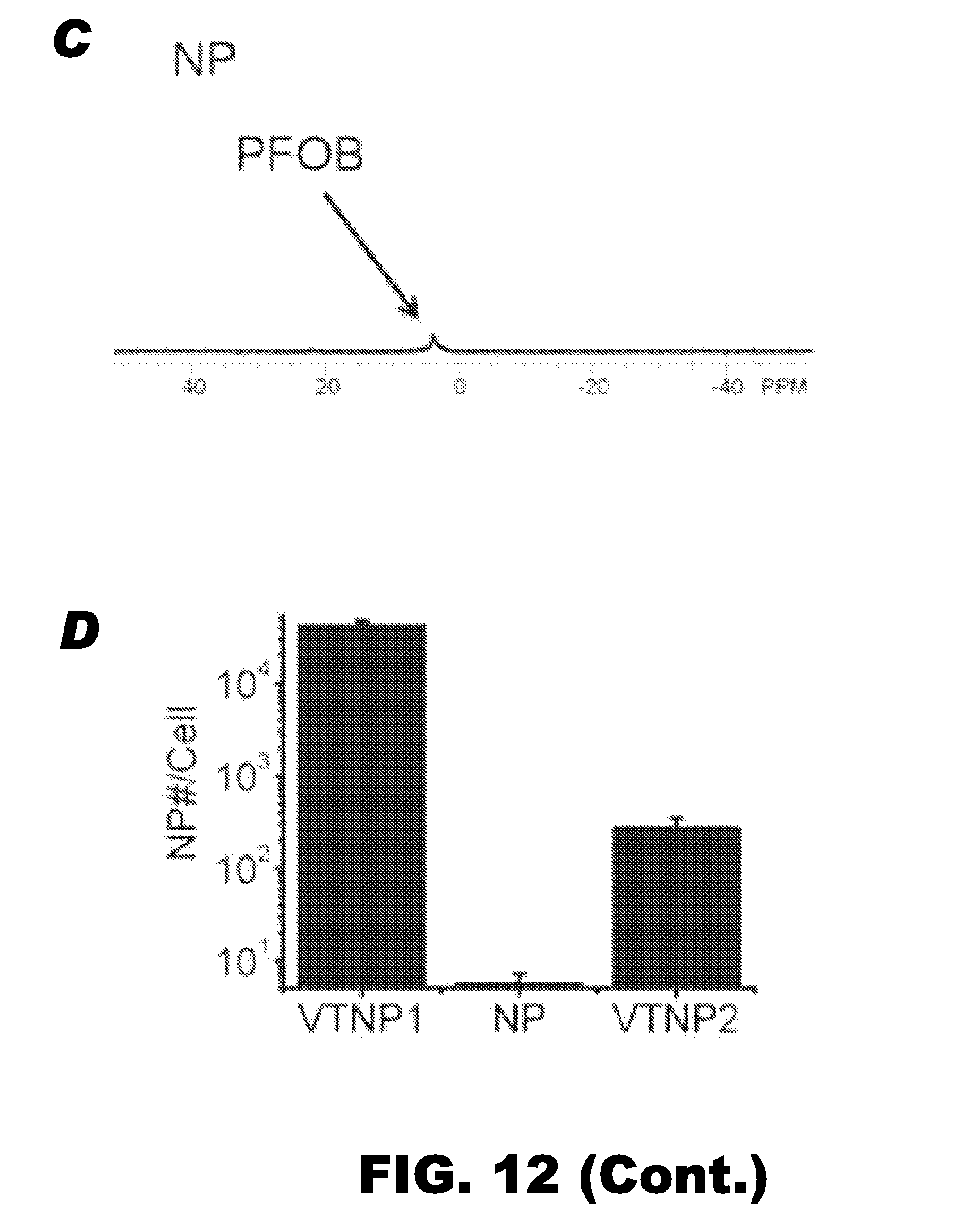
D00016

D00017

D00018

D00019
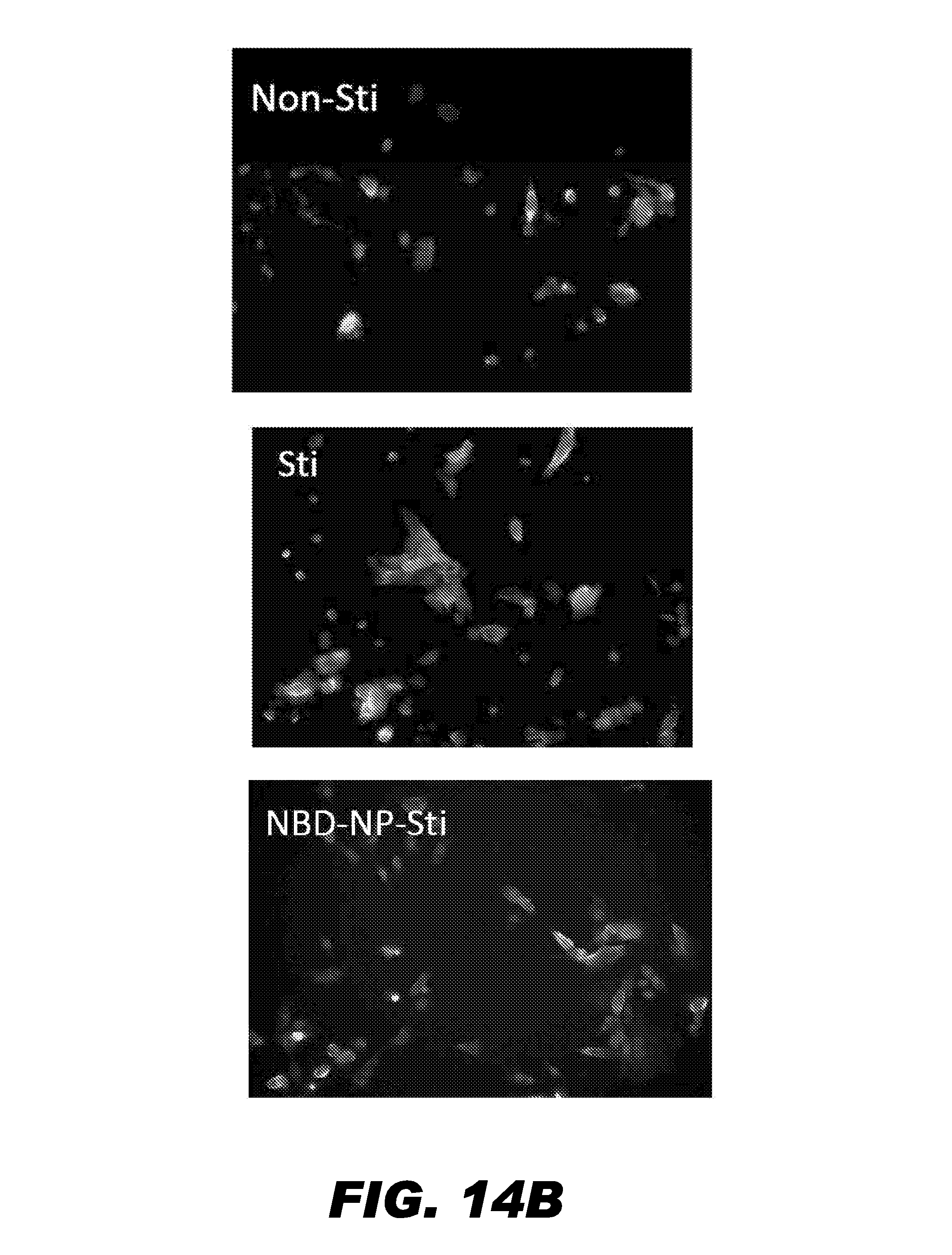
D00020

D00021

D00022

D00023

D00024

D00025

D00026

D00027

D00028

D00029
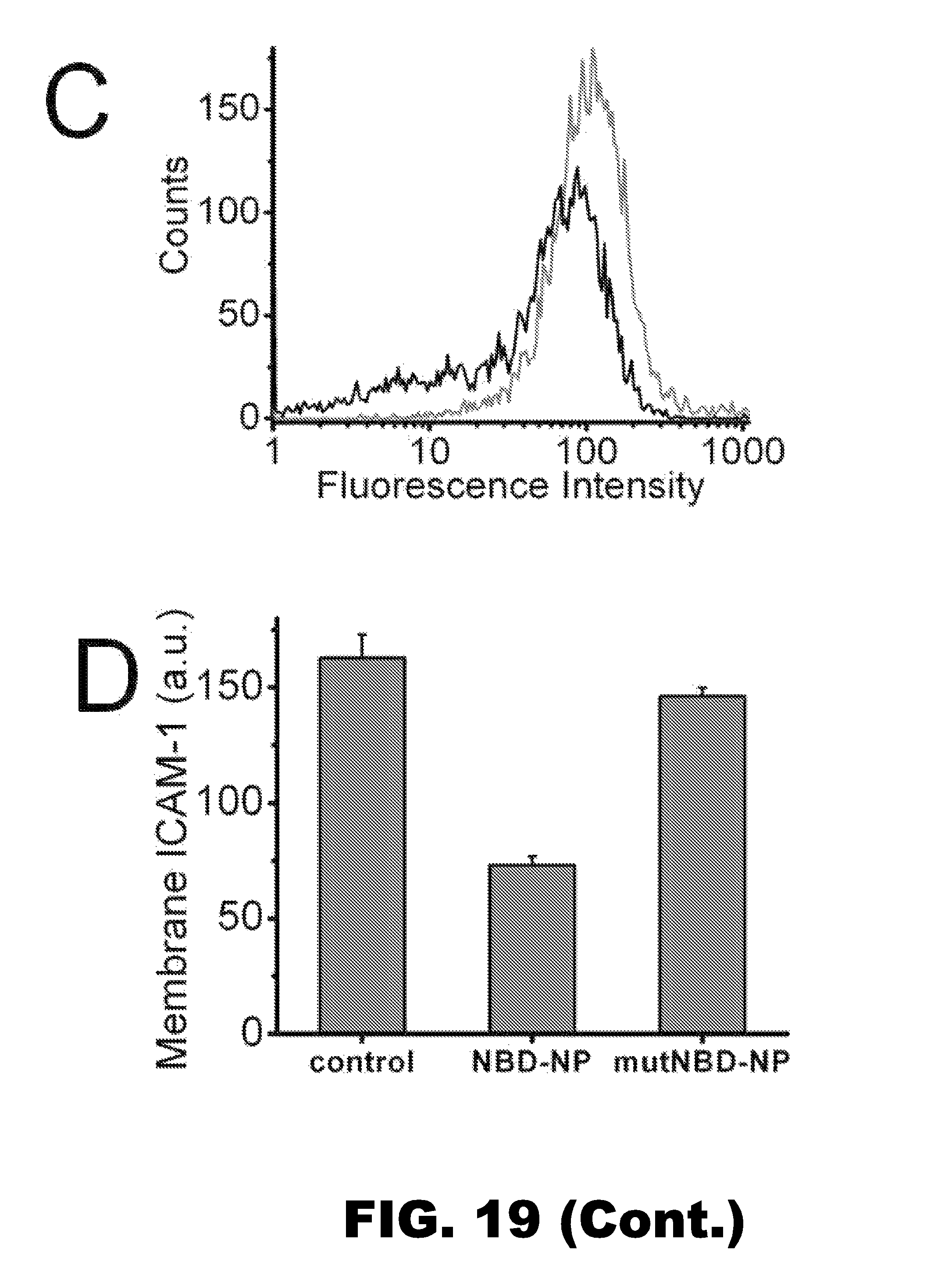
D00030

D00031

M00001

M00002

P00001

P00002

S00001
XML
uspto.report is an independent third-party trademark research tool that is not affiliated, endorsed, or sponsored by the United States Patent and Trademark Office (USPTO) or any other governmental organization. The information provided by uspto.report is based on publicly available data at the time of writing and is intended for informational purposes only.
While we strive to provide accurate and up-to-date information, we do not guarantee the accuracy, completeness, reliability, or suitability of the information displayed on this site. The use of this site is at your own risk. Any reliance you place on such information is therefore strictly at your own risk.
All official trademark data, including owner information, should be verified by visiting the official USPTO website at www.uspto.gov. This site is not intended to replace professional legal advice and should not be used as a substitute for consulting with a legal professional who is knowledgeable about trademark law.